https://theses.gla.ac.uk/ Theses Digitisation: https://www.gla.ac.uk/myglasgow/research/enlighten/theses/digitisation/ This is a digitised version of the original print thesis. Copyright and moral rights for this work are retained by the author A copy can be downloaded for personal non-commercial research or study, without prior permission or charge This work cannot be reproduced or quoted extensively from without first obtaining permission in writing from the author The content must not be changed in any way or sold commercially in any format or medium without the formal permission of the author When referring to this work, full bibliographic details including the author, title, awarding institution and date of the thesis must be given Enlighten: Theses https://theses.gla.ac.uk/ [email protected]

Welcome message from author
This document is posted to help you gain knowledge. Please leave a comment to let me know what you think about it! Share it to your friends and learn new things together.
Transcript
https://theses.gla.ac.uk/
Theses Digitisation:
https://www.gla.ac.uk/myglasgow/research/enlighten/theses/digitisation/
This is a digitised version of the original print thesis.
Copyright and moral rights for this work are retained by the author
A copy can be downloaded for personal non-commercial research or study,
without prior permission or charge
This work cannot be reproduced or quoted extensively from without first
obtaining permission in writing from the author
The content must not be changed in any way or sold commercially in any
format or medium without the formal permission of the author
When referring to this work, full bibliographic details including the author,
title, awarding institution and date of the thesis must be given
Enlighten: Theses
https://theses.gla.ac.uk/
THE INHIBITORY AND MOTOR INNERVATION OF
ANOCOCCYGEDS MJSCLE
*(
A thesis presented for the degree of
Doctor of Philosophy
by
John Christie McGrath
THE
Department of Pharmacology
University of Glasgow
December 1973
ProQuest Number: 10760464
All rights reserved
INFORMATION TO ALL USERS The quality of this reproduction is dependent upon the quality of the copy submitted.
In the unlikely event that the author did not send a com p le te manuscript and there are missing pages, these will be noted. Also, if material had to be removed,
a note will indicate the deletion.
uestProQuest 10760464
Published by ProQuest LLC(2018). Copyright of the Dissertation is held by the Author.
All rights reserved.This work is protected against unauthorized copying under Title 17, United States C ode
Microform Edition © ProQuest LLC.
ProQuest LLC.789 East Eisenhower Parkway
P.O. Box 1346 Ann Arbor, Ml 48106- 1346
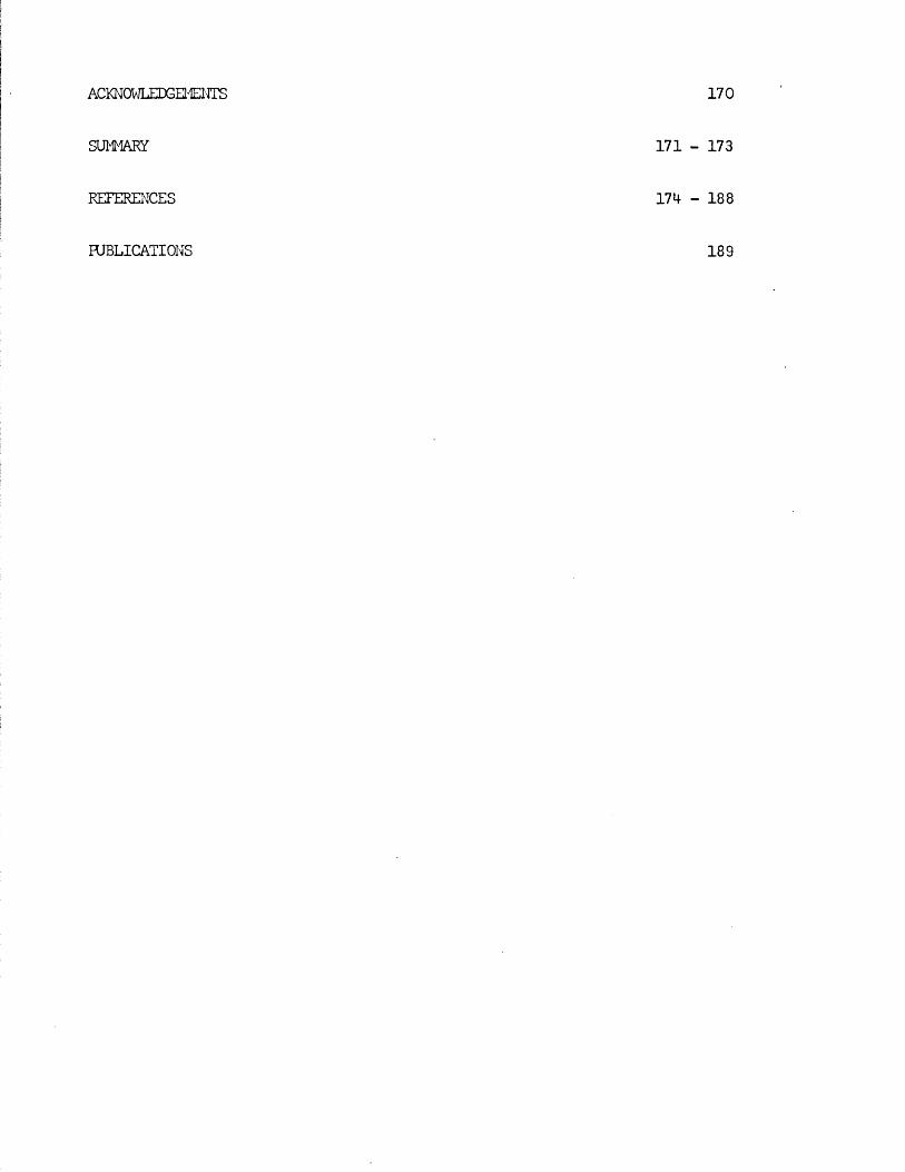
CONTENTS
INTRODUCTION H - 18
METHODS 19 - 40Anatcmy of the anocoecygeus iruscle 19Isolation and removal of the rat anocoecygeus muscles 20Isolation and removal of the cat anocoecygeus muscles 21In vitro preparation of rat or cat anocoecygeus muscle 23Removal of rat heart and vasa deferentia 25The pithed rat spinal electrode preparation 26The rat* anocoecygeus in'vivo preparation....... 30The rat vas deferens in vivo preparation 32Administration of reserpine - protocol 34Assay of tissue noradrenaline 36Histochemistry - Falck Technique 39
RESULTS 41 - 103Part I - The rat anocoecygeus in vivo 41 - 67
The motor response -Spinal origin of the motor nerves 41Frequency characteristics of the motor response 42Effects of blocking drugs on the motor response 44Drugs producing motor responses 47The inhibitory response -Spinal origin of the inhibitory nerves 51Effects of blocking drugs on the inhibitory response 53Cardiovascular responses -Drugs producing pressor responses 54Spinal origin of cardiovascular responses 55Effects of drugs on nerve or drug mediated cardiovascular 56responsesComparison of the motor responses in the rat anocoecygeus and vas deferens -Spinal origin of the vas deferens response 58Shape of vas deferens response 58Effects of drugs on the vas deferens response 59Summary of motor responses in vas deferens compared with 65anocoecygeus
Part II - The effects of reserpine on the rat • 68 - 74anocoecygeus, vas deferens and heart
Effect of reserpine dose on the tissue NA content 69Time course of depletion of NA by reserpine 69Recovery of tissue NA after reserpine 70Effects of pithing and of nerve stimulation in 70non - reserpinised animalsEffects of pithing and of nerve stimulation in 71reserpinised animalsThe effects of reserpine on the response of the vas 72deferensSummary of effects of reserpine on the rat anocoecygeus, 74
- vas* deferens and heart...................................
Part III - The properties of the cat anocoecygeus 75 - 103muscles in vitro and a comparison with the rat anocoecygeus
Spontaneous activity and intrinsic tone in the cat 76anocoecygeus muscleEffect of agonist drugs -60HDA in vitro treatment 77Drugs causing contraction in both species 79Drugs causing inhibition in the cat anocoecygeus 86Responses to field stimulationResponses to field stimulation in the absence of blocking 90drugsInteraction between motor and inhibitory nerves 92The effects of blocking drugs on nerve responses 95The effects of agonist drugs on nerve responses 96Effect of 60HDA treatment on nerve responses 100Effect of neostigmine on nerve responses 100Effects of cold storage on the cat anocoecygeus 102
DISCUSSION 104 - 169Rat anocoecygeus in vivo 104Effects of nerve stimulation on NA depletion by reserpine 123Cat anocoecygeus 136General discussion of anocoecygeus 164
ACKNOWLEDGEMENTS
SUMMARY
REFERENCES
PUBLICATIONS
170
171 - 173
174 - 188
189
INTRODUCTION
1
The description of the nervous system began in the 2nd century A.D. when Galen introduced the "experimental method" into the neurological sciences. His method of combining anatomical findings, as revealed by dissection, with physiological observations was so successful that it resulted in a description of the nervous system which remained virtually unchanged for 15 centuries. Subsequent developments have still been based on this correlation of anatomy with physiological function, although both have been examined at an increasingly fine level. In anatomy this has involved the use of the light microscope, the development of histochemistry and most recently the electron microscope. The identification of the individual tissue components which this revealed has paralleled and assisted the development of sophisticated methods of measuring physiological function. These techniques employed the industrial technology which was gradually developed throughout the 19th and 20th centuries to examine the electrical and chemical properties of tissues.
It has been the application of these techniques of micro - anatomy, electrophysiology and biochemistry together with the powerful effects of certain chemical substances on physiological processes which has led to the present state of knowledge of neurological function.
The modern concept of the autonomic nervous system began in the second half of the 19th century when it was recognised that this was a discrete, anatomically distinct part of the nervous system, characterised by an unusual efferent pathway (see Gaskell, 1916).It was subsequently realised, rrainly due to the work of Gaskell (1916) and Langley (1921) that this consisted of two separate but related systems which differed in their anatomical nature, produced essentially opposite physiological responses and were differently affected by
2
blocking drags. Following the demonstration of chemical transmission (Loewi, 1921) it was recognised that the difference in the two systems extended to the transmitters involved, a recognition which led Dale (1933) to suggest a functional classification into adrenergic and cholinergic nerves to replace the existing anatomical distinction of sympathetic and parasympathetic. The interplay of these two divisions was recognised to provide a wide range of homeostatic control mechanisms (see Cannon, 1939; Dale, 1965).
From this emerged the present day conventional picture of the autonomic nervous system as a control system, with afferent, central and efferent components. The efferent part innervates smooth muscle, most exocrine secretary cells and cardiac muscle. It consists of two major divisions, the sympathetic and :,parasympatheticv The sympathetic consists of a preganglionic thoracico - lumbar outflow which emerges from the corresponding ventral roots of spinal nerves to synapse with postganglionic neurons in the paravertebral or prevertebral ganglia, which are sited at locations distant from the end - organs. These postganglionic neurons then run to and supply the peripheral organs. The transmitter liberated from the preganglionic nerves is acetylcholine (cholinergic) whereas from the postganglionic nerves it is noradrenaline (adrenergic). The parasympathetic consists of cranial and sacral preganglionic outflows which emerge from either certain cranial nerves or the ventral roots of sacral spinal nerves and run uninterrupted to peripheral tissues where they synapse with postganglionic nerves in ganglia lying in or near the innervated organ. The preganglionic parasympathetic nerves are cholinergic and the postganglionic
3
parasympathetic fibres are also cholinergic. When both systems innervate the same organ, the two systems usually cause opposite responses.
Before the development of the theory of chemical transmission, the division between the two branches was based mainly on the spinal / origin of the preganglionic fibres. With the advent of chemical transmission and the realisation that this transmitter differed in different nerves, each branch became identified with its postganglionic nerves. Thus pathways with postganglionic cholinergic fibres ^became synonymous with parasympathetic and vice versa.
It became clear very early in the life*of this theory, however, that exceptions to this universal concept existed. These exceptions came to notice largely due to the use of specific drugs. In the course of developing the theory of chemical transmission, drugs played a central role both as agents producing effects similar to those of nerve stimulation and as blockers of the effects of nerve stimulation. This assisted greatly in the early identification of the type of transmission involved in the different branches of the autonomic system but eventually produced anomalies which required modification of the classical picture. The first conclusive example of this was demonstrated by Dale & Feldberg (1934). Starting from the observation that sweating, although initiated by stimulation of sympathetic nerves, / was blocked by atropine, they were able to demonstrate that secretary nerve fibres originating in ganglia of the sympathetic chain, and therefore classified as postganglionic sympathetic nerves, were /cholinergic. Together with other comparable deviations fran the classical picture, which came to light at around the same time (e.g. cholinergic sympathetic nerves to the blood vessels of dogs*
lips, Euler & Gaddum, 1931; in iris, Young, 1932; and in the vasculature of skeletal mscle, Bulbring & Burn, 1935), this led to the view that the general rules still held but that a recognition that there were exceptions to the rules would clear up many apparent anomalies (Dale & Feldberg, 1934).
Subsequent developments have shown that deviations from the conventional system can take two main farms, viz. (1) unconventional transmission (2) direct anatomical anomalies.
Anomalous transmission arose first as examples like that shown by Dale & Feldberg (1934) where the transmission was still adrenergic or cholinergic but the transmitter vas inappropriate far its anatomical origin. This could be readily accommodated into the existing system, tut later findings have suggested that transmitters other than noradrenaline or acetylcholine might exist, e.g. 5 - hydroxytryptainine (Bulbring & Gershon, 1967), adenosine triphosphate (Burnstock, 1972), prostaglandins (see Campbell, 1970). If a multiplicity of transmitters does exist, then serious re - appraisal of the division of the autonomic nervous system may became necessary. The further factor of mare than one transmitter from a single nerve is suggested by the theory of Burn & Rand (1959, 1960), which states that adrenergic nerves may liberate acetylcholine as well as noradrenaline and the related finding that during the development of sympathetic nerves they may release acetylcholine as transmitter before they obtain the capacity to release noradrenaline (Day & Rand,1961).
Examples of the kind of direct anatomical anomalies which have been
5
uncovered are to be found in the innervation of the secondary sexual organs and of the gastro - intestinal tract. The sympathetic pathway to the vas deferens and seminal vesicles via the hypogastric nerve has its ganglia located along the nerve trunk itself very near to the organs innervated. This produces a system which is therefore similar to the parasympathetic in the positioning of its ganglia, although the spinal origin of preganglionic nerves is tharacico - lumbar and the postganglionic nerves are adrenergic (for review see Sjostrand, 1965; Ferry, 1967), In the gastro - intestinal tract a further dimension has been added to the whole system with the discovery that the adrenergic nerves may not mke direct neuro - muscular contact with the smooth muscle but may act by synapsing with a further neuron or neurons which may then act on the muscle (Norberg, 1964, 1967; Jacobowitz, 1965; Aberg & Eranko, 1967; Gabella & Costa, 1968; Read & Burnstock, 1969).
These anatomical features, together with the possibility of several transmitters, thus present a considerable challenge to the classical framework and have indeed led seme people to call for a re - appraisal of the whole system, free from attempts to fit in deviant phenomena as aberrations from "the norm but which would consider them rather in their cwn right (e.g, Campbell, 1970),
This call has been partly answered by Burnstock (1972) who has suggested a third component to the autonomic nervous system whose postganglionic neurons are neither cholinergic nor adrenergic but utilise ATP or a related compound as transmitter and which he has thus tentatively called "purinergic" (Burnstock, 1971), Just as
6
Langley’s (1921) original division of the autonomic nervous system into two parts was based on comparative studies of the innervation in different organs and in different species, so this latter division into at least three parts also rests largely on the evidence of comparative studies, not only in different tissues in mammals, but also in invertebrate species (Burnstock, 1969)*
With this background, it is not surprising that each previously untested preparation which is new studied provides further fuel to this debate on the classification of the autonomic nervous system* The subject of this thesis, the neural control of the anocoecygeus muscle, provides an excellent example of this, since the anocoecygeus is a smooth muscle tissue receiving both a powerful inhibitory innervation, whose transmitter is unknown, and also an anatomically conventional motor adrenergic innervation distributed throughout the rnscle* The purpose of the present thesis was therefore to study this preparation in three ways
(1) To investigate the inhibitory innervation and determinewhether it represented a separate nerve pathway distinct from the sympathetic and whether it was organised on the efferent pattern of the autonomic nervous system, i.e. with an interrupting ganglion.(2) To compare the characteristics of the motor innervation inthe vas deferens and in the anocoecygeus to see what, if any, differences were introduced by the "short” postganglionic neurons in the vas.(3) To compare the properties of the anocoecygeus muscle in twospecies, the rat and cat, in order to determine which features were common and whether this comparative study would shed more light on the unknown inhibitory innervation.
7
The history of the anocoecygeus preparation illustrates, in a snail way, the inter - relationship between form and function and the role of drugs in the development of any neurological study. The anocoecygeus came to light during a histochemical investigation of the density of adrenergic innervation of smooth muscle at different levels in the alimentary canal of the rat (Gillespie & Maxwell, 1971).It was the dense adrenergic innervation, as shown by the fluorescence technique of Falck & Hillarp which drew attention to the presence of this strap - like muscle which had not previously been reported in the rat and had been little more than noted to be present in other species (Straus - EUrckheim, 1845; Langley & Anderson, 1895; Miller, Christensen & Evans, 1964). Further histological examination showed it to be composed of a thin, flat sheet of smooth muscle making it "particularly suitable far in vitro studies, electrophysiologic measurements of membrane potentials and the effects of adrenergic nerve stimulation on these" (Gillespie & Maxwell, 1971).
Subsequent in vitro studies (Gillespie, 1972) demonstrated that the adrenergic innervation could be excited by electrical field stimulation and that this produced contraction of the muscle; a motor response. Further investigation demonstrated that, in the presence of a large dose of guanethidine, this adrenergic innervation was blocked and the tone of the muscle raised and that, under these conditions, field stimulation now produced relaxation of the muscle; an inhibitory response. This inhibitory response could be elicited with short pulses of 1 m sec or less and was abolished by low doses of tetrodotoxin thus showing the characteristics of a nerve mediated response. It could not, however, be blocked by the conventional blocking agents which prevent responses
8
to noradrenaline or acetylcholine in other tissues and so the nature of the transmission process, remined unknown.
In the anocoecygeus, therefore, the histochemical discovery of the adrenergic nerves within the muscle led to the finding that this tissue received a powerful motor adrenergic innervation. While further investigating its physiological / pharmacological properties in vitro , this in turn led to the discovery of an unusual type of inhibitory innervation.
The first objective of the present investigation was to further extend the anatomical study of this system by determining whether the inhibitory as well as the motor pathway originated in the spinal card and if so whether it was interrupted by a ganglion synapse, a condition which would allow it to be classified within the autonomic nervous system. This was approached using the pithed rat preparation of Gillespie, MacLaren & Pollock, (1970) in which the pithing rod can be used as a moveable electrode to selectively stimulate the spinal outflow at any desired level. By recording the resultant mechanical responses of the anocoecygeus muscle in situ, it was thus possible to delineate the spinal origin of both motor and inhibitory pathways and then pharmacologically analyse the nature of these pathways.
Several possible explanations of a dual response to field stimulation or to stimulation of the extrinsic nerve exist. These can be summarised as follows -
(1) If both responses are produced by a single type of fibre, the nature of the response might be determined by (a) the "peripheral mechanism"; the response depends on the "state" of the nerve and
9
muscle at the periphery (McCrea, McSwiney & Stcpford, 1925)(b) Wedensky inhibition; block of the nerve impulse somewhere in the pathway to the effector muscle without diminution of the ability of the muscle to respond, i.e. the inhibitory response results from the loss of the motor response (e.g. Veach, 1925; Cannon, Raule & Schaefer, 1954) Cc) variable effect of a single transmitter (e.g. Bozler, 1940; Burn & Robinson, 1951; Gardner & Kandel, 1972).(2) Two functionally distinct fibre groups exist, producingopposite responses. These are usually adrenergic and cholinergic and can be organised as (a) according to the conventional autonomic pattern with anatomically discrete sympathetic and parasympathetic nerves (b) sympathetic and parasympathetic fibres with separate anatomical origins but which may come together peripherally to reach the target:organ in a common nerve bundle (e.g. McSwiney & Robson, 1929; Finkleman, 1930; Gillespie & Mackenna, 1961) (c) fibres share acommon anatomical origin but the sympathetic may be cholinergic (Dale & Feldberg, 1934) (d) one or both of the nerves might be neitheradrenergic or cholinergic and have as its transmitter some other substance, e.g. ATP (Burnstock, 1972) (e) one of the responses mightbe due to antidromic stimulation of afferent nerves producing liberation of seme substance at the peripheral endings (e.g. dorsal root afferents, Bayliss, 1902; Lewis & Marvin, 1927; Wybauw, 1936; Holton & Perry, 1951).Delineation of the spinal origins of the nerves responsible for the responses together with the effects of selective blocking drugs obviously eliminates most of these possibilities; an analysis impossible by in vitro experiments alone.
10
Once the in vivo preparation of the anocoecygeus was developed, it provided the means for the second part of the study, the comparison with the vas deferens. By appropriate selection of the level of spinal stimulation, the sympathetic outflows to the anocoecygeus and vas deferens and the vasomotor outflow could be stimulated simultaneously and the resultant responses compared. This enabled comparison of the new preparation, the anocoecygeus, with the known but anatomically unconventional preparation, the vas deferens, and both of these with the known but anatomically more conventional vasomotor system.
This comparison was carried out in two separate parts.
Firstly, the effects of agonist and antagonist drugs which normally mimic cr modify adrenergic responses were studied. This permitted the assessment of any differences in the effects on the vas deferens from those on the other two test organs. In recent years many of the effects of such drugs on the vas have been shown to be different from those found on other tissues and have led to a debate over whether motor transmission is indeed adrenergic.
When the isolated guinea - pig vas deferens - hypogastric nerve preparation was introduced by Hukovic (1961) it seemed to be a suitable preparation far demonstration of adrenergic neurotransmission. Two glaring anomalies, however, emerged from the start. Adrenergic «L- blocking agents did not block the mechanical response to nerve stimulation except in extremely high concentrations and could, in fact, potentiate the response in the moderate concentrations which were sufficient to block responses in other tissues (Boyd, Chang & Rand, 1960).
11
In addition, the response could be potentiated by cholinesterase inhibitors and inhibited by hemicholiniuin (Rand & Chang, 1960), evidence which was taken to support the cholinergic link hypothesis (Burn & Rand, 1959, 1960). It gradually became apparent from the work of several authors that these latter anomlies, which implicated a cholinergic component, arose from the fact that the ganglia in the motor pathway were distributed along the hypogastric nerve close to its junction with the vas deferens, so that stinulation of the hypogastric nerve was initiating impulses in preganglionic nerves which therefore had to pass through the cholinergic synapse of the ganglion before reaching the postganglionic adrenergic nerve and its junction with the smooth muscle of the vas (Bentley & Sabine,1963; Birmingham & Wilson, 1963; Ferry, 1963, 1967; Kuriyama, 1963; Ohlin & Stromblad, 1963; Falck, Owman & Sjostrand, 1965). This largely accounted for the ability of drugs affecting the cholinergic system to influence the response and explained why such drugs were ineffective if the tissue was stimulated postganglionically by transmural field stimulation (Birmingham & Wilson, 1963). The resistance to oc- blockers, however, remained whether stimulation was pre - or postganglionic.
More recently, this paradox has been more closely investigated by Ambache and co - workers who have linked this to the finding that noradrenaline itself does not produce contractions except in high concentrations and can inhibit the response to stimulation of the motor nerves in lower concentrations (Ambache & Zar, 1970) and that this latter effect is prevented by phentolamine. Together with the finding that the motor response in the vas to nerve stimulation is not
12
il
prevented by reserpine pretreatment, this has led Ambache & Zar (1971) to suggest that this motor transmission is non - adrenergic and that if the adrenergic nerves have any function in this tissue, it is likely to be an inhibitory one* To further support this contention, they have subsequently demonstrated that the sympathomimetic agents tyramine and cocaine also inhibit the motor response (Ambache, Dunk, Verney & Zar, 1972). This hypothesis has not yet deflected the main - stream of opinion from the conventional view that motor transmission in the vas deferens is noradrenergic (see Euler, 1972) and evidence has been produced which upholds this conventional view arguing that the resistance to c*c - blockers is due to the morphological arrangement
iof the neuro - muscular junction (Swedin, 1971) and that complete sympathectomy using 60HBA can abolish the motor nerve response (Wadsworth, 1973). In addition, these nerve responses are abolished by guanethidine, a finding acknowledged by Ambache & Zar (1971). Nevertheless, the results produced by Ambache et al present a considerable challenge to the contention that transmission in the vas is adrenergic, and it was hoped that by comparison with the anocoecygeus some further light might be thrown on this paradox.
The second comparison of the vas deferens with the anocoecygeus was undertaken in order to clarify the reasons far the relative resistance of the farmer tissue to the noradrenaline depleting effect of reserpine (Sjostrand & Swedin, 1968). Since Holzbauer & Vogt (1956) first showed that tissue catecholamines were depleted by reserpine, it has been shown that the rate of depletion is dose - dependent (e.g. Carlsson, Rosengren, Bertler & Nilsson, 1957; Bertler, 1961) and is similar far most adrenergically innervated peripheral organs tested ( Bertler, 1961).
13
Sjostrand & Swedin (1968) found, however, that the vas deferens and seminal vesicles were depleted of their noradrenaline content by reserpine more slowly than other peripheral tissues such as the heart and submaxillary glands and that decentralisation of the former organs did not affect the rate of depletion, whereas it slowed the rate of depletion of submaxillary glands in the same rats. Two possible explanations of this resistance are that either the rate of depletion is dependent on the firing frequency in the nerves and therefore reduced in the intermittently active nerves of the vas deferens and seminal vesicles or that the "short” adrenergic neurons, which are found in the seminal vesicles and vas deferens and have their cell bodies located close to the target organs (Owman & Sjostrand, 1965; Sjostrand, 1965), are like the adrenal medullary cells less easily depleted than the "long" adrenergic neurons found elsewhere.
The first hypothesis is supported by the finding that the rate of depletion by reserpine in various organs is reduced if impulse flew is reduced by decentralisation of the adrenergic nerves before reserpine (Holzbauer & Vogt, 1956; Hertting, Potter & Axelrod, 1962; Benmiloud & Euler, 1963; Sedvall, 19640 or administration of ganglion blocking drugs (Karki, Paasonen & Vanhakartano, 1959; Mirkin, 1961; Hertting, Potter & Axelrod, 1962).
The second explanation is suggested by the evidence that "short" adrenergic neurons have a lower sensitivity to 60HDA (Malmfors & Sachs, 1968) as well as to reserpine, react differently to immnosympathectcrny (Hamberger, Levi - Montalcini, Narberg & Sjoquist, 1965; Zaimis, Berk & Callingham, 1965; Iversen, Glcwinski & Axelrod, 1966) and their transmitter granules, when isolated, have different properties compared with those from splenic nerve (Euler & Lishajko, 1966; Stjarne &
Lishajko, 1966).
These hypotheses were tested by contrasting the effect of abolishing neuronal activity completely by pithing and reinforcing that activity by maximal artificial stimulation of the sympathetic nerves by the same pithing rod on the rate of depletion of noradrenaline in the heart, vas deferens and anocoecygeus muscles. Since an earlier part of the study shows that the anocoecygeus receives conventional ’’long" adrenergic neurons, any difference linked to the length of the neuron should become obvious. The dose - dependence and time course of noradrenaline depletion by reserpine in vivo was first determined and then the effect on the rate of depletion of simultaneous stimulation of the nerves to the anocoecygeus and vas deferens was examined using pithed rats.
The third part of the investigation was the comparative study of the cat and rat anocoecygeus in vitro. This was intended primarily as a direct comparison of the effects of field stimulation on the muscle from the two species in order to determine whether the dual innervation and especially the inhibitory component found in the rat was also to be found in a second species. Having established that the dual response was present in the cat, several putative transmitter .substances and specific blockers were tested on both species in an attempt to categorise the inhibitory nerves.
This study was given further impetus when Garrett, Howard & lansdale (1972) examined the cat anocoecygeus histologically, showing that it had, like the rat, a dense adrenergic innervation but, unlike the rat, also stained for cholinesterase. It is tempting to interpret this staining as
15
evidence of a cholinergic innervation and to associate this with the inhibitory innervation.
The possibility that the inhibitory nerves might be cholinergic had been rejected by Gillespie (1972) on the grounds that acetylcholine was motor on the rat anocoecygeus, so in addition to any putative role as a transmitter, other possible roles for acetylcholine in the tissue had to be considered.
In addition to transmitter at "cholinergic’1 nerve terminals, acetylcholine has in recent years been ascribed several roles in transmission at adrenergic nerve terminals (for reviews see tiischoll, 1970; Kosterlitz & Lees, 1972). These were embodied into the "cholinergic link hypothesis" of Burn & Rand (1959, 1960) which states that when the nerve impulse reaches the adrenergic nerve terminal there is a release of acetylcholine which, by activation of nicotinic receptors, in turn releases noradrenaline from its stores. The evidence for and against this hypothesis was reviewed by Ferry (1966). Whatever the physiological role, if any, of acetylcholine turns out to be, cholinomimetic agents certainly have effects on the release of noradrenaline frcm adrenergic nerve terminals which must be considered when investigating the role of acetylcholine in an adrenergically innervated tissue, e.g. facilitation of noradrenaline release at low doses, inhibition of noradrenaline release at intermediate doses and release of noradrenaline frcm the nerves at high doses (Malik & Ling, 1969; Loffelholz & Muscholl, 1969). The possible effects of acetylcholine on both motor and inhibitory nerves as well as on the muscle itself were therefore investigated in the anocoecygeus.
A further property of the anocoecygeus muscles from both species which was investigated in vitro was the ability of several substances to
16
produce syirpathomimetic effects* Guanethidine, cocaine, LSD and 5HT j!all produced contraction of the rnscles frcm both species which were
inhibited by the adrenergic c{ - receptor blocker phentolamine. >’;/
Direct actions on smooth muscle have been reported previously for guanethidine (Maxwell, 1965; Maxwell, Wastila & Eckhardt, 1966; Bevan & Verity, 1967), cocaine (Daniel & Wolowyck, 1967; Maxwell, Wastila & Eckhardt, 1966; Cliff, 1968), LSD (Gant & Dyer, 1971; Dyer & Gant,1973) and 5HT (Gaddum & Hameed, 195*+; Gaddum & Picarelli, 1957; Innes,1962). Indirect action via release of noradrenaline from adrenergic nerves has also been reported for guanethidine (Kadzielawa, 1962; Bhagat & Shideman, 1963; Pluchino, Muskus & Pluchino, 1969), cocaine (Kukovetz & Lembeck, 1962; Maengwyn -Davies & Kqppanyi, 1966; 01 Donnell & Hecker,1967; Trendelenburg, 1968) and 5HT (Furchgott, 1955; Innes, 1962;Pluchino, 1972).
It was thus of interest to determine whether the responses produced by these substances on the anocoecygeus were direct effects on the muscle or indirect effects produced by the release of noradrenaline frcm adrenergic nerves. Early distinctions between the actions of tyramine and of adrenaline were that both cocaine and denervation potentiated the effects of adrenaline but antagonised those of tyramine (Tainter & Chang, 1927; Burn & Tainter, 1931; Bulbring & Burn, 1938). More recently, additional evidence far the release of noradrenaline by tyramine and similar drugs was obtained by demonstrating that tyramine lost its effectiveness after depletion of noradrenaline stores by reserpine (Burn & Raid, 1958), At the present time these three methods, cocaine, denervation and reserpine pretreatment are the main experimental approaches to
17
classification of sympathomimetic effects as direct or indirect (Trendelenburg, 1972).
In the present study, specific chemical denervation of the adrenergic nerves using 60HDA was chosen as a means of making this distinction (see Malmfors & Thoenen, 1971). It has recently been shown by Gibson & Gillespie (1973) that pretreatment of rats with 60HDA will selectively destroy the motor but not the inhibitory nerves. In the case of the cat anocoecygeus, sympathectomy was produced by incubating the tissue in vitro with 60HDA since the normal in vivo pretreatment was impracticable. This method has previously been shown to be successful in various tissues by Sachs (1971) and Wadsworth (1973).
A final aspect of the cat anocoecygeus muscle which was investigated was the ability of the nerves and nuscle to survive cold storage. This was necessitated since the tissues were obtained from cats primarily intended far experiments by other workers with the perfused spleen. Due to the timing of these experiments, the anocoecygeus muscles, although removed fresh from the cats, were sometimes stared in Krebs1 solution at 4°C for 24 - 48 hrs before use. It was therefore of interest to determine whether this storage caused any change in the properties of the tissue which would make it unsuitable far experimental use. Another aspect of cold storage which might also have been of interest would have been if one or other of the sets of nerves had preferentially survived storage. This property has been employed in the past to differentiate between sets of nerves. Far instance, nerves with their ganglion cell body included within the isolated preparation will survive longer than nerve endings cut;,off from the cell body (Holman & Hughes, 1965; Hattori, Kurahashi, Mori & Shibata, 1972).
18
The results of these three separate aspects of the study are presented according to the techniques employed. Thus the first section includes all the experiments where the mechanical responses in the tissues to stimulation of the autonomic outflow were examined, viz. (a) Origin and properties of the anocoecygeus inhibitory and motor nerves (b) Comparison of the properties of the anocoecygeus, vas deferens and cardiovascular responses in the pithed rat. The second section consists of the study of the effect of nerve stimulation on the depletion of tissue noradrenaline by reserpine, and the third part includes all the in vitro experiments comparing cat and rat anocoecygeus nuscles.
By this combination of anatomical, biochemical and pharmacological techniques it was hoped to further our understanding of the unusual dual innervation of the anocoecygeus while simultaneously investigating some pharmacological problems of current interest.
METHODS
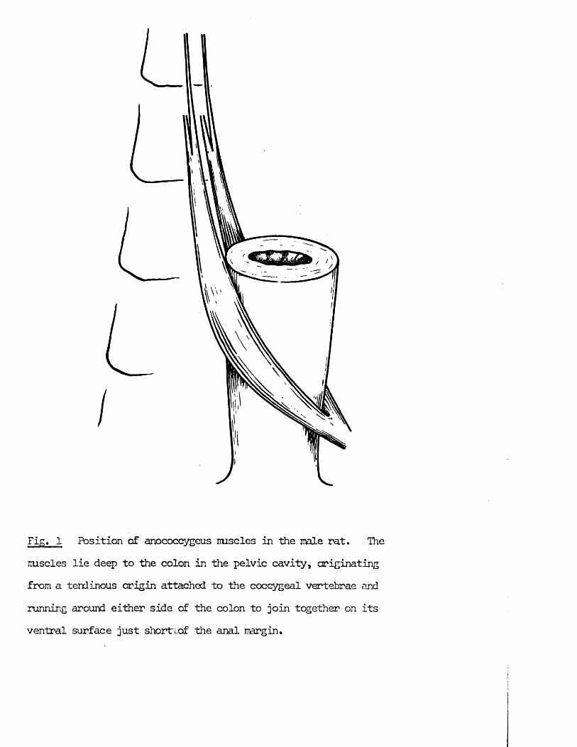
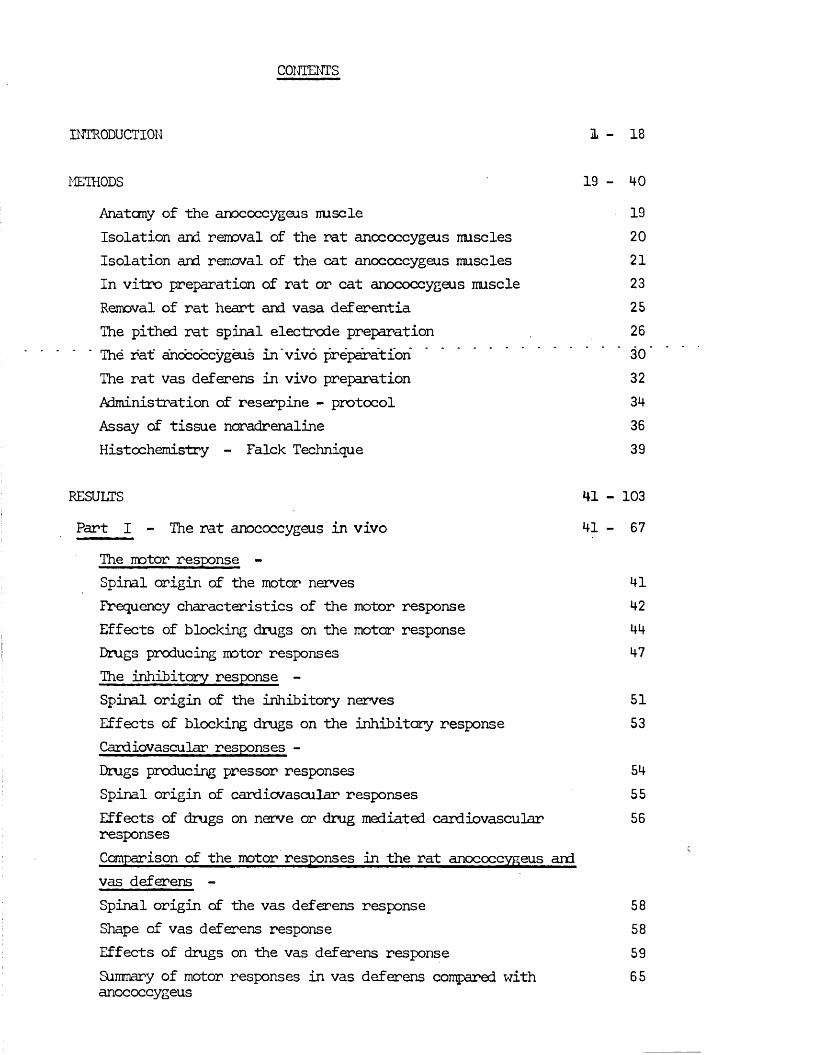
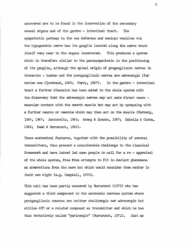
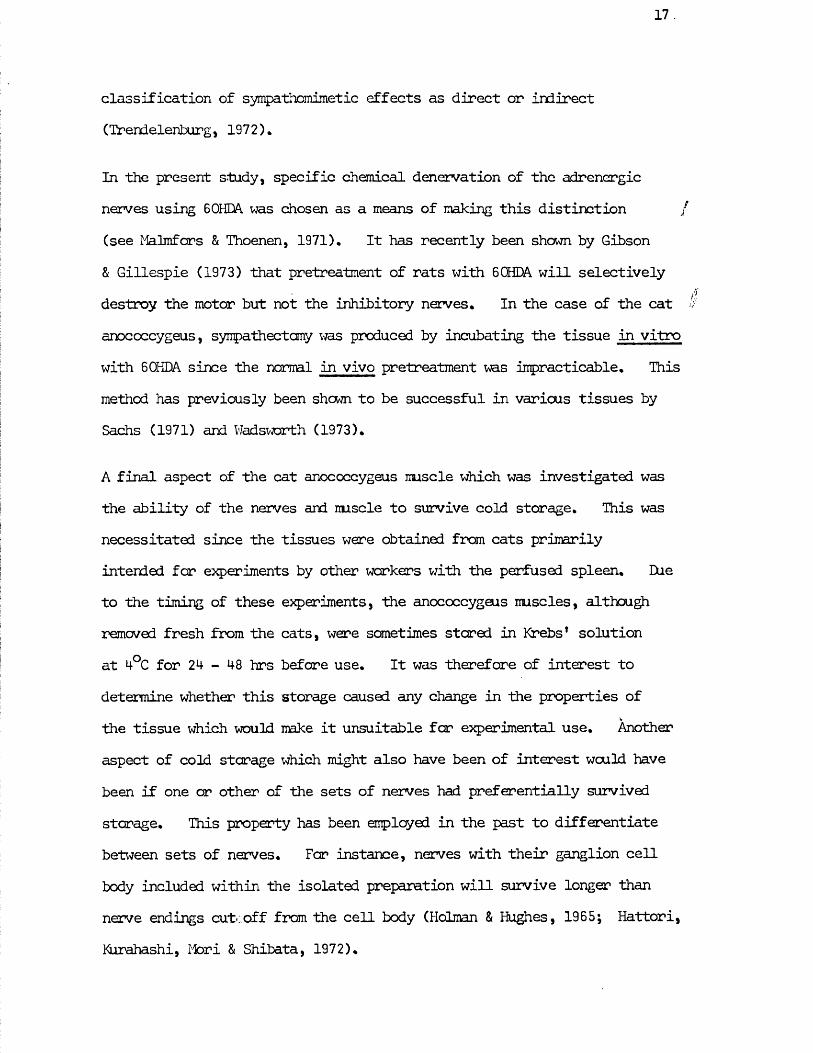
Fig. 1 Position of anococcygeus imscles in the male rat. The mscles lie deep to the colon in the pelvic cavity, originating from a tendinous origin attached to the coccygeal vertebrae and running around either side of the colon to join together on its ventral surface just shortlof the anal margin.
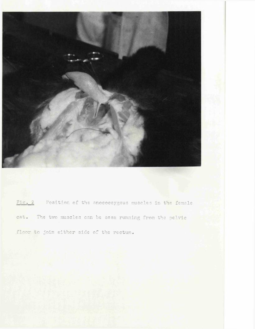
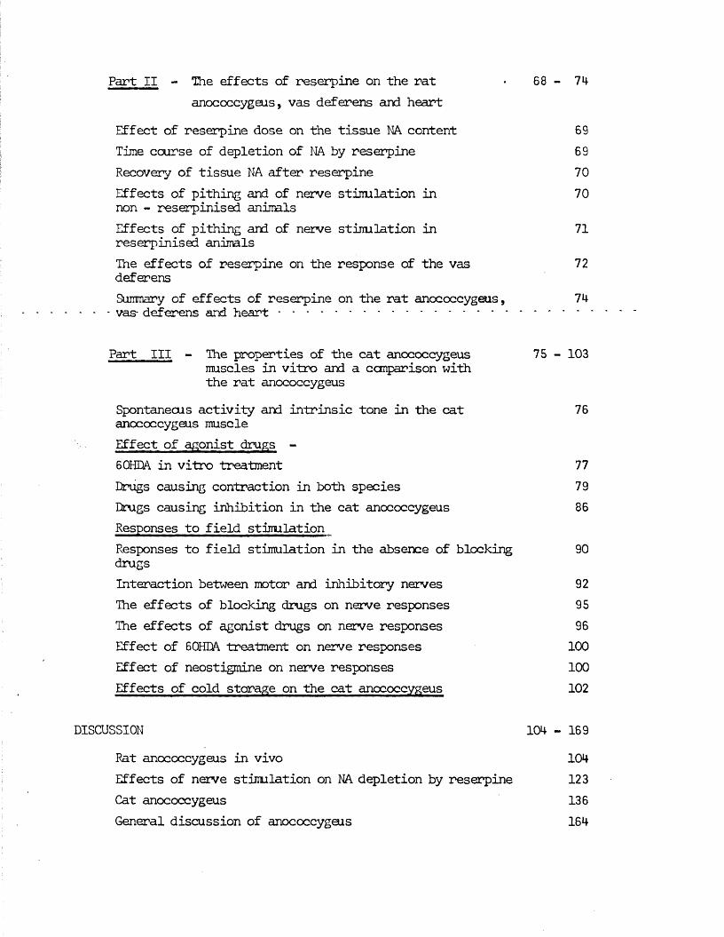
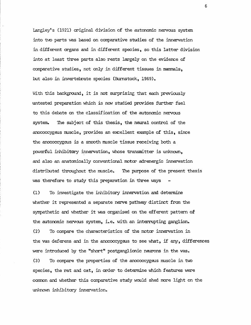
7ir.. 2 Position of the anococcygeus muscles in the female
cat. The two muscles can be seen running from the pelvic
floor to join either side of the rectum.
RAT CAT
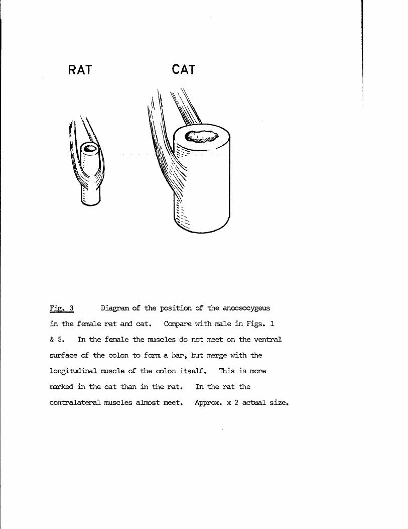
Fig* 3 Diagram of the position of the anococcygeusin the female rat and cat. Compare with male in Figs. 1 & 5. In tine female the muscles do not meet on the ventral surface of the colon to form a bar, but merge with the longitudinal muscle of the colon itself. This is more narked in the cat than in the rat. In the rat thecontralateral muscles almost meet. Approx, x 2 actual size.
Anatomy of tine anococcygeus muscle
The anococcygeus muscles in the rat lie deep to the colon in the pelvic cavity. They consist of parallel bundles of smooth muscle fibres which arise from a tendinous origin attached to coccygeal vertebrae 1 and 2,They run caudally first behind the colon and then passing ventrally around either side of the terminal colon to join together in front to form a ventralbar (Fig. 1). In the male, this ventral bar continues in the mid-line ofthe perineum as the retractor penis muscle. The muscle is surrounded by a thin, loose sheath of connective tissue but is otherwise free from both the colon and the pelvic floor along its entire length except where it comes into close contact with the side of the colon. Here some fibresnay merge with the longitudinal muscle of the colon as is found in thedog and cat (Fig. 2). In the nale this is the only close contact between anococcygeus and colon, but in the female, where the ventral extension is absent, the muscles remain in close contact with the colon till they join (Fig. 3).
Each muscle in the nale is 25-30 mri in length, 4-5 irm in breadth, and less then 1 mm in thickness in situ. On removal from the animal, they contract to approximately half this length. The average weight far a single muscle is 19,1 + 2.7 mg (n=12). Muscles from females are shorter due to the absence of the ventral extension.
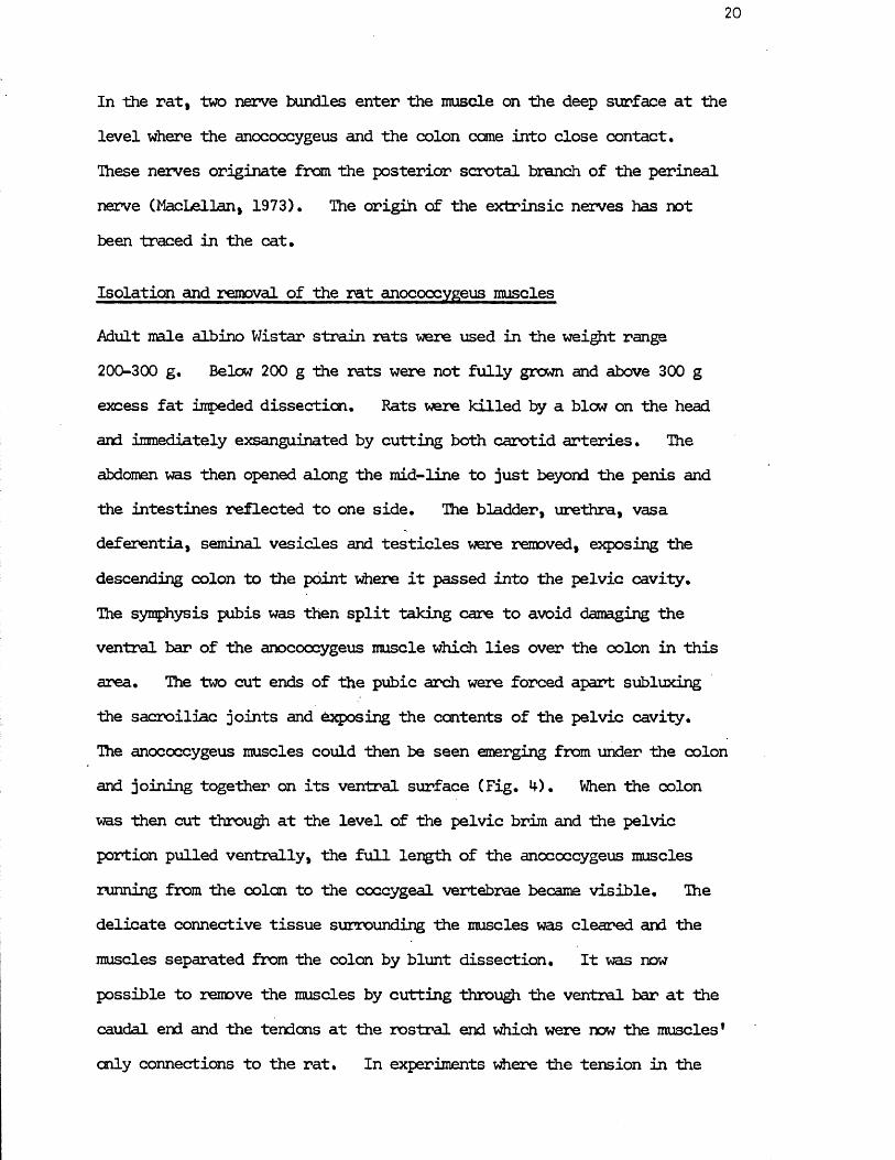
In the cat, the anococcygeus muscles lie in a similar position to those in the rat. The muscles interlock with the rectococcygeus which is absent in the rat but in the cat runs caudally from the dorsal surface of the colon (Fig, 5), The anococcygeus muscles lie on either side of the rectococcygeus and run in the opposite direction. The muscles are broader and thicker than those in the rat but generally of a similar length, although in large nale cats they can be up to 50 mm long. Weights for single muscles are male 51 mg (n=4); female 29 mg (n=8).
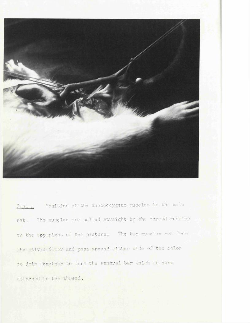
Fjor. 4 Position of the anococcygeus muscles in th■: male
rat. The muscles are pulled straight by the thread running
to the top right of the picture. The two muscles run from
the pelvic floor and pass around either side of the colon
to join together to form the ventral bar which is here
attached to the thread.
20
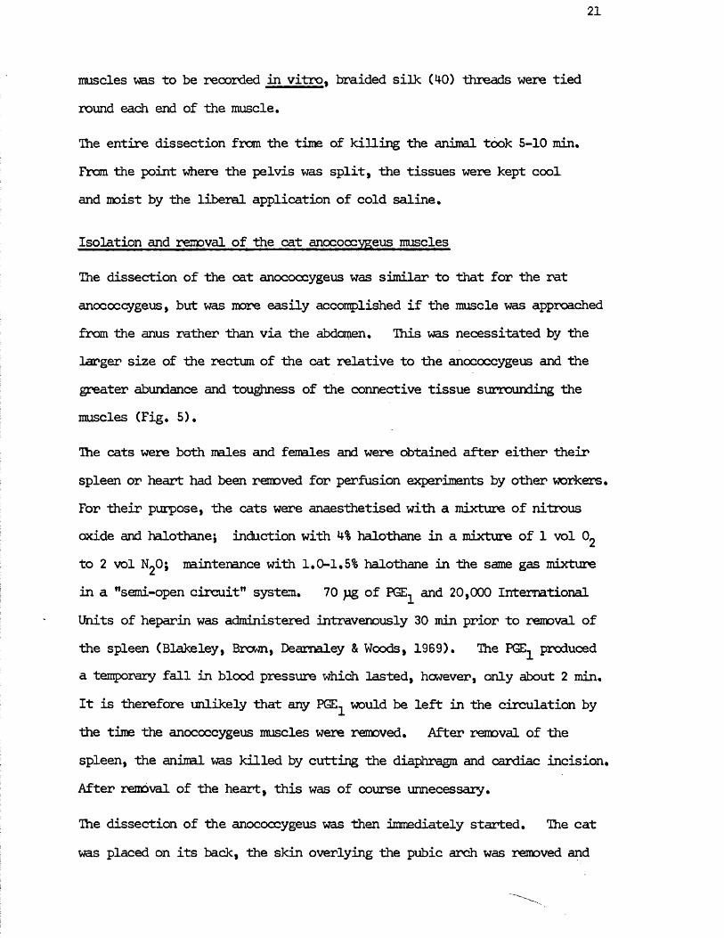
In the rat, two nerve bundles enter the muscle on the deep surface at the level where the anococcygeus and the colon cane into close contact.These nerves originate from the posterior scrotal branch of the perineal nerve (MacLellan, 1973). The origin of the extrinsic nerves has not been traced in the cat.
Isolation and removal of the rat anococcygeus muscles
Adult male albino Wistar strain rats were used in the weight range 200-300 g. Below 200 g the rats were not fully grown and above 300 g excess fat impeded dissection. Rats were killed by a blow on the head and immediately exsanguinated by cutting both carotid arteries. The abdomen was then opened along the mid-line to just beyond the penis and the intestines reflected to one side. The bladder, urethra, vasa deferentia, seminal vesicles and testicles were removed, exposing the descending colon to the point where it passed into the pelvic cavity.The symphysis pubis was then split taking care to avoid damaging the ventral bar of the anococcygeus muscle which lies over the colon in this area. The two cut ends of the pubic arch were forced apart subluxing the sacroiliac joints and exposing the contents of the pelvic cavity.The anococcygeus muscles could then be seen emerging from under the colon and joining together on its ventral surface (Fig. M-). When the colon was then cut through at the level of the pelvic brim and the pelvic portion pulled ventrally, the full length of the anococcygeus muscles running from the colcn to the coccygeal vertebrae became visible. The delicate connective tissue surrounding the muscles was cleared and the muscles separated from the colon by blunt dissection. It was now possible to remove the muscles by cutting through the ventral bar at the caudal end and the tendens at the rostral end which were now the muscles* cnly connections to the rat. In experiments where the tension in the
Recto.
Fig. 5 Diagram of the relative positionsof the anococcygeus muscles and the rectococcygeus muscle in the cat. The rectococcygeus interlocks with the two anococcygeus muscles and runs from the dorsal surface of the colon in the anal direction.
21
muscles was to be recorded in vitro, braided silk (40) threads were tied round each end of the muscle.
The entire dissection from the time of killing the animal took 5-10 min.From the point where the pelvis was split, the tissues were kept cool and moist by the liberal application of cold saline.
Isolation and removal of the cat anocoocygeus muscles
The dissection of the cat anococcygeus was similar to that for the rat anococcygeus, but was more easily accomplished if the muscle was approached from the anus rather than via the abdomen. This was necessitated by the larger size of the rectum of the cat relative to the anococcygeus and the greater abundance and toughness of the connective tissue surrounding the muscles (Fig. 5).
The cats were both males and females and were obtained after either their spleen or heart had been removed for perfusion experiments by other workers. For their purpose, the cats were anaesthetised with a mixture of nitrous oxide and halothane; induction with 4% halothane in a mixture of 1 vol to 2 vol N O; maintenance with 1.0-1.5% halothane in the same gas mixture in a "semi-open circuit" system. 70 pg of PGE and 20,000 International Units of heparin was administered intravenously 30 min prior to removal of the spleen (Blakeley, Brown, Deamaley & Woods, 1969). The PGE produced a temporary fall in blood pressure which lasted, however, only about 2 min. It is therefore unlikely that any PGE would be left in the circulation by the time the anococcygeus muscles were removed. After removal of the spleen, the aninal was killed by cutting the diaphragm and cardiac incision. After removal of the heart, this was of course unnecessary.
The dissection of the anococcygeus was then immediately started. The cat was placed on its back, the skin overlying the pubic arch was removed and
22
the pelvis split by cutting through the symphysis pubis with bone clippers* The pelvic cavity was then opened up by forcing apart the pubic bones. The urethra and bladder, together with the vagina and uteri in female cats were removed and the rectum tied and cut at the level of the pelvic brim. The rectum was then displaced ventrally and the connective tissue between it and the pelvic floor cleared by blunt dissection until the anococcygeus muscles became visible. This latter process required care since this connective tissue was sometimes very tough, unlike the delicate tissue in the rat, so the anococcygeus muscles could easily be tom. The portion of the anococcygeus muscles running from their coccygeal tendinous origin to the rectum could be easily freed from the surrounding connective tissue by blunt dissection. In female cats, the muscle appears to merge with the longitudinal muscle at the sides of the rectum and can be dissected free only with difficulty and with probable damage to the muscle. For this reason, in femles only the free part of the muscle terminating at the rectum was removed for in vitro studies. In males, on the other hand, the part of the muscle running round the side of the rectum could be freed by blunt dissection and in this case the entire length of the muscle was taken from the tendon to the point where the two muscles join on the ventral surface of the rectum. Surgical threads were then tied round each end of the muscles and the remaining connections to the animal, beyond these ties, were severed enabling removal.
The dissection from the time the cat was killed took 10-15 min. After splitting the pelvis, the tissues were kept cool and moist by the application of cold saline.
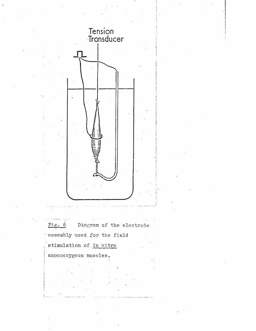
Tension Transducer
ffig> 6 Diagram of the electrode assembly used for the field
stimulation of in- vitro
anococcygeus muscles.
23
In vitro preparation of rat or cat anococcygeus muscle
For in vitro experiments, the rat and cat anococcygeus muscles were treated similarly. The muscles were threaded through the ring electrode assembly shown in Fig, 6, This electrode, which is an adaptation of the one used by Bum & Rand (1960) for stimulation of the sympathetic chain, was originally designed for use with the rat anococcygeus but due to the similar dimensions of the cat anococcygeus, it was equally well suited to this muscle.
The electrode assembly was then lowered into Krebs solution in a 10 ml organ bath and clamped into position. The Krebs solution was maintained at 36°C by a thermostatically controlled water circulator and gassed with a mixture of 95% O2 and 5% C02. The physiological solution used throughout was Krebs* bicarbonate solution as described by Krebs u Henseleit (1932), with composition as follows - NaCl 6,9g/lj KC1 0.35g/l; CaCl2 0,29g/l; KHgPO 0.16g/lj Mg50lf.7H20 0.29g/l; NaHC03 2,lg/lj glucose 2g/l,
This solution was made up daily from concentrated stock solutions which were sufficient for 5 experiments apart from glucose which was added solid,to the mixture. To avoid precipitation of the calcium content, calcium chloridewas added last to the mixture and the pH maintained at 7.4 by continuousgassing with 95% C>2; 5% C02«
The electrode assembly was positioned so that the muscles ran verticallythrough the ring electrode and were free to contract. The long threadon the tendinous end was attached to a Grass FT03 isometric transducer which was positioned vertically above the bath and mounted on a rack and pinion clamp. The muscle was first set up in a relaxed condition and then the transducer was racked up to stretch the muscle to a tension of 1 g.Following this, the muscle was left to equilibrate for a variable period of at least 20 min before any further operations were carried out. During
24
this period, the tension fell within the first 10 min to a resting level of 0.3-0.6 g in both species which was maintained from then on. Tension was displayed on a Grass model 7, four channel Polygraph. In most experiments, the two muscles were set up and recorded simultaneously in separate but identical baths.
Field stimulation of the intramural nerve fibres was by a Tektronix type 161 pulse generator triggered by a Tektronix type 162 waveform generator, and monitored by a Tektronix type 360 indicator oscilloscope. This gave the capacity to deliver pulses of 0.01-100 msec duration and 0-50 V intensity at frequencies of 0.1-10,000 Hz for gated periods of up to 10 sec. Longer periods were gated manually and lower frequencies applied manually as single pulses. Two separate stimulators were used enabling either independent or simultaneous stimulation of the two muscles. A pulse width of 1 msec was used except in experiments whose object was to examine the effects of pulse duration. Pulses of 1 msec duration stimulated nerves but not muscle as shewn by the results and in agreement with experience with other tissues.
In order to determine the voltage to be used in each experiment, the threshold voltage to produce a detectable response was first found. This value was then doubled and a standard response obtained (usually the motor response to 10 Hz for 10 sec) and this voltage further increased to ensure that it was supra-mxinal. Typical values were 3-4 V threshold; with a supra-maximal voltage of 10-15 V.
Drugs were added to the bath in volumes of 0.1-0.3 ml from graduated glass syringes. Mixing occurred rapidly due to the bubbling gas mixture and all concentrations are expressed as the final dilution in the bath. The bath was emptied from belcw and filled from above by gravity from a 1 L reservoir. Drugs were washed out by emptying and refilling the bath three times. In cases where one drug was required to be present continuously
throughout several washes, it was added to the reservoir to give the final concentration required.
Removal of rat heart and vasa deferehtla
In experiments an the effects of reserpine, the heart, vas deferens and anococcygeus were removed for assay of their NA content. Rats were killed by a blew an the head followed by exsanguination, or, in the case of animals previously pithed, by exsanguination by removal of the carotid cannula. Tissues were removed in the order: heart, vas deferens, anococcygeus•The abdomen was opened along the mid-line and the heart exposed by cutting through the costochondral joints and removing a flap of chest wall. The pericardium was opened and the heart removed from the body by cutting the mjor blood vessels where they enter the heart. After removal, the ventricles were cut open, washed with saline and blotted to remove any blood that remained.
Dissection of the vasa deferentia started by pushing the testicles out of the scrotum into the abdominal cavity. Holding the epididymus with forceps, the connection between the epididymus and scrotum was severed and a thread tied round the epididymal end of the vas. It was then possible to dissect thevas free of mesenteric fold and fat along each side before severing the organat the prostatic end. If a thread is tied round the prostatic end, the vas can be used as an in vitro preparation similar to that described by Hukovic (1961) or Birmingham & Wilson (1963) for measurement of the responses of the longitudinal smooth muscle. After removal for assay, the vasa were blotted and excess secretory products in the lumen were gently extruded before weighing, since it has been suggested by Sjostrand & Swedin (1968) that reserpine my cause an increase in such contents, “thus causing anapparent decrease in the NA content per unit weight.
26
Removal of the heart and vasa deferentia took less than 2 min so there was no significant delay before dissection of the anococcygeus which followed inmediately after. Tissues for NA assay were weighed immediately after removal in beakers cooled to *+°C then quickly cooled in liquid nitrogen in order to eliminate any possible chemical or enzymic breakdown of the tissue NA.
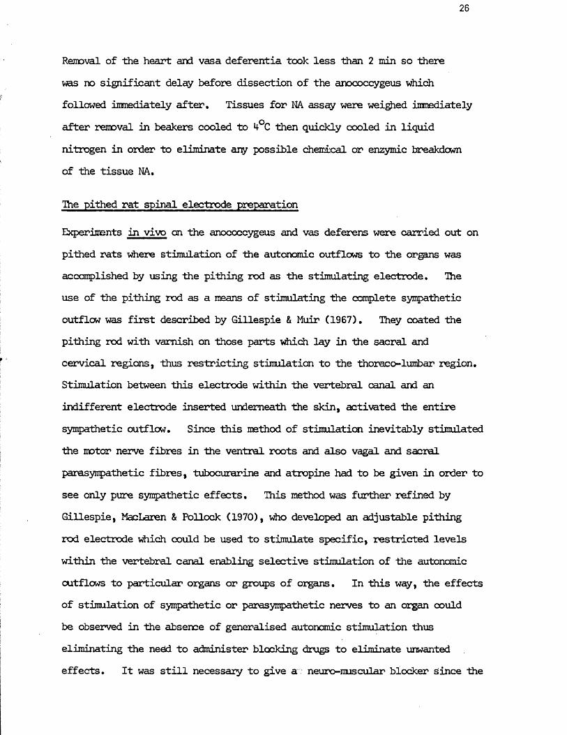
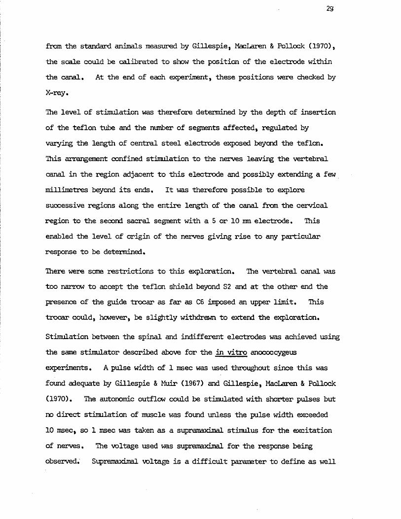
The pithed rat spinal electrode preparation
Experiments in vivo cn the anococcygeus and vas deferens were carried out on pithed rats where stimulation of the autonomic outflows to the organs was accomplished by using the pithing rod as the stimulating electrode. The use of the pithing rod as a means of stimulating the complete sympathetic outflow was first described by Gillespie & Muir (1967). They coated the pithing rod with varnish on those parts which lay in the sacral and cervical regions, thus restricting stimulation to the thoraco-*lumbar region. Stimulation between this electrode within the vertebral canal and an indifferent electrode inserted underneath the skin, activated the entire sympathetic outflow. Since this method of stimulation inevitably stimulated the motor nerve fibres in the ventral roots and also vagal and sacral parasympathetic fibres, tubocurarine and atropine had to be given in order to see only pure sympathetic effects. This method was further refined by Gillespie, MacLaren & Pollock (1970), who developed an adjustable pithing rod electrode which could be used to stimulate specific, restricted levels within the vertebral canal enabling selective stimulation of the autonomic outflows to particular organs or groups of organs. In this way, the effects of stimulation of sympathetic or parasympathetic nerves to an organ could be observed in the absence of generalised autonomic stimulation thus eliminating the need to administer blocking drugs to eliminate unwanted effects. It was still necessary to give a: neuro-muscular blocker Since the
ScaleTeflonTube-
SteelTubeOrbit
Electrode
Fir,. 7 Arrangement of the pithing rod electrode assembly in the pithed rat. The animal is initially pithed via the orbit and foramen ragnum by inserting a steel tube down into the spinal column to the level of the sixth cervical vertebrae. Through this trocar is passed successively a teflon tube and ins5.de that a fine steel tube.The position of this steel electrode within the canal and the length exposed can be determined by reference to a simple scale.
27
ventral roots adjacent to the electrode were still stimulated, but since specific drugs are now available such as gallamine and pancuronium which are relatively free of the autonomic side effects of tubocurarine, this was no serious disadvantage. This is the preparation which has new been used and adapted for stimulation of the nerves to the anococcygeus-and vas deferens.
Male Wistar rats 250-300 g were used. Gillespie, MacLaren & Pollock (1970) found that in this weight range the dimensions of the vertebrae were constant, enabling a standard size of pithing rod to be used and the location of the electrode within the canal to be known by reference to a simple scale.
The rats were anaesthetised with a mixture of 5% Halothane in 1 vol 0£ to 2 vol N2 O. The gas mixture was administered to the rats in an anaesthetic box to minimise any stress or excitement caused by induction which could lead to fatalities on pithing. When the animal no longer showed withdrawal reflexes, its trachea was intubated and then it was pithed and immediately respired artificially at 90 per min with a Palmer respiration pump.
The arrangement of the pithing rod is shewn in fig. 7. A short steel tube (13 s.w.g.) was inserted through the orbit and foramen magnum and down into the spinal column as far as the sixth cervical vertebrae. Through this trocar were passed successively a teflon tube (0,16 mm 0.D,) and inside that a fine steel tube (26 s.w.g.) which was extruded at the sacral end to complete the pithing. The most important features for survival of the preparation, were
1) Deep anaesthesia beforehand which reduces the large autonomic discharge caused by pithing,
2) immediate and adequate ventilation after pithing since the respiration centre was destroyed, and
28
3) mintenance of body temperature since control of this was alsolost.
The temperature of each pithed animal was, in most experiments, maintained at 34-37°C by means of a tungsten lamp, and monitored by a rectal thermometer, but in some of the later experiments it was maintained automatically by a Hamoeothermic Blanket (Epil) placed underneath the rat and regulated by a rectal probe thermistor.
After pithing, one carotid artery was cannulated with a polythene cannula filled with heparinised saline to record the blood pressure via a Statham P23A pressure transducer. This pressure, together with the heart rate derived from it by a Grass Tachograph Pre-amplifier, was displayed on a Grass Polygraph. A polythene cannula was inserted into the left femoral vein for administration of drugs. No heparin was necessary to keep the venous cannula clear, so no heparin was introduced into the circulation.Drugs were administered in volumes of 0.05-0.2 ml from a glass syringe and washed in with 0.9% saline from a gravity feed to make up a total volume of 0.3 ml. In some experiments, the left femoral vein was also cannulated in order to give a slow infusion of tyramine. A Palmer slow injection apparatus was used to administer the drug at a rate of 5-40 pl/min. A silver wire which acted as the indifferent electrode was inserted under the skin parallel to, and extending the full length of, the spinal column.
The position of the electrode within the vertebral canal was determined by means of the scale shown in fig. 7. Since the lengths of the outer steel tube, teflon tube and inner steel electrode were known, if the lengths of the teflon and electrode outside the rat were measured, the length of the teflon and electrode within the vertebral canal could be arrived at by subtraction. Since the distances inside the vertebral canal to each segment were kncwn
23
from the standard animals measured by Gillespie, MacLaren & Pollock (1970), the scale could be calibrated to show the position of the electrode within the canal. At the end of each experiment, these positions were checked by X-ray.
Ihe level of stimulation was therefore determined by the depth of insertion of the teflon tube and the number of segments affected, regulated by varying the length of central steel electrode exposed beyond the teflon.This arrangement confined stimulation to the nerves leaving the vertebral canal in the region adjacent to this electrode and possibly extending a few millimetres beyond its ends. It was therefore possible to explore successive regions along the entire length of the canal from the cervical region to the second sacral segment with a 5 or 10 mm electrode. This enabled the level of origin of the nerves giving rise to any particular response to be determined.
There were same restrictions to this exploration. The vertebral canal was too narrow to accept the teflon shield beyond S2 and at the other end the presence of the guide trocar as far as 06 imposed an upper limit. This trocar could, however, be slightly withdrawn to extend the exploration.
Stimulation between the spinal and indifferent electrodes was achieved using the same stimulator described above for the in vitro anococcygeus experiments. A pulse width of 1 msec was used throughout since this was found adequate by Gillespie & Muir (1967) and Gillespie, MacLaren & Pollock (1970). The autonomic outflow could be stimulated with shorter pulses but no direct stimulation of muscle was found unless the pulse width exceeded 10 msec, so 1 msec was taken as a supramaximal stimulus for the excitation of nerves. The voltage used was supramaximal for the response being observed. Supramaximal voltage is a difficult parameter to define as well
Y /VAAAAi- /lAAAAi-
Recorder
Transducers^
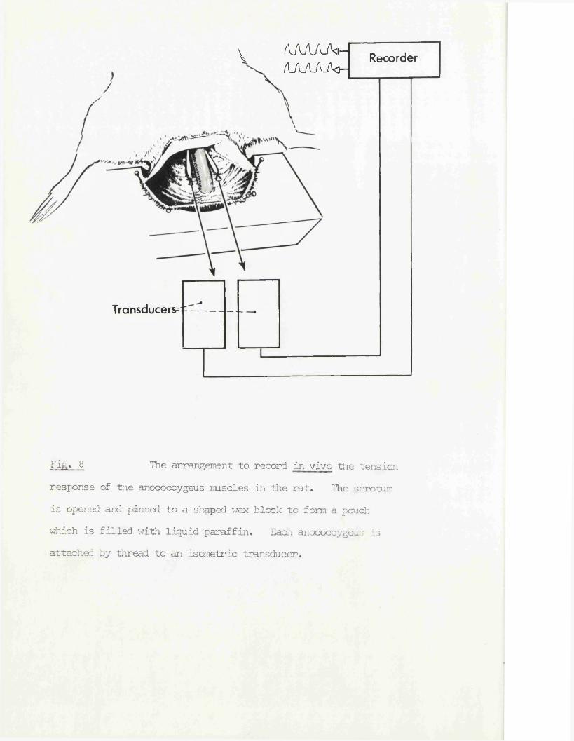
Tift* 8 The arrangement to record in vivo the tensionresponse of the anococcygeus muscles in the rat. The scrotum is opened and pinned to a shaped wax block to form a pouch which is filled with liquid paraffin. Each anococcygeus is attached by thread to an isometric transducer.
30
as determine in this preparation since if the outflow to a particular organ extends over a greater number of segments than are covered by the stimulating electrode then increasing the strength of stimulation beyond what is maximal for adjacent nerve fibres will continue to oause an increase in response by an extension of the region stimulated. What was required, however, was supramaximal stimulation of the autonomic outflows within the region of the electrode. In early experiments, it was found that 20 V was always supramaximal while still leaving the electrode specific to within 5 mm, so this parameter was used in each subsequent experiment with the proviso that double or half this voltage did not produce a significantly larger or smaller response. If they did, this normally was a pointer to an electrical fault since the electrical properties of the preparations were so similar.
Muscle twitching as a result of stimulating the ventral roots was prevented by giving Pancuronium bromide (1 mg/kg) intravenously at the start of each experiment as soon as the venous cannula was in place.
The rat anococcygeus in vivo preparation
The tension of the nale anococcygeus muscles can easily be recorded with a .minimum of operative interference since the ventral bar lies superficially just under the skin of the scrotum a few mm short of the anal margin. If this ventral bar is split, a thread can be attached to each muscle without interference with the nerves and their individual responses to drugs administered intravenously or, in the case of pithed rats, from stimulation of the autonomic outflows, can be recorded.
Dissection of the anococcygeus began after pithing and insertion of cannulae.
The muscles were exposed by an incision in the scrotum just anterior to the anal margin and the edges of the incision pinned on to a shaped wax block placed across the tail as shown in fig. 8. The pouch so formed was filled
31
with liquid paraffin (sp. gr. 0*85) to prevent drying of the exposed tissue. The rectal thermometer or probe could be inserted underneath this block*
The muscle layers overlying the colon were then cleared by blunt dissection for approximately 25 ram from the anal margin in order to free more of the anococcygeus, the ventral bar was split and a thread tied round the cut end of each muscle.
Each thread was attached to a Grass FT03 isometric transducer mounted on a rack and pinion clamp, and the transducer positioned to apply an initial tension of 1 g to each muscle at an angle of approximately 30° to the horizontal. This held the muscle in its normal in situ position. There was no need for further dissection of the anococcygeus muscle since it was effectively anchored at the rostral end by the tendon attached to the vertebrae.
This preparation responded to drugs and spinal stimulation for as long as the rat remained viable as indicated by the presence of a blood pressure and heart beat, i.e. up to 10 hrs, and so presumably receives an adequate blood perfusion and intact innervation. Responses to nerve stimulation and drugs were comparable in size to those obtained in vitro, so the muscles appeared to be free to develop tension. Movement of the colon might have been expected to interfere with the tension in the anococcygeus, but, in fact, spontaneous, visible movements of the colon had no effect, and when the sacral parasympathetic outflow was stimulated, the resultant contraction of the longitudinal smooth muscle of the colon had no effect on the tension of the anococcygeus.
This dissection was at first performed with the help of a Zeiss dissection microscope but as it became more routine was carried out using the naked eye. The dissection took less than 10 min.
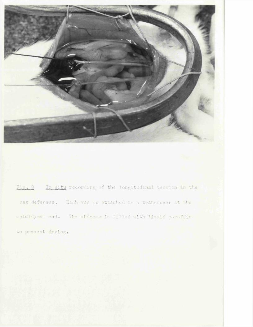
71°:. 9 In situ recording of the longitudinal tension in the
vas deferens. 3ach vas is attached to a transducer at the
©pldidymal end. The abdomen is filled with liquid paraffin
to prevent drying.
32
The rat vas deferens in vivo preparation
Since Hukovic (1961) first introduced the guinea pig isolated hypogastric nerve - vas deferens preparation as an example of a sympathetically innervated smooth muscle, the response of this tissue to nerve stimulation has been extensively investigated (Sjostrand, 1962, 1965; Bentley & Sabine, 1963; Birmingham & Wilson, 1963; Ferry, 1963, 1967; Merrilees, Burnstock & Holman, 1963). In the rat, although the isolated hypogastric nerve-vas deferens preparation (Graham, A1 Katib & Spriggs, 1968) and hypogastric denervation (Sjostrand & Swedin, 1967) have been described, relatively little work has been done with extrinsic stimulation, possibly because of the greater difficulty in preserving the extrinsic nerve. By comparison, there is an extensive body of work on the effect of drugs on the isolated vas in this species (Chang & Chang, 1965; Swedin, 1971; Furness & Burnstock, 1969; Birmingham, 1970). In addition, the effects of stimulating the nerves to the vas deferens in vivo have rarely been described (Blakeley, Deamaley & Harrison, 1970), and this only in the guinea pig. With the pithed rat, however, stimulation of the autonomic outflow offers an opportunity to examine the properties of the innervation of the rat vas deferens in vivo.
Ihe dissection of the vas deferens was carried out after the rat had been pithed, cannulae inserted and, where both vas and anococcygeus were recorded in the same animal, after dissection of the anococcygeus.
Ihe abdomen was opened in the mid-line for approximately 60 mm rostral from the penis and the edges of the incision sewn round a curved bar to form a a sac as shown in fig. 9. In a few initial experiments, the gastrointestinal tract between the stonach and descending colon was removed, but this was discontinued since it was found to make no difference to the ease
of recording the vas deferens, which was the intention of the manoevre, and also shortened the survival time of the preparation, presumably due to loss of blood volume.
On first opening the abdomen, a few ml of liquid paraffin was run over the Intestines to prevent drying. When the dissection of the vas was complete, the abdominal cavity was filled with liquid paraffin (approximately 20 ml) pre-warmed to 36°C to provide a pool in which the vas could function over a period of hours. The initial dissection was as for removal but leaving the prostatic end attached. Since the nerves enter and the blood vessels enter and leave at the prostatic end (Sjostrand, 1965), the vas can be tied off at the epididymal end and dissected free of connective tissue along its entire length without any loss of functional capacity. Two adjacent ties were made at the epididymal end and the vas cut between these, leaving it attached to the rat only at the prostatic end. The thread attached to the vas was then attached to a Grass FT03 isometric transducer which was positioned to pull the vas straight and apply an initial tension of 0.5 g (fig. 9). There was no need to fix the vas at the prostatic end since it was very effectively anchored by its attachment to the urethra which is firmly held in place by the ischio-cavemosus and pyramidalis muscles.Both left and right vas deferens were set up in this way.
This preparation enabled recording of the longitudinal tension developed by the smooth muscle of the vas.
Another way of recording the response of the vas was to perfuse physiological solution at a constant rate through the lumen from the epididymal end and measure the perfusion pressure (Gillespie, MacLaren & Pollock, 1970) • This may be nearer to a measure of the physiological response of the organ in much the same way that the perfused ear artery is a more physiological response than the aortic spiral strip. Since no one
34
has ever suggested that the function of the vas is to move something by virtue of its shortening its length, it is surprising that so many examinations of this hollow organ have concentrated on its capacity to develop tension isometrically or shorten its length isotonically, when perfusion is so simple.
With the abdomen opened as above, the vas was cannulated at the epididymal end in the prostatic direction with a polythene cannula, taking care to avoid the artery which runs superficially along the surface, Krebs* solution was perfused at a constant rate of 20 or 40 ul/min using a slow injection apparatus and perfusion pressure was recorded from a side branch 1 cm from the cannula using a Statham P23A pressure transducer. The perfusate usually escaped via the penis without obstruction, the vas itself providing almost all of the resistance, but in a few cases a gradual build up of pressure occurred due to some obstruction distal to the vas, and in these instances an incision was made at the base of the adjacent seminal vesicle to allow egress of the perfusate. No further dissection was necessary.
The dissection of the vas took approxinately 15 min for both organs.
The total dissection time to set up a pithed rat, insert cannulae and record both the anococcygeus and vasa eventually took 1 hr.
Administration of Reserpine - Protocol
In the experiments to determine the effects of reserpine on tissues, the reserpine was administered intra-peritoneally. Rat tissues are depleted of their catecholamines by reserpine whether it is administered intra- peritoneally (Graham, A1 Katib, & Spriggs, 1968; Jane, Planas & Bonaccorsi, 1970; Haggendal & Dahlstrom, 1971) or sub-cutaneously (Sjostrand & Swedin, 1968), In the present experiments, reserpine could not be obtained as the
commonly used "Serpasil" injectable solution so the alkaloid was dissolved in an aqueous solution of ascorbic acid (0.4% w/v) in which it is readily soluble (Carlsson 1966). This solution was, however, acidic (pH 4) and therefore the intra-peritoneal route was selected as being more humane and less traumatic for the animals. The drug was administered in volumes of1 ml/kg i.e. 0.25 ml/250 g rat. The rats showed no signs of being in anydiscomfort after these injections.
The experiments with reserpine pretreatment could be split into 3 groups.
1) Variation of reserpine doseReserpine was given to groups of 6 rats, 2*+ hrs before sacrifice in doses of 50, 100, 200, and 1,000 pg/kg.
2) Variation of time of pretreatmentReserpine 200 pg/kg was given to groups of 6 rats at the following timesbefore sacrifice; 3, 6, 12, 24, and 48 hrs, 21, 28, and 35 days.
3) Pretreatment before pithingRats were given 200 pg/kg reserpine 3 hrs before pithing.
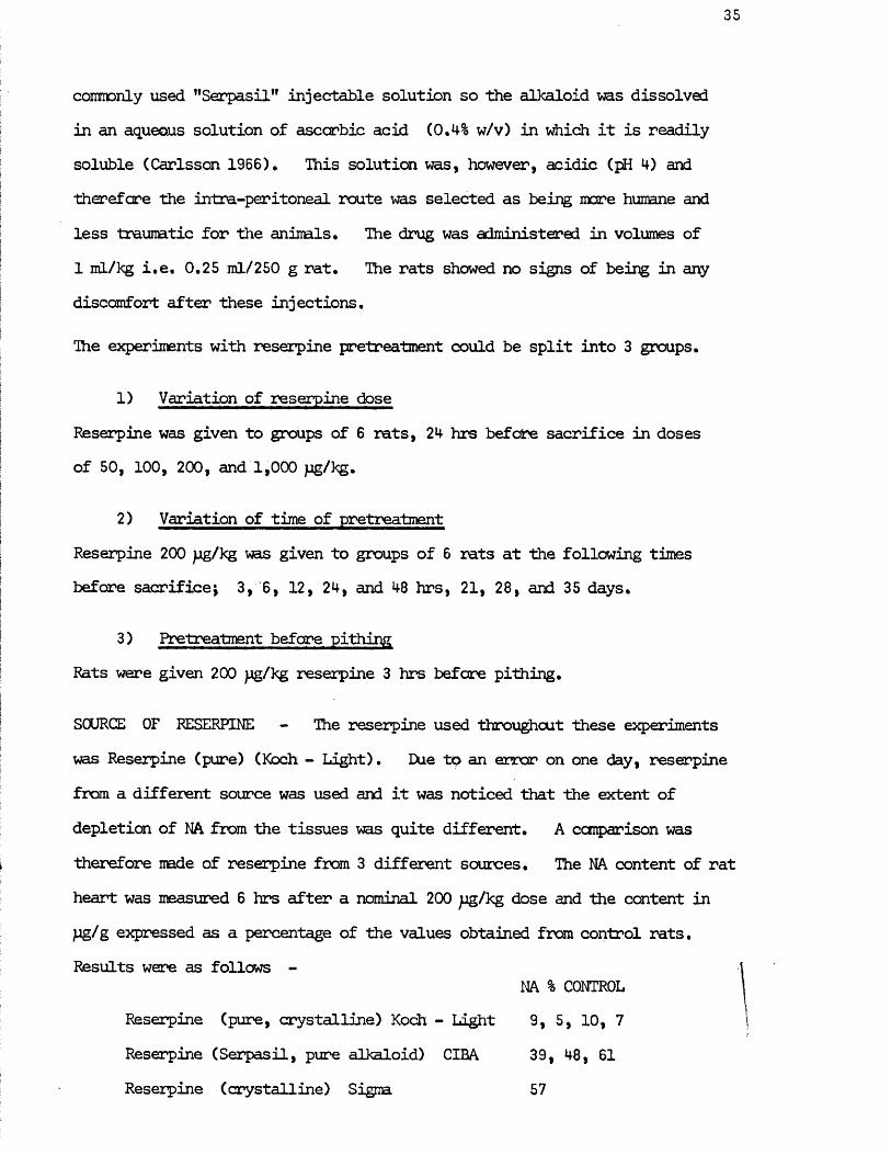
SCURCE OF RESERPINE - The reserpine used throughout these experiments was Reserpine (pure) (Koch - Light). Due to an error on one day, reserpine from a different source was used and it was noticed that the extent of depletion of NA from the tissues was quite different. A comparison was therefore made of reserpine from 3 different sources. The NA content of ratheart was measured 6 hrs after a nominal 200 pg/kg dose and the content inpg/g expressed as a percentage of the values obtained from control rats. Results were as follows -
NA % CONTROLReserpine (pure, crystalline) Koch - Light 9, 5, 10, 7Reserpine (Serpasil, pure alkaloid) CIBA 39, 48, 61Reserpine (crystalline) Sigma 57
36
It is clear from these results that the source of reserpine used is an important factor in any experiments performed,
CIRCADIAN CONSIDERATIONS - Rats in groups 1) and 2) were sacrificed as near as possible to 12,00 hrs except for the 6 hr pretreated rats which were treated at 9,00 hrs and therefore sacrificed at 15.00 hrs. The rats in group 3) were treated at 9.00 hrs, pithed at 12.00 hrs and their tissues taken at 15.00 hrs. This made direct comparison possible between the 3 and 6 hr pretreatment and pithed animals.
There is some evidence of circadian variation in peripheral catecholamine content in the submaxillary glands (Wurtman & Axelrod, 1966) and in the adrenals (Scheving, Harrison & Pauley, 1968), but there has been no suggestion of such a factor in the tissues used here. This arrangement of the experiment should, however, eradicate any such problems.
Assay of tissue noradrenalineThe main steps in the chemical assay of catecholamines are extraction, purification and concentration of the amines and thereafter the final estimation. Before assay of tissue catecholamines, an extract must be made and the proteins precipitated. The reaction utilised in this assay is the trihydroxyindole reaction. Since Loewi (1918) first reported the appearanceof a yellow - green fluorescence after addition of strong alkali to an adrenaline solution, the reactions involved have been worked out and developed into a sensitive and specific assay for adrenaline and noradrenaline (Ehrlen, 1948; Lund, 1949). It was subsequently shown that noradrenaline and adrenaline could be determined differentially spectrophotametrically (Bertler, Carlsson & Rosengren, 1958; Euler & Lishajko, 1959) and this method further refined by stabilisation of the end-products (Euler & Lishajko, 1961). It is this method of Euler & Lishajko which I have used, with purification of the
37
noradrenaline on alumina columns as described by Gillespie, Hamilton &Hosie (1970),
Extraction After removal, tissues were weighed and frozen in liquid nitrogen. Tissues from 2 animals were pooled at this stage. The tissues were then placed in a stainless steel mortar and pestle (also pre-cooled in liquid nitrogen) and pulverised far 20 secs using a power-hammer driven from a compressor. This procedure reduced the tissues to a fine powder. The powdered« tissue-was transferred to a cooled beaker and 5 ml of 0.4 M perchloric acid added to precipitate the protein. At this stage 1 jug NA aliquots (2) were added to perchloric adid to give control samples for the recovery from subsequent procedures. After 30 min, on ice, the mixture was centrifuged in a bench centrifuge to remove the precipitate and the supernatant decanted for adsorption onto alumina columns. The alumina ("CAMAG" Emanuel, pH of aqueous suspension .5 ± 0.5, 504 - C - 1) columns (15 mm high, 10 mm diameter) were prepared with a flow rate of at least 5 ml/min in order to shorten the time that the NA is on the columns at a pH at which it is unstable. To each supernatant, EDTA was added to give a final concentration of 1% and the pH was adjusted to 8.2 - 8.3 with 2 M & 0.5 M NaQH, At pH 8.2 the extract was added immediately to the alumina and the column washed with cold distilled water till the pH of the effluent reached 5.8 - 6.0. The columns were dried using compressed nitrogen and the catecholamines eluted with 2 consecutive 5 ml aliquots of 0.25 M acetic acid. The extract was then centrifuged for 10 min in a bench centrifuge to remove any alumina particles and the supernatant decanted for assay. During these prodedures, all materials were kept at 0°C to reduce breakdown of catecholamines.
Assay The trihydroxy indole reaction was carried out in two stages.Oxidation of the catecholamines to chrome derivatives was performed with potassium ferricyanide. An advantage of potassium ferricyanide is that
38
dopamine, normetanephrine and metanephrine, if present, will hardly interfere (Haggendal, 1966), Oxidation was then interrupted and the chrome derivatives converted into fluorescent lutines by the addition of a mixture of ascorbic acid and sodium hydroxide. Ethylene diamine was also present in this latter mixture to stabilise the lutines (Euler & Lishajko, 1961).The composition of this alkaline ethylene diamine - ascorbic acid mixture was as follows - per 100 ml, ethylene diamine 2 ml; 5 M NaCH 88 ml;2% ascorbic acid 10 ml.
The pH of the eluates was adjusted to 6.2 - 6.3 with 2 M NH QH and 3 ml aliquots reacted as follows -
Time 0 min 0.1 ml 0.25% potassium ferricyanide added3 min 4 ml alkaline ethylene diamine - ascorbic acid added
Another 3 ml was treated as a reaction blank as follows -
Time 0 min 4 ml alkaline ethylene diamine - ascorbic acid added 3 min 0.1 ml 0.25% potassium ferricyanide added
An assay standard of 0.1 pg NA was also prepared as follows -
2 ml pH 6.5 phosphate buffer 1 ml 0.1 pg/ml NA
TWo such NA standards were reacted and two acted as blanks, as above.Ihe fluorescence in the samples and blanks was then read at 500 nm in an Aminco - Bowman Spectrophotofluorometer using an excitation wavelength of 400 nm. These are the emission and excitation peaks for the NA product.The adrenaline product fluorescence can be read with an excitation wavelength of 450 nm, but. initial experiments shewed that no adrenaline was present in any of the tissues used, so only NA was estimated in the subsequent experiments. The fluorescent product is stable for up to 2 hrs if kept in the dark (Euler & Lishajko, 1961;. Haggendal, 1966) but fading can occur due to exposure to
39
fluorescent lighting, so the reacted samples were kept covered and read within 15 min of the reaction at which time fading was insignificant.Initial calibration experiments showed that the relationship between NA and fluorescence (test minus blank) was linear over the range necessary for the experiments, so all NA contents were calculated as a proportion of the mean assay standard after allowance for volumes.The NA content of the various alumina eluates was thus calculated and the tissue extracts corrected for recovery from the columns using the 1 jug NA column standard. This column recovery never varied by more than 2% between the two standards in any single experiment. It varied between experiments from 70 - 85% and averaged 78%,The NA content of tissues was then expressed as pg NA/g tissue, i.e. pg/g.
Histochemistry - Falck TechniqueThe histochemical method currently in use for the demonstration of catecholamines and indoleamines at the cellular level was developed in Sweden in the early sixties (Carlsson, Falck, Hillarp & Thieme, 1961; Falck, Hillarp, Thieme & Torp, 1962; Falck, 1962; Falck & Torp, 1962 a & b).
The method is based on the principle that amines can be transformed into intensely fluorescent isoquinoline derivatives by condensation with formaldehyde. This was first applied to tissues by Eranko (1952, 1955) who used liquid formaldehyde to demonstrate the location of catecholamines in the adrenals. The more sensitive technique involving the use of formaldehyde vapour on freeze-dried tissue was then later developed by Hillarp, Falck and co-workers. The reactions involved have been categorised (Corrodi & Hillarp, 1963, 1964; Corrodi, Hillarp & Jonsson, 196M-; Corrodi & Jonsson, 1965 a & b, 1966; Jonsson, 1967) and developed into a quantitative miapospectrofluorcmetric method permitting differentiation between noradrenaline, adrenaline, dopamine and 5 - hydroxytryptamine (Caspersson, Hillarp & Ritzen, 1966; Bjorklund, Ehinger & Falck, 1968).
The method used here is a modified version of this technique by Gillespie & Kirpekar (1966).
It is necessary to freeze-dry the tissues to achieve rapid fixation which prevents diffusion of amines out of nerves and also to keep the tissues - 'dry through all subsequent procedures since water converts the fluorescent end- product to a non - fluorescent form.
Tissue blocks were frozen in liquid nitrogen cooled isopentane and freeze- dried in a Speedivac - Pearse tissue drier at -40°C, 0.01 torr for 18 hrs.The temperature in the freeze-drier was then increased to +40°C to ensure that on removal the tissues were not exposed to condensation of water vapour from the atmosphere due to being below the ambient temperature. The tissues were then placed in a closed jar containing paraformaldehyde (Merck) in an oven and thus exposed to formaldehyde vapour at 80°C for 1 hr. Following this, they were re-dried at 0.01 torr for 1 hr before being embedded in degassed wax under vacuum at 65°C. Wax blocks were subsequently made, sliced at 6 p, mounted on heated glass slides and covered with liquid paraffin and a cover glass for examination in the fluorescence microscope. These sections were kept in the dark at all times to prevent fading of the fluorescence which occurs rapidly under daylight or the fluorescent lighting in the laboratory.
Tissues were examined and photomicrographs taken with a Leitz (Wetzlar) Qrthcplan microscope with dark field illumination: HBO 200 Mercury vapourlamp; 3 mm BG12 exciter filter; Condensers - D 0.80, D 1.20 A oil immersion; K530 barrier filter; Objectives - PI 10/0.25, PI 25/0.50, PI 40/0.65,FI Oel 54/0.95 oil immersion.Photomicrographs were taken on Ilford FP4 with a 3 or 5 min exposure.
Drugs used were -
Acetylcholine chloride (Koch - Light); L - Adrenaline bitartrate (B.D.H.); Atropine sulphate (B.D.H.); Dexamphetamine sulphate; Adenosine 51 - triphosphate (Sigma); Carbachol (Burroughs Wellcome); Cocaine hydrochloride; Guanethidine sulphate (CIBA); Hexamethonium bromide (Koch - Light); Histamine acid phosphate (B.D.H*); 6 - Hydroxydopamine(kindly donated by Dr. H. Thoenen of Hoffmann la Roche); 5 - hydroxy- tryptamine creatinine sulphate (Koch - Light); DL - Isoprenaline hydrochloride (Sigma); D - Lysergic acid diethylamide tartrate (Sandoz); Mecamylamine hydrochloride (Merck, Sharp & Dohme); Mepyramine maleate (iky & Baker); Methysergide bimleate (Sandoz); Neostigmine methylsulphate (Roche); L - Noradrenaline bitartrate (Koch - Light); Oxymetazoline (kindly donated by Dr. D. Jack of Allen & Hanbury); Pancuronium bromide (Organon); Fhentolamine mesylate (CIBA); Propranolol sulphate (Sigma); Prostaglandins E , & F^(kindly donated by Prof. E.W. Horton);Reserpine (crystalline) (Koch - Light); Tyramine hydrochloride (Sigma); Vasopressin (Parke - Davis).
Doses in in vivo experiments are expressed in grams of the base and in in vitro experiments as molar concentration of the base with the exceptions of 6 - hydroxydopamine, prostaglandins (g/ml) and vasopressin (International Units / ml).
Abbreviations used throughout the text are noradrenaline - NA;5 - hydroxytryptamine - 5HT; Lysergic acid diethylamide - LSD;6 - hydroxydopamine - 60HDA; adenosine 5' - triphosphate - ATP.

RESULTS
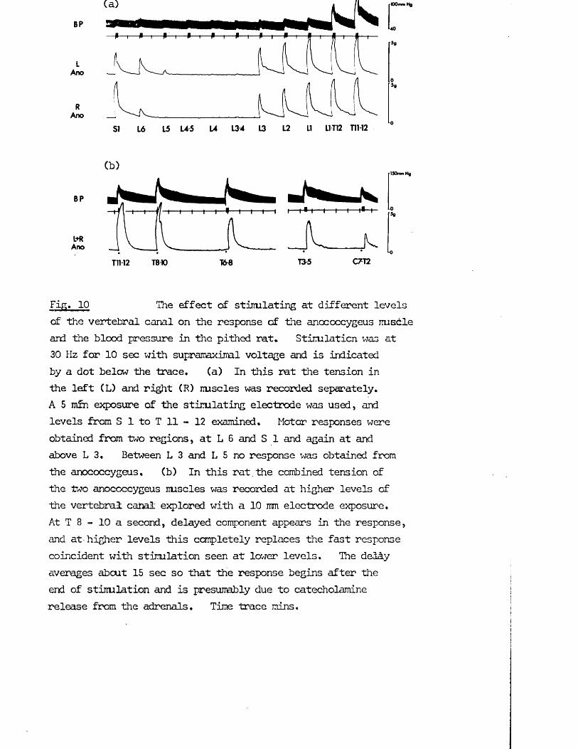
100 mm hg
40
5g
5«
'130mm Hg
■0Sg
Fig. 10 The effect of stirrulating at different levelsof the vertebral canal on the response of the anococcygeus iruscile and the blood pressure in the pithed rat* Stimlaticn was at 30 Hz for 10 sec with supranaximal voltage and is indicated by a dot below the trace. (a) In this rat the tension in the left (L) and right (R) iruscles was recorded separately,A 5 mfn exposure of the stirrulating electrode was used, and levels from S 1 to T 11 - 12 examined. Motor responses were obtained from two regions, at L 6 and S 1 and again at and above L 3, Between L 3 and L 5 no response was obtained from the anococcygeus, (b) In this rat the combined tension of the two anococcygeus nuscles was recorded at higher levels of the vertebral cabal explored with a 10 mm electrode exposure.At T 8 - 10 a second, delayed component appears in the response, and at-higher levels this completely replaces the fast response coincident with stimulation seen at lower levels. The deMy averages about 15 sec so that the response begins after the end of stimulation and is presumably due to catecholamine release from the adrenals. Time trace mins.
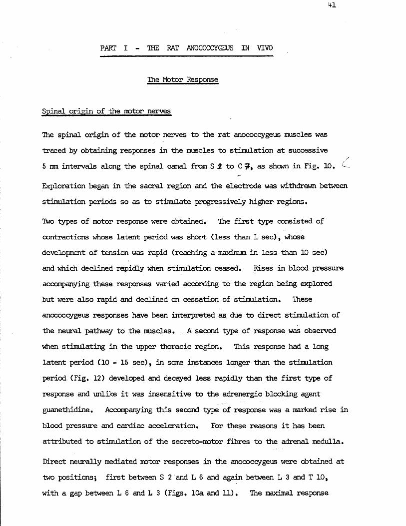
PART I - THE RAT ANOCOCCYGEUS IN VIVO
The Motor Response
Spinal origin of the motor nerves
The spinal origin of the motor nerves to the rat anococcygeus muscles was traced by obtaining responses in the muscles to stimulation at successive 5 mm intervals along the spinal canal from S i to C as shewn in Fig. 10 •
Exploration began in the sacral region and the electrode was withdrawn between stimulation periods so as to stimulate progressively higher regions.
Two types of motor response were obtained. The first type consisted of contractions whose latent period was short (less than 1 sec) , whose development of tension was rapid (reaching a maximum in less than 10 sec) and which declined rapidly when stimulation ceased. Rises in blood pressure accompanying these responses varied according to the region being explored but were also rapid and declined on cessation of stimulation. These anococcygeus responses have been interpreted as due to direct stimulation of the neural pathway to the muscles. A second type of response was observed when stimulating in the upper thoracic region. This response had a long latent period (10-15 sec), in some instances longer than the stimulation period (Fig. 12) developed and decayed less rapidly than the first type of response and unlike it was insensitive to the adrenergic blocking agent guanethidine. Accompanying this second type of response was a narked rise in blood pressure and cardiac acceleration. For these reasons it has been attributed to stimulation of the secreto-motor fibres to the adrenal medulla.
Direct neurally mediated motor responses in the anococcygeus were obtained at two positions; first between S 2 and L 6 and again between L 3 and T 10, with a gap between L 6 and L 3 (Figs. 10a and 11). The maximal response
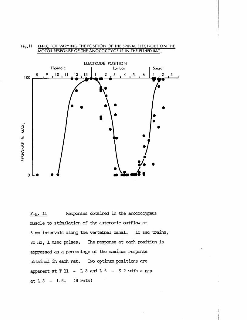
RES
PONS
E %
MA
X
Fig. 11 EFFECT OF V A R Y IN G THE POSITION OF THE SPINAL ELECTRODE O N THE MO TO R RESPONSE OF THE A N O C O C C Y G E U S IN THE PITHED RAT.
ELECTRODE POSITIONThoracic Lumbar Sacral
1 • 2100
Fig. 11 Responses obtained in the anococcygeusmscle to stimulation of the autonomic outflow at 5 mm intervals along the vertebral canal. 10 sec trains, 30 Hz, 1 msec pulses. The response at each position is expressed as a percentage of the maximum response obtained in each rat. Ttoo optimum positions areapparent at T 11 - L 3 and L 6 - S 2 with a gap at L 3 - L 6. (9 rats)
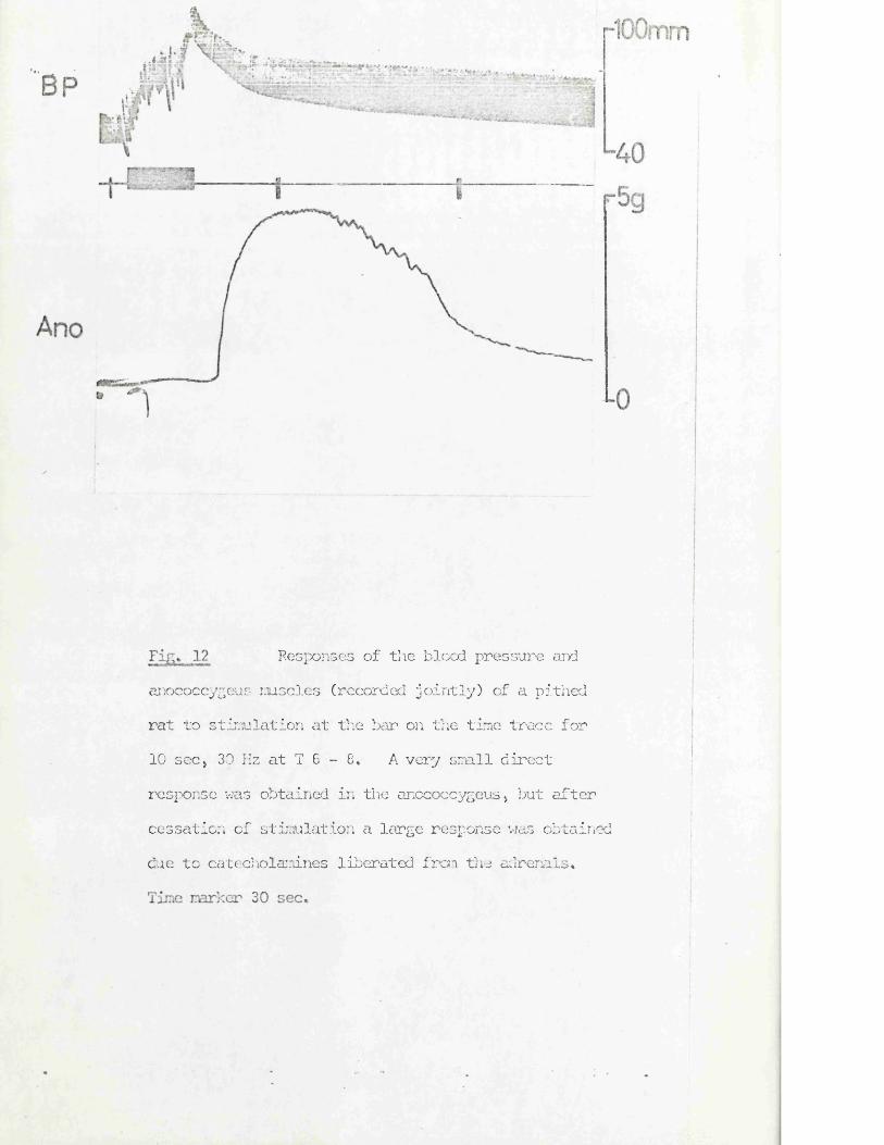
Responses of the blood pressure and anococcygeus muscles (recorded jointly) of a pithed rat to stimulation at the bar on the time trace for 10 sec, 30 Hz at T 6 - 8. A very small direct response was obtained in the anococcygeus, but after cessation of stimulation a large response was obtained due to catecholamines liberated frcn the adrenals.
42
was similar in size in both regions (4.9 ± 0.2 g per single muscle, n = 10).At T 10 - 11 delayed responses appeared in addition to the direct responses and beyond T 10 the rapid responses disappeared to be replaced by the delayed adrenal responses (Figs. 10b and 12). The optimal position for the adrenal response lay from T 10 to T 4. No responses were obtained higher than T 2.
Regions below S 2 could not be separately stimulated since the narrowness of the spinal canal prevented entry of the teflon tube which isolates the stimulating electrode. The precise lower limits of the lower motor outflow could not, therefore, be defined, but experiments with finer teflon and the electrode extruded to the end of the vertebral canal suggest that outflows to the anococcygeus do not extend much beyond S 2 since responses to stimulation with an electrode completely below S 2 were very small.
Frequency characteristics of the motor response
Three methods were used to determine the frequency/response characteristics of the responses obtained from stimulation in the two motor regions, S 2 - L 6 (’lower1) and L 3 - T 10 (’upper’).
(1) Stimulation for a fixed 10 sec period at varyingfrequencies.
(2) Stimulation until the maximum response was obtainedat varying frequencies.
(3) Stimulation with a fixed number of pulses at varyingfrequencies.
The frequency/response curves obtained with these different techniques are shewn in Fig. 13. Clearly the optimal frequency, whichever method is chosen, is 30 Hz, but the efficacy of low frequency is very dependent on the method of comparison used.
(1) Stimulation for 10 sec periods at frequencies from 0.1 to 100 Hz produced motor responses at all frequencies with a maximum at 10 - 30 Hz.
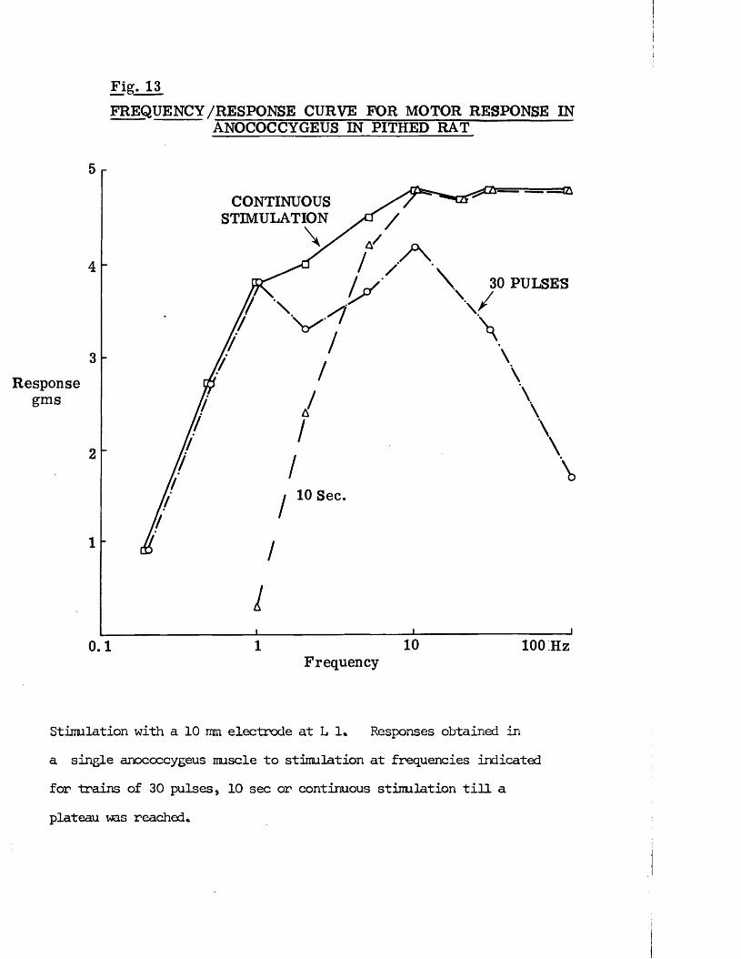
Fig. 13FREQUENCY/RESPONSE C U RVE F O R M O T O R RESPONSE IN
A N O C O C C Y G E U S IN PITHED R A T
Responsegms
0.1
CONTINUOUSSTIMULATION
30 PULSES
10 Sec.
I
10 100 HzFrequency
Stimulation with a 10 mm electrode at L 1* Responses obtained in a single anococcygeus muscle to stimulation at frequencies indicated for trains of 30 pulses, 10 sec or continuous stimulation till a plateau ms reached*
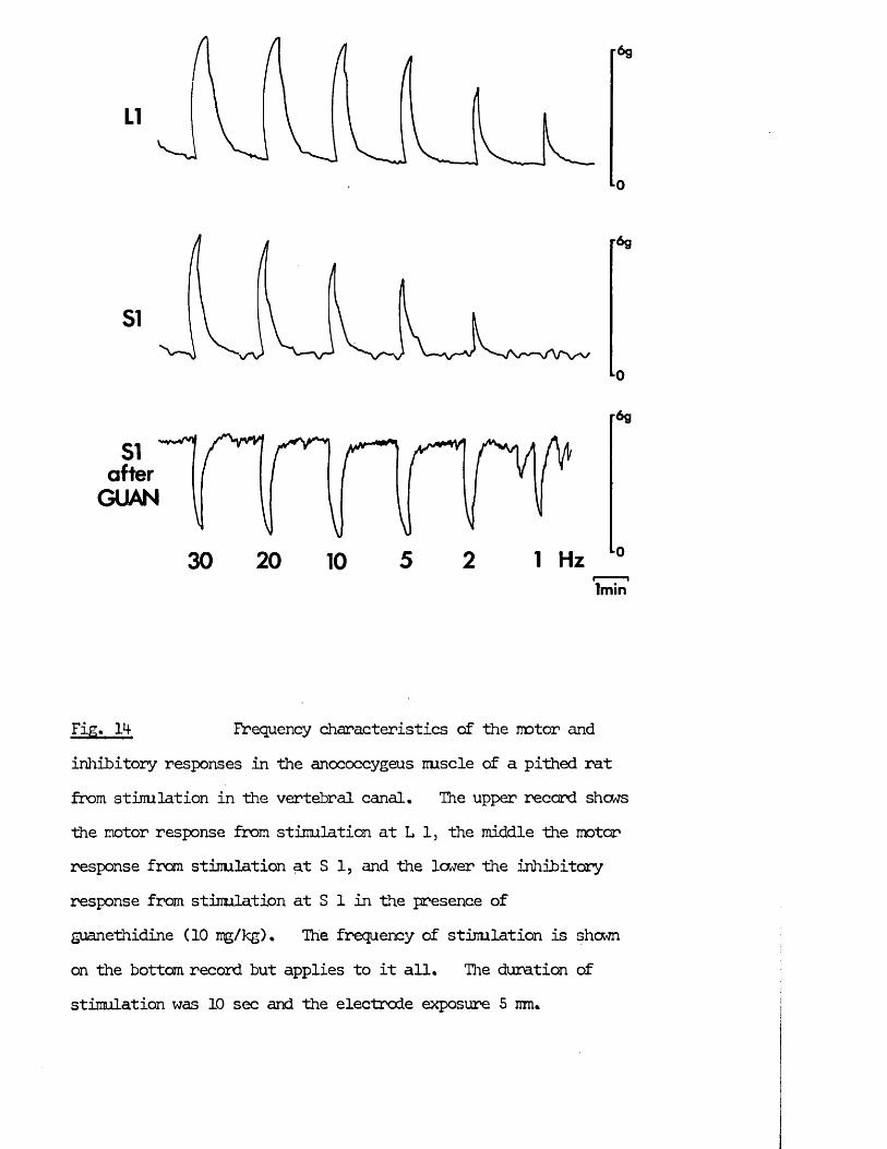
0
SI after
GUANIV
30 20 10 5 2 1 Hz
r6g
lm in
Fig:* 1M- Frequency characteristics of the motor andinhibitory responses in the anococcygeus nuscle of a pithed rat from stimulation in the vertebral caneil* The upper record shows the motor response from stimulation at L 1, the middle the motor response from stimulation at S 1, and the lower the inhibitory response from stimulation at S 1 in the presence of guanethidine (10 mg/kg)* The frequency of stimulation is shewn on the bottom record but applies to it all. The duration of stimulation was 10 sec and the electrode exposure 5 mm.
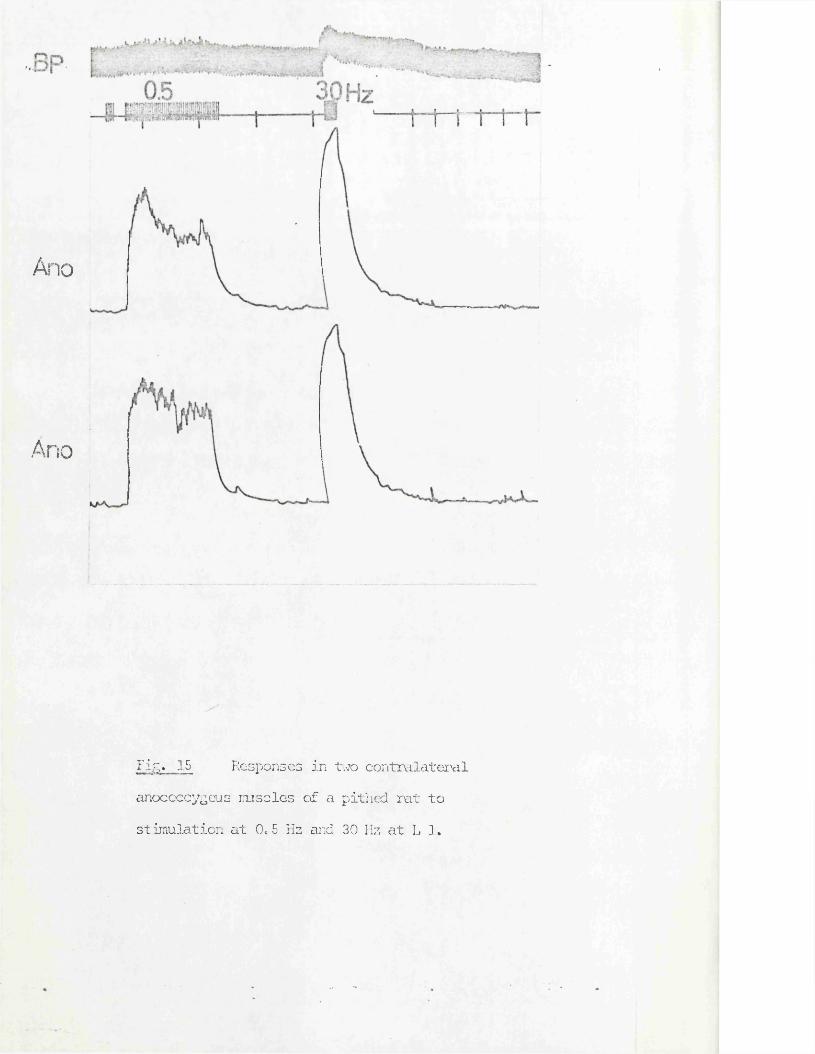
Ano
Ano
Fi^. 15 Responses in 17/70 contralateral anococcygeus mscles of a pithed rat to stimulation at 0*5 Hz and 30 Hz at L 1*
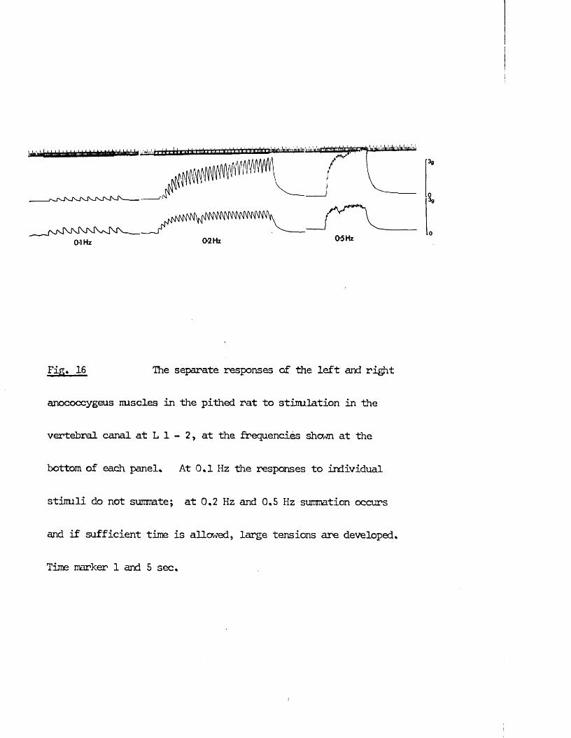
Fig, 16 The separate responses of the left and right
anococcygeus muscles in the pithed rat to stimulation in the
vertebral canal at L 1 - 2, at the frequencies shown at the
bottom of each panel* At 0.1 Hz the responses to individual
stimuli do not summte; at 0,2 Hz and 0,5 Hz summation occurs
and if sufficient time is allowed, large tensions are developed.
Time marker 1 and 5 sec.
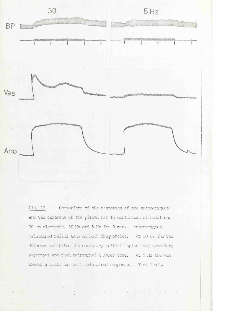
30
. Mam*
Fig* 17 Comparison of the responses of the anococcygeus
aid vas deferens of the pithed rat to continuous stimulation* 10 mm electrode, 30 Hz and 5 Hz for 3 min* Anococcygeus maintained raised tone at both frequencies* At 30 Hz the vas deferens exhibited the customary initial "spike" and secondary responses and then maintained a lower tone. At 5 Hz the vasshowed a small but well maintained response. Time 1 min.
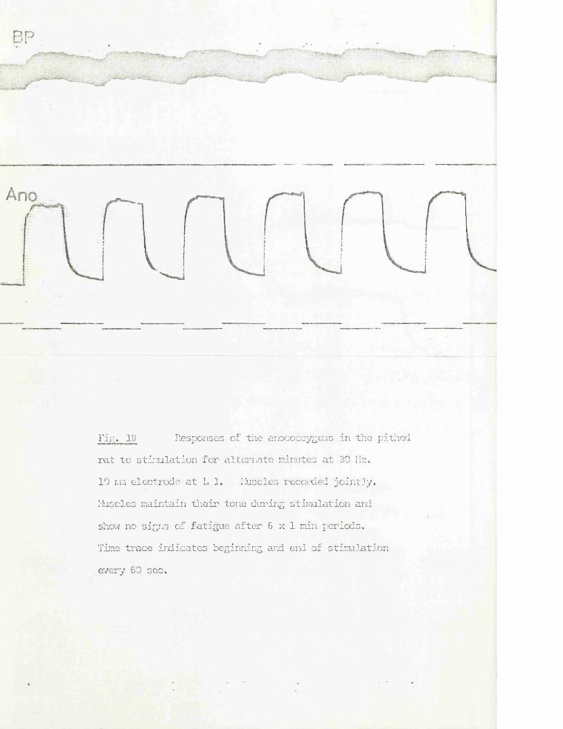
H D L,> i
rip;. 18 Responses of the anococcygeus in the pithedrat to stinulation for alternate minutes at 39 Hz.10 mm electrode at L 1. Muscles recorded jointly.
Muscles maintain their tone during stimulation ard show no signs of fatigue after 6 x 1 min periods.Time trace indicates beginning and end of stimulation every 60 sec.
43
At frequencies above 10 Hz, the responses from the upper and lower regions were equal and had reached their naximum amplitude within the 10 sec period of stinulation. At low frequencies, responses were often larger from stimulation in the upper than in the lower region and had not reached their maximum amplitude within the 10 sec stinulation period (Fig* 14)* In several other experiments, however, the frequency/response characteristics in the two motor regions were similar,
(2) Continuous stinulation until the maximum was reached showed that themost effective frequency renained at 30 Hz, as in (1), but frequencies as low as 0,5 Hz could eventually produce a plateau tension of some 70% of the naximum at 30 Hz (Fig, 15),
Single pulses produced responses of approximately 10% of the naximum tension and summation of tension was seen at frequencies at or above 0.1 Hz (Fig. 16). At higher frequencies, up to 50 Hz, repetitive stinulation produced a smooth contraction well maintained for a 3 min period (Fig. 17).
Responses were not only maintained within a single period of stimulation, but prolonged and repetitive stinulation at 30 Hz for alternateminutes did not affect the maintenance of responses (Fig. 18).
Single pulses and continuous stimulation produced similar responses from stimulation in either spinal region in most experiments. As with (1), in one experiment responses were smaller from the lower region. It is possible that simultaneous stimulation of the inhibitory nerves to the anococcygeus itself or mechanical interference from stimulation of the parasympathetic nerves to the colon could have affected the response to stimulation in this lower region in these experiments. Since in the majority of experiments responses were similar from stimulation in either region these possibilities were not further investigated.
(3) Stimulation with trains of 30 pulses demonstrated that frequencies as low as 1 Hz could produce as large a response as 30 Hz emphasising the importance of the number of pulses as opposed to the duration of the stimulation
period (Fig, 13), No difference was found in these experiments between responses from the two vertebral regions.
In the pithed-rat, therefore, stimulation of the nerves to the anococcygeus muscles produces mechanical responses in the muscles over a wide range of frequencies from single pulses to 100 Hz, The maximum tension which may be developed is produced by continuous stimulation at 30 Hz, but much lower frequencies are capable of producing large responses if stinulation is continued long enough for the individual pulses to summate to a plateau level.At all frequencies, the response is well maintained over a period of at least 3 min and there is no evidence of fatigue even after prolonged stimulation at high frequency.
Effects of blocking drugs on the motor response
The effects of anoc- blocking drug, phentolamine, of an adrenergic neurone blocking agent, guanethidine, of two ganglion blocking agents, hexamethonium and mecamylamine, and of a cholinergic blocking agent, atropine, were examined on the motor response. The intention was to analyse the nature of the motor pathway to see whether it was indeed adrenergic as the in vitro experiments suggested, whether it was interrupted by a ganglion and so anatomically belonged to the autonomic nervous system and finally whether there was any possibility of a cholinergic contribution since acetylcholine also causes contraction of the muscle in vitro. The effect of LSD - 25 was also examined since it inhibits motor nerve responses of the vas deferens in vitro (Ambache, Dunk, Vemey & Zar, 1973).
Phentolamine Responses from both motor regions were abolished by phentolamine (2 mg/kg) (Fig, 19). Complete blockade occurred within 2 min of administration and lasted for 15 - 20 min. Responses had recovered to 80% of their previous size within 30-45 min.
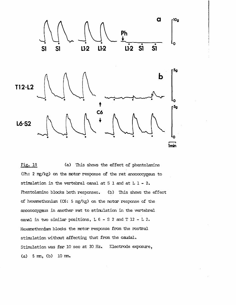
LI-2 LI-2
Ph*
LI-2 SI SI
T12-L2
C6
IL6-S2
lmin
Fig, 19 (a) This shews the effect of phentolamine(Fh: 2 mg/kg) on the motor response of the rat anococcygeus to stinulation in the vertebral canal at S 1 and at L 1 - 2. Phentolamine blocks both responses. (b) This shows the effect of hexamethonium (C6: 5 mg/kg) on the motor response of the anococcygeus in another rat to stinulation in the vertebral canal in two similar positions, L 6 - S 2 and T 12 - L 2. Hexamethonium blocks the motor response from the rostral stinulation without affecting that from the caudal.Stimulation was for 10 sec at 30 Hz, Electrode exposure,(a) 5 mm, (b) 10 mm.
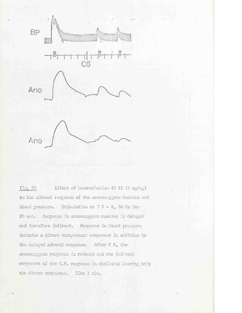
cs
Ano
Fig. 20 Effect of hexamethonium (C 6) (1 mg/kg)on the adrenal response of the anococcygeus msoles and blood pressure* Stinulation at T 6 - 8 > 30 IIz for 20 sec* Response in anococcygeus muscles is delayed and therefore indirect. Response in blood pressure includes a direct vasopressor component in addition to the delayed adrenal response. After C G, the anococcygeus response is reduced and the indirect component of the B,P, response is abolished leaving only the direct component. Tine 1 min.
i+5
Guanethidine (0.1 mg/kg) blocked responses from both regions within 10 min.This block could be temporarily reversed within the first 20 min by theintravenous injection of NA (500 ng).
Ganglion blockers A short lived inhibition of the motor response to stimulationfrom the upper (L 3 - T 11) region was produced by 1 mg/kg hexamethonium, andthe response was completely abolished by 5 mg/kg (Fig. 19). Recovery from even 5 mg/kg hexamethonium was rapid, the response being back to 30% of its previous size after 15 min. After responses had recovered from blockade by hexamethonium they could again be blocked by a similar dose (5 mg/kg).Another ganglion blocker, mecamylamine (5 mg/kg), also blocked responses from the upper region.The motor response from the lower (S 2 - L 6) region was unaffected by these doses of hexamethonium or mecamylamine (Fig. 19).
Atropine & LSD Atropine (1 mg/kg) and LSD (0.5 - 400 pg/kg) had no effect on the motor responses from either region.
The motor responses from stimulation higher in the spinal canal and due to liberation of catecholamines from the adrenals were abolished by phentolamine (1 mg/kg) and reduced by 70% by hexamethonium (5 mg/kg) (Fig. 20). Adrenal ,, responses were not blocked by guanethidine (5 mg/kg) (Fig. 20).
From the effects of these blocking drugs it appears that stimulation within the vertebral canal at the upper motor region activates preganglionic sympathetic nerves which can be blocked at the ganglion by hexamethonium or mecamylamine and at the neuro - muscular junction by guanethidine or phentolamine, while stimulation in the lower region activates postganglionic sympathetic nerves which can be blocked by guanethidine or phentolamine, but i/fwhich are unaffected by ganglion blockers. \
Stimulation in the upper region thus corresponds with the previous findings that the pithing rod electrode stimulates the preganglionic autonomic outflows
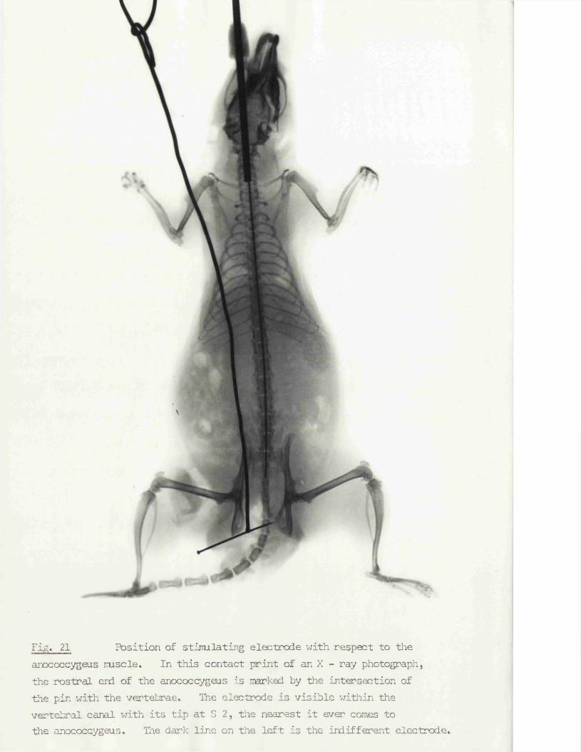
Fig. 21 Position of stimulating electrode with respect to theanococcygeus rnscle. In this contact print of an X - ray photograph, the rostral end of the anococcygeus is narked by the intersection of the pin with the vertebrae. The electrode is visible within the vertebral canal with its tip at S 2, the nearest it ever comes to the anococcygeus. The dark line on the left is the indifferent electrode.
at the point where they leave the vertebral canal (Gillespie & Muir, 1967; Gillespie, MacLaren & Pollock, 1970), but the ability to stimulate postganglionic fibres in the lower region is unique. One explanation for this could have been the position of the anococcygeus muscle directly in front of the vertebral column and having a tendinous origin from the upper coccygeal vertebrae. These conditions might have been particularly favourable for direct stimulation of intramural nerves in a muscle attached to bone overlying the stimulating electrode. This possibility was excluded by narking the position of the upper tendinous origin of the anococcygeus muscle with a metal marker and comparing by X-ray the relative positions of the marker and the spinal electrode (Fig, 21) • There was always a vertical displacement of at least 20 mm and up to 4-0 mm when the electrode was in the lower motor region, so it is unlikely that motor intramural nerves could be stimulated by an electrode so far away from the muscle. The present experiments do not pinpoint the feature responsible for the ability to stimulate postganglionic fibres to this muscle. One possibility, however, could be that if the ganglia on the motor pathway to the anococcygeus were para-vertebral rather than pre-vertebml, as in the case of several of the other systems studied, then this would lead to postganglionic fibres being nearer to the vertebral column and therefore to the stimulating electrode.
Another factor which might have interfered with the interpretation of the isometric tension responses in the in situ anococcygeus was movements of the descending colon. When the colon was visibly, spontaneously active, small disturbances on the anococcygeus baseline could be detected and it was feared that stimulation, especially in the sacral region, would involve the sacral parasympathetic outflow and so cause contraction of the longitudinal muscle of the colon, and this might in turn indirectly influence the tension record from the anococcygeus muscles. To test this, the tension in the longitudinal muscle of the colon was recorded iscmetrically by sewing a thread through the
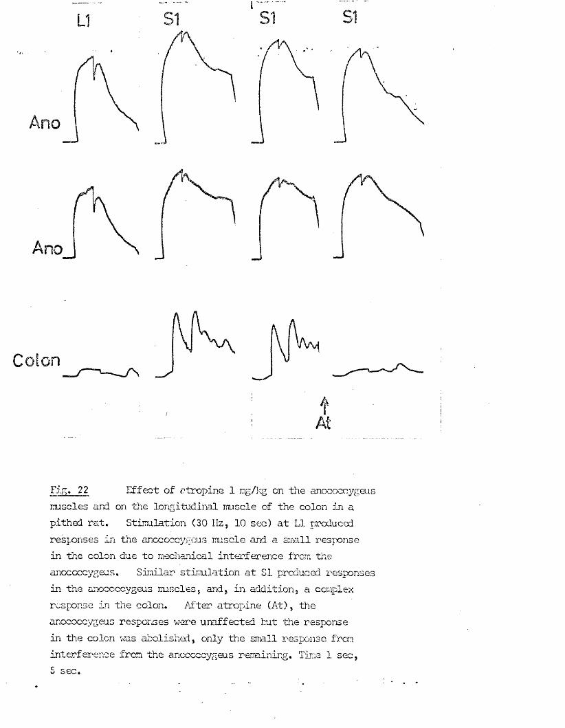
Clt
/y,
/ ‘- V
Fig. 22 Effect of atropine 1 mg/kg on the anococcygeusmscles and on the longitudinal muscle of the colon in a pithed rat. Stimulation (30 Hz, 10 sec) at LI produced responses in the anococcygeus muscle and a small response in the colon due to mechanical interference from the anoccccygeus. Similar stimulation at SI produced responses in the anococcygeus muscles. and, in addition, a complex response in the colon. After atropine (At), the anococcygeus responses were unaffected but the response in the colon was abolished, only the small response iron interference from the anococcygeus remining. Time 1 sec,5 sec.
W1
ventral surface of the colon midway between the two anococcygeus muscles and attaching it to an FT03 transducer so that it was parallel with the direction of recording of the anococcygeus muscle (i.e. approxinately 30° to the horizontal). Recording in this way, the spontaneous activity in the colon could be monitored and compared with the anococcygeus tension (Fig. 22). Spontaneous contractions of the colon resulted in small relaxations of the anococcygeus which did not interfere with the recording of responses to spinal stimulation. Stimulation at S 1 but not L I produced contractions of the colon at the same time as the anococcygeus response. Atropine (2 mg/kg) abolished this colon response without affecting the anococcygeus response.It was thus concluded that stimulation at S 1 stimulated the sacral parasympathetic nerves which in turn stimulated the longitudinal iruscle of the colon, but this did not interfere with the response in the anococcygeus nuscles.
Drugs producing motor responses
In vitro the inhibitory response in the anococcygeus can only be seen after raising the tone in the muscle. In order to establish a similar situation in vivo far examination of possible inhibitory responses, the action of several drugs capable of raising the tone in vitro were examined on the pithed rat.
Noradrenaline Single injections of NA produced dose - related motor responses (Fig. 23) which reached a peak within 2 min and had returned to baseline within 5-10 min. Threshold for the response was 0.2 ug/kg but maximal responses could not be determined since the dose necessary would have killed the rat due to its effects on the blood pressure and heart.Comparison with responses obtained in vitro (Fig. 23) shows that the maxinum response was not produced until the concentration of NA in the organ bath
— Llreached 10 M. The largest dose which was examined in the pithed rat was 800 pg/kg. Expressed as a molar concentration in the whole rat, this is equivalent to H x 10" M or, assuming that the NA is restricted to the blood
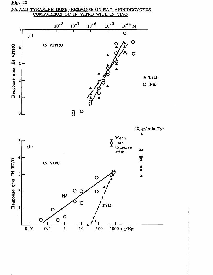
Response gms
IN VIVO
Response gms
IN VI
TRO
Fig. 23N A A N D TYRAMINE DOSE /RESPONSE O N R A T A N O C O C C Y G E U S
COMPARISON O F IN VITRO WITH IN VIVO
IN VITRO
a TYR O NA
OL
40/ng/min TyrA
Mean max to nerve stim.
5
AA
4IN VIVO
3
2NA
T Y R1
0.01 0.1 100 lOOOptg/Kg101
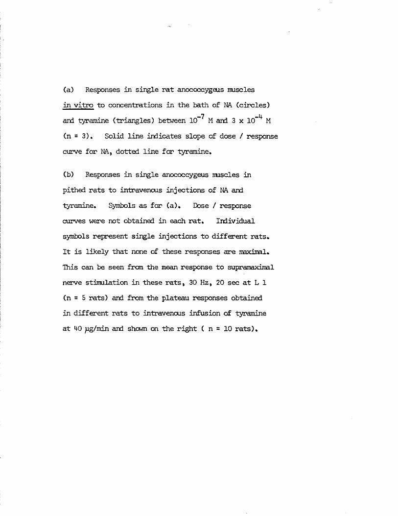
(a) Responses in single rat anococcygeus musclesin vitro to concentrations in the bath of NA (circles)
-7 -4and tyramine (triangles) between 10 M and 3 x 10 M(n = 3). Solid line indicates slope of dose / response curve for NA, dotted line for tyramine.
(b) Responses in single anococcygeus muscles in pithed rats to intravenous injections of NA and tyramine. Symbols as for (a). Dose / response curves were not obtained in each rat. Individual symbols represent single injections to different rats.It is likely that none of these responses are naxiral. This can be seen from the mean response to supramaximal nerve stimulation in these rats, 30 Hz, 20 sec at L 1 (n = 5 rats) and from the plateau responses obtainedin different rats to intravenous infusion of tyramine at 40 pg/min and shewn on the right ( n = 10 rats).
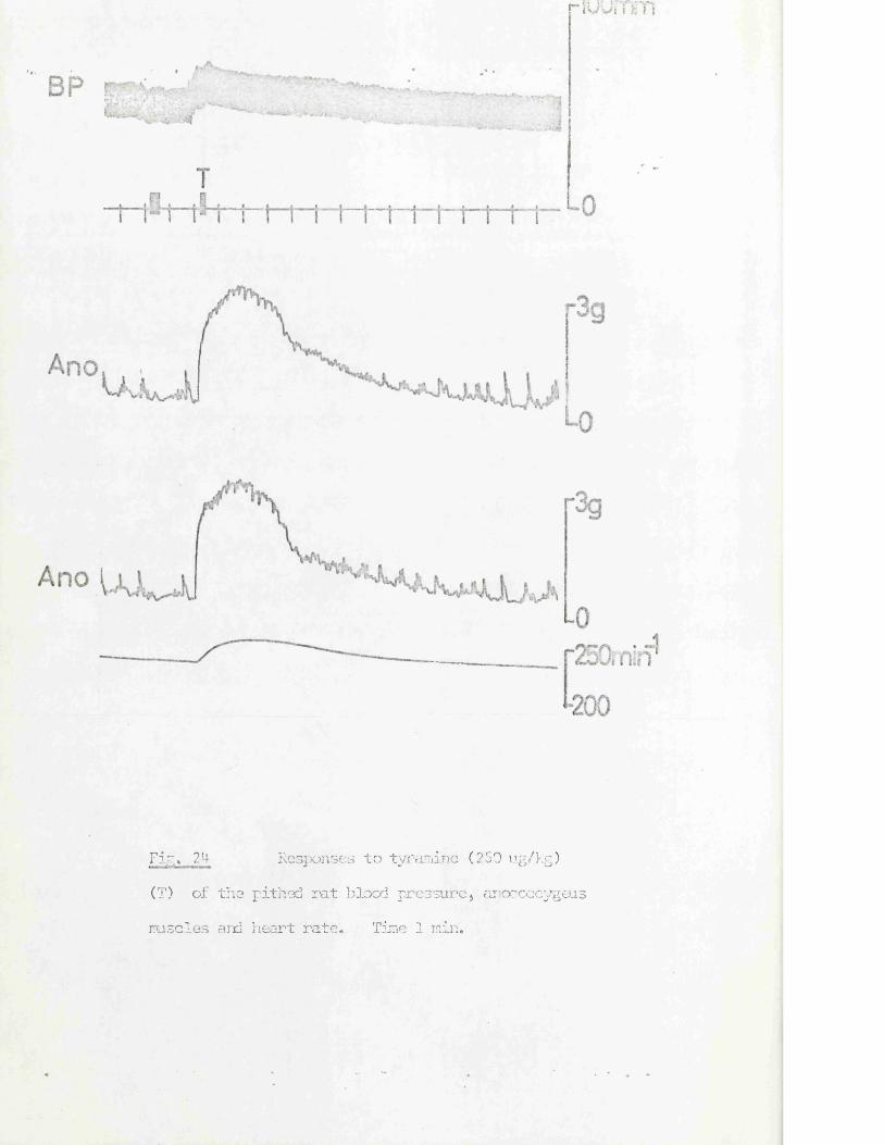
riuunrn
Fig. 2M- Responses to tyramine (250 ug/kg)
(T) of the pithed rat blood pressure, anococcygeus
iruscles and heart rate. Time 1 min.
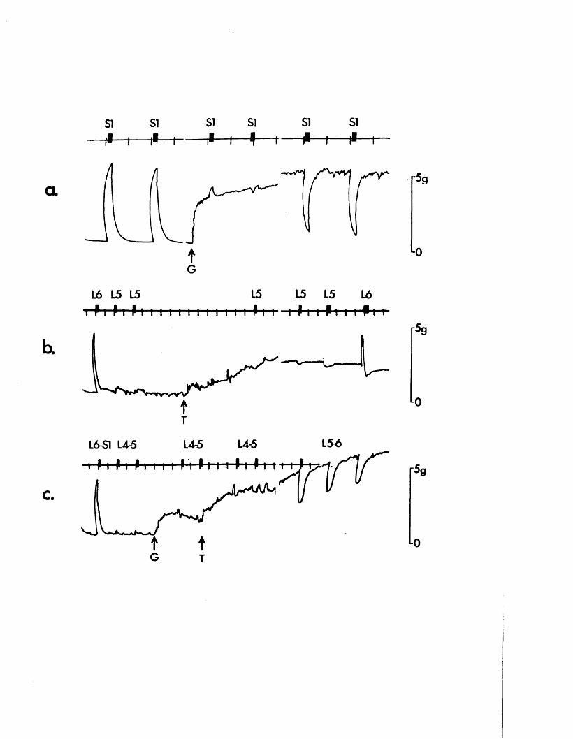
si s i s i SI SI SI1 * — i — t * — i - — t1 — i — ! — t — — i — — r
a r5g
tG
L6 L5 15 L5 L5 L5 L6i f I f I 11 i i I i i I i I i i I I m i Pm rf-t r
L0
r5g
tT
L6-S1 L4-5 L4-5
■t~ P i > i >i i i i h 4L4-5 L54
t- P-l > 1 1 I l I iC.
'59
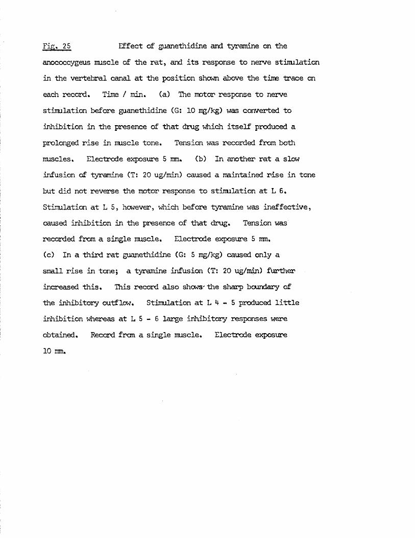
Fig, 25 Effect of guanethidine and tyramine on theanococcygeus muscle of the rat, and its response to nerve stimulation in the vertebral canal at the position shown above the time trace on each record. Time / min, (a) The motor response to nerve stimulation before guanethidine (G: 10 mg/kg) was converted to inhibition in the presence of that drug which itself produced a prolonged rise in muscle tone. Tension was recorded from both muscles. Electrode exposure 5 mm, (b) In another rat a slow infusion of tyramine (T: 20 ug/min) caused a maintained rise in tone but did not reverse the motor response to stimulation at L 6, Stimulation at L 5, however, which before tyramine was ineffective, caused inhibition in the presence of that drug. Tension was recorded from a single muscle. Electrode exposure 5 mm.(c) In a third rat guanethidine (G: 5 mg/kg) caused only a small rise in tone; a tyramine infusion (T: 20 ug/min) further increased this. This record also shows'the sharp boundary of the inhibitory outflow. Stimulation at L 4 - 5 produced little inhibition whereas at L 5 - 6 large inhibitory responses were obtained. Record frcm a single muscle. Electrode exposure 10 mm.
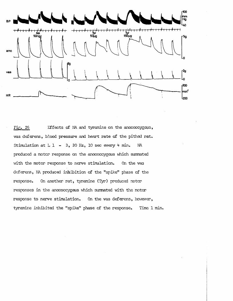
BP
HR / K _
NA50ngi 50pg
ano
vas
-100
-40
rsg
L0
-2g
-0•300
miri200
Fi * 26 Effects cf NA and tyramine on the anococcygeus,vas deferens, blood pressure and heart rate of the pithed rat* Stimulation at L 1 - 3, 30 Hz, 10 sec every 4 min. NAproduced a motor response on the anococcygeus which summated with the motor response to nerve stimulation. On the vas deferens, NA produced inhibition of the "spike" phase of the response. On another rat, tyramine (Tyr) produced motor responses in the anococcygeus which summated with the motor response to nerve stimulation. On the vas deferens, however, tyramine inhibited the "spike" phase of the response. Time 1 min.
4-8
volume alone, i.e. approximately 6% of body weight, is equivalent to6 x 10“5 M. Since the actual concentration reaching the anococcygeus islikely to be between these two figures, comparison with the responses in vitro
—G —5to NA 4 x 10“ M - 6 x 10“ M shows that the responses in the two situationsare of the same order of imgnitude. On the other hand, raking similarassumptions, the threshold dose is mch lower in the pithed rat than in vitro.
-9 -8Doses of 1 ug/kg (5 x 10 M - 8 x 10 M) are consistently supra-threshold—8in the pithed rat, but in vitro the threshold lies between 3 x 10” M and
-73 x 10 M. This high sensitivity to NA in the pithed rat anococcygeus is similar to and nay be produced by the same factors which lead to the sensitivity of the pithed rat blood pressure to NA. Since a dose of 50 ngNA can easily be detected in a 250 g rat, the anococcygeus preparation might |
/
indeed be developed as a sensitive assay for NA. sTyramine Single injections of tyramine produced dose - related motor responses similar to those for NA, reaching a peak within 2 min and declining to baseline within 5-10 min (Figs. 23 and 24). The threshold far tyramine was , however, higher at approximately 50 pg/kg. The highest single dose given at 600 pg/kg did not produce a maximal response, but if the tyramine was given by slow infusion into the femoral vein at 40 pg/min the response plateaued at a level equivalent to both the maximum responses in vitro and the maximum response to motor nerve stimulation in vivo (Fig. 23). Slower rates of infusion of tyramine (20 pg/min and 10 pg/min) produced sub-maximal motor responses which were also well maintained (Fig. 25).Both NA and tyramine responses summated with the motor response to nerve stimulation from within the vertebral canal. Neither drug had a significant effect on motor nerve responses at doses which were sub-threshold for contraction, but in doses which did produce motor responses (Fig. 26) the drug + nerve response was larger than the pre-drug nerve response (provided that the nerve response was sub-maximal; maximal responses could not be increased in this way). The increase in tension above that produced by the
agonist drug, produced by nerve stimulation was, however, smaller than in the absence of the agonist, illustrating the difficulty of interpreting the effects of a drug on nerve responses when that drug itself alters the baseline from which contractions are measured. It was clear, hcwever, that neither drug reduced the ability of the motor nerves to produce responses when given by single injection.After prolonged, i.e. greater than 1 hr, infusion of tyramine, the motor nerve responses were reduced in size.
Guanethidine in doses at or above 0.1 mg/kg blocked the motor responses to nerve stimulation within the vertebral canal. At doses at and above 0.5 mg/kg, in addition to blocking nerve responses, guanethidine also raised the tone of the nuscle. At 0.5 mg/kg these responses were small and short lived, but became increased in size and more prolonged with increasing dose until at 10 mg/kg a maximal motor response was produced (Fig. 25) which was well maintained for 25-40 min, after which time tension gradually declined and spontaneous activity started. Further increases in dose beyond 10 mg/kg produced responses which were less well maintained.Once spontaneous activity and tension decline had started, the response could not be restabilised by an additional dose of guanethidine. 30 min after spontaneous activity had ceased, hcwever, a further dose of guanethidine (10 mg/kg) could again produce a stable response. The ability to produce motor responses by nerve stimulation returned approximately 30 min after the end of spontaneous activity, coinciding with the return of the ability of guanethidine to itself produce a motor response. This may reflect the mechanism of development of tachyphylaxis to guanethidine whereby the capacity of guanethidine to release NA is blocked by the same mechansim which blocks the release of NA by nerve impulses.Fhentolamine (2 mg/kg) abolished motor responses to NA, tyramine and guanethidine. Together with the evidence from in vitro experiments it is reasonable to assume,
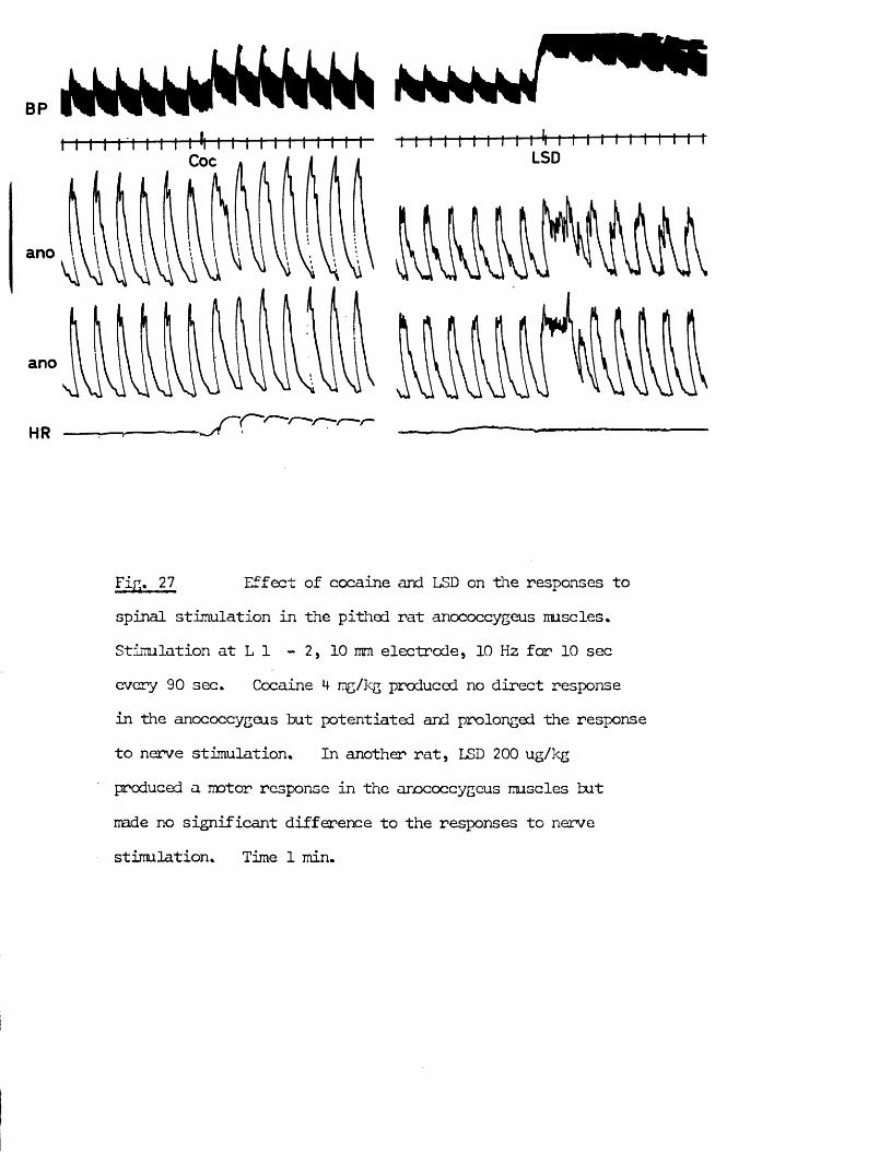
BP
LSDCoc
ano
ano
•r~~HR
Fin* 27 Effect of cocaine and LSD on the responses tospinal stimulation in the pithed rat anococcygeus muscles. Stimulation at L 1 - 2, 10 mm electrode, 10 Hz for 10 sec every 90 sec* Cocaine *+ mg/kg produced no direct response in the anococcygeus but potentiated and prolonged the response to nerve stimulation* In another rat, LSD 200 ug/kg produced a motor response in the anococcygeus muscles but made no significant difference to the responses to nerve stimulation. Time 1 min.
50
therefore, that tyramine and guanethidine are acting as indirect sympathomimetics •
Other indirect sympathomimetics Cocaine (4- mg/kg), which produces motor responses in vitro, produced a snail motor response in vivo* At this dose, it also potentiated the height of sub-maximal motor nerve responses (10 Hz,5 sec at L 1) by 5% and prolonged their duration (Fig. 27).Large doses of LSD (100 - 800 ug/kg) produced motor responses which lasted 2 - 5 min but did not affect the responses to motor nerve stimulation (Fig. 27). Both LSD and cocaine are believed to produce these contractions by an indirect sympathomimetic action as will be shown later in vitro.
Other agonists Other drugs which raise the tone of the muscles in vitro include acetylcholine and other muscarinic agonists ard 5-hydroxytryptamine (5HT). Neither of these proved to be a useful means of raising the tone in vivo due to their other widespread systemic effects.Acetylcholine and carbachol did not consistently produce any response in doses (1-10 mg/kg) which stopped the heart and therefore killed the rat.Furmethide produced irregular responses in doses of 2 - 10 mg/kg which eventually killed the rat. These responses were prevented by atropine (2 mg/kg).
5HT (0.2 - 2 mg/kg) produced small responses but also produced dramatic effects on the blood pressure and heart. High blood pressure and arrythmia quickly terminated these experiments.
It was thus apparent that the most suitable drugs for producing maintained motor responses in the in vivo anococcygeus were indirect sympathomimetics which produced a stable and viable preparation.
The Inhibitory Response
Spinal origin of the inhibitory nerves
When the tone of the muscles was raised by a single injection of guanethidine (10 mg/kg), 10 sec periods of stimulation at 30 Hz in the region L 5 - S 2 produced inhibitory responses. In individual experiments this inhibition could amount to 90% of the induced tone (Fig. 25a). The average maximal inhibition in all experiments was 62 ± 5% of induced tone (n = 12).
When the tone was raised with tyramine (20 jug/min), which did not abolish motor responses, stimulation in the upper end of this region (L 5) produced small inhibitory responses, whereas stimulation in the lower part of the same region (L 6 - S 2) produced a biphasic response; contraction followed by relaxation (Fig. 25b).
These experiments showed that inhibitory nerves did arise in the spinal card and could be stimulated in one region only, in the lower lumbar and upper sacral region, overlapping with, but extending one segment further rostrally than, the lower motor region.
To examine the origin and properties of the inhibitory nerves systematically, however, required that the tone be raised to a steady plateau and that the action of the motor nerves be blocked. Guanethidine was clearly the better drug for this purpose, because bf its neuronal blocking action on the adrenergic motor nerves in addition to its indirect sympathomimetic action in raising tpne. Guanethidine had, however, the disadvantage that after 25-40 min the tone began to fall and rythmic activity appeared. In these circumstances it was difficult to demonstrate inhibition. The possibility was, therefore, investigated of reinforcing the motor action of guanethidine with a slow infusion of tyramine, thus combining the neuronal blocking action of the former with the steady induced tone of the latter.
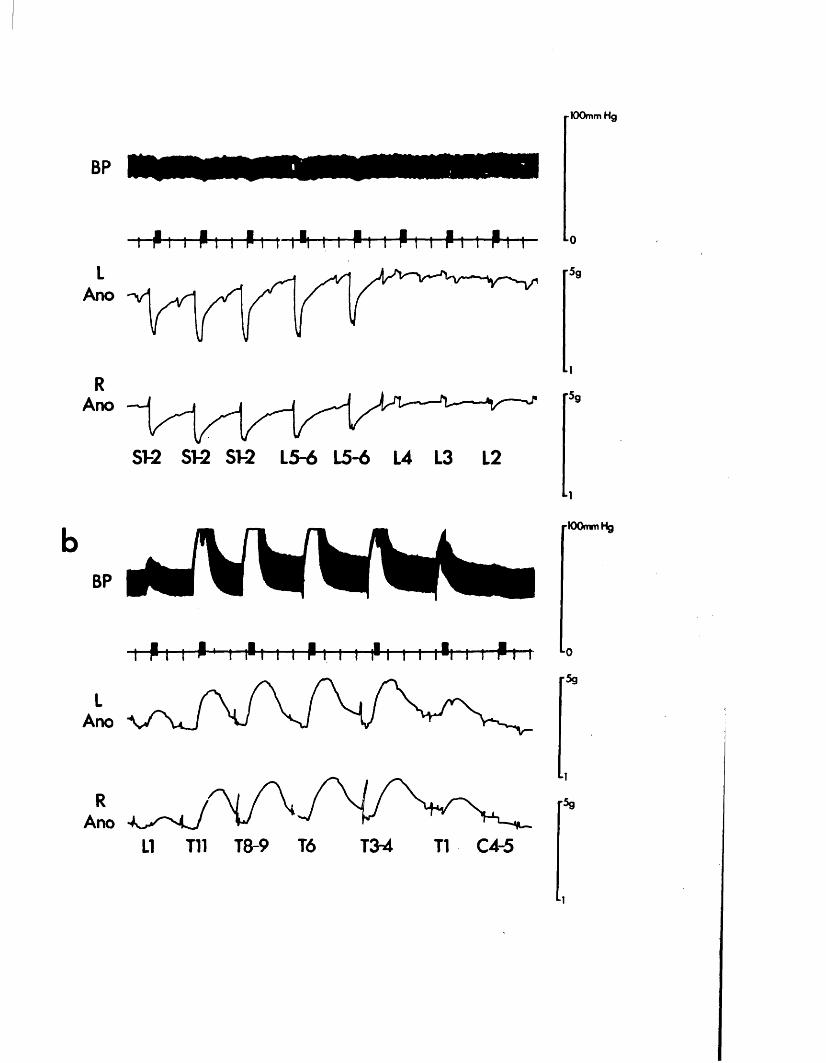
■ 100mm Hg
BP
P i i Pi i Pi m ^ m Pm Pm Pm P *-0
r59
■ir59
BP
ST-2 ST-2 ST-2 L5-6 L5-6 L4 L3 L2
P i i P 1 I 1*1 I i P I I i PI I I I1! I i~Pt
M-100mm Hg
LAno
Lor5g
RAno
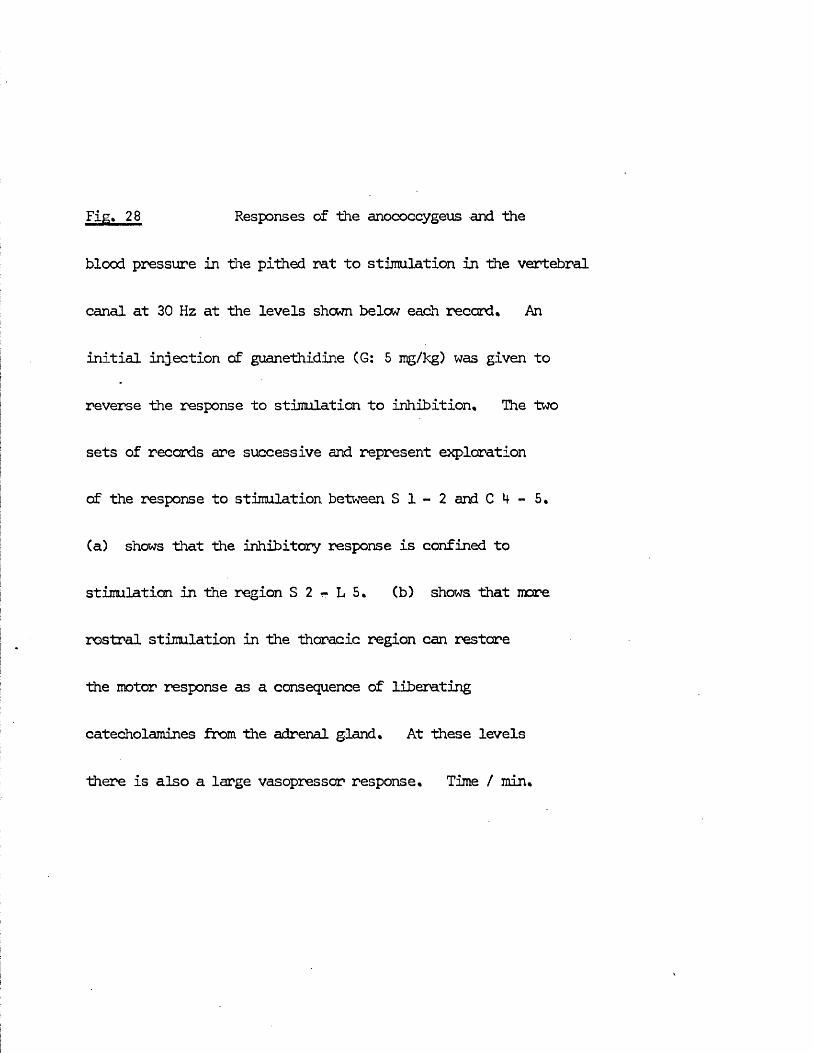
Fig. 28 Responses of the anococcygeus and the
blood pressure in the pithed rat to stimulation in the vertebral
canal at 30 Hz at the levels shewn below each record. An
initial injection of guanethidine CG: 5 mg/kg) was given to
reverse the response to stimulation to inhibition. The two
sets of records are successive and represent exploration
of the response to stimulation between S 1 - 2 and C 4 - 5.
(a) shows that the inhibitory response is confined to
stimulation in the region S 2 - L 5. (b) shows, that more
rostral stimulation in the thoracic region can restore
the motor response as a consequence of liberating
catecholamines from the adrenal gland. At these levels
there is also a large vasopressor response. Time / min.
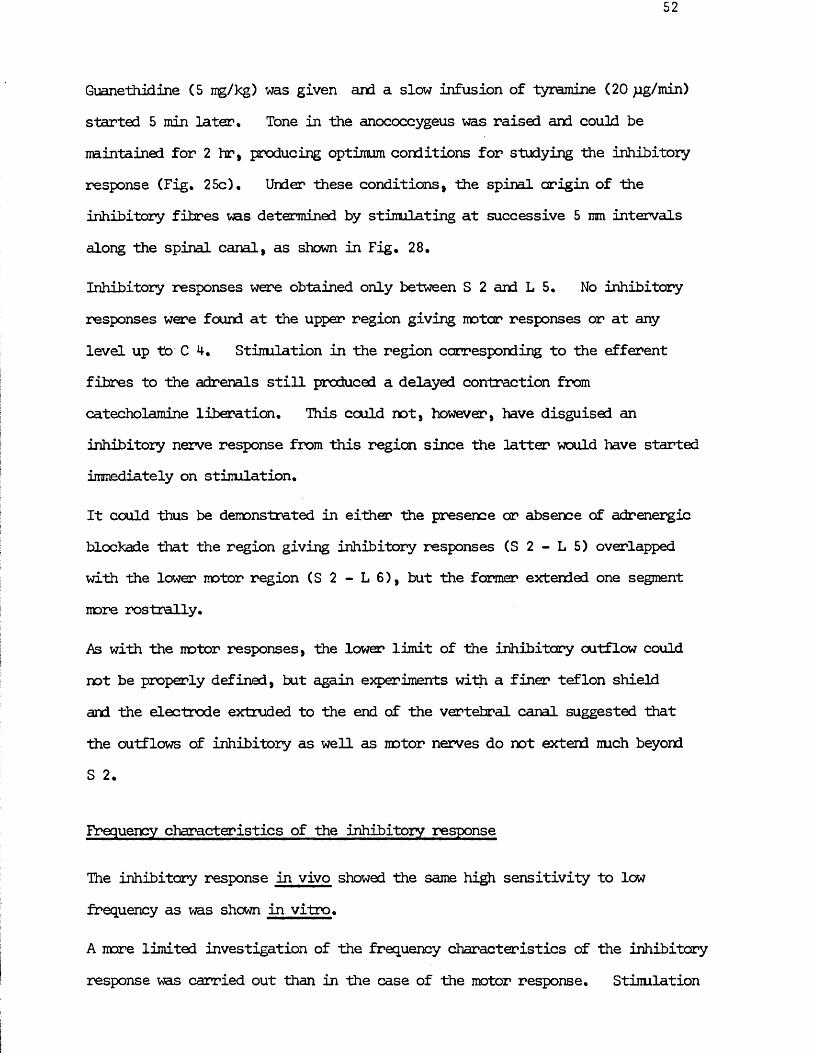
Guanethidine (5 mg/kg) was given and a slow infusion of tyramine (20 jig/min) started 5 min later. Tone in the anococcygeus was raised and could be mintained for 2 hr, producing optimum conditions for studying the inhibitory response (Fig. 25c). Under these conditions, the spinal origin of the inhibitory fibres was determined by stimulating at successive 5 mm intervals along the spinal canal, as shown in Fig. 28.
Inhibitory responses were obtained only between S 2 and L 5. No inhibitory responses were found at the upper region giving motor responses or at any level up to C 4, Stimulation in the region corresponding to the efferent fibres to the adrenals still produced a delayed contraction from catecholamine liberation. This could not, however, have disguised an inhibitory nerve response from this region since the latter would have started immediately on stimulation.
It could thus be demonstrated in either the presence or absence of adrenergic blockade that the region giving inhibitory responses (S 2 - L 5) overlapped with the lower motor region (S 2 - L 6), but the farmer extended one segment more rostrally.
As with the motor responses, the lower limit of the inhibitory outflow could not be properly defined, but again experiments with a finer teflon shield and the electrode extruded to the end of the vertebral canal suggested that the outflows of inhibitory as well as motor nerves do not extend nuch beyond S 2.
Frequency characteristics of the inhibitory response
The inhibitory response in vivo showed the same high sensitivity to low frequency as was shown in vitro.
A more limited investigation of the frequency characteristics of the inhibitory response was carried out than in the case of the motor response. Stimulation
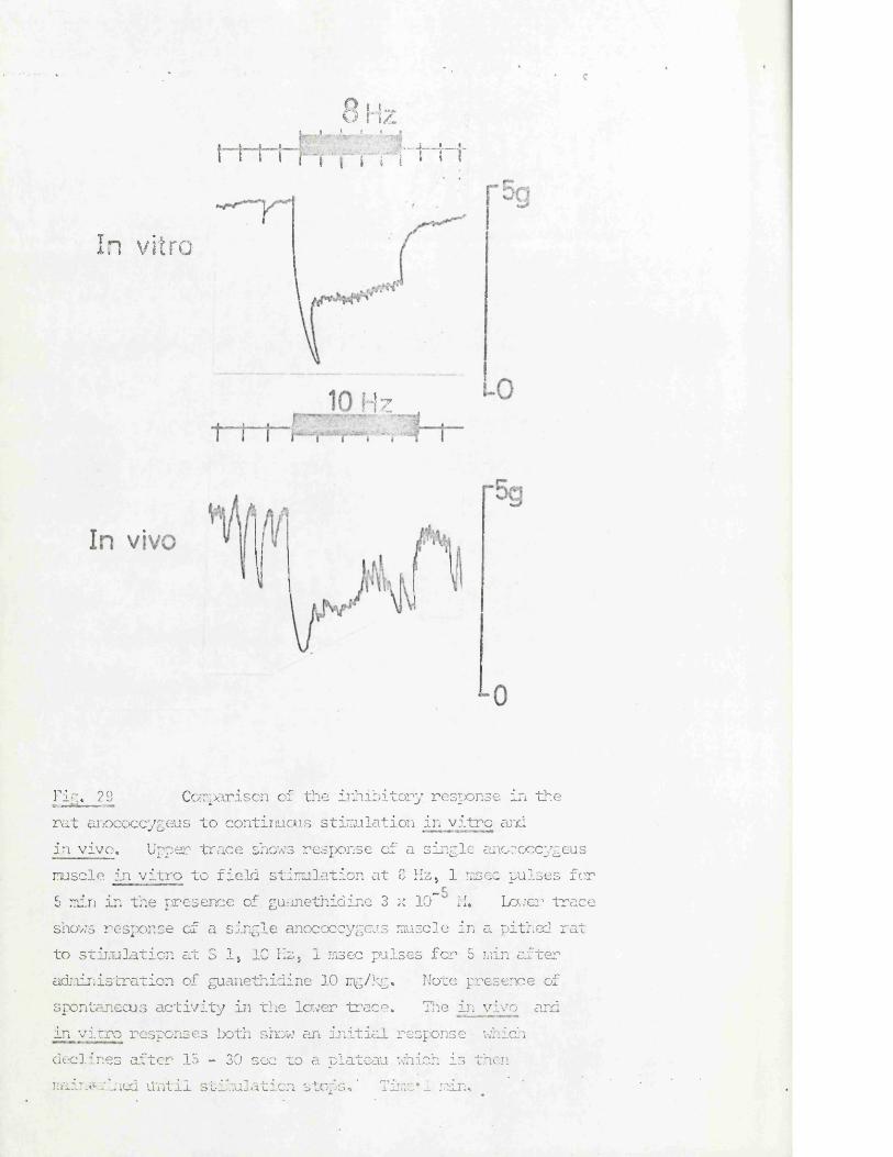
e
8 H-i/L, i f « I
I i i i p T' - f r 1"'ti~t
In vitro
H zit- VWnV >-.j AS
rivi* J29 Comparison of the inhibitory response in therat anococcygeus to continuous stimulation in vitro arid in vivo. Upper trace shows response of a single anococcygeus mscle in vitro to field stimulation at 0 Hz, 1 msec pulses for 5 min in the presence of guanethidine 3 x 10” 11. Lower traceshows response of a single anococcygeus muscle in a pithed rat to stimulation at S 1, 10 Hz, 1 msec pulses for 5 min after administration of guanethidine 3-0 mg/kg. Note presence of spontaneous activity in the lower trace. line in vivo and in vitro responses both show an initial response which declines after 15 - 30 sec to a plateau which is then mini?rined until stimulation stops*’ Time*! man*
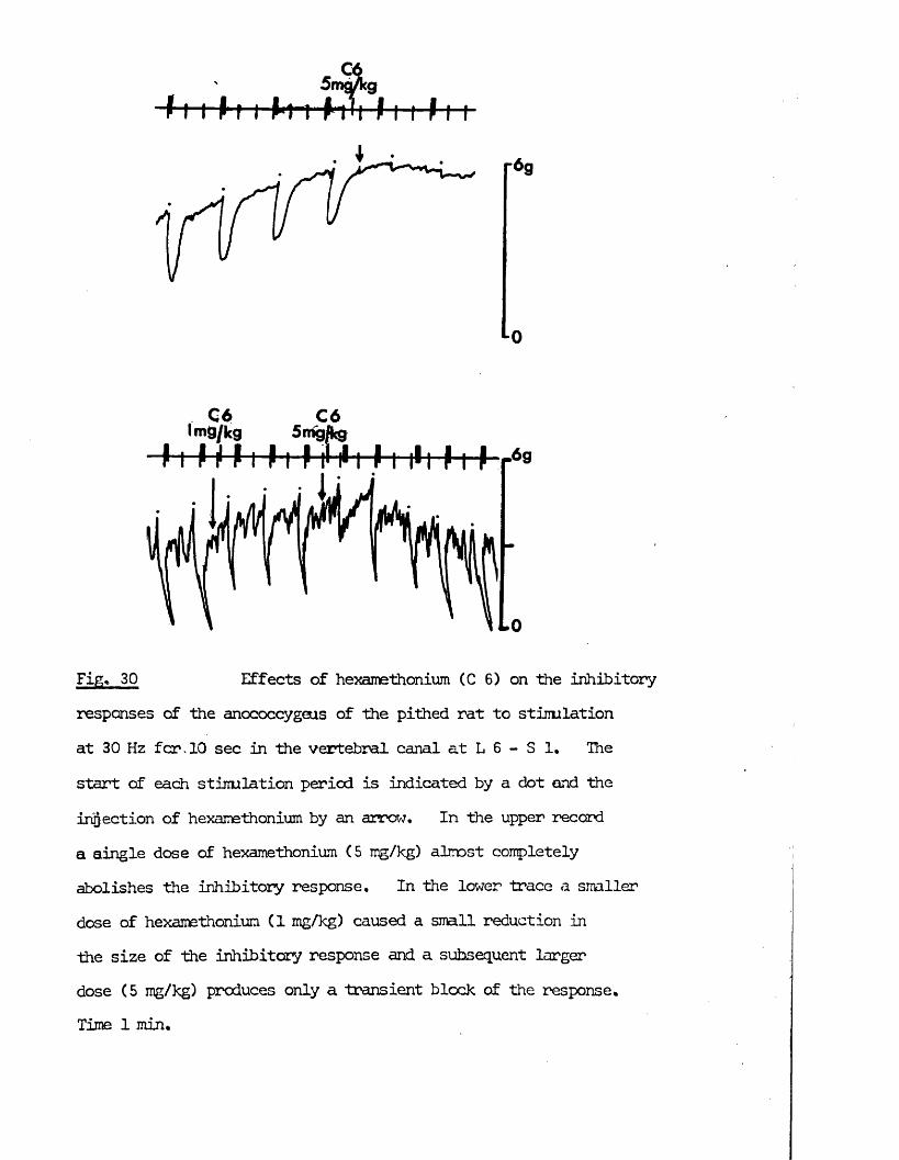
r69
Lo
C6 C6mg/kg Sirigjkg
Fig. 30 Effects of hexamethonium (C 6) on the inhibitoryresponses of the anococcygeus of the pithed rat to stimulation at 30 Hz fcr. 10 sec in the vertebral canal at L 6 - S 1. The start of each stimulation period is indicated by a dot and the infection of hexamethonium by an arrow. In the upper record a aingle dose of hexamethonium (5 mg/kg) almost completely abolishes the inhibitory response. In the lower trace a smaller dose of hexamethonium (1 mg/kg) caused a small reduction in the size of the inhibitory response and a subsequent larger dose (5 mg/kg) produces only a transient block of the response.Time 1 min.
53
far a fixed time with varying frequency showed that maximal responses could be obtained with 10 sec periods of stinulation at 2 - 5 Hz (Fig. 1M-).
Responses could be produced to single pulses, but these were very small and transient and summation did not occur till 0.5 Hz. With continuous stimulation, in the region 1-10 Hz, large responses were obtained which were rapid in onset and well maintained over a 20 sec period. If stimulation was continued for several minutes, the response declined slightly after 20 - 30 sec to a plateau which could then be maintained for up to 8 min.This was similar to the shape of the response in vitro (Fig. 29).
The frequency characteristics of the inhibitory innervation thus show a slightly different pattern from that of the motor nerves. The motor nerves can produce significant responses to single pulses, can maintain responses with low frequencies and the maximum response which can be produced by continuous stimulation increases with increasing frequency up to 30 Hz. The inhibitory nerves on the other hand cannot produce readily detectable responses to single pulses or very ’ low frequencies but are highly effective in the region 2 - 10 Hz in which maximal responses can be produced.
Effects of blocking drugs on the inhibitory response
Clearly nerve fibres mediating inhibition in the anococcygeus were located in the spinal cord. The question of whether this pathway to the muscle was interrupted by a ganglion relay was investigated by examining the effects of hexamethonium and mecamylamine.
Hexamethonium or mecamylamine (5 mg/kg) completely abolished the inhibitory j response. This is shown for hexamethonium in Fig. 30. When recovery from this inhibition occurred, after 30 - 60 min, a second similar dose had little effect in renewing the block. If initially, a small (1 mg/kg) dose of hexamethonium was given, a transient reduction in the response was observed.On recovery from this, the tissue was insensitive to subsequent large (5 mg/kg +)
doses of the drug. Similar desensitisation was found with mecamylamine.
Ganglion blockers did not, therefore, present a clear cut effect on the inhibitory pathway. Rapid desensitisation occurred which is in contrast to their effects on the motor pathway. A similar desensitisation was, however, found in the motor pathway to the vas deferens where a ganglion synapse has been convincingly demonstrated (for review see Sjostrand, 1965) and in the pathway producing depressor responses on blood pressure (see following two sections). Taken together with this evidence, the present results are consistent with the presence of a ganglion synapse in the inhibitory outflow to the anococcygeus muscles. Since ganglion blockers have no effect on inhibitory responses in vitro, the location of this ganglion remains undefined.
Atropine (2 ng/kg) did not block inhibitory responses, but caused a transient and unexplained reduction in guanethidine tone. This argues against a role for muscarinic receptors at either the ganglion or at the nerve - muscle junction.
Cardiovascular Responses
During these experiments on the pithed rat, aortic blood pressure was recorded at all times and heart rate was usually monitored.
Responses on blood pressure and heart rate from stimulation within the vertebral canal and to administration of most of the drugs used have been previously studied (Gillespie & Muir, 1967 a, b, c; Gillespie, MacLaren & Pollock, 1970). It was thus possible to correlate cardiovascular and anococcygeus responses without systematically studying the cardiovascular aspects of the preparation.
Drugs producing pressor responses
Noradrenaline, tyramine and guanethidine in doses which produced motor responses in the anococcygeus also produced dose - related vasopressor responses
55
and increases in heart rate, A single dose of guanethidine (10 mg/kg) or a slow infusion of tyramine (10 - 40 ug/min) which maintained a steady tone in the anococcygeus also maintained an elevated blood pressure and heart rate,
5 - hydroxytryptamine (0,2 mg/kg) produced a large pressor response andincreased heart rate. Higher doses led to arrythmia,
LSD (0,4 - 100 ug/kg) produced dose - related vasopressor responses without affecting heart rate.
Cocaine (4 mg/kg) increased blood pressure and heart rate.
Spinal origin of cardiovascular responses
The blood pressure responses obtained to stimulation within the vertebral canal in the absence of pressor drugs are shown in Fig. 10. Small pressor responses were obtained from S 1 to L 3 and larger pressor responses from L 2 to T 12. These were direct, adrenergic nerve responses which started immediately on stimulation and were not accompanied by any increase in heart rate. From T 11 to T 2 a direct vasopressor response was followed by a delayed pressor response which was accompanied by an increase in heart rate
\ and was due to release of catecholamines from the adrenals -(see also Tig. 22>. /
When the blood pressure was elevated by tyramine, guanethidine or a combination of both, depressor responses were found from stimulation in the region S 2 - L 5 (Fig. 28). No changes in heart rates accompanied these responses. It was not necessary to block the adrenergic pressor nerves to obtain these depressor responses since the vasopressor response at S 2 - L 5 was small.When the adrenergic nerves were blocked by guanethidine, no depressor responses were found outside of the S 2 - L 5 region. There thus appears to be a depressor outflow originating from S 2 - L 5 which might be considered analogous to the inhibitory pathway to the anococcygeus. The depressor responses were small when viewed against the total systemic blood pressure, but became quite distinct and reproducible if the polygraph sensitivity was increased and
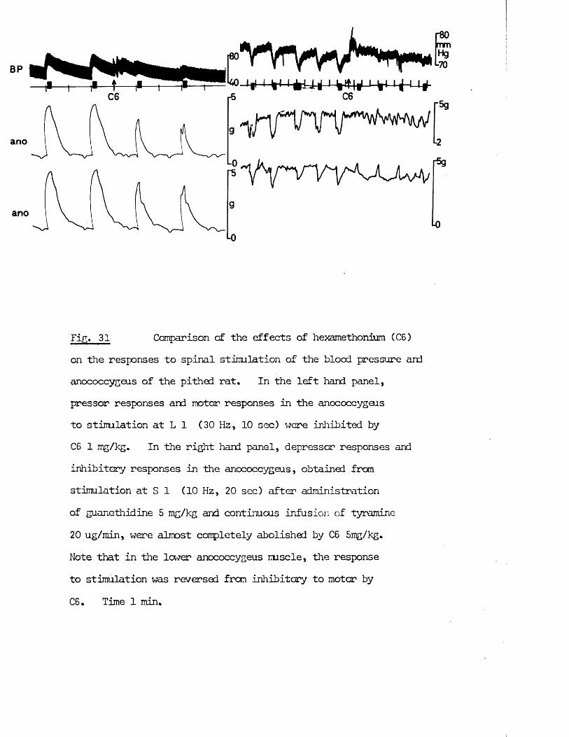
C6
ano0
ano
■0
vyrvvv^n^-i|M n-ifcU J 4Ht|rf
C6
Y Y ^ t m o a H /
"5g
Fig. 31 Comparison of the effects of hexamethonium (C6)on the responses to spinal stimulation of the blood pressure andanococcygeus of the pithed rat* In the left hand panel,pressor responses and motor responses in the anococcygeus to stimulation at L 1 (30 Hz, 10 sec) were inhibited byC6 1 mg/kg. In the right hand panel, depressor responses andinhibitory responses in the anococcygeus, obtained from stimulation at S 1 (10 Hz, 20 sec) after administrationof guanethidine 5 mg/kg and continuous infusion of tyramine 20 ug/min, were almost completely abolished by C6 5mg/kg.Note that in the lower anococcygeus mscle, the response to stimulation was reversed from inhibitory to motor by C6* Time 1 min*
56
damped to give a mean blood pressure reading (Fig. 31).
Effects of drugs on nerve or drug mediated cardiovascular responses
(1) The pressor responseFhentolamine (2 mg/kg) which blocked the effects of NA and sympathetic nerves on the anococcygeus, also blocked the effects of NA and pressor nerves on the blood pressure as well as the adrenal response on blood pressure. Guanethidine (0.1 - 10 mg/kg) which blocked the effects of adrenergic nerves but not NA on the anococcygeus, also blocked the effects of the pressor nerves without preventing the action of NA on blood pressure or blocking the adrenal response.Hexamethonium and mecamylamine in doses which blocked the responses in the anococcygeus to preganglionic stimulation blocked both the pressor nerves and adrenal responses (Figs. 31 and 20).Noradrenaline, tyramine and cocaine in doses which potentiated the anococcygeus sympathetic nerve response also potentiated the pressor nerve responses (Figs. 26 and 27).
(2) The depressor response
The depressor nerve response from S 2 - L, 5 was unaffected by guanethidine (10 mg/kg), propranolol (1 mg/kg) or atropine (4 mg/kg). Its sensitivity to ganglion blockers was, however, similar to that of the anococcygeus inhibitory nerves.Hexamethonium and mecamylamine (5 mg/kg) which blocked the anococcygeus inhibitory nerves also blocked this depressor response (Fig. 31) . After recovery, a second dose did not block, and in the experiments where a small dose was given first, parallel desensitisation occurred in the blood pressure and anococcygeus responses.
It therefore appears that the preganglionic fibres in a pathway capable of producing depressor responses originate from the same region within the
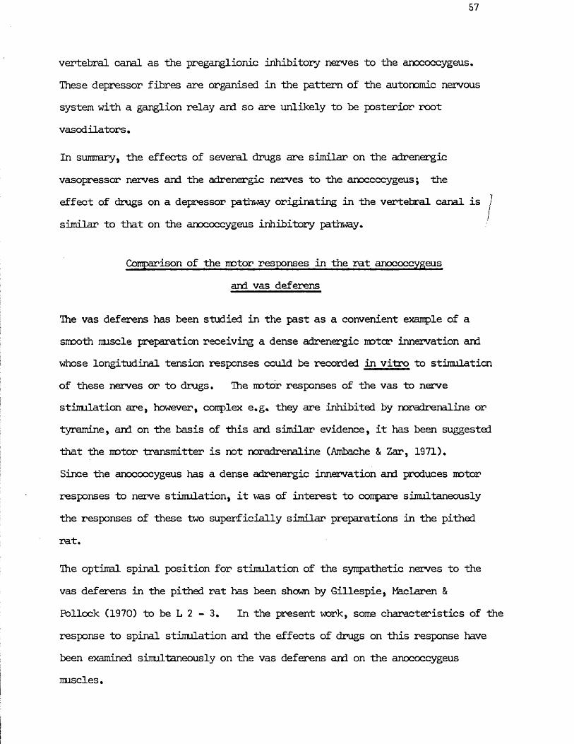
57
vertebral canal as the preganglionic inhibitory nerves to the anococcygeus.These depressor fibres are organised in the pattern of the autonomic nervous system with a ganglion relay and so are unlikely to be posterior root vasodilators.
In sumnary, the effects of several drugs are similar on the adrenergic vasopressor nerves and the adrenergic nerves to the anococcygeus; theeffect of drugs on a depressor pathway originating in the vertebral canal is /
/similar to that on the anococcygeus inhibitory pathway.
Comparison of the motor responses in the rat anococcygeusand vas deferens
The vas deferens has been studied in the past as a convenient example of a smooth muscle preparation receiving a dense adrenergic motor innervation and whose longitudinal tension responses could be recorded in vitro to stimulation of these nerves or to drugs. The motor responses of the vas to nerve stimulation are, however, complex e.g. they are inhibited by noradrenaline or tyramine, and on the basis of this and similar evidence, it has been suggested that the motor transmitter is not noradrenaline (Ambache & Zar, 1971).Since the anococcygeus has a dense adrenergic innervation and produces motor responses to nerve stimulation, it was of interest to compare simultaneously the responses of these two superficially similar preparations in the pithed rat.
The optimal spinal position for stimulation of the sympathetic nerves to the vas deferens in the pithed rat has been shown by Gillespie, MacLaren &Pollock (1970) to be L 2 - 3. In the present work, some characteristics of the response to spinal stimulation and the effects of drugs on this response have been examined simultaneously on the vas deferens and on the anococcygeus muscles.
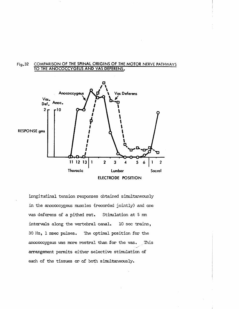
F ig .32 C O M P A R IS O N OF THE SPINAL ORIGINS OF THE MOTOR NERVE PATHWAYS TO THE ANOCOCCYGEUS AND VAS DEFERENS.
□
Anococcygeus \ Vas DeferensVos.Def.2 r r'O
RESPONSE gms
11 12 13
Thoracic Lumbar Sacral
ELECTRODE POSITION
Longitudinal tension responses obtained simultaneously in the anococcygeus muscles (recorded jointly) and one vas deferens of a pithed rat. Stimulation at 5 mm intervals along the vertebral canal. 10 sec trains, 30 Hz, 1 msec pulses. The optimal position for the anococcygeus was more rostral than for the vas. This arrangement permits either selective stimulation of each of the tissues or of both simultaneously.
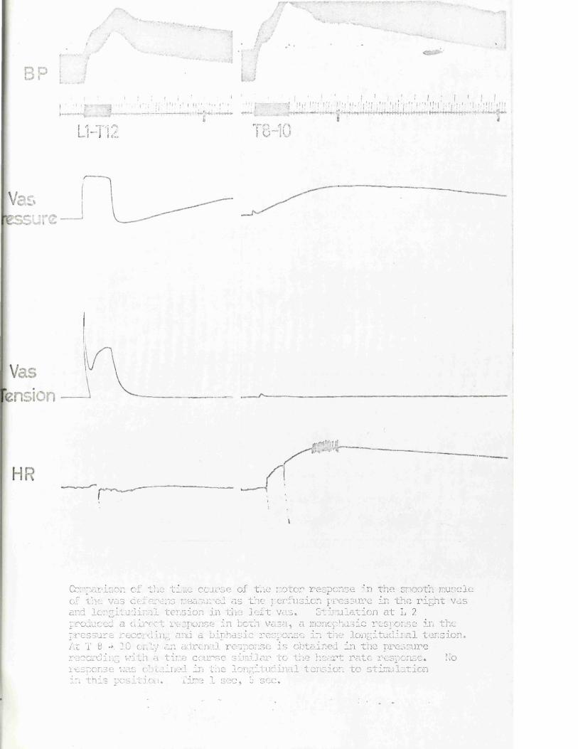
11-T19La 1 } &,I
• ' ! ; | • ' 1 1 j I I ' ! ! J I ! . I ! ; ! III ■' j ■'; I ' j f !! 1 1 ' j ! ’ ' ! ! ■ ' ' 1 I j ! I ! •'! i ! : 1 ' ! I ' ! ‘ j | J : 1 !
18-10
Comparison of the time course of the motor response in the smooth muscle of the vas deferens measured as the perfusion pressure in the right vas and longitudinal tension in the left vas* Stimulation at L 2 produced a direct response in both vasa, a monopbasic response in the pressure recording and a biphasic response in the longitudinal tension. At T B •*. 10 only an adrenal response is obtained in the pressure recording with a time course similar to the heart rate response* Noresponse was obtained in the longitudinal tension to stimulation in this position. Time 1 sec, 5 see.
bp
Fig. 33 Comparison of the effects of LSD and
hexamethonium on the response of the pithed rat vas deferens
measured as longitudinal tension or constant volume perfusion
pressure. Stimulation at L 1 - 3, 30 Hz for 10 sec. The
response, measured in the left vas deferens as longitudinal tension
shews a biphasic response but in the right vas deferens as perfusion
pressure, only a monophasic response is found.
While LSD 200 ug/kg inhibits the "spike" phase of the tension response, it has no visible effect on the pressure response. Subsequent administration of hexamethonium (C6) (5 mg/kg) completely abolished
\ the secondary response of the tension and almost completely abolished the pressure response. Time 1 sec, 5 sec.
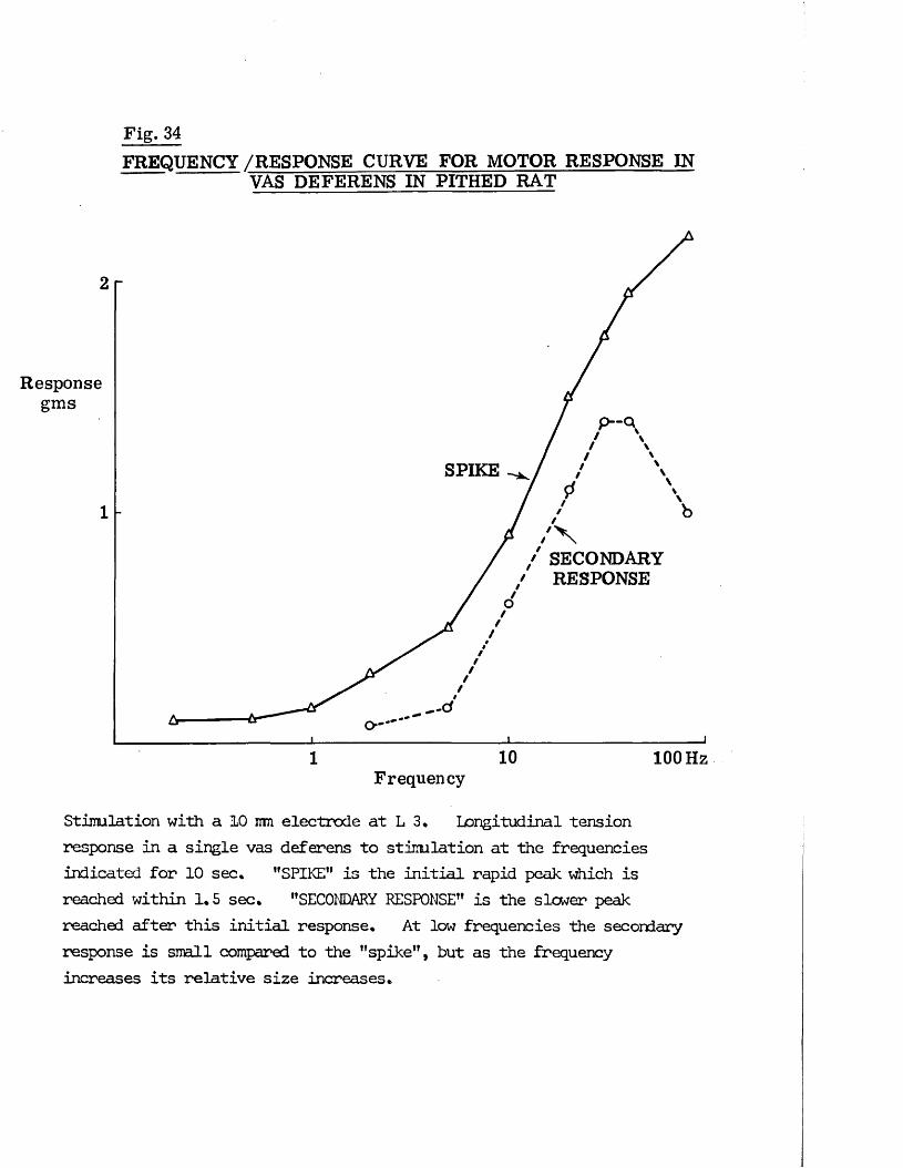
Fig. 34F R E Q U E N C Y /RESPONSE C U R V E F O R M O T O R RESPONSE IN
VAS DEFER E N S IN PITHED R A T
2
Responsegms
SPIKE
1
/ S E C O N D A R Y / RESPONSE
10 100 Hz1Frequency
Stimulation with a 10 mu electrode at L 3* Longitudinal tension response in a single vas deferens to stimulation at the frequencies indicated for 10 sec. "SPIKE” is the initial rapid peak which is reached within 1.5 sec. "SECONDARY RESPONSE” is the slower peak reached after this initial response. At low frequencies the secondary response is snail compared to the "spike", but as the frequency increases its relative size increases.
58
Spinal origin of the vas deferens response
Preliminary experiments, exploring the spinal canal, confirmed that the optimal position for the response in the vas deferens was L 2 - 3, Fig. 32 illustrates this and shows that this position is somewhat lower than the position for stimulation of preganglionic sympathetic nerves to the anococcygeus. If the extreme ends of these optimal positions were used, selective stimulation of either the anococcygeus at T 12 - 13 or vas deferens at L 3 - 4 was possible. If the electrode was placed at L 1 - 3, both muscles could be stimulated simultaneously.
Shape of vas deferens response
The isometric development of tension in the vas deferens during nerve stimulation showed a characteristic two phase response. An initial rapid "spike” contraction reached its maximum within 1.5 sec and then declined to be replaced by a slower smooth secondary contraction which reached its maximum after 8-10 sec (Fig. 33).
The relationship in the size of these two phases varied with frequency and with the duration of stimulation. Single pulses produced a rapid "spike” only. At low frequencies, the secondary response was small but as the frequency increased it became greater in proportion to the "spike" until at 30 Hz it was 80% of the "spike" (Fig. 34). The response declined immediately on cessation of stimulation. Continuous stimulation for up to 3 min at 30 Hz showed that the secondary response declined after 20 - 30 sec but remained at a lower plateau level far the remainder of the stimulation period (Fig. 17).
This two phase response in the vas deferens is in contrast to the single phase, smooth contraction of the anococcygeus. Had the "spike" been absent, the secondary response in the vas deferens would be, however, similar to the anococcygeus response (see Figs. 17 & 35).
59
In experiments on the vas deferens where perfusion pressure instead of longitudinal tension was used as a measure of the response, only a single phase of pressure increase was seen corresponding to the secondary component of the isometric longitudinal tension response. The maximum pressure was reached after 8-10 sec (Fig. 33). This my indicate an anatomical basis for the two phase tension response since longitudinal tension is likely to reflect activity in the outer longitudinal layer of smooth muscle in the vas, whereas the perfusion pressure is likely to reflect activity in the inner circular layer.
Another difference in the perfusion pressure response was that it showed a delayed response corresponding in time with the blood pressure and heart rate adrenal response when stimulating in the thoracic region, whereas the longitudinal tension showed no adrenal response (Fig. 32).
The absence of the "spike" in the perfusion response is not due to the inability of the pressure recording system to respond to rapid changes in lumen diameter since mechanical constriction of the vas with forceps can produce pressure increases at least as rapid as the "spike”.
The longitudinal tension responses in the vas deferens to 10 sec periods of stimulation are reproducible if at least 3 min is left between periods. If less than 3 min is left, the "spike" component declines slightly. This decline is similar to the endogenous inhibition found in vitro and attributed by Swedin (1971 b) to release of prostaglandins within the tissue. To eliminate this additional factor, when investigating the effects of drugs on the response, at least 3 min was left between periods of stimulation.
Effects of drugs on the vas deferens response
With the pithing rod electrode at L 1 - 3, the autonomic outflows to both vas deferens and anococcygeus can be stimulated simultaneously. In order to compare the properties of the innervation of the two tissues, the effects of
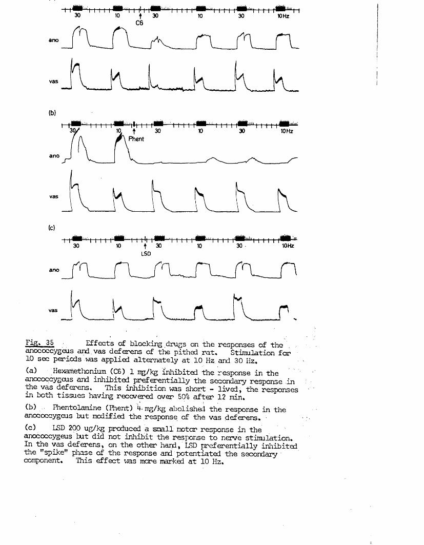
Fig* 35 Effects of blocking drugs on the responses of theanococcygeus and. vas deferens of the pithed rat* Stimulation for 10 sec periods was applied alternately at 10 Hz; ard 30 Hz*(a) Hexamethonium (C6) 1 irg/kg inhibited the response in the anococcygeus and inhibited preferentially the secondary response in th£ vas deferens* This inhibition was short — lived, the responses in both tissues having recovered over 50% after 12 min*(b) .. Fhentolamine (Phent) k< mg/kg abolished the response in the anococcygeus but modified the response of the vas deferens. -(c) LSD 200 ug/3<g produced a small motor response in the 1anococcygeus tut did not inhibit the response to nerve stimulation* In the vas deferens, on the other hand, LSD preferentially inhibited the "spike" phase of the response and potentiated the secondary component* This effect was mare marked at 10 Hz.
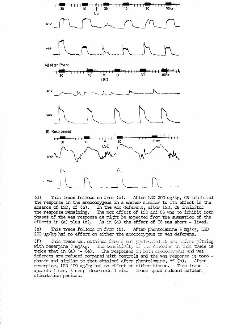
ano
vas f W U r v(e) after Phent
|iiiiiniiiii| | |
30-u®B1uuij_|_||-|_-(—|-u|H
10 t 10LSD
’uJ.kJ uJ^UiiBBiiimi | | | i^Milwiliiiili | ||30 10 Hz
ano
vas
(f) Reserpinised
10HzLSD
ano
vas
(d) This trace follows on from (c). After LSD 200 ug/kg, C6 inhibited the response in the anococcygeus in a manner similar to its effect in Hie absence of LSD, cf (a)* In the vas deferens, after LSD, C6 inhibited the response remaining. The net effect of LSD and C6' was to inhibit both phases of Hie vas response as might be expected from the summation of the effects in (a) plus (c),f As in (a) the effect of C6 was short - lived.(e) This trace follows on from (b). After phentolamine ft mg/kg, LSD 200 ug/kg: had no effect on either the anococcygeus or vas deferens.(f) This trace was obtained from a rat pretrcated 1C hrs b of ere pithing with reserpine 3 mg/kg. The sensitivity of the recorder in this trace is twice that in (a) ‘- (e). The responses in both anococcygeus and vas deferens are reduced compared with controls and the vas response is mono. - phasic and similar to that obtained after phentolamine, cf (b). After reserpine, LSD 200 ug/kg had no effect on either tissue. Time trace upwards l’ sec, 5 sec; downwards 1 min. Trace speed reduced between stimulation periods.
ganglion blocking drugs and drugs which block or potentiate the action of adrenergic nerves were studied. In addition, LSD was examined since it has been shown to inhibit the response to field stimulation in rat and guinea-pig vas deferens in vitro (Ambache, Dunk, Vemey & Zar, 1973). Except where otherwise stated the vas deferens response referred to is the longitudinal tension.
Hexamethonium (1 mg/kg) inhibited the responses in both anococcygeus and vas deferens (Fig. 35 a). The secondary component of the vas deferens response was inhibited more than the "spike”. Two possible explanations for this differential action on the vas deferens are either that the two phases are produced by different pathways which have different sensitivities to ganglion blocking agents or that partial ganglion blockade leads to a reduced frequency of discharge in the postganglionic nerves which in turn produces a response in which the "spike" is proportionately greater than the secondary response. In favour of the former and against the latter argument is the fact that the response to 30 Hz after hexamethonium has a larger "spike" but snaller secondary response than the response to 10 Hz before hexamethonium. If there are indeed two pathways, it is of interest that they appear to have a common spinal origin since both phases are produced in similar proportions at different spinal levels (Fig. 32).
A larger dose of hexamethonium (5 ng/kg) completely abolished the response in j both anococcygeus and vas deferens, indicating that both components of the vas deferens response are produced by nerves having a ganglion synapse in their pathway.
The effects of hexamethonium were short lasting. The effect of 1 ug/kg had started to disappear after 5 min in both tissues. After 5 mg/kg, the response in the vas deferens recovered particularly quickly, being back to its full size within 15 - 30 min. After recovery, a second dose of 5 ug/kg hexamethonium again produced a block but this was even more short lived
61
than before.
Reserpine (3 mg/kg) given intravenously, selectively inhibited the "spike” component of the vas deferens response without affecting the secondary conponent or the anococcygeus response. This effect developed within 5 min and so is unlikely to be related to any significant depletion of NA stores by reserpine.
In rats pretreated for 18 hr with reserpine (3 mg/kg, i.p.), at which time the NA content of both the vas deferens and anococcygeus had been depleted by at least 98% (i.e. the NA level was undetectable) a reduced, single phase response was obtained in the vas deferens to nerve stimulation. This response reached its maximum after 2 -3 sec and then declined over the reminder of the 10 sec stimulation period to 50 - 80% of its maximum (Fig. 38 b). When the peak of this response was compared with the secondary response in control animals, it was only 60% of the latter (Fig. 39). Thus after reserpine pretreatment both components of the vas deferens response were reduced and the residual response was monophasic.
After reserpine pretreatment, the response to nerve stimulation in the anococcygeus was depressed compared with controls • Responses could not be produced to single pulses or lew frequencies and even at high frequencies the response was slow to develop (Fig. 38 b) and had a reduced maximum (Fig. 39). When the response had developed, hcwever, the raised tension could be mintained for up to 60 sec if stimulation was continued.
Reserpine has, therefore, a dual action on the vas deferens response; anacute effect to selectively inhibit the "spike” conponent and a delayed effect, related to the depletion of NA from the tissue, to reduce both phases of theresponse. On the anococcygeus, on the other hand, reserpine affects theresponse only when the NA oontent is depressed but even under these conditions the response is not abolished.
62
Guanethidine (10 mg/kg) completely abolished the responses to nerve stimulation in both tissues. The anococcygeus response recovered after 1 - li hr but the vas deferens response did not,
Phentolamine (4 mg/kg) completely abolished responses in the anococcygeus but only changed the shape of the response in the vas deferens (Fig, 35 b). This change in shape is open to various descriptions but one is to say that the "spike" became prolonged and the secondary response became poorly maintained. At 30 Hz, the height of both components was decreased but at 10 Hz the height of the two phases was little changed, only the shape being changed. Alternatively it could be argued that the single phase response after phentolamine is the secondary response which now starts earlier but is poorly maintained.
In either case it is clear that phentolamine (4 mg/kg) does not block the response in the vas deferens as it does the response in the anococcygeus and no additional dose of phentolamine (up to 20 mg/kg) produces any further change in response.
LSD - 25 (200 jug/kg) had no detectable effect on the motor response of theanococcygeus (Fig. 27), but had a differential effect on the two phases of the vas deferens response (Fig. 35 c). At 10 Hz, the "spike" was completely abolished and at 30 Hz, was inhibited. The secondary response on the other hand was slightly potentiated (Figs. 35 c & 37). Whatever the pathway producing the "spike", it appears to be specifically inhibited by LSD, and either as a result of this inhibition or due to some other property of LSD, the secondary response is potentiated.
The differential effect of LSD is thus the opposite of that of hexamethonium. When hexamethonium (1 mg/kg) was given after LSD (200 pg/kg) both phases of the vas deferens response were inhibited as might be expected from the differential effect of each drug on one phase (Fig. 35 d). The effect of
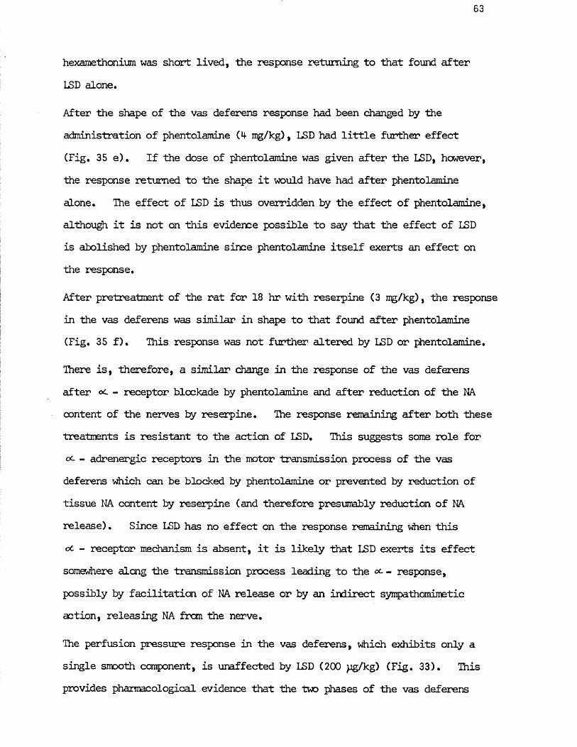
63
hexamethonium was short lived, the response returning to that found after LSD alone.
After the shape of the vas deferens response had been changed by the administration of phentolamine (4 mg/kg), LSD had little further effect (Fig. 35 e). If the dose of phentolamine was given after the LSD, hcwever, the response returned to the shape it would have had after phentolamine alone. The effect of LSD is thus overridden by the effect of phentolamine, although it is not on this evidence possible to say that the effect of LSD is abolished by phentolamine since phentolamine itself exerts an effect on the response.
After pretreatment of the rat for 18 hr with reserpine (3 mg/kg), the response in the vas deferens was similar in shape to that found after phentolamine (Fig. 35 f). This response was not further altered by LSD or phentolamine.
There is, therefore, a similar change in the response of the vas deferens after oc - receptor blockade by phentolamine and after reduction of the NA content of the nerves by reserpine. The response remaining after both these treatments is resistant to the action of LSD. This suggests some role for oc - adrenergic receptors in the motor transmission process of the vas deferens which can be blocked by phentolamine or prevented by reduction of tissue NA content by reserpine (and therefore presumably reduction of NA release). Since LSD has no effect on the response remaining when this cc - receptor mechanism is absent, it is likely that LSD exerts its effect somewhere along the transmission process leading to the - response, possibly by facilitation of NA release or by an indirect sympathomimetic action, releasing NA from the nerve.
The perfusion pressure response in the vas deferens, which exhibits only a single smooth component, is unaffected by LSD (200 pg/kg) (Fig. 33). This provides pharmacological evidence that the two phases of the vas deferens
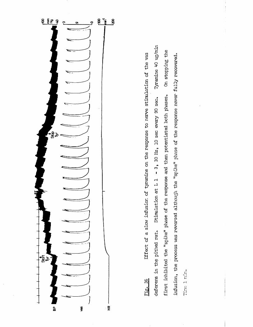
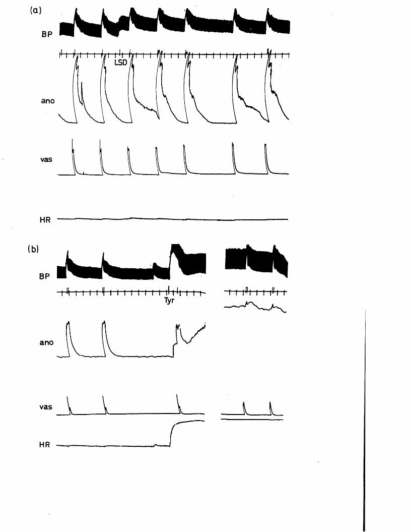
(a)
BP
ano
vas
-» \
HR
(b)BP
i H i ii t < i i i t-t t 1 1— H t U i i t- -t i il!i iTyr
anoi
vas i i L i _
HR
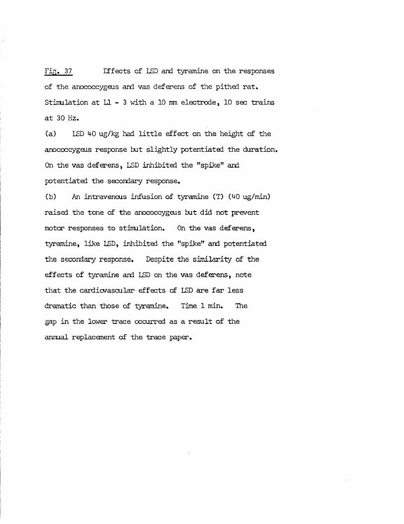
Fig. 37 Effects of LSD and tyramine on the responsesof the anococcygeus and vas deferens of the pithed rat. Stimulation at LI - 3 with a 10 mm electrode, 10 sec trains at 30 Hz.(a) LSD 4-0 ug/kg had little effect on the height of the anococcygeus response tut slightly potentiated the duration. On the vas deferens, LSD inhibited the ’’spike” and potentiated the secondary response.(b) An intravenous infusion of tyramine (T) (40 ug/min) raised tine tone of the anococcygeus tut did not prevent motor responses to stimulation. On the vas deferens, tyramine, like LSD, inhibited the ’’spike” and potentiated the secondary response. Despite the similarity of the effects of tyramine and LSD on the vas deferens, note that the cardiovascular effects of LSD are far less dramatic than those of tyramine. Time 1 min. Thegap in the lower trace occurred as a result of the annual replacement of the trace paper.
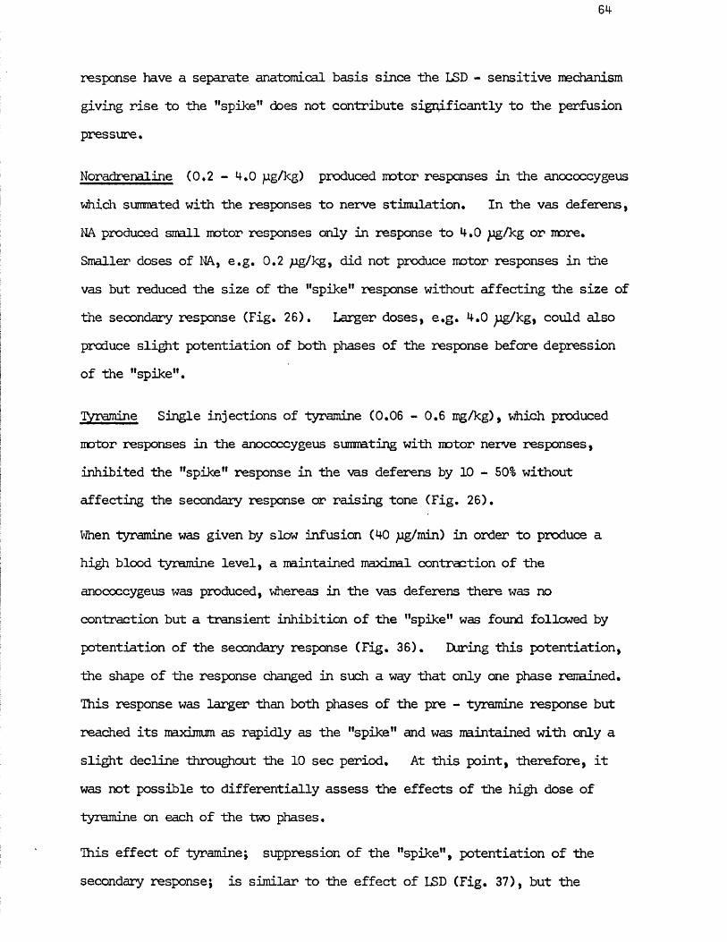
64
response have a separate anatomical basis since the LSD - sensitive mechanism giving rise to the "spike” does not contribute significantly to the perfusion pressure.
Noradrenaline (0,2 - 4,0 pg/kg) produced motor responses in the anococcygeus which summated with the responses to nerve stimulation. In the vas deferens, NA produced small motor responses only in response to 4,0 jg/kg or more. Smaller doses of NA, e.g, 0,2 /ig/kg, did not produce motor responses in the vas but reduced the size of the "spike" response without affecting the size of the secondary response (Fig. 26), Larger doses, e.g. 4.0 jug/kg, could also produce slight potentiation of both phases of the response before depression of the "spike".
Tyramine Single injections of tyramine (0.06 - 0.6 mg/kg), which produced motor responses in the anococcygeus sumnating with motor nerve responses, inhibited the "spike" response in the vas deferens by 10 - 50% without affecting the secondary response or raising tone (Fig. 26).
When tyramine was given by slow infusion (40 jug/min) in order to produce a high blood tyramine level, a maintained maximal contraction of the anococcygeus was produced, whereas in the vas deferens there was no contraction but a transient inhibition of the "spike" was found followed by potentiation of the secondary response (Fig. 36). EHjring this potentiation, the shape of the response changed in such a way that only one phase renained. This response was larger than both phases of the pre - tyramine response but reached its maximum as rapidly as the "spike" and was maintained with only a slight decline throughout the 1 0 sec period. At this point, therefore, it was not possible to differentially assess the effects of the high dose of tyramine on each of the two phases.
This effect of tyramine; suppression of the "spike", potentiation of the secondary response; is similar to the effect of LSD (Fig. 37), but the
(a) Reser*pin ised HOOmmHg
-AONA
50pg
ano.0
vas
■300
m i r f *1-200
(b)K>S€C
Control
ano
vas
tTyr
r5g
Lo
0
Reserpinised
.02g
LO
tTyr
Fig. 38 Effect of reserpinisation on the effect oftyramine on the pithed rat anococcygeus, vas deferens, blood pressure and heart rate.
(a) Pithed rat pretreated 18 hrs before pithing with reserpine 3 mg/kg. Stimulation at L 1 - 3, 30 Hz, 10 sec every 4 min. Tyramine produced very small motor responses"" in the anococcygeus, no effect on the vas deferens response and only smll responses on B.P. and H.R. NA, on the other hand, produced responses on all >4 parameters, cf. control rat in Fig. 26. Time 1 min. Note sensitivity of B.P. trace was halved between crosses.
(b) Comparison of the effect of a slow infusion of tyramine (40 ug/min) on the responses to nerve stimulation in the anococcygeus and vas deferens of the pithed rat in control and reserpinised (18 hr, 3 mg/kg) pithed rats. Stimulation at L 1 - 3, 30 Hz, 10 sec. In the control, the tone ofthe anococcygeus is raised and the motor response to stimulation is superimposed on this. After reserpinisation* tyramine produces no elevation of tone but the reduced response to stimulation is’ slightly potentiated. In the control vas deferens, the "spike" is abolished by tyramine. After reserpinisation, the single phase response remaining in the vas deferens was slightly potentiated by tyramine.
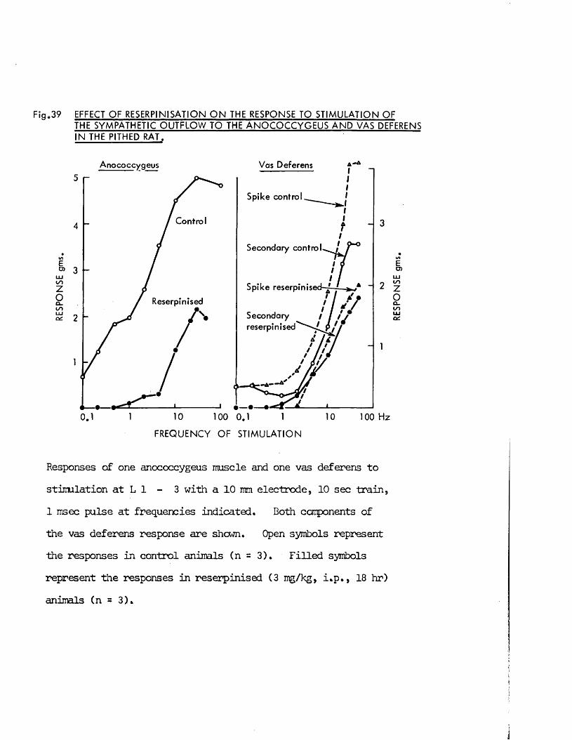
g.39 EFFECT OF RESERPINISATION O N THE RESPONSE TO STIMULATION OFTHE SYMPATHETIC O U TFLO W TO THE A N O C O C C Y G E U S A N D VAS DEFERENS IN THE PITHED RAT.
Anococcy geus Vas Deferens5
Spike control
Control4
Secondary c o n t r o l^ i
o> 3t o
Spike reserpinised-—-
ReserpinisedCl.t o
Secondaryreserpinised
2
—1 •100 0.110 100 H z0.1 10
FREQ UENCY O F S T IM U LA T IO N
Responses of one anococcygeus muscle and one vas deferens to stimulation at L 1 - 3 with a 10 mm electrode, 10 sec train,1 msec pulse at frequencies indicated. Both components of the vas deferens response are shewn. Open symbols represent the responses in control animals (n = 3). Filled symbols represent the responses in reserpinised (3 mg/kg, i*p,, 18 hr) animals (n = 3),
RES
PON
SE
gms

65
effect of tyramine unlike that of LSD is reversible (Fig, 36),
After reserpine (3 mg/kg, i.p., 24 hr) pretreatment, tyramine (0,2 - 0.6 mg/kg) produced weak motor responses on the anococcygeus which summated with the reduced motor nerve responses still present (Fig. 38). NA (4 pg/kg) produced contraction of the anococcygeus similar to controls. In the vas deferens, however, the diminished motor nerve response remaining after reserpine was unaffected by tyramine, although it could be potentiated by NA (4 pg/Rg).
Reserpine pretreatment thus prevents the effects of indirect sympathomimetics (tyramine) but not direct sympathomimetics (NA) on the anococcygeus. By analogy, it is tempting to suggest that the effects of tyramine and LSD on the vas deferens, which are also absent after reserpine, are produced by an indirect sympathomimetic action.
Summary of motor responses in vas deferens compared with anococcygeus
In terms of classical autonomic pharmacology, these experiments suggest that the effects of drugs acting on the nerve - muscle junction in the anococcygeus are relatively straightforward compared with those in the vas deferens•
The anococcygeus shows a regular, frequency related, monophasic contraction to preganglionic sympathetic nerve stimulation which is abolished by interruption of the pathway at the ganglion, at the sympathetic terminal by neurone blockade and at the effector organ itself by <*- - adrenergic blockade, and is very much reduced after depletion of the tissue NA content by reserpine.
In the vas deferens, on the other hand, the motor response to nerve stimulation is complex and the pharmacological evidence does not conclusively prove that the motor transmission is adrenergic.
Part of the complex, biphasic nature of the longitudinal tension response in the vas arises from the anatomical structure of the organ as demonstrated by the much simpler monophasic response produced when perfusion pressure rather than longitudinal tension is recorded. This does not, however, fully explain the unconventional effects of drugs normally associated with modification of adrenergic transmission.
The responses in the vas were obtained from stimulation in the region L 1 - 3,an area normally associated with the sympathetic outflow to many tissues
Iincluding he anococcygeus. That the responses so produced in the vas were due to stimulation of preganglionic sympathetic nerves appears to be verified by the blockade of the response by a large dose of a ganglion blocker or by the adrenergic neurone blocker guanethidine. This hypothesis is not,
/
hcwever, directly corroborated by the effects of the other drugs tested. jThe motor response is not directly blocked by phentolamine, as it is in the 1anococcygeus, but NA, tyramine and LSD all appear to modulate transmission jvia an - receptor mechanism. This modulation appears to consist of inhibition of the "spike” and potentiation of the secondary response. Phentolamine cannot, therefore, block the motor response since it disinhibits the "spike" mechanism at the same time as inhibiting the secondary response.
On this evidence, therefore, the secondary response is modified in the manner expected of an adrenergic response, whereas the initial "spike" is unconventional apart from its abolition by hexamethonium and guanethidine.
Analyses which concentrate on the first component of the vas deferens response either by using the maximum amplitude or by giving so few stimuli that only the first component is elicited, naturally suggest that the motor transmission is non - adrenergic since it is resistant to phentolamine, inhibited by NA or tyramine and is still present after virtually complete depletion of the tissue NA content by reserpine. If, however, the shape of the response and
67
the above hypothesis on the role of * - receptors in this tissue are taken into account, it is clear that both reserpine and phentolamine produce a significant change in the pattern of response to nerve stimulation which implicates NA in the roles of both motor transmitter (in the secondary component at least) and modulator of the response (in the "spike” phase) possibly by feedback inhibition of its own release. The effect of reserpine is particularly interesting since an acute dose specifically inhibits the "spike”, whereas after depletion of tissue NA a monophasic response is left, which from the above hypothesis, is a prolonged ”spike” response. This acute inhibition could be due to initial release of NA from the nerve terminals by reserpine similar to the indirect sympathomimetic action postulated for tyramine and LSD. After depletion of NA by reserpine, no further release of NA by this indirect sympathomimetic action will occur, leaving the reserpine - resistant "spike” response disinhibited. Thus the main anomaly in the motor transmission process of the vas deferens emerges as the initial "spike” response which is resistant to phentolamine and to reserpine pretreatment. From these results there is no direct evidence that the transmitter giving rise to the latter response is or is not NA.
PART II - 1HE EFFECTS OF RESERPINE
ON THE RAT ANOCOCCYGEUS, VAS DEFERENS AND HEART
The adrenergic nerves of the rat vas deferens are depleted of/their NA byi’/ow/y ft
reserpine relatively more quickly than those in other tissues such as the // heart (Sjostnand & Swedin, 1988).
Two possible explanations of this resistance are that either the rate of depletion is dependent on the firing frequency in the nerves and therefore reduced in the intermittently active genital organs or that the "short" adrenergic neurones, which are found in the vas deferens and seminal vesicles and have their cell bodies located close to the target organ, are less easily depleted than the "long" adrenergic neurones found elsewhere.
With the pithed rat preparation it is possible to test the first hypothesis since impulse traffic to tissues can be altered in two ways; the normal tonic sympathetic discharge can be eliminated by pithing alone or traffic can be increased by pithing followed by electrical stimulation of the sympathetic outflow.
The second hypothesis can also be tested by simultaneous comparison of depletion in the vas deferens and in the anococcygeus since the latter has the anatomically conventional "long" adrenergic neurones as demonstrated in Part I by the ability to stimulate postganglionic nerves to the tissue at a distance of up to 40 mm from the tissue. Both these tissues were further compared with the heart, a tissue known to be rapidly depleted of NA by reserpine.
The dose dependence and time course of depletion in vivo in the tissues were first determined and then the effect of stimulation of the nerves on the rate of depletion was examined using pithed rats.
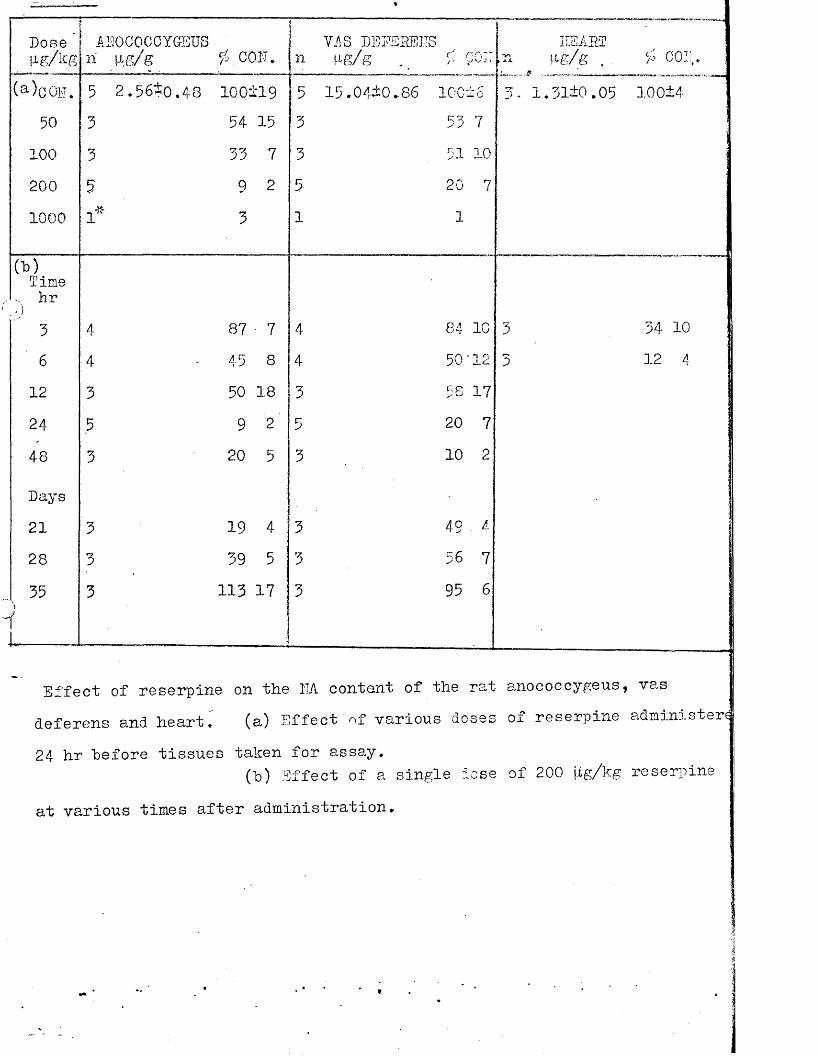
Doseu g A s
AI ri
JOCOCCYGEUSM-.g/g
OO.
nVIS DEd e e s:4g/g
rs r nC'tT/ - gvfvcT o •
.
5 2.56*0.48 100 £19 5 1 5 .0410 .86 10C±650 3 54 15 3 53 7
100 3 33 7 3 51 10200 9 2 5 20 71000
u1"' 3 1 1
(b)Time... hr .)
3 4 87 7 4 84 1C6 4 45 8 4 50'12
12 3 50 18 3 58 1724 5 9 2 5 20 748 3 20 5 3 10 2
Days21 3 19 4 3 49 . 1
28 3 39 5 3 56 735\
i3 11.3 17 3 95 6
nI EE ASTu cor
1.31*0.05 100±4
34 10 12 4
Effect of reserpine on the NA content of the rat anococcygeus, vasdeferens and heart. (a) Effect of various doses of reserpine administer?24 hr before tissues taken for assay.
(b) Effect of a single lose of 200 jigat various times after administration.
reseruine
Table 2
1ti1"-T'"n
ft f \ ft /Ttr.-t rrr--> iVU tt y X \T it v.*i-t e an
•kt » r*n
DEFERENSKean
HEn
ARTMea?
Pithod (3 hr) jjil 3 12 o *» 3 3 92 " 4! " j 3 102 ■h 5
IPithed -v Stin. jr 3 101 - 13 3 8 2 - 6 4. 104 -r 7
/ \ 1I'ooerp* \3 hr) j 4 87 - 6 A*T 84 - 10 , 3 34 10
Reoorp. (o hr) j 4 45 - 3 4 30 t 12 3 12 -f-4• |
Rescrp. -f Pithed (l hr) [i 2 69 2 72 Cl ■*0✓<-Reoorp. + Pithed (3 hr) j 4 53 t 14 4 64 1 7 3 ’ 34 *** 9lieserp. + Pithod + Stim. j
~ ir _ i 4 is i 6 4 32 1 5 3 . 12 4
Effect of pithing, of nerve stimulation and of reserpine on the NA content of the rat anococcygeus, vas deferens and heart. The NA content is expressed as a percentage of the mean values from untreated control rats for each tissue (calculated as pg NA/s tissue).Neserp. — Reserpine 200 [ig/kg i.p. 3 hr before pithing or for time indicated. Pithed - Tissues taken 3 hr after pithing cr after time indicated.Stim. - Stimulation via pitiling rod electrode at Ll-3, 1 msec pulse3 at
30II2, 10 sec trains every 1.5 nin for 2 hr starting 1 hr after pithing*
E F F E C T O F R E S E R P I N E O N N A C O N T E N T
100 |24 hrs
N AC O N T E N T
C O N T R O L
V V A S
A N O .
1000200D O S E M g / k g
100
Fig. 40 Effect of reserpine in doses of 50, 100, 200and 1000 /ig/kg (i.p.) on the NA content of rat anococcygeus and vas deferens after 24 hr. NA content is expressed as a percentage of the mean value from untreated controls for eachtissue. anococcygeusj vasdeferens. Solid I - bars represent S.E.H., dotted I - bars represent the S.E.H. for the untreated controls j from left to right - anococcygeus, vas deferens.
" e f f e c t O F R E S E R P I N E O N N A C O N T E N T
loo!! 2 0 0 / x g / k g
N AC O N T E N T
C O N T R O L
HEART
TIME Hr s.
Fig* Effect of reserpine 200 pg/kg (i.p.) after varioustimes on the NA content of the rat anococcygeus, vas deferens and heart. NA content is expressed as a percentage of the mean valuefrom untreated controls for each tissue. anococcygeus
vas deferens; heart. Solid I - barsrepresent S.E.H,, dotted I - bars represent the S.E.H. for the untreated controls; from left to right - anococcygeus, vas deferens, heart.
69
Effect of reserpine dose on the tissue NA content
The normal level of NA in the three organs studied was - anococcygeus 2.56 ± 0.48 pg/g (n = 5), vas deferens 15.04 + 0.86 pg/g (n = 5) and heart 1.13 * 0.05 pg/g (n = 3). The effect of various doses of reserpine on the NA level in the anococcygeus muscle and vas deferens was measured at 24 hr and the results are shown in Table 1 and Fig. 40. Both tissues were depleted in a similar dose dependent nanner. At the highest dose level (1000 pg/kg) there was almost complete depletion to 3% and 1% of normal for the anococcygeus and vas deferens respectively. Reserpine 200 pg/kg produced a sub - maximal depletion to 9 ± 2% (n = 3) for the anococcygeus and 20 + 7% (n = 3) for the vas deferens. There was no statistically significant difference at any dose level between the percentage depletion in NA of the two tissues.
Time course of depletion of NA by reserpine
At 24 hr a dose of 200 pg/kg of reserpine produced a large but sub - maximal depletion and was therefore selected as a suitable dose which, in the experiments on pithing and nerve stimulation, could show either enhanced or decreased depletion. The time course of depletion by this dose was then studied in the three tissues with the results shown in Table 1 and Fig. 41.The anococcygeus and vas deferens were depleted of NA at a similar rate but the heart was depleted much more rapidly. After 6 hr the NA content of the heart vas 12+4% (n = 3), the anococcygeus 45 + 8 % (n = 7) and the vas deferens 50+ 8 % (n = 7) of their respective controls.
Since this large divergence in depletion existed over this initial 6 hr period, the effects of pithing and nerve stimulation were examined over the period from 3 to 6 hr after reserpine. In this way, direct comparison was possible between depletion in vivo and that after the additional factors of pithing and nerve stimulation had been added.
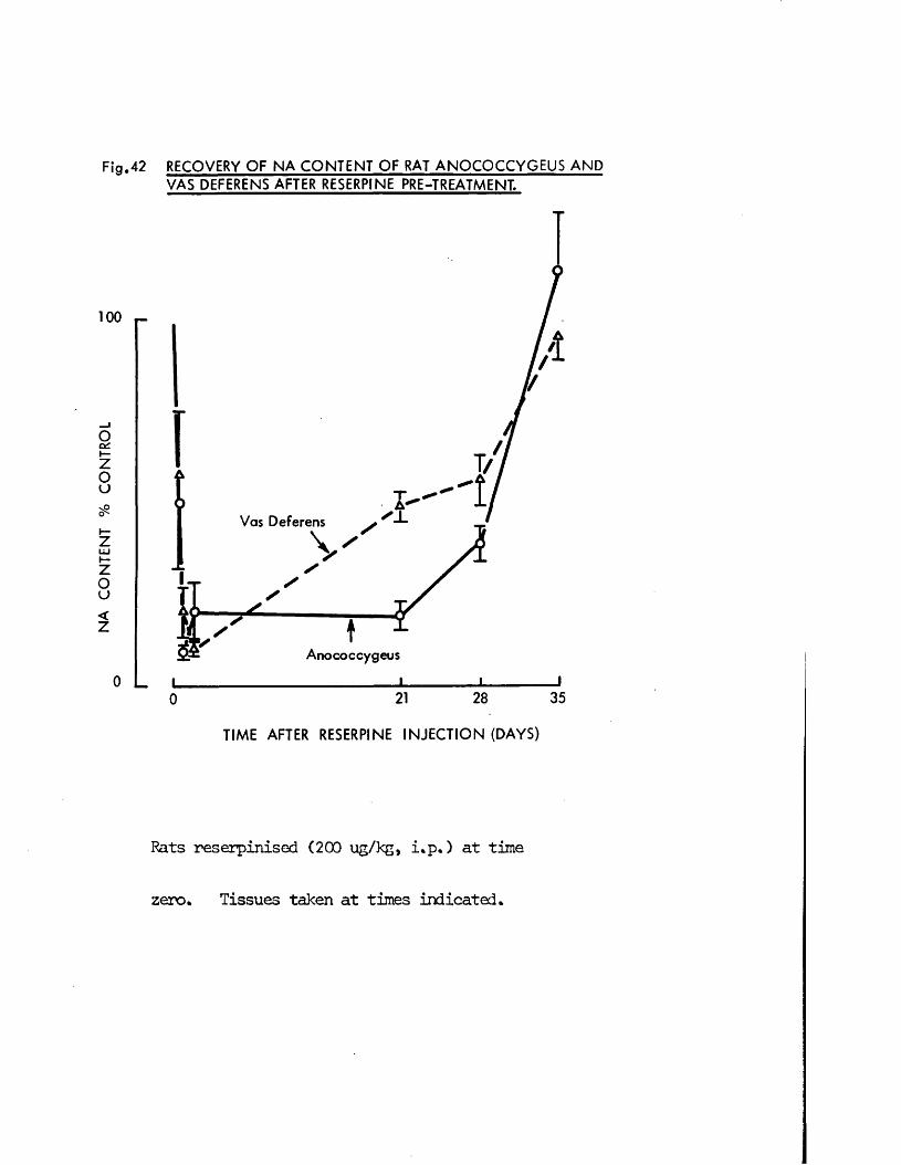
NA
CO
NTE
NT
% C
ON
TR
OL
F ig .42 RECOVERY O F N A C O N T E N T O F RAT A N O C O C C Y G E U S A N D VAS DEFERENS AFTER RESERPINE PRE-TREATMENT.
100 T-
0 L
Vas Deferens
Anococcygeus
i0 21 28 35
TIM E AFTER RESERPINE IN J E C T IO N (DAYS)
Rats reserpinised (200 ug/kg, i.p.) at time
zero* Tissues taken at times indicated*
70
Recovery of tissue NA after reserpine
Since the adrenergic nerves of the vas deferens and anococcygeus were depleted of their NA contents at a similar rate by reserpine and this rate was slower than that found in other tissues, it was of interest to compare the rate of recovery of their NA contents after depletion by reserpine 200 pg/kg. The results are shown in Table 1 and Fig. 42.
48 hr after reserpine the NA content of the vas deferens had fallen further than at 24 hr to 10 + 2% (n = 3) but the NA content of the anococcygeus showed a non - significant increase to 20 t 51 (n = 3). 21 days after reserpine, however, the NA contents of the vas deferens and anococcygeus were49 + 4% (n = 3) and 19 + 4% (n = 3) respectively. This difference between the two tissues was statistically significant, indicating a more rapid recovery in the vas deferens despite both tissues having been initially depleted to a similar extent. The recovery in the anococcygeus increased after 28 and35 days and by 35 days after reserpine the NA content of both tissues was restored to norml levels.
Effects of pithing and of nerve stimulation in non - reserpinised animals
The effects of interrupting any spontaneous neuronal activity by pithing and of increasing neuronal activity by a 2 hr period of intermittent stimulation are shown in the first part of Fig. 43 and Table 2. Pithing alone produced a rise in the NA content of the anococcygeus with little change in the content of the vas deferens or heart. Nerve stimulation in the pithed animalproduced a slight reduction in the NA level in the vas deferens with little change in the anococcygeus or heart. None of these changes was statistically significant.
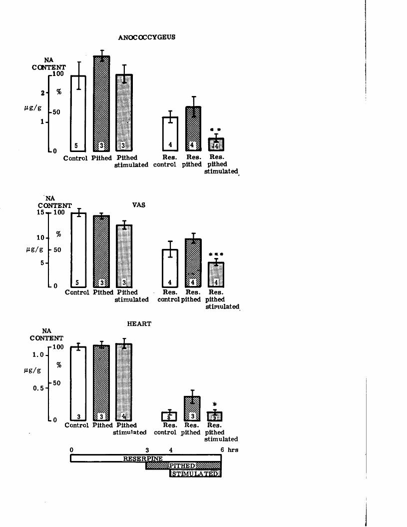
ANOCOCCYGEUS
NACONTENT
_ 1 0 0
Control Pithed Pithed Res. Res. Res.stimulated control pithed pithed
stimulated
NACONTENT „ 15*r 100 i-4 -
10-1Mg/g
5-\- 50
L0
VAS
3Control Pithed Pithed Res. Res. Res.
stimulated control pithed pithedstimulated
HEARTNA
CONTENT
1.0
Mg/g
plOOHH i ! ~r
% M-50
1
-0 3 { A -<UControl Pithed Pithed
stimulated control pithedRes.
pithedstimulated
6 hrsRESERPINE m i m p r r K E D m
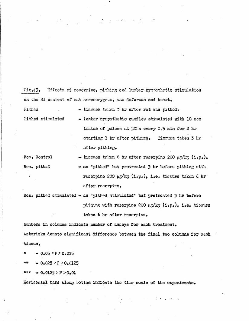
Pithed
Pithed stimulated
Res* Control Eos* pithed
Fig*43® Effects of roserpin®, pithing and lucb&r sympathetic stimulation ‘on th® HA content of rat saocooeygouo, vas deferens and heart®
«* tissues taken 3 hr after rat was pithed®«• )ute sympathetic outflow stimulated with 10 son trains of pulses at $ 0 1 is ewery 1»3 ain for 2 hr starting 1 hr after pithing* Timuvs taken 3 hr after pi thing*
«* tissues taken 0 hr after reserpin® 2 0 0 pg/kg (i.p.)* -> as "pithed*1 but pratreatcd. $ hr before pithing with reserpine 2 0 0 p&/kg (i«p*)» i*®.' tissues taken 6 hr after reserpine.
Has* pithed stimulated « aa "pithed stimulated" hut pretreated 3 hr beforepithing with reserpine 200 pg/kg (i»p*)» i»e* tissues taken 6 hr after resarpisc*
2susbers is columns indicate number of assay© for each treatment *Asterisks denote significant difference between th® final two colmans for each tissue** - 0*05 >?>0.025** ~ 0*025 > ? >0.0125 *** ~ 0.0X2$ >?;> 0*01Borisest&l bar© along bottom indicate the tis© ©cal© of the experiments*
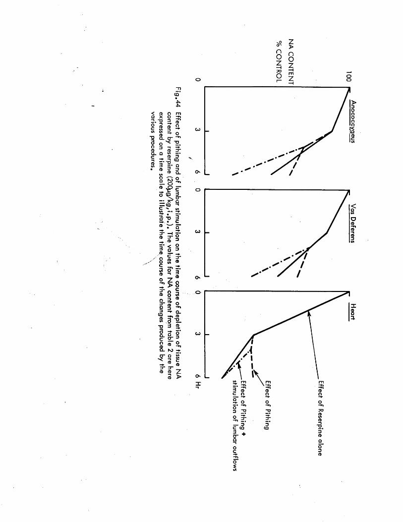
nOz—i X3o
z>nOzoo
CQ-N
< CD O mQ X O ■ ti-n T> 3 CDo’c
*nCDi/ i
CD3
O
I/I VICD
-♦ oT> Q_ 0 “
—in
3 OX ■g4
O 3 “ t —♦*CD CD 3 "Q . Q i/»c CD 3■nCDi / i 3
•n-o#CQ
Q• CD 3 3
»/i CD Q_OQ N 3
OO- n
3“CDO3”Q3
CQCD(A"O3Q_Coa.
3" g CD 3
< § Q_ 38 ?? fz ® > 88 a3 «
3 ^ -*■ Q _ -r» CD3 12. 3 %o gO" 3
DO - f
O " O £'•c £CD c
I - CD3-3“CD CD Z3 >
GJ .(Q
O
oo
G J
73C CQ CQ
O
71
Effects of pithing and of nerve stimulation in reserpinised animals
The effect of pithing on the NA content of the three organs in animals pretreated with reserpine depended on the time elapsing after pithing before the tissues were removed (Table 2 and Figs. 43 & HU). When only 1 hr had elapsed then the degree of depletion was essentially the same as would have been expected in animals treated with that dose of reserpine but unpithed.Where 3 hr had elapsed the degree of depletion in all three tissues was less than in unpithed animls, suggesting that the loss of nonral neuronal activity slowed the NA depletion by reserpine.
Stimulation of the sympathetic outflows to the anococcygeus and vas deferens not only restored the nonral rate of NA depletion by reserpine in unpithed animals but increased it. The increase in NA depletion in both anococcygeus and vas deferens between reserpinised animals which were pithed and which were pithed but had their sympathetic outflow stimulated was statistically significant (Fig. 43). It is this latter observation, the contrast in depletion between abolishing nerve impulses and reinforcing them by artificial nerve stimulation, vhich is the key observation in this study as can be seen v in Figs. 43 & 44. Such a difference did not occur in the absence of reserpine so it can be concluded that the extent of neuronal activity affects the rate of depletion of tissue NA by reserpine and that this applies to tissues with either "long" or "short" neurones.
The stimulation of the lower lumbar outflow significantly increased the NA depletion in the heart compared with that in pithed but unstimulated animals. This observation was unexpected since, anatomically, stimulation in this region should not affect the cardiac sympathetic nerves and this is confirmed by the observed absence of cardio - acceleration on stimulation. It must, however, remain a possibility that sane inadvertent stimulation could have occurred.
The extent of depletion in the heart in these reserpinised, pithed and stimulated rats was similar to, but not greater than, the depletion in reserpinised but unpithed rats after 6 hr, in contrast to the anococcygeus and vas deferens which were more depleted after nerve stimulation than they would have been after 6 hr. In addition, the significance level for the difference in the heart was less than that for the other two tissues (Fig. 43). None the less, this is a real effect for which I have no satisfactory explanation j but which I hope does not affect the interpretation of the data from the other tissues.
The effects of reserpine on the response of the vas deferens
Since the tension response in both the anococcygeus and the vas deferens was recorded, it was possible to correlate the changes in NA content with the mechanical response.
The 10 sec trains of pulses at 30 Hz were sufficient to produce maximal contractions in both organs. The response in the anococcygeus was a smooth contraction, whereas the vas responded with the familiar two phase contraction, an initial quick twitch ("spike”) reaching its mximm within 1.5 sec then declining to be followed by a slower, smooth secondary contraction which reached its maximum after 8 - 1 0 sec.
With stimulation repeated every 1.5 min, the response in the anococcygeus increased gradually over the first 15 min and then remained constant for the remainder of the 2 hr period. In the vas deferens the responses, particularly the initial rapid contraction, declined slightly.
In tissues first stimulated 4 hr after reserpine (200 pg/kg) there was no change in the response of the anococcygeus or the secondary response of the vas deferens and continued stimulation in the following 2 hr produced no greater tendency to fatigue than in non - reserpinised animals. The first, rapid
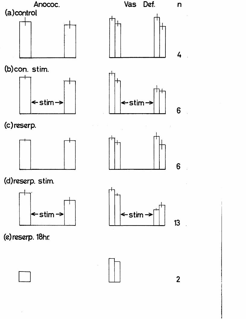
Anococ.(a)control
(b)con. stim.
(c)reserp.
(d)reserp. stim.
(e)reserp. 18hr
Vas Def. n
r +T
+i
rh+l
[Vl
rh-h
rt-.4-i
<-stim - »rfi
2
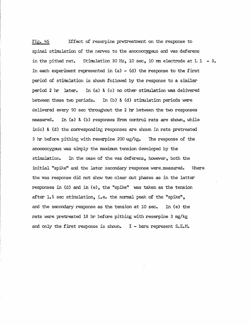
Fig. M-5 Effect of reserpine pretreatment on the response tospinal stimulation of the nerves to the anococcygeus and vas deferens in the pithed rat. Stimulation 30 Hz, 10 sec, 10 mm electrode at L 1 In each experiment represented in (a) - (d) the response to the first period of stimulation is shown followed by the response to a similar period 2 hr later. In (a) & (c) no other stimulation was delivered between these two periods. In (b) & (d) stimulation periods were delivered every 90 sec throughout the 2 hr between the two responses measured. In Ca) & (b) responses from control rats are shown, while inCc) & (d) the corresponding responses are shown in rats pretreated 3 hr before pithing with reserpine 200 ug/kg. The response of the anococcygeus was simply the maximum tension developed by the stimulation. In the case of the vas deferens, however, both the initial "spike” and the later secondary response were:, measured. Where the vas response did not show two clear cut phases as in the latter responses in (d) and in (e), the "spike” was taken as the tension after 1.5 sec stimulation, i.e. the normal peak of the "spike”, and the secondary response as the tension at 10 sec. In (e) the rats were pretreated 18 hr before pithing with reserpine 3 mg/kg and only the first response is shown. I - bars represent S.E.M.
73
"spike" component of the response to nerve stimulation was, however, influenced by reserpine in a characteristic manner. The response to the first period of nerve stimulation after reserpine, whatever time was allowed for the reserpine to act, contained a "spike" of nomal amplitude but this quickly declined in subsequent stimulation periods until after 15 - 20 min it had quite disappeared at a time when the secondary component of the response was unaffected (Fig, 45).
There thus appears to be no correlation between NA content and mechanical response in either anococcygeus or vas deferens over the range of NA content produced in these experiments despite depletion of up to 82% for anococcygeus and 6 8 % for vas deferens. Taken together with the results from Part I , which show that motor responses can still be obtained in both tissues when over 97% depletion has occurred, this indicates that only a very small proportion of the NA content is necessary in both these tissues for motor transmission to occur. The evanescence of the "spike" response in the vas deferens of rats pretreated with a moderate ( 2 0 0 pug/kg) dose of reserpine also correlates with the abolition of this response by a large (3000 pg/kg) acute dose. It is interesting that this effect of pretreatment does not fully develop until after several stimulation periods. This may indicate that there is some "pool" of transmitter responsible for the "spike" which is depleted by reserpine only after nerve stimulation or after a large dose, or that some other change occurs in the region of the neuro - muscular junction after reserpine which leads to the suppression of the mechanisms responsible for the "spike" and which only occurs after nerve stimulation or a large dose of reserpine, e.g. the build up in the area of some substance such as NA or prostaglandins which could act on either nerve or muscle to produce inhibition. This latter explanation would be in agreement with the cl - mechanism postulated in Part I •
74
Summary of effects of reserpine on the rat anococcygeus, vas deferens & heart
It has been shown that the conventional l,longM adrenergic neurones of the anococcygeus and the "short" adrenergic neurones of the vas deferens are depleted of NA by reserpine to a similar extent by various doses and at, a similar rate by a moderate ( 2 0 0 pg/kg) dose, and that this rate is slow compared with that in the cardiac sympathetic nerves. In addition, it has been demonstrated that the rate of depletion in both anococcygeus and vas deferens is decreased by abolition of inpulse traffic by pithing and increased by stimulation of the sympathetic outflow to the tissues. It is thus concluded that nerve impulse traffic may be an important factor in determining the rate of depletion of tissue NA by reserpine.
Further information arising from this study was that the NA content of the vas deferens recovered more quickly after reserpine treatment than did that of the anococcygeus and that no direct correlation was found between tissue NA content and the size of the response to motor nerve stimulation in either the anococcygeus or the vas deferens.
75
PART III - THE PROPERTIES OF THE CAT ANOCOCCYGEUS MUSCLES IN VITRO
AND A COMPARISON WITH THE RAT ANOCOCCYGEUS
The anococcygeus muscles in the cat were described as long ago as 1845 (Straus - IXirckheim, 1845) but their physiological and pharmacological properties have never been examined. Since the pharmacological properties of the anococcygeus muscles have been examined in the rat alone, it was of interest to compare the properties of this tissue in the two species.
The necessity for this study was given further impetus just after it was started when a paper appeared by Garrett, Howard & Lansdale (1972), demonstrating histochemically the presence in the cat anococcygeus muscle not only of a dense adrenergic innervation similar to that found in the rat, but also of a diffuse distribution of acetylcholinesterase throughout the muscle. It was tempting to interpret this staining as evidence of a cholinergic innervation and to associate this with a possible inhibitory innervation.
The present experiments were, therefore, designed to determine whether the cat anococcygeus had a dual motor and inhibitory innervation similar to that in the rat muscle and if so whether either component could be cholinergic. In the course of this work several responses in the cat muscle were found to differ so radically from those in the rat that a simultaneous comparison was made on muscles from the two species and this developed into a pharmacological comparison of both differences and similarities in the properties of the two species.
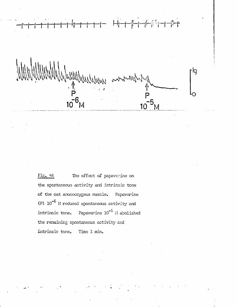
Fig. 46 The effect of papaverine on
the spontaneous activity and intrinsic toneof the cat anococcygeus muscle. Papaverine(P) 10 M reduced spontaneous activity and
-5intrinsic tone. Papaver-ine 10 ° H abolished
the remaining spontaneous activity and intrinsic tone. Time 1 min.
• Spontaneous activity and intrinsic tone in the cat anococcygeus muscle
When rat anococcygeus muscles were stretched to give an initial tension of 1 g , they gradually relaxed over a period of 2 - 5 min to a resting tension of 0.3 - 0.6 g , which was then maintained for up to 10 hr. The muscles shewed no rhythmic activity and it was not possible to lower this resting tension with papaverine or inhibitory nerve stimulation. The resting tension is, therefore, presumably sustained by connective tissue elements.Stimulation of the motor nerves or the addition of agonist drugs causes contraction in which the tension is then actively maintained by the smooth muscle elements.
In the cat anococcygeus, similar stretching to give an initial tension of 1 gwas also followed by a relaxation in 5 min to a resting tension of 0.3 -0 . 6 g but unlike that in the rat this resting tension involved an element ofactive muscular contraction. This was engendered in two ways. First, in31 out of 38 preparations, spontaneous rhythmic activity appeared with afrequency of 1 - 6 /min (mean 2,9 i 0.3/min, n = 31) and amplitude range of0.2 - 6,0 g (mean 1.1 ± 0.2 g, n = 31). This rhythmic activity steadilydeclined over the first 2 hr, a decline accelerated by regular fieldstimulation of the preparation, and usually disappeared completely. Theseoond evidence of a contribution of active muscle contraction to the resting
- 6 -5tension was the ability of papaverine (10 M - 10 M) to inhibit spontaneous activity (Fig. 46) and lower "the resting tension, and also the ability to display, in appropriate circumstances, a similar reduction in resting tension by stimulation of the inhibitory nerves. This active component of the resting tension will be referred to as "intrinsic tone" and was observed in 26 out of 30 preparations tested.
Intrinsic tone in the cat anococcygeus appeared to be myogenic in origin. It was unaltered by phentolamine (10" M) or atropine (10“ M) and was found in
tissues stored in Krebs' solution at 4° C for up to 10 days, so long as aperiod of equilibration at 36° C was allowed before applying tension.Storage for longer than 10 days reduced but did not abolish intrinsic tone.
Effect of Agonist drugs
In the course of examining the effects of several putative transmitter or transmitter - like substances on the cat anococcygeus, several were found to contract the muscle including NA, adrenaline, 5HT and indirect sympathcmimetics, and several to relax the muscle including acetylcholine, isoprenaline, prostaglandins, ATP and vasopressin. Furthermore, several drugs such as 5HT which are normally considered as direct agonists appeared to have a large indirect component to their action. This made it necessary to be able to distinguish between drugs acting directly on the smooth muscle and those acting indirectly by the release of the neuro - transmitter, NA, from theadrenergic nerves within the muscle. An effective means of making thisdistinction is to sympathectomise the muscle using 6 - hydroxydopamine (60HDA). In the case of the cat, it was not practicable to pretreat the animals with 60HDA, vhich is the method normally employed (Malmfors & Thoenen, 1971). It was, however, possible to produce a sympathectomy by exposure of the muscles to 6 CHDA in vitro as has been demonstrated in the vas deferens by Wadsworth (1973). Before considering the effects of the various agonist drugs, this 60HDA treatment is described in the following section.
60HDA in vitro treatment
The method employed was that of Wadsworth (1973) except that the highconcentration of ascorbic acid was emitted from the incubation medium since
— 6 —3all doses of ascorbic acid from 1 0 " g/ml to 1 0 ” g/ml were found to produce contraction of both the rat and cat anococcygeus muscles. The purpose of
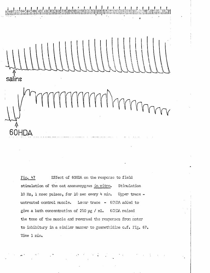
60HDA
Fig. 47 Effect of 60HDA on the response to fieldstimulation of the cat anococcygeus in vitro. Stimulation
10 Hz, 1 msec pulses, for 10 sec every 4 min. Upper trace -
untreated control muscle. La;er trace - 60HDA added to give a bath concentration of 250 pg / ml. 6CHDA raised the tone of the muscle and reversed the responses frco motor to inhibitory in a similar manner to guanethidire c.f. Fig. 67. Time 1 min.
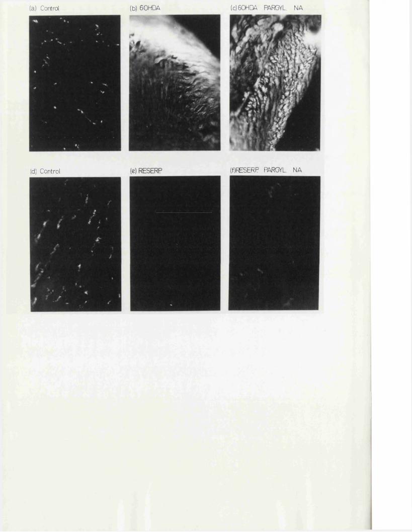
(c)60HQA PARGYL. NA(b) 60HDA(a) Control
(f)RESERP PARCEL. NA(d) Control
3 k i
j 7 y . ,
Fig. 48 Effect of pretreatment with reserpine crin vitro incubation with 60HDA on the adrenergic nerves of of the rat anococcygeus as shown by the fluorescence technique of Falck & Hillarp. Magnification x 300.
(a) Control muscle, transverse section.(b) Muscle incubated with 60HDA 250 ug/ml for 2 hr thenwashed for 2 hr. Note absence of nerves but presence of intensefluorescence within the smooth muscle cells.(c) Contralateral muscle from (b) treated similarly except
-5that following 60HDA it was incubated with pargyline 5 x 10 g/mlfar 30 min in order to inhibit MAO and then incubated with NA -410 M. As in (b), no nerves were visible.
(d) Control muscle, transverse section.(e) Muscle from rat pretreated with reserpine 3 mg/kg, 18 hr.No nerves are visible.(f) Contralateral muscle frcm (e) incubated with par gy line andNA as (c). Nerves become faintly visible once more.
This illustrates that whereas the reserpine treatment merely depletes the NA from the nerves, the 60HDA treatment destroys the nerves making it impossible to revisualise them by filling the terminals with exogenous NA.
78
including ascorbic acid was to prevent oxidation of 60HDA, but omission was found to make no difference to the sympathectomy produced.
Tissues were suspended in the organ bath in the usual way and control responses to NA and to field stimulation obtained. Solid 60HDA was then dissolved in 1 ml of Krebs* and immediately added to the bath to give a final concentration of 250 ug/ml. On addition of 60HDA to the bath, the Krebs* immediately turned bright red (this colour change was not prevented by the prior addition of ascorbic acid to the Krebs*). Both rat and cat anococcygeus muscles immediately contracted on addition of 6 GHDA to the bath and motor responses to field stimulation were reversed to inhibitory responses (Fig. 47) •Maximal contraction of the muscles was maintained for 1 hr after which time the tension gradually waned, reaching 50% of maximum after 2 hr. If the 60HDA was washed out after 1 hr, the rate of relaxation of the muscle was not significantly affected (Fig. 47). After 2 hr exposure to 60HDA, the bath j was washed out several times and the muscle left gradually to relax to the initial baseline. After a further 2 hr, the muscles were tested for their sensitivity to field stimulation and to agonist drugs.
At this time i.e. 4 hr after the beginning of exposure to 60HDA, noadrenergic nerves could be detected in the tissue by the Falck technique(Fig. 48). These nerves did not reappear, in either species, when the tissues
—4were subsequently exposed to a high concentration of NA (10 M) after... -5inhibition of mono - amine oxidase by pargyline (5 x 10 g/ml, 30 min), a
treatment which produced the reappearance of adrenergic nerve endings after depletion of the tissue NA content by reserpine (1 mg/kg, i.p., 24 hr)(Fig. 48). This suggests that the sympathectomy by 60HDA treatment inrvitro is produced by destruction of the nerve terminals or at least by depletion of neuronal NA coupled with complete blockade of the neuronal NA uptake mechanism.
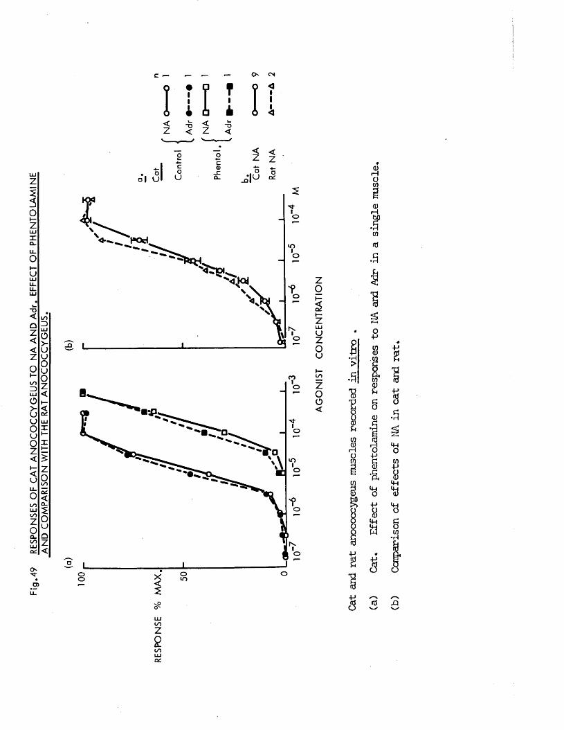
49 R
ESPO
NSE
S OF
CA
T A
NO
CO
CC
YG
EU
S
TO NA
AN
D A
dr.
EFFE
CT
OF
PH
EN
TO
LA
MIN
E
AND
CO
MP
AR
ISO
N
WIT
H TH
E RA
T A
NO
CO
CC
YG
EU
S.
m i iO- CN
<
<Z
~o<
. o ol VJcoU
<Z4 -oQ1U
<zoo'
o
U-)
o
o
o_Q
O
O
lOo
o
oo
ooo
>9
zo
uzou
CO ill•H>•3•8%
8to<u
r Ha10
rH8
COid.3
i/iZ
2ooLUoc
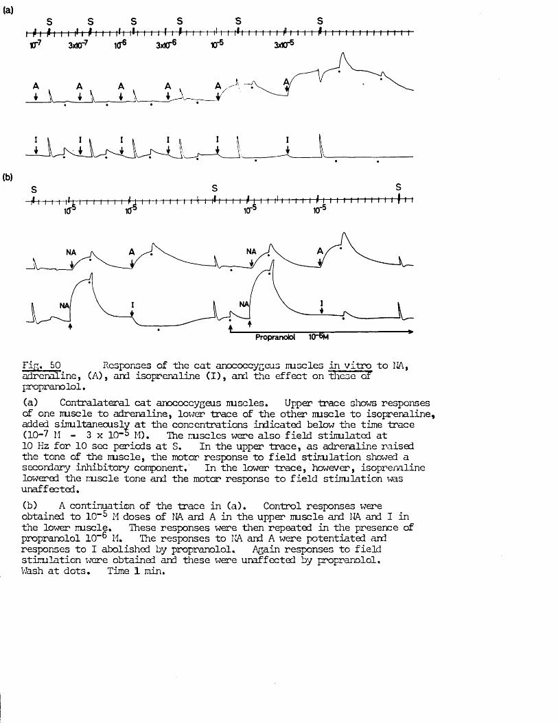
j !
(b)S Si< i i t t ft i i i i i i i i f j i i i i I t i i *i i 1 * 1 i i i i* j. i i i11 t~i i i f i i I i tt i i i n r t-i-r............. -i(f5 kr icr 10"5
Si i i i t f-
NA
NANA
Propranolol 10“6m
Fir,* 50 Responses of the cat anococcygeus uuscles in vitro to NA,adrenaline, (A), and isoprenaline Cl), and the affect on these of propranolol.(a) Contralateral cat anococcygeus nu sales. Upper trace shows responses of one iruscle to adrenaline, lower trace of the other nu sc le to isoprenaline, added simultaneously at the concentrations indicated below the time trace (10-7 M - 3 x 10⁻⁵ M), phe nuscles were also field stimulated at10 Hz for 10 sec periods at S. In the upper trace, as adrenaline raised the tone of the nuscle, the motor response to field stinulation showed a secondary inhibitory component. In the lower trace, however, isoprenaline lowered the iruscle tone and the motor response to field stimulation was unaffected.(b) A continuation of the trace in (a). Control responses were obtained to 10”5 M doses of NA and A in the upper muscle and NA and I in the lower muscle. These responses were then repeated in the presence of propranolol 10"® M. The responses to HA and A were potentiated and responses to I abolished by propranolol. Again responses to field stimulation were obtained and these were unaffected by propranolol.Wash at dots. Time 1 min.
After this sympathectomy, it was possible to demonstrate whether drugs were acting directly on the muscle or as indirect sympathomimetics, since the latter were no longer effective.
In the case of the rat it was possible to compare these in vitro effects of 60HDA with the sympathectomy produced by pretreatment in vivo with, the regimen; day 1 - 2 x 50 mg/kg i.p., day 4 - 2 x 100_mg/kg i.p.,day 5 - tissues used.
The effects of 60HDA on responses to field stimulation and to drugs are discussed in the appropriate sections.
Drugs causing contraction in both species
Direct sympathomimetics Both NA and Adr produced dose - related contractionsof the cat anococcygeus. Their potency and the effect of antagonists wereexamined by constructing dose / response curves (Fig. 49). Adr and NA were
— 6approximately equipotent (Fig. 49a). Fhentolamine (10” M) produced an equal and parallel shift to the right of both curves equal to a twelvefold increase in dose (Fig. 49a). The sensitivity of the oat and rat anococcygeusto NA was almost identical (Fig. 49b). Responses in the cat muscle to NA
—7 — 6were unaffected by methysergide (2 x 10” M) (Fig. 51) or atropine (10” M).Responses to Adr and to a lesser extent NA were potentiated by
_ 6propranolol (10” M) (Fig. 50). Isoprenaline did not produce contraction ofN -7the cat anococcygeus at any dose but at all doses tested from 1 0 M to
3 x 10” 5 M produced relaxations which were abolished by propranolol (Fig. 50).
Responses in the cat anococcygeus to NA were potentiated by the presence of -5cocaine (3 x 10 M) in the organ bath. The dose / response curve to NA
was shifted to the left (Fig. 51) and responses were prolonged, returning to baseline more slowly after washout (Fig. 53).
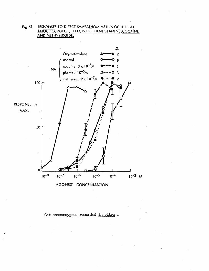
F ig .51 RESPONSES TO DIRECT SYMPATHOMIMETICS OF THE CAT
RESPONSE
M A X .
A N O C O C C Y G E U S . EFFECTS O F P H E N T O L A M IN E . C O C A IN E A N D M E TH Y S ER G ID E .
N A
10 -8
O x y m e ta zo lin e
( control
cocaine 3 x 10“^ M
phentol- 10”^ M
1 0 0
%
50
0
10 - 71-0 ' 10 -5 1 0
- 4 10"3 M
A G O N IS T C O N C E N T R A T IO N
Cat anococcygeus recorded in vitro *
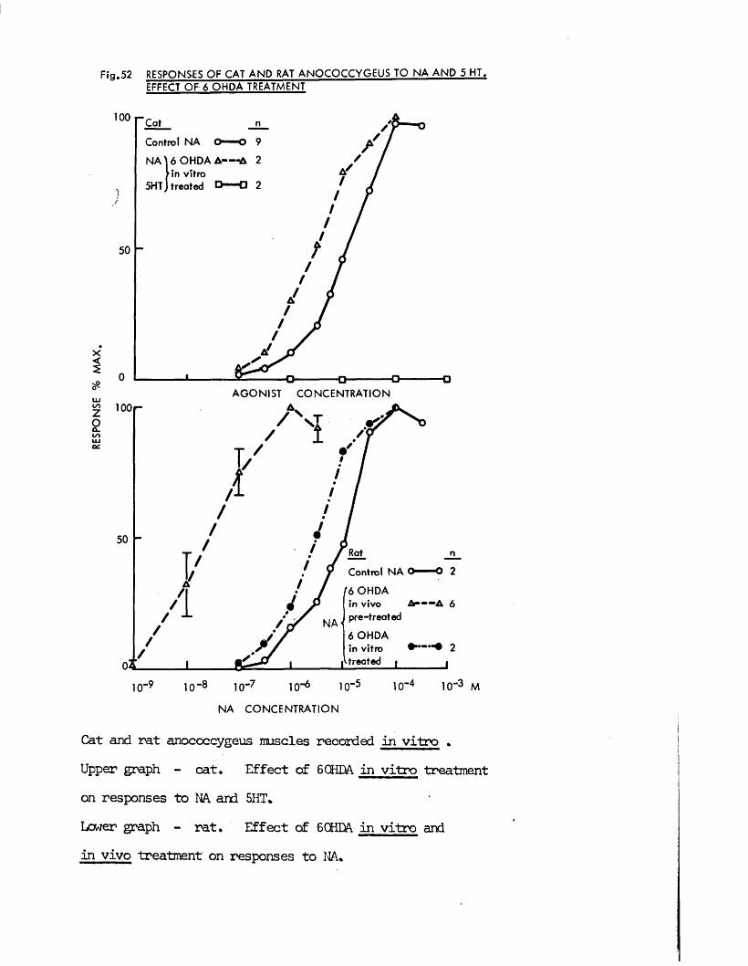
Fig .52 RESPONSES OF CAT AND RAT ANOCOCCYGEUS TO NA AND 5 HT. EFFECT OF 6 OHDA TREATMENT
1 0 0 Cat n
Control NA O O 9
N A U O H D A A — -A 2 tin vitro
5HTJ treated O— Cl 2
AGONIST CONCENTRATION£ 100 r~Zoa.ooLUOC
Control NA O ... 2
f6 OHDAin vivo A 6
J pre-treated
6 OHDAin vitro 2
|V treated | J
1 0 -9 10-8 10-7 10-6 10“5
NA CONCENTRATION
1CT4 10“3 M
Cat and rat anococcygeus muscles recorded in vitro .Upper graph - cat. Effect of 6 OHDA in vitro treatmenton responses to NA and 5HT.Lcwer graph - rat. Effect of 6 CHEA in vitro andin vivo treatment on responses to NA.
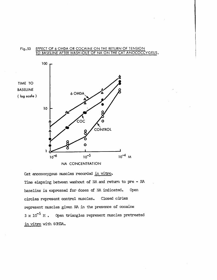
F ig .53 EFFECT O F 6 O H D A OR C O C A IN E O N THE RETURN OF T E N S IO NTO BASELINE AFTER W A S H -O U T OF N A O N THE CAT A N O C O C C Y G E U S .
TIM E TO
BASELINE
( log scale )
100 r-
6 O H D A
C O C
C O N TR O L
N A C O N C E N T R A T IO N
Cat anococcygeus nuscles recorded in vitro >Time elapsing between washout of NA and return to pre - NA baseline is expressed for doses of NA indicated. Open circles represent control muscles. Closed cirles represent muscles given NA in the presence of cocaine 3 x 10” 5 M , Open triangles represent muscles pretreated in vitro with 6 CHDA,
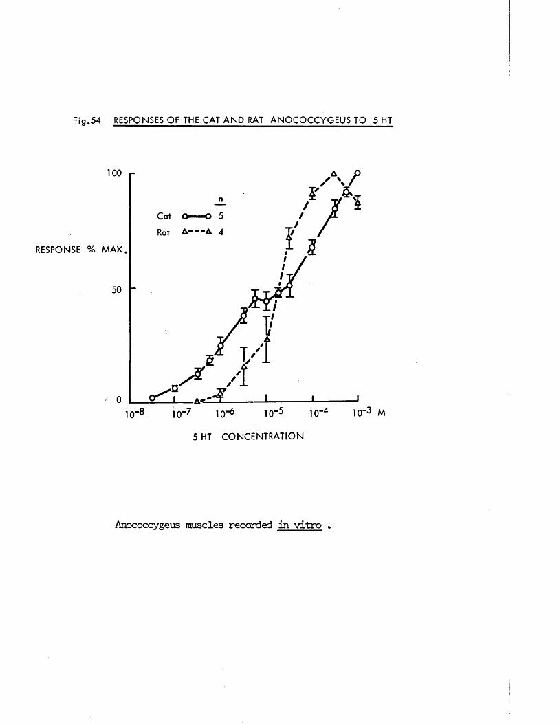
F ig .54 RESPONSES OF THE CAT A N D RAT ANOCO CCYG EUS TO 5 HT
RESPONSE
100
Cat
Rat 4
M A X .
5 HT C O N C E N T R A T IO N
Anococcygeus muscles recorded in vitro *
After treatment of the muscle for 2 hr with 6 OHDA (250 pg/ml), responses to NA were potentiated in a manner similar to that after cocaine. The dose / response curve to NA was shifted to the left (Fig. 52a) and the response was prolonged after NA washout (Fig. 53). This 60HDA treatment in vitroproduced a sympathectomy of the anococcygeus similar to that found after pretreatment in vivo but did not produce as great an increase in sensitivity to NA as the latter, as shown for the rat in Fig. 52b.
The cat anococcygeus was approximately x 200 irore sensitive to oxymetazoline, an oc- receptor agonist which is not taken up into adrenergic nerves (Mujic & Von Rossum, 1965), than to NA (Fig. 51). Together with the effects of cocaine and 6 OHDA treatment, this suggests a role for neuronal uptake in determining the response to NA in this tissue.
The cat anococcygeus thus contracts in the presence of oL - adrenergic agonists. This contraction is potentiated if the agonist is prevented from being taken up into adrenergic nerves and, in the case of agonists which also have (2 - adrenergic effects, is potentiated by blockade of ft - receptors. These properties are similar to those of the rat anococcygeus except that in the rat no ft - receptor effects were found (Gillespie, 1972).
5 - hydroxytryptamine (5HT) produced dose - related contractions of the catanococcygeus. The cat anococcygeus was more sensitive to low doses of 5HTthan was the rat anococcygeus (Fig. 54) and the log dose / response curve to
-55HT of the former showed an inflection at 10 M. Beyond this inflection,-3the response to 5HT increased with increasing dose up to 10 H, which was the
-3highest dose tested. Even the response to 10 M was, however, still lowerthan the maximum response to NA (Fig. 55) so it is likely that the theoretical
-3maximum response to 5HT could not be produced until doses higher than 10 M.
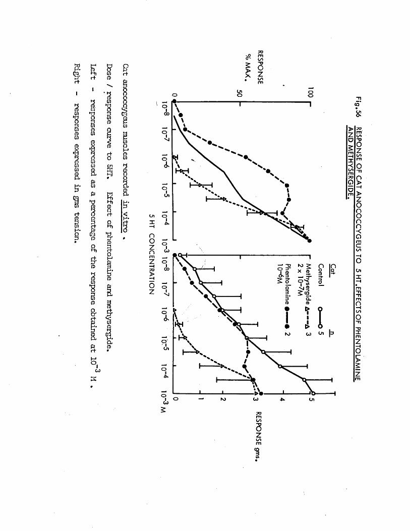
Fig
.56 R
ESPON
SE OF
CAT A
NO
CO
CC
YG
EU
S
TO 5
HT.E
FFEC
TSO
F P
HE
NT
OL
AM
INE
AND
ME
TH
YS
ER
GID
E.
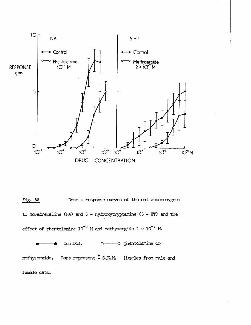
10NA
Control
Phentolamine 10“6 MRESPONSE
qms.
5 HT
Control
Methyserqide 2 x 1CT7 M
DRUG CONCENTRATION
Fig. 55 Dose - response curves of the cat anococcygeus
to Noradrenaline (NA) and 5 - hydroxytryptamine (5 - HT) and the
— 6 -7effect of phentolamine lCf M and methysergide 2 x 10“ M.
&-----& Control* o— 0 phentolamine or
methysergide. Bars represent - S.E.M. Muscles from male and
female cats.
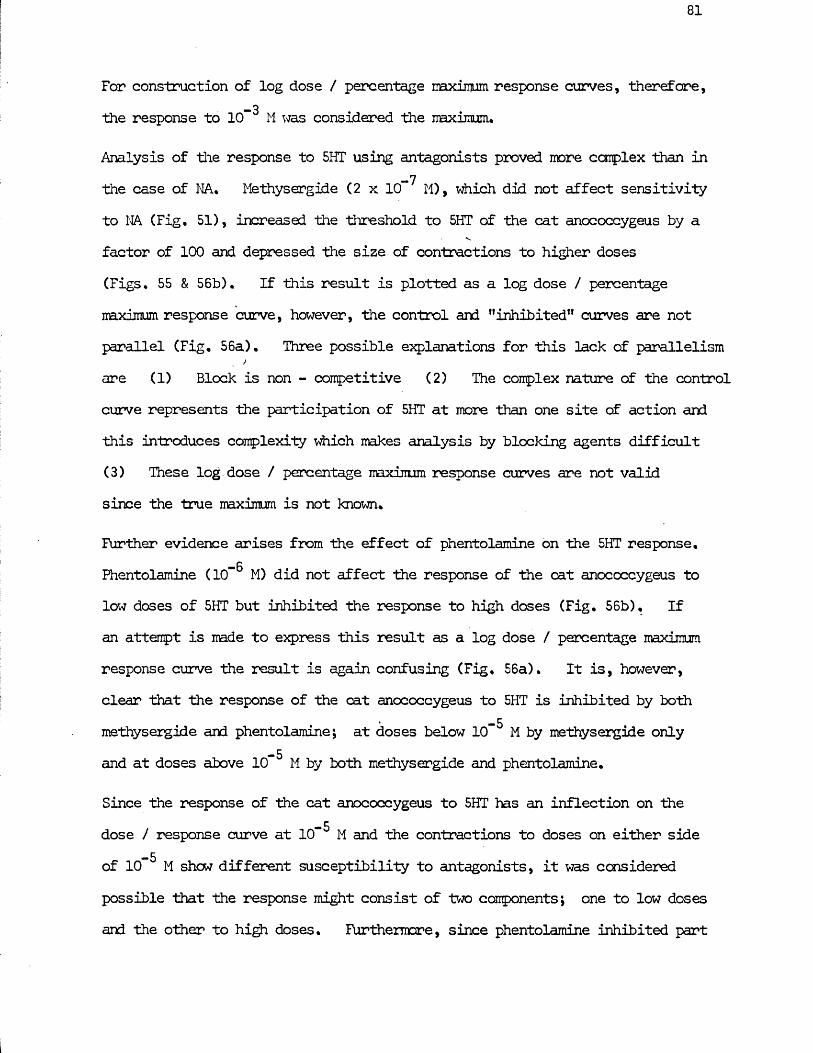
For construction of log dose / percentage maximum response curves, therefore,-3the response to 1 0 M was considered the naximum.
Analysis of the response to 5HT using antagonists proved more complex than in_7the case of NA. Methysergide (2 x 10 M), which did not affect sensitivity
to NA (Fig. 51), increased the threshold to 5HT of the cat anococcygeus by afactor of 1 0 0 and depressed the size of contractions to higher doses (Figs. 55 & 56b). If this result is plotted as a log dose / percentage mximum response curve, however, the control and "inhibited” curves are not parallel (Fig. 56a). Three possible explanations for this lack of parallelism
jare (1) Block is non - competitive (2) The complex nature of the controlcurve represents the participation of 5HT at more than one site of action andthis introduces complexity which makes analysis by blocking agents difficult (3) These log-dose / percentage maximum response curves are not valid since the true maximum is not known.
Further evidence arises from the effect of phentolamine on the 5HT response.—6Phentolamine (10” M) did not affect the response of the cat anococcygeus to
low doses of 5HT but inhibited the response to high doses (Fig. 56b), Ifan attempt is made to express this result as a log dose / percentage maximumresponse curve the result is again confusing (Fig. 56a). It is, however,clear that the response of the cat anococcygeus to 5HT is inhibited by both
-5methysergide and phentolamine; at doses below 10 M by methysergide only-5and at doses above 10 M by both methysergide and phentolamine.
Since the response of the cat anococcygeus to 5HT has an inflection on the-5dose / response curve at 10 M and the contractions to doses on either side
-5of 10 M shew different susceptibility to antagonists, it was considered possible that the response might consist of two components; one to low doses and the other to high doses. Furthermore, since phentolamine inhibited part
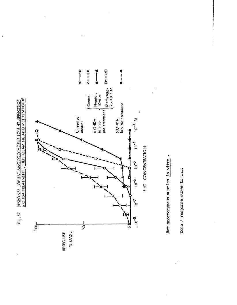
Fig
.57
RES
PON
SE
OF
RAT
AN
OC
OC
CY
GE
US
TO
5 H
T.E
FF
EC
TS
OF
6 O
HDA
TREA
TMEN
T .P
HE
NTO
LAM
INE
AN
D M
ETH
YS
ER
GID
E
<II< 1
?IA
— -S Sio -c x oc -o —<u • % x_C O
Q - ■— ^ CM
"DCOO
Q_O
O
LOOI-
o
oI—
COo
oo oLU
Zo<C£
uzoxu->
CO<ur—Iato
a
&88
LOO+-*(U0 <u18ua)
ZoQ_oo
<5
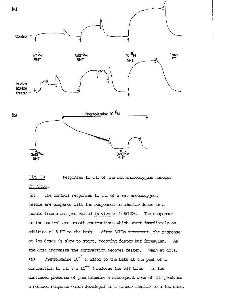
(a)
Control
1 0"^5HT
3adO-6M5HT
in vivo 60HDA treated
1min5HT
(b) Phentolamine 1 0"^
3x10⁵HT
Fig. 58 Responses to 5HT of the rat anococcygeus musclesin vitro.(a) The control responses to 5HT of a rat anococcygeusiruscle are compared with the responses to similar doses in amuscle from a rat pretreated in vivo .with 6 OHM* The responsesin the control are smooth contractions which start immediately onaddition of 5 HT to the bath* After 60HDA treatment, the responseat low doses is slow to start, becoming faster but irregular* Asthe dose increases the contraction becomes faster. Wash at dots.
— 6(b) Phentolamine 10*“ H added to the bath at the peak of a contraction to 5HT 3 x 10 M reduces the 5HT tone. In the continued presence of phentolamine a subsequent dose of 5HT produced a reduced response which developed in a manner similar to a low dose.
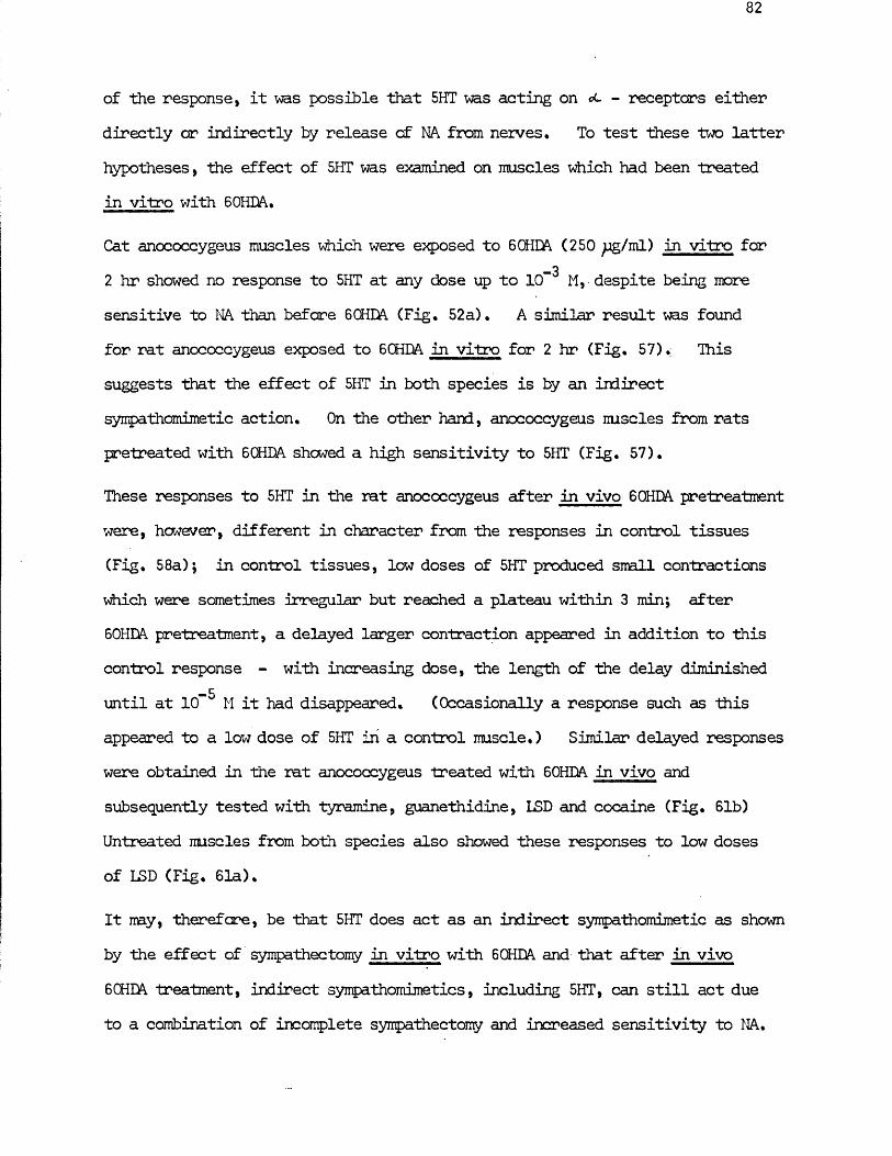
of the response, it was possible that 5HT was acting on - receptors either directly or indirectly by release of NA from nerves. To test these two latter hypotheses, the effect of 5HT was examined on muscles which had been treated in vitro with 6 OHDA.
Cat anococcygeus muscles which were exposed to 6 OHDA (250 pg/mL) in vitro for-32 hr showed no response to 5HT at any dose up to 10 M, despite being more
sensitive to NA than before 6 CHDA (Fig. 52a). A similar result ms found for rat anococcygeus exposed to 6 CHDA in vitro for 2 hr (Fig. 57). This suggests that the effect of 5HT in both species is by an indirect syirpathomimetic action. On the other hand, anococcygeus muscles from rats pretreated with 60HDA showed a high sensitivity to 5HT (Fig. 57).
These responses to 5HT in the rat anococcygeus after in vivo 60HDA pretreatmentwere, however, different in character from the responses in control tissues(Fig. 58a); in control tissues, low doses of 5HT produced small contractionswhich were sometimes irregular but reached a plateau within 3 min; after60HDA pretreatment, a delayed larger contraction appeared in addition to thiscontrol response - with increasing dose, the length of the delay diminished
-5until at 10 M it had disappeared. (Occasionally a response such as this appeared to a low dose of 5HT in a control muscle.) Similar delayed responses were obtained in the rat anococcygeus treated with 60HDA in vivo and subsequently tested with tyramine, guanethidine, LSD and cocaine (Fig. 61b) Untreated muscles from both species also showed these responses to low doses of LSD (Fig. 61a).
It my, therefore, be that 5HT does act as an indirect sympathomimetic as shown by the effect of sympathectomy in vitro with 6 OHDA and that after in vivo 6 CHDA treatment, indirect sympathomimetics, including 5HT, can still act due to a combination of incomplete sympathectomy and increased sensitivity to NA.
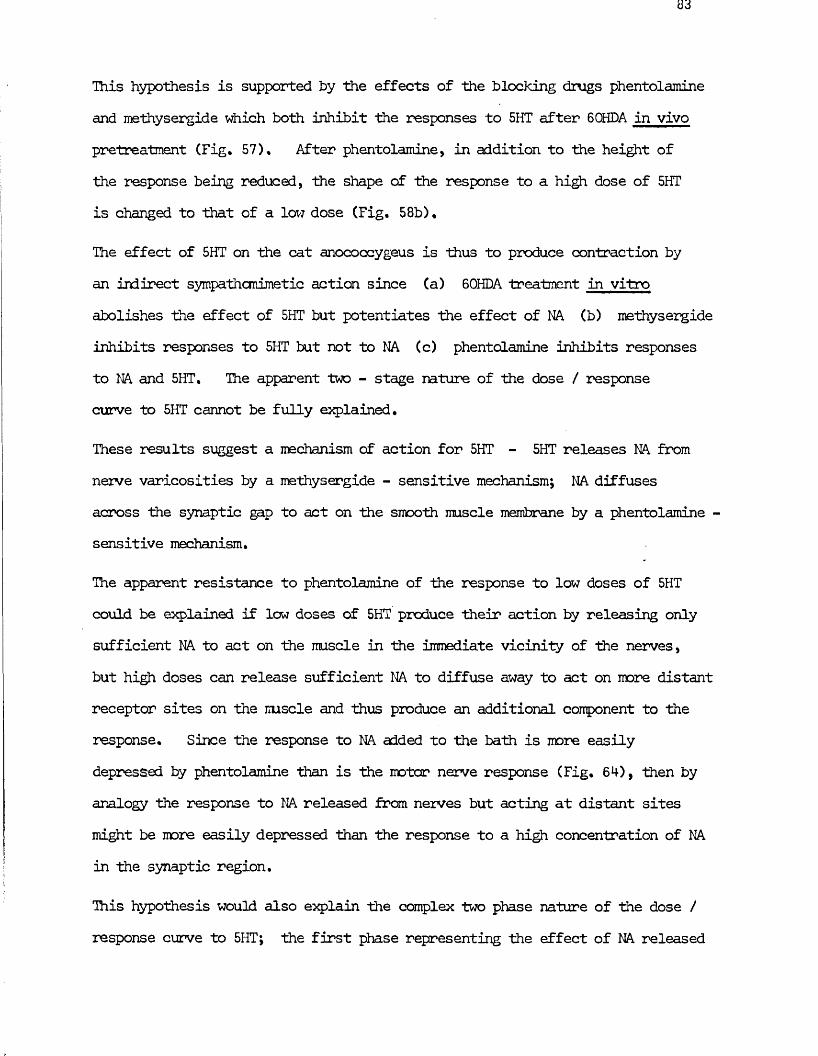
This hypothesis is supported by the effects of the blocking drugs phentolamine and methysergide which both inhibit the responses to 5HT after 60HDA in vivo pretreatment (Fig. 57), After phentolamine, in addition to the height of the response being reduced, the shape of the response to a high dose of 5HT is changed to that of a low dose (Fig. 58b) •
The effect of 5HT on the cat anococcygeus is thus to produce contraction by an indirect sympathomimetic action since (a) 6 OHDA treatment in vitroabolishes the effect of 5HT but potentiates the effect of NA (b) methysergide inhibits responses to 5HT but not to NA (c) phentolamine inhibits responses to NA and 5HT, The apparent two - stage nature of the dose / response curve to 5HT cannot be fully explained.
These results suggest a mechanism of action for 5HT - 5HT releases NA from nerve varicosities by a methysergide - sensitive mechanism; NA diffuses across the synaptic gap to act on the smooth muscle membrane by a phentolamine - sensitive mechanism.
The apparent resistance to phentolamine of the response to low doses of 5HT could be explained if low doses of 5HT produce their action by releasing only sufficient NA to act on the muscle in the immediate vicinity of the nerves, but high doses can release sufficient NA to diffuse away to act on more distant receptor sites on the mscle and thus produce an additional component to the response. Since the response to NA added to the bath is more easily depressed by phentolamine than is the motor nerve response (Fig. 64), then by analogy the response to NA released from nerves but acting at distant sites might be more easily depressed than the response to a high concentration of NA in the synaptic region.
This hypothesis would also explain the complex two phase nature of the dose / response curve to 5HT; the first phase representing the effect of NA released
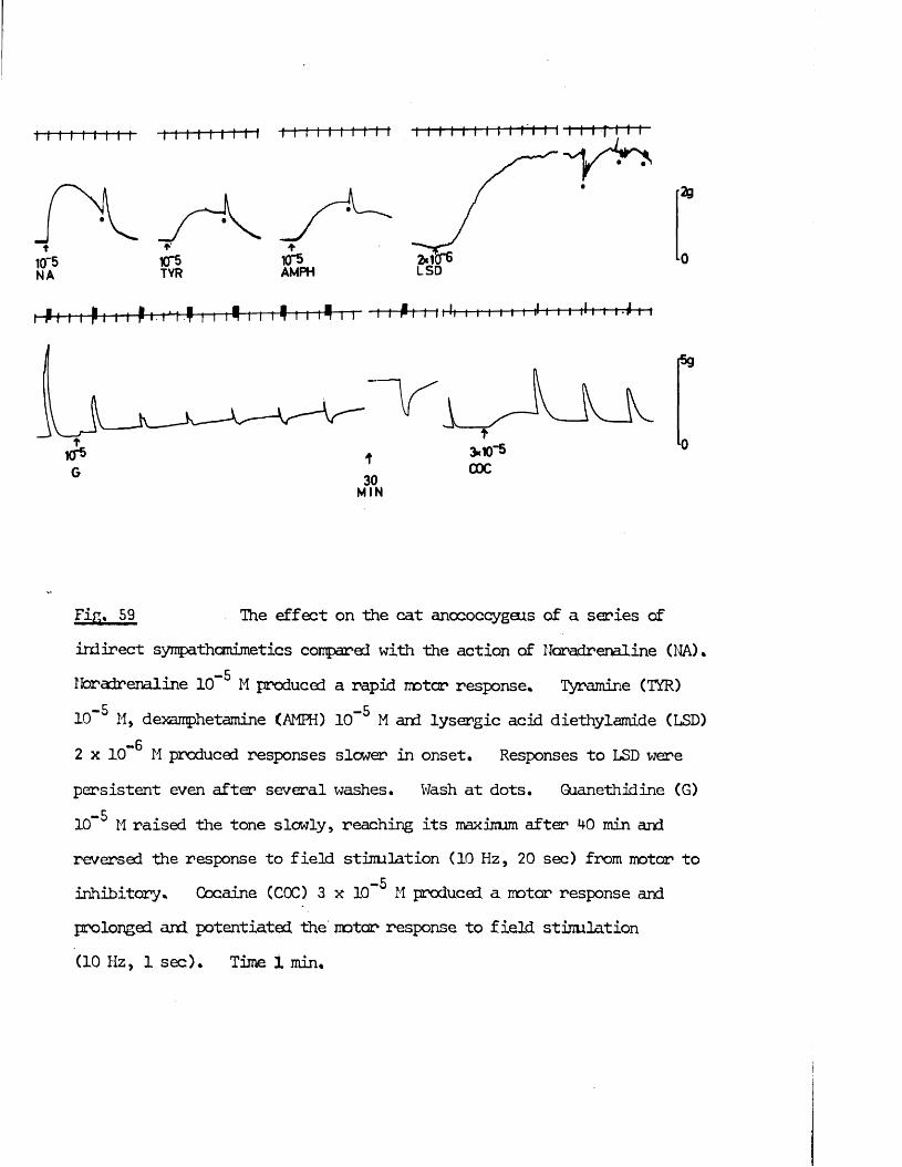
t10"5NA
*•KT5TYR
♦KT5AMPH
2g
P I i t-p t i i P i■ t*i I i i i j i i i ^ i i 1 r~t i i Pi i I*-*ii r i i i f |l I I I |l t f 1-T-P I 1
1CP
MIN
fa
Fig. 59 The effect on the cat anococcygeus of a series ofindirect sympathamimetics compared with the action of Noradrenaline (NA).
-5Noradrenaline 10 M produced a rapid motor response* Tyramine (TYR)-5 —510 M, dexamphetamine (AMFH) 10 M and lysergic acid diethylamide (LSD)
2 x 10** M produced responses slower in onset. Responses to LSD werepersistent even after several washes. Wash at dots. Guanethidine (G) -5
1 0 M raised the tone slowly, reaching its maximum after HO min andreversed the response to field stimulation (10 Hz, 20 sec) from motor to
-5inhibitory. Cocaine (C0C) 3 x 10 M produced a motor response and prolonged and potentiated the motor response to field, stimulation (10 Hz, 1 sec). Time i min.
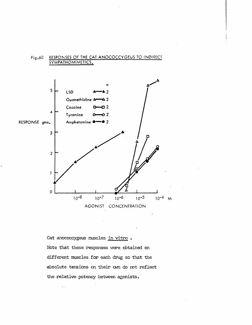
F ig .60 RESPONSES O F THE CAT A N O C O C C Y G E U S TO INDIRECT S YM P A TH O M IM E T IC S .
RESPONSE
5 ▲ A 2LSD
G uaneth id ine A*
Cocaine
Tyramine
Amphetamine # " • # 2
□ 24
gms.
3
2
0
A G O N IS T C O N C E N T R A T IO N
Cat anococcygeus muscles in vitro *Note that these responses were obtained on different muscles for each drug so that the absolute tensions on their cwn do not reflect the relative potency between agonists.
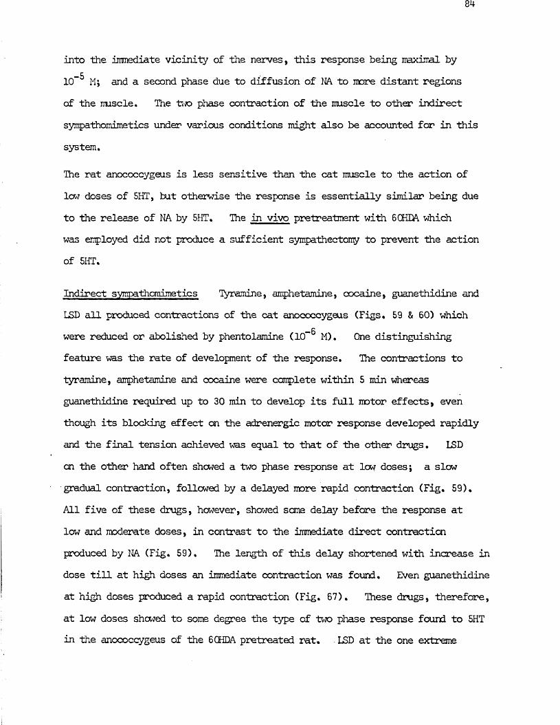
into the immediate vicinity of the nerves, this response being maximal by lCf M; and a second phase due to diffusion of NA to more distant regions of the muscle. The two phase contraction of the muscle to other indirect sympathomimetics under various conditions might also be accounted for in this system.
The rat anococcygeus is less sensitive than the cat muscle to Hie action of low doses of 5HT, but otherwise the response is essentially similar being due to the release of NA by 5HT. The in vivo pretreatment with 6 CHDA which was employed did not produce a sufficient sympathectomy to prevent the action of 5HT,
Indirect sympathomimetics Tyramine, amphetamine, cocaine, guanethidine and LSD all produced contractions of the cat anococcygeus (Figs, 59 & 60) which
— Bwere reduced or abolished by phentolamine (10" M). One distinguishing feature was the rate of development of the response. The contractions to tyramine, amphetamine and cocaine were complete within 5 min whereas guanethidine required up to 30 min to develop its full motor effects, even though its blocking effect on the adrenergic motor response developed rapidly and the final tension achieved was equal to that of the other drugs, LSD on the other hand often shewed a two phase response at low doses; a slow gradual contraction, followed by a delayed more rapid contraction (Fig, 59),All five of these drugs, however, showed same delay before the response at low and moderate doses, in contrast to the immediate direct contraction produced by NA (Fig, 59), The length of this delay shortened with increase in dose till at high doses an immediate contraction was found. Even guanethidine at high doses produced a rapid contraction (Fig. 67). These drugs, therefore, at low doses showed to some degree the type of two phase response found to 5HT in the anococcygeus of the 6 CHDA pretreated rat. LSD at the one extreme
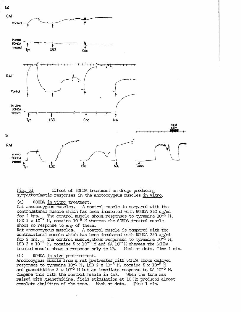
Control — i '— t— tt t
in vitro60HDA A • — J----------- J L
,reQted Tyr LSD c L
RAT
Control
in vitro 60HDA treated J
Tyr LSD Coc NAfieldstim
t I i i i i i i i 11— t— i-i— i(b)
RAT
in vivo 60HDA treated tGoanCoc
Fig. 61 Effect of 601 DA treatment on drugs producingsympathomimetic responses in the anococcygeus muscles in vitro.Ca) 6 OHDA in vitro treatment.Cat anococcygeus muscles. A control muscle is compared with the contralateral muscle which has been incubated with 6 OHDA 250 ug/ml for 2 hr s. R The control muscle shows responses to tyramine 1 0 " 5 M,LSD 2 x 10 M, cocaine 1 0 " M whereas the 6 OHDA treated muscleshows no response to any of these.Fat anococcygeus muscles. A control muscle is compared with the contralateral muscle which has been incubated with 60IDA 250 ug/ml for 2 hrs. „ The control mu sc le shows responses to tyramine 1 0 " M,LSD 2 x 10 M, cocaine 5 x 10” M and NA 10“ M whereas the 60HDAtreated muscle shows a response only to NA. Wash at dots. Time 1 min.(b) 6 OHDA in vivo pretreatment.Anococcygeus muscle from a rat pretreated with 6 OHDA shows delayed responses to tyramine 10”5 M, LSD 2 x 10~® M, cocaine 5 x 1 0 * M and guanethidine 3 x 10“ M tut an immediate response to NA 10" M. Compare this with the control muscle in (a). When the tone was raised with guanethidine, field stimulation at 10 Hz produced almost complete abolition of the tone. Wash at dots. ine 1 min.
showed a slow contraction followed after a delay of up to several minutes by a rapid contraction, tyramine, amphetamine and cocaine showed a short period of slow contraction foil wed by a mere rapid contraction and guanethidine showed only a slow, steady contraction.
After washout, the effects of these drugs were slow to disappear compared with the rapid return to baseline after washing out NA. The effect of LSD was particularly persistent, taking up to 4-5 min to disappear, but the effects of the others were also slow to wear off; in decreasing order of persistence - LSD, guanethidine, cocaine, amphetamine and tyramine.
In the rat anococcygeus, tyramine, amphetamine, guanethidine, LSD and cocaine all produced contraction in doses similar to those for the cat. Responses in the rat were in general rapid monophasic contractions apart from those to LSD and cocaine which showed an initial slow followed by a later more rapid contraction.
In both species, after 60HDA (250 pg/ml, 2 hr) treatment in vitro, the responsesto tyramine, guanethidine, LSD and cocaine were completely absent (Fig. 61a). J
This suggests that each of these drugs is acting by an indirect sympathomimeticaction. In the anococcygeus muscles from rats pretreated in vivo with 6 CHDA,responses could be obtained to high doses of tyramine, guanethidine, cocaineand LSD. Unfortunately, dose / response curves were not obtained so thesensitivity change cannot be quantified. These responses had a characteristicshape; a slow gradual contraction followed by a mere rapid contraction(Fig. 61b). This shape is similar to that of the control response to low dosesof LSD and to the response to 5HT after 60HDA in vivo pretreatment. Theseresponses to tyramine, guanethidine, cocaine, LSD and 5HT, remaining after
—K6 CHDA in vivo pretreatment, were abolished or reduced by phentolamine (10" M).
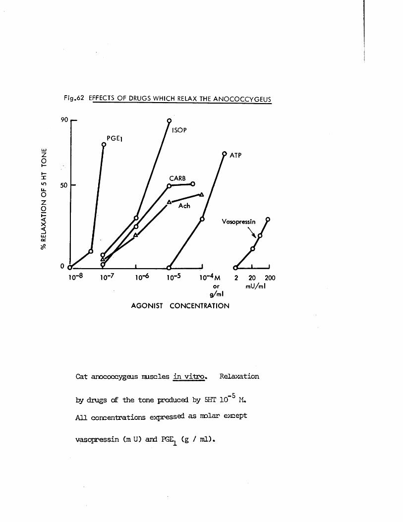
% R
ELA
XA
TIO
N
OF
5 HT
T
ON
E
Fig .62 EFFECTS OF DRUGS WHICH RELAX THE ANOCOCCYGEUS
ISOPPGE]
ATP
CARB
Ach
Vasopressin
10"6 10~5 1 (T 4 M 2 20 200or m U /m l
g /m l
A G O N IS T C O N C E N T R A T IO N
Cat anococcygeus muscles in vitro % Relaxation
by drugs of the tone produced, by 5HT 10” M.All concentrations expressed as molar except
vasopressin (m U) and PGE (g / ml).
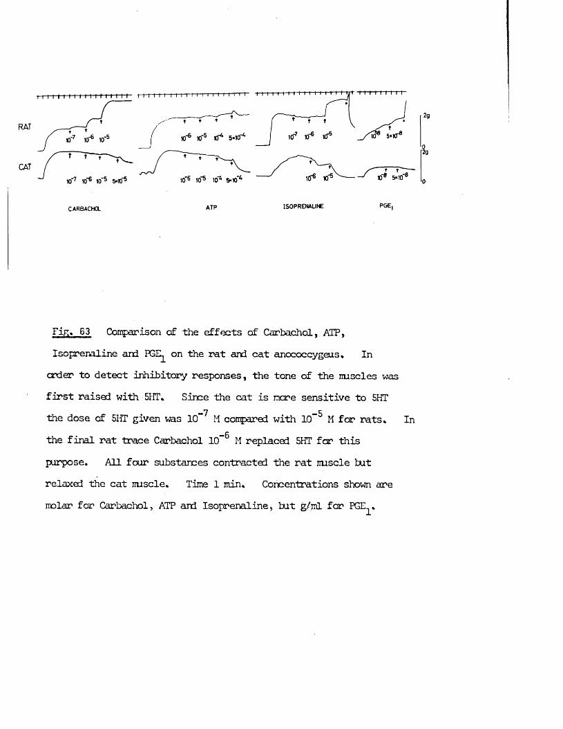
t t I I I I I I I M I f I I I I f
RAT
CAT
2g
10° 5*1010° 1010° 103 10H 5-10
CARBACHa ATP LSOPRENALINE PGE,
Fig, 63 Comparison of the effects of Carbachol, ATP,Isoprenaline and PGE on the rat and cat anococcygeus* In order to detect inhibitory responses, the tone of the iruscles wasfirst raised with 5HT, Since the cat is more sensitive to 5HT
-7 -Athe dose of 5HT given was 10 M compared with 10 M for rats* In— fithe final rat trace Carbachol 10~ M replaced 5HT for this
purpose. All four substances contracted the rat muscle but relaxed the cat muscle* Time 1 min* Concentrations shown are molar for Carbachol, ATP and Isoprenaline, tut g/ml for PGE *
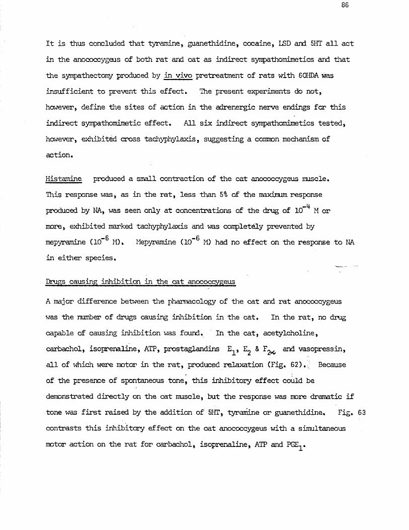
86
It is thus concluded that tyramine, guanethidine, cocaine, LSD and 5HT all act in the anococcygeus of both rat and cat as indirect synpathomimetics and that the sympathectomy produced by in vivo pretreatment of rats with 60HDA was insufficient to prevent this effect. The present experiments do not, however, define the sites of action in the adrenergic nerve endings for this indirect syirpathomimetic effect. All six indirect sympathomimetics tested, however, exhibited cross tachyphylaxis, suggesting a common mechanism of action.
Histamine produced a small contraction of the cat anococcygeus muscle.This response was, as in the rat, less than 5% of the maximum response
„llproduced by NA, was seen only at concentrations of the drug of 10 M ormore, exhibited narked tachyphylaxis and was completely prevented by
—6 - 6mepyramine (10” M). Mepyramine (10~ M) had no effect on the response to NAin either species.
Drugs causing inhibition in the cat anococcygeus
A major difference between the pharmacology of the cat and rat anococcygeus was the number of drugs causing inhibition in the cat. In the rat, no drug capable of causing inhibition was found. In the cat, acetylcholine, carbachol, isoprenaline, ATP, prostaglandins E , and vasopressin,all of which were motor in the rat, produced relaxation (Fig. 62). Because of the presence of spontaneous tone, this inhibitory effect could be demonstrated directly on Hie cat muscle, but the response was more dramatic if tone was first raised by the addition of 5HT, tyramine or guanethidine. Fig. 63 contrasts this inhibitory effect on the cat anococcygeus with a simultaneous motor action on the rat for carbachol, isoprenaline, ATP and PGE .
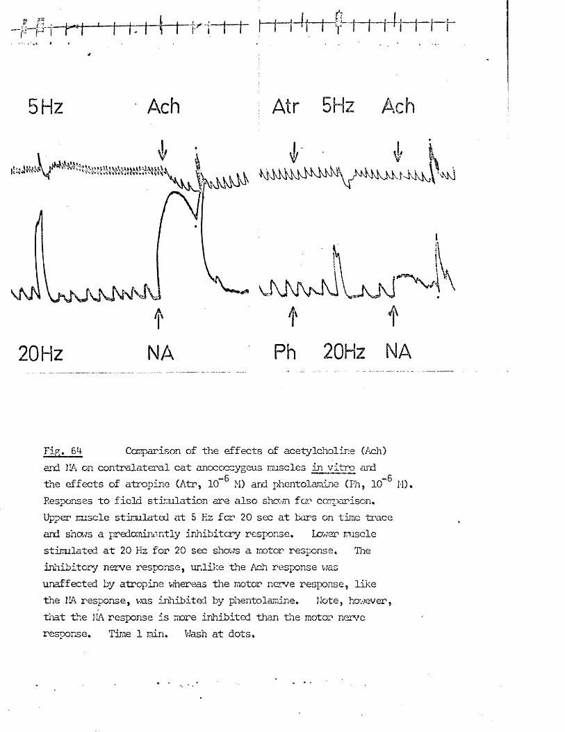
a — u 4 — t-7 - r 1 i I I 1 - 1 i - f - K t - 1 ~ f ~ t
5 5H:
20 Hz
w
A
Ph 20Hz
Fig. 6M- Canparison of the effects of acetylcholine (Ach)and HA on contralateral cat anococcygeus muscles in vitro and
— 0 "™ 1 1 » ^ 0
the effects of atropine (Atr, 10” M) ard phentolamine (Ph, 10” M).Responses to field stir Halation are also shown fan ccirparison.Upper muscle stimulated at 5 Kz for 20 sec at bars on time traceand shews a predominantly inhibitory response. Lcwer musclestimulated at 20 Hz for 20 sec shows a motor response. Theinhibitory nerve response, unlike the Ach response wasunaffected by atropine whereas the motor nerve response, likethe HA response, was inhibited by phentolamine. Note, however,that the HA response is more inhibited than the motor nerveresponse. Time 1 min. Wash at dots.
RAT
CAT
10*75HT
1 0 'b
5HT
HT?ACH
10"6ACH
t
tNEO
Kf5 10-7 5HT ACH
ACH ACH5HT
-7Fin, 65 Comparison of the effects of neostigmine (NEO) 10 M on the response of the rat and cat anococcygeus to acetylcholine(Ach). The tone of the nuscles was first raised with 5 -
— 6 —7hydroxytryptamine 10~ M for rat, 10~ M for cat. Neostigmine-7enhanced the motor response to acetylcholine 1 0 M in the rat— 6and the inhibitory response to acetylcholine 1 0 ” M in the cat
but did not affect the response to 5 - hydroxytryptamine in either species. Wash at dots.
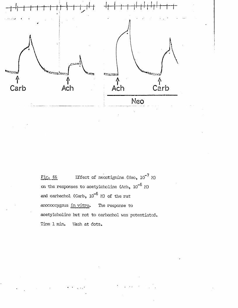
£on the responses to acetylcholine (Ach, 10“ M) and carbachol (Carb, 10“ M) of the rat anococcygeus in vitro. The response to acetylcholine but not to carbachol was potentiated* Time 1 min* Wash at dots*
87
Muscarinic Agonists Acetylcholine and carbachol both produced relaxation of the cat anococcygeus. This could be demonstrated in "the presence of intrinsic tone alone (Fig, 64) or after raising the tone of the muscle with motor agonists (Figs. 63 & 65), The relaxation produced by acetylcholine
_7was potentiated by neostigmine (10 M) (Fig. 65) whereas the carbachol— 6response was unaffected. Atropine (10” M) abolished the relaxation produced
by acetylcholine (Fig. 64) or by carbachol.
In the rat anococcygeus, acetylcholine and carbachol both producedcontraction (Fig. 6 6 ), This contraction by acetylcholine but not by
— 6carbachol was potentiated by neostigmine (10“ M) (Figs. 65 & 6 6 ). Motor responses to both acetylcholine and carbachol were abolished by atropine (10” M). High doses of acetylcholine may produce small, atropine - sensitive contractions of the cat anococcygeus (Fig. 80). These responses are small carrpared with the more powerful inhibitory effect of acetylcholine.
Muscarinic agonists thus produce the opposite effect on the tone of the anococcygeus in the cat or rat; contraction in the rat, relaxation in the cat. Both these effects are abolished by atropine and the efficacy of acetylcholine to produce either response is potentiated by cholinesterase inhibition.
Isoprenaline produced relaxation of the cat anococcygeus in the presence of intrinsic tone alone (Fig, 50) or after the tone of the muscle had been raisedby agpnist drugs (Fig. 63), This relaxation by isoprenaline was abolished by
—6 “6propranolol (Fig. 63) but not by phentolamine (10” M), atropine (10” M) or“ 6hexamethonium (10” M) and so can be classified as a ft - adrenergic effect.
The motor response produced in the cat anococcygeus by NA and adrenaline was potentiated by propranolol, presumably by inhibition of these sameft - receptors. It thus appears that the cat anococcygeus possesses both cL - and ft - adrenergic receptors with opposing effects.
88
The rat anococcygeus was not relaxed by isoprenaline under any conditions.The only response produced by isoprenaline was contraction at high doses which was abolished by phentolamine (lCf M). The rat, unlike the cat, anococcygeus thus appears to lack p - receptors.
_aAdenosine triphosphate (ATP) at concentrations at and above 10 M relaxedthe cat anococcygeus in the presence of intrinsic tone alone or after thetone had been raised by motor agonists (Figs. 62 & 63). In the presence ofintrinsic tone alone, ATP produced a transient, small motor response at doses
-6 -4from 5 x 10 M to 5 x 10 M. This led to a biphasic response in the range _i4 „n-10 H - 5 x 10 Mi transient contraction followed by relaxation.
In the rat anococcygeus, ATP produced contraction at all doses from5 x 10" M to 5 x 10-* M. In Fig. 63, the response to ATP 5 x 10” Mappears to have a biphasic nature. The relaxation following the motorresponse is not, however, a true inhibitory response but rather a reflectionof the diminishing response to a supra - maximal dose since the tone did notrelax to the level from which the first dose of ATP produced contraction.
—BThe effects of ATP on both species were unaffected by atropine (10" M),—G —Bpropranolol (10 M) or hexamethonium (10" M).
Prostaglandins E , E2 and all produced relaxation of the catanococcygeus if the tone was first raised by an agonist. FGE and PGE2
were approximately equipotent, being capable of relaxation 50 - 90% of the-7induced tone within 3 min at a concentration of 10 g/ml. produced
-7a very weak effect at 1 0 g/ml and in one out of three experiments produced a small motor response at this concentration. The effects of PGE are shown in Figs. 62 & 63.
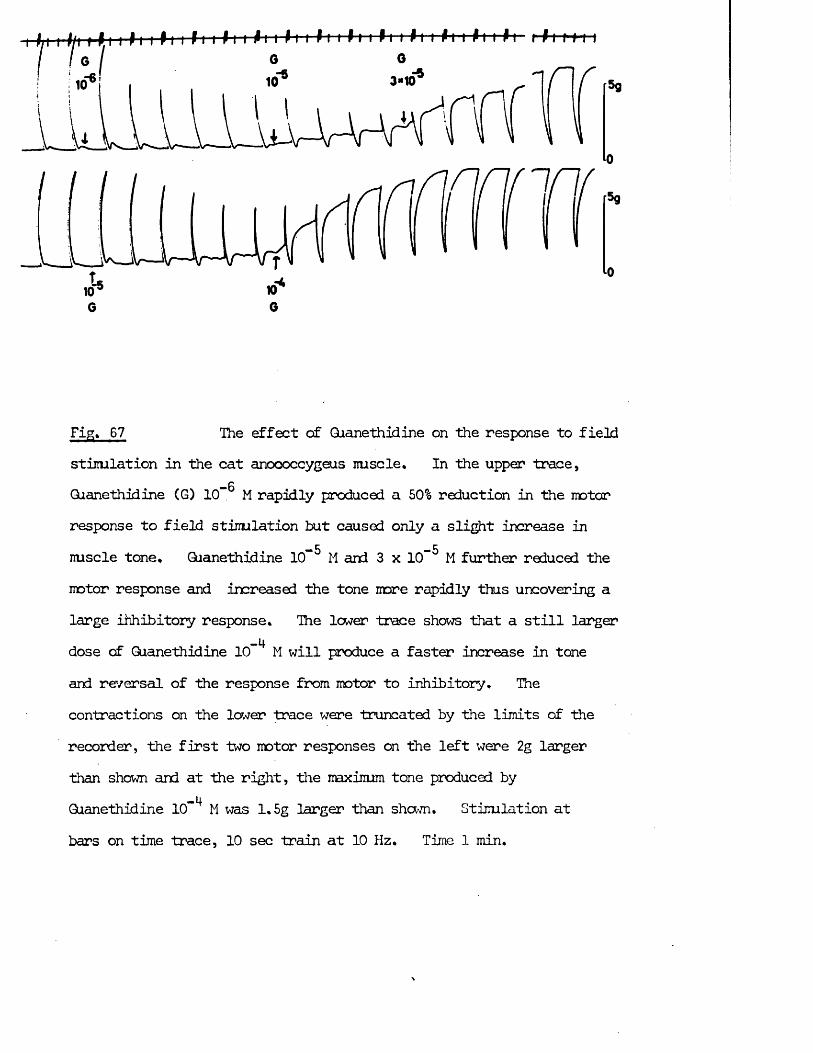
3*10
t5
EfrJiZ The effect of Qianethidine on the response to fieldstimlation in the cat anococcygeus nuscle. In the upper trace,
— 6Qianethidine (G) 10” M rapidly produced a 50% reduction in the motorresponse to field stimlation but caused only a slight increase in
-5 -5nuscle tone, Qianethidine 10 M and 3 x 10 M further reduced the motor response and increased the tone more rapidly thus uncovering a large ihhibitory response. The lower trace shows that a still larger dose of Qianethidine 10 M will produce a faster increase in tone and reversal of the response from motor to inhibitory. Thecontractions on the lower trace were truncated by the limits of -therecorder, the first two motor responses on the left were 2g largerthan shown and at the right, the maxinum tone produced by
. . -4 .Qianethidine 10 M was 1.5g larger than shown. Stimulation atbars on time trace, 10 sec train at 10 Hz. Time 1 min.
In the rat anococcygeus, all three prostaglandins tested raised the tone of-9 - 8the muscle. The threshold varied from 3 x 10 g/ml to 10 g/ml.—8Maxinal responses were always produced by 3 x 10 g/ml and were never
larger than 50% of the maximal motor response to NA. In the rat anococcygeus, the three prostaglandins showed a spectrum of potency Ej > E > •
The effects of prostaglandins on both rat and cat anococcygeus were unaffected— 6 *“ 6 “ * 6by atropine CIO" M), propranolol (10" M) or hexamethonium (10" M).
Vasopressin produced transient motor responses in the cat anococcygeus at 6 - 200 m U / ml in the absence of induced tone. At 20 - 200 m U / ml these motor responses were followed by relaxation responses. When the tone of the muscle was raised with an agonist drug, no motor responses were found and inhibitory responses were produced by 20 - 200 m U / ml.
In the rat anococcygeus, vasopressin 2 - 200 m U / ml produced small motor responses. The maximum response was obtained at 20 m U / ml, larger doses producing smaller responses.
These responses to vasopressin in both species were unaffected by atropine (1 0 - 6 M).
Responses to Field Stimulation
Field stimulation of the rat anococcygeus produces motor responses due to its adrenergic innervation. When these motor nerves are blocked with guanethidine and the tone of the muscle raised, field stimulation then produces a powerful inhibitory response. Similar motor and inhibitory responses were found in Hie cat anococcygeus (Fig. 67).
Increasing doses of guanethidine gradually depressed the motor response to
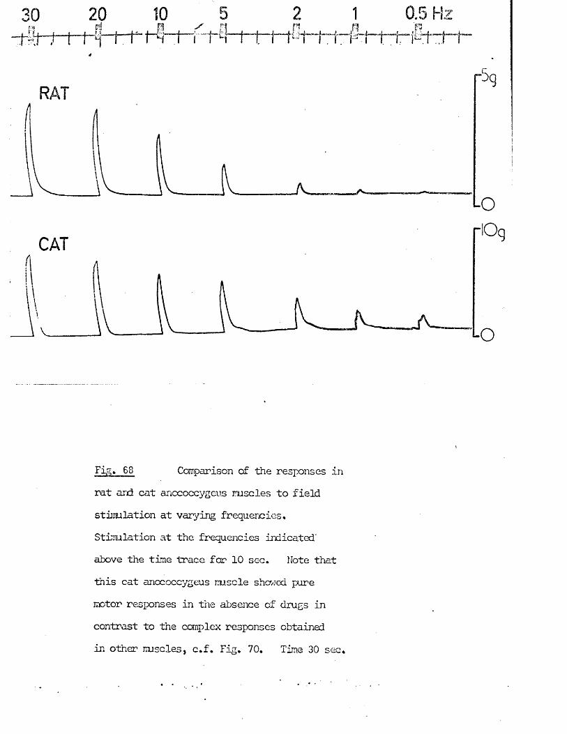
Fig. 68 Comparison of the responses inrat and cat anococcygeus muscles to field stimulation at varying frequencies* Stimulation at the frequencies indicated' above the time trace far 10 sec. Note that
this cat anococcygeus muscle showed pure motor responses in the absence of drugs in
contrast to the complex responses obtained in other muscles, c.f. Fig. 70. Tima 30 sec.
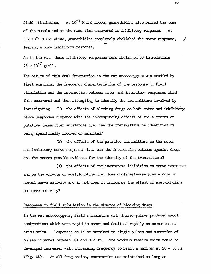
90
field stimulation. At 10™ M and above, guanethidine also raised the tone of the muscle and at the same time uncovered an inhibitory response. At 3 x 10” 5 M and above, guanethidine completely abolished the motor response, / leaving a pure inhibitory response.
As in the rat, these inhibitory responses were abolished by tetrodotoxin (3 x 10' 7 g/ml).
The nature of this dual innervation in the cat anococcygeus was studied by first examining the frequency characteristics of the response to field stimulation and the interaction between motor and inhibitory responses which this uncovered and then attempting to identify the transmitters involved by investigating (1 ) the effects of blocking drugs on both motor and inhibitory nerve responses compared with the corresponding effects of the blockers on putative transmitter substances i.e. can the transmitters be identified by being specifically blocked or mimicked?
(2 ) the effects of the putative transmitters on the motor and inhibitory nerve responses i.e. can the interaction between agonist drugs and the nerves provide evidence for the identity of the transmitters?
(3) the effects of cholinesterase inhibition on nerve responses and on the effects of acetylcholine i.e. does cholinesterase play a role in normal nerve activity and if not does it influence the effect of acetylcholine on nerve activity?
Responses to field stimulation in the absence of blocking drugs
In the rat anococcygeus, field stimulation with 1 msec pulses produced smooth contractions which were rapid in onset and declined rapidly on cessation of stimulation. Responses could be obtained to single pulses and summation of pulses occurred between 0.1 and 0.2 Hz. The maximum tension which could be developed increased with increasing frequency to reach a maximum at 20 - 30 Hz (Fig. 6 8 ). At all frequencies, contraction was maintained as long as
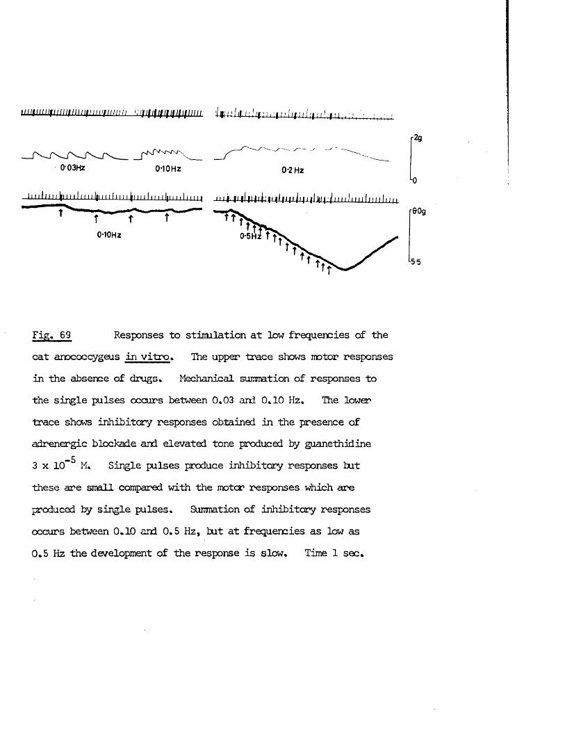
ut iu iu i i t iu i in iu i i i i i i i i n u i i ju i i i i i i f i i i i i f i f i i i i i ■ijjij/fir ir />f ; f f , />f!,'7.i . • ■ '■ ,
0-03HZ
pfs/VNr-NT' '.
0-10 Hz 0*2 Hz
I I I I M U U J L Il.il II I I I I ILlliJII j l j i /I Ifl f : / 11111111111111 111 / 1
0-WHz
r2g
Lo
r&Og
l5-5
Fig. 69 Responses to stimlation at low frequencies of thecat anococcygeus in vitro> The upper trace shows motor responsesin the absence of drugs. Mechanical summation of responses to the single pulses occurs between 0.03 and 0.10 Hz. The lower trace shews inhibitory responses obtained in the presence of adrenergic blockade and elevated tone produced by guanethidine
_53 x 10 M. Single pulses produce inhibitory responses but these are small compared with the motor responses which are produced by single pulses. Summation of inhibitory responses occurs between 0.10 and 0.5 Hz, but at frequencies as low as 0.5 Hz the development of the response is slow. Time 1 sec.
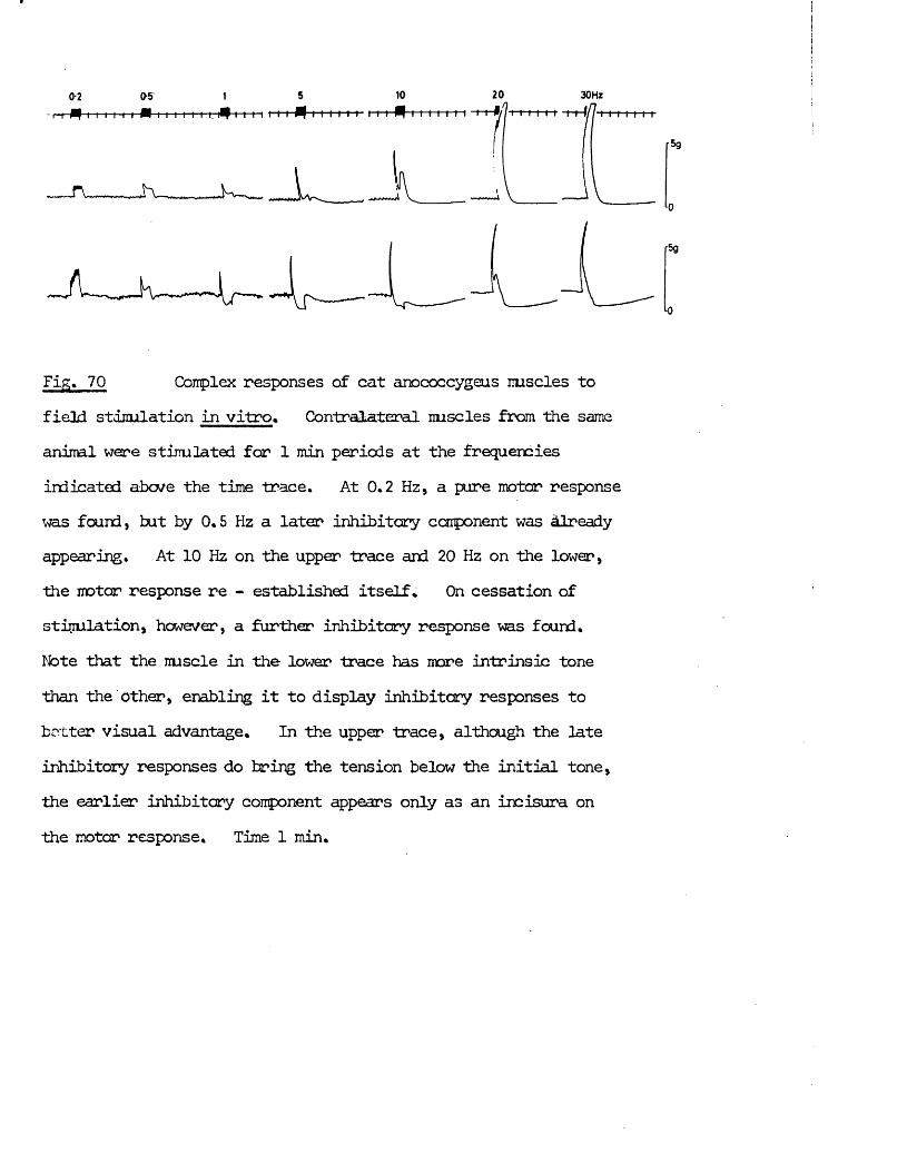
0
/-5g
■0
Fig, 70 Complex responses of cat anococcygeus imscles tofield stimulation in vitro. Contralateral miscles from the same animal were stipulated for 1 min periods at the frequencies indicated above the time trace. At 0.2 Hz, a pure motor response was found, but by 0.5 Hz a later inhibitory component was already appearing. At 10 Hz on the upper trace and 20 Hz on the lower, the motor response re - established itself. On cessation of stipulation, however, a further inhibitory response was found.Note that the nuscle in the lower trace has more intrinsic tone than the other, enabling it to display inhibitory responses to better visual advantage. In the upper trace, although the late inhibitory responses do bring the tension below the initial tone, the earlier inhibitory component appears only as an incisura on the motor response. Time 1 min.
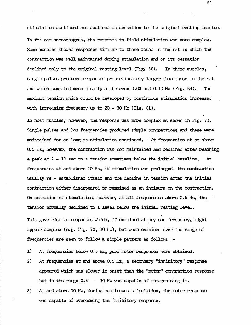
stimulation continued and declined on cessation to the original resting tension
In the cat anococcygeus, the response to field stimulation was more complex. Same muscles showed responses similar to those found in the rat in which the contraction was well maintained during stimulation and on its cessation declined only to the original resting level (Fig. 6 8 ). In these muscles, single pulses produced responses proportionately larger than those in the rat and which summated mechanically at between 0.03 and 0.10 Hz (Fig. 69). The maximum tension which could be developed by continuous stimulation increased with increasing frequency up to 20 - 30 Hz (Fig. 81).
In most muscles, however, the response was more complex as shown in Fig. 70. Single pulses and lew frequencies produced simple contractions and these were maintained for as long as stimulation continued. - At frequencies at or above 0.5 Hz, hcwever, the contraction was not maintained and declined after reaching a peak at 2 - 10 sec to a tension sometimes below the initial baseline. At frequencies at and above 10 Hz, if stimulation was prolonged, the contraction usually re - established itself and the decline in tension after the initial contraction either disappeared or remained as an incisura on the contraction.On cessation of stimulation, however, at all frequencies above 0.5 Hz, the tension normally declined to a level below the initial resting level.
This gave rise to responses which, if examined at any one frequency, might appear complex (e.g. Fig. 70, 10 Hz), but when examined over the range of frequencies are seen to follow a simple pattern as follows -
1) At frequencies below 0.5 Hz, pure motor responses were obtained.2) At frequencies at and above 0.5 Hz, a secondary "inhibitory” response
appeared which was slower in onset than the "motor” contraction response but in the range 0.5 - 10 Hz was capable of antagonising it.
3) At and above 10 Hz, during continuous stimulation, the motor response was capable of overcoming the inhibitory response.
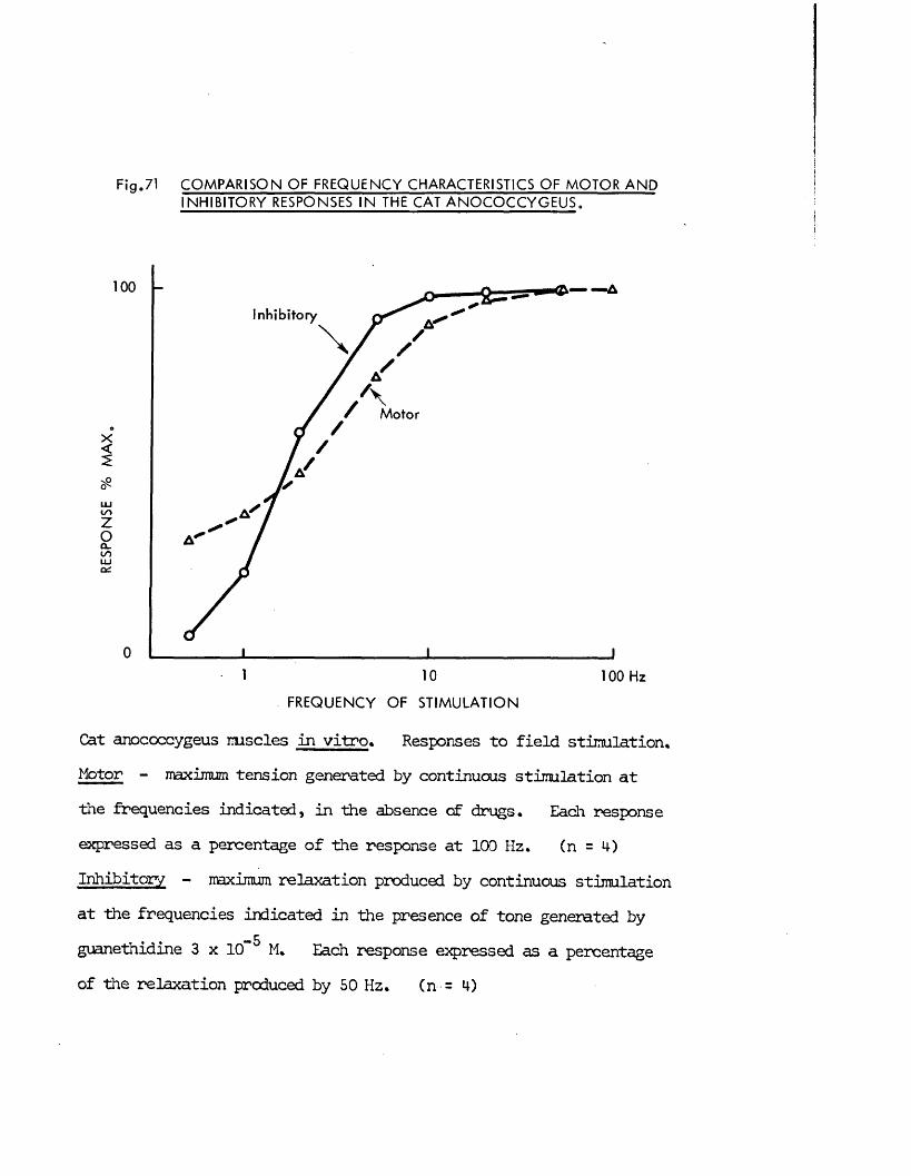
F ig .71 C O M P A R IS O N OF FREQUENCY CHARACTERISTICS OF M O TO R A N D IN H IB ITO R Y RESPONSES IN THE CAT A N O C O C C Y G E U S .
1 0 0
Inhibitory
Motor
0
10 100 Hz
FREQUENCY OF S T IM U LA TIO N
Cat anococcygeus muscles in vitro. Responses to field stimlation.Motor - maximum tension generated by continuous stimulation atthe frequencies indicated, in the absence of drugs. Each responseexpressed as a percentage of the response at 100 Hz. (n = 4)Inhibitory - maximum relaxation produced by continuous stimlationat the frequencies indicated in the presence of tone generated by
. . -5guanethidine 3 x 10 M. Each response expressed as a percentage of the relaxation produced by 50 Hz. (n = 4)
rx5g
Fig, 72 Comparison of the time course of maximal motor and inhibitory responses in the cat anococcygeus muscle in vitro . The upper trace • shows the response to field stimulation at 30 Hz for 30 sec in the absence of drugs. After a short latent period, tension developed rapdily to reach a plateau within 15 sec. Tension was then well maintained until stimulation ceased. On cessation, tension declined to reach baseline within 35 sec. In this muscle, no inhibitory component to the response developed in the absence of adrenergic blockade. The lower trace shews the response of the same muscle to
-5stimulation at 10 Hz for 30 sec in the presence of guanethidine 3 x 10 M. The inhibitory response started slowly after an initial short latent period but rapidly produced a steeper decline of tension. This inhibition was well maintained and was still increasing on cessation of stimulation at 30 sec. After a short delay, the tension began to recover slowly but had not reached its former level until 60 sec after the end of stimulation. Time 1 sec, 5 sec.
92
*4) On cessation of stimulation, at all frequencies above 0,5 Hz, theeffects of the inhibitory response outlast those of the motor response and so produce a late relaxation.
This interpretation is borne out by an analysis of the interaction between the two sets of responses as shown in the following section.
Interaction between motor and inhibitory nerves
Consideration of the frequency characteristics of the motor and of the inhibitory responses shews agreement with the above interpretation of the complex response. Comparing the motor responses obtained to continuous field stimulation in the absence of drugs with the inhibitory response
-5obtained to similar stimulation in the presence of guanethidine (3 x 10 M),it is apparent that single pulses and low frequencies are more effective at producing motor than inhibitory responses (Fig. 69) but that at intermediate frequencies (2 - 10 Hz) the inhibitory response is more fully developed than the motor response (Fig. 71).
The rate of development of the motor and inhibitory responses also agrees with the above interpretation. Fig. 72 compares a maximal motor response in a cat anococcygeus which showed no signs of an inhibitory component in the absence of guanethidine with a maximal inhibitory response in the same muscle after guanethidine. The latent period was short for both responses, but the motor response showed a very rapid initial rate of development whereas the inhibitory response had a slow initial rate of development which gradually accelerated. Similarly, after cessation of stimulation, the motor response waned more rapidly than the inhibitory response. This correlates with the finding that when motor and inhibitory nerves are active simultaneously the motor response nay appear at first before the inhibitory response, but since the inhibitory response takes longer to develop and lasts
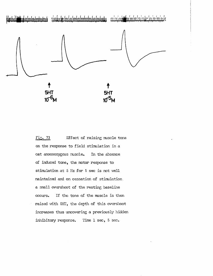
t5 H T1(T6 M
t5 H T1CT5 M
Fig. 73 Effect of raising iruscle toneon the response to field stimulation in a cat anococcygeus muscle. In the absence of induced tone, the motor response to stimulation at 5 Hz for 5 sec is not well maintained and on cessation of stimulation, a srall overshoot of the resting baseline occurs. If the tone of the iruscle is then raised with 5HT, the depth of this overshoot increases thus uncovering a previously hidden inhibitory response. Time 1 sec, 5 sec.
longer, it may reverse the motor response either during or after the stimulation period according to the frequency.
Two other experiments which analyse the response at 5 Hz (the frequency at which the inhibitory response is most evident) further confirm the above interpretation of the complex response.
Fig. 73 illustrates the first. When the tone of the nuscle was raised by addition of 5HT to the bath, the inhibitory response following the motor response became more apparent and as the tone was raised further this inhibitory component produced a relaxation of tension to and beyond the initial resting baseline. This demonstrates that in the absence of adrenergic blocking drugs, both motor and inhibitory nerves are being stimulated and the mechanical response which is observed is the sum of their two opposing responses and will therefore vary according to the state of activity in the muscle. This biphasic response in the presence of 5HT can be further modified by guanethidine which will abolish the motor response leaving a pure inhibitory response, confirming the above interpretation.
This first experiment confirmed that when stimulating at the optimal frequency for the inhibitory nerves i.e. 5 Hz, after 3 - 4 sec the tone of the muscle comes under the dominant influence of the inhibitory nerves. The motor nerves, which are still being stimulated, are thus no longer capable of producing contraction. A second experiment was, therefore, designed to estinate the time course and nognitude of the inhibitory effect produced by transmitter liberated from the inhibitory nerves during a short optimal burst of stimulation. The response to a single pulse was used as a measure of the degree of inhibition at any given moment since a single pulse in the absence of other influences produces a relatively pure motor response.
This experiment is illustrated in Fig. 74. A single test stimulus was followed
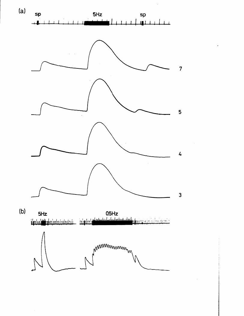
(a) sp-4 1 l-i
5 H z sp4 1
0 . 5 H z5 H z!. ..I.. .1 : : r . I : i : | >' I ■•m • '
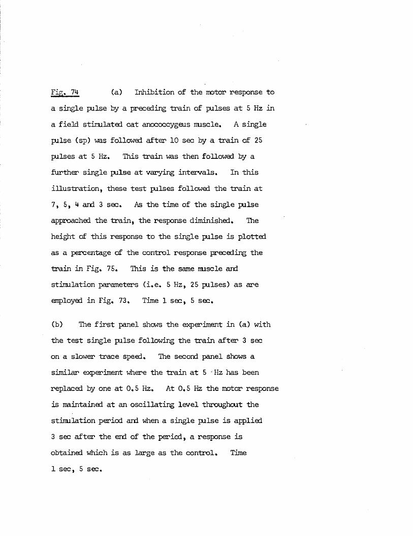
Fig. 7M- (a) Inhibition of the motor response toa single pulse by a preceding train of pulses at 5 Hz in a field stimulated cat anococcygeus muscle. A single pulse (sp) ms followed after 10 sec by a train of 25 pulses at 5 Hz. This train was then followed by a further single pulse at varying intervals. In this illustration, these test pulses followed the train at 7, 5,4 and 3 sec. As the time of the single pulse approached the train, the response diminished. The height c£ this response to the single pulse is plotted as a percentage of the control response preceding the train in Fig. 75. This is the same muscle and stimulation parameters (i.e. 5 Hz, 25 pulses) as areemployed in Fig. 73. Time 1 sec, 5 sec.
(b) The first panel shows the experiment in (a) with the test single pulse following the train after 3 sec on a slower trace speed. The second panel shows a similar experiment where the train at 5 -Hz has been replaced by one at 0.5 Hz. At 0.5 Hz the motor response is maintained at an oscillating level throughout the stimulation period and when a single pulse is applied3 sec after the end of the period, a response isobtained which is as large as the control. Time
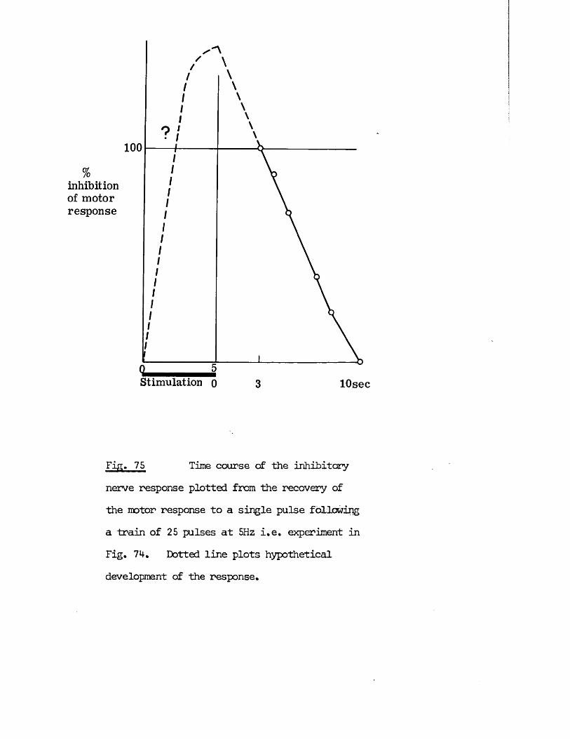
%inhibition of motor response
100
Stimulation 0 lOsec
Fifl» 75 Time course of the inhibitorynerve response plotted from the recovery of the motor response to a single pulse following a train of 25 pulses at 5Hz i.e. experiment in Fig. 74. Dotted line plots hypothetical development of the response.
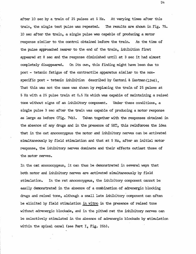
94
after 10 sec by a train of 25 pulses at 5 Hz, At varying times after this train, the single test pulse was repeated. The results are shewn in Fig. 75. 1 0 sec after the train, a single pulse was capable of producing a motor response similar to the control obtained before the train. As the time of the pulse approached nearer to the end of the train, inhibition first appeared at 8 sec and the response diminished until at 3 sec it had almost completely disappeared. On its own, this finding might have been due to post - tetanic fatigue of the contractile apparatus similar to the nonspecific post - tetanic inhibition described by Cantoni & Eastman (1946) %That this was not the case was shown by replacing the train of 25 pulses at 5 Hz with a 25 pulse train at 0.5 Hz which was capable of maintaining a raised tone without signs of an inhibitory component. Under these conditions, a single pulse 3 sec after the train was capable of producing a motor response as large as before (Fig. 74b). Taken together with the responses obtained in the absence of any drugs and in the presence of 5HT, this reinforces the idea that in the cat anococcygeus the motor and inhibitory nerves can be activated simultaneously by field stimulation and that at 5 Hz, after an initial motor response, the inhibitory nerves dominate and their effects outlast those of the motor nerves.
In the cat anococcygeus, it can thus be demonstrated in several ways that both motor and inhibitory nerves are activated simultaneously by field stinulation. In the rat anococcygeus, the inhibitory component cannot be easily demonstrated in the absence of a combination of adrenergic blocking drugs and raised tone, although a small late inhibitory component can often be elicited by field stimulation in vitro in the presence of raised tone without adrenergic blockade, and in the pithed rat the inhibitory nerves can be selectively stimulated in the absence of adrenergic blockade by stimulation within the spinal canal (see Part I, Fig. 25b).
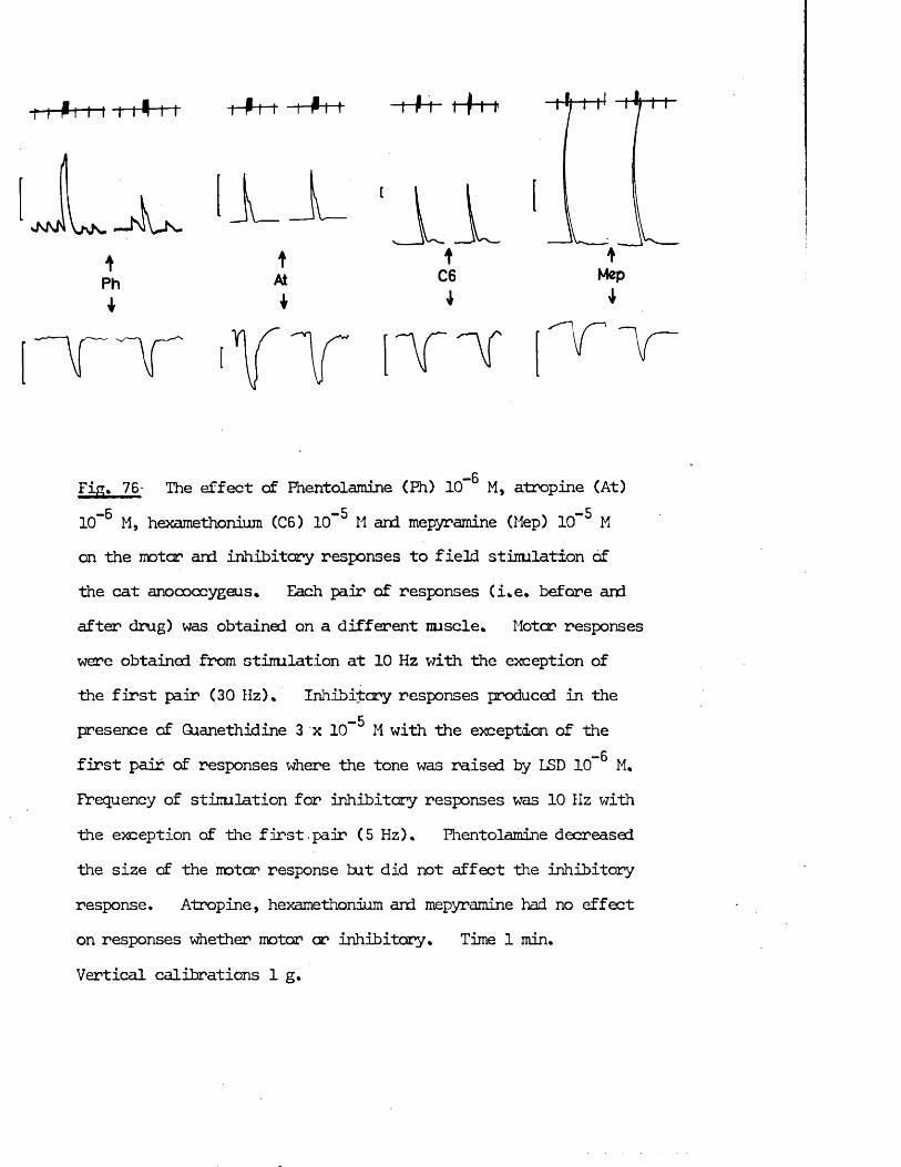
i i Pit-t 1-1+
a n ! Va n. — J'IVjs-
1
Phi
-Pi-1 I P-t-1- I P 1 ' t-f-M1
tAt*
Mep
wmPiFig* 76- The effect of Fhentolamine (Fh) 10“ M, atropine (At)- 6 -5 . -510 M, hexamethonium (06) 10 M and mepyr amine (Hep) 10 H
on the motor and inhibitory responses to field stimlation ofthe cat anococcygeus* Each pair of responses (i*e* before andafter drug) was obtained on a different nuscle* Motor responseswere obtained from stimlation at 10 Hz with the exception ofthe first pair (30 Hz)* Inhibitory responses produced in the
-5presence of Guanethidine 3 x 1 0 M with the exception of the—6first pair of responses where the tone was raised by LSD 10” M.
Frequency of stimlation for inhibitory responses was 10 Hz with the exception of the first pair (5 Hz)* Fhentolamine decreased the size of the motor response but did not affect the inhibitory response. Atropine, hexamethonium and mepyramine had no effect on responses whether motor or inhibitory* Time 1 min*Vertical calibrations 1 g.
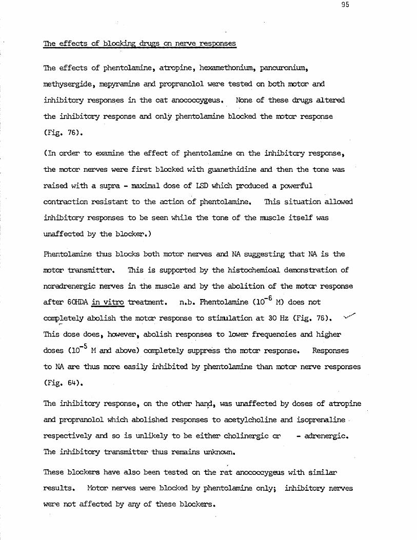
The effects of blocking drugs on nerve responses
The effects of phentolamine, atropine, hexamethonium, pancuronium, methysergide, mepyr amine and propranolol were tested on both motor and inhibitory responses in the cat anococcygeus. None of these drugs altered the inhibitory response and only phentolamine blocked Hie motor response (Fig. 76).
(In order to examine the effect of fhentolamine on the inhibitory response, the motor nerves were first blocked with guanethidine and then the tone was raised with a supra - maximal dose of LSD which produced a powerful contraction resistant to the action of phentolamine. This situation allowed inhibitory responses to be seen while the tone of the muscle itself was unaffected by the blocker.)
Fhentolamine thus blocks both motor nerves and NA suggesting that NA is the motor transmitter. This is supported by the histochemical demonstration of noradrenergic nerves in the muscle and by the abolition of the motor response after 6 CHDA in vitro treatment. n.b. Fhentolamine (10” M) does not completely abolish the motor response to stimlation at 30 Hz (Fig. 76). ^
r* ‘
This dose does, however, abolish responses to lower frequencies and higher -5doses (10 M and above) completely suppress the motor response. Responses
to NA are thus more easily inhibited by phentolamine than motor nerve responses (Fig. 64).
The inhibitory response, on the other hand, was unaffected by doses of atropine and propranolol which abolished responses to acetylcholine and isoprenaline respectively and so is unlikely to be either cholinergic or - adrenergic.The inhibitory transmitter thus remains unknown.
These blockers have also been tested on the rat anococcygeus with similar results. Motor nerves were blocked by phentolamine only; inhibitory nerves were not affected by any of these blockers.
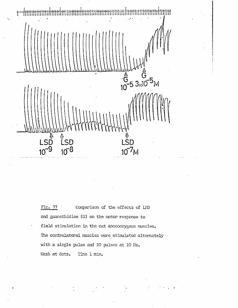
Tig. 77 Comparison of the effects of LSDand guanethidine (G) on the motor response to
field stimlation in the cat anococcygeus muscles*
The contralateral muscles were stimulated alternately with a single pulse and 30 pulses at 10 Hz.Wash at dots. Time 1 min.
96
Guanethidine also blocks the motor but not the inhibitory nerve responses in both species. Unlike phentolamine, however, guanethidine does not block the response to NA. added to the bath and so is supposed to act by preventing the release of NA from the nerves i.e. neuronal blockade.
LSD is known to block the motor adrenergic response in the vas deferens(Ambache, Dunk, Verney & Zar, 1973). In a previous section, evidence hasbeen given that LSD can act on the adrenergic nerves of the cat anococcygeusbut as an indirect sympathomimetic. It was, therefore, of interest todetermine whether LSD was acting as a pure indirect sympathomimetic orwhether its action was close to that of guanethidine, which, in low doses,acts as a pure neuronal blocking agent and only at relatively high doses actsas an indirect sympathomimetic (Fig. 67). No evidence was found that LSD,
_ qin doses less than those required far an agonist action ( 1 0 M), reducedthe motor response to field stimulation (Fig. 77). At doses producing a
— 8moderate agonist action (1 0 " M), the motor response to field stimlationwas diminished. At higher doses producing large contractions, the responseto field stimlation was reversed to inhibition. This inhibition was asgreat as that usually seen after guanethidine (Figs. 67 & 77) and was notenhanced by further addition cf guanethidine. In addition, these doses of
—6LSD did not inhibit the response to NA 3 x 10" M so it is possible that LSD may be inhibiting the motor nerves by a neuronal blocking action similar to that of guanethidine.
The effects of agonist drugs on nerve responses
The effects of the substances which relax the cat anococcygeus on the responses to field stimlation of the nerves are interesting for several reasons. First, considered as putative transmitter substances, they might be expected to inhibit the responses to motor nerve stimlation and to summate with the responses to inhibitory nerve stimlation. In addition, whether or not a
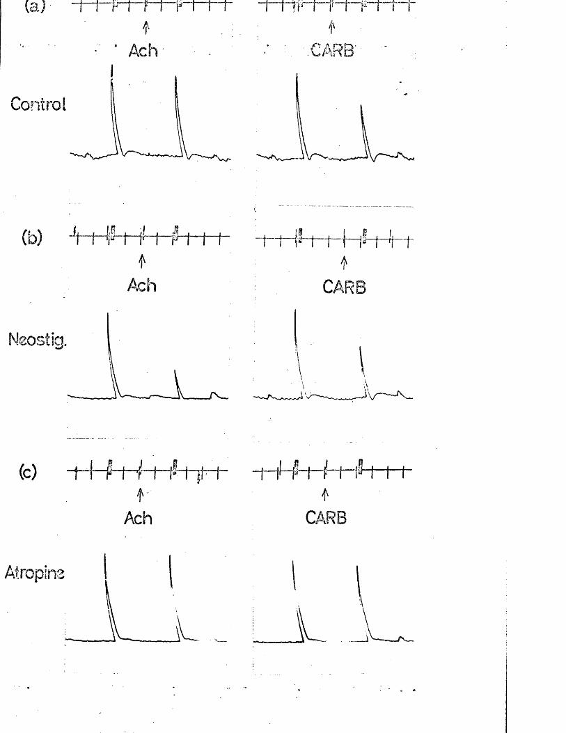
(c) -f-f-f i- t ~i— — I— fS—}— (— ff I
Ach CARB
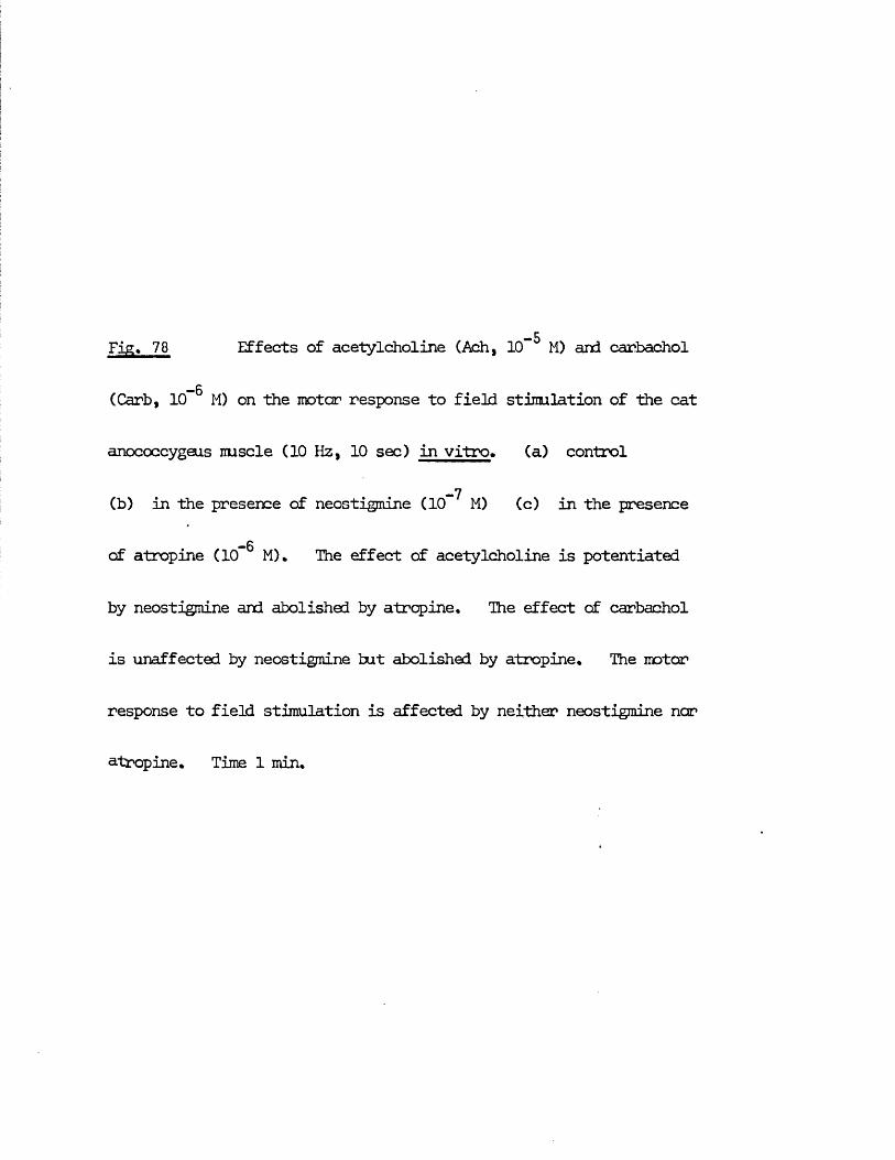
-5Fjg. 78 Effects of acetylcholine (Ach, 10 M) and carbachol
(Carb, 10" M) on the motor response to field stimulation of the cat
anococcygeus nuscle (10 Hz, 10 sec) in vitro, (a) control
_7(b) in the presence of neostigmine ( 1 0 M) (c) in the presence
— 6of atropine (10” M), The effect of acetylcholine is potentiated
by neostigmine and abolished by atropine. The effect of carbachol
is unaffected by neostigmine but abolished by atropine. The motor
response to field stimulation is affected by neither neostigmine nor
atropine. Time 1 min.
97
particular substance was. the transmitter, its effects on nerve responses as opposed to those on intrinsic or drug - induced tone might be expected to provide more information on the type and location of pharmacological receptors present within the tissue.
Acetylcholine especially has been shown to have effects in several tissuesnot only directly on the nuscle but also on synpathetic nerve terminals.These responses can vary considerably according to the dose and tissueemployed. Thus, in the isolated perfused mesenteric artery, extremely lewdoses of acetylcholine (5 x 10” g/ml) can potentiate the response to
-9synpathetic nerve stinulation while higher concentrations (5 x 10 g/ml)can inhibit the same response (Malik & Ling, 1969). On the other hand, in
-9 - 6the isolated rabbit heart, concentrations of 1 0 - 1 0 g/ml have been shownto inhibit NA release by nerve stinulation while still higher doses
_ ii(10 g/ml) released NA from the nerves (Loffelholz & Muscholl, 1969).
With these considerations in mind, the effects of the inhibitory substances on responses to field stinulation were examined, paying particular attention to the effects of acetylcholine over a wide dose range.
Muscarinic agonists Acetylcholine and carbachol were found to have aninhibitory effect on the motor nerve response in the cat anococcygeus (Fig. 78).This inhibition was more effective at low than at high frequency ofstinulation and, in the case of acetylcholine but not of carbachol, waspotentiated by neostigmine (Fig. 79 a & b). The threshold for this inhibition
— 8 —7was 10” M - 10 M. Acetylcholine had no apparent effect in the range1 0 - 1 3 M - 10" 8 M.
This inhibition of motor nerve responses, like the inhibitory response on nuscle tone, did not occur in the rat anococcygeus. The sole effect of acetylcholine in the rat anococcygeus appeared to be to contract the nuscle
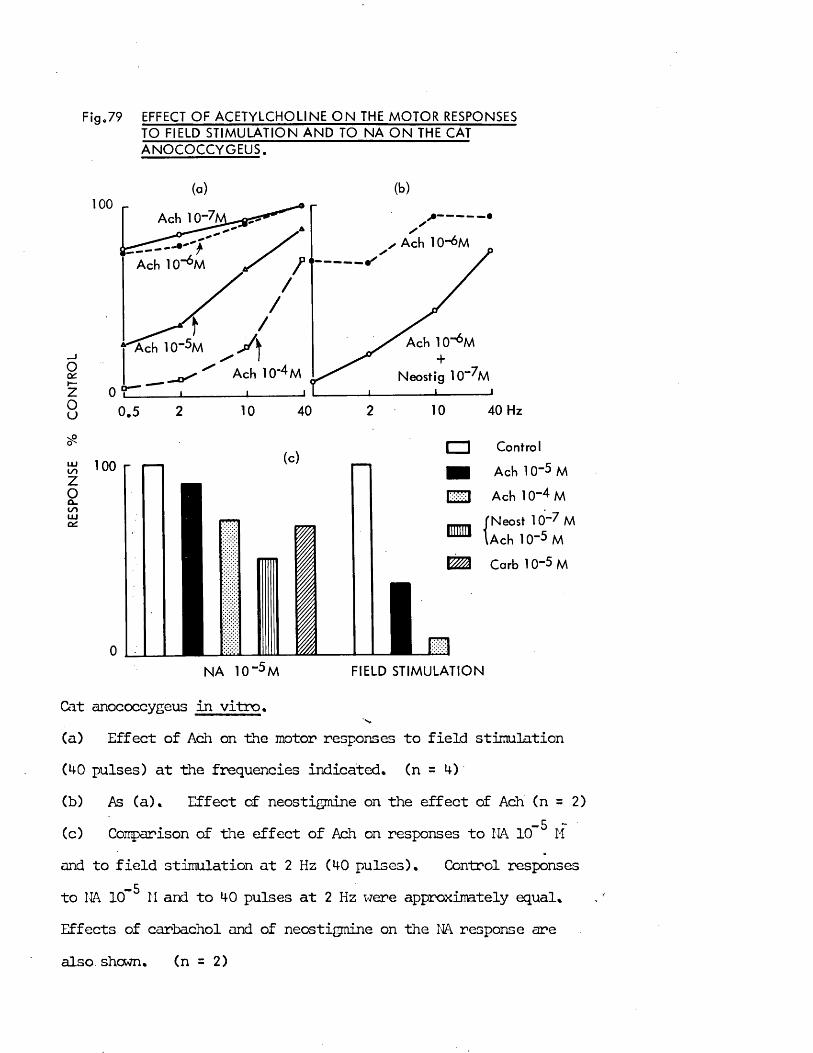
Fig„79 EFFECT OF ACETYLCHOLINE O N THE MOTOR RESPONSESTO FIELD S T IM U L A T IO N A N D TO N A O N THE CAT A N O C O C C Y G E U S .
(a) (b)
Occh-Zou
IS)zoCL.IS)
100
0
(c)
1 0 0
A
Ach 10
0
10 40 Hz210 4020 .5
VZZ\
1 i i
I I Control
m Ach 10"5 M
I'-K-ll Ach 10“4 M
...... fN eost 1 0 -7 M
m { a c H I O - S m
Carb 1 0"5 M
N A 10 “5 M FIELD S T IM U L A T IO N
Cat anococcygeus in vitro*s .
(a) Effect of Ach on the motor responses to field stimulation (40 pulses) at the frequencies indicated* (n = 4)(b) As (a). Effect of neostigmine on the effect of Ach (n = 2)
-5 ~(c) Comparison of the effect of Ach on responses to NA 10 Mand to field stimulation at 2 Hz (40 pulses)* Control responses
-5to NA 10 M and to 40 pulses at 2 Hz were approximately equal* Effects of carbachol and of neostigmine on the NA response are also shewn* (n = 2 )
and these contractions summated with the motor nerve responses. Fig. 80 contrasts the effects of acetylcholine in the cat and rat muscles and illustrates that at high doses acetylcholine can produce small contractions of the cat nuscle.
Atropine (10” M) abolished the inhibition of motor nerve responses in the cat anococcygeus produced by acetylcholine cr carbachol (Fig. 78) and also the small contractions produced by high doses of acetylcholine.
It is not possible to tell from these experiments whether acetylcholine isexerting its inhibitory effect on the release of NA from the motor nerves orby acting directly on the nuscle. One test of this would be the effect ofacetylcholine on the motor response produced by NA as shown in Fig. 79 c.
-5The sub - maximal motor responses produced by NA 10 M were inhibited by the-5 -4presence of acetylcholine 10 M or 10 H. This inhibition of the NA-5response by acetylcholine 10 M was only 10% but could be increased to 49%
-7by the prior inhibition of cholinesterase with neostigmine 10 M.-5 . . .Carbachol 10 M produced an inhibition similar to that produced by
-4acetylcholine 10 M. From this evidence, an inhibitory effect of acetylcholine on the cat anococcygeus nuscle is likely to be exerted directly on the nuscle.
It is not possible to make a direct comparison between the extent of theinhibitions by acetylcholine on the motor nerve response and on the NA responsesince the effects of acetylcholine have not been tested against the whole dose /response curve for NA, but only against a single sub - maximal dose.
-5Comparison can, however, be made between the response to NA 10 M and theresponse to motor nerve stinulation at 2 Hz which produced motor responses of
-5 -4similar magnitude. The inhibition produced by acetylcholine 10 M or 10 M against the nerve response is greater than that against the NA response - Fig, 79 c. This suggests either that acetylcholine is acting at a site on
(a)
2? ? ? ? ? *9 05 2 10 40 HzM H I |!| II M f '( 01 I I |i f 1 |l I I I 1 I - I I I t
Rat
Control Ach 10“6M Ach T0-AM
05 2 10 401 t I* I I l If I I
OS 2 10 40 r h - # t - H f / t i i *t i
n. I u k - .
05 2 X) 40Hz■h - t P f i P i f N i t t
(b) Ach Ach Ach Ach Ach NA NA
Rat
3x10t t iw t im ttm ttn tit t i tm tt it t t t t f tm t r w t n ii i i t i i i n n i i n i i iH n i i u im t tm tfft
80 Comparison of the effects of acetylcholine(Ach) on the rat and cat anococcygeus.(a) Effects of Ach on the responses to field stimulation in the two species. Stimulation - 30 pulses at each frequency. In the rat, Ach raised the tone of the nuscle tut did not affect the height of the motor responses to field stimulation. In the cat, on the other hand, Ach inhibited the responses to field stimulation withcut affecting the tone. Time 1 min.(b) Effect of Ach on responses to single pulses in the two species. Stimulation 1 pulse every 1 min. The responses in the rat muscle are superimposed on the motor response to Ach. In the cat, however, the responses are inhibited even in doses where the tone of the nuscle is raised. Note the contrast with the effect of NA in the cat. When NA raises the tone, the responses aresuper inposed on the raised tone. This cat muscle was cold stored far 24 hr at 4° C and as a result of this shows greater motor responses to Ach than would a fresh nuscle. Time 1 min. Note that between the crosses the sensitivity of the rat trace was halved.
the nuscle which is more effective against nerve - released NA than against NA added to the bath e.g. acetylcholine and nerve - released NA act mainly in the region of the nerve - nuscle junction but NA added to the bath acts generally over the surface of the nuscle cells, or that the effect of acetylcholine against nerve responses is only partly by an action on the muscle and is perhaps also exerted on the nerve against release of NA.
The snail contractions produced in the cat anococcygeus by high doses of acetylcholine were produced in the nuscles from 3 out of 8 cats. These responses were abolished by atropine but were not further investigated. It is not, therefore, known whether they represent a direct or indirect effect of acetylcholine on the nuscle but they are very small compared with the direct responses to similar doses on the rat (Fig. 80 b).
The effect of acetylcholine on the inhibitory nerve response in the presence of guanethidine was difficult to quantify since the tone of the nuscle was altered by the acetylcholine itself. Inhibitory nerve responses were superimposed on the acetylcholine - induced relaxation, but shewed no obvious differences from responses which were obtained when the tone of the nuscle was reduced to a similar level by reducing the concentration of guanethidine. Acetylcholine, therefore, produced no changes in the inhibitory nerve response which could not be accounted far by the change in nuscle tone.
Other drugs which relax the cat anococcygeus Apart from acetylcholine and carbachol, the drugs which produced relaxation of the cat anococcygeus did not have a significant effect on motor or inhibitory nerve responses in either species.
Isoprenaline had little effect on the motor nerve response at doses from -7 -53 x 10 M to 3 x 10 M (Fig. 50) and had no effect on inhibitory responses.
In both species, ATP, vasopressin and prostaglandins all caused a slight
F ig .81 EFFECT OF 6 OHDA IN V IVO PRE-TREATMENT O N THE MOTOR NERVE RESPONSES OF THE RAT ANOCOCCYGEUS.
Control4
RESPONSE gms.
3
2
0
30 Hz100
FREQUENCY OF STIMULATION
Rat anococcygeus muscles in vitro.(a) Responses expressed as maximum tension generated by continuous stimulation at each frequency. Open circles - untreated controls. Filled circles - muscles from 6 OHDA. pretreated rats. (n = 6 )(b) (Right hand page) Comparison of the development of tension produced by field stimulation in tissues from control and 6 CHDA treated rats.
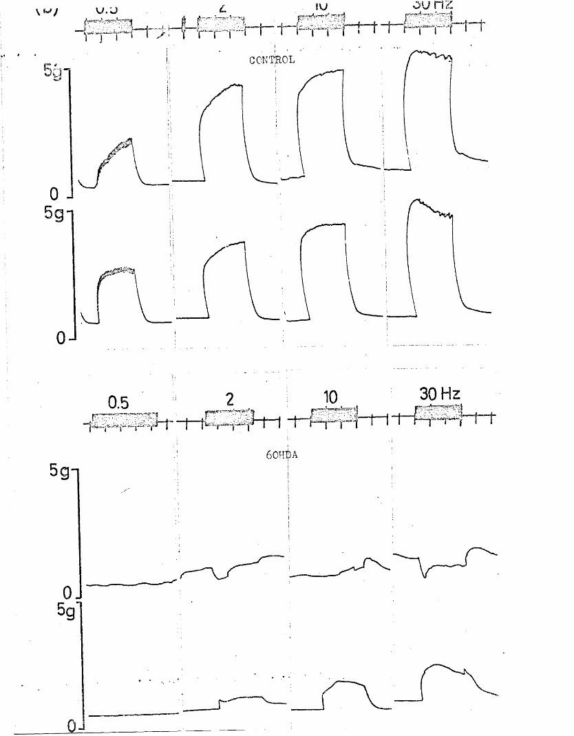
CONTROL
i j11
60HBA
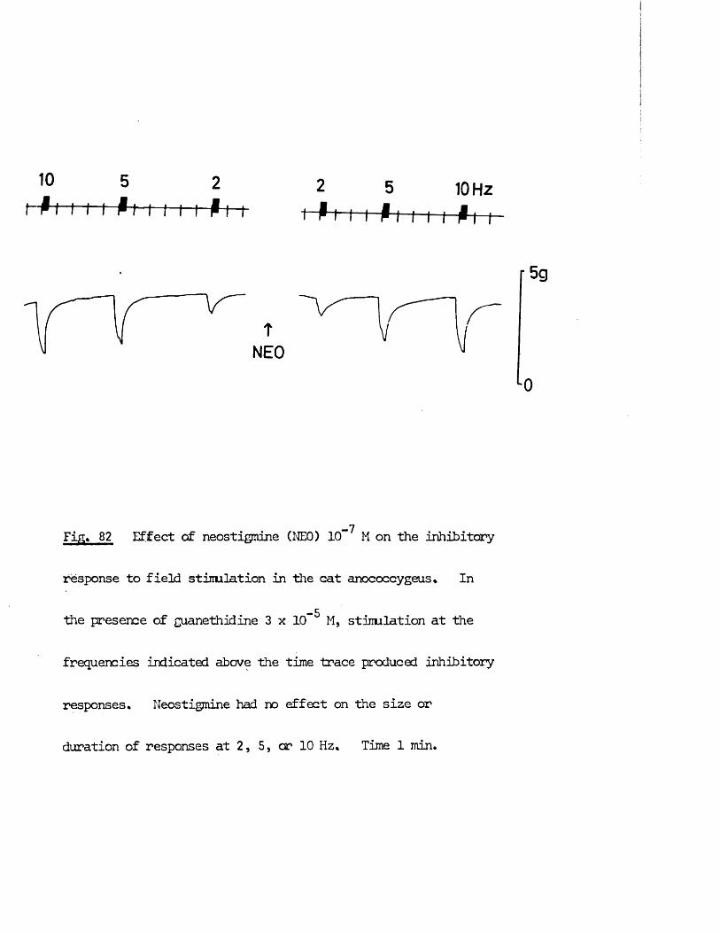
10
-h-t
5ft 2
f 2t - f f
10 H z f t - t
-7Fig* 82 Effect of neostigmine CNEO) 10 H on the inhibitory
response to field stimulation in the cat anococcygeus* In
-5the presence of guanethidine 3 x 10 M, stimulation at the
frequencies indicated above the time trace produced inhibitory
responses* Neostigmine had no effect on the size or
duration of responses at 2, 5, or 10 Hz* Time 1 min*
depression of motor responses to field stimulation at the highest doses tested_ ii _7i.e. ATP 5 x 10 M; vasopressin 200 m U/mlj prostaglandins 10 g/ml;
but produced no change in the size of the inhibitory nerve response.
There was, therefore, no evidence in either species of summation between any of the substances which were found to relax the cat anococcygeus and the inhibitory nerve response.
Effect of 60HDA treatment on nerve responses
60HDA added to the organ bath contracted the anococcygeus from both species and reversed motor nerve responses to inhibitory responses (Fig. *47). After washing out the 60HTA, the motor response did not recover even up to 6 hr after washout. The inhibitory response, on the other hand, was produced by field stimulation in the presence of 60HDA and after removal of 60HDA inhibitory responses could be demonstrated when the tone was raised with NA. 60HDA in vitro treatment thus appears to selectively destroy the motor nerves without affecting the inhibitory nerves.
In rats pretreated with 60HDA in vivo, diminished motor nerve responses to field stimulation cculd be still be obtained (Fig* 81). Together with the evidence that indirect sympathomimetics can still produce contractions, this demonstrates that the sympathectomy produced by the 60HDA in vivo pretreatment was incomplete.
Effect of neostigmine on nerve responses
Since the response to acetylcholine in the cat anococcygeus was potentiated by -7neostigmine (10 M) (Fig. 65) any role played by acetylcholine in the motor
or inhibitory nerve transmission process might also be expected to be modified-7by cholinesterase inhibition. Neostigmine (10 M) had, however, no effect
on the motor or inhibitory nerve responses in either species. This is shown for the cat motor nerves in Fig. 78 and the cat inhibitory nerves in Fig. 82.
Since the effects of acetylcholine in the anococcygeus of both species are potentiated by neostigmine and inhibited by atropine and the responses to stimulation of both inhibitory and motor nerves are unaffected by either neostigmine or atropine, it seems unlikely that acetylcholine has a significant role to play in the normal neuro - transmission processes of the anococcygeus muscles.
From the pharmacological evidence so far, it thus appears that the anococcygeus nuscle of the cat contains a dual innervation similar to that in the rat.
A motor adrenergic innervation is present which can be activated by field electrical stimulation, whose responses can be mimicked by NA or by indirect synpathomimetics, which can be specifically blocked by the * - adrenergic blocker phentolamine or the adrenergic neurone blocking agent guanethidine and which can be functionally destroyed by the action of 6 OHDA.
An inhibitory innervation exists which also can be activated by field stinulation, whose presence becomes most obvious when the nuscle is contracted by motor agonists, for which no effective blocking drug has been found, which has only been abolished by tetrodotoxin and whose transmitter, therefore, remains unknown. Among the substances which can produce relaxation and therefore mimic inhibition, the effects of acetylcholine and isoprenaline can be specifically altered by drugs which do not affect the inhibitory nerve response. In addition, all these substances, which relax the cat anococcygeus, contract the rat anococcygeus.
The pharmacological properties of the innervation of the anococcygeus muscle are, therefore, similar in both species. It is, however, possible to demonstrate the presence of the inhibitory innervation in the cat muscle without resorting to the use of blocking drugs as is necessary in the rat.
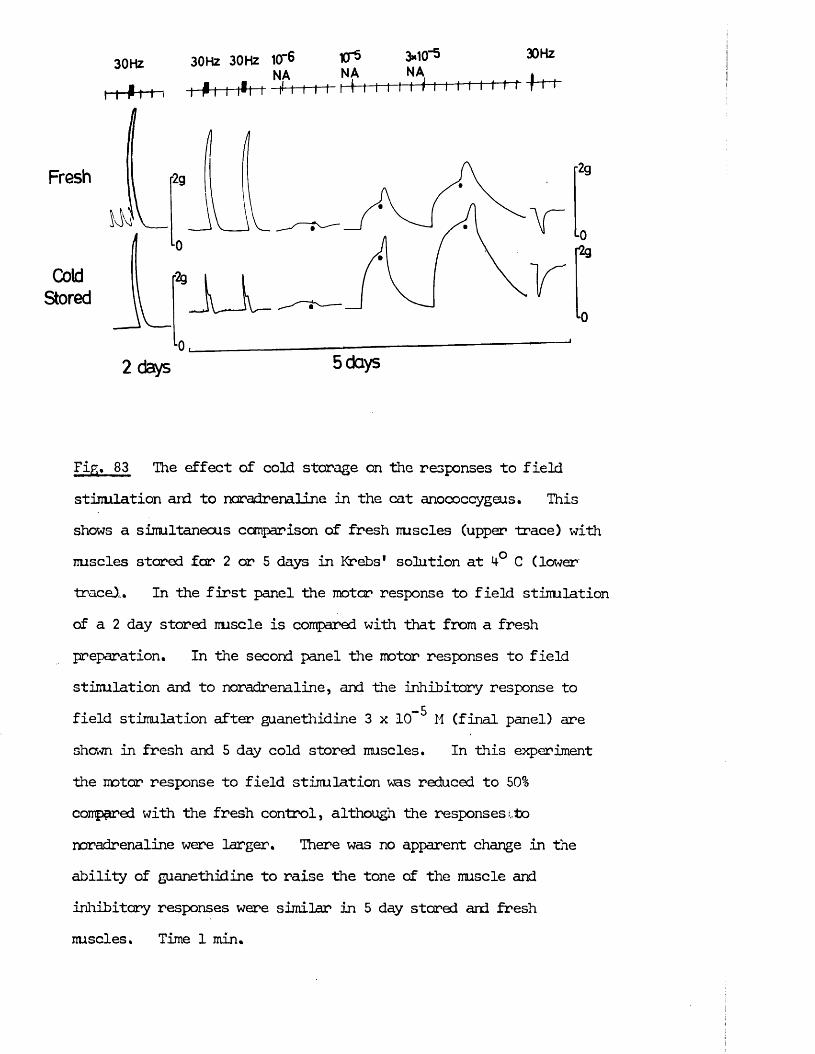
L0,2 d a y s 5 days
Fig. 83 The effect of cold storage on the responses to field stinulation aid to noradrenaline in the cat anococcygeus. This shows a simultaneous comparison of fresh muscles (upper trace) with muscles stared far 2 or 5 days in Krebs* solution at 4-° C (lower trace),. In the first panel the motor response to field stimulation of a 2 day stored nuscle is compared with that from a fresh preparation. In the second panel the motor responses to field stinulation and to noradrenaline, and the inhibitory response to
_5field stimulation after guanethidine 3 x 10 M (final panel) are shown in fresh and 5 day cold stored muscles. In this experiment ■the motor response to field stimulation was reduced to 50% compared with the fresh control, although the responsescto noradrenaline were larger. There was no apparent change in the ability of guanethidine to raise the tone of the muscle and inhibitory responses were similar in 5 day stared and fresh muscles. Time 1 min.
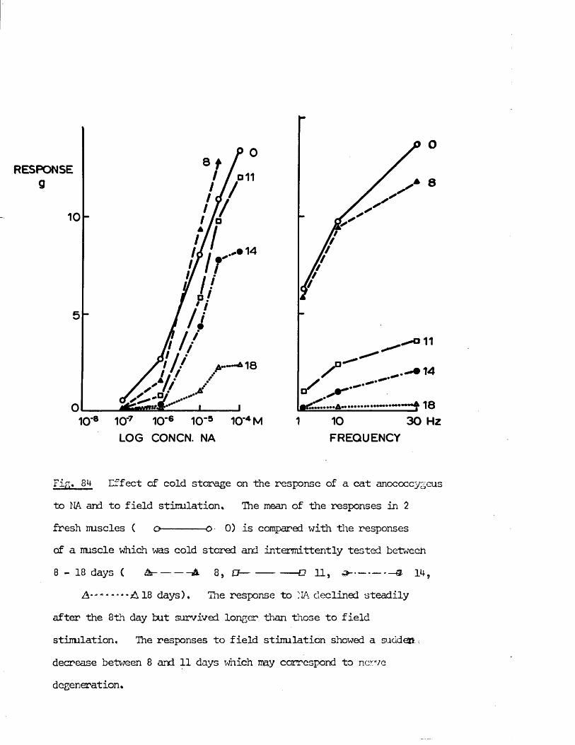
30 Hz101
RESPONSE
^ — •*--7 10"6 10’5 10‘4 M10'8 10'
LOG CONCN. NA FREQUENCY
Fig, 8 Effect of cold storage on the response of a cat anococcygeus to NA and to field stimulation. The mean of the responses in 2fresh muscles ( o------ O 0 ) is compared with the responsescf a muscle which was cold stored and intermittently tested between8 — 18 days ( £!a--- A 8 5 £7— ----- O 1 1 j --— 9- 1 M-,
A*-- A 18 days). The response to NA declined steadilyafter the 8 th day but survived longer than those to field stimulation. The responses to field stimulation showed a sudd®.i decrease between 8 and 1 1 days which may correspond to nerve degeneration.
Effects of cold storage on the cat anococcygeus
Since the anococcygeus muscles used were obtained from cats used in other experiments, sane were stored in Krebs* solution at 4° C in a sealed beaker for a variable time before their responses were measured. Observations were, therefore, made on the effects of cold storage on the responses to field stimulation and to agonist drugs. The action of direct and indirect synpathomimetics and the responses to field stimulation proved remarkably resistant to cold storage. Up to 5 days there was little diminution of the motor response to either drug or nerve stimulation (Fig. 83).
In another experiment, the effect of NA and field stinulation was examined in the same nuscle after 8 , 11, 14 and 18 days cold storage (Fig. 84). At 8 days the responses to both NA and field stimulation were similar to those obtained with fresh tissues. After 8 days the responses to NA declined gradually but were still present at 18 days, the dose / response curve drawn as a percentage of the maximum remaining unchanged. The motor response to field stimulation, on the other hand, declined mare rapidly, the greatest change occurring between 8 and 11 days. Motor responses to field stimulationwere just detectable at 10 Hz and 30 Hz at 18 days.
The effect of cold storage on the inhibitory response was more difficult toexamine since guanethidine was necessary to display inhibition and once given,further examination of the effect of cold storage on the motor response in that muscle was impossible. For this reason, the effect of cold storage on the inhibitory response was examined in the muscles from only 2 cats. In one cat, the response was measured in one muscle immediately after removal and in the contralateral muscle from the same animal, after 5 days cold storage. The muscles from the second cat were stored for 11 and 18 days respectively. The percentage inhibition of induced tone in the fresh muscle was 6 8 %, comparable
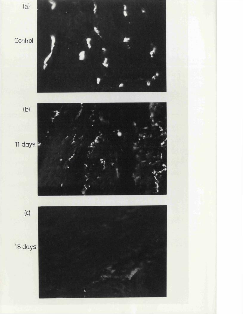
(a)
Control
(b)
11 days
(c)
18 days
#■ jff
*
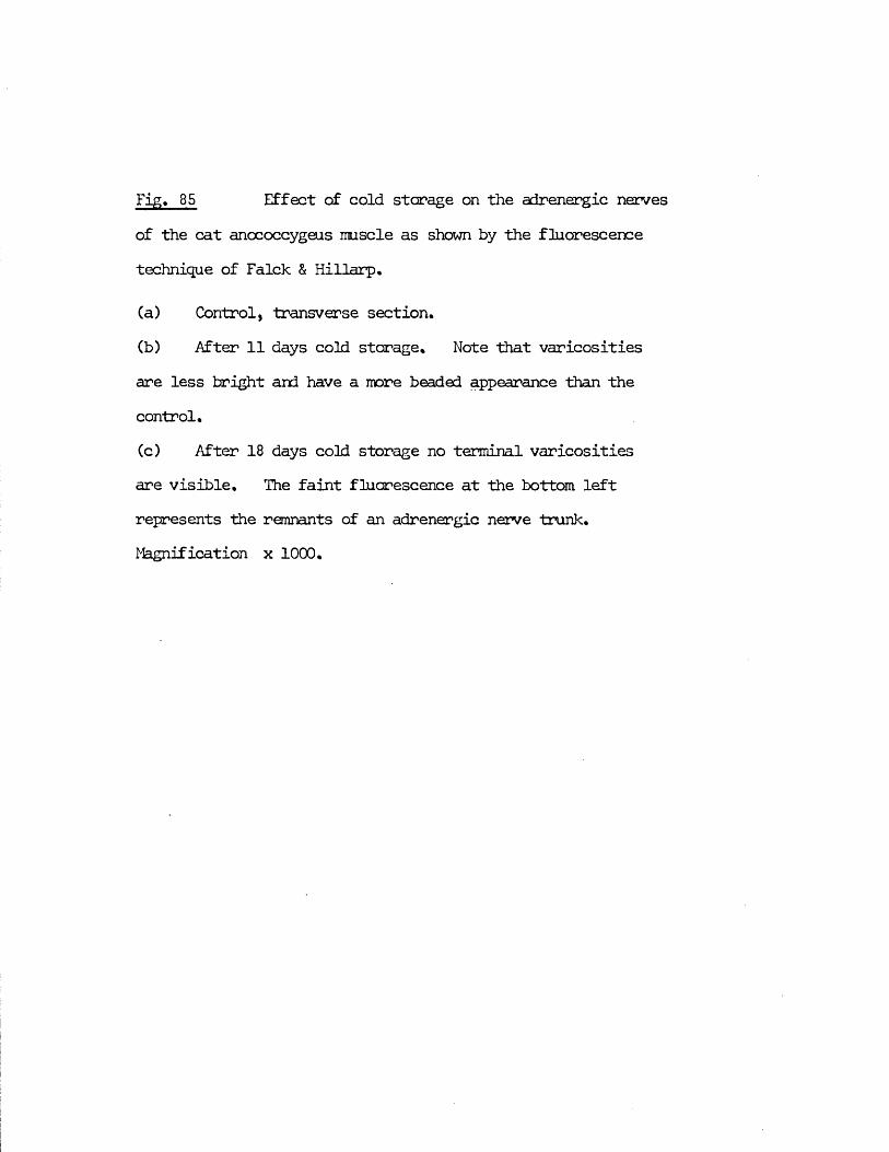
Fig. 85 Effect of cold storage on the adrenergic nervesof the cat anococcygeus muscle as shown by the fluorescence technique of Falck & Hillarp.
(a) Control, transverse section.(b) After 11 days cold storage. Note that varicositiesare less bright and have a more beaded appearance than the control.(c) After 18 days cold storage no terminal varicositiesare visible. The faint fluorescence at the bottom left represents the remnants of an adrenergic nerve trunk, l gnification x 1 0 0 0 .
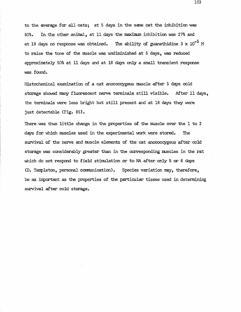
to the average for all cats; at 5 days in the same cat the inhibition was60%, In the other animal, at 11 days the maximum inhibition was 27% and
-5at 18 days no response was obtained. The ability of guanethidine 3 x 10 M to raise the tone of the nuscle was undiminished at 5 days, was reduced approximately 50% at 11 days and at 18 days only a small transient response was found.
Histochemical examination of a cat anococcygeus nuscle after 5 days cold storage showed many fluorescent nerve terminals still visible. After 11 days, the terminals were less bright but still present and at 18 days they were just detectable (Fig. 85).
There was thus little change in the properties of the muscle over the 1 to 2 days for which muscles used in the experimental work were stored. The survival of the nerve and nuscle elements of the cat anococcygeus after cold storage was considerably greater than in the corresponding muscles in the rat which do not respond to field stimulation or to NA after only 5 or 6 days (D. Templeton, personal communication). Species variation may, therefore, be as important as the properties of the particular tissue used in determining survival after cold storage.
DISCUSSION
im
The work on which this thesis is based falls conveniently into three related sections, the rat anococcygeus in vivo, the effects of nerve stinulation on NA depletion by reserpine and the cat anococcygeus in vitro. These are discussed in turn followed by a section linking them together,
RAT ANOCOCCYGEUS IN VIVO
The work of Gillespie (1972) made it clear that field stimulation of the rat anococcygeus could produce two separate and opposing nerve mediated responses, a motor response in the absence of drugs and an inhibitory response when the motor nerve response was blocked and the tone of the muscle raised* On histochemical and pharmacological grounds, it was shown that neither response was preganglionic and that the motor response was due to adrenergic neurones. The inhibitory response could, however, be neither blocked nor mimicked by conventional drugs and was, therefore, uncharacterised,
The possible mechanisms responsible for a dual response are several, e.g.(1), If only one type of fibre is concerned, the nature of the response is determined by (a) the "peripheral mechanism” (McCrea, ifcSwiney & Stopford, 1925) (b) Wedensky inhibition (e.g. Veach, 1925; Cannon,Raule & Schaefer, 1954) (c) variable effect of a single transmitter(e.g. Bozler, 1940; Burn & Robinson, 1951; Gardner & Kandel, 1972).(2) Two functionally different fibre groups exist, usually adrenergic and cholinergic, and these are organised either as (a) sympathetic and parasympathetic fibres with separate anatomical origin but which may come together peripherally to reach the target organ in a common nerve bundle (e.g. McSwiney & Robson, 1929; Finkleman, 1930) (b) fibres are ofsimilar origin but sympathetic may be cholinergic (Dale & Feldberg, 1934) and parasympathetic my be adrenergic.
The experiments described, fortunately eliminate all of these explanationsother than the existence of a double set of fibres with separate origins inthe spinal cord and mediated by two different transmitters. The preganglionicfibres in the motor pathway originate in the lower thoracic and upper lumbar j
• |region of the spinal cord whereas the preganglionic fibres in the inhibitory pathway originate in the lower lumbar and upper sacral region. This demonstrates clearly that there are two separate nerve pathways giving rise to the two opposing responses and eliminates the possibilities of only one type of fibre being involved or of two different types of fibre with a similar origin. This has now been supported by the evidence that after a sympathectomy by 60HDA, the motor response to field stimulation is greatly reduced but the inhibitory response remains unchanged (Gillespie & Gibson, 1973),
The nature of the motor fibres is clear. Anatomically, preganglionic fibres arising from the thoracicoj- lumbar outflow belong almost exclusively to / |the sympathetic nervous system (Mitchell, 1953; Pick, 1970) so the 'classification as sympathetic of the motor nerves is justified on 1
anatomical grounds alone as well as being supported by histochemical and pharmacological evidence of their adrenergic nature*
The nature of the inhibitory fibres is more interesting* On the basis of their spinal origin, they could have been either autonomic efferents or possibly, posterior root afferents equivalent to the posterior root vaso - dilators (Bayliss, 1902; Lewis & Marvin, 1927; Wybauw, 1936; Holton &Perry, 1951)* If they are autonomic they are not cholinergic as their spinal origin (similar to sacral parasympathetic) might suggest since they are not blocked by atropine* The blockade of the inhibitory responses by hexamethonium, on the other hand, suggests that they are autonomic efferent
fibres % If they are not cholinergic, then they might have been purinergic but their spinal position does not commend the inhibitory nerves to classification within the purinergic system either. In his review of this system, Burnstock (1972) cites only one tentative, unpublished example of purinergic nerves under sacral parasympathetic control. Central control of purinergic nerves, where it occurs at all, appears to be via the vagus.
Ihe possibility of antidromic stinulation of posterior root afferent fibres was also eliminated by the use of ganglion blockers. This possibility is excluded if a ganglion is shown to be in the pathway since afferent nerves lack a peripheral synapse, and even if they had one, it would be unlikely to transmit impulses in the antidromic direction.
The demonstration of ganglion blockade again became useful when it was found that the motor nerves could be stimulated postganglionically with the spinal electrode in a position which partially overlapped with the origin of the inhibitory nerves. Since the inhibitory region extends one segment further rostrally to L 5 , enabling selective stimulation of the inhibitory nerves alone, as demonstrated in the presence of tyramine, ganglion blockade was not a necessary condition for the demonstration that the motor and inhibitory pathways were separate. It served, however, as a further adjunct.
This ability to stimulate the motor nerves postganglionically was, in itself,y interesting since in previous investigations of this method of stimulating ! the spinal outflows, it had never been possible to stimulate postganglionic fibres (Gillespie, MacLaren & Pollock, 1970) and it is believed that the usual location of such stimulation is the point at which the ventral roots leave the vertebral canal. One explanation for the present exception could have been the position of the anococcygeus directly in front of the vertebral column,
107
enabling stinulation of the nerves within the muscle. This was, however, excluded by the radiographic demonstration that the distance between the electrode and the nuscle was at least 20 mm and up to 40 mm.
The most likely explanation is that some anatomical feature brings the postganglionic sympathetic nerve which supplies the anococcygeus close to the vertebral column in the upper sacral region, bringing it into close apposition with the spinal electrode in this position and producing favourable conditions far stimulation. The precise anatomical feature which makes this possible is not known but it has been shown that postganglionic fibres of the genitofemoral nerve, which supplies the anococcygeus, can take an unusual route which might make it particularly susceptible to stimulation by a spinal electrode (Pick, 1970), The postganglionic sympathetic fibres leave the sympathetic ganglion by the usual route via the gray ramus to the spinal nerve but then may leave the spinal nerve again and fuse with the sympathetic trunk at a more caudal level. This feature has been reported only in the rhesus monkey (Zuckerman, 1938; Sheehan &Pick, 1943) and in man (Pick, 1970) but if it occurs in the rat, it might explain why (1 ) preganglionic fibres can be stimulated in the upper lumbar region, in the usual way,(2 ) no fibres to the anococcygeus can be stimulated in the central lumbar region since the preganglionic outflow to the tissue ceases at L 3 and(3 ) postganglionic fibres can be stimulated in the lower lumbar and upper sacral region where the postganglionic nerve again canes near to the vertebral column. This hypothesis is, however, not wholly satisfactory since it brings the postganglionic nerve only as near to the vertebral column as the sympathetic trunk, and we have no reason to suppose that the spinal electrode can normally stimulate this structure. This may, nevertheless, point to the type of feature involved.
Another possible factor bearing on the apparent uniqueness of this ability to stimulate the anococcygeus postganglionically might be the location of the ganglia. In many of the previous investigations carried out using the pithing rod as the electrode, the responses measured have been due to either parasympathetic nerves with their ganglia located close to the target organ e.g. sacral parasympathetic to the bladder or colon (Gillespie, MacLaren & Pollock, 1970), or to sympathetic nerves with ganglia located at sites distant from the sympathetic chain e.g. thoracico -lumbar outflow to the vas deferens via the hypogastric nerve with the ganglia located along the peripheral end of the nerve, to the heart via the stellate ganglia, to the nictitating membrane via the superior cervical ganglion and although not strictly a ganglion synapse, to the adrenal medulla (Gillespie & Muir,1967 a; Gillespie, MacLaren & Pollock, 1970; Gillespie, MacLaren, Marshall, Muir & Pollock, 1970; MacLaren & Pollock, unpublished). In the anococcygeus, on the other hand, the sympathetic motor nerves originate from the genito - femoral nerve which has paravertebral ganglia located in the sympathetic chain. It is, therefore, possible that the anococcygeus is one of the few tissues we have examined in the pithed rat which has a
4
"classical" sympathetic innervation, the other main exception being the thoracico - lumbar vasomotor outflow, and that If other such pathways were examined, postganglionic stimulation might be possible there also.
Whatever the reason for this facility far pre - ar post - ganglionic nerve stimulation, it does provide a simple preparation in which both pre - and post - ganglionic nerve stimulation can be selectively achieved and the responses of the end organ measured in situ with a minimum of dissection.This might provide a convenient in vivo means of assessing drugs acting on ganglia, especially since the motor pathway to the anococcygeus appears to be relatively susceptible to ganglion blockade compared with other pathways
such as the motor nerves to the vas deferens or the inhibitory pathway to the anococcygeus itself.
The reason far this differential susceptibility of ganglia to blockade in various pathways is not clear from the present experiments. The motor pathway to the vas deferens, the inhibitory pathway to the anococcygeus and the nerve - mediated depressor responses seen when blood pressure was raised all showed comparatively rapid recovery from the effects of ganglion blockers when compared with the motor pathway to the anococcygeus. In addition, the Inhibitory pathway to the anococcygeus exhibited a form of desensitisation whereby an initial low dose of hexamethanium prevented blockade by a subsequent larger dose.
Differential action between tissues in terms of both time scale and the extentof blockade is not surprising since at their introduction as ganglion blockersby Paton & Zaimis (1951), it was noticed that the methoninm salts hadquantitatively different actions on different pathways and that the relativeefficacy on different pathways varied between different members of the series.It has subsequently become accepted that differences do exist in thesusceptibilities of various autonomic ganglia to ganglion blocking agents andindeed that they may exist between cell groups within a given ganglion (Voile& Koelle, 1965). It has further been suggested that morphological differencesbetween ganglia may produce uneven distribution of blocking agents and thatthis may be a primary reason for the different sensitivities to blockade(Shaw, MacCallum, Dewhurst & Mainland, 1951; Voile, 1962). In the presentinvestigation, the paravertebral ganglia of the anococcygeus motor pathwayand those of the vasopressor nerves were more susceptible to blockade thanthose elsewhere but there is no justification in the literature for a simplecorrelation between the anatomical nature of the ganglion and the effects of blocking agents; indeed the difference between the susceptibilities of cells
within a given ganglion make this clear. A further possibility which was at first considered for the relative resistance of the ganglia in the anococcygeus motor pathway was that after blockade of the hexamethonium - sensitive transmission process in the ganglia, an atropine - sensitive mi scar inic transmission process might have taken over (Steinberg & Hilton,1966). Atropine, however, produced no further ganglion blockade when the pathway had recovered from or was desensitised to hexamethonium. Muscarinic transmission does not, therefore, appear to be responsible for the resistance to conventional ganglion blockade. _
The anococcygeus responded to stinulation of the adrenals on a time scale similar to that of the heart rate and blood pressure. Stinulation of the thoracic outflow to the adrenals with a 300 pulse train at 30 Hz produced a response in the anococcygeus similar to that of 1 ug NA in a 250 g rat, this response being 70% of the response to maximal motor nerve stinulation. This high in situ sensitivity to catecholamines of either endogenous or exogenous origin is similar to the response of the nictitating membrane (Rosenblueth & Bard, 1932) but different from that of the vas deferens which contracts to neither adrenal stinulation nor injected catecholamines. In the vas deferens, only a small response in the perfusion pressure was recorded following adrenal stinulation. Since all three tissues, anococcygeus, vas deferens, and nictitating membrane, receive a motor adrenergic innervation and are contracted in vitro by catecholamines, this differential effect of circulating catecholamines may be produced by several factors which are common to the anococcygeus and nictitating membrane tut different for the vas deferens. Differences in sensitivity to drugs are usually explained by differences in the access of the drug to its site of action or to differences in the sensitivity of the tissue itself to the drug.
Access of catecholamines to their site of action has been invoked as a factor determining the response to catecholamines in two ways.
In the anococcygeus, vas deferens (Birmingham, 1970; Greenberg & Long, 1971) and nictitating membrane (Trendelenburg & Weiner, 1962), blockade of the neuronal NA uptake mechanism or complete removal of adrenergic nerves by sympathectomy produces increased sensitivity to NA. It has been suggested that in the vas deferens, the highly efficient neuroral uptake mechanism my be responsible for the comparatively low sensitivity to NA (see Birmingham,1970). There is, however, no evidence that blockade of this mechanism leadsto any greater increase in sensitivity of the vas than of the other twotissues (Birmingham, 1970; Tsai, Denholm & McGrath, 1968; Gibson &Gillespie, 1973), which suggests that this is not the primary reason for the difference in sensitivity. ^
Also under the heading of access, it has been suggested that the low sensitivity of the vas to several drugs my be due to a relatively low rate of perfusion of blood through the tissue (Kopin, Gordon & Horst, 1965; Sjostrand & Swedin, 1968). This might be expected to lead to a lower net drug concentration at the site of action and therefore a smaller response far a given concentration, but this hypothesis does not hold true far all drugs e.g. guanethidine and reserpine both exert their blocking action on the motor nerves within 2 min, and judging by the lack of fatigue after vigorous, repetitive stimulation ofthe sympathetic outflow of the tissue over a 2 hr period, the tissue is veryadequately perfused for maintenance of its metabolic activity.
The most convincing explanation for the differences in sensitivity between these tissues is the sensitivity of the muscle cells themselves to the drug. Thismy be related to the cytological arrangement of the nuscle and to the physiological role of the muscle cells in the various tissues. In the vas

deferens, the synaptic cleft between nerve and nuscle is relatively close (Burnstock, 1970) and each cell receives at least one synapse (Richardson, 1962). Conditions are thus favourable for synaptic transmission in this narrow cleft where high concentrations of NA my be built up by release from nerves. Over the surface of the cell as a whole, however, sensitivity to NA is insufficient for either exogenously administered NA or catecholamines from the adrenals to act. In the anococcygeus (Gillespie, personal comnunication) and in the nictitating membrane (Esterhuizen, Graham, Lever & Spriggs, 1968), however, the syraptic gap is larger i.e. 1 0 0 0 - 3000 A cf 2 0 0 A for vas deferens, and from both the in vitro and in vivo evidence, sensitivity of the cells as a whole to catecholamines is greater. Thus in the latter tissues, the nerve and iruscle are organised in such a way that the transmitter my act over a wider area than in the vas, perhaps that from one varicosity acting on several cells, and also so that blood catecholamines my produce an effect on the tissue. In the vas deferens, on the other hand, the organisation favours specific nerve transmission via discrete connections and prevents contraction of the muscle by blood catecholamines«
Ihis situation thus favours a simple contraction of the anococcygeus and nictitating membrane by either sympathetic nerve or adrenal activity. Since both tissues consist of sheets of smooth muscle and the function of the nictitating membrane and probably also of the anococcygeus is to contract under conditions of sympathetic stimulation, then their cytological organisation, which manifests itself in the present experiments by influencing the pharmacological sensitivity to NA, is seen to be necessary for their function.In the vas deferens, on the other hand, the nuscle is organised in a complex manner with inner circular and outer longitudinal layers. The function of this nuscle is not simply to shorten, but to propel the seminal fluid, in one
direction, during the process of emission. It is thus likely that specific control of each muscle cell, or at least of snail bundles, is necessary in order that the contractions of the various components are integrated to produce this response. Since sexual activity takes place only under specific circumstances, it would be undesirable if adrenal activity, whenever it occurred, produced emission. In addition, the greater sensitivity to circulating catecholamines of the circular compared with the longitudinal nuscle, suggests same further physiological correlate. The structure and pharmacological properties of the vas deferens are thus less surprising if its function is taken into account. So far as I can determine from the literature, very little is known of the precise mechanism of nuscle contraction leading to emission (see Bell, 1972) despite the popularity of the vas deferens far pharmacological investigation,
The cardiovascular responses in these experiments corresponded with the findings in the original experiments of Gillespie, MacLaren & Pollock, (1970), with the additional uncovering of a nerve - mediated depressor response.
The origin of the splanchnic nerve fibres to the adrenal medulla overlaps with the direct sympathetic outflow to blood vessels. For this reason it is not possible to elicit a pressor response from adrenal stinulation without a superimposed response from stimulation of the sympathetic nerves to the arterioles. Simultaneous experimental release and assay of adrenal catecholamines in the one animal is therefore not possible utilising the pressor response. Since the sympathetic outflow to the anococcygeus arises some considerable distance from the fibres to the adrenal medulla and since the anococcygeus shows a high sensitivity to circulating adrenaline, it is possible to use it as an assay organ for adrenal output.
In the presence of raised blood pressure, an unexpected depressor response ms produced by stimulation in the lower lumbar and upper sacral regions. This response was abolished by ganglion blockade so is therefore not due to antidromic stimulation of afferent nerves, and was unaffected by atropine*This effect of atropine eliminates the possibility of cholinergic vasodilator nerves*
Dusting and Rand (1972) have demonstrated depressor responses to stimulation of the entire autonomic outflow after artificially raising the blood pressure with NA. In their experiments, no neuro - muscular blocker was adminstered to prevent skeletal nuscle twitching* By sequential administration of blockers, they demonstrated that a large part of their complex depressor responses was due to (1) excitation of motoneurones with consequent (a) functional hyperaemia in skeletal muscle, producing delayed depressor responses (b) overflow of acetylcholine from cholinergic nerves to blood vessels ~ (2) ft - effects of adrenaline released from the adrenal medulla(3) an extra component which they could not block and therefore did not characterise n*b, this response was resistant to among other things, hexamethonium. None of these factors are likely to be relevant to the present findings* Functional hyperaemia is not a factor since pancuronium was administered* Overflow of acetylcholine seems an unlikely cause since atropine did not block the response and, due to the selective method of stimulation, the adrenals are not a factor* The final uncharacterised part of their response is also unlikely to be related to the present response since their electrode ended at L 5 or L 6 which is just at the rostral end of the origin of the present depressor nerves and finally their response, unlike the present one, was unaffected by hexamethonium*
There thus appears to be a sacral preganglionic outflow in the rat with an
origin similar to that of the inhibitory nerves to the anoooccygeus and which produces depressor responses by a non - adrenergic, non - cholinergic mechanism. These depressor responses, in absolute terms, are relatively snail but, if as is likely, the vascular bed affected is limited to a particular area, then the effect in that bed would be substantial. No such responses have previously been reported in the pithed rat, but Hughes & Vane (1967) have demonstrated a non - adrenergic, non - cholinergic inhibitory nerve response in the isolated portal vein which might be analogous to the peripheral component of this response.
The similarity of this depressor response in the pithed rat to the inhibitory response in the anococcygeus is quite clear. Both share a similar spinal origin, have ganglia with a similar sensitivity to hexamethonium and are neither adrenergic nor cholinergic. It may, therefore, be tentatively postulated that they belong to related systems.
In recent years radical new theories on the nature of transmission in the vas deferens have been advanced. Ambache & Zar (1971) have postulated that the motor transmitter is not NA tut some unknown chemical. NA is, of course, present in large quantities in the vas deferens and, furthermore, is undoubtedly present in nerves. Ambache and co - workers postulate that NA and the adrenergic nerves are inhibitory. This theory is based irainly on four observations on the isolated guinea - pig and rat vas deferens -
(1) The undoubted low sensitivity of the vas to the motor effect of NA (Ambache & Zar, 1970).
(2) The inability of cL— receptor blockers such as phentolamine or NA depletion by reserpine to abolish the response to motor nerve stinulation (Ambache & Zar, 1971),
(3) The ability of NA, tyramine or cocaine to inhibit the motor response
116
to nerve stimulation (Ambache & Zar, 1970; Ambache, Dunk, Verney & Zar, 1972).
(40 The ability of LSD to block the motor response to. nerve stirrulation (Ambache, Dunk, Verney & Zar, 1973)*
The only evidence which they found to be against their theory was that guanethidine and bretylium completely abolished the motor nerve response. / This was, however, explained as a non - specific action of guanethidine and bretylium since they have been shown to be capable of paralysing cholinergic transmission in the phrenic nerve - diaphragm preparation (Dixit, Gulati & Gokhale, 1961).
A major factor which must be borne in mind when comparing the work of Ambache and co - workers with that of other authors is that they employ only short pulse trains, preferably 15 pulses or less, which they use because they consider this to be as many pulses as are likely to occur physiologically although they do not quote the basis far this assumption. As a result of this, they are observing only the first phase of the biphasic response seen in the present experiments or in those of Swedin (1971). They also show that inhibition by NA and LSD is mere effective on short than on long trains (Ambache & Zar, 1971; Ambache, Dank, Verney & Zar, 1973). This first part of the response is, of course, the part which is most unconventional in its response to drugs affecting adrenergic systems. This confinement to the study of a short train of pulses will, therefore, put an undue bias against adrenergic motor transmission.
Another hypothesis which seeks to explain the motor transmission mechanism but taking into account the two phases of the response was put forward by Swedin (1971). From studies on the rat and guinea - pig isolated vas deferens, he concluded that both phases were due to adrenergic nerves but that the basis of the two phases was that the NA released from the nerves acted at two
different sites of action on the muscle. In this hypothesis, NA released into the synaptic cleft produces the fast "spike" response. This same NA then diffuses to more distant sites on the muscle to produce the slower secondary response. It is difficult to see, since nerve stimulation continues, why the initial response should wane before the secondary response appears, and this is not explained in his argument.
These two theories of Ambache et al and of Swedin are obviously mutually antagonistic although the main conflicts arise from differences of experimental approach. The present experiments, however, offered experimental advantages in investigating this problem since the anococcygeus has a similar motor innervation arising from a similar spinal level but with a sensitivity to both agonists and antagonists which corresponds to a conventional adrenergic innervation. It therefore serves as an excellent control on which to study simultaneously the effects of drugs on the vas deferens. In this way, a comparative study could be made of the similarities and differences which this motor innervation in the vas deferens has to the motor adrenergic nerves in another smooth muscle tissue.
In the present study, the effects of drugs on the motor response to stimulation of the sympathetic outflow to the vas are discussed in relation to the biphasic nature of the response. Three main factors bear on any interpretation of thisresponse - (1) The two phases appear to have a common spinal origin(2) They are differentially affected by several drugs which act at either the ganglion or the nerve - muscle junction (3) Only one component of the response appears to be present in the circular muscle. With these points in mind, it is concluded from the present experiments that there are two anatomically and pharmacologically distinct components to the motor innervation of the vas although these have a common spinal origin.
118
This leads to the development of yet another hypothesis concerning the nature of the motor innervation in the vas deferens. Each component of the response, in this hypothesis, is considered to be due to a separate nerve pathway.
The first, which produces the initial "spike" response, acts mainly on the longitudinal muscle of the vas, is less susceptible to ganglion blockade than the second pathway and is inhibited by an oC- adrenergic mechanism which normally terminates the response by negative feed - back. This oL- inhibition can be reinforced by exogenously applied NA or, most effectively, by endogenous release of NA. by the indirect synpathomimetic action of tyramine cr LSD and can be blocked by phentolamine or by pretreatment with reserpine, both of which produce prolongation of the "spike". Apart from guanethidine and the .chronic effects of reserpine treatment, drugs normally associated with modification of adrenergic responses do not affect this response in the way that they affect, far instance, the anococcygeus response or the secondary phase of the vas’s own response.
The second component in the pathway produces the secondary response, which occurs in both the circular and the longitudinal layers, is more susceptible to ganglion blockade than the "spike" and is potentiated by an cL- adrenergic mechanism. This secondary response is thus potentiated by exogenous NA or by the indirect syirpathanimetic action of tyramine or LSD and is inhibited by phentolamine or by reserpine pretreatment. The effects of all the drugs tested on this part of the response were similar to those on the anococcygeus and are as might be expected in a conventional adrenergic system.
This interpretation stands in partial disagreement with the explanations of motor transmission in the vas given by Ambache et al and by Swedin but is basically an attempt to resolve sane of the problems inherent in both these
theories in order to farm a more cohesive working hypothesis.
The experimental findings which led to this hypothesis are not in conflict with those of Ambache and co - workers except in some points of interpretation. Their view that NA, tyramine and LSD are all inhibitory to the motor nerves is confirmed, provided that it is acknowledged that only the first component of the response is so affected.
A major difference arises, however, when the effects of reserpine are considered. Ambache, Dunk, Verney and Zar, (1972), concluded that tyramine and cocaine exerted their inhibitory effect by an indirect sympathomimetic action but that LSD did not produce its action through this means since reserpine pretreatment did not prevent its effect (Ambache, Dunk, Verney &Zar, 1973). Since it is also part of their hypothesis that transmission is non - adrenergic since reserpine pretreatment does not abolish it, a large part of their argument is seen to rest on the ability of reserpine to completely prevent both adrenergic transmission and indirect sympathomimetic effects. From the present work on the anococcygeus and other work on other tissues (e.g. Sedvall & Thorson, 1965j Anden & Henning, 1966j Lee, 1967) it is clear that even depletion of NA which is virtually complete as measured by biochemical estimation of NA content, may be insufficient to cause a complete blockade of adrenergic transmission. If this is the case in the vas deferens also, as seems likely (see Swedin, 1971), then the sympathectomy produced by reserpine pretreatment is insufficient to prevent the action of indirect sympathomimetics as well as adrenergic transmission. To counter this, Ambache, EUnk, Verney & Zar (1973), claim that the failure of tyramine to inhibit motor responses after reserpine treatment is a measure of the failure of indirect sympathomimetics under these circumstances. The present experiments in vitro with both rat and cat anococcygeus demonstrated that in this tissue at least, LSD is a more powerful and persistent indirect
sympathomimetic than tyramine and so its effects might be expected to be more difficult to suppress. In support of adrenergic transmission, and relevant to this point, Wadsworth (1973) has demonstrated that if a complete sympathectomy is produced in the rat vas deferens by in vitro 6QHDA treatment then no motor nerve responses can be obtained. This suggests that the reserpinisation employed by Ambache, Dunk, Verney & Zar (1973) is indeed insufficient to produce complete sympathectomy.
In addition to this, both the present experiments and those of Ambache, KiUick, Srinivasan & Zar (1973) have demonstrated that LSD also inhibits the motor response in the anococcygeus which certainly is adrenergic. This weakens the argument that inhibition by LSD points to a non - adrenergic mechanism. More interesting, and possibly more relevant, is the clear similarity between the effect of LSD and that of guanethidine, both of which block the adrenergic response in the anococcygeus as well as releasing NA from the nerves. Since guanethidine is the one drug whose action in the vas is consistent with an adrenergic innervation, LSD should also perhaps be interpreted as reinforcing the adrenergic nature of the initial component in the response. Whether the mechanism of action is similar far LSD and guanethidine, the net result of NA release and neurone blockade is different in the vas and anococcygeus. Since the anococcygeus is highly sensitive to the motor action of endogenously released NA, it would be expected that indirect sympathomimetics would cause contraction whereas the vas with a low sensitivity to the motor action of endogenously released NA could be expected not to show the indirect sympathomimetic effects. If the present hypothesis is true, then the feed - back inhibition of NA release in the vas deferens produced by an o(. - receptor action via indirect release of NA should be prevented by phentolamine. Direct neurone blockade of the guanethidine type on the other hand, presumably would not.
From this it is concluded that there is insufficient evidence available to
justify the rejection of NA as motor transmitter in the vas, even for the ubiquitous initial component.
Many of the results reported by Swedin (1971) suggest that motor transmission in the vas is adrenergic and corroborate with the present results e.g. similar results with reserpine pretreatment and guanethidine, and a preferential inhibition of the first phase by PGE is demonstrated which suggests a further pharmacological difference between the two phases. ,—
Swedin1 s hypothesis that the two phases are produced by NA acting at two separate sites of action, while fitting his own experimental findings, is difficult to adapt to the present results. These demonstrate a differential action of ganglion blockers and a difference in the response of circular and longitudinal muscle. Ambache, Dunk, Verney & Zar (1972) raise a related objection to Swedin*s hypothesis. This is the apparent contradiction whereby FGE2 is claimed to inhibit the "spike" phase by inhibition of NA release and yet the secondary phase, which is also supposed to be due to this same NA, is unaffected. These objections are resolved if the two hypotheses are combined so that the two phases are due to different nerves, both of which are adrenergic but only one of which is susceptible to PGE2. This latter explanation is the view of the present author.
A further objection to Swedin1 s hypothesis is raised by Ambache, Dunk, Verney & Zar (1972) which might also apply to the present hypothesis. This is based on their own findings that the motor effect of tyramine is abolished by oL - blockers which they claim negates the existence of 'X. - receptors resistant to o(- blockers, a concept postulated by several authors (see Holrran, 1970) and which is necessary in Swedin*s system. This objection is not entirely sound since it is quite likely from the system suggested by Swedin that both the high concentration of NA and its localisation to the
synaptic cleft is responsible for the resistance to phentolamine. It is unlikely that tyramine will release NA. in this localised fashion or at the same initial rate as does nerve stimulation and so it is hardly surprising that it is more susceptible to blockade.
It is hoped tiiat the present hypothesis ties together the work of both Ambache et al and Swedin to provide a more cohesive working hypothesis of motor transmission in the vas deferens and that it does not simply lead to further confusion. It brings together the major contribution of the work of Ambache et al which is that the motor nerves of the vas deferens are unconventional by the standard of other known adrenergic systems and that syirpathomimetic agents are inhibitory to these nerves and the contribution of Swedin which is that there are two phases to the response and that both bear the characteristics of adrenergic responses. It is further suggested that many of the experimental anomalies arising from these properties may be explained by the presence of two sets of nerves. No new concepts have been introduced in forming this further hypothesis except that LSD should be considered as both a neurone blocking agent and an indirect sympathomimetic and that the two components of the vas deferens response are both adrenergic but elicited from two different muscle groups, the initial fast "spike11 from the longitudinal and the slow response from both longitudinal and circular layers. The properties of the adrenergic nerves innervating these two regions also appear to differ in that those serving the longitudinal muscle are influenced by prostaglandins. Even the suggestion that differences exist between the two muscle layers has been demonstrated by Donald (1969). She showed that splitting the vas longitudinally led to a decrease in the longitudinal tension response to nerve or drug stimulation which could only be explained if the circular muscle was contributing a significant part of the longitudinal tension in the unsplit vas. She further demonstrated considerable
qualitative differences between the split and unsplit vas in response to agonist drugs which suggested different properties for the longitudinal and circular layers. The only other concept which is employed in forming the present hypothesis is that of endogenous inhibition of NA release by an cL - receptor mechanism. This is now a well established phenomenon in the vas deferens (see Stjame, 1973).
In summary, it is hoped that this work with the anococcygeus of the pithed rat has demonstrated that the anococcygeus muscle receives two anatomically separate nerve pathways, one motor and one inhibitory, which originate frcm different levels of the spinal card, producing opposing responses on the muscle that both belong to the autonomic nervous system as indicated by the presence of ganglia, that the transmitter in the motor nerves is noradrenaline, that the transmission in the inhibitory nerves is unlikely to be either cholinergic or purinergic and that both sets of nerves have properties which are qualitatively similar to those in the cardiovascular system. In addition, comparison of the effects of drugs on the motor innervation of the anococcygeus and vas deferens demonstrated that transmission in the anococcygeus is relatively straightforward and may help to clarify some of the problems involved in the understanding of the more complex motor transmission in the vas
EFFECTS OF NERVE STIMULATION ON NA DEPLETION BY RESERPINE
Fluorescence histochemistry shews the anococcygeus to have a dense adrenergic innervation distributed throughout the muscle. The appearance of this innervation is similar to but less dense than that of the vas deferens. In
addition, preliminary experiments using the rcdcrospectroflucrometric technique of Caspersson, HiUarp & Ritzen (1966), which identifies the substance giving rise to fluorescence after formaldehyde condensation according to its emission spectrum, have indicated that the fluorophore in both tissues bears the characteristics of NA. This is confirmed by the biochemical measurement of NA content, which shows the anococcygeus to contain 2.56 tg NA / g tissue as opposed to 15.56 pg / g in the vas. Although less than in the vas, this NA content in the anococcygeus is nevertheless fairly high compared with other smooth nuscle tissues e.g. rat gastro - intestinal tract, 0.22 - 0.98 ug / g; cat nictitating membrane, 2.4 - 4,3 jig / g; rabbit blood vessels, 0.24 - 0.90 ug / g (far further comparisons see Holzbauer & Shaman, 1972). This similarity combined with the fact that the adrenergic innervation is motor in both tissues provides grounds for making comparisons between the properties of the nerves in the anococcygeus and vas deferens as demonstrated in Part I.One major difference between the two tissues, however, is that the ganglia on the sympathetic pathway to the vas deferens occur along the hypogastric nerve, near the tissue itself, giving rise to the so - called "short" adrenergic neurones, whereas the corresponding ganglia in the sympathetic pathway to the anococcygeus are located in the paravertebral ganglia, giving rise to the conventional "long" adrenergic neurones (see Part I). It has been suggested that this property of the vas neurones might be responsible for the tissue*s relative resistance to depletion by reserpine.
It was thus interesting that reserpine should deplete both the anococcygeus and vas deferens of NA to a similar extent with different doses and at a similar rate with a fixed, moderate (200 pg / kg) dose. The basis for the belief that resistance to NA depletion in the vas by reserpine was due to the short adrenergic neurones, was that tissues such as the vas deferens and seminal
125
vesicles were mere resistant to depletion than tissues receiving long adrenergic neurones (see Sjostrand & Swedin, 1968). The discovery of a tissue with long adrenergic neurones which is depleted in a manner similar to that of the vas but different from that of other tissues thus makes this less likely. That both the anococcygeus and vas are depleted more slowly than the heart, confirms that a similar ’’resistance” to reserpine exists in the anococcygeus and vas deferens.
If it is not the length of the neurone that is the significant factor, what then is the common factor in the vas and anococcygeus giving rise to this resistance? Comparisons carried out so far show that the two tissues recover at a different rate from the effects of reserpine, their nerves show mjor differences in response to many drugs (see Part I) and the cytological organisation of the tissues as shown by electron microscopy is quite different (Gillespie, personal communication). There are thus, on pharmacological and morphological grounds, many differences as well as similarities between the two tissues. -
Another factor suggested by Sjostrand & Swedin, that low resting impulse traffic to the vas deferens could be responsible for resistance to depletion, could be tested with the pithed rat. Since comparison between the three tissues showed that the major difference between depletion in the vas and anococcygeus was in the rate of depletion rather than the final level reached (see also Sjostrand & Swedin, 1968), then impulse traffic seemed a possibility. It is known that there is a continuous discharge in the sympathetic nerves to the heart (Bronk, Ferguson, Margaria & Solandt, 1936) and that this will be reflexly increased as reserpine lowers systemic blood pressure by its action on the adrenergic nerves to the blood vessels (Iggo & Vogt, 1960). No information is as yet available on the presence of spontaneous activity in the nerves to the
anococcygeus nor am I aware of such information in the literature far the nerves to the vas deferens although the function of the vessel mkes this unlikely.None the less, the observations that pithing the animals reduces the depletion produced by reserpine in all three tissues and that nerve stiiiulation enhances the depletion in the anococcygeus and vas deferens, support the idea that a major factor determining the extent of depletion is the amount of neuronal activity.
It is not possible to say from the present experiments whether nerve activity could fully explain the difference between the heart and the other two tissues. If it does, then one would expect the degree of depletion to be similar in animals pithed and then given reserpine. These experiments have not been carried out since it is difficult consistently to keep pithed rats in good condition for the necessary length of time. In the experiments where reserpinised rats were pithed, reserpine had three hours to act before the abolition of impulse flow by pithing and this could explain the greater depletion in the heart (66%) than in the anococcygeus (H2%) and vas deferens (36%) at 6 hr.
These observations confirm and extend previous evidence that reduction of nerve inpulse traffic by nerve section or drugs blocking ganglia will reduce the depletion of NA by reserpine in various tissues (Holzbaner & Vogt, 1956; Karki, Paasonen & Vanhakartano, 1959; Mir kin, 1961; Hertting, Potter &Axelrod, 1962; Benmiloud & Euler, 1963). This is in disagreement with the experiments reported by Sjostrand & Swedin (1968) who sectioned rat hypogastric nerve and found that the depletion in the vas deferens was unaltered. The explanation may lie in the different conditions. In the vas deferens, where impulse traffic is low, it is unlikely that a big effect will be seen by abolition alone. Furthermore, these authors used a smaller
127
dose (50 pg/kg) of reserpine given subcutaneously so that the rate of depletion was less* Since the effect of abolishing impulse traffic to the vas in the present experiments is snail, it might be non - existent at smaller doses* In the present experiments the role of nervous activity in depletion was exaggerated by contrasting the depletion when nerve impulses were abolished with that when activity was reinforced by artificial nerve stimulation.
The act of pithing a rat leads to considerable autonomic discharge, presumably as a result of destruction of the spinal cord. This discharge continues for10 - 15 min and is manifested by the initially high blood pressure and heartrate characteristic of the pithed rat and also by a contraction seen on pithing in the anococcygeus if its tension is recorded in the anaesthetised rat prior to pithing* These blood pressure and anococcygeus responses are absent if the anirral is pretreated with phentolamine and are presumably due to a combination of sympathetic nerve discharge to the target organs themselves and to the adrenal medulla. In the present experiments, where reserpine pretreatment is employed, this discharge, if severe enough, resulted in an increased depletion of NA. A comparison of the NA contents of the tissues 1 hr after pithing i.e. 4 hrs after reserpine, with the contents 4 hrs after reserpine in unpithed animals showed no significant difference. If there had been no discharge, the rate of depletion during this 1 hr period might have been expected to fall. This discharge produced by pithing thus slightly reduces the effectiveness of pithing as a means of reducing the net impulseflow in the subsequent 3 hr period. Nevertheless, since it is a small effect,this does not interfere with the comparison of the NA content after pithing alone with that after pithing plus sympathetic stimulation. This is illustrated in Fig. 44.
In any study of the effect of nerve stimulation on NA content, the effect of homeostatic mechanisms on the maintenance of normal NA levels must be taken into account. For example, in the isolated guinea - pig vas deferens, during hypogastric nerve stimulation, the uptake and retention of radioactive NA from the surrounding fluid was increased (Ehagat & Zeidman, 1970) and the formation of NA from tyrosine was also increased (Thoa, Johnson, Kcpin &Weiner, 1971). Any change produced in NA content is thus the net result of competing homeostatic mechanisms tending to maintain normal amine levels and nerve stimulation depleting these levels. The outcome can thus only be adequately understood if each parameter can either be measured and a balance sheet drawn up or else controlled by pharmacological or other means. In the present study on the effects of reserpine, the homeostatic factors cannot maintain the NA content at normal levels since the NA made available by any increased synthesis or reuptake cannot be retained by the storage granules since it is here that reserpine exerts its action. Instead of being rade available for replenishment of the stares, any such NA would thus be exposed to metabdlism by intracellular mono - amine oxidase (see Kopin & Gordon, 1962).
The effects on NA content of the tissues of pithing the animals and / or stimulating the sympathetic outflows in the absence of reserpine were not statistically significant probably due to the small number of examples used far each conparison. This provided a valid basis for comparison with the statistically significant differences which did exist after reserpine pretreatment, but unfortunately precluded a proper assessment of the effects of pithing and stimulating per se. As a basis for further comparison, it is, however, interesting to compare the non - significant changes in the mean values which did occur with analogous changes found in other tissues. Thus the effect of pithing might be compared to that of decentralisation. Decentralisation of the cat spleen led to an increase in the NA content of the
129
organ (Brown, Dearnaley & Geffen, 1967) while comparable decentralisation of the guinea - pig vas deferens produced no such change (Blakeley, Dearnaley & Harrison, 1970). In the present experiments pithing produced a non - significant rise in the mean NA content of the anococcygeus but no change at all in the vas deferens or heart. Moderate stimulation of adrenergic nerves does not normally decrease the NA content of tissues (see Euler & Hellner - Bjorkman, 1955; Blakeley, Dearnaley & Harrison, 1970) although exceptions to this have been reported e.g. cat spleen (Dearnaley & Geffen, 1966), On the other hand, more vigorous stimulation can produce depletion e.g. guinea - pig vas deferens (Chang & Chang, 1965; Roth, Stjarne & Euler, 1967). In the present experiments, stimulation of the sympathetic outflows to the two tissues produced a non - significant fall in the content of the vas deferens with no change in the anococcygeus. Clearly any change in content after changing the amount of nerve activity will depend on the net result of release, metabolism, reuptake and resynthesis as mentioned above. Even with the complete abolition of sympathetic activity or vigorous stimulation employed, the present results are for the vas deferens and heart in accord with the previous literature and demonstrate that the anococcygeus, while possibly increasing its NA content on resting, can maintain its NA stares in the face of prolonged and repetitive stimulation. It would be interesting to know if this small effect is due to the efficiency of the compensating homeostatic mechanisms so that NA synthesis or turnover is enhanced.
The faster rate of recovery of NA contents after reserpine pretreatment in the vas deferens compared with the anococcygeus is interesting in view of the similarity in the rate and extent of depletion in the two tissues. It is also interesting that the rate of recovery in both tissues after this 200 jug/kg dose was slower than the recovery following 10 mg/kg in rat gastrocnemius muscle, sub - maxillary gland and heart found by Haggendal & Dahlstrom (1971),
These authors demonstrated that recovery of the NA content of tissues after reserpine is dependent on and can be explained solely by the axonal dcwn - transport of newly formed amine granules.' Implicit in their argument is that after the almost total depletion of NA resulting in complete loss of adrenergic function, there is a supranormal dcwn - transport of new granules leading to almost full recovery of NA content after three weeks, a fall at four to five weeks as these "extra" new granules reached the end of their life span and then finally complete recovery to control levels at six weeks. It is thus interesting that in the present experiments, where the initial depletion was not sufficient to abolish sympathetic function, the initial rate of recovery should be slower than that found after a larger depletion. If the initial stimulus which produces the increased formation and transport of new granules is dependent on a negative feed - back loop resulting frcm the loss of nerve function, then this would explain this apparently contradictory slower recovery after a smaller dose. Recovery in the present experiments, therefore, might depend on the arrival of newly formed granules at the normal rate. It has been suggested by Dahlstrom (1970) that the stimulus for the increased formation and transport of granules might be a reflex increase in sympathetic activity after reserpine as was demonstrated by Iggo & Vogt (1960). The consequent increased bombardment of the postganglionic neurone cell body might then result in an increased protein synthesis similar to that shewn by Hyden (1960) resulting in increased formation of new granules and the acconpanying enzymes required for catecholamine synthesis, such as dopamine /? - oxidase and tyrosine hydroxylase (Thoenen, Mueller & Axelrod, 1969; Axelrod, Mueller & Thoenen, 1970). In the present experiments such an increased nerve activity would be unlikely, at least as a consequence of impairment of transmission, and thus the stimulus to rapid recovery would be absent. Another factor which might be of interest but has not been examined
131
here would be whether* the postulated increase in protein synthesis in the cell body would occur in all tissues whose transmission was prevented by a large dose of reserpine. In a tissue which was not part of a homeostatic reflex loop this phenomenon might be absent. Into this category would fall the vas deferens and possibly also the anococcygeus if it plays a role in defaecation. This point could be answered by examining the rate of recovery of the tissues1 NA content after a large dose of reserpine.
The reason for the slower rate of recovery of NA content in the anococcygeus than in vas deferens is not known. On the basis of the "normal" rate of down - transport of new granules (72 - 240 mm/day, Dahlstrom & Haggendal,1966, 1967), a 50 mm difference in axone length on its own would be unlikely to result in a significant difference in the rate of arrival at the terminals. This would tend to eliminate the length of axone as a direct contributory factor.
The effects cf reserpine on the mechanical responses due to sympathetic nerve stimulation of the anococcygeus and vas deferens further illustrate a dual action of the drug on the response of the latter organ. These effects of reserpine were observed in three different situations. First, in a series of experiments where reserpine 200 }ig/kg i.p. was administered three hours prior to pithing, the responses of the anococcygeus were as found in untreated controls but in the vas, although the response to the first period of stimulation was normal, in subsequent periods, the "spike" phase rapidly diminished. Secondly, when a large dose of reserpine 3 mg/kg i.p. was given 18 hours prior to pithing in order to produce a large depletion of NA content, the response of the anococcygeus was slower to develop and reduced in size compared with controls while in the vas, although both phases were reduced in size, the "spike" was prolonged and the secondary response was absent. In
the third situation, when reserpine 3 mg/kg was given intravenously to the pithed rat, the response in the anococcygeus was unaffected but the "spike” in the vas was acutely inhibited, leaving only the secondary response. The anococcygeus response vas thus affected in the manner expected of a motor
t
adrenergically innervated tissue, being unaffected by acute doses and reduced in size only after considerable depletion of the catecholamine stores. Inthe vas deferens, on the other hand, there were two different effects. The"spike" was susceptible to acute large doses of reserpine and was alsoreduced 4 hrs after a small dose if stimulation was repeated.
This effect is analogous to that reported by von Euler (1969, 1970) in the isolated guinea - pig vas deferens in vitro. He found that reserpine added to the bath rapidly reduced the responses to either field or hypogastric nerve stimulation. Relating this to the effect of reserpine on inhibition of NA release from isolated granules (Euler & Lishajko, 1963; Euler, 1966 a & b), he concluded that this acute effect of reserpine could be due to reduction of NA release by nerve stimulation resulting from a reduction in the transfer of NA from the storage granules to the actual neuronal release sites. In agreement with this concept, the first stimulus response after addition of reserpine was normal, whereafter the contractions declined gradually at a rate depending on the dose. In these experiments the stimul - at ion period lasted only 5 sec so that according to the time scale (shape) of the two phase response in the isolated guinea - pig vas deferens reported by Swedin (1971), only the initial "spike" phase of the response was being examined. This effect is thus clearly analogous to the inhibition of the "spike" in the present experiments by an acute large dose or 4 hrs after a small dose of reserpine. In addition, the gradual disappearance of the "spike" with subsequent stimulation periods is similar to his findings.
Although the effect appears to be similar in every respect, the present experiments do, however, throw some doubt on von Euler1 s explanation. The first difficulty is that this effect appears to be absent in the anococcygeus. It is true that a similar acute inhibition of adrenergic responses by reserpine has been demonstrated on the isolated colon (Gillespie & Mackenna, 1961) and on the rabbit ear artery (Day & Owen, 1963) but the evidence was not strong, the former authors acknowledging that the effect could have been due to a solute effect and in the latter case the effect was so snail that the authors did not ccmment on it in the text.It thus appears that this acute effect of reserpine may be relatively specific far the "spike" phase of the vas deferens response in a manner analogous to the similar actions of PGE (Swedin, 1971) or LSD (Ambache,Dunk, Verney & Zar, 1973).
This similarity raised two possibilities which cannot be satisfactorily resolved by either von EulerTs or the present experiments, viz. either(1) LSD and perhaps also PGE act in a manner similar to reserpine in producing their effects i.e. by reducing NA release from granules by the nerve impulse, or (2) reserpine acts in a manner similar to that postulated by the present author far LSD, producing inhibition of the "spike" by acute release of NA.
The first of these possibilities is difficult to explain in view of the fact that only the "spike" phase is altered unless it is accepted that there are two different adrenergic motor nerve pathways and only one of them is affected. Furthermore, 18 hrs after reserpine 3 mg/kg, a "spike" can be obtained and so the initial inhibition of the "spike" appears to have disappeared at this time. LSD has, however, no effect at this time which together with the prevention of its effect by phentolamine is the reason far postulating that
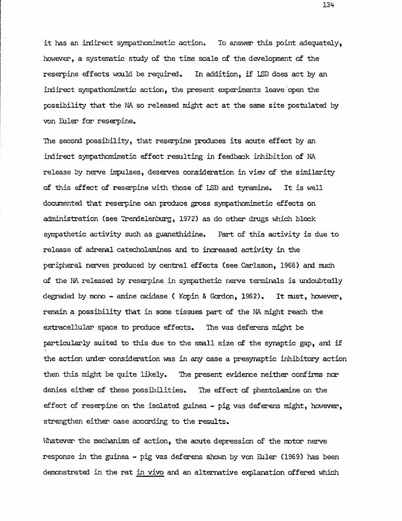
131*
it has an indirect sympathomimetic action* To answer this point adequately, however, a systematic study of the time scale of the development of the reserpine effects would be required. In addition, if LSD does act by an indirect sympathomimetic action, the present experiments leave open the possibility that the NA so released might act at the same site postulated by von Euler for reserpine*
The second possibility, that reserpine produces its acute effect by an indirect sympathomimetic effect resulting in feedback inhibition of NA release by nerve impulses, deserves consideration in view of the similarity of this effect of reserpine with those of LSD and tyramine. It is well documented that reserpine can produce gross synpathomimetic effects on administration (see Trendelenburg, 1972) as do other drugs which block sympathetic activity such as guanethidine. Part of this activity is due to release of adrenal catecholamines and to increased activity in the peripheral nerves produced by central effects (see Carlsson, 1966) and much of the NA released by reserpine in sympathetic nerve terminals is undoubtedly degraded by mono - amine oxidase ( Kcpin & Gordon, 1962). It nust, however, remain a possibility that in some tissues part of the NA might reach the extracellular space to produce effects. The vas deferens might be particularly suited to this due to the small size of the synaptic gap, and if the action under consideration was in any case a presynaptic inhibitory action then this might be quite likely. The present evidence neither confirms nor denies either of these possibilities. The effect of phentolamine on the effect of reserpine on the isolated guinea - pig vas deferens might, however, strengthen either case according to the results.
Whatever the mechanism of action, the acute depression of the motor nerve response in the guinea - pig vas deferens shown by von Euler (1969) has been demonstrated in the rat in vivo and an alternative explanation offered which
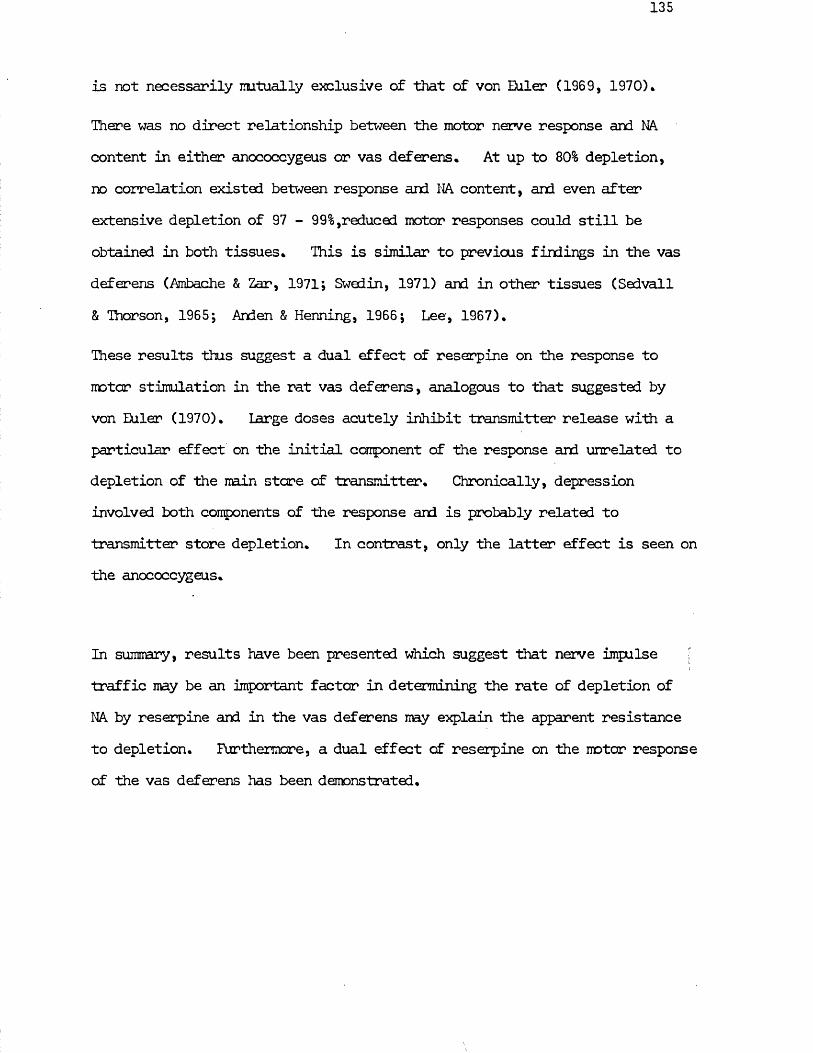
is not necessarily mutually exclusive of that of von Euler (1969, 1970).
There was no direct relationship between the motor nerve response and NA content in either anococcygeus or vas deferens. At up to 80% depletion, no correlation existed between response and NA content, and even after extensive depletion of 97 - 99%,reduced motor responses could still be obtained in both tissues. This is similar to previous findings in the vas deferens (Ambache & Zar, 1971; Swedin, 1971) and in other tissues (Sodvail & Thorson, 1965; Anden & Henning, 1966; Lee, 1967).
These results thus suggest a dual effect of reserpine on the response to motor stimulation in the rat vas deferens, analogous to that suggested by von Euler (1970). Large doses acutely inhibit transmitter release with a particular effect on the initial component of the response and unrelated to depletion of the main stare of transmitter. Chronically, depression involved both components of the response and is probably related to transmitter store depletion. In contrast, only the latter effect is seen on the anococcygeus.
In summary, results have been presented which suggest that nerve impulse ; traffic may be an important factor in determining the rate of depletion of NA by reserpine and in the vas deferens may explain the apparent resistance to depletion. Furthermore, a dual effect of reserpine on the motor response of the vas deferens has been demonstrated.
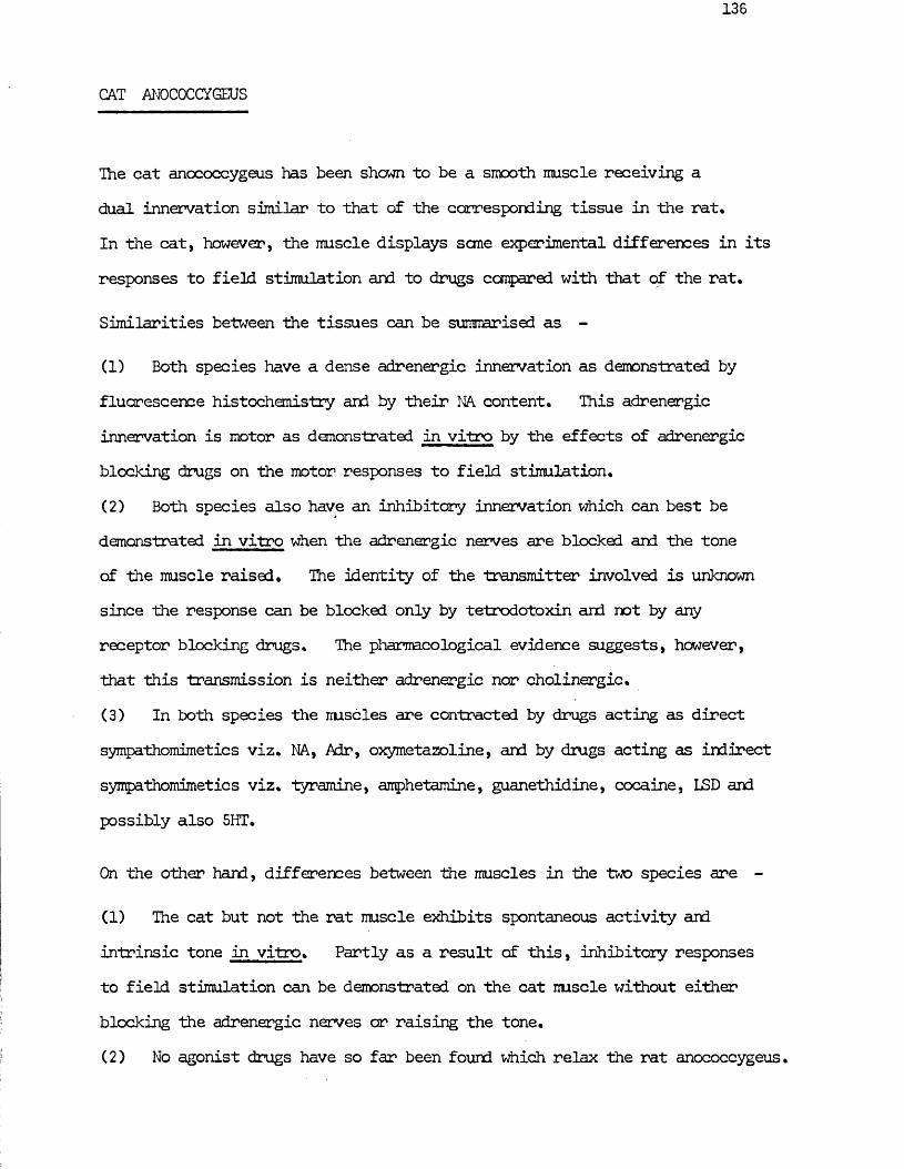
CAT ANOCOCCYGEUS
The cat anococcygeus has been shown to be a smooth muscle receiving a dual innervation similar to that of the corresponding tissue in the rat.In the cat, however, the muscle displays sane experimental differences in its responses to field stimulation and to drugs compared with that of the rat.
Similarities between the tissues can be summarised as -
(1) Both species have a dense adrenergic innervation as demonstrated by fluorescence histochemistry and by their NA content. This adrenergic innervation is motor as demonstrated in vitro by the effects of adrenergic blocking drugs on the motor responses to field stimulation.(2) Both species also have an inhibitory innervation which can best be demonstrated in vitro when the adrenergic nerves are blocked and the tone of the muscle raised. The identity of the transmitter involved is unknown since the response can be blocked only by tetrodotoxin and not by any receptor blocking drugs. The pharmacological evidence suggests, however, that this transmission is neither adrenergic nor cholinergic.(3) In both species the muscles are contracted by drugs acting as direct sympathomimetics viz. NA, Adr, oxymetazoline, and by drugs acting as indirect sympathomimetics viz. tyramine, amphetamine, guanethidine, cocaine, LSD and possibly also 5HT.
On the other hand, differences between the muscles in the two species are -
(1) The cat but not the rat muscle exhibits spontaneous activity and intrinsic tone in vitro. Partly as a result of this, inhibitory responses to field stimulation can be demonstrated on the cat muscle without either blocking the adrenergic nerves or raising the tone.(2) No agonist drugs have so far been found which relax the rat anococcygeus.
The cat muscle, however, is relaxed by several drugs all of which contract the rat muscle viz. acetylcholine, carbachol, isoprenaline, ATP, prostaglandins
, E£ and F2 and vasopressin. In addition to relaxing the muscle itself, acetylcholine also inhibits responses to adrenergic nerve stimulation in the cat anococcygeus.
The implications of this comparison are clear. The dual innervation found in the rat anococcygeus with motor adrenergic and inhibitory "unknown” transmission has a direct counterpart in another species. In this further species the effects of stimulation of the motor adrenergic nerves and of drugs which mimic their effects are similar, confirming both the classical effects on this tissue of NA and tyramine, which produce respectively direct and indirect sympathomimetic responses of the muscle and also the somewhat less expected actions of guanethidine, cocaine, LSD and 5HT, all of which produce indirect synpathomimetic responses. The ability to observe the effects of the inhibitory nerves in the cat muscle in the absence of the complicating effects of drugs strengthened the case far their being an entity independent of the adrenergic nerves and permitted the interaction and thus the interrelationship between motor and inhibitory nerves to be examined. Since several possible candidates for the role of inhibitory transmitter produced opposite actions in the two species and specific blocking or potentiating drugs could affect their responses without affecting the inhibitory nerve responses, the candidature of these substances was considerably weakened. Acetylcholine, for example, which must rank as the first candidate as transmitter in any nerves "physiologically” opposed to adrenergic nerves, produced opposite responses in the two species not only on the muscle but also by its effect on the adrenergic nerves. This not only made it an unlikely inhibitory transmitter in this tissue but also raised questions as to the general significance of any effects of acetylcholine on this tissue.
Many of these points require consideration in closer detail.
Even without advance knowledge of the existence of a dual innervation in the rat anococcygeus and the information on the separate anatomical origins of the motor and inhibitory nerves, the responses of the cat anococcygeus to field stimulation in vitro would have themselves provided convincing evidence of two sets of nerves in that tissue. The experiments illustrating the time course of development of the dual response to field stimulation, combined with the frequency characteristics of the two sets of nerves, allay the possibilities of the inhibitory response being due to some transformation in the effects of the motor nerves such as might be explained by "Wedensky Inhibition" or some change in the "peripheral mechanism". The complex, compound responses produced by field stimulation in the absence of drugs are seen to be due to enhancement of either motor or inhibitory components of the response by selection of the appropriate frequency and duration of stimulation. Thus at very low frequencies and again at high frequencies the motor nerves dominate, while at intermediate frequencies the inhibitory nerves dominate. The time course far development of both motor and inhibitory responses also fits in with the observed compound responses. Thus contraction from motor nerve stimulation develops rapidly while inhibition from inhibitory nerve stimulation, although having a similarly short latent period, develops more slowly. On cessation of stimulation, the motor response wanes more rapidly than the inhibitory.
This leads to compound responses in which the relative sizes of the various components vary with frequency tut which in general consist of (1) an initial motor response followed by (2) an inhibitory component (3) a period when either response may dominate and (4-) on cessation of stimulation, a further inhibitory component may be found. These findings would be
difficult to justify in terms of a varying output of transmitter from a single nerve type (Wedensky Inhibition) or by variations in the properties of the nerve - muscle complex (peripheral mechanism)* Furthermore, the initial resting tone in the iruscle is purely myogenic in origin as demonstrated by the relaxation produced by papaverine but not by phentolamine. The inhibitory component of the response to field stimulation can reduce this tension in the muscle to below the resting baseline, demonstrating that a direct inhibitory mechanism exists and not simply a variation in the extent of motor stimulation*
In addition to demonstrating the separate natures of the two sets of nerves, these experiments on field stimulation also have a bearing on their physiological function. With separate stimulation of the two sets of nerves and appropriate selection of frequency, muscle activity cculd be made to range over the entire spectrum from almost pure inhibition through various intermediate states of activity to a maximum contraction* Thus mechanisms exist which under physiological conditions could produce a rapid and fine control of the muscle tone. No direct light is* however, thrown by this onto the function of the muscle since the possibilities for this control appear so flexible* Thus the muscle might be normally held contracted under the influence of a low frequency tonic discharge in the motor nerves and might be rapidly relaxed during seme intermittent function by stimulation of the inhibitory nerves* This hypothesis would, however, be opposed by a corollary of the results of the experiments on the effect of reserpine (see Part II)* These experiments-suggest that a possible reason for the slow rate of depletion of NA from the anococcygeus is that this tissue receives only a low tonic impulse traffic in its motor nerves. If this is the case, the above hypothesis would be unlikely* Conversely, the muscle may normally be relaxed and play same intermittent role by a combination of motor and / or inhibitory responses* These two possibilities appear to be
the most likely simply on the grounds of economy, since it is difficult to comprehend why the muscle would be normally under the tonic influence of either the inhibitory nerves alone or a combination of motor and inhibitory.
Information on the time course of the inhibitory response was also gained from these experiments on field stimulation. Previous information on this had relied on the rate of recovery of tone after inhibition and was thus a measure of the rate at which inhibition was overcome by the presence of a motor agonist. It was, however, possible to obtain a more direct measure of inhibitory effect by observing the influence of a burst of stimulation on the motor response to a single stimulus. This demonstrated that the motor response to a single pulse was still completely abolished 3 secs after a f burst of stimulation at 5 Hz, the optimum frequency for the inhibitory response, and that this response had not recovered until 8-10 sec after cessation of stimulation, Ihis indicates that the influence of the inhibitory nerves on the muscle lasts a shorter time than might be imagined frcm the recovery of drug - induced motor tone after inhibitory nerve stimulation. This is illustrated by a comparison of Figs. 73 & 74: which were obtained with similar stimulation parameters on the same muscle. VJhen field stimulation was applied in the presence of 5HT - induced tone, the peak of the relaxation of muscle tone was reached 10 -15 sec after the end of the stimulation period, (Fig, 73). Similar stimulation in the absence of induced tone and using a test stimulus as a measure of the inhibitory effect, showed recovery after 8-10 sec (Fig. 74), Several factors could explain this temporal difference in the maximal effects. (a) Ihe inhibitory transmitter may affect both the muscle and the motor nerve and the effect on the muscle may outlast that on the nerve. (b) The time when the tension is at a minimum may not reflect the point of maximum action of the inhibitory transmitter on
the muscle. A more accurate indication of the effect of the inhibitory transmitter might be the rate of change of tone* This would reach a maximum much earlier and correspond mere closely with the time course of suppression of the motor nerve response*
The fairly rapid decline of the inhibitory effect suggests that some inactivation process exists for the transmitter* In the absence of any information on the nature of the transmitter it is pointless to speculate whether this consists of enzymic breakdown and / or active reuptake into some site as exists for cholinergic and adrenergic systems*
Drug analysis of the motor and inhibitory responses confirmed the conclusions from these early experiments* Receptor and neurone blocking agents confirmed the adrenergic nature of the motor nerves. The "unknown” nature of the inhibitory nerves was demonstrated by a series of essentially negative results. Neither muscarinic, nicotinic, ft - adrenergic, 5 - hydroxy - ;tryptaminergic or histaminergic blocking agents affected the response.
Hexamethonium had no effect on either response, demonstrating that only the postganglionic neurones are present in each case. This was confirmed histochemically by the lack of any ganglia in or around the..muscles* The interpretation of responses in this tissue is thus simpler than in gastro - intestinal smooth muscle where the additional presence of Aierbachfs and Meissner's plexuses leads to difficulties. The adrenergic nerves in the anococcygeus appear, on pharmacological evidence alone, to directly innervate the smooth muscle and do not act as has been suggested in the gastro - intestinal tract via a further neurone (Norberg, 1967). This is confirmed by the E.M. studies on the rat anococcygeus (Gillespie & Du liman, prepared far publication) which show the terminal varicose nerve fibres distributed
11+2I|
throughout the nuscle in direct relation with muscle fibres* Within the muscle the inhibitory nerves also appear to be solely postganglionic* This stands in contrast to most cholinergic ( Burns tock, 1969) or pur inerg ic (Burnstock, 1972) systems which normally have their cell bodies in or close to the tissue innervated. While there is as yet no direct proof of a preganglionic component to the inhibitory pathway to the cat anococcygeus, the presence of such a ganglion in the rat makes this a reasonable assumption. The E.M. evidence so far does not conclusively demonstrate separate adrenergic and non - adrenergic nerves in the rat anococcygeus, but nerves containing agranular vesicles are present throughout the tissue and may represent the inhibitory nerves (Gillespie & Lullman, prepared for publication)• >
The effects of agonist as well as antagonist drugs also serve to reinforce the adrenergic and "unknown" natures of the motor and inhibitory nerves.
The cat anococcygeus shows a sensitivity to NA similar to that of the rat muscle and these responses are specifically blocked by phentolamine in concentrations which also block the motor nerve response. . Indirect sympathomimetics such as tyramine also contract the muscle and are blocked by phentolamine. These oC- receptor responses are similar to those in the rat. A difference between the species appears when ft - receptor effects are considered. The/?- blocker, propranolol, has no effect on the motor response to NA in the rat, but in the cat it potentiates motor responses to NA and Adr. The inhibitory ft - adrenergic effect which this u re overs can be seen directly by the effect of isoprenaline. In the cat, isoprenaline relaxes' both intrinsic tone and that induced by an agonist drug. This inhibitory effect is abolished by propranolol. The cat anococcygeus thus possesses inhibitory ft - adrenergic receptors in addition to motor cL - receptors. These ft - receptors are activated by NA and Adr as well as by
isoprenaline, although this action of NA and Air is normally masked by their predominant motor o i - effect.
The site of these /?- receptors is not known but they do not appear to play an important role in the motor transmission process since the motor response to field stimulation was unaffected by propranolol. Possible reasons for this could be -
(1 ) ft - receptors occur over the surface of the muscle as a whole but not specifically near the nerve - muscle junction. In this case they would be more likely to be affected by exogenously administered NA than by nerve released NA.(2) If ft - receptors do occur in the region of the nerve - muscle junction, the fi - response to nerve stimulation might be relatively resistant to blodode by propranolol in a manner analogous to the relative resistance of the motor nerve response to phentolamine.
No ft - receptors appear to be present in the rat anococcygeus as shown by the lack of effect of isoprenaline whose only action is to produce direct oi - effects in high concentrations. This lack of ft - effects in the rat tends to eliminate /?- adrenergic transmission as a possible candidate for the inhibitory nerve response, an argument which will also be applied to cholinergic, purinergic and prostaglandin - mediated transmission. The most convincing evidence, however, that ft - receptors are not involved in the inhibitory pathway is the observation that propranolol has no effect on inhibitory responses in concentrations which abolish the effects of isoprenaline.
Even if isoprenaline is not the inhibitory transmitter, it would be expected to summate with or potentiate the inhibitory response. This was not observed. If both inhibitory transmitter and agonist acted by a similar mechanism such
144
I
*as might be produced by hyperpolarisation via a change in permeability to a specific ion, then some potentiation might have been expected. The lack of such an effect suggests a difference in the mechanism which can only be resolved by electrophysiological means* The present experiments, with specific blocking drugs can only provide evidence that different receptors are involved* Whether the next step activated by receptor occupation is common can only be properly answered by electrophysiological techniques,
Acetycholine had to be considered as a possible inhibitory transmitter in the cat anococcygeus since it produced an inhibitory response and since Howard & Garrett (1973) had found acetylcholinesterase in the tissue and postulated that cholinergic nerves might be present* The evidence in Part I of the present study that the inhibitory nerves originate in the upper sacral region of the spinal card (the origin of parasympathetic fibres) supports ■this though the motor effect of acetylcholine in the rat anococcygeus led to its rejection in that species as a possible inhibitory transmitter. This possibility was, however, re - opened by the finding that acetylcholine relaxed the cat anococcygeus. Since the intrinsic, as well as the drug - induced, tone could be relaxed this was a direct effect on the nuscle, Carbachol produced a similar effect to acetylcholine, the effect of acetylcholine was potentiated by neostigmine and the effects of both acetylcholine and carbachol were blocked by atropine. Neither atropine nor neostigmine had, however, any effect on the inhibitory nerve response, nor did acetylcholine potentiate the inhibitory nerve response. For these reasons together with completely opposite effects of acetylcholine on the rat anococcygeus, cholinergic transmission was again rejected as a possible inhibitory nerve process in the cat anococcygeus.
On the rat muscle the muscarinic receptors appear to play a directly opposing
role, producing a motor response but still potentiated by neostigmine and blocked by atropine. This opens the question as to the role of both acetylcholine and cholinesterase in this tissue. The presence in the cat anococcygeus of cholinesterase, demonstrated by Howard & Garrett (1973), was confirmed by the effect of neostigmine. In the rat, however, attempts to demonstrate cholinesterase met with no success (Gillespie, 1972) leading to the conclusion that no cholinesterase was present. The specific potentiation of acetylcholine by neostigmine in the present experiments contradicts this latter conclusion, hit it is possible that the reaction conditions employed, despite producing a dense staining of motor end plates in rat diaphragm, were not so vigorous as those employed by Howard & Garrett (1973). This treatment might, therefore, have been insufficient to detect small quantities of cholinesterase present, especially if this was diffusely distributed throughout the muscle.
Assuming then that acetylcholinesterase is present in the anococcygeus of both species, what function if any is it playing? The lack of any effect of either atropine or neostigmine on the motor or inhibitory nerves in either species rules out the suggestion of Howard & Garrett (1973) that cholinergic nerves nay be present in the muscle, unless some further nerves are present which subserve neither a motor nor inhibitory function and whose effects cannot be detected by field stimulation of the muscle with 0.1 - 10 msec pulses. In addition, this same evidence makes it unlikely that in this tissue a cholinergic link exists in the adrenergic nerves as suggested by Burn & Rand (1959) or that acetylcholine plays a significant role in the activation of the axone as suggested by Nachmanson (1966).
The effects of acetylcholine on the nerve responses to field stimulation shed no further light on the role of cholinesterase. In the rat, acetylcholine appears to have no effect on the motor response to field stimulation, other
than can be explained by simple summation of the direct responses on the muscle of the motor transmitter and of acetylcholine. Cholinesterase inhibition simply potentiates the motor effect on the muscle of acetylcholine. Inhibitory nerve responses are similarly unaffected by acetylcholine and in fact after 60HDA in vivo treatment carbachol or acetylcholine provide a convenient means of raising muscle tone in order to observe inhibitory nerve responses (Gibson & Gillespie, 1973), In the cat, on the other hand, acetylcholine inhibits motor nerve responses, an effect potentiated by cholinesterase inhibition and blocked by atropine. This might be expected from the inhibitory effect of acetylcholine on the muscle, but other agonists which relax the cat anococcygeus such as isoprenaline, ATP, prostaglandins and vasopressin are not effective against motor nerve responses except in very high doses, in contrast to acetylcholine and carbachol which inhibit motor nerve responses in doses as low as those which relax the muscle. This points to a possible presynaptic effect of acetylcholine on motor nerves which is borne out by comparison of the effects of acetylcholine against NA and against motor nerves. Acetylcholine inhibits motor nerve responses to a greater extent than NA responses of a similar magnitude. This stands in contrast to the effects of the specific ©(.- receptor blocker phentolamine which was more effective in a similar situation against NA. A presynaptic inhibition of NA release by acetylcholine is thus suggested, similar to that demonstrated on the rabbit heart by Loffelholz & Muscholl (1969). Another possibility which is not ruled out by these experiments is, however, that the muscarinic receptors might be concentrated in the region of the nerve - muscle junction resulting in a greater effectiveness against nerve as opposed to exogenously administered NA. This question would be resolved by transmitter overflow studies.
The inhibitory nerve response was unaffected by acetylcholine. As
mentioned previously, potentiation between the inhibitory nerve and a drugwhich lowers the tension is difficult to judge. There was, however, no overtevidence of such potentiation or of reduction of the inhibitory nerve responseThe inhibitory effect of acetylcholine on the motor response in the cat in
- 8 -4doses of 1 0 M - 1 0 M was thus the only effect of acetylcholine found oneither set of nerves in either species. No potentiation of adrenergic
-13responses was observed at lower doses (down to 1 0 M) as reported by Malik & Ling (1969) for the perfused mesenteric artery.
A further action of acetylcholine on the cat anococcygeus which has not been properly examined but which deserves mention was the small motor response at high doses. This response nay be significant in two separate respects. Firstly, it my be produced by release of NA from the motor nerves. Such an effect was found by Loffelholz & Muscholl (1969) in doses higher than those required to produce inhibition of NA release and would be similar to the well known sympathomimetic effects of acetylcholine in high concentrations (see Kosterlitz & Lees, 1972). This pattern my thus be repeated in the anococcygeus. Loffelholz & Muscholl found that the inhibitory effect was muscarinic, while the release of NA was nicotinic. In the present experiments both responses were blocked by atropine, but no attempt was made to analyse the specificity of blockade of this motor action of acetylcholine. In a second respect this small response m y be related to the phenomenon of reversal of acetylcholine responses reported by Burn & Robinson (1951).These motor responses did not occur in every experiment and it was noted that they were more regularly present in muscles cold stared overnight. This can be seen from Fig. 80 b. There may thus be some evidence of reversal of acetylcholine effects produced by isolation of the tissue in physiological saline. Whether this would consist of reversal of the effect of stimulating receptors on the muscle or be due to development of the indirect release of NA, remains an open question.
Experiments with acetylcholine thus suggest that neither acetylcholine nor cholinesterase play a significant role in the nerve transmission process of the anococcygeus muscle but that exogenously applied acetylcholine can exert effects on the tissue and these effects are potentiated by cholinesterase inhibition. In the rat the only effect of acetylcholine is to produce contraction by a muscarinic action on the muscle. In the cat acetylcholine produces relaxation by a direct muscarinic action on the muscle and also inhibits motor nerve responses by a presynaptic mechanism. A universal physiological role far acetylcholine or acetylcholinesterase cannot, therefore be suggested in this tissue. It is, however, possible that since there is no evidence for the presence of endogenous activity of acetylcholine itself in the anococcygeus, the acetylcholinesterase may be present either solely as a safety factor to prevent the action of acetylcholine from exogenous sources or to regulate some function of acetylcholine in the tissue which has not yet been suggested. This explanation is on a similar basis to those put forward far the various effects of acetylcholine on adrenergic transmission in other tissues (e.g. Malik & Ling, 1969; Loffelholz & Muscholl, 1969). Since the effects of acetylcholine are opposite in the two tissues, it becomes even more difficult to encompass them into a universal role in adrenergic transmission as suggested by Burn & Rand (1959) and again more recently by von Euler (1970).
The effects of ATP on the cat anococcygeus were examined in order to test whether the inhibitory innervation was purinergic. Burnstock (1972) has suggested that a great number of formerly uncharacterised inhibitory mechanisms may be due to nerves having as their transmitter ATP or a related nucleotide. In addition, he tentatively postulated that the non - adrenergic non - cholinergic inhibitory system demonstrated by Gillespie (1971) in the rat anococcygeus might be related to this system. While Gillespie (1972)
has refuted this by demonstrating that ATP produces only motor responses in the rat anococcygeus, it was felt necessary to re - examine the possibility in the cat. As in the case of isoprenaline and acetylcholine, ATP produced relaxation of the cat anococcygeus, in contrast to its motor action in the rat. Considering the cat anococcygeus on its cwn, this response suggests ATP as a candidate far inhibitory transmitter. No attempt has as yet been made to obtain further pharmacological evidence to settle this point such as might be achieved by examining the action of the adenosine uptake blocking drug dipyridamole (see Burnstock, 1972). On the basis that the effect of ATP is motor in the rat muscle, it is at present felt that ATP is unlikely to be the inhibitory transmitter. ^
Another factor concerning the possible involvement of purinergic transmission in this tissue is that purinergic nerves have so far been demonstrated only in tissues where the adrenergic nerves are inhibitory. The only examples quoted by Burnstock (1972) of mamnalian systems which receive a motor adrenergic innervation as well as a postulated purinergic inhibitory innervation are the monkey internal anal sphincter (Payner, 1969), the cat internal anal sphincter (Garrett, Howard & Lansdale, 1971) and the rat anococcygeus (Gillespie, 1971).It is tempting to suggest that in each of these cases the inhibitory nerves involved are the inhibitory nerves of the anococcygeus or of a closely related but functionally similar tissue. This would in no way detract from the validity of purinergic nerves in tissues where adrenergic nerves are inhibitory, but the similarity in the anatomical location of the only examples of non - adrenergic, non - cholinergic nerves in systems where the adrenergic nerves are motor seems a great coincidence and suggests a system of non - adrenergic, non - cholinergic, non - purinergic innervation in such tissues.
Prostaglandins have been implicated as modulators of adrenergic transmission in several tissues (Horton, 1973) or even as inhibitory transmitters (Campbell,
150
1970)* Like acetylcholine, isoprenaline, ATP and vasopressin, the prostaglandins E , and F^ all contract the rat anococcygeus and relax the cat anococcygeus. Even at high doses the contraction of the rat nuscle is small compared to the contraction produced by other motor agonists such as NA and only a slight inhibition of the motor response to field stimulation was produced at the highest dose tested. In the oat, the inhibitory response was produced against either intrinsic or drug - induced tone. As in the rat, these responses were not as large as those produced by other agonists such as isoprenaline, although it is possible that they might have been if larger doses had been given. Only a small inhibition of motor responses to field stimulation was found at high doses and no apparent effect on inhibitory responses was found. As with the other inhibitory agonists, no summation occurred between prostaglandin - induced relaxation and inhibitory nerve responses. Although no further attempt was made to study the pharmacology of prostaglandins on this tissue by manoevres such as inhibition of prostaglandin synthesis, it was concluded that the inhibitory transmitter was not a prostaglandin of the type tested here since they exerted an opposing action on the muscles from the two species and since no summation occurred with the inhibitory nerve response. Since prostaglandins are active in this tissue, albeit in fairly high concentrations, a role in modulation of adrenergic nerve responses, such as is found in guinea - pig vas deferens (Euler & Hedqvist, 1969) and in other tissues (see Horton, 1973), cannot be completely ruled out.
Vasopressin also produced motor responses on the rat and inhibitory responses on the cat anococcygeus. There is no suggestion that vasopressin might represent the inhibitory transmitter. It was included in the study simply as another example of a substance which may either relax or stimulate smooth muscle (e,g. Woo & Somlyo, 1967; Gilmore & Vane, 1970; Mackay & IfeKirdy, 1972), The fact that it produces opposite responses in the two species merely
151
adds vasopressin to the list of substances tested producing such responses*It is suggested that each of these substances may act on a similar receptor in each species, but that the net effect on the muscle of occupation of this "receptor11 is different in each species. Presumably the receptor involved is different far each substance. This can be proved at least to the extent that acetylcholine and carbachol act on muscarinic receptors, isoprenaline on ft - adrenergic receptors and the others, ATP, prostaglandins and vasopressin, on receptors which are neither of these. The present evidence provides grounds far speculation that these receptors are all different again from the receptors for the inhibitory nerve transmitter and that the mechanism activated by this latter receptor is different from that of the others. It would thus be of interest to direct an electrophysiological study towards determining the two unknown factors here, viz. (1 ) what is the mechanism or mechanisms set in action by acetylcholine etc. which leads to contraction of the rat and relaxation of the cat anococcygeus, is this the same basic mechanism e.g. change in permeability to some ion, in both species, and what is the difference between the two species which results in a net opposite response? (2 ) what is the difference between this mechanism in (1 ) and that set in motion by the inhibitory transmitter, which leads to an inhibitory response in both species? If these factors could be elucidated then characterisation of the inhibitory nerves would be a further step closer.
A striking feature of the anococcygeus in both species is the ease with which a wide range of drugs nay produce sympathomimetic effects i.e. contractions which were reduced or prevented by phentolamine. In order to determine whether these effects were direct effects on the oL - receptors of the smooth muscle or indirect effects produced by release of NA from the motor nerves, chemical sympathectomy using 6 GHDA was employed. The methods available for differentiating.between direct and indirect sympathomimetic effects have
152
recently been reviewed by Trerdellenburg (1972) who enumerates three methods, viz, (1 ) the use of cocaine which will block the neuronal uptake process producing block of indirect and potentiation of direct actions (2 ) denervation, which will similarly prevent indirect effects by destruction of the nerves (3) reserpine pretreatment which will remove the NA which is available for release by indirect agents. In the present circumstances, cocaine could not be usefully employed since it was itself a powerful sympathomimetic, surgical denervation was not possible in the anococcygeus since the surgical problems of this have not yet been solved and reserpine treatment was considered unsuitable since it had been shown to produce depletion of NA stares which was insufficient to prevent adrenergic nerve responses. The alternative but related method of producing sympathectomy using 60HDA was therefore chosen. Pretreatment of rats in vivo with 6 QHDA had been shown to produce a sympathectomy in the anococcygeus as demonstrated histochemically by the absence of adrenergic nerves and by the abolition of mctar responses to field stimulation (Gibson & Gillespie, 1973), In addition, after this treatment the sympathomimetic effect of guanethidine was absent.This treatment was thus suitable for the rat. Treatment of a sufficient number of cats with 60HDA was financially impracticable so in this case the muscles were incubated in vitro with 60HDA, a method previously demonstrated to produce an effective chemical sympathectomy in a variety of tissues (e,g, Sachs, 1971; Wadsworth, 1973). This in vitro sympathectomy was carried out on rat as well as cat anococcygeus, so that in this species the two treatments could be compared.
Chemical sympathectomy either in vivo or in vitro greatly reduced or completely abolished the responses to tyramine and potentiated the responses to NA in both species. This suggested that a destruction of the adrenergic nerves had occurred resulting in both the inability of the indirect sympathomimetics to
153
produce responses and the potentiation of the direct sympathomimetics by removal of the neuronal uptake process. Of the test drugs, the responses of guanethidine, LSD and cocaine were either abolished cr greatly reduced while 5HT produced a paradoxical result. In vitro 60HDA treatment in both species resulted in abolition of the 5HT response, suggesting a wholly indirect mechanism of action, while 60HDA in vivo treatment of rats produced a sensitivity to 5HT greater than before.
A clear - cut result was thus obtained with guanethidine, LSD and cocaine whose responses were reduced to a similar extent to those of tyramine and could thus be considered to produce their action entirely by release of NA. It was possible that the residual responses remining to these drugs after 60HDA could be produced by either direct or indirect actions, but several factors suggest that this is a residual direct action on the muscle. Thus the size of these responses varied between groups of rats which were treated from different batches of 60HDA and this was reflected in the size of the motor responses which could be obtained to field stimulation. This suggests that the 60HDA which was obtained from various sources my have been of varying purity. After in vitro 60HDA treatment, residual responses to these indirect agents were uncommon. For these reasons a wholly indirect action is suggested for tyramine, guanethidine, LSD and cocaine, although a direct component cannot be ruled out completely. Tyramine, guanethidine and cocaine have all been shown in the past to possess both indirect and direct effects on smooth muscle tissues (see Trendelenburg, 1972). As Trendelenburg (1972) has pointed out 11 each sympathomimetic amine should be regarded as potentially able to exert both direct and indirect effects. Its overall effects in any given organ or system depend on the affinity of the amine to the receptor, to the uptake mechanism, on its ability to impair the re - uptake of the released transmitter as well as on the factors which influence the concentration of the amines'.'at the receptors (such as the morphology of the synaptic region).". These same factors are
also likely to apply to non - amine synpathomimetics and it appears that the conditions existing in the anococcygeus are particularly conducive to indirect action.
The factors producing this susceptibility to indirect actions are not yet clear. The nerve - muscle distance is no smaller than in other tissues so this is unlikely to be a significant factor. Other morphological factors, an efficient neuronal uptake mechanism or a particular susceptibility to displacement of the granular NA may all be relevant.
LSD has surprisingly never been directly linked with synpathomimetic actions although several factors suggest the possibility. Thus (1) LSD has been shown to inhibit NA uptake in isolated storage granules (Euler, 1970), a property it shares with tyramine (Burgen & Iversen, 1965) (2) other ergot derivativeshave been shown to possess indirect sympathomimetic actions e.g. ergotamine (Salzmann, Pacha, Taeschler & Weidmann, 1968) (3) the isolated catnictitating membrane which is similar to the anococcygeus in being contracted by cocaine and 5HT as well as NA, is also contracted by LSD, ergotamine and dihydro - ergotamine (Thomson, 1958). It was not, therefore, surprising that LSD should exert an indirect sympathomimetic action on the anococcygeus. A further connection is that Gant & Dyer (1971) have shown that LSD and 5HT can both produce contractions of isolated strips of human umbilical vein and umbilical artery which are abolished by phenoxybenzanine. Since these preparations are nerve - free, an indirect synpathomimetic action seems at first sight unlikely. It has, however, been suggested by Gulati & Kelkar (1971) that tyramine may act on human umbilical artery by liberation of extraneuronal NA.
jIt might, therefore, be possible that LSD and possibly also 5HT might act in j this way. It would be rash to suggest that this indirect synpathomimetic property of LSD in the anococcygeus might have any significance for its CNS hallucinogenic action, but other peripheral properties such as inhibition of
155
5HT receptors have in the past led to iruch psychopharmacological speculation (see Mantegazzini, 1966).
Another property of LSD in the anococcygeus was an adrenergic neuronal blocking action. This property has been recently reported also by Ambache, Killick, Srinivasan & Zar (1973) for the rat ancococcygeus. They related this to the similar blocking action of LSD on the vas deferens of the guinea - pig and rat and demonstrated that this inhibitory action was more effective against short than long pulse trains. The present experiments demonstrate that LSD can inhibit adrenergic responses in the rat and cat anococcygeus but show no difference in inhibition of the response to pulse trains of different lengths. In addition, this inhibition could take up to 1 hr to reach its naxinum effect and was produced by doses of LSD which raised tone. Earlier in this thesis in discussing the effect of LSD on the vas deferens of the pithed rat, it was suggested that since the inhibition produced in this tissue was restricted to the first phase of the motor response, it resembled the action of tyramine. This was related to the work of Ambache, Dunk, Verney & Zar (1972, 1973) and it was suggested that the reason far the inhibition found in their experiments being more effective against short than against long pulse trains was that only the first part of the two phase response was affected.When LSD was then shown to produce an indirect synpathomimetic effect on the anococcygeus, this lent further weight to this hypothesis. Ambache, Killick, Srinivasan & Zar (1973), however, demonstrated that this relatively greater inhibition of short pulse trains also existed in the anococcygeus and thus suggested that LSD was capable of producing a presynaptic neurone blocking action of the adrenergic nerves in both the anococcygeus and vas deferens and that this was somehow more effective against the first few impulses. In the present experiments, the inhibition of motor responses by LSD in the anococcygeus could not be shown to be different at any particular length of train.
It is thus felt by the present anther that while LSD undoubtedly has a presynaptic blocking action on the anococcygeus, this is not necessarily either a simple effect cr directly analogous to its effect on the vas deferens. Ininterpreting these effects and making comparisons, several points must beborne in mind -
(1) There is only one component to the motor response of the anococcygeus and this is inhibited by LSD. There are two components to the response of thevas deferens tut only one of those is inhibited by LSD.(2) In addition to producing inhibition of the motor nerves, LSD produces an indirect sympathomimetic action in the anococcygeus. This may be reflected in the potentiation of the secondary phase of the response in the vas deferens.(3) In the anococcygeus the effect of LSD resembles that of guanethidine with the exception that with guanethidine the threshold for the neurone blocking action is lower than far sympathomimetic action, whereas with LSD inhibition does not occur until sympathomimetic doses have been reached. In the vas deferens, guanethidine inhibits both phases of the response but LSD only the first phase,(*4) In the rat vas deferens there is evidence (see Part II) that the two phases of the motor response may be due to two anatomically and pharmacologically distinct sets of nerves. The first phase, which is resistant to some methods of blocking adrenergic nerves e.g. reserpinisation or oC - blockers, is blocked by LSD, but the latter phase, which is susceptible to these adrenergic blocking manoevres, is resistant to, and may be potentiated by, LSD.
With these factors in mind, it is suggested that LSD and guanethidine each possess the dual properties of adrenergic neurone blockade and indirect release of NA from adrenergic nerves. When the additional possibility of cL - receptor inhibition of NA release by an indirect sympathomimetic is
157
added to this, the interpretation of the effect of either drug on a given system must take into account the net effect of these mltiple factors. In this way all the effects of both LSD and guanethidine on the anococcygeus and vas deferens can be accounted far except the inability of LSD to block the secondary phase in the vas deferens. This latter exception may, however, be related to differences in morphology or metabolism of the nerves giving rise to this response. The lack of references in the literature to either sympathomimetic or adrenergic blocking actions of LSD in other tissues would support this. The reasons for this differential susceptibility to LSD of adrenergic nerves in different tissues remins to be elucidated.
The paradoxical effect of 60HDA treatment on the response to 5HT wasreflected in the effects of specific blocking drugs. The initial observationthat the response to 5HT of the rat anococcygeus was blocked by phentolaminesuggested that 5HT might act either directly on 0 6 - receptors or indirectlyby release of NA from nerves. It was subsequently found that the catanococcygeus was more sensitive to 5HT than the rat muscle, that phentolaminedid not block at all concentrations and that the dose / response curve in the
-5cat had a definite inflection at 10 M, Moreover, methysergide caused a narked inhibition of responses to low doses of 5HT, whereas phentolamine inhibited only responses to high doses. This then suggested that perhaps low doses exerted a direct effect on methysergide - sensitive 5HT - receptors and high doses exerted, in addition, an indirect effect which by acting via NA was therefore phentolamine - sensitive. A similar hypothesis to this has recently been put forward by Pluchino (1972) for the cat nictitating membrane. He showed that the dose / response curve to 5HT in this latter tissue also had an inflection and demonstrated that whereas the response to high doses was (a) blocked by phentolamine, cocaine or reserpinisation (b) potentiated by blockade of MAO (c) showed cross - tachyphylaxis to tyramine, the response to
A
158
low doses was unaffected by these procedures. His conclusion from this was thus that the response to high doses was an indirect sympathomimetic action, whereas that to low doses was a direct action on 5HT receptors. Unfortunately, he did not report on the action of methysergide which in the present study abolished the response to low doses. Nevertheless, he suggested, on the negative evidence of lack of blockade by phentolamine that this direct effect was exerted on specific 5HT receptors and not on adrenergic oL- receptors.
With this background in mind, it was hoped that after sympathectomy by 60HDA, direct and indirect components in the anococcygeus could be resolved and if direct components existed, they could be classified as 5HT or 0 6 - receptors by using specific blockers. After 60HDA in vitro treatment a clear answer to these questions appeared to be presented. In both species, 5HT produced no responses whatsoever, in contrast to NA whose responses were potentiated.On this basis it appeared that 5HT acted wholly as an indirect sympathomimetic in both species and that methysergide might then inhibit the responses at all concentrations of 5HT by presynaptic inhibition of the release of NA by 5HT.The only weakness in this hypothesis was that phentolamine inhibited only the responses to high doses which would be surprising if the whole response was due to liberation of NA. This could be rationalised by suggesting that the response to low doses might be due to an effect in the immediate vicinity of the nerves and that the high dose of NA built up in this region might be relatively resistant to phentolamine in much the same way that NA released by nerve impulses is. This argument also might explain the inflection on the dose / response curve to 5HT in the cat anococcygeus. Thus if the maximal effect of occupation of receptors by NA in the immediate vicinity of the nerves was reached at a lower concentration than the threshold for the effect on more distant receptors, then an inflection on the curve might be expected. The lack of such an effect coupled with the lower sensitivity to 5HT in the rat anococcygeus might suggest some morphological difference in this tissue. This
159
type of argument of differential effects on morphological grounds is analogous to those of de la Lande & Waterson (1968), Trendelenburg, Draskoczy & Pluchino (1969), Urquilla, Stitzel & Fleming (1970).
When muscles from 60HDA in vivo treated rats were tested, however, a conflicting result was obtained. In this case the muscles were sensitive to 5HT, although the development of this response was much slower than in controls. This response was inhibited by either phentolamine or methysergide. If the final maxinum tension developed by each dose was used for the construction of dose / response curves, these 60HBA sympathectomisad tissues appeared to be even more sensitive to 5HT than were untreated controls. The difference in rate of development of the response did, however, suggest that these responses were qualitatively very different from those obtained in controls. In the case of the other indirect sympathomimetics tested, viz. tyramine, guanethidine, LSD and cocaine, similar slow - onset responses were obtained in sane 60HDA in vivo sympathectamised muscles. The ability of these drugs to produce these responses appeared to be inversely related to the degree of sympathectomy, so for this reason it is assumed that these responses are due to residual indirect effects. Since 60HDA destroys nerve terminals before nerve trunks (Malmfors, 1971), it is also possible that part of these residual responses arises from NA released from axones rather than fran terminal varicosities. This is partly substantiated by the presence of occasional fluorescent axone profiles in the histochemical picture of these tissues. For this reason it is suggested that the residual response to 5HT in these 60HDA in vivo treated tissues is due entirely to the release of NA remaining in nerves following the incomplete sympathectomy produced by this treatment. It is further suggested that the apparent increase in the magnitude of the response to 5HT under these conditions might be due to the abolition of factors normally operating to limit the effectiveness of 5HT but which are largely
160
removed by the 60HDA treatment, e.g. the destruction of the majority of the terminal varicosities will result in the loss of the neuronal uptake of 5HT and subsequent destruction by intra - neuronal MAO.
On the basis of these experiments, the final hypothesis so far reached for the action of 5HT in the anococcygeus is that its action is entirely indirect. This is based both on the actions of phentolamine and methysergide and crucially on the sympathectomy produced by in vitro 60HDA treatment. The results of in vivo instead of in vitro 60HDA treatment might have led to the conflicting conclusion that the effects of 5HT are both direct and indirect and that the direct component iray be potentiated by destruction of the varicosities leading to an increased amount of 5HT available for direct action. It: must,therefore, be stated that although this latter hypothesis has been discarded on the basis that the sympathectomy so produced is incomplete, there are drawbacks to the use of 60HDA in vitro.
60HDA may itself produce blockade of receptors. It has been shown to block cx_- adrenergic receptors (Haeusler, 1971) and muscarinic receptors (Furness,1971). This effect may be due to 60HDA itself or to oxidation products (Malmfcrs & Thoenen, 1971). To eliminate this effect the organ bath was thoroughly washed throughout the 2 hr period following 60HDA treatment.Despite this precaution, the sensitivity of the rat anococcygeus to NA after this treatment was less than that after in vivo 60HDA treatment. This could have been due to depression of the <XL - receptors or alternatively due to fatigue of the muscle since the 60HDA treatment produced a prolonged maximal motor response. At the dose of 60HDA used in these experiments, this response is likely to be due partially to the indirect sympathomimetic action of 6 QHDA and partially to the release of NA following disruption of the nerve varicosities (Sachs, 1971; Trendelenburg & Wagner, 1971). The former effect is suggested by the rapid onset of the contraction and the latter by the slow
161
decline of the response after washout. This prolonged contraction of the muscles could thus have led to fatigue and thus to a limitation on sensitivity to NA but still does not eliminate the possibility that either oL - receptors or hypothetical 5HT receptors on the muscle may have been depressed. The possibility that 5HT may act on either its own specific receptors or on oC - adrenergic receptors has been suggested by Innes (1962) who demonstrated by receptor - protection experiments that 5HT has affinity for - receptors in the cat nictitating membrane.
A further factor which necessitates caution in these experiments is the effectof ascorbic acid. In several recent studies on the in vitro effects of60HDA, ascorbic acid has been included in the medium (e.g. Sachs, 1971;Wadsworth, 1973) ostensibly to prevent oxidation of the 6 CHDA along the linesthat NA is so protected (see Hamberger, 1967). In the present study, ascorbic
-3acid in the concentrations employed by these authors ( 6 x 1 0 M) produced a maximal contraction of the anococcygeus muscles from both species, and even in a 1,000 time dilution, still produced motor responses. It is not yet: known whether these responses are due to a direct effect of ascorbic acid on the muscle cr due to release of NA. Whatever the action, a direct pH effect is unlikely since the buffering capacity of the Krebs* solution prevented significant change especially at the lower concentrations. Effects of ascorbic acid on smooth imscle tissues have long been noted (e.g. Eliasson, Euler & Stjarne, 1955; Gillespie & Mackenna, 1961) so it-should be included in physiological bathing media only with caution. In the present experiments, the inclusion of ascorbic acid was found to make no difference whatsoever to the chemical sympathectony produced. This may be partly due to the unnecessarily large dose of 6 QHDA employed, which was utilised simply because it had been shewn to be successful by previous authors, or alternatively due to the lack of any effect of ascorbic acid on oxidation of 6 QHDA.
A
162
With all these provisos in mind, it is thus concluded that 5HT acts as an indirect sympathomimetic in the anococcygeus muscle, the cat muscle being more sensitive than the rat. A direct effect of 5HT on the muscle cannot, however, be completely eliminated. Further studies using the more conventional means ) of differentiating between direct and indirect effects are, therefore, necessary.
The final aspect of the cat anococcygeus preparation which was examined was the effects of cold storage. This was necessary since the experiments were carried out on tissues which might be cold stared up to a maximum of 2 days.The necessary control studies on the effect of cold storage showed that little change in nerve or drug responses occurred over this short period.
Less expected was the ability of these nerves, cut off from their ganglion cell body, to survive and continue functioning after 1 1 days, as shown by the histochemical demonstration of adrenergic nerve terminals together with the responses to field stimulation and to the indirect synpathomimetic action of guanethidine. Stimulation of postganglionic adrenergic nerves fails to produce responses after 2 days in mouse, rat and rabbit ileum, 3 days in guinea - pig colon (HOlman & Hughes, 1965) and 7 days in guinea - pig taenia caecum (Hattori et al, 1972). Adrenergic nerves were not demonstrable histochemically after 5 days cold storage in guinea - pig caecum (Hattori et al, 1972) or rabbit aortic strips (Shibata et al, 1971). The capacity of the anococcygeus muscle to respond to NA after the nerves had ceased to function is in agreement with the usual finding that in cold stored tissues the smooth muscle cells remain viable longer than the nerves. Thus
si
intestinal smooth muscle from various species remains responsive to acetylcholine for several days after the cholinergic nerves have ceased to function (Ambache, 1946) and in guinea - pig taenia caecum, phenylephrine still
relaxes the muscle after 18 days cold storage (Fukuda & Shibata, 1972) despite the complete absence of nerve responses after 5 days (Hattori et al,1972).
It was not possible, however, to differentiate between motor and inhibitory nerves in the cat anococcygeus by their ability to survive cold storage. In previous studies where cold storage of a tissue has been employed successfully to differentiate between the components of a multiple innervation, the different anatomical arrangement of the nerves has been a major factor contributing to the preferential survival of one type, e.g. one group of fibres separated from their cell body (Holman & Hughes, 1965; Hattori et al, 1972).In the isolated cat anococcygeus muscle, however, as in the rat (Gillespie,1972), both motor and inhibitory nerves consist purely of postganglionic fibres. There is, therefore, no reason to expect differential survival. Despite the long survival after cold storage of the nerve and muscle elements of the cat anococcygeus, species variation may be at least as important as the properties of the particular tissue used since the rat anococcygeus muscle does not respond to either field stimulation or NA after only 5 or 6 days (D. Tenpleton, personal communication) •
All these experiments with the cat anococcygeus muscles have thus demonstratedthat the dual innervation first found in the rat anococcygeus exists and has j
Isimilar properties in the corresponding tissue in the cat. Ihey confirm that! the motor nerves are adrenergic and demonstrate the basic pharmacological properties of the nerves and muscle which vary slightly from those of the rat. They do not lead any further towards a positive identification of the inhibitory nerve transmitter but demonstrate the interaction between the motor and inhibitory nerves and provide evidence that the inhibitory nerves are not cholinergic and are unlikely to be purinergic.
164
GENERAL DISCUSSION OF ANOCOCCYGHJS
The separate but related aspects of the anococcygeus muscle which have been investigated come together to answer several questions concerning the nature of the muscle and its innervation. Thus it has been clearly demonstrated that the dual innervation exists in two species, that the motor nerves are adrenergic and that the two sets of nerves may interact with each other. Themain unswered question is the identity of the inhibitory transmitter, although several candidates have been eliminated. The physiological function of the nuscle has not been determined although sufficient evidence has been accumulated concerning the properties of the nerves to permit tentative suggestions.
The separate nature of the two sets of nerves has been demonstrated in three ways
(1) The anatomical separation of the two sets of nerves was achieved by the experiments with the pithed rat which demonstrated the separate anatomical origins of the motor and inhibitory nerves. Either motor or inhibitory outflows or both could be stimulated selectively by appropriate selection of the electrode position. Pure motor or inhibitory responses or responses including components from both could thus be obtained. These experiments also demonstrated that both sets of nerves possessed a ganglion synapse and could thus be classified as part of the autonomic nervous system, but since hexamethonium had no effect on the responses to field stimulation in vitro, these ganglia were shown to lie somewhere between the spinal cord and the tissue and not in the tissue itself. In the case of the motor nerves, the ganglia were further shown to lie at least 40 mm from the tissue.(2) The contrasting "physiological" properties of the two sets of nerves were demonstrated both in vivo and in vitro by their different characteristics
165
with regard to frequency and the development and decline of the responses.This led in turn to an understanding of the complex responses which were produced by the simultaneous stimulation of both sets of nerves by field stimulation in vitro . This further clarified also the reasons for the dominance of either effect according to the tone of the muscle. One separation which could not be made, however, was that of stimulating one or the other nerves preferentially by varying the pulse width. Motor and inhibitory nerves have a similar rheobase and chronaxie.(3) The pharmacological separation of the two sets of nerves was achieved by the effects of blocking drugs either in vivo or in vitro and by the effect of reserpine or of 60HDA pretreatment.
The evidence for the adrenergic nature of the motor nerves is also considerable.
(1) The adrenergic blocking manoevres mentioned above not only separate the two sets of nerves but clearly demonstrate the adrenergic nature of the motor nerves. This is further confirmed by the effects of direct or indirect sympathomimetics and by the effects on these of various pharmacological manoevres. A corollary of this was that the anococcygeus is particularly susceptible to indirect sympathomimetic effects, showing responses, which were due to the release of NA, to guanethidine, LSD and cocaine as well as to the more usual tyramine and amphetamine. A pharmacological comparison with the motor nerves of the vas deferens also demonstrated the classical adrenergic nature of the anococcygeus motor nerves.(2) The histochemical evidence derived from the Falck technique shows a wide spread distribution of fluorescent terminal varicosities throughout the tissue in both species. Microspectrofluarometric analysis confirms that this fluorescence is due to the presence of NA.(3) Assay of the catecholamine content shows that the tissue from both species contains a relatively high NA content and no detectable Air.
Reserpine treatment produces depletion of NA, this depletion is accelerated by stimulation of the motor outflow and ultimately depression of the motor response results from NA depletion. This is also reflected in the decline in the number and brightness of terminal varicosities visible with the Falck technique. Similar results apply to the motor nerves of the vas deferens although the effect of reserpine treatment on its motor response is mare conplex.(4) The thoracico - lumbar origin of the preganglionic motor nerves corresponds with what might be expected of the sympathetic nerves to a tissue in thepelvic region.(5) Further evidence of the presence and properties of adrenergic nerves in the anococcygeus is currently being accumulated which demonstrates that(a) the E.M. picture of the tissue shows nerves which have granular vesiclesafter 50HDA treatment (Gillespie, personal connunication) (b) evidence fromintracellular recording of the membrane potential of the smooth muscle demonstrates depolarisation produced by field stimulation of the muscle.This depolarisation is abolished by phentolamine (Gillespie, Creed & Muir, 1973).
The identity of the inhibitory transmitter remains unknown, but the evidence suggests that it is not any of the more obvious candidates.
(1) That they are not cholinergic is demonstrated by (a) the conflicting effects of acetylcholine on the tissue frcrn the two species tested (b) the lack of effect on the inhibitory nerve response of atropine and neostigmine which respectively block and potentiate the response to acetylcholine. This leaves open the question of the function of acetylcholinesterase in the tissue, vhich appears to be involved in neither motor ncr inhibitory transmission.(2) Several factors also suggest that transmission is not purinergic.(a) ATP has opposite effects in the two species. (b) The inhibitory
167
response which is produced by ATP in the cat anococcygeus is only produced-4.by extremely high concentrations of 10 M or more, (c) The spinal origin
of the preganglionic inhibitory fibres together with the presence of the ganglion between the spinal cord and the tissue but not in or near the tissue would be uncharacteristic for the type of pur inerg ic nerves postulated by Burnstock (1972). Where purinergic nerves come under central control, this appears to be via the vagus and synaptic connections with the postganglionic nerves are usually within the tissue. (d) Purinergic inhibitory nerves occur in tissues whose adrenergic innervation is exclusively inhibitory. The only tentative exceptions to this are tissues related to the anococcygeus.(3) - adrenergic transmission can also be excluded on the basis of(a) inhibitory responses occur in the presence of guanethidine both in vivo and in vitro (b) - receptors are absent in the rat (c) in the cat,where inhibitory effects do occur, these are abolished by propranolol whereas the inhibitory nerve responses are not.(4) Prostaglandins are eliminated as inhibitory transmitter candidates since they too have opposing responses in the two species.(5) The possibility of antidromic stimulation of afferent fibres, which is always a possibility with an unknown response in an isolated tissue, is eliminated by the presence of a ganglion on the inhibitory pathway.(6 ) On the positive side, although the activity of the inhibitory nerves can at present only be visualised by the change in tone of the muscle or by inhibition of motor nerve responses, attempts are being made to obtain electrophysio logical evidence of their effects and also to detect the inhibitor by bioassay. In the rat anococcygeus field stimulation in the presence of guanethidine has not so far produced evidence of any definite change in membrane potential or conductance, but preliminary experiments with the rabbit anococcygeus suggest that the inhibitory nerves may produce
hyperpolarisation of the membrane (K. Creed, personal canmunication). In addition, preliminary experiments employing one rat anococcygeus as the assay tissue to detect overflow of transmitter from another rat anococcygeus, suggest that the inhibitory transmitter can be so detected. Further bioassay experiments my thus shed more, light on the nature of the inhibitory transmitter.
All this provides evidence on the structure and capacity to function of the anococcygeus and its dual innervation. An aspect that remains unclear, however, is its actual physiological function. From the anatomical position" of the muscles, ’’wrapped around” the terminal colon and fixing it to the pelvic floor at the coccygeal vertebrae two opposing functions spring to mind. The first is that the muscles have a sphincteric function and the other that they my play a role in the defecation reflex, to assist defecation. The motor nature of the adrenergic nerves suggests a sphincteric function, sirce the sympathetic nervous system is normally associated with closing sphincters and relaxing the other areas of the gastro - intestinal tract. Since the motor nerves are capable of generating a considerable motor response even at very low frequencies either in vivo or in vitro, this would facilitate a sphinteric function. Inhibitory nerves might then play a role in the relaxation of the nuscle during defecation. This is an attractive hypothesis but is in conflict with the slow rate of depletion in the tissue after reserpine, which suggests a low tonic discharge in the motor nerves.It is, however, possible that since such a low discharge frequency is necessary to maintain tone, this might be insufficient to produce a rapid depletion. The most likely alternative to a maintained tone via the motor nerves would be a normally relaxed state of the muscle with an intermittent role to be played by both sets of nerves. Such a role might also play a part in defecation and would in this case be consistent with the reserpine
169
experiments. Which of these hypotheses proves to be the most likely could be resolved in two ways. First the effects on the animal and on the tissue itself of cutting in turn each of the nerves to the muscle should be examined and secondly the tonic discharge rate, if any, in these nerves should be measured and examined in decerebrate or anaesthetised animals. In this way, the physiological function of the anococcygeus may became clear.
The anococcygeus muscle has thus been investigated in two species, the rat and the cat, and experiments performed both in vivo and in vitro in order to examine the nature of this muscle and its dual innervation. This has enabled the relationship of structure and function in this tissue to be examined. In addition to answering several questions as to the nature of this tissue and the effects of drugs on this, this study has also presented several more questions on the subject of both the nature of this tissue and on the effects of drugs in general.
ACKNOWLEDGEMENTS
I would like to thank first Prof. J.S. Gillespie who initiated and supervised this project and has given freely of his time and advice throughout its progress over the last three years. J am also grateful to the Medical Research Council for a studentship to allow the work to be carried out, Miss Hannah McCaffery and Mrs Helen Sheridan for excellent technical assistance and fcrebearance, Miss Margaret Paton, the departmental librarian, who was helpful at all times and kindly allowed the use of her office far preparation of this thesis, the staff of Mr Robin Callander’ s Medical Illustration Unit especially Messrs Ian Ramsden and Henry MacDonald for advice and assistance in preparation of the figures, Dr s. Asa Blakeley, Garth Pcwis and Roger Summers, who, despite their foreign origins, are now accustomed to the indigenous /,' practice first pioneered by Burke and Hare and recently carried out / 1 with respect to the anococcygeus nuscles of their experimental cats,Dr. David Pollock and his erstwhile henchman Alastair MacLaren for instruction and advice in the use of the adjustable pithing rod,Roger Marshall who X-rayed the rats, the staff of the radiography department of the Glasgow Western Infirmary who processed these X-rays and all the other inhabitants of the Department of Pharmacology for their assistance and advice. Finally I am extremely grateful to my wife Wilma who has consistently encouraged me throughout the seemingly endless process of completing this work and has given up practically all her spare time for the last several months to type this thesis.
SUMMARY
171
(1) The subject of this thesis ras the investigation of the dual innervation of the anococcygeus muscle. The rat anococcygeus had previously been shown in vitro to have a motor adrenergic innervation and also an inhibitory nerve response whose transmitter was unknown.
(2) The object of the present study was threefold -
(a) To determine whether this inhibitory response was due to a nervepathway distinct from the motor innervation and with a separate spinal origin and if so whether this pathway had a ganglion synapse and so could be considered as part of the autonomic nervous system.(b) To compare the pharmacological properties of the anococcygeusnuscle and vas deferens and to determine whether the rate of depletion of noradrenaline by reserpine in these two tissues was affected by nerve stimulation.(c) To compare the properties of the anococcygeus nuscles from thecat and the rat in order to find whether the dual innervation found in the rat was represented in this further species and if so whether this comparison would throw any more light on the nature of the inhibitory response.
(3) Using a pithed rat preparation permitting selective stimulation of the autonomic spinal outflows, it was shown that the inhibitory pathway to the anococcygeus muscles arose from the spinal canal, that it was interrupted by a ganglion synapse and that the spinal origin of its preganglionic nerves was L 5 - S 2 as opposed to T 10 - L 3 far the preganglionic nerves in the motor pathway.
(4-) Using this same preparation, the pharmacological properties of the motor nerves to the anococcygeus were examined in situ and compared with those of the vas deferens. This comparison demonstrated that the pharmacological properties of the anococcygeus motor innervation were those
172
of a classical adrenergic innervation whereas the vas deferens showed responses which were in themselves complex and showed unconventional responses to drugs. A hypothesis is suggested to explain this unconventional nature of the vas deferens response.
(5) An analysis of the dose dependence and time course of the depletion of tissue noradrenaline by reserpine showed that the rat anococcygeus and vas deferens were depleted to a similar extent and at a similar rate and that this was slower than that found in the heart. Increase in sympathetic nerve activity by spinal stimulation in pithed rats significantly increased the noradrenaline depletion in both anococcygeus and vas deferens. From this it is suggested that nerve impulse traffic may be an important factor in determining the rate of depletion ofnoradrenaline by reserpine and in the vas deferens may explain the apparent resistance to depletion.
(6 ) The cat anococcygeus muscle was investigated in vitro and shown to possess a dual innervation similar to that in the rat. Hie to the presence of intrinsic tone, both motor and inhibitory nerve responses could be demonstrated in the absence of blocking drugs and their interaction studied. The pharmacological properties of the cat anococcygeus were similar to those of the rat except that several substances relaxed the cat muscle which contracted the rat including acetylcholine, isoprenaline, prostaglandins and ATP, These substances were therefore assessed as possible inhibitory transmitters but further analysis with blocking drugs suggested that the relaxations produced by these drugs were different from that produced by the inhibitory nerves. The inhibitory effect of acetylcholine on the cat muscle was particularly interesting since it inhibited motor nerve responses as well as relaxing the muscle. Several substances not normally associated with release of noradrenaline from nerves, including guanethidine, cocaine
173
LSD and 5HT produced indirect sympathomimetic effects in both species.
(7) It is concluded that the anococcygeus muscle receives a dual innervation consisting of a motor adrenergic pathway originating from the lower thoracic and upper lumbar cord and a separate inhibitory pathway with its preganglionic fibres originating from the lower lumbar and upper sacral region of the vertebral column. This dual innervation is found in both the rat and cat anococcygeus but in neither species does the inhibitory pathway appear to be adrenergic, cholinergic or purinergic and the transmitter remains unknown.

REFERENCES
174
Aberg G. & Eranko 0, (1967)* Localisation of noradrenaline andacetylcholinesterase in the taenia of the guinea - pig caecum.Acta physiol, scand. £9 383 - 384%
Ambache N, (1946). Interaction of drugs and the effect of coolingon the isolated marnralian tissue, J. Physiol. 104 266 - 287.
Ambache N., Dunk L.P., Vemey J. & Zar M. Aboo (1972). Inhibition of postganglionic motor transmission in vas deferens by indirectly acting sympathomimetic drugs. J. Physiol. 227 433 - 456.
Ambache N., Dunk L.P., Vemey J. Zar M. Aboo (1973). An inhibitionof postganglionic motor transmission in the namrrmalian vas deferens by D - Lysergic acid diethylamide, J. Physiol. 231 251 - 270.
Ambache N., Killick S.W,, Srinivasan V. & Zar M. Aboo (1973), Presynaptic inhibition of adrenergic motor transmission in rat anococcygeus muscle by D - Lysergic acid diethylamide (LSD),J. Physiol. 233 35 - 37 P.
Ambache N. & Zar M. Aboo (1970). Motor transmission in the vas deferens;the inhibitory action of noradrenaline. Br. J. Fharmac. 40 556P -558P.
Ambache N. & Zar M. Aboo (1971). Evidence against adrenergic motortransmission in the guinea - pig vas deferens. J. Physiol. 216 359 - 389.
Anden N.-E. & Henning M. (1966). Adrenergic nerve function, noradrenalinelevel and noradrenaline uptake in cat nictitating membrane after reserpine treatment. Acta physiol, scand* 67 498 - 504.
Axelrod J., Mueller R.A. & Thoenen H. (1970). Neuronal and hormonal control of tyrosine hydroxylase and phenylethanblamine N - methyl transferase, pp. 212-219 In "New aspects of storage and release mechanisms of catecholamines. 11 ed. H.Jf Schumann & G. Kroneberg: Springer -Verlag; Berlin, Heidelberg, New York, 1970.
Bayliss W.M. (1902). Rirther researches on antidrcmic nerve impulses.J. Physiol. 28 276 - 299.
Bell C. (1972). Autonimic nervous control of reproduction: Circulatoryand other factors. Pharmac, Rev. 24 657 - 736.
Benmiloud M. & Euler U.S. von (1963). Effects of Bretylium, reserpine,guanethidine and sympathetic denervation on the noradrenaline content of the rat submaxillary gland. Acta physiol, scand. J59 34 - 42
Bentley G.A. & Sabine J.R. (1963). The effects of ganglion - blocking and postganglionic sympatholytic drugs on preparations of the guinea - pig vas deferens. Br. J. Pharmac. 21 190 - 201.
Bertler A. (1961) Effect of reserpine on the storage of catechol amines in brain and other tissues. Acta physiol, scand. 51 75 - 83.
175
Bertler A., Carlsson A* & Rosengren E. (1958). A method for thefluorimetric determination of adrenaline and noradrenaline in tissues. Acta physiol, scand. 44 273 - 292.
Bevan J.A. & Verity M.A. (19.67). Sympathetic nerve - free vascular muscle.J. Pharmac. exp. Ther. 157 117 - 124.
3Bhagat B. & Zeidman H. (1970). Increased retention of norepinephrine - H in vas deferens during nerve stimulation, Am. J. Physiol. 219 691 - 696.
Birmingham A.T. (1970). Sympathetic denervation of the smooth muscle of the vas deferens. J. Physiol. 206 645 - 661.
Birmingham A.T. & Wilson A.B. (1963). Preganglionic and postganglionicstimulation of the guinea - pig isolated vas deferens preparation.Brit. J. Pharmac. 21 569 - 580.
Bjorklund A., Ehinger B. & Falck B. (1968). A method for differentiating dopamine from noradrenaline in tissue sections by microspectro - fluarcmetry. J. Histochem. Cytochem. 16 263 - 270.
Blakeley A.G.H., Dearnaley D.P. & Harrison V. (1970). The noradrenaline content of the vas deferens of the guinea - pig. Proc. R. Soc. B 174 491 - 502.
Bozler E. (1940). An analysis of the excitatory and inhibitory effects of sympathetic nerve impulses and adrenaline on visceral smooth muscle. Am. J. Physiol. 130 627 - 634,
Bronk D.W., Ferguson L.K., Margaria R. & Solandt D.Y. (1936). Theactivity of the cardiac sympathetic centres. Am. J. Physiol. 117 237 - 249.
Brown G.L., Dearnaley D.P. & Geffen L.B. (1967). Noradrenaline storage and release in the decentralized spleen. Proc. R* Soc. B 168 48 - 56.
Bulbring E, & Bum J.H. (1935). The sympathetic dilator fibres in the muscles of the cat and dog, J. Physiol. 83 483 - 501.
Bulbring E. & Gershon M.D. (1967). 5HT participation in the vagalinhibitory innervation of the stomch. J. Physiol. 192 823 - 846.
Burn J.H, & Rand K.J. (1959). Sympathetic postganglionic mechanism.Nature, Lond. 164 163 - 165,
Bum J.H. & Rand M.J. (1960). The relation of circulating noradrenalineto the effect of sympathetic stimulation. J. Physiol. 150 295 - 305.
Burn J.H. & Robinson J. (1951). Reversal of the vascular responses toacetylcholine and adrenaline. Br. J. Pharmac. 6_ 110 - 119.
Burnstock G. (1969). Evolution of the autonomic innervation of visceral and cardiovascular systems in vertebrates, Pharmac. Rev. 21 247 - 324.
176
Burnstock G. (1970)* Structure of smooth muscle and its innervation,pp. 1-69 In "Smooth nuscle” ed. E. Bulbring, A.F. Brading,A.W. Jones & T. Tcmita: Edward Arnold; London, 1970.
Burnstock G. (1971). Neural nomenclature. Nature, Lond. 229 282 - 283.Burnstock G. (1972). Purinergic nerves. Pharmac. Rev. 24 509 - 581,Burgen A.S.V. & Iversen L.L. (1965). The inhibition of noradrenaline
uptake by sympathomimetic amines in the rat isolated heart.Br. J. Pharmac. 25 3*1 - 49.
Campbell G. (1970). Autonomic nervous supply to effector tissues, pp. 451 - 495. Chapter 15. In "Smooth Nuscle" ed. E. Bulbring, A.F. Brading, A.W. Jones & T. Tcmita: Edward Arnold; London, 1970,
Cannon W.B. (1939). The wisdom of the body, Norton, New York, 1939.Cannon P., Raule W. & Schaefer H, (1954). Zur Physiologie eines
syirpathischen Ganglions und zur Frage der vasodilatcren und des synpathischen Tonus, Fflugers Arch, ges Physiol. 260 116 - 136.
Carlsson A. (1966), Drugs which block the storage of 5-hydroxytryptamine and related amines, pp. 527 - 592 In "Handbook of experimental pharmacology" Vol. XIX "5-hydroxytryptamine and relatedindolealkylamines" ed. V. Erspamer: Springer - Verlag; Berlin,Heidelberg, New York, 1966.
Carlsson A., Rosengren E., Bertler A. & Nilsson J, (1961). Effect of reserpine on the metabolism of catechol amines pp. 363 - 372 In "Psychotropic drugs" ed. S. Garattini & V. Ghetti: Elsevier;Amsterdam, 1957.
Carlsson A., Falck B., Hillarp N.A. & Thieme G. (1961). A newhistochemical method for visualisation of tissue catechol amines.Med. exp. _4 123 - 124.
Caspersson T., Hillarp N.A. & Ritzen M. (1966). Fluorescencemicrospectrophotametry of cellular catecholamines and 5-hydroxytrypt - amine. Expl Cell Res. 4£ 415 - 428.
Chang C.C. & Chang J.C. (1965), A change in the subcellular distributionof noradrenaline in the rat isolated vas deferens effected by nervestimulation, Br. J. Pharmac. 2_5 758 - 762.
Cliff G.S. (1968). Drug induced rhythmicity in smooth muscle. J. Pharm.Pharmac. 20 579 - 580.
Carrodi H. & Hillarp N.A. (1963). Fluoreszenzmethoden zur histochemischenSichtbarmachung von Monoaminen (1) Identif izierung der fluoreszierenden Produkte aus Modellversuchung mit 6,7Dimethoxy - isochinolinderivaten und Fcrmaldehyd. Helv, chim. Acta 4B 2425 - 2430.
177
Carrodi H. & Hillarp N.A. (1964). Fluoreszenzmethoden zur histochemischen Sichtbarmachung von Monoaminen (2) Identifizierung des f lucreszierenden Produktes aus Dopamin und Formaldehyd. Helv. chim.Acta 47 911 - 918.
Corrcdi H., Hillarp N.A. & Jonsson G. (1964). Fluorescence methods for the histochemical demonstration of monoamines. (3) Sodium borohydride reduction of the fluorescent compounds as a specificity test. J. Histochem. Cytochem. 12 582 - 586.
Ccrrodi H. & Jonsson G. (1965 a). Fluorescence methods far thehistochemical demonstration of monoamines (4) Histochemical differentiation between dopamine and noradrenaline in models.J. Histochem. Cytochem. L3 484 - 487.
Carrodi H. & Jonsson G. (1965 b). Fluores zenzmethoden zur histochemischenSichtbarmachung von Monoaminen (5) Identifierung des fluores zierenden Produktes aus modellversuchen mit 5-Methoxytryptamin und formaldehyd. Acta histochem. _22 247 - 258.
Carrodi H. & Jonsson G. (1966). Fluores zenzmethoden zur histochemischenSichtbarmachung von Monoaminen (6 ) Identif ierung der fluores zierenden Produkte aus m-Hydroxyphenylathylaminen und Fcrmaldehyd. Helv. chim. Acta 49 798 - 806.
Dahlstrom A. (1970). The effects of drugs on axonal transport of amine storage granules, pp. 20 - 36 In "New aspects of storage and release mechanisms of catecholamines” ed. H.J. Schumann & G.Kroneberg: Springer - Verlag; Berlin, Heidelberg, New York, 1970
Dahlstrom A. & Haggendal J. (1966). Studies on the transport and life - span of amine storage granules in a peripheral adrenergic neurone system. Acta physiol, scand. J37 278-288.
Dale H.H. (1965). Adventures in Physiology, The Wellcome Trust: London, 1965.
Dale H.H. & Feldberg W. (1934). The chemical transmission of secretaryimpulses to the sweat glands of the cat. J. Physiol. 82 121 - 128.
Daniel E.E. .& Wolowyck M. (1966). The contractile response of the uterus to cocaine. Can. J. Physiol. & Pharmacol. 44 721 - 730.
Day M.D. & Owen D.A.A? (1963). The interaction between angiotensin and sympathetic vasoconstriction in the isolated artery of the rabbit ear. J. Pharm. Pharrac. 15 221 - 224.
Dearnaley D.P. & Geffen L.B. (1966). Effect of nerve stinulation on thenoradrenaline content of the spleen. Proc. R. Soc. B 166 303 - 315.
De La Lande I.S. & Waterson J.G. (1968). The action of tyramine on therabbit ear artery. Br. J. Pharmac. 34 8 - 18.
Dixit B.N., Gulati O.D. & Gokhale S.D. (1961). Action of bretylium andguanethidine at the neuromuscular junction. Br. J. Pharmac. 17 372 - 379.
178
Donald F.M.S. (1969). The effects of nerve stimulation, agonists andantagonists on the vas deferens of the guinea - pig. B.Sc. Honours Thesis, University of Glasgow, 1969.
Dusting G.J. & Rand M.J. (1972). Depressor responses to spinalstimulation in the pithed rat. Br. J. Pharmac, 4J5 218 - 228.
Dyer D.C. & Gant D.VI. (1973). Vasoconstriction produced by hallucinogens on isolated human and sheep umbilical vasculature. J. Pharmac. exp. Ther. 184 366 - 375.
Enrlen I. (1948). Finer metric determination of adrenaline II. Farm.Revy 47 242 - 250.
Eranko 0. (1952). On the histochemistry of the adrenal medulla of therat, with special reference to acid phosphatase. Acta anat. 16 suppl. 17.
Eranko 0. (1955). Distribution of fluorescing islets, adrenaline andnoradrenaline in the adrenal medulla of the hamster. Acta endocr. Coperih. 18 174 - 179.
Esterhuizen A.C., Graham J.D.P., Lever J.D. & Spriggs T.L.B. (1968).Catecholamine and acetylcholinesterase distribution in relation to noradrenaline release. An enzyme histochemical and autoradiographic study on the innervation of the cat nictitating membrane. Br. J. Pharmac. 32 46 - 56.
Euler U.S. von (1966 a). Release and uptake of noradrenaline in adrenergic nerve granules. Acta physiol, scand. J57 430 - 440.
Euler U.S. von (1966 b). 20 years of noradrenaline. Pharmac. Rev. 1829 - 38.
Euler U.S. von (1969). Acute neuromuscular transmission failure in vas deferens after reserpine, Acta physiol scand. 76. 255 - 256.
Euler U.S. von (1970). Effect of same metabolic factors and drugs on uptake and release of catecholamines in vitro and in vivo, pp. 144 - 158 In "New aspects of storage and release mechanisms of catecholamines11
ed. H.J. Schumann, G. Kroneberg: Springer - Verlag; Berlin,Heidelberg, New York, 1970.
Euler U.S. von (1972). Synthesis, Uptake and Storage of Catecholamines in Adrenergic Nerves, pp. 186 - 230 Ch. 6 In "Handbook of experimental pharmacology" Vol XXXIII "Catecholamines" j Springer - Verlag; Berlin, Heidelberg, New York, 1972.
Euler U.S. von & Gaddum J.H. (1931). Pseudomotor contractures after degeneration of the facial nerve. J. Physiol. 73_ 54-56.
Euler U.S. von & He liner - Bjorkman S. (1955). Effect of increased adrenergic nerve activity on the content of noradrenaline and adrenaline in cat organs. Acta physiol, scand. 33 suppl. 118, 17 - 20.
179
Euler U.S. von & Hedqvist P. (1969). Inhibitory action of prostaglandinsE, & E„ on the neuromuscular transmission in the guinea - pig vas deferens. Acta physiol, scand. 77 510 - 512.
Euler U.S. von & Lishajko F. (1959). The estimation of catecholamines inurine. /eta physiol, scand. MJ5 122 - 132.
Euler U.S. von & Lishajko F. (1961). Improved technique for thefluorometric estimation of catecholamines. /eat physiol scand.51 34-8 - 355.
Euler U.S. von & Lishajko F. (1963). Effect of reserpine on the uptakeof catecholamines in isolated nerve storage granules. Int. J. Neuropharmac. 2_ 127 - 134-.
Euler U.S. von & Lishajko F. (1966). A specific kind of noradrenalinegranules in the vesicular gland and the vas deferens of the bull.Life Sci. 5_ 687 - 691.
Falck B. (1962). Observations on the possibilities of the cellularlocalization of monoamines by a fluorescence method. Auta physiol, scand. suppl. 197.
Falck B., Hillarp N.A. , Thieme G. & Torp A. (1962). Fluorescence ofcatecholamines and related compounds condensed with formaldehyde..J. Histochem. Cytochem. 10 348 - 354.
Falck B., Owman Ch. & Sjostrand N.O. (1965). Peripherally locatedadrenergic neurons innervating the vas deferens and the seminal vesicle of the guinea pig. Experientia (Basel) 21 98 - 103.
Falck B. & Tarp A. (1962 a). A fluorescence method for histochemicaldemonstration of noradrenaline in the adrenal medulla. Med. exp.S 429 - 432.
Falck B. & Torp A. (1962 b). A new evidence far the localisation ofnoradrenaline in adrenergic nerve terminals. Med. exp. £ 169 - 172.
Ferry C.B. (1963). The innervation of the vas deferens of the guinea -pig. J. Physiol. 166 16 P.
Ferry C.B. (1966). Cholinergic link hypothesis in adrenergic neuroeffectartransmission. Physiol. Rev. 40 420 - 456.
Ferry C.B. (1967). The innervation of the vas deferens of the guinea - pig.J. Physiol. 192 463 - 478.
Finkleman B. (1930). On the nature of inhibition in the intestine.J. Physiol. 70 145 - 157.
Fukuda H. & Shibata S. (1972). The effect of cold storage on the inhibitoryaction of isopreraline, phenylephrine and nicotine on the mechanical and membranal activities of guinea - pig caecum. Br. J. Pharmac.4£ 438 - 448.
Furchgott R.F. (1955). The pharmacology of vascular smooth muscle.Pharmac. Rev. 7 183 - 265.
180
Furness J.B. (1971). Some actions c£ 6 -hydroxydopamine which affect autonomic neuromuscular transmission. pp. 205 - 214 In "6 -hydroxydopamine and catecholamine neurons" ed. T. Malmfars &H. Thoenen: Iforth-Holland; Amsterdam, London, 1971.
Furness J.B. & Burnstock G. (1969). A comparitive study of spikepotentials in response to nerve stimulation in the vas deferens of themouse, rat and guinea - pig. Comp. Biochem. Fhysiol. 31 337 - 345.
Gabella G. & Costa M. (1968). Le fihre adrenergiche nel canale alimentare. G. Accad. Med. Torino 130 1 -
Gaddum J.H. & Hameod K.A. (1954). Drugs which antagonise 5HT. Br. J. Pharnac. J9 240 - 248.
Gaddum J.H. & Picarelli G. (1957). Two kinds of tryptamine receptors.Br. J. Pharmac. 12 323 - 328.
Gant D.W. & Dyer D.C. (1971). d-Lysergic acid diethylamide (LSD-25); a constrictor of human umbilical vein. Life Sci. 10 235 - 240.
Garrett J.R., Howard E.R. & Lansdale J.H. (1972). Myenteric nerves in thehind gut of the cat. J. Physiol. 226 103P - 104P.
Gaskell W.H. (1916). The involuntary nervous system. London, Longmans, 1916.
Gibson A. & Gillespie J.S. (1973). The effect of immunosynpathectcmy andof 6 -hydroxydopamine on the responses of the rat anococcygeus tonerve stimulation and to some drugs. Br. J. Pharmac. 47_ 261 - 267.
Gillespie J.S. (1955). The extrinsic and intrinsic innervation of the colon. Ph.D. Thesis, Glasgow University, 1955.
Gillespie J.S. (1972). The rat anococcygeus nuscle and its response tonerve stimulation and to some drugs. Br. J. Pharmac. 45_ 404 - 416.
Gillespie J.S,, Creed K.E. & Muir T.C. (1973). ELectrical changes underlying excitation and inhibition in intestinal and related smooth nuscle. Phil. Trans. R. Soc. Lond. B 265 95 - 106.
Gillespie J.S., Hamilton D.N.H. & Hbsie R.J.A. (1970). The extraneuronal uptake and localization of noradrenaline in the cat spleen and the effect on this of seme drugs, of cold and of denervation.. J. Physiol. 206 563 - 590.
Gillespie J.S. & Kirpekar S.M. (1966). The histological localisation ofnoradrenaline in the cat spleen. J. Physiol. 187 69 - 79.
Gillespie J.S. & >kckenna B.R. (1961). The inhibitory action of the sympathetic nerves of the smooth:nuscle of the rabbit gut, its reversal by reserpine and restoration by catecholamines and by D0PA.J. Physiol. 156 17 - 34.
Gillespie J.S., MacLaren A., bkrshall R.W. & Pollock D. (1970). Segmental stimulation of spinal autonomic outflows in the pithed cat and rat.J. Physiol. 211 11 - 12 P.
181
Gillespie J.S*, MacLaren A. & Pollock D. (1970). A method ofstimulating different segments of the autonomic outflow from the spinal column to various organs in the pithed cat and rat. Br. J. Pharmac. 40 257 - 267.
Gillespie J.S. & Maxwell J.D. (1971). Adrenergic innervation ofsphincteric and nonsphincteric smooth nuscle in the rat intestine.J. Histochem. Cytochem. 19 676 - 681.
Gillespie J.S. & Muir T.G. (1967) a. A method of stinulating thecomplete sympathetic outflow from the spinal cord to blood vessels in the pithed rat. Br* J. Pharmac. 30 78 - 87.
Gillespie J.S. & Muir T.C. (1967) b. The origin of the decline in thevasopressor response to infused noradrenaline in the pithed rat.Br. J. Pharmac. 30 8 8 - 98.
Gillespie J.S. & Muir T.C. (1967) c. The effect of infusing noradrenaline or of stinulating the sympathetic nerves on the blood pressure response to indirect syirpathomimetics. Br. J. Pharmc. 30 99 - 107
Gilmore N.J. & Vane J.R. (1970). A sensitive and specific assay farvasopressin in the circulating blood. Br. J. Pharmc. 38 633 - 652
Graham J.D.P., A1 Katib H. & Spriggs T.L.B. (1968). The isolatedhypogastric nerve - vas deferens preparation of the rat. Br. J.Pharmc. 32 34 - 45.
Greenberg S. & Long J.P. (1971). The effects of cocaine, norepinephrineand ionic stimulants on the isolated superfused rat vas deferens.J. Pharmc. exp. Ther. 177 136 - 145.
Gulati O.S. & Kelkar V.V. (1971). Effect of tyramine on human umbilical artery in vitro. Br. J. Pharmc. 42 155 - 158.
Haeusler H. (1971). Short and long term effects of 6 -hydroxydopamine on peripheral organs. p)p. 193 - 204- In "6 -hydroxydopamine and catecholamine neurons" ed T. Malmfars & H. Thoenen: North -Holland; Amsterdam, London, 1971.
Haggendal J. (1966). Newer developments in catecholamine assay. Pharmc. Rev. 18 325 - 329.
Haggendal J. & Dahlstrom A. (1971). The recovery of noradrenaline in adrenergic nerve terminals of the rat after reserpine treatment.J. Pharm. Pharmc. 23 81 - 89.
Hamberger B, (1967). Reserpine - resistant uptake of catecholamines inisolated tissues of the rat. Acta physiol, scand. Suppl. 295.
Hamberger B,, Levi-Montalcini R., Norberg K.A. & Sjoqvist F. (1965).Monoamines in iirmnosympathectomized rats. Int. J. Neuropharmc.4 91 - 95.
182
Hattori K., Kurahashi K., Mori J. & Shibata S. (1972), The effect of cold storage on the adrenergic mechanisms of intestinal smooth nuscle.Br. J. Pharmac. 46_ 423 - 437.
Hertting G., Potter L.T. & Axelrod J. (1962). Effect of decentralization and ganglion blocking agents on the spontaneous release of H - norepinephrine. J. Pharmac. exp. Ther. 136 289 - 292.
Holman M.E. (1970). Junction potentials in smooth nuscle. pp. 244 - 288 In "Smooth nuscle" ed. E. Bulbring, A.F. Brading, A.W. Jones &T. Tcmita: Edward Arnold j London, 1970.
Holman M.E. & Hughes J.R. (1965). Inhibition of intestinal smooth nuscle. Aust. J. exp. Biol. med. Sci. 43_ 277 - 290.
Holton P. & Perry W, L.M. (1951). On the transmitter responsible forantidromic vasodilatation in the rabbit’s ear. J. Physiol. 114 240 - 251.
Holzbauer M. & Sharman D.F. (1972). The distribution of catecholamines in vertebrates, pp. 110 - 185 In "Handbook of experimental pharmacology" Vol. XXXIII "Catecholamines" ed H. Blaschko &E. Muscholl: Springer - Verlag; Berlin, Heidelberg, New York, 1972.
Holzbauer M. & Vogt M, (1956). Depression by reserpine of the noradrenalineconcentration in the hypothalamus of the cat. J. Neurochem. 1 8 - 11.
Horton E.W. (1973). Prostaglandins at adrenergic nerve endings. Br. med. Bull. 29_ 148 - 151.
Howard E.R. & Garrett J.R. (1973). The intrinsic myenteric innervation of the hind - gut and accessory muscles of defaecation in the cat.Z. Zellforsch 136 31-44.
Hughes J. & Vane J.R. (1967). An analysis of the responses of theisolated portal vein of the rabbit to electrical stimulation and to drugs. Br. J. Pharmac. £0 46 - 6 6 .
Hukovic S. (1961). Responses of the isolated sympathetic nerve - ductusdeferens preparation of the guinea - pig. Br. J. Pharmac. 16 188 - 194.
Hyden H. (1960). The neuron, pp. 215 - 323 In "The cell" IV., ed J.Brachet, A. Mirsky; Academic Press; New York, London, 1960.
Innes I.R. (1962). An action of 5-hydroxytryptamine on adrenaline receptors. Br. J. Pharmc. 19 427 - 441.
Iggo A. & Vogt Mar the (1960). Preganglionic sympathetic activity innormal and in reserpine - treated cats. J. Physiol. 150 114 - 133.
Iversen L.L., Glowinski J. & Axelrod J. (1966). The physiologicdisposition and metabolism of norepinephrine in imnunosympathectomizedanimals. J. Pharmac. exp. Ther. 151 273 - 284.
Jacobowitz D. (1965). Histochemical studies of the autonomic innervation of the gut. J. Pharmac. exp. Ther. 149 358 - 364,
183
Jane F., Planas M.E. & Bonaccorsi A. (1970)* Action of desipramine onnoradrenaline depletion by reserpine in the rat vas deferens in vivo*J* Fharm. Pharmc* 2j2 381 - 383*
Jonsson G. (1967). Fluorescence methods for the histochemicaldemonstration of monoamines (7) Fluorescence studies on biogenic monoamines and related compounds condensed with formaldehyde.Histochemie £ 288 - 296*
Karki N.T., Paasonen M.K. & Vanhakartano P*A. (1959)* The influence ofpentolinium, iscraunescine and yohimbine on the noradrenaline depleting action of reserpine. Acta pharmac. (Kbh.) 16 13 - 19.
3Kbpin I.J. & Gordon E.K. (1962). Metabolism of norepinephrine - Hreleased by tyramine and reserpine. J. Pharmac. exp. Ther. 138 351 - 359.
Kopin I.J., Gordon E.K. Horst W.D. (1965). Studies on uptake of1 -norepinephrine-C , Biochem. Pharmac. 14 753 - 759.
Kosterlitz H.W. & Lees G.M. (1972). Interrelationship between adrenergicani cholinergic mechanisms. Chapter 17 In "Catecholamines"Hnadbook of experimental pharmcology Vol. XXXIII: Springer -Verlag; Berlin, Heidelberg, New York, 1972.
Krebs H.A. und Henseleit K. (1932). Untersechungen uber dieHarnstoffbildung im Tierkorper. Hoppe-Seylerfs Z. physiol. Chem.210 33 - 6 6 .
Kukovetz W.R. & Lembech F. (1962). Untersuchungen uber dieadrenalinpotenzierende Wirkung von Cocain und Denervierung. N.-g. fs Arch. Exp. Pathol.Phannakol. 2*42 4-67 - 479.
Kuriyam H. (1963), Electrophysiological observations on the motorinnervation of the smooth muscle cells in the guinea - pig vas deferens.J. Physiol. 169 213 - 228.
Langley J.N. & Anderson H.K. (1895). The innervation of the pelvic andadjoining viscera. Ill The external generative organs. J. Physiol.19 85 - 121.
Langley J.N. (1921). The autonomic nervous system. Cambridge,W. Heffer & Sons, 1921.
Lee F.L. (1967). The relation between norepinephrine content and responseto sympathetic nerve stimulation of various organs of cats pretreated with reserpine. J. Pharmac. exp. Ther. 156 137 - 141.
Lewis T. & Marvin H.M. (1927). Observations relating to vasodilatation arising from antidromic impulses, to herpes zoster and trophic effects. Heart 14 27 - 47.
Loewi 0. (1918). Uber die Natur der Giftwirkung des Suprarenins.Biochem. Z. 85 295 - 306.
184
Loffelholz K. & Muscholl E. (1969)* A muscarinic inhibition of thenoradrenaline release evoked by postganglionic sympathetic nerve stimlation. N.-S. !s Arch. Fharmak. 265 1 - 15.
Lund A. (1949), Flucrometric determination of adrenaline in blood.III. A new sensitive and specific method.' Acta pharmc. (Kbh.) h 231 - 247.
McCrea E.D., MsSwiney B.A. & Stopford J.S.G. (1925). The effect on the stcmach of stimulation of the peripheral end of the vagus. Q. J1 exp. Physiol. 15 201 - 233.
MacKay D. & McKirdy H.C. (1972). Effect of vasopressin and of adenosine triphosphate on the flat preparation of rabbit rectum. Br. J.Pharmac. 44 366P - 367P.
MacLellan P. (1973). Are there inhibitory nerve fibres in the extrinsic nerves to the rat anococcygeus muscle and if so, what is the effect of drugs on these. B.Sc. Thesis, Dept, of Pharmacology,University of Glasgow,
McSwiney B.A. & Robson J.M. (1929). Reaction of smooth muscle tostimulation of vagus and sympathetic nerves in isolated nerve muscle preparations. Am. J. Physiol. 90 45OP.
Malik K.U. & Ling G.M. (1969). Modification by acetylcholine of the response of rat mesenteric arteries to sympathetic stimulation. Circulation Res. 2J5 1-9.
Malmfars T. (1971). The effects of 60HDA on the adrenergic nerves as revealed by the fluorescence histochemical method, pp. 47-58 In ’16 -hydroxydopamine and catecholamine neurons” ed. T. Malmfors & H. Thoenen: North - Holland; Amsterdam, London, 1971,
Malmfors T. & Sachs Ch. (1968). Degeneration of adrenergic nervesproduced by 6 -hydroxydopamine. Europ. J. Pharmac. 3_ 89 - 92.
Lklmfors T. & Thoenen H. (1971) (edited by) Discussion p 359In "6 -hydroxydopamine and catecholamine neurons": North - Holland;Amsterdam, London, 1971.
Maxwell R.A. (1965). Concerning the mode of action of guanethidine and seme derivatives in augmenting the vasomotor action of adrenergic amines in vascular tissues of the rabbit. J, Pharmc. expi Ther.148 320 - 328.
Maxwell R.A., Eckhardt S.B. & Wastila W.B. (1968), Concerning thedistribution of endogenous norepinephrine in the adventitial and media - intiml layers of the rabbit aorta and the capacity of these layers to bind tritiated norepinephrine. J. Pharmac. exp. Ther.161 34 - 39. .
Maxwell R.A., Wastila W.B. & Eckhardt S.B, (1966). Some factors determiningthe response of rabbit aortic strips to dl - norepinephrine - 7 - H HC1 and the influence of cocaine, guanethidine & methylphenidate on these factors. J. Pharmc. exp; Ther. 151 253 - 261.
185
Merrilees N.C.R., Burnstock G. & Holman M.E. (1963). Correlation offine structure and physiology of the innervation of smooth muscle in the guinea - pig vas deferens. J. cell. Biol. 19 529 - 550.
Miller M.E., Christensen G.C. & Evans H.E. (1964). Anatomy of the dog. p. 194 Philadelphia & London, W.B. Saunders Co., 1964.
Mirkin B.L. (1961). The effect of synaptic blocking agents on reserpine - induced alterations in adrenal medullary and urinary catecholamine levels. J. Pharmac. exp. Ther. 133 34 - 40.
Mitchell G.A.G. (1953). Anatomy of the autonomic nervous system.Edinburgh, Livingstone, 1953.
Muscholl E. (1970). Cholinomimetic drugs and release of the adrenergic transmitter pp. 168 - 186 In "New aspects of storage and release mechanisms of catecholamines” Ed H.J. Schumann & G. Kroneberg: Springer - Verlagj Berlin, Heidelberg, New York, 1970.
Nachmanson D. (1966). Role of acetylcholine in neuromuscular transmission.Ann. N.Y. Acad. Sci. 135 136 - 149,
Norberg K.-A, (1964). Adrenergic innervation of the intestinal wallstudies by fluorescence microscopy. Int. J. Neuropharmac. 3 379 - 382. ”
Norberg K.-A. (1967). Transmitter histochemistry of the sympatheticnervous system. Brain Res. £ 125 - 170.
0* Donnell S.R. & Hecker S.E. (1967). Contraction of rat vas deferens by cocaine. J. Pharm. Pharmac. 19 695.
Qhlin P. & Stroiriblad B.C.R. (1963). Observations on the isolated vas deferens. Br, J, Pharmac. 20 299 - 306.
Owman Ch, .& Sjostrand N,0. (1965). Short adrenergic neurons andcatecholamine - containing cells in vas deferens and accessoryrale genital glands of different mammals. Z. Zellforsch. 6 6 300 -320.
Paton W.D.M. & Zaimis E.J. (1951). Paralysis of autonomic ganglia bymethonium salts. Br. J. Pharmac, £ 155 - 168.
Pick J. (1970). The autonomic nervais system, pp. 39 - 40.Philadelphia, Toronto, J.P. Lippincott Co., 1970.
Pluchino S. (1972). Direct and indirect effects of 5HT and tyramine on cat smooth muscle. N.-M. fs Arch. Pharmak. 272 189 - 224.
Rayner V. (1970). Observations on the functional internal anal sphincterof the vervet monkey. J. Physiol. 213 27 - 28P.
Read J.B. & Burnstock G. (1969). Adrenergic innervation of the gut nusculature in vertebrates. Histochemie 17 263 - 272,
Richardson K.C. (1962). The fine structure of autonomic nerve endings in smooth nuscle of the rat vas deferens. J. Anat. 96 427 - 442.
136
Rosenblueth A. & Bard P. (1932), The innervation and functions of the nictitating membrane in the cat. An, J, Physiol, 100 537 - 544-.
Roth R.H,, Stjarne L, & Euler U.S, von (1967), Factors influencing therate of norepinephrine biosynthesis in nerve tissue, J, Pharmac, exp, Ther, 158 373 - 377.
Sachs Ch. (1971). Effect of 6 -hydroxydopamine in vitro on the adrenergicneuron, pp. 59 - 74 In 116 -hydroxydopamine and catecholamine neurons" ed. T. Malmfors & H. Thoenen: North---Holland; Amsterdam,London, 1971.
Salzmnn R., Pacha W. , Taeschler M & Weidmann H, (1968). The effect ofergotamine on humoral and neuronal actions in the nictitating membrane and the spleen of the cat. N.-S. *s Arch. exp. Pathol. Fharmkol.261 360 - 378.
Scheving L.E., Harrison W.H. & Pauley J.E. (1968). Daily fluctuation Tcircadian) in levels of epinephrine in the rat supra-renal gMnd.Am. J. Phsyiol. 215 799 - 802.
Sedvall G. (1964). Short term effects of reserpine on noradrenaline levels in skeletal nuscle, Acta phsyiol. scand. 62_ 101 - 108.
Sedvall G. & Tharson J, (1965). Adrenergic transmission atvasoconstrictor nerve terminals partially depleted of noradrenaline.Acta physiol, scand, 64_ 251-258,
Shaw F.H,, MacCallum M., Dewhurst D.J. & Mainland J.F. (1951). Thepossibility of the dual nature of sympathetic ganglion cells III,Aust. J. exp. Biol. med. Sci. 29 153 - 160.
Sheehan D, & Pick J. (1943). The rami comrrunicarrtes in the rhesus monkey.J. Anat. 77 125 - 139.
Shibata S., Hattori K., Sakurai I., Mori J. & Rijiwara M. (1971).Adrenergic innervation and cocaine induced potentiation of adrenergic responses of aortic strips from young and old rabbits. J. Pharmac. exp. Ther. 177 621 632.
Sjostrand N.0. (1962). Inhibition by ganglionic blocking agents of themotor response of the isolated guinea - pig vas deferens to hypogastric nerve stimulation. Acta physiol, scand. 5 306-315.
Sjostrand N.0. (1965). The adrenergic innervation of the vas deferens andthe accessary male genital glands. Acta physiol, scand. 65 Suppl* 257.
Sjostrand N.0. & Swedin G. (1967). Effect of chronic hypogastric denervation on the noradrenaline content of the vas deferens and the accessary male reproductive glands of the rat. Experientia (Basel) 23_ 817 - 818.
187
Sjostrand N.O. & Swodin G. (1968), Effect of reserpine on thenoradrenaline content of the vas deferens and the seminal vesicle compared with the submaxillary gland and the heart of the rat, Acta physiol, scand. 72_ 370 - 377.
Steinberg M. & Hilton J.G. (1966). Site of action of atropine in blocking pressor response to increased intracranial pressure in chlorisondamine - treated dogs. Tex. Rep. Biol. Med. 24 222 - 227.
Stjarne L. (1973). Prostaglandin - versus <<- adrenoreceptar - mediated control of sympathetic neurotransmitter secretion in guinea - pig isolated vas deferens. Europ. J. Pharmc. 22_ 233 - 238.
Stjarne L. & Lishajko F. (1966). Comparison of spontaneous loss ofcatecholamines and ATP in vitro from isolated bovine adrenomedullary, vesicular gland, vas deferens and splenic nerve granules. J. Neurochem. 13 1213 - 1216.
Straus - Durckheim H. (19845). Anatonie Descriptive et Comparative du>: Chat, Type des Mammiferes en Generale et des Carnivores en Particulier, Vol. II. Paris, 1845.
Swodin G. (1971)a. Studies on neurotransmission mechanisms in the rat andguinea - pig vas deferens. Acta physiol, scand. Suppl. 369,
Swodin G. (1971)b. Studies on neurotransmission mechanisms in the rat andguinea - pig vas deferens. Acta physiol, scand. Suppl. 369.
Swodin G, (1971)b. Endogenous inhibition of the mechanical response of the isolated rat and guinea pig vas deferens to pre and postganglionic nerve stimulation. Acta physiol, scand. 83 473 - 485.
Ihoa N.B., Johnson D.G,, Kbpin I.J. & Weiner N. (1971). Acceleration ofcatecholamine formation in the guinea - pig vas deferens afterhypogastric nerve stimulation: Roles of tyrosine hydroxylase andnew protein synthesis. J. Pharmac. exp. Ther. 178 442 - 449.
Ihoenen H., Mueller R. & Axelrod J. (1969). Increased tyrosine hydroxylase activity after drug induced alteration of sympathetic transmission. Nature (Lond.) 221 1246.
Thomson J.W. (1958), Studies on the response of the isolated nictitating membrane of the cat. J. Physiol, 141 46 - 72.
Trendelenburg U. (1972). Classification of sympathomimetic amines.Chapter 10, pp. 336 - 362 In "Handbook of experimental pharmacology" Vol. XXXIII "Catecholamines" ed. H. Blaschko & E, Mischoll:Springer - Verlag; Berlin, Heidelberg, New York, 1972.
Trendelenburg U., Draskoczy P.R. & Plnchino S. (1969). The density ofadrenergic innervation of the catfs nictitating membrane as a factor influencing the sensitivity of the isolated preparation to 1 - norepinephrine. J. Pharmac. exp. Ther. 166 14 - 25.
188
Trendelenburg U. & Wagner K. (1971). The effect of 6 -hydroxydopamine on the catfs nictitating membrane; degeneration contraction and sensitivity to (-)-noradrenaline. pp. 215 - 224 In "6 -hydroxydopamine and catecholamine neurons” ed. T. Malmfars & H. Thoenen : North -Holland; Amsterdam, London, 1971.
Trendelenburg U. & Weiner N. (1962). Sensitivity of the nictitating membrane after various procedures and agents. J. Pharmac. exp.Ther. 136 152 - 161.
Tsai T.H., Denholm S. & MoGrath W.H. (1968). Sensitivity of the isolated nictitating membrane of the cat to NA and Ach after various procedures and agents. J. Pharmac. exp. Ther. 164 146 - 157.
Urquilla P.R., Stitzel R.E. & Fleming W.W. (1970). The antagonism of phentolamine against exogenously administered and endogenously released norepinephrine in rabbit aortic strips. J. Pharmac. exp.Ther. ]J2 310 - 319.
Veach H.O. (1925), Studies on the innervation of smooth muscle.I Vagus effects on the lower end of the oesophagus, cardia and stomach of the cat and the stonach and lungs of the turtle in relation to Wedensky inhibition. Am. J. Physiol. 71 229 - 264.
Voile R.L. (1962). The responses to ganglionic stimulating and blockingdrugs of cell groups within a synaptic ganglion. J. Pharmac. exp.Ther. 135 54 - 61.
Voile R.L, & Kbelle G.B. (1965). Ganglion stimulating and blocking agents, pp. 578 - 595 In "The pharmacological basis of therapeutics"ed L.S. Goodman & A. Gilman: Macmillan; New York, Toronto, London,3rd edition, 1965.
Wadsworth R.M. (1973). Abolition of neurally evoked motor responses of the vas deferens by 6 -hydroxydopamine. Europ. J. Pharmac. 21 383 - 387.
Woo C.-Y. & Scmlyo A.P. (1967). Interaction of magnesium with vasopressin in intestinal smooth muscle. J. Pharmac. exp. Ther. 155 357 - 366.
Wurtman R.J. & Axelrod J. (1966), A 24 hr rythymn in the content ofnorepinephrine in the pineal and salivary glands of the rat. Life Sci. b 665- 669.
Wybauw L. (1936). Transmission humorale de la vaso-dilatation provoquee par l'excitation du bout peripherique des racines posterieures lombalres chz le chat. C.R. Soc. Biol, (paris) 123 524 - 528,
Zaimis E., Berk L. & Callingham B.A. (1965). Morphological, biochemical and functional changes in the sympathetic nervous system of rats treated with nerve growth factor - antiserum. Nature (Lond.) 206 1220 - 1222.
Zuckernan S. (1938). Observations on the autonomic nervous system and neural segmentation in monkeys. Trans. Zool. Soc. London, 23_315 - 378.
189
Publications
Part of the work of this thesis has been submitted for publication as follows -
J.S, Gillespie & J.C. McGrath. The origin of the inhibitory nervepathway to the rat anococcygeus muscle. J. Physiol. 224 4-4 - 45 p
J.S. Gillespie & J.C, McGrath. The effect of nerve stimulation on the depletion of noradrenaline by reserpine in the heart, vas deferens and anococcygeus muscle of the rat. Br. J. Pharrac. 46 525 - 526 p
J.S. Gillespie & J.C. McGrath, The spinal origin of the motor andinhibitory innervation of the rat anococcygeus muscles. J. Physiol. 230 659 - 672 1973.
J.S. Gillespie & J.C. McGrath. The response of the cat anococcygeus muscle to nerve or drug stimulation and a comparison with the rat anococcygeus. Br. J. Pharmac, 50 In the press 1974
J.S. Gillespie & J.C. McGrath. The effect of reserpine on the ratanococcygeus, vas deferens and heart. Submitted for publication to Br. J. Pharrac.
Reprints where available are included in a folder at the back of this
1972
1972.
thesis
J. Physiol. (1973), 230, pp. 659-672 With 8 text-figures Printed in Great Britain
659
T H E S P I N A L O R I G I N O F T H E M O T O R A N D I N H I B I T O R YI N N E R V A T I O N O F T H E R A T A N O C O C C Y G E U S M U S C L E S
By J. S. GILLESPIE a n d J. C. McGRATH*From the Department of Pharmacology,University of Glasgow, Glasgow G12 8QQ
(Received 23 November 1972)
SUMMARY1. A preparation is described whereby the responses of the two ano
coccygeus muscles can be recorded in vivo in the pithed rat and the autonomic outflows to the muscle selectively stimulated in the spinal canal.
2. Motor responses are obtained from stimulation at two levels; an upper extending from T 11 to L 3 and a lower from L 6 to S 2. Stimulation between these levels, i.e. between L 3 and L 6, produces no response. The response to stimulation at both upper and lower levels is abolished by phentolamine. The response to stimulation in the upper region is abolished by hexamethonium and is, therefore, presumably preganglionic; the response to stimulation at the lower level is resistant to hexamethonium and presumably post-ganglionic. Stimulation at levels above T i l causes contraction after a delay, by liberating catecholamines from the adrenal medulla. This effect is blocked by both phentolamine and hexamethonium.
3. If the adrenergic motor nerves are blocked and the muscle tone raised by a combination of guanethidine and tyramine, stimulation between L 5 and S 2 produces inhibition. The inhibitory outflow, therefore, overlaps the motor outflow but extends one segment more rostral (L 5), Stimulation restricted to this L 5 segment even in the presence of a normal unblocked motor innervation causes inhibition. The inhibitory response is blocked by hexamethonium or mecamylamine but desensitization and ‘escape5 occurs. This desensitization is less than that observed in the vas deferens when its motor nerves are similarly stimulated in the spinal cord.
4. It is concluded that inhibitory fibres to the anococcygeus arise in the spinal cord and are organized in the pattern of the autonomic nervous system with a peripheral synapse. The site of origin of these inhibitory fibres is different from the motor adrenergic fibres to the muscle.
* M.R.C. Scholar.
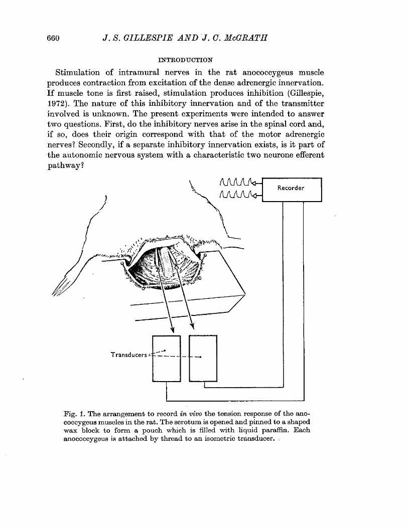
660 J. S . GILLESPIE A N D J. G. McGRATH
INTRODUCTIONStimulation of intramural nerves in the rat anococcygeus muscle
produces contraction from excitation of the dense adrenergic innervation. If muscle tone is first raised, stimulation produces inhibition (Gillespie, 1972). The nature of this inhibitory innervation and of the transmitter involved is unknown. The present experiments were intended to answer two questions. First, do the inhibitory nerves arise in the spinal cord and, if so, does their origin correspond with that of the motor adrenergic nerves? Secondly, if a separate inhibitory innervation exists, is it part of the autonomic nervous system with a characteristic two neurone efferent pathway?
Recorder
T ransducers -
Fig. 1. The arrangement to record in vivo the tension response o f the anococcygeus m uscles in the rat. The scrotum is opened and pinned to a shaped w ax block to form a pouch which is filled w ith liquid paraffin. Each anococcygeus is attached by thread to an isometric transducer.
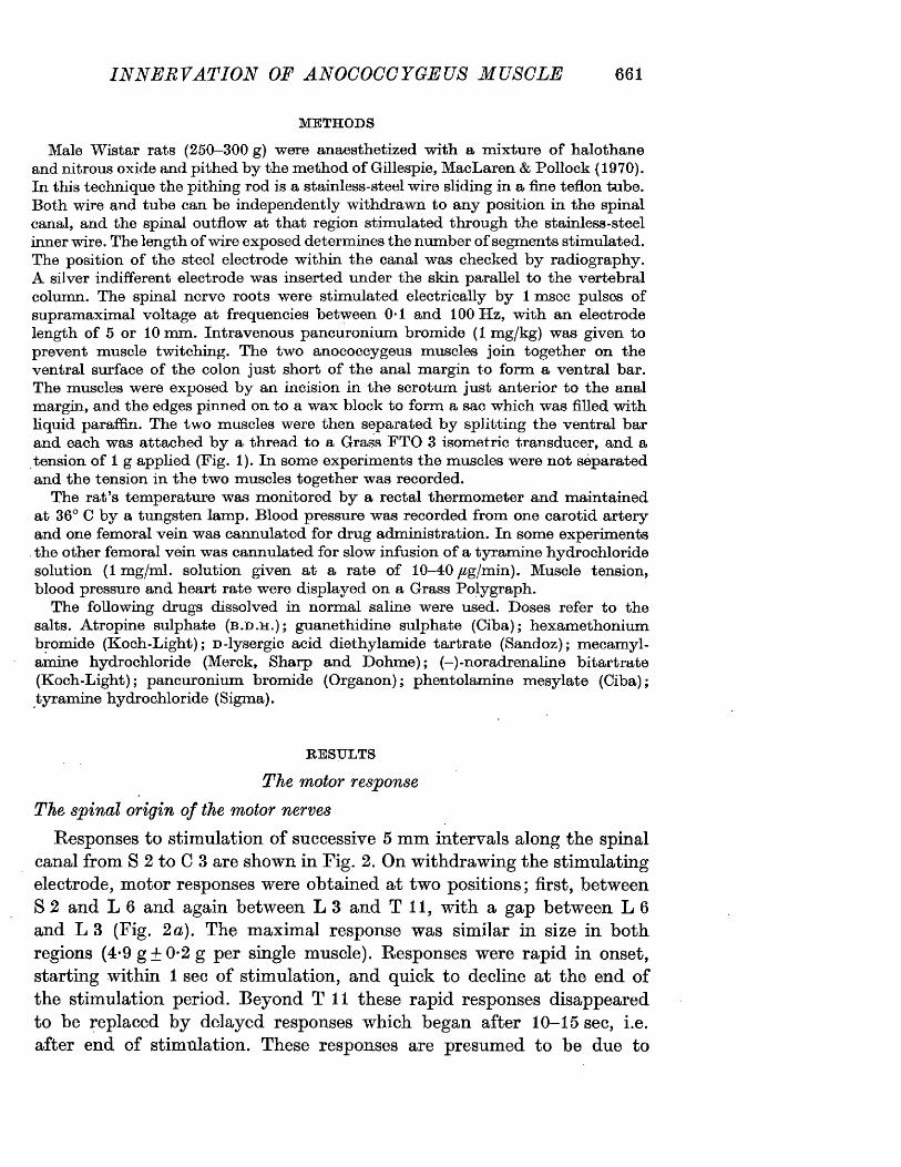
IN N E R V A TIO N OF ANOCOCCYGEUS MUSCLE 661
METHODSMale W istar rats (250—300 g) were anaesthetized w ith a m ixture o f halothane
and nitrous oxide and pithed by the m ethod o f Gillespie, MacLaren & Pollock (1970). In this technique the pithing rod is a stainless-steel wire sliding in a fine teflon tube. Both wire and tube can be independently withdrawn to any position in the spinal canal, and the spinal outflow at that region stim ulated through the stainless-steel inner wire. The length o f wire exposed determ ines the number o f segm ents stim ulated. The position o f the steel electrode w ithin the canal was checked by radiography. A silver indifferent electrode was inserted under the skin parallel to the vertebral column. The spinal nerve roots were stim ulated electrically by 1 msec pulses o f supramaximal voltage at frequencies between 0-1 and 100 H z, w ith an electrode length o f 5 or 10 m m. Intravenous pancuronium bromide (1 m g/kg) was given to prevent m uscle twitching. The tw o anococcygeus m uscles join together on the ventral surface o f the colon just short o f the anal margin to form a ventral bar. The muscles were exposed by an incision in the scrotum just anterior to the anal margin, and the edges pinned on to a w ax block to form a sac which was filled w ith liquid paraffin. The tw o m uscles were then separated b y splitting the ventral bar and each was attached b y a thread to a Grass FTO 3 isom etric transducer, and a tension o f 1 g applied (Fig. 1). In some experim ents the m uscles were not separated and the tension in the tw o m uscles together was recorded.
The rat’s temperature was m onitored by a rectal therm om eter and m aintained at 36° C b y a tungsten lamp. Blood pressure was recorded from one carotid artery and one femoral vein w as cannulated for drug adm inistration. In some experim ents
. the other femoral vein was cannulated for slow infusion o f a tyram ine hydrochloride solution (1 m g/m l. solution given at a rate o f 10-40 /£g/min). Muscle tension, blood pressure and heart rate were displayed on a Grass Polygraph.
The following drugs dissolved in normal saline were used. Doses refer to the salts. Atropine sulphate (b .d .h . ); guanethidine sulphate (Ciba); hexam ethonium bromide (Koch-Light); d-lysergic acid diethylam ide tartrate (Sandoz); m ecam yl- amine hydrochloride (Merck, Sharp and Dohm e); ( - ) -noradrenaline bitartrate (K och-Light); pancuronium bromide (Organon); phentolam ine m esylate (Ciba); tyram ine hydrochloride (Sigma).
RESULTSThe motor response
The spinal origin of the motor nervesResponses to stimulation of successive 5 mm intervals along the spinal
canal from S 2 to C 3 are shown in Fig. 2. On withdrawing the stimulating electrode, motor responses were obtained at two positions; first, between S 2 and L 6 and again between L 3 and T i l , with a gap between L 6 and L 3 (Fig. 2 a). The maximal response was similar in size in both regions (4-9g + 0*2g per single muscle). Responses were rapid in onset, starting within 1 sec of stimulation, and quick to decline at the end of the stimulation period. Beyond T i l these rapid responses disappeared to be replaced by delayed responses which began after 10-15 sec, i.e. after end of stimulation. These responses are presumed to be due to
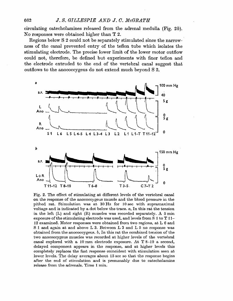
662 J. S. GILLESPIE A N D J. C. McGRATHcirculating catecholamines released from the adrenal medulla (Fig. 2b). No responses were obtained higher than T 2.
Regions below S 2 could not be separately stimulated since the narrowness of the canal prevented entry of the teflon tube which isolates the stimulating electrode. The precise lower limit of the lower motor outflow could not, therefore, be defined but experiments with finer teflon and the electrode extruded to the end of the vertebral canal suggest that outflows to the anococcygeus do not extend much beyond S 2.
100 mm Hg
B.P.
Ano
Ano __
S1 L 6 L 5 L 4-5 L 4 L 3 -4 L 3 L 2 L1 L 1 - T T 1 1 - 1 2
b150 mm Hg
0
5g
0T 11-12 T 8-10 T 6 -8 T 3 - 5 . C 7 - T 2
Fig. 2. The effect o f stim ulating a t different levels o f the vertebral canal on the response o f the anococcygeus m uscle and the blood pressure in the pithed rat. Stim ulation was at 30 H z for 10 sec w ith supramaximal voltage and is indicated by a dot below the trace, a, In this rat the tension in the left (L) and right (R) m uscles was recorded separately. A 5 m in exposure o f the stim ulating electrode was used, and levels from S 1 to T 11— 12 examined. Motor responses were obtained from tw o regions, a t L 6 and S 1 and again a t and above L 3. Between L 3 and L 5 no response was obtained from the anococcygeus, b, In this rat the combined tension of the two anococcygeus muscles was recorded at higher levels o f the vertebral canal explored w ith a 10 m m electrode exposure. A t T 8-10 a second, delayed com ponent appears in the response, and at higher levels this com pletely replaces the fast response coincident w ith stim ulation seen at lower levels. The delay averages about 15 sec so that the response begins after the end o f stim ulation and is presumably due to catecholamine release from the adrenals. Tim e 1 min.
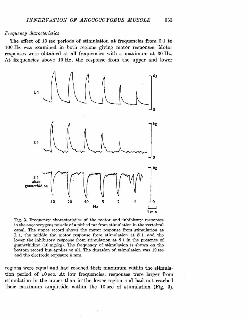
IN N E R V A TIO N OF ANOCOCCYGEUS MUSCLE 663
Frequency characteristicsThe effect of 10 sec periods of stimulation at frequencies from 0*1 to
100 Hz was examined in both regions giving motor responses. Motor responses were obtained at all frequencies with a maximum at 30 Hz. At frequencies above 10 Hz, the response from the upper and lower
L 1
S 1
-r6g
JO
v«vvVvl
S 1 after
guanethidine
“1 6g
30 20 JoI__|1 m in
Fig. 3. Frequency characteristics o f the m otor and inhibitory responses in the anococcygeus m uscle o f a pithed rat from stim ulation in the vertebral canal. The upper record shows the m otor response from stim ulation at L 1, the m iddle the m otor response from stim ulation at S I , and the lower the inhibitory response from stim ulation at S 1 in the presence of guanethidine (10 m g/kg). The frequency o f stim ulation is shown on the bottom record but applies to all. The duration o f stim ulation was 10 sec and the electrode exposure 5 m m.
regions were equal and had reached their maximum within the stimulation period of 10 sec. At low frequencies, responses were larger from stimulation in the upper than in the lower region and had not reached their maximum amplitude within the 10 sec of stimulation (Fig. 3).
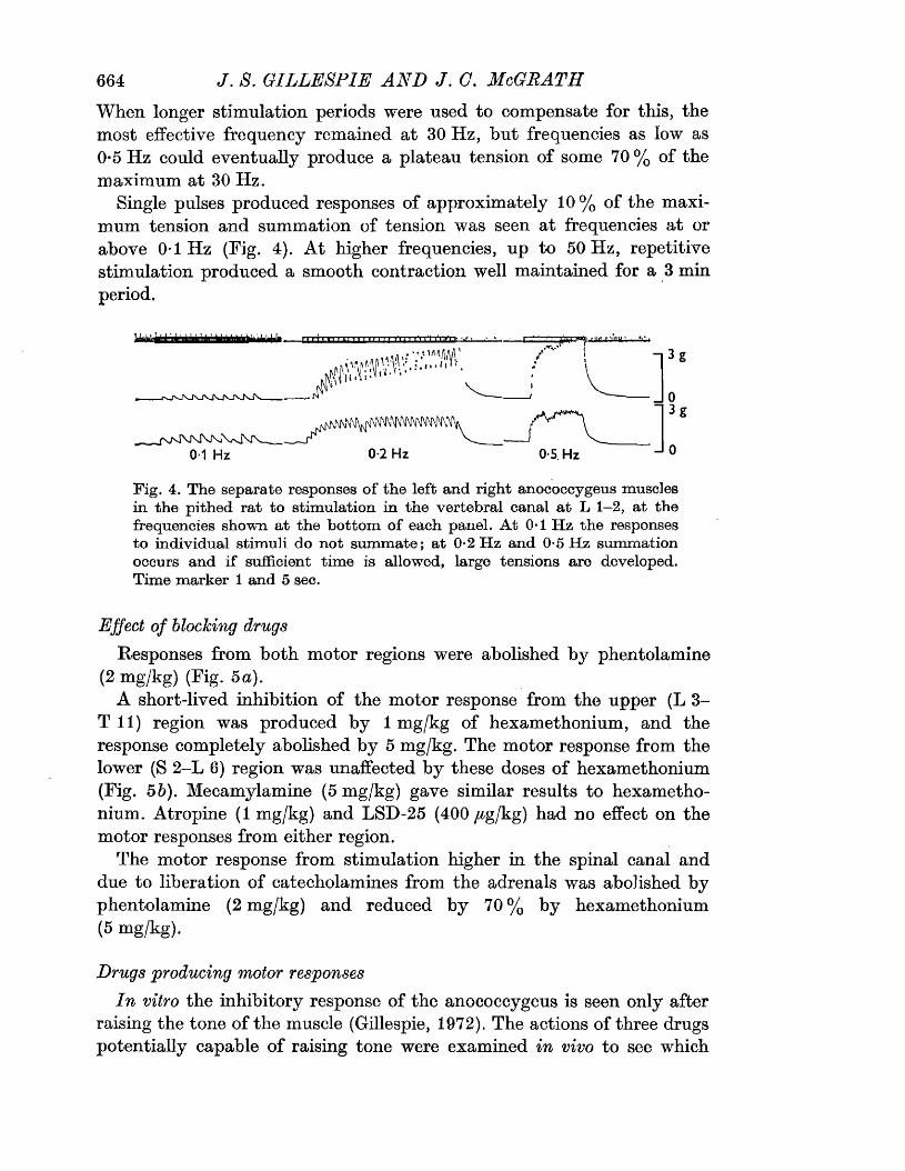
664 J . 8. GILLESPIE A N D J . (7. McGRATHWhen longer stimulation periods were used to compensate for this, the most effective frequency remained at 30 Hz, but frequencies as low as 0-5 Hz could eventually produce a plateau tension of some 70% of the maximum at 30 Hz.
Single pulses produced responses of approximately 10% of the maximum tension and summation of tension was seen at frequencies at or above 0*1 Hz (Fig. 4). At higher frequencies, up to 50 Hz, repetitive stimulation produced a smooth contraction well maintained for a 3 min period.
J0-1 Hz 0-2 Hz 0-5. Hz
Fig. 4. The separate responses o f the left and right anococcygeus muscles in the pithed rat to stim ulation in the vertebral canal a t L 1—2, at the frequencies shown at the bottom of each panel. A t 0*1 H z the responses to individual stim uli do not sum m ate; at 0-2 H z and 0-5 H z summation occurs and if sufficient tim e is allowed, large tensions are developed. Time marker 1 and 5 sec.
P T.t -T MlglJ •
Effect of blocking drugsResponses from both motor regions were abolished by phentolamine
(2 mg/kg) (Fig. 5 a).A short-lived inhibition of the motor response from the upper (L 3-
T 11) region was produced by 1 mg/kg of hexamethonium, and the response completely abolished by 5 mg/kg. The motor response from the lower (S 2-L 6) region was unaffected by these doses of hexamethonium (Fig. 55). Mecamylamine (5 mg/kg) gave similar results to hexamethonium. Atropine (1 mg/kg) and LSD-25 (400 /^g/kg) had no effect on the motor responses from either region.
The motor response from stimulation higher in the spinal canal and due to liberation of catecholamines from the adrenals was abolished by phentolamine (2 mg/kg) and reduced by 70% by hexamethonium (5 mg/kg).
Drugs producing motor responsesIn vitro the inhibitory response of the anococcygeus is seen only after
raising the tone of the muscle (Gillespie, 1972). The actions of three drugs potentially capable of raising tone were examined in vivo to see which
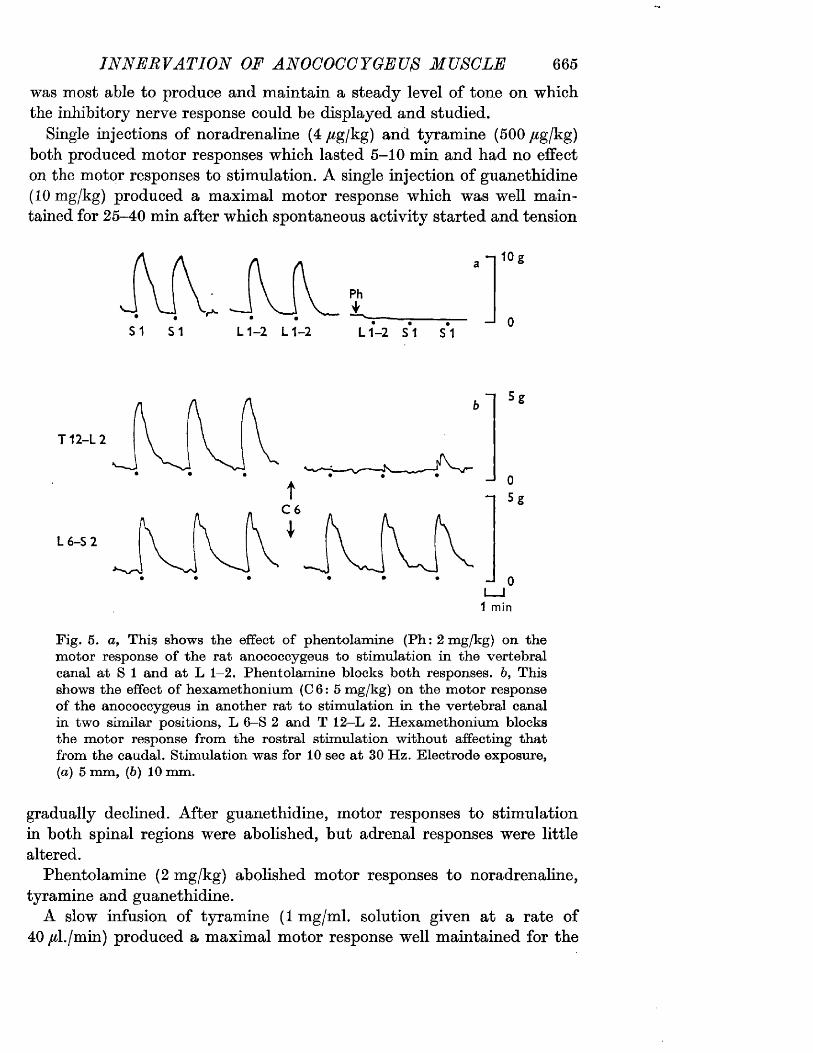
IN N ERVATIO N OF ANOCOCCYGEUS MUSCLE 665was most able to produce and maintain a steady level of tone on which the inhibitory nerve response could be displayed and studied.
Single injections of noradrenaline (4 /igj'kg) and tyramine (500 /ig/kg) both produced motor responses which lasted 5-10 min and had no effect on the motor responses to stimulation. A single injection of guanethidine (10 mg/kg) produced a maximal motor response which was well maintained for 25-40 min after which spontaneous activity started and tension
S1 S1 L 1-2 L 1-2
- t 10 g
Ph*
L 1 -2 S1 S1
T12-L2
“I 5e
L 6-S2
Fig. 5. a, This shows the effect o f phentolamine (Ph: 2 m g/kg) on the m otor response o f the rat anococcygeus to stim ulation in the vertebral canal at S 1 and at L 1—2. Phentolam ine blocks both responses. 6, This shows the effect o f hexam ethonium (C 6: 5 mg/kg) on the m otor response of the anococcygeus in another rat to stim ulation in the vertebral canal in two similar positions, L 6—S 2 and T 12-L 2. H exam ethonium blocks the m otor response from the rostral stim ulation without affecting that from the caudal. Stim ulation was for 10 sec at 30 H z. Electrode exposure, (a) 5 mm, (b) 10 mm.
gradually declined. After guanethidine, motor responses to stimulation in both spinal regions were abolished, but adrenal responses were little altered.
Phentolamine (2 mg/kg) abolished motor responses to noradrenaline, tyramine and guanethidine.
A slow infusion of tyramine (1 mg/ml. solution given at a rate of 40 /d./min) produced a maximal motor response well maintained for the
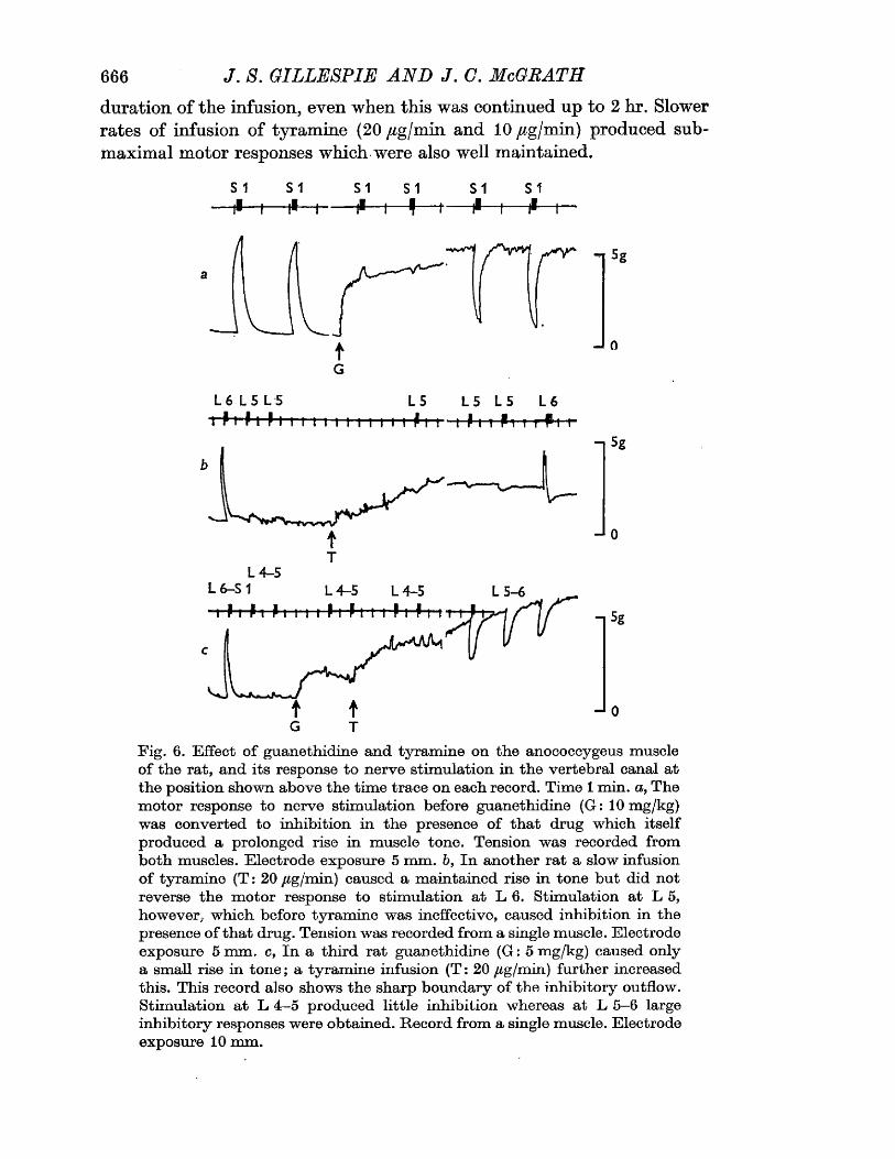
666 J. 8. GILLESPIE A N D J. C. McGRATHduration of the infusion, even when this was continued up to 2 hr. Slower rates of infusion of tyramine (20 /ig/min and 10 /^g/min) produced sub- maximal motor responses which were also well maintained.
S 1 S1-4 -
S1
-t*-S1 S1
-f-
a
"IwM
tG
L 6 L 5 L 5
T~P 1 P t P M 1 11 I I l lL 5
tt P I 1L 5 L 5 L 6 t P m Pi i rf i 1
5g
—* 0
L 4 -5L 6-S1 L 4 -5 L 4 -5 L 5 -6
G T
5g
- 1 0
Fig. 6. Effect o f guanethidine and tyram ine on the anococcygeus muscle o f the rat, and its response to nerve stim ulation in the vertebral canal at the position shown above the tim e trace on each record. Time 1 m in. a , The m otor response to nerve stim ulation before guanethidine (G : 10 mg/kg) was converted to inhibition in the presence o f that drug which itself produced a prolonged rise in m uscle tone. Tension was recorded from both m uscles. Electrode exposure 5 m m . b, In another rat a slow infusion o f tyram ine (T : 20 fig/min) caused a m aintained rise in tone but did not reverse the m otor response to stim ulation at L 6. Stim ulation at L 5, however, which before tyram ine was ineffective, caused inhibition in the presence o f that drug. Tension was recorded from a single muscle. Electrode exposure 5 m m. c, In a third rat guanethidine (G : 5 mg/kg) caused only a small rise in to n e ; a tyram ine infusion (T : 20 /tg/min) further increased this. This record also shows the sharp boundary of the inhibitory outflow. Stim ulation a t L 4 -5 produced little inhibition whereas at L 5 -6 large inhibitory responses were obtained. Record from a single muscle. Electrode exposure 10 mm.
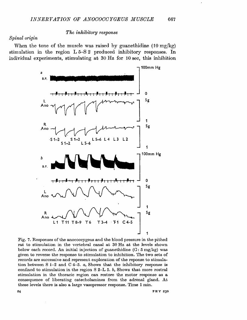
INNERVATIO N OF ANOCOCCYGEUS MUSCLE 667
The inhibitory responseSpinal origin
When the tone of the muscle was raised by guanethidine (10 mg/kg) stimulation in the region L 5-S 2 produced inhibitory responses. In individual experiments, stimulating at 30 Hz for 10 sec, this inhibition
- i 100mm Hg
B.P.
Pm Pm Pi i iPi i i Pi i Pi i Pm P
RAno
0
5g
-J 1n 5s
B.P.
■S 1 -2 S 1 -2 L 5 -6 L 4 L 3 L 2 S 1 -2 L 5 -6 —I 1
—i 100mm Hg
P I I I 1 t I1 1 1 I P I I I P I i i i "m r-fc-r JL
Ano
RA no
L 1 T 11 T 8 -9 T 6 T 3 -4 T 1 C 4 -5
0
5g
15g
J 1
Fig. 7. Responses o f the anococcygeus and the blood pressure in the pithed rat to stim ulation in the vertebral canal a t 30 H z at the levels shown below each record. An initial injection o f guanethidine (G: 5 m g/kg) was given to reverse the response to stim ulation to inhibition. The tw o sets o f records are successive and represent exploration o f the reponse to stim ulation between S 1—2 and C 4—5. a, Shows that the inhibitory response is confined to stim ulation in the region S 2 -L 5. b, Shows that more rostral stim ulation in the thoracic region can restore the m otor response as a consequence o f liberating catecholamines from the adrenal gland. A t these levels there is also a large vasopressor response. Time 1 min.24 P H Y 2 3 0
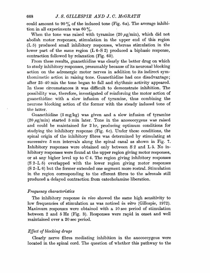
668 J. S\ GILLESPIE A N D J. C. McGRATHcould amount to 90% of the induced tone (Fig. 6 a). The average inhibition in all experiments was 60%,
When the tone was raised with tyramine (20 yag/min), which did not abolish motor responses, stimulation in the upper end of this region (L 5) produced small inhibitory responses, whereas stimulation in the lower part of the same region (L 6-S 2) produced a biphasic response, contraction followed by relaxation (Fig. 6 b).
From these results, guanethidine was clearly the better drug on which to study inhibitory responses, presumably because of its neuronal blocking action on the adrenergic motor nerves in addition to its indirect sym- thomimetic action in raising tone. Guanethidine had one disadvantage; after 25-40 min the tone began to fall and rhythmic activity appeared. In these circumstances it was difficult to demonstrate inhibition. The possibility was, therefore, investigated of reinforcing the motor action of guanethidine with a slow infusion of tyramine, thus combining the neurone blocking action of the former with the steady induced tone of the latter,
Guanethidine (5 mg/kg) was given and a slow infusion of tyramine (20 yMg/min) started 5 min later. Tone in the anococcygeus was raised and could be maintained for 2 hr, producing optimum conditions for studying the inhibitory response (Fig. 6c). Under these conditions, the spinal origin of the inhibitory fibres was determined by stimulating at successive 5 mm intervals along the spinal canal as shown in Fig. 7. Inhibitory responses were obtained only between S 2 and L 5. No inhibitory responses were found at the upper region giving motor responses, or at any higher level up to C 4. The region giving inhibitory responses (S 2-L 5) overlapped with the lower region giving motor responses (S 2-L 6) but the former extended one segment more rostral. Stimulation in the region corresponding to the efferent fibres to the adrenals still produced a delayed contraction from catecholamine liberation.
Frequency characteristicsThe inhibitory response in vivo showed the same high sensitivity to
low frequencies of stimulation as was noticed in vitro (Gillespie, 1972). Maximum responses were obtained with a 10 sec period of stimulation between 2 and 5 Hz (Fig. 3). Responses were rapid in onset and well maintained over a 20 sec period.
Effect of blocking drugsClearly nerve fibres mediating inhibition in the anococcygeus were
located in the spinal cord. The question of whether this pathway to the
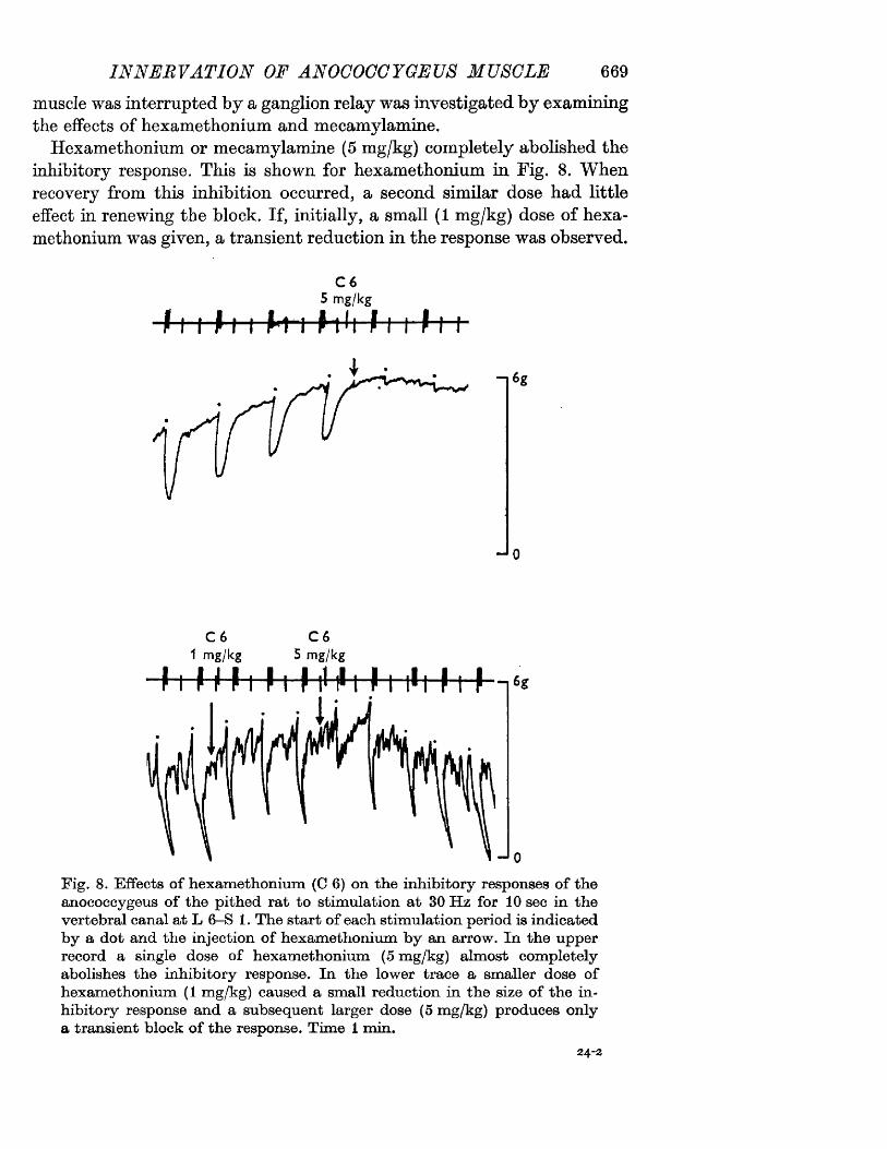
IN N ERVATIO N OF ANOCOCCYGEUS MUSCLE 669muscle was interrupted by a ganglion relay was investigated by examining the effects of hexamethonium and mecamylamine.
Hexamethonium or mecamylamine (5 mg/kg) completely abolished the inhibitory response. This is shown for hexamethonium in Fig. 8. When recovery from this inhibition occurred, a second similar dose had little effect in renewing the block. If, initially, a small (1 mg/kg) dose of hexamethonium was given, a transient reduction in the response was observed.
f t + + t
C 65 mg/kg
h-i hh f ■£++
C 6 C 65 mg/kg1 mg/kg
Fig. 8. Effects o f hexam ethonium (C 6) on the inhibitory responses o f the anococcygeus o f the pithed rat to stim ulation at 30 H z for 10 sec in the vertebral canal at L 6—S 1. The start o f each stim ulation period is indicated by a dot and the injection o f hexam ethonium b y an arrow. In the upper record a single dose o f hexam ethonium (5 m g/kg) alm ost com pletely abolishes the inhibitory response. In the lower trace a smaller dose o f hexam ethonium (1 mg/kg) caused a small reduction in the size o f the in hibitory response and a subsequent larger dose (5 m g/kg) produces only a transient block o f the response. Tim e 1 m in.
24-2
670 J, S. GILLESPIE AN D J. C. McGRATHOn recovery from this, the tissue was insensitive to subsequent large (5 mg/kg) doses of the drug.
Atropine was also studied since there is the possibility of muscarinic receptors in ganglia (Steinberg & Hilton, 1966). Atropine (1 mg/kg) had no effect on inhibitory responses.
DISCUSSIONOf the two questions posed at the outset of these investigations, the
answer to the first is clear. There is an inhibitory nerve outflow from the spinal cord to the anococcygeus muscle whose frequency and drug sensitivity resembles that of the nerves involved in the inhibitory response in vitro (Gillespie, 1972). It seems most unlikely that this inhibitory outflow could be the sympathetic adrenergic outflow liberating some additional transmitter whose inhibitory effect is uncovered by blocking the release of noradrenaline by guanethidine, for the following reasons. First, the level of origin of the motor and inhibitory fibres differs. The preganglionic motor fibres arise from L 3-T 11 whereas the inhibitory preganglionic fibres arise from S 2-L 5. Secondly, in the presence of tyramine, when the motor responses to stimulation either of the preganglionic outflow at L 3-T 11 or the post-ganglionic at S 2-L 6 are not blocked, stimulation at L 5 produces a pure inhibitory response, i.e. adrenergic blockade is not an essential requirement before inhibition can be demonstrated. These results are supported by the observation that destruction of the adrenergic innervation by 6-hydroxydopamine abolishes the motor adrenergic response but leaves the inhibitory response unaltered (Gibson & Gillespie, 1973).
The second question, whether the inhibitory outflow is organized in the pattern of the autonomic nervous system, hinges round the action of the ganglion blocking agents hexamethonium and mecamylamine. The effects of these two drugs are similar. They abolish the adrenergic motor response to stimulation in the upper (preganglionic) region. This block is reasonably long lasting and when recovery takes place a second dose of ganglion blocking drug will restore the block. On the inhibitory response the evidence for ganglion blockage was less convincing. If a large dose of drug was given, the inhibitory response was convincingly blocked and remained so for some time. As the response returned, however, a second large dose produced a short-lived inhibition only. If a small initial dose were used, it itself produced only slight transient block but was effective in producing desensitization, so that a second large dose was almost ineffective.
The doubt these results threw on the presence of a ganglion in the
IN N E R VAT I ON OF ANOCOCCYGEUS MUSCLE 671peripheral pathway made us examine the sensitivity of another organ with an adrenergic motor innervation from the lumbar cord - the vas deferens. This was found to be as easily desensitized to the action of hexamethonium as the inhibitory pathway to the anococcygeus muscle. A large dose caused inhibition of the motor response which quickly ‘ escaped whereupon a second similar dose had only a slight short-lived action. Since there is certainly a synapse in the motor pathway to the vas deferens (Sjostrand, 1965) we feel the results consistent with the presence of a synapse in the inhibitory outflow to the anococcygeus muscle.
An observation of some interest was the ability to stimulate the postganglionic adrenergic fibres from the lower lumbar-upper sacral region of the spinal canal. In previous investigations of this method of stimulating the spinal autonomic outflows, it had never been possible to stimulate post-ganglionic fibres (Gillespie et at. 1970). One explanation for the present exception could have been the position of the anococcygeus muscle directly in front of the vertebral column and having a tendinous origin from the upper coccygeal vertebrae. These conditions might have been particularly favourable for direct stimulation of intramural nerves in a muscle attached to bone overlying the stimulating electrode. This was checked by marking the position of the upper tendinous origin of the anococcygeus muscle with a metal marker and comparing by X-ray the relative positions of this marker and the spinal electrode. There was always a vertical displacement of at least 20 mm so it is unlikely that motor intramural nerves were stimulated within the muscle itself. The ability to stimulate the post-ganglionic motor nerves at least 20 mm from the muscle suggests that in this tissue, unlike the vas deferens, motor innervation is by conventional post-ganglionic Tong’ adrenergic neurones.
W e are grateful to the Rankin Medical Research Fund for apparatus and to Miss M. I. McNish and other members o f staff o f the Radiography D epartm ent o f Glasgow Western Infirmary for processing o f X-rays.
REFERENCESG i b s o n , A. & G i l l e s p i e , J . S. (1973). The effect o f im m unosym pathectom y and o f
6-hydroxydopamine on the responses o f the rat anococcygeus to nerve stim ulation and to some drugs. B r. J . Pharmac. 47, 261-267.
G i l l e s p i e , J . S. (1972). The rat anococcygeus muscle and its response to nerve stim ulation and to some drugs. Br. J . Pharmac. 45, 404-416.
G i l l e s p i e , J. S., M a c L a b e n , A. & P o l l o c k , D . (1970). A m ethod o f stim ulating different segm ents o f the autonom ic outflow from the spinal column to various organs in the pithed cat and rat. Br. J . Pharmac. 40, 257-267.
672.' ; J. 8. GILLESPIE AND J. C. McGRATHS jo stra n d , N\ O. (1965). The adrenergic innervation o f the vas deferens and the
accessory male genital glands. Acta physiol, scand. 65, suppl. 257.S te in b erg , M. & H ilto n , J . G. (1966). Site o f action o f atropine in blocking pressor
response to increased intracranial pressure in chlorisondam ine-treated dogs. Tex. Rep. Biol. M ed. 24, 222—227.
.T . Ph futrtAc- U(>
The effect of nerve stimulation on i u o c 7 1 :: i 0 : rontt'rcRnltnc by reserpine inthe heart, vas deferens and anocoee;y:;..';r; r..;-... r-e o f >:A,
J. S. G illespie and J. C. M cG rath*Department of Pharmacology, The University, Glasgow, G12 8QQ, Scotland
Most adrenergically innervated peripheral organs are depleted of their noradrenaline (NA) content by reserpine to a roughly similar degree. An. exception is the vas deferens which is relatively resistant (Sjostrand & Swedin, 1968). Two possible explanations are either that the rate of depiction is dependent on the firing frequency in the nerves and this is low in the vas deferens, or that ‘short’ adrenergic neurones in the vas deferens are less easily depleted than the long adrenergic neurones elsewhere. We have investigated these by studying the elTect of artificial stimulation on the rate of depletion of the nerves to the heart, vas deferens and anococcygeus. The latter has a dense adrenergic innervation uniformly distributed throughout the muscle like the vas deferens, but unlike the vas deferens, the neurones are conventional ‘long* ones (Gillespie & McGrath, 1972).
Rats were given various doses of reserpine intraperitoneally, the vas deferens and anococcygeus muscles removed 24 h later, and the NA.content measured. A dose of 200 jjlg/k g caused 80-90% depletion of NA in both muscles. The time course of depletion with such a dose was then examined in all three organs. After 6 h
the heart was 90% depleted whereas the vas deferens and anococcygeus were only 50% depleted. This dose (200 /xg/kg) and time (6 h) were selected to study the effects of nerve stimulation. Six groups of rats were used, control animals ; animals pithed but unstim ulated; animals pithed and the sympathetic nerves stim ulated; animals given reserpine but neither pithed nor stimulated ; animals given reserpine, pithed but not stimulated ; and animals given reserpine, pithed and stimulated. The appropriate spinal outflows were stimulated by a movable electrode in the spinal canal (Gillespie, MacLaren & Pollock, 1971). - ^
The level of NA at 6 h in animals pithed but unstimulated rose in the anococcygeus muscle in comparison with the controls ; stimulation of the nerves at 30 Hz for 10 s periods at 90 s intervals for 2 h lowered NA levels in all organs. Reserpine alone caused a 50% reduction in the vas deferens and anococcygeus and a 90% reduction in the heart.
Pithing the animal after giving reserpine reduced the depletion in the vas deferens and anococcygeus to 36% and 42% respectively and in the heart to 66%. Stimulation of the nerves for 2 h in the reserpinized rat increased the depletion in the vas deferens and anococcygeus to 68% and 82% respectively, but in the heart simply restored the depletion to 90%. The increased depletion with nerve stimulation in the vas deferens and anococcygeus was statistically significant. Since the anococcygeus and vas deferens are equally resistant to reserpine depletion, the length of the adrenergic neurone seems to be unimportant. The reduced depletion, following pithing in all three tissues and the increased depletion by nerve stimulation in the vas deferens and anococcygeus suggest that impulse traffic is an important factor.
REFERENCESG il le s p ie , J. S., M a c L a r e n , A. & P o l l o c k , D. (1970). A method of stimulating different segments
of the autonomic outflow from the spinal column to various organs in the pithed cat and rat. Br. J. Pharmac., 40, 257-267.
G il le s p ie , J. S. & M c G r a t h , J. C. (1972). The origin of the inhibitory nerve pathway t© th e r a t anococcygeus muscle. J. Physiol., Proc. Phys. Soc. March.
S jO s t r a n d , N. O. & S w e d in , G., (1968). Effect of reserpine on the noradrenaline content of the v a s deferens and the seminal vesicle compared with the submaxillary gland and the heart of the r a t . Acta Physiol. Scand,, 72, 370-377.
[From the Proceedings of the Physiological Society, 24-25 March 1972] Journal of Physiology, 224, 44-45 P
The origin of the inhibitory nerve pathway to the rat anococcygeus muscleB y J. S. G i l l e s p i e an d J. C. M c G r a th .* Department of Pharmacology, The University, Glasgow G12 8QQ, Scotland
A new smooth muscle preparation has recently been described (Gillespie,1971). This preparation has a dense motor adrenergic innervation. When these motor fibres are blocked by guanethidine and the tone of the preparation raised, field stimulation causes a large inhibitory response of rapid onset. This response is abolished by tetrodotoxin and is presumably due to nerve fibres. The transmitter is as yet unknown but is not a catecholamine, acetylcholine or ATP. The present experiments were designed to determine whether these inhibitory fibres had their origin in the spinal cord, whether their position was identical with that of the motor adrenergic fibres and, finally, whether their frequency sensitivity and response to drugs were similar to the adrenergic fibres.
The preparation used was the pithed rat in which the pithing rod is arranged to stimulate selectively different levels of the spinal outflows (Gillespie, MacLaren & Pollock, 1970). The position of the stimulating electrode was identified by X-ray. Isometric tension recordings were made from the anococcygeus muscles which can conveniently, and with minimal operative interference, be approached from below the pelvic arch; heart rate and blood pressure were also recorded.
Stimulation of two regions of the spinal outflow, separated by a definite gap, caused a motor response. The upper corresponded to vertebrae T12-L1 and the lower to L6-S2. The response from stimulation in the upper region was almost completely abolished by hexamethonium 5 mg/kg whereas the lower was little affected; both responses were abolished by phentolamine 2 mg/kg. These results are consistent with an adrenergic sympathetic pathway with a ganglion relay in the sympathetic chain. When this motor response was abolished by guanethidine and the tone of the muscle raised either by giving a large dose of guanethidine or by an infusion of tyramine, stimulation caused inhibition. The position of this inhibitory outflow coincided with the lower motor outflow but extended more rostrally, i.e. to L5. These inhibitory responses were abolished by hexamethonium 5 mg/kg though desensitization to this drug on occasions masked this blocking action. The frequency sensitivity of the two pathways was measured. Optimal motor adrenergic responses were obtained at 30 Hz, optimal inhibitory responses at 5 Hz.
* M.R.C. scholar.
2
The results show that the inhibitory neural pathway to this muscle does arise from the spinal cord. The difference in localization and frequency sensitivity between the inhibitory and motor outflows suggest these are mediated by different nerves. So far it has not been possible to distinguish a pre- and post-ganglionic component to the inhibitory pathway.
REFERENCES G i l l e s p i e , J. S. (1971). Br. J . Pharmac. 43, 430P .G i l l e s p i e , S., M a c L a r e n , A. & P o l l o c k , D. (1970). Br. J . Pharmac. 40, 257-267.
Br. J. Pharmac. ( 1974), 50 , 0 0 0 -0 0 0
THE R E S P O N S E OF THE CAT A N O C O C C Y G E U S ' "M U S C L E TO NERVE OR DRUG STIMULATION AN D !A C O M P A R I S O N WITH THE RAT A N O C O C C Y G E U S • j* * ' ' ** ' ’ . • ‘ 1** • * 4
J.S. GILLESPIE & J.C. McGRATHDepartment of Pharmacology, University of Glasgow, Glasgow, G12 8QQ
1 The cat anococcygeus muscle is shown to possess a dual innervation similar to the rat anococcygeus, with a motor adrenergic innervation and an inhibitory innervation whose transmitter is unknown. The pharmacological properties of the cat muscle were investigated and compared with those of the rat muscle.2 The cat muscle .contracts to noradrenaline, 5-hydroxytryptamine, tyramine, amphetamine, guanethidine, cocaine and lysergic acid diethylamide (LSD). The effects of noradrenaline and 5-hydroxytryptaminc are blocked by phentolamine and methysergide respectively.3 The cat anococcygeus is relaxed b y acetylch'Uine, carbachol, isoprenaline, ATP, prostaglandins E i , E2 and F2a and vasopressin, all qf which contract the rat muscle. The effects of acetylcholine and carbachol are blocked by atropine and those of isoprenaline by propranolol.4 Field stimulation produces contraction of the cat anococcygeus, which is blocked by phentolamine and guanethidine but unaffected by hexamethonium, atropine or neostigmine.5 In the presence of guanethidine (10"5m ), the tone of the muscle is raised and field stimulation produces relaxation of the muscle. These inhibitory responses are unaffected by phentolamine, hexamethonium, atropine or neostigmine.6 Neostigmine potentiates the effects of acetylcholine, but not of carbachol in relaxing the cat anococcygeus and in contracting the rat anococcygeus, but has no effect on either motor or inhibitory responses to field stimulation.7 Cold storage for up to eight days had little effect on either the motor response to noradrenaline or the motor or inhibitory response to field stimulation of the cat anococcygeus. Beyond eight days, the response to field stimulation diminishes more rapidly than the response to noradrenaline.
Introduction i The purpose of the present experiments was to! determine whether a cholinergic innervation exists
The anatomy and responses to nerve and drug in the cat muscle and if so, whether its propertiesstimulation of the rat anococcygeus have been resemble those of the inhibitory innervation in thedescribed (Gillespie & Maxwell, 1971; Gillespie, rat. In the course of the work, certain responses to
.1972; Gillespie & McGrath, 1973; Gibson & drugs in the cat muscle were found to differ so'Gillespie, ' 1973). The muscle has a dense '**'’ radically frOrh those in 'the fat that a simultaneous " adrenergic motor innervation, but there is no j comparison was made on muscles from the two evidence of cholinergic nerves; it does, however,. species; and these results are described here, possess a powerful inhibitory innervation mediatedh y " a n unknown transmitter (Gillespie, 1 9 7 2 ;" ’....... ' • • --v *'vv.Gillespie & McGrath, 1972). ” Methods
Recently, Garrett, Howard & Lansdale (1972) have examined the similar muscle in the cat and The arrangement of the cat anococcygeus musclesshown that it too has a dense adrenergic is similar to that in the rat. The two muscles ariseinnervation but, unlike the rat, it also stains for from the upper coccygeal vertebrae close to oneacetylcholinesterase. It is tempting to interpret • another in the mid-line of the pelvic cavity. The this staining as evidence of a cholinergic muscles run caudally, lying first behind and theninnervation and to associate the latter with a to one side of the colon. In the female, thepossible inhibitory mechanism. muscles terminate at the sides of the colon. In the
02. J-S. GILLESPIE &J.C. McGRATH
male they continue vcntrally to form a bar in front of the colon. :
The muscles used in these experiments were from cats primarily intended for experiments with the perfused spleen. For this latter purpose they
f-wcrc anaesthetized with a mixture of nitrous oxide and halothane and given (intravenously) 70 jug of prostaglandin Ei and 20,000 i.u. of heparin, before removal of the spleen (Blakeley, Brown, pdarnaley & W oods,, 1969). The anococcygeus
•.muscles-were then immediately dissected out. The animal was 'first killed by cardiac incision, the abdomen opened in the mid-line as far as the anal margin, the pelvis split and the bladder, urethra and vasa deferentia1 or uteri removed. The colon was then’ cut through at the pelvic brim, the pelvic .portion pulled forward and the connective-tissue behind carefully cleared until the anococcygeus muscles came into view. The muscles were isolated and in females the portion running from the vertebrae to the colon, or in males the entire length to the ventral bar, was removed to a 10 ml bath containing Krebs saline at 36 C and gassed with a mixture of 95%- 02 and 5% C 02. Rat anococcygeus muscles were removed as described by Gillespie (1972). Tension was measured with Grass FT03 isometric transducers and displayed on a Grass Polygraph. Field stimulation of the intramural nerve fibres was applied after drawing the muscles through a pair of electrodes similar to those described by Burn & Rand (1960). Stimulation of intramural nerves was with 1 ms pulses and supramaximal voltage, at the fre-- quencies indicated in the text.
Most muscles were set up as described above within minutes of removal. On other occasions, this was inconvenient and the muscles were stored in Krebs solution at 4°C for some time before use. For this reason, the effect of cold storage on the responses to field stimulation and to drugs was examined. Muscles were quickly removed from the animal to 100 ml of oxygenated Krebs in a sealed vessel and kept at 4 C for 1-18 days.
The distribution of adrenergic nerves within the muscle was examined histochemically-on freeze dried tissue with a slightly modified version of the technique of Hillarp & Falck (Gillespie & Kirpekar, 1966)..- Noradrenaline assays were carried out by the.,, trihydroxyindole method of Euler & Lisliajko (1961) after absorption on alumina.
Drugs used were: acetylcholine chloride (Koch- Light); (—)-adrenaline bitartrate (B.D.H.); dexamphetamine sulphate; adenosine S’-triphosphate disodium (Sigma); atropine sulphate; carbachol (Burroughs Wellcome); cocaine hydrochloride; guanethidine sulphate (CIBA); hexamethonium . bromide (Koch-Light); histamine acid phosphate
(B.D.H.); 5-hydroxytryptamine creatinine sulphate (Koch-Light); (±)-isoprenaline hydrochloride (Sigma); (+)-lysergic acid diethylamide tartrate (Sandoz); mcpyramine maleate (May & Baker); mcthyscrgidc bimalcate (Sandoz); '^neostigmine
1 mcthylsulphate (Roche); (-)-noradrenaline bitar- trate (Koch-Light); pancuronium bromide
_ (Organon); phentolamine mesylate (CIBA); propranolol sulphate (Sigma); prostaglandins E j, E2, F2 a tyramine hydrochloride (Sigma); vasopressin
..•(Parke-Davis). Doses-refer-to the-base ,.\vith the.* exception of prostaglandins and vasopressin.
Results
Anatomically, the rat- and cat muscles are similar. Both are about 3 cm in length; in both, the female muscle is shorter than the male because of the absence of the ventral extension. The one macroscopic difference is that the cat muscle is considerably thicker and consequently heavier. Representative weight ranges for a pair of muscles are: cat, male 100-150 mg; cat, female, 50-100 mg; rat, male 25-50 mg. The Falck technique shows that the cat, like the rat, has a terminal plexus of brightly fluorescent fibres scattered uniformly throughout the muscle. Assay of the noradrenaline content gave a value of 2.1 jug/g for the cat compared with 2.56 ±0.48 jug/g for the rat. Suspended in Krebs solution at 36°C, the cat anococcygeus differs from the rat in developing both spontaneous tone and rhythmic acitivity, greatest immediately after setting up the preparation but usually present to some extent throughout the experiment (e.g. Figures 5 and 8). Papaverine (10"5M) abolished both tone and rhythmic activity.
Effects o f drugs
Drugs causing contraction Noradrenaline, adrenaline and 5-hydroxytryptamine all caused a dose-related contraction of the cat anococcygeus.*
-Noradrenaline and adrenaline were approximately -.-., equipotent, producing contractions which were antagonized by phentolamine (10~6 M ). Responses to adrenaline and to a lesser extent noradrenaline,
. were . potentiated by . propranolol ( 1 0 “6 m ) . 5-hydroxytryptamine produced contractions which were antagonized by methysergide (2 x 10~7 m). Dose-response curves for noradrenaline and 5-hydroxytryptamine and for the effects of phentolamine and methysergide, are shown in Figure 1. Muscles from male cats produced larger responses to noradrenaline and 5-hydroxytryptamine than those from females, but the dose-response curves (drawn as a percentage of the
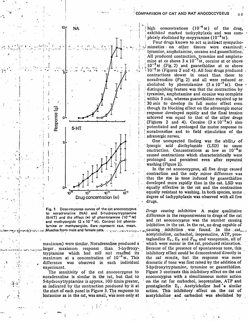
COMPARISON OF CAT AND RAT ANOCOCCYGEUS O 2
i. I
NA
CT)0 L
5-HT
oL1 0- X X
Drug concentration (m )
Fig. 1 Dose-response curves of the cat anococcygeus to noradrenaline (NA) and 5-hydroxytryptamine (5-HT) and the effect ( • ) of phentolamine (10“6 M) and methysergide (2 x 10“7 M). Control (o) phentolamine or methysergide. Bars represent ±s.e. mean.
i.v-.-Musclesfrom male and female cats. . v .>v .
maximum) were similar. Noradrenaline produced a larger., maximum, response than 5-hydroxy- tryptamine which had still not reached its maximum at a concentration of 10“3 M. This difference was observed in each individual experiment.
The sensitivity of the cat anococcygeus to noradrenaline is similar in the rat, but that to 5-hydroxytryptamine is approx. 100 times greater, as indicated by the contraction produced by it at the start of each panel in Figure 3. The response to histamine as in the rat, was small, was seen only at
! high concentrations (lO ^ M ) of the drug,. exhibited marked tachyphylaxis and was com- ' pletely abolished by mcpyramine (10~6m ).
Four drugs known to act as indirtct sympatho- ».4'mimetics • on other tissues were examined:
tyramine, amphetamine, cocaine and guanethidine.'; All produced contraction, tyramine and ampheta
mine at or above 3 x 10“ 7M, cocaine at or above j 10~5m (Fig. 2) and guanethidine at or above - M- (Figures 2- and 4). All four drugs, produced
contractions slower in onset than those to noradrenaline (Fig. 2) and all were reduced or. abolished by phentolamine ( 3 x 1 0 “ 7 m). One
i distinguishing feature was that the contraction by | tyramine, amphetamine and. cocaine was complete j within 5 min, whereas guanethidine required up to ! 30 min to develop its full motor effect even , though its blocking effect on the adrenergic motor ! response developed rapidly and the final tension
achieved was equal to that of the other drugs ; (Figtires 2 and 4). Cocaine (3 x 10“ 5m ) also • potentiated and prolonged the motor response to j noradrenaline and to field stimulation of the j adrenergic nerves.
One unexpected finding was the ability of 1 lysergic acid diethylamide (LSD) to cause
contraction. Concentrations as low as 10~8M caused contractions which characteristically were
; prolonged and persistent even after repeated washing (Figure 2).
In the rat anococcygeus, all five drugs caused , contraction and the only minor difference was
that the rise in tone induced by guanethidine developed more rapidly than in the cat. LSD was
: equally effective in the rat and the contraction equally resistant to washing. In both species, some . degree of tachyphylaxis was observed with all five
• drugs.
Drugs causing inhibition A major qualitative difference in the responsiveness to drugs of the cat and rat anococcygeus was the number causing inhibition in the cat. In the rat, no drug capable of
.^causing inhibition , was . found. -.In the ,cat,„ acetylcholine, carbachol, isoprenaline, ATP, prostaglandins E i , E2 and F2a and vasopressin, all of
* which were motor in the rat, produced relaxation. Because of the presence of spontaneous tone, this inhibitory effect could be demonstrated directly in the cat muscle, but the response was more dramatic if tone was first raised by the addition of 5-hydroxytryptamine, tyramine or guanethidine. Figure 3 contrasts this inhibitory effect on the cat anococcygeus with a simultaneous motor action
' on the rat for carbachol, isoprenaline, ATP and prostaglandin E ] . Acetylcholine had 'a similar action. This inhibitory"' effect on the cat of acetylcholine and carbachol was abolished by
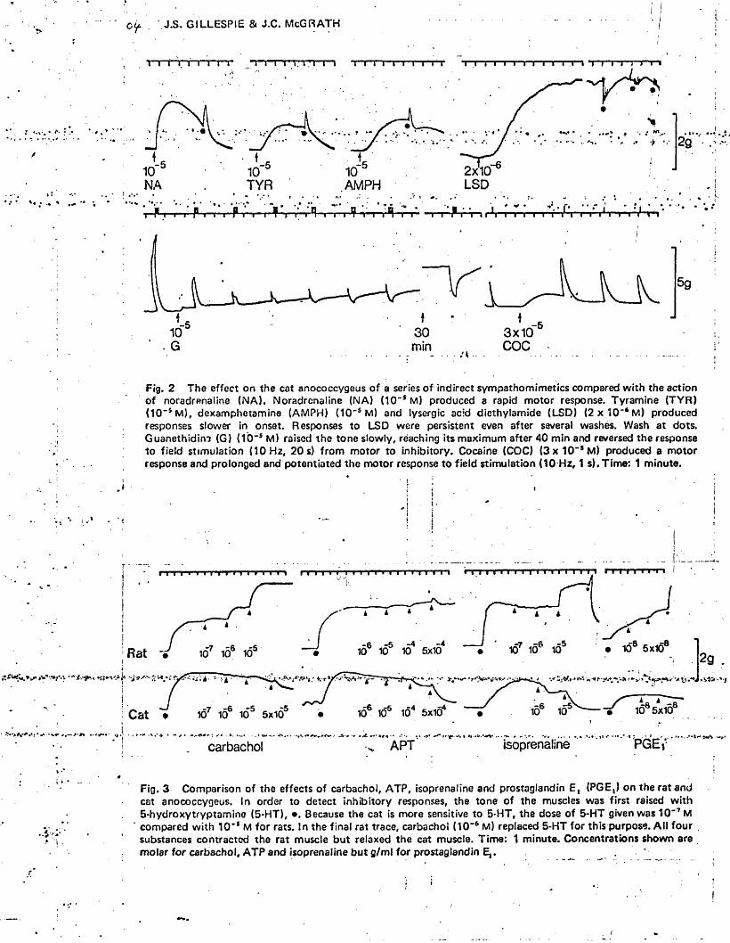
0 ^ J.S. GILLESPIE & XC. McGRATH
icf5AMPH
2x10LSD
-530min
3x10COC. G
Fig. 2 The effect on the cat anococcygeus of a series of indirect sympathomimetics compared with the action of noradrenaline (N A ). Noradrenaline (NA) (10"s M) produced a rapid motor response. Tyramine (TY R ) (10"SM ), dexamphetamine (AMPH) (10"s M) and lysergic acid diethylamide (LSD) (2 x 1 0 " 6 M) produced responses slower in onset. Responses to LSD were persistent even after several washes. Wash at dots. Guanethidine (G) (10~s M) raised the tone slowly, reaching its maximum after 40 min and reversed the response to field stimulation (10 Hz, 20 s) from motor to inhibitory. Cocaine (COC) (3 x 10"* M) produced a motor response and prolonged and potentiated the motor response to field stimulation (10 Hz, 1 s).Tim e: 1 minute.
-4Rat
■Mi -* - « f ! *" -x . is* -' ■ j-6 10° 5x10
PGEisoprenalineAPTcarbachol
.4
Fig. 3 Comparison of the effects of carbachol, ATP, isoprenaline and prostaglandin E, (PGE,) on the rat and cat anococcygeus. In order to detect inhibitory responses, the tone of the muscles was first raised with 5-hydroxytryptamine (5-H T), • , Because the cat is more sensitive to 5-HT, the dose of 5*HT given was 10"7 M compared with 10 '* M for rats. In the final rat trace, carbachol (10“6 M) replaced 5-HT for this purpose. A ll four substances contracted the rat muscle but relaxed the cat muscle. Time: 1 minute. Concentrations shown are molar for carbachol, ATP and isoprenaline but g/ml for prostaglandin E,.
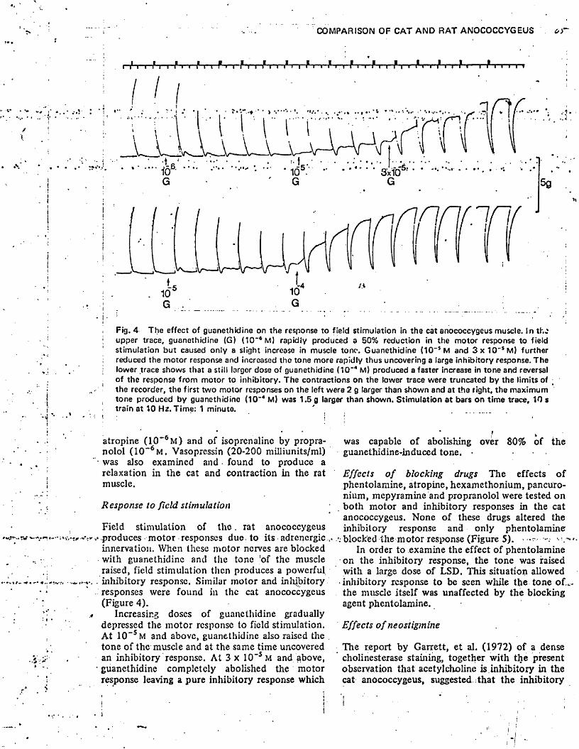
COMPARISON OF CAT AND RAT ANOCOCCYGEUS of
/r i f r i f t~r~hy~r'f n i* r-| E i i E i i E i r-f r-i—f r i-y^T-T-E"! “ i f r »"i r i
_ L _ L J. . . . . •••
‘ 105 ' " * ............3’xiO ’ *
, :V
\
iI
V— ^
10"5 G . G
5g
Fig. 4 The effect of guanethidine on the response to field stimulation in the cat anococcygeus muscle. In the upper trace, guanethidine (G) (10"*M ) rapidly produced a 50% reduction in the motor response to field stimulation but caused only a slight increase in muscle tone. Guanethidine (10-S M and 3 x 1 0 'SM) further reduced the motor response and increased the tone more rapidly thus uncovering a large inhibitory response. The lower trace shows that a still larger dose of guanethidine (1Q“4 M) produced a faster increase in tone and reversal of the response from motor to inhibitory. The contractions on the lower trace were truncated by the limits of the recorder, the first two motor responses on the left were 2 g larger than shown and at the right, the maximum tone produced by guanethidine (10~4 M) was 1.5 g larger than shown. Stimulation at bars on time trace, 10 s train at 10 Hz. Time: 1 minute.
atropine ( 1 0 " 6 m ) and of isoprenaline by propranolol ( 1 0 “6 m . Vasopressin (2 0 -2 0 0 milliunits/ml) was also examined and ■ found to produce a relaxation in the cat and contraction in the rat muscle.
Response to field stimulation
Field stimulation of th e . rat anococcygeus -produces motor responses due. to its adrenergic innervation. When these motor nerves are blocked with guanethidine and the tone ’of the muscle raised, field stimulation then produces a powerful inhibitory response. Similar motor and inhibitory responses were found in the cat anococcygeus (Figure 4).
Increasing doses of guanethidine gradually depressed the motor response to field stimulation. At 1 0 _SM and above, guanethidine also raised the tone of the muscle and at the same time uncovered an inhibitory response. At 3 x 10”5 m and above, guanethidine completely abolished the motor response leaving a pure inhibitory response which
t ‘was capable of abolishing over 80% of the guanethidine-induced tone. •
Effects o f blocking drugs The effects of phentolamine, atropine, hexamethonium, pancuronium, mepyramine and propranolol were tested on both motor and inhibitory responses in the cat anococcygeus. None of these drugs altered the inhibitory response and only phentolaminer
. blocked the motor response (Figure 5). *In order to examine the effect of phentolamine
on the inhibitory response, the tone was raised with a large dose of LSD. This situation allowed inhibitory response to be seen while the tone of._ the muscle itself was unaffected by the blocking agent phentolamine.
Effects o f neostigmine
The report by Garrett, et al. (1972) of a dense cholinesterase staining, together with the present observation that acetylcholine is.inhibitory in the cat anococcygeus, suggested that the inhibitory
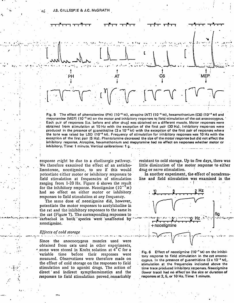
J.S. GILLESPIE & J.C. McGRATH
T~i~i°T-rn[ -t— rhnr nr-ii-r
• - 1 * Sjf
T f"i~ r f n
sNKN•\ • . t • «
VaK. j U - J i—1 ‘
tPHI
tAT
1
( T ' Y
Fig. 5 The effect of phentolamine (PH) (10“# M), atropine (AT) (10"® M), hexamethonium (C6) (10“* M) and mepyramine (MEP) (10“s M) on tho motor and inhibitory responses to field stimulation of the cat anococcygeus. Each pbir of responses (i.o, before and after drug) was obtained on a different muscle. Motor responses were obtained from stimulation at 10 Hz with the exception of the first pair (30 Hz). Inhibitory responses were produced in the presence of guanethidine (3 x 10-s M) with the exception of the first pair of responses where the tone was raised by LSD (10 ’ tt M). Frequency of stimulation for inhibitory responses was 10 Hz with the exception of the first pair (5 Hz). Phentolamine decreased the size of the motor response but did not affect the inhibitory response. Atropine, hexamethonium and mepyramine had no effect on responses whether motor or inhibitory. Time: 1 minute. Vertical calibrations: 1 g.
response n ight be due to a cholinergic pathway. We therefore examined the effect of an anticholinesterase, neostigmine, to sec if this would potentiate cither motor or inhibitory responses to field stimulation at frequencies of stimulation ranging from 1-20 II/.. Figure 6 shows the result for the inhibitory response. Neostigmine (1 0 ~ 7 m ) had no effect on either motor or inhibitory responses to field stimulation at any frequency.
The same dose of neostigmine did, however, potentiate the motor responses to acetylcholine in the rat and the inhibitory responses to the same in the cat (Figure 7). The corresponding responses to carbachol in b o th ” species were unaffected b y " neostigmine.
muscles used were
Effects o f cold storage
Since the anococcygeus obtained from cats used in other experiments, some were stored in Krebs, solution at 4 C for a variable time before their responses were measured. Observations were therefore made on the effect of cold storage on the responses to field stimulation and to agonist drugs. The action of d irec t' and indirect sympthomimetics and the responses to field stimulation proved . remarkably
resistant to cold storage. Up to five days, there was little diminution of the motor response to either drug or nerve stimulation. ■
In another experiment, the effect of noradrenaline and field stimulation was examined in the
\2 Hz
■ 2:- -vr-r 5.- : Hz .v*1— J S — 1— 1— i" 1 * t ~ i— -i -’i81 r -|—
i + neostigmine
5g-•V *.*■>?*» ■■•r-w-
• f . v - -
Fig. 6 Effect of neostigmine (10 -7 M) on the inhibitory response to field stimulation in the cat anococcygeus. In the presence of guanethidine (3 x 10‘ s M), stimulation at the frequencies indicated above the time trace produced inhibitoryrerponses. Neostigmine (lower trace) had no effect'on the size or duration of responses at 2, 5, or 10 Hz. Time: 1 minute.
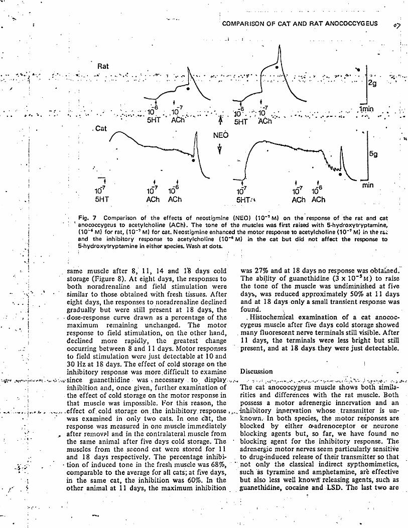
COMPARISON OF CAT AND RAT ANOCOCCYGEUS
Rat
5HT ACh
f'. .. .\*K'
♦
V • * . . * * • * * ■» !
^ 5HT ACh
NEO
2 g
Imin
5g
mtn
ACh ACh 5HT-* ACh ACh
Fig. 7 Comparison of the effects of neostigmine (NEO) (10~7 M) on the response of the rat and cat 1 anococcygeus to acetylcholine (ACh). The tone of the muscles was first raised with 5-hydroxytryptamine,
(10“6 M) for rat, (TO-7 M) for cat. Neostigmine enhanced the motor response to acetylcholine (10~7 M) in the ra; and the inhibitory response to acetylcholine (10“6 M) in the cat but did not affect the response to 5-hydroxytryptamine in either species. Wash at dots.
same muscle after 8, 11, 14 and 1’8 days cold storage (Figure 8). At eight days, the responses to both noradrenaline and field stimulation were similar to those obtained with fresh tissues. After eight days, the responses to noradrenaline declined gradually but were still present at 18 days, the
• dose-response curve drawn as a percentage of the maximum remaining unchanged. The motor response to field stimulation, on the other hand, declined more rapidly, the greatest change occurring between 8 and 11 days. Motor responses to field stimulation were just detectable at 10 and 30 Hz at IS days. The effect of cold storage on the inhibitory response was more.difficult to examine
v-since guanethidine was •. necessary . to display.,, inhibition and, once given, further examination of the effect of cold storage on the motor response in that muscle was impossible. For this reason, the
.effect of. cold storage on . the inhibitory, response,. was examined in only two cats. In one c<it, the response was measured in one muscle immediately after removal and in the contralateral muscle from the same animal after five days cold storage. The muscles from the second cat were stored for 11 and 18 days respectively. The percentage inhibi-
• tion of induced tone in the fresh muscle was 68%, comparable to the average for all cats; at five days, in the same cat, the inhibition was 60%. In the other animal at 11 days, the maximum inhibition
was 27% and at 18 days no response was obtained. The ability of guanethidine (3 x 10" s m ) to raise the tone of the muscle was undiminished at five days, was reduced approximately 50% at 11 days and at 18 days only a small transient response was found.
. Histochemical examination of a cat anococcygeus muscle after five days cold storage showed
, many fluorescent nerve terminals still visible. After 11 days, the terminals were less bright but still present, and at 18 days they were just detectable.
Discussion■' > • •' ' . »'V.f I * V . ' ■ " V - ' - , ' ;VJ J
The cat anococcygeus muscle shows both similarities and differences with the rat muscle. Both possess a motor adrenergic innervation and an inhibitory innervation whose transmitter is unknown. In both species, the motor responses are blocked by either o-adrenoceptor or neurone blocking agents but, so far, we have found no blocking agent for the inhibitory response. The adrenergic motor nerves seem particularly sensitive
. to drug-induced release of their transmitter so that ‘ not only the classical indirect sypthomimetics,
such as tyramine and amphetamine, are effective but also less well knowtf releasing agents, such as guanethidine, cocaine and LSD. The last two are
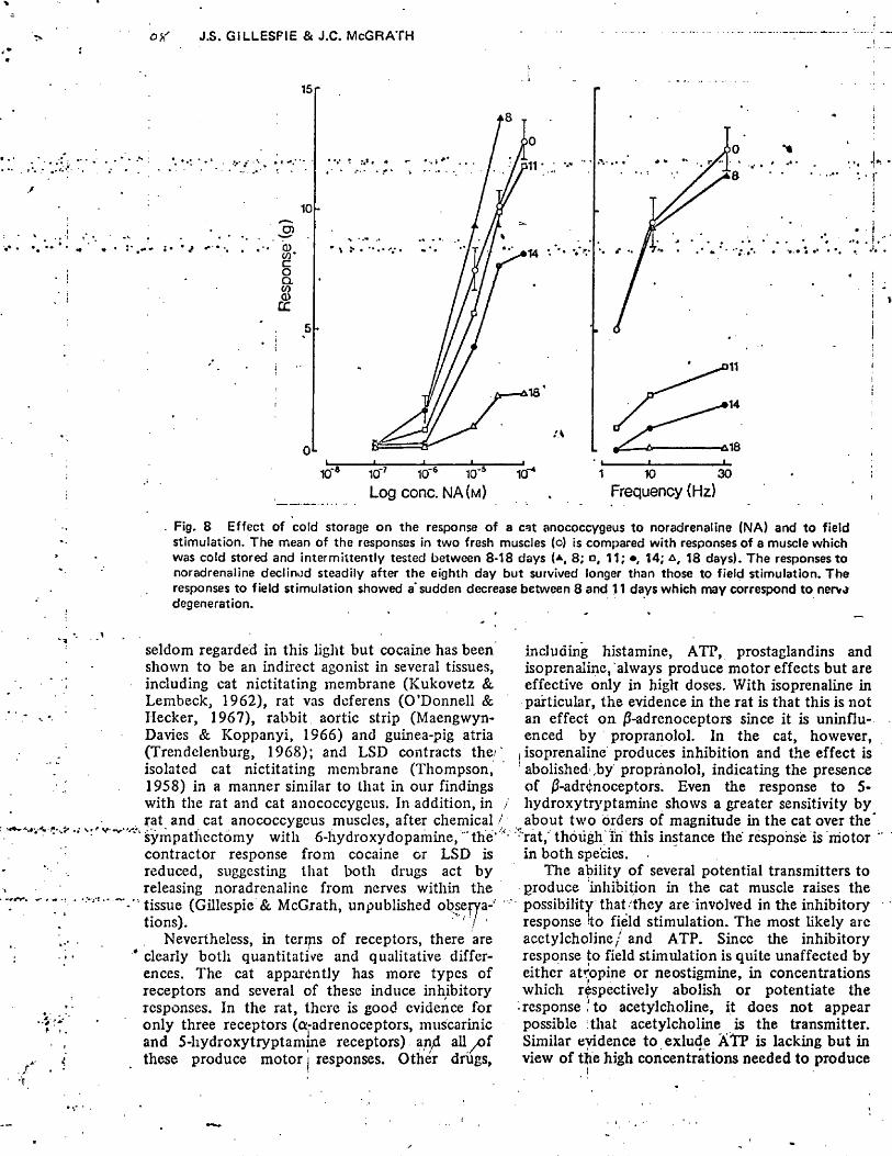
J.S. GILLESPIE & J.C. McGRATH
15 r
10
•2 ?CDCOcoCLCO<1>CC
0L xL10® 10'7 10'6 1CT5Log conc. NA(m)
icr 1 10 30Frequency (Hz)
Fig. 8 Effect of cold storage on the response of a cat anococcygeus to noradrenaline (NA) and to field stimulation. The mean of the responses in two fresh muscles (o) is compared with responses of a muscle which was cold stored and intermittently tested between 8-18 days (* , 8; o, 11; • , 14; 18 days). The responses tonoradrenaline declined steadily after the eighth day but survived longer than those to field stimulation. The responses to field stimulation showed a* sudden decrease between 8 and 11 days which may correspond to nerve degeneration.
seldom regarded in this light but cocaine has been shown to be an indirect agonist in several tissues, including cat nictitating membrane (Kukovetz & Lembeck, 1962), rat vas deferens (O’Donnell & Hecker, 1967), rabbit aortic strip (Maengwyn- Davies & Koppanyi, 1966) and guinea-pig atria (Trendelenburg, 1968); and LSD contracts the/' isolated cat nictitating membrane (Thompson, 1958) in a manner similar to that in our findings with the rat and cat anococcygeus. In addition, in / rat and cat anococcygeus muscles, after chemical /
‘ Sympathectomy with 6-hydroxydopamine, the*" contractor response from cocaine or LSD is reduced, suggesting that both drugs act by releasing noradrenaline from nerves within the tissue (Gillespie & McGrath, unpublished obsem -' tions). ^ ' '! '
Nevertheless, in terips of receptors, there are clearly both quantitative and qualitative differences. The cat apparently has more types of receptors and several of these induce inhibitory responses. In the rat, there is good evidence for only three receptors (a-adrenoceptors, muscarinic and 5-hydroxytryptamme receptors) anyd all of these produce motor! responses. Other drugs,
including histamine, ATP, prostaglandins and isoprenaline, always produce motor effects but are effective only in high doses. With isoprenaline in particular, the evidence in the rat is that this is not an effect on j3-adrenoceptors since it is uninfluenced by propranolol. In the cat, however,
i isoprenaline produces inhibition and the effect is 1 abolished ,by propranolol, indicating the presence of ^-adrenoceptors. Even the response to 5- hydroxytryptamine shows a greater sensitivity by about two orders of magnitude in the cat over the*
Tat,- though in this instance the response is motor in both species.
The ability of several potential transmitters to produce inhibition in the cat muscle raises the possibility that ■ they are involved in the inhibitory response to field stimulation. The most likely arc acetylcholine/ and ATP. Since the inhibitory response to field stimulation is quite unaffected by either atropine or neostigmine, in concentrations which respectively abolish or potentiate the
• response 'to acetylcholine, it does not appear possible Itliat acetylcholine is the transmitter. Similar evidence to exlude ATP is lacking but in view of the high concentrations needed to produce
“ COMPARISON OF CAT AND RAT ANOCOCCYGEUS 09
a small mechanical inhibition in contrast to the intense inhibition produced by field stimulation, it does not seem a likely candidate. Furthermore, it appears more probable that the inhibitory, transmitter will be the same in both species and in the rat only motor responses can be obtained with ATP.
Garrett et 'dl. (1972) reported acetylcholinesterase in the cat anococcygeus. No similar staining was- observed in the rat muscle, y e t .
■’nebstigfnihe caused ‘a similar’ degree of potentia-. "tion of the effects of acetylcholine in both. We
have no explanation of this discrepancy nor can we suggest a function for the enzyme in this muscle.
These experiments on nerve or drug responses x>f the cat muscle were carried out on tissues which might be cold stored up to a maximum of two days. The necessary control studies on the effect of cold storage showed that little change in nerve or drug responses occurred over this short period. Less expected was the ability of these nerves, cut off from their ganglion cell body, to be still present and capable of functioning after 11 days, as shown by the hislochemical demonstration of adrenergic nerve terminals together with . the responses to field stimulation and to the indirect sympathomimetic action of guanethidine. Stimulation of post-ganglionic adrenergic nerves fails to produce responses after two days in, mouse, rat and rabbit ileum, three days in guinea-pig colon (Holman &. Hughes, 1965) and seven days in guinea-pig taenia caecum (Ilattori, Kurahashi, Mori & Shibata, 1972); Adrenergic nerves were not demonstrable histochcmically after five days cold storage in guinea-pig caecum (Ilattori et a l, 1972) or rabbit aortic strips (Shibata, Hattori, Sakurai, Mori & Fujiwara, 1971). The capacity of the anococcygeus muscle to respond to noradrenaline after the nerves had ceased to function is in
agreement with the usual finding that in cold stored tissues the smooth muscle cells remain viable longer than the nerves. Thus intestinal smooth muscle from various species remains responsive to acetylcholine for several days after the cholinergic nerves have ceased to function (Ambache, 1946) and in guinea-pig taenia caecum phenylephrine still relaxes the muscle after 18 days cold storage (Fukuda & Shibata, 1972) despite the complete absence of nerve responses 'after 5 days (Hattori cf a/:, 197?-). ,
It was not possible, however, to differentiate between motor and inhibitory nerves in the cat ‘ anococcygeus by their ability to, survive cold storage. In previous studies, where cold storage of a tissue has been employed successfully to differentiate between, the components of a multiple innervation, the different anatomical arrangement of the nerves has been a major factor contributing to the preferential survival of one type (Holman & Hughes, 1965; Hattori et a l ,1972). However, in the isolated cat anococcygeus muscle as in the rat, (Gillespie, 1972), both motor and inhibitory nerves consist purely of postganglionic fibres since ganglion blockers have no effect on responses to field stimulation. There is therefore no reason tp expect differential survival. Despite the long survival after cold storage of the nerve and muscle elements of the cat anococcygeus, we feel that species variation may be at least as important as the properties of the particular tissue used, since the rat anococcygeus muscle does not respond to either field stimulation or noradrenaline after only five or six days (D. Templeton, personal communication).
We are grateful to the M.R.C. for a support grant (J.C.M. was an M.R.C. scholar) and to Professor E. Horton for the. gift o f the prostaglandins. '
References
AMBACHE, N. : (1946). Interaction- of 'd ru g s ' and the" effect of cooling on the isolated mammalian tissue./. Physiol (Lond.), 104, 266-287,
BLAKELEY, A.G.H., BROWN, SIR L., DEARNALEY, .D.P. & WOODS, R.I. (1969). Perfusion of the spleen with
blood containing prostaglandin E ,: transmiltcrjibera^ tion and uptake. Proc. Roy. Soc. Lond. B„ 174, 281-292.
BURN, J.IL & RAND, M.J. (1960). The relation of circulating noradrenaline to the effect of sympathetic stimulation. / . Physiol (Lond.), 150, 293-305.
EULER, U.S. VON, & L1SHAJICO, F. (1961). Improved techniques for the fluorim euic estimation of catecholamine. Acta. Physiol Scand., 51, 348-356.
FUKUDA, H. & SHIBATA, S. (1972). The effect of cold
storage "*\mT tiU "inhibitory action of isoprenaline, phenylephrine and nicotine on the mechanical and membranal activities pf guinea-pig taenia caecum. Br.
J. Pharmac., 46,438,-448.. .. GARRETT,. J.R ., HOWARD, E.R. & LANS DALE, J .M .. .
(1972). Myenteric nerves in the hind gut o f the cat. / . Physiol. (Lond.),.226,103-104.
GIBSON, A. & GILLESPIE, J.S. (1973). The effect o f immunosympathectomy and o f 6-hydroxydopamine on the response , o f the rat anococcygeus to nerve stimulation and to some drugs. Br. / . Pharmac., 47, 261-267.
GILLESPIE, J.S. (1972). The rat anococcygeus muscle and its response to nerve stimulation and to some drugs. Br. /. Pharmac., 45/404-416.
O'O J.S. GILLESP'E & J.C. McGRATH
GILLESPIE, J.S. &. KIRPEKAR, S.M. (3 966). The histological localization of noradrenaline in the cat spleen. J. Physiol (Lond.), 187, 69-79.
GILLESPIE. J.S. & McGRATH, J.C. (1972). The effect o f nerve stimulation on the depletion of noradrenaline by
• rcserpine in the heart, vas deferens and anococcygeus • muscle of the rat. Br. J. Pharmac., 46, 525-526.
GILLESPIE, J.S. &. McGRATH, J.C. (1973). The spinal origin o f the motor and inhibitory innervation of the rat anococcygeus muscles. J. Physiol. (Lond.), (in the press). . . . . . - . .
GILLESPIE, J.S. & MAXWELL, J .b .v(l97i);'A drcncr'grc "* innervation of sphinctcric and non-sphinclcric smooth muscle in the rat intestine. J. Histochem. Cytochcm,, ' 19,676-681.
HATTORI, K., KURAHASHI, K., MORI, J. &, SHIBATA,S. (1972). The effect o f cold storage on the adrenergic mechanisms o f intestinal sm ooth muscle. Br. J. Pharmac., 46, 423-437.
HOLMAN, E. &, HUGHES, J.R. (1965). Inhibition of intestinal smooth muscle. Aust. J. exp. Biol. med. Sci., 43,277-290.
KUKOVETZ, W.R. & LEMBECK, F. (1962). Unter- ■suchungen uber die adrcnalinpotcnzierende Wirkung
von Cocain und Dencrvierung. Naunyn-Schmiedebcrg's Arch. exp. Path, u Pharmak., 242,467-479.
MAENGWYN-DAV1ES, G.D. & KOPPANYI, T. (1966). Cocaine tachyphylaxis and effects on indirectly-acting; sympathomimetic drugs in the rabbit aortic strip and in splenic tissue. J. Pharmac. exp, Ther., 154, 481-492; '.
O ’DONNELL, S.R. & IIECKER, S.E. (1967). Contraction of rat vas deferens by cocaine. / Pharm. Pharmac., 19, 695.
SHIBATA, S., HATTORI, K., SAKURAI, I., MORI, J. &. FUJIWARA, M. (1971)., Adrenergic innervation and
• •• cocaine-induced potentiation of adrenergic.responses - of aortic strips from young and old rabbits. J. Pharmac. exp. Ther., 177,621-632.
THOMSON, J.W. (1958). Studies on the response of the isolated nictitating membrane of the cat J. Physiol (Lond.):, 141,46-72. . . . ,
TRENDELENBURG, U. (1968). The effect of cocaine on ■the pacemaker of isolated guinea-pig atria. J. Pharmac. exp. Ther., 161,222-231. ^
(ReceivedMay 25, 1973) .
SUlliARY
THE INHIBITORY AT© MOTOR INNERVATION OF
ANOCOCCYGEUS MJSCLE
A thesis presented for the degree of
Doctor of Philosophy
by
John Christie McGrath
Department of Pharmacology
University of Glasgow
December 1973
THE
(1) The subject of this thesis was the investigation of the dual innervation of the anococcygeus muscle* The rat anococcygeus had previously been shewn in vitro to have a motor adrenergic innervation and also an inhibitory nerve response whose transmitter was unknown.
(2) The object of the present study ms threefold -
(a) To determine whether this inhibitory response was due to a nerve )pathway distinct from the motor innervation and with a separate spinal origin and if so whether this pathway had a ganglion synapse and so cculd be considered as part of the autonomic nervous system*(b) To compare the pharmacological properties of the anococcygeus imscle and vas deferens and to determine whether the rate of depletion of noradrenaline by reserpine in these two tissues was affected by nerve stimulation*(c) To compare the properties of the anococcygeus nuscles from thecat and the rat in order to find whether the dual innervation found in the rat was represented in this further species and if so whether this comparison would throw any more light on the nature of the inhibitory response.
(3) Using a pithed rat preparation permitting selective stimulation of the autonomic spinal outflows, it was shown that the inhibitory pathway to the anococcygeus nuscles arose from the spinal canal, that it was interrupted by a ganglion synapse and that the spinal origin of its preganglionic nerves was L 5 - S 2 as opposed to T 10 - L 3 for the preganglionic nerves in the motor pathway,
(4) Using'.this same preparation, the pharmacological properties of the motor nerves to the anococcygeus were examined in situ and compared with those of the vas deferens* Tnis comparison demonstrated that the pharmacological properties of the anococcygeus motor innervation were thos
of a classical adrenergic innervation whereas the vas deferens showed responses which were in themselves canplex and showed unconventional responses to drugs. A hypothesis is suggested to explain this unconventional nature of the vas deferens response.
(5) An analysis of the dose dependence and time course of the depletion oftissue noradrenaline by res orpine showed that the rat anococcygeus and vas
/deferens were depleted to a similar extent and at: a similar rate and that this was slower than that found in the heart. Increase in sympathetic nerve activity by spinal stinulation in pithed rats significantly increased the noradrenaline depletion in both anococcygeus and vas deferens. From this it is suggested that nerve impulse traffic m y be an important factor in determining the rate of depletion of .noradrenaline by reserpine arid in the vas deferens m y explain the apparent resistance to depletion.
(6) The cat anococcygeus muscle was investigated in vitro and shown to possess a dual innervation similar to that in the rat. EUe to the presence of intrinsic tone, both motor and inhibitory nerve responses could be demonstrated in the absence of blocking drugs and their interaction studied. The pharmacological properties of the cat anococcygeus were similar to those of the rat except that several substances relaxed the cat muscle which contracted the rat including acetylcholine, isoprenaline, prostaglandins and ATP. These substances were therefore assessed as possible inhibitory transmitters but further analysis with blocking drugs suggested that the relaxations produced by these drugs were different from that produced by the inhibitory nerves. The inhibitory effect of acetylcholine on the cat muscle was particularly interesting since it inhibited motor nerve responses as well as relaxing the muscle. Several substances not normally associated with release of noradrenaline from nerves, including guanethidine, cocaine
LSD and 5HT produced indirect sympathomimetic effects in both species*
(7) It is concluded that the anococcygeus mscle receives a dual innervation consisting of a motor adrenergic pathway originating from the lower thoracic and upper lumbar cord and a separate inhibitory pathway with its preganglionic fibres originating from the lower lumbar and upper sacral region of the vertebral column. ' This dual innervation is found in both the rat and cat anococcygeus but in neither species does the inhibitory pathway appear to be adrenergic, cholinergic or par inerg ic and the transmitter remains unknown.
Related Documents


![Welcome to Enlighten: Theses - Enlighten: Thesestheses.gla.ac.uk/71878/1/10394171.pdf · 2019. 5. 10. · [Chronicle of the EOKA struggle 1955-1959] (Athens: [n. pub.], 1971); Georgios](https://static.cupdf.com/doc/110x72/612d4a4a1ecc5158694218df/welcome-to-enlighten-theses-enlighten-2019-5-10-chronicle-of-the-eoka.jpg)