[Frontiers in Bioscience, Landmark, 25, 1058-1109, March 1, 2020] 1058 Epigenetic alterations in cancer Suganya Ilango 1 , Biswaranjan Paital 2 , Priyanka Jayachandran 1 , Palghat Raghunathan Padma 1 , Ramalingam Nirmaladevi 1 1 Department of Biochemistry, Biotechnology and Bioinformatics, Avinashilingam Institute for Home Science and Higher Education for Women, Coimbatore, 641043, Tamil Nadu, India, 2 Redox Regulation Laboratory, Department of Zoology, CBSH, Odisha University of Agriculture and Technology, Bhubaneswar-751003, India TABLE OF CONTENTS 1. Abstract 2. Introduction 3. Importance of epigenetics in cancers 4. Biological basis of cancer 4.1. Epigenetic mechanisms in normal cells 4.2. Epigenetic mechanisms in cancer cells 4.3. Epigenetics of cancer in relation to aging 5. DNA methylation 5.1. Role of DNA methylation in cancer 5.2. DNA hypomethylation in cancer 5.3. Epigenetic alterations involving DNA methylation by mutation 5.4. DNA hypermethylation in cancer 5.5. DNA demethylation 6. Histone modifications 6.1. Non histone methylation 7. Nucleosome remodelling 7.1. Changes in chromatin 8. Micro RNAs (miRNAs) 8.1. miRNA biogenesis 8.2. Biological roles of miRNAs 9. Regulation of epigenetics in cancer progression 10. Role of oxygen and cancer 10.1. Normoxia and cancer 10.2. Hypoxia 10.2.1. Functional effect of epigenetic regulation upon hypoxia 10.2.2.. Importance of epigenetics in tumor hypoxia and cancer immunotherapy 11. Epigenetic therapy 12. Acknowledgments 13. References 1. ABSTRACT Genetic and epigenetic modifications in DNA contribute to altered gene expression in aging and cancer. In human cancers, epigenetic changes such as DNA methylation, histone modifications, micro RNAs and nucleosome remodelling all control gene expression. The link between the genetics and

Welcome message from author
This document is posted to help you gain knowledge. Please leave a comment to let me know what you think about it! Share it to your friends and learn new things together.
Transcript
[Frontiers in Bioscience, Landmark, 25, 1058-1109, March 1, 2020]
1058
Epigenetic alterations in cancer
Suganya Ilango1, Biswaranjan Paital2, Priyanka Jayachandran1, Palghat Raghunathan
Padma1, Ramalingam Nirmaladevi1
1Department of Biochemistry, Biotechnology and Bioinformatics, Avinashilingam Institute for Home
Science and Higher Education for Women, Coimbatore, 641043, Tamil Nadu, India, 2Redox
Regulation Laboratory, Department of Zoology, CBSH, Odisha University of Agriculture and
Technology, Bhubaneswar-751003, India
TABLE OF CONTENTS
1. Abstract
2. Introduction
3. Importance of epigenetics in cancers
4. Biological basis of cancer
4.1. Epigenetic mechanisms in normal cells
4.2. Epigenetic mechanisms in cancer cells
4.3. Epigenetics of cancer in relation to aging
5. DNA methylation
5.1. Role of DNA methylation in cancer
5.2. DNA hypomethylation in cancer
5.3. Epigenetic alterations involving DNA methylation by mutation
5.4. DNA hypermethylation in cancer
5.5. DNA demethylation
6. Histone modifications
6.1. Non histone methylation
7. Nucleosome remodelling
7.1. Changes in chromatin
8. Micro RNAs (miRNAs)
8.1. miRNA biogenesis
8.2. Biological roles of miRNAs
9. Regulation of epigenetics in cancer progression
10. Role of oxygen and cancer
10.1. Normoxia and cancer
10.2. Hypoxia
10.2.1. Functional effect of epigenetic regulation upon hypoxia
10.2.2.. Importance of epigenetics in tumor hypoxia and cancer immunotherapy
11. Epigenetic therapy
12. Acknowledgments
13. References
1. ABSTRACT
Genetic and epigenetic modifications in
DNA contribute to altered gene expression in aging
and cancer. In human cancers, epigenetic changes
such as DNA methylation, histone modifications,
micro RNAs and nucleosome remodelling all control
gene expression. The link between the genetics and
Epigenetic and cancer
1059 © 1996-2020
epigenetics in cancer is further shown by existence
of aberrant metabolism and biochemical pathways in
cancer or mutation in genes that are epigenetic
players. Reversal of these epigenetic changes has
been clearly shown to have therapeutic value in
various forms of lymphoma and preleukemia and
similar results are appearing for the treatment of solid
tumors. In this review, we discuss the functional
effects of epigenetic changes inducible by hypoxia,
the epigenetic alterations in cancer and how they
contribute to tumor progression and their relevance
to epigenetic therapy.
2. INTRODUCTION
The human genome project has been one
of the most important scientific achievements in
modern history. It has ushered in a new era in the
field of life science research. However, among the
project’s many great discoveries, surprising findings
such as only particular subsets of genes being able
to be expressed at a particular location and time, led
to the realization that knowledge of DNA sequences
is insufficient to understand phenotypic
manifestations. The mechanism by which DNA, or
the genetic code, is translated into protein sequences
is not merely dependent on the sequence itself but
also on a sophisticated regulatory system that
interplays between genetic and environmental
factors. These mechanisms comprise the science of
epigenetics, and the control of genes through various
chemical interactions for the basis of at least part of
the regulatory system overseeing the expression of
the genetic code (1).
Eukaryotic genomic information is
modulated by a variety of epigenetic modifications
that play both a direct role in establishing
transcription profiles, modulation of DNA
replication and repair processes and also indirect
effects on the aforementioned processes through
the organization of DNA architecture within the cell
nucleus. Nowadays, the role of epigenetic
modifications in regulating tissue-specific
expression, genomic imprinting or X chromosome
inactivation is widely recognized. In addition, the
key role epigenetic modification during cell
differentiation and development has been
highlighted by the identification of a variety of
epigenetic alterations in human disease. Particular
attention has been focused on the study of
epigenetic alterations in cancer, which is the
subject of intense multidisciplinary efforts and has
an impact not only in understanding the
mechanisms of epigenetic regulation but also in
guiding the development of novel therapies for
cancer treatment. In addition, a number of genetic
disorders such as Immunodeficiency-Centromere
Instability-Facial anomalies (ICF) or Rett
syndromes are directly associated with defects in
elements of the epigenetic machinery. More
recently, epigenetic changes in cardiovascular,
neurological and autoimmune disorders as well as
in other genetically complex diseases have also
started to emerge. All these examples illustrate the
widespread association of epigenetic alterations
with disease and highlight the need of
characterizing the range and extension of
epigenetic changes to understand their
contribution to fundamental human biological
processes (2).
The history of epigenetics is linked with the
study of evolution and development. But during the
past 50 years, the meaning of the term “epigenetics”
has itself undergone an evolution that parallels our
dramatically increased knowledge of the molecular
mechanisms underlying regulation of gene
expression in eukaryotes. Our present definitions of
epigenetics reflect our understanding that although
the complement of DNA is essentially the same in all
of an organism’s somatic cells, patterns of gene
expression differ greatly among different cell types,
and these patterns can be clonally inherited. This has
led to a working definition of epigenetics as “the study
of mitotically and/or meiotically heritable changes in
gene function that cannot be explained by changes
in DNA sequence” (3, 4). More recently added to this
definition is the constraint that initiation of the new
epigenetic state should involve a transient
mechanism separate from the one required to
maintain it (5). Until the 1950s, however, the word
epigenetics was used more broadly (and less
precisely) to categorize all of the developmental
events leading from the fertilized zygote to the mature
organism—that is, all of the regulated processes that,
beginning with the genetic material, shape the final
product (6).
Epigenetic and cancer
1060 © 1996-2020
Epigenetics is formally defined as a
heritable change in gene expression or chromosomal
stability by utilizing DNA methylation, histone
covalent modification or non-coding RNAs without a
change in DNA sequence (7). The term “epigenetics”
was originally used to denote the poorly understood
process by which a fertilized zygote developed into a
mature, complex organism. The definition of
epigenetics was changed to focus on the ways of
heritable traits, with the knowledge of mechanisms of
gene expression that can be connected not with
changes in the sequence of nucleotide, but with DNA
chemical modifications, or of the structural and
regulatory proteins bound to it. New discoveries
about the role of these mechanisms in early
development may make it advantageous to return to
the indigenous definition of “epigenetics” (8).
Waddington introduced the term
epigenetics in 1942 (9) as a refinement of his
conception of an “epigenetic landscape” (10). He
used the term to describe the class of internal and
external interactions between the environment and
the genes leading to the development of phenotype.
In molecular epigenetics the term “epi” is interpreted
as meaning “over,” as in the molecular process sitting
over and operating on the genes; However,
Waddington knew nothing about molecular
processes as sitting over the genes, Avery's
identification of DNA as the genetic material wasn't
published until 1944 (11) and Waddington could only
theorize about the processes involved. His
theoretical work was of a piece with his experimental
work on environmental influences on the
development of phenotype in Drosophila (see (12))
an excellent overview of Waddington's life and work),
His view was that there was a landscape of choices
facing an organism and the initial constraints and
starting point were set by genes, but during
development environmental and physiologic forces,
increasingly came into play. These forces would then
operate along with, and in interaction with genes and
each other over time and push (structure) the
organism into typically deeper canals resulting in the
organism's eventual phenotype. The interactive
process—canalization—meant that individual
organisms that might have identical genetic make-up
could develop radically different phenotypes (13). His
view, perhaps predated in some ways by Lamarck
(though Waddington wasn't a Lamarckian (13)), was
an initial clear statement of a mechanistic theory of
gene X environment (GxE) interaction. His
conceptualization had profound influences on
different fields, especially developmental fields,
which strive to specify the nature of the environment
and its underlying physiologic and later
neurophysiologic effects in interaction with genes on
the eventual phenotype of the organism.
The precision of the term ‘‘epigenetics’’
shaped by these findings to become the study of
gene expression modifications that do not involve in
DNA nucleotide sequences changes (14). Hence,
gene regulation of the epigenetic layer controls both
normal cellular processes and abnormal events
related to disease, notably cancer (15).
For cancer initiation and progression,
changes in cellular function by the accumulation of
mutations have been recognized as secondary for
many years. Inherited or sporadic mutations,
activation of oncogenes or the inactivation of tumor
suppressor genes, changes in the epigenome (both
DNA and histones) may result in the beginning and
the development of cancer. To define long term
changes in cancer that alter the physiology of a
subset of cells in a tissue independent of change in
DNA sequence is increasingly used. Epigenetic
markers can act in response to alterations in
physiological conditions, which can be drivers of the
progression of cancer, additionally to gene mutations
and epigenetic markers are similarly dynamic.
Additionally alterations in DNA methylation, histone
modifications and global reprogramming of
epigenetic marks are known to occur during
malignancy (16).
3. IMPORTANCE OF EPIGENETICS IN
CANCERS
In cancer deregulated transcription of
proto-oncogenes and tumor suppressors plays
central role. Distal cis-regulatory elements that are
decorated by specific epigenetic marks are known as
enhancers, and it is crucial for the regulation of the
expression of tissue-specific genes. Enhancer
sequence mutations, enhancer-promoter
communication alteration, and epigenetic enzymes
Epigenetic and cancer
1061 © 1996-2020
mis-regulation and transcription factors that bind
enhancers lead to enhancer malfunction, which are
frequently answerable for a cancer deregulated
transcription program (17). The fundamental
mechanism leading towards carcinogenesis is the
activation of oncogenes or the deactivation of tumor
suppressor genes has long been accepted. By the
epigenetic phenomena like nucleosome remodelling
by histone modifications, DNA methylation and
miRNAs mediated targeting of various genes, various
biochemical pathways that are necessary towards
tumorigenesis are regulated. The alliance of
epigenetics in cancer has further strengthened by the
existence of mutations in the genes controlling the
epigenetic players. For targeted anti-cancer drug
therapy, this combination has opened up newer
avenues with many pharmaceutical industries
focusing on enlarging their research and
development pipeline with epigenetic drugs (18), one
of example in clinical trial drugs for targeting
epigenetic in cancer is for the treatment of
haematological malignancies, compound – EPZ-
5676 is currently in clinical trial for targeting the
enzyme DOT1L (19, 20).
4. BIOLOGICAL BASIS OF CANCER
Cancer is a disease caused due to multiple
reasons but predominantly caused by modulation in
gene expression, where the complex networks ruling
homeostasis in multicellular organisms are
deranged, which allows cells to grow without
reference to the needs of an organism as a whole.
The clear sets of cellular control pathways are
pretentious and paralysed in nearly all types of
cancers (19). Mutational activation of oncogenes or
inactivation of tumor suppressor genes (TSG’s)
supports the key cellular pathway alterations on the
genetic basis of cancer.
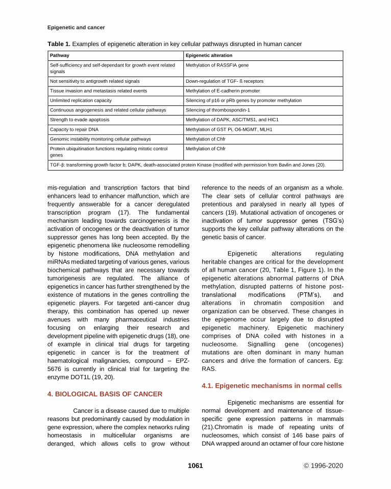
Epigenetic alterations regulating
heritable changes are critical for the development
of all human cancer (20, Table 1, Figure 1). In the
epigenetic alterations abnormal patterns of DNA
methylation, disrupted patterns of histone post-
translational modifications (PTM’s), and
alterations in chromatin composition and
organization can be observed. These changes in
the epigenome occur largely due to disrupted
epigenetic machinery. Epigenetic machinery
comprises of DNA coiled with histones in a
nucleosome. Signalling gene (oncogenes)
mutations are often dominant in many human
cancers and drive the formation of cancers. Eg:
RAS.
4.1. Epigenetic mechanisms in normal cells
Epigenetic mechanisms are essential for
normal development and maintenance of tissue-
specific gene expression patterns in mammals
(21).Chromatin is made of repeating units of
nucleosomes, which consist of 146 base pairs of
DNA wrapped around an octamer of four core histone
Table 1. Examples of epigenetic alteration in key cellular pathways disrupted in human cancer
Pathway Epigenetic alteration
Self-sufficiency and self-dependant for growth event related
signals
Methylation of RASSFIA gene
Not sensitivity to antigrowth related signals Down-regulation of TGF- ß receptors
Tissue invasion and metastasis related events Methylation of E-cadherin promoter
Unlimited replication capacity Silencing of p16 or pRb genes by promoter methylation
Continuous angiogenesis and related cellular pathways Silencing of thrombospondin-1
Strength to evade apoptosis Methylation of DAPK, ASC/TMS1, and HIC1
Capacity to repair DNA Methylation of GST Pi, O6-MGMT, MLH1
Genomic instability monitoring cellular pathways Methylation of Chfr
Protein ubiquitination functions regulating mitotic control
genes
Methylation of Chfr
TGF-β: transforming growth factor b; DAPK, death-associated protein Kinase (modified with permission from Bavlin and Jones (20).
Epigenetic and cancer
1062 © 1996-2020
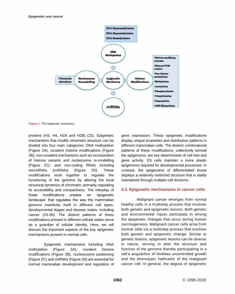
proteins (H3, H4, H2A and H2B) (22). Epigenetic
mechanisms that modify chromatin structure can be
divided into four main categories: DNA methylation
(Figure 2A), covalent histone modifications (Figure
2B), non-covalent mechanisms such as incorporation
of histone variants and nucleosome re-modelling
(Figure 2C), and non-coding RNAs including
microRNAs (miRNAs) (Figure 2D). These
modifications work together to regulate the
functioning of the genome by altering the local
structural dynamics of chromatin, primarily regulating
its accessibility and compactness. The interplay of
these modifications creates an ‘epigenetic
landscape’ that regulates the way the mammalian
genome manifests itself in different cell types,
developmental stages and disease states, including
cancer (23-28). The distinct patterns of these
modifications present in different cellular states serve
as a guardian of cellular identity. Here, we will
discuss the important aspects of the key epigenetic
mechanisms present in normal cells.
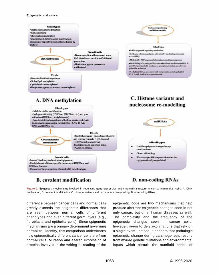
Epigenetic mechanisms including DNA
methylation (Figure 2A), covalent histone
modifications (Figure 2B), nucleososme positioning
(Figure 2C) and miRNAs (Figure 2D) are essential for
normal mammalian development and regulation of
gene expression. These epigenetic modifications
display unique properties and distribution patterns in
different mammalian cells. The distinct combinatorial
patterns of these modifications, collectively termed
the epigenome, are key determinants of cell fate and
gene activity. ES cells maintain a more plastic
epigenome required for developmental processes. In
contrast, the epigenome of differentiated tissue
displays a relatively restricted structure that is stably
maintained through multiple cell divisions.
4.2. Epigenetic mechanisms in cancer cells
Malignant cancer emerges from normal
healthy cells in a multistep process that involves
both genetic and epigenetic lesions. Both genetic
and environmental inputs participate in driving
the epigenetic changes that occur during human
carcinogenesis. Malignant cancer cells arise from
normal cells via a multistep process that involves
both genetic and epigenetic change. Similar to
genetic lesions, epigenetic lesions can be diverse
in nature, serving to alter the structure and
function of the genome thereby participating in a
cell’s acquisition of limitless uncontrolled growth
and the phenotypic hallmarks of the malignant
cancer cell. In general, the degree of epigenetic
Figure 1. The epigenetic machinery.
Epigenetic and cancer
1063 © 1996-2020
difference between cancer cells and normal cells
greatly exceeds the epigenetic differences that
are seen between normal cells of different
phenotypes and even different germ layers (e.g.,
fibroblasts and epithelial cells). Since epigenetic
mechanisms are a primary determinant governing
normal cell identity, this comparison underscores
how epigenetically different cancer cells are from
normal cells. Mutation and altered expression of
proteins involved in the writing or reading of the
epigenetic code are two mechanisms that help
produce aberrant epigenetic changes seen in not
only cancer, but other human diseases as well.
The complexity and the frequency of the
epigenetic changes seen in cancer cells,
however, seem to defy explanations that rely on
a single event. Instead, it appears that pathologic
epigenetic change during carcinogenesis results
from myriad genetic mutations and environmental
inputs which perturb the manifold nodes of
Figure 2. Epigenetic mechanisms involved in regulating gene expression and chromatin structure in normal mammalian cells. A. DNA
methylation, B. covalent modification. C. Histone variants and nucleosome re-modelling, D. non-coding RNAs.
Epigenetic and cancer
1064 © 1996-2020
epigenetic regulation (29).
Tumorigenesis is a complex and
multifactorial progressive process of
transformation of normal cells into malignant
ones. It is characterized by the accumulation of
multiple cancer-specific heritable phenotypes,
including persistent proliferative signaling,
resistance to cell death, evasion of growth
suppression, replicative immortality,
inflammatory response, deregulation of energy
metabolism, genomic instability, induction of
angiogenesis, and activation of invasion
ultimately resulting in metastases (30). The
acquisition of these cancer-specific alterations
may be triggered by the mutational and/or non-
mutational (i.e., epigenetic) events in the genome
which, in turn, affect gene expression and the
downstream phenotypes listed above (30, 31).
Furthermore, it has been suggested that
epigenetic alterations may play as important or
even more prominent role in tumor development
(32). Epigenetic events , most prominently
manifested by stable and heritable changes in
gene expression that are not due to any alteration
in the primary DNA sequence ( 33) , signify the
fundamental molecular principles in which
genetic information is organized and read ( 35) .
Epigenetic modifications include change in
methylation patterns of cytosines in DNA (35, 36),
modifications of the proteins that bind to DNA (35,
36), and the nucleosome positioning along DNA
(33). These epigenetic marks are tightly and
interdependently connected and are essential for
the normal development and the maintenance of
cellular homeostasis and functions in adult
organisms, particularly for X-chromosome
inactivation in females, genomic imprinting,
silencing of repetitive DNA elements, regulation
of chromatin structure, and proper expression of
genetic information (39). The epigenetic status is
well-balanced in normal cells, but may be altered
in many ways in cancer cells. Additionally,
growing evidence indicates that a number of
lifestyle and environmental factors may disrupt
this epigenetic balance and compromise the
stability of the epigenome in normal cells leading
to the development of a wide range of
pathologies, including cancer (40).
4.3. Epigenetics of cancer in relation to
aging
Aging is defined as the unavoidable time-
dependent alleviation in both functional and
structural integrity of organ physiology. Aging and
its associated complications such as overweight,
smoking, drinking alcohol and telomerase
shortening are considered as one of the major risk
factors for cancer development and progression
(41, 42). As a result of ultra-modern health care,
increase in hygienic knowledge, better nutritional
habit (43) and conscious lifestyle, the process of
aging is somehow observed to be controlled.
Therefore, life expectancy is now noticed to be
elevated in many developed and developing
countries, for example, 84.118 years in Japan,
83.468 years in Singapore, 82.864 years in
Sweden, 81.892 years in the UK and Hong Kong
and Macau being topped the list having >84.19
years life expectancy. On the other hand, it leads
to a shift in the proportion of people from young to
a more aged one. Aging and cancer have a very
close relationship, being the former believed to be
one of the important causes of the later (44).
Mechanisms of both aging and cancer are also
found to be common in some cases. Such
mechanisms include the role of genomic instability,
telomere attrition, epigenetic changes, and loss of
proteostasis, decreased nutrient sensing and
altered metabolism. So, it is suggested to target
both with same or similar strategies even with the
same or similar drugs, for example to supress
micro RNA that are common in both. However,
unraveling clear molecular events sharing both the
cellular disorders are anticipated to target them
with the same or similar strategies or drugs (45).
Owing to the observed tight association
between aging and cancer, it is noticed that both
share epigenetic control over their entire process
of development and progression. Various
epigenetic mechanisms are influenced by several
external factors such as environment, pollution,
lifestyle and quality and quantity of diet. They are
also believed to play a pivotal role in gene
expression (46). Dietary supplements such as
antioxidants (lycopene, curcumin and vitamin E
and A etc) can influence various cellular events
Epigenetic and cancer
1065 © 1996-2020
associated with aging and cancer as well.
Especially, sulforaphane present in cruciferous
vegetables and epigallocatechin-3-gallate found in
green tea is examined to influence several
epigenetic events such as inhibition of the enzyme
DNA methyltransferase, histone modifications
through enzymes such as histone deacetylase,
histone acetyltransferase inhibition and non-
coding RNA expression. The above epigenetic
pathways found to control both the formation and
progression of various neoplasms. Due to the key
role in epigenetic modulation, such diets are
referred to as epigenetic diets. On the other hand,
they can control both the processes of cellular
longevity and carcinogenesis through specific key
genes that encode telomerase. Therefore, caloric
restriction can modulate both aging and cancer-
associated events, notably, high caloric diet can
up-regulate both the events. So, epigenetic diets
that are rich in genistein, sulforaphane, and
epigallocatechin-3-gallate are believed to have
many health benefits in terms of influencing
epigenome positively (42).
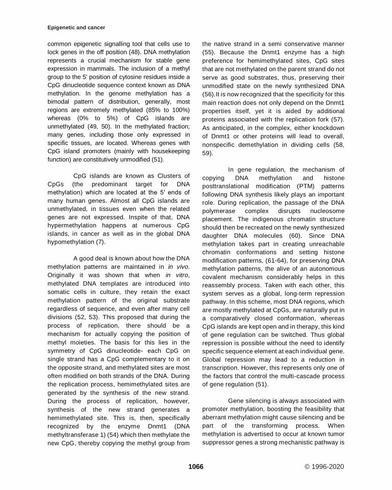
5. DNA METHYLATION
DNA methylation is established by
DNMTs, which catalyze the adding of methyl group
in C5 (carbon 5) position of cytosine to produce
C5-methyl-cytosine (5mC). So far, two types of
DNMTs have been defined: de novo
methyltransferases and maintenance
methyltransferases (Figure 3A and 3B). De novo
methyltransferases create hemimethylated CpG
dinucleotide sites in double-strand DNA, and are
responsible for setting up the pattern of
methylation. Maintenance methyltransferases add
methylation to DNA when one strand is already
methylated, and are responsible for maintaining
the methylation pattern that had been established
by the de novo methyltransferases. According to
the catalytic features for methylation, DNMTs are
classified into three families: DNMT1, DNMT2, and
DNMT3. Generally speaking, DNA 5mC in the
genome of mammalian somatic cells is found
almost entirely within CpG dinucleotide. It has
been proposed that within housekeeping
promoters, CpG methylation should be rare at CpG
islands, while this modification could be highly
prevalent in repetitive sequences of promoters and
enhancers, the genes of which are regulated so
that they may be stabilized or locked in a silent
state (47).
There are many ways that gene expression
is controlled in eukaryotes, but DNA methylation is a
Figure 3. Mechanism of DNA methylation. A. Mechanism of DNA methylation (A) DNMTs add methyl group in C5 (carbon 5) position of
cytosines to produce 5mC, while TETs catalyze 5mC to 5hmC, then some other factors turn 5hmC back to cytosine. B. Genomic DNA
methylation is established by DNMT3 as hemi-methylated templates, and maintained by DNMT1 to full-methylated DNA.
Epigenetic and cancer
1066 © 1996-2020
common epigenetic signalling tool that cells use to
lock genes in the off position (48). DNA methylation
represents a crucial mechanism for stable gene
expression in mammals. The inclusion of a methyl
group to the 5' position of cytosine residues inside a
CpG dinucleotide sequence context known as DNA
methylation. In the genome methylation has a
bimodal pattern of distribution, generally, most
regions are extremely methylated (85% to 100%)
whereas (0% to 5%) of CpG islands are
unmethylated (49, 50). In the methylated fraction;
many genes, including those only expressed in
specific tissues, are located. Whereas genes with
CpG island promoters (mainly with housekeeping
function) are constitutively unmodified (51).
CpG islands are known as Clusters of
CpGs (the predominant target for DNA
methylation) which are located at the 5′ ends of
many human genes. Almost all CpG islands are
unmethylated, in tissues even when the related
genes are not expressed. Inspite of that, DNA
hypermethylation happens at numerous CpG
islands, in cancer as well as in the global DNA
hypomethylation (7).
A good deal is known about how the DNA
methylation patterns are maintained in in vivo.
Originally it was shown that when in vitro,
methylated DNA templates are introduced into
somatic cells in culture, they retain the exact
methylation pattern of the original substrate
regardless of sequence, and even after many cell
divisions (52, 53). This proposed that during the
process of replication, there should be a
mechanism for actually copying the position of
methyl moieties. The basis for this lies in the
symmetry of CpG dinucleotide- each CpG on
single strand has a CpG complementary to it on
the opposite strand, and methylated sites are most
often modified on both strands of the DNA. During
the replication process, hemimethylated sites are
generated by the synthesis of the new strand.
During the process of replication, however,
synthesis of the new strand generates a
hemimethylated site. This is, then, specifically
recognized by the enzyme Dnmt1 (DNA
methyltransferase 1) (54) which then methylate the
new CpG, thereby copying the methyl group from
the native strand in a semi conservative manner
(55). Because the Dnmt1 enzyme has a high
preference for hemimethylated sites, CpG sites
that are not methylated on the parent strand do not
serve as good substrates, thus, preserving their
unmodified state on the newly synthesized DNA
(56).It is now recognized that the specificity for this
main reaction does not only depend on the Dnmt1
properties itself, yet it is aided by additional
proteins associated with the replication fork (57).
As anticipated, in the complex, either knockdown
of Dnmt1 or other proteins will lead to overall,
nonspecific demethylation in dividing cells (58,
59).
In gene regulation, the mechanism of
copying DNA methylation and histone
posttranslational modification (PTM) patterns
following DNA synthesis likely plays an important
role. During replication, the passage of the DNA
polymerase complex disrupts nucleosome
placement. The indigenous chromatin structure
should then be recreated on the newly synthesized
daughter DNA molecules (60). Since DNA
methylation takes part in creating unreachable
chromatin conformations and setting histone
modification patterns, (61-64), for preserving DNA
methylation patterns, the alive of an autonomous
covalent mechanism considerably helps in this
reassembly process. Taken with each other, this
system serves as a global, long-term repression
pathway. In this scheme, most DNA regions, which
are mostly methylated at CpGs, are naturally put in
a comparatively closed conformation, whereas
CpG islands are kept open and in therapy, this kind
of gene regulation can be switched. Thus global
repression is possible without the need to identify
specific sequence element at each individual gene.
Global repression may lead to a reduction in
transcription. However, this represents only one of
the factors that control the multi-cascade process
of gene regulation (51).
Gene silencing is always associated with
promoter methylation, boosting the feasibility that
aberrant methylation might cause silencing and be
part of the transforming process. When
methylation is advertised to occur at known tumor
suppressor genes a strong mechanistic pathway is
Epigenetic and cancer
1067 © 1996-2020
suggested as a potential role in tumorigenesis
(66).
DNA hypermethylation of RB gene
(retinoblastoma) controls cell cycle which is one of
the first epigenetic lesions to be involved in
carcinogenesis and is combined with the loss of
RB expression (67, 71).
In carcinogenesis, the case of RB
methylation remains one of the able arguments in
favor of a causal role for aberrant methylation; RB
gene is commonly active in the precursor cells of
tumors and promoter methylation seems to have
the same consequence as the genetic mutation of
the gene (68).Another tumor type in which this
happens is microsatellite unstable colon cancer,
by germ line mutation of the DNA mismatch repair
(MMR) protein MLHI the inherited forms of the
disease are commonly caused (65).
Almost 15% of cases of sporadic colon
cancer lack MMR gene mutation although still
display microsatellite instability, in these cases,
MLH1 promoters have methylated and lack
expression of the gene (67, 70).
This by the treatment with the
demethylating agent 5-aza-2' –deoxycytidine, the
MLH1 repression is reported to be reversed in cell
lines showing this abnormality (69). The
p16INK4a/CDKN2A promoter aberrant methylation
has been shown to be present in both human
squamous cell carcinomas and in the early stages
of neoplastic transformation (71, 72). Similarly,
methylation of GSTP1 (π-class glutathione s-
transferase) in prostate carcinogenesis, is an early
event and it is also found in premalignant lesions
(73).
Likewise during the development of
specific tumors in colorectal carcinogenesis,
hypermethylation of chromosome 17p region,
corresponding to the location of the tumor
suppressor of p53 has been demonstrated to
antecede its allelic loss, suggesting that
methylation may not aimlessly mark chromosome
regions that are altered (74).
It has been presumed that in malignant
transformation, aberrant methylation plays an
important role, based on these examples,
particularly when methylation has been
demonstrated to appear early in the tumorigenic
process. Cells with a particular advantage over
others, either by causing their increased
proliferation or refiance to apoptosis may be
provided by the methylation induced silencing of
tumor suppressor genes. Because of premalignant
cells, clonal expansion could result in the
hyperproliferative phenotype which is
characteristic of the early stages of tumorigenesis
(75).
Genes such as RB, MLH1, and VHL are
methylated, in tumor and also mutated commonly
and suggesting that hypermethylation of CpG
island during tumorigenesis (76). DNA
hypermethylation has been used to subdivide
tumor types and to distinguish them from non-
malignant tissue (77). A CpG island methylator
phenotype (CIMP) has been nominated as tumor
subgroups with high levels of DNA methylation,
and is mostly associated with worse prognosis
(78).
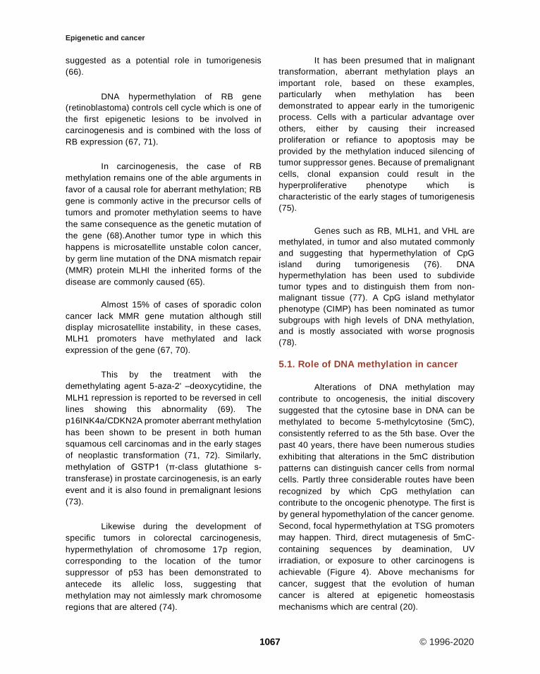
5.1. Role of DNA methylation in cancer
Alterations of DNA methylation may
contribute to oncogenesis, the initial discovery
suggested that the cytosine base in DNA can be
methylated to become 5-methylcytosine (5mC),
consistently referred to as the 5th base. Over the
past 40 years, there have been numerous studies
exhibiting that alterations in the 5mC distribution
patterns can distinguish cancer cells from normal
cells. Partly three considerable routes have been
recognized by which CpG methylation can
contribute to the oncogenic phenotype. The first is
by general hypomethylation of the cancer genome.
Second, focal hypermethylation at TSG promoters
may happen. Third, direct mutagenesis of 5mC-
containing sequences by deamination, UV
irradiation, or exposure to other carcinogens is
achievable (Figure 4). Above mechanisms for
cancer, suggest that the evolution of human
cancer is altered at epigenetic homeostasis
mechanisms which are central (20).
Epigenetic and cancer
1068 © 1996-2020
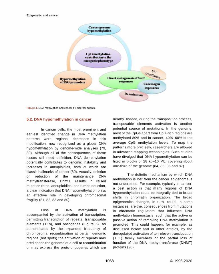
5.2. DNA hypomethylation in cancer
In cancer cells, the most prominent and
earliest identified change in DNA methylation
patterns were regional decreases in this
modification, now recognized as a global DNA
hypomethylation by genome-wide analyses (79,
80). Although all of the consequences of these
losses still need definition, DNA demethylation
potentially contributes to genomic instability and
increases in aneuploidies, both of which are
classic hallmarks of cancer (80). Actually, deletion
or reduction of the maintenance DNA
methyltransferase, Dnmt1, results in raised
mutation rates, aneuploidies, and tumor induction,
a clear indication that DNA hypomethylation plays
an effective role in developing chromosomal
fragility (81, 82, 83 and 80).
Loss of DNA methylation is
accompanied by the activation of transcription,
permitting transcription of repeats, transposable
elements (TEs), and oncogenes (Figure 5). As
authenticated by the expanded frequency of
chromosomal recombination at certain genomic
regions (hot spots) the activation of repeats may
predispose the genome of a cell to recombination
or may express the proto-oncogenes which are
nearby. Indeed, during the transposition process,
transposable elements activation is another
potential source of mutations. In the genome,
most of the CpGs apart from CpG-rich regions are
methylated 80% and in cancer, 40%–60% is the
average CpG methylation levels. To map the
patterns more precisely, researchers are allowed
in advanced mapping technologies. Such studies
have divulged that DNA hypomethylation can be
fixed in blocks of 28 kb–10 Mb, covering about
one-third of the genome (84, 85, 86 and 87).
The definite mechanism by which DNA
methylation is lost from the cancer epigenome is
not understood. For example, typically in cancer,
a best action is that many regions of DNA
hypomethylation could be integrally tied to broad
shifts in chromatin organization. The broad
epigenomics changes, in turn, could, in some
instances, are the, consequences from mutations
in chromatin regulators that influence DNA
methylation homeostasis, such that the active or
passive action of removing DNA methylation is
promoted. This could happen, for example, as
discussed below and in other articles, by the
deregulated activation of ten-eleven translocation
(TET) family members or the partial loss of
function of the DNA methyltransferase (DNMT)
proteins (20).
Figure 4. DNA methylation and cancer by external agents.
Epigenetic and cancer
1069 © 1996-2020
5.3. Epigenetic alterations involving DNA
methylation by mutation
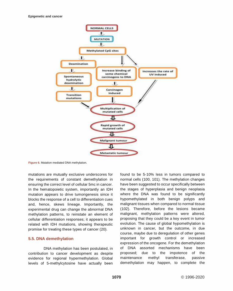
In cancer, DNA methylation change can
be integrally combined with the transcriptional
silencing, providing a different mechanism for the
inactivation of genes with tumor suppressor
function by mutation (20, Figure 6).
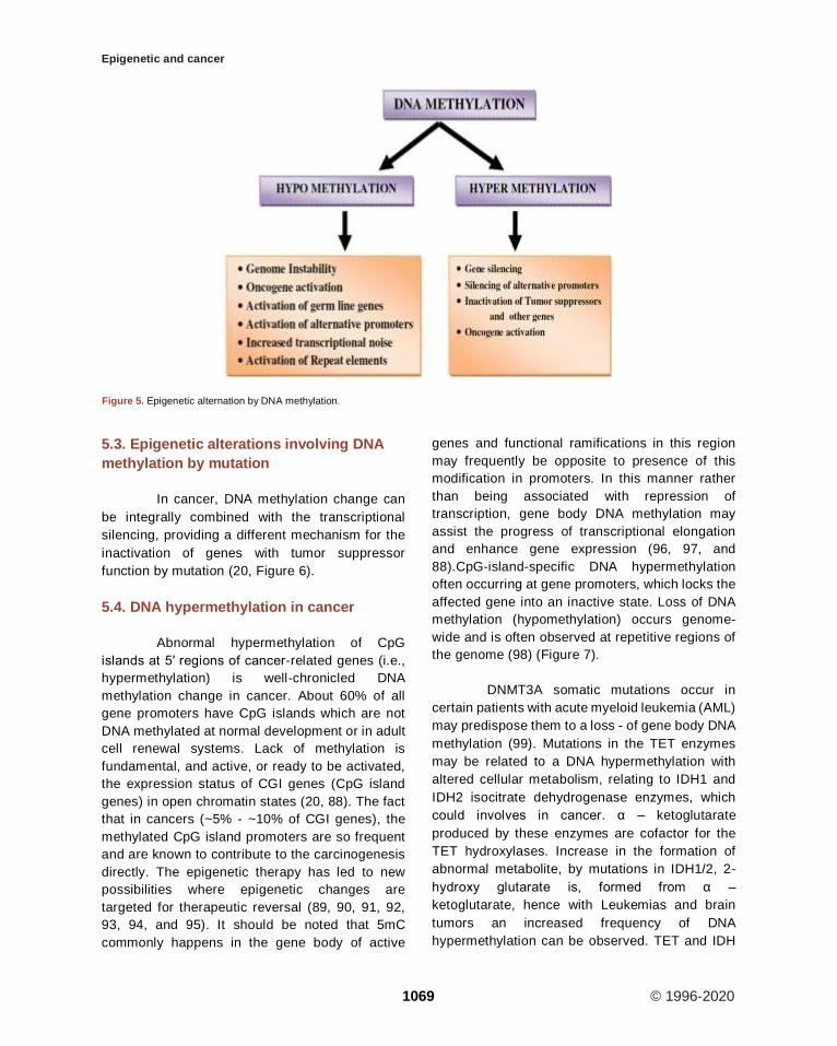
5.4. DNA hypermethylation in cancer
Abnormal hypermethylation of CpG
islands at 5′ regions of cancer-related genes (i.e.,
hypermethylation) is well-chronicled DNA
methylation change in cancer. About 60% of all
gene promoters have CpG islands which are not
DNA methylated at normal development or in adult
cell renewal systems. Lack of methylation is
fundamental, and active, or ready to be activated,
the expression status of CGI genes (CpG island
genes) in open chromatin states (20, 88). The fact
that in cancers (~5% - ~10% of CGI genes), the
methylated CpG island promoters are so frequent
and are known to contribute to the carcinogenesis
directly. The epigenetic therapy has led to new
possibilities where epigenetic changes are
targeted for therapeutic reversal (89, 90, 91, 92,
93, 94, and 95). It should be noted that 5mC
commonly happens in the gene body of active
genes and functional ramifications in this region
may frequently be opposite to presence of this
modification in promoters. In this manner rather
than being associated with repression of
transcription, gene body DNA methylation may
assist the progress of transcriptional elongation
and enhance gene expression (96, 97, and
88).CpG-island-specific DNA hypermethylation
often occurring at gene promoters, which locks the
affected gene into an inactive state. Loss of DNA
methylation (hypomethylation) occurs genome-
wide and is often observed at repetitive regions of
the genome (98) (Figure 7).
DNMT3A somatic mutations occur in
certain patients with acute myeloid leukemia (AML)
may predispose them to a loss - of gene body DNA
methylation (99). Mutations in the TET enzymes
may be related to a DNA hypermethylation with
altered cellular metabolism, relating to IDH1 and
IDH2 isocitrate dehydrogenase enzymes, which
could involves in cancer. α – ketoglutarate
produced by these enzymes are cofactor for the
TET hydroxylases. Increase in the formation of
abnormal metabolite, by mutations in IDH1/2, 2-
hydroxy glutarate is, formed from α –
ketoglutarate, hence with Leukemias and brain
tumors an increased frequency of DNA
hypermethylation can be observed. TET and IDH
Figure 5. Epigenetic alternation by DNA methylation.
Epigenetic and cancer
1070 © 1996-2020
mutations are mutually exclusive underscores for
the requirements of constant demethylation in
ensuring the correct level of cellular 5mc in cancer.
In the hematopoietic system, importantly an IDH
mutation appears to drive tumorigenesis since it
blocks the response of a cell to differentiation cues
and, hence, skews lineage. Importantly, the
experimental drug can change the abnormal DNA
methylation patterns, to reinstate an element of
cellular differentiation responses; it appears to be
related with IDH mutations, showing therapeutic
promise for treating these types of cancer (20).
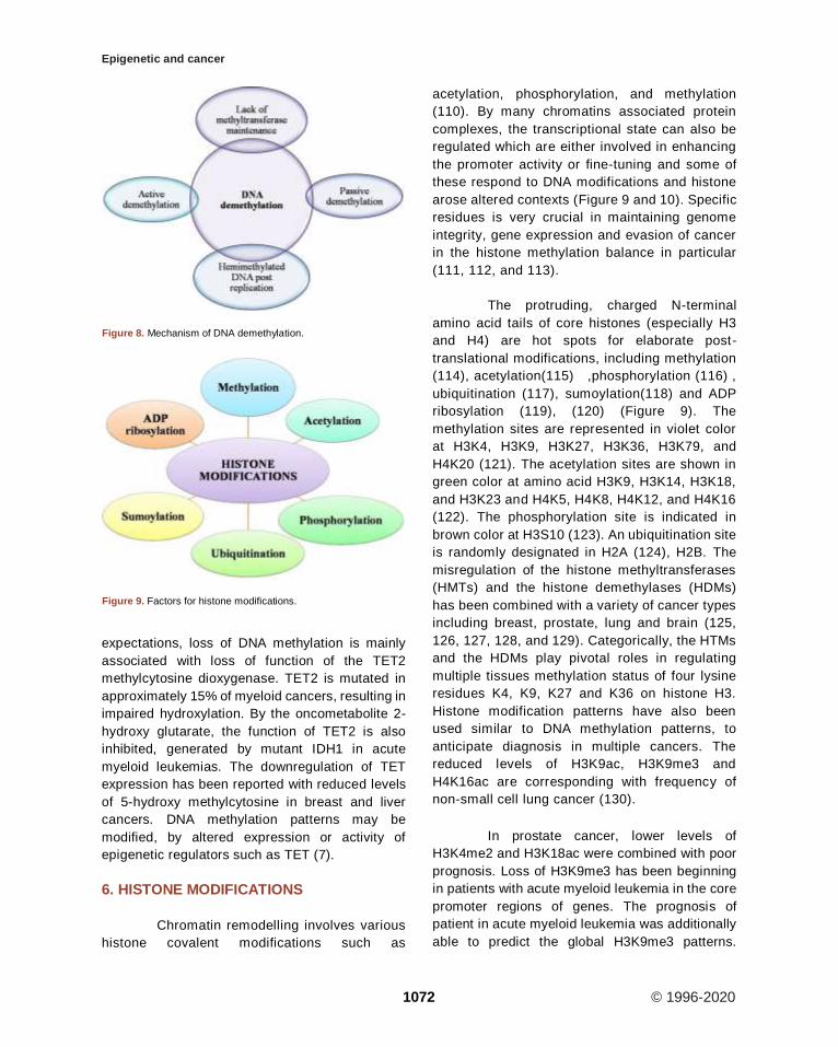
5.5. DNA demethylation
DNA methylation has been postulated, in
contribution to cancer development as despite
evidence for regional hypermethylation. Global
levels of 5-methylcytosine have actually been
found to be 5-10% less in tumors compared to
normal cells (100, 101). The methylation changes
have been suggested to occur specifically between
the stages of hyperplasia and benign neoplasia
where the DNA was found to be significantly
hypomethylated in both benign polyps and
malignant tissues when compared to normal tissue
(102). Therefore, before the lesions became
malignant, methylation patterns were altered,
proposing that they could be a key event in tumor
evolution. The cause of global hypomethylation is
unknown in cancer, but the outcome, in due
course, maybe due to deregulation of other genes
important for growth control or increased
expression of the oncogene. For the demethylation
of DNA assorted mechanisms have been
proposed; due to the impotence of the
maintenance methyl transferase, passive
demethylation may happen, to complete the
Figure 6. Mutation mediated DNA methylation.

Epigenetic and cancer
1071 © 1996-2020
methylation step that would normally be guided by
hemimethylated DNA post replication (Figure 8).
This is thought to be during preimplantation, in the
case of maternal pronucleus which undergoes
passive demethylation, most likely expected to
sequestration of the oocyte-specific form of
DNMT1 (DNMT10) in the cytoplasm (103). Rapid
demethylation of the paternal pronucleus appears,
by TET3 due to the oxidation of 5-ethylcytosine to
5-hydroxy methylcytosine (104).
There is evidence that the maintenance
of methyltransferase DNMT1 does not restore
methylation to cytosine’s, in the newly synthesized
daughter strand; if the diagonally opposite cytosine
is hydroxyl methylated (105) resulting in
replication-dependent passive dilution of 5-
methylcytosine. In cultured human cells and the
adult mouse brain, active DNA demethylation has
been demonstrated to involve TET1 catalysed
hydroxymethylation persued by AID/APO-BEC-
mediated deamination of 5-hydroxymethyl
cytosine, with the resulting base mismatch being
removed by the base excision repair pathway (106,
107). TET proteins further oxidize 5-hydroxy
methylcytosine to 5- formyl cytosine and by
thymine DNA glycosylase (TDG) the 5- carboxyl
cytosine can be excised and by the base excision
repair pathway it has been repaired. In a study, the
methylation status of a number of genes, DNA
methylation and demethylation cyclic process have
shown to occur approximately every hour, which
has been examined when the cells were released
from a synchronizing block, (108, and 109).
Different possibilities including a
dynamic, replication – independent response to
alterations in physiological conditions such as
hypoxia; are accepted, this was a surprise
discovery. The components of the base excision
repair pathway and in TDG, a mechanism has
been proposed, that these were enlisted to the
promoter at the origination of each transcriptionally
productive cycle and a reduction in TDG
expression impaired demethylation and reduced
transcriptional activity. In conflicting to
Figure 7. Schematic outline of the most relevant DNA methylation changes observed in human cancers.
Epigenetic and cancer
1072 © 1996-2020
expectations, loss of DNA methylation is mainly
associated with loss of function of the TET2
methylcytosine dioxygenase. TET2 is mutated in
approximately 15% of myeloid cancers, resulting in
impaired hydroxylation. By the oncometabolite 2-
hydroxy glutarate, the function of TET2 is also
inhibited, generated by mutant IDH1 in acute
myeloid leukemias. The downregulation of TET
expression has been reported with reduced levels
of 5-hydroxy methylcytosine in breast and liver
cancers. DNA methylation patterns may be
modified, by altered expression or activity of
epigenetic regulators such as TET (7).
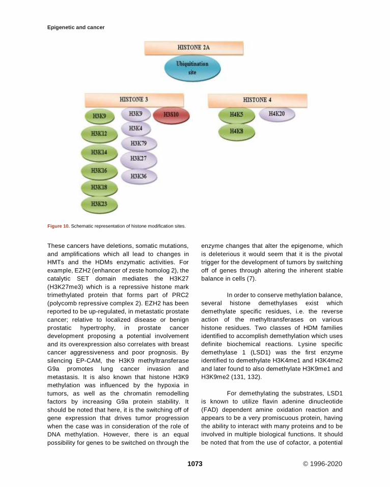
6. HISTONE MODIFICATIONS
Chromatin remodelling involves various
histone covalent modifications such as
acetylation, phosphorylation, and methylation
(110). By many chromatins associated protein
complexes, the transcriptional state can also be
regulated which are either involved in enhancing
the promoter activity or fine-tuning and some of
these respond to DNA modifications and histone
arose altered contexts (Figure 9 and 10). Specific
residues is very crucial in maintaining genome
integrity, gene expression and evasion of cancer
in the histone methylation balance in particular
(111, 112, and 113).
The protruding, charged N-terminal
amino acid tails of core histones (especially H3
and H4) are hot spots for elaborate post-
translational modifications, including methylation
(114), acetylation(115) ,phosphorylation (116) ,
ubiquitination (117), sumoylation(118) and ADP
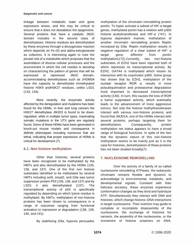
ribosylation (119), (120) (Figure 9). The
methylation sites are represented in violet color
at H3K4, H3K9, H3K27, H3K36, H3K79, and
H4K20 (121). The acetylation sites are shown in
green color at amino acid H3K9, H3K14, H3K18,
and H3K23 and H4K5, H4K8, H4K12, and H4K16
(122). The phosphorylation site is indicated in
brown color at H3S10 (123). An ubiquitination site
is randomly designated in H2A (124), H2B. The
misregulation of the histone methyltransferases
(HMTs) and the histone demethylases (HDMs)
has been combined with a variety of cancer types
including breast, prostate, lung and brain (125,
126, 127, 128, and 129). Categorically, the HTMs
and the HDMs play pivotal roles in regulating
multiple tissues methylation status of four lysine
residues K4, K9, K27 and K36 on histone H3.
Histone modification patterns have also been
used similar to DNA methylation patterns, to
anticipate diagnosis in multiple cancers. The
reduced levels of H3K9ac, H3K9me3 and
H4K16ac are corresponding with frequency of
non-small cell lung cancer (130).
In prostate cancer, lower levels of
H3K4me2 and H3K18ac were combined with poor
prognosis. Loss of H3K9me3 has been beginning
in patients with acute myeloid leukemia in the core
promoter regions of genes. The prognosis of
patient in acute myeloid leukemia was additionally
able to predict the global H3K9me3 patterns.
Figure 8. Mechanism of DNA demethylation.
Figure 9. Factors for histone modifications.
Epigenetic and cancer
1073 © 1996-2020
These cancers have deletions, somatic mutations,
and amplifications which all lead to changes in
HMTs and the HDMs enzymatic activities. For
example, EZH2 (enhancer of zeste homolog 2), the
catalytic SET domain mediates the H3K27
(H3K27me3) which is a repressive histone mark
trimethylated protein that forms part of PRC2
(polycomb repressive complex 2). EZH2 has been
reported to be up-regulated, in metastatic prostate
cancer; relative to localized disease or benign
prostatic hypertrophy, in prostate cancer
development proposing a potential involvement
and its overexpression also correlates with breast
cancer aggressiveness and poor prognosis. By
silencing EP-CAM, the H3K9 methyltransferase
G9a promotes lung cancer invasion and
metastasis. It is also known that histone H3K9
methylation was influenced by the hypoxia in
tumors, as well as the chromatin remodelling
factors by increasing G9a protein stability. It
should be noted that here, it is the switching off of
gene expression that drives tumor progression
when the case was in consideration of the role of
DNA methylation. However, there is an equal
possibility for genes to be switched on through the
enzyme changes that alter the epigenome, which
is deleterious it would seem that it is the pivotal
trigger for the development of tumors by switching
off of genes through altering the inherent stable
balance in cells (7).
In order to conserve methylation balance,
several histone demethylases exist which
demethylate specific residues, i.e. the reverse
action of the methyltransferases on various
histone residues. Two classes of HDM families
identified to accomplish demethylation which uses
definite biochemical reactions. Lysine specific
demethylase 1 (LSD1) was the first enzyme
identified to demethylate H3K4me1 and H3K4me2
and later found to also demethylate H3K9me1 and
H3K9me2 (131, 132).
For demethylating the substrates, LSD1
is known to utilize flavin adenine dinucleotide
(FAD) dependent amine oxidation reaction and
appears to be a very promiscuous protein, having
the ability to interact with many proteins and to be
involved in multiple biological functions. It should
be noted that from the use of cofactor, a potential
Figure 10. Schematic representation of histone modification sites.
Epigenetic and cancer
1074 © 1996-2020
linkage between metabolic state and gene
expression arises, and this may be critical to
ensure that it does not destabilize the epigenome.
Several proteins that have a catalytic JMJC
domain includes in the second class of
demethylases. Histone residues are demethylated
by these enzymes through a dioxygenase reaction
which depends on Fe (II) and alpha-ketoglutarate
as cofactors. It is interesting again to note the
pivotal role of a metabolite which proposes that the
assimilation of diverse cellular processes and the
environment in which the cell resides is deciding
on characterizing the pattern of genes that will be
expressed or repressed. JMJC domain-
accommodating demethylases such as JHDM3A
have the capacity to demethylate trimethylated
histone H3K9 andH3K27 residues, unlike LSD1
(133, 134).
More recently, the enzymatic activity
affected by the deregulation and mutations has been
found for the HDMs. In liver and lung cancers the
H3K27 demethylase JMJD3 is found to be down-
regulated, while in multiple tumor types, inactivating
somatic mutations in the UTX gene are regularly
found. Some of these HDMs have been generated in
knock-out mouse models and consequence in
definite phenotypes including numerous that are
lethal, indicating that proper expression of HDMs is
critical for development (7).
6.1. Non histone methylation
Other than histones, several proteins
have been recognized to be methylated by the
HMTs and also demethylated by the HDMs (135,
136, and 137). One of the first non histone
substrates identified to be methylated by several
HMTs including set9, smyd2, and G9a was tumor
suppressor protein P53 (135, 136, and 137) and by
LSD1 it also demethylated (137). The
transcriptional activity of p53 is specifically
regulated by depending on which lysine residue is
methylated. By HMTs, methylation of non-histone
proteins has been shown to consequence in a
range of outcomes ranging from functional
activation to repression or degradation (138, 139,
140, and 141).
By stabilising G9a, hypoxia persuades
methylation of the chromatin remodelling protein
pontin. To hyper activate a subset of HIF-α target
genes, methylated pontin has a relation with p300
histone acetyltransferase and HIF-α (141). In
hypoxia dependent manner, methylation of
another chromatin remodelling protein Reptin
increased by G9a. Reptin methylation results in
negative regulation of a clear subset of HIF- α
target gene, different from pontin
methylation(172).Currently, two non-histone
substrates of EZH2 have been reported both of
which represses its transcriptional activity. By
EZH2, GATA4 is methylated which lessen its
interaction with its coactivator p300. Some group
has shown that by EZH2, methylation of the
nuclear receptor ROR α, results in more
polyubiquitination and proteasomal degradation
most important to decreased transcriptional
activity (140). In turn, this causes the loss of ROR
α tumor suppressor activity, which eventually
leads to the advancement of more aggressive
tumors. Not only the histone methyltransferases
interact with various non-histone proteins; and
found that JMJD1A, one of the HDMs interact with
several proteins, perhaps targeting them for
demethylation. Consequently, protein
methylation net status appears to have a broad
range of biological functions. In spite of the fact
that the dynamic nature of this non-histone
methylation seems to be mainly just as it is the
case for histones, demethylation of these proteins
has not been studied broadly(7).
7. NUCLEOSOME REMODELLING
Over the activity of a family of so-called
nucleosome remodeling ATPases, the eukaryotic
chromatin remains flexible and dynamic to
acknowledge to environmental, metabolic, and
developmental signals. Constant with their
helicase ancestry, these enzymes experience
conformation changes as they bind and hydrolyze
ATP. Simultaneously they interact with DNA and
histones, which change histone–DNA interactions
in target nucleosome. Their exertion may guide to
complete or incomplete disassembly of the
nucleosome, the exchange of histones for
variants, the assembly of the nucleosome, or the
movement of histone octamers on DNA.
Epigenetic and cancer
1075 © 1996-2020
Remodelling may give DNA sequences
approachable to collaborating proteins or,
conversely, encourage packing into tightly folded
structures. In every aspect of genome function,
remodelling processes engage. Remodelling
activities are frequently integrated with other
mechanisms such as histone modifications or
RNA metabolism to assemble stable, epigenetic
states (142).
7.1. Changes in chromatin
The eukaryotic genome is packaged into
the nucleus in the form of chromatin. Beyond a
mechanism for packaging, chromatin has evolved
as a means for dynamically regulating the genome.
At its most basic description, chromatin consists of
histone proteins in complex with DNA. Modification
of the histone proteins and DNA plays a major role
in regulating chromatin structure, and together
they form an extensive signaling network. The
modification state of chromatin has been found to
be responsive to the environment and the
metabolic state of the cell, and there is now
evidence that some histone and DNA
modifications are heritable. Moreover,
dysregulation of chromatin signaling pathways
underlies a wide range of diseases and disorders,
providing a link between the environment and
nutrition, gene regulation, and human health and
susceptibility to diseases (143).
Considering the importance of chromatin
in regulating eukaryotic gene expression and
maintaining genome stability, it is perhaps not
wholly unexpected that recent genome-wide
sequencing studies have uncovered cancer-
associated mutations in genes encoding chromatin
regulatory factors and enzymes (144, Figure 11).
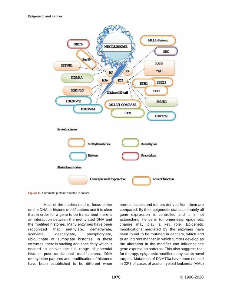
A summary of cancer mutations that
affect post translational modifications of the
histone H3 N-terminal tail. Proteins classes are
indicated by the outline color; orange –
methylation, blue- histone, green-demethylase,
brown-deacetylase. Whereas mutational status is
indicated by fill color- over expressed/hyperactive,
outline color indicates – loss of function. Dashed
lines indicate the residue of histone H3 that is
expected to be modified due to the indicated
cancer mutations.
In the regulation of gene expression,
chromatin epigenetic modification plays a main role.
Mostly in the sequence CpG and in vitro methylated
promoters, DNA is methylated post-synthetically on
cytosine residues are known to be generally inactive
when transfected into eukaryotic cells (145). By a
family of DNA methyltransferase (DNMTs), DNA
methylation is catalyzed. Reciprocal methylation of
the new DNA strand complementary to
hemimethylated DNA was maintained by the
DNMT1, and that is produced as a result of semi-
conservative DNA replication. DNA
methyltransferase is known to be DNMT3a and
DNMT3b which is being able to methylate the
completely unmethylated DNA duplex in vivo (146,
147). More recently it has been shown that, by a
family of Fe2+, 2-oxoglutarate dependent
methylcytosine dioxygenases known as TET
proteins, 5-methylcytosine can be oxidized to 5-
hydroxymethylcytosine (148), by a mechanism that
appears to include base excision repair processes,
which effectively results in the subsequent removal of
the repressive methyl group. Other DNA
modifications are also described such as methylation
at sites other than CpG (149, 150), and the
generation of formyl and carboxyl derivatives of DNA
(151).
Beginning discussions that obtained from
those that studied transgenerational phenomena
concentrated on the classical set of DNMTs.
Nevertheless, modifications of epigenetic go
beyond DNA methylation. The chromatin histone
proteins are also altered in their transcriptional
states and N-terminal residues are often related to
particular histone modifications (152, 153). The
number and complication of the possible
amalgamation of these have grown very quickly in
recent years (111), but a simplified generalization
could be that active genes are associated with
acetylation of H3 and H4 histones and methylation
of the lysine-4 residue of histone H3 (H3K4).
Inactive genes are regularly hypo acetylated and
may also be methylated on the lysine-9 (H3K9) or
lysine-27 (H3K27) residues of histone H3 (154).
Epigenetic and cancer
1076 © 1996-2020
Most of the studies tend to focus either
on the DNA or histone modifications and it is clear
that in order for a gene to be transcribed there is
an interaction between the methylated DNA and
the modified histones. Many enzymes have been
recognized that methylate, demethylate,
acetylate, deacetylate, phosphorylate,
ubiquitinate or sumoylate histones. In these
enzymes, there is sacking and specificity which is
needed to deliver the full range of potential
histone post-translational modifications. DNA
methylation patterns and modification of histones
have been established to be different when
normal tissues and tumors derived from them are
compared. By their epigenetic status ultimately all
gene expression is controlled and it is not
astonishing, hence in tumorigenesis, epigenetic
change may play a key role. Epigenetic
modifications mediated by the enzymes have
been found to be mutated in cancers, which add
to an indirect manner in which tumors develop as
the alteration in the modifier can influence the
gene expression patterns. This also suggests that
for therapy, epigenetic modifiers may act as novel
targets. Mutations of DNMT3a have been noticed
in 22% of cases of acute myeloid leukemia (AML)
Figure 11. Chromatin proteins mutated in cancer.
Epigenetic and cancer
1077 © 1996-2020
where they are related to a poor outcome (155).
Similarly, in ~15% of myeloid cancers,
the methylcytosine dioxygenase TET2 is mutated.
In mutant mice, Tet2-deficiency causes
myeloproliferation, suggesting a role in stem cell
function (156). In multiple human cancers, the
H3K27 demethylase UTX is mutated and the
highest frequency (~10%) being in multiple
myeloma (157). The finding of gene mutations that
alter chromatin proposes that the disruption of
epigenetic control has a very notable role in the
promotion of cancers. Secondary roles are the
specific proteins which bind correctly to modified
histones. Alteration in their structure can also drive
the development of tumours (7).
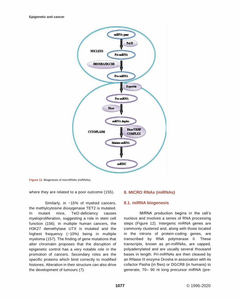
8. MICRO RNAs (miRNAs)
8.1. miRNA biogenesis
MiRNA production begins in the cell’s
nucleus and involves a series of RNA processing
steps (Figure 12). Intergenic miRNA genes are
commonly clustered and, along with those located
in the introns of protein-coding genes, are
transcribed by RNA polymerase II. These
transcripts, known as pri-miRNAs, are capped,
polyadenylated and are usually several thousand
bases in length. Pri-miRNAs are then cleaved by
an RNase III enzyme Drosha in association with its
cofactor Pasha (in flies) or DGCR8 (in humans) to
generate, 70– 90 nt long precursor miRNA (pre-
Figure 12. Biogenesis of microRNAs (miRNAs).
Epigenetic and cancer
1078 © 1996-2020
miRNA) which folds into an imperfect stem–loop
hairpin structure. These pre-miRNAs are
transported to the cytoplasm by exportin 5, where
they are further processed by Dicer to form a
transient 22 nt mature double stranded (ds) miRNA
(miRNA duplex). One strand of this duplex is
preferentially incorporated into a miRNA-
associated RNA induced silencing complex
(miRISC). The mature miRNA guides RISC to
target mRNAs containing a sequence partially
complementary (miRNA target site) to the miRNA
(158) (Figure 12).miRNA genes are generally
transcribed by RNA polymerase II (Pol II) within the
nucleus to form large capped and polyadenylated
pri-miRNA transcripts. These pri-miRNA
transcripts are processed by the RNase III enzyme
Drosha and its cofactor, DGCR8, to a pre-miRNA
precursor product. The pre-miRNA is then
transported to the cytoplasm by exportin 5.
Subsequently, another RNase III enzyme, Dicer,
processes the pre-miRNA to generate a transient,
22 nucleotide miRNA: miRNA* duplex. This duplex
is then loaded into the miRNA-associated RNA-
induced silencing complex (miRISC), which
includes the Argonaute proteins, and the mature
single-stranded miRNA is preferentially retained in
this complex.
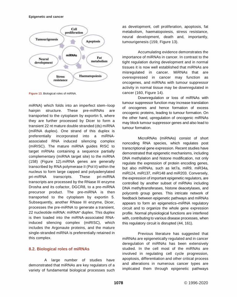
8.2. Biological roles of miRNAs
A large number of studies have
demonstrated that miRNAs are key regulators of a
variety of fundamental biological processes such
as development, cell proliferation, apoptosis, fat
metabolism, haematopoiesis, stress resistance,
neural development, death and, importantly,
tumourigenesis (159, Figure 13).
Accumulating evidence demonstrates the
importance of miRNAs in cancer. In contrast to the
tight regulation during development and in normal
tissues it is now well established that miRNAs are
misregulated in cancer. MiRNAs that are
overexpressed in cancer may function as
oncogenes, and miRNAs with tumour suppressor
activity in normal tissue may be downregulated in
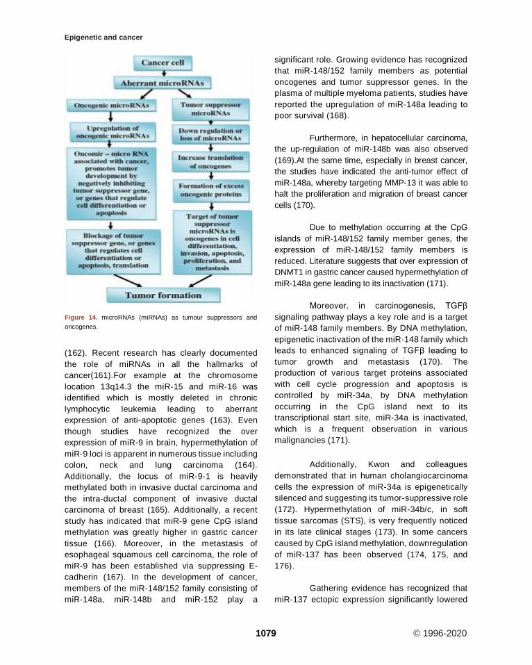
cancer (160, Figure 14).
Downregulation or loss of miRNAs with
tumour suppressor function may increase translation
of oncogenes and hence formation of excess
oncogenic proteins, leading to tumour formation. On
the other hand, upregulation of oncogenic miRNAs
may block tumour suppressor genes and also lead to
tumour formation.
MicroRNAs (miRNAs) consist of short
noncoding RNA species, which regulates post
transcriptional gene expression. Recent studies have
demonstrated that epigenetic mechanisms, including
DNA methylation and histone modification, not only
regulate the expression of protein encoding genes,
but also miRNAs, such as let7a, miR9, miR34a,
miR124, miR137, miR148 and miR203. Conversely,
the expression of important epigenetic regulators, are
controlled by another subset of miRNAs including
DNA methyltransferases, histone deacetylases, and
polycomb group genes. This intricate network of
feedback between epigenetic pathways and miRNAs
appears to form an epigenetics–miRNA regulatory
circuit and to organize the whole gene expression
profile. Normal physiological functions are interfered
with, contributing to various disease processes, when
this regulatory circuit is disrupted (44, 161).
Previous literature has suggested that
miRNAs are epigenetically regulated and in cancer
deregulation of miRNAs has been extensively
studied. In the cell most of the miRNAs are
involved in regulating cell cycle progression,
apoptosis, differentiation and other critical process
and alterations in numerous cancer types are
implicated them through epigenetic pathways
Figure 13. Biological roles of miRNA.
Epigenetic and cancer
1079 © 1996-2020
(162). Recent research has clearly documented
the role of miRNAs in all the hallmarks of
cancer(161).For example at the chromosome
location 13q14.3 the miR-15 and miR-16 was
identified which is mostly deleted in chronic
lymphocytic leukemia leading to aberrant
expression of anti-apoptotic genes (163). Even
though studies have recognized the over
expression of miR-9 in brain, hypermethylation of
miR-9 loci is apparent in numerous tissue including
colon, neck and lung carcinoma (164).
Additionally, the locus of miR-9-1 is heavily
methylated both in invasive ductal carcinoma and
the intra-ductal component of invasive ductal
carcinoma of breast (165). Additionally, a recent
study has indicated that miR-9 gene CpG island
methylation was greatly higher in gastric cancer
tissue (166). Moreover, in the metastasis of
esophageal squamous cell carcinoma, the role of
miR-9 has been established via suppressing E-
cadherin (167). In the development of cancer,
members of the miR-148/152 family consisting of
miR-148a, miR-148b and miR-152 play a
significant role. Growing evidence has recognized
that miR-148/152 family members as potential
oncogenes and tumor suppressor genes. In the
plasma of multiple myeloma patients, studies have
reported the upregulation of miR-148a leading to
poor survival (168).
Furthermore, in hepatocellular carcinoma,
the up-regulation of miR-148b was also observed
(169).At the same time, especially in breast cancer,
the studies have indicated the anti-tumor effect of
miR-148a, whereby targeting MMP-13 it was able to
halt the proliferation and migration of breast cancer
cells (170).
Due to methylation occurring at the CpG
islands of miR-148/152 family member genes, the
expression of miR-148/152 family members is
reduced. Literature suggests that over expression of
DNMT1 in gastric cancer caused hypermethylation of
miR-148a gene leading to its inactivation (171).
Moreover, in carcinogenesis, TGFβ
signaling pathway plays a key role and is a target
of miR-148 family members. By DNA methylation,
epigenetic inactivation of the miR-148 family which
leads to enhanced signaling of TGFβ leading to
tumor growth and metastasis (170). The
production of various target proteins associated
with cell cycle progression and apoptosis is
controlled by miR-34a, by DNA methylation
occurring in the CpG island next to its
transcriptional start site, miR-34a is inactivated,
which is a frequent observation in various
malignancies (171).
Additionally, Kwon and colleagues
demonstrated that in human cholangiocarcinoma
cells the expression of miR-34a is epigenetically
silenced and suggesting its tumor-suppressive role
(172). Hypermethylation of miR-34b/c, in soft
tissue sarcomas (STS), is very frequently noticed
in its late clinical stages (173). In some cancers
caused by CpG island methylation, downregulation
of miR-137 has been observed (174, 175, and
176).
Gathering evidence has recognized that
miR-137 ectopic expression significantly lowered
Figure 14. microRNAs (miRNAs) as tumour suppressors and
oncogenes.
Epigenetic and cancer
1080 © 1996-2020
the levels of Cdc42 and Cdk6, and in lung cancer
cells leading to cell cycle arrest at the G1 phase
(177). Most frequent miRNA in the brain is miR-
124 and a deviant expression leads to central
nervous system related malignancies
(178).Including glioblastomas, in numerous
cancers, a diverse mode of miR-124 expression
has been observed. Recent report suggests that
miR-124 acts as a tumor suppressor and by
targeting STAT3 it might be useful in treating
human glioblastomas (179).Furthermore, studies
have identified that the hepatitis C virus (HCV)
induction of DNMT, in HCV related intrahepatic
cholangiocarcinoma, led to the suppression of
miR-124 (180). In non- Hodgkin’s lymphoma a
greater frequency of miR-124-1 gene
hypermethylation was observed. miR-200 is
recognized as a cell’s autonomous suppressor of
epithelial to mesenchymal transition (EMT) and
metastasis (181). Reports suggest that in
numerous cancer it has been identified that the
finger E-box binding homeobox transcription factor
1 (ZEB1) is involved in EMT. Studies have
identified that in colorectal cancer cells, miR-200
over expression inhibits ZEB1 mediated
metastasis. Indeed it has been demonstrated that
by CpG island hypermethylation of miR-200
silencing, causes the transition between EMT and
vice versa leading to tumorigenesis (18).
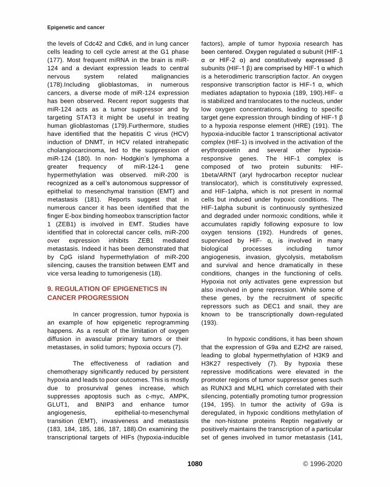
9. REGULATION OF EPIGENETICS IN
CANCER PROGRESSION
In cancer progression, tumor hypoxia is
an example of how epigenetic reprogramming
happens. As a result of the limitation of oxygen
diffusion in avascular primary tumors or their
metastases, in solid tumors; hypoxia occurs (7).
The effectiveness of radiation and
chemotherapy significantly reduced by persistent
hypoxia and leads to poor outcomes. This is mostly
due to prosurvival genes increase, which
suppresses apoptosis such as c-myc, AMPK,
GLUT1, and BNIP3 and enhance tumor
angiogenesis, epithelial-to-mesenchymal
transition (EMT), invasiveness and metastasis
(183, 184, 185, 186, 187, 188).On examining the
transcriptional targets of HIFs (hypoxia-inducible
factors), ample of tumor hypoxia research has
been centered. Oxygen regulated α subunit (HIF-1
α or HIF-2 α) and constitutively expressed β
subunits (HIF-1 β) are comprised by HIF-1 α which
is a heterodimeric transcription factor. An oxygen
responsive transcription factor is HIF-1 α, which
mediates adaptation to hypoxia (189, 190).HIF- α
is stabilized and translocates to the nucleus, under
low oxygen concentrations, leading to specific
target gene expression through binding of HIF-1 β
to a hypoxia response element (HRE) (191). The
hypoxia-inducible factor 1 transcriptional activator
complex (HIF-1) is involved in the activation of the
erythropoietin and several other hypoxia-
responsive genes. The HIF-1 complex is
composed of two protein subunits: HIF-
1beta/ARNT (aryl hydrocarbon receptor nuclear
translocator), which is constitutively expressed,
and HIF-1alpha, which is not present in normal
cells but induced under hypoxic conditions. The
HIF-1alpha subunit is continuously synthesized
and degraded under normoxic conditions, while it
accumulates rapidly following exposure to low
oxygen tensions (192). Hundreds of genes,
supervised by HIF- α, is involved in many
biological processes including tumor
angiogenesis, invasion, glycolysis, metabolism
and survival and hence dramatically in these
conditions, changes in the functioning of cells.
Hypoxia not only activates gene expression but
also involved in gene repression. While some of
these genes, by the recruitment of specific
repressors such as DEC1 and snail, they are
known to be transcriptionally down-regulated
(193).
In hypoxic conditions, it has been shown
that the expression of G9a and EZH2 are raised,
leading to global hypermethylation of H3K9 and
H3K27 respectively (7). By hypoxia these
repressive modifications were elevated in the
promoter regions of tumor suppressor genes such
as RUNX3 and MLH1 which correlated with their
silencing, potentially promoting tumor progression
(194, 195). In tumor the activity of G9a is
deregulated, in hypoxic conditions methylation of
the non-histone proteins Reptin negatively or
positively maintains the transcription of a particular
set of genes involved in tumor metastasis (141,
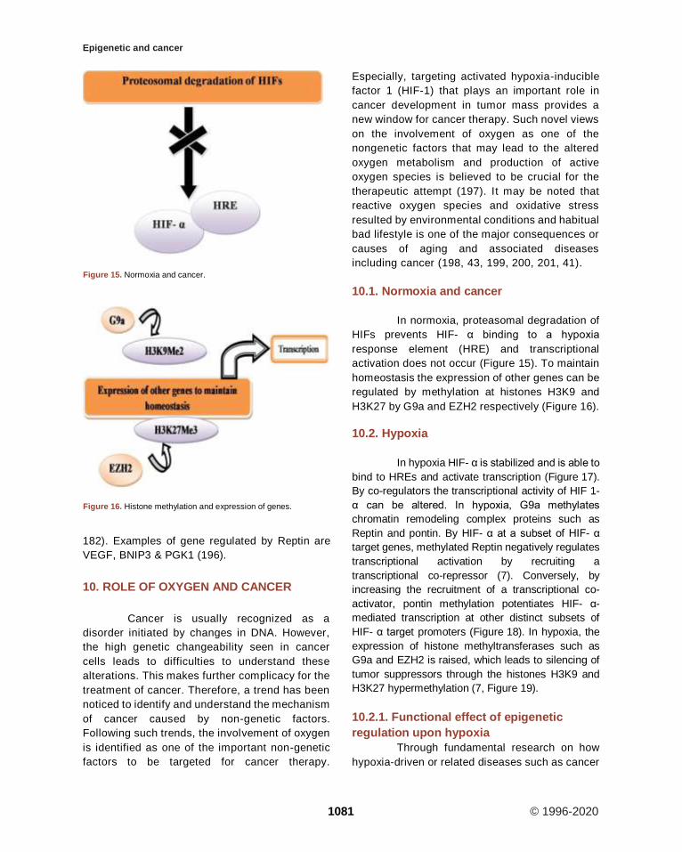
Epigenetic and cancer
1081 © 1996-2020
182). Examples of gene regulated by Reptin are
VEGF, BNIP3 & PGK1 (196).
10. ROLE OF OXYGEN AND CANCER
Cancer is usually recognized as a
disorder initiated by changes in DNA. However,
the high genetic changeability seen in cancer
cells leads to difficulties to understand these
alterations. This makes further complicacy for the
treatment of cancer. Therefore, a trend has been
noticed to identify and understand the mechanism
of cancer caused by non-genetic factors.
Following such trends, the involvement of oxygen
is identified as one of the important non-genetic
factors to be targeted for cancer therapy.
Especially, targeting activated hypoxia-inducible
factor 1 (HIF-1) that plays an important role in
cancer development in tumor mass provides a
new window for cancer therapy. Such novel views
on the involvement of oxygen as one of the
nongenetic factors that may lead to the altered
oxygen metabolism and production of active
oxygen species is believed to be crucial for the
therapeutic attempt (197). It may be noted that
reactive oxygen species and oxidative stress
resulted by environmental conditions and habitual
bad lifestyle is one of the major consequences or
causes of aging and associated diseases
including cancer (198, 43, 199, 200, 201, 41).
10.1. Normoxia and cancer
In normoxia, proteasomal degradation of
HIFs prevents HIF- α binding to a hypoxia
response element (HRE) and transcriptional
activation does not occur (Figure 15). To maintain
homeostasis the expression of other genes can be
regulated by methylation at histones H3K9 and
H3K27 by G9a and EZH2 respectively (Figure 16).
10.2. Hypoxia
In hypoxia HIF- α is stabilized and is able to
bind to HREs and activate transcription (Figure 17).
By co-regulators the transcriptional activity of HIF 1-
α can be altered. In hypoxia, G9a methylates
chromatin remodeling complex proteins such as
Reptin and pontin. By HIF- α at a subset of HIF- α
target genes, methylated Reptin negatively regulates
transcriptional activation by recruiting a
transcriptional co-repressor (7). Conversely, by
increasing the recruitment of a transcriptional co-
activator, pontin methylation potentiates HIF- α-
mediated transcription at other distinct subsets of
HIF- α target promoters (Figure 18). In hypoxia, the
expression of histone methyltransferases such as
G9a and EZH2 is raised, which leads to silencing of
tumor suppressors through the histones H3K9 and
H3K27 hypermethylation (7, Figure 19).
10.2.1. Functional effect of epigenetic
regulation upon hypoxia
Through fundamental research on how
hypoxia-driven or related diseases such as cancer
Figure 15. Normoxia and cancer.
Figure 16. Histone methylation and expression of genes.
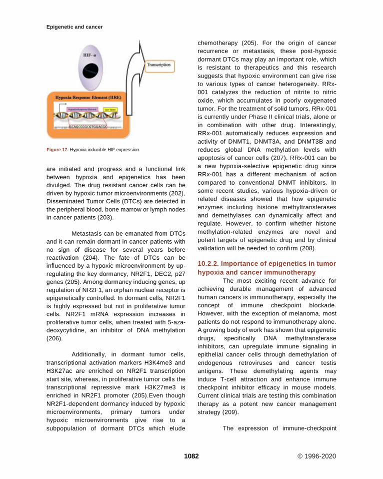
Epigenetic and cancer
1082 © 1996-2020
are initiated and progress and a functional link
between hypoxia and epigenetics has been
divulged. The drug resistant cancer cells can be
driven by hypoxic tumor microenvironments (202),
Disseminated Tumor Cells (DTCs) are detected in
the peripheral blood, bone marrow or lymph nodes
in cancer patients (203).
Metastasis can be emanated from DTCs
and it can remain dormant in cancer patients with
no sign of disease for several years before
reactivation (204). The fate of DTCs can be
influenced by a hypoxic microenvironment by up-
regulating the key dormancy, NR2F1, DEC2, p27
genes (205). Among dormancy inducing genes, up
regulation of NR2F1, an orphan nuclear receptor is
epigenetically controlled. In dormant cells, NR2F1
is highly expressed but not in proliferative tumor
cells. NR2F1 mRNA expression increases in
proliferative tumor cells, when treated with 5-aza-
deoxycytidine, an inhibitor of DNA methylation
(206).
Additionally, in dormant tumor cells,
transcriptional activation markers H3K4me3 and
H3K27ac are enriched on NR2F1 transcription
start site, whereas, in proliferative tumor cells the
transcriptional repressive mark H3K27me3 is
enriched in NR2F1 promoter (205).Even though
NR2F1-dependent dormancy induced by hypoxic
microenvironments, primary tumors under
hypoxic microenvironments give rise to a
subpopulation of dormant DTCs which elude
chemotherapy (205). For the origin of cancer
recurrence or metastasis, these post-hypoxic
dormant DTCs may play an important role, which
is resistant to therapeutics and this research
suggests that hypoxic environment can give rise
to various types of cancer heterogeneity. RRx-
001 catalyzes the reduction of nitrite to nitric
oxide, which accumulates in poorly oxygenated
tumor. For the treatment of solid tumors, RRx-001
is currently under Phase II clinical trials, alone or
in combination with other drug. Interestingly,
RRx-001 automatically reduces expression and
activity of DNMT1, DNMT3A, and DNMT3B and
reduces global DNA methylation levels with
apoptosis of cancer cells (207). RRx-001 can be
a new hypoxia-selective epigenetic drug since
RRx-001 has a different mechanism of action
compared to conventional DNMT inhibitors. In
some recent studies, various hypoxia-driven or
related diseases showed that how epigenetic
enzymes including histone methyltransferases
and demethylases can dynamically affect and
regulate. However, to confirm whether histone
methylation-related enzymes are novel and
potent targets of epigenetic drug and by clinical
validation will be needed to confirm (208).
10.2.2. Importance of epigenetics in tumor
hypoxia and cancer immunotherapy
The most exciting recent advance for
achieving durable management of advanced
human cancers is immunotherapy, especially the
concept of immune checkpoint blockade.
However, with the exception of melanoma, most
patients do not respond to immunotherapy alone.
A growing body of work has shown that epigenetic
drugs, specifically DNA methyltransferase
inhibitors, can upregulate immune signaling in
epithelial cancer cells through demethylation of
endogenous retroviruses and cancer testis
antigens. These demethylating agents may
induce T-cell attraction and enhance immune
checkpoint inhibitor efficacy in mouse models.
Current clinical trials are testing this combination
therapy as a potent new cancer management
strategy (209).
The expression of immune-checkpoint
Figure 17. Hypoxia inducible HIF expression.
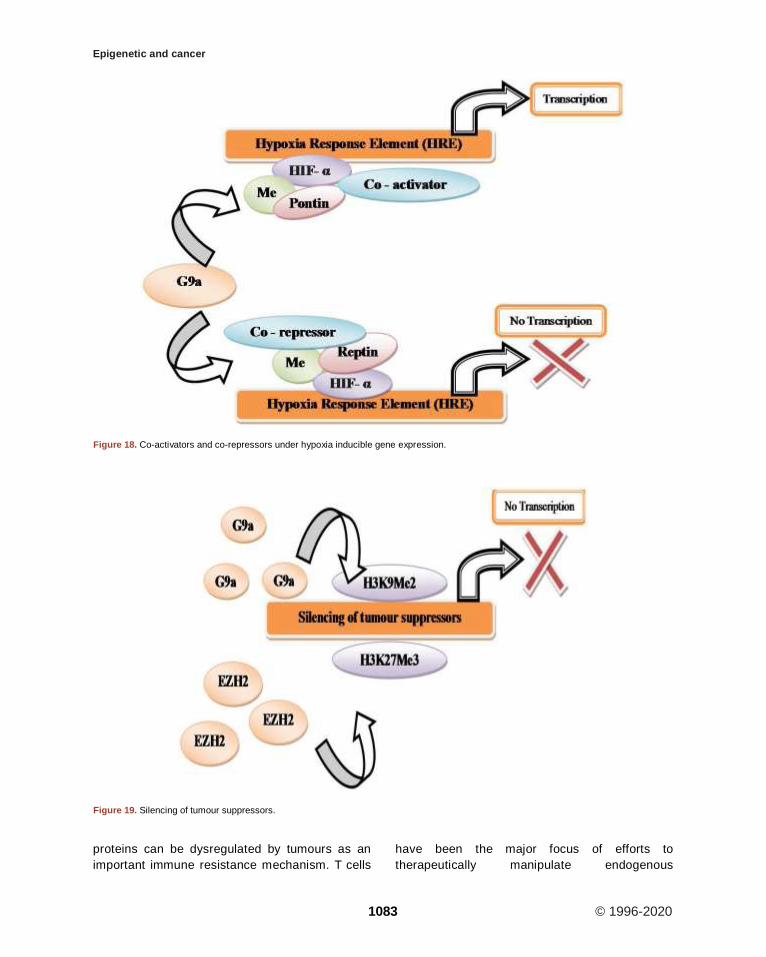
Epigenetic and cancer
1083 © 1996-2020
proteins can be dysregulated by tumours as an
important immune resistance mechanism. T cells
have been the major focus of efforts to
therapeutically manipulate endogenous
Figure 18. Co-activators and co-repressors under hypoxia inducible gene expression.
Figure 19. Silencing of tumour suppressors.
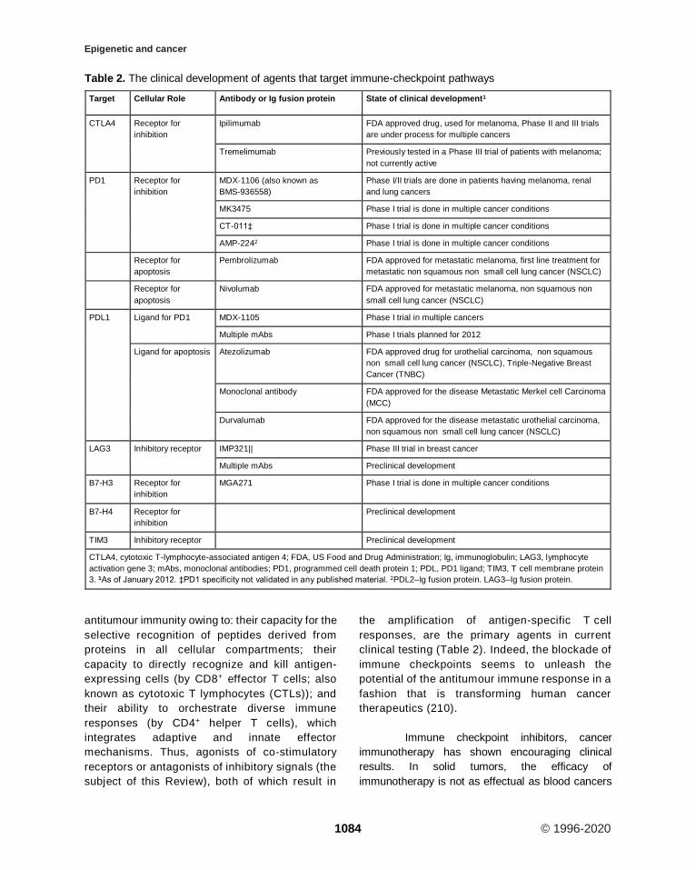
Epigenetic and cancer
1084 © 1996-2020
antitumour immunity owing to: their capacity for the
selective recognition of peptides derived from
proteins in all cellular compartments; their
capacity to directly recognize and kill antigen-
expressing cells (by CD8+ effector T cells; also
known as cytotoxic T lymphocytes (CTLs)); and
their ability to orchestrate diverse immune
responses (by CD4+ helper T cells), which
integrates adaptive and innate effector
mechanisms. Thus, agonists of co-stimulatory
receptors or antagonists of inhibitory signals (the
subject of this Review), both of which result in
the amplification of antigen-specific T cell
responses, are the primary agents in current
clinical testing (Table 2). Indeed, the blockade of
immune checkpoints seems to unleash the
potential of the antitumour immune response in a
fashion that is transforming human cancer
therapeutics (210).
Immune checkpoint inhibitors, cancer
immunotherapy has shown encouraging clinical
results. In solid tumors, the efficacy of
immunotherapy is not as effectual as blood cancers
Table 2. The clinical development of agents that target immune-checkpoint pathways
Target Cellular Role Antibody or Ig fusion protein State of clinical development1
CTLA4 Receptor for
inhibition
Ipilimumab FDA approved drug, used for melanoma, Phase II and III trials
are under process for multiple cancers
Tremelimumab Previously tested in a Phase III trial of patients with melanoma;
not currently active
PD1 Receptor for
inhibition
MDX-1106 (also known as
BMS-936558)
Phase I/II trials are done in patients having melanoma, renal
and lung cancers
MK3475 Phase I trial is done in multiple cancer conditions
CT-011‡ Phase I trial is done in multiple cancer conditions
AMP-2242 Phase I trial is done in multiple cancer conditions
Receptor for
apoptosis
Pembrolizumab FDA approved for metastatic melanoma, first line treatment for
metastatic non squamous non small cell lung cancer (NSCLC)
Receptor for
apoptosis
Nivolumab FDA approved for metastatic melanoma, non squamous non
small cell lung cancer (NSCLC)
PDL1 Ligand for PD1 MDX-1105 Phase I trial in multiple cancers
Multiple mAbs Phase I trials planned for 2012
Ligand for apoptosis Atezolizumab FDA approved drug for urothelial carcinoma, non squamous
non small cell lung cancer (NSCLC), Triple-Negative Breast
Cancer (TNBC)
Monoclonal antibody FDA approved for the disease Metastatic Merkel cell Carcinoma
(MCC)
Durvalumab FDA approved for the disease metastatic urothelial carcinoma,
non squamous non small cell lung cancer (NSCLC)
LAG3 Inhibitory receptor IMP321|| Phase III trial in breast cancer
Multiple mAbs Preclinical development
B7-H3 Receptor for
inhibition
MGA271 Phase I trial is done in multiple cancer conditions
B7-H4 Receptor for
inhibition
Preclinical development
TIM3 Inhibitory receptor Preclinical development
CTLA4, cytotoxic T-lymphocyte-associated antigen 4; FDA, US Food and Drug Administration; Ig, immunoglobulin; LAG3, lymphocyte
activation gene 3; mAbs, monoclonal antibodies; PD1, programmed cell death protein 1; PDL, PD1 ligand; TIM3, T cell membrane protein
3. 1As of January 2012. ‡PD1 specificity not validated in any published material. 2PDL2–Ig fusion protein. LAG3–Ig fusion protein.
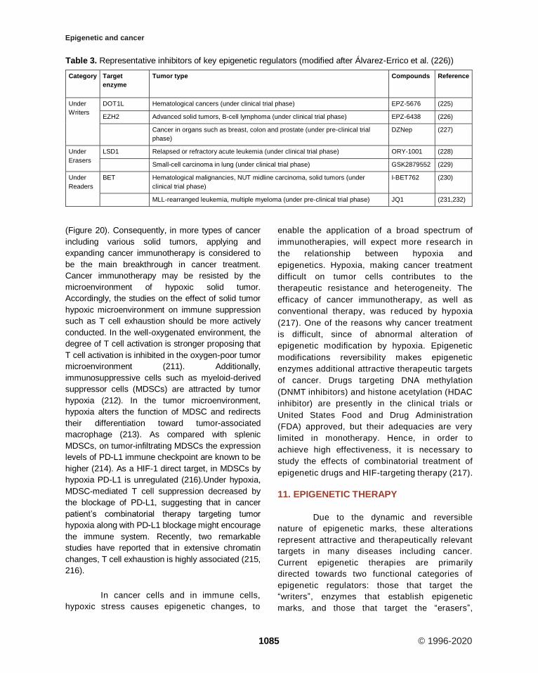
Epigenetic and cancer
1085 © 1996-2020
(Figure 20). Consequently, in more types of cancer
including various solid tumors, applying and
expanding cancer immunotherapy is considered to
be the main breakthrough in cancer treatment.
Cancer immunotherapy may be resisted by the
microenvironment of hypoxic solid tumor.
Accordingly, the studies on the effect of solid tumor
hypoxic microenvironment on immune suppression
such as T cell exhaustion should be more actively
conducted. In the well-oxygenated environment, the
degree of T cell activation is stronger proposing that
T cell activation is inhibited in the oxygen-poor tumor
microenvironment (211). Additionally,
immunosuppressive cells such as myeloid-derived
suppressor cells (MDSCs) are attracted by tumor
hypoxia (212). In the tumor microenvironment,
hypoxia alters the function of MDSC and redirects
their differentiation toward tumor-associated
macrophage (213). As compared with splenic
MDSCs, on tumor-infiltrating MDSCs the expression
levels of PD-L1 immune checkpoint are known to be
higher (214). As a HIF-1 direct target, in MDSCs by
hypoxia PD-L1 is unregulated (216).Under hypoxia,
MDSC-mediated T cell suppression decreased by
the blockage of PD-L1, suggesting that in cancer
patient’s combinatorial therapy targeting tumor
hypoxia along with PD-L1 blockage might encourage
the immune system. Recently, two remarkable
studies have reported that in extensive chromatin
changes, T cell exhaustion is highly associated (215,
216).
In cancer cells and in immune cells,
hypoxic stress causes epigenetic changes, to
enable the application of a broad spectrum of
immunotherapies, will expect more research in
the relationship between hypoxia and
epigenetics. Hypoxia, making cancer treatment
difficult on tumor cells contributes to the
therapeutic resistance and heterogeneity. The
efficacy of cancer immunotherapy, as well as
conventional therapy, was reduced by hypoxia
(217). One of the reasons why cancer treatment
is difficult, since of abnormal alteration of
epigenetic modification by hypoxia. Epigenetic
modifications reversibility makes epigenetic
enzymes additional attractive therapeutic targets
of cancer. Drugs targeting DNA methylation
(DNMT inhibitors) and histone acetylation (HDAC
inhibitor) are presently in the clinical trials or
United States Food and Drug Administration
(FDA) approved, but their adequacies are very
limited in monotherapy. Hence, in order to
achieve high effectiveness, it is necessary to
study the effects of combinatorial treatment of
epigenetic drugs and HIF-targeting therapy (217).
11. EPIGENETIC THERAPY
Due to the dynamic and reversible
nature of epigenetic marks, these alterations
represent attractive and therapeutically relevant
targets in many diseases including cancer.
Current epigenetic therapies are primarily
directed towards two functional categories of
epigenetic regulators: those that target the
“writers”, enzymes that establish epigenetic
marks, and those that target the “erasers”,
Table 3. Representative inhibitors of key epigenetic regulators (modified after Álvarez-Errico et al. (226))
Category Target
enzyme
Tumor type Compounds Reference
Under
Writers
DOT1L Hematological cancers (under clinical trial phase) EPZ-5676 (225)
EZH2 Advanced solid tumors, B-cell lymphoma (under clinical trial phase) EPZ-6438 (226)
Cancer in organs such as breast, colon and prostate (under pre-clinical trial
phase)
DZNep (227)
Under
Erasers
LSD1 Relapsed or refractory acute leukemia (under clinical trial phase) ORY-1001 (228)
Small-cell carcinoma in lung (under clinical trial phase) GSK2879552 (229)
Under
Readers
BET Hematological malignancies, NUT midline carcinoma, solid tumors (under
clinical trial phase)
I-BET762 (230)
MLL-rearranged leukemia, multiple myeloma (under pre-clinical trial phase) JQ1 (231,232)
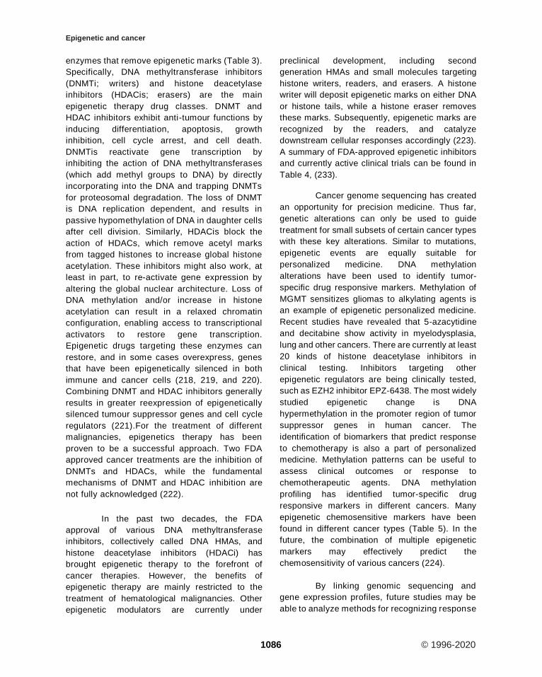
Epigenetic and cancer
1086 © 1996-2020
enzymes that remove epigenetic marks (Table 3).
Specifically, DNA methyltransferase inhibitors
(DNMTi; writers) and histone deacetylase
inhibitors (HDACis; erasers) are the main
epigenetic therapy drug classes. DNMT and
HDAC inhibitors exhibit anti-tumour functions by
inducing differentiation, apoptosis, growth
inhibition, cell cycle arrest, and cell death.
DNMTis reactivate gene transcription by
inhibiting the action of DNA methyltransferases
(which add methyl groups to DNA) by directly
incorporating into the DNA and trapping DNMTs
for proteosomal degradation. The loss of DNMT
is DNA replication dependent, and results in
passive hypomethylation of DNA in daughter cells
after cell division. Similarly, HDACis block the
action of HDACs, which remove acetyl marks
from tagged histones to increase global histone
acetylation. These inhibitors might also work, at
least in part, to re-activate gene expression by
altering the global nuclear architecture. Loss of
DNA methylation and/or increase in histone
acetylation can result in a relaxed chromatin
configuration, enabling access to transcriptional
activators to restore gene transcription.
Epigenetic drugs targeting these enzymes can
restore, and in some cases overexpress, genes
that have been epigenetically silenced in both
immune and cancer cells (218, 219, and 220).
Combining DNMT and HDAC inhibitors generally
results in greater reexpression of epigenetically
silenced tumour suppressor genes and cell cycle
regulators (221).For the treatment of different
malignancies, epigenetics therapy has been
proven to be a successful approach. Two FDA
approved cancer treatments are the inhibition of
DNMTs and HDACs, while the fundamental
mechanisms of DNMT and HDAC inhibition are
not fully acknowledged (222).
In the past two decades, the FDA
approval of various DNA methyltransferase
inhibitors, collectively called DNA HMAs, and
histone deacetylase inhibitors (HDACi) has
brought epigenetic therapy to the forefront of
cancer therapies. However, the benefits of
epigenetic therapy are mainly restricted to the
treatment of hematological malignancies. Other
epigenetic modulators are currently under
preclinical development, including second
generation HMAs and small molecules targeting
histone writers, readers, and erasers. A histone
writer will deposit epigenetic marks on either DNA
or histone tails, while a histone eraser removes
these marks. Subsequently, epigenetic marks are
recognized by the readers, and catalyze
downstream cellular responses accordingly (223).
A summary of FDA-approved epigenetic inhibitors
and currently active clinical trials can be found in
Table 4, (233).
Cancer genome sequencing has created
an opportunity for precision medicine. Thus far,
genetic alterations can only be used to guide
treatment for small subsets of certain cancer types
with these key alterations. Similar to mutations,
epigenetic events are equally suitable for
personalized medicine. DNA methylation
alterations have been used to identify tumor-
specific drug responsive markers. Methylation of
MGMT sensitizes gliomas to alkylating agents is
an example of epigenetic personalized medicine.
Recent studies have revealed that 5-azacytidine
and decitabine show activity in myelodysplasia,
lung and other cancers. There are currently at least
20 kinds of histone deacetylase inhibitors in
clinical testing. Inhibitors targeting other
epigenetic regulators are being clinically tested,
such as EZH2 inhibitor EPZ-6438. The most widely
studied epigenetic change is DNA
hypermethylation in the promoter region of tumor
suppressor genes in human cancer. The
identification of biomarkers that predict response
to chemotherapy is also a part of personalized
medicine. Methylation patterns can be useful to
assess clinical outcomes or response to
chemotherapeutic agents. DNA methylation
profiling has identified tumor-specific drug
responsive markers in different cancers. Many
epigenetic chemosensitive markers have been
found in different cancer types (Table 5). In the
future, the combination of multiple epigenetic
markers may effectively predict the
chemosensitivity of various cancers (224).
By linking genomic sequencing and
gene expression profiles, future studies may be
able to analyze methods for recognizing response
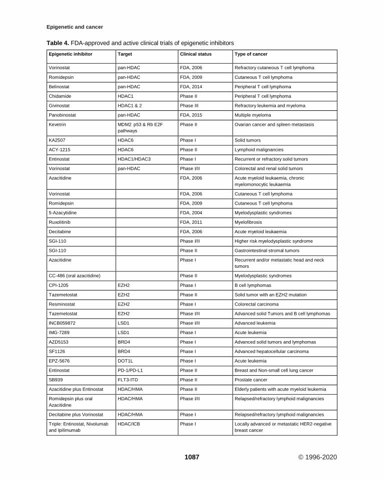
Epigenetic and cancer
1087 © 1996-2020
Table 4. FDA-approved and active clinical trials of epigenetic inhibitors
Epigenetic inhibitor Target Clinical status Type of cancer
Vorinostat pan-HDAC FDA, 2006 Refractory cutaneous T cell lymphoma
Romidepsin pan-HDAC FDA, 2009 Cutaneous T cell lymphoma
Belinostat pan-HDAC FDA, 2014 Peripheral T cell lymphoma
Chidamide HDAC1 Phase II Peripheral T cell lymphoma
Givinostat HDAC1 & 2 Phase III Refractory leukemia and myeloma
Panobinostat pan-HDAC FDA, 2015 Multiple myeloma
Kevetrin MDM2 p53 & Rb E2F
pathways
Phase II Ovarian cancer and spleen metastasis
KA2507 HDAC6 Phase I Solid tumors
ACY-1215 HDAC6 Phase II Lymphoid malignancies
Entinostat HDAC1/HDAC3 Phase I Recurrent or refractory solid tumors
Vorinostat pan-HDAC Phase I/II Colorectal and renal solid tumors
Azacitidine FDA, 2006 Acute myeloid leukaemia, chronic
myelomonocytic leukaemia
Vorinostat FDA, 2006 Cutaneous T cell lymphoma
Romidepsin FDA, 2009 Cutaneous T cell lymphoma
5-Azacytidine FDA, 2004 Myelodysplastic syndromes
Ruxolitinib FDA, 2011 Myelofibrosis
Decitabine FDA, 2006 Acute myeloid leukaemia
SGI-110 Phase I/II Higher risk myelodysplastic syndrome
SGI-110 Phase II Gastrointestinal stromal tumors
Azacitidine Phase I Recurrent and/or metastatic head and neck
tumors
CC-486 (oral azacitidine) Phase II Myelodysplastic syndromes
CPI-1205 EZH2 Phase I B cell lymphomas
Tazemetostat EZH2 Phase II Solid tumor with an EZH2 mutation
Resminostat EZH2 Phase I Colorectal carcinoma
Tazemetostat EZH2 Phase I/II Advanced solid Tumors and B cell lymphomas
INCB059872 LSD1 Phase I/II Advanced leukemia
IMG-7289 LSD1 Phase I Acute leukemia
AZD5153 BRD4 Phase I Advanced solid tumors and lymphomas
SF1126 BRD4 Phase I Advanced hepatocellular carcinoma
EPZ-5676 DOT1L Phase I Acute leukemia
Entinostat PD-1/PD-L1 Phase II Breast and Non-small cell lung cancer
SB939 FLT3-ITD Phase II Prostate cancer
Azacitidine plus Entinostat HDAC/HMA Phase II Elderly patients with acute myeloid leukemia
Romidepsin plus oral
Azacitidine
HDAC/HMA Phase I/II Relapsed/refractory lymphoid malignancies
Decitabine plus Vorinostat HDAC/HMA Phase I Relapsed/refractory lymphoid malignancies
Triple: Entinostat, Nivolumab
and Ipilimumab
HDAC/ICB Phase I Locally advanced or metastatic HER2-negative
breast cancer
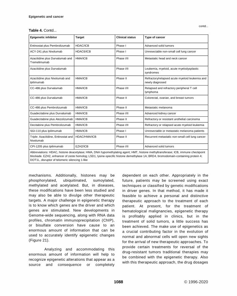
Epigenetic and cancer
1088 © 1996-2020
contd...
Table 4. Contd...
Epigenetic inhibitor Target Clinical status Type of cancer
Entinostat plus Pembrolizumab HDAC/ICB Phase I Advanced solid tumors
ACY-241 plus Nivolumab HDAC6/ICB Phase I Unresectable non-small cell lung cancer
Azacitidine plus Durvalumab and
Tremelimumab
HMA/ICB Phase I/II Metastatic head and neck cancer
Azacitidine plus Durvalumab Phase I/II Leukemia, myeloid, acute myelodysplastic
syndromes
Azacitidine plus Nivolumab and
Ipilimumab
HMA/ICB Phase II Refractory/relapsed acute myeloid leukemia and
newly diagnosed
CC-486 plus Durvalumab HMA/ICB Phase I/II Relapsed and refractory peripheral T cell
lymphoma
CC-486 plus Durvalumab HMA/ICB Phase II
Colorectal, ovarian, and breast tumors
CC-486 plus Pembrolizumab HMA/ICB Phase II Metastatic melanoma
Guadecitabine plus Durvalumab HMA/ICB Phase I/II Advanced kidney cancer
Guadecitabine plus Atezolizumab HMA/ICB Phase II Refractory or resistant urothelial carcinoma
Decitabine plus Pembrolizumab HMA/ICB Phase I/II Refractory or relapsed acute myeloid leukemia
SGI-110 plus Ipilimumab HMA/ICB Phase I Unresectable or metastatic melanoma patients
Triple: Azacitidine, Entinostat and
Nivolumab
HDAC/HMA/ICB Phase II Recurrent metastatic non-small cell lung cancer
CPI-1205 plus Ipilimumab EZH2/ICB Phase I/II Advanced solid tumors
Abbreviations: HDAC, histone deacetylase; HMA, DNA hypomethylating agent; HMT, histone methyltransferase; ICB, immune checkpoint
blockade. EZH2, enhancer of zeste homolog; LSD1, lysine-specific histone demethylase 1A; BRD4, bromodomain-containing protein 4;
DOT1L, disruptor of telomeric silencing 1-like
mechanisms. Additionally, histones may be
phosphorylated, ubiquitinated, sumoylated,
methylated and acetylated. But, in diseases,
these modifications have been less studied and
may also be able to divulge other therapeutic
targets. A major challenge in epigenetic therapy
is to know which genes are the driver and which
genes are stimulated. New developments in
Genome-wide sequencing, along with RNA data
profiles, chromatin immunoprecipitation (ChIP),
or bisulfate conversion have cause to an
enormous amount of information that can be
used to accurately identify epigenetic changes
(Figure 21).
Analyzing and accommodating this
enormous amount of information will help to
recognize epigenetic alterations that appear as a
source and consequence or completely
dependent on each other. Appropriately in the
future, patients may be screened using exact
techniques or classified by genetic modifications
in driver genes. In that method, it has made it
feasible to achieve a personal and distinctive
therapeutic approach to the treatment of each
patient. At present, for the treatment of
hematological malignancies, epigenetic therapy
is profitably applied in clinics, but in the
treatment of solid tumors, a little success has
been achieved. The make use of epigenetics as
a crucial contributing factor in the evolution of
normal and abnormal cells will open new sights
for the arrival of new therapeutic approaches. To
provide certain treatments for reversal of the
drug-resistant tumors traditional therapies may
be combined with the epigenetic therapy. Also
with this therapeutic approach, the drug dosages
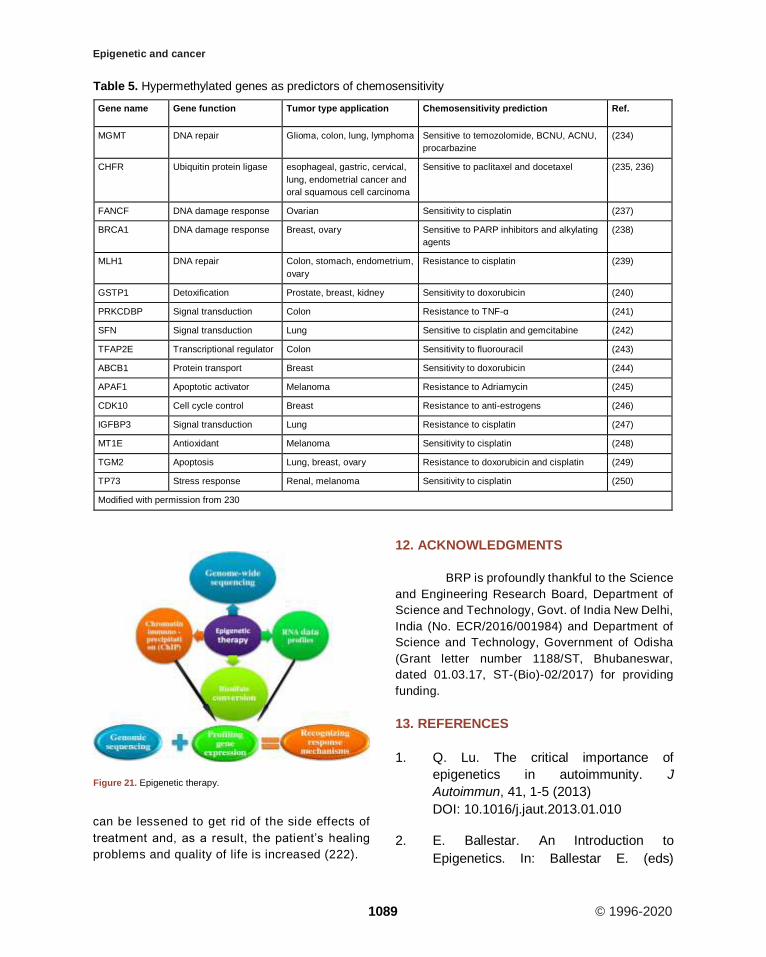
Epigenetic and cancer
1089 © 1996-2020
can be lessened to get rid of the side effects of
treatment and, as a result, the patient’s healing
problems and quality of life is increased (222).
12. ACKNOWLEDGMENTS
BRP is profoundly thankful to the Science
and Engineering Research Board, Department of
Science and Technology, Govt. of India New Delhi,
India (No. ECR/2016/001984) and Department of
Science and Technology, Government of Odisha
(Grant letter number 1188/ST, Bhubaneswar,
dated 01.03.17, ST-(Bio)-02/2017) for providing
funding.
13. REFERENCES
1. Q. Lu. The critical importance of
epigenetics in autoimmunity. J
Autoimmun, 41, 1-5 (2013)
DOI: 10.1016/j.jaut.2013.01.010
2. E. Ballestar. An Introduction to
Epigenetics. In: Ballestar E. (eds)
Table 5. Hypermethylated genes as predictors of chemosensitivity
Gene name Gene function Tumor type application Chemosensitivity prediction Ref.
MGMT DNA repair Glioma, colon, lung, lymphoma Sensitive to temozolomide, BCNU, ACNU,
procarbazine
(234)
CHFR Ubiquitin protein ligase esophageal, gastric, cervical,
lung, endometrial cancer and
oral squamous cell carcinoma
Sensitive to paclitaxel and docetaxel (235, 236)
FANCF DNA damage response Ovarian Sensitivity to cisplatin (237)
BRCA1 DNA damage response Breast, ovary Sensitive to PARP inhibitors and alkylating
agents
(238)
MLH1 DNA repair Colon, stomach, endometrium,
ovary
Resistance to cisplatin (239)
GSTP1 Detoxification Prostate, breast, kidney Sensitivity to doxorubicin (240)
PRKCDBP Signal transduction Colon Resistance to TNF-α (241)
SFN Signal transduction Lung Sensitive to cisplatin and gemcitabine (242)
TFAP2E Transcriptional regulator Colon Sensitivity to fluorouracil (243)
ABCB1 Protein transport Breast Sensitivity to doxorubicin (244)
APAF1 Apoptotic activator Melanoma Resistance to Adriamycin (245)
CDK10 Cell cycle control Breast Resistance to anti-estrogens (246)
IGFBP3 Signal transduction Lung Resistance to cisplatin (247)
MT1E Antioxidant Melanoma Sensitivity to cisplatin (248)
TGM2 Apoptosis Lung, breast, ovary Resistance to doxorubicin and cisplatin (249)
TP73 Stress response Renal, melanoma Sensitivity to cisplatin (250)
Modified with permission from 230
Figure 21. Epigenetic therapy.
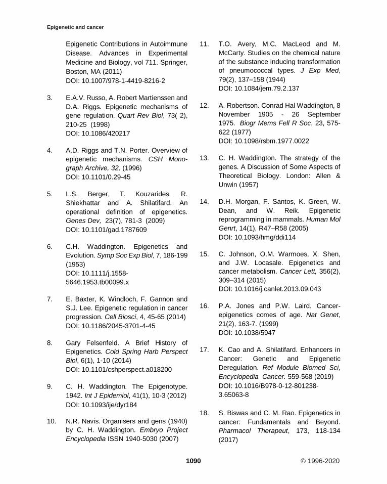
Epigenetic and cancer
1090 © 1996-2020
Epigenetic Contributions in Autoimmune
Disease. Advances in Experimental
Medicine and Biology, vol 711. Springer,
Boston, MA (2011)
DOI: 10.1007/978-1-4419-8216-2
3. E.A.V. Russo, A. Robert Martienssen and
D.A. Riggs. Epigenetic mechanisms of
gene regulation. Quart Rev Biol, 73( 2),
210-25 (1998)
DOI: 10.1086/420217
4. A.D. Riggs and T.N. Porter. Overview of
epigenetic mechanisms. CSH Mono-
graph Archive, 32, (1996)
DOI: 10.1101/0.29-45
5. L.S. Berger, T. Kouzarides, R.
Shiekhattar and A. Shilatifard. An
operational definition of epigenetics.
Genes Dev, 23(7), 781-3 (2009)
DOI: 10.1101/gad.1787609
6. C.H. Waddington. Epigenetics and
Evolution. Symp Soc Exp Biol, 7, 186-199
(1953)
DOI: 10.1111/j.1558-
5646.1953.tb00099.x
7. E. Baxter, K. Windloch, F. Gannon and
S.J. Lee. Epigenetic regulation in cancer
progression. Cell Biosci, 4, 45-65 (2014)
DOI: 10.1186/2045-3701-4-45
8. Gary Felsenfeld. A Brief History of
Epigenetics. Cold Spring Harb Perspect
Biol, 6(1), 1-10 (2014)
DOI: 10.1101/cshperspect.a018200
9. C. H. Waddington. The Epigenotype.
1942. Int J Epidemiol, 41(1), 10-3 (2012)
DOI: 10.1093/ije/dyr184
10. N.R. Navis. Organisers and gens (1940)
by C. H. Waddington. Embryo Project
Encyclopedia ISSN 1940-5030 (2007)
11. T.O. Avery, M.C. MacLeod and M.
McCarty. Studies on the chemical nature
of the substance inducing transformation
of pneumococcal types. J Exp Med,
79(2), 137–158 (1944)
DOI: 10.1084/jem.79.2.137
12. A. Robertson. Conrad Hal Waddington, 8
November 1905 - 26 September
1975. Biogr Mems Fell R Soc, 23, 575-
622 (1977)
DOI: 10.1098/rsbm.1977.0022
13. C. H. Waddington. The strategy of the
genes. A Discussion of Some Aspects of
Theoretical Biology. London: Allen &
Unwin (1957)
14. D.H. Morgan, F. Santos, K. Green, W.
Dean, and W. Reik. Epigenetic
reprogramming in mammals. Human Mol
Genrt, 14(1), R47–R58 (2005)
DOI: 10.1093/hmg/ddi114
15. C. Johnson, O.M. Warmoes, X. Shen,
and J.W. Locasale. Epigenetics and
cancer metabolism. Cancer Lett, 356(2),
309–314 (2015)
DOI: 10.1016/j.canlet.2013.09.043
16. P.A. Jones and P.W. Laird. Cancer-
epigenetics comes of age. Nat Genet,
21(2), 163-7. (1999)
DOI: 10.1038/5947
17. K. Cao and A. Shilatifard. Enhancers in
Cancer: Genetic and Epigenetic
Deregulation. Ref Module Biomed Sci,
Encyclopedia Cancer. 559-568 (2019)
DOI: 10.1016/B978-0-12-801238-
3.65063-8
18. S. Biswas and C. M. Rao. Epigenetics in
cancer: Fundamentals and Beyond.
Pharmacol Therapeut, 173, 118-134
(2017)
Epigenetic and cancer
1091 © 1996-2020
DOI: 10.1016/j.pharmthera.2017.02.011
19. D. Hanahan and R. A. Weinberg.
Hallmarks of Cancer: The Next
Generation. Cell, 144(5), 646-674
(2011)
DOI: 10.1016/j.cell.2011.02.013
20. S. B. Baylin and P. A. Jones. A decade
of exploring the cancer epigenome -
biological and translational
implications. Nat Rev Cancer, 11(10),
726-734 (2011)
DOI: 10.1038/nrc3130
21. S. Sharma, T. K. Kelly and P. A. Jones.
Epigenetics in cancer. Carcinogen,
31(1), 27-36 (2009)
DOI: 10.1093/carcin/bgp220
22. K. Luger, A. W. Mäder, R.K. Richmond,
D.F. Sargent and T.J. Richmond.
Crystal structure of the nucleosome
core particle at 2.8 Å resolution. Nature,
389(6648), 251-260 (1997)
DOI: 10.1038/38444
23. P. A. Jones and S. B. Baylin. The
Epigenomics of Cancer. Cell, 128(4),
683-692 (2007)
DOI: 10.1016/j.cell.2007.01.029
24. B. E. Bernstein, A. Meissner and E. S.
Lander. The Mammalian Epigenome.
Cell, 128(4), 669-681 (2007)
DOI: 10.1016/j.cell.2007.01.033
25. M. M. Suzuki and A. Bird. DNA
methylation landscapes: provocative
insights from epigenomics. Nat Rev
Genet, 9(6), 465-476 (2008)
DOI: 10.1038/nrg2341
26. T. Kouzarides. Chromatin Modifications
and Their Function. Cell, 128(4), 693-
705 (2007)
DOI: 10.1016/j.cell.2007.02.005
27. B. Zhang, X. Pan, G. P. Cobb, and T. A.
Anderson. microRNAs as oncogenes
and tumor suppressors. Development
Biol, 302(1), 1-12 (2007)
DOI: 10.1016/j.ydbio.2006.08.028
28. C. Jiang and B. F. Pugh. Nucleosome
positioning and gene regulation:
advances through genomics. Nat Rev
Genet, 10(3), 161-172 (2009)
DOI: 10.1038/nrg2522
29. B.W. Futscher. Epigenetic Changes
During Cell Transformation. Epigenet
Alt Oncogen, 179-194 (2012)
DOI: 10.1007/978-1-4419-9967-2_9
30. D. Hanahan and R. A. Weinberg.
Hallmarks of Cancer: The Next
Generation. Cell, 144(5), 646-674
(2011)
DOI: 10.1016/j.cell.2011.02.013
31. P. A. Jones and S. B. Baylin. The
Epigenomics of Cancer. Cell, 128(4),
683-692 (2007)
DOI: 10.1016/j.cell.2007.01.029
32. T. Ushijima & K. Asada. Aberrant DNA
methylation in contrast with mutations.
Cancer Sci, 101(2), 300-305 (2010)
DOI: 10.1111/j.1349-
7006.2009.01434.x
33. S. Sharma, T. K. Kelly and P.A. Jones.
Epigenetics in cancer. Carcinogen,
31(1), 27-36 (2009)
DOI: 10.1093/carcin/bgp220
34. J. Marlowe, S.S. Teo, S.D. Chibout, F.
Pognan and J. Moggs. Mapping the
epigenome - impact for toxicology. Mol
Clinic Environ Toxicol (2009)
DOI: 10.1007/978-3-7643-8336-7_10
Epigenetic and cancer
1092 © 1996-2020
35. J.K. Kim, M. Samaranayake and S.
Pradhan. Epigenetic mechanisms in
mammals. Cell Mol Lif Sci, 66(4), 596-
612 (2008)
DOI: 10.1007/s00018-008-8432-4
36. S.K.T. Ooi, A.H. O'Donnell and T.H.
Bestor. Mammalian cytosine
methylation at a glance. J Cell Sci,
122(16), 2787-2791 (2009)
DOI: 10.1242/jcs.015123
37. Z. Chen and A.D. Riggs. DNA
methylation and demethylation in
mammals. J Biol Chem, 286(21),
18347-18353 (2011)
DOI: 10.1074/jbc.R110.205286
38. T. Jenuwein. Translating the histone
code. Science, 293(5532), 1074-1080
(2001)
DOI: 10.1126/science.1063127
39. T. Kouzarides. Chromatin modifications
and their function. Cell, 128(4), 693-705
(2007)
DOI: 10.1016/j.cell.2007.02.005
40. I. P. Pogribny and I. Rusyn.
Environmental toxicants, epigenetics,
and cancer. Epigenet Alt Oncogen,
754, 215-232 (2012)
DOI: 10.1007/978-1-4419-9967-2_11
41. G.B.N. Chainy, B. Paital, and J.
Dandapat. An overview of seasonal
changes in oxidative stress and
antioxidant defence parameters in
some invertebrate and vertebrate
species. Scientifica, 2016, 1-8 (2016)
DOI: 10.1155/2016/6126570
42. M. Daniel and T.O. Tollefsbol.
Epigenetic linkage of aging, cancer and
nutrition. J Exp Biol, 218(1), 59-70
(2015)
DOI: 10.1242/jeb.107110
43. B. Paital, T. Jahan, S. Priyadarshini and
A. Mohanty. Antioxidants and ageing.
Open J Environ Biol, 2(1), 021 - 022
(2017)
DOI: 10.17352/ojeb.000004
44. G.T. Iswariya, B. Paital, P.R. Padma
and R. Nirmaladevi. microRNAs:
Epigenetic players in cancer and aging.
Front Biosci Scholar, 11, 9-9 (2019)
DOI: 10.2741/s525
45. J. R. Aunan, W. C. Cho and K. Søreide.
The Biology of Aging and Cancer: A
Brief Overview of Shared and Divergent
Molecular Hallmarks. Aging Dis, 8(5),
628 (2017)
DOI: 10.14336/AD.2017.0103
46. P. Mishra, B. Paital, S. Jena, S. S.
Swain, S. Kumar, M. K. Yadav and L.
Samanta. Possible activation of NRF2
by Vitamin E/Curcumin against altered
thyroid hormone induced oxidative
stress via NFĸB/AKT/mTOR/KEAP1
signalling in rat heart. Sci Rep, 9(1),
7408 (2019)
DOI: 10.1038/s41598-019-43320-5
47. A. Razin and A. Riggs. DNA
methylation and gene function.
Science, 210(4470), 604-610 (1980)
48. T. Phillips. The role of methylation in
gene expression. Nat Educ, 1(1), 116
(2008)
49. R. Straussman, D. Nejman, D. Roberts,
I. Steinfeld, B. Blum, N. Benvenisty and
H. Cedar. Developmental programming
of CpG island methylation profiles in the
human genome. Nat Struct Mol Biol,
16(5), 564-571 (2009)
Epigenetic and cancer
1093 © 1996-2020
DOI: 10.1038/nsmb.1594
50. L. Laurent, E. Wong, G. Li, T. Huynh, A.
Tsirigos, C. T. Ong and C.-L. Wei.
Dynamic changes in the human
methylome during differentiation.
Genom Res, 20(3), 320-331 (2010)
DOI: 10.1101/gr.101907.109
51. G. Almouzni and H. Cedar.
Maintenance of Epigenetic Information.
Cold Spring Harb Persp Biol, 8(5),
a019372 (2016)
DOI: 10.1101/cshperspect.a019372
52. Y. Pollack, R. Stein, A. Razin and H.
Cedar. Methylation of foreign DNA
sequences in eukaryotic cells. Proc
National Acad Sci, 77(11), 6463-6467
(1980)
DOI: 10.1073/pnas.77.11.6463
53. M. Wigler. The somatic replication of
DNA methylation. Cell, 24(1), 33-40
(1981)
DOI: 10.1016/0092-8674(81)90498-0
54. E. Li, T. H. Bestor and R. Jaenisch.
Targeted mutation of the DNA
methyltransferase gene results in
embryonic lethality. Cell, 69(6), 915-
926 (1992)
DOI: 10.1016/0092-8674(92)90611-F
55. E. Li and Y. Zhang. DNA Methylation in
Mammals. Cold Spring Harb Persp Biol,
6(5), a019133-a019133 (2014)
DOI: 10.1101/cshperspect.a019133
56. Y. Gruenbaum, H. Cedar and A. Razin.
Substrate and sequence specificity of a
eukaryotic DNA methylase. Nature,
295(5850), 620-622 (1982)
DOI: 10.1038/295620a0
57. X. Cheng. Structural and Functional
Coordination of DNA and Histone
Methylation. Cold Spring Harb Persp
Biol, 6(8), a018747-a018747 (2014)
DOI: 10.1101/cshperspect.a018747
58. Y. Gruenbaum, T. Naveh-Many, H.
Cedar and A. Razin. Sequence
specificity of methylation in higher plant
DNA. Nature, 292(5826), 860-862
(1981)
DOI: 10.1038/292860a0
59. L. Lande-Diner, J. Zhang, I. Ben-
Porath, N. Amariglio, I. Keshet, M.
Hecht and H. Cedar. Role of DNA
Methylation in Stable Gene Repression.
J. Biol Chem, 282(16), 12194-12200
(2007)
DOI: 10.1074/jbc.M607838200
60. R. Lucchini and J. M. Sogo. Replication
of transcriptionally active chromatin.
Nature, 374(6519), 276-280 (1995)
DOI: 10.1038/374276a0
61. S. Eden, T. Hashimshony, I. Keshet, H.
Cedar and A. W. Thorne. DNA
methylation models histone acetylation.
Nature, 394(6696), 842-842 (1998)
DOI: 10.1038/29680
62. P. L. Jones, G. C. Jan Veenstra, P. A.
Wade, D. Vermaak, S. U. Kass, N.
Landsberger and A.P. Wolffe.
Methylated DNA and MeCP2 recruit
histone deacetylase to repress
transcription. Nat Genet, 19(2), 187-
191 (1998)
DOI: 10.1038/561
63. X. Nan, H.-H. Ng, C. A. Johnson, C. D.
Laherty, B. M. Turner, R.N. Eisenman
and A. Bird. Transcriptional repression
by the methyl-CpG-binding protein
MeCP2 involves a histone deacetylase
Epigenetic and cancer
1094 © 1996-2020
complex. Nature, 393(6683), 386-389
(1998)
DOI: 10.1038/30764
64. T. Hashimshony, J. Zhang, I. Keshet,
M. Bustin and H. Cedar. The role of
DNA methylation in setting up
chromatin structure during devel-
opment. Nat Genet, 34(2), 187-192
(2003)
DOI: 10.1038/ng1158
65. N. Papadopoulos, N. Nicolaides, Y.
Wei, S. Ruben, K. Carter and C. Rosen.
Mutation of a mutL homolog in
hereditary colon cancer. Science,
263(5153), 1625-1629 (1994)
DOI: 10.1126/science.8128251
66. V. Greger, N. Debus, D. Lohmann, W.
Hopping, E. Passarge and B.
Horsthemke. Frequency and parental
origin of hypermethylated RB1 alleles in
retinoblastoma. Human Genet, 94(5)
(1994)
DOI: 10.1007/BF00211013
67. F.M. Kane, M. Loda, M.G. Gaida, J.
Lipman, R. Mishra, J. H. Goldman, M.
Jessup and R. Kolodner. Methylation of
the hMLH1 Promoter Correlates with
Lack of Expression of hMLH1 in
Sporadic Colon Tumors and Mismatch
Repair-defective Human Tumor Cell
Lines. Cancer Res, 57(5), 808-11,
(1997).
68. C. Stirzaker, DS. Millar, CL. Paul, PM.
Warnecke, J. Harrison, PC. Vincent, M.
Frommer, SJ. Clark. Extensive DNA
methylation spanning the Rb promoter
in retinoblastoma tumors. Cancer Res,
57(11), 2229-37 (1997)
69. J.G. Herman, A. Umar, K. Polyak, J.R.
Graff, N. Ahuja, J.-P. J Issa and S.B.
Baylin. Incidence and functional
consequences of hMLH1 promoter
hypermethylation in colorectal
carcinoma. Proc National Acad Sci,
95(12), 6870-6875 (1998)
DOI: 10.1073/pnas.95.12.6870
70. T. Furukawa, F. Konishi, S. Masubuchi,
K. Shitoh, H. Nagai and T. Tsukamoto.
Densely methylatedMLH1 promoter
correlates with decreased mRNA
expression in sporadic colorectal
cancers. Gene Chromosom Cancer,
35(1), 1-10 (2002)
DOI: 10.1002/gcc.10100
71. F. A. Dick. Retinoblastoma Tumor
Suppressor Gene. Ref Mod Biomedic
Sci, 30(13), 1492–1502 (2015)
DOI: 10.1016/B978-0-12-801238-
3.04443-3
72. S.A. Belinsky, K.J. Nikula, W. A.
Palmisano, R. Michels, G.
Saccomanno, E. Gabrielson and J.G.
Herman. Aberrant methylation of
p16INK4a is an early event in lung
cancer and a potential biomarker for
early diagnosis. Proc National Acad
Sci, 95(20), 11891-11896 (1998)
DOI: 10.1073/pnas.95.20.11891
73. W.H. Lee, R.A. Morton, J.I. Epstein,
J.D. Brooks, P.A. Campbell, G.S. Bova
and W.G. Nelson. Cytidine methylation
of regulatory sequences near the pi-
class glutathione S-transferase gene
accompanies human prostatic
carcinogenesis. Proc National Acad
Sci, 91(24), 11733-11737 (1994)
DOI: 10.1073/pnas.91.24.11733
74. M. Makos, B.D. Nelkin, M.I. Lerman, F.
Latif, B. Zbar and S.B. Baylin. Distinct
Epigenetic and cancer
1095 © 1996-2020
hypermethylation patterns occur at
altered chromosome loci in human lung
and colon cancer. Proc National Acad
Sci, 89(5), 1929-1933 (1992)
DOI: 10.1073/pnas.89.5.1929
75. B. Vogelstein, E. R. Fearon, S. R.
Hamilton, S. E. Kern, A. C. Preisinger,
M. Leppert and J. L. Bos. Genetic
Alterations during Colorectal-Tumor
Development. New Eng J Med, 319(9),
525-532 (1988)
DOI: 10.1056/NEJM198809013190901
76. S. J. Clark and J. Melki. DNA
methylation and gene silencing in
cancer: which is the guilty party?
Oncogene, 21(35), 5380-5387 (2002)
DOI: 10.1038/sj.onc.1205598
77. B.C. Christensen, C.J. Marsit, E.A.
Houseman, J.J Godleski, J.L.
Longacker, S. Zheng, K.T. Kelsey.
Differentiation of Lung
Adenocarcinoma, Pleural
Mesothelioma, and Nonmalignant
Pulmonary Tissues Using DNA Methy-
lation Profiles. Cancer Res, 69(15),
6315-6321 (2009)
DOI: 10.1158/0008-5472.CAN-09-1073
78. M. Toyota, N. Ahuja, M. Ohe-Toyota, J.
G. Herman, S. B. Baylin abnd J.-P. J.
Issa. CpG island methylator phenotype
in colorectal cancer. Proc National
Acad Sci, 96(15), 8681-8686 (1999)
DOI: 10.1073/pnas.96.15.8681
79. A. P. Feinberg and B. Vogelstein.
Hypomethylation distinguishes genes
of some human cancers from their
normal counterparts. Nature,
301(5895), 89-92 (1983)
DOI: 10.1038/301089a0
80. M. Ehrlich and M. Lacey. DNA
Hypomethylation and Hemimethylation
in Cancer. Epigenet Alt Oncogen, 31-56
(2012)
DOI: 10.1007/978-1-4419-9967-2_2
81. R. Z. Chen, U. Pettersson, C. Beard, L.
Jackson-Grusby and R. Jaenisch. DNA
hypomethylation leads to elevated
mutation rates. Nature, 395(6697), 89-
93 (1998)
DOI: 10.1038/25779
82. A. Narayan, W. Ji, X.-Y Zhang, A.
Marrogi, J. R. Graff, S. B. Baylin and M.
Ehrlich. Hypomethylation of
pericentromeric DNA in breast
adenocarcinomas. Int J Cancer, 77(6),
833-838 (1998)
DOI: 10.1002/(SICI)1097-
0215(19980911)77:6<833::AID-
IJC6>3.0.CO;2-V
83. F. Gaudet. Induction of Tumors in Mice
by Genomic Hypomethylation. Science,
300(5618), 489-492 (2003)
DOI: 10.1126/science.1083558
84. K. D. Hansen, W. Timp, H. C. Bravo, S.
Sabunciyan, B. Langmead, O. G.
McDonald and A.P. Feinberg.
Increased methylation variation in
epigenetic domains across cancer
types. Nat Genet, 43(8), 768-775
(2011)
DOI: 10.1038/ng.865
85. B. P. Berman, D. J. Weisenberger, J. F.
Aman, T. Hinoue, Z. Ramjan, Y. Liu and
P. W. Laird. Regions of focal DNA
hypermethylation and long-range
hypomethylation in colorectal cancer
coincide with nuclear lamina-
associated domains. Nat Genet, 44(1),
40-46 (2011)
DOI: 10.1038/ng.969
Epigenetic and cancer
1096 © 1996-2020
86. G. C. Hon, R. D. Hawkins, O. L.
Caballero, C. Lo, R. Lister, M. Pelizzola
and B. Ren. Global DNA
hypomethylation coupled to repressive
chromatin domain formation and gene
silencing in breast cancer. Genome
Res, 22(2), 246-258 (2011)
DOI: 10.1101/gr.125872.111
87. S. A. Bert, M. D. Robinson, D.
Strbenac, A. L. Statham, J. Z. Song, T.
Hulf and S. J. Clark. Regional Activation
of the Cancer Genome by Long-Range
Epigenetic Remodeling. Cancer Cell,
23(1), 9-22 (2013)
DOI: 10.1016/j.ccr.2012.11.006
88. H. Shen and P. W. Laird, P. W. Interplay
between the Cancer Genome and
Epigenome. Cell, 153(1), 38-55 (2013)
DOI: 10.1016/j.cell.2013.03.008
89. G. Egger, G. Liang, A. Aparicio and P.
A. Jones. Epigenetics in human
disease and prospects for epigenetic
therapy. Nature, 429(6990), 457-463
(2004)
DOI: 10.1038/nature02625
90. A. Spannhoff, A.-T. Hauser, R. Heinke,
W. Sippl and M. Jung. The Emerging
Therapeutic Potential of Histone
Methyltransferase and Demethylase
Inhibitors. Chem Med Chem, 4(10),
1568-1582 (2009)
DOI: 10.1002/cmdc.200900301
91. T. K. Kelly, D. D. De Carvalho and P. A.
Jones. Epigenetic modifications as
therapeutic targets. Nat Biotechnol,
28(10), 1069-1078 (2010)
DOI: 10.1038/nbt.1678
92. K. M. Bernt, N. Zhu, A. U. Sinha, S.
Vempati, J. Faber, A. V. Krivtsov and S.
A. Armstrong. MLL-Rearranged
Leukemia Is Dependent on Aberrant
H3K79 Methylation by DOT1L. Cancer
Cell, 20(1), 66-78 (2011)
DOI: 10.1016/j.ccr.2011.06.010
93. S. R. Daigle, E. J. Olhava, C. A.
Therkelsen, C. R. Majer, C.J.
Sneeringer, J. Song and R. M. Pollock.
Selective Killing of Mixed Lineage
Leukemia Cells by a Potent Small-
Molecule DOT1L Inhibitor. Cancer Cell,
20(1), 53-65 (2011)
DOI: 10.1016/j.ccr.2011.06.009
94. M. A. Dawson and T. Kouzarides.
Cancer Epigenetics: From Mechanism
to Therapy. Cell, 150(1), 12-27 (2012)
DOI: 10.1016/j.cell.2012.06.013
95. N. Azad, C. A. Zahnow, C. M. Rudin
and S. B. Baylin. The future of
epigenetic therapy in solid tumours-
lessons from the past. Nat Rev Clinic
Oncol, 10(5), 256-266 (2013)
DOI: 10.1038/nrclinonc.2013.42
96. P. A. Jones. Functions of DNA
methylation: islands, start sites, gene
bodies and beyond. Nat Rev Genet,
13(7), 484-492 (2012)
DOI: 10.1038/nrg3230
97. M. Kulis, S. Heath, M. Bibikova, A. C.
Queirós, A. Navarro, G. Clot and J. I.
Martín-Subero. Epigenomic analysis
detects widespread gene-body DNA
hypomethylation in chronic lymphocytic
leukemia. Nat Genet, 44(11), 1236-
1242, (2012)
DOI: 10.1038/ng.2443
98. G. Pfeifer. Defining Driver DNA
Methylation Changes in Human
Cancer. Int J Mol Sci, 19(4), 1166
(2018)
DOI: 10.3390/ijms19041166
Epigenetic and cancer
1097 © 1996-2020
99. Comprehensive molecular
characterization of clear cell renal cell
carcinoma. Nature, 499(7456), 43-49
(2013)
DOI: 10.1038/nature12222
100. Andrew P. Feinberg, Charles W.
Gehrke, Kenneth C. Kuo and Melanie
Ehrlich. Reduced Genomic 5-
Methylcytosine Content in Human
Colonic Neoplasia. Cancer Res, 48,
1159-1161 (1988)
101. M. A. Gama-Sosa, V. A. Slagel, R. W.
Trewyn, R. Oxenhandler, K. C. Kuo, C.
W. Gehrke and M. Ehrlich. The 5-
methylcytosine content of DNA from
human tumors. Nucleic Acid Res,
11(19), 6883-6894 (1983)
DOI: 10.1093/nar/11.19.6883
102. S. Goelz, B. Vogelstein, Hamilton and
A. Feinberg. Hypomethylation of DNA
from benign and malignant human
colon neoplasms. Science, 228(4696),
187-190 (1985)
DOI: 10.1126/science.2579435
103. C. Mertineit, J.A. Yoder, T. Taketo,
D.W. Laird, J.M. Trasler and T.H.
Bestor. Sex-specific exons control DNA
methyltransferase in mammalian germ
cells. Development., 125(5):889-97.
(1998)
104. M. Wossidlo, T. Nakamura, K.
Lepikhov, C. J. Marques, V.
Zakhartchenko and M. Boiani, J.
Walter. 5-Hydroxymethylcytosine in the
mammalian zygote is linked with
epigenetic reprogramming. Nat Comm,
2(1) (2011)
DOI: 10.1038/ncomms1240
105. V. Valinluck and L. C. Sowers.
Endogenous Cytosine Damage
Products Alter the Site Selectivity of
Human DNA Maintenance
Methyltransferase DNMT1. Cancer
Res, 67(3), 946-950 (2007)
DOI: 10.1158/0008-5472.CAN-06-3123
106. Y.-F He, B.-Z. Li, Z. Li, P. Liu, Y. Wang,
Q. Tang and G.-L. Xu, Tet-Mediated
Formation of 5-Carboxylcytosine and
Its Excision by TDG in Mammalian
DNA. Science, 333(6047), 1303-1307
(2011)
DOI: 10.1126/science.1210944
107. A. Maiti and A. C. Drohat. Thymine DNA
Glycosylase Can Rapidly Excise 5-
Formylcytosine and 5-
Carboxylcytosine. J Biol Chem,
286(41), 35334-35338 (2011)
DOI: 10.1074/jbc.C111.284620
108. S. Kangaspeska, B. Stride, R. Métivier,
M. Polycarpou-Schwarz, D. Ibberson,
R. P. Carmouche and G. Reid.
Transient cyclical methylation of
promoter DNA. Nature, 452(7183), 112-
115 (2008)
DOI: 10.1038/nature06640
109. R. Métivier, R. Gallais, C. Tiffoche, C.
Le Péron, R. Z. Jurkowska, R. P.
Carmouche and G. Salbert. Cyclical
DNA methylation of a transcriptionally
active promoter. Nature, 452(7183), 45-
50 (2008)
DOI: 10.1038/nature06544
110. T. Jenuwein. Translating the Histone
Code. Science, 293(5532), 1074-1080
(2001)
DOI: 10.1126/science.1063127
111. P. Chi, C. D. Allis and G. G. Wang.
Covalent histone modifications -
miswritten, misinterpreted and mis-
erased in human cancers. Nat Rev
Epigenetic and cancer
1098 © 1996-2020
Cancer, 10(7), 457-469 (2010)
DOI: 10.1038/nrc2876
112. J. C. Rice, S. D. Briggs, B. Ueberheide,
C. M. Barber, J. Shabanowitz, D. F.
Hunt and C. D. Allis. Histone Methyl-
transferases Direct Different Degrees
of Methylation to Define Distinct
Chromatin Domains. Mol Cell, 12(6),
1591-1598 (2003)
DOI: 10.1016/S1097-2765(03)00479-9
113. A. P. Feinberg and B. Tycko. The
history of cancer epigenetics. Nat Rev
Cancer, 4(2), 143-153 (2004)
DOI: 10.1038/nrc1279
114. T. Jenuwein. Re-SET-ting hetero-
chromatin by histone
methyltransferases. Trend Cell Biol,
11(6), 266-273 (2001)
DOI: 10.1016/S0962-8924(01)02001-3
115. P. A. Wade, D. Pruss and A. P. Wolffe.
Histone acetylation: chromatin in
action. Trend Biochem Sci, 22(4), 128-
132 (1997)
DOI: 10.1016/S0968-0004(97)01016-5
116. C. L. Peterson and M.-A. Laniel.
Histones and histone modifications.
Curr Biol, 14(14), R546-R551 (2004)
DOI: 10.1016/j.cub.2004.07.007
117. A. Shilatifard. Chromatin Modifications
by Methylation and Ubiquitination:
Implications in the Regulation of Gene
Expression. Ann Rev Biochem, 75(1),
243-269 (2006)
DOI: 10.1146/annurev.biochem.-
75.103004.142422
118. Y. Shiio and R. N. Eisenman. Histone
sumoylation is associated with
transcriptional repression. Proc Nat
Acad Sci, 100(23), 13225-13230 (2003)
DOI: 10.1073/pnas.1735528100
119. W. Fischle, Y. Wang and C. D. Allis.
Histone and chromatin cross-talk. Curr
Opin Cell Biol, 15(2), 172-183 (2003)
DOI: 10.1016/S0955-0674(03)00013-9
120. S. Messner and M. O. Hottiger. Histone
ADP-ribosylation in DNA repair,
replication and transcription. Trend Cell
Biol, 21(9), 534-542 (2011)
DOI: 10.1016/j.tcb.2011.06.001
121. M. Lachner. An epigenetic road map for
histone lysine methylation. J Cell Sci,
116(11), 2117-2124 (2003)
DOI: 10.1242/jcs.00493
122. T. Kouzarides. Chromatin Modifications
and Their Function. Cell, 128(4), 693-
705 (2007)
DOI: 10.1016/j.cell.2007.02.005
123. S. L. Berger, Histone modifications in
transcriptional regulation. Curr Opin
Genet Develop, 12(2), 142-148 (2002)
DOI: 10.1016/S0959-437X(02)00279-4
124. L. J. M. Jason, S. C. Moore, J. D. Lewis,
and G. Lindsey and J. Ausió. Histone
ubiquitination: a tagging tail unfolds?
BioEssay, 24(2), 166-174 (2002)
DOI: 10.1002/bies.10038
125. C. Dong, Y. Wu, J. Yao, Y. Wang, Y.
Yu, P. G. Rychahou and B. P. Zhou.
G9a interacts with Snail and is critical
for Snail-mediated E-cadherin
repression in human breast cancer. J
Clinical Invest, 122(4), 1469-1486
(2012)
DOI: 10.1172/JCI57349
126. M.-W. Chen, K.-T. Hua, H.-J. Kao, C.-
C. Chi, L.-H. G. Wei, Johansson and
M.-L. Kuo. H3K9 Histone
Epigenetic and cancer
1099 © 1996-2020
Methyltransferase G9a Promotes Lung
Cancer Invasion and Metastasis by
Silencing the Cell Adhesion Molecule
Ep-CAM. Cancer Res, 70(20), 7830-
7840 (2010)
DOI: 10.1158/0008-5472.CAN-10-0833
127. C. G. Kleer, Q. Cao, S. Varambally, R.
Shen, I. Ota, S. A. Tomlins and A. M.
Chinnaiyan. EZH2 is a marker of
aggressive breast cancer and promotes
neoplastic transformation of breast
epithelial cells. Proc Nat Acad Sci,
100(20), 11606-11611 (2003)
DOI: 10.1073/pnas.1933744100
128. S. Varambally. S. M. Dhanasekaran, M.
Zhou, T. R. Barrette, C. Kumar-Sinha,
M. G. Sanda and A. M. Chinnaiyan. The
polycomb group protein EZH2 is
involved in progression of prostate
cancer. Nature, 419(6907), 624-629
(2002)
DOI: 10.1038/nature01075
129. K. Agger, P. A. C. Cloos, L. Rudkjaer,
K. Williams, G. Andersen, J.
Christensen and K. Helin. The
H3K27me3 demethylase JMJD3
contributes to the activation of the
INK4A-ARF locus in response to
oncogene- and stress-induced
senescence. Gene Develop, 23(10),
1171-1176 (2009)
DOI: 10.1101/gad.510809
130. J. S. Song, Y. S. Kim, D.K. Kim, S.I.
Park, and S. J. Jang. Global histone
modification pattern associated with
recurrence and disease-free survival in
non-small cell lung cancer patients.
Pathol Int, 62(3), 182-190 (2012)
DOI: 10.1111/j.1440-
1827.2011.02776.x
131. Y. Shi, F. Lan, C. Matson, P. Mulligan,
J. R. Whetstine, P. A. Cole and Y. Shi.
Histone Demethylation Mediated by the
Nuclear Amine Oxidase Homolog
LSD1. Cell, 119(7), 941-953 (2004)
DOI: 10.1016/j.cell.2004.12.012
132. J. Huang, R. Sengupta, A. B. Espejo, M.
G. Lee, J. A. Dorsey, M. Richter, S. L.
Berger. p53 is regulated by the lysine
demethylase LSD1. Nature, 449(7158),
105-108 (2007)
DOI: 10.1038/nature06092
133. S. S. Ng, K. L. Kavanagh, M. A.
McDonough, D. Butler, E. S. Pilka, B.
M. R. Lienard and U. Oppermann.
Crystal structures of histone
demethylase JMJD2A reveal basis for
substrate specificity. Nature,
448(7149), 87-91 (2007)
DOI: 10.1038/nature05971
134. Y. Okada, G. Scott, M. K. Ray, Y.
Mishina and Y. Zhang. Histone
demethylase JHDM2A is critical for
Tnp1 and Prm1 transcription and
spermatogenesis. Nature, 450(7166),
119-123 (2007)
DOI: 10.1038/nature06236
135. S. Chuikov, J. K. Kurash, J. R. Wilson,
B. Xiao, N. Justin, G. S. Ivanov and D.
Reinberg. Regulation of p53 activity
through lysine methylation. Nature,
432(7015), 353-360 (2004)
DOI: 10.1038/nature03117
136. S. R. Daigle, E. J. Olhava, C. A.
Therkelsen, C. R. Majer, C. J.
Sneeringer, J. Song and R. M. Pollock.
Selective Killing of Mixed Lineage
Leukemia Cells by a Potent Small-
Molecule DOT1L Inhibitor. Cancer Cell,
20(1), 53-65 (2011)
DOI: 10.1016/j.ccr.2011.06.009
Epigenetic and cancer
1100 © 1996-2020
137. J. Huang, J. Dorsey, S. Chuikov, X.
Zhang, T. Jenuwein, D. Reinberg and
S. L. Berger. G9a and Glp Methylate
Lysine 373 in the Tumor Suppressor
p53. J Biol Chem, 285(13), 9636-9641
(2010)
DOI: 10.1074/jbc.M109.062588
138. E. Kim, M. Kim, D.-H. Woo, Y. Shin, J.
Shin, N. Chang and J. Lee.
Phosphorylation of EZH2 Activates
STAT3 Signaling via STAT3
Methylation and Promotes
Tumorigenicity of Glioblastoma Stem-
like Cells. Cancer Cell, 23(6), 839-852
(2013)
DOI: 10.1016/j.ccr.2013.04.008
139. A. He, X. Shen, Q. Ma, J. Cao, A. von
Gise, P. Zhou and W. T. Pu. PRC2
directly methylates GATA4 and
represses its transcriptional activity.
Gene Develop, 26(1), 37-42 (2012)
DOI: 10.1101/gad.173930.111
140. J.M. Lee, J. S. Lee, H. Kim, K. Kim, H.
Park, J.-Y. Kim and S. H. Baek. EZH2
Generates a Methyl Degron that Is
Recognized by the DCAF1/DDB1/CUL4
E3 Ubiquitin Ligase Complex. Mol Cell,
48(4), 572-586 (2012)
DOI: 10.1016/j.molcel.2012.09.004
141. J. S. Lee, Y. Kim, J. Bhin, H.-J. R. Shin,
H. J. Nam, S. H. Lee and S. H. Baek.
Hypoxia-induced methylation of a
pontin chromatin remodeling factor.
Proc Nat Acad Sci, 108(33), 13510-
13515 (2011)
DOI: 10.1073/pnas.1106106108
142. P. B. Becker and J. L. Workman.
Nucleosome Remodeling and
Epigenetics. Cold Spring Harb Persp
Biol, 5(9), a017905-a017905 (2013)
DOI: 10.1101/cshperspect.a017905
143. Catherine A. Musselman. Chromatin
and epigenetic signaling pathways.
In:Chromatin Signaling and
Neurological Disorders. O. Binda.
Elsevier, 12, 1-23 (2019)
DOI: 10.1016/B978-0-12-813796-
3.00001-8
144. M.A. Morgan and A. Shilatifard.
Chromatin signatures of cancer. Gene
Develop, 29(3), 238-249 (2015)
DOI: 10.1101/gad.255182.114
145. R. Stein, A. Razin and H. Cedar. In vitro
methylation of the hamster adenine
phosphoribosyl transferase gene
inhibits its expression in mouse L cells.
Proc Nat Acad Sci, 79(11), 3418-3422
(1982)
DOI: 10.1073/pnas.79.11.3418
146. J. E. Dodge, B. H. Ramsahoye, Z. G.
Wo, M. Okano and E. Li. De novo
methylation of MMLV provirus in
embryonic stem cells: CpG versus non-
CpG methylation. Gene, 289(1-2), 41-
48 (2002)
DOI: 10.1016/S0378-1119(02)00469-9
147. M. Okano, D. W. Bell, D. A. Haber and
E. Li. DNA Methyltransferases Dnmt3a
and Dnmt3b Are Essential for De Novo
Methylation and Mammalian Develop-
ment. Cell, 99(3), 247-257, (1999)
DOI: 10.1016/S0092-8674(00)81656-6
148. M. Tahiliani, K. P. Koh, Y. Shen, W. A.
Pastor, H. Bandukwala, Y. Brudno and
A. Rao. Conversion of 5-Methylcytosine
to 5-Hydroxymethylcytosine in
Mammalian DNA by MLL Partner TET1.
Science, 324(5929), 930-935 (2009)
DOI: 10.1126/science.1170116
149. R. Lister, M. Pelizzola, R. H. Dowen, R.
D. Hawkins, G. Hon, J. Tonti-Filippini
Epigenetic and cancer
1101 © 1996-2020
and J. R. Ecker. Human DNA
methylomes at base resolution show
widespread epigenomic differences.
Nature, 462(7271), 315-322 (2009)
DOI: 10.1038/nature08514
150. J. Xue, Z. Chen, X. Gu, Y. Zhang, and
W. Zhang. MicroRNA-148a inhibits
migration of breast cancer cells by
targeting MMP-13. Tumor Biology,
37(2), 1581-1590 (2015)
DOI: 10.1007/s13277-015-3926-9
151. B. H. Ramsahoye, D. Biniszkiewicz, F.
Lyko, V. Clark, A.P. Bird and R.
Jaenisch, Non-CpG methylation is
prevalent in embryonic stem cells and
may be mediated by DNA
methyltransferase 3a. Proc Nat Acad
Sci, 97(10), 5237-5242 (2000)
DOI: 10.1073/pnas.97.10.5237
152. S. Ito, L. Shen, Q. Dai, S. C. Wu, L. B.
Collins, J. A. Swenberg and Y. Zhang.
Tet Proteins Can Convert 5-
Methylcytosine to 5-Formylcytosine and
5-Carboxylcytosine. Science,
333(6047), 1300-1303 (2011)
DOI: 10.1126/science.1210597
153. B. D. Strahl and C. D. Allis. The
language of covalent histone
modifications. Nature, 403(6765), 41-
45 (2000)
DOI: 10.1038/47412
154. T. K. Barth and A. Imhof. Fast signals
and slow marks: the dynamics of
histone modifications. Trend Biochem
Sci, 35(11), 618-626 (2010)
DOI: 10.1016/j.tibs.2010.05.006
155. T. J. Ley, T. L. Ding, M. J. Walter, M. D.
McLellan, T. Lamprecht, D. E. Larson
and R. K. Wilson. DNMT3A Mutations in
Acute Myeloid Leukemia. New Eng J
Med, 363(25), 2424-2433 (2010)
DOI: 10.1056/NEJMoa1005143
156. K. Moran-Crusio, L. Reavie, A. Shih, O.
Abdel-Wahab, D. Ndiaye-Lobry, C.
Lobry and R. L. Levine. Tet2 Loss
Leads to Increased Hematopoietic
Stem Cell Self-Renewal and Myeloid
Transformation. Cancer Cell, 20(1), 11-
24 (2011)
DOI: 10.1016/j.ccr.2011.06.001
157. G. Van Haaften, G. L. Dalgliesh, H.
Davies, L. Chen, G. Bignell, C.
Greenman and J. Teague. Somatic
mutations of the histone H3K27
demethylase gene UTX in human
cancer. Nature Genetics, 41(5), 521-
523 (2009)
DOI: 10.1038/ng.349
158. T. Paranjape, F. J. Slack and J. B.
Weidhaas. MicroRNAs: tools for cancer
diagnostics. Gut, 58(11), 1546-1554
(2009)
DOI: 10.1136/gut.2009.179531
159. M. Osaki, F. Takeshita and T. Ochiya.
MicroRNAs as biomarkers and
therapeutic drugs in human cancer.
Biomarkers, 13(7-8), 658-670 (2008)
DOI: 10.1080/13547500802646572
160. B. Zhang, X. Pan, G. P. Cobb and T. A.
Anderson. microRNAs as oncogenes
and tumor suppressors. Developmental
Biology, 302(1), 1-12 (2007)
DOI: 10.1016/j.ydbio.2006.08.028
161. F. Sato, S. Tsuchiya, S. J. Meltzer and
K. Shimizu. MicroRNAs and epigenetics.
FEBS J, 278(10), 1598-1609 (2011)
DOI: 10.1111/j.1742-
4658.2011.08089.x
Epigenetic and cancer
1102 © 1996-2020
162. T. Kunej, I. Godnic, J. Ferdin, S. Horvat,
P. Dovc and G. A. Calin. Epigenetic
regulation of microRNAs in cancer: An
integrated review of literature. Mut
Res/Fund Mol Mech Mutagen, 717(1-
2), 77-84 (2011)
DOI: 10.1016/j.mrfmmm.2011.03.008
163. G. A. Calin, C. D. Dumitru, M. Shimizu,
R. Bichi, S. Zupo, E. Noch and C. M.
Croce. Nonlinear partial differential
equations and applications: Frequent
deletions and down-regulation of micro-
RNA genes miR15 and miR16 at 13q14
in chronic lymphocytic leukemia. Proc
Nat Acad Sci, 99(24), 15524-15529
(2002)
DOI: 10.1073/pnas.242606799
164. D. Nass, S. Rosenwald, E. Meiri, S.
Gilad, H. Tabibian-Keissar, A.
Schlosberg and N. Rosenfeld. MiR-92b
and miR-9/9* Are Specifically
Expressed in Brain Primary Tumors and
Can Be Used to Differentiate Primary
from Metastatic Brain Tumors. Brain
Pathol, 19(3), 375-383 (2009)
DOI: 10.1111/j.1750-
3639.2008.00184.x
165. U. Lehmann, B. Hasemeier, M.
Christgen, M. Müller, D. Römermann, F.
Länger and H. Kreipe. Epigenetic
inactivation of microRNA genehsa-mir-
9-1in human breast cancer. J Pathol,
214(1), 17-24 (2008)
DOI: 10.1002/path.2251
166. Y. Li, Z. Xu, B. Li, Z. Zhang, H. Luo, Y.
Wang, X. Wu. Epigenetic silencing of
miRNA-9 is correlated with promoter-
proximal CpG island hypermethylation
in gastric cancer in vitro and in vivo. Int
J Oncol, 45(6), 2576-2586 (2014)
DOI: 10.3892/ijo.2014.2667
167. Ye Song, Jiangchao Li, Yinghui Zhu,
Yongdong Dai1, Tingting Zeng, Lulu
Liu, Jianbiao Li, Hongbo Wang, Yanru
Qin, Musheng Zeng, Xin-Yuan Guan
and Yan Li. MicroRNA-9 promotes
tumor metastasis via repressing E-
cadherin in esophageal squamous cell
carcinoma. Oncotarget, 5(22), 11669–
11680 (2014)
DOI: 10.18632/oncotarget.2581
168. J. Huang, J. Yu, J. Li, Y. Liu and R.
Zhong. Circulating microRNA
expression is associated with genetic
subtype and survival of multiple
myeloma. Medic Oncol, 29(4), 2402-
2408 (2012)
DOI: 10.1007/s12032-012-0210-3
169. J. Xia, X. Guo, J. Yan and K. Deng. The
role of miR-148a in gastric cancer.
Journal of Cancer Res Clinic Oncol,
140(9), 1451-1456 (2014)
DOI: 10.1007/s00432-014-1649-8
170. C. Neuzillet, A. Tijeras-Raballand, R.
Cohen, J. Cros, S. Faivre, E. Raymond
and A. de Gramont. Targeting the TGFβ
pathway for cancer therapy. Pharmacol
Therap, 147, 22-31 (2015)
DOI: 10.1016/j.pharmthera.2014.11.001
171. D. Lodygin, V. Tarasov, A.
Epanchintsev, C. Berking, T. Knyazeva,
H. Körner and H. Hermeking.
Inactivation of miR-34a by aberrant
CpG methylation in multiple types of
cancer. Cell Cycle, 7(16), 2591-2600
(2008)
DOI: 10.4161/cc.7.16.6533
172. H. Kwon, K. Song, C. Han, J. Zhang, N.
Ungerleider, L. Yao, and T. Wu.
Epigenetic silencing of microRNA-34a
in human cholangiocarcinoma cells via
DNA methylation and EZH2: Impact on
Epigenetic and cancer
1103 © 1996-2020
regulation of Notch pathway. Am J
Pathol, 187(10), 2288-2299 (2017)
DOI: 10.1016/j.ajpath.2017.06.014
173. Y. Xie, P. Zong, W. Wang, D. Liu, B. Li,
Y. Wang and F. Li. Hypermethylation of
potential tumor suppressor miR-34b/c
is correlated with late clinical stage in
patients with soft tissue sarcomas. Exp
Mol Pathol, 98(3), 446-454 (2015)
DOI: 10.1016/j.yexmp.2015.03.017
174. F. Balaguer, A. Link, J. J. Lozano, M.
Cuatrecasas, T. Nagasaka, C. R.
Boland and A. Goel. Epigenetic
Silencing of miR-137 Is an Early Event
in Colorectal Carcinogenesis. Cancer
Res, 70(16), 6609-6618 (2010)
DOI: 10.1158/0008-5472.CAN-10-0622
175. Y. Deng, H. Deng, F. Bi, J. Liu, L. T.
Bemis, D. Norris and Q. Zhang.
MicroRNA-137 Targets Carboxyl-
terminal Binding Protein 1 in Melanoma
Cell Lines. Int J Biol Sci, 7(1), 133-137
(2011)
DOI: 10.7150/ijbs.7.133
176. Y. Zhao, Y. Li, G. Lou, L. Zhao, Z. Xu,
Y. Zhang & F. He. MiR-137 Targets
Estrogen-Related Receptor Alpha and
Impairs the Proliferative and Migratory
Capacity of Breast Cancer Cells. PLoS
ONE, 7(6), e39102 (2012)
DOI: 10.1371/journal.pone.0039102
177. X. Zhu, Y. Li, H. Shen, H. Li, L. Long, L.
Hui & W. Xu. miR-137 inhibits the
proliferation of lung cancer cells by
targeting Cdc42 and Cdk6. FEBS Lett,
587(1), 73-81 (2012)
DOI: 10.1016/j.febslet.2012.11.004
178. M. Karsy, E. Arslan and F. Moy. Current
Progress on Understanding MicroRNAs
in Glioblastoma Multiforme. Gene
Cancer, 3(1), 3-15, (2012)
DOI: 10.1177/1947601912448068
179. W. Li, H. Huang, J. Su, X. Ji, X. Zhang,
Z. Zhang and H. Wang. RETRACTED
ARTICLE: miR-124 Acts as a Tumor
Suppressor in Glioblastoma via the
Inhibition of Signal Transducer and
Activator of Transcription 3. Mol
Neurobiol, 54(4), 2555-2561 (2016)
DOI: 10.1007/s12035-016-9852-z
180. B. Zeng, Z. Li, R. Chen, N. Guo, J.
Zhou, Q. Zhou and Y. Gong. Epigenetic
regulation of miR-124 by Hepatitis C
Virus core protein promotes migration
and invasion of intrahepatic cholan-
giocarcinoma cells by targeting
SMYD3. FEBS Lett, 586(19), 3271-
3278 (2012)
DOI: 10.1016/j.febslet.2012.06.049
181. Y. Liu, S. El-Naggar, D. S. Darling, Y.
Higashi and D. C. Dean. Zeb1 links
epithelial-mesenchymal transition and
cellular senescence. Development,
135(3), 579-588 (2008)
DOI: 10.1242/dev.007047
182. J. S. Lee, Y. Kim, I. S. Kim, B. Kim, H.
J. Choi, J. M. Lee and S. H. Baek.
Negative Regulation of Hypoxic
Responses via Induced Reptin
Methylation. Mol Cell, 39(1), 71-85
(2010)
DOI: 10.1016/j.molcel.2010.06.008
183. J. A. Bertout, S. A. Patel and M. C.
Simon. The impact of O2 availability on
human cancer. Nat Rev Cancer, 8(12),
967-975 (2008)
DOI: 10.1038/nrc2540
184. D. R. Borger, L. C. Gavrilescu, M. C.
Bucur, M. Ivan and J. A. DeCaprio.
AMP-activated protein kinase is
Epigenetic and cancer
1104 © 1996-2020
essential for survival in chronic hypoxia.
Bioch Biophys Res Comm, 370(2), 230-
234, (2008)
DOI: 10.1016/j.bbrc.2008.03.056
185. R. Fukuda, H. Zhang, J. Kim, L.
Shimoda, C. V. Dang G. L. Semenza.
HIF-1 Regulates Cytochrome Oxidase
Subunits to Optimize Efficiency of
Respiration in Hypoxic Cells. Cell,
129(1), 111-122 (2007)
DOI: 10.1016/j.cell.2007.01.047
186. G. L. Semenza. HIF-1: upstream and
downstream of cancer metabolism.
Curr Opin Genet Developt, 20(1), 51-56
(2010)
DOI: 10.1016/j.gde.2009.10.009
187. A. L. Harris. Hypoxia - a key regulatory
factor in tumour growth. Nat Rev
Cancer, 2(1), 38-47 (2002)
DOI: 10.1038/nrc704
188. A. J. Majmundar, W. J. Wong and M. C.
Simon. Hypoxia-Inducible Factors and
the Response to Hypoxic Stress. Mol
Cell, 40(2), 294-309 (2010)
DOI: 10.1016/j.molcel.2010.09.022
189. M. Ema, S. Taya, N. Yokotani, K.
Sogawa, Y. Matsuda and Y. Fujii-
Kuriyama. A novel bHLH-PAS factor
with close sequence similarity to
hypoxia-inducible factor 1 regulates the
VEGF expression and is potentially
involved in lung and vascular
development. Proc Nat Acad Sci, 94(9),
4273-4278 (1997)
DOI: 10.1073/pnas.94.9.4273
190. G. L. Semenza and G. L. Wang. A
nuclear factor induced by hypoxia via
de novo protein synthesis binds to the
human erythropoietin gene enhancer at
a site required for transcriptional
activation. Mol Cell Biol, 12(12), 5447-
5454 (1992)
DOI: 10.1128/MCB.12.12.5447
191. C. Dong, Y. Wu, J. Yao, Y. Wang, Y.
Yu, P. G. Rychahou and B. P. Zhou.
G9a interacts with Snail and is critical
for Snail-mediated E-cadherin
repression in human breast cancer. J
Clin Invest, 122(4), 1469-1486 (2012)
DOI: 10.1172/JCI57349
192. S. Salceda and J. Caro. Hypoxia-
inducible Factor 1α (HIF-1α) Protein Is
Rapidly Degraded by the Ubiquitin-
Proteasome System under Normoxic
Conditions. J Biol Chem, 272(36),
22642-22647 (1997)
DOI: 10.1074/jbc.272.36.22642
193. S. V. Ivanov, K. Salnikow, A.V. Ivanova,
L. Bai and M. I. Lerman. Hypoxic
repression of STAT1 and its
downstream genes by a pVHL/HIF-1
target DEC1/STRA13. Oncogen, 26(6),
802-812 (2006)
DOI: 10.1038/sj.onc.1209842
194. H. Chen, Y. Yan, T. L. Davidson, Y.
Shinkai and M. Costa. Hypoxic Stress
Induces Dimethylated Histone H3
Lysine 9 through Histone
Methyltransferase G9a in Mammalian
Cells. Cancer Res, 66(18), 9009-9016
(2006)
DOI: 10.1158/0008-5472.CAN-06-0101
195. S. H. Lee, J. Kim, W.-H Kim and Y. M.
Lee. Hypoxic silencing of tumor
suppressor RUNX3 by histone
modification in gastric cancer cells.
Oncogen, 28(2), 184-194 (2008)
DOI: 10.1038/onc.2008.377
196. Z. Wang, D. Yang, X. Zhang, T. Li, J. Li,
Y. Tang and W. Le. Hypoxia-Induced
Epigenetic and cancer
1105 © 1996-2020
Down-Regulation of Neprilysin by
Histone Modification in Mouse Primary
Cortical and Hippocampal Neurons.
PLoS ONE, 6(4), e19229 (2011)
DOI: 10.1371/journal.pone.0019229
197. M. Lopez-Lazaro. Role of Oxygen in
Cancer: Looking Beyond Hypoxia. Anti-
Cancer Agent Med Chem, 9(5), 517-
525 (2009)
DOI: 10.2174/187152009788451806
198. B. Paital. Longevity of animals under
reactive oxygen species stress and
disease susceptibility due to global
warming. World J Biol Chem, 7(1), 110-
127 (2016)
DOI: 10.4331/wjbc.v7.i1.110
199. B. Paital. Nutraceutical values of fish
demand their ecological genetic
studies: a short review. J Basic Appl
Zool, 79(16), 1-11 (2018)
DOI: 10.1186/s41936-018-0030-x
200. B. Paital, A. Bal, A. G. Rivera-Ingraham
and J.-H. Lignot. Increasing frequency
of large-scale die-off events in the Bay
of Bengal: reasoning, perceptive and
future approaches. Ind J Geo-Mar Sci,
47(11), 2135-2146 (2018)
nopr.niscair.res.in/-
handle/123456789/45314
201. B. Paital, D. Guru, P. Mohapatra, B.
Panda, N. Parida, S. Rath and A.
Srivastava. Ecotoxic impact
assessment of graphene oxide on lipid
peroxidation at mitochondrial level and
redox modulation in fresh water fish
Anabas testudineus. Chemosphere,
224, 796-804 (2019).
DOI: 10.1016/j.chemosphere-
.2019.02.156
202. R. Sullivan, G. C. Pare, L. J.
Frederiksen, G. L. Semenza and C. H.
Graham. Hypoxia-induced resistance to
anticancer drugs is associated with
decreased senescence and requires
hypoxia-inducible factor-1 activity. Mol
Cancer Therapeut, 7(7), 1961-1973
(2008)
DOI: 10.1158/1535-7163.MCT-08-
0198
203. M. Mohme, S. Riethdorf and K. Pantel.
Circulating and disseminated tumour
cells - mechanisms of immune
surveillance and escape. Nat Rev Clinic
Oncol, 14(3), 155-167 (2016)
DOI: 10.1038/nrclinonc.2016.144
204. M. S. Sosa, P. Bragado and J. A.
Aguirre-Ghiso. Mechanisms of
disseminated cancer cell dormancy: an
awakening field. Nat Rev Cancer,
14(9), 611-622 (2014)
DOI: 10.1038/nrc3793
205. G. Fluegen, A. Avivar-Valderas, Y.
Wang, M. R. Padgen, J. K. Williams, A.
R. Nobre and J. A. Aguirre-Ghiso.
Phenotypic heterogeneity of
disseminated tumour cells is preset by
primary tumour hypoxic micro-
environments. Nat Cell Biol, 19(2), 120-
132 (2017)
DOI: 10.1038/ncb3465
206. M. S. Sosa, F. Parikh, A. G. Maia, Y.
Estrada, A. Bosch, P. Bragado and J. A.
Aguirre-Ghiso. NR2F1 controls tumour
cell dormancy via SOX9- and RARβ-
driven quiescence programmes. Nat
Comm, 6(1), 1-14 (2015)
DOI: 10.1038/ncomms7170
207. D. S. Das, A. Ray, A. Das, Y. Song, Z.
Tian, B. Oronsky and K. C. Anderson. A
novel hypoxia-selective epigenetic
Epigenetic and cancer
1106 © 1996-2020
agent RRx-001 triggers apoptosis and
overcomes drug resistance in multiple
myeloma cells. Leukemia, 30(11), 2187-
2197 (2016)
DOI: 10.1038/leu.2016.96
208. H.J. Nam and S.H. Baek. Epigenetic
regulation of the hypoxic response. Curr
Opinion Physiol, 7, 1-8 (2018).
DOI: 10.1016/j.cophys.2018.11.007
209. K. B. Chiappinelli, C.A. Zahnow, N.
Ahuja and S. B. Baylin. Combining
Epigenetic and Immunotherapy to
Combat Cancer. Cancer Res, 76(7),
1683-1689 (2016)
DOI: 10.1158/0008-5472.CAN-15-2125
210. D. M. Pardoll. The blockade of immune
checkpoints in cancer immunotherapy.
Nat Rev Cancer, 12(4), 252-264 (2012)
DOI: 10.1038/nrc3239
211. A. Ohta, R. Diwanji, R. Kini, M.
Subramanian, A. Ohta and M. Sitkovsky.
In vivo T Cell Activation in Lymphoid
Tissues is inhibited in the Oxygen-Poor
Microenvironment. Front Immunol, 2, 1-
10 (2011)
DOI: 10.3389/fimmu.2011.00027
212. J. Sceneay, M. T. Chow, A. Chen, H. M.
Halse, C. S. F. Wong, D. M. Andrews
and A. Möller. Primary Tumor Hypoxia
Recruits CD11b+/Ly6Cmed/Ly6G+Im-
mune Suppressor Cells and
Compromises NK Cell Cytotoxicity in the
Premetastatic Niche. Cancer Res,
72(16), 3906-3911 (2012)
DOI: 10.1158/0008-5472.CAN-11-3873
213. C.A. Corzo, T. Lu, L. Condamine, M. J.
Cotter, J.-I. Youn, P. Cheng and D.I.
Gabrilovich. HIF-1α regulates function
and differentiation of myeloid-derived
suppressor cells in the tumor
microenvironment. J Exp Med, 207(11),
2439-2453 (2010)
DOI: 10.1084/jem.20100587
214. M. Z. Noman, G. Desantis, B. Janji, M.
Hasmim, S. Karray, P. Dessen and S.
Chouaib. PD-L1 is a novel direct target
of HIF-1α, and its blockade under
hypoxia enhanced MDSC-mediated T
cell activation. J Exp Med, 211(5), 781-
790 (2014)
DOI: 10.1084/jem.20131916
215. K. E. Pauken, M. A. Sammons, P. M.
Odorizzi, S. Manne, J. Godec, O. Khan
and E. J. Wherry. Epigenetic stability of
exhausted T cells limits durability of
reinvigoration by PD-1 blockade.
Science, 354(6316), 1160-1165 (2016)
DOI: 10.1126/science.aaf2807
216. D. R. Sen, J. Kaminski, R. A. Barnitz, M.
Kurachi, U. Gerdemann, K. B. Yates and
W. N. Haining. The epigenetic
landscape of T cell exhaustion. Science,
354(6316), 1165-1169 (2016)
DOI: 10.1126/science.aae0491
217. F. Falahi, M. van Kruchten, N. Martinet,
G. Hospers and M. G. Rots. Current and
upcoming approaches to exploit the
reversibility of epigenetic mutations in
breast cancer. Breast Cancer Res,
16(4), 1-11 (2014)
DOI: 10.1186/s13058-014-0412-z
218. L. Sigalotti, E. Fratta, S. Coral, and M.
Maio. Epigenetic drugs as
immunomodulators for combination
therapies in solid tumors. Pharmacol
Therapeut, 142(3), 339-350 (2014)
DOI: 10.1016/j.pharmthera.2013.12.015
219. M. Maio, A. Covre, E. Fratta, A. M. Di
Giacomo, P. Taverna, P. G. Natali and
Epigenetic and cancer
1107 © 1996-2020
L. Sigalotti. Molecular Pathways: At the
Crossroads of Cancer Epigenetics and
Immunotherapy. Clinic Cancer Res,
21(18), 4040-4047 (2015)
DOI: 10.1158/1078-0432.CCR-14-
2914
220. K. B. Chiappinelli, C. A. Zahnow, N.
Ahuja and S. B. Baylin. Combining
Epigenetic and Immunotherapy to
Combat Cancer. Cancer Res, 76(7),
1683-1689 (2016)
DOI: 10.1158/0008-5472.CAN-15-2125
221. C. S. Tellez, M. J. Grimes, M. A. Picchi,
Y. Liu, T. H. March, M. D. Reed and S.
A. Belinsky. SGI-110 and entinostat
therapy reduces lung tumor burden and
reprograms the epigenome. Int J
Cancer, 135(9), 2223-2231 (2014)
DOI: 10.1002/ijc.28865
222. M. Fardi, S. Solali and M. Farshdousti
Hagh. Epigenetic mechanisms as a
new approach in cancer treatment: An
updated review. Gene Dis, 5(4), 304-
311 (2018)
DOI: 10.1016/j.gendis.2018.06.003
223. C. H. Arrowsmith, C. Bountra, P. V.
Fish, K. Lee and M. Schapira.
Epigenetic protein families: a new
frontier for drug discovery. Nat Rev
Drug Discov, 11(5), 384-400 (2012)
DOI: 10.1038/nrd3674
224. W. Yan, J. G. Herman and M. Guo.
Epigenome-based personalized medi-
cine in human cancer. Epigenomic,
8(1), 119-133 (2016)
DOI: 10.2217/epi.15.84
225. S. R. Daigle, E. J. Olhava, C. A.
Therkelsen, C. R. Majer, C. J.
Sneeringer, J. Song and R. M. Pollock.
Selective Killing of Mixed Lineage
Leukemia Cells by a Potent Small-
Molecule DOT1L Inhibitor. Cancer Cell,
20(1), 53-65 (2011)
DOI: 10.1016/j.ccr.2011.06.009
226. D. Álvarez-Errico, R. Vento-Tormo, M.
Sieweke and E. Ballestar. Epigenetic
control of myeloid cell differentiation,
identity and function. Nat Rev
Immunol, 15(1), 7-17 (2015)
DOI: 10.1038/nri3777
227. S. Wee, D. Dhanak, H. Li, S. A.
Armstrong, R. A. Copeland, R. Sims
and L. Schweizer. Targeting
epigenetic regulators for cancer
therapy. ANNAL New York Acad of Sci,
1309(1), 30-36 (2014)
DOI: 10.1111/nyas.12356
228. J. Tan, X. Yang, L. Zhuang, X. Jiang,
W. Chen, P. L. Lee and Q. Yu.
Pharmacologic disruption of
Polycomb-repressive complex 2-
mediated gene repression selectively
induces apoptosis in cancer cells.
Gene Develop, 21(9), 1050-1063
(2007)
DOI: 10.1101/gad.1524107
229. EU Clinical Trials Register.
www.clinicaltrialsregister.eu retrieved
on 13.08.2019
230. Investigation of GSK2879552 in
Subjects with Relapsed/Refractory
Small Cell Lung Carcinoma.
https://clinicaltrials.-
gov/ct2/show/NCT02034123 retrieved
on 13.08.2019
231. X. Lucas and S. Günther Targeting the
BET family for the treatment of
leukemia. Epigenomic, 6(2), 153-155
(2014)
DOI: 10.2217/epi.14.5
Epigenetic and cancer
1108 © 1996-2020
232. J. E. Delmore, G. C. Issa, M. E.
Lemieux, P. B. Rahl, J. Shi, H. M.
Jacobs and C. S. Mitsiades. BET
Bromodomain Inhibition as a
Therapeutic Strategy to Target c-Myc.
Cell, 146(6), 904-917 (2011)
DOI: 10.1016/j.cell.2011.08.017
233. H. L.Yau, I. Ettayebi and D. D. De
Carvalho. The Cancer Epigenome:
Exploiting Its Vulnerabilities for
Immunotherapy. Trend Cell Biol, 29(1),
31-43. (2018)
DOI: 10.1016/j.tcb.2018.07.006
234. M. Esteller, J. Garcia-Foncillas, E.
Andion, S. N. Goodman, O. F. Hidalgo,
V. Vanaclocha and J. G. Herman.
Inactivation of the DNA-Repair Gene
MGMT and the Clinical Response of
Gliomas to Alkylating Agents. New Engl
J Med, 343(19), 1350-1354 (2000)
DOI: 10.1056/NEJM200011093431901
235. Y. Li, Y. Yang, Y. Lu, J. G. Herman, M.
V. Brock, P. Zhao and M. Guo.
Predictive value of CHFR and MLH1
methylation in human gastric cancer.
Gastric Cancer, 18(2), 280-287 (2014)
DOI: 10.1007/s10120-014-0370-2
236. T. Taniguchi, M. Tischkowitz, N.
Ameziane, S. V. Hodgson, C. G.
Mathew, H. Joenje and A. D. D'Andrea.
Disruption of the Fanconi anemia-
BRCA pathway in cisplatin-sensitive
ovarian tumors. Nat Med, 9(5), 568-574
(2003)
DOI: 10.1038/nm852
237. M. Tanaka, P. Chang, Y. Li, D. Li, M.
Overman and D. M. Maru, C. Eng.
Association of CHFR Promoter
Methylation with Disease Recurrence in
Locally Advanced Colon Cancer. Clinic
Cancer Res, 17(13), 4531-4540 (2011)
DOI: 10.1158/1078-0432.CCR-10-
0763
238. J. Veeck, S. Ropero, F. Setien, E.
Gonzalez-Suarez, A. Osorio, J. Benitez
and M. Esteller. BRCA1 CpG Island
Hypermethylation Predicts Sensitivity
to Poly (Adenosine Diphosphate) -
Ribose Polymerase Inhibitors. J Clinic
Oncol, 28(29), e563-e564 (2010)
DOI: 10.1200/JCO.2010.30.1010
239. G. Strathdee, M. J. MacKean, M. Illand
and R. Brown. A role for methylation of
the hMLH1 promoter in loss of hMLH1
expression and drug resistance in
ovarian cancer. Oncogene, 18(14),
2335-2341 (1999)
DOI: 10.1038/sj.onc.1202540
240. E. Dejeux, J. Rønneberg, H. Solvang, I.
Bukholm, S. Geisler, T. Aas and J. Tost.
DNA methylation profiling in
doxorubicin treated primary locally
advanced breast tumours identifies
novel genes associated with survival
and treatment response. Mol Cancer,
9(68), 1-13 (2010)
DOI: 10.1186/1476-4598-9-68
241. J.-H Lee, M.-J Kang, H.-Y Han, M.-G
Lee, S.-I. Jeong, B.-K Ryu and S.-G
Chi. Epigenetic Alteration of PRKCDBP
in Colorectal Cancers and Its
Implication in Tumor Cell Resistance to
TNF -Induced Apoptosis. Clinic Cancer
Res, 17(24), 7551-7562 (2011)
DOI: 10.1158/1078-0432.CCR-11-
1026
242. J. L. Ramirez, R. Rosell, M. Taron, M.
Sanchez-Ronco, V. Alberola, R. de las
Peñas and S. Catot. 14-3-3σ
Methylation in Pre-treatment Serum
Epigenetic and cancer
1109 © 1996-2020
Circulating DNA of Cisplatin-Plus-
Gemcitabine-Treated Advanced Non-
Small-Cell Lung Cancer Patients
Predicts Survival: The Spanish Lung
Cancer Group. J Clinic Oncol, 23(36),
9105-9112 (2005)
DOI: 10.1200/JCO.2005.02.2905
243. M. P.A. Ebert, M. Tänzer, B. Balluff, E.
Burgermeister, A. K. Kretzschmar, D. J.
Hughes and R. M. Schmid. TFAP2E-
DKK4and Chemoresistance in Colorectal
Cancer. New Engl J Med, 366(1), 44-53
(2012)
DOI: 10.1056/NEJMoa1009473
244. V. F. Chekhun, G. I. Kulik, O. V.
Yurchenko, V. P. Tryndyak, I. N. Todor,
L. S. Luniv and I.P. Pogribny. Role of
DNA hypomethylation in the
development of the resistance to
doxorubicin in human MCF-7 breast
adenocarcinoma cells. Cancer Lett,
231(1), 87-93 (2006)
DOI: 10.1016/j.canlet.2005.01.038
245. M. S. Soengas, P. Capodieci, D. Polsky,
J. Mora, M. Esteller, X. Opitz-Araya and
S. W. Lowe. Inactivation of the apoptosis
effector Apaf-1 in malignant melanoma.
Nature, 409(6817), 207-211 (2001)
DOI: 10.1038/35051606
246. E. Iorns, N. C. Turner, R. Elliott, N. Syed,
O. Garrone, M. Gasco and A. Ashworth.
Identification of CDK10 as an Important
Determinant of Resistance to Endocrine
Therapy for Breast Cancer. Cancer Cell,
13(2), 91-104 (2008)
DOI: 10.1016/j.ccr.2008.01.001
247. I. Ibanez de Caceres, M. Cortes-
Sempere, C. Moratilla, R. Machado-
Pinilla, V. Rodriguez-Fanjul, C.
Manguán-García and R. Perona. IGFBP-
3 hypermethylation-derived deficiency
mediates cisplatin resistance in non-
small-cell lung cancer. Oncogene,
29(11), 1681-1690 (2009)
DOI: 10.1038/onc.2009.454
248. Faller, J. William Rafferty, Mairin,
Hegarty, Shauna, Gremel, Gabriela,
Ryan, Denise, Fraga, F. Mario Esteller,
Manel, Dervan, A. Peter Gallagher and
M. William. Metallothionein 1E is
methylated in malignant melanoma and
increases sensitivity to cisplatin-induced
apoptosis. Melanoma Res, 20(5), 392-
400 (2010)
DOI: 10.1097/CMR.0b013e32833d32a6
249. L. Ai, W.-J. Kim, B. Demircan, L. M. Dyer,
K. J. Bray, R. R. Skehan and K. D. Brown.
The transglutaminase 2 gene (TGM2), a
potential molecular marker for
chemotherapeutic drug sensitivity, is
epigenetically silenced in breast cancer.
Carcinogenesis, 29(3), 510-518 (2008)
DOI: 10.1093/carcin/bgm280
250. L. Shen, Y. Kondo, S. Ahmed, Y.
Boumber, K. Konishi, Y. Guo and J.-P. J.
Issa. Drug Sensitivity Prediction by CpG
Island Methylation Profile in the NCI-60
Cancer Cell Line Panel. Cancer Res,
67(23), 11335-11343 (2007)
DOI: 10.1158/0008-5472.CAN-07-1502
Key Words: Epigenetics, Cancer, Hypoxia,
DNA Methylation, Histone Modifications,
miRNAs, Chromatin.
Send correspondence to: Ramalingam
Nirmaladevi, Department of Biochemistry,
Biotechnology and Bioinformatics,
Avinashilingam Institute for Home Science and
Higher Education for Women, Coimbatore,
641043, Tamil Nadu, India, Tel.: 91-
9976152000, E-mail: nirmala-
Related Documents