https://theses.gla.ac.uk/ Theses Digitisation: https://www.gla.ac.uk/myglasgow/research/enlighten/theses/digitisation/ This is a digitised version of the original print thesis. Copyright and moral rights for this work are retained by the author A copy can be downloaded for personal non-commercial research or study, without prior permission or charge This work cannot be reproduced or quoted extensively from without first obtaining permission in writing from the author The content must not be changed in any way or sold commercially in any format or medium without the formal permission of the author When referring to this work, full bibliographic details including the author, title, awarding institution and date of the thesis must be given Enlighten: Theses https://theses.gla.ac.uk/ [email protected]

Welcome message from author
This document is posted to help you gain knowledge. Please leave a comment to let me know what you think about it! Share it to your friends and learn new things together.
Transcript
https://theses.gla.ac.uk/
Theses Digitisation:
https://www.gla.ac.uk/myglasgow/research/enlighten/theses/digitisation/
This is a digitised version of the original print thesis.
Copyright and moral rights for this work are retained by the author
A copy can be downloaded for personal non-commercial research or study,
without prior permission or charge
This work cannot be reproduced or quoted extensively from without first
obtaining permission in writing from the author
The content must not be changed in any way or sold commercially in any
format or medium without the formal permission of the author
When referring to this work, full bibliographic details including the author,
title, awarding institution and date of the thesis must be given
Enlighten: Theses
https://theses.gla.ac.uk/
DNA Méthylation,
Transcription and
Chromatin Assembiy In Vitro
Colin Anfimov Johnson
Thesis submitted to the University of Giasgow
for the degree of Doctor of Phiiosophy
November 1995
Division of Biochemistry and Molecular Biology Institute of Biomedical and Life Sciences The University of Glasgow Glasgow, G12 8QQ Scotland
I
f
United Kingdom © Colin A. Johnson, 1995
ProQuest Number: 10391159
All rights reserved
INFORMATION TO ALL USERS The quality of this reproduction is dependent upon the quality of the copy submitted.
In the unlikely event that the author did not send a complete manuscript and there are missing pages, these will be noted. Also, if material had to be removed,
a note will indicate the deletion.
uestProQuest 10391159
Published by ProQuest LLC (2017). Copyright of the Dissertation is held by the Author.
All rights reserved.This work is protected against unauthorized copying under Title 17, United States Code
Microform Edition © ProQuest LLC.
ProQuest LLC.789 East Eisenhower Parkway
P.O. Box 1346 Ann Arbor, Ml 48106- 1346

I l
Abstract
Histone H I is implicated in the establishment of stable and tissue-specific gene
repression. It is presumed to repress transcription by binding to the internucleosomal,
linker DNA leading to chromatin compaction and the formation of the 30 nm chromatin
fibre. Histone H I is abundant in heterochromatin and is associated with nucleosomes
containing 5-methylcytosine, Conversely, it is absent from CpG island chromatin which
is characteristic of active autosomal housekeeping genes and it is depleted in chromatin
1
that contains active genes. DNA méthylation correlates with the inactivity of many genes
in vertebrates. It has been proposed that méthylation may direct the formation of an
inactive chromatin structure that is inaccessible to transcription factors, in a process
requiring the participation of proteins that bind preferentially to methylated DNA. This
Study is an attempt to understand the molecular mechanisms that underlie the repression
of gene activity by a combination of DNA méthylation and chromatin. In vitro systems
for transcription and the formation of chromatin are used as simplified models.
The extent of in vitro transcription from unmethylated or methylated template is
assayed in the presence of varied levels of total histone HI. Two templates are used:
plasmid pArg/Leu contains two tRNA genes, which arc transcribed by RNA polymerase......
Ill (pol III), and plasmid pVHCk contains the SV40 promoter linked to a reporter gene,
which is transcribed by RNA polymerase II (pol II). A nuclear extract of HeLa cells is
used for in vitro transcription assays of both types of template. Histone H I forms
characterised complexes on DNA, under the conditions used for these studies. Histone
HI-DNA complexes are presumed to be a valid model of inactive chromatin.
Transcriptional inactivation by histone HI is effective at lower levels with
methylated templates, in comparison with unmethylated templates. Complete inactivation
of all types of template is obtained with a further increase in histone H I levels. Different
somatic variants of histone H I show differing degrees of preferential inhibition.
Furthermore, histone H I is a contaminant of the nuclear extract. The extract can be
depleted of endogenous histone HI either by the addition of competitor DNA or by
fractionation of the extract with ammonium sulphate. Both treatments increase the level of:,-ap
j'.. 'V*
iiitranscription from the methylated pol III template to that of the unmethylated template.
The effect is reversed by the addition of exogenous histone H I to the pol III template.
The preferential inhibition of transcription from methylated templates by histone HI does
not appear to be due to a greater binding affinity of the protein to methylated DNA, in
comparison to unmethylated DNA. Instead, the conformation of the complex between
histone H I and methylated DNA is changed, which prevents the formation of initiation
and elongation transcription complexes on the methylated pol III template.
Endogenous histone HI in the nuclear extract therefore prevents fully methylated
pol III and pol II templates from being transcribed as efficiently as the unmethylated
templates. This effect is most obvious when only the promoter region of the pol II
template is methylated. Fully methylated DNA is intrinsically resistant to limited digestion
with the restriction enzyme Msp I, in comparison to unmethylated DNA, This differential
effect of DNA méthylation is also observed when these templates are reconstituted as
chromatin mingXenopus S I50 egg extract. Chromatin reconstituted on fully methylated
or regionally-methylated DNA shows a greater resistance to digestion with Mspl than
chromatin reconstituted on unmethylated DNA. The preferential resistance to Mspl,
which is enhanced by the addition of histone HI during the chromatin reconstitution,
occurs even on regions of unmethylated DNA if another region of that DNA is patch-
methylated prior to chromatin reconstitution. This is consistent with DNA méthylation
acting as a focus for the formation of inactive chromatin. The transcriptional activity of
unmethylated, patch-methylated and fully methylated pol II templates supports these
observations.
INDEX
I V
Preface i“Xvi
Chapter One Introduction 1-39
Chapter Two Materials and Methods
ChapterThree In vitro transcription of class III and
class III genes
40-73
74-95
Chapter Four Chromatin assembly in vitro 96-106
Chapter Five The effect of histone H1 DNA méthylationon in vitro transcription 107-142
Chapter Six
ChapterSeven
References
The effect of méthylation on in vitrochromatin formation and transcription 143-175
Discussion 176-183
184-205
IJ:
I
V
Acknowledgements
I would like to express my gratitude to Dr s. Roger Adams and John Goddard for
their supervision of this work, and their consistently helpful and friendly advice
throughout the three years of this study. More recently, their critical advice and
encouragement were invaluable during the writing of this thesis.
The technical assistance and wit of Tom Carr and Heather Lindsay are gi'eatly
appreciated. Toads were kindly provided by Dr. Gwyn Gould, and both he and other
members of his lab gave helpful advice on handling toads. Histone H I variants and
several oligonucleotides were kindly provided by Prof. Paola Caiafa and colleagues.
Past and present members of the lab are all thanked for their many helpful
suggestions and for making the workplace so friendly. These people are: Ali Alloueche,
Bill Caspary, Gavin Collett, Michele Cummings, Stef Kass, Margo Murphy and Sriharsa
Pradhan.
I wish to thank the following friends in Glasgow and elsewhere for their many
instances of kindness, good humour and entertaining lapses into sadness: Paul Allcock,
Diane Alldrit, Florence Banales, Belén Calvo, Rite hard Cook, Nils Eckhardt, Will
Goodwin, Saiqa Khan, Tino Krell, Heidrun Kruger, Callum Livingstone, Jo Long,
Sally Martin, Tim Martin, Alirio and Anneka Melendez, Gillian Muir, Alastair
McCullough, Lisa Porter, Lipika and Mihika Pradhan, Phil Raines, Ian Ramsey, Tim
Sawbridge, Graeme Thompson, Amanda Walters, Phillip Walters, Ken and Paula
Welch.
In particular, I would like to thank my parents for their support and
encouragement over the years. This thesis is dedicated to them, for this only the
beginning of the thanks that I shall always give them.
I acknowledge financial support from the Medical Research Council.
VI
U,BeTOK
Ubctok saœxiHHH SesyxaiiiiLiH, 3a6LiTLiH B KHwre BHXcy h;M EOT y^Ke MCHTOK) crpaiiHOH A ym a nanoiniHJiacL moh:
r ^ e o B e J i? K o r ^ a ? KaKOH bccho io ?
M AOJirO JTDb IfBCJl? M œpBEH KCM, MyiKOH, SHaKOMOH JIM pyKOIO?H nojTo:*:eH a o m sa^eM?
Ha naMHTb He:aeHoro jtl cBWiiaHbfl, Him pa3JiyKH poKOBOH,Mjib OAHHOKoro ryjTiiHbfl B THIUH nOJICH, B TCHH JlCaiOH?
M :aCHB JIH TOT, H Ta ]KMBa JTM?
H Hbmqe rute mx yrojiOK?Mjth y^Ke ohh yBHJM,Kax COM HCBe OMblH UBCTOK?
A, C. HyiuKKMH
VI I
CONTENTS
1.6 CpG islands 12
pageTitle page iAbstract ilIndex iv
IAcknowledgements vPoem viContents viiList of tables and figures xiiiAbbreviations xvi
Chapter 1 Introduction 1
1.1 General introduction 1
i
1.2 DNA méthylation 21.2.1 introduction 21.2.2 Methods to study DNA méthylation 31.2.3 Sequence-unspecific methods for studying
5-methylcytosine distribution in DNA 41.2.4 Sequence-specific methods 5
1.3 Distribution of 5-methylcytosine in higher eukaryotes 5
1.4 DNA méthylation, mutation and oncogenesis 6 Î
1.5 DNA methyltransferases 71.5.1 Prokaryotic DNA methyltransferases 81.5.2 Eukaryotic methyltransferases 91.5.3 Inhibition and disruption of eukaryotic methyltransferase
function and expression 11
1.7 DNA méthylation and transcriptional activity 141.7.1 DNA méthylation prevents binding of transcription factors 141.7.2 Specific binding of proteins to methylated DNA 16
viii1.8 Chromatin and DNA méthylation 17
1.8.1 Active and inactive chromatin 171.8.2 Méthylation alters the conformation of chromatin 191.8.3 Méthylation, chromatin structure and
the regulation of gene expression 201.8.4 The effect of DNA méthylation on chromatin structure 211.8.5 The effect of histone HI on gene expression 231.8.6 Histone modifications and changes in chromatin structure 261.8.7 Histone HI variants 28
1.9 DNA méthylation during development andcell differentiation 31
1.10 The inactive X chromosome 3 3
1.11 Genomic imprinting 35
1.12 Aims of the project 3 8
Chapter 2 Materials and Methods 4 0
2.1 Materials 402.1.1 List of suppliers 40
2.2 Bacterial ceil culture 4 02.2.1 Bacterial strain 402.2.2 Bacterial growth media 41
2.3 Mammalian ceil culture 412.3.1 Mammalian cell line 412.3.2 Cell culture media 42
2.4 Buffers and solutions 4 2
2.5 DNA vectors, recombinants and oligonucleotides 4 62.5.1 DNA vectors and recombinants 462.5.2 Synthetic oligonucleotides 49
2.6 DNA size markers 49
I X
2.7 Methods 50
2.7.12.7.1.12.7.1.22.7.1.3
2.7.1.42.7.1.52.7.1.62.7.1.72.7.1.82.7.1.92.7.1.102.7.1.112.7.1.122.7.1.132.7.1.142.7.1.152.7.1.162.7.1.172.7.1.182.7.1.192.7.1.20
2.7.1.21
General methods used in molecular biologyPhenol/chloroform extraction Ethanol precipitation Quantitation of nucleic acids
Spectrophotometry
Fluorimetry
Preparation of competent cells Transformation of bacteria Small scale preparation of plasmid DNA Large scale plasmid preparation Preparation of supercoiled plasmid DNA Isolation of single-stranded DNA from phagemids Restriction enzyme digests of DNA LigationsDephosphorylation of plasmid DNA Agarose gel electrophoresis of DNA Isolation of DNA fragments from LMP agarose gels Polyacrylamide gel electrophoresis (PAGE) of DNA Isolation of DNA fragments from PAGE gels Purification of oligonucleotides End labelling of oligonucleotides Random-primed radiolabelling of DNA fragments Removal of unincorporated nucleotides from
radiolabelled DNA Southern blotting and hybridisation
50505050
51515252535455 555556575758585959
60 60
I.
2.7 .2 Methods for quantification and analysis of proteins 612.7.2.1 Quantification of protein concentrations 612.7.2.2 Analysis of proteins by SDS-PAGE 62
2.7 .3 Méthylation of plasmid DNA2.7.3.1 Méthylation of plasmid DNA in vitro using
prokaryotic methylases2.7.3.2 Patch-methylation of pVHCk plasmid DNA
63
6363
2 .7 .4 In vitro transcription assays 6 42.7.4.1 Preparation of nuclear extracts from HeLa cells 642.7.4.2 In vitro transcription assays using direct
internal radiolabelling 652.7.4.3 In vitro transcription assays using primer extension 66
2.7 .5 Histone HI preparation andcompiex formation with DNA 6 8
2.7.5.1 Renaturation of histone H1 682.7.5.2 Formation of histone H1-DNA complexes 69
2 .7 .6 Purification of core histones 70
2.7 .7 In vitro reconstitution of chromatin 712.7.7.1 Preparation of SI 50 extract from Xenopus eggs 712.7.7.2 In vitro reconstitution of chromatin 722.7.7.3 Assays for chromatin reconstitution 73
Digestion with staphylococcal nuclease
Digestion with Msp I restriction enzyme
Chapter 3 In vitro transcription of class III andclass II genes 7 4
3.1 Introduction 7 4
3 .2 In vitro transcription of class 111 tRNA genes 7 53.2.1 tRNA genes, pol III transcription and DNA méthylation 763.2.2 In vitro transcription of the pArg/Leu template 793.2.3 Preincubation of the template with nuclear extract
enhances transcription 83
3.3 In vitro transcription of the class II SV40 promoter 8 53.3.1 The SV40 promoter and pol II transcription 873.3.2 Nuclease SI protection assay 883.3.3 Assay for RNase contamination 893.3.4 Primer extension of transcripts from the
in vitro transcription of pVHCk 91
Xi:
Chapter 4 Chromatin assembly in vitro 9 6
4.1 Introduction 96
4 .2 Chromatin assembiy on double-stranded DNA 9 84.2.1 Chromatin assembly on unmethylated or methylated pVHOk 994.2.2 Chromatin assembly on unmethylated or methylated pVHCk,
in the presence of histone H1 99
4.3 Chromatin assembiy on singie-strandedphagemid DNA 105
Chapter 5 The effect of histone HI andDNA méthylation on in vitro transcription 107
5.1 Introduction 107
5.2 The formation and analysis of histone HI andDNA complexes 107
5.2.1 Gel retardation analyses of histone HI-DNA complexes 1095.2.2 Gel filtration of histone HI-DNA complexes 113
5.3 The effect of histone H1 andDNA méthylation on transcription 113
5.3.1 Titration of unmethylated and methylated templates 1145.3.2 Titrations with unmethylated and methylated competitor DNA 116
5.3.2.1 Titration with unmethylated and methylated plasmid DNA 1 1 6
5.3.2.2 Titration with double-stranded oligonucleotides 1 1 9
5.3.3 Transcription with histone HI-depleted nuclear extract 1215.3.4 Histone HI preferentially inhibits transcription from
methylated templates 1235.3.5 Different variants of histone HI inhibit transcription to
unequal extents 1285.3.6 Histone HI prevents the formation of initiation and elongation
transcription complexes preferentially onmethylated templates 132
5.3.7 Gene méthylation is not essential for HI-mediatedinhibition transcription 134
5 .4 DiscussionH1-DNA complexes as a mode! for inactive chromatin Histone H1 preferentially inhibits transcription from
methylated templates The possible roles of histone H1 variants
Chapter 6 The effect of méthylation on in vitrochromatin formation and transcription 143
6.1 Introduction
6.2 In vitro chromatin formation and transcription onfully methylated plasmid template 145
6.2.1 Assembly and structure of chromatin templates 1456.2.2 Méthylation affects Msp I sensitivity of DNA, histone HI-DNA
complexes and chromatin 1476.2.3 Méthylation reduces transcriptional activity of DNA,
histone HI-DNA complexes and chromatin ■ 152
I:yi t
A-
6.3 In vitro chromatin formation and transcription onpatch-methylated pVHOk 156
6.3.1 Analysis of patch-methylated constructs 1566.3.2 Méthylation does not reduce Msp I sensitivity of unmethylated
regions of DNA or histone HI -DNA complexes 1616.3.3 Méthylation affects the chromatin structure of
unmethylated regions 1626.3.4 In vitro transcription of patch-methylated constructs 167
6.4 Discussion 171Summary 171Msp I sensitivity of naked DNA and
histone HI-DNA complexes 172Evidence for the spread of inactive chromatin 174
Chapter 7 Discussion 176
References 184
Xf i l
LIST OF TABLES AND FIGURES
Tablespage
Table 3.1 Proportions of primer that anneal to target RNA 89
Table 3.2 Assay for contamination of nuclear extract with RNase 90Table 5.1 Loss of preferential inhibition of methylated template 1 22Table 6.1 Accessibility of different substrates to Msp 1 151Table 6.2 Accessibility of patch or non-patch regions to Msp I, for
naked mock-methylated and patch-methylatedconstructs, in the absence or presence of histone H1 161
Table 6.3 Accessibility of patch or non-patch regions to Msp 1, formock-methylated and patch-methylated constructsreconstituted with chromatin, in the absence or
presence of histone H1 167
Figures
Figure 1.1 Structure formulae of cytosine and 5-methylcytosine 3
Figure 2.1 Maps of plasmid pArg/Leu 47
Figure 2.2 Map of plasmid pVHCk 48
Figure 3.1 Figure 3.2
Figure 3.3
Figure 3.4
Figure 3.5 Figure 3.6 Figure 3.7
77Sequences of tRNA^i'Q and tRNAi-®^ genes In vitro transcription of a tRNA^rg gene, a tR N A *-eu gene
and both of these genes in the pArg/Leu template Inhibition of pol III transcription by a-amanitin
The effect on transcription of pArg/Leu of preincubation
of nuclear extract with DNA template Map of the SV40 promoter in the template pVHCk Primer extension products from transcripts of pVHCk 92-93 Optimisation of in vitro transcription from
pVHCk template 9 4
8182
84
86
f:
Î
ÎI
I
:
X I V
Figure
Figure
4.1
4.2
Chromatin reconstituted on unmethylated and methylated pVHOk and digested with staphylococcal nuclease 100
Chromatin and histone H1 reconstituted on unmethylatedand methylated pVHCk and digested with staphylococcal nuclease 101
Figure 4.3A The effect of increased levels of histone H1 onnucleosomal spacing of chromatin reconstituted on unmethylated and methylated pVHCk plasmid DNA 103
Figure 4.3B Nucleosomal spacing of chromatin reconstituted on unmethylated or methylated pVHCk, in the absence or presence of histone H1 104
Figure 4.4 Chromatin reconstitution on single-stranded
phagemid DNA 106
Figure 5.1 SDS-PAGE of core histones and histone H1 108Figure 5.2 Gel retardation analysis of histone H1-DNA complexes 110Figure 5.3 Gel retardation of histone H1-DNA complexes on
unmethylated and methylated DNA 111Figure 5.4 Gel filtration of histone H1-DNA complexes 112Figure 5.5 Titration of unmethylated and methylated templates 115Figure 5.6 Removal of inhibitors from the nuclear extract with
competitor DNA 117Figure 5.7 Reversal of enhanced transcription by addition of
histone H1 118Figure 5.8 Possible removal of transcription factors from the nuclear
extract with unmethylated or methylated dsDNA oligonucleotide 120
Figure 5.9 Transcription with histone H1-depleted extract 122Figure 5.10 Histone H1 preferentially inhibits transcription from a
methylated pol III template, pArg/Leu 124Figure 5.11 Histone H1 preferentially inhibits transcription from
methylated pol 11 template, pVHCk 126Figure 5.12 Effect of different combinations of methylated
templates on transcription by histone H1 1 27Figure 5.13 Elution profile and characterisation of histone H1
variants from reverse-phase HPLC column 129
XV
Figure 5.14 Effect of histone HI variants on preferential inhibition of transcription from methylated templates
Figure 5.15 Effect of different histone H1 variants on transcriptionFigure 5.16 Initiation of transcription on methylated templateFigure 5.17 Patch-methylated constructs of the pArg/Leu templateFigure 5.18 Flanking region méthylation causes preferential
inhibition of transcription of the tRNAbeu gene by histone H1
130131 133 135
36
Figure 6.1
Figure 6.2
Figure 6.3
Figure 6.4
Figure 6.5
Figure 6.6 Figure 6.7
Figure 6.8
Figure 6.9
Figure 6.10
Figure 6.11
Figure 6.12
Linear map of plasmid pVHCk showing location of patches 14 6
Msp I fade-out assays of mock-methylated and methylated DNA or chromatin, in the absence and presence of histone H1 148-149
Rate of Msp I digestion of DNA, histone H1-DNA complexes and chromatin in the absence and presence of histone HI 150
In vitro transcription of pVHCk, using different types of
mock-methylated or fully methylated templates 153 In vitro transcription of pArg/Leu, using different types
of mock-methylated or fully methylated templates 155 Analysis of patch-methylated constructs 157-159Méthylation does not reduce Msp I sensitivity of
unmethylated regions of naked DNA or histone H1-DNA complexes 160
Staphylococcal nuclease digestion of chromatin reconstituted on mock-methylated and
patch-methylated constructs 163Msp I fade-out assay of patch-methylated construct d,
reconstituted with chromatin, or with chromatin and histone H1 164-165
Rate of Msp 1 digestion of patch-methylated construct d reconstituted with chromatin 166
In vitro transcription of different types of mock-methylated,
patch-methylated and fully methylated templates 168 Comparison of in vitro transcription from different types of
mock-methylated and patch-methylated templates 170
I*■-W'
i
1;;v
I:
I
j■I
f
I
XVI
Abbreviations
The abbreviations used in this thesis are in agreement with the recommendations of
the editors of the Biochemical Journal {Biochem. J. (1995) 305, 1-15) with the
following additions:
aprt
CAT
DEPC
dsDNA
5-azaC
5-mC
g6pd
HEPES
hprt
MDBP
MeCP
MTase
PBS
PGR
pgk
PMSF
pol II
pol in
SAM
ssDNA
SV40
tk
adenine phosphoribosyltransferase
chloramphenicol acetyltransferase
diethylpyrocarbonate
double-stranded DNA
5-azacytidine
5-methylcytosine
glucose-6-phosphate dehydrogenase
N-2-hydroxyethylpiperazine-N'-2-
ethanesulphonic acid
hypoxanthine-guanine phosphoribosyltransferase
methylated DNA-binding protein
methyl CpG-binding protein
methyltransferase
phosphate buffered saline
polymerase chain reaction
phosphoglycerate kinase
phenylraethylsulphonylfluoride
RNA polymerase II
RNA polymerase III
S -adenosyl-L-methionine
single-stranded DNA
simian virus 40
thymidine kinase
I
CHAPTER ONE
Introduction
1.1 General introduction
The massive size of the eukaryotic genome requires the DNA in the nucleus to be
compacted into a stable but readily accessible form (for a recent general review see
Paranjape et al. 1994), so that the activities of appropriate genes are coordinated both |
spatially and temporally. The human genome of 3 x 10^ bp contains at least 50,000 genes
and yet is compacted into a nucleus of only 10-5 m diameter. This compaction is
achieved by the association of DNA into the nucleosome, the fundamental repeating unit
of chromatin, and supranucleosomal structures. The most widely accepted model of
chromosome structure is that DNA is wrapped around a core histone octamer to foim a
10 nm chain of nucleosomes, which is then coiled into a 30 nm chromatin fibre or
5solenoid (see Section 1.8.5). The chromatin fibre is then folded into 75 kbp average size
loops tethered to a protein scaffold. The folded chromatin fibre is about 250 nm wide and
is further coiled into the 700 nm chromatid of a metaphase chromosome, which is the
most tightly compacted chromosome state (reviewed in Wolffe 1992). Genes in i
chromatin tend be to repressed or inactive because the closed, tightly-packed structure of
the chromatin will limit the accessibility of diffusible trans -acting factors to these genes.
Cis -acting elements adjacent to genes, such as promoter and enhancer elements, can
therefore influence the role of trans -acting factors. An additional cis -acting element may
be the covalent modification of cytosine residues in DNA to 5-methylcytosine (Adams
1990b). In mammals, recent evidence has suggested that DNA méthylation has an if
Iimportant role in the processes of gene regulation, imprinting, oncogenesis and inherited
diseases such as fragile X syndrome (see Sections 1.4, 1.7-1.11).
' .L -J ' -.1- "-'f. • ■ ■ • . ■■ ■ ■■............... • . ...
2
In particular, current opinion is that méthylation can act as a signal for the global
repression of gene activity. Patterns of DNA méthylation at cytosine residues have been
proposed to be a cij-acting element of vertebrate gene regulation during cell
differentiation (Eden and Cedar 1994). Méthylation correlates with the inactivity of many
tissue-specific genes (Busslinger et al. 1983; Doerfler 1983; Hergersberg 1991; Razin
and Cedar 1991; Bird 1992) and exerts this inhibitory cis -acting effect on trans -acting
factors. Methyl groups in a promoter can interfere with the binding of transcription
factors (Watt and Molloy 1988; Comb and Goodman 1990; Ehrlich and Ehrlich 1993),
but in addition, DNA méthylation can also direct the formation of an inactive chromatin
structure that is inaccessible to transcription factors (Lewis and Bird 1991; Graessmann
and Graessmann 1993), An attractive hypothesis is that DNA méthylation can inactivate
chromatin by providing targets for the recently characterised methyl-CpG binding
proteins and methylated DNA binding proteins. The importance of DNA méthylation is
obvious from the dramatic demonstration that mouse DNA methyltransferase knockout
mutations are lethal during early development (Li et al. 1992). The biological significance
of DNA méthylation will be discussed in subsequent sections.
1.2 DNA méthylation
1.2.1 Introduction
In addition to the four major bases, the DNA of most organisms contains minor bases
that are foimed by the covalent modification of the major ones. The most common minor
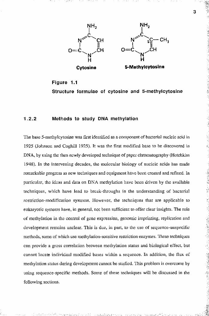
base of higher eukaryotes is 5-methylcytosine (Fig. 1.1), which is formed by the activity
of the enzyme DNA-(cytosine-5) methyltransferase. The DNA of prokaryotes contains
the minor bases N ^-methyladeniiie (6-mA) and N -methylcytosine (4-mC), in addition
to 5-methylcytosine (5-mC), in varying amounts (Doerfler 1983; Barras and Marinus
1989 and references therein). Since 6-mA has been reported in few eukaryotes, and 4-
mC in none, this study will be concerned only with 5-methylcytosine. The term DNA
méthylation will therefore refer exclusively to C5 cytosine méthylation, unless otherwise
stated.
N H , N H ,
^ C H 5C - C H 3
Cytosine 5-Methylcytoslne
Figure 1.1
Structure formulae of cytosine and 5-methylcytosine
1.2 .2 Methods to study DNA méthylation
The base 5-methylcytosine was first identified as a component of bacterial nucleic acid in
1925 (Johnson and Coghill 1925). It was the first modified base to be discovered in
DNA, by using the then newly developed technique of paper chromatography (Hotchkiss
1948). In the intervening decades, the molecular biology of nucleic acids has made
remarkable progress as new techniques and equipment have been created and refined. In
particular, the ideas and data on DNA méthylation have been driven by the available
techniques, which have lead to break-throughs in the understanding of bacterial
restriction-modification systems. However, the techniques that are applicable to
eukaryotic systems have, in general, not been sufficient to offer clear insights. The role
of méthylation in the control of gene expression, genomic imprinting, replication and
development remains unclear. This is due, in part, to the use of sequence-unspecific
methods, some of which use méthylation-sensitive restriction enzymes. These techniques
can provide a gross correlation between méthylation status and biological effect, but
cannot locate individual modified bases within a sequence. In addition, the flux of
méthylation status during development cannot be studied. This problem is overcome by
using sequence-specific methods. Some of these techniques will be discussed in the
following sections.
1.2 .3 Sequence-unspecific methods for studying
5-methyIcytosine distribution in DNA IThe 5-mC content of DNA has been quantified after hydrolysis by chemical or enzymatic
means. Fractionation procedures such as electrophoresis, thin-layer chromatography
(TLC) and high performance liquid chromatography (HPLC) have been used to separate
5-mC after total acid hydrolysis of DNA (Adams and Burdon 1985; Saluz and Jost
1993), or after degradation by enzymatic hydrolysis. Enzymatic cleavage products are
mononucleotides with either 5' phosphates (in the case of digestion with pancreatic
DNasel) or 3' phosphorylated mononucleotides (with micrococcal nuclease). A
modification of nearest-neighbour analysis has allowed the detection of 5-mC (Adams
and Burdon 1985). Nicks were introduced into the DNA using DNasel, the nucleotide 3'
to the nicked base was labelled using [a-^^PjdNTP and the DNA was hydrolysed to 3'
monophosphates which were fractionated by thin-layer chromatography. This analysis
quantifies the relative proportions of méthylation at each of the four CpN dinucleotides
(Gmenbaum et al. 1981; Pollack et al. 1984).
The close relationship between DNA méthylation and DNA restiiction (reviewed
in Noyer-Weidner and Trautner 1993) has made restriction endonucleases important tools
in the analysis of méthylation patterns (Bird and Southern 1978). Several isoschizomeric
restriction endonucleases are available. Isoschizomeric enzymes recognise the same
sequence, but have differential sensitivity to the presence of methylated bases within that
recognition sequence. For example, Hpa II will cleave the unmethylated recognition
sequence CCGG, but is refractory to C'^CGG. The isoschizomer Msp I is insensitive to
the méthylation of the internal cytosine (it will cleave both CCGG and O^CGG) but is
refractory to "^CCGG. Comparison of the digestion patterns with isoschizomeric
enzymes can therefore reveal the méthylation status of genomic DNA (reviewed by
Adams and Burdon 1985; Saluz and Jost 1993). For example, the distribution of CpG
dinucleotides has been estimated in DNA from mouse liver, as well as the proportion of
methylated CpGs (Singer et al. 1979).
Sequence-specific methods
1.3 Distribution of 5-methylcytosine in higher eukaryotes
1.2 .4
Sequencing methodologies can locate all the 5-methyIcytosines on uncloned or genomic
DNA in a sequence- and strand-specific manner (Saluz and Jost 1993; Grigg and Clark
1994). This has provided valuable insights into de novo méthylation during
embryogenesis and subsequent déméthylation during cell differentiation (reviewed in Jost
and Saluz 1993; discussed in Section 1.9). The original method as devised by Church
and Gilbert (1984) involves digestion of total genomic DNA with restriction enzymes
which provides fragments of defined length containing the target sequence. The DNA is
then subject to chemical sequencing reactions (Maxam and Gilbert 1980), and the
cleavage products are then resolved on a sequencing gel and transferred onto a nylon
filter. Southern hybridisation using the appropriate probe reveals the sequencing ladder
of the target sequence. Methylated cytosines are represented as gaps in the ladder as
hydrazine does not react with 5-mC in the C specific reaction. One improvement on the
original procedure has been the linear PCR amplification of the cleavage products with
labelled primers (Saluz and Jost 1989). A new method for genomic sequencing is the
4
bisulphite method (Frommer et al. 1992), in which bisulphite is used to specifically
deaminate cytosine, but not 5-methylcytosine, to uracil. The target sequence is then
amplified by PCR and subject to dideoxy (Sanger) sequencing. Upon sequencing of the
amplified DNA, all the thymine and uracil residues become detectable as thymine and
only 5-mC as cytosine.
!
The percentage of 5-mC in eukaryotic DNA varies over a wide range (reviewed in
Doerfler 1983; Adams and Burdon 1985), and it has been suggested that the genome size
of an organism is an important factor that determines the level of cytosine méthylation
(Antequera and Bird 1993). Higher eukaryotes with large, complex genomes will retain
DNA méthylation as a universal regulatory mechanism, despite selective pressure for it to
a symmetrical sequence (Gruenbaum et al. 1981; Belanger and Hepburn 1990). The CG
1.4 DNA méthylation, mutation and oncogenesis
6
be removed (see Section 1.5). In vertebrates 3-8% of DNA cytosine is methylated,
which represents about 5 x 10' 5-mC residues per diploid nucleus. In higher plants as
many as 30% of the total cytosines are methylated. By contrast, 5-mC is present at low
levels, or is absent, in the DNA of lower eukaryotes with smaller genomes. The DNAs
of the fruit-fly Drosophila, the nematode Caenorhabditis and the yeast Saccharomyces
are exceptional in that they do not contain any detectable 5-methylcytosine (reviewed in
Doerfler 1983). In fungi (Rothnie et al. 1991) or echinodermata (Bird et al. 1979), only
repetitive satellite sequences appear to be methylated. An interesting parallel observation
is that repetitive DNA in mammals is also enriched in 5-mC. For example, major satellite
DNA contains over 40% of all 5-mC in the mouse genome (Miller et al. 1974).
However, DNA méthylation also occurs in the non-repetitive DNA of vertebrates and
higher plants, where it can be a cis -acting element on gene expression.
In the vertebrate genome, almost all 5-mC residues are restricted to the
symmetrical dinucleotide CpG. In mammals 60-70% of all CpGs are methylated
(Antequera and Bird 1993). In higher plants, 5-mC occurs at both CpG dinucleotides and
CpNpG trinucleotides, where N is any nucleotide, so that the méthylation again occurs at
I
and the CNG methyltransferase activities of the pea Pisum sativum have been
fractionated into two distinct species (Pradhan and Adams 1995), but the existence of
CpNpG méthylation in mammalian genomes remains controversial. Genomic sequencing
fails to detect significant levels of CpNpG méthylation, although a nearest-neighbour
analysis has suggested that 5-mC was not located exclusively at CpG dinucleotides
(Woodcock et al, 1987). CNG methyltransferase activity has been observed only by
using the bisulphite sequencing of plasmid DNA that has been transfected and stably
integrated into mammalian cells (Clark et al. 1995).
Cytosine méthylation is inherently unstable and mutagenic, as first demonstrated in
bacteria (Coulondre et al. 1978). This is because 5-methylcytosine can be converted to
I :
1.5 DNA Methyltransferases
DNA is methylated in an early post-replicative step in eukaryotes, with the newly
synthesised strand becoming methylated. 5-methylcytosine is formed by the activity of the
enzyme DNA-(cytosine-5) methyltransferase (MTase) on the 5-carbon of the pyrimidine
7thymine by spontaneous oxidative deamination. A methyl-CpG dinucleotide therefore
mutates into TpG, which gives rise to a T-G mismatch of base-pairs. An enzymatic repair
system that corrects the mismatch in favour of-the original cytosine does exist, but
appears not to be totally efficient (Wiebauer et al. 1993). The failure of the repair
mechanism results in C-G to T-A transition mutations, and 5-methylcytosines represent
mutational hot spots in the genome. It has been calculated that there are about 8
deamination events per day per human genome (Jones et al. 1992). Methylated cytosines
are therefore a mutational load for higher eukaryotes, but this disadvantage appears to be
outweighed by the advantages that méthylation confers on the organism. The primary
advantage may be that méthylation can separate the genome into compartments, each of
which has autonomous control over gene expression (discussed in Section 1.8).II
Over the course of millions of years, selective pressure has tended to delete CpG
sequences from vertebrate genomes. CpGs are therefore comparatively rare and occur at
one-fifth of the expected frequency in bulk vertebrate DNA (Bird 1986). The suppression
of CpGs is matched by a proportional excess of TpG and CpA dinucleotides. In contrast,
GpC dinucleotides are not suppressed and are distributed evenly throughout the genome.
C-G to T-A transition mutations account for 30-40% of all point mutations (Cooper and
Youssoufian 1988) and restriction fragment length polymorphisms (Barker et al. 1984).
In addition, these mutations occur preferentially at CpG dinucleotides. Point mutations in
the p53 tumour suppressor gene have been found in over half of all human tumours,
which suggests that such mutations have a role in oncogenesis (Jones et al. 1992).
Transition mutations of C-G base-pairs at CpG sequences predominate in colon cancer,
and it has been directly demonstrated that some of these CpGs are methylated by genomic
sequencing (Rideout et al. 1990).
Il
a
I
8ring of cytosine. The DNA of prokaryotes contains, in addition to 5-methylcytosine (5-
mC), the minor bases iV 6-methyladenine (6-mA) and A ^-methylcytosine (4-mC) that are
formed by the modification of exocyclic nitrogens. These two classes of exocyclic
methyltransferases are closely related and have been isolated only from prokaryotes. The
third class of MTase, DNA-(cytosine-5) methyltransferase, is found in both eukaryotes
and prokaryotes. The methyl group donor in the catalytic process is S -adenosyl-L-
methionine (SAM; also known as AdoMet in some literature) for all classes of enzyme.
1.5.1 Prokaryotic DNA methyltransferases
In prokaryotes, DNA méthylation is involved in restriction-modification systems, but it
also plays a role in regulating the initiation of DNA replication and the correction of errors
immediately following replication (Modrich 1991; Noyer-Weidner and Trautner 1993). In
restriction-modification systems, the modification by a methyltransferase prevents
endonuclease attack by the cognate restriction enzyme (reviewed in Wilson and Murray
1991). These systems serve as protection against bacteriophage infection. There are three
classes of MTase involved in these systems: types I, II, and HI. Types I and III are multi
subunit enzymes in which the restriction and modification activities reside in different
subunits and, in type I enzymes, yet another subunit is responsible for determining target
sequence specificity. All members of types I and III are 6-mA methyltransferases. The
type II MTases are separate proteins from their cognate restriction endonucleases.
Members of this class can be 5-mC, 4-mC or 6-mA MTases. An unusual prokaryotic
enzyme is Sss I methyltransferase (M.5.S5 I), which was detected in the mycoplasma
Spiroplasma sp. (Nur et al. 1985). It me thy late s CpG dinucleotides and is therefore an
isomethylomer of eukaryotic methyltransferases. (An isomethylomer is analogous to an
isoschizomer in that it is an enzyme from a different source, but with the same
specificity).
The mechanism of methyl transfer has been determined for the type II Hha I
MTase (Klimasauskas et al. 1994). The enzyme catalyses a dramatic alteration in the
structure of the DNA helix at the site of the target cytosine. The C-G base pair is disrupted
and the cytosine is flipped completely out of the double helix, which represents a hitherto
. ..
9unknown mode of protein-DNA interaction (reviewed in Cheng 1995). The cytosine is
then positioned in the active site of the enzyme, where a covalent bond is formed between
carbon-6 of the pyrimidine substrate of cytosine and a cysteine of a conserved Pro-Cys
dipeptide (Wu and Santi 1987), This activates the 5-position of the cytosine, which now
accepts a methyl group from the AdoMet cofactor. This catalytic mechanism is presumed
to be common to all DNA (cytosine-5) methyltransferases, including eukaryotic MTases
(reviewed in Cheng 1995), since the Pro-Cys dipeptide is highly conserved. In addition,
all the known eukaryotic MTases (see Section 1.5.2) have a carboxyl-terminal domain that
is closely related to the much smaller prokaryotic enzymes.
1.5 .2 Eukaryotic methyltransferases
.
Unlike their prokaryotic counterparts, which do not discriminate between unmethylated
and hemi-methylated targets, the eukaryotic DNA (cytosine-5) MTases preferentially
methylate hemi-methylated substrates (Gruenbaum et al. 1982). This reflects the different
biological role that MTases play in eukaryotes. There appears to be only one MTase in
mammals, which can catalyse both maintenance and de novo méthylation (Leonhardt and
Bestor 1993). The maintenance activity predominates because of the preference for hemi-
methylated sites over unmethylated sites. The cDNAs for the murine (Bestor et al. 1988),
human (Yen et al. 1992), Arabidopsis (Finnegan and Dennis 1993) and Pisum sativum i
(Pradhan and Cummings, 1995; personal communication) enzymes have been cloned.
The open reading frames for the vertebrate enzymes indicate a mass for the primary
translation product of 170 kDa, although the apparent mass of MTase in normal somatic
tissues and proliferating cell types is about 190 kDa (Adams 1990b). In higher plants
both CpG and CpNpG sequences are methylated, which has lead to speculation that there
are separate CG and CNG methyltransferases. Two such activities have been fractionated
from the pea Pisum sativum , with masses of 140 kDa for the CG-specific enzyme and
110 kDa for the CNG enzyme (Pradhan and Adams 1995). Both enzymes are believed to
arise by proteolytic processing of the initial translation product.
The primary role of mammalian DNA MTase appears to be the maintenance of■'i:
tissue-specific méthylation patterns after DNA replication. The semi-conservative nature
II,

1 0
ïof DNA replication results in the parental strand remaining methylated while the newly
synthesised daughter strand is unmethylated. In an early post-replicative step (Burdon
and Adams 1980), hemi-methylated CpG sites are recognised and methylated
symmetrically on the daughter strand by MTase. This mechanism would enable
méthylation patterns to be transmitted by clonal inheritance in somatic tissues (Holliday
and Pugh 1975). This speculation was verified by the observation that patterns of in vitro
méthylation on transfected DNA could be maintained for many cell generations (Wigler et
al. 1981). More recent work has used DNA constructs that are integrated into the genome
of transgenic mice. A preimposed pattern of méthylation on the transgenic construct
could be maintained for up to four generations beyond the founder animal (Lettmann et
al. 1991). DNA methyltransferases have been demonstrated to associate specifically with
replication foci in mammalian S phase nuclei (Leonhardt et al. 1992). The clonal
inheritance of méthylation patterns therefore requires the close coordination of replication
and méthylation, as well as the preference of eukaryotic MTase for hemi-methylated
DNA.
New patterns of tissue-specific méthylation are established during embryogenesis
(Monk 1990) by de novo méthylation of previously unmethylated DNA (reviewed by
Adams et al. 1993). It is assumed that maintenance and de novo activities reside on the
same protein, but the possibility remains that there are different specific de novo MTases
expressed during embryogenesis, although there is no evidence for the presence of a
gene family in mammals (Leonhardt and Bestor 1993). De novo méthylation is facilitated
by the high levels of MTase present in the egg and early embryo (Monk et al. 1991). The
role of de novo méthylation during embryogenesis will be discussed in more detail in
Section 1.9. De novo méthylation is not just confined to cells of the germline or the early
embryo, but can occur in somatic cells. Patterns of méthylation can be restored after
treatment with 5-azacytidine (see below), an inducer of déméthylation (Jones 1984).
Many cell lines have de novo méthylation imposed at the promoters of certain tissue-
specific genes, that are not essential under the condition of cell culture (Antequera et al.
1990; Jones et al. 1990). This epimutation (Holliday 1987) leads to the inactivation of
these genes, which could explain the loss of cell type-specific functions in culture. This
topic is discussed in more detail in subsequent sections.
I'L' i _ _ ________
1 1
1.5 .3 Inhibition and disruption of eukaryotic
methyltransferase function and expression
The importance of DNA méthylation in mammals is demonstrated by the use of MTase
inhibitors and, more recently, by the disruption of the MTase gene in knock-out
experiments. Several drugs that inhibit méthylation both in vivo and in vitro have been
identified (reviewed in Adams and Burdon 1985). The most widely used of these are the
base analogues 5-azacytidine (5-azaC) and 5-azadeoxycytidine. On incorporation into
DNA, 5-azacytosine is a powerful inducer of déméthylation (Jones 1984). This is
because MTase binds irreversibly to DNA containing 5-azacytosine, which prevents the
maintenance of méthylation patterns. The demethylating activity of 5-azacytidine has been
correlated with selective gene activation for vertebrate cells in culture (reviewed in Razin
and Cedar 1991). For example, an inactive endogenous retroviral gene in a chicken cell
line is activated when the cells are exposed to 5-azaC (Groudine et al. 1981). Silent genes
on the inactive X chromosome (see Section 1.10) can also be activated by the same
ti’eatment (Mohandas et al. 1981). However, 5-azaC is toxic at concentrations over 1 p-M
and leads to a marked change in the metabolism and development of the exposed
mammalian cells (Jones 1984). In addition, the genes which are activated by 5-azaC in
these studies are in cultmed cells, which themselves have aberrant patterns of méthylation
(Antequera et al. 1990). For example, the MyoD gene contains a CpG island (see
Section 1.6) that is normally unmethylated at all stages of development in tissues. The
gene can be activated by 5-azaC treatment of lOTl/2 cells, but the CpG island is modified
by de novo méthylation (Jones et al. 1990). The significance of experiments that use 5-
azaC must be assessed carefully (Bird 1992), since it cannot be proved that the effects of
this drug are only exerted by inhibiting MTase activity.
The biological importance of méthylation in mammals has been demonstrated by
the technique of gene targeting in murine embryonic stem (ES) cells, which allows
predetermined mutations in any mouse gene for which clones are available. This
technique has been used to construct targeted deletion mutations that disrupt alleles of
mouse DNA methyltransferase in ES cells (Li et al. 1992). The modified ES cells have a
!
Ï
1 2
third of the wild-type 5-mC level and only 5% of the MTase activity in an in vitro assay.
Embryos that are homozygous for the mutation complete gastrulation, but are both
stunted and developmentally retarded, and die at mid-gestation. The inactivation of the
gene is far from complete in the embryo, since 30% of methyltransferase activity remains
in mutant mice, but even this is sufficient to perturb patterns of méthylation during
embryogenesis. An attractive hypothesis to explain this mutant phenotype is that genes
that are normally inactivated by DNA méthylation are inappropriately activated in the
mutant mice. Over-expression of the MTase gene in mouse fibroblasts causes the
tumourgenic transformation of these cells (Wu et al. 1993). This provides further
evidence that the balanced expression of the MTase gene is a prerequisite for normal
embryogenesis.
1.6 CpG islands
As discussed previously, most if not all méthylation in the vertebrate genome occurs at
the CpG dinucleotide, which has brought about the suppression of this dinucleotide as
discussed in Section 1.4. CpGs tend to occur in clusters, called CpG islands, where
suppression is absent. CpG islands were identified by the analysis of the cleavage
patterns of genomic DNA by methyl-sensitive restriction enzymes (Cooper et al. 1983).
These studies showed that CpG islands consisted of about 1-2% of the total genome,
and were enriched in clustered, unmethylated Hpa II sites which gave rise to so-called
Hpa II Tiny Fragments (HTFs). The remaining 98% of the genome is the ocean in which
the islands are landmarks, and this contains dispersed methylated CpG sites. Vertebrate
genomes are therefore divided into compartments of unmethylated islands and methylated
oceans. In summary, the characteristics of CpG islands are as follows: they are about
0.5-3 kb in length, lack CpG suppression, have a high G+C content of >60% and are
invariably unmethylated, with the certain exceptions discussed below (Antequera and
Bird 1993).
All human CpG islands identified so far are associated with genes, and all
housekeeping genes have been found to have a CpG island starting 5 ' of the transcription
1 3
unit and covering one or more exons (reviewed in Cross and Bird 1995). In general,
CpG islands contain multiple binding sites for transcription factors (Somma et al. 1991).
However, some CpG islands have been found to be downstream of the promoter, and
even at the 3’ end of genes (Shemer et al, 1990). Several highly tissue-specific genes
show no evidence of associated CpG islands and remain methylated across the whole
length of the gene in non-expressing cells. Other tissue-specific genes can be associated
with CpG islands that remain unmethylated even in non-expressing tissues. Examples
include the human a-globin gene and the mouse MyoD gene (Antequera and Bird 1993).
A strategy to purify genomic restriction fragments that contain unmethylated CpG islands
has been devised (Cross et al. 1994), which will allow the isolation of genes that are
associated with the islands. The procedure fractionates DNA on the basis of méthylation
status, by using a methylated DNA binding column.
CpG islands are also distinctive at the level of chromatin (Tazi and Bird 1990).
Most noticeable is the presence of nucleosome-free gaps within most CpG islands. The
nucleosomes that are formed at a CpG island tend to contain histones H3 and H4 in
highly acetylated forms, compared to bulk chromatin (refer to Section 1.8.6). In
addition, the levels of histone HI in CpG island chromatin are reduced. These properties
are characteristic of active chromatin (Turner 1991), which reflects the fact that most
CpG islands include the promoter of a housekeeping gene.
Although most CpG islands are unmethylated at all times in all tissues, there are
some exceptions. One exception is the islands of some genes that have their activity
determined by the epigenetic process of genomic imprinting (see Section 1.11). A second
exception is the islands of the inactive X chromosome (Riggs and Pfeifer 1992). As
mentioned previously and discussed in Section 1.10, the X chromosome of eutherian
mammals is inactivated at an early stage of development. The characteristics of the so-
called “facultative” heterochromatin that forms are hypermethylation of the DNA and a
lack of hyperacetylated histones (Grant and Chapman 1988; Jeppesen and Turner 1993).
All the known CpG islands on the inactive X chromosome show a strict correlation
between transcriptional inactivity and DNA méthylation (Tribioli et al. 1992). Thirdly,
CpG islands are de novo methylated in cell-lines (Antequera et al. 1990). As has been
discussed previously, these aberrant patterns of méthylation silence certain tissue-specific
1 4
genes that are not essential under the condition of cell culture. At the chromatin level,
methylated CpG islands are inactive and closed, as defined by an increase in the
resistance to nuclease digestion (Antequera et al. 1990). In some cases, treatment of the
cells with 5-azacytidine can activate these genes and lead to the déméthylation of the CpG
island.
Méthylation of a CpG island may be a factor in causing fragile X syndrome,
which is the most common heritable form of mental retardation in males (Sutherland and
Richards 1995). In affected individuals the expansion of a CCG trinucleotide repeat in an
island, and subsequent méthylation, may trigger a specific break of the X chromosome
near to the methylated CpG island (Oberlé et al. 1991; Ritchie et al. 1994). Three such
fragile sites, FRAXA , FRAXE and FRAXF , have been found on the distal long arm of
the X chromosome (Sutherland and Richards 1995).
I
1.7 DNA méthylation and transcriptional activity
It is now well established that DNA méthylation correlates with inhibition of
transcriptional activity of many tissue-specific and housekeeping genes (reviewed in
Doerfler 1983; Hergersberg 1991; Razin and Cedar 1991; Eden and Cedar 1994). The
same observation has been made both in vivo, following the transfection of methylated
reporter genes into cells, and in studies that have investigated the in vitro transcription of
methylated genes. Two mechanisms have been proposed to mediate gene inactivation in
vivo. In both mechanisms the protrusion of a methyl moiety into the major groove of the
DNA helix can alter protein-DNA interactions.
1.7.1 DNA méthylation prevents binding of
transcription factors
Early studies used gene consU’ucts with regional patterns of méthylation (Busslinger et al.
1983; Keshet et al. 1985). to map the sites at which DNA méthylation can affect
transcription. These sites were shown to be the regulatory regions upstream of the
I
■
reporter genes, which suggests that methyl groups at a binding site can prevent the
initiation of transcription by blocking the binding of transcription factors because the base
sequence is changed (Watt and Molloy 1988). Several transcription factors have been
found that do not bind to methylated target sequences (reviewed by Ehrlich and Ehrlich
1993; Tate and Bird 1993). Examples of known proteins include E2F (Kovesdi et al.
1987), the cAMP responsive element binding protein (CREB; Iguchi-Ariga and
Schaffner 1989), AP-2 (Comb and Goodman 1990), c-Myc/Myn (Prendergast et al.
1991) and NF-kB (Bednarik et al. 1991), all of which recognise sequences that contain
CpG residues. Most of these studies demonstrate a general correlation between DNA
méthylation, the prevention of specific factor binding and transcriptional inhibition, but
their weakness is that the binding studies were performed in vitro . All of the factors that
appear to be affected by DNA méthylation are not specific to one cell type, but
méthylation may be the cw-acting element that gives a degree of cell type-specific gene
expression by a direct mechanism of action. It may also suppress basal transcription of
tissue-specific genes in non-expressing cells, even though the factors that are required for
expression are ubiquitous in many cell types (Bird 1992). This mechanism was first
suggested by studies of the liver-specific tyrosine aminotransferase (TAT) gene in rat.
Becker et al. (1987) used in vivo genomic footprinting techniques to show that part of
the TAT promoter can bind ubiquitous factors in hepatocytes, but is unoccupied in non
expressing tissues where it would be expected to bind the same ubiquitous factors. The
promoter is unmethylated in expressing cells but is methylated in non-expressing cells,
such as fibroblasts. Méthylation is implicated in the establishment of cell type-specific
gene expression because in vitro méthylation of the promoter eliminates the in vitro
footprint. Similar results have been shown for the testis-specific H2B histone gene (Choi
and Chae 1991).
In the case of some factors that are part of the transcriptional machinery,
méthylation does not affect their binding to DNA. Common transcription factors such as
Spl (Holler et al. 1988; Bryans et al. 1992) and CTF (Ben Hattar et al. 1989) are
insensitive to the presence of methyl CpG in their target sites. However, transcription
from the promoters that bind these factors can be inhibited by méthylation (Ben Hattar et
al. 1989; Bryans et al. 1992). Although there is some evidence that, under certain
'■2.
1.7 .2 Specific binding of proteins to methylated DNA
conditions, DNA méthylation may inhibit the processive activity of RNA polymerase
(Kobagashi et al. 1990), this does not appear to be the explanation for the inhibition of
transcription. A second mechanism that mediates the inactivation of genes by méthylation
is described in the next section.
I
In addition to a direct effect on factor binding, méthylation may cause chromatin to adopt
an inactive or closed conformation, which can therefore influence gene accessibility by an
indirect effect. The inactivation is presumed to be mediated by the specific binding of
repressor proteins to methylated DNA which, in turn, have been proposed to influence
the structure of chromatin. Several such factors have been identified in mammals
(reviewed in Ehrlich and Ehrlich 1993; Tate and Bird 1993) and are referred to as
methylated DNA-binding proteins (MDBPs) or methyl CpG-binding proteins (MeCPs).
These proteins include MDBP-1 (Huang et al. 1984), which is a ubiquitous vertebrate
protein that binds in a sequence-specific manner (Wang et al. 1986; Ehrlich and Ehrlich
1993), and MDBP-2 (lost et al. 1991). MDBP-2 requires 30 nucleotides with a single
methylated CpG, to which it binds in a sequence non-specific manner (Zhang et al.
1990). MDBP-2 is a repressor of in vitro transcription of the avian vitellogenin gene
(Pawlak et al. 1991) and, in addition, shares significant sequence homologies with
histone HI (lost and Hofsteenge 1992). MeCPl, previously known as MeCP (Meehan
et al. 1989), has been shown to inactivate methylated genes in vitro and in vivo , and
binds to a minimum of 15 methylated CpGs in >100 bp of DNA (Boyes and Bird 1991;
Boyes and Bird 1992). MeCP2 requires only one methylated CpG for binding and is
associated with pericentromeric heterochromatin (Lewis et al. 1992; Nan et al. 1993). In
one study, MeCP2 does not appear to preferentially inhibit in vitro transcription from
methylated genes (Meehan et al. 1992), but a recent communication fiom the same group
has reported that MeCP2 can inhibit a methylated gene that is transiently expressed in
HeLa cells (Lin et al. 1995). In view of the characteristics of MeCPl and 2, Bird and co
workers have proposed a model whereby MeCPl competes with transcription factors to
i
1 7bind to methylated DNA and guides the DNA to form a heterochromatin structure which,
in turn, is maintained by MeCP2 (Meehan et al. 1992).
Other proteins have been characterised that bind preferentially to methylated I
DNA. The first méthylation-specific, sequence non-specific DNA-binding protein to be-'4::
identified was the plant protein DBP-m (Zhang et al. 1989). A protein that binds to part
of the promoter of a number of major histocompatibility class II genes has been shown to'
have the same DNA-binding specificity as MDBP-1 (Zhang et al. 1993). Although.
méthylation correlates with gene inactivation, a role in activation is possible either by
attracting an activator that binds preferentially to methylated DNA or by repelling a
méthylation-sensitive repressor (Barlow 1993). Factors that fulfil this regulatory role
have yet to be identified, but their involvement can explain the surprising observation that
regions of the active allele of some imprinted genes is methylated (Efstratiadis 1994; see
Section 1.11).
1.8 Chromatin and DNA méthylation
There is growing evidence that DNA méthylation in organisms with large genomes
(vertebrates and higher plants) is accompanied by a change in chromatin conformation
(reviewed in Lewis and Bird 1991; Graessmann and Graessmann 1993). This may be a
means of dividing the genome into compartments (Lewis and Bird 1991). A small,
unmethylated compartment would be accessible to diffusible trans -acting factors whereas
a large, methylated compartment would be associated with condensed chromatin and be-
transcriptionally inert. Compartmentalisation can reduce the time required for a trans-
acting factor to locate a target site in a large genome.
1.8.1 Active and inactive chromatin
Chromatin can exist in two general conformations that affect the degree of accessibility of
genes. The open conformation is transcriptionally active in contrast to chromatin in the
closed conformation, which is transcriptionally inert (reviewed in Gross and Garrard
1 8
1988). It is thought that the open chromatin conformation is a prerequisite for the
initiation of transcription, because genes are the most accessible to trans -acting factors in
this structure (Adams and Workman 1993). In contrast, closed chromatin forms large
distinctive segments on a chromosome (Pardue and Hennig 1990). These distinctive
regions are called heterochromatin, and are characterised by being transcriptionally
inactive, highly condensed and replicated late in S-phase. The paradigm of this structure
is the inactive X chromosome of eutherian mammals, which involves the formation of
facultative heterochromatin on one copy of the chromosome. This condenses at
interphase to form the cytologically distinctive Barr body (Grant and Chapman 1988).
The inactive X chromosome is almost completely silent and remains heterochromatic
throughout metaphase, which is a characteristic of regions of the genome that play a
structural rather than a coding role. Regions that are adjacent to the telomere and the
centromere are transcriptionally inert and are associated with so-called constitutive
heterochromatin. It remains unknown if the mechanisms for the formation of facultative
and constitutive heterochromatin are the same.
The heterogeneous structure of nuclear chromatin can be characterised at the
molecular level. Differential sensitivity to a nuclease, such as DNase I, has been used to
classify the genome into compartments (reviewed in Gross and Garrard 1988; Wolffe
1992). Regions of the genome that contain active genes show a general sensitivity to
nucleases, whereas inactive chromatin is comparatively resistant. The structural changes
that underlie these alterations in the conformation of chromatin remain unknown.
It is generally believed that chromatin in a simple closed conformation can
inactivate genes by limiting the accessibility of diffusible trans -acting factors, such as
transcription factors. However, in recent years it has been shown that the trans -acting
factors can themselves alter the structure of chromatin, and that chromatin can play an
additional active role in the regulation of gene expression. The generalised repression of
gene activity by closed chromatin can be relieved by trans -acting factors, such as
enhancer- and promoter-binding factors (Winston and Carlson 1992; Paraiijape et al.
1994) which open the chromatin structure. This phenomenon is referred to as “anti
repression”, and is distinct from the “true activation” of a gene by specific transcription
factors which increase the rate of transcription. It is now obvious that the regulation of
1 9
eukaryotic gene expression can only be understood in the context of chromatin, since
stretches of naked DNA are probably never found in vivo.
1 .8 .2 Méthylation alters the conformation of chromatin
Razin and Cedar (1977) were the first to demonstrate that methylated DNA is in a specific
chromatin structure. Mouse L cell nuclei were digested with micrococcal nuclease, and
analysis showed that 5-methylcytosine in chromatin was relatively resistant to
degradation. After extensive digestion, only 50% of the DNA was solubilised and over
70% of the total 5-mC content of the DNA remained in the unsolubilised fraction. A
subsequent study demonstrated that the DNA solubilised at early times of digestion had a
reduced 5-mC content, which indicates that linker DNA is hypomethylated compared to
nucleosomal core DNA (Solage and Cedar 1978). Both of these studies are weakened by
the observation that 5-mC is preferentially associated with regions that are resistant to
digestion by staphlococcal nuclease not only in chi'omatin from human fibroblast nuclei,
but also in purified naked fibroblast DNA (Barr et al. 1985). The authors suggest that
methylated naked DNA has an intrinsic resistance to digestion by this nuclease, which is
of a sufficient magnitude to account for the nuclease resistance of chromatin that contains
methylated DNA. This point is discussed further in Chapter 6, which describes the
resistance of naked DNA and chromatin to the nuclease Msp I.
In the mammalian genome, the regions that are enriched in 5-mC are generally
heterochromatic, transcriptionally inert and contain repetitive DNA. Pericentromeric
heterochromatin, which contains major satellite DNA in mouse, is preferentially
visualised by using an antibody raised against 5-mC (Miller et al. 1974). In addition, a
number of studies have shown that methylated CpG islands on the inactive X
chromosome are insensitive to Msp I in intact nuclei (Wolf and Migeon 1985; Hansen et
al. 1988; Antequera et al. 1989). In contrast, CpG islands on the active X chromosome
are comparatively nuclease sensitive (Kerem et al. 1983).
The first direct evidence that méthylation has an effect on chromatin conformation
came from transfection studies of methylated constructs that were integrated into the
genome of L cells (Keshet et al. 1986). All unmethylated DNA inserted into the mouse
... .
correlated with transcriptional inactivation (Levine et al. 1991).
1.8 .3 Méthylation, chromatin structure and the regulation of
gene expression
2 0genome was found to be in a DNase I-sensitive conformation, whereas methylated
constructs were nuclease resistant, which is characteristic for closed inactive chromatin.
The accessibility of sites in nuclei for CpG enzymes, such as Msp lo xT th I, depends on
their méthylation status (Antequera et al. 1989; Levine et al. 1991). After the treatment of;
bulk chromatin with nuclease, under conditions of complete digestion, it was shown that I
Msp lo iT th I were preferentially blocked from cutting methylated sites at specific;!
locations. Unmethylated CpG sites, and restriction sites that did not contain a CpG, were
comparatively nuclease sensitive. The formation of closed, nuclease-resistant chromatin
■■.‘I
I
Two studies have used reporter gene constructs, that contained either the y-globin gene
(Busslinger et al. 1983) or the p-globin gene (Yisraeli et al. 1988), to show that in vitro
méthylation of the promoter regions abolishes expression of the gene after transfection
into cells. In the case of the y-globin gene, the inhibition is not dependant on the
méthylation of specific sites in the promoter, since the presence of three 5-mC residues in
different regions of the promoter was sufficient to inhibit transcription (Murray and
Grosveld 1987). Reporter gene constructs have also been used in microinjection
experiments, in which the construct is methylated in vitro , and after transfer into
recipient cells is found to be inactive (reviewed in Doerfler 1983). Two examples from
more recent literature describe the transcription of methylated constructs that contained
either a tRNA gene (Besser et al. 1990) or SV40 late genes (Gotz et al. 1990). After the |
microinjection of the constructs into Xenopus oocytes, the transcription of genes was f
inhibited. These studies provide only circumstantial evidence that chromatin is involved
in the mechanism of gene inactivation, since the formation of minichromosomes on
construct DNA was implied but not proven.
Further evidence has been provided in a study by Buschausen et al. (1987), in
which a construct containing the herpes simplex virus thymidine kinase (TK) gene was
microinjected into TK” tissue culture cells. Expression of the methylated reporter gene,
2 1
as assayed by the number of TK+ cells, dropped sharply only 8 hours after
microinjection. This time coincided with the assembly of the DNA into chromatin. In
contrast, mock-methylated minichromosomes remained active 72 hours after injection.
When methylated constructs were assembled into chromatin in vitro prior to injection,
gene inactivation was immediate. An extension of this study has shown that a
hemimethylated TK reporter gene construct was sufficient to block expression
(Deobagkar et al. 1990). However, the authors did not investigate the méthylation status
of the construct after transfection, but it can be presumed that the construct became fully
methylated due to maintenance méthylation. DNA méthylation of a reporter gene
construct has been shown to inhibit the activity of the murine a 1(1) collagen promoter by
an indirect mechanism (Rhodes et al. 1994), after the transfection of the construct into
tissue culture cells. An indirect mechanism requires both chromatin and methyl-CpG
binding proteins to cooperatively mediate the inhibitory effect of méthylation. The degree
of transcriptional inactivation may be dependent on the density of CpG méthylation, as
shown for an episomal system that can be stably maintained in human cells. Plasmids
containing the Epstein-Barr virus replication origin were methylated to four different
levels of density, and transcriptional inactivation of minichromosomes was shown to be
graded to an increase in méthylation density (Hsieh 1994).
1.8 .4 The effect of DNA méthylation on chromatin structure
Although it is well established that méthylation alters the confonuation of chromatin, it is
a much more complex problem to identify the proteins that cause the structural changes at
the molecular level. This problem also brings us back to the more fundamental question
of what constitutes the difference between active and inactive chromatin. So far, not
many reports have dealt with the special structure of chromatin assembled on methylated
DNA. In an early study it was reported that a methylated CpG polymer has a two-fold
greater affinity for core histones than the unmethylated polymer, but that the core
particles deposited on the substrate DNA were identical on both types of polymer
(Felsenfeld et al. 1982). The study by Buschausen et al. (1987), which is described
above, demonstrates an obvious difference in function between the chromatin of:
2 2unmethylated and methylated construct DNA. However, a structural difference was not
observed, as tested by electron microscopy and micrococcal nuclease digestion
(Buschausen et al. 1987; Graessmann and Graessmann 1993). In a recent important
study core histone octamers and tetramers were assembled in vitro on unmethylated or
methylated templates that contained human Alu repetitive elements (Englander et al.
1993). Alu elements possess internal RNA pol III promoters that are strongly transcribed
in vitro, but are almost silent in somatic cells. The authors suggest that chromatin plays a
role in silencing Alu elements, since the assembly of nucleosomes using histone oc tamers
results in the equal inactivation of in vitro transcription for both unmethylated and
methylated templates. The unexpected result with assembly of the H3/H4 tetramer was
that the in vitro transcription of the methylated template was preferentially inhibited. The
significance of these results may be that transcription activators compete with the H2A
and H2B dimers for association with the histone H3/H4 tetramer. If the DNA that is
bound to the tetramer is methylated then this may favour the completion of the histone
octamer, with repression of transcription as a consequence.
CpG islands usually contain the promoters of housekeeping genes. Since these
genes are by definition transcriptionally active, the chromatin at island regions should be
in the open conformation. This has been shown to be the case with two X-linked genes,
those for hypoxanthine phosphoribosyltransferase (HPRT ) and glucose 6-phosphate
dehydrogenase (G6PD), whose CpG islands are accessible to nucleases such as Msp I,
DNase I and SI nuclease on the active X chromosome (Kerem et al. 1983). This
observation has been used to isolate short oligonucleosomes derived from CpG island
chromatin (Tazi and Bird 1990). Nuclei were digested with CpG enzymes, which
released a highly enriched fraction of transcriptionally active chromatin, but left bulk
chromatin intact. The analysis of the protein composition of these two fractions showed
significant differences between CpG island chromatin and bulk chromatin. The island
chromatin contained hyperacetylated histones H3 and H4 (see Section 1.8.6), compared
to bulk chromatin, which is now known to be a characteristic of transcriptionally active
chromatin (reviewed in Turner 1991; Turner 1993). Nucleosomes that were obtained
from CpG island chromatin were deficient in histone HI (approximately 10% of the level
observed in bulk chromatin), and one region of the island chromatin contained a DNase I
■ 23
.hiL
2 3
hypersensitive site, which coincided with the absence of a nucleosome. These features
are all characteristic of active chromatin (Kamakaka and Thomas 1990; Hebbes et al.
1994; reviewed in Wolffe 1992).
1.8 .5 The effect of histone HI on gene expression
1
The special so-called “linker histone” in vertebrates, typically histone H I, may be the
protein that can account for differences between chromatin of methylated and
unmethylated DNA. Histone HI is implicated in the formation of supranucleosomal
structures, such as the 30 nm chromatin fibre (Thoma et al. 1979). Several models have
been proposed for the structure of the 30 nm chromatin fibre, based on electron
microscopy studies, but the strongest evidence supports the original solenoidal model
(Felsenfeld and McGhee 1986) . This is in the form of neutron scattering data, that
shows histone HI to be located in the interior of the 30 nm chromatin fibre (Graziano et
al. 1994). Supranucleosomal structures would be refractory to nuclease attack, which
may account for the nuclease resistance of transcriptionally inert chromatin. Treatment of
intact cells and isolated nuclei with the oligopeptide distamycin results in a decrease in
nuclease resistance of chromatin (Kas et al. 1993). The increased accessibility is thought
to result from the unfolding of the chromatin fibre, because distamycin binds to AT-rich
sequences in the minor groove of DNA which would displace histone HI.
In an early study by Ball et al. (1983) it was demonstrated that at least 80% of 5-
methylcytosine is localised in nucleosomes which contain histone FIl, which is a
characteristic of inactive chromatin (Weintraub 1984). Several studies have provided
direct evidence that histone H I mediates stable and tissue-specific gene inactivation in
chromatin (reviewed in Zlatanova 1990), for genes transcribed by either RNA
polymerase II (pol II) or RNA polymerase III (pol III). Two studies have used the
developmentaily-regulated oocyte 5S rRNA genes of Xenopus laevis, which are inactive
in somatic tissues, as a model system for pol III transcription (Schlissel and Brown
1984; Wolffe 1989). In both cases, in vitro transcription from sperm chromatin that
contains the oocyte-specific 5S rRNA genes could be repressed by the addition of histone
H I. Xenopus sperm chromatin is naturally deficient in histone H I, whereas the removal
2 4
of histone HI from somatic cell chromatin, such that regular nucleosomal spacing was
maintained, brought about the activation of the oocyte 5S rRNA genes (Schlissel and
Brown 1984). These studies implicate changes of chromatin structure in the
developmental control of gene expression during embryogenesis. A recent study has
shown that histone HI mediated the selective repression of the oocyte 5S rRNA genes in
vivo , but did not affect the expression of somatic 5S rRNA genes or endogenous tRNA
genes (Bouvet et al. 1994; see Section 1.8.7 ).
The activity of the Xenopus somatic 5S rRNA gene has also been studied in an
in vitro system. Chromatin with regularly spaced nucleosomes was assembled on the
DNA in vitro by using sl Xenopus oocyte extract (Shimamura et al. 1988; Gotîesfeld
1989) and subsequent addition of histone H I brought about transcriptional repression
(Shimamura et al. 1989). Histone HI could be removed from the chromatin without the
disruption of the nucleosomal array by using an ion-exchange resin. This treatment
restored the activity of the 5S rRNA gene. A second type of in vitro system has used
purified core histone octamers to reconstitute chromatin on DNA in the absence or
presence of histone H I (Laybourn and Kadonaga 1991). The analysis of in vitro
transcription from a pol II reporter gene again demonstrated gene repression by histone
H I, which could be counteracted by the “anti-repressor” function of transcriptional,
activators. The accessibility of transcription factors to DNA could also be affected by the
mobility of nucleosomes (Meersseman et al. 1992). In this model, transcriptionally active
chromatin is repressed by linker histones that restrict the mobility of nucleosomes
(Pennings et al. 1994). An in vitro system, that used only octamers as a model for
chromatin, has shown that linker histones directly inhibit both nucleosome mobility and
transcription (Ura et al. 1995).
The distribution of histone H I in the nuclei of certain tissues has shown that most
genes in transcriptionally active chromatin are depleted of histone H I, compared to genes
that are inactive in this tissue (Kamakaka and Thomas 1990). A similar series of
experiments have used the mouse mammary tumour virus (MMTV) promoter, that is
present on a construct that has integrated stably into the genome of tissue culture cells.
UV-crosslinking and immunoadsorption studies have shown that the transcriptionally-
f
2 5
active promoter is depleted of histone HI (Bresnick et al. 1991). The inactive promoter
binds histone H I, but treatment of the cells with glucocorticoid results in the induction of
gene expression and depletion of histone H I at the promoter. This important result,
obtained in a system that is relevant physiologically, suggests that histone H I has a
fundamental role in the control of gene expression. However, it must be borne in mind
that transcriptionally active chromatin is not completely depleted of histone HI (reviewed
in Garrard 1991), so the correlation between gene repression and the presence of histone
HI in chromatin is rather general. Kamakaka and Thomas (1990) suggest that the change
from inactive to active genes involves an increase in the stoichiometry of histone H I in
chromatin.
Histone H I has been demonstrated to be a type of methylated-DNA binding
protein (Levine et al. 1993), at least under the in vitro conditions that were used in this
study. This supports the data of Jost and Hofsteenge (1992), who reported that the
methylated-DNA binding protein MDBP-2 shares sequence homologies with histone HI,
Several studies have used histone H I bound to DNA in orderly complexes as a model for
inactive chromatin (reviewed in Paranjape et al. 1994). The assumptions of this model
will not be discussed at this stage, since it is a major aspect of the work presented in
Chapter 5. A full discussion can be found in Section 5.4. When histone H I is associated
with DNA in vitro , digestion with M spl, but not with other restriction enzymes is
specifically inhibited on the methylated DNA substrate (Higurashi and Cole 1991). This
difference could be explained if the binding of histone H I to methylated DNA changed
the conformation of the complex, thus rendering the DNA more resistant to digestion by
Msp I. This study by Higurashi and Cole (1991) did not demonstrate any preferential
binding of histone H I to methylated DNA, an observation that was repeated in a more
recent study (Nightingale and Wolffe 1995). In contrast, a third study has shown that
histone HI can bind to methylated DNA with approximately two-fold greater affinity than
to unmethylated DNA (Levine et al. 1993), which supports the role of histone HI as an
MDBP (Jost and Hofsteenge 1992). In addition, Levine et al. (1993) demonstrated that
histone H I not only inhibits in vitro transcription from unmethylated DNA
(Jerzmanowski and Cole 1990; Croston et al. 1991), but that it inhibits transcription from'
methylated templates with even more efficiency. This observation has been extended with■' .!■
my own work (Johnson et al. 1995), which is presented in Chapter 5.
2 6
1.8 .6 Histone modifications and changes in
chromatin structure
'i:
Although it is well established that the nucleosome is the fundamental structural unit of
eukaryotic chromatin, it is still unclear why the histone proteins, that are the highly
conserved structural components of the nucleosome, exist in so many different forms.
Some of this variation arises because each of the five classes of histone protein (HI,
H2A, H2B, H3 and H4) exists as sub-types or isoforms that differ in a few amino acid
residues. Further complexity arises from various post-translational modifications
(reviewed in Wu et al. 1984; Wolffe 1992) which include acétylation, phosphorylation,
ADP-ribosylation and ubiquitination of the parent histone molecule. Electrophoresis
techniques have been used to identify and characterise these histone sub-types and
modified forms (Lennox and Cohen 1989). Reversible modifications of histones at the
molecular level are presumed to be the basis for the changes in chromatin structure, that
could lead to the condensation and decondensation of chromosomes during the cell-cycle.
For example, five specialised histone H2A variants and at least six variants of histone HI
(see Section 1.8.7) are present during sea urchin embryogenesis (Wolffe 1991). Each
variant has a different expression profile for the cleavage, blastula and gastrula stages of
embryogenesis (reviewed in Khochbin and Wolffe 1994). Core histones and linker
histones are also subject to reversible post-translational modifications that correlate with
stages of the cell-cycle.
Histone acétylation is the only post-translational modification that occurs
consistently in transcriptionally active chromatin, compared to bulk chromatin that is
transcriptionally inert (reviewed in Turner 1991; Turner 1993), and is the most
extensively studied histone modification. Acétylation of the core histones is present in all
the animal and plant species that have been studied (Csordas 1990), and occurs at
specific lysine residues, all of which occur in the amino-terminal domains of the core
histones. High-resolution proton magnetic resonance has shown that the amino-terminal
domains of core histones are either fully mobile or interact weakly with core DNA in the
nucleosome (Cary et al. 1978). A chemical modification procedure has also been used to
I
2 7investigate core histone-DNA interactions (Lambert and Thomas 1986; Thomas 1989),
and a later study from the same group has shown that the amino-terminal tail of histone
H2A does not bind to core DNA in sea urchin, sperm chromatin (Hill and Thomas 1990).
Mobile amino-terminal “tails” of core histones could therefore interact with linker DNA,
linker histones such as H I or other non-histone proteins to mediate the formation of
supranucleosomal structures. This model is supported by the observation that amino-
terminal tails are required for the in vitro assembly of a solenoidal structure (Allan et al.
1982).
Each acetate group added to a lysine group reduces the net positive charge of the
histone by one. This is presumed to reduce the electrostatic attraction between the basic
core histones and the acid-phosphate backbone of the DNA, so that the core DNA is
partially unwound or loosened around the core octamer (reviewed in Turner 1991). An
electrostatic mechanism could therefore explain the open conformation and
hyperacetylated status of core histones in transcriptionally active chromatin, because
transcription factors are allowed access to regulatory elements in chromatin (Lee et al.
1993). Although a description of this mechanism at the molecular level is far from
complete, the correlation between acétylation and transcriptional activity is well
established. For example, experiments using Hg-agarose affinity chromatography have
shown that the DNA of transcriptionally active genes was copurified with hyperacetylated
core histones (Allegra et al. 1987). Tazi and Bird (1990) showed that chromatin fractions
enriched in sequences characteristic of CpG islands contained hyperacetylated histones
(see Section 1.8.4). Immunoprécipitation of mononucleosomes with an antibody::
recognising acetylated histones has shown the modification to be present at the p-globin
locus in chicken embryo erythrocytes (Hebbes et al. 1994). The region of histone
acétylation in this locus corresponded to a generalised DNase I sensitivity of the P-globin
chromosomal domain. Recently it has been confirmed that inactive chromatin does not
carry acetylated histone H4. (Jeppesen and Turner 1993) have shown that the human
female inactive X chromosome (see Section 1.10) fails to stain with antibodies for the
acetylated isoforms of histone H4, in contrast to all the other chromosomes. Autosomes
were strongly labelled at the R-bands, which are the regions enriched in coding DNA,
but were unlabelled at those regions adjacent to centromeres. This provides strong
2 8
evidence that a lack of histone acétylation may define both facultative and constitutive
heterochromatin.
A second well-studied post-translational modification of chromatin is the
reversible phosphorylation of linker histones, including histone H I (reviewed in Wolffe
1992). Phosphorylation levels of histone H I vary during the mitotic cell cycle of
eukaryotes, with the levels increasing during S phase and mitosis, and peaking during
metaphase when chromosomes are most highly condensed. Histone HI is modified by a
growth-associated histone HI kinase, which is homologous to p34/cdc2 kinase. This
kinase regulates many other nuclear processes that are driven by phosphorylation during
the cell cycle (reviewed in Draetta 1990). Phosphorylation of serine residues in the highly
basic amino-terminal domain of histone H I reduces the net positive charge of the
molecule. This would be expected to weaken the electrostatic interactions between linker
histone and the DNA, in much the same way as core histone acétylation reduces histone-
DNA interactions. Changes in the level of phosphorylation of sea urchin sperm-specific
histone HI have been studied during spermatogenesis and fertilisation (Hill et al. 1990),
which are associated with changes in the condensation of chromatin in the nucleus.
Phosphorylation of six sites in the C -terminal tail of histone H I could abolish
binding of the tail to DNA in both histone HI-DNA complexes and in sea urchin
spermatid chromatin (Hill et al. 1991). This is expected from the weakened electrostatic
attraction between a phosphorylated linker histone and the DNA. In view of this, it is a
paradox that the phosphorylated chromatin in the spermatid becomes condensed and inert
at this stage of spermatogenesis. The functional significance of linker histone
phosphorylation remains unresolved.
1.8 .7 Histone H1 variants
Linker histones exist in chromatin as a family of sub-types or isoforms, consisting of
histones HI, H r , H it and H5, each of which has a set of variants (Lennox 1984; Wu et
al. 1984). The diversity arises because the number of variants and the amount of each
variant can change during cell differentiation. They also differ from tissue to tissue and
for a given tissue even differ from one species to another (reviewed in Khochbin and
2 9
Wolffe 1994). Histone H it is a testis-specific linker histone (Wu et al. 1984) and Hl°
tends to occur in quiescent cells such as neurones (Castiglia et al. 1993), but the
functional significance of these variants is not known. Histone H5 is discussed in more
detail below. The differences between linker histones are located in the highly basic
amino- and carboxyl-terminal “tail” domains of linker histones, rather than in the central
globular domain. The sequence of this region is highly conserved because it binds to the
dyad of DNA in the nucleosome. This has lead to the proposal that the developmental
regulation of linker histones can mediate changes in chromatin structure and
transcriptional activity during gametogenesis, embryogenesis and within somatic cells
(Bouvet et al. 1994; Pruss et al. 1995).
Histone HI is the normal isoform found in somatic cells, but H5 is a special form
that is expressed during the development of chicken erythroid cells. During the final
stages of chicken erythrocyte development, the nucleus is condensed into inactive
heterochromatin due in part to the appearance of histone H5. Histone H5 can reconstitute
a supranucleosomal structure in vitro when it is added to chromatin that is depleted in
linker histones (Graziano et al. 1988), but histone H5 both binds more tightly and
confers greater stability to chromatin than does histone HI (Sun et al. 1990). Both of
these linker histones have a non-random distribution in chicken erythrocytes
(Muyldermans et al. 1994). Telomeric nucleosomes are preferentially associated with
histone HI in this system, compared to bulk chromatin in which the most abundant sub-
type is histone H5. Another study has shown that the chromatin associated with the p-
globin gene in chicken erythrocytes is transcriptionally active and depleted in histones HI
and H5 (Postnikov et al. 1991), but that inactive chromatin is relatively enriched in these
linker histones.
The changes of linker histone type in chromatin have also been studied during
embryogenesis in two systems. In the first, the expression of the linker histone H i p
gene during sea urchin embryogenesis occurs in the late blastula-stage embryo, and
subsequently in adult tissues (Lai and Childs 1988). The second system is the
developmental regulation of chromatin-associated proteins during the early development
of Xenopus laevis . Histone H I is not found in Xenopus oocytes, sperm or eggs and the
role of linker histone is taken by the B4 protein. B4 protein is replaced by histone HI
i
i.
à.
3 0before the end of gastrulation (Dimitrov et al. 1993). Bouvet et al. (1994) have shown
that Xenopus 5S rRNA gene transcription in vivo can be repressed by raising the level
of histone HI during gastrulation (see Section 1.8.5), after micro-injection of in vitro
-synthesised mRNA into eggs. The over-expression of histone HI specifically repressed
oocyte 5S rRNA gene transcription, but did not affect the expression of somatic 5S
rRNA genes or endogenous tRNA genes. Specific repression was mediated by the
association of the Xenopus histone H lC variant with chromatin, despite adequate levels
of the transcription factor TFIIIA which is* required for the transcription of 5S rRNA
genes. By contrast, the HIA variant did not appear to cause any preferential repression of
the oocyte genes compared to the somatic genes.
The somatic variants of histone H I have been isolated and characterised by:
reverse-phase HPLC (Cole 1989; Santoro et al. 1995) and the mouse variants consist of
H la , H lb , H lc , H id and H ie (Lennox 1984). The number and relative amounts of
these variants differ in various tissues and species, as well as during cell differentiation
(reviewed in Khochbin and Wolffe 1994). The expression patterns of the variants is
different in mouse lung carcinoma tissue, compared to normal lung tissue (Giancotti et al.
1993), and the relative proportions of histone H I variants are changed during the
differentiation of murine erythroleukaemic cells (Helliger et al, 1992). The variants may !
play different roles in the organisation of chromatin structure, with a non-random
distribution of particular forms (see the discussion on histone H5, above). For example,
one variant in larval tissue of the midge Chironomus thummi is localised in specific
regions of polytene interphase chromosomes, whereas other variants have a uniform
distribution (Schulze et al. 1993). A recent study has suggested that the histone H le-c
variants can specifically inhibit the in vitro méthylation of a double-stranded DNA
substrate (Santoro et al. 1995). A similar inhibition of the in vitro méthylation of linker
DNA is observed on the addition of histone H I to HI-depleted oligonucleosomes (Caiafa
et al. 1995). The addition of histone H I correlates with the condensation of the
oligonucleosomes, as revealed by distortions in the circular dichroism spectra of the
particles (D’Erme et al. 1993). Caiafa et al. (1995) suggest that the specific H le -c
variants could maintain linker DNA in a hypomethylated form. This may be important in
preserving the unmethylated state of CpG islands, since H le -c are the only variants that
are capable of binding to CpG-rich DNA (Santoro et al. 1995).
i
3 1
1.9 DNA méthylation during development and cell
differentiation
New patterns of tissue-specific méthylation are established during embryogenesis by de
novo méthylation (reviewed in Razin and Cedar 1993). This is because the patterns of
méthylation derived from the gametes are erased in the morula and early blastula, and
need to be re-established during the implantation of the embryo (Kafri et al. 1992). Adult
levels of 5-mC are attained only after the completion of gastrulation (Monk 1990). The
wave of de novo méthylation during implantation remodels the genome into separate
methylated and unmethylated compartments. Unmethylated regions of the genome
contain CpG islands, which are associated with active and housekeeping genes, even in
tissues where the gene is not expressed. In exceptional cases, CpG islands are
methylated (see Section 1.6), but even in these cases the CpG islands appear to be
unmethylated in the germ line (Brandeis et al. 1993). This is, of course, expected since
méthylation in a CpG island would lead to deamination and the erosion of the island.
Méthylation patterns of particular human genes, in lymphocytes and other blood cells, are
often indistinguishable amongst different individuals even of different genetic
backgrounds (Behn-KTappa et al. 1991). In addition, this pattern does not change when
the lymphocytes are stimulated to divide by cytokines. These highly cell type-specific
patterns of de novo méthylation cannot be imposed just by the specificity of MTase for
CG sequences, and must involve other cis -acting regulatory elements. Another example
is the allele-specific méthylation of one X chromosome of eutherian mammals.
Déméthylation of modified CpG residues appears to be a necessary stage of early
embryogenesis. The biological significance of déméthylation is unknown, but
demodification is a mechanism for removing differences in specific gene méthylation
patterns that emerge from the male and female germ lines. Monk et al. (1987)
demonstrated that extensive déméthylation of the genome takes place in the early mouse
embryo, between the 8-cell and blastula stage. This Is paralleled by a decrease in
methylase activity (Monk et al. 1991). By the blastocyst stage all inherited patterns of
méthylation are lost, which results in hypomethylation of both extra-embryonic and
Ii
3 2embryonic tissues (Kafri et al. 1992). This study also showed that de novo méthylation
that occurs during implantation lead to the remodification of non-island DNA, whereas
CpG islands remain unmethylated.
An important development in this field has been the discovery that the presence of
Spl sites in close proximity to CpG islands protects these islands from de novo
méthylation in vivo (Brandeis et al. 1994; Macleod et al. 1994). A similar result has been
seen for the human X-linked HPRT gene, with the inactive allele completely methylated
except at putative transcription factor binding sites (Hornstra and Yang 1994; see Section
1.10). This evidence suggests that steric hindrance by chromatin-associated factors,
including transcription factors, may regulate de novo méthylation in a tissue and allele-
specific manner (Adams et al. 1993). Protein factors may therefore prevent the
méthylation of CpG islands by blocking the methyltransferase. However, this model
cannot explain how even inactive genes are associated with unmethylated CpG islands. A
previous study has shown that an in vitro methylated transgene, introduced into a
fertilised mouse oocyte, can be specifically demethylated at a CpG island (Frank et al.
1991), and that the CpG island was protected from the de novo méthylation of flanking
regions.
Transfection experiments in vitro have suggested that déméthylation is an active
process (Frank et al. 1991), that is specific for islands. Findings in other systems are
consistent with the view that déméthylation is an active process (Razin et al. 1986;
Paroush et al. 1990; Shemer et al. 1991). Nuclear extracts of chicken embryos can
promote the active déméthylation of DNA by an excision repair mechanism that is
specific for 5-methyldeoxycytidine (Jost 1993). Excision is catalysed by a 5-mCpG
endonuclease, which has been purified, and is found in developing mouse and chicken
embryos as well as differentiating mouse myoblasts (Jost and Jost 1994). A 5-mC
glycosylase activity, that has been detected in crude HeLa cell extracts, may also
demethylate DNA by an active, enzymatic process (Vairapandi and Duker 1993).
Tissue-specific and gene-specific méthylation patterns are established in late
embryonic development. The relevant genes specific for the cell type in the differentiated
tissue are demethylated, and this appeal's to be an integral part of the activation process.
(Yisraeli et al. 1986) demonstrated that an in vitro methylated a-actin construct
J
3 3
undergoes déméthylation and activation in myoblasts, but not in fibroblasts. The muscle
like cells therefore have the ability to recognise and activate the tissue-specific gene by
déméthylation. The déméthylation appears to be dependent on cis -acting sequences and
is independent of DNA replication (Paroush et al. 1990), that indicates that it is not a
passive mechanism in which DNA is replicated in the absence of maintenance
méthylation. In conclusion, it is clear that vertebrate méthylation patterns are dynamic and
subject to genetic and developmental control.
1,10 The inactive X chromosome
The X chromosome contains 5% of the genes of the haploid mammalian genome,
comprising of several thousand genes. Dosage equivalence for these genes between XY
males and XX females is achieved by the random inactivation of one of the X
chromosomes in each somatic cell of the female (reviewed in Grant and Chapman 1988;
Rastan 1994). The silencing of transcription is achieved by the formation of facultative
heterochromatin, which becomes highly condensed during interphase and is replicated
late in S-phase. In metaphase spreads of female chromosomes the inactive X can be
distinguished as the densely staining Barr body (Grant and Chapman 1988). In addition,
the CpG islands of some genes on the inactive X are de novo methylated (Antequera and
Bird 1993), and there is a decreased level of histone H4 acétylation (Jeppesen and Turner
1993). The latter observation has led to the proposal that histone H4 hyperacetylation
defines active chromatin, since it is absent in both constitutive and facultative
heterochromatin (see Section 1.8.6).
Primordial germ cells and oogonia also have one inactive X, but this becomes
functional during early embryogenesis in a unique process of reactivation. Both X
chromosomes are therefore active in oocytes and in cleavage-stage female embryos, but
one X is again inactivated in somatic cells. Once X inactivation has occurred, it is stable
and heritable in all subsequent cell divisions. The inactivation process is thought to occur
in three stages: initiation of inactivation, the spreading of inactivation along the length of
the chromosome and the maintenance of the inactive state of the chromosome.
3 4
A role for DNA méthylation has been proposed for the maintenance of X
chromosome inactivation, since méthylation does not appear to be the primary
mechanism that initiates X inactivation (Lyon 1993). Several studies have shown that the
formation of facultative heterochromatin and transcriptional silencing precede de novo
méthylation of genes on the inactive X by several days (reviewed in Riggs and Pfeifer
1992). For example, transcriptional silencing of the mouse X-linked hprt gene takes
place some days before méthylation of the CpG island (Lock et al. 1987). Earlier studies
have provided a general correlation between de novo méthylation of CpG islands on the
inactive X with nuclease insensitivity and transcriptional inactivation of X-linked genes,
such as the glucose 6-phosphate dehydrogenase (G6PD ) gene (Wolf and Migeon 1985;
Toniolo et al. 1988) and the hypoxanthine phosphoribosyl-transferase {HPRT ) gene
(Sasaki et al. 1992). Reactivation of these genes on the inactive X coincided with the
appearance of nuclease sensitivity in the CpG island chromatin after treatment with the
demethylating di'ug 5-azacytidine. In a series of studies by Pfeifer and co-workers on the
human X-linked phosphoglycerate kinase {PGK ) gene, it was demonstrated that the 5’
CpG island on the active X was completely unmethylated, free of nucleosomes and
showed genomic footprints for putative transcription factors. In contrast, the same CpG
island sequence on the inactive X was found to be hypemiethylated, wrapped around two
nucleosomes and did not reveal any footprints of transcription factor binding (Pfeifer et
al. 1990; Pfeifer and Riggs 1991; Riggs and Pfeifer 1992). A similar study has compared
the méthylation status of a 5 ’ region of the HPRT gene, which contains both a CpG
island and several GC boxes, on the active and inactive X chromosomes (Hornstra and
Yang 1994). Méthylation analysis using genomic sequencing showed that the active allele
was unmethylated and the inactive allele was completely methylated at all CpG sites,
except at the GC boxes. However, the GC boxes exhibited an in vivo footprint of a
putative transcription factor only on the active X chromosome.
The formation of facultative heterochromatin on the X chromosome is initiated at
a locus known in humans as the X chromosome inactivating centre (XIC), after which
the inactivating signal spreads in cis from the XIC in both directions along the X
chromosome. In humans, the XIST gene (Brown et al. 1991) has been mapped to the
region X ql3, which also contains the XIC locus. X IST , for X inactive specific
3 5transcript, has the unique property of being transcriptionally active only on the inactive X
chromosome (Brown et al. 1991). The XIST RNA transcript is 17 kb in size, includes
several tandem repeats and does not appear to code for a protein. It has been suggested
that either the RNA has a structural role, or that the transcript is irrelevant (Rastan 1994),
and only the act of transcription through the XIC locus is sufficient for the initiation of
the inactivating signal. The state of inactivation is then maintained by DNA méthylation.
Recent work has shown that a CpG-rich region at the 5’ end of the mouse Xist gene is
totally unmethylated on the inactive X in somatic tissues, in which Xist is expressed, but
is completely methylated on the active X, in which Xist is silent (Norris et al. 1994). In
addition, for tissues that undergo imprinted paternal X inactivation (see below), the
paternal Xist allele is always unmethylated and the maternal allele is totally methylated.
In the male germ line, déméthylation of the CpG-rich region occurs during
spermatogenesis, an event that precedes reactivation of Xist in the testis. Méthylation is
therefore implicated in the establishment of an imprinting signal (Ariel et al. 1995;
Zuccotti and Monk 1995), as has been proposed for other imprinted genes (Peterson and
Sapienza 1993). This is discussed in the next section.
1.11 Genomic imprinting
It has been known for many years that some loci in animals and plants are expressed in
an epigenetic fashion that is dependent on their gamete of origin. In other words, when
an allele at the locus passes through gametogenesis in one sex it remains active, but
becomes silent when it passes through gametogenesis in the opposite sex, despite the
identical genetic constitutions of the two parental loci (reviewed in Peterson and Sapienza
1993; Efstratiadis 1994; Holliday 1994). The silent locus is said to be “imprinted” in the
germline by an epigenetic modification (Holliday 1989) that distinguished the parental
origin of the two loci. The retention of epigenetic information in eukaryotes is required
for phenomena other than parental imprinting, such as position-effect variegation in
Drosophila , the telomere position effect and X chromosome inactivation (reviewed in
Riggs and Pfeifer 1992; Bestor et al. 1994; Karpen 1994). Since the patterns of active
36and inactive compartments of chromatin are faithfully reproduced each cell generation, it
has been proposed that clonally-inherited conformations of chromatin may underlie all
examples of epigenetic phenomena in eukaryotic cells (Sapienza 1990).
Several alternative models have been proposed to explain imprinting of
mammalian genes at the molecular level and, although some models cannot be formally
rejected (Efstratiadis 1994), there is now considerable evidence that méthylation is
essential (Li et al. 1993a; Razin and Cedar 1994). Four characteristics have been
proposed to be essential for the imprinting mechanism, all of which can be fulfilled by
méthylation (Surani 1993; Efstratiadis 1994; Fundele and Surani 1994). First, the
primary imprint must be established during gametogenesis, as this is the only stage
during which male and female genome are separated and can be differentially imprinted.
Second, the imprint must be clonally inherited from one cell generation to the other which
requires the imprint to be reimposed following DNA replication. Third, the mechanism
must be reversible as reprogramming is possible after passage through the germ line of
the opposite sex. Finally, the imprint must affect the expression of the gene, in a direct or
indirect way, to account for parental-dependent expression.
The first example of genomic imprinting to be discovered in mammals was the
inactivation of the X chromosome during the early development of female mice. Excra-
embryonic tissues, such as the trophéetoderm and the primitive endoderm, displayed a
non-random inactivation of the paternally-derived X chromosome (Takagi and Sasaki
1975). Several days later, the X chromosome was inactivated at random in the epiblast
lineage, which provides all the cells of the embryo. Evidence for the involvement of
DNA méthylation came from the differential méthylation observed during X inactivation
(see Section 1.10), and recent studies on the. Xist gene (Norris et al. 1994).'
The molecular basis of imprinting on autosomes has been investigated following
the discovery of endogenous imprinted mouse genes. These include the Insulin-like
growth factor II {Igf2 ) gene, Igf2 receptor {IgfZr ), Insulin2 (Ins2 ), Small nuclear
ribonucleoprotein polypeptide N (Snrpn ) and the H I9 gene, which has no known
function (reviewed in.Peterson and Sapienza 1993; Surani 1994). Restriction landmark
genomic scanning (RLGS) has used the méthylation imprints themselves to detect other
imprinted loci, regardless of where or when they are expressed (Hatada et al. 1993). This
-L S É iâ i
3 7
approach has shown that there are 100 imprinted loci throughout the genome, and has
enabled the U2afbp-rs (U2af binding protein related sequence) gene to be cloned and
characterised in detail (Hayashizaki et al. 1994). Ig ft (Sasaki et al. 1992; Brandeis et al.
1993), U2afbp-rs and Snrpn are expressed exclusively from the paternal allele, whereas
Igf2r (Stoger et al. 1993) and H19 (Bartolomei et al. 1993; Brandeis et al. 1993;
Ferguson-Smith et al. 1993) are transcribed only from the maternal copy in somatic cells.
These studies also describe the allele-specific differences in méthylation patterns for these
imprinted genes (reviewed in Razin and Cedar 1994). In summary, the H19 gene is
methylated exclusively on the inactive paternal allele, whereas a small upstream region of
the Igf2 gene is methylated on the active paternal allele. This result is surprising, not
least because Igf2 and H19 are closely linked in the genome and yet are imprinted in
opposite directions. The Igf2r gene has an alternating pattern of parental méthylation
with méthylation in the expected manner on the inactive paternal allele in one region, but
with the unexpected méthylation of the active allele in a second region.
The important role for DNA méthylation in monoallelic expression of imprinted
genes has been elegantly demonstrated in mice deficient in DNA methyltransferase
activity (Li et al. 1992; Li et al. 1993b; see Section 1.5.3). The authors analysed the
méthylation status and expression of three imprinted genes in embryos that were
homozygous for the targeted deletions in the MTase gene. As expected, the H I9 paternal
allele, which was methylated and inactive in the wild-type, became active in the mutant
embryos. Surprisingly, the lack of méthylation silenced both the normally active paternal
allele of Igf2 and the normally active maternal allele of Ig f2r . This observation suggests
that méthylation on the active allele can either attract an activator protein that binds
preferentially to methylated DNA, or repel a methylation-sensitive transcriptional
repressor. Such factors have yet to be characterised. The effect of méthylation on
individual imprinted genes may provide insights into regulatory mechanisms, but
conclusions cannot be made from the study described above because the entire mouse
genome is hypomethylated.
Chromatin, and chromatin-associated proteins such as transcription factors, may
act as an epigenetic mechanism that can preserve the phenotype and determined state of a
cell from one generation to the next. Some models of this phenomenon of “cell memory”
3 8
invoke stable transcription complexes that remain bound to DNA from one DNA
replication to the next (Brown 1984; Wolffe 1994). Heritable chromatin states and DNA
méthylation may therefore act together to transfer epigenetic information to progeny cells.
A recent study has examined if possible epigenetic chromatin structures remain in
metaphase chromosomes, which represent the most compact chromosome state
(Hershkovitz and Riggs 1995). The authors examined the promoter region of the X-
linked human PGKJ gene on the active and inactive X chromosome, and showed that
transcription factors present on the active allele in interphase chromatin are absent in
metaphase chromatin. This first study suggests that transcription complexes must form
de novo each cell generation, at least for the P G K l gene. However, the mitotic
inheritance of chromatin states in Drosophila are implicated in the heritable phenomenon
of position-effect variegation (Karpen 1994). Drosophila cannot, of course, utilise
clonally inherited patterns of DNA méthylation to establish cell memory, so an alternative
mechanism may well involve the clonal inheritance of chromosomal proteins. For
example, mitotic inheritance regulates homeotic gene expression in Drosophila by the
assembly of Polycomb group (Pc-G) and trithorax group (trx-G) proteins on or around
the homeotic genes (Faro 1993).
"I
1.12 Aims of the project
The complexity of gene regulation in eukaryotes has hampered attempts to analyse and
describe mechanisms, compared to the systems in prokaryotes. The exact roles of
chromatin and DNA méthylation in eukaryotic systems remain elusive, so that their
importance for the control of gene expression, genomic imprinting, replication and
development is implied rather than proven. As described above, DNA méthylation plays
a role in these fundamental biological processes, most of which centre around the vital
question of how DNA méthylation exerts its effects on gene expression. Evidence has
been gathered for three different mechanisms of transcriptional inactivation by DNA
méthylation:
an inactive chromatin structure, rendering the gene inaccessible for transcription.
3 9
(a) The presence of methyl groups in the promoter region can inhibit
transcription factor
binding and thus reduces transcriptional activation.
(b) Proteins which bind specifically to methylated DNA can act as repressors,
restricting access of the transcriptional machineiy to promoter regions.
(c) the presence of methyl groups in a gene sequence results in the formation of
In this study I have investigated the correlations between DNA méthylation,
inactive chromatin formation and the repression of gene expression. To simplify matters,
I chose to model the in vivo situation, in which genes are always expressed in the
context of chromatin, by using in vitro systems for transcription (chapter 3) and
chromatin assembly (chapter 4). The assembly of chromatin was further simplified in a
series of experiments that used only complexes of histone H I and DNA as a model of
inactive chromatin (chapter 5). Kass et al. (1993) have shown that regional DNA
méthylation on a plasmid transfected into mammalian cells acts as a focus for the
formation of inactive chromatin. I have investigated this further by the study of in vitro
chromatin formation and transcription on a variety of regionally-methylated plasmid
constructs (chapter 6).
4 0
CHAPTER TWO
Materials and methods
2.1 Materials2.1.1 List of suppliers
Unless otherwise stated all the chemicals were Analar grade, supplied by BDH
Chemicals, Poole, Dorset, or Fisons Scientific, Loughborough, Leics. Radiochemicals
were purchased from Amersham International PLC, Aylesbury, Bucks. Growth media
for bacteria were supplied by Difco Laboratories, Detroit, USA, and for the cultivation of
mammalian cells by GIBCO/BRL Ltd., Paisley, Scotland. Prokaryotic DNA methylases
were purchased from New England Biolabs, Beverley, MA, USA. Restriction enzymes
and other DNA modifying enzymes were obtained from Promega Ltd., Southampton;
Pharmacia Ltd., Milton Keynes and Boehringer Mannheim Ltd., Lewes, East Sussex, if
not otherwise specified. Where special chemicals, reagents or equipment were obtained
from other sources, this is indicated in the text.
2.2. Bacterial cell culture
2.2.1 Bacterial strain
Escherichia coli XLl-Blue MRF' (Stratagene Ltd., Cambridge) was the host strain used
for the growth of all plasmid DNA as well as for the isolation of single-stranded
phagemid DNA. It has the following genotype: Ês(mcrA)183, A(mcrCB-hsdSM R-
mrr)173, endAl, supE44, thi-1, recA, gyrA96, relA l, lac, X~, (F ', proAB,
lacNZAMlS, TniO,(tetO).
a
4 1
2 .2 .2 . Bacterial growth media
LB m edium 10 g bactotryptone10 g NaCl 5 g yeast extract
adjusted to pH7.0 with 5M NaOH and made up to 1 litre with dHaO
2x YT m edium 16 g bactotryptone5 g NaCl
10 g yeast extract adjusted to pH7.0 with 5M NaOH and made up to 1 litre with dH^O
SOB m edium 20 g bactotryptone 0.5 g NaCl 5 g yeast extract
10m lof250inM K C l 5m lof2M M gCl2
made up to 1 litre with dH20; sterile MgCh added after autoclaving
The media were supplemented with 15 g bactoagar/litre for growth on plates. All media
and solutions used for the growth of micro-organisms were sterilised as appropriate by
autoclaving for 20 min at 15 psi or by filter sterilisation. Antibiotics were added as
indicated in Sections 2.7.1.5 to 2.7.1.9.
I2.3. Mammalian ceil culture2 .3 .1 . Mammalian cell line
HeLa S3 cells (Gey et al. 1952), a human, cervical carcinoma cell line, were used for the
preparation of nuclear extracts for in vitro transcription assays.
^ ^ ''''.lit2 .3 .2 . Cell culture media
2.4, Buffers and solutions
TE bufferTris-HCl (pH 7.9) 10 mMEDTA Im M
:HeLa S3 cells were grown in Eagle's minimum essential medium (EMEM) which
contained 450 ml (IH2O, 50 ml lOx EMEM, 50 ml new-born calf serum, 30 ml of 7.5%
sodium bicarbonate, 5 ml of 200 mM L-glutamine, 5 ml non-essential amino acids and 5
ml of penicillin/streptomycin (10,000 units/ml and 10,000 |ig/ml, respectively). Refer to
Adams (1990a) for further details.
All components were obtained sterile and freshly made media were checked for
bacterial and fungal contamination 2-3 days before use.
All solutions used in the routine handling of nucleic acids were sterilised, as appropriate,
by autoclaving for 20 min at 15 psi, or otherwise by filter sterilisation. Buffers that were
used for more specialised procedures are detailed elsewhere in the text, in appropriate
sections.
Phosphate buffered saline (PBS)Buffer A (pH 7.2)
NaCl 0.17 MKCl 3.35 mMNa2HP04 10 mMKH2PO4 1.84 mM
Buffer BCaCl2*6H20 6.8 mM
Buffer CMgCl2'6H20 4.9 mM
buffers A, B and C were autoclaved separately and mixed in a ratio of 8:1:1 before use
'ft-Jj-
4 3TBE buffer, Ix (pH 8.3)
Tris base 89 mM
boric acid 89 mM
EDTA 2 mM
Tris-glycine buffer, Ix (pH 8.3)Tris base 25 mM
glycine 250 mM
SDS 0.1% (w/v)
PAGE/DNA elution bufferammonium acetate 0.5 M
magnesium acetate 10 mM
EDTA 1 mMSDS 0.1% (w/v)
SSC buffer, 20xNaCl 3M
tri -sodium citrate 0.3 Madjusted to pH 7.5 with HCl
50x Denhardt's reagentFicoll (Type 400, Pharmacia) 1% (w/v)BSA (fraction V, Sigma) 1% (w/v)polyvinylpyrrolidone 1% (w/v)
Hybridisation bufferSSC 5xDenhardt's reagent 5x
SDS 0.5% (w/v)
FSB bufferpotassium acetate 10 mM
MnCl2‘4H20 45 mM
CaCl2*2H20 10 mM
KCl 100 mM
(Co[NH3l6)Cl3 3 mMglycerol 10% (v/v)
.
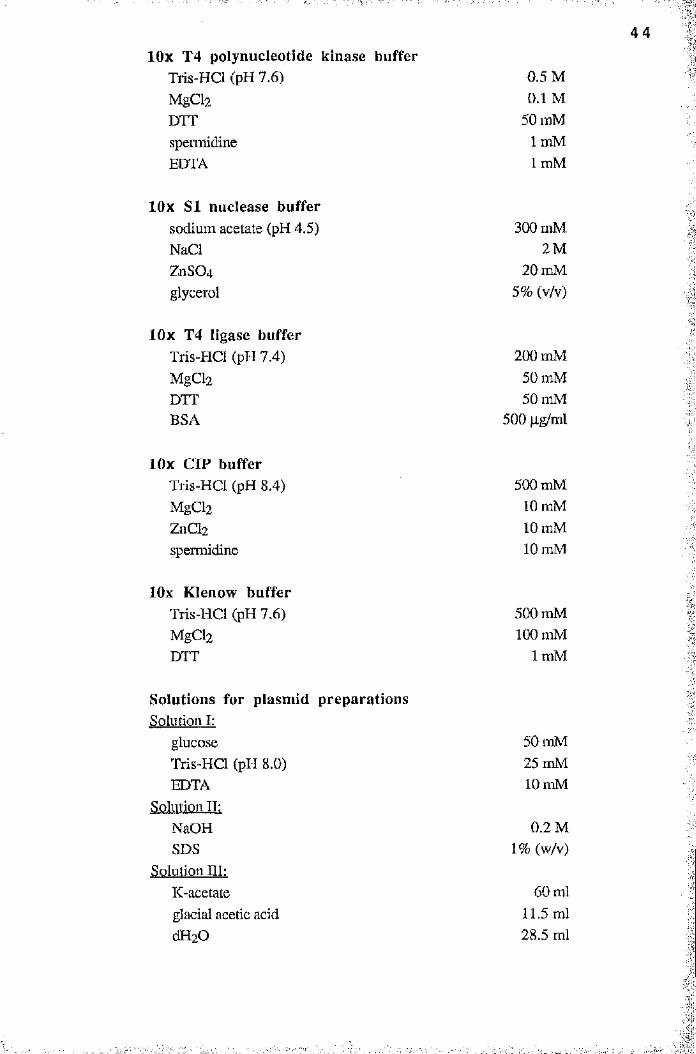
lOx T4 polynucleotide kinase buffer Tris-HCl (pH 7.6)MgCl2DTTspermidineEDTA
lOx SI nuclease buffersodium acetate (pH 4.5)NaClZnS04glycerol
lOx T4 ligase bufferTris-HCl (pH 7.4)MgChDTTBSA
lOx CIP bufferTris-HCl (pH 8.4)MgCl2ZnCl2spermidine
lOx Klenow bufferTris-HCl (pH 7.6)MgCl2DTT
Solutions for plasmid preparationsSolution I:
glucoseTris-HCl (pH 8.0)EDTA
Solution II:NaOH SDS
Solution III:K-acetate glacial acetic acid dH20
0.5 M 0.1 M
50 mM Im M 1 mM
300 mM 2M
20 mM 5% (v/v)
200 mM 50 mM 50 mM
500 jig/ml
500 mM 10 mM 10 mM 10 mM
500 mM 100 mM
1 mM
50 mM 25 mM 10 mM
0.2 M 1 % (w/v)
60 ml11.5 ml28.5 ml
4 4
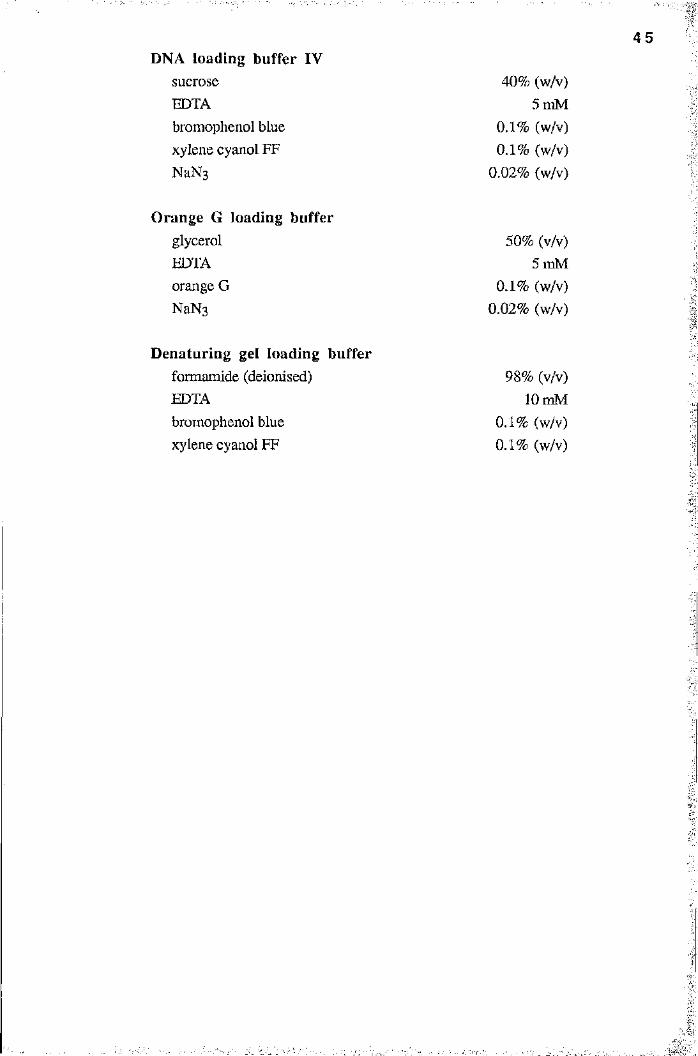
4 5
DNA loading buffer IVsucroseEDTAbromophenol blue xylene cyanol FF NaNs
40% (w/v) 5 mM
0.1% (w/v) 0.1% (w/v)
0.02% (w/v)
Orange G loading bufferglycerol EDTA orange G NaNg
50% (v/v) 5 mM
0.1% (w/v) 0.02% (w/v)
Denaturing gel loading bufferformamide (deionised)EDTAbromophenol blue xylene cyanol FF
98% (v/v) lOmM
0.1% (w/v) 0.1% (w/v)
' .
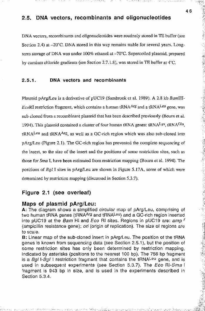
4 6
2.5. DNA vectors, recombinants and oligonucleotides
DNA vectors, recombinants and oligonucleotides were routinely stored in TE buffer (see
term storage of DNA was under 100% ethanol at-70°C. Supercoiled plasmid, prepared
by caesium chloride gradients (see Section 2.7.1.8), was stored in TE buffer at 4°C.
2 .5 .1 . DNA vectors and recombinants
Figure 2.1 (see overleaf)
1
Section 2.4) at -20°C. DNA stored in this way remains stable for several years. Long-
:%
Plasmid pArg/Leu is a derivative of pUC19 (Sambrook et al. 1989). A 2.8 kb BarriRl-
EcoRI restriction fragment, which contains a human tRNA^S and a tRNA^^u gene, was
sub-cloned from a recombinant plasmid that has been described previously (Bourn et al.
1994). This plasmid contained a cluster of four human tRNA genes: tRNA^ys, tRNA^H",
tRNA^eu and tRNA^^'8, as well as a GC-rich region which was also sub-cloned into
pArg/Leu (Figure 2.1). The GC-rich region has prevented the complete sequencing of
the insert, so the size of the insert and the positions of some restriction sites, such as
those for Sma I, have been estimated from restriction mapping (Bourn et al. 1994). The
positions of Bgl I sites in pArg/Leu are shown in Figure 5.17A, some of which were
determined by restriction mapping (discussed in Section 5.3.7).
Maps of plasmid pArg/Leu:A; The diagram shows a simplified circular map of pArg/Leu, comprising of two human tRNA genes (tRNA^rg and tRNA^eu) and a GC-rich region inserted into pUG19 at the Bam HI and Eco RI sites. Regions in pUC19 are: amp (ampicillin resistance gene); ori (origin of replication). The size of regions are to scale.B: Linear map of the sub-cloned insert in pArg/Leu. The position of the tRNA genes is known from sequencing data (see Section 2.5.1), but the position of some restriction sites has only been determined by restriction mapping, indicated by asterisks (positions to the nearest 100 bp). The 756 bp fragment is a Bgl \-Bgl I restriction fragment that contains the tRNAbeu gene, and Is used in subsequent experiments (see Section 5.3.7). The Eco Rl-Sma I fragment is 943 bp in size, and is used in the experiments described in Section 5.3.4.
I
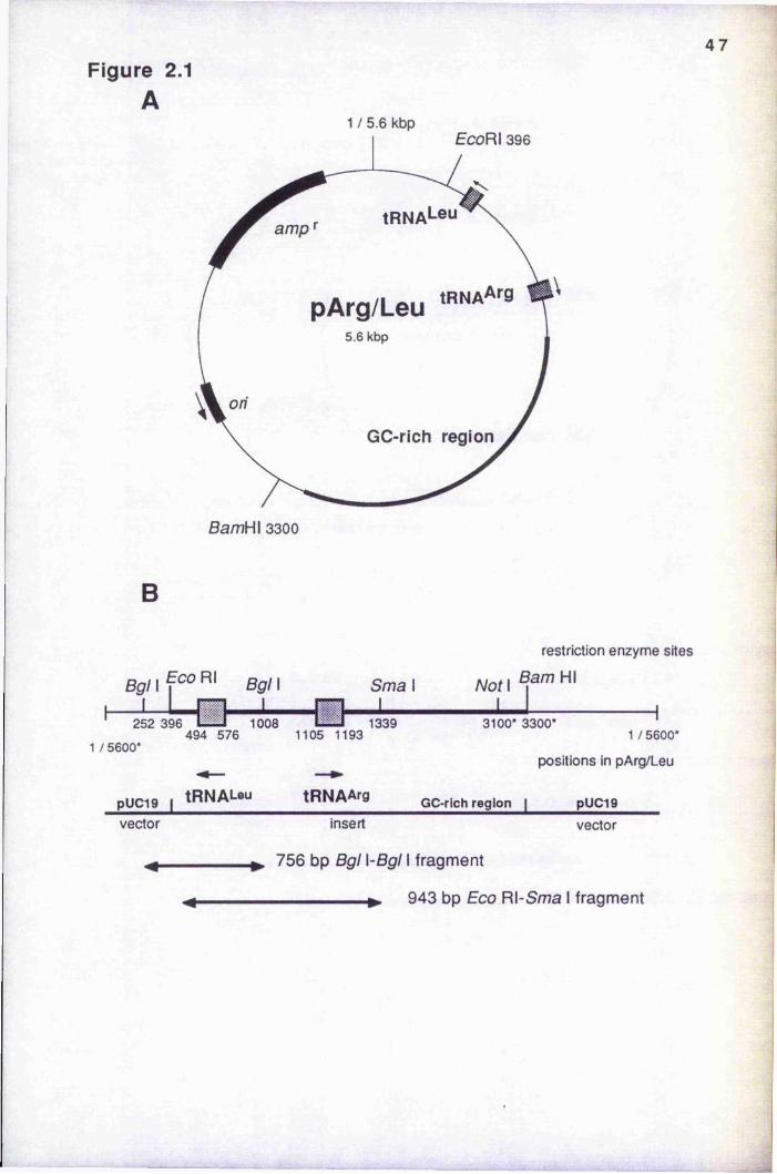
4 7Figure 2.1
A1 / 5.6 kbp
amp
pArg/Leu5.6 kbp
on
GC-rich region
B
restriction enzyme sites
Bgl\1
ECO Rl Bgl 1 Sma 1 Not 11 1 1
252 396 K i l 1008 L U 1339 494 576 1105 1193
1 /5600*
3100* 3300* '1/5600*
positions in pArg/Leu
pUC19 1 tR N A L eu tR N A A rg GC-rich region | pUC19vector insert vector
756 bp Bgl \-Bgl fragment
943 bp Eco Rl-Sma 1 fragment
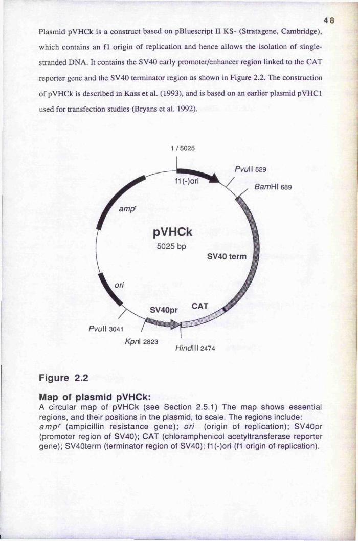
4 8Plasmid pVHCk is a construct based on pBluescript II KS- (Stratagene, Cambridge),
which contains an f l origin of replication and hence allows the isolation of single
stranded DNA. It contains the SV40 early promoter/enhancer region linked to the CAT
reporter gene and the SV40 terminator region as shown in Figure 2.2. The construction
of pVHCk is described in Kass et al. (1993), and is based on an earlier plasmid pVHCl
used for transfection studies (Bryans et al. 1992).
1 / 5025
PvuW 529
f1 (-)oriBamHl 689
amf/
pVHCk5025 bp
SV40 term
ori
SV40pr
PvuW 3041
Kpn\ 2823HindWl 2474
Figure 2.2
Map of plasmid pVHCk:A circular map of pVHCk (see Section 2.5.1) The map shows essential regions, and their positions in the plasmid, to scale. The regions include: ampi' (ampicillin resistance gene); ori (origin of replication); SV40pr (promoter region of SV40); CAT (chloramphenicol acetyltransferase reporter gene); SV40term (terminator region of SV40); f1 (-)ori (fl origin of replication).

4 9
2 .5 .2 Synthetic oligonucleotides
Synthetic oligonucleotides for primer extension assays were made in the Department of
Biochemistry by Dr. V. Math, using an Applied Biosystems 381A DNA synthesiser for
the phosphite-triester method.
2.6. DNA size markers
The following DNA standards were used as size markers for the analysis of nucleic acids
during gel electrophoresis. Figure legends refer to markers by the following labels:
bacteriophage A DNA, digested with Hin d l l l : 23.130, 9.416, 6.557, 4.361,
2.322, 2.027, 0.564, 0.125 (size in kb).
0 : bacteriophage 0 X174 (RF form dsDNA), digested with H in f l : 726, 713,
553, 500, 427, 417, 413, 311, 249, 200, 151, 140, 118, 100, 81, 66, 48, 42, 40, 24
(size in bp).
lOOn: 100 bp m arker: 2200, 1500, 1400, 1300, 1200, 1100, 1000, 900, 800, 700,
600, 500, 400, 300, 200, 100 (size in bp). The 600 bp fragment is approximately three
times more intense than other bands. (The marker was supplied by GIBCO/BRL).
5 0
2.7. Methods2.7.1 General methods used in moiecular biology
The following methods are standai'd procedures in molecular biology and essentially
follow protocols described by Sambrook et al. (1989), adapted to the equipment
available in the lab.
2.7.1.1 Phenol/chloroform extraction
ÿPhenol, equilibrated with TE buffer and containing 8-hydroxyquinoline at 0.1% (w/v)
was mixed with chloroform/isoamylaicohol (24:1) at a ratio of 1:1. Extraction of an
aqueous DNA solution was performed as follows: an equal volume of phenol/chloroform
was added to the DNA solution, voitexed and centrifuged for 5 min in a microfuge. The
2 .7 .1 .3 Quantification of nucleic acids
Spectrophotom etry
The absorbance of various dilutions of DNA samples was measured using quartz 1.0 ml
cuvettes in a spectrophotometer at 260 nm. An absorbance of 1.0 corresponds to
approximately 50 pg/ml for double-stranded DNA, 40 jig/ml for single-stranded DNA
and 20 qg/ml for oligonucleotides.
IUpper aqueous phase was transfened to a fresh tube and the extraction repeated if
■
necessary.
2.7 .1 .2 Ethanol precipitation
A salt solution was added to the DNA solution (sodium acetate, adjusted to pH5.2 with
glacial acetic acid, to 0.3 M; NaCl to 0.2 M; ammonium acetate to 2.0 M) and the DNA
precipitated with 2.5 volumes of ethanol (98% v/v) at-20°C for 15 min. The DNA was
recovered by centrifugation at 12,000 g for 15 min at 4°C. Subsequent washes with cold
70% (v/v) ethanol removed any salt which was coprecipitated with the DNA.
5 1
Fluorim etry
Accurate measurements of DNA concentrations, down to 10 ng/ml, were determined
using a Hoefer TKO 100 DNA minifluorometer. DNA solutions were added to 2.0 ml of
a working dye solution. Aliquots of the working dye solution, of final volume 100 ml,
were replaced daily and stored in the dark at room temperature. Each aliquot consisted of:
90 ml double-distilled, filter sterilised water (to remove particulate contaminants), 10 ml
TNE, xlO (100 mM Tris-HCl pH8.0, 10 mM EDTA, 1.0 M NaCl; adjusted to pH7.4
with conc. HCl) and Hoerst H33258 dye solution to a final concentration of lOOng/ml.
Readings were performed as described in the manufacturer’s instructions. Serial
dilutions of both calf thymus DNA and pUC19 plasmid DNA, from 10-500 ng/ml final
concentration, were used to construct standard curves from which the DNA
concentration of a fixed volume of sample DNA was determined,
2 .7 .1 .4 Preparation of competent ceiis
For the preparation of competent cells a single colony of E. coli XLl-Blue was
transferred to 20 ml of LB medium containing tetracycline (12.5 p-g/ml) and incubated
overnight at 37°C. 500 ql of this suspension were used to inoculate 100 ml of LB
medium containing tetracycline (12.5 qg/ml). Incubation was for 2 to 3 hours until an
OD^OO (optical density at 600 nm) of 0.45-0.55 was reached. The cells were then cooled
down on ice for 10 min and harvested by centrifugation at 4,000 rpm for 10 min at 4“C
using a Beckman 12-21 centrifuge and JA14 rotor. The pellet was carefully resuspended
in 40 ml of ice cold FSB buffer (Section 2.4) and stored for 10 min on ice. The cells
were recovered as above and resuspended in 6 ml of FSB buffer. 200 ql of DMSO were
then added. Cells were dispensed into aliquots of 150 pi, snap-frozen in liquid nitrogen
and stored at -70° C.
2 .7 .1 .5 Transformation of bacteria
Transformation-competent cells (150 pi, section 2.7.1.4) were thawed on ice and
plasmid DNA (no more than 50 ng) was added. After a 30 min incubation on ice the
5 2
tubes were transferred to a 42°C water-bath, incubated for 90 seconds and rapidly
transferred to an ice bath. After 5 min, 800 pi of LB medium was added and incubated
for 45 min at 37°C to allow the bacteria to express the antibiotic resistance. Dilutions of
the bacterial suspension were made and plated out on LB plates containing ampicillin
(100 pg/ral) and tetracycline (12.5 pg/ml).
2 .7 .1 .6 Small scale preparation of plasmid DNA
Transformed cells were grown overnight in 200 ml LB medium supplemented with
ampicillin (100 pg/ml). The cells were harvested by centrifugation for 15 min at 6,000
rpm in a Beckman J2-21 centrifuge using a JA 14 rotor. The pellet was resuspended in
10 ml solution I (Section 2.4.). Lysis of the cells was achieved by addition of 10 ml
solution II and incubation on ice for 5 min. Addition of 7.5 ml solution III precipitated
out the cell debris, which was sedimented by centrifuging at 10,000 rpm at 4°C
(Beckman J2-21 centrifuge, JA 20 rotor) for 15 min. The supernatant was filtered
through Whatman 3MM paper and 0.6 volumes of Lo-propanol were added to precipitate
■y;
I
IA 3 ml overnight culture was prepared from a single colony of transformed bacteria in
LB medium, supplemented with ampicillin (100 pg/ml). 1.5 ml of the bacterial
suspension was transferred to an Eppendorf tube and the bacteria pelleted at 5,000 rpm
(Eppendorf 5415 microcentrifuge) for 5 min. The pellet was suspended in 100 pi
solution I (see Section 2.4). 200 pi solution II was added, vortexed and incubated for 5
min on ice. Subsequently, 150 pi of solution III was added, vortexed and incubated
again for 5 min on ice. The bacterial debris was pelleted at 12,000 rpm for 10 min at
4°C. To the supernatant was added 460 pi phenol/chloroform mix; the mixture was
vortexed and the phases separated at 10,000 rpm for 10 min at 4°C. To the aqueous
phase was added 1 ml ethanol and the sample was incubated for 15 min at -70°C. The
DNA was pelleted at 12,000 rpm for 10 min at 4°C and the pellet resuspended in 40 pi of
TE buffer. 10 pi were used for a restriction digest which included RNase A at 1 pg/pl.
2 .7 .1 .7 Large scale plasmid preparation
5 3
the nucleic acids for 15 min at room temperature. The nucleic acids were pelleted at
3,000 rpm for 15 min at 4°C using a Beckman CS-6R benchtop centrifuge and
resuspended in 2 ml TE buffer. 2 ml 5 M LiCl/50 mM Tris-HCl (pH 8,0) was added and
incubated 10 min on ice to precipitate RNA. RNA was sedimented at 3,000 rpm for 5
min at 4°C (Beckman benchtop centrifuge) and the DNA in the supernatant precipitated
with 2 volumes of ethanol for 15 min on ice. The plasmid DNA was recovered by
centrifugation for 15 min at 3,000 rpm at 4°C (Beckman benchtop centrifuge) and
resuspended in 500 pi TE buffer. The DNA was then ethanol precipitated a second time
after a phenol, a phenol/chloroform and a chloroform extraction and finally dissolved in
500 pi TE buffer.
2 .7 .1 .8 Preparation of supercoiled plasmid DNA
Supercoiled plasmid DNA for in vitro transcription assays was prepared by isopycnic
centrifugation in a caesium chloride gradient. Recombinant plasmid was prepared as
described in Section 2.7.1,7, and diluted to 30 ml with TE buffer to which were added
28.9 g CsCl (BRL, UK; ultrapure optical grade) and 1.8 ml ethidium bromide stock
solution (at 10 mg/ml). This gave a solution of density 1.55 g/ml and a refractive index
of 1.3860, as measured with an Abbe refractometer (Bellingham and Stanley Ltd.,
London) for the sodium D% line (wavelength 589.6 nm), as recommended by the
manufacturer.
The solution was clarified by centrifugation at 10,000 rpm for 15 min at 4°C
(JA20 rotor), after which the clear red supernatant was carefully aspirated to avoid the
surface scum. The solution was transfeired to Beckman VT150 QuickSeal tubes, and the
tubes filled with light mineral oil if necessary. The samples were centrifuged at 50,000
rpm at 20°C overnight in a Beckman VTi50 rotor. After equilibrium was reached two
bands of DNA were seen. The upper band consisted of linear bacterial DNA and nicked
plasmid DNA, whilst the lower band consisted of supercoiled DNA. The latter was
collected using an 18 gauge hypodermic needle, which pierced the tube just below the
band. Ethidium bromide was removed by repeated extraction with an equal volume of 3-
methylbutan-Lol, followed by the addition of 4.0 vol. TE buffer. DNA was precipitated
5 4
by adding 2.0 (total) vol. ethanol and centrifuging at 15,000 rpm for 20 min at 4“C
(JA20 rotor). The DNA pellet was resuspended in TE and the quality of the preparationB
was assessed by agarose gel electrophoresis (Section 2.7.1.13). This protocol gave
preparations of at least 95% supercoiled plasmid.
•I■1
2 .7 .1 .9 Isolation of single-stranded DNA from phagemlds;
":'B:Single-stranded DNA from pBluescript phagemids was isolated as described by Blondel
and Thillet (1991). A colony of E. coli XLl-Blue, harbouring the phagemid of interest
and grown on LB plates supplemented with ampicillin (100 |ag/ml) for selection of the ■
phagemid and tetracycline (12.5 jig/ml) for selection of the F' episome, was suspended
in 20 ql 2x YT medium (Section 2.4). The bacterial suspension was then infected with 5
jil of helper phage (M l3 K07, Stratagene, Cambridge), with a titre of at least 10^^
PFU/ml, and incubated for 15 min at room temperature. 500 |il of 2x YT medium with
antibiotics (ampicillin at 100 qg/ml and tetracycline at 12.5 jag/ml) was added and
incubated for 1 hour at 37°C to let the helper phage express antibiotic resistance. 120 pi
of the incubated cells were then added to 3 ml 2x YT medium supplemented with
ampicillin (100 pg/ml), tetracycline (12.5 pg/ml) and kanamycin (75 pg/ml). The culture
was then incubated with vigorous shaking for 18 to 20 hours at 37°C. Cultures were
then pooled (typically into a total volume of 30 ml) and the bacteria pelleted by
centrifugation in a Beckman JA20 rotor at 6.5k rpm for 7 min. Phage particles were
precipitated by incubating with 2.5% (w/v) PEG 6000 and 0.35 M NaCl for 1 hr. at,
room temperature, and recovered by centrifugation (12k rpm, 5 min in a JA20 rotor).
The phage pellet was dissolved in 1000 pi TE buffer. After two phenol extractions and
one chloroform extraction the single-stranded DNA was ethanol precipitated and
dissolved in 300 pi TE buffer. The preparation of ssDNA was then checked on anV ”
agarose gel and the concentration determined by spectrophotometry.
I■ f i
:
5 5
2 .7 .1 .10 Restriction enzyme digests of DNA
The DNA was incubated with the appropriate restriction enzyme in the presence of the
appropriate buffer (supplied together with the restriction enzyme as lOx buffer) for at
least 2 hours at 37“C, with a DNA concentration not higher than 1 pg/10 pi and an
enzyme concentration of about 10 units/pg DNA. The volume of the restriction enzyme
did not exceed a 1/10 of the total volume, as glycerol can affect the performance of the
enzyme.
2.7.1.11 Ligations
Routinely, 200 ng of linearised vector DNA were used in a ligation in a total volume of
10 pi. The vectornnsert molar ratio was 1:3. 1 pi of lOx T4 ligase buffer (section 2.4)
and 1 pi of 10 mM ATP were added to the DNA and the ligation initiated by addition of 1
Weiss unit of bacteriophage T4 DNA ligase. Incubation was for 8 to 12 hours at 16°C.
2 .7 .1 .12 Dephosphorylation of plasmid DNA
The 5 "-phosphate groups of a linear plasmid were removed by calf intestinal alkaline
phosphatase (CIP) to avoid self-ligation of the vector. 1 pg of linearised vector DNA
was incubated in CIP buffer (Section 2.4) with 1 unit of calf intestinal alkaline
phosphatase (Boehringer Mannheim GmbH) in a total volume of 50 pi at 37°C for 30
min. SDS and EDTA were added to final concentrations of 0.5% and 5 mM,
respectively. Proteinase K (Boehringer Mannheim GmbH) was added to a final
concentration of 100 pg/ml and incubated for 30 min at 55“C. The reaction was subjected
to a phenol and phenol/chloroform extraction and the DNA was recovered by ethanol
precipitation.
5 6
2 .7 .1 .13 Agarose gel electrophoresis of DNA
Agarose gels were routinely used to separate DNA fragments of size range 0.2-20 kbp.
Agarose (GIBCO/BRL) was dissolved at the desired concentration (0.8 - 2.0% w/v) in
TBE 0.5x buffer (Section 2.4) containing 0.5 pg/ml ethidium bromide by heating in a
microwave oven until boiling. A well-forming comb was inserted and the gel allowed to
set. One half volume of DNA loading buffer IV (Section 2.4) or orange G loading buffer
(Section 2.4) was added to the DNA samples before being applied to the gel.
Electrophoresis was carried out at 50 mA in TBE 0.5x buffer containing 0.5 pg/ml
ethidium bromide. The DNA fragments were visualised by using a long wavelength UV
transilluminator.
5 7
2 .7 .1 .14 Isolation of DNA fragments from LMP agarose gels
I
II.
Low melting point (LMP) agarose (GIBCO/BRL) gels were prepared exactly as agarose
gels, but were run in a cold room. The DNA band that was required was visualised using
a hand-held long wavelength UV lamp and excised from the gel using a sterile scalpel
blade. The gel slice was transferred to an Eppendorf tube and heated to 65“C for 15 min
to melt the agarose. To isolate DNA fragments of size range 0.4-14 kbp, to yield
amounts up to about lOjitg, a Geneclean II kit (Bio 101, CA, USA) was used as
recommended by the manufacturer.
Higher yields of restriction fragments of plasmid DNA (10-50 |ig) were required
for the generation of patch-methylated constructs (Section 2.7.3.2), so a different
strategy was chosen to isolate sufficient amounts of the DNA fragment. Depending on
the size of the restriction fragment to be isolated, 50 to 100 |ag of plasmid DNA were
digested with the desired restriction enzyme(s). Complete digestion was checked on a
minigel and the bulk of the restriction digest was separated on a preparative LMP agarose
gel, where the sample was applied in one large slot. The desired band was identified and
the fragment cut out of the gel. The plunger of a 1 ml (or 2 ml) disposable syringe was
removed and the syringe plugged with sterile siliconised glass wool. The agarose slice
was placed in the syringe, and the DNA was recovered by centrifugation of the syringe
for 20 min at 25“C in a Beckman CS-6R benchtop centrifuge. 500 p.1 of boiling TE
buffer (section 2.4) was added to the agarose that was trapped by the glass wool, and the
procedure of centrifugation was repeated to extract any remaining DNA. The DNA
solution was chloroform extracted and the DNA recovered by ethanol precipitation.
2 .7 .1 .15 Polyacrylamide gel electrophoresis (PAGE) of DNA
Polyacrylamide gels were routinely used to separate DNA fragments of size range 10-
300 bp. They were prepared by mixing a suitable volume of Easigel acrylamide/bis-
acrylamide stock solution (Scotlab, Paisley, UK), containing 30% [w/v] acrylamide and
1.034% [w/v] N,N'-methylene-bisacrylamide, with water and an appropriate volume of

5 8
TBE xlO buffer to give the desired acrylamide concentration (5-20% [w/v]) in TBExl.O.
The acrylamide was polymerised by the addition of 0.006 vol. of 10% ammonium
persulphate and 0.001% TEMED at 42“C for 30 min. DNA samples were mixed with
one half volume of DNA loading buffer IV, and gels were run at 50 mA in TBE xl.O
electrophoresis buffer. Gels were stained in TBE Xl.O containing 0.5p,g/ml ethidium
bromide for 30 min, and after rinsing in distilled water were visualised as for agarose
gels.
2 .7 .1 .17 Purification of oligonucleotides
2 .7 .1 .16 Isolation of DNA fragments from PAGE gels
The desired band was identified and the DNA fragment cut out of the gel. The DNA was
recovered by a modification of the “crush and soak” method (Sambrook et al. 1989). Gel
slices, each weighing approximately 100 mg, were transfeixed to Eppendorf tubes and
crushed using a yellow pipette tip. To each tube was added 300 pi PAGE/DNA elution
buffer (Section 2.4) and the mixture was incubated for 16 hr at 37”C with vigorous
shaking. The gel pieces were pelleted by centrifugation at 12,000 rpm for 10 min in a
microcentrifuge. The supernatants were pooled, ethanol precipitated and dissolved in 50
pi TE buffer. Finally, to remove any remaining gel particles the DNA fragments were
purified using a ChromaSpin-10 TE spin column (Section 2.7,1.20).
60 pg (approx. 2 OD^^O) of the oligonucleotide were lyophilised to dryness in a rotary
vacuum lyophiliser to remove the aqueous ammonia. The pellet was then dissolved in 20
pi H2O and 20 pi formamide was added. Before loading on a gel the DNA was heat
denatured for 5 min at 55"C. Separation was on 1 mm thick, 19% denaturing
polyaciylamide (acrylamide : N,N'-methylene-bisacrylamide 19:1) gels in IxTBE buffer
(Section 2.4). The gel was pre-run for 30 min at 30 mA before the oligonucleotide was
loaded. To monitor the electrophoresis, 5 pi of denaturing gel loading buffer (Section
2.4) was run alongside the oligonucleotide samples. After completion of the
electrophoresis the apparatus was dismantled and the gel transferred onto a fluorescent
5 9surface. The oligonucleotide was visualised by illuminating with a hand-held long
wavelength UV lamp. The oligonucleotide absorbed the UV radiation and appeared as
dark bands on the fluorescing background. The top bands of the oligonucleotide
(containing the full size oligonucleotide) were marked, excised from the gel and isolated
as described in Section 2.7.1.16.
2 .7 .1 .18 End labelling of oligonucleotides
!
Synthetic oligonucleotides were end-labelled by transfer of the from [y-32p]ATP
using bacteriophage T4 polynucleotide kinase (PNK). 10 pmol of oligonucleotide were
incubated in T4 PNK buffer (Section 2.4) with 5 pi of [y-32p]ATP (5,000 Ci/mmol, 10
mCi/ml) and 10 units of T4 PNK in a total volume of 20 pi. Unincorporated radiolabel
was removed using ChromaSpin-10 columns (Section 2.7.1.20).
2 .7 .1 .19 Random-primed radiolabelling of DNA fragments
The Megaprime DNA labelling system from Amers ham (RPN 1606) was used for the
random-primed radioactive labelling of probes for Southern hybridisations. 5 pi of
primer solution (containing random nonamer primers) and water to a final reaction
volume of 50 pi were added to 100 ng of purified fragment DNA. The DNA was
denatured for 5 min at 95“C in a boiling water-bath. Subsequently 10 pi of Megaprime
reaction buffer (containing dATP, dGTP, dTTP, MgCl^, 2-mercaptoethanol and Tris-
HCl buffer [pH 7.5]; the exact concentrations are not given in the company literature), 5
pi [a-32p]dCTP (3,000 Ci/mmol, 10 mCi/ml), 2 pi Klenow enzyme (1 unit/pl) and
water to a final reaction volume of 100 pi were added and the reaction incubated for 30
min at 37"C. The reaction was stopped by the addition of 5 pi 0.2 M EDTA and
unincorporated radionucleotide was removed by using ChromaSpin-10 columns (Section
2.7.1.20).
6 0 #2 .7 .1 .20 Removal of unincorporated radionucleotides from
radiolabelled DNA
2.7.1.21 Southern blotting and hybridisation
ChromaSpin-10 TE spin columns (Clontech Laboratories, Palo Alto, CA) were used to
remove unincorporated radionucleotide from radioactively labelled DNA. The column
was drained and packed by centrifugation at 2,200 rpm for 5 min at 4°C in a Beckman
CS-6R bench-top centrifuge. The sample (maximum volume 100 pi) was carefully
applied onto the centre of the column and centrifuged as above. The radioactively labelled
sample was collected in an Eppendorf tube at the bottom of the column, whereas the
unincorporated nucleotides remained in the column. The specific activity of the
radiolabelled sample was calculated by comparison of the radioactivity before and after
removal of the free nucleotides.
;;l
IA combined vacuum/alkaline blot method was used to transfer DNA from agarose gels to
nylon membranes. A vacuum blotting apparatus from Hybaid (Teddington, Middx.,
UK) was used to perform the alkaline Southern blot. A piece of Whatman 3MM filter
paper and a piece of nylon membrane (Hybond N+, Amersham International PLC,
Amersham, UK) were cut slightly larger than the gel to be blotted and pre-wetted in 0.4
M NaOH. The paper was placed on top of the porous plate of the blotting apparatus,
followed by the nylon membrane and a rubber gasket, which was cut with an aperture
slightly smaller than the gel to be blotted. The agarose gel was briefly soaked in 0.4 M
NaOH and then carefully placed on the nylon membrane, to avoid trapping any air
bubbles between gel and membrane. The vacuum was then applied and the transfer
buffer (0.4 M NaOH) poured in the chamber so that the gel was completely immersed.
The vacuum was reduced by a valve to 80 cm of water and the transfer time was 30 min.
After the transfer was completed the nylon membrane was neutralised by brief washing
in 5x SSC buffer. For some experiments, the DNA fragments was blotted by the
traditional technique of capillary transfer (Sambrook et al. 1989). Diffusion of fragments ■
2.7.2 Methods for quantification and analysis of proteins
2.7.2.1 Quantification of protein concentrations
6 1
in the agarose during transfer reduced the resolution of bands, as compared to the
vacuum transfer procedure, so the latter was the preferred method for the experiments
described in Chapter 6.
Southern blots were prehybridised in hybridisation buffer (section 2.4)
containing 100 |ig/ml heat denatured sonicated salmon sperm DNA for 1-2 hours at
65“C. The heat denatured probe (1-5 pg/ml hybridisation buffer, 10^-10^ cpm/p-g) was
added and incubated for 12-16 hours at 65°C in a shaking water bath. The filter was
washed twice in 2x SSC, 0.1% SDS for 15 min at room temperature and subsequently
once in Ix SSC, 0.1% SDS at 65°C for 15 min and twice in O.lx SSC, 0.1% SDS at
65°C for 15 min. For experiments with relatively high amounts of target DNA (>50 ng)
and a long homologous probe (>400 bp.) the stringency conditions for hybridisation
were increased. (The majority of Msp I fade-out assays that are described in chapter 6 are
experiments of this type). The conditions for higher stringency washes were: 2x SSC,
0.1% SDS for 15 min at 68°C and subsequently once in Ix SSC, 0.1% SDS at 68°C for
15 min and twice in O.lx SSC, 0.1% SDS at 68°C for 15 min. (and longer if required).
The membrane was then autoradiographed for the desired time.
If the blot was subjected to a hybridisation with a second probe the membrane
was never allowed to dry during or after hybridisation and washing. The blot was
Stripped of the first probe by pouring a boiling solution of 0,5% SDS on the membrane
and allowing it to cool to 50°C. The complete removal of the probe was checked by
autoradiography, after which the blot was hybridised to the second probe, which was
prepared to have a higher specific activity than the first.
I
The concenti^ation of proteins in solution was assayed using the method of Bradford
(1976). Water was added to an aliquot of the protein sample to a total volume of 100 |Lil,
followed by 1.0 ml of Bradford's reagent (8.5% [v/v] phosphoric acid, 5% [v/v]
ethanol, 0.01% [w/v] Coomassie Brilliant Blue G), The A^95 of the mixture was
i
6 2measured, and the protein concentration estimated from a calibration curve of standard
concentrations (0-40 pg per assay) of bovine serum albumin for routine determinations.
The concentrations of histone proteins were determined by comparison with standards of
purified core histones (Section 2.7.6), to compensate for the anomalous effect of basic
proteins on colorimetric assays.
2 .7 .2 .2 Analysis of proteins by SDS-PAGE
SDS-polyacrylamide gel electrophoresis was used to separate proteins of size range
between 10-100 kDa. SDS-PAGE resolving gels were prepared by mixing suitable
volumes of Easigel acrylamide/bisacrylamide stock solution (Scotlab, Paisley, UK),
containing 30% [w/v] acrylamide and 1.034% [w/v] N,N'-methylene-bisacrylamide,
with 1.5 M Tris-HCl [pH8.8], 10% SDS and water to give the desired acrylamide
concentration (10-15% [w/v]), 0.375 M Tris buffer and 0.1% SDS. The acrylamide was
supplemented by the addition of 0,006 vol. of 10% ammonium persulphate and 0.001%
TEMED, poured between a pair of vertical glass plates, overlaid with i.so-butanol and
allowed to polymerise at 42°C for 30 min. The overlay was then poured off, the top of
the resolving gel was washed with water, and SDS-PAGE stacking gel solution was
poured directly onto the surface of the resolving gel. Stacking gels were prepared by
mixing suitable volumes of 30% acrylamide/bisacrylamide stock solution, 1.0 M Tris-
HCl [pH6.8], 10% SDS and water to give final concentiations of 5% acrylamide, 0.125
M Tris buffer and 0.1% SDS. The stacking gel was polymerised as described above.
Samples containing approximately 10 jag protein were denatured by heating to
100°C for 3 min in Ix SDS gel-loading buffer (50 mM Tris-HCl [pH6.8], 100 mM
DTT, 2% SDS, 0.1% [w/v] bromophenol blue, 10% glycerol) and loaded onto the gel.
Electrophoresis was initially at 8V/cm, until the dye front had moved into the resolving
gel, after which the voltage was increased to 15V/cm. Gels were fixed and stained in a
solution containing 10% [v/v] methanol, 10% [v/v] glacial acetic acid and 0.25% [w/v]'
Coomassie Brilliant Blue R250 for 4 hr at room temperature. The gel was then
thoroughly destained by soaking in several changes of destain solution containing 10%
methanol and 10% glacial acetic acid.
6 3
2.7.3 Méthylation of plasmid DMA2.7.3.1 Méthylation of plasmid DNA in vitro using
prokaryotic methylases
2 .7 .3 .2 Patch-methylation of pVHCk plasmid DNA
I'
■I'
Prokaryotic methylases I, M.Hpa II and M.Hha I and their assay buffers were
purchased from New England Biolabs (Beverley, MA, USA). Routinely, 20 |ag plasmid
DNA were incubated with 20 units of raethylase (60 units in the case of M.Hpa II) in the
appropriate assay buffer for 16 hours at 37 “C in a total volume of 100 p.1. The
concentration of the methyl-group donor S-adenosyl methionine (SAM) was 80 p-M for
M.Hpa II and M.Hha I and 160 pM for M .to I, respectively. For inactivation of the
methylases SDS and EDTA were added to final concentrations of 0.5% and 5 mM,
respectively. Proteinase K (Boehringer Mannheim GmbH) was added to a final
concentration of 100 pg/ml and incubated for 30 min at 55“C. The reaction mixture was
subjected to a phenol and phenol/chloroform extraction and the DNA recovered by an
ethanol precipitation. For every méthylation assay, a mock-methylated control was
treated in the same way as described above, but omitting the methylase in the méthylation
assay.
Restriction fragments were gel purified and annealed to ssDNA at a molar ratio of 3:1
(fragment DNA:ssDNA) in 20 mM Tris-HCl (pH7.5); 10 mM MgCU; 50 mM NaCl; 1
mM DTT by heating for 2 min at 95°C and allowing to cool down from 70 to 30° C in 1
hr. To prevent binding to and méthylation of the single-stranded region by methylase
Sssl (M.Sss I), T4 gene 32 protein (Boehringer Mannheim Gmbh) was added (10 pg/pg
of ssDNA), and the reaction mixture was incubated at 37°C for 15 min. The double
stranded DNA patch was methylated for 16 h, using M.Sss I (New England Biolabs)
under conditions recommended by the manufacturer. After proteinase K treatment and
phenol/chloroform extraction, the unmethylated gap was filled in and ligated by standard
procedures (Sambrook et al. 1989). For every patch-methylated construct, a
fS
V : .
6 4
mock-methylated control was made in the same way but omitting M .to I. Constructs
2.7.4. In vitro transcription assays
Nuclear extracts for in vitro transcription assays were prepared according to Dignam et
al. (1983). HeLa S3 cells were grown in siliconised spinner flasks at 37°C in EMEM
medium (Section 2.3) to a density of 4 to 6 x 10⁵ cell/ml. A total of about 109 cells were
used for a typical extract preparation. Cells were harvested by centrifugation for 10 min
at 2,000 rpm and room temperature (Beckman J2-21 centrifuge, JA 14 rotor). Pelleted
were separated from unannealed restriction fragment by gel purification. Precise
concentrations of purified DNA were determined by using a Hoefer TKO 100 DNA
minifluorometer (see Section 2.7.1.3). Calf thymus or pBluescript KSII- DNA was used
as a standard. To confirm the location of the methylated region, constructs were digested
with the appropriate restriction enzymes to release the methylated region and subjected to
Hpa II digestion. The DNA was then separated on a 1.3% agarose gel, transferred to a
nylon membrane (Hybond N+; Amersham), and hybridised to the appropriate labelled
fragment (Section 2.7.1.21). Membranes were reprobed after stripping with boiling
0.5% SDS solution. The results of these analyses are presented in Figure 6.6 A-C and
discussed in Section 6.3.1.
All solutions for the preparation of nuclear extracts and for the in vitro transcription assay
were either treated with diethylpyrocarbonate (DEPC) or made up with DEPC-treatedI
water, and were autoclaved when possible. DEPC was added to a final concentration of
0.1% (v/v) and incubated at 37°C for at least 4 hr with vigorous agitation. Glassware■i
was treated with 3% (v/v) H2O2 for 16 hr and then rinsed with DEPC-treated water."■is
!cDisposable plasticware was either subjected to multiple autoclaving or rinsed briefly with
isï
chloroform to reduce the risk of RNase contamination. In vitro transcription is discussed
in more detail in Section 3.1.
s/■'
2.7.4.1 Preparation of nuclear extracts from HeLa cells
î
6 5
cells were suspended in 5 volumes of PBS (4°C) and collected by centrifugation as
above. Subsequent steps were performed at 4“C. The cells were suspended in 5 packed
cell volumes of buffer A (10 mM HEPES-KOH [pH 7.9], 1.5 mM MgCU, 10 mM KCl,
0.5 mM DTT) and incubated on ice for 10 min. Cells were collected by centrifugation as
before and resuspended in two packed cell volumes of buffer A and lysed by 10 strokes
of a Kontes all glass Dounce homogeniser, using the B type pestle. The homogenate was
centrifuged as before to pellet the nuclei. The nuclear pellet was subjected to a second
centrifugation for 20 min at 15,000 rpm (Beckman J2-21 centrifuge, JA 20 rotor) at 4°C
to remove any residual cytoplasmic material. These crude nuclei were resuspended in 3
ml buffer C (20 mM PIEPES-KOH [pH 7.9], 25% v/v glycerol, 0.42 M NaCl, 1.5 mM
MgCl2, 0.2 mM EDTA, 0.5 mM PMSF, 0.5 mM DTT) per 109 cells and homogenised
as above. The resulting suspension was stirred gently for 30 min at 4°C and centrifuged
for 30 min at 15,000 rpm at 4°C. The resulting clear supernatant was dialysed against 50
volumes of buffer D (20 mM HEPES-KOH [pH 7.9], 20% v/v glycerol, 0.1 M KCl,
0.2 mM EDTA, 0.5 mM PMSF, 0.5 mM DTT) for 5 hours and the dialysate centrifuged
at 15,000 rpm for 20 min at 4°C. The supernatant was snap frozen as 50 pi aliquots in
liquid nitrogen and stored at -70“C.
In a modification to the protocol, histone H I was depleted from the dialysed
extract by the selective precipitation of proteins by the addition of 1.25 vol of 4 M
ammonium sulphate (Croston et al. 1991) buffered with 50 mM HEPES-KOH pH7.9, in
a protocol described elsewhere (Shapiro, Shaip et al, 1988). The proteins were pelleted
by centrifugation at 100,000 g for 30 min at 4^C, and were resuspended in buffer D
(Dignam et al. 1983).
2 .7 .4 .2 In vitro transcription assay using direct internal
radiolabelling
This method was used to assay the transcripts from the human tENA^Gu and tRNA^g
genes, contained in the recombinant pArg/Leu (Section 2.5). These are class III genes
that are transcribed by RNA polymerase III (refer to Section 3.2). The conditions for
transcription of tRNA genes by pol III have been described previously (Gonos and
6 6
Goddard 1990), and with subsequent modifications (Bourn et al. 1994; Johnson et al,
1995). The protocol is discussed in greater detail in Sections 3.2.2 and 3.2.3. The
nuclear extract used for this pol HI system was the one prepared by the Dignam protocol
(Dignam et al. 1983), so the extract contained some contaminating, endogenous histone
HI. A reaction mixture, of final volume 20 pi, contained 5 pi extract (7.0 pg protein/pl),
40 U RNasin (Promega) and final concentrations of 14 mM HEPES-KOH pH7.9, 80
mM KCl, 20 mM NaCl, 3.5 mM MgCli, 10% (v/v) glycerol, 0.3 mM DTT and 0.3 mM
PMSF. The reaction mixture included the desired amount of template DNA (usually 150
ng), with pUC19 DNA as a non-specific carrier, if required, to a total of 150 ng DNA
per assay. The mixture was preincubated at 30^C for 20 min (see Section 3.2.3), after
which transcription was initiated by the addition of unlabelled ribonucleoside
triphosphates at pH7.0 (600 pM ATP, CTP and GTP; 25 pM UTP), 2.5 pCi of [a-32p]
UTP (3,000 Ci/mmol, 10 mCi/ml) and 10 mM creatine phosphate. The transcription
reaction was allowed to proceed at 30^C for 60 min and terminated by the addition of
10% (w/v) SDS to a final concentration of 0.5%, 100 pg proteinase K (Boehringer
Mannheim GmbH) and 40 pg E. coli crude tRNA as carrier. The mixture was incubated
at 37®C for 20 min and then an equal volume 1.0 M ammonium acetate was added. The
mixture was extracted with phenol-chloroform (Section 2.7.1.1), which was then back-
extracted with an equal volume of 0.5 M ammonium acetate. The RNA in the pooled
aqueous phases was ethanol precipitated (Section 2.7.1.2), washed with 70% ethanol,
dried thoroughly and resuspended in denaturing gel loading buffer (Section 2.4).
Samples were analysed by electrophoresis on a 12% polyacrylamide, 4 M urea
denaturing slab gel, followed by autoradiography. Transcription was quantified either by
scintillation counting or by using a Fuji BASIOCX) phosphoimager.
2 .7 .4 .3 In vitro transcription assay using primer extension
In vitro transcription assays of the pVHCk construct (Section 2.5.2) were quantified
using a primer extension approach (Leonard and Patient 1991; Mason et al. 1993).
Transcription from the SV40 promoter requires RNA polymerase II and preliminary i
6 7
experiments established that this transcription system required an optimal MgCl^
concentration of 4 mM. These optimisation experiments and an extensive discussion of
this protocol can be found in Sections 3.3.1 to 3.3.4. The nuclear extract that was used
in this pol II system was prepared according to the Shapiro protocol (Shapiro et al.
1988), so the extract was depleted of histone H I. This simplified the interpretation of
results for the pol II system to the effects of exogenous histone H I (see Section 6.2.3).
A standard in vitro transcription assay was carried out as follows:
HeLa nuclear extract 27 piMgClz (50 mM) 3.2 piATP-CTP-GTP-UTP-mix (pH 7.0, each at 10 mM) 4.0 piRNasin (Promega, 40 U) 1,0 pisupercoiled pVHCk plasmid DNA (50 - 500 ng) 5 pi
total reaction volume: 40 pi
The reaction mixture was incubated for 45 min at 30“C, after which 100 pi of DNase
digestion buffer (20 mM HEPES-NaOH [pH 7.6], 50 mM NaCl, 5 mM CaCl2, 5 mM
M gCl2, 10 mM DTT) and 1.0 U DNase I (RQl RNase-free DNase, Promega) was
added. The reaction was incubated at 37 °C for 10 min and was stopped by the addition
of 150 pi DNase stop buffer x2 (20 mM Tris-HCl [pH 8.0], 0.3 M NaCl, 1.0% SDS,
50 mM EDTA, 40 pg tRNA, 20 pg proteinase K) and incubation at 37°C for a further 30
min. After phenol/chloroform extraction and back extraction, the aqueous phase was
heated to 65“C for 10 min to inactivate any residual DNase. The RNA was ethanol
precipitated, air-dried and dissolved in 10 pi of dH20 .
To assay the level of the specific mRNA, first strand cDNA was synthesised
from the transcripts using an 5’ end-labelled primer complementary to the taiget RNA.
The amount of cDNA obtained was a measure of the level of transcription of that
paiticular gene. An oligonucleotide (oligo PE;1 shown in Figure 3,5), complementary to
the mRNA of the CAT gene was end-labelled (Section 2.7.1.18) and diluted to 50
fmol/pl. 1.0 pi of primer (104 cpm) was mixed with 10 pi RNA and 4.0 pi 5x reverse
transcriptase buffer (250 mM Tris-HCl [pH 8.3], 250 mM KCl, 50 mM MgCl2, 50 mM
DTT, 5 mM spermidine) and incubated for 20 min at 65°C, followed by 10 min at 42°C.
6 8
To this was added 1 pi dNTP-mix, each at 10 mM (Promega), and 10 U AMV reverse
transcriptase (Promega, 12 U/pl). After incubation for 30 min at 42°C, the nucleic acids
were directly ethanol precipitated, dissolved in 8 pi denaturing gel loading buffer
(Section 2.4) and separated on a 10% denaturing polyacrylamide gel.
A necessary preliminary experiment was to determine that a significant proportion
of the primer formed a hybrid with the mRNA. This was achieved by treating
hybridisation reactions (see above) with an excess of nuclease SI (a suitable protocol is
described in Mason et al. 1993). In brief, a 1.0 pi aliquot of the hybridisation reaction
was added to 200 pi of nuclease SI buffer xl(Section 2.4), Four 50 pi aliquots were
removed, and 300 U nuclease S 1 (Boehringer Mannheim GmbH) were added to only
two of the aliquots. At the concentration used, the nuclease SI degraded all single-
stranded DNA, including unannealed primer, but did not digest duplexes of primer
hybridised to RNA. The digestion reactions were then spotted onto a disc of DE81 paper
(Whatman). The discs were allowed to air-dry and were then washed extensively with
5% (w/v) Na2HP0 4 , and once each with water, and then ethanol. The filters were air-
dried and the radioactivity assayed by scintillation counting. The proportion of the primer
which is resistant to nuclease SI is determined by averaging duplicate values, and
expressing the radioactivity of the samples with nuclease SI as a percentage of the
radioactivity of the samples with no nuclease SI. See Section 3.3.2 for further details.
2.7.5 Histone H1 preparation and complex
formation with DNA%
2.7.5.1 Renaturation of histone HI
Total acid-extracted, lyophilised calf thymus histone HI (Boehringer Mannheim GmbH)
was used in initial experiments. Lyophilised protein was dissolved in a buffer containing
10 mM Tris-HCl [pH7.5], 1.0 mM DTT, 0.4 mM PMSF, 5 mM EDTA, 0.01% (v/v)
Nonidet-P40, 10% (v/v) glycerol. Nonidet-P40 was included in all buffers containing
histone H I to prevent non-specific adsorption to plastic or glass (Croston et al. 1991).
The presence of EDTA facilitated histone renaturation (Caiafa et al. 1991). Samples were
6 9
2 .7 .5 .2 . Formation of histone H1-DNA complexes
renatured by step-dialysis (Hentzen and Bekhor 1985) to the histone HI buffer, starting
from buffer containing 2 M NaCl, 5 M urea, 10 mM TRIS-HCl [pH7.5], 1.0 mM DTT,
0.4 mM PMSF, 5 mM EDTA. The subsequent buffers used in the dialysis had the
following decreasing amounts of urea and NaCl: 1 M NaCl, 5 M urea; 0.8 M NaCl, 5 M
urea; 0.6 M NaCl, 5 M urea; 0.4 M NaCl, 5 M urea; 0.4 M NaCl. Protein concentrations
were determined using the method of Bradford, with purified core histones as standards
to compensate for the anomalous effect of histones on colorimetric assays (Section
2.7.2.1). Histone H I preparations were snap-frozen in liquid nitrogen and stored frozen
in histone H I buffer at -70^C.
Somatic histone H I variants H la-e were partially purified by reversed-phase
HPLC chromatography (Quesada et al. 1989), as modified by (Santoro et ai. 1993).
Protein fractions were lyophilised and renatured as described above. Histone HI variants
are discussed in detail in Section 1.8.6, and experiments that use histone HI variants are
described in Section 5.3.5.
-f
I
Total histone HI was allowed to bind to 100 ng supercoiled template DNA under
conditions that facilitated the formation of a “slow” complex, as defined by Clark and
Thomas (1986). “Slow” complexes are presumed to form by the non-cooperative and
reversible binding of H I to DNA. Histone H I dissolved in histone H I buffer was
mixed, in various proportions, with DNA in 20 mM HEPES-KOH [pH7.0] and 2.0 mM
MgCl2 in the presence of 15 mM NaCl. Mixtures were incubated at 27^C for 60 min,
after which the complexes were used in subsequent procedures. “Fast” complexes, as
defined by Clark and Thomas (1986), were presumed to be aggregates formed by the
irreversible binding of histone HI to the DNA. The conditions used in this study were
optimised for slow complex formation and minimised the formation of fast complexes.
The formation of histone HI-DNA complexes, under these conditions, was assayed by
agarose (1.0%) gel electrophoresis in 0.25x TBE buffer and 20% (v/v) glycerol (see
Section 5.2.1). Hl-DNA complexes were retarded in comparison to naked supercoiled
DNA, whereas aggregates of histone HI and DNA did not migrate out of the wells of the
agarose gel (Figure 5.2).
7 02.7.6. Purification of core histones
The method used was a consensus of the methods of Stein and Mitchell (1988) and
Workman et al. (1991). Core histones were prepared from dispersed chromatin by salt
elution on hydroxy apatite. All steps in the procedure were carried out at 4°C. Nuclear
material, derived from the procedure for making HeLa nuclear extracts (Section 2.7.4.1),
was stored at -70°C in chromatin storage buffer (CSB; 10 mM TRIS-HCl [pH7.4], 0.1
M NaCl, 0.1 mM EDTA, 1 mM DTT, 0.4 mM PMSF, 10% [v/v] glycerol) until
required. The dispersed chromatin was pelleted by centrifugation at 6k rpm for 20 min
(Beckman JA 20 rotor) and resuspended in 20 pellet volumes of lysis buffer (10 mM
TRIS-HCl [pH8.0], 0.25 M sucrose, 3 mM MgCU, 0.5 mM PMSF, 1.0% [v/v]
Nonidet-P40). Nuclear material was washed twice in lysis buffer and twice in rinse
buffer (the same as lysis buffer but omitting Nonidet-P40) by centrifugation as described
above. The concentration of DNA was determined by resuspending the nuclear material
in an aliquot of 2 M NaCl and measuring the A^bO Nuclear material containing
approximately 15 mg DNA (300 A^bO units) was used in subsequent steps.
Pelleted nuclear material was resuspended in 10 ml low salt buffer (LSB; 0.4 M
NaCl, 10 mM TRIS-HCI [pH8.0], 1 mM EDTA, 0.5 mM PMSF) by gentle
homogenisation, stirred for 15 min on ice, pelleted by centrifugation as above and
washed once more in LSB. Pelleted material was then resuspended in 10 ml medium salt
buffer (MSB; 0.6 mM NaCl, 50 mM Na2P04 [pH6.8], 0.5 mM PMSF) and stirred for
10 min on ice. To the suspension was added 5.0 g dry DNA-grade Bio-Gel HTP
hydroxyapatite (Bio-Rad) with slow stirring, to allow the resin to swell to a paste.
Dispersed chromatin was immobilised by the adsorption of the DNA onto the
hydroxyapatite matrix. The paste was resuspended in a minimum volume of MSB and
poured into a wide (10 x 2.5 cm) glass bm'rel chromatography column. The suspended
hydroxyapatite was allowed to settle under gravity and the column volume estimated to
be 10 ml. The column was washed with 10 column volumes of MSB and core histones
were eluted, in the fomi of natural core complexes, with a step of high salt buffer (HSB;
2.5 M NaCl, 50 niM Na2P 04 [pH6.8], 0.5 mM PMSF). Fractions of 1.5 ml were
7 1
collected and the protein concentration of each one was determined by measuring
Peak fractions were pooled and the core histones were concentrated to 2-4 mg/ml by
using low-molecular weight cut-off Centriprep YMIO concentrators (Amicon), as
2.7.7.1 Preparation of S I50 extract from Xenopus eggs
Irecommended by the manufacturers. Concentrated core histones were dialysed against
CSB for 3 hr, snap-frozen as small aliquots in liquid nitrogen and stored at -70 C. The
purity of core histone preparations was assessed by SDS-PAGE (Section 2.1.2.2) on
15% acrylamide gels (Figure 5.1).
Is
t
2.7.7 In vitro reconstitution of chromatin
%Chromatin was reconstituted on both plasmid dsDNA and ssDNA prepared from
phagemid (Section 2.7.1.9) using a crude cytoplasmic fraction derived from Xenopus
eggs. This S150 extract was enriched in cytoplasmic components, such as histones and
the DNA replication machinery, but depleted in nuclear membrane, mitochondria and
ribosomes. The S I50 extract assembled nucleosomes on both ssDNA and dsDNA with a
physiological spacing of approximately 180 bp, which is typical for this system (Wolffe
and Schild 1991).
The protocol is essentially that of Wolffe and Schild (1991). A female Xenopus laevis
was primed in the morning by injection of 250 U human chorionic gonadotropin into the
dorsal lymph sac. In the evening, a further 500-1000 U were injected and the eggs were
collected overnight at room temperature in high salt Barth's solution (110 mM NaCl, 15
mM Tris-HCl [pH7.4], 2 mM KCl, 1 mM MgS0 4 , 0.5 mM N a2H P04, 2 mM
NaHC03). The eggs were removed using a wide-mouth 25 ml pipette and stored in ice-
cold high salt Barth's solution.
The Barth's saline was decanted from the eggs and the jelly coat surrounding the
eggs was removed by treatment at room temperature with 20% modified Barth's solution
(18 mM NaCl, 2 mM HEPES-NaOH [pH7.5], 0.2 mM KCl, 0.5 mM NaHCO], 0.15

7 2
mM MgS0 4 , 50 \xM Ca(N03)2, 0.1 mM CaCU), supplemented with 2% [w/v] cysteine
adjusted to pHS.O with 5 M NaOH immediately before use. The eggs were allowed to sit
in the cysteine solution for 5 min with periodic gentle swirling so that all the eggs were
resuspended in the liquid. The dissociation of the jelly coats was followed by closer
packing of the eggs, at which point the eggs were washed thoroughly with ice-cold 20%
modified Barth's/cysteine solution and damaged or discoloured eggs removed. All
further procedures were carried out at 4^C. Eggs were subsequently washed with
extraction buffer (50 mM HEPES-KOH [pH7.4], 50 mM KCl, 5 mM MgCla, 2 mM 2-
mercaptoethanol, 0.5 mM PMSF) and excess buffer was removed. Eggs were then
carefully packed into 5.1 ml tubes for a Beckman SW 55 rotor and centrifuged at 9k rpm
for 30 min to remove yolk platelets. Following centrifugation the central brown
cytoplasmic layer was removed and clarified by recentrifuging at 9k rpm for 30 min. The
supernatant was then centrifuged at 40k rpm (150k xg) for 60 min to sediment nuclear
membranes, mitochondria and ribosomes. The clear, yellow upper fraction (the S150
egg extract) was carefully aspirated, snap-frozen in liquid nitrogen and stored at-70°C .
2 .7 .7 .2 in vitro reconstitution of chromatin
I I
Single-stranded phagemid DNA or double-stranded plasmid DNA was reconstituted with
chromatin, consisting of nucleosomes with physiological spacing, at a concentration of
2.5 |4g/|Lil, routinely in a final reaction volume lOOpl, following a protocol described by
Rodrfguez-Campos et al. (1989). The S150 Xenopus egg extract did not exceed 60% of
the final reaction volume, otherwise the reconstitution of chromatin was inefficient.
A reaction mixture of final volume lOOjUl contained 250 ng ssDNA or dsDNA,
10 |il assembly salts xlO (200 mM FIEPES-NaOH [pH7.0], 10 mM MgCU), 3 mM
ATP, 40 mM creatine phosphate and water to a final volume of lOOjil. DNA replication
on the ssDNA template could be monitored by supplementing the reaction with 10 p.Ci of
[a~32p] dATP (3,000 Ci/mmol, 10 mCi/ml), in addition to 0.5 mM for each of the
unlabelled dNTPS, so that the newly synthesised DNA was internally labelled. The
reaction mixture was incubated at 27^C for 4 hi' (sometimes 2 hr), after which the extent
of chromatin reconstitution was assayed immediately by one of the methods described
below (Section 2.7.7.3)..,s.
7 3
2 .7 .7 .3 Assays for chromatin reconstitution
The structure of chromatin reconstituted on plasmid DNA was established by treatment
of the chromatin assembly reaction mixture with either staphylococcal nuclease ox Msp I:
D igestion w ith Staphylococcal nuclease
Chromatin reconstitution reactions were supplemented with CaCl2 to a final
concentration of 3 mM, and the digest was initiated by the addition of Staphylococcus
aureus nuclease (Boehringer Mannheim GmbH) at a concentration of 20 U/|Ug DNA,
diluted in buffer (10 mM Tris-HCl [pH7.4], 1.25 mM CaCh). The DNA was digested at
37®C and aliquots were removed at suitable time points (typically 0, 2, 5, 15 and 30
min). The reaction was stopped by the addition of 0.25 vol. of stop solution (2.5% [w/v]
N-lauryl sarcosine, 100 mM EDTA) and the RNA digested with 100 }xg RNase A at
37®C for 30 min. Subsequently, 0.2% SDS and 200 |ug proteinase K were added and
the reaction incubated at 55^C for 30 min. After phenol/chloroform extraction and
ethanol precipitation, the DNA was separated on a 1.3% agarose gel, with orange G
loading buffer (Section 2.4) as the marker. The gel was run until the orange G was two-
thirds of the way down the gel.
D igestion w ith Msp I restric tion enzyme
For Msp I digestion, an aliquot of the chromatin reconstitution reaction (typically
containing 250 ng DNA) was digested with the appropriate restiiction enzymes (typically
50 units/jig DNA) to linearise the DNA or to release the methylated patch (refer to
Section 6.3.3). The sample was then diluted with Msp I buffer (50 mM NaCl; 10 mM
Tris-HCl [pH 7.4]; 10 mM MgCh; 1 mM DTE) to a final concentration of 1 ng DNA/|ul
and digested with Msp I (Promega), 10 units/jUg DNA) at 37°C. Aliquots, typically of’I:
volume 40 pi, were removed at suitable time points (typically 0, 2, 5, 15, 30 and 60
min). Samples were then processed as described above.
7 4
CHAPTER THREE
sIn vitro transcription of class III and
class II genes
3.1 Introduction
f
I
The work presented in this thesis investigates the inhibitory action of DNA méthylation
and chromatin on gene transcription. Two systems to assay in vitro transcription are
used in subsequent sections. In the first system, the in vitro transcription of two tRNA
genes is presented in Chapter 5 (see Section 2.7.4.2 for the protocol of this assay).
These genes are class III genes, transcribed by RNA polymerase III (pol III; reviewed in
White 1994). The genes are for tRNA^S and tRNA^®^, which are part of a characterised
cluster of tRNA genes (Bourn et al. 1994). The pArg/Leu plasmid template was
constructed by subcloning the tRNA^S and tRNAh^u genes into the pUC19 vector, as
described in Section 2.5.1 (also refer to Figure 2.1). The second system examines
transcriptional initiation from the SV40 promoter by using the technique of primer
extension (Section 2.7.4.3). The SV40 promoter is part of the pVHCk plasmid, which
contains a chloramphenicol acetyltransferase (CAT) reporter gene (refer to Figure 2.2),
transcribed by the RNA polymerase II (pol II) transcriptional machinery (reviewed in
Buratowski 1994). This chapter discusses the two systems, and the modifications to
existing protocols that had to be made to optimise in vitro transcription. These
optimisations had to be made before the transcription from the two different templates,
pArg/Leu and pVtICk, could be examined. In addition, the structure and function of
tRNA gene promoters (Section 3.2.1) and the SV4Ü promoter (Section 3.3.1) are
discussed briefly.
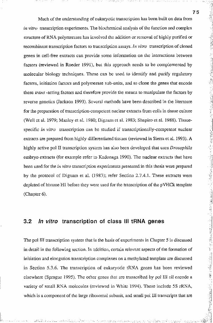
7 5
Much of the understanding of eukaryotic transcription has been built on data from
in vitro transcription experiments. The biochemical analysis of the function and complex
structure of RNA polymerases has involved the addition or removal of highly purified or
recombinant transcription factors to ti anscription assays. In vitro transcription of cloned
genes in cell-free extracts can provide some information on the interactions between
factors (reviewed in Roeder 1991), but this approach needs to be complemented by
molecular biology techniques. These can be used to identify and purify regulatory
factors, initiation factors and polymerase sub-units, and to clone the genes that encode
these trans -acting factors and therefore provide the means to manipulate the factors by
reverse genetics (Jackson 1993). Several methods have been described in the literature
for the preparation of transcription-competent nucleai' extracts from cells in tissue culture
(Weil et al. 1979; Manley et al, 1980; Dignam et al. 1983; Shapiro et ai. 1988). Tissue-
specific in vitro transcription can be studied if transcriptionally-competent nuclear
extracts are prepared from highly-differentiated tissues (reviewed in Sierra et al. 1993). A
highly active pol II transcription system has also been developed that uses Drosophila
embryo extracts (for example refer to Kadonaga 1990). The nuclear extracts that have
been used for the in vitro tianscription experiments presented in this thesis were prepared
by the protocol of Dignam et al. (1983); refer Section 2.7.4.1. These extracts were
depleted of histone HI before they were used for the transcription of the pVHCk template
(Chapter 6).
I
3,2 In vitro transcription of class III tRNA genes
The pol HI transcription system that is the basis of experiments in Chapter 5 is discussed
in detail in the following section. In addition, certain relevant aspects of the formation of
initiation and elongation transcription complexes on a methylated template are discussed
in Section 5.3.6. The transcription of eukaryotic tRNA genes has been reviewed
elsewhere (Sprague 1995). The other genes that are transcribed by pol III all encode a
variety of small RNA molecules (reviewed in White 1994). These include 5S rRNA,
which is a component of the large ribosomal subunit, and small pol III transcripts that are
7 6
encoded viral class III genes. For example, adenovirus encodes the VAi and VAn
transcripts, which subvert the translational machinery of an infected cell to the efficient
production of viral proteins.
3.2.1 tRNA genes, pol III transcription and DNA méthylation
Transfer RNA molecules are 70 to 90 nucleotides in length. They function as the adaptor
molecules that translate the genetic information carried by mRNA into a particular order
of amino acid residues in a protein. A tRNA molecule is able to recognise a particular
amino acid and match this to the correct codon in the message. There are 50 to 100
distinct tRNA species in most eukaryotic cells, with the relative abundance of a species
correlating to the usage frequency of the codon (reviewed in Sharp et al. 1984). The
haploid human genome contains 10 to 20 copies of each of about 60 different tRNA
genes, to give a total of approximately 1300 genes. The genes that encode tRNA often
occur in complex multigene families dispersed throughout the eukaryotic genome. In
higher eukaryotes, tRNA genes are organised into clusters (Rosenthal and Doering
1983). Some human tRNA genes occur in clusters of two, three or four genes (for
example, refer to Shortridge et al. 1989). Bourn et al. (1994) have isolated and
characterised a genomic clone from human placenta that contains the tRNA genes for
lysine, glutamine, leucine and arginine in a cluster of 2 kbp, as well as a gene for
tRNA^ly that is within 4 kbp. I have sub-cloned these tRN A ^ë and tRNA^^u genes into
the pUC19 vector, to construct the pArg/Leu template for transcription (see Section
2.5.1).
The promoters of most class III genes include intragenic elements, termed internal
control regions (ICRs). Transfer RNA genes, as well as most class III genes, have type
II ICRs that consist of two essential and highly conserved domains. The A-block (also
known as the 5’ ICR) has the consensus sequence TGGCNNAGTGG and encodes the
D-loop region of the tRNA gene product. The B-block (or 3 ’ ICR) has the consensus
sequence GGTTCGANNCC and encodes the TTC-loop. The high degree of conser
vation of these blocks therefore reflects their dual role in promoter function and tRNA
structure. The sequence of the tRNA and tRNA genes is shown in Figure 3.1,
7 7
tRNA^^S gene
+1 8
GGCTCTG
(1105)
IAT AGCGCATTGG ACTTCTAGTG ACGAATAGAG
A-block
+51I
67
CAATTCAAAG GTTGTG GGTT CGAATCC
B-block
77 88
CAC CAGAGTCG
(1193)
tRNA*-eu gene
+1 8
GGTAGCG
(576)
18
TC TAAGGCGCTG GATTTAGGCT CCAGTCTCTT
A-block
+51 61
CGGAGGCGTG QQTTCGAHTC C
B-block
71 82
CACCGCTGC CA
(494)
Figure 3.1
Sequences of the t R N A ^ r g and t R N A ^ e u genesCoding strand sequences of the two tRNA genes that were transcribed in pol III transcription assays are shown. The extent of the sequences correspond to the nascent transcript, beginning at the +1 position and finishing at the site of termination, as indicated by the numbers above the sequences. The positions of the A-block (consensus sequence TGGCNNAGTGG) and B-block (consensus sequence GGTTCGANNCC) are shown for each gene. The expected splice sites for the tRNA^rg precursor are indicated in the DNA sequence by the two arrows. Refer to Bourn et al. (1994) for further details. The positions of these coding regions in pArg/Leu (see Section 2.5.1 and Figure 2.1) are indicated by the numbers in brackets under the sequence.
7 8
with the positions of A-blocks and B-blocks for each gene highlighted by the boxes. The
separation between A- and B-blocks can vary from 30 to 60 bp without affecting the
efficiency of transcription. This is necessary because extra arms in tRNA molecules can
vary in length and some tRNA genes contain introns (reviewed in Westaway and
Abelson 1995). For example, the tR N A ^ë gene shown in Figure 3.1 contains an intron
of 15 bp (Bourn et al. 1994). Introns have been found in the tRNA genes of
Archaebacteria , fungi and many eukaryotic organisms, although the best characterised
system is that of the yeast, Saccharomyces cerevisiae (Westaway and Abelson 1995).
The splicing mechanisms that remove intions are diverse, but in all cases the intervening
sequences are removed from the precursor or nascent tRNA (pre-tRNA) to form the
functional product. The expected splice sites for the tRNA^^^g gene in the figure are
indicated by arrows, and the splicing of the gene product is discussed further in Section
3.2.2. Many other steps are involved in the maturation of pre-tRNAs, including the
removal of the 5 ’ leader and 3’ trailing sequences (Deutscher 1984), tRNA base
modification (Bjork et al. 1987) and the addition of the CCA sequence to the 3’ end of
the tR N A .
The ICR binds the polypeptides that make up the TFIIIC transcription factor
fraction (Gabrielsen and Sentenac 1991). TFIIIC is one of the three traditional fractions
that are obtained after purification of transcription factors from yeast, that together are
sufficient to reconstitute tRNA transcription in vitro . The other two fractions are TFIIIB
and polymerase. Regulation of tRNA genes is necessary for the adaptation of the tRNA
population to the different frequencies of codon usage and amino acid utilisation in
different cell types. Variations in the 5' flanking sequences of tRNA genes from yeast,
silk-worms, mice and humans (for example, refer to Gonos and Goddard 1990) have
been shown to affect the in vitro transcription of these genes (reviewed in Sprague
1995). A recent study has shown that the 5 ’ flanking sequence negatively modulates the
in vivo expression of a human tRNA gene (Tapping et al. 1993). The protein
components of the TFIIIB fraction, including the TATA-binding protein (TBP), are
presumed to bind to 5 ’ flanking promoter elements which would allow tissue-specific
and developmental regulation of expression (Sprague 1995).
'1I
:
1
3 .2 .2 In vitro transcription of the pArg/Leu template
7 9
DNA méthylation can inhibit the expression of class III genes (see Sections 1.7
and 1.8.3). Méthylation has been shown to inhibit the transcription of a methylated
tRNAT^u gene, after the micro-injection of a construct containing this gene into Xenopus
oocytes (Besser et al. 1990). Inhibition was only observed if the CpG dinucleotides in
the B-block were methylated, but not if the methylated sites were elsewhere. In contrast,
an oocyte 5S rRNA gene was not affected by méthylation. The expression of the
adenovirus type 2 VAi gene is inhibited by méthylation at CpG sites, either after
transfection into mammalian cells or in a pol III in vitro transcription system (Jüttermann
et al. 1991). Transfer RNA genes are perfect examples of housekeeping genes and, in
general, they are associated with CpG islands (see Section 1.6). For example, the if
genomic clone that was isolated and characterised by Bourn et al. (1994) contained an |I'
extensive GC-rich region associated with the cluster of tRNA genes (see Figure 2. IB). #I'l
Genomic tRNA sequences in human leukocytes lack CpG suppression and are
unmethylated at certain sites (Schorderet and G artier 1990).
Since these studies were published, the effect of méthylation on class III gene
expression has been seldom investigated. I decided to investigate the expression of tRNA
genes for this reason, but also because the pol III transcription assay (described in
Section 2.7.4.2) has many advantages as a system. Most importantly, it is relatively
straight-forward to perform, gives labelled transcription products of a high specific
activity and is quite resilient to RNase contamination.
I
The protocol for the pol III in vitro transcription assay (Section 2.7.4.2) has been
developed and optimised for tRNA genes by Gonos and Goddard (1990), and in a later
study by Bourn et al. (1994). The important points of this protocol are that the optimal
Mg2+ concentration for this transcription system is 3.5 mM, creatine phosphate is
included in the reaction mixture to allow the regeneration of ribonucleoside triphosphate
levels and the saturating level of template is approximately 150 ng, although transcription
can be detected with as little as 10 ng of template. Sierra et al. (1993) describe a similar
protocol that uses the G-free cassette to analyse pol II transcription. My own
:
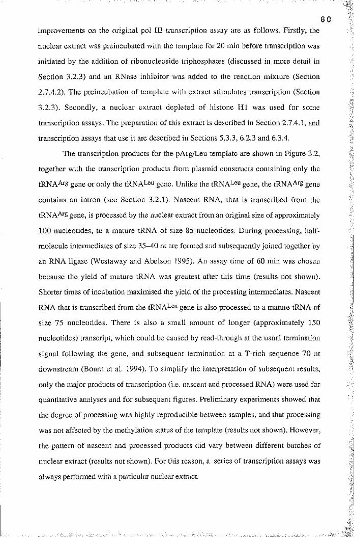
8 0
improvements on the original pol III transcription assay are as follows. Firstly, the
nuclear extract was preincubated with the template for 20 min before transcription was
initiated by the addition of ribonucleoside triphosphates (discussed in more detail in
Section 3.2.3) and an RNase inhibitor was added to the reaction mixture (Section
2.T.4.2). The preincubation of template with extract stimulates transcription (Section
3.2.3). Secondly, a nuclear extract depleted of histone H I was used for some
transcription assays. The preparation of this extract is described in Section 2.7.4.1, and
transcription assays that use it are described in Sections 5,3.3, 6.2.3 and 6.3.4.
The transcription products for the pArg/Leu template are shown in Figure 3.2,
together with the transcription products from plasmid constructs containing only the
tR N A ^g gene or only the tRNA^^u gene. Unlike the tRNA^-^u gene, the tRNA^^^S gene
contains an intron (see Section 3.2.1). Nascent RNA, that is transcribed from the
tRNA^S gene, is processed by the nuclear exti'act from an original size of approximately
100 nucleotides, to a mature tRNA of size 85 nucleotides. During processing, half
molecule intermediates of size 35-40 nt are formed and subsequently joined together by
an RNA ligase (Westaway and Abelson 1995). An assay time of 60 min was chosen
because the yield of mature tRNA was greatest after this time (results not shown).
Shorter times of incubation maximised the yield of the processing intermediates. Nascent
RNA that is transcribed from the tRNA^eu gene is also processed to a mature tRNA of
size 75 nucleotides. There is also a small amount of longer (approximately 150
nucleotides) transcript, which could be caused by read-through at the usual termination
signal following the gene, and subsequent termination at a T-rich sequence 70 nt
downstream (Bourn et al. 1994). To simplify the interpretation of subsequent results,
only the major products of transcription (i.e. nascent and processed RNA) were used for
quantitative analyses and for subsequent figures. Preliminary experiments showed that
the degree of processing was highly reproducible between samples, and that processing
was not affected by the méthylation status of the template (results not shown). However,
the pattern of nascent and processed products did vary between different batches of
nuclear extract (results not shown). For this reason, a series of transcription assays was
always performed with a particular nuclear extiact.
:
If
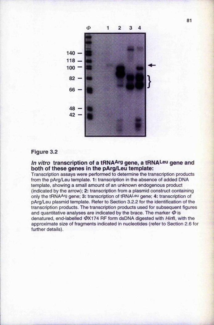
810 3 4
140 — f t118 — t é100 — #
82 - « •
66 — #
4842
Figure 3.2
In vitro transcription of a t R N A A r g gene, a t R N A L e u gene and both of these genes in the pArg/Leu template:Transcription assays were performed to determine the transcription products from the pArg/Leu template. 1 : transcription in the absence of added DNA template, showing a small amount of an unknown endogenous product (indicated by the arrow); 2: transcription from a plasmid construct containing only the tRNAArg gene; 3: transcription of tRNALeu gene; 4: transcription of pArg/Leu plasmid template. Refer to Section 3.2.2 for the identification of the transcription products. The transcription products used for subsequent figures and quantitative analyses are indicated by the brace. The marker 0 is denatured, end-labelled 0X174 RF form dsDNA digested with H/nfl, with the approximate size of fragments indicated in nucleotides (refer to Section 2.6 for further details).
82
0 1000 100 10
a-amanitin ( n M )
Figure 3.3
Inhibition of pol III transcription by a-amanitin:In vitro transcription of the un methylated pArg/Leu template was assayed at the indicated concentrations (from 1000 to 0 nM) of the RNA polymerase III inhibitor a-amanitin. The control lane C is an assay performed in the absence of DNA template. The arrow indicates an artifactual band that is present in all lanes, irrespective of the a-amanitin concentration. The transcription product bands are smeared due to contamination by RNase. See Section 3.2.2 for further details.
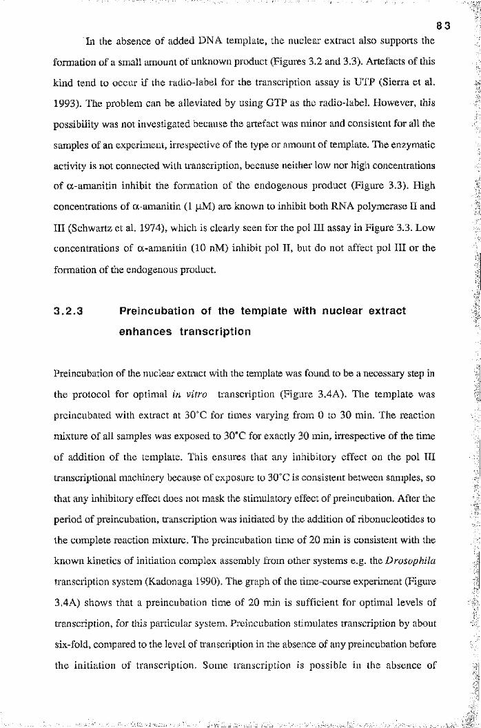
8 3In the absence of added DNA template, the nuclear extract also supports the
formation of a small amount of unknown product (Figures 3.2 and 3.3). Artefacts of this
kind tend to occur if the radio-label for the transcription assay is UTP (Sierra et al.
Preincubation of the nuclear extract with the template was found to be a necessary step in
the protocol for optimal in vitro transcription (Figure 3.4A). The template was
preincubated with extract at 30“C for times varying from 0 to 30 min. The reaction
mixture of all samples was exposed to 30“C for exactly 30 min, irrespective of the time
of addition of the template. This ensures that any inhibitory effect on the pol III
transcriptional machinery because of exposure to 30“C is consistent between samples, so
that any inhibitory effect does not mask the stimulatory effect of preincubation. After the
period of preincubation, transcription was initiated by the addition of ribonucleotides to
the complete reaction mixture. The preincubation time of 20 min is consistent with the
known kinetics of initiation complex assembly from other systems e.g. the Drosophila
transcription system (Kadonaga 1990). The graph of the time-course experiment (Figure
3.4A) shows that a preincubation time of 20 min is sufficient for optimal levels of
transcription, for this particular system. Preincubation stimulates transcription by about
six-fold, compared to the level of ti*anscription in the absence of any preincubation before
the initiation of transcription. Some transcription is possible in the absence of
1993). The problem can be alleviated by using GTP as the radio-label. However, this
possibility was not investigated because the artefact was minor and consistent for all the
samples of an experiment, iiTespective of the type or amount of template. The enzymatic
activity is not connected with transcription, because neither low nor high concentrations
of a-am anitin inhibit the formation of the endogenous product (Figure 3.3). High
concentrations of a-amanitin (1 |iM) are known to inhibit both RNA polymerase II and
III (Schwartz et al. 1974), which is clearly seen for the pol III assay in Figure 3.3. Low
concentrations of a-amanitin (10 nM) inhibit pol II, but do not affect pol III or the
formation of the endogenous product.
3.2 .3 Preincubation of the template with nuclear extract
enhances transcription
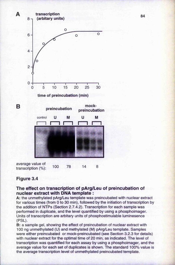
transcription(arbitary units)
4 -
10 15 20 25 300 5
84
time of preincubation (min)
B . . mock-preincubation preincubation
control U M U M
average value of transcription (%): 100 78 14 8
Figure 3.4
The effect on transcription of pArg/Leu of preincubation of nuclear extract with DNA template ;A: the unmethylated pArg/Leu template was preincubated with nuclear extract for various times (from 0 to 30 min), followed by the initiation of transcription by the addition of NTPs (Section 2.7.4.2). Transcription for each sample was performed in duplicate, and the level quantified by using a phosphoimager. Units of transcription are arbitary units of phosphostimulable luminesence (PSL).B: a sample gel, showing the effect of preincubation of nuclear extract with 100 ng unmethylated (U) and methylated (M) pArg/Leu template. Samples were either preincubated or mock-preincubated (see Section 3.2.3 for details) with nuclear extract for the optimal time of 20 min, as indicated. The level of transcription was quantified for each assay by using a phosphoimager, and the average value for each set of duplicates is shown. The standard 100% value is the average transcription level of unmethylated preincubated template.
8 5
preincubation because initiation complex formation can form during the elongation period
of the transcription assay, after the NTPs have been added to the reaction mixture. The
elongation period for this system is 60 min.
The effect of preincubation is also seen in Figure 3.4B. Unmethylated and
methylated pArg/Leu template were preincubated with nuclear extract for the optimal time
of 20 min. Mock-preincubations for the same templates were also carried out, for which
the reaction mixtures were exposed to 30“C for 20 min in the absence of template. The
template was then added with the NTPs at the initiation of transcription. The
preincubation treatment stimulates transcription six to ten-fold, but is less effective for the
methylated template. The final level of tianscription from the methylated template is also
lower, being 80% of that from the unmethylated template. This preferential inhibition of
the methylated template is discussed further in Chapter 5; for example, refer to Figure
5.5.
3.3 In vitro transcription of the class II SV40 promoter
The pol II transcription system that is the basis of several experiments in Chapter 6 is
discussed in the following section. The transcription of eukaryotic class II genes has
been reviewed in detail elsewhere (Buratowski 1994), and will not be discussed except in
general terms. The initiation of transcription from the SV40 promoter in the pVHCk
plasmid template (Figure 3.5) was assayed by using a primer extension approach (see
Section 2,7.4.3 for a description of the protocol). The principle of primer extension
involves the hybridisation of an end-labelled oligonucleotide to RNA, that has been
transcribed from the reporter gene, followed by extension of the oligonucleotide primer
using the enzyme reverse transcriptase. The extension reaction tenninates at the extreme
5’ end of the RNA, and can therefore be used to determine the start point of transcription
of an mRNA sequence. Protocols for primer extension can be found elsewhere
(Sambrook et al. 1989; Leonard and Patient 1991; Mason et al. 1993). The SV40
promoter and pol II transcription are discussed in Section 3.3.1. Preliminary experiments
to optimise this pol II transcription system are described in Sections 3.3.2 and 3.3.3.
Further optimisations of the primer extension protocol are described in Section 3.3.4,
and are concerned with the optimal amounts of template, end-labelled primer and the
concentration of Mg^+ that is required for m vitro transcription in this system.
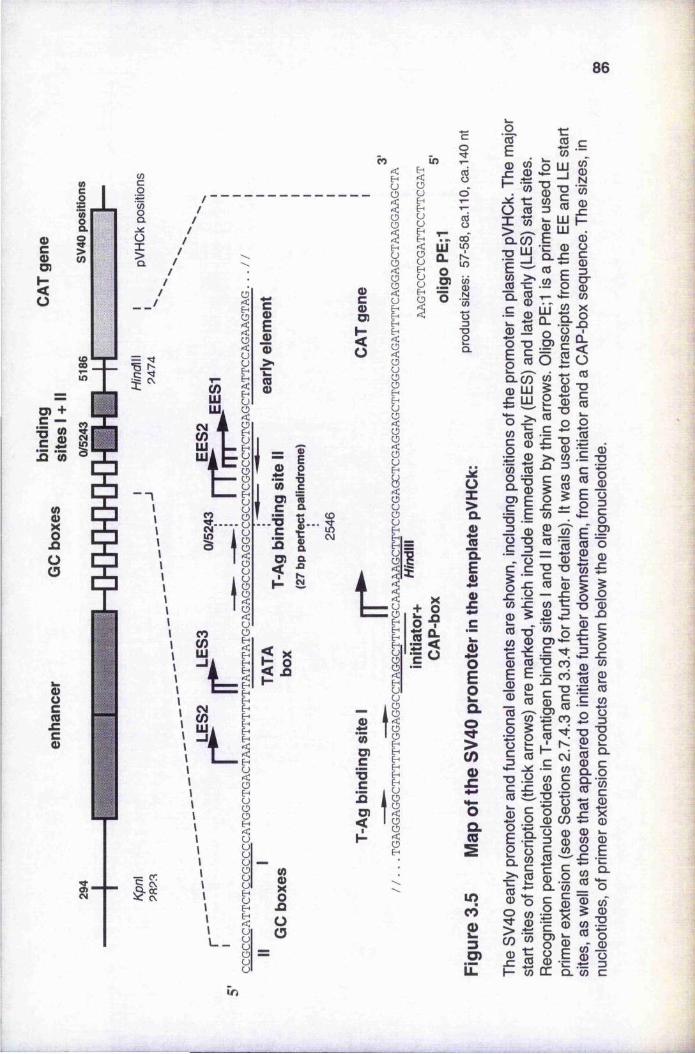
86
0)gO)
<ü
s
II(0
1(/)
IüIs
=5 “ a
P!3LU
œLU
0)og£co
S)
II
5uE-*
;
A i■ i l
ÇJ
l" v |II[H H
l__
' L
gE0)Q>>
1
— EB 2 •55 1O) Q
• I i - iO) a
2 s
P
eo in ?
IOO
0)coD)
S
g
IIIEhH
ïEh
gU Eh Eh U UEh .Eh m CO
i “ 6oB So -s
i I(OU)c’■5cZO)
üX
IOL
I0>JZ
o>co0)£oOL(0
incô23O)iZ
oco
I Iü m
Q.CO
E — ■c'
Q- S.5 Q)
c5
■C
o LU _J
11|1S"C CDû-:£CO en
IEg -Û.C/D CD LUs y i■s-ê-
III II I
Ien .c
1 0
I Il ï112 :«
'cô
1?co CD
0 üg
IIÛL < ü
4— 0TJC0
IC0
" s i
1E2CL m
0 "OB ^0 0 E E 0 © ® 0
§
0 O 2 0 "en S
i-l"O 0k_ X»2 c
-g sI £
s î1 1T3 .20 ê0
!î îI I (/) (0
11
|S•— w 0 c 0 o
l îü ©
!0 OQ. 0
i lII
5c©
I II I 0 »- 0 0g E
«3
i S
«1
•1 iirj
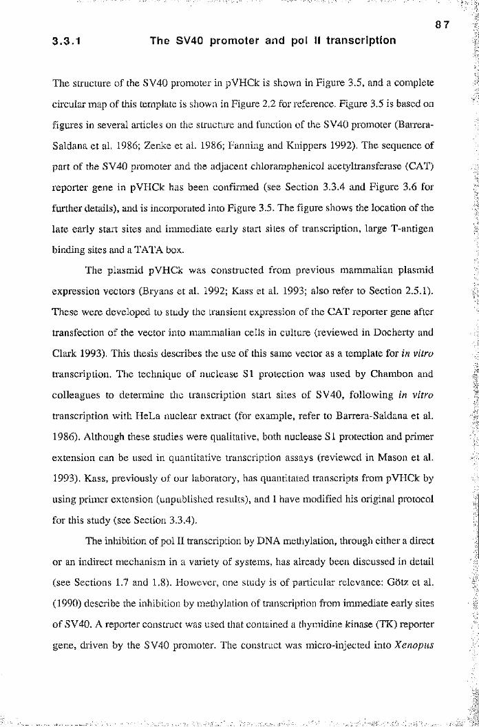
' i * : .
8 7
3.3.1 The SV40 promoter and pol II transcription
The structure of the SV40 promoter in pVHCk is shown in Figure 3.5, and a complete
circular map of this template is shown in Figure 2.2 for reference. Figure 3.5 is based on
figures in several articles on the structure and function of the SV40 promoter (Barrera-
Saldana et al. 1986; Zenke et al. 1986; Fanning and Knippers 1992). The sequence of
part of the SV40 promoter and the adjacent chloramphenicol acetyltransferase (CAT)
reporter gene in pVHCk has been confirmed (see Section 3.3.4 and Figure 3.6 for
further details), and is incorporated into Figure 3.5. The figure shows the location of the
late early start sites and immediate early start sites of transcription, large T-antigen
binding sites and a TATA box.
The plasmid pVHCk was constructed from previous mammalian plasmid
expression vectors (Bryans et al. 1992; Kass et al. 1993; also refer to Section 2.5.1).
These were developed to study the transient expression of the CAT reporter gene after
transfection of the vector into mammalian cells in culture (reviewed in Docherty and
Clark 1993). This thesis describes the use of this same vector as a template for in vitro
transcription. The technique of nuclease SI protection was used by Chambon and
colleagues to determine the transcription start sites of SV40, following in vitro
transcription with HeLa nuclear extract (for example, refer to Barrera-Saldana et al.
1986), Although these studies were qualitative, both nuclease SI protection and primer
extension can be used in quantitative transcription assays (reviewed in Mason et al.
1993). Kass, previously of our laboratory, has quantitated transcripts from pVHCk by
using primer extension (unpublished results), and I have modified his original protocol
for this study (see Section 3.3.4).
The inhibition of pol II transcription by DNA méthylation, through either a direct
or an indirect mechanism in a variety of systems, has already been discussed in detail
(see Sections 1.7 and 1.8). However, one study is of particular relevance: Gotz et al.
(1990) describe the inhibition by méthylation of transcription from immediate early sites
of SV40, A reporter construct was used that contained a thymidine kinase (TK) reporter
gene, driven by the SV40 promoter. The construct was micro-injected into Xenopus
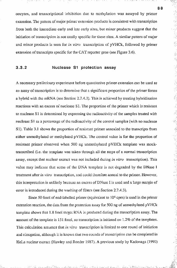
8 8
oocytes, and transcriptional inhibition due to méthylation was assayed by primer
extension. The pattern of major primer extension products is consistent with transcription
from both the immediate early and late early sites, but minor products suggest that the
initiation of transcription is not totally specific for these sites. A similar pattern of major
and minor products is seen for in vitro transcription of pVHCk, followed by primer
extension of transcripts specific for the CAT reporter gene (see Figure 3.6).
3 .3 .2 Nuclease S1 protection assay
A necessary preliminary experiment before quantitative primer extension can be used as
an assay of transcription is to deteimine that a significant proportion of the primer forms
a hybrid with the mRNA (see Section 2.7.4.3). This is achieved by treating hybridisation3
reactions with an excess of nuclease SI. The proportion of the primer which is resistant-
to nuclease SI is determined by expressing the radioactivity of the samples treated with
nuclease SI as a percentage of the radioactivity of the control samples (with no nuclease el
SI). Table 3.1 shows the proportion of resistant primer annealed to the transcripts from.
either unmethylated or methylated pVHCk. The control value is for the proportion of
resistant primer observed when 500 ng unmethylated pVHCk template was mock-
transcribed (i.e. the template was taken through all the steps of a normal transcription
assay, except that nuclear exti'act was not included during in vitro transcription). This
value may indicate that some of the DNA template is not degraded by the DNase Î
treatment after in vitro transcription, and could therefore anneal to the primer. However,
this interpretation is unlikely because an excess of DNase I is used and a large margin of
error is introduced during the washing of filters (see Section 2.7.4.3).
Since 50 fmol of end-labelled primer (equivalent to 10 cpm) is used in the primer
extension reaction, the data from the protection assay for 500 ng of unmethylated pVHCk
template shows that 1.8 fmol target RNA is produced during the transcription assay. The
amount of the template is 151 fmol, so ti'anscription is initiated on 1.2% of the templates.
This calculation assumes that in vitro transcription is limited to one round of initiation
and elongation, although it is known that two rounds of transcription can be completed in
HeLa nuclear extract (Hawley and Roeder 1987). A previous study by Kadonaga (1990)
: : S
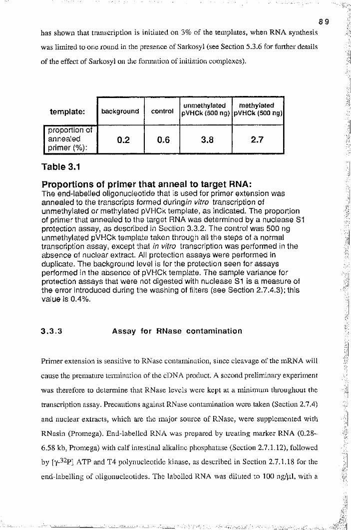
8 9
has shown that transcription is initiated on 3% of the templates, when RNA synthesis
was limited to one round in the presence of Sai’kosyl (see Section 5.3.6 for further details
of the effect of Sarkosyl on the formation of initiation complexes).
template: background controlunmethylated
pVHCk (500 ng)methylated
pVHCk (500 ng)
proportion of annealed primer (%):
0.2 0.6 3.8 2.7
Table 3.1
Proportions of primer that anneal to target RNA:The end-labelled oligonucleotide that is used for primer extension was annealed to the transcripts formed during//? vitro transcription of unmethylated or methylated pVHCk template, as indicated. The proportion of primer that annealed to the target RNA was determined by a nuclease S1 protection assay, as described in Section 3.3.2. The control was 500 ng unmethylated pVHCk template taken through all the steps of a normal transcription assay, except that in vitro transcription was performed in the absence of nuclear extract. All protection assays were performed in duplicate. The background level is for the protection seen for assays performed in the absence of pVHCk template. The sample variance for protection assays that were not digested with nuclease 81 is a measure of the error introduced during the washing of filters (see Section 2.7.4.3); this value is 0.4%.
3.3 .3 Assay for RNase contamination
Primer extension is sensitive to RNase contamination, since cleavage of the mRNA will
cause the premature termination of the cDNA product. A second preliminary experiment
was therefore to determine that RNase levels were kept at a minimum throughout the
transcription assay. Precautions against RNase contamination were taken (Section 2.7.4)
and nuclear extracts, which are the major source of RNase, were supplemented with
RNasin (Promega). End-labelled RNA was prepared by treating marker RNA (0.28-
6.58 kb, Promega) with calf intestinal alkaline phosphatase (Section 2.7.1.12), followed
by [y-^^P] ATP and T4 polynucleotide kinase, as described in Section 2.7.1.18 for the
end-labelling of oligonucleotides. The labelled RNA was diluted to 100 ng/jil, with a
I
: |
1iÎ
9 0
final specific activity of 10 cpnVpl. The integrity of the labelled marker was checked on
a denaturing gel (Section 2.7.4.2), and the bulk of the RNA was treated as described
below.
Each reaction mixture was spotted onto discs of DE81 paper and washed as
described in Section 2.7.4.3. Intact, labelled RNA remained bound to the filters.
However, 5’ exonuclease or endonuclease activity degraded the RNA into small enough
fragments so that the label could not bind to the filters, and was removed during the
washing procedure. This assay can test for RNase contamination, but it is only a
qualitative indicator of this activity because a large margin of error is introduced during
the washing procedure. The results are tabulated in Table 3.2.
control RNaseRNase + inhibitor
nuclearextract
extract + inhibitor
proportion of intact RNA (%):
100 9 86 35 95
Table 3.2
Assay for contamination of nuclear extract by RNase:RNase contamination was assayed as described in Section 3.3.3. The proportion of Intact end-labelled RNA was measured after treatment with RNase A or nuclear extract, as indicated. RNase activity could be inhibited by the addition of RNasin inhibitor to the reaction mixture (see Section 3.3.3). The control for the 100% standard is the radioactivity retained on filters after the labelled RNA has not been exposed to RNase activity. The sample variance for the control is 13%.
Aliquots of labelled RNA (1.0 pi) were treated either with 10 pg RNase A diluted
in 9 p i RNase-free buffer D (the buffer used for the dialysis of nuclear extracts; see
Section 2.7.4.1), or with RNase supplemented with 40 U RNasin to test the efficiency of
this inhibitor. The definition of one unit activity of RNasin is the inhibition of 5 ng
RNase by 50%. Reaction mixtures were incubated at 42“C for 30 min, and performed in
duplicate. Table 3.2 shows that this amount of RNasin can inhibit the action of RNase,
so that minimal degradation occurs. Labelled RNA was also treated either with 9 pi
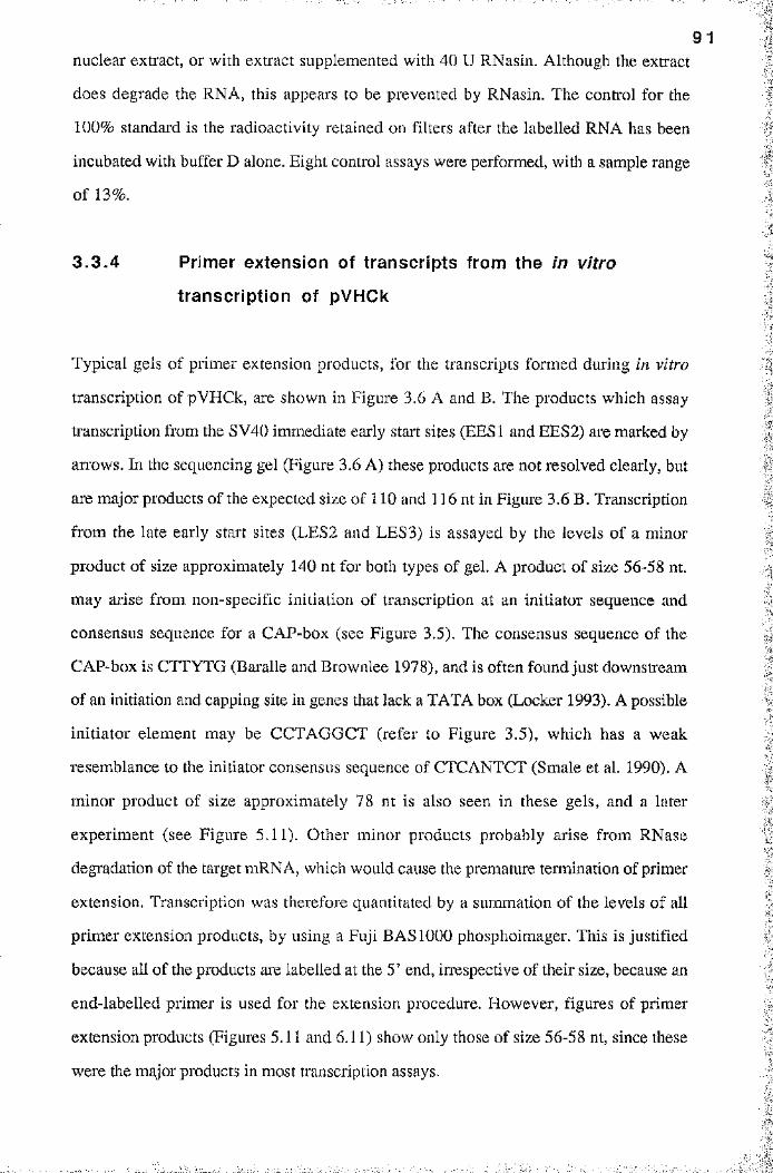
9 1
nuclear extract, or with extract supplemented with 40 U RNasin. Although the extract
does degrade the RNA, this appears to be prevented by RNasin. The control for the
100% standard is the radioactivity retained on filters after the labelled RNA has been
incubated with buffer D alone. Eight control assays were performed, with a sample range
of 13%.
3 .3 .4 Primer extension of transcripts from the in vitro
transcription of pVHCk
Typical gels of primer extension products, for the transcripts formed during in vitro
transcription of pVHCk, are shown in Figure 3.6 A and B. The products which assay
transcription from the SV40 immediate early start sites (EESl and EES2) are marked by
arrows. In the sequencing gel (Figure 3.6 A) these products are not resolved clearly, but
are major products of the expected size of 110 and 116 nt in Figure 3.6 B. Transcription
from the late early start sites (LES2 and LES3) is assayed by the levels of a minor
product of size approximately 140 nt for both types of gel, A product of size 56-58 nt.
may arise from non-specific initiation of transcription at an initiator sequence and
consensus sequence for a CAP-box (see Figure 3.5). The consensus sequence of the
CAP-box is CTTYTG (Baralle and Brownlee 1978), and is often found just downstream
of an initiation and capping site in genes that lack a TATA box (Locker 1993). A possible
initiator element may be CCTAGGCT (refer to Figure 3.5), which has a weak
resemblance to the initiator consensus sequence of CTCANTCT (Smale et al. 1990). A
minor product of size approximately 78 nt is also seen in these gels, and a later
experiment (see Figure 5.11). Other minor products probably arise from RNase
degradation of the target mRNA, which would cause the premature termination of primer
extension. Transcription was therefore quantitated by a summation of the levels of all
primer extension products, by using a Fuji BAS 1000 phosphoimager. This is justified
because all of the products are labelled at the 5’ end, irrespective of their size, because an
end-labelled primer is used for the extension procedure. However, figures of primer
extension products (Figures 5.11 and 6.11) show only those of size 56-58 nt, since these
were the major products in most transcription assays.
_ _ _
nt
200
151140
118
100
82
0 T G C AC O pB U M 92
66
48
40
J ca.140
} ca.110
56-58
47
39
Figure 3.6 A Primer extension products from transcripts of pVHCk:see overleaf for figure legend

93nt
200
151140
118
100
82
66
C O pB U M
ca. 140
116110
56-58
Figure 3.6 A and B:
The use of primer extension products to assay levels of in vitro transcription from mock-methylated (U) and methylated (M) pVHCk plasmid template:A: Transcripts from pVHCk are quantitated by primer extension, which gives rise to major extension products (marked by arrows) and several minor products. The products were resolved on a 0.4 mm denaturing, 12% polyacrylamide sequencing geI.The size of products is determined by comparison with: 0 (denatured, end-labelled 0X174/Hinfl marker; the approximate size of denatured fragments is given in nucleotides under nt) and sequencing tracts T, G, C and A. The sequence was obtained by using end-labelled primer extension oligonucleotide (oligo PE;1) and doublestranded pVHCk as template. Extension products: 0, primer only; O, no DNA; pB, 150 ng pBluescript KS-; U, 150 ng mock-methylated pVHCk; M, 150 ng methylated pVHCk.B: the same legend as for Figure 3.6 A, except that sequencing tracts were not run on this gel. The gel was a denaturing, 12% PAGE gel of 1.5 mm in thickness, so does not resolve the primer extension products as well as the sequencing gel. The autoradiograph of this transcription assay was kindly provided by Dr. S U Kass, and is used with his permission in this thesis.
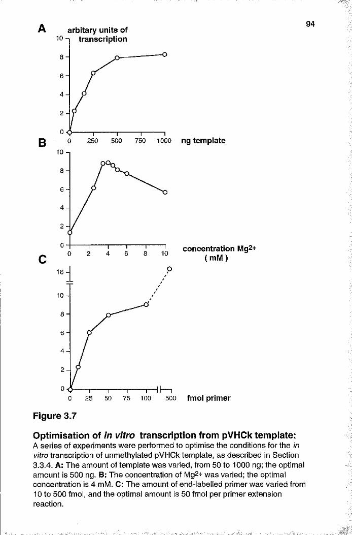
B
arbitary units of94
Figure 3.7
1 0 transcription
8
6
4
2
0500 750 10000 250
10
8
6
4
2
0104 6 80 2
concentration M g2+ ( mM )
16
10
8
6
4
2
025 50 75 100 5000 fmol primer
Optimisation of in vitro transcription from pVHCk tempiate:A series of experiments were performed to optimise the conditions for the in vitro transcription of unmethylated pVHCk template, as described in Section 3.3.4. A: The amount of template was varied, from 50 to 1000 ng; the optimal amount is 500 ng. B: The concentration of Mg2+ was varied; the optimal concentration is 4 mM. C: The amount of end-labelled primer was varied from 10 to 500 fmol, and the optimal amount is 50 fmol per primer extension reaction.
I
9 5The pol II transcription assay was optimised as described below, and summarised
in Figure 3.7. The optimal amount of template for this system was found to be 500 ng
(graph A) and the optimal concentration of Mg^+ was 4.0 mM (graph B). Quantitative
primer extension of transcription products requires that the primer is in an excess (at least
ten-fold) molar ratio to the amount of target RNA (Mason et al. 1993). The titration
experiment that is summarised in graph C varies the amount of primer from 10 fmol to
500 fmol. Nuclease SI protection (see Section 3.3.2) has shown that 500 ng of template
can produce 1.8 fmol of RNA during the course of the transcription assay. The plateau in
graph C shows that all the target RNA becomes annealed to primer, at moderate levels
(25-100 fmol) of the primer. At the highest level of primer (500 fmol), the apparent
increase in transcription is caused by non-specific annealing during the extension
reaction.
A-.1
I
iI
9 6
# #
CHAPTER FOUR
Chromatin assembly in vitro
4.1 Introduction■!;
:
A current challenge for molecular and developmental biologists is to understand the
molecular mechanisms through which chromatin and chromosome structure influence
gene activity. A number of methods have been developed to reconstitute chromatin in
vitro. These have been coupled to assays that measure either access of the DNA to trans-
acting factors or transcription in vitro. These studies have allowed major insights to be
made into the structure and function of chromatin.
High salt concentrations dissociate nucleosome core particles into their
components of DNA and histones. The first methods to assembly chromatin in vitro
reversed this process, and purified core histones were reconstituted onto DNA by
dialysis from high salt (reviewed in Dimitrov and Wolffe 1995). A typical method is
described in Rhodes and Laskey (1989). Further refinements of this method of chromatin
assembly have been the exchange of octamers from donor chi’omatin to recipient DNA at
high salt (Tatchell and van Holde 1977; Drew and Travers 1985) and the use of a
negatively chai’ged third component as an assembly factor. The use of poly (glutamic
acid) as a nucleosome and chromatin assembly factor has been reviewed elsewhere (Stein
1989). These methods have a serious drawback: the nucleosomes are not positioned with
physiological spacing. Instead of the correct spacing of 180-200 bp found in native
chromatin, the histone octamers that are reconstituted onto DNA by these methods pack
together as closely as possible, with each octamer occupying 150-160 bp of DNA
(reviewed in Dimiti'ov and Wolffe 1995). Since the octamers are closely packed together
there is insufficient linker DNA for the correct binding of histone H I. Nucleosomal
9 7
arrays that are reconstituted with histone HI by the high salt or exchange methods result
in the formation of aggregates and the precipitation of DNA.
A second method of chromatin assembly was therefore chosen for the studies in
this thesis, to allow chromatin to be reconstituted with histone H I. Chromatin was
reconstituted on dsDNA using a crude cytoplasmic fraction derived ïvomXenopus eggs.
These extracts (prepared as described in Section 2.7.7.1) were used to reconstitute
chromatin in vitro on plasmid DNA (see Section 2.7.7.2). A typical method is described
in Wolffe and Schild (1991) and the technique is discussed in Section 4.2.1. In some
experiments the reaction mixture was supplemented with histone H I, added at a 0.6 w/w
ratio with respect to the DNA (see Section 4.2.2). Chromatin assembled in this way is
used in the studies described in Sections 6.2.3 and 6.3.4, that investigate the effect of
DNA méthylation and chromatin on in vitro transcription.
Nucleoplasmin is an acidic thermostable protein from Xenopus eggs that has
been used as an assembly factor during chromatin reconstitution with purified octamers
(Sealy et al. 1989). Two other acidic proteins called N1/N2 have been purified from eggs
(Kleinschmidt et al. 1990). N1/N2 appear to act with nucleoplasmin as molecular
chaperones that ensure the correct spacing of nucleosomes. Xenopus oocytes have been
used to prepare chromatin assembly extracts (for example, refer to Shimamura et al.
1989), but the quality of oocytes is more difficult to assess as compared to the quality of
eggs. Extracts from mammalian cells (Stillman 1986; Gruss et al. 1990) m à Drosophila
embryos (Kamakaka et al. 1993) have also been used for in vitro chromatin assembly.
Chromatin assembly in vitro with Xenopus egg extracts was chosen because the
quality of eggs was easy to assess, the preparation of extracts was straight-forward (see
Section 2.7.7.1) and chromatin was reconstituted on plasmid DNA with high efficiency.
The egg extract was not competent for the transcription of either tRNA genes or the
initiation of transcription from the SV40 promoter (refer to Sections 6.2.3 and 6.3.4).
Transcription could therefore be assayed from templates reconstituted with chromatin by
the use of HeLa nuclear extract, without endogenous ti'anscription due to the egg extr act
to complicate the interpretation of the data.
9 8
4.2 Chromatin assembly on double-stranded DNA
Chromatin was reconstituted on pVHCk plasmid, by using Xenopus egg extract. In
some experiments the reaction mixture was supplemented with histone H I, added at a
0.6 w/w ratio with respect to the DNA. Section 4.2.2 presents evidence that this amount
of histone HI is incorporated into the assembled chromatin at physiological levels. This
observation is necessary before it can be argued that the chr omatin formed on methylated
DNA, in the presence of histone H I, is a valid model of heterochromatin. The case for
this argument is presented in Section 6.4. The structure of chromatin reconstituted on
plasmid DNA was established by treatment of the chromatin assembly reaction mixture
with either staphylococcal nuclease or Msp I, as described in Section 2.1.13. The more
elaborate experiments described in Chapter 6 investigate the structure and transcriptional
activity of chromatin reconstituted on either fully methylated DNA (Sections 6.2.2 and
6.2.3) or DNA methylated in defined regions (Sections 6.3.3 and 6.3.4). These sections
in Chapter 6 show that the chromatin reconstituted in vitro on methylated DNA is more
resistant to digestion by Mspl, than is tliat formed on unmethylated DNA. This has been
shown previously to be true for methylated DNA in nuclei and for methylated DNA
transfected into mammalian cells (see Section 1.8.2), but not for an in vitro system. This
section will discuss the digestion of chromatin with staphylococcal nuclease, and will not
discuss Msp I digestion any further.
The digestion of assembled chromatin with staphylococcal nuclease results in
oligonucleosome fragments, which were deproteinised and the DNA resolved on agarose
gels (see Figures 4.1 and 4.2). The DNA was visualised by a Southern blotting
procedure (see Section 2.7.1.21), in all respects identical to the procedures used for the
series of Msp I fade-out assays described in Chapter 6. The DNA was hybridised to the
Pvu ll-Pvu II restriction fragment labelled as fragment d in Figure 6.1. Nucleosomal
ladders were easier to see on hybridised filters than on agarose gels stained with
ethidium, because the input amount of DNA was kept quite low to conserve the stocks of
Xenopus egg extract.
'■■■■■ ' ,
i
9 9
4.2.1 Chromatin assembly on unmethylated or
methylated pVHCk
Chromatin reconstituted on mock-methylated or fully-methylated pVHCk shows no
difference in the size of the spacing between nucleosomes (Figure 4.1). In both cases,
the spacing is 180 bp, decreasing to the expected 146 bp for nucleosome core DNA after
extensive nuclease digestion. The spacing value was determined by scanning the lanes in
the autoradiograph of Figure 4.1 in a densitometer, and comparing the positions of
oligonucleosomes with those of marker fragments in a semi-log plot (results not shown).
The same average spacing is also obtained from the densitometry traces in Figure 4.3 B.
This repeat length is typical for the chromatin formed with Xenopus egg extracts
(Dimitrov and Wolffe 1995), and for the 180-200 bp repeat length seen in the native
chromatin of normal somatic cells (Finch et al. 1975; Wolffe 1992).
4 .2 .2 Chromatin assembly on unmethylated or methylated
pVHCk, in the presence of histone HI
Chromatin was also reconstituted on unmethylated or methylated pVHCk in the presence
of histone HI (Figure 4.2). The level of histone HI was chosen after a series of
empirical experiments as that which increased the nucleosomal spacing to the greatest
extent. This level is an histone H1:DNA w/w ratio of 0.6 (i.e. 150 ng of histone HI was
added to a reaction mixture containing 250 ng DNA), which approximates the
physiological level of histone HI in chromatin. The physiological level is calculated by
assuming that 25 nucleosomes can form on pVHCk, which is 5025 bp in size. Each
nucleosome associates with one molecule of histone H I, which is indicated as a molar
ratio of histone HI to nucleosome of 1 in Figure 4.2. For convenience, the molar ratio is
represented as H l/n. An H l/n molar ratio of 1 corresponds to an histone H1:DNA w/w
ratio of 0.22, if the molecular mass of histone HI is taken to be 26 kDa. A w/w ratio of
0.60 therefore is equivalent to a molar ratio of 2.7 molecules of histone H I per
nucleosome. A previous study has shown that the incorporation of histone HI into
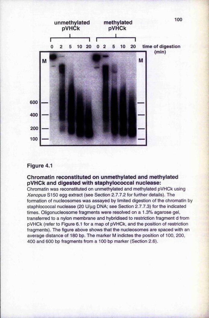
r
unmethylatedpVHCk
I___
2 5 10n20
methylatedpVHCk
I
100
10 2 5 10 20 time of digestion
(min)
Figure 4.1
Chromatin reconstituted on unmethylated and methylated pVHCk and digested with staphylococcal nuclease:Chromatin was reconstituted on unmethylated and methylated pVHOk using Xenopus SI 50 egg extract (see Section 2.7.7.2 for further details). The formation of nucleosomes was assayed by limited digestion of the chromatin by staphlococcal nuclease (20 U/pg DNA; see Section 2.7.7.3) for the indicated times. Oligonucleosome fragments were resolved on a 1.3% agarose gel, transferred to a nylon membrane and hybridised to restriction fragment d from pVHOk (refer to Figure 6.1 for a map of pVHCk, and the position of restriction fragments). The figure above shows that the nucleosomes are spaced with an average distance of 180 bp. The marker M indictes the position of 100, 200, 400 and 600 bp fragments from a 100 bp marker (Section 2.6).
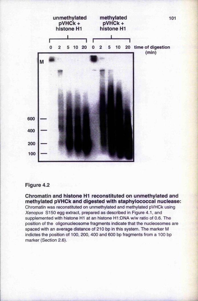
unmethylatedpVHCk +
histone H1
methylated pVHCk +
histone H1
I_____
101
I I I I0 2 5 1 0 20 0 2 5 10 20 time of digestion
(min)
Ü
Figure 4.2
Chromatin and histone HI reconstituted on unmethylated and methylated pVHCk and digested with staphylococcal nuclease:Chromatin was reconstituted on unmethylated and methylated pVHCk using Xenopus S150 egg extract, prepared as described in Figure 4.1, and supplemented with histone HI at an histone HI :DNA w/w ratio of 0.6. The position of the oligonucleosome fragments indicate that the nucleosomes are spaced with an average distance of 210 bp in this system. The marker M indictes the position of 100, 200, 400 and 600 bp fragments from a 100 bp marker (Section 2.6).
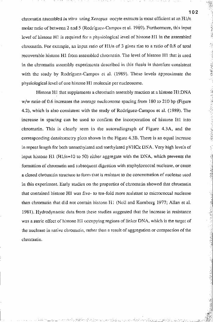
1 0 2
chromatin assembled in vitro using Xenopus oocyte extracts is most efficient at an H l/n
molar ratio of between 2 and 5 (Rodriguez-Campos et al. 1989). Furthermore, this input
level of histone HI is required for a physiological level of histone H I in the assembled
chromatin. For example, an input ratio of H l/n of 3 gives rise to a ratio of 0.8 of total
recoverable histone H I from assembled chromatin. The level of histone H I that is used
in the chromatin assembly experiments described in this thesis is therefore consistent
with the study by Rodriguez-Campos et al. (1989). These levels approximate the
physiological level of one histone HI molecule per nucleosome.
Histone HI that supplements a chromatin assembly reaction at a histone H1:DNA
w/w ratio of 0.6 increases the average nucleosome spacing from 180 to 210 bp (Figure
4.2), which is also consistent with the study of Rodrfguez-Campos et al. (1989). The
increase in spacing can be used to confirm the incorporation of histone H I into
chromatin. This is clearly seen in the autoradiograph of Figure 4.3A, and the
corresponding densitometry plots shown in the Figure 4.3B. There is an equal increase
in repeat length for both unmethylated and methylated pVHCk DNA. Very high levels of
input histone HI (Hl/n=10 to 50) either aggregate with the DNA, which prevents the
formation of chromatin and subsequent digestion with staphylococcal nuclease, or cause
a closed chromatin structure to form that is resistant to the concentration of nuclease used
in this experiment. Early studies on the properties of chromatin showed that chromatin
that contained histone H I was five- to ten-fold more resistant to micrococcal nuclease
than chromatin that did not contain histone HI (Noll and Kornberg 1977; Allan et al.
1981). Hydrodynamic data from these studies suggested that the increase in resistance
was a steric effect of histone HI occupying regions of linker DNA, which is the target of
the nuclease in native chromatin, rather than a result of aggregation or compaction of the
chromatin.
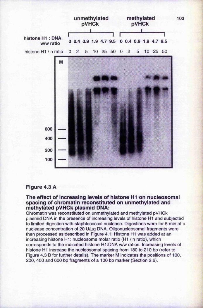
r
unmethylatedpVHCk
I_ _ _ _ _ _ _1 r
methylated pVHCk
I
103
histone DNA ^ ^ ^ Q.9 1.9 4.7 9.5 0 0.4 0.9 1.9 4.7 9.5
histone H1 / n ratio 0 2 5 10 25 50 0 2 5 10 25 50
Figure 4,3 A
The effect of increasing levels of histone HI on nucleosomal spacing of chromatin reconstituted on unmethylated and methylated pVHCk plasmid DNA:Chromatin was reconstituted on unmethylated and methylated pVHCk plasmid DNA in the presence of increasing levels of histone HI and subjected to limited digestion with staphlococcal nuclease. Digestions were for 5 min at a nuclease concentration of 20 U/pg DNA. Oligonucleosomal fragments were then processed as described in Figure 4.1. Histone HI was added at an increasing histone HI: nucleosome molar ratio (HI / n ratio), which corresponds to the indicated histone H1:DNA w/w ratios. Increasing levels of histone HI increase the nucleosomal spacing from 180 to 210 bp (refer to Figure 4.3 B for further details). The marker M indicates the positions of 100, 200, 400 and 600 bp fragments of a 100 bp marker (Section 2.6).
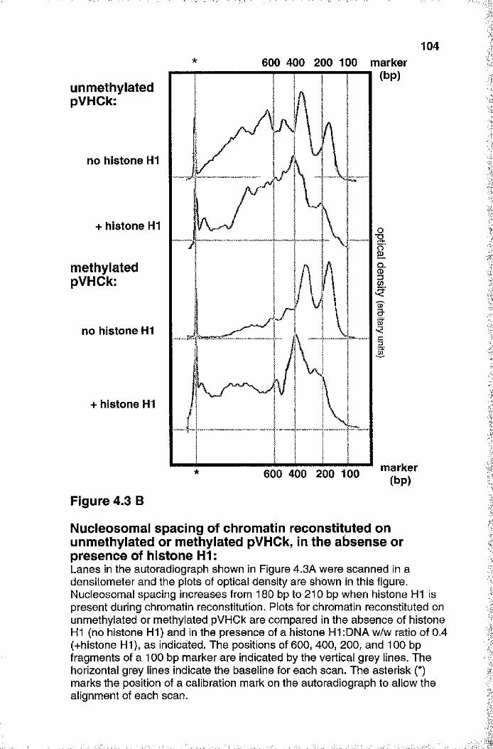
104600 400 200 100 marker
(bp)unmethylatedpVHCk:
no histone HI
+ histone HIT3
methylatedpVHCk:
Q.
ct
no histone HI
+ histone HI
marker(bp)600 400 200 too
Figure 4.3 B
Nucleosomal spacing of chromatin reconstituted on unmethylated or methylated pVHCk, in the absense or presence of histone HI :Lanes in the autoradiograph shown in Figure 4.3A were scanned in a densitometer and the plots of optical density are shown in this figure. Nucleosomal spacing increases from 180 bp to 210 bp when histone HI is present during chromatin reconstitution. Plots for chromatin reconstituted on unmethylated or methylated pVHCk are compared in the absence of histone HI (no histone HI) and in the presence of a histone HI :DNA w/w ratio of 0.4 (+histone HI), as indicated. The positions of 600, 400, 200, and 100 bp fragments of a 100 bp marker are indicated by the vertical grey lines. The horizontal grey lines indicate the baseline for each scan. The asterisk (*) marks the position of a calibration mark on the autoradiograph to allow the alignment of each scan.
1 0 5
4.3 Chromatin assembly on single-stranded phagemid
DNA
IChromatin can be assembled on replicating DNA in vitro. Xenopus egg extract can use
single-stranded DNA (ssDNA) as a template for complementary strand synthesis, with
concomitant nucleosome assembly on the resulting double-stranded DNA (dsDNA)
(Almouzni and Méchali 1988; Almouzni et al. 1990). Although chromatin assembly does
not occur on ssDNA in preference to dsDNA, the process of replication appears to
enhance chromatin assembly relative to non-replicating dsDNA.
Chromatin was assembled on pVHCk (+) single-stranded DNA, that was
prepared from phagemid (see Section 2.7.1.9 for the protocol). Chromatin assembly on
ssDNA is described in Section 2.7.7.2, with the incorporation of [a-^^P] dATP into the
replicated strand used to monitor the progress of DNA synthesis (Figure 4.4A). The
labelled products were deproteinised and resolved on an agarose gel. The products
include double-stranded nicked DNA and relaxed covalently-closed circular DNA which
cannot be resolved from the nicked form. The relaxed molecules undergo negative
supercoiling during nucleosome assembly, to form the fast-moving supercoiled form
seen in Figure 4.4A. This is due to the activity of a topoisomerase in the extract.
Intermediate topoisomers should form in between the relaxed and supercoiled molecules,
but are not formed because the assembly of chromatin is presumed to be very efficient
under these conditions (Almouzni et al. 1990; Wolffe and Schild 1991). Reconstitution
of chromatin on double-stranded pVHCk was included as a control (Figure 4.4A), and as
a marker for supercoiled dsDNA. The plasmid also became labelled during the
incubation, but this is presumed to be due to the incorporation of the dATP label into
nicks of relaxed molecules that were then supercoiled. That chromatin was assembled on
replicated DNA is confirmed by the formation of a nucleosomal ladder after digestion
with staphylococcal nuclease (Figure 4.4B).
This is the most efficient method for the assembly of chromatin in vitro , but was
not chosen for the studies in this thesis because the patch-methylated constructs of
Chapter 6 (Section 6.3.1, in particular) could only be analysed as double-stranded
molecules.
‘■Aii
dsDNA I control time for assembly
106
I 0 20 40100 240 (min)
nicked
supercoiledlinear
B Q 2 5 15 30 of digestion(min)
600
100
Figure 4.4
Chromatin reconstitution on single-stranded phagemid DNA:Chromatin was reconstituted on pVHCk (+) single-strand DNA, prepared from phagemid as described in Section 2.7.1.9. The reconstitution reaction was performed in the presence of [a-32p] dATP, as described in Section 2.7.7.2.See Section 4.3 for further details.A: Chromatin was reconstituted on 50 ng ssDNA for the indicated times. The DNA was then deproteinised and resolved on a 1 % agarose gel before autoradiography. The positions of nicked dsDNA, supercoiled dsDNA and linear dsDNA are indicated. Double-stranded pVHCk (50 ng) was incubated in a reconstitution reaction for 240 min (dsDNA), and the control assay was a reaction in the absence of DNA.B: Approximately 100 ng of dsDNA, reconstituted with chromatin as described above, was digested with staphylococcal nuclease at a concentration of 20 U/pg DNA for the indicated times. Oligonucleosomal fragments were resolved on a 1.3% agarose gel. The positions of 600 and 100 bp fragments of a 100 bp marker (lane M) are indicated by the marks.
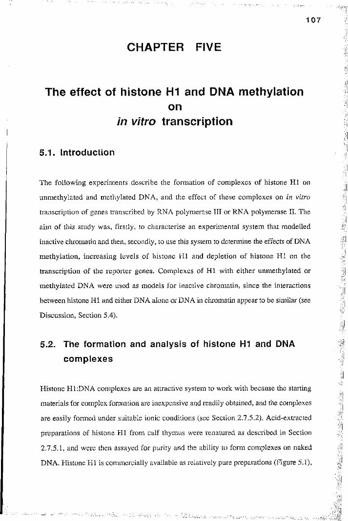
1 0 7
CHAPTER FIVE
The effect of histone HI and DMA méthylationon
in vitro transcription
5.1. Introduction
The following experiments describe the formation of complexes of histone HI on
unmethylated and methylated DNA, and the effect of these complexes on in vitro
transcription of genes transcribed by RNA polymerase III or RNA polymerase II. The
aim of this study was, firstly, to characterise an experimental system that modelled
inactive chromatin and then, secondly, to use this system to determine the effects of DNA
méthylation, increasing levels of histone H I and depletion of histone HI on the
transcription of the reporter genes. Complexes of H I with either unmethylated or
methylated DNA were used as models for inactive chromatin, since the interactions
between histone HI and either DNA alone or DNA in chromatin appear to be similar (see
Discussion, Section 5.4).
5.2. The formation and analysis of histone H1 and DNA
complexes
Histone H1:DNA complexes are an attractive system to work with because the starting
materials for complex formation are inexpensive and readily obtained, and the complexes
are easily formed under suitable ionic conditions (see Section 2.7.5.2). Acid-extracted
preparations of histone HI from calf thymus were renatured as described in Section
2.7 .5.1, and were then assayed for purity and the ability to form complexes on naked
DNA. Histone HI is commercially available as relatively pure preparations (Figure 5,1),
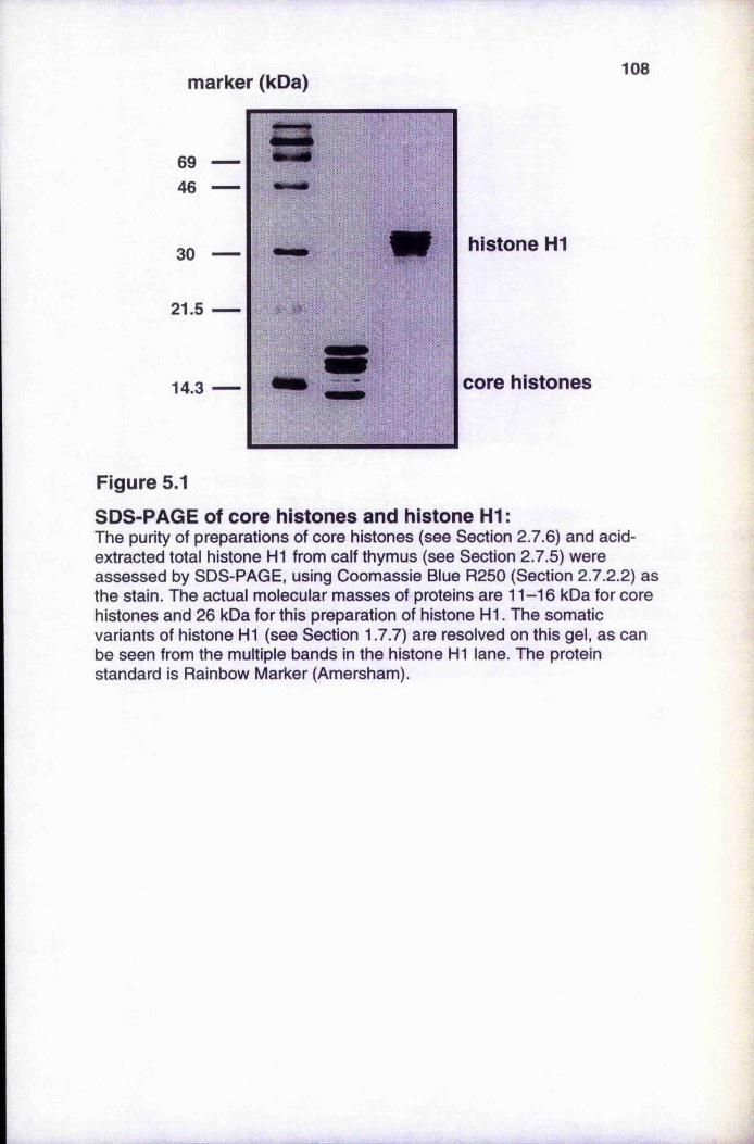
marker (kDa)108
histone HI
core histories
Figure 5.1
SDS-PAGE of core histories and histone HI:The purity of preparations of core histones (see Section 2.7.6) and acid- extracted total histone H1 from calf thymus (see Section 2.7.5) were assessed by SDS-PAGE, using Coomassie Blue R250 (Section 2.7.2.2) as the stain. The actual molecular masses of proteins are 11-16 kDa for core histones and 26 kDa for this preparation of histone H I. The somatic variants of histone HI (see Section 1.7.7) are resolved on this gel, as can be seen from the multiple bands in the histone HI lane. The protein standard is Rainbow Marker (Amersham).
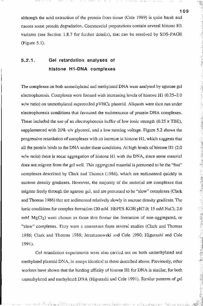
1 0 9
although the acid extraction of the protein from tissue (Cole 1989) is quite harsh and
causes some protein degradation. Commercial preparations contain several histone HI
variants (see Section 1.8.7 for further details), that can be resolved by SDS-PAGE
(Figure 5.1).
5 .2 .1 . Gel retardation analyses of
h istone H1-DNA complexes4
The complexes on both unmethylated and methylated DNA were analysed by agarose gel
electrophoresis. Complexes were fomied with increasing levels of histone HI (0.25-2.0
w/w ratio) on unmethylated supercoiled pVHCk plasmid. Aliquots were then run under
electrophoresis conditions that favoured the maintenance of protein-DNA complexes.
These included the use of an electrophoresis buffer of low ionic strength (0.25 x TBE),
supplemented with 20% v/v glycerol, and a low running voltage. Figure 5.2 shows the
progressive retardation of complexes with an increase in histone H I, which suggests that
all the protein binds to the DNA under these conditions. At high levels of histone H I (2.0
w/w ratio) there is some aggregation of histone HI with the DNA, since some material
does not migrate from the gel well. This aggregated material is presumed to be the “fast”
complexes described by Clark and Thomas (1986), which are sedimented quickly in
sucrose density gradients. However, the majority of the material are complexes that
migrate freely through the agai'ose gel, and are presumed to be “slow” complexes (Clark
and Thomas 1986) that are sedimented relatively slowly in sucrose density gradients. The
ionic conditions for complex fomiation (20 mM HEPES-KOH pH7.0; 15 mM NaCl; 2.0
mM MgCl2) were chosen as those that favour the formation of non-aggregated, or
“slow” complexes. They were a consensus from several studies (Clark and Thomas
1986; Clark and Thomas 1988; Jerzmanowski and Cole 1990; Higurashi and Cole
1991).
Gel retardation experiments were also carried out on both unmethylated and
methylated plasmid DNA, in assays identical to those described above. Previously, other
workers have shown that the binding affinity of histone HI for DNA is similar, for both
unmethylated and methylated DNA (Higurashi and Cole 1991). Similar patterns of gel
■ ./■''/-■-il-'.;;.-
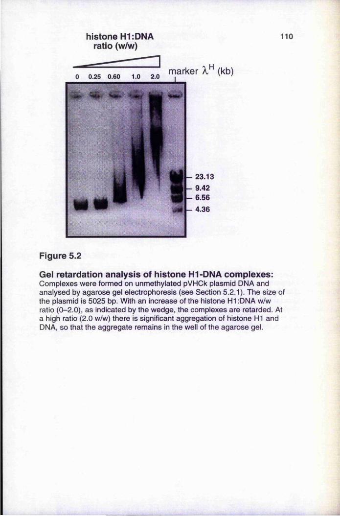
histone H1:DNA ratio (w/w)
110
marker (kb)
- 23.13- 9.42- 6.56
Figure 5.2
Gei retardation anaiysis of histone H1-DNA compiexes:Complexes were formed on unmethylated pVHCk plasmid DNA and analysed by agarose gel electrophoresis (see Section 5.2.1). The size of the plasmid is 5025 bp. With an increase of the histone HI :DNA w/w ratio (0-2.0), as indicated by the wedge, the complexes are retarded. At a high ratio (2.0 w/w) there is significant aggregation of histone HI and DNA, so that the aggregate remains in the well of the agarose gel.
111unmethylated plasmid
methylated plasmid
* #
m IN
histone H1:DNA(w/w) ratio
Figure 5.3
Gei retardation of histone HI-DNA compiexes on unmethylated and methylated plasmid DNA:Unmethylated and methylated plasmid DNA are retarded to similar extents with an increase in histone H1 :DNA w/w ratio (0, 0.25, 0.60, 1.0, 2.0). The increases in the w/w ratios are indicated by the wedges, and the marker is Hin dill digest of X DNA. The size of the plasmid is approx. 8 kb. and aggregates are seen in the upper part of the gel at high w/w ratios. See Section 5.2.1. for details.
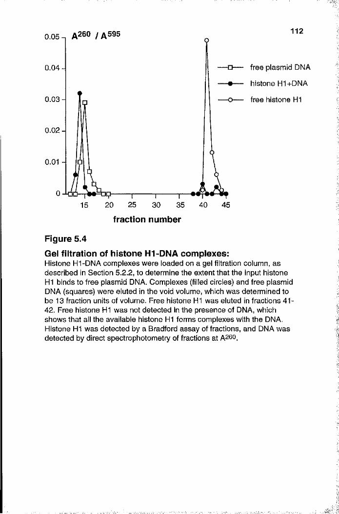
" « ■
1120.05-, A260 / / l5 9 5
free plasmid DNA
histone H1+DNA
0.04
0.03 free histone H1
0.02
0.01
20 25 35 40 4515 30
fraction number
Figure 5.4Gel filtration of histone H1-DNA complexes:Histone HI-DNA complexes were loaded on a gel filtration column, as described in Section 5.2.2, to determine the extent that the input histone HI binds to free plasmid DNA. Complexes (filled circles) and free plasmid DNA (squares) were eluted in the void volume, which was determined to be 13 fraction units of volume. Free histone HI was eluted in fractions 41- 42. Free histone HI was not detected in the presence of DNA, which shows that all the available histone HI forms complexes with the DNA. Histone HI was detected by a Bradford assay of fractions, and DNA was detected by direct spectrophotometry of fractions at A260.
I
II1:ïy
;i
j'-
I
1 1 3
retardation occur for both unmethylated and methylated DNA (Nightingale and Wolffe
1995), a finding which is confirmed in our own studies (Figure 5.3).
5 .2 .2 . Gel filtration of histone H1-DNA complexes
Gel retardation studies confirmed that histone HI-DNA aggregates were not formed
under the ionic conditions used, so aggregation could not be the facile explanation of
transcriptional inactivation in this system. However, these studies did not confirm that all
the input histone HI actually bound to the DNA, so that there was no free histone HI
present in the reaction mixture after a suitable time of incubation. To study this gel
filtration analyses were carried out on free plasmid DNA, free histone H I and putative
histone HI-DNA complexes, at an H1:DNA ratio of 1.0 w/w. A Sephadex G-50
(Pharmacia) gel filtration column (length 10 cm x diameter 0.5 cm) was packed under
gravity and the void volume determined using Dextran Blue 2000 (Phaimacia). The
column was then used to determine the extent of binding of histone HI to DNA. DNA
was detected by spectrometry of fractions at A^^^, and protein by Bradford assay of
fractions. The DNA and the complexes both passed through the column in the void
volume (Figure 5.4), whereas free histone HI was significantly retarded. No free histone
H I was present in the “complex” sample, which implies that all the histone H I binds to
the plasmid DNA under these conditions.
5.3. The effect of histone H1 and DNA méthylation on
transcription
This section details the inhibitory effect of histone HI-DNA complex formation on
templates that were transcribed in vitro. The plasmid template used is pArg/Leu (see
Section 2.5.1), which contains a tR N A ^g and a tRNA^eu gene that are transcribed by
RNA polymerase III. The template is either unmethylated or methylated. Transcription is
also assayed from the pVHCk plasmid template (see Section 2.5.1) which is transcribed
by RNA polymerase II. However, the latter system is technically more difficult to assay
1 1 4
5.3.1. Titration of unmethylated and methylated templates
This preliminary experiment investigated the effect of increasing amounts of template
DNA on in vitro transcription. Transcription was preferentially inhibited from methylated
templates, as compared to unmethylated templates, in transcription assays that used
nuclear extract prepared according to the Dignam protocol (Dignam et al. 1983; see
Section 2.13.2). At low levels of template (2-20 ng DNA/assay) little transcription is
observed from the methylated pArg/Leu in comparison with the unmethylated control.
This difference is less pronounced at high levels of template (>50 ng/assay; Figure 5.5),
since a 20% reduction in transcription from the methylated template is observed. The
extent of this reduction was rather variable, and depended on the particular batch of
nuclear extract. (Transcription from high levels of methylated template shows little
reduction for the batch of extract used in the experiment for Figure 5.5). These results
indicate the presence of a limiting amount of a selective inhibitor in the nuclear extract.
I
(Section 2.7.4.3), so most experiments were carried out on the pol III system. Unless it
is explicitly stated otherwise, the template used can be assumed to be pArg/Leu. The
HeLa nuclear extract that was used for this series of experiments on the pol III system
was prepared according to the Dignam protocol (Dignam et al. 1983; see Section
2.7.4.1). This meant that the Dignam extract is contaminated by endogenous histone HI
which is, of course, fortunate because it allows the preferential inhibition of methylated
templates to be observed in a simple titration experiment (Section 5.3.1).
The extent of ti'anscription from unmethylated or fully methylated templates was :
assayed in the presence of various levels of histone HI. The transcriptional activity of
both unmethylated and methylated templates are inhibited by increasing amounts of
histone H I, although inhibition of the methylated template occurs at a lower HI.-DNA
ratio. The H lc variant shows the greatest preferential inhibition of the methylated
template. Clearly, histone HI complexed to DNA is one of the factors that inhibits
transcription. Histone H I inhibits transcription by preventing the formation of initiation
complexes, particularly on methylated template (see Section 5.3.6), rather than by the
formation of disordered HI-DNA aggregates.
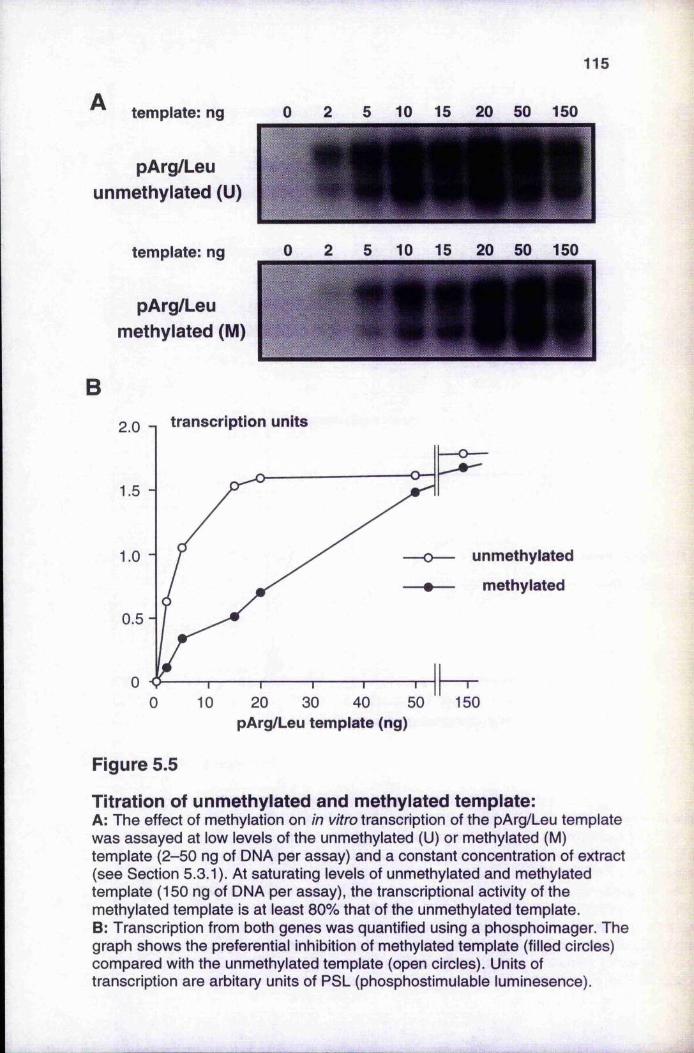
115
template: ng
pArg/Leu unmethylated (U)
template: ng
pArg/Leu methylated (M)
0 2 5 10 15 20 50 150
10 15 20 50 150
Btranscription units2.0
1.5
unmethylated
methylated
1.0
0.5
020 30 40 !
pArg/Leu template (ng)150
Figure 5.5
Titration of unmethylated and methylated template:A: The effect of méthylation on in vitro transcription of the pArg/Leu template was assayed at low levels of the unmethylated (U) or methylated (M) template (2-50 ng of DNA per assay) and a constant concentration of extract (see Section 5.3.1). At saturating levels of unmethylated and methylated template (150 ng of DNA per assay), the transcriptional activity of the methylated template is at least 80% that of the unmethylated template.B: Transcription from both genes was quantified using a phosphoimager. The graph shows the preferential inhibition of methylated template (filled circles) compared with the unmethylated template (open circles). Units of transcription are arbitary units of PSL (phosphostimulable luminesence).
1 1 6
Similar results have been described for transcription of pol II genes by Boyes and Bird
(1991), who suggested that MeCP-1 was the limiting protein.
Titrations were also carried out on increasing levels (50-500 ng) of unmethylated
and methylated pVHCk plasmid template, using the standard in vitro transcription assay
for this pol II system (Section 2.7.3.3). The results (not shown) were similar to those
seen for the pol III system: a selective inhibition of the methylated template, at low levels
(50 ng) of template, which largely disappeared at high levels (5(X) ng) of template.
5.3.2.Titrations with unmethylated and methylated competitor DNA
Transcription was carried out in the presence of increasing levels of unmethylated or
methylated competitor DNA, in the form of either pUC 19 plasmid DNA or a double
stranded oligonucleotide (see sequence below). It was supposed that the inhibitory effect
of a methyl-CpG binding protein (MeCP), or a similar protein, on low levels of template
would only be reversed by the addition of methylated competitor plasmid DNA. This is
because MeCPs bind selectively to methylated DNA, but have little affinity for identical
sequences that are unmethylated (Boyes and Bird 1991; see Section 1.7.2). Were the
putative inhibitor to be competed out by both unmethylated and methylated competitor
plasmid DNA, this would indicate that the inhibitor does not have the binding
characteristics of MeCPs, since it can bind to both unmethylated and methylated DNA. A
methylated double-stranded oligonucleotide would not be expected to bind MeCP-1
because it contains too few methylcytosines for the sequence binding affinity of MeCP-1
(Meehan et al. 1989). The oligonucleotide duplex could, however, act to compete out
other inhibitory factors. Titration studies with both plasmid and oligonucleotide DNA
could therefore determine if the putative inhibitor in the extract binds to methylated DNA
exclusively, or if it binds to both unmethylated and methylated DNA.
5.3.2.1 Titration with unmethylated and methylated plasmid DNA
In the presence of 10-240 ng unmethylated or methylated pUC19 competitor
DNA, transcription from 10 ng template was increased considerably (Figure 5,6).
Nevertheless, transcription remains less effective from the methylated template, reaching
only about 80% of the level obtained from the unmethylated template even at the highest
levels of competitor tested. Somewhat greater stimulation of transcription is observed
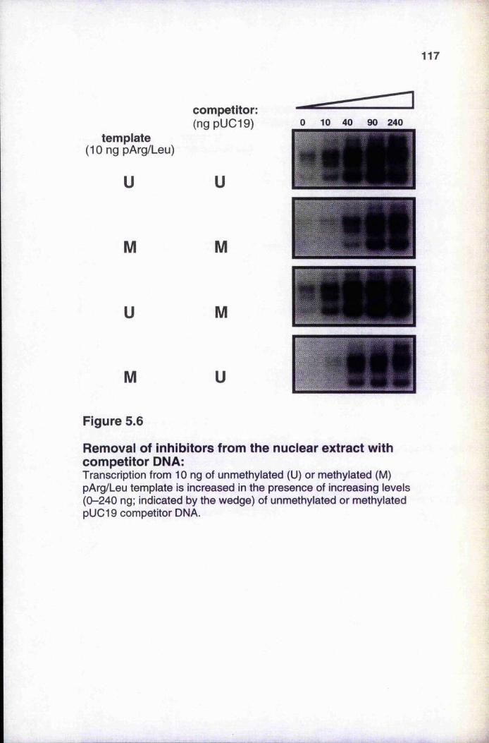
117
template(10 ng pArg/Leu)
u
M
U
M
competitor;(ng pUC19)
u
M
M
U
0 10 40 90 240
Figure 5.6
Removal of Inhibitors from the nuclear extract with competitor DNA:Transcription from 10 ng of unmethylated (U) or methylated (M) pArg/Leu template is increased in the presence of increasing levels (0-240 ng; indicated by the wedge) of unmethylated or methylated pUC19 competitor DNA.
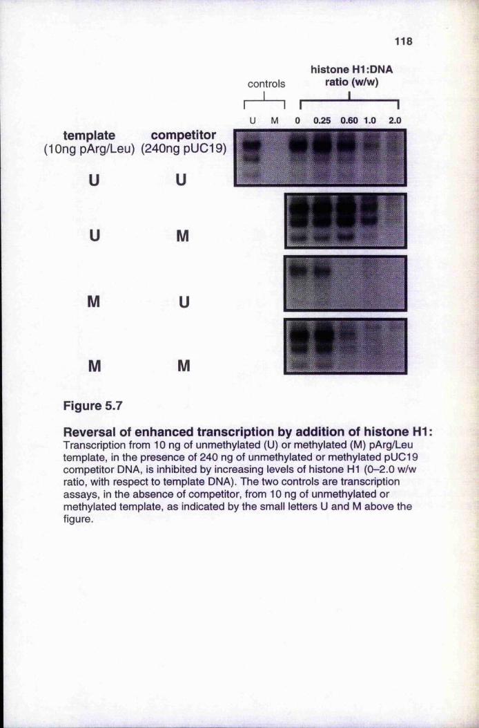
118
histone H1:DNA controls ratio (w/w)
1 ' 1U M 0 0.25 0.60 1.0 2.0
template competitor(lOng pArg/Leu) (240ng pUC19)
U
u
u
M
M U
M M
Figure 5.7
Reversai of enhanced transcription by addition of histone H1Transcription from 10 ng of unmethylated (U) or methylated (M) pArg/Leu template, in the presence of 240 ng of unmethylated or methylated pUC19 competitor DNA, is inhibited by increasing levels of histone HI (0-2.0 w/w ratio, with respect to template DNA). The two controls are transcription assays, in the absence of competitor, from 10 ng of unmethylated or methylated template, as indicated by the small letters U and M above the figure.
1 1 9with methylated competitor but, in both cases, transcription was increased at least ten
fold. It is assumed that the stimulation of transcription was caused by the binding to the
competitor DNA of inhibitory proteins present in the nuclear extract. As the inhibitory
factors bind to both unmethylated and methylated competitor DNA, this observation is
not consistent with the limiting inhibitor being MeCP-1, or a similar protein that binds,
preferentially to methylated DNA. It also appeal’s to rule out the involvement of MeCPs in
the general lowered template activity of the methylated template as this is still observed in
the presence of excess methylated competitor. It is, however, consistent with a role for
histone H I as the limiting component responsible for preferentially inhibiting
transcription from a methylated template. Histone H I is known to be present in low■
amounts in the HeLa nuclear extracts (Croston et al. 1991; Paranjape et al. 1994) and will
be removed by binding to competitor DNA, be it methylated or not.
In a second experiment, it is seen that preincubation of the template DNA (either
unmethylated or methylated) with histone HI leads to an inhibition of the enhanced
:.r
transcription seen on addition of competitor DNA (Figure 5.7). This inhibition of
transcription by histone HI occurs preferentially with methylated templates, whether or
not the competitor DNA is methylated. In other words, this is evidence for histone HI
alone being essential (and limiting) for the preferential inhibition of transcription from the
metliylated template. These titration experiments do not, by themselves, provide evidence
for the involvement of a second inhibitory factor (e.g. MeCP-1) that could mediate the
preferential inhibition of transcription from methylated templates. However, these
experiments cannot rule out the involvement of MeCPs in the general lowered
transcriptional activity of the methylated template. This activity is consistently no more
than 80% of that from the unmethylated template.
5.3.2.2. Titration with double-stranded oligonucleotides
A second type of DNA competitor was also used, in an attempt to compete out
inhibitory factors from the nuclear extract. MeCP-1 requires 15 methylated CpGs within
a short region of DNA (see Section 1.7.2), and so would be expected to bind to the
methylated plasmid competitor (used in the titration experiments of Section 5.3.2.1) but
not to a short double-stranded unmethylated or methylated oligonucleotides (shown in
Figure 5.8A),
— _ . _ — 1
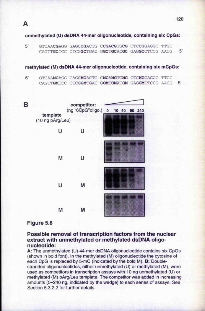
120
unmethylated (U) dsDNA 44-mer oligonucleotide, containing six CpGs:
5 ’ GTCAACGAGG GAGCCGACTG CCGACGTGCG CTCCGGAGGC TTGCCAGTTGCTCC CTCGGCTGAC GGCTGCACGC GAGGCCTCCG AACG 5 ’
methylated (M) dsDNA 44-mer oligonucleotide, containing six mCpGs:
5’ GTCAAMGAGG GAGCMGACTG CMGAMGTGMG CTCMGGAGGC TTGCCAGTTGMTCC CTCGGMTGAC GGMTGMACGM GAGGMCTCCG AACG 5’
B competitor:(ng “6CpG”oligo.) o 10 40 90 240
template(10 ng pArg/Leu)
U U
M U
U M
M M
Figure 5.8
Possible removal of transcription factors from the nuclear extract with unmethylated or methylated dsDNA oligonucleotide:A: The unmethylated (U) 44-mer dsDNA oligonucleotide contains six CpGs (shown in bold font). In the methylated (M) oligonucleotide the cytosine of each CpG is replaced by 5-mC (indicated by the bold M). B: Doublestranded oligonucleotides, either unmethylated (U) or methylated (M), were used as competitors in transcription assays with 10 ng unmethylated (U) or methylated (M) pArg/Leu template. The competitor was added in increasing amounts (0-240 ng, indicated by the wedge) to each series of assays. See Section S.3.2.2 for further details.
1 2 1
The unmethylated 44-mer dsDNA oligonucleotide contains six CpG dinucleotides
(shown in bold font in Figure 5.8A). In the methylated oligonucleotide the cytosine of
each CpG dinucleotide is replaced by 5-mC (indicated by the bold M). These
oligonucleotides have been used to investigate the preferential binding of histone HI
variants to a series of different oligonucleotides in gel-retardation assays (Santoro et al.
1995) . Refer to Sections 5.3.5 and 5.4 for a discussion about histone H I variants. The
oligonucleotides used in this experiment were kindly provided by Prof. P. Caiafa,
MeCP-1 would not be expected to bind to these oligonucleotides because there are fewer
than 15 closely spaced methylated CpGs, but they could compete out other inhibitory
factors that bind to both methylated and unmethylated DNA.
%
With increasing amounts of double-stranded unmethylated or methylated
oligonucleotide, transcription from 10 ng template is correspondingly reduced (Figure
5.8B). This can be explained by the removal of DNA-binding proteins, including basal
transcription factors, from the nuclear extract by the binding of the proteins to the
competitor DNA. Figure 5.8B suggests that transcription factors can be competed out
equally well from either the unmethylated template or the methylated template, in the
presence of increasing levels of competitor. However, the data does not provide evidence
that both the unmethylated and methylated oligonucleotide compete out transcription
factors to the same extent. These observations suggest that the preferential inhibition of
transcription from methylated templates (see Figure 5.5) could be explained by
prevention of transcription factor binding to methylated DNA (see Section 1.7.1), for this
pol III in vitro transcription system. An identical competition effect is seen with a
different unmethylated or methylated 40-mer dsDNA oligonucleotide that forms part of
the SV40 promoter, and which contains three GC boxes and three CpG dinucleotides
(results not shown).
5 .3 .3 . Transcrip tion w ith histone H1-depleted nuclear extract
Another way to remove histone HI is to subject the nuclear extract to ammonium sulphate
fractionation (see Section 2.7.3.1). This is a standard technique to remove contaminants,
such as histone H I, from crude nuclear extracts such as that prepared by the Dignam
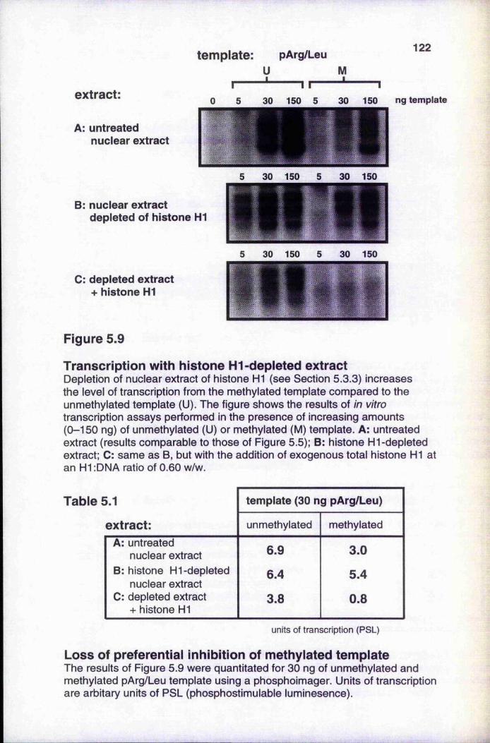
template: pArg/Leu122
extract:
A: untreated nuclear extract
B: nuclear extractdepleted of histone HI
30 150 5 30 150 ng template
30 150 30 150
30 150 30 150
C: depleted extract + histone HI
Figure 5.9
Transcription with histone H1-depleted extractDepletion of nuclear extract of histone HI (see Section 5.3.3) increases the level of transcription from the methylated template compared to the unmethylated template (U). The figure shows the results of in vitro transcription assays performed in the presence of increasing amounts (0-150 ng) of unmethylated (U) or methylated (M) template. A: untreated extract (results comparable to those of Figure 5.5); B: histone HI-depleted extract; C: same as B, but with the addition of exogenous total histone HI at an HI :DNA ratio of 0.60 w/w.
5.1 template (30 ng pArg/Leu)
extract: unmethylated methylated
A: untreated 6.9 3.0nuclear extractB: histone HI-depleted
nuclear extract6.4 5.4
C: depleted extract + histone HI
3.8 0.8
units of transcription (PSL)
Loss of preferential inhibition of methylated templateThe results of Figure 5.9 were quantitated for 30 ng of unmethylated and methylated pArg/Leu template using a phosphoimager. Units of transcription are arbitary units of PSL (phosphostimulable luminesence).
1 2 3
protocol (Dignam et al. 1983), and is described in more detail elsewhere (Shapiro et al.
1988). The fractionation procedure precipitates all the proteins involved in the
transcriptional machinery, but allows highly basic proteins such as histone HI to remain
in the supernatant.
In comparison to the crude nuclear extract that was used in the studies described
above, a nuclear extract that is depleted of histone HI shows increased transcription with
low levels (5-30 ng) of methylated or unmethylated template (Figure 5.9). Table 5.1
tabulates the level of transcription from 30 ng of unmethylated or methylated template,
which shows that the nuclear extract depleted of histone H I reduces the degree of
preferential inhibition of transcription from the methylated template. The addition of
histone H I (histone H1:DNA w/w ratio of 0.6) to the depleted extract increases the
degree of the preferential inhibition, with transcription from the methylated template
almost completely inhibited. Inhibition is presumed to be due to the formation, during the
preincubation stage of the transcription assay (Section 2.7.3.2), of Hl-DNA complexes
that inhibit transcription from methylated and, to a lesser extent, unmethylated constructs.
5 .3 .4 . Histone HI preferentially Inhibits transcription from
methylated templates
■Ia
Since low levels of methylated template can be preferentially inhibited by the endogenous
histone H I present in crude nuclear extracts, it seemed reasonable to try to inhibit
transcription from high levels of template by the addition of exogenous histone HI. With
amounts of template in excess of 50 ng per assay, the difference between transcription
from methylated and unmethylated templates is small (Figure 5.5B), in the absence of
exogenous histone H I. However, at high levels of template DNA, transcription is
inhibited from both templates (Figure 5.10) onto which “slow” complexes of total calf
thymus histone H I have been deposited (Section 2.7.5.2). Transcriptional inhibition
occurs at a lower HI ;DNA ratio for methylated templates, in comparison to unmethylated
or mock-methylated templates. Complete inhibition is obtained at high levels of HI (1.0-
2.0 ratio), for both types of template. Previous data (Section 5,2.2) suggests that all the
exogenous histone HI that is added binds to the DNA, so H1:DNA w/w ratios of 0.25,
Id :.--
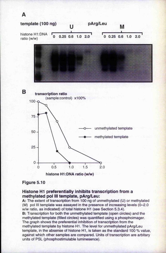
template (100 ng)
histone H1;DNA i ratio (w/w)
u pArg/Leu M---------- 1 I " I
0 0.25 0.6 1.0 2.0 0 0.25 0.6 1.0 2.0
B transcription ratio(sample:control) x100%
100
unmethylated template50 -
methylated template
2 5 -
0.5 1.0 1.5 2.00
histone H1:DNA ratio (w/w)
Figure 5.10
Histone HI preferentially inhibits transcription from a methylated pol III template, pArg/Leu:A: The extent of transcription from 100 ng of unmethylated (U) or methylated (M) pol III template was assayed in the presence of increasing levels (0-2.0 w/w ratio, as indicated) of total histone HI (see Section 5.3.4).B: Transcription for both the unmethylated template (open circles) and the methylated template (filled circles) was quantified using a phosphoimager. The graph shows the preferential inhibition of transcription from the methylated template by histone H I. The level for unmethylated pArg/Leu template, in the absense of histone HI, is taken as the standard 100 % value, against which other samples are compared. Units of transcription are arbitary units of PSL (phosphostimulable luminesence).
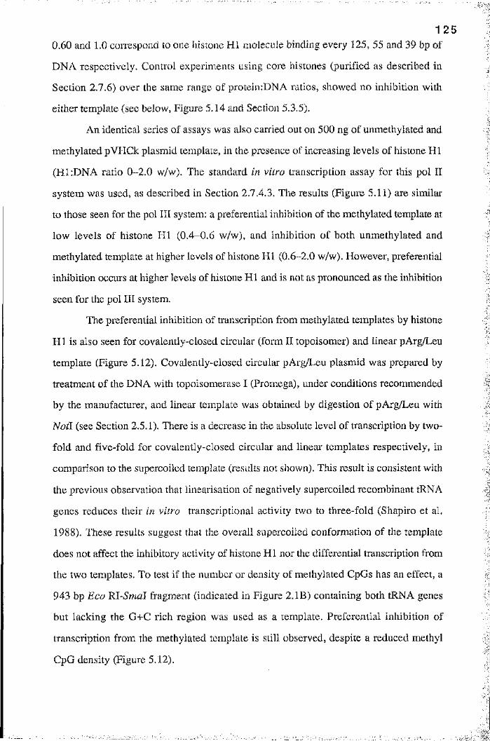
1 2 5
0.60 and 1.0 coiTespond to one histone HI molecule binding every 125, 55 and 39 bp of
DNA respectively. Control experiments using core histones (purified as described in
Section 2.7.6) over the same range of protein:DNA ratios, showed no inhibition with
either template (see below, Figure 5.14 and Section 5.3.5).
An identical series of assays was also carried out on 500 ng of unmethylated and
methylated pVHCk plasmid template, in the presence of increasing levels of histone HI
(HI.-DNA ratio 0-2.0 w/w). The standard in vitro transcription assay for this pol II
system was used, as described in Section 2.7.4.3. The results (Figure 5.11) are similar
to those seen for the pol III system: a preferential inhibition of the methylated template at
low levels of histone HI (0.4-0.6 w/w), and inhibition of both unmethylated and:
methylated template at higher levels of histone HI (0.6-2.0 w/w). However, preferential
inhibition occurs at higher levels of histone HI and is not as pronounced as the inhibition
seen for the pol III system.
The preferential inhibition of transcription from methylated templates by histone
H I is also seen for covalently-closed circular (form II topoisomer) and linear pArg/Leu
template (Figure 5.12). Covalently-closed circular pArg/Leu plasmid was prepared by
treatment of the DNA with topoisomerase I (Promega), under conditions recommended
by the manufacturer, and linear template was obtained by digestion of pArg/Leu with
Natl (see Section 2.5.1). There is a decrease in the absolute level of transcription by two
fold and five-fold for covalently-closed circular and linear templates respectively, in
comparison to the supercoiled template (results not shown). This result is consistent with
the previous observation that linearisation of negatively supercoiled recombinant tRNA
genes reduces their in vitro transcriptional activity two to three-fold (Shapiro et al.
1988). These results suggest that the overall supercoiled conformation of the template
does not affect the inhibitory activity of histone HI nor the differential transcription from
the two templates. To test if the number or density of methylated CpGs has an effect, a
943 bp Eco Rl-Smal fragment (indicated in Figure 2 .IB) containing both tRNA genes
but lacking the G+C rich region was used as a template. Preferential inhibition of
transcription from the methylated template is still observed, despite a reduced methyl
CpG density (Figure 5.12).
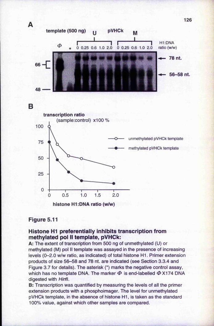
126
tem plate (500 ng) y pVHCk ^
0 \1 I I
11 H1:DNA
* 0 0.25 0.6 1.0 2.0 0 0.25 0.6 1.0 2.0 ratio (w/w)
I— 78 nt.
56—58 nt.
Btranscription ratio
(sample:control) x100%100
unmethylated pVHCk template75
methylated pVHCk template
50
25
01.0 1.5 2.00.50
histone H1:DNA ratio (w/w)
Figure 5.11
Histone HI preferentially inhibits transcription from methylated pol II template, pVHCk:A: The extent of transcription from 500 ng of unmethylated (U) or methylated (M) pol II template was assayed in the presence of increasing levels (0-2.0 w/w ratio, as indicated) of total histone H I. Primer extension products of size 56-58 and 78 nt. are indicated (see Section 3.3.4 and Figure 3.7 for details). The asterisk (*) marks the negative control assay, which has no template DNA. The marker 0 is end-labelled 0X174 DNA digested with H/nfl.B: Transcription was quantified by measuring the levels of all the primer extension products with a phosphoimager. The level for unmethylated pVHCk template, in the absence of histone HI, is taken as the standard 100% value, against which other samples are compared.
127
A: CGC
H1:DNA (w/w) ratio:
unmethylated (U) pArg/Leu
I '----------- 10 0.25 0.60 1.0 2.0
methylated (M) pArg/Leu
I------------- '------------- 10 0.25 0.60 1.0 2.0
B: linear
A ;
C: 943 bp fragment
Figure 5.12
Effect of different conformations of methylated template on preferential inhibition of transcription by histone HI :Preferential inhibition by increasing levels of histone HI (0-2.0 w/w ratio) of the transcription from different conformations of 100 ng methylated pArg/Leu template were assayed (see Section 5.3.4). A: covalently-closed circular (CCC) pArg/Leu; B: linear pArg/Leu; and C: a 943 bp restriction fragment containing both tRNA genes. The asterisk (*) indicates negative control assays, which were performed in the absence of DNA template. The marker on the extreme left is the usual end-labelled 0X 174/H/nfl marker.

1 2 8
In summary, histone HI appears to preferentially inhibit transcription from
methylated templates over a critical range of histone HIrDNA w/w ratios (0.25 - 0.60).
Preferential inhibition of transcription from methylated templates is observed irrespective
of the supercoiled status or of the methyl CpG density of the templates.
5 .3 .5 , Different variants of histone H1 inhibit transcription to
unequal extents
The contribution of different histone HI variants to the formation of inactive chromatin is
not known but it has been suggested that they may act at different sites to regulate the
stability of the 30 nm chromatin fibre (Wolffe 1992; Dimitrov et al. 1993; Pruss et al.
1995). This topic is discussed in detail in Section 1.8.7. I decided to investigate any
differences between the inhibitory action of somatic histone H I variants and the
preparation of total calf thymus histone H I that had been used in the experiments
described above. Any differences in vitro could reflect the in vivo role of variants during
the formation of the 30 nm chromatin fibre.
The different variants of histone H I were partially purified by reverse-phase
HPLC (Santoro et al, 1995) using a method described elsewhere by (Quesada et al.
1989). The variants were kindly provided by Prof. P. Caiafa. Protein fractions were
lyophilised and renatured as described in Section 2.7.5.1. Total histone H I and HI
variants were analysed and characterised by SDS-polyacrylamide (12%) gel
electrophoresis and a spectrometrie trace of the column fractions is shown in Figure 5.13.
Variant preparations are not homogenous as a result of incomplete resolution obtained by
reversed-phase HPLC chromatography. Variants were characterised as previously
described (Santoro et al. 1995), from which it could be concluded that preparations of
H la and H id were 95% homogenous, whereas the H lc preparations (fraction 2 and 3)
were contaminated with H ie. The H ie preparation (fraction 1) was also contaminated
with H lc, and H lb was not resolved using this method of chromatography. Protein
concentrations were determined by Bradford's procedure (Section 2.7.2.1), after
correction for the anomalous effect of histones on colorimetric assays.
g
I
i
;■a
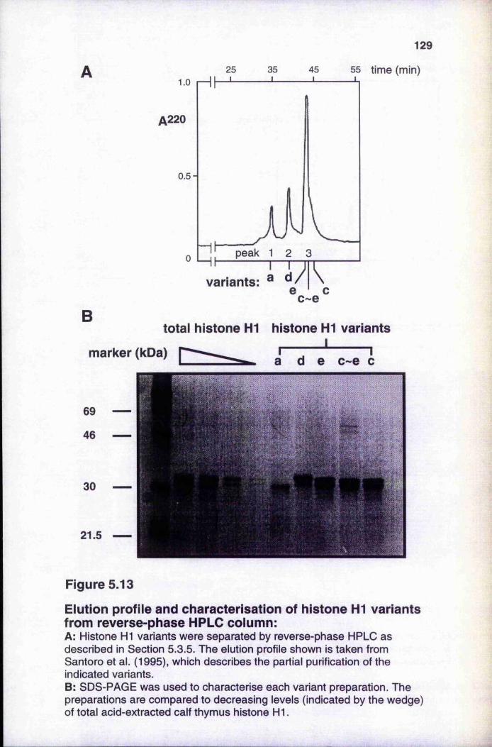
25 35 45
A220
0.5-
vahants:
129
55 time (min)
Btotal histone H1 histone HI variants
Imarker (kDa)
69
46
30
21.5
Figure 5.13
Eiution profile and characterisation of histone HI variants from reverse-phase HPLC coiumn:A: Histone HI variants were separated by reverse-phase HPLC as described in Section 5.3.5. The elution profile shown is taken from Santoro et al. (1995), which describes the partial purification of the indicated variants.B: SDS-PAGE was used to characterise each variant preparation. The preparations are compared to decreasing levels (indicated by the wedge) of total acid-extracted calf thymus histone H I.
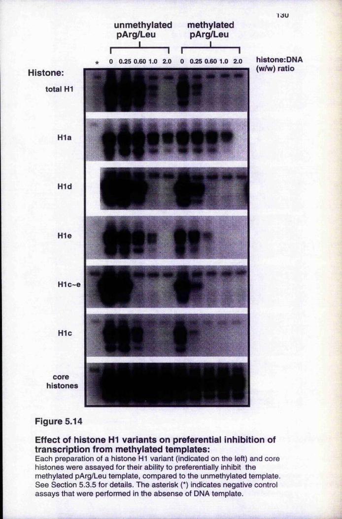
1 J U
unmethylated methylated pArg/Leu pAr^Leu
I______________ II I I I
* 0 0.25 0.60 1.0 2.0 0 0.25 0.60 1.0 2.0 histone:DNAHistone:
total H1
H1a
H1d
H1e
H1c~e
H1c
(w/w) ratio
corehistones
Figure 5.14
Effect of histone HI variants on preferential inhibition of transcription from methylated templates:Each preparation of a histone H1 variant (indicated on the left) and core histones were assayed for their ability to preferentially inhibit the methylated pArg/Leu template, compared to the unmethylated template. See Section 5.3.5 for details. The asterisk (*) indicates negative control assays that were performed in the absense of DNA template.
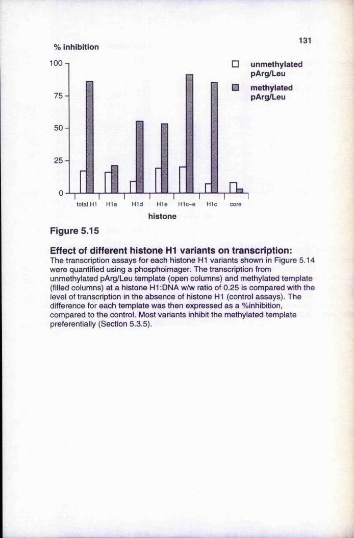
% inhibition
100 n
7 5 -
131
50 -
2 5 -
0
□ unmethylated pArg/Leu
M methylated pArg/Leu
Ttotal H1 H1a H1d H1e H1c~e H1c core
histone
Figure 5.15
Effect of different histone HI variants on transcription:The transcription assays for each histone HI variants shown in Figure 5.14 were quantified using a phosphoimager. The transcription from unmethylated pArg/Leu template (open columns) and methylated template (filled columns) at a histone H1:DNA w/w ratio of 0.25 is compared with the level of transcription in the absence of histone HI (control assays). The difference for each template was then expressed as a %inhibition, compared to the control. Most variants inhibit the methylated template preferentially (Section 5.3.5).
1 3 2
Each variant was complexed with methylated or unmethylated (conti'ol) DNA
templates, at increasing hi stone H1:DNA w/w ratios, and used in transcription assays as
described above (Figure 5.14). Assays for the total histone H I preparation and a
preparation of core histones as control are also shown in Figure 5.14. Preparations
containing predominantly H lc show inhibition at H1:DNA levels greater than those
shown by total histone FIl (Figure 5,14). The unmethylated template is inhibited at
H1:DNA ratios of 0.60-1.0 (w/w), whereas the methylated template is inhibited at the
0.25 (w/w) ratio. The preparations which contain predominantly H ie or H id have a
reduced, preferential inhibitory activity, whereas HI a has little preferential inhibitory
activity (Figure 5.15). Core histones do not have any inhibitory activity in this system.
5 .3 .6 . Histone HI prevents the formation of in itia tion and
elongation transcrip tion complexes preferentia lly on
methylated tem plates
It has previously been shown that histone HI inhibits RNA polymerase II transcription
by preventing the assembly of initiation complexes on template DNA (Croston et al.
1991). The results in this section show that a similar interaction occurs with the general
RNA polymerase III transcriptional machinery, but that this process appears to occur
preferentially on methylated templates.
The results of Figure 5.16 show that preincubation of the template with histone
H I at a HliDNA ratio of 1.0 (w/w) prevents the assembly of initiation complexes on
both unmethylated (lane 2) and methylated (lane 9) template. Histone H I also inhibits the
conversion of preassembled initiation complexes into elongation complexes on the
addition of ribonucleoside triphosphates, although this inhibition is less effective on
unmethylated template (lane 3) than on methylated template (lane 10). However, the
presence of H I does not inhibit elongation from either template (lanes 4 and 11),
although transcription is limited to a single round in both cases, as shown by comparison
with transcription assays containing 0.025% (w/v) Sarkosyl (N -laurylsarcosine) (lanes 7
and 14). Sarkosyl prevents the formation of initiation complexes (lanes 5 and 12) and the
conversion of preassembled initiation complexes into elongation complexes (lanes 6 and
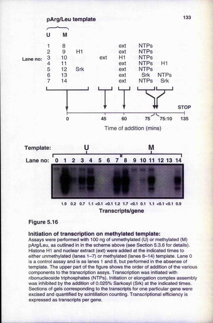
pArg/Leu templater~U
Lane no:
M
1 8 ext NTPs2 9 H1 ext NTPs3 10 ext H1 NTPs4 11 ext NTPs HI5 12 Srk ext NTPs6 13 ext Srk NTPs7 14 ext NTPs Srk
Y
133
STOP—r— 60
— -----------------------
75 75:10 135
Time of addition (mins)
Template:
7 8 9 10 11 12 13 14Lane no:
1.9 0.2 0.7 1.1 <0.1 <0.1 1.2 1.7 <0.1 0.1 1.1 <0.1 <0.1 0.9
Transcripts/gene
Figure 5.16
Initiation of transcription on methylated template:Assays were performed with 100 ng of unmethylated (U) or methylated (M) pArg/Leu, as outlined in in the scheme above (see Section 5.3.6 for details). Histone H1 and nuclear extract (ext) were added at the indicated times to either unmethylated (lanes 1-7) or methylated (lanes 8-14) template. Lane 0 is a control assay and is as lanes 1 and 8, but performed in the absense of template. The upper part of the figure shows the order of addition of the various components to the transcription assys. Transcription was initiated with ribonucleoside triphosphates (NTPs). Initiation or elongation complex assembly was inhibited by the addition of 0.025% Sarkosyl (Srk) at the indicated times. Sections of gels corresponding to the transcripts for one particular gene were excised and quantified by scintillation counting. Transcriptional efficiency is expressed as transcripts per gene.
1 3 4
13) but, like H I, does not stop the elongation by the polymerase once ribonucleoside
triphosphates have been added (Hawley and Roeder 1987). As a consequence, the
presence of Sarkosyl prevents reinitiation of transcription, limiting the transcription to
one round. This was confirmed by excising gel slices containing the labelled RNA
transcripts for one particular gene, counting in a liquid scintillation counter and
determining the transcriptional efficiency in terms of transcripts per gene (Figure 5.16).
In summary, it can be concluded that histone H I, like Sarkosyl, acts by
preventing the formation of initiation complexes, particularly on methylated templates,
rather than by preventing elongation which proceeds at similar rates from unmethylated
and methylated templates.
5 .3 .7 . Gene méthylation is not essential for HI-mediated
inhibition of transcription
Patch-methylated constructs of pArg/Leu were made by ligating together different
combinations of unmethylated or methylated Bgl \-Bgl I fragments. This was achieved by
methylating the restriction fragments obtained by digestion of pArg/Leu with Bgl I,■
separating and purifying the fragments by standard procedures and ligating suitable
combinations of methylated fragments with unmethylated fragments. Bgl I was chosen, '-4
because it cleaves the sequence GCCNNNN/NGGC at the site indicated by the stroke. In
principle, the Bgl I fragments could be religated to form a covalently-closed circular |Ê
molecule, if all the Bgl I sites had unique sequences. This prevents the formation of a |
variety of linear molecules and concatamers, which would otherwise occur if different
fragments had compatible sticky ends. On average, a restriction fragment of unknown
sequence will have a 1 in 64 chance of having compatible sticky ends with a second
fragment. Plasmid pArg/Leu has five Bgl I sites, which give rise to five fragments on
cleavage with the enzyme. Two sites are of unknown sequence (Figure 5.17A), but their
approximate location is known from restriction mapping. The chance that one of these
sites has the same sticky end as the other four is therefore 1 in 16, but since the sequence
of two sites is unknown the chance is reduced to 1 in 32. This means that the probability

AB1
Eco Ri1 B B
1B1
restriction enzyme sites
Earn HI 1 B
J 1 ,' 252 396 1-----1 1008
494 5761 / 5600*
LmhJ1105 1193
2400* 3000* 3300* 4700* '1 /5600*
positions in pArg/Leu
pUC19 . tRNALeu tRNAArg GC-rich region 1 PÜC19vector insert vector
construct:
B time of digestion
X 100n
constructs I
nicked
> !-<-• mm linear
Figure 5.17
Patch-methylated constructs of the pArg/Leu template:A: Patch-methylated constructs of pArg/Leu were constructed as described in Section 5.3.7. The top of the diagram shows the positions of restriction enzyme sites (see Section 2.5.1), includingSg/l sites (indicated by B), and the two tRNA genes within the insert of pArg/Leu (indicated by the bold line).The position of sites marked with an asterisk (*) were determined by restriction mapping. Methylated regions of constructs b-d are indicated by a thick line in the lower part of the diagram. The dashed line indicates unmethylated regions in constructs a, c and d.B: Religated covalently-closed circular (CCC) constructs a-d were gel purified. The products of the ligation reaction are compared to DNA samples of pArg/Leu that are either in the CCC and nicked forms (lane 1) or linear (lane 2). These samples were prepared by digesting pArg/Leu with DNA topoisomerase or Not I. Plasmid pArg/Leu was also partially and fully digested with Bgl\ during a time- course experiment, as indicated by the wedge.
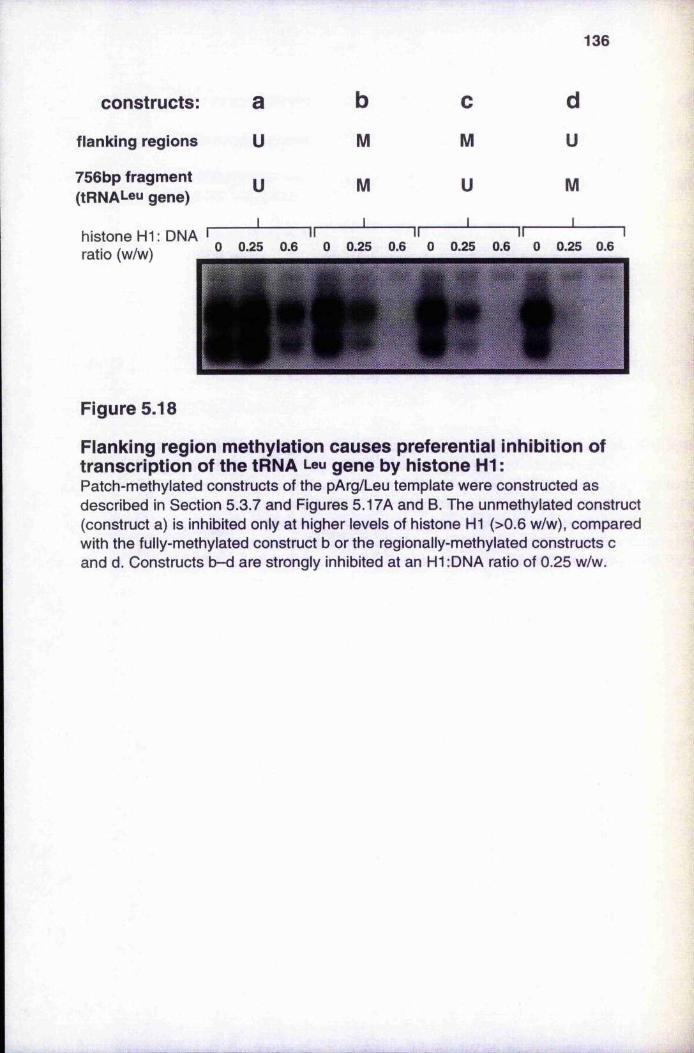
136
constructs: a b c d
flanking regions u M M u756bp fragment u
1
M
1
U
1
M
1
( t R N A L e u gene)
h is to n e H1: DNA • II0.25 0.6 0 0.25
II0.6 0 0.25
II0.6 0 0.25 0.6ratio (w/w) °
Figure 5.18
Flanking region méthylation causes preferential inhibition of transcription of the tRNA Leu gene by histone HI :Patch-methylated constructs of the pArg/Leu template were constructed as described in Section 5.3.7 and Figures 5.17A and B. The unmethylated construct (construct a) is inhibited only at higher levels of histone H1 (>0.6 w/w), compared with the fully-methylated construct b or the regionally-methylated constructs c and d. Constructs b-d are strongly inhibited at an H1:DNA ratio of 0.25 w/w.
'•'1.4»:?
1 3 7
of forming circular constructs is 31/32, compared to that for linear concatamers which is
1/32.
Covalently-closed circular patch-methylated templates were constructed which
were methylated either in all regions of plasmid pArg/Leu except a Bgl l-Bgl I fragment
of size 756 bp. (Figure 5.17A) which contained the tRNA^eu gene, or methylated only in
this 756 bp. fragment. The 756 bp. fragment which contained the tRNA^eu gene was. ■
either unmethylated, with all the flanking regions methylated (construct c; refer to Figure■
5.17A), or was itself methylated with all other regions unmethylated (construct d).
Control templates were either fully methylated (construct b; refer to Figure 5.17A) or
unmethylated (construct a), and were constructed by ligating together only methylated or
unmethylated Bgl I fragments. Ligations were carried out at 16°C for 16 h using T4 DNA
ligase (Promega) at a concentration of 3 Weiss U/pg DNA (refer to Section 2.7.1.11).i.',;
Religated covalently-closed circular plasmid (Figure 5.17B) was separated and purified
from unligated fragments by gel elecU’Ophoresis in 0.7% LMP agarose (Gibco/BRL).
It is clear that méthylation of the flanking region can preferentially reduce
transcription of the unmethylated tRNAPGu g&ne in the presence of increasing levels of
histone HI (Figure 5.18), as well as the transcription from the methylated tRNAArg gene
in the flanking region. Construct c which contains an unmethylated tRNA^eu a
methylated vector is inactivated at similar Hl.'DNA ratios, as is the fully methylated
construct b. Construct d which contains the methylated tRNA^Gu is inactivated to the
greatest extent. Inactivation appears to spread to include the adjacent unmethylated
tRNAArg gene. It is impossible to be certain that the template used in these experiments is
covalently-closed circular, because the sequence of two B gl I sites is unknown.
However, irrespective of the nature of the template, be it circular or linear, it is clear that
methylated flanking regions can influence the transcription from an adjacent unmethylated
region. This observation is supported by the experiment described in Section 5.3.4 and
Figure 5.12: histone HI can preferentially inhibit transcription from either covalently-
closed circular or linear methylated template.
This method of making patch-methylated constructs appears to be quite
promising, but a plasmid with known, unique Bgl I sites should be used. The yield of
construct is poor because the efficiency of fragment religation is low. The yield is much
better for the protocol described in Section 2.7.3.2, which forms the basis of the studies
presented in Chapter 6, and is therefore the preferred method of patch méthylation.
1 3 8
5.4. Discussioni ’
iH l-D N A complexes as a model for inactive chromatin
Complexes of histone H1 and DNA have been used in several studies to model
inactive chromatin. This approach is valid if it is assumed that in chromatin the histone
H I molecule has minimal interaction with the core histones or the dyad of the
chromatosome, but interacts mainly with the linker DNA. Histone H I molecules are then■
presumed to form an ordered array along the length of the naked DNA. This suggests
that the presence of a chromatosome is not required for the repression of genes by
histone H I, at least for the in vitro systems studied in this thesis. However, under
certain conditions, such as a high H1:DNA ratio or high ionic strength (Clark and
Thomas 1986), histone H I and DNA foim an aggregate. The aggregation is presumably
mediated by cooperative hydrophobic interactions between histone HI molecules.
Thomas et al. (1992) demonstrate that neighbouring globular domains of histone HI
cooperatively bind to DNA, but this can occur at protein:DNA w/w ratios of 0.1-0,6 and
at 5 mM NaCl with the formation of “fast” and “slow” complexes (refer to Section 5.2.1
and Clark and Thomas 1986). The linker histone constrains two adjacent double helices,
and additional linker histones stack between the helices with cooperative interactions. The
globular domains bridge “tramlines” of DNA and therefore bind to two different
segments of DNA, which is presumed to also occur in nucleosomes. Aggregation is the
facile explanation for the repression of genes in vitro , but the conditions that were used
in this series of experiments have precluded the formation of histone Hl-DNA
aggregates. For example, the repression mediated by histone HI is reversible on addition
o f excess competitor DNA (Figure 5.6). In addition, a template that is complexed with
histone HI can still be readily transcribed (Figures 5.7 and 5.16), albeit at a reduced
efficiency, which would be impossible if the template was in the form of an aggregate.
Thomas et al. (1992) use conditions similar to those described in Chapter 5 (essentially,
protein:DNA ratios of 0.25-0.6 and a NaCl concentration of 15 mM; refer to Section
2.7.5.2), to study an histone H l-DNA complex that can be characterised by using'
biochemical techniques and is not an aggregate.
1 3 9
In fact, the interactions between histone H I and naked DNA or DNA in
chromatin appear to be similar (Croston e ta l. 1991). For example, histone HI has a
similar salt dependence when it binds to either DNA or to chromatin (Clark and Thomas
1986). Cooperative binding of H I to DNA occurs over the same range of salt
concentrations as the compaction of nucleosomes into the 30 nm chromatin fibre (Clark
and Kimura 1990). HI appears to interact mostly with linker DNA in chromatin, rather
than with core histones (Thoma et al. 1979), although the globular domain of the
molecule can be cross-linked to H3, H2A and H2B in the core octamer (Pruss et al.
1995). The crystal structure of the globular domain of histone H5 (a linker histone that
resembles histone HI; see Section 1.8.7) has been solved (Ramakrishnan et al. 1993),
which imposes several constraints on previous models of chromatosome structure
(reviewed in Ramakrishnan 1994). None of the new structure data suggest that the
globular domain has a strong interaction with the central turn of DNA that wraps round
the core octamer. Instead, it has been proposed that the globular domain interacts with
both DNA linkers at some distance from the core octamer. However, this model appears
to be incompatible with neutron scattering and cross-linking data (Graziano et al, 1994;
Boulikas et al. 1980).
Jerzmanowski and Cole (1990) describe the transcriptional inhibition of an
unmethylated plasmid at H1:DNA ratios 0.4-0.6 w/w. This corresponds well with the
values of 0.25-0.6 w/w ratio for inhibition of the plasmid templates used in this study.
These H1:DNA ratios correspond to one molecule of HI per 125-55 bp DNA. It is
generally accepted that the nucleosome core particle contains 146 bp of DNA (Paranjape
et al. 1994), and that there is 39 bp of linker DNA per nucleosome that is accessible to
H I in the chromatin of mammalian cells. The observation that the spacing of histone HI
molecules is quite similar in both Hl-DNA complexes and chromatin suggests that the
interactions of HI with DNA are similar in naked DNA and in chromatin (Croston et al.
1991). In addition, the reversible binding of H I to DNA in vitro under certain
conditions, is comparable to the exchange of HI within chromatin in vivo (Louters and
Chalkley 1985). There have also been two recent studies of HI-mediated inactivation of
class II genes on naked DNA templates (Croston et al. 1991; Levine et al. 1993). In
Section 5.2 I have shown that histone FIl forms ordered complexes with DNA under
1 4 0
certain concentrations and ionic conditions which precluded the formation of aggregates.
At these concentrations of DNA and histone HI all the input histone H I forms ordered
complexes with the DNA. No difference is seen in the ability of histone HI to form
complexes on either unmethylated or methylated DNA, as assayed by retardation in
agarose gels.
In summary, ordered complexes of histone HI and DNA were chosen as a
simplified model system for inactive chromatin because the interaction of histone HI with
naked DNA in such complexes resembles the interaction with chromatin.
Histone H I preferentially inhibits transcription from methylated
tem plates.
The initial observation which prompted the studies reported in this chapter was
that in vitro tr anscription from low levels of a methylated template was less efficient than
from low levels of an unmethylated template (Section 5.3.1). This suggested the presence
of an inhibitor in the nuclear exti'act that is specific for methylated DNA. There is no
evidence that the binding of transcription factors to pol III genes is affected by DNA
méthylation, but HeLa nuclear extract does contain low levels of histone HI (Croston et
al. 1991; Paranjape et al. 1994), and would also be expected to contain the methyl CpG
binding protein, MeCP-1. Two methods were used to remove histone H I from nuclear
extracts.
Addition of competitor DNA, either methylated or unmethylated, led to enhanced
transcription from both templates implying that an inhibitor was present that bound to
both methylated and unmethylated DNA (Section 5.3.2 and Figure 5.6). This inhibitor
cannot, therefore, be MeCP-1 (that binds only to methylated DNA) but could be histone ‘
H I that has a similar affinity for methylated and unmethylated DNA. The differential
inhibition from the methylated template was considerably reduced in the presence of high
levels of competitor DNA which is consistent with the structure of the complex between
histone HI and methylated DNA being different from that formed with unmethylated
DNA (Higurashi and Cole 1991). Addition of histone HI alone is able to restore the
sti*ong preferential inhibition of transcription from the methylated template (Figure 5.7).
Presumably, the methylated competitor has removed methyl CpG binding proteins as
4' ■'■A'''
1 4 1
well as histone HI and so these cannot be involved in the selective inhibition exerted by
histone HI on transcription from the methylated template.
Depletion of the nuclear extract of histone H I using ammonium sulphate
precipitation also abolished the preferential inhibition from the methylated template
(Section 5 .33 and Figure 5.9). This differential effect could be restored by addition of
histone H I alone, again indicating that this protein is able to preferentially inhibit in vitro
transcription from the methylated template. In this case, it is presumed that MeCP-1
remains in the HI-depleted nuclear extract and it alone is insufficient to inhibit
transcription from methylated templates. Previous evidence and our own data suggests
that histone HI can act as a methylated-DNA binding protein (MDBP; see Section 1.7.2),
which is supported by the observation that MDBP-2 has sequence homologies with
histone HI (lost and Hofsteenge 1992). The inhibition of transcription by an MDBP-like
activity of histone HI could therefore supplement the generalised repression of chromatin
by histone H I, presumably by the formation of supranucleosomal structures (see Section
1.8.5).
Histone H I functions as a repressor of transcription for both pol II transcribed
genes (Bresnick et al. 1991; Layboum and Kadonaga 1991) and pol III transcribed genes
(Schlissel and Brown 1984; Shimamura et al. 1989; Woiffe 1989) in the context of
chromatin or nucleosomes (see Section 1.8.5). I show here that this inhibition is more
efficient for methylated pol III templates. However, histone H I does not prevent RNA
chain elongation, which suggests that the movement of the transcription complex is not
impeded by the Hl-DNA complexes. The results in Figure 5.16 show that histone HI
exerts its differential effect on ti'anscription of methylated DNA during the formation of
the initiation complex. This observation is supported by a study on the expression of the
herpes simplex virus (HSV) thymidine kinase {tk ) gene, after transfection of a construct
containing the gene into mammalian cells (Levine et al. 1992). Promoter méthylation
appeared to affect the formation of the initiation complex, rather than the rate of
transcription.
The binding of histone H I to methylated DNA may lead to a change in
nucleoprotein conformation, thereby rendering the promoters inaccessible to transcription
factors. Higurashi and Cole (1991) have shown that histone H l-DNA complexes are
resistant to digestion by Msp I (see Section 1.8.5), and have also suggested that this can
be explained by a local change in the conformation of the nucleoprotein complex. These
1 4 2findings are confirmed by my own work, presented in Section 6.2.2. Figure 5.18 shows
that méthylation of flanking regions alone is able to bring about HI-mediated inhibition of
transcription of an unmethylated gene. This suggests that the conformational change can
spread, either by the cooperative binding of histone HI molecules or by a change in the
double helix. This effect appears to only occur over short distances (the tRNAf^u gene is
242 and 432 bp from the start of the flanking regions; refer to Figure 5.17A). Histone
H l-DNA complexes do not appear to mediate the spread of inactivation over greater
distances: there is no evidence of this effect being mediated by histone HI on the patch-
methylated constructs for the pol II system, described in Sections 6.3.2 and 6.3.4.
The possible roles of histone HI variants
Different histone H I variants occur in different species, cell types and
developmental stages (see Section 1.8.7), which has implications for chromatin structure
and folding (Pruss et al. 1995). In some vertebrates, the presence of certain histone HI
variants has been correlated with distinct developmental stages, that involve changes in
chromatin structui'e prior to changes in gene transcription. For example, the special linker
histone H5 of chicken erythrocytes compacts and inactivates the entire erythrocyte
nucleus by the formation of heterochromatin and in the sea urchin there are well-
chai’acterised changes of histone HI variants during embryogenesis (see Section 1.8.7).
I have shown that different somatic variants of calf thymus histone H I inhibit
transcription to different extents (Figures 5.14 and 5.15). Recently, a mixture of two
variants, H lc and H ie, has been shown to specifically inhibit in vitro enzymatic DNA
méthylation (Santoro et al. 1995) and it has been proposed that these variants act at CpG
islands to inhibit their méthylation by DNA methyltransferase, thereby maintaining the
islands in an unmethylated state. This follows from the finding that H ie is the only
variant that can bind to DNA that is rich in CpG dinucleotides (6 CpG dinucleotides per
44 bp). Here I demonstrate that H lc is particularly effective at inhibiting in vitro
transcription from methylated templates where the overall CpG concentration is
approximately 400 per 5600 bp (i.e. about three 5-mCpG dinucleotides per 44 bp).
Island DNA undergoing maintenance méthylation during DNA replication could thus be
packaged into inactive chromatin by H lc, whereas unmethylated genes would remain
active.
I
i
1 4 3
CHAPTER SIX
The effect of méthylation on in vitro chromatin formation and transcription
6.1 Introduction
This chapter describes a series of in vitro studies that investigate the inhibition of
transcription by méthylation and chromatin. The experiments use as template the plasmid
pVHCk (see Section 2.5.1 for a map of the plasmid), which is transcribed by RNA
polymerase II (pol II). One experiment (presented in Section 6.2.3) uses pArg/Leu as the
template. Plasmid pArg/Leu (see Section 2.5.1 for a map) is transcribed by RNA
polymerase III (polRI). The protocol for in vitro transcription of the pol II system has
been described in Section 2.7.4.3, and experiments that detail the optimisation of this
protocol are presented in Section 3.3.4. The method for the in vitro reconstitution of
chromatin on plasmid DNA, by using Xenopus S150 egg extract, has been described in
Section 2.7.7 and several experiments that were based on this system were presented and
discussed in Chapter 4. The data in this chapter have been obtained by using these
systems of in vitro transcription and chromatin assembly to investigate the inhibition of
pol II transcription by méthylation.
The results presented in Section 6.2.3 demonstrate the preferential inhibition of
transcription from methylated chromatin templates (i.e. chromatin that was in vitro
reconstituted on methylated plasmid DNA, that was subsequently used as the template for
in vitro transcription assays). This is caused by méthylation directing the formation of an
inactive chromatin structure that is inaccessible to transcription factors (Lewis and Bird
1991; Graessmann and Graessmann 1993), as has been discussed in Section 1.8,2.
Inhibition in the context of chromatin is shown for both the pol III system (that uses
pArg/Leu as the template) and the pol II system (that uses pVHCk), as discussed in
Section 6.2.2. This observation has been made in previous studies (see Section 1.8.3),
1 4 4
and serves as a necessary control for subsequent experiments, A similar preferential
inhibition of methylated pol III and pol II templates by histone HI has been discussed at' :■
some length in Chapter 5 (see Section 5.3.4. in particular). Inhibition of transcription by
histone HI supports the argument that histone Hl-DNA complexes are a useful model of
chromatin for use in transcription studies. In view of the results in Chapter 5, the
protocol for pol II ti'anscription (see Section 2.7.4.3) uses a nuclear extract that is
depleted of histone HI (prepared according to Shapiro et al. 1988), which simplifies the
interpretation of these transcription experiments. In addition, chromatin has been
reconstituted on methylated DNA in the presence of exogenous histone HI (see Section
4.2.2), for which preferential inhibition of transcription is also observed (Section 6.2.3).
I argue that this is a valid model of heterochromatin in Section 6.4. Methylated DNA in' 'M-
chromatin is shown to be inaccessible to Msp I (Section 6.2.2), which examines the
accessibility of the chromatin at specific locations. By comparison, unmethylated DNA in
chromatin is relatively hypersensitive to this and other restriction enzymes and, by
analogy, to transcription factors (Graessmann and Graessmann 1993; also see Section
1.8.2). This would explain the preferential inhibition of transcription from methylated
chromatin templates.
The second series of experiments, described in Section 6.3, extends previous
observations made in this laboratory. Kass et al. (1993) have shown that regional DNA
méthylation on plasmid pVHCk acts as a focus for the formation of inactive chromatin,:
when the regionally-methylated plasmid is ti’ansfected into mouse L929 fibroblasts. The
1993 study showed that regional méthylation of the CpG-rich prokaryotic pBluescript
KS- vector DNA, which resembles a CpG island, could inactivate transcription even
when the promoter was unmethylated. The regional méthylation could also seed the
spread of an Msp I-inaccessible chromatin conformation into adjacent unmethylated
regions. However, the 1993 study did not examine the chromatin structure of transfected
plasmid, as assayed by limited digestion of cell nuclei with staphylococcal or micrococcal
nuclease. The experiments that did examine the fate of transfected plasmid could not
provide the necessary evidence for the formation of nucleosomes on the
minichromosomes. The Msp I fade-out assays that were presented in Kass et al. (1993)
provide somewhat weaker evidence for the spread of an Msp I-inaccessible chromatin
i
1 4 5
conformation. Another limitation in the 1993 study is that the transcription assay in this
system analysed CAT activity in cell extracts that were prepared 48 hr. post-transfection,
which does not provide a direct measure of transcriptional initiation at the SV40
promoter.
I have therefore investigated the spread of inactive chromatin by using in vitro
chromatin formation and transcription on the pVHCk plasmid, using several patch-
methylated plasmid constructs. The presence of nucleosomes is assessed by the
formation of a nucleosomal ladder on digestion of chromatin by staphylococcal nuclease
(see Section 4.2.1). That the initiation of transcription occurs at the expected sites was
confirmed by using the primer-extension protocol described in Section 2.7.4.3 and
discussed in Chapter 3. The use of in vitro systems extends the transfection studies of
Kass et al. (1993). However, because the nuclear and egg extracts contain many
components other than the transcription and chromatin assembly machinery, the identities
of the proteins that mediate the spread of inactive chromatin remain unknown. This in
vitro approach is therefore purely phenomenological, in contrast to the rather more
analytical experiments with histone Hl-DNA complexes in Chapter 5. It could be
rendered more informative by using purified histones in chromatin assembly (refer to the
discussion in Section 4.1).
6.2 In vitro chromatin formation and transcription on fully
methylated plasmid templates
6.2.1 Assembly and structure of chrom atin templates
Chromatin was reconstituted on dsDNA using a crude cytoplasmic fraction derived from
Xenopus eggs, designated S150 extract. This was prepaied essentially as described by
Woiffe and Schild (1991) and chromatin was assembled by following a protocol
described by Rodriguez-Campos et al. (1989), and is described in detail in Section 2.7.7.
In some experiments the reaction mixture was supplemented with histone H I, added at a
0.6 w/w ratio with respect to the DNA. Renatured histone H I was prepared from
lyophilised total acid-extracted calf thymus histone HI (Boehiinger Mannheim GmbH) as
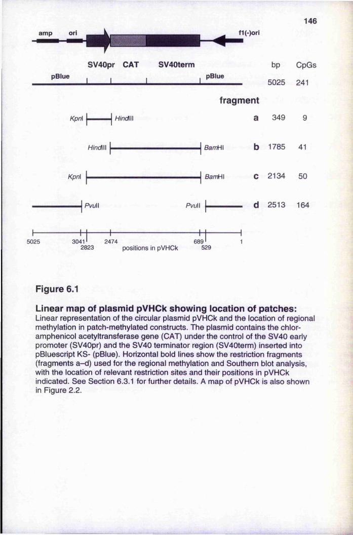
146f1(-)ori
SV40pr CAT SV40term pBlue I I I I pBlueI I
bp CpGs
5025 241
fragment
Kpn\ a 349 9
BamH\ b 1785 41
Kpn\ BamH\ C 2134 50
PvuW PvuW d 2513 164
5025 3041' 2474 689'2823 positions in pVHCk 529
Figure 6.1
Linear map of plasmid pVHCk showing location of patches:Linear representation of the circular plasmid pVHOk and the location of regional méthylation in patch-methylated constructs. The plasmid contains the chloramphenicol acetyltransferase gene (CAT) under the control of the SV40 early promoter (SV40pr) and the SV40 terminator region (SV40term) inserted into pBluescript KS- (pBlue). Horizontal bold lines show the restriction fragments (fragments a-d) used for the regional méthylation and Southern blot analysis, with the location of relevant restriction sites and their positions in pVHCk indicated. See Section 6.3.1 for further details. A map of pVHOk is also shown in Figure 2.2.
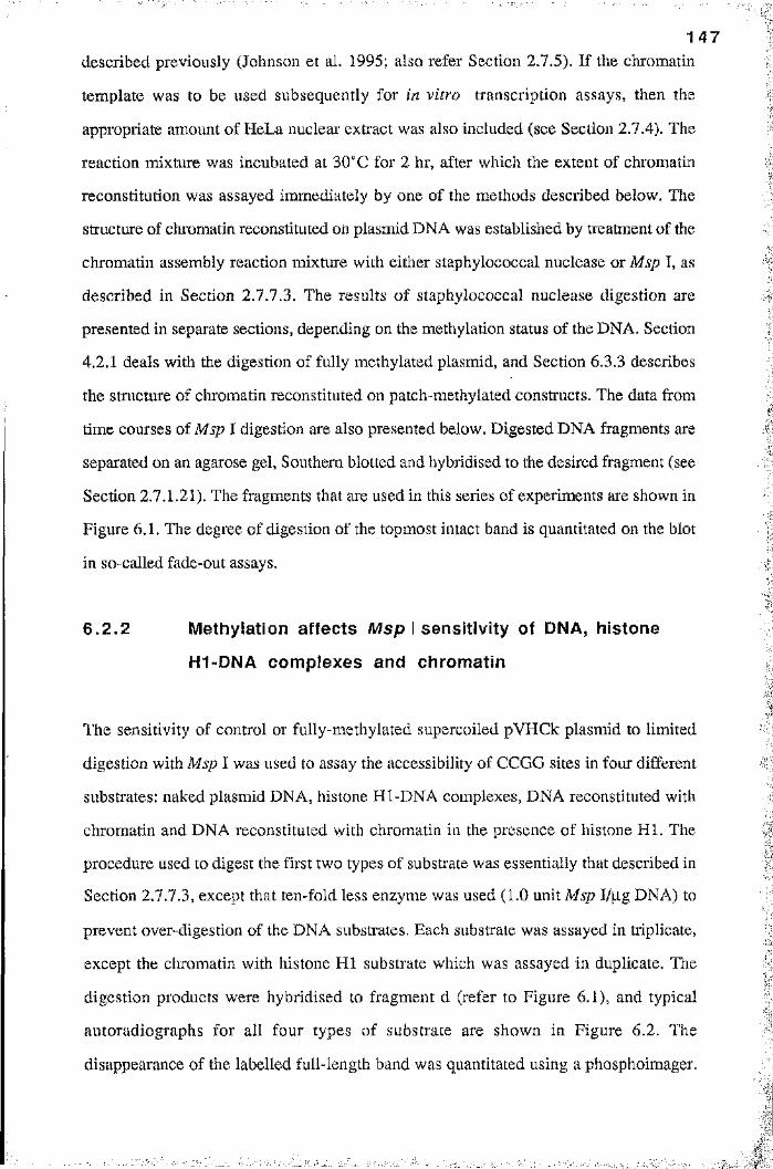
1 4 7
described previously (Johnson et al. 1995; also refer Section 2.7.5). If the chromatin
template was to be used subsequently for in vitro transcription assays, then the
appropriate amount of HeLa nuclear extract was also included (see Section 2.7.4). The
reaction mixture was incubated at 30“C for 2 hr, after which the extent of chromatin
reconstitution was assayed immediately by one of the methods described below. The
structure of chromatin reconstituted on plasmid DNA was established by treatment of the
chromatin assembly reaction mixture with either staphylococcal nuclease or Msp I, as
described in Section 2.7.7.3. The results of staphylococcal nuclease digestion are
presented in separate sections, depending on the méthylation status of the DNA. Section
4.2.1 deals with the digestion of fully methylated plasmid, and Section 6.3.3 describes
the structure of chromatin reconstituted on patch-methylated constructs. The data from
time courses of Msp I digestion are also presented below. Digested DNA fragments are
separated on an agarose gel, Southern blotted and hybridised to the desired fragment (see
Section 2.7.1.21). The fragments that are used in this series of experiments are shown in
Figure 6.1. The degree of digestion of the topmost intact band is quantitated on the blot
in so-called fade-out assays.
6 .2 .2 Méthylation affects Msp I sensitiv ity of DNA, histone
H l-DNA complexes and chrom atin
The sensitivity of control or fully-methylated supercoiled pVHCk plasmid to limited
digestion with Msp I was used to assay the accessibility of CCGG sites in four different
substrates: naked plasmid DNA, histone Hl-DNA complexes, DNA reconstituted with
chromatin and DNA reconstituted with chromatin in the presence of histone H I. The
procedure used to digest the first two types of substrate was essentially that described in
Section 2.1.13, except that ten-fold less enzyme was used (1.0 unit Msp I/|ig DNA) to
prevent over-digestion of the DNA substrates. Each substrate was assayed in triplicate,
except the chromatin with histone HI substrate which was assayed in duplicate. The
digestion products were hybridised to fragment d (refer to Figure 6.1), and typical
autoradiographs for all four types of substrate are shown in Figure 6.2. The
disappearance of the labelled full-length band was quantitated using a phosphoimager.
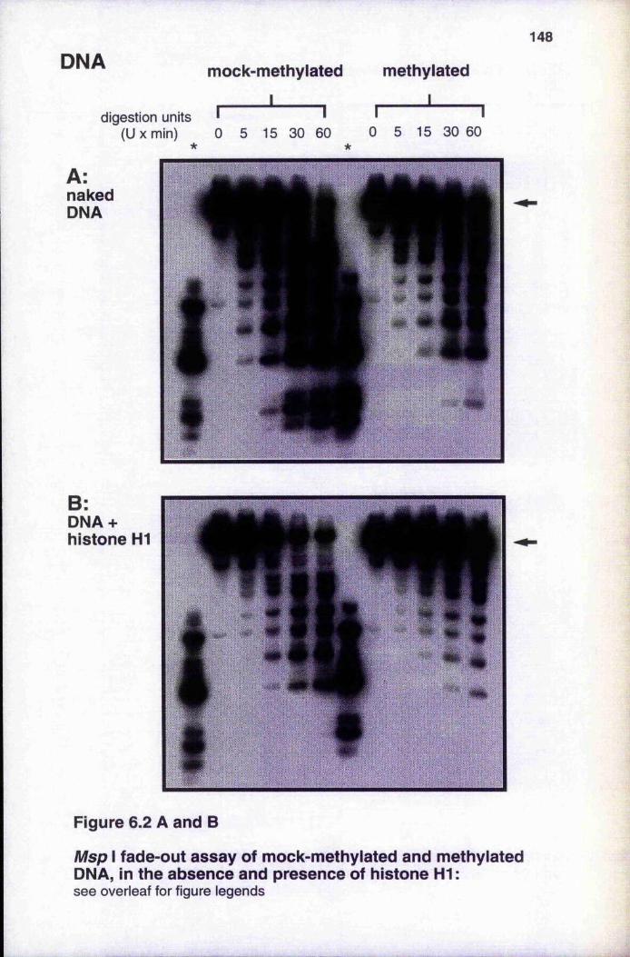
148
DNA mock-methylated
______ I___d ig es tio n units
(U X min)
I 1
methylated
______I______I I
0 5 15 30 60
A:nakedDNA
0 5 15 30 60
B;DNA + histone HI
Figure 6.2 A and B
Msp I fade-out assay of mock-methylated and methylated DNA, in the absence and presence of histone HI :s e e o v e rle a f for figure le g e n d s
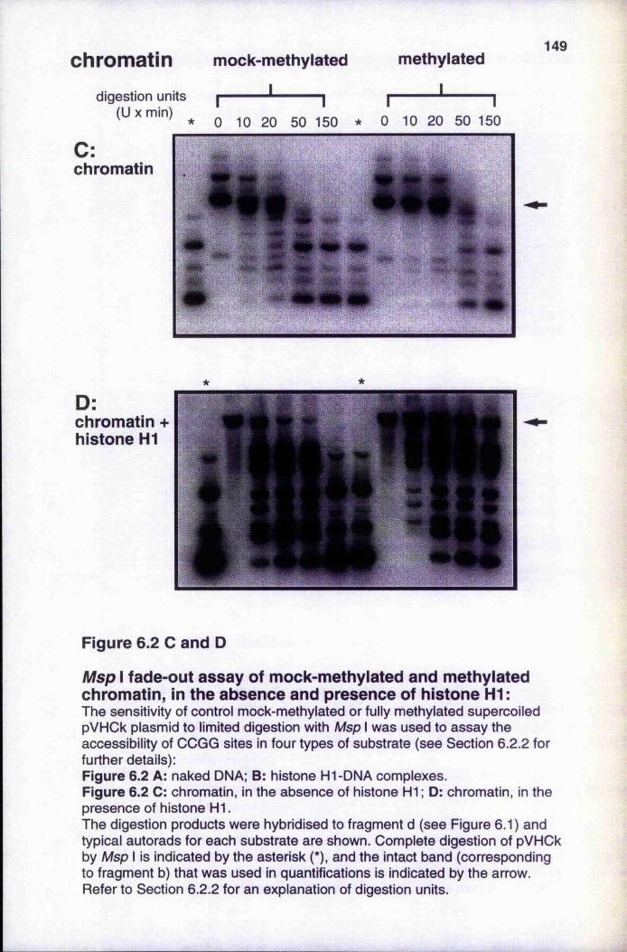
chromatindigestion units
(U X min)
C:chromatin
mock-methyiated
______ I___
methylated
I_____
149
I I I I* 0 10 20 50 150 * 0 10 20 50 150
D;chromatin + histone HI
Figure 6.2 C and D
Msp I fade-out assay of mock-methylated and methylated chromatin, in the absence and presence of histone HI ;The sensitivity of control mock-methylated or fully methylated supercoiled pVHCk plasmid to limited digestion with Msp I was used to assay the accessibility of CCGG sites in four types of substrate (see Section 6.2.2 for further details):Figure 6.2 A: naked DNA; B: histone Hl-DNA complexes.Figure 6.2 0: chromatin, in the absence of histone HI; D: chromatin, in the presence of histone HI.The digestion products were hybridised to fragment d (see Figure 6.1) and typical autorads for each substrate are shown. Complete digestion of pVHCk by Msp I is indicated by the asterisk (*), and the intact band (corresponding to fragment b) that was used in quantifications is indicated by the arrow. Refer to Section 6.2.2 for an explanation of digestion units.
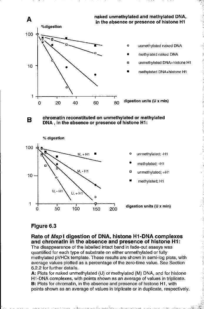
naked unmethylated and methylated DNA, In the absence or presence of histone H1
%digestion
100 -0
10
140 60 80200
uomethylated naked DNA
methylated naked DNA
unmethylated DNA+hlstone H1
methylated DNA+hlstone H1
0Q digestion units (U x min)
D chromatin reconstituted on unmethylated or methylated^ DNA , in the absence or presence of histone H1 :
?3'
iI
% digestion
M, -H1
U,-H1 U, + H1
100 150 200
o unmethylated; -H1
• methylated; -H1
° unmethylated; +H1
■ methylated; H1
digestion units (Ü x min)
.1
i
I
Figure 6.3
Rate of Msp I digestion of DNA, histone Hl-DNA complexes and chromatin in the absence and presence of histone H1:The disappearance of the labelled intact band in fade-out assays was quantified for each type of substrate on either unmethylated or fully methylated pVHCk template. These results are shown in semi-log plots, with average values plotted as a percentage of the zero-time value. See Section6.2.2 for further details.A: Plots for naked unmethylated (U) or methylated (M) DNA, and for histone Hl-DNA complexes, with points shown as an average of values in triplicate.B: Plots for chromatin, in the absence and presence of histone H I, with points shown as an average of values in triplicate or in duplicate, respectively.
■■
1 5 1
These results are shown in semi-log graphs in Figure 6.3, with the average value for
separate assays plotted as a percentage of the zero-time value. Lines in these graphs are
biased to consider the points for high % digestion values, because the line of best fit did
not appear to be close to the points for the lowest % digestion values on a logarithmic
scale. However, it was confirmed that the line of best fit did pass through the error bar
for these lowest values (error bars not shown). The abscissa is expressed as the product
of units of enzyme and time of digestion in minutes, to allow the comparison of
substrates of different sensitivities. For convenience, this product is called the digestion
unit.
In a separate series of graphs all points for a digestion experiment were
considered, rather than the average value as shown in the graphs of Figure 6.3 (results
not shown). The possible range of lines was then drawn, which provides an estimate of
sample deviation for each type of substrate. The digestion units required for 50% loss of
the full-length band was used as a measure of the gradient of each line. The average value
of the gradients for each type of substrate are shown in Table 6.1. The sample deviation
of each set of gradients is expressed as a percentage value of the average gradient.
Table 6.1
Accessibility of different substrates to Msp I:
digestion units (U x min) required for 50% digestion:
sample deviation (%)
naked DNA± 1 2 %
DNA+histone H1
± 1 4 %
chromatin± 2 4 %
chromatin*histone H1*
unmethylated 4 19 22 21methylated 7 3 7 42 60
nsufficent samp es
Methylated plasmid DNA is somewhat less sensitive to Msp I digestion than is
mock-methylated (control) DNA (Figure 6.3A). This experiment, which was carried out
in triplicate, may be a consequence of the altered conformation of the methylated plasmid,
y
. -1:,
,11
"Ii,.y
■
1 5 2or of the reduced rate of cleavage at C^^CGG sequences. The rate of digestion of histone
H l-D N A complexes is considerably reduced relative to that of naked DNA, but the
substrate containing methylated DNA is again less sensitive to limited I digestion
than is that containing mock-methylated DNA (Figure 6.3). This differential sensitivity is
approximately the same as that seen with naked DNA substrate (Table 6.1). When these
templates are reconstituted as chromatin using Xenopus S150 egg extract the rates of
digestion by Msp I are very similar to those observed in the presence of histone H I and
the differential effect of DNA méthylation is still observed (Figures 6.2C and 6.3B; Table
6.1). In the presence of histone HI the chromatin reconstituted on methylated DNA is
even more resistant to Msp I digestion (Figures 6.2D and 6.3B; Table 6.1).
6 .2 .3 Méthylation reduces transcriptional ac tiv ity of DNA,
histone H1-DNA complexes and chrom atin
The effect of méthylation on the activity of the SV40 promoter in pVHCk was assayed by
in vitro transcription of mock-methylated or fully methylated pVHCk, in the form of the
four types of substrates described above. The transcription assays for these substrates are
presented in Section 6.3.4, because the assays for one type of substrate were done at the
same time as those for patch-methylated constructs. This allows the direct comparison of
fully methylated plasmid and patch-methylated constructs by minimising the variability
during the formation of the substrate. This is pai'ticulaiiy true for chromatin substrates.
However, in this section the analysed data for mock-methylated and fully methylated
substrates is presented in Figure 6.4. Transcription of control (mock-methylated)
templates decreases in the order: naked DNA (100%), histone Hl-DNA complexes
(80%), chromatin (70%) and chromatin with histone H I (40%). In all cases,
transcription from the fully methylated DNA template is lower when compared to that
from the control mock-methylated templates.
It has been shown that histone Hl-DNA complexes preferentially inhibit the in
vitro transcription of pol III genes compared to complexes formed on unmethylated
DNA (see Section 5.3.4 and Johnson et al. 1995). Similar results for the pol II system
1
s
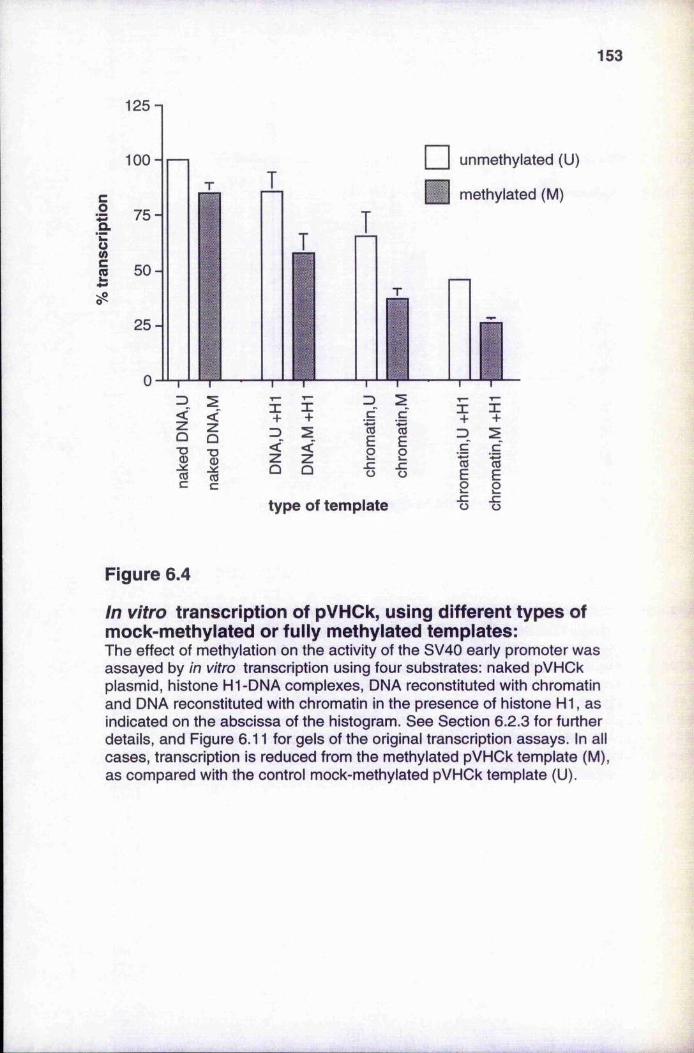
153
co
G(/}C
125-1
100 -
75-
50-
25-
Z)<
•o
COc
I I unmethylated (U)
methylated (M)
type of template
X+
3cc5E2•ë
05E2szü
Figure 6.4
In vitro transcription of pVHCk, using different types of mock-methylated or fully methylated templates:The effect of méthylation on the activity of the SV40 early promoter was assayed by in vitro transcription using four substrates: naked pVHCk plasmid, histone Hl-DNA complexes, DNA reconstituted with chromatin and DNA reconstituted with chromatin in the presence of histone HI, as indicated on the abscissa of the histogram. See Section 6.2.3 for further details, and Figure 6.11 for gels of the original transcription assays. In all cases, transcription is reduced from the methylated pVHCk template (M), as compared with the control mock-methylated pVHCk template (U).
1 5 4
are seen in Figure 6.4, at the same level of histone HI that was used for the pol III study
(a histone H1:DNA w/w ratio of 0.6), although the degree of preferential inhibition is not
as pronounced. This confirms a previous experiment on the pol II system, also described
in Section 5.3.4. The in vitro transcription of naked methylated DNA is also
preferentially inhibited compared to naked unmethylated DNA (Figure 6.4). Since the
HeLa nucleai' extract that is used for the transcription studies is depleted of endogenous
histone H I (Section 2.7.4.3), the preferential inhibition of naked methylated DNA
indicates the presence of a limiting amount of a selective inhibitor in the nuclear extract,
as described previously (Johnson et al. 1995). Similar results have been described for
transcription of pol II genes by Boyes and Bird (1991), who suggested that MeCPl was
I
the limiting protein.
Chromatin was reconstituted on methylated DNA in the absence and in the
presence of exogenous histone H I. Transcription from both of these substrates is also
preferentially inhibited (Figure 6.4), although unmethylated templates are inhibited when
compared to naked DNA templates. Other studies have described the inhibition of
transcription by nucleosomes and histone HI in more detail (Section 1.8.5). It is known
that both full méthylation of pVHCl (Bryans et al. 1992) and regional méthylation of
pVHCk (Kass et al. 1993) can inactivate the S V40 early promoter after transfection into
mammalian cells, although viral SV40 early gene expression is insensitive towards
méthylation (Graessmann et al. 1983; Graessmann and Graessmann 1993). In this, the
results are similar to those observed for histone Hl-DNA complexes. However, histone
H l-D N A complexes cannot mediate the spread of inactivation from a region of
méthylation (Section 6.3.4) that can be mediated by chromatin. The effect of histone HI
in the context of methylated chromatin is to inhibit transcription still further (Figure 6.4),
which is reflected in an increase in the inaccessibility to Msp I (Table 6.1), as compared
to unmethylated chromatin. By contrast, histone H I does not appear to inhibit
unmethylated chromatin to as great an extent.
The effect of méthylation on tRNA gene expression was also assayed by in vitro
transcription of mock-methylated or fully methylated pArg/Leu template, which has been
reconstituted with chromatin (Figure 6.5). Assays were performed in duplicate. These
templates were reconstituted with chromatin and preincubated with nuclear extract
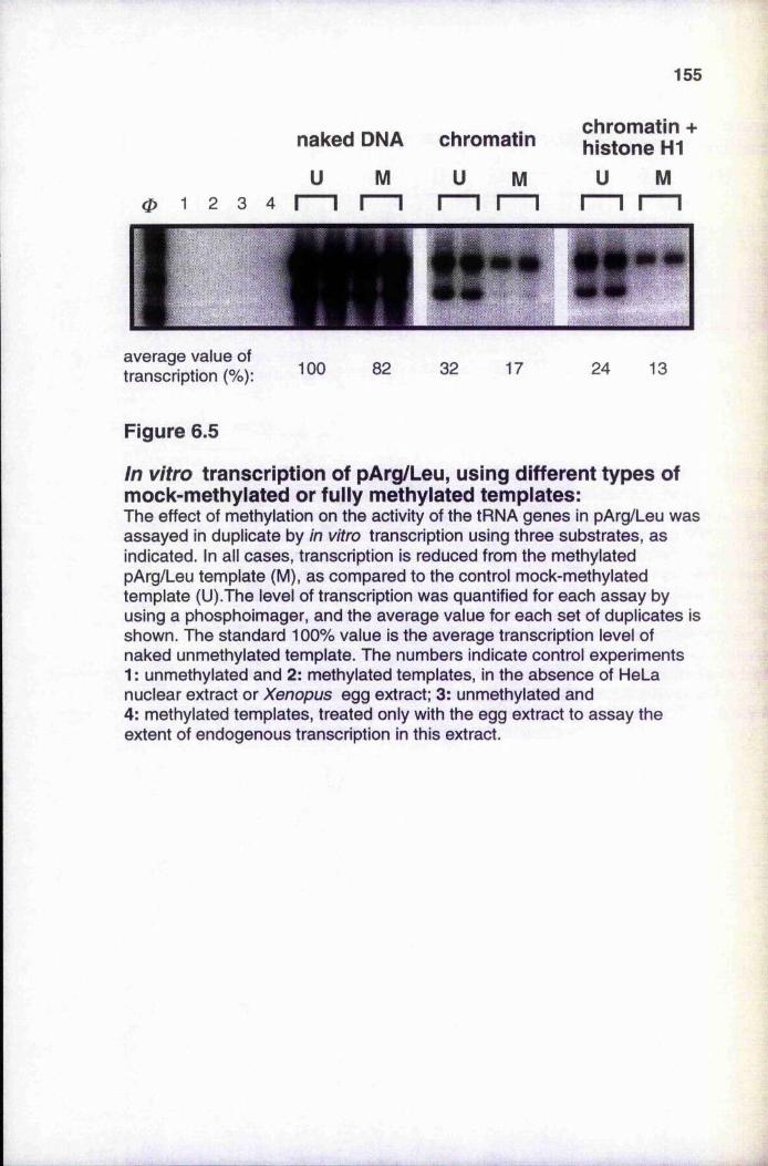
155
naked DNA
U M
chromatin
U M
chromatin + histone H1
U M0 1 2 3 4
average value of transcription 100 82 32 17 24 13
Figure 6.5
In vitro transcription of pArg/Leu, using different types of mock-methylated or fully methylated templates:The effect of méthylation on the activity of the tRNA genes in pArg/Leu was assayed in duplicate by in vitro transcription using three substrates, as indicated. In all cases, transcription is reduced from the methylated pArg/Leu template (M), as compared to the control mock-methylated template (U).The level of transcription was quantified for each assay by using a phosphoimager, and the average value for each set of duplicates is shown. The standard 100% value is the average transcription level of naked unmethylated template. The numbers indicate control experiments 1 : unmethylated and 2: methylated templates, in the absence of HeLa nuclear extract or Xenopus egg extract; 3: unmethylated and 4: methylated templates, treated only with the egg extract to assay the extent of endogenous transcription in this extract.
1 5 6
simultaneously as described in Section 6.2,1. Control experiments confirmed that the
Xenopus egg extract did not support endogenous transcription of the template. The
effect of chromatin reconstitution was to preferentially inhibit transcription from the
methylated template, as shown in Figure 6.5. The addition of histone HI to the reaction
mixture appears to have a further inhibitory effect on both unmethylated and methylated
chiomatin. These observations support the data presented in Figure 6.4.
6.3 In vitro chromatin formation and transcription on patch-methylated pVHCk
I'I
This section describes experiments, similar to those in Section 6.2, that were performed
on patch-methylated constructs. This allows the direct comparison of the effect of
regional méthylation on transcription with control experiments that use the fully
methylated pVHCk template. The preparation of patch-methylated pVHCk constructs is
described in Section 2.7.3.2. These patch-methylated constructs for the pol II system
must not be confused with the constructs for the pol III system, which were prepared by
the ligation of Bgl I fragments of pArg/Leu (see Section 5.3.7).
6.3 .1 Analysis of patch-methylated constructs |
The prepai’ation of patch-methylated pVHCk constructs yielded double-stranded plasmid
constructs that were methylated at CpG dinucleotides only in specific regions, so-called
"patches", with an efficiency of 60-80% as reported by Kass et al. (1993). Mock
methylated plasmids and non-patch regions are not methylated. Four different restriction
fragments, ranging in size from 349 to 2513 bp (fragments a-d in Figure 6.1) were used
to make the patch-methylated constructs of the pVHCk plasmid. The location of
methylated patches in all constructs were verified by restriction enzyme digestion with a
methyl-sensitive enzyme (typically Hpa II) and, if possible, by Southern blot analysis.
The construct with fragment, a could not be analysed in this way because the SV40
promoter lacks Hpa II sites, and indeed sites for any other methylation-sensitive
restriction enzymes. The analyses of constructs b to d ai*e shown in Figure 6.6A-C, with
fui'ther explanation of the analyses in tlie figure legends.
.s
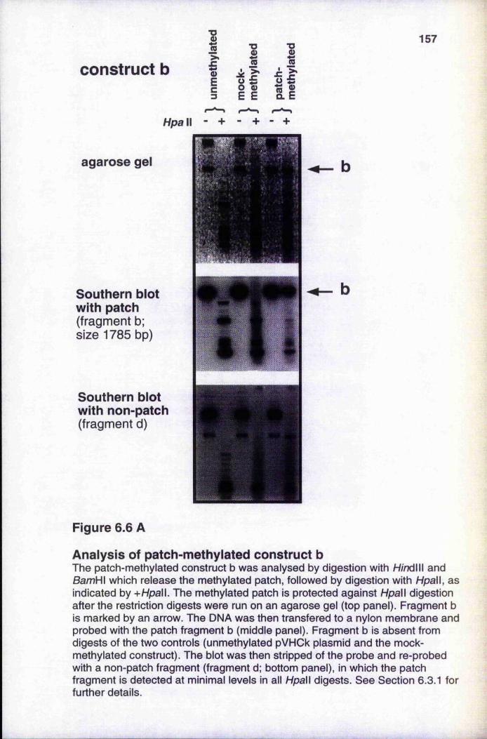
construct b1fEc3
IE E
1
1 1
157
HpaW - + - + - +
agarose gel
Southern blot with patch(fragment b; size 1785 bp)
Southern blot with non-patch(fragment d)
1
i '
Figure 6.6 A
Analysis of patch-methylated construct bThe patch-methylated construct b was analysed by digestion with H/ndlll and BamH\ which release the methylated patch, followed by digestion with Hpall, as indicated by + Hpall. The methylated patch is protected against Hpall digestion after the restriction digests were run on an agarose gel (top panel). Fragment b is marked by an arrow. The DNA was then transfered to a nylon membrane and probed with the patch fragment b (middle panel). Fragment b is absent from digests of the two controls (unmethylated pVHCk plasmid and the mock- methylated construct). The blot was then stripped of the probe and re-probed with a non-patch fragment (fragment d; bottom panel), in which the patch fragment is detected at minimal levels in all Hpall digests. See Section 6.3.1 for further details.
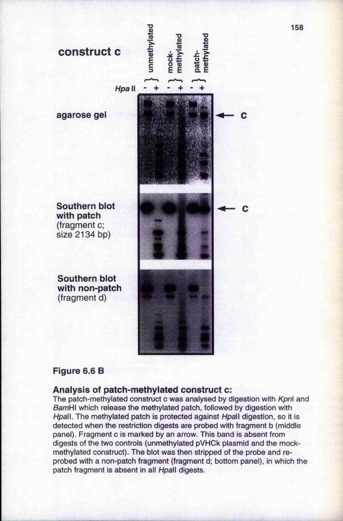
construct cI>*
.CQ)Ec
158%
■o0)
If ifHpa II -
agarose gel
Southern blot with patch(fragment c; size 2134 bp)
Southern blot with non-patch(fragment d)
Figure 6.6 B
Analysis of patch-methylated construct c:The patch-methylated construct c was analysed by digestion with Kpnl and BamHI which release the methylated patch, followed by digestion with Hpall. The methylated patch is protected against Hpall digestion, so it is detected when the restriction digests are probed with fragment b (middle panel). Fragment c is marked by an arrow. This band is absent from digests of the two controls (unmethylated pVHCk plasmid and the mock- methylated construct). The blot was then stripped of the probe and reprobed with a non-patch fragment (fragment d; bottom panel), in which the patch fragment is absent in all Hpall digests.
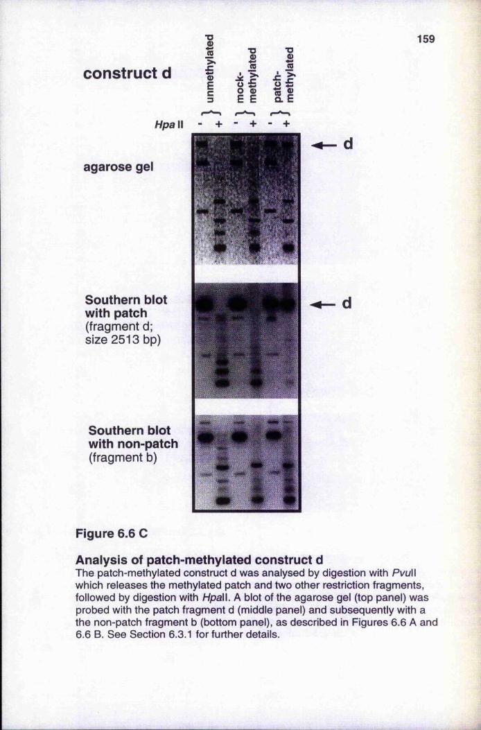
construct d1IEc
159
1È
1>*
Il ilHpall
agarose gel
Southern blot with patch(fragment d; size 2513 bp)
Southern blot with non-patch(fragment b)
Figure 6.6 C
Analysis of patch-methylated construct dThe patch-methylated construct d was analysed by digestion with PvuW which releases the methylated patch and two other restriction fragments, followed by digestion with Hpall. A blot of the agarose gel (top panel) was probed with the patch fragment d (middle panel) and subsequently with a the non-patch fragment b (bottom panel), as described in Figures 6.6 A and 6.6 B. See Section 6.3.1 for further details.
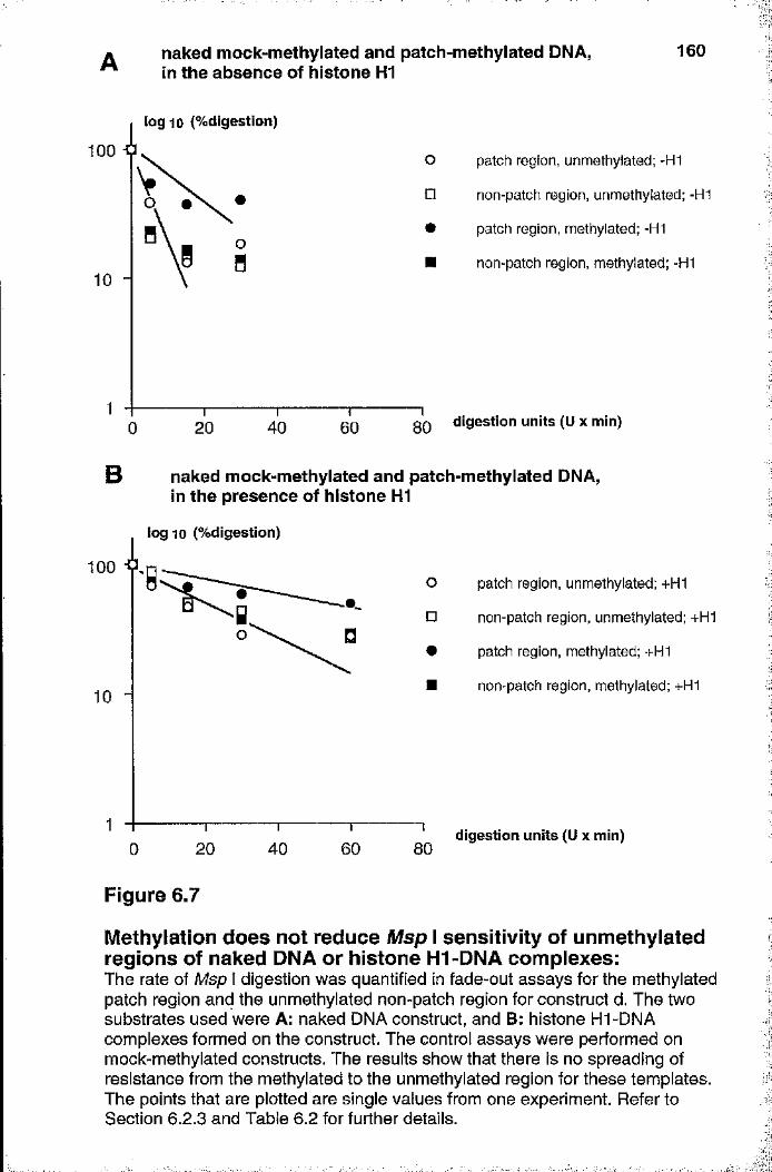
naked mock-methylated and patch-methylated DNA, in the absence of histone H1
160
log 10 (%digestion)
100 patch region, unmethylated; -H1
non-patch region, unmethylated; -H1
patch region, methylated; -H1
non-patch region, methylated; -H1
gQ digestion units (U x min)4020 600
B naked mock-methylated and patch-methylated DNA, in the presence of histone HI
100 j ,
lo g ic (%digestion)
10 -
'8
0
o
□#
20 40 60
patch region, unmethylated; +H1
non-patch region, unmethylated; +H1
patch region, methylated; +H1
non-patch region, methylated; +H1
80digestion units (Ü x min)
I
I::
Figure 6.7
Méthylation does not reduce Msp I sensitivity of unmethylated regions of naked DNA or histone HI-DNA complexes:The rate of Msp I digestion vyas quantified in fade-out assays for the methyiated patch region and the unmethyiated non-patch region for construct d. The two substrates used were A: naked DNA construct, and B: histone HI-DNA complexes formed on the construct. The control assays were performed on mock-methylated constructs. The results show that there is no spreading of resistance from the methyiated to the unmethylated region for these templates. The points that are plotted are single values from one experiment. Refer to Section 6.2.3 and Table 6.2 for further details.
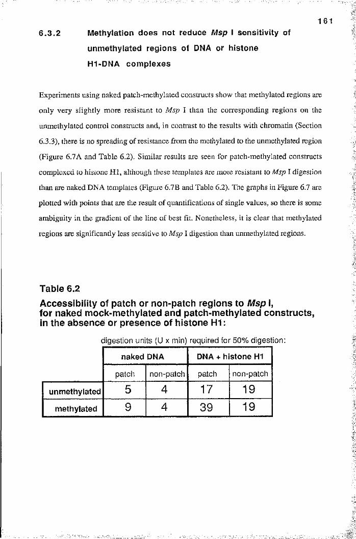
1 6 1
6 .3 .2 Méthylation does not reduce Msp I sensitiv ity of
unmethylated regions of DNA or histone
H1-DNA com plexes
Experiments using naked patch-methylated constructs show that methylated regions are
only very slightly more resistant to Msp I than the corresponding regions on the
unmethylated control constructs and, in contrast to the results with chromatin (Section
6.3.3), there is no spreading of resistance from the methylated to the unmethylated region
(Figure 6.7A and Table 6.2). Similar results are seen for patch-methylated constructs
complexed to histone HI, although these templates are more resistant to Msp I digestion
than are naked DNA templates (Figure 6.7B and Table 6.2). The graphs in Figure 6.7 are
plotted with points that are the result of quantifications of single values, so there is some
ambiguity in the gradient of the line of best fit. Nonetheless, it is clear that methylated
regions are significantly less sensitive to Msp I digestion than unmethylated regions.
Table 6.2
Accessibility of patch or non-patch regions to Msp I, for naked mock-methylated and patch-methylated constructs, in the absence or presence of histone H1 :
digestion units (U x min) required for 50% digestion:
naked DNA DNA + histone HI
patch non-patch patch non-patch
unmethylated 5 4 17 19
methylated 9 4 39 19
Î
I
' i :
I
J
.
'
'
I*{i
i
I#>0
1 6 2
6.3 .3 Méthylation affects the chromatin structure of
unmethylated regions
Chromatin was reconstituted on patch-methylated constructs and subjected to limited
digestions with either staphylococcal nuclease or Msp I (10 unit Msp I/p.g DNA; see
Section 2.7.73 for further details). The digestion products from these treatments were
hybridised to restriction fragments corresponding to either the region that had been patch-
methylated, or to the adjacent unmethylated region.
The results for staphylococcal digestion ai’e shown in Figure 6.8. As expected,
there is no difference in the nucleosome spacing between the chromatin at the metliylated
patch and the chromatin in the same region of the mock-methylated construct (see Section
4.2.1). There is also no apparent difference between the non-patch regions. The relative
rate of digestion for these regions was not deteimined, as it was for the Msp I digestions,
because the quantification of the diffuse bands that are produced in this assay would be
inaccurate. Instead, this assay was used to ensure that chromatin had been reconstituted
on construct DNA, before it was used in the Msp I digestion experiments.
The results for Msp I digestions are shown in Figures 6.9 and 6.10. Typical
autoradiographs for chromatin, or chromatin in the presence of histone H I , reconstituted
on a patch-methylated construct are shown in Figure 6.9. The assays for each substrate
were performed in triplicate, and average values are shown in the graphs of Figure 6.10.
(This is the same procedure as that adopted for the graphs of Figure 6.3; refer to Section
6.2.2 for further details). The results in Figure 6.9A show clearly that the chromatin
reconstituted on either the methylated region or the unmethylated region of each patch-
methylated construct is less accessible to Msp I digestion compared to that formed on the
mock patch-methylated control construct (Table 6.3). Similar results are observed when
chromatin reconstitution takes place in the presence of histone H I (Figure 6.9B and
Table 6.3), but the methylated construct is even more resistant to nuclease digestion of
both the patched and the contiguous regions. The sample variance for the average value
of each gradient in Table 6.3 was calculated as described in Section 6.2.2. The sample
variance is expressed as a percentage value of the average gradient.
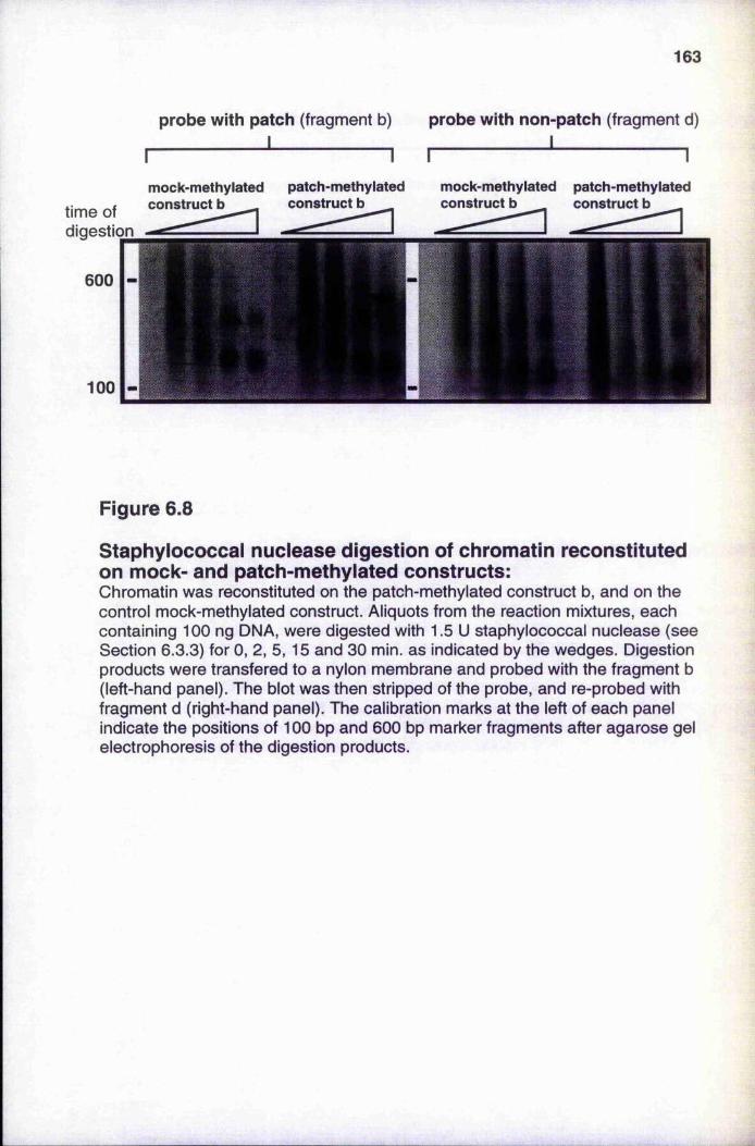
163
time of digestion
600
100
probe with patch (fragment b) probe with non-patch (fragment d) ___________ I_____________________ I______
I---------------------------------------1 I--------------mock-methylated patch-methylated mock-methylated patch-methylatedconstruct b construct b construct b construct b
Figure 6.8
Staphylococcal nuclease digestion of chromatin reconstituted on mock- and patch-methylated constructs:Chromatin was reconstituted on the patch-methylated construct b, and on the control mock-methylated construct. Aliquots from the reaction mixtures, each containing 100 ng DNA, were digested with 1.5 U staphylococcal nuclease (see Section 6.3.3) for 0, 2,5,15 and 30 min. as indicated by the wedges. Digestion products were transfered to a nylon membrane and probed with the fragment b (left-hand panel). The blot was then stripped of the probe, and re-probed with fragment d (right-hand panel). The calibration marks at the left of each panel indicate the positions of 100 bp and 600 bp marker fragments after agarose gel electrophoresis of the digestion products.
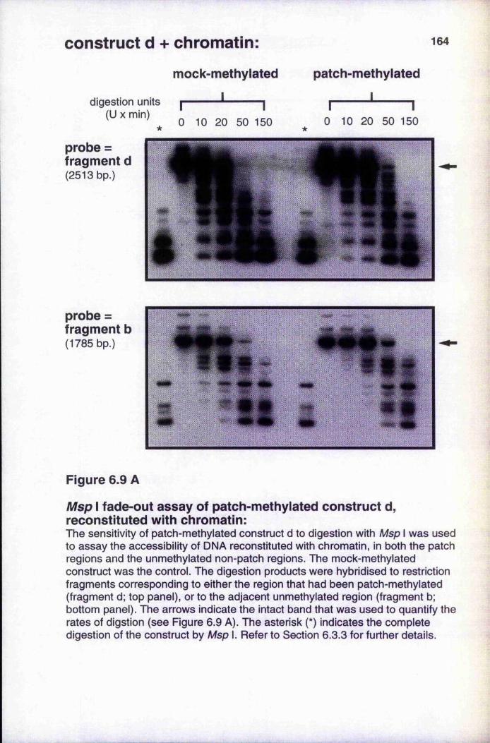
construct d + chromatin: 164
digestion units r (U X min)
mock-methylated
______ I______
patch-methylated
II r 1
probe = fragment d(2513 bp.)
0 10 20 50 150 0 10 20 50 150
probe = fragment b(1785 bp.)
m
Figure 6.9 A
Msp I fade-out assay of patch-methylated construct d, reconstituted with chromatin:The sensitivity of patch-methylated construct d to digestion with Msp I was used to assay the accessibility of DNA reconstituted with chromatin, in both the patch regions and the unmethylated non-patch regions. The mock-methylated construct was the control. The digestion products were hybridised to restriction fragments corresponding to either the region that had been patch-methylated (fragment d; top panel), or to the adjacent unmethylated region (fragment b; bottom panel). The arrows indicate the intact band that was used to quantify the rates of digstion (see Figure 6.9 A). The asterisk (*) indicates the complete digestion of the construct by Msp I. Refer to Section 6.3.3 for further details.
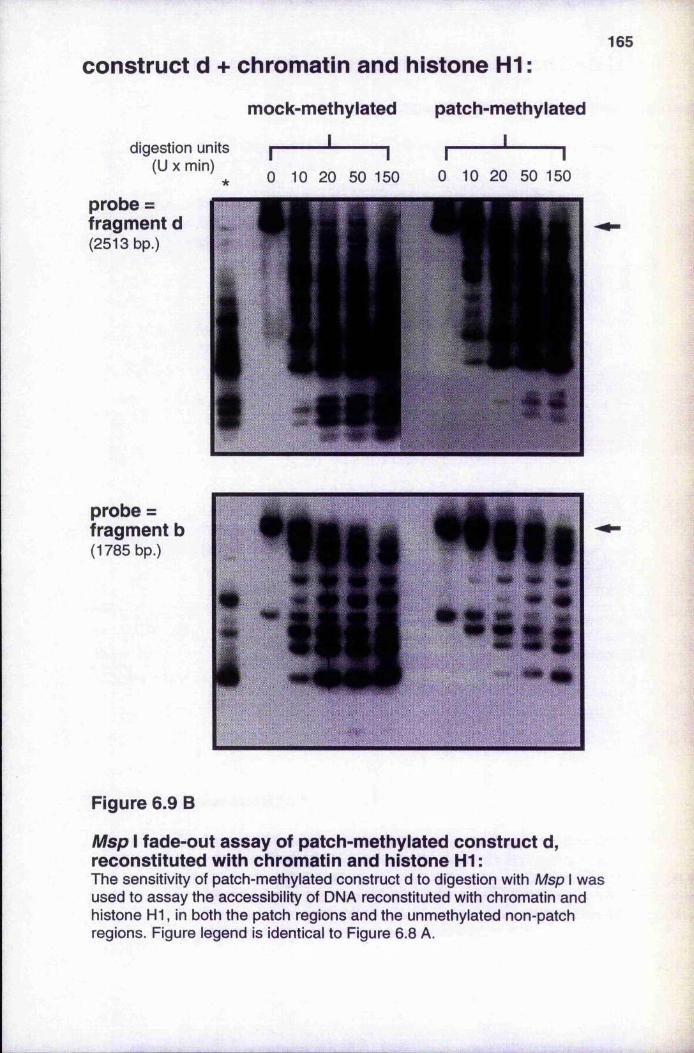
165
construct d + chromatin and histone H1 :
digestion units (U X min)
*
mock-methylated
I *--------- 10 10 20 50 150
patch-methylated
______I______I I
0 10 20 50 150
probe = fragment d(2513 bp.)
probe = fragment b(1785 bp.)
Figure 6.9 B
Msp I fade-out assay of patch-methylated construct d, reconstituted with chromatin and histone H1:The sensitivity of patch-methylated construct d to digestion with Msp I was used to assay the accessibility of DNA reconstituted with chromatin and histone HI, in both the patch regions and the unmethylated non-patch regions. Figure legend is identical to Figure 6.8 A.
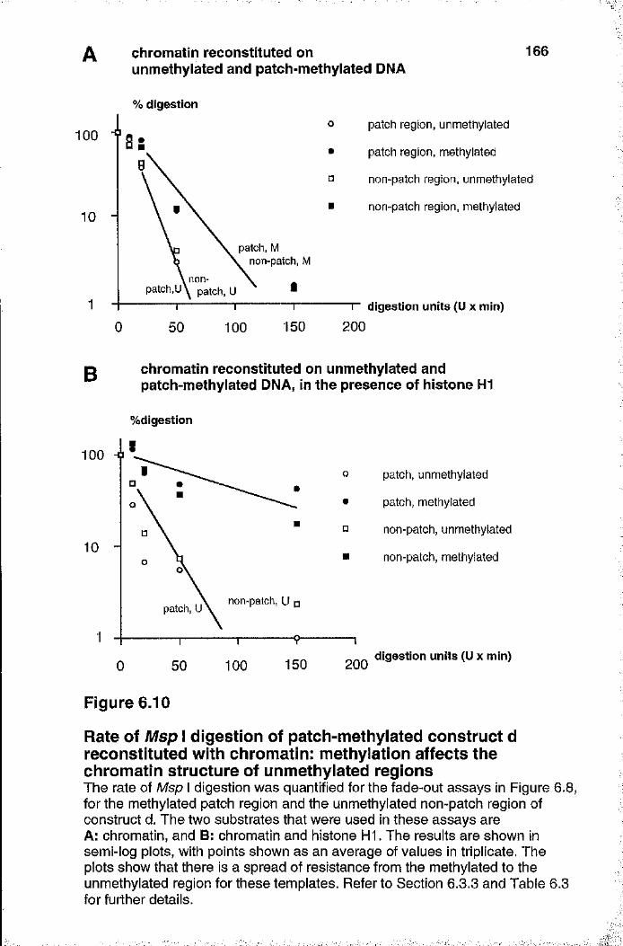
chromatin reconstituted on unmethylated and patch-methylated DNA
166
1 0 0
10
% digestion
0
patch, M non-patch, M
patch region, unmethylated
patch region, methylated
non-patch region, unmethylated
non-patch region, methylated
50 100 150r digestion units (U x min)
200
g chromatin reconstituted on unmethylated andpatch-methylated DNA, in the presence of histone HI
%digestion
patch, unmethylated
patch, methylated
non-patch, unmethylated
non-patch, methylated
100 -O
non-patch, Upatch, U
150 2001000 50digestion units (Ü x min)
Figure 6.10
Rate of Msp I digestion of patch-methylated construct d reconstituted with chromatin: méthylation affects the chromatin structure of unmethylated regionsThe rate of Msp I digestion was quantified for the fade-out assays in Figure 6.8, for the methylated patch region and the unmethylated non-patch region of construct d. The two substrates that were used in these assays are A; chromatin, and B: chromatin and histone HI. The results are shown in semi-log plots, with points shown as an average of values in triplicate. The plots show that there is a spread of resistance from the methylated to the unmethylated region for these templates. Refer to Section 6.3.3 and Table 6.3 for further details.
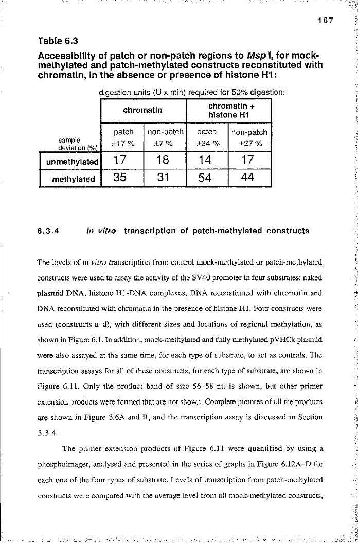
1 6 7
Table 6.3 Accessibility of patch or non-patch regions to Msp I, for mock- methylated and patch-methylated constructs reconstituted with chromatin, in the absence or presence of histone H1:
digestion units (U x min) required for 50% digestion:
sample deviation (%)
chromatin chromatin + histone H1
patch±17%
non-patch± 7 %
patch ±24 %
non-patch ±27 %
unmethylated 17 18 14 17
methylated 35 31 54 4 4
6.3 .4 in vitro transcription of patch-methylated constructs
The levels of in vitro transcription from control mock-methylated or patch-methylated
constructs were used to assay the activity of the SV40 promoter in four substrates: naked
plasmid DNA, histone Hl-DNA complexes, DNA reconstituted with chromatin and
DNA reconstituted with chromatin in the presence of histone H I. Four constructs were
used (constructs a-d), with different sizes and locations of regional méthylation, as
shown in Figure 6.1. In addition, mock-methylated and fully methylated pVHCk plasmid
were also assayed at the same time, for each type of substrate, to act as controls. The
transcription assays for all of these constructs, for each type of substrate, are shown in
Figure 6.11. Only the product band of size 56-58 nt. is shown, but other primer
extension products were foi*med that are not shown. Complete pictures of all the products
are shown in Figure 3.6A and B, and the transcription assay is discussed in Section
3.3.4.
The primer extension products of Figure 6.11 were quantified by using a
phosphoimager, analysed and presented in the series of graphs in Figure 6.12A-D for
each one of the four types of substrate. Levels of ti'anscription from patch-methylated
constmcts were compared with the average level from all mock-methylated constructs,
■
:■'I-. - v i
1-
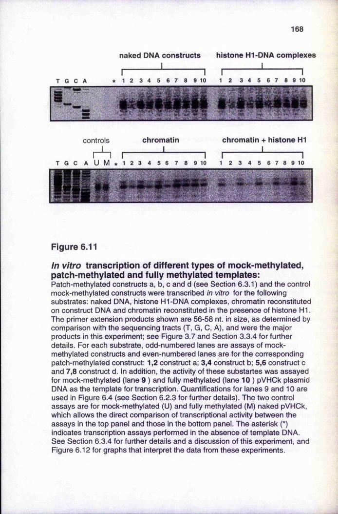
168
naked DNA constructs histone Hl-DNA complexes I________________ I
* 1 2 3 4 5 6 7 8 9 10 1 2 3 4 5 6 7 8 9 10
controls
r A rchromatin
IT G C A U M * 1 2 3 4 5 6 7 8 9 10
chromatin + histone HI
I * I1 2 3 4 5 6 7 8 9 10
Figure 6.11
In vitro transcription of different types of mock-methylated, patch-methylated and fully methylated templates:Patch-methylated constructs a, b, c and d (see Section 6.3.1) and the control mock-methylated constructs were transcribed in vitro for the following substrates: naked DNA, histone H1-DNA complexes, chromatin reconstituted on construct DNA and chromatin reconstituted in the presence of histone HI. The primer extension products shown are 56-58 nt. in size, as determined by comparison with the sequencing tracts (T, G, 0, A), and were the major products in this experiment; see Figure 3.7 and Section 3.3.4 for further details. For each substrate, odd-numbered lanes are assays of mock- methylated constructs and even-numbered lanes are for the corresponding patch-methylated construct: 1,2 construct a; 3,4 construct b; 5,6 construct c and 7,8 construct d. In addition, the activity of these substartes was assayed for mock-methylated (lane 9 ) and fully methylated (lane 10 ) pVHCk plasmid DNA as the template for transcription. Quantifications for lanes 9 and 10 are used in Figure 6.4 (see Section 6.2.3 for further details). The two control assays are for mock-methylated (U) and fully methylated (M) naked pVHCk, which allows the direct comparison of transcriptional activity between the assays in the top panel and those in the bottom panel. The asterisk (*) indicates transcription assays performed in the absence of template DNA. See Section 6.3.4 for further details and a discussion of this experiment, and Figure 6.12 for graphs that interpret the data from these experiments.
1 6 9
which did not vary significantly with size or location of mock-methylated patch. These
Iresults show that fully methylated naked DNA template is transcribed less efficiently than
unmethylated template. This inhibition is most obvious when only the promoter of the
reporter gene was methylated (Figure 6.12A). The degree of inhibition decreases with
increasing patch size (compare transcription from construct a and c) and there is no
inhibition of consti’uct d. This is consistent with the HeLa nuclear extract containing
limiting levels of an inhibitory factor (e.g. methyl CpG binding proteins). The density of
this factor bound to the promoter of the otherwise naked plasmid could then dictate the
activity of the promoter. Similar results are observed for constructs complexed with
histone HI (Figure 6.12B), although the absolute levels of transcription are only 80% of
those with naked DNA constructs, as is consistent with the results in Section 6.2.3 and
Figure 6.4. Therefore, with naked DNA in the absence or presence of histone H I, no
spreading of inactivation is observed and inhibition is dependant on a high density of
promoter méthylation. However, in the presence of chromatin (Figure 6.12C), or
chromatin with histone H I (Figure 6.12D), all patch-methylated constructs are equally
inhibited, including construct d, in which the patch of méthylation is covers only vector
sequences. In these cases a methylated patch in the vector DNA is able to seed the
formation of inactive chromatin that leads to the inhibition of transcription from the
distant promoter. This is reflected in the Msp I resistance of the non-patch regions of
chromatin-associated plasmids (see Section 6.3.3).
Transcription of all the patch-methylated constructs (constructs a-d) reconstituted
with chromatin, in the absence or presence of histone H I, is inhibited to a common level
that is 40-50% of the unmethylated constructs (Figure 6.12C and D). Transcription from
naked DNA, or histone Hl-DNA complexes, is at various levels depending on the size
and position of the methylated region. The methylated CpG-rich prokaryotic vector DNA
in construct d does not inhibit ti'anscription from the distant promoter for these templates
(Table 6.2), as discussed above. Transcription from construct c, in which the patch
covers both promoter and reporter gene, is inhibited to 60-80% of the unmethylated
constructs which is consistent with the results for fully-methylated templates (Figure
6.12A). However, transcription from construct a, in which only the promoter is
methylated, is inhibited still further to about 40%. This result could be explained if a
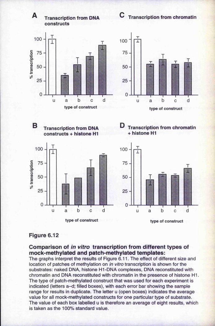
Transcription from DNA constructs
Transcription from chromatin
1 0 0 -
a b c d type of construct
1 0 0
75-
50-
25-
0
T
T-----------1------- TU a b 0
type of construct
B Transcription from DNA constructs + histone H1
100 -
co 75 -a
50-
25 -
b du ca
Transcription from chromatin + histone H1
1 0 0 -
75-
50-
25-
0
type of construct
T
a b e d
type of construct
Figure 6.12
Comparison of in vitro transcription from different types of mock-methylated and patch-methylated templates:The graphs interpret the results of Figure 6.11. The effect of different size and location of patches of méthylation on in vitro transcription is shown for the substrates: naked DNA, histone Hl-DNA complexes, DNA reconstituted with chromatin and DNA reconstituted with chromatin in the presence of histone HI. The type of patch-methylated construct that was used for each experiment is indicated (letters a-d; filled boxes), with each error bar showing the sample range for results in duplicate. The letter u (open boxes) indicates the average value for all mock-methylated constructs for one particular type of substrate. The value of each box labelled u is therefore an average of eight results, which is taken as the 100% standard value.
171large patch (c, containing 50 methylated CpGs) is able to compete out limiting amounts
of a selective inhibitor (such as M eCPl) that is present in the nuclear extract, whereas a
small patch (a, containing 9 methylated CpGs) is unable to compete out the inhibitor.
Construct b is also inhibited, even though the promoter is unmethylated, which suggests
that the putative MeCP can mediate an indirect mechanism of promoter inhibition even in
the absence of chromatin. Kass et al. (1993) have shown that a patch at the promoter (as
in construct a) can reduce the transcriptional activity of the methylated construct to 69%
of the mock-methylated control, after transient expression of the constructs.
6.4 Discussion
Sum m ary
This chapter investigates the spread of inactive chromatin from a region of méthylation by
using in vitro systems of chromatin assembly and transcription. The presence of inactive
chromatin was assayed by resistance to digestion by Msp I. However, fully methylated
naked plasmid DNA is also less sensitive to digestion with Msp I, compared to naked
unmethylated DNA. This reduced sensitivity is confined to the region of méthylation in a
construct and does not affect contiguous regions, even when histone HI is complexed to
the DNA. Using HeLa nuclear extract, fully methylated naked DNA template is
transcribed less efficiently than is unmethylated template. This effect is most obvious
when only the promoter of the reporter gene is methylated, a finding that is indicative of
limiting levels of an inhibitor in the nuclear extract. Histone Hl-DNA complexes on
unmethylated and fully-methylated pVHCk were also used as templates, and the results
support the previous observation that histone H I preferentially represses in vitro
transcription from fully-methylated template DNA (Johnson et al. 1995; and Section
5.3.4). When chromatin was reconstituted on these templates using Xenopus S150 egg
extract, the lower sensitivity to Mspl digestion of the methylated DNA is maintained and
this reduced sensitivity is now able to spread from the methylated patch to a contiguous
unmethylated region. Methylated chromatin templates retain a reduced transcriptional
activity compared with unmethylated chromatin templates, and this is also the case when
_ k... ..i ; Bî i::
1 7 2
any region of the plasmid is unmethylated. These effects are further enhanced by the
addition of histone HI during the chromatin reconstitution.
These results indicate that on naked DNA templates, in the absence or presence of
histone H I, promoter méthylation is important to inhibit transcriptional activity and only
regions of the plasmid that are actually methylated show reduced sensitivity to nuclease
digestion. However, with chromatin templates patch-methylation affects transcription and
nuclease sensitivity of a distant promoter.
M sp I sensitivity of naked DNA and histone H l-D N A complexes
The accessibility of nucleases to chromatin has been used as an assay of chromatin
conformation (Paranjape et al. 1994) and, in particular, Msp I has been used to assay
differences between unmethylated and methylated chromatin (Keshet et al. 1986;
Antequera et al. 1989). The results discussed in Section 6.2.2 show that in vitro
chromatin reconstitution onto methylated DNA is also more resistant to digestion by
M spl than unmethylated chromatin (Figure 6.3), using a fade-out assay for intact
restriction fragments as described above. Histone Hl-DNA complexes on methylated
DNA are also more resistant than unmethylated DNA (Figure 6.3 and Table 6.1), and are
results that confirm a previous study by Higurashi and Cole (1991). This study also
stated that there was no detectable difference in Msp I sensitivity between naked
unmethylated and methylated DNA, after simple agarose gel electrophoesis of the
restriction digests.
In the present study a difference between naked unmethylated and methylated
DNA was observed (Figure 6.3 and Table 6.1). This was achieved after the precise
quantification of the extent of hybridisation of a radioactive probe to a Southern blot of
pVHCk digested with Msp I. A similar sensitive technique has been used to show
differences in accessibility of naked methylated and unmethylated DNA to DNase I
(Kochanek et al. 1993). This effect of méthylation may be caused both by local
distortions in the DNA structure and by the steric effects of a methyl group in the major
groove of the DNA (Hodges-Garcia and Hagerman 1992; Kochanek et al. 1993), which
would both modify direct protein-DNA interactions. An earlier study by Barr et al.
(1985) has shown that methylated DNA has an intrinsic resistance to digestion by
'"'"il:
1 7 3
staphylococcal nuclease, both in native chromatin from human fibroblasts and in naked
DNA that was purified from the nuclei of these cells. The authors of this study suggested
that the intrinsic nuclease resistance of methylated DNA could be of sufficient magnitude'
to explain the nuclease resistance of chromatin that contained methylated DNA. This
point is supported by the results in Section 6.2.2 and Table 6,1. The rate of digestion of
chromatin is considerably reduced relative to that of naked DNA, but the substrate
containing methylated DNA is less sensitive to Msp I digestion than is that containing
mock-methylated DNA. However, the differential sensitivity is approximately two-fold
iiTespective of the type of substrate. The results of Figure 6.8 would also be expected to
show that chromatin reconstituted on methylated DNA has an intrinsic resistance to
digestion by staphylococcal nuclease, but no such difference is evident. However, the
bands in this assay were not quantified (see Section 6.3.3), so a difference could exist.
The original paper by Barr et al. (1985) used labelling with and HPLC fractionation
of staphylococcal nuclease fragments as a very sensitive assay of 5-mC distribution.
A local distortion of DNA structure in a methylated region could propagate as a
change in the topology of the plasmid to adjacent unmethylated regions. However, there
is no evidence for such a spread in patch-methylated constructs (Figure 6.7A) since the
methylated patch and the unmethylated non-patch have differences in accessibility (Table
6.2) which resemble the values for fully-methylated and fully-unmethylated naked DNA
(Table 6.1). Similar results are observed for histone Hl-DNA complexes on patch-
methylated constructs (Figure 6.7B and Table 6.2), implying that the increase in
resistance of complexes on methylated DNA is mediated by the structure and the
interaction between histone HI and the DNA (Higurashi and Cole 1991), rather than a
cooperative binding of histone H I that could spread into adjacent unmethylated regions.
These conclusions are drawn from a single set of data points (in the graphs of Figure
6.7), so the gradients of the lines of best fit are ambiguous. Although the gradients have
been quantified and tabulated in Table 6.2, it is safer to assume that a qualitative
difference does exist between DNA in the methylated patch and DNA in unmethylated
regions, but that the limited data does not allow the difference to be quantified.
JS
■->;■
. 1
::
1 7 4
Evidence for the spread of inactive chromatin
Several studies that precede the one by Kass et al. (1993) show that the in vitro
méthylation of sequences that are distant to an unmethylated promoter can inhibit gene
expression. In vitro méthylation of sequences adjacent to an unmethylated promoter
could inhibit expression of the human (3-globin gene (Yisraeli et al. 1988) and the herpes
simplex virus (HSV) thymidine kinase (tk ) gene (Graessmann and Graessmann 1993),
after transfection or microinjection of the DNA into mammalian cells. The study by
Graessmann and Graessmann (1993) showed that méthylation of the coding region of the
tk gene caused inactivation. In addition, the méthylation of single Hpall sites, of the
several located from 122 to 571 bp downstream of the promoter, was sufficient for tk
inactivation. Méthylation of one such site within the promoter did not bring about any
inactivation. It has also been reported that the méthylation of flanking sequences does not
inhibit the expression of the hamster aprt gene following transfection into mammalian
cells, but that méthylation of the 3 ’ structural region of the HSV tk gene does inhibit
expression (Keshet et al. 1985). These conflicting observations may be explained by the
non-physiological pattern of hemimethylation imposed on the reporter gene constructs,
since in vitro méthylation used restriction fragment primer-directed second-strand
synthesis with 2 ’-deoxy-5-methylcytidine triphosphate. The protocol for regional
méthylation described in this study overcomes this problem by using the prokaryotic
M .to I methyltransferase. Levine et al. (1992) have used several methyltransferases,
both separately and in combination, to methylate constructs that had small inserts within
the preinitiation domain of the S V40 promoter. This resulted in transcriptional inhibition,
after the constructs were transfected into mouse L cells. The méthylation of inserts
further upstream or downstream of the TATA box with individual methyltransferases
caused no inhibition, but méthylation with a combination of the enzymes was able to
mediate a weak inhibition.
The spread of inactive chromatin, in the absence of DNA méthylation, has been
proposed to underlie the phenomenon of position-effect variegation in Drosophila and
yeast (reviewed in Karpen 1994). Heterochromatin in these systems does not contain
genes that can be detected by conventional genetic assays of mutation and recombination
(Pardue and Hennig 1990), but it does have other significant genetic effects. Position-„■ I
effect variegation describes the repression of genes that are transferred into, or adjacent
Ï

1 7 5to, heterochromatic regions by chromosome rearrangement. Cytological observations
have shown that euchromatin juxtaposed with heterochromatin can itself become
heterocliromatic. Several models, such as the chromatin assembly model, have suggested
that the spread of repressed gene activity is driven by the diffusion of a repressor protein,
or multimeric complex of proteins, from their normal location in heterochromatin (Reuter
and Spierer 1992). The gene for H Pl (heterochromatin protein 1) has been identified in..
Drosophila because mutations in the H P l gene suppress position effects on gene'
expression (Elgin 1990). The H Pl protein tends to associate with the heterochromatin
regions of polytene chromosomes and does not appear to bind to DNA. The so-called
“chromo domain" in H Pl, and the homologous protein Polycomb (Pc; reviewed in Paro
1993), has been proposed to enhance the protein-protein interactions that are required for
higher-order chromatin structures. However, it is still unclear if the chromo domain
functions to compact chromatin, with gene repression as a direct consequence of this
interaction. In a similar fashion, repressor proteins could mediate the spread of inactive
chromatin from regions of méthylation in the mammalian cell systems described above,
or in the chromatin reconstituted with Xenopus egg extracts, as demonstrated by my
own results. Candidate proteins include MDBPs and MeCPs (see Section 1.7.2), but the
mechanism by which these proteins repress gene expression in the context of chromatin
remains unknown.
This study has extended the observations of Kass et al. (1993) for transfected
cells to an in vitro system. This approach cannot yet yield information on the proteins
other than histones that are required for methylation-mediated chromatin inactivation,
since these components in Xenopus egg extracts remain unidentified and unpurified. An
in vitro system using purified components may yield less ambiguous results. The
significance for gene expression in vivo of inactive chromatin spreading from methylated
regions may be connected with the de novo méthylation of CpG sites during early
embryogenesis. Méthylation could mediate long-range organisation of chromatin and a
global regulation of gene expression within nuclear domains. However, a housekeeping
gene would remain active because an adjacent CpG island could act as an insulator to the
spread of inactive chromatin, because CpG islands are unmethylated throughout
development.
i
1 7 6
CHAPTER SEVEN
Discussion
The original aim of the experiments described in previous sections was to investigate the
inhibition by méthylation of gene expression. The first series of experiments, described
in Chapter 5, investigated the in vitro transcription of tRNA genes, since it was not
known whether the expression of these genes was inhibited by méthylation (see Section
3.2.1). The transcription assays for this system used a HeLa nuclear exti'act to provide
the basal transcription factors for transcription by pol III. The first investigation of this
system showed that transcription from a methylated template was consistently lower than
from an unmethylated template, and that the degree of preferential inhibition was even
greater at low levels of methylated template (Section 5.3.1). This observation was
entirely consistent with a proposal by Boyes and Bird (1991) that MeCP-1 could mediate
the preferential inhibition by méthylation of pol II transcription. The surprising
observation was then made that both unmethylated and methylated competitor DNA could
remove the putative inhibitor or inhibitors from the nuclear extract used in these
ti'anscription studies (Section 5.3.2.1). The conclusion is cleai” preferential inhibition of
a methylated template could not be mediated just by a protein that binds specifically to
methylated DNA, although a role for such a protein is not ruled out.
Several lines of evidence pointed to histone HI as an inhibitor of transcription
(discussed in Section 5.4). Two studies of particular relevance have been published
recently (lost and Hofsteenge 1992; Levine et al. 1993). lost and Hofsteenge (1992)
showed that the methylated DNA binding protein MDBP-2 has certain sequence
homologies with histone H I, Levine et al. (1993) showed that histone H I can
preferentially inhibit in vitro pol II transcription of methylated templates, which is
supported by my own study of histone HI and pol III templates (Johnson et ai. 1995)
The crucial observation in my study is that in vitro transcription from defined histone
Hl-D N A complexes (Section 5.2) is sensitive to the méthylation status of the DNA
'I '
1 7 7
(Section 5.3.4). Histone HI appears to prevent the formation of initiation and elongation
transcription complexes preferentially on methylated templates (Section 5.3.6).
MDBP-2 has characteristics similar to histone H I. It preferentially inhibits
transcription from methylated DNA, but in addition it appears to bind preferentially to
methylated DNA, both in vitro and in vivo . If histone H I does indeed have an MDBP-
2-like activity, then studies of the behaviour of histone H I in the absence of chromatin
may be valid, or at least complementary to those of histone HI and chromatin. However,
a gel-retardation experiment of histone HI complexed to unmethylated or methylated
DNA (Section 5.2.1) does not demonstrate that histone H I has any greater affinity to
methylated DNA, which is an observation supported by previous studies (Higurashi and
Cole 1991; Nightingale and Wolffe 1995). In contrast, Levine et al. (1993) have shown
that histone H I has a two-fold greater binding affinity to methylated DNA. Although
MDBP-2 and histone H I share sequence homologies, it remains unclear if these two
proteins are similar in function, Histone Hl-DNA complexes that are formed on
methylated DNA are resistant to digestion by Msp I, compared with complexes formed
on unmethylated DNA (Higurashi and Cole 1991; also refer to Section 6.2.2). The
resistance of methylated CCGG sites does not appear to aiise by a greater binding affinity
of histone H I to methylated DNA. Instead, the resistance could be due to either a steric
effect of methyl groups at CCGG sequences or because the conformation of the
methylated DNA changes at these sites.
In summary, the current opinion is that histone HI has certain characteristics of a
methylated-DNA binding protein, which is reasonable since it tends to be located in
quiescent, methylated regions of the mammalian genome. However, the current literature
is undecided over whether histone HI binds preferentially to methylated DNA. Inactive
chromatin could be formed in the following scenario: histone HI binds to methylated
linker DNA which causes inactivation of the gene, firstly by the MDBP-like repressor
activity of histone HI and, secondly, by the formation of condensed supranucleosomal
structures.
The assumption that underlies the studies described in Chapter 5 is that histone
H l-D N A complexes are a valid model of inactive chromatin. This assumption is
discussed in detail elsewhere (Croston et al. 1991; also refer to Section 5.4) but, suffice
I:1 7 8
it to say, that the model has obvious weaknesses. The exact nature of the interaction
between a histone H I molecule and the dyad of DNA that wraps around the octamer is
not known (Ramakrishnan 1994; Pruss et al. 1995). In particular, it is not known how
closely the globular domain of histone H I interacts with the octamer (discussed in
Section 5.4). One model of the chromatosome suggests that the tails of histone H I
interact with linker DNA at some distance from the nucleosome, with minimal contacts
between the globular domain and octamer (Ramakrishnan 1994). A second model, based
on cross-linking and neutron scattering data (Boulikas et al. 1980; Graziano et al. 1994),ri-
suggests that there are additional interactions of the globular domain with the
nucleosome. The first model supports the assumption that histone H I locks the DNA into
an inactive, inaccessible structure and that nucleosomes are just structural components to
keep the DNA neat and safe. Recent evidence refutes this: nucleosomes are not just
passive packages of nucleoprotein but have active roles to play in gene activation. In
particular, nucleosomal arrays can be disrupted by a^vaiiety of transcriptional activators
(Carlson and Laurent 1994; Paranjape et al. 1994) in a process called anti-repression
(Section 1.8.1), and hyperacetylation of core histones is presumed to reduce the
interaction of the DNA with the octamer (Section 1.8.6). In view of this, it is not safe to
draw conclusions about the behaviour of chromatin from the in vitro system of histone
Hl-DNA complexes. For example, different somatic variants of histone HI preferentially
inhibit the in vitro transcription from a methylated template to different extents (Sections
5.3.5 and 5.4). However, it is impossible to say if these differences reflect defined
physiological roles for the different valiants during development and cell differentiation
(refer to Section 1.8.7).
A complete understanding of gene expression can only be achieved by studying
transcription in the context of chromatin. This is a difficult area of study, with technical
limitations on the experiments that ai'e possible. A necessary simplification has been the
in vitro ti’anscription of naked DNA templates, which is an approach that has identified
many of the components of transcription complexes (discussed in Section 3.1).
However, DNA in vivo is almost certainly never naked, and is bound at all times to a
variety of proteins such as histones, non-histone proteins, transcription factors and high
mobility group (HMG) proteins (Wolffe 1992).
I
1 7 9
The logical progression for this study was to investigate the inhibition of
transcription by DNA méthylation in the context of a substrate that resembles native
chromatin. The system that was chosen assembled chromatin on double-stranded DNA
(Section 4.2) using Xenopus egg extracts as a source of core histones, nucleosome
assembly factors and topoisomerases (Dimitrov and Wolffe 1995). The assembly was
straight-forward, and allows histone H I to be incoi-porated into the chromatin at
physiological levels (Section 4.2.2). The average nucleosome spacing of the assembled
chromatin was determined by digestion with staphylococcal nuclease and found to be 180
bp. This spacing is the same as that for noxiwo Xenopus chromatin (Almouzni and Wolffe
1993). In this assay there is no discernible difference between the chromatin assembled
on unmethylated and methylated DNA, confirming the results of Buschausen et al.
(1987). A separate study by Englander et al. (1993) reports that the in vitro assembly of
histone octamers onto DNA was unaffected by méthylation (discussed in Section
1.8.4).The addition of histone H I to the chromatin reconstitution reaction increases the
average nucleosome spacing of both unmethylated and methylated chromatin from 180 to
210 bp (Section 4.2.2). Both types of assembled chromatin appear to be equally sensitive
to staphylococcal nuclease digestion.
It was therefore an unexpected observation that the unmethylated or methylated
chromatin was differentially sensitive to digestion by Msp I (Section 6.2.2). Methylated
DNA in nuclei, or in the chromatin structures formed when methylated constructs are
transiently transfected into mammalian cells, is known to be preferentially resistant to
Msp I (Section 1.8.2). The experiments in Chapter 6 extend this observation to an in
vitro system. The components of the egg extract that are responsible for the observed
differences in Msp I sensitivity of methylated and unmethylated DNA are unknown. It is
reasonable, however, to assume that interactions between ubiquitous nuclear proteins,
such as histones and HMG proteins and proteins that bind specifically to methylated
DNA are responsible. This is discussed below in more detail. Buschausen et al. (1987)
demonstrated that chromatin formation was required for transcriptional inactivation of a
methylated tk gene, and there is considerable circumstantial evidence for a role for
MeCPs in the formation of inactive chromatin (Section 1.7.2). The intrinsic resistance of
methylated DNA to digestion by M spl (Section 6.2.2) may also contribute to the

18 0 jobserved difference between methylated and unmethylated chromatin. Barr et al. (1985)
originally proposed that the nuclease resistance of naked methylated DNA could be of a
sufficient magnitude to explain the nuclease resistance of chromatin that contained
methylated DNA.
The most surprising observation is that unmethylated regions in regionally
methylated constructs of plasmid pYHCk are resistant to Msp I compared to mock-■■■
methylated constructs, when these constructs are reconstituted with chromatin (Section
6.3.3), This indicates that méthylation in one region can influence the nuclease sensitivity
in an unmethylated region on the same plasmid. These results support the study by Kass
et al. (1993), which reports the same finding for patch-methylated constructs of plasmid
pVHCk transiently transfected into L cells. In addition, the same study demonstrated that
methylated regions of between 349 to 2513 bp in size could repress transcription from an
SV40 promoter at least 1 kbp away, with the degree of inactivation dependant on the size
of the patch. The spreading of an inactive chromatin structure was previously observed
for integrated sequences, but it was concluded that the range of spreading is limited to a
few hundred nucleotides (Keshet et al. 1986). A similar conclusion was reached for
methylated constructs that were transiently transfected into L cells (Levine et al. 1992),
but in contrast to the study by Kass et al. (1993) the region of méthylation was limited to
38 bp in size and contained 9 methylated sites. Histone Hl-DNA complexes also appear
to mediate a spread of inactivation from methylated sequences over a few hundred
nucleotides (Section 5.3.7), but these experiments should be repeated to obtain a
conclusive result. Cooperative interactions between histone HI molecules could explain
the spread of inactivation. Cooperative interactions between the globular domains occur
at low salt concentrations and low input histone HLDNA ratios (5 mM NaCl and 0.1-
0.6 w/w; Thomas et al. 1992), which are similar to the conditions described for the
formation of histone Hl-DNA complexes in this thesis (Section 2.1.52). In contrast,
chromatin appears to mediate an activation that can travel for longer distances: the results
in Section 6.3.3 suggest that this distance is at least 1 kbp. The data from Msp I fade-out
assays correlates with that from in vitro transcription studies of patch-methylated
constructs of plasmid pVHCk (Section 6.3.4), which assay the initiation of transcription
from the SV40 promoter. Transcriptionally inactive chromatin forms on methylated
1 8 1DNA, but can also form on unmethylated DNA if adjacent regions are methylated. There
is no evidence that méthylation can prevent transcription from the SV40 promoter
through a direct mechanism (Bryans et al. 1992; refer to Section 1.7.1), so the inhibition
of transcription is due to the formation of chromatin.
This study has not addressed the obvious questions of how far the inactivation
can spread and if this distance is dependant on the size of the patch. For example, the
ability of the small promoter patch in construct a (refer to Figure 6.1) to inactivate
chromatin at different regions of the vector (fragment d in Figure 6.1) was not studied. If
suitable small fragments of DNA from the vector region were used as probes then the
degree of inactivation could be assayed beside the patch at region a, and then at
progressively further distances away from the patch. Kass et al. (1993) have shown that
a patch at the promoter (as in construct a) can reduce the ti'anscriptional activity of the
methylated construct to 69% of the mock-methylated conti’ol, after transient expression of
the constructs. Studies such as these could determine if there is a function between
distance from the methylated patch, the size of the patch and the degree of inactivation.
Alternatively, the size of the patch could stay the same, but progressively longer stretches
of “buffer” DNA could be inserted in between the patch and the region that is to be
probed. Bird (1992) suggests a model whereby promoter activity is dependent on the
proximity of methyl-CpGs to the promoter, the strength of the promoter and the density
of methyl-CpG s. The CpG density of the patch-methylated constructs is abnormally high
and this might be able to override the distance effect normally associated with
methylation-mediated gene inactivation.
Another question that could be addressed is if the spread can be affected by the
nature of the intervening sequence. The vector sequences of plasmid pVHCk are rich in
CpG dinucleotides, compared to the reporter gene sequences (Figure 6.1) and, in
particular, the SV40 terminator region. In other words, 80% of the sequence of pVHCk
is CpG-rich and could therefore represent a rather large CpG island of 4 kbp in size. It
might be argued that méthylation of the vector sequences (in, for example, construct d)
does not reflect the in vivo situation, because CpG islands are almost invariably
unmethylated at all times of development (refer to Section 1.6). The exception is the
méthylation of islands on the inactive X chromosome (reviewed in Riggs and Pfeifer■b S'
I
1 8 2
1992; Rastan 1994), which is thought to play a role in transcriptional inactivation. It has
been demonstrated that DNA méthylation follows X-chromosome inactivation (Lock et
ai. 1987), indicating that méthylation is not a prerequisite for the formation of inactive
chromatin. Méthylation could support the formation of inactive chromatin, presumably
through the specific binding of proteins to methylcytosine. In addition, méthylation could
maintain this inactive state, which then can spread to adjacent regions. Alternatively, a
CpG island could act as an insulator to the spread of inactive chromatin, because islands
are unmethylated in autosomes. This would ensure that inactive chromatin could not form
in close proximity to a housekeeping gene. This hypothesis implies that the formation of
inactive chromatin is the default pathway during the maturation of chromatin after
replication. Repression would therefore be the default state for gene expression, which
could only be overcome by “anti-repression” with transcriptional activator proteins. This
is reasonable since large proportions of the chromosome contain repetitive DNA
sequences that become associated with constitutive heterochromatin (Pardue and Hennig
1990; also refer to Section 1.8.1). CpG islands could be distinguished as targets for
transcriptional activator proteins from the rest of the ocean if the chromatin at islands
could not become inactive and closed.
An analysis of the protein composition of unmethylated and methylated chromatin
has not been performed for this study. In preliminary experiments the assembled
chromatin could not be isolated on sucrose gradients from the other protein components
of Xenopus egg exti'act. The use of sucrose gradients to purify chromatin has been
reviewed in (Noll and Noll 1989). The nuclear proteins that mediate the formation of
inactive chromatin on methylated DNA, and those that mediate the spread of inactivation
remain unknown. Proteins that bind to methylated DNA or methylcytosine (reviewed by
Ehiiich and Ehrlich 1993; Tate and Bird 1993) are ideal candidates to mediate the effects
of méthylation on chromatin structure and transcription (Section 1.7.2). Both MeCPl
and MeCP2 have been reported to preferentially inhibit in vitro ti’anscription from
methylated pol II genes (Boyes and Bird 1991; Lin et al. 1995), and the presence of such
a factor in HeLa nuclear extracts is required to explain the consistent preferential
inhibition of methylated genes in both pArg/Leu and pVHCk. The level of transcription
from fully methylated templates is about 80% of that from unmethylated templates, for
«
1 8 3
both the pol II and the pol III system. However, if the density of methylCpGs is high
and localised in a small region (fragment a, the promoter region of pVHCk; see Figure
6.1) then transcription is inhibited even further (Section 6.3.4 and Figure 6.12A). This
cleai’ly indicates that limiting amounts of the inhibitor are present in the volume of nuclear
extract that is used for the pol H transcription assays, but the identity of the inhibitor
cannot be determined.
Although Xenopus egg extract does not contain histone H I, it does contain the
B4 protein which appears to be the embryonic homologue (Dimitrov et al. 1993). This
homologue may be a component of chromatin assembled in vitro , and could mediate the
spreading of inactive chromatin by the formation of supranucleosomal structures, even in
the absence of histone HI (Section 6.3.3 and Figure 6.9A). Exogenous histone HI that
is added during chromatin reconstitution may enhance the spread of inactivation. High
mobility group (HMG) proteins have been proposed to have an architectural role in
assembling supranucleosomal chromatin structures (reviewed in Grosschedl et al. 1994).
For example, HMGl and 2 can facilitate nucleosome assembly (Bonne-Andrea et al.
1984) and HMG-D, a Drosophila homologue of the vertebrate HM Gl protein, can
condense chromatin structures during embryogenesis (Ner et al. 1993). These properties
are also characteristic of histone H I but, in addition, the proteins of the HMG 1/2 group
from vertebrates can induce and bind sharp bends in DNA.
In conclusion, this thesis has been an attempt to understand the molecular
mechanisms that underlie the repression of gene activity by a combination of DNA
méthylation and chromatin. In vitro systems for transcription and chromatin assembly
have been used to model the correct in vivo regulation of genes in a natural chromosomal
context.
1 8 4
I:
REFERENCES
Adams, C.C. and J.L. Workman (1993), “Nucleosome displacement in transcription.”Cell 72: 305-308.
Adams, R.L.P. (1990a). Cell culture for biochemists. Amsterdam, Elsevier Science Publishers.
Adams, R.L.P. (1990b). “DNA méthylation: the effect of minor bases on DNA-protein interactions.” Biochem. J. 265: 309-320.
Adams, R.L.P. and R.H. Burdon (1985). Molecular Biology o f DNA Méthylation, New York, Springer Verlag.
Adams, R.L.P., H. Lindsay, A. Reale, C. Seivwright, S. Kass, et al. (1993).“Regulation of de novo méthylation.” DNA Méthylation: Molecular Biology and Biological Significance. J.P. lost and H.P. Saluz. Basel, Birkhauser Verlag:120-144.
■
Allan, J., G.J, Cowling, N. Harbome, P. Cattim, R. Craigie, et al. (1981). “Regulation of the higher-order structure of chromatin by hi stones H I and H5.” J. Cell Biol.90: 279-288.
Allan, J., N. Harborne, D.C. Ran and H. Gould (1982). “Participation of core histone ‘tails’ in the stabilisation of the chromatin solenoid.” J. Cell. Biol. 93: 285-297.
Allegra, P., R. Sterner, D.F. Clayton and V.G. Allfrey (1987). “Affinitychromatographic purification of nucleosomes containing transcriptionally active DNA sequences.” J. Mol. Biol. 196: 379-388.
Almouzni, G., D.J. Clark, M. Mechali and A.P. Wolffe (1990). “Chromatin assembly on replicating DNA in vitro.” Nucleic Acids Res. 18: 5767-5774.
Almouzni, G. and M. Mechali (1988). “ Assembly of spaced chromatin promoter by DNA synthesis in extracts from Xenopus eggs.” EMBO J. 7: 665-672.
Almouzni, G. and A.P. Wolffe (1993). “Nuclear assembly, structure, and function: the use ofXenopus in vitro systems.” Exp. Cell Res. 205: 1-15.
Antequera, F. and A. Bird (1993). “CpG islands.” DNA Méthylation: Molecular Biology and Biological Significance. J.P. lost and H.P. Saluz. Basel, Birkhauser Verlag:169-187.
Antequera, F., J. Boyes and A. Bird (1990). “High level of de novo méthylation and altered chromatin structure at CpG islands in cell lines.” Cell 59: 503-514.
Antequera, F., D. Macleod and A.P. Bird (1989). “Specific protection of methylated CpGs in mammalian nuclei.” Cell 58: 509-517.
Ariel, M., E. Robinson, J.R. McCarrey and H. Cedar (1995). “Gamete-specificméthylation correlates with imprinting of the murine Xist gene.” Nature Genet.9: 312-315.
1 8 5
Ball, D J., D.S. Gross and W.T. Garrard (1983). “5-Methylcytosine is localised in
;nucleosomes that contain histone H I.” Proc. Natl. Acad. Sci. USA 80: 5490-5494.
Baralle, F.E. and G.G. Brownlee (1978). “AUG is the only recognisable signalsequence in the 5’ noncoding regions of eukaryotic mRNA.” Nature 274: 84-87.
!
Barker, D., M. Schafer and R. White (1984). “Restriction sites containing CpG show a higher frequency of polmorphism in human DNA.” Cell 36: 131-138.
Barlow, D.P. (1993). “Méthylation and imprinting: from host defence to gene regulation?” Science 260: 309-310
Barr, F.G., M.B. Kastan and M.W, Lieberman (1985). “Disribution of 5-methyldeoxy- cyddine in products of staphylococcal nuclease digestion of nuclei and purified DNA.” Biochem. 24: 1424-1428.
Barras, F. and M.G. Marinus (1989). “The great GATC: DNA méthylation in E. coUr Trends Genet. 5: 139-143.
Barrera-Saldana, H,, K. Takahashi, M. Vigneron, A. Wileman, I. Davidson, et al.(1986). “All six GC-motifs of the SV40 early upstream element contribute to promoter activity in vivo and in vitro,” EMBO J. 5: 3839-3849.
Bartolomei, M.S., A.L. Webber, M.E. Brunkow and S.M. Tilghman (1993).“Epigenetic mechanisms underlying the imprinting of the mouse H I9 gene.” Genes Devel. 7: 1663-1673.
Becker, P.B., S. Ruppert and G. Schütz (1987). “Genomic footprinting reveals cell type-specific DNA binding of ubiquitious factors.” Cell 51: 435-443.
Bednarik, D.P., C. Duckett, S.U. Kim, V.L. Perez, K. Griffis, et al. (1991). “DNACpG méthylation inhibits binding of NF-k B proteins to the HIV-1 long terminal repeat cognate DNA motifs.” New Biol. 3: 969-976.
Behn-Krappa, A., I. Holker, U. Sandaradura de Silva and W. Doerfler (1991). “Patterns of DNA méthylation are indistinguishable in different individuals over a wide range of human DNA sequences.” Genomics 11: 1-7.
Belanger, F.C. and A.G. Hepburn (1990). “The evolution of CpNpG méthylation in plants.” J. Mol. Evol. 30: 26-35.
Ben Hattar, J., P. Beard and J. Jiricny (1989). “Cytosine méthylation in CTF and Spl recognition sites of an HSV tk promoter: effects on transcription in vivo and on factor binding in vitro.” Nucleic Acids Res. 17: 10179-10190.
Besser, D., F. Gotz, K. Schulze-Forster, H. Wagner, H. Kroger, et al. (1990). “DNA méthylation inhibits transcription by RNA polymerase III of a tRNA gene, but not of a 5S rRNA gene.” FEES Lett. 269: 258-362.
Bestor, T., A. Laudano, R. Mattaliano and V. Ingham (1988). “Cloning and sequencing of a cDNA encoding DNA methyltransferase of mouse cells.” J. Mol. Biol. 203: 971-983.
Bestor, T.H., V.L. Chandler and A.P. Feinberg (1994). “Epigenetic effects ineukaryotic gene expression.” Develop. Genet. 15: 458-462.
1 8 6
Bird, A, (1992). “The essentials of DNA méthylation.” Cell 70: 5-8.Bird, A.P. (1986). “CpG-rich islands and the function of DNA méthylation.” Nature
321: 209-213.Bird, A.P, and E.M. Southern (1978). “Use of restriction enzymes to study eukaryotic
DNA méthylation.” J. Mol. Biol. 118: 27-47.Bird, A.P., M.H. Taggart and B.A. Smith (1979). “Methylated and unmethylated DNA
compartments in the sea urchin genome.” Cell 17: 889-901.Bjdrk, G.R., J.U. Ericson, C.E.D. Gustafsson, T.G. Hagervall, Y.H. Jonsson, et al.
(1987). “Transfer RNA modification.” Ann. Rev. Biochem. 56: 263-287.Blondel, A. and J. Thillet (1991). “A fast and convenient way to produce single stranded
DNA from a phagemid,” Nucleic Acids Res. 19: 181.Bonne-Andrea, C., F. Harper, J. Sobczak and A.-M. DeRecondo (1984). “Rat liver
HMGl: a physiological nucleosome assembly factor.” EMBO J. 3: 1193-1199.Boulikas, T., J.M. Wiseman and W.T. Garrard (1980). “Points of contact between
histone H I and the chromatin octamer.” Proc. Natl. Acad. Sci. USA 77:127-131.
Bourn, D., T. Cair, D. Livingstone, A. McLaren and J.P. Goddard (1994). “An intron- containing tRNA^S gene within a large cluster of human tRNA genes.”DNA Sequence 5: 83-92.
Bouvet, P., S. Dimitrov and A.P. Wolffe (1994). “Specific regulation of Xenopuschromosomal 5S rRNA gene transcription in vivo by histone H I.” Genes Dev. 8: 1147-1159.
Boyes, J. and A. Bird (1992). “Repression of genes by DNA méthylation depends on CpG density and promoter strength: evidence for involvement of a methyl-CpG binding protein.” EMBO J. 11: 327-333.
Boyes, J. and A.P. Bird (1991). “DNA méthylation inhibits transcription indirectly via a methyl-CpG binding protein.” Cell 64: 1123-1134.
Bradford, M.M. (1976). “A rapid and sensitive method for quantitation of microgramme quantities of protein utilising the principle of protein dye binding.” Anal.Biochem. 72: 248-254.
Brandeis, M., D. Frank, I. Keshet, Z. Siegfried, M. Mendelsohn, et al. (1994). “Splelements protect a CpG island from de novo méthylation.” Nature 371: 435-438.
Brandeis, M., T. Kafri, M. Ariel, J.R. Chaillet, J. McCarrey, et al, (1993). “Theontogeny of allele-specific méthylation associated with imprinted genes in the mouse,” EMBO J. 12: 3669-3677.
Bresnick, E.H., M. Bustin, V. Marsaud, H. Richard-Foy and G.L. Hager (1991). “The transcriptionally-active MMTV promoter is depleted of histone H I.” Nucleic Acids Res. 20: 273-278.
Brown, C.J., A. Ballabio, J.L. Rupert, R.G. Lafreniere, M. Grompe, et al. (1991). “A gene from the region of the human X-inactivation centre is expressed exclusively from the inactive X chromosome.” Nature 349: 38-44.
' ■ :
1 8 7
Brown, C J., R.G. Lafreniere, V.E. Powers, G. Sebastio, A. Ballabio, et al. (1991). “Localisation of the X Inactivation Centre on the human X chromosome in X ql3 .” Nature 349: 82-84.
Brown, D.D. (1984). “The role of stable complexes that repress and activate eukaryotic genes.” Cell 37: 359-365.
Bryans, M., S. Kass, C. Seivwright and R.L.P. Adams (1992). “Vector méthylation inhibits transcription from the SV40 early promoter.” FEBS Lett. 309: 97-102.
Buratowski, S. (1994). “The basics of basal transcription by RNA polmerase II.”Cell 77: 1-3.
Burdon, R.H. and R.L.P. Adams (1980). “Eukaryotic DNA méthylation.” Trends Biochem. Sci. 5: 294-297.
Buschausen, G., B. Wittig, M. Graessmann and A. Graessmann (1987). “Chromatinstructure is required to block transcription of the methylated herpes simplex virus thymidine kinase gene.” Proc. Natl.'Acad. Sci. USA 84: 1177-1181.
Busslinger, M., J. Hurst and R.A. Flaveli (1983). “DNA méthylation and the regulation of globin gene expression.” Cell 34: 197-206.
Caiafa, P., A. Reale, P. Allegra, M. Rispoli, M. D ’Erme, et al. (1991). “Histones and DNA méthylation in mammalian chromatin : differential inhibition by histone H I.” Biochem. Biophys. Acta 1090: 38-42.
Caiafa, P., A. Reale, R. Santoro, M. D ’Erme, S. Marenzi, et al. (1995). “Does hypo- methylation of linker DNA play a role in chromatin condensation?” Gene 157: 247-251.
Carlson, M. and B.C. Laurent (1994). “ The SNF/SWI family of global transcriptional activators.” Curr. Opin. Cell Biol. 6: 396-402.
Cary, P.D., T. Moss and E.M. Bradbury (1978). “High-resolution proton-magnetic- resonance studies of chromatin core particles.” Eur. J. Biochem. 89: 475-482.
Castiglia, D., R. Gristina, M. Scaturro and I. Di Liegro (1993). “Cloning and analysis of cDNA for rat histone H U .” Nucleic Acids Res. 21: 1674.
Cheng, X. (1995). “Structure and function of DNA methyltransferases.” Ann. Rev.Biophys. Biomol. Struct. 24: 293-318.
Choi, Y.C. and C.B. Chae (1991). “DNA hypomethylation and germ cell-specific expression of testis-specific H2B histone gene.” J. Biol. Chem. 266:20504-20511.
Church, G.M. and W. Gilbert (1984). “Genomic sequencing.” Proc. Natl. Acad. Sci.USA 81: 1991-1995.
Clark, D.J. and T. Kimura (1990). “Electi’ostatic mechanism of chromatin folding.”J. Mol. Biol. 211: 883-896.
Clark, D.J. and J.O. Thomas (1986). “Salt-dependent co-operative interaction of histone HI with linear DNA.” J. Mol. Biol. 187: 569-580.
Clark, D.J. and J.O. Thomas (1988). “Differences in the binding of HI variants to DNA.” Eur. J. Biochem. 178: 225-233.

1 8 8
Clark, SJ., J. Harrison and M. Frommer (1995). “CpNpG méthylation in mammalian cells.” Nature Genet. 10: 20-27.
Cole, R.D. (1989). “Purification and analysis of FIl histonesi’ Nucleosomes.P.M. Wassarman and R.D. Kornberg. San Diego CA, Academic Press Inc.170: 524-536.
Comb, M. and H.M. Goodman (1990). “CpG méthylation inhibits proenkephalin gene expression and binding of the transcription factor AP-2.” Nucleic Acids Res. 18: 3975-3982.
Cooper, D.N., M.H. Taggart and A.P. Bird (1983). “Unmethylated domains in vertebrate DNA.” Nucleic Acid Res. 11: 647-658.
Cooper, D.N. and H. Youssoufian (1988). “The CpG dinucleotide and human genetic disease.” Hum. Genet. 78: 151-155.
Coulondre, C., J.H. Miller, P.J. Farabaugh and W. Gilbert (1978). “Molecular basis of base substitution hotspots in Esherichia coli. ” Nature 274: 775-780.
Cross, S.H. and A.P. Bird (1995). “CpG islands and genes.” Curr. Opin. Genet. Devel.5: 309-314.
Cross, S.H., J.A. Charlton, X. Nan and A.P. Bird (1994). “Purification of CpG islandsusing a methylated DNA binding column.” Nature Genet. 6: 236-244.
Croston, G.E., L.A. Kerrigan, L.M. Lira, D.R. Marshak and J.T. Kadonaga (1991). “Sequence-specific antirepression of histone HI-mediated inhibition of basal RNA polymerase II transcription.” Science 251: 643-649.
Csordas, A. (1990). “On the biological role of histone acétylation.” Biochem. J.265: 23-38.
D ’Erme, M., R. Santoro, P. Allegra, A. Reale, S. Marenzi, et al. (1993). “Inhibition ofCpG méthylation in linker DNA by H I histone.” Biochem, Biophys. Acta 1173: :209-216.
Deobagkar, D.D., M. Liebler, M. Graessmann and A. Graessmann (1990). “Hemi- methylation of DNA prevents chromatin expression.” Pi’oc. Natl. Acad, Sci.USA 87: 1691-1695.
Deutscher, M.P. (1984). “Processing of tRNA in prokaryotes and eukaryotes.” Grit.Rev. Biochem. 17: 45-71.
Dignam, J.D., R.M. Lebovitz and R.G. Roeder (1983). “Accurate transcription initiation by RNA polymerase II in a soluble extract fi-om isolated mammalian nuclei.”Nucleic Acids Res. 11:1475-1486.
Dimitrov, S., G. Almouzni, M. Dasso and A.P. Wolffe (1993). “Chromatin transitions during early Xenopus embryogenesis: changes in histone H4 acétylation and in 1 inker histone type.” Develop. Biol. 160: 214-227.
Dimitrov, S. and A.P. Wolffe (1995). “Chromatin and nuclear assembly: experimentalapproaches towards the reconstitution of transcriptionally active and silent states.”Biochem. Biophys. Acta 1260: 1-13.
4mi
■I
1 8 9
Docherty, K. and A.R. Clark (1993). “Transcription of exogenous genes in mammalian cells.” Gene Transcription: a Practical Approach. B.D. Hames and S.J. Higgins. Oxford, IRL Press: 65-123.
Doerfler, W. (1983). “DNA méthylation and gene activity.” Ann. Rev. Biochem. 52:93-124.
Draetta, G. (1990). “Cell cycle control in eukaiyotes: molecular mechanisms of cdc2 activation.” Trends Biochem. Sci. 15: 378-383.
Drew, H.R. and A.A. Travers (1985). “DNA bending and its relation to nucleosome positioning.” J. Mol. Biol. 186; 773-790.
Eden, S. and H. Cedar (1994). “Role of DNA méthylation in the regulation of transcription.” Curr. Opin. Genet. Dev. 4: 255-259.
Efstratiadis, A. (1994). “Parental imprinting of autosomal mammalian genes.”Curr. Opin. Genet. Dev. 4: 265-280.
Ehrlich, M. and K.C. Ehrlich (1993). “Effect of DNA méthylation on the binding ofvertebrate and plant proteins to DNA.” DNA Methtlation: Molecular Biology and Biological Significance. J.P. Jost and H.P. Saluz. Basel, Birkhauser Verlag: 145-168.
Elgin, S.C.R. (1990). “Chromatin structure and gene activity.” Curr. Opin. Cell. Biol.2: 437-445.
Englander, E.W., A.P. Wolffe and B.H. Howard (1993). “Nucleosome interactions with a human Alu element; transcriptional repression and effects of template méthylation.” J. Biol. Chem. 268: 19565-19573.
Fanning, E. and R. Knippers (1992). “Simian virus 40 large T antigen.” Ann. Rev. Biochem. 61: 55-85.
Felsenfeld, G. and J.D. McGhee (1986). “Structure of the 30 nm chromatin fiber.” Cell 44: 375-377.
Felsenfeld, G., J. Nickol, M. Behe, J. McGhee and D. Jackson (1982). “Méthylation and chromatin structure.” Cold Spring Harbor Symp. Quant. Biol. 47: 577-584.
Ferguson-Smith, A.C., H. Sasaki, B.M. Cattanach and M.A. Surani (1993). “Parental- origin-specific epigenetic modification of the mouse H19 gene.” Nature 362: 751-754.
Finch, J.T., M. Noll and R.D. Kornberg (1975). “Electron microscopy of defined lengths of chromatin.” Proc. Natl. Acad. Sci. USA 72: 3320-3322.
Finnegan, E.J. and E.S. Dennis (1993). “Isolation and identification by sequencehomology of a putative cytosine methyltransferase from Arabidopsis thaliana.'' Nucleic Acids Res. 21: 2383-2388.
Frank, D., I. Keshet, M. Shani, A. Levine, A. Razin, et al. (1991). “Déméthylation of CpG islands in embryonic cells.” Nature 351: 239-241.
. . . . .
1 9 0
Frommer, M., L.E. McDonald, D.S. Millar, C.M. Collis, F. Watt, et al. (1992), “Agenomic sequencing protocol which yields a positive display of 5-methylcytosine residues in individual DNA strands.” Proc. Natl. Acad. Sci. USA 89:1827-1831.
Fundele, R.H. and M.A. Surani (1994). “Experimental embryological analysis of genetic imprinting in mouse development.” Dev. Genet. 15: 515-522.
Gabrielsen, O.S. and A. Sentenac (1991). “RNA polymerase III (C) and its transcription factors,” Trends Biochem. Sci. 16: 412-416.
Garrard, W.T. (1991). “Histone HI and the conformation of transcriptionally active chromatin.” BioEssays 13: 87-88.
Gey, G.O., W.D. Coffman and M.T. Kubicek (1952). “Tissue culture studies of theproliferative capacity of cervical carcinoma and normal epithelium.” Cancer Res.12: 264-265.
Giancotti, V., A. Bandiera, L. Ciani, D. Santoro, C. Crane-Robinson, et al. (1993).
a
“High-mobility-group (HMG) proteins and histone HI subtypes expression in normal and tumour tissues of mouse.” Eur. J. Biochem. 213: 825-832.
Gonos, E.S. and J.P. Goddard (1990). “The role of the 5 '-flanking sequence of a human tRNA^l^ gene in modulation of its transcriptional activity in vitro.” Biochem. J. 272: 797-803.
Gottesfeld, J.M. (1989). “Analysis of RNA polymerase III transcription in vitro using chromatin and cloned gene templates.” Nucleosomes. P.M. Wassarman and R.D. Kornberg. San Diego CA, Academic Press Inc. 170: 347-359.
Gotz, F., K. Schulze-Forster, H. Wagner, H. Kroger and D. Simon (1990).“Transcription inhibition of.S V40 by in viti'O DNA méthylation.” Biochim. Biophys. Acta 1087: 323-329.
Graessmann, M. and A. Graessmann (1993). “DNA méthylation, chromatin structureand the regulation of gene expression.” DNA Méthylation: Molecular Biology and Biological Significance. J.P. Jost and H.P. Saluz. Basel, Birkhauser Verlag: 404-424.
Graessmann, M., A. Graessmann, H. Wagner, E. Werner and D. Simon (1983).“Complete DNA méthylation does not prevent polyoma and simian virus 40 virus early gene expression.” Proc. Natl. Acad. Sci. USA 80: 6470-6474.
Grant, S.G. and V.M. Chapman (1988). “Mechanisms of X-chromosome inactivation.” Ann. Rev. Genet. 22: 199-233.
Graziano, V., S.E. Gerchman and V. Ramakrishnan (1988). “Reconstitution ofchromatin higher-order structure from histone H5 and depleted chromatin.”J. Mol. Biol. 203: 997-1007.
Graziano, V., S.E. Gerchman, D.K. Schneider and V. Ramakrishnan (1994). “Histone HI is located in the interior of the chromatin 30-nm filament.” Nature 368: 351-354.
1 9 1
Grigg, G. and S. Clark (1994). “Sequencing 5-methylcytosine residues in genomic DNA.” BioEssays 16: 431-438.
Gross, D.S. and W.T. Garrard (1988). “Nuclease hypersensitive sites in chromatin.”Ann. Rev. Biochem. 57: 159-197.
Grosschedl, R., K. Giese and J. Pagel (1994). “HMG domain proteins: architectural elements in the assembly of nucleoprotein structures.” Trends Genet. 10:94-100.
Groudine, M., R. Eisenman and H. Weintraub (1981). “Chromatin structure of endogenous retroviral genes and activation by an inhibitor of of DNA méthylation.” Nature 292: 311-317.
Gruenbaum, Y., H. Cedar and A. Razin (1982). “Substrate and sequence specificity of a eukaryotic DNA methylase.” Nature 295: 620-622.
Gruenbaum, Y., T. Naveh-Many, H. Cedar and A. Razin (1981). “Sequence specificity of méthylation in higher plant DNA.” Nature 292: 860-862.
Gruss, C., C. Gutierrez, W.C. Burhans, M.L. DePamphilis, T. Roller, et al. (1990).“Nucleosome assembly in mammalian cell extracts before and after DNA replication.” EMBO J. 9: 2911-2922.
Hansen, R., N. Ellis and S. Gartler (1988). “Déméthylation of specific sites in the 5’ r egion of the inactive X-linked human phosphoglycerate kinase gene correlates with the appearance of nuclease sensitivity and gene expression.” Mol. Cell.Biol. 8: 4692-4699.
Hatada, I., T. Sugama and T. Mukai (1993). “A new imprinted gene cloned by a methylation-sensitive genome scanning method.” Nucleic Acids Res. 21:5577-5582.
Hawley, D.K. and R.G. Roeder (1987). “Functional steps in transcription inhibition and reinitiation from the major late promoter in a HeLa nuclear extract.”J. Biol. Chem. 262: 3452-3461.
Hayashizaki, Y., H. Shibata, S. Hirotsune, H. Sugino, Y. Okazaki, et al. (1994).“Identification of an imprinted U2af binding protein related sequence on mouse chromosome 11 using the RLGS method.” Nature Genet. 6: 33-40.
Hebbes, T.R., A.L. Clayton, A.W. Thorne and C. Crane-Robinson (1994). “Core histone hyperacetylation co-maps with generalised DNase I sensitivity in the chicken p-globin chromosomal domain.” EMBO J. 13: 1823-1830.
Helliger, W., H. Lindner, O. Grübl-Knosp and B. Puschendorf (1992). “Alteration in proportions of histone HI variants during the differentiation of murine erythroleukaemic cells.” Biochem. J. 288: 747-751.
' ;
Hentzen, P.C. and I. Bekhor (1985). “Characterisation of the 2 M NaCl-resistant chromatin fraction from chicken erythroid cells.” Progress in Non-Histone Research. I. Bekhor. Boca Raton FA, CRC Press. 1: 75-101.
Hergersberg, M. (1991). “Biological aspects of cytosine méthylation in eukaryotic cells.” Experientia 47: 1171-1185.

1 9 2
Hershkovitz, M. and A.D. Riggs (1995). “Metaphase chromosome analysis by ligation- mediated PCR: heritable chromatin structure and a comparison of active and inactive X chromosomes.” Proc. Natl. Acad. Sci. USA 92: 2379-2383.
Higurashi, M. and R.D. Cole (1991). “The combination of DNA méthylation and HI histone binding inhibits the action of a restriction nuclease on plasmid DNA.” J.Biol. Chem. 266: 8619-8625.
Hill, C.S., L.C. Packman and J.O. Thomas (1990). “Phosphorylation at clustered-Ser-Pro-X-Lys/Arg- motifs in sperm-specific histones H I and H2B.” EMBO J.9: 805-813.
Hill, C.S., J.M. Rimmer, B.N. Green, J.T. Finch and J.O. Thomas (1991). “Histone- DNA interactions and their modulation by phosphorylation of -Ser-Pro-X- Lys/Arg- motifs.” EMBO J. 10: 1939-1948.
Hill, C.S. and J.O. Thomas (1990). “Core histone-DNA interactions in sea urchin sperm chromatin.” Eur. J. Biochem. 187: 145-153.
Hodges-Garcia, Y. and P.J. Hagerman (1992). “Cytosine méthylation can induce local distortions in the stmcture of duplex DNA.” Biochem. 31: 7595-7599.
Holler, M., G. Westin, J. Jiricny and W. Schaffner (1988). “Spl transcription factor binds DNA and activates transcription even when the binding site is CpG methylated.” Genes Dev. 2: 1127-1135.
Holliday, R. (1987). “The inheritance of epigenetic defects.” Science 238: 163-170.Holliday, R. (1989). “A different kind of inheritance”. Sci. Amer. 266: 40-48.Holliday, R. (1994). “Epigenetics: an overview.” Dev. Genet. 15: 453-457.Holliday, R. and J. Pugh (1975). “DNA modification mechanisms and gene activity
during development.” Science 187: 226-232,Homstra, I.K. and T.P. Yang (1994). “High-resolution méthylation analysis of the
human hypoxanthine phosphoribosyltiansferase gene 5' region on active and inactive X chromosomes: correlation with binding site for transcription factors.”Mol. Cell. Biol. 14: 1419-1430.
Hotchkiss, R.D. (1948). “The quantitative separation of purines, pyrimidines, and nucleosides by paper chromatography.” J. Biol. Chem. 168: 315-332.
Hsieh, C.-L. (1994). “Dependence of transcriptional repression on CpG méthylation density.” Mol. Cell. Biol. 14: 5487-5494.
Huang, L.-H., R. Wang, M.A. Gama-Sosa, S. Shenoy and M. Ehrlich (1984). “Aprotein from human placental nuclei binds preferentially to 5-methylcytosine-rich DNA.” Nature 308: 293-295.
Iguchi-Ariga, S.M.M. and W. Schaffner (1989). “CpG méthylation of the cAMP-responsive enhancer/promoter sequence TGACGTCA abolishes specific factor binding as well as transcriptioal activation.” Genes Dev. 3: 612-619.
Jackson, S.P. (1993). “Identification and characterisation of eukaryotic and transcription factors.” Gene Transcription: A Practical Approach. B.D. Hames and S.J. Higgins. Oxford, IRL Press: 189-242.
■a
1 9 3
Jeppesen, P. and B.M. Turner (1993). “The inactive X chromosome in female mammals is distinguished by a lack of histone H4 acétylation, a cytogenetic marker for gene expression.” Cell 74: 281-289.
Jerzmanowski, A. and R.D. Cole (1990). “Flanking sequences ofXenopus 5S RNA genes determine differential inhibition of transcription by HI histone in vitro.”J. Biol. Chem. 265: 10726-10732.
Johnson, C.A., J.P. Goddard and R.L.P, Adams (1995). “The effect of histone HI and DNA méthylation on transcription.” Biochem. J. 305: 791-798.
Johnson, T.B. and R.D. Coghill (1925). “The discovery of 5-methylcytosine intuberculinic acid, the nucleic acid of the tubercle bacillus.” J. Am. Chem. Soc.47: 2838-2844.
Jones, P. (1984). “Gene activation by 5-azacytidine.” DNA Méthylation: Biochemistry and Biological Significance. A. Razin, H. Cedar and A.D. Riggs. New York, Springer-Verlag: 165-187.
Jones, P.A., W.M. Rideout III, J.-C. Shen, C.H. Spruck and Y.C. Tsai (1992). “Méthylation, mutation and cancer.” BioEssays 14: 33-36.
Jones, P.A., M.J. Wolkowicz, W.M. Rideout, F.A, Gonzales, C.M. Marziasz, et al.(1990). “De novo méthylation of the MyoDl CpG island during the establishment of immortal cell lines.” Proc. Natl. Acad. Sci. USA 87:6117-6121.
Jost, J.-P. and J. Hofsteenge (1992). “The repressor MDBP-2 is a member of the histone H I family that binds preferentially in vitro and in vivo to methylated nonspecific DNA sequences.” Proc. Natl. Acad. Sci. USA 89: 9499-9503.
Jost, J.-P., H.-P. Saluz and A. Pawlak (1991). “Estradiol down regulates the binding activity of an avian vitellogenin gene repressor (MDBP-2) and triggers a gradual déméthylation of the mCpG pair of its DNA binding site.” Nucleic Acids Res.19: 5771-5775.
Jost, J.P. (1993). “Nuclear extracts of chicken embryos promote an active déméthylation of DNA by excision repair of 5-methyIdeoxtcytidine.’’ Proc. Natl. Acad. Sci.USA 90: 4684-4688.
Jost, J.P. and Y.C. Jost (1994). “Transient DNA déméthylation in differentiating mouse myoblasts correlates with higher activity of 5 -methyldeoxycytidine excision r epair.” J. Biol. Chem. 269: 10040-10043.
Jost, J.P. and H.P. Saluz (1993). “Steroid hormone-dependent changes in DNAméthylation and its significance for the activation or silencing of specific genes,”DNA Méthylation: Molecular Biology and Biological Significance. J.P. Jost and H.P. Saluz. Basel, Birkhauser Verlag: 425-451.
Jiittermann, R., K. Hosokawa, S. Kochanek and W. Doerfler (1991). “Adenovirus type 2 VAI RNA transcription by polymerase III is blocked by sequence-specific méthylation.” J. Virol. 65: 1735-1742.
1 9 4
Kadonaga, J.T. (1990). “Assembly and disassembly of the Drosophila RNApolymerase II complex during transcription.” J. Biol. Chem. 265: 2624-2631.
Kafri, T., M. Ariel, M. Brandeis, R. Shemer, L. Urven, et al. (1992). “Developmental pattern of gene-specific DNA méthylation in the mouse embryo and germ line.” Genes Dev. 6: 705-714.
Kamakaka, R.T., M. Bulger and J.T. Kadonaga (1993). “Potentiation of RNApolymerase II transcription by Gal4-VP16 during but not after DNA replication and chromatin assembly.” Genes Dev. 7: 1779-1795.
Kamakaka, R.T. and J.O. Thomas (1990). “Chromatin structure of transcriptionally competent and repressed genes.” EMBO J. 9: 3997-4006.
Karpen, G.H. (1994). “Position-effect varigation and the new biology of heterochromatin.” Curr. Opin. Genet. Dev, 4: 281-291.
Kas, E., L. Poljak, Y. Adachi and U.K. Laemmli (1993). “A model for chromatin opening: stimulation of topoisomerase II and restiiction enzyme cleavage of chromatin by distamycin.” EMBO J. 12: 115-126.
Kass, S.U., J.P. Goddard and R.L.P. Adams (1993). “Inactive chromatin spreads from a focus of méthylation.” Mol. Cell. Biol. 13: 7372-7379.
Kerem, B.S., R. Goitein, C. Richler, M. Marcus and H. Cedar (1983). “In situ nick- translation distinguishes between active and inactive X-chromosomes.” Nature 304: 88-90.
Keshet, I., J. Lieman-Hurwitz and H. Cedar (1986). “DNA méthylation affects the formation of active chromatin.” Cell 44: 535-543.
Keshet, I., J. Yisraeli and H. Cedar (1985). “Effect of regional DNA méthylation on gene expression.” Proc. Natl. Acad. Sci. USA 82: 2560-2564.
Khochbin, S. and A .P. Wolffe (1994). “Developmentally regulated expression of linker- histone variants in vertebrates.” Eur. J. Biochem. 225: 501-510.
Kleinschmidt, J.A., A. Seiter and H. Zentgraf (1990). “Nucleosome assembly in vitro: separate histone transfer and synergistic interaction of native histone complexes purified from nuclei of Xenopus laevis oocytes.” EMBO J. 9: 1309-1318.
Klimasauskas, S., S. Kumar, R.R. Roberts and X. Cheng (1994). ''Hha I methyltransferase flips its target base out of the DNA helix.” Cell 76: 357-369.
Kobagashi, H., J. Ngemprasirtsir and T. Akazawa (1990). “Transcriptional regulationand DNA méthylation in plastids during transitional conversion of chloroplasts to chromoplasts.” EMBO J. 9: 307-313.
Kochanek, S., D. Renz and W. Doerfler (1993). “Differences in the accessibility of methylated and unmethylated DNA to DNase I.” Nucleic Acids Res. 21:5843-5845.
Kovesdi, I., R. Reichel and J.R. Nevins (1987). “Role of an adenovirus E2 promoterbinding factor in ElA-mediated coordinate gene control.” Proc. Natl. Acad. Sci. USA84: 2180-2184.
1 9 5
Lai, Z.-C. and G. Childs (1988). “Characterisation of the structure and transcriptional patterns of the gene encoding the late histone subtype H l-p of the sea urchin
Strongylocentrotus purpuratusP Mol. Cell. Biol. 8: 1842-1844.Lambert, S.F. and J.O. Thomas (1986). “Lysine-containing DNA-binding regions on
the surface of the histone octamer in the nucleosome core particle.” Eur. J.Biochem. 160: 191-201.
Laybourn, P.J. and J.T. Kadonaga (1991). “Role of nucloesomal cores and histone HI in regulation of transcription by RNA polymerase II.” Science 254: 238-245.
Lee, D.Y., J.J. Hayes, D. Pruss and A.P. Wolffe (1993). “A positive role for histone acétylation in transcription factor access to nucleosomal DNA.” Cell 72: 73-84.
Lennox, R.W. (1984). “Differences in evolutionary stability among mammalian HI subtypes.” J. Biol. Chem. 259: 669-672.
Lennox, R.W. and L.H. Cohen (1989). “Analysis of histone subtypes and their modified forms by polyacrylamide gel electrophoresis.” Nucleosomes.P.M. Wassarman and R.D. Kornberg, San Diego CA, Academic Press Inc.170: 532-549.
Leonard, M.W. and R.K. Patient (1991). “Primer extension analysis of mRNA isolated from transfected cell lines.” Methods in Molecular Biology. E.J. Murray.Clifton NJ, The Humana Press Inc. 7: 297-306.
Leonhardt, H. and T.H, Bestor (1993). “Structure, function and regulation ofmammalian DNA methyltransferase.” DNA Méthylation: Molecular Biology and Biological Significance. J.P. Jost and H.P. Saluz. Basel, Birkhauser Verlag:109-119.
Leonhardt, H., A.W. Page, H.-U. Weier and T.H. Bestor (1992). “A targeting sequence directs DNA methyltransferase to sites of DNA replication in mammalian nuclei.”Cell 71: 865-873.
Lettmann, C., B. Schmitz and W. Doerfler (1991). “Persistence or loss of preimposed méthylation patterns and de novo méthylation of foreign DNA integrated in transgenic mice.” Nucleic Acids Res. 19: 7131-7137.
Levine, A., G.L. Cantoni and A. Razin (1991). “Inhibition of promoter activity byméthylation: possible involvment of protein mediators.” Proc. Natl. Acad. Sci.USA 88: 6515-6518.
Levine, A., G.L. Cantoni and A. Razin (1992). “Méthylation in the preinitiation domain suppresses gene transcription by an indirect mechanism.” Proc. Natl Acad. Sci.USA 89: 10119-10123.
Levine, A., A. Yeivin, E. Ben-Asher, Y. Aloni and A. Razin (1993). “Histone H l- mediated inhibition of transcription initiation of methylated templates in vitro.”J. Biol. Chem. 268: 21754-21759.
Lewis, J. and A. Bird (1991). “DNA méthylation and chromatin structure.” FEBS Lett.285: 155-159.
'5
1 9 6
Lewis, J.D., R.R. Meehan, W.J. Henzel, I. Maurer-Fogy, P. Jeppesen, et al. (1992). “Purification, sequence and cellular localisation of a novel chromosomal protein that binds to methylated DNA.” Cell 69: 905-914.
Li, E., C. Beard, A.C. Forster, T.H. Bestor and R. Jaenisch (1993a). “DNAméthylation, genomic imprinting, and mammalian development.” Cold Spring Harbor Symp. Quant. Biol. 58: 297-305.
Li, E., C. Beard and R. Jaenisch (1993b). “Role for DNA méthylation in genomic imprinting.” Nature 366: 362-365.
Li, E., T.H. Bestor and R. Jaenisch (1992). “Targeted mutation of the DNAmethyltransferase gene results in embryonic lethality.” Cell 69: 915-926.
Lin, X.H., L.J. Gu, A.P. Bird and T.F. Denel (1995). “A methyl CpG binding protein MeCP-2 selectively suppresses the transcription of methylated gene and activates non-methylated gene.” J. Cell. Biochem. suppl. 19A (SIA): 68.
Lock, L., N. Takagi and G. Martin (1987). “Méthylation of the HPRT gene on the inactive X occurs after chromosome inactivation.” Cell 48: 39-46.
Locker, J. (1993). “Transcription controls: cis -elements and trans -factors.” Gene Transcription: a Practical Approach. B.D. Hames and S.J. Higgins. Oxford,IRL Press: 321-345.
Louters, L. and R. Chalkley (1985). “Exchange of histone H I, H2A, and H2B in vivo.”Biochem. 24: 3080-3085.
Lyon, M.F. (1993). “Epigenetic inheritance in mammals.” Trends Genet. 9: 123-128.Macleod, D., J. Charlton, J. Mullins and A.P. Bird (1994). “Spl sites in the mouse aprt
gene promoter are required to prevent méthylation of the CpG island.”Genes Devel. 8; 2282-2292.
Manley, J.L., A. Fire, A. Cano, P.A. Sharp and M.L. Gefter (1980), “DNA-dependent transcription of adenovirus genes in a soluble whole-cell extract.” Proc. Natl.Acad. Sci. USA 77: 3855-3859.
Mason, P.J,, T. Enver, D. Wilkinson and J.G. Williams (1993). “Assay of genetranscription in vitro.” Gene Transcription, a practical approach. B.D. Hames and S.J. Higgins. Oxford, IRL Press.
Maxam, A.M. and W. Gilbert (1980). “Sequencing end-labeled DNA with base-specific,
chemical cleavages.” Meth. Enzymol. 65: 499-560.Meehan, R.R., J.D. Lewis and A.P. Bird (1992). “Characterisation of MeCP2, a
vertebrate DNA binding protein with affinity for methylated DNA.” Nucleic Acids Res. 20: 5085-5092.
Meehan, R.R., J.D. Lewis, S. McKay, E.L. Kleiner and A.P. Bird (1989),“Identification of a mammalian protein that binds specifically to DNA containing methylated DNA.” Cell 58: 499-507.
Meersseman, G., S. Pennings and E.M. Bradbury (1992). “Mobile nucleosomes: a general behavior.” EMBO J. 11: 2951-2959.
1 9 7
Miller, L.L., W. Schnoedl, J. Allen and B.F. Erlanger (1974). “5-methylcytosinelocalised in mammalian constitutive heterochromatin.” Nature 251: 636-637.
Modrich, P. (1991). “Mechanisms and biological effects of mismatch repair.” Ann. Rev. Genet. 25: 229-253.
Mohandas, T., R.S. Sparkes and L.J. Shapiro (1981). “Reactivation of an inactive human X chromosome: evidence for X inactivation by DNA méthylation.” Science 211: 393-396.
Monk, M. (1990). “Changes in DNA méthylation during mouse embryonic development in relation to X-chromosome activity and imprinting.” Phil. Trans. Roy. Soc. Lond. B 326: 179-187.
Monk, M., R.L.P. Adams and A. Rinaldi (1991). “Decrease in DNA methylase activity during preimplantation development in the mouse.” Development 112: 189-192.
Monk, M., M. Boubelik and S. Lehnert (1987). “Temporal and regional changes inDNA méthylation in the embryonic, extraembryonic and germ cell lineages during mouse embryo development.” Development 99: 371-382.
Murray, E.J. and F. Grosveld (1987). “Site specific déméthylation in the promoter of human y-globin gene does not alleviate méthylation mediated suppression.”
EMBO J. 6: 2329-2335.Muyldermans, S., J. De Jonge, L. Wyns and A.A. Travers (1994). “Differential
association of linker histones HI and H5 with telomeric nucleosomes in chicked erythrocytes.” Nucleic Acids Res. 22: 5635-5639.
Nan, X., R.R. Meehan and A. Bird (1993). “Dissection of the methyl-CpG bindingdomain from chromosomal protein MeCP2.” Nucleic Acids Res. 21: 4886-4892.
Ner, S.S., M.E.A. Churchill, M.A. Searles and A.A. Travers (1993). “dHMG-Z, asecond HMG-1 -related protein in Drosophila melanogasterP Nucleic Acids Res. 21: 4369-4371.
Nightingale, K. and A.P. Wolffe (1995). “Méthylation at CpG sequences does notinfluence histone HI binding to a nucleosome including a Xenopus borealis 5S rRNA gene.” J. Biol. Chem. 270: 4197-4200.
Noll, H. and M. Noll (1989). “Sucrose gradient techniques and applications tonucleosome structure.” Nucleosomes. P.M. Wassaiman and R.D. Kornberg.San Diego CA, Academic Press Inc. 170: 57-116.
Noll, M. and K. Kornberg (1977). “Action of micrococcal nuclease on chromatin and the location of histone H I.” J. Mol. Biol. 109: 393-404.
Norris, D.P.N., D. Patel, G.F. Kay, G.D. Penny, N. Brockdorff, et al. (1994).“Evidence that random and imprinted Xist expression is controlled by preemptive méthylation.” Cell 77: 41-51.
Noyer-Weidner, M. and T.A. Trautner (1993). “Méthylation of DNA in prokaryotes.” DNA Méthylation: Molecular Biology and Biological Significance. J.P. Jost and H.P. Saluz. Basel, Birkhauser Verlag: 39-109.
■ ............... ' " I198
Nm*, L, M. Szyf, A. Razin, G. Glaser, S. Rottem, et al. (1985), “Procaryotic and eucaryotic traits of DNA méthylation in Spiroplasmas (Mycoplasmas).”J. Bacteriol. 164: 19-24.
Oberlé, I., F. Rousseau, D. Heitz, C. Kretz, D. Devys, et al. (1991). “Instability of a 550-base pair DNA segment and abnormal méthylation in fragile X syndrome.”Science 252: 1097-1102.
Paranjape, S.M., R.T. Kamakaka and J.T. Kadonaga (1994). “Role of chromatinstructure in the regulation of transcription by RNA polymerase II.” Ann. Rev.Biochem. 63: 265-297.
Pardue, M.L. and W. Hennig (1990). “Heterochromatin: junk or collector’s item?”Chromosoma 100: 3-7.
Paro, R. (1993). “Mechanisms of heritable gene repression during development of Drosophila.” Curr. Opin. Cell. Biol. 5: 999-1005.
Paroush, Z., I. Keshet, J. Yisraeli and H. Cedar (1990). “Dynamics of déméthylation and activation of the a-actin gene in myoblasts.” Cell 63: 1229-1237.
Pawlak, A., M. Bryans and J.P. Jost (1991). “An avian 40 kDa nucleoprotein binds preferentially to a promoter sequence containing one single pair of methylayed CpG.” Nucleic Acids Res. 19: 1029-1034.
Pennings, S., G. Meersseman and E.M. Bradbury (1994). “Linker histones HI and H5 prevent the mobility of positioned nucleosomes.” Proc. Natl. Acad. Sci. USA 91: 10275-10279.
Peterson, K. and C. Sapienza (1993). “Imprinting the genome: imprinted genes, imprinting genes, and a hypothesis for their interaction.” Ann. Rev. Genet.27: 7-31.
Pfeifer, G.P. and A.D. Riggs (1991). “Chromatin differences between active andinactive X chromosomes revealed by genomic footprinting of pemieabilised cells using DNase I and ligation-mediated PCR.” Genes Devel. 5: 1102-1113.
Pfeifer, G.P., R. Tanguay, S. Steigerwald and A. Riggs (1990). “In vivo footprint and méthylation analysis by PCR-aided genomic sequencing: comparison of active and inactive X chromosomal DNA at the CpG island and promoter of human PGK-1.” Genes Devel. 4: 1277-1287.
Pollack, Y., J. Kasir, T. Shemer, S. Metzger and M. Szyf (1984). “Méthylation pattern of mouse mitochondrial DNA.” Nucleic Acids Res. 12: 4811-4824.
Postnikov, Y.V., V.V. Shick, A.V. Belyavsky, K.R. Khrapko, K.L. Brodolin, et al.(1991). “Distribution of high mobility group proteins 1,2E and 14/17 and linker histones HI and H5 on transcribed and non-transcribed regions of chicken erythrocyte chromatin.” Nucleic Acids Res. 19: 717-725.
Pradhan, S. and R.L.P. Adams (1995). “Distinct CG and CNG methyltransferases in Pisum sativum.” Plant J. 7: 471-481.
:
i
1 9 9Prendergast, G.C., D. Lawe and E.B. Ziff (1991). “Association of Myn, the murine
homolog of Max, with c-myc stimulates methylation-sensitive DNA binding and ras CO trans-formation.” Cell 65: 395-407.
Pruss, D., J.J. Hayes and A.P. Wolffe (1995). “Nucleosomal anatomy - where are the histones?” BioEssays 17: 161-170.
Quesada, P., B. Farina and R. Jones (1989). “Poly(ADP-ribosylation) of nuclearproteins in rat testis correlates with active spermatogenesis.” Biochim. Biophys,Acta 1007: 167-175.
Ramakrishnan, V. (1994). “Histone Structure.” Curr. Opin. Struct. Biol. 4: 44-50.Ramakrishnan, V., J.T. Finch, V. Graziano, P.L. Lee and R.M. Sweet (1993). “Crystal
structure of globular domain of histone H5 and its implications for nucleosome binding.” Nature 362: 219-224.
Rastan, S. (1994). “X chromosome inactivation and the Xist gene.” Curr. Opin. Genet.Dev. 4: 292-297.
Razin, A. and H. Cedar (1977). “Distribution of 5-methylcytosine in chromatin.” Proc.NaU. Acad. Sci. USA 74; 2725-2728.
Razin, A. andH. Cedar (1991). “DNA méthylation and gene expression.” Microbiol.Rev. 55: 451-458.
Razin, A. and H. Cedar (1993). “DNA méthylation and embryogenesis.” DNAMéthylation: Molecular Biology and Biological Significance. J.P. Jost and H.P. Saluz. Basel, Birkhauser Verlag: 343-357.
Razin, A. and H. Cedar (1994). “DNA méthylation and genomic imprinting.”Cell 77: 473-476.
Razin, A., M. Szyf, T. Kafri, M. Roll, H. Giloh, et al. (1986). “Replacement of 5- methylcytosine by cytosine: a possible mechanism for transient DNA déméthylation during differentiation.” Proc. Natl. Acad. Sci. USA 83:2827-2831.
Reuter, G. and P. Spierer (1992). “Position effect varigation and chromatin proteins.”BioEssays 14: 605-612.
Rhodes, D. and R.A. Laskey (1989). “Assembly of nucleosomes and chromatin in vitro.” Nucleosomes. P.M. Wassarman and R.D. Kornberg. San Diego CA, Academic Press Inc. 170: 575-585.
Rhodes, K., R.A. Rippe, A. Umezawa, M. Nehls, D.A. Brenner, et al. (1994). “DNA méthylation represses the murine a 1(1) collagen promoter by an indirect mechanism.” Mol. Cell. Biol. 14: 5950-5960.
Rideout, W.M., G.A. Coetzee, A.F. Olumi and P.A. Jones (1990), “5-methylcytosine as an endogenous mutagen in the human LDL receptor and p53 genes.” Science 249: 1288-1290.
Riggs, A.D. and G.P. Pfeifer (1992). “X-chromosome inactivation and cell memory.” Trends Genet. 8: 169-174.
ï
2 0 0Ritchie, R.J., SJ.L . Knight, M.C. Hirst, P.K. Grewal, M. Bobrow, et al. (1994). “The
cloning of FRAXF : trinucleotide repeat expansion and méthylation at a third fragile site in distal Xqter.” Hum. Mol. Genet. 3: 2115-2121.
Rodriguez-Campos, A., A. Shimamura and A. Worcel (1989). “Assembly andproperties of chromatin containing histone H I.” J. Mol. Biol. 209: 135-150.
Roeder, R.G. (1991). “The complexities of eukaryotic trancription initiation: regulation of preinitiation complex assembly.” Trends Biochem. Science 16: 402-408.
Rosenthal, D.S. and J.L. Doering (1983). “The genomic organisation of dispersed tRNA and 5S RNA genes in Xenopus laevis.” J. Biol. Chem. 258: 7402-7410.
Rothnie, H.M., K.J. McCurrach, L.A. Glover and N. Hardman (1991).“Retrotransposon-like nature of Tpl elements: implications for the organisation of highly repetitive, hypermethylated DNA in the genome of Physarwn polycephalum.” Nucleic Acids Res. 19: 279-286.
Saluz, H.P. and J.P. Jost (1989). “Genomic footprinting with Taq polymerase.”Proc. Natl. Acad. Sci. USA 86: 2602-2606.
Saluz, H.P. and J.P. Jost (1993). “Major techniques to study DNA méthylation.”DNA Méthylation: Molecular Biology and Biological Significance. J.P. Jost and H.P. Saluz. Basel, Birkhauser Verlag: 11-26.
Sambrook, J., E.F. Fritsch and T. Maniatis (1989). Molecular cloning; a laboratory manual. New York, Cold Spring Harbour Press.
Santoro, R., M. D ’Erme, S. Mastrantonio, A. Reale, S. Marenzi, et al. (1995). “Binding of histone H le-c variants to CpG-rich DNA correlates with the inhibitory effect on enzymic DNA méthylation.” Biochem. J. 305: 739-744.
Santoro, R., M. D ’Erme, A. Reale, R. Strom and P. Caiafa (1993). “Effect of HI histone isoforms on the méthylation of single- or double-stranded DNA.”Biochem. Biophys. Res. Comm. 190: 86-91.
Sapienza, C. (1990). “Parental imprinting of genes.” Sci. Amer. 263: 52-60.Sasaki, T., R.S. Hansen and S.M. Gartler (1992). “Hemimethylation and
hypersensitivity are early events in transcriptional reactivation of a human inactive X-linked genes in a hamster x human somatic cell hybrid.” Mol. Cell. Biol.12: 3819-3826.
Schlissel, M.S. and D.D. Brown (1984). “ The transcriptional regulation ofXenopus 5S RNA genes in chromatin: the roles of active stable transcription complexes and histone H I.” Cell 37: 903-913.
Schorderet, D.F. and S.M. Gartler (1990). “Absence of méthylation aiHpa II sites in three human genomic tRNA sequences.” Nucleic Acids Res. 18: 6965-6969.
Schulze, E., L. Trieschmann, B. Schulze, E.R. Schmidt, S. Pitzel, et al. (1993).“Structural and functional differences between histone HI sequence variants with differential intranuclear distribution.” Proc. Natl. Acad. Sci. USA 90:2481-2485.
I
«
2 0 1Schwartz, L.B., V.E.F. Sklar, S J . Jaehning, R. Weinmann and R.G. Roeder (1974).
“Isolation and partial characterisation of the multiple forms of deoxyribonucleic acid-dependent ribonucleic acid polymerase in mouse myeloma MOPC.” J. Biol. Chem. 249: 5889-5897.
Seaiy, L., R.R. Burgess, M. Cotten and R. Chalkley (1989). “Purification ofXenopus egg nucleoplasmin and its use in chromatin assembly in vitro.” Nucleosomes.P.M. Wassarman and R.D. Kornberg. San Diego CA, Academic Press Inc.170: 612-630.
Shapiro, D.L., P.A. Sharp, W.W. Wahli and M.J. Keller (1988). “A high efficiency HeLa cell nuclear transcription extract.” DNA 7: 47-55.
Sharp, S.J., J. Schaack, L. Cooley, D.J. Burke and D. Soil (1984). “Structure and transcription of eukaryotic tRNA genes.” CRC Crit. Rev. Biochem. 19:107-144.
Shemer, R., T. Kafri, A. O ’Connel, S. Eisenberg, J.L. Breslow, et al. (1991).“Méthylation changes in the apolipoprotein Al gene during embryonic development of the mouse.” Proc. Natl. Acad. Sci. USA 88: 11300-11304.
Shemer, R., A. Walsh, S. Eisenberg, J. Breslow and A. Razin (1990). “Tissue-specific méthylation patterns and expression of the human apolipoprotejn A l gene.”J. Biol. Chem. 265: 1010-1015.
Shimamura, A., B. Jessee and A. Worcel (1989). “Assembly of chromatin with oocyte extracts.” Nucleosomes. P.M. Wassarman and R.D. Kornberg. San Diego CA, Academic Press Inc. 170: 603-615
Shimamura, A., M. Sapp, A. Rodriguez-Campos and A. Worcel (1989). “Histone H I represses transcription from minichromosomes assembled in vitro.” Mol. Cell.Biol. 9: 5573-5584.
Shimamura, A., D. Tremethick and A. Worcel (1988). “Characterisation of the repressed 5S DNA minichromosomes assembled in vitro with a high-speed supernatent of Xenopus laevis oocytes.” Mol. Cell. Biol. 8: 4257-4269.
Shortridge, R.D., G.D. Johnson, L.C. Craig, I.M. Pirtle and R.M. Pirtle (1989). “Ahuman tRNA gene hetero-cluster encoding threonine, proline and valine tRNAs.” Gene 79: 309-324.
Sierra, F., J.-M. Tian andU. Schibler (1993). “In vitro transcription with nuclearextracts from differentiated tissues.” Gene Transcription: A Practical Approach.B.D. Hames and S.J. Higgins. Oxford, IRL Press: 125-152.
Singer, J., J. Robert-Ems and A.D. Riggs (1979). “Méthylation of mouse liver DNA studied by means of the restriction enzymes Msp I and Hpa II.” Science 203: 1019-1021.
Smale, S., M.C. Schmidt, A.J. Berk and D. Baltimore (1990). “Transcriptionalactivation by Spl as directed through TATA or initiator: specific requirement for mammalian transcription factor IID.” Proc. Natl. Acad. Sci. USA 87:4509-4513.
2 0 2Solage, A. and H. Cedar (1978). “Organisation of 5-methylcytosine in chromosomal
DNA.” Biochem. 17: 2934-2938,Somma, P., C. Pisano and P. Lavia (1991). “The housekeeping promoter from the
mouse CpG island HTF9 contains multiple protein-binding elements that are functionally redundant.” Nucleic Acids Res. 19: 2817-2824.
Sprague, K.U. (1995). “Transcription of eukaryotic tRNA genes.” tRNA: Structure, Biosynthesis and Faction. D. Soli and U.L. RajBhandary. Washington, DC,ASM Press: 31-50.
Stein, A. (1989). “Reconstitution of chromatin from purified components.”Nucleosomes. P.M. Wassarman and R.D. Kornberg. San Diego CA,Academic Press Inc. 170: 585-603
Stein, A. and M. Mitchell (1988). “Generation of different nucleosome spacingperiodicities in vitro: possible origin of cell type specificity.” J. Mol. Biol. 203: 1029-1043.
Stillman, B. (1986). “Chromatin assembly during SV40 DNA replication in vitro.” Cell 45: 555-565.
Stoger, R., P. Kubicka, C.-G. Liu, T. Kafri, A. Razin, et al. (1993). “Maternal-specific méthylation of the imprinted mouse Igplr locus identifies the expressed locus as carrying the imprinting signal.” Cell 73: 61-71.
Sun, J.-M., Z. Ali, R. Lurz and A. Ruiz-Carrillo (1990). “Replacement of histone HI by H5 in vivo does not change the nucleosome repeat length of chromatin but increases stability.” EMBO J. 9: 1651-1658.
Surani, M.A. (1993). “Silence of the genes.” Nature 366: 302-303.Surani, M.A. (1994). “Genomic imprinting: control of gene expression by epigenetic
inheritance.” Curr. Opin. Cell Biol, 6: 390-395.Sutherland, G.R. and R.I. Richards (1995). “The molecular basis of fragile sites in
human chromosomes.” Curr. Opin. Genet. Devel. 5: 323-327.Takagi, N. and M. Sasaki (1975). “Preferential inactivation of the paternally derived X
chromosome in the extraembryonic membranes of the mouse.” Nature 256:640-642.
Tapping, R.I., D.E. Syroid, P.T. Bilan and J.P, Capone (1993). “The 5 ’ flankingsequence negatively modulates the in vivo expression and in vitro transcription of a human tRNA gene.” Nucleic Acids Res. 21: 4476-4482.
Tatchell, K. and K.E. van Holde (1977). “Reconstitution of chromatin core particles,” Biochem. 16: 5295-5303.
Tate, P.H. and A.P, Bird (1993). “Effects of DNA méthylation on DNA-binding proteins and gene expression.” Curr. Opin. Genet. Dev. 3: 226-231.
Tazi, J. and A. Bird (1990). “Alternative chromatin structure at CpG islands.” Cell 60: 909-920.

2 0 3
Thoma, F., T. Koller and A, King (1979). “Involvment of histone Fil in theorganisation of the nucleosome and of the salt dependant superstructures of chromatin.” J. Cell Biol. 83: 407-427.
Thomas, J.O. (1989). “Chemical radiolabelling of lysines that interact strongly with DNA in chromatin.” Nucleosomes. P.M. Wassarman and R.D. Kornberg.San Diego CA, Academic Press Inc. 170: 369-385.
Thomas, J.O., C. Rees and J.T, Finch (1992). “Cooperative binding of the globular domains of histones H I and H5 to DNA.” Nucleic Acids Res. 20: 187-194.
Toniolo, D., G, Martini, B. Migeon and R. Dono (1988). “Expression of G6PD locus on the human X chromosome is associated with déméthylation of three CpG islands within 100 kb of DNA.” EMBO J. 7: 401-406.
Tribioli, C., F. Tamanini, C. Patrosso, L. Milanesi, A. Villa, et al. (1992). “Méthylation and sequence analysis around Eag I sites: identification of 28 new CpG islands in XQ24-Xq28.” Nucleic Acids Res. 20: 727-733.
Turner, B.M. (1991). “Histone acétylation and control of gene expression.” J, Cell Sci. 99: 13-20.
Turner, B.M. (1993). “ Decoding the nucleosome,” Cell 75: 5-8.Ura, K,, J.J. Hayes and A.P. Wolffe (1995). “A positive role for nucleosome mobility
in the transcriptional activity of chromatin templates: restriction by linker histones.” EMBO J. 14: 3752-3765.
Vairapandi, M. and N.J. Duker (1993). “Enzymatic removal of 5-methylcytosine from DNA by a human DNA-glycosylase.” Nucleic Acids Res. 21: 5323-5327.
Wang, R.-H., X.-Y. Zhang, R. Khan, Y. Zhou, L.-H. Huang, et al. (1986).“Methylated DNA-binding protein from human placenta recognises specific methylated sites on several prokaryotic DNAs.” Nucleic Acids Res. 14: 9843-9860.
Watt, F. and P.L. Molloy (1988). “Cytosine méthylation prevents binding to DNA of a HeLa cell transcription factor required for optimal expression of the adenovirus major late promoter.” Genes Dev. 2: 1136-1143.
Weil, P.A., D.S. Luse, J. Segall and R.G. Roeder (1979). “Selective and accurate initiation of transcription at the Ad2 major late promoter in a soluble system dependent on purified RNA polymerase II and DNA.” Cell 18: 469-484.
Weintraub, H. (1984). “Histone-Hl-dependant chromatin superstructures and the suppression of gene activity.” Cell 38: 17-27.
Westaway, S.K. and J. Abelson (1995). “Splicing of tRNA precursors.” îRNA: Structure, Biosynthesis and Function. D. Soil andU. RajBhandary.Washington, DC, ASM Press: 79-92.
White, R.J. (1994). RNA polymerase III transcription. Austin, TX, R.G. Landes Co.
2 0 4Wiebauer, K., P. Nedderraan, M. Hughes and J. Jiricny (1993). “The repair of 5-
methylcytosine deamination damage.” DNA Méthylation: Molecular Biology and Biological Significance. J.P. Jost and H.P. Saluz. Basel, Birkhauser-Verlag: 510-522.
Wigler, M., D. Levy and M. Perucho (1981). “The somatic replication of DNA méthylation.” Cell 24: 33-40.
Wilson, G.G. and N.E. Murray (1991). “Restriction and modification systems.” Annu.Rev. Genet. 25: 585-627.
Winston, F. and M. Carlson (1992). “Yeast SNF/SWI transcriptional activators and the SPT/SIN chromatin connection.” Trends Genet. 8: 387-391.
Wolf, S. and B. Migeon (1985). “Clusters of CpG dinucleotides implicated by nuclease hypersensitivity as control elements of housekeeping genes.” Nature 314:467-469.
Wolffe, A.P. (1989). “Dominant and specific repression ofXenopus oocyte 5S RNA genes and satellite I DNA by histone H I.” EMBO J. 8: 527-537.
Wolffe, A.P. (1991). “Developmental regulation of chromatin structure and function.” Trends Cell. Biol. 1: 61-66.
Wolffe, A.P. (1992). Chromatin: Structure and Function. San Diego, CA,Academic Press Inc.
Wolffe, A.P. (1994). “Inheritance of chromatin states.” Dev. Genet. 15: 463-470.Wolffe, A.P. and C. Schild (1991). “Chromatin assembly.” Methods Cell Biol. 36:
541-559.Woodcock, D.M., P.J. Crowther and W.P. Diver (1987). “The majority of methylated
deoxycytidines in human DNA are not in the CpG dinucleotide.”Biochem. Biophys. Res. Comm. 145: 888-894.
Workman, J.L., I.C.A. Taylor, R.E. Kingston and R.G. Roeder (1991). “Control of class n gene transcription during in vitro nucleosome assembly.” Functional Orgainisation o f the Nucleus: a laboratory guide. B.A. Hamlako and S.C.R. Elgin. San Diego CA, Academic Press Inc. 35: 419-447
Wu, J., J.-P. Issa, J. Herman, D.E. Basset Jr, B.D. Nelkin, et al. (1993). “Expression of an exogenous eukaryotic DNA methyltransferase gene induces transformation of NIH 3T3 cells,” Proc. Natl. Acad. Sci. USA 90: 8891-8895.
Wu, J.C. and D.V. Santi (1987). “Kinetic and catalytic mechanism of Hha I methyltransferase.” J. Biol. Chem. 262: 4778-4786.
Wu, R.S., H.T. Panusz, C.L. Hatch and W.M. Bonner (1984). “Histones and their modifications.” CRC Crit. Rev. Biochem. 20: 201-263.
Yen, R. W.C., P.M. Vertino, B.D. Nelkin, J.J. Yu, W. El-Deiry, et al. (1992).“Isolation and characterisation of the cDNA encoding human DNA methyltransferase.” Nucleic Acids Res. 20: 2287-2291.
Yisraeli, J., R.S. Adelstein, D. Melloul, U. Nudel, D. Yaffe, et al. (1986). “Muscle- specific activation of a methylated chimeric actin gene.” Cell 46: 409-416.
%
2 0 5
Yisraeli, J., D. Frank, A. Razin and H. Cedar (1988). “Effect of in vitro DNA méthylation on p-globin gene expression.” Proc. Natl. Acad. Sci. USA
85: 4638-4642.Zenke, M., T. Grimdstrom, H. Matthes, M. Wintzerith, C. Schatz, et al. (1986).
“Multiple sequence motifs are invovled in S V40 enhancer function.” EMBO J.
5: 387-397.Zhang, X.-Y., C.K. Asiedu, P.C. Supakar, R. Khan, K.C. Ehrlich, et al. (1990).
“Binding sites in mammalian genes and viral gene regulatory regions recognised by methylated DNA-binding protein.” Nucleic Acids Res. 18: 6253-6260.
Zhang, X.-Y., K.C. Ehrlich, P.C. Supakar and M. Ehrlich (1989). “A plant DNA- binding protein that recognises 5-methylcytosine residues.” Mol. Cell. Biol.
9: 1351-1356.Zhang, X.-Y., N. Jabrane-Ferrat, C.K. Asiedu, S. Samac, B.M. Peterlin, et al. (1993).
“The Major Histocompatibility Complex class II promoter binding protein RFX (NF-X) is a methylated DNA-binding protein.” Mol. Cell. Biol. 13: 6810-6818.
Zlatanova, J. (1990). “Histone H I and the regulation of eukaryotic genes.” Trends Biochem. Sci. 15: 273-276.
Zuccotti, M. and M. Monk (1995). “Méthylation of the mouso Xist gene in sperm and eggs correlates with imprintedXwr expression and paternal X-inactivation.”Nature Genet. 9: 316-320.
//
IbC-aOw
Related Documents