1 of 30 Number: 0354 Policy *Please see amendment for Pennsylvania Medicaid at the end of this CPB. Aetna considers Nd:YAG laser capsulotomy medically necessary when performed following cataract extraction (see CPB 0508 ‐ Cataract Removal Surgery (../500_599/0508.html)) in members with visually significant clouding (opacification) of the posterior portion of the membrane that surrounds the lens (the posterior capsule) according to the following selection criteria based on the clinical guidelines of an expert panel on cataract surgery convened by the Agency for Health Care Policy and Research (AHCPR, 1993): Last Review 03/23/2017 Effective: 10/07/1999 Next Review: 03/22/2018 Review History Definitions I. After cataract removal in the same eye, unless the laser capsulotomy is scheduled at the same time as cataract removal surgery, or performed prophylactically. II. When performed within 6 months of surgery only if one of the following medical necessity criteria is met: Clinical Policy Bulletin Notes A. The member has a best‐corrected visual acuity (BCVA) of 20/50 or worse and both of the following conditions are met:

Welcome message from author
This document is posted to help you gain knowledge. Please leave a comment to let me know what you think about it! Share it to your friends and learn new things together.
Transcript

1 of 30
Number: 0354
Policy
*Please see amendment for Pennsylvania Medicaid at the end
of this CPB.
Aetna considers Nd:YAG laser capsulotomy medically necessary
when performed following cataract extraction (see CPB 0508 ‐
Cataract Removal Surgery (../500_599/0508.html)) in members
with visually significant clouding (opacification) of the posterior
portion of the membrane that surrounds the lens (the posterior
capsule) according to the following selection criteria based on the
clinical guidelines of an expert panel on cataract surgery
convened by the Agency for Health Care Policy and Research
(AHCPR, 1993):
Last Review 03/23/2017
Effective: 10/07/1999 Next
Review: 03/22/2018
Review History
Definitions
I. After cataract removal in the same eye, unless the laser
capsulotomy is scheduled at the same time as cataract
removal surgery, or performed prophylactically.
II. When performed within 6 months of surgery only if one of the
following medical necessity criteria is met:
Clinical Policy Bulletin Notes
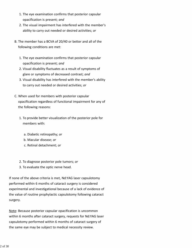
A. The member has a best‐corrected visual acuity (BCVA) of
20/50 or worse and both of the following conditions are
met:
2 of 30
1. The eye examination confirms that posterior capsular
opacification is present; and
2. The visual impairment has interfered with the member's
ability to carry out needed or desired activities; or
B. The member has a BCVA of 20/40 or better and all of the
following conditions are met:
1. The eye examination confirms that posterior capsular
opacification is present; and
2. Visual disability fluctuates as a result of symptoms of
glare or symptoms of decreased contrast; and
3. Visual disability has interfered with the member's ability
to carry out needed or desired activities; or
C. When used for members with posterior capsular
opacification regardless of functional impairment for any of
the following reasons:
1. To provide better visualization of the posterior pole for
members with:
a. Diabetic retinopathy; or
b. Macular disease; or
c. Retinal detachment; or
2. To diagnose posterior pole tumors; or
3. To evaluate the optic nerve head.
If none of the above criteria is met, Nd:YAG laser capsulotomy
performed within 6 months of cataract surgery is considered
experimental and investigational because of a lack of evidence of
the value of routine prophylactic capsulotomy following cataract
surgery.
Note: Because posterior capsular opacification is uncommon
within 6 months after cataract surgery, requests for Nd:YAG laser
capsulotomy performed within 6 months of cataract surgery of
the same eye may be subject to medical necessity review.
3 of 30
Aetna considers Nd:YAG laser peripheral iridotomy medically
necessary for primary angle closure and primary angle‐closure
glaucoma.
Aetna considers ND‐YAG laser goniotomy medically necessary for
the treatment of primary congenital glaucoma.
Aetna considers Nd:YAG laser vitreolysis experimental and
investigational for the treatment of vitreous degeneration and
vitreous floaters because its effectiveness for these indications
has not been established.
Aetna considers Nd:YAG laser anterior hyaloidotomy
experimental and investigational for the treatment of trapped
triamcinolone behind the lens after intra‐vitreal injection because
its effectiveness for this indication has not been established.
Aetna considers Nd:YAG laser peripheral iridotomy experimental
and investigational for the prevention of pigment dispersion
glaucoma because its effectiveness for this indication has not
been established.
Aetna considers Nd:YAG laser posterior hyaloidotomy
experimental and investigational for the clearance of pre‐macular
hemorrhages because its effectiveness for this indication has not
been established.
Aetna considers Nd:YAG laser goniopuncture experimental and
investigational for rescue of failed trabeculectomy because its
effectiveness for this indication has not been established.
Aetna considers Nd:YAG laser experimental and investigational for
the treatment of the following non‐ophthalmological indications
because its effectiveness for these indications has not been
established (not an all‐inclusive list):
Benign prostatic hyperplasia (for Holmium:YAG laser
(HoLAP) for BPH, see CPB 0079 ‐ Benign Prostatic Hypertrophy
Treatments)
Chronic periodontitis
4 of 30
Disc decompression
Infantile hemangioma
Onychomycosis Peri‐
implantitis
Port wine stain
Recurrent aphthous stomatitis
Aetna considers Er:YAG laser experimental and investigational for
the treatment of the following non‐ophthalmological indications
because its effectiveness for this indication has not been
established (not an all‐inclusive list):
Recurrent aphthous stomatitis
Urinary incontinence
Background
The Agency for Health Care Policy and Research (AHCPR) panel
concluded that laser capsulotomy should not be scheduled at the
time cataract surgery is performed because one can not predict
whether a cataract surgery patient will develop posterior capsular
opacification or the time at which any such opacification will
occur. For similar reasons, manual removal of the posterior
capsule, performed with a needle or hook (called corneo‐scleral
section), should not be routinely performed at the time of initial
cataract surgery.
The AHCPR panel also concluded that neodymium:yttrium‐
aluminum‐garnet (Nd:YAG) laser capsulotomy should not be
performed prophylactically or scheduled routinely at particular
times after cataract surgery.
The eye examination should confirm the diagnosis of posterior
capsular opacification and exclude other ocular causes of
functional impairment. The panel concurred with the finding of
the literature review that there is yet no objective method of
relating the degree of capsular opacification to the severity of
functional impairment.
The panel also concluded that posterior capsular opacification
rarely occurs within 3 months of surgery, and that it is uncommon
5 of 30
for posterior capsular opacification to occur within the first 6
months of surgery. Therefore, any cases of Nd:YAG laser
capsulotomy occurring within 6 months of cataract surgery
should be reviewed, to ensure that Nd:YAG laser capsulotomy is
reasonable and medically necessary.
In a single center retrospective study, Delaney and colleagues
(2002) determined the effectiveness of Nd:YAG vitreolysis and
pars plana vitrectomy in the treatment of vitreous floaters. A
total of 31 patients (42 eyes) who underwent 54 procedures
(Nd:YAG vitreolysis or pars plana vitrectomy) for the treatment of
vitreous floaters were included in the study. Main outcome
measures were percentage symptomatic improvement following
treatment and incidence of post‐operative complications.
Statistical analysis was performed using the Fisher exact test.
Posterior vitreous detachment was the primary cause of floaters
in all 42 eyes with co‐existing vitreous veils in 3 eyes and asteroid
hyalosis in 2 eyes. Thirty‐nine of 42 eyes received Nd:YAG
vitreolysis; 38 % found Nd:YAG vitreolysis moderately improved
their symptoms while 62 % found no improvement. After an
average of 14.7 months follow‐up, no post‐operative
complications were recorded. Fifteen eyes underwent a pars
plana vitrectomy, 1 with combined phaco‐emulsification and
posterior chamber implantation and 11 following unsuccessful
laser vitreolysis. Pars plana vitrectomy resulted in full resolution
of symptoms in 93 % of eyes. One patient developed a post‐
operative retinal detachment which was successfully treated.
The authors concluded that patients' symptoms from vitreous
floaters are often under‐estimated resulting in no intervention.
This paper showed Nd:YAG vitreolysis to be a safe but only
moderately effective primary treatment conferring clinical
benefit in 1/3 of patients.
Kirwan and Cahill (2011) reported on a case of successful drainage
of a large pre‐macular hemorrhage using laser photo‐
disruption of the posterior hyaloid membrane. A 47‐year old man
presented acutely to the authors' emergency department
complaining of a 24‐hr history of sudden onset, painless and
persistent loss of vision in his left eye. Immediately before
noticing this loss of vision, he had been vomiting violently from

6 of 30
excessive alcohol intake. The left visual acuity was counting
fingers. Dilated fundoscopy of the left eye revealed a large pre‐
macular hemorrhage that was 14 disc diameters in size. Clotting
investigations were normal. A diagnosis of valsalva retinopathy
was made and the patient elected to receive a prompt Nd:YAG
laser posterior hyaloidotomy as an outpatient. At 1 week follow‐
up, the hemorrhage had drained completely into the vitreous
space revealing a healthy macula and the visual acuity had
improved to 6/12 unaided. At 6‐month follow‐up the left visual
acuity stabilized at 6/9 unaided. The authors concluded that
Nd:YAG laser posterior hyaloidotomy is an useful outpatient
procedure for successful clearance of large pre‐macular
hemorrhages that offers patients rapid recovery of visual acuity
and the avoidance of more invasive intra‐ocular surgery. The
findings of this case study (with short‐term follow‐up) needs to be
validated by well‐designed studies.
In a prospective, randomized, controlled trial, Scott et al (2011)
tested the hypothesis that Nd:YAG laser peripheral iridotomy (LPI)
significantly reduces the incidence of conversion from pigment
dispersion syndrome (PDS) with ocular hypertension (OHT) to
pigmentary glaucoma (PG). A total of 116 eyes of 116 patients
with PDS and OHT were used in this analysis. Patients were
assigned randomly either to Nd:YAG LPI or to a control group (no
laser). The primary outcome measure was conversion to PG
within 3 years, based on full‐threshold visual field (VF) analysis
using the Ocular Hypertension Treatment Study criteria.
Secondary outcome measures were whether eyes required
topical anti‐glaucoma medications during the study period and
the time to conversion or medication. Fifty‐seven patients were
randomized to undergo laser treatment and 59 were randomized
to no laser (controls). Age, gender, spherical equivalent
refraction, and intra‐ocular pressure at baseline were similar
between groups. Outcome data were available for 105 (90 %) of
recruited subjects, 52 in the laser treatment group and 53 in the
no laser treatment group. Patients were followed‐up for a
median of 35.9 months (range of 10 to 36 months) in the laser
arm and 35.9 months (range of 1 to 36 months) in the control
arm. Eight eyes (15 %) in the laser group and 3 eyes (6 %) in the
control group converted to glaucoma in the study period. The
7 of 30
proportion of eyes started on medical treatment was similar in
the 2 groups: 8 eyes (15 %) in the laser group and 9 eyes (17 %) in
the control group. Survival analyses showed no evidence of any
difference in time to VF progression or commencement of topical
therapy between the 2 groups. Cataract extraction was
performed on 1 patient in the laser group and in 1 patient in the
control group during the study period (laser eye at 18 months;
control eye at 34 months). The authors concluded that the
findings of this study suggested that there was no benefit of
Nd:YAG LPI in preventing progression from PDS with OHT to PG
within 3 years of follow‐up.
Ascaso and colleagues (2012) reported on the case of a 65‐year
old male who underwent intra‐vitreal triamcinolone acetonide
(IVTA) injection for treating a clinically significant macular edema
(CSME) due to background diabetic retinopathy in his left eye. On
the first post‐operative day, visual acuity dropped from 20/80 to
hand movements. Slit‐lamp examination showed the drug
between the posterior capsule of the lens and the anterior
hyaloid face. Two weeks later, visual acuity and the milky fluid
seemed unchanged. Neodymium:yttrium‐aluminum‐garnet laser
anterior hyaloidotomy was performed. One week later, slit‐lamp
examination of the retrolental space revealed the complete
disappearance of triamcinolone and intra‐ocular pressure
remained stable. After a follow‐up period of 2 months, visual
acuity increased to 20/50 with the lens remaining clear. The
authors concluded that Nd:YAG laser anterior hyaloidotomy is an
effective, simple, useful and minimally invasive outpatient
procedure in patients with persistent entrapment of
triamcinolone behind the crystalline lens, allowing the drug to
clear without trauma to the lens. The findings of this case study
(with short‐term follow‐up) needs to be validated by well‐
designed studies.
Ramani et al (2009) examined the morphologic changes in the
anterior segment of primary angle closure suspects (PACS) who
underwent laser peripheral iridotomy (LPI) for a period of 2
years. Primary angle closure suspects (n = 82 eyes) of Asian
Indian origin underwent A‐scan biometry and ultrasound
biomicroscopy. Anterior chamber depth, anterior chamber angle

8 of 30
(ACA), axial length, lens thickness, relative lens position, central
corneal thickness, angle opening distance 500, trabecular‐ciliary
process distance, iris‐ciliary process distance, and iris thickness
were measured before LPI and after LPI at 1 week, 6 months, 1
year, 1.5 years, and 2 years. Variation in the parameters
measured over a period of 2 years was analyzed. Fifteen eyes out
of 52 eyes developed into primary angle closure (PAC) with
synechial changes. Uni‐variate analysis for the predictive factors
of PAC showed no significant association for age, sex, narrow
angle, ultrasound biomicroscopy parameters, and vertical cup‐
disc ratio. When analyzed as continuous variables, decreasing
ACA was significant risk factor (95 % confidence interval [CI]:
0.703, 0.989, p = 0.037). Iris‐ciliary process distance, ACA, lens
thickness, and angle opening distance 500 were the parameters
that varied significantly (p < 0.05) between "before LPI group"
and "after LPI groups". None of the subjects developed
increased intra‐ocular pressure (IOP) after laser iridotomy. The
authors concluded that in this hospital‐based study on the course
of PACS subjects after LPI, as many as 28 % progressed to PAC.
Decreasing ACA was the predictive factor for the progression of
PACS to PAC. There was no increase in IOP, history, or symptoms
of acute attack of glaucoma among the study subjects after LPI.
In a case‐series study, Lin and colleagues (2011) evaluated the
long‐term changes in anterior segment morphology by using
ultrasound biomicroscopy (UBM) following LPI in eyes with PAC. A
total of 54 eyes with PAC of 31 consecutive patients were
enrolled. Routine ophthalmic and UBM examination were
performed at visit‐1 (before LPI), 2 weeks, 6, and 12 months after
LPI. The parameters of anterior chamber were measured by UBM
and calculated. Results of each follow‐up time were analyzed
using repeated measures analysis of variance. Parameters of
UBM measurement at 750 µm anterior to the sclera spur and at
500 µm counterpart were compared using paired student t‐test.
Compared to before LPI, anterior chamber depth (ACD) was
deepened by approximate 0.10 mm after LPI, however, it was not
statistically significant (F = 3.50, p > 0.05). Angle opening distance
(AOD), trabecular‐iris angle (TIA), angle recess area (ARA) and
trabecular‐ciliary process distance (TCPD) were significantly

9 of 30
increased at 2 weeks, 6 and 12 months after LPI compared with
respective baseline [AOD750: (165.0 ± 70.3), (185.8 ± 68.5),
(196.1 ± 77.7) µm versus (66.2 ± 51.6) µm, F = 92.60; TIA750:
14.1° ± 6.3°, 15.5° ± 6.2°, 16.4° ± 5.9° versus 6.4° ± 4.9°, F = 92.60;
ARA: (0.058 ± 0.024), (0.065 ± 0.023), (0.068 ± 0.026) mm(2)
versus (0.025 ± 0.017) mm(2), F = 92.60; TCPD: (647.1 ± 113.0),
(701.8 ± 93.4), (670.1 ± 95.4) µm versus (571.0 ± 97.2) µm, F =
34.00; p < 0.05]. The parameters of UMB measurement at 750
µm were significantly increased more than that at 500 µm
anterior to the sclera spur (AOD: t = 5.90, TIA750: t = 2.70, p <
0.05; ARA: t = 2.00, p = 0.05). The authors concluded that LPI can
significantly widen the peripheral anterior angle in eyes with PAC
lasting for at least 1 year after LPI. Parameters detected by UBM
at 750 µm anterior to the sclera spur appear to be more sensitive
in evaluating the alternation of peripheral angle structure.
The American Academy of Ophthalmology’s Preferred practice
pattern guidelines on “ Primary angle closure” (AAO, 2010) stated
that patients with PAC may have elevated IOP as a result of a
chronic compromise of aqueous outflow due to appositional or
synechial angle closure, or damage to the trabecular meshwork
from previous intermittent acute angle‐closure crisis. Iridotomy is
indicated for eyes with PAC or primary angle‐closure glaucoma
(PACG).
A Medscape review on “Glaucoma, angle closure, chronic
treatment & management” (Tham, 2012) stated that “Laser
iridotomy is indicated for all stages of chronic angle‐closure
glaucoma (CACG). Laser iridotomy involves the creation of a hole
in the peripheral iris by laser. The hole provides an alternative
pathway for aqueous to flow from the posterior chamber into the
anterior chamber, bypassing the pupil. Therefore, iridotomy will
eliminate pupillary block and prevent forward bowing of the iris
as a result of the pressure difference between the two chambers.
Iridotomy will open those areas of the angle not involved by PAS
(peripheral anterior synechiae) and prevent further synechial
closure”.
Thomas and Walland (2013) noted that PACG and its precursors
represent both a significant proportion of world glaucoma

10 of 30
blindness and a currently insurmountable burden of treatment. In
contrast to primary open‐angle glaucoma, preventive
interventions in primary angle closure disease (PACD) can
sometimes be definitive. These investigators have synthesized
data from randomized controlled trials (RCT's) ‐‐ and where this is
not available ‐‐ principles grounded in known biology, biological
plausibility, logic, preferred practice and personal experience to
develop detailed and explicit clinical algorithms for the
management of the spectrum of PACD. Laser iridotomy is the
mainstay of first‐line intervention and is usually required for all
PACD with the exception of some PACS. Laser iridotomy is a
necessary but not always sufficient step and uncertainty arises
where a patent iridotomy has not alleviated the angle closure
profile or achieved clinically desired end points. The crucial step‐
wise considerations after iridotomy are: whether the angle is
open or closed; whether the IOP can be medically controlled; the
extent of PAS and the presence of visually significant cataract.
These lead to further interventions that include iridoplasty,
cataract surgery, trabeculectomy or phacotrabeculectomy. Such
subsequent interventions are based on an arbitrary threshold
(180 degrees) for angle opening and extent of PAS following
iridotomy and other initial procedures.
Furthermore, an UpToDate review on “Angle‐closure glaucoma”
(Weizer, 2013) states that “Laser peripheral iridotomy is the first
step in treatment of patients with chronic angle closure glaucoma,
to relieve any pupillary block component. The intraocular
pressure may remain elevated, however, if scarring has
already damaged the drainage angle. In this case, the
remaining glaucoma is treated medically and surgically much as in
open‐angle glaucoma …. Patients with signs and symptoms
suggesting an acute attack of angle‐closure glaucoma require
emergency treatment by an ophthalmologist …. We recommend
emergency use of topical ophthalmic medications to reduce
intraocular pressure (Grade 1C). These drugs may include a beta‐
blocker, an alpha agonist, and an agent to produce miosis.
We also suggest systemic medication to decrease intraocular
pressure, which may include oral or IV acetazolamide, IV
mannitol, oral glycerol, or isosorbide (Grade 2C). Once the acute
attack is controlled, definitive treatment for angle‐closure
11 of 30
glaucoma is a laser peripheral iridotomy to provide a small
drainage hole through the iris”.
Susanna et al (2014) stated that there is an increasing need to
prolong trabeculectomy success rates with minimally invasive
procedures. In a prospective, non‐comparative, interventional
cohort study, these researchers examined the safety and
effectiveness of Nd:YAG laser goniopuncture (LGP) in IOP in eyes
having late bleb failure following trabeculectomy with mitomycin
C administration. A total of 19 eyes of 19 patients with
uncontrolled glaucoma after failed trabeculectomy were include
in this study. All eyes had ischemic non‐functioning blebs with
patent internal ostia underwent Nd:YAG LGP, followed by a 5‐
fluorouracil injection. Main outcome measures were IOP and the
number of anti‐glaucoma medications before and after the
procedure, as well as pre‐surgical and post‐surgical appearance of
the blebs, using the Indiana Bleb Appearance Grading Scale
classification. The mean (SD) time of LGP after trabeculectomy
was 35.7 (32.3) months, and the mean (SD) follow‐up period after
LGP was 6.0 (1.1) months (range of 4.4 to 8.4 months). The mean
(SD) IOP had decreased from 20.9 (4.5) mm Hg (range of 15.5 to
29.0 mm Hg) to 11.9 (4.1) mm Hg (range of 5.0 to 21.0 mm Hg) (p
< 0.001). The only complications observed after LGP were 2 cases
of hypotony, which resolved spontaneously. Compared with
baseline Indiana Bleb Appearance Grading Scale classifications, 2
eyes showed an increase in bleb height and 10 eyes showed an
increase in bleb extension. None of the eyes had a positive Seidel
test result. The mean (SD) number of hypotensive agents per eye
had decreased from 0.7 (1.1) to 0.3 (0.7) after the procedure. At
the last follow‐up visit, 15 eyes (79 %) had achieved an IOP of 15
mm Hg or less, with a minimum IOP reduction of 20 % from
baseline without medication use. The authors concluded that the
Nd:YAG LGP is a safe and effective procedure for lowering IOP in
eyes with ischemic non‐functioning blebs and patent
trabeculectomy ostia. They stated that this is a promising
solution to rescue failed trabeculectomies and can potentially
prolong trabeculectomy success rates.
Guidelines from the World Glaucoma Association (2013) on
childhood glaucoma state that angle surgery (goniotomy and

12 of 30
trabeculotomy – conventional or circumferential) is the procedure
of choice for primary congenital glaucoma with the exact choice
dictated by corneal clarity and the surgeon’s experience and
preference. The guidlines state that angle surgery success rates
for secondary childhood glaucomas are generally not as good as
for primary congenital glaucoma (PCG) with certain exceptions
[e.g., glaucoma with acquired condition (uveitis) in juvenile
idiopathic arthritis (JIA)].
In a Cochrane review, Ghate and Wang (2015) compared the
safety and effectiveness of different surgical techniques for
primary congenital glaucoma (PCG). These investigators searched
CENTRAL (which contains the Cochrane Eyes and Vision Group
Trials Register) (The Cochrane Library 2014, Issue 6), Ovid
MEDLINE, Ovid MEDLINE In‐Process and Other Non‐Indexed
Citations, Ovid MEDLINE Daily, Ovid OLDMEDLINE (January 1946
to June 2014), EMBASE (January 1980 to June 2014), (January
1982 to June 2014), PubMed (January 1946 to June 2014), the
metaRegister of Controlled Trials (mRCT) (www.controlled‐
trials.com), ClinicalTrials.gov (www.clinicaltrials.gov), the WHO
International Clinical Trials Registry Platform (ICTRP)
(www.who.int/ictrp/search/en). They did not use any date or
language restrictions in the electronic searches for trials. They
last searched the electronic databases on June 23, 2014. These
researchers included all randomized and quasi‐randomized trials
in which different types of surgical interventions were compared
in children less than 5 years of age with PCG. They used standard
methodological procedures specified by The Cochrane
Collaboration. A total of 6 trials (4 randomized and 2 quasi‐
randomized) with 102 eyes in 61 children were included in this
analysis. Two trials were conducted in the USA and 1 trial each in
Egypt, Israel, Lebanon and Saudi Arabia. All trials included
children aged younger than 1 year when diagnosed with PCG, and
followed them for periods ranging from 6 months to 5 years. No
2 trials compared the same pair of surgical interventions, so these
ersearchers did not perform any meta‐analysis. One trial
compared trabeculotomy versus goniotomy; a 2nd trial compared
combined trabeculectomy‐trabeculotomy with mitomycin C
versus trabeculectomy‐trabeculotomy with mitomycin C and deep
sclerectomy; a 3rd trial compared combined trabeculotomy‐
13 of 30
trabeculectomy versus trabeculotomy; a 4th trial compared 1
goniotomy versus 2 goniotomies; a 5th trial compared
trabeculotomy versus viscocanalostomy; and the 6th trial
compared surgical goniotomy versus neodymium‐YAG laser
goniotomy. For IOP change and surgical success (defined by IOP
achieved), none of the trials reported a difference between pairs
of surgical techniques. However, due to the limited sample sizes
for all trials (average of 10 children per trial), the evidence as to
whether a particular surgical technique is effective and which
surgical technique is better, still remains uncertain. Adverse
events, such as choroidal detachment, shallow anterior chamber
and hyphema, were reported from 4 trials. None of the trials
reported quality of life or economic data. Overall, these trials
were neither designed nor reported well. Two trials were quasi‐
randomized trials and judged to have high risk of selection bias; 4
trials were at unclear or high risk for performance bias and
detection bias; and these investigators judged 1 trial to have high
risk of attrition bias due to high proportions of losses to follow‐
up. Due to poor study design and reporting, the reliability and
applicability of evidence remain unclear. The authors concluded
that no conclusions could be drawn from the trials included in
this review due to paucity of data. They stated that more
research is needed to determine which of the many surgeries
performed for PCG are effective.
Chronic Periodontitis:
The American Dental Association Council on Scientific Affairs
Expert Panel’s clinical practice guideline on “The nonsurgical
treatment of chronic periodontitis by means of scaling and root
planing with or without adjuncts” (Smiley et al, 2015) listed
neodymium:yttrium‐aluminum‐garnet (Nd:YAG) laser and scaling
and root planing (SRP) as interventions that were considered but
not recommended.
Disc Decompression:
Moon et al (2015) noted that laser ablation under an
epiduroscopic view allows for the vaporization of a small amount
of the nucleus pulposus, causing a reduction in intradiscal

14 of 30
pressure and relief of radicular pain. Currently, Ho:YAG and
Nd:YAG lasers are commonly used for spinal diseases. However,
the use of the Nd:YAG laser for intra‐spinal procedures can be
limited because of thermal injury and low efficacy. These
researchers investigated the safety and effectiveness of
epiduroscopic laser ablation using a 1,414‐nm Nd:YAG laser; they
examined if laser ablation was able to penetrate nucleus pulposus
without heating surrounding tissues and without mechanical
damage to surrounding tissue. Two live pigs, 3 porcine cadavers,
and 2 human cadavers were used. For the in‐vitro study,
intradiscal and epidural pressure and temperature were
compared in vertebral columns obtained from 3 porcine cadavers
before and after laser ablation. For the in‐vivo study, 2 pigs were
used to simulate percutaneous epiduroscopic laser ablation.
They were observed for behavioral changes and neurological
deficits for 1 month after the laser ablation procedure. Two
human cadavers were used for placing the laser fiber and
epiduroscope in the correct target site through the sacral hiatus.
Histological analysis was also performed to observe any damage
around the ablated lesion. Both intradiscal and epidural pressure
were markedly reduced immediately after laser ablation as
compared with the pre‐ablative state. The amount of the
pressure decrease in the intradiscal space was significantly
greater than that in the epidural space (45.8 ± 15.0 psi versus
30.0 ± 9.6 psi, p = 0.000). The temperature beneath the
ipsilateral spinal nerve, which was the nearest site to the laser
probe, never exceeded 40° C. Histology revealed no evidence of
thermal damage to surrounding structures, including the spinal
nerves, end‐plates, and vertebrae, after laser ablation. All live
pigs showed normal behavior without any sign of pain. In the
human cadaveric study, there was no case of targeting failure or
dural laceration. The mean time to reach the target region was
less than 5 minutes. The authors concluded that the 1,414‐nm
Nd:YAG laser can be used safely and effectively under the
guidance of a spinal epiduroscope in an in‐vivo porcine model
and in a human cadaveric model. The main drawback of this
study was that pressure measurements were performed on
cadavers and not in‐vivo. Cadaver models cannot account for
intradiscal pressure changes that occur during live muscle
contraction and different positions, which may affect results.

15 of 30
Moreover, although these investigators controlled temperatures
with heat baths, vascular and cerebrospinal fluid circulations
were not simulated. Those circulations may change the
temperature results in‐vivo.
Peri‐Implantitis:
Natto et al (2015) evaluated the effectiveness of various types of
lasers (Nd:YAG, carbon dioxide [CO2], diode, erbium/chromium‐
doped yttrium‐scandium‐gallium‐garnet [Er,Cr:YSGG], and
erbium‐doped yttrium‐aluminum‐garnet [Er:YAG]) in the
treatment of peri‐implantitis and their use in surgical and non‐
surgical procedures. Human studies for the treatment of peri‐
implantitis with laser therapy, published between 2002 and
January 2014, were collected utilizing the electronic databases
PubMed, Ovid, MEDLINE, Cochrane, and Google Scholar. Two
reviewers conducted the study selection, data collection, and
validity assessment. A total of 812 studies were selected in the
initial title search; 13 studies were then chosen for this review.
No human studies evaluated the effect of the Nd:YAG laser on
peri‐implantitis. The CO2 laser was reported to be safe and able
to enhance bone regeneration. The diode laser (980 nm)
appeared to be effective in its bactericidal effect without
changing the implant surface pattern. The Er,Cr:YSGG laser was
reported to obtain bone regeneration around a failing implant in
1 case, while the Er:YAG laser exhibited a strong bactericidal
effect against periodontopathic bacteria at a low energy level.
The authors concluded that although lasers have shown
promising results in reducing clinical signs of peri‐implantitis,
because of the limited sample sizes and short follow‐up periods,
no firm conclusion can be drawn at this moment. They stated
that there is a need for more well‐designed, longitudinal, RCTs.
Urinary Incontinence:
Ogrinc et al (2015) assessed the non‐invasive erbium:yttrium‐
aluminum‐garnet (Er:YAG) laser as a potential treatment strategy
for stress urinary incontinence (SUI) and mixed UI (MUI). These
researchers included 175 women (aged 49.7 ± 10 years) with

16 of 30
newly diagnosed SUI (66 % of women) and MUI (34 %),
respectively. Patients were clinically examined and classified by
incontinence types (SUI and MUI) and grades (mild, moderate,
severe, and very severe) using International Consultation on
Incontinence Modular Questionnaire (ICIQ) and assessing
Incontinence Severity Index (ISI). Using Er:YAG laser, these
investigators performed on average 2.5 ± 0.5 procedures in each
woman separated by a 2‐month period. At each session, clinical
examination was performed, ICIQ and ISI assessed and treatment
discomfort measured with visual analog system (VAS) pain scale,
and adverse effects and patients' satisfaction were followed.
Follow‐ups were performed at 2, 6, and 12 months after the
treatment. After the treatment, ISI decreased for 2.6 ± 1.0 points
in patients diagnosed with mild UI before the treatment, for 3.6 ±
1.4 points in those with moderate UI, for 5.7 ± 1.8 points in those
with severe UI and for 8.4 ± 2.6 in those with very severe UI (p <
0.001, paired samples t‐test). Altogether, in 77 % patients
diagnosed with SUI, a significant improvement was found after
treatment, while only 34 % of women with MUI exhibited no UI at
1‐year follow‐up. Age did not affect the outcome. No major
adverse effects were noticed in either group. The authors
concluded that the findings of this study showed that new non‐
invasive Er:YAG laser could be regarded as a promising additional
treatment strategy for SUI with at least 1 year lasting positive
effects. On the other hand, it does not seem appropriate for
treating MUI.
In a pilot study, Fistonic et al (2016) evaluated the safety and
effectiveness of the Er:YAG laser for the treatment of SUI. The
subject of this study is a treatment of SUI with a 2,940‐nm Er:YAG
laser, operating in a special SMOOTH mode designed to increase
temperature of the vaginal mucosa up to maximally 60 to 65 °C
without ablating the epidermis. Numerical modelling of the
temperature distribution within mucosa tissue following an
irradiation with the SMOOTH mode Er:YAG laser was performed
in order to determine the appropriate range of laser parameters.
The laser treatment parameters were further confirmed by
measuring in‐vivo temperatures of the vaginal mucosa using a
thermal camera. To investigate the clinical safety and
effectiveness of the SMOOTH mode Er:YAG laser SUI treatment, a

17 of 30
pilot clinical study was performed. The study recruited 31 female
patients suffering from SUI; follow‐ups were scheduled at 1, 2,
and 6 months post‐treatment. ICIQ‐UI questionnaires were
collected as a primary trial end‐point. Secondary end‐points
included perineometry and residual urine volume measurements
at baseline and all follow‐ups. Thermal camera measurements
have shown the optimal increase in temperature of the vaginal
mucosa following treatment of SUI with a SMOOTH mode Er:YAG
laser. Primary end‐point, the change in ICIQ‐UI score, showed
clinically relevant and statistically significant improvement after
all follow‐ups compared to baseline scores. There was also
improvement in the secondary end‐points. Only mild and
transient adverse events and no serious adverse events were
reported. The authors concluded that the findings of this study
indicated that non‐ablative Er:YAG laser therapy is a promising
minimally invasive non‐surgical option for treating women with
SUI symptoms. These preliminary findings need to be validated
by well‐designed studies.
Benign Prostatic Hyperplasia:
In a retrospective observational study, Palmero‐Mari and
colleagues (2016) compared the safety and effectiveness of
thulium laser (Tm: YA G) 150W against greenlight laser (LBO:ND‐
YA G) 120W in the treatment of benign prostatic hyperplasia
(BPH) 12 months after surgery. Subjects were men who
underwent the surgical technique of prostate vaporization over a
period of 4 years in the authors’ center. The homogeneity of the
sample was checked, and post‐operative complications (acute
urinary retention, re‐entry, need for transfusion), failures per year
of surgery (re‐operation, peak flow less than 15ml/sec, no
improvement in comparing the International Prostate Symptom
Score (I‐PSS)), and decreased prostate‐specific antigen (PSA) were
compared a year after surgery. A bivariate analysis using Chi‐
square and t‐Student was carried out. A total of 116 patients
were treated with thulium and 118 with green laser. The sample
was homogeneous for pre‐operative variables (p > 0.05).
No differences in complications were observed: in urine acute
retention, 4.3 % with thulium and 6.8 % with green laser (p =
0.41); in readmissions, 2.6 % with thulium and 1.7 % with green
18 of 30
laser (p = 0.68); in need for transfusion, 2.6 % with thulium and 0
% with green laser (p = 0.12). No differences were observed in
the percentage of patients re‐operation (1.7 % in the group of
thulium, 5.1 % in the green laser, p = 0.28); or in individuals with
Qmax less than 15 ml/sec (6.9 % with thulium, 6.77 % with green
laser, p = 0.75), or in the absence of improvement in the IPSS (5.2
% with thulium, 3.4 % with green laser, p = 0.65). There was also
no difference in the levels of PSA in ng/ml a year after surgery:
with thulium 2.78 ± 2.09 and with green laser 1.83 ± 1.48 (p =
0.75). The authors concluded that prostate vaporization with
thulium laser 150W was comparable to that made with green
laser 120W for the treatment of lower urinary tract symptoms
caused by BPH, being both safe and effective techniques to 12
months after surgery. Moreover, they stated that future
prospective randomized studies are needed to confirm this
conclusion on both techniques.
Furthermore, an UpToDate review on “Transurethral procedures
for treating benign prostatic hyperplasia” (Cunningham and
Kadmon, 2017) states that “The Holmium:Yttrium‐Aluminium‐
Garnet (YAG) (2,140 nm), Thulium:YAG (2,014 nm), and
Neodymium:YAG (1,064 nm) lasers were initially developed to
ablate tissue, but these lasers were less effective at ablating
prostate tissue compared with the other lasers, since the
wavelength of light used is near the peak of water absorption”.
Infantile Hemangioma:
Chinnadurai and associates (2016) reviewed studies of laser
treatment of infantile hemangioma (IH). These investigators
searched multiple databases including Medline and Embase from
1982 to June 2015. Two investigators independently screened
studies against pre‐determined criteria and extracted key data.
Investigators independently assessed study risk of bias and the
strength of the evidence of the body of literature. They identified
29 studies addressing lasers: 4 RCTs, 8 retrospective cohort
studies, and 17 case series. Lasers varied across studies in type,
pulse width, or cooling materials. Most comparative studies (n =
9) assessed variations of pulsed dye laser (PDL) and examined
heterogeneous end‐points. Most studies reported on treatment

19 of 30
of cutaneous lesions. Overall, longer pulse PDL with epidermal
cooling was the most commonly used laser for cutaneous lesions;
Nd:YAG was the most commonly used intralesionally. Most
studies reported a higher success rate with longer pulse PDL
compared with observation in managing the size of IH, although
the magnitude of effect differed substantially. CO2 laser was used
for subglottic IH in a single study, and was noted to have a higher
success rate and lower complication rate than both Nd:YAG and
observation. Studies comparing laser with β‐blockers or in
combination with β‐blockers reported greater improvements in
lesion size in combination arms versus β‐blockers alone and
greater effects of lasers on mixed superficial and deep IH.
Strength of the evidence for outcomes after laser treatments
ranged from insufficient to low for effectiveness outcomes.
Strength of the evidence was insufficient for the effects of laser
compared with β‐blockers or in combination with β‐blockers as
studies evaluated different agents and laser types. Studies
assessing outcomes after CO2 and Nd:YAG lasers typically
reported some resolution of lesion size, but heterogeneity among
studies limited their abilities to draw conclusions. The authors
concluded that studies of laser treatment of IH primarily
addressed different laser modalities compared with observation
or other laser modalities. Pulsed dye laser was the most
commonly studied laser type, but multiple variations in treatment
protocols did not allow for demonstration of superiority of a
single method. Most studies reported a higher success rate with
longer pulse PDL compared to observation in managing the size
of IH, although the magnitude of effect differed substantially.
Studies generally found PDL more effective than other types of
lasers for cutaneous lesions. When first introduced as a primary
treatment for IH, various laser modalities generally offered
superior outcomes compared with steroid therapy and
observation. In the era of β‐blocker therapy, laser treatment may
retain an important role in the treatment of residual and
refractory lesions.
Furthermore, an UpToDate review on “Management of infantile
hemangiomas” (Metry, 2017) states that “The pulsed‐dye laser
(PDL) cannot be expected to affect hemangiomas with deep
involvement, since the depth of laser penetration is only 1.2 mm.

20 of 30
The most accepted use of PDL in the management of
hemangiomas is the treatment of ulceration, post‐involution
erythema, and/or telangiectasias. Which hemangiomas benefit
most from laser therapy and what the optimal settings are remain
areas of controversy ”. This review does not mention YA G laser as
a therapeutic option.
Onychomycosis:
Rivers and colleagues (2016) examined the effectiveness of a
1064‐nm Nd:YAG laser for the treatment of onychomycosis in a
real‐world setting. A single‐center retrospective chart review was
conducted between 2012 and 2013. A total of 100 consecutive
patients with a culture‐ and/or potassium hydroxide‐confirmed
diagnosis of onychomycosis were treated at least twice. Baseline
and follow‐up photographs were taken, and the change in degree
of clinical nail involvement of the subject's great toenail was
determined by a blinded reviewer using validated planimetry
measurement. A total of 199 hallux nails from 100 subjects were
assessed. The mean infected area decreased from 53.2 % at
baseline to 50.8 % at the end of the study (paired t‐test, p =
0.054; Wilcoxon signed rank test, p = 0.006). Degree of nail
involvement was statistically significantly associated with amount
of improvement; subjects who had the greatest degree of nail
involvement improved the most, while those with less severe
disease showed a worsening of nail appearance (Kruskal‐Wallis
test, p < 0.001); 72.6 % of nails that had more than 67 % nail
involvement showed statistically significant improvement (χ2 test,
p = 0.001). Adverse events (AEs) were limited to mild‐to‐
moderate pain at the time of therapy. A total of 76 subjects were
assessed for treatment satisfaction: 60 % were very satisfied with
treatment despite limited clinical improvement in some cases.
The authors concluded that laser therapy has a very limited
positive clinical effect on the appearance of onychomycosis after
2 treatment sessions.
Piccolo and associates (2016) evaluated the effectiveness of
long‐pulsed 1064‐nm Nd:YAG laser in penetrating tissue and
targeting the fungal overgrowth in the nail plate. A total of 20
consecutive, unselected patients were enrolled in the study and

21 of 30
treated, at intervals of 1 week, for a total of 4 sessions, using a
long‐pulsed 1064‐nm Nd:YAG laser. In each session, 3 passages
across each nail plate were performed with 1‐min pause between
each passage. A special lens for dermatoscopy, connected to a
digital camera, was used for dermoscopic images. In 14 patients
(70 %; 12 females; 2 males), excellent results were obtained with
an important reduction of chromonychia, onycholysis, opacity,
longitudinal striae, and jagged proximal edge. Better results were
observed in severe cases in the 2‐month follow‐up visit. The
authors concluded that data for treating nail onychomycosis with
laser and light therapy appeared to be positive. They stated that
the promising findings of this study identified long‐pulsed 1064‐
nm Nd:YAG laser as a possible alternative option for the
treatment of onychomycosis; however, increasing subject data,
improving study methodology, and output parameters may
become an important next step of study in the treatment of nail
onychomycosis.
Furthermore, an UpToDate review on “Onychomycosis:
Management” (Goldstein and Bhatia, 2017) states that “A lthough
neodymium‐doped:yttrium aluminum garnet (Nd:YAG) and diode
lasers have emerged as treatment options for onychomycosis,
data on the efficacy of these interventions are limited and the
mechanisms of action and optimal regimens for these treatments
remain unclear. Until more robust data supporting the efficacy of
laser therapy for onychomycosis are available, we cannot
recommend the routine use of this modality”.
Port Wine Stain:
Xing and colleagues (2017) stated that based on the principle of
selective photothermolysis, 1064‐nm Nd:YAG laser has great
potential for the treatment of deeper and larger port wine stain
(PWS). However, the clinical effectiveness is limited because of
the weak absorption of blood to Nd:YAG laser. These researchers
obtained the optimal irradiation conditions to effectively destroy
vascular lesions with the assistance of PEG‐modified gold nano‐
rods (NRs) to enhance blood absorption of Nd:YAG laser. In this
study, PEG‐modified gold NRs were prepared by the seeded
growth method. Gold NRs after exposure to Nd:YAG laser were

22 of 30
characterized using absorption spectra and transmission electron
microscope images. The tissue‐like phantom containing a glass
capillary with blood was prepared and exposed to Nd:YAG laser to
investigate the laser energy density and pulse number required
for blood coagulation before and after the addition of gold NRs in
blood. The results showed that the milli‐second Nd:YAG laser
irradiation did not result in the shape change of gold NRs. After
injection of gold NRs into the bloodstream (4.60 mg/kg), the
absorbance of blood at 1064‐nm increased 3.9 times. The
threshold energy density for the treatment of PWS decreased by
33 % (from 30 to 20 J/cm2). The authors concluded that these
findings provided an experimental guide for choosing laser
parameters and gold NRs concentration for the treatment of
deeper and larger PWS with the assistance of PEG‐modified gold
NRs in‐vivo in the future.
Furthermore, and UpToDate review on “Laser and light therapy
for cutaneous vascular lesions” (Kelly, 2017) states that
“Millisecond pulsed near‐infrared lasers such as the 755 nm
alexandrite or 1064 neodymium:yttrium aluminum garnet
(Nd:YAG) laser may be useful for the treatment of thick or
nodular PWS. However, the risk of scarring with long wavelength
lasers exceeds risk with PDL ….”.
Recurrent Aphthous Stomatitis:
Han and colleagues (2016) stated that laser therapy is a promising
new treatment for patients with recurrent aphthous stomatitis
(RAS). However, the clinical effect and security issue of laser
therapy remain controversial. These researchers performed a
systematic review to evaluate the clinical effectiveness and
security of laser treatment in RAS patients. Five electronic
databases were searched (Medline (PubMed), Embase,
ScienceDirect, the Cochrane Library, and Web of Science) to
identify all studies that were about RCTs, involving the effect of
laser therapy in RAS patients. A total of 23 studies were retained
for full‐text analysis after screening the titles and abstracts of
potential articles, but only 10 studies satisfied the inclusion
criteria after the full texts were reviewed. The included studies
reported a comparison of the effectiveness between the laser

23 of 30
treatment and placebo laser therapy (or conventional drug
therapy) when managing the RAS patients. Clinical case reports
and RCTs about several different types of lasers (e.g., Nd:YAG
laser, Er:YAG laser, InGaAlP laser, GaAlAs laser, etc.) were reported
in the use for treatment of RAS. The authors concluded that laser
therapy has the superiority in relieving ulcer pain and shortening
healing time when compared with placebo group or medical
treatment group. They stated that the evidence of the retrieved
studies is weak; thus, rigorously designed, long‐term,
randomized, controlled, and large sample‐sized clinical trials are
needed to confirm the effectiveness of laser on RAS therapy.
This study had several drawbacks: (i) although most of the
included studies provided evidence that laser therapy may help in
pain relief and promote wound healing, no report was conducted
regarding the difference in recurrence rates after positive and
placebo treatments, (ii) most trials did not report their
randomization process and whether treatment allocations were
conducted. Nevertheless, treatment allocations may be
recognized based on the materials and devices used, and (iii) cost
analysis was not performed in this review because no study
reported the price of laser therapy.
Furthermore, an UpToDate review on “Oral lesions” (Goldstein
and Goldstein, 2017) does not mention laser as a therapeutic
option for recurrent aphthous stomatitis.
CPT Codes / HCPCS Codes / ICD‐10 Codes
Informa'tion in the [brackets] below has been added for clarifica'tion
purposes. Codes requiring a 7th character are represented by "+":
Nd: YAG laser goniotomy:
CPT codes covered if selection criteria are met:
65820 Goniotomy [ND‐YAG laser]
ICD‐10 codes covered if selection criteria are met:
Q15.0 Congenital glaucoma [Primary congenital glaucoma]
Nd: YAG laser capsulotomy or hyaloidotomy:
CPT codes covered if selection criteria are met:
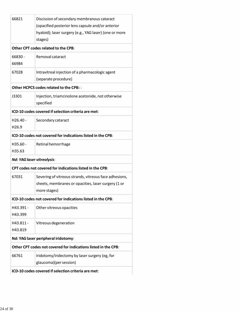
24 of 30
66821 Discission of secondary membranous cataract
(opacified posterior lens capsule and/or anterior
hyaloid); laser surgery (e.g., YAG laser) (one or more
stages)
Other CPT codes related to the CPB:
66830 ‐
66984
Removal cataract
67028 Intravitreal injection of a pharmacologic agent
(separate procedure)
Other HCPCS codes related to the CPB: :
J3301 Injection, triamcinolone acetonide, not otherwise
specified
ICD‐10 codes covered if selection criteria are met:
H26.40 ‐
H26.9
Secondary cataract
ICD‐10 codes not covered for indications listed in the CPB:
H35.60 ‐
H35.63
Retinal hemorrhage
Nd: YAG laser vitreolysis:
CPT codes not covered for indications listed in the CPB:
67031 Severing of vitreous strands, vitreous face adhesions,
sheets, membranes or opacities, laser surgery (1 or
more stages)
ICD‐10 codes not covered for indications listed in the CPB:
H43.391 ‐
H43.399
Other vitreous opacities
H43.811 ‐
H43.819
Vitreous degeneration
Nd: YAG laser peripheral iridotomy:
Other CPT codes not covered for indications listed in the CPB:
66761 Iridotomy/iridectomy by laser surgery (eg, for
glaucoma)(per session)
ICD‐10 codes covered if selection criteria are met:

25 of 30
H40.061 ‐
H40.069
Primary angle closure without glaucoma damage
H40.20x+ ‐
H40.249
Primary angle‐closure glaucoma
Nd: YAG laser goniopunture [for rescue of failed trabeculectomy]:
CPT codes not covered for indications listed in the CPB:
66250 Revision or repair of operative wound of anterior
segment, any type, early or late, major or minor
procedure
Other CPT codes related to the CPB:
65885 Trabeculoplasty by laser surgery, 1 or more sessions
(defined treatment series)
Nd: YAG laser for the treatment of chronic periodon'ti'tis, disc
decompression, and peri‐implan'ti'tis ‐ No specific code:
Other CPT codes related to the CPB:
0274T Percutaneous laminotomy/laminectomy (interlaminar
approach) for decompression of neural elements,
(with or without ligamentous resection, discectomy,
facetectomy and/or foraminotomy), any method,
under indirect image guidance (eg, fluoroscopic, CT),
with or without the use of an endoscope, single or
multiple levels, unilateral or bilateral; cervical or
thoracic
0275T Percutaneous laminotomy/laminectomy (interlaminar
approach) for decompression of neural elements,
(with or without ligamentous resection, discectomy,
facetectomy and/or foraminotomy), any method,
under indirect image guidance (eg, fluoroscopic, CT),
with or without the use of an endoscope, single or
multiple levels, unilateral or bilateral; lumbar
62287 Decompression procedure, percutaneous, of nucleus
pulposus of intervertebral disk, any method, single or
multiple levels, lumbar (eg, manual or automated
percutaneous discectomy, percutaneous laser
discectomy)
ICD‐10 codes not covered for indications listed in the CPB:

26 of 30
K05.3 ‐
K05.6
Chronic periodontitis
M27.62 Post‐osseointegration biological failure of dental
implant
Er: YAG laser for the treatment of urinary incon'tinence ‐ no specific
code:
ICD‐10 codes not covered for indications listed in the CPB:
N39.3 Stress incontinence (female) (male)
N39.4 ‐
N39.498
Other specified urinary incontinence
R32 Unspecified urinary incontience
The above policy is based on the following references:
1. U.S. Department of Health and Human Services, Agency for
Health Care Policy and Research (AHCPR). Cataract in
adults: Management of functional impairment. Clinical
Practice Guideline No. 4. Rockville, MD: AHCPR; 1993.
2. Powell SK, Olson RJ, Incidence of retinal detachment after
cataract surgery and neodynium:YAG laser capsulotomy. J
Cataract Refract Surg. 1995;21(2):132‐135.
3. Steinberg EP, Javitt JC, Sharkey PD, et al. The content and
cost of cataract surgery. Arch Ophthalmol.
1993;111(8):1041‐1049.
4. Thornval P, Naeser K. Refraction and anterior chamber
depth before and after neodynium:YAG laser treatment for
posterior capsule opacification in pseudophakic eyes: A
prospective study. J Cataract Refract Surg. 1995;21(4):457‐
460.
5. Nielsen NE, Naeser K. Epidemiology of retinal detachment
following extracapsular cataract extraction: A follow‐up
study with an analysis of risk factors. J Cataract Refract
Surg. 1993;19(16):675‐680.
6. Obstbaum SA. Extracapsular cataract surgery, retinal
detachment, and YAG laser posterior capsulotomy. J
Cataract Refract Surg. 1993;19(6):673.
7. Murrill CA, Stanfield DL, Brockliln MD. Capsulotomy. Optom

27 of 30
Clin. 1995;4(4):69‐83.
8. Baratz KH, Cook BE, Hodge DO. Probability of Nd:YAG laser
capsulotomy after cataract surgery in Olmsted County,
Minnesota. Am J Ophthalmol. 2001;131(2):161‐166.
9. Schaumberg DA, Dana MR, Christen WG, Glynn RJ. A
systematic overview of the incidence of posterior capsule
opacification. Ophthalmology. 1998;105(7):1213‐1221.
10. Ku WC, Chuang LH, Lai CC. Cataract extraction in high
myopic eyes. Chang Gung Med J. 2002;25(5):315‐320.
11. Aslam TM, Devlin H, Dhillon B. Use of Nd:YAG laser
capsulotomy. Surv Ophthalmol. 2003;48(6):594‐612.
12. Yilmaz S, Ozdil MA, Bozkir N, Maden A. The effect of
Nd:YAG laser capsulotomy size on refraction and visual
acuity. J Refract Surg. 2006;22(7):719‐721.
13. Cinal A, Demirok A, Yasar T, et al. Nd:YAG laser posterior
capsulotomy after pediatric and adult cataract surgery. Ann
Ophthalmol (Skokie). 2007;39(4):321‐326.
14. Findl O, Buehl W, Bauer P, Sycha T. Interventions for
preventing posterior capsule opacification. Cochrane
Database Syst Rev. 2007;(3): CD003738.
15. Delaney YM, Oyinloye A, Benjamin L. Nd:YAG vitreolysis and
pars plana vitrectomy: Surgical treatment for vitreous
floaters. Eye. 2002;16(1):21‐26.
16. Lundqvist B, Mönestam E. Ten‐year longitudinal visual
function and Nd: YAG laser capsulotomy rates in patients
less than 65 years at cataract surgery. Am J Ophthalmol.
2010;149(2):238‐244.
17. Findl O, Buehl W, Bauer P, Sycha T. Interventions for
preventing posterior capsule opacification. Cochrane
Database Syst Rev. 2010;(2):CD003738.
18. Kirwan RP, Cahill MT. Nd:YAG laser hyaloidotomy for
valsalva pre‐macular haemorrhage. Ir J Med Sci.
2011;180(3):749‐752.
19. Scott A, Kotecha A, Bunce C, et al. YAG laser peripheral
iridotomy for the prevention of pigment dispersion
glaucoma a prospective, randomized, controlled trial.
Ophthalmology. 2011;118(3):468‐473.
20. Ascaso FJ, de Gopegui ER, Cascante JM. Neodymium:
yttrium‐aluminum‐garnet laser anterior hyaloidotomy to
treat trapped triamcinolone acetonide behind the

28 of 30
crystalline lens after intravitreal injection. Middle East Afr J
Ophthalmol. 2012;19(1):163‐165.
21. Ramani KK, Mani B, George RJ, Lingam V. Follow‐up of
primary angle closure suspects after laser peripheral
iridotomy using ultrasound biomicroscopy and A‐scan
biometry for a period of 2 years. J Glaucoma.
2009;18(7):521‐527.
22. American Academy of Ophthalmology. Preferred practice
pattern guidelines: Primary angle closure. October 2010.
Available at: http://one.aao.org/CE/PracticeGuidelines
/PPP_Content.aspx?cid=92bea8f6‐5459‐49a6‐9233‐4528343dc4c3.
Accessed March 19, 2013.
23. Lin Z, Li SZ, Fan SJ, et al. Alteration of anterior chamber and
angle structure in eyes with primary angle closure after
laser peripheral iridotomy. Zhonghua Yan Ke Za Zhi.
2011;47(10):881‐886.
24. Tham CCY. Glaucoma, angle closure, chronic treatment &
management. Medscape. August 7, 2012. Available at:
http://emedicine.medscape.com/article/1205154‐
treatment (http://emedicine.medscape.com/article
/1205154‐treatment). Accessed March 19, 2013.
25. Thomas R, Walland MJ. Management algorithms for
primary angle closure disease. Clin Experiment Ophthalmol.
2013;41(3):282‐292.
26. Weizer JS. Angle‐closure glaucoma. Last reviewed February
2013. UpTodate Inc. Waltham, MA.
27. Susanna R Jr, De Moraes CG, Alencar LM, Ritch R. Nd:YAG
laser goniopuncture for late bleb failure after
trabeculectomy with adjunctive mitomycin C. JAMA
Ophthalmol. 2014;132(3):286‐290.
28. Karahan E, Er D, Kaynak S. An overview of Nd:YAG laser
capsulotomy. Med Hypothesis Discov Innov Ophthalmol.
2014;3(2):45‐50.
29. Ghate D, Wang X. Surgical interventions for primary
congenital glaucoma. Cochrane Database Syst Rev.
2015;1:CD008213.
30. Olitsky SE, Reynolds JD. Overview of glaucoma in infants
and children. UpToDate Inc., Waltham, MA. Last reviewed
February 2015.
31. World Glaucoma Association. Childhood glaucoma.

29 of 30
Summary consensus points. 9th Consensus Meeting.
Vancouver, BC, July 16, 2013.
32. Smiley CJ, Tracy SL, Abt E, et al. Evidence‐based clinical
practice guideline on the nonsurgical treatment of chronic
periodontitis by means of scaling and root planing with or
without adjuncts. J Am Dent Assoc. 2015;146(7):525‐535.
33. Moon BJ, Lee HY, Kim KN, et al. Experimental evaluation of
percutaneous lumbar laser disc decompression using a
1414 nm Nd:YAG laser. Pain Physician. 2015;18(6):E1091‐
E1099.
34. Natto ZS, Aladmawy M, Levi PA Jr, Wang HL. Comparison of
the efficacy of different types of lasers for the treatment of
peri‐implantitis: A systematic review. Int J Oral Maxillofac
Implants. 2015;30(2):338‐345.
35. Ogrinc UB, Sencar S, Lenasi H. Novel minimally invasive
laser treatment of urinary incontinence in women. Lasers
Surg Med. 2015;47(9):689‐697.
36. Fistonic N, Fistonic I, Gustek SF, et al. Minimally invasive,
non‐ablative Er:YAG laser treatment of stress urinary
incontinence in women ‐ a pilot study. Lasers Med Sci.
2016;31(4):635‐643.
37. Chinnadurai S, Sathe NA, Surawicz T. Laser treatment of
infantile hemangioma: A systematic review. Lasers Surg
Med. 2016;48(3):221‐233.
38. Han M, Fang H, Li QL, et al. Effectiveness of laser therapy in
the management of recurrent aphthous stomatitis: A
systematic review. Scientifica (Cairo). 2016;2016:9062430.
39. Palmero‐Mari JL, Panach‐Navarrete J, Valls‐Gonzalez L, et
al. Comparative study between thulium laser (Tm: YA G)
150W and greenlight laser (LBO:ND‐YA G) 120W for the
treatment of benign prostatic hyperpplasia: Short‐term
efficacy and security. Actas Urol Esp. 2016 Nov 25 [Epub
ahead of print].
40. Rivers JK, Vestvik BJ, Berkowitz J. Real‐world efficacy of
1064‐nm Nd:YAG laser for the treatment of onychomycosis.
J Cutan Med Surg. 2016 Nov 16 [Epub ahead of print].
41. Piccolo D, Kostaki D, Del Duca E, et al. Long‐pulsed 1064‐nm
Nd: YA G laser for the treatment of onychomycosis.
Photomed Laser Surg. 2016 Dec 30 [Epub ahead of print].
42. Cunningham GR, Kadmon D. Transurethral procedures for
30 of 30
treating benign prostatic hyperplasia. UpToDate Inc.,
Waltham, MA. Last reviewed January 2017.
43. Metry DW. Management of infantile hemangiomas.
UpToDate Inc., Waltham, MA. Last reviewed January 2017.
44. Goldstein AO, Bhatia N. Onychomycosis: Management.
UpToDate Inc., Waltham, MA. Last reviewed January 2017.
45. Kelly KM. Laser and light therapy for cutaneous vascular
lesions. UpToDate Inc., Waltham, MA. Last reviewed
January 2017.
46. Goldstein BG, Goldstein AO. Oral lesions. UpToDate Inc.,
Waltham, MA. Last reviewed January 2017.
47. Xing L, Chen B, Li D, et al. Nd:YAG laser‐induced
morphology change and photothermal conversion of gold
nanorods with potential application in the treatment of
port‐wine stain. Lasers Med Sci. 2017 Feb 3 [Epub ahead of
print].
31 of 30
Copyright Aetna Inc. All rights reserved. Clinical Policy Bulletins are developed by Aetna to assist in administering plan
benefits and constitute neither offers of coverage nor medical advice. This Clinical Policy Bulletin contains only a
partial, general description of plan or program benefits and does not constitute a contract. Aetna does not provide
health care services and, therefore, cannot guarantee any results or outcomes. Participating providers are
independent contractors in private practice and are neither employees nor agents of Aetna or its affiliates. Treating
providers are solely responsible for medical advice and treatment of members. This Clinical Policy Bulletin may be
updated and therefore is subject to change.
Copyright © 2001‐2017 Aetna Inc.

AETNA BETTER HEALTH® OF PENNSYLVANIA
Amendment to Aetna Clinical Policy Bulletin Number: 0354 - YAG Laser in
Ophthalmology and Other Selected Indications
There are no amendments for Medicaid.
www.aetnabetterhealth.com/pennsylvania revised 05/24/2017
Related Documents