ن م ح ر ل ها ل ل ما س ب م ي ح ر ل ا

بسم الله الرحمن الرحيم
Mar 22, 2016
بسم الله الرحمن الرحيم. By Tarik Zaher ,MD Assistant Professor of Endemic and Tropical Medicine , Zagazig University. Leishmaniasis. - PowerPoint PPT Presentation
Welcome message from author
This document is posted to help you gain knowledge. Please leave a comment to let me know what you think about it! Share it to your friends and learn new things together.
Transcript

الرحمن الله بسمالرحيم
LEISHMANIASIS By
Tarik Zaher ,MDAssistant Professor of Endemic and Tropical Medicine ,Zagazig University

BACKGROUND
Leishmaniasis is a protozoal disease capable of causing a spectrum of clinical syndromes ranging from cutaneous ulcerations to systemic infections. With the exception of Australia, the Pacific Islands, and Antarctica, the parasites have been identified throughout large portions of the world.
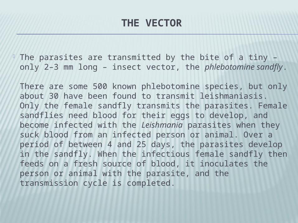
THE VECTOR
The parasites are transmitted by the bite of a tiny – only 2–3 mm long – insect vector, the phlebotomine sandfly.
There are some 500 known phlebotomine species, but only about 30 have been found to transmit leishmaniasis. Only the female sandfly transmits the parasites. Female sandflies need blood for their eggs to develop, and become infected with the Leishmania parasites when they suck blood from an infected person or animal. Over a period of between 4 and 25 days, the parasites develop in the sandfly. When the infectious female sandfly then feeds on a fresh source of blood, it inoculates the person or animal with the parasite, and the transmission cycle is completed.


CAUSES Localized cutaneous leishmaniasis Old World
L donovan i - China, India, Bangladesh, Sudan L tropica - Middle East, China, India, Mediterranean L major - Middle East, Africa, India, Asia L aethiopica - Ethiopia, Kenya, Namibia Leishmania infantum - Asia, Africa, Europe
New World Leishmania leishmania mexicana - Central and South America, North America L leishmania amazonensis - Dominican Republic, Central America, South America L leishmania venezuelensis - Venezuela L viannia braziliensis - Central America, South America L viannia guyanensis - Guyana, French Guyana, Surinam, Brazil L viannia panamensis - Costa Rica, Panama, Colombia, Ecuador L viannia peruviana - Peru, Argentina Leishmania donovani chagasi - Texas, Caribbean, Central America, South America

• Diffuse cutaneous leishmaniasis• Old World
• L aethiopica - Ethiopia, Kenya, Namibia• New World
• L leishmania mexicana - Central, South America, North America
• L leishmania amazonensis - Dominican Republic, Central America, South America

• Leishmaniasis recidivans• Old World
• L tropica - Middle East, China, India, Mediterranean• New World
• L viannia braziliensis - Central America, South America
• Post-kala-azar leishmaniasis • Old World
• L donovani - China, India, Bangladesh• L infantum - Asia, Africa, Europe
• New World • L donovani chagasi - Central America, South
America

• Mucocutaneous leishmaniasis• Old World
• L aethiopica - Ethiopia, Kenya, Namibia• New World
• L viannia braziliensis - Central America, South America
• L viannia panamensis - Central America, South America
• L viannia guyanensis - Guyana, French Guyana, Surinam, Brazil
• Less often seen with L leishmania mexicana - Central America, South America, North America
• Less often seen with L leishmania amazonensis - Brazil, Panama

• Visceral leishmaniasis • Old World
• L donovani - China, India, Bangladesh, Sudan
• L infantum - Asia, Africa, Europe• New World
• L donovani chagasi - Central America, South America
• Viscerotropic leishmaniasis • Old World
• L tropica - Middle East

LIFE CYCLE


PATHOPHYSIOLOGY After inoculation, parasites infect the
reticuloendothelial system and live in the intracellular lysosomal organelles of macrophages. Parasites may incubate for weeks to months before presenting as skin lesions or as a disseminated systemic infection involving the liver, spleen, and bone marrow. Pathogenesis appears related to T-cell cytotoxicity.

The extent and presentation of disease depend on several factors, including the humoral and cell-mediated immune response of the host, the virulence of the infecting species, and the parasite burden. Infections may heal spontaneously or may progress to chronic disease, often resulting in death from secondary infection.


EPIDEMIOLOGY & RISK FACTORS
The majority of human cases occur in a small number of countries:
90% of visceral leishmaniasis cases occur in parts of India, Bangladesh, Nepal, Sudan, Ethiopia and Brazil
90% of cutaneous leishmaniasis cases occur in parts of Afghanistan, Algeria, Iran, Saudi Arabia, Syria, Brazil, Colombia, Peru and Bolivia

Dozens of different sand fly vectors, adapted to the different ecological settings where leishmaniasis occurs, are known to transmit some form of leishmaniasis. Leishmaniasis, especially the visceral form, can also be transmitted by blood transfusion or sharing of contaminated needles. Congenital transmission has been reported, but appears to be rare.

In sylvatic cycles, such as those in New World rain forests and the deserts of Central Asia, animal reservoir hosts can maintain transmission indefinitely without human disease. Sporadic or epidemic leishmaniasis occurs when humans enter the sylvatic habitat for economic or military purposes, or when human habitation encroaches on the sylvatic setting. In domestic cycles, humans or dogs form the predominant or sole infection reservoir. In the Mediterranean basin and parts of Latin America, visceral leishmaniasis transmission is zoonotic (dog – sand fly – human). The area that account for the largest number of human cases, for example, visceral leishmaniasis in South Asia and cutaneous leishmaniasis in Afghanistan, usually reflect anthroponotic (human - sand fly - human) transmission

GEOGRAPHICAL DISTRIBUTION OF VISCERAL LEISHMANIASIS

GEOGRAPHICAL DISTRIBUTION OF CUTANEOUS LEISHMANIASIS

GEOGRAPHICAL DISTRIBUTION OF MUCO-CUTANEOUS LEISHMANIASIS

EPIDEMICS
Visceral leishmaniasis can cause large-scale and tenacious epidemics, with high case–fatality rates. Epidemics occur frequently in regions which are difficult to access, such as Libo Kemkem, Ethiopia (2005–06), Wajir, Kenya (2008), and Upper Nile, Southern Sudan (2009). Malnutrition is a well-known risk factor, and the epidemics flourish under conditions of famine, complex emergencies and mass population movements.
In Sudan a major epidemic of visceral leishmaniasis occurred from 1984 to 1994. As this was the first epidemic in the area, the population was highly susceptible. Some studies have estimated that the disease caused 100 000 deaths in a population of around 300 000 in the western upper Nile area of the country. In some villages, more than half of the population died from the disease.
In 1997, a new epidemic caused the number of confirmed cases of visceral leishmaniasis in Sudan to increase by 400% over the previous year. Treatment centres were overwhelmed and stocks of first-line drugs were depleted. The migration of seasonal workers and large population movements caused by civil unrest carried the epidemic into Eritrea and Ethiopia.

Visceral and cutaneous leishmaniasis in patients with AIDS have been increasingly appreciated as a potential opportunistic infection. Co-infection with HIV has been reported in more than 35 countries throughout southern Europe, the Mediterranean Basin, Central and South America, and India. Disease occurs in conjunction with severe immunosuppression. The incidence of co-infection has decreased in developed countries because of the widespread use of antiretroviral therapy.

MORTALITY/MORBIDITY
Localized cutaneous leishmaniasis often spontaneously resolves in 3-6 months without therapy, although some infections persist indefinitely. Most cases of diffuse cutaneous leishmaniasis, post-kala-azar dermal leishmaniasis, and leishmaniasis recidivans are chronic and resistant to treatment and are associated with low mortality rates.
Mucocutaneous leishmaniasis is chronic and progressive. Death can occur from secondary infection and after respiratory tract mucosal invasion. Respiratory compromise and dysphagia may lead to malnutrition and pneumonia.
Visceral leishmaniasis is a progressive disease, with the mortality rate ranging from 75-95% if left untreated. With appropriate therapy and supportive care, the mortality rate is decreased to 5%. Death usually occurs from malnutrition and secondary infection. Tuberculosis, pneumonia, and dysentery are commonly implicated.

CLINICAL PICTURE

CUTANEOUS LEISHMANIASIS Both New World and Old World species cause
localized cutaneous leishmaniasis. Inoculation occurs after a sandfly bites an exposed part of the body (usually the legs, arms, neck, or face).
Incubation occurs over weeks to months followed by the appearance of an erythematous papule, which can evolve into a plaque or ulcer. New World disease usually presents with a solitary nodule, while Old World disease is associated with multiple lesions. Systemic symptoms are absent. Wound progression occurs over time and may exhibit localized lymphangitic spread.

Lesions are usually without pain or pruritus, although secondary bacterial infection may complicate the wound. Healing may occur spontaneously from 2-12 months and is followed by scarring and changes in pigmentation. New World disease may progress to mucocutaneous leishmaniasis. The presentation varies depending on the stage of disease. Lesions are usually found in exposed areas. The skin lesion begins as a nontender, firm, red papule several centimeters in size at the site of the sandfly bite. In time, the lesion widens with central ulceration, serous crusting, and granuloma formation. The border often has a raised erythematous rim known as the volcano sign.

Lesions may be wet or dry and become fibrotic or hyperkeratotic with healing. Other findings include eczematous, psoriasiform, varicelliform, and verrucous lesions. The area surrounding the primary lesion may exhibit lymphangitic spread with palpable cords and subcutaneous nodules. This is common in New World lesions caused by L viannia braziliensis infections. Satellite lesions may be present. The lesions are generally painless and without pruritus. A generalized inflammatory reaction to migrating parasites may be present in the skin surrounding the sore. Overlying bacterial infection may complicate the natural history. Healing occurs over months to years, leaving a characteristic retracted hypopigmented scar.

Diffuse cutaneous leishmaniasis develops in an anergic host with poor immune response. Infection is characterized by a primary lesion, which spreads to involve multiple areas of the skin. Plaques, ulcers, and nodules may form over the entire body, resembling lepromatous leprosy. However, no neurologic or systemic invasion is involved. Infections are chronic and may recur after treatment. Although more common with New World species in Central and South America, Old World Leishmania aethiopica may progress to diffuse disease in east Africa. Diffuse cutaneous leishmaniasis presents with disseminated, nontender, nonulcerating plaques and nodules covering the body similar in presentation to lepromatous leprosy. Generally a chronic disease, it may be resistant to therapy.

Leishmaniasis recidivans may occur years after a localized cutaneous lesion has healed, commonly presenting on the face. New ulcers and papules form over the edge of the old scar and proceed inward to form a psoriasiform lesion. Infection may be from reactivation of dormant parasites or new infection from a different species. Infections tend to be resistant to treatment. Leishmaniasis recidivans presents with lesions in the center or periphery of an old healed leishmaniasis scar. New ulcers may form years after the initial infection and often involve the cheek. Over years, these sores advance toward the center of the scar and form a psoriasiform plaque. It is often resistant to treatment

Post-kala-azar dermal leishmaniasis has predominantly been described in Africa and India. The Indian variant occurs several years after recovery from visceral leishmaniasis and is characterized by multiple, hypopigmented, erythematous macules. Over time, these can transform into large plaques and nodules that involve the face and trunk. The disease resembles lepromatous leprosy and requires intensive therapy. The African variant occurs shortly after treatment of visceral leishmaniasis and is characterized by an erythematous papular rash on the face, buttocks, and extremities. These lesions spontaneously resolve over the course of several months. Post-kala-azar leishmaniasis is initially characterized by a malar erythematous rash in conjunction with hypopigmented or reddish macules, following treatment for visceral leishmaniasis. Usually involving the face or trunk, the lesions progress to nontender plaques and nodules. Over time, these lesions join to form large raised growths similar to those of lepromatous leprosy. In Sudan, patients often present with a facial rash consisting of small papules resembling measles that spreads to involve other parts of the body. This syndrome may heal spontaneously, but relapse is common. Established disease is generally difficult to treat


MUCOCUTANEOUS LEISHMANIASIS Mucocutaneous disease is most commonly caused by New World
species, although Old World L aethiopica has been reported to cause this syndrome. Infection by Leishmania viannia braziliensis may lead to mucosal involvement in up to 10% of infections depending on the region in which it was acquired. Initial infection is characterized by a persistent cutaneous lesion that eventually heals, although as many as 30% of patients report no prior evidence of leishmaniasis. Several years later, oral and respiratory mucosal involvement occurs, causing inflammation and mutilation of the nose, mouth, oropharynx, and trachea.
Progressive disease is difficult to treat and often recurs. With prolonged infection, death occurs from respiratory compromise and malnutrition. Mucocutaneous leishmaniasis may arise after inadequate treatment of certain Leishmania species

The initial skin lesion is often notable for its prolonged healing time and large size. In most cases, a healed scar can be identified based on careful examination. Months to years after the initial infection, patients may have rhinorrhea, epistaxis, and nasal congestion.
Examination reveals excessive tissue obstructing the nares, septal granulation, and perforation. Nose cartilage may be involved, giving rise to external changes known as parrot's beak or camel's nose.
The palate, uvula, lips, pharynx, and larynx may exhibit granulation, erosion, and ulceration with sparing of the bony structures. Hoarseness may be a sign of laryngeal involvement.
Other physical signs include gingivitis, periodontitis, and localized lymphadenopathy. In time, disfiguring facial deformities may occur, requiring plastic surgery. Optical and genital mucosal involvement have been reported in severe cases.
Death occurs from suffocation secondary to airway obstruction, respiratory infection, and aspiration pneumonia.

VISCERAL LEISHMANIASIS Visceral disease, the most devastating and fatal form, is
classically known as kala-azar or black fever. It occurs with both New and Old World species and results from systemic infection of the liver, spleen, and bone marrow. The syndrome is characterized by the pentad of fever, weight loss, hepatosplenomegaly, pancytopenia, and hypergammaglobulinemia.
Patients may report night sweats, weakness, and anorexia. Melanocyte stimulation and xerosis can occur, causing characteristic skin hyperpigmentation.
The incubation period varies after infection and may depend on the patient's age and immune status and the species of Leishmania. If left untreated, death frequently occurs from immunosuppression and secondary infections.

In endemic areas, the diagnosis often is made based on the history and physical examination. Patients present with recurrent high fevers, wasting, anorexia, night sweats, diarrhea, and malaise. Physical examination reveals a patient who is thin and cachetic with abdominal distension and protuberance due to massive hepatosplenomegaly. The liver and spleen are usually soft and easily palpated, and the patient may experience intermittent abdominal distress. Epistaxis and petechiae from severe thrombocytopenia may occur. Lymphadenopathy and hair changes, such as alopecia and eyelash elongation, may be present. Characteristic patchy darkening of the face and trunk has been described. Although uncommon, xerosis may occur. Complications of visceral leishmaniasis include amyloidosis, glomerulonephritis, and cirrhosis.
In patients with HIV and visceral leishmaniasis co-infection, other atypical findings include gastrointestinal and respiratory involvement. Patients have presented with gastrointestinal ulcerations, masses, pleural effusions, and odynophagia. Spread outside of the reticuloendothelial system appears more common.


VISCEROTROPIC LEISHMANIASIS Viscerotropic disease caused by L tropica has been described in patients
returning from the Middle East. These patients presented anywhere from 1 month to 2 years after exposure, with symptoms of malaise, fatigue, intermittent fever, cough, diarrhea, abdominal pain, and other gastrointestinal symptoms.
Viscerotropic leishmaniasis has an indolent but distinct clinical presentation and does not appear to progress to full visceral leishmaniasis. L tropica traditionally has been associated with cutaneous leishmaniasis, although several reports of visceral disease have been reported from Kenya, India, and Israel. Patients have presented with an array of symptoms months to years after infection, including fever, rigors, fatigue, malaise, nonproductive cough, intermittent diarrhea, headache, arthralgias, myalgias, nausea, adenopathy, transient hepatosplenomegaly, and abdominal pain.
This disease does not appear to progress to visceral leishmaniasis.

LABORATORY STUDIES

CUTANEOUS LEISHMANIASIS Diagnosis usually is based on the appearance of the
lesion. In more than 70% of cutaneous leishmaniasis cases,
microscopy of the parasite in Giemsa stains or histological section can reveal the parasite and should be attempted first. Culture of the organisms is an option but is unreliable (approximately 40% sensitivity) because organisms are difficult to isolate from the lesion, especially as the lesion becomes older. The organism grows on Schneider Drosophila medium (positive results in 1 wk) and Novy-MacNeal-Nicolle (NNN) medium (media available from the CDC). Cultures can produce positive results in 1-3 weeks.

The leishmaniasis skin test (Montenegro test) produces positive results 3 months after the appearance of lesions. The Montenegro test is performed by injecting killed promastigotes intradermally and examining the skin 48 hours later to see if a delayed-type hypersensitivity response has formed. A positive result is defined as induration of 5 mm or more. The two main drawbacks are that acute infections cannot be identified (in endemic regions, more than 70% of the population will test positive) since it remains positive for life and those who are immunosuppressed may not mount a response.
Serologic tests such as isoenzyme or monoclonal antibody analysis are not well established. However, polymerase chain reaction (PCR) is being used more frequently and is more accurate in determining new-onset leishmaniasis than serum tests.

MUCOCUTANEOUS LEISHMANIASIS Diagnosis preferably is made by culture of the organism, but
organisms are often scant. On biopsy, a nonspecific granulomatous reaction often is
observed. Giemsa stain may show the organisms. Results from the leishmanin skin test are positive after 2-3
months of infection. No skin tests currently are approved for use in the United States.
Serologic tests are available in some centers and, as in the cutaneous form, PCR is becoming more common as a method of diagnosing the disease.
Because the organisms often are scarce, the diagnosis often is epidemiologic (travel to endemic area, clinical picture coupled with laboratory data).

VISCERAL LEISHMANIASIS Definitive diagnosis is made by observing the parasite (more
specifically, amastigotes in tissue) on stained Giemsa smears or by observing the culture of bone marrow, splenic, hepatic, or lymph node aspirates. The most sensitive method is splenic puncture, although iatrogenic complications can be serious.
Cultures are grown on NNN medium (media available from the CDC), a biphasic medium, or liquid media with fetal calf serum (eg, Schneider Drosophila medium). Culture grows for 1-3 weeks.
Serologic testing is useful with the indirect fluorescent antibody test (IFAT), which is 80-100% sensitive in patients with visceral leishmaniasis who are not infected with HIV. IFAT may cross-react in patients who have leprosy, tuberculosis, malaria, schistosomiasis, Chagas disease, and African trypanosomiasis.

An enzyme-linked immunosorbent assay (ELISA) is now available and can be combined with IFAT and/or Western blot to increase sensitivity and specificity. If PCR is available, it is highly sensitive (between 92% and 99%) and specific (100%).
Obtain a complete blood count. Bone marrow infiltration may cause anemia, thrombocytopenia, and leukopenia with a relative monocytosis and lymphocytosis.
Perform liver function tests (LFTs). Run a coagulation panel.

OTHER TESTS
Of note, very few of these diagnostic tests are available in developing countries, where most diagnoses are made clinically.
Monoclonal antibodies (MoAb) or hybridization of tissue touch blots with labeled kinetoplast DNA probes are used for identification of different strains of Leishmania.
An immunochromatographic strip test exists for rapid detection of antibodies to Leishmania antigen K39.
New research is being performed using PCR to detect clinically occult visceral leishmaniasis.

TREATMENT

SODIUM STIBOGLUCONATE (RX) - PENTOSTAM
DOC for the treatment of cutaneous and mucocutaneous leishmaniasis in the United States. Sodium stibogluconate is also effective against visceral leishmaniasis and is often the first-line treatment outside the United States. Not FDA approved but is currently available from the CDC as an investigational new drug.
May be administered IV or IM. Intravenous use is preferred because large volumes are required. Available at 100 mg/mL. Dilute each mL in 10 mL of 5% dextrose water and administer over 15 min to prevent thrombophlebitis. Children often tolerate adverse effects better and may not require ECG monitoring.

Dosing Forms & Strengthsinjectable solution100mg Sb/mL Leishmaniasis20 mg Sb/kg/day (maximum 850 mg)
IV/IM x20-28 daysAvailable in the United States only from
CDC

Adverse EffectsFrequency Not DefinedAnorexiaNausea/vomitingAbdominal painECG changesHeadacheLethargyMyalgiaRaised liver enzymesCoughing and substernal pain Anaphylaxis (rare)FeverSweatingFlushingVertigoBleeding from nose or gumJaundiceRashPain and thrombosis on intravenous administration, intramuscular injection also painful

AMPHOTERICIN B LIPOSOMAL Dosing Form & Strengthspowder for injection50mg/vialFungal Infection, Empiric TherapyIndicated for empiric therapy for presumed fungal infection in febrile,
neutropenic patients 3 mg/kg IV qDay Systemic Fungal InfectionsIndicated for treatment of Aspergillus species, Candida species, and/or
Cryptococcus species infections refractory to amphotericin B deoxycholate, or if renal impairment or unacceptable toxicity precludes use of amphotericin B deoxycholate
3-5 mg/kg IV qDay Cryptococcal MeningitisIndicated for treatment of Cryptococcal meningitis in HIV-infected patients 6 mg/kg IV qDay

Visceral LeishmaniasisIndicated for treatment of visceral leishmaniasis Note: relapse rate high with amphotericin B
liposomal following initial clearance of parasites in patients who are immunocompromised
Immunocompetent patients 3 mg/kg IV qDay on days 1-5, 14, and 21 May repeat course of therapy if parasitic clearance
not achieved Immunocompromised patients 4 mg/kg IV qDay on days 1-5, 10, 17, 24, 31, and 38 If parasitic clearance not achieved, consult
infectious disease specialist for further treatment

Adverse Effects>10% Hypokalemia (31-51%) Hypomagnesemia (15-50%) Chills (29-48%) Anemia (27-48%) Nephrotoxicity (14-47%) Nausea (16-40%) Vomiting (11-32%) Diarrhea (11-30%) Rash (5-25%) Dyspnea (18-23%) Hyperglycemia (8-23%) Insomnia (17-22%) Alkaline phosphatase increase (7-22%) Infusion reaction (4-21%) Headache (9-20%) Hypertension (8-20%) Abdominal pain (7-20%) Tachycardia (9-19%) Lung disorder (14-18%) Blood transfusion reaction (9-18%) Hypocalcemia (5-18%) Cough (2-18%) Bilirubinemia (<18%) Leukopenia (15-17%) ALT increased (15%) Constipation (15%) Peripheral edema (15%) Pain (14%) Anorexia (10-14%) Anxiety (7-14%) Hypotension (7-14%) Sepsis (7-14%) AST increased (13%) Pleural effusion (13%) Confusion (9-13%) Thrombocytopenia (6-13%) Weakness (6-13%) Back pain (12%) Edema (10-12%) Hyponatremia (9-12%) Chest pain (8-12%) Hypervolemia (8-12%) Pruritus (11%) Rhinitis (11%) Phlebitis (9-11%)

1-10% Abnormal thinking Acidosis Agitation Alopecia Arthralgia Asthma Bruising Cardiovascular abnormalities Coagulation disorder Cellulitis Depression Dizziness Electrolyte abnormalities Fluid overload Gastrointestinal hemorrhage Hallucinations Hyperventilation Injection site reaction Malaise Mucositis Petechia Rash Renal function abnormalities Respiratory acidosis Respiratory failure Seizure Stomatitis

Pregnancy & Lactation Pregnancy Category: B Lactation: Unknown whether distributed in breast milk, caution
advised A:Generally acceptable. Controlled studies in pregnant women
show no evidence of fetal risk. B:May be acceptable. Either animal studies show no risk but
human studies not available or animal studies showed minor risks and human studies done and showed no risk.
C:Use with caution if benefits outweigh risks. Animal studies show risk and human studies not available or neither animal nor human studies done.
D:Use in LIFE-THREATENING emergencies when no safer drug available. Positive evidence of human fetal risk.
X:Do not use in pregnancy. Risks involved outweigh potential benefits. Safer alternatives exist.
NA:Information not available.

IV Incompatibilities Solution: NaCl solutions or electrolyte solutionsIV Compatibilities Solution: dextrose solutions Y-site: anidulafungin IV Preparation Reconstitute by adding 12 mL sterile water for injection to
50 mg-vial (resulting concentration 4 mg/mL); do not use fluids containing NaCl or bacteriostatic agent
Do not admix with other drugs or electrolytes Shake vial vigorously for 30 seconds Dilute further by withdrawing appropriate amount of
reconstituted solution and adding to D5W to provide a final concentration of 1-2 mg/mL; lower concentrations (0.2-0.5 mg/mL) may be appropriate for infants and small children

IV Administration May be infused through in-line filter provided pore diameter
>1 micron May be administered through existing IV line; if doing so, flush
line with D5W prior to infusion, otherwise use separate line Infuse over at least 2 hr using controlled infusion device; if
well tolerated, infusion time may be reduced to 1 hr Infusion time may need to be increase if discomfort during
infusion occurs Withdraw dosage amount into a syringe and inject through a
5-micron filter into appropriate amount of D5W Use a controlled infusion device and an inline filter (with a
mean pore diameter >1 micron) Storage Refrigerate unopened drug at 36-46°F (2-8°C) Do not freeze

Antineoplastics/antiprotozoanClass Summary Miltefosine is a phosphocholine cytidylyl
transferase (CTP) inhibitor with antimetastatic properties that induces apoptosis in cancer cells. The antiprotozoal effect is poorly understood.
Miltefosine (Impavido) Since 2002, this is rapidly becoming the DOC
for visceral leishmaniasis in India. Currently registered in India and Europe for the treatment of visceral leishmaniasis.

THANK YOU
THANK YOU
Related Documents