Dopaminergic modulation of short-term synaptic plasticity at striatal inhibitory synapses Fatuel Tecuapetla, Luis Carrillo-Reid, Jose ´ Bargas*, and Elvira Galarraga Depto de Biofı´sica, Instituto de Fisiologı´a Celular, Universidad Nacional Auto ´ noma de Me ´ xico, P.O. Box 70-253, 04510 Me ´ xico D.F., Me ´ xico Communicated by Ranulfo Romo, National Autonomous University of Mexico, Mexico City, Mexico, April 25, 2007 (received for review February 6, 2007) Circuit properties, such as the selection of motor synergies, have been posited as relevant tasks for the recurrent inhibitory synapses between spiny projection neurons of the neostriatum, a nucleus of the basal ganglia participating in procedural learning and volun- tary motor control. Here we show how the dopaminergic system regulates short-term plasticity (STP) in these synapses. STP is thought to endow neuronal circuits with computational powers such as gain control, filtering, and the emergence of transitory net states. But little is known about STP regulation. Employing unitary and population synaptic recordings, we observed that activation of dopamine receptors can modulate STP between spiny neurons. A D 1 -class agonist enhances, whereas a D 2 -class agonist decreases, short-term depression most probably by synaptic redistribution. Presynaptic receptors appear to be responsible for this modulation. In contrast, STP between fast-spiking interneurons and spiny projection neurons is largely unregulated despite expressing pre- synaptic receptors. Thus, the present experiments provide an explanation for dopamine actions at the circuit level: the control of STP between lateral connections of output neurons and the reor- ganization of the balance between different forms of inhibitory transmission. Theoretically, D 1 receptors would promote a sensi- tive, responsive state for temporal precision (dynamic component), whereas D 2 receptors would sense background activity (static component). dopamine neostriatum recurrent inhibition presynaptic receptors basal ganglia S ynaptic strength within neuronal circuits is continuously modified because of short-term plasticity (STP) (1–5). STP bestows circuits with functional capabilities such as detection of sudden changes, filtering, and fidelity transfer (5– 8). Short-term depression (STD) is a form of STP. STD gives networks dynamic gain-control and filtering capabilities that facilitate synchroni- zation (5, 6, 9, 10). It is little known what variables regulate STD. Synaptic inhibition in the neostriatum (NSt) relies in part on the synapses that interconnect spiny projection neurons (SPNs) (11–18). Numerous functions of the NSt are thought to depend on this inhibition (12, 19–22), such as the selection and organi- zation of learned motor synergies and contrast enhancement (20–25). Another form of neostriatal inhibition is due to local circuit fast-spiking (FS) interneurons (13, 14, 26–32). The inhi- bition from FS interneurons is stronger than that between SPNs (13, 14, 26 –32). But inhibition between SPNs makes the majority of contacts (12, 28, 32). Both types of inhibition exhibit STD (13, 14). The hypothesis that the dopaminergic system modulates STD in these synapses was tested in this work. Dopamine is an important transmitter in the NSt (24, 33). In its absence, the circuit functionally collapses, inducing the dev- astating signs of Parkinsons disease (34 –36). Although pre- and postsynaptic actions of dopamine have been described (24, 32, 37, 38), a global action with a potential impact at the circuit level is not known. Here we show that STD between SPNs can be regulated by the dopaminergic system (6, 32, 39). Results Two Types of Inhibition. The inhibition between SPNs (SPN3SPN connection) and between FS interneurons and SPNs (FS3SPN connection) was investigated with dual recordings. Inhibitory postsynaptic currents (IPSCs) were obtained from either con- nection by depolarizing presynaptic SPN or FS neurons (Fig. 1 B and C). IPSCs between SPNs were also recorded after antidromic stimulation of striofugal axons at the globus pallidus (GP) (called the GP3NSt connection to avoid confusion) (Fig. 1 A) (32, 40–42) in the presence of glutamate receptor blockers (10 M CNQX plus 50 M APV). These responses were sensitive to 10 M bicuculline (Fig. 1D) and are called ‘‘popu- lation’’ responses because they probably arise from various presynaptic axons (42). In contrast, synaptic currents evoked with dual recordings are called ‘‘unitary’’ responses (Fig. 1 B, C, and G–I). Results were obtained from 14 unitary SPN3SPN connections of 106 dual recordings (13%) at distances 100 m (Fig. 1 F and G). SPN3SPN IPSCs were blocked by 10 M bicuculline (13, 14, 16). Thirteen unitary FS3SPN IPSCs of 24 dual recordings (55%) were also obtained and blocked by bicuculline (10 M) (13, 14, 16, 28). Fig. 1 E–G illustrates averages of several (20 –24) unitary IPSCs (average, black traces; individual trials, gray traces). Evidence supporting that IPSCs recorded from GP3NSt connections are the ‘‘population’’ version (Fig. 1 E) of the ‘‘unitary’’ IPSCs recorded from SPN3SPN connections (Fig. 1 F) was found (32, 40–42). First, shape index plots (graphing half widths vs. rise times) of mean IPSCs (GP3NSt, empty circles; SPN3SPN, filled circles) showed no significant differ- ences between these two connections (Fig. 1H; n 9; NS; Mann–Whitney U test). This comparison had not been done before, and the result strongly confirms that field stimulation at the GP can isolate the synapses interconnecting SPNs. In contrast, comparison of the two unitary connections (Fig. 1 F and G; SPN3SPN, filled circles; FS3SPN, gray circles) showed significant differences for the same parameters (Fig. 1I). FS3SPN IPSCs had significantly faster rise and decay times. FS3SPN responses were stronger on average than SPN3SPN responses [56 12 pA vs. 23 4 pA, respectively; P 0.01; Mann–Whitney U test; see supporting information (SI) Text for statistical details] (13, 14, 28, 32). Moreover, superimposition of normalized IPSCs showed identical time courses for GP3NSt and SPN3SPN connections (Fig. 1 J), but significantly different shapes from IPSCs recorded from FS3SPN connections. How- ever, despite having strong evidence that SPN3SPN unitary and GP3NSt population responses come from the same synapse, kinetics of their STP were also compared (during activation of Author contributions: F.T., J.B., and E.G. designed research; F.T. and E.G. performed research; L.C.-R. contributed new reagents/analytic tools; F.T., L.C.-R., and J.B. analyzed data; and F.T., J.B., and E.G. wrote the paper. The authors declare no conflict of interest. Freely available online through the PNAS open access option. Abbreviations: SPN, spiny projection neuron; FS, fast-spiking; GP, globus pallidus; NSt, neostriatum; IPSC, inhibitory postsynaptic current; STP, short-term plasticity; STD, short- term depression; CV, coefficient of variation. *To whom correspondence should be addressed. E-mail: [email protected]. This article contains supporting information online at www.pnas.org/cgi/content/full/ 0703813104/DC1. © 2007 by The National Academy of Sciences of the USA 10258 –10263 PNAS June 12, 2007 vol. 104 no. 24 www.pnas.orgcgidoi10.1073pnas.0703813104

zpq10258
Dec 13, 2015
articulo
Welcome message from author
This document is posted to help you gain knowledge. Please leave a comment to let me know what you think about it! Share it to your friends and learn new things together.
Transcript
Dopaminergic modulation of short-term synapticplasticity at striatal inhibitory synapsesFatuel Tecuapetla, Luis Carrillo-Reid, Jose Bargas*, and Elvira Galarraga
Depto de Biofısica, Instituto de Fisiologıa Celular, Universidad Nacional Autonoma de Mexico, P.O. Box 70-253, 04510 Mexico D.F., Mexico
Communicated by Ranulfo Romo, National Autonomous University of Mexico, Mexico City, Mexico, April 25, 2007 (received for review February 6, 2007)
Circuit properties, such as the selection of motor synergies, havebeen posited as relevant tasks for the recurrent inhibitory synapsesbetween spiny projection neurons of the neostriatum, a nucleus ofthe basal ganglia participating in procedural learning and volun-tary motor control. Here we show how the dopaminergic systemregulates short-term plasticity (STP) in these synapses. STP isthought to endow neuronal circuits with computational powerssuch as gain control, filtering, and the emergence of transitory netstates. But little is known about STP regulation. Employing unitaryand population synaptic recordings, we observed that activation ofdopamine receptors can modulate STP between spiny neurons. AD1-class agonist enhances, whereas a D2-class agonist decreases,short-term depression most probably by synaptic redistribution.Presynaptic receptors appear to be responsible for this modulation.In contrast, STP between fast-spiking interneurons and spinyprojection neurons is largely unregulated despite expressing pre-synaptic receptors. Thus, the present experiments provide anexplanation for dopamine actions at the circuit level: the control ofSTP between lateral connections of output neurons and the reor-ganization of the balance between different forms of inhibitorytransmission. Theoretically, D1 receptors would promote a sensi-tive, responsive state for temporal precision (dynamic component),whereas D2 receptors would sense background activity (staticcomponent).
dopamine � neostriatum � recurrent inhibition � presynaptic receptors �basal ganglia
Synaptic strength within neuronal circuits is continuouslymodified because of short-term plasticity (STP) (1–5). STP
bestows circuits with functional capabilities such as detection ofsudden changes, filtering, and fidelity transfer (5–8). Short-termdepression (STD) is a form of STP. STD gives networks dynamicgain-control and filtering capabilities that facilitate synchroni-zation (5, 6, 9, 10). It is little known what variables regulate STD.Synaptic inhibition in the neostriatum (NSt) relies in part on thesynapses that interconnect spiny projection neurons (SPNs)(11–18). Numerous functions of the NSt are thought to dependon this inhibition (12, 19–22), such as the selection and organi-zation of learned motor synergies and contrast enhancement(20–25). Another form of neostriatal inhibition is due to localcircuit fast-spiking (FS) interneurons (13, 14, 26–32). The inhi-bition from FS interneurons is stronger than that between SPNs(13, 14, 26–32). But inhibition between SPNs makes the majorityof contacts (12, 28, 32). Both types of inhibition exhibit STD (13,14). The hypothesis that the dopaminergic system modulatesSTD in these synapses was tested in this work.
Dopamine is an important transmitter in the NSt (24, 33). Inits absence, the circuit functionally collapses, inducing the dev-astating signs of Parkinson�s disease (34–36). Although pre- andpostsynaptic actions of dopamine have been described (24, 32,37, 38), a global action with a potential impact at the circuit levelis not known. Here we show that STD between SPNs can beregulated by the dopaminergic system (6, 32, 39).
ResultsTwo Types of Inhibition. The inhibition between SPNs (SPN3SPNconnection) and between FS interneurons and SPNs (FS3SPN
connection) was investigated with dual recordings. Inhibitorypostsynaptic currents (IPSCs) were obtained from either con-nection by depolarizing presynaptic SPN or FS neurons (Fig. 1B and C). IPSCs between SPNs were also recorded afterantidromic stimulation of striofugal axons at the globus pallidus(GP) (called the GP3NSt connection to avoid confusion) (Fig.1A) (32, 40–42) in the presence of glutamate receptor blockers(10 �M CNQX plus 50 �M APV). These responses weresensitive to 10 �M bicuculline (Fig. 1D) and are called ‘‘popu-lation’’ responses because they probably arise from variouspresynaptic axons (42). In contrast, synaptic currents evokedwith dual recordings are called ‘‘unitary’’ responses (Fig. 1 B, C,and G–I). Results were obtained from 14 unitary SPN3SPNconnections of 106 dual recordings (�13%) at distances �100�m (Fig. 1 F and G). SPN3SPN IPSCs were blocked by 10 �Mbicuculline (13, 14, 16). Thirteen unitary FS3SPN IPSCs of 24dual recordings (55%) were also obtained and blocked bybicuculline (10 �M) (13, 14, 16, 28). Fig. 1 E–G illustratesaverages of several (20–24) unitary IPSCs (average, black traces;individual trials, gray traces).
Evidence supporting that IPSCs recorded from GP3NStconnections are the ‘‘population’’ version (Fig. 1E) of the‘‘unitary’’ IPSCs recorded from SPN3SPN connections (Fig.1F) was found (32, 40–42). First, shape index plots (graphinghalf widths vs. rise times) of mean IPSCs (GP3NSt, emptycircles; SPN3SPN, filled circles) showed no significant differ-ences between these two connections (Fig. 1H; n � 9; NS;Mann–Whitney U test). This comparison had not been donebefore, and the result strongly confirms that field stimulation atthe GP can isolate the synapses interconnecting SPNs. Incontrast, comparison of the two unitary connections (Fig. 1 Fand G; SPN3SPN, filled circles; FS3SPN, gray circles) showedsignificant differences for the same parameters (Fig. 1I).FS3SPN IPSCs had significantly faster rise and decay times.FS3SPN responses were stronger on average than SPN3SPNresponses [56 � 12 pA vs. 23 � 4 pA, respectively; P � 0.01;Mann–Whitney U test; see supporting information (SI) Text forstatistical details] (13, 14, 28, 32). Moreover, superimposition ofnormalized IPSCs showed identical time courses for GP3NStand SPN3SPN connections (Fig. 1J), but significantly differentshapes from IPSCs recorded from FS3SPN connections. How-ever, despite having strong evidence that SPN3SPN unitary andGP3NSt population responses come from the same synapse,kinetics of their STP were also compared (during activation of
Author contributions: F.T., J.B., and E.G. designed research; F.T. and E.G. performedresearch; L.C.-R. contributed new reagents/analytic tools; F.T., L.C.-R., and J.B. analyzeddata; and F.T., J.B., and E.G. wrote the paper.
The authors declare no conflict of interest.
Freely available online through the PNAS open access option.
Abbreviations: SPN, spiny projection neuron; FS, fast-spiking; GP, globus pallidus; NSt,neostriatum; IPSC, inhibitory postsynaptic current; STP, short-term plasticity; STD, short-term depression; CV, coefficient of variation.
*To whom correspondence should be addressed. E-mail: [email protected].
This article contains supporting information online at www.pnas.org/cgi/content/full/0703813104/DC1.
© 2007 by The National Academy of Sciences of the USA
10258–10263 � PNAS � June 12, 2007 � vol. 104 � no. 24 www.pnas.org�cgi�doi�10.1073�pnas.0703813104

the GP3NSt connection, reciprocal inhibition between SPNsmay alter subsequent responses in a train yielding spuriousdepression).
STP after trains of 10 stimuli delivered at 10, 20, and 50 Hz(Fig. 2 A–C) given at a low (0.1-Hz) frequency was studied. Fig.2D illustrates that STP dynamics was virtually identical forGP3NSt and SPN3SPN connections, discarding that recipro-cal inhibition interferes with plasticity (at least with this stimu-lation strength) and suggesting that presynaptic neurons imping-ing on the recorded neuron scarcely connect with each other. Incontrast, STP in FS3SPN connections showed significant dif-ferences (Fig. 2D). These results validate the use of populationresponses (GP3NSt) to study STP in the synapses betweenSPNs. We chose responses to 20 Hz for most analyses becausethis frequency is commonly found in SPNs and corticostriatalafferents during behavioral tasks (43). Moreover, average inter-spike intervals during SPNs ‘‘up states’’ is �50 msec (�20 Hz)
(44). However, main results did not differ significantly at theother frequencies (13). STP for responses coming from all threeconnections was STD (13, 14). The STD ratio [mean amplitudeof first IPSC divided over mean amplitude of last (7th to 10th)IPSCs] was 5.1 � 0.4 (n � 27; median, 4.3; range, 1.1–20) forGP3NSt IPSCs and 4.5 � 0.9 for SPN3 SPN IPSCs (n � 11;median, 3.0; range, 1.2–25) (NS; Kruskal–Wallis test; see SIText), confirming that both protocols isolated the same synapse.In contrast, the STD ratio was significantly different forFS3SPN connections: 2.4 � 0.2 (n � 10; median, 2.4; range,0.9–4.0; P � 0.01). STD dynamics is peculiar to each class ofsynapse (1–4). By fitting a sum of exponential functions (2) toaveraged and normalized STD plots (Fig. 2D), it was observedthat the faster time constant (�1 of Eq. 1 in Materials andMethods) was larger for FS3SPN connections than forSPN3SPN connections (87 � 11 msec vs. 31 � 4 msec) (P �0.05). However, �1 of GP3NSt connections (31 � 2 msec) wasvirtually identical to that from SPN3 SPN (NS). Furthermore,a significant correlation was found between IPSCs mean ampli-tude versus IPSCs reciprocal of the squared coefficient ofvariation (CV�2), proportional to mean quantal content (Fig.2E; n � 6 pairs for each input source), indicating that STD wasgreatly mediated by a presynaptic mechanism (r2 of at least 0.9 �0.01; P � 0.002 for the three sources) (45). However, note thatthe correlations from GP3NSt and SPN3SPN synapses wereundistinguishable from each other, whereas the correlation builtfrom FS3SPN synapses was displaced to the right (Fig. 2E),
BAIP
SC
(pA
)
500400300200100
0
CNQX + AP5 bicuculline
1
2
3
2
0 3010 20 40time (min)
1
3
C
40 mV
25 pA
SPN 1
SPN 2
200 ms
FS
SPN
GP NSt(population)
FS SPN (unitary)
SPN SPN (unitary)
10 ms
F60 mV
H
D
70 pA
0 2 4 6 80
10
20
30
Rise time (ms)
Hal
f wid
th (
ms)
G
0 2 4 6 80
10
20
30
Rise time (ms)
I
J 10 ms
E
S
NSt GP
Cx
Hal
f wid
th (
ms)
population & unitary
FS
25 pA
GP NSt(population)
SPN SPN (unitary)
FS SPN (unitary)
50 pA
Fig. 1. Inhibition in the NSt. (A) Arrangement for recording ‘‘population’’IPSCs from GP3NSt connections. A postsynaptic SPN of the NSt is beingrecorded. It receives multiple terminals from other SPNs. Striofugal axons fromSPNs, which are presynaptic to the recorded SPN, are stimulated antidromi-cally (S) at the GP. (B) SPN3SPN connection: Evoked action potentials (APs) ina presynaptic SPN (SPN1) generate IPSCs in a postsynaptic SPN (SPN2). (C)FS3SPN connection: Evoked APs in a presynaptic FS interneuron generateeither tonic (gray) or bursting (black) discharges that evoke IPSCs in a postsyn-aptic SPN. (D) GP3NSt synaptic responses (1) are blocked by 10 �M CNQX and50 �M APV, leaving an IPSC (2) that can be blocked by 10 �M bicuculline (3).(E) IPSC from the GP3NSt connection. Arrow, stimulus artifact; black trace,average of 20 of 24 individual responses (gray). (F) IPSC from the SPN3SPNconnection. (G) IPSC from the FS3SPN connection. (F and G Upper) Traces ofpresynaptic APs. (F and G Lower) Unitary IPSCs. (H) IPSC rise times against halfwidths (shape index plots) obtained from GP3NSt (empty circles) andSPN3SPN (filled circles) connections (n � 9). Note that the samples areundistinguishable from each other. (I) Same plot comparing FS3SPN (graycircles) and SPN3SPN (filled circles) connections (n � 9). Note that the samplesare clearly separate. (J) Averaged and normalized IPSCs from all three con-nections. Note virtual identical shapes for SPN3SPN (unitary) and GP3NSt(population) connections. Gray trace corresponds to the FS3SPN connection.
50 ms150 pA
A
B
40 mV
50 pA
50 pA
1 3 5 7 90.0
0.2
0.4
0.6
0.8
1.0
IPS
Cn
/ IP
SC
1
Stimulus number
C
2 4 6 8 10
D
0 30 60 90 1800
2
4
6
8
10
CV
-2
Amplitude (pA)120150
E
GP NSt(population)
SPN SPN (unitary)
FS SPN (unitary)
GP NSt(population)
SPN SPN (unitary)
FS SPN (unitary)
GP NSt
SPN SPN
FS SPN
Fig. 2. STD kinetics of neostriatal inhibitory synapses. From top to bottom,trains of IPSCs from each connection in response to a 20-Hz train of stimulusdelivered at 0.1 Hz. (A) GP3NST. (B) SPN3SPN. (C) FS3SPN. (A–C) Black trace,average of 25 individual responses (gray). (D) STD kinetics: Normalized andaveraged IPSC amplitudes evoked with stimulus trains. Lines are fits to:IPSC(t) � A1e � x/�1 � A2e � x/�2 � y0, where �1 is the faster time constant ofdecay (see Materials and Methods). Note virtually identical STD kinetics forSPN3SPN (unitary) and GP3NSt (population) connections and a slower decayfor the FS3SPN connection. (E) Direct relationships between IPSC amplitudesand CV�2 (r2 � 0.9 for all connections; P � 0.002). Note the plot superimpo-sition for SPN3SPN (unitary) and GP3NSt (population) connections and aseparate relationship for the FS3SPN connection.
Tecuapetla et al. PNAS � June 12, 2007 � vol. 104 � no. 24 � 10259
NEU
ROSC
IEN
CE
suggesting a greater quantal content for FS3 SPN synapses. Insummary, half width, rise time, STD kinetics (�1), and STD ratiostrongly suggest that GP3NSt and SPN3 SPN protocols isolatethe same synapse: the one interconnecting SPNs. Further, thissynapse has different functional characteristics than the FS3SPN synapse, adding to previous evidence (32, 40–42). There-fore, if GP3 NSt and SPN3 SPN connections are two differentways to isolate the same synapse, a testable prediction is that themodulation induced by dopamine agonists should be the samefor both responses. In this case, data from both experimentalarrangements can be pooled together (population and unitary),yielding results with strong statistical value (unitary plus popu-lation).
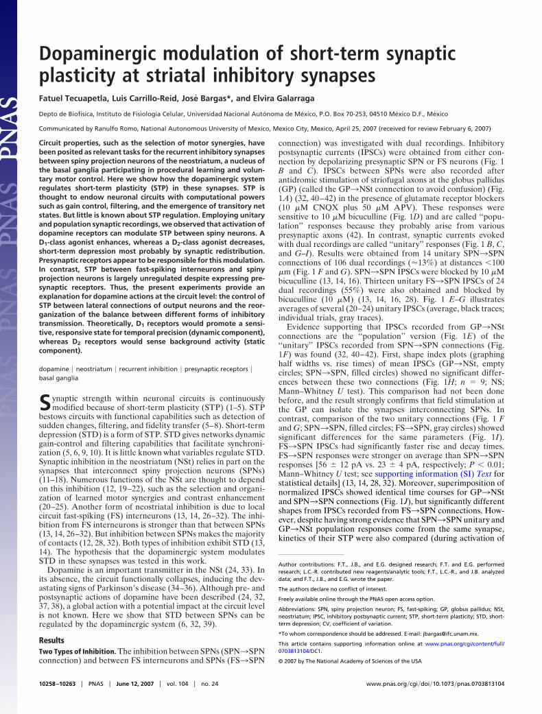
STD Modulation in Synapses Between Spiny Neurons. Activation ofD1-class receptors during STD from SPN3SPN and GP3NStconnections (20-Hz trains) is shown in Fig. 3. The D1-classselective agonist, SKF-81297 (1 �M), enhances STD for bothSPN3SPN (n � 3 of 5 pair recordings) and GP3NSt (n � 5 of12 population recordings) responses [n � 8 of 17 (47%) casesresponded to D1-agonist]. In the pool of responsive cases, thechange in STD ratio was significant (mean � SEM): 3.3 � 0.4before and 9.7 � 3.7 after addition of SKF 81297 (n � 8; P �0.03). The fact that about half the cases did not respond to theD1-agonist supports the hypothesis that dopamine receptorssegregate in different sets of SPNs, at least at the level of synaptic
terminals. STD enhancement was accompanied by an increase inthe amplitude of the first response (IPSC1) from 57 � 17 pAbefore to 75 � 20 pA after the D1-agonist (n � 8; P � 0.02). Tosee whether this change was due to presynaptic mechanisms, theCV of IPSC1 was assessed. It decreased for all D1-responsivecases by 54 � 8% (P � 0.02), suggesting mediation by presynapticreceptors (32). At the same time, a nonparametric test ofvariance rendered a significant increase in IPSC variance afterSKF-81297 for each individual experiment (46) (n � 8; P �0.001; F test), suggesting an increase in release probability for thefirst response (47). Finally, �1 of the STD process was signifi-cantly accelerated from 31 � 3 to 23 � 2 msec (n � 8; P � 0.01),showing that D1-receptor activation accelerates STD kinetics.Taken together, changes in release probability and STD kineticssuggest synaptic ‘‘redistribution’’ (4, 8, 48). These actions werecompletely blocked by the selective D1-receptor antagonist SCH39393 (1 �M) (n � 4; data not shown). A selective (see SI Text)nonhydrolyzable D1-class agonist (32, 37) was used for theseexperiments, and only stable responses at a train frequency of 0.1Hz were chosen for analysis.
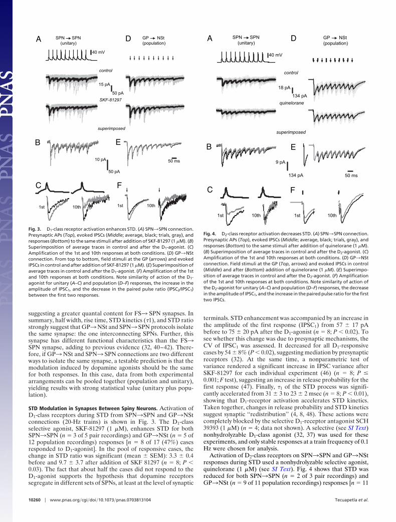
Activation of D2-class receptors on SPN3SPN and GP3NStresponses during STD used a nonhydrolyzable selective agonist,quinelorane (1 �M) (see SI Text). Fig. 4 shows that STD wasreduced for both SPN3SPN (n � 2 of 3 pair recordings) andGP3NSt (n � 9 of 11 population recordings) responses [n � 11
D
E
F
control
50 ms
SKF-81297
superimposed
50 pA
15 pA
50 pA
10 pA
40 mV
1st 10th 1st 10th
A
B
C
GP NSt(population)
SPN SPN (unitary)
Fig. 3. D1-class receptor activation enhances STD. (A) SPN3SPN connection.Presynaptic APs (Top), evoked IPSCs (Middle; average, black; trials, gray), andresponses (Bottom) to the same stimuli after addition of SKF-81297 (1 �M). (B)Superimposition of average traces in control and after the D1-agonist. (C)Amplification of the 1st and 10th responses at both conditions. (D) GP3NStconnection. From top to bottom, field stimuli at the GP (arrows) and evokedIPSCs in control and after addition of SKF-81297 (1 �M). (E) Superimposition ofaverage traces in control and after the D1-agonist. (F) Amplification of the 1stand 10th responses at both conditions. Note similarity of action of the D1-agonist for unitary (A–C) and population (D–F) responses, the increase in theamplitude of IPSC1, and the decrease in the paired pulse ratio (IPSC2/IPSC1)between the first two responses.
50 ms
40 mV
1st 10th 1st 10th
9 pA
18 pA
A
B
C
control
quinelorane
superimposed
D
E
F
GP NSt(population)
SPN SPN (unitary)
134 pA
134 pA
Fig. 4. D2-class receptor activation decreases STD. (A) SPN3SPN connection.Presynaptic APs (Top), evoked IPSCs (Middle; average, black; trials, gray), andresponses (Bottom) to the same stimuli after addition of quinelorane (1 �M).(B) Superimposition of average traces in control and after the D2-agonist. (C)Amplification of the 1st and 10th responses at both conditions. (D) GP3NStconnection. Field stimuli at the GP (Top, arrows) and evoked IPSCs in control(Middle) and after (Bottom) addition of quinelorane (1 �M). (E) Superimpo-sition of average traces in control and after the D2-agonist. (F) Amplificationof the 1st and 10th responses at both conditions. Note similarity of action ofthe D2-agonist for unitary (A–C) and population (D–F) responses, the decreasein the amplitude of IPSC1, and the increase in the paired pulse ratio for the firsttwo IPSCs.
10260 � www.pnas.org�cgi�doi�10.1073�pnas.0703813104 Tecuapetla et al.
of 14 (78%) cases responded to D2-agonist]. The results werepooled together to obtain the change in STD ratio induced by theD2-agonist, from 4.5 � 0.6 before to 2.7 � 0.4 after quinelorane(n � 11; P � 0.008). Although some cases did not respond,responsive cases are more than expected by the receptor segre-gation hypothesis into two pathways (49, 50). A decrease in STDcorresponded to a decrease in IPSC1 from 94 � 25 pA before to45 � 13 pA after the D2-agonist (n � 11; P � 0.005). To seewhether this change was due to presynaptic mechanisms, the CVof IPSC1 was assessed. It was significantly enhanced (220 � 40%)(P � 0.008), suggesting a presynaptic mechanism (32). Inaddition, IPSC variance was significantly decreased (P � 0.05;n � 11; F test) in each responsive experiment, and �1 of the STDprocess increased significantly from 31 � 2 to 42 � 4 msec (P �0.03), suggesting synaptic ‘‘redistribution’’ (4, 8, 48). While usingquinelorane, special care was taken to discard cases showingsigns of long-term depression after repeated trains. These effectswere completely blocked by the selective D2/3-class antagonist,sulpiride (1 �M) (n � 4; data not shown).
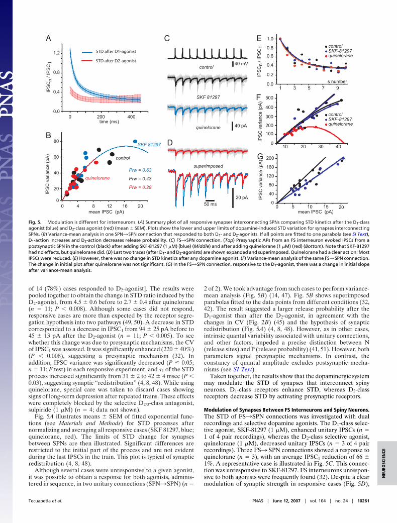
Fig. 5A illustrates means � SEM of fitted exponential func-tions (see Materials and Methods) for STD processes afternormalizing and averaging all responsive cases (SKF 81297, blue;quinelorane, red). The limits of STD change for synapsesbetween SPNs are then illustrated. Significant differences arerestricted to the initial part of the process and are not evidentduring the last IPSCs in the train. This plot is typical of synapticredistribution (4, 8, 48).
Although several cases were unresponsive to a given agonist,it was possible to obtain a response for both agonists, adminis-tered in sequence, in two unitary connections (SPN3SPN) (n �
2 of 2). We took advantage from such cases to perform variance-mean analysis (Fig. 5B) (14, 47). Fig. 5B shows superimposedparabolas fitted to the data points from different conditions (32,42). The result suggested a larger release probability after theD1-agonist than after the D2-agonist, in agreement with thechanges in CV (Fig. 2B) (45) and the hypothesis of synapticredistribution (Fig. 5A) (4, 8, 48). However, as in other cases,intrinsic quantal variability associated with unitary connections,and other factors, impeded a precise distinction between N(release sites) and P (release probability) (41, 51). However, bothparameters signal presynaptic mechanisms. In contrast, theconstancy of quantal amplitude excludes postsynaptic mecha-nisms (see SI Text).
Taken together, the results show that the dopaminergic systemmay modulate the STD of synapses that interconnect spinyneurons. D1-class receptors enhance STD, whereas D2-classreceptors decrease STD by activating presynaptic receptors.
Modulation of Synapses Between FS Interneurons and Spiny Neurons.The STD of FS3SPN connections was investigated with dualrecordings and selective dopamine agonists. The D1-class selec-tive agonist, SKF-81297 (1 �M), enhanced unitary IPSCs (n �1 of 4 pair recordings), whereas the D2-class selective agonist,quinelorane (1 �M), decreased unitary IPSCs (n � 3 of 4 pairrecordings). Three FS3 SPN connections showed a response toquinelorane (n � 3), with an average IPSC1 reduction of 66 �1%. A representative case is illustrated in Fig. 5C. This connec-tion was unresponsive to SKF-81297. FS interneurons unrespon-sive to both agonists were frequently found (32). Despite a clearmodulation of synaptic strength in responsive cases (Fig. 5D),
40 pA
40 mV
50 ms
control
SKF 81297
quinelorane
010 20 30 40
100
200
300
400
500
IPS
C v
aria
nce
(pA
)
0 5 10 150
40
80
120
160
200
20mean IPSC (pA)
control
SKF 81297
s number1 3 5 7 9
0.0
0.4
0.6
0.8
1.0
0.2
control
quineloraneSKF-81297
20 pA
superimposed
STD after D1-agonist
STD after D2-agonist
A
B
IPS
Cn
/ IP
SC
1
IPS
C v
aria
nce
(pA
)
C
D
E
control
quineloraneSKF-81297
IPS
C v
aria
nce
(pA
)
F
G
mean IPSC (pA)
0 200 4000.0
0.4
0.8
1.2IP
SC
n / I
PS
C1
time (ms)
0 4 8 12 16 200
20
40
60
80
quinelorane
Prw = 0.29
Prw = 0.63
Prw = 0.43
Fig. 5. Modulation is different for interneurons. (A) Summary plot of all responsive synapses interconnecting SPNs comparing STD kinetics after the D1-classagonist (blue) and D2-class agonist (red) (mean � SEM). Plots show the lower and upper limits of dopamine-induced STD variation for synapses interconnectingSPNs. (B) Variance-mean analysis in one SPN3SPN connection that responded to both D1- and D2-agonists. If all points are fitted to one parabola (see SI Text),D1-action increases and D2-action decreases release probability. (C) FS3SPN connection. (Top) Presynaptic APs from an FS interneuron evoked IPSCs from apostsynaptic SPN in the control (black) after adding SKF-81297 (1 �M) (blue) (Middle) and after adding quinelorane (1 �M) (red) (Bottom). Note that SKF-81297had no effects, but quinelorane did. (D) Last two traces (after D1- and D2-agonists) are shown expanded and superimposed. Quinelorane had a clear action: MostIPSCs were reduced. (E) However, there was no change in STD kinetics after any dopamine agonist. (F) Variance-mean analysis of the same FS3SPN connection.The change in initial plot after quinelorane was not significant. (G) In the FS3SPN connection, responsive to the D1-agonist, there was a change in initial slopeafter variance-mean analysis.
Tecuapetla et al. PNAS � June 12, 2007 � vol. 104 � no. 24 � 10261
NEU
ROSC
IEN
CE

quinelorane did not change STD (Fig. 5E). STD ratios were2.7 � 4 before and 2.6 � 0.5 after the D2-agonist in responsivecases. Variance-mean analysis (Fig. 5F) showed no significantchange in initial slope of parabolas despite changes in amplitudeand variance. Thus, FS synapses exhibit presynaptic modulationof synaptic strength, but no signs of STD modulation (52) (seeDiscussion).
In a case responsive to the D1-agonist, IPSC amplitudeincreased 159% (data not shown). This action did not interferewith STD kinetics. Variance-mean analysis (Fig. 5G) suggestedthat this action was postsynaptic (53). However, it coursed witha decrease in quantal amplitude (54), implying, perhaps, amixture of pre- and postsynaptic actions.
DiscussionThe basis for the selection and organization of motor synergiesmay depend on the lateral inhibition between SPNs (12, 14, 18,20–23, 25). The present work demonstrates that this lateralinhibition exhibits STD that can be modulated by the dopaminesystem. STD is in charge of controlling the gain and filteringcapabilities of a circuit to increase its sensitivity to suddenchanges (4–7, 9, 10, 48). Hence, fast selection of motor synergiesmay depend on dopaminergic modulation of STD betweenSPNs. Patients with Parkinson’s disease have problems begin-ning or changing motor tasks.
It was shown that STD exhibited by the inhibitory synapsesthat interconnect SPNs (13, 14) can be enhanced by the activa-tion of D1-class receptors. This change would make the circuitsuitable to respond to sudden fast commands (48) and subtlechanges in frequency (dynamic component), helping to balanceinputs of different strengths (6, 9, 39). However, D2-class recep-tors decrease STD, perhaps setting the circuit to keep respondingto and transferring a continuous basal level of activity (static ortonic component) (6, 9, 39). STD modulation was demonstratedby dual (unitary) recordings between SPNs and by field stimu-lation of groups of afferents arising from striofugal axonsstimulated at the GP. Some synaptic responses did not respondto a given applied agonist, suggesting that not all terminals haveboth receptor classes (49, 50). If terminals had dopaminereceptors segregated, it can be hypothesized that the directpathway is the structural basis for the dynamic component,whereas the indirect pathway is the platform for the staticcomponent. Both pathways would complement each other (12,33, 35), e.g., different sets of coordinated muscles may beappointed to fast actions (dynamic component), whereas othersmay be needed to preserve tone and posture (static component).Interestingly, D2-class receptors have more affinity for dopaminethan D1-class receptors (see SI Text) (55). This difference inaffinity suggests that D2-class receptors maintain a dopaminergictone with low basal dopamine concentrations, whereas D1-classreceptors respond during sudden peaks in dopamine concentra-tion (56, 57), suggesting that affinities of receptors for dopamineare in agreement with the role that these receptors play insynaptic function. It reinforces the view that D2 and D1 actionsrepresent the static and dynamic branches of the same system.
Nonetheless, some unitary connections responded to both D1-and D2-class agonists, suggesting a degree of colocalization(58–60). Hence, some sets of terminals may shift their plasticitybetween the lowest and highest degrees of STD. This intersectionbetween pathways (59) may quickly change the balance betweendynamic and tonic neurons depending on the context.
Another target for dopamine in the NSt are the interneuronsand their synapses (14, 26, 27, 29–32, 61, 62). Dopamine agonistsdepolarize FS interneurons in the striatum by D1-class receptors(61). However, in most cases, we did not observe a presynapticmodulation by the D1-agonist (FS3SPN) (32). We only foundan enhancement of IPSCs in one of four (25%) FS3SPNconnections treated with SKF-81297 (1 �M). Variance-mean
analysis indicated a change in initial slope in this case (Fig. 5G)(53, 54), suggesting postsynaptic mechanisms.
A reduction of IPSCs was found in three of four (75%)FS3SPN connections treated with quinelorane (62) (Fig. 5C–F). Noticeably, STD was not changed. Presynaptic inhibitionwithout STD changes has been seen in other synapses (52, 63,64). The mechanism proposed to dissociate presynaptic modu-lation of synaptic strength from modulation of STD includes thepresence of some proteins such as parvalbumin or calciumsensors (65, 66).
Therefore, the dopamine system may affect presynapticallysome interneurons but not others, suggesting heterogeneity ofinterneuron types (32, 67); these presynaptic actions do not implySTD modulation. It is concluded that SPN3SPN synapses cantransiently modify their strength and computational properties,whereas FS3SPN synapses can only modify their strength. Thisdifference may serve to rearrange the balance among differenttypes of inhibition.
STP mainly depends on presynaptic mechanisms such asrelease probability and refilling rate of the vesicle pool (48).Calcium dynamics reflects the ‘‘use’’ or ‘‘history’’ of networkactivity (1–5, 68). Therefore, presynaptic receptors can be con-ceived as sensors and regulators of the dynamics and computa-tional properties of circuits, not simply as modifiers of synapticstrength (32, 41, 69, 70).
Materials and MethodsElectrophysiology. Electrophysiological experiments were per-formed by using neostriatal slices (250–300 �m thick) fromWistar rats PD19–PD22 cut in 4°C saline by using a vibratome(Ted Pella, Reading, CA) as described previously (32). For amore detailed account on methods, see SI Text. Extracellularsaline containing (in mM) 126 NaCl, 3 KCl, 1 MgCl2, 2 CaCl2,26 NaHCO3, and 10 glucose (pH 7.4 with NaOH, 298 mOsm/literwith glucose, saturated with 95% O2 and 5% CO2; 25–27°C).6-cyano-2,3-dihydroxy-7-nitro-quinoxaline disodium salt(CNQX) (10 �M) and D-(-)-2-amino-5 phosphonovaleric acid(AP5) (50 �M) were added to block AMPA and NMDAreceptors, respectively. Standard whole-cell recordings usinginfrared differential interference contrast (IR-DIC) microscopywith an upright microscope and a digital camera were used toobtain IPSCs with the help of an Axoclamp 2B and/or Axopatch200B amplifiers (Axon Instruments, Foster City, CA). Tracesshown are the average of 4-min recordings (24 traces) takenwhen the amplitude had been stabilized for a given condition.IPSCs were evoked by field stimulation using a bipolar electrodelocated at the GP (32). The intracellular saline for postsynapticmedium spiny neurons contained (in mM) 72 KH2PO4, 36 KCl,2 MgCl2, 10 Hepes, 1.1 EGTA, 0.2 Na2ATP, 0.2 Na3GTP, 5QX-314, and 1% biocytin (pH 7.2)/275 mOsm/liter for a theo-retical ECl
� � �30 mV. Experiments were performed at 25°C to27°C. Series resistances ranged from 5 to 20 M and werepartially compensated (60–90%). Whole-cell recordings ofpostsynaptic medium SPNs were recorded in voltage-clampmode at a holding potential of �85 mV. Postsynaptic cells werelocated in the vicinity (�100 �m) of a presynaptic SPN or FSneuron recorded in current-clamp mode. The presynaptic neu-ron was stimulated with brief current pulses that evoked actionpotentials (1–5 msec, 0.2–0.3 nA). Single stimuli or trains of 10stimuli were evoked at a frequency of 10, 20, or 50 Hz every 10sec. Digitized data were saved on a disk and analyzed withcommercial software (Origin version 7; Microcal, Northampton,MA). IPSC amplitudes were measured from basal line to peakfor the first response in a train (IPSC1). For the subsequentresponses (IPSCn), the basal line remaining from the previousresponse was subtracted. Distribution-free statistical procedures(Systat version 11; SPSS, Chicago, IL) were used to find datasignificance.
10262 � www.pnas.org�cgi�doi�10.1073�pnas.0703813104 Tecuapetla et al.

Amplitudes of IPSCs in a train were fitted to a sum ofexponential functions after normalization:
IPSC t� � A1e�x/�1 � A2e�x/�2 � y0. [1]
Fast (�1) and slow (�2) time constants of decay are obtained fromthis fit (48). A1 and A2 are the magnitude of the postsynapticresponse for each exponential component, and y0 representsresidual baseline activity. We measured the STD ratio as IPSC1/IPSC7–10. Variance-mean analysis was performed as proposed byClements and Silver (2000), and the details are provided in SIText.
Pharmacology. Drugs were dissolved in the bath saline from stocksolutions made daily and were administered by using a gravity-driven superfusion system. Equilibrated concentrations of thedrugs were achieved in 4 to 5 min. All of the following werepurchased from Sigma–Aldrich (St. Louis, MO): CNQX, AP5,
L-glutamic acid [glutamate (GLU)], SKF 81297, SCH 23390,bicuculline, QX-314, and quinelorane.
Immunohistochemistry. Neurons were filled with biocytin duringrecording. Slices with a single or two filled neurons (pairedrecordings) were taken into consideration for immunocytochem-istry. A combination of intracellular labeling and substance P,enkephalin, or parvalbumin immunocytochemistry was used ineach occasion to identify the recorded neurons (28, 32). Detailsof the methods can be found elsewhere (32), and a brief summaryis given in SI Text.
We thank D. Tapia and A. Laville for technical support. This work wassupported by IMPULSA-Universidad Nacional Autónoma de Méxicoand Dirección General de Asuntos del Personal Académico-UniversidadNacional Autónoma de México grants (to J.B. and E.G.) and ConsejoNacional de Ciencia y Tecnología-Mexico Grants 42636 (to E.G.) and49484 (to J.B.).
1. Zucker RS, Regehr WG (2002) Ann Rev Physiol 64:355–405.2. Abbott LF, Nelson SB (2000) Nat Neurosci 3:1178–1183.3. Thomson AM (2000) Trends Neurosci 23:305–312.4. Abbott LF, Regehr WG (2004) Nature 431:796–803.5. Thomson AM (2000) Prog Neurobiol 62:159–196.6. Richardson MJ, Melamed O, Silberberg G, Gerstner W, Markram H (2005)
J Comput Neurosci 18:323–331.7. Fuhrmann G, Segev I, Markram H, Tsodyks M (2002) J Neurophysiol 87:140–
148.8. Markram H, Tsodyks M (1996) Nature 382:807–810.9. Abbott LF, Varela JA, Sen K, Nelson SB (1997) Science 275:220–224.
10. O’Donovan MJ, Rinzel J (1997) Trends Neurosci 20:431–433.11. Czubayko U, Plenz D (2002) Proc Natl Acad Sci USA 99:15764–15769.12. Groves PM (1983) Brain Res 286:109–132.13. Gustafson N, Gireesh-Dharmaraj E, Czubayko U, Blackwell KT, Plenz D
(2006) J Neurophysiol 95:737–752.14. Koos T, Tepper JM, Wilson CJ (2004) J Neurosci 24:7916–7922.15. Park MR, Lighthall JW, Kitai ST (1980) Brain Res 194:359–369.16. Taverna S, van Dongen YC, Groenewegen HJ, Pennartz CM (2004) J Neuro-
physiol 91:1111–1121.17. Tunstall MJ, Oorschot DE, Kean A, Wickens JR (2002) J Neurophysiol
88:1263–1269.18. Venance L, Glowinski J, Giaume C (2004) J Physiol 559:215–230.19. Wickens JR, Alexander ME, Miller R (1991) Synapse 8:1–12.20. Wickens JR, Oorchcot DE (2000) in Brain Dynamics and the Striatal Complex,
eds Miller R, Wickens JR (Harwood, Australia), pp 141–150.21. Bar-Gad I, Bergman H (2001) Curr Opin Neurobiol 11:689–695.22. Plenz D (2003) Trends Neurosci 26:436–443.23. Latash ML, Shim JK, Smilga AV, Zatsiorsky VM (2005) Biol Cyber 92:186–191.24. Nicola SM, Woodward Hopf F, Hjelmstad GO (2004) Cell Tissue Res 318:93–106.25. Sil’kis IG (2006) Neurosci Behav Physiol 36:631–643.26. Kita H (1993) Prog Brain Res 99:51–72.27. Bennett BD, Bolam JP (1994) Neuroscience 62:707–719.28. Koos T, Tepper JM (1999) Nat Neurosci 2:467–472.29. Tepper JM, Bolam JP (2004) Curr Opin Neurobiol 14:685–692.30. Tepper JM, Koos T, Wilson CJ (2004) Trends Neurosci 27:662–669.31. Tepper JM, Plenz D (2005) in The Interface between Neurons and Global Brain
Function, Dahlem Workshop Report 93, eds Grillner S, Graybiel AM (MITPress, Cambridge, MA), pp 127–148.
32. Guzman JN, Hernandez A, Galarraga E, Tapia D, Laville A, Vergara R,Aceves J, Bargas J (2003) J Neurosci 23:8931–8940.
33. Albin RL, Young AB, Penney JB (1995) Trends Neurosci 18:63–64.34. Hornykiewicz O (1973) Br Med Bull 29:172–178.35. Wichmann T, DeLong MR (2003) Ann NY Acad Sci 991:199–213.36. Wooten GF (1990) in Neurobiology of Disease, ed Pearlman AL (Oxford, Univ
Press, New York), pp 454–468.37. Hernandez-Lopez S, Bargas J, Surmeier DJ, Reyes A, Galarraga E (1997)
J Neurosci 17:3334–3342.
38. Hernandez-Lopez S, Tkatch T, Perez-Garci E, Galarraga E, Bargas J, HammH, Surmeier DJ (2000) J Neurosci 20:8987–8995.
39. Silberberg G, Wu C, Markram H (2004) J Physiol 556:19–27.40. Perez-Rosello T, Figueroa A, Salgado H, Vilchis C, Tecuapetla F, Guzman JN,
Galarraga E, Bargas J (2005) J Neurophysiol 93:2507–2519.41. Salgado H, Tecuapetla F, Perez-Rosello T, Perez-Burgos A, Perez-Garci E,
Galarraga E, Bargas J (2005) J Neurophysiol 94:3771–3787.42. Tecuapetla F, Carrillo-Reid L, Guzman JN, Galarraga E, Bargas J (2005)
J Neurophysiol 93:1119–1126.43. Romo R, Scarnati E, Schultz W (1992) Exp Brain Res 91:385–395.44. Stern EA, Kincaid AE, Wilson CJ (1997) J Neurophysiol 77:1697–1715.45. Silver RA, Momiyama A, Cull-Candy SG (1998) J Physiol 510:881–902.46. Koos T, Tepper JM (2002) J Neurosci 22:529–535.47. Clements JD, Silver RA (2000) Trends Neurosci 23:105–113.48. Tsodyks MV, Markram H (1997) Proc Natl Acad Sci USA 94:719–723.49. Gerfen CR (2000) Ann Neurol 47:S42–S50.50. Gerfen CR, Engber TM, Mahan LC, Susel Z, Chase TN, Monsma FJ, Jr, Sibley
DR (1990) Science 250:1429–1432.51. Biro AA, Holderith NB, Nusser Z (2006) J Neurosci 26:12487–12496.52. Hefft S, Kraushaar U, Geiger JR, Jonas P (2002) J Physiol 539:201–208.53. Flores-Hernandez J, Hernandez S, Snyder GL, Yan Z, Fienberg AA, Moss SJ,
Greengard P, Surmeier DJ (2000) J Neurophysiol 83:2996–3004.54. Gonzalez-Islas C, Hablitz JJ (2001) J Neurophysiol 86:2911–2918.55. Missale C, Nash SR, Robinson SW, Jaber M, Caron MG (1998) Physiol Rev
78:189–225.56. Giros B, Jaber M, Jones SR, Wightman RM, Caron MG (1996) Nature
379:606–612.57. Suaud-Chagny MF, Dugast C, Chergui K, Msghina M, Gonon F (1995)
J Neurochem 65:2603–2611.58. Surmeier DJ, Song WJ, Yan Z (1996) J Neurosci 16:6579–6591.59. Deng YP, Lei WL, Reiner A (2006) J Chem Neuroanat 32:101–116.60. Mizuno T, Schmauss C, Rayport S (2007) BMC Neurosci 8:8.61. Bracci E, Centonze D, Bernardi G, Calabresi P (2002) J Neurophysiol 87:2190–
2194.62. Delgado A, Sierra A, Querejeta E, Valdiosera RF, Aceves J (2000) Neuro-
science 95:1043–1048.63. Hefft S, Jonas P (2005) Nat Neurosci 8:1319–1328.64. Hjelmstad GO (2004) J Neurosci 24:8621–8628.65. Sippy T, Cruz-Martin A, Jeromin A, Schweizer FE (2003) Nat Neurosci
6:1031–1038.66. Caillard O, Moreno H, Schwaller B, Llano I, Celio MR, Marty A (2000) Proc
Natl Acad Sci USA 97:13372–13377.67. Gao WJ, Wang Y, Goldman-Rakic PS (2003) J Neurosci 23:1622–1630.68. Brenowitz S, Trussell LO (2001) J Neurosci 21:1857–1867.69. Selig DK, Nicoll RA, Malenka RC (1999) J Neurosci 19:1236–1246.70. Wilson RI, Nicoll RA (2001) Nature 410:588–592.
Tecuapetla et al. PNAS � June 12, 2007 � vol. 104 � no. 24 � 10263
NEU
ROSC
IEN
CE