ZFX Controls the Self-Renewal of Human Embryonic Stem Cells Sivan Harel 1. , Edmund Y. Tu 2. , Stuart Weisberg 1 , Manuel Esquilin 1 , Stuart M. Chambers 2 , Becky Liu 2 , Christian T. Carson 3 , Lorenz Studer 2 , Boris Reizis 1 *, Mark J. Tomishima 2 * 1 Department of Microbiology and Immunology, Columbia University Medical Center, New York, New York, United States of America, 2 Developmental Biology Program, Sloan-Kettering Institute, New York, New York, United States of America, 3 BD Biosciences, La Jolla, California, United States of America Abstract Embryonic stem cells (ESCs) and induced pluripotent stem cells (iPSCs) offer great promise in regenerative medicine and disease modeling due to their unlimited self-renewal and broad differentiation capacity. There is evidence that the growth properties and critical signaling pathways differ between murine and human ESCs; therefore, it is essential to perform functional studies to test the putatively conserved mechanisms of pluripotent stem cell self-renewal between species. Previously, we identified the transcription factor Zfx as a key regulator of self-renewal in murine ESCs. Here we extend those findings to human ESCs. ZFX knockdown in hESCs hindered clonal growth and decreased colony size after serial replating. ZFX overexpression enhanced clone formation in the presence of Y-27632, increased colony size at low density and decreased expression of differentiation-related genes in human ESCs. ZFX-overexpressing hESCs resisted spontaneous differentiation but could be directed to differentiate into endodermal and neural cell fates when provided with the appropriate cues. Thus, ZFX acts as a molecular rheostat regulating the balance between self-renewal and differentiation in hESCs, revealing the close evolutionary conservation of the self-renewal mechanisms in murine and human ESCs. Citation: Harel S, Tu EY, Weisberg S, Esquilin M, Chambers SM, et al. (2012) ZFX Controls the Self-Renewal of Human Embryonic Stem Cells. PLoS ONE 7(8): e42302. doi:10.1371/journal.pone.0042302 Editor: Wei-Chun Chin, University of California Merced, United States of America Received June 20, 2011; Accepted July 6, 2012; Published August 3, 2012 Copyright: ß 2012 Harel et al. This is an open-access article distributed under the terms of the Creative Commons Attribution License, which permits unrestricted use, distribution, and reproduction in any medium, provided the original author and source are credited. Funding: This work was supported by The Starr Foundation (MJT and LS; www.starrfoundation.org), NYSTEM grant C024175-01 (MJT and LS; www.nystem.ny. org), National Institutes of Health grant HL084353 (to BR) and Becton, Dickinson and Company (CTC). The funders had no role in study design, data collection and analysis, decision to publish, or preparation of the manuscript. Competing Interests: The authors have read the journal’s policy and have the following conflicts: CTC owns stock in Becton, Dickinson and Company. This does not alter the authors’ adherence to all the PLoS ONE policies on sharing data and materials. * E-mail: [email protected] (BR); [email protected] (MJT) . These authors contributed equally to this work. Introduction Embryonic stem cells (ESCs) and the related induced pluripo- tent stem cells (iPSCs) are unique cells capable of giving rise to all tissues of the adult organism. These pluripotent stem cells (PSCs) can be exponentially expanded in culture while retaining their differentiation potential. The traits of pluripotency and continuous self-renewal underlie the value of PSCs as a potential source for cell replacement therapies and disease modeling, as well as a tool to study normal human development [1–3]. The pluripotency of both mouse and human ESCs is regulated by a network of ESC- specific transcription factors including Oct4, Nanog, Sox2 and their binding partners and targets [4,5]. These factors promote the undifferentiated state by positively regulating expression of pluripotency related genes while repressing lineage-specific gene expression and maintaining the unique permissive chromatin structure of ESCs. In addition to ESC-specific transcription factors, additional sets of regulators appear essential for the self- renewal of undifferentiated ESCs and/or iPSCs, including Klf family members, c-Myc and Lin28 [6,7]. Understanding the exact role and mechanism of action of these and other regulators in ESC self-renewal is an important goal in developmental biology and will aid the practical use of PSCs. Although ESCs from different species share the same key properties of pluripotency and self-renewal, major differences were found between murine (mESCs) and human ESCs (hESCs) including expression of different sets surface markers and distinct growth factor requirements [8]. Compared to mouse ESCs, hESCs display a characteristic flattened colony morphology, relatively slow growth and inefficient clonal propagation [3]. These properties resemble mouse epiblast-derived stem cells (EpiSC – referred to as ‘‘primed’’ hereafter), and indeed the gene expression profile of hESCs is closer to that of mouse EpiSC [9,10]. Thus, current evidence suggests that hESCs are derived from a later developmental stage (primed) relative to the stage from which mouse ESCs are derived (naı ¨ve). Some progress has been made to push human ESCs toward the naı ¨ve state through genetic manipulation or by altering culture conditions [11,12], but much work remains in order to unravel the differences between pluripotent state and species differences. While the ‘‘primed’’ model of hESCs might reconcile some of the differences between murine and human ESCs, it opens a fundamental question about the similarity of the transcriptional circuitry between the two ESC types. Previously, we demonstrated a role for the transcription factor Zfx in the self-renewal of mESC and adult hematopoietic stem cells [13]. Zfx is encoded on the mammalian X chromosome and contains a transcription activation domain and a zinc finger domain for sequence-specific DNA binding. A highly homologous PLoS ONE | www.plosone.org 1 August 2012 | Volume 7 | Issue 8 | e42302

Welcome message from author
This document is posted to help you gain knowledge. Please leave a comment to let me know what you think about it! Share it to your friends and learn new things together.
Transcript
ZFX Controls the Self-Renewal of Human EmbryonicStem CellsSivan Harel1., Edmund Y. Tu2., Stuart Weisberg1, Manuel Esquilin1, Stuart M. Chambers2, Becky Liu2,
Christian T. Carson3, Lorenz Studer2, Boris Reizis1*, Mark J. Tomishima2*
1 Department of Microbiology and Immunology, Columbia University Medical Center, New York, New York, United States of America, 2 Developmental Biology Program,
Sloan-Kettering Institute, New York, New York, United States of America, 3 BD Biosciences, La Jolla, California, United States of America
Abstract
Embryonic stem cells (ESCs) and induced pluripotent stem cells (iPSCs) offer great promise in regenerative medicine anddisease modeling due to their unlimited self-renewal and broad differentiation capacity. There is evidence that the growthproperties and critical signaling pathways differ between murine and human ESCs; therefore, it is essential to performfunctional studies to test the putatively conserved mechanisms of pluripotent stem cell self-renewal between species.Previously, we identified the transcription factor Zfx as a key regulator of self-renewal in murine ESCs. Here we extend thosefindings to human ESCs. ZFX knockdown in hESCs hindered clonal growth and decreased colony size after serial replating.ZFX overexpression enhanced clone formation in the presence of Y-27632, increased colony size at low density anddecreased expression of differentiation-related genes in human ESCs. ZFX-overexpressing hESCs resisted spontaneousdifferentiation but could be directed to differentiate into endodermal and neural cell fates when provided with theappropriate cues. Thus, ZFX acts as a molecular rheostat regulating the balance between self-renewal and differentiation inhESCs, revealing the close evolutionary conservation of the self-renewal mechanisms in murine and human ESCs.
Citation: Harel S, Tu EY, Weisberg S, Esquilin M, Chambers SM, et al. (2012) ZFX Controls the Self-Renewal of Human Embryonic Stem Cells. PLoS ONE 7(8):e42302. doi:10.1371/journal.pone.0042302
Editor: Wei-Chun Chin, University of California Merced, United States of America
Received June 20, 2011; Accepted July 6, 2012; Published August 3, 2012
Copyright: � 2012 Harel et al. This is an open-access article distributed under the terms of the Creative Commons Attribution License, which permitsunrestricted use, distribution, and reproduction in any medium, provided the original author and source are credited.
Funding: This work was supported by The Starr Foundation (MJT and LS; www.starrfoundation.org), NYSTEM grant C024175-01 (MJT and LS; www.nystem.ny.org), National Institutes of Health grant HL084353 (to BR) and Becton, Dickinson and Company (CTC). The funders had no role in study design, data collection andanalysis, decision to publish, or preparation of the manuscript.
Competing Interests: The authors have read the journal’s policy and have the following conflicts: CTC owns stock in Becton, Dickinson and Company. This doesnot alter the authors’ adherence to all the PLoS ONE policies on sharing data and materials.
* E-mail: [email protected] (BR); [email protected] (MJT)
. These authors contributed equally to this work.
Introduction
Embryonic stem cells (ESCs) and the related induced pluripo-
tent stem cells (iPSCs) are unique cells capable of giving rise to all
tissues of the adult organism. These pluripotent stem cells (PSCs)
can be exponentially expanded in culture while retaining their
differentiation potential. The traits of pluripotency and continuous
self-renewal underlie the value of PSCs as a potential source for
cell replacement therapies and disease modeling, as well as a tool
to study normal human development [1–3]. The pluripotency of
both mouse and human ESCs is regulated by a network of ESC-
specific transcription factors including Oct4, Nanog, Sox2 and
their binding partners and targets [4,5]. These factors promote the
undifferentiated state by positively regulating expression of
pluripotency related genes while repressing lineage-specific gene
expression and maintaining the unique permissive chromatin
structure of ESCs. In addition to ESC-specific transcription
factors, additional sets of regulators appear essential for the self-
renewal of undifferentiated ESCs and/or iPSCs, including Klf
family members, c-Myc and Lin28 [6,7]. Understanding the exact
role and mechanism of action of these and other regulators in ESC
self-renewal is an important goal in developmental biology and will
aid the practical use of PSCs.
Although ESCs from different species share the same key
properties of pluripotency and self-renewal, major differences were
found between murine (mESCs) and human ESCs (hESCs)
including expression of different sets surface markers and distinct
growth factor requirements [8]. Compared to mouse ESCs,
hESCs display a characteristic flattened colony morphology,
relatively slow growth and inefficient clonal propagation [3].
These properties resemble mouse epiblast-derived stem cells
(EpiSC – referred to as ‘‘primed’’ hereafter), and indeed the gene
expression profile of hESCs is closer to that of mouse EpiSC
[9,10]. Thus, current evidence suggests that hESCs are derived
from a later developmental stage (primed) relative to the stage
from which mouse ESCs are derived (naı̈ve). Some progress has
been made to push human ESCs toward the naı̈ve state through
genetic manipulation or by altering culture conditions [11,12], but
much work remains in order to unravel the differences between
pluripotent state and species differences. While the ‘‘primed’’
model of hESCs might reconcile some of the differences between
murine and human ESCs, it opens a fundamental question about
the similarity of the transcriptional circuitry between the two ESC
types.
Previously, we demonstrated a role for the transcription factor
Zfx in the self-renewal of mESC and adult hematopoietic stem
cells [13]. Zfx is encoded on the mammalian X chromosome and
contains a transcription activation domain and a zinc finger
domain for sequence-specific DNA binding. A highly homologous
PLoS ONE | www.plosone.org 1 August 2012 | Volume 7 | Issue 8 | e42302

protein called Zfy is encoded on the Y chromosome and is
expressed in human but not in murine male somatic cells. ZFX/
ZFY genes are highly conserved in vertebrates, with ,97% amino
acid identity between murine and human ZFX in the DNA
binding domain. The deletion of Zfx in mESC impairs self-
renewal but does not affect differentiation capacity. Conversely,
Zfx overexpression enhanced mESC self-renewal under subopti-
mal conditions and opposed both spontaneous and directed
differentiation. Zfx directly activated functionally relevant mESC-
specific target genes such as Tbx3 and Tcl1. Subsequent work has
implicated Zfx in a common genetic pathway with Myc and Klf4,
the two transcription factors controlling mESC self-renewal and
iPSC reprogramming [14–16]. Thus, Zfx emerged as an essential
and specific regulator of self-renewal in mESC, warranting the
investigation of its role in the human system.
Here, we used a genetic approach to analyze the role of ZFX in
hESC self-renewal and differentiation. Lentiviral shRNA knock-
down of ZFX impaired self-renewal of hESCs. A novel bacterial
artifical chromosome (BAC) transgenic strategy [17] was adapted
to overexpress human ZFX in hESC under its native regulatory
elements. Such gene dosage analysis showed that ZFX overex-
pression increased colony formation from single cells, a hallmark
of improved hESC self-renewal. Furthermore, ZFX overexpres-
sion prevented spontaneous hESC differentiation under subopti-
mal conditions. Array analysis showed that ZFX overexpression
decreased a number of genes characteristic of differentiated cells.
These data establish an important role for ZFX in the regulation
of hESC, confirming the conservation of self-renewal mechanisms
in murine and human ESC.
Materials and Methods
ZFX Knockdown Human ESCsH9 (WA-09) hESCs were maintained on MEFs (GlobalStem,
Inc., Rockville, MD) before feeder-free expansion on Matrigel
with conditioned media and 10 ng/ml FGF2 (3–5 days). To
prepare cells for transduction, hESCs were exposed to Accutase
(Innovative Cell Technologies, Inc., San Diego, CA) to create a
single-celled suspension before plating at 18,000 cells/cm2 in the
presence of 10 mM Y-27632. The following day, cells were
transduced at a multiplicity of infection of 1 and 0.1. For each
knockdown, phenotypes were observed at both multiplicities
relative to the scrambled control. Puromycin selection began two
days after transduction and no mock transduced cells remained
two days after selection began. At this time, 5,000 puromycin
resistant cells were replated per condition and were expanded in
conditioned media +10 ng/ml FGF2 for seven days before crystal
violet staining.
Quantitative ImmunofluoresceCells were transduced at a multiplicity of ,0.1 as described
above and puromycin selection was applied as described. Cells
were fixed in 4% paraformaldehyde for 15 minutes before rinsing
four times with PBS. Fixed cells were blocked/permeabilized with
16 Perm/wash buffer (BD Biosciences, San Jose, CA) for
20 minutes before exposure to ZFX primary antibody overnight
at 4uC. The next day, cells were washed three times in Perm/wash
buffer before exposure to secondary antibodies conjugated to
Alexa 488 and Hoechst 33258. Cells were washed three times after
the hour, and PBS was added to the cells for imaging.
Imaging was performed on an Operetta High Content
Screening System. The Hoechst was exposed for 20 mSec and
the Alexa 488 signal was captured by a 300 mSec exposure using
50% intensity from the light source. Harmony software version 3.0
was used to identify cell nuclei (Hoechst signal) and quantitated the
Alexa 488 pixel intensity (ZFX protein) for each cell nucleus. The
mean pixel intensity from 50 cell fields per sample were imaged
from 3 independent experiments for quantitation. All knockdowns
were significantly different from the scrambled control (p,0.0001
for pairwise t-tests). 11,803 (Scr), 10,296 (Z2), 4,765 (Z3) and 6,905
(Z4) nuclei were measured in these experiments.
ZFX Overexpressing Human ESCsHuman ESCs were maintained as previously described [18].
BAC overexpressing human ESCs were made essentially as
described for transgenic reporter BAC integration [17]. Briefly,
H9 (WA-09) hESCs were maintained on MEFs (GlobalStem, Inc.,
Rockville, MD) before feeder-free expansion on Matrigel with
conditioned media and 10 ng/ml FGF2 (3–5 days). To prepare
cells for nucleofection, Accutase (Innovative Cell Technologies,
Inc., San Diego, CA) was used to create a single-celled suspension,
and 5 million cells were used per nucleofection (solution V,
protocol B-16; Lonza, Cologne, Germany) with 3 ug of human
Zfx BAC (RP11-1107D4) retrofitted with pRetroES [19]. Each
nucleofection reaction was plated onto Neo-resistant MEFs
(GlobalStem, Inc., Rockville, MD) in 10 mM Y-27632 for two
days after nucleofection (Tocris Bioscience, Bristol, UK), and cells
were allowed to recover for 4 days. Selection began with 25 ug/ml
G418 (days 4–14; Invitrogen Life Science, Carlsbad, CA) before
increasing selection to 40 ug/ml (days 14–20). Drug-resistant
colonies were manually dissected before expansion into lines.
Quantitative PCR AnalysisReal-time PCR analysis was performed using the following
QuantiTect primers: POU5F1, CXCR4, PAX6, GAPDH and
ACTN2. Other gene expression levels were determined by qPCR
using the following primers:
ZFX
F: 59-CGTAGGAGAGGAGGATGCTG-39
R: 59-TGCCTGGAATCAGGTCTTCT-39
EOMES
F: 59-CGGCCTCTGTGGCTCAAA-39
R: 59-AAGGAAACATGCGCCTGC-39
ID2
F: 59-AATCCTGCAGCACGTCATCG-39
R: 59- CTGGTGATGCAGGCTGACAA-39
VIM
F: 59-GGAGCTGCAGGAGCTGAATG-39
R: 59-GACTTGCCTTGGCCCTTGAG-39
Septin 5
F: 59-TCAACATCGTGCCTCTCATC-39
R: 59-GCTGCTTGAAGTCCTCATCC-39
RNA was isolated using Trizol (Invitrogen, #15596-018) and
reversed transcribed with the QuantiTect Reverse Transcription
kit (Qiagen, #205313). SYBR green PCR reactions were
performed using the PerfeCta SYBR Green SuperMix (Quanta
Bioscience, #95054-500) on an Eppendorf Mastercycler Real-
plex2.
Stem Cell Marker ExpressionDirect immunofluorescence was performed using SSEA-4 Alexa
FluorH 488 (BD Pharmingen, #560308, La Jolla, CA), TRA1-81
Alexa FluorH 555 (BD Pharmingen, #560123, La Jolla, CA), and
Oct3/4 Alexa FluorH 647 (BD Pharmingen, #560307, La Jolla,
CA) according to the manufacturers recommendations.
ZFX Controls the Self-Renewal of hESCs
PLoS ONE | www.plosone.org 2 August 2012 | Volume 7 | Issue 8 | e42302
Western BlottingProtein samples of H9 hESC and ZFXOver hESC clones were
prepared in RIPA buffer plus protease inhibitors (Roche
Diagnostics Ltd.). Protein concentration was determined using
Bradford assay and 30 ug or 15 ug of each sample were separated
by 10% polyacrylamide gel. Gels were transferred to PVDF
membranes using the Bio-Rad mini-gel transfer apparatus.
Membranes were blocked with 3% milk for 1 hour at room
temperature and probed with affinity-purified polyclonal anti-Zfx
antibody (in 3% Milk, 1:1000 overnight at 4uC). After washing 3X
with TBST, membranes were next incubated with peroxidase-
labeled goat anti-rabbit IgG (1:10,000 in 3% milk) for 1 hour at
room temperature before visualization with ECL.
Spontaneous Differentiation AssayHuman ESCs were expanded on Matrigel-coated dishes in
standard human ESC media containing 6 ng/ml FGF2 without
conditioned media. After seven days, single-celled suspensions
were made by dissociating cells with Accutase (Innovative Cell
Technologies, Inc., San Diego, CA) for 45 minutes before being
washed twice with human ESC media. Aliquots of the cells were
stained with anti-SSEA-3 (SSEA-3 Alexa FluorH 647; unpublished,
BD Pharmingen, La Jolla, CA) and anti-SSEA-1 antibodies
(SSEA-1 Alexa FluorH 647; BD Pharmingen #560120, La Jolla,
CA) before quantitation on a FACSAria. For this assay, a number
of unrelated BAC transgenic cell lines were used as normal clone
controls (see Table 1). These lines do not overexpress ZFX but
have undergone the same clonal selection as the ZFXOver clones
(data not shown). Control clones were made from BACs with:
ID1::YFP (clone 2) [20], HES5::GFP (clone 10) and DLL1::GFP
(clones 281 and 277) [17]. All control clones were derived from H9
hESC and were made with the same nucleofection/selection
protocol [17].
Clonogenic AssayFor colony-forming cell assays, human ESC were dissociated
with Accutase (Innovative Cell Technologies, Inc., San Diego, CA)
for 45 minutes. Dissociated cells were washed twice with human
ESC media. Serial dilutions of hESCs were plated onto Matrigel-
coated 6 well dishes in conditioned media with 10 ng/ml FGF2.
Cells were exposed to Y-27632 for 24 hours after plating, and
colonies were visualized after 7 days by staining with crystal violet.
The data presented are the number of colonies from the 1:100
dilution and represent the number of colonies derived from
2,666 cells in 9.6 cm2.
Crystal violet staining was used because it is easier and cheaper
and works as well for determining the number of colony forming
units in a given culture. Figure S1A shows the alkaline
phosphatase staining (Sigma, 86R-1KT) in the colony-forming
assay: nearly every colony stains positive. We confirmed this result
by growing clonal colonies before performing Oct4 FACS analysis
(Figure S1B). Over 97.5% of the cells from all genotypes were
Oct4+ demonstrating that these growth conditions strongly select
for undifferentiated hESCs. This makes alkaline phosphatase
detection a redundant feature of the colony growth assay.
Directed DifferentiationTo direct human ESCs to endoderm, we used a previously
published protocol [21]. Briefly, endoderm induction was
performed by switching from hESC media to RPMI with
glutamine, 0.5% Hyclone FBS and 100 ng/ml Activin A. Cells
were fed daily and were assayed on days 1 and 3. ESCs were
directed to neural cells through co-culture with MS-5 bone
marrow stromal cells as previously described [18].
Gene Expression ArrayRNA was isolated from the Tra1-81HI/SSEA-3HI fraction of
ZFXOver1,2, ZFXNormal and H9 hESCs using Trizol (Invitrogen).
Samples were labeled and hybridized to Illumina human 6
oligonucleotide arrays. Normalization and model-based expression
measurements were performed using the Illumina analysis package
(LUMI) available through open-source Bioconductor project
(www.bioconductor.org) within the statistical programming lan-
guage R (http://cran.r-project.org/). Pairwise comparisons were
performed using the Linear Models for Microarray Data package
(LIMMA) available through Bioconductor. Genes found to have
an adjusted p-value ,0.05 were considered significant.
Statistical AnalysisStatistical analysis was performed using Prism for Mac version
5.0a. Unpaired, two-tailed T tests were performed comparing
control cell lines versus ZFXOver clones.
BAC FISHMetaphase harvest. Sub-confluent cultures were treated
with 0.1 ug/ml Colcemid (Karyomax, Invitrogen) for 60–90 min-
utes before harvesting according to standard cytogenetics proce-
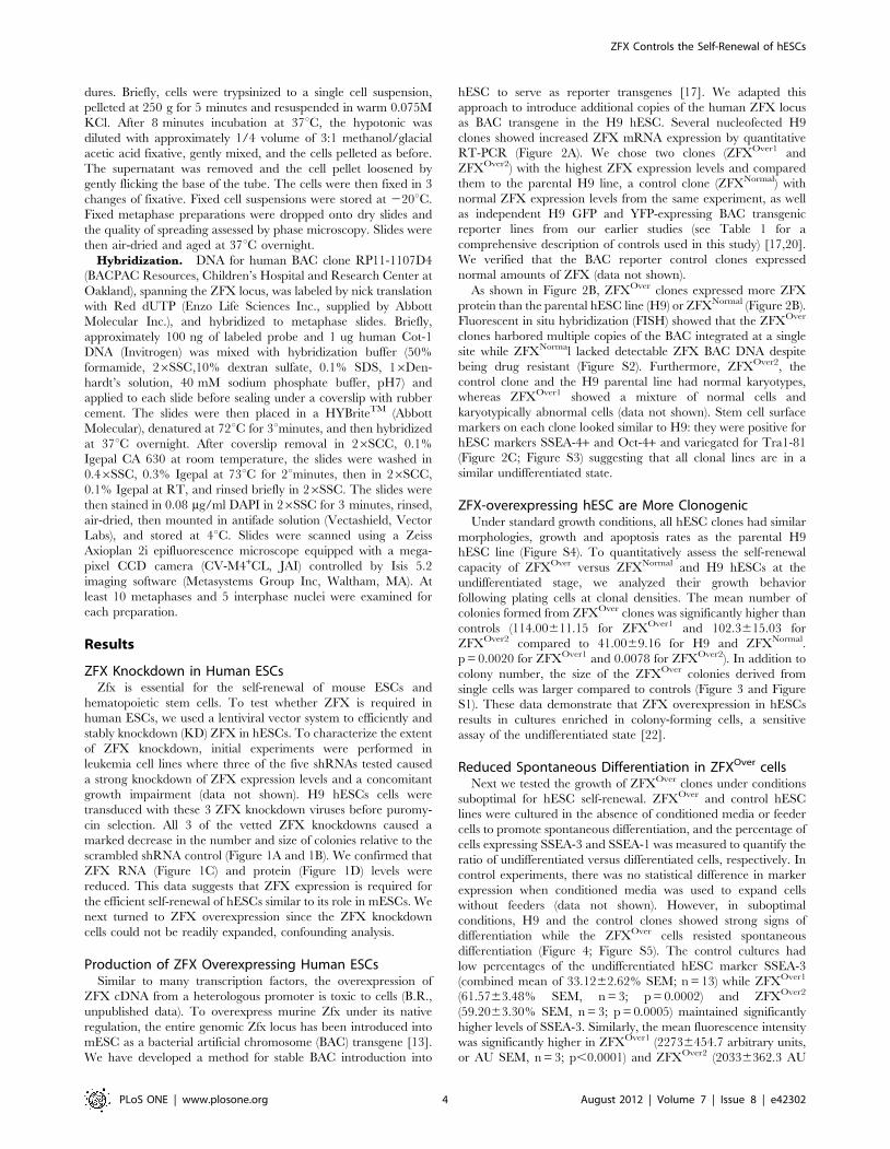
Table 1. All control hESC lines and clones used in this manuscript.
Figure Control Source
2 H9 and ZFXNormal This manuscript
3 H9 and ZFXNormal This manuscript
4 H9, DLL1::GFPc277 and c281, HES5::GFP c10 and ID1::YFPc2 Placantonakis et al., 2009 and James et al., 2010
5 H9, ID1::YFPc2 and DLL1::GFPc277 Placantonakis et al., 2009 and James et al., 2010
6 H9 and ZFXNormal This manuscript
S1 H9 and ZFXNormal This manuscript
S2 H9 and ZFXNormal This manuscript
S3 H9 and ZFXNormal This manuscript
S4 H9 and ZFXNormal This manuscript
S5 H9, DLL1::GFPc277 and c281, HES5::GFP c10 and ID1::YFPc2 Placantonakis et al., 2009 and James et al., 2010
doi:10.1371/journal.pone.0042302.t001
ZFX Controls the Self-Renewal of hESCs
PLoS ONE | www.plosone.org 3 August 2012 | Volume 7 | Issue 8 | e42302
dures. Briefly, cells were trypsinized to a single cell suspension,
pelleted at 250 g for 5 minutes and resuspended in warm 0.075M
KCl. After 8 minutes incubation at 37uC, the hypotonic was
diluted with approximately 1/4 volume of 3:1 methanol/glacial
acetic acid fixative, gently mixed, and the cells pelleted as before.
The supernatant was removed and the cell pellet loosened by
gently flicking the base of the tube. The cells were then fixed in 3
changes of fixative. Fixed cell suspensions were stored at 220uC.
Fixed metaphase preparations were dropped onto dry slides and
the quality of spreading assessed by phase microscopy. Slides were
then air-dried and aged at 37uC overnight.
Hybridization. DNA for human BAC clone RP11-1107D4
(BACPAC Resources, Children’s Hospital and Research Center at
Oakland), spanning the ZFX locus, was labeled by nick translation
with Red dUTP (Enzo Life Sciences Inc., supplied by Abbott
Molecular Inc.), and hybridized to metaphase slides. Briefly,
approximately 100 ng of labeled probe and 1 ug human Cot-1
DNA (Invitrogen) was mixed with hybridization buffer (50%
formamide, 26SSC,10% dextran sulfate, 0.1% SDS, 16Den-
hardt’s solution, 40 mM sodium phosphate buffer, pH7) and
applied to each slide before sealing under a coverslip with rubber
cement. The slides were then placed in a HYBriteTM (Abbott
Molecular), denatured at 72uC for 3uminutes, and then hybridized
at 37uC overnight. After coverslip removal in 26SCC, 0.1%
Igepal CA 630 at room temperature, the slides were washed in
0.46SSC, 0.3% Igepal at 73uC for 2uminutes, then in 26SCC,
0.1% Igepal at RT, and rinsed briefly in 26SSC. The slides were
then stained in 0.08 mg/ml DAPI in 26SSC for 3 minutes, rinsed,
air-dried, then mounted in antifade solution (Vectashield, Vector
Labs), and stored at 4uC. Slides were scanned using a Zeiss
Axioplan 2i epifluorescence microscope equipped with a mega-
pixel CCD camera (CV-M4+CL, JAI) controlled by Isis 5.2
imaging software (Metasystems Group Inc, Waltham, MA). At
least 10 metaphases and 5 interphase nuclei were examined for
each preparation.
Results
ZFX Knockdown in Human ESCsZfx is essential for the self-renewal of mouse ESCs and
hematopoietic stem cells. To test whether ZFX is required in
human ESCs, we used a lentiviral vector system to efficiently and
stably knockdown (KD) ZFX in hESCs. To characterize the extent
of ZFX knockdown, initial experiments were performed in
leukemia cell lines where three of the five shRNAs tested caused
a strong knockdown of ZFX expression levels and a concomitant
growth impairment (data not shown). H9 hESCs cells were
transduced with these 3 ZFX knockdown viruses before puromy-
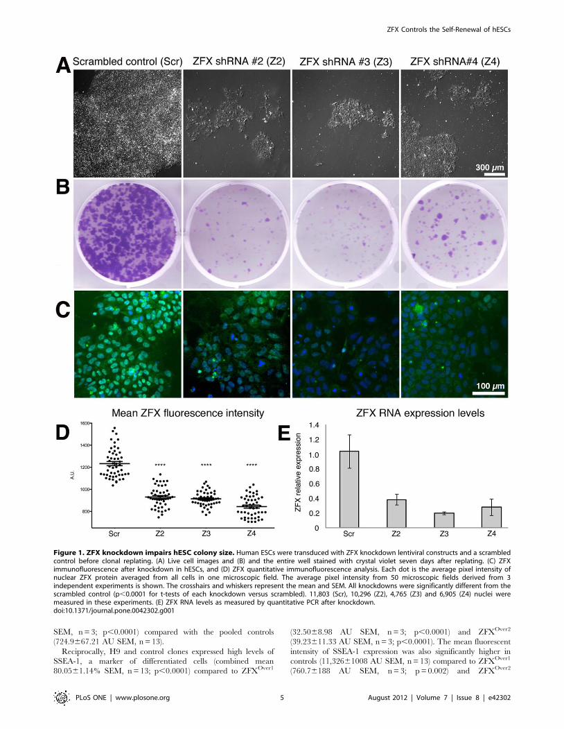
cin selection. All 3 of the vetted ZFX knockdowns caused a
marked decrease in the number and size of colonies relative to the
scrambled shRNA control (Figure 1A and 1B). We confirmed that
ZFX RNA (Figure 1C) and protein (Figure 1D) levels were
reduced. This data suggests that ZFX expression is required for
the efficient self-renewal of hESCs similar to its role in mESCs. We
next turned to ZFX overexpression since the ZFX knockdown
cells could not be readily expanded, confounding analysis.
Production of ZFX Overexpressing Human ESCsSimilar to many transcription factors, the overexpression of
ZFX cDNA from a heterologous promoter is toxic to cells (B.R.,
unpublished data). To overexpress murine Zfx under its native
regulation, the entire genomic Zfx locus has been introduced into
mESC as a bacterial artificial chromosome (BAC) transgene [13].
We have developed a method for stable BAC introduction into
hESC to serve as reporter transgenes [17]. We adapted this
approach to introduce additional copies of the human ZFX locus
as BAC transgene in the H9 hESC. Several nucleofected H9
clones showed increased ZFX mRNA expression by quantitative
RT-PCR (Figure 2A). We chose two clones (ZFXOver1 and
ZFXOver2) with the highest ZFX expression levels and compared
them to the parental H9 line, a control clone (ZFXNormal) with
normal ZFX expression levels from the same experiment, as well
as independent H9 GFP and YFP-expressing BAC transgenic
reporter lines from our earlier studies (see Table 1 for a
comprehensive description of controls used in this study) [17,20].
We verified that the BAC reporter control clones expressed
normal amounts of ZFX (data not shown).
As shown in Figure 2B, ZFXOver clones expressed more ZFX
protein than the parental hESC line (H9) or ZFXNormal (Figure 2B).
Fluorescent in situ hybridization (FISH) showed that the ZFXOver
clones harbored multiple copies of the BAC integrated at a single
site while ZFXNormal lacked detectable ZFX BAC DNA despite
being drug resistant (Figure S2). Furthermore, ZFXOver2, the
control clone and the H9 parental line had normal karyotypes,
whereas ZFXOver1 showed a mixture of normal cells and
karyotypically abnormal cells (data not shown). Stem cell surface
markers on each clone looked similar to H9: they were positive for
hESC markers SSEA-4+ and Oct-4+ and variegated for Tra1-81
(Figure 2C; Figure S3) suggesting that all clonal lines are in a
similar undifferentiated state.
ZFX-overexpressing hESC are More ClonogenicUnder standard growth conditions, all hESC clones had similar
morphologies, growth and apoptosis rates as the parental H9
hESC line (Figure S4). To quantitatively assess the self-renewal
capacity of ZFXOver versus ZFXNormal and H9 hESCs at the
undifferentiated stage, we analyzed their growth behavior
following plating cells at clonal densities. The mean number of
colonies formed from ZFXOver clones was significantly higher than
controls (114.00611.15 for ZFXOver1 and 102.3615.03 for
ZFXOver2 compared to 41.0069.16 for H9 and ZFXNormal.
p = 0.0020 for ZFXOver1 and 0.0078 for ZFXOver2). In addition to
colony number, the size of the ZFXOver colonies derived from
single cells was larger compared to controls (Figure 3 and Figure
S1). These data demonstrate that ZFX overexpression in hESCs
results in cultures enriched in colony-forming cells, a sensitive
assay of the undifferentiated state [22].
Reduced Spontaneous Differentiation in ZFXOver cellsNext we tested the growth of ZFXOver clones under conditions
suboptimal for hESC self-renewal. ZFXOver and control hESC
lines were cultured in the absence of conditioned media or feeder
cells to promote spontaneous differentiation, and the percentage of
cells expressing SSEA-3 and SSEA-1 was measured to quantify the
ratio of undifferentiated versus differentiated cells, respectively. In
control experiments, there was no statistical difference in marker
expression when conditioned media was used to expand cells
without feeders (data not shown). However, in suboptimal
conditions, H9 and the control clones showed strong signs of
differentiation while the ZFXOver cells resisted spontaneous
differentiation (Figure 4; Figure S5). The control cultures had
low percentages of the undifferentiated hESC marker SSEA-3
(combined mean of 33.1262.62% SEM; n = 13) while ZFXOver1
(61.5763.48% SEM, n = 3; p = 0.0002) and ZFXOver2
(59.2063.30% SEM, n = 3; p = 0.0005) maintained significantly
higher levels of SSEA-3. Similarly, the mean fluorescence intensity
was significantly higher in ZFXOver1 (22736454.7 arbitrary units,
or AU SEM, n = 3; p,0.0001) and ZFXOver2 (20336362.3 AU
ZFX Controls the Self-Renewal of hESCs
PLoS ONE | www.plosone.org 4 August 2012 | Volume 7 | Issue 8 | e42302
SEM, n = 3; p,0.0001) compared with the pooled controls
(724.9667.21 AU SEM, n = 13).
Reciprocally, H9 and control clones expressed high levels of
SSEA-1, a marker of differentiated cells (combined mean
80.0561.14% SEM, n = 13; p,0.0001) compared to ZFXOver1
(32.5068.98 AU SEM, n = 3; p,0.0001) and ZFXOver2
(39.23611.33 AU SEM, n = 3; p,0.0001). The mean fluorescent
intensity of SSEA-1 expression was also significantly higher in
controls (11,32661008 AU SEM, n = 13) compared to ZFXOver1
(760.76188 AU SEM, n = 3; p = 0.002) and ZFXOver2
Figure 1. ZFX knockdown impairs hESC colony size. Human ESCs were transduced with ZFX knockdown lentiviral constructs and a scrambledcontrol before clonal replating. (A) Live cell images and (B) and the entire well stained with crystal violet seven days after replating. (C) ZFXimmunofluorescence after knockdown in hESCs, and (D) ZFX quantitative immunofluorescence analysis. Each dot is the average pixel intensity ofnuclear ZFX protein averaged from all cells in one microscopic field. The average pixel intensity from 50 microscopic fields derived from 3independent experiments is shown. The crosshairs and whiskers represent the mean and SEM. All knockdowns were significantly different from thescrambled control (p,0.0001 for t-tests of each knockdown versus scrambled). 11,803 (Scr), 10,296 (Z2), 4,765 (Z3) and 6,905 (Z4) nuclei weremeasured in these experiments. (E) ZFX RNA levels as measured by quantitative PCR after knockdown.doi:10.1371/journal.pone.0042302.g001
ZFX Controls the Self-Renewal of hESCs
PLoS ONE | www.plosone.org 5 August 2012 | Volume 7 | Issue 8 | e42302
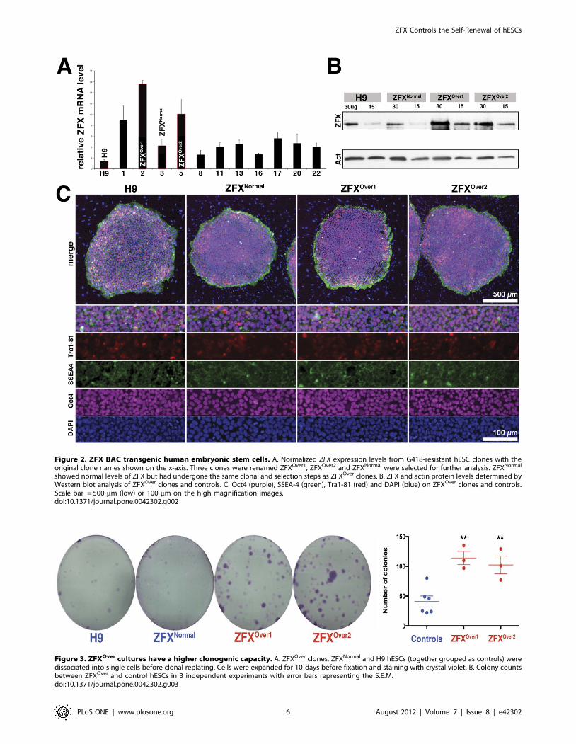
Figure 2. ZFX BAC transgenic human embryonic stem cells. A. Normalized ZFX expression levels from G418-resistant hESC clones with theoriginal clone names shown on the x-axis. Three clones were renamed ZFXOver1, ZFXOver2 and ZFXNormal were selected for further analysis. ZFXNormal
showed normal levels of ZFX but had undergone the same clonal and selection steps as ZFXOver clones. B. ZFX and actin protein levels determined byWestern blot analysis of ZFXOver clones and controls. C. Oct4 (purple), SSEA-4 (green), Tra1-81 (red) and DAPI (blue) on ZFXOver clones and controls.Scale bar = 500 mm (low) or 100 mm on the high magnification images.doi:10.1371/journal.pone.0042302.g002
Figure 3. ZFXOver cultures have a higher clonogenic capacity. A. ZFXOver clones, ZFXNormal and H9 hESCs (together grouped as controls) weredissociated into single cells before clonal replating. Cells were expanded for 10 days before fixation and staining with crystal violet. B. Colony countsbetween ZFXOver and control hESCs in 3 independent experiments with error bars representing the S.E.M.doi:10.1371/journal.pone.0042302.g003
ZFX Controls the Self-Renewal of hESCs
PLoS ONE | www.plosone.org 6 August 2012 | Volume 7 | Issue 8 | e42302
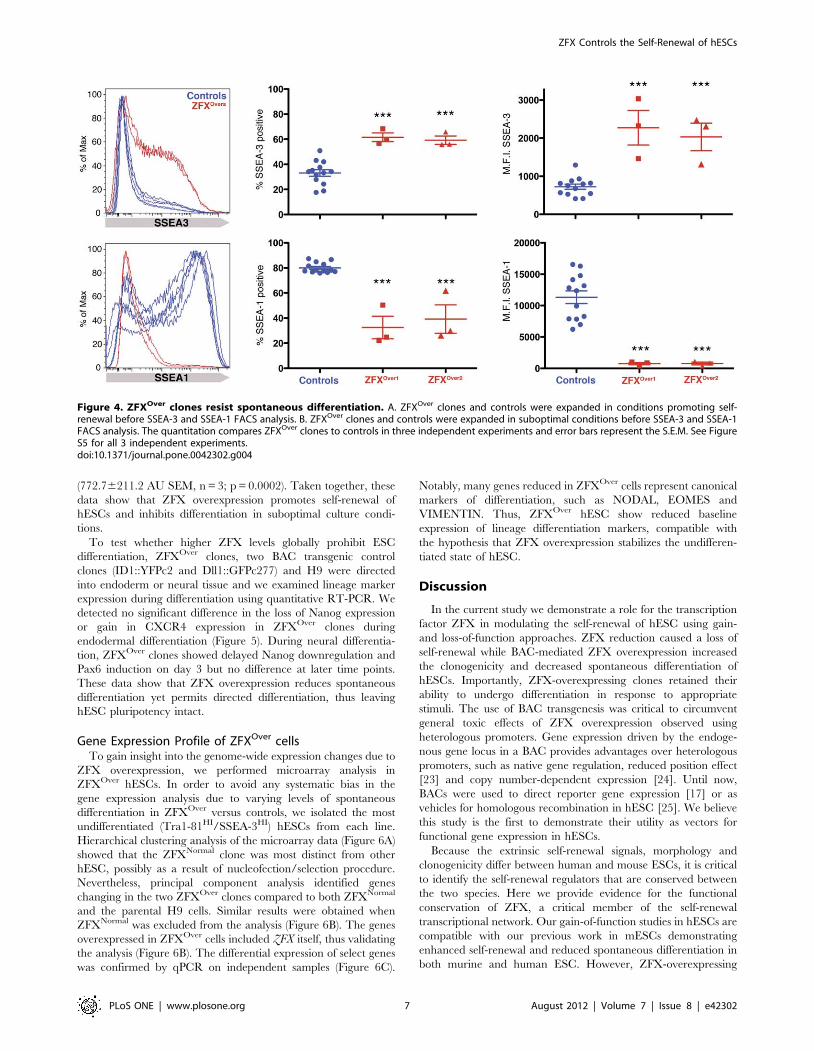
(772.76211.2 AU SEM, n = 3; p = 0.0002). Taken together, these
data show that ZFX overexpression promotes self-renewal of
hESCs and inhibits differentiation in suboptimal culture condi-
tions.
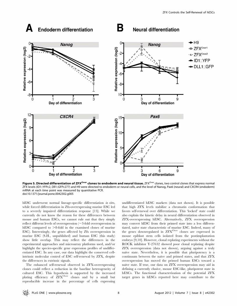
To test whether higher ZFX levels globally prohibit ESC
differentiation, ZFXOver clones, two BAC transgenic control
clones (ID1::YFPc2 and Dll1::GFPc277) and H9 were directed
into endoderm or neural tissue and we examined lineage marker
expression during differentiation using quantitative RT-PCR. We
detected no significant difference in the loss of Nanog expression
or gain in CXCR4 expression in ZFXOver clones during
endodermal differentiation (Figure 5). During neural differentia-
tion, ZFXOver clones showed delayed Nanog downregulation and
Pax6 induction on day 3 but no difference at later time points.
These data show that ZFX overexpression reduces spontaneous
differentiation yet permits directed differentiation, thus leaving
hESC pluripotency intact.
Gene Expression Profile of ZFXOver cellsTo gain insight into the genome-wide expression changes due to
ZFX overexpression, we performed microarray analysis in
ZFXOver hESCs. In order to avoid any systematic bias in the
gene expression analysis due to varying levels of spontaneous
differentiation in ZFXOver versus controls, we isolated the most
undifferentiated (Tra1-81HI/SSEA-3HI) hESCs from each line.
Hierarchical clustering analysis of the microarray data (Figure 6A)
showed that the ZFXNormal clone was most distinct from other
hESC, possibly as a result of nucleofection/selection procedure.
Nevertheless, principal component analysis identified genes
changing in the two ZFXOver clones compared to both ZFXNormal
and the parental H9 cells. Similar results were obtained when
ZFXNormal was excluded from the analysis (Figure 6B). The genes
overexpressed in ZFXOver cells included ZFX itself, thus validating
the analysis (Figure 6B). The differential expression of select genes
was confirmed by qPCR on independent samples (Figure 6C).
Notably, many genes reduced in ZFXOver cells represent canonical
markers of differentiation, such as NODAL, EOMES and
VIMENTIN. Thus, ZFXOver hESC show reduced baseline
expression of lineage differentiation markers, compatible with
the hypothesis that ZFX overexpression stabilizes the undifferen-
tiated state of hESC.
Discussion
In the current study we demonstrate a role for the transcription
factor ZFX in modulating the self-renewal of hESC using gain-
and loss-of-function approaches. ZFX reduction caused a loss of
self-renewal while BAC-mediated ZFX overexpression increased
the clonogenicity and decreased spontaneous differentiation of
hESCs. Importantly, ZFX-overexpressing clones retained their
ability to undergo differentiation in response to appropriate
stimuli. The use of BAC transgenesis was critical to circumvent
general toxic effects of ZFX overexpression observed using
heterologous promoters. Gene expression driven by the endoge-
nous gene locus in a BAC provides advantages over heterologous
promoters, such as native gene regulation, reduced position effect
[23] and copy number-dependent expression [24]. Until now,
BACs were used to direct reporter gene expression [17] or as
vehicles for homologous recombination in hESC [25]. We believe
this study is the first to demonstrate their utility as vectors for
functional gene expression in hESCs.
Because the extrinsic self-renewal signals, morphology and
clonogenicity differ between human and mouse ESCs, it is critical
to identify the self-renewal regulators that are conserved between
the two species. Here we provide evidence for the functional
conservation of ZFX, a critical member of the self-renewal
transcriptional network. Our gain-of-function studies in hESCs are
compatible with our previous work in mESCs demonstrating
enhanced self-renewal and reduced spontaneous differentiation in
both murine and human ESC. However, ZFX-overexpressing
Figure 4. ZFXOver clones resist spontaneous differentiation. A. ZFXOver clones and controls were expanded in conditions promoting self-renewal before SSEA-3 and SSEA-1 FACS analysis. B. ZFXOver clones and controls were expanded in suboptimal conditions before SSEA-3 and SSEA-1FACS analysis. The quantitation compares ZFXOver clones to controls in three independent experiments and error bars represent the S.E.M. See FigureS5 for all 3 independent experiments.doi:10.1371/journal.pone.0042302.g004
ZFX Controls the Self-Renewal of hESCs
PLoS ONE | www.plosone.org 7 August 2012 | Volume 7 | Issue 8 | e42302
hESC underwent normal lineage-specific differentiation in vitro,
while forced differentiation in Zfx-overexpressing murine ESC led
to a severely impaired differentiation response [13]. While we
currently do not know the reason for these differences between
mouse and human ESCs, we cannot rule out that they simply
reflect different levels of overexpression (,3-fold overexpression in
hESC compared to .8-fold in the examined clones of murine
ESC). Interestingly, the genes affected by Zfx overexpression in
murine ESC (S.H., unpublished) and human ESC (this study)
show little overlap. This may reflect the differences in the
experimental approaches and microarray platforms used, and/or
highlight the species-specific gene expression profiles of undiffer-
entiated ESC. In any case, our data highlight the conserved cell-
intrinsic molecular control of ESC self-renewal by ZFX, despite
the differences in extrinsic signals.
The enhanced self-renewal observed in ZFX-overexpressing
clones could reflect a reduction in the baseline heterogeneity of
cultured ESC. This hypothesis is supported by the increased
plating efficiency of ZFXOver clones and by a small but
reproducible increase in the percentage of cells expressing
undifferentiated hESC markers (data not shown). It is possible
that high ZFX levels stabilize a chromatin conformation that
favors self-renewal over differentiation. This ‘locked’ state could
also explain the kinetic delay in neural differentiation observed in
ZFX-overexpressing hESC. Alternatively, ZFX overexpression
may convert hESC from their primed state into a less differen-
tiated, naı̈ve state characteristic of murine ESC. Indeed, many of
the genes downregulated in ZFXOver clones are expressed in
mouse epiblast stem cells isolated from the postimplantation
embryo [9,10]. However, clonal replating experiments without the
ROCK inhibitor Y-27632 showed poor clonal replating despite
ZFX overexpression (data not shown), arguing against a truly
naı̈ve state. Nevertheless, it is possible that pluripotency is a
continuum between the naı̈ve and primed states, and that ZFX
overexpression has moved the primed human ESCs toward a
naı̈ve state. If true, our data on ZFX overexpression may aid in
defining a currently elusive, mouse ESC-like, pluripotent state in
hESCs. The functional characterization of the potential ZFX
target genes in hESCs reported here may contribute to the
Figure 5. Directed differentiation of ZFXOver clones to endoderm and neural tissue. ZFXOver clones, two control clones that express normalZFX levels (ID1::YFPc2, Dll1::GFPc277) and H9 were directed to endoderm or neural cells, and the level of Nanog, Pax6 (neural) and CXCR4 (endoderm)mRNA at each time point was measured by quantitative PCR.doi:10.1371/journal.pone.0042302.g005
ZFX Controls the Self-Renewal of hESCs
PLoS ONE | www.plosone.org 8 August 2012 | Volume 7 | Issue 8 | e42302
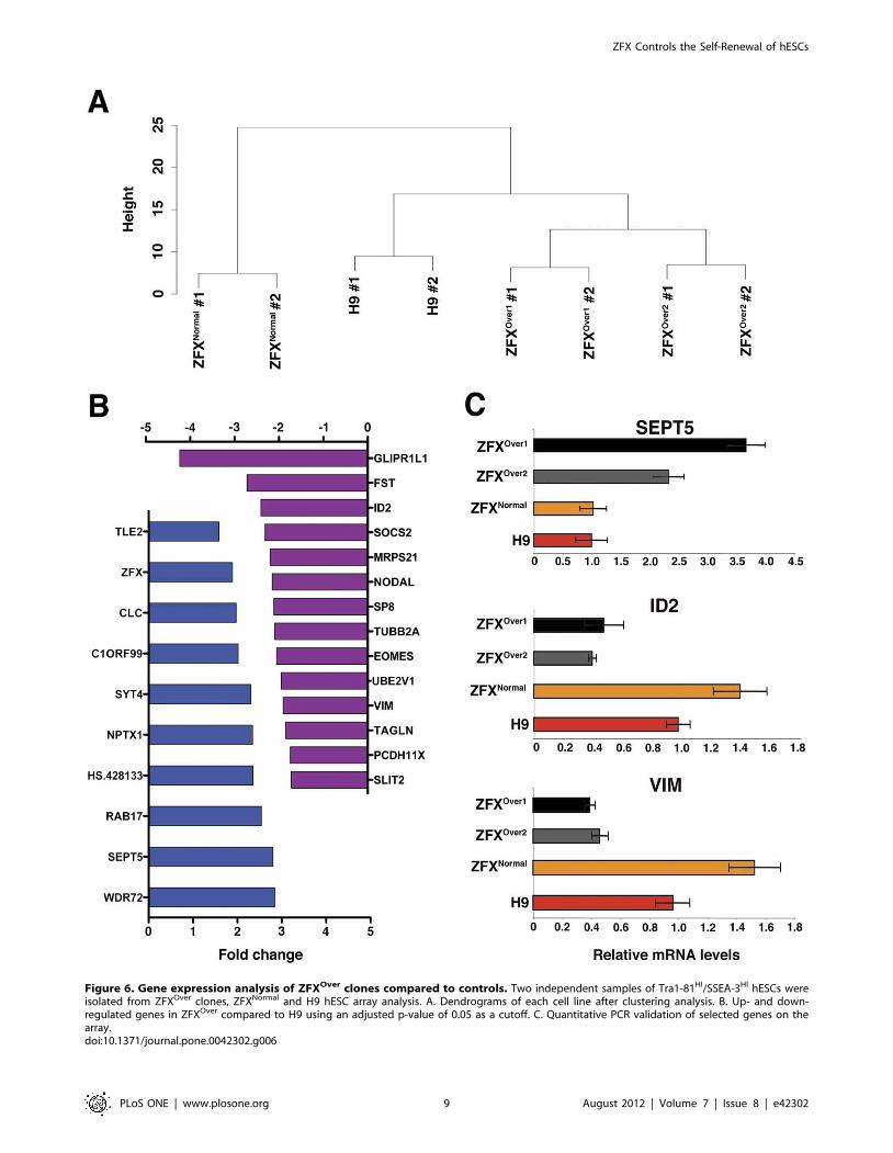
Figure 6. Gene expression analysis of ZFXOver clones compared to controls. Two independent samples of Tra1-81HI/SSEA-3HI hESCs wereisolated from ZFXOver clones, ZFXNormal and H9 hESC array analysis. A. Dendrograms of each cell line after clustering analysis. B. Up- and down-regulated genes in ZFXOver compared to H9 using an adjusted p-value of 0.05 as a cutoff. C. Quantitative PCR validation of selected genes on thearray.doi:10.1371/journal.pone.0042302.g006
ZFX Controls the Self-Renewal of hESCs
PLoS ONE | www.plosone.org 9 August 2012 | Volume 7 | Issue 8 | e42302
identification of key evolutionarily conserved components of the
ESC self-renewal machinery.
Supporting Information
Figure S1 Nearly all cells that survive the colony-forming assay are pluripotent. A. Alkaline phosphatase
staining of the clonal colony-forming assay. B. Cells were grown
clonally in the colony forming assay before single cell dissociation,
fixation, permeabilization and staining with Oct4-Alexa647 before
FACS analysis.
(TIF)
Figure S2 ZFX BAC transgenic human embryonic stemcells characterization. Full images from Figure 2, showing
Oct4 (purple), SSEA-4 (green) Tra1-81 (red) and DAPI (blue) on
ZFXOver clones and controls. Scale bar = 100 mm.
(TIF)
Figure S3 ZFX BAC FISH analysis identifies multiplecopies of the BAC in each ZFXOver clone at a singleintegration site. ZFXNormal and H9 show only endogenous
ZFX whereas both ZFXOver clones show endogenous (green
arrows) an additional brighter spot (red arrows) revealing the ZFX
BAC integration.
(TIF)
Figure S4 ZFXOver clones do not have differences in cellgrowth nor apoptosis during normal passage. A. hESC
lines were passaged on day 0 using dispase and were counted the
day after passage to assess seeding (day 1). Six days later, cells were
counted to assess growth kinetics (day 7). On day 7, cells were
passaged again using dispase and counted the day after (day 8).
The same process was repeated on days 14 and 15. B. hESC lines
were grown in feeder free conditions and the number of cells with
AnnexinV staining were quantitated. No differences were
observed after dispase passage. Three independent experiments
were performed both for A and B and the data were not
significantly different in either case.
(TIF)
Figure S5 All data from the spontaneous differentiationexperiment shown in Figure 4. Three independent experi-
ments showing the level of SSEA-1 and SSEA-3 after 7 days of
suboptimal culture. The mean fluorescent intensity is diagrammed
on the right side of the figure: controls are grouped on the left side,
ZFXOver1 in the middle and ZFXOver2 on the right.
(TIF)
Acknowledgments
We thank Margaret Leversha and Lei Zhang (Molecular Cytogenetics
Core Lab at MSKCC) for the BAC FISH and karyotyping. We thank
Lorraine Clark for the use of the Illumina Analyzer.
Author Contributions
Conceived and designed the experiments: SH LS BR MJT. Performed the
experiments: SH EYT MJT BL. Analyzed the data: SH EYT SMC LS BR
MJT BL. Contributed reagents/materials/analysis tools: SH SW ME SMC
CTC BR. Wrote the paper: SH LS BR MJT.
References
1. Evans MJ, Kaufman MH (1981) Establishment in culture of pluripotential cells
from mouse embryos. Nature 292: 154–156.2. Martin GR (1981) Isolation of a pluripotent cell line from early mouse embryos
cultured in medium conditioned by teratocarcinoma stem cells. Proc Natl Acad
Sci U S A 78: 7634–7638.3. Thomson JA, Itskovitz-Eldor J, Shapiro SS, Waknitz MA, Swiergiel JJ, et al.
(1998) Embryonic stem cell lines derived from human blastocysts. Science 282:1145–1147.
4. Chambers I, Tomlinson SR (2009) The transcriptional foundation of
pluripotency. Development 136: 2311–2322.5. Do JT, Scholer HR (2009) Regulatory circuits underlying pluripotency and
reprogramming. Trends Pharmacol Sci 30: 296–302.6. Takahashi K, Tanabe K, Ohnuki M, Narita M, Ichisaka T, et al. (2007)
Induction of pluripotent stem cells from adult human fibroblasts by definedfactors. Cell 131: 861–872.
7. Yu J, Vodyanik MA, Smuga-Otto K, Antosiewicz-Bourget J, Frane JL, et al.
(2007) Induced pluripotent stem cell lines derived from human somatic cells.Science 318: 1917–1920.
8. Rossant J (2007) Stem cells and lineage development in the mammalianblastocyst. Reprod Fertil Dev 19: 111–118.
9. Brons IG, Smithers LE, Trotter MW, Rugg-Gunn P, Sun B, et al. (2007)
Derivation of pluripotent epiblast stem cells from mammalian embryos. Nature448: 191–195.
10. Tesar PJ, Chenoweth JG, Brook FA, Davies TJ, Evans EP, et al. (2007) New celllines from mouse epiblast share defining features with human embryonic stem
cells. Nature 448: 196–199.
11. Hanna J, Cheng AW, Saha K, Kim J, Lengner CJ, et al. (2010) Humanembryonic stem cells with biological and epigenetic characteristics similar to
those of mouse ESCs. Proc Natl Acad Sci U S A 107: 9222–9227.12. Xu Y, Zhu X, Hahm HS, Wei W, Hao E, et al. (2010) Revealing a core signaling
regulatory mechanism for pluripotent stem cell survival and self-renewal by smallmolecules. Proc Natl Acad Sci U S A 107: 8129–8134.
13. Galan-Caridad JM, Harel S, Arenzana TL, Hou ZE, Doetsch FK, et al. (2007)
Zfx controls the self-renewal of embryonic and hematopoietic stem cells. Cell129: 345–357.
14. Chen X, Xu H, Yuan P, Fang F, Huss M, et al. (2008) Integration of externalsignaling pathways with the core transcriptional network in embryonic stem
cells. Cell 133: 1106–1117.
15. Hu G, Kim J, Xu Q, Leng Y, Orkin SH, et al. (2009) A genome-wide RNAi
screen identifies a new transcriptional module required for self-renewal. Genes
Dev 23: 837–848.
16. Ouyang Z, Zhou Q, Wong WH (2009) ChIP-Seq of transcription factors
predicts absolute and differential gene expression in embryonic stem cells. Proc
Natl Acad Sci U S A 106: 21521–21526.
17. Placantonakis DG, Tomishima MJ, Lafaille F, Desbordes SC, Jia F, et al. (2009)
BAC transgenesis in human embryonic stem cells as a novel tool to define the
human neural lineage. Stem Cells 27: 521–532.
18. Perrier AL, Tabar V, Barberi T, Rubio ME, Bruses J, et al. (2004) Derivation of
midbrain dopamine neurons from human embryonic stem cells. Proc Natl Acad
Sci U S A 101: 12543–12548.
19. Wang Z, Engler P, Longacre A, Storb U (2001) An efficient method for high-
fidelity BAC/PAC retrofitting with a selectable marker for mammalian cell
transfection. Genome Res 11: 137–142.
20. James D, Nam HS, Seandel M, Nolan D, Janovitz T, et al. (2010) Expansion
and maintenance of human embryonic stem cell-derived endothelial cells by
TGFbeta inhibition is Id1 dependent. Nat Biotechnol 28: 161–166.
21. D’Amour KA, Agulnick AD, Eliazer S, Kelly OG, Kroon E, et al. (2005)
Efficient differentiation of human embryonic stem cells to definitive endoderm.
Nat Biotechnol 23: 1534–1541.
22. O’Connor MD, Kardel MD, Iosfina I, Youssef D, Lu M, et al. (2008) Alkaline
phosphatase-positive colony formation is a sensitive, specific, and quantitative
indicator of undifferentiated human embryonic stem cells. Stem Cells 26: 1109–
1116.
23. Lee EC, Yu D, Martinez de Velasco J, Tessarollo L, Swing DA, et al. (2001) A
highly efficient Escherichia coli-based chromosome engineering system adapted
for recombinogenic targeting and subcloning of BAC DNA. Genomics 73: 56–
65.
24. Yang XW, Wynder C, Doughty ML, Heintz N (1999) BAC-mediated gene-
dosage analysis reveals a role for Zipro1 (Ru49/Zfp38) in progenitor cell
proliferation in cerebellum and skin. Nat Genet 22: 327–335.
25. Song H, Chung SK, Xu Y (2010) Modeling disease in human ESCs using an
efficient BAC-based homologous recombination system. Cell Stem Cell 6: 80–
89.
ZFX Controls the Self-Renewal of hESCs
PLoS ONE | www.plosone.org 10 August 2012 | Volume 7 | Issue 8 | e42302
Related Documents


