THE NEUROETHOLOGY AND EVOLUTION OF NEST- BUILDING BEHAVIOUR Zachary Hall A Thesis Submitted for the Degree of PhD at the University of St Andrews 2014 Full metadata for this item is available in St Andrews Research Repository at: http://research-repository.st-andrews.ac.uk/ Please use this identifier to cite or link to this item: http://hdl.handle.net/10023/5542 This item is protected by original copyright

Welcome message from author
This document is posted to help you gain knowledge. Please leave a comment to let me know what you think about it! Share it to your friends and learn new things together.
Transcript
THE NEUROETHOLOGY AND EVOLUTION OF NEST-BUILDING BEHAVIOUR
Zachary Hall
A Thesis Submitted for the Degree of PhD
at the University of St Andrews
2014
Full metadata for this item is available in St Andrews Research Repository
at: http://research-repository.st-andrews.ac.uk/
Please use this identifier to cite or link to this item: http://hdl.handle.net/10023/5542
This item is protected by original copyright
The neuroethology and evolution of nest-building behaviour
Zachary Hall
This thesis is submitted in partial fulfilment for the degree of PhD at the
University of St Andrews
September 2014

1. Candidate’s declarations: I, Zachary Hall hereby certify that this thesis, which is approximately 38,000 words in length, has been written by me, and that it is the record of work carried out by me, or principally by myself in collaboration with others as acknowledged, and that it has not been submitted in any previous application for a higher degree. I was admitted as a research student in September 2011 and as a candidate for the degree of PhD in September 2011; the higher study for which this is a record was carried out in the University of St Andrews between 2011 and 2014. Date Signature of candidate 2. Supervisor’s declaration: I hereby certify that the candidate has fulfilled the conditions of the Resolution and Regulations appropriate for the degree of PhD in the University of St Andrews and that the candidate is qualified to submit this thesis in application for that degree. Date Signature of supervisor 3. Permission for publication: (to be signed by both candidate and supervisor) In submitting this thesis to the University of St Andrews I understand that I am giving permission for it to be made available for use in accordance with the regulations of the University Library for the time being in force, subject to any copyright vested in the work not being affected thereby. I also understand that the title and the abstract will be published, and that a copy of the work may be made and supplied to any bona fide library or research worker, that my thesis will be electronically accessible for personal or research use unless exempt by award of an embargo as requested below, and that the library has the right to migrate my thesis into new electronic forms as required to ensure continued access to the thesis. I have obtained any third-party copyright permissions that may be required in order to allow such access and migration, or have requested the appropriate embargo below. The following is an agreed request by candidate and supervisor regarding the publication of this thesis: PRINTED COPY
b) Embargo on all or part of print copy for a period of 2 years from the date the thesis is lodged in the University Library on the following ground(s): • Publication would preclude future publication

Supporting statement for printed embargo request:
• Publication would preclude future publication ELECTRONIC COPY b) Embargo on all or part of print copy for a period of 2 years from the date the thesis is
lodged in the University Library on the following ground(s): • Publication would preclude future publication
Supporting statement for electronic embargo request:
• Publication would preclude future publication Date Signature of candidate Signature of supervisor

Declaration of publications The work described in chapter 2 forms the basis of “Hall ZJ, Bertin M, Bailey IE, Meddle SL, Healy SD (2014) Neural correlates of nesting behaviour in zebra finches (Taeniopygia guttata). Behaviour Brain Research 264:26-33.” Chapter 3 will form part of the following manuscript: Hall ZJ, Healy SD, Meddle SL. A role for nonapeptides and dopamine in nest-building behaviour. The work described in chapter 4 forms the basis of “Hall ZJ, Street SE, Healy SD (2013) The evolution of cerebellum structure correlates with nest complexity. Biology Letters 9: 20130687. Chapter 5 will form part of the following manuscript: Hall ZJ, Street SE, Healy SD. Co-evolution of nest structure and location in Old World babblers (Timaliidae). Declaration of collaboration I collected all data with the exception of female behavioural data in Chapter 2. These data were collected by the undergraduate student Marion Bertin, under the supervision of me and Dr. Susan Healy. Ida Bailey provided advice regarding the statistical analysis performed in chapter 2. Simone Meddle provided advice regarding immunohistochemical techniques performed in chapters 2 and 3. Finally, Sally Street provided advice regarding the statistical analyses performed in Chapters 4 and 5.
Abstract A surge of recent work elucidating a role for learning and memory in avian nest-
building behaviour has challenged the long-standing assumption that nest building develops
under genetic control. Whereas that work has been addressed at describing the cognitive
mechanisms underpinning nest-building behaviour, almost nothing is known about either
the neurobiological processes controlling nest building or the selection pressures
responsible for the diversity in avian nest-building behaviour. Here, I sought to identify
both the neural substrates involved in nest-building behaviour and some of those selection
pressures. First, I used expression of the immediate early gene product Fos, an indirect
marker of neuronal activity, to identify brain regions activated during nest-building
behaviour in the brains of nest-building and control zebra finches (Taeniogypia guttata). I
found that neural circuits involved in motor control, social behaviour, and reward were
activated during nest building. Furthermore, I found that subpopulations of neurons that
signal using the nonapeptides vasotocin and mesotocin and the neurotransmitter dopamine
located within some of these neural circuits were also activated during nest building,
suggesting these cell-signalling molecules may be involved in controlling nest-building
behaviour. Next, I found that variation in the amount of folding in the cerebellum, a brain
structure thought to be involved in manipulative skills, increased with increasing nest
structural complexity, suggesting that the cerebellum is also involved in nest building.
Finally, using evolutionary statistical models, I found support for the hypothesis that nest-
site competition off-ground and increased predation pressure on the ground in Old World
babblers (Timaliidae) led to the co-evolution of building domed nests on the ground. Here,

then, I provide the first evidence of potential neural substrates controlling and selection
pressures contributing to variation in nest-building behaviour.
Acknowledgements
Firstly, I would like to thank my supervisor, Susan Healy, for her unending help and support throughout my tenure at the University of St. Andrews. Because of Sue’s courage and confidence in my abilities, I was able to amass a body of work addressing a topic that some deemed too risky to be central to a PhD thesis. I hope that I will be able to maintain collaborations with such an inspirational researcher and that the work I was able to complete while in St. Andrews recompensed her for at least a fraction of all that she has taught me. I would also like to thank all of my collaborators, who taught me the techniques that were vital to the integrative approach to studying nest building presented here. Most notably, I would like to thank Simone Meddle for all of her patience and help in improving my lab techniques and Sally Street, who taught me the phylogenetic comparative statistics crucial to my evolutionary work. I would also like to thank Chris Vendetti and Daniel Barker for their help and advice regarding the statistical approaches used in my comparative analyses. For insightful and inspiring discussion about my research, I would like to thank Rob Barton, Dave Shukar, David Sherry, and Scott MacDougall-Shackleton. I want to thank all of the members of my lab group who had the unfortunate fate of working in the lab over the course of my entire PhD tenure: David Pritchard, María Cristina Tello Ramos, and Kate Morgan. I also want to thank all other lab group members, past and present, Lauren Guillette, Ida Bailey, Eira Ihalainen, Georgina Glaser, Felicity Muth, Rachael Marshall, Guill McIvor, and Nuri Flores Abreu for their help and discussion. Thank you to my family, Mom, Dad, Hannah, Otis, Milo, Kramer, and Waldo, for their love, support, and willingness to at least try and read some of my publications. Thank you to my favourite border terrier, Fidra, for helping me get the least amount of work done while visiting Simone’s lab. Thanks to my best friend, Nick, for tolerating me talking about brains while we are trying to play video games. Thanks to Luvian’s for a seemingly endless source of new beer and thanks to Ben and Sean for getting mad at the same things that I get mad at. Finally, and most importantly, I would like to thank every bird included in my studies. You were all clever to me, even if you weren’t crows or feathered apes or whatever.

Ethical note
All experimental procedures in this thesis were performed with ethical permission
from the University of St. Andrews Animal Welfare and Ethics Committee and from the
UK Home Office (PPL. 60/3666).
Table of Contents
Chapter 1: Introduction ...................................................................................................... 1
Chapter 2: Neural correlates of nest-building behaviour in zebra finches .................. 26
Introduction ................................................................................................................................... 26
Methods and materials .................................................................................................................. 29
Results ........................................................................................................................................... 40
Discussion ..................................................................................................................................... 49
Chapter 3: A role for nonapeptides and dopamine in nest-building behaviour .......... 56
Introduction ................................................................................................................................... 56
Methods and materials .................................................................................................................. 60
Results ........................................................................................................................................... 66
Discussion ..................................................................................................................................... 71
Chapter 4: The evolution of cerebellum structure and nest complexity ...................... 79
Introduction ................................................................................................................................... 79
Methods and materials .................................................................................................................. 80
Results ........................................................................................................................................... 88
Discussion ..................................................................................................................................... 90
Chapter 5: Co-evolution of nest structure with location ................................................ 93
Introduction ................................................................................................................................... 93
Methods and materials .................................................................................................................. 95
Results ......................................................................................................................................... 104
Discussion ................................................................................................................................... 108
Chapter 6: General discussion ........................................................................................ 113
Bibliography ..................................................................................................................... 127
Appendices ....................................................................................................................... 145
Appendix 1 – Chapter 2 regression models ................................................................................ 145
Appendix 2 – Chapter 3 regression models ................................................................................ 154
Appendix 3 – Chapter 4 data ....................................................................................................... 158
Appendix 4 – Chapter 5 data ....................................................................................................... 161
Chapter 1 1
Chapter 1: Introduction
Of all the constructions made by non-human animals, perhaps none are as widely
recognised as the nests built by birds. From the sewing behaviour of the common tailorbird
(Orthotomus sutorius), which stitches together leaves to form a nest cup later filled with
insulating material (Nguembock et al., 2007) through the famous weaving and thatching
abilities of weaver birds (Ploceidae; Collias and Collias, 1964) to the unique nest
construction of the Horned Coot (Fulica cornuta), which deposits upwards of 1 ton of
pebbles in bodies of water to form a nesting island before constructing a nest cup
(McFarlane, 1975), the daunting diversity in nest-building behaviour has long been
celebrated by the likes of Wallace (1867), Tinbergen (1953), and Thorpe (1956). Despite
the ongoing accumulation of nest structure descriptions for the majority of extant, known
bird species, as seen in the Handbook of Birds of the World book series (for example, del
Hoyo et al., 1992), it is then perhaps surprising that so few researchers have sought to
elucidate the mechanisms underlying how birds construct nests and why there is such
structural diversity in nests across species.
Amongst the handful of studies in which the way birds construct nests has been
addressed, research effort has been focused almost entirely on the role of learning and
experience in nest building. Historically, nest building was assumed to be an innate
behaviour under genetic control and unaffected by experience (Healy et al., 2008). For
example, in Descent of Man, Charles Darwin stated that, in contrast to human skills, which
improve with practice, inexperienced birds will construct nests comparable to those of
experienced builders on their first attempt (Darwin, 1882). Experimentally, this view

Chapter 1 2
received early support from studies in which hand-reared birds, deprived of nest material
during development and first exposed to nest material as adults, were reported to construct
nests resembling those built by experienced builders. For example, hand-reared female
canaries (Serinus canaria) deprived of nesting material during development constructed
species-typical nests upon their first exposure to nest material in adulthood (Hinde and
Matthews, 1958). It should be noted, however, that this finding conflicts with earlier,
similar experiments in which hand-reared American Robins (Turdus migratorius) and
Rose-breasted Grosbeaks (Pheuticus ludovicianus) failed to construct species-typical nests
upon their first exposure to nest material in adulthood (Scott, 1902; 1904).
Soon after Hinde and Matthew’s work on canaries, Collias and Collias (1962; 1964),
displeased with the limitations of describing the mechanisms underlying nest building as
innate, published a series of studies on the nest-building behaviour of African Village
weaver birds (Ploceus cucullatus) in the wild and captivity. In one of the strongest
challenges to a (still-prevalent) genetic-only origin of nest-building behaviour, Collias and
Collias (1964) documented the development of weaving abilities in hand-reared and aviary-
reared weaver birds, reporting a significant effect of experience with nest material during
development on subsequent nest material preferences and construction behaviour.
Specifically, hand-reared weaver birds deprived of experience with nest material exhibited
weaker preferences for the longer, flexible, green nest material than did experienced weaver
birds and were also less able to weave material successfully into the aviary cage and trees.
When these naive birds were given experience with nest material and tested again months
later, they exhibited material preferences and weaving capabilities similar to those
exhibited by birds reared with access to nest material (Collias and Collias, 1962). Although

Chapter 1 3
the studies by Collias and Collias suffer somewhat from a reliance on anecdotal evidence,
they provided some of the first evidence that nest-building behaviour cannot be explained
purely by genetic, innate origins.
Despite these compelling studies by Collias and Collias, however, it is still common
to identify nest-building behaviour as entirely innate, a view that has been used to discount
comparisons between nest building and other construction behaviours thought to depend on
cognition such as tool manufacture and use (Raby and Clayton, 2009; Seed and Byrne,
2010). The assumption that nest building is innate, however, fails to explain the results of
Collias and Collias’ work, remains largely untested, and cannot account for apparent
phenotypic similarities between nest-building and tool-use behaviour (Hansell, 2005; Healy
et al., 2008; Hansell and Ruxton, 2008; Schumaker et al., 2011). Recently, a surge of
studies on wild and captive birds has demonstrated a role for learning and experience on
subsequent selection of nest material (Muth and Healy, 2011; 2012; Muth et al., 2013), nest
location (Mennerat et al., 2009; Hoi et al., 2012), and construction behaviour at the nest
(Walsh et al., 2011; Muth and Healy, 2014; Bailey et al., 2014), reigniting the Collias’
challenge to the assumed genetic origins of this behaviour.
Although these recent studies have begun identifying the learning processes
involved in nest-building behaviour, this body of work addresses only one level of
mechanism. Compared to ongoing work on the role of learning and experience in nest
building, even less work has addressed the neural mechanisms underlying nest-building
behaviour. Similarly, few studies have addressed the evolutionary processes that have lead
to the considerable interspecific variation in nest design. The focus of my thesis was,
therefore, to establish methodological approaches facilitating research on the
Chapter 1 4
neurobiological substrates underlying and evolutionary influences shaping nest-building
behaviour. Using techniques from behavioural neuroscience, I sought to identify neural
circuits that were active during the performance of nest-building behaviour. Additionally,
by using phylogenetic statistical techniques, I aimed to test whether species differences in
brain morphology may relate to variation in nest structure and to identify selection
pressures that might influence nest structure and location.
Why study nest building in the brain?
Nest building has the potential to become a powerful behavioural model in the
fields of both behavioural and comparative neuroscience. As a model in behavioural
neuroscience, nest-building behaviour offers an opportunity to study the neural substrates
involved in sequence learning and motor sequencing using a naturally occurring behaviour
that has significant fitness consequences. This is firstly because nest-building behaviour
can be decomposed into sequences of discrete, organised motor actions. For example, in
1953, Tinbergen observed the nest-building behaviour of long-tailed tits (Aegithalos
caudatus), which construct domed nests with walls comprised of moss and up to 600 spider
egg cocoons. Following construction of most of the dome, long-tailed tits cover the outside
of their nests with lichen flakes, which adhere to the spider silk in the nest walls. The birds
then create an entrance hole and finish the roof of the nest before finally lining the nest with
an estimated 2600 feathers (Thorpe, 1956; Hansell, 2000). Tinbergen’s observations led
him to decompose nest building by the long-tailed tit into 13 or 14 discrete, highly
stereotyped actions that must be organised correctly to produce a viable nest. The correct
sequence of building actions required to produce a nest is called the effective sequence, a
Chapter 1 5
term coined by Collias and Collias (1964) while describing the development of nest-
building behaviour in Village weaver birds. Whereas the effective sequence of long-tailed
tits and weaver birds involves organising many actions over long periods of time, nest
building, in its simplest form, involves an effective sequence of nesting material collection
and deposition at the nest site.
Current behavioural neuroscience models of sequence learning and motor
sequencing include serial reaction time tasks and shaping animals to perform motor
sequences using operant conditioning procedures. In serial reaction time tasks, animals are
trained to respond to multiple stimuli presented in a sequence. When each stimulus is
presented, the animal is required to produce a stimulus-specific response within a limited
amount of time to receive a reward. In rodents, for example, an animal must poke its nose
through one of five holes when the light above that hole is illuminated to receive a food
reward. In the sequence learning condition, five stimuli are presented in the same order
each trial, whereas in the control condition, the stimuli are presented in a randomised order
each trial (Schwarting, 2009). The animal is assumed to have learned the sequence when
the reaction times to stimuli are lower in the sequenced condition compared to stimuli
presented in a random order, suggesting the animal has learned to predict the next stimulus
in the sequence. Alternatively, other studies use operant conditioning procedures to train
animals to press up to five buttons in a specific order, called serial-order tasks. These
paradigms have been used to directly compare motor sequence learning between humans,
non-human primates, and birds (Scarf and Colombo, 2008). Furthermore, this shaping
paradigm has been used to identify neural substrates in the pigeon involved in initiating a
memorised sequence of pecks (Helduser and Güntürkün, 2012; Helduser et al., 2013).

Chapter 1 6
One limitation of serial reaction time and serial-order tasks is that both paradigms
focus on relatively short action sequences that occur over a few seconds, whereas many of
the action sequences that animals perform occur over much longer timespans. Nest building,
for example, can occur over hours, days, and even weeks. For example, Red-winged
Blackbirds (Agelaius phoeniceus) take up to three days to construct cup nests (Holcomb
and Twiest, 1968) while the male malleefowl (Leipoa ocellata) constructs a large nesting
mound over the course of weeks, which he then maintains daily for the majority of the year
(Frith, 1959). Comparing the neural substrates involved in nest building to those identified
using pre-existing behavioural paradigms will help to increase our understanding of how
the brain organises motor sequences across different timescales. Furthermore, both serial
reaction time and serial-order tasks rely on immediate and consistent food rewards to
change animal behaviour, whereas nest building, alongside many other behaviours
performed in the wild, are typically met with no overt, immediate reward. The role of
reward contingencies in studies on sequence learning in the lab has only recently been
discussed and evidence suggests that such contingencies blur the contributions of learning
versus rewards to changes in task performance. For example, in serial reaction time tasks,
animals in the sequence learning treatment typically exhibit increased response accuracy
over repeated trials (Schwarting, 2009) and, thus, may receive more rewards than controls.
This group difference in the amount of reward received can influence task motivation and,
in turn, reaction times. By studying nest building, I would be able to test for the
involvement of brain regions thought to be involved in motor organisation and sequencing
without relying on artificial reward contingencies to change behaviour. Furthermore, in the
absence of reward contingencies, I would be able to test whether neural circuits regulating
Chapter 1 7
the motivation and reward associated with ecologically-relevant behaviours such as
courtship (O’Connell and Hoffman, 2012) are also involved in reinforcing nest-building
behaviour.
In additional to its potential as a model of motor sequencing, I also believe that nest
building could become a powerful model in comparative neuroscience. Our understanding
of how the brain controls behaviour is often restricted to a few, intensively studied,
typically lab-reared animal models. This limitation reduces the cross-species transferability
of our knowledge of brain-behaviour relationships and is thought to contribute to the failure
of, for example, neuropsychiatric therapeutic interventions first validated on lab animals
and subsequently tested in humans (Hall et al., 2014a). By incorporating more species into
neurobiological studies, we can produce a more robust understanding of how the brain
controls behaviour and generate conclusions that can be transferred across species. One of
the biggest, current hindrances for comparative neuroscience is the lack of behavioural and
neural data for large samples of species. Although detailed observational descriptions of
nest-building behaviour such as that provided by Tinbergen (1953; see above) are relatively
rare, descriptions of species-typical nest structure have been collected for the majority of
extant bird species and may contain some information about species differences in building
behaviour. In conjunction with the availability of nest structure descriptions, databases
comprised of neuroanatomical data on multiple bird species are widely accessible and have
been used previously to relate brain morphology to species differences in behaviour such as
song repertoire size in songbirds (Moore et al., 2011). Although relating brain morphology
to species differences in behaviour does not necessarily imply a functional connection
between the brain and behaviour, these comparative analyses help identify brain regions of
Chapter 1 8
interest that can be focused on in subsequent functional studies using fewer species. For
example, comparative studies on avian neuroanatomy identified significantly larger
hippocampal volumes in the brains of bird species that cache and retrieve seeds (Sherry et
al., 1989), suggesting that the hippocampus may be involved in learning cache locations.
Since that study, evidence from both hippocampal lesions (Sherry and Vaccarino, 1989)
and, more recently, impairments of hippocampal adult neurogenesis (Hall et al., 2014b)
confirm a functional connection between neurons in the hippocampus and spatial learning
of food locations in the black-capped chickadee (Poecile atricapillus), a caching species.
How to study nest building in the brain
How patterns of neuronal activity translate into the production of behaviour is a
question that has always been at the forefront of neuroscience. A common approach to
linking brain and behaviour is to identify brain regions that are active while animals
perform behaviour of interest. The popularity of this approach in behavioural neuroscience
is evident in the large array of techniques that have been developed to sample activity
within the brain. These techniques often differ in the measure of brain activity quantified,
the time- and spatial scale across which brain activity is sampled, and the procedures
required to prepare an animal for recording brain activity. For example, whereas blood-
oxygen-level dependent functional magnetic resonance imaging (BOLD fMRI) measures
changes in oxygenated bloodflow occurring 10 seconds after elevated neuronal activity in
heavily restrained animals (Ogawa et al., 1990), electrophysiological techniques record
individual action potentials instantaneously in small populations of neurons in anesthesised
or awake, behaving animals (for example, Hubel and Wiesel, 1962).

Chapter 1 9
Here, I sampled brain activity in nest-building zebra finches (Taeniopygia guttata)
using immunohistochemistry on sectioned neural tissue to highlight neurons producing an
immediate early gene product. As the name suggests, immediate early genes are a group of
genes expressed immediately following periods of elevated neuronal activity, specifically
the production of action potentials in neurons (Clayton, 2000; but see Kovács [2008] for
other factors regulating immediate early gene expression). I focused on the expression of
the immediate early gene c-fos, which is transcribed and translated to produce the protein
product Fos (Morgan and Curran, 1991). Fos protein is the most commonly studied
immediately early gene product and has been used to identify patterns of brain activity in
most vertebrate taxa, including songbirds (Clayton, 2000). There is a time-dependent
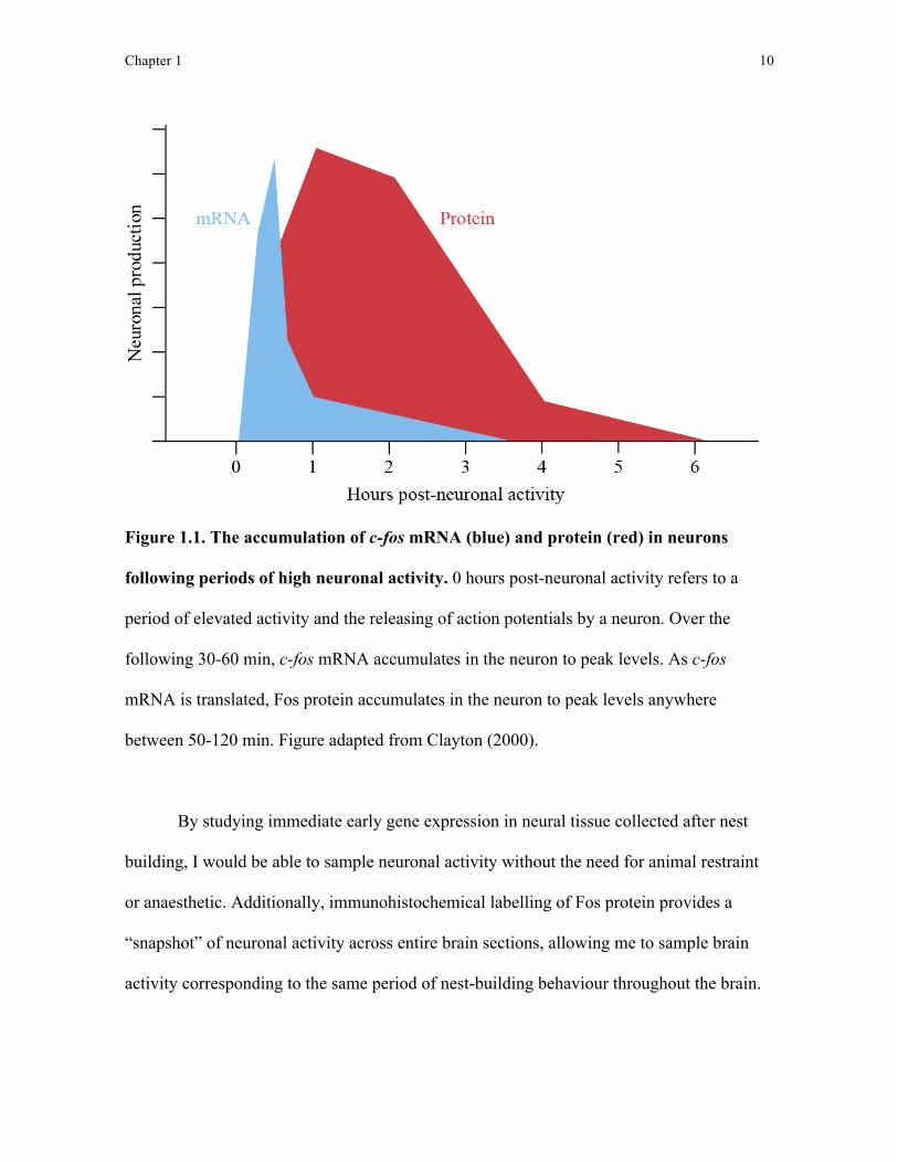
profile to the appearance of c-fos mRNA such that it accumulates to peak levels roughly
30-60 minutes following a period of elevated neuronal activity. Requiring the additional
step of mRNA translation, Fos protein accumulates to peak levels anywhere between 50 to
120 minutes following elevated neuronal activity (Figure 1.1; Clayton, 2000).
Neurobiologists exploit the temporal dissociation between neuronal activity and the
accumulation of Fos mRNA and protein to indirectly sample levels of brain activity in
neural tissue collected up to 120 minutes after an animal performs a behaviour of interest.
Chapter 1 10
Figure 1.1. The accumulation of c-fos mRNA (blue) and protein (red) in neurons
following periods of high neuronal activity. 0 hours post-neuronal activity refers to a
period of elevated activity and the releasing of action potentials by a neuron. Over the
following 30-60 min, c-fos mRNA accumulates in the neuron to peak levels. As c-fos
mRNA is translated, Fos protein accumulates in the neuron to peak levels anywhere
between 50-120 min. Figure adapted from Clayton (2000).
By studying immediate early gene expression in neural tissue collected after nest
building, I would be able to sample neuronal activity without the need for animal restraint
or anaesthetic. Additionally, immunohistochemical labelling of Fos protein provides a
“snapshot” of neuronal activity across entire brain sections, allowing me to sample brain
activity corresponding to the same period of nest-building behaviour throughout the brain.
Chapter 1 11
Due to the relatively slow accumulation and degradation of c-fos mRNA and Fos
protein, immediate early gene techniques suffer from reduced temporal acuity in
quantifying brain activity. Furthermore, neurons labelled for the production of Fos protein
in neural tissue are quantified as “active” or “inactive” based on the intensity of Fos
labelling in each neuron, ignoring differences in activity between individual neurons.
Despite these limitations, characterising immediate early gene expression patterns is widely
and successfully used as a “first step” in identifying candidate brain regions activated
during performance of a behaviour. For example, in zebra finches, immediate early gene
techniques have been used to identify brain regions exhibiting elevated neuronal activity
during birdsong production (Kimpo and Doupe, 1997; Jarvis et al., 1998), song perception
(Bailey et al., 2002), and social and agonistic interactions with conspecifics (Goodson,
2005). After candidate brain regions are identified, subsequent studies can focus on these
regions and compare neuronal activity to the production of behaviour on a much finer
timescale or interfere with neuronal activity in these regions to test for a causal relationship
between brain activity and production of behaviour.
In the work presented here, I exploited the temporal delay between neuronal activity
and the accumulation of Fos protein to sample neuronal activity in the brains of nest-
building zebra finches 90 minutes after nest building began. Although Fos labelling has
been used to identify patterns of brain activity across entire brain sections (Sadananda and
Bischof 2002; 2006), I chose to focus on sampling neuronal activity in neural circuits that I
hypothesised may be involved in nest building based on previous studies on these brain
regions.
Chapter 1 12
Anterior and posterior motor pathways
Aside from the song-control system (a group of interconnected brain nuclei
involved in producing birdsong: Tramontin and Brenowitz, 2000), the neural substrates
involved in motor control in birds were only recently identified. In 2008, Feenders et al.
compiled the results of several studies on songbirds, parrots, ring doves, and hummingbirds
in which the production of different locomotor behaviours correlated with the expression of
immediate early gene mRNA, used as a proxy of neuronal activity. These behaviours
included wing-whirring during migratory restlessness in garden warblers (Sylvia borin) and
hopping in zebra finches. Across these comparisons, and in additional experiments in which
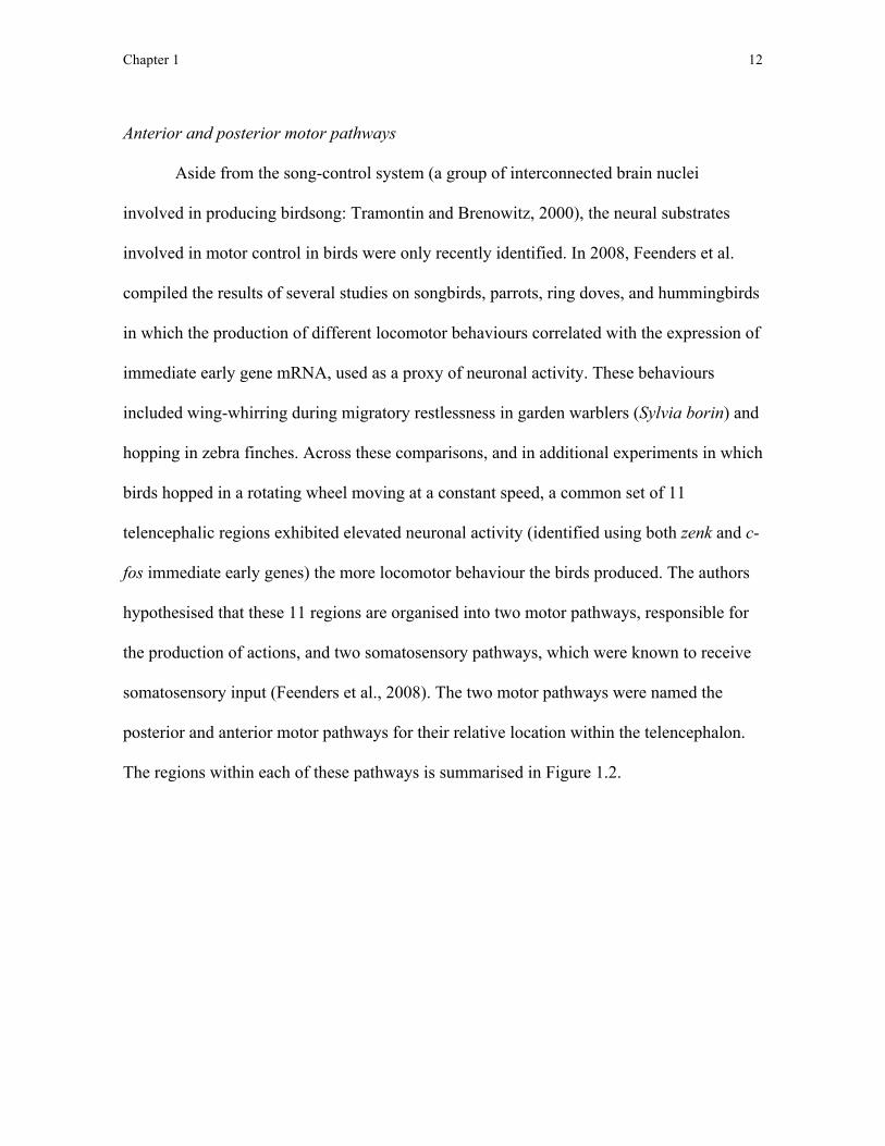
birds hopped in a rotating wheel moving at a constant speed, a common set of 11
telencephalic regions exhibited elevated neuronal activity (identified using both zenk and c-
fos immediate early genes) the more locomotor behaviour the birds produced. The authors
hypothesised that these 11 regions are organised into two motor pathways, responsible for
the production of actions, and two somatosensory pathways, which were known to receive
somatosensory input (Feenders et al., 2008). The two motor pathways were named the
posterior and anterior motor pathways for their relative location within the telencephalon.
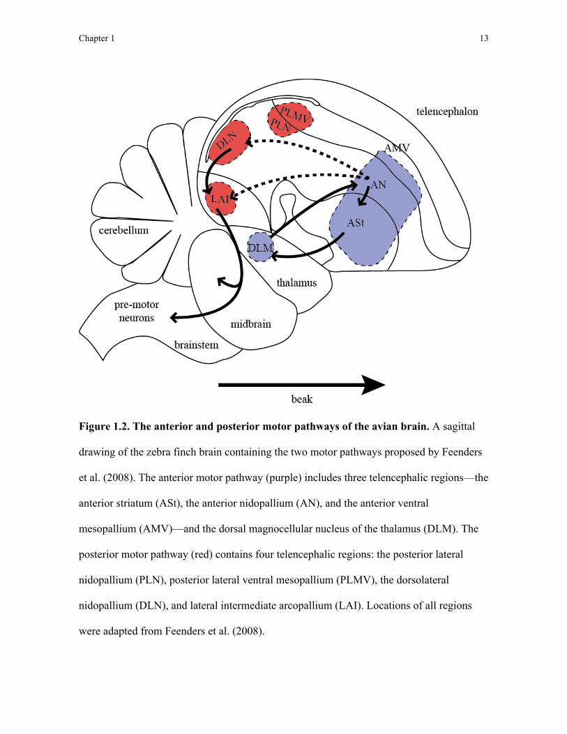
The regions within each of these pathways is summarised in Figure 1.2.
Chapter 1 13
Figure 1.2. The anterior and posterior motor pathways of the avian brain. A sagittal
drawing of the zebra finch brain containing the two motor pathways proposed by Feenders
et al. (2008). The anterior motor pathway (purple) includes three telencephalic regions—the
anterior striatum (ASt), the anterior nidopallium (AN), and the anterior ventral
mesopallium (AMV)—and the dorsal magnocellular nucleus of the thalamus (DLM). The
posterior motor pathway (red) contains four telencephalic regions: the posterior lateral
nidopallium (PLN), posterior lateral ventral mesopallium (PLMV), the dorsolateral
nidopallium (DLN), and lateral intermediate arcopallium (LAI). Locations of all regions
were adapted from Feenders et al. (2008).

Chapter 1 14
Feenders et al. (2008) noted that, in bird species that learn their songs, both the
posterior and anterior motor pathways are located within close proximity to pathways in the
song-control system. The authors used functional knowledge about each of these song-
learning pathways to suggest functions for the posterior and anterior motor pathway. The
posterior motor pathway is located beside the “motor pathway” of the song-control system
(consisting mainly of two song nuclei: the robust nucleus of the arcopallium and HVC
[used as a proper name]), which sends motor commands to the singing muscle, the syrinx,
to produce song (Tramontin and Brenowitz, 2000). Accordingly, Feenders et al. (2008)
suggested that the posterior motor pathway sends motor commands out of the
telencephalon down into the brainstem and spinal cord to produce movement.
As the anterior motor pathway is located beside the similarly-named “anterior motor
pathway” of the song-control system (consisting of three telencephalic song nuclei: Area X
in the striatum, magnocellular nucleus of the anterior nidopallium [MAN], and oval nucleus
of the mesopallium [MO]), which is involved in the learning and modification of birdsong
(Tramontin and Brenowitz, 2000), Feenders et al. (2008) suggested that the anterior motor
pathway is involved in the learning, modification, and organisation of actions.
In this thesis, I aimed to determine whether the anterior and posterior motor
pathways are involved in controlling the production of nest-building behaviour using Fos
protein immunohistochemistry to sample neuronal activity in both pathways. If nest-
building behaviour, and specifically the collection and deposition of nesting material,
involves motor sequencing, then I expected to see correlations between nest-building
behaviour and the number of neurons producing Fos in the anterior striatum, anterior
nidopallium, and anterior ventral mesopallium of the anterior motor pathway.
Chapter 1 15
Social behaviour network
Whereas few neurobiological investigations have attempted to identify the neural
circuits involved in nest-building behaviour, much work has elucidated the neural
substrates involved in courtship behaviour preceding and parental behaviour following, nest
building. The majority of these studies have focussed on the social behaviour network, a
group of interconnected telencephalic nuclei involved in the production and regulation of
social behaviour (Goodson, 2005). Newman (1999) first proposed the existence of a social
behaviour network based on previous neurobiological work in mammals. In his review,
Newman grouped six brain regions in the limbic system together as a neural system based
on reciprocal connectivity between all regions, expression of gonadal hormone receptors in
each region, and a common function in mediating affiliative, aggressive, and parental
behaviour in mammals. Since then, homologous regions of all six social behaviour network
brain regions have been identified in all vertebrate lineages, including fish, reptiles, and
birds (Goodson, 2005; O’Connell and Hofmann, 2011). In birds, nuclei in the social
behaviour network have been functionally associated with social behaviours including
courtship singing and displaying (Heimovics and Riters, 2006), copulation (Balthazart and
Surlemont, 1990; Meddle et al., 1999), aggressive interactions (Goodson and Adkins-
Regan, 1999) and incubation (Youngren et al., 1989). Because the social behaviour network
regulates reproductive behaviour prior to and following nest building I expected that these
brain regions might also be involved in controlling nest-building behaviour.
A previous study sampling neuronal activity in the social behaviour network in
songbirds included indirect measures of nest building. In 2006, Heimovics and Riters found
that captive adult male European starlings (Sturnus vulgaris) possessing a nest box
Chapter 1 16
exhibited elevated neuronal activity in several brain regions in the social behaviour network
relative to males lacking a nest box. The regions identified in that study included the medial
bed nucleus of the stria terminalis, dorsal subdivision (BSTmd), medial bed nucleus of the
stria terminalis, ventral subdivision (BSTmv), anterior hypothalamus (AH), medial preoptic
area (POM), and ventromedial hypothalamus (VMH). The authors noted that male starlings
possessing a nest box also collected and delivered nest material to the nest box, however, as
nest-building behaviour was not quantified it is difficult to determine whether the observed
changes in neuronal activity were related to nest building specifically and not to other
concurrent changes in courtship, territorial, and parental behaviour. In this thesis, I aimed to
compare neuronal activity in the social behaviour network with nest-building behaviour in
zebra finches with a focus on the nuclei that were observed to be more active during nest
possession in starlings (Heimovics and Riters, 2006).
One limitation of quantifying brain activity in the social behaviour network by
sampling the number of neurons producing Fos is that all neurons in a given brain region
are assumed to serve the same function. Contrary to this assumption, studies on the
chemical neuroanatomy of the social behaviour network have demonstrated that several
brain regions contain functionally distinct subpopulations of neurons that differ in the type
of cellular signal they use to transmit information. Notably, medial divisions of the bed
nucleus of the stria terminalis (BST) of the social behaviour network contain at least two,
overlapping neuronal subpopulations: vasotocinergic neurons that transmit signals using
vasotocin (the avian analog of arginine vasopressin in mammals) and mesotocinergic
neurons that transmit signals using mesotocin (the avian analog of oxytocin in mammals;
Goodson, 2008). Furthermore, these vasotocin and mesotocin neurons appear to mediate
Chapter 1 17
many of the social behaviours associated with BST function (Goodson, 2008). In this thesis,
after identifying regions in the social behaviour network that are activated during nest
building, I also tested whether neuronal activity specifically in vasotocinergic and
mesotocinergic neuronal subpopulations within these brain regions increased during nest
building. By combining Fos protein immunohistochemistry with vasotocin or mesotocin
immunohistochemistry, I was able to sample neuronal activity specifically within
vasotocinergic and mesotocinergic neurons in the social behaviour network.
Dopaminergic reward system
Alongside studies on the involvement of the social behaviour network in regulating
behaviour in birds, similar work has identified the neural substrates that reinforce the
performance of social behaviours. A group of interconnected nuclei collectively referred to
as the dopaminergic reward system has been extensively studied in the context of
controlling the incentive and reward associated with behaviour in both laboratory
paradigms and ethological study (Riters, 2011). Much like the social behaviour network,
the dopaminergic reward system appears to be functionally and anatomically conserved
amongst vertebrates and putative homologs of two of the most commonly studied reward
nuclei, the ventral tegmental area and central gray, have been identified in all vertebrate
lineages (O’Connell and Hofmann, 2012). O’Connell and Hofmann (2011) have recently
proposed that the social behaviour network and dopaminergic reward system be considered
a single neural system, called the social-decision making network, based on the deep
homology of both the social behaviour network and dopaminergic reward system in
vertebrates and extensive reciprocal connectivity between these two circuits. Because the
Chapter 1 18
social-decision making network is a recent hypothetical framework and requires directed
studies to justify grouping these two neural circuits, in this thesis I focused on each neural
circuit separately.
Functional studies on the dopaminergic reward system show that neuronal activity
in this system is related to the speed at which animals approach an environmental stimulus
associated with reward and how long the animal engages with that stimulus, suggesting this
neural circuit plays a key role in controlling motivational processes (Salamone and Correa,
2012). Changes in neuronal activity in dopaminergic neurons in this circuit predict
behavioural changes in reward-based learning tasks, suggesting this neural circuit also
plays a role in mediating the effects of reward on reinforcing behaviour (Schultz et al.,
1997). Accordingly, dysfunction in the dopaminergic reward system has been associated
with addiction disorders (Gardner, 2011). In studies on birds, the dopaminergic reward
system also appears to play a role in controlling motivational and reward processes shaping
naturally occurring behaviour: the ventral tegmental area is thought to reinforce the
production of courtship song (Heimovics and Riters, 2005), copulation (Charlier et al.,
2005), affiliation behaviours (Goodson et al., 2009), and pair bonding (Banerjee et al.,
2013). Support for the involvement of the dopaminergic reward system in nest-building
behaviour comes from evidence that neuronal activity is elevated in the ventral tegmental
area in adult male starlings that possessed a nest box compared to males that did not
(Heimovics and Riters 2005; 2007). Although this finding suggests a role for the ventral
tegmental area in nest building, as in a similar study sampling activity in the social
behaviour network described above (Heimovics and Riters, 2006), nest-building behaviour
was not quantified and it remains unclear whether increased neuronal activity in the ventral
Chapter 1 19
tegmental area can be attributed to nest-building behaviour or to concurrent changes in
reproductive and territorial behaviours. Relative to the ventral tegmental area, much less is
known about the function of the central gray in birds. After observing that neuronal activity
in the central gray increased the more male zebra finches produced vocalisations directed at
conspecifics, however, Goodson et al. (2009) hypothesised that the central gray may be
involved in motivational processes controlling social communication. Here, I looked to see
whether there was a relationship between neuronal activity in the ventral tegmental area
and central gray and nest-building behaviour. If nest building is rewarding, I would expect
neuronal activity in dopaminergic reward system nuclei to increase the more birds engage
in nest-building behaviour.
As for the social behaviour network, brain regions in the dopaminergic reward
system contain subpopulations of neurons characterised for using different cellular signals
to transmit information. As the name “dopaminergic reward system” suggests, one such
neuronal subpopulation in the ventral tegmental area and central gray uses the
neurotransmitter dopamine. Furthermore, as mentioned above, dopaminergic neurons
contained in these regions are thought to be central to the dopaminergic reward system’s
function in reinforcing behaviour. To test whether these neuronal subpopulations are
involved in nest building, I compared Fos immunoreactivity in dopaminergic neurons in the
ventral tegmental area and central gray with the production of nest-building behaviour.
Hippocampus
As described at the outset, unlike the role that motor or reward pathways may play
in the neural underpinnings of nest building, there is an ongoing dispute regarding the role
Chapter 1 20
played by cognition in nest-building behaviour, particularly with regard to comparisons
between nest building and other construction behaviours that are thought to involve
cognition (Hansell, 2005; Hansell and Ruxton, 2008; Healy et al., 2008). Demonstrating
neuronal activation in certain brain regions associated with a behaviour may be useful in
this debate as it can potentially inform us of the cognitive/learning processes involved in a
behaviour. For example, consistent demonstrations of increased neuronal activity in the
hippocampus during spatial cognition tasks in birds (reviewed in Mayer et al., 2012) and
mammals (Nakamura et al., 2010; Teather et al., 2005; Guzowski et al., 2001) have
suggested these animals share at least a partly homologous neural substrate involved in
spatial learning. In addition to spatial learning, the hippocampus is thought to be involved
in behavioural sequencing (Remondes and Wilson, 2013) and in regulating the context-
specificity of behaviour in both mammals (Behrendt, 2013) and birds, including sexual
behaviour (Atoji and Wild, 2006). As nest building might involve one or more of these
processes, I compared Fos immunoreactivity in the hippocampus to nest-building
behaviour.
Cerebellum
The cerebellum is a brain structure found in all vertebrates and located caudal to the
telencephalon. Historically, the cerebellum was thought to serve only motor functions, an
assertion supported by connectivity studies, in which it was reported that the cerebellum
sent output exclusively to motor and pre-motor regions in the telencephalon, as well as
studies connecting cerebellar damage with motor dysfunction including akinesia and
rigidity (reviewed in Middleton and Strick, 2000). A surge of hodological studies in the

Chapter 1 21
1990s using a newly-introduced viral-mediated tract tracing protocol, which enabled more
extensive tracing of neural tracts across multiple synaptic junctions, however,
demonstrated that the cerebellum, in addition to connections with cortical motor regions,
was also reciprocally connected with several brain regions thought to be primarily involved
in cognitive processing, including prefrontal cortex (Middleton and Strick, 2000). These
connectivity studies, in conjunction with ongoing work demonstrating neuronal activity in
the cerebellum associated with cognitive tasks, have lead to the current view that the
cerebellum is involved not only in motor control, but also in learning, memory, and
language processing, at least, in humans (reviewed in Barton, 2012).
In mammals and birds, cerebellar volume and the degree to which the cerebellar
cortex is folded (called cerebellar foliation) exhibit tremendous diversity between species
(Larsell, 1967). Butler and Hodos (2005) suggested that the expansion of cerebellar cortex,
associated with increased cerebellar foliation, increases the neuronal processing capacity of
the cerebellar cortex and supports enhanced motor abilities. Although the specific nature of
improved motor abilities was not elucidated by Butler and Hodos, positive correlations
between cerebellar foliation and tool use in birds (Iwaniuk et al., 2009) and between
cerebellar volume and extractive foraging techniques in primates (Barton, 2012) suggest
that increasing cerebellar foliation may improve manipulative skill with the beak and hands
in birds and primates, respectively. Because nest building likely requires different degrees
of manipulative skill to shape, stitch, and weave nest materials into different nest structures,
I tested whether cerebellar foliation, as measured using a previously-published list of
cerebellar foliation indices (Iwaniuk et al., 2006), relates to variation in species-typical nest
structure. To do this, I classified species-typical nest structure based on structural

Chapter 1 22
complexity following the assumption that the nest structure a bird builds is at least partially
dictated by the manipulative skill of that species. For example, I predicted that constructing
a cup nest, characterised by a nest floor and walls that are shaped by the beak, would
require more manipulative skill and a more foliated cerebellum than would building a
platform nest, which consists of an un-manipulated pile of collected material.
The evolution of nest structure
Much like the neurobiology of nest building, there has been little work aimed at
elucidating the selective forces that have lead to the vast structural diversity in nests among
bird species. Previous comparative studies investigating the evolution of nest structure are
characterised by a lack of formal statistical tests of evolution and, instead, have described
evolutionary patterns by mapping species-typical nest structure onto contemporaneous
phylogenies (Winkler and Sheldon, 1993; Eberhard, 1998; Irestedt et al., 2006). In those
studies, ancestral nest states and evolutionary transitions were estimated using outgroup
comparison, a phylogenetic inference technique that suffers from overestimating the
influence of phylogeny and relying on only the species included in the tested phylogeny to
reveal the evolutionary history of the whole clade. Furthermore, outgroup comparison
cannot account for either the degree of relatedness between species or phylogenetic
uncertainty (Pagel and Harvey, 1988).
Despite advances in phylogenetically-informed statistical techniques that overcome
the limitations of outgroup comparison (Pagel and Meade, 2006), the application of these
tests in studies on the evolution of nest structure have been largely hampered by the lack of
accessible phylogeny distributions with detailed information on species relatedness and the

Chapter 1 23
lack of a classification system for the structural complexity of bird nests. Recently,
however, Jetz et al. (2012) produced an online, publically accessible database of
phylogenies for the largest sample of bird species to date. Usefully, for my purposes, many
of these phylogeny estimations are amenable to current techniques in phylogenetic
statistical modelling. In conjunction with the classification system I developed to compare
cerebellar foliation with species-typical nest structure, I was able to generate
phylogenetically-informed statistical models to test evolutionary hypotheses regarding the
evolution of nest structure.
In spite of the historic lack of phylogenetic and nest classification data required to
investigate the evolutionary origins of nest structure diversity, there are a number of
hypotheses regarding the evolutionary pressures influencing nest structure extant in the
literature. Notably, Collias (1997) used outgroup comparisons and descriptive statistics to
present multiple hypothetical evolutionary routes that he believes have led to the diversity
in nest structure seen today. Although Collias’ arguments lacked statistical complements to
account for the effects of phylogenetic relatedness in his proposal, many of his hypotheses
are testable (albeit thus far untested) and supported by ecological work on nest placement
and structure. In this thesis, I used phylogenetically-informed statistics to test one of
Collias’ hypotheses regarding the evolutionary pressures selecting for the construction of
domed nests. Specifically, Collias (1997) argued that, from an ancestral state of
constructing cup nests in trees, competition for limited nest sites off the ground favoured
bird lineages that began constructing nests closer and closer to the ground. The closer a
nest is constructed to the ground, however, the greater the risk of predation from ground
predators. Collias postulated that birds began constructing enclosed nests to confer
Chapter 1 24
protection from this increased predation risk. Here, I aimed to retest Collias’ hypothesis
regarding the evolution of domed nests in Old World babblers (Timaliidae) by
incorporating phylogenetically-informed analyses to test for co-evolution between nest
height and structure, to identify the ancestral state of nests in this clade, and to elucidate
the most likely evolutionary transitions between nest heights and structures.
Thesis Aims
In the following chapters, I sought to identify the neural substrates involved in nest-
building behaviour in birds and to establish a comparative framework to begin studying the
evolutionary pressures that have produced the diversity in nest structures among bird
species.
First, I aimed to identify neural circuits exhibiting elevated neuronal activity during
the production of nest-building behaviour. To do this, in the work described in Chapter 2 I
sampled neuronal activity, indirectly as the number of neurons producing Fos protein, in
adult male and female nest-building and control zebra finches. I sampled neuronal activity
in neural circuits I hypothesised may be involved in nest building and tested whether
neuronal activity in these regions differed between nest-building and control birds.
Furthermore, I used stepwise linear regressions to test whether or not any single behaviour
explained individual variation in neuronal activity in nest-building finches.
Following the identification of brain regions associated with nest-building
behaviour, in the work described in Chapter 3 I sampled neuronal activity in some of these
regions again, however, this time I focused on sampling Fos immunoreactivity in neuronal
subpopulations located within these brain regions. Specifically, I compared neuronal
Chapter 1 25
activity in mesotocinergic and vasotocinergic neuronal subpopulations in the social
behaviour network and dopaminergic neuronal subpopulations in the dopaminergic reward
system between nest-building and control birds. Again, I also tested whether any nest-
building behaviours explained individual variation in neuronal activity in any of these
neuronal subpopulations.
In Chapter 4, I describe my nest classification scheme for species-typical nest
structure and how I used this classification system to test whether cerebellar foliation is
related to variation in species-typical nest structure, which would suggest that foliation
correlates with species differences in manipulative skill with the beak. To do this, I used
phylogenetically-informed statistical techniques to compare the degree of cerebellar
foliation between species building nests of different structural complexity.
Finally, in Chapter 5 I used my nest structure classification scheme to test the
evolutionary hypothesis underlying the evolution of domed nests in Old World babblers as
originally proposed by Collias (1997). Specifically, I looked for differences in nest height
between cup- and domed-nesting babblers and identified the most likely ancestral state of
nest height and structure in Timaliidae and the likely order of transitions in nest height and
structure leading the diversity in nest height and structure observed in extant babblers.
Chapter 2 26
Chapter 2: Neural correlates of nest-building behaviour in zebra finches
Introduction
As mentioned in Chapter 1, nest-building behaviour in birds consists of a sequence
of actions, which in its simplest form involves the collection and deposition of nest material
at the nest-site. For some species this nest-building sequence can be decomposed into just
a few actions while for others the construction of nests is more elaborate. For example,
arctic terns (Sterna paradisaea) nest in unadorned ground scrapes whereas long-tailed tits
(Aegithalos caudatus) sequence up to 14 motor actions to build a domed nest comprised of
moss and spider egg cocoons (Thorpe, 1956). Superficially at least, nest building appears
to involve motor actions and sequencing akin to those used in tool manufacture and use
(Hansell, 2000; Walsh et al., 2010; 2011; 2013) but to date there is little information
regarding the neurobiology of these behaviours in birds.
In this study, I sought to investigate the neural substrates involved in nest-building
behaviour in zebra finches. Zebra finches readily build nests in the laboratory (Muth and
Healy, 2011; 2012; 2013) using an easily quantified motor sequence of nest material
collection and deposition. While the male zebra finch collects and deposits nest material,
the female remains within the nest cup and manipulates material to shape a species-typical
dome nest (Zann, 1996). As mentioned in Chapter 1, one of the most common ways to
implicate brain regions involved in the behaviour of interest is to determine which brain
regions are activated whenever this behaviour is performed. As described in Chapter 1, I
quantified immunoreactivity for the immediate early gene c-fos protein product Fos
(Meddle and Follett, 1997, Clayton, 2000) throughout multiple neural circuits that I
Chapter 2 27
predicted may be involved in nest-building behaviour in male and female zebra finches. I
did this using birds that did or did not build a nest.
I first quantified Fos immunoreactivity in the anterior motor pathway, which is
thought to control motor learning and sequencing (Feenders et al., 2008) and includes the
striatum, the input structure of the basal ganglia. The basal ganglia control motor planning
and sequencing, are found in all vertebrates (Kuenzel et al., 2011), and are activated during
trained tool use in macaque monkeys (Obayashi et al., 2001). By sampling Fos
immunroeactivity in the anterior motor pathway, I could test the hypothesis that nest
building involves motor sequencing: Fos immunoreactivity in the anterior motor pathway
should correlate with the amount of nest-building behaviour exhibited by male zebra
finches. I also predicted that Fos immunoreactivity would not differ between nest-building
and control birds (birds that were not allowed to build nests) in the posterior motor
pathway, a circuit that is involved in the production of motor actions (Feenders et al., 2008;
Chapter 1), as both nest-building and control birds could move freely.
In addition to sampling Fos immunoreactivity in these motor pathways, I also
quantified Fos immunoreactivity in the social behaviour network, a neural circuit involved
in avian courtship and parental behaviour (e.g. Goodson, 2005; Chapter 1). Because nest
box possession in male European starlings increases Fos immunoreactivity in several
regions in the social behaviour network (Heimovics and Riters, 2006), Fos
immunoreactivity specifically in these social behaviour network regions should be greater
as a result of nest box possession (the dorsal and ventral subdivisions of the medial bed
nucleus of the stria terminalis [BSTmd and BSTmv, respectively], anterior hypothalamus,
medial preoptic area, and ventromedial hypothalamus) in nest-building zebra finches than

Chapter 2 28
it is in control birds. Although Heimovics and Riters (2006) noted that starlings that
possessed a nest box also built nests, they did not quantify nest-building behaviour and so
were unable to test whether Fos immunoreactivity in the social behaviour network was
specifically related to nest-building behaviour. By quantifying nest-building behaviour, I
could determine whether Fos immunoreactivity in these regions during nest building is
associated with nest possession or nest building itself.
Complementary to the social behaviour network, I also quantified Fos
immunoreactivity in the dopaminergic reward system, which is involved in reward and
motivation of social behaviours including courtship (O’Connell and Hofmann, 2011;
Chapter 1). If nest-building behaviour is rewarding, Fos immunoreactivity in this reward
pathway should correlate with nest-building behaviour. Furthermore, this correlation
should be most conspicuous specifically in the ventral tegmental area and central gray, two
regions in the dopaminergic reward system which exhibit elevated neuronal activity
following nest box possession in starlings (Heimovics and Riters, 2005; 2007).
Finally, as described in Chapter 1, the avian hippocampus is involved in spatial
learning memory and in synthesising multimodal cues to promote context-specific
behaviour. If the hippocampus is involved in initiating nest building after zebra finches
recognise a reproductive context (Sherry and Hoshooley, 2009; Székely and Krebs, 1996),
Fos immunoreactivity in the hippocampus should be elevated in nest-building finches
compared to controls.

Chapter 2 29
Methods and materials
Animals
Thirty-two adult zebra finches (n = 16 male, n = 16 female) were bred in captivity at
the University of St. Andrews, St. Andrews, Scotland, UK and the University of Glasgow,
Glasgow, Scotland, UK. Prior to experimentation, I housed birds in single-sex groups in
cages containing 10 to 20 birds with access to finch seed mix and water ad libitum but
deprived of access to coconut fibre. The room was held on 14L:10D light:dark light cycle
(lights on 8:00) with temperatures ranging between 19-27°C and 50-70% humidity. All
procedures were performed with ethical permission from the University of St. Andrews
Animal Welfare and Ethics Committee and from the UK Home Office (PPL. 60/3666).
Treatment group assignment
I caught zebra finches from group cages, randomly paired birds (one bird of each
sex) in wooden/wire mesh cages (44 x 30 x 39 cm), and then moved pairs to a separate
room with the same light cycle, temperature, and humidity as the group-housing room. I
fitted cages with a wooden nest cup (11 x 13 x 12 cm) and covered the floor with bedding
chips. The birds had access to finch seed mix and water ad libitum. I paired birds for at
least one week before providing them with coconut fibre as nest material. Prior to
receiving this nest material, all pairs filled their nest cups with bedding chips at least once
and some females laid eggs in these bedding chip nests. I removed all bedding and eggs
from nest cups during daily inspection.
At least one week after pairing, at 12:00 (4 hours after lights on) I gave six pairs of
birds 7.5 g of coconut fibre each and I inspected cages 24 hours later to identify pairs that
Chapter 2 30
had begun to build in their nest cup. To create an experimental cohort, I randomly assigned
a pair of finches that had begun building a fibre nest to each behavioural treatment group
(nest-building or control group). I selected only pairs of birds that had begun building a
nest to ensure that all of the finches included in this study, both nest-building and control
pairs, were motivated and capable of building nests prior to behavioural observation. I
removed coconut fibre nests and remaining fibre from the cages of both pairs and also
removed the nest cup from the cage of the control pair. I removed the cage bedding chips
and lined the cage floor with black plastic to prevent unwanted nest building with bedding.
I moved the two pairs of the experimental cohort to a test room where both pairs were
visually but not acoustically isolated from each other by a wooden barrier.
Isolation of nest-building behaviour
On the next morning, 1 hour after lights on, I provided the nest-building finch pair
with 12 g of coconut fibre and monitored them throughout the day for evidence of nest
building. If the nest-building pair began building a nest on the day they received nest
material, I scheduled the behavioural observation period for the following morning. If the
nest-building pair failed to construct a nest on the first day I provided the material, I
replaced the 12 g of coconut fibre the next morning and monitored the nest-building male
for the remainder of the day. If a nest-building male failed to deposit any material in the
nest cup within two days of material provision, the nest cup and material were removed and
a new nest cup and 12 g of coconut fibre were given to the control pair, reversing the
treatment assignment of each pair in the cohort. Reversal of treatment conditions occurred
Chapter 2 31
twice and in one case, neither male constructed a nest while in the isolation room. These
birds were removed from the study and replaced by a subsequent cohort.
When the lights came on the morning after a nest-building pair began nest building
in the test room, I removed unused nest material from this pair’s cage but left the nest they
had begun building. Both the nest-building and control pairs were left for 30 minutes
before I began filming. After 30 minutes, I gave the nest-building pair 9 g of coconut fibre
so that the male could resume nest building and I filmed each pair using either a JVC
Everio ACVHD (Model no. GZ-HD300AU) or Sony Handycam AVCHD (Model no.
HDR-CX115E) camcorder. Nest-building males did not typically resume building
immediately so I observed the birds from outside the isolation room via a window until I
observed the nest-building male make three consecutive trips with material from the cage
floor to the nest, which I considered the initiation of nest building. I recorded the time at
which the male began to build.
Behaviour coding
I encoded the birds’ behaviour using Noldus Observer (TrackSys Ltd., Nottingham,
U.K.) behavioural analysis software. I measured the occurrence of five behaviours that
were performed by both nest-building and control finches: hopping (a jump between
perches, the cage floor, and/or the nest cup), feeding (pecks into the ground or cage-
mounted feeder), drinking (pecks into the cage-mounted water dispenser), preening (each
preen of the chest, wing, or tail feathers by the beak), and scratching (scratch head feathers
with foot). In all females, I also recorded allopreening (female preens her partner male
with her beak). In all males, I assessed singing behaviour in two ways: song bouts (number
Chapter 2 32
of song bouts separated by at least 3 seconds) and time spent singing (number of seconds a
bird spent singing). I measured two nest-building behaviours only in nest-building males:
pick up (male picked up coconut fibre from the floor of the cage using his beak) and put
down (male released coconut fibre into the nest cup). In both nest-building males and
females, I counted the number of nest visits (bird entered the nest cup) and nest time
(number of seconds the bird spent in nest cup).
Tissue collection
After 90 minutes following the initiation of nest building, I entered the room to
confirm visually that material on the floor of the cage was added to the nest. Once
confirmed, I sacrificed both the control and nest-building pairs by terminally anaesthetising
(0.2 ml Pentobarbitone sodium i.p.; Dolethal, Vétoquinol) birds and then rapidly dissected
brains from the skulls. I fixed brains via submersion in 4% paraformaldehyde in
phosphate-buffered saline (0.1M, pH = 7.4) for six days and then cryoprotected brains in
20% sucrose in phosphate-buffered saline for 48 hours. I embedded brains embedded in
cubes of quail egg yolk, which was subsequently fixed with 4% paraformaldehyde over six
days. I sectioned the embedded brains coronally (section thickness = 30 µm) using a
freezing microtome and collected sections in three, alternating series (intersection interval
= 90 µm) into phosphate-buffered saline.
I repeated all of these procedures until I had observed behaviour of, and collected
brains from, eight nest-building pairs and eight control zebra finch pairs. Note: although I
will refer to ‘nest-building pairs’ it is the male that is the builder of the nest. The female
Chapter 2 33
may bring material at the end of the process in order to line the nest (Zann, 1996) but the
birds in this experiment did not reach that point of nest construction.
Fos immunohistochemistry
I rinsed sections three times in phosphate-buffered saline before incubating them in
0.5% H2O2 in phosphate-buffered saline for 30 minutes at room temperature to reduce
endogenous peroxidase activity. Following another three phosphate-buffered saline rinses,
I incubated sections in 10% Normal Goat Serum (Vector Laboratories) in 0.3% Triton X-
100 (Sigma) in 0.1M phosphate-buffered saline (0.3% PBT) for 60 minutes at room
temperature. I then removed sections from the blocking serum into the primary Fos
antibody (rabbit-anti-Fos antibody diluted 1:1000 in 0.3% PBT, Santa Cruz Biotechnology
K-25) and incubated for 21 hours at room temperature. This antibody has previously been
validated for use in the zebra finch (see Nordeen et al., 2009). The following day, I rinsed
sections three times in 0.1% PBT and incubated sections in biotinylated goat anti-rabbit
secondary antibody (diluted 1:250 in 0.3% PBT; Vector Laboratories) for 1 hour at room
temperature. After three rinses in 0.1% PBT, I incubated sections at room temperature in
ABC Elite avidin-biotin horseradish-peroxidase complex (Vector Laboratories) for 1 hour.
Following three rinses in 0.1% PBT I visualised the antibody-avidin-biotin complexes with
0.04% diaminobenzidene solution (Sigma Fast DAB) for 90 seconds and then rinsed
sections 4 times with phosphate-buffered saline. I then serially mounted tissue sections on
to Polysine microscope slides (VWR), serially dehydrated tissue through alcohol (50 to
100%), cleared tissue in xylene, and cover-slipped slides with DePeX (VWR). I found no
immunoreactivity when I omitted the primary Fos antibody.

Chapter 2 34
Quantification of Fos immunoreactivity
In all brain regions, I quantified Fos immunoreactivity by sampling the number of
neurons in a given brain region immunoreactive for Fos protein. In males, I quantified the
number of nuclei immunoreactive for Fos in HVC (used as a proper name) and the robust
nucleus of arcopallium (RA) in the song-control system. I also quantified Fos
immunoreactivity in the lateral intermediate arcopallium and dorsal lateral nidopallium of
the posterior motor pathway and anterior ventral mesopallium, anterior nidopallium, and
anterior striatum of the anterior motor pathway as identified in Feenders et al. (2008). In
the social behaviour network, I quantified Fos immunoreactivity in brain regions previously
reported to increase immediate early gene expression with nest box possession in starlings:
BSTmd, BSTmv, anterior hypothalamus, medial preoptic area, and ventromedial
hypothalamus (Heimovics and Riters, 2006; 2007). I also quantified Fos immunoreactivity
in the social behaviour network in one other division of the bed nucleus of the stria
terminalis (lateral subdivision [BSTl]), four divisions of the septum (ventral caudal
subdivision [LScv], lateral ventral caudal subdivision [LScvl], rostral subdivision [LSr],
and medial septum), and nucleus taeniae as identified by Goodson (2005) and Heimovics
and Riters (2006). Because BSTmd and BSTmv have been found to both increase Fos
immunoreactivity with nest box possession but the level of Fos immunoreactivity is
differentially influenced by breeding condition in each subdivision (Heimovics and Riters,
2006), I opted to sample these subdivisions separately, unlike a recent study testing for a
role of vasotocinergic neuronal subpopulations in BSTm (both BSTmd and BSTmv
together) in nest building (Klatt and Goodson, 2013). In the dopaminergic reward system, I
Chapter 2 35
quantified Fos immunoreactivity in the ventral tegmental area and central gray. I quantified
Fos immunoreactivity in two regions of the hippocampus (dorsal hippocampus and medial
hippocampus). All sampled brain regions are summarised in Figure 2.1.
I located areas of interest in brains using full section architecture and regional
anatomy with reference to brain atlases of the canary (Stokes et al., 1974) and zebra finch
(Nixdorf-Bergweiler and Bischof, 2007). At each area of interest, I inspected adjacent
coronal sections to locate the midpoint of the region in the rostrocaudal axis (Figure 2.1). I
took images of each region in both hemispheres and across 3 consecutive coronal sections
centred on the rostrocaudal midpoint of the region (intersection interval = 90 µm). For
brain regions that are larger in the rostrocaudal plane (anterior striatum and dorsal and
medial hippocampus), I took images across 5 evenly-spaced coronal sections centred on the
rostrocaudal midpoint of the region with an intersection interval of 270 µm. I captured all
images using a Nikon Coolpix E4500 digital camera mounted on a Leitz Diaplan
microscope using a 40x objective lens and Leitz Wetzlar 307-148.001 light source.
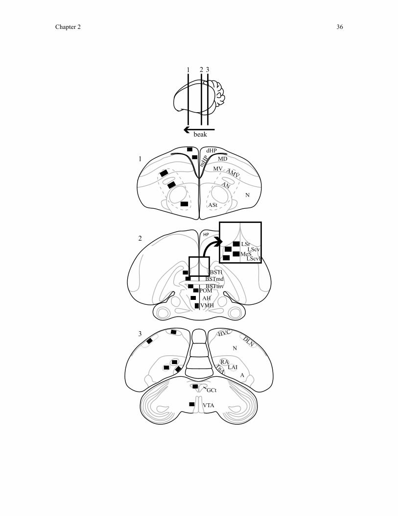
Chapter 2 36
N
dHPMD
MV
ASt
AN
AMV
mHP
HVC
A
TnA
N
RA
DLN
LAI
VTA
GCt
1
2
3
HP
BSTl
VMHAHPOMBSTmv
LSrLScvLScvl
MeS
BSTmd
1 2 3
beak

Chapter 2 37
Figure 2.1. Brain regions quantified for Fos immunoreactivity in the zebra finch brain.
Drawing of three coronal brain sections (1-3) and their locations along the sagittal plane
(top diagram) depicting all brain regions quantified bilaterally for Fos immunoreactivity in
this study. Black squares on the left hemisphere represent sampling squares taken at 40x
objective magnification and brain region acronyms are located in the relative position of the
sampling square in the right hemisphere. AH = anterior hypothalamus; ASt = anterior
striatum; AMV = anterior ventral mesopallium; AN = anterior nidopallium; BSTl = bed
nucleus of the stria terminalis, lateral subdivision; BSTmd = medial bed nucleus of the stria
terminalis, dorsal subdivision; BSTmv = medial bed nucleus of the stria terminalis, ventral
subdivision; dHP = dorsal hippocampus; DLN = dorsolateral nidopallium; GCt = central
gray; LAI = lateral intermediate arcopallium; LScv = lateral septum, ventral caudal
subdivision; LScvl = lateral septum, lateral ventral caudal subdivision; LSr = lateral septum,
rostral subdivision; mHP = medial hippocampus; MS = medial septum; POM = medial
preoptic area; RA = robust nucleus of the arcopallium; TnA = nucleus taeniae; VMH =
ventromedial hypothalamus; VTA = ventral tegmental area.
During quantification of Fos immunoreactivity, I opened each image in ImageJ
software (version 1.45, NIH, Bethesda, MD, USA) and desaturated the image. To isolate
Fos nuclei from background staining, I used the auto levels function in ImageJ, which
saturates a lack of Fos immunoreactivity as white and saturates Fos immunoreactivity as
black. Before applying the function to each image, I subtracted 40 units from the auto
levels adjustment value. This subtraction was necessary because the auto levels adjustment
value selected by ImageJ saturated both neurons and much of the background, neuropil Fos

Chapter 2 38
immunoreactivity, making neurons indistinguishable from background levels of staining.
By subtracting 40 units from the auto levels adjustment value, I was able to saturate the
more intense Fos immunoreactivity specific to neurons, without saturating the lighter,
background neuropil. An experimenter blind to bird treatment confirmed that this
subtraction reliably highlighted darkly-stained Fos immunoreactive nuclei from background
staining in a set of randomly selected images from multiple birds and brain regions. In the
anterior motor pathway regions, I subtracted only 30 units from the auto levels value as the
same experimenter (blind to bird treatment) found that neuropil staining was notably lighter
and better excluded using this modified levels manipulation. After applying the levels
function, I counted the number of highlighted Fos immunoreactive nuclei using the analyze
particles function in ImageJ. I only counted nuclei if they had a minimum area of 400
pixels2. An experimenter blind to bird treatment selected this value by measuring the area
of the smallest Fos immunoreactive nuclei identified in multiple, randomly-selected regions
across birds and brain regions. I summed the number of Fos immunoreactive nuclei in each
hemisphere and section to yield a single value of Fos immunoreactivity for each brain
region in each bird. I used these total Fos immunoreactive nuclei counts for each brain
region in statistical analysis except for HVC because lateralisation in activation in the right
hemisphere has been previously reported during short-distance communication with a
sexual partner in zebra finches (George et al., 2006). Accordingly, I analysed Fos
immunoreactivity in HVC in the left and right hemispheres separately.
Statistical analysis
Chapter 2 39
During the behavioural analysis, I identified one pair of nest-building finches as
outliers because the male picked up only small amounts of nest material (<2 SD below the
mean for the rest of nest-building males) and the female never interacted with the nest
material within the nest cup. As a result I excluded this pair from further statistical analysis.
I performed all statistical analyses using PASW software (version 19.00, SPSS Inc.,
Chicago, IL, USA). I quantified finch behaviour 80-50 minutes prior to the time at which
finches were sacrificed. The delay between this period of behaviour and sacrifice provides
sufficient time for the accumulation of Fos protein following neural activation associated
with nest-building behaviour (Morgan and Curran, 1991; Chapter 1). All behaviour and
Fos data were normally distributed (p > 0.05; Shapiro-Wilkes). I compared behaviour and
Fos immunoreactivity as dependent variables using GLMs and the independent variables
included sex on two levels (male and female) and treatment on two levels (nest-building
and control). Because I used these group comparisons to identify differences in Fos
immunoreactivity that would be associated with having a nest or not, such as visual
perception of the nest, and not Fos immunoreactivity that might be associated with how
much nest-building behaviour individual birds exhibited, I treated male and female birds
from the same nesting pairs as independent birds. For data on Fos immunoreactivity, I
looked specifically for treatment and treatment x sex interaction effects that reflected
neuronal activity associated with nest building.
To investigate whether nest-building behaviours explain individual variation in Fos
immunoreactivity, I regressed each brain region on all recorded behaviours in nest-building
birds as independent predictors of Fos immunoreactivity using multiple linear regression. I
ran regression models separately for males and females using a stepwise backwards

Chapter 2 40
elimination procedure that excluded interactions between types of behaviour. Using this
statistical approach, I could enter all behaviours measured into my regressional models and
identify the behaviour that best predicts Fos immunoreactivity in each brain region
compared to all other nest-building and non-nest-building behaviours measured. By using
this approach, I can avoid presenting relationships between Fos immunoreactivity and nest-
building behaviour that may actually be attributed to concurrent non-nest-building
behaviours measured, such as hopping to and from the nest cup. In the song control nuclei
(HVC and RA), I entered only singing behaviour (song bouts and time spent singing) as
predictors of Fos immunoreactive nuclei counts in all males (nest-building and control)
firstly to test for song-brain correlations as previously reported (Kimpo and Doupe, 1997)
and secondly to test whether a relationship between Fos immunoreactivity and birds’
behaviour 80-50 minutes prior to sacrifice existed.
Results
Regressional models in which nest-building behaviour significantly explained
variation in Fos immunoreactivity in a brain region are summarised in Table 2.1 and
Appendix 1.
Table 2.1. Relationships between behaviour and Fos immunoreactivity in brain
regions of nest-building adult zebra finches. Correlates were calculated using stepwise
linear regression to identify behaviours performed by nest-building zebra finches 80-50
minutes before sacrifice that predicted Fos immunoreactivity in sampled brain regions.
When regression models identified more than one behaviour that predicted Fos
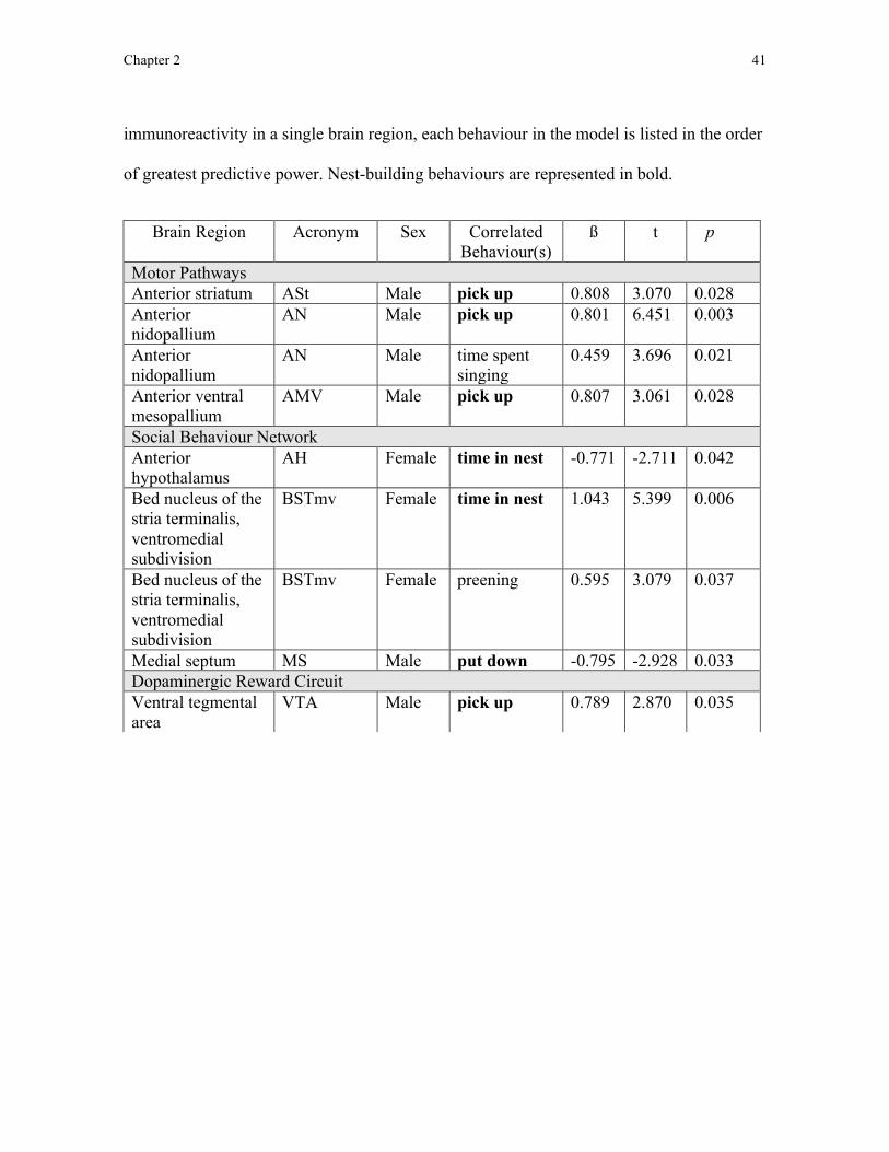
Chapter 2 41
immunoreactivity in a single brain region, each behaviour in the model is listed in the order
of greatest predictive power. Nest-building behaviours are represented in bold.
Brain Region Acronym Sex Correlated Behaviour(s)
ß t p
Motor Pathways Anterior striatum ASt Male pick up 0.808 3.070 0.028 Anterior nidopallium
AN Male pick up 0.801 6.451 0.003
Anterior nidopallium
AN Male time spent singing
0.459 3.696 0.021
Anterior ventral mesopallium
AMV Male pick up 0.807 3.061 0.028
Social Behaviour Network Anterior hypothalamus
AH Female time in nest -0.771 -2.711 0.042
Bed nucleus of the stria terminalis, ventromedial subdivision
BSTmv Female time in nest 1.043 5.399 0.006
Bed nucleus of the stria terminalis, ventromedial subdivision
BSTmv Female preening 0.595 3.079 0.037
Medial septum MS Male put down -0.795 -2.928 0.033 Dopaminergic Reward Circuit Ventral tegmental area
VTA Male pick up 0.789 2.870 0.035

Chapter 2 42
Behavioural analyses
Between 80-50 minutes prior to sacrifice, control birds hopped (F1,26 = 22.623, p <
0.001), fed (F1,26 = 9.617, p = 0.005), drank (F1,26 = 7.296, p = 0.012) and preened (F1,26 =
6.049, p = 0.021) more than did nest-building birds. Males scratched more often than did
females (F1,26 = 20.362, p < 0.001).
Control females tended to allopreen more than did nest-building females (t13 = 1.991,
p = 0.087). Nest-building and control males did not differ significantly in the time they
spent singing (p > 0.05). In nest-building pairs, males visited the nest cup more often than
did females (t12 = 6.128, p < 0.001) but did not spend more time in the nest cup (p = 0.091).
Song control system
Time spent singing positively correlated with Fos immunoreactivity in the right
HVC (Figure 2.2B; ß = 0.564, t13 = 2.464, p = 0.028) but did not significantly explain
variation in Fos immunoreactivity in the left hemisphere HVC in all males. Neither the
number of song bouts nor time spent singing significantly explained variation in Fos
immunoreactivity in RA in males.
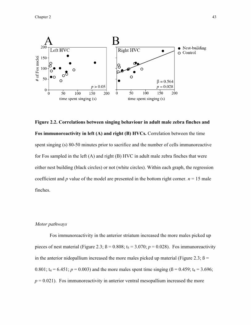
Chapter 2 43
Figure 2.2. Correlations between singing behaviour in adult male zebra finches and
Fos immunoreactivity in left (A) and right (B) HVCs. Correlation between the time
spent singing (s) 80-50 minutes prior to sacrifice and the number of cells immunoreactive
for Fos sampled in the left (A) and right (B) HVC in adult male zebra finches that were
either nest building (black circles) or not (white circles). Within each graph, the regression
coefficient and p value of the model are presented in the bottom right corner. n = 15 male
finches.
Motor pathways
Fos immunoreactivity in the anterior striatum increased the more males picked up
pieces of nest material (Figure 2.3; ß = 0.808; t5 = 3.070; p = 0.028). Fos immunoreactivity
in the anterior nidopallium increased the more males picked up material (Figure 2.3; ß =
0.801; t4 = 6.451; p = 0.003) and the more males spent time singing (ß = 0.459; t4 = 3.696;
p = 0.021). Fos immunoreactivity in anterior ventral mesopallium increased the more
Chapter 2 44
males picked up material (Figure 2.3; ß = 0.807; t5 = 3.061; p = 0.028). None of the
behaviours that I measured significantly explained individual variation in Fos
immunoreactivity in either of the areas I quantified from the posterior motor pathway, the
lateral intermediate arcopallium and dorsal lateral nidopallium.
In nest-building females, neither the number of visits to the nest nor the time spent
in the nest significantly explained the variation in Fos immunoreactivity in either the
anterior or posterior motor pathway.
I also found no significant difference in Fos immunoreactivity between nest-
building and control birds in either the anterior or posterior motor pathway (p > 0.05).

Chapter 2 45
Figure 2.3. Correlations between nest-building behaviours and Fos immunoreactivity
in the anterior motor pathway in zebra finches. Correlations between the picking up of
nest material and the number of Fos immunoreactive nuclei quantified in the (A) anterior
striatum [ASt], (B) anterior ventral mesopallium [AMV], and (C) anterior nidopallium
[AN] of the anterior motor pathway in adult male zebra finches. Correlations were derived
from stepwise linear regressions. Within each graph, the regression coefficient and p value
of the model are presented in the top left corner. (D) Micrographs of sampling squares
taken in tissue stained to label neurons immunoreactive for Fos in ASt in the right
hemisphere of a male finch who picked up the least and a male finch who picked up the
most number of times while building a nest. Scale bar represents 50 µm.
Chapter 2 46
Social behaviour network
Fos immunoreactivity in the medial septum decreased the more pieces of material
males deposited in the nest cup (ß = -0.795; t5 = -2.928; p = 0.033). Fos immunoreactivity
increased in LScv and decreased in the ventromedial hypothalamus the more time nest-
building males spent singing (LScv: ß = 0.928; t5 = 5.555; p = 0.003; ventromedial
hypothalamus: ß = -0.792; t5 = -2.899; p = 0.034). Fos immunoreactivity in LSr decreased
the more nest-building males hopped (ß = -0.778; t5 = -2.771; p = 0.039) and neither
picking up nor depositing nest material significantly explained variation in Fos
immunoreactivity in any of the other social behaviour network regions that I quantified.
Fos immunoreactivity in the anterior hypothalamus decreased the more time nest-
building females spent in the nest (ß = -0.771; t5 = -2.711; p = 0.042). Fos
immunoreactivity in BSTmv, however, increased the more time these females spent in the
nest (Figure 2.4; ß = 1.043; t4 = 5.399; p = 0.006) and the more time they spent preening (ß
= 0.595; t4 = 3.079; p = 0.037). Fos immunoreactivity in the ventromedial hypothalamus
decreased the more nest-building females preened (ß = -0.861; t5 = -3.790; p = 0.013).
Neither the number of times these females visited the nest nor the time these females spent
in the nest significantly explained variation in Fos immunoreactivity in any other social
behaviour network regions sampled.
Fos immunoreactivity in BSTmd (F1,23 = 4.720, p = 0.040) and medial preoptic area
(F1,25 = 8.095, p = 0.009) was significantly greater in nest-building birds relative to control
birds. There was no significant difference in Fos immunoreactivity between nest-building
and control birds in any other region sampled (p > 0.05).
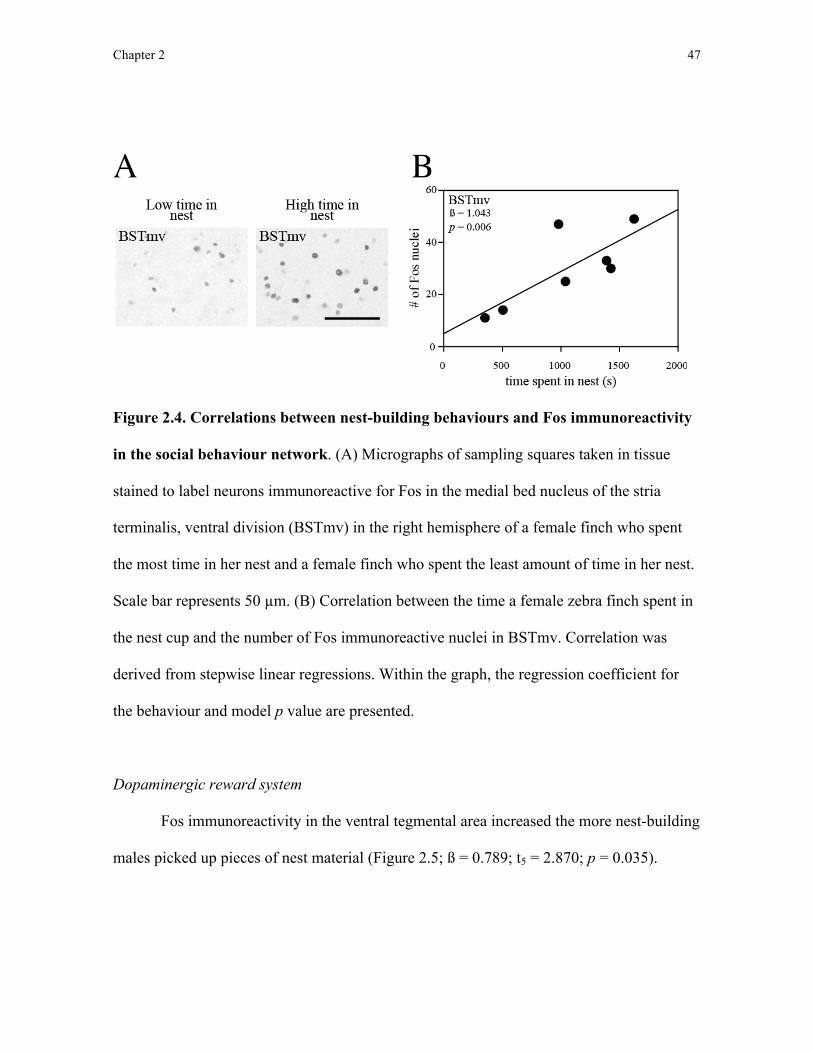
Chapter 2 47
Figure 2.4. Correlations between nest-building behaviours and Fos immunoreactivity
in the social behaviour network. (A) Micrographs of sampling squares taken in tissue
stained to label neurons immunoreactive for Fos in the medial bed nucleus of the stria
terminalis, ventral division (BSTmv) in the right hemisphere of a female finch who spent
the most time in her nest and a female finch who spent the least amount of time in her nest.
Scale bar represents 50 µm. (B) Correlation between the time a female zebra finch spent in
the nest cup and the number of Fos immunoreactive nuclei in BSTmv. Correlation was
derived from stepwise linear regressions. Within the graph, the regression coefficient for
the behaviour and model p value are presented.
Dopaminergic reward system
Fos immunoreactivity in the ventral tegmental area increased the more nest-building
males picked up pieces of nest material (Figure 2.5; ß = 0.789; t5 = 2.870; p = 0.035).

Chapter 2 48
Conversely, variation in nest-building behaviour did not significantly explain variation in
Fos immunoreactivity in the central gray.
In nest-building female finches, neither the number of nest visits nor the time spent
in the nest significantly explained variation in Fos immunoreactivity in the ventral
tegmental area or central gray.
Fos immunoreactivity in the ventral tegmental area and central gray did not differ
between nest-building and control birds (p > 0.05).
Figure 2.5. Correlations between nest-building behaviours and Fos immunoreactivity
in the dopaminergic reward system. (A) Correlation between the picking up of nest
Chapter 2 49
material and the number of Fos immunoreactive nuclei quantified in the ventral tegmental
area (VTA) in adult male zebra finches. This correlation was derived from stepwise linear
regressions. Within the graph, the regression coefficient for the behaviour and model p
value are presented. (B) Micrographs of sampling squares taken in tissue stained to label
neurons immunoreactive for Fos in the ventral tegmental area in the right hemisphere of a
male finch who picked up the most and a male finch who picked up the least amount of nest
material while constructing a nest. Scale bar represents 50 µm.
Hippocampus
None of the behaviours that I measured significantly explained individual variation
in Fos immunoreactivity in dorsal and medial hippocampus. I also found no significant
differences in Fos immunoreactivity in the dorsal and medial hippocampus between nest-
building and control birds (p > 0.05).
Discussion
In this study I used immediate early gene immunohistochemistry to identify regions
of the songbird brain that produce Fos protein during nest building. Based on the
assumption that Fos production reflects neuronal activation (Clayton, 2000), these data
show Fos immunoreactivity associated with nest-building behaviour (the number of times
nest material was picked up by nest-building males or with the time spent in the nest cup by
nest-building females) within the anterior motor pathway, social behaviour network, and
dopaminergic reward system. To my knowledge, this is the first demonstration of neural
Chapter 2 50
correlates of nest-building behaviour in the anterior motor pathway and dopaminergic
reward system.
Prior to discussing my results, an important caveat to address is that this study used
a restrictive sample size to test for a relationship between neuronal activity in the brain and
behaviour. Because of this small sample size, it is difficult to interpret non-significant
results as a demonstration that a specific brain region is not involved in the nest-building
behaviours tested here. Compounded with the imprecision of Fos immunohistochemistry as
a technique for inferring neuronal activity on a finer timescale (Chapter 1), non-significant
results presented in this study and the following chapter (Chapter 3) should not be used to
as evidence to preclude a relationship between a given brain region and nest-building
behaviour.
Motor pathways
Variation in Fos immunoreactivity throughout the anterior, but not the posterior,
motor pathway was explained by the number of times a male finch picked up nest material.
Given the involvement of the anterior motor pathway in motor learning and sequencing
(Feenders et al., 2008), activation of the anterior motor pathway, and the anterior striatum
in particular, during nest building suggests that nest-building behaviour may involve
similar motor sequencing and control as has been ascribed to tool use behaviour (which
activates the basal ganglia in primates: Obayashi et al., 2001). Fos immunoreactivity in the
anterior motor pathway was, however, specifically related to initiation of the sequence of
nest-building behaviour (picking up material) but not to the final step in the behavioural
sequence that I quantified (depositing material in the nest). This suggests that the anterior
Chapter 2 51
nidopallium in the zebra finch brain (as identified by Feenders et al., 2008) is functionally
similar to nidopallium intermedium medialis pars laterale (as identified by Helduser and
Güntürkün, 2012), a region in the pigeon brain found in the same location as the anterior
nidopallium in zebra finches, which plays a role in executing learned motor sequences.
The number of visits the females partnered to nest-building males made to the nest
and time they spent in the nest cup, however, were unrelated to Fos immunoreactivity in the
anterior motor pathway. This sex difference suggests that, during nest building, the anterior
motor pathway is specifically involved in the collection of nest material and not
construction within the nest cup, in which both male and female zebra finches participate
(Zann, 1996). The measures of nest-building behaviour in female finches used here,
however, were restricted to nest visitation and the time females spend in the nest and may
not reflect the degree to which they carry out any construction behaviour while in the nest.
Collection of construction behaviour data within the nest by both birds is required to
specifically address whether the anterior motor pathway might be involved in female nest-
building behaviour.
Social behaviour network
Fos immunoreactivity in the medial preoptic area and BSTmd of nest-building
finches was significantly higher compared to control birds. In conjunction with previous
reports of increased Fos immunoreactivity in the medial preoptic area and BSTmd during
nest box possession in adult male starlings (Heimovics and Riters, 2006), my failure to find
correlations between Fos immunoreactivity in the medial preoptic area and BSTmd and
nest-building behaviour suggest that this activity is associated with nest possession and not
Chapter 2 52
with nest building itself. It is important to note that there is no obvious control condition to
match with a pair of nest-building birds. For example, our control pair were unable to build
a nest, but also could not perceive nest material or the nest cup. For this reason, it is
possible that group differences in Fos immunoreactivity between nest-building and control
birds may reflect group differences not directly associated with nest building but other
environmental and behavioural differences between our treatment groups. For this reason, I
focus predominantly on my correlational results, which demonstrate a relationship between
Fos immunoreactivity and production of a specific, nest-building behaviour.
Although the groups did not differ in Fos immunoreactivity in BSTmv, within nest-
building females, Fos immunoreactivity in this region was greater the longer the female
spent in the nest. Elevation of Fos immunoreactivity in BSTmv following nest box
possession has been attributed to concurrent changes in agonistic behaviour associated with
territorial defence of the nest (Heimovics and Riters, 2006). My results in female finches,
however, suggest that such changes may be associated with occupation of the nest, a
behaviour that is only possible after a nest site has been obtained. Similar to Heimovics
and Riters (2006), I found that immediate early gene expression was higher in both BSTmd
and BSTmv the more nest-building behaviours birds performed but the specific expression
pattern in each subdivision of BSTm differed. These differences in expression patterns
dependent on the subdivision of BSTm sampled may explain why there appeared to be no
relationship in between nest-building behaviour and activation of vasotocinergic neurons in
BSTm in a previous study (Klatt and Goodson, 2013).
As mentioned in Chapter 1, after demonstrating the involvement of BSTmd and
BSTmv in nest-building behaviour, I sought to test whether the relationship between

Chapter 2 53
neuronal activity in the social behaviour network and nest-building behaviour existed in
specific neuronal subpopulations located in the social behaviour network. Although Klatt
and Goodson (2013) have already tested for a relationship between neuronal activity in
vasotocinergic and mesotocinergic neuronal subpopulations in the social behaviour network
and nest-building behaviour in zebra finches, this study failed to recognise the potential
functional division between BSTmd and BSTmv (see above and Chapter 1). In order to test
for the potential involvement of vasotocinergic and mesotocinergic neurons in nest-building
behaviour while recognising the functional division of BSTm, in Chapter 3 I sampled Fos
immunoreactivity within vasotocinergic and mesotocinergic neuronal subpopulations in
BSTmd and BSTmv separately and compare these levels of neuronal activity to nest-
building behaviour.
Dopaminergic reward system
The more males picked up pieces of nest material the greater the Fos
immunoreactivity in the ventral tegmental. As with the increase in Fos immunoreactivity I
observed in the BSTmd, it appears that Fos immunoreactivity in the ventral tegmental area
is associated with nest building itself rather than with other behavioural changes that occur
after a nest site is obtained, which are unrelated to nest building (Heimovics and Riters,
2006). Given the role that the dopaminergic reward system plays in motivating and
rewarding behaviour (O’Connell and Hofmann, 2011), I propose that the ventral tegmental
area may be involved in rewarding material collection behaviour in male nest-building
finches. In Chapter 1, I mentioned that dopaminergic neuronal subpopulations are thought
to mediate the reward and motivation functions of the entire dopaminergic reward system.
Chapter 2 54
To test whether these neurons are responsible for the correlation between neuronal activity
in the ventral tegmental area and nest-building behaviour, in Chapter 3 I sampled Fos
immunoreactivity specifically within this neuronal subpopulation and compared this
neuronal activity to nest-building behaviour.
In addition to a potential role in reward, the ventral tegmental may also influence
activity in the anterior motor pathway during nest building. In vertebrates, the ventral
tegmental area contains dopaminergic projection neurons and, in mammals, these neurons
innervate the striatum and provide necessary dopamine to support striatal functions
including motor learning and sequencing (Joel and Weiner, 2000; Hikosaka et al., 2008).
The possibility that the ventral tegmental area plays a role in influencing activity of the
anterior motor pathway is supported by my observation that Fos immunoreactivity was
higher in both the ventral tegmental area and anterior striatum the more nest material the
males picked up. Further examination of the relationship between Fos immunoreactivity in
dopaminergic neuron populations in the ventral tegmental area and nest building is required
to test this prediction.
Hippocampus
The absence of a correlation between variation in Fos immunoreactivity in the
dorsal and medial hippocampus and nest-building behaviour in male or female finches
suggests that the hippocampus does not play a substantial role in nest building, at least in
zebra finches.
Chapter 2 55
Singing and HVC
Finally, as has been previously reported Fos immunoreactivity was higher in the
HVC the longer the males spent singing. Furthermore, the time a male spent singing
explained the variation in Fos expression better than did the number of song bouts (Kimpo
and Doupe, 1997; Jarvis et al., 1998).
Conclusion
Here I identified several neural circuits in which neuronal activity, as indicated by
production of the immediate early gene c-fos protein product Fos (the anterior motor
pathway, social behaviour network, and dopaminergic reward system), was correlated with
the production of nest-building behaviour in nest-building male zebra finches and their
mates. These are the first detailed data to show the neural underpinnings of building
behaviour in birds and are, therefore, a major step in determining the role that motor
planning and sequencing, and reward and motivation may play in those behaviours.
Chapter 3 56
Chapter 3: A role for nonapeptides and dopamine in nest-building behaviour
Introduction
Understanding the neurobiology of sexual and parental behaviour in vertebrates has
long been a focus of neuroendocrine research (e.g. O’Connell and Hoffmann, 2011). In
birds, these studies often focus on the production and perception of courtship song (Riters
et al., 1998; Heimovics and Riters, 2005; 2006), affiliation (Goodson et al., 2009),
copulation (Balthazart and Ball, 2007), and parental care (Youngren et al., 1989). Despite
this work on the neurobiology of social behaviour throughout the breeding season, few
studies have elucidated the neuroendocrinological systems involved in nest-building
behaviour.
The current consensus is that two evolutionarily conserved neural circuits, the social
behaviour network and dopaminergic reward system, are important for sexual and parental
behaviour in all vertebrate lineages (O’Connell and Hofmann, 2012). Functionally, the
social behaviour network is thought to be involved in the production of courtship, sexual,
affiliative, and aggressive behaviours, whereas the dopaminergic reward system is thought
to be involved in the motivation to perform, and the positive feedback for performing, these
social behaviours (O’Connell and Hofmann, 2011). In Chapter 2, I found that neuronal
activity in the social behaviour network and dopaminergic reward system increased the
more nest-building behaviour male and female zebra finches exhibited, suggesting these
neural circuits may also be involved in nest-building behaviour.
Many of the brain regions in the social behaviour network and dopaminergic reward
system that I identified as being associated with nest-building behaviour in Chapter 2

Chapter 3 57
contain subpopulations of neurons characterised for using specific signalling molecules to
transmit neuronal information to downstream target brain regions (O’Connell and Hofmann,
2012). In zebra finches, these subpopulations include vasotocinergic and mesotocinergic
neuronal subpopulations in the medial bed nucleus of the stria terminalis (BSTm) of the
social behaviour network, which synthesise and release the nonapeptide hormones
vasotocin (the avian analog of arginine vasopressin in mammals) and mesotocin (the avian
analog of oxytocin in mammals), respectively. In addition to releasing these nonapeptides,
which bind to receptors in sites including the striatum, hypothalamus, and the septum of the
social behaviour network (Goodson et al. 2012), these neuronal subpopulations also
innervate hypothalamic and social behaviour network targets including the medial preoptic
area, which exhibits elevated neuronal activity during nest building (Chapter 2; Goodson et
al., 2012). In the dopaminergic reward system, dopaminergic neuron subpopulations in the
ventral tegmental area and central gray use the neurotransmitter dopamine to transmit
information to dopaminergic receptors in both the striatum and regions in the social
behaviour network including BSTm and the septum (Balthazart and Absil, 1997; Kubikova
et al., 2010; O’Connell and Hofmann, 2011).
Both the actions of vasotocin, mesotocin, and dopamine released from their
respective neuronal subpopulations and neuronal activity within the subpopulations
themselves are thought to mediate many of the behavioural functions associated with the
social behaviour network and dopaminergic reward system during the breeding season. For
example, administering a pharmacological antagonist that blocks the predominant
mesotocin receptor in the brain decreased affiliative behaviours associated with pair
formation in male and female zebra finches (Pedersen and Tomaszycki, 2012) and neuronal
Chapter 3 58
activity in BSTm vasotocinergic neurons increased in male zebra finches after courting a
female (Goodson et al., 2009), suggesting that vasotocinergic and mesotocinergic neuronal
subpopulations have a central role in controlling affiliative behaviour. Neuronal activity in
dopaminergic neurons in the ventral tegmental area increased the more male zebra finches
(Goodson et al., 2009) and European starlings (Sturnus vulgaris; Heimovics and Riters,
2005) sang courtship song to female conspecifics and pharmacologically agonising or
antagonising dopamine transmission increased and decreased the amount of song produced
by male starlings, respectively (Schroeder and Riters, 2006), suggesting that dopaminergic
neurons in the ventral tegmental area are involved in the motivation to perform courtship
behaviour. Because neuronal activity in the dopaminergic neurons in the ventral tegmental
area also increases following the production of reproductive and aggressive behaviour
(Bharati and Goodson, 2006), this dopaminergic neuronal subpopulation is thought to serve
a general function involved in the motivation to interact with conspecifics (O’Connell and
Hofmann, 2011). In the central gray of male zebra finches, however, neuronal activity in
dopaminergic neurons increased only after males produced vocalisations directed at
conspecifics, leading Goodson et al. (2009) to hypothesise that this neuronal subpopulation
is involved in the motivation to communicate vocally.
Following Chapter 2, in which I suggested that brain regions in the social behaviour
network and dopaminergic reward system are involved in nest building, here I hypothesised
that it may be the vasotocinergic, mesotocinergic, and dopaminergic neuronal
subpopulations within these circuits specifically that are involved in nest-building
behaviour. To test this hypothesis, I compared nest-building behaviour exhibited by male
and female zebra finches with concurrent neuronal activity, as measured indirectly by the

Chapter 3 59
number of neurons producing Fos protein (see Chapter 2), in vasotocinergic and
mesotocinergic neuronal subpopulations in subdivisions of BSTm and dopaminergic
neuronal subpopulations in the ventral tegmental area and central gray. Because neuronal
activity in the ventral subdivision of BSTm (BSTmv) increased the more time female
finches spent in the nest (Chapter 2) and systemic administration of a mesotocin receptor
blocker reduced the amount of time the female mate of nest-building zebra finch males
spent in the nest (Klatt and Goodson, 2013), I predicted that neuronal activity within
BSTmv mesotocinergic neurons would increase the more time that the female finches spent
in the nest cup. In the dorsal subdivision of BSTm (BSTmd), neuronal activity increased
during nest building in both male and female zebra finches (Chapter 2) and, accordingly, I
predicted that neuronal activity in vasotocinergic and mesotocinergic neurons in BSTmd
would increase during nest building.
In the ventral tegmental area, neuronal activity increased the more male finches
picked up nest material (Chapter 2). If picking up nest material is involves dopaminergic
neurons, I predicted that neuronal activity in dopaminergic neurons within the ventral
tegmental area would also increase the more often male finches picked up nest material.
Finally, as wild zebra finch pairs produce “duet-like” song exclusively while in the nest
(Elie et al., 2010), I predicted that Fos production in dopaminergic neurons in the central
gray would positively correlate with the time a pair of finches spent together in the nest.
Chapter 3 60
Methods and materials
Animals
Thirty-two adult zebra finches (n = 16 male, n = 16 female) were bred in captivity at
the University of St. Andrews, St. Andrews, Scotland, UK. All birds were maintained in the
same conditions as the experiment in Chapter 2 and all procedures were performed with
permission from the University of St. Andrews Animal Welfare and Ethics Committee and
the UK Home Office (PPL. 60/3666).
Treatment group assignment
I randomly paired zebra finches and formed experimental cohorts using the same
selection procedures as in Chapter 2, however, instead of coconut fibre, in this study I gave
birds 15 cm lengths of string (No. 4 Polished Cotton Twine; Rope Source, UK) with which
to build their nests. I administered string as a nest material in this study instead of the
coconut fibre used in Chapter 2 because string is more easily observed than coconut fibre in
videotaped footage of zebra finches building in the lab and finches build more readily and
faster using string compared to coconut fibre (Morgan, KV, pers. comm.). After at least a
week following pairing, I gave four pairs of birds 50 pieces of string at 12:00 (4 hours after
lights on). I inspected cages 24 hours later to identify pairs that had deposited string into
their nest cup. As in Chapter 2, to create an experimental cohort, I randomly assigned one
pair of finches in which the male had begun building a nest to each behavioural treatment
group (nest-building and control group). I selected only finch pairs that had begun building
a nest to ensure that all pairs included in this study were motivated and capable of building
nests prior to behavioural observation. I removed the string nests and remaining, unused
Chapter 3 61
string from the cages of both selected pairs and also removed the nest cup from the cage of
the control pair. I removed the bedding chips from the cages of both pairs, lined the cage
floors with black plastic to prevent nest building with bedding chips, and moved the two
pairs to a test room, as in Chapter 2. I repeated this selection procedure until I had 8 nest-
building and 8 control zebra finch pairs.
Nest building
Once in the test room, the control and nest-building pair were visually but not
acoustically isolated from each other by a wooden barrier. To record out-of-nest box
behaviour, I positioned a camcorder in front of each pair’s cage (Sony Handycam AVCHD,
Model no. HDR-CX115E) and to record in-nest box behaviour I suspended a bird-box
camera inside each pair’s cage (SpyCameraCCTV, Bristol, UK). I left each cohort
undisturbed in the test room for 24 hours to habituate.
30 minutes after the lights came on the morning following the habituation, I gave
the nest-building pair 250 pieces of string and began filming both pairs. I observed the
birds from outside the test room via a window until the male of the nest-building pair made
three consecutive trips with nest material from the cage floor to the nest. As in Chapter 2, I
recorded these trips as the time at which the male began to build and set the sacrifice time
for 90 minutes later. If the male began building immediately after receiving material, I
delayed the start of the observation for 15 minutes to avoid sampling Fos production in the
brain associated with the bird seeing the experimenter.
Behaviour coding
Chapter 3 62
As in Chapter 2, I encoded the birds’ behaviour using Noldus Observer
(TrackSys Ltd., Nottingham, U.K.) behavioural analysis software and here I also measured
the occurrence of behaviours performed 80-50 minutes prior to sacrifice, a time bin in
which Fos production is associated with nest-building behaviour. Briefly, I measured
instances of hopping, feeding, drinking, preening, scratching, and allopreening in all birds.
In males, I recorded the number of song bouts and the time spent singing. In nest-building
birds, I measured six nest-building behaviours: pick up, put down, tuck (when the bird
picked up a piece of string and tucked the string back into the nest while in the nest cup),
nest visits, and nest time. Unique to this chapter, I also measured time together in the nest
(the duration both members of a nesting pair spent together in the nest cup [seconds]).
Tissue collection
After 90 minutes following the initiation of nest building, I entered the room to
confirm visually that string was deposited in the nest cup. Once confirmed, I terminally
anaesthetised (0.2 ml i.p.; Dolethal) both pairs of birds and rapidly dissected their brains
from their skulls. I fixed brains via submersion in 4% paraformaldehyde in phosphate-
buffered saline (0.1M; pH = 7.4) for six days and then moved the brains into in 20%
sucrose in phosphate-buffered saline overnight and then in 30% sucrose in phosphate-
buffered saline for another night to cryoprotect them. I removed cerebella from the rest of
the brains by cutting and then froze both the cerebella and remaining brain on pulverised
dry ice and stored all neural tissue at -80°C before transporting the brains on dry ice to the
Roslin Institute, University of Edinburgh, Roslin, UK. I sectioned brains coronally (section
thickness = 52 µm) using a freezing microtome and collected sections in four, alternating

Chapter 3 63
series in cryoprotectant and stored the sections at -20°C until immunohistochemical
processing.
Double-label immunohistochemistry
Three series of sections were rinsed four times in 0.2% Triton X-100 (Sigma) in
0.1M phosphate buffer (PBT) and once in 0.1M phosphate buffer before being incubated in
0.3% H2O2 in phosphate buffer for 15 minutes at room temperature to reduce endogenous
peroxidase activity. Following three PBT rinses, sections were incubated in 10% Normal
Goat Serum (Vector Laboratories) in PBT for 60 minutes at room temperature. Sections
were then moved into the primary Fos antibody (Santa Cruz Biotechnology rabbit
polyclonal anti-Fos K-25, sc-253, 1:10,000) in 10% Normal Goat Serum in PBT and
incubated for 21 hours at 4°C. The following day, sections were rinsed three times in PBT
and incubated in biotinylated goat anti-rabbit secondary antibody (diluted 1:250 in PBT;
Vector Laboratories) for 1 hour at room temperature. After another three rinses in PBT,
sections were then incubated in avidin-biotin horseradish-peroxidase complex (1:400;
Vector Laboratories) in PBT for 1 hour at room temperature. Following four rinses in PBT,
one rinse in phosphate buffer, and a brief rinse in 0.1M sodium acetate, tissue was reacted
with 0.04% nickel-intensified diaminobenzidene (Sigma) solution for 210 seconds at room
temperature to visualise Fos immunoreactivity and then rinsed 5 times with phosphate
buffer to stop the reaction.
Immediately after Fos visualisation, I double-labelled each series to visualise
tyrosine hydroxylase, vasotocin, or mesotocin. Tyrosine hydroxylase is an enzyme
catalysing the rate-limiting step in dopamine synthesis and is used as a marker for

Chapter 3 64
dopaminergic neurons in vertebrate neuroanatomy (e.g. O’Connell and Hofmann, 2012).
Briefly, tissue series were rinsed three times in PBT, once in phosphate buffer, and
incubated in 0.3% H202 for 15 min. After another three PBT rinses, tissue series were
incubated in blocking serum (tyrosine hydroxylase: 10% Normal Horse Serum, Vector;
vasotocin and mesotocin: 3% Normal Goat Serum, Vector) in PBT for 60 min at room
temperature. Tissue was then moved into a solution containing the appropriate primary
antibody (tyrosine hydroxylase: Millipore, MAB5280, 1:1000; vasotocin: rabbit anti-
vasotocin: a gift of Dr David A. Gray, University of the Witwatersrand, Johannesburg,
South Africa, 1:10,000) and incubated for 60 h at 4°C. The tissue series reacted to visualise
mesotocin (primary antibody: Immunostar, 20068, 1:5000) was incubated for 87 hours at
4°C. After three more rinses in PBT, tissue was incubated in a solution containing
biotinylated secondary antibody (tyrosine hydroxylase: horse anti-mouse, 1:100, Vector;
vasotocin and mesotocin: goat anti-rabbit, 1:100, Vector Laboratories) in PBT for 60
minutes at room temperature. After three rinses in PBT, sections were then incubated in
avidin-biotin horseradish-peroxidase complex (1:50; Vector Laboratories) in PBT for 60
min at room temperature. After a final 4 rinses in PBT and a single rinse in phosphate
buffer, the second label was visualised by incubating tissue in non-intensified
diaminobenzidene at room temperature for different periods of time depending on the tissue
series (tyrosine hydroxylase: 110 s; vasotocin: 225 s; mesotocin: 140 s). Tissue was rinsed
five times in phosphate buffer to stop the diaminobenzidene reaction. This labelling
procedure produced an intensely dark, black Fos labelled nuclei in neurons and a light
brown cytoplasmic staining of neurons producing tyrosine hydroxylase, vasotocin, or
mesotocin. After double-labelling, all tissue sections were mounted on to 0.5% gelatine-
Chapter 3 65
subbed microscope slides (Thermo), serially dehydrated through alcohol (70 to 99%),
cleared in xylene, and cover-slipped with Pertex (VWR).
Quantification of Fos immunoreactivity
I sampled Fos immunoreactivity in neuronal subpopulations characterised by their
production of tyrosine hydroxylase, vasotocin, or mesotocin. I located each neuronal
subpopulation with reference to full-section architecture (Stokes et al., 1974) and, more
specifically, visualisation of tyrosine hydroxylase, vasotocin, and mesotocin. In tyrosine
hydroxylase-labelled tissue, I sampled tyrosine hydroxylase-immunoreactive
(dopaminergic) subpopulations in the ventral tegmental area in three adjacent sections and
central gray in four adjacent sections in each brain. In both vasotocin- and mesotocin-
labelled tissue, I sampled vasotocinergic and mesotocinergic subpopulations in BSTmd in
three adjacent sections and BSTmv in two adjacent sections in each brain.
In each neuronal subpopulation, I counted the number of neurons producing
tyrosine hydroxylase, vasotocin, or mesotocin and the number of double-labelled (tyrosine
hydroxylase+Fos, vasotocin+Fos, or mesotocin+Fos) neurons. Although tyrosine
hydroxylase+Fos neurons could be counted in the ventral tegmental area visually while
using the microscope, single-labelled tyrosine hydroxylase-immunoreactive neuronal
subpopulations were too large to be quantified using this method. To count these neurons, I
took images of all ventral tegmental area sections using a 20x objective lens and counted
the tyrosine hydroxylase-immunoreactive neurons by using ImageJ software (version 1.45,
NIH, Bethesda, MD, USA). All neuron counts were made in both hemispheres. To account
for differences in tyrosine hydroxylase-immunoreactive, vasotocinergic, and

Chapter 3 66
mesotocinergic neuronal subpopulation sizes between sections and birds, I divided the total
number of double-labelled cells by the total number of tyrosine hydroxylase-
immunoreactive, vasotocinergic, or mesotocinergic neurons, respectively, in a given brain
to quantify Fos immunoreactivity as the percentage of a neuronal subpopulation
immunoreactive for Fos.
Statistical analysis
I used PASW software (version 19.00, SPSS Inc., Chicago, IL, USA) for all of my
statistical analyses. I compared Fos immunoreactivity in each neuronal subpopulation using
GLMs with independent variables including sex on two levels (male and female) and
treatment on two levels (nest building and control).
To investigate whether nest-building behaviour explained individual variation in
Fos immunoreactivity, I used multiple linear regression including neuronal activity as a
dependent variable and all recorded behaviours in nest-building birds as independent
predictors, as in Chapter 2. I ran regression models separately for each sex and each
vasotocinergic, mesotocinergic, and dopaminergic neuron subpopulation sampled using a
stepwise reduction procedure to identify behaviours that significantly explained individual
differences in Fos immunoreactivity in these subpopulations.
Results
Full regressional models for all of the significant findings present below are
summarised in Appendix 2.
Vasotocinergic neuronal subpopulations
Chapter 3 67
Overall, Fos immunoreactivity in vasotocinergic neuron subpopulations in BSTmd
or BSTmv did not differ between nest-building birds and control birds (BSTmd: p = 0.535;
BSTmv: p = 0.978).
Among nest-building males, however, Fos immunoreactivity in vasotocinergic
neurons in BSTmd increased the more time a male spent together with his mate in the nest
cup (ß = 0.837; t6 = 3.748; p = 0.010; Figure 3.1). Additionally, Fos immunoreactivity in
vasotocinergic neurons in BSTmv increased the more times males picked up pieces of nest
material (ß = 0.784; t6 = 3.097; p = 0.021: Figure 3.1). In nesting females, none of the
behaviours I measured significantly explained the individual variation in Fos
immunoreactivity in vasotocinergic subpopulations in either BSTmd or BSTmv.

Chapter 3 68
Figure 3.1. (A) A micrograph of medial bed nucleus of the stria terminalis labelled for the
production of arginine vasotocin and Fos protein with dotted lines indicating the boundaries
of vasotocinergic neuronal subpopulations sampled in this study. (B) Correlation between
the time a pair of nest-building zebra finches spent together in the nest and the percentage
of arginine vasotocin immunoreactive (AVT-ir) neurons in the medial bed nucleus of the
stria terminalis, dorsal subdivision (BSTmd) immunoreactive for Fos in male brains. (C)
Correlation between the number of times male nest-building zebra finches picked up pieces
of nest material and the percentage of vasotocinergic neurons in the medial bed nucleus of
the stria terminalis, ventral subdivision (BSTmv) immunoreactive for Fos in male brains.
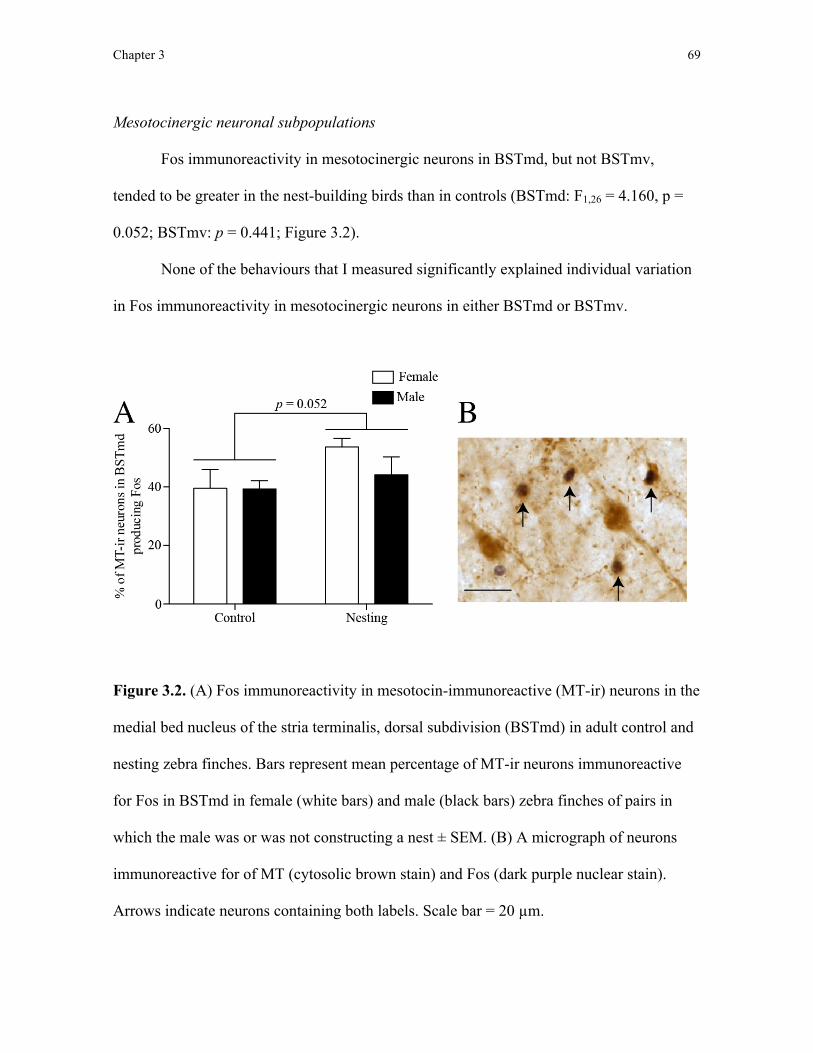
Chapter 3 69
Mesotocinergic neuronal subpopulations
Fos immunoreactivity in mesotocinergic neurons in BSTmd, but not BSTmv,
tended to be greater in the nest-building birds than in controls (BSTmd: F1,26 = 4.160, p =
0.052; BSTmv: p = 0.441; Figure 3.2).
None of the behaviours that I measured significantly explained individual variation
in Fos immunoreactivity in mesotocinergic neurons in either BSTmd or BSTmv.
Figure 3.2. (A) Fos immunoreactivity in mesotocin-immunoreactive (MT-ir) neurons in the
medial bed nucleus of the stria terminalis, dorsal subdivision (BSTmd) in adult control and
nesting zebra finches. Bars represent mean percentage of MT-ir neurons immunoreactive
for Fos in BSTmd in female (white bars) and male (black bars) zebra finches of pairs in
which the male was or was not constructing a nest ± SEM. (B) A micrograph of neurons
immunoreactive for of MT (cytosolic brown stain) and Fos (dark purple nuclear stain).
Arrows indicate neurons containing both labels. Scale bar = 20 µm.

Chapter 3 70
Tyrosine hydroxylase-immunoreactive neuronal subpopulations
Overall, Fos immunoreactivity in tyrosine hydroxylase-immunoreactive neurons in
either the ventral tegmental area or central gray did not differ between the nest-building and
control birds (ventral tegmental area: p = 0.211; central gray: p = 0.794).
Among nest-building males, however, Fos immunoreactivity in tyrosine
hydroxylase-immunoreactive neurons in the central gray increased the more time a male
spent with his mate in the nest cup (ß = 0.921; t6 = 5.793; p = 0.001; Figure 3.3).
Additionally, Fos immunoreactivity in tyrosine hydroxylase-immunoreactive neurons in the
ventral tegmental area decreased the more males tucked nest material into the nest (ß = -
0.719; t6 = -2.531; p = 0.045).
In nesting females, Fos immunoreactivity in tyrosine hydroxylase-immunoreactive
neurons in the ventral tegmental area decreased the more a female fed (ß = -0.816; t6 = -
3.453; p = 0.014). Stepwise linear regression identified no behaviours that significantly
explained individual variation in Fos immunoreactivity in tyrosine hydroxylase-
immunoreactive neurons in the central gray of female nesting finches.
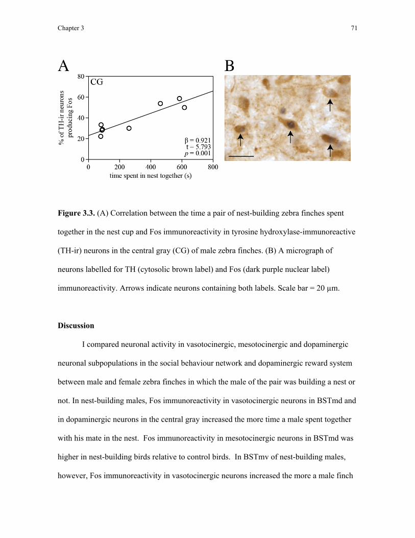
Chapter 3 71
Figure 3.3. (A) Correlation between the time a pair of nest-building zebra finches spent
together in the nest cup and Fos immunoreactivity in tyrosine hydroxylase-immunoreactive
(TH-ir) neurons in the central gray (CG) of male zebra finches. (B) A micrograph of
neurons labelled for TH (cytosolic brown label) and Fos (dark purple nuclear label)
immunoreactivity. Arrows indicate neurons containing both labels. Scale bar = 20 µm.
Discussion
I compared neuronal activity in vasotocinergic, mesotocinergic and dopaminergic
neuronal subpopulations in the social behaviour network and dopaminergic reward system
between male and female zebra finches in which the male of the pair was building a nest or
not. In nest-building males, Fos immunoreactivity in vasotocinergic neurons in BSTmd and
in dopaminergic neurons in the central gray increased the more time a male spent together
with his mate in the nest. Fos immunoreactivity in mesotocinergic neurons in BSTmd was
higher in nest-building birds relative to control birds. In BSTmv of nest-building males,
however, Fos immunoreactivity in vasotocinergic neurons increased the more a male finch

Chapter 3 72
picked up nest material. Finally, Fos immunoreactivity in dopaminergic neurons in the
ventral tegmental area decreased the more a male finch tucked material into the nest. These
data provide the first evidence suggesting vasotocinergic and mesotocinergic neuronal
subpopulations in the social behaviour network and dopaminergic neuronal subpopulations
in the dopaminergic reward system may be involved in controlling nest-building behaviour
in zebra finches.
Vasotocinergic and mesotocinergic neuronal subpopulations
Medial bed nucleus of the stria terminalis, dorsal subdivision (BSTmd)
I found that Fos immunoreactivity in mesotocinergic neurons in BSTmd was higher
in nest-building finches relative to Fos immunoreactivity in these neurons in control birds
(Figure 3.2). These data appear to contradict those from an earlier study in which Fos
immunoreactivity in BSTm vasotocinergic and mesotocinergic neurons did not differ
between nest-building and control zebra finches (Klatt and Goodson, 2013). As the neurons
sampled in that study, however, included subpopulations from both BSTmd and BSTmv as
a single measure, coupled with my observation that Fos immunoreactivity in
mesotocinergic neurons in BSTmv in nest-building birds did not differ from that of controls,
it seems plausible that assessing the activity in neurons across the two subdivisions may
have masked a group difference. Aste et al. (1998) originally proposed the division of
BSTm into dorsal and ventral subdivisions, BSTmd and BSTmv, respectively, because of
the anatomical separation of these two subpopulations by the anterior commissure. Support
for such a functional distinction between the two subdivisions comes from two studies, one
in which Fos immunoreactivity in both BSTmd and BSTmv increased during nest box

Chapter 3 73
possession in starlings (Heimovics and Riters, 2006) and the other in which Fos
immunoreactivity in both subdivisions increased during nest building in zebra finches
(Chapter 2). Although neuronal activity in both subdivisions increased following these
behaviours, the relationship between neuronal activity and the behaviour observed differed
between BSTmd and BSTmv. For example, in Chapter 2, Fos immunoreactivity in BSTmd
was higher in nest-building zebra finches relative to that of the non-nesting controls but this
increased Fos immunoreactivity did not correlate with any of the nest-building behaviour I
quantified. Fos immunoreactivity in BSTmv, however, did not differ between nest-
building and control finches, but, within nest-building females, increased specifically with
the more time a female spent in the nest cup, suggesting BSTmd may play a role in nest
possession or perception, whereas BSTmv is specifically involved in time spent in the nest
in female zebra finches (Chapter 2). Here, I also found that neuronal activity in
nonapeptidergic neuronal subpopulations in BSTmd and BSTmv exhibited different
relationships with nest-building behaviour, supporting the previous assertion that these
subdivisions are functionally distinct.
In addition to replicating the increase in BSTmd Fos immunoreactivity in nest-
building finches compared to controls that I reported in Chapter 2, here I show that this
increase in neuronal activity appears to occur specifically within mesotocinergic neurons.
Functionally, because Fos immunoreactivity in mesotocinergic BSTmd neurons was higher
in nest-building birds compared to controls but this increased Fos immunoreactivity in this
subpopulation did not correlate with any behaviour measured, it seems plausible that the
activity in this neuronal subpopulation is related to nest possession or perception of the nest
rather than to nest building, as I proposed for BSTmd in Chapter 2.
Chapter 3 74
Also within BSTmd, Fos immunoreactivity in vasotocinergic neurons increased in
male nest-building finches the more time he spent together with his mate in their nest, a
result that appears at odds with the absence of a relationship between Fos immunoreactivity
in vasotocinergic neurons in BSTm and the time spent in the nest in zebra finches (Klatt
and Goodson, 2013). Further to the suggestion above regarding differences between
sampling BSTmd and BSTmv as a single neuronal subpopulation and sampling them
separately, it could be that at least part of the explanation of this discrepancy between
studies lies with the behaviours quantified. Both Klatt and Goodson (2013) and I (Chapter
2) measured the amount of time individual birds spent within the nest whereas here I
measured the amount of time the pair of finches spent together in the nest. This discrepancy
might be particularly important because the social behaviour network is primarily involved
in social interactions between conspecifics (Goodson, 2005). For example, in zebra finches,
vasotocinergic neurons in BSTm specifically appear to be involved in eliciting affiliative
responses to mates (Goodson and Wang, 2006). These results suggest that vasotocinergic
neurons in BSTmd of male finches may be involved in affiliative behaviour within the nest
during nest building, although more detailed data on the social interactions occurring within
the nest are necessary to test this possibility.
Medial bed nucleus of the stria terminalis, ventral subdivision (BSTmv)
Here, I found that Fos immunoreactivity in vasotocinergic neurons in BSTmv
increased the more a nest-building male finch picked up nest material, which also appears
at odds with the data I reported in Chapter 2. This difference may be explained if the
relationship between neuronal activity and picking up nest material is specific to
Chapter 3 75
vasotocinergic neurons in this region and, therefore, may have been masked by Fos
immunoreactivity in other BSTmv neuronal subpopulations sampled alongside
vasotocinergic neurons in Chapter 2. Functionally, I suggest that vasotocinergic neurons in
BSTmv of zebra finches may be involved in picking up nest material. Again, this
suggestion contradicts that of Klatt and Goodson (2013), who found no relationship
between neuronal activity in BSTm and nest material collection by male zebra finches. As
with my results in BSTmd above, I believe this discrepancy in BSTmv may be, in part,
explained by differences in the behaviour quantified by Klatt and Goodson (2013) and by
myself. Whereas Klatt and Goodson (2013) counted the number of pieces of nest material
picked by male finches, in this study, I counted the number of times males picked up nest
material. In both this study and Chapter 2, I noticed that male finches often pick up but then
drop the same piece of nest material several times and encoding the number of pieces of
nest material picked up in lieu of the number of picking up actions, as in Klatt and Goodson
(2013), may not reflect nest-building behaviour. By demonstrating that neuronal activity in
vasotocinergic neurons in BSTmv increased specifically the more male finches picked up
nest material, I suggest that neuronal activity in this subpopulation is involved in the action
of collecting nest material and not the number of pieces of nest material collected, as
measured in Klatt and Goodson (2013). By manipulating vasotocin signalling using
pharmacological agents targeted to BSTmv subpopulations and recording subsequent
effects on nest-building behaviour, one could help to determine whether vasotocin from this
neuronal subpopulation is involved in picking up actions.
In females, I found no relationship between Fos immunoreactivity in either
vasotocinergic or mesotocinergic neuronal subpopulations and nest-building behaviour,
Chapter 3 76
which suggests that the correlation between Fos immunoreactivity in BSTmv and the time a
female spent in the nest that I reported in Chapter 2 may be attributed to other neuronal
subpopulations located in BSTmv intermingled with the nonapeptidergic subpopulations
sampled here, such as the population of neurons expressing receptors for vasoactive
intestinal peptide (Goodson et al., 2006). Consistent with this possibility, Klatt and
Goodson (2013) found no effect of central infusions of pharmacological antagonists that
impair vasotocin and mesotocin signalling on the time female zebra finches spent within
the nest. Here, as in Chapter 2, I will reiterate that the lack of a relationship between Fos
immunoreactivity in any of the neuronal populations tested here and nest-building
behaviour should not be used as evidence to discount a relationship between these neuronal
populations and nest-building behaviour because the restricted sample size used in this
study may have been too small to have detected this relationship.
Dopaminergic neuronal subpopulations
Ventral tegmental area
In Chapter 2, I found that neuronal activity in the ventral tegmental area increased
the more male finches picked up nest material, however, here I saw no change in Fos
immunoreactivity in dopaminergic neurons in the ventral tegmental area with regard to the
collection of nest material by males, suggesting that dopaminergic neurons in the ventral
tegmental area do not play a role in collecting nest material. Instead, here I found a
decrease in Fos immunoreactivity in ventral tegmental area dopaminergic neurons the more
nest-building male finches tucked material into the nest structure. This may mean that
tucking nest material into the nest structure is unrewarding or that the dopaminergic neuron

Chapter 3 77
subpopulation in the ventral tegmental area inhibits tucking behaviour. Such negative
relationships between neuronal activity and the production of behaviour have been reported
by Goodson et al. (2005), who found neuronal activity throughout the lateral septum
negatively correlated with aggressive displays in male song sparrows (Melospiza melodia).
Pharmacological manipulations could be used to inhibit neuronal activity in ventral
tegmental area dopaminergic neurons in order to distinguish between these two possibilities.
Because I did not find an increase in dopaminergic neuronal activity in the ventral
tegmental area the more male finches picked up nest material, I believe the relationship
between the ventral tegmental area and nest material collection that I reported in Chapter 2
may occur in other, non-dopaminergic neuronal subpopulations in the ventral tegmental
area. For example, the ventral tegmental area also contains a neuronal subpopulation that
uses the inhibitory neurotransmitter gamma-aminobutryic acid (GABAergic neurons),
which also appears to be involved in controlling social behaviours including courtship song
production in male zebra finches (Hara et al., 2007; Lynch et al., 2008). Comparing
neuronal activity in non-dopaminergic neuronal subpopulations in the ventral tegmental
area to nest-building behaviour could test this hypothesis.
Central gray
The increase in Fos immunoreactivity in central gray dopaminergic neurons in male
nest-building finches the more time he spent in the nest with his partner supports the
proposal that dopaminergic neurons in the central gray play a role in social communication
(Goodson et al., 2009). It is possible that this social communication takes the form of duet-

Chapter 3 78
like vocalisations that appear to be performed only within the nest (Elie et al., 2010) but, as
yet, we have no data to confirm this possibility.
In this chapter, I provide the first evidence that vasotocinergic, mesotocinergic, and
dopaminergic neuronal subpopulations in the social behaviour network and dopaminergic
reward system are active when birds are nest building. These brain-behaviour relationships
suggest that nest-building behaviour can be classified as a social behaviour regulated by the
social behaviour network and dopaminergic reward system.
Chapter 4 79
Chapter 4: The evolution of cerebellum structure and nest complexity
Introduction
In Chapters 2 and 3, I demonstrated the potential involvement of brain regions in
nest-building behaviour by comparing neuronal activity in the brain to the production of
nest-building behaviour. Although this functional neuroscience approach of comparing
brain activity to behaviour is common, it is not the only approach to identify brain regions
involved in the behaviour of interest. As described in Chapter 1, identifying correlations
between brain morphology and behaviour across species has been used to suggest the
function of brain regions. For example, by demonstrating that food-caching bird species
have larger hippocampal volumes than do non-caching species, Krebs et al. (1989) and
Sherry et al. (1989) both suggested that structural variation in the avian hippocampus was
related to variation in its functional capabilities, specifically with regard to spatial learning
and memory. In this chapter, therefore, I aimed to test whether morphological variation in
the cerebellum was correlated with variation in nest-building behaviour across bird species.
The cerebellum is a caudal brain region found in all vertebrates, which although
historically was considered to play a major role in motor control (Ito, 1984), is now known
also to be involved in a range of cognitive processes, such as learning, memory, and
language in humans (Ito, 1993). Across vertebrates, the morphology of the cerebellum is
highly varied in both its volume and foliation (amount of surface folding) across species:
amphibians and reptiles have unfolded cerebella while birds and mammals have variably
convoluted cerebella (Larsell, 1967; Iwaniuk et al., 2006). Of specific importance to the
work I describe in this thesis, increased cerebellar foliation in birds is hypothesised to

Chapter 4 80
increase the density of cerebellar neural circuitry and processing capacity of the cerebellum
to enhance motor abilities, specifically manipulative skills (Butler and Hodos, 2005;
Iwaniuk et al., 2009). Some support for this suggestion is provided by the positive
correlation between cerebellar foliation and tool use in birds (Iwaniuk et al., 2009) and
between cerebellum volume and extractive foraging in primates (Barton, 2012) and neural
activation (as seen by positron emission tomography) in the cerebellum during tool use in
monkeys (Obayashi et al., 2001).
Because nest building in birds also requires some manipulative skills phenotypically
similar to those involved in tool use (see Chapter 1) and these skills may vary depending on
the structural complexity of the nest built, I hypothesised that the cerebellum may be
involved in nest-building behaviour. Here, I examined whether variation in cerebellar
foliation index (Iwaniuk et al., 2006) in birds is explained by the variation in the
complexity of their species-typical nest structure. I predicted that species that build more
structurally complex nests would have higher cerebellar foliation indices than would
species that build simpler nests, suggesting the cerebellum is involved in the manipulative
skill underlying nest-building behaviour.
Methods and materials
Cerebellar foliation and nest structure
I collected data on cerebellar foliation index, measured as the degree of cerebellar
cortex folding compared to a hypothetical unfolded cortex for the same cerebellum size,
cerebellum volume, whole brain volume, and body mass from Iwaniuk et al. (2006) for 87
bird species.
Chapter 4 81
I then gathered descriptions of the species-typical nest structure from published
studies and texts (Appendix 1). Based on these descriptions, I categorised nest structures as
no nest, platform, cup, domed, and excavation nests. Birds that do not excavate or construct
a nest but lay eggs directly on a bare substrate or in a nest built by another species were
categorised as building no nest. No nest categorisations included birds that build nests in
nest boxes and cavities only if they are not described as building any structure within these
housings. Birds that construct nests within cavities and nest boxes were classified by the
structure they build within these housings. Platform nests are unshaped piles of collected
nesting material, including material used to line ground scrapes and depressions. Cup nests
have nest walls created during construction by the bird and not by depression of the nest’s
centre by the weight of the bird and eggs’ during incubation. Domed nests have both nest
walls and a roof. Finally, excavation nests are tunnels or chambers dug using the beak or
feet into a substrate. Unlike Hansell (2005), I did not differentiate between platform nests
built in the tree and those on the ground (referred to as “plate” and “bed” nests, respectively,
in Hansell, 2005) but I did differentiate between species that excavate nests and those that
nest in natural cavities or cavities excavated by other species (both referred to as “cavity
nests” in Hansell, 2005). These differences in nest categorisation reflected my focus on the
manipulative skill and behaviour required to construct a nest, regardless of nest location or
materials used.
I focused on comparing no nest, platform, and cup nest structures because these
three nest structures differ in the degree to which material is collected and manipulated
during construction: birds building no nest do not collect or manipulate nest material,
platform nests require the collection but little manipulation of material while cup nests
Chapter 4 82
require collection and manipulation of nest material to produce walls in the cup structure.
Because excavation behaviour involves a distinct set of actions to burrow into a substrate
which are difficult to compare to the collection and manipulation of nest material, I
excluded species that built excavation nests from further analysis. Furthermore, because
only two species (Acanthiza pusilla and Menura novaehollandiae) in my sample
constructed domed nests, I excluded these species from analysis as well as those species
without a nest description. After these exclusions, 64 species remained in my analysis.
Keywords used to categorise species-typical nest structures compared here are summarised
in Table 4.1.
Table 4.1. Terminology in published nest descriptions used to classify species-typical
nest structure. In my nest structure classification scheme, I focused on the nest-building
behaviour involved in collecting and manipulating nest material as well as manipulating
nesting, irrespective of nest location or the materials used.
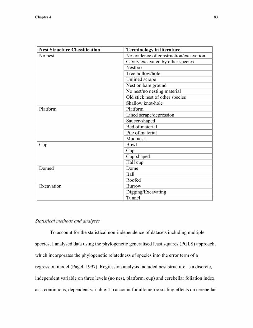
Chapter 4 83
Nest Structure Classification Terminology in literature No nest No evidence of construction/excavation
Cavity excavated by other species Nestbox Tree hollow/hole Unlined scrape Nest on bare ground No nest/no nesting material Old stick nest of other species Shallow knot-hole
Platform Platform Lined scrape/depression Saucer-shaped Bed of material Pile of material Mud nest
Cup Bowl Cup Cup-shaped Half cup
Domed Dome Ball Roofed
Excavation Burrow Digging/Excavating Tunnel
Statistical methods and analyses
To account for the statistical non-independence of datasets including multiple
species, I analysed data using the phylogenetic generalised least squares (PGLS) approach,
which incorporates the phylogenetic relatedness of species into the error term of a
regression model (Pagel, 1997). Regression analysis included nest structure as a discrete,
independent variable on three levels (no nest, platform, cup) and cerebellar foliation index
as a continuous, dependent variable. To account for allometric scaling effects on cerebellar
Chapter 4 84
foliation index, I included cerebellum volume as a covariate. Cerebellum volume was log-
transformed to achieve normality (Shapiro-Wilkes test, p > 0.05). Although previous
cerebellar foliation index analyses included other allometric variables (body size, whole
brain volume, and whole brain - cerebellum volume; Iwaniuk et al., 2006), I found that
cerebellum volume predicted cerebellar foliation index better than the other allometric
measures and after including cerebellum volume as a covariate no other allometric variable
explained significant variation in cerebellar foliation index. To test whether nest structure
was related specifically to cerebellar foliation, I also tested whether nest structure predicted
cerebellar volume using a PGLS with log-transformed whole brain volume and log-
transformed body size as allometric co-variates.
In addition to testing the main effect of nest structure on cerebellar foliation, I also
made three planned contrasts (no nest vs. platform, no nest vs. cup, and platform vs. cup)
by changing which factor level was the reference level in the model. I ran analyses in R (R
Development Core Team 2013) using the packages ape (Paradis et al., 2004) and caper
(Orme, 2012) and viewed phylogenetic trees in FigTree (Rambaut, 2012) and DensiTree
(Bouckaert, 2010).
To account for phylogenetic uncertainty, I ran my PGLS models across a sample of
3000 phylogenies built using a family backbone by Hackett et al. (2008; Jetz et al., 2012)
with restricted phylogenetic signal estimation (λ = lower: 0.01-0.1, upper: 0.95-0.99). I
used model averaging (following Johnson and Omland, 2004) to estimate average
parameters from PGLS regressions across the tree-block, weighted by the probability of the
model given each tree. Main effects could not be model-averaged across the tree-block
Chapter 4 85
because they were calculated from comparison of models with and without nest structure
using ANOVA. Instead, I present the minimum F and maximum p values reported across
the tree-block as a conservative means of testing for the main effect across varying
phylogenies. Because model comparison requires a fixed λ value in both models, λ was
fixed at either 0.85 or 0.95 (values derived from maximum likelihood estimations) when
testing for main effects of nest structure on cerebellar foliation. I acquired all bird
phylogenies from www.birdtree.org (Jetz et al., 2012). An example phylogeny is presented
in Figure 4.1. Finally, because my species sample included two flightless birds (Rhea
americana and Struthio camelus) and flight may also be a behavioural specialisation
associated with cerebellar foliation, I reran analyses excluding these two species.

Chapter 4 86
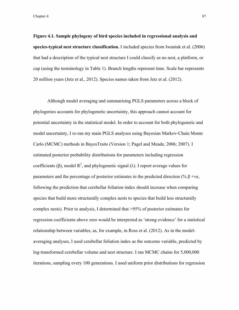
Chapter 4 87
Figure 4.1. Sample phylogeny of bird species included in regressional analysis and
species-typical nest structure classification. I included species from Iwaniuk et al. (2006)
that had a description of the typical nest structure I could classify as no nest, a platform, or
cup (using the terminology in Table 1). Branch lengths represent time. Scale bar represents
20 million years (Jetz et al., 2012). Species names taken from Jetz et al. (2012).
Although model averaging and summarising PGLS parameters across a block of
phylogenies accounts for phylogenetic uncertainty, this approach cannot account for
potential uncertainty in the statistical model. In order to account for both phylogenetic and
model uncertainty, I re-ran my main PGLS analyses using Bayesian Markov-Chain Monte
Carlo (MCMC) methods in BayesTraits (Version 1; Pagel and Meade, 2006; 2007). I
estimated posterior probability distributions for parameters including regression
coefficients (β), model R2, and phylogenetic signal (λ). I report average values for
parameters and the percentage of posterior estimates in the predicted direction (% β +ve,
following the prediction that cerebellar foliation index should increase when comparing
species that build more structurally complex nests to species that build less structurally
complex nests). Prior to analysis, I determined that >95% of posterior estimates for
regression coefficients above zero would be interpreted as ‘strong evidence’ for a statistical
relationship between variables, as, for example, in Ross et al. (2012). As in the model-
averaging analyses, I used cerebellar foliation index as the outcome variable, predicted by
log-transformed cerebellar volume and nest structure. I ran MCMC chains for 5,000,000
iterations, sampling every 100 generations. I used uniform prior distributions for regression

Chapter 4 88
coefficients (-100, +100). Mean acceptance rates were between 20-40%, as recommended
by Pagel and Meade (2007), and all effective sample sizes were >5,000.
Results
Across 64 species of bird, nest structure was significantly associated with cerebellar
foliation index (F1,60 > 3.875, p < 0.026, R2 = 0.615; using λ = 0.85 = model-averaged
estimate from main regression model). This relationship appears specific to cerebellar
foliation because nest type did not predict cerebellum volume (F1,60 < 1.686, p > 0.194; λ =
0.95).
Specific contrasts confirmed my predictions: species that build a platform nest have
significantly higher cerebellar foliation indices than do species that do not build nests (t46 =
2.047, p = 0.047), species that build a cup nest have significantly higher cerebellar foliation
indices than species that do not build nests (t37 = 3.165, p = 0.003), and species that build a
cup nest have significantly higher cerebellar foliation indices than species that build a
platform nest (t39 = 2.020, p = 0.049). Altogether, as nests increase in structural complexity
(no nest à platform à cup), cerebellar foliation index also increases. Furthermore, my
main results were not affected by removing the two flightless species in my sample, in
terms of either the main effect of nest structure on cerebellar foliation: (F1,58 > 4.589, p <
0.028, across 3000 trees, λ = 0.85, using cerebellum volume as a co-variate), or in any of
the planned contrasts (all model-averaged p < 0.05).
In my re-analysis of the data using the Bayesian MCMC approach, I again found
strong evidence for greater cerebellar foliation in species that build cup nests relative to
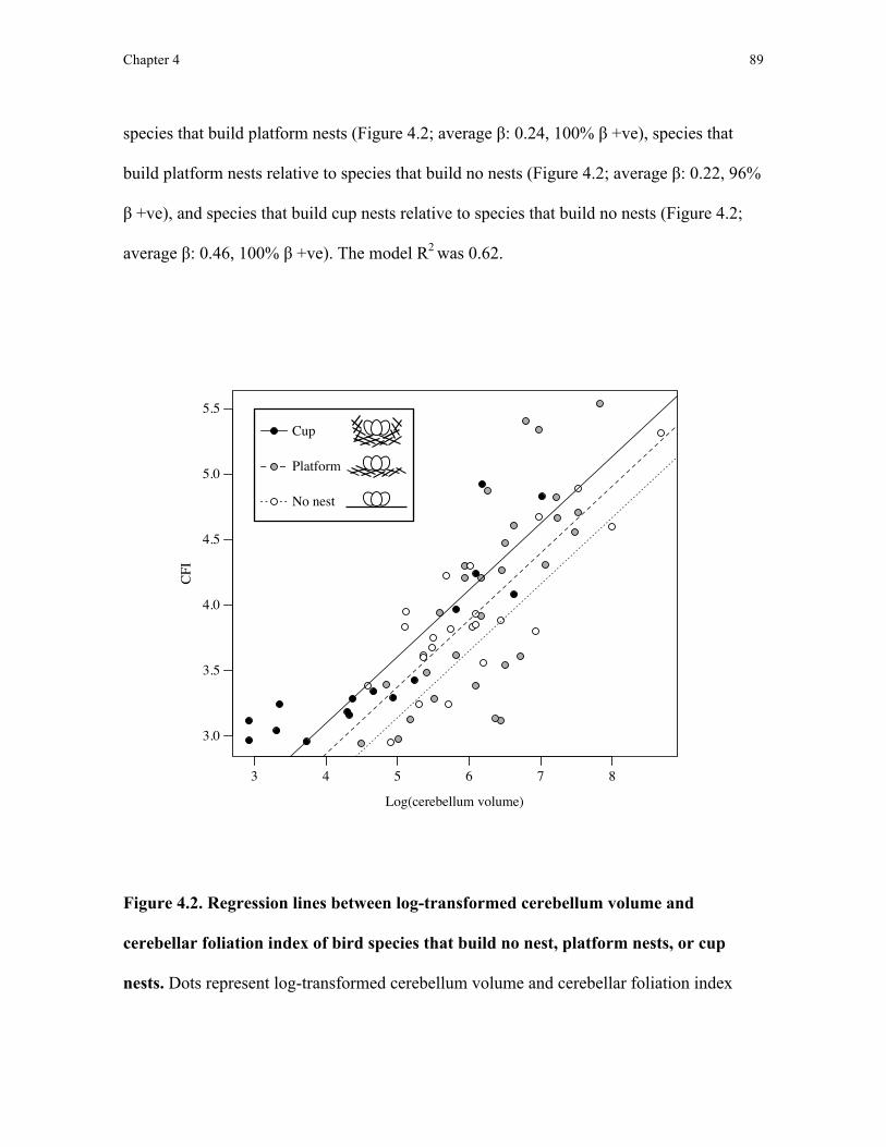
Chapter 4 89
species that build platform nests (Figure 4.2; average β: 0.24, 100% β +ve), species that
build platform nests relative to species that build no nests (Figure 4.2; average β: 0.22, 96%
β +ve), and species that build cup nests relative to species that build no nests (Figure 4.2;
average β: 0.46, 100% β +ve). The model R2 was 0.62.
Figure 4.2. Regression lines between log-transformed cerebellum volume and
cerebellar foliation index of bird species that build no nest, platform nests, or cup
nests. Dots represent log-transformed cerebellum volume and cerebellar foliation index
Log(cerebellum volume)
CFI
3.0
3.5
4.0
4.5
5.5
5.0
3 4 5 6 7 8
Cup
Platform
No nest
Chapter 4 90
(CFI) for bird species that build cup (black), platform (gray), and no nest (white). Slopes
and intercepts for all three groups were estimated from phylogenetic generalised least
squares regression models. For a given cerebellum volume, species that build cup nests
have more foliated cerebella than do species that build platform nests and no nest (both p <
0.05) and species that build platform nests have more foliated cerebella than species that
build no nest (p < 0.05).
Discussion
The building of more structurally complex nests is associated with greater cerebellar
foliation than it is in birds that build simpler nests. These data support both the hypothesis
that increased cerebellar foliation enables enhanced manipulative motor skills (Butler and
Hodos, 2005) and that the cerebellum is involved in nest-building behaviour. A relationship
between increased cerebellar foliation and ‘increasingly sophisticated’ behaviours (e.g.
agile capture of cephalopod prey in the Tawny nurse shark (Nebrius ferrugineu) has also
been observed in chondrichthyes (Yopak et al. (2007). Taken together, these data suggest
that increasing cerebellar foliation may be a mechanism that is conserved across vertebrates
to improve manipulative skill and motor control. In fact, such an increase in foliation may
also underpin the positive correlation between cerebellum volume and extractive foraging
in primates (Barton, 2012).
Functionally, increased cerebellar foliation is hypothesised to increase the density
of Purkinje cells, the predominant neuron in the cerebellar cortex and only source of
cerebellar output, which is thereby thought to increase the processing capacity of the
cerebellum in birds (Iwaniuk et al., 2009). Although here I suggest that cerebellar foliation
Chapter 4 91
is associated with the manipulative skill required to build nests, other processes involved in
nest-building behaviour also supported by the cerebellum, such as motor sequencing and
learning, may also explain the correlation between nest structure complexity and cerebellar
foliation. By incorporating measures of neuronal activity in the cerebellum into future
studies on nest-building behaviour as in Chapters 2 and 3, we can identify which of the
processes associated with nest building involve the cerebellum.
My demonstration that cerebellar foliation is positively correlated to nest-building
behaviour is based on the same dataset in which cerebellar foliation has been shown to be
positively correlated with tool use in birds (Iwaniuk et al., 2006). Although a currently
unpopular notion, this parallel between these two construction behaviours suggests that nest
building and tool use may involve the same, or similar, neurobiological processes. In
Chapter 6, I explore implications of the neurobiological similarities between nest-building
behaviour and tool use incorporating not only these morphological cerebellar data but also
functional data on neuronal activity during nest building presented in Chapter 2.
In my analyses, I used a much simpler nest classification system relative to those
used previously (Hansell, 2005) to examine causes of variation in nest building. For
example, I excluded nesting materials, nest attachment to substrates, and nest location from
my nest structure classification scheme. By doing so, however, I had a dataset that was
amenable to current comparative statistical analytical techniques. The association between
variation in cerebellar foliation index and in nest structural complexity that I show here
would suggest that this simple classification system may be useful for further investigation
of the evolution of nest design. Accordingly, in Chapter 5, I demonstrate that this nest
Chapter 4 92
classification scheme may be more generally useful as it enabled me to investigate the
evolutionary history of nest structure and location in Old World babblers (Timaliidae).
In conclusion, I found that variation in cerebellar foliation is positively associated
with the complexity of nest structures built by birds. Across all bird species, nest structure
varies tremendously, beyond the three nest classifications I tested here (Hansell, 2005). By
continuing to identify the neural underpinnings of nest building (as described throughout
this thesis), I can take advantage of variation in species-specific behaviour to understand
how evolution has shaped the brain to produce unique behaviours and the structural
outcomes that result from those behaviours.
Chapter 5 93
Chapter 5: Co-evolution of nest structure with location
Introduction
The tremendous diversity in avian nest structure has long been documented and
celebrated. For example, in The Jungle Book, Rudyard Kipling (1899) describes nest
building by the common tailorbird (Orthotomus sutorius), which stitches leaves together to
form a deep cup. This diversity in nest structure extends from the simple stick platform of
the woodpigeon (Columba palumbus) to the intricate woven hanging nest of the Southern
masked weaver (Ploceus velatus) and it has been suggested that flexible nest-building
behaviour, alongside a small body and flight, was one of the key traits that enabled the
adaptive radiation of passerines (Collias, 1997). Despite the accumulation of descriptions
of nest structure for thousands of bird species (e.g. del Hoyo et al., 2007), together with a
flurry of mechanistic studies elucidating the structural properties of nests (Heenan and
Seymour, 2011; 2012) and the learning mechanisms associated with nest building (see
Chapter 1), there has been little work addressing the evolution of nest structure.
Two major problems have hampered such study. Firstly, the lack of avian
phylogenetic information amenable to phylogenetic comparative methods has precluded the
use of formal statistical tests of evolutionary hypotheses of nest structure. Instead, past
investigations of nest structure evolution superimposed nest traits onto a single phylogeny
to describe proposed evolutionary patterns rather than conducting formal phylogenetic
analyses (e.g. Winkler and Sheldon, 1993; Eberhard, 1998; Irestedt et al., 2006). Without
formal statistical models, however, such studies rely on outgroup comparison to infer
ancestral states, which suffers from sampling bias and an inability to incorporate
Chapter 5 94
information on branch lengths and phylogenetic uncertainty (Pagel and Harvey, 1988). The
recent availability of posterior probability samples of phylogenetic estimations across the
largest sample of birds to date (Jetz et al., 2012) now enables formal statistical analysis
incorporating branch length information and phylogenetic uncertainty to address the
evolution of nest structure. Secondly, the lack of a standardised nest structure classification
scheme has prevented cross-species comparisons. In Chapter 4, I proposed a simple nest
categorisation scheme based on structural complexity that can be used for comparative
statistical analyses of nest structure.
With these tools and data now available, it is possible to test, for instance, one
specific hypothesis regarding the evolution of nest building proposed by Collias (1997):
that building domed nests evolved from the building of cup nests by species building nests
in trees. Collias specifically suggested that competition for limited nest sites off the ground
favoured birds that built their nests nearer to the ground, eventually leading to birds’
building nests on the ground. Because open-cup nests built nearer to the ground are thought
to be susceptible to greater predation pressure from ground predators than are enclosed,
domed nests (Linder and Bollinger, 1995), Collias argued that the shift to ground nesting
should, therefore, coincide with the building of an enclosed, domed nest to confer
protection against this increased predation risk.
In his original proposal, Collias (1997) supported his hypothesis with data on Old
World babblers (Timaliidae) from India, which build either cup or domed nests. Collias
reported that the majority of cup-nest building babblers built nests off the ground, whereas
the majority of domed-nest building babblers built nests on the ground. This comparison,
however, failed to incorporate any information on phylogenetic relatedness of the sampled

Chapter 5 95
species and could not, therefore, formally test either the potential co-evolution of domed-
nest building and building on the ground or the ancestral state of and history of
evolutionary transitions in nest location and structure in this clade. Here, I investigated the
co-evolution of building on the ground and the building of a domed-nest in the Timaliidae,
using a large species sample and phylogenetically-informed statistical analyses to elucidate
the evolutionary history of nest structure and height in this family. If building a domed nest
confers increased protection from predation and that risk increases with increasing
proximity to the ground, I would expect domed-nest building species to build their nests
closer to the ground than would cup-nest building species. Further, to determine whether
ground-nesting co-evolved with the building of a domed nest, I carried out phylogenetic
analyses of trait co-evolution, including an ancestral reconstruction and order of evolution
analysis to establish the ancestral state of nest structure and location and to test whether
subsequent co-evolution was more likely to occur first through changes in nest structure or
changes in nest height. Because phenotypic plasticity in nest location within bird species is
well-documented (reviewed in Lima, 2009) whereas flexibility in nest structure is less
commonly observed, I expected that transitions would be more likely to occur through
changes first in nest height rather than nest structure.
Methods and materials
Collection of nest data
I gathered descriptions from previously-published sources of the species-typical
nest structure and the lowest height of nests built by 155 species within Timaliidae (del
Hoyo et al., 2007). I categorised nest structures as either cup or domed using the nest
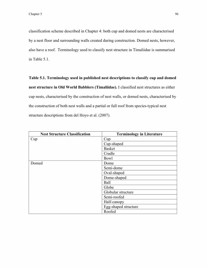
Chapter 5 96
classification scheme described in Chapter 4: both cup and domed nests are characterised
by a nest floor and surrounding walls created during construction. Domed nests, however,
also have a roof. Terminology used to classify nest structure in Timaliidae is summarised
in Table 5.1.
Table 5.1. Terminology used in published nest descriptions to classify cup and domed
nest structure in Old World Babblers (Timaliidae). I classified nest structures as either
cup nests, characterised by the construction of nest walls, or domed nests, characterised by
the construction of both nest walls and a partial or full roof from species-typical nest
structure descriptions from del Hoyo et al. (2007).
Nest Structure Classification Terminology in Literature Cup Cup
Cup-shaped Basket Cradle Bowl
Domed Dome Semi-dome Oval-shaped Dome-shaped Ball Globe Globular structure Semi-roofed Half-canopy Egg-shaped structure Roofed

Chapter 5 97
In addition to nest structure, I recorded the lowest height at which nests were built. I
used the lowest reported nest height because selection pressure exerted by ground predators
should be the greatest at the lowest height at which a nest is built. Whenever nests were
described as being placed on the ground, I entered the nest height as 0 m. All nest structure
and height data are summarised in the Appendix 2.
Phylogenetic comparative statistical methods
Similar to the second analysis in Chapter 4, I used Bayesian Markov-Chain Monte
Carlo (MCMC) methods in order to estimate posterior probability distributions for model
parameters across posterior probability distributions of phylogenies (Pagel and Meade,
2006). For all MCMC analyses, I used 3000 phylogenies obtained from a posterior sample
in a recent Bayesian phylogeny estimation (Jetz et al., 2012; http://birdtree.org/). I used a
version of the phylogenies built only from genetic data and a family ‘backbone’ provided
by a previous phylogenetic estimation (Hackett et al., 2008). I ran all analyses in
BayesTraits (Pagel et al., 2004). I excluded species for which I had nest data but that were
not included in the phylogenetic sample from Jetz et al. (2012) from further analysis (58
exclusions, final n = 97). A maximum clade credibility phylogeny from the posterior
sample of phylogenies is presented in Figure 5.1.
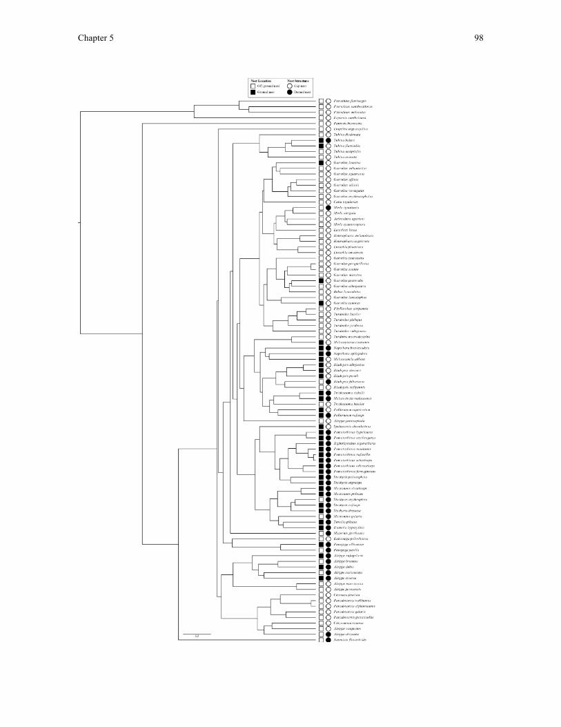
Chapter 5 98

Chapter 5 99
Figure 5.1. A maximum clade credibility phylogeny of Timaliidae species used in this
study. Species-typical nest location (ground or off-ground) and structure (cup or domed)
are listed following each species’ scientific name. This maximum clade credibility
phylogeny was constructed from a Bayesian posterior sample of 3000 phylogenies from
Jetz et al. (2012) that had been constructed using genetic data only and a ‘backbone’ family
estimation by Hackett et al. (2008). Scale bar represents 5 mya (Jetz et al., 2012).
Phylogenetic generalised least squares regression
I transformed lowest nest height data using log(x+1) transformation and compared
these heights between cup- and domed-nest building species using the phylogenetic
generalised least squares regression (PGLS) approach, as in Chapter 4, which incorporates
phylogenetic relatedness into the error term of regression models (Grafen, 1989; Pagel,
1997). In this analysis, I included nest structure as an independent factor on two levels
(‘cup’ and ‘domed’, where cup was the reference level) and nest height as a dependent
continuous variable. I used MCMC to estimate posterior probability distributions for
regression coefficients (β) and phylogenetic signal (λ; Pagel, 1999). I ran MCMC chains
for PGLS analyses for 1 million iterations, sampling every 100 generations, with a ‘burn-in’
period of 50,000 iterations. I used uniform priors (range -100, 100) for all parameters.
As in Chapter 4, prior to analyses, I specified that where ≥95% of the posterior
probability distribution of regression coefficients (β) was in the predicted direction
(negative, following the prediction that domed nests are built at lower heights compared to
cup nests), I would conclude that there was ‘strong evidence’ for the predicted relationship
Chapter 5 100
(for example, Ross et al., 2012). I also report the mean λ from the posterior probability
distributions.
Co-evolution of binary traits
To investigate possible co-evolution of nest height and nest structure, I used Pagel’s
methods for detecting co-evolution of discrete character traits (Pagel and Meade, 2006).
This approach uses continuous-time Markov models to estimate up to 8 transition rates
between states of 2 binary traits. I coded nest height as ‘ground’ where nest height was 0 m,
and ‘off-ground’ where nest height was >0 m. I coded nest structure as before. For these
‘discrete’ analyses (models depicted in Figure 5.2), I ran chains for 100 million iterations,
sampling every 5000 generations, with a ‘burn-in’ period of 50,000 iterations, using
exponential hyper-prior distributions (range 0, 5) for all parameters.
Dependent versus independent evolution
To compare models of dependent versus independent evolution of nest structure and
height, I used the reversible-jump MCMC approach, which estimates transition rates whilst
simultaneously selecting the best-fitting model of evolutionary change by visiting models
in proportion to their posterior probabilities (Pagel and Meade, 2006). In the dependent
reversible-jump model (Figure 5.2A), transition rates for each character are permitted to
depend on the state of the other character, i.e. it is possible that q12≠q34, q13≠q24,
q43≠q21 and q42≠q31, whereas in the independent reversible-jump model (not shown),
transition rates for each character are not permitted to depend on the state of the other
character, such that q12=q34, q21=q43, q13=q24 and q31=q42.

Chapter 5 101
To investigate specifically the hypothesis that building a domed nest co-evolved
with building on the ground, (i.e. q12<q34, q13<q24, q43<q21, and q42<q31), I also ran a
reduced, non-reversible-jump dependent model (Figure 5.2B) in which two transition rates
were estimated, one corresponding to state transitions that I predicted would not be
favourable (i.e. toward building a cup nest on the ground and building a domed nest off the
ground: q12, q13, q43 and q42) and one corresponding to state transitions that I predicted
would be favourable (i.e. toward building a cup nest off the ground and building a domed
nest on the ground: q34, q24, q21 and q31; Figure 5.2B). I predicted that the former rate
would be smaller than the latter rate. I compared this reduced, non-reversible-jump two-rate
model to a reduced, non-reversible-jump one-rate model corresponding to independent
evolution of the traits (not shown), as well as to the unconstrained dependent reversible-
jump model.
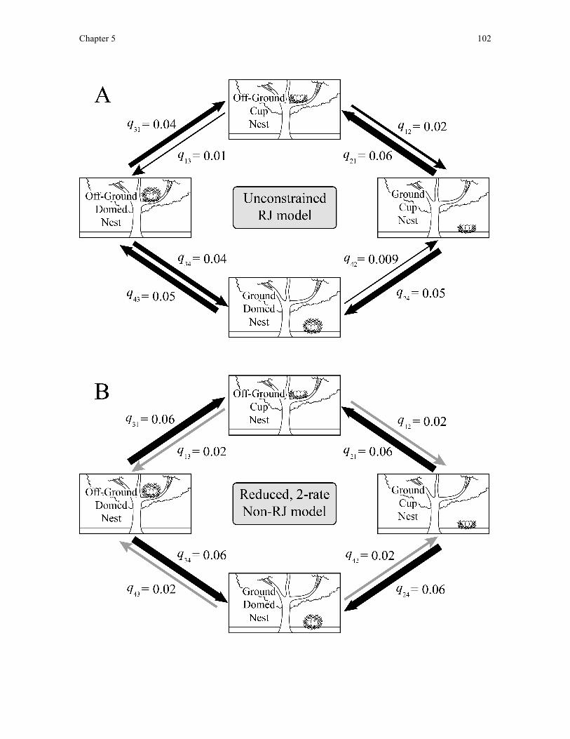
Chapter 5 102
Chapter 5 103
Figure 5.2. Two transition rate models used to investigate the co-evolution of nest
height and structure in Timaliidae. (A) An unconstrained, dependent reversible-jump
(RJ) model used to estimate 8 evolutionary transition rates (q) corresponding to all possible
transitions between nest height and nest structure state combinations. (B) A reduced, non-
RJ dependent model of nest structure and height in which estimated only two transition
rates: transitions toward nest states predicted favourable (black arrows; toward off-ground
cup nest and ground domed nest; q34, q24, q21 and q31) and transitions away from nest
states predicted to be favourable (gray arrows; q12, q13, q43 and q42). Arrow thickness is
proportional to likelihood of the associated transition.
Ancestral states
To investigate the most likely ancestral state of nest structure and nest height in the
most recent common ancestor, I compared three models in which the most recent common
ancestor was fixed as either 1) building a cup nest on the ground, 2) building a domed nest
off the ground, or 3) building a domed nest on the ground to a model in which the most
recent common ancestor was fixed as the predicted ancestral state (off-ground/cup-nesting).
I compared ancestral states models both for the full, dependent reversible-jump model, and
for the reduced, non-reversible-jump two-rate dependent model.
Order of evolutionary transitions
I investigated the likely order of evolutionary transitions by testing whether
transitions from building a cup nest off the ground to building a domed nest on the ground
were more likely to occur through changes in nest height or nest structure (i.e. whether

Chapter 5 104
q12≠q13). I also tested whether transitions from building a domed nest on the ground to
building a cup nest off the ground were more likely to occur through changes in nest height
or nest structure (i.e. whether q43≠q42; Pagel, 1997). I therefore compared reversible-jump
dependent models in which the rates of interest were fixed as equal (transitions through nest
structure and height being equally likely) to unconstrained reversible-jump dependent
models with the prediction that, if the transition rates in nest structure and height differ, the
unconstrained models should be supported over the restricted models.
Model diagnostics and comparison
For all analyses, I ran three MCMC chains to ensure that chains converged on
similar values. All reported model parameters were averaged across the three chains. I used
the program ‘Tracer’ (Rambaut and Drummond, 2009) for visual examination of chains to
ensure convergence and to estimate effective sample size for posterior probability
distributions (E.S.S.). No analysis reported an effective sample size below 13,000 for model
parameters. I used Bayes Factors (B.F.) to compare model fit based on the harmonic means
of the model likelihoods where, by convention, a positive value of >2 is taken as ‘positive
evidence’ and 5-10 as ‘strong’ evidence for the better fitting model (Pagel et al., 2004). I
took harmonic means from the final iteration in the MCMC chain.
Results
Nest heights of cup and domed nests
I found strong evidence that species that build domed nests build them closer to the
ground than do those species that build cup nests (Figure 5.3; 99% β < 0, λ = 0.64, n = 97).

Chapter 5 105
Figure 5.3. Domed-nesting species in Timaliidae build nests at lower heights than cup-
nesting relatives. Bars represent average predicted log(x+1)-transformed lowest nest
heights of cup and domed nesting species in Timaliidae calculated using phylogenetic least
squares regression. Error bars represent 95% confidence interval. *≥95% of the posterior
probability distribution of regression coefficients were in the predicted, negative direction
(following the prediction that domed-nesting species would construct nests at lower heights
than cup-nesting relatives).
Co-evolution of domed- and ground-nesting
I found positive evidence for the unconstrained, dependent reversible-jump model
over the unconstrained, independent reversible-jump model (B.F. = 4.0, n = 97), suggesting

Chapter 5 106
co-evolution of nest structure and nest height. Mean transition rates from in the
unconstrained dependent reversible-jump model supported the hypothesis of both co-
evolution of building a domed nest on the ground and building a cup nest off the ground, i.e.
q12<q34, q13<q24, q43<q21 and q42<q31 (Figure 5.2A).
The reduced, non-reversible-jump, two-rate model of dependent evolution was
strongly favoured over a reduced, non-reversible-jump one-rate model of independent
evolution (B.F. = 9.0, n = 97), further suggesting co-evolution of both building of domed
nests with nesting on the ground and the building of cup-nests when nesting off the ground.
I also found positive evidence for the reduced, non-reversible-jump, two-rate model over
the unconstrained, reversible-jump dependent model (B.F. = 4.8). Mean transition rates
estimated in the reduced, non-reversible-jump 2-rate model of dependent evolution
corresponded to the hypothesis of co-evolution of domed-nests with building on the ground
and cup-nests with building off the ground, i.e. q12<q34, q13<q24, q43<q21 and q42<q31
(Figure 5.2B).
Ancestral states
Under the unconstrained reversible-jump dependent model, the most probable
ancestral state was building a cup nest off the ground. I found positive evidence that a cup
nest built off the ground was more probable than was a cup nest built on the ground (B.F. =
3.28), but I had insufficient evidence to show that a cup nest built off the ground was more
probable as the ancestral state than was a domed nest built off the ground (B.F. = 1.14). I
found strong evidence that a cup nest built off the ground was the more probable ancestral
state than was a domed nest built on the ground (B.F. = 7.05).
Chapter 5 107
Under the reduced two-rate non-reversible-jump dependent model, the most
probable ancestral state was also building a cup nest off the ground. I had insufficient
evidence to show that building a cup nest off the ground was more probable than was
building a cup nest on the ground (B.F. = 1.58), but I found positive evidence that building
a cup nest off the ground was a more probable ancestral state than was building a domed
nest irrespective of location (on-ground: B.F. = 4.72, off-ground: B.F. = 2.43).
Order of evolutionary transitions
Transition rates from the unconstrained reversible-jump dependent model (Figure
5.2A) suggest that a change from building a cup nest off the ground to building a domed
nest on the ground was more likely to occur through a change in nest height than in nest
structure (i.e. q12 > q13). Similarly, a change from building a domed nest on the ground to
building a cup nest off the ground was more likely to occur through a change in nest height
than in nest structure (i.e. q43 > q42). Fixing q12 = q13, however, did not reduce model fit
relative to the unconstrained reversible-jump model (B.F. 3.0, in favour of the reduced
model), suggesting that changes in nest height and nest structure when building cup nests
off the ground are equally likely. Reversible-jump models that fixed q43 = q42 did reduce
the model fit in comparison to the unconstrained dependent reversible-jump model (B.F.
4.2, in favour of the unconstrained model), suggesting that a transition from building a
domed nest on the ground to building a cup nest off the ground is more likely to occur
through a change in nest height than in nest structure.
Chapter 5 108
Discussion
Using phylogenetic comparative statistical techniques, I found evidence to support
the proposed co-evolution of nest height and structure in Old World Babblers (Timaliidae).
Together, my analyses showed that those species in this group that build domed nests build
their nests at a lower height than do related species that build cup nests and strongly suggest
that building a domed nest and nesting on the ground co-evolved as derived traits.
Furthermore, although transitions away from building a cup nest off the ground are equally
likely to occur through changes in either nest height or nest structure, transitions away from
building a domed nest on the ground are more likely to occur through changes in nest
height rather than in nest structure. To my knowledge, this is the first demonstration of co-
evolution between the structure and location of bird nests.
Using nest height as a continuous variable, I found that domed-nesting babblers
construct nests at lower heights than do cup-building relatives. Comparison of dependent
models of evolution in which transitions in nest height were permitted to depend on
transitions in nest structure were favoured over models in which nest height and structure
evolved independently. In restricted models of dependent versus independent evolution,
evolutionary transitions towards either building a cup nest off the ground or a domed nest
on the ground are more likely than are transitions away from these two nest state
combinations. These data support Collias’ (1997) original prediction that nest height and
structure co-evolve in Timaliidae.
Although my analysis here provides strong support that ground-nesting and building
a domed nest co-evolved as derived traits in Timaliidae, my findings can only provide
indirect support for these transitions being driven by selective factors including nest-site
Chapter 5 109
competition and predation, as hypothesised by Collias (1997). Future studies should look
to obtain direct measurements of nest predation at varying heights in the forest edge
environments species in Timaliidae inhabit to provide more direct evidence for a role of
predation and nest-site competition in the evolution of nest-building behaviour.
Furthermore, other factors, such as protection from weather conditions at different heights
in the forest, should be considered and tested in future studies on the evolution of nest
structure in Timaliidae. Because this analysis was constructed from Collias’ hypothesis and
no other current hypotheses have been established to predict the co-evolution described
here, I will mainly focus on how our data alongside Collias’ hypotheses might suggest a
role for predation at specific heights in shaping nest-building behaviour in birds. Outside of
species nesting in cavities, a role for nest-site competition has not been measured in species
that construct cup and domed nests, but here I suggest Timaliidae may be an interesting
system to study whether this competition could influence nest-building behaviour in birds.
In addition to providing support for the co-evolution of nesting on the ground and
constructing a domed nest, here I provide some of the first cross-species statistical evidence
to support the idea that building a cup nest off the ground or a domed nest on the ground
are both more likely to be favoured by selection (hypothesised by Collias [1997] to be
attributed to reduced predation pressure) than are either domed nests built off the ground or
cup nests built on the ground, at least in the Timaliidae (Collias and Collias, 1984). Also in
support of a role for ground predation in influencing nest-building behaviour (as
hypothesised by Collias [1997]), ground predation by introduced terrestrial mammals
seems to explain the change in nest elevation in the Hawaiian monarch flycatcher (Oahu
elepaio), which now constructs its open nest 50% higher than was reported in 1995
Chapter 5 110
(Vanderwerf, 2012). A general change in nest height in response to changing predation
pressure is not necessarily to be expected, however, as pointed out by Newmark and
Stanley (2011): the effect of nest height on nest predation is likely to be species-specific
and influenced by the importance of predators operating at different heights in different
habitats. In support of selection favouring enclosed nests on the ground, a previous study
using artificial nests placed on the ground found that eggs placed in domed nests were less
susceptible to predation than were those placed in cup nests (Linder and Bollinger, 1995).
Although studies using artificial nests to assess predation rates have been heavily criticised
for a lack of external validity (Moore and Robinson, 2004), my results indirectly support
the conclusions of Linder and Bollinger (1995), as here I found that selection is likely to
favour domed nests over cup nests when building on the ground in Timaliidae.
Both my two different models of dependent evolution (unconstrained reversible-
jump and restricted, non-reversible-jump) demonstrated that building a cup nest off the
ground is more likely to have been the ancestral state than was building a domed nest on
the ground in the Timaliidae. These results support Collias’ (1997) prediction that domed-
nests and building on the ground co-evolved as derived traits in this family. When I
examined the order of evolutionary transitions from cup nests off the ground to the likely
derived state of domed nests on the ground, I found that changes in either nest structure or
nest height were equally likely, providing support for both of these evolutionary pathways.
In contrast, Collias (1997) predicted that transitions from a building a cup nest off the
ground state would occur primarily as shifts to ground-nesting to avoid competition for
limited nest sites off the ground. The effect of competition for nest sites on nest site
selection is well documented in species nesting in natural or excavated cavities
Chapter 5 111
(Brightsmith, 2005). Furthermore, communal defence, territoriality, and an absence of
coloniality in Timaliidae (Collar and Robson, 2007) may restrict the number of nesting sites
available off the ground. Investigating how competition for nest sites relates to selection for
nest sites could help identify the selection pressures involved in the evolutionary transition
toward nesting on the ground.
Unlike these transitions to building a cup nest on the ground, Collias (1997) argued
that transitions from building a cup nest off the ground to building a domed nest off the
ground are unfavourable because nesting off the ground already confers protection from
predators and birds should avoid the presumed higher energetic cost of additional
construction to create a nest roof (Bailey et al., 2014). Here I found that, from building a
cup nest off the ground, transitions to building a domed nest off the ground were equally
likely as transitions to building a cup nest on the ground. The evolutionary pathway to a
domed nest built off the ground may be a response to increased nest predation: Newmark
and Stanley (2011) found that, among nest structures, predation rates were the highest for
open and cup nests regardless of nest height in Afrotropical bird communities inhabiting
forest edges produced by fragmentation. Alternatively, the transition from cup to domed
nest building in off-ground nesting lineages could represent another evolutionary path
toward the construction of domed nests proposed by Collias (1997). Specifically, that
domed nests may be favoured for those species that construct their nests in the canopy
periphery because an enclosed nest could mitigate the effects of increased exposure to
aversive weather experienced by nests placed farther away from the tree trunk.
Incorporating nest location within off-ground sites could enable statistical tests of this
alternative, but equally likely, evolutionary route.
Chapter 5 112
Once birds were building domed nests on the ground, I found that transitions to
nesting off the ground were more likely than were transitions to building a cup nest on the
ground. This supports my prediction that evolutionary transitions in nest height would be
more likely than would changes in nest structure. Previous reports on phenotypic plasticity
in nest height (Lima, 2009; Vanderwerf, 2012) also suggest that nest height is more easily
changed than is nest structure. Furthermore, transitions from building a domed nest on the
ground to building a cup nest on the ground probably increase susceptibility to nest
predation due to the abundance of ground predators in forest edge habitats (Söderström et
al., 1998), making this transition highly unfavourable. Strong selection pressure against
transitions from building domed nests to building cup nests in ground-nesting lineages is
also supported by the transitions rates calculated in my unconstrained reversible-jump
model (i.e. Figure 5.2A; q42 < q43).
In sum, here I present the first formal analyses of co-evolution between nest height
and structure in Timaliidae. I found that building a domed nest and doing so on the ground
is highly likely to have co-evolved in this family as derived traits providing indirect support
for suggestions that nest predation and nest site competition are two selective forces that
may influence nest structure design and nest site selection.
Chapter 6 113
Chapter 6 – General discussion
During the research in my thesis, I identified a number of neural circuits that may be
involved in nest building in zebra finches and I used my own nest structure classification
scheme to characterize how the evolution of nest structure relates to brain morphology and
the hypothesised influences of nest-site competition and predation. Specifically, I used the
expression of the immediate early gene product Fos, an indirect marker of neuronal activity,
to identify brain regions exhibiting elevated brain activity during nest-building behaviour.
Using phylogenetically-informed statistical techniques, I tested whether variation in
cerebellar foliation, hypothesized to play a role in the development of motor control, could
be explained by the structural complexity of the species-typical nest built. Also using
phylogenetically-informed analysis, I performed the first formal statistical test to
investigate the evolution of nest height and structure in Old World babblers (Timaliidae).
Summary of Fos production and nest-building behaviour relationships
By comparing the number of neurons producing the immediate early gene product
Fos in different neural circuits to nest-building behaviour exhibited by male and female
zebra finches, I identified brain regions that are activated during nest building. I showed
that neuronal activity in all three components of the anterior motor pathway, the anterior
striatum, the anterior nidopallium, and the anterior ventral mesopallium increased the more
male zebra finches picked up nest material (Chapter 2).
In the social behaviour network, neuronal activity in the anterior hypothalamus and
medial bed nucleus of the stria terminalis, ventral subdivision (BSTmv) increased the more
Chapter 6 114
female finches spent time in the nest (Chapter 2), however, in BSTmv this relationship does
not appear to involve vasotocinergic or mesotocinergic neuronal subpopulations (Chapter
3). Neuronal activity in the medial preoptic area and medial bed nucleus of the stria
terminalis, dorsal subdivision (BSTmd) increased in nest-building birds, regardless of sex,
compared to controls (Chapter 2) and, in BSTmd, this increased neuronal activity appears
to occur specifically in mesotocinergic neurons (Chapter 3). By sampling neuronal activity
specifically in vasotocinergic and mesotocinergic neuronal subpopulations in the social
behaviour network, I found that, in male finches, neuronal activity in vasotocinergic
neurons in BSTmd and BSTmv increased the more time a male spent together with his
mate in the nest and the more a male picked up nest material, respectively (Chapter 3).
In the dopaminergic reward system, neuronal activity in the ventral tegmental area
increased the more male finches picked up nest material (Chapter 2), however, this
relationship did not appear to involve dopaminergic neurons within this brain region
(Chapter 3). Instead, neuronal activity in dopaminergic neurons in the ventral tegmental
area decreased the more male finches tucked material into the nest structure. Finally,
neuronal activity in dopaminergic neurons within the central gray increased in male finches
the more time they spent in the nest with their mates (Chapter 3).
In summary, I found evidence suggesting the anterior motor pathway, social
behaviour network, and dopaminergic reward system may all be involved in nest-building
behaviour. Furthermore, some aspects of nest-building behaviour may involve specifically
the vasotocin-mesotocin and dopaminergic neuronal subpopulations contained in the social
behaviour network and dopaminergic reward system, respectively. In the following sections
of this discussion, I will speculate about how each of these neural circuits may contribute to
Chapter 6 115
the control of nest-building behaviour and, reciprocally, what a role in controlling nest
building might tell us about the more general functions of these neural circuits.
The anterior motor pathway and nest building
The pattern of increased neuronal activation in the anterior motor pathway during
nest building suggests that the anterior motor pathway controls the initiation of motor
sequences. In the original paper in which they described the anterior motor pathway,
Feenders et al. (2008) reported elevated activity in this neural circuit following the
production of a variety of locomotor behaviours in birds, although the importance of motor
sequencing in these behaviours, which included wing-whirring in garden warblers (Sylvia
borin) and hovering flight in hummingbirds, is difficult to assess. During nest building,
however, activity in the anterior motor pathway increased the more male finches exhibited
the first step in the nest-building sequence (the collection of material) but was unrelated to
the number of times males deposited that material in the nest, the final step in the sequence.
Because of this relationship, it seems plausible that the anterior motor pathway would be
involved at the beginning of behavioural sequences. In order to test whether this neural
circuit is involved in the beginning of motor sequences and not the specific action I
quantified (picking up material; Chapter 2), it would be useful to record neuronal activity
using electrophysiological techniques in the anterior motor pathway in birds while they
perform motor sequences of interest. Using this paradigm both in birds performing
sequences comprised of different actions (for example, nest building) as well as birds
performing sequences consisting of the same action (for example, a series of pecks to
receive a food reward as in Helduser and Güntürkün [2012]), it would be possible to test

Chapter 6 116
whether neuronal activity is always associated with the beginning of a sequence or with the
specific actions the sequence contains.
I propose that, in addition to this role in beginning motor sequences, the anterior
motor pathway may also be involved in the learning and modification of motor actions, two
functions for this neural circuit that were originally proposed by Feenders et al. (2008).
Feenders et al. (2008) based their proposal on the evidence that the song-control nuclei that
are involved in learning and modifying birdsong are located within close proximity to the
brain regions comprising the anterior motor pathway. This proximity was interpreted to
suggest that the anterior motor pathway plays a more general role in motor learning than
does the nearby song-control system, which is involved exclusively in the motor learning
associated with birdsong. We could test whether the anterior motor pathway is involved in
motor learning by adapting paradigms previously used to demonstrate the relationship
between the song-control system and motor learning involved in birdsong for testing the
relationship between the anterior motor pathway and the motor learning involved in nest-
building behaviour. For example, the lateral portion of the song nucleus MAN (lMAN), a
song nucleus located within close proximity to the anterior nidopallium of the anterior
motor pathway, is required to learn how to produce the actions involved in species-typical
birdsong in juvenile male zebra finches (Bottjer et al., 1984). If the anterior nidopallium is
involved in learning the actions required to build a nest then lesions to the anterior
nidopallia in juvenile birds should lead to no improvement in nest-building skills with
experience compared to intact controls, but without impairing previously learned motor
skills. One behavioural system in which this could be tested is the development of weaving
skill by male Village weaver birds who increase the number of pieces of nest material they
Chapter 6 117
can weave successfully into a nest site with weaving experience (Collias and Collias, 1964).
In this system, I would predict that anterior nidopallial lesions would prevent birds from
improving their ability to successfully weave material into nest-sites compared to controls.
Furthermore, such anterior nidopallial lesions should not impair weaving success compared
to levels prior to lesioning, suggesting a deficit specifically in the learning of new motor
skills and not previously learned skills or motor output. Furthermore, as lMAN is also
involved in modifying previously learned birdsong in adulthood (Kao and Brainard, 2005;
Kojima and Doupe, 2011), the anterior nidopallium may be involved in modifying nest-
building actions in adult birds. If so, then lesions to the anterior nidopallia in adult male
zebra finches might then lead to an experienced bird being unable to modify how he picks
up and delivers nest material to the nest box and, instead, continue to use nest-building
actions expressed prior to lesioning (Muth and Healy 2011). One could also use Helduser
and Güntürkün’s (2012) paradigm in which a bird is trained to peck five keys in a rewarded
order to test if the anterior motor pathway is involved in modifying all behavioural
sequences and not just nest building: birds trained to peck a specific sequence of buttons
and subsequently given anterior nidopallia lesions should be unable to modify their
sequence of pecks in response to changes in the rewarded sequence, for example, by adding
a new peck or rearranging the rewarded order of pecks.
Finally, the concerted increase in neuronal activity in all three regions sampled in
the anterior motor pathway during nest building provides support for a recent theory
regarding the functional organisation of the avian telencephalon compared to mammalian
neocortex. Whereas functional divisions in mammalian neocortex consist of a stack of six,
abutting layers in which incoming information is received and processed through

Chapter 6 118
connections between the six layers of neocortex, functional divisions in the avian
telencephalon are historically considered to be comprised of interconnected, but
anatomically isolated nuclei located throughout the brain (termed “nuclear” organisation;
Karten, 1997). Because this distinction in brain organisation between mammals and birds
has been interpreted as evidence that the neocortex and the avian pallia developed from
distinct evolutionary processes (Karten and Shimizu, 1989), avian homologs of mammalian
neocortex are rarely proposed, hampering comparative studies with the mammalian
neocortex. Via a tract-tracing study in 2010, however, Wang et al. (2010) found that
auditory regions abutting one another in the avian brain are heavily interconnected across
striatal, nidopallial, and mesopallial brain divisions and resemble the connectivity reported
across the six layers of mammalian neocortex. As a result it has been suggested that based
on anatomical contiguity of brain regions across major brain divisions and similar
molecular profiles during development the anterior striatum, anterior nidopallium, and
anterior ventral mesopallium of the anterior motor pathway form a similar functional
division akin to that reported in the avian auditory system and in the mammalian neocortex
(Chen et al., 2013). Although the arrangement of regions in the anterior motor pathway
suggests they may be involved in the same neural processes, data demonstrating that all
regions of this pathway are functionally involved in the same types of information
processing are crucial to defining them as a functional unit.
The concerted increase in neuronal activity in all three regions of the anterior motor
pathway the more male finches picked up nest material is consistent with the notion that all
three of these regions are involved in the same functional processes and are likely, therefore,
to be interconnected and activated as a functional unit. One could confirm this by using
Chapter 6 119
tract-tracing techniques to visualise connections between these three regions across pallial
divisions. If the avian telencephalon and mammalian neocortex exhibit similar functional
organisation, we could begin to study potentially homologous regions of the avian brain
and mammalian neocortex to understand how the brain controls similar behavioural
processes across these distinct taxa.
The social behaviour network and nest building
Whereas the anterior motor pathway appears to be involved in the motor control
underlying behaviour, the social behaviour network is thought to be involved in the
production of social behaviours including aggression, copulation, and parental care
(Goodson, 2005). Prior to the work presented in this thesis, there was no evidence for the
involvement of the social behaviour network in nest-building behaviour. Indeed, in the only
previous study in which the authors looked for correlations between patterns of neuronal
activity in the social behaviour network and nest-building behaviour in birds, they found no
evidence for a relationship (Klatt and Goodson, 2013). As discussed in Chapters 2 and 3, it
is plausible that those authors failed to find this relationship because, at least in part, they
sampled the medial bed nucleus of the stria terminalis (BSTm) as a single brain region. As
a result of Klatt and Goodson (2013)’s data, others have assumed that the social behaviour
network is not involved in nest-building behaviour but have, instead, suggested that this
behaviour may be controlled by the paraventricular nucleus of the hypothalamus (PVN; for
example, Kelly and Goodson, 2014). To confirm this lack of relationship, however, the
potential parcellation of BSTm into a dorsal and ventral subdivision (BSTmd and BSTmv,
respectively) needs to be tested functionally. This could be done by focally lesioning or
Chapter 6 120
administering vasotocin to each BSTm subdivision and testing for subsequent changes in
nest-building behaviour. Based on my interpretation of the data from female nesting finches
in Chapter 2, lesions to the BSTmv, but not BSTmd, in female finches would lead to these
birds spending less time in the nest. Based on my interpretation of the data from male nest-
building finches in Chapter 3, using a chronically implanting cannulae to administer
vasotocin directly to BSTmd should increase the amount of time a male finch spends with
his partner in the nest cup without affecting nest material collection, whereas administering
vasotocin to BSTmv should increase the number of times males picked up nest material
without influencing the time a male spends in the nest cup with his partner. Given that
BSTm is increasingly studied for its role in a whole array of social and breeding behaviours
(Goodson, 2005), it would be useful to determine whether functional subdivisions exist in
this region sooner rather than later.
Because the social behaviour network appears to be involved in the expression of all
breeding behaviours in birds, including courtship, copulation, incubation, territoriality
(O’Connell and Hofmann, 2011), and now nest building, I propose that the social behaviour
network is involved, at least in part, in coordinating the expression of these behaviours
across the breeding season. This coordination could be achieved physiologically through
temporal changes in the levels of hormones, nonapeptides, and neurotransmitters released
and acting in the social behaviour network. Support for this possibility comes from the
demonstration that knocking down mesotocin production in adult zebra finches using
antisense mRNA both impairs pair formation and reduces nest occupation behaviour in
females (Kelly and Goodson, 2014), suggesting that mesotocin in the brain may be
necessary for both pair formation and the subsequent occupation and defence of a nest site
Chapter 6 121
in female zebra finches. Whereas Kelly and Goodson (2014) suggest that this coordinated
increase in both affiliative behaviours associated with pair formation and nest occupation
occurs due to the actions of mesotocin in PVN, I found evidence that mesotocinergic
neurons in BSTmd may be involved in possession of a nest site (Chapter 3). Mesotocin in
the brain may, therefore, influence pair formation and nest occupation through its actions in
PVN and BSTmd, respectively, in female zebra finches. Such coordinated changes in
behaviour caused by the actions of a signalling molecular acting in multiple locations in the
brain have been demonstrated in zebrafish (Danio rerio), in which widespread release of the
neurotransmitter histamine produced changes in aggression, boldness, and exploration in
adult fish (Norton et al., 2011). If mesotocinergic neurons in BSTmd are involved nest
occupation and mesotocinergic neurons in PVN are involved in pair formation,
administering mesotocin to the social behaviour network in female finches with BSTmd
lesions should increase affiliation behaviours associated with pair formation without
increasing nest occupation exhibited by these birds.
The dopaminergic reward system and nest building
Whereas it seems plausible that the anterior motor pathway and social behaviour
network are involved in the motor control and coordination of nest-building behaviour,
respectively, the dopaminergic reward system seems to be involved in reinforcing male
nest-building behaviour in zebra finches. Specifically, activation of the ventral tegmental
area in the dopaminergic reward system seems to reward nest material collection and
discourages nest-building behaviour within the nest cup in male zebra finches. One would,
then, expect that neuronal activity in the ventral tegmental area and dopaminergic neurons
Chapter 6 122
within this brain region might reflect an individual bird’s contributions to nest building in
other bird species. For example, in a species in which females collect material and males
construct the nest, activity across all neurons in the ventral tegmental area in females
should increase the more a female picks up nest material and activity specifically within
dopaminergic neurons should decrease the more the female contributes to construction of
the nest at the nest site, as found in male zebra finches.
Although compared to the ventral tegmental area, much less is known about the
functions of the central gray, my data provide some support for Goodson et al.’s (2009)
suggestion that dopaminergic neurons in the central gray are involved in the motivation to
communicate vocally with conspecifics. Quantifying as much nest-building behaviour and
social behaviour performed by males and females within the nest is crucial for identifying
whether or not neuronal activity in the central gray is associated with nest-building actions
or, following Goodson et al.’s (2009) hypothesis, vocal interactions between the individuals
in a nesting finch pair. Another approach to testing whether this dopaminergic
subpopulation is involved in social interaction during nest building would be to sample
neuronal activity in a male zebra finch building a nest while exposed to a female in an
adjacent cage, where she is unable to enter the nest cup. If central gray dopaminergic
neurons are involved in social interactions within the nest during nest building, then
neuronal activity in this subpopulation should be both unrelated to any nest-building
behaviour exhibited by the lone male and lower than in male finches building a nest with a
female partner within the same cage.

Chapter 6 123
Using neurobiology to compare nest building and tool use
In addition to an increasingly large body of work challenging the assumed genetic
origins of nest building by identifying a role for learning and experience (see Chapter 1),
my work provides new data enabling the comparison of the neurobiology of nest-building
and tool use behaviour. Based on the data currently available regarding the neurobiology of
nest-building behaviour and tool use, I propose that these two construction behaviours are
controlled by the same neurobiological processes and may represent two different
elaborations of the same sensory-motor processes (Barton, 2012).
One method for testing whether two behaviours use the same neurobiological
processes is to demonstrate that the same brain regions are functionally involved in both
behaviours. One brain region involved in both tool use and nest-building behaviour is the
cerebellum. In primates, a larger cerebellum appears to have coevolved with the use of
extractive foraging techniques (Barton, 2012) and, in birds, a more foliated cerebellum is
coincident with tool use (Iwaniuk et al., 2009). Barton has suggested the enlargement of the
primate cerebellum enables the learning and execution of increasingly elaborate
behavioural sequences, including both tool use and the production and comprehension of
language (Barton, 2012). There may be a similar relationship between cerebellar structure
and function in birds: the evolution of a more foliated cerebellum may have enabled the
learning and execution of increasingly elaborate behavioural sequences including both the
manufacture and use of tools (Iwaniuk et al., 2009) and the manipulative abilities and motor
sequencing required to construct a more structurally complex nest (Chapter 4).
Although this correlated evolution between the cerebellum, tool use, and nest
building suggests that the evolution of a more foliated cerebellum supports these
Chapter 6 124
behaviours, implying the function of a brain region by anatomy alone can be misleading
(Healy and Rowe, 2007) and requires complementary functional studies to demonstrate that
the cerebellum is active during both tool use and nest-building behaviour. Evidence that the
cerebellum is activated during tool use in Japanese monkeys has been shown using positron
emission tomography (Macaca fuscata; Obayashi et al., 2001) and although this functional
imaging technique has recently been adapted for use in crows (Marzluff et al., 2012),
current technological limitations prevent this technique from being used in smaller birds
such as zebra finches. Using a similar Fos immunohistochemical protocol as in Chapters 2-
3 to sample neuronal activity in the cerebellum would, however, address whether cerebellar
activity is correlated with the production of nest-building behaviour and tool use in birds,
providing some functional support for the involvement of the cerebellum in both
behaviours. In addition to the cerebellum, several other regions in the mammalian brain are
known to be activated during tool use (Obayashi et al., 2001), however, current debate over
the homology of the avian telencephalon and mammalian neocortex complicates
comparisons of neocortical brain regions active during tool use. One brain region found in
both birds and mammals and activated during tool use is the striatum (Reiner et al., 2004;
Obayashi et al., 2001), which I found is also activated during nest building in male zebra
finches. With the data currently available, then, at least two brain regions, the cerebellum
and striatum, appear to be involved in the production of tool use and nest-building
behaviour, suggesting these behaviours use the same neurobiological processes. By
mapping patterns of neuronal activity associated with tool use throughout the avian brain,
the neurobiological comparison of these two behaviours can extend beyond the cerebellum
and striatum to the rest of the brain.
Chapter 6 125
The evolution of nest structure
I developed the first nest classification scheme to adapt pre-existing descriptions of
nest structure into data amenable to formal statistical tests of evolution. By demonstrating
that this classification scheme can be used to identify neural substrates (Chapter 4) that may
be involved in nest structure, I also found evidence that this classification scheme may
reflect some aspect of the behaviour underpinning the construction of the nest. Whereas
previous studies attempting to identify evolutionary pressures that affect nest building have
relied on comparisons between sympatric species, which may ignore other species
differences in nest building, or on the use non-formal statistical techniques, such as
outgroup comparison, the statistical models I used allowed me to explain variation in nest
structure across a large number of species while accounting for species relatedness. The
success of this analysis would suggest that this classification scheme and statistical
approach could be used to test a number of other theories regarding the evolution of nest
building, with the added benefit of using previously compiled descriptions of nest
structures, eliminating the need for additional data collection. For example, Winkler and
Sheldon (1993) found that the construction of an increasingly enclosed, retort-shaped nest
coevolved with higher breeding densities in swifts (Apodidae). The authors hypothesise
that constructing a more enclosed nest may lessen the threat of extra-pair fertilisations, a
hypothesis that could be tested by investigating potential correlated evolution between nest
structure and breeding density in this clade. Additionally, in my own analysis on nest
structure, I found evidence suggesting that competition for limited nest-sites and predation
are two key evolutionary pressures that have influenced the evolution of nest structure and
location. Because Timaliidae is just one radiation of passerines, one would expect to see
Chapter 6 126
nest-site competition and predation pressure influence nest location and structure in other
groups of birds. For example, in study sites in Arizona and Arkansas forests, where
predation pressure is lowest on the ground (Martin, 1993) one would expect species
constructing nests off-ground should be more likely to construct a domed nest to confer
protection from the heightened predation pressure. By using such comparative analyses,
one might be able to elucidate the variety of evolutionary pressures that may have helped
produce the tremendous diversity in nest structure seen today.
In this thesis, I sought to integrate data from behavioural, neural, and evolutionary
sources and paradigms to enable a holistic understanding of nest-building behaviour. This
approach has led me to not only identify neural substrates involved in nest-building
behaviour but also how these neural substrates may specifically contribute to nest building
and to identify the evolutionary pressures that may have acted on the brain and behaviour to
produce variation in nest structures. For example, extrapolating from my interpretation of
the data in Chapters 4 and 5, I could predict that elevated predation pressure on the ground
would favour ground-nesting species with more foliated cerebella that may enable the
manipulative skills to construct a domed nest and confer protection from this ground
predation. By continuing to establish approaches to the neurobiological control of
behaviour using both functional neuroscience and comparative studies, we understand not
only how the brain controls behaviour but also how these brain-behaviour relationships
may vary across species exhibiting behavioural differences.

Bibliography 127
Bibliography
Alabrudzinska J, Kalinski A, Slomczynski R, Wawrzyniak J, Zielinski P, Banbura J (2003)
Effects of nest characteristics on breeding success of great tits Parus major. Acta
Ornithologica 38:151-154.
Anderson EM (1915) Nesting of the Bohemian waxwing in northern British Columbia.
Condor 17:145-148.
Aste N, Balthazart J, Absil P, Grossmann R, Mülhbauer E, Viglietti-Panzica C, Panzica GC
(1998) Anatomical and neurochemical definition of the nucleus of the stria
terminalis in Japanese quail (Coturnix japonica). Journal of Comparative Neurology
396:141-157.
Atoji Y, Wild JM (2006) Anatomy of the avian hippocampal formation. Reviews in the
Neurosciences 17:3-15.
Bailey IE, Morgan KV, Bertin M, Meddle SL, Healy SD (2014) Physical cognition: birds
learn the structural efficacy of nest material. Proceedings of the Royal Society B:
Biological Sciences 281: 20133225.
Bailey DJ, Rosebush JC, Wade J (2002) The hippocampus and caudomedial neostriatum
show selective responsiveness to conspecific song in the female zebra finch. Journal
of Neurobiology 52:43-51.
Balthazart J, Absil P (1997) Identification of catecholaminergic inputs to and outputs from
aromatase-containing brain areas of the Japanese quail by tract tracing combined
with tyrosine hydroxylase immunocytochemistry. Journal of Comparative
Neurology 382:401-428.

Bibliography 128
Balthazart J, Ball GF (2007) Topography in the preoptic region: differential regulation of
appetitive and consummatory male sexual behaviors. Frontiers in
Neuroendocrinology 28:161-178.
Balthazart J, Surlemont C (1990) Androgen and estrogen action in the preoptic area and
activation of copulatory behavior in quail. Physiology & Behavior 48:599-609.
Banerjee SB, Dias BG, Crews D, Adkins-Regan E (2013) Newly paired zebra finches have
higher dopamine levels and immediate early gene Fos expression in dopaminergic
neurons. European Journal of Neuroscience 38:3731-3739.
Barton RA (2012) Embodied cognitive evolution and the cerebellum. Philosophical
Transactions of the Royal Society B: Biological Sciences 367:2097-2107.
Behrendt RP (2013) Situationally appropriate behavior: translating situations into appetitive
behavior modes. Reviews in the Neurosciences 24:577-606.
Bharati IS, Goodson JL (2006) Fos responses of dopamine neurons to sociosexual stimuli
in male zebra finches. Neuroscience 143:661-670.
Bottjer SW, Miesner EA, Arnold AP (1984) Forebrain lesions disrupt development but not
maintenance of song in passerine birds. Science 224:901-903.
Bouckaert RR (2010) DensiTree: making sense of sets of phylogenetic trees.
Bioinformatics 26:1372–1373.
Brightsmith DJ (2005) Competition, predation and nest niche shifts among tropical cavity
nesters: ecological evidence. Journal of Avian Biology 36:74-83.
Butler AB, Hodos W (2005) Comparative vertebrate neuroanatomy. New York, US: Wiley-
Liss.
Charlier TD, Ball GF, Balthazart J (2005) Sexual behavior activates the expression of the
Bibliography 129
immediate early genes c-fos and Zenk (egr-1) in catecholaminergic neurons of male
Japanese quail. Neuroscience 131:13-30.
Chen CC, Winkler CM, Pfenning AR, Jarvis ED (2013) Molecular profiling of the
developing avian telencephalon: regional timing and brain subdivision continuities.
Journal of Comparative Neurology 521:3666-3701.
Clayton DF (2000) The genomic action potential. Neurobiology of Learning and Memory
74:185–216.
Collar NJ, Robson C (2007) Family Timaliidae (Babblers) in: Handbook of the Birds of the
World. 12:70-291.
Collias NE (1997) On the origin and evolution of nest building by passerine birds. Condor
99:253-270.
Collias NE, Collias EC (1962) An experimental study of the mechanisms of nest building
in a weaverbird. The Auk 79:568-595.
Collias NE, Collias EC (1964) The development of nest-building behavior in a weaverbird.
The Auk 81:42-52.
Collias NE, Collias EC (1984) Nest building and bird behavior. Princeton, US: Princeton
University Press.
Eberhard JR (1998) Evolution of nest-building behavior in Agapornis parrots. The Auk
115:455-464.
Elie JE, Mariette MM, Soula HA, Griffith SC, Mathevon N, Vignal C (2010) Vocal
communication at the nest between mates in wild zebra finches: a private vocal
duet? Animal Behaviour 80:597-605.
Feenders G, Liedvogel M, Rivas M, Zapka M, Horita H, Hara E, Wada K, Mouritsen H,

Bibliography 130
Jarvis ED (2008) Molecular mapping of movement-associated areas in the avian
brain: a motor theory for vocal learning origin. PLOS ONE 3:e1768.
Frith HJ (1959) Breeding of the mallee-fowl, Leipoa ocellata Gould (Megapodiidae).
Wildlife Research 4:31-60.
Gardner EL (2011) Addiction and brain reward and antireward pathways. Advances in
Psychosomatic Medicine 30:22-60.
George I, Hara E, Hessler NA (2006) Behavioral and neural lateralization of vision in
courtship singing of the zebra finch. Journal of Neurobiology 66:1164–1173.
Gooders J, Lambert T, Arlott N (1982) Collins British Birds. London, UK: William Collins
Sons & Co. Ltd.
Goodson JL (2005) The vertebrate social behavior network: evolutionary themes and
variations. Hormones and Behavior 48:11–22.
Goodson JL, Evans AK, Soma KK (2005) Neural responses to aggressive challenge
correlate with behavior in nonbreeding sparrows. Neuroreport 16:1719-1723.
Goodson JL (2008) Nonapeptides and the evolutionary patterning of sociality. Progress in
Brain Research 170:3-15.
Goodson JL, Adkins-Regan E (1999) Effect of intraseptal vasotocin and vasoactive
intestinal polypeptide on courtship song and aggression in the male zebra finch
(Taeniopygia guttata). Journal of Neuroendocrinology 11:19-25.
Goodson JL, Kabelik D, Kelly AM, Rinaldi J, Klatt JD (2009) Midbrain dopamine neurons
reflect affiliation phenotypes in finches and are tightly coupled to courtship.
Proceedings of the National Academy of Sciences USA 106:8737-8742.
Goodson JL, Kelly AM, Kingsbury MA (2012) Evolving nonapeptide mechanisms of
Bibliography 131
gregariousness and social diversity in birds. Hormones and Behavior 61:239-250.
Goodson JL, Wang Y (2006) Valence-sensitive neurons exhibit divergent functional
profiles in gregarious and asocial species. Proceedings of the National Academy of
Sciences USA 103:17013-17017.
Goodwin D (1951) Some aspects of the behaviour of the jay Garrulus glandarius. Ibis
93:414-442.
Grafen A (1989) The phylogenetic regression. Philosophical Transactions of the Royal
Society B: Biological Sciences 326:119-157.
Guzowski JF, Setlow B, Wagner EK, McGaugh JL (2001) Experience-dependent gene
expression in the rat hippocampus after spatial learning: a comparison of the
immediate-early genes Arc, c-fos, and zif268. The Journal of Neuroscience
21:5089-5098.
Hackett SJ, Kimball RT, Reddy S, Bowie RCK, Braun EL, Braun MJ, Chojnowski JL, Cox
WA, Han K, Harshman J, Huddleston CJ, Marks BD, Miglia KJ, Moore WS,
Sheldon FH, Steadman DW, Witt CC, Yuri T (2008) A phylogenomic study of
birds reveals their evolutionary history. Science 320:1763–1768.
Hall ZJ, De Serrano AR, Rodd FH, Tropepe V (2014a) Casting a wider fish net on animal
models in neuropsychiatric research. Progress in Neuro-psychopharmacology &
Biological Psychiatry. In press (http://dx.doi.org/10.1016/j.pnpbp.2014.04.003).
Hall ZJ, Delaney S, Sherry DF (2014b) Inhibition of cell proliferation in black-capped
chickadees suggests a role for neurogenesis in spatial learning. Developmental
Neurobiology. ahead of print DOI: 10.1002/dneu.22180.
Hansell M (2000) Bird nests and construction behaviour. Cambridge, UK: Cambridge

Bibliography 132
University Press.
Hansell M (2005) Animal Architecture. Oxford, UK: Oxford Animal Biology Series.
Hansell M, Ruxton GD (2008) Setting tool use within the context of animal construction
behaviour. Trends in Ecology and Evolution 23:73-78.
Hara E, Kubikova L, Hessler NA, Jarvis ED (2007) Role of the midbrain dopaminergic
system in modulation of vocal brain activation by social context. European Journal
of Neuroscience 25:3406-3416.
Harrison C (1978) A field guide to the nests, eggs and nestling of North American birds.
London, UK: Collins.
Healy SD, Rowe C (2007) A critique of comparative studies of brain size. Proceedings of
the Royal Society B: Biological Sciences 274:453-464.
Healy SD, Walsh P, Hansell M (2008) Nest building by birds. Current Biology 18:R271-
R273.
Heenan CB, Seymour RS (2011) Structural support, not insulation, is the primary driver
for avian cup-shaped nest design. Proceedings of the Royal Society B: Biological
Sciences 278:2924-2929.
Heenan CB, Seymour RS (2012) The effect of wind on the rate of heat loss from avian
cup-shaped nests. PLOS ONE 7:e32252.
Heimovics SA, Riters LV (2005) Immediate early gene activity in song control nuclei and
brain areas regulating motivation relates positively to singing behavior during, but
not outside of, a breeding context. Journal of Neurobiology 65:207–224.
Heimovics SA, Riters LV (2006) Breeding-context-dependent relationships between song
and cFOS labeling within social behavior brain regions in male European starlings
Bibliography 133
(Sturnus vulgaris). Hormones and Behavior 50:726–735.
Heimovics SA, Riters LV (2007) ZENK labeling within social behavior brain regions
reveals breeding context-dependent patterns of neural activity associated with song
in male European starlings (Sturnus vulgaris). Behavioral Brain Research 176:333–
343.
Helduser S, Cheng S, Güntürkün O (2013) Identification of two forebrain structures that
mediate execution of memorized sequences in the pigeon. Journal of
Neurophysiology 109:958-968.
Helduser S, Güntürkün O (2012) Neural substrates for serial reaction time tasks in pigeons.
Behavioural Brain Research 230:132-143.
Hikosaka O, Bromberg-Martin E, Hong S, Matsumoto M (2008) New insights on the
subcortical representation of reward. Current Opinion in Neurobiology 18:203–208.
Hinde RA, Matthews LH (1958) The nest-building behaviour of domesticated canaries.
Proceedings of the Zoological Society of London 131:1-48.
Hoi H, Krištín A,Valera F, Hoi C (2012) Traditional versus non-traditional nest-site
choice: alternative decision strategies for nest-site selection. Oecologia 169:117-
124.
Holcomb LC, Twiest G (1968) Ecological factors affecting nest building in red-winged
blackbirds. Bird-Banding 39:14-22.
del Hoyo J, Elliott A, Sargatal J (1992) Handbook of the birds of the world. Volume 1:
Ostrich to Ducks. Barcelona, ES: Lynx Edicions.
del Hoyo J, Elliott A, Sargatal J (1994) Handbook of the birds of the world. Volume 2:
New World Vultures to Guineafowl. Barcelona, ES: Lynx Edicions.

Bibliography 134
del Hoyo J, Elliott A, Sargatal J (1996) Handbook of the birds of the world. Volume 3:
Hoatzin to Auks. Barcelona, ES: Lynx Edicions.
del Hoyo J, Elliott A, Sargatal J (1997) Handbook of the birds of the world. Volume 4:
Sandgrouse to Cuckoos. Barcelona, ES: Lynx Edicions.
del Hoyo J, Elliott A, Sargatal J (1999) Handbook of the birds of the world. Volume 5:
Barn-Owls to Hummingbirds. Barcelona, ES: Lynx Edicions.
del Hoyo J, Elliott A, Sargatal J (2007) Handbook of the birds of the world. Volume 12:
Picathartes to Tits and Chickadees. Barcelona, ES: Lynx Edicions.
Hubel DH, Wiesel TN (1962) Receptive fields, binocular interaction and functional
architecture in the cat’s visual cortex. The Journal of Physiology 160:106-154.
Irestedt M, Fjeldså J, Ericson, PG (2006) Evolution of the ovenbird-woodcreeper
assemblage (Aves: Furnariidae) ‐ major shifts in nest architecture and adaptive
radiation. Journal of Avian Biology 37:260-272.
Ito M (1984) The cerebellum and neural control. New York, NY: Raven Press.
Ito M (1993) Movement and thought: identical control mechanisms by the cerebellum.
Trends in Neuroscience 16:453-454.
Iwaniuk AN, Hurd L, Wylie DRW (2006) Comparative morphology of the avian
cerebellum : I. degree of foliation. Brain, Behavior and Evolution 68:45–62.
Iwaniuk AN, Lefebvre L, Wylie DR (2009) The comparative approach and brain
behaviour relationships : a tool for understanding tool use. Canadian Journal of
Zoology 63:150–159.
Jarvis ED, Scharff C, Grossman MR, Ramos JA, Nottebohm F (1998) For whom the bird
Bibliography 135
sings: context-dependent gene expression. Neuron 21:775–788.
Jetz W, Thomas GH, Joy JB, Hartmann K, Mooers AO (2012) The global diversity of birds
in space and time. Nature 491:444–448.
Joel D, Weiner I (2000) The connections of the dopaminergic system with the striatum in
rats and primates: an analysis with respect to the functional and compartmental
organization of the striatum. Neuroscience 96:451–474.
Johnson JB, Omland KS (2004) Model selection in ecology and evolution. Trends in
Ecology & Evolution 19:101-108.
Kao MH, Brainard MS (2005) Lesions of an avian basal ganglia circuit prevent context-
dependent changes to song variability. Journal of Neurophysiology 96:1441-1455.
Kaplan G (2004) Australian Magpie: biology and behaviour of an unusual songbird.
Collingwood, UK: CSIRO Publishing.
Karten HJ (1997) Evolutionary developmental biology meets the brain: the origins of
mammalian cortex. Proceedings of the National Academy of Sciences USA
94:2800-2804.
Karten HJ, Shimizu T (1989) The origins of neocortex: connections and lamination as
distinct events in evolution. Journal of Cognitive Neuroscience 1:291-301.
Kelly AM, Goodson JL (2014) Hypothalamic oxytocin and vasopressin neurons exert sex
specific effects on pair bonding, gregariousness, and aggression in finches.
Proceedings of the National Academy of Sciences USA 111:6069-6074.
Kimpo RR, Doupe AJ (1997) FOS is induced by singing in distinct neuronal populations in
a motor network. Neuron 18:315–325.
Kipling R (1899) The jungle book. London, UK: Macmillan.

Bibliography 136
Klatt JD, Goodson JL (2013) Sex-specific activity and function of hypothalamic
nonapeptide neurons during nest-building in zebra finches. Hormones and Behavior
64:818-824.
Kojima S, Doupe AJ (2011) Social performance reveals unexpected vocal competency in
young songbirds. Proceedings of the National Academy of Sciences USA 108:1687-
1692.
Kovács KJ (2008) Measurement of immediate-early gene activiation- c-fos and beyond.
Journal of Neuroendocrinology 20:665-672.
Kubikova L, Wada K, Jarvis ED (2010) Dopamine receptors in a songbird brain. Journal of
Comparative Neurology 518:741-769.
Kuenzel W, Medina L, Csillag A, Perkel D, Reiner A (2011) The avian subpallium: new
insights into structural and functional subdivisions occupying the lateral subpallial
wall and their embryological origins. Brain Research 1424:67–101.
Larsell O (1967) The comparative anatomy and histology of the cerebellum from
myxinoids through birds. Minneapolis, US: University of Minnesota Press.
Lima SL (2009) Predators and the breeding bird: behavioral and reproductive flexibility
under the risk of predation. Biological Reviews 84:485-513.
Linder ET, Bollinger EK (1995) Depredation of artificial ovenbird nests in a forest
patch. The Wilson Bulletin 107:169-174.
Lynch KS, Diekamp B, Ball GF (2008) Catecholaminergic cell groups and vocal
communication in male songbirds. Physiology & Behavior 93:870-876.
Martin TE (1993) Nest predation among vegetation layers and habitat types: revising the
dogmas. American Naturalist 141:897-913.

Bibliography 137
Marzluff JM, Miyaoka R, Minoshima S, Cross DJ (2012) Brain imaging reveals neuronal
circuitry underlying the crow’s perception of human faces. Proceedings of the
National Academy of Sciences USA 109:15912-15917.
Mayer U, Watanabe S, Bischof HJ (2012) Spatial memory and the avian hippocampus:
Research in zebra finches. Journal of Physiology-Paris 107:2-12.
McFarlane RW (1975) Notes on the Giant Coot (Fulica gigantea). The Condor 77:324-327.
Meddle SL, Foidart A, Wingfield JC, Ramenofskyand M, Balthazart J (1999) Effects of
sexual interactions with a male on fos-like immunoreactivity in the female quail
brain. Journal of Neuroendocrinology 11:771-784.
Meddle SL, Follett BK (1997) Photoperiodically driven changes in Fos expression within
the basal tuberal hypothalamus and median eminence of Japanese quail. The Journal
of Neuroscience 17:8909–8918.
Mennerat A, Perret P, Lambrechts MM (2009) Local individual preferences for nest
materials in a passerine bird. PLOS ONE 4:e5104.
Middleton FA, Strick PL (2000) Basal ganglia and cerebellar loops: motor and cognitive
circuits. Brain Research Reviews 31:236-250.
Moore RP, Robinson WD (2004) Artificial bird nests, external validity, and bias in
ecological field studies. Ecology 85:1562-1567.
Moore JM, Székely T, Büki J, Devoogd TJ (2011) Motor pathway convergence predicts
syllable repertoire size in oscine birds. Proceedings of the National Academy of
Sciences USA 108:16440-16445.
Morgan JI, Curran T (1991) Stimulus-transcription coupling in the nervous system:
involvement of the inducible proto-oncogenes fos and jun. Annual Review of
Bibliography 138
Neuroscience 14:421–451.
Muth F, Healy SD (2011) The role of adult experience in nest building in the zebra finch,
Taeniopygia guttata. Animal Behaviour 82:185–189.
Muth F, Healy SD (2012) Zebra finches build nests that do not resemble their natal nest.
Avian Biology Research 5:218-226.
Muth F, Healy SD (2014) Zebra finches select nest material appropriate for a building
task. Animal Behaviour 90:237-244.
Muth F, Steele M, Healy SD (2013) Colour preferences in nest-building zebra finches.
Behavioural Processes 99:106-111.
Nakamura NH, Fukunaga M, Akama KT, Soga T, Ogawa S, Pavlides C (2010)
Hippocampal cells encode places by forming small anatomical clusters.
Neuroscience 166:994-1007.
Newman SW (1999) The medial extended amygdala in male reproductive behavior. A node
in the mammalian social behavior network. Annals of the New York Academy of
Sciences USA 877:242-257.
Newmark WD, Stanley TR (2011) Habitat fragmentation reduces nest survival in an
Afrotropical bird community in a biodiversity hotspot. Proceedings of the National
Academy of Sciences USA 108:11488-11493.
Nguembock B, Fjeldså J, Tillier A, Pasquet E (2007) A phylogeny for the Cisticolidae
(Aves: Passeriformes) based on nuclear and mitochondrial DNA sequence data, and
a re-interpretation of an unique nest-building specialization. Molecular
Phylogenetics and Evolution 42:272-286.
Nixdorf-Bergweiler B, Bischof H (2007) A stereotaxic atlas of the brain of the zebra finch,

Bibliography 139
Taeniopygia guttata, with special emphasis on telencephalic visual and song system
nuclei in transverse. Bethesda, MD: National Library of Medicine (US), NCBI:
c2007.
Nordeen EJ, Holtzman DA, Nordeen KW (2009) Increased Fos expression among midbrain
dopaminergic cell groups during birdsong tutoring. European Journal of
Neuroscience 30:662–670.
Norton WHJ, Stumpenhorst K, Faus-Kessler T, Folchert A, Rohner N, Harris MP,
Callebert J, Bally-Cuif L (2011) Modulation of Fgfr1a signalling in zebrafish
reveals a genetic basis for the aggression-boldness syndrome. The Journal of
Neuroscience 31:13796-13807.
O’Connell LA, Hofmann HA (2011) The vertebrate mesolimbic reward system and social
behavior network: a comparative synthesis. Journal of Comparative Neurology
519:3599-3639.
O’Connell LA, Hofmann HA (2012) Evolution of a vertebrate social decision-making
network. Science 336:1154-1157.
Obayashi S, Suhara T, Kawabe K, Okauchi T, Maeda J, Akine Y, Onoe H, Iriki A (2001)
Functional brain mapping of monkey tool use. NeuroImage 14:853–861.
Ogawa S, Lee TM, Kay AR, Tank DW (1990) Brain magnetic resonance imaging with
contrast dependent on blood oxygenation. Proceedings of the National Academy of
Sciences USA 87:9868-9872.
Orme CDL (2012) The caper package: comparative analyses in phylogenetics and
evolution in R. See http://caper.r-forge.r-project.org/.
Pagel M (1997) Inferring evolutionary processes from phylogenies. Zoologica Scripta

Bibliography 140
26:331–348.
Pagel M (1999) Inferring the historical patterns of biological evolution. Nature 401:877-
884.
Pagel MD, Harvey PH (1988) Recent developments in the analysis of comparative
data. The Quarterly Review of Biology 63:413-440.
Pagel M, Meade A (2006) Bayesian analysis of correlated evolution of discrete characters
by Reversible-jump Markov chain Monte Carlo. American Naturalist 167:808-825.
Pagel M, Meade A, Barker D (2004) Bayesian estimation of ancestral character states on
phylogenies. Systematic Biology 53:673-684.
Paradis E, Claude J, Strimmer K (2004) APE: analyses of phylogenetics and evolution in R
language. Bioinformatics 20:289–290.
Pedersen A, Tomaszycki ML (2012) Oxytocin antagonist treatments alter the formation of
pair relationships in zebra finches of both sexes. Hormones and Behavior 62:113-
119.
Raby CR, Clayton NS (2009) Prospective cognition in animals. Behavioural Processes
80:314-324.
Rambaut A (2012) FigTree v1.3.1. See http://tree.bio.ed.ac.uk/software/figtree/.
Rambaut A, Drummond AJ (2009) Tracer version 1.4. Computer program and
documentation distributed by the author, see http://beast.bio.ed.ac.uk/Tracer.
Reiner A, Perkel DJ, Mello CV, Jarvis ED (2004) Songbirds and the revised avian brain
nomenclature. Annals of the New York Academy of Sciences 1016:77-108.
Remondes M, Wilson MA (2013) Cingulate-hippocampus coherence and trajectory coding
in a sequential choice task. Neuron 80:1277-1289.
Bibliography 141
Riters LV (2011) Pleasure seeking and birdsong. Neuroscience and Biobehavioral Reviews
35:1837-1845.
Riters LV, Absil P, Balthazart J (1998) Effects of brain testosterone implants on appetitive
and consummatory components of male sexual behavior in Japanese quail. Brain
Research Bulletin 47:69-79.
Ross CF, Iriarte-Diaz J, Nunn CL (2012) Innovative approaches to the relationship
between diet and mandibular morphology in primates. International Journal of
Primatology 33:632–660.
Sadananda M, Bischof HJ (2002) Enhanced fos expression in the zebra finch (Taeniopygia
guttata) brain following first courtship. Journal of Comparative Neurology 448:150-
164.
Sadananda M, Bischof HJ (2006) C-fos induction in forebrain areas of two different visual
pathways during consolidation of sexual imprinting in the zebra finch (Taeniopygia
guttata). Behavioural Brain Research 173:262-267.
Salamone JD, Correa M (2012) The mysterious motivational functions of mesolimbic
dopamine. Neuron 76:470-485.
Scarf D, Colombo M (2008) Representation of serial order: a comparative analysis of
humans, monkeys, and pigeons. Brain Research Bulletin 76:307-312.
Schroeder MB, Riters LV (2006) Pharmacological manipulations of dopamine and opiods
have differential effects on sexually motivated song in male European starlings.
Physiology & Behavior 88:575-584.
Schultz W, Dayan P, Montague PR (1997) A neural substrate of prediction and reward.
Science 275:1593-1599.
Bibliography 142
Schumaker RW, Walkup KR, Beck B (2011) Animal tool behavior. The use and
manufacture of tools by animals. Baltimore, US: The Johns Hopkins University
Press.
Schwarting RK (2009) Rodent models of serial reaction time tasks and their
implementation in neurobiological research. Behavioral Brain Research 199:76-88.
Scott WED (1902) Instinct in song birds. Methods of breeding in hand-reared Robins
(Merula migratoria). Science 16:70-71.
Scott WED (1904) The inheritance of song in passerine birds. Further observations on the
development of song and nest-building in hand-reared Rose-breasted Grosbeaks,
Zamelodia ludoviciana (Linnaeus). Science 20:282-283.
Seed A, Byrne R (2010) Animal tool-use. Current Biology 20:R1032-R1039.
Sherry DF, Hoshooley JS (2009) The seasonal hippocampus of food-storing birds.
Behavioural Processes 80:334–338.
Sherry DF, Vaccarino AL (1989) Hippocampus and memory for food caches in black-
capped chickadees. Behavioral Neuroscience 103:308-318.
Sherry DF, Vaccarino AL, Buckenham K, Herz RS (1989) The hippocampal complex of
food-storing birds. Brain, Behavior and Evolution 34:308-317.
Snow DW, Perrins CM, Gillmor R (1998) The birds of the Western Palearctic. Volume 1.
Oxford, UK: Oxford University Press.
Söderström B, Pärt T, Rydén J (1998) Different nest predator faunas and nest predation risk
on ground and shrub nests at forest ecotones: an experiment and a review.
Oecologia 117:108-118.
Soler JJ, Møller AP, Soler M (1998) Nest building, sexual selection and parental
Bibliography 143
investment. Evolutionary Ecology 12:427-441.
Stokes TM, Leonard CM, Nottebohm F (1974) The telencephalon, diencephalon, and
mesencephalon of the canary, Serinus canaria, in stereotaxic coordinates. Journal of
Comparative Neurology 156:337–374.
Székely AD, Krebs JR (1996) Efferent connectivity of the hippocampal formation of the
zebra finch (Taenopygia guttata): an anterograde pathway tracing study using
Phaseolus vulgaris leucoagglutinin. Journal of Comparative Neurology 368:198–
214.
Teather LA, Packard MG, Smith DE, Ellis-Behnke RG, Bazan NG (2005) Differential
induction of c-Jun and Fos-like proteins in rat hippocampus and dorsal striatum
after training in two water maze tasks. Neurobiology of Learning and Memory
84:75-84.
Thorpe WH (1956) Learning and instinct in animals. London, UK: Methuen & Co.
Tinbergen N (1953) Specialists in nest-building. Country Life 30:270-271.
Tramontin AD, Brenowitz EA (2000) Seasonal plasticity in the adult brain. Trends in
Neurosciences 23:251-258.
Vanderwerf EA (2012) Evolution of nesting height in an endangered Hawaiian forest bird
in response to a non-native predator. Conservation Biology 26:905-911.
Wallace AR (1867) The philosophy of birds’ nests. Intellectual Observer 11:413-420.
Walsh PT, Hansell M, Borello WD, Healy SD (2010) Repeatability of nest morphology in
African weaver birds. Biology Letters 6:149–151.
Walsh PT, Hansell M, Borello W, Healy SD (2011) Individuality in nest building: do
Southern Masked weaver (Ploceus velatus) males vary in their nest-building

Bibliography 144
behavior? Behavioural Processes 88:1-6.
Walsh PT, Hansell M, Borello W, Healy SD (2013) Are elaborate bird nests built using
simple rules? Avian Biology Research 6:157-162.
Walters M (1994) Eyewitness handbooks: Birds eggs. London, UK: Dorling Kindessley.
Wang Y, Brzozowska-Prechtl A, Karten HJ (2010) Laminar and columnar auditory cortex
in avian brain. Proceedings of the National Academy of Sciences USA 107:12676-
12681.
Wilmore SB, Wilmore S (1977) Crows, jays, ravens and their relatives. London, UK:
David & Charles.
Winkler DW, Sheldon FH (1993) Evolution of nest construction in swallows
(Hirundinidae): a molecular phylogenetic perspective. Proceedings of the National
Academy of Sciences USA 90:5705-5707.
Yopak KE, Lisney TJ, Collin SP, Montgomery JC (2007) Variation in brain organization
and cerebellar foliation in chondrichthyans: sharks and holocephalans. Brain,
Behavior and Evolution 69:280-300.
Young AB, Penney JB (1984) Neurochemical anatomy of movement disorders. Neurologic
Clinics 2:417-433.
Youngren OM, el Halawani ME, Phillips RE, Silsby JL (1989) Effects of preoptic and
hypothalamic lesions in female turkeys during a photoinduced reproductive cycle.
Biology of Reproduction 41:610-617.
Zann RA (1996) The zebra finch: a synthesis of field and laboratory studies. Oxford, UK:
Oxford University Press.
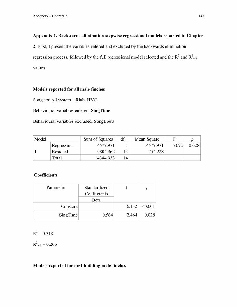
Appendix – Chapter 2 145
Appendix 1. Backwards elimination stepwise regressional models reported in Chapter
2. First, I present the variables entered and excluded by the backwards elimination
regression process, followed by the full regressional model selected and the R2 and R2adj
values.
Models reported for all male finches
Song control system – Right HVC
Behavioural variables entered: SingTime
Behavioural variables excluded: SongBouts
Model Sum of Squares df Mean Square F p
1 Regression 4579.971 1 4579.971 6.072 0.028 Residual 9804.962 13 754.228 Total 14384.933 14
Coefficients
Parameter Standardized Coefficients
t p
Beta Constant 6.142 <0.001
SingTime 0.564 2.464 0.028
R2 = 0.318
R2adj = 0.266
Models reported for nest-building male finches
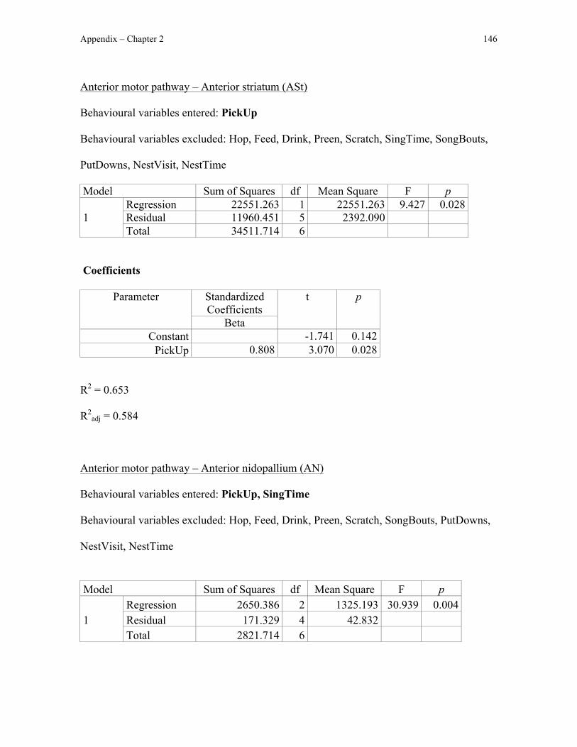
Appendix – Chapter 2 146
Anterior motor pathway – Anterior striatum (ASt)
Behavioural variables entered: PickUp
Behavioural variables excluded: Hop, Feed, Drink, Preen, Scratch, SingTime, SongBouts,
PutDowns, NestVisit, NestTime
Model Sum of Squares df Mean Square F p
1 Regression 22551.263 1 22551.263 9.427 0.028 Residual 11960.451 5 2392.090 Total 34511.714 6
Coefficients
Parameter Standardized Coefficients
t p
Beta Constant -1.741 0.142
PickUp 0.808 3.070 0.028
R2 = 0.653
R2adj = 0.584
Anterior motor pathway – Anterior nidopallium (AN)
Behavioural variables entered: PickUp, SingTime
Behavioural variables excluded: Hop, Feed, Drink, Preen, Scratch, SongBouts, PutDowns,
NestVisit, NestTime
Model Sum of Squares df Mean Square F p
1 Regression 2650.386 2 1325.193 30.939 0.004 Residual 171.329 4 42.832 Total 2821.714 6
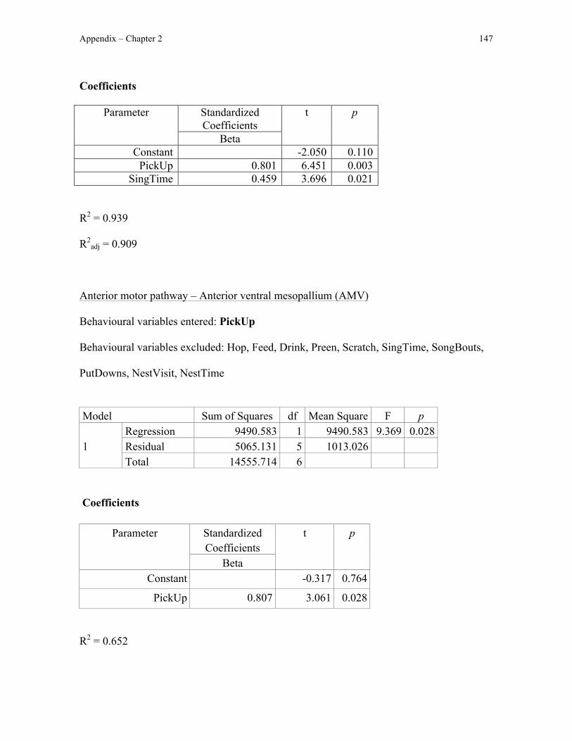
Appendix – Chapter 2 147
Coefficients
Parameter Standardized Coefficients
t p
Beta Constant -2.050 0.110
PickUp 0.801 6.451 0.003 SingTime 0.459 3.696 0.021
R2 = 0.939
R2adj = 0.909
Anterior motor pathway – Anterior ventral mesopallium (AMV)
Behavioural variables entered: PickUp
Behavioural variables excluded: Hop, Feed, Drink, Preen, Scratch, SingTime, SongBouts,
PutDowns, NestVisit, NestTime
Model Sum of Squares df Mean Square F p
1 Regression 9490.583 1 9490.583 9.369 0.028 Residual 5065.131 5 1013.026 Total 14555.714 6
Coefficients
Parameter Standardized Coefficients
t p
Beta Constant -0.317 0.764
PickUp 0.807 3.061 0.028
R2 = 0.652
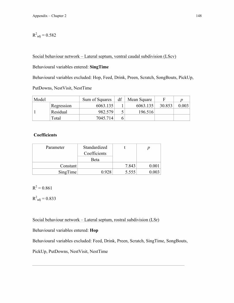
Appendix – Chapter 2 148
R2adj = 0.582
Social behaviour network – Lateral septum, ventral caudal subdivision (LScv)
Behavioural variables entered: SingTime
Behavioural variables excluded: Hop, Feed, Drink, Preen, Scratch, SongBouts, PickUp,
PutDowns, NestVisit, NestTime
Model Sum of Squares df Mean Square F p
1 Regression 6063.135 1 6063.135 30.853 0.003 Residual 982.579 5 196.516 Total 7045.714 6
Coefficients
Parameter Standardized Coefficients
t p
Beta Constant 7.843 0.001
SingTime 0.928 5.555 0.003
R2 = 0.861
R2adj = 0.833
Social behaviour network – Lateral septum, rostral subdivision (LSr)
Behavioural variables entered: Hop
Behavioural variables excluded: Feed, Drink, Preen, Scratch, SingTime, SongBouts,
PickUp, PutDowns, NestVisit, NestTime

Appendix – Chapter 2 149
Model Sum of Squares df Mean Square F p
1 Regression 1022.759 1 1022.759 7.677 0.039 Residual 666.098 5 133.220 Total 1688.857 6
Coefficients
Parameter Standardized Coefficients
t p
Beta Constant 11.512 <0.001
Hop -0.778 -2.771 0.039
R2 = 0.606
R2adj = 0.527
Social behaviour network – Medial septum (MS)
Behavioural variables entered: PutDowns
Behavioural variables excluded: Hop, Feed, Drink, Preen, Scratch, SingTime, SongBouts,
PickUp, NestVisit, NestTime
Model Sum of Squares df Mean Square F p
1 Regression 5358.035 1 5358.035 8.572 0.033 Residual 3125.393 5 625.079 Total 8483.429 6
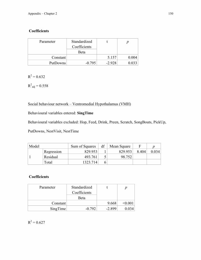
Appendix – Chapter 2 150
Coefficients
Parameter Standardized Coefficients
t p
Beta Constant 5.157 0.004
PutDowns -0.795 -2.928 0.033
R2 = 0.632
R2adj = 0.558
Social behaviour network – Ventromedial Hypothalamus (VMH)
Behavioural variables entered: SingTime
Behavioural variables excluded: Hop, Feed, Drink, Preen, Scratch, SongBouts, PickUp,
PutDowns, NestVisit, NestTime
Model Sum of Squares df Mean Square F p
1 Regression 829.953 1 829.953 8.404 0.034 Residual 493.761 5 98.752 Total 1323.714 6
Coefficients
Parameter Standardized Coefficients
t p
Beta Constant 9.668 <0.001
SingTime -0.792 -2.899 0.034
R2 = 0.627
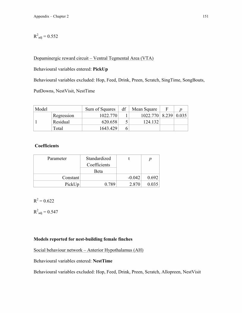
Appendix – Chapter 2 151
R2adj = 0.552
Dopaminergic reward circuit – Ventral Tegmental Area (VTA)
Behavioural variables entered: PickUp
Behavioural variables excluded: Hop, Feed, Drink, Preen, Scratch, SingTime, SongBouts,
PutDowns, NestVisit, NestTime
Model Sum of Squares df Mean Square F p
1 Regression 1022.770 1 1022.770 8.239 0.035 Residual 620.658 5 124.132 Total 1643.429 6
Coefficients
Parameter
Standardized Coefficients
t p
Beta Constant -0.042 0.692
PickUp 0.789 2.870 0.035
R2 = 0.622
R2adj = 0.547
Models reported for nest-building female finches
Social behaviour network – Anterior Hypothalamus (AH)
Behavioural variables entered: NestTime
Behavioural variables excluded: Hop, Feed, Drink, Preen, Scratch, Allopreen, NestVisit
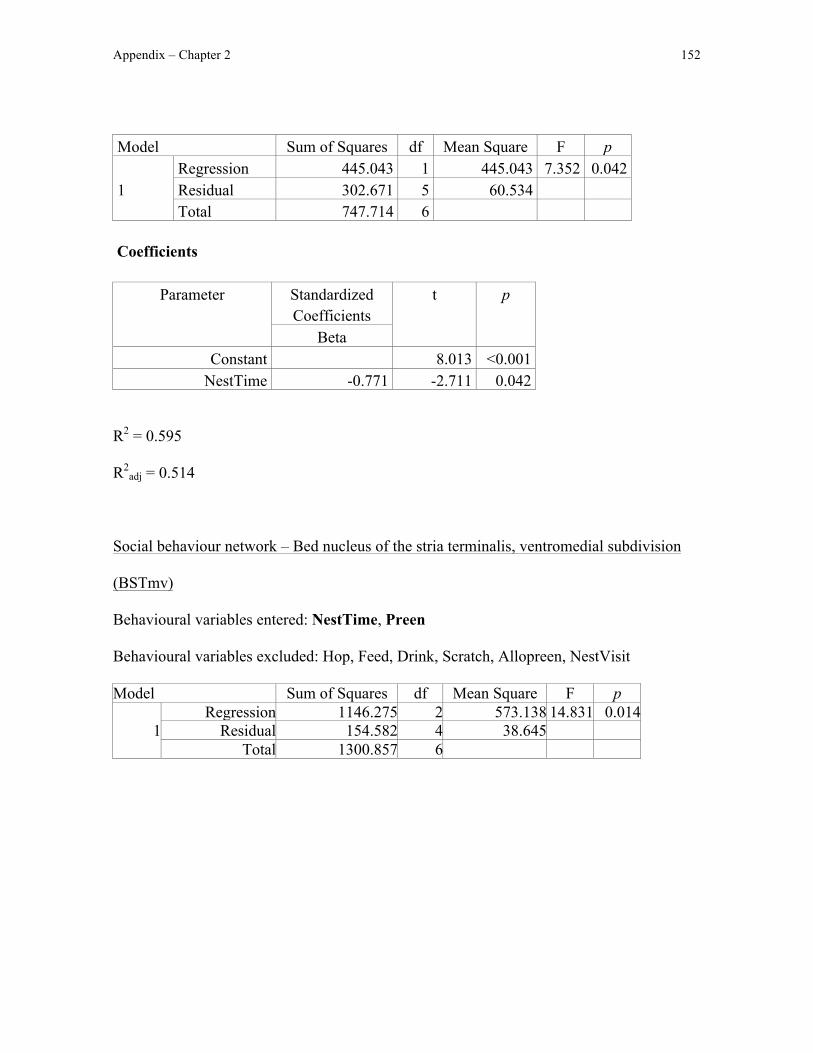
Appendix – Chapter 2 152
Model Sum of Squares df Mean Square F p
1 Regression 445.043 1 445.043 7.352 0.042 Residual 302.671 5 60.534 Total 747.714 6
Coefficients
Parameter Standardized Coefficients
t p
Beta Constant 8.013 <0.001
NestTime -0.771 -2.711 0.042
R2 = 0.595
R2adj = 0.514
Social behaviour network – Bed nucleus of the stria terminalis, ventromedial subdivision
(BSTmv)
Behavioural variables entered: NestTime, Preen
Behavioural variables excluded: Hop, Feed, Drink, Scratch, Allopreen, NestVisit
Model Sum of Squares df Mean Square F p
1 Regression 1146.275 2 573.138 14.831 0.014
Residual 154.582 4 38.645 Total 1300.857 6

Appendix – Chapter 2 153
Coefficients
Parameter Standardized Coefficients
t p
Beta Constant -1.417 0.229
NestTime 1.043 5.399 0.006 Preen 0.595 3.079 0.037
R2 = 0.600
R2adj = 0.519
Social behaviour network – Ventromedial Hypothalamus (VMH)
Behavioural variables entered: Preen
Behavioural variables excluded: Hop, Feed, Drink, Scratch, Allopreen, NestVisit,
NestTime
Model Sum of Squares df Mean Square F p
1 Regression 373.638 1 373.638 14.362 0.013 Residual 130.076 5 26.015 Total 503.714 6
Coefficients
Parameter Standardized Coefficients
t p
Beta Constant 17.455 <0.001
Preen -0.861 -3.790 0.013 R2 = 0.742
R2adj = 0.690
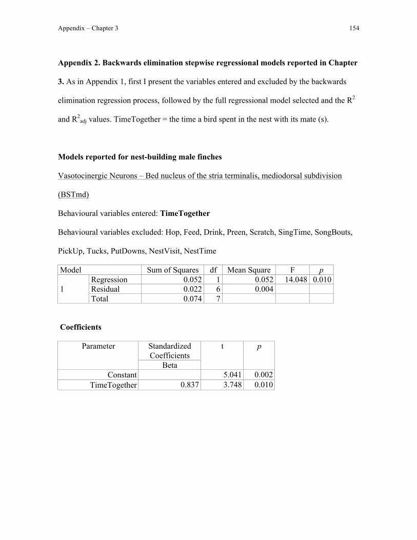
Appendix – Chapter 3 154
Appendix 2. Backwards elimination stepwise regressional models reported in Chapter
3. As in Appendix 1, first I present the variables entered and excluded by the backwards
elimination regression process, followed by the full regressional model selected and the R2
and R2adj values. TimeTogether = the time a bird spent in the nest with its mate (s).
Models reported for nest-building male finches
Vasotocinergic Neurons – Bed nucleus of the stria terminalis, mediodorsal subdivision
(BSTmd)
Behavioural variables entered: TimeTogether
Behavioural variables excluded: Hop, Feed, Drink, Preen, Scratch, SingTime, SongBouts,
PickUp, Tucks, PutDowns, NestVisit, NestTime
Model Sum of Squares df Mean Square F p
1 Regression 0.052 1 0.052 14.048 0.010 Residual 0.022 6 0.004 Total 0.074 7
Coefficients
Parameter Standardized Coefficients
t p
Beta Constant 5.041 0.002
TimeTogether 0.837 3.748 0.010

Appendix – Chapter 3 155
R2 = 0.701
R2adj = 0.651
Vasotocinergic Neurons – Bed nucleus of the stria terminalis, medioventral subdivision
(BSTmv)
Behavioural variables entered: PickUp
Behavioural variables excluded: Hop, Feed, Drink, Preen, Scratch, SingTime, SongBouts,
Tucks, PutDowns, NestVisit, NestTime, TimeTogether
Model Sum of Squares df Mean Square F p
1 Regression 0.168 1 0.168 9.590 0.021 Residual 0.105 6 0.017 Total 0.272 7
Coefficients
Parameter Standardized Coefficients
t p
Beta Constant 1.998 0.093
PickUp 0.784 3.097 0.021
R2 = 0.615
R2adj = 0.551
Dopaminergic Neurons – Central gray
Behavioural variables entered: TimeTogether
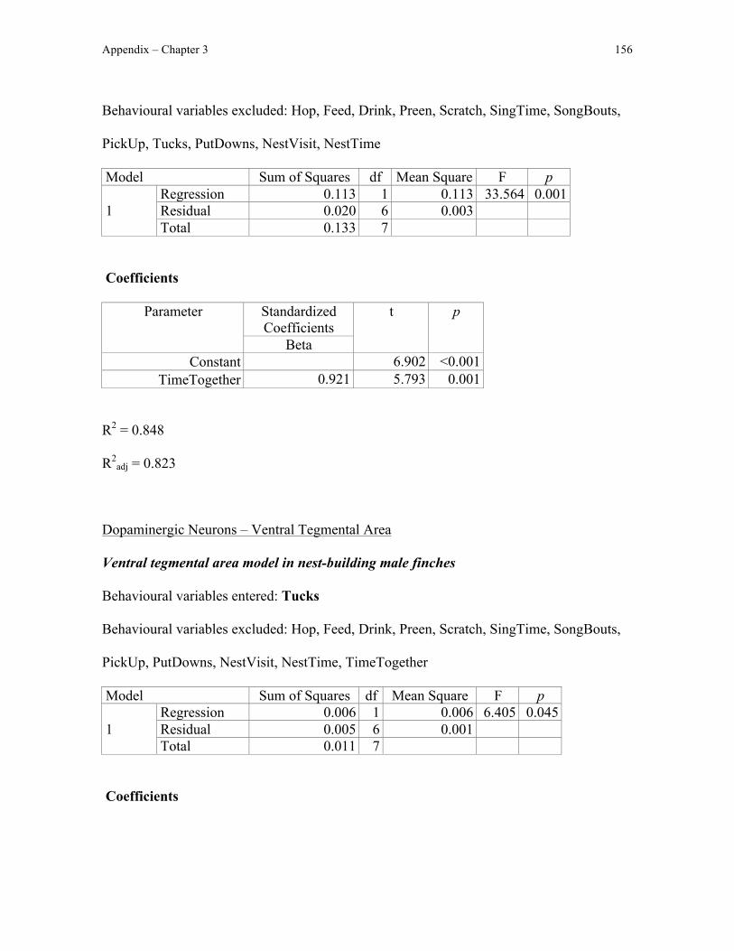
Appendix – Chapter 3 156
Behavioural variables excluded: Hop, Feed, Drink, Preen, Scratch, SingTime, SongBouts,
PickUp, Tucks, PutDowns, NestVisit, NestTime
Model Sum of Squares df Mean Square F p
1 Regression 0.113 1 0.113 33.564 0.001 Residual 0.020 6 0.003 Total 0.133 7
Coefficients
Parameter Standardized Coefficients
t p
Beta Constant 6.902 <0.001
TimeTogether 0.921 5.793 0.001
R2 = 0.848
R2adj = 0.823
Dopaminergic Neurons – Ventral Tegmental Area
Ventral tegmental area model in nest-building male finches
Behavioural variables entered: Tucks
Behavioural variables excluded: Hop, Feed, Drink, Preen, Scratch, SingTime, SongBouts,
PickUp, PutDowns, NestVisit, NestTime, TimeTogether
Model Sum of Squares df Mean Square F p
1 Regression 0.006 1 0.006 6.405 0.045 Residual 0.005 6 0.001 Total 0.011 7
Coefficients

Appendix – Chapter 3 157
Parameter Standardized Coefficients
t p
Beta Constant 6.293 0.001
Tucks -0.719 -2.531 0.045
R2 = 0.516
R2adj = 0.436
Ventral tegmental area model in nest-building female finches
Behavioural variables entered: Feed
Behavioural variables excluded: Hop, Drink, Preen, Allopreen, Scratch, Tucks, NestVisit,
NestTime, TimeTogether
Model Sum of Squares df Mean Square F p
1 Regression 0.001 1 0.001 11.923 0.014 Residual 0.001 6 <0.001 Total 0.002 7
Coefficients
Parameter Standardized Coefficients
t p
Beta Constant 13.840 <0.001
Feed -0.816 -3.453 0.014
R2 = 0.665
R2adj = 0.609
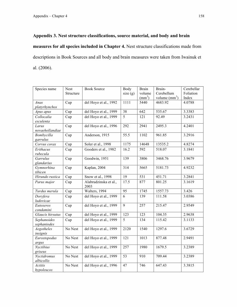
Appendix – Chapter 4 158
Appendix 3. Nest structure classifications, source material, and body and brain
measures for all species included in Chapter 4. Nest structure classifications made from
descriptions in Book Sources and all body and brain measures were taken from Iwainuk et
al. (2006).
Species name Nest Structure
Book Source Body size (g)
Brain volume (mm3)
Brain-Cerebellum volume (mm3)
Cerebellar Foliation Index
Anas platyrhynchos
Cup del Hoyo et al., 1992 1111 5440 4683.92 4.0788
Apus apus Cup del Hoyo et al., 1999 38 642 535.67 3.3383 Collocalia esculenta
Cup del Hoyo et al., 1999 5 121 92.49 3.2431
Larus novaehollandiae
Cup del Hoyo et al., 1996 292 2941 2495.3 4.2401
Bombycilla garrulus
Cup Anderson, 1915 55.5 1102 961.85 3.2916
Corvus corax Cup Soler et al., 1998 1175 14648 13535.2 4.8274 Erithacus rubecula
Cup Gooders et al., 1982 16.2 592 518.07 3.1841
Garrulus glandarius
Cup Goodwin, 1951 139 3806 3468.76 3.9679
Gymnorhina tibicen
Cup Kaplan, 2004 314 5665 5181.73 4.9232
Hirundo rustica Cup Snow et al., 1998 19 531 451.71 3.2841 Parus major Cup Alabrudzinska et al.,
2003 17.5 877 801.25 3.1619
Turdus merula Cup Walters, 1994 95 1745 1557.73 3.426 Doryfera ludovicae
Cup del Hoyo et al., 1999 6 139 111.58 3.0386
Eutoxeres condamini
Cup del Hoyo et al., 1999 9 257 215.47 2.9549
Glaucis hirsutus Cup del Hoyo et al., 1999 123 123 104.35 2.9638 Sephanoides sephaniodes
Cup del Hoyo et al., 1999 5 134 115.42 3.1133
Aegotheles insignis
No Nest del Hoyo et al., 1999 2120 1540 1297.6 3.6729
Eurostopodus argus
No Nest del Hoyo et al., 1999 121 1013 877.48 2.9491
Nyctibius griseus
No Nest del Hoyo et al., 1999 257 1980 1679.5 3.2389
Nyctidromus albicollis
No Nest del Hoyo et al., 1999 53 910 709.44 3.2389
Actitis hypoleucos
No Nest del Hoyo et al., 1996 47 746 647.43 3.3815
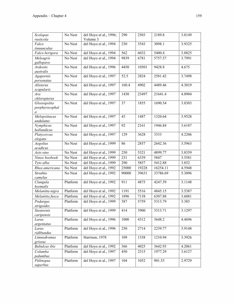
Appendix – Chapter 4 159
Scolopax rusticola
No Nest del Hoyo et al., 1996; Volume 3
290 2503 2189.8 3.8149
Falco tinnunculus
No Nest del Hoyo et al., 1994 230 3543 3098.1 3.9325
Falco berigora No Nest del Hoyo et al., 1994 562 6032 5400.4 3.8825 Meleagris gallopavo
No Nest del Hoyo et al., 1994 9839 6781 5757.57 3.7991
Ardeotis australis
No Nest del Hoyo et al., 1996 4450 10501 9428.8 4.675
Agapornis personatus
No Nest del Hoyo et al., 1997 52.5 2824 2581.42 3.7498
Alisterus scapularis
No Nest del Hoyo et al., 1997 160.4 4902 4489.46 4.3019
Ara chloropterus
No Nest del Hoyo et al., 1997 1430 23497 21641.4 4.8904
Glossopsitta porphyrocephala
No Nest del Hoyo et al., 1997 37 1855 1690.54 3.8303
Melopsittacus undulatus
No Nest del Hoyo et al., 1997 43 1487 1320.64 3.9528
Nymphicus hollandicus
No Nest del Hoyo et al., 1997 92 2161 1946.84 3.6187
Platycercus elegans
No Nest del Hoyo et al., 1997 129 3628 3333 4.2206
Aegolius acadicus
No Nest del Hoyo et al., 1999 86 2857 2642.36 3.5963
Asio otus No Nest del Hoyo et al., 1999 250 5321 4899.77 3.8359 Ninox boobook No Nest del Hoyo et al., 1999 231 6339 5847 3.5581 Tyto alba No Nest del Hoyo et al., 1999 290 5857 5412.88 3.852 Rhea americana No Nest del Hoyo et al., 1992 25000 19228 16254.11 4.5948 Struthio camelus
No Nest del Hoyo et al., 1992 90000 39631 33786.69 5.3096
Clangula hyemalis
Platform del Hoyo et al., 1992 911 4875 4247.59 3.1148
Melanitta nigra Platform del Hoyo et al., 1992 1191 5516 4845.15 3.5387 Melanitta fusca Platform del Hoyo et al., 1992 1896 7138 6307.88 3.6081 Podargus strigoides
Platform del Hoyo et al., 1999 387 5759 5313.79 3.385
Steatornis caripensis
Platform del Hoyo et al., 1999 414 3900 3313.71 3.1297
Larus argentatus
Platform del Hoyo et al., 1996 1000 4312 3648.2 4.4696
Larus ridibundus
Platform del Hoyo et al., 1996 250 2714 2239.77 3.9148
Limnodromus griseus
Platform Harrison, 1978 109 1338 1210.94 3.3926
Bubulcus ibis Platform del Hoyo et al., 1992 366 4025 3642.93 4.2061 Columba palumbus
Platform del Hoyo et al., 1997 450 2315 1977.29 3.6127
Ptilinopus superbus
Platform del Hoyo et al., 1997 104 1052 901.53 2.9729

Appendix – Chapter 4 160
Aquila audax Platform del Hoyo et al., 1994 3350 15997 14146.55 4.7077 Buteo buteo Platform del Hoyo et al., 1994 900 8452 7282.85 4.3031 Haliaeetus leucogaster
Platform del Hoyo et al., 1994 3004 12541 11164.89 4.6655
Bonasa umbellus
Platform del Hoyo et al., 1994 650 3136 2867.61 3.9399
Perdix perdix Platform del Hoyo et al., 1994 401 1849 1625.66 3.4847 Phasianus colchicus
Platform del Hoyo et al., 1994 1133 3865 3384.25 4.2058
Fulica americana
Platform del Hoyo et al., 1996 651 2719 2471.67 3.2863
Corvus monedula
Platform Wilmore and Wilmore, 1977
200 4593 4210.97 4.3009
Corvus corone Platform Wilmore and Wilmore, 1977
537 9382 8628.94 4.6097
Pelecanus conspicillatus
Platform del Hoyo et al., 1992 5850 24880 23522.25 4.8202
Phoenicopterus ruber
Platform del Hoyo et al., 1992 3000 10674 8908.31 4.5568
Thalassarche melanophrys
Platform del Hoyo et al., 1992 3388 14129 11634.59 5.5338
Cacatua roseicapilla
Platform del Hoyo et al., 1997 355 7456 6934.09 4.8683
Cacatua galerita
Platform del Hoyo et al., 1997 765 13933 12868.28 5.3408

Appendix – Chapter 5 161
Appendix 4. Nest structure description, nest structure classification, and minimum
nest height (m) for all species in Timaliidae included in Chapter 5.
Scientific Name Nest description from del Hoyo et al., 1997 Nest
Classification Minimum Nest Height (m)
Actinodura egertoni
largish, rather deep cup Cup 1
Alcippe brunnea loose dome or semi-dome with entrance at upper part
Domed 0.1
Alcippe castaneceps
dome Domed 1
Alcippe chrysotis very deep cup, sometimes domed or egg shaped with side entrance
Domed 0.4
Alcippe cinerea deep cup, sometimes domed or semi-domed Domed 0 Alcippe dubia loose oval or dome-shaped structure with
entrance towards the top Domed 0
Alcippe morrisonia
very compact, fairly strong cup or hanging basket
Cup 0.2
Alcippe nipalensis
usually neat and compact deep cup, rarely loosely woven and semi transparent
Cup 0.3
Alcippe peracensis
small cup Cup 1.5
Alcippe poioicephala
roughly built, compact, deep cup, sometimes almost cone shaped
Cup 0.6
Alcippe rufogularis
rather loose dome or semi-dome or cup on large base of leaves, protected by whorl of upward pointing leaves
Domed 0
Alcippe vinipectus
bulky, fairly deep, compact cup Cup 0.9
Babax lanceolatus
reportedly a loose but well defined open cup Cup 0.6
Babax waddelli large, rather rough cup, exterior woven Cup 1.8 Chamaea fasciata
deep compact cup Cup 0.3
Chrysomma sinense
small, compact, cone-shaped deep cup Cup 0.5
Cutia nipalensis open cup Cup 3 Dumetia hyperythra
loose or neat dome, with side entrance, sometimes towards the top.
Domed 0
Erpornis zantholeuca
small, deep cradle Cup 0.5
Garrulax affinis large but neat cup Cup 1 Garrulax albogularis
broad, shallow saucer to moderately deep cup Cup 1
Garrulax austeni cup Cup 0 Garrulax caerulatus
reportedly a large, compact, rather shallow to deep cup
Cup 1
Garrulax reportedly a large cup, outwardly rough but with Cup 0
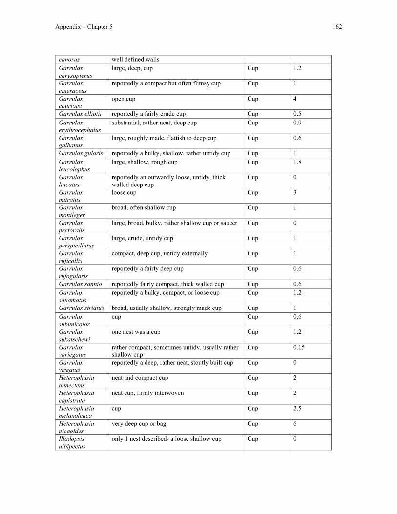
Appendix – Chapter 5 162
canorus well defined walls Garrulax chrysopterus
large, deep, cup Cup 1.2
Garrulax cineraceus
reportedly a compact but often flimsy cup Cup 1
Garrulax courtoisi
open cup Cup 4
Garrulax elliotii reportedly a fairly crude cup Cup 0.5 Garrulax erythrocephalus
substantial, rather neat, deep cup Cup 0.9
Garrulax galbanus
large, roughly made, flattish to deep cup Cup 0.6
Garrulax gularis reportedly a bulky, shallow, rather untidy cup Cup 1 Garrulax leucolophus
large, shallow, rough cup Cup 1.8
Garrulax lineatus
reportedly an outwardly loose, untidy, thick walled deep cup
Cup 0
Garrulax mitratus
loose cup Cup 3
Garrulax monileger
broad, often shallow cup Cup 1
Garrulax pectoralis
large, broad, bulky, rather shallow cup or saucer Cup 0
Garrulax perspicillatus
large, crude, untidy cup Cup 1
Garrulax ruficollis
compact, deep cup, untidy externally Cup 1
Garrulax rufogularis
reportedly a fairly deep cup Cup 0.6
Garrulax sannio reportedly fairly compact, thick walled cup Cup 0.6 Garrulax squamatus
reportedly a bulky, compact, or loose cup Cup 1.2
Garrulax striatus broad, usually shallow, strongly made cup Cup 1 Garrulax subunicolor
cup Cup 0.6
Garrulax sukatschewi
one nest was a cup Cup 1.2
Garrulax variegatus
rather compact, sometimes untidy, usually rather shallow cup
Cup 0.15
Garrulax virgatus
reportedly a deep, rather neat, stoutly built cup Cup 0
Heterophasia annectens
neat and compact cup Cup 2
Heterophasia capistrata
neat cup, firmly interwoven Cup 2
Heterophasia melanoleuca
cup Cup 2.5
Heterophasia picaoides
very deep cup or bag Cup 6
Illadopsis albipectus
only 1 nest described- a loose shallow cup Cup 0
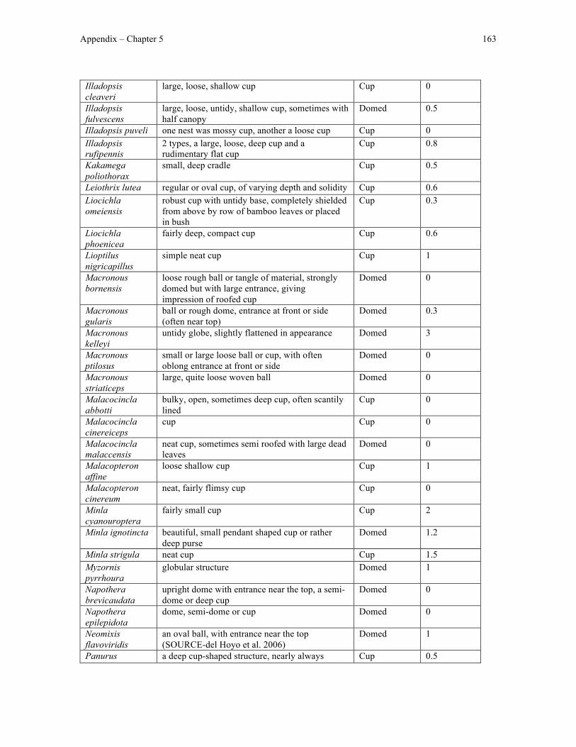
Appendix – Chapter 5 163
Illadopsis cleaveri
large, loose, shallow cup Cup 0
Illadopsis fulvescens
large, loose, untidy, shallow cup, sometimes with half canopy
Domed 0.5
Illadopsis puveli one nest was mossy cup, another a loose cup Cup 0 Illadopsis rufipennis
2 types, a large, loose, deep cup and a rudimentary flat cup
Cup 0.8
Kakamega poliothorax
small, deep cradle Cup 0.5
Leiothrix lutea regular or oval cup, of varying depth and solidity Cup 0.6 Liocichla omeiensis
robust cup with untidy base, completely shielded from above by row of bamboo leaves or placed in bush
Cup 0.3
Liocichla phoenicea
fairly deep, compact cup Cup 0.6
Lioptilus nigricapillus
simple neat cup Cup 1
Macronous bornensis
loose rough ball or tangle of material, strongly domed but with large entrance, giving impression of roofed cup
Domed 0
Macronous gularis
ball or rough dome, entrance at front or side (often near top)
Domed 0.3
Macronous kelleyi
untidy globe, slightly flattened in appearance Domed 3
Macronous ptilosus
small or large loose ball or cup, with often oblong entrance at front or side
Domed 0
Macronous striaticeps
large, quite loose woven ball Domed 0
Malacocincla abbotti
bulky, open, sometimes deep cup, often scantily lined
Cup 0
Malacocincla cinereiceps
cup Cup 0
Malacocincla malaccensis
neat cup, sometimes semi roofed with large dead leaves
Domed 0
Malacopteron affine
loose shallow cup Cup 1
Malacopteron cinereum
neat, fairly flimsy cup Cup 0
Minla cyanouroptera
fairly small cup Cup 2
Minla ignotincta beautiful, small pendant shaped cup or rather deep purse
Domed 1.2
Minla strigula neat cup Cup 1.5 Myzornis pyrrhoura
globular structure Domed 1
Napothera brevicaudata
upright dome with entrance near the top, a semi-dome or deep cup
Domed 0
Napothera epilepidota
dome, semi-dome or cup Domed 0
Neomixis flavoviridis
an oval ball, with entrance near the top (SOURCE-del Hoyo et al. 2006)
Domed 1
Panurus a deep cup-shaped structure, nearly always Cup 0.5

Appendix – Chapter 5 164
biarmicus roofed by sheltering vegetation Paradoxornis alphonsianus
cup shaped structure Cup 0.5
Paradoxornis flavirostris
very, neat, compact, deep (rarely shallow) cup Cup 1
Paradoxornis gularis
beautiful, very neat, compact, cup-shaped struture, sometimes with broad bulging sides
Cup 2
Paradoxornis guttaticollis
very compact and deep cup-shaped structure Cup 0.9
Paradoxornis heudei
beautiful cup-shaped structure Cup 1.3
Paradoxornis ruficeps
neat and compact deep cup Cup 1
Paradoxornis webbianus
neat and fairly stiff, deep cup-shaped structure (rounded or oblong)
Cup 0.3
Pellorneum albiventre
small compact globe or dome, sometimes semi-dome or deep cup
Domed 0
Pellorneum capistratum
outwardly untidy cup Cup 0
Pellorneum fuscocapillus
loose ball with large lateral entrance, or occasionally a cup
Domed 0
Pellorneum palustre
reportedly ball shaped Domed 0
Pellorneum ruficeps
large, flimsy ball or dome, entrance at side, or a semi-dome or cup, sheltered by large upward pointing leaf
Domed 0
Phyllanthus atripennis
large, untidy cup Cup 3
Pnoepyga albiventer
globular structure, entrance two thirds up one side
Domed 0
Pnoepyga formosana
dome or cylinder with entrance hole at one end Domed 0
Pnoepyga pusilla small ball of moss, rootlets, bark shreds and leaf skeletons, or a built in structure made of long strands of brilliant green moss, with tiny cup
Domed 0.5
Pomatorhinus erythrogenys
loose dome with broad entrance high up at side, or sometimes open at both ends
Domed 0
Pomatorhinus ferruginosus
oval or bulky cone placed on side, egg-shaped (Hume, A.O., 2004; The Nests and Eggs of Indian Birds, Vol.1)
Domed 0
Pomatorhinus gravivox
untidy dome with side entrance Domed 0
Pomatorhinus horsfieldii
loose, often large dome, entrance on upper side, or a semi-domed cup
Domed 0.3
Pomatorhinus hypoleucos
large, semi-domed oval, but very open, part forming the roof sometimes flimsier, the cup fairly deep and more solid
Domed 0
Pomatorhinus mcclellandi
loose dome with side entrance Domed 0
Pomatorhinus montanus
large dome or sheltered cup Domed 0
Pomatorhinus oval ball, loosely put together Domed 0
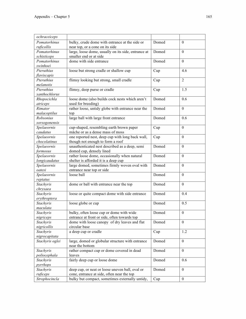
Appendix – Chapter 5 165
ochraceiceps Pomatorhinus ruficollis
bulky, crude dome with entrance at the side or near top, or a cone on its side
Domed 0
Pomatorhinus schisticeps
large, loose dome, usually on its side, entrance at smaller end or at side
Domed 0
Pomatorhinus swinhoei
dome with side entrance Domed 0
Pteruthius flaviscapis
loose but strong cradle or shallow cup Cup 4.6
Pteruthius melanotis
flimsy looking but strong, small cradle Cup 2
Pteruthius xanthochlorus
flimsy, deep purse or cradle Cup 1.5
Rhopocichla atriceps
loose dome (also builds cock nests which aren’t used for breeding)
Domed 0.6
Rimator malacoptilus
rather loose, untidy globe with entrance near the top
Domed 0
Robsonius sorsogonensis
large ball with large front entrance Domed 0.6
Spelaeornis caudatus
cup-shaped, resembling earth brown paper mâche or as a dense mass of moss
Cup 0
Spelaeornis chocolatinus
one reported nest, deep cup with long back wall, though not enough to form a roof
Cup 0
Spelaeornis formosus
unauthenticated nest described as a deep, semi domed cup, densely lined
Domed 0
Spelaeornis longicaudatus
rather loose dome, occasionally when natural shelter is afforded it is a deep cup
Domed 0
Spelaeornis oatesi
large domed, sometimes firmly woven oval with entrance near top or side
Domed 0
Spelaeornis reptatus
loose ball Domed 0
Stachyris chrysaea
dome or ball with entrance near the top Domed 0
Stachyris erythroptera
loose or quite compact dome with side entrance Domed 0.4
Stachyris maculata
loose globe or cup Domed 0.5
Stachyris nigriceps
bulky, often loose cup or dome with wide entrance at front or side, often towards top
Domed 0
Stachyris nigricollis
dome with loose canopy of dry leaves and flat circular base
Domed 0
Stachyris nigrocapitata
a deep cup or cradle Cup 1.2
Stachyris oglei large, domed or globular structure with entrance near the bottom
Domed 0
Stachyris poliocephala
rather compact cup or dome covered in dead leaves
Domed 0
Stachyris pyrrhops
fairly deep cup or loose dome Domed 0.6
Stachyris ruficeps
deep cup, or neat or loose uneven ball, oval or cone, entrance at side, often near the top
Domed 0
Strophocincla bulky but compact, sometimes externally untidy, Cup 0
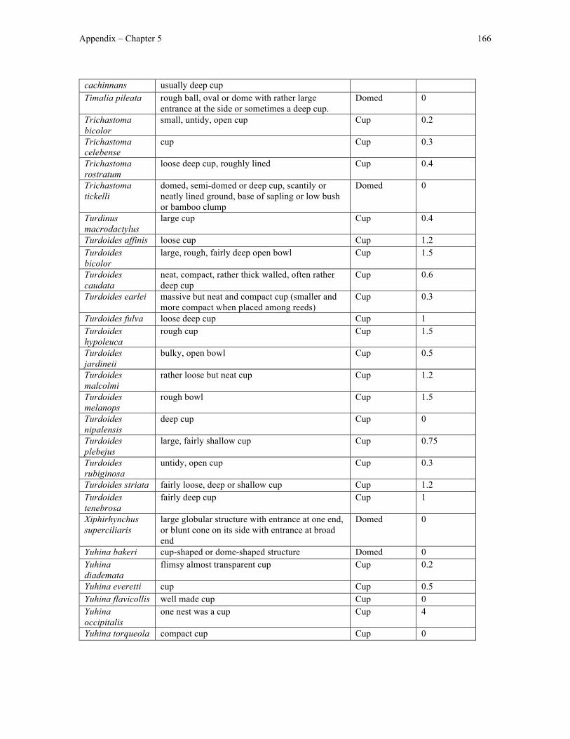
Appendix – Chapter 5 166
cachinnans usually deep cup Timalia pileata rough ball, oval or dome with rather large
entrance at the side or sometimes a deep cup. Domed 0
Trichastoma bicolor
small, untidy, open cup Cup 0.2
Trichastoma celebense
cup Cup 0.3
Trichastoma rostratum
loose deep cup, roughly lined Cup 0.4
Trichastoma tickelli
domed, semi-domed or deep cup, scantily or neatly lined ground, base of sapling or low bush or bamboo clump
Domed 0
Turdinus macrodactylus
large cup Cup 0.4
Turdoides affinis loose cup Cup 1.2 Turdoides bicolor
large, rough, fairly deep open bowl Cup 1.5
Turdoides caudata
neat, compact, rather thick walled, often rather deep cup
Cup 0.6
Turdoides earlei massive but neat and compact cup (smaller and more compact when placed among reeds)
Cup 0.3
Turdoides fulva loose deep cup Cup 1 Turdoides hypoleuca
rough cup Cup 1.5
Turdoides jardineii
bulky, open bowl Cup 0.5
Turdoides malcolmi
rather loose but neat cup Cup 1.2
Turdoides melanops
rough bowl Cup 1.5
Turdoides nipalensis
deep cup Cup 0
Turdoides plebejus
large, fairly shallow cup Cup 0.75
Turdoides rubiginosa
untidy, open cup Cup 0.3
Turdoides striata fairly loose, deep or shallow cup Cup 1.2 Turdoides tenebrosa
fairly deep cup Cup 1
Xiphirhynchus superciliaris
large globular structure with entrance at one end, or blunt cone on its side with entrance at broad end
Domed 0
Yuhina bakeri cup-shaped or dome-shaped structure Domed 0 Yuhina diademata
flimsy almost transparent cup Cup 0.2
Yuhina everetti cup Cup 0.5 Yuhina flavicollis well made cup Cup 0 Yuhina occipitalis
one nest was a cup Cup 4
Yuhina torqueola compact cup Cup 0
Related Documents