B.E. Haley/Medical Veritas 4 (2007) 1510–1524 1510 The relationship of the toxic effects of mercury to exacerbation of the medical condition classified as Alzheimer’s disease Boyd E. Haley, PhD Professor, Department of Chemistry University of Kentucky Lexington, KY 40506-0055 Email: [email protected] Abstract Mercury(II) or Hg 2+ , is neurotoxic. When exposed to normal brain tissue homogenates, neurons in culture, Hg 2+ (mercury(II) or mercuric mer- cury) is capable of causing many of the same biochemical aberrancies found in Alzheimer’s diseased (AD) brain. Also, rats exposed to mercury(0), metallic mercury, vapor show some of these same abnormalities in their brain tissue. Specifically, the rapid inactivation of the brain thiol-sensitive enzymes (tubulin, creatine kinase and glutamine synthetase) occurs after: (a) the addition of low micromolar levels of Hg 2+ , (b) exposure to mercury vapor (Hg 0 ) or (c) the addition of Thimerosal (ethylmercurythiosalicylate sodium salt). Moreover, these same enzymes are significantly inhibited in the AD brain. Further, exposure of neurons in culture to nanomolar levels of Hg 2+ has been shown to produce three of the widely accepted pathologi- cal diagnostic hallmarks of AD. These AD hallmarks are elevated amyloid protein, hyper-phosphorylation of Tau, and formation of neurofibillary tangles (NFTs). This paper proposes the hypothesis that elemental mercury, organic mercury compounds, and other blood-brain permeable toxicants, which have enhanced specificity for thiol-sensitive enzymes, are the etiological source of AD. Included in this category are other heavy metals such as lead and cadmium that act synergistically to enhance, by many-fold, the toxicity of metallic mercury and organic-mercury(II) compounds, like Thimerosal. This hypothesis also explains the genetic susceptibility to AD that is expressed through the APO-E geneotype. Specifically, a reduction of APO-E gene type that contains two cysteines decreases the one of the innate detoxification capabilities of APO-E, the removal of mercury and other thiol- reactive toxicants from the central nervous system. This increases brain exposure to thiol-reactive toxicants and elevates the risk of AD. Also, the increased exposure to mercury through breathing the mercury vapor emanating from mercury amalgam dental fillings can have a deleterious effect on olfactory capability. This effect may explain the high correlation between the loss of sense of smell and the subsequent development of AD. Keywords: Alzheimer’s Disease (AD), mercury, mercury toxicity, neurotoxicants 1. Rationale for the hypothesis Currently, AD is considered a disease of unknown etiology. However, it is widely accepted that most AD is not directly genetically inherited and that some external factor, such as a toxicant exposure or an infection, must be involved for the dis- ease to progress to the point AD can be clinically diagnosed. In the USA the rate of AD is very similar in rural and urban areas; and the rate is also similar in all of the states in the USA. Therefore, if a toxicant is involved, then this toxicant must be of a very personal nature – like something we eat or something that is placed into our bodies from other sources such as dental fillings, vaccines, etc. Moreover, the involvement of infectious agents such as bac- teria, virus or yeasts; while possible at this time, seems not to be a direct causal factor. This apparent reality is based on the huge amount of funds that the National Institutes of Health (NIH-USA) and other organizations worldwide have spent on AD to identify the causal factors, excluding mercury. To date, this effort has failed to identify any consistent microbial factor. If an infectious agent were involved (like in AIDS or polio) it is likely that it would have been identified by now. However, fo- cal infections caused by microbes in the oral cavity must still be considered, as these microbes are known to produce toxicants (e.g., hydrogen sulfide, methyl-mercaptan, and gliatoxin) that inhibit thiol-sensitive enzymes. Further, toxins produced by these oral facultative anaerobes can react with oral mercury to generate compounds like methylthiomercury cation (CH 3 -S- Hg + ) and di-(methylthio)mercury[II] (CH 3 -S-Hg-S-CH 3 ) that could be exceptionally toxic. Thus, for any toxicant, or class of toxicants, to be proposed as involved in the etiology of AD it must be available almost equally to individuals living in markedly different locations. The toxicant proposed must also explain the genetic susceptibil- ity aspects of AD. Further, under experimental conditions the putative toxicants must be able to cause or exacerbate many of the biochemical abnormalities found in the AD brain. Based on our research studies and a literature review, mercury and or- ganic mercury-containing compounds (from dental amalgams, vaccines, other medicinals, and preservatives used in paints, seed grains, etc.) represent compounds that fill this require- ment. Consistent with this hypothesis is the fact that the first case of Alzheimer’s disease was identified in 1903, roughly 60 to 70 years after the introduction of mercury amalgams as a tooth filling material in the Western world. It seems illogical that all of the pathologists in the 1700 to 1900s would miss a neuro- logical disease with decreased brain volume and the obvious abnormal morphological markers that are seen in post-mortem studies of those with a diagnosis of AD. Therefore, AD is most likely a recent human disease caused by human-generated mer- cury toxins – an iatrogenic mercury-poisoning disease. Mercury and organic mercury compounds are neurotoxi- cants. Further, the enzyme inhibitory effects of mercury are synergistically enhanced by exposures to other toxicants such as lead and cadmium (smokers). As presented below, even the doi: 10.1588/medver.2007.04.00164

Welcome message from author
This document is posted to help you gain knowledge. Please leave a comment to let me know what you think about it! Share it to your friends and learn new things together.
Transcript
B.E. Haley/Medical Veritas 4 (2007) 1510–1524 1510
The relationship of the toxic effects of mercury to exacerbation of the medical condition classified as Alzheimer’s disease
Boyd E. Haley, PhD
Professor, Department of Chemistry University of Kentucky
Lexington, KY 40506-0055 Email: [email protected]
Abstract Mercury(II) or Hg2+, is neurotoxic. When exposed to normal brain tissue homogenates, neurons in culture, Hg2+ (mercury(II) or mercuric mer-cury) is capable of causing many of the same biochemical aberrancies found in Alzheimer’s diseased (AD) brain. Also, rats exposed to mercury(0), metallic mercury, vapor show some of these same abnormalities in their brain tissue. Specifically, the rapid inactivation of the brain thiol-sensitive enzymes (tubulin, creatine kinase and glutamine synthetase) occurs after: (a) the addition of low micromolar levels of Hg2+, (b) exposure to mercury vapor (Hg0) or (c) the addition of Thimerosal (ethylmercurythiosalicylate sodium salt). Moreover, these same enzymes are significantly inhibited in the AD brain. Further, exposure of neurons in culture to nanomolar levels of Hg2+ has been shown to produce three of the widely accepted pathologi-cal diagnostic hallmarks of AD. These AD hallmarks are elevated amyloid protein, hyper-phosphorylation of Tau, and formation of neurofibillary tangles (NFTs). This paper proposes the hypothesis that elemental mercury, organic mercury compounds, and other blood-brain permeable toxicants, which have enhanced specificity for thiol-sensitive enzymes, are the etiological source of AD. Included in this category are other heavy metals such as lead and cadmium that act synergistically to enhance, by many-fold, the toxicity of metallic mercury and organic-mercury(II) compounds, like Thimerosal. This hypothesis also explains the genetic susceptibility to AD that is expressed through the APO-E geneotype. Specifically, a reduction of APO-E gene type that contains two cysteines decreases the one of the innate detoxification capabilities of APO-E, the removal of mercury and other thiol-reactive toxicants from the central nervous system. This increases brain exposure to thiol-reactive toxicants and elevates the risk of AD. Also, the increased exposure to mercury through breathing the mercury vapor emanating from mercury amalgam dental fillings can have a deleterious effect on olfactory capability. This effect may explain the high correlation between the loss of sense of smell and the subsequent development of AD. Keywords: Alzheimer’s Disease (AD), mercury, mercury toxicity, neurotoxicants
1. Rationale for the hypothesis Currently, AD is considered a disease of unknown etiology. However, it is widely accepted that most AD is not directly genetically inherited and that some external factor, such as a toxicant exposure or an infection, must be involved for the dis-ease to progress to the point AD can be clinically diagnosed. In the USA the rate of AD is very similar in rural and urban areas; and the rate is also similar in all of the states in the USA. Therefore, if a toxicant is involved, then this toxicant must be of a very personal nature – like something we eat or something that is placed into our bodies from other sources such as dental fillings, vaccines, etc. Moreover, the involvement of infectious agents such as bac-teria, virus or yeasts; while possible at this time, seems not to be a direct causal factor. This apparent reality is based on the huge amount of funds that the National Institutes of Health (NIH-USA) and other organizations worldwide have spent on AD to identify the causal factors, excluding mercury. To date, this effort has failed to identify any consistent microbial factor. If an infectious agent were involved (like in AIDS or polio) it is likely that it would have been identified by now. However, fo-cal infections caused by microbes in the oral cavity must still be considered, as these microbes are known to produce toxicants (e.g., hydrogen sulfide, methyl-mercaptan, and gliatoxin) that inhibit thiol-sensitive enzymes. Further, toxins produced by these oral facultative anaerobes can react with oral mercury to generate compounds like methylthiomercury cation (CH3-S-
Hg+) and di-(methylthio)mercury[II] (CH3-S-Hg-S-CH3) that could be exceptionally toxic. Thus, for any toxicant, or class of toxicants, to be proposed as involved in the etiology of AD it must be available almost equally to individuals living in markedly different locations. The toxicant proposed must also explain the genetic susceptibil-ity aspects of AD. Further, under experimental conditions the putative toxicants must be able to cause or exacerbate many of the biochemical abnormalities found in the AD brain. Based on our research studies and a literature review, mercury and or-ganic mercury-containing compounds (from dental amalgams, vaccines, other medicinals, and preservatives used in paints, seed grains, etc.) represent compounds that fill this require-ment. Consistent with this hypothesis is the fact that the first case of Alzheimer’s disease was identified in 1903, roughly 60 to 70 years after the introduction of mercury amalgams as a tooth filling material in the Western world. It seems illogical that all of the pathologists in the 1700 to 1900s would miss a neuro-logical disease with decreased brain volume and the obvious abnormal morphological markers that are seen in post-mortem studies of those with a diagnosis of AD. Therefore, AD is most likely a recent human disease caused by human-generated mer-cury toxins – an iatrogenic mercury-poisoning disease. Mercury and organic mercury compounds are neurotoxi-cants. Further, the enzyme inhibitory effects of mercury are synergistically enhanced by exposures to other toxicants such as lead and cadmium (smokers). As presented below, even the
doi: 10.1588/medver.2007.04.00164
B.E. Haley/Medical Veritas 4 (2007) 1510–1524 1511
simultaneous presence of EDTA (ethylene-diamine-tetraacetic acid, a common food additive) or metal binding antibiotics such as tetracycline can enhance mercury toxicity. It is also known that intake of milk enhances the retention of mercury in the body [25,36]. Therefore, any determination of a safe level of mercury exposure using rats in a cage being fed carefully moni-tored food and water is not reliable for determination of a “safe level of exposure to mercury” for humans. Thus, current sci-ence does not know what the combined toxic effects of many toxicants or enhancers of toxicity are, when present with mer-cury, and, therefore, cannot identify a safe level of exposure. Therefore, thiol-reactive toxicants such as mercury, cad-mium, lead and certain organics are rational suggestions for exacerbating factors in AD, or possibly even causal factors. However, mercury is the only toxicant that has been shown able reproduce many of the biochemical abnormalities and patho-logic, diagnostic hallmarks of AD when applied to appropriate test systems. Also, chronic mercury exposure occurs in most humans as most of us have grams of it (in the form of dental amalgams, which are about 50% mercury) stored and continu-ously releasing mercury vapor only inches from our brain. Therefore, it is reasonable to propose that exposure to mercury is one of the major toxic factors involved in early-onset AD. Further, simultaneous exposures to other toxicants or factors can enhance the toxicity of mercury and hasten the onset of AD, especially in those individuals who are genetically suscep-tible. Thus, the presence of synergistic toxicity factors and genetic susceptibility factors prevent any simple correlation of mercury exposure or tissue levels to the onset of mercury induced ill-nesses. 2. Research review and results
2.1 Enzyme inhibition and protein partitioning results
The research on Alzheimer’s disease (AD) done in our labo-ratory in the late 1980s was directed towards detecting differ-ences in the nucleotide binding proteins of AD post-mortem brain tissue versus age-matched, non-demented control brain samples. Basic to our early findings was the following observa-tion: Two very important brain nucleotide binding proteins, tubulin and creatine kinase (CK), showed greatly diminished activity and nucleotide binding ability in the AD brain tissues versus age-matched control brain samples [1-3]. Both tubulin and CK are proteins that bind the nucleotides GTP (guanosine-5’-triphosphate) and ATP (adenosine-5’-triphosphate), respec-tively. To monitor the presence of viable tubulin and CK we used a “photoaffinity labeling” technology to determine the bioavail-ability of these nucleotide binding sites before and after addi-tion of mercury or other toxicants. The basic concept is that photolabeling of these enzymes indicates that they are present and normally active; lack of photolabeling indicates the en-zymes are inhibited. This technology is explained in detail elsewhere for those interested in the chemistry [4]. Using this technology, our laboratory demonstrated that both tubulin and CK had diminished biological activity and abnormal partition-
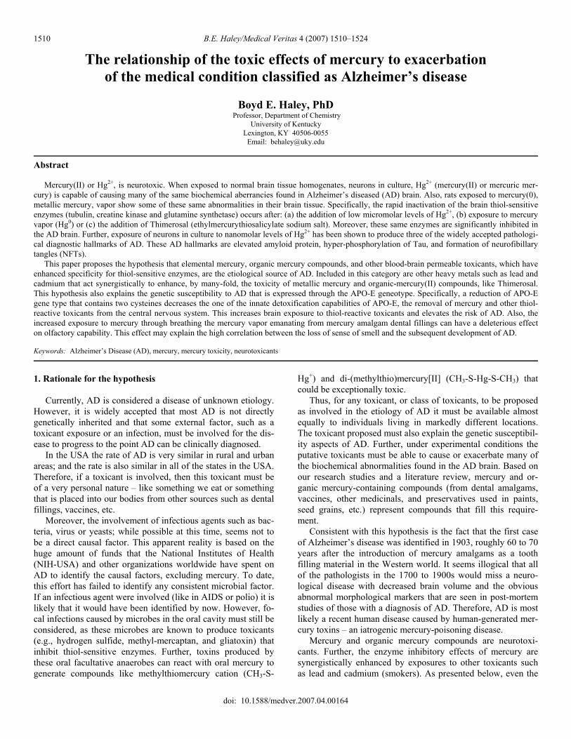
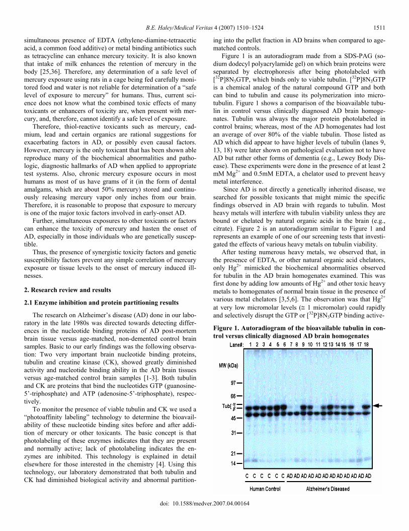
ing into the pellet fraction in AD brains when compared to age-matched controls. Figure 1 is an autoradiogram made from a SDS-PAG (so-dium dodecyl polyacrylamide gel) on which brain proteins were separated by electrophoresis after being photolabeled with [32P]8N3GTP, which binds only to viable tubulin. [32P]8N3GTP is a chemical analog of the natural compound GTP and both can bind to tubulin and cause its polymerization into micro-tubulin. Figure 1 shows a comparison of the bioavailable tubu-lin in control versus clinically diagnosed AD brain homoge-nates. Tubulin was always the major protein photolabeled in control brains; whereas, most of the AD homogenates had lost an average of over 80% of the viable tubulin. Those listed as AD which did appear to have higher levels of tubulin (lanes 9, 13, 18) were later shown on pathological evaluation not to have AD but rather other forms of dementia (e.g., Lewey Body Dis-ease). These experiments were done in the presence of at least 2 mM Mg2+ and 0.5mM EDTA, a chelator used to prevent heavy metal interference. Since AD is not directly a genetically inherited disease, we searched for possible toxicants that might mimic the specific findings observed in AD brain with regards to tubulin. Most heavy metals will interfere with tubulin viability unless they are bound or chelated by natural organic acids in the brain (e.g., citrate). Figure 2 is an autoradiogram similar to Figure 1 and represents an example of one of our screening tests that investi-gated the effects of various heavy metals on tubulin viability. After testing numerous heavy metals, we observed that, in the presence of EDTA, or other natural organic acid chelators, only Hg2+ mimicked the biochemical abnormalities observed for tubulin in the AD brain homogenates examined. This was first done by adding low amounts of Hg2+ and other toxic heavy metals to homogenates of normal brain tissue in the presence of various metal chelators [3,5,6]. The observation was that Hg2+ at very low micromolar levels (≅ 1 micromolar) could rapidly and selectively disrupt the GTP or [32P]8N3GTP binding active-
Figure 1. Autoradiogram of the bioavailable tubulin in con-trol versus clinically diagnosed AD brain homogenates
doi: 10.1588/medver.2007.04.00164
B.E. Haley/Medical Veritas 4 (2007) 1510–1524 1512
ity of tubulin (Mr = 55,000 Daltons) without any noticeable effect on the other GTP binding protein(s) observed at an Mr of about 42,000 Daltons, which are present in both the control and AD brains at approximately equal levels. Therefore, with re-spect to heavy metal toxicants, at low micromolar levels in the presence of any of the organic acid chelators studied, only the addition of mercury to control brain homogenates gave a [32P]8N3GTP binding profile that was identical to that observed in AD brain samples. In addition, the chelation of the Hg2+ with EDTA did not prevent, but rather enhanced, this binding-inhibition effect. Figure 2. Autoradiogram of an example of one of the screening tests investigating effects of various heavy metals on tubulin viability
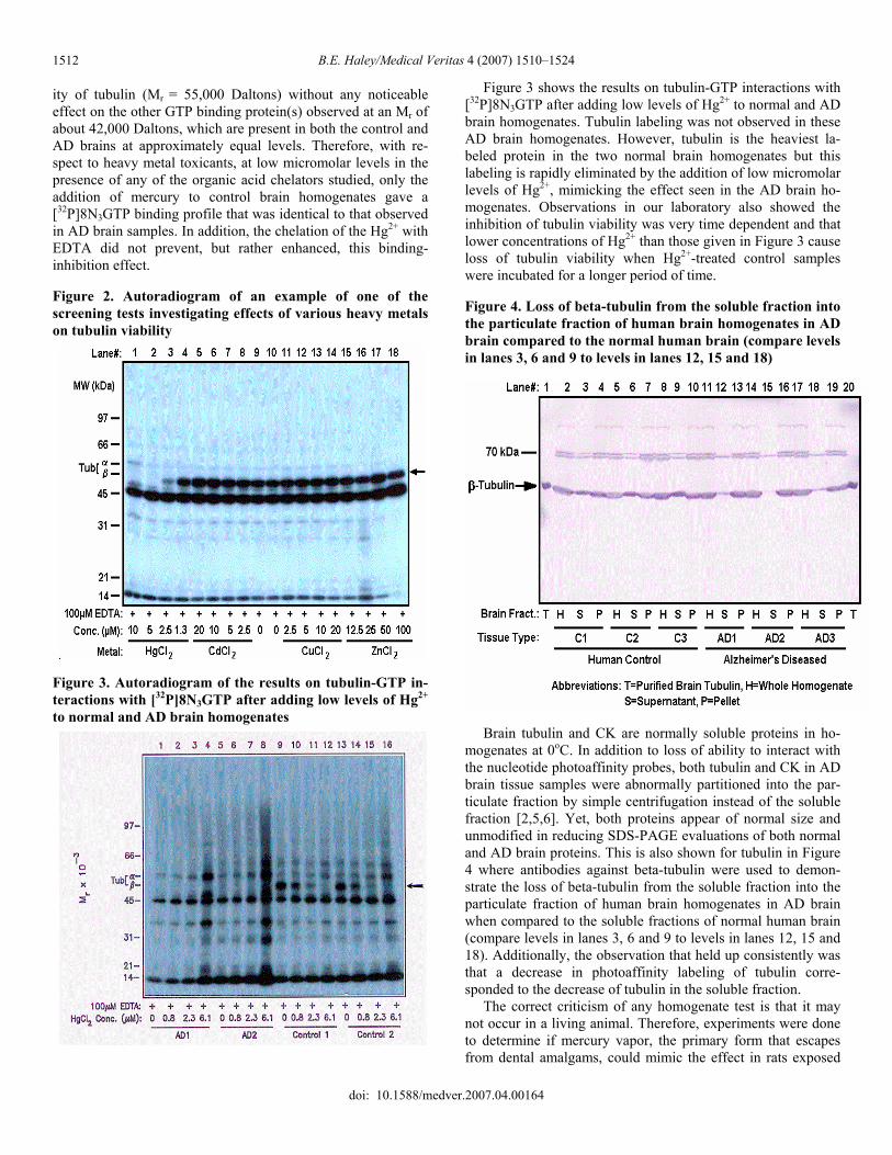
Figure 3. Autoradiogram of the results on tubulin-GTP in-teractions with [32P]8N3GTP after adding low levels of Hg2+ to normal and AD brain homogenates
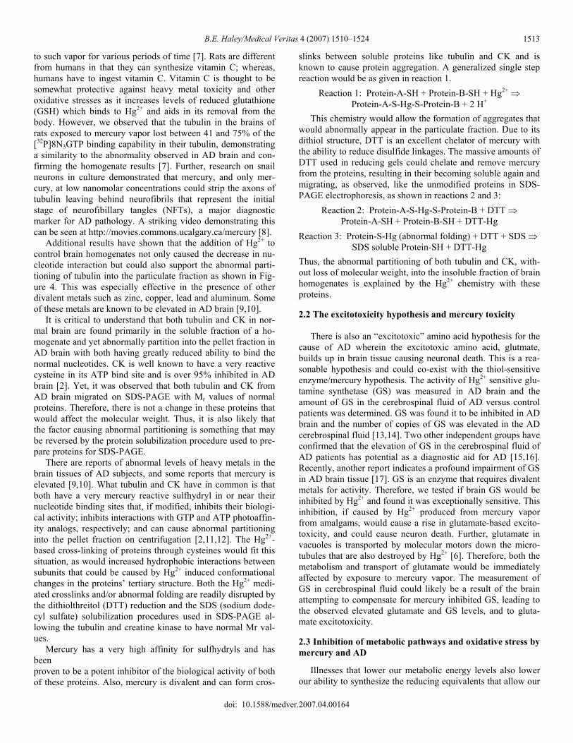
Figure 3 shows the results on tubulin-GTP interactions with [32P]8N3GTP after adding low levels of Hg2+ to normal and AD brain homogenates. Tubulin labeling was not observed in these AD brain homogenates. However, tubulin is the heaviest la-beled protein in the two normal brain homogenates but this labeling is rapidly eliminated by the addition of low micromolar levels of Hg2+, mimicking the effect seen in the AD brain ho-mogenates. Observations in our laboratory also showed the inhibition of tubulin viability was very time dependent and that lower concentrations of Hg2+ than those given in Figure 3 cause loss of tubulin viability when Hg2+-treated control samples were incubated for a longer period of time. Figure 4. Loss of beta-tubulin from the soluble fraction into the particulate fraction of human brain homogenates in AD brain compared to the normal human brain (compare levels in lanes 3, 6 and 9 to levels in lanes 12, 15 and 18)
Brain tubulin and CK are normally soluble proteins in ho-mogenates at 0oC. In addition to loss of ability to interact with the nucleotide photoaffinity probes, both tubulin and CK in AD brain tissue samples were abnormally partitioned into the par-ticulate fraction by simple centrifugation instead of the soluble fraction [2,5,6]. Yet, both proteins appear of normal size and unmodified in reducing SDS-PAGE evaluations of both normal and AD brain proteins. This is also shown for tubulin in Figure 4 where antibodies against beta-tubulin were used to demon-strate the loss of beta-tubulin from the soluble fraction into the particulate fraction of human brain homogenates in AD brain when compared to the soluble fractions of normal human brain (compare levels in lanes 3, 6 and 9 to levels in lanes 12, 15 and 18). Additionally, the observation that held up consistently was that a decrease in photoaffinity labeling of tubulin corre-sponded to the decrease of tubulin in the soluble fraction. The correct criticism of any homogenate test is that it may not occur in a living animal. Therefore, experiments were done to determine if mercury vapor, the primary form that escapes from dental amalgams, could mimic the effect in rats exposed
doi: 10.1588/medver.2007.04.00164
B.E. Haley/Medical Veritas 4 (2007) 1510–1524 1513
to such vapor for various periods of time [7]. Rats are different from humans in that they can synthesize vitamin C; whereas, humans have to ingest vitamin C. Vitamin C is thought to be somewhat protective against heavy metal toxicity and other oxidative stresses as it increases levels of reduced glutathione (GSH) which binds to Hg2+ and aids in its removal from the body. However, we observed that the tubulin in the brains of rats exposed to mercury vapor lost between 41 and 75% of the [32P]8N3GTP binding capability in their tubulin, demonstrating a similarity to the abnormality observed in AD brain and con-firming the homogenate results [7]. Further, research on snail neurons in culture demonstrated that mercury, and only mer-cury, at low nanomolar concentrations could strip the axons of tubulin leaving behind neurofibrils that represent the initial stage of neurofibillary tangles (NFTs), a major diagnostic marker for AD pathology. A striking video demonstrating this can be seen at http://movies.commons.ucalgary.ca/mercury [8]. Additional results have shown that the addition of Hg2+ to control brain homogenates not only caused the decrease in nu-cleotide interaction but could also support the abnormal parti-tioning of tubulin into the particulate fraction as shown in Fig-ure 4. This was especially effective in the presence of other divalent metals such as zinc, copper, lead and aluminum. Some of these metals are known to be elevated in AD brain [9,10]. It is critical to understand that both tubulin and CK in nor-mal brain are found primarily in the soluble fraction of a ho-mogenate and yet abnormally partition into the pellet fraction in AD brain with both having greatly reduced ability to bind the normal nucleotides. CK is well known to have a very reactive cysteine in its ATP bind site and is over 95% inhibited in AD brain [2]. Yet, it was observed that both tubulin and CK from AD brain migrated on SDS-PAGE with Mr values of normal proteins. Therefore, there is not a change in these proteins that would affect the molecular weight. Thus, it is also likely that the factor causing abnormal partitioning is something that may be reversed by the protein solubilization procedure used to pre-pare proteins for SDS-PAGE. There are reports of abnormal levels of heavy metals in the brain tissues of AD subjects, and some reports that mercury is elevated [9,10]. What tubulin and CK have in common is that both have a very mercury reactive sulfhydryl in or near their nucleotide binding sites that, if modified, inhibits their biologi-cal activity; inhibits interactions with GTP and ATP photoaffin-ity analogs, respectively; and can cause abnormal partitioning into the pellet fraction on centrifugation [2,11,12]. The Hg2+-based cross-linking of proteins through cysteines would fit this situation, as would increased hydrophobic interactions between subunits that could be caused by Hg2+ induced conformational changes in the proteins’ tertiary structure. Both the Hg2+ medi-ated crosslinks and/or abnormal folding are readily disrupted by the dithiolthreitol (DTT) reduction and the SDS (sodium dode-cyl sulfate) solubilization procedures used in SDS-PAGE al-lowing the tubulin and creatine kinase to have normal Mr val-ues. Mercury has a very high affinity for sulfhydryls and has been proven to be a potent inhibitor of the biological activity of both of these proteins. Also, mercury is divalent and can form cros-
slinks between soluble proteins like tubulin and CK and is known to cause protein aggregation. A generalized single step reaction would be as given in reaction 1.
Reaction 1: Protein-A-SH + Protein-B-SH + Hg2+ ⇒ Protein-A-S-Hg-S-Protein-B + 2 H+
This chemistry would allow the formation of aggregates that would abnormally appear in the particulate fraction. Due to its dithiol structure, DTT is an excellent chelator of mercury with the ability to reduce disulfide linkages. The massive amounts of DTT used in reducing gels could chelate and remove mercury from the proteins, resulting in their becoming soluble again and migrating, as observed, like the unmodified proteins in SDS-PAGE electrophoresis, as shown in reactions 2 and 3:
Reaction 2: Protein-A-S-Hg-S-Protein-B + DTT ⇒ Protein-A-SH + Protein-B-SH + DTT-Hg
Reaction 3: Protein-S-Hg (abnormal folding) + DTT + SDS ⇒ SDS soluble Protein-SH + DTT-Hg
Thus, the abnormal partitioning of both tubulin and CK, with-out loss of molecular weight, into the insoluble fraction of brain homogenates is explained by the Hg2+ chemistry with these proteins. 2.2 The excitotoxicity hypothesis and mercury toxicity There is also an “excitotoxic” amino acid hypothesis for the cause of AD wherein the excitotoxic amino acid, glutmate, builds up in brain tissue causing neuronal death. This is a rea-sonable hypothesis and could co-exist with the thiol-sensitive enzyme/mercury hypothesis. The activity of Hg2+ sensitive glu-tamine synthetase (GS) was measured in AD brain and the amount of GS in the cerebrospinal fluid of AD versus control patients was determined. GS was found it to be inhibited in AD brain and the number of copies of GS was elevated in the AD cerebrospinal fluid [13,14]. Two other independent groups have confirmed that the elevation of GS in the cerebrospinal fluid of AD patients has potential as a diagnostic aid for AD [15,16]. Recently, another report indicates a profound impairment of GS in AD brain tissue [17]. GS is an enzyme that requires divalent metals for activity. Therefore, we tested if brain GS would be inhibited by Hg2+ and found it was exceptionally sensitive. This inhibition, if caused by Hg2+ produced from mercury vapor from amalgams, would cause a rise in glutamate-based excito-toxicity, and could cause neuron death. Further, glutamate in vacuoles is transported by molecular motors down the micro-tubules that are also destroyed by Hg2+ [6]. Therefore, both the metabolism and transport of glutamate would be immediately affected by exposure to mercury vapor. The measurement of GS in cerebrospinal fluid could likely be a result of the brain attempting to compensate for mercury inhibited GS, leading to the observed elevated glutamate and GS levels, and to gluta-mate excitotoxicity. 2.3 Inhibition of metabolic pathways and oxidative stress by mercury and AD
Illnesses that lower our metabolic energy levels also lower our ability to synthesize the reducing equivalents that allow our
doi: 10.1588/medver.2007.04.00164
B.E. Haley/Medical Veritas 4 (2007) 1510–1524 1514
body to bind and dispose of excess mercury. Hg2+ is known to inhibit the metabolic processes in mitochondria that produce ATP and NADH by inhibiting the enzymes in the citric acid cycle, and the electron transport system. These nucleotides are absolutely required for both the synthesis of reduced glu-tathione (GSH) and to reduce glutathione after it is oxidized. GSH is the major biomolecule involved in the natural removal of mercury from the body. Therefore, as mercury slowly accu-mulates in the body, it weakens the body’s natural defense against itself and all forms of other heavy metal toxicities, and increases the overall oxidative stress expressed by reactive oxygen species formation. It is well known that AD brain tissue suffers from greater oxidative stress in all cellular components versus similar tissues from control subjects. If mercury is a factor, this would be ex-pected be as it is well documented that mercury increases oxi-dative stress in biological tissues. Further, Hg2+ is well known to inhibit numerous other enzymes important to neurological function, including the porphyrin pathway that leads to the syn-thesis of heme [18-20]. It is unique that only Hg2+ toxicity causes a specific porphyrin profile based on inhibition of a spe-cific enzyme. It was reported that 85% of the dentists and den-tal technicians tested showed mercury related toxicities in both behavior and physiological parameters, and 15% showed an increased mercury induced neurological deficits with polymor-phism of the CPOX4 gene [20]. The Hg2+ induced interruption of heme synthesis could af-fect oxygen transport by limiting hemoglobin synthesis; de-crease mitochondrial ATP production as cytochrome C requires heme; and inhibit the action of many heme requiring P450 en-zymes which are the major detox enzymes of the body. There-fore, the many numerous abnormalities observed in AD brain would be expected within a hypothesis that proposes exposure to Hg2+ as a major contributor to this disease. Consistent with this is the report that the heme levels in AD brain are abnor-mally low and that this abnormality is likely caused by β-amyloid protein [21]. However, it is also reasonable that Hg2+ could alter brain heme synthesis leading to an inability to ex-crete β-amyloid protein. 2.4 Relevant mercury exposures and measurments of toxic mercury release from dental amalgams
The fact that mercury has inhibitory effects on tubulin, CK and GS, and that these proteins are abnormally inhibited in AD does not conclusively prove that mercury exposure causes AD. However, it definitely proves that chronic, daily exposure to mercury would at least exacerbate the clinical conditions of AD. Is such an exposure to mercury likely? The answer is yes in spite of consistent FDA refusal to evaluate directly the mer-cury vapor released by dental amalgams, or to make the ADA and manufacturers of this material provide such information. The documented high release of mercury by dental amalgams and mercury’s ability to induce the biochemistry and pathologi-cal markers consistent with observations in AD brain make mercury involvement in AD causation plausible. The first question that must be addressed: Is there enough mercury in an amalgam filling to continue a low chronic-level exposure for years? The answer is yes. For example, if a single
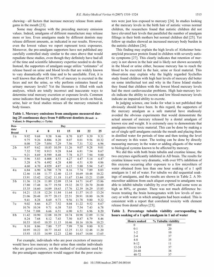
large amalgam filling contained 1 gram of mercury (1 million micrograms) and lost a significantly toxic 10 micrograms per day there would be enough mercury for 100,000 days or about 274 years of exposure. A small tenth of a gram mercury filling would last 27 years. So enough mercury is within amalgam fillings to provide a consistent toxic exposure for the life of most fillings. Second, does mercury emit from amalgams at a rate that should cause concern? The answer is yes. Dental amalgams, or “silver fillings” as organized dentistry calls them, are approxi-mately 50% mercury by weight; and it is quite easy to demon-strate that these fillings readily emit mercury vapors. The actual amount emitted can only be determined with the amalgam in a closed system with the amount of mercury released being de-termined using reliable, time-proven chemical techniques and modern instrumentation. The accurate level of mercury released cannot be easily measured for amalgams in the mouth due to the fact that some is released into the jawbone, some is ab-sorbed by the saliva and oral tissues, some leaves as small non-volatile particles that release mercury in the stomach and diges-tive track, and some is inhaled and absorbed in the lungs. In a carefully designed study in a sealed container, Chew et al. tested the “long term dissolution of mercury from a non-mercury-releasing amalgam (trade name Composil)” [22]. Their results demonstrated “that the overall mean release of mercury was 43.5 +/- 3.2 micrograms/cm2/24 hr, and the amount of mer-cury released remained fairly constant during the duration of the experiment (2 years).” In our laboratory we have repeated similar experiments in our laboratory with similar results. The International Academy of Science (IAOMT) supported a study of mercury release from different dental amalgams. Nine IAOMT dentists were sent identical Plexiglas molds with 10 holes that would hold 1 spill of amalgam. Each of the den-tists was also sent amalgam material from different manufactur-ers. They placed 10 amalgams in the molds and sent them to Dr. Haley’s lab at the University of Kentucky for testing of mercury release using a Nippon Direct Mercury Analyzer. The amalgams were first allowed to age until the mercury emissions leveled off and then the amount of mercury being released was measured for 25 continuous days. Some of the results are shown in Table 1. This data shows that the level of mercury released by 1 cm2 of dental amalgam from a single spill filling (each filling was slightly less than 1 cm2) was between 4 to 22 micrograms per day. This result was obtained by leaving the amalgams setting in distilled water at room temperature, gently mixing the water without disturbing the amalgam and collecting 1 ml for analy-sis. We did some other experiments. For example, holding an amalgam under water and brushing it 15 strokes with a conven-tional medium bristle tooth brush lead to a 5 to 10 fold increase in the release of mercury. Thus, the previously reported 43.5 micrograms/cm2/day [22] and the values found in the IAOMT amalgam study are not an insignificant amount of mercury exposure when one considers the number of years a 70-year old individual living today may have been exposed to chronic mercury levels from his or her amalgam fillings. Additionally, this release per day is the level released without galvanism, excess heat, or pressure from
doi: 10.1588/medver.2007.04.00164
B.E. Haley/Medical Veritas 4 (2007) 1510–1524 1515
chewing—all factors that increase mercury release from amal-gams in the mouth [23]. Some may disagree with the preceding mercury emission values. Indeed, amalgams of different manufacture may release more or less. Even amalgams made by different dentists may release different amounts, as indicated in our results. However, even the lowest values we report represent toxic exposures. Moreover, the pro-amalgam supporters have not published any carefully controlled study similar to the ones above that would repudiate these studies, even though they definitely have had all of the time and scientific laboratory expertise needed to do this. Instead, the supporters of amalgam usage utilize “estimates” of release based on urine and blood levels that are widely known to vary dramatically with time and to be unreliable. First, it is well known that about 85 to 95% of mercury is excreted in the feces and not the urine, so why perform estimates based on urinary mercury levels? Yet the literature is filled with such analyses, which are totally incorrect and inaccurate ways to determine total mercury excretion or exposure levels. Common sense indicates that basing safety and exposure levels on blood, urine, hair or fecal studies misses all the mercury retained in body tissues.
Table 1. Mercury emissions from amalgams measured dur-ing 25 continuous days from 9 different dentists (Brand: a–Valiant; b–Dispersalloy; c–Tytin)
Day Den-tists/ Brand 1 4 8 11 15 18 22 25 1/a 9.92 9.68 9.58 9.46 8.70 8.87 9.39 9.31 9.75 9.26 8.886 8.20 8.07 8.01 9.56 10.32 8.08 7.29 7.054 7.29 7.56 7.31 7.32 6.96 2/b 9.97 9.62 10.85 10.59 11.26 9.07 9.28 9.01 7.32 7.92 9.913 9.28 8.64 6.81 7.54 8.67 9.21 8.69 8.599 8.48 7.78 8.27 7.94 9.00 3/a 5.96 5.83 4.408 4.53 4.27 4.47 5.14 4.47 5.28 4.76 4.492 4.28 4.80 4.51 4.30 4.86 4.60 4.70 4.929 4.87 6.15 5.80 5.94 5.47 4/a 6.84 6.90 6.788 5.78 8.16 7.74 7.89 8.03 12.46 11.88 11.77 12.40 12.15 10.69 10.48 10.22 13.91 13.42 12.62 11.18 11.67 13.44 13.21 13.09 5/b 11.36 11.24 11.89 12.09 15.34 14.71 14.47 15.86 17.80 17.48 16.77 19.58 19.32 20.72 20.70 20.00 15.35 14.60 14.09 18.63 17.76 12.39 16.29 15.85 6/c 14.21 13.18 12.24 11.84 11.64 11.57 14.15 13.24 21.06 20.48 19.77 20.15 22.51 20.91 18.80 16.58 9.41 8.28 8.69 9.73 9.56 11.78 9.80 9.22 7/c 9.02 8.66 8.27 7.52 8.04 11.22 9.52 8.67 10.76 10.34 9.71 6.38 7.03 7.54 7.43 6.78 7.54 7.108 6.66 6.51 6.90 6.51 6.96 8.20 8/b 11.42 10.90 12.08 10.39 10.74 10.98 12.09 11.54 8.24 7.68 8.12 7.43 7.50 8.87 8.79 8.46 10.53 10.43 10.55 11.15 10.46 10.16 10.56 10.23 9/c 9.10 8.06 7.80 7.37 7.99 7.30 9.80 9.31 10.95 10.22 10.77 10.43 12.25 11.32 12.48 11.20 15.93 15.53 14.99 12.23 12.80 14.67 14.04 13.65
For example, individuals who are poor excretors of mercury would have less mercury in their urine than similar individuals who are good excretors, yet the method of evaluation used by the pro-amalgam supporters would suggest that the poor excre-
tors were just less exposed to mercury [24]. In studies looking at the mercury levels in the birth hair of autistic versus normal children, the researchers found that autistic children did not have elevated hair levels that paralleled the number of amalgam fillings in their birth mothers but normal children did [25]. Yet follow up studies showed an increased mercury body burden in the autistic children [26]. This finding may explain the high levels of Alzheimer beta-amyloid precursor protein found in children with severely autis-tic symptoms [27]. This clearly indicates that retention of mer-cury is not shown in the hair and is likely not shown accurately in the blood or urine either, because mercury has to reach the blood to be excreted in the hair, urine or fecal material. This observation may explain why the highly regarded Sychelles study found children with high hair levels of mercury did better on some intellectual test and why in the Faroe Island studies they found that children with the lowest blood mercury levels had the most cardiovascular problems. High hair-mercury lev-els indicate the ability to excrete and low blood-mercury levels indicate an impaired ability to excrete. In judging science, one looks for what is not published that obviously should have been. In this regard, the supporters of the mercury amalgam as a safe material have studiously avoided the obvious experiments that would demonstrate the actual amount of mercury released by a dental amalgam of known size and weight. It is extremely easy to demonstrate that amalgams release toxic material. This can be done by making a set of single spill amalgams outside the mouth and placing them in distilled water for periods of time and then testing the level of mercury in this water. The testing can be done by directly measuring mercury in the water or adding aliquots of the water to biological systems known to be affected by mercury. We did this with both brain tubulin and creatine kinase, the two enzymes significantly inhibited in AD brain. The results for creatine kinase were very dramatic, with over 95% inhibition of this enzyme occurring after exposure to a few microliters of water obtained from less than one hour soaking of a 1 spill amalgam in 1 ml of water. For tubulin we did sequential soak-ings of amalgams, and the results are shown in Table 2. A 50-microliter addition from each aliquot exposed to amalgams was able to inhibit tubulin viability by over 60% and some were as high as 80%, or greater. There was not much difference be-tween treating the brain homogenates with low levels of mer-cury or with water in which amalgams had been soaked. This is consistent with a report that correlated toxicity with element release from dental alloys [23].
Table 2. Percentage tubulin viability corresponding to hours soaking of a 1 spill amalgam in 1 ml of water
Hours soaked % Tubulin viability 0 100 0-1 20 1-2 10 2-4 37 4-8 35 8-12 11 12-24 37 24-48 38 48-72 37
doi: 10.1588/medver.2007.04.00164

B.E. Haley/Medical Veritas 4 (2007) 1510–1524 1516
Such experiments show that mercury from amalgams is ca-pable of causing the same abnormal biochemistry in normal brain tissues as observed in AD brain samples and, this alone, should be adequate rationale not to continue the use of dental amalgam. 2.5 Does the presence of amalgams contribute significantly to mercury body burden? There have been numerous published reports of increased tissue mercury levels in subjects and the relationship to in-creased number of amalgams fillings [28-32]. Also, the World Health Organization Scientific Panel found ranges of mercury exposures from 3 to 70 micrograms/day, with the bulk being from amalgam fillings [33]. Data relevant to mercury exposure through dental amalgams was addressed by a recent NIH study using 1,127 military per-sonnel [34]. Soldiers in this study had an average of 20 amal-gam surfaces with ranges from 0 to 66 surfaces. Each 10 sur-faces increased the urine mercury level by 1 microgram/liter. Considering that earlier studies have shown that the ratio of mercury excreted in the feces versus urine is 12 to 1, this indi-cates that urine represents less than 8% of total mercury being excreted [29]. Therefore, each 10 amalgam surfaces increase the total exposure by at least 12 micrograms; and this estimate does not even address the mercury retained by the body. The NIH study also indicated that individuals with an aver-age number of amalgam fillings had about 4.5 times the urine mercury levels as controls without amalgams [34]. Those sol-diers with over 49 surfaces averaged over 8 times the urine level observed in the non-amalgam controls. Further, the blood and urine mercury levels tracked the number of amalgam fill-ings. These results are consistent with an earlier study in which urinary mercury levels dropped by a factor of 5 after the re-moval of several amalgam fillings. The conclusion of the au-thors was that mercury exposure from dental amalgams exceeds that from all forms of food, air and fluids [35]. These levels, in blood especially, point to the need to remove amalgams based on the recent report that micromolar levels of mercury activate the enzyme phospholipase-D which leads to biochemical changes in arterial wall cells that are found in cardiovascular disease [36]. All of the data on urine or blood mercury levels must be considered with the knowledge that approximately 80% of in-haled mercury vapor is retained in the body and that the urine mercury levels represent a minor fraction of the mercury ex-creted and is not a measure of total exposure. Mercury typifies a “retention” toxicant since the tissues absorb much of the mer-cury taken into the body. The amount in urine represents mer-cury being excreted. However, the main question is how much is being retained in the different body tissues. The report show-ing that children who die with idiopathic dilated cardiomyopa-thy (IDCM) have 22,000 times the mercury in their heart tissue as compared to subjects that died of other forms of cardiovas-cular disease [37] points to the importance of considering body retention of mercury, not urinary levels. There has never been a report of this level of mercury in blood, urine or fecal material so how could measurement of mercury in these samples prove
the safety of any exposure? Though this report on IDCM was published in 1999, to date, no group in the US FDA, CDC, NIH or NIDCR has requested research proposals to study this amaz-ing phenomenon. This is the disease that kills the youngest ath-letes on exposure to increased physical activity and generates the need for many of the heart transplants in older individuals. Amazingly, and in contrast to other reports [30,33], research was published in the J. American Dental Association (JADA) that measured mercury levels in brain and other neurological tissues and concluded: “Our results do not support the hypothe-sis that dental amalgam is a major contributor to brain Hg lev-els. They also do not support the hypothesis that Hg is a patho-genetic factor in AD.”[38] This study’s findings lack credibility because it is impossible to explain how amalgams can increase kidney, liver, blood, hair, urine and fecal mercury levels and not increase brain mercury levels. However, these researchers presented data showing no significant increase in Hg level in several brain regions between control and AD subjects regard-less of amalgam content in the test subject. However, they did present data showing that about 8 to 15% of the subjects had brain mercury levels in the exceptionally high micromolar range [38]. Some were AD and some were considered “normal” although it is difficult to explain how someone can have such high toxic levels (micromolar) of mercury in their brain and still be normal. The JADA article surprisingly also included data showing that the Hg levels in control subject olfactory region was more than double that of the corresponding AD subject olfactory tissue [38]. This olfactory mercury increase in control subjects could have several explanations. One explanation could be the dentists in this study were not precise in estimating the amount of dental mercury exposures of their subjects and the controls they selected were much more exposed to mercury than the AD subjects selected. The olfactory region is outside the blood-brain barrier and should be a consistent internal standard for mercury exposure in the air breathed in by the subjects. Another explanation would be that the controls, even though exposed to more than double the mercury levels of the AD sub-jects, as shown by their mercury levels in the olfactory region, had an ability or mechanism that protected their brain tissues from also having double the mercury levels. If this were true, then dividing the brain mercury levels by the olfactory mercury levels would give results that clearly show a significant ability of the controls to protect their brain tissue from mercury that is lacking in the AD subjects. This ability could be the presence of the protective APO-E protein genotypes (see below) and other predisposition factors not yet known [6,39]. The debate continues on whether or not human mercury exposures reach levels in the brain and other tissues that could be considered toxic or harmful. However, the levels reported, while not correlating well to AD, definitely showed that ex-ceeding high concentrations of mercury can be found in the brains of a subset of the population [38]. The determination of the levels of mercury toxicity that could cause neurological disease has been done using animals, such as rats and monkeys, under tightly controlled laboratory conditions where the diet is carefully monitored to exclude oth-er toxicants. Further, any test animal that becomes ill or in-
doi: 10.1588/medver.2007.04.00164
B.E. Haley/Medical Veritas 4 (2007) 1510–1524 1517
fected by microbial sources and requiring antibiotic therapy is removed from the study. However, humans do not live under such restricted condi-tions. For example, we are exposed to numerous infections re-quiring antibiotic treatment, which has been shown in animals to increased mercury retention [40]. Also, humans are exposed to additional heavy metal imbalances. Cigarette smokers are exposed to excess cadmium (Cd2+), and lead (Pb2+) toxicity is not that uncommon in the inter-city environment, or for those exposed to leaded-gasoline fumes for many years. This means that the synergistic toxicities of combined heavy metals must be considered for humans. It is also questionable whether or not brain mercury levels should be expected to remain high in AD brain. A report by Hock et al. [41], in sharp contrast to the dental study [38] stated that in early onset AD the blood levels of mercury were almost three fold higher than the control groups, and that these in-creases were unrelated to the patients’ dental status. They con-cluded that the explanation of increased mercury in AD would include yet unidentified environmental sources or release from the brain tissue with the advance in neuronal death. The AD brain loses 25% of its average weight by the time of death, making the latter explanation reasonable. It is a well-known biochemical event that cells or tissues rid themselves of dena-tured, unusable protein. The higher olfactory Hg levels in con-trol versus AD subjects may be based on a lack of tissue degen-eration in the control subjects. The inhibition and breakdown of neuronal tissue may also explain another observation related to AD. It is documented that AD patients have elevated olfactory thresholds and im-paired odor identification. It is further suggested that in patients with mild cognitive impairment, olfactory problems may have clinical value as an early diagnostic predictor for diagnosis of AD [42-44]. Mercury in the oral cavity must interact with the olfactory bulb. Due to the neurotoxicity of mercury, this could impair olfactory sensitivity. Also, based on the mercury-AD hypothesis, impaired olfactory response would almost have to occur and precede brain damage, as recent research has demon-strated. Our laboratory has shown that one can add various metals to human brain homogenates to levels that alone do not affect nucleotide binding to tubulin, yet the very presence of these metals synergistically increases the toxicity of Hg2+. That is, the presence of Pb2+, Zn2+ and Cd2+, at no observable effect levels, decrease the amount of Hg2+ required for 50% inhibition of tubulin or creatine kinase viability. It is important to remember the “Periodic Chart of the Elements” which places Zn, Cd and Hg in the same IIB category, and all have high relative affinity for thiol groups. In other words, mercury is much more toxic in the presence of other metals that compete with mercury for the binding sites on protective biomolecules (e.g., APO-E2 & E3, glutathione or GSH, and metallothionine). We have recently observed this same phenomenon with regards to Thimerosal toxicity. The ability of low nanomolar levels of Thimerosal to kill primary rat neurons in culture was greatly increased by the presence of non-toxic levels of aluminum hydroxide, as found in vaccines [24].
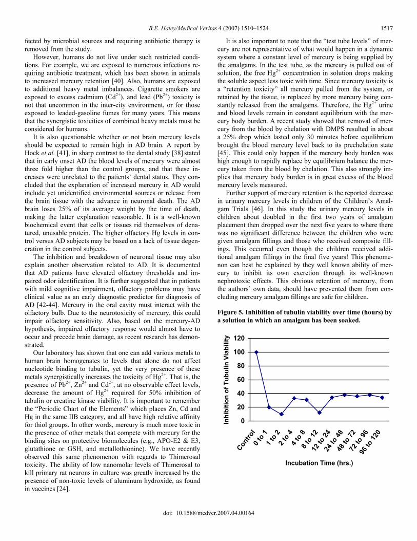
It is also important to note that the “test tube levels” of mer-cury are not representative of what would happen in a dynamic system where a constant level of mercury is being supplied by the amalgams. In the test tube, as the mercury is pulled out of solution, the free Hg2+ concentration in solution drops making the soluble aspect less toxic with time. Since mercury toxicity is a “retention toxicity” all mercury pulled from the system, or retained by the tissue, is replaced by more mercury being con-stantly released from the amalgams. Therefore, the Hg2+ urine and blood levels remain in constant equilibrium with the mer-cury body burden. A recent study showed that removal of mer-cury from the blood by chelation with DMPS resulted in about a 25% drop which lasted only 30 minutes before equilibrium brought the blood mercury level back to its prechelation state [45]. This could only happen if the mercury body burden was high enough to rapidly replace by equilibrium balance the mer-cury taken from the blood by chelation. This also strongly im-plies that mercury body burden is in great excess of the blood mercury levels measured. Further support of mercury retention is the reported decrease in urinary mercury levels in children of the Children’s Amal-gam Trials [46]. In this study the urinary mercury levels in children about doubled in the first two years of amalgam placement then dropped over the next five years to where there was no significant difference between the children who were given amalgam fillings and those who received composite fill-ings. This occurred even though the children received addi-tional amalgam fillings in the final five years! This phenome-non can best be explained by they well known ability of mer-cury to inhibit its own excretion through its well-known nephrotoxic effects. This obvious retention of mercury, from the authors’ own data, should have prevented them from con-cluding mercury amalgam fillings are safe for children. Figure 5. Inhibition of tubulin viability over time (hours) by a solution in which an amalgam has been soaked.
0
20
40
60
80
100
120
Contro
l0 t
o 11 t
o 22 t
o 44 t
o 88 t
o 12
12 to
2424
to 48
48 to
7272
to 96
96 to
120
Incubation Time (hrs.)
Inhi
bitio
n of
Tub
ulin
Via
bilit
y
doi: 10.1588/medver.2007.04.00164
B.E. Haley/Medical Veritas 4 (2007) 1510–1524 1518
2.6 Are amalgams capable of producing toxic solutions?
To propose deleterious effects of amalgams while in the mouth, the amalgams must be able to produce toxic effects out-side of the mouth. Figure 5 shows the inhibition of tubulin vi-ability using water in which a dental amalgam was incubated for the time shown. It is obvious that amalgams make water toxic to brain tubu-lin in a manner very similar to adding Hg2+ as done in refer-ences 3 to 6. In agreement, Wataha et al. reported that extracts of the amalgam material (trade name, Dispersalloy) “was se-verely cytotoxic when Zn release was greatest, but less toxic between 48 and 72 hours, as Zn release decreased.”[23] Zn is a non-volatile trace material in dental amalgams and Zn2+ is a needed mineral for living neurons. Therefore, it did not seem likely that the toxicity would be due to Zn leaching from the amalgams. To test this, the toxicity of 0.6 μM Hg2+ to tubulin in brain homogenates in the presence of 0, 10 and 20 micromolar Zn2+, as shown in Figure 6, was determined as previously de-scribed [3,4]. Zn2+ at these levels increased the 0.6 μM Hg2+ inhibition of tubulin viability by 0.6 μM Hg2+ from 4% to 50% and 76%, respectively. This supports the concept that the zinc correlation to increased toxicity was due to the synergistically enhanced toxicity of the mercury released from the amalgam, not to Zn2+ toxicity alone. Further, as shown in Table 2, studies in our laboratory have shown that incubation of amalgams in distilled water for less than one hour created a solution that also caused rapid inhibition of brain tubulin and creatine kinase similar to that observed on adding Hg2+ solutions Therefore, it appears that the toxicity of solutions in which amalgams were soaked is not caused by direct Zn2+ toxic ef-fects. Rather, enhanced mercury toxicity is due to the Zn2+ or other amalgam heavy metals synergistically enhancing the toxi-city of mercury as previously reported for lead and cadmium [47]. The enhancing metals may compete with biological chela- tors resulting in a higher concentration of free Hg2+ capable of inhibiting the activity of critical nucleotide binding proteins such as tubulin and CK.
Figure 6. Enhancement of Hg2+ toxicity to brain tubulin by increasing Zn2+
0
20
40
60
80
100
120
0.625 1.25 2.5 5 10 20
Inhi
bitio
n of
Tub
ulin
Via
bilit
y
HgZnHg+Zn(10)Hg+Zn(20)
Zn2+ (micromolars)
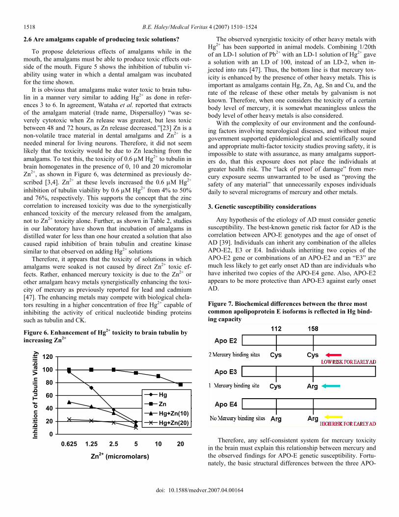
The observed synergistic toxicity of other heavy metals with Hg2+ has been supported in animal models. Combining 1/20th of an LD-1 solution of Pb2+ with an LD-1 solution of Hg2+ gave a solution with an LD of 100, instead of an LD-2, when in-jected into rats [47]. Thus, the bottom line is that mercury tox-icity is enhanced by the presence of other heavy metals. This is important as amalgams contain Hg, Zn, Ag, Sn and Cu, and the rate of the release of these other metals by galvanism is not known. Therefore, when one considers the toxicity of a certain body level of mercury, it is somewhat meaningless unless the body level of other heavy metals is also considered. With the complexity of our environment and the confound-ing factors involving neurological diseases, and without major government supported epidemiological and scientifically sound and appropriate multi-factor toxicity studies proving safety, it is impossible to state with assurance, as many amalgams support-ers do, that this exposure does not place the individuals at greater health risk. The “lack of proof of damage” from mer-cury exposure seems unwarranted to be used as “proving the safety of any material” that unnecessarily exposes individuals daily to several micrograms of mercury and other metals. 3. Genetic susceptibility considerations Any hypothesis of the etiology of AD must consider genetic susceptibility. The best-known genetic risk factor for AD is the correlation between APO-E genotypes and the age of onset of AD [39]. Individuals can inherit any combination of the alleles APO-E2, E3 or E4. Individuals inheriting two copies of the APO-E2 gene or combinations of an APO-E2 and an “E3” are much less likely to get early onset AD than are individuals who have inherited two copies of the APO-E4 gene. Also, APO-E2 appears to be more protective than APO-E3 against early onset AD. Figure 7. Biochemical differences between the three most common apolipoprotein E isoforms is reflected in Hg bind-ing capacity
Therefore, any self-consistent system for mercury toxicity in the brain must explain this relationship between mercury and the observed findings for APO-E genetic susceptibility. Fortu-nately, the basic structural differences between the three APO-
doi: 10.1588/medver.2007.04.00164
B.E. Haley/Medical Veritas 4 (2007) 1510–1524 1519
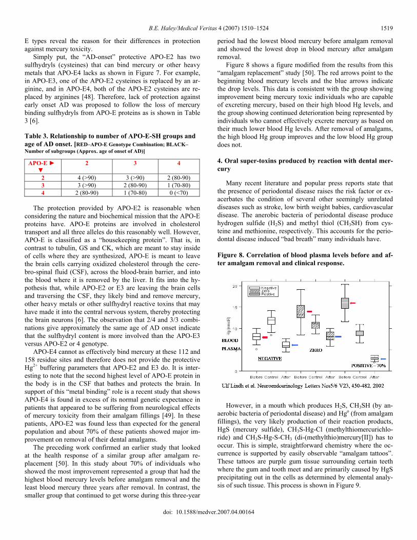
E types reveal the reason for their differences in protection against mercury toxicity. Simply put, the “AD-onset” protective APO-E2 has two sulfhydryls (cysteines) that can bind mercury or other heavy metals that APO-E4 lacks as shown in Figure 7. For example, in APO-E3, one of the APO-E2 cysteines is replaced by an ar-ginine, and in APO-E4, both of the APO-E2 cysteines are re-placed by arginines [48]. Therefore, lack of protection against early onset AD was proposed to follow the loss of mercury binding sulfhydryls from APO-E proteins as is shown in Table 3 [6]. Table 3. Relationship to number of APO-E-SH groups and age of AD onset. [RED–APO-E Genotype Combination; BLACK–Number of subgroups (Approx. age of onset of AD)]
APO-E ► ▼
2 3 4
2 4 (>90) 3 (>90) 2 (80-90) 3 3 (>90) 2 (80-90) 1 (70-80) 4 2 (80-90) 1 (70-80) 0 (<70)
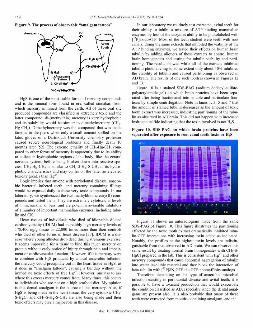
The protection provided by APO-E2 is reasonable when considering the nature and biochemical mission that the APO-E proteins have. APO-E proteins are involved in cholesterol transport and all three alleles do this reasonably well. However, APO-E is classified as a “housekeeping protein”. That is, in contrast to tubulin, GS and CK, which are meant to stay inside of cells where they are synthesized, APO-E is meant to leave the brain cells carrying oxidized cholesterol through the cere-bro-spinal fluid (CSF), across the blood-brain barrier, and into the blood where it is removed by the liver. It fits into the hy-pothesis that, while APO-E2 or E3 are leaving the brain cells and traversing the CSF, they likely bind and remove mercury, other heavy metals or other sulfhydryl reactive toxins that may have made it into the central nervous system, thereby protecting the brain neurons [6]. The observation that 2/4 and 3/3 combi-nations give approximately the same age of AD onset indicate that the sulfhydryl content is more involved than the APO-E3 versus APO-E2 or 4 genotype. APO-E4 cannot as effectively bind mercury at these 112 and 158 residue sites and therefore does not provide the protective Hg2+ buffering parameters that APO-E2 and E3 do. It is inter-esting to note that the second highest level of APO-E protein in the body is in the CSF that bathes and protects the brain. In support of this “metal binding” role is a recent study that shows APO-E4 is found in excess of its normal genetic expectance in patients that appeared to be suffering from neurological effects of mercury toxicity from their amalgam fillings [49]. In these patients, APO-E2 was found less than expected for the general population and about 70% of these patients showed major im-provement on removal of their dental amalgams. The preceding work confirmed an earlier study that looked at the health response of a similar group after amalgam re-placement [50]. In this study about 70% of individuals who showed the most improvement represented a group that had the highest blood mercury levels before amalgam removal and the least blood mercury three years after removal. In contrast, the smaller group that continued to get worse during this three-year
period had the lowest blood mercury before amalgam removal and showed the lowest drop in blood mercury after amalgam removal. Figure 8 shows a figure modified from the results from this “amalgam replacement” study [50]. The red arrows point to the beginning blood mercury levels and the blue arrows indicate the drop levels. This data is consistent with the group showing improvement being mercury toxic individuals who are capable of excreting mercury, based on their high blood Hg levels, and the group showing continued deterioration being represented by individuals who cannot effectively excrete mercury as based on their much lower blood Hg levels. After removal of amalgams, the high blood Hg group improves and the low blood Hg group does not.
4. Oral super-toxins produced by reaction with dental mer-cury Many recent literature and popular press reports state that the presence of periodontal disease raises the risk factor or ex-acerbates the condition of several other seemingly unrelated diseases such as stroke, low birth weight babies, cardiovascular disease. The anerobic bacteria of periodontal disease produce hydrogen sulfide (H2S) and methyl thiol (CH3SH) from cys-teine and methionine, respectively. This accounts for the perio-dontal disease induced “bad breath” many individuals have. Figure 8. Correlation of blood plasma levels before and af-ter amalgam removal and clinical response.
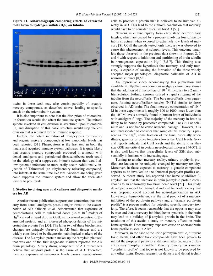
However, in a mouth which produces H2S, CH3SH (by an-aerobic bacteria of periodontal disease) and Hgo (from amalgam fillings), the very likely production of their reaction products, HgS (mercury sulfide), CH3S-Hg-Cl (methylthiomercurichlo-ride) and CH3S-Hg-S-CH3 (di-(methylthio)mercury[II]) has to occur. This is simple, straightforward chemistry where the oc-currence is supported by easily observable “amalgam tattoos”. These tattoos are purple gum tissue surrounding certain teeth where the gum and tooth meet and are primarily caused by HgS precipitating out in the cells as determined by elemental analy-sis of such tissue. This process is shown in Figure 9.
doi: 10.1588/medver.2007.04.00164
B.E. Haley/Medical Veritas 4 (2007) 1510–1524 1520
Figure 9. The process of observable “amalgam tattoos”
HgS is one of the most stable forms of mercury compounds and is the mineral form found in ore, called cinnabar, from which mercury is mined from the earth. All of these oral site produced compounds are classified as extremely toxic and the latter compound, di-(methylthio) mercury is very hydrophobic and its solubility would be similar to dimethylmercury (CH3-Hg-CH3). Dimethylmercury was the compound that was made famous in the press when only a small amount spilled on the latex gloves of a Dartmouth University chemistry professor caused severe neurological problems and finally death 10 months later [52]. The extreme lethality of CH3-Hg-CH3 com-pared to other forms of mercury is apparently due to its ability to collect in hydrophobic regions of the body, like the central nervous system, before being broken down into reactive spe-cies. CH3-Hg-CH3 is similar to CH3-S-Hg-S-CH3 in its hydro-phobic characteristics and may confer on the latter an elevated toxicity greater than Hg2+. Logic implies that anyone with periodontal disease, anaero-bic bacterial infected teeth, and mercury containing fillings would be exposed daily to these very toxic compounds. In our laboratory, we synthesized the two methylthiomercury(II) com-pounds and tested them. They are extremely cytotoxic at levels of 1 micromolar or less, and are potent, irreversible inhibitors of a number of important mammalian enzymes, including tubu-lin and CK. Heart tissues of individuals who died of idiopathic dilated cardiomyopathy (IDCM) had incredibly high mercury levels of 178,400 ng/g tissue or 22,000 times more than their controls who died of other forms of heart disease [37]. IDCM is a dis-ease where young athletes drop dead during strenuous exercise. It seems impossible for a tissue to bind this much mercury on protein without early notice of injury through pain and impair-ment of cardiovascular function. However, if this mercury were to combine with H2S produced by a local anaerobic infection the mercury could precipitate out in the heart tissue as HgS, as it does in “amalgam tattoos”, causing a buildup without the immediate toxic effects of free Hg2+. However, one has to ask where this excess mercury comes from. Many times, this occurs to individuals who are not on a high seafood diet. My opinion is that dental amalgam is the source of this mercury. Also, if HgS is being made in the heart tissue, the very cytotoxic CH3-S-HgCl and CH3-S-Hg-S-CH3 are also being made and their toxic effects may play a major role in this disease.
In our laboratory we routinely test extracted, avital teeth for their ability to inhibit a mixture of ATP binding mammalian enzymes by loss of the enzymes ability to be photolabeled with [32P]azidoATP. Most of the teeth studied were teeth with root canals. Using the same extracts that inhibited the viability of the ATP binding enzymes, we tested their effects on human brain tubulin by adding aliquots of these extracts to control human brain homogenates and testing for tubulin viability and parti-tioning. The results showed while all of the extracts inhibited tubulin photolabeling to some extent only about 40% inhibited the viability of tubulin and caused partitioning as observed in AD brain. The results of one such tooth is shown in Figures 12 and 13. Figure 10 is a stained SDS-PAG (sodium dodecyl-sulfate-polyacrylamide gel) on which brain proteins have been sepa-rated after being fractionated into soluble and particulate frac-tions by simple centrifugation. Note in lanes 1, 3, 5 and 7 that the amount of stained tubulin decreases as the amount of toxic tooth extract was increased, indicating partitioning of the tubu-lin as observed in AD brain. This did not happen with increased hydrogen sulfide indicating that the toxin involved is not H2S.
Figure 10. SDS-PAG on which brain proteins have been separated after exposure to root canal tooth toxin or H2S
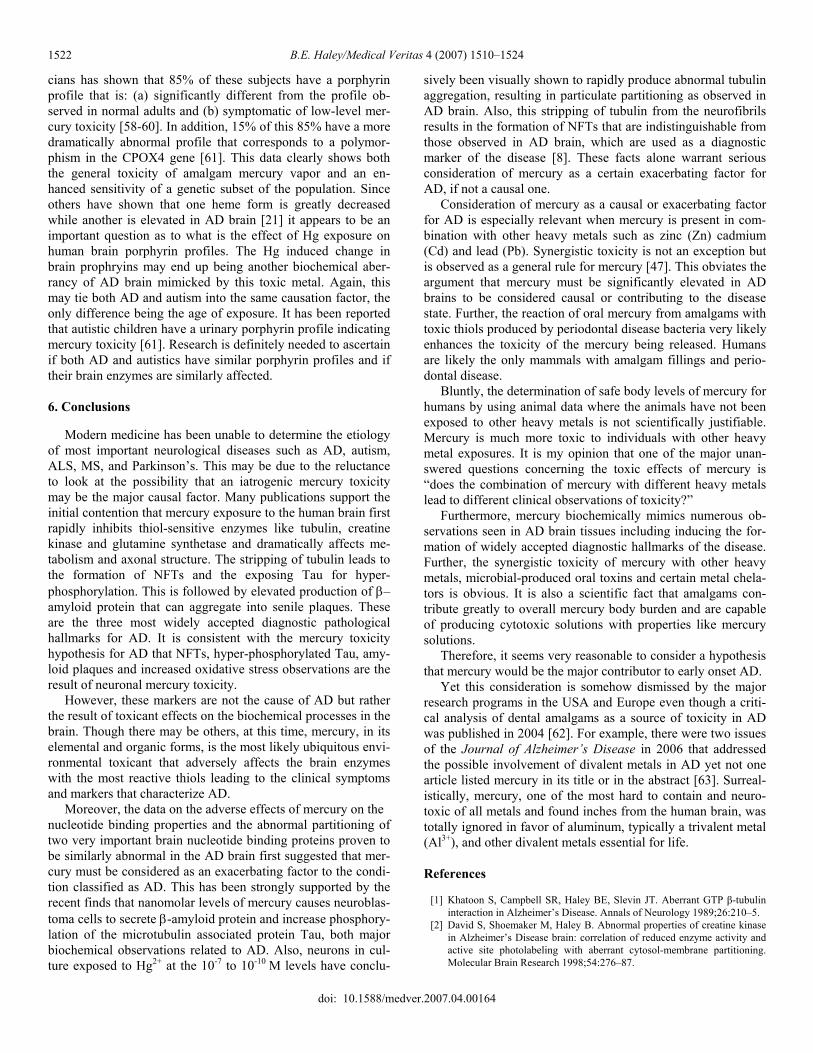
Figure 11 shows an autoradiogram made from the same SDS-PAG of Figure 10. This figure illustrates the partitioning effected by the toxic tooth extract dramatically inhibited tubu-lin-GTP interactions with increasing toxin added as indicated. Notably, the profiles at the highest toxin levels are indistin-guishable from that observed in AD brain. We can observe this same result by treating normal brain homogenates with CH3-S- HgCl prepared in the lab. This is consistent with Hg2+ and other mercury compounds that cause abnormal aggregation of tubulin into water insoluble material and they block the interaction of beta-tubulin with [32P]8N3GTP the GTP photoaffinity analogs. Therefore, depending on the type of anaerobic microbial infection existing in periodontal disease and avital teeth, it is possible to have a toxicant production that would exacerbate the condition classified as AD, especially when the dental amal-gams are present also. It is also probable that many of these teeth were extracted from mouths containing amalgam, and the
doi: 10.1588/medver.2007.04.00164
B.E. Haley/Medical Veritas 4 (2007) 1510–1524 1521
Figure 11. Autoradiograph comparing effects of extracted tooth toxin to hydrogen sulfide (H2S) on tubulin
toxins in these teeth may also consist partially of organic-mercury compounds, as described above, leading to specific attack on the microtubulin system. It is also important to note that the disruption of microtubu-lin formation would also affect the immune system. The mitotic spindle involved in cell division is structured upon microtubu-lin, and disruption of this basic structure would stop the cell division that is required for the immune response. Further, the potent inhibition of phagocytosis by mercury and organic mercury compounds at low nanomolar levels has been reported [51]. Phagocytosis is the first step in both the innate and acquired immune system pathways. It is quite likely that organic mercury compounds produced in a mouth with dental amalgams and periodontal disease/infected teeth could be the etiology of a suppressed immune system that would al-low systemic infections to more easily occur. Additionally, in-jection of Thimerosal (an ethylmercury releasing compound) into infants at the same time live viral vaccines are being given could suppress the immune system and allow the attenuated viruses to proliferate
5. Studies involving neuronal cultures and diagnostic mark-ers for AD Another recent publication supports our contention that mer-cury from dental amalgams poses a major threat to the exacer-bation of AD. Olivieri et al. demonstrated that exposure of neuroblastoma cells to sub-lethal doses (36 x 10-9 molar) of Hg2+ caused a rapid drop in GSH, an increased secretion of β–amyloid protein, and an increased phosphorylation of the mi-crotubulin protein Tau [53]. The latter two of these biochemical changes are uniquely observed in AD brain tissues and are widely considered to be diagnostic, pathological markers of the disease. The β-amyloid protein makes up the ‘amyloid plaques’ that was one of the first diagnostic markers reported for AD brain pathology. A very strong component of AD researchers believe that amyloid protein is the cause of AD. Therefore, mercury exposure at nanomolar levels causes neuroblastoma
cells to produce a protein that is believed to be involved di-rectly in AD. This lead to the author’s conclusion that mercury would have to be consider as causal for AD [53]. Neurons in culture rapidly form early stage neurofibillary tangles, which are caused by a process involving loss of micro-tublin structure, when exposed to extremely low levels of mer-cury [8]. Of all the metals tested, only mercury was observed to cause this phenomenon at subppm levels. This outcome paral-lels those observed in the previous data shown in Figures 2, 3 and 4 with respect to inhibition and partitioning of brain tubulin in homogenates exposed to Hg2+ [3,5-7]. This finding also strongly supports the hypothesis that mercury, and only mer-cury, is capable of causing the formation of the three widely accepted major pathological diagnostic hallmarks of AD in neuronal cultures [8,53]. An impressive video accompanying this publication and available at http://movies.commons.ucalgary.ca/mercury shows that the addition of 2 microliters of 10-7 M mercury to a 2 milli-liter solution bathing neurons caused a rapid stripping of the tubulin from the neurofibrils. The bare neurofibrils then aggre-gate, forming neurofibrillary tangles (NFTs) similar to those observed in AD brain. The final mercury concentration of 10-10
M in these experiments is roughly 100 to 1000 times lower than the 10-7 M levels normally found in human brain of individuals with amalgam fillings. The majority of the mercury in brain is likely to be bound by protective compounds like GSH or sele-nium and is not free to cause neuronal damage. However, it is not unreasonable to consider that some of this mercury is pre-sent as free Hg2+, some fraction of the time, especially when illness, genetics or other toxicities lower the GSH levels. Sev-eral reports indicate that GSH levels and the ability to synthe-size GSH are critical in certain neurological illnesses [54-57]. It is also well known that intracellular levels of GSH drop sub-stantially in humans with increasing age. Turning to another mercury reality, urinary prophyrin pro-files are known to be uniquely changed by mercury toxicity. Moreover, in those exposed to mercury, a genetic component appears to be involved on the abnormal porphyrin profiles ob-served. A recent study has reported that heme solubilizes β-amyloid and that the increase in brain β-amyloid protein corre-sponds to an abnormally low brain heme level [21]. This study developed a model for β-amyloid induced heme-deficiency that was proposed could account for neurodegeneration in AD. However, a heme-deficiency is the final product of a mercury inhibition of the porphyrin pathway and a “urinary porphyrin profile” is a proven method for detecting specific mercury tox-icity. Therefore, it seems reasonable that the opposite may also be true and that a mercury inhibited heme synthesis in the brain may lead to a buildup of β-amyloid protein in the brain. The resolution of this awaits a study on mercury effects on brain heme synthesis. Does mercury exposure cause an aberrant brain heme profile as seen in AD? Moreover, in the case of the urine porphyrin profile, different toxic metals and other toxic compounds have been proven to inhibit the porphyrin pathway at different sites causing a differ-ent urinary “porphyrin profile.” Mercury toxicity has a unique “prophyrin profile” that today is not known to be produced by any other toxin. Recent research on dentists and dental techni-
doi: 10.1588/medver.2007.04.00164
B.E. Haley/Medical Veritas 4 (2007) 1510–1524 1522
cians has shown that 85% of these subjects have a porphyrin profile that is: (a) significantly different from the profile ob-served in normal adults and (b) symptomatic of low-level mer-cury toxicity [58-60]. In addition, 15% of this 85% have a more dramatically abnormal profile that corresponds to a polymor-phism in the CPOX4 gene [61]. This data clearly shows both the general toxicity of amalgam mercury vapor and an en-hanced sensitivity of a genetic subset of the population. Since others have shown that one heme form is greatly decreased while another is elevated in AD brain [21] it appears to be an important question as to what is the effect of Hg exposure on human brain porphyrin profiles. The Hg induced change in brain prophryins may end up being another biochemical aber-rancy of AD brain mimicked by this toxic metal. Again, this may tie both AD and autism into the same causation factor, the only difference being the age of exposure. It has been reported that autistic children have a urinary porphyrin profile indicating mercury toxicity [61]. Research is definitely needed to ascertain if both AD and autistics have similar porphyrin profiles and if their brain enzymes are similarly affected. 6. Conclusions Modern medicine has been unable to determine the etiology of most important neurological diseases such as AD, autism, ALS, MS, and Parkinson’s. This may be due to the reluctance to look at the possibility that an iatrogenic mercury toxicity may be the major causal factor. Many publications support the initial contention that mercury exposure to the human brain first rapidly inhibits thiol-sensitive enzymes like tubulin, creatine kinase and glutamine synthetase and dramatically affects me-tabolism and axonal structure. The stripping of tubulin leads to the formation of NFTs and the exposing Tau for hyper-phosphorylation. This is followed by elevated production of β–amyloid protein that can aggregate into senile plaques. These are the three most widely accepted diagnostic pathological hallmarks for AD. It is consistent with the mercury toxicity hypothesis for AD that NFTs, hyper-phosphorylated Tau, amy-loid plaques and increased oxidative stress observations are the result of neuronal mercury toxicity. However, these markers are not the cause of AD but rather the result of toxicant effects on the biochemical processes in the brain. Though there may be others, at this time, mercury, in its elemental and organic forms, is the most likely ubiquitous envi-ronmental toxicant that adversely affects the brain enzymes with the most reactive thiols leading to the clinical symptoms and markers that characterize AD. Moreover, the data on the adverse effects of mercury on the nucleotide binding properties and the abnormal partitioning of two very important brain nucleotide binding proteins proven to be similarly abnormal in the AD brain first suggested that mer-cury must be considered as an exacerbating factor to the condi-tion classified as AD. This has been strongly supported by the recent finds that nanomolar levels of mercury causes neuroblas-toma cells to secrete β-amyloid protein and increase phosphory-lation of the microtubulin associated protein Tau, both major biochemical observations related to AD. Also, neurons in cul-ture exposed to Hg2+ at the 10-7 to 10-10 M levels have conclu-
sively been visually shown to rapidly produce abnormal tubulin aggregation, resulting in particulate partitioning as observed in AD brain. Also, this stripping of tubulin from the neurofibrils results in the formation of NFTs that are indistinguishable from those observed in AD brain, which are used as a diagnostic marker of the disease [8]. These facts alone warrant serious consideration of mercury as a certain exacerbating factor for AD, if not a causal one. Consideration of mercury as a causal or exacerbating factor for AD is especially relevant when mercury is present in com-bination with other heavy metals such as zinc (Zn) cadmium (Cd) and lead (Pb). Synergistic toxicity is not an exception but is observed as a general rule for mercury [47]. This obviates the argument that mercury must be significantly elevated in AD brains to be considered causal or contributing to the disease state. Further, the reaction of oral mercury from amalgams with toxic thiols produced by periodontal disease bacteria very likely enhances the toxicity of the mercury being released. Humans are likely the only mammals with amalgam fillings and perio-dontal disease. Bluntly, the determination of safe body levels of mercury for humans by using animal data where the animals have not been exposed to other heavy metals is not scientifically justifiable. Mercury is much more toxic to individuals with other heavy metal exposures. It is my opinion that one of the major unan-swered questions concerning the toxic effects of mercury is “does the combination of mercury with different heavy metals lead to different clinical observations of toxicity?” Furthermore, mercury biochemically mimics numerous ob-servations seen in AD brain tissues including inducing the for-mation of widely accepted diagnostic hallmarks of the disease. Further, the synergistic toxicity of mercury with other heavy metals, microbial-produced oral toxins and certain metal chela-tors is obvious. It is also a scientific fact that amalgams con-tribute greatly to overall mercury body burden and are capable of producing cytotoxic solutions with properties like mercury solutions. Therefore, it seems very reasonable to consider a hypothesis that mercury would be the major contributor to early onset AD. Yet this consideration is somehow dismissed by the major research programs in the USA and Europe even though a criti-cal analysis of dental amalgams as a source of toxicity in AD was published in 2004 [62]. For example, there were two issues of the Journal of Alzheimer’s Disease in 2006 that addressed the possible involvement of divalent metals in AD yet not one article listed mercury in its title or in the abstract [63]. Surreal-istically, mercury, one of the most hard to contain and neuro-toxic of all metals and found inches from the human brain, was totally ignored in favor of aluminum, typically a trivalent metal (Al3+), and other divalent metals essential for life. References [1] Khatoon S, Campbell SR, Haley BE, Slevin JT. Aberrant GTP β-tubulin
interaction in Alzheimer’s Disease. Annals of Neurology 1989;26:210–5. [2] David S, Shoemaker M, Haley B. Abnormal properties of creatine kinase
in Alzheimer’s Disease brain: correlation of reduced enzyme activity and active site photolabeling with aberrant cytosol-membrane partitioning. Molecular Brain Research 1998;54:276–87.
doi: 10.1588/medver.2007.04.00164
B.E. Haley/Medical Veritas 4 (2007) 1510–1524 1523
[3] Duhr EF, Pendergrass JC, Slevin JT, Haley B. HgEDTA complex inhibits GTP interactions with the E-Site of brain β-tubulin. Toxicology and Ap-plied Pharmacology 1993 Oct.;122(2):273–88.
[4] Haley BE. Development and utilization of 8-azidopurine nucleotide photoaffinity Probes. Federation Proceedings 1983;42:2831–6.
[5] Pendergrass JC, Haley BE. Mercury-EDTA complex specifically blocks brain β-tubulin-GTP interactions: similarity to observations in Alz-heimer”s Disease. In: Status Quo and Perspective of Amalgam and Other Dental Materials (International Symposium Proceedings. Friberg LT, Schrauzer GN. Georg Thieme Verlag, eds. Stuttgart-New York 1995:98–105.
[6] Pendergrass JC, Haley BE. Inhibition of brain tubulin-guanosine 5’-triphosphate interactions by mercury: similarity to observations in Alz-heimer’s Diseased brain. In: Metal Ions in Biological Systems V34. Mer-cury and Its Effects on Environment and Biology, Chapter 16. Edited by H. Sigel and A. Sigel. Marcel Dekker, Inc. 270 Madison Ave., N.Y., N.Y. 10016, 1996:461–78.
[7] Pendergrass JC, Haley BE, Vimy MJ, Winfield SA, Lorscheider FL. Mer-cury vapor inhalation inhibits binding of GTP to tubulin in rat brain: simi-larity to a molecular lesion in Alzheimer’s Disease brain. Neurotoxicology 1997;18(2):315–24.
[8] Leong CCW, Syed NI, Lorscheider FL. Retrograde degeneration of neu-rite membrane structural integrity and formation of neruofibillary tangles at nerve growth cones following in-vitro exposure to mercury. NeuroRe-ports 2001;12(4):733–7.
[9] Thompson CM, Markesbery WR, Ehmann WD, Mao YX, Vance DE. Regional brain trace-element studies in Alzheimer’s disease. NeuroToxi-cology 1988 Spring;9(1):1–7.
[10] Deibel MA, Ehmann WD, Markesbery WR. Copper, iron and zinc imbal-ances in severely degenerated brain regions in Alzheimer’s disease: possi-ble relation to oxidative stress. J. Neurol. Sci. 1996;143:137–42.
[11] Jayaram B,.Haley B. Identification of peptides within the base binding domains of the GTP and ATP specific binding sites of tubulin. J. Biol. Chem. 1994;269(5):3233–42.
[12] Olcott M, Haley B. Identification of two peptides from the ATP-binding domain of creatine kinase. Biochemistry 1994;33:11935–41.
[13] Gunnersen DJ, Haley B. Detection of glutamine synthetase in the cerebro-spinal fluid of Alzheimer’s diseased patients: a potential diagnostic bio-chemical marker. Proc. Natl. Acad. Sci. USA 1992 Dec. 15;89(24):, 11949–53.
[14] Butterfield DA, Hensley K, Cole P, Subramaniam R, Aksenov M, Ak-senova M, Bummer PM, Haley BE, Carney JM. Oxidatively-induced structural alteration of glutamine synthetase assessed by analysis of spin labeled incorporation kinetics: relevance to Alzheimer’s disease, J. Neuro-chem. 1997 Jun.;68(6):2451–7.
[15] Tumani H, Shen GQ, Peter J, Bruck W. Glutamine synthetase in cerebro-spinal fluid, serum and brain: a diagnostic marker for Alzheimer Disease? Arch. Neurol. 1999;56:1241–6.
[16] Takahashi M, Stanton E, Moreno JI, Jackowski G. Immunoassay for se-rum glutamine synthetase in serum: development, reference values, and preliminary study in dementias. Clinical Chemistry 2002;48(2):375–8.
[17] Robinson S/R. Neuronal expression of glutamine synthetase in Alz-heimer’s disease indicate a profound impairment of metabolic interactions with astrocytes. Neurochemistry International 2000;36:471–82.
[18] Woods J, Martin MD, Naleway CA, Echeverria D. Urinary porphyrin profiles as a biomarker of mercury exposure: studies on dentists with oc-cupational exposure to mercury vapor. J. Toxicol. Environ. Health 1993; 40:235–46.
[19] Echeverria D, Woods JS, Heyer NJ, Rohlman DS, Farin FM, Bittner AC, Li T, Garabedian C. Chronic low-level mercury exposure, BDNF (brain derived neurotrophic factor) polymorphism, and associations with cogni-tive and motor function. Neurotoxicol. Teratol, 2005 Nov.-Dec.; 27(6):781–96.
[20] Echeverria D, Woods JS,Heyer NJ, Rohlman D, Farin FM, Li T, Garabe-dian CE. The association between a genetic polymorphism of copropor-phyrinogen oxidase, dental mercury exposure and neurobehavioral re-sponse in humans. Neurotoxicol. Teratol. 2006 Jan.-Feb.; 28(1):39–48.
[21] Atamna H, Frey WH. A Role for heme in Alzheimer’s disease: Heme binds amyolid β and has altered metabolism. Proc. Natl. Acad. Sci. 2004;101(30):11153–8.
[22] Chew CL, Soh G, Lee AS, Yeoh TS. Long-term dissolution of mercury from a non-mercury-releasing amalgam. Clinical Preventive Dentistry 1991 May-Jun.;13(3):5–7.
[23] Wataha JC, Nakajima H, Hanks CT, Okabe T. Correlation of Cytotoxicity with Element Release from Mercury and Gallium-based Dental Alloys in vitro. Dental Materials 1994 Sept.;10(5):298–303.
[24] Haley BE. Mercury toxicity: genetic susceptibility and synergistic effects. Medical Veritas 2005 Nov.;2(2):535–42.
[25] Holmes AS, Blaxill MF, Haley B. Reduced levels of mercury in first baby haircuts of autistic children. International J. of Toxicology, 2003;22:1–9.
[26] Bradstreet J, Geier DA, Kartzinel JJ, Adams JB, Geier MR. A case-control study of mercury burden in children with autistic spectrum disorders. J. Am. Phys. Surg. 2003;8(3):76–9.
[27] Sokol DK, Chem D, Farlow MR, Dunn DW, Maloney B, Zimmer JA, Lahiri DK. High levels of Alzheimer’s beta-amyloid precursor protein (APP) in children with severely autistic behavior and aggression. 2006; 21(6):444–9.
[28] Guzzi G, Grandi M, Cattaneo C, Calza S, Minoia C, ronchi A, Gatti A, Severi G. Dental amalgam and mercury levels in autopsy tissues: food for thought. Am J Forensic Med Pathol 2006 Mar.;27(1):42–5.
[29] Engqvist SI. Amalgam Restorations-an important source of human expo-sure of mercury and silver. Lakartidnigen 1992;15:1299–1301.
[30] Drasch G, Aigner S, Roider G, Staiger F, Lipowsky G. Mercury in human colostrum and early breast milk. Its dependence on dental amalgam and other factors. J. Trace Elements Med. Biol. 1998:12(1); 23–7.
[31] Luglie PF, Campus G, Chessa G, Spano G, Capobianco G, Fadda GM, Dessole S. Effect of amalgam fillings on the mercury concentration in human amniotic fluid. Arch. Gynecol. Obstet. 2005;271(2):138–4.
[32] Lorscheider FL, Vimy MJ, Summers AO. Mercury exposure from silver tooth fillings: emerging evidence questions a traditional dental paradigm. FASEB J. 1995;9:504–8.
[33] World Health Organization (WHO) report on Environmental Health Crite-ria 118, Inorganic Mercury, WHO, Genevia, 1991.
[34] Kingman A, Albertini T, Brown LJ. Mercury concentrations in urine and whole blood associated with amalgam exposure in a U.S. military popula-tion. J. of Dental Research 1998;77(3):461–71.
[35] Begerow J, Zander D, Freier I, Dunemann L. Long-term mercury excre-tion in urine after removal of amalgam fillings. Int. Arch. Occup. Environ. Health 1994:66(3):209–12.
[36] Hagele J, Mazerik JN, Gregory A, Kaufman B, Magalang U, Kuppusamy ML, Marsh CB, Kuppusamy P, Parinandi L. Mercury activates vascular endothelial cell phospholipase D through thiols and oxidative stress. In-ternational J. Toxicology 2007;26:57–69.
[37] Frustaci A, Magnavita N, Chimenti C, Caldarulo M, Sabbioni E, Pietra R, Cellini C, Possati GF, Maseri A. Marked elevation of myocardial trace elements in idiopathic dilated cardiomyopathy compared with secondary dysfunction. J. American College of Cardiology 1999;33(6):1578–83.
[38] Saxe SR, Wekstein MW, Kryscio RJ, Henry RG, Conrett CR, Snowdon, DA, Grant FT, Schmitt FA, Donegan SJ, Wekstein DR, Ehmann WD, Markesbery WR. Alzheimers’ disease, dental amalgam and mercury. JADA 1999;130:191–9.
[39] (a)Roses AD. Scientific American. Science and Medicine, 1995:16-15. (b)Roses AD. Apolipoprotein-E and Alzheimer’s disease. The tip of the susceptibility iceberg. Annals of the N.Y. Academy of Science 1998; 855:738–43.
[40] Rowland I, Davies M, Evans J. Tissue content of mercury in rats given methylmercury chloride orally: Influence of intestinal flora. Arch Environ Health 1980;35:155–60.
[41] Hock C, Drasch G, Golombowski S, Muller-Spahn F, Willershausen-Zonnchen B, Schwarz P, Hock U, Growdon JH, Nitsch RM. Increased blood mercury levels in patients with Alzheimer’s disease. J. Neural Transm 1998;105(1):59–68..
[42] Devanand DP, Michaels-Marston KS, Liu X, Pelton GH, Padilla M, Marder K, Bell K, Stern Y, Mayeux R. Olfactory deficits in patients with mild cognitive impairment predict Alzheimer’s disease at follow-up. Am. J. Psychiatry 2000;157(9):1399–1405.
[43] Kovacs T, Cairns NJ, Lantos PL. Olfactory centres in Alzheimer’s dis-ease: olfactory bulb is involved in early Braak’s stages. Neuroreport 2001; 12(2):285–8.
[44] Gray AJ, Staples V, Murren K, Dahariwal A, Bentham P. Olfactory identi-fication is impaired in clinic-based patients with vascular dementia and
doi: 10.1588/medver.2007.04.00164
B.E. Haley/Medical Veritas 4 (2007) 1510–1524 1524
senile dementia of Alzheimer’s type. Int. J. Geriatr. 2001;Psychiatry 16(5):513–7.
[45] Vamnes et al. Blood mercury following DMPS administration to subjects with and without dental amalgam. Sci. Total Envir. 2004;308:63–71.
[46] DeRouen TA, Martin MD, Leroux BG, Townes BD, Woods JS, Leitao J et al. Neurobehavioral effects of dental amalgam in children: a randomized clinical trial. JAMA 2006 Apr. 19;295(15):1784–92.
[47] Schubert J, Riley EJ, Tyler SA. Combined effects in toxicology—a rapid systemic testing procedure: cadmium, mercury and lead. J. of Toxicology and Environmental Health 1978;4:763–76.
[48] Brouwer DA. Clinical chemistry of common Apoprotein-E isoforms. J. Chromatography, Biomed. Applications 1996;678(1):23–41.
[49] Wojcik D, Godfrey M, Christie D, Haley B. Mercury toxicity presenting as chronic fatigue, memory impairment and depression: diagnosis, treat-ment, susceptibility, and outcomes in a New Zealand general practice set-ting (1994-2006). Neuroendcrinology Letters 2006;27(4):416–23.
[50] Ulf Lindh et al. Neuroendocrinology Letters 2002;23(5/6):450–82. [51] Rampersad GC, Suck G, Sakac D, Fahim S, Foo A, Denomme GA,
Langler RF,Branch DR. Chemical compounds that target thiol-disulfide groups on mononuclear phagocytes inhibit immune mediated phagocytosis of red blood cells. Transfusion 2005;45:384–93.
[52] Nierenberg DW, Nordgren RE, Chang MB, Siegler W, Blayney MG, Hochberg F, Toribara TY, Cernichiari E, Clarkson T. Delayed cerebellar disease and death after accidental exposure to dimethyl mercury. N. Engl. J. Med 1998;338;1672–5.
[53] Olivieri G, Brack C, Muller-Spahn F, Stahelin HB, Herrmann M, Renard P, Brockhaus M, Hock C. Mercury induces cell cytotoxicity and oxidative stress and increases β-amyloid secretion and tau phosphorylation in SHSY5Y neuroblastoma cells. J. Neurochemistry 2000 Jan.;74(1):231–6.
[54] Westphal GA, Schnuch A, Schulz TG, Reich K, Aberer W, Brasch J, Koch P, Wessbecher R, Szliska C, Bauer A, Hallier E. Homozygous gene deletions of the glutathione S-transferases M1 and T1 are associated with thimerosal sensitization. Int Arch Occup Environ Health 2000;73:384–8.
[55] James SJ, Slikker W 3rd, Melnyk S, New E, Pogribna M, Jernigan S. Thimerosal neurotoxicity is associated with glutathione depletion: protec-tion with glutathione precursors. Neurotoxicology 2005;26:1-8.
[56] James SJ, Cutler P, Melnyk S et al. Metabolic Biomarkers of Increased Oxidative Stress and Impaired Methylation capacity in children with au-tims. Am. J. Clin. Nutr. 2004;80:1611-17.
[57] Waly M, Olteanu H, Banerjee R, Choi SW, Mason JB, Parker BS, Suku-mar S, Shim S, Sharma A, Benzecry JM, Power-Charnitsky VA, Deth RC. Activation of methionine synthase by insulin-like growth factor-1 and do-pamine: a target for neurodevelopmental toxins and thimerosal. Mol Psy-chiatry 2004;9:358-70.
[58] Woods J, Martin MD, Naleway CA, Echeverria D. Urinary porphyrin profiles as a biomarker of mercury exposure: studies on dentists with oc-cupational exposure to mercury vapor. J. Toxicol. Environ. Health 1993;40:235–46.
[59] Echeverria D, Woods JS, Heyer NJ, Rohlman DS, Farin FM, Bittner AC, Li T, Garabedian C. Chronic low-level mercury exposure, BDNF (brain derived neurotrophic factor) polymorphism, and associations with cogni-tive and motor function. Neurotoxicol. Teratol, 2005 Nov.-Dec.;27(6):781–96.
[60] Echeverria, D. Woods, JS, Heyer NJ, Rohlman D, Farin FM, Li T, Gara-bedian CE. The association between a genetic polymorphism of copropor-phyrinogen oxidase, dental mercury exposure and neurobehavioral re-sponse in humans. Neurotoxicol. Teratol. 2006 Jan.-Feb.;28(1):39–48.
[61] Nataf R, Skorupka C, Amet L, Lam A, Springbett A, Lathe R. Porphyrinuria in childhood autistic disorder: implications for environmental toxicity. Toxicology and Applied Pharmacology 2006 Jul. 15;214(2):99–108.
[62] Mutter J, Naumann J, Sadaghiani C, Walach H, Drasch G. Amalgam stud-ies: disregarding basic principles of mercury toxicity. Int J Hyg Environ Health 2004;207:391-7.
[63] Adlard PA, Bush AI. Metals in Alzheimer’s disease. J Alzheimer Dis 2006 Nov.; 10(2-3):145--63.
doi: 10.1588/medver.2007.04.00164
Related Documents

![r G { Gr ü GGGG G t ¡ G s ü GGGGGGG G t Gz GGGGGGG G n Gz GGGGGGG r GGGGGGG · 2020-05-11 · r ggggggg pg[s`wg n gz g r sgr g g gz g pg\s`w g « yrq 7lurohu *hp vh elv ]x (lhuq](https://static.cupdf.com/doc/110x72/5f30a1a40065fb796e0e11b1/r-g-gr-gggg-g-t-g-s-ggggggg-g-t-gz-ggggggg-g-n-gz-ggggggg-r-ggggggg-2020-05-11.jpg)


![gZ N ac io n e s U n id as hj]ZgbaZpby GZpbc CONFERENCE · A g ric u lt u re O rg a n iz a t io n o f t h e U n it e d N a t io n s ... Twenty-eighth - Sana’a, Republic of Yemen,](https://static.cupdf.com/doc/110x72/5c6ff23f09d3f29a798c502e/gz-n-ac-io-n-e-s-u-n-id-as-hjzgbazpby-gzpbc-a-g-ric-u-lt-u-re-o-rg-a-n-iz-a.jpg)


![gZihjtqdZaZ mkem] Z · ?K 2016/679 gZ ?\jhi_ckdby iZjeZf_gl b gZ Kt\_lZ hlghkgh aZsblZlZ gZ nbabq_kdbl_ ebpZ \t\ \jtadZ k h[jZ[hl\Zg_lh gZ ebqgb ^Zggb b hlghkgh k\h[h^ghlh ^\b`_gb_](https://static.cupdf.com/doc/110x72/5ec4170de2fbf52ed91cd7e9/gzihjtqdzaz-mkem-z-k-2016679-gz-jhickdby-izjezfgl-b-gz-ktlz-hlghkgh-azsblzlz.jpg)


