The Journal of Experimental Medicine ARTICLE JEM © The Rockefeller University Press $8.00 Vol. 203, No. 7, July 10, 2006 1745–1759 www.jem.org/cgi/doi/10.1084/jem.20060085 1745 X-linked susceptibility to mycobacteria is caused by mutations in NEMO impairing CD40-dependent IL-12 production Orchidée Filipe-Santos, 1 Jacinta Bustamante, 1 Margje H. Haverkamp, 10,12 Emilie Vinolo, 2 Cheng-Lung Ku, 1 Anne Puel, 1 David M. Frucht, 11 Karin Christel, 1 Horst von Bernuth, 1 Emmanuelle Jouanguy, 1 Jacqueline Feinberg, 1 Anne Durandy, 3 Brigitte Senechal, 9 Ariane Chapgier, 1 Guillaume Vogt, 1 Ludovic de Beaucoudrey, 1 Claire Fieschi, 1,13 Capucine Picard, 1,4 Meriem Garfa, 5 Jalel Chemli, 14 Mohamed Bejaoui, 15 Maria N. Tsolia, 17 Necil Kutukculer, 18 Alessandro Plebani, 19 Luigi Notarangelo, 19 Christine Bodemer, 6 Frédéric Geissmann, 9 Alain Israël, 8 Michel Véron, 2 Maike Knackstedt, 20 Ridha Barbouche, 16 Laurent Abel, 1 Klaus Magdorf, 20 Dominique Gendrel, 21 Fabrice Agou, 2 Steven M. Holland, 10 and Jean-Laurent Casanova 1,7 1 Laboratory of Human Genetics of Infectious Diseases, University of Paris René Descartes-Institut National de la Santé et de la Recherche Médicale (INSERM) U 550, Necker Medical School; 2 Laboratory of Enzymatic Regulation of Cellular Activities, URA 2185 Centre National de la Recherche Scientifique (CNRS), Pasteur Institute; 3 Laboratory of Normal and Pathologic Development of the Immune System, INSERM U768, 4 Center for the Study of Primary Immunodeficiencies, 5 Laboratory of Confocal Microscopy, 6 Dermatology Unit, and 7 Pediatric Hematology-Immunology Unit, Necker Hospital; 8 Laboratory of Molecular Signaling and Cellular Activation, URA 2582 CNRS, Pasteur Institute; and 9 INSERM, Laboratory of Mononuclear Phagocyte Biology, Avenir Team, Necker Enfants Malades Institute, 75015 Paris, France 10 Laboratory of Clinical Infectious Diseases, National Institutes of Health and 11 Laboratory of Cell Biology, Division of Monoclonal Antibodies, Center for Drug Evaluation and Research, Food and Drug Administration, Bethesda, MD 20892 12 Department of Infectious Diseases, Leiden University Medical Center, 2300 Leiden, Netherlands 13 Laboratory of Immunology, Saint Louis Hospital, 75010 Paris, France 14 Department of Pediatrics, Sahloul Hospital, 4054 Sousse, Tunisia 15 National Center for Bone Marrow Transplantation and 16 Department of Immunology, Pasteur Institute, 1002 Tunis, Tunisia 17 Second Department of Pediatrics, University of Athens School of Medicine, P. and A. Kyriakou Children’s Hospital, 115 27 Athens, Greece 18 Department of Pediatrics, Ege University, 35100 Izmir, Turkey 19 Department of Pediatrics and Institute for Molecular Medicine Angello Nocivelli, University of Brescia, 25121 Brescia, Italy 20 Department of Pediatric Pulmonology and Immunology, Charité, Campus Virchow Klinikum, 13353 Berlin, Germany 21 Department of Pediatrics, St. Vincent de Paul Hospital, 75014 Paris, France Germline mutations in five autosomal genes involved in interleukin (IL)-12–dependent, interferon (IFN)-–mediated immunity cause Mendelian susceptibility to mycobacterial diseases (MSMD). The molecular basis of X-linked recessive (XR)–MSMD remains unknown. We report here mutations in the leucine zipper (LZ) domain of the NF-B essential modula- tor (NEMO) gene in three unrelated kindreds with XR-MSMD. The mutant proteins were produced in normal amounts in blood and fibroblastic cells. However, the patients’ mono- cytes presented an intrinsic defect in T cell–dependent IL-12 production, resulting in defec- tive IFN- secretion by T cells. IL-12 production was also impaired as the result of a specific defect in NEMO- and NF-B/c-Rel–mediated CD40 signaling after the stimulation of monocytes and dendritic cells by CD40L-expressing T cells and fibroblasts, respectively. However, the CD40-dependent up-regulation of costimulatory molecules of dendritic cells and the proliferation and immunoglobulin class switch of B cells were normal. Moreover, the patients’ blood and fibroblastic cells responded to other NF-B activators, such as tumor necrosis factor-, IL-1, and lipopolysaccharide. These two mutations in the NEMO LZ domain provide the first genetic etiology of XR-MSMD. They also demonstrate the impor- tance of the T cell– and CD40L-triggered, CD40-, and NEMO/NF-B/c-Rel–mediated induc- tion of IL-12 by monocyte-derived cells for protective immunity to mycobacteria in humans. CORRESPONDENCE Jean-Laurent Casanova: [email protected] Abbreviations used: EM, envi- ronmental mycobacteria; LZ, leucine zipper; MDDC, mono- cyte-derived dendritic cell; MSMD, Mendelian susceptibil- ity to mycobacterial diseases; NEMO, NF-κB essential mod- ulator; XR, X-linked recessive. O. Filipe-Santos and J. Bustamante contributed equally to this work. S.M. Holland and J.-L. Casanova contributed equally to this work.

Welcome message from author
This document is posted to help you gain knowledge. Please leave a comment to let me know what you think about it! Share it to your friends and learn new things together.
Transcript
The
Journ
al o
f Exp
erim
enta
l M
edic
ine
ARTICLE
JEM © The Rockefeller University Press $8.00
Vol. 203, No. 7, July 10, 2006 1745–1759 www.jem.org/cgi/doi/10.1084/jem.20060085
1745
X-linked susceptibility to mycobacteria is caused by mutations in NEMO impairing CD40-dependent IL-12 production
Orchidée Filipe-Santos,1 Jacinta Bustamante,1 Margje H. Haverkamp,10,12
Emilie Vinolo,2 Cheng-Lung Ku,1 Anne Puel,1 David M. Frucht,11 Karin Christel,1 Horst von Bernuth,1 Emmanuelle Jouanguy,1 Jacqueline Feinberg,1 Anne Durandy,3 Brigitte Senechal,9 Ariane Chapgier,1 Guillaume Vogt,1 Ludovic de Beaucoudrey,1 Claire Fieschi,1,13 Capucine Picard,1,4 Meriem Garfa,5 Jalel Chemli,14 Mohamed Bejaoui,15 Maria N. Tsolia,17 Necil Kutukculer,18 Alessandro Plebani,19 Luigi Notarangelo,19 Christine Bodemer,6 Frédéric Geissmann,9 Alain Israël,8 Michel Véron,2 Maike Knackstedt,20 Ridha Barbouche,16 Laurent Abel,1 Klaus Magdorf,20 Dominique Gendrel,21 Fabrice Agou,2 Steven M. Holland,10 and Jean-Laurent Casanova1,7
1Laboratory of Human Genetics of Infectious Diseases, University of Paris René Descartes-Institut National de la Santé
et de la Recherche Médicale (INSERM) U 550, Necker Medical School; 2Laboratory of Enzymatic Regulation of Cellular
Activities, URA 2185 Centre National de la Recherche Scientifi que (CNRS), Pasteur Institute; 3Laboratory of Normal
and Pathologic Development of the Immune System, INSERM U768, 4Center for the Study of Primary Immunodefi ciencies, 5Laboratory of Confocal Microscopy, 6Dermatology Unit, and 7Pediatric Hematology-Immunology Unit, Necker Hospital; 8Laboratory of Molecular Signaling and Cellular Activation, URA 2582 CNRS, Pasteur Institute; and 9INSERM, Laboratory
of Mononuclear Phagocyte Biology, Avenir Team, Necker Enfants Malades Institute, 75015 Paris, France10Laboratory of Clinical Infectious Diseases, National Institutes of Health and 11Laboratory of Cell Biology, Division
of Monoclonal Antibodies, Center for Drug Evaluation and Research, Food and Drug Administration, Bethesda, MD 2089212Department of Infectious Diseases, Leiden University Medical Center, 2300 Leiden, Netherlands13Laboratory of Immunology, Saint Louis Hospital, 75010 Paris, France14Department of Pediatrics, Sahloul Hospital, 4054 Sousse, Tunisia15National Center for Bone Marrow Transplantation and 16Department of Immunology, Pasteur Institute, 1002 Tunis, Tunisia17Second Department of Pediatrics, University of Athens School of Medicine, P. and A. Kyriakou Children’s Hospital, 115 27
Athens, Greece18Department of Pediatrics, Ege University, 35100 Izmir, Turkey19Department of Pediatrics and Institute for Molecular Medicine Angello Nocivelli, University of Brescia, 25121 Brescia, Italy20Department of Pediatric Pulmonology and Immunology, Charité, Campus Virchow Klinikum, 13353 Berlin, Germany21Department of Pediatrics, St. Vincent de Paul Hospital, 75014 Paris, France
Germline mutations in fi ve autosomal genes involved in interleukin (IL)-12–dependent,
interferon (IFN)-𝛄–mediated immunity cause Mendelian susceptibility to mycobacterial
diseases (MSMD). The molecular basis of X-linked recessive (XR)–MSMD remains unknown.
We report here mutations in the leucine zipper (LZ) domain of the NF-𝛋B essential modula-
tor (NEMO) gene in three unrelated kindreds with XR-MSMD. The mutant proteins were
produced in normal amounts in blood and fi broblastic cells. However, the patients’ mono-
cytes presented an intrinsic defect in T cell–dependent IL-12 production, resulting in defec-
tive IFN-𝛄 secretion by T cells. IL-12 production was also impaired as the result of a specifi c
defect in NEMO- and NF-𝛋B/c-Rel–mediated CD40 signaling after the stimulation of
monocytes and dendritic cells by CD40L-expressing T cells and fi broblasts, respectively.
However, the CD40-dependent up-regulation of costimulatory molecules of dendritic cells
and the proliferation and immunoglobulin class switch of B cells were normal. Moreover, the
patients’ blood and fi broblastic cells responded to other NF-𝛋B activators, such as tumor
necrosis factor-𝛂, IL-1𝛃, and lipopolysaccharide. These two mutations in the NEMO LZ
domain provide the fi rst genetic etiology of XR-MSMD. They also demonstrate the impor-
tance of the T cell– and CD40L-triggered, CD40-, and NEMO/NF-𝛋B/c-Rel–mediated induc-
tion of IL-12 by monocyte-derived cells for protective immunity to mycobacteria in humans.
CORRESPONDENCE
Jean-Laurent Casanova:
Abbreviations used: EM, envi-
ronmental mycobacteria; LZ,
leucine zipper; MDDC, mono-
cyte-derived dendritic cell;
MSMD, Mendelian susceptibil-
ity to mycobacterial diseases;
NEMO, NF-κB essential mod-
ulator; XR, X-linked recessive.
O. Filipe-Santos and
J. Bustamante contributed
equally to this work.
S.M. Holland and
J.-L. Casanova contributed
equally to this work.
1746 X-LINKED SUSCEPTIBILITY TO MYCOBACTERIA | Filipe-Santos et al.
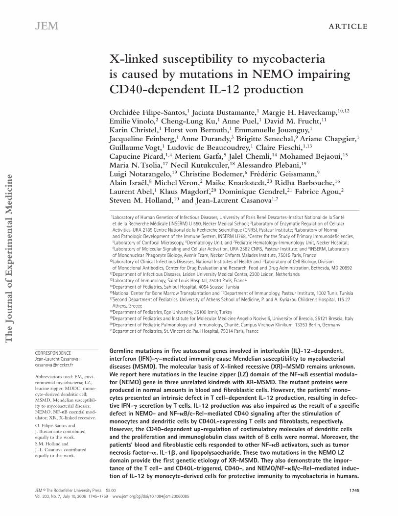
Mendelian susceptibility to mycobacterial diseases (MSMD) (MIM 209950) is a congenital syndrome resulting in predispo-sition to clinical disease caused by weakly virulent mycobacte-rial species, such as BCG vaccines and nontuberculous, environmental mycobacteria (EM) (1–4). Patients are also sus-ceptible to the more virulent species Mycobacterium tuberculosis (5). Diseases caused by nontyphoidal Salmonella serotypes have been observed in just under half the cases (4). Other infectious
diseases have only rarely been reported, mostly in single pa-tients. A few patients have suff ered from viral infections, in-cluding cytomegalovirus and human herpes virus-8 infections, and others have had fungal infections with species such as His-toplasma capsulatum and Paracoccidioidomyces braziliensis. MSMD was initially thought to be autosomal recessive in most, if not all kindreds, as a result of the high frequency of both multiple-case sibships and consanguineous kindreds (2, 3).
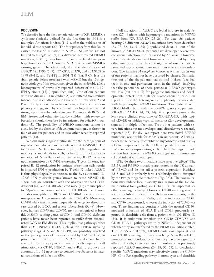
Figure 1. Clinical phenotypes and NEMO genotypes of patients.
(A) Pedigrees of three kindreds with NEMO mutations (A, B, and C). Each
generation is designated by a Roman numeral, and each individual is
designated by an Arabic numeral. Male patients (in black squares) pres-
ent mycobacterial infections and hemizygous NEMO mutations; asymp-
tomatic female carriers are represented by black dots. The probands are
indicated by arrows. (B) The teeth of patient P1 (Kindred A, II.4, sparse
teeth), patient P2 (Kindred B, II.1, conical decidual incisors), and patient
P3 (Kindred C, II.1, normal teeth). (C) Sequence electrophoregram of
NEMO complementary DNA in the region corresponding to the mutation.
Kindred A, P1 presents the mutation A→C leading to the replacement at
residue 315 of Glu (E) by Ala (A) (E315A). Kindred B, P2 and kindred C, P3
present the mutation G→A resulting in the replacement of an Arg (R) at
position 319 by Gln (Q) (R319Q). (D) Schematic representation of the
NEMO coding region, from exons 2 to 10, and corresponding domains,
shown in dark gray: the coil-coiled domain 1 (CC1, Leu 93 to Gln 194)
and coil-coiled domain 2 (CC2, Met 258 to Lys 292), and the leucine
zipper (LZ, Tyr 308 to Lys 344) and zinc fi nger (ZF, Cys 397 to Cys 417)
domains. All published hypomorphic NEMO mutations (associated with
the various forms of EDA-ID) are represented (reference 43). Missense
and nonsense mutations (amino-acid code) are shown on top, whereas
splice and frameshift mutations (nucleotide code) are shown on the
bottom. NEMO mutations in red represent mutations associated with
mycobacterial infections.

JEM VOL. 203, July 10, 2006 1747
ARTICLE
After the identifi cation in 1996 of germline mutations in IFNGR1, encoding the IFN-γ receptor ligand-binding chain (IFN-γR1) (6, 7), recessive mutations were reported in three other autosomal genes: IFNGR2, encoding the accessory chain of the IFN-γ receptor (8); IL12B, encod-ing the p40 subunit shared by IL-12 and IL-23 (9); and IL12RB1, encoding the β1 chain shared by the receptors for IL-12 and IL-23 (10, 11). Defects in IFNGR1 and IF-NGR2 are associated with impaired cellular responses to IFN-γ, whereas defects in IL12B and IL12RB1 are asso-ciated with impaired IFN-γ production. Allelic heteroge-neity at these four loci accounts for the existence of nine autosomal recessive disorders. After the identifi cation of complete defects in which no protein is expressed (6–8, 10, 11), partial forms of IFN-γR1 (12) and IFN-γR2 (13) de-fi ciency and complete forms of IFN-γR1 (14), IFN-γR2 (15), and IL-12Rβ1 (16) defi ciency with surface receptor expression were identifi ed.
The identifi cation of MSMD-causing recessive alleles in these four autosomal genes led to the discovery of an autoso-mal dominant form of MSMD, caused by dominant-negative alleles of IFNGR1 (17). The mutant alleles exert a dominant-negative eff ect as the result of overexpression, at the cell surface, of truncated chains that cannot transduce signals. Dominant mutations in STAT1, another autosomal gene en-coding the signal transducer and transactivator of transcrip-tion 1 (Stat-1), were subsequently identifi ed (18). There are two forms of partial Stat-1 defi ciency, depending on whether the mutation impairs the phosphorylation of Tyr 701 (18) or DNA-binding activity (unpublished data). The two forms of partial Stat-1 defi ciency are associated with MSMD because these mutations aff ect the activation of IFN-γ–induced Stat-1 homodimers, but not of IFN-α/β–induced Stat-1-Stat-2-IRF-9 trimers, in heterozygous cells. A dominant-negative mutant allele of IFNGR2 has also been identifi ed, resulting in the aberrant subcellular distribution of IFN-γR2, as the re-sult of a mutation aff ecting the transmembrane domain (19).
In one family fi rst reported in 1991, and more exten-sively in 1994, it was suggested that MSMD might display X-linked recessive inheritance (1, 20–22). Four maternally related male patients were found to suff er from severe EM disease. These patients’ monocytes were found to be in-trinsically unable to produce normal levels of IL-12 upon stimulation by PHA and T cells (21, 22). Despite the iden-tifi cation of this immunological phenotype, the molecular basis of XR-MSMD has remained elusive. We report here the identifi cation of MSMD-causing NEMO mutations in this kindred and in two other, unrelated kindreds. These mutations do not aff ect NF-κB activation in response to most classical activators tested, accounting for the narrow spectrum of infections in patients. The two NF-κB essen-tial modulator (NEMO) mutations identifi ed selectively impair the CD40-triggered and NF-κB/c-Rel–mediated induction of IL-12 production by monocytes and mono-cyte-derived dendritic cells, accounting for the susceptibility to mycobacteria.
RESULTS
New germline mutations in NEMO in three kindreds
We studied an American multiplex kindred, a French kin-dred, and a German kindred, each containing one male pa-tient with sporadic mycobacterial disease (Fig. 1 A). None of the six patients in these kindreds displayed the classical devel-opmental and infectious features of anhidrotic ectodermal dysplasia with immunodefi ciency (EDA-ID) (23–25). Coni-cal incisors in one proband (P2, kindred B, Fig. 1 B) and in a second patient (patient III.8, kindred A) nevertheless led us to study the X-linked EDA-ID–causing NEMO gene (26) (unpublished data). New mutations were found in the cod-ing region of NEMO in the three probands (Fig. 1 C). P1 from kindred A presented a nucleotide substitution at posi-tion 944 (A→C) (Fig. 1 C, left), leading to the replacement of a Glu by an Ala at residue 315 (E315A) (Fig. 1 D). P2 and P3 from kindreds B and C, respectively, presented a nucleo-tide substitution at position 956 (G→A) (Fig. 1 C, middle
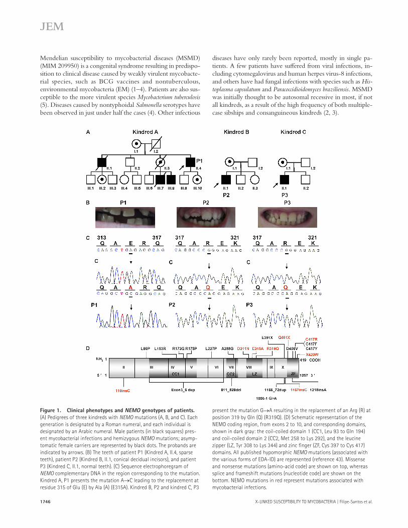
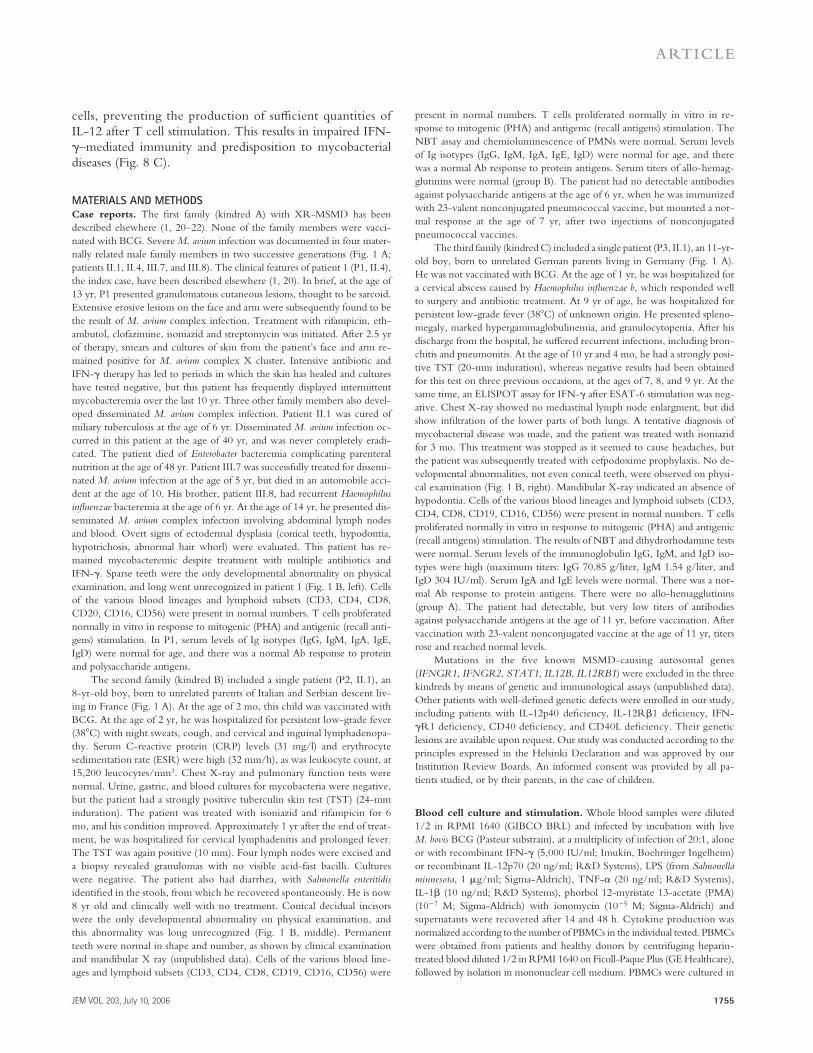
Figure 2. A model of the three-dimensional structure of NEMO.
(A) Schematic representation of the structural model of the NEMO oligo-
merization domain. The oligomerization domains from three NEMO sub-
units are represented in green, light gray, and dark gray. The LZ motifs
dock in an antiparallel manner in the crevices defi ned by the central tri-
meric CC2 coiled-coil. (B) Modeling of NEMO oligomerization domain.
Modeling was based on the coordinates of the HIV-1 gp41 ectodomain, as
described in Materials and methods. For the sake of simplicity, only one
LZ domain (LZ[A], in green) is represented, together with the three CC2
motifs forming the core of the pseudo-six-helix bundle. Amino acids E315
and R319, mutated in the NEMO protein of the XR-MSMD patients and
located within the LZ motif, are shown in blue and purple, respectively.
(C) A close-up of the surrounding environment of E315 (blue) and R319
(purple) amino acids, which probably form an intramolecular salt bridge.
Spatially close to these two residues, V280 and K277 (green) are located
within the CC2 motif of the same NEMO subunit. Q278 (light gray),
located within the CC2 motif of the adjacent B subunit (CC2[B]) probably
forms an intermolecular hydrogen bond with K277.
1748 X-LINKED SUSCEPTIBILITY TO MYCOBACTERIA | Filipe-Santos et al.
and right) resulting in the replacement of an Arg by a Gln at position 319 (R319Q) (Fig. 1 D). The two mutations were detected in cDNAs, indicating that they corresponded to NEMO and not the nearby NEMO pseudogene (27). These mutations were not found in 800 unrelated healthy controls (1,200 X chromosomes) from 51 ethnic groups (HGDP-CEPH database). The familial segregation of NEMO in kin-dred A was consistent with linkage between the observed syndrome and this gene (unpublished data). The heterozy-gous grandmother (I.1), and three other women in kindred A (II.3, III.2, III.10), like the mother of P2 in kindred B (I.1) and the mother of P3 in kindred C (I.1), were obligate carri-ers, but displayed no signs of incontinentia pigmenti or EDA (unpublished data). These data suggest that these three unre-lated families suff er from XR-MSMD as the result of mis-sense mutations in NEMO.
Modeling of the molecular environment of the E315
and R319 residues
Amino acids E315 and R319 are both located within the leucine zipper (LZ) domain of NEMO (Fig. 1 D). Several studies have shown that NEMO self-assembles into trimers (28, 29). The minimal oligomerization domain of the protein consists of the CC2 and LZ coiled-coil motifs, and we re-cently suggested that these motifs interact to form a pseudo-six-helix bundle, in which the LZ α-helices dock in the crevices defi ned by two of the helices of the central trimeric CC2 coiled-coil (Fig. 2 A) (30). A similar scaff old was de-scribed for the HIV-1 gp41 protein (31). We therefore mod-eled the NEMO trimeric CC2-LZ domain based on the X-ray structure of the gp41 ectodomain (Fig. 2 B). In our model, the E315 and R319 residues, located in the middle of the LZ domain, face the interior of the coiled coil assembly. The environment of the two residues is shown in greater de-tail in Fig. 2 C. E315 and R319 probably form an intramo-lecular salt bridge stabilizing the LZ α-helix. Other amino acids located in the vicinity of these residues in the model are also shown, including K277, from the same NEMO subunit, and Q278 from the CC2 motif of an adjacent subunit, which probably interact via a hydrogen bond. The E315A and R319Q mutations found in XR-MSMD patients would break the predicted salt bridge and might induce local plastic-ity in the LZ helices of the NEMO trimer. Within the highly structured CC2-LZ trimer, this might also result in local modifi cations to interactions with the central CC2 domains. Our structural model therefore also provides a rationale for the remarkable similarity and narrowness of the clinical phe-notypes of the patients described, with NEMO mutations disrupting a common salt bridge in the LZ domain.
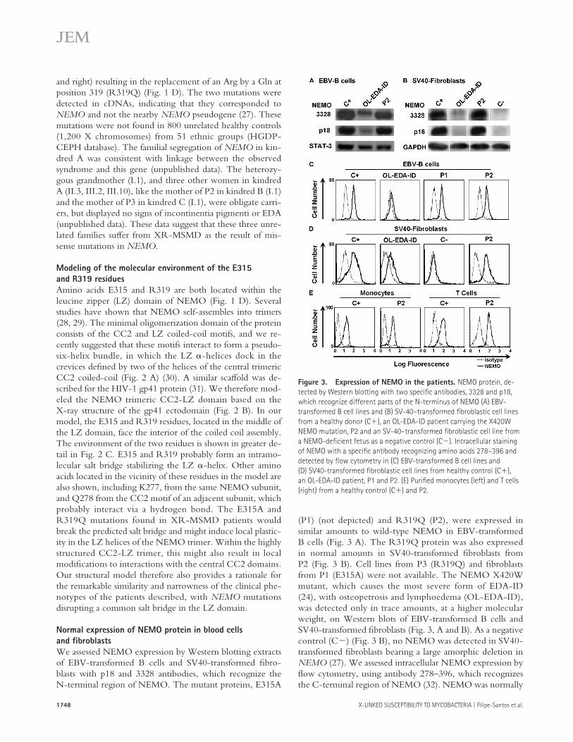
Normal expression of NEMO protein in blood cells
and fi broblasts
We assessed NEMO expression by Western blotting extracts of EBV-transformed B cells and SV40-transformed fi bro-blasts with p18 and 3328 antibodies, which recognize the N-terminal region of NEMO. The mutant proteins, E315A
(P1) (not depicted) and R319Q (P2), were expressed in similar amounts to wild-type NEMO in EBV-transformed B cells (Fig. 3 A). The R319Q protein was also expressed in normal amounts in SV40-transformed fi broblasts from P2 (Fig. 3 B). Cell lines from P3 (R319Q) and fi broblasts from P1 (E315A) were not available. The NEMO X420W mutant, which causes the most severe form of EDA-ID (24), with osteopetrosis and lymphoedema (OL-EDA-ID), was detected only in trace amounts, at a higher molecular weight, on Western blots of EBV-transformed B cells and SV40-transformed fi broblasts (Fig. 3, A and B). As a negative control (C−) (Fig. 3 B), no NEMO was detected in SV40-transformed fi broblasts bearing a large amorphic deletion in NEMO (27). We assessed intracellular NEMO expression by fl ow cytometry, using antibody 278–396, which recognizes the C-terminal region of NEMO (32). NEMO was normally
Figure 3. Expression of NEMO in the patients. NEMO protein, de-
tected by Western blotting with two specifi c antibodies, 3328 and p18,
which recognize different parts of the N-terminus of NEMO (A) EBV-
transformed B cell lines and (B) SV-40–transformed fi broblastic cell lines
from a healthy donor (C+), an OL-EDA-ID patient carrying the X420W
NEMO mutation, P2 and an SV-40–transformed fi broblastic cell line from
a NEMO-defi cient fetus as a negative control (C−). Intracellular staining
of NEMO with a specifi c antibody recognizing amino acids 278–396 and
detected by fl ow cytometry in (C) EBV-transformed B cell lines and
(D) SV40-transformed fi broblastic cell lines from healthy control (C+),
an OL-EDA-ID patient, P1 and P2. (E) Purifi ed monocytes (left) and T cells
(right) from a healthy control (C+) and P2.
JEM VOL. 203, July 10, 2006 1749
ARTICLE
expressed in EBV-transformed B cells (P1, P2) and SV40-transformed fi broblasts (P2) from the patients, taking seven and fi ve control cell lines, respectively, as a reference, but not in fi broblasts bearing a large deletion in NEMO (Fig. 3, C and D). NEMO was barely detectable in EBV-transformed B cells and SV40-transformed fi broblasts from the patient with OL-EDA-ID. The intracellular expression of NEMO was also normal in freshly isolated monocytes and T cells from P2 (Fig. 3 E). Thus, levels of NEMO expression were strictly normal in patients bearing the E315A and R319Q muta-
tions as shown by Western blotting and fl ow cytometry in hematopoietic and nonhematopoietic cell lines and in freshly prepared blood cells.
Response of blood cells and fi broblasts to LPS,
R-848, TNF-𝛂, and IL-1𝛃NEMO mutations in patients with EDA-ID are usually asso-ciated with impaired cellular responses to multiple stimuli, including LPS (via TLR-4), TNF-α, and IL-1β (24, 25). We thus stimulated whole blood cells from the patients and
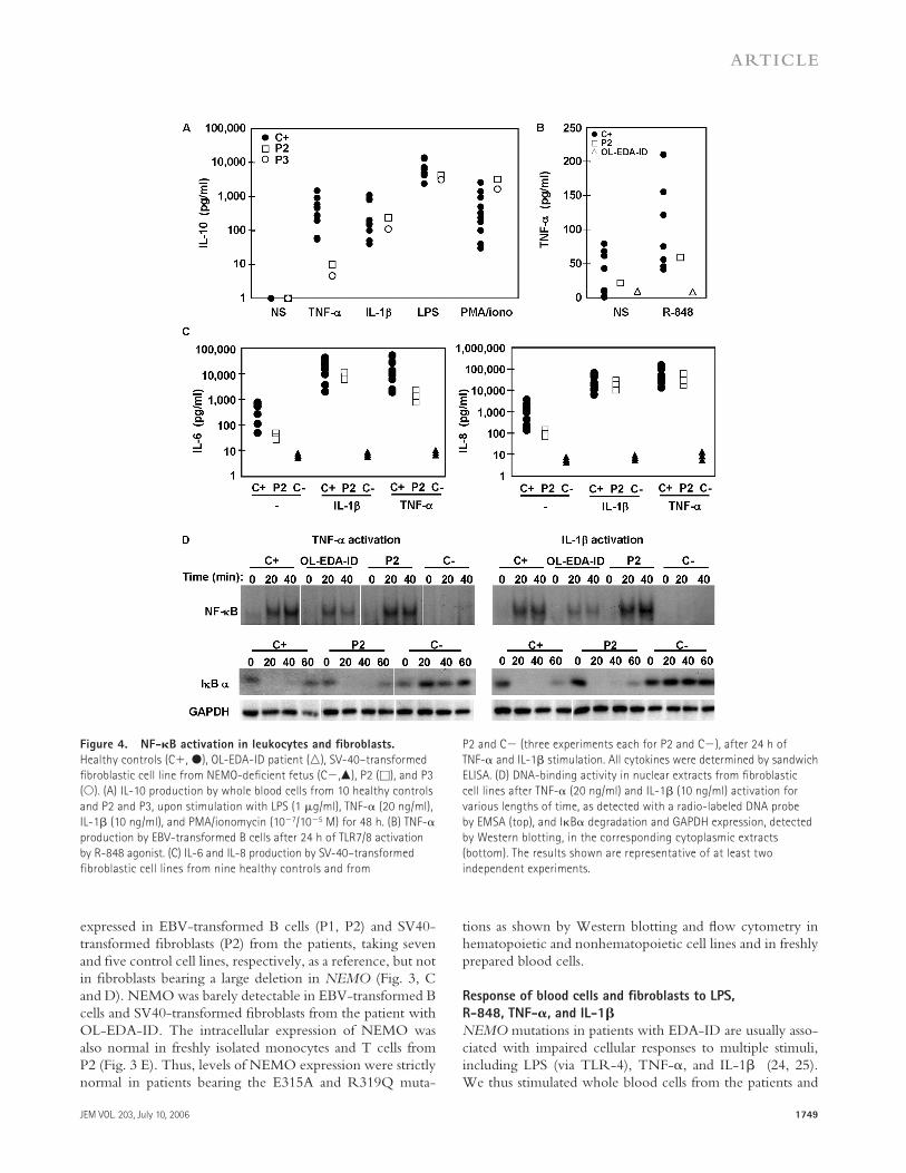
Figure 4. NF-𝛋B activation in leukocytes and fi broblasts.
Healthy controls (C+, ●), OL-EDA-ID patient (△), SV-40–transformed
fi broblastic cell line from NEMO-defi cient fetus (C−,▲), P2 (□), and P3
(○). (A) IL-10 production by whole blood cells from 10 healthy controls
and P2 and P3, upon stimulation with LPS (1 μg/ml), TNF-α (20 ng/ml),
IL-1β (10 ng/ml), and PMA/ionomycin (10−7/10−5 M) for 48 h. (B) TNF-α
production by EBV-transformed B cells after 24 h of TLR7/8 activation
by R-848 agonist. (C) IL-6 and IL-8 production by SV-40–transformed
fibroblastic cell lines from nine healthy controls and from
P2 and C− (three experiments each for P2 and C−), after 24 h of
TNF-α and IL-1β stimulation. All cytokines were determined by sandwich
ELISA. (D) DNA-binding activity in nuclear extracts from fi broblastic
cell lines after TNF-α (20 ng/ml) and IL-1β (10 ng/ml) activation for
various lengths of time, as detected with a radio-labeled DNA probe
by EMSA (top), and IκBα degradation and GAPDH expression, detected
by Western blotting, in the corresponding cytoplasmic extracts
(bottom). The results shown are representative of at least two
independent experiments.
1750 X-LINKED SUSCEPTIBILITY TO MYCOBACTERIA | Filipe-Santos et al.
10 healthy controls with LPS, TNF-α, IL-1β, and PMA/ionomycin, and measured the induction of IL-6 (unpublished data), IL-10, and TNF-α. The three patients displayed nor-mal levels of IL-10 (Fig. 4 A), IL-6, and TNF-α (not de-picted) production upon stimulation with LPS, IL-1β, and PMA plus ionomycin. IL-10 was also induced by TNF-α in P2 and P3, although to lower levels (Fig. 4 A). EBV-trans-formed B cells from P2 responded normally to R-848, an ag-onist of TLR-7/8, in terms of TNF-α production (Fig. 4 B). We then assessed the impact of NEMO mutations in fi bro-blasts from P2, a NEMO-defi cient fi broblastic cell line bear-ing a large NEMO deletion, and fi broblasts from nine healthy controls. Fibroblasts from P2 produced normal amounts of IL-6 and IL-8 in response to both IL-1β and TNF-α (Fig. 4 C). In contrast, fi broblasts from all EDA-ID patients tested, bear-ing other NEMO mutations, showed various levels of im-pairment of IL-6 and IL-8 production (unpublished data). We assessed IκBα cytoplasmic degradation by Western blot-ting and the κB-binding activity of nuclear extracts by elec-trophoretic mobility shift assays. In contrast with what was observed in fi broblasts from the OL-EDA-ID patient bearing NEMO mutation X420W (24), both IκBα degradation and κB binding activity were completely normal in fi broblasts from P2 (Fig. 4 D). Thus, the NEMO–NF-κB signaling pathway in response to TLR4, 7, and 8 agonists, IL-1β, and TNF-α, was normal in SV40-fi broblastic and EBV-trans-formed B cell lines from our patients.
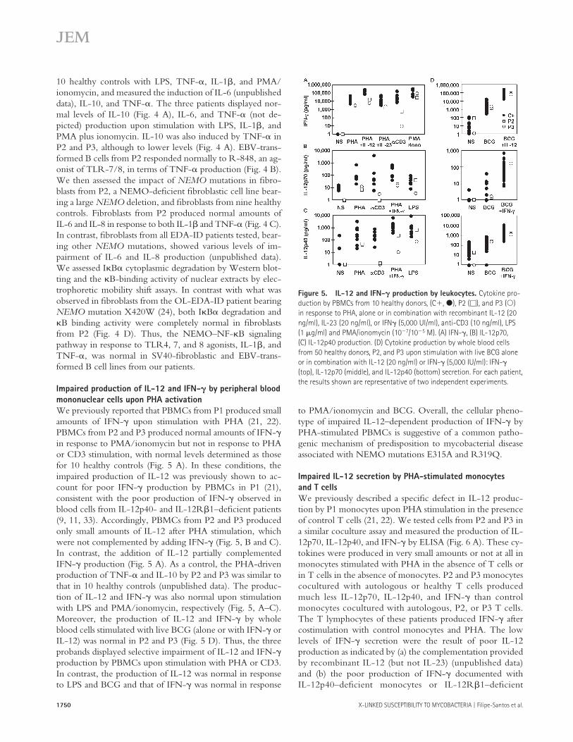
Impaired production of IL-12 and IFN-𝛄 by peripheral blood
mononuclear cells upon PHA activation
We previously reported that PBMCs from P1 produced small amounts of IFN-γ upon stimulation with PHA (21, 22). PBMCs from P2 and P3 produced normal amounts of IFN-γ in response to PMA/ionomycin but not in response to PHA or CD3 stimulation, with normal levels determined as those for 10 healthy controls (Fig. 5 A). In these conditions, the impaired production of IL-12 was previously shown to ac-count for poor IFN-γ production by PBMCs in P1 (21), consistent with the poor production of IFN-γ observed in blood cells from IL-12p40- and IL-12Rβ1–defi cient patients (9, 11, 33). Accordingly, PBMCs from P2 and P3 produced only small amounts of IL-12 after PHA stimulation, which were not complemented by adding IFN-γ (Fig. 5, B and C). In contrast, the addition of IL-12 partially complemented IFN-γ production (Fig. 5 A). As a control, the PHA-driven production of TNF-α and IL-10 by P2 and P3 was similar to that in 10 healthy controls (unpublished data). The produc-tion of IL-12 and IFN-γ was also normal upon stimulation with LPS and PMA/ionomycin, respectively (Fig. 5, A–C). Moreover, the production of IL-12 and IFN-γ by whole blood cells stimulated with live BCG (alone or with IFN-γ or IL-12) was normal in P2 and P3 (Fig. 5 D). Thus, the three probands displayed selective impairment of IL-12 and IFN-γ production by PBMCs upon stimulation with PHA or CD3. In contrast, the production of IL-12 was normal in response to LPS and BCG and that of IFN-γ was normal in response
to PMA/ionomycin and BCG. Overall, the cellular pheno-type of impaired IL-12–dependent production of IFN-γ by PHA-stimulated PBMCs is suggestive of a common patho-genic mechanism of predisposition to mycobacterial disease associated with NEMO mutations E315A and R319Q.
Impaired IL-12 secretion by PHA-stimulated monocytes
and T cells
We previously described a specifi c defect in IL-12 produc-tion by P1 monocytes upon PHA stimulation in the presence of control T cells (21, 22). We tested cells from P2 and P3 in a similar coculture assay and measured the production of IL-12p70, IL-12p40, and IFN-γ by ELISA (Fig. 6 A). These cy-tokines were produced in very small amounts or not at all in monocytes stimulated with PHA in the absence of T cells or in T cells in the absence of monocytes. P2 and P3 monocytes cocultured with autologous or healthy T cells produced much less IL-12p70, IL-12p40, and IFN-γ than control monocytes cocultured with autologous, P2, or P3 T cells. The T lymphocytes of these patients produced IFN-γ after costimulation with control monocytes and PHA. The low levels of IFN-γ secretion were the result of poor IL-12 production as indicated by (a) the complementation provided by recombinant IL-12 (but not IL-23) (unpublished data) and (b) the poor production of IFN-γ documented with IL-12p40–defi cient monocytes or IL-12Rβ1–defi cient
Figure 5. IL-12 and IFN-𝛄 production by leukocytes. Cytokine pro-
duction by PBMCs from 10 healthy donors, (C+, ●), P2 (□), and P3 (○)
in response to PHA, alone or in combination with recombinant IL-12 (20
ng/ml), IL-23 (20 ng/ml), or IFNγ (5,000 UI/ml), anti-CD3 (10 ng/ml), LPS
(1 μg/ml) and PMA/ionomycin (10−7/10−5 M). (A) IFN-γ, (B) IL-12p70,
(C) IL-12p40 production. (D) Cytokine production by whole blood cells
from 50 healthy donors, P2, and P3 upon stimulation with live BCG alone
or in combination with IL-12 (20 ng/ml) or IFN-γ (5,000 IU/ml): IFN-γ
(top), IL-12p70 (middle), and IL-12p40 (bottom) secretion. For each patient,
the results shown are representative of two independent experiments.

JEM VOL. 203, July 10, 2006 1751
ARTICLE
T cells (unpublished data). Certain monokines, such as IL-6, G-CSF, and MCP-1, were poorly induced, whereas others were induced in normal (IL-4, IL-10) or large (IL-5, IL-13) amounts by monocytes from P2 and P3, indicating that these cells were not globally unresponsive (Fig. 6 B and not de-picted). Two other lymphokines (IL-2, IL-4) were not af-fected and the production of IL-17 was impaired. Finally, cytokines secreted by T cells and monocytes (TNF-α, IL-7, IL-10, and GM-CSF) were normally induced. All three pro-bands had a specifi c immunological phenotype in which IL-12 production by monocytes in response to PHA and T cells
was aff ected. Impaired IFN-γ production does not refl ect an intrinsic T cell defect and is instead a consequence of im-paired IL-12 production by monocytes.
PHA-activated T cell–dependent monocyte secretion
of IL-12 is driven by CD40
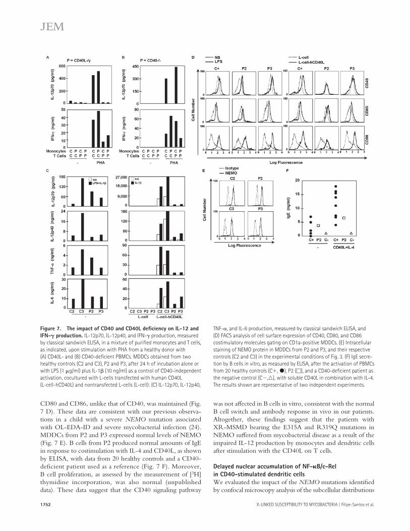
The two NEMO mutations exerted their eff ects downstream from monocyte receptors triggering the induction of IL-12. The CD40 signaling pathway has been shown to be critical for the T cell–dependent induction of IL-12 in monocytes and dendritic cells (34–37). After PHA stimulation, PBMCs from CD40- and CD40L-defi cient patients (38) produced much lower levels of IL-12 and IFN-γ than did PBMCs from 10 healthy controls (unpublished data), consistent with previous data (39). The induction of IL-12 by LPS and that of IFN-γ by PMA/ionomycin were normal (unpublished data). IL-12 induction was abolished if CD40L-defi cient T cells were cocultured with healthy or CD40L-defi cient monocytes, resulting in low levels but not the total absence of IFN-γ production (Fig. 7 A). Conversely, normal IL-12 induction was observed if control or CD40L-defi cient mon-ocytes were mixed with T lymphocytes from a healthy donor. Low levels of IFN-γ induction were associated with the pres-ence of small amounts of IL-12 in this assay. IL-12 induction was abolished if CD40-defi cient monocytes were incubated with CD40-defi cient or normal T cells (Fig. 7 B). Paradoxi-cally, IFN-γ secretion was not signifi cantly impaired, perhaps refl ecting the direct cross-linking of CD40L by PHA (40). In contrast, control monocytes produced normal amounts of IL-12 when cultured with control or CD40-defi cient T cells. Our fi ndings for the monocyte–T cell coculture assay unambiguously indicate that the engagement of the mono-cyte CD40 by CD40L-expressing T cells is required for the optimal induction of IL-12 production. These data suggest that the NEMO mutations in P1, P2, and P3 were patho-genic as a result of their impact on CD40 signaling within monocytes, leading to low levels of IL-12 secretion upon T cell stimulation.
NEMO mutation impairs CD40 signaling in dendritic cells
but not B cells
We assessed the responses to CD40 stimulation of cells from our patients. We fi rst incubated purifi ed monocytes from P2 with mouse fi broblastic L cells (L-cells) and L-cells sta-bly transfected with a construct encoding human CD40L (L-cells-hCD40L). Monocytes from P2 did not produce IL-12, IL-6, and TNF-α upon stimulation with L-cells-hCD40L (unpublished data). We then tested monocyte-de-rived dendritic cells (MDDCs) as DCs are major producers of IL-12 in vivo. MDDCs produce large amounts of IL-12 upon CD40 stimulation in vitro (36). MDDCs from P2 and P3 were activated with LPS plus IL-1β, L-cells, or L-cells-hCD40L with or without IL-1β. MDDCs from P2 and P3 displayed severely impaired cytokine production in response to L-cells–hCD40L but not in response to LPS plus IL-1β (Fig. 7 C), whereas induction of the costimulatory molecules
Figure 6. IL-12 and IFN-𝛄 production by cocultured monocytes
and T cells from the patients studied and healthy controls.
(A) IL-12p70, IL-12p40, and IFN-γ production, measured using classical
sandwich ELISA, in a mixture of purifi ed monocytes and T cells, as indi-
cated upon stimulation with PHA. The results shown are representative
of three independent experiments for P2 and one for P3. (B) The same
coculture supernatants were analyzed for a multiplex of 16 cytokines,
using the Bioplex array. Each column represents the data for one monocyte–
T cell coculture system, and all four columns correspond to the same
experiment. Each row corresponds to one cytokine. The gray-scale bar
indicates the magnitude of cytokine expression, using the control/control
(C/C) coculture system as a reference. For each data point, the amount of
cytokine produced in the unstimulated system was subtracted from that
produced in the PHA-activated system, and the result obtained was
compared with the reference value (C/C). The production of *MCP-1 and
*MIP-1β by monocytes was PHA-dependent but T cell–independent, as
monocytes responded to PHA by producing large amounts of these cyto-
kines, whereas the addition of T cells did not increase cytokine production.
The defects in the production of IL-6, IL-12p70, G-CSF, IFN-γ, and MCP-1
were confi rmed in three independent experiments on blood cells from P2
and one experiment on blood from P3.
1752 X-LINKED SUSCEPTIBILITY TO MYCOBACTERIA | Filipe-Santos et al.
CD80 and CD86, unlike that of CD40, was maintained (Fig. 7 D). These data are consistent with our previous observa-tions in a child with a severe NEMO mutation associated with OL-EDA-ID and severe mycobacterial infection (24). MDDCs from P2 and P3 expressed normal levels of NEMO (Fig. 7 E). B cells from P2 produced normal amounts of IgE in response to costimulation with IL-4 and CD40L, as shown by ELISA, with data from 20 healthy controls and a CD40-defi cient patient used as a reference (Fig. 7 F). Moreover, B cell proliferation, as assessed by the measurement of [3H] thymidine incorporation, was also normal (unpublished data). These data suggest that the CD40 signaling pathway
was not aff ected in B cells in vitro, consistent with the normal B cell switch and antibody response in vivo in our patients. Altogether, these fi ndings suggest that the patients with XR-MSMD bearing the E315A and R319Q mutations in NEMO suff ered from mycobacterial disease as a result of the impaired IL-12 production by monocytes and dendritic cells after stimulation with the CD40L on T cells.
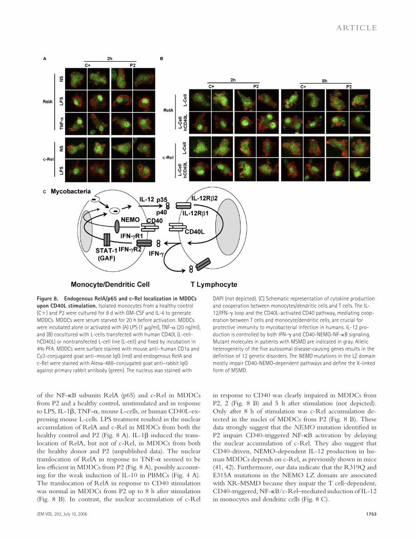
Delayed nuclear accumulation of NF-𝛋B/c-Rel
in CD40-stimulated dendritic cells
We evaluated the impact of the NEMO mutations identifi ed by confocal microscopy analysis of the subcellular distributions
Figure 7. The impact of CD40 and CD40L defi ciency on IL-12 and
IFN-𝛄 production. IL-12p70, IL-12p40, and IFN-γ production, measured
by classical sandwich ELISA, in a mixture of purifi ed monocytes and T cells,
as indicated, upon stimulation with PHA from a healthy donor with
(A) CD40L- and (B) CD40-defi cient PBMCs. MDDCs obtained from two
healthy controls (C2 and C3), P2 and P3, after 24 h of incubation alone or
with LPS (1 μg/ml) plus IL-1β (10 ng/ml) as a control of CD40-independent
activation, cocultured with L-cells transfected with human CD40L
(L-cell-hCD40L) and nontransfected L-cells (L-cell): (C) IL-12p70, IL-12p40,
TNF-α, and IL-6 production, measured by classical sandwich ELISA, and
(D) FACS analysis of cell surface expression of CD40, CD80, and CD86
costimulatory molecules gating on CD1a-positive MDDCs. (E) Intracellular
staining of NEMO protein in MDDCs from P2 and P3, and their respective
controls (C2 and C3) in the experimental conditions of Fig. 3. (F) IgE secre-
tion by B cells in vitro, as measured by ELISA, after the activation of PBMCs
from 20 healthy controls (C+, ●), P2 (□), and a CD40-defi cient patient as
the negative control (C−,△), with soluble CD40L in combination with IL-4.
The results shown are representative of two independent experiments.
JEM VOL. 203, July 10, 2006 1753
ARTICLE
of the NF-κB subunits RelA (p65) and c-Rel in MDDCs from P2 and a healthy control, unstimulated and in response to LPS, IL-1β, TNF-α, mouse L-cells, or human CD40L-ex-pressing mouse L-cells. LPS treatment resulted in the nuclear accumulation of RelA and c-Rel in MDDCs from both the healthy control and P2 (Fig. 8 A). IL-1β induced the trans-location of RelA, but not of c-Rel, in MDDCs from both the healthy donor and P2 (unpublished data). The nuclear translocation of RelA in response to TNF-α seemed to be less effi cient in MDDCs from P2 (Fig. 8 A), possibly account-ing for the weak induction of IL-10 in PBMCs (Fig. 4 A). The translocation of RelA in response to CD40 stimulation was normal in MDDCs from P2 up to 8 h after stimulation (Fig. 8 B). In contrast, the nuclear accumulation of c-Rel
in response to CD40 was clearly impaired in MDDCs from P2, 2 (Fig. 8 B) and 5 h after stimulation (not depicted). Only after 8 h of stimulation was c-Rel accumulation de-tected in the nuclei of MDDCs from P2 (Fig. 8 B). These data strongly suggest that the NEMO mutation identifi ed in P2 impairs CD40-triggered NF-κB activation by delaying the nuclear accumulation of c-Rel. They also suggest that CD40-driven, NEMO-dependent IL-12 production in hu-man MDDCs depends on c-Rel, as previously shown in mice (41, 42). Furthermore, our data indicate that the R319Q and E315A mutations in the NEMO LZ domain are associated with XR-MSMD because they impair the T cell–dependent, CD40-triggered, NF-κB/c-Rel–mediated induction of IL-12in monocytes and dendritic cells (Fig. 8 C).
Figure 8. Endogenous RelA/p65 and c-Rel localization in MDDCs
upon CD40L stimulation. Isolated monocytes from a healthy control
(C+) and P2 were cultured for 8 d with GM-CSF and IL-4 to generate
MDDCs. MDDCs were serum starved for 20 h before activation. MDDCs
were incubated alone or activated with (A) LPS (1 μg/ml), TNF-α (20 ng/ml),
and (B) cocultured with L-cells transfected with human CD40L (L-cell-
hCD40L) or nontransfected L-cell line (L-cell) and fi xed by incubation in
4% PFA. MDDCs were surface stained with mouse anti–human CD1a and
Cy3-conjugated goat anti–mouse IgG (red) and endogenous RelA and
c-Rel were stained with Alexa-488–conjugated goat anti–rabbit IgG
against primary rabbit antibody (green). The nucleus was stained with
DAPI (not depicted). (C) Schematic representation of cytokine production
and cooperation between monocytes/dendritic cells and T cells. The IL-
12/IFN-γ loop and the CD40L-activated CD40 pathway, mediating coop-
eration between T cells and monocyte/dendritic cells, are crucial for
protective immunity to mycobacterial infection in humans. IL-12 pro-
duction is controlled by both IFN-γ and CD40-NEMO-NF-κB signaling.
Mutant molecules in patients with MSMD are indicated in gray. Allelic
heterogeneity of the fi ve autosomal disease-causing genes results in the
defi nition of 12 genetic disorders. The NEMO mutations in the LZ domain
mostly impair CD40-NEMO-dependent pathways and defi ne the X-linked
form of MSMD.
1754 X-LINKED SUSCEPTIBILITY TO MYCOBACTERIA | Filipe-Santos et al.
D I S C U S S I O N
We describe here the fi rst genetic etiology of XR-MSMD, a syndrome clinically defi ned for the fi rst time in 1994 in a multiplex American kindred (1, 21), after the publication of individual case reports (20). The four patients from this family carried the E315A mutation in NEMO. XR-MSMD is not limited to a single family, as a diff erent, but related NEMO mutation, R319Q, was found in two unrelated European boys, from France and Germany. NEMO is the sixth MSMD-causing gene to be identifi ed, after the identifi cation of IFNGR1 in 1996 (6, 7), IFNGR2, IL12B, and IL12RB1 in 1998 (8–11), and STAT1 in 2001 (18) (Fig. 8 C). It is the sixth genetic defect associated with MSMD but the 13th ge-netic etiology of this syndrome, given the considerable allelic heterogeneity of previously reported defects of the IL-12–IFN-γ circuit (15) (unpublished data). One of our patients with EM disease (II.4 in kindred A) also suff ered from miliary tuberculosis in childhood, and two of our probands (P2 and P3) probably suff ered from tuberculosis, as the sole infectious phenotype suggested by consistent histological results and strongly positive tuberculin skin tests. Patients with BCG and EM diseases and otherwise healthy children with severe tu-berculosis should therefore be investigated for NEMO muta-tions (5). The possibility of NEMO mutations cannot be excluded by the absence of developmental signs, as shown in four of our six patients and in two other recently reported patients (43).
We can now suggest a rationale for the pathogenesis of mycobacterial diseases in patients with XR-MSMD. The two causal NEMO mutations impair CD40 signaling in monocytes and dendritic cells, delaying the nuclear accu-mulation of NF-κB/c-Rel and impairing IL-12 secretion upon stimulation by CD40L-expressing T cells. In turn, im-paired IL-12 production by antigen-presenting cells results in impaired IFN-γ production by T cells. X-linked NEMO is thus physiologically connected to the fi ve autosomal IL-12/23–IFN-γ circuit genes known to cause MSMD (4). These data are consistent with the observation that CD40-defi cient (44) and CD40L-depleted mice (45) are susceptible to Mycobacterium avium infections. CD40L-defi cient mice are also susceptible to BCG and CD40-defi cient mice are susceptible to Mycobacterium tuberculosis (46, 47). Moreover, CD40L-defi cient patients frequently develop localized dis-ease caused by BCG, and severe tuberculosis (5, 48). How-ever, human CD40 and CD40L are not recognized as bona fi de MSMD-causing genes, as CD40- and CD40L-defi cient patients have never been reported to suff er from dissemi-nated BCG or EM disease. This implies that pathways other than CD40–NEMO–IL-12, such as the TNF-α signaling pathway (Figs. 4 A and 8 A) (49), are probably involved in the pathogenesis of diseases caused by EM, at least, in patients with mutations in the NEMO LZ domain. In any event, human phagocytes and dendritic cells require T cell stimulation via CD40, NEMO, and c-Rel to produce the amounts of IL-12 necessary to control mycobacteria in natu-ral conditions of infection (50).
Null mutations in NEMO are lethal in utero in male fe-tuses (27). Patients with hypomorphic mutations in NEMO suff er from XR-EDA-ID (23–26). To date, 36 patients bearing 28 diff erent NEMO mutations have been described (23–27, 32, 43, 51–55) (unpublished data). 11 out of the known 36 XR-EDA-ID patients have developed severe my-cobacterial infection, mostly caused by M. avium. However, these patients also suff ered from infections caused by many other microorganisms. In contrast, fi ve of our six patients presented mycobacterial disease as their sole invasive infec-tion. The invasive Haemophilus infl uenzae b infection in one of our patients may not have occurred by chance. Similarly, two out of the six patients had conical incisors (decidual teeth in one and permanent teeth in the other), implying that the penetrance of these particular NEMO genotypes was low (but not null) for pyogenic infections and devel-opmental defects, but high for mycobacterial disease. Our report stresses the heterogeneity of phenotypes associated with hypomorphic NEMO mutations. Two patients with XR-EDA-ID, both with the X420W genotype, presented XR-OL-EDA-ID (24, 53, 54). Most patients present the less severe clinical syndrome of XR-EDA-ID, with typi-cal (23–25) or hidden (conical incisors) (26) developmental signs and multiple infections. Two other patients with se-vere infections but no developmental disorder were recently reported (43). Finally, we report here two novel NEMO mutations, responsible for MSMD in six patients. All six pa-tients are selectively susceptible to mycobacteria and display selective impairment of the CD40-dependent induction of IL-12 in antigen-presenting cells. These fi ndings provide the fi rst link between a NEMO genotype and immunologi-cal and infectious phenotypes.
Why do these two mutations have selective eff ects? The E315A and R319Q mutations are located in the LZ domain of NEMO and do not aff ect protein expression. Residues E315 and R319 probably form a salt bridge that is disrupted by the two pathogenic mutations (Fig. 2 C). The two muta-tions may induce local plasticity in a region of the LZ do-main critical for signaling via CD40, but less important for other signaling pathways. However, CD40 signaling was not totally abolished in dendritic cells from our patients, as the nuclear accumulation of RelA, and the induction of CD80 and CD86 were normal, whereas the induction of CD40 was not. These fi ndings are consistent with the normal CD40-mediated induction of HLA-II and CD86 previously re-ported in dendritic cells from a patient with OL-EDA-ID (24). It is unknown whether the CD40–CD80/86 and CD40–HLA-II pathways are truly NEMO independent or whether they are unaff ected by the NEMO mutations tested. The E315A and R319Q NEMO mutations impair at least one CD40 signaling pathway involving NF-κB–c-Rel in monocytes and dendritic cells, but they have no signifi cant eff ect on B cells, in vivo and in vitro, unlike other previously reported NEMO mutations (24, 25, 52, 55). In conclusion, specifi c NEMO mutations may selectively target the CD40–NF-κB–c-Rel signaling pathway in monocytes and dendritic
JEM VOL. 203, July 10, 2006 1755
ARTICLE
cells, preventing the production of suffi cient quantities of IL-12 after T cell stimulation. This results in impaired IFN-γ–mediated immunity and predisposition to mycobacterial diseases (Fig. 8 C).
MATERIALS AND METHODSCase reports. The fi rst family (kindred A) with XR-MSMD has been
described elsewhere (1, 20–22). None of the family members were vacci-
nated with BCG. Severe M. avium infection was documented in four mater-
nally related male family members in two successive generations (Fig. 1 A;
patients II.1, II.4, III.7, and III.8). The clinical features of patient 1 (P1, II.4),
the index case, have been described elsewhere (1, 20). In brief, at the age of
13 yr, P1 presented granulomatous cutaneous lesions, thought to be sarcoid.
Extensive erosive lesions on the face and arm were subsequently found to be
the result of M. avium complex infection. Treatment with rifampicin, eth-
ambutol, clofazimine, isoniazid and streptomycin was initiated. After 2.5 yr
of therapy, smears and cultures of skin from the patient’s face and arm re-
mained positive for M. avium complex X cluster. Intensive antibiotic and
IFN-γ therapy has led to periods in which the skin has healed and cultures
have tested negative, but this patient has frequently displayed intermittent
mycobacteremia over the last 10 yr. Three other family members also devel-
oped disseminated M. avium complex infection. Patient II.1 was cured of
miliary tuberculosis at the age of 6 yr. Disseminated M. avium infection oc-
curred in this patient at the age of 40 yr, and was never completely eradi-
cated. The patient died of Enterobacter bacteremia complicating parenteral
nutrition at the age of 48 yr. Patient III.7 was successfully treated for dissemi-
nated M. avium infection at the age of 5 yr, but died in an automobile acci-
dent at the age of 10. His brother, patient III.8, had recurrent Haemophilus
infl uenzae bacteremia at the age of 6 yr. At the age of 14 yr, he presented dis-
seminated M. avium complex infection involving abdominal lymph nodes
and blood. Overt signs of ectodermal dysplasia (conical teeth, hypodontia,
hypotrichosis, abnormal hair whorl) were evaluated. This patient has re-
mained mycobacteremic despite treatment with multiple antibiotics and
IFN-γ. Sparse teeth were the only developmental abnormality on physical
examination, and long went unrecognized in patient 1 (Fig. 1 B, left). Cells
of the various blood lineages and lymphoid subsets (CD3, CD4, CD8,
CD20, CD16, CD56) were present in normal numbers. T cells proliferated
normally in vitro in response to mitogenic (PHA) and antigenic (recall anti-
gens) stimulation. In P1, serum levels of Ig isotypes (IgG, IgM, IgA, IgE,
IgD) were normal for age, and there was a normal Ab response to protein
and polysaccharide antigens.
The second family (kindred B) included a single patient (P2, II.1), an
8-yr-old boy, born to unrelated parents of Italian and Serbian descent liv-
ing in France (Fig. 1 A). At the age of 2 mo, this child was vaccinated with
BCG. At the age of 2 yr, he was hospitalized for persistent low-grade fever
(38°C) with night sweats, cough, and cervical and inguinal lymphadenopa-
thy. Serum C-reactive protein (CRP) levels (31 mg/l) and erythrocyte
sedimentation rate (ESR) were high (32 mm/h), as was leukocyte count, at
15,200 leucocytes/mm3. Chest X-ray and pulmonary function tests were
normal. Urine, gastric, and blood cultures for mycobacteria were negative,
but the patient had a strongly positive tuberculin skin test (TST) (24-mm
induration). The patient was treated with isoniazid and rifampicin for 6
mo, and his condition improved. Approximately 1 yr after the end of treat-
ment, he was hospitalized for cervical lymphadenitis and prolonged fever.
The TST was again positive (10 mm). Four lymph nodes were excised and
a biopsy revealed granulomas with no visible acid-fast bacilli. Cultures
were negative. The patient also had diarrhea, with Salmonella enteritidis
identifi ed in the stools, from which he recovered spontaneously. He is now
8 yr old and clinically well with no treatment. Conical decidual incisors
were the only developmental abnormality on physical examination, and
this abnormality was long unrecognized (Fig. 1 B, middle). Permanent
teeth were normal in shape and number, as shown by clinical examination
and mandibular X ray (unpublished data). Cells of the various blood line-
ages and lymphoid subsets (CD3, CD4, CD8, CD19, CD16, CD56) were
present in normal numbers. T cells proliferated normally in vitro in re-
sponse to mitogenic (PHA) and antigenic (recall antigens) stimulation. The
NBT assay and chemioluminescence of PMNs were normal. Serum levels
of Ig isotypes (IgG, IgM, IgA, IgE, IgD) were normal for age, and there
was a normal Ab response to protein antigens. Serum titers of allo-hemag-
glutinins were normal (group B). The patient had no detectable antibodies
against polysaccharide antigens at the age of 6 yr, when he was immunized
with 23-valent nonconjugated pneumococcal vaccine, but mounted a nor-
mal response at the age of 7 yr, after two injections of nonconjugated
pneumococcal vaccines.
The third family (kindred C) included a single patient (P3, II.1), an 11-yr-
old boy, born to unrelated German parents living in Germany (Fig. 1 A).
He was not vaccinated with BCG. At the age of 1 yr, he was hospitalized for
a cervical abscess caused by Haemophilus infl uenzae b, which responded well
to surgery and antibiotic treatment. At 9 yr of age, he was hospitalized for
persistent low-grade fever (38°C) of unknown origin. He presented spleno-
megaly, marked hypergammaglobulinemia, and granulocytopenia. After his
discharge from the hospital, he suff ered recurrent infections, including bron-
chitis and pneumonitis. At the age of 10 yr and 4 mo, he had a strongly posi-
tive TST (20-mm induration), whereas negative results had been obtained
for this test on three previous occasions, at the ages of 7, 8, and 9 yr. At the
same time, an ELISPOT assay for IFN-γ after ESAT-6 stimulation was neg-
ative. Chest X-ray showed no mediastinal lymph node enlargment, but did
show infi ltration of the lower parts of both lungs. A tentative diagnosis of
mycobacterial disease was made, and the patient was treated with isoniazid
for 3 mo. This treatment was stopped as it seemed to cause headaches, but
the patient was subsequently treated with cefpodoxime prophylaxis. No de-
velopmental abnormalities, not even conical teeth, were observed on physi-
cal examination (Fig. 1 B, right). Mandibular X-ray indicated an absence of
hypodontia. Cells of the various blood lineages and lymphoid subsets (CD3,
CD4, CD8, CD19, CD16, CD56) were present in normal numbers. T cells
proliferated normally in vitro in response to mitogenic (PHA) and antigenic
(recall antigens) stimulation. The results of NBT and dihydrorhodamine tests
were normal. Serum levels of the immunoglobulin IgG, IgM, and IgD iso-
types were high (maximum titers: IgG 70.85 g/liter, IgM 1.54 g/liter, and
IgD 304 IU/ml). Serum IgA and IgE levels were normal. There was a nor-
mal Ab response to protein antigens. There were no allo-hemagglutinins
(group A). The patient had detectable, but very low titers of antibodies
against polysaccharide antigens at the age of 11 yr, before vaccination. After
vaccination with 23-valent nonconjugated vaccine at the age of 11 yr, titers
rose and reached normal levels.
Mutations in the fi ve known MSMD-causing autosomal genes
(IFNGR1, IFNGR2, STAT1, IL12B, IL12RB1) were excluded in the three
kindreds by means of genetic and immunological assays (unpublished data).
Other patients with well-defi ned genetic defects were enrolled in our study,
including patients with IL-12p40 defi ciency, IL-12Rβ1 defi ciency, IFN-
γR1 defi ciency, CD40 defi ciency, and CD40L defi ciency. Their genetic
lesions are available upon request. Our study was conducted according to the
principles expressed in the Helsinki Declaration and was approved by our
Institution Review Boards. An informed consent was provided by all pa-
tients studied, or by their parents, in the case of children.
Blood cell culture and stimulation. Whole blood samples were diluted
1/2 in RPMI 1640 (GIBCO BRL) and infected by incubation with live
M. bovis BCG (Pasteur substrain), at a multiplicity of infection of 20:1, alone
or with recombinant IFN-γ (5,000 IU/ml; Imukin, Boehringer Ingelheim)
or recombinant IL-12p70 (20 ng/ml; R&D Systems), LPS (from Salmonella
minnesota, 1 μg/ml; Sigma-Aldrich), TNF-α (20 ng/ml; R&D Systems),
IL-1β (10 ng/ml; R&D Systems), phorbol 12-myristate 13-acetate (PMA)
(10−7 M; Sigma-Aldrich) with ionomycin (10−5 M; Sigma-Aldrich) and
supernatants were recovered after 14 and 48 h. Cytokine production was
normalized according to the number of PBMCs in the individual tested. PBMCs
were obtained from patients and healthy donors by centrifuging heparin-
treated blood diluted 1/2 in RPMI 1640 on Ficoll-Paque Plus (GE Healthcare),
followed by isolation in mononuclear cell medium. PBMCs were cultured in
1756 X-LINKED SUSCEPTIBILITY TO MYCOBACTERIA | Filipe-Santos et al.
RPMI 1640 medium (GIBCO BRL) supplemented with 10% heat-
i nactivated pooled FBS (GIBCO BRL), referred to as complete RPMI
1640. Cells (106 PBMCs/ml/well) were activated by incubation with phy-
tohemagglutinin-P 1/700 (PHA, Bacto PHA-P; Becton Dickinson), alone
or in combination with recombinant IFN-γ (5,000 IU/ml; Imukin, Boeh-
ringer Ingelheim) or recombinant IL-12p70 (20 ng/ml; R&D System) or
IL-23 (20 ng/ml; R&D Systems), anti-CD3 antibody (10 ng/ml, OKT3,
OrthoPharmaceutical) and LPS (from Salmonella minnesota, 1 μg/ml; Sigma-
Aldrich) for 14 and 48 h.
Culture and stimulation of cell lines. Fibroblast cell lines were derived
from dermal biopsy samples from patients and healthy donors and immor-
talized by transformation with a plasmid-containing SV40 large T antigen.
We added 105 fi broblasts to DMEM (GIBCO BRL) supplemented with
10% (vol/vol) heat-inactivated FBS (GIBCO BRL) in each well of a
24-well plate. Fibroblasts were stimulated with TNF-α (20 ng/ml; R&D
Systems), IL-1β (10 ng/ml; R&D Systems), or PMA (10−7 M; Sigma-
Aldrich) plus ionomycin (10−5 M; Sigma-Aldrich) and supernatants were
recovered after 24 h of activation. B lymphocytes were immortalized with
Epstein-Barr virus (EBV-transformed B cells) and cultured in complete
RPMI 1640. EBV-transformed B cells were stimulated with imidazoquino-
lone R-848, an agonist of TLR-7 and TLR-8 (provided by R. Miller, 3M
Pharmaceuticals and 3M Innovative Properties Company, St. Paul, MN)
for 24 h.
Genetic analysis. Genomic DNA was isolated by phenol/chloro-
form extraction from blood cells, which were washed in TE 10:1 buff er
(10 mM Tris, 1 mM EDTA, pH 7.6) and lysed in extraction buff er
(10 mM Tris, 0.1 M EDTA, 0.5% SDS, and 10 mg/ml proteinase K) over-
night at 37°C. RNA was extracted from EBV-transformed B cell lines or
SV40-transformed fi broblasts with TRIzol (Invitrogen), according to the
kit manufacturer’s instructions. We used 1–5 μg total RNA for direct
reverse transcription with Oligo-dT (Invitrogen). PCR was performed
using Taq polymerase (Invitrogen), and the GeneAmp PCR System 9700
(Applied Biosystems). Primer sequences for the amplifi cation of NEMO
exons and cDNA are available upon request. PCR products were purifi ed
by centrifugation through Sephadex G-50 Superfi ne resin (GE Healthcare)
and sequenced with the BigDye Terminator Cycle sequencing kit (Applied
Biosystems). Sequencing products were purifi ed on Sephadex G-50 Superfi ne
resin and sequences were analyzed on a 3100 ABI Prism Genetic Analizer
(Applied Biosystems).
Western blot and EMSA. For EMSAs, nuclear extracts were prepared
from SV40-transformed fi broblasts, after incubation with or without IL-1β
(10 ng/ml) and TNF-α (20 ng/ml) for 20, 40, and 60 min. We used the
Bio-Rad Laboratories protein assay to adjust protein concentrations such
that all samples contained similar amounts of protein. EMSA was performed
with a [32P]-labeled NF-κB–specifi c DNA probe (5′-G A T C A T G G G G A A-
T C C C C A -3′, 5′-G A T C T G G G G A A T T C C C C C A T -3′). For Western
blotting, total protein was extracted from SV40-transformed fi broblasts and
EBV-transformed B cells. Cytosolic extracts were prepared from SV-40–
transformed fi broblasts with or without stimulation with IL-1β (10 ng/ml)
and TNF-α (20 ng/ml). Protein fractions were separated by reducing SDS-
PAGE and electrotransfered onto nitrocellulose membranes (Schleicher &
Schuell). Membranes were blocked by incubation in 5% nonfat milk powder
(Nestle USA) in 0.05% Tween-20 in PBS for 60 min at room temperature.
NEMO was detected using rabbit polyclonal anti-NEMO antibodies (3328
and p18, gifts from G. Courtois, INSERM, Saint Louis Hospital, Paris,
France), rabbit antibodies against GAPDH (SC-25778, Santa Cruz Biotech-
nology, Inc.), rabbit anti-IκBα (C-21, sc-37, Santa Cruz Biotechnology, Inc.),
and mouse antibodies against Stat-3 (Santa Cruz Biotechnology, Inc.).
Membranes were washed several times in 0.05% Tween-20 in PBS, and an-
tibody binding was detected by incubation with horseradish peroxidase–
conjugated anti–rabbit secondary antibodies (GE Healthcare), using the ECL
system (GE Healthcare).
Purifi cation of peripheral T cells and monocytes. For T cell isolation,
we depleted peripheral PBMCs of adherent cells and performed negative
immunomagnetic depletion using a cocktail of antibodies against CD14,
CD16, CD19, CD36, CD56, CD123, and glycophorin A (Macs; Miltenyi
Biotec), according to the kit manufacturer’s instructions. The preparation
was >95% pure, as assessed by fl ow cytometry with FITC-CD2 and PE-
CD3 staining (BD Biosciences). Monocytes were isolated from peripheral
PBMCs by negative immunomagnetic depletion, using a cocktail of anti-
bodies against CD3, CD7, CD16, CD19, CD56, CD123, and glycophorin
A (Macs; Miltenyi Biotec), according to the kit manufacturer’s instructions.
Monocytes were >80% pure, as assessed by fl ow cytometry with PE-CD14
(BD Biosciences) staining.
Coculture of monocytes and T cells. We plated 105 monocytes per
well in complete RPMI 1640 medium in 24-well plates (Nunc) and incu-
bated them for 3 h at 37°C in an atmosphere containing 5% CO2. The
nonadherent cells were washed off and the adherent cells were incubated
overnight with 400 μl of complete RPMI 1640. The following day, T cells
were purifi ed as described in a previous paragraph and 5 × 10⁵ T cells were
added to the monocytes. Selected wells were stimulated with a 1/700 dilu-
tion of PHA (Bacto PHA-P; Becton Dickinson), alone or in combination
with recombinant IFN-γ (5,000 IU/ml; Imukin, Boehringer Ingelheim,
France), recombinant IL-12p70 (20 ng/ml; R&D Systems), and recombi-
nant IL-23 (20 ng/ml; R&D Systems). Supernatants were collected 14 and
48 h after activation.
Generation of dendritic cells from purifi ed monocytes. MDDCs
were obtained from purifi ed monocytes as described in a previous
paragraph. They were incubated for 2 h in complete RPMI 1640 medium
at 37°C under an atmosphere containing 5% CO2 to remove all nonadh-
erent cells, thereby increasing purity. The adherent cells were cultured in
complete RPMI 1640 in the presence of GM-CSF (50 ng/ml; R&D
Systems) and IL-4 (10 ng/ml; R&D Systems). Optimal conditions were
maintained by splitting these cultures every 2 d. Cells were collected on
day 7, when the cultures contained >95% immature CD1a+ (BD Biosciences)
dendritic cells.
CD40 activation. Mouse fi broblastic L-cells transfected with the human
ligand of CD40 (L-cell-hCD40L) were used to induce CD40 activation on
MDDCs. Nontransfected L-cells were used for control cultures. L-cells were
fi rst treated with 10 μg/ml mitomycin (Sigma-Aldrich) in sterile 1 x PBS for
2 h at 37°C in 5% CO2. L-cells were washed fi ve to seven times in 1 x PBS
to remove all trace of mitomycin. We then plated 104 L-cells per well in 24-
well plates and incubated them for 2 d in complete RPMI 1640 at 37°C in
an atmosphere containing 5% CO2. We incubated 2 × 10⁵ MDDCs in
complete RPMI 1640 with mitomycin-treated L-cells and recovered cell-
free supernatants for cytokine analysis and MDDCs for cell surface pheno-
typing of CD40, CD80, and CD86 (BD Biosciences) by fl ow cytometry 24 h
later. CD40 was activated on B cells with a mixture of soluble recombinant
CD40L (500 ng/ml; Amgen) and IL-4 (100 IU/ml; R&D Systems). In brief,
PBMCs were cultured for 5 d for proliferation (assessed by [3H]thymidine
incorporation) assays and 12 d for the measurement of IgE production
(assessed by ELISA) as previously described (43).
Flow cytometry. For the staining of NEMO in EBV-transformed
B cells, fi broblastic cell lines, and highly purifi ed monocytes and T cells
isolated from PBMCs, we fi xed 2 × 10⁵ cells with 4% paraformalde-
hyde (PFA, Fluka) in 1 x PBS and permeabilized the cells by incubation
in 0.1% saponin (Sigma-Aldrich) in PBS. The cells were incubated with
anti-NEMO (611306; BD Biosciences) antibody and the corresponding
mouse IgG1 isotype (BD Biosciences), and primary antibody binding was
detected by incubation with Alexa Fluor 488–labeled anti–mouse antibodies
(Invitrogen). Cell surface phenotyping was performed by fl ow cytometry,
in MDDCs incubated with L-cell or L-cell-hCD40L, using directly labeled
antibodies. Dual-color fl uorescence assays were performed with PE-CD1a

JEM VOL. 203, July 10, 2006 1757
ARTICLE
(BD Biosciences) in combination with FITC-CD40, FITC-CD80, and
FITC-CD86 (all from BD Biosciences). The respective isotype controls
were performed with mouse PE-IgG2k (BD Biosciences) and mouse FITC-
IgG1k (BD Biosciences). Fluorescence was analyzed with a FACScan appara-
tus (Becton Dickinson), using CELLQuest software.
Cytokine analysis. Cytokine concentrations in supernatants were mea-
sured by two-sided sandwich ELISA, according to the kit manufacturer’s in-
structions: IL-12p40, IL-12p70, G-CSF (R&D Systems), and TNF-α,
IFN-γ, IL-10, IL-6, IL-8 (Sanquin). Multiplex cytokine assays were also
performed on culture supernatants, using a cocktail of 17 cytokines: IL-1β,
IL-2, IL-4, IL-5, IL-6, IL-7, IL-8, IL-10, IL-12p70, IL-13, IL-17, TNF-α,
IFN-γ, GM-CSF, G-CSF, MCP-1, and MIP-1β (Bio-Plex Suspension
Array System).
Structural modeling of the NEMO oligomerization domain. The
NEMO sequence encompassing the CC2-LZ region is not >30% of iden-
tity to any reference protein in the Protein Data Bank. We therefore used
experimental data to search for compatible structure references. The NEMO
oligomerization domain forms a globular trimer by equilibrium sedimenta-
tion with a hydrodynamic radius of 26.1 Å, as shown by gel fi ltration (30)
and dynamic light scattering (unpublished data), and the CC2 and LZ coiled-
coil subdomains interact by fl uorescence polarization (30). Limited proteoly-
sis in combination with mass spectrometry identifi ed a trypsin-sensitive site
at residue Lys302, coinciding with the loop connecting the CC2 and LZ
coiled-coils (unpublished data). Finally, the CD spectrum of the trimer
showed it to contain 92% α-helix at 277 K (unpublished data). Based on
these data, we used the HIV-1 gp41 ectodomain (PDB no. 1F23; reference 31)
as a structure reference to provide atomic coordinates. The trimeric
CC2-LZ domain of NEMO was modeled using the Insight II program
(Accelrys, Inc.). The model was constructed by manually docking the Cα
backbone of the CC2 coiled-coil (residues 260–292) with that of the LZ
coiled-coil (residues 306–344) in an antiparallel manner. During docking,
we looked for unfavorable and favorable contacts between the CC2 and LZ
atoms, by calculating electrostatic and van der Waals interaction energies,
using the Docking module of Insight II. The resulting energy-minimized
model is shown in Fig. 2.
Immunofl urescence microscopy. MDDCs were allowed to diff erentiate
and were activated by CD40L-expressing fi broblasts, as previously described.
MDDCs were immediately fi xed upon activation, by incubation in 4%
formaldehyde/PBS at 4°C for 20 min, followed by surface staining with
mouse anti–huamn CD1a (BD Biosciences) at 4°C for 15 min. Cells were
washed in PBS and incubated with goat Cy3-conjugated anti–mouse IgG
antibody (Zymed Laboratories). For intracellular staining, MDDCs were
permeabilized by incubation in 0.2% Triton X-100 (Sigma-Aldrich) in PBS
for 15 min. Cells were washed twice in PBS, blocked by incubation with
FcR-blocking reagent (Macs; Miltenyi Biotec) for 20 min and washed once
with PBS. MDDCs were incubated with 1 μg primary antibody/PBS, rabbit
anti-p65 (C20, Santa Cruz Biotechnology, Inc.) and rabbit anti–c-Rel (C;
Santa Cruz Biotechnology, Inc.) at 4°C for 30 min and were washed with
PBS. Cells were incubated with Alexa 488– conjugated goat anti–rabbit IgG
antibody (1:300 dilution) (Invitrogen) and with 4,6-diamidino-2-phenylin-
dole (DAPI) (1/10,000 dilution; Invitrogen) for 15 min. They were washed
at least three times with PBS. MDDCs were resuspended in 100 μl of PBS
and plated on lysine-coated slides (VWR), using the Cytospin system. The
coverslips were mounted on glass slides with Mowiol. Slides were viewed
under an LSM 510 confocal microscope.
We thank A. Munnich, A. Smahi, C. Hivroz, G. Courtois, R. Döffi nger, and all members
of the Laboratory of Human Genetics of Infectious Diseases for helpful discussions.
We are particularly grateful to the families for agreeing to participate in this study.
We thank T. Leclerc, M. Courat, and C. Bidalled for excellent technical and secretarial
assistance. A. Plebani and L. Notarangelo thank the Centro per le Immunodefi cienze
Primitive “Mario di Martino”.
O. Filipe-Santos was supported by Fundação para a Ciência e Tecnologia,
Portugal; J. Bustamante was supported by the Fondation Schlumberger and
INSERM. The Laboratory of Human Genetics of Infectious Diseases is supported in
part by grants from the Schlumberger and BNP Paribas Foundations, the March of
Dimes, and by EU grant QLK2-CT-2002-00846. S.M. Holland and M. Haverkamp
acknowledge the support of the intramural program of the National Institute of
Allergy and Infectious Diseases, National Institutes of Health. Studies by E. Vinolo,
F. Agou, M. Véron, and A. Israël were supported by the Association pour la Recherche
contre le Cancer and the Canceropôle Ile de France. J.-L. Casanova is an International
Scholar of the Howard Hughes Medical Institute.
The authors have no confl icting interests.
Submitted: 9 January 2006
Accepted: 25 May 2006
R E F E R E N C E S 1. Holland, S.M., E.M. Eisenstein, D.B. Kuhns, M.L. Turner, T.A.
Fleisher, W. Strober, and J.I. Gallin. 1994. Treatment of refractory dis-seminated nontuberculous mycobacterial infection with interferon γ. A preliminary report. N. Engl. J. Med. 330:1348–1355.
2. Levin, M., M.J. Newport, S. D’Souza, P. Kalabalikis, I.N. Brown, H.M. Lenicker, P.V. Agius, E.G. Davies, A. Thrasher, N. Klein, and J. Blackwell. 1995. Familial disseminated atypical mycobacterial infec-tion in childhood: a human mycobacterial susceptibility gene? Lancet. 345:79–83.
3. Casanova, J.L., E. Jouanguy, S. Lamhamedi, S. Blanche, and A. Fischer. 1995. Immunological conditions of children with BCG disseminated infection. Lancet. 346:581.
4. Casanova, J.L., and L. Abel. 2002. Genetic dissection of immunity to mycobacteria: the human model. Annu. Rev. Immunol. 20:581–620.
5. Alcais, A., C. Fieschi, L. Abel, and J.L. Casanova. 2005. Tuberculosis in children and adults: two distinct genetic diseases. J. Exp. Med. 202:1617–1621.
6. Newport, M.J., C.M. Huxley, S. Huston, C.M. Hawrylowicz, B.A. Oostra, R. Williamson, and M. Levin. 1996. A mutation in the interferon-γ-receptor gene and susceptibility to mycobacterial infection. N. Engl. J. Med. 335:1941–1949.
7. Jouanguy, E., F. Altare, S. Lamhamedi, P. Revy, J.F. Emile, M. Newport, M. Levin, S. Blanche, E. Seboun, A. Fischer, and J.L. Casanova. 1996. Interferon-γ-receptor defi ciency in an infant with fatal bacille Calmette-Guerin infection. N. Engl. J. Med. 335:1956–1961.
8. Dorman, S.E., and S.M. Holland. 1998. Mutation in the signal-transducing chain of the interferon-γ receptor and susceptibility to mycobacterial infection. J. Clin. Invest. 101:2364–2369.
9. Altare, F., D. Lammas, P. Revy, E. Jouanguy, R. Doffi nger, S. Lamhamedi, P. Drysdale, D. Scheel-Toellner, J. Girdlestone, P. Darbyshire, et al. 1998. Inherited interleukin 12 defi ciency in a child with bacille Calmette-Guerin and Salmonella enteritidis disseminated infection. J. Clin. Invest. 102:2035–2040.
10. Altare, F., A. Durandy, D. Lammas, J.F. Emile, S. Lamhamedi, F. Le Deist, P. Drysdale, E. Jouanguy, R. Döffi nger, F. Bernaudin, et al. 1998. Impairment of mycobacterial immunity in human interleukin-12 receptor defi ciency. Science. 280:1432–1435.
11. de Jong, R., F. Altare, I.A. Haagen, D.G. Elferink, T. Boer, P.J. van Breda Vriesman, P.J. Kabel, J.M. Draaisma, J.T. van Dissel, F.P. Kroon, et al. 1998. Severe mycobacterial and Salmonella infections in interleukin-12 receptor-defi cient patients. Science. 280:1435–1438.
12. Jouanguy, E., S. Lamhamedi-Cherradi, F. Altare, M.C. Fondaneche, D. Tuerlinckx, S. Blanche, J.F. Emile, J.L. Gaillard, R. Schreiber, M. Levin, et al. 1997. Partial interferon-γ receptor 1 defi ciency in a child with tuberculoid bacillus Calmette-Guerin infection and a sibling with clinical tuberculosis. J. Clin. Invest. 100:2658–2664.
13. Döffi nger, R., E. Jouanguy, S. Dupuis, M.C. Fondanèche, J.L. Stéphan, J.F. Emile, S. Lamhamedi, F. Altare, A. Pallier, G. Barcenas-Morales, et al. 2000. Partial interferon γ receptor signalling chain defi ciency in a patient with bacille Calmette-Guérin and Mycobacterium abscessus infection. J. Infect. Dis. 181:379–384.
1758 X-LINKED SUSCEPTIBILITY TO MYCOBACTERIA | Filipe-Santos et al.
14. Jouanguy, E., S. Dupuis, A. Pallier, R. Döffi nger, M.C. Fondanèche, S. Lamhamedi-Cherradi, F. Altare, J.F. Emile, P. Lutz, P. Bordigoni, et al. 2000. In a novel form of complete IFNγR1 defi ciency, cell-surface receptors fail to bind IFNγ. J. Clin. Invest. 105:1429–1436.
15. Vogt, G., A. Chapgier, K. Yang, N. Chuzhanova, J. Feinberg, C. Fieschi, S. Boisson-Dupuis, A. Alcais, O. Filipe-Santos, J. Bustamante, et al. 2005. Gains of glycosylation comprise an unexpectedly large group of pathogenic mutations. Nat. Genet. 37:692–700.
16. Fieschi, C., M. Bosticardo, L. de Beaucoudrey, S. Boisson-Dupuis, J. Feinberg, O.F. Santos, J. Bustamante, J. Levy, F. Candotti, and J.L. Casanova. 2004. A novel form of complete IL-12/IL-23 receptor beta1 defi ciency with cell surface-expressed nonfunctional receptors. Blood. 104:2095–2101.
17. Jouanguy, E., S. Lamhamedi-Cherradi, D. Lammas, S.E. Dorman, M.C. Fondaneche, S. Dupuis, R. Doffi nger, F. Altare, J. Girdlestone, J.F. Emile, et al. 1999. A human IFNGR1 small deletion hotspot as-sociated with dominant susceptibility to mycobacterial infection. Nat. Genet. 21:370–378.
18. Dupuis, S., C. Dargemont, C. Fieschi, N. Thomassin, S. Rosenzweig, J. Harris, S.M. Holland, R.D. Schreiber, and J.L. Casanova. 2001. Impairment of mycobacterial but not viral immunity by a germline hu-man STAT1 mutation. Science. 293:300–303.
19. Rosenzweig, S.D., S.E. Dorman, G. Uzel, S. Shaw, A. Scurlock, M.R. Brown, R.H. Buckley, and S.M. Holland. 2004. A novel mu-tation in IFN-γ receptor 2 with dominant negative activity: biologi-cal consequences of homozygous and heterozygous states. J. Immunol. 173:4000–4008.
20. Nedorost, S.T., B. Elewski, J.W. Tomford, and C. Camisa. 1991. Rosacea-like lesions due to familial Mycobacterium avium-intracellulare infection. Int. J. Dermatol. 30:491–497.
21. Frucht, D.M., and S.M. Holland. 1996. Defective monocyte costim-ulation for IFN-γ production in familial disseminated Mycobacterium avium complex infection: abnormal IL-12 regulation. J. Immunol. 157:411–416.
22. Frucht, D.M., D.I. Sandberg, M.R. Brown, S.M. Gerstberger, and S.M. Holland. 1999. IL-12-independent costimulation pathways for inter-feron-γ production in familial disseminated Mycobacterium avium com-plex infection. Clin. Immunol. 91:234–241.
23. Zonana, J., M.E. Elder, L.C. Schneider, S.J. Orlow, C. Moss, M. Golabi, S.K. Shapira, P.A. Farndon, D.W. Wara, S.A. Emmal, and B.M. Ferguson. 2000. A novel X-linked disorder of immune defi ciency and hypohidrotic ectodermal dysplasia is allelic to incontinentia pig-menti and due to mutations in IKK-γ (NEMO). Am. J. Hum. Genet. 67:1555–1562.
24. Doffi nger, R., A. Smahi, C. Bessia, F. Geissmann, J. Feinberg, A. Durandy, C. Bodemer, S. Kenwrick, S. Dupuis-Girod, S. Blanche, et al. 2001. X-linked anhidrotic ectodermal dysplasia with immuno defi ciency is caused by impaired NF-κB signaling. Nat. Genet. 27:277–285.
25. Jain, A., C.A. Ma, S. Liu, M. Brown, J. Cohen, and W. Strober. 2001. Specifi c missense mutations in NEMO result in hyper-IgM syndrome with hypohydrotic ectodermal dysplasia. Nat. Immunol. 2:223–228.
26. Ku, C.L., S. Dupuis-Girod, A.M. Dittrich, J. Bustamante, O.F. Santos, I. Schulze, Y. Bertrand, G. Couly, C. Bodemer, X. Bossuyt, et al. 2005. NEMO mutations in 2 unrelated boys with severe infections and coni-cal teeth. Pediatrics. 115:615–619.
27. Smahi, A., G. Courtois, P. Vabres, S. Yamaoka, S. Heuertz, A. Munnich, A. Israel, N.S. Heiss, S.M. Klauck, P. Kioschis, et al. 2000. Genomic rearrangement in NEMO impairs NF-κB activation and is a cause of incontinentia pigmenti. The International Incontinentia Pigmenti (IP) Consortium. Nature. 405:466–472.
28. Huang, G.J., Z.Q. Zhang, and D.Y. Jin. 2002. Stimulation of IKK-γ oligomerization by the human T-cell leukemia virus oncoprotein Tax. FEBS Lett. 531:494–498.
29. Agou, F., F. Ye, S. Goffi nont, G. Courtois, S. Yamaoka, A. Israel, and M. Veron. 2002. NEMO trimerizes through its coiled-coil C-terminal domain. J. Biol. Chem. 277:17464–17475.
30. Agou, F., F. Traincard, E. Vinolo, G. Courtois, S. Yamaoka, A. Israel, and M. Veron. 2004. The trimerization domain of NEMO is composed
of the interacting C-terminal CC2 and LZ coiled-coil subdomains. J. Biol. Chem. 279:27861–27869.
31. Chan, D.C., D. Fass, J.M. Berger, and P.S. Kim. 1997. Core structure of gp41 from the HIV envelope glycoprotein. Cell. 89:263–273.
32. Nishikomori, R., H. Akutagawa, K. Maruyama, M. Nakata-Hizume, K. Ohmori, K. Mizuno, A. Yachie, T. Yasumi, T. Kusunoki, T. Heike, and T. Nakahata. 2004. X-linked ectodermal dysplasia and immuno-defi ciency caused by reversion mosaicism of NEMO reveals a critical role for NEMO in human T-cell development and/or survival. Blood. 103:4565–4572.
33. Fieschi, C., S. Dupuis, E. Catherinot, J. Feinberg, J. Bustamante, A. Breiman, F. Altare, R. Baretto, F. Le Deist, S. Kayal, et al. 2003. Low penetrance, broad resistance, and favorable outcome of interleukin 12 receptor β1 defi ciency: medical and immunological implications. J. Exp. Med. 197:527–535.
34. Shu, U., M. Kiniwa, C.Y. Wu, C. Maliszewski, N. Vezzio, J. Hakimi, M. Gately, and G. Delespesse. 1995. Activated T cells induce interleukin-12 production by monocytes via CD40-CD40 ligand interaction. Eur. J. Immunol. 25:1125–1128.
35. Stuber, E., W. Strober, and M. Neurath. 1996. Blocking the CD40L–CD40 interaction in vivo specifi cally prevents the priming of T helper 1 cells through the inhibition of interleukin 12 secretion. J. Exp. Med. 183:693–698.
36. Cella, M., D. Scheidegger, K. Palmer-Lehmann, P. Lane, A. Lanzavecchia, and G. Alber. 1996. Ligation of CD40 on dendritic cells triggers production of high levels of interleukin-12 and enhances T cell stimulatory capacity: T-T help via APC activation. J. Exp. Med. 184:747–752.
37. Takenaka, H., S. Maruo, N. Yamamoto, M. Wysocka, S. Ono, M. Kobayashi, H. Yagita, K. Okumura, T. Hamaoka, G. Trinchieri, and H. Fujiwara. 1997. Regulation of T cell-dependent and -independent IL-12 production by the three Th2-type cytokines IL-10, IL-6, and IL-4. J. Leukoc. Biol. 61:80–87.
38. Ferrari, S., and A. Plebani. 2002. Cross-talk between CD40 and CD40L: lessons from primary immune defi ciencies. Curr. Opin. Allergy Clin. Immunol. 2:489–494.
39. Fontana, S., D. Moratto, S. Mangal, M. De Francesco, W. Vermi, S. Ferrari, F. Facchetti, N. Kutukculer, C. Fiorini, M. Duse, et al. 2003. Functional defects of dendritic cells in patients with CD40 defi ciency. Blood. 102:4099–4106.
40. Peng, X., A. Kasran, P.A. Warmerdam, M. de Boer, and J.L. Ceuppens. 1996. Accessory signaling by CD40 for T cell activation: induction of Th1 and Th2 cytokines and synergy with interleukin-12 for interferon-γ production. Eur. J. Immunol. 26:1621–1627.
41. Ouaaz, F., J. Arron, Y. Zheng, Y. Choi, and A.A. Beg. 2002. Dendritic cell development and survival require distinct NF-κB subunits. Immunity. 16:257–270.
42. Mason, N., J. Aliberti, J.C. Caamano, H.C. Liou, and C.A. Hunter. 2002. Cutting edge: identifi cation of c-Rel-dependent and -independent pathways of IL-12 production during infectious and infl ammatory stimuli. J. Immunol. 168:2590–2594.
43. Puel, A., J. Reichenbach, J. Bustamante, C.L. Ku, J. Feinberg, R. Doffi nger, M. Bonnet, O. Filipe-Santos, L. Beaucoudrey, A. Durandy, et al. 2006. The NEMO mutation creating the most-upstream prema-ture stop codon is hypomorphic because of a reinitiation of translation. Am. J. Hum. Genet. 78:691–701.
44. Florido, M., A.S. Goncalves, M.S. Gomes, and R. Appelberg. 2004. CD40 is required for the optimal induction of protective immunity to Mycobacterium avium. Immunology. 111:323–327.
45. Hayashi, T., S.P. Rao, P.R. Meylan, R.S. Kornbluth, and A. Catanzaro. 1999. Role of CD40 ligand in Mycobacterium avium infection. Infect. Immun. 67:3558–3565.
46. Campos-Neto, A., P. Ovendale, T. Bement, T.A. Koppi, W.C. Fanslow, M.A. Rossi, and M.R. Alderson. 1998. CD40 ligand is not essential for the development of cell-mediated immunity and resistance to Mycobacterium tuberculosis. J. Immunol. 160:2037–2041.
47. Lazarevic, V., A.J. Myers, C.A. Scanga, and J.L. Flynn. 2003. CD40, but not CD40L, is required for the optimal priming of T cells and con-trol of aerosol M. tuberculosis infection. Immunity. 19:823–835.
JEM VOL. 203, July 10, 2006 1759
ARTICLE
48. Levy, J., T. Espanol-Boren, C. Thomas, A. Fischer, P. Tovo, P. Bordigoni, I. Resnick, A. Fasth, M. Baer, L. Gomez, et al. 1997. Clinical spectrum of X-linked hyper-IgM syndrome. J. Pediatr. 131:47–54.
49. Kindler, V., A.P. Sappino, G.E. Grau, P.F. Piguet, and P. Vassalli. 1989. The inducing role of tumor necrosis factor in the development of bacte-ricidal granulomas during BCG infection. Cell. 56:731–740.
50. Casanova, J.L., and L. Abel. 2004. The human model: a genetic dissec-tion of immunity to infection in natural conditions. Nat. Rev. Immunol. 4:55–66.
51. Aradhya, S., G. Courtois, A. Rajkovic, R. Lewis, M. Levy, A. Israel, and D. Nelson. 2001. Atypical forms of incontinentia pigmenti in male individuals result from mutations of a cytosine tract in exon 10 of NEMO (IKK-γ). Am. J. Hum. Genet. 68:765–771.
52. Orange, J.S., S.R. Brodeur, A. Jain, F.A. Bonilla, L.C. Schneider, R. Kretschmer, S. Nurko, W.L. Rasmussen, J.R. Kohler, S.E. Gellis, et al.
2002. Defi cient natural killer cell cytotoxicity in patients with IKK-γ/NEMO mutations. J. Clin. Invest. 109:1501–1509.
53. Dupuis-Girod, S., N. Corradini, S. Hadj-Rabia, J.C. Fournet, L. Faivre, F. Le Deist, P. Durand, R. Doffi nger, A. Smahi, A. Israel, et al. 2002. Osteopetrosis, lymphedema, anhidrotic ectodermal dysplasia, and im-munodefi ciency in a boy and incontinentia pigmenti in his mother. Pediatrics. 109:e97.
54. Mansour, S., H. Woff endin, S. Mitton, I. Jeff ery, T. Jakins, S. Kenwrick, and V.A. Murday. 2001. Incontinentia pigmenti in a surviving male is accompanied by hypohidrotic ectodermal dysplasia and recurrent infec-tion. Am. J. Med. Genet. 99:172–177.
55. Jain, A., C.A. Ma, E. Lopez-Granados, G. Means, W. Brady, J.S. Orange, S. Liu, S. Holland, and J.M. Derry. 2004. Specifi c NEMO mutations impair CD40-mediated c-Rel activation and B cell terminal diff erentiation. J. Clin. Invest. 114:1593–1602.
Related Documents


![Detection of clinically important non tuberculous mycobacteria … · 2020. 8. 26. · atypical or non-tuberculous mycobacteria (NTM) [2]. NTM, also known as environmental mycobacteria](https://static.cupdf.com/doc/110x72/60d3deeff170c737ef603bcb/detection-of-clinically-important-non-tuberculous-mycobacteria-2020-8-26-atypical.jpg)