Wound Healing

Wound Healing. Skin Haemostasis Meet the cells Inflammation Migration Proliferation Maturation.
Dec 17, 2015
Welcome message from author
This document is posted to help you gain knowledge. Please leave a comment to let me know what you think about it! Share it to your friends and learn new things together.
Transcript

Wound Healing
Wound Healing• Skin• Haemostasis• Meet the cells• Inflammation• Migration• Proliferation• Maturation
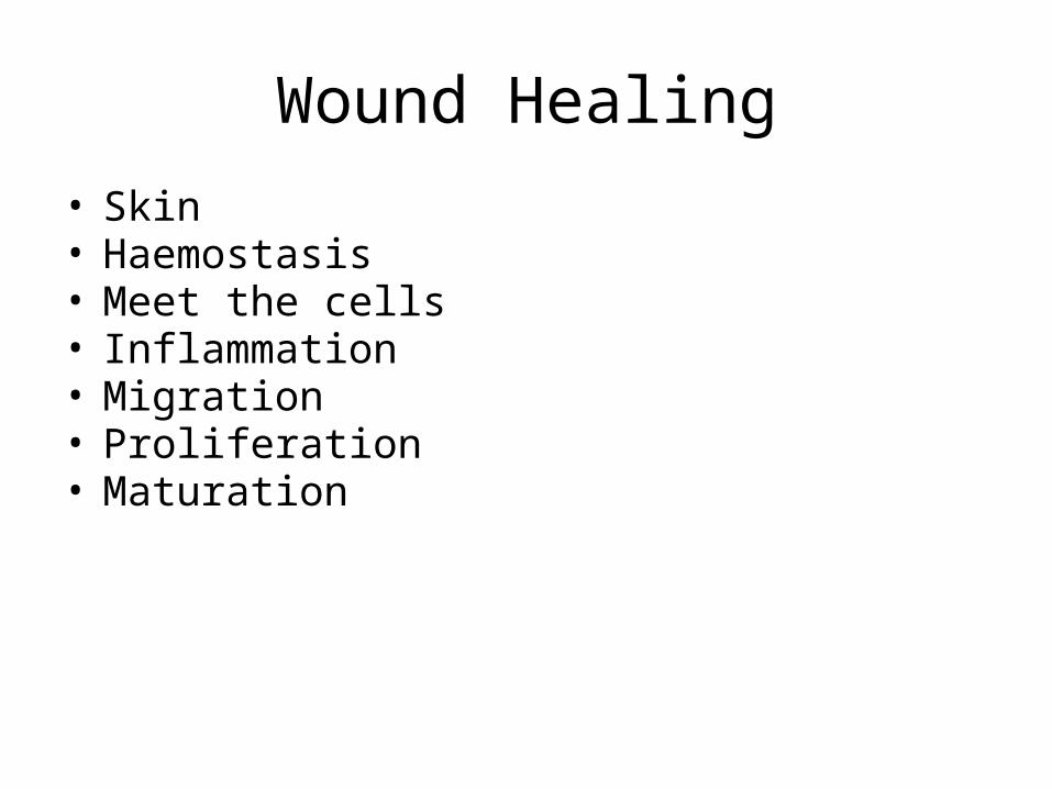
Integumentary system
• What is the integumentary system? Outer covering• What organ(s) are involved?Skin • What structures are involved? Hair, nails, receptors, glands

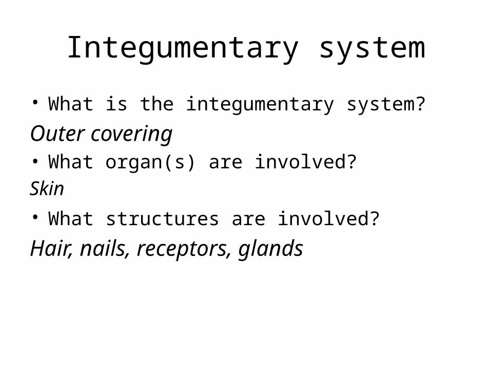
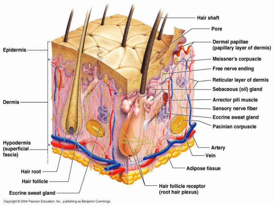
Integumentary system• Skin structure:3 layers; epidermis, dermis,
hypodermis or subcutaneous.
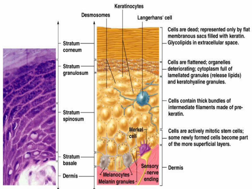
• Epidermis: Outer layers (4-5) of closely packed cells.• Stratum basale, Stratum spinosum, Stratum
granulosum, Stratum lucidum, Stratum corneum• Contains keratinocytes & melanocytes, Merkel cells
& Langerhans (dendritic) cells.
Epidermis
• Stratum basale is base layer where cells (keratinocytes) multiply. New cells push old cells away from blood supply. They fill with keratin, flatten & die in the corneum. Millions are shed daily.
• Melanocytes are in the stratum basale & produce melanin pigment
• Merkel cells at the epidermis-dermis junction function in touch
• Langerhans (dendritic) cells are phagocytes (immune system cells).

Dermis
• 2 indistinct layers: papillary layer & reticular layer.
• Contain hair follicles, sweat (sudoriferous) glands, oil (sebaceous) glands, touch receptors and a good blood supply.
• Connective tissue with lots of collagen and elastic fibres made by fibroblasts
Hypodermis
• Subcutaneous or superficial fascia anchors skin to underlying muscle.
• contains fat

Integumentary key functions
• Provides external protection• Regulates body temperature• Sensory organ• Excretion• Vitamin D synthesis• Immunity

Functions 1. • Regulation of body temperature primarily by
sweating and changing superficial vein diameter (& blood flow)
• Heat conservation occurs when vasoconstriction of superficial veins re-routes warm blood deeper into the body.

Regulation of body temperature
• Cooling occurs when • (a) Vasodilation of superficial veins brings
blood to the body surface and heat is lost to the environment by radiation convection & conduction.
• (b) An increase in perspiration results in increased evaporation cooling the skin but effectiveness is dependant on the humidity.
Functions
• 2. Protection from mechanical damage (keratin), microbial invasion, dehydration & UV radiation (melanin)
• 3. Excretion for loss of some ions, water & nitrogen containing waste

Functions 4.
• Sensations from sensory receptors• Free nerve endings: temperature, touch, pressure &
pain • Hair receptors : hair movement• Merkels disc: Light pressure• Meissner’s corpuscle: Light pressure • Ruffini’s endings: Deep pressure, stretch • Pacinian corpuscle: Deep pressure, stretch

Functions• 5. Langerhans cells of the epidermis are immune
system cells• 6. Blood reservoir carrying 8-10% of total blood
flow and is highly variable.• 7. Synthesis of Vitamin D when UV rays from sun
promote production of a Vitamin D precursor by the epidermis
• 8. Stores fat & fat soluble vitamins (A, D, E & K)• 9. Cutaneous absorption and secretion• 10. Social interaction

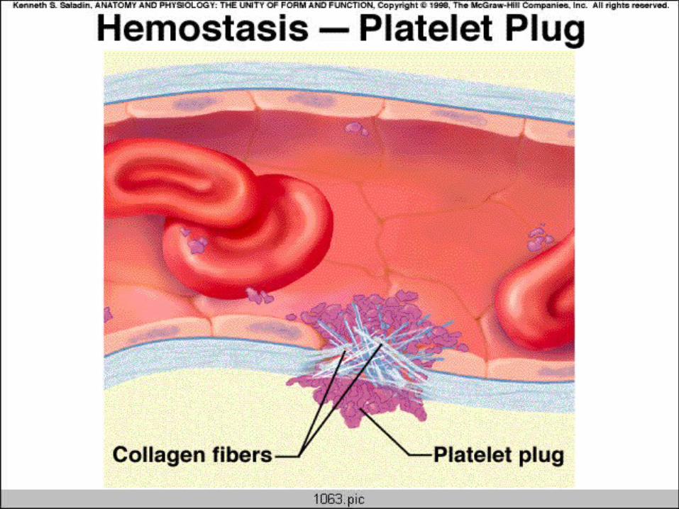

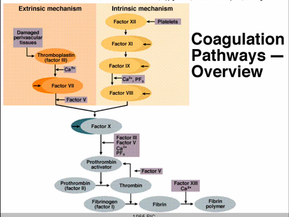
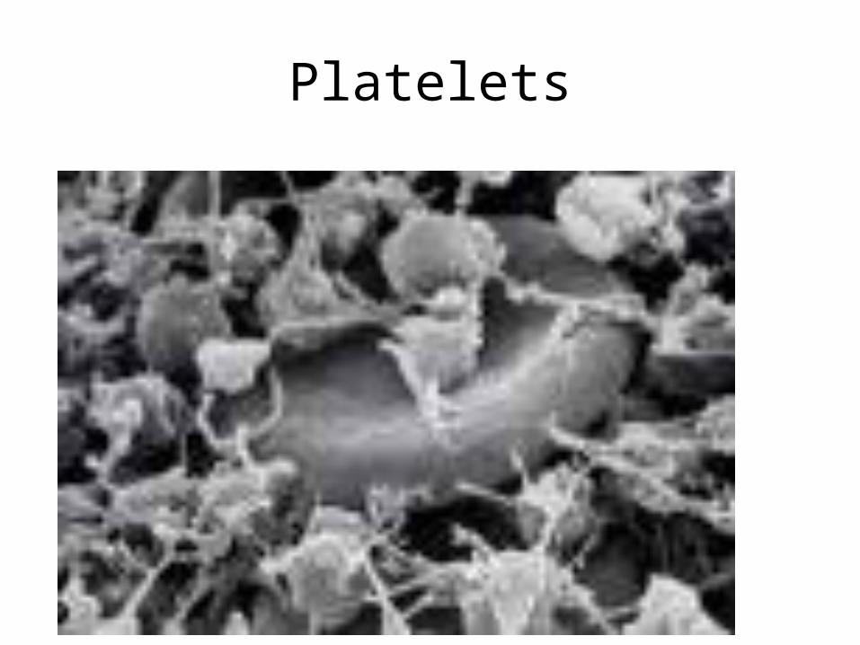
Platelets
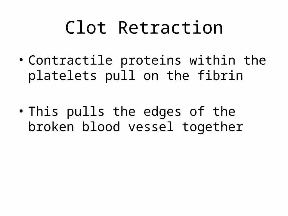
Clot Retraction
• Contractile proteins within the platelets pull on the fibrin
• This pulls the edges of the broken blood vessel together
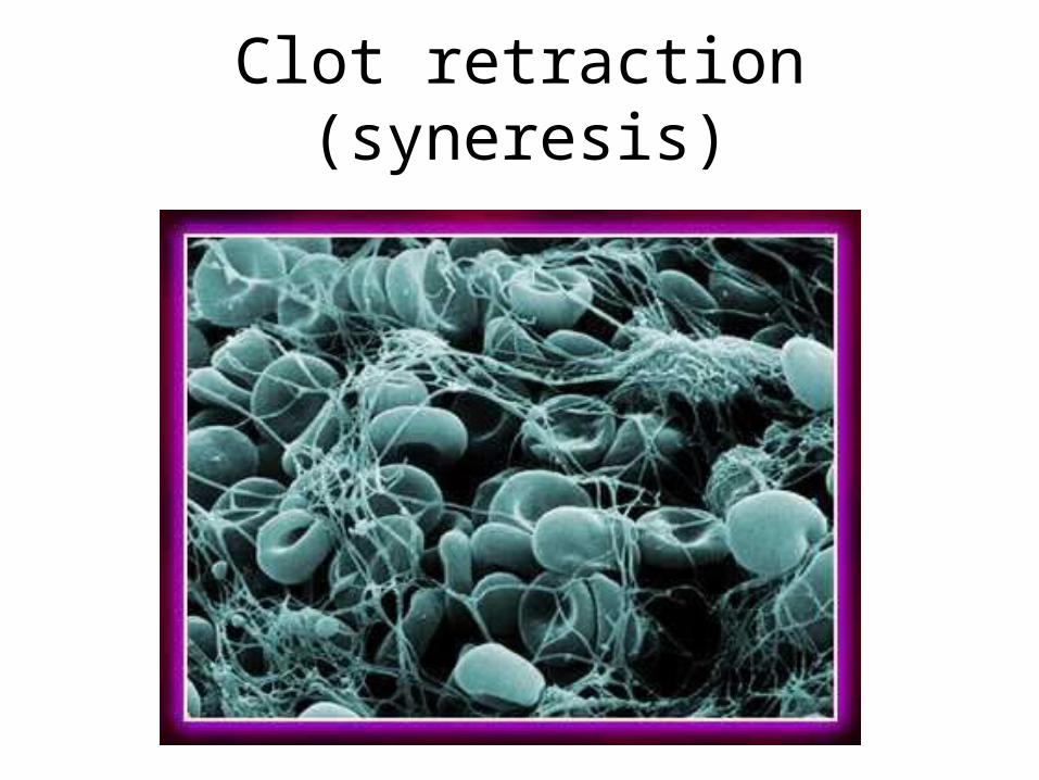
Clot retraction (syneresis)

Clot Destruction (Fibrinolysis)
• Plasminogen is converted to Plasmin
• Plasmin breaks down the fibrin in the clot

Meet the cells
• Labile cells– Regenerate readily, involved in wound repair
• Stable cells– Don’t normally divide in adult, but may
• Permanent cells– Unable to divide, replaced by scar tissue

Neutrophil
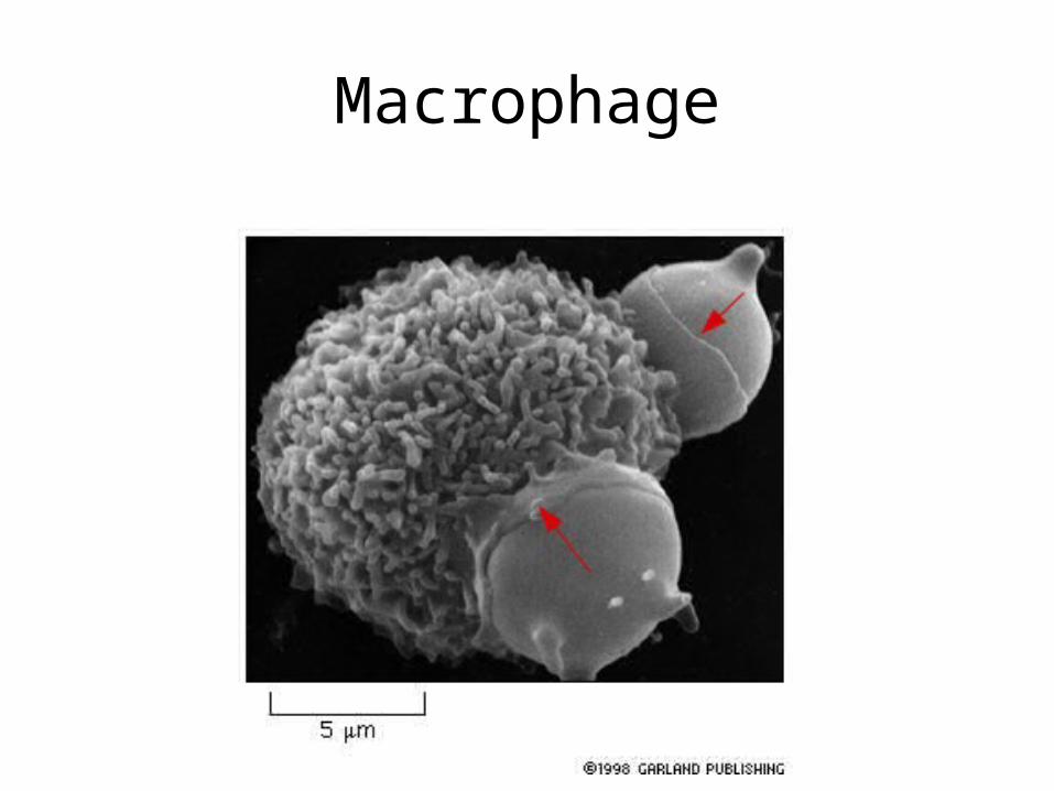
Macrophage
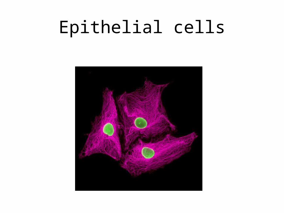
Epithelial cells
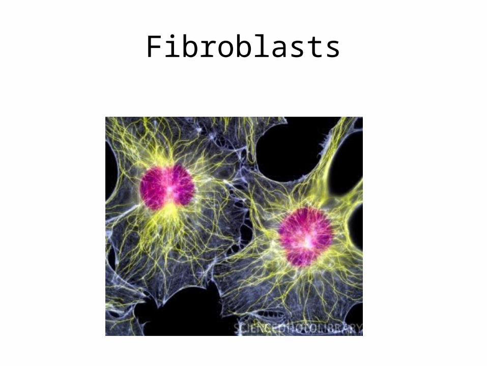
Fibroblasts
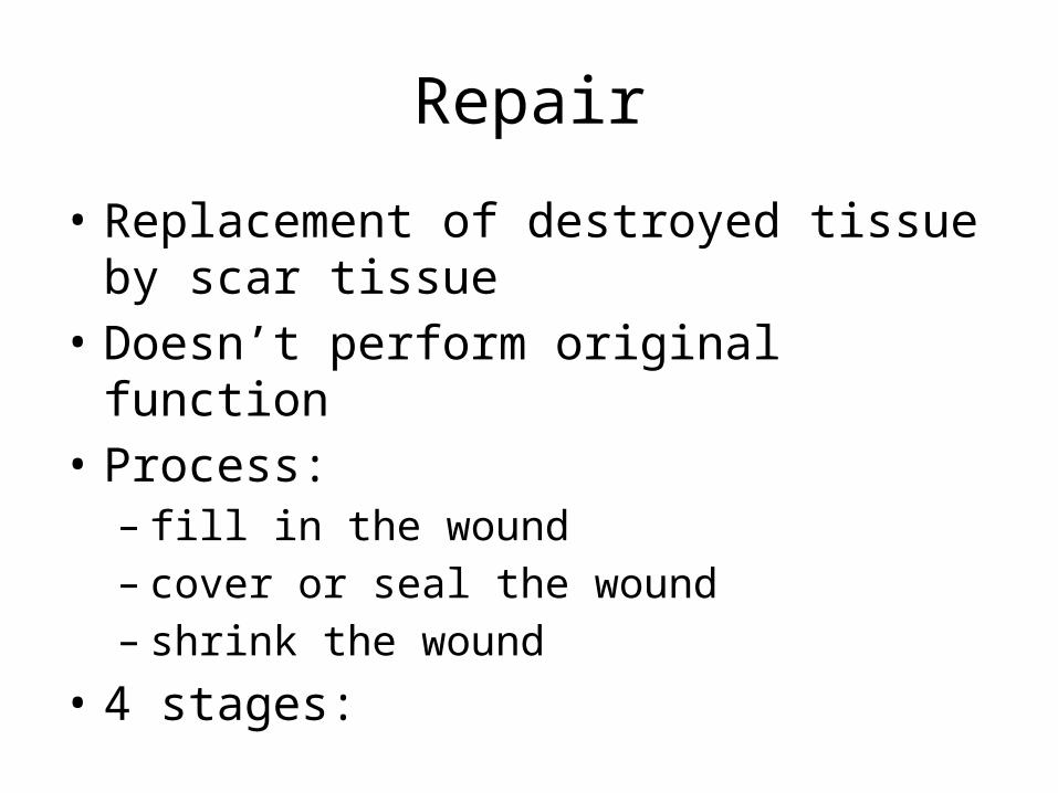
Repair
• Replacement of destroyed tissue by scar tissue• Doesn’t perform original function• Process:– fill in the wound– cover or seal the wound– shrink the wound
• 4 stages:

1) Inflammatory phase
• Haemostasis• Inflammation• Vasodilation & permeability of blood vessels• Phagocytotic cells (neutrophils &
macrophages) eat up cell debris & bacteria• Immediate to 2 - 5 days
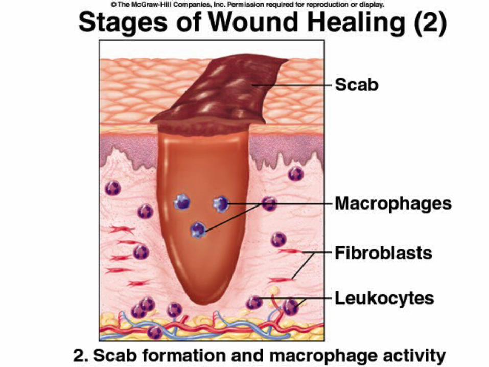
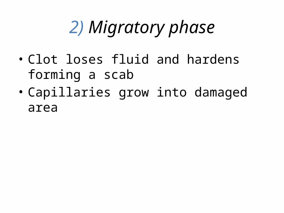
2) Migratory phase
• Clot loses fluid and hardens forming a scab• Capillaries grow into damaged area
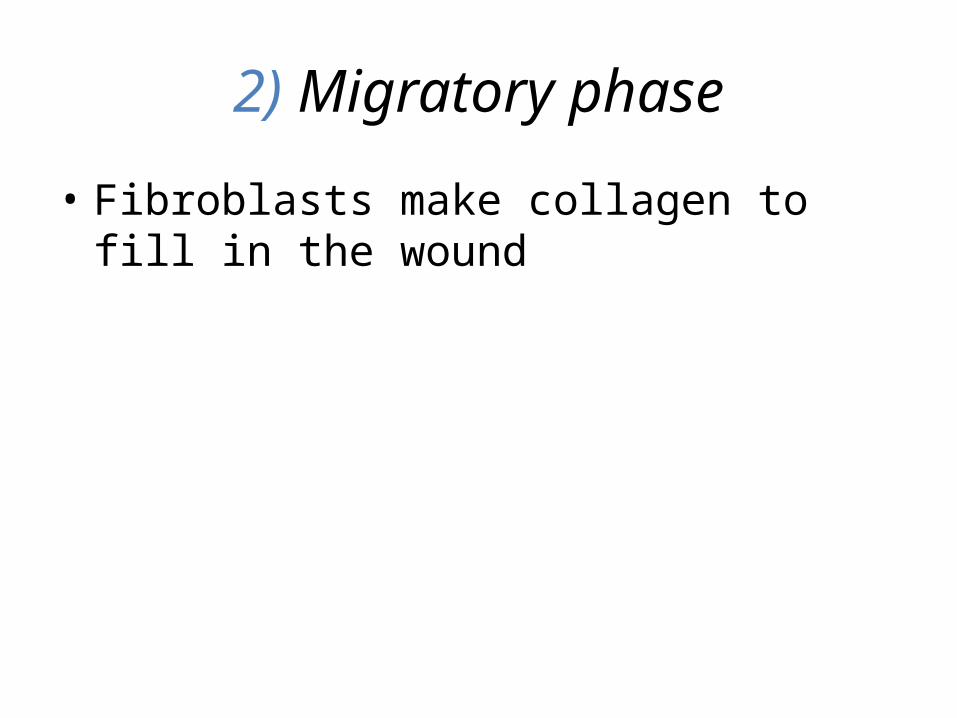
2) Migratory phase
• Fibroblasts make collagen to fill in the wound


3) Proliferative phase
• 2 days - 3 weeks• Granulation• fibroblasts lay bed of collagen, fills defect (Scar
tissue)• new capillaries, fragile so bleed easily• Epithelialization• Epithelial cells regenerate to form a new
surface layer (under the scab)
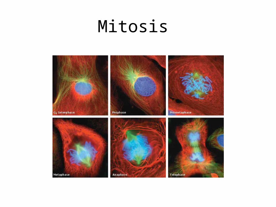
Mitosis
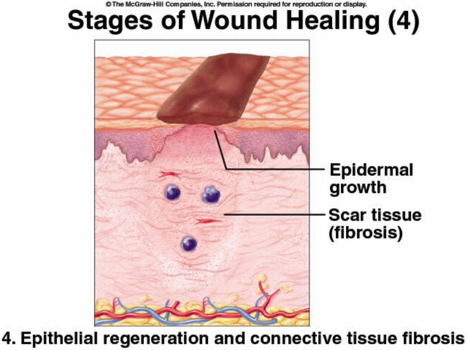
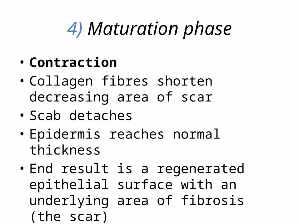
4) Maturation phase
• Contraction• Collagen fibres shorten decreasing area of scar • Scab detaches• Epidermis reaches normal thickness• End result is a regenerated epithelial surface
with an underlying area of fibrosis (the scar)

Primary Intention
• minimal tissue loss• e.g. clean, sutured incision

Secondary Intention
• much more tissue replacement• takes longer• e.g. stage IV decubitus ulcer
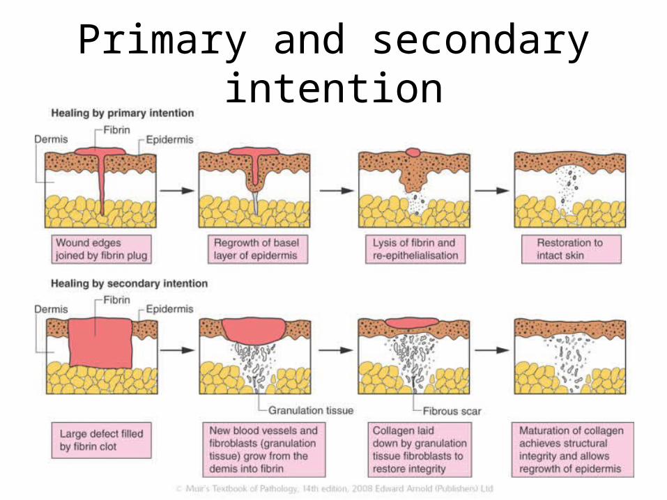
Primary and secondary intention
Related Documents