Printed Edition of the Special Issue Published in Healthcare Wound Care Edited by Zena Moore www.mdpi.com/journal/healthcare

Welcome message from author
This document is posted to help you gain knowledge. Please leave a comment to let me know what you think about it! Share it to your friends and learn new things together.
Transcript
Printed Edition of the Special Issue Published in Healthcare
Wound Care
Edited by
Zena Moore
www.mdpi.com/journal/healthcare
Zena Moore (Ed.) Wound Care Volume 1
This book is a reprint of the special issue that appeared in the online open access journal
Healthcare (ISSN 2227-9032) in 2015 (available at:
http://www.mdpi.com/journal/healthcare/special_issues/wound-care).
Guest Editor
Zena Moore
Royal College of Surgeons in Ireland
123 St Stephens Green
Dublin 2
Ireland
Editorial Office
MDPI AG
Klybeckstrasse 64
Basel, Switzerland
Publisher
Shu-Kun Lin
Senior Assistant Editor
Sarah Shao
1. Edition 2015
MDPI • Basel • Beijing
ISBN 978-3-03842-045-3
© 2015 by the authors; licensee MDPI, Basel, Switzerland. All articles in this volume are
Open Access distributed under the Creative Commons Attribution 3.0 license
(http://creativecommons.org/licenses/by/3.0/), which allows users to download, copy and
build upon published articles even for commercial purposes, as long as the author and
publisher are properly credited, which ensures maximum dissemination and a wider impact of
our publications. However, the dissemination and distribution of copies of this book as a
whole is restricted to MDPI, Basel, Switzerland.
III
Table of Contents
List of Contributors ............................................................................................................... V
Preface .................................................................................................................................IX
Wendy Chaboyer, Vinah Anderson, Joan Webster, Anne Sneddon, Lukman Thalib and
Brigid M. Gillespie
Negative Pressure Wound Therapy on Surgical Site Infections in Women Undergoing
Elective Caesarean Sections: A Pilot RCT
Reprinted from: Healthcare 2014, 2, 417-428
http://www.mdpi.com/2227-9032/2/4/417 .............................................................................. 1
Ann-Mari Fagerdahl
The Patient’s Conceptions of Wound Treatment with Negative Pressure Wound Therapy
Reprinted from: Healthcare 2014, 2, 272-281
http://www.mdpi.com/2227-9032/2/3/272 ............................................................................ 14
Margaret B. Harrison, Elizabeth G. VanDenKerkhof, Wilma M. Hopman and Meg E.
Carley
The Role of Preference on Outcomes of People Receiving Evidence-Informed Community
Wound Care in Their Home or in a Nurse-Clinic Setting: A Cohort Study (n = 230)
Reprinted from: Healthcare 2014, 2, 401-416
http://www.mdpi.com/2227-9032/2/3/401 ............................................................................ 24
Charne Miller, Suzanne Kapp and Lisa Donohue
Sustaining Behavior Changes Following a Venous Leg Ulcer Client Education Program
Reprinted from: Healthcare 2014, 2, 324-337
http://www.mdpi.com/2227-9032/2/3/324 ............................................................................ 40
Karen Ousey and Karen-leigh Edward
Exploring Resilience When Living with a Wound — An Integrative Literature Review
Reprinted from: Healthcare 2014, 2, 346-355
http://www.mdpi.com/2227-9032/2/3/346 ............................................................................ 54
IV
Hiromi Sanada, Gojiro Nakagami, Kimie Takehara, Taichi Goto, Nanase Ishii, Satoshi Yoshida, Mizuyuki Ryu and Yuichiro Tsunemi Antifungal Effect of Non-Woven Textiles Containing Polyhexamethylene Biguanide with
Sophorolipid: A Potential Method for Tinea Pedis Prevention
Reprinted from: Healthcare 2014, 2, 183-191
http://www.mdpi.com/2227-9032/2/2/183 ............................................................................... 63
Marianne V. Trondsen Managing Everyday Life: A Qualitative Study of Patients’ Experiences of a Web-Based Ulcer
Record for Home-Based Treatment
Reprinted from: Healthcare 2014, 2, 492-504
http://www.mdpi.com/2227-9032/2/4/492 ............................................................................... 72
Sara Ud-Din and Ardeshir Bayat Electrical Stimulation and Cutaneous Wound Healing: A Review of Clinical Evidence
Reprinted from: Healthcare 2014, 2, 445-467
http://www.mdpi.com/2227-9032/2/4/445 ............................................................................... 85
Ryan Vazales, Dustin Constant and Robert J. Snyder A Rare Case of Aggressive Digital Adenocarcinoma of the Lower Extremity, Masquerading
as an Ulcerative Lesion that Clinically Favored Benignancy
Reprinted from: Healthcare 2014, 2, 315-323
http://www.mdpi.com/2227-9032/2/3/315 ............................................................................. 109
Krishna S. Vyas and Henry C. Vasconez Wound Healing: Biologics, Skin Substitutes, Biomembranes and Scaffolds
Reprinted from: Healthcare 2014, 2, 356-400
http://www.mdpi.com/2227-9032/2/3/356 ............................................................................. 118
Tracey L. Yap, Susan Kennerly, Kirsten Corazzini, Kristie Porter, Mark Toles and Ruth A. Anderson Evaluation of Cueing Innovation for Pressure Ulcer Prevention Using Staff Focus Groups
Reprinted from: Healthcare 2014, 2, 299-314
http://www.mdpi.com/2227-9032/2/3/299 ............................................................................. 163
V
List of Contributors
Ruth A. Anderson: School of Nursing, Duke University, Durham, NC 27710, USA; Center
for the Study of Aging and Human Development, Duke University, Durham, NC 27710, USA
Vinah Anderson: NHMRC Centre of Research Excellence in Nursing (NCREN), Centre for
Health Practice Innovation, Griffith Health Institute, Griffith University, Gold Coast Campus,
QLD 4222, Australia
Ardeshir Bayat: Plastic and Reconstructive Surgery Research, Manchester Institute of
Biotechnology, University of Manchester, Manchester M1 7DN, UK; University Hospital of
South Manchester NHS Foundation Trust, Manchester Academic Health Science Centre,
University of Manchester, Manchester M1 7DN, UK
Meg E. Carley: School of Nursing, Queen's University, 92 Barrie Street, Kingston, ON K7L
3N6, Canada
Wendy Chaboyer: NHMRC Centre of Research Excellence in Nursing (NCREN), Centre for
Health Practice Innovation, Griffith Health Institute, Griffith University, Gold Coast Campus,
QLD 4222, Australia
Dustin Constant: Barry University School of Podiatric Medicine, Miami Shores, FL 33161,
USA
Kirsten Corazzini: School of Nursing, Duke University, Durham, NC 27710, USA; Center
for the Study of Aging and Human Development, Duke University, Durham, NC 27710, USA
Lisa Donohue: Faculty of Medicine and Health Sciences, School of Nursing an Midwifery,
Monash University, Peninsula Campus, McMahons Rd., Frankston, VIC 3199, Australia
Karen-leigh Edward: Faculty of Health Sciences, Australian Catholic University, Locked
Bag 4115 Fitzroy MDC, Victoria 3065, Australia; Nursing Research Unit, St Vincent's
Private Hospital Melbourne, 59-61 Victoria Pde Fitzroy, Victoria 3065, Australia
Ann-Mari Fagerdahl: Department of Surgery, Department of Clinical Science and
Education, Södersjukhuset, Karolinska Institutet, SE-171 77 Stockholm, Sweden
Brigid M. Gillespie: NHMRC Centre of Research Excellence in Nursing (NCREN), Centre
for Health Practice Innovation, Griffith Health Institute, Griffith University, Gold Coast
Campus, QLD 4222, Australia
Taichi Goto: Department of Gerontological Nursing/Wound Care Management, Graduate
School of Medicine, The University of Tokyo, 7-3-1 Hongo, Bunkyo-ku, Tokyo 113-0033,
Japan
Margaret B. Harrison: School of Nursing, Queen's University, 92 Barrie Street, Kingston,
ON K7L 3N6, Canada
Wilma M. Hopman: Clinical Research Centre, Kingston General Hospital, Kingston, ON
K7L 2V7, Canada; Department of Public Health Sciences, Queen's University, Kingston, ON
K7L 3N6, Canada
Nanase Ishii: Biochemical Laboratory, Saraya Co., Ltd., 24-12 Tamatecho, Kashiwara,
Osaka 582-0028, Japan
VI
Suzanne Kapp: RDNS Institute, 31 Alma Road, St. Kilda, VIC 3182, Australia
Susan Kennerly: School of Nursing, University of North Carolina at Charlotte, Charlotte,
NC 28223, USA
Charne Miller: La Trobe University Alfred Health Clinical School, Level 4, The Alfred
Centre, 99 Commercial Road, Prahran, VIC 3181, Australia
Gojiro Nakagami: Department of Gerontological Nursing/Wound Care Management,
Graduate School of Medicine, The University of Tokyo, 7-3-1 Hongo, Bunkyo-ku, Tokyo
113-0033, Japan
Karen Ousey: School of Human and Health Sciences, University of Huddersfield,
Queensgate, Huddersfield, HD1 3DH, UK
Kristie Porter: RTI International, Research Triangle Park, NC 27709, USA
Mizuyuki Ryu: Biochemical Laboratory, Saraya Co., Ltd., 24-12 Tamatecho, Kashiwara,
Osaka 582-0028, Japan
Hiromi Sanada: Department of Gerontological Nursing/Wound Care Management, Graduate
School of Medicine, The University of Tokyo, 7-3-1 Hongo, Bunkyo-ku, Tokyo 113-0033,
Japan
Anne Sneddon: Women's and Newborn Health, Gold Coast University Hospital, Southport,
QLD 4215, Australia
Robert J. Snyder: Clinical Research and Fellowship Program, Barry University School of
Podiatric Medicine, Miami Shores, FL 33161, USA
Kimie Takehara: Department of Nursing Administration, Graduate School of Medicine, The
University of Tokyo, 7-3-1 Hongo, Bunkyo-ku, Tokyo 113-0033, Japan
Lukman Thalib: epartment of Community Medicine (Biostatistics), Faculty of Medicine,
Kuwait University, PO Box 24923, Safat 13110, Kuwait
Mark Toles: School of Nursing, University of North Carolina at Chapel Hill, Chapel Hill,
NC 27599, USA
Marianne V. Trondse: Norwegian Centre for Integrated Care and Telemedicine (NST),
University Hospital of North Norway (UNN), P.O. Box 35, N-9038 Tromsø, Norway
Yuichiro Tsunemi: Department of Dermatology, Tokyo Women's Medical University, 8-1
Kawada-cho, Shinjuku-ku, Tokyo 162-8666, Japan
Sara Ud-Din: Plastic and Reconstructive Surgery Research, Manchester Institute of
Biotechnology, University of Manchester, Manchester M1 7DN, UK; University Hospital of
South Manchester NHS Foudation Trust, Manchester Academic Health Science Centre,
University of Manchester, Manchester M1 7DN, UK
Elizabeth G. VanDenKerkhof: School of Nursing, Queen's University, 92 Barrie Street,
Kingston, ON K7L 3N6, Canada; Department of Anesthesiology, Queen's University,
Kingston, ON K7L 2V7, Canada
Henry C. Vasconez: Division of Plastic Surgery, Department of Surgery, University of
Kentucky, Kentucky Clinic K454, 740 South Limestone, Lexington, KY 40536, USA
Ryan Vazales: Department of Podiatric Medicine and Surgery, Florida Hospital East
Orlando, Lake Underhill Road, Orlando, FL 32822, USA
VII
Krishna S. Vyas: Division of Plastic Surgery, Department of Surgery, University of
Kentucky, Kentucky Clinic K454, 740 South Limestone, Lexington, KY 40536, USA
Joan Webster: NHMRC Centre of Research Excellence in Nursing (NCREN), Centre for
Health Practice Innovation, Griffith Health Institute, Griffith University, Gold Coast Campus,
QLD 4222, Australia; Centre for Clinical Nursing, Royal Brisbane and Women's Hospital,
Butterfield Street, Herston, QLD 4029, Australia
Tracey L. Yap: School of Nursing, Duke University, Durham, NC 27710, USA; Center for
the Study of Aging and Human Development, Duke University, Durham, NC 27710, USA
Satoshi Yoshida: Department of Gerontological Nursing/Wound Care Management,
Graduate School of Medicine, The University of Tokyo, 7-3-1 Hongo, Bunkyo-ku, Tokyo
113-0033, Japan; Promote Development Division, Saraya Co., Ltd., 2-2-8 Yuzato,
Higashisumiyoshi-ku, Osaka City, Osaka 546-0013, Japan

IX
Preface
Wounds and the many associated problems have challenged health care providers for
centuries and today, despite the wealth of knowledge available, neither the incidence nor
prevalence of wounds is reducing. Furthermore, in view of our changing demographic profile
and the projected increase in the older population it is likely that wound management will
become an ever increasing burden to the individual, health care services and society as a
whole. The annual incidence of wounds in the EU-27 is approximately 4 million, and between
25% and 50% of acute hospital beds are occupied by patients with a wound, with up to 60%
of these representing non-healing wounds (infected surgical wounds, pressure ulcers, leg/foot
ulcers) The increasing prevalence and incidence of non-wounds healing is closely linked with
quality of care and, as such, these rising figures reduce society’s confidence in the health
service’s ability to deliver care that is timely, appropriate and effective. Thus, for those
involved in this specialist area of clinical practice, the fundamental goal is to improve clinical
outcomes, reduce the burden of wounds and improve health related quality of life.
In this Special Issue “Wound Care” in Healthcare, we invited submission of manuscripts
exploring contemporary issues in wound care. By devoting a special issue to wound care, we
endeavoured to provide readers with a comprehensive reference source, outlining key areas of
interest in this important aspect of clinical practice. The response to the call for manuscripts
was fantastic and, as a result, we were able to include both original qualitative and
quantitative research papers in addition to review papers, thereby providing readers with a
wealth of valuable information pertinent to wound care.
The impact of wounds on both the individual and society as a whole is significant and thus,
concerted efforts are required to reduce the burden of wounds. In order to achieve this,
clinicians need to have access to up to date information relevant for clinical care. This Special
Issue has enabled those involved in the cutting edge of wound care practice and research to
share their work with the wider community. Such endeavours are fundamental to achieving
the common goals in wound care today.
Zena Moore
Guest Editor
1
Negative Pressure Wound Therapy on Surgical Site Infections in Women Undergoing Elective Caesarean Sections: A Pilot RCT
Wendy Chaboyer, Vinah Anderson, Joan Webster, Anne Sneddon, Lukman Thalib and Brigid M. Gillespie
Abstract: Obese women undergoing caesarean section (CS) are at increased risk of surgical site infection (SSI). Negative Pressure Wound Therapy (NPWT) is growing in use as a prophylactic approach to prevent wound complications such as SSI, yet there is little evidence of its benefits. This pilot randomized controlled trial (RCT) assessed the effect of NPWT on SSI and other wound complications in obese women undergoing elective caesarean sections (CS) and also the feasibility of conducting a definitive trial. Ninety-two obese women undergoing elective CS were randomized in theatre via a central web based system using a parallel 1:1 process to two groups i.e., 46 women received the intervention (NPWT PICO™ dressing) and 46 women received standard care (Comfeel Plus® dressing). All women received the intended dressing following wound closure. The relative risk of SSI in the intervention group was 0.81 (95% CI 0.38–1.68); for the number of complications excluding SSI it was 0.98 (95% CI 0.34–2.79). A sample size of 784 (392 per group) would be required to find a statistically significant difference in SSI between the two groups with 90% power. These results demonstrate that a larger definitive trial is feasible and that careful planning and site selection is critical to the success of the overall study.
Reprinted from Healthcare. Cite as: Chaboyer, W.; Anderson, V.; Webster, J.; Sneddon, A.; Thalib, L.; Gillespie, B.M. Negative Pressure Wound Therapy on Surgical Site Infections in Women Undergoing Elective Caesarean Sections: A Pilot RCT. Healthcare 2014, 2, 417-428.
1. Introduction
Between 187 and 281 million surgical procedures are performed around the world each year, or one for every 25 people [1]. In Australia in 2008/9, 1.8 million elective surgeries were performed with one elective surgery for every 12.4 people [2]. Surgical site infections (SSIs) are defined by the Centers for Disease Control and Prevention (CDC) as infections occurring up to 30 days after surgery that affect the incision, deep tissue at the operation site or involve the organs or body space [3]. SSIs have many negative effects including pain, increasing the risk of morbidity and mortality, prolonging hospitalisation and increasing costs [4,5]. Of concern is that SSIs occur in up to 30% of all surgical procedures, and are the third most commonly reported hospital acquired infection [3]. Obesity is an independent predictor of SSI [4,6], thus it has significant safety and cost implications.
Obesity, defined as a body mass index (BMI) 30, is a growing global public health problem in developed nations. In 2007–2008, 28%–43% of 18–44 year old Australian women of childbearing age were obese [7]. Obese women are more likely to have a caesarean section (CS). One meta-analysis of 16 studies identified the odds ratio for overweight or obese women (BMI 25) having a CS as 2.0 (95% CI 1.9–2.2) compared to non-overweight women [8], similar to results of
2
an Australian analysis of 11,252 women giving birth [9]. Post-operative infection is a potential complication of all surgeries including CS, but overweight and obese women are at particular risk [10]. A meta-analysis of 6 studies showed the odds ratio for overweight or obese CS women having an infection was 3.3 (95% CI 2.7–4.1) compared to non-overweight women [8], consistent with individual studies [11]. Given that SSI extends hospital length of stay by up to 6 days in women undergoing obstetric and gynaecologic surgery, increasing hospital costs by US$14,000 for each SSI [12], it has significant implications for women and the health system.
Negative Pressure Wound Therapy (NPWT), also known as vacuum assisted closure, has been used to aid healing since the late 1990s [13,14]. It is based on a closed sealed system that applies negative pressure to the wound surface. The wound is covered or packed with an open-cell foam or gauze dressing and sealed with an occlusive drape. Intermittent or continuous suction is maintained by connecting suction tubes from the wound dressing to a vacuum pump and liquid waste collector. Standard negative pressure rates are 50–125 mm Hg [15]. Despite limited evidence of its effectiveness [16], Tipton and colleagues report “vacuum therapy can be included as an option for management of abdominal wounds, but evidence from randomized controlled trials in obese women undergoing cesarean is not available” [17,18]. Others note NPWT is increasingly being used in closed incisions to prevent SSI [19] and dehiscence. Additionally, one retrospective cohort study of 48 women receiving standard dressings compared to 21 women receiving NPWT found fewer wound complications in the NPWT group, but this difference was not statistically significant [18]. Limitations of Mark et al.’s study [18] such as the small sample size, use of historical controls, lack of control over the dressings used in the control group and reliance on coded medical record data suggests the findings should be interpreted very cautiously. Finally, a recent Cochrane Review of NPWT notes limited evidence for its effectiveness and recommends high quality trials to be undertaken[16]. Thus, this limited evidence base became the impetus to undertake a pilot trial in preparation for a larger, definitive trial of NPWT in obese women undergoing elective CS.
2. Aim
The aim of this pilot randomized controlled trial (RCT) was to assess the feasibility of conducting a larger trial in terms of measurement of potential outcomes, recruitment, intervention fidelity and retention. The hypothesis tested was “In obese women undergoing elective CS, those who receive a NPWT dressing will have significantly better outcomes than those receiving the standard dressing”. Data from this pilot study will assist researchers to determine sample size requirements and potential primary and secondary outcomes to be used in a larger, definitive trial.
3. Methods
A parallel group pilot RCT was undertaken (Australian and New Zealand Trial Registation number ACTRN12612000171819). Ethics approval was granted by the hospital and university office of human research ethics committees. An interim analysis of the first 48 women enrolled in this pilot showed 87% of women approached agreed to be part of the trial and there was 94.2% retention. All
3
women received the dresssings they were randomized to, and inter-rater reliability for the outcome SSI was 0.87 (citation masked for blinded peer review).
3.1. Participants and Setting
This study took place in one Australian hospital. As this was a pilot study, the target sample size was set at 80–100 [20]. Inclusion criteria were: (i) women booked for elective CS surgery; (ii) recorded pre-pregnancy BMI of 30 and (iii) able to provide written informed consent. Exclusion criteria were: (i) women whose condition changes to warrant an urgent or emergency CS; (ii) previous participation in this trial; (iii) existing infection after admission to hospital and prior to CS; and (iv) unable to speak or understand English with no interpreter present.
3.2. Outcomes
The primary outcome for this study was surgical site infection (SSI), as defined by the Centers for Disease Control and Prevention [3]. Secondary outcomes included: (1) type of SSI–superficial incision, deep incision or organ/body space using the CDC criteria; (2) wound complications (i.e., dehiscence, haematoma, bleeding, seroma, blisters); (3) hospital length of stay (HLOS); and (4) hospital readmissions (within 28 days). All outcomes except HLOS and readmission were assessed daily while the women were in hospital and weekly for 4 weeks after hospital discharge. No changes in the proposed trial outcomes occurred during the study.
3.3. Intervention and Control
At the completion of skin closure, those randomly allocated to the NPWT had, a PICO™ (Smith and Nephew, Hull, UK) applied by the obstetrician under sterile conditions. Women in the control arm had, Comfeel Plus® (Coloplast, City, Denmark) dressing applied per manufacturer’s recommendations after skin closure. In both groups, the dressing remained in place until day 4, unless it became soiled or dislodged, in which case a new dressing of the same type was applied. To ensure consistency, obstetricians, nurses and midwives received trial-specific education (Negative Pressure Wound Therapy (NPWT) and Comfeel Plus standard dressing). The research assistant (RA) was available to clinical staff via telephone and in person to provide ongoing training and support about correct use of the dressings as well as monitor dressing changes and complete documentation daily to assess protocol compliance and outcomes.
3.4. Procedure
Potential participants were screened between the 32nd and 38th weeks of gestation by either the attending doctor or midwife in the antenatal clinic. An RA who was a Registered Nurse recruited participants during their 36th week outpatient visit, providing potential participants with an information summary of the research. If women agreed to participate, they signed a consent form. On the day of the elective CS, the RA confirmed ongoing consent from the women. Randomization was via a computer-generated 1:1 ratio, and had blocks of randomly varying sizes. Randomization
4
occurred by the RA in the operating room. A centralized web-based randomization service was accessed which ensured allocation concealment.
The RA collected all outcome data daily while the women were in hospital. Following hospital discharge women were contacted weekly until the study end-point, at 28 days. Field notes were recorded that provided narrative information regarding the conduct of the trial and the care women received. A separate person, experienced in assessing for SSI, assessed the outcome SSI and was blinded to group allocation. Assessment of the data for SSI occurred at two intervals during the course of the study, firstly data on 35 women was assessed prior to preliminary analsysis (9 months into the trial) and the remaining 52 women’s data was examined on completion of the study. All women had completed 28 days of data collection at time of outcome assessment.
3.5. Data Analysis
Descriptive and infererntial statistics were used to analyse the data. Continous variables were summarized using mean and standard deviation (SD) or median and inter quartile range (IQR) based on normality assumptions. Normal continous varibles were compared between the intervention and control groups, using independent t-test while those that were not normal were analysed using Mann Whitney U test. Categorical variables were described using frequency and percentages. Testing of hypotheses of categorical varibles were evaluated using Chi-square test or Fisher’s exact test as appropriate.
Primary and secondary outcome variables were compared by computing the risk in each group and risk ratio (RR) and 95% confidence interval (CI). We did not expect statistical signficance between the groups for the oucome measures but point estimates (RR) were expected to show the direction and approximate magnitude of effect, if the study were to have been sufficienty powered. With the intention of conducting a larger trial, we used these data for a power calculation. Most data analyses were carried out using SPSS version 21 [21], MedCalc [22] was used for risk computations and confidence intervals, and PASS version 12 [23] was used for sample size calcuations.
4. Results
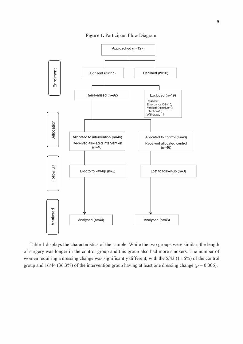
Recruitment occurred from July 2012 to April 2014. As identified in the flow diagram (Figure 1), a total of 111 women were recruited but 19 (17%) were subsequently excluded prior to randomisation. There was incomplete outcome data on 5 (5%) women, therefore the final analysis included 87 women. Four of the five women dropped out before the final data collection point and the fifth was transferred inter-hospital and had no outcome data. All women in the intervention and all women in the control group received the dressing to which they were randomized. One (2.2%) woman in the intervention group had a subsequent dressing change that resulted in a standard (rather than NPWT) dressing being used for the replacement (contamination). In successive dressing changes, none of the control women received the intervention (NPWT) dressing. Women were analysed according to their randomized dressing irrespective of whether they received a different dressing to the group to which they were allocated during the study period.
5
Figure 1. Participant Flow Diagram.
Table 1 displays the characteristics of the sample. While the two groups were similar, the length of surgery was longer in the control group and this group also had more smokers. The number of women requiring a dressing change was significantly different, with the 5/43 (11.6%) of the control group and 16/44 (36.3%) of the intervention group having at least one dressing change (p = 0.006).

6
Table 1. Characteristics of the Sample (n = 87).
Characteristic Intervention Group n = 44 Control Group n = 43 p-Value Median (IQR) Median (IQR)
Age 30.6 (5.5) 30.7 (5.0) 0.925 Body Mass Index 35.7 (4.5) 36.8 (5.8) 0.538 Length of surgery
(minutes) 45.0 (16.0) 53.0 (16.0) 0.002
Frequency (%) Frequency (%) a Co-morbidities (yes/no) 30 (68.1) 30 (69.7)
0.145 z score
Number of co-morbidities 0 1 2 3 4
14 (31.8) 18 (40.9) 10 (22.7) 1 (2.3) 1 (2.3)
13 (30.2) 21 (48.8) 5 (11.6) 3 (7.0) 1 (2.3)
Previous CS (yes/no) 37 (84.0) 40 (93.0) 0.188 z score
Number of previous CS 0 1 2 3 4
7 (15.9) 24 (54.5) 7 (15.9) 5 (11.4) 1 (2.3)
3 (7.0) 28 (65.1) 11 (25.6)
0 (0.0) 1 (2.3)
Smoker 3 (6.8) 10 (23.3) 0.032 Diabetic (any type) 13 (29.5) 12 (27.9) 0.290
a Comorbidities included: Anaemia, Diabetes Mellitus, Gestational Diabetes, Hypercholesterol, Hypertension, Immuno-compromised, Nutritional deficiency, Thromboembolytic disease, Smoking, Other.
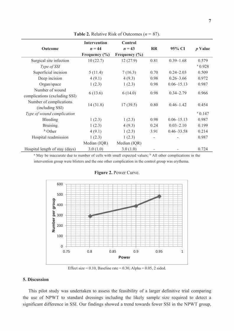
Table 2 shows the comparison of primary and secondary outcomes. In total, 27.9% of the control group and 22.7% of the intervention group had a SSI, but this difference did not reach statistical significance, due to smaller sample size. However the RR of 0.81 (95% CI 0.39; 1.68) shows the risk of SSI was almost 20% lower in the NPWT group (10/44) compared to the control group (12/43) which may be clinically important, although not statistically significant with this sample size. As identified in Table 2, there were no statistically significant differences in the other outcomes, although there was a trend towards reduced bruising but increased blistering in NPWT. No women in either group experienced a seroma or dehiscence.
Figure 2 provides a power curve based on the SSI data, demonstrating the various sample sizes required for trials powered at 80%, 90% and 95%. A sample size of 392 per group would be required to find a statistically significant difference in SSI between the two groups with 90% power.
7
Table 2. Relative Risk of Outcomes (n = 87).
Outcome Intervention
n = 44 Frequency (%)
Controln = 43
Frequency (%)RR 95% CI p Value
Surgical site infection 10 (22.7) 12 (27.9) 0.81 0.39–1.68 0.579 Type of SSI a 0.928
Superficial incision 5 (11.4) 7 (16.3) 0.70 0.24–2.03 0.509 Deep incision 4 (9.1) 4 (9.3) 0.98 0.26–3.66 0.972 Organ/space 1 (2.3) 1 (2.3) 0.98 0.06–15.13 0.987
Number of wound complications (excluding SSI)
6 (13.6) 6 (14.0) 0.98 0.34–2.79 0.966
Number of complications (including SSI)
14 (31.8) 17 (39.5) 0.80 0.46–1.42 0.454
Type of wound complication a 0.147 Bleeding 1 (2.3) 1 (2.3) 0.98 0.06–15.13 0.987 Bruising 1 (2.3) 4 (9.3) 0.24 0.03–2.10 0.199 b Other 4 (9.1) 1 (2.3) 3.91 0.46–33.58 0.214
Hospital readmission 1 (2.3) 1 (2.3) - - 0.987 Median (IQR) Median (IQR)
Hospital length of stay (days) 3.0 (1.0) 3.0 (1.0) - - 0.724 a May be inaccurate due to number of cells with small expected values; b All other complications in the intervention group were blisters and the one other complication in the control group was erythema.
Figure 2. Power Curve.
Effect size = 0.10, Baseline rate = 0.30, Alpha = 0.05, 2 sided.
5. Discussion
This pilot study was undertaken to assess the feasibility of a larger definitive trial comparing the use of NPWT to standard dressings including the likely sample size required to detect a significant difference in SSI. Our findings showed a trend towards fewer SSI in the NPWT group,
0
100
200
300
400
500
600
0.75 0.8 0.85 0.9 0.95 1
Num
ber p
er g
roup
Power
8
although this difference was not statistically significant, likely due to the small sample. However, if this trend were to be supported in an adequately powered trial, it could have important implications for clinical practice. Preventing some SSIs has a number of benefits including improving women’s recovery from surgery, decreasing the need for treatment and hospital length of stay as well as saving valuable health care dollars. Argubly, despite NPWT being more expensive than conventional dressings, the benefits in preventing SSI may outweigh these dressing costs. It would be important for larger trials to incorporate some form of economic analysis, given the limited evidence in this area. This recommendation is also supported by a Cochrane Review of NPWT, which only identified one abstract of a study that considered costs but the full paper was not available [16]. There is however some costing research in other patient populations, with one study of patients with diabetic foot wounds finding that the average cost to achieve healing was less in the NPWT group (although this was a small study) [24]. Without good quality RCT evidence of effect or cost-benefit, it would be premature to recommend using NPWT for surgical wounds in obese women undergoing elective CS.
Despite our sample being randomized, there were group differences in both the smoking status of the women and the length of surgery, with women in the control group reporting smoking more and their surgery was longer. Both factors are recognised risk factors for SSI, thus it is always possible that these results explain the trend towards more SSI in the control group. It would be expected that randomising women in a trial with a larger sample size would see at least the difference in smoking status between the two groups disappear. In a larger sample, the length of surgery, a potential confounder, could be handled statistically, by entering it into the analysis as a covariate.The independent effect of NPWT could then be assessed while controlling for length of surgery. However, the clinical significance of an average of 10 min longer surgery is unknown.
The NPWT dressing was acceptable to both the women in the trial and the healthcare teams providing care. For example we were able to enrol 87% of the women approached to participate in the study and all women allocated to the NPWT received it (i.e., clinicians did not prevent the NPWT dressing from being used in women recruited to the study). However, a general lack of familiarity with the NPWT dressing meant that ongoing education of both the medical and nursing staff was required. Additionally, at times when the research team was not available, some NPWT dressings were changed and in one instance, was replaced by the standard (control) dressing. In fact, 36% of the NPWT group had at least one dressing change, as compared to 12% in the control group. This unexpected finding requires further understanding as it has implications for future research, treatment costs and clinical practice. For example, this may indicate the need for additional staff training to ensure unnecessary dressing changes do not occur or it may indicate some other factor that can be addressed such as poor application technique.
An important consideration for RCTs is the extent to which the control and intervention groups receive similar care. In this hospital, women having elective CS followed a standardised care pathway during their admission, which should have standardised important aspects of care. However, the RA observed and documented subtle differences in certain aspects of surgical care such as antibiotic timing in theatre, surgeons’ preference for wound closure including suture materials, and type of standard dressing. Each of these issues could influence the findings of a definitive trial. Clinical practice guidelines and systematic reviews recommend pre-operative prophylactic
9
antibiotics for clean contaminated wounds such as CS [25–27]. Including these recommendations during education sessions related to a larger trial may help to standardize practice. In terms of wound closure, the 2008 National Institute of Health and Clinical Excellence guidelines note there is no high quality evidence to recommend one practice over another [28], but a recent meta-analysis found closure with staples had a twofold higher risk of wound infection than closure with subcuticular sutures [29]. Thus, including the use of sutures rather than staples for wound closure in future trial protocols could reduce the potential impact of this potential confounder, although a small study of 63 women undergoing CS found surgeons preferred staples over sutures [30]. Finally, in terms of what dressings were used in the control group, a 2011 Cochrane review found no evidence to suggest one dressing type was better than others for the prevention of SSI [31]. It could be that there are differences that have yet to be demonstrated. In future trials, standardizing the dressing type in the control group may be prudent, but some variation in clinical practice does not mean that subsequent trials without standardisation cannot be completed in a rigorous manner. It does however suggest that future trials should be a pragmatic (versus explanatory) trial. Sackett suggests that pragmatic trials answer the question “Does this treatment improve patient-important outcomes when applied by typical clinicians to typical patients?” [32]. There are a number of features of pragmatic trials that make them particularly well suited for testing interventions such as wound dressings in the clinical environment. First, pragmatic trials focus on effectiveness in usual circumstances or practice. Second, the intervention is applied in a flexible way, as it would be in clinical practice. Finally, the findings of the research are generally directly relevant to patients, clinicians and decision makers. As part of the feasibility component of this trial, we generated information to estimate a range of possible sample sizes for the primary outcome of SSI, required for a larger definitive trial. Using this approach reflects best practice and has added to the methodological rigor of this pilot trial [33]. However, as there may be some uncertainties around sample size estimates obtained through pilot trials, it is also recommended to discuss estimates with clinicians to obtain additional information around clinically meaningful effect sizes [33]. Our results indicate that a definitive trial would require an overall sample size of 784 (i.e., 392 per group in a two arm trial) to have 90% power to find a difference between groups if the primary outcome was the absence or presence of a SSI. Clearly, if SSI remains the primary outcome it will require a multi-site study.
In our pilot study we measured a number of other complications including bleeding, bruising, blister, seroma and dehiscence, but only noted whether they were present or absent and not the extent of each. Clinically, a small amount of bleeding, bruising or blistering would likely have little effect on the women or their ongoing care, but if more extensive, would likely require corrective action. Interestingly, there were no cases of either seroma or dehiscence reported but it is always possible this could occur in a larger sample. There was a trend towards more blistering in the NPWT group but none in the control group developing blisters. In one trial of 60 patients undergoing total knee arthroplasty, the rate of blisters in the NPWT group was so high (63%; RR 18.3 95% CI 4.3–77.6), the trial was stopped [34]. A recent review suggests skin blisters are common in orthopaedic surgery when adhesive dressings are used because of the swelling/oedema that occurs [19]. Clearly, blistering is an important safety consideration for both future trials and when the NPWT dressings are used in clinical practice.
10
An alternative option for selecting the primary outcome measure for the definitive trial is to develop a “composite” outcome such as “any wound complication” used in some previous research [18]. A composite measure involves aggregating the scores of several variables into an overall score [35]. The use of composite measures versus single outcome measures has been debated for some time [35–37]. Using a composite measure of “any wound complication” as the primary outcome in a definitive trial would likely result in a smaller sample size being required to demonstrate statistical significance. Nonetheless, there are also a number of limitations to such an approach. For example, grouping more serious complications like SSI and wound dehiscence with minor blistering or bleeding could make interpretation of the research findings including their clinical relevance difficult.
Other considerations for the larger definitive trial include standardizing training across sites especially proper application of the NPWT dressing, a clear monitoring plan to ensure the trial is proceeding as planned and additional data collection about site specific processes. Given the challenges associated with the real-world clinical settings, and the large number of health care providers involved in the clinical management of this population, using a pragmatic approach to trial design is appropriate.
6. Conclusions
This pilot study of 87 women showed that a larger definitive trial is feasible. Almost 90% of women approached agreed to be in the trial and 95% completed it. A sample size of 784 women would be required to detect a 20% difference in SSI at 90% power. A pragmatic trial, and associated process evaluation may be an appropriate approach if a definitive trial is undertaken in the future.
Acknowledgments
We acknowledge the advice and support Jennifer Fenwick provided. This study was funded by the Office of Health and Medical Research, Queensland Health, the National Health and Medical Research Council Centre of Research Excellence in Nursing and a Gold Coast University Hospital Private Practice grant.
Author Contributions
Study Conception and Design: Wendy Chaboyer, Brigid Gillespie, and Anne Sneddon; Data Collection: Vinah Anderson; Data Analysis and Interpretation: Lukman Thalib, Wendy Chaboyer, Vinah Anderson, Brigid Gillespie, Anne Sneddon, and Joan Webster; Writing and Revisions to the paper: Wendy Chaboyer, Brigid Gillespie, Vinah Anderson, Lukman Thalib, Anne Sneddon, and Joan Webster.
11
Conflicts of Interest
The authors declare no conflict of interest.
References
1. World Health Organization. 10 Facts on Safe Surgery. Available online: http://www.who.int/ topics/patient_safety/en/ (accessed on 15 August 2014).
2. Australia’s Hospitals 2008–2009—At a Glance; Australian Institute of Health and Welfare: Canberra, Australia, 2010.
3. Mangram, A.J.; Horan, T.C.; Pearson, M.L.; Silver, L.C.; Jarvis, W.R. Guideline for prevention of surgical site infection, 1999. Infect. Control Hosp. Epidemiol. 1999, 20, 247–280.
4. Anderson, D.J. Surgical site infections. Infect. Dis. Clin. 2011, 25, 135–153. 5. Graves, N.; Halton, K.; Curtis, M.; Doidge, S.; Lairson, D.; McLaws, M.; Whitby, M. Costs of
surgical site infections that appear after hospital discharge. Emerg. Infect. Dis. 2006, 12, 831–834. 6. Waisbren, E.; Rosen, H.; Bader, A.M.; Lipsitz, S.R.; Rogers, S.O., Jr.; Eriksson, E. Percent body
fat and prediction of surgical site infection. J. Am. Coll. Surg. 2010, 210, 381–389. 7. National Health Survey 2007–2008, Cat 4364.0; Australian Bureau of Statistics: Canberra,
Australia, 2008. 8. Heslehurst, N.; Simpson, H.; Ells, L.J.; Rankin, J.; Wilkinson, J.; Lang, R.; Brown, T.J.;
Summerbell, C.D. The impact of maternal bmi status on pregnancy outcomes with immediate short-term obstetric resource implications: A meta-analysis. Obes. Rev. 2008, 9, 635–683.
9. Callaway, L.K.; Prins, J.B.; Chang, A.M.; McIntyre, H.D. The prevalence and impact of overweight and obesity in an Australian obstetric population. Med. J. Aust. 2006, 184, 56–59.
10. Ramachenderan, J.; Bradford, J.; McLean, M. Maternal obesity and pregnancy complications: A review. Aust. N. Zeal. J. Obstetr. Gynaecol. 2008, 48, 228–235.
11. Ezechi, O.C.; Edet, A.; Akinlade, H.; Gab-Okafor, C.V.; Herbertson, E. Incidence and risk factors for caesarean wound infection in lagos nigeria. BMC Res. Notes 2009, 2, 186.
12. De Lissovoy, G.; Fraeman, K.; Hutchins, V.; Murphy, D.; Song, D.; Vaughn, B.B. Surgical site infection: Incidence and impact on hospital utilization and treatment costs. Am. J. Infect. Control 2009, 37, 387–397.
13. Fleischmann, W.; Lang, E.; Russ, M. Treatment of infection by vacuum sealing. Unfallchirurg 1997, 100, 301–304.
14. Morykwas, M.J.; Argenta, L.C.; Shelton-Brown, E.I.; McGuirt, W. Vacuum-assisted closure: A new method for wound control and treatment: Animal studies and basic foundation. Ann. Plast Surg. 1997, 38, 553–562.
15. Vikatmaa, P.; Juutilainen, V.; Kuukasjarvi, P.; Malmivaara, A. Negative pressure wound therapy: A systematic review on effectiveness and safety. Eur. J. Vasc. Endovasc. Surg. 2008, 36, 438–448.
16. Webster, J.; Scuffham, P.; Sherriff, K.L.; Stankiewicz, M.; Chaboyer, W.P. Negative pressure wound therapy for skin grafts and surgical wounds healing by primary intention. Cochr. Database Syst. Rev. 2012, doi:10.1002/14651858.CD009261.pub2.
12
17. Tipton, A.M.; Cohen, S.A.; Chelmow, D. Wound infection in the obese pregnant woman. Semin. Perinatol. 2011, 35, 345–349.
18. Mark, K.S.; Alger, L.; Terplan, M. Incisional negative pressure therapy to prevent wound complications following cesarean section in morbidly obese women: A pilot study. Surg. Innov. 2013, 21, 345–349.
19. Karlakki, S.; Brem, M.; Giannini, S.; Khanduja, V.; Stannard, J.; Martin, R. Negative pressure wound therapy for management of the surgical incision in orthopaedic surgery: A review of evidence and mechanisms for an emerging indication. Bone Joint Res. 2013, 2, 276–284.
20. Hertzog, M.A. Considerations in determining sample size for pilot studies. Res. Nurs. Health 2008, 31, 180–191.
21. IBM SPSS Statistics for Windows, Version 21.0; IBM Corp.: Armonk, NY, USA, 2012. 22. MedCalc for Windows, Version 12.5; MedCalc Software: Ostend, Belgium, 1999. 23. Hintze, J. Pass 12. NCSS, LLC.: Kaysville, Utah, USA, 2013. 24. Apelqvist, J.; Armstrong, D.G.; Lavery, L.A.; Boulton, A.J.M. Resource utilization and
economic costs of care based on a randomized trial of vacuum-assisted closure therapy in the treatment of diabetic foot wounds. Am. J. Surg. 2008, 195, 782–788.
25. Caesarean Section; National Institute for Health and Clinical Excellence: London, UK, 2011. 26. Tita, A.T.; Rouse, D.J.; Blackwell, S.; Saade, G.R.; Spong, C.Y.; Andrews, W.W. Emerging
concepts in antibiotic prophylaxis for cesarean delivery: A systematic review. Obstetr. Gynecol. 2009, 113, 675–682.
27. Baaqeel, H.; Baaqeel, R. Timing of administration of prophylactic antibiotics for caesarean section: A systematic review and meta-analysis. BJOG 2013, 120, 661–669.
28. National Institute for Health and Clinical Excellence. Nice Clinical Guideline 74: Surgical Site Infection: Prevention and Treatment of Surgical Site Infection; National Collaborating Centre for Women’s and Children’s Health, Ed.; RCOG Press: London, UK, 2008; pp. 4–28.
29. Tuuli, M.G.; Rampersad, R.M.; Carbone, J.F.; Stamilio, D.; Macones, G.A.; Odibo, A.O. Staples compared with subcuticular suture for skin closure after cesarean delivery: A systematic review and meta-analysis. Obstetr. Gynecol. 2011, 117, 682–690.
30. Aabakke, A.J.; Krebs, L.; Pipper, C.B.; Secher, N.J. Subcuticular suture compared with staples for skin closure after cesarean delivery: A randomized controlled trial. Obstetr. Gynecol. 2013, 122, 878–884.
31. Dumville, J.C.; Walter, C.J.; Sharp, C.A.; Page, T. Dressings for the prevention of surgical site infection. Cochrane Database Syst. Rev. 2011, doi:10.1002/14651858.CD003091.pub2.
32. Sackett, D.L. Clinician-trialist rounds: 16. Mind your explanatory and pragmatic attitudes!—Part 1: What? Clin. Trials 2013, 10, 495–498.
33. Thabane, L.; Ma, J.; Chu, R.; Cheng, J.; Ismaila, A.; Rios, L.; Robson, R.; Thabane, M.; Giangregorio, L.; Goldsmith, C. A tutorial on pilot studies: The what, why and how. BMC Med. Res. Methodol. 2010, doi:10.1186/1471-2288-10-1.
13
34. Howell, R.D.; Hadley, S.; Strauss, E.; Pelham, F.R. Blister formation with negative pressure dressings after total knee arthroplasty. Curr. Orthop. Pract. 2011, 22, 176–179.
35. Marcus, B. Composite measurement. The Sage Dictionary of Social Research Methods; SAGE Publications, Ltd.: London, UK, 2006; pp. 35–36.
36. Landis, R.S.; Beal, D.J.; Tesluk, P.E. A comparison of approaches to forming composite measures in structural equation models. Org. Res. Methods 2000, 3, 186–207.
37. Freemantle, N.; Calvert, M.; Wood, J.; Eastaugh, J.; Griffin, C. Composite outcomes in randomized trials: Greater precision but with greater uncertainty? JAMA 2003, 289, 2554–2559.
14
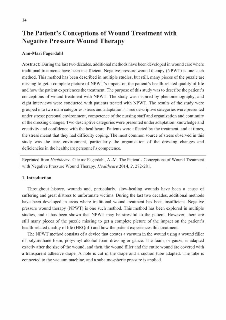
The Patient’s Conceptions of Wound Treatment with Negative Pressure Wound Therapy
Ann-Mari Fagerdahl
Abstract: During the last two decades, additional methods have been developed in wound care where traditional treatments have been insufficient. Negative pressure wound therapy (NPWT) is one such method. This method has been described in multiple studies, but still, many pieces of the puzzle are missing to get a complete picture of NPWT’s impact on the patient’s health-related quality of life and how the patient experiences the treatment. The purpose of this study was to describe the patient’s conceptions of wound treatment with NPWT. The study was inspired by phenomenography, and eight interviews were conducted with patients treated with NPWT. The results of the study were grouped into two main categories: stress and adaptation. Three descriptive categories were presented under stress: personal environment, competence of the nursing staff and organization and continuity of the dressing changes. Two descriptive categories were presented under adaptation: knowledge and creativity and confidence with the healthcare. Patients were affected by the treatment, and at times, the stress meant that they had difficulty coping. The most common source of stress observed in this study was the care environment, particularly the organization of the dressing changes and deficiencies in the healthcare personnel’s competence.
Reprinted from Healthcare. Cite as: Fagerdahl, A.-M. The Patient’s Conceptions of Wound Treatment with Negative Pressure Wound Therapy. Healthcare 2014, 2, 272-281.
1. Introduction
Throughout history, wounds and, particularly, slow-healing wounds have been a cause of suffering and great distress to unfortunate victims. During the last two decades, additional methods have been developed in areas where traditional wound treatment has been insufficient. Negative pressure wound therapy (NPWT) is one such method. This method has been explored in multiple studies, and it has been shown that NPWT may be stressful to the patient. However, there are still many pieces of the puzzle missing to get a complete picture of the impact on the patient’s health-related quality of life (HRQoL) and how the patient experiences this treatment.
The NPWT method consists of a device that creates a vacuum in the wound using a wound filler of polyurethane foam, polyvinyl alcohol foam dressing or gauze. The foam, or gauze, is adapted exactly after the size of the wound, and then, the wound filler and the entire wound are covered with a transparent adhesive drape. A hole is cut in the drape and a suction tube adapted. The tube is connected to the vacuum machine, and a subatmospheric pressure is applied.
15
NPWT has been in clinical use for wound management since 1995, and the first scientific documentation originates from 1997 with the work of Argenta and Morykwas [1]. Since then, thousands of articles have been published, but only a small fraction of the literature focuses on the patient’s conceptions and experiences of the treatment.
The impact on the HRQoL during NPWT has been explored qualitatively in only a few studies. Abbotts showed that the treatment with NPWT was experienced as stressful, especially regarding the impact on daily life and the organization of dressing changes [2]. An interview study by Bolas and Holloway confirms these findings, but also emphasizes the technical aspect of NPWT and describes the feelings of distress associated with its use [3].
Upton, Stephens and Andrew described that the NPWT system can cause patients to feel anxious, due to both the patient and the health professional being unfamiliar with this form of treatment. Furthermore, they described that the treatment can also restrict the patient’s daily care and wider social life, which may result in a negative self-image and low self-esteem. They also emphasize the need for more knowledge, particularly exploring the patient’s experience throughout the treatment process in order to minimize the negative effects of NPWT [4].
The World Union of Wound Healing Societies (WUWHS) consensus document on NPWT states that NPWT can have a positive impact on a patient’s HRQoL [5]. However, Ousey, Cook and Milne conclude in their review of the impact of NPWT on the patient’s HRQoL that it is not possible to determine whether the impact is positive, neutral or negative based on existing research [6]. Since the amount of research focusing on the patient’s experiences and that the existing literature presents varying results with both negative and positive impact on the patient’s HRQoL, it is necessary to conduct more qualitative research on the effects of NPWT. The aim of this study was to describe the patient’s conceptions of wound treatment with NPWT.
2. Methods
In this study, a phenomenographic approach was used. Phenomenography is a research method that explores the qualitatively different ways in which people perceive a specific phenomenon. Fundamental in phenomenography is to find the variation of people’s conceptions of this phenomenon [7]. In this study, the phenomenon is wound treatment with NPWT.
2.1. Participants
The participants were purposefully selected to ensure variation with respect to gender, age, wound type, type of NPWT device and treatment time, in accordance with the phenomenographic methodology [7,8]. Nineteen patients treated with NPWT during 2006 were asked to participate in the study, and in total, eight patients agreed (Table 1).

16
Table 1. Demographic and medical data of the participants (n = 8). NPWT, negative pressure wound therapy.
Variables Number
Gender Men 6 Women 2
Age Range 20–73 Median 66
Wound type
Post-operative wound infection 2 Diabetic foot ulcer 1 Pressure ulcer 1 Traumatic wound 2 Open abdomen 2
NPWT pump type Portable 4 Stationary 4
Treatment time (days) Range 2–42 Median 17
The NPWT system used was, in four cases, a portable vacuum-assisted closure (VAC) device (ActiV.A.C., KCI Inc, San Antonio, TX, USA) and in four cases, a larger stationary pump (InfoV.A.C., KCI Inc, San Antonio, TX, USA). The dressings were changed twice weekly. The dressing changes were performed as an inpatient treatment for patients with the stationary pumps and at the outpatient clinic for patients with portable machines. The healthcare personnel performing the wound treatment were physicians of different specialties, registered nurses and nurse’s aides at a large emergency city hospital. The hospital had no formal requirement that the personnel should have received specialized education in wound care, so knowledge and competence varied and was dependent on the individual’s experience and own interest.
2.2. Data Collection
Interviews were conducted in the period of June–November, 2006. A non-structured interview procedure was used, developing new questions following earlier answers, until no further information was received. All interviews began with one open question, where the participants were asked to talk freely about their conceptions of NPWT in general. The interview was expanded by follow-up questions regarding the injury, the wound healing process and the experience of being treated with NPWT. Six of the interviews were conducted at the hospital and two were telephone interviews. All interviews were conducted by the same researcher and lasted from seven to 43 min. The interviews were tape-recorded and transcribed verbatim.
Initially, six interviews were conducted and analyzed. Then, two more additional interviews were conducted, and after analysis, no new data was received, indicating a satisfying saturation of the material [9].
17
2.3. Data Analysis
Data analysis was conducted according to the phenomenographic method [8]. In all phases of the analysis, discussions took place between the researcher and co-workers, until consensus was reached.
The transcribed interviews were initially read several times to get familiar with the content and to obtain a sense of the whole. When a deeper understanding of the content was reached, distinct statements of conception were compared. Statements with similar content were grouped together and categorized into five labelled descriptive categories. These categories were thoroughly examined and discussed to ensure that they were distinctly separated from each other. In the next phase, the underlying meaning on an abstract level of the descriptive categories was analyzed, discussed and formulated into two main categories. Finally, the whole material was analyzed again to confirm the correlation between the statements of conception, descriptive categories and the main categories with the original text of the transcribed interviews.
2.4. Ethical Considerations
All participants were given written and verbal information, and their informed consent was obtained. Confidentiality was assured by decoding the interviews and all research data were kept in locked cabinets. Ethical approval was obtained by the local Ethics Committee (2006/571-31/2).
3. Results
The findings in this study show that being treated with NPWT was perceived by the participantsas stressful, and at times, the stress meant that they had difficulty coping. The ability to adapt to the prevailing circumstances had a major effect on their conceptions and experiences of the wound treatment process. The descriptive categories presented in the result comprise the participants’ conceptions as identified in their responses (Table 2).
Table 2. Patients’ conceptions of being treated with NPWT: main categories and description categories.
Description Category Main Category Personal environment
Stress Competence of the nursing staff Organization and continuity of the dressing changes
Knowledge and creativity Adaptation
Confidence with the healthcare
3.1. Stress
The majority of the participants perceived treatment with NPWT as being stressful, but worth the inconvenience.
18
3.1.1. Personal Environment
The participants’ personal environment was affected by the treatment in physical, mental, social and spiritual aspects. The participants particularly perceived physical discomfort during the treatment. Some of the participants described the treatment as being painful, especially during the dressing changes, but the majority did not perceive the treatment as painful at all. One participant even expressed himself so well that the staff was surprised by the fact that he did not have any pain:
“...about the abdomen...I don’t feel that I...I never had any pain...in the abdomen...all doctors asked but do you not have any pain there...?”
The most frequently described problem when being treated with NPWT was the inconvenience of being attached to a machine all the time. This was particularly disturbing to the participants treated with the larger stationary pump. The participants with the smaller portable pump, however, described an inconvenience when carrying it for a longer time, even when it felt light at first. The machine also affected some issues of daily life, like getting dressed and undressed and taking a shower. One man described frustration in the prolonged time required for performing everyday tasks:
“Most difficult this period was taking a shower…with a plastic bag…or thinking that the tube enters somewhere…and there will leak in water…if it gets soaked it must be replaced. So I put on two socks and then a plastic bag…oh, it was the greatest project…and what I have missed most of all…is not to sleep but to stand on two naked feet in the shower…”
3.1.2. Competence of the Nursing Staff
The participant perceived the competence and knowledge of the treatment as being rather varying and that there were major differences within the personnel who fully mastered the treatment compared to those that did not. Several participants described this as feeling like guinea pigs:
“It is not so many that feels...you know of the staff that knows this inside out yet, so they are experimenting a bit”
“…the staff…they said they did not know much…so they were also curious to know more about the machine…”
The competence of the staff was perceived by the majority of the participants as being inadequate, and they described this as very troublesome. The participants, however, also described being tolerant and understanding regarding the deficiencies in the competence of the staff, since they were aware of that the treatment was new, some even expressed an interest in being part of the staff’s education.
“…it was a bit…fascinating. Yes, there were several people in the OR and they were invited to watch the dressing changes…on some occasions there was a flow of visitors asking if they could take a look…well, it can be fun with a little public but finally only four persons at a time were allowed to watch the dressing changes as it became crowded I suppose…”
19
3.1.3. Organization and Continuity of the Dressing Changes
All of the participants described the continuity of the dressing changes as troublesome, particularly since there were so many people involved in their care and no one with the full responsibility.
The participants who had their dressing changes performed in the operation room (OR) ward described the waiting as most stressful in the process of dressing changes. The procedure was planned in the so-called emergency list at the OR and prioritized together with all other emergency cases in need of surgery. All of the participants experienced being given lower priority to have to wait for a long time for each dressing change. They all expressed this not being a great problem when being treated once or twice, but for longer treatment periods with many dressing changes, it became a major concern. Particularly problematic was when being forced to fast all day and the dressing change was postponed until the next day:
So that a…well…that part was an inconvenience, to have to wait not knowing if the change of dressing could be done that day…all of a sudden it could not be done and then you did not know when next a change could be performed…well you must get a scheduled time for the change of dressing.
3.2. Adaptation
Despite the stressful impact the treatment had on the participants, the majority perceived the treatment as being positive and that they were able to adapt and to manage the stress.
3.2.1. Knowledge and Creativity
Several of the participants described the importance of knowledge, both the knowledge within the staff, but also their own knowledge of the NPWT technique and their understanding of their own wound treatment. The participants received information regarding the treatment several times, however of varied content, and it is difficult to understand. The healthcare personnel who was informing also showed clear shortcomings in knowledge. One participant perceived that the staff was taking much for granted and did not understand that the patients had difficulties comprehending the information. Furthermore, the reduced health condition that several of the participants had was considered a reason for the perceived lack of information given and the understanding of that information.
The participants talked about several problems with daily living during treatment, but also how they, in a creative way, went about to solve these problems. They showed great creativity when trying to adapt to the situation and make everyday life as manageable as possible, both on their own, but also together with the healthcare personnel. Some participants treated with the larger stationary pump had different ways of making the pump more mobile:
“...then I went and experimented a bit on the ward so it resulted in that we took this vacuum pump and put on one of those IV-poles and then it went after all...it was...I was able to walk around and it up and...”
20
“…but I learnt to put the bed there (closer to the shower room, authors’ comments). I put the wire under the door so I could take a shower on my own.”
3.2.2. Confidence with the Healthcare
The participants said that, from the very beginning, they had had great confidence in the treatment and in the healthcare staff, and when the wound started to heal, they felt faith in the future. One participant had had the wound for a long time and was willing to try just about anything to see an improvement. Particularly, participants treated with open abdominal wounds described the treatment and trust in healthcare as giving them hope for recovery. One participant said that he, before treatment with NPWT, had been lying with an open abdomen and experienced how the intestines virtually fell out when he moved. With NPWT, he got the feeling that his body was whole again and with that, the agony he felt disappeared and the hope of recovery was lit:
No, it was that feeling…those first days…that everything leaks out of you…it was literally speaking only the peritoneum which held the intestines in place, and it leaked and smelled…you felt this is not going to work…almost a sort of deadly anxiety, I must say. I thought I wouldn’t survive…despite everyone saying to the contrary…When they applied this VAC dressing it felt more like it was a part of my body, somehow…The body felt whole again. This increased my well-being psychologically…from thinking “This is the end” to suddenly feeling “This is not so bad”.
4. Discussion
The results of this study show that the participants treated with NPWT perceived the treatment as positive and effective, despite stress in the form of physical strain and the inconvenience of being connected to the unit around the clock. These strains were managed by the participants’ feeling of the fundamental belief in the treatment and healthcare and that they had trust that their wounds would heal. Moreover, they perceived knowledge of the treatment method as important and contributing to their ability to creatively solve problems that arose during the treatment. The participants perceived the inadequate and varied skills of the healthcare staff and the organization and continuity of the dressing changes as being the most troublesome aspect.
4.1. Stress
The participants stated that treatment with NPWT was stressful to them, which is in accordance with other research focusing on patients’ experiences of traditional wound treatment [10–12].
The most troublesome for the participants during treatment was the organization of the dressing changes, particularly when performed in the OR ward. This problem has also been described by Abbott and by Bolas and Holloway [2,3]. It is important to facilitate the care of these patients and to minimize stress. By planning the dressing changes as elective operations in the surgical planning schedule, the risk of being postponed can be reduced. This could give the patients a better ability for themselves to prepare for the dressing change, which could result in a greater sense of control.
21
Another issue contributing to stress during treatment was the inadequate and varying competence of the healthcare personnel. This is also a well-described problem with NPWT treatment in the literature [3,13]. It is a major concern when apparently insufficiently-educated personnel handle advanced treatment, such as NPWT. Unfortunately, problems with the staff’s lack of skills are not unique to NPWT, but also occur in other wound treatment methods, as confirmed by previous research [14,15]. Graham [16] pinpoints the importance of sufficient education before applying the therapy, especially since incorrect use could seriously harm the patient. Graham suggests an educational program according to the theories by Patricia Benner [17] with different knowledge levels, from novice to expert. According to the ethical principle of non-maleficence, embodied by the phrase “first, do no harm”, it is essential for healthcare to ensure that the personnel has adequate knowledge of the equipment and method used, to avoid the risk of harming the patient.
The participants’ description of pain during treatment and, particularly, the absence of pain are worth mentioning. Procedural pain during dressing changes when treated with NPWT was earlier described in the literature [4]; however, studies of pain during the entire wound treatment process have shown varying results, and some studies even indicate that NPWT as a treatment may, in fact, ease the wound pain rather than enhance it [4,13].
4.2. Adaptation
The participants had to adapt to the current situation to manage the stress involved in the treatment to maintain a balance and the conception of health. The first step towards adaptation for the patients was receiving sufficient knowledge and information regarding the treatment. Edward, Moffat and Franks point out the importance of adequate information for the experience and management of the strain that wound treatment may have on the patient [18]. They also emphasize the varying quality of information provided. In their study, only one fifth of the patients had received some form of written information. The participants in this study expressed that poor general status of health during treatment was one explanation of difficulties to comprehend received information. Having the possibility of written information in addition to verbal could facilitate the patients’ understanding and allow them to process the information in a longer time span.
When feeling confident in managing the treatment, the participants became inventive and creative in dealing with different obstacles that arose in everyday life. Knowledge and confidence were key factors for managing and coping in a positive way with stressful issues during treatment, which is in concordance with other studies of NPWT [4,13] and in wound management, in general [11,12,19].
4.3. Methodological Considerations
Why is it important to know the patient’s conceptions of NPWT? There is an old saying: “The cure is worse than the disease”. This means that the treatment itself can be effective, but at the same time, so incredibly stressful to the individual patient that it is just not worth it. It is only the patient who is an expert of his/her own body and own conceptions, and therefore, research must be based on a patient’s perspective. Thus, using phenomenography as a research method and purposive sampling is appropriate, particularly since it is possible to identify a variation of conceptions, which is the
22
main objective of phenomenography as a research method [7,8]. To ensure clinical credibility, the whole process of analysis was performed in close collaboration with co-workers and other wound experts, and the process has been thoroughly described in the Methods section.
Regarding the transferability of the result, it should only be seen as an awareness-raising of the knowledge of wound patients and not as a representative experience of all patients treated with NPWT. However, since the participants in this study were selected with a large variation concerning age, gender, different wound types, different treatment times and different types of NPWT machines, the result describes a wide range of conceptions, which may be transferred to patients treated with NPWT in other settings.
One limitation of qualitative research may be that it is the interviewer who is the main instrument in the acquisition of knowledge. It is important that the researcher is aware of his/her role in order to obtain scientific knowledge, also adhering to ethical considerations, during the research interview. By recording and transcribing the interviews verbatim, the credibility of collected material may be enhanced. To ensure a sufficient amount of material, two additional interviews were performed. These interviews did not change the findings, so that the feeling of saturation of the material was achieved [9]. Another limitation of this study may be the rather short interviews, often due to the poor health status among several of the participants. However, the objective with this study was not to perform in-depth interviews, only to describe a variation of conceptions among patients treated with NPWT.
5. Conclusions and Relevance for Practice
The findings in this study show that patients were negatively affected by treatment with NPWT and, at times, the stress meant that they had difficulty coping. The largest source of stress observed in this study was the clinical setting, particularly the organization of the dressing changes and deficiencies in healthcare personnel’s competence.
These findings have relevance for the practice by demonstrating the importance of the organization of the treatment, especially the dressing changes, and highlight the insufficient knowledge and skills in wound management of the healthcare personnel that must be addressed.
Acknowledgments
Financial support was provided through a grant from The Swedish Society of Nursing.
Conflicts of Interest
The author declares no conflict of interest.
References
1. Argenta, L.C.; Morykwas, M.J. Vacuum-assisted closure: A new method for wound control and treatment: Clinical experience. Ann. Plast. Surg. 1997, 38, 563–577.
23
2. Abbotts, J. Patients’ views on topical negative pressure: “Effective but smelly”. Br. J. Nurs. 2010, 19, S37–S41.
3. Bolas, N.; Holloway, S. Negative pressure wound therapy: A study on patient perspectives. Br. J. Community Nurs. 2012, 17, S30–S35.
4. Upton, D.; Stephens, D.; Andrews, A. Patients’ experiences of negative pressure wound therapy for the treatment of wounds: A review. J. Wound Care 2013, 22, 34–39.
5. World Union Wound Healing Societies. Vacuum assisted closure: Recommendations for use: A consensus document. Int. Wound J. 2008, 5, doi:10.1111/j.1742-481X.2008.00537.x.
6. Ousey, K.J.; Cook, L.; Milne, J. Negative pressure wound therapy—Does it affect quality of life? Wounds UK 2012, 8, 18–28.
7. Marton, F.; Booth, S. Learning and Awareness; Lawrence Erlbaum Associate Publishers: Mahwah, NJ, USA, 1997.
8. Alexandersson, M. Den fenomenografiska forskningsansatsen i fokus (the phenomenographic reserach approach in focus). In Kvalitativa Metoder och Vetenskapsteori (Qualitative Methods and the Theory of Science; Starrin, B., Svensson, P., Eds.; Studentlitteratur: Lund, Sweden, 1994.
9. Mason, M. Sample size and saturation in Ph.D. studies using qualitative interviews. Forum Qual. Soc. Res. 2010, 11, 8.
10. Ebbeskog, B.; Ekman, S.L. Elderly persons’ experiences of living with venous leg ulcer: Living in a dialectal relationship between freedom and imprisonment. Scand. J. Caring Sci. 2001, 15, 235–243.
11. Persoon, A.; Heinen, M.M.; van der Vleuten, C.J.; de Rooij, M.J.; van de Kerkhof, P.C.; van Achterberg, T. Leg ulcers: A review of their impact on daily life. J. Clin. Nurs. 2004, 13, 341–354.
12. Spilsbury, K.; Nelson, A.; Cullum, N.; Iglesias, C.; Nixon, J.; Mason, S. Pressure ulcers and their treatment and effects on quality of life: Hospital inpatient perspectives. J. Adv. Nurs. 2007, 57, 494–504.
13. Fagerdahl, A.M.; Boström, L.; Ottosson, C.; Ulfvarson, J. Patients’ experience of advanced wound treatment—A qualitative study. Wounds Compend. Clin. Res. Pract. 2013, 25, 205–211.
14. Haram, R.; Nåden, D. What patients with leg ulcers think about the professionals treating their ulcers in home healthcare [Norwegian]. Nor. Tidsskr. Sykepl. 2002, 4, 135–151.
15. Kjaer, M.L.; Mainz, J.; Sorensen, L.T.; Karlsmark, T.; Gottrup, F. Venous leg ulcer patient priorities and quality of care: Results of a survey. Ostomy Wound Manag. 2004, 50, 48–55.
16. Graham, A. The development of a competency assessment for vacuum assisted closure therapy. Nurse Educ. Pract. 2005, 5, 144–151.
17. Benner, P. From Novice to Expert, Excellence and Power in Clinical Nursing Practice; Addison-Wesley Publishing Company: Menlo Park, CA, USA, 1984.
18. Edwards, L.M.; Moffatt, C.J.; Franks, P.J. An exploration of patients’ understanding of leg ulceration. J. Wound Care 2002, 11, 35–39.
19. Ebbeskog, B.; Emami, A. Older patients’ experience of dressing changes on venous leg ulcers: More than just a docile patient. J. Clin. Nurs. 2005, 14, 1223–1231.
24
The Role of Preference on Outcomes of People Receiving Evidence-Informed Community Wound Care in Their Home or in a Nurse-Clinic Setting: A Cohort Study (n = 230)
Margaret B. Harrison, Elizabeth G. VanDenKerkhof, Wilma M. Hopman and Meg E. Carley
Abstract: This study followed a cohort of community-dwelling individuals receiving wound-care in a large urban-rural region. During a randomized control trial (RCT) evaluating outcomes of receiving care in a nurse-clinic or at home, many approached were willing to participate if they could choose their location of care. This provided a unique opportunity to enroll them as a “choice” cohort, following them in the same manner as the trial participants but allowing them to select their setting of care. The objective was to investigate the role of preference and location of care on care outcomes, including satisfaction with care, healing, health-related quality of life (HRQL), pain, and resource use. This is a secondary analysis of a prospective cohort of 126 individuals enrolled in an RCT to receive care at home or in a nurse-clinic (Allocated group), and an additional 104 who received care at home or in a nurse-clinic based on their preference (Choice group). Mobile individuals with a leg ulcer of venous or mixed venous etiology, referred for community leg ulcer care, were eligible. Specially-trained nurses provided care to both groups using an evidence-informed protocol. Baseline data included socio-demographic, circumstance-of-living and a detailed wound assessment. Mean age of the cohort was 68 years. Satisfaction, healing, recurrence, pain, HRQL, and resource utilization did not differ between groups. If available, individuals should have an option of care venue given almost half of those approached indicated a clear preference for clinic or home. With outcomes being similar, health care planners and decision-makers, as well as individuals and their families, can feel confident that the setting of care will not impact the outcomes. However, larger studies in other contexts are needed to explore the interaction between choice and setting.
Reprinted from Healthcare. Cite as: Harrison, M.B.; VanDenKerkhof, E.G.; Hopman, W.M.; Carley, M.E. The Role of Preference on Outcomes of People Receiving Evidence-Informed Community Wound Care in Their Home or in a Nurse-Clinic Setting: A Cohort Study (n = 230). Healthcare 2014, 2, 401-416.
1. Introduction
Community care has become an increasingly important element within health care systems everywhere. As an example in Canada, acute care facilities have downsized, with more conditions managed and procedures performed on an outpatient basis. One advantage to this is that it may allow individuals to receive care closer to home if they prefer. An exemplar population receiving the majority of their care in the community are those suffering from chronic wounds. In the case of leg ulcers, much effort has been dedicated to evaluating “best practices” including high compression bandages [1–4] and where and how care is delivered, e.g., in the home, nurse clinics or specialist
25
clinic [5–12]. On the other hand, individuals’ preference of where they receive their health care, or if having one’s choice makes any difference to outcomes, has typically not been a focus of research.
In a unique opportunity, we were able to follow a cohort of community dwelling individuals receiving wound care in a large urban-rural region in Ontario. It began as a randomized controlled trial (RCT) evaluating the outcomes of nurse-clinic vs. home delivery of evidence-informed care delivered by well-prepared providers [13]. After initiating the trial, many of those approached were willing to join the study but only if they could choose their preference of care location. We elected to enrol them in a “Choice Cohort” and follow them in exactly the same manner as the trial participants to assess if having choice made a difference to outcomes [14]. By combining the two previous studies for the current analysis, we have been able to not only describe a larger cohort of individuals receiving care, but also explore the role of preference on various outcomes. From a program planning perspective, we sought to understand if those with a stated preference differed from those allocated to where care is delivered. We posited that having one’s preference might improve outcomes such as satisfaction with care, well-being and quality of life and possibly the time-to-healing. From a health services perspective, there was interest in offering wound care in nurse clinics as well as home visiting, thus this analysis would also allow us to revisit the question of outcomes from care in a clinic location contrasted with home delivery in a larger sample (n = 230) than in our previous study (n = 126) [13] and in a sample that may be more representative of the target population than would be expected in a randomized controlled trial.
2. Experimental Section
2.1. Design
This study is a combined secondary analysis of a prospective cohort of 126 individuals enrolled in an RCT to receive care at home or in a nurse-clinic (Allocated Group), and an additional 104 who also received care at home or in a local community nurse-clinic, but received their care based on their preference (Choice Group). All 230 participants were followed until one-year post healing in an identical manner, using the same time points and outcome measurements. For the purpose of this analysis, we compared the Allocated and Choice groups to assess whether having choice impacted various outcomes, regardless of whether care was delivered in the home or clinic. The original study was reviewed for ethical compliance and approval obtained from the Ottawa Health Research Institute Ethics Board (#20000272-01H). A summary of key aspects of the study methodology are provided as full details are discussed elsewhere [13,14].
2.2. Setting and Sample
The study population for both the Allocated and the Choice Group came from the same large urban-rural Ontario region overseen by two regional homecare authorities. Inclusion and exclusion criteria for the two groups were the same: mobile individuals with a leg ulcer below the knee of venous or mixed venous and arterial etiology, with no major contraindication for clinic care (e.g., not being able to leave an ill spouse), referred for community leg ulcer care, were eligible to participate. Etiology was determined based on a thorough clinical assessment and ankle brachial
26
pressure index (ABPI) via handheld Doppler. No upper limit for ABPI was set as criteria for exclusion. Individuals who were cognitively impaired, too ill, or unable to travel outside the home were excluded.
2.3. Procedures
Specially trained registered nurses performed a comprehensive, standardized clinical assessment on all individuals referred to the regional community care service for leg ulcers. Eligible individuals were provided information regarding the clinic vs. home trial and invited to participate. Consenting individuals were randomized to be given care in either their home or a nurse clinic and this comprised the Allocated Group. For those who expressed a willingness to participate but not be randomized to where care was received, they provided a modified consent for the Cohort Study noting they would have their choice of location of care. Those who chose their care setting comprised the Choice Group.
The same nursing team delivered care in both the home and clinic settings and practice was guided by international evidence-informed recommendations [6,15]. The Practice Guideline Evaluation and Adaptation Cycle (PGEAC) [15–17] guided development of the study’s leg ulcer care management protocol. It was prepared by an interdisciplinary task force and feedback on the draft protocol was sought from homecare nurses and family physicians [18,19] prior to implementation. The protocol was kept up-to-date through ongoing scheduled reviews [20]. Community care nurses involved in the study received additional training in leg ulcer assessment and compression bandaging application, and were familiar with the evidence for practice supporting the guideline recommendations. Compression bandaging was applied by the same nursing team in both home and clinic settings. Visits for leg ulcer care were typically scheduled two to three times per week and bandages changed based on nurses’ clinical judgment and individual circumstances (e.g., amount of exudate). Once healed, participants were advised to wear compression stockings.
2.4. Data Collection and Management
At the time of initial assessment following referral for community care, baseline data were gathered through interview, clinical assessment and chart review. This included socio-demographic, circumstance-of-living and a detailed wound assessment. Ulcer size was measured every 3 months until complete healing, or until 12 months post study entry, whichever came first. If healing occurred between these measurement intervals, this was recorded and the next full assessment carried out according to the schedule. Integrity of the trial and cohort study was ensured through rigorous and systematic quality assurance procedures [21–23].
2.5. Outcome Measurement
Satisfaction with care was assessed with a 12-item questionnaire developed in consultation with frontline clinicians and administered at 3-months post-baseline. Formal validation was not undertaken, as these items were based on simple statements relating to care, and the use of expert consensus generally provides a high degree of face and content validity. The questionnaire provided data on an individual’s perception of the continuity of care, information about prevention and
27
self-managing the leg ulcer themselves, and their satisfaction with the care they received in either the clinic or home setting.
The principal healing outcome for the cohort was healing at 3-months ( 91 days). Change in ulcer size and sustainability of healing (days to first recurrence) were also monitored. Both the pain and health related quality of life (HRQL) measures were selected based on our previous work [24,25] and that of Walters et al. [26]. Pain was assessed using the McGill Short Form Pain Questionnaire (SF-MPQ) [27–29]. HRQL was assessed with the Medical Outcomes Trust SF-12® [30], which measures eight self-reported aspects of HRQL, including physical function, role physical, bodily pain, general health, vitality, social function, role emotional and mental health. The SF-12 generates a Physical Component Summary (PCS) and Mental Component Summary (MCS) that are standardized to a mean of 50, with a score above and below 50 representing better and poorer than average function, respectively. Important for our health services partners, Canadian population-based normative data are also available for this measure [31]. The EuroQol (EQ-5D™) [32,33] measured aspects of functional autonomy (i.e., self-care, usual activities, mobility). The EQ-5D index was derived using Canadian based population weights [34].
2.6. Analyses
The primary data analysis sought to describe outcomes of satisfaction with care, healing, HRQL, pain, and resource use by group (Allocated vs. Choice). Analysis was based on intention-to-treat; all participants were included in the analysis regardless of compliance with the location of care allocated or chosen or whether participants adhered to the care plan with compression therapy. Participants with no assessment post-baseline were excluded from analyses pertaining to healing and recurrence outcomes and those who never healed were excluded from analyses pertaining to recurrence. Kaplan-Meier survival curves were constructed for the two groups, and the statistical significance of the differences was tested using the log rank test. The proportion of individuals in each group who healed within 3 months (91 days) and recurrence rates were compared using Chi squared tests. Mean differences in self-reported health status outcomes (SF-12, pain) were compared using the independent samples t-test of either the pooled or separate variance estimates as appropriate. Variables with a non-normal distribution were analyzed with the appropriate non-parametric procedures, Mann-Whitney for unpaired data and Wilcoxon for paired data. The potential for non-response bias was assessed by comparing characteristics of those who completed and those who did not complete the SF-MPQ, SF-12, EQ-5D, and Satisfaction questionnaires.
3. Results
Seven hundred and fifty-nine individuals referred for community leg ulcer care underwent a multi-step screening process (Figure 1) over a span of 28 months for the Clinic vs. Home RCT [13] and the Choice Cohort Study [14]. Individuals were first approached for the trial but if they stated a preference for either clinic or home, they were considered ineligible for the trial and invited to be enrolled in the Choice Cohort. Of those screened, 44% were assessed as eligible to receive clinic care due to sufficient mobility and ability to travel outside their homes. A clinical assessment
28
followed with 69% (n = 230) presenting with venous disease or mixed (venous and arterial etiology) and being eligible for management with compression bandages. When approached, 55% (n = 126) agreed to be randomly allocated to a home or clinic care setting. However, 45% (n = 104) indicated a willingness to be studied but declared a preference for receiving care in one setting or the other. The combined group (Allocated for the RCT and Choice) formed the Preference Cohort of 230 individuals in the current analysis. The flow of participants over the 12 month follow-up period is illustrated by Figure 2.
There were no significant differences between the allocated and the choice group on socio-demographic, circumstance of living, health-related quality of life or clinical characteristics at baseline, i.e., admission to care (Table 1). Mean age of the full cohort was 68 years. There were slightly more women (51%) than men and the majority was English-speaking (84%). Half (50.4%) had at least one previous episode of ulceration and 57.4% on admission had a current ulcer 5 cm2 for 6 months or less. Baseline SF-12 PCS scores were poor; much lower than the Canadian norm (35.7 vs. 51.7). The SF-12 MCS was comparable to Canadian normative values (49.4 vs. 50.5) [31]. In tracking adherence to the evidence-informed protocol, there were no differences found in key aspects of care received by the groups (Table 1).
3.1. Individual’s Satisfaction with Care
The vast majority were very or quite satisfied (95%) with the care received in the past 12 weeks and the information they received on how to care for their leg ulcers (97%), with 94% indicating that they would recommend it to others (Table 2). No differences in waiting time were observed when comparing the allocated and choice groups. Anecdotally, we recorded information from nurses about what individuals said about their preference for care. For some people, getting out to a clinic setting provided social contact which was otherwise not available to them. Others liked being booked for a specific appointment time because with home visiting they did not know when a nurse was going to come which was problematic for those still working. For some, receiving care at home was preferential because of difficulty making the arrangements to travel to the clinic, such as transportation, parking and distance to walk, and leaving a spouse or family member alone at home.
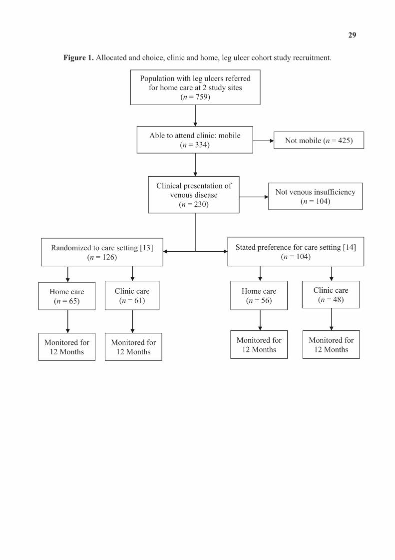
29
Figure 1. Allocated and choice, clinic and home, leg ulcer cohort study recruitment.
Population with leg ulcers referred for home care at 2 study sites
(n = 759)
Clinic care (n = 48)
Not mobile (n = 425) Able to attend clinic: mobile (n = 334)
Clinical presentation of venous disease
(n = 230) Not venous insufficiency
(n = 104)
Stated preference for care setting [14] (n = 104)
Randomized to care setting [13] (n = 126)
Home care (n = 56)
Home care (n = 65)
Clinic care (n = 61)
Monitored for 12 Months
Monitored for 12 Months
Monitored for 12 Months
Monitored for 12 Months
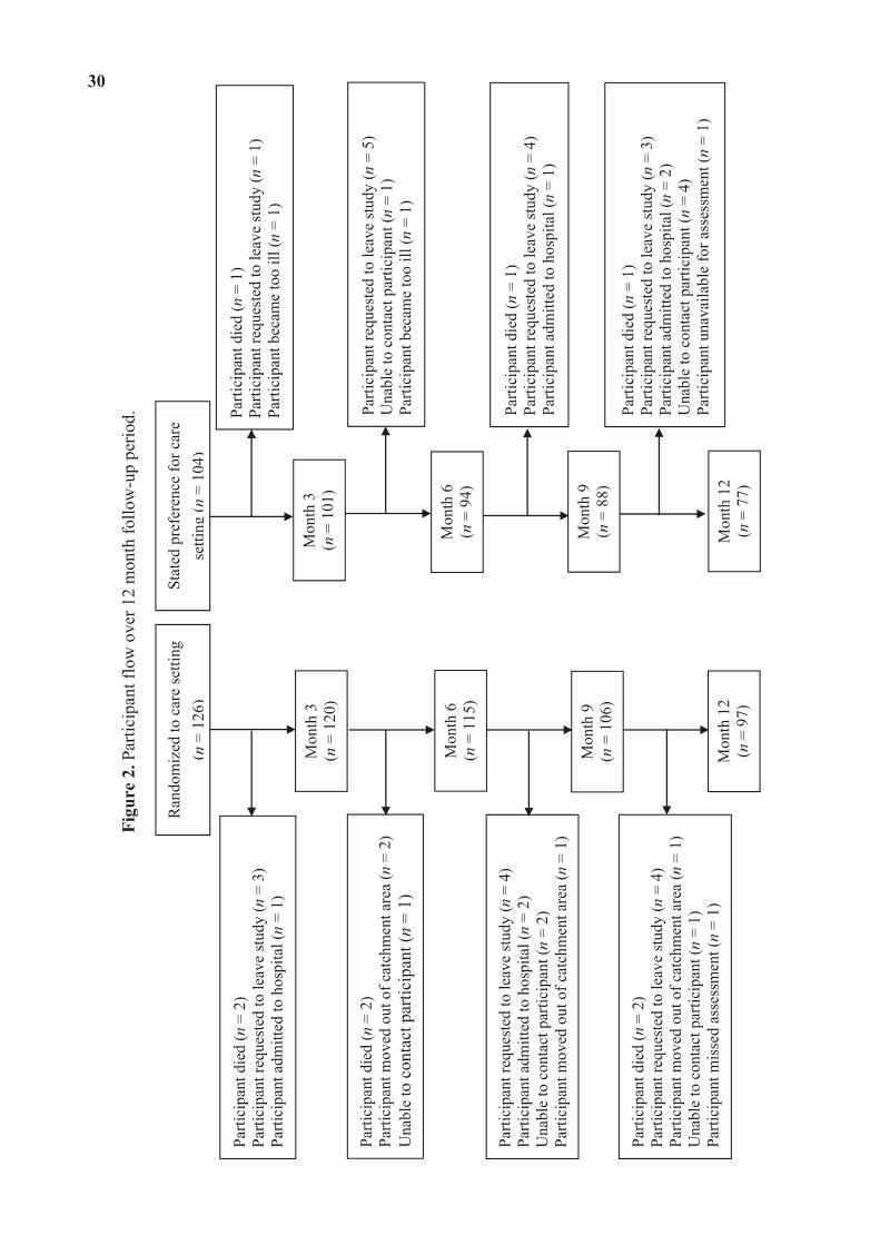
30
Figu
re 2
. Par
ticip
ant f
low
ove
r 12
mon
th fo
llow
-up
perio
d.
Ran
dom
ized
to c
are
setti
ng
(n=
126)
Stat
ed p
refe
renc
e fo
r car
e se
ttin g
(n=
104)
Mon
th 3
(n
= 1
20)
Parti
cipa
nt d
ied
(n =
2)
Parti
cipa
nt re
ques
ted
to le
ave
stud
y (n
= 3
) Pa
rtici
pant
adm
itted
to h
ospi
tal (
n =
1)
Parti
cipa
nt d
ied
(n =
1)
Parti
cipa
nt re
ques
ted
to le
ave
stud
y (n
= 1
) Pa
rtici
pant
bec
ame
too
ill (n
= 1
)
Mon
th 3
(n
= 1
01)
Mon
th 6
(n
= 1
15)
Mon
th 9
(n
= 1
06)
Mon
th 1
2 (n
= 9
7)
Parti
cipa
nt d
ied
(n =
2)
Parti
cipa
nt m
oved
out
of c
atch
men
t are
a (n
= 2
) U
nabl
e t o
con
tact
par
ticip
ant (
n =
1)
Parti
cipa
nt re
ques
ted
to le
ave
stud
y (n
= 5
) U
nabl
e to
con
tact
par
ticip
ant (
n =
1)
Parti
cipa
nt b
ecam
e to
o ill
(n =
1)
Parti
cipa
nt re
ques
ted
to le
ave
stud
y (n
= 4
) Pa
rtici
pant
adm
itted
to h
ospi
tal (
n =
2)
Una
ble
to c
onta
ct p
artic
ipan
t (n
= 2)
Pa
rtici
pant
mov
ed o
ut o
f cat
chm
ent a
rea
(n =
1)
Mon
th 6
(n
= 9
4)
Parti
cipa
nt d
ied
(n =
1)
Parti
cipa
nt re
ques
ted
to le
ave
stud
y (n
= 4
) Pa
rtici
pant
adm
itted
to h
ospi
tal (
n =
1)
Mon
th 9
(n
= 8
8)
Mon
th 1
2 (n
= 7
7)
Parti
cipa
nt d
ied
(n =
2)
Parti
cipa
nt re
ques
ted
to le
ave
stud
y (n
= 4
) Pa
rtici
pant
mov
ed o
ut o
f cat
chm
ent a
rea
(n =
1)
Una
ble
to c
onta
ct p
artic
ipan
t (n
= 1)
Pa
rtici
pant
mis
sed
asse
ssm
ent (
n =
1)
Parti
cipa
nt d
ied
(n =
1)
Parti
cipa
nt re
ques
ted
to le
ave
stud
y (n
= 3
) Pa
rtici
pant
adm
itted
to h
ospi
tal (
n =
2)
Una
ble
to c
onta
ct p
artic
ipan
t (n
= 4)
Pa
rtici
pant
una
vaila
ble
for a
sses
smen
t (n
= 1)
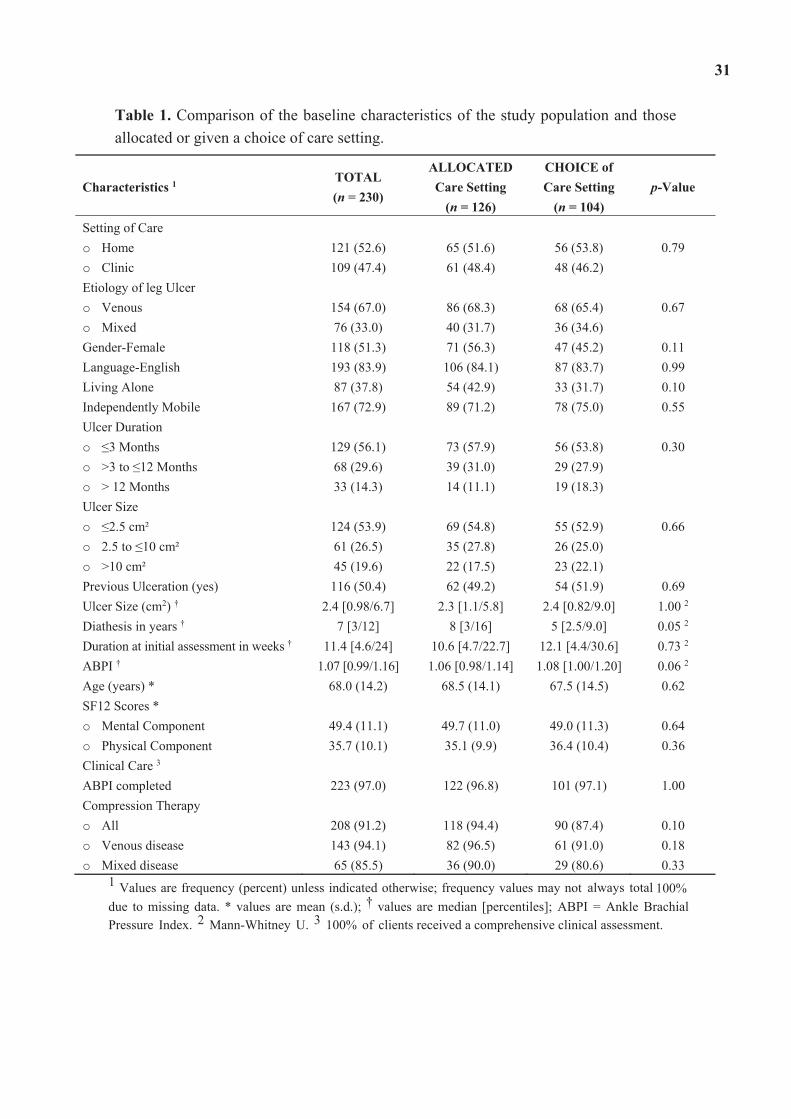
31
Table 1. Comparison of the baseline characteristics of the study population and those allocated or given a choice of care setting.
Characteristics 1 TOTAL (n = 230)
ALLOCATED Care Setting
(n = 126)
CHOICE of Care Setting
(n = 104) p-Value
Setting of Care o Home 121 (52.6) 65 (51.6) 56 (53.8) 0.79 o Clinic 109 (47.4) 61 (48.4) 48 (46.2) Etiology of leg Ulcer o Venous 154 (67.0) 86 (68.3) 68 (65.4) 0.67 o Mixed 76 (33.0) 40 (31.7) 36 (34.6) Gender-Female 118 (51.3) 71 (56.3) 47 (45.2) 0.11 Language-English 193 (83.9) 106 (84.1) 87 (83.7) 0.99 Living Alone 87 (37.8) 54 (42.9) 33 (31.7) 0.10 Independently Mobile 167 (72.9) 89 (71.2) 78 (75.0) 0.55 Ulcer Duration o 3 Monthso >3 to 12 Monthso > 12 Months
129 (56.1) 68 (29.6) 33 (14.3)
73 (57.9) 39 (31.0) 14 (11.1)
56 (53.8) 29 (27.9) 19 (18.3)
0.30
Ulcer Size o 2.5 cm²o 2.5 to 10 cm²o >10 cm²
124 (53.9) 61 (26.5) 45 (19.6)
69 (54.8) 35 (27.8) 22 (17.5)
55 (52.9) 26 (25.0) 23 (22.1)
0.66
Previous Ulceration (yes) 116 (50.4) 62 (49.2) 54 (51.9) 0.69 Ulcer Size (cm2) † 2.4 [0.98/6.7] 2.3 [1.1/5.8] 2.4 [0.82/9.0] 1.00 2
Diathesis in years † 7 [3/12] 8 [3/16] 5 [2.5/9.0] 0.05 2
Duration at initial assessment in weeks † 11.4 [4.6/24] 10.6 [4.7/22.7] 12.1 [4.4/30.6] 0.73 2
ABPI † 1.07 [0.99/1.16] 1.06 [0.98/1.14] 1.08 [1.00/1.20] 0.06 2
Age (years) * 68.0 (14.2) 68.5 (14.1) 67.5 (14.5) 0.62 SF12 Scores * o Mental Component 49.4 (11.1) 49.7 (11.0) 49.0 (11.3) 0.64 o Physical Component 35.7 (10.1) 35.1 (9.9) 36.4 (10.4) 0.36 Clinical Care 3
ABPI completed 223 (97.0) 122 (96.8) 101 (97.1) 1.00 Compression Therapy o All 208 (91.2) 118 (94.4) 90 (87.4) 0.10 o Venous disease 143 (94.1) 82 (96.5) 61 (91.0) 0.18 o Mixed disease 65 (85.5) 36 (90.0) 29 (80.6) 0.33
1 Values are frequency (percent) unless indicated otherwise; frequency values may not always total 100% due to missing data. * values are mean (s.d.); † values are median [percentiles]; ABPI = Ankle Brachial Pressure Index. 2 Mann-Whitney U. 3 100% of clients received a comprehensive clinical assessment.
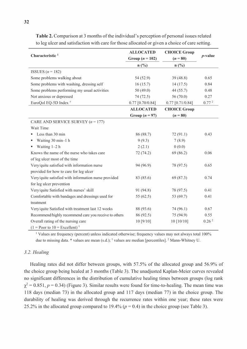
32
Table 2. Comparison at 3 months of the individual’s perception of personal issues related to leg ulcer and satisfaction with care for those allocated or given a choice of care setting.
Characteristic 1ALLOCATED
Group (n = 102) CHOICE Group
(n = 80) p-value
n (%) n (%) ISSUES (n = 182) Some problems walking about 54 (52.9) 39 (48.8) 0.65 Some problems with washing, dressing self 16 (15.7) 14 (17.5) 0.84 Some problems performing my usual activities 50 (49.0) 44 (55.7) 0.48 Not anxious or depressed 74 (72.5) 56 (70.0) 0.27 EuroQol EQ-5D Index † 0.77 [0.70/0.84] 0.77 [0.71/0.84] 0.77 2
ALLOCATEDGroup (n = 97)
CHOICE Group (n = 80)
CARE AND SERVICE SURVEY (n = 177) Wait Time
Less than 30 min 86 (88.7) 72 (91.1) 0.43 Waiting 30 min–1 h 9 (9.3) 7 (8.9) Waiting 1–2 h 2 (2.1) 0 (0.0)
Knows the name of the nurse who takes care of leg ulcer most of the time
72 (74.2) 69 (86.2) 0.06
Very/quite satisfied with information nurse provided for how to care for leg ulcer
94 (96.9) 78 (97.5) 0.65
Very/quite satisfied with information nurse provided for leg ulcer prevention
83 (85.6) 69 (87.3) 0.74
Very/quite Satisfied with nurses’ skill 91 (94.8) 78 (97.5) 0.41 Comfortable with bandages and dressings used for treatment
55 (62.5) 53 (69.7) 0.41
Very/quite Satisfied with treatment last 12 weeks 88 (93.6) 74 (96.1) 0.67 Recommend/highly recommend care you receive to others 86 (92.5) 75 (94.9) 0.55 Overall rating of the nursing care (1 = Poor to 10 = Excellent) †
10 [9/10] 10 [10/10] 0.26 2
1 Values are frequency (percent) unless indicated otherwise; frequency values may not always total 100% due to missing data. * values are mean (s.d.); † values are median [percentiles]. 2 Mann-Whitney U.
3.2. Healing
Healing rates did not differ between groups, with 57.5% of the allocated group and 56.9% of the choice group being healed at 3 months (Table 3). The unadjusted Kaplan-Meier curves revealed no significant differences in the distribution of cumulative healing times between groups (log rank
2 = 0.851, p = 0.34) (Figure 3). Similar results were found for time-to-healing. The mean time was 118 days (median 73) in the allocated group and 117 days (median 77) in the choice group. The durability of healing was derived through the recurrence rates within one year; these rates were 25.2% in the allocated group compared to 19.4% (p = 0.4) in the choice group (see Table 3).
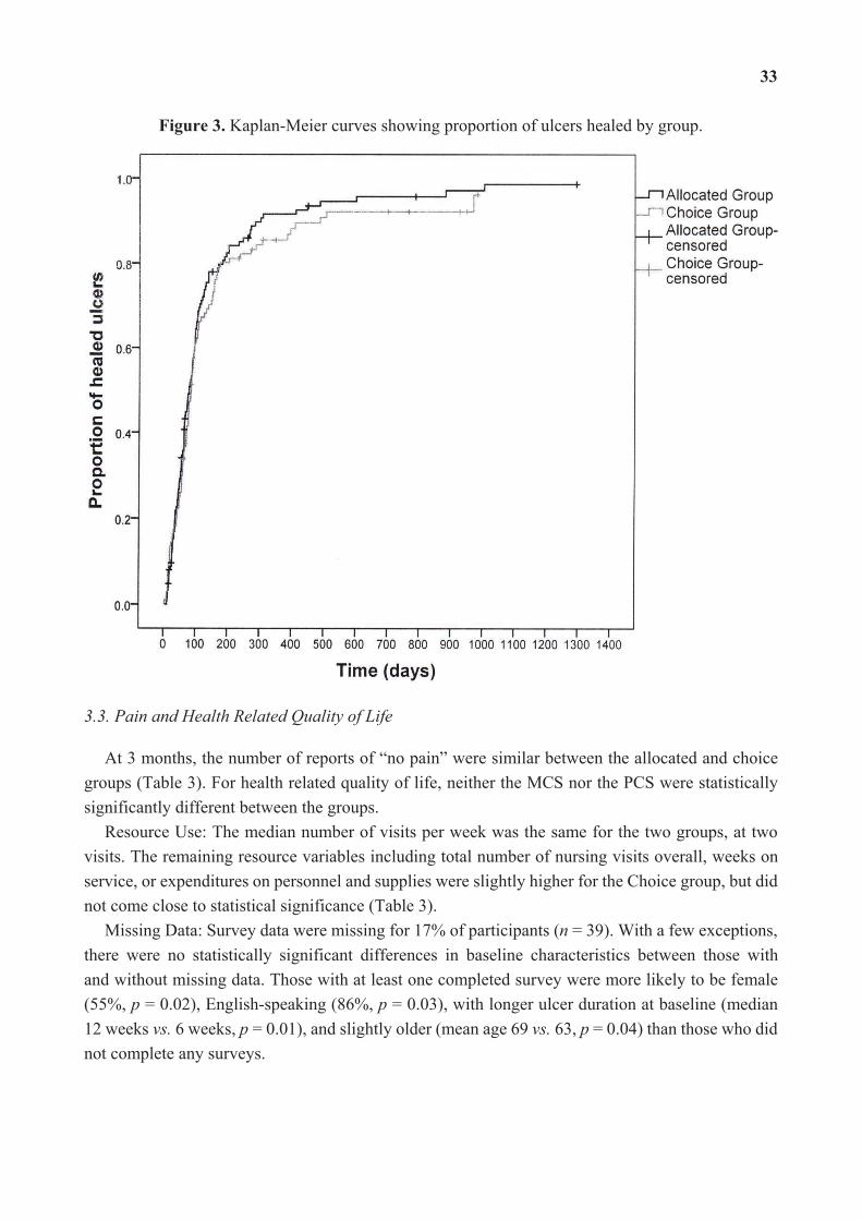
33
Figure 3. Kaplan-Meier curves showing proportion of ulcers healed by group.
3.3. Pain and Health Related Quality of Life
At 3 months, the number of reports of “no pain” were similar between the allocated and choice groups (Table 3). For health related quality of life, neither the MCS nor the PCS were statistically significantly different between the groups.
Resource Use: The median number of visits per week was the same for the two groups, at two visits. The remaining resource variables including total number of nursing visits overall, weeks on service, or expenditures on personnel and supplies were slightly higher for the Choice group, but did not come close to statistical significance (Table 3).
Missing Data: Survey data were missing for 17% of participants (n = 39). With a few exceptions, there were no statistically significant differences in baseline characteristics between those with and without missing data. Those with at least one completed survey were more likely to be female (55%, p = 0.02), English-speaking (86%, p = 0.03), with longer ulcer duration at baseline (median 12 weeks vs. 6 weeks, p = 0.01), and slightly older (mean age 69 vs. 63, p = 0.04) than those who did not complete any surveys.
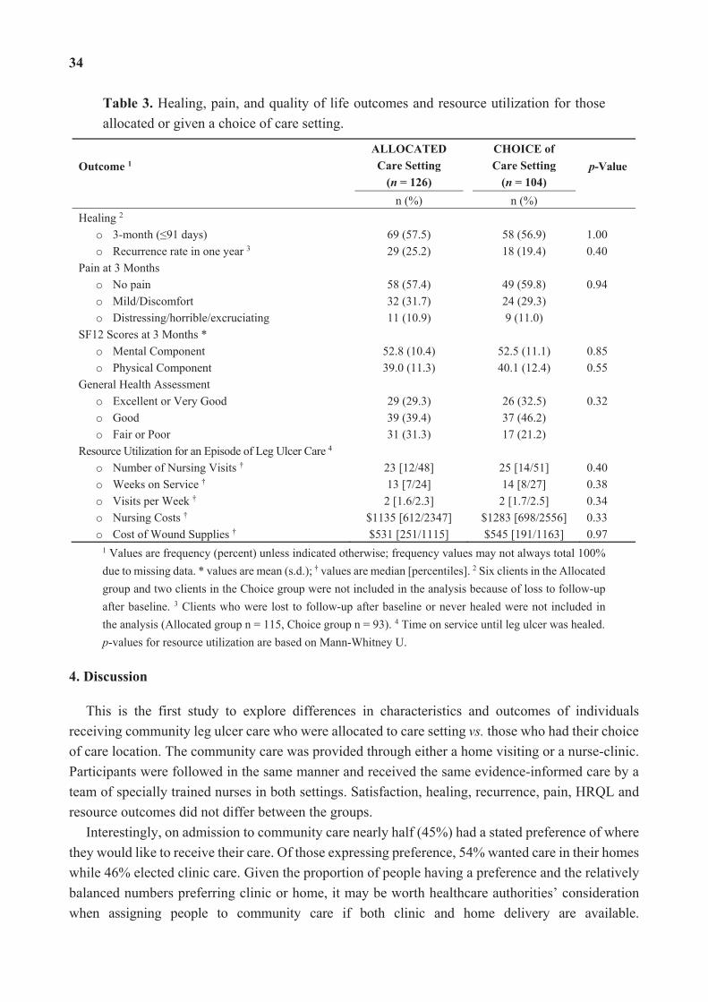
34
Table 3. Healing, pain, and quality of life outcomes and resource utilization for those allocated or given a choice of care setting.
Outcome 1ALLOCATED Care Setting
(n = 126)
CHOICE of Care Setting
(n = 104) p-Value
n (%) n (%)Healing 2
o 3-month ( 91 days)
69 (57.5)
58 (56.9) 1.00
o Recurrence rate in one year 3 29 (25.2) 18 (19.4) 0.40 Pain at 3 Months
o No pain
58 (57.4)
49 (59.8) 0.94
o Mild/Discomfort 32 (31.7) 24 (29.3) o Distressing/horrible/excruciating 11 (10.9) 9 (11.0)
SF12 Scores at 3 Months * o Mental Component 52.8 (10.4) 52.5 (11.1) 0.85 o Physical Component 39.0 (11.3) 40.1 (12.4) 0.55
General Health Assessment o Excellent or Very Good 29 (29.3) 26 (32.5) 0.32 o Good 39 (39.4) 37 (46.2) o Fair or Poor 31 (31.3) 17 (21.2)
Resource Utilization for an Episode of Leg Ulcer Care 4 o Number of Nursing Visits † 23 [12/48] 25 [14/51] 0.40 o Weeks on Service † 13 [7/24] 14 [8/27] 0.38 o Visits per Week † 2 [1.6/2.3] 2 [1.7/2.5] 0.34 o Nursing Costs † $1135 [612/2347] $1283 [698/2556] 0.33 o Cost of Wound Supplies † $531 [251/1115] $545 [191/1163] 0.97
1 Values are frequency (percent) unless indicated otherwise; frequency values may not always total 100% due to missing data. * values are mean (s.d.); † values are median [percentiles]. 2 Six clients in the Allocated group and two clients in the Choice group were not included in the analysis because of loss to follow-up after baseline. 3 Clients who were lost to follow-up after baseline or never healed were not included in the analysis (Allocated group n = 115, Choice group n = 93). 4 Time on service until leg ulcer was healed. p-values for resource utilization are based on Mann-Whitney U.
4. Discussion
This is the first study to explore differences in characteristics and outcomes of individuals receiving community leg ulcer care who were allocated to care setting vs. those who had their choice of care location. The community care was provided through either a home visiting or a nurse-clinic. Participants were followed in the same manner and received the same evidence-informed care by a team of specially trained nurses in both settings. Satisfaction, healing, recurrence, pain, HRQL and resource outcomes did not differ between the groups.
Interestingly, on admission to community care nearly half (45%) had a stated preference of where they would like to receive their care. Of those expressing preference, 54% wanted care in their homes while 46% elected clinic care. Given the proportion of people having a preference and the relatively balanced numbers preferring clinic or home, it may be worth healthcare authorities’ consideration when assigning people to community care if both clinic and home delivery are available.
35
Although RCTs are thought to not represent target populations generally, this study seems to refute that argument.
Satisfaction with care, healing, recurrence, pain, and HRQL outcomes were similar between the allocated and choice groups. At baseline, the groups showed some borderline differences in diatheses and ABPI scores. This might impact the results although in our experience an ABPI of 1.06 compared to 1.08 would not be considered clinically important for decision-making. In comparison to what has been reported in the literature, healing rates in this cohort were relatively high. This likely is attributed to consistency in the delivery of evidence-informed care by trained nurses.
When comparing results from our randomized trial on clinic vs. home delivery [13] to the choice cohort study [14], those who chose the clinic setting for their care fared somewhat better than those who were allocated (67% compared to 58% healed by 3 months), whereas those who were allocated to receive their care at home fared slightly better than those who stated a preference for homecare (57% compared to 48% healed by 3 months), though these differences were not significant. One might contemplate based on the above, that people who choose clinic are more mobile/healthy while those who choose home are less mobile/more co-morbidities. We did not see a significant difference in these characteristics at baseline. However, the allocated group had slightly larger ulcers with longer duration and even though not statistically significant, may have been clinically relevant with their choice of setting. Clearly larger studies are needed to explore the interaction between choice and setting if this is an important issue for individuals, providers and health systems.
These results have several implications for individuals with leg ulcers, their families, health service planners and decision-makers. Given outcomes are similar with respect to satisfaction with care, healing, recurrence, pain, HRQL, resource use, whether people were allocated or had their stated their preference for care in a home or clinic setting, it seems reasonable that planners and decision-makers can feel confident in offering a choice of their care setting. The caveat here of course is that expectation of similar outcomes will take place in the context of delivery using an evidence-informed approach by trained health care providers. Similarly, if given the choice, individuals and their families can feel confident that the venue they choose for their care will not have a negative impact on their outcomes.
Leg ulcer management in the home and clinic settings each come with their own set of advantages and disadvantages. Supplies are stocked and readily available in the clinic, whereas there may be a delay in receiving supplies ordered to the home. In clinic, only supplies required for an episode of treatment can be utilized whereas in the home, returning unused supplies may not be viable due to infection control issues. Individuals who receive their care at the clinic can be given a specific appointment time. However, treatment at home might be optimal for those with mobility issues or needing to care for a spouse or family member at home. In the clinic, nurses may have an advantage of better coordination with family physicians or specialists because of access to electronic resources (i.e., computer, email, fax machine), whilst in the home setting, nurses may have a better sense of a client’s lifestyle requirements and can tailor their treatment accordingly. For health service providers, client accessibility, protocols for infection control, and overhead costs are issues that need to be considered in terms of clinic care while travel time and fuel costs are important factors to consider with home delivery.
36
With increasing pressure on homecare resources and nursing hours considered a scarce resource, access to clinic care for individuals who are mobile or with good transportation support, could be an important consideration by authorities and health service providers. In the Canadian context, factors such as the size and distribution of the population regionally, varying urban-rural mix, and vastly varying climate from one region to another may make offering choice of clinic or home delivery not feasible. However, knowing that the quality of care delivered in either setting can result in similar outcomes should be reassuring.
5. Limitations of the Study
This study—a combined analysis of data from an RCT and Cohort study of those who wereallocated and chose their care setting, respectively—has some limitations to take into consideration. First, inclusion criteria for the overall study was the ability to be able to attend a clinic for care; therefore, the study participants may not be representative of the population receiving community leg ulcer care given their mobility. Both the RCT and Cohort Study were conducted in two south-easternOntario health regions and may not be representative of a broader geographic region.
However, they did represent an urban rural mix and typical characteristics of a leg ulcer population. Nurses providing care were involved in the collection of data. Blinding the nurse to the setting
was not possible. Conversely, outcomes were assessed in a rigorous and consistent manner by a small team of devoted specially trained nurses regardless of setting. Finally, this study was conducted as a combined analysis of common indicators of community leg ulcer care (satisfaction, healing, HRQL, resource use) and was not powered a priori to determine factors associated with choice and receiving care in either of the settings, nor was it a randomized controlled design focused on preference, thus readers are cautioned when drawing conclusions from this study. However, the results do provide a useful overview of this patient population and will be useful to those planning a larger future study. An important element for a future study would be accurate accounting of travel time incurred by nurses delivering homecare and the overhead expenditures of the nurse clinics.
6. Conclusions
This study examined outcomes of groups who were allocated to a setting of care (communityclinic or home setting), or who had the choice of setting. In spite of the above limitations, it is our belief that if available, individuals should have an option of care venue where possible given that almost half of those approached (45%) indicated a clear preference for clinic or home. Besides our previous work where the Choice cohort [14] was examined separately from the Allocated group (RCT) [13], there have been no published studies examining the interaction between choice and setting of community care for people with leg ulcers. For planners and decision makers, taking into account the population profile, local contextual factors, resource availability, and patient perspective will aid in making the appropriate health-services decision.
Our hope is that these analyses can provide a foundation for further research in this area.
37
Acknowledgments
The authors would like to thank the health services wound trial group for making this enquiry possible. Their dedication to the quality of data collection with the original trials and cohort data was unfaltering. We are also indebted to the many individuals with leg ulcers who gave generously of their time to participate in these studies.
Author Contributions
M.B.H. was Principal Investigator on the wound trials from which the data for this study was extracted. M.B.H., E.V.K. and W.M.H. were responsible for the conceptualization, conduct and management of the cohort study secondary analyses and interpretation of the results. M.E.C. was responsible for data management and contributed to the conceptualization and conduct of the analysis. All authors contributed to drafting of the manuscript and have read and approved the final manuscript.
Conflicts of Interest
The authors declare no conflict of interest.
References
1. Cullum, N.; Nelson, E.A.; Fletcher, A.W.; Sheldon, T.A. Compression for venous leg ulcers. Cochrane Database Syst. Rev. 2000, doi:10.1002/14651858.CD000265.
2. Harrison, M.B.; VanDenKerkhof, E.G.; Hopman, W.M.; Graham, I.D.; Carley, M.E.; Nelson, E.A. The Canadian Bandaging Trial: Evidence-informed leg ulcer care and the effectiveness of two compression technologies. BMC Nurs. 2011, 10, e20.
3. National Health Services Centre for Reviews and Dissemination. Compression therapy for venous leg ulcers. Eff. Health Care Bull. 1997, 3, 1–12.
4. Nelson, E.A.; Iglesias, C.P.; Cullum, N.; Torgerson, D.J. Randomized clinical trial of four-layer and short-stretch compression bandages for venous leg ulcers (VenUS I). Br. J. Surg. 2004, 91, 1292–1299.
5. Castineira, F.; Fisher, H.; Coleman, D.; Grace, P.A.; Burke, P. The limerick leg ulcer project: Early results. Ir. J. Med. Sci. 1999, 168, 17–20.
6. Lambourne, L.A.; Moffatt, C.J.; Jones, A.C.; Dorman, M.C.; Franks, P.J. Clinical audit and effective change in leg ulcer services. J. Wound Care 1996, 5, 348–351.
7. Moffatt, C.J.; Franks, P.J.; Oldroyd, M.; Bosanquet, N.; Brown, P.; Greenhalgh, R.M.; McCollum, C.N. Community clinics for leg ulcers and impact on healing. Br. Med. J. 1992, 305, 1389–1392.
8. Moffatt, C.J.; Oldroyd, M.I. A pioneering service to the community. The Riverside Community Leg Ulcer Project. Prof. Nurse 1994, 9, 486, 488, 490 passim.
9. Morrell, C.J.; Walters, S.J.; Dixon, S. Cost effectiveness of community leg ulcer clinics: Randomised controlled trial. Br. Med. J. 1998, 316, 1487–1491.
38
10. Scriven, J.M.; Hartshorne, T.; Bell, P.R.F.; Naylor, A.R.; London, N.J.M. Single-visit venous ulcer assessment clinic: The first year. Br. J. Surg. 1997, 84, 334–336.
11. Simon, D.A.; Freak, L.; Kinsella, A. Community leg ulcer clinics: A comparative study in two health authorities. Br. Med. J. 1996, 312, 1648–1651.
12. Stevens, J.; Franks, P.; Harrington, M. A community/hospital leg ulcer service. J. Wound Care 1997, 6, 62–68.
13. Harrison, M.B.; Graham, I.D.; Lorimer, K.; VanDenKerkhof, E.; Buchanan, M.; Wells, P.; Brandys, T.; Pierscianowski, T. Nurse clinic versus home delivery of evidence-based community leg ulcer care: A randomized health services trial. BMC Health Serv. Res. 2008, 8, e243.
14. Harrison, M.B.; VanDenKerkhof, E.; Hopman, W.M.; Graham, I.D.; Lorimer, K.; Carley, M. Evidence-informed leg ulcer care: A cohort study comparing outcomes of individuals choosing nurse-led clinic or home care. Ostomy Wound Manage. 2011, 57, 38–45.
15. Graham, I.D.; Harrison, M.D.; Brouwers, M. Evaluating and adapting practice guidelines for local use: A conceptual framework. In Clinical Governance in Practice; Pickering, S., Thompson, J., Eds.; Harcourt Publishers: London, UK, 2003; pp. 213–229.
16. Graham, I.D.; Lorimer, K.; Harrison, M.B.; Pierscianowski, T. Evaluating the quality of international clinical practice guidelines for leg ulcers: Preparing for a Canadian adaptation. Can. Assoc. Enterostomal Ther. J. 2000, 19, 15–31.
17. Graham, I.D.; Harrison, M.B. Evaluation and adaptation of clinical practice guidelines. Evid. Based Nurs. 2005, 8, 68–72.
18. Graham, I.D.; Harrison, M.B.; Shafey, M.; Keast, D. Knowledge and attitudes regarding care of leg ulcers. Survey of family physicians. Can. Fam. Physician 2003, 49, 896–902.
19. Graham, I.D.; Harrison, M.B.; Moffat, C.; Franks, P. Leg ulcer care: Nursing attitudes and knowledge. Can. Nurse 2001, 97, 19–24.
20. Graham, I.D.; Harrison, M.B.; Lorimer, K. Adapting national and international leg ulcer practice guidelines for local use: The Ontario Leg Ulcer Community Care Protocol. Adv. Skin Wound Care 2005, 18, 307–318.
21. Gilliss, C.L.; Kulkin, I.L. Monitoring nursing interventions and data collection in a randomized clinical trial. West. J. Nurs. Res. 1991, 13, 416–422.
22. Kirchhoff, K.T.; Dille, C.A. Issues in intervention research: Maintaining integrity. Appl. Nurs. Res. 1994, 7, 32–38.
23. Rabeneck, L.; Viscoli, C.M.; Horwitz, R.I. Problems in the conduct and analysis of randomized clinical trials. Are we getting the right answers to the wrong questions? Arch. Intern. Med. 1992, 152, 507–512.
24. Nemeth, K.A.; Graham, I.D.; Harrison, M.B. The measurement of leg ulcer pain: Identification and appraisal of pain assessment tools. Adv. Skin Wound Care 2003, 16, 260–267.
25. Nemeth, K.A.; Harrison, M.B.; Graham, I.D.; Burke, S. Pain in pure and mixed aetiology venous leg ulcers: A three-phase point prevalence study. J. Wound Care 2003, 12, 336–340.
26. Walters, S.J.; Morell, C.J.; Dixon, S. Measuring health-related quality of life in patients with venous leg ulcers. Qual. Life Res. 1999, 8, 327–336.
39
27. Melzack, R. The McGill Pain Questionnaire: Major properties and scoring methods. Pain 1975, 1, 277–299.
28. Melzack, R.; Katz, J. The McGill Pain Questionnaire: Appraisal and current status. In Handbook of Pain Assessment; Guilford Press: New York, NY, USA, 1992; pp. 152–168.
29. Melzack, R. The short-form McGill Pain Questionnaire. Pain 1987, 30, 191–197. 30. Ware, J.E.; Kosinski, M.; Keller, S.D. SF-12: How to Score the SF-12 Physical and Mental
Health Summary Scales, 2nd ed.; The Health Institute, New England Medical Centre: Boston, MA, USA, 1995.
31. Hopman, W.M.; Towheed, T.; Anastassiades, T. Canadian normative data for the SF-36 health survey. Can. Med. Assoc. J. 2000, 163, 265–271.
32. The EuroQol Group. EuroQol—A new facility for the measurement of health-related quality of life. Health Policy 1990, 16, 199–208.
33. Brooks, R. EuroQol: The current state of play. Health Policy 1996, 37, 53–72. 34. Bansback, N.; Tsuchiya, A.; Brazier, J.; Anis, A. Canadian valuation of EQ-5D health states:
Preliminary value set and considerations for future valuation studies. PLoS One 2012, 7, e31115.
40
Sustaining Behavior Changes Following a Venous Leg Ulcer Client Education Program
Charne Miller, Suzanne Kapp and Lisa Donohue
Abstract: Venous leg ulcers are a symptom of chronic insufficiency of the veins. This study considered the sustainability of behavior changes arising from a client focus e-Learning education program called the “Leg Ulcer Prevention Program” (LUPP) for people with a venous leg ulcer. Data from two related studies were used to enable a single sample (n = 49) examination of behavior maintenance across an average 8 to 9 months period. Physical activity levels increased over time. Leg elevation, calf muscle exercises, and soap substitute use were seen to fluctuate over the follow up time points. The use of a moisturizer showed gradual decline over time. The provision of a client-focused venous leg ulcer program was associated with behavior changes that had varied sustainability across the evaluation period.
Reprinted from Healthcare. Cite as: Miller, C.; Kapp, S.; Donohue, L. Sustaining Behavior Changes Following a Venous Leg Ulcer Client Education Program. Healthcare 2014, 2, 324-337.
1. Introduction
1.1. Venous Leg Ulcers: A Common and Costly Wound
Approximately 70% of all lower leg ulcers are venous leg wounds [1]. Venous leg ulcers affect 0.1%–1.1% of people [2], with a prevalence of up to 3% for people aged over 60 years [1]. It is a condition associated with significant morbidity and impact on quality of life [3–11]. It has been estimated to cost $3 billion Australian dollars annually [12].
1.2. A Chronic Condition, a Complex Treatment
Venous leg ulcers are a symptom of peripheral vascular disease, and more specifically of chronic venous insufficiency. Parallels can be readily drawn between chronic venous insufficiency and characteristics of other chronic diseases as this condition is typically caused by multiple factors associated with a person’s genetics, environment and lifestyle [13]. Consistent with the profile of chronic disease [13], venous leg ulcers may have complex causality, multiple risk factors and cause functional disability. These wounds can take an extended time to heal and there is a high recurrence rate. For older people who have comorbid conditions, corrective surgery to fix the underlying problem with the veins is not usually a treatment option.
The current best practice treatment of venous disease involves the use of compression therapy in conjunction with optimizing the conduct of a number of lifestyle behaviors. Standards for venous leg ulcer management and prevention [14] advocate the use of the highest level of compression therapy tolerable, regular activity, leg elevation during periods of inactivity, heel raises and squats to strengthen the calf muscle, a healthy diet, adequate “good” fluid intake, and the use of a moisturizer and soap substitute to promote skin hydration. These treatments apply not only when an individual
41
has a venous leg ulcer but also post-healing in order to prevent the ulcer from recurring. With a recurrence rate of 69% [15], repeated episodes of care is a significant issue for those affected and health professionals engaged in the wound management field.
Concordance with the recommended treatment and healthy behaviors is for people with venous disease, alike other chronic conditions, not always well adopted or adhered to [16,17]. A number of programs encouraging effective self-management including leg clubs [18], the “Lively Legs” Program [19,20], and other e-Learning initiatives [21] have been developed for use in venous leg ulcer care and prevention to optimize the uptake of recommended strategies and these have been shown to be acceptable to clients [21–23] and nurses [24]. While these programs have improved knowledge, physical activity, skin care, and compression use, no program has been effective in all of the targeted domains [19,21,23]. Although important efforts have been made by client education innovators in the wound management field, self-management research and innovation in the wound management field is still in its infancy when compared to other clinical conditions. Transferring knowledge gained in other clinical areas can assist the wound management field [25]. There are however gaps in knowledge that effect all clinical areas. Two particular gaps to be considered by this paper is the need to further understand the efficacy of interventions that promote multiple behavior changes and how these behaviors changes can be maintained after the intervention is finished.
Historically, the policy and evidence base regarding self-management programs has focused on changes for a single condition [26–28]. What is increasingly recognized is that programs that target multiple behavior changes are necessary not only to adequately address management of the chronic condition but due to individuals having multiple morbidities [28]. It is common for people, especially later in life, to have multiple chronic health conditions. Almost half (49%) of Australians living in the community aged 65–74 years have five or more long-term conditions, a rate which increases to 70% for people aged 85 years and over [29]. Many of these chronic conditions benefit from the same healthy lifestyle behaviors such as being physically active, drinking “good” fluids, and eating a nutritionally complete diet. As such, a management plan that considers the collective sum of self-management practices can consolidate recommendations that might otherwise overwhelm. Indeed, it has been suggested that a cognitive representation of illness/es and their management enables adaptions and inclusions arising from shifting health priorities [28]. That is, an integrated but broad understanding of the disease function and how healthy behaviors facilitate the management of the disease can help the individual accommodate variation arising from life or health events that optimizes their effective management of multiple morbidities. As the salience of venous disease management can be lessened as time since healing increases and in the presence of other life threatening diseases, transferences across other morbidities may be one approach wound clinicians use to optimize self-management practices.
The effectiveness of strategies to target multiple behavior change is still being unraveled. Behavior changes in multiple domains have been described as complementary or compensatory [30]. On one hand there could be competing effects between health behaviors where gain in one domain is at the cost of losses in another area [31]. In a study of a multiple component intervention including weight management, activity, stress management, smoking and social support intervention for women this issue was examined and was not found to be the case [31]. Alternatively, by spreading
42
the focus on multiple behaviors the potential gains are diluted [31]. Certainly, the perceived incongruence between recommendations for people with venous disease to be physical active as well as to rest and elevate their leg has been noted as undermining the conduct of one or both of these behaviors [23,32,33]. There is, on the other hand, quite a few studies to support the suggestion that behavior change in one domain can in fact enable change in another [34–38]. Systematic review evidence of the effectiveness of multiple behavior change interventions suggest positive outcomes can be achieved but these are rarely uniform across the behavior domains being targeted [39–41]. More research is required to identify the circumstances in which multiple component interventions are effective.
Another challenge for the field is the absence of research considering the maintenance of behavior changes. In 2012, the results of a randomized controlled trial assessing the effectiveness of the “Lively Legs” program [20] for with people with primarily venous leg ulcers was published which assessed the performance of physical activity, use of compression therapy and wound progress over an 18 months period [19]. Participants in the “Lively Legs” program were going on more frequent short walks, engaged in leg exercises, and had better wound healing, with no difference observed in compression use and in longer duration walking. It is both exciting and promising that some behavior change was achieved, sustained, and the research team implemented such a longer term follow up of study participants.
In 2011, a systematic review was published that considered maintenance that was achieved in 157 studies assessing the effects of a dietary and/or physical activity intervention [42]. Only 35% reported maintenance outcomes up to three months, and only 10% up to one year, although of these studies maintenance rates were promising with 72% achieving maintenance [42]. Of the 29 studies that met the eligibility criteria, the range of health conditions was considerable including healthy adults, type 2 diabetes mellitus, osteoarthritis, chronic back pain, breast cancer and coronary heart disease. The study found that the only sample characteristic that influenced maintenance of a dietary and/or physical activity intervention was gender. That is, studies targeting women revealed less effective maintenance of behavior change. No differences in the degree of maintenance achieved with programs targeting healthy adults and programs targeting people with a chronic health condition [42].
More generally, this “decay of impact” [43,44] whilst not universal, is considered to be the norm [31,44–46]. To advance how maintenance of behavior change is understood and how health professionals can most effectively empower people to self-manage their chronic condition/s it has been suggested that maintenance be conceptualized as a process and quite possibility a separate psychological process to behavior change [46]. Support of this latter contention emerges from a review of a physical activity program among cardiac patients that found actions plans were more influential during the early rehabilitation process, while coping plans were more instrumental as rehabilitation progressed [47]. Alike chronic disease inquiry in general, the longevity of behavior changes arising from venous leg ulcer self-management programs is an area in need of investigation.
In 2007, clinicians employed by Royal District Nursing Service (RDNS), a community nursing and health provider in Australia, provided the impetus for developing the Leg Ulcer Prevention Program (LUPP). LUPP was conceived in the belief that better outcomes for clients with leg ulcers could be facilitated by innovative, structured and standardized client education and that strategies to
43
increase client involvement in their management were warranted. A pre- and post-evaluation of the LUPP education showed that client knowledge, skin care, physical activity and compression therapy use increased after the education, and that nurses and clients expressed high satisfaction with the program [19,21]. The aim of this investigation was to ascertain the sustainability of behavior changes arising from this client focused e-learning education program for people with a venous leg ulcer.
2. Methods
2.1. Design
To enable this analysis, data from two related studies were combined to enable a prospective single sample cohort study. The first study was the LUPP pilot study (n = 155) in which performance of a variety of health behaviors were evaluated before (pre) and after (post) the delivery of a six part e-learning program with clients of a community nursing service in Australia. All of these study participants had an active leg ulcer at the time of the study. Forty-nine of these participants upon healing, subsequently participated in a randomized controlled trial (RCT) (total n = 100) comparing wound recurrence associated with the use of a moderate and a high compression stocking. Evaluation of health behaviors amongst this sample occurred at baseline, and 13 weeks and 26 weeks after recruitment. Thus, in total there were five time points available for 49 participants in total to examine how sustainable the behavior changes associated with the LUPP education were over time.
By combining these two data sources that used the same measures at each time point, it became possible to examine the sustainability of behavior change for people who participated in both the preliminary LUPP pilot evaluation and the RCT. Though using RCT data this investigation is not an experimental study but rather a single sample longitudinal appraisal of health behavior maintenance.
2.2. The Leg Ulcer Prevention Program (LUPP)
LUPP is a standardized e-Learning client education package that delivers best practice recommendations for venous leg ulcer management. LUPP was developed by a multidisciplinary team including wound management clinical nurse consultants, researchers, allied health practitioners, e-Learning experts, marketing specialists and a professional photographer. While educational experts were engaged to review the education structure, content and resources, the initiative itself was not based on a clearly defined theoretical framework. Six sessions are delivered to the client and carer where appropriate at usual wound treatment visits, ideally one session per week. The client and nurse view a multimedia presentation, of approximately 10 min in duration, on the nurse’s tablet computer and then review a summary sheet and complete an activity to reinforce the learning. The client is provided a written booklet of the complete program to keep. The program sessions review what venous disease is and why ulcers occur, venous leg ulcer treatment with compression bandaging, activity and exercise, skin care, nutrition and compression stockings for recurrence prevention (see Table 1).
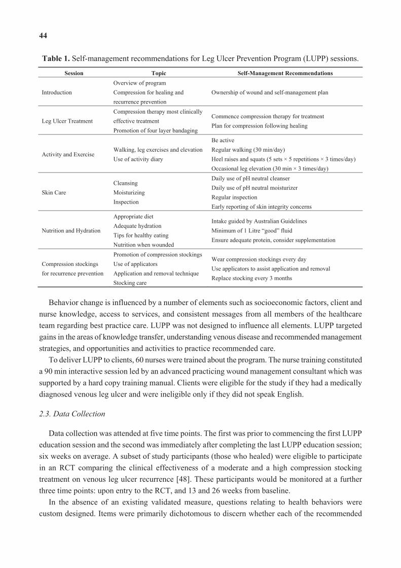
44
Table 1. Self-management recommendations for Leg Ulcer Prevention Program (LUPP) sessions.
Session Topic Self-Management Recommendations
Introduction Overview of program Compression for healing and recurrence prevention
Ownership of wound and self-management plan
Leg Ulcer Treatment Compression therapy most clinically effective treatment Promotion of four layer bandaging
Commence compression therapy for treatment Plan for compression following healing
Activity and Exercise Walking, leg exercises and elevation Use of activity diary
Be active Regular walking (30 min/day) Heel raises and squats (5 sets × 5 repetitions × 3 times/day) Occasional leg elevation (30 min × 3 times/day)
Skin Care Cleansing Moisturizing Inspection
Daily use of pH neutral cleanser Daily use of pH neutral moisturizer Regular inspection Early reporting of skin integrity concerns
Nutrition and Hydration
Appropriate diet Adequate hydration Tips for healthy eating Nutrition when wounded
Intake guided by Australian Guidelines Minimum of 1 Litre “good” fluid Ensure adequate protein, consider supplementation
Compression stockings for recurrence prevention
Promotion of compression stockings Use of applicators Application and removal technique Stocking care
Wear compression stockings every day Use applicators to assist application and removal Replace stocking every 3 months
Behavior change is influenced by a number of elements such as socioeconomic factors, client and nurse knowledge, access to services, and consistent messages from all members of the healthcare team regarding best practice care. LUPP was not designed to influence all elements. LUPP targeted gains in the areas of knowledge transfer, understanding venous disease and recommended management strategies, and opportunities and activities to practice recommended care.
To deliver LUPP to clients, 60 nurses were trained about the program. The nurse training constituted a 90 min interactive session led by an advanced practicing wound management consultant which was supported by a hard copy training manual. Clients were eligible for the study if they had a medically diagnosed venous leg ulcer and were ineligible only if they did not speak English.
2.3. Data Collection
Data collection was attended at five time points. The first was prior to commencing the first LUPP education session and the second was immediately after completing the last LUPP education session; six weeks on average. A subset of study participants (those who healed) were eligible to participate in an RCT comparing the clinical effectiveness of a moderate and a high compression stocking treatment on venous leg ulcer recurrence [48]. These participants would be monitored at a further three time points: upon entry to the RCT, and 13 and 26 weeks from baseline.
In the absence of an existing validated measure, questions relating to health behaviors were custom designed. Items were primarily dichotomous to discern whether each of the recommended
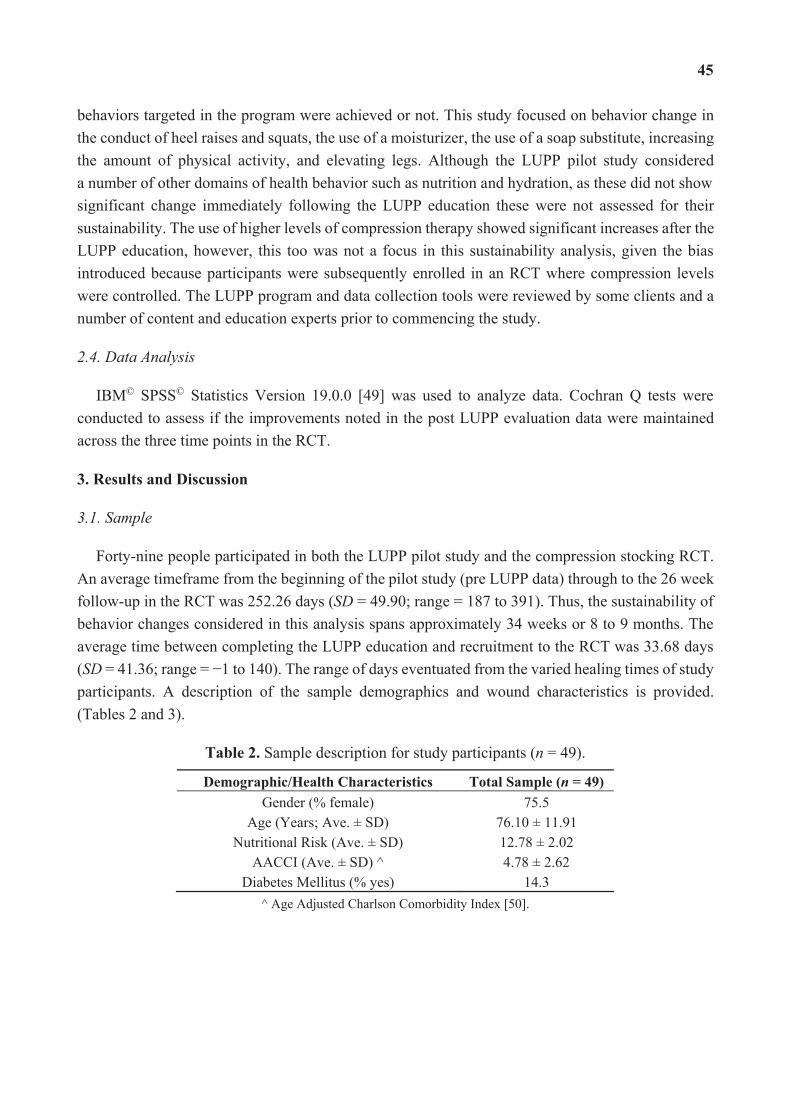
45
behaviors targeted in the program were achieved or not. This study focused on behavior change in the conduct of heel raises and squats, the use of a moisturizer, the use of a soap substitute, increasing the amount of physical activity, and elevating legs. Although the LUPP pilot study considered a number of other domains of health behavior such as nutrition and hydration, as these did not show significant change immediately following the LUPP education these were not assessed for their sustainability. The use of higher levels of compression therapy showed significant increases after the LUPP education, however, this too was not a focus in this sustainability analysis, given the bias introduced because participants were subsequently enrolled in an RCT where compression levels were controlled. The LUPP program and data collection tools were reviewed by some clients and a number of content and education experts prior to commencing the study.
2.4. Data Analysis
IBM© SPSS© Statistics Version 19.0.0 [49] was used to analyze data. Cochran Q tests were conducted to assess if the improvements noted in the post LUPP evaluation data were maintained across the three time points in the RCT.
3. Results and Discussion
3.1. Sample
Forty-nine people participated in both the LUPP pilot study and the compression stocking RCT. An average timeframe from the beginning of the pilot study (pre LUPP data) through to the 26 week follow-up in the RCT was 252.26 days (SD = 49.90; range = 187 to 391). Thus, the sustainability of behavior changes considered in this analysis spans approximately 34 weeks or 8 to 9 months. The average time between completing the LUPP education and recruitment to the RCT was 33.68 days (SD = 41.36; range = 1 to 140). The range of days eventuated from the varied healing times of study participants. A description of the sample demographics and wound characteristics is provided. (Tables 2 and 3).
Table 2. Sample description for study participants (n = 49).
Demographic/Health Characteristics Total Sample (n = 49) Gender (% female) 75.5
Age (Years; Ave. ± SD) 76.10 ± 11.91 Nutritional Risk (Ave. ± SD) 12.78 ± 2.02
AACCI (Ave. ± SD) ^ 4.78 ± 2.62 Diabetes Mellitus (% yes) 14.3
^ Age Adjusted Charlson Comorbidity Index [50].
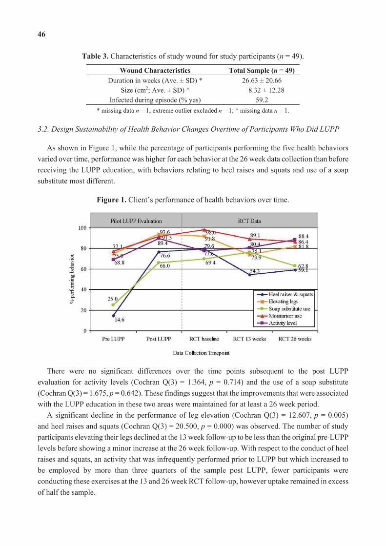
46
Table 3. Characteristics of study wound for study participants (n = 49).
Wound Characteristics Total Sample (n = 49) Duration in weeks (Ave. ± SD) * 26.63 ± 20.66
Size (cm2; Ave. ± SD) ^ 8.32 ± 12.28 Infected during episode (% yes) 59.2
* missing data n = 1; extreme outlier excluded n = 1; ^ missing data n = 1.
3.2. Design Sustainability of Health Behavior Changes Overtime of Participants Who Did LUPP
As shown in Figure 1, while the percentage of participants performing the five health behaviors varied over time, performance was higher for each behavior at the 26 week data collection than before receiving the LUPP education, with behaviors relating to heel raises and squats and use of a soap substitute most different.
Figure 1. Client’s performance of health behaviors over time.
There were no significant differences over the time points subsequent to the post LUPP evaluation for activity levels (Cochran Q(3) = 1.364, p = 0.714) and the use of a soap substitute (Cochran Q(3) = 1.675, p = 0.642). These findings suggest that the improvements that were associated with the LUPP education in these two areas were maintained for at least a 26 week period.
A significant decline in the performance of leg elevation (Cochran Q(3) = 12.607, p = 0.005) and heel raises and squats (Cochran Q(3) = 20.500, p = 0.000) was observed. The number of study participants elevating their legs declined at the 13 week follow-up to be less than the original pre-LUPP levels before showing a minor increase at the 26 week follow-up. With respect to the conduct of heel raises and squats, an activity that was infrequently performed prior to LUPP but which increased to be employed by more than three quarters of the sample post LUPP, fewer participants were conducting these exercises at the 13 and 26 week RCT follow-up, however uptake remained in excess of half the sample.
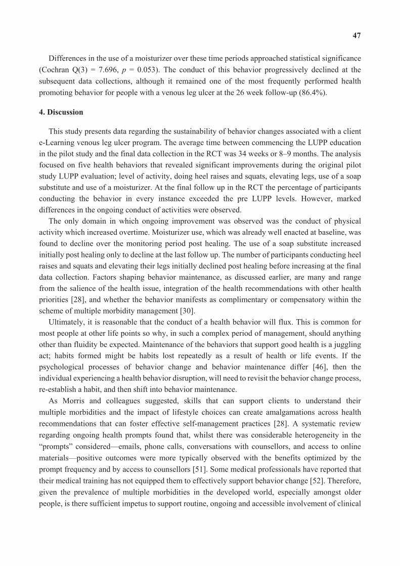
47
Differences in the use of a moisturizer over these time periods approached statistical significance (Cochran Q(3) = 7.696, p = 0.053). The conduct of this behavior progressively declined at the subsequent data collections, although it remained one of the most frequently performed health promoting behavior for people with a venous leg ulcer at the 26 week follow-up (86.4%).
4. Discussion
This study presents data regarding the sustainability of behavior changes associated with a client e-Learning venous leg ulcer program. The average time between commencing the LUPP education in the pilot study and the final data collection in the RCT was 34 weeks or 8–9 months. The analysis focused on five health behaviors that revealed significant improvements during the original pilot study LUPP evaluation; level of activity, doing heel raises and squats, elevating legs, use of a soap substitute and use of a moisturizer. At the final follow up in the RCT the percentage of participants conducting the behavior in every instance exceeded the pre LUPP levels. However, marked differences in the ongoing conduct of activities were observed.
The only domain in which ongoing improvement was observed was the conduct of physical activity which increased overtime. Moisturizer use, which was already well enacted at baseline, was found to decline over the monitoring period post healing. The use of a soap substitute increased initially post healing only to decline at the last follow up. The number of participants conducting heel raises and squats and elevating their legs initially declined post healing before increasing at the final data collection. Factors shaping behavior maintenance, as discussed earlier, are many and range from the salience of the health issue, integration of the health recommendations with other health priorities [28], and whether the behavior manifests as complimentary or compensatory within the scheme of multiple morbidity management [30].
Ultimately, it is reasonable that the conduct of a health behavior will flux. This is common for most people at other life points so why, in such a complex period of management, should anything other than fluidity be expected. Maintenance of the behaviors that support good health is a juggling act; habits formed might be habits lost repeatedly as a result of health or life events. If the psychological processes of behavior change and behavior maintenance differ [46], then the individual experiencing a health behavior disruption, will need to revisit the behavior change process, re-establish a habit, and then shift into behavior maintenance.
As Morris and colleagues suggested, skills that can support clients to understand their multiple morbidities and the impact of lifestyle choices can create amalgamations across health recommendations that can foster effective self-management practices [28]. A systematic review regarding ongoing health prompts found that, whilst there was considerable heterogeneity in the “prompts” considered—emails, phone calls, conversations with counsellors, and access to online materials—positive outcomes were more typically observed with the benefits optimized by the prompt frequency and by access to counsellors [51]. Some medical professionals have reported that their medical training has not equipped them to effectively support behavior change [52]. Therefore, given the prevalence of multiple morbidities in the developed world, especially amongst older people, is there sufficient impetus to support routine, ongoing and accessible involvement of clinical
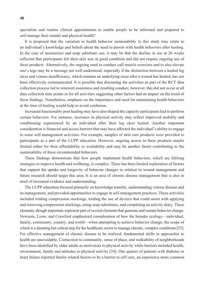
48
specialists and routine clinical appointments to enable people to be informed and prepared to self-manage their mental and physical health?
It is proposed that the variation in health behavior sustainability in this study may relate to an individual’s knowledge and beliefs about the need to persist with health behaviors after healing. In the case of moisturizer and soap substitute use, it may be that the decline in use at 26 weeks reflected that participants felt their skin was in good condition and did not require ongoing use of these products. Alternatively, the ongoing need to conduct calf muscle exercises and to also elevate one’s legs may be a message not well understood, especially if the distinction between a healed leg ulcer and venous insufficiency, which remains an underlying issue after a wound has healed, has not been effectively communicated. It is possible that discussing the activities as part of the RCT data collection process led to renewed awareness and resulting conduct, however, this did not occur at all data collection time points or for all activities suggesting other factors had an impact on the trend of these findings. Nonetheless, emphasis on the importance and need for maintaining health behaviors at the time of healing would help to avoid confusion.
Increased functionality post healing may have also shaped the capacity participants had to perform certain behaviors. For instance, increases in physical activity may reflect improved mobility and conditioning experienced by an individual after their leg ulcer healed. Another important consideration is financial and access barriers that may have affected the individual’s ability to engage in some self-management activities. For example, samples of skin care products were provided to participants as a part of the LUPP education. However, ongoing access to these products maybe limited either for their affordability or availability and may be another factor contributing to the sustainability of these recommended behaviors.
These findings demonstrate that how people implement health behaviors, which are lifelong strategies to improve health and wellbeing, is complex. There has been limited exploration of factors that support the uptake and longevity of behavior changes in relation to wound management and future research should target this area. It is an area of chronic disease management that is also in need of increased evidence and understanding.
The LUPP education focused primarily on knowledge transfer, understanding venous disease and its management, and provided opportunities to engage in self-management practices. These activities included trialing compression stockings, trialing the use of devices that could assist with applying and removing compression stockings, using soap substitutes, and completing an activity diary. These elements, though important, represent part of several elements that generate and sustain behavior change. Newsom, Lions, and Crawford emphasized consideration of how the broader ecology—individual, family, community, country, and world—when attempting to achieve behavior change; the scope of which is a daunting but critical step for the healthcare sector to manage chronic, complex conditions [53]. For effective management of chronic disease to be realized, fundamental shifts in approaches to health are unavoidable. Connection to community, sense of place, and walkability of neighborhoods have been identified by older adults as motivators to physical activity while barriers included health, environment, family and attitudes to physical activity [54]. One quarter of patients with diabetes or heart failure reported family-related factors to be a barrier to self care, an experience more common
49
amongst females [55]. Family barriers were further associated with lower self-efficacy and self-management adherence.
There are a number of limitations to this research study. First, the investigation was made possible by the opportunity presented by two related studies but was not the primary purpose of the study. As such, there are design features missing that would have been incorporated if this was the primary purpose, including more standardized data collection timeframes in between the completion of the LUPP education and data collection attend subsequently. The findings have limited external validity for people with a recently healed venous leg ulcer beyond the characteristics required for entry to the RCT. The pre and post LUPP tools were designed by the Research Team and though they permitted evaluation of the specific aspects of program, they relied on self-report and were not validated instruments. Subsequent evaluations of the LUPP education due to be reported in 2015 have incorporated the use of validated physical activity and nutrition scales and have expanded the response scale for other items beyond a dichotomous classification. The effect of asking for information about health behaviors as part of data collections may have prompted behavior modification potentially altering the “true” gap between the intervention and control groups. Finally, the timeframe for follow up, whilst still adding to existing knowledge in the area of wound management, was still ultimately short.
5. Conclusions
Venous leg ulcers are difficult to heal and their high rate of recurrence is a significant worry for client and healthcare provider alike. The LUPP education was established to better inform and support people with venous insufficiency to self-manage their chronic condition. This study observed that behavior changes achieved during a LUPP pilot evaluation did maintain changes in excess of their original level several months after the intervention was complete. However, varied maintenance patterns emerged for these behaviors. Approaches to multiple component self-management training and maintenance support is an area where further research is in need not only in relation to wound management but chronic disease more generally.
Acknowledgments
The authors acknowledge the following organizations for their financial support of the research reported in this article: The Department of Health Victoria, the Sidney Myer Foundation and the Angior Family Foundation. Home and Community Care services provided by the organization are jointly funded by the Victorian and Australian governments.
Author Contributions
Suzanne Kapp, Charne Miller and Lisa Donohue contributed to the design of the research studies. Suzanne Kapp implemented the studies with significant contributions from Charne Miller and Lisa Donohue. Charne Miller analyzed these data with significant contributions from Suzanne Kapp and Lisa Donohue. Suzanne Kapp and Charne Miller reported these findings with significant contributions from Lisa Donohue.
50
Conflicts of Interest
The authors declare no conflict of interest.
References
1. Briggs, M.; Closs, S.J. The prevalence of leg ulceration: A review of the literature. Eur. Wound Manag. Assoc. J. 2003, 3, 14–20.
2. Graham, I.D.; Harrison, M.B.; Nelson, E.A.; Lorimer, K.; Fisher, A. Prevalence of lower-limb ulceration: A systematic review of prevalence studies. Adv. Skin Wound Care 2003, 16,305–316.
3. Chase, S.; Melloni, M.; Savage, A. A forever healing: The lived experience of venous ulcer disease. J. Vasc. Nurs. 1997, 15, 73–78.
4. Chase, S.; Whittemore, R.; Crosby, N.; Freney, D.; Howes, P.; Phillips, T. Living with chronic venous leg ulcers: A descriptive study of patients’ experiences. J. Community Health Nurs. 2000,17, 1–13.
5. Douglas, V. Living with a chronic leg ulcer: An insight into patients’ experiences and feelings. J. Wound Care 2001, 10, 355–360.
6. Franks, P.J.; Moffat, C.J. Who suffers most from leg ulceration? J. Wound Care 1998, 7,383–385.
7. Husband, L. Venous ulceration: The pattern of pain and the paradox. Clin. Eff. Nurs. 2001, 5,35–40.
8. Hyde, C.; Ward, B.; Horsfall, J.; Winder, G. Older women’s experience of living with chronic leg ulceration. Int. J. Nurs. Pract. 1999, 5, 189–198.
9. Krasner, D. Painful venous ulcers: Themes and stories about living with pain and suffering. J. Wound Ostomy Cont. Nurs. 1997, 25, 158–168.
10. Neil, J.A.; Munjas, B.A. Living with a chronic wound: The voice of sufferers. Ostomy Wound Manag. 2000, 46, 28–38.
11. Rich, A.; McLachlan, L. How living with a leg ulcer affects peoples’ daily life: A nurse-led study. J. Wound Care 2003, 12, 51–54.
12. Angel, D.; Sieunarine, K.; Abbas, M.; Mwipatayi, B. The difficult leg ulcer: A case review illustrating the problems and difficulties associated with treatment. Prim. Intent. 2005, 13, 7–16.
13. Australian Institute of Health and Welfare. Chronic Diseases. Secondary Chronic Diseases. Available online: http://www.aihw.gov.au/chronic-diseases/ (accessed on 10 October 2013).
14. Australian Wound Management Association Inc.; New Zealand Wound Care Society Inc. Australian and New Zealand Clinical Practice Guidelines for Prevention and Management of Venous Leg Ulcers; Cambridge Publishing: Perth, Australia, 2011.
15. Kapp, S.; Sayers, V. Preventing venous leg ulcer recurrence: A review. Wound Pract. Res. 2008,16, 38–47.
16. Moffatt, C.; Kommala, D.; Dourdin, N.; Choe, Y. Venous leg ulcers: Patient concordance with compression therapy and its impact on healing and prevention of recurrence. Int. Wound J. 2009,6, 386–393.
51
17. Van Hecke, A.; Grypdonck, M.; Defloor, T. Interventions to enhance patient compliance with leg ulcer treatment: A review of the literature. J. Clin. Nurs. 2008, 17, 29–39.
18. Lindsay, E. The Lindsay LEG CLUB model: A model for evidence-based leg ulcer management. Br. J. Community Nurs. 2004, 9, S15–S20.
19. Heinen, M.; Borm, G.; van der Vleuten, C.; Evers, A.; Oostendorp, R.; van Achterberg, T. The Lively Legs self-management program increased physical activity and reduced wound days in leg ulcer patients: Results from a randomized controlled trial. Int. J. Nurs. Stud. 2012, 49, 151–161.
20. Heinen, M.M.; Bartholomew, L.K.; Wensing, M.; van de Kerkhof, P.; van Achterberg, T. Supporting adherence and healthy lifestyles in leg ulcer patients: Systematic development of the Lively Legs program for dermatology outpatient clinics. Patient Educ. Couns. 2006, 61, 279–291.
21. Kapp, S.; Miller, C.; Sayers, V.; Donohue, L. The Leg Ulcer Prevention Program: Effectiveness of a multimedia client education package for people with venous leg ulcers. Wound Pract. Res. 2010, 18, 80–90.
22. Freeman, E.; Gibbins, A.; Walker, M.; Hapeshi, J. “Look after your legs”: Patients’ experience of an assessment clinic. Wound Care 2007, 12, S19–S25.
23. Van Hecke, A.; Grypdonck, M.; Beele, H.; Vanderwee, K.; Defloor, T. Adherence to leg ulcer lifestyle advice: Qualitative and quantitative outomes associated with a nurse-led intervention. J. Clin. Nurs. 2011, 20, 429–443.
24. Kapp, S.; Miller, C.; Donohue, L. The Leg Ulcer Prevention Program: Nurse perspectives of a multimedia client education program for people with venous leg ulcers. Wound Pract. Res. 2010, 18, 91–99.
25. Blissmer, B.; Prochaska, J.O.; Velicer, W.F.; Redding, C.A.; Rossi, J.S.; Greene, G.W.; Paiva, A.; Robbins, M. Common factors precidting long term changes in multiple health behaviors. J. Health Psychol. 2010, 15, 205–214.
26. Bayliss, E.A.; Steiner, J.F.; Fernald, D.H.; Crane, L.A.; Main, D.S. Descriptions of barriers to self-care by persons with comorbid chronic diseases. Ann. Fam. Med. 2003, 1, 15–21.
27. Fortin, M.; Soubhi, H.; Hudon, C.; Bayliss, E.A.; van den Akker, M. Multimorbidity’s many challenges. Br. Med. J. 2007, 334, 1016–1017.
28. Morris, R.L.; Sanders, C.; Kennedy, A.P.; Rogers, A. Shifting priorities in multimorbidity: A longitudinal qualitative study of patient’s prioritization of multiple conditions. Chronic Illn. 2011, 7, 147–161.
29. Australian Bureau of Statistics. Disability, Ageing and Carers, Australia: Summary of Findings, 2012. Available online: http://www.abs.gov.au/ausstats/[email protected]/Lookup/D9BD84DBA2528FC9 CA257C21000E4FC5?opendocument (accessed on 19 February 2014).
30. Currie, C.E.; Amos, A.; Hunt, S. The dynamics and processes of behavioral change in five classes of health-related behavior—Findings from qualitative research. Health Educ. Res. 1991, 6, 443–453.
31. Toobert, D.J.; Strychker, L.A.; Barrera, M.; Glasgow, R.E. Seven-year follow-up of a multiple-health-behavior diabetes intervention. Am. J. Health Behav. 2010, 34, 680–694.
52
32. Ebbeskog, B.; Ekman, S.L. Elderly persons’ experiences of living with venous leg ulcer: Living in a dialectical relationship between freedom and imprisonment. Scand. J. Caring Sci. 2001, 15, 235–243.
33. Walshe, C. Living with a venous leg ulcer: A descriptive study of patients’ experiences. J. Adv. Nurs. 1995, 22, 1092–1100.
34. Everson, E.S.; Taylor, A.H.; Ussher, M. Determinants of physical activity promotion by smoking cessation advisors as an aid for quitting: Support for the transtheorectical model. Patient Educ. Couns. 2010, 78, 53–56.
35. Everson-Hock, E.S.; Taylor, A.H.; Ussher, M. Readiness to use physical activity as a smoking cessation aid: A multiple behavior change application of the transtheorectical model among quitters attending stop smoking clinics. Patient Educ. Couns. 2010, 79, 156–159.
36. Everson-Hock, E.S.; Taylor, A.H.; Ussher, M.; Faulkner, G. A qualitative perspective on multiple health behavior change: Views of smoking cessation advisors who promote physical activity. J. Smok. Cessat. 2010, 5, 7–14.
37. Johnson, S.S.; Paiva, A.L.; Cummins, C.O.; Johnson, J.L.; Dyment, S.J.; Wright, J.A.; Prochaska, J.O.; Prochaska, J.M.; Sherman, K. Transtheorectical model-based multiple behavior intervention for weight management: Effectiveness on a population basis. Prev. Med. 2008, 46, 238–246.
38. Prochaska, J.O.; Velicer, W.F. The transtheorectical model of health behavior change. Am. J. Health Behav. 1997, 12, 38–48.
39. Barlow, J.; Wright, C.; Sheasby, J.; Turner, A.; Hainsworth, J. Self-management approaches for people with chronic conditions: A review. Patient Educ. Couns. 2002, 48, 177–187.
40. Berzins, K.; Reilly, S.; Abell, J.; Hughes, J.; Challis, D. UK self-care support initiatives for older patients with long-term conditions: A review. Chronic Illn. 2009, 5, 56–72.
41. Prochaska, J.J.; Prochaska, J.O. A review of multiple health behavior change interventions for primary prevention. Am. J. Lifestyle Med. 2011, 5, 208–221.
42. Fjeldsoe, B.; Neuhaus, M.; Winkler, E.; Eakin, E. Systematic review of maintenance of behavior change following physical activity and dietary interventions. Health Psychol. 2011, 30, 99–109.
43. Green, L.W. Evaluation and measurement: Some dilemmas for health education. Am. J. Public Health 1977, 67, 155–161.
44. Park, M.J.; Green, J.; Ishikawa, H.; Kiuchi, T. Hidden decay of impact after education for self-management of chronic illnesses: Hypotheses. Chronic Illn. 2012, 9, 73–80.
45. Ory, M.G.; Smith, M.L.; Mier, N.; Wernicke, M.M. The science of sustaining health behavior change: The health maintenance consortium. Am. J. Health Behav. 2010, 34, 647–659.
46. Wing, R.R. Cross-cutting themes in maintenance of behavior change. Health Psychol. 2000, 19, 84–88.
47. Sniehotta, F.F.; Schwarzer, R.; Scholz, U.; Schüz, B. Action planning and coping planning for long-term lifestyle change: Theory and assessment. Eur. J. Soc. Psychol. 2005, 35, 565–576.
48. Kapp, S.; Miller, C.; Donohue, L. The clinical effectiveness of two compression stocking treatments on venous leg ulcer recurrence: A randomised controlled trial. J. Low. Extrem. Wounds 2013, doi:10.1177/1534734613502034.
53
49. IBM SPSS Statistics for Windows, 19.0.0 version; IBM Corp.: Armonk, NY, USA, 2010. 50. Charlson, M.E.; Pompei, P.; Ales, K.; MacKenzie, C.R. A new method of classifying prognostic
comorbidity in longitudinal studies: Development and validation. J. Chronic Dis. 1987, 40, 373–383.
51. Fry, J.P.; Neff, R.A. Periodic prompts and reminders in health promotion and health behavior interventions: Systematic review. J. Med. Internet Res. 2009, 11, 1–15.
52. Rollnick, S.; Butler, C.C.; McCambridge, J.; Kinnersley, P.; Elwyn, G.; Resnicow, K. Consultations about changing behavior. Br. Med. J. 2005, 331, 961–963.
53. Newsom, R.S.; Lion, R.; Crawford, R.J.; Curtis, V.; Elmadfa, I.; Feunekes, G.I.J.; Hicks, C.; van Liere, M.; Lowe, C.F.; Meijer, G.W.; et al. Behavior change for better health: Nutrition, hyigene and sustainability. BMC Public Health 2013, 13, S1–S13.
54. Fuller, B.G.; Stewart Williams, J.A.; Byles, J.E. Active living—The perception of older people with chronic conditions. Chronic Illn. 2010, 6, 294–305.
55. Rosland, A.; Heisler, M.; Choi, H.; Silveira, M.J.; Piette, J.D. Family influences on self-management among functionlly independent adults with diabetes or heart failure: Do family members hinder as much as they help? Chronic Illn. 2010, 6, 22–33.
54
Exploring Resilience When Living with a Wound — An Integrative Literature Review Karen Ousey and Karen-leigh Edward
Abstract: The psychological impact for patients with wounds can be significant, and adverse psychological effects frequently occur when there are permanent changes in the body’s structure or function. Evidence suggests that anxiety, depression and stress can adversely affect the wound healing process. An integrative review examined any paper that discussed any patient in any health care setting who had experienced a psychological impact from the experience of having a wound and the experience of being resilient in that context. Ninety nine papers were located in the initial search with twelve meeting the inclusion criteria and being reviewed. A review of the papers identified that improvement and maintenance of quality of life was perceived to be an important aspect of patient management, but none focused on resilience as a primary endpoint. Further research is required into the clinical benefits of resilient behaviours in patients living with a wound.
Reprinted from Healthcare. Cite as: Ousey, K.; Edward, K. Exploring Resilience When Living with a Wound — An Integrative Literature Review. Healthcare 2014, 2, 346-355.
1. Introduction
The psychological impact for patients with wounds can be significant, and adverse psychological effects frequently occur when there are permanent changes in the body’s structure or function. The emotional, social and psychological impact of wounds has been the focus of some studies; however, the notion of resilient behaviours in the context of this phenomenon has received little attention to date. Antoni and Goodkin [1], Rabkin, Remien, Katoff, and Williams [2] and Edward et al. [3,4] all examined the notion of resilience in the context of chronic conditions, which included illnesses such as cancer, HIV/AIDS and mental illness respectively. The results of these studies identify personal characteristics associated with resilience and comprise optimism, an active or adaptable coping style, and the ability to elicit social support [5].
Studies have identified the effects of perceived stress and delayed wound healing [6]; participants were required to provide saliva samples for cortisol assessment after awakening and at two weeks before, directly after and two weeks after a punch biopsy being administered. Additionally they were asked to complete questionnaires on perceived stress (Perceived Stress Scale) [7] and health behaviours (General Health Questionnaire, GHQ-12) [8] following punch biopsies. Results identified that cortisol levels increased on the morning after wound administration and this was associated with slower wound healing (r = 0.55; p < 0.05). There was also a significant negative relationship between healing speed and both the Perceived Stress Scale (r = 0.59; p < 0.01) and GHQ-12 (r = 0.59; p < 0.01) scores, at the time of wound administration suggesting that stress has an impact on the wound healing process. Similarly Goiun, Kiecolt, Malarkey et al. [9] investigated the impact of anger expression following punch biopsy. They found that those participants who demonstrated lower levels of anger control also had slower wound healing and higher cortisol reactivity.
55
Solowiej et al. [10] report that there is empirical evidence which supports a relationship between psychological stress and delayed wound healing. They recommend that in the assessment of psychological stress and pain the use of psychological and physical measures should be utilized to monitor changes associated with stress. Additionally, they suggest that the patient should be listened to and their own perception of levels of pain and stress incorporated into care interventions. As Adams et al. [11] explain, if a patient interprets painful treatments in a negative way, physiological and behavioural responses can be influenced that adversely affect the immune system. The relationship between anxiety and depression and chronic wound healing using the Hospital and Depression Anxiety Scale (HADS) has been examined by Cole-King and Harding [12] (n = 53). They concluded that delayed healing related to a high score on the HADS, and the relationship between healing and anxiety/depression was statistically significant. Anxiety and depression in 190 patients with venous leg ulceration was explored by Jones et al. [13] who identified that 53% of patients scored above the cut off for anxiety and depression with pain and malodour being reported as the two symptoms that led to anxiety and depression. Jones et al. further [13] evaluated quality of life and pain scores in 148 males and 233 females with chronic ulcers, they found that females reported more pain and worse quality of life than men, identifying a direct correlation between pain and quality of life. They concluded that ulceration in the lower limb can lead to social isolation as a result of worsening mobility, reduced functional ability, and pain, causing decreased quality of life, low self-esteem, compromised self-image, and depression. Another study exploring prevalence of anxiety and depression in 190 patients with chronic venous ulcers using the HADS for data collection, revealed that 52 (27%) patients reported depression and 50 (26%) were classified as anxious [14], both of which can delay wound healing. There is evidence [15–17] that demonstrates for many patients, chronic diseases associated with wounds cause pain, loss of mobility/functional capacity, and decreased quality of life, resulting in anxiety and depression.
A global exploratory questionnaire survey design (n = 5000) investigating mood problems and disorders experienced by patients with acute and chronic wounds from the perspective of health professionals [18] found that the majority of health professionals included in this study reported the presence of mood disorders amongst their patients with chronic and acute wounds. These symptoms were most common in those patients with chronic wounds however all patients reported anxiety, fatigue and loss of interest in daily tasks being reported as the most common symptoms. Contemporary health practice is primarily concerned with focusing on symptom reduction and working with pathology and individuals’ personal strengths; however, working with resilience has not been a consideration. By being resilient, individuals have the power to adjust, resist stress and potentially thrive in the face of adversity [3,4,19]. It is this understanding of resilience that will guide the search for this review. After an initial review of the literature there appeared to be a paucity of evidence related to the notion of resilience in the context of living with a wound. Research into resilience and chronic health conditions was undertaken in respect of cancer, HIV/AIDS, mental illness and chronic illness [1,2,4,5,20] and identify personal characteristics such as optimism, active adaptation style, the ability to seek support and the ability of the person to return to or near their original position after duress. These significant abilities are the strengths individuals, families, schools and communities call upon to promote health, well-being and healing.
56
The aim of this integrative review is to illuminate the construct of resilience for these patients and how this may inform contemporary practice. The objectives involve systematically searching, critically appraising and summarising research that examines the psychological impact of wounds including the notion of being resilient in transcending the negative impact of living with a wound.
2. Experimental Section
Search Strategy
This review included any patient in any health care setting who experienced a psychological impact from the experience of having a wound and the experience of being resilient in that context. Each study must have reported, at a minimum, one of the following outcome measures: the psychological impact from the experience of having a wound; the type of wound and the expression of resilience. The articles included in this review were papers written in English, papers published up to 2013, articles reporting research on wounds and the experience of being resilient psychologically and emotionally. Exclusion criteria were those articles not written in English and not reporting research. The databases used in this review included CINAHL, Embase, Medline, BNI, and Psycinfo using the key words—Wound, Resilience, Psychological and/or Emotional.
We assessed the quality of the research with reference to the Critical Appraisal Skills Program (CASP) qualitative research checklist and the Critical Appraisal Skills Program (CASP): [21]. Data was extracted by the two researchers (KO and KE) using the CASP tool(s) to assess quality of each study included in the review. Extracted data were collated and synthesised.
3. Results and Discussion
The search yielded 99 results. Of these 99, 19 were duplicates. Following a review of all abstracts 61 were excluded as they did not meet the inclusion criteria leaving a total of 21 papers for review. Two authors (KO and KE) reviewed all papers (see Figure 1). The figure is presented in a PRISMA (Preferred Reporting Items for Systematic Reviews and Meta-Analyses) [22]. No articles were identified that met the criteria of examining resilience in patients who are living with a wound, a notable gap in the evidence base. Of the reviewed articles, 12 were considered for inclusion in this review that examined instead the experiences or the psychological impact of living with a wound, with others excluded or included with reasons provided (see Table 1).
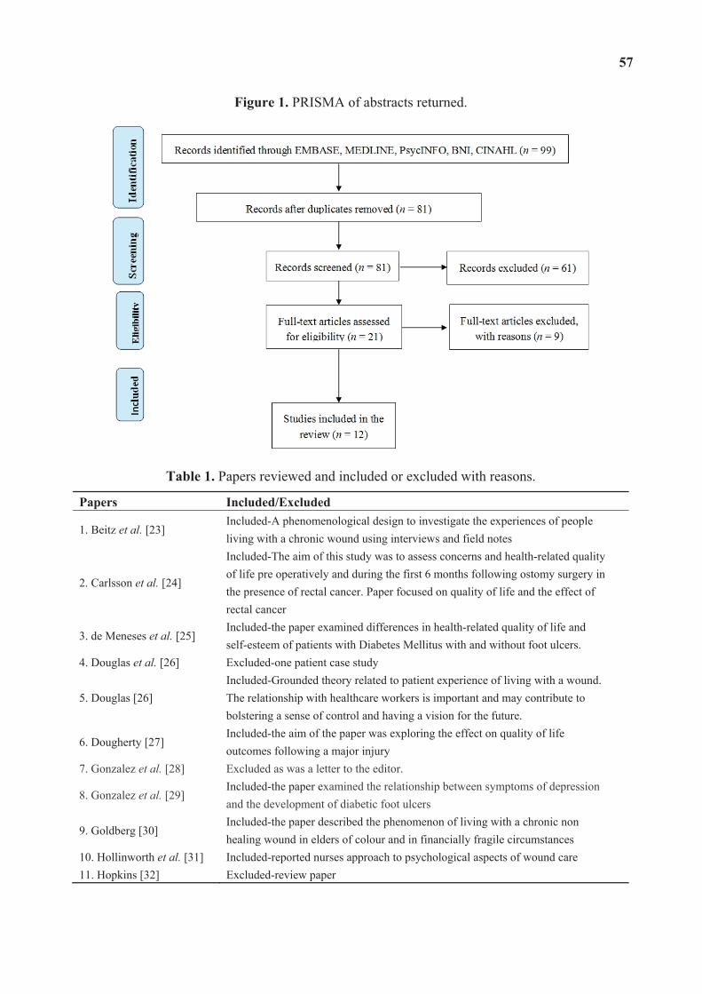
57
Figure 1. PRISMA of abstracts returned.
Table 1. Papers reviewed and included or excluded with reasons.
Papers Included/Excluded
1. Beitz et al. [23] Included-A phenomenological design to investigate the experiences of people living with a chronic wound using interviews and field notes
2. Carlsson et al. [24]
Included-The aim of this study was to assess concerns and health-related quality of life pre operatively and during the first 6 months following ostomy surgery in the presence of rectal cancer. Paper focused on quality of life and the effect of rectal cancer
3. de Meneses et al. [25] Included-the paper examined differences in health-related quality of life and self-esteem of patients with Diabetes Mellitus with and without foot ulcers.
4. Douglas et al. [26] Excluded-one patient case study
5. Douglas [26] Included-Grounded theory related to patient experience of living with a wound. The relationship with healthcare workers is important and may contribute to bolstering a sense of control and having a vision for the future.
6. Dougherty [27] Included-the aim of the paper was exploring the effect on quality of life outcomes following a major injury
7. Gonzalez et al. [28] Excluded as was a letter to the editor.
8. Gonzalez et al. [29] Included-the paper examined the relationship between symptoms of depression and the development of diabetic foot ulcers
9. Goldberg [30] Included-the paper described the phenomenon of living with a chronic non healing wound in elders of colour and in financially fragile circumstances
10. Hollinworth et al. [31] Included-reported nurses approach to psychological aspects of wound care 11. Hopkins [32] Excluded-review paper
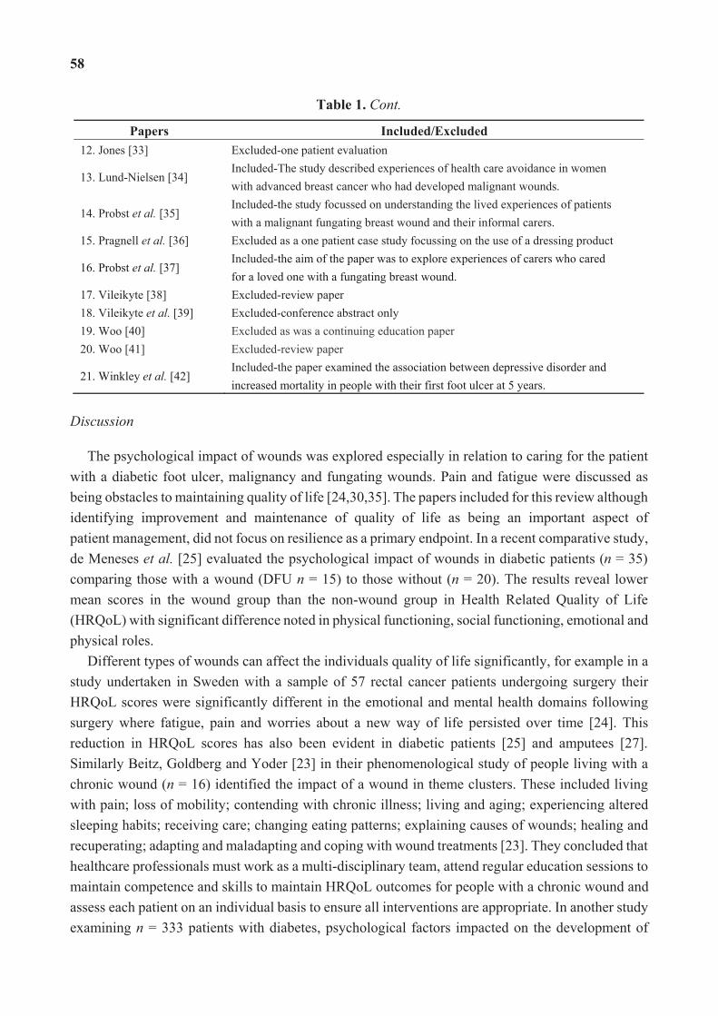
58
Table 1. Cont.
Papers Included/Excluded 12. Jones [33] Excluded-one patient evaluation
13. Lund-Nielsen [34] Included-The study described experiences of health care avoidance in women with advanced breast cancer who had developed malignant wounds.
14. Probst et al. [35] Included-the study focussed on understanding the lived experiences of patients with a malignant fungating breast wound and their informal carers.
15. Pragnell et al. [36] Excluded as a one patient case study focussing on the use of a dressing product
16. Probst et al. [37] Included-the aim of the paper was to explore experiences of carers who cared for a loved one with a fungating breast wound.
17. Vileikyte [38] Excluded-review paper 18. Vileikyte et al. [39] Excluded-conference abstract only 19. Woo [40] Excluded as was a continuing education paper 20. Woo [41] Excluded-review paper
21. Winkley et al. [42] Included-the paper examined the association between depressive disorder and increased mortality in people with their first foot ulcer at 5 years.
Discussion
The psychological impact of wounds was explored especially in relation to caring for the patient with a diabetic foot ulcer, malignancy and fungating wounds. Pain and fatigue were discussed as being obstacles to maintaining quality of life [24,30,35]. The papers included for this review although identifying improvement and maintenance of quality of life as being an important aspect of patient management, did not focus on resilience as a primary endpoint. In a recent comparative study, de Meneses et al. [25] evaluated the psychological impact of wounds in diabetic patients (n = 35) comparing those with a wound (DFU n = 15) to those without (n = 20). The results reveal lower mean scores in the wound group than the non-wound group in Health Related Quality of Life (HRQoL) with significant difference noted in physical functioning, social functioning, emotional and physical roles.
Different types of wounds can affect the individuals quality of life significantly, for example in a study undertaken in Sweden with a sample of 57 rectal cancer patients undergoing surgery their HRQoL scores were significantly different in the emotional and mental health domains following surgery where fatigue, pain and worries about a new way of life persisted over time [24]. This reduction in HRQoL scores has also been evident in diabetic patients [25] and amputees [27]. Similarly Beitz, Goldberg and Yoder [23] in their phenomenological study of people living with a chronic wound (n = 16) identified the impact of a wound in theme clusters. These included living with pain; loss of mobility; contending with chronic illness; living and aging; experiencing altered sleeping habits; receiving care; changing eating patterns; explaining causes of wounds; healing and recuperating; adapting and maladapting and coping with wound treatments [23]. They concluded that healthcare professionals must work as a multi-disciplinary team, attend regular education sessions to maintain competence and skills to maintain HRQoL outcomes for people with a chronic wound and assess each patient on an individual basis to ensure all interventions are appropriate. In another study examining n = 333 patients with diabetes, psychological factors impacted on the development of
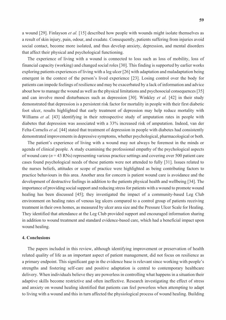
59
a wound [29]. Finlayson et al. [15] described how people with wounds might isolate themselves as a result of skin injury, pain, odour, and exudate. Consequently, patients suffering from injuries avoid social contact, become more isolated, and thus develop anxiety, depression, and mental disorders that affect their physical and psychological functioning.
The experience of living with a wound is connected to loss such as loss of mobility, loss of financial capacity (working) and changed social roles [30]. This finding is supported by earlier works exploring patients experiences of living with a leg ulcer [26] with adaptation and maladaptation being emergent in the context of the person’s lived experience [23]. Losing control over the body for patients can impede feelings of resilience and may be exacerbated by a lack of information and advice about how to manage the wound as well as the physical limitations and psychosocial consequences [35] and can involve mood disturbances such as depression [30]. Winkley et al. [42] in their study demonstrated that depression is a persistent risk factor for mortality in people with their first diabetic foot ulcer, results highlighted that early treatment of depression may help reduce mortality with Williams et al. [43] identifying in their retrospective study of amputation rates in people with diabetes that depression was associated with a 33% increased risk of amputation. Indeed, van der Felta-Cornelis et al. [44] stated that treatment of depression in people with diabetes had consistently demonstrated improvements in depressive symptoms, whether psychological, pharmacological or both.
The patient’s experience of living with a wound may not always be foremost in the minds or agenda of clinical people. A study examining the professional empathy of the psychological aspects of wound care (n = 43 RNs) representing various practice settings and covering over 500 patient care cases found psychological needs of these patients were not attended to fully [31]. Issues related to the nurses beliefs, attitudes or scope of practice were highlighted as being contributing factors to practice behaviours in this area. Another area for concern is patient wound care is avoidance and the development of destructive feelings in addition to the patients physical health and wellbeing [34]. The importance of providing social support and reducing stress for patients with a wound to promote wound healing has been discussed [45]; they investigated the impact of a community-based Leg Club environment on healing rates of venous leg ulcers compared to a control group of patients receiving treatment in their own homes, as measured by ulcer area size and the Pressure Ulcer Scale for Healing. They identified that attendance at the Leg Club provided support and encouraged information sharing in addition to wound treatment and standard evidence-based care, which had a beneficial impact upon wound healing.
4. Conclusions
The papers included in this review, although identifying improvement or preservation of health related quality of life as an important aspect of patient management, did not focus on resilience as a primary endpoint. This significant gap in the evidence base is relevant since working with people’s strengths and fostering self-care and positive adaptation is central to contemporary healthcare delivery. When individuals believe they are powerless in controlling what happens in a situation their adaptive skills become restrictive and often ineffective. Research investigating the effect of stress and anxiety on wound healing identified that patients can feel powerless when attempting to adapt to living with a wound and this in turn affected the physiological process of wound healing. Building
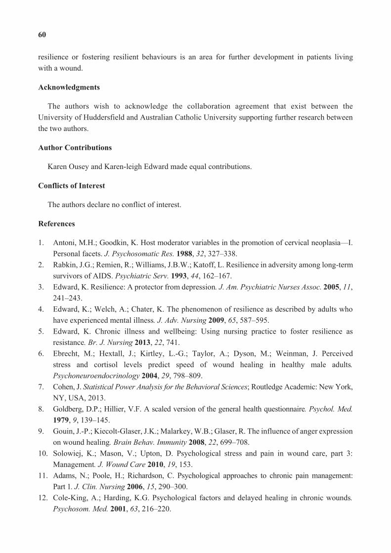
60
resilience or fostering resilient behaviours is an area for further development in patients living with a wound.
Acknowledgments
The authors wish to acknowledge the collaboration agreement that exist between the University of Huddersfield and Australian Catholic University supporting further research between the two authors.
Author Contributions
Karen Ousey and Karen-leigh Edward made equal contributions.
Conflicts of Interest
The authors declare no conflict of interest.
References
1. Antoni, M.H.; Goodkin, K. Host moderator variables in the promotion of cervical neoplasia—I. Personal facets. J. Psychosomatic Res. 1988, 32, 327–338.
2. Rabkin, J.G.; Remien, R.; Williams, J.B.W.; Katoff, L. Resilience in adversity among long-term survivors of AIDS. Psychiatric Serv. 1993, 44, 162–167.
3. Edward, K. Resilience: A protector from depression. J. Am. Psychiatric Nurses Assoc. 2005, 11, 241–243.
4. Edward, K.; Welch, A.; Chater, K. The phenomenon of resilience as described by adults who have experienced mental illness. J. Adv. Nursing 2009, 65, 587–595.
5. Edward, K. Chronic illness and wellbeing: Using nursing practice to foster resilience as resistance. Br. J. Nursing 2013, 22, 741.
6. Ebrecht, M.; Hextall, J.; Kirtley, L.-G.; Taylor, A.; Dyson, M.; Weinman, J. Perceived stress and cortisol levels predict speed of wound healing in healthy male adults. Psychoneuroendocrinology 2004, 29, 798–809.
7. Cohen, J. Statistical Power Analysis for the Behavioral Sciences; Routledge Academic: New York, NY, USA, 2013.
8. Goldberg, D.P.; Hillier, V.F. A scaled version of the general health questionnaire. Psychol. Med. 1979, 9, 139–145.
9. Gouin, J.-P.; Kiecolt-Glaser, J.K.; Malarkey, W.B.; Glaser, R. The influence of anger expression on wound healing. Brain Behav. Immunity 2008, 22, 699–708.
10. Solowiej, K.; Mason, V.; Upton, D. Psychological stress and pain in wound care, part 3: Management. J. Wound Care 2010, 19, 153.
11. Adams, N.; Poole, H.; Richardson, C. Psychological approaches to chronic pain management: Part 1. J. Clin. Nursing 2006, 15, 290–300.
12. Cole-King, A.; Harding, K.G. Psychological factors and delayed healing in chronic wounds. Psychosom. Med. 2001, 63, 216–220.
61
13. Jones, J.; Barr, W.; Robinson, J.; Carlisle, C. Depression in patients with chronic venous ulceration. Br. J. Nursing 2006, 15, S17.
14. Guarnera, G.; Tinelli, G.; Abeni, D.; di Pietro, C.; Sampogna, F.; Tabolli, S. Pain and quality of life in patients with vascular leg ulcers: An Italian multicentre study. J. Wound Care 2007, 16, 347–351.
15. Finlayson, K.; Edwards, H.; Courtney, M. The impact of psychosocial factors on adherence to compression therapy to prevent recurrence of venous leg ulcers. J. Clin. Nursing 2010, 19, 1289–1297.
16. Mapplebeck, L. Case study: Psychosocial aspects of chronic bilateral venous leg ulcers. Br. J. Community Nursing 2008, 13, S33–S38.
17. Pieper, B.; Vallerand, A.H.; Nordstrom, C.K.; DiNardo, E. Comparison of bodily pain: Persons with and without venous ulcers in an indigent care clinic. J. Wound Ostomy Continence Nursing 2009, 36, 493–502.
18. Upton, D.; Solowiej, K.; Woo, K.Y. A multinational health professional perspective of the prevalence of mood disorders in patients with acute and chronic wounds. Int. Wound J. 2013, doi:10.1111/iwj.12018.
19. Edward, K. Being resilient as described by people who experience mental illness. Int. J. Mental Health Nursing 2008, 17, A6.
20. Edward, K.; Warelow, P. Resilience: When coping is emotionally intelligent. J. Am. Psychiatric Nurses Assoc. 2005, 11, 101–102.
21. Taylor, R.; Reeves, B.; Ewings, P.; Binns, S.; Keast, J.; Mears, R. A systematic review of the effectiveness of critical appraisal skills training for clinicians. Med. Educ. 2000, 34, 120–125.
22. Liberati, A.; Altman, D.G.; Tetzlaff, J.; Mulrow, C.; Gøtzsche, P.C.; Ioannidis, J.P.A.; Clarke, M.; Devereaux, P.J.; Kleijnen, J.; Moher, D. The PRISMA statement for reporting systematic reviews and meta-analyses of studies that evaluate health care interventions: Explanation and elaboration. Ann. Int. Med. 2009, 151, W-65-W-94.
23. Beitz, J.M.; Goldberg, E.; Yoder, L.H. The lived experience of having a chronic wound: A phenomenologic study. Medsurg Nursing 2005, 14, 51–82.
24. Carlsson, E.; Berndtsson, I.; Hallén, A.-M.; Lindholm, E.; Persson, E. Concerns and quality of life before surgery and during the recovery period in patients with rectal cancer and an ostomy. J. Wound Ostomy Continence Nursing 2010, 37, 654–661.
25. De Meneses, L.C.; Blanes, L.; Francescato Veiga, D.; Carvalho Gomes, H.; Masako Ferreira, L. Health-related quality of life and self-esteem in patients with diabetic foot ulcers: Results of a cross-sectional comparative study. Ostomy-Wound Manage. 2011, 57, 36.
26. Douglas, V.; Living with a chronic leg ulcer: An insight into patients’ experiences and feelings. J. Wound Care 2001, 10, 355–362.
27. Dougherty, P.J. Transtibial amputees from the Vietnam war twenty-eight-year follow-up. J. Bone Joint Surg. 2001, 83, 383–383.
28. Gonzalez, J.; Hardman, M.; Boulton, A.J.; Vileikyte, L. Coping and depression in diabetic foot ulcer healing: Causal influence, mechanistic evidence or none of the above? Diabetologia 2011, 54, 205–206.
62
29. Gonzalez, J.; Vileikyte, L.; Ulbrecht, J.; Rubin, R.; Garrow, A.; Delgado, C.; Cavanagh, P.; Boulton, A.; Peyrot, M. Depression predicts first but not recurrent diabetic foot ulcers. Diabetologia 2010, 53, 2241–2248.
30. Goldberg, E.; Beitz, J.M. The lived experience of diverse elders with chronic wounds. Ostomy/Wound Manage. 2010, 56, 36–46.
31. Hollinworth, H.; Hawkins, J. Teaching nurses psychological support of patients with wounds. Br. J. Nursing 2002, 11, 8–19.
32. Hopkins, S. Psychological aspects of wound healing. Nursing Times 2001, 97, 57. 33. Jones, J. Stress responses, pressure ulcer development and adaptation. Br. J. Nursing 2003, 12,
S17–S24. 34. Lund-Nielsen, B.; Midtgaard, J.; Rørth, M.; Gottrup, F.; Adamsen, L. An avalanche of
ignoring-a qualitative study of health care avoidance in women with malignant breast cancer wounds. Cancer Nursing 2011, 34, 277–285.
35. Probst, S.; Arber, A.; Faithfull, S. Malignant fungating wounds—the meaning of living in an unbounded body. Eur. J. Oncol. Nursing 2013, 17, 38–45.
36. Pragnell, J; Neilson, J. The social and psychological impact of hard-to-heal wounds. Br. J. Nursing 2009, 19, 1248–1252.
37. Probst, S.; Arber, A.; Trojan, A.; Faithfull, S. Caring for a loved one with a malignant fungating wound. Supportive Care in Cancer 2012, 20, 3065–3070.
38. Vileikyte, L. Psychosocial and behavioral aspects of diabetic foot lesions. Curr. Diabetes Rep. 2008, 8, 119–125.
39. Vileikyte, L.; Shen, B.-J.; Hardman, M.; Kirsner, R.; Boulton, A.; Schneiderman, N. Emotional distress may impede diabetic foot ulcer healing through elevated levels of interleukin-6: Preliminary findings. In Diabetologia; Springer: New York, NY, USA, 2010.
40. Woo, K.Y. Exploring the effects of pain and stress on wound healing. Adv. Skin Wound Care 2012, 25, 38–44.
41. Woo, K. Chronic wound-associated pain, psychological stress, and wound healing. Surgical Technol. Int. 2012, 22, 57–65.
42. Winkley, K.; Sallis, H.; Kariyawasam, D.; Leelarathna, L.; Chalder, T.; Edmonds, M.; Stahl, D.; Ismail, K. Five-year follow-up of a cohort of people with their first diabetic foot ulcer: The persistent effect of depression on mortality. Diabetologia 2012, 55, 303–310.
43. Williams, L.H.; Miller, D.R.; Fincke, G.; Lafrance, J.-P.; Etzioni, R.; Maynard, C.; Raugi, G.J.; Reiber, G.E. Depression and incident lower limb amputations in veterans with diabetes. J. Diabetes Complicat. 2011, 25, 175–182.
44. Van der Feltz-Cornelis, C.M.; Nuyen, J.; Stoop, C.; Chan, J.; Jacobson, A.M.; Katon, W.; Snoek, F.; Sartorius, N. Effect of interventions for major depressive disorder and significant depressive symptoms in patients with diabetes mellitus: A systematic review and meta-analysis. Gen. Hosp. Psychiatry 2010, 32, 380–395.
45. Edwards, H.; Courtney, M.; Finlayson, K.; Lewis, C.; Lindsay, E.; Dumble, J. Improved healing rates for chronic venous leg ulcers: Pilot study results from a randomized controlled trial of a community nursing intervention. Int. J. Nursing Pract. 2005, 11, 169–176.
63
Antifungal Effect of Non-Woven Textiles Containing Polyhexamethylene Biguanide with Sophorolipid: A Potential Method for Tinea Pedis Prevention
Hiromi Sanada, Gojiro Nakagami, Kimie Takehara, Taichi Goto, Nanase Ishii, Satoshi Yoshida, Mizuyuki Ryu and Yuichiro Tsunemi
Abstract: Tinea pedis is a preventable skin disease common in elderly or diabetic patients. Daily foot washing is effective for prevention, but can be difficult for many patients. Additionally, conventional methods cannot eliminate fungi within the stratum corneum, a common site for fungal invasion. This study investigates the antifungal effects, cytotoxicity, permeability, and efficacy of non-woven textiles containing polyhexamethylene biguanide (PHMB) mixed with sophorolipid. Permeability of PHMB with varying concentrations of sophorolipid was assessed via a cultured skin model. Stratum corneum PHMB concentration was quantified by polyvinylsulphuric acid potassium salt titration and cytotoxicity was assayed via 3-(4,5-dimethylthiazol-2-yl)-2,5-diphenyltetrazolium bromide. Antifungal effects were evaluated via a new cultured skin/Trichophyton mentagrophytes model, with varying PHMB exposure duration. Clinically-isolated Trichophyton were applied to the feet of four healthy volunteers and then immediately treated with the following methods: washing with soap, a non-woven textile with PHMB, the textile without PHMB, or without washing. Fungal colony forming units (CFUs) were evaluated after one of these treatments were performed. Sophorolipid with various concentrations significantly facilitated PHMB permeation into the stratum corneum, which was not in a dose-dependent manner. Significant PHMB antifungal effects were achieved at 30 min, with low cytotoxicity. Textiles containing PHMB significantly reduced CFU of fungi in healthy volunteers to levels comparable to soap washing. Our results indicate the utility of this product for tinea pedis prevention in clinical settings.
Reprinted from Healthcare. Cite as: Sanada, H.; Nakagami, G.; Takehara, K.; Goto, T.; Ishii, N.; Yoshida, S.; Ryu, M.; Tsunemi, Y. Antifungal Effect of Non-Woven Textiles Containing Polyhexamethylene Biguanide with Sophorolipid: A Potential Method for Tinea Pedis Prevention. Healthcare 2014, 2, 183-191.
1. Introduction
Tinea pedis is one of the fungal infections caused by dermatophytes [1]. Trichophyton rubrumand T. mentagrophytes account for over 90% of causative fungi. The prevalence of tinea pedis increases with age, with the highest prevalence among those 50 to 60 years of age [2]. The main nutrient of dermatophytes is keratin, which is located in the stratum corneum. These fungi often cause skin maceration and erosion between the toes, leading to secondary bacterial infection. This is critical to elderly and diabetic patients, as secondary infections cause foot ulcers, cellulitis, necrotizing fasciitis, or gas gangrene, making prevention of infection most important [3].
Since dermatophytes invade the keratin layer within 24 h after attachment, daily foot washing is recommended for efficient physical removal of fungi [4,5]. Previous reports indicate foot washing
64
using soap can eliminate fungi from the skin surface [6]. However, daily foot washing can be difficult for many patients, owing to limited bathing of residents in long-term care facilities, limited joint range of motion, or visual impairment [7,8]. Furthermore, the skin barrier function of the stratum corneum also makes dermatophyte removal within the keratin layer difficult using only conventional antiseptics [9,10]. Therefore, a clinically effective, simple, and easy-to-use method is needed.
To overcome these challenges, we developed a new non-woven textile product soaked with an antifungal agent with enhanced permeability to the stratum corneum. This product can physically eliminate fungi attached to the skin surface and interferes with growth in the stratum corneum via application to the skin surface. This study investigates its effect on antifungal function in vitro and in vivo in healthy volunteers.
2. Experimental
2.1. Evaluation of Permeation of PHMB with Sophorolipid Treatment within the Skin Model
Polyhexamethylene biguanide (PHMB; Arch UK Biocides Ltd., Blackley, UK) alone or with 0.1 or 1% sophorolipid (synthesized as previously described [11]) was added to the top of the stratum corneum of a three-dimensional cultured human skin model (LSE-high, Toyobo Co., Ltd., Osaka, Japan) and quantified in the stratum corneum after 120 min to determine the degree of permeation. PHMB was extracted from homogenized stratum corneum sample of each well in 1 mL of phosphate-buffered saline and filtered. For quantitation, each sample was mixed with sodium chloride (1 N) adjusted to a pH of 1.5 ± 0.05, then titrated with a standardized aqueous solution of polyvinylsulphuric acid potassium salt (N/400, Wako Pure Chemical Industries, Ltd., Osaka, Japan) to a blue to pink toluidine blue indicator color change endpoint (n = 3).
2.2. MTT Assay for Cytotoxicity
To assess cytotoxicity, the cultured skin model was treated with 0.1% PHMB with 0.1% sophorolipid supplementation for 5, 30, 60, and 120 min, and then incubated with 3-(4,5-dimethylthiazol-2-yl)-2, 5-diphenyltetrazolium bromide (MTT; Sigma-Aldrich, Tokyo, Japan). Yellow tetrazolium salt is reduced by mitochondrial enzymes in viable cells to an insoluble blue formazan product. The skin model without PHMB was used as a control. After incubation, samples were transferred to new 1.5 mL tubes and mixed with 200 L of isopropanol to extract any resulting formazan. Absorbance was measured spectrophotometrically using an automated microplate reader (Spectra Thermo, Tecan Group Ltd., San Jose, CA, USA) at a wavelength of 570 nm (n = 3). Cell survival was computed by using the following formula: Cell viability % = [(mean optical density of the sample blank)/(mean optical density of the control blank)] × 100.
2.3. Evaluation of Antifungal Effect: In Vitro
We established an intra-stratum corneum T. mentagrophytes model to evaluate the antifungal effect of 0.1% of PHMB. T. mentagrophytes was incubated at 30 °C on 3.9% autoclaved potato dextrose agar (Nissui Pharmaceutical Co., Tokyo, Japan) slant medium for a week. The slant medium
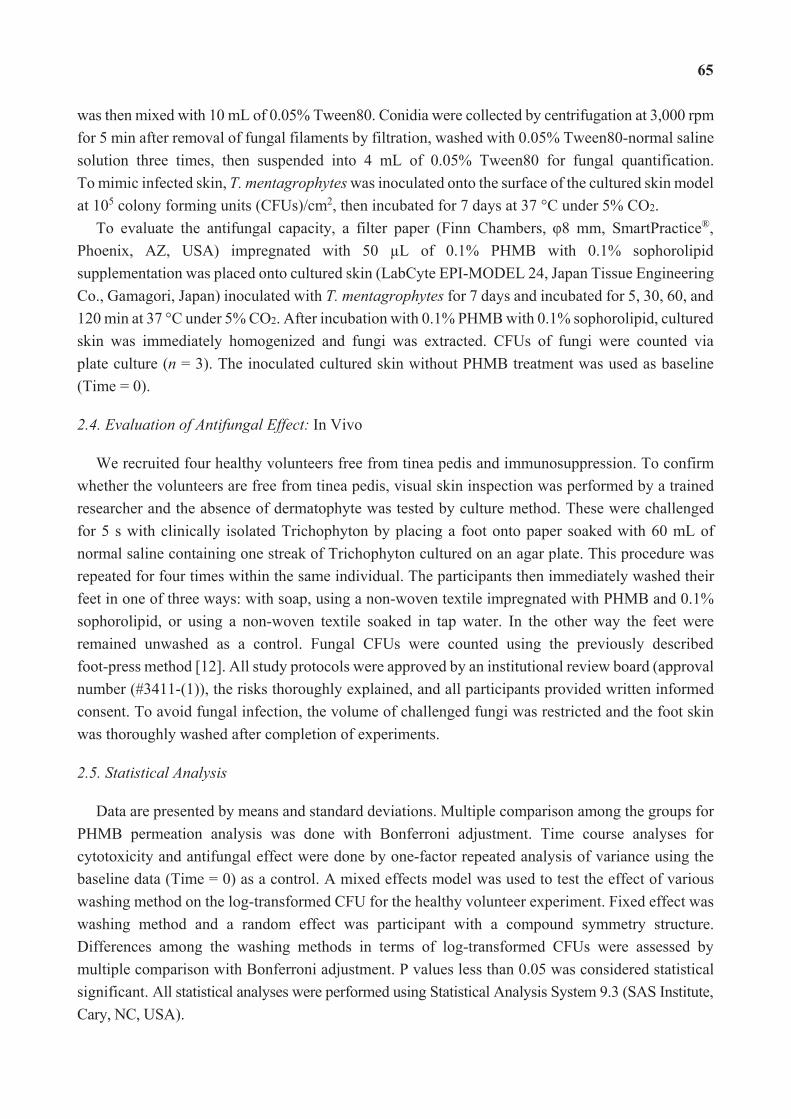
65
was then mixed with 10 mL of 0.05% Tween80. Conidia were collected by centrifugation at 3,000 rpm for 5 min after removal of fungal filaments by filtration, washed with 0.05% Tween80-normal saline solution three times, then suspended into 4 mL of 0.05% Tween80 for fungal quantification. To mimic infected skin, T. mentagrophytes was inoculated onto the surface of the cultured skin model at 105 colony forming units (CFUs)/cm2, then incubated for 7 days at 37 °C under 5% CO2.
To evaluate the antifungal capacity, a filter paper (Finn Chambers, 8 mm, SmartPractice®, Phoenix, AZ, USA) impregnated with 50 L of 0.1% PHMB with 0.1% sophorolipid supplementation was placed onto cultured skin (LabCyte EPI-MODEL 24, Japan Tissue Engineering Co., Gamagori, Japan) inoculated with T. mentagrophytes for 7 days and incubated for 5, 30, 60, and 120 min at 37 °C under 5% CO2. After incubation with 0.1% PHMB with 0.1% sophorolipid, cultured skin was immediately homogenized and fungi was extracted. CFUs of fungi were counted via plate culture (n = 3). The inoculated cultured skin without PHMB treatment was used as baseline (Time = 0).
2.4. Evaluation of Antifungal Effect: In Vivo
We recruited four healthy volunteers free from tinea pedis and immunosuppression. To confirm whether the volunteers are free from tinea pedis, visual skin inspection was performed by a trained researcher and the absence of dermatophyte was tested by culture method. These were challenged for 5 s with clinically isolated Trichophyton by placing a foot onto paper soaked with 60 mL of normal saline containing one streak of Trichophyton cultured on an agar plate. This procedure was repeated for four times within the same individual. The participants then immediately washed their feet in one of three ways: with soap, using a non-woven textile impregnated with PHMB and 0.1% sophorolipid, or using a non-woven textile soaked in tap water. In the other way the feet were remained unwashed as a control. Fungal CFUs were counted using the previously described foot-press method [12]. All study protocols were approved by an institutional review board (approval number (#3411-(1)), the risks thoroughly explained, and all participants provided written informed consent. To avoid fungal infection, the volume of challenged fungi was restricted and the foot skin was thoroughly washed after completion of experiments.
2.5. Statistical Analysis
Data are presented by means and standard deviations. Multiple comparison among the groups for PHMB permeation analysis was done with Bonferroni adjustment. Time course analyses for cytotoxicity and antifungal effect were done by one-factor repeated analysis of variance using the baseline data (Time = 0) as a control. A mixed effects model was used to test the effect of various washing method on the log-transformed CFU for the healthy volunteer experiment. Fixed effect was washing method and a random effect was participant with a compound symmetry structure. Differences among the washing methods in terms of log-transformed CFUs were assessed by multiple comparison with Bonferroni adjustment. P values less than 0.05 was considered statistical significant. All statistical analyses were performed using Statistical Analysis System 9.3 (SAS Institute, Cary, NC, USA).
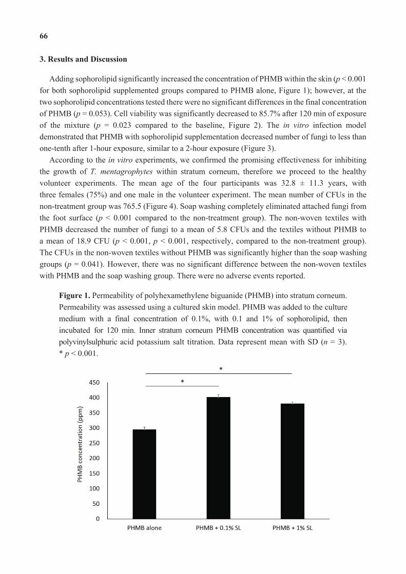
66
3. Results and Discussion
Adding sophorolipid significantly increased the concentration of PHMB within the skin (p < 0.001 for both sophorolipid supplemented groups compared to PHMB alone, Figure 1); however, at the two sophorolipid concentrations tested there were no significant differences in the final concentration of PHMB (p = 0.053). Cell viability was significantly decreased to 85.7% after 120 min of exposure of the mixture (p = 0.023 compared to the baseline, Figure 2). The in vitro infection model demonstrated that PHMB with sophorolipid supplementation decreased number of fungi to less than one-tenth after 1-hour exposure, similar to a 2-hour exposure (Figure 3).
According to the in vitro experiments, we confirmed the promising effectiveness for inhibiting the growth of T. mentagrophytes within stratum corneum, therefore we proceed to the healthy volunteer experiments. The mean age of the four participants was 32.8 ± 11.3 years, with three females (75%) and one male in the volunteer experiment. The mean number of CFUs in the non-treatment group was 765.5 (Figure 4). Soap washing completely eliminated attached fungi from the foot surface (p < 0.001 compared to the non-treatment group). The non-woven textiles with PHMB decreased the number of fungi to a mean of 5.8 CFUs and the textiles without PHMB to a mean of 18.9 CFU (p < 0.001, p < 0.001, respectively, compared to the non-treatment group). The CFUs in the non-woven textiles without PHMB was significantly higher than the soap washing groups (p = 0.041). However, there was no significant difference between the non-woven textiles with PHMB and the soap washing group. There were no adverse events reported.
Figure 1. Permeability of polyhexamethylene biguanide (PHMB) into stratum corneum. Permeability was assessed using a cultured skin model. PHMB was added to the culture medium with a final concentration of 0.1%, with 0.1 and 1% of sophorolipid, then incubated for 120 min. Inner stratum corneum PHMB concentration was quantified via polyvinylsulphuric acid potassium salt titration. Data represent mean with SD (n = 3). * p < 0.001.
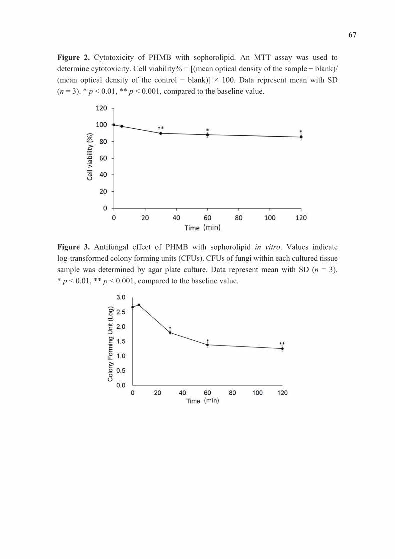
67
Figure 2. Cytotoxicity of PHMB with sophorolipid. An MTT assay was used to determine cytotoxicity. Cell viability% = [(mean optical density of the sample blank)/ (mean optical density of the control blank)] × 100. Data represent mean with SD (n = 3). * p < 0.01, ** p < 0.001, compared to the baseline value.
Figure 3. Antifungal effect of PHMB with sophorolipid in vitro. Values indicate log-transformed colony forming units (CFUs). CFUs of fungi within each cultured tissue sample was determined by agar plate culture. Data represent mean with SD (n = 3). * p < 0.01, ** p < 0.001, compared to the baseline value.
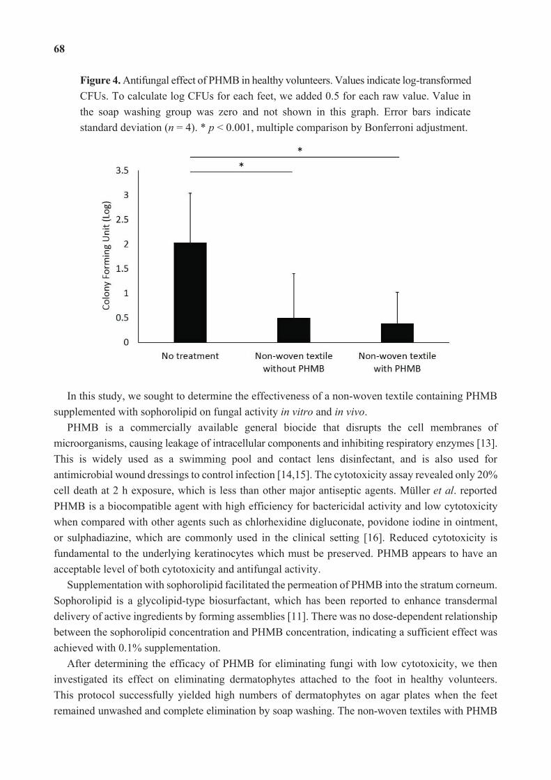
68
Figure 4. Antifungal effect of PHMB in healthy volunteers. Values indicate log-transformed CFUs. To calculate log CFUs for each feet, we added 0.5 for each raw value. Value in the soap washing group was zero and not shown in this graph. Error bars indicate standard deviation (n = 4). * p < 0.001, multiple comparison by Bonferroni adjustment.
In this study, we sought to determine the effectiveness of a non-woven textile containing PHMB supplemented with sophorolipid on fungal activity in vitro and in vivo.
PHMB is a commercially available general biocide that disrupts the cell membranes of microorganisms, causing leakage of intracellular components and inhibiting respiratory enzymes [13]. This is widely used as a swimming pool and contact lens disinfectant, and is also used for antimicrobial wound dressings to control infection [14,15]. The cytotoxicity assay revealed only 20% cell death at 2 h exposure, which is less than other major antiseptic agents. Müller et al. reported PHMB is a biocompatible agent with high efficiency for bactericidal activity and low cytotoxicity when compared with other agents such as chlorhexidine digluconate, povidone iodine in ointment, or sulphadiazine, which are commonly used in the clinical setting [16]. Reduced cytotoxicity is fundamental to the underlying keratinocytes which must be preserved. PHMB appears to have an acceptable level of both cytotoxicity and antifungal activity.
Supplementation with sophorolipid facilitated the permeation of PHMB into the stratum corneum. Sophorolipid is a glycolipid-type biosurfactant, which has been reported to enhance transdermal delivery of active ingredients by forming assemblies [11]. There was no dose-dependent relationship between the sophorolipid concentration and PHMB concentration, indicating a sufficient effect was achieved with 0.1% supplementation.
After determining the efficacy of PHMB for eliminating fungi with low cytotoxicity, we then investigated its effect on eliminating dermatophytes attached to the foot in healthy volunteers. This protocol successfully yielded high numbers of dermatophytes on agar plates when the feet remained unwashed and complete elimination by soap washing. The non-woven textiles with PHMB
69
considerably decreased fungal counts, but so did the textiles without PHMB, albeit with slightly higher CFU counts. It is noteworthy that the fungal count in the non-woven textiles without PHMB was significantly higher than in the soap washing group and the significant difference was not seen in the non-woven textiles with PHMB and the soap washing comparison. This suggests physical removal of fungi by scrubbing the skin surface plays a major role in fungal clearance, but PHMB may contribute to the additional reduction [6]. Since the non-woven textiles with PHMB possess the anti-fungal ability as demonstrated in the present study, it is expected that the number of rubbing the foot skin would be less for using this textiles than for soap washing, which might facilitate the individuals with limited joint range of motion and access to the bathing care to perform daily foot care by themselves. Further study is needed to prove the antifungal effectiveness of PHMB on human skin and ultimately in the patients with tinea pedis.
A major limitation of this study is assessing the antifungal effect immediately after fungi inoculation. As fungi invade the stratum corneum after a certain period of attachment in the clinical setting [4], antifungal effects would ideally be assessed at a later phase. Additionally, the effectiveness in preventing tinea pedis should be tested in a long-term interventional trial. PHMB may have a more significant effect in patients with stratum corneum infiltration, as opposed to surface colonization. As Hammer et al. reported that dermatophyte susceptibility varies towards antimicrobial textiles, we need to consider the further experiments against different species even though they indicated T. mentagrophytes growth remained unaffected in direct contact with the antimicrobial textiles tested [17].
Although there are many products available for washing feet, they are not designed to prevent tinea pedis among patients with limited physical capacity. We hope to enhance patient self-care by designing easy-to-use sheet-type materials with high antifungal effectiveness and low cytotoxicity. This would be suitable for daily use by patients for whom daily foot washing is difficult.
4. Conclusions
Our results indicate the non-woven textiles with containing PHMB with sophorolipid favorable reduction of Trichophyton in vitro and in vivo, suggesting potential clinical effectiveness for tinea pedis prevention.
Acknowledgments
The authors are grateful to the study volunteers for their willing participation.
Author Contributions
Hiromi Sanada: Study design, data collection and analysis, and comments on drafts and final manuscript. Gojiro Nakagami: Study design, data collection and analysis, and preparation of first draft and final manuscript. Kimie Takehara: Study design, data collection and analysis, interpretation of data, comments on drafts and final manuscript. Taichi Goto: Data analysis, interpretation of data, and comments on drafts. Nanase Ishii: Study design, data collection and analysis, interpretation of data, and comments on drafts. Satoshi Yoshida: Study design, data collection and analysis,
70
interpretation of data, and comments on drafts. Mizuyuki Ryu: Study design, data collection and analysis, interpretation of data, and comments on drafts. Yuichiro Tsunemi: Data collection and analysis, interpretation of data, and comments on drafts.
Conflicts of Interest
The non-woven textiles containing PHMB with sophorolipid used in this study was co-developed and provided by Saraya Co., Ltd., Osaka, Japan.
References
1. Moriarty, B.; Hay, R.; Morris-Jones, R. The diagnosis and management of tinea. BMJ 2012, 345, e4380.
2. Watanabe, S.; Harada, T.; Hiruma, M.; Iozumi, K.; Katoh, T.; Mochizuki, T.; Naka, W. Japan Foot Week Group. Epidemiological survey of foot diseases in Japan: Results of 30,000 foot checks by dermatologists. J. Dermatol. 2010, 37, 397–406.
3. Roujeau, J.C.; Sigurgeirsson, B.; Korting, H.C.; Kerl, H.; Paul, C. Chronic dermatomycoses of the foot as risk factors for acute bacterial cellulitis of the leg: A case-control study. Dermatology 2004, 209, 301–307.
4. Rashid, A.; Scott, E.; Richardson, M.D. Early events in the invasion of the human nail plate by Trichophyton mentagrophytes. Br. J. Dermatol. 1995, 133, 932–940.
5. Takehara, K.; Oe, M.; Tsunemi, Y.; Nagase, T.; Ohashi, Y.; Iizaka, S.; Ueki, K.; Tsukamoto, K.; Kadowaki, T.; Sanada, H. Factors associated with presence and severity of toenail onychomycosis in patients with diabetes: A cross-sectional study. Int. J. Nurs. Stud. 2011, 48, 1101–1108.
6. Watanabe, K.; Taniguchi, H.; Katoh, T. Adhesion of dermatophytes to healthy feet and its simple treatment. Mycoses 2000, 43, 45–50.
7. Naik, A.D.; Concato, J.; Gill, T.M. Bathing disability in community-living older persons: Common, consequential, and complex. J. Am. Geriatr. Soc. 2004, 52, 1805–1810.
8. LaPlante, M.P.; Harrington, C.; Kang, T. Estimating paid and unpaid h of personal assistance services in activities of daily living provided to adults living at home. Health Serv. Res. 2002, 37, 397–415.
9. Duek, L.; Kaufman, G.; Ulman, Y.; Berdicevsky, I. The pathogenesis of dermatophyte infections in human skin sections. J. Infect. 2004, 48, 175–180.
10. Bos, J.D.; Meinardi, M.M. The 500 Dalton rule for the skin penetration of chemical compounds and drugs. Exp. Dermatol. 2000, 9, 165–169.
11. Ishii, N.; Kobayashi, T.; Matsumiya, K.; Ryu, M.; Hirata, Y.; Matsumura, Y.; Suzuki, Y.A. Transdermal administration of lactoferrin with sophorolipid. Biochem. Cell Biol. 2012, 90, 504–512.
12. Maruyama, R.; Katoh, T.; Nishioka, K. Demonstration of dermatophyte dissemination from the infected soles using the foot-press method. Mycoses 1998, 41, 145–151.
71
13. Rebong, R.A.; Santaella, R.M.; Goldhagen, B.E.; Majka, C.P.; Perfect, J.R.; Steinbach, W.J.; Afshari, N.A. Polyhexamethylene biguanide and calcineurin inhibitors as novel antifungal treatments for Aspergillus. keratitis. Invest. Ophthalmol. Vis. Sci. 2011, 52, 7309–7315.
14. Buck, S.L.; Rosenthal, R.A.; Schlech, B.A. Methods used to evaluate the effectiveness of contact lens care solutions and other compounds against Acanthamoeba: A review of the literature. CLAO J. 2000, 26, 72–84.
15. Eberlein, T.; Haemmerle, G.; Signer, M.; Gruber Moesenbacher, U.; Traber, J.; Mittlboeck, M.; Abel, M.; Strohal, R. Comparison of PHMB-containing dressing and silver dressings in patients with critically colonised or locally infected wounds. J. Wound Care 2012, 21, 12–20.
16. Müller, G.; Kramer, A. Biocompatibility index of antiseptic agents by parallel assessment of antimicrobial activity and cellular cytotoxicity. J. Antimicrob. Chemother. 2008, 61, 1281–1287.
17. Hammer, T.R.; Mucha, H.; Hoefer, D. Dermatophyte susceptibility varies towards antimicrobial textiles. Mycoses 2012, 55, 344–351.
72
Managing Everyday Life: A Qualitative Study of Patients’ Experiences of a Web-Based Ulcer Record for Home-Based Treatment
Marianne V. Trondsen
Abstract: Chronic skin ulcers are a significant challenge for patients and health service resources, and ulcer treatment often requires the competence of a specialist. Although e-health interventions are increasingly valued for ulcer care by giving access to specialists at a distance, there is limited research on patients’ use of e-health services for home-based ulcer treatment. This article reports an exploratory qualitative study of the first Norwegian web-based counselling service for home-based ulcer treatment, established in 2011 by the University Hospital of North Norway (UNN). Community nurses, general practitioners (GPs) and patients are offered access to a web-based record system to optimize ulcer care. The web-based ulcer record enables the exchange and storage of digital photos and clinical information, by the use of which, an ulcer team at UNN, consisting of specialized nurses and dermatologists, is accessible within 24 h. This article explores patients’ experiences of using the web-based record for their home-based ulcer treatment without assistance from community nurses. Semi-structured interviews were conducted with a total of four patients who had used the record. The main outcomes identified were: autonomy and flexibility; safety and trust; involvement and control; and motivation and hope. These aspects improved the patients’ everyday life during long-term ulcer care and can be understood as stimulating patient empowerment.
Reprinted from Healthcare. Cite as: Trondsen, M.V. Managing Everyday Life: A Qualitative Study of Patients’ Experiences of a Web-Based Ulcer Record for Home-Based Treatment. Healthcare 2014, 2, 492-504.
1. Introduction
Chronic and complex ulcers are a widespread problem that represents a significant challenge for patients and health services [1,2]. Chronic ulcers are painful and impair mobility and activity, which reduces patients’ quality of life [3,4]. Community nurses, in collaboration with general practitioners (GPs), have the primary responsibility for providing care to patients with ulcers, but patients’ self-care can play an important role. However, insufficient knowledge of ulcer treatment is common in primary healthcare services, as well as in local hospitals [1,2,5]. Treatment of long-lasting ulcers often requires the competence of a specialist, access to which is limited in rural areas. Repeated visits to specialist hospitals are often necessary, representing an important cost factor for healthcare systems and placing a significant burden on the patients, because such visits are time-consuming and often include long travel distances [6,7].
Teledermatology, which is defined as delivery of dermatology services through the use of information and communication technology (ICT) [8], is a rapidly evolving field that offers great potential for long-term ulcer care and contributes to overcoming inequities in access to health services across geographical regions [5–8]. Videoconferences, cameras and mobile devices, as well
73
as web-based electronic transmission of digital photos and written information make it possible for ulcer experts from specialist hospitals to offer opinions for diagnosis and recommendations for treatment without delay [6,9]. The expert advice can be delivered in real time or in store-and-forward systems to GPs, community nurses, physicians or nurses at local hospitals, as well as directly to patients in-home.
Teledermatology has provided high levels of concordance in diagnosis and management when compared with face-to-face consultations [10]. It is cost-effective, providing rapid evaluation and treatment, as well as more efficient use of human resources [5,7]. Access to ulcer experts through web-based tools also enhances community nurses’ knowledge of ulcer treatment [5,11]. A study by Binder et al. (2007) demonstrated that web-based follow-up of ulcer care, which included sharing clinical information and digital photos between community nurses and ulcer specialists, improved the quality of treatment [6]. A good healing rate was achieved, and patients, community nurses and ulcer specialists were all satisfied with the service [6]. Nevertheless, the development of teledermatology as a routine service remains limited [8].
This article reports an exploratory qualitative study of the first Norwegian web-based counselling service for home-based ulcer treatment (www.pleie.net). In 2011, the Department of Dermatology (DoD) at the University Hospital of North Norway (UNN) established a web-based ulcer record system in collaboration with the Norwegian Centre for Integrated Care and Telemedicine, using an adapted web-based ulcer record system developed by Danish Telemedicine [9,11]. The purpose of the web-based counselling service was to optimize treatment and care of complex and demanding ulcers through self-management of home-based ulcer treatment. Use of the web-based service in home-based treatment was to be a part of the follow-up of ulcer patients at UNN, in combination with clinical consultations at the DoD. The web-based ulcer record (www.pleie.net) is a store-and-forward system that consists of databases, an application to communicate digital photos and text between the users and a tool to measure and analyze ulcers. The log-on procedure for the record system requires a two-factor authentication, employing a username, password and a one-time password sent by SMS from the server to the phone number registered in the user profile, and gives access only to the ulcer records of the patients that the user is authorized to access [9]. Patients or community nurses take photos of the ulcers with a digital camera or a mobile phone and publish the digital photos, leave comments or address questions in the web-based record to a specialist ulcer team consisting of specialized nurses and a dermatologist at the DoD. The nurses on the team have the primary responsibility of replying as quickly as possible and within 24 h. The communication between the user’s web client on a mobile phone or a computer and the ulcer record system is encrypted. The system runs in a secure environment. All of the photos, comments and responses are archived in the record for retrospective examination. The measurement tool enables an analysis of the ulcers’ development and presents diagrams for observing changes over time. This article addresses patients’ experiences of using the web-based ulcer record for their home-based ulcer treatment without regular assistance from community nurses or other community health services.
Most of the studies of teledermatology have been on the effects and outcomes from a medical perspective. Thus, there is a need to supplement this research by exploring patients’ experiences. Web-based applications make it possible for patients to get direct access to expert advice, but little
74
is known about patients’ attitudes towards using a web-based record to support their home-based ulcer treatment. Patients’ perspectives have, to a large extent, been left out of studies on telemedicine and e-health applications, even though such services often constitute a new practice in which patients are expected to play an active role [12–14].
Recently, there has been a shift in Western healthcare towards embracing patient-centered care, as well as prioritizing the development of patient-oriented ICT [15,16]. The empowered patient, characterized as active, engaged and informed in health matters, is a ubiquitous ideal in contemporary healthcare [15,17]. Self-management interventions are highly valued [18]. Patient empowerment is argued to result in positive health outcomes; in particular, there are optimistic expectations of the empowerment potential in patients’ use of e-health [15,19]. As pointed out by Andreassen (2012), however, in e-health policy and research, the question of the empowerment potential of e-health has been limited to a focus on information gathering through ICT [15]. This is expected to increase patients’ involvement and control, as well as to stimulate democratization of power relations in the patient-health professional relationship [15]. However, patients’ use of technology in healthcare is multifaceted and might also load patients with new tasks, responsibilities and obligations [12]. Hence, limitations and uncritical use of the notion of empowerment have recently been discussed [20,21]. More empirical research is needed that enables a nuanced approach to the complexity of e-health communication between patients and professionals and how the technology is integrated into patients’ everyday practices [14,17,22,23]. Accordingly, this study explores patients’ experiences of using a web-based ulcer record on their own for home-based treatment, and the empirical findings will be discussed in light of the empowerment debate.
2. Methods
The study was exploratory in nature, and a qualitative research design was used. Based on semi-structured interviews with ulcer patients, community nurses and the ulcer team at the specialist hospital, the study explores experiences of using a web-based record (www.pleie.net) as a tool for home-based ulcer treatment from different angles. This article, however, is based on interviews with the ulcer patients who had used the web-based record in their home-based ulcer treatment without regular assistance from community nurses. The health professionals’ experiences with using the web-based record in ulcer treatment, the processes entailing use and the enacted improvements of care are reported in previous [24] and forthcoming papers [25].
2.1. Participants
Four patients voluntarily participated in the study and were recruited through the DoD. Although the small number of patients limits the study, these four participants represented the total population of patients who had been offered access to the web-based ulcer record based on selection by the specialist ulcer team at the DoD during the study period. The ulcer team offered access and follow-up through the web-based record for two groups of users; patients who were capable of taking care of their home-based ulcer treatment assisted by the record (without regular assistance from community nurses) and community nurses doing the ulcer treatment for patients who were not capable
75
of managing the ulcer care by themselves. The researchers were not involved in the selection of patients who were offered the web-based ulcer record. Hence, the four patients who were offered access and who used the web-based ulcer record were all included in the study and interviewed. The researchers did not have access to patients who potentially could have refused the use of the record. Although the recruitment strategy is not investigated in this study, it is worth noting that the study was initiated at an early stage of the implementation process of the new e-health service. This may be one of the reasons why there were few patients recruited. Thus, we found it important to address these patients’ experiences of use of the web-based ulcer record in order to increase knowledge for the development of this new web-based counselling service. The study participants were 40 to 51 years of age, two men and two women. All of them lived a long distance from the specialist hospital; three lived 750 to 850 km away. The duration of ulceration ranged from five months to three years. They all had complicated ulcers and were still receiving treatment from the DoD at the time of the interviews. Due to other health problems, one of the patients was a disabled pensioner and another was working half-time. The other two were not capable of working because of their ulcers. All of the patients had access to a computer and the Internet at home. They were given both written information and practical training from the DoD in how to use the web-based ulcer record before they started to use the counselling service at home.
2.2. Data Collection and Analysis
The interviews were conducted from September to December, 2011. Three of them were carried out in a meeting room at the hospital. The fourth interview was performed at the interviewer’s office. The author did three of the interviews, and the project manager of the study did the fourth. A semi-structured approach was taken. The interviewees were first asked a broad open question, to “tell their story” in any way they chose [26]. Then, an interview guide consisting of a list of questions related to the web-based record and their experiences as ulcer patients was consulted. Probing questions were added to gather further details [27]. The interview guide was shaped in collaboration between the two interviewers. The interviews lasted between 45 and 75 min. Three out of four interviews were digitally recorded and transcribed, while notes made by the interviewer were used to document the fourth interview. The notes were approved by the interviewee during and after the interview.
The digital recordings of the interviews were transcribed verbatim and were analyzed by the author following an inductive, issue-focused approach [28], focusing on emerging themes, events or processes introduced by the participants. The aim was to explore issues raised in the interviews rather than differences between individuals. Throughout the issue-focused analysis, the data material was coded, sorted and integrated [28].
2.3. Ethics
The Committee for Medical Research Ethics of Northern Norway approved the study. Permission to conduct the interviews was obtained from the manager of the hospital dermatology ward (DoD), and the ulcer team gave information about the study to potential participants. The patients, who gave their voluntary consent to participate, were informed that they could withdraw from the study at any
76
time. To ensure patient confidentiality, pertinent biographical details were concealed in the quoted material.
2.4. Validity and Reliability
Two researchers collaborated in carrying out the study, and the author performed the analysis of the interviews with the patients. However, to assure trustworthiness, the project manager read the transcripts and reviewed the author’s codes, categories and interpretations during the analysis process [29]. Work-in-progress drafts of the paper were presented in a workshop and discussed with the project group and research colleagues. Verbatim citations from the participants were included to ensure reliability by low-inference descriptors [30].
3. Results
From the analysis of the interviews, four recurring interrelated aspects were identified as crucial with respect to the patients’ use of the web-based record in their home-based ulcer treatment: (1) autonomy and flexibility; (2) safety and trust; (3) involvement and control; and (4) motivation and hope.
3.1. Autonomy and Flexibility
All of the patients emphasized the saving of time and the avoidance of the need to travel to the DoD as the main advantages of the web-based record. All of them had to travel about half a day by boat or plane (including plane changes or intermediate landings) to visit the hospital for control or treatment. These journeys could be required two or three times a month. Often, they had to arrive the day before their appointments, and sometimes, they were hospitalized for several days. One patient needed an assistant with him in order to make the journey. With fewer visits to the specialist hospital, the patients experienced increased autonomy in daily life.
One of the patients expressed enthusiastically:
It is great! I wouldn’t be without the ulcer record because it’s so easy. It’s incredible—I can sit at home and do it! I don’t have to travel and spend time on that. … Well, I think it’s simply fantastic!
Another patient stated that fewer journeys made it possible to live a more normal life:
I don’t need to travel to the hospital and have been able to live a normal life all the time. When problems occur, I just log on. You don’t have to leave the house. It’s very positive.
Three patients had previously received help several days a week from outpatient clinics at a local hospital or primary healthcare center, for treatment or to change bandages. Guidance from the ulcer team at the DoD through the web-based record made it much easier for the patients to take care of their ulcers at home on their own. In all, the patients emphasized improved quality of life because they had more time for family, work, leisure activities and social life.
77
In addition, the patients emphasized that the web-based record provided more flexibility in managing ulcer treatment. They were allowed to choose when, where and how often communication with the ulcer team would take place. With a web-based record available 24 h a day, seven days a week, they could post photos and messages and read the answers when it was convenient. One patient commented:
It is easier for me; I am not put under stress. I can enter the record whenever I feel like it. If something occurs, I can write whenever I want.
3.2. Involvement and Control
Another recurring theme expressed by patients was that use of the ulcer record made them feel more involved in assessments and treatment. Communication between the patients and the ulcer team was primarily initiated by the patients. They decided how, when and how often they wanted to send photos. Along with the photos, they often wrote a comment about what they assumed was adequate for further treatment. They also had to be alert for aggravation of the ulcers and to determine when they needed help from specialists. One of the participants highlighted patient involvement as an important aspect of the web-based record:
The ulcer record is very good because you are personally involved. It’s really a good thing. You have to concentrate; you have to follow up by yourself and have an interest in it to succeed.
Some patients reported that using the web-based record provided a feeling of being in control. It facilitated access to complete information about their illness for the first time in their relatively long illness career:
I feel that I have much more control because of the tight follow-up through the ulcer record. … It is perhaps one thing that health personnel often forget; they don’t realize that there is little information coming down to you as a patient. Now, I have that information, now I understand what they are thinking, and now I get feedback. It is very important.
All information about their ulcers was documented, archived and available in the record for the patients. As one patient stated, “The information is not going over my head anymore”.
Others said they sometimes used the ulcer record as an archive for reviewing what the ulcer team had said and what kind of treatment was done:
I think it’s very good to have everything written in the record because then I can go back later and see what she (the ulcer nurse) actually said about that, instead of sitting in a dialogue, and then just thinking afterwards, “What is it we actually agreed on?” So, I think it’s very good.
78
Overall, patients felt they gained more ownership and control of their illness, as well as more knowledge on ulcer treatment. Being knowledgeable gave them the ability to manage their ulcers and contributed to a sense of self-confidence in their at-home treatment.
3.3. Safety and Trust
All of the patients had previously been in contact with a range of primary healthcare services and health professionals, but had experienced a lack of competence and continuity in treatment. Hence, the patients felt safer when being followed up by ulcer experts at the DoD. They stressed that the 24/7 access to the ulcer team through the web-based record provided an increased sense of safety and trust:
I feel confident, and I trust the system. Because … I send photos and tell them what I have put on the ulcer and they read it and see how the ulcer looks. I describe it and get feedback immediately, with advice like “Now you can do this, not do that, or do this instead of something else”. This response has been great. And, I also know that they answer very promptly!
This open gateway to expertise was mentioned as a source of security, because patients could rapidly contact an ulcer nurse if their ulcers worsened:
I’m really privileged! Instead of having to wait for at least 14 days to receive help, I now get it immediately. That is fantastic, and it is reassuring me and makes me feel secure.
Another consideration that supported their sense of safety and trust was that the ulcer team made their medical assessments based on photos and written texts about the ulcers, not only on oral descriptions by telephone:
It’s clear; they [the ulcer nurses] have something to look at, right. And, I have received such good instructions from them.… I am much safer than before—confident that my foot won’t “fall off” at worst. … I receive feedback; in fact, it is professionals who are looking at the ulcers. This makes me feel safe.
For their own sense of security, some patients chose to send photos after every bandage change. The patients said they felt calmer after sending photos and comments to the ulcer team, and they used the web-based record to share or transfer responsibility for their treatment to the professionals.
In addition, communication through the web-based record was with the same persons who followed up on their ulcers face-to-face at the hospital, which inspired trust. Patients appreciated that only a few professionals, who had complete information about their illness history, were involved in their ulcer treatment. One of the patients stressed that the “person on the other end of the record” who looked at the photos and gave feedback was an ulcer nurse she trusted and knew very well. She appreciated that she did not need to continuously tell her “illness story” to new nurses and doctors.
79
3.4. Motivation and Hope
Finally, the patients emphasized that the follow-up from the DoD through the web-based record had sparked new motivation and a more optimistic attitude towards becoming healthy again. The patients had been suffering from their complex ulcers for several years and said they had found it hard to be optimistic about the healing process. They described earlier periods when they were discouraged and afraid of the potentially serious consequences if their ulcers did not heal. Using the record provided a sense of doing something besides just waiting. The immediate responses from the ulcer team made it possible to make rapid modifications to treatment procedures if needed. By looking back at the archive of photos and using the measurement function, patients were able to visualize the status of their ulcers. This visualization provided an opportunity to compare the ulcers historically and to see the progress that otherwise would have been impossible to see in daily treatment:
I used to go back and look at the photos to see how bad it has been. There was a period when I felt nothing was happening and there was no progress at all. … But, when I logged on, went back and had a look at the photos, I saw that something had happened after all. So, it was very good. I have also tried the chart to see if and how the ulcer has been reduced. It was funny because I saw that the ulcer had surprisingly been reduced!
4. Discussion
The patients only described advantages with the use of the web-based record, which were linked to four interrelated aspects: (1) autonomy and flexibility; (2) involvement and control; (3) safety and trust; and (4) motivation and hope. These outcomes of their use of the web-based ulcer record represented changes that improved the everyday lives of patients with long-lasting ulcers.
First, the patients emphasized that the practical and timesaving aspects of using the web-based record for ulcer treatment contributed to more autonomy and flexibility in their lives. In line with previous studies of teledermatology [6,7,10], the patients found that the web-based follow-up of the ulcers reduced repeated visits to the hospital dermatology ward. Moreover, they appreciated staying at home, where they could adjust and integrate the ulcer treatment into other daily activities. The patients could have contact with the ulcer team through the record whenever it suited them or whenever they considered it necessary. Accordingly, Dedding et al. (2011) have pointed out that not having to wait for an appointment or to organize dialogues that are otherwise difficult to arrange within traditional healthcare are features of e-health that can be understood as supplementary to existing forms of care [23]. In the study presented here, patients experienced the web-based record as increasing their autonomy with respect to managing their illness.
Moreover, the patients emphasized how their use of the web-based record provided more involvement and control in the ulcer treatment, as well as increasing their knowledge of ulcer care, not only through the flow of actual information and recommendations, but also by the act of using the record for themselves. The archived information and photos made it possible for patients to read
80
their illness history and recall what had been previously done and recommended. The patients compared photos of their ulcers over time, used the measurement instruments in the record to analyze their ulcers and made continuous assessments of the healing process. Interaction through the ulcer record was also primarily initiated by the patients. They often suggested new procedures for treatment based on their own experience. Accordingly, the patients’ use of the web-based record can be described as participatory and empowering, stimulating them to be informed and involved. Dedding et al. (2011) have shown how patients’ use of e-health can foster patient empowerment by providing interaction with health professionals, offering information that supports choices and presenting a decentralized decision-making structure [23]. Increased knowledge and extended control over health matters are basic elements of an empowering process [20].
However, the web-based record facilitated a constant gateway to ulcer specialists. The patients reported that the use of the record increased a sense of safety and trust in the healing process. Access 24/7 to expertise and the fact that the ulcer team’s assessments were based on visual documentation acted as important sources of security for the patients. This functionality of the web-based record was considered particularly important because of patients’ earlier experiences of long-lasting and challenging healing processes and fears of actual consequences if their ulcers would not heal. A common fear expressed by patients with long-lasting leg ulcers is the risk of amputation [3]. The reassuring aspect of new e-health tools has also been found in other studies: an e-health service for eczema represented a “safety alarm device” for the users by facilitating rapid contact with medical professionals if needed [14].
The patients gained a sense of relief by frequently sharing photos of their ulcers with the ulcer team. It made them feel that the responsibility for their treatment was shared or transferred to ulcer specialists. This is much in line with Henwood et al. (2003), who illuminated the constraints to the emergence of the “informed patient or empowered patient”, in situations where patients also want health professionals to take responsibility and be in charge of their healthcare [17]. Although patients want to be involved and informed in all facets of their illness, transferring parts of the responsibility for treating their illness to their doctor is an important aspect of medical consultations [14]. As indicated by others [12,23], introducing new technology to the patient-healthcare provider relationship places new responsibilities on the patient and encourages patients to act for themselves in their healthcare. The patients in our study had to continuously inspect and assess their ulcers and decide when it was necessary to send photos or consult the ulcer team through the record. A previous study of electronic communication between patients and GPs demonstrated that patients tend to use technology in a way that transfers responsibility back to their doctor [31].
However, the patients here did not give away the responsibility for their treatment to experts. Their communication with the ulcer team through exchanges of digital photos, comments and responses in the web-based record can instead be understood as collaborative. The patients’ experiential knowledge of their ulcers and the expert knowledge from the ulcer team together constituted the basis for further treatment. This patient-professional collaboration is consistent with what has been noted elsewhere: that patients’ use of e-health services might induce a shift away from professionally-led interaction to a patient-provider partnership [23].
81
Finally, the patients were enthusiastic about the web-based record as a tool that stimulated their motivation to “keep up their spirits” in the healing process and to maintain hope for the future. Through visualization using digital photos and measurement applications, the patients could observe the healing process of their ulcers, which they could not see with their naked eyes during day-to-day ulcer care. Patients’ motivation and involvement in their ulcer treatment are crucial factors in the healing process in cases where patients are caring for their ulcers at home without assistance from community nurses.
These different outcomes of the ways patients used the web-based record shed light on the diversity and complexity of patients’ experiences of e-health services in everyday life. The patients collaborated with the ulcer team by being informed and involved. However, the record is more than a gateway to medical information and expert assessments. It also adds autonomy and flexibility in everyday life and increases patients’ sense of safety, trust, motivation and hope. At the same time, the patients were both taking responsibility for their own health and transferring responsibility back to the specialists. The diversity of patients’ experiences raises questions about whether the notion of empowerment can capture the complex and sometimes contradictory impacts of e-health services in patient settings. Access to e-health technology can have both empowering and disempowering elements, related, for example, to responsibility. Dedding et al. (2011), in a literature review of the consequences of e-health, found that the relationship between technology and the patient-provider relationship is more complex than the often polarized responses indicated [23]. E-health has diverse and potentially contradictory effects on the patient-professional relationship in healthcare [23]. In addition to its informative potential, patient-oriented ICT can affect other important aspects of patients’ experience, such as safety and trust [15].
The impacts of long-lasting ulcers on the quality of life are well documented [3,4], and patients in our study emphasized a range of limitations and disruptions to their mobility, activity, self-esteem and social life. The concept of “biographical disruption” [32] illustrates such disruptive aspects in life when a chronic or long-lasting illness occurs. Thus, the patients experienced that use of the web-based record strengthened autonomy, flexibility and independence in their everyday lives. We can interpret the web-based record as providing these patients with a tool for reorientation of earlier disruptions in daily life in the direction of normality. When they became ill with long-lasting ulcers, they had to adjust their lives to the illness. With access to the web-based record, they could, to a greater extent, adjust the illness to their everyday lives, perhaps giving less attention to their ulcers and their role as patients. The web-based record also stimulated a revitalization of their lifestyle with long-lasting ulcers, which might have provided motivation and hope for a brighter future. Transforming the spiral of hopelessness and learning to manage the situation are major components in furthering the healing process [3]. Thus, as proposed by others [12,14,23], there is a need to examine patients’ experiences and everyday practices when new e-health services are introduced in order to illuminate the nuances and diversity in e-health communication in patients’ efforts to manage health issues.
Although this is an exploratory study with the total population of patients included, a limitation of the study is the small number of participants. However, we have explored a new e-health tool for ulcer treatment, and it is a novel study addressing patients’ experiences of using a web-based record
82
in home-based treatment without support from community nurses. The qualitative design facilitated in-depth insight into these four patients’ experiences and some of the roles a web-based record could play in the everyday lives of patients with long-lasting ulcers. Thus, the findings could provide a useful starting point for broader analyses of interactions between patients and health professionals through web-based records for ulcer treatment.
5. Conclusions
Through the introduction of a web-based ulcer record for home-based ulcer treatment, patients gained improved access to expert advice from a hospital dermatology ward as part of a collaborative follow-up for ulcer treatment. Although this e-health tool placed new responsibilities on patients, they used it in ways that can be interpreted as stimulating patient empowerment. Introduction of the web-based record not only provided patients with access to medical information and expert recommendations, but also made it possible to better integrate ulcer treatment into their daily lives and increased patient autonomy and flexibility. This might be crucial in strengthening patients’ own efforts, involvement and motivation to “keep up their spirits” while managing the healing process and on-going ulcer treatment. Hence, the web-based record can be understood as facilitating efforts for a reconstruction of normality in patients’ everyday lives after the disruption caused by their ulcers.
Focusing on patients’ perspectives, this study has shed light on their use of a web-based ulcer record and on multiple advantages of this technology for their everyday lives, which were important for managing long-lasting ulcers. We can question whether the concept of empowerment provides a complete picture of the complexity of e-health technologies as used by the patients. It might be necessary to extend, or go beyond, the empowerment concept, to capture the diversity and abundance of impacts of this technology, mainly supporting patients’ management of everyday life. Discussing additional or contradictory experiences of patients is an important issue for further research addressing how e-health services may support proper treatment for ulcer patients.
Acknowledgments
The study is supported by grants from The Northern Norway Regional Health Authority through the Committee for Health Services, Coordination and Telemedicine Research Programme (HST 962-10). We wish to thank the participants in the study and Ada Steen, Department of Dermatology, University Hospital of North Norway, for their cooperation. Thanks are also due to Anne G. Ekeland and Beate Nyheim of the project team for valuable contributions to the study, as well as to Hege K. Andreassen for constructive comments on previous drafts.
Conflicts of Interest
The author declares no conflict of interest.
83
References
1. Van Hecke, A.; Grypdonck, M.; Beele, H.; de Bacquer, D.; Defloor, T. How evidence-based is venous leg ulcer care? A survey in community settings. J. Adv. Nurs. 2009, 65, 337–347.
2. Edwards, H.; Finlayson, K.; Courtney, M.; Graves, N.; Gibb, M.; Parker, C. Health service pathways for patients with chronic leg ulcers: Identifying effective pathways for facilitation of evidence based wound care. BMC Health Serv. Res. 2013, doi:10.1186/1472-6963-13-86.
3. Briggs, M.; Flemming, K. Living with leg ulceration: A synthesis of qualitative research. J. Adv. Nurs. 2007, 59, 319–328.
4. González-Consuegra, R.V.; Verdú, J. Quality of life in people with venous leg ulcers: An integrative review. J. Adv. Nurs. 2011, 67, 926–944.
5. Ameen, J.; Coll, A.M.; Peters, M. Impact of tele-advice on community nurses’ knowledge of venous leg ulcer care. J. Adv. Nurs. 2004, 50, 583–594.
6. Binder, B.; Hofman-Wellenhof, R.; Salmhofer, W.; Okcu, A.; Kerl, H.; Soyer, P. Teledermatological monitoring of leg ulcers in cooperation with home care nurses. Arch. Dermatol. 2007, 143, 1511–1514.
7. Chanussot-Deprez, C.; Contreras-Ruiz, J. Telemedicine in wound care. Int. Wound J. 2008, 5, 651–654.
8. Finch, T.L.; Mair, F.S.; May, C.R. Teledermatology in the UK: Lessons in service innovation. Br. J. Dermatol. 2007, 156, 521–527.
9. Ekeland, A.G.; Skipenes, E.; Nyheim, B.; Christiansen, E.K. Making a web based ulcer record work by aligning architecture, legislation and users—A formative evaluation study. In User Centered Networked Health Care: Studies in Health Technology and Informatics; Moen, A., Andersen, S.K., Aarts, J., Hurlen, P., Eds.; IOS Press: Amsterdam, The Netherlands, 2011; Volume 169, pp. 417–421.
10. Hofman-Wellenhof, R.; Salmhofer, W.; Binder, B.; Okcu, A.; Kerl, H.; Soyer, P. Feasibility and acceptance of telemedicine for wound care in patients with chronic leg ulcers. J. Telemed. Telecare 2006, 12, 15–17.
11. Nyheim, B.; Lotherington, A.T.; Steen, A. Nettbasert sårveiledning. Kunnskapsutvikling og bedre mestring av leggsårbehandling i hjemmetjenesten (in Norwegian). Nordisk tidsskrift for helseforskning 2010, 6, 40–55.
12. Oudshoorn, N. Diagnosis at a distance: The invisible work of patients and healthcare professionals in cardiac telemonitoring technology. Sociol. Health Illn. 2008, 30, 272–288.
13. Mort, M.; Finch, T.; May, C. Making and unmaking telepatients: Identity and governance in new health technologies. Sci. Technol. Hum. Values 2009, 34, 9–33.
14. Andreassen, H.K. What does an e-mail adress add?—Doing health and technology at home. Soc. Sci. Med. 2011, 72, 521–528.
15. Andreassen, H.K. ICT and patient roles; contradictions in e-health policy. Health Policy Technol. 2012, 1, 86–92.
84
16. Kitson, A.; Marshall, A.; Bassett, K.; Zeitz, K. What are the core elements of patient-centred care? A narrative review and synthesis of the literature from health policy, medicine and nursing. J. Adv. Nurs. 2013, 69, 4–15.
17. Henwood, F.; Wyatt, S.; Hart, A.; Smith, J. “Ignorance is bliss sometimes”: Constraints on the emergence of the “informed patient” in the changing landscapes of health information. Sociol. Health Illn. 2003, 25, 589–607.
18. Barlow, J.; Wright, C.; Sheasby, J.; Turner, A.; Hainsworth, J. Self-management approaches for people with chronic conditions: A review. Patient Educ. Couns. 2002, 48, 177–187.
19. Street, R.L. Mediated consumer-provider communication in cancer care: The empowering potential of new technologies. Patient Educ. Couns. 2003, 50, 99–104.
20. Andreassen, H.K.; Trondsen, M. The empowered patient and the sociologist. Soc. Theory Health 2010, 8, 280–287.
21. Spencer, G. Young people and health: Towards a new conceptual framework for understanding empowerment. Health Interdiscip. J. Soc. Study Health Illn. Med. 2014, 18, 3–22.
22. Nettleton, S.; Burrows, R.; O’Malley, L. The mundane realities of the everyday lay use of the internet for health, and their consequences for media convergence. Sociol. Health Illn. 2005, 27, 972–992.
23. Dedding, C.; van Doorn, R.; Winkler, L.; Reis, R. How will e-health affect patient participation in the clinic? A review of e-health studies and the current evidence for changes in the relationship between medical professionals and patients. Soc. Sci. Med. 2011, 72, 49–53.
24. Ekeland, A.G. Confronting challenges of contingency: Using a web-based patient record for ulcer care. Procedia Soc. Behav. Sci. 2013, 82, 835–840.
25. Ekeland, A.G. University Hospital of North Norway, Tromsø, Norway. Unpublished work, 2014. 26. Rapley, T. Interviews. In Qualitative Research Practice; Seale, C., Gobo, G., Gubrium, J.F.,
Silverman, D., Eds.; Sage: Los Angeles, CA, USA, 2007; pp. 15–33. 27. Holstein, J.A.; Gubrium, J.F. Inside Interviewing: New Lenses, New concerns. In Inside
Interviewing: New Lenses, New Concerns; Holstein, J.A., Gubrium, J.F., Eds.; Sage: London, UK, 2003; pp. 3–30.
28. Weiss, R.S. Learning from Strangers: The Art and Method of Qualitative Interview Studies; Free Press: New York, NY, USA, 1994.
29. Silverman, D. Interpreting Qualitative Data: Methods for Analysing Talk, Text and Interaction, 2nd ed.; Sage: London, UK, 2001.
30. Seale, C. The Quality of Qualitative Research; Sage: London, UK, 1999. 31. Andreassen, H.K.; Trondsen, M.; Kummervold, P.E.; Gammon, D.; Hjortdahl, P. Patients
who use e-mediated communication with their doctor: New constructions of trust in the patient-doctor relationship. Qual. Health Res. 2006, 16, 238–248.
32. Bury, M. Chronic illness as biographical disruption. Sociol. Health Illn. 1982, 4, 167–182.
85
Electrical Stimulation and Cutaneous Wound Healing: A Review of Clinical Evidence
Sara Ud-Din and Ardeshir Bayat
Abstract: Electrical stimulation (ES) has been shown to have beneficial effects in wound healing. It is important to assess the effects of ES on cutaneous wound healing in order to ensure optimization for clinical practice. Several different applications as well as modalities of ES have been described, including direct current (DC), alternating current (AC), high-voltage pulsed current (HVPC), low-intensity direct current (LIDC) and electrobiofeedback ES. However, no one method has been advocated as the most optimal for the treatment of cutaneous wound healing. Therefore, this review aims to examine the level of evidence (LOE) for the application of different types of ES to enhance cutaneous wound healing in the skin. An extensive search was conducted to identify relevant clinical studies utilising ES for cutaneous wound healing since 1980 using PubMed, Medline and EMBASE. A total of 48 studies were evaluated and assigned LOE. All types of ES demonstrated positive effects on cutaneous wound healing in the majority of studies. However, the reported studies demonstrate contrasting differences in the parameters and types of ES application, leading to an inability to generate sufficient evidence to support any one standard therapeutic approach. Despite variations in the type of current, duration, and dosing of ES, the majority of studies showed a significant improvement in wound area reduction or accelerated wound healing compared to the standard of care or sham therapy as well as improved local perfusion. The limited number of LOE-1 trials for investigating the effects of ES in wound healing make critical evaluation and assessment somewhat difficult. Further, better-designed clinical trials are needed to improve our understanding of the optimal dosing, timing and type of ES to be used.
Reprinted from Healthcare. Cite as: Ud-Din, S.; Bayat, A. Electrical Stimulation and Cutaneous Wound Healing: A Review of Clinical Evidence. Healthcare 2014, 2, 445-467.
1. Introduction
Acute wounds normally undergo a complex healing process, which ultimately leads to a completely healed wound [1]. The process of acute wound healing is typically divided into a series of overlapping phases, which include: haemostasis, inflammation, proliferation, wound contraction and remodeling [2]. Normal would healing in the skin should result in the restoration of skin continuity and function. Nevertheless, there are a number of responses which can occur following a cutaneous injury; normal repair in the adult human skin should typically produce a fine line permanent scar, however, abnormal healing can result in excessive healing where there is an increased deposition of connective tissue leading to the formation of hypertrophic and keloid scars or either can deficient healing where there is insufficient deposition of connective tissue and therefore, new tissue formation is incomplete and can result in the formation of chronic wounds [2].
Chronic wounds are defined as those wounds that have failed to proceed through the reparative phases of healing in less than 42 days [3,4]. There are various factors that can delay wound healing
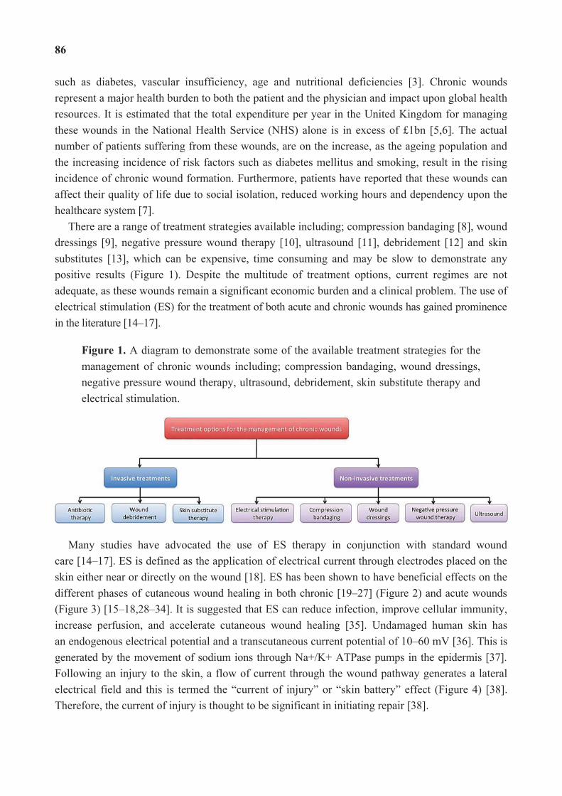
86
such as diabetes, vascular insufficiency, age and nutritional deficiencies [3]. Chronic wounds represent a major health burden to both the patient and the physician and impact upon global health resources. It is estimated that the total expenditure per year in the United Kingdom for managing these wounds in the National Health Service (NHS) alone is in excess of £1bn [5,6]. The actual number of patients suffering from these wounds, are on the increase, as the ageing population and the increasing incidence of risk factors such as diabetes mellitus and smoking, result in the rising incidence of chronic wound formation. Furthermore, patients have reported that these wounds can affect their quality of life due to social isolation, reduced working hours and dependency upon the healthcare system [7].
There are a range of treatment strategies available including; compression bandaging [8], wound dressings [9], negative pressure wound therapy [10], ultrasound [11], debridement [12] and skin substitutes [13], which can be expensive, time consuming and may be slow to demonstrate any positive results (Figure 1). Despite the multitude of treatment options, current regimes are not adequate, as these wounds remain a significant economic burden and a clinical problem. The use of electrical stimulation (ES) for the treatment of both acute and chronic wounds has gained prominence in the literature [14–17].
Figure 1. A diagram to demonstrate some of the available treatment strategies for the management of chronic wounds including; compression bandaging, wound dressings, negative pressure wound therapy, ultrasound, debridement, skin substitute therapy and electrical stimulation.
Many studies have advocated the use of ES therapy in conjunction with standard wound care [14–17]. ES is defined as the application of electrical current through electrodes placed on the skin either near or directly on the wound [18]. ES has been shown to have beneficial effects on the different phases of cutaneous wound healing in both chronic [19–27] (Figure 2) and acute wounds (Figure 3) [15–18,28–34]. It is suggested that ES can reduce infection, improve cellular immunity, increase perfusion, and accelerate cutaneous wound healing [35]. Undamaged human skin has an endogenous electrical potential and a transcutaneous current potential of 10–60 mV [36]. This is generated by the movement of sodium ions through Na+/K+ ATPase pumps in the epidermis [37]. Following an injury to the skin, a flow of current through the wound pathway generates a lateral electrical field and this is termed the “current of injury” or “skin battery” effect (Figure 4) [38]. Therefore, the current of injury is thought to be significant in initiating repair [38].
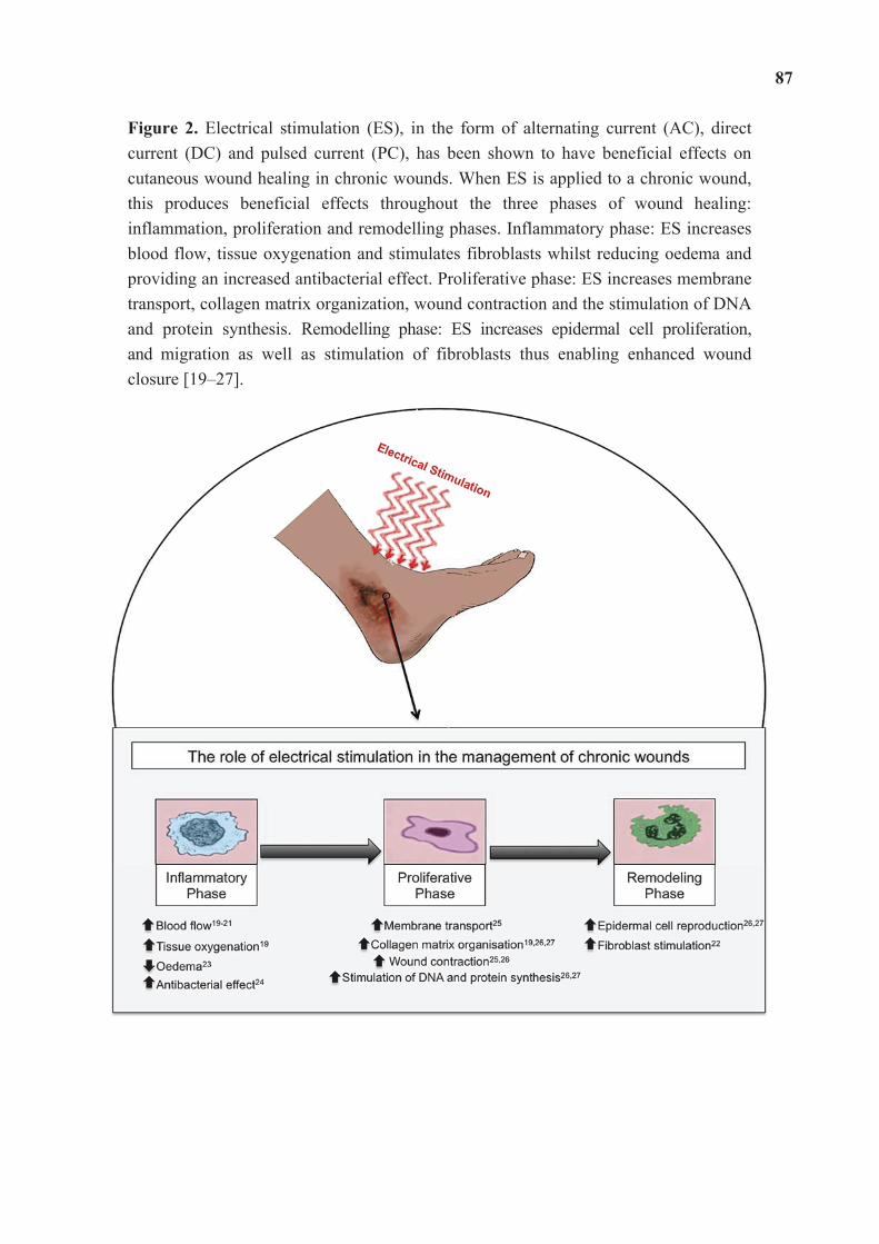
87
Figure 2. Electrical stimulation (ES), in the form of alternating current (AC), direct current (DC) and pulsed current (PC), has been shown to have beneficial effects on cutaneous wound healing in chronic wounds. When ES is applied to a chronic wound, this produces beneficial effects throughout the three phases of wound healing: inflammation, proliferation and remodelling phases. Inflammatory phase: ES increases blood flow, tissue oxygenation and stimulates fibroblasts whilst reducing oedema and providing an increased antibacterial effect. Proliferative phase: ES increases membrane transport, collagen matrix organization, wound contraction and the stimulation of DNA and protein synthesis. Remodelling phase: ES increases epidermal cell proliferation, and migration as well as stimulation of fibroblasts thus enabling enhanced wound closure [19–27].
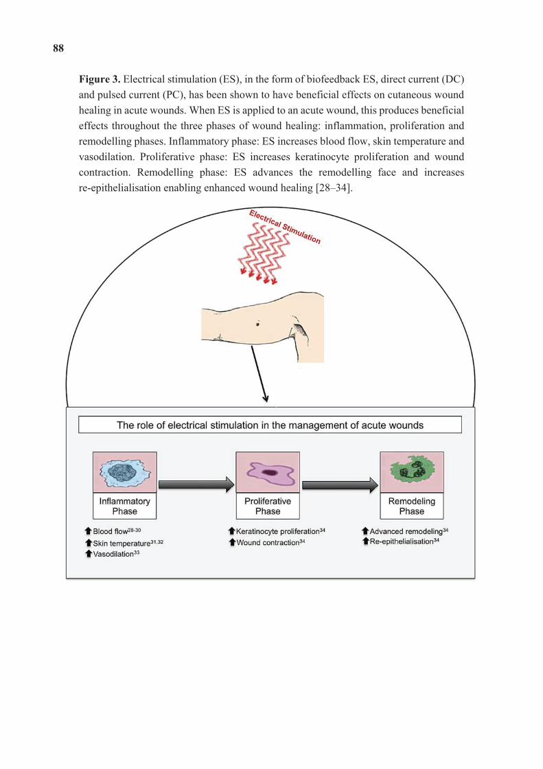
88
Figure 3. Electrical stimulation (ES), in the form of biofeedback ES, direct current (DC) and pulsed current (PC), has been shown to have beneficial effects on cutaneous wound healing in acute wounds. When ES is applied to an acute wound, this produces beneficial effects throughout the three phases of wound healing: inflammation, proliferation and remodelling phases. Inflammatory phase: ES increases blood flow, skin temperature and vasodilation. Proliferative phase: ES increases keratinocyte proliferation and wound contraction. Remodelling phase: ES advances the remodelling face and increases re-epithelialisation enabling enhanced wound healing [28–34].
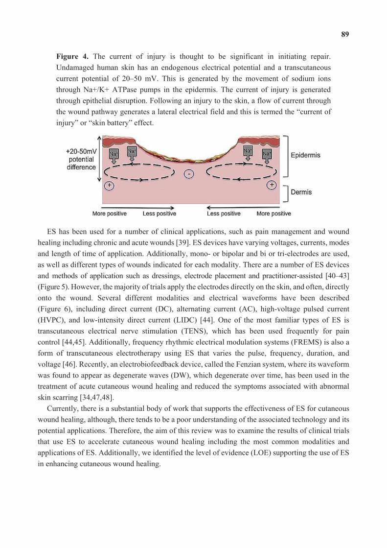
89
Figure 4. The current of injury is thought to be significant in initiating repair. Undamaged human skin has an endogenous electrical potential and a transcutaneous current potential of 20–50 mV. This is generated by the movement of sodium ions through Na+/K+ ATPase pumps in the epidermis. The current of injury is generated through epithelial disruption. Following an injury to the skin, a flow of current through the wound pathway generates a lateral electrical field and this is termed the “current of injury” or “skin battery” effect.
ES has been used for a number of clinical applications, such as pain management and wound healing including chronic and acute wounds [39]. ES devices have varying voltages, currents, modes and length of time of application. Additionally, mono- or bipolar and bi or tri-electrodes are used, as well as different types of wounds indicated for each modality. There are a number of ES devices and methods of application such as dressings, electrode placement and practitioner-assisted [40–43] (Figure 5). However, the majority of trials apply the electrodes directly on the skin, and often, directly onto the wound. Several different modalities and electrical waveforms have been described (Figure 6), including direct current (DC), alternating current (AC), high-voltage pulsed current (HVPC), and low-intensity direct current (LIDC) [44]. One of the most familiar types of ES is transcutaneous electrical nerve stimulation (TENS), which has been used frequently for pain control [44,45]. Additionally, frequency rhythmic electrical modulation systems (FREMS) is also a form of transcutaneous electrotherapy using ES that varies the pulse, frequency, duration, and voltage [46]. Recently, an electrobiofeedback device, called the Fenzian system, where its waveform was found to appear as degenerate waves (DW), which degenerate over time, has been used in the treatment of acute cutaneous wound healing and reduced the symptoms associated with abnormal skin scarring [34,47,48].
Currently, there is a substantial body of work that supports the effectiveness of ES for cutaneous wound healing, although, there tends to be a poor understanding of the associated technology and its potential applications. Therefore, the aim of this review was to examine the results of clinical trials that use ES to accelerate cutaneous wound healing including the most common modalities and applications of ES. Additionally, we identified the level of evidence (LOE) supporting the use of ES in enhancing cutaneous wound healing.
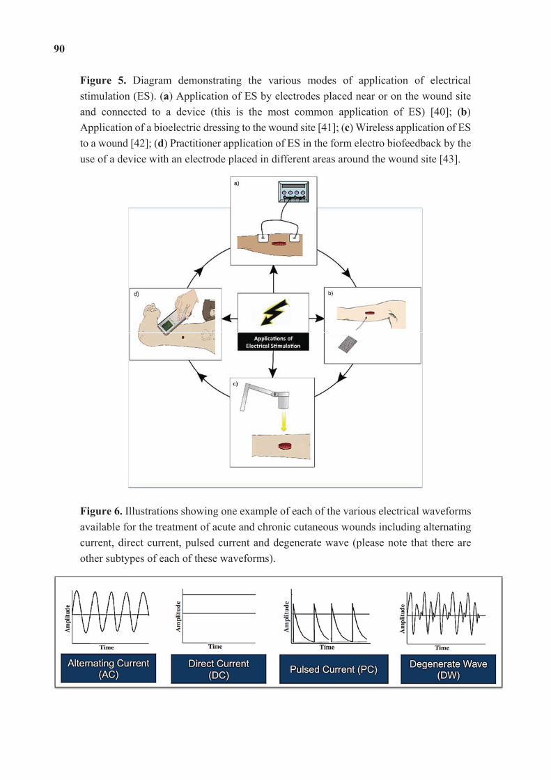
90
Figure 5. Diagram demonstrating the various modes of application of electrical stimulation (ES). (a) Application of ES by electrodes placed near or on the wound site and connected to a device (this is the most common application of ES) [40]; (b) Application of a bioelectric dressing to the wound site [41]; (c) Wireless application of ES to a wound [42]; (d) Practitioner application of ES in the form electro biofeedback by the use of a device with an electrode placed in different areas around the wound site [43].
Figure 6. Illustrations showing one example of each of the various electrical waveforms available for the treatment of acute and chronic cutaneous wounds including alternating current, direct current, pulsed current and degenerate wave (please note that there are other subtypes of each of these waveforms).
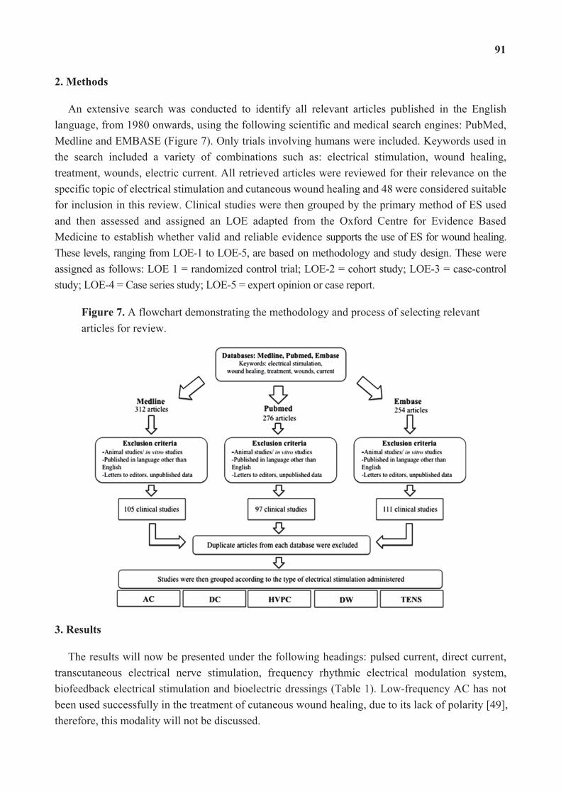
91
2. Methods
An extensive search was conducted to identify all relevant articles published in the English language, from 1980 onwards, using the following scientific and medical search engines: PubMed, Medline and EMBASE (Figure 7). Only trials involving humans were included. Keywords used in the search included a variety of combinations such as: electrical stimulation, wound healing, treatment, wounds, electric current. All retrieved articles were reviewed for their relevance on the specific topic of electrical stimulation and cutaneous wound healing and 48 were considered suitable for inclusion in this review. Clinical studies were then grouped by the primary method of ES used and then assessed and assigned an LOE adapted from the Oxford Centre for Evidence Based Medicine to establish whether valid and reliable evidence supports the use of ES for wound healing. These levels, ranging from LOE-1 to LOE-5, are based on methodology and study design. These were assigned as follows: LOE 1 = randomized control trial; LOE-2 = cohort study; LOE-3 = case-control study; LOE-4 = Case series study; LOE-5 = expert opinion or case report.
Figure 7. A flowchart demonstrating the methodology and process of selecting relevant articles for review.
3. Results
The results will now be presented under the following headings: pulsed current, direct current, transcutaneous electrical nerve stimulation, frequency rhythmic electrical modulation system, biofeedback electrical stimulation and bioelectric dressings (Table 1). Low-frequency AC has not been used successfully in the treatment of cutaneous wound healing, due to its lack of polarity [49], therefore, this modality will not be discussed.
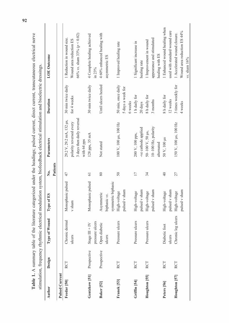
92
Tab
le 1
. A s
umm
ary
tabl
e of
the
liter
atur
e ca
tego
rized
und
er th
e he
adin
gs; p
ulse
d cu
rren
t, di
rect
cur
rent
, tra
nscu
tane
ous
elec
trica
l ner
ve
stim
ulat
ion,
freq
uenc
y rh
ythm
ic e
lect
rical
mod
ulat
ion
syst
em, b
iofe
edba
ck e
lect
rical
stim
ulat
ion
and
bioe
lect
ric d
ress
ings
.
Aut
hor
Des
ign
Typ
e of
Wou
nd
Typ
e of
ES
No.
Pa
tient
s Pa
ram
eter
s D
urat
ion
LO
E O
utco
me
Puls
ed C
urre
nt
Feed
ar [5
0]
RC
T C
hron
ic d
erm
al
ulce
rs
Mon
opha
sic
puls
ed
v sh
am
47
29.2
V, 2
9.2
mA
, 132
s,
pola
rity
reve
rsed
eve
ry
3 da
ys th
en d
aily
reve
rsal
w
ith 6
4 pp
s
30 m
in tw
ice
daily
fo
r 4 w
eeks
1
Redu
ctio
n in
wou
nd si
ze.
Wou
nd a
rea
redu
ctio
n ES
66
% v
s. sh
am 3
3% (p
< 0
.02)
Gen
tzko
w [5
1]
Pros
pect
ive
Stag
e II
I + IV
pr
essu
re u
lcer
s M
onop
hasi
c pu
lsed
61
128
pps,
35 m
A
30 m
in tw
ice
daily
4
Com
plet
e he
alin
g ac
hiev
ed
in 2
3%
Bak
er [5
2]
Pros
pect
ive
Ope
n di
abet
ic
ulce
rs
Asy
mm
etric
bi
phas
ic v
s. sy
mm
etric
bip
hasi
c
80
Not
stat
ed
Unt
il ul
cers
hea
led
4 60
% e
nhan
ced
heal
ing
with
as
ymm
etric
ES
Fran
ek [5
3]
RC
T Pr
essu
re u
lcer
s H
igh-
volta
ge
puls
ed v
sham
50
10
0 V
, 100
s,
100
Hz
50 m
in, o
nce
daily
5
days
a w
eek
for
6
wee
ks
1 Im
prov
ed h
ealin
g ra
te
Gri
ffin
[54]
R
CT
Pres
sure
ulc
ers
Hig
h-vo
ltage
pu
lsed
v sh
am
17
200
V, 1
00 p
ps,
-ve
cath
ode
appl
ied
1 h
daily
for
20
day
s 1
Sign
ifica
nt in
crea
se in
he
alin
g ra
te
Hou
ghto
n [5
5]
RC
T Pr
essu
re u
lcer
s H
igh-
volta
ge
puls
ed v
sham
34
50
–100
V, 5
0 s,
10
–100
Hz,
pol
arity
al
tern
ated
8 h
daily
for
3
mon
ths
1 Im
prov
emen
t in
wou
nd
appe
aran
ce a
nd st
imul
ated
he
alin
g w
ith E
S Pe
ters
[56]
R
CT
Dia
betic
foot
ul
cers
H
igh-
volta
ge
puls
ed v
sham
40
50
V, 1
00
s 8
h da
ily fo
r
12 w
eeks
1
Enha
nced
wou
nd h
ealin
g w
hen
used
with
stan
dard
wou
nd c
are
Hou
ghto
n [5
7]
RC
T C
hron
ic le
g ul
cers
Hig
h-vo
ltage
pu
lsed
v sh
am
27
150
V, 1
00
s, 10
0 H
z 3
times
wee
kly
for
4 w
eeks
1
Acc
eler
ated
wou
nd c
losu
re.
Wou
nd a
rea
redu
ctio
n ES
44%
vs
. sha
m 1
6%

93
Tab
le 1
. Con
t.
Aut
hor
Des
ign
Typ
e of
Wou
ndT
ype
of E
S N
o.
Patie
nts
Para
met
ers
Dur
atio
n L
OE
Out
com
e
Puls
ed C
urre
nt
Bur
dge
[58]
R
etro
spec
tive
Chr
onic
dia
betic
w
ound
s H
igh-
volta
ge
puls
ed
30
<140
V, 9
0–10
0 s,
55
.19
Hz
45 m
in se
ssio
ns,
3 tim
es w
eekl
y un
til h
eale
d ap
prox
. 16
wee
ks
4 Im
prov
ed h
ealin
g
Gol
dman
[59]
R
CT
Isch
emic
w
ound
s H
igh-
volta
ge
puls
ed v
sham
8
100
pps,
360
V,
-ve
pola
rity
1 h
daily
for
14
wee
ks
2 In
crea
sed
vaso
dila
tion
and
derm
al c
apill
ary
form
atio
n A
hmad
[60]
R
CT
Pres
sure
ulc
ers
Hig
h-vo
ltage
pu
lsed
v sh
am
60
100–
175
V, 5
0 s,
120
Hz
Gro
up 1
: 45
min
, G
roup
2: 6
0 m
in,
Gro
up 3
: 120
min
; da
ily fo
r 5 w
eeks
1 Im
prov
ed h
ealin
g w
ith E
S
Dir
ect C
urre
nt
Gau
lt [6
1]
RC
T Is
chem
ic u
lcer
s LI
DC
v sh
am
12
Not
stat
ed
Unt
il he
aled
1
LID
C g
roup
hea
led
twic
e as
fa
st a
s con
trol
Adu
nsky
[59]
R
CT
Pres
sure
ulc
ers
DC
63
N
ot st
ated
8
wee
ks
1 D
C u
sefu
l com
bine
d w
ith
stan
dard
wou
nd c
are
Car
ley
[46]
R
etro
spec
tive
Sacr
al/b
elow
kn
ee u
lcer
s LI
DC
30
30
0–50
0 A
for
norm
ally
inne
rvat
ed a
nd
500–
700
A fo
r de
nerv
ated
skin
2 h,
5 d
ays a
wee
k fo
r 5 w
eeks
3
LID
C im
prov
ed h
ealin
g.
Wou
nd a
re re
duct
ion
ES 8
9%
vs. c
ontro
l 37%
(p <
0.0
1)
Wir
sing
[42]
C
ontro
lled
Dia
betic
leg
and
foot
ulc
ers
Wire
less
LID
C
47
1.5
A
2–3
times
wee
kly,
45
–60
min
sess
ions
, fo
r 8 w
eeks
2 Si
gnifi
cant
ly
acce
lera
ted
heal
ing
Woo
d [6
2]
Plac
ebo
cont
rolle
d Ch
roni
c de
cubi
tus u
lcer
s Pu
lsed
LID
C
74
300–
600
A
8 w
eeks
1
Fibr
obla
st a
nd k
erat
inoc
ytes
gr
owth
enh
ance
d.
Incr
ease
d he
alin
g ra
te
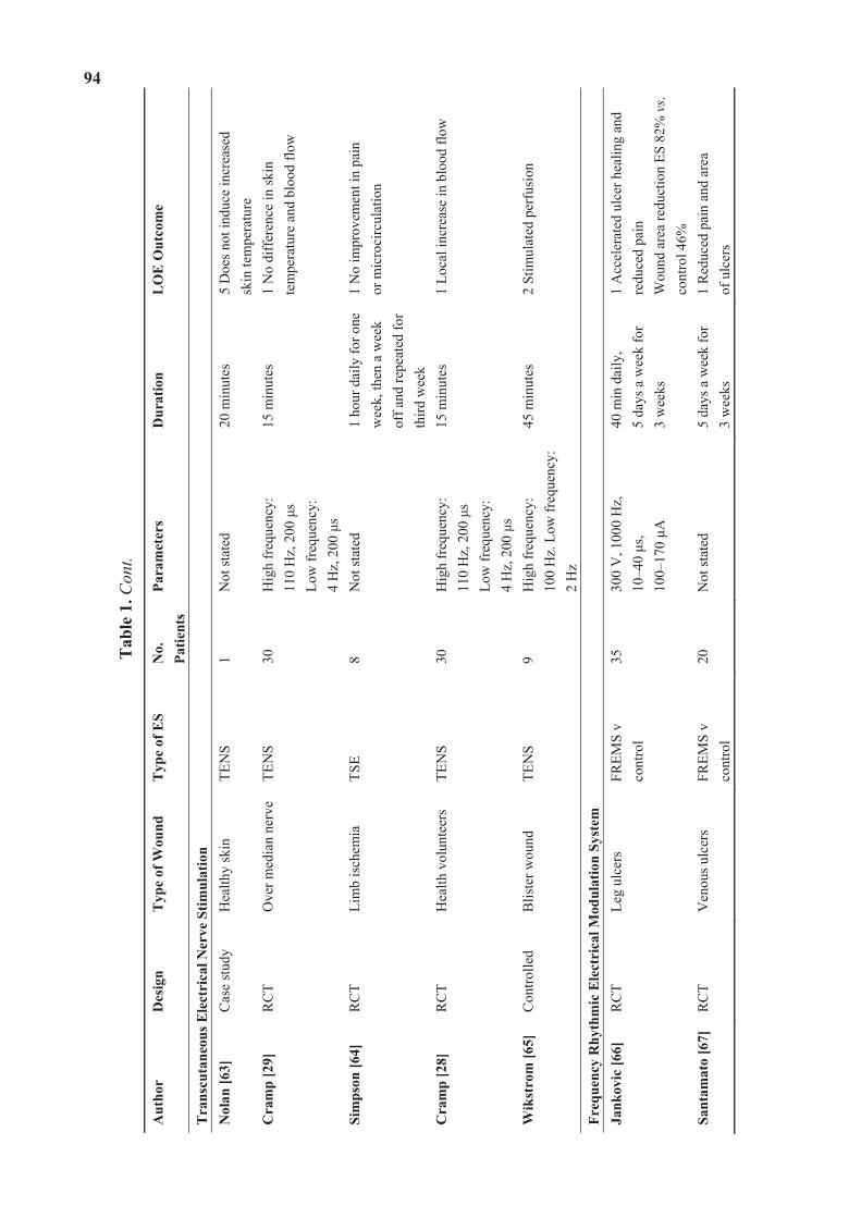
94
Tab
le 1
. Con
t.
Aut
hor
Des
ign
Typ
e of
Wou
nd
Typ
e of
ES
No.
Pa
tient
s Pa
ram
eter
s D
urat
ion
LO
E O
utco
me
Tra
nscu
tane
ous E
lect
rica
l Ner
ve S
timul
atio
n
Nol
an [6
3]
Cas
e st
udy
Hea
lthy
skin
TE
NS
1 N
ot st
ated
20
min
utes
5
Doe
s not
indu
ce in
crea
sed
sk
in te
mpe
ratu
re
Cra
mp
[29]
R
CT
Ove
r med
ian
nerv
eTE
NS
30
Hig
h fr
eque
ncy:
11
0 H
z, 2
00
s Lo
w fr
eque
ncy:
4
Hz,
200
s
15 m
inut
es
1 N
o di
ffer
ence
in sk
in
tem
pera
ture
and
blo
od fl
ow
Sim
pson
[64]
R
CT
Lim
b is
chem
ia
TSE
8 N
ot st
ated
1
hour
dai
ly fo
r one
w
eek,
then
a w
eek
off a
nd re
peat
ed fo
r th
ird w
eek
1 N
o im
prov
emen
t in
pain
or
mic
roci
rcul
atio
n
Cra
mp
[28]
R
CT
Hea
lth v
olun
teer
s TE
NS
30
Hig
h fr
eque
ncy:
11
0 H
z, 2
00
s Lo
w fr
eque
ncy:
4
Hz,
200
s
15 m
inut
es
1 Lo
cal i
ncre
ase
in b
lood
flow
Wik
stro
m [6
5]
Con
trolle
d B
liste
r wou
nd
TEN
S 9
Hig
h fr
eque
ncy:
10
0 H
z. L
ow fr
eque
ncy:
2
Hz
45 m
inut
es
2 St
imul
ated
per
fusi
on
Freq
uenc
y R
hyth
mic
Ele
ctri
cal M
odul
atio
n Sy
stem
Ja
nkov
ic [6
6]
RC
T Le
g ul
cers
FR
EMS
v co
ntro
l 35
30
0 V
, 100
0 H
z,
10–4
0 s,
10
0–17
0 A
40 m
in d
aily
, 5
days
a w
eek
for
3
wee
ks
1 A
ccel
erat
ed u
lcer
hea
ling
and
redu
ced
pain
W
ound
are
a re
duct
ion
ES 8
2% v
s. co
ntro
l 46%
Sa
ntam
ato
[67]
RC
T V
enou
s ulc
ers
FREM
S v
cont
rol
20
Not
stat
ed
5 da
ys a
wee
k fo
r
3 w
eeks
1
Red
uced
pai
n an
d ar
ea
of u
lcer
s
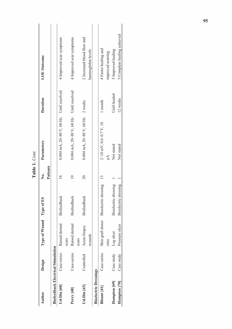
95
Tab
le 1
. Con
t.
Aut
hor
Des
ign
Typ
e of
Wou
ndT
ype
of E
S N
o.
Patie
nts
Para
met
ers
Dur
atio
n L
OE
Out
com
e
Bio
feed
back
Ele
ctri
cal S
timul
atio
n
Ud-
Din
[68]
C
ase-
serie
s R
aise
d de
rmal
sc
ars
Bio
feed
back
18
0.
004
mA
, 20–
80 V
, 60
Hz
Unt
il re
solv
ed
4 Im
prov
ed sc
ar sy
mpt
oms
Perr
y [4
8]
Cas
e-se
ries
Rai
sed
derm
al
scar
s B
iofe
edba
ck
19
0.00
4 m
A, 2
0–80
V, 6
0 H
zU
ntil
reso
lved
4
Impr
oved
scar
sym
ptom
s
Ud-
Din
[43]
C
ontro
lled
Acu
te b
iops
y w
ound
s B
iofe
edba
ck
20
0.00
4 m
A, 2
0–80
V, 6
0 H
z2
wee
ks
2 In
crea
sed
bloo
d flo
w a
nd
haem
oglo
bin
leve
ls
Bio
elec
tric
Dre
ssin
gs
B
loun
t [41
] C
ase-
serie
s Sk
in g
raft
dono
r si
tes
Bio
elec
tric
dres
sing
13
2–10
mV
, 0.6
–0.7
V, 1
0 A
1
mon
th
4 Fa
ster
hea
ling
and
im
prov
ed sc
arrin
g H
ampt
on [6
9]
Cas
e st
udy
Leg
ulce
r B
ioel
ectri
c dr
essi
ng1
Not
stat
ed
Unt
il he
aled
5
Impr
oved
hea
ling
Ham
pton
[70]
C
ase
stud
y Pr
essu
re u
lcer
B
ioel
ectri
c dr
essi
ng1
Not
stat
ed
12 w
eeks
5
Com
plet
e he
alin
g ac
hiev
ed
96
3.1. Pulsed Current
Pulsed current (PC) is the unidirectional or bidirectional flow of electrons or ions, and has two waveforms: monophasic or biphasic [49]. Monophasic PC can also be described as low-voltage [52] and high voltage [53,71]. Biphasic PC is bidirectional and its waveform can be asymmetric or symmetric. PC is able to mimic the physiological endogenous current [49]. PC is delivered to the wound tissues by conductive coupling with a hydrogel or moist gauze filling the defect and the electrodes of appropriate polarity placed on top [49]. The majority of studies which used pulsed current are unidirectional.
Low voltage PC (LVPC) devices deliver continuous DC and monophasic and biphasic waveforms of longer durations and lower voltages (20–35 V) [49]. A number of clinical studies used an LVPC device named woundEL® and reported beneficial outcomes when using this for the treatment of ulcers [50,51,72]. The parameters used were: a duration of 132 microseconds and 64 pulses per second.
High-voltage pulsed current (HVPC) employs a monophasic pulsed current where the pulses are delivered in doubles. Each pulse is of short duration (less that 200 micro seconds) and it has a high peak voltage (150–500 V). HVPC is typically delivered by a device with both negative and positive electrodes either placed on the wound site or proximally on the skin [49]. This application has been used in wound healing, pain relief and oedema resolution [54,55]. A randomized controlled trial (LOE-1) conducted by Peters et al. studied 40 patients with diabetic foot ulcers for 12 weeks [56]. Patients were randomized to receive HVPC or sham therapy. Patients received 20 minutes of ES every hour for 8 hours each day over the 12-week study. Most patients healed in the ES group (65% compared to the sham group 35%), but the difference was not significant (p = 0.058). However, when patient compliance was evaluated, patients that used the device at least three times a week were more likely to heal than patients that received sham therapy and patients who used ES 0, 1, or 2 times a week (p = 0.038) [56].
An RCT (LOE-1) by Houghton et al. involved 27 patients with 42 chronic leg ulcers (arterial, venous, chronic) which were assigned to either a placebo or treatment group [57]. HVPC was delivered at 150 V, 100 pps and 100 microsecond duration. Treatments lasted for 45 minutes, 3 times a week for 4 weeks. The treatment group wounds significantly reduced in size (44%) compared to the sham group (16%). However, the significant differences were not maintained at the 1-month follow-up period. A retrospective study (LOE-4) also demonstrated positive results using HVPC in 30 patients with chronic diabetic wounds [58]. Furthermore, an RCT (LOE-1) also used this modality versus sham therapy in the treatment of ischemic wounds over a 14-week period and showed that the area of the wounds decreased and microcirculation was improved [59].
An RCT (LOE-1) was conducted with 60 subjects who had chronic pressure ulcers. They were split into 4 groups; one control who received sham therapy and three groups who received HVPC for 45, 60, 120 minutes respectively daily for one week [60]. Wound surface area was measured at 0, 3 and 5 weeks and they noted a significant reduction in the groups who received HVPC for 60 min and 120 min. However, no significant differences were noted between the treatment groups.
It is evident, that it is practically impossible to standardize chronic wounds in these studies, as each wound is substantially different to the next. Additionally, the research designs and device
97
parameters were not comparable across these studies. Therefore, further larger controlled trials are critical in order to determine the optimal dosage and mode of delivery of ES.
3.2. Direct Current
Continuous direct current is the unidirectional flow of charged particles, which flow for 1 second or longer, and is produced by batteries, thermo couplings and solar cells [73]. The length of time the current flows has been known to cause irritation and pH changes to the skin [74]. Pulsed direct current is a monophasic pulsed waveform which flows from 1 ms to 1second [49]. Direct current is able to mimic the physiological endogenous current [49]. In wound care, a low-intensity direct current (20–1000 microamps) is used to avoid damaging healthy tissue [61]. Low-intensity direct current has been shown to promote chronic wound healing by two mechanisms: galvanotaxis (by stimulating the migration of fibroblasts and keratinocytes [75] and its antimicrobial effect [61].
A study by Adunsky et al. (LOE-2), 38 patients with pressure ulcers were distributed equally between shams and treatment with DC application of electrical stimulation for 8 weeks [76]. The primary outcome was percentage change in the wound area, with the results showing that wound area reduction was 31% (ES group) vs. 4% (sham group) (p = 0.09). The relatively small sample size may have contributed to the lack of significance.
Gault et al. conducted an 8-week trial (LOE-2) using continuous LIDC to treat 76 patients with 106 ischaemic skin ulcers [61]. They applied the negative electrode directly onto the wound for three days in order to debride necrotic tissue. The current used was 200–800 microamps for two hours, three times daily. Six patients had bilateral ulcers and therefore one ulcer was treated as a control. Forty-eight of the 100 ulcers healed completely. In the patients who had controls, healing rate for the treated ulcers was 30% compared to 14.7% in the controls. Nevertheless, a larger control group would be needed for more meaningful results. In a controlled clinical trial (LOE-2) [46], 15 unspecified wounds were treated with continuous LIDC and 15 with conservative treatment for 5 weeks. The current was 300–700 microamps for two hours in two sessions per day, five days a week. The mean healing rate for the treatment group was 89%, compared to 45% for the control group. However, limitations of this study were that despite mentioning that they had conducted a follow-up, no details were reported of this. Furthermore, the randomization process was not rigorous; participants were paired according to their age, diagnosis, wound location and aetiology with each pair placed in one of two groups. Additionally, there was no blinding as this was not possible. A recent study (LOE-3) used a wireless micro current stimulation device for the treatment of 47 patients with leg and diabetic foot ulcers [42]. This was applied 2 or 3 times a week for 60 minutes per session combined with standard wound care. They demonstrated complete healing within 3 months for the majority of cases. This device is contactless and pain-free and different wounds can be treated at the same time.
Intermittent low-intensity direct current delivers a current, which goes up to 29.2 milliamps and then down to zero [73]. A double-blind multi-centred controlled trial (LOE-1) [62] evaluated the effect of this treatment on 43 patients with stage II and III pressure ulcers compared to 31 placebo (sham intermittent LIDC) patients. The current used was 300–600 milliamps. Twenty-five ulcers in the treatment group healed completely within 8 weeks (p < 0.001), compared to 4 in the control
98
group, which had healed up to 80%. These positive results indicate a beneficial effect of intermittent LIDC, however, there was no report of randomization and no explanation for the difference in size of the two groups. Feedar et al. conducted a double-blind multi-centred RCT (LOE-1) using intermittent LIDC with 47 patients with 50 ulcers, which were split into control and treatment groups [50]. The current was applied at 35 milliamps, which was applied for 30 minutes, twice daily on a daily basis. They showed a statistically significant difference between the groups; the mean healing rate was 56% in the treatment group compared to 33% in the control group (p < 0.02).
3.3. Transcutaneous Electrical Nerve Stimulation
Transcutaneous Electrical Nerve Stimulation (TENS) is a low-frequency, pulsed electrical current transmitted by electrodes through the skin surface [77] to stimulate the peripheral nerves to produce various physiological effects [78]. The biphasic pulses are most commonly used [79,80]. TENS is considered to be one of the most common therapeutic modalities used in clinical practice for the relief of chronic and acute pain [78]. Some authors have observed that, in addition to its analgesic effects, TENS can also alter skin temperature and increase blood flow [40]. This observation has lead to various studies investigating the effect on the peripheral vascular system and how this facilitates tissue repair [81]. There are disagreements in the literature with regard to the increase in blood flow and skin temperature. Some studies have shown that TENS significantly increases skin temperature with low- (2 Hz to 4 Hz) [31] and high-frequency (75 Hz to 100 Hz) TENS [63], and in local blood flow [31]. However, some studies have not shown any significant increase of blood flow [29] and temperature [64] with the use of TENS. Interestingly, some studies suggested that when applied at the same intensity, low-frequency TENS enhanced blood flow levels more than high-frequency TENS [28,65].
3.4. Frequency Rhythmic Electrical Modulation System
Frequency rhythmic electrical modulation system (FREMS) is a form of transcutaneous electrotherapy using ES that automatically varies the pulse, frequency, duration, and voltage [46]. Two RCTs have been conducted utilising this for the treatment of chronic leg ulcers in order to improve wound healing [66,67]. The first RCT (LOE-1) recruited 35 patients and divided them into two groups [66]. One group received FREMS treatment for 2 months and the control group of no treatment. Their results showed that ulcer improvement in the treatment group was significantly higher than compared to the control group. However, a larger sample size would be needed for future studies. Another RCT (LOE-1) used FREMS treatment in 20 older patients with chronic and painful venous leg ulcers [67]. One group of 10 patients received FREMS and a topical treatment, whilst the control group received topical treatment only. 15 treatments were performed over a period of 3 weeks. They showed that there was a statistically significant decrease in ulcer area when treated with FREMS compared to the control group. Again, the small sample size means that further studies are necessary to investigate this treatment more robustly.
99
3.5. Biofeedback ES
An electro biofeedback device termed the Fenzian system (Fenzian Ltd, Hungerford, UK), where its waveform was deciphered and shown to resemble degenerate waves, has been used successfully in the treatment of symptoms in keloid and hypertrophic scarring and in accelerating the process of acute wound healing in the skin [34,47,48,82]. It is a transcutaneous low intensity device, which detects changes in skin impedance. This device forms part of an electrobiofeedback link with the individual’s normal physiological repair. This modality follows the theory that the normal electrical potential of skin forms a global electrical network reflecting the underlying neurological activity through changes in skin impedance [82]. Using a concentric electrode the device detects the skin’s electrical impedance and adjusts the outgoing microcurrent electrical biofeedback impulses [82]. The device delivers 0.004 milliamps, 20–80 V, has a frequency default of 60 Hz and impulses which last approximately six-hundredth of a second.
It has been used successfully to alleviate the symptoms for pain, pruritus and inflammation in two case series (LOE-4) on raised dermal scars [48,68]. It is postulated that this treatment can be beneficial in the treatment of abnormal skin scarring as it may negate the need for long-term pain medications. Furthermore, a clinical trial (LOE-2) was conducted involving multiple temporal punch biopsies treated with biofeedback ES and demonstrated increased blood flow and haemoglobin levels in acute cutaneous wounds (on day 14 post-wounding) created in 20 human volunteers compared to controls which had not received ES [43]. This treatment modality accelerated the rate of cutaneous wound healing in all cases as evidenced by gene and protein studies showing up-regulated angiogenesis and down-regulated inflammation [34]. Additional larger randomised controlled trials are required to investigate this treatment further to identify if this could be beneficial in patients with chronic wounds.
3.6. Bioelectric Dressings
Bioelectric dressings are emerging as a useful method of delivering ES to the wound site. However, studies of these specific modalities are lacking. Procellera® is a woven metallic bandage with embedded microbatteries, which is used as a dressing for partial or full thickness wounds. The mechanism of action is delivery of ES to the wound site. It produces a low voltage of 2–10 mV by microbatteries of Ag and Zn metals which are inside a woven material and are activated by the moisture in the wound delivers 0.6–0.7 V at 10 microamps. In a study by Blount et al. [41], 13 patients had skin grafting and the Procellera® dressing was applied to half of the donor site area. They noted improved healing, scarring and patient subjective outcomes. However, a larger trial is required to substantiate these results further. Another bioelectric wound dressing, named the PosiFect RD® DC device, has been used in treating pressure and venous ulcers [69,70]. This dressing contains a miniature electrical circuit delivering a microcurrent to the wound bed for a minimum of 48 hours and has shown promise in treating these chronic wounds.
100
4. Discussion
The reported studies demonstrate considerable variability in the parameters of ES application, leading to difficulty in generating sufficient evidence to support any one standard therapeutic approach. Most studies reported successful positive outcomes using ES to accelerate wound healing. Nonetheless, the differences in types of ulcerations or wounds, ES parameter settings and limited power of study design make synthesis of the results difficult and to draw conclusions as to an optimal mode of ES or type of ES device.
The level of evidence assigned for each study showed variations amongst the types of ES. The majority of LOE-1 were for HVPC, TENS, FREMS and DC. The available evidence (n = 4) suggests that HVPC is most beneficial for pressure ulcer treatment, with these LOE-1 studies demonstrating improved healing rates with the application of this modality. Despite a limited number of studies, HVPC has also shown positive results when used in diabetic (n = 2), ischaemic (n = 1) and chronic leg ulcers (n = 1). However, it is not apparent if other types of wounds such as acute wounds or venous ulcers would respond differently to this therapy. FREMS has been used in the treatment of leg ulcers and have shown promising results by accelerating ulcer healing and the area of the ulcer. Nevertheless, there were only two RCTs (LOE-1); thus, it is difficult to identify whether this treatment is effective in other wound types. Additionally, HVPC and DC stimulation demonstrate higher levels of evidence when compared to biofeedback ES and bioelectric dressings, which are based on case series and case study reports. There is limited clinical evidence regarding ES application for acute wounds in comparison to chronic wounds.
ES has mainly been evaluated in pressure ulcers, venous ulcers, vascular ulcers and diabetic foot wounds. One of the challenges in interpreting these data is the variation in outcome measurements, type of ES, and how the therapy was dosed in the trials. Most of the studies were small and many had a short treatment period and limited follow-up. In addition, many of the studies did not use complete wound healing (i.e., complete wound closure) as the primary outcome. Due to the short duration of the studies, change in wound area was often used instead of wound closure. As it is difficult to standardize chronic wounds, it is important to look at acute wound studies for the effects ES has on these wounds. There was a lack of human controlled trials investigating the role of ES in acute cutaneous wounds. Biofeedback ES has been shown to be an effective method for enhancing cutaneous wound healing. A significantly increased blood flow was noted on day 14 in a controlled study [43]. Nevertheless, based on the findings to date, it remains difficult to ascertain which phases of wound healing this particular device would be optimal for. Importantly, it is of note that not all applications and modalities of ES have an effect on all phases of wound healing (Figure 8).
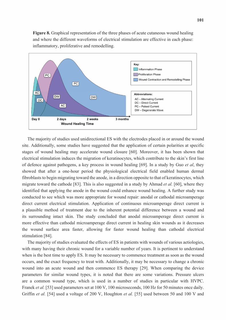
101
Figure 8. Graphical representation of the three phases of acute cutaneous wound healing and where the different waveforms of electrical stimulation are effective in each phase: inflammatory, proliferative and remodelling.
The majority of studies used unidirectional ES with the electrodes placed in or around the wound site. Additionally, some studies have suggested that the application of certain polarities at specific stages of wound healing may accelerate wound closure [60]. Moreover, it has been shown that electrical stimulation induces the migration of keratinocytes, which contribute to the skin’s first line of defence against pathogens, a key process in wound healing [69]. In a study by Guo et al, they showed that after a one-hour period the physiological electrical field enabled human dermal fibroblasts to begin migrating toward the anode, in a direction opposite to that of keratinocytes, which migrate toward the cathode [83]. This is also suggested in a study by Ahmad et al. [60], where they identified that applying the anode in the wound could enhance wound healing. A further study was conducted to see which was more appropriate for wound repair: anodal or cathodal microamperage direct current electrical stimulation. Application of continuous microamperage direct current is a plausible method of treatment due to the inherent potential difference between a wound and its surrounding intact skin. The study concluded that anodal microamperage direct current is more effective than cathodal microamperage direct current in healing skin wounds as it decreases the wound surface area faster, allowing for faster wound healing than cathodal electrical stimulation [84].
The majority of studies evaluated the effects of ES in patients with wounds of various aetiologies, with many having their chronic wound for a variable number of years. It is pertinent to understand when is the best time to apply ES. It may be necessary to commence treatment as soon as the wound occurs, and the exact frequency to treat with. Additionally, it may be necessary to change a chronic wound into an acute wound and then commence ES therapy [29]. When comparing the device parameters for similar wound types, it is noted that there are some variations. Pressure ulcers are a common wound type, which is used in a number of studies in particular with HVPC. Franek et al. [53] used parameters set at 100 V, 100 microseconds, 100 Hz for 50 minutes once daily. Griffin et al. [54] used a voltage of 200 V, Houghton et al. [55] used between 50 and 100 V and
102
Ahmad et al. [60] applied 100–175 V. Therefore, the voltages applied in these studies tend to vary. Additionally, the length of time ES is to be performed is approximately similar across some studies; 50 minutes [53] and 60 minutes [54], whilst Griffin et al. [54] applied HVPC over a period of 8 hours per day. Ahmad et al. [60] compared different durations of treatment over three groups; 45 minutes, 60 minutes and 120 minutes. They noted that 60 and 120 minute groups when applied for 7 days a week for 5 weeks demonstrated optimal healing compared to the 45 minute group. In diabetic wounds, similar parameters were used for HVPC across some studies [51,71]. These studies used an interphase interval of 100 microseconds and a voltage between 50 and 140 V; however, duration of treatment times varied. Further studies comparing the parameters for different wound types and types of ES would be useful to identify the optimal settings for each device in specific wounds.
Koel et al. [85] summarized the results of effect studies with ES as an additional treatment to standard wound care. They used forest plots and identified the healing rate, which was expressed as the percentage area reduction within 4 weeks of treatment. Their results showed that unidirectional ES and standard wound care increases the reduction in wound surface area by 30.8%. In pressure ulcers, the results increased to 42.7% by 4 weeks. Additionally, they noted that unidirectional ES is most beneficial for pressure ulcers, whereas venous leg ulcers and diabetic foot ulcers have had positive results with bidirectional ES.
ES therapy is considered safe and easy to use, as no device-related complications or adverse effects have been reported to date. ES application is relatively cost effective compared to other comparative treatments. In those ES modalities which are administered by a practitioner, this can be performed by a single experienced practitioner and there is often no pain associated with the treatments.
Some authors suggested that compliance might be a factor that affects cutaneous wound healing in ES studies [56,69]. However, in most studies, therapy was provided in a hospital or clinic setting, therefore, patients attending clinic appointments determined the main measure of compliance. The study by Peters et al. [56] was the only study that provided an ES device for patients to use at home and they recorded the number of hours the device was used. There was no significant difference in the compliance rates between the two treatment groups. There was a trend demonstrating a dose response with ES. A higher proportion of wounds healed in compliant patients in the ES treatment group (71%), non-compliant patients in the ES treatment group (50%), compliant patients in the sham group (39%), and non-compliant patients in the sham group (29%) [56].
In summary, despite variations in the type of current, duration, and dosing of ES, the majority of studies showed a significant improvement in wound area reduction or wound healing compared to the standard of care or sham therapy as well as improved local perfusion. Furthermore, no device-related complications or adverse effects have been reported in the existing literature, therefore, indicating that the therapy is safe and easy to use. Additionally, as ES decreases bacterial infection, increases local perfusion and accelerates wound healing, it targets these main factors of significance in wound management. There are several questions which remain unanswered, including, the optimal method of delivering ES, identifying which wound types respond better to treatment and the ideal anatomical location, frequency, duration and time to commence the application of ES for each wound type. Overall, the evidence to date infers that further clinical trials
103
are much needed to aid in better understanding the optimal dosing, timing and type of ES to be used and to optimize the effectiveness and appropriate clinical application. No doubt, this is likely to be achieved in the future by comparing the effects of different ES modalities, treatment durations and frequencies on the rate and quality of healing in similar cutaneous wounds.
Author Contributions
Sara Ud-Din wrote the paper. Ardeshir Bayat wrote and edited the paper and is the senior author.
Conflict of Interest
The authors declare no conflict of interest.
References
1. Monaco, J.L.; Lawrence, W.T. Acute wound healing: An overview. Clin. Plast. Surg. 2003, 30, 1–12.
2. Greaves, N.S.; Iqbal, S.A.; Baguneid, M.; Bayat, A. The role of skin substitutes in the management of chronic cutaneous wounds. Wound Repair Regen. 2013, 21, 194–210.
3. Fonder, M.A.; Lazarus, G.S.; Cowan, D.A.; Aronson-Cook, B.; Kohli, A.R.; Mamelak, A.J. Treating the chronic wound: A practical approach to the care of nonhealing wounds and wound care dressings. J. Am. Acad. Dermatol. 2008, 58, 185–206.
4. Singh, A.; Halder, S.; Chumber, S.; Misra, M.C.; Sharma, L.K.; Srivastava, A. Meta-analysis of randomized controlled trials on hydrocolloid occlusive dressing versus conventional gauze dressing in the healing of chronic wounds. Asian J. Surg. 2004, 27, 326–332.
5. Posnett, J.; Franks, P.J. The burden of chronic wounds in the UK. Nurs. Times 2008, 104, 44–45.
6. Mustoe, T.A.; O’Shaughnessy, K.; Kloeters, O. Chronic wound pathogenesis and current treatment strategies: A unifying hypothesis. Plast. Reconstr. Surg. 2006, 117, 35S–41S.
7. Vileikyte, L. Diabetic foot ulcers: A quality of life issue. Diabetes Metab. Res. Rev. 2001, 17, 246–249.
8. Amsler, F.; Willenberg, T.; Blättler, W. In search of optimal compression therapy for venous leg ulcers: A meta-anlysis of studies comparing divers bandages with specially designed stockings. J. Vasc. Surg. 2009, 50, 668–674.
9. Carter, J.M.; Tingley-Kelley, K.; Warriner, I.R.A. Silver treatments and silver-impregnated dressings for the healing of leg wounds and ulcers: A systematic review and meta-analysis. J. Am. Acad. Dermatol. 2010, 63, 668–679.
10. Armstrong, D.G.; Marston, W.A.; Reyzelman, A.M.; Kirsner, R.S. Comparison of negative pressure wound therapy with an ultraportable mechanically powered device vs. traditional electrically powered device for the treatment of chronic lower extremity ulcers: A multicenter randomized- controlled trial. Wound Repair Regen. 2011, 19, 173–180.
11. Cullum, N.A.; Al-Kurdi, D.; Bell-Syer, S.E. Therapeutic ultrasound for venous leg ulcers. Cochrane Database Syst. Rev. 2010, doi:10.1002/14651858.CD001180.pub3.
104
12. Dumville, J.C.; Worthy, G.; Soares, M.O.; Bland, J.M.; Cullum, N.; Dowson, C.; Iglesias, C.; McCaughan, D.; Mitchell, J.L.; Nelson, E.A.; et al. VenUS II: A randomised controlled trial of larval therapy in the management of leg ulcers. Health Technol. Assess. 2009, 13, 1–206.
13. Greaves, N.S.; Benatar, B.; Baguneid, M.; Bayat, A. Single-stage application of a novel decellularized dermis for treatment-resistant lower limb ulcers: Positive outcomes assessed by SIAscopy, laser perfusion, and 3D imaging, with sequential timed histological analysis.Wound Repair Regen. 2013, 21, 813–822.
14. Snyder, R.J.; Kirsner, R.S.; Warriner, R.A.; Lavery, L.A.; Hanft, J.R.; Sheehan, P. Consensus recommendations on advancing the standard of care for treating neuropathic foot ulcers in patients with diabetes. Ostomy Wound Manag. 2010, 56, S1–S24.
15. Suh, H.; Petrofsky, J.; Fish, A.; Hernandez, V.; Mendoza, E.; Collins, K.; Yang, T.; Abdul, A.; Batt, J.; Lawson, D. A new electrode design to improve outcomes in the treatment of chronic non-healing wounds in diabetes. Diabetes Technol. Ther. 2009, 11, 315–322.
16. Petrofsky, J.; Lawson, D.; Prowse, M.; Suh, H.J. Effects of a 2-, 3- and 4-electrode stimulator design on current dispersion on the surface and into the limb during electrical stimulation in controls and patients with wounds. J. Med. Eng. Technol. 2008, 32, 485–497.
17. Jünger, M.; Arnold, A.; Zuder, D.; Stahl, H.W.; Heising, S. Local therapy and treatment costs of chronic, venous leg ulcers with electrical stimulation (Dermapulse): A prospective, placebo controlled, double blind trial. Wound Repair Regen. 2008, 16, 480–487.
18. Isseroff, R.R.; Dahle, S.E. Electrical stimulation therapy and wound healing: Where are we now? Adv. Wound Care 2012, 1, 238–243.
19. Reger, S.I.; Hyodo, A.; Negami, S.; Kambic, H.E.; Sahgal, V. Experimental wound healing with electrical stimulation. Artif. Organs 1999, 23, 460–462.
20. Gagnier, K.A.; Manix, N.; Baker, L. The effects of electrical stimulation on cutaneous oxygen supply in paraplegics. Phys. Ther. 1988, 68, 835–839.
21. Peters, E.J.; Armstrong, D.G.; Wunderlich, R.P.; Bosma, J.; Stacpoole-Shea, S.; Lavery, L.A. The benefit of electrical stimulation to enhance perfusion in persons with diabetes mellitus. J. Foot Ankle Surg. 1998, 37, 396–400.
22. Rouabhia, M.; Park, H.; Meng, S.; Derbali, H.; Zhang, Z. Electrical stimulation promotes wound healing by enhancing dermal fibroblast activity and promoting myofibroblast transdifferentiation. PLoS One 2013, 8, e71660.
23. Young, S.; Hampton, S.; Tadej, M. Study to evaluate the effect of low-intensity pulsed electrical currents on levels of oedema in chronic non-healing wounds. J. Wound Care 2011, 20,370–373.
24. Daeschlein, G.; Assadian, O.; Kloth, L.C.; Meinl, C.; Ney, F.; Kramer, A. Antibacterial activity of positive and negative polarity low-voltage pulsed current (LVPC) on six typical gram-positive and gram-negative bacterial pathogens of chronic wounds. Wound Repair Regen.2007, 15, 399–403.
25. Alvarez, O.M.; Mertz, P.M.; Smerbeck, R.V.; Eaglstein, W.H. The healing of superficial skin wounds is stimulated by external electrical current. J. Invest. Dermatol. 1983, 81, 144–148.
105
26. Bourguignon, G.J.; Bourguignon, L.Y. Electric stimulation of protein and DNA synthesis in human fibroblasts. FASEB J. 1987, 1, 398–402.
27. Bassett, C.A.L.; Herrmann, I. The effect of electrostatic fields on macromolecular synthesis by fibroblasts in vitro. J. Cell Biol. 1968, 9, 39.
28. Cramp, A.F.L.; Gilsenan, C.; Lowe, A.S.; Walsh, D.M. The effect of high- and low-frequency transcutaneous electrical nerve stimulation upon cutaneous blood flow and skin temperature in healthy subjects. Clin. Physiol. 2000, 20, 150–157.
29. Cramp, A.F.; Noble, J.G.; Lowe, A.S.; Walsh, D.M. Transcutaneous electrical nerve stimulation (TENS): The effect of electrode placement upon cutaneous blood flow and skin temperature. Acupunct. Electrother. Res. 2001, 26, 25–37.
30. Cramp, F.L.; McCullough, G.R.; Lowe, A.S.; Walsh, D.M. Transcutaneous electric nerve stimulation: The effect of intensity on local and distal cutaneous blood flow and skin temperature in healthy subjects. Arch. Phys. Med. Rehabil. 2002, 83, 5–9.
31. Kaada, B.; Olsen, E.; Eielsen, O. In search of mediators of skin vasodilation induced by transcutaneous nerve stimulation: III. Increase in plasma VIP in normal subjects and in Raynaud’s disease. Gen. Pharmacol. 1984, 15, 107–113.
32. Kaada, B.; Helle, K.B. In search of mediators of skin vasodilation induced by transcutaneous nerve stimulation: IV. In vitro bioassay of the vasoinhibitory activity of sera from patients suffering from peripheral ischemia. Gen. Pharmacol. 1984, 15, 115–122.
33. Abram, S.E.; Asiddao, C.B.; Reynolds, A.C. Increased skin temperature during transcutaneous electrical stimulation. Anesth. Analg. 1980, 59, 22–25.
34. Sebastian, A.; Syed, F.; Perry, D.; Balamurugan, V.; Colthurst, J.; Chaudhry, I.H.; Bayat, A. Acceleration of cutaneous healing by electrical stimulation: Degenerate electrical waveform down-regulates inflammation, up-regulates angiogenesis and advances remodelling in temporal punch biopsies in a human volunteer study. Wound Repair Regen. 2011, 19, 693–708.
35. Szuminky, N.J.; Albers, A.C.; Unger, P.; Eddy, J.G. Effect of narrow, pulsed high voltages on bacterial viability. Phys. Ther. 1994, 74, 660–667.
36. Foulds, I.S.; Barker, A.T. Human skin battery potentials and their possible role in wound healing. Br. J. Dermatol. 1983, 109, 515–522.
37. McGinnis, M.E.; Vanable, J.W., Jr. Voltage gradients in newt limb stumps. Prog. Clin. Biol. Res. 1986, 210, 213–238.
38. Barker, A.T.; Jaffe, L.F.; Vanable, J.W., Jr. The glabrous epidermis of cavies contains a powerful battery. Am. J. Physiol. 1982, 242, R358–R366.
39. Thakral, G.; Lafontaine, J.; Najafi, B.; Talal, T.K.; Kim, P.; Lavery, L.A. Electrical stimulation to accelerate wound healing. Diabet. Foot Ankle 2013, doi:10.3402/dfa.v4i0.22081.
40. Atalay, C.; Yilmaz, K.B. The effect of transcutaneous electrical nerve stimulation on postmastectomy skin flap necrosis. Breast Cancer Res. Treat. 2009, 117, 611–614.
41. Blount, A.L.; Foster, S.; Rapp, D.A.; Wilcox, R. The use of bioelectric dressings in skin graft harvest sites: A prospective case series. J. Burn Care Res. 2012, 33, 354–357.
106
42. Wirsing, P.G.; Habrom, A.D.; Zehnder, T.M.; Friedli, S.; Blatti, M. Wireless micro current stimulation—An innovative electrical stimulation method for the treatment of patients with leg and diabetic foot ulcers. Int. Wound J. 2013, doi:10.1111/iwj.12204.
43. Ud-Din, S.; Perry, D.; Giddings, P.; Colthurst, J.; Zaman, K.; Cotton, S.; Whiteside, S.; Morris, J.; Bayat, A. Electrical stimulation increases blood flow and haemoglobin levels in acute cutaneous wounds without affecting wound closure time: Evidenced by non-invasive assessment of temporal biopsy wounds in human volunteers. Exp. Dermatol. 2012, 21, 758–764.
44. Stillings, D. A survey of the history of electrical stimulation for pain to 1900. Med. Instrum. 1975, 9, 255–259.
45. Kloth, L.C.; Feedar, J.A. Acceleration of wound healing with high voltage, monophasic, pulsed current. Phys. Ther. 1988, 68, 503–508.
46. Carley, P.J.; Wainapel, S.F. Electrotherapy for acceleration of wound healing: Low intensity direct current. Arch. Phys. Med. Rehabil. 1985, 66, 443–446.
47. Sebastian, A.; Syed, F.; McGrouther, D.A.; Colthurst, J.; Paus, R.; Bayat, A. A novel in vitro assay for electrophysiological research on human skin fibroblasts: Degenerate electrical waves downregulate collagen I expression in keloid fibroblasts. Exp. Dermatol. 2011, 20, 64–68.
48. Perry, D.; Colthurst, J.; Giddings, P.; McGrouther, D.A.; Morris, J.; Bayat, A. Treatment of symptomatic abnormal skin scars with electrical stimulation. J. Wound Care 2010, 19, 447–453.
49. Kloth, L.C. Electrical stimulation technologies for wound healing. Adv. Wound Care 2014, 3, 81–90.
50. Feedar, J.A.; Kloth, L.C.; Gentzkow, G.D. Chronic dermal ulcer healing enhanced with monophasic pulsed electrical stimulation. Phys. Ther. 1991, 71, 639–649.
51. Gentzkow, G.D.; Alon, G.; Taler, G.A.; Eltorai, I.M.; Montroy, R.E. Healing of refactory stage III and IV pressure ulcers by a new electrical stimulation device. Wounds 1993, 5, 160–172.
52. Baker, L.L.; Chanbers, R.; Demuth, S.K.; Villar, F. Effects of electrical stimulation on wound healing in patients with diabetic ulcers. Diabetes Care 1997, 20, 405–412.
53. Franek, A.; Kostur, R.; Polak, A.; Taradaj, J.; Szlachta, Z.; Blaszczak, E.; Dolibog, P.; Dolibog, P.; Koczy, B.; Kucio, C. Using high voltage electrical stimulation in the treatment of recalcitrant pressure ulcers: Results of a randomised, controlled clinical study. Ostomy Wound Manag. 2012, 58, 30–44.
54. Griffin, J.W.; Tooms, R.E.; Mendius, R.A.; Clifft, J.K.; Vander Zwaag, R.; el-Zeky, F. Efficacy of high voltage pulsed current for healing of pressure ulcers in patients with spinal cord injury. Phys. Ther. 1991, 71, 433–442.
55. Houghton, P.E.; Campbell, K.E.; Fraser, C.H.; Harris, C.; Keast, D.H.; Potter, P.J.; Hayes, K.C.; Woodbury, M.G. Electrical stimulation therapy increases rate of healing of pressure ulcers in community-dwelling people with spinal cord injury. Arch. Phys. Med. Rehabil. 2010, 91, 669–678.
56. Peters, E.J.; Lavery, L.A.; Armstrong, D.G.; Fleischli, J.G. Electric stimulation as an adjunct to heal diabetic foot ulcers: A randomized clinical trial. Arch. Phys. Med. Rehabil. 2001, 82, 721–725.
107
57. Houghton, P.E.; Kincaid, C.B.; Lovell, M.; Campbell, K.E.; Keast, D.H.; Woodbury, M.G.; Harris, K.A. Effect of electrical stimulation on chronic leg ulcer size and appearance. Phys. Ther. 2003, 83, 17–28.
58. Burdge, J.J.; Hartman, J.F.; Wright, M.L. A study of HVPC as an adjunctive therapy in limb salvage for chronic diabetic wounds of the lower extremity. Ostomy Wound Manag. 2009, 55, 30–38.
59. Goldman, R.; Rosen, M.; Brewley, B.; Golden, M. Electrotherapy promotes healing and microcirculation of infrapopliteal ischemic wounds: A prospective pilot study. Adv. Skin Wound Care 2004, 17, 284–294.
60. Ahmad, E.T. High-voltage pulsed galvanic stimulation: Effect of treatment duration on healing of chronic pressure ulcers. Mediterr. Counc. Burn. Fire Disasters 2008, 21, 124–128.
61. Gault, W.R.; Gatens, P.F., Jr. Use of low intensity direct current in management of ischemic skin ulcers. Phys. Ther. 1976, 56, 265–269.
62. Wood, J.M.; Evans, P.E., 3rd; Schallreuter, K.U.; Jacobson, W.E.; Sufit, R.; Newman, J.; White, C.; Jacobson, M. A multicenter study on the use of pulsed low-intensity direct current for healing chronic stage II and stage III decubitus ulcers. Arch. Dermatol. 1993, 129, 999–1009.
63. Nolan, M.F.; Hartsfield, J.K.; Witters, D.M.; Wason, P.J. Failure of transcutaneous electrical nerve stimulation in the conventional and burst modes to alter digital skin temperature. Arch. Phys. Med. Rehabil. 1993, 74, 182–187.
64. Simpson, K.H.; Ward, J. A Randomized, double-blind, crossover study of the use of transcutaneous spinal electroanalgesia in patients with pain from chronic critical limb ischemia. J. Pain Symptom Manag. 2004, 28, 511–516.
65. Wikstrom, S.O.; Svedman, P.; Svensson, H.; Tanweer, A.S. Effect of transcutaneous nerve stimulation on microcirculation in intact skin and blister wounds in healthy volunteers. Scand. J. Plast. Reconstr. Surg. Hand Surg. 1999, 33, 195–201.
66. Jankovi , A.; Bini , I. Frequency rhythmic electrical modulation system in the treatment of chronic painful leg ulcers. Arch. Dermatol. Res. 2008, 300, 377–383.
67. Santamato, A.; Panza, F.; Fortunato, F.; Portincasa, A.; Frisardi, V.; Cassatella, G.; Valente, M.; Seripa, D.; Ranieri, M.; Fiore, P. Effectiveness of the frequency rhythmic electrical modulation system for the treatment of chronic and painful venous leg ulcers in older adults. Rejuvenation Res. 2012, 15, 281–287.
68. Ud-Din, S.; Giddings, P.; Colthurst, J.; Whiteside, S.; Morris, J.; Bayat, A. Significant reduction of symptoms of scarring with electrical stimulation: Evaluated with subjective and objective assessment tools. Wounds 2013, 25, 212–224.
69. Hampton, S.; King, L. Healing an intractable wound using bio-electric stimulation therapy. Br. J. Nurs. 2005, 14, S30–S32.
70. Hampton, S.; Collins, F. Treating a pressure ulcer with bio-electric stimulation therapy. Br. J. Nurs. 2006, 15, S14–S18.
71. Franek, A.; Taradaj, J.; Polak, A.; Cierpka, L.; Blaszczak, E. Efficacy of high voltage stimulation for healing of venous leg ulcers in surgically and conservatively treated patients. Phlebologie 2006, 35, 127–133.
108
72. Kloth, L.C. Wound healing with conductive electrical stimulation—It’s the dosage that counts. J. Wound Technol. 2009, 6, 30.
73. Ramadan, A.; Elsaidy, M.; Zyada, R. Effect of low-intensity direct current on the healing of chronic wounds: A literature review. J. Wound Care 2008, 17, 292–296.
74. Newton, R.A.; Karselis, T.C. Skin pH following high voltage pulsed galvanic stimulation. Phys. Ther. 1983, 63, 1593–1596.
75. Ojingwa, J.C.; Isseroff, R.R. Electrical stimulation of wound healing. J. Investig. Dermatol. 2003, 121, 1–12.
76. Adunsky, A.; Ohry, A. Decubitus direct current treatment (DDCT) of pressure ulcers: Results of a randomized double-blinded placebo controlled study. Arch. Gerontol. Geriatr. 2005, 41, 261–269.
77. Chen, C.; Johnson, M.I. A comparison of transcutaneous electrical nerve stimulation (TENS) at 3 and 80 pulses per second on cold-pressor pain in healthy human participants. Clin. Physiol. Funct. Imaging 2010, 30, 260–268.
78. Liebano, R.E.; Rakel, B.; Vance, C.G.; Walsh, D.M.; Sluka, K.A. An investigation of the development of analgesic tolerance to TENS in humans. Pain 2011, 152, 335–342.
79. Kjartansson, J.; Lundeberg, T.; Körlof, B. Transcutaneous electrical nerve stimulation (TENS) in ischemic tissue. Plast. Reconstr. Surg. 1988, 81, 813–815.
80. Johnson, M.I. Transcutaneous Electrical Nerve Stimulation (TENS). In Electrotherapy: Evidence-Based Practice, 12th ed.; Watson, T., Ed.; Elsevier: Toronto, ON, Canada, 2008.
81. Sherry, J.E.; Oehrlein, K.M.; Hegge, K.S.; Morgan, B.J. Effect of burst-mode transcutaneous electrical nerve stimulation on peripheral vascular resistance. Phys. Ther. 2001, 81, 1183–1191.
82. Colthurst, J.; Giddings, P. A retrospective case note review of the Fenzian electrostimulation system: A novel non-invasive, non-pharmacological treatment. Pain Clin. 2007, 19, 7–14.
83. Guo, A.; Song, B.; Reid, B.; Gu, Y.; Forrester, J.V.; Jahoda, C.A.; Zhao, M. Effects of physiological electric fields on migration of human dermal fibroblasts. J. Investig. Dermatol. 2010, 130, 2320–2327.
84. Talebi, G.; Torkaman, G.; Firoozabadi, M.; Shariat, S. Effect of anodal and cathoda microamperage direct current electrical stimulation on injury potential and wound size in guinea pigs. J. Rehabil. Res. Dev. 2008, 45, 153–159.
85. Koel, G.; Houghton, P.E. Electrostimulation: Current status, strength of evidence guidelines, and meta-analysis. Adv. Wound Care (New Rochelle) 2014, 3, 118–126.
109
A Rare Case of Aggressive Digital Adenocarcinoma of the Lower Extremity, Masquerading as an Ulcerative Lesion that Clinically Favored Benignancy
Ryan Vazales, Dustin Constant and Robert J. Snyder
Abstract: A rare case report of Aggressive Digital Adenocarcinoma (ADPCa) is presented complete with a literature review encompassing lesions that pose potential diagnostic challenges. Similarities between basal cell carcinoma (BCC), marjolin’s ulceration/squamous cell carcinoma (MSCC) and ADPCa are discussed. This article discusses potential treatment options for ADPCa and the need for early biopsy when faced with any challenging lesion. An algorithmic approach to ADPCa treatment based on the most current research is recommended.
Reprinted from Healthcare. Cite as: Vazales, R.; Constant, D.; Snyder, R.J. A Rare Case of Aggressive Digital Adenocarcinoma of the Lower Extremity, Masquerading as an Ulcerative Lesion that Clinically Favored Benignancy. Healthcare 2014, 2, 315-323.
1. Case Report
An 85 year-old black female with a 7 year history of type II diabetes mellitus presented with a chief complaint of long standing repeated foot infections to the left big toe. The wound on the left hallux was consistent with a typical recalcitrant foot ulceration in a patient with diabetes mellitus (DFU). The patient stated that in the past, the wound healed with antibiotic and wound care treatment over a 6–10 week period and then often would reopen over the same time frame. According to the patient’s daughter, the current ulceration has been open for a two year period despite continued attempts at healing including advanced treatment of surgical debridement with placement of a skin graft. These modalities were met with little success in getting the wound to close.
Much of the patient history was gathered via communication with the patient’s daughter, who stated that the ulceration to the left big toe had been “occurring on and off for 20 years”. The patient’s past medical history included diabetes mellitus type II of seven years duration, chronic venous insufficiency; arthritis of both knees; hypertension of 15 year duration and high cholesterol for 2 years. The patient stated that she felt her foot problems began at age 21. Surgical history included hernia repair 11 years ago and hysterectomy 9 years ago. A review of the patient’s family history revealed DM type II; heart disease; high blood pressure; arthritis and colon cancer for the patient’s mother; medical history was unknown for her father; colon cancer for her brother and heart disease and high blood pressure were noted for her sister. The patient’s medication list was not available at the time of her consultation. The patient’s allergies included reaction to “ace inhibitor medication,” which caused her to cough. The patient denied smoking or the use of alcohol or recreational drugs.
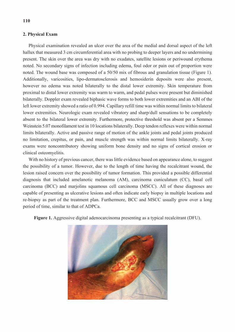
110
2. Physical Exam
Physical examination revealed an ulcer over the area of the medial and dorsal aspect of the left hallux that measured 3 cm circumferential area with no probing to deeper layers and no undermining present. The skin over the area was dry with no exudates, satellite lesions or periwound erythema noted. No secondary signs of infection including edema, foul odor or pain out of proportion were noted. The wound base was composed of a 50/50 mix of fibrous and granulation tissue (Figure 1). Additionally, varicosities, lipo-dermatosclerosis and hemosiderin deposits were also present, however no edema was noted bilaterally to the distal lower extremity. Skin temperature from proximal to distal lower extremity was warm to warm, and pedal pulses were present but diminished bilaterally. Doppler exam revealed biphasic wave forms to both lower extremities and an ABI of the left lower extremity showed a ratio of 0.994. Capillary refill time was within normal limits to bilateral lower extremities. Neurologic exam revealed vibratory and sharp/dull sensations to be completely absent to the bilateral lower extremity. Furthermore, protective threshold was absent per a Semmes Weinstein 5.07 monofilament test in 10 locations bilaterally. Deep tendon reflexes were within normal limits bilaterally. Active and passive range of motion of the ankle joints and pedal joints produced no limitation, crepitus, or pain, and muscle strength was within normal limits bilaterally. X-ray exams were noncontributory showing uniform bone density and no signs of cortical erosion or clinical osteomyelitis.
With no history of previous cancer, there was little evidence based on appearance alone, to suggest the possibility of a tumor. However, due to the length of time having the recalcitrant wound, the lesion raised concern over the possibility of tumor formation. This provided a possible differential diagnosis that included amelanotic melanoma (AM), carcinoma cuniculatum (CC), basal cell carcinoma (BCC) and marjolins squamous cell carcinoma (MSCC). All of these diagnoses are capable of presenting as ulcerative lesions and often indicate early biopsy in multiple locations and re-biopsy as part of the treatment plan. Furthermore, BCC and MSCC usually grow over a long period of time, similar to that of ADPCa.
Figure 1. Aggressive digital adenocarcinoma presenting as a typical recalcitrant (DFU).
111
3. Diagnosis
A standard blood panel for chemistry and hematology was taken but values were found to be not clinically significant. Three millimeter punch biopsies were taken at the wound sight in multiple locations. Using the clock face technique for distinguishing punch locations, biopsies were taken at 12:00 (proximal) and 06:00 (distal). These biopsies were sent in formalin medium to pathology services for further evaluation in hopes of finding a definitive diagnosis.
The pathology report indicated features of eccrine differentiation with atypical epithelial cells consistent with low grade adnexal proliferation. Lesional cells exhibited extension into the superficial and deep peripheral surfaces of the specimen. Although the proliferation was assymetrical, nuclear atypia was not severe suggesting a low grade sweat duct tumor which favored benignancy; however, malignancy could not be ruled out. Given the differential diagnosis stated above, poor outcomes with standard wound care to date and no clear disease process identified, a second set of biopsies was indicated. After follow up conversations with the pathologist, as well as patient and family, a second set of biopsies were taken using the same technique at 3 and 9 o’clock, as well as, the central base of the lesion. Again, they were sent to pathology for evaluation. The pathology report for the second set of biopsies showed findings of infiltrative neoplastic cells with moderate cytologic atypia in all three locations consistent with Aggressive digital papillary adenocarcinoma (intermediate to high grade) also known as ADPCa, a much more serious disease process.
4. Treatment
Treatment rendered while waiting for pathology results included sharp debridement of nonviable soft tissue and the use of cadexomer iodine applied to the wound base along with a dry sterile dressing. The patient was instructed to continue with dressing changes every other day to keep the wound clean, dry and free of bacterial pathogens and to return to clinic in 1 week for further evaluation and review of pathology results. Once a clear diagnosis was made based on pathology, both conservative and surgical treatment options were explained and consults for oncology, vascular surgery, plastic surgery, as well as, palliative care management was highly advised. The patient was referred to oncology. Follow up with oncology revealed that based on the patient’s age and physicality, she and her family elected to treat conservatively with palliative care.
5. Discussion
This case presents a potential diagnostic challenge. The possibility of tumors/cancers masquerading as wounds can be serious and treatment plans require a clinician’s keen eye regarding clinical picture and patient history. The clinician’s discretion to biopsy early can potentially minimize devastating long term results and maximize best treatment practices. Thus, a literature review was undertaken looking at both ADPCa, as well as, some other cancer/disease processes that follow similar growth patterns, present with similar clinical characteristics and have a potential to metastasize.
112
5.1. Basal Cell Carcinoma
BCC is the most common skin tumor reported in the literature worldwide and arises from the epidermis. Although it is a generally slow growing, often painless tumor, BCC has the potential and rare occasion to metastasize. Due to its slow growing nature, basal cell tumors allow for a successful treatment outcome with surgical excision in 90% of the cases [1,2]. Its name is derived from the cells that resemble the basal layer of the epidermis often described as “Basaloid”. The etiology of BCC is thought to be multifactorial with risk factors including UV light exposure, tanning beds, ionizing radiation and recipients of solid organ transplants [3]. Although both the P53 tumor suppressor mutation and the Patched (PTCH) gene have been well documented to be associated with BCC, an article by Nghiem et al. 2003 [4] revealed the potential of UVA radiation to act as a tumor promoter by activating protein Kinase C, as well as, a cutaneous immunosuppressor ultimately leading to malignant transformation. Even more was the discovery that the combination of different wavelengths, i.e., UVB in conjunction with UVA, work synergistically to activate the malignant transformation in cutaneous cells [4].
The literature has provided some similarities of BCC to ADPCa. One such similarity is BCC’s ability to present with a number of different clinical scenarios, from a nodular lesion to a tissue destructive ulceration. In fact, most primary metastatic BCC present as destructive tissue lesions representing up to 75% [5]. Another similarity of BCC to ADPCa is the high risk of recurrence after surgical resection of the lesion. A meta-analysis of the data by Marcil and stern in 2000 [6] revealed that after a review of 9005 patients, the risk of recurrence with BCC after resection was 44% within a 3 year period. Tefler et al. in 2008 [7] described risk factors affecting the level of recurrence and prognostic outcome with BCC to include: tumor size, site, margins, histological subtype and aggression, failure of previous treatment and immunosuppression [7]. Subtypes of BCC include superficial, morpheaform, fibroepithelioma of pinkus, pigmented and common. Furthermore, both ADPCa and BCC have multiple treatment options available. However, surgical excision appears to be the standard of care due to the concern of malignant transformation. For this reason, early biopsy should be sought in wounds that present with BCC or ADPCa characteristics and thorough follow up is indicated, especially in recalcitrant or challenging lesion presentations.
5.2. Marjolin’s Ulceration/Squamous Cell Carcinoma (MSCC)
Traditionally, a Marjolin’s ulcer is a rare malignancy that arises from a chronic nonhealing ulcer. Although most commonly associated with burns, it has been shown that MSCC can be in concomitances with many other pathologic processes. These include but are not limited to; pressure ulcers, chronic venous ulcers, osteomyelitis, traumatic wounds, fistulas, and lacerations [8]. While much of the etiology is still unclear, there is a consensus that MSCC stems from a multifactorial mechanism that contributes to the malignancy. It was originally hypothesized that scar tissue has impaired immunological response due to the lymphatic damage and decreased vascularity. This compromised immune response then allows for further uncontrollable mutations, which lead to the malignancy [9]. More recently, it has been suggested that predisposed immune deficient individuals along with chronic irritation may play a significant role. Chronic irritation causes
113
continuous mitotic turnover, along with avascularity and depressed immune response allowing for malignant cell proliferation [10]. Histologically, majolin’s ulcer present as MSCC and BCC in 83% present of the cases. The majority (73%) being that of MSCC, is important due to its high potential for metastasis [11]. Metastasis rates of MSCC approach upwards of 54% of the cases and presentation in the lower extremity in 50% of the cases [12].
Due to its clinical presentation in the form of an ulcerative lesion and it’s potential to metastasize, MSCC must also be considered in the differential diagnosis for ADPCa. MSCC often presents 25 years after the initial cutaneous damage. This significant latency period is similar to that of ADPCa [8]. The suggestion of diagnosis for MSCC follows a distinct triad of nodular formation, induration, and ulceration which are all present in BCC and ADPCa [10]. As with BCC and ADPCa, recurrence rates can approach 50% with inadequate resection [13]. Furthermore, many of the treatment options to be considered for MSCC are similar to that of BCC and ADPCa. Treatment includes wide local excision of at least 1 centimeter healthy margin or amputation when margins cannot be confirmed.
There has been some discussion on the importance of performing frozen section examinations intra-operatively. While this seems to be an ideal technique, it is important to note that the quality of frozen sections are very low and might not be as predicable for lesions which have many morphologies [14]. Holgado and Ward in 2000 [14] suggested that the Mohs surgical technique might provide the best outcome for removing all cancerous tissue and salvaging as much viable tissue as possible. In theory, this would be the best option for the treatment of all malignant ulcerative lesions, however it is unrealistic considering that the physician would need to be trained in surgery and pathology.
5.3. Aggressive Digital Papillary Adenocarcinoma (ADPCa)
Aggressive digital papillary adenocarcinoma (ADPCa), first described by Kao and Helwig in 1984 [15], is a rare variant of sweat gland carcinoma. It is a metastatic cutaneous tumor that predominantly occurs on digits and volar surfaces of middle aged to older males [16,17]. Some risk factors include previous radiation therapy and immunosuppression [18]; however, the pathophysiological mechanism of this disease process is still largely unknown. In 2012, Suchak [19] revealed that of 31 cases reviewed, only 4 presented in the lower extremity indicating the even more rare presentation seen in this particular case. ADPCa differentiates itself from other sweat gland carcinomas by its potential capability to metastasize quickly and high recurrence rate. Even with surgical excision, recurrence rates of up to 42% have been reported [20]. Furthermore, after reviewing 19 cases of ADPCa, Hsu et al. in 2009 [16] showed that metastasis was described in 47% of the cases, and local reoccurrence was seen in 50% of the cases within 2 months of excision, indicating the difficulty in rendering successful treatment.
Diagnosis of ADPCa is also challenging as some authors have suggested a mixed adnexal lineage with eccrine, apocrine and pilosebaceous features [21]. Also, ADPCa can often present with common clinical characteristics to other lower extremity lesions. This allows these tumors to masquerade as benign lesions such as nodules or papules and even ulcerations as seen in this particular case [22,23]. Most often, the legion presents as a nodule or papule that is mildly painful and relatively slow growing [24] which adds to the difficulty in diagnosis when presented with an ulcerative lesion. Many
114
times the clinical presentation can represent that of a ganglion cyst [22]. Histological characteristics of ADPCa include lobulated infiltrating neoplastic cells often with a focal papillary growth pattern. These neoplastic cells often show cytologic atypia with pleomorphism representing more than one morphologic subtype. Spindle cells with heavily active mitosis may also be seen. Even histology does not necessarily allow for a definitive diagnosis, as ganglion cysts, synovial sarcomas and fibrosarcomas all have histologic characteristics similar to ADPCa [22]. Most recently Suchak et al. in 2012 [19], stated that histological parameters currently used for diagnosis are not reliable in predicting the potential for metastasis. Thus all tumors with any remote characteristics of ADPCa should be treated as such until proven otherwise.
5.4. Proposed Treatment
Currently there are a number of plausible treatment recommendations for aggressive digital papillary adenocarcinoma. However, a guide to treatment has not been well defined in literature due to its rarity. When Kao and Helwig first introduced ADPCa in 1987 [20] the standard of care included local excision of the tumor. Research by Tsujita-Kyutiku et al. in 2003 [25] discussed the use of epithelial markers, specifically the P63 marker to help differentiate low grade from high grade ADPCa. Di Como et al. [26] showed that P63 staining would be positive in the basal layers of normal sweat ducts and primary carcinomas but negative in metastatic foci indicating that it may allow the ability to determine whether amputation or excision is warranted. However, the data continues to be inconclusive. The malignant nature of ADPCa has warranted more aggressive therapy such as a more extensive excisional debridement and even amputation of the affected digit or limb [16]. In fact, in the Hsu et al. 2009 [16] study, amputation was the method of treatment in 11 out of 19 cases. However, it should be noted that although amputation is recommended with recurrent ADPCa, no statistical significance regarding outcomes was found when compared to the other 7 of 18 case reports reviewed that underwent wide excision with/without sentinel node biopsy.
More recently, the approach towards APDCa has become much more targeted due to a new emphasis on limb preservation. A few case studies have suggested chemotherapy as a viable option for treatment of ADPCa although the outcomes where only marginally beneficial [22,27]. In a study by Jones et al. 2013 [17], the authors suggest conventional palliative doses of radiotherapy as an adjuvant to chemotherapy for the treatment of ADPCa. However, the role of radiation and chemotherapy in the treatment of ADPCa is still uncertain.
Sentinel lymph node biopsy combined with local excision has also been recommended to detect subclinical metastases [28]. While this may be a pivotal step in detecting early metastasis, it has been documented that patients can have a prolonged disease-free interval of up to 20 years after the initial presentation before seeing recurrence making this disease process a challenge for physicians.
6. Summary
While there has been some inconsistency within the literature on how to approach the treatment for difficult lesions such as BCC, MSCC and ADPCa, the similarities in clinical presentation indicate the need to biopsy early and many times in multiple locations and more than once for definitive
115
diagnosis. Early biopsy in challenging and recalcitrant wounds can provide the best information to make an informed decision on treatment options. It is important to note that many of the treatment options discussed regarding ADPCa have only been reported in small case studies. The infrequent occurrence of ADPCa in the lower extremity presents a diagnostic dilemma often being diagnosed incorrectly. ADPCa is a rare cutaneous metastatic carcinoma and physicians should treat any lesions exhibiting characteristics consistent with the disease process as such.
Clinical examination and history still appear to be the best diagnostic tools at our disposal. As with any ulcerative presentation, we must take the time to examine the signs of malignancy which include chronic ulceration greater than 3 months, rolled or everted wound borders, boisterous granulation tissue, foul smelling purulence, increase in size, bleeding on contact, and pain. If any of these criteria are present, it is our obligation to consider biopsy for definitive diagnosis.
Our literature review revealed no clear guidelines for re-biopsy of suspicious lesions even though the author’s feel that this practice is indicated with recalcitrant wounds that remain problematic. Further research is needed to better elucidate a successful treatment guideline that can be followed based on clinical evidence, clinical picture, lab results and physician expertise. Perhaps most important is the clinicians’ discretion to biopsy and re-biopsy when faced with ambiguous results or recalcitrant wounds. Based on the most current literature, the authors suggest a Sentinel lymph node biopsy during the first wide excision, along with an annual exam for reoccurrence and chest x-ray to evaluate for lung metastasis. This, combined with repeat biopsies in multiple locations when indicated, may provide the best algorithmic approach and produce significantly better outcomes to these rare and challenging disease processes.
Author Contributions
All the authors completely involved in all aspects of researching, writing and editing the manuscript.
Conflicts of Interest
The authors declare no conflicts of interest.
References
1. Culliford, A.; Hzen, A. Dermatology for plastic surgeons. In Grabb and Smith’s Plastic Surgery, 6th ed.; Thorn C.H., Ed.; Lippincott Williams & Wilkins: Philadelphia, PA, USA, 2007; pp. 105–114.
2. Wong, C.S.; Strange, R.C.; Lear, J.T. Basal cell carcinoma. Br. Med. J. 2003, 327, 794–798. 3. Kwasniak, L.A.; Jorge, G.Z. Basal cell carcinoma: Evidence-based medicine and review of
treatment modalities. Int. J. Dermatol. 2011, 50, 645–658. 4. Nghiem, D.X.; Kazimi, N.; Mitchell, D.L.; Vink, A.A.; Ananthaswamy, H.N.; Kripke, M.L.;
Ullrich, S.E. Mechanism underlying the suppression of established immune responses by ultraviolet radiation. J. Investig. Dermatol. 2003, 119, 600–608.
5. Snow, S.N.; Sahl, W.; Lo, J.S.; Mohs, F.E.; Warner, T.; Dekkinga, J.A.; Feyzi, J. Metastatic basal cell carcinoma: Report of five cases. Cancer 1994, 73, 328–335.
116
6. Marcil, I.; Stern, R.S. Risk of developing a subsequent nonmelanoma skin cancer in patients with a history of nonmelanoma skin cancer: A critical review of the literature and meta-analysis. Arch. Dermatol. 2000, 136, 1524–1530.
7. Telfer, N.R.; Colver, G.B.; Morton, C.A. Guidelines for the management of basal cell carcinoma. Br. J. Dermatol. 2008, 159, 35–48.
8. Pavlovic, S.; Surowiec, D.; Branieck, M. Marjolin’s Ulcer of the hallux. J. Am. Podiatr. Med. Assoc. 2013, 103, 250–253.
9. Fleming, M.D.; Hunt, J.L.; Purdue, G.F.; Sandstad, J. Marjolin’s ulcer: A review and reevaluation of a difficult problem. J. Burn. Care Rehabil. 1990, 11, 460–469.
10. Pekarek, B.; Buck, S.; Osher, L. A comprehensive review on Marjolin’s ulcers: Diagnosis and treatment. J. Am. Coll. Certif. Wound Spec. 2011, 3, 60–64.
11. Ochenduszkiewicz, U.; Matkowski, R.; Szynglarewicz, B.; Kornafel, J. Marjolin’s ulcer: Malignant neoplasm arising from scars. Rep. Pract. Oncol. Radiother. 2006, 11, 135–138.
12. Ogawa, B.; Chen, M.; Marjolis, J.; Schiller, F.J.; Schnall, S.B. Marjolin’s ulcer arising at the elbow: A case report and literature review. Hand 2006, 1, 89–93.
13. Shahla, A. An overview of heel Marjolin’s ulcers in the orthopedic department of Urmia University of Medical Sciences. Arch Iran. Med. 2009, 12, 405–408.
14. Holgado, R.D.; Ward, S.C.; Suryaprasad, S.G. Squamous cell carcinoma of the hallux. J. Am. Podiatr. Med. Assoc. 2000, 90, 309–311.
15. Kao, G.F.; Graham, J.H.; Helwig, E.B. Aggressive digital papillary adenocarcinoma. Arch. Dermatol. 1984, 14, 129–146.
16. Hsu, H.C.; Ho, C.Y.; Chen, C.H.; Yang, C.H.; Hong, H.S.; Chuang, Y.H. Aggressive digital papillary adenocarcinoma: A review. Clin. Exp. Dermatol. 2010, 35, 113–119.
17. Jones, J.A.; Patel, V.B.; Teitelbaum, U.; Plastaras, J.P. Diffusely metastatic digital papillary adenocarcinoma eleven years after initial presentation treated with palliative chemotherapy and radiotherapy. J. Clin. Oncol. 2013, 31, e386–e389.
18. Pujol, R.M.; LeBoit, P.E.; Su, W.P. Microcystic adnexal carcinoma with extensive sebaceous differentiation. Am. J. Dermatopathol. 1997, 19, 358–362.
19. Suchak, R.; Wang, W.L.; Prieto, V.G.; Ivan, D.; Lazar, A.J.; Brenn, T.; Calonje, E. Cutaneous digital papillary adeno- carcinoma: A clinicopathologic study of 31 cases of a rare neoplasm with new observations. Am. J. Surg. Pathol. 2012, 36, 1883–1891.
20. Kao, G.F.; Helwig, E.B.; Graham, J.H. Aggressive digital papillary adenoma and adenocarcinoma. A clinicopathological study of 57 patients with histochemical, immunopathological, and ultrastructural observations. J. Cutan. Pathol. 1987, 14, 129–146.
21. Obaidat, N.A.; Alsaad, K.O.; Ghazarian, D. Skin adnexal neoplasms—Part 2: An approach to tumours of cutaneous sweat glands. J. Clin. Pathol. 2007, 60, 145–159.
22. Bakotic, B.; Antonescu, C.R. Aggressive digital papillary adenocarcinoma of the foot: The clinicopathologic features of two cases. J. Foot Ankl. Surg. 2000, 39, 402–405.
23. Snyder, R.J.; Driver, V.; Fife, C.E.; Lantis, J.; Peirce, B.; Serena, T.; Weir, D. Using a diagnostic tool to identify elevated protease activity levels in chronic and stalled wounds: A consensus panel discussion. Ostomy Wound Manag. 2011, 57, 36–46.
117
24. Altman, C.E.; Hamill, R.I.; Easton, D.M. Metastatic aggressive digital papillary adenocarcinoma. Cutis 2003, 72, 145–147.
25. Tsujita-Kyutoku, M.; Kiuchi, K.; Danbara, N.; Yuri, T.; Senzaki, H.; Tsubura, A. P63 expression in normal human epidermis and epidermal appendages and their tumors. J. Cutan. Pathol. 2003, 30, 11–17.
26. Di Como, C.J.; Urist, M.J.; Babayan, I.; Drobnjak, M.; Hedvat, C.V.; Teruya-Feldstein, J.; Pohar, K.; Hoos, A.; Cordon-Cardo, C. P63 expression profiles in human normal and tumor tissues. Clin. Cancer Res. 2002, 8, 494–501.
27. Nishimoto, J.; Amoh, Y.; Niiyama, S.; Takasu, H.; Katsuoka, K. Aggressive digital papillary adenocarcinoma on the palm with pulmonary metastases. J. Dermatol. 2008, 35, 468–470.
28. Bogner, P.N.; Fullen, D.R.; Lowe, L.; Paulino, A.; Biermann, J.S.; Sondak, V.K.; Su, L.D. Lymphatic mapping and sentinel lymph node biopsy in the detection of early metastasis from sweat gland carcinoma. Cancer 2003, 97, 2285–2289.
118
Wound Healing: Biologics, Skin Substitutes, Biomembranes and Scaffolds
Krishna S. Vyas and Henry C. Vasconez
Abstract: This review will explore the latest advancements spanning several facets of wound healing, including biologics, skin substitutes, biomembranes and scaffolds.
Reprinted from Healthcare. Cite as: Vyas, K.S.; Vasconez, H.C. Wound Healing: Biologics, Skin Substitutes, Biomembranes and Scaffolds. Healthcare 2014, 2, 356-400.
Abbreviations
RNA Ribonucleic Acid IL-6 Interleukin 6 TNF- Tumor Necrosis Factor Alpha LTC4 Leukotriene C4 TXB2 Thromboxane B2 UVB Ultraviolet B MIF Migration Inhibitory Factor NO Nitric Oxide RCT Randomized Controlled Trial TBSA Total Body Surface Area STSG Split-Thickness Skin Graft COX-2 Cyclooxygenase-2 IL-1 Interleukin-1 beta NF- B Nuclear Factor kappa-light-chain-enhancer of activated B cells IL-10 Interleukin 10 ATP Adenosine Triphosphate KATP Potassium Channels CEA Cultured Epithelial Autograft HDE Humanitarian Device Exemption DFU Diabetic Foot Ulcers PMA Premarket Approval LOS Length of Stay TGF- Transforming Growth Factor-beta CACs Circulating Angiogenic Cells OPN Osteopontin
119
CCPE Collagen Coated Porous Polyethylene PMB Poly(2-methacryloyloxyethyl phosphorylcholine-co-n-butyl methacrylate)UPPE Uncoated Porous Polyethylene DNA Deoxyribonucleic Acid P3HT Photosensitive Polymer Poly (3-hexylthiophene) CGS Collagen/Gelatin Sponge
1. Introduction
The healing of wounds is a complex process that involves the activation and synchronization of intracellular, intercellular and extracellular elements, including coagulatory and inflammatory events, fibrous tissue accretion, deposition of collagen, epithelialization, wound contraction, tissue granulation and remodeling [1]. This process occurs via activation of local and systemic cells to restore tissue integrity through regeneration and scar formation, and often these cumulative processes result in satisfactory repair of damaged sites. Disruptions caused by tissue loss, inadequate blood flow, and comorbid disease states can lead to chronic wounds that are difficult to manage [2]. There are many strategies that have been applied to the treatment of wounds in the past. Early on, these were based on empirical deduction and unsubstantiated determinations. Although there was a general resistance to new concepts and modalities that impeded progress, advancements in the treatment of wounds have, nevertheless, evolved [3]. Over the past two decades, advancements in the clinical understanding of wounds and their pathophysiology have commanded significant biomedical innovations in the treatment of acute, chronic, and other types of wounds. This review will explore the latest advancements spanning several facets of wound healing, including biologics, skin substitutes, biomembranes and scaffolds.
2. Biologics for Wound Healing
2.1. Description
Biologic wound healing therapies are those that are intended to facilitate the re-establishment of the innate repair mechanisms, and may involve the application of active biological agents, such as plant-derived active biomolecules which exhibit antioxidant, antimicrobial, or anti-inflammatory attributes. Biologic dressings prevent evaporative water loss, heat loss, protein and electrolyte loss, and contamination. They also permit autolytic debridement and develop a granular wound bed. Biological skin equivalents, epidermal growth factors, stem cell therapies, and tissue engineering might also be utilized [2].
120
2.2. Mechanisms and Indications
Monoterpenes represent an extensive and varied family of naturally occurring terpene-based chemical compounds that comprise the majority of essential oils. These compounds exhibit anti-inflammatory, antibacterial, and antioxidant attributes [4,5]. The primary mechanisms proposed for various monoterpenes encompass: antimicrobial activity (inhibition of microorganism ribonucleic acid (RNA) and protein biosynthesis); anti-inflammation (lowers the generation of interleukin 6 (IL-6) and tumor necrosis factor alpha (TNF- ) in mast cells, inhibition and alteration of leukotriene C4 (LTC4) release and thromboxane B2 (TXB2) release, respectively); antioxidation (inhibits the production of ultraviolet B (UVB)-induced free radicals photoprotective effects and oxidative stress); fibroblast growth and macrophage migration inhibitory factor (MIF) effects. The anti-inflammatory action of the monoterpenes is often correlated to their wound-healing effects. Monoterpenes include compounds such as borneol, thymol, -terpineol, genipin, aucubin, d-Limonene and sericin that have either direct or indirect activities in wound healing. Although monoterpenes are poorly studied in the context of wound healing, studies suggest that they are promising for the treatment of chronic wounds (Table 1).
Mai et al. [6] investigated the ointment Sulbogin® (marketed as SuileTM), comprised of borneol (a bicyclic monoterpenoid alcohol), bismuth subgallate and Vaseline®, and found it to hasten excision wound closure in adult male Sprangue-Dawley rats. Although the specific mechanism remains elusive, it is thought that bismuth subgallate may induce macrophages to secrete growth factors to facilitate wound healing. It was found to decrease the lesion area, enhance granulation tissue formation and re-epithelialization, initiate the proliferation of collagen via the activation of fibroblasts, accelerate the reestablishment of blood vessels, and restrict the formation of nitric oxide (NO) [4,6].
The monoterpenoid phenol, thymol, demonstrates multiple beneficial bioactivities toward the healing of wounds. These attributes encompass the modulation of prostaglandin synthesis [7], imparting anti-inflammatory effects in neutrophils, the inhibition of myeloperoxidase activity and a decreased influx of leukocytes [8,9], positive antioxidant effects on docosahexaenoic acid (an omega-3 fatty acid) concentrations [10], the prevention of lipid autoxidation [11] and formation of toxic elements via the stimulation of reactive nitrogen species [12], and antimicrobial activity [13,14]. The capacity of thymol for direct wound healing involves its being correlated with elevated concentrations, in the central nervous system, of macrophage MIF, as well as enhanced anti-inflammatory related tissue granulation. Furthermore, it influences collagen synthesis and fibroblast metabolism, leading to augmented fibroblast growth in vitro [9].
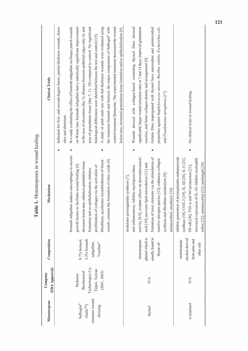
121
Tab
le 1
. Mon
oter
pene
s in
wou
nd h
ealin
g.
Mon
oter
pene
C
ompa
ny
(FD
A A
ppro
val)
Com
posi
tion
Mec
hani
sm
Clin
ical
Tri
als
Sulb
ogin
®
(Sui
leTM
)
oint
men
t wou
nd
dres
sing
Hed
onis
t
Bio
chem
ical
Tech
nolo
gies
Co,
Taip
ei, T
aiw
an
(200
1, 2
003)
0.7%
bor
neol
,
4.5%
bis
mut
h
subg
alla
te,
Vas
elin
e®
bism
uth
subg
alla
te in
duce
s mac
roph
ages
to se
cret
e
grow
th fa
ctor
s to
faci
litat
e w
ound
hea
ling
[6]
decr
ease
s les
ion
area
, enh
ance
s gra
nula
tion
tissu
e
form
atio
n an
d re
-epi
thel
ializ
atio
n, in
itiat
es
prol
ifera
tion
of c
olla
gen
via
the
activ
atio
n of
fibro
blas
ts, a
ccel
erat
es re
esta
blis
hmen
t of b
lood
vess
els,
rest
ricts
the
form
atio
n of
nitr
ic o
xide
[4]
Indi
cate
d fo
r firs
t- an
d se
cond
-deg
ree
burn
s, pa
rtial
-thic
knes
s w
ound
s, do
nor
site
s and
abr
asio
ns.
In a
stud
y ev
alua
ting
the
effe
ct o
f bis
mut
h su
bgal
late
on
biop
sy p
unch
wou
nds
on W
ista
r rat
s, bi
smut
h su
bgal
late
had
a st
atis
tical
ly si
gnifi
cant
impr
ovem
ent
in th
e are
a of
ulc
erat
ion
(day
1),
dist
ance
bet
wee
n ep
ithel
ial e
dges
(day
4),
and
area
of g
ranu
latio
n tis
sue
(day
7, 1
1, 1
8) c
ompa
red
to c
ontro
l. N
o si
gnifi
cant
hist
olog
ical
diff
eren
ces w
ere
iden
tifie
d be
twee
n th
e te
st a
nd c
ontro
l [15
].
A s
tudy
of
adul
t mal
e ra
ts w
ith f
ull-t
hick
ness
wou
nds
wer
e ev
alua
ted
usin
g
the
treat
men
t bis
mut
h an
d bo
rneo
l, th
e m
ajor
com
pone
nts
of S
ulbo
gin®
with
cont
rol t
reat
men
t fla
maz
ine.
The
exp
erim
enta
l tre
atm
ent d
ecre
ased
the
wou
nd
lesi
on a
rea,
incr
ease
d gr
anul
atio
n tis
sue
form
atio
n an
d re
-epi
thel
ializ
atio
n [6
].
thym
ol
N/A
mon
oter
peni
c
phen
ol w
hich
is
usua
lly fo
und
in
thym
e oil
mod
ulat
es p
rost
agla
ndin
synt
hesi
s [7]
;
anti-
infla
mm
ator
y; in
hibi
ts m
yelo
pero
xida
se
activ
ity [8
,9];
oxid
ant e
ffec
ts o
n do
cosa
hexa
enoi
c
acid
[10]
; pre
vent
s lip
id a
utox
idat
ion
[11]
and
form
atio
n of
toxi
c el
emen
ts v
ia th
e st
imul
atio
n of
reac
tive
nitro
gen
spec
ies [
12];
enha
nces
col
lage
n
synt
hesi
s and
fibr
obla
st m
etab
olis
m [9
];
antim
icro
bial
; ane
sthe
tic [1
6]
Wou
nds
dres
sed
with
co
llage
n-ba
sed
cont
aini
ng
thym
ol
film
s sh
owed
signi
fican
tly la
rger
wou
nd re
tract
ion
rate
s at 7
and
14
days
, im
prov
ed g
ranu
latio
n
reac
tion,
and
bet
ter c
olla
gen
dens
ity a
nd a
rran
gem
ent [
9].
Gel
atin
film
s im
preg
nate
d w
ith t
hym
ol h
ave
antio
xida
nt a
nd a
ntim
icro
bial
prop
ertie
s ag
ains
t Sta
phyl
ococ
cus
aure
us, B
acill
us s
ubtil
is, E
sche
rich
ia c
oli,
and
Pseu
dom
onas
aer
ugin
osa
[17]
.
-terp
ineo
l N
/A
mon
oter
pene
alco
hol d
eriv
ed
from
pin
e an
d
othe
r oils
inhi
bits
gen
erat
ion
of p
rost
agla
ndin
-end
oper
oxid
e
synt
hase
[18]
, CO
X-2
[19]
, IL-
1 [2
0], I
L-6
[21]
,
NF-
B [2
0], T
NF-
and
NO
pro
duct
ion
[21]
;
incr
ease
d ex
pres
sion
of I
L-10
; inh
ibits
neu
troph
il
influ
x [2
2]; a
ntim
icro
bial
[23]
; ant
ifung
al [2
4]
No
clin
ical
tria
ls in
wou
nd h
ealin
g.
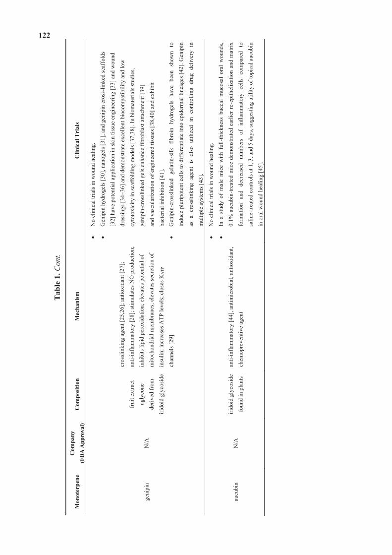
122
Tab
le 1
. Con
t.
Mon
oter
pene
C
ompa
ny
(FD
A A
ppro
val)
Com
posi
tion
Mec
hani
sm
Clin
ical
Tri
als
geni
pin
N/A
frui
t ext
ract
agly
cone
deriv
ed fr
om
irido
id g
lyco
side
cros
slin
king
age
nt [2
5,26
]; an
tioxi
dant
[27]
;
anti-
infla
mm
ator
y [2
8]; s
timul
ates
NO
pro
duct
ion;
inhi
bits
lipi
d pe
roxi
datio
n; e
leva
tes p
oten
tial o
f
mito
chon
dria
l mem
bran
es; e
leva
tes s
ecre
tion
of
insu
lin; i
ncre
ases
ATP
leve
ls; c
lose
s KA
TP
chan
nels
[29]
No
clin
ical
tria
ls in
wou
nd h
ealin
g.
Gen
ipin
hyd
roge
ls [3
0], n
anog
els [
31],
and
geni
pin
cros
s-lin
ked
scaf
fold
s
[32]
hav
e po
tent
ial a
pplic
atio
n in
skin
tiss
ue e
ngin
eerin
g [3
3] a
nd w
ound
dres
sing
s [34
–36]
and
dem
onst
rate
exc
elle
nt b
ioco
mpa
tibili
ty a
nd lo
w
cyto
toxi
city
in sc
affo
ldin
g m
odel
s [37
,38]
. In
biom
ater
ials
stud
ies,
geni
pin-
cros
slin
ked
gels
enh
ance
fibr
obla
st a
ttach
men
t [39
]
and
vasc
ular
izat
ion
of e
ngin
eere
d tis
sues
[38,
40] a
nd e
xhib
it
bact
eria
l inh
ibiti
on [4
1].
Gen
ipin
-cro
sslin
ked
gela
tin-s
ilk
fibro
in
hydr
ogel
s ha
ve
been
sh
own
to
indu
ce p
lurip
oten
t cel
ls to
diff
eren
tiate
into
epi
derm
al li
neag
es [4
2]. G
enip
in
as a
cro
sslin
king
age
nt i
s al
so u
tiliz
ed i
n co
ntro
lling
dru
g de
liver
y in
mul
tiple
syst
ems [
43].
aucu
bin
N/A
iri
doid
gly
cosi
de
foun
d in
pla
nts
anti-
infla
mm
ator
y [4
4], a
ntim
icro
bial
, ant
ioxi
dant
,
chem
opre
vent
ive
agen
t
No
clin
ical
tria
ls in
wou
nd h
ealin
g.
In a
stu
dy o
f m
ale
mic
e w
ith f
ull-t
hick
ness
buc
cal
muc
osal
ora
l w
ound
s,
0.1%
auc
ubin
-trea
ted
mic
e de
mon
stra
ted
earli
er re
-epi
thel
izat
ion
and
mat
rix
form
atio
n an
d de
crea
sed
num
bers
of
in
flam
mat
ory
cells
co
mpa
red
to
salin
e-tre
ated
con
trols
at 1
, 3, a
nd 5
day
s, su
gges
ting
utili
ty o
f top
ical
auc
ubin
in o
ral w
ound
hea
ling
[45]
.
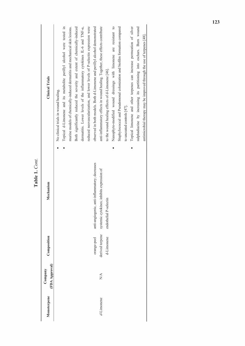
123
Tab
le 1
. Con
t.
Mon
oter
pene
C
ompa
ny
(FD
A A
ppro
val)
Com
posi
tion
Mec
hani
sm
Clin
ical
Tri
als
d-Li
mon
ene
N/A
oran
ge-p
eel
deriv
ed te
rpen
e
d-Li
mon
ene
anti-
angi
ogen
ic, a
nti-i
nfla
mm
ator
y; d
ecre
ases
syste
mic
cyt
okin
es; i
nhib
its e
xpre
ssio
n of
endo
thel
ial P
-sel
ectin
No
clin
ical
tria
ls in
wou
nd h
ealin
g.
Topi
cal
d-Li
mon
ene
and
its m
etab
olite
per
illyl
alc
ohol
wer
e te
sted
in
mur
ine
mod
els o
f che
mic
ally
-indu
ced
derm
atiti
s and
mec
hani
cal s
kin
lesi
ons.
Bot
h si
gnifi
cant
ly r
educ
ed t
he s
ever
ity a
nd e
xten
t of
che
mic
ally
-indu
ced
derm
atiti
s. Lo
wer
lev
els
of t
he i
nfla
mm
ator
y cy
toki
nes
IL-6
and
TN
F-,
redu
ced
neov
ascu
lariz
atio
n, a
nd l
ower
lev
els
of P
-sel
ectin
exp
ress
ion
wer
e
obse
rved
in b
oth
mod
els.
Bot
h d-
Lim
onen
e an
d pe
rilly
l alc
ohol
dem
onst
rate
d
anti-
infla
mm
ator
y ef
fect
s in
wou
nd h
ealin
g. T
oget
her,
thes
e ef
fect
s con
tribu
te
to th
e w
ound
hea
ling
effe
cts o
f d-L
imon
ene
[46]
.
Nan
ophy
to-m
odifi
ed
wou
nd
dres
sing
s w
ith
limon
ene
are
resi
stan
t to
Stap
hylo
cocc
al a
nd P
seud
omon
al c
olon
izat
ion
and
biof
ilm fo
rmat
ion
com
pare
d
to u
ncoa
ted
cont
rols
[47]
.
Topi
cal
limon
ene
and
othe
r te
rpen
es c
an i
ncre
ase
perm
eatio
n of
silv
er
sulp
hadi
azin
e by
in
crea
sing
its
pa
rtitio
ning
in
to
esch
ars.
Bur
n w
ound
antim
icro
bial
ther
apy
may
be
impr
oved
thro
ugh
the
use
of te
rpen
es [4
8].
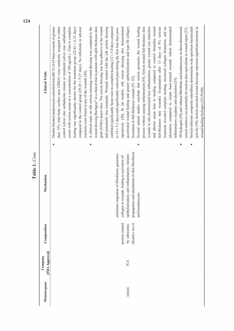
124
Tab
le 1
. Con
t.
Mon
oter
pene
Com
pany
(F
DA
App
rova
l) C
ompo
sitio
n M
echa
nism
C
linic
al T
rial
s
seric
in
N/A
pr
otei
n cr
eate
d by
silk
wor
ms
(Bom
byx
mor
i)
stim
ulat
es m
igra
tion
of fi
brob
last
s; g
ener
ates
co
llage
n in
wou
nds,
lead
ing
to a
ctiv
atio
n of
ep
ithel
ializ
atio
n; a
nti-i
nfla
mm
ator
y; in
itiat
es
prop
agat
ion
and
atta
chm
ent o
f ski
n fib
robl
asts
an
d ke
ratin
ocyt
es
Dou
ble b
linde
d ra
ndom
ized
cont
rolle
d tri
al (R
CT)
of 6
5 bu
rn w
ound
s of g
reat
er
than
15%
tota
l bod
y su
rfac
e ar
ea (
TBSA
) w
ere
rand
omly
ass
igne
d to
eith
er
cont
rol (
silv
er z
inc
sulfa
diaz
ine
crea
m)
or tr
eatm
ent (
silv
er z
inc
sulfa
diaz
ine
crea
m w
ith s
eric
in c
ream
at a
con
cent
ratio
n of
100
g/
mL)
. Tim
e to
com
plet
e he
alin
g w
as s
igni
fican
tly s
horte
r fo
r th
e tre
atm
ent g
roup
(22
.42
± 6.
33 d
ays)
co
mpa
red
to th
e co
ntro
l gro
up (2
9.28
± 9
.27
days
). N
o in
fect
ions
or
adve
rse
reac
tions
wer
e fo
und
in a
ny o
f the
wou
nds [
49].
A c
linic
al s
tudy
on
silk
ser
icin
-rel
easi
ng w
ound
dre
ssin
g w
as c
ompa
red
to th
e w
ound
dre
ssin
g B
actig
ras®
in a
clin
ical
tria
l in
patie
nts w
ith sp
lit-th
ickn
ess s
kin
graf
t (ST
SG) d
onor
site
s. Th
e se
ricin
dre
ssin
g w
as le
ss a
dhes
ive
to th
e w
ound
an
d po
tent
ially
less
trau
mat
ic. W
ound
s tre
ated
with
the
silk
ser
icin
dre
ssin
g ex
hibi
ted
sign
ifica
ntly
fast
er ra
tes t
o co
mpl
ete
heal
ing
(12
± 5.
0 da
ys co
mpa
red
to 1
4 ±
5.2
days
) and
sign
ifica
ntly
redu
ced
pain
dur
ing
the
first
four
day
s pos
t-op
erat
ivel
y [5
0].
In r
at m
odel
s, si
lk s
eric
in d
ress
ing
also
dem
onst
rate
d ac
cele
rate
d w
ound
hea
ling
and
grea
ter
epith
elia
lizat
ion
and
type
III
col
lage
n fo
rmat
ion
in fu
ll-th
ickn
ess w
ound
s [51
–53]
. Se
vera
l an
imal
stu
dies
con
clud
e th
at s
eric
in p
rom
otes
the
wou
nd h
ealin
g pr
oces
s w
ithou
t cau
sing
infla
mm
atio
n [5
4]. S
eric
in tr
eate
d fu
ll-th
ickn
ess
skin
w
ound
s in
rats
dem
onst
rate
d le
ss in
flam
mat
ion,
gre
ater
wou
nd s
ize
redu
ctio
n an
d sh
orte
r m
ean
time
to h
ealin
g co
mpa
red
to c
ontro
l (b
etad
ine
treat
ed
full-
thic
knes
s sk
in w
ound
s).
Exam
inat
ion
afte
r 15
day
s of
8%
ser
icin
e tre
atm
ent
reve
aled
com
plet
e he
alin
g, i
ncre
ased
col
lage
n fo
rmat
ion,
and
no
ulce
ratio
n co
mpa
red
to c
ream
bas
e-tre
ated
wou
nds
whi
ch d
emon
stra
ted
infla
mm
ator
y ex
udat
es a
nd u
lcer
atio
n [5
5].
3D h
ydro
gels
[56]
and
cul
ture
d fib
robl
asts
and
kera
tinoc
ytes
on
thre
e-di
men
siona
l se
ricin
mat
rices
can
pot
entia
lly b
e us
ed a
s ski
n eq
uiva
lent
s in
wou
nd re
pair
[57]
. Se
ricin
/chi
tosa
n co
mpo
site
nan
ofib
ers d
emon
stra
te w
ide
spec
trum
bac
teric
idal
ac
tivity
[58]
. Ser
icin
enr
iche
d w
ound
dre
ssin
gs re
pres
ent s
igni
fican
t pro
mis
e in
w
ound
hea
ling
biol
ogic
s [35
,59,
60].
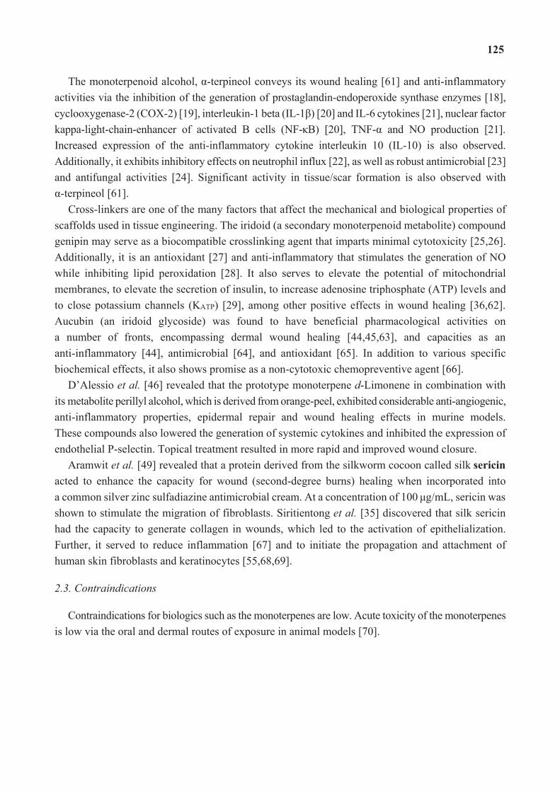
125
The monoterpenoid alcohol, -terpineol conveys its wound healing [61] and anti-inflammatory activities via the inhibition of the generation of prostaglandin-endoperoxide synthase enzymes [18], cyclooxygenase-2 (COX-2) [19], interleukin-1 beta (IL-1 ) [20] and IL-6 cytokines [21], nuclear factor kappa-light-chain-enhancer of activated B cells (NF- B) [20], TNF- and NO production [21]. Increased expression of the anti-inflammatory cytokine interleukin 10 (IL-10) is also observed. Additionally, it exhibits inhibitory effects on neutrophil influx [22], as well as robust antimicrobial [23] and antifungal activities [24]. Significant activity in tissue/scar formation is also observed with
-terpineol [61]. Cross-linkers are one of the many factors that affect the mechanical and biological properties of
scaffolds used in tissue engineering. The iridoid (a secondary monoterpenoid metabolite) compound genipin may serve as a biocompatible crosslinking agent that imparts minimal cytotoxicity [25,26]. Additionally, it is an antioxidant [27] and anti-inflammatory that stimulates the generation of NO while inhibiting lipid peroxidation [28]. It also serves to elevate the potential of mitochondrial membranes, to elevate the secretion of insulin, to increase adenosine triphosphate (ATP) levels and to close potassium channels (KATP) [29], among other positive effects in wound healing [36,62]. Aucubin (an iridoid glycoside) was found to have beneficial pharmacological activities on a number of fronts, encompassing dermal wound healing [44,45,63], and capacities as an anti-inflammatory [44], antimicrobial [64], and antioxidant [65]. In addition to various specific biochemical effects, it also shows promise as a non-cytotoxic chemopreventive agent [66].
D’Alessio et al. [46] revealed that the prototype monoterpene d-Limonene in combination with its metabolite perillyl alcohol, which is derived from orange-peel, exhibited considerable anti-angiogenic, anti-inflammatory properties, epidermal repair and wound healing effects in murine models. These compounds also lowered the generation of systemic cytokines and inhibited the expression of endothelial P-selectin. Topical treatment resulted in more rapid and improved wound closure.
Aramwit et al. [49] revealed that a protein derived from the silkworm cocoon called silk sericin acted to enhance the capacity for wound (second-degree burns) healing when incorporated into a common silver zinc sulfadiazine antimicrobial cream. At a concentration of 100 g/mL, sericin was shown to stimulate the migration of fibroblasts. Siritientong et al. [35] discovered that silk sericin had the capacity to generate collagen in wounds, which led to the activation of epithelialization. Further, it served to reduce inflammation [67] and to initiate the propagation and attachment of human skin fibroblasts and keratinocytes [55,68,69].
2.3. Contraindications
Contraindications for biologics such as the monoterpenes are low. Acute toxicity of the monoterpenes is low via the oral and dermal routes of exposure in animal models [70].
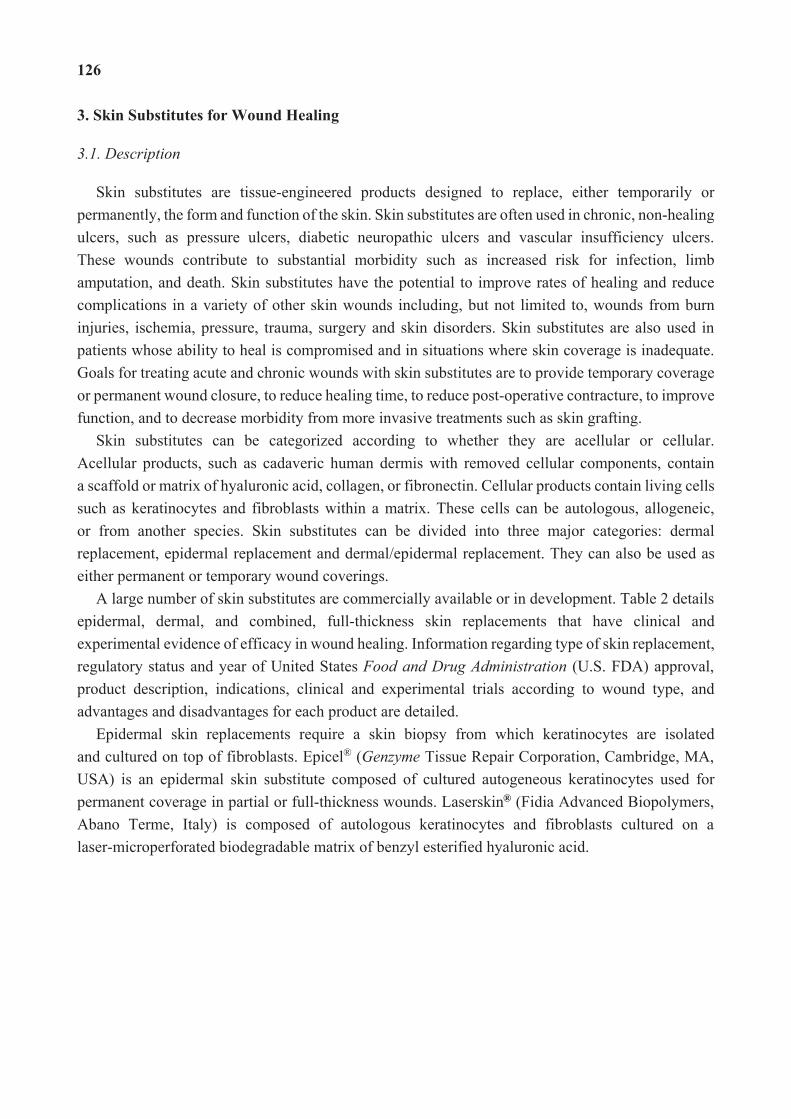
126
3. Skin Substitutes for Wound Healing
3.1. Description
Skin substitutes are tissue-engineered products designed to replace, either temporarily or permanently, the form and function of the skin. Skin substitutes are often used in chronic, non-healing ulcers, such as pressure ulcers, diabetic neuropathic ulcers and vascular insufficiency ulcers. These wounds contribute to substantial morbidity such as increased risk for infection, limb amputation, and death. Skin substitutes have the potential to improve rates of healing and reduce complications in a variety of other skin wounds including, but not limited to, wounds from burn injuries, ischemia, pressure, trauma, surgery and skin disorders. Skin substitutes are also used in patients whose ability to heal is compromised and in situations where skin coverage is inadequate. Goals for treating acute and chronic wounds with skin substitutes are to provide temporary coverage or permanent wound closure, to reduce healing time, to reduce post-operative contracture, to improve function, and to decrease morbidity from more invasive treatments such as skin grafting.
Skin substitutes can be categorized according to whether they are acellular or cellular. Acellular products, such as cadaveric human dermis with removed cellular components, contain a scaffold or matrix of hyaluronic acid, collagen, or fibronectin. Cellular products contain living cells such as keratinocytes and fibroblasts within a matrix. These cells can be autologous, allogeneic, or from another species. Skin substitutes can be divided into three major categories: dermal replacement, epidermal replacement and dermal/epidermal replacement. They can also be used as either permanent or temporary wound coverings.
A large number of skin substitutes are commercially available or in development. Table 2 details epidermal, dermal, and combined, full-thickness skin replacements that have clinical and experimental evidence of efficacy in wound healing. Information regarding type of skin replacement, regulatory status and year of United States Food and Drug Administration (U.S. FDA) approval, product description, indications, clinical and experimental trials according to wound type, and advantages and disadvantages for each product are detailed.
Epidermal skin replacements require a skin biopsy from which keratinocytes are isolated and cultured on top of fibroblasts. Epicel® (Genzyme Tissue Repair Corporation, Cambridge, MA, USA) is an epidermal skin substitute composed of cultured autogeneous keratinocytes used for permanent coverage in partial or full-thickness wounds. Laserskin® (Fidia Advanced Biopolymers, Abano Terme, Italy) is composed of autologous keratinocytes and fibroblasts cultured on a laser-microperforated biodegradable matrix of benzyl esterified hyaluronic acid.
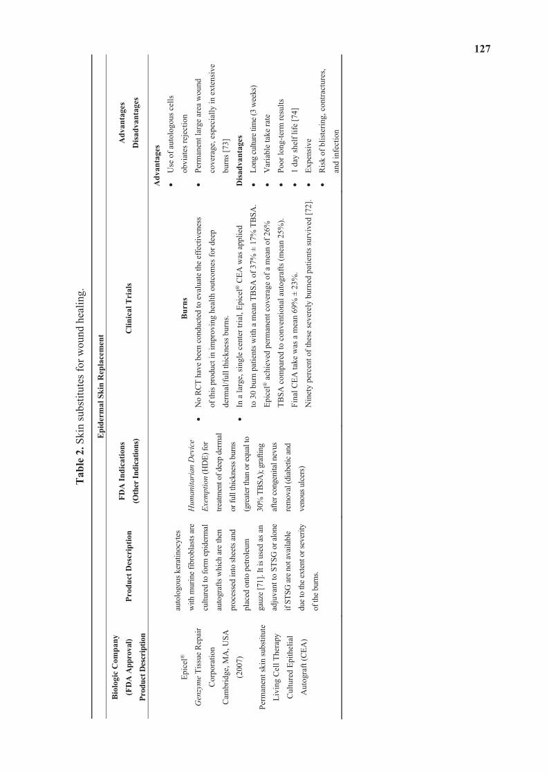
127
Tab
le 2
. Ski
n su
bstit
utes
for w
ound
hea
ling.
Epi
derm
al S
kin
Rep
lace
men
t
Bio
logi
c C
ompa
ny
(FD
A A
ppro
val)
Prod
uct D
escr
iptio
n
Prod
uct D
escr
iptio
n FD
A In
dica
tions
(Oth
er In
dica
tions
) C
linic
al T
rial
s A
dvan
tage
s
Dis
adva
ntag
es
Epic
el®
Gen
zym
e Ti
ssue
Rep
air
Cor
pora
tion
Cam
brid
ge, M
A, U
SA
(200
7)
Perm
anen
t ski
n su
bstit
ute
Livi
ng C
ell T
hera
py
Cul
ture
d Ep
ithel
ial
Aut
ogra
ft (C
EA)
auto
logo
us k
erat
inoc
ytes
with
mur
ine
fibro
blas
ts ar
e
cultu
red
to fo
rm e
pide
rmal
auto
graf
ts w
hich
are
then
proc
esse
d in
to sh
eets
and
plac
ed o
nto
petro
leum
gauz
e [7
1]. I
t is u
sed
as a
n
adju
vant
to S
TSG
or a
lone
if ST
SG ar
e not
avai
labl
e
due t
o th
e ext
ent o
r sev
erity
of th
e bur
ns.
Hum
anita
rian
Dev
ice
Exem
ptio
n (H
DE)
for
treat
men
t ofd
eep
derm
al
or fu
ll th
ickn
ess b
urns
(gre
ater
than
or e
qual
to
30%
TBS
A);
graf
ting
afte
r con
geni
tal n
evus
rem
oval
(dia
betic
and
veno
us u
lcer
s)
Bur
ns
No
RCT
have
bee
n co
nduc
ted
to e
valu
ate
the
effe
ctiv
enes
s
of th
is pr
oduc
t in
impr
ovin
g he
alth
out
com
es fo
r dee
p
derm
al/fu
ll th
ickn
ess b
urns
.
In a
larg
e, si
ngle
cen
ter t
rial,
Epic
el®
CEA
was
app
lied
to 3
0 bu
rn p
atie
nts w
ith a
mea
n TB
SA o
f 37%
± 1
7% T
BSA
.
Epic
el®
ach
ieve
d pe
rman
ent c
over
age
of a
mea
n of
26%
TBSA
com
pare
d to
con
vent
iona
l aut
ogra
fts (m
ean
25%
).
Fina
l CEA
take
was
a m
ean
69%
± 2
3%.
Nin
ety
perc
ent o
f the
se se
vere
ly b
urne
d pa
tient
s sur
vive
d [7
2].
Adv
anta
ges
Use
of a
utol
ogou
s cel
ls
obvi
ates
reje
ctio
n
Perm
anen
t lar
ge a
rea
wou
nd
cove
rage
, esp
ecia
lly in
ext
ensi
ve
burn
s [73
]
Dis
adva
ntag
es
Long
cultu
re ti
me (
3 w
eeks
)
Var
iabl
e ta
ke ra
te
Poor
long
-term
resu
lts
1 da
y sh
elf l
ife [7
4]
Expe
nsiv
e
Ris
k of
blis
terin
g, c
ontra
ctur
es,
and
infe
ctio
n
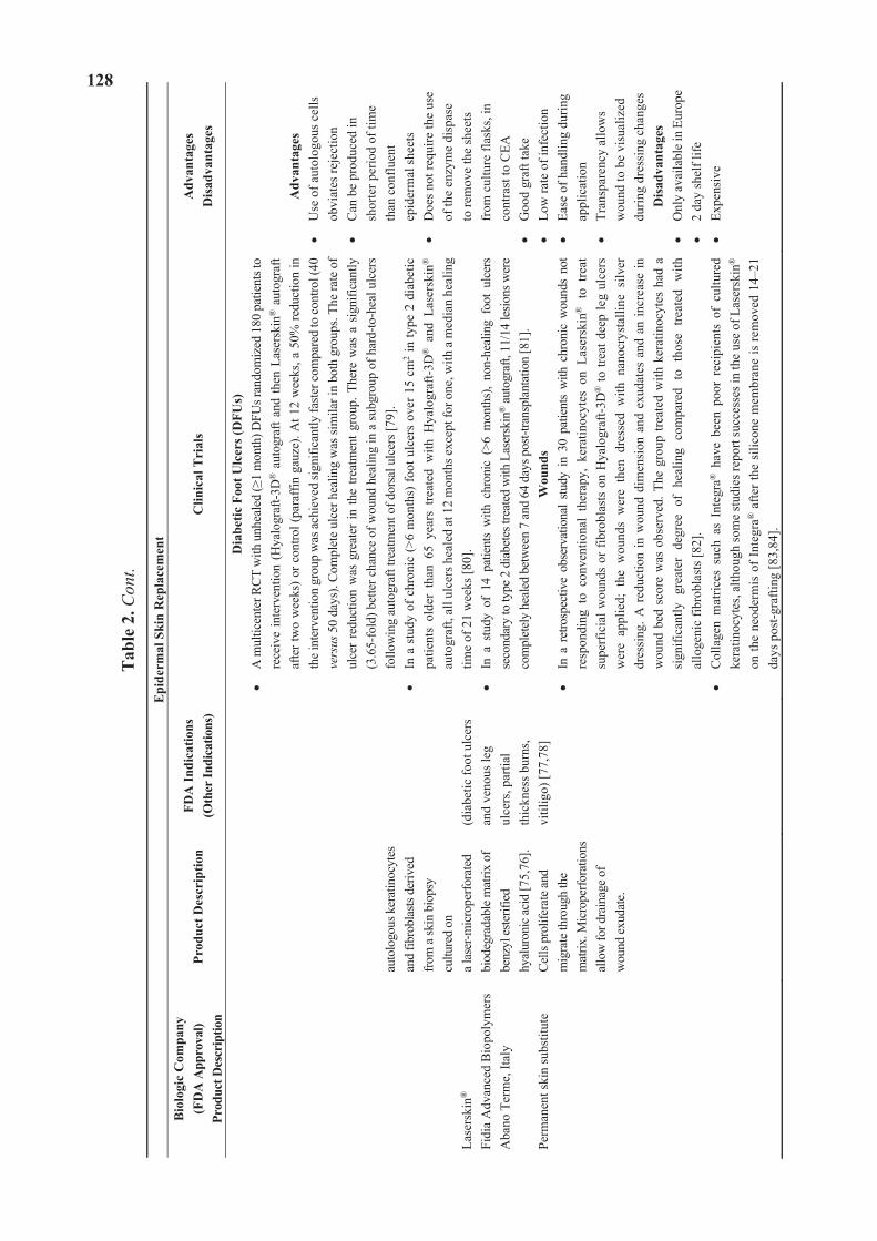
128
Tab
le 2
. Con
t.
Epi
derm
al S
kin
Rep
lace
men
tB
iolo
gic
Com
pany
(F
DA
App
rova
l)
Prod
uct D
escr
iptio
n Pr
oduc
t Des
crip
tion
FDA
Indi
catio
ns
(Oth
er In
dica
tions
) C
linic
al T
rial
s A
dvan
tage
sD
isad
vant
ages
Lase
rski
n®
Fidi
a A
dvan
ced
Bio
poly
mer
s A
bano
Ter
me,
Ital
y
Perm
anen
t ski
n su
bstit
ute
auto
logo
us k
erat
inoc
ytes
an
d fib
robl
asts
deriv
ed
from
a sk
in b
iops
y cu
lture
d on
a
lase
r-mic
rope
rfora
ted
biod
egra
dabl
e mat
rix o
f be
nzyl
este
rifie
d hy
alur
onic
acid
[75,
76].
Cells
pro
lifer
ate a
nd
mig
rate
thro
ugh
the
mat
rix. M
icro
perfo
ratio
ns
allo
w fo
r dra
inag
e of
wou
nd ex
udat
e.
(dia
betic
foot
ulc
ers
and
veno
us le
g
ulce
rs, p
artia
l th
ickn
ess b
urns
, vi
tilig
o) [7
7,78
]
Dia
betic
Foo
t Ulc
ers (
DFU
s)
A m
ultic
ente
r RCT
with
unh
eale
d (
1 m
onth
) DFU
s ran
dom
ized
180
pat
ient
s to
rece
ive
inte
rven
tion
(Hya
logr
aft-3
D® a
utog
raft
and
then
Las
ersk
in® a
utog
raft
afte
r tw
o w
eeks
) or c
ontro
l (pa
raffi
n ga
uze)
. At 1
2 w
eeks
, a 5
0% re
duct
ion
in
the i
nter
vent
ion
grou
p w
as ac
hiev
ed si
gnifi
cant
ly fa
ster c
ompa
red
to co
ntro
l (40
ve
rsus
50
days
). Co
mpl
ete
ulce
r hea
ling
was
sim
ilar i
n bo
th g
roup
s. Th
e ra
te o
f ul
cer
redu
ctio
n w
as g
reat
er in
the
treat
men
t gro
up. T
here
was
a s
igni
fican
tly
(3.6
5-fo
ld) b
ette
r cha
nce
of w
ound
hea
ling
in a
subg
roup
of h
ard-
to-h
eal u
lcer
s fo
llow
ing
auto
graf
t tre
atm
ent o
f dor
sal u
lcer
s [79
]. In
a s
tudy
of c
hron
ic (>
6 m
onth
s) fo
ot u
lcer
s ov
er 1
5 cm
2 in
type
2 d
iabe
tic
patie
nts
olde
r th
an 6
5 ye
ars
treat
ed w
ith H
yalo
graf
t-3D
® a
nd L
aser
skin
®
auto
graf
t, al
l ulc
ers h
eale
d at
12
mon
ths e
xcep
t for
one
, with
a m
edia
n he
alin
g tim
e of
21
wee
ks [8
0].
In a
stu
dy o
f 14
pat
ient
s w
ith c
hron
ic (
>6 m
onth
s),
non-
heal
ing
foot
ulc
ers
seco
ndar
y to
type
2 d
iabe
tes t
reat
ed w
ith L
aser
skin
® au
togr
aft,
11/1
4 le
sions
wer
e co
mpl
etel
y he
aled
bet
wee
n 7
and
64 d
ays p
ost-t
rans
plan
tatio
n [8
1].
Wou
nds
In a
ret
rosp
ectiv
e ob
serv
atio
nal
study
in 3
0 pa
tient
s w
ith c
hron
ic w
ound
s no
t re
spon
ding
to
conv
entio
nal
ther
apy,
ker
atin
ocyt
es o
n La
sers
kin®
to
treat
su
perf
icia
l wou
nds
or fi
brob
last
s on
Hya
logr
aft-3
D®
to tr
eat d
eep
leg
ulce
rs
wer
e ap
plie
d; t
he w
ound
s w
ere
then
dre
ssed
with
nan
ocry
stal
line
silv
er
dres
sing
. A r
educ
tion
in w
ound
dim
ensi
on a
nd e
xuda
tes
and
an in
crea
se in
w
ound
bed
sco
re w
as o
bser
ved.
The
gro
up tr
eate
d w
ith k
erat
inoc
ytes
had
a
sign
ifica
ntly
gre
ater
deg
ree
of h
ealin
g co
mpa
red
to t
hose
tre
ated
with
al
loge
nic
fibro
blas
ts [8
2].
Col
lage
n m
atric
es s
uch
as I
nteg
ra®
hav
e be
en p
oor
reci
pien
ts o
f cu
lture
d ke
ratin
ocyt
es, a
lthou
gh so
me
stud
ies r
epor
t suc
cess
es in
the
use
of L
aser
skin
®
on th
e ne
oder
mis
of I
nteg
ra® a
fter t
he s
ilico
ne m
embr
ane
is re
mov
ed 1
4–21
da
ys p
ost-g
rafti
ng [8
3,84
].
Adv
anta
ges
Use
of a
utol
ogou
s cel
ls
obvi
ates
reje
ctio
n C
an b
e pr
oduc
ed in
sh
orte
r per
iod
of ti
me
than
con
fluen
t
epid
erm
al sh
eets
D
oes n
ot re
quire
the
use
of th
e en
zym
e di
spas
e to
rem
ove
the
shee
ts
from
cul
ture
flas
ks, i
n co
ntra
st to
CEA
G
ood
graf
t tak
e Lo
w ra
te o
f inf
ectio
n Ea
se o
f han
dlin
g du
ring
appl
icat
ion
Tran
spar
ency
allo
ws
wou
nd to
be
visu
aliz
ed
durin
g dr
essi
ng c
hang
es
Dis
adva
ntag
esO
nly
avai
labl
e in
Euro
pe
2 da
y sh
elf l
ife
Expe
nsiv
e
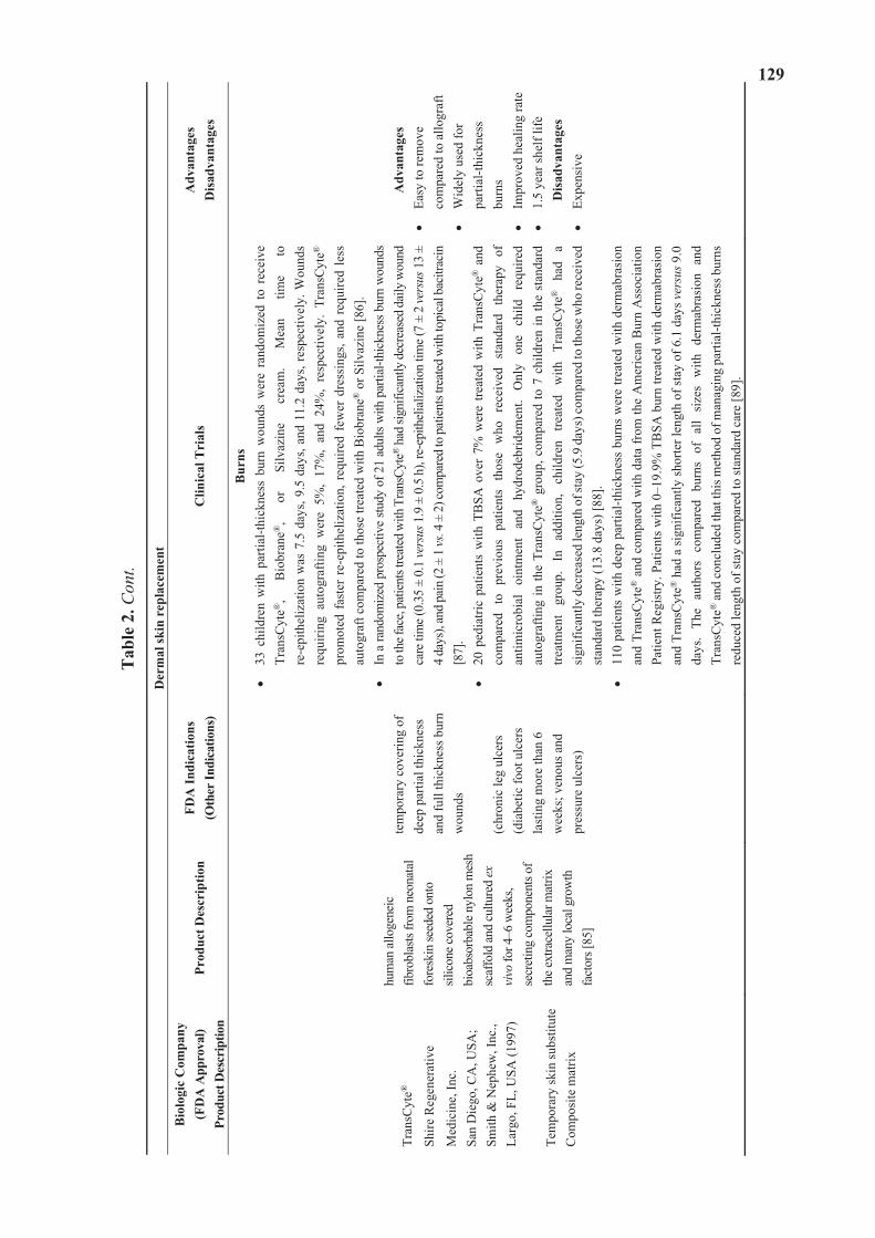
129
Tab
le 2
. Con
t.
Der
mal
skin
rep
lace
men
tB
iolo
gic
Com
pany
(F
DA
App
rova
l)
Prod
uct D
escr
iptio
n Pr
oduc
t Des
crip
tion
FDA
Indi
catio
ns
(Oth
er In
dica
tions
) C
linic
al T
rial
s A
dvan
tage
sD
isad
vant
ages
Tran
sCyt
e®
Shire
Reg
ener
ativ
e
Med
icin
e, In
c.
San
Die
go, C
A, U
SA;
Sm
ith &
Nep
hew
, Inc
., La
rgo,
FL,
USA
(199
7)
Tem
pora
ry sk
in su
bstit
ute
Com
posi
te m
atrix
hum
an al
loge
neic
fib
robl
asts
from
neo
nata
l fo
resk
in se
eded
ont
o sil
icon
e cov
ered
bi
oabs
orba
ble n
ylon
mes
h sc
affo
ld an
d cu
lture
d ex
vi
vo fo
r 4–6
wee
ks,
secr
etin
g co
mpo
nent
s of
the e
xtra
cellu
lar m
atrix
an
d m
any
loca
l gro
wth
fa
ctor
s [85
]
tem
pora
ry c
over
ing
of
deep
par
tial t
hick
ness
an
d fu
ll th
ickn
ess b
urn
wou
nds
(chr
onic
leg
ulce
rs
(dia
betic
foot
ulc
ers
last
ing
mor
e th
an 6
w
eeks
; ven
ous a
nd
pres
sure
ulc
ers)
Bur
ns33
chi
ldre
n w
ith p
artia
l-thi
ckne
ss b
urn
wou
nds
wer
e ra
ndom
ized
to
rece
ive
Tran
sCyt
e®,
Bio
bran
e®,
or
Silv
azin
e cr
eam
. M
ean
time
to
re-e
pith
eliz
atio
n w
as 7
.5 d
ays,
9.5
days
, and
11.
2 da
ys, r
espe
ctiv
ely.
Wou
nds
requ
iring
aut
ogra
fting
wer
e 5%
, 17
%,
and
24%
, re
spec
tivel
y. T
rans
Cyt
e®
prom
oted
fas
ter
re-e
pith
eliz
atio
n, r
equi
red
few
er d
ress
ings
, and
req
uire
d le
ss
auto
graf
t com
pare
d to
thos
e tre
ated
with
Bio
bran
e® o
r Silv
azin
e [8
6].
In a
rand
omiz
ed p
rosp
ectiv
e stu
dy o
f 21
adul
ts w
ith p
artia
l-thi
ckne
ss b
urn
wou
nds
to th
e fac
e, pa
tient
s tre
ated
with
Tra
nsCy
te® h
ad si
gnifi
cant
ly d
ecre
ased
dai
ly w
ound
ca
re ti
me (
0.35
± 0
.1 ve
rsus
1.9
± 0
.5 h
), re
-epi
thel
ializ
atio
n tim
e (7
± 2
vers
us 1
3 ±
4 da
ys),
and
pain
(2 ±
1 vs
. 4 ±
2) c
ompa
red
to p
atie
nts t
reat
ed w
ith to
pica
l bac
itrac
in
[87]
. 20
ped
iatri
c pa
tient
s w
ith T
BSA
ove
r 7%
wer
e tre
ated
with
Tra
nsC
yte®
and
co
mpa
red
to
prev
ious
pa
tient
s th
ose
who
re
ceiv
ed
stan
dard
th
erap
y of
an
timic
robi
al
oint
men
t an
d hy
drod
ebrid
emen
t. O
nly
one
child
re
quire
d au
togr
aftin
g in
the
Tran
sCyt
e® g
roup
, com
pare
d to
7 c
hild
ren
in th
e st
anda
rd
treat
men
t gr
oup.
In
ad
ditio
n,
child
ren
treat
ed
with
Tr
ansC
yte®
ha
d a
sign
ifica
ntly
dec
reas
ed le
ngth
of s
tay
(5.9
day
s) co
mpa
red
to th
ose w
ho re
ceiv
ed
stan
dard
ther
apy
(13.
8 da
ys) [
88].
110
patie
nts
with
dee
p pa
rtial
-thic
knes
s bu
rns
wer
e tre
ated
with
der
mab
rasi
on
and
Tran
sCyt
e® a
nd c
ompa
red
with
dat
a fr
om th
e A
mer
ican
Bur
n A
ssoc
iatio
n Pa
tient
Reg
istry
. Pat
ient
s with
0–1
9.9%
TB
SA b
urn
treat
ed w
ith d
erm
abra
sion
an
d Tr
ansC
yte®
had
a si
gnifi
cant
ly sh
orte
r len
gth
of st
ay o
f 6.1
day
s ver
sus 9
.0
days
. Th
e au
thor
s co
mpa
red
burn
s of
all
size
s w
ith d
erm
abra
sion
and
Tr
ansC
yte®
and
conc
lude
d th
at th
is m
etho
d of
man
agin
g pa
rtial
-thic
knes
s bur
ns
redu
ced
leng
th o
f sta
y co
mpa
red
to st
anda
rd c
are
[89]
.
Adv
anta
ges
Easy
to re
mov
e co
mpa
red
to a
llogr
aft
Wid
ely
used
for
parti
al-th
ickn
ess
burn
s Im
prov
ed h
ealin
g ra
te
1.5
year
shel
f life
D
isad
vant
ages
Expe
nsiv
e
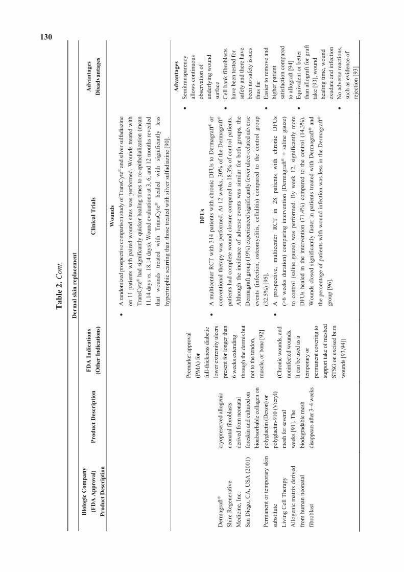
130
Tab
le 2
. Con
t.
Der
mal
skin
rep
lace
men
tB
iolo
gic
Com
pany
(F
DA
App
rova
l)
Prod
uct D
escr
iptio
n Pr
oduc
t Des
crip
tion
FDA
Indi
catio
ns
(Oth
er In
dica
tions
) C
linic
al T
rial
s A
dvan
tage
sD
isad
vant
ages
Wou
nds
A ra
ndom
ized
pro
spec
tive c
ompa
rison
stud
y of
Tra
nsCy
te® an
d sil
ver s
ulfa
diaz
ine
on 1
1 pa
tient
s w
ith p
aire
d w
ound
site
s w
as p
erfo
rmed
. Wou
nds
treat
ed w
ith
Tran
sCyt
e® h
ad s
igni
fican
tly q
uick
er h
ealin
g tim
es to
re-e
pith
elia
lizat
ion
(mea
n 11
.14
days
vs.
18.1
4 da
ys).
Wou
nd e
valu
atio
ns a
t 3, 6
, and
12
mon
ths r
evea
led
that
w
ound
s tre
ated
w
ith
Tran
sCyt
e®
heal
ed
with
si
gnifi
cant
ly
less
hy
pertr
ophi
c sc
arrin
g th
an th
ose
treat
ed w
ith si
lver
sulfa
diaz
ine
[90]
.
Der
mag
raft®
Shire
Reg
ener
ativ
e M
edic
ine,
Inc.
Sa
n D
iego
, CA
, USA
(200
1) Pe
rman
ent o
r tem
pora
ry sk
in
subs
titut
e Li
ving
Cel
l The
rapy
A
lloge
nic
mat
rix d
eriv
ed
from
hum
an n
eona
tal
fibro
blas
t
cryo
pres
erve
d al
loge
nic
neon
atal
fibr
obla
stsde
rived
from
neo
nata
l fo
resk
in an
d cu
lture
d on
bi
oabs
orba
ble c
olla
gen
on
poly
glac
tin (D
exon
) or
poly
glac
tin-9
10 (V
icry
l) m
esh
for s
ever
al
wee
ks [9
1]. T
he
biod
egra
dabl
e mes
h di
sapp
ears
after
3–4
wee
ks
Prem
arke
t app
rova
l (P
MA
) for
fu
ll-th
ickn
ess d
iabe
tic
low
er ex
trem
ity u
lcer
s pr
esen
t for
long
er th
an
6 w
eeks
exte
ndin
g th
roug
h th
e der
mis
but
not t
o th
e ten
don,
m
uscl
e, or
bon
e [92
] (C
hron
ic w
ound
s, an
d no
ninf
ecte
d w
ound
s.
It ca
n be
use
d as
a te
mpo
rary
or
perm
anen
t cov
erin
g to
su
ppor
t tak
e of
mes
hed
STSG
on
exci
sed
burn
w
ound
s [93
,94]
)
DFU
s A
mul
ticen
ter R
CT
with
314
pat
ient
s w
ith c
hron
ic D
FUs
to D
erm
agra
ft® o
r co
nven
tiona
l the
rapy
was
per
form
ed. A
t 12
wee
ks, 3
0% o
f the
Der
mag
raft®
pa
tient
s ha
d co
mpl
ete
wou
nd c
losu
re c
ompa
red
to 1
8.3%
of c
ontro
l pat
ient
s. A
lthou
gh t
he i
ncid
ence
of
adve
rse
even
ts w
as s
imila
r fo
r bo
th g
roup
s, th
e D
erm
agra
ft gr
oup
(19%
) exp
erie
nced
sign
ifica
ntly
few
er u
lcer
-rel
ated
adve
rse
even
ts (
infe
ctio
n, o
steo
mye
litis
, ce
llulit
is)
com
pare
d to
the
con
trol
grou
p (3
2.5%
) [95
]. A
pr
ospe
ctiv
e,
mul
ticen
ter
RC
T in
28
pa
tient
s w
ith
chro
nic
DFU
s (>
6 w
eeks
dur
atio
n) c
ompa
ring
inte
rven
tion
(Der
mag
raft®
+ s
alin
e ga
uze)
to
con
trol
(sal
ine
gauz
e) w
as p
erfo
rmed
. B
y w
eek
12,
sign
ifica
ntly
mor
e D
FUs
heal
ed i
n th
e in
terv
entio
n (7
1.4%
) co
mpa
red
to t
he c
ontro
l (1
4.3%
). W
ound
s cl
osed
sig
nific
antly
fast
er in
pat
ient
s tre
ated
with
Der
mag
raft®
and
th
e pe
rcen
tage
of p
atie
nts
with
wou
nd in
fect
ion
was
less
in th
e D
erm
agra
ft®
grou
p [9
6].
Adv
anta
ges
Sem
itran
spar
ency
al
low
s con
tinuo
us
obse
rvat
ion
of
unde
rlyin
g w
ound
su
rfac
e C
ell b
ank
fibro
blas
ts
have
bee
n te
sted
for
safe
ty a
nd th
ere
have
be
en n
o sa
fety
issu
es
thus
far
Easi
er to
rem
ove
and
high
er p
atie
nt
satis
fact
ion
com
pare
d to
allo
graf
t [94
] Eq
uiva
lent
or b
ette
r th
an a
llogr
aft f
or g
raft
take
[93]
, wou
nd
heal
ing
time,
wou
nd
exud
ate
and
infe
ctio
n N
o ad
vers
e re
actio
ns,
such
as e
vide
nce
of
reje
ctio
n [9
3]
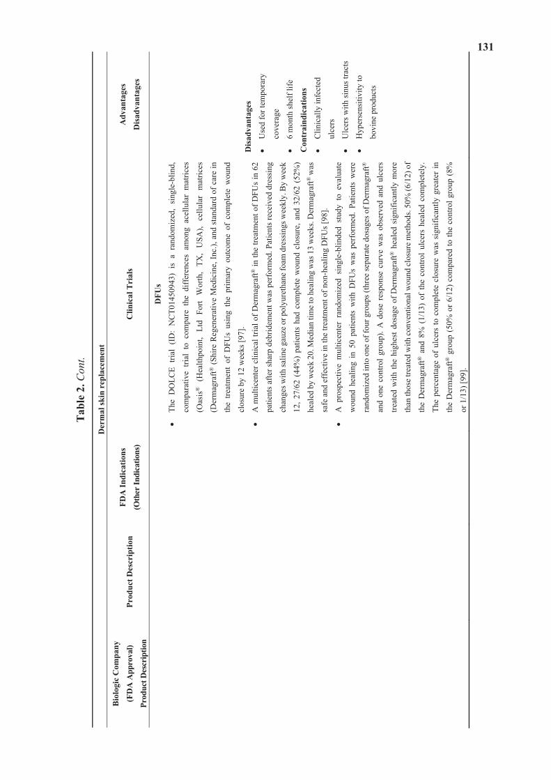
131
Tab
le 2
. Con
t.
Der
mal
skin
rep
lace
men
t
Bio
logi
c C
ompa
ny
(FD
A A
ppro
val)
Prod
uct D
escr
iptio
n
Prod
uct D
escr
iptio
n FD
A In
dica
tions
(Oth
er In
dica
tions
) C
linic
al T
rial
s A
dvan
tage
s
Dis
adva
ntag
es
DFU
s
The
DO
LCE
trial
(ID
: N
CT0
1450
943)
is
a ra
ndom
ized
, si
ngle
-blin
d,
com
para
tive
trial
to
com
pare
the
diff
eren
ces
amon
g ac
ellu
lar
mat
rices
(Oas
is®
(H
ealth
poin
t, Lt
d Fo
rt W
orth
, TX
, U
SA),
cellu
lar
mat
rices
(Der
mag
raft®
(Shi
re R
egen
erat
ive
Med
icin
e, In
c.),
and
stan
dard
of c
are
in
the
treat
men
t of
DFU
s us
ing
the
prim
ary
outc
ome
of c
ompl
ete
wou
nd
clos
ure
by 1
2 w
eeks
[97]
.
A m
ultic
ente
r clin
ical
tria
l of D
erm
agra
ft® in
the
treat
men
t of D
FUs
in 6
2
patie
nts a
fter s
harp
deb
ridem
ent w
as p
erfo
rmed
. Pat
ient
s rec
eive
d dr
essi
ng
chan
ges w
ith sa
line
gauz
e or
pol
yure
than
e fo
am d
ress
ings
wee
kly.
By
wee
k
12, 2
7/62
(44
%)
patie
nts
had
com
plet
e w
ound
clo
sure
, and
32/
62 (
52%
)
heal
ed b
y w
eek
20. M
edia
n tim
e to
heal
ing
was
13
wee
ks. D
erm
agra
ft® w
as
safe
and
eff
ectiv
e in
the
treat
men
t of n
on-h
ealin
g D
FUs [
98].
A p
rosp
ectiv
e m
ultic
ente
r ra
ndom
ized
sin
gle-
blin
ded
stud
y to
eva
luat
e
wou
nd h
ealin
g in
50
patie
nts
with
DFU
s w
as p
erfo
rmed
. Pa
tient
s w
ere
rand
omiz
ed in
to o
ne o
f fou
r gro
ups (
thre
e se
para
te d
osag
es o
f Der
mag
raft®
and
one
cont
rol
grou
p). A
dos
e re
spon
se c
urve
was
obs
erve
d an
d ul
cers
treat
ed w
ith th
e hi
ghes
t dos
age
of D
erm
agra
ft® h
eale
d si
gnifi
cant
ly m
ore
than
thos
e tre
ated
with
conv
entio
nal w
ound
clo
sure
met
hods
. 50%
(6/1
2) o
f
the
Der
mag
raft®
and
8%
(1/
13)
of t
he c
ontro
l ul
cers
hea
led
com
plet
ely.
The
perc
enta
ge o
f ul
cers
to c
ompl
ete
clos
ure
was
sig
nific
antly
gre
ater
in
the
Der
mag
raft®
gro
up (5
0% o
r 6/1
2) c
ompa
red
to th
e co
ntro
l gro
up (8
%
or 1
/13)
[99]
.
Dis
adva
ntag
es
Use
d fo
r tem
pora
ry
cove
rage
6 m
onth
shel
f life
Con
trai
ndic
atio
ns
Clin
ical
ly in
fect
ed
ulce
rs
Ulc
ers w
ith si
nus t
ract
s
Hyp
erse
nsiti
vity
to
bovi
ne p
rodu
cts
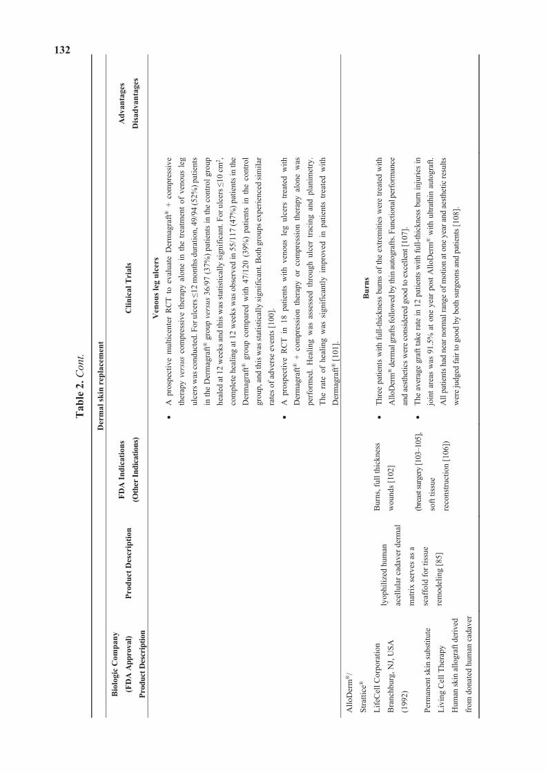
132
Tab
le 2
. Con
t.
Der
mal
skin
rep
lace
men
t
Bio
logi
c C
ompa
ny
(FD
A A
ppro
val)
Prod
uct D
escr
iptio
n
Prod
uct D
escr
iptio
n FD
A In
dica
tions
(Oth
er In
dica
tions
) C
linic
al T
rial
s A
dvan
tage
s
Dis
adva
ntag
es
Ven
ous l
eg u
lcer
s A
pro
spec
tive
mul
ticen
ter
RC
T to
eva
luat
e D
erm
agra
ft® +
com
pres
sive
th
erap
y ve
rsus
com
pres
sive
ther
apy
alon
e in
the
treat
men
t of
veno
us le
g ul
cers
was
cond
ucte
d. F
or u
lcer
s 12
mon
ths d
urat
ion,
49/
94 (5
2%) p
atie
nts
in th
e D
erm
agra
ft® g
roup
ver
sus 3
6/97
(37%
) pat
ient
s in
the
cont
rol g
roup
he
aled
at 1
2 w
eeks
and
this
was
stat
istic
ally
sign
ifica
nt. F
or u
lcer
s 10
cm
2 , co
mpl
ete
heal
ing
at 1
2 w
eeks
was
obs
erve
d in
55/
117
(47%
) pat
ient
s in
the
Der
mag
raft®
gro
up c
ompa
red
with
47/
120
(39%
) pa
tient
s in
the
con
trol
grou
p, an
d th
is w
as st
atis
tical
ly si
gnifi
cant
. Bot
h gr
oups
expe
rienc
ed si
mila
r ra
tes o
f adv
erse
eve
nts [
100]
. A
pro
spec
tive
RC
T in
18
patie
nts
with
ven
ous
leg
ulce
rs t
reat
ed w
ith
Der
mag
raft®
+ c
ompr
essi
on t
hera
py o
r co
mpr
essi
on t
hera
py a
lone
was
pe
rfor
med
. H
ealin
g w
as a
sses
sed
thro
ugh
ulce
r tra
cing
and
pla
nim
etry
. Th
e ra
te o
f he
alin
g w
as s
igni
fican
tly i
mpr
oved
in
patie
nts
treat
ed w
ith
Der
mag
raft®
[101
].
Allo
Der
m®
/
Stra
ttice
®
Life
Cel
l Cor
pora
tion
Bra
nchb
urg,
NJ,
USA
(199
2)
Perm
anen
t ski
n su
bstit
ute
Livi
ng C
ell T
hera
py
Hum
an sk
in a
llogr
aft d
eriv
ed
from
don
ated
hum
an c
adav
er
lyop
hiliz
ed h
uman
acel
lula
r cad
aver
der
mal
mat
rix se
rves
as a
scaf
fold
for t
issu
e
rem
odel
ing
[85]
Bur
ns, f
ull t
hick
ness
wou
nds [
102]
(bre
ast s
urge
ry [1
03–1
05],
soft
tissu
e
reco
nstru
ctio
n [1
06])
Bur
nsTh
ree
patie
nts
with
full-
thic
knes
s bu
rns
of th
e ex
trem
ities
wer
e tre
ated
with
A
lloD
erm
® d
erm
al g
rafts
follo
wed
by
thin
auto
graf
ts. F
unct
iona
l per
form
ance
an
d ae
sthet
ics w
ere
cons
ider
ed g
ood
to e
xcel
lent
[107
]. Th
e av
erag
e gr
aft t
ake
rate
in 1
2 pa
tient
s w
ith fu
ll-th
ickn
ess
burn
inju
ries i
n jo
int a
reas
was
91.
5% a
t one
yea
r pos
t Allo
Der
m®
with
ultr
athi
n au
togr
aft.
All
patie
nts h
ad n
ear n
orm
al ra
nge
of m
otio
n at
one
yea
r and
aes
thet
ic re
sults
w
ere
judg
ed fa
ir to
goo
d by
bot
h su
rgeo
ns a
nd p
atie
nts [
108]
.
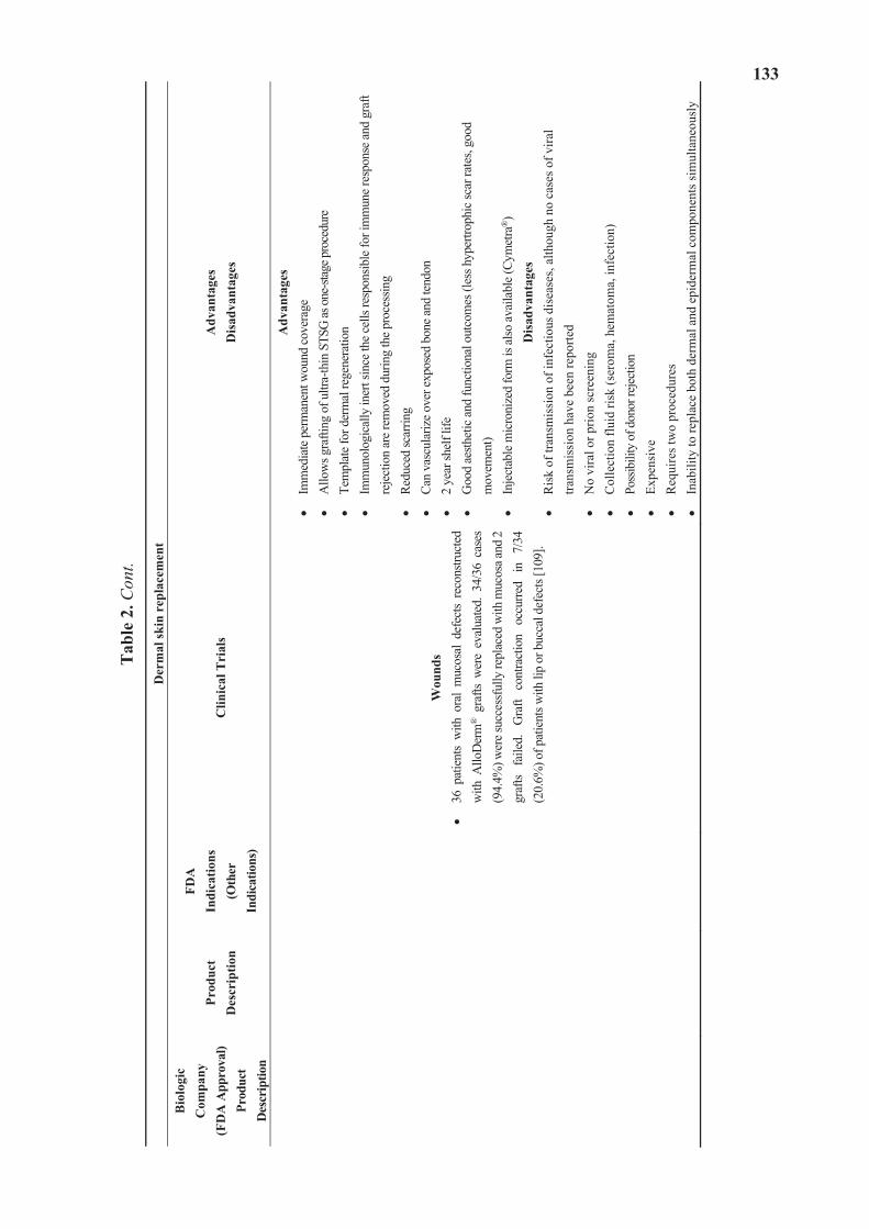
133
Tab
le 2
. Con
t.
Der
mal
skin
rep
lace
men
tB
iolo
gic
Com
pany
(F
DA
App
rova
l) Pr
oduc
t D
escr
iptio
n
Prod
uct
Des
crip
tion
FDA
Indi
catio
ns
(Oth
er
Indi
catio
ns)
Clin
ical
Tri
als
Adv
anta
ges
Dis
adva
ntag
es
Wou
nds
36 p
atie
nts
with
ora
l m
ucos
al d
efec
ts re
cons
truct
ed
with
Allo
Der
m® g
rafts
wer
e ev
alua
ted.
34/
36 c
ases
(9
4.4%
) wer
e suc
cess
fully
repl
aced
with
muc
osa a
nd 2
gr
afts
faile
d.
Gra
ft co
ntra
ctio
n oc
curre
d in
7/
34
(20.
6%) o
f pat
ient
s with
lip
or b
ucca
l def
ects
[109
].
Adv
anta
ges
Imm
edia
te p
erm
anen
t wou
nd co
vera
ge
Allo
ws g
rafti
ng o
f ultr
a-th
in S
TSG
as o
ne-st
age p
roce
dure
Te
mpl
ate f
or d
erm
al re
gene
ratio
n Im
mun
olog
ical
ly in
ert s
ince
the c
ells
resp
onsib
le fo
r im
mun
e res
pons
e and
gra
ft re
ject
ion
are
rem
oved
dur
ing
the p
roce
ssin
g Re
duce
d sc
arrin
g Ca
n va
scul
ariz
e ove
r exp
osed
bon
e and
tend
on
2 ye
ar sh
elf l
ife
Goo
d ae
sthet
ic an
d fu
nctio
nal o
utco
mes
(les
s hyp
ertro
phic
scar
rate
s, go
od
mov
emen
t) In
ject
able
mic
roni
zed
form
is al
so av
aila
ble (
Cym
etra
®)
Dis
adva
ntag
esR
isk
of tr
ansm
issi
on o
f inf
ectio
us d
isea
ses,
alth
ough
no
case
s of v
iral
trans
mis
sion
hav
e be
en re
porte
d N
o vi
ral o
r prio
n sc
reen
ing
Col
lect
ion
fluid
risk
(ser
oma,
hem
atom
a, in
fect
ion)
Po
ssib
ility
of d
onor
rejec
tion
Expe
nsiv
e R
equi
res t
wo
proc
edur
es
Inab
ility
to re
plac
e bo
th d
erm
al a
nd e
pide
rmal
com
pone
nts s
imul
tane
ousl
y
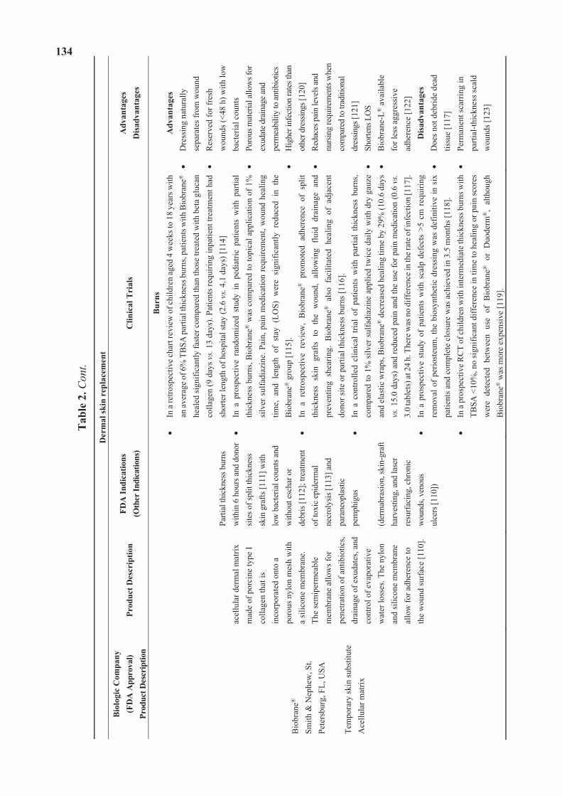
134
Tab
le 2
. Con
t.
Der
mal
skin
rep
lace
men
tB
iolo
gic
Com
pany
(F
DA
App
rova
l)
Prod
uct D
escr
iptio
n Pr
oduc
t Des
crip
tion
FDA
Indi
catio
ns
(Oth
er In
dica
tions
) C
linic
al T
rial
s A
dvan
tage
sD
isad
vant
ages
Bio
bran
e®
Smith
& N
ephe
w, S
t. Pe
ters
burg
, FL,
USA
Te
mpo
rary
skin
subs
titut
e A
cellu
lar m
atrix
acel
lula
r der
mal
mat
rix
mad
e of
por
cine
type
I co
llage
n th
at is
in
corp
orat
ed o
nto
a po
rous
nyl
on m
esh
with
a
silic
one
mem
bran
e.
The
sem
iper
mea
ble
mem
bran
e al
low
s for
pe
netra
tion
of a
ntib
iotic
s, dr
aina
ge o
f exu
date
s, an
d co
ntro
l of e
vapo
rativ
e w
ater
loss
es. T
he n
ylon
an
d si
licon
e m
embr
ane
allo
w fo
r adh
eren
ce to
th
e w
ound
surf
ace
[110
].
Parti
al th
ickn
ess b
urns
w
ithin
6 h
ours
and
don
or
sites
of s
plit
thic
knes
s sk
in g
rafts
[111
] with
lo
w b
acte
rial c
ount
s and
w
ithou
t esc
har o
r
debr
is [1
12];
treat
men
t of
toxi
c ep
ider
mal
ne
crol
ysis
[113
] and
pa
rane
opla
stic
pem
phig
us
(der
mab
rasio
n, sk
in-g
raft
harv
estin
g, a
nd la
ser
resu
rfaci
ng, c
hron
ic
wou
nds,
veno
us
ulce
rs [1
10])
Bur
nsIn
a re
trosp
ectiv
e cha
rt re
view
of c
hild
ren
aged
4 w
eeks
to 1
8 ye
ars w
ith
an av
erag
e of 6
% T
BSA
par
tial t
hick
ness
bur
ns, p
atie
nts w
ith B
iobr
ane®
he
aled
sign
ifica
ntly
fast
er c
ompa
red
than
thos
e tre
ated
with
bet
a gl
ucan
co
llage
n (9
day
s vs.
13 d
ays)
. Pat
ient
s req
uirin
g in
patie
nt tr
eatm
ent h
ad
shor
ter l
engt
h of
hos
pita
l sta
y (2
.6 v
s. 4.
1 da
ys) [
114]
In
a p
rosp
ectiv
e ra
ndom
ized
stu
dy i
n pe
diat
ric p
atie
nts
with
par
tial
thic
knes
s bu
rns,
Bio
bran
e® w
as c
ompa
red
to to
pica
l app
licat
ion
of 1
%
silv
er s
ulfa
diaz
ine.
Pai
n, p
ain
med
icat
ion
requ
irem
ent,
wou
nd h
ealin
g tim
e, a
nd l
engt
h of
sta
y (L
OS)
wer
e si
gnifi
cant
ly r
educ
ed i
n th
e B
iobr
ane®
gro
up [1
15].
In a
ret
rosp
ectiv
e re
view
, B
iobr
ane®
pro
mot
ed a
dher
ence
of
split
th
ickn
ess
skin
gra
fts t
o th
e w
ound
, al
low
ing
fluid
dra
inag
e an
d pr
even
ting
shea
ring.
Bio
bran
e® a
lso
faci
litat
ed h
ealin
g of
adj
acen
t do
nor s
ite o
r par
tial t
hick
ness
bur
ns [1
16].
In a
con
trolle
d cl
inic
al t
rial
of p
atie
nts
with
par
tial
thic
knes
s bu
rns,
com
pare
d to
1%
silv
er s
ulfa
diaz
ine
appl
ied
twic
e da
ily w
ith d
ry g
auze
an
d el
astic
wra
ps, B
iobr
ane®
dec
reas
ed h
ealin
g tim
e by
29%
(10.
6 da
ys
vs. 1
5.0
days
) and
redu
ced
pain
and
the
use
for p
ain
med
icat
ion
(0.6
vs.
3.0
tabl
ets)
at 2
4 h.
The
re w
as n
o di
ffer
ence
in th
e rat
e of i
nfec
tion
[117
].In
a p
rosp
ectiv
e st
udy
of p
atie
nts
with
sca
lp d
efec
ts >
5 cm
req
uirin
g re
mov
al o
f pe
riost
eum
, the
bio
synt
hetic
dre
ssin
g w
as d
efin
itive
in s
ix
patie
nts a
nd c
ompl
ete
clos
ure
was
ach
ieve
d in
3.5
mon
ths [
118]
. In
a p
rosp
ectiv
e RC
T of
chi
ldre
n w
ith in
term
edia
te th
ickn
ess b
urns
with
TB
SA <
10%
, no
sign
ifica
nt d
iffer
ence
in ti
me
to h
ealin
g or
pai
n sc
ores
w
ere
dete
cted
bet
wee
n us
e of
Bio
bran
e® o
r D
uode
rm®
, al
thou
gh
Bio
bran
e® w
as m
ore
expe
nsiv
e [1
19].
Adv
anta
ges
Dre
ssin
g na
tura
lly
sepa
rate
s fro
m w
ound
R
eser
ved
for f
resh
w
ound
s (<4
8 h)
with
low
ba
cter
ial c
ount
s Po
rous
mat
eria
l allo
ws f
or
exud
ate d
rain
age a
nd
perm
eabi
lity
to an
tibio
tics
Hig
her i
nfec
tion
rate
s tha
n ot
her d
ress
ings
[120
] Re
duce
s pai
n le
vels
and
nurs
ing
requ
irem
ents
whe
n co
mpa
red
to tr
aditi
onal
dr
essin
gs [1
21]
Shor
tens
LO
S B
iobr
ane-
L® a
vaila
ble
fo
r les
s agg
ress
ive
ad
here
nce
[122
] D
isad
vant
ages
Doe
s not
deb
ride
dead
tis
sue
[117
] Pe
rman
ent s
carr
ing
in
parti
al-th
ickn
ess s
cald
w
ound
s [12
3]
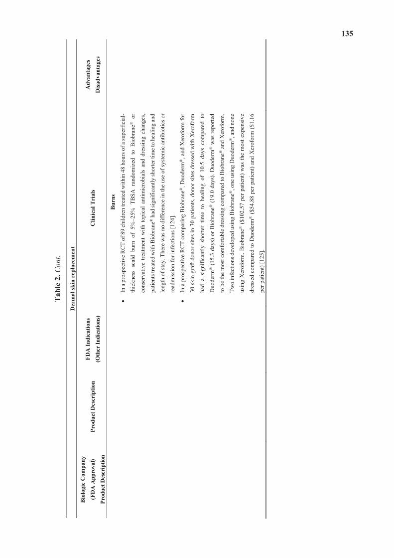
135
Tab
le 2
. Con
t.
Der
mal
skin
rep
lace
men
t
Bio
logi
c C
ompa
ny
(FD
A A
ppro
val)
Prod
uct D
escr
iptio
n
Prod
uct D
escr
iptio
n FD
A In
dica
tions
(Oth
er In
dica
tions
) C
linic
al T
rial
s A
dvan
tage
s
Dis
adva
ntag
es
Bur
ns
In a
pro
spec
tive R
CT
of 8
9 ch
ildre
n tre
ated
with
in 4
8 ho
urs o
f a su
perf
icia
l-
thic
knes
s sc
ald
burn
of
5%–2
5% T
BSA
ran
dom
ized
to
Bio
bran
e® o
r
cons
erva
tive
treat
men
t w
ith t
opic
al a
ntim
icro
bial
s an
d dr
essi
ng c
hang
es,
patie
nts t
reat
ed w
ith B
iobr
ane®
had
sign
ifica
ntly
shor
ter t
ime t
o he
alin
g an
d
leng
th o
f sta
y. T
here
was
no
diff
eren
ce in
the
use
of sy
stem
ic a
ntib
iotic
s or
read
mis
sion
for i
nfec
tions
[124
].
In a
pro
spec
tive
RC
T co
mpa
ring
Bio
bran
e®, D
uode
rm®
, and
Xer
ofor
m fo
r
30 s
kin
graf
t don
or s
ites
in 3
0 pa
tient
s, do
nor s
ites
dres
sed
with
Xer
ofor
m
had
a si
gnifi
cant
ly s
horte
r tim
e to
hea
ling
of 1
0.5
days
com
pare
d to
Duo
derm
® (1
5.3
days
) or B
iobr
ane®
(19.
0 da
ys).
Duo
derm
® w
as re
porte
d
to b
e th
e m
ost c
omfo
rtabl
e dr
essi
ng c
ompa
red
to B
iobr
ane®
and
Xer
ofor
m.
Two
infe
ctio
ns d
evel
oped
usi
ng B
iobr
ane®
, one
usi
ng D
uode
rm®
, and
non
e
usin
g X
erof
orm
. Bio
bran
e® ($
102.
57 p
er p
atie
nt) w
as th
e m
ost e
xpen
sive
dres
sed
com
pare
d to
Duo
derm
® ($
54.8
8 pe
r pat
ient
) and
Xer
ofor
m ($
1.16
per p
atie
nt) [
125]
.
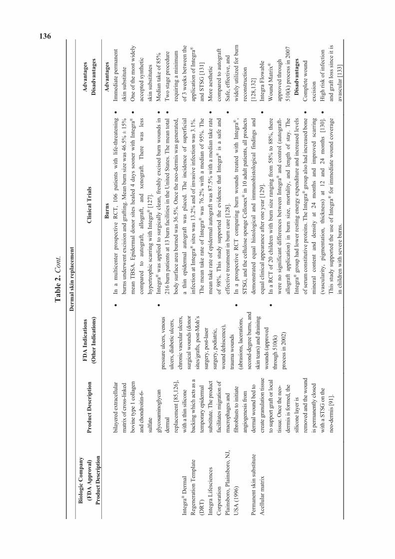
136
Tab
le 2
. Con
t.
Der
mal
skin
rep
lace
men
tB
iolo
gic
Com
pany
(F
DA
App
rova
l)
Prod
uct D
escr
iptio
n Pr
oduc
t Des
crip
tion
FDA
Indi
catio
ns
(Oth
er In
dica
tions
) C
linic
al T
rial
s A
dvan
tage
sD
isad
vant
ages
Inte
gra®
Der
mal
R
egen
erat
ion
Tem
plat
e (D
RT)
In
tegr
a Li
fesc
ienc
es
Cor
pora
tion
Pl
ains
boro
, Pla
insb
oro,
NJ,
USA
(199
6)
Perm
anen
t ski
n su
bstit
ute
Ace
llula
r mat
rix
bila
yere
d ex
trace
llula
r m
atrix
of c
ross
-link
ed
bovi
ne ty
pe 1
colla
gen
and
chon
droi
tin-6
-su
lfate
glyc
osam
inog
lyca
n de
rmal
re
plac
emen
t [85
,126
], w
ith a
thin
silic
one
back
ing
whi
ch ac
ts as
a te
mpo
rary
epid
erm
al
subs
titut
e. Th
e pro
duct
fa
cilit
ates
mig
ratio
n of
m
acro
phag
es an
d fib
robl
asts
to in
itiat
e an
giog
enes
is fro
m
derm
al w
ound
bed
to
crea
te g
ranu
latio
n tis
sue
to su
ppor
t gra
ft or
loca
l tis
sue.
Onc
e the
neo
-de
rmis
is fo
rmed
, the
sil
icon
e lay
er is
re
mov
ed an
d th
e wou
nd
is pe
rman
ently
clos
ed
with
a ST
SG o
n th
e ne
o-de
rmis
[91]
.
pres
sure
ulc
ers,
veno
us
ulce
rs, d
iabe
tic u
lcer
s, ch
roni
c vas
cula
r ulc
ers,
surg
ical
wou
nds (
dono
r sit
es/g
rafts
, pos
t-Moh
’s
surg
ery,
pos
t-las
er
surg
ery,
pod
iatri
c,
wou
nd d
ehisc
ence
), tra
uma
wou
nds
(abr
asio
ns, l
acer
atio
ns,
seco
nd-d
egre
e bur
ns, a
nd
skin
tear
s) an
d dr
aini
ng
wou
nds (
appr
oved
th
roug
h51
0(k)
pr
oces
s in
2002
)
Bur
nsIn
a m
ultic
ente
r pr
ospe
ctiv
e R
CT,
106
pat
ient
s w
ith l
ife-th
reat
enin
g bu
rns u
nder
wen
t exc
isio
n an
d gr
aftin
g. M
ean
burn
size
was
46.
5% ±
15%
m
ean
TBSA
. Epi
derm
al d
onor
site
s he
aled
4 d
ays
soon
er w
ith I
nteg
ra®
co
mpa
red
to
auto
graf
t, al
logr
aft,
and
xeno
graf
t. Th
ere
was
le
ss
hype
rtrop
hic
scar
ring
with
Inte
gra®
[127
]. In
tegr
a® w
as a
pplie
d to
sur
gica
lly c
lean
, fre
shly
exc
ised
bur
n w
ound
s in
21
6 bu
rn p
atie
nts a
t 13
burn
faci
litie
s in
the
Uni
ted
Stat
es. T
he m
ean
tota
l bo
dy su
rfac
e ar
ea b
urne
d w
as 3
6.5%
. Onc
e th
e ne
o-de
rmis
was
gen
erat
ed,
a th
in e
pide
rmal
aut
ogra
ft w
as p
lace
d. T
he i
ncid
ence
of
supe
rfic
ial
infe
ctio
n at
Inte
gra®
site
s was
13.
2% a
nd o
f inv
asiv
e in
fect
ion
was
3.1
%.
The
mea
n ta
ke r
ate
of I
nteg
ra®
was
76.
2% w
ith a
med
ian
of 9
5%. T
he
mea
n ta
ke ra
te o
f epi
derm
al a
utog
raft
was
87.
5% w
ith a
med
ian
take
rate
of
98%
. Th
is s
tudy
sup
porte
d th
e ev
iden
ce t
hat
Inte
gra®
is
a sa
fe a
nd
effe
ctiv
e tre
atm
ent i
n bu
rn c
are
[128
]. In
a p
rosp
ectiv
e R
CT
com
parin
g bu
rn w
ound
s tre
ated
with
Int
egra
®,
STSG
, and
the
cellu
lose
spon
ge C
ello
nex®
in 1
0 ad
ult p
atie
nts,
all p
rodu
cts
dem
onst
rate
d eq
ual
hist
olog
ical
and
im
mun
ohis
tolo
gica
l fin
ding
s an
d eq
ual c
linic
al a
ppea
ranc
e af
ter o
ne y
ear [
129]
. In
a R
CT
of 2
0 ch
ildre
n w
ith b
urn
size
rang
ing
from
58%
to 8
8%, t
here
w
ere
no s
igni
fican
t diff
eren
ces
betw
een
Inte
gra®
and
con
trol (
auto
graf
t-al
logr
aft
appl
icat
ion)
in
burn
siz
e, m
orta
lity,
and
len
gth
of s
tay.
The
In
tegr
a® g
roup
had
low
er re
stin
g en
ergy
exp
endi
ture
and
incr
ease
d le
vels
of
seru
m co
nstit
utiv
e pr
otei
ns. T
he In
tegr
a® g
roup
als
o ha
d in
crea
sed
bone
m
iner
al
cont
ent
and
dens
ity
at
24
mon
ths
and
impr
oved
sc
arrin
g (v
ascu
larit
y,
pigm
enta
tion,
th
ickn
ess)
at
12
an
d 24
m
onth
s [1
30].
This
stu
dy s
uppo
rted
the
use
of In
tegr
a® fo
r im
med
iate
wou
nd c
over
age
in c
hild
ren
with
seve
re b
urns
.
Adv
anta
ges
Imm
edia
te p
erm
anen
t
skin
subs
titut
e O
ne o
f the
mos
t wid
ely
acce
pted
synt
hetic
sk
in su
bstit
utes
M
edia
n ta
ke o
f 85%
Tw
o st
age
proc
edur
e re
quiri
ng a
min
imum
of
3 w
eeks
bet
wee
n th
e ap
plic
atio
n of
Inte
gra®
an
d ST
SG [1
31]
Mor
e ae
sthe
tic
com
pare
d to
aut
ogra
ft Sa
fe, e
ffec
tive,
and
w
idel
y ut
ilize
d fo
r bur
n re
cons
truct
ion
[128
,132
] In
tegr
a Fl
owab
le
Wou
nd M
atrix
®
appr
oved
thro
ugh
510(
k) p
roce
ss in
200
7 D
isad
vant
ages
Com
plet
e w
ound
ex
cisi
on
Hig
h ris
k of
infe
ctio
n an
d gr
aft l
oss s
ince
it is
av
ascu
lar [
133]
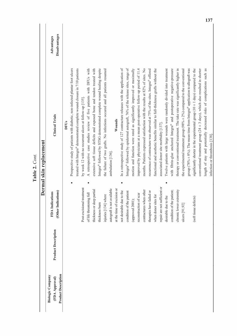
137
Tab
le 2
. Con
t.
Der
mal
skin
rep
lace
men
tB
iolo
gic
Com
pany
(F
DA
App
rova
l)
Prod
uct D
escr
iptio
n Pr
oduc
t Des
crip
tion
FDA
Indi
catio
ns
(Oth
er In
dica
tions
) C
linic
al T
rial
s A
dvan
tage
sD
isad
vant
ages
Post-
exci
siona
l tre
atm
ent
of li
fe th
reat
enin
g fu
ll th
ickn
ess o
r dee
p pa
rtial
th
ickn
ess b
urn
in
jurie
s [13
4] w
here
au
togr
aft i
s not
avai
labl
e at
the t
ime o
f exc
ision
or
not d
esira
ble
due t
o th
e co
nditi
on o
f the
pat
ient
(a
ppro
ved
2001
); re
cons
truct
ion
of sc
ar
cont
ract
ures
whe
n ot
her
ther
apie
s hav
e fai
led
or
whe
n do
nor s
ites f
or
repa
ir ar
e not
suffi
cien
t or
desir
able
due
to th
e co
nditi
on o
f the
pat
ient
; ch
roni
c low
er ex
trem
ity
ulce
rs [9
1,92
] (s
oft t
issue
def
ects)
DFU
s Pr
ospe
ctiv
e st
udy
of p
atie
nts w
ith d
iabe
tic, n
on-in
fect
ed p
lant
ar fo
ot u
lcer
s tre
ated
with
Inte
gra®
dem
onst
rate
d co
mpl
ete w
ound
clos
ure i
n 7/
10 p
atie
nts
by w
eek
12 w
ith n
o re
curr
ent u
lcer
s at f
ollo
w-u
p [1
35].
A r
etro
spec
tive
case
stu
dies
rev
iew
of
five
patie
nts
with
DFU
s w
ith
exte
nsiv
e so
ft tis
sue
defic
its a
nd e
xpos
ed b
one
and
tend
on t
reat
ed w
ith
Inte
gra®
follo
wed
by
STSG
dem
onst
rate
d co
mpl
ete
wou
nd h
ealin
g de
spite
th
e fa
ilure
of
two
graf
ts. N
o in
fect
ions
occ
ured
and
all
patie
nts
resu
med
am
bula
tion
[136
]. W
ound
s In
a re
trosp
ectiv
e st
udy
of 1
27 c
ontra
ctur
e re
leas
es w
ith th
e ap
plic
atio
n of
In
tegr
a® fo
llow
ed b
y ep
ider
mal
aut
ogra
ft, 7
6% o
f the
rele
ase
site
s, ra
nge
of
mot
ion
and
func
tion
wer
e ra
ted
as s
igni
fican
tly i
mpr
oved
or
max
imal
ly
impr
oved
by
phys
icia
ns a
t a m
ean
post
-ope
rativ
e fo
llow
-up
perio
d of
11.
4 m
onth
s. Pa
tient
s exp
ress
ed sa
tisfa
ctio
n w
ith th
e re
sults
at 8
2% o
f site
s. N
o re
curr
ence
of c
ontra
ctur
e w
as o
bser
ved
at 7
5% o
f the
site
s. In
tegr
a® o
ffer
ed
func
tiona
l and
aes
thet
ic b
enef
its si
mila
r to
full-
thic
knes
s gra
fts w
ithou
t the
as
soci
ated
don
or si
te m
orbi
dity
[137
]. Tw
elve
pat
ient
s w
ith la
rge
wou
nds
wer
e ra
ndom
ly d
ivid
ed in
to tr
eatm
ent
with
fib
rin-g
lue
anch
ored
Int
egra
® a
nd p
osto
pera
tive
nega
tive-
pres
sure
th
erap
y or
con
vent
iona
l tre
atm
ent.
The
take
rate
was
sign
ifica
ntly
hig
her i
n th
e exp
erim
enta
l tre
atm
ent g
roup
(98%
± 2
%) c
ompa
red
to th
e con
vent
iona
l gr
oup
(78%
± 8
%).
The m
ean
time f
rom
Inte
gra®
appl
icat
ion
to al
logr
aft w
as
signi
fican
tly sh
orte
r in
the
expe
rimen
tal g
roup
(10
± 1
days
) com
pare
d to
the
conv
entio
nal t
reat
men
t gro
up (2
4 ±
3 da
ys),
whi
ch a
lso
resu
lted
in s
horte
r le
ngth
of
stay
and
pot
entia
lly d
ecre
ased
ris
ks o
f co
mpl
icat
ions
suc
h as
in
fect
ion
or th
rom
bosi
s [13
8].
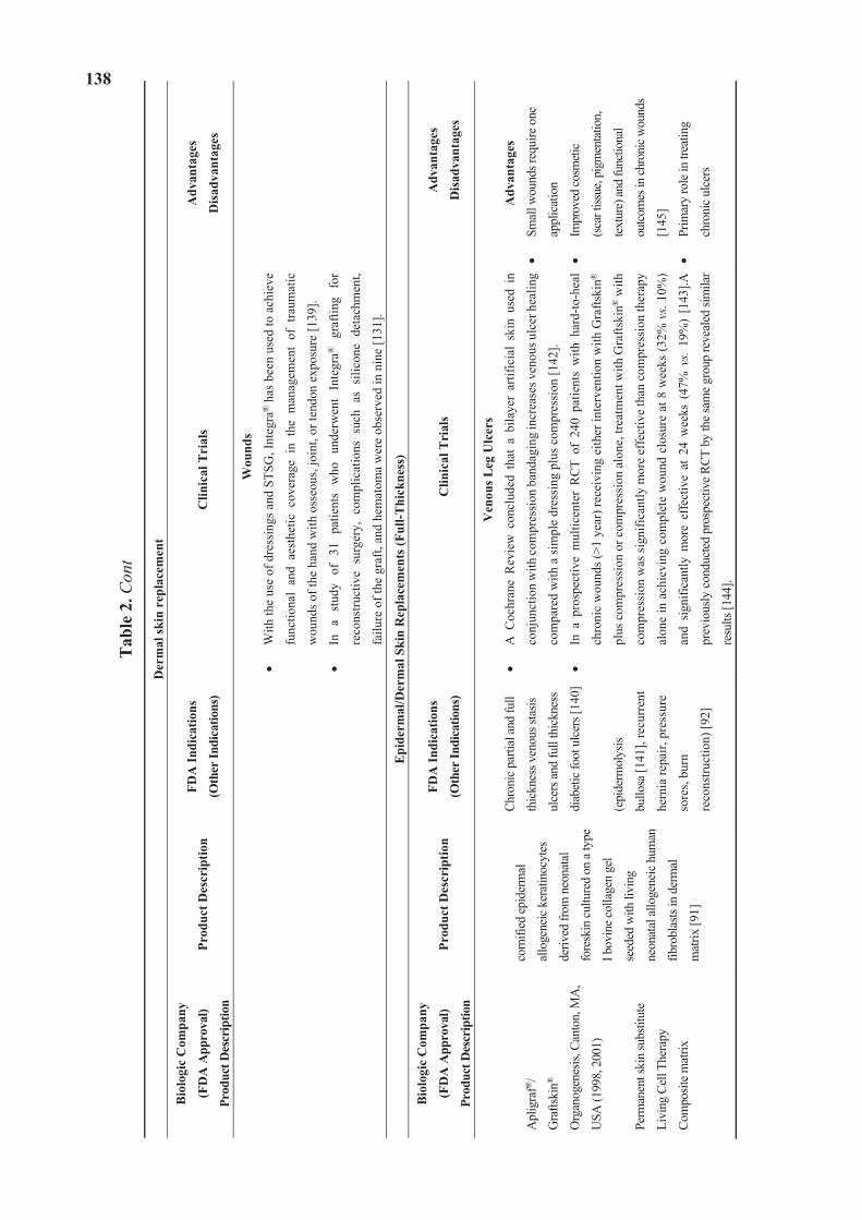
138
Tab
le 2
. Con
t
Der
mal
skin
rep
lace
men
t
Bio
logi
c C
ompa
ny
(FD
A A
ppro
val)
Prod
uct D
escr
iptio
n
Prod
uct D
escr
iptio
n FD
A In
dica
tions
(Oth
er In
dica
tions
) C
linic
al T
rial
s A
dvan
tage
s
Dis
adva
ntag
es
Wou
nds
With
the
use
of d
ress
ings
and
STS
G, I
nteg
ra®
has
bee
n us
ed to
ach
ieve
func
tiona
l an
d ae
sthe
tic c
over
age
in t
he m
anag
emen
t of
tra
umat
ic
wou
nds o
f the
han
d w
ith o
sseo
us, j
oint
, or t
endo
n ex
posu
re [1
39].
In a
stu
dy o
f 31
pat
ient
s w
ho u
nder
wen
t In
tegr
a® g
rafti
ng f
or
reco
nstru
ctiv
e su
rger
y, c
ompl
icat
ions
suc
h as
sili
cone
det
achm
ent,
failu
re o
f the
gra
ft, a
nd h
emat
oma
wer
e ob
serv
ed in
nin
e [1
31].
Epi
derm
al/D
erm
al S
kin
Rep
lace
men
ts (F
ull-T
hick
ness
) B
iolo
gic
Com
pany
(FD
A A
ppro
val)
Prod
uct D
escr
iptio
n
Prod
uct D
escr
iptio
n FD
A In
dica
tions
(Oth
er In
dica
tions
) C
linic
al T
rial
s A
dvan
tage
s
Dis
adva
ntag
es
Apl
igra
f®/
Gra
ftski
n®
Org
anog
enes
is, C
anto
n, M
A,
USA
(199
8, 2
001)
Perm
anen
t ski
n su
bstit
ute
Livi
ng C
ell T
hera
py
Com
posit
e mat
rix
corn
ified
epid
erm
al
allo
gene
ic k
erat
inoc
ytes
deriv
ed fr
om n
eona
tal
fore
skin
cultu
red
on a
type
I bov
ine c
olla
gen
gel
seed
ed w
ith li
ving
neon
atal
allo
gene
ic h
uman
fibro
blas
ts in
der
mal
mat
rix [9
1]
Chro
nic p
artia
l and
full
thic
knes
s ven
ous s
tasis
ulce
rs an
d fu
ll th
ickn
ess
diab
etic
foot
ulc
ers[
140]
(epi
derm
olys
is
bullo
sa [1
41],
recu
rren
t
hern
ia re
pair,
pre
ssur
e
sore
s, bu
rn
reco
nstru
ctio
n) [9
2]
Ven
ous L
eg U
lcer
s
A C
ochr
ane
Rev
iew
con
clud
ed t
hat
a bi
laye
r ar
tific
ial
skin
use
d in
conj
unct
ion
with
com
pres
sion
ban
dagi
ng in
crea
ses v
enou
s ulc
er h
ealin
g
com
pare
d w
ith a
sim
ple
dres
sing
plu
s com
pres
sion
[142
].
In a
pro
spec
tive
mul
ticen
ter
RC
T of
240
pat
ient
s w
ith h
ard-
to-h
eal
chro
nic
wou
nds (
>1 y
ear)
rece
ivin
g ei
ther
inte
rven
tion
with
Gra
ftski
n®
plus
com
pres
sion
or c
ompr
essi
on a
lone
, tre
atm
ent w
ith G
rafts
kin®
with
com
pres
sion
was
sign
ifica
ntly
mor
e ef
fect
ive
than
com
pres
sion
ther
apy
alon
e in
ach
ievi
ng c
ompl
ete
wou
nd c
losu
re a
t 8 w
eeks
(32%
vs.
10%
)
and
signi
fican
tly m
ore
effe
ctiv
e at
24
wee
ks (
47%
vs.
19%
) [1
43].A
prev
ious
ly c
ondu
cted
pro
spec
tive
RCT
by th
e sa
me
grou
p re
veal
ed si
mila
r
resu
lts [1
44].
Adv
anta
ges
Smal
l wou
nds r
equi
re o
ne
appl
icat
ion
Impr
oved
cosm
etic
(sca
r tiss
ue, p
igm
enta
tion,
text
ure)
and
func
tiona
l
outco
mes
in ch
roni
c wou
nds
[145
]
Prim
ary
role
in tr
eatin
g
chro
nic u
lcer
s
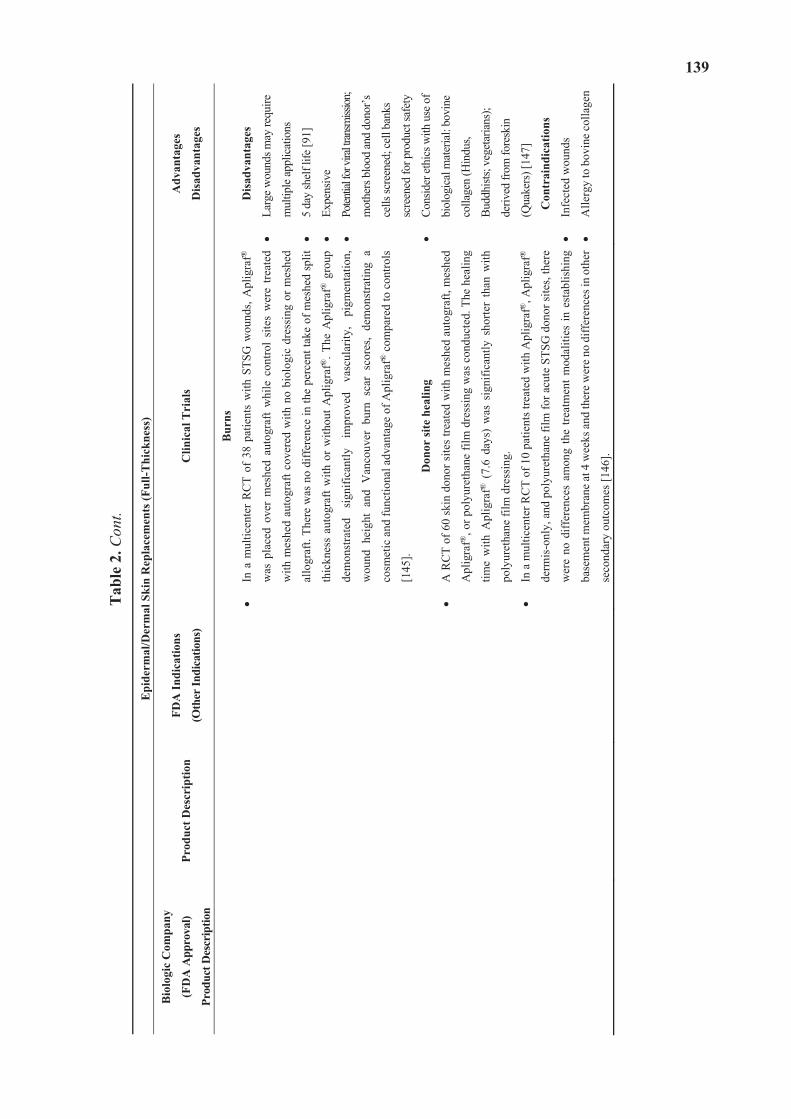
139
Tab
le 2
. Con
t.
Epi
derm
al/D
erm
al S
kin
Rep
lace
men
ts (F
ull-T
hick
ness
)
Bio
logi
c C
ompa
ny
(FD
A A
ppro
val)
Prod
uct D
escr
iptio
n
Prod
uct D
escr
iptio
n FD
A In
dica
tions
(Oth
er In
dica
tions
) C
linic
al T
rial
s A
dvan
tage
s
Dis
adva
ntag
es
Bur
ns
In a
mul
ticen
ter
RC
T of
38
patie
nts
with
STS
G w
ound
s, A
plig
raf®
was
pla
ced
over
mes
hed
auto
graf
t w
hile
con
trol
site
s w
ere
treat
ed
with
mes
hed
auto
graf
t cov
ered
with
no
biol
ogic
dre
ssin
g or
mes
hed
allo
graf
t. Th
ere
was
no
diff
eren
ce in
the
perc
ent t
ake
of m
eshe
d sp
lit
thic
knes
s au
togr
aft w
ith o
r w
ithou
t Apl
igra
f®. T
he A
plig
raf®
gro
up
dem
onst
rate
d si
gnifi
cant
ly
impr
oved
va
scul
arity
, pi
gmen
tatio
n,
wou
nd h
eigh
t an
d V
anco
uver
bur
n sc
ar s
core
s, de
mon
stra
ting
a
cosm
etic
and
func
tiona
l adv
anta
ge o
f Apl
igra
f® c
ompa
red
to c
ontro
ls
[145
].
Don
or si
te h
ealin
g
A R
CT
of 6
0 sk
in d
onor
site
s tre
ated
with
mes
hed
auto
graf
t, m
eshe
d
Apl
igra
f®, o
r pol
yure
than
e fil
m d
ress
ing
was
con
duct
ed. T
he h
ealin
g
time
with
Apl
igra
f® (
7.6
days
) w
as s
igni
fican
tly s
horte
r th
an w
ith
poly
uret
hane
film
dre
ssin
g.
In a
mul
ticen
ter R
CT
of 1
0 pa
tient
s tre
ated
with
Apl
igra
f®, A
plig
raf®
derm
is-o
nly,
and
pol
yure
than
e fil
m fo
r acu
te S
TSG
don
or si
tes,
ther
e
wer
e no
diff
eren
ces
amon
g th
e tre
atm
ent m
odal
ities
in e
stab
lishi
ng
base
men
t mem
bran
e at
4 w
eeks
and
ther
e w
ere
no d
iffer
ence
s in
othe
r
seco
ndar
y ou
tcom
es [1
46].
Dis
adva
ntag
es
Larg
e w
ound
s may
requ
ire
mul
tiple
appl
icat
ions
5 da
y sh
elf l
ife [9
1]
Expe
nsiv
e
Poten
tial f
or vi
ral tr
ansm
ission
;
mot
hers
blo
od an
d do
nor’s
cells
scre
ened
; cel
l ban
ks
scre
ened
for p
rodu
ct sa
fety
Cons
ider
ethi
cs w
ith u
se o
f
biol
ogic
al m
ater
ial:
bovi
ne
colla
gen
(Hin
dus,
Budd
hists
; veg
etar
ians
);
deriv
ed fr
om fo
resk
in
(Qua
kers
) [14
7]
Con
trai
ndic
atio
ns
Infe
cted
wou
nds
Alle
rgy
to b
ovin
e co
llage
n
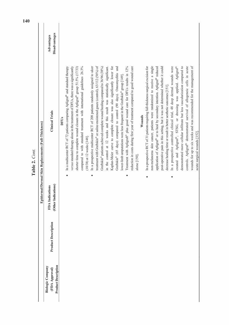
140
Tab
le 2
. Con
t.
Epi
derm
al/D
erm
al S
kin
Rep
lace
men
ts (F
ull-T
hick
ness
)B
iolo
gic
Com
pany
(F
DA
App
rova
l)
Prod
uct D
escr
iptio
n Pr
oduc
t Des
crip
tion
FDA
Indi
catio
ns
(Oth
er In
dica
tions
) C
linic
al T
rial
s A
dvan
tage
sD
isad
vant
ages
DFU
s In
a m
ultic
ente
r RCT
of 7
2 pa
tient
s com
parin
g A
plig
raf®
and
stan
dard
ther
apy
vers
us st
anda
rd th
erap
y al
one i
n th
e tre
atm
ent o
f DFU
s, th
ere w
as a
signi
fican
tly
shor
ter t
ime
to c
ompl
ete
wou
nd c
losu
re in
the
Apl
igra
f® g
roup
51.
5% (1
7/33
) co
mpa
red
to w
ith s
tand
ard
treat
men
t w
ith i
nter
natio
nal
guid
elin
es 2
6.3%
(1
0/38
) at 1
2 w
eeks
[148
]. In
a p
rosp
ectiv
e m
ultic
ente
r RCT
of 2
08 p
atie
nts
rand
omly
ass
igne
d to
ulc
er
treat
men
t with
Gra
ftski
n® o
r sal
ine-
moi
stene
d ga
uze (
cont
rol),
63/
112
(56%
) of
Gra
ftski
n® p
atie
nts a
chie
ved
com
plet
e wou
nd h
ealin
g co
mpa
red
to 3
6/96
(38%
) in
the
con
trol
at 1
2 w
eeks
and
thi
s re
sult
was
sta
tistic
ally
sig
nific
ant.
Kap
lan-
Mei
er c
urve
to
com
plet
e cl
osur
e w
as a
lso s
igni
fican
tly l
ower
for
G
rafts
kin®
(65
day
s) c
ompa
red
to c
ontro
l (9
0 da
ys).
Oste
omye
litis
and
low
er-li
mb
ampu
tatio
ns w
ere l
ess f
requ
ent i
n th
e G
rafts
kin®
gro
up [1
49].
Trea
tmen
t w
ith A
plig
raft®
plu
s go
od w
ound
car
e fo
r D
FUs
resu
lts i
n 12
%
redu
ctio
n in
cos
ts du
ring
first
year
of t
reat
men
t com
pare
d to
goo
d w
ound
car
e al
one
[150
]. W
ound
s In
a pr
ospe
ctiv
e RCT
of 3
1 pa
tient
s req
uirin
g fu
ll-th
ickn
ess s
urgi
cal e
xcisi
on fo
r no
n-m
elan
oma
skin
can
cer,
patie
nts
wer
e ra
ndom
ized
to
rece
ive
a sin
gle
appl
icat
ion
of A
plig
raf®
or t
o he
al b
y se
cond
ary
inte
ntio
n. A
plig
raf®
redu
ced
post-
oper
ativ
e pa
in in
this
setti
ng, b
ut it
was
not
det
erm
ined
whe
ther
it c
ould
de
crea
se h
ealin
g tim
e or r
esul
t in
bette
r aes
thet
ic o
utco
mes
[151
]. In
a p
rosp
ectiv
e co
ntro
lled
clin
ical
tria
l, 48
dee
p de
rmal
wou
nds
wer
e cr
eate
d an
d A
plig
raf®
, ST
SG,
or
dres
sing
w
as
appl
ied.
A
plig
raf®
de
mon
stra
ted
mor
e ce
llula
r inf
iltra
te b
ut le
ss v
ascu
lariz
atio
n co
mpa
red
to
cont
rols
. A
plig
raf®
dem
onst
rate
d su
rviv
al o
f al
loge
neic
cel
ls i
n ac
ute
wou
nds
for u
p to
six
wee
ks a
nd w
as re
com
men
ded
for t
he m
anag
emen
t of
acut
e su
rgic
al w
ound
s [15
2].
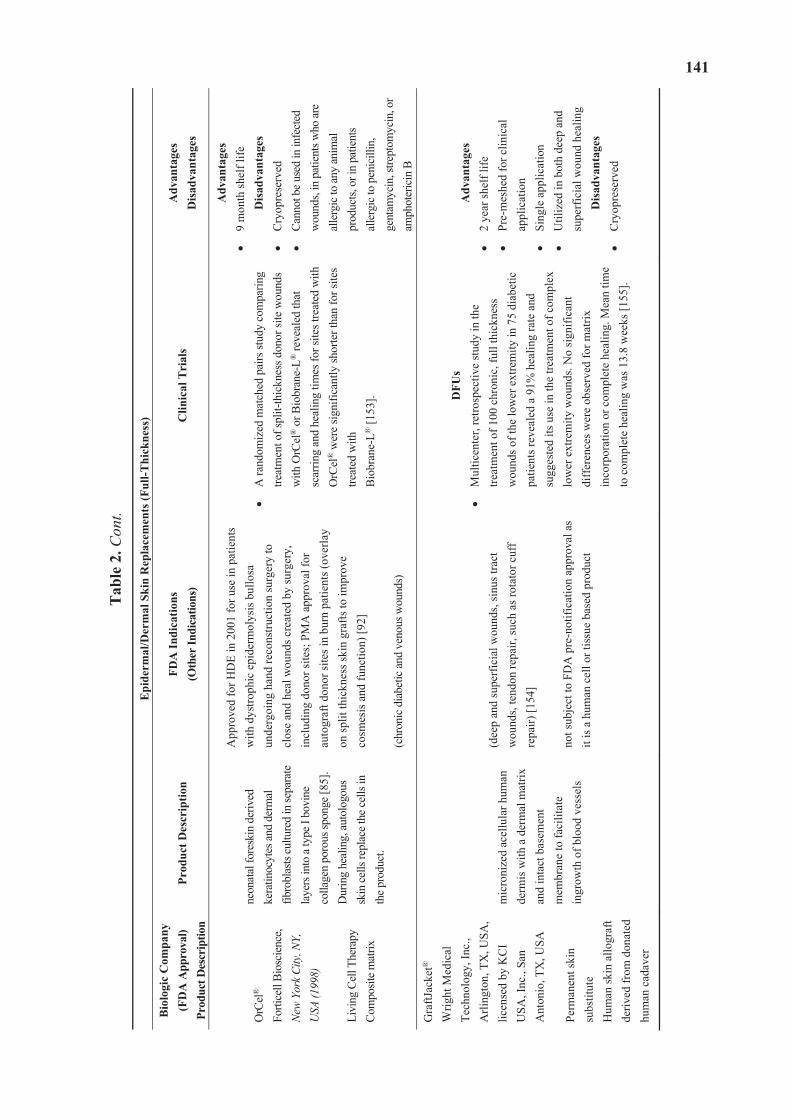
141
Tab
le 2
. Con
t.
Epi
derm
al/D
erm
al S
kin
Rep
lace
men
ts (F
ull-T
hick
ness
)B
iolo
gic
Com
pany
(F
DA
App
rova
l)
Prod
uct D
escr
iptio
nPr
oduc
t Des
crip
tion
FDA
Indi
catio
ns
(Oth
er In
dica
tions
) C
linic
al T
rial
s A
dvan
tage
sD
isad
vant
ages
OrC
el®
Forti
cell
Bios
cien
ce,
New
York
City
, NY,
U
SA (1
998)
Li
ving
Cel
l The
rapy
Co
mpo
site m
atrix
neon
atal
fore
skin
der
ived
ke
ratin
ocyt
es a
nd d
erm
al
fibro
blas
tscu
lture
d in
sepa
rate
la
yers
into
a ty
pe I
bovi
ne
colla
gen
poro
us sp
onge
[85]
. D
urin
g he
alin
g, au
tolo
gous
sk
in ce
lls re
plac
e the
cells
in
the p
rodu
ct.
App
rove
d fo
r HD
E in
200
1 fo
r use
in p
atie
nts
with
dys
troph
ic e
pide
rmol
ysis
bul
losa
un
derg
oing
han
d re
cons
truct
ion
surg
ery
to
clos
e an
d he
al w
ound
s cre
ated
by
surg
ery,
in
clud
ing
dono
r site
s; P
MA
app
rova
l for
au
togr
aft d
onor
site
s in
burn
pat
ient
s (ov
erla
y on
split
thic
knes
s ski
n gr
afts
to im
prov
e co
smes
is a
nd fu
nctio
n) [9
2]
(chr
onic
dia
betic
and
veno
us w
ound
s)
A ra
ndom
ized
mat
ched
pai
rs st
udy
com
parin
g tre
atm
ent o
f spl
it-th
ickn
ess d
onor
site
wou
nds
with
OrC
el® o
r Bio
bran
e-L®
reve
aled
that
sc
arrin
g an
d he
alin
g tim
es fo
r site
s tre
ated
with
O
rCel
® w
ere
signi
fican
tly sh
orte
r tha
n fo
r site
s tre
ated
with
Bi
obra
ne-L
® [1
53].
Adv
anta
ges
9 m
onth
shel
f life
D
isad
vant
ages
Cry
opre
serv
ed
Cann
ot b
e use
d in
infe
cted
w
ound
s, in
pat
ient
s who
are
alle
rgic
to an
y an
imal
pr
oduc
ts, o
r in
patie
nts
alle
rgic
to p
enic
illin
, ge
ntam
ycin
, stre
ptom
ycin
, or
amph
oter
icin
B
Gra
ftJac
ket®
W
right
Med
ical
Te
chno
logy
, Inc
., A
rling
ton,
TX
, USA
, lic
ense
d by
KC
I U
SA, I
nc.,
San
Ant
onio
, TX
, USA
Pe
rman
ent s
kin
subs
titut
e H
uman
skin
allo
graf
t de
rived
from
don
ated
hu
man
cad
aver
mic
roni
zed
acel
lula
r hum
an
derm
is w
ith a
der
mal
mat
rix
and
inta
ct b
asem
ent
mem
bran
e to
faci
litat
e in
grow
th o
f blo
od v
esse
ls
(dee
p an
d su
perfi
cial
wou
nds,
sinu
s tra
ct
wou
nds,
tend
on re
pair,
such
as r
otat
or c
uff
repa
ir) [1
54]
not s
ubje
ct to
FD
A p
re-n
otifi
catio
n ap
prov
al a
s it
is a
hum
an c
ell o
r tis
sue
base
d pr
oduc
t
DFU
sM
ultic
ente
r, re
trosp
ectiv
e st
udy
in th
e tre
atm
ent o
f 100
chr
onic
, ful
l thi
ckne
ss
wou
nds o
f the
low
er e
xtre
mity
in 7
5 di
abet
ic
patie
nts r
evea
led
a 91
% h
ealin
g ra
te a
nd
sugg
este
d its
use
in th
e tre
atm
ent o
f com
plex
lo
wer
ext
rem
ity w
ound
s. N
o si
gnifi
cant
di
ffer
ence
s wer
e ob
serv
ed fo
r mat
rix
inco
rpor
atio
n or
com
plet
e he
alin
g. M
ean
time
to c
ompl
ete
heal
ing
was
13.
8 w
eeks
[155
].
Adv
anta
ges
2 ye
ar sh
elf l
ife
Pre-
mes
hed
for c
linic
al
appl
icat
ion
Sing
le a
pplic
atio
n U
tiliz
ed in
bot
h de
ep a
nd
supe
rfic
ial w
ound
hea
ling
Dis
adva
ntag
esC
ryop
rese
rved
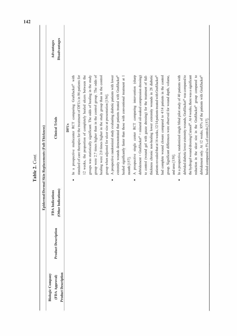
142
Tab
le 2
. Con
t.
Epi
derm
al/D
erm
al S
kin
Rep
lace
men
ts (F
ull-T
hick
ness
)
Bio
logi
c C
ompa
ny
(FD
A A
ppro
val)
Prod
uct D
escr
iptio
n
Prod
uct D
escr
iptio
n FD
A In
dica
tions
(Oth
er In
dica
tions
) C
linic
al T
rial
s A
dvan
tage
s
Dis
adva
ntag
es
DFU
s In
a p
rosp
ectiv
e m
ultic
ente
r R
CT
com
parin
g G
raftJ
acke
t® w
ith
stan
dard
of c
are
ther
apie
s for
the
treat
men
t of D
FUs i
n 86
pat
ient
s for
12
wee
ks,
the
prop
ortio
n of
com
plet
ely
heal
ed u
lcer
s be
twee
n th
e gr
oups
was
sta
tistic
ally
sig
nific
ant.
The
odds
of h
ealin
g in
the
stud
y gr
oup
wer
e 2.
7 tim
es h
ighe
r th
an in
the
cont
rol g
roup
. The
odd
s of
he
alin
g w
ere
2.0
times
hig
her i
n th
e st
udy
grou
p th
an in
the
cont
rol
grou
p w
hen
adju
sted
for u
lcer
size
at p
rese
ntat
ion
[156
]. A
pro
spec
tive
rand
omiz
ed s
tudy
eva
luat
ing
diab
etic
pat
ient
s w
ith lo
wer
ex
trem
ity w
ound
s de
mon
strat
ed th
at p
atie
nts
treat
ed w
ith G
raftJ
acke
t®
heal
ed s
igni
fican
tly f
aste
r th
an th
ose
with
con
vent
iona
l tre
atm
ent a
t 1
mon
th [1
57].
A
pros
pect
ive
singl
e ce
nter
RC
T co
mpa
ring
inte
rven
tion
(sha
rp
debr
idem
ent +
Gra
ftJac
ket®
+ m
iner
al o
il-so
aked
com
pres
sion
dres
sing)
to
con
trol
(wou
nd g
el w
ith g
auze
dre
ssin
g) f
or t
he t
reat
men
t of
ful
l-th
ickn
ess
chro
nic
non-
heal
ing
low
er e
xtre
mity
wou
nds
in 2
8 di
abet
ic
patie
nts r
evea
led t
hat a
t 16 w
eeks
, 12/
14 pa
tient
s tre
ated
with
Gra
ftJac
ket®
ha
d co
mpl
ete
wou
nd c
losu
re c
ompa
red
to 4
/14
patie
nts
in th
e co
ntro
l gr
oup.
Sig
nific
ant d
iffer
ence
s w
ere
obse
rved
for w
ound
dep
th, v
olum
e, an
d ar
ea [1
58].
In a
pro
spec
tive,
rand
omiz
ed si
ngle
blin
d pi
lot s
tudy
of 4
0 pa
tient
s with
de
brid
ed d
iabe
tic lo
wer
extre
mity
wou
nds,
Gra
ftJac
ket®
was
com
pare
d to
th
e hyd
roge
l wou
nd dr
essin
g Cur
asol
®. A
t 4 w
eeks
, the
re w
as a
signi
fican
t re
duct
ion
in t
he u
lcer
siz
e in
the
Gra
ftJac
ket®
gro
up c
ompa
red
to
debr
idem
ent o
nly.
At 1
2 w
eeks
, 85%
of t
he p
atie
nts
with
Gra
ftJac
ket®
he
aled
com
pare
d to
5%
of c
ontro
ls [1
57].
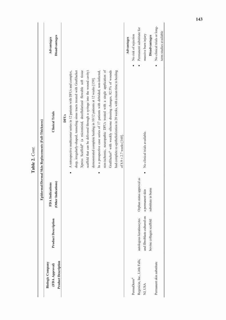
143
Tab
le 2
. Con
t.
Epi
derm
al/D
erm
al S
kin
Rep
lace
men
ts (F
ull-T
hick
ness
)
Bio
logi
c C
ompa
ny
(FD
A A
ppro
val)
Prod
uct D
escr
iptio
n
Prod
uct D
escr
iptio
n FD
A In
dica
tions
(Oth
er In
dica
tions
) C
linic
al T
rial
s A
dvan
tage
s
Dis
adva
ntag
es
DFU
s
A re
trosp
ectiv
e m
ultic
ente
r ser
ies i
n 12
pat
ient
s with
DFU
s and
com
plex
,
deep
, irre
gula
rly-s
hape
d, tu
nnel
ing
sinus
trac
ts tre
ated
with
Gra
ftJac
ket
Xpr
ess
Scaf
fold
® (
a m
icro
nize
d, d
ecel
lula
rized
flo
wab
le s
oft
tissu
e
scaf
fold
that
can
be
deliv
ered
thro
ugh
a sy
ringe
into
the
wou
nd c
avity
)
dem
onstr
ated
com
plet
e he
alin
g in
10/
12 p
atie
nts a
t 12
wee
ks [1
59].
In a
pro
spec
tive
case
ser
ies
of 1
7 pa
tient
s w
ith d
ebrid
ed, n
on-in
fect
ed,
non-
ische
mic
, ne
urop
athi
c D
FUs
treat
ed w
ith a
sin
gle
appl
icat
ion
of
Gra
ftJac
ket®
with
wee
kly
silic
one
dres
sing
chan
ges,
82.5
% o
f w
ound
s
had
com
plet
e re-
epith
elia
lizat
ion
in 2
0 w
eeks
, with
a m
ean
time t
o he
alin
g
of 8
.9 ±
2.7
wee
ks [1
60].
Perm
aDer
m®
Rege
nici
n, In
c., L
ittle
Fal
ls,
NJ,
USA
Perm
anen
t ski
n su
bstit
ute
auto
logo
us k
erat
inoc
ytes
and
fibro
blas
ts cu
lture
d on
bovi
ne co
llage
n sc
affo
ld
Orp
han
statu
s app
rova
l as
a per
man
ent s
kin
subs
titut
e in
burn
s
No
clin
ical
tria
ls a
vaila
ble.
Adv
anta
ges
No
risk
of re
ject
ion
Perm
anen
t sub
stitu
te fo
r
mas
sive
bur
n in
jury
Dis
adva
ntag
es
No
clin
ical
tria
ls o
r lon
g-
term
stud
ies a
vaila
ble
144
Dermal skin replacements provide greater stability to the wound and prevent the wound from contracting. Transcyte® (Shire Regenerative Medicine, Inc., San Diego, California, USA; Smith & Nephew, Inc., Largo, FL, USA) is composed of human allogeneic fibroblasts from neonatal foreskin seeded onto silicone covered bioabsorbable nylon mesh scaffold and cultured ex vivo for 4–6 weeks [85]. Transcyte® is often used as a non-living, temporary wound covering for partial- and full-thickness burns after excision [161]. A derivative of Transcyte® is Dermagraft® (Shire Regenerative Medicine, Inc., San Diego, California, USA), a skin substitute composed of living allogenic fibroblasts incorporated into a bioresorbable polyglactin mesh that secretes extracellular matrix (ECM) proteins, collagen, growth factors and cytokines into the wound site in the provision of viable living dermal substitute [162,163]. Dermagraft® has shown improvement in the treatment of chronic diabetic foot ulcers. AlloDerm®/Strattice® (LifeCell Corporation, Branchburg, NJ, USA) are lyophilized human acellular cadaver dermal matrices which serve as a scaffold for tissue remodeling. Autologous keratinocytes may be seeded and cultured on Alloderm® to form an epithelium; together; these can be utilized for wound and burn closure. Subsequent to its administration to a wound site, AlloDerm® is shown to exhibit cellular infiltration and neovascularization [164]. Biobrane® (Smith & Nephew, St. Petersburg, FL, USA) is a synthetic dermis temporary skin substitute composed of inner nylon and outer silicone with bovine collagen used for temporary coverage in partial and full-thickness burns. Integra® Dermal Regeneration Template (DRT) (Integra Lifesciences Corporation, Plainsboro, NJ, USA) is an example of a composite skin graft. It is composed of an outer layer of silicone and a cross-linked bovine type I collagen glycosaminoglycan dermal matrix. Once the dermal layer has regenerated, the silicone layer is removed and the wound is permanently closed with a split thickness skin graft (STSG) on the neo-dermis. Integra® is used for permanent coverage of full-thickness burns when combined with thin skin graft.
Epidermal/Dermal skin replacements are also called as full-thickness skin substitutes and are composed of both epidermal and dermal layers. Autologous or allogeneic fibroblasts and keratinocytes are used in their preparation. The allogenically derived Apligraf® (Organogenesis, Canton, MA, USA) is a bilayered matrix construct similar to a microscopic skin layer. Specifically, it is comprised of a lower dermal layer of bovine type 1 collagen combined with human fibroblasts (extracted from postnatal foreskin) and an upper layer that consists of human keratinocytes, along with granulocyte/macrophage colony-stimulating factors. Apligraf® has been used for permanent coverage of non-healing chronic wounds (such as diabetic foot ulcers), surgical wounds, pressure wounds, neuropathic wounds and venous insufficiency ulcers. Apligraf® has been observed in vitro to generate extracellular matrix structural elements and modulators inclusive of tissue inhibitors of matrix metalloproteinases and glycoprotein fibronectin [2]. OrCel® (Forticell Bioscience, New York, NY, USA) is a composite matrix composed of keratinocytes and dermal fibroblasts cultured in separate layers into a type I bovine collagen porous sponge. It is used in patients with dystrophic epidermolysis bullosa undergoing hand reconstruction surgery and at autograft donor sites in burn patients [92]. GraftJacket® (Wright Medical Technology, Inc., Arlington, TX, USA, licensed by KCI USA, Inc., San Antonio, Texas, USA), is an acellular derivative of human dermis. GraftJacket® was shown to facilitate accelerated healing and initiate depth and volume reductions in wounds [156]. PermaDerm® (Regenicin, Inc., Little Falls, NJ, USA) is a newer product that acts as a permanent skin
145
substitute to cover large burns. It is composed of autologous keratinocytes and fibroblasts cultured on bovine collagen scaffold [165].
3.2. Contraindications
Biological skin equivalents such as allogenically derived Apligraf® or Dermagraft® have an existing, albeit significantly low, risk of disease transmission due to their allogenicity [162]. In the case of Apligraf®, it has been verified in a number of studies that the cells it delivers are not sustained within the wound site beyond six weeks, and has inconsistent effects on the wound basement membrane, in vivo collagen composition and vascularization [2,146,152].
3.3. Clinical Trial Based Evidence
Greer et al. [166] compared a number of advanced wound therapies in the treatment of ulcers in regard to the proportion of ulcers healed and time to healing. This study reviewed randomized controlled trials from the literature (MEDLINE 1995–2013, Cochrane Library, and existing systemic reviews), which involved patients who were typically middle-aged white males. The 56 trials encompassed lower extremity or foot ulcers, with 35 cases of patients with diabetic ulcers, 20 patients with venous ulcers, and one patient with arterial ulcers. The duration of therapies generally spanned from 4 to 20 weeks, with a mean ulcer duration from 2 to 94 weeks. The mean ulcer size ranged from 1.9 to 41.5 cm2. Of the advanced wound care products used in these trials, the biological skin equivalent Apligraf® demonstrated moderate-strength evidence for enhanced healing, as did negative pressure wound therapy. Low-strength evidence was shown for platelet-derived growth factors and silver cream in comparison to standard care. For arterial ulcers, there was an improvement in healing with biological skin equivalent. Although the evidence was deemed as limited, the conclusion of the authors was that several advanced wound care therapies appeared to enhance the number of ulcers healed, as well as to reduce the times for healing.
A clinical randomized, double-blind, standard-controlled study was undertaken, which compared burn wounds that were treated with silver zinc sulfadiazine cream (control) against those treated with the identical cream that also contained silk sericin. The study involved 29 patients presenting with 65 burn wounds that covered at least 15% of total body surface areas. It was observed that the typical time for attaining 70% re-epithelialization in the sericin group was approximately 5–7 days shorter than the control group. The control group required 29.28 ± 9.27 days for complete burn wound healing, while the sericin group attained this condition within 22.42 ± 6.33 days with no indication of severe reaction or infection in any wound [49].
Multiple clinical trials have been conducted with the living skin equivalents Apligraf® and Dermagraft®. A retrospective controlled trial was undertaken that involved 2517 patients (446 Apligraf®, 1892 Regranex® (a human platelet-derived growth factor topical gel for the treatment of lower extremity diabetic neuropathic ulcers), 125 platelet releasates, 54 combined) and found that diabetic foot ulcers initially treated with Apligraf® were 31.2% more likely to heal than those administered with topical growth factor and 40% more likely to heal than those treated with platelet releasates [95]. In a prospective, randomized controlled trial involving 72 patients (33 Apligraf®,
146
39 with saline moistened gauze control), it was found that at 12 weeks, full wound closure was observed in 51.5% (17 of 33) of Apligraf® patients in contrast to 26.3% (10 of 38) of control patients [148]. An additional prospective, randomized controlled trial involved 74 patients (38 autograft + Apligraf®, 36 autograft alone or + allograft) with dull and partial thickness burns. It was found at 22 months that 58% of the Apligraf® sites were deemed of better quality than the controls, with 26% as equivalent and 16% as worse. Further, Apligraf® treated patients (47%) exhibited normal vascularity in contrast to 6% of control patients [145].
A prospective, randomized controlled trial with Dermagraft® studied 314 patients (130 Dermagraft®, 115 controls) with diabetic foot ulcers. At 12 weeks, 30% of the Dermagraft® patients were healed in comparison to 8.3% of the control patients, who were treated with standard wet-to-dry dressings [95]. An additional prospective, randomized controlled trial was undertaken with 18 patients (10 Dermagraft®, eight controls) with venous ulcers, which revealed that the healing rate after 12 weeks was enhanced considerably in those patients treated with Dermagraft® + compression (five patients (50%)) as opposed to compression on its own (one patient (12.5%)). In addition, the perfusion of capillaries in the Dermagraft® group increased by 20%, in comparison to 4.9% in the compression group [101].
4. Biomembranes for Wound Healing
4.1. Description
Biocompatible vegetal biomembranes of natural rubber/latex, amniotic, polyurethane and poly-DL-lactic acid (PDLLA) comprise a class of versatile interventions for the treatment and healing of wounds. Additionally, biomembranes may be impregnated with a wide range of bioactive compounds to further facilitate and promote wound healing.
4.2. Mechanism and Indications
Human amniotic membranes, such as Biomembrane® (Matrix Company, Ismailia, Egypt) are comprised of skin-like fetal ectoderm, consisting of four layers (epithelial, basement membrane, connective tissue fibroblasts, and spongy layer), which have demonstrated angiogenic properties. The membrane is freeze dried to 5% water content and then gamma irradiated (25 kgy) to ensure sterilization. These biomembranes exhibit a 1000-fold improvement in efficacy over split-thickness human skin grafts, though the specific mechanisms remain unclear [167,168]. Further, amniotic membranes are found to inhibit the alpha smooth muscle protein actin, resulting in a significant reduction in the generation of scar tissue in comparison to a moist wound dressing control [169]. Additional benefits included decreased pain, protection from infection and control of the loss of electrolytes and albumin.
The polyurethane film, TegadermTM (3M, Saint Paul, MN, USA), exhibits gas semi-permeability, which acts to augment the rate of epithelialization. This may be due the retention of carbon dioxide, which translates to sustaining a low pH. The pain relief that is reportedly associated with this film may be the result of the exclusion of atmospheric oxygen, which negates the generation of prostaglandin E2, via the oxygen-reliant cyclo-oxygenase system [167,170]. An additional imparted
147
benefit secondary to the semi-permeability of TegadermTM is the regulation of transforming growth factor beta (TGF- ) via the mediation of transepidermal water transfer [171]. It also stimulates the propagation of keratinocytes through the activation of integrins a5 and a6 to encourage enhanced and rapid wound healing [172].
A biocompatible vegetal biomembrane derived from the Hevea brasiliensis rubber tree exhibited the capacity to initiate angiogenesis and re-epithelialization in the chronic ulcers of diabetic patients. Its activity in the healing process appears most prominent at the inflammatory stage, where the microenvironment is transformed by robust angiogenesis followed by re-epithelialization [173].
A non-toxic, biocompatible, biodegradable, and non-carcinogenic crosslinked gelatin hydrogel biomembrane was developed for use as a wound dressing via the addition of a naturally occurring genipin crosslinking agent, and compared to a glutaraldehyde-crosslinked control. The resulting genipin infused biomembrane exhibited considerably less inflammation along with more rapid re-epithelialization and subsequent wound healing than the control, which may have been facilitated by a lower level of genipin imparted cytotoxicity [36].
4.3. Contraindications
Despite stringent preparation protocols, there might be a very low risk of bacterial or viral transmission via the use of human amniotic membranes on open wounds.
4.4. Clinical Trial Based Evidence
Adly et al. [167] conducted a randomized, controlled clinical trial to compare the efficacy of an amniotic membrane (Biomembrane®) group I (23 patients) and a polyurethane membrane (TegadermTM) dressing group II (23 patients) in the treatment of burns (scald and flame). There were no notable differences between the two groups. The criteria were inclusion of both genders and all age groups with <50% total body surface area affected with either second or third degree burns. The group I patients exhibited a considerably lower infection rate (one patient (4.3%) in group I compared to three patients (13.0%) in group II) and required fewer dressing changes than group II (highest dressing change frequency was once per day in 30.4% of group I patients, in comparison to five times per day in 60.9% of group II patients). In addition, electrolyte disturbance was evident in 17.4% of patients in group I, compared with 60.9% of patients in group II. Albumin loss was indicated in 39.1% of patients in group I in contrast to 60.9% of patients in group II. In terms of pain and healing times, 43.5% of group I patients experienced pain during dressing, compared with 60.9% in group II. Healing frequency was 47.8% (11–20 days) for group I in contrast to 39.1% in group II spanning the same time period.
5. Scaffolds for Wound Healing
5.1. Description
Hybrid scaffolds comprised of polymeric substrates coated with bioactive materials, collagen, silk fibroin, as well as advanced tissue engineered substrates impregnated with endothelial progenitor
148
cells, and nanomaterial-based scaffolds may be employed as advanced wound dressings to initiate and expedite wound healing.
5.2. Mechanisms and Indications
Collagen is a component of the extracellular matrix, which has found established utility as a biomaterial in cell therapies and tissue engineering via the provision of a viable substrate for the attachment and propagation of cells. In the treatment of wounds, collagen scaffolds offer a feasible platform for the topical conveyance of cells into the wound bed, increase the healing of wounds and initiate angiogenesis and neovasculogenesis.
O’Loughlin et al. [174] investigated the use of type 1 collagen scaffolds for the topical delivery of autologous circulating angiogenic (CACs) cells (precursors to endothelial progenitor cells), to full thickness cutaneous ulcers. It was revealed that the CACs could also be pre-stimulated through the addition of matricellular protein osteopontin (OPN), a glycoprotein involved in immune function, neovascularization, and facilitation of cell migration and survival [175]. The inclusion of OPN served to augment wound healing. It was demonstrated that scaffolds comprised of type 1 collagen, which has been shown to sustain angiogenesis [176], when infused with CACs and enhanced with OPN, resulted in the formation of larger diameter blood vessels than untreated wounds, and thus acceleration of the wound healing process [174].
Ehashi et al. [177] compared subcutaneously implanted scaffolds for their host body reactions in order to assess their wound healing capacities. The scaffolds consisted of collagen coated porous (Ø32 m and Ø157 m) polyethylene (CCPE), bio-inert poly(2-methacryloyloxyethyl phosphorylcholine-co-n-butyl methacrylate) (PMB) coated polyethylene, and uncoated porous polyethylene (UPPE) (control). Subsequent to their immersion in sterile solution for an appropriate period, six samples (two of each type with different pore diameters) were implanted under the skins of mouse models, and then resected after seven days. In terms of vascularization, it was observed that small vessels were induced on the UPPE, albeit contingent on the pore size (more activity seen with Ø32 m pores than Ø157
m pores). Interestingly, the reverse was true for the CCPE, with more activity seen with the Ø157 m pore sample. There was no vessel growth activity associated with the PMB scaffolds.
A deoxyribonucleic acid (DNA) microarray assay was then employed to conduct genetic analyses, which showed that the CCPE scaffold had a more highly distributed level of gene expression than did the PMB scaffold. The PMB scaffold showed the up-regulation of genes associated with the suppression of inflammation. The CCPE scaffold indicated up-regulation of genes related to inflammation, angiogenesis, and wound healing. The authors concluded that the up-regulation of interleukin-1b and angiogenesis associated genes within the porous scaffolds likely contributed to the mediation of tissue regeneration.
A novel scaffold comprised of electrospun core-shell gelatin/poly(L-lactic acid)-co-poly- ( -caprolactone) nanofibers, which encapsulated a photosensitive polymer poly (3-hexylthiophene) (P3HT) and epidermal growth factor (EGF) at its core, was investigated by Jin et al. [178] as a potential skin graft. It was found that fibroblast propagation was activated under exposure to light in contrast to its absence and cells akin to keratinocytes were found only on the light exposed scaffolds.
149
The researchers propose that these light sensitive nanofibers may have utility as a unique scaffold for the healing of wounds and the reconstitution of skin.
Bacterial (or microbial) cellulose has been investigated by Fu et al. [179] for its capacity to enable wound healing and skin tissue rejuvenation. Specific bacteria are involved in the biosynthesis of this natural polymer, which has unique properties in contrast to plant based cellulose, encompassing biocompatibility, hydrophilicity, high water retention, elasticity, transparency, conformability and the capacity for absorbing wound generated exudate during inflammation. These features position microbial cellulose to have great potential for biomedical advancements in skin tissue repair.
5.3. Contraindications
Scaffolds that are comprised of hyaluronan (an anionic polysaccharide), even though non-cytotoxic and biodegradable, may disrupt cell adhesion and the regeneration of tissues due to its hydrophilic surface properties [177]. Additional drawbacks for tissue engineered scaffolds in the case of severe burns relate to their unreliable adhesion to lesions and failure to replace dermal tissues [180].
5.4. Clinical Trial Based Evidence
The clinical performance of bacterial cellulose (BC) scaffold DermafillTM (AMD/Ritmed, Tonawanda, New York, USA) wound dressings (Acetobacter xylinum derived) was assessed by Portal et al. [181] who compared the reduction in wound size of chronic non-healing lower extremity ulcers following standard care. A total of 11 chronic wounds were evaluated for the time required to achieve 75% epithelization, by comparing non-healing ulcers prior to and following the application of DermafillTM. The median observation timeline for chronic non-healing wounds under standard care prior to the application was 315 days. When BC scaffolds were applied to these same wounds, the median time to 75% epithelization was decreased to 70 days. Thus, the authors concluded that BC scaffold-initiated wound closure for non-healing ulcers proceeded considerably more rapidly than did standard care wound dressings.
Morimoto et al. [182] investigated the clinical efficacy of a unique synthetic collagen/gelatin sponge (CGS) scaffold for the treatment of chronic skin ulcers. This artificial dermal scaffold demonstrated the capacity to sustainably release basic fibroblast growth factor (bFGF) over 10 days or longer. One of the criteria for the study group was the inclusion of chronic skin ulcers that had not healed over a time period of at least four weeks. These wounds treated with CGS, which was infused with 7 or 14 g/cm2 of bFGF following debridement, and assessed two weeks subsequent to their application. Positive improvement in the wound beds was defined by the emergence of granulated and epithelialized areas of 50% or greater. Out of a total of 17 subjects, it was observed that 16 showed wound bed improvements, with no discernable difference between the low and high dose groups. There was rapid recovery from mild adverse reactions.
6. Conclusions
The healing of surface and deep wounds of the epidermis is a complex multistage process, but one that may nevertheless be expedited utilizing strategies such as the application of active biologic,
150
biomembrane or scaffold based wound dressings. Specific therapeutic compounds and cell species including epidermal stem cells may be utilized to impregnate biocompatible and/or biodegradable substrates, including membranes and scaffolds to facilitate rapid revascularization, re-epithelialization, and healing of wound beds.
Acknowledgments
This research received no specific grant from any funding agency in the public, commercial, or not-for-profit sectors.
Author Contributions
The authors’ responsibilities were as follows—Krishna S. Vyas and Henry C. Vasconez: participated in the design of the study, drafting, critical review, and final approval of the manuscript.
Conflicts of Interest
The authors declare no conflict of interest.
References
1. Ghosh, P.K.; Gaba, A. Phyto-extracts in wound healing. J. Pharm. Pharm. Sci. 2013, 16, 760–820. 2. Rennert, R.C.; Rodrigues, M.; Wong, V.W.; Duscher, D.; Hu, M.; Maan, Z.; Sorkin, M.;
Gurtner, G.C.; Longaker, M.T. Biological therapies for the treatment of cutaneous wounds: Phase iii and launched therapies. Expert opin. Biol. Ther. 2013, 13, 1523–1541.
3. Cohen, I.K. Lessons from the history of wound healing. Clin. Dermatol. 2007, 25, 3–8. 4. Barreto, R.S.; Albuquerque-Junior, R.L.; Araujo, A.A.; Almeida, J.R.; Santos, M.R.; Barreto, A.S.;
DeSantana, J.M.; Siqueira-Lima, P.S.; Quintans, J.S.; Quintans-Junior, L.J. A systematic review of the wound-healing effects of monoterpenes and iridoid derivatives. Molecules 2014, 19, 846–862.
5. Dias, A.M.; Braga, M.E.; Seabra, I.J.; Ferreira, P.; Gil, M.H.; de Sousa, H.C. Development of natural-based wound dressings impregnated with bioactive compounds and using supercritical carbon dioxide. Inter. J. Pharm. 2011, 408, 9–19.
6. Mai, L.M.; Lin, C.Y.; Chen, C.Y.; Tsai, Y.C. Synergistic effect of bismuth subgallate and borneol, the major components of sulbogin, on the healing of skin wound. Biomaterials 2003, 24, 3005–3012.
7. Anamura, S.; Dohi, T.; Shirakawa, M.; Okamoto, H.; Tsujimoto, A. Effects of phenolic dental medicaments on prostaglandin synthesis by microsomes of bovine tooth pulp and rabbit kidney medulla. Arch. Oral Biol. 1988, 33, 555–560.
8. Braga, P.C.; Dal Sasso, M.; Culici, M.; Bianchi, T.; Bordoni, L.; Marabini, L. Anti-inflammatory activity of thymol: Inhibitory effect on the release of human neutrophil elastase. Pharmacology 2006, 77, 130–136.
151
9. Riella, K.R.; Marinho, R.R.; Santos, J.S.; Pereira-Filho, R.N.; Cardoso, J.C.; Albuquerque-Junior, R.L.; Thomazzi, S.M. Anti-inflammatory and cicatrizing activities of thymol, a monoterpene of the essential oil from lippia gracilis, in rodents. J. Ethnopharmacol. 2012, 143, 656–663.
10. Youdim, K.A.; Deans, S.G. Effect of thyme oil and thymol dietary supplementation on the antioxidant status and fatty acid composition of the ageing rat brain. Br. J. Nutr. 2000, 83,87–93.
11. Yanishlieva, N.V.; Marinova, E.M.; Gordon, M.H.; Raneva, V.G. Antioxidant activity and mechanism of action of thymol and carvacrol in two lipid systems. Food Chem. 1999, 64,59–66.
12. Prieto, J.M.I.; Cioni, P.; Chericoni, S. In vitro activity of the essential oils of origanum vulgare, satureja montana and their main constituents in peroxynitrite-induced oxidative processes. Food Chem. 2007, 104, 889–895.
13. Haeseler, G.; Maue, D.; Grosskreutz, J.; Bufler, J.; Nentwig, B.; Piepenbrock, S.; Dengler, R.; Leuwer, M. Voltage-dependent block of neuronal and skeletal muscle sodium channels by thymol and menthol. Eur. J. Anaesthesiol. 2002, 19, 571–579.
14. Karpanen, T.J.; Worthington, T.; Hendry, E.R.; Conway, B.R.; Lambert, P.A. Antimicrobial efficacy of chlorhexidine digluconate alone and in combination with eucalyptus oil, tea tree oil and thymol against planktonic and biofilm cultures of staphylococcus epidermidis. J. Antimicrob. Chemother. 2008, 62, 1031–1036.
15. Tramontina, V.A.; Machado, M.A.; Nogueira Filho Gda, R.; Kim, S.H.; Vizzioli, M.R.; Toledo, S. Effect of bismuth subgallate (local hemostatic agent) on wound healing in rats. Histological and histometric findings. Braz. Dent. J. 2002, 13, 11–16.
16. Priestley, C.M.; Williamson, E.M.; Wafford, K.A.; Sattelle, D.B. Thymol, a constituent of thyme essential oil, is a positive allosteric modulator of human gaba(a) receptors and a homo-oligomeric gaba receptor from drosophila melanogaster. Br. J. Pharmacol. 2003, 140, 1363–1372.
17. Kavoosi, G.; Dadfar, S.M.; Purfard, A.M. Mechanical, physical, antioxidant, and antimicrobial properties of gelatin films incorporated with thymol for potential use as nano wound dressing. J. Food Sci. 2013, 78, E244–E250.
18. Khalil, Z.; Pearce, A.L.; Satkunanathan, N.; Storer, E.; Finlay-Jones, J.J.; Hart, P.H. Regulation of wheal and flare by tea tree oil: Complementary human and rodent studies. J. Invest. Dermatol. 2004, 123, 683–690.
19. Kawata, J.K.; Miyazawa, M. Cyclooxygenase-2 inhibitory effects of monoterpenoids with a p-methane skeleton. Int. J. Essent. Oil Ther. 2008, 2, 145–148.
20. Hassan, S.B.; Gali-Muhtasib, H.; Goransson, H.; Larsson, R. Alpha terpineol: A potential anticancer agent which acts through suppressing nf-kappab signalling. Anticancer Res. 2010,30, 1911–1919.
21. Held, S.; Schieberle, P.; Somoza, V. Characterization of alpha-terpineol as an anti-inflammatory component of orange juice by in vitro studies using oral buccal cells. J. Agric. Food Chem. 2007, 55, 8040–8046.
152
22. De Oliveira, M.G.; Marques, R.B.; de Santana, M.F.; Santos, A.B.; Brito, F.A.; Barreto, E.O.; de Sousa, D.P.; Almeida, F.R.; Badaue-Passos, D., Jr.; Antoniolli, A.R.; et al. Alpha-terpineol reduces mechanical hypernociception and inflammatory response. Basic Clin. Pharmacol. Toxicol. 2012, 111, 120–125.
23. Park, S.N.; Lim, Y.K.; Freire, M.O.; Cho, E.; Jin, D.; Kook, J.K. Antimicrobial effect of linalool and alpha-terpineol against periodontopathic and cariogenic bacteria. Anaerobe 2012, 18, 369–372.
24. Park, M.J.; Gwak, K.S.; Yang, I.; Kim, K.W.; Jeung, E.B.; Chang, J.W.; Choi, I.G. Effect of citral, eugenol, nerolidol and alpha-terpineol on the ultrastructural changes of trichophyton mentagrophytes. Fitoterapia 2009, 80, 290–296.
25. Jin, J.; Song, M.; Hourston, D.J. Novel chitosan-based films cross-linked by genipin with improved physical properties. Biomacromolecules 2004, 5, 162–168.
26. Sung, H.W.; Huang, R.N.; Huang, L.L.; Tsai, C.C. In vitro evaluation of cytotoxicity of a naturally occurring cross-linking reagent for biological tissue fixation. J. Biomater. Sci. Polym. Ed. 1999, 10, 63–78.
27. Wang, G.F.; Wu, S.Y.; Rao, J.J.; Lu, L.; Xu, W.; Pang, J.X.; Liu, Z.Q.; Wu, S.G.; Zhang, J.J. Genipin inhibits endothelial exocytosis via nitric oxide in cultured human umbilical vein endothelial cells. Acta pharmacol. Sin. 2009, 30, 589–596.
28. Koo, H.J.; Song, Y.S.; Kim, H.J.; Lee, Y.H.; Hong, S.M.; Kim, S.J.; Kim, B.C.; Jin, C.; Lim, C.J.; Park, E.H. Antiinflammatory effects of genipin, an active principle of gardenia. Eur. J. Pharmacol. 2004, 495, 201–208.
29. Parton, L.E.; Ye, C.P.; Coppari, R.; Enriori, P.J.; Choi, B.; Zhang, C.Y.; Xu, C.; Vianna, C.R.; Balthasar, N.; Lee, C.E.; et al. Glucose sensing by pomc neurons regulates glucose homeostasis and is impaired in obesity. Nature 2007, 449, 228–232.
30. Bao, D.; Chen, M.; Wang, H.; Wang, J.; Liu, C.; Sun, R. Preparation and characterization of double crosslinked hydrogel films from carboxymethylchitosan and carboxymethylcellulose. Carbohydr. Polym. 2014, 110, 113–120.
31. Arteche Pujana, M.; Perez-Alvarez, L.; Cesteros Iturbe, L.C.; Katime, I. Biodegradable chitosan nanogels crosslinked with genipin. Carbohydr. Polym. 2013, 94, 836–842.
32. Pankongadisak, P.; Ruktanonchai, U.R.; Supaphol, P.; Suwantong, O. Preparation and characterization of silver nanoparticles-loaded calcium alginate beads embedded in gelatin scaffolds. AAPS Pharm. Sci. Tech. 2014, doi:10.1208/s12249-014-0140-9.
33. Huang, G.P.; Shanmugasundaram, S.; Masih, P.; Pandya, D.; Amara, S.; Collins, G.; Arinzeh, T.L. An investigation of common crosslinking agents on the stability of electrospun collagen scaffolds. J. Biomed. Mater. Res. A 2014, doi:10.1002/jbm.a.35222.
34. Yazdimamaghani, M.; Vashaee, D.; Assefa, S.; Shabrangharehdasht, M.; Rad, A.T.; Eastman, M.A.; Walker, K.J.; Madihally, S.V.; Kohler, G.A.; Tayebi, L. Green synthesis of a new gelatin-based antimicrobial scaffold for tissue engineering. Mater. Sci. Eng. C Mater. Biol. Appl. 2014, 39, 235–244.
35. Siritientong, T.; Ratanavaraporn, J.; Srichana, T.; Aramwit, P. Preliminary characterization of genipin-cross-linked silk sericin/poly(vinyl alcohol) films as two-dimensional wound dressings for the healing of superficial wounds. Biomed. Res. Int. 2013, 2013, 904314.
153
36. Chang, W.H.; Chang, Y.; Lai, P.H.; Sung, H.W. A genipin-crosslinked gelatin membrane as wound-dressing material: In vitro and in vivo studies. J. Biomater. Sci. Polym. Ed. 2003, 14, 481–495.
37. Zhang, X.; Chen, X.; Yang, T.; Zhang, N.; Dong, L.; Ma, S.; Liu, X.; Zhou, M.; Li, B. The effects of different crossing-linking conditions of genipin on type i collagen scaffolds: An in vitro evaluation. Cell Tissue Banking 2014, doi:10.1007/s10561-014-9423-3.
38. Wang, M.; Da, L.; Xie, Y.; Xie, H. Application of genipin for modification of natural biomaterials as a crosslinking agent. Chin. J. Reparative Reconstructive Surg. 2013, 27, 580–585.
39. Mekhail, M.; Jahan, K.; Tabrizian, M. Genipin-crosslinked chitosan/poly-l-lysine gels promote fibroblast adhesion and proliferation. Carbohydr. Polym. 2014, 108, 91–98.
40. Chan, K.L.; Khankhel, A.H.; Thompson, R.L.; Coisman, B.J.; Wong, K.H.; Truslow, J.G.; Tien, J. Crosslinking of collagen scaffolds promotes blood and lymphatic vascular stability. J. Biomed. Mater. Res. A 2013, doi:10.1002/jbm.a.34990.
41. Pandit, V.; Zuidema, J.M.; Venuto, K.N.; Macione, J.; Dai, G.; Gilbert, R.J.; Kotha, S.P. Evaluation of multifunctional polysaccharide hydrogels with varying stiffness for bone tissue engineering. Tissue Eng. A 2013, 19, 2452–2463.
42. Sun, W.; Incitti, T.; Migliaresi, C.; Quattrone, A.; Casarosa, S.; Motta, A. Genipin-crosslinked gelatin-silk fibroin hydrogels for modulating the behaviour of pluripotent cells. J. Tissue Eng. Regenerative Med. 2014, doi:10.1002/term.1868.
43. Manickam, B.; Sreedharan, R.; Elumalai, M. “Genipin”—The natural water soluble cross-linking agent and its importance in the modified drug delivery systems: An overview. Curr. Drug Delivery 2014, 11, 139–145.
44. Recio, M.C.; Giner, R.M.; Manez, S.; Rios, J.L. Structural considerations on the iridoids as anti-inflammatory agents. Planta Med. 1994, 60, 232–234.
45. Shim, K.M.; Choi, S.H.; Jeong, M.J.; Kang, S.S. Effects of aucubin on the healing of oral wounds. In Vivo (Athens, Greece) 2007, 21, 1037–1041.
46. D’ALESSIO, P.A.; Mirshahi, M.; Bisson, J.F.; Bene, M.C. Skin repair properties of d-limonene and perillyl alcohol in murine models. Anti-inflammatory Anti-allergy Agents Med. Chem. 2014, 13, 29–35.
47. Anghel, I.; Holban, A.M.; Grumezescu, A.M.; Andronescu, E.; Ficai, A.; Anghel, A.G.; Maganu, M.; Laz, R.V.; Chifiriuc, M.C. Modified wound dressing with phyto-nanostructured coating to prevent staphylococcal and pseudomonal biofilm development. Nanoscale Res. Let. 2012, 7, 690.
48. Moghimi, H.R.; Makhmalzadeh, B.S.; Manafi, A. Enhancement effect of terpenes on silver sulphadiazine permeation through third-degree burn eschar. Burns 2009, 35, 1165–1170.
49. Aramwit, P.; Palapinyo, S.; Srichana, T.; Chottanapund, S.; Muangman, P. Silk sericin ameliorates wound healing and its clinical efficacy in burn wounds. Arch. Dermatol. Res. 2013, 305, 585–594.
50. Siritientong, T.; Angspatt, A.; Ratanavaraporn, J.; Aramwit, P. Clinical potential of a silk sericin-releasing bioactive wound dressing for the treatment of split-thickness skin graft donor sites. Pharm. Res. 2014, 31, 104–116.
154
51. Kanokpanont, S.; Damrongsakkul, S.; Ratanavaraporn, J.; Aramwit, P. An innovative bi-layered wound dressing made of silk and gelatin for accelerated wound healing. Int. J. Pharm. 2012, 436, 141–153.
52. Siritienthong, T.; Ratanavaraporn, J.; Aramwit, P. Development of ethyl alcohol-precipitated silk sericin/polyvinyl alcohol scaffolds for accelerated healing of full-thickness wounds. Int. J. Pharm. 2012, 439, 175–186.
53. Aramwit, P.; Siritienthong, T.; Srichana, T.; Ratanavaraporn, J. Accelerated healing of full-thickness wounds by genipin-crosslinked silk sericin/pva scaffolds. Cells Tissues Organs 2013, 197, 224–238.
54. Aramwit, P.; Kanokpanont, S.; de-Eknamkul, W.; Srichana, T. Monitoring of inflammatory mediators induced by silk sericin. J. Biosci. Bioeng. 2009, 107, 556–561.
55. Aramwit, P.; Sangcakul, A. The effects of sericin cream on wound healing in rats. Biosci. Biotech. Biochem. 2007, 71, 2473–2477.
56. Nayak, S.; Kundu, S.C. Sericin-carboxymethyl cellulose porous matrices as cellular wound dressing material. J. Biomed. Mater. Res. A 2014, 102, 1928–1940.
57. Nayak, S.; Dey, S.; Kundu, S.C. Skin equivalent tissue-engineered construct: Co-cultured fibroblasts/ keratinocytes on 3d matrices of sericin hope cocoons. PLoS One 2013, 8, e74779.
58. Zhao, R.; Li, X.; Sun, B.; Zhang, Y.; Zhang, D.; Tang, Z.; Chen, X.; Wang, C. Electrospun chitosan/sericin composite nanofibers with antibacterial property as potential wound dressings. Int. J. Biol. Macromol. 2014, 68, 92–97.
59. Teramoto, H.; Kameda, T.; Tamada, Y. Preparation of gel film from bombyx mori silk sericin and its characterization as a wound dressing. Biosci. Biotech. Biochem. 2008, 72, 3189–3196.
60. Akturk, O.; Tezcaner, A.; Bilgili, H.; Deveci, M.S.; Gecit, M.R.; Keskin, D. Evaluation of sericin/collagen membranes as prospective wound dressing biomaterial. J. Biosci. Bioeng. 2011, 112, 279–288.
61. Villegas, L.F.; Marcalo, A.; Martin, J.; Fernandez, I.D.; Maldonado, H.; Vaisberg, A.J.; Hammond, G.B. (+)-epi-Alpha-bisabolol [correction of bisbolol] is the wound-healing principle of peperomia galioides: Investigation of the in vivo wound-healing activity of related terpenoids. J. Nat. Prod. 2001, 64, 1357–1359.
62. Zhang, K.; Qian, Y.; Wang, H.; Fan, L.; Huang, C.; Yin, A.; Mo, X. Genipin-crosslinked silk fibroin/hydroxybutyl chitosan nanofibrous scaffolds for tissue-engineering application. J. Biomed. Mater. Res. A 2010, 95, 870–881.
63. Stevenson, P.C.; Simmonds, M.S.J.; Sampson, J.; Houghton, P.J.; Grice, P. The effects of iridoid compounds on wound healing. J. Korean Acad. Oral Med. 1999, 24, 137–142.
64. Davini, E.; Iavarone, C.; Trogolo, C.; Aureli, P.; Pasolini, B. The quantitative isolation and antimicrobial activity of the aglycone of aucubin. Phytochemistry 1986, 25, 2420–2422.
65. Jin, L.; Xue, H.; Jin, L.; Li, S.; Xu, Y. Antioxidative activities of aucubin in vitro. J. Shaanxi Norm. Univ. 2004, 32, 98–101.
66. Hung, J.Y.; Yang, C.J.; Tsai, Y.M.; Huang, H.W.; Huang, M.S. Antiproliferative activity of aucubin is through cell cycle arrest and apoptosis in human non-small cell lung cancer a549 cells. Clin. Exp. Pharmacol. Physiol. 2008, 35, 995–1001.
155
67. Aramwit, P.; Kanokpanont, S.; Nakpheng, T.; Srichana, T. The effect of sericin from various extraction methods on cell viability and collagen production. Int. J. Mol. Sci. 2010, 11, 2200–2211.
68. Tsubouchi, K.; Igarashi, Y.; Takasu, Y.; Yamada, H. Sericin enhances attachment of cultured human skin fibroblasts. Biosci. Biotech. Biochem. 2005, 69, 403–405.
69. Terada, S.; Nishimura, T.; Sasaki, M.; Yamada, H.; Miki, M. Sericin, a protein derived from silkworms, accelerates the proliferation of several mammalian cell lines including a hybridoma. Cytotechnology 2002, 40, 3–12.
70. U.S. Environmental Protection Agency. Hazard Characterization Document, Screening-Level Hazard Characterization, Monoterpene Hydrocarbons Category; Washinton, DC, USA, 2009.
71. Vacher, D. Autologous epidermal sheets production for skin cellular therapy. Ann. Pharm. Fr. 2003, 61, 203–206.
72. Carsin, H.; Ainaud, P.; le Bever, H.; Rives, J.; Lakhel, A.; Stephanazzi, J.; Lambert, F.; Perrot, J. Cultured epithelial autografts in extensive burn coverage of severely traumatized patients: A five year single-center experience with 30 patients. Burns 2000, 26, 379–387.
73. Atiyeh, B.S.; Costagliola, M. Cultured epithelial autograft (cea) in burn treatment: Three decades later. Burns 2007, 33, 405–413.
74. Horch, R.E.; Kopp, J.; Kneser, U.; Beier, J.; Bach, A.D. Tissue engineering of cultured skin substitutes. J. Cell. Mol. Med. 2005, 9, 592–608.
75. Ramos-e-Silva, M.; Ribeiro de Castro, M.C. New dressings, including tissue-engineered living skin. Clin. Dermatol. 2002, 20, 715–723.
76. Uccioli, L. A clinical investigation on the characteristics and outcomes of treating chronic lower extremity wounds using the tissuetech autograft system. Int. J. Lower Extremity Wounds 2003, 2, 140–151.
77. Ruszczak, Z. Effect of collagen matrices on dermal wound healing. Adv. Drug Delivery Rev. 2003, 55, 1595–1611.
78. Andreassi, L.; Pianigiani, E.; Andreassi, A.; Taddeucci, P.; Biagioli, M. A new model of epidermal culture for the surgical treatment of vitiligo. Int. J. Dermatol. 1998, 37, 595–598.
79. Uccioli, L.; Giurato, L.; Ruotolo, V.; Ciavarella, A.; Grimaldi, M.S.; Piaggesi, A.; Teobaldi, I.; Ricci, L.; Scionti, L.; Vermigli, C.; et al. Two-step autologous grafting using hyaff scaffolds in treating difficult diabetic foot ulcers: Results of a multicenter, randomized controlled clinical trial with long-term follow-up. Int. J. Lower Extremity Wounds 2011, 10, 80–85.
80. Monami, M.; Vivarelli, M.; Desideri, C.M.; Ippolito, G.; Marchionni, N.; Mannucci, E. Autologous skin fibroblast and keratinocyte grafts in the treatment of chronic foot ulcers in aging type 2 diabetic patients. J. Am. Podiatric Med. Assoc. 2011, 101, 55–58.
81. Lobmann, R.; Pittasch, D.; Muhlen, I.; Lehnert, H. Autologous human keratinocytes cultured on membranes composed of benzyl ester of hyaluronic acid for grafting in nonhealing diabetic foot lesions: A pilot study. J. Diabetes Complicat. 2003, 17, 199–204.
82. Pajardi, G.; Rapisarda, V.; Somalvico, F.; Scotti, A.; Russo, G.L.; Ciancio, F.; Sgro, A.; Nebuloni, M.; Allevi, R.; Torre, M.L.; et al. Skin substitutes based on allogenic fibroblasts or keratinocytes for chronic wounds not responding to conventional therapy: A retrospective observational study. Int. Wound J. 2014, doi:10.1111/iwj.12223.
156
83. Lam, P.K.; Chan, E.S.; Liew, C.T.; Lau, C.; Yen, S.C.; King, W.W. Combination of a new composite biocampatible skin graft on the neodermis of artificial skin in an animal model. ANZ J. Sur. 2002, 72, 360–363.
84. Chan, E.S.; Lam, P.K.; Liew, C.T.; Lau, H.C.; Yen, R.S.; King, W.W. A new technique to resurface wounds with composite biocompatible epidermal graft and artificial skin. J. Trauma 2001, 50, 358–362.
85. Bello, Y.M.; Falabella, A.F.; Eaglstein, W.H. Tissue-engineered skin. Current status in wound healing. Am. J. Clin. Dermatol. 2001, 2, 305–313.
86. Kumar, R.J.; Kimble, R.M.; Boots, R.; Pegg, S.P. Treatment of partial-thickness burns: A prospective, randomized trial using transcyte. ANZ J. Sur. 2004, 74, 622–626.
87. Demling, R.H.; DeSanti, L. Management of partial thickness facial burns (comparison of topical antibiotics and bio-engineered skin substitutes). Burns 1999, 25, 256–261.
88. Lukish, J.R.; Eichelberger, M.R.; Newman, K.D.; Pao, M.; Nobuhara, K.; Keating, M.; Golonka, N.; Pratsch, G.; Misra, V.; Valladares, E.; et al. The use of a bioactive skin substitute decreases length of stay for pediatric burn patients. Journal Pediatr. Surg. 2001, 36, 1118–1121.
89. Amani, H.; Dougherty, W.R.; Blome-Eberwein, S. Use of transcyte and dermabrasion to treat burns reduces length of stay in burns of all size and etiology. Burns 2006, 32, 828–832.
90. Noordenbos, J.; Dore, C.; Hansbrough, J.F. Safety and efficacy of transcyte for the treatment of partial-thickness burns. J. Burn Care Rehabil. 1999, 20, 275–281.
91. Hansen, S.L.; Voigt, D.W.; Wiebelhaus, P.; Paul, C.N. Using skin replacement products to treat burns and wounds. Adv. Skin Wound Care 2001, 14, 37–44; quiz 45–46.
92. Van der Veen, V.C.; van der Wal, M.B.; van Leeuwen, M.C.; Ulrich, M.M.; Middelkoop, E. Biological background of dermal substitutes. Burns 2010, 36, 305–321.
93. Hansbrough, J.F.; Mozingo, D.W.; Kealey, G.P.; Davis, M.; Gidner, A.; Gentzkow, G.D. Clinical trials of a biosynthetic temporary skin replacement, dermagraft-transitional covering, compared with cryopreserved human cadaver skin for temporary coverage of excised burn wounds. J. Burn Care Rehabil. 1997, 18, 43–51.
94. Purdue, G.F.; Hunt, J.L.; Still, J.M., Jr.; Law, E.J.; Herndon, D.N.; Goldfarb, I.W.; Schiller, W.R.; Hansbrough, J.F.; Hickerson, W.L.; Himel, H.N.; et al. A multicenter clinical trial of a biosynthetic skin replacement, dermagraft-tc, compared with cryopreserved human cadaver skin for temporary coverage of excised burn wounds. J. Burn Care Rehabil. 1997, 18, 52–57.
95. Marston, W.A.; Hanft, J.; Norwood, P.; Pollak, R. The efficacy and safety of dermagraft in improving the healing of chronic diabetic foot ulcers: Results of a prospective randomized trial. Diabetes Care 2003, 26, 1701–1705.
96. Hanft, J.R.; Surprenant, M.S. Healing of chronic foot ulcers in diabetic patients treated with a human fibroblast-derived dermis. J. Foot Ankle Surg. 2002, 41, 291–299.
97. Lev-Tov, H.; Li, C.S.; Dahle, S.; Isseroff, R.R. Cellular versus acellular matrix devices in treatment of diabetic foot ulcers: Study protocol for a comparative efficacy randomized controlled trial. Trials 2013, 14, 8.
157
98. Warriner, R.A., 3rd; Cardinal, M.; Investigators, T. Human fibroblast-derived dermal substitute: Results from a treatment investigational device exemption (tide) study in diabetic foot ulcers. Adv. Skin Wound Care 2011, 24, 306–311.
99. Gentzkow, G.D.; Iwasaki, S.D.; Hershon, K.S.; Mengel, M.; Prendergast, J.J.; Ricotta, J.J.; Steed, D.P.; Lipkin, S. Use of dermagraft, a cultured human dermis, to treat diabetic foot ulcers. Diabetes Care 1996, 19, 350–354.
100. Harding, K.; Sumner, M.; Cardinal, M. A prospective, multicentre, randomised controlled study of human fibroblast-derived dermal substitute (dermagraft) in patients with venous leg ulcers. Int. Wound J. 2013, 10, 132–137.
101. Omar, A.A.; Mavor, A.I.; Jones, A.M.; Homer-Vanniasinkam, S. Treatment of venous leg ulcers with dermagraft. Eur. J. Vasc. Endovasc. Sur. 2004, 27, 666–672.
102. Wainwright, D.J. Use of an acellular allograft dermal matrix (alloderm) in the management of full-thickness burns. Burns 1995, 21, 243–248.
103. Lynch, M.P.; Chung, M.T.; Rinker, B.D. Dermal autografts as a substitute for acellular dermal matrices (adm) in tissue expander breast reconstruction: A prospective comparative study. J. Plast. Reconstructive aesthetic Sur. 2013, 66, 1534–1542.
104. McCarthy, C.M.; Lee, C.N.; Halvorson, E.G.; Riedel, E.; Pusic, A.L.; Mehrara, B.J.; Disa, J.J. The use of acellular dermal matrices in two-stage expander/implant reconstruction: A multicenter, blinded, randomized controlled trial. Plast. Reconstructive Sur. 2012, 130, 57S–66S.
105. Bochicchio, G.V.; de Castro, G.P.; Bochicchio, K.M.; Weeks, J.; Rodriguez, E.; Scalea, T.M. Comparison study of acellular dermal matrices in complicated hernia surgery. J. Am. Coll. Sur. 2013, 217, 606–613.
106. Deneve, J.L.; Turaga, K.K.; Marzban, S.S.; Puleo, C.A.; Sarnaik, A.A.; Gonzalez, R.J.; Sondak, V.K.; Zager, J.S. Single-institution outcome experience using alloderm(r) as temporary coverage or definitive reconstruction for cutaneous and soft tissue malignancy defects. Am. Sur. 2013, 79, 476–482.
107. Lattari, V.; Jones, L.M.; Varcelotti, J.R.; Latenser, B.A.; Sherman, H.F.; Barrette, R.R. The use of a permanent dermal allograft in full-thickness burns of the hand and foot: A report of three cases. J. Burn Care Rehabil.1997, 18, 147–155.
108. Tsai, C.C.; Lin, S.D.; Lai, C.S.; Lin, T.M. The use of composite acellular allodermis-ultrathin autograft on joint area in major burn patients—One year follow-up. Kaohsiung J. Med. Sci. 1999, 15, 651–658.
109. Shi, L.J.; Wang, Y.; Yang, C.; Jiang, W.W. Application of acellular dermal matrix in reconstruction of oral mucosal defects in 36 cases. J. Oral Maxillofac. Surg. 2012, 70, e586–e591.
110. Whitaker, I.S.; Prowse, S.; Potokar, T.S. A critical evaluation of the use of biobrane as a biologic skin substitute: A versatile tool for the plastic and reconstructive surgeon. Ann. Plast. Surg. 2008, 60, 333–337.
111. Housinger, T.A.; Wondrely, L.; Warden, G.D. The use of biobrane for coverage of the pediatric donor site. J. Burn Care Rehabil.1993, 14, 26–28.
158
112. Gerding, R.L.; Imbembo, A.L.; Fratianne, R.B. Biosynthetic skin substitute vs. 1% silver sulfadiazine for treatment of inpatient partial-thickness thermal burns. J. Trauma 1988, 28, 1265–1269.
113. Arevalo, J.M.; Lorente, J.A. Skin coverage with biobrane* biomaterial for the treatment of patients with toxic epidermal necrolysis. J. Burn Care Rehabil. 1999, 20, 406–410.
114. Lesher, A.P.; Curry, R.H.; Evans, J.; Smith, V.A.; Fitzgerald, M.T.; Cina, R.A.; Streck, C.J.; Hebra, A.V. Effectiveness of biobrane for treatment of partial-thickness burns in children. J. Pediatric Sur. 2011, 46, 1759–1763.
115. Barret, J.P.; Dziewulski, P.; Ramzy, P.I.; Wolf, S.E.; Desai, M.H.; Herndon, D.N. Biobrane vs. 1% silver sulfadiazine in second-degree pediatric burns. Plast. Reconstructive Sur. 2000, 105, 62–65.
116. Farroha, A.; Frew, Q.; El-Muttardi, N.; Philp, B.; Dziewulski, P. The use of biobrane(r) to dress split-thickness skin graft in paediatric burns. Ann. Burns Fire Disasters 2013, 26, 94–97.
117. Gerding, R.L.; Emerman, C.L.; Effron, D.; Lukens, T.; Imbembo, A.L.; Fratianne, R.B. Outpatient management of partial-thickness burns: Biobrane vs. 1% silver sulfadiazine. Ann. Emergency Med. 1990, 19, 121–124.
118. Martorell-Calatayud, A.; Sanz-Motilva, V.; Nagore, E.; Serra-Guillen, C.; Sanmartin, O.; Echeverria, B.; Guillen-Barona, C. Biosynthetic porcine collagen dressings as an adjunct or definitive tool for the closure of scalp defects without periosteum. Actas Dermo-Sifiliograficas 2012, 103, 887–896.
119. Cassidy, C.; St Peter, S.D.; Lacey, S.; Beery, M.; Ward-Smith, P.; Sharp, R.J.; Ostlie, D.J. Biobrane versus duoderm for the treatment of intermediate thickness burns in children: A prospective, randomized trial. Burns 2005, 31, 890–893.
120. Prasad, J.K.; Feller, I.; Thomson, P.D. A prospective controlled trial of biobrane versus scarlet red on skin graft donor areas. J. Burn Care Rehabil.1987, 8, 384–386.
121. Beltra Pico, R.; Uroz Tristan, J.; Santana Ramirez, R.; Hernandez Castello, C.; Acosta Merida, A. Our experience with the use of biobrane in the treatment of burns and other injuries in children. Cir. Pediatr. 2002, 15, 107–109.
122. Klein RL, R.B., Marshall RB. Biobrane—A useful adjunct in the therapy of outpatient burns. J. Pediatric Sur. 1984, 19, 846–847.
123. Ahmadi, H.; Williams, G. Permanent scarring in a partial thickness scald burn dressed with biobrane. J. Plast. Reconstructive Aesthetic Sur. 2009, 62, 697–698.
124. Lal, S.; Barrow, R.E.; Wolf, S.E.; Chinkes, D.L.; Hart, D.W.; Heggers, J.P.; Herndon, D.N. Biobrane improves wound healing in burned children without increased risk of infection. Shock 2000, 14, 314–319.
125. Feldman, D.L.; Rogers, A.; Karpinski, R.H. A prospective trial comparing biobrane, duoderm and xeroform for skin graft donor sites. Sur. Gynecol. Obstetrics 1991, 173, 1–5.
126. Yannas, I.V.; Burke, J.F. Design of an artificial skin. I. Basic design principles. J. Biomed. Mater. Res. 1980, 14, 65–81.
159
127. Heimbach, D.; Luterman, A.; Burke, J.; Cram, A.; Herndon, D.; Hunt, J.; Jordan, M.; McManus, W.; Solem, L.; Warden, G.; et al. Artificial dermis for major burns. A multi-center randomized clinical trial. Ann. Sur. 1988, 208, 313–320.
128. Heimbach, D.M.; Warden, G.D.; Luterman, A.; Jordan, M.H.; Ozobia, N.; Ryan, C.M.; Voigt, D.W.; Hickerson, W.L.; Saffle, J.R.; DeClement, F.A.; et al. Multicenter postapproval clinical trial of integra dermal regeneration template for burn treatment. J. Burn Care Rehabil. 2003, 24, 42–48.
129. Lagus, H.; Sarlomo-Rikala, M.; Bohling, T.; Vuola, J. Prospective study on burns treated with integra(r), a cellulose sponge and split thickness skin graft: Comparative clinical and histological study—Randomized controlled trial. Burns 2013, 39, 1577–1587.
130. Branski, L.K.; Herndon, D.N.; Pereira, C.; Mlcak, R.P.; Celis, M.M.; Lee, J.O.; Sanford, A.P.; Norbury, W.B.; Zhang, X.J.; Jeschke, M.G. Longitudinal assessment of integra in primary burn management: A randomized pediatric clinical trial. Crit. Care Med. 2007, 35, 2615–2623.
131. Dantzer, E.; Braye, F.M. Reconstructive surgery using an artificial dermis (integra): Results with 39 grafts. Br. J. Plast. Sur. 2001, 54, 659–664.
132. Heitland, A.; Piatkowski, A.; Noah, E.M.; Pallua, N. Update on the use of collagen/ glycosaminoglycate skin substitute-six years of experiences with artificial skin in 15 german burn centers. Burns 2004, 30, 471–475.
133. Peck, M.D.; Kessler, M.; Meyer, A.A.; Bonham Morris, P.A. A trial of the effectiveness of artificial dermis in the treatment of patients with burns greater than 45% total body surface area. J. Trauma 2002, 52, 971–978.
134. Burke, J.F.; Yannas, I.V.; Quinby, W.C., Jr.; Bondoc, C.C.; Jung, W.K. Successful use of a physiologically acceptable artificial skin in the treatment of extensive burn injury. Ann. Sur. 1981, 194, 413–428.
135. Yao, M.; Attalla, K.; Ren, Y.; French, M.A.; Driver, V.R. Ease of use, safety, and efficacy ofintegra bilayer wound matrix in the treatment of diabetic foot ulcers in an outpatient clinical setting: A prospective pilot study. J. Am. Podiatric Med. Assoc. 2013, 103, 274–280.
136. Silverstein, G. Dermal regeneration template in the surgical management of diabetic foot ulcers: A series of five cases. J. Foot Ankle Sur. 2006, 45, 28–33.
137. Frame, J.D.; Still, J.; Lakhel-LeCoadou, A.; Carstens, M.H.; Lorenz, C.; Orlet, H.; Spence, R.; Berger, A.C.; Dantzer, E.; Burd, A. Use of dermal regeneration template in contracture release procedures: A multicenter evaluation. Plast. Reconstructive Sur. 2004, 113, 1330–1338.
138. Jeschke, M.G.; Rose, C.; Angele, P.; Fuchtmeier, B.; Nerlich, M.N.; Bolder, U. Development of new reconstructive techniques: Use of integra in combination with fibrin glue and negative-pressure therapy for reconstruction of acute and chronic wounds. Plast. Reconstructive Sur. 2004, 113, 525–530.
139. Weigert, R.; Choughri, H.; Casoli, V. Management of severe hand wounds with integra(r) dermal regeneration template. J. Hand Sur. Eur. Volume 2011, 36, 185–193.
140. Curran, M.P.; Plosker, G.L. Bilayered bioengineered skin substitute (apligraf): A review of its use in the treatment of venous leg ulcers and diabetic foot ulcers. BioDrugs 2002, 16, 439–455.
160
141. Falabella, A.F.; Valencia, I.C.; Eaglstein, W.H.; Schachner, L.A. Tissue-engineered skin (apligraf) in the healing of patients with epidermolysis bullosa wounds. Arch. Dermatol. 2000, 136, 1225–1230.
142. Jones, J.E.; Nelson, E.A.; Al-Hity, A. Skin grafting for venous leg ulcers. Cochrane Database Syst. Rev. 2013, 1, CD001737.
143. Falanga, V.J. Tissue engineering in wound repair. Adv. Skin Wound Care 2000, 13, 15–19. 144. Falanga, V.; Sabolinski, M. A bilayered living skin construct (apligraf) accelerates complete
closure of hard-to-heal venous ulcers. Wound Repair Regen. 1999, 7, 201–207. 145. Waymack, P.; Duff, R.G.; Sabolinski, M. The effect of a tissue engineered bilayered living skin
analog, over meshed split-thickness autografts on the healing of excised burn wounds. The apligraf burn study group. Burns 2000, 26, 609–619.
146. Hu, S.; Kirsner, R.S.; Falanga, V.; Phillips, T.; Eaglstein, W.H. Evaluation of apligraf persistence and basement membrane restoration in donor site wounds: A pilot study. Wound Repair Regen. 2006, 14, 427–433.
147. Enoch, S.; Shaaban, H.; Dunn, K.W. Informed consent should be obtained from patients to use products (skin substitutes) and dressings containing biological material. J. Med. Ethics 2005, 31, 2–6.
148. Edmonds, M. Apligraf in the treatment of neuropathic diabetic foot ulcers. Int. J. Lower Extremity Wounds 2009, 8, 11–18.
149. Veves, A.; Falanga, V.; Armstrong, D.G.; Sabolinski, M.L. Graftskin, a human skin equivalent, is effective in the management of noninfected neuropathic diabetic foot ulcers: A prospective randomized multicenter clinical trial. Diabetes Care 2001, 24, 290–295.
150. Redekop, W.K.; McDonnell, J.; Verboom, P.; Lovas, K.; Kalo, Z. The cost effectiveness of apligraf treatment of diabetic foot ulcers. PharmacoEconomics 2003, 21, 1171–1183.
151. Donohue, K.G.; Carson, P.; Iriondo, M.; Zhou, L.; Saap, L.; Gibson, K.; Falanga, V. Safety and efficacy of a bilayered skin construct in full-thickness surgical wounds. J. Dermatol. 2005, 32, 626–631.
152. Griffiths, M.; Ojeh, N.; Livingstone, R.; Price, R.; Navsaria, H. Survival of apligraf in acute human wounds. Tissue Eng. 2004, 10, 1180–1195.
153. Still, J.; Glat, P.; Silverstein, P.; Griswold, J.; Mozingo, D. The use of a collagen sponge/living cell composite material to treat donor sites in burn patients. Burns 2003, 29, 837–841.
154. Papanas, N.; Eleftheriadou, I.; Tentolouris, N.; Maltezos, E. Advances in the topical treatment of diabetic foot ulcers. Curr. Diabetes Rev. 2012, 8, 209–218.
155. Winters, C.L.; Brigido, S.A.; Liden, B.A.; Simmons, M.; Hartman, J.F.; Wright, M.L. A multicenter study involving the use of a human acellular dermal regenerative tissue matrix for the treatment of diabetic lower extremity wounds. Adv. Skin Wound Care 2008, 21, 375–381.
156. Reyzelman, A.; Crews, R.T.; Moore, J.C.; Moore, L.; Mukker, J.S.; Offutt, S.; Tallis, A.; Turner, W.B.; Vayser, D.; Winters, C.; et al. Clinical effectiveness of an acellular dermal regenerative tissue matrix compared to standard wound management in healing diabetic foot ulcers: A prospective, randomised, multicentre study. Int. Wound J. 2009, 6, 196–208.
161
157. Brigido, S.A.; Boc, S.F.; Lopez, R.C. Effective management of major lower extremity wounds using an acellular regenerative tissue matrix: A pilot study. Orthopedics 2004, 27, S145–S149.
158. Brigido, S.A. The use of an acellular dermal regenerative tissue matrix in the treatment of lower extremity wounds: A prospective 16-week pilot study. Int. Wound J. 2006, 3, 181–187.
159. Brigido, S.A.; Schwartz, E.; McCarroll, R.; Hardin-Young, J. Use of an acellular flowable dermal replacement scaffold on lower extremity sinus tract wounds: A retrospective series. Foot Ankle Specialist 2009, 2, 67–72.
160. Martin, B.R.; Sangalang, M.; Wu, S.; Armstrong, D.G. Outcomes of allogenic acellular matrix therapy in treatment of diabetic foot wounds: An initial experience. Int. Wound J. 2005, 2,161–165.
161. Purdue, G.F. Dermagraft-tc pivotal efficacy and safety study. J. Burn Care Rehabil.1997, 18,S13–S14.
162. Hart, C.E.; Loewen-Rodriguez, A.; Lessem, J. Dermagraft: Use in the treatment of chronic wounds. Adv. Wound Care 2012, 1, 138–141.
163. Mansbridge, J.N.; Liu, K.; Pinney, R.E.; Patch, R.; Ratcliffe, A.; Naughton, G.K. Growth factors secreted by fibroblasts: Role in healing diabetic foot ulcers. Diabetes Obesity Metab. 1999, 1, 265–279.
164. Wainwright, D.; Madden, M.; Luterman, A.; Hunt, J.; Monafo, W.; Heimbach, D.; Kagan, R.; Sittig, K.; Dimick, A.; Herndon, D. Clinical evaluation of an acellular allograft dermal matrix in full-thickness burns. J. Burn Care Rehabil. 1996, 17, 124–136.
165. Boyce, S.T.; Kagan, R.J.; Greenhalgh, D.G.; Warner, P.; Yakuboff, K.P.; Palmieri, T.; Warden, G.D. Cultured skin substitutes reduce requirements for harvesting of skin autograft for closure of excised, full-thickness burns. J. Trauma 2006, 60, 821–829.
166. Greer, N.; Foman, N.A.; MacDonald, R.; Dorrian, J.; Fitzgerald, P.; Rutks, I.; Wilt, T.J. Advanced wound care therapies for nonhealing diabetic, venous, and arterial ulcers: A systematic review. An. Int. Med. 2013, 159, 532–542.
167. Adly, O.A.; Moghazy, A.M.; Abbas, A.H.; Ellabban, A.M.; Ali, O.S.; Mohamed, B.A. Assessment of amniotic and polyurethane membrane dressings in the treatment of burns. Burns 2010, 36, 703–710.
168. Dovi, J.V.; He, L.K.; DiPietro, L.A. Accelerated wound closure in neutrophil-depleted mice. J. Leukocyte Biol. 2003, 73, 448–455.
169. Fraser, J.F.; Cuttle, L.; Kempf, M.; Phillips, G.E.; Hayes, M.T.; Kimble, R.M. A randomised controlled trial of amniotic membrane in the treatment of a standardised burn injury in the merino lamb. Burns 2009, 35, 998–1003.
170. Williams, J.Z.; Barbul, A. Nutrition and wound healing. Sur. Clin. North Am. 2003, 83,571–596.
171. Akita, S.; Akino, K.; Imaizumi, T.; Tanaka, K.; Anraku, K.; Yano, H.; Hirano, A. A polyurethane dressing is beneficial for split-thickness skin-graft donor wound healing. Burns2006, 32, 447–451.
162
172. Rennekampff, H.O.; Hansbrough, J.F.; Kiessig, V.; Abiezzi, S.; Woods, V., Jr. Wound closure with human keratinocytes cultured on a polyurethane dressing overlaid on a cultured human dermal replacement. Surgery 1996, 120, 16–22.
173. Frade, M.A.; Assis, R.V.; Coutinho Netto, J.; Andrade, T.A.; Foss, N.T. The vegetal biomembrane in the healing of chronic venous ulcers. Anais Brasileiros Dermatologia 2012, 87, 45–51.
174. O’Loughlin, A.; Kulkarni, M.; Vaughan, E.E.; Creane, M.; Liew, A.; Dockery, P.; Pandit, A.; O’Brien, T. Autologous circulating angiogenic cells treated with osteopontin and delivered via a collagen scaffold enhance wound healing in the alloxan-induced diabetic rabbit ear ulcer model. Stem Cell Res. Ther. 2013, 4, 158.
175. Vaughan, E.E.; Liew, A.; Mashayekhi, K.; Dockery, P.; McDermott, J.; Kealy, B.; Flynn, A.; Duffy, A.; Coleman, C.; O’Regan, A.; et al. Pretreatment of endothelial progenitor cells with osteopontin enhances cell therapy for peripheral vascular disease. Cell Transplantation 2012, 21, 1095–1107.
176. Sweeney, S.M.; DiLullo, G.; Slater, S.J.; Martinez, J.; Iozzo, R.V.; Lauer-Fields, J.L.; Fields, G.B.; San Antonio, J.D. Angiogenesis in collagen i requires alpha2beta1 ligation of a gfp*ger sequence and possibly p38 mapk activation and focal adhesion disassembly. J. Biol. Chem. 2003, 278, 30516–30524.
177. Ehashi, T.; Takemura, T.; Hanagata, N.; Minowa, T.; Kobayashi, H.; Ishihara, K.; Yamaoka, T. Comprehensive genetic analysis of early host body reactions to the bioactive and bio-inert porous scaffolds. PLoS One 2014, 9, e85132.
178. Jin, G.; Prabhakaran, M.P.; Ramakrishna, S. Photosensitive and biomimetic core-shell nanofibrous scaffolds as wound dressing. Photochem. Photobiol. 2014, 90, 673–681.
179. Fu, L.; Zhang, J.; Yang, G. Present status and applications of bacterial cellulose-based materials for skin tissue repair. Carbohydr. Polym. 2013, 92, 1432–1442.
180. Groeber, F.; Holeiter, M.; Hampel, M.; Hinderer, S.; Schenke-Layland, K. Skin tissue engineering—In vivo and in vitro applications. Adv. Drug Delivery Rev. 2011, 63, 352–366.
181. Portal, O.; Clark, W.A.; Levinson, D.J. Microbial cellulose wound dressing in the treatment of nonhealing lower extremity ulcers. Wounds 2009, 21, 1–3.
182. Morimoto, N.; Yoshimura, K.; Niimi, M.; Ito, T.; Aya, R.; Fujitaka, J.; Tada, H.; Teramukai, S.; Murayama, T.; Toyooka, C.; et al. Novel collagen/gelatin scaffold with sustained release of basic fibroblast growth factor: Clinical trial for chronic skin ulcers. Tissue Eng. A 2013, 19, 1931–1940.
163
Evaluation of Cueing Innovation for Pressure Ulcer Prevention Using Staff Focus Groups
Tracey L. Yap, Susan Kennerly, Kirsten Corazzini, Kristie Porter, Mark Toles and Ruth A. Anderson
Abstract: The purpose of the manuscript is to describe long-term care (LTC) staff perceptions of a music cueing intervention designed to improve staff integration of pressure ulcer (PrU) prevention guidelines regarding consistent and regular movement of LTC residents a minimum of every two hours. The Diffusion of Innovation (DOI) model guided staff interviews about their perceptions of the intervention’s characteristics, outcomes, and sustainability. Methods: This was a qualitative, observational study of staff perceptions of the PrU prevention intervention conducted in Midwestern U.S. LTC facilities (N = 45 staff members). One focus group was held in each of eight intervention facilities using a semi-structured interview protocol. Transcripts were analyzed using thematic content analysis, and summaries for each category were compared across groups. Results: The a priori codes (observability, trialability, compatibility, relative advantage and complexity) described the innovation characteristics, and the sixth code, sustainability, was identified in the data. Within each code, two themes emerged as a positive or negative response regarding characteristics of the innovation. Moreover, within the sustainability code, a third theme emerged that was labeled “brainstormed ideas”, focusing on strategies for improving the innovation. Implications: Cueing LTC staff using music offers a sustainable potential to improve PrU prevention practices, to increase resident movement, which can subsequently lead to a reduction in PrUs.
Reprinted from Healthcare. Cite as: Yap, T.L.; Kennerly, S.; Corazzini, K.; Porter, K.; Toles, M.; Anderson, R.A. Evaluation of Cueing Innovation for Pressure Ulcer Prevention Using Staff Focus Groups. Healthcare 2014, 2, 299-314.
1. Introduction
Reducing pressure ulcers (PrU) has proven difficult for U.S. long-term care (LTC) facilities [1,2]. PrUs are areas of soft-tissue injury that occur when there is compression involving a bony prominence and an external surface [3]; they are painful and associated with septic infections and premature deaths [4]. LTC is the fastest growing segment of the U.S. healthcare continuum, and LTC residents have a higher prevalence of risk factors for PrU development than community-dwellers; LTC residence itself is a risk factor [5]. Many PrUs are avoidable [6], and treatment cost is greater than prevention cost, provoking a quest among stakeholders to balance cost containment, quality, and care delivery to prevent facility-acquired PrUs. The cornerstone of ulcer prevention is eliminating the source of pressure, usually accomplished through some form of movement [7]. In a recent randomized intervention trial [8], we showed that odds of acquiring a new PrU were lower in facilities that used tailored music cues, played every two hours during the 12-h daytime period to remind the multidisciplinary LTC staff [9] to encourage or enable all residents to move, regardless of the residents’ apparent risk for PrU. Given the clinical significance of this finding, we aimed to
164
understand what factors contributed to the staffs’ implementation and adoption of the intervention and observed reduction in PrUs. We used the Diffusion of Innovation (DOI) model to frame questions for staff focus groups [10]. The DOI model is considered useful for exploring the characteristics of any idea, device, or method perceived as new by an individual or group, in this case, using musical cues to prompt the movement of LTC residents as a PrU prevention strategy. Our research question was: in what ways did the staff describe the innovation to be sustainable and either better or worse than previous methods? The DOI identifies five characteristics that influence an individual’s decision to adopt or reject an innovation: observability (overall visibility); trialability (how easily it can be experimented with); compatibility (for assimilation into routine); relative advantage (over a previous method) and perceived complexity [10]. Because the diffusion of innovations involves a process by which communication flows through specific channels over time among the members within the user system, the human perception of the innovation and its diffusion and impact on outcomes is important to the innovation’s successful implementation. Therefore, all staff in eight facilities that received the intervention (either for 6 or 12 months), were asked to share their perceptions of the intervention’s attributes, impact and sustainability.
2. Experimental
2.1. Design, Sample and Setting
This qualitative study of LTC staff perceptions of a musical cue innovation used a naturalistic inquiry method focus group design, whereby multiple focus groups are conducted with facility staff. Focus groups were chosen, because they allow for ideas and perspectives to emerge from the interactions that might not occur with individual interviews [11]. The Institutional Review Board approved all study procedures, and all participants provided written informed consent for study participation. Administrators in the facilities limited us to one focus group per facility.
Within 2 weeks of completing the intervention trial, focus group members were recruited in each of the eight facilities, using a purposive sampling design [11] to enable a comparison across facilities. Participants from all departments were recruited using flyers containing the purpose and logistics about the study. We asked the director of nursing or administrator to distribute the flyers to all staff in each LTC facility. Any staff member who had participated in the intervention trial for at least two months was eligible. Interested participants contacted the study personnel by phone if they were interested in participating on the scheduled date for the onsite focus group; snacks and drinks were served. A total of 45 participants across the eight facilities volunteered and participated, because they had compatible shifts and workloads; some staff participated off-clock (either coming in early or staying just past their shift completion).
Two PhD-level study team members experienced in focus group methods moderated each focus group session. Each session was completed within a 1-h time frame. The moderator provided an overview of the study’s purpose, background and procedures, such as investigator roles and session recordings, which was followed by introductions. Moderators encouraged all group members to participate in the discussion and explored dissenting views. All eight focus groups were
165
audio-recorded and transcribed verbatim. The PI listened to the recording and verified the accuracy of transcripts before loading into the qualitative analysis program, NVivo 9.0 [12], for analysis.
2.2. Focus Group Interview
The team developed a semi-structured interview guide designed to elicit participants’ descriptions of their experiences with the intervention, opinions and descriptions of the innovation and its implementation and perceived outcomes. The questions (Table 1) were written using six a priori codes (observability, trialability, compatibility, relative advantage, complexity and sustainability) to elicit information regarding barriers and facilitators and participants’ feelings and expectations about the intervention. We developed a priori codes for the DOI model’s five innovation characteristics to explore the degree to which each attribute affected adoption, and the sixth code, sustainability, to understand what contributes to the long-term endurance of the innovation.
The intervention to which the questions referred has been previously described [8]. Briefly, music served as an auditory cue (timely reminder) to staff that all LTC residents should be moved or reminded to move. The pre-existing organizational standard of care had been to reposition only at-risk residents at least every 2 h, per commonly accepted PrU preventive practices [13]. Each facility established mobility teams, which were nurse-led and included representatives from nursing, administration, dietary, housekeeping, activities coordinator, maintenance, physical therapy, chaplaincy, and residents’ visiting family members. Nursing staff repositioned residents who required assistance, while ancillary staff reminded mobile residents to change positions or walk. Musical selections played facility-wide every 2 h during 12 daytime hours, 7 days a week. Every month, staff and residents at each facility selected 10 new songs (with or without lyrics). A laptop computer with standard task-manager software was configured to select 6 of the 10 full-length audio music files at random each day to be played over the facility’s hallway PA system. Resident movement was recorded on a study-specific document. When the study launched, a whiteboard was meant to be used for visual documentation to determine which residents had been missed, but every facility disliked the boards, and they were not used.
2.3. Analysis
Because we used the DOI theory to guide the qualitative content analysis [14], we used a directed approach to identify core concepts in the focus group data. We derived our a priori codes from the theory [14]. Brief definitions for the DOI a priori codes are presented in Table 1. The analytic plan allowed for new themes to emerge post hoc.
Four criteria to ensure analytical rigor were employed in the development of the analysis procedures described below (Figure 1) [15,16]. The study should measure what it was intended to (credibility), and if the work was repeated in the same way, in the same context and with the same participants and methods, one should get similar results (dependability). The findings should be the results of the experiences of the participants (confirmability) and should be applicable to other situations (transferability).
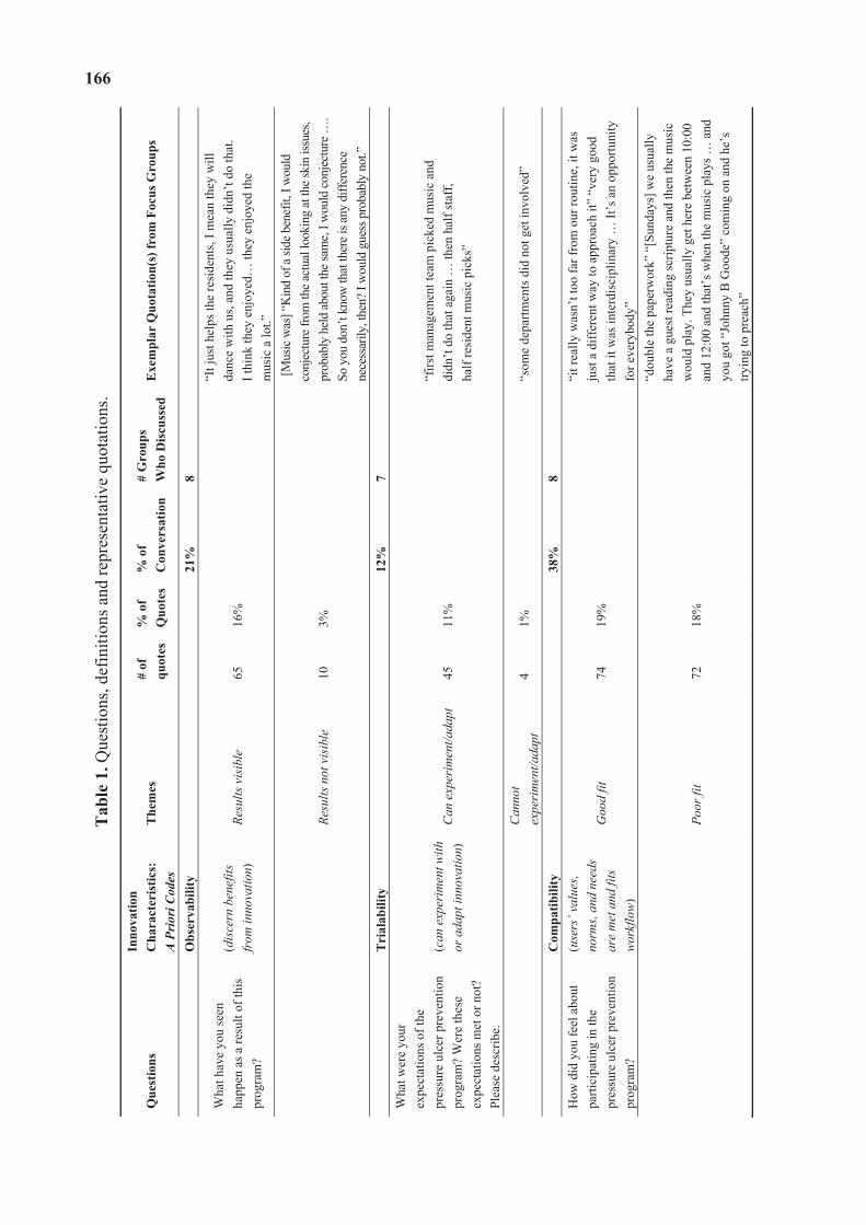
166T
able
1. Q
uest
ions
, def
initi
ons a
nd re
pres
enta
tive
quot
atio
ns.
Que
stio
nsIn
nova
tion
Cha
ract
eris
tics:
A
Prio
ri Co
des
The
mes
#
of
quot
es%
of
Quo
tes
% o
f C
onve
rsat
ion
# G
roup
s
Who
Dis
cuss
edE
xem
plar
Quo
tatio
n(s)
from
Foc
us G
roup
s
O
bser
vabi
lity
21%
8
Wha
t hav
e yo
u se
en
happ
en a
s a re
sult
of th
is
prog
ram
?
(dis
cern
ben
efits
fr
om in
nova
tion)
Re
sults
vis
ible
65
16
%
“It j
ust h
elps
the
resi
dent
s, I m
ean
they
will
da
nce
with
us,
and
they
usu
ally
did
n’t d
o th
at.
I thi
nk th
ey e
njoy
ed…
they
enj
oyed
the
m
usic
a lo
t.”
Resu
lts n
ot v
isib
le
10
3%
[M
usic
was
] “K
ind
of a
side b
enef
it, I
wou
ld
conj
ectu
re fr
om th
e act
ual l
ooki
ng at
the s
kin
issue
s, pr
obab
ly h
eld
abou
t the
sam
e, I w
ould
conj
ectu
re …
. So
you
don
’t kn
ow th
at th
ere i
s any
diff
eren
ce
nece
ssar
ily, t
hen?
I w
ould
gue
ss p
roba
bly
not.”
Tri
alab
ility
12%
7
W
hat w
ere
your
ex
pect
atio
ns o
f the
pr
essu
re u
lcer
pre
vent
ion
prog
ram
? W
ere
thes
e ex
pect
atio
ns m
et o
r not
? Pl
ease
des
crib
e.
(can
exp
erim
ent w
ith
or a
dapt
inno
vatio
n)C
an e
xper
imen
t/ada
pt
45
11%
“f
irst m
anag
emen
t tea
m p
icke
d m
usic
and
di
dn’t
do th
at a
gain
… th
en h
alf s
taff
, ha
lf re
side
nt m
usic
pic
ks”
Can
not
expe
rim
ent/a
dapt
4
1%
“som
e de
partm
ents
did
not
get
invo
lved
”
C
ompa
tibili
ty
38
%
8
How
did
you
feel
abo
ut
parti
cipa
ting
in th
e pr
essu
re u
lcer
pre
vent
ion
prog
ram
?
(use
rs’ v
alue
s, no
rms,
and
need
s ar
e m
et a
nd fi
ts
wor
kflo
w)
Goo
d fit
74
19
%
“it r
eally
was
n’t t
oo fa
r fro
m o
ur ro
utin
e, it
was
ju
st a
diff
eren
t way
to a
ppro
ach
it” “
very
goo
d th
at it
was
inte
rdis
cipl
inar
y …
It’s
an
oppo
rtuni
ty
for e
very
body
”
Poor
fit
72
18%
“dou
ble
the
pape
rwor
k” “
[Sun
days
] we
usua
lly
have
a g
uest
read
ing
scrip
ture
and
then
the
mus
ic
wou
ld p
lay.
The
y us
ually
get
her
e be
twee
n 10
:00
and
12:0
0 an
d th
at’s
whe
n th
e m
usic
pla
ys …
and
yo
u go
t “Jo
hnny
B G
oode
” co
min
g on
and
he’
s try
ing
to p
reac
h”
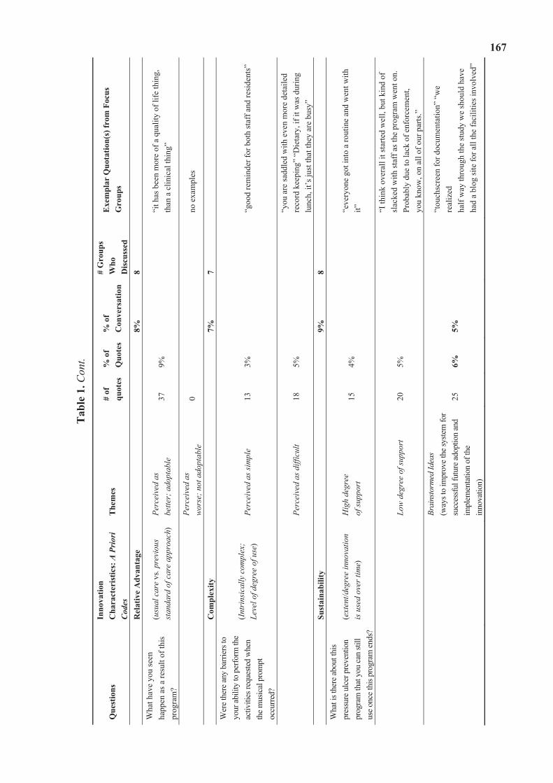
Tab
le 1
. Con
t.
Que
stio
nsIn
nova
tion
Cha
ract
eris
tics:
A P
riori
Code
sT
hem
es
# of
qu
otes
% o
f Q
uote
s %
of
Con
vers
atio
n
# G
roup
s W
hoD
iscu
ssed
Exe
mpl
ar Q
uota
tion(
s) fr
om F
ocus
G
roup
s
Rel
ativ
eA
dvan
tage
8%8
Wha
t hav
e yo
u se
en
happ
en a
s a re
sult
of th
is
prog
ram
?
(usu
al c
are
vs. p
revi
ous
stan
dard
of c
are
appr
oach
)Pe
rcei
ved
as
bette
r; a
dopt
able
37
9%
“i
t has
bee
n m
ore
of a
qua
lity
of li
fe th
ing,
th
an a
clin
ical
thin
g”
Perc
eive
d as
w
orse
; not
ado
ptab
le
0no
exa
mpl
es
Com
plex
ity7%
7W
ere t
here
any
barri
ers t
o yo
ur ab
ility
to p
erfo
rm th
e ac
tiviti
es re
ques
ted
whe
n th
e mus
ical
pro
mpt
oc
curre
d?
(Int
rins
ical
ly c
ompl
ex;
Leve
l of d
egre
e of
use
) Pe
rcei
ved
as si
mpl
e 13
3%
“g
ood
rem
inde
r for
bot
h st
aff a
nd re
side
nts”
Perc
eive
d as
diff
icul
t 18
5%
“y
ou a
re sa
ddle
d w
ith e
ven
mor
e de
taile
d re
cord
kee
ping
” “D
ieta
ry, i
f it w
as d
urin
g lu
nch,
it’s
just
that
they
are
bus
y”
Sust
aina
bilit
y9%
8W
hat i
s the
re ab
out t
his
pres
sure
ulc
er p
reve
ntio
n pr
ogra
m th
at y
ou ca
n sti
ll us
e onc
e thi
s pro
gram
ends
?
(ext
ent/d
egre
e in
nova
tion
is u
sed
over
tim
e)
Hig
h de
gree
of
supp
ort
15
4%
“eve
ryon
e go
t int
o a
rout
ine
and
wen
t with
it”
Low
deg
ree
of su
ppor
t 20
5%
“I th
ink
over
all i
t sta
rted
wel
l, bu
t kin
d of
sl
acke
d w
ith st
aff a
s the
pro
gram
wen
t on.
Pr
obab
ly d
ue to
lack
of e
nfor
cem
ent,
yo
u kn
ow, o
n al
l of o
ur p
arts
.”
Brai
nsto
rmed
Idea
s (w
ays t
o im
prov
e the
syste
m fo
r su
cces
sful
futu
re ad
optio
n an
d im
plem
enta
tion
of th
e in
nova
tion)
25
6%
5%
“tou
chsc
reen
for d
ocum
enta
tion”
“w
e re
aliz
ed
half
way
thro
ugh
the
stud
y w
e sh
ould
hav
e
had
a bl
og si
te fo
r all
the
faci
litie
s inv
olve
d”
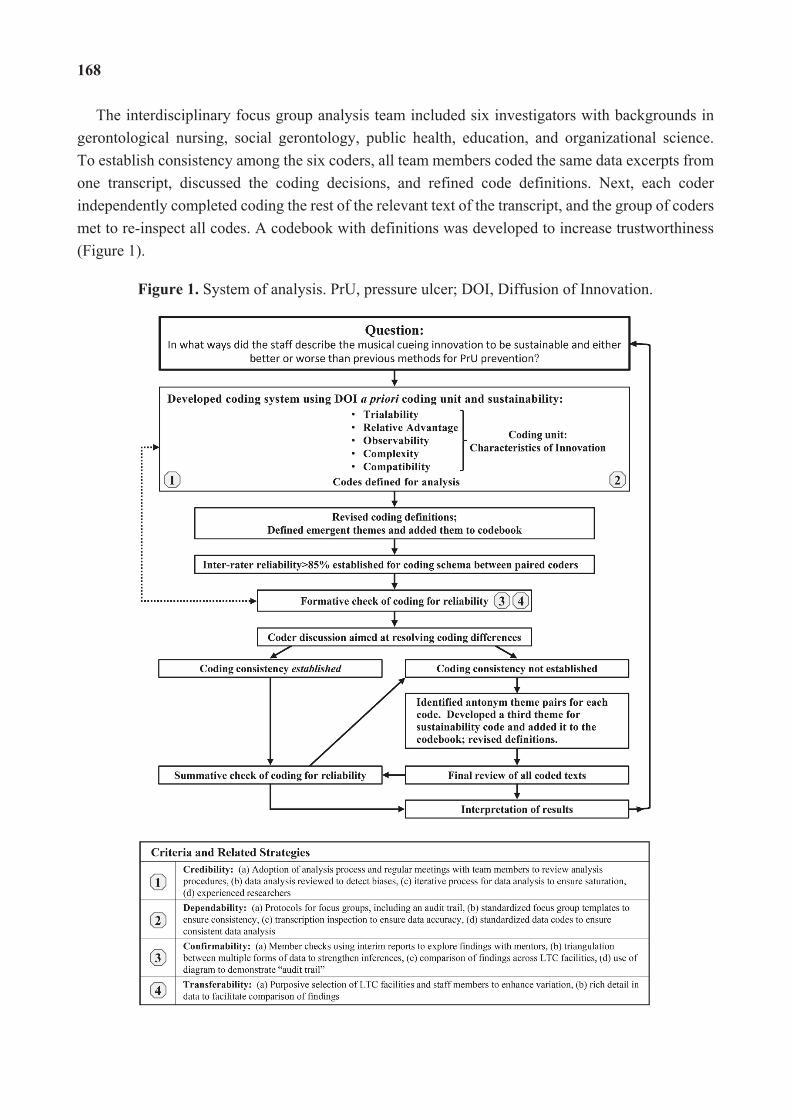
168
The interdisciplinary focus group analysis team included six investigators with backgrounds in gerontological nursing, social gerontology, public health, education, and organizational science. To establish consistency among the six coders, all team members coded the same data excerpts from one transcript, discussed the coding decisions, and refined code definitions. Next, each coder independently completed coding the rest of the relevant text of the transcript, and the group of coders met to re-inspect all codes. A codebook with definitions was developed to increase trustworthiness (Figure 1).
Figure 1. System of analysis. PrU, pressure ulcer; DOI, Diffusion of Innovation.
169
Next, a pair of coders was assigned to each transcript and coded them independently using the established definitions for codes and themes. The coder pairs met to examine the coding that each had applied to the text and to compare the coding with each other to establish reliability. The pairs of coders established inter-coder reliability at above 85% [17]. To increase the trustworthiness, the full coding team met repeatedly, until the full group had reviewed all coded statements for the consistency and accuracy of codes. Differences in coding were discussed and resolved by the team. Data emerged that could not be coded with a priori codes or emergent themes and was grouped into a seventh theme (brainstormed ideas). Next, the PI and one other coder met to examine and ensure that all transcripts and coding assignments had been appropriately coded according to correct definitions across group transcripts.
3. Results
A total of 45 staff members, 98% female, participated in the study, which included 26 certified nursing assistants (CNA), seven licensed practical nurses (LPN), eight registered nurses (RN), two housekeepers and two dietary staff. Each focus group exceeded the suggested minimum and mean sample size of five to eight participants [11,18]. Theme saturation appeared to be reached after the multiple facility cases (five groups), because no new information was emerging and no disconfirming evidence was found that enhances transferability. There were two types of responses that we noted for each of our codes, one being affirmation or a positive response by participants and the other being a negation or a negative response by participants to the characteristics of the innovation. Table 1 presents each characteristic coded and the subsequent two themes, the frequency and percent of comments related to each theme, the number of focus groups that discussed the category, and examples of those comments. The calculated percentage of the conversation for all groups was decided by tallying the number of discrete quotes (each quote that fit the predetermined definition) for each theme that emerged from the a priori category and then dividing the number of discrete quotes within each category by the total number of quotes. No substantial differences were found between the groups’ participation and the comments yielded. The results will be reported below by each a priori category for all eight groups together.
3.1. Observability
The two themes related to observability of the innovation included: (1) the visible benefits of the innovation relative to staff and resident behavior and the PrU outcomes; and (2) no obvious benefits. The visible benefits included cueing being enjoyable and PrUs decreasing. All groups discussed the music as being a fun cue that resulted in an increase in both movement and quality of life for both staff and residents. One staff commented that the “music was fun…what came out more than anything is how much the residents enjoyed the music”, and another staff member mentioned how the music “improved staff and resident mood”. The staff also believed they were seeing a decrease in the number of facility-acquired PrUs, remarking that “everyone participated; we’ve seen less ulcers”. The theme for the absence of visible benefits of the innovation included comments about the dearth of multidisciplinary involvement and some staff members failing to see the importance of
170
regular movement. In some facilities, the lack of multidisciplinary effort was disappointing to others: “some departments did not get involved as planned”. They also noted that if you did not have many residents who were bed ridden, some staff failed to see the importance of regular resident movement: “so from my standpoint, it was sort of, I just got done with exercise class and now the music’s going off. ‘Okay, everybody, go again, let’s move around a bit”. Overall, 21% of the focus groups’ discussion showed that staff could discern benefits from the innovation. This means that 65 of the 75 discrete quotes in this category were about results that were visible to them. This high level of visible benefits thereby enhances the perception of the innovation’s added value when adopted.
3.2. Trialability
The two themes related to the trialability of the innovation included: (1) it can be “tinkered” with and adapted for our facility; and (2) it cannot be experimented with to fit our facility. Several adjustments were made to fit facility requests, such as music choices, cueing times, documentation forms, and the placement of the documentation binder for different disciplines. Not every facility expressed a need for changes, i.e., facilities were blinded to each other’s adjustments during the intervention study. When the adjustments were later discussed across the focus groups, it became apparent that some of the changes may have worked in other intervention facilities. For example, one participant stated, “We gave Housekeeping their own documentation; we gave Dietary their own documentation”. When group moderators asked if this would have been helpful in other facilities, those staff members agreed. Another example of tweaking the innovation was that music was customized several ways, including both the timing and type of music: (1) “we had to adjust a little bit, not always with the music at mealtimes …, but the other times where you could”; and (2),
“when you play music that came from their era, they started with the memories because I know I brought The Platters out to play in the Dining Room, and then when I’d be in the Dining Room, we’d start talking to the husbands, and they would start talking about going dancing with their wives at the Park, and for some of the residents they would start to reminiscing and it was …. The Platters were soft, and their songs are back from when they were younger, they really liked it”!
However, there also were instances in which the innovation was not successfully adapted as planned, with statements like “we didn’t fool with the whiteboard a lot” and “I think the music was a good trigger to remind people, you know, to move your people as long as everybody was participating.” Overall, the intervention was deemed pliable and could be adapted to each facility to enhance adoption potential. Ninety-two percent (45 of 49) of the comments were rated as reflective of the staff’s perceived ability to experiment with or adapt the innovation.
3.3. Compatibility
The two themes related to the compatibility of the innovation included: (1) the innovation was well-matched with the staff values, norms, and perceived needs and fits in the current workflow; (2) the innovation was not well-matched and did not fit the current workflow. In the focus group discussions, the topic of compatibility involved 38% of the overall conversation with an even split
171
between the dichotomous themes. The attributes of the innovation that were considered a good match with staff values and needs were the perceived increase in resident participation and movement and the multidisciplinary involvement. One participant commented that, “I think in the beginning, from my perspective, I didn’t feel like it was anything that we didn’t know we should already be doing. I thought it was an excellent opportunity to refresh and reeducate and remind people how important it is to minimize your skin breakdown and your disease from not moving. And, I think that it was very good that it was interdisciplinary, that Housekeeping, everyone was involved because it is an opportunity for everybody to take part in the therapy.” Moreover, a former housekeeper noted, “that’s one of the reasons I became an aide, “cause as a housekeeper you can’t do a whole lot. [Once I started this program I] wanted to have hands-on”. Another staff member described that staff were “surprised at the number of residents when the music would go off; they would say, “there’s the music, we gotta’ move!” However, some components were not perceived to be as well-matched to staff and resident needs. For example, “documentation fell back on aides” in some facilities and was generally considered an unwanted increase in work. Staff in all facilities “backed away from the white board when we realized it was really duplication of a lot of things; maybe we didn’t capture the interdisciplinary teams as well, going away from the board, but we still caught the documentation in the binders that were supplied”. Staff comments about compatibility were almost evenly split between perceptions of a good fit versus poor fit for the innovation. When staff considered how well the innovation fit into their already established routine, they saw the innovation as being close to, yet somewhat different from, their accustomed work flow. While staff were willing to consider making changes in routine, the driving force behind perceptions of poor fit were things contributing to an increased workload, such as study-related documentation.
3.4. Relative Advantage
The only theme relating to the relative advantage of the innovation was that it was perceived as better than the previous method used to ensure resident movement on a regular basis. The emotional uplifting of the music and the increased interaction with the residents was considered an advantage. All groups described how the music “spunks us up a little bit”, and several described the residents and how “they dance with us, and they don’t usually do that”. There was no discussion alluding to any part of the musical cues being considered as worse than the previous standard of care; however, the additional documentation required for the study was not considered to be advantageous. This intervention was considered an advantage over the previous method used for resident movement to prevent PrUs and was perceived to positively influence their decisions to adopt. Eight percent of the total comments reflect the staff’s perception that usual care is enhanced by supplementation of a cueing system; all comments within this category (37/37) considered this music innovation as better.
3.5. Complexity
The two themes related to the complexity of the innovation included: (1) the innovation is perceived as simple to use and not difficult; and (2) the innovation is intrinsically complex and hard to do. Changing of the music and responding to the musical cue were perceived as simple and easy
172
to use. All groups provided examples of how they “were able to change the music” and let it evolve over time to satisfy everyone in the facility. It was also noted that the music “helped residents stay on schedule for other tasks”, as one staff member said:
“My alert residents mentioned that [the music] helped them keep on schedule if they had therapy or lunch, or know what to do because they got to that 2 h mark, and they would be like, ‘Oh, the music is always time to go to an activity, or it’s time… [for smoke break]’ so they actually started timing their schedule and their days around what was going on with the music, so that was something I know wasn’t part of the program, but it helped them to know what time it was, and that they needed to get to where they were going”.
A lack of innovation ease was perceived when the music cue was not appropriate for certain time periods, for example, “when passing trays it was a timing issue”. All participants noted that in some locations, it was harder to hear the music, for example: “I’m in the Rehabilitation Department, and we couldn’t really hear the music back there”. All groups also mentioned difficulties in accomplishing the added documentation related to resident movement. Overall, the decision to adopt was affected by both the perceived complexity and perceived simplicity of the innovation; however, only 3% of the conversation across groups was about the innovation simplicity and 5% was about the perceived intrinsically complex components.
3.6. Sustainability
Three themes emerged related to the sustainability potential and subsequent adoption of the innovation: (1) the innovation continues to be used over time; (2) the innovation does not continue to be used over time; and (3) brainstormed ideas, defined as staff ideas or proposed solutions for improving the innovation to increase the sustainability potential.
Some facilities described the innovation as continuing to be used over time. This was characterized by thorough integration of the innovation into care routines. As one staff member explained,
“I really can’t remember like what our routine was before we started this”, and the musical cue “helps to cue us when we were supposed to do it because we didn’t…. always recognize that like 2 h has past; we have to go turn such and such, but you would hear the music, and you’re like, okay, we’ve got to go turn everybody. So, I think it kind of helped, but we liked the music too”.
In fact, for three of the eight facilities, the equipment was left in place as a result of staff requests to continue the program. As one staff participant said, “I liked it! I want it to continue, yeah! We still are implementing it”. By contrast, other groups described the innovation as not being used over time. One noted that “I think it was top down and decreased pressure ulcers obviously. I think overall it started well, but kind of slacked with all staff as the program went on probably due to lack of enforcement, on all of our parts”. Moreover, staff discussed a lack of consistency that eroded sustainability, “You would see some people one day do it, and the next day you wouldn’t see anybody do it; it’s like a consistency thing”.
173
The theme of brainstormed ideas was comprised of staff ideas or suggestions to improve the innovation and support adoption and sustainability. For example, the additional documentation of the innovation was identified as disadvantageous in our themes related to relative advantage. However, staff provided suggestions to fix this issue, such as developing a novel flag system to indicate whether someone was turned by changing the color of residents’ blankets or simplifying with a form of computer documentation. Furthermore, because the volume was not consistent throughout the facilities, staff suggested using wireless speakers with Wi-Fi boosters placed throughout the facility. These ideas or solutions for improving the innovation could potentially contribute to improved adoption and sustainability.
Overall, we identified both positive and negative examples of each attribute of the innovation (the relative advantage being the exception with only positive). Sixty-eight percent of all coded examples were coded as positive examples. Further, staff provided many suggestions for how to improve the innovation, most of which staff felt would improve the sustainability of the innovation.
4. Discussion
This study used Rogers’ [10] DOI model regarding innovation characteristics to guide the focus group interviews that explored staff perceptions of the attributes of the intervention that affected the decision to adopt, as well as staff perceptions regarding impact on PrUs. The staff were from eight LTC facilities that had completed a musical cueing intervention aimed at prompting staff to increase resident movement in order to reduce facility-acquired PrUs.
We learned that each innovation characteristic and sustainability was more complex than our team originally believed. Within each code, two themes emerged, reflecting either a positive or negative response to the characteristics of the innovation. We are the first, to our knowledge, to present the DOI characteristics with these two themes, because the characteristics have been previously considered as a degree of perception [10]. The largest portion of the focus groups’ discussion and coded quotes was related to the positively-toned comments and the perceived benefits, thereby demonstrating how meaningfully connected the innovation appeared to be with staff values and culture; the findings suggest a strong potential for adoption. In general, the staff described the musical cue as fun for all disciplines, joyous and helpful; it served as a reminder to increase residents’ movement. Staff considered the innovation to be better than the prior system for the implementation of standards of care about moving residents and described the music as improving the overall energy and mobility, both of which were perceived as improving quality of life; for example, dancing between staff and residents now occurs. The majority of staff generally perceived that the occurrence of PrUs was decreasing, and the musical cueing was seemingly simple to use and a good reminder to ensure staff consistency with resident movement. Overall, the participants in this study professed that there was an improvement using musical cues for PrU prevention over their previously used methods.
We recognize that a substantial portion (32%) of the discrete statements was related to utility issues. For example, the timing of the cue was periodically not ideal and subsequently affected non-nursing departments’ involvement; furthermore, there were facilities in which non-nursing disciplines did not participate during the entire implementation. However, the code for sustainability

174
had a third theme “brainstormed ideas”, in which 5% of all statements focused on creative solutions for improving the innovation’s fit for future implementation, indicating that with minor adjustments or enhancements made, there was a perceived potential for the sustainability of the innovation. A number of participants explicitly expressed ideas aiming to simplify certain aspects in order to improve the intervention. Innovations that are perceived to be simple and easy to use and can be broken down into more manageable parts with few barriers to overcome will be assimilated and adopted more easily [19]. Our findings indicated that the musical cueing was perceived both as complex and, yet, as a seemingly simple innovation. The majority of concerns related to function was about the timing of the cues, sound quality of the music, the additional workload from the study-related documentation and the need for a strategy to increase other department involvement. All of these aspects are potentially modifiable to better fit each work environment.
This study adds to our understanding of the mechanisms by which an innovation’s characteristics yield (or fail to yield) the outcome of interest and might be successfully adopted in a particular context. For example, this study required additional documentation to be completed, which was considered to hinder adoption, but the additional study-related documentation would not be needed if the innovation were a permanent part of the facility system for PrU prevention. Another example would be how the music evolved and changed over time with new music requests every month. For some facilities, the music was made to be reflective of the residents’ era and genre choices, thereby enabling dancing in the halls and making both staff and residents happy. Each facility context is unique, making successful adoption for any length of time a complex process with a multiplicity of variables, which may or may not induce staff ambivalence and could negatively affect innovation adoption. Characteristics of the innovation are neither stable features nor sure determinants of the innovation’s adoption or assimilation, nor should they be. The ability to tailor aspects of an innovation (i.e., music choice) to participant preferences appears to make it more appealing to the staff attitudes. No substantial differences in perceptions were found between the focus groups; the participant groups all described both positive and negative aspects of the innovation’s characteristics, which would influence their decision to either adopt or reject the intervention. Future research should focus on understanding how each innovation characteristic impacts outcomes of interest.
Staff largely perceived a positive impact of this innovation on residents’ movement and PrU outcomes. However, because PrU prevention is complex and context laden, clinicians need to pay close attention to those characteristics that facilitate or impede innovative implementation and guideline adoption and implementation. Research about implementing guidelines is needed to improve understanding about PrU prevention practices. In our study, all facilities that received the intervention experienced some reduction in facility-acquired PrUs by the end of study participation [8]. Our qualitative analysis leads us to believe that the impact of this intervention in part can be attributed to the perceived positive good fit of the intervention with the beliefs and values of the facility staff and residents. We postulate that the musical component of the innovation can influence the participants to connect and engage with the intervention and its aim of guideline implementation.
A limitation of the study was that all facilities were from one corporation from the Midwestern U.S. that volunteered to participate in the study and may therefore be systematically different from
175
facilities in other geographic regions or corporations, limiting generalizability. Furthermore, we used a convenience sample of staff that was available and willing to participate on the day of the scheduled focus group. Although it is possible that individuals who volunteered to participate did so because they favored the intervention, we believe that our efforts to recruit a diverse group of staff were effective, as reflected by 32% of the discussion consisting of negatively-toned comments. We used an unusual, semi-quantitative method for counting unique expressions of each subcategory and recognize that these counts may be open to bias. We were interested in exploring not only the qualitative differences in the innovation codes, but also how frequently they were expressed. To optimize confidentiality and coder blinding, participants were not named in coded transcripts, and no roles were stated, even though focus group leaders reported the overall focus group composition by gender and job title. Therefore, we were unable to compare the perceptions of staff in different roles. The focus groups only included one male, and thus, the findings might reflect gender bias. However, it is notable that a broad range of participant roles was represented.
Tolba and Mourad [20] advanced the DOI model by proposing a “functional dimension” of the innovation, in which high complexity was considered to have a negative impact on innovation acceptance, and a “social dimension” of the innovation adoption that is influenced by different types of users and leaders [20]. Although these authors provided no definition of social and functional dimensions, our consideration of these dimensions enriched the interpretation of our findings. In our study, the social dimension embodied the organizational and cultural ways that people relate to an innovation, and the functional dimension embodied all of the activities and tasks that people recognize as relating to the way in which an innovation works or operates. These functional and social dimensions offer an expanded mechanism for explaining our focus group findings regarding perceptions of the innovation’s characteristics, outcomes and sustainability. The social dimension was evidenced in staff’s generally positively-toned perceptions regarding the innovation and its outcomes on resident movement and PrU prevention, suggesting that individual and cultural needs were being met and enhancing the potential for adoption of the innovation. The functional dimension was evidenced in the way staff perceived responsibilities and the utility of the innovation’s operational process, and this aspect of the conversation expressed more negatively-toned comments than did the social dimension.
Future research on innovations will depend upon a better understanding of their social dimensions, because it is the interaction among the innovation, the intended adopter(s) and a particular context that determines whether adoption of the innovation is successful and is sustainable [19]. Furthermore, we would suggest that future research address whether improved outcomes are contingent on an intervention assimilation period. This pressure ulcer prevention intervention study’s results showed that the frequency of new facility-acquired PrUs per assessment was less than 2% within three months of intervention deployment in both facility groups and below 1% during the final three months of the study [8]. The benefits of this particular innovation might extend beyond the intended mobility-related benefits of this simple, inexpensive intervention to the benefits afforded by sensory stimulation. Both staff and residents expressed satisfaction with the music. Music from the residents’ era was chosen by staff in some facilities, because it brought a great deal of pleasure to residents, prompting memories and the sharing of stories, which, in turn, improved staff morale [8].
176
5. Conclusions
We conclude that musical cueing for the promotion of the consistent implementation of guidelines regarding LTC resident movement aimed at PrU prevention is worth further exploration. This intervention had valuable unanticipated consequences, such as improving social connections by increasing communication frequency and quality between staff and residents. The intervention appears to enhance the social aspects of the care environment, which might increase innovation uptake and adoption among the LTC staff. Multiple departments participated initially, but there were some unanticipated consequences that interfered with the successful participation of these disciplines over time. However, adoption is a process rather than an event, with different concerns being dominant at different points in the process; the decision to adopt an innovation is also based on an individual’s perception of the innovation’s worth relative to other ways of accomplishing the same goal. Moreover, what one staff member perceives as easy and valuable might be considered unimportant, unpleasant or even hard for another staff member. The impact of musical cueing on other guideline implementation and clinical outcomes requires additional validation and, thus, merits future research.
Acknowledgments
The authors wish to thank the staff of the eight participating long-term care facilities. Support for this project was provided by a grant from the Robert Wood Johnson Foundation Interdisciplinary Nursing Quality Research Initiative program (Interdisciplinary Mobility Team Approach to Reduction of Facility-Acquired Pressure Ulcers); Tracey L. Yap (PI). Trial Registration: clinicaltrials.gov Identifier: NCT01008254.
Author Contributions
Tracey L. Yap, Susan Kennerly, Kirsten Corazzini, Kristie Porter, Mark Toles and Ruth A. Anderson had full access to all the data in the study related to focus group interviews and take responsibility for the integrity of the data and the accuracy of the data analysis. Study concept and design: Tracey L. Yap and Susan Kennerly. Acquisition of subjects and/or data: Tracey L. Yap, and Susan Kennerly. Analysis and interpretation: Tracey L. Yap, Susan Kennerly, Kirsten Corazzini, Kristie Porter, Mark Toles, and Ruth A. Anderson. Drafting of the manuscript: Tracey L. Yap, Susan Kennerly, Kirsten Corazzini, Kristie Porter, Mark Toles, and Ruth A. Anderson.
Conflicts of Interest
The authors declare no conflict of interest.
References
1. Centers for Medicare & Medicaid Services (CMS). Action Plan for (Further Improvement of) Nursing Home Quality; Centers for Medicare & Medicaid Services: Baltimore, MD, USA, 2008.
177
2. Niederhauser, A.; VanDeusen Lukas, C.; Parker, V.; Ayello, E.A.; Zulkowski, K.; Berlowitz, D. Comprehensive programs for preventing pressure ulcers: A review of the literature. Adv. Skin Wound Care 2012, 25, 167–188.
3. Lyder, C.H. Pressure ulcer prevention and management. JAMA 2003, 289, 223–226. 4. Redelings, M.D.; Lee, N.E.; Sorvillo, F. Pressure ulcers: More lethal than we thought? Adv. Skin
Wound Care 2005, 18, 367–372. 5. Keelaghan, E.; Margolis, D.; Zhan, M.; Baumgarten, M. Prevalence of pressure ulcers on
hospital admission among nursing home residents transferred to the hospital. Wound Repair Regen. 2008, 16, 331–336.
6. Black, J.M.; Edsberg, L.E.; Baharestani, M.M.; Langemo, D.; Goldberg, M.; McNichol, L.; Cuddigan, J.; National Pressure Ulcer Advisory Panel. Pressure ulcers: Avoidable or unavoidable? Results of the National Pressure Ulcer Advisory Panel Consensus Conference. Ostomy Wound Manag. 2011, 57, 24–37.
7. Cannon, B.C.; Cannon, J.P. Management of pressure ulcers. Am. J. Health. Syst. Pharm. 2004, 61, 1895–1905.
8. Yap, T.L.; Kennerly, S.M.; Simmons, M.R.; Buncher, C.R.; Miller, E.; Kim, J.; Yap, W.Y. Multidimensional team-based intervention using musical cues to reduce odds of facility-acquired pressure ulcers in long-term care: A paired randomized intervention study. J. Am. Geriatr. Soc. 2013, 61, 1552–1559.
9. Scarborough, P. Your wound team model: Is it unidisciplinary, multidisciplinary, interdisciplinary or transdisciplinary? Wound Source. 2013, 16, 7–8.
10. Rogers, E.M. Diffusion of Innovations, 5th ed; Free Press: New York, NY, USA, 2003. 11. Krueger, R.A.; Casey, M.A. Focus Groups: A Practical Guide for Applied Research, 3rd ed.;
Sage Publications: Thousand Oaks, CA, USA, 2000. 12. QSR International Pty. Ltd. NVivo 9. 2012; home page for NVivo 9 software. Available online:
http://www.qsrinternational.com/support_faqs_detail.aspx?view=11 (accessed on 6 March 2013). 13. Lyder, C.H.; Ayello, E.A. Pressure ulcers: A patient safety issue. In Patient Safety and Quality:
An Evidence-Based Handbook for Nurses (AHRQ Pub. No. 08–0043); Hughes, R.G., Ed.; Agency for Healthcare Research and Quality (AHRQ): Rockville, MD, USA, 2008.
14. Hsieh, H.F.; Shannon, S.E. Three approaches to qualitative content analysis. Qual. Health Res. 2005, 15, 1277–1288.
15. Teddlie, C.; Tashakkori, A. Foundations of Mixed Methods Research; Sage Publications: Thousand Oaks, CA, USA, 2009.
16. Shenton, A.K. Strategies for ensuring trustworthiness in qualitative research projects. Educ. Inf. 2004, 22, 63–75.
17. Smith, C.P. Content Analysis and Narrative Analysis; Reis, H.T., Judd, C.M., Eds.; The Press Syndicate of the University of Cambridge: Camridge, UK, 2000.
18. Morgan, D.L. Planning Focus Groups; Sage Publications, Inc.: Thousand Oaks, CA, USA, 1998. 19. Greenhalgh, T.; Robert, G.; Macfarlane, F.; Bate, P.; Kyriakidou, O. Diffusion of Innovations in
Service Organizations: Systematic Review and Recommendations. Milbank Q. 2004, 82, 581–629. 20. Tolba, A.H.; Mourad, M. Individual and cultural factors affecting diffusion of innovation.
J. Int. Bus. Cult. Stud. 2011, 5, 1–16.

MDPI AG Klybeckstrasse 64
4057 Basel, Switzerland Tel. +41 61 683 77 34 Fax +41 61 302 89 18 http://www.mdpi.com/
Healthcare Editorial Office E-mail: [email protected]
http://www.mdpi.com/journal/healthcare

MDPI • Basel • Beijing ISBN 978-3-03842-045-3www.mdpi.com
Related Documents