Worry, generalized anxiety disorder, and emotion: Evidence from the EEG gamma band Desmond J. Oathes a,b, *, William J. Ray a , Alissa S. Yamasaki a , Thomas D. Borkovec a , Louis G. Castonguay a , Michelle G. Newman a , Jack Nitschke b a Department of Psychology, The Pennsylvania State University, University Park, PA, United States b Departments of Psychiatry and Psychology, The University of Wisconsin-Madison, Madison, WI, United States 1. Introduction 1.1. EEG gamma band and emotion The gamma rhythm (30–100 Hz) is widespread in the central nervous system including in areas associated with emotional processing such as the amygdala and perirhinal cortex (Collins et al., 2001). Recent research continues to suggest connections between EEG gamma activity and emotion with special emphasis on negative emotional processing (e.g. Luo et al., 2007; Matsumoto et al., 2006). Intracranial field potentials recorded from the amygdala confirm that gamma power is highest for aversive stimulus presentations as compared with neutral or pleasant stimuli (Oya et al., 2002). Spectral power in the gamma band has been associated with emotional processing when both EEG alpha frequency and beta frequency activity have not shown sensitivity to emotional stimulus variations (Mu ¨ ller et al., 1999). Another attribute of gamma as opposed to other indexes of emotional perception is that gamma induced by emotional stimuli is typically not phase-locked to the onset of visual stimulus presentations (Oya et al., 2002). Instead, induced gamma is usually measured over periods of several seconds as with successive presentations of visual stimuli (Mu ¨ ller et al., 1999). This suggests that induced gamma reflects a more integrative or reflective aspect of processing emotional material. Consistent with the idea that induced gamma fluctuates with extended periods of emotional processing; experimental tasks thought to induce emotional experience have been shown to increase gamma activity. When asked to imagine a phobic object, individuals suffering from a specific phobia show increases in gamma band activation as well as increases in heart rate and respiration (Gemignani et al., 2000). Also, gamma has been shown to decrease during periods of relaxation and to increase during periods of imagining negative emotional material (Sebastiani et al., 2003). Thus, the present study records periods of several minutes during which emotional experiences were thought to be induced. Distributions of gamma activation recorded from the scalp surface may be important for discovering links between specific emotional experiences and physiological recordings. For example, Biological Psychology 79 (2008) 165–170 ARTICLE INFO Article history: Received 1 December 2007 Received in revised form 12 March 2008 Accepted 7 April 2008 Available online 15 April 2008 Keywords: Generalized anxiety disorder Electroencephalography Gamma band Psychotherapy Emotion ABSTRACT The present study examined EEG gamma (35–70 Hz) spectral power distributions during worry inductions in participants suffering from generalized anxiety disorder (GAD) and in control participants without a history of psychiatric illness. As hypothesized, the EEG gamma band was useful for differentiating worry from baseline and relaxation. During worry induction, GAD patients showed higher levels of gamma activity than control participants in posterior electrode sites that have been previously associated with negative emotion. Gamma fluctuations in these electrode sites were correlated with subjective emotional experience ratings lending additional support to interpretations of negative affect. Following 14 weeks of psychotherapy, the GAD group reported less negative affect with worry inductions and the corresponding gamma sites that previously differentiated the clinical from control groups changed for the GAD patients in the direction of control participants. These findings suggest converging evidence that patients suffering from GAD experience more negative emotion during worry and that the EEG gamma band is useful for monitoring fluctuations in pathological worry expected to follow successful treatment. ß 2008 Elsevier B.V. All rights reserved. * Corresponding author at: Waisman Laboratory for Brain Imaging and Behavior, 1500 Highland Avenue (S111), The University of Wisconsin, Madison, WI 53705, United States. Tel.: +1 608 890 1389. E-mail address: [email protected] (D.J. Oathes). Contents lists available at ScienceDirect Biological Psychology journal homepage: www.elsevier.com/locate/biopsycho 0301-0511/$ – see front matter ß 2008 Elsevier B.V. All rights reserved. doi:10.1016/j.biopsycho.2008.04.005

Welcome message from author
This document is posted to help you gain knowledge. Please leave a comment to let me know what you think about it! Share it to your friends and learn new things together.
Transcript
Worry, generalized anxiety disorder, and emotion:Evidence from the EEG gamma band
Desmond J. Oathes a,b,*, William J. Ray a, Alissa S. Yamasaki a, Thomas D. Borkovec a,Louis G. Castonguay a, Michelle G. Newman a, Jack Nitschke b
a Department of Psychology, The Pennsylvania State University, University Park, PA, United Statesb Departments of Psychiatry and Psychology, The University of Wisconsin-Madison, Madison, WI, United States
Biological Psychology 79 (2008) 165–170
A R T I C L E I N F O
Article history:
Received 1 December 2007
Received in revised form 12 March 2008
Accepted 7 April 2008
Available online 15 April 2008
Keywords:
Generalized anxiety disorder
Electroencephalography
Gamma band
Psychotherapy
Emotion
A B S T R A C T
The present study examined EEG gamma (35–70 Hz) spectral power distributions during worry
inductions in participants suffering from generalized anxiety disorder (GAD) and in control participants
without a history of psychiatric illness. As hypothesized, the EEG gamma band was useful for
differentiating worry from baseline and relaxation. During worry induction, GAD patients showed higher
levels of gamma activity than control participants in posterior electrode sites that have been previously
associated with negative emotion. Gamma fluctuations in these electrode sites were correlated with
subjective emotional experience ratings lending additional support to interpretations of negative affect.
Following 14 weeks of psychotherapy, the GAD group reported less negative affect with worry inductions
and the corresponding gamma sites that previously differentiated the clinical from control groups
changed for the GAD patients in the direction of control participants. These findings suggest converging
evidence that patients suffering from GAD experience more negative emotion during worry and that the
EEG gamma band is useful for monitoring fluctuations in pathological worry expected to follow
successful treatment.
� 2008 Elsevier B.V. All rights reserved.
Contents lists available at ScienceDirect
Biological Psychology
journal homepage: www.elsevier.com/locate/biopsycho
1. Introduction
1.1. EEG gamma band and emotion
The gamma rhythm (30–100 Hz) is widespread in the centralnervous system including in areas associated with emotionalprocessing such as the amygdala and perirhinal cortex (Collinset al., 2001). Recent research continues to suggest connectionsbetween EEG gamma activity and emotion with special emphasison negative emotional processing (e.g. Luo et al., 2007; Matsumotoet al., 2006). Intracranial field potentials recorded from theamygdala confirm that gamma power is highest for aversivestimulus presentations as compared with neutral or pleasantstimuli (Oya et al., 2002).
Spectral power in the gamma band has been associated withemotional processing when both EEG alpha frequency and betafrequency activity have not shown sensitivity to emotional
* Corresponding author at: Waisman Laboratory for Brain Imaging and Behavior,
1500 Highland Avenue (S111), The University of Wisconsin, Madison, WI 53705,
United States. Tel.: +1 608 890 1389.
E-mail address: [email protected] (D.J. Oathes).
0301-0511/$ – see front matter � 2008 Elsevier B.V. All rights reserved.
doi:10.1016/j.biopsycho.2008.04.005
stimulus variations (Muller et al., 1999). Another attribute ofgamma as opposed to other indexes of emotional perception is thatgamma induced by emotional stimuli is typically not phase-lockedto the onset of visual stimulus presentations (Oya et al., 2002).Instead, induced gamma is usually measured over periods ofseveral seconds as with successive presentations of visual stimuli(Muller et al., 1999). This suggests that induced gamma reflects amore integrative or reflective aspect of processing emotionalmaterial.
Consistent with the idea that induced gamma fluctuates withextended periods of emotional processing; experimental tasksthought to induce emotional experience have been shown toincrease gamma activity. When asked to imagine a phobic object,individuals suffering from a specific phobia show increases ingamma band activation as well as increases in heart rate andrespiration (Gemignani et al., 2000). Also, gamma has been shownto decrease during periods of relaxation and to increase duringperiods of imagining negative emotional material (Sebastiani et al.,2003). Thus, the present study records periods of several minutesduring which emotional experiences were thought to be induced.
Distributions of gamma activation recorded from the scalpsurface may be important for discovering links between specificemotional experiences and physiological recordings. For example,
D.J. Oathes et al. / Biological Psychology 79 (2008) 165–170166
relatively more gamma power in the right temporal area isassociated with positively valenced stimulus presentations andrelatively more gamma in the left temporal area is associated withnegative stimulus presentations (Muller et al., 1999). The presentstudy sought to contribute to a growing literature linkingdistributions of induced EEG gamma spectral power to patholo-gical and non-pathological experiences of emotion. We utilizedEEG spectral power as well as ratings of subjective experience toassess differences between GAD and non-psychiatric controlgroups; between baseline, relaxation and worry tasks; andbetween pre- and post-treatment assessments in our GAD group.Based on a literature linking GAD and worry to negative emotion,we expected gamma during worry to differentiate our patientsfrom controls and to be sensitive to changes in the GAD groupexpected to follow treatment.
1.2. Worry, GAD and reports of negative affect
The present study focused on worry as a central negativeemotional experience for chronic worriers suffering from GAD.Generalized anxiety disorder (GAD) is characterized by excessiveanxiety and uncontrollable worry about a variety of topics(DSM-IV-TR; American Psychiatric Association, 2000). Theprocess of worry in itself is a negative emotional experiencewhether or not the worrier suffers from GAD (Borkovec and Inz,1990; Andrews and Borkovec, 1998). Though worry increasesreports of negative affect and anxiety, particular physiologicalsystems may not register emotional arousal during worry. Whenasked to worry, research participants report increases in anxietywhile cardiovascular measures do not consistently reflect thechange (Borkovec and Hu, 1990; Borkovec et al., 1993). Also,chronic worry does not increase fear-potentiated startle eye-blink EMG amplitudes to emotional stimuli (Nitschke et al.,2002) or muscle activity recorded using EMG (Oathes et al., inpress; though see that paper for evidence of worry influences onmotor preparation). Thus, it is important to identify apsychophysiological measure which not only characterizesworry experiences but also differentiates individuals sufferingfrom pathological anxiety (GAD) from non-anxious individuals.The present study suggests that the EEG gamma band mightfunction as such an index.
1.3. Hypotheses
Our initial manipulation check to test the hypothesis thatgamma might be sensitive to experimentally induced emotionalintensity was based on a prediction that gamma spectral powerwould be increasingly present in the order from least to greatestbeginning with our relaxation task followed by baseline recordingsand that worry would facilitate the most gamma activity across ourtwo groups. The relaxation task served as a comparison to worry inthat relaxation was also a cognitive induction (which mayinfluence gamma activity; cf. Jensen et al., 2007) but was expectedto differ from worry according to the degree of negative affectinduced by the experimental instructions. Based on relationshipsbetween subjective experiences of negative emotion and GAD (e.g.Borkovec and Inz, 1990; Borkovec and Ruscio, 2001) and betweennegative emotion and a particular distribution of scalp recordedinduced EEG gamma activity (Muller et al., 1999), we expectedgreater left posterior gamma activity for the GAD group comparedto control participants. It was expected that this difference wouldbe especially pronounced during the worry task, as this was thetask thought to be most relevant to the GAD diagnosis and itsassociated negative emotionality. Worry is especially relevant tostudying GAD in that chronic uncontrollable worry is the essential
feature common to all individuals diagnosed with GAD. Though thePenn State Worry Questionnaire or another measure of traitanxiety might be relevant to chronic worry and GAD, the presentstudy sought to examine a less often assessed aspect of worry:negative affect. To support interpretations related to negativeemotionality for group differences in EEG gamma during worry, weassessed ratings of subjective emotional experience during ourphysiological recording sessions that we expected to correlate withEEG gamma activity.
2. Method
2.1. Participants
Anxious participants were drawn from newspaper advertisements or from
outside agency referrals. Advertisements also invited control participants with a
request for individuals between 18 and 65 years of age ‘‘without current or past
anxiety or depression difficulties’’. Fifteen clients and 15 control participants were
used from the first wave recruited for a therapy outcome study (see Newman et al.,
2004 for details). The study was approved by the Office for Research Protections
(IRB) at the Pennsylvania State University. All participants gave signed informed
consent (in accord with Helsinki Declaration) to participate in the therapy
outcome portion of this study and signed a separate consent for the
psychophysiological assessments. For GAD clients, clinical assessors conducted
phone screens in order to initially assess a GAD diagnosis. The assessor then met
eligible clients to administer the Anxiety Disorder Interview Schedule-IV (ADIS-
IV; DiNardo et al., 1994) as well as a variety of psychiatric symptom scales
including the Beck Depression Inventory (BDI-II; Beck et al., 1996) and Penn State
Worry Questionnaire (PSWQ; Meyer et al., 1990). Within 2 weeks of the first
interview, a clinical psychologist conducted a second ADIS-IV interview to ensure
the reliable presence of a GAD diagnosis. Criteria for inclusion of GAD clients were:
formal principal diagnosis of GAD according to DSM-IV criteria by either both
assessors or consensus of the assessors in case of discrepancy; lack of psychosis,
substance abuse and/or medical or physical conditions linked to anxiety; a global
severity of 4 or more (moderate anxiety) on an 0–8 point clinician determined
severity scale; and between ages 18 and 65. Psychoactive medication use was
permitted if the client agreed to maintain constant dosage levels for the duration
of treatment and assessment. Among the fifteen participants used for analysis,
three were taking a selective serotonin reuptake inhibitor (SSRI; two to treat
major depressive disorder and the third for panic disorder). All but two of our GAD
sample had additional diagnoses including six diagnoses of social phobia, four of
major depressive disorder, two of panic disorder, two of specific phobias, and one
of obsessive compulsive disorder. The final sample of 15 GAD clients consisted of
13 female and 2 male participants. All were Caucasian by self-identified ethnicity
and ranged in age from 22 to 45 years (M = 36.4, S.D. = 8.21). Control participants
were also given the ADIS-IV and were included based on lack of current or past
diagnosable psychiatric disorder, as well as lack of current or past alcohol or
substance dependence or any prior psychological or pharmacological treatment
for a psychological problem. Control participants were asked for medical histories
and excluded if medical problems frequently linked to anxiety were discovered
(e.g. thyroid problems). The 15 members of the control group consisted of 12
females and 3 males. The control group was also Caucasian with age ranges from
22 to 45 years (M = 36.6, S.D. = 7.84). The two groups were not significantly
different in age, gender or education (i.e. category of educational attainment such
as high school diploma, 2 year degree, etc.).
2.2. Procedure
One week before physiological sessions, GAD clients were given a tour of the
laboratory including a description of recording methods and tasks to be
administered. The remaining procedures of the experimental session for GAD
clients and control participants were identical. As electrodes were being affixed, he/
she was told, ‘‘At one point during the experiment, you will be asked about a worry
topic. Can you have something in mind for later use?’’ Participants were encouraged
to consider a topic of ‘‘current concern’’ that they would be able to ‘‘worry intensely
about’’ for several minutes. Participants were then left alone in the room with the
door closed. An audiotaped instruction sequence announced the tasks, while the
experimenters monitored the participant by videocamera and via intercom in an
adjacent room.
For the duration of the experiment, participants sat in a noise-controlled room. A
17-channel EEG cap (ElectroCap International) was used to obtain signals from 15
channels referenced to linked ears: left and right frontal (F3, F4), midline frontal
(FZ), midline central (CZ), midline parietal (PZ), left and right temporal (T3, T4, T5,
T6), left and right parietal (P3, P4, P5, P6), and occipital regions (O1, O2). Electrode
placement was based on the International 10–20 System (Jasper, 1958). Signals
were amplified using a Nihon-Kohden 21-channel electroencephalograph and
digitized by a Neuroscan (Neurosoft) system. Data were collected at a sampling rate
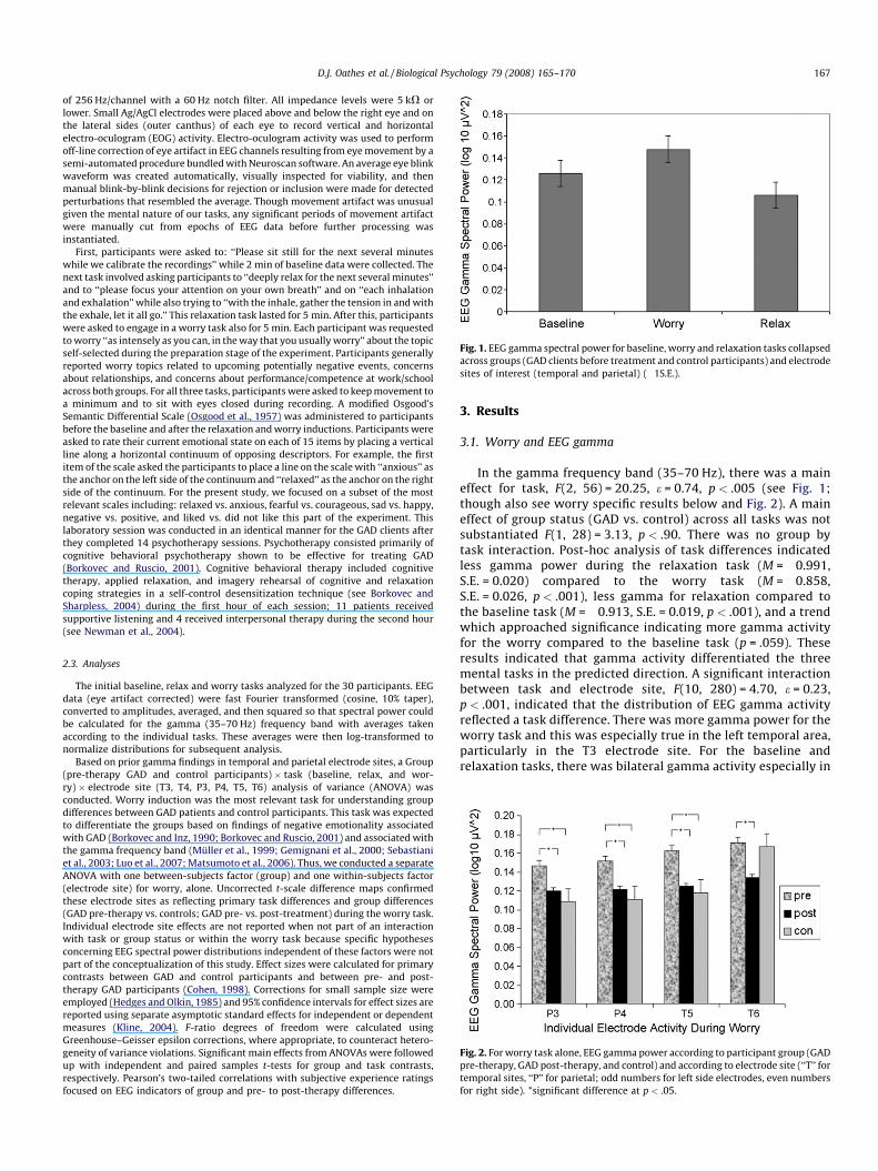
Fig. 1. EEG gamma spectral power for baseline, worry and relaxation tasks collapsed
across groups (GAD clients before treatment and control participants) and electrode
sites of interest (temporal and parietal) (�1S.E.).
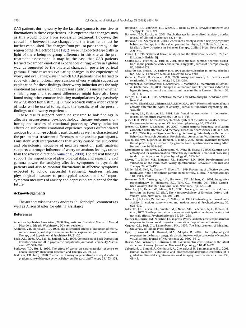
Fig. 2. For worry task alone, EEG gamma power according to participant group (GAD
pre-therapy, GAD post-therapy, and control) and according to electrode site (‘‘T’’ for
temporal sites, ‘‘P’’ for parietal; odd numbers for left side electrodes, even numbers
for right side). *significant difference at p < .05.
D.J. Oathes et al. / Biological Psychology 79 (2008) 165–170 167
of 256 Hz/channel with a 60 Hz notch filter. All impedance levels were 5 kV or
lower. Small Ag/AgCl electrodes were placed above and below the right eye and on
the lateral sides (outer canthus) of each eye to record vertical and horizontal
electro-oculogram (EOG) activity. Electro-oculogram activity was used to perform
off-line correction of eye artifact in EEG channels resulting from eye movement by a
semi-automated procedure bundled with Neuroscan software. An average eye blink
waveform was created automatically, visually inspected for viability, and then
manual blink-by-blink decisions for rejection or inclusion were made for detected
perturbations that resembled the average. Though movement artifact was unusual
given the mental nature of our tasks, any significant periods of movement artifact
were manually cut from epochs of EEG data before further processing was
instantiated.
First, participants were asked to: ‘‘Please sit still for the next several minutes
while we calibrate the recordings’’ while 2 min of baseline data were collected. The
next task involved asking participants to ‘‘deeply relax for the next several minutes’’
and to ‘‘please focus your attention on your own breath’’ and on ‘‘each inhalation
and exhalation’’ while also trying to ‘‘with the inhale, gather the tension in and with
the exhale, let it all go.’’ This relaxation task lasted for 5 min. After this, participants
were asked to engage in a worry task also for 5 min. Each participant was requested
to worry ‘‘as intensely as you can, in the way that you usually worry’’ about the topic
self-selected during the preparation stage of the experiment. Participants generally
reported worry topics related to upcoming potentially negative events, concerns
about relationships, and concerns about performance/competence at work/school
across both groups. For all three tasks, participants were asked to keep movement to
a minimum and to sit with eyes closed during recording. A modified Osgood’s
Semantic Differential Scale (Osgood et al., 1957) was administered to participants
before the baseline and after the relaxation and worry inductions. Participants were
asked to rate their current emotional state on each of 15 items by placing a vertical
line along a horizontal continuum of opposing descriptors. For example, the first
item of the scale asked the participants to place a line on the scale with ‘‘anxious’’ as
the anchor on the left side of the continuum and ‘‘relaxed’’ as the anchor on the right
side of the continuum. For the present study, we focused on a subset of the most
relevant scales including: relaxed vs. anxious, fearful vs. courageous, sad vs. happy,
negative vs. positive, and liked vs. did not like this part of the experiment. This
laboratory session was conducted in an identical manner for the GAD clients after
they completed 14 psychotherapy sessions. Psychotherapy consisted primarily of
cognitive behavioral psychotherapy shown to be effective for treating GAD
(Borkovec and Ruscio, 2001). Cognitive behavioral therapy included cognitive
therapy, applied relaxation, and imagery rehearsal of cognitive and relaxation
coping strategies in a self-control desensitization technique (see Borkovec and
Sharpless, 2004) during the first hour of each session; 11 patients received
supportive listening and 4 received interpersonal therapy during the second hour
(see Newman et al., 2004).
2.3. Analyses
The initial baseline, relax and worry tasks analyzed for the 30 participants. EEG
data (eye artifact corrected) were fast Fourier transformed (cosine, 10% taper),
converted to amplitudes, averaged, and then squared so that spectral power could
be calculated for the gamma (35–70 Hz) frequency band with averages taken
according to the individual tasks. These averages were then log-transformed to
normalize distributions for subsequent analysis.
Based on prior gamma findings in temporal and parietal electrode sites, a Group
(pre-therapy GAD and control participants) � task (baseline, relax, and wor-
ry) � electrode site (T3, T4, P3, P4, T5, T6) analysis of variance (ANOVA) was
conducted. Worry induction was the most relevant task for understanding group
differences between GAD patients and control participants. This task was expected
to differentiate the groups based on findings of negative emotionality associated
with GAD (Borkovec and Inz, 1990; Borkovec and Ruscio, 2001) and associated with
the gamma frequency band (Muller et al., 1999; Gemignani et al., 2000; Sebastiani
et al., 2003; Luo et al., 2007; Matsumoto et al., 2006). Thus, we conducted a separate
ANOVA with one between-subjects factor (group) and one within-subjects factor
(electrode site) for worry, alone. Uncorrected t-scale difference maps confirmed
these electrode sites as reflecting primary task differences and group differences
(GAD pre-therapy vs. controls; GAD pre- vs. post-treatment) during the worry task.
Individual electrode site effects are not reported when not part of an interaction
with task or group status or within the worry task because specific hypotheses
concerning EEG spectral power distributions independent of these factors were not
part of the conceptualization of this study. Effect sizes were calculated for primary
contrasts between GAD and control participants and between pre- and post-
therapy GAD participants (Cohen, 1998). Corrections for small sample size were
employed (Hedges and Olkin, 1985) and 95% confidence intervals for effect sizes are
reported using separate asymptotic standard effects for independent or dependent
measures (Kline, 2004). F-ratio degrees of freedom were calculated using
Greenhouse–Geisser epsilon corrections, where appropriate, to counteract hetero-
geneity of variance violations. Significant main effects from ANOVAs were followed
up with independent and paired samples t-tests for group and task contrasts,
respectively. Pearson’s two-tailed correlations with subjective experience ratings
focused on EEG indicators of group and pre- to post-therapy differences.
3. Results
3.1. Worry and EEG gamma
In the gamma frequency band (35–70 Hz), there was a maineffect for task, F(2, 56) = 20.25, e = 0.74, p < .005 (see Fig. 1;though also see worry specific results below and Fig. 2). A maineffect of group status (GAD vs. control) across all tasks was notsubstantiated F(1, 28) = 3.13, p < .90. There was no group bytask interaction. Post-hoc analysis of task differences indicatedless gamma power during the relaxation task (M = �0.991,S.E. = 0.020) compared to the worry task (M = �0.858,S.E. = 0.026, p < .001), less gamma for relaxation compared tothe baseline task (M = �0.913, S.E. = 0.019, p < .001), and a trendwhich approached significance indicating more gamma activityfor the worry compared to the baseline task (p = .059). Theseresults indicated that gamma activity differentiated the threemental tasks in the predicted direction. A significant interactionbetween task and electrode site, F(10, 280) = 4.70, e = 0.23,p < .001, indicated that the distribution of EEG gamma activityreflected a task difference. There was more gamma power for theworry task and this was especially true in the left temporal area,particularly in the T3 electrode site. For the baseline andrelaxation tasks, there was bilateral gamma activity especially in
D.J. Oathes et al. / Biological Psychology 79 (2008) 165–170168
the T3 and T6 electrodes. These relationships were consistentacross GAD and control participants. Collapsing across electrodesites for each hemisphere, there was no main effect of hemi-sphere and no interactions between hemisphere and task orgroup.
For the worry task, there was a main effect of electrode site F(5,140) = 3.87, e = 0.57, p < .01 and a group main effect F(1, 28) = 4.46,p < .05. Specifically, there was more left posterior activity forworry and more overall gamma for the GAD compared to controlparticipants. There was no interaction between group status andelectrode site. There were no effects involving hemisphere. Basedon the significant group effect for the worry task and based on asubset of electrodes defined a priori (temporal and parietal sites)hypothesized to reflect emotional state effects during worry, thefollowing independent samples (GAD pre-therapy vs. control) andpaired sample t-test (GAD pre-therapy vs. GAD post-therapy)comparisons were conducted.
3.2. Group differences in EEG gamma during worry
3.2.1. GAD pre-therapy vs. controls
To determine the specific sites where pre-treatment GAD andcontrol groups differed, independent samples t-tests were run andshowed significant differences at left parietal (P3), t(28) = 3.13,p < .01, d = 1.15 (95% CI = 0.38–1.92), right parietal (P4),t(28) = 3.24, p < .01, d = 1.15 (95% CI = 0.38–1.92) and left temporal(T5), t(28) = 2.90, p < .01, d = .94 (95% CI = 0.19–1.70) sites (seeTable 1). There were no other significant differences betweengroups in the sites of interest.
3.2.2. GAD pre- vs. post-therapy
A graphical representation of GAD differences from pre- to post-therapy show changes in the gamma band during the worry taskmostly in posterior areas and more consistently in the left thanright hemisphere (see Fig. 2). The pre- to post-treatment maineffect across all electrode sites was not significant F(1, 14) = 2.64,p > .05 but the electrode main effect F(5, 70) = 7.30, p < .001 andtreatment effect by electrode site interaction F(5, 70) = 3.00,p < .05 were both significant. Driving this interaction, significantdifferences between the pre- and post-therapy assessments werepresent in left parietal (P3), t(14) = 2.25, p < .05, d = 0.68 (95%CI = 0.26–1.10), right parietal (P4), t(14) = 2.33, p < .05, d = 0.72(95% CI = 0.20–1.23), left temporal (T5), t(14) = 2.94, p < .05,d = 0.79 (95% CI = 0.29–1.28) and right temporal (T6),t(14) = 2.56, p < .05, d = 0.61 (95% CI = 0.26–0.95) sites. Thus, theP3, P4 and T5 electrode sites during the worry task not onlydifferentiated GAD clients from control participants but also weresites of change in gamma activity for the GAD clients followingtreatment (see Table 1).
3.2.3. GAD post-therapy vs. controls
Statistical analyses showed normalization of gamma in theGAD clients following treatment. Tests of differences between
Table 1Group differences in regional EEG gamma band activity during worry induction
Pre-treatment GAD Post-treatment GAD Controls
Pre-treatment GAD P3*, P4*, T5*, T6*
Post-treatment GAD NS
Controls P3**, P4**, T5**
Note: ‘‘Treatment’’ consisted of Cognitive Behavioral Psychotherapy. *significant
difference at p < .05; **significant difference at p < .01; NS indicates no significant
differences. ‘P’ and ‘T’ denote parietal and temporal lobe electrode sites. Even
numbers (i.e. 4 and 6) indicate right hemisphere electrodes; odd numbers (i.e. 3 and
5) indicate the left hemisphere.
post-therapy GAD clients and controls during the worry taskrevealed no significant differences in any of the four temporal ortwo parietal electrode sites assessed (all with p > .05; seeTable 1).
3.3. Subjective experience
As expected, GAD participants at pre-therapy were moreanxious than control participants before baseline recordings andfollowing worry inductions at which time they reported alsofeeling also more sad, fearful, and negative (all with p < .05 afterBonferonni correction). In the relevant set of electrodes whichdifferentiated GAD from control groups, there were nosignificant correlations between subjective experience ratingsimmediately following the worry task and EEG gamma powerduring the worry task (uncorrected or Bonferonni corrected; allwith p > .05). However, an analysis of baseline predictors ofworry activation in relevant electrodes indicated that morereported fear following baseline (just before worry induction)predicted more left temporal (T3) gamma during worry, r = .53,p < .05. The T3 site did not differentiate the groups at pre-treatment though it was contiguous with sites of significantdifference and there was a trend suggesting that GAD patientshad higher levels of gamma activity during worry compared tocontrols at this site (GAD M = �0.7851; controls M = �0.8255).To further explore relationships between pre-worry inductionsubjective experience (following baseline) and subsequentworry responses, difference scores were calculated in gammaactivation and subjective experience between pre- andpost-worry to test for correlations between them. To minimizeeffects of multiple comparisons, we considered relationshipsbetween subjective report and gamma as significant onlywhen physiology and subjective experience scores differentiatedGAD from control groups, separately, and when the differenceswere correlated with each other. One electrode site andsubjective experience measure met these criteria: the differencebetween baseline and worry in the P3 electrode (which alsodifferentiated the groups for the worry task alone) was greaterfor GAD than for control participants, t(28) = 2.37, p < .05,d = 0.87 (95% CI = 0.11–1.62). The difference between pre-(following baseline) and post-worry ratings of fearfulness wasgreater for GAD participants, t(28) = 2.76, p = .01, and thesedifference measures were significantly correlated, r = �.49,p < .01 across the GAD patients and control participants (seeFig. 3). In this measure of fearfulness at post-therapy,experiences of fear decreased for the GAD patients followingworry induction, t(12) = 3.33, p < .01, d = 1.38 (95% CI = �0.04–2.80) as well as before worry induction, t(12) = 2.60, p < .05,d = 0.91 (95% CI = 0.17–1.65).
3.4. Symptom improvement
On the PSWQ, 12 of 15 participants showed improvementfollowing treatment by an average of 20 points (S.D. = 12) asopposed to declines on average of three points (S.D. = 3) for the fewwho did not improve. The BDI results were similar: 13 of 15improved by an average of 11 points (S.D. = 7) with the two non-responders to the treatment declining by three and four points,respectively (S.D. = 0.7). The clinical criterion of 50% reduction insymptom scales was reached for one patient according to thePSWQ (7% of patients) and eight patients according to the BDI (53%of patients). Thus, the typically assessed clinical symptomimprovements are consistent with improvements in subjectiveemotional experience and in posterior gamma activation duringworry inductions.
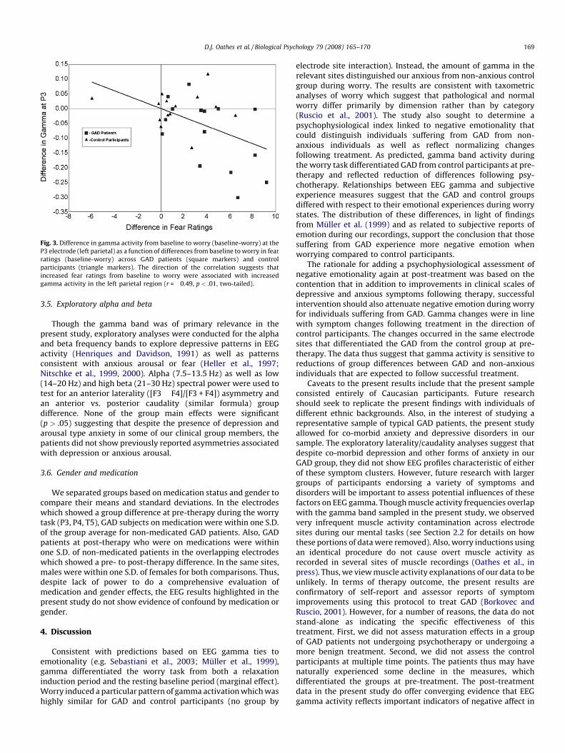
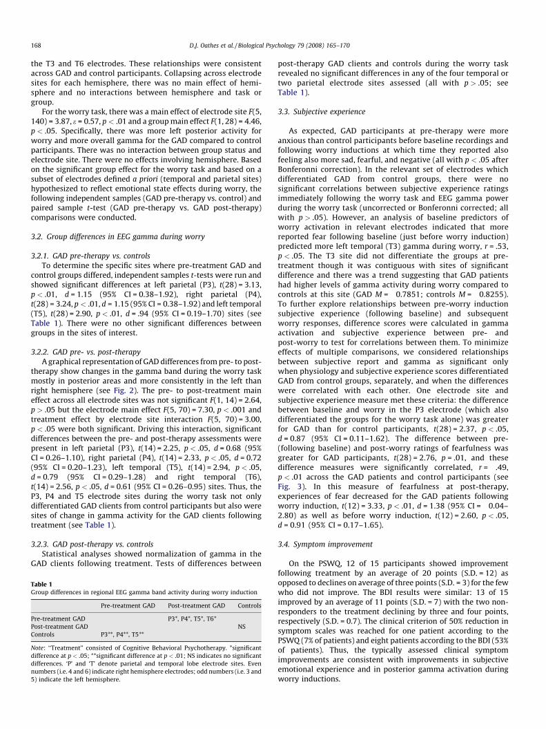
Fig. 3. Difference in gamma activity from baseline to worry (baseline-worry) at the
P3 electrode (left parietal) as a function of differences from baseline to worry in fear
ratings (baseline-worry) across GAD patients (square markers) and control
participants (triangle markers). The direction of the correlation suggests that
increased fear ratings from baseline to worry were associated with increased
gamma activity in the left parietal region (r = �0.49, p < .01, two-tailed).
D.J. Oathes et al. / Biological Psychology 79 (2008) 165–170 169
3.5. Exploratory alpha and beta
Though the gamma band was of primary relevance in thepresent study, exploratory analyses were conducted for the alphaand beta frequency bands to explore depressive patterns in EEGactivity (Henriques and Davidson, 1991) as well as patternsconsistent with anxious arousal or fear (Heller et al., 1997;Nitschke et al., 1999, 2000). Alpha (7.5–13.5 Hz) as well as low(14–20 Hz) and high beta (21–30 Hz) spectral power were used totest for an anterior laterality ([F3 � F4]/[F3 + F4]) asymmetry andan anterior vs. posterior caudality (similar formula) groupdifference. None of the group main effects were significant(p > .05) suggesting that despite the presence of depression andarousal type anxiety in some of our clinical group members, thepatients did not show previously reported asymmetries associatedwith depression or anxious arousal.
3.6. Gender and medication
We separated groups based on medication status and gender tocompare their means and standard deviations. In the electrodeswhich showed a group difference at pre-therapy during the worrytask (P3, P4, T5), GAD subjects on medication were within one S.D.of the group average for non-medicated GAD patients. Also, GADpatients at post-therapy who were on medications were withinone S.D. of non-medicated patients in the overlapping electrodeswhich showed a pre- to post-therapy difference. In the same sites,males were within one S.D. of females for both comparisons. Thus,despite lack of power to do a comprehensive evaluation ofmedication and gender effects, the EEG results highlighted in thepresent study do not show evidence of confound by medication orgender.
4. Discussion
Consistent with predictions based on EEG gamma ties toemotionality (e.g. Sebastiani et al., 2003; Muller et al., 1999),gamma differentiated the worry task from both a relaxationinduction period and the resting baseline period (marginal effect).Worry induced a particular pattern of gamma activation which washighly similar for GAD and control participants (no group by
electrode site interaction). Instead, the amount of gamma in therelevant sites distinguished our anxious from non-anxious controlgroup during worry. The results are consistent with taxometricanalyses of worry which suggest that pathological and normalworry differ primarily by dimension rather than by category(Ruscio et al., 2001). The study also sought to determine apsychophysiological index linked to negative emotionality thatcould distinguish individuals suffering from GAD from non-anxious individuals as well as reflect normalizing changesfollowing treatment. As predicted, gamma band activity duringthe worry task differentiated GAD from control participants at pre-therapy and reflected reduction of differences following psy-chotherapy. Relationships between EEG gamma and subjectiveexperience measures suggest that the GAD and control groupsdiffered with respect to their emotional experiences during worrystates. The distribution of these differences, in light of findingsfrom Muller et al. (1999) and as related to subjective reports ofemotion during our recordings, support the conclusion that thosesuffering from GAD experience more negative emotion whenworrying compared to control participants.
The rationale for adding a psychophysiological assessment ofnegative emotionality again at post-treatment was based on thecontention that in addition to improvements in clinical scales ofdepressive and anxious symptoms following therapy, successfulintervention should also attenuate negative emotion during worryfor individuals suffering from GAD. Gamma changes were in linewith symptom changes following treatment in the direction ofcontrol participants. The changes occurred in the same electrodesites that differentiated the GAD from the control group at pre-therapy. The data thus suggest that gamma activity is sensitive toreductions of group differences between GAD and non-anxiousindividuals that are expected to follow successful treatment.
Caveats to the present results include that the present sampleconsisted entirely of Caucasian participants. Future researchshould seek to replicate the present findings with individuals ofdifferent ethnic backgrounds. Also, in the interest of studying arepresentative sample of typical GAD patients, the present studyallowed for co-morbid anxiety and depressive disorders in oursample. The exploratory laterality/caudality analyses suggest thatdespite co-morbid depression and other forms of anxiety in ourGAD group, they did not show EEG profiles characteristic of eitherof these symptom clusters. However, future research with largergroups of participants endorsing a variety of symptoms anddisorders will be important to assess potential influences of thesefactors on EEG gamma. Though muscle activity frequencies overlapwith the gamma band sampled in the present study, we observedvery infrequent muscle activity contamination across electrodesites during our mental tasks (see Section 2.2 for details on howthese portions of data were removed). Also, worry inductions usingan identical procedure do not cause overt muscle activity asrecorded in several sites of muscle recordings (Oathes et al., inpress). Thus, we view muscle activity explanations of our data to beunlikely. In terms of therapy outcome, the present results areconfirmatory of self-report and assessor reports of symptomimprovements using this protocol to treat GAD (Borkovec andRuscio, 2001). However, for a number of reasons, the data do notstand-alone as indicating the specific effectiveness of thistreatment. First, we did not assess maturation effects in a groupof GAD patients not undergoing psychotherapy or undergoing amore benign treatment. Second, we did not assess the controlparticipants at multiple time points. The patients thus may havenaturally experienced some decline in the measures, whichdifferentiated the groups at pre-treatment. The post-treatmentdata in the present study do offer converging evidence that EEGgamma activity reflects important indicators of negative affect in
D.J. Oathes et al. / Biological Psychology 79 (2008) 165–170170
GAD patients during worry by the fact that gamma is sensitive tofluctuations in these experiences. It is expected that changes suchas this would follow from successful treatment. However, thecausal link between these changes and the treatment must befurther established. The changes from pre- to post-therapy in theregion of the T6 electrode (see Fig. 2) were unexpected especially inlight of there being no group difference in this site at the pre-treatment assessment. It may be the case that GAD patientslearned to dampen emotional experiences during worry in a globalway, as suggested by the bilateral changes in posterior parietalgamma. Future research evaluating changes in the experience ofworry and evaluating ways in which GAD patients have learned tocope with the emotional repercussions of worry might suggest anexplanation for these findings. Since worry induction was the onlyemotional task assessed in the present study, it is unclear whethersimilar group and treatment differences might have also beenfound using other emotion inducing manipulations (e.g. passivelyviewing affect laden stimuli). Future research with a wider varietyof tasks will be useful to highlight the specificity of the presentfindings to the worry experience.
These results support continued research to link findings inaffective neuroscience, psychopathology, therapy outcome mon-itoring and studies of emotional processing. Worry inductioneffects on subjective emotional experience reports differentiatedanxious from non-psychiatric participants as well as characterizedthe pre- to post-treatment differences in our anxious participants.Consistent with interpretations that worry caused the subjectiveand physiological sequelae of negative emotion, path analysissupports a stronger influence of worry on anxious feelings ratherthan the reverse direction (Gana et al., 2000). The present findingssupport the importance of physiological data, and especially EEGgamma power, for studying affective symptoms in psychiatricpatients and also to monitor fluctuations in affective symptomsexpected to follow successful treatment. Analyses relatingphysiological measures to prototypical assessor and self-reportsymptom measures of anxiety and depression are planned for thefuture.
Acknowledgements
The authors wish to thank Andreas Keil for helpful comments aswell as Alison Staples for editing assistance.
References
American Psychiatric Association, 2000. Diagnostic and Statistical Manual of MentalDisorders, 4th ed., Washington, DC (text revision).
Andrews, V.H., Borkovec, T.D., 1998. The differential effects of induction of worry,somatic anxiety, and depression on emotional experience. Journal of BehaviorTherapy and Experimental Psychiatry 19, 21–26.
Beck, A.T., Steer, R.A., Ball, R., Ranieri, W.F., 1996. Comparison of Beck DepressionInventories-IA and -II in psychiatric outpatients. Journal of Personality Assess-ment 67, 588–597.
Borkovec, T.D., Hu, S., 1990. The effect of worry on cardiovascular response tophobic imagery. Behaviour Research and Therapy 28, 69–73.
Borkovec, T.D., Inz, J., 1990. The nature of worry in generalized anxiety disorder: apredominance of thought activity. Behaviour Research and Therapy 28, 153–158.
Borkovec, T.D., Lyonfields, J.D., Wiser, S.L., Deihl, L., 1993. Behaviour Research andTherapy 31, 321–324.
Borkovec, T.D., Ruscio, A., 2001. Psychotherapy for generalized anxiety disorder.Journal of Clinical Psychology 62, 37–45.
Borkovec, T.D., Sharpless, B., 2004. Generalized anxiety disorder: bringing cognitivebehavioral therapy into the valued present. In: Hayes, S., Follette, V., Linehan,M. (Eds.), New Directions in Behavior Therapy. Guilford Press, New York, pp.209–242.
Cohen, J., 1998. Statistical Power Analyses for the Behavioral Sciences, 2nd ed.Erlbaum, Hillsdale, NJ.
Collins, D.R., Pelletier, J.G., Pare, D., 2001. Slow and fast (gamma) neuronal oscilla-tions in the perirhinal cortex and lateral amygdala. Journal of Neurophysiology85, 1661–1672.
DiNardo, P.A., Brown, T.A., Barlow, D.H., 1994. Anxiety Disorders Interview Schedulefor DSM-IV: Clinician’s Manual. Graywind, New York.
Gana, K., Martin, B., Canouet, M.D., 2000. Worry and anxiety: Is there a causalrelationship? Psychopathology 34, 221–229.
Gemignani, A., Santarcangelo, E., Sebastiani, L., Marchese, C., Mammoliti, R., Simoni,A., Ghelarducci, B., 2000. Changes in autonomic and EEG patterns induced byhypnotic imagination of aversive stimuli in man. Brain Research Bulletin 53,105–111.
Hedges, L., Olkin, I., 1985. Statistical Methods for Meta-analysis. Academic Press,New York.
Heller, W., Nitschke, J.B., Etienne, M.A., Miller, G.A., 1997. Patterns of regional brainactivity differentiate types of anxiety. Journal of Abnormal Psychology 106,376–385.
Henriques, J.B., Davidson, R.J., 1991. Left frontal hypoactivation in depression.Journal of Abnormal Psychology 100, 535–545.
Jasper, H.H., 1958. The ten–twenty electrode system of the international federation.Electroencephalography and Clinical Neurophysiology 10, 371–375.
Jensen, O., Kaiser, J., Lachaux, J.P., 2007. Human gamma-frequency oscillationsassociated with attention and memory. Trends in Neurosciences 30, 317–324.
Kline, R.B., 2004. Beyond Significant Testing: Reforming Data Analysis Methods inBehavioral Research. American Psychological Association, Washington, DC.
Luo, Q., Holroyd, T., Jones, M., Hendler, T., Blair, J., 2007. Neural dynamics for facialthreat processing as revealed by gamma band synchronization using MEG.Neuroimage 34, 839–847.
Matsumoto, A., Ichikawa, Y., Kanayama, N., Ohira, H., Iidaka, T., 2006. Gamma bandactivity and its synchronization reflect the dysfunctional emotional processingin alexithymic persons. Psychophysiology 43, 533–540.
Meyer, T.J., Miller, M.L., Metzger, R.L., Borkovec, T.D., 1990. Development andvalidation of the Penn State Worry Questionnaire. Behaviour Research andTherapy 28, 487–495.
Muller, M.M., Keil, A., Gruber, T., Elbert, T., 1999. Processing of affective picturesmodulates right-hemisphere gamma band activity. Clinical Neurophysiology110, 1913–1920.
Newman, M.G., Castonguay, L.G., Borkovec, T.D., Molnar, C., 2004. Integrativepsychotherapy. In: Heimberg, R.G., Turk, C.L., Mennin, D.S. (Eds.), Genera-lized Anxiety Disorder. Guilford Press, New York, pp. 320–350.
Nitschke, J.B., Heller, W., Miller, G.A., 2000. Anxiety, stress, and cortical brainfunction. In: Borod, J.C. (Ed.), The Neuropsychology of Emotion. Oxford Uni-versity Press, New York, pp. 298–319.
Nitschke, J.B., Heller, W., Palmieri, P., Miller, G.A., 1999. Contrasting patterns of brainactivity in anxious apprehension and anxious arousal. Psychophysiology 36,628–637.
Nitschke, J.B., Larson, C.L., Smoller, M.J., Navin, S.D., Pederson, A.J.C., Ruffalo, D.,et al., 2002. Startle potentiation in aversive anticipation: evidence for state butnot trait effects. Psychophysiology 39, 254–258.
Oathes, D.J., Bruce, J.M., Nitschke, J.B., in press. Worry facilitates corticospinal motorresponse to transcranial magnetic stimulation. Depression and Anxiety.
Osgood, C.E., Suci, G.J., Tannenbaum, P.H., 1957. The Measurement of Meaning.University of Illinois Press, Urbana.
Oya, H., Kawasaki, H., Howard, M.A., Adolphs, R., 2002. Electrophysiologicalresponses in the human amygdala discriminate emotion categories of complexvisual stimuli. Journal of Neuroscience 22, 9502–9512.
Ruscio, A.M., Borkovec, T.D., Ruscio, J., 2001. A taxometric investigation of the latentstructure of worry. Journal of Abnormal Psychology 110, 413–422.
Sebastiani, L., Simoni, A., Gemignani, A., Ghelarducci, B., Santarcangelo, E.L., 2003.Human hypnosis: autonomic and electroencephalographic correlates of aguided multimodal cognitive-emotional imagery. Neuroscience Letters 338,41–44.
Related Documents