World Journal of Gastrointestinal Pathophysiology World J Gastrointest Pathophysiol 2014 August 15; 5(3): 122-379 ISSN 2150-5330 (online) Published by Baishideng Publishing Group Inc

Welcome message from author
This document is posted to help you gain knowledge. Please leave a comment to let me know what you think about it! Share it to your friends and learn new things together.
Transcript

World Journal of Gastrointestinal PathophysiologyWorld J Gastrointest Pathophysiol 2014 August 15; 5(3): 122-379
ISSN 2150-5330 (online)
Published by Baishideng Publishing Group Inc

EDITOR-IN-CHIEFThomas Y Ma, Albuquerque
STRATEGY ASSOCIATE EDITOR-IN-CHIEFHirotada Akiho, FukuokaJean-Francois Beaulieu, SherbrookeMichael W Bradbury, ErieSharon DeMorrow, Temple
GUEST EDITORIAL BOARD MEMBERSJia-Ming Chang, TaipeiWai-Keung Chow, TaichungChien-Wei Hsu, KaohsiungMing-Tsan Lin, TaipeiBor-Shyang Sheu, TainanJin-Town Wang, Taipei
MEMBERS OF THE EDITORIAL BOARD
ArgentinaBernabé Matías Quesada, Buenos AiresMarcelo G Roma, Rosario
AustraliaChris Richard Abbiss, JoondalupGuy D Eslick, PenrithMontri Gururatsakul, AdelaideChandana Herath, Melbourne Michael Horowitz, AdelaidMustafa Khasraw, GeelongShu-Chuen Li, CallaghanAntonina Mikocka-Walus, AdelaideNam Quoc Nguyen, Adelaide
Kulmira Nurgali, St AlbansNicholas John Spencer, Flagstaff HillNick Spencer, AdelaideDeborah Verran, CamperdownShu-Feng Zhou, Melbourne
AustriaCord Langner, GrazDietmar Ofner-Velano, SalzburgMichael Trauner, Graz
Belgium
Kathleen Blondeau, LeuvenRobaeys Geert, GenkIlse Maria Hoffman, LeuvenMichael H J Maes, WilrijkTheodoor Abram Niewold, HeverleeXavier Sagaert, LeuvenJean-Marie Vanderwinden, BrusselsKristin Verbeke, LeuvenMathieu Vinken, Roeselare
BrazilUilian Andreis, BotucatuEverson L A Artifon, Vila MarianaJoão Batista Calixto, TrindadeNiels O Saraiva Câmara, Vila ClementinoJulio Chebli, Juiz de ForaFernando Fornari, Passo FundoClélia Akiko Hiruma-Lima, BotucatuMarcel C C Machado, Sao PauloJuarez Quaresma, BelemWagner Vilegas, Araraquara
Brunei Darussalam
Vui Heng Chong, Bandar Seri Begawan
Canada
Fernando Alvarez, MontréalFrancois Boudreau, SherbrookeGeorge A Bubenik, GuelphWang-Xue Chen, OttawaJan D Huizinga, PuslinchKusum K Kharbanda, OmahaWolfgang Kunze, HamiltoJian-Jun Li, OttawaRoderick John Macleod, KingstonMichele Molinari, HalifaxNathalie Rivard, SherbrookeKirill Rosen, HalifaxManuela Santos, MontrealCaroline Saucier, QuebecJean Sévigny, QuebecEldon A Shaffer, CalgaryManuel A Silva, HamiltonAlan B R Thomson, EdmontonPierre H Vachon, Sherbrooke
China
Kai-Xing Ai, ShanghaiZhao-Xiang Bian, Hong KongMin-Hu Chen, GuangzhouCH Cho, Hong KongZhong-Hong Gao, WuhanJun-Ming Guo, NingboJing-Yan Han, Beijing
I
Editorial Board2011-2015
The World Journal of Gastrointestinal Pathophysiology Editorial Board consists of 523 members, representing a team of worldwide experts in gastrointestinal pathophysiology. They are from 45 countries, including Argentina (2), Australia (14), Austria (3), Belgium (9), Brazil (10), Brunei Darussalam (1), Canada (20), China (30), Croatia (1), Czech Republic (2), Denmark (4), Egypt (1), Estonia (1), Finland (1), France (8), Germany (22), Greece (7), Hungary (5), India (10), Indonesia (1), Iran (2), Ireland (2), Israel (8), Italy (42), Japan (47), Lebanon (3), Malaysia (1), Mexico (2), Netherlands (8), Norway (1), Poland (4), Portugal (1), Romania (1), Russia (1), Singapore (4), South Korea (13), Spain (23), Sweden (11), Switzerland (4), Thailand (2), Turkey (6), Ukraine (1), United Kingdom (10), United States (173), and Venezuela (1).
February 15, 2013WJGP|www.wjgnet.com
World Journal ofGastrointestinal PathophysiologyW J G P
Jian-Dong Huang, Hong KongJia-Fu Ji, BeijingShi Liu, WuhanZhan-Ju Liu, ShanghaiXiao-Hong Wang, BeijingZhen-Ning Wang, ShenyangWei Wei, HefeiDong-Ping Xie, ShanghaiWen-Xie Xu, ShanghaiHua Yang, ChongqingXiao Yang, BeijingWei-Zhen Zhang, BeijingHua-Chuan Zheng, ShenyangDa-Ling Zhu, HarbinJin-Xia Zhu, BeijingMin-Sheng Zhu, NanjingYong-Liang Zhu, Hangzhou
Croatia
Alen Protic, Rijeka
Czech Republic
Pavel Hladik, SemilyMartin Vokurka, Prague
Denmark
Lars Arendt-Nielsen, AalborgFrank Vinholt Schiodt, CopenhagenJonas Worsoe, AarhusJing-Bo Zhao, Aalborg
Egypt
Mahmoud Aboelneen Khattab, Minia
Estonia
Enn Seppet, Tartu
Finland
Pauli Antero Puolakkainen, Turku
France
Bruno Bonaz, GrenoblePierre Marie Dechelotte, RouenJean-Paul Lallès, Saint-GillesCharles-Henri Malbert, Saint-GillesThierry Piche, NicePascale Plaisancié, LyonMichelina Plateroti, LyonVeronique Vitton, Marseille
Germany
Hans Gunter Beger, UlmCarsten Bergmann, IngelheimElke Cario, Essen
Arno J Dormann, KolnNikolaus Gassler, AachenWerner Hartwig, HeidelbergMarion Hewicker-Trautwein, HannoverJens Hoeppner, FreiburgTobias Keck, FreiburgJorg Kleeff, MunichPeter Malfertheiner, MagdeburgOliver Mann, HamburgChristoph Michalski, MunichAndreas Klaus Nussler, MunichChristian Pehl, VilsbiburgPeter Schemmer, HeidelbergMarc Stemmler, FreiburgFrank Tacke, AachenSya Nomna Ukena, HannoverBrigitte Vollmar, RostockThomas Michael Wex, MagdeburgMargot Zoller, Heidelberg
Greece
Stelios F Assimakopoulos, PatrasGeorge N Dalekos, LarissaAlkiviadis Efthymiou, thessalonikiMaria Gazouli, AthensIoannis E Koutroubakis, HeraklionGerassimos J Mantzaris, AthensGeorge Papatheodoridis, Athens
Hungary
Mária Bagyánszki, SzegedMihály Boros, SzegedLaszlo Czako, SzegedPal Miheller, BudapestZoltan Rakonczay, Szeged
India
Anil Kumar Agarwal, DelhiUday Bandyopadhyay, KolkataSriparna Basu, VaranasiChandra Kanti Chakraborti, RourkelaRajeev Garg, PunjabChandra P Sharma, ThiruvananthapuramShailesh V Shrikhande, MumbaiVirendra Singh, ChandigarhNicholas James Skill, IndianapolisPrabhakar R Veerareddy, Andhra Pradesh
Indonesia
Laurentius A Lesmana, Jakarta
Iran
Gholamreza Roshandel, GorganShahram Shahabi, Urmia
Ireland
Billy Bourke, DublinStephen Keely, Dublin
IsraelYosefa Avraham, JerusalemYaron Bar-Dayan, HolonShomron Ben-Horin, HashomerBoris Kirshtein, Beer ShevaStephen Malnick, RehovotYaakov Maor, Tel-HashomerRifaat Safadi, JerusalemNachum Vaisman, Tel Aviv
Italy
Rosaria Acquaviva, CataniaDario Acuna-Castroviejo, ArmillaAlessandro Antonelli, PisaGiacosa Attilio, GenovaSalvatore Auricchio, NaplesGuido Basilisco, MilanoAntonio Basoli, RomeClaudio Bassi, VeronaMassimo Bellini, PisaLuigi Bonavina, MilanoAlfio Brogna, CataniaGiuseppe Calamita, BariRaffaele Capasso, NaplesIgnazio Castagliuolo, PadovaEnrico Stefano Corazziari, RomeFrancesco Cresi, TorinoRosario Cuomo, NapoliSalvatore Cuzzocrea, GazziMario M D’Elios, FlorenceCinzia Domeneghini, MilanLuca Elli, MilanoCresi Francesco, TorinoWalter Fries, MessinaEugenio Gaudio, RomeMarco Gobbetti, BariFabio Grizzi, MilanEnzo Grossi, MilaneseEnzo Ierardi, FoggiaPietro Invernizzi, MilanAngelo A Izzo, NaplesAnna Kohn, RomeGiovanni Latella, L’AquilaMassimo Marignani, RomeSergio Morini, RomeRaffaele Pezzilli, BolognaCristiano Rumio, MilanGiovanni Sarnelli, NaplesEdoardo Vincenzo Savarino, GenoaPierpaolo Sileri, RomeAnnamaria Staiano, NaplesGiacomo Carlo Sturniolo, PadovaClaudio Tiribelli, Triest
Japan
Akihiro Asakawa, KagoshimaHisashi Aso, SendaiYasu-Taka Azuma, OsakaShotaro Enomoto, WakayamaMikihiro Fujiya, HokkaidoTakahisa Furuta, HamamatsuAkira Hokama, OkinawaRyota Hokari, SaitamaYuichi Hori, Kobe
II February 15, 2013WJGP|www.wjgnet.com
III February 15, 2013WJGP|www.wjgnet.com
Hideki Iijima, OsakaMasahiro Iizuka, AkitaMotohiro Imano, OsakaHajime Isomoto, NagasakiTatehiro Kagawa, IseharaTakumi Kawaguchi, KurumeHaruki Kitazawa, SendaiXiao-Kang Li, TokyoNoriaki Manabe, OkayamaAtsushi Masamune, SendaiHiroyuki Matsubayashi, ShizuokaKazuyuki Matsushita, Chuo-kuReiko Miyazawa, GunmaKazunari Murakami, OitaHikaru Nagahara, TokyoYuji Naito, KyotoAtsushi Nakajima, Atsushi NakajimaShoji Natsugoe, KagoshimaTsutomu Nishida, OsakaKoji Nomoto, TokyoNaoaki Sakata, MiyagiShouji Shimoyama, TokyoGoshi Shiota, YonagoIkuo Shoji, HyogoHidekazu Suzuki, TokyoHitoshi Takagi, GunmaToru Takahashi, OkayamaYoshihisa Takahashi, TokyoKan Uchiyama, ChibaTakato Ueno, KurumeYoshiyuki Ueno, SendaiHisayuki Uneyama, KwasakiMitsunori Yamakawa, YamagataTakayuki Yamamoto, MieYutaka Yata, GunmaNaohisa Yoshida, KyotoHitoshi Yoshiji, Nara
Lebanon
Costantine Fouad Daher, ByblosAssaad M Soweid, BeirutJulnar Usta, Beirut
Malaysia
Andrew Chua, Perak
Mexico
José María de la Roca-Chiapas, LeonMaria Raquel Huerta Franco, Guanajuato
Netherland
Wouter J de Jonge, AmsterdamAldo Grefhorst, GroningenRuben Hummelen, RotterdamDaniel Keszthelyi, MaastrichtCornelis F M Sier, LeidenPieter J Tanis, AmsterdamLuc JW van der Laan, RotterdamSander van der Marel, Leiden
NorwayAnne Marie Bakke, Oslo
Poland
Stanisław Hac, GdańskStanisław Jan Konturek, KrakówAgata Mulak, WroclawNapoleon Waszkiewicz, Choroszcz
Portugal
Ricardo Marcos, Porto
Romania
Mihai Ciocirlan, Bucharest
Russia
Ludmila Filaretova, Petersburg
Singapore
Madhav Bhatia, SingaporeBrian K P Goh, SingaporeKhek Yu Ho, SingaporeCliff K S Ong, Singapore
South Korea
Jae Hee Cheon, SeoulMyung Haing Cho, SeoulJae Bock Chung, SeoulKi-Baik Hahm, IncheonHo Jae Han, GwangjuChang Duk Jun, GwangjuHong Joo Kim, SeoulJin Kyung Kim, Gyeongsan-SiSang Geon Kim, SeoulWon Jae Lee, SeoulKwan Kyu Park, DaeguSeung Ha Park, BusanSung Joo Park, Jeonbuk
Spain
Raquel Abalo, AlcorcónJuan G Abraldes, BarcelonaAgustin Albillos, MadridMaria-Angeles Aller, MadridFernando Azpiroz, BarcelonaRamon Bataller, BarcelonaMarco Bustamante, ValenciaAndres Cardenas, BarcelonaDariao Acuna Castroviejo, ArmillaJoan Claria, BarcelonaPere Clave, BarcelonaManuel Giner, Madrid
Angel I Lanas, ZaragozaMaite Martin, BarcelonaMaria Teresa Martin, BarcelonaVicente Martinez, BarcelonaJose M Matés, MalagaJulio M Mayol, MadridMarçal Pastor-Anglada, BarcelonaMaría Eugenia Sáez, SevilleYolanda Sanz, BurjassotCarlos Taxonera, MadridMaria D Yago, Granada
Sweden
Marco Del Chiaro, StockholmFrida Fak, GothenburgGunnar FA Flemstrom, UppsalaEvangelos Kalaitzakis, GothenburgKristina Lamas, UmeaBob Roger Olsson, GöteborgSara Maria Regnér, MalmöPeter thelin Schmidt, StockholmXiao-Feng Sun, LinkopingHenrik Thorlacius, MalmöCurt Tysk, Orebro
Switzerland
Jyrki J Eloranta, ZurichAndreas Geier, ZurichRemy Meier, LiestalCatherine Pastor, Geneva
Thailand
Thawatchai Akaraviputh, BangkokWeekitt Kittisupamongkol, Bangkok
Turkey
Mehmet Bektas, AnkaraMukaddes Esrefoglu, MalatyaAhmet Guven, AnkaraMuammer Karadeniz, ManisaElvan Ozbek, ErzuruIlhami Yuksel, Ankara
Ukraine
Oksana S Zayavhkivska, Lviv
United Kingdom
Geoffrey Burnstock, LondonJanice E Drew, AberdeenGirish Gupte, BirminghamDavid C Hay, EdinburghNusrat Husain, CheshireMichael Leslie Lucas, GlasgowJamie Murphy, LondonVadim Sumbayev, KentWing-Kin Syn, Birmingham
IV February 15, 2013WJGP|www.wjgnet.com
Andrea Varro, Liverpool
United States
Sami Rene Achem, JacksonvilleTauseef Ali, OklahomaDavid H Alpers, St LouisGianfranco D Alpini, TempleShrikant Anant, OklahomaM Sawkat Anwer, North GraftonAndrew Aronsohn, ChicagoToms Augustin, SayreGyorgy Baffy, BostonMichael T Bailey, ColumbusKim Elaine Barrett, San DiegoMarc D Basson, LansingRobert L Bell, New HavenDavid H Berger, HoustonUrs A Boelsterli, StorrsRichard G Boles, Los AngelesEdward L Bradley III, SarasotaQiang Cai, AtlantaWei-Biao Cao, ProvidenceSubhash C Chauhan, Sioux FallsJian-De Chen, GalvestonTao-Sheng Chen, MemphisJohn Chiang, RootstownMashkoor A Choudhry, MaywoodParimal Chowdhury, Little RockEric Cohen, BostonRobert Cormier, DuluthSrinivasan Dasarathy, ClevelandEdwin A Deitch, NewarkDan A Dixon, ColumbiaJames P Dolan, PortlandH Henry Dong, PittsburghHui Dong, La JollaAshkan Farhadi, IrvineBin Feng, PittsburghJenifer Fenton, East LansingAlessandro Fichera, ChicagoMitchell P Fink, PittsburghP Marco Fisichella, MaywoodLeo R Fitzpatrick, HummelstownRobert Armour Forse, OmahaGlenn Tsuyoshi Furuta, AuroraJuan F Gallegos-Orozco, ScottsdalePandu R Gangula, NasvhilleTimothy Gardner, LebanonShannon Stroud Glaser, TempleFrancisco Gondim, St. LouisJohn R Grider, RichmondYan-Fang Guan, CincinnatiGregory M Holmes, Baton RougeAi-Xuan Le Holterman, ChicagoRichard Hu, Los AngelesHartmut Jaeschke, KansasRobert Thomas Jensen, Los AngelesSreenivasa S Jonnalagadda, LouisMichel Kahaleh, Charlottesville
Andreas Martin Kaiser, Los AngelesRandeep Singh Kashyap, RochesterLaurie Keefer, ChicagoRichard Kellermayer, HoustonChris Kevil, ShreveportSandeep Khurana, BaltimorePawel R Kiela, TucsonTammy Lyn Kindel, CincinnatGordana Kosutic, DurhamDavid Kravetz, San DiegoAshok Kumar, DetroitJohn H Kwon, ChicagoMuriel Larauche, Los AngelesI Michael Leitman, New YorkFelix W Leung, North HillsSuthat Liangpunsakul, IndianapolisFeng-Xin Lu, BostonPauline Kay Lund, Chapel HillGeorge Luo, LexingtonGuang-Xiang Luo, LexingtonJay Luther, Ann ArborRam I Mahato, MemphisAkhil Maheshwari, BirminghamKenneth Maiese, NewarkAdhip P N Majumdar, DetroitJose E Manautou, StorrsCraig J McClain, LouisvilleDermot McGovern, Los AngelesB Greenwood-van Meerveld, OklahomaDouglas Scott Merrel, BethesdaMurielle Mimeault, OmahaEmiko Mizoguchi, BostonHuan-Biao Mo, DentonAdam Moeser, RaleighRamzi M Mohammad, DetroitSatdarshan Singh Monga, PittsburghRoger Klein Moreira, New YorkSandeep Mukherjee, OmahaKarnam S Murthy, RichmondMichael J Nowicki, JacksonShuji Ogino, BostonMary Francis Otterson, WisconsinChung Owyang, Ann ArborHelieh S Oz, LexingtonMarco G Patti, ChicagoTimothy Michael Pawlik, BaltimoreSara Peleg, HoustonNicholas C Popescu, BethesdaLi-Ya Qiao, RichmondChao Qin, OklahomaParvaneh Rafiee, MilwaukeeSigrid A Rajasekaran, WilmingtonVazhaikkurichi M Rajendran, MorgantownJean Pierre Raufman, BaltimoreRamesh M Ray, MemphisArie Regev, IndianapolisDouglas K Rex, CarmelYehuda Ringel, Chapel HillRichard A Rippe, RockvilleChantal A Rivera, Bossier
Andrea Romani, ClevelandPraveen K Roy, AlbuquerquePaul A Rufo, BostonDavid B Sachar, New YorkBimaljit Singh Sandhu, RichmondSanjaya Kumar Satapathy, New Hyde ParkAnthony Senagore, Los AngelesMuhammad Y Sheikh, FresnoBo Shen, ClevelandLe Shen, ChicagoFrank A Simmen, Little RockSteven Mitchell Singer, WashingtonShailinder Jit Singh, WashingtonAdam Jan Smolka, CharlestonNed Snyder, HoustonZhen-Yuan Song, ChicagoGagan K Sood, HoustonRhonda F Souza, DallasStuart Jon Spechler, DallasSubbaramiah Sridha, AugustaCatia Sternini, Los AngelesVeedamali S Subramanian, Long BeachJun Sun, RochesterYvette Taché, Los AngelesXiao-Di Tan, ChicagoPaul Daniel Terry, AtlantaJennifer Tirnauer, FarmingtonAndrea Todisco, Ann ArborGeorge C Tsokos, BostonVic Velanovich, DetroitRaj Vuppalanchi, IndianapolisEstela Wajcberg, CranfordArnold Wald, MadisonLi-Xin Wang, Los AngelesHorst Christian Weber, BostonSteven D Wexner, WestonJackie D Wood, ColumbusGuo-Yao Wu, College StationChristian Wunder, BethesdaZuo-Liang Xiao, ClevelandGuang-Yin Xu, GalvestonGuo-Rong Xu, East OrangeGuang-Yu Yang, ChicagoJay A Yelon, ValhallaYamaoka Yoshio, HoustonShao-Yong Yu, HersheyYana Zavros, CincinnatiJoerg Zehetner, Los AngelesJian X Zhang, CharlotteZhi Zhong, CharlestonHui-Ping Zhou, RichmondZhan-Xiang Zhou, KannapolisQing Zhu, BethesdaYao-Hui Zhu, Stanford
Venezuela
Fabian Michelangeli, Caracas
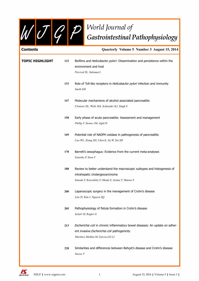
Contents
August 15, 2014|Volume 5|Issue 3|WJGP|www.wjgnet.com I
Quarterly Volume 5 Number 3 August 15, 2014
122 BiofilmsandHelicobacterpylori :Disseminationandpersistencewithinthe
environmentandhost
Percival SL, Suleman L
133 RoleofToll-likereceptorsinHelicobacterpylori infectionandimmunity
Smith SM
147 Molecularmechanismsofalcoholassociatedpancreatitis
Clemens DL, Wells MA, Schneider KJ, Singh S
158 Earlyphaseofacutepancreatitis:Assessmentandmanagement
Phillip V, Steiner JM, Algül H
169 PotentialroleofNADPHoxidaseinpathogenesisofpancreatitis
Cao WL, Xiang XH, Chen K, Xu W, Xia SH
178 Barrett’soesophagus:Evidencefromthecurrentmeta-analyses
Gatenby P, Soon Y
188 Reviewtobetterunderstandthemacroscopicsubtypesandhistogenesisof
intrahepaticcholangiocarcinoma
Sanada Y, Kawashita Y, Okada S, Azuma T, Matsuo S
200 LaparoscopicsurgeryinthemanagementofCrohn’sdisease
Lim JY, Kim J, Nguyen SQ
205 PathophysiologyoffistulaformationinCrohn’sdisease
Scharl M, Rogler G
213 Escherichiacoli inchronicinflammatoryboweldiseases:Anupdateonadher-
entinvasiveEscherichiacoli pathogenicity
Martinez-Medina M, Garcia-Gil LJ
228 SimilaritiesanddifferencesbetweenBehçet’sdiseaseandCrohn’sdisease
Yazısız V
TOPIC HIGHLIGHT
ContentsWorld Journal of Gastrointestinal Pathophysiology
Volume 5 Number 3 August 15, 2014
MINIREVIEWS
August 15, 2014|Volume 5|Issue 3|WJGP|www.wjgnet.com II
239 Multidisciplinaryandevidence-basedmanagementoffistulizingperianal
Crohn’sdisease
Sordo-Mejia R, Gaertner WB
252 Pancreatitis-imagingapproach
Busireddy KK, AlObaidy M, Ramalho M, Kalubowila J, Baodong L, Santagostino I,
Semelka RC
271 Newinsightstooccultgastrointestinalbleeding:Frompathophysiologyto
therapeutics
Sánchez-Capilla AD, De La Torre-Rubio P, Redondo-Cerezo E
284 Roleofhemostaticpowdersintheendoscopicmanagementof
gastrointestinalbleeding
Bustamante-Balén M, Plumé G
293 Predictorsofresponsetoanti-tumornecrosisfactortherapyinulcerative
colitis
Zampeli E, Gizis M, Siakavellas SI, Bamias G
304 Geneticupdateoninflammatoryfactorsinulcerativecolitis:Reviewofthe
currentliterature
Sarlos P, Kovesdi E, Magyari L, Banfai Z, Szabo A, Javorhazy A, Melegh B
322 Currentstatusofpredictivebiomarkersforneoadjuvanttherapyin
esophagealcancer
Uemura N, Kondo T
335 Epidemiologicalstudiesofesophagealcancerintheeraofgenome-wideas-
sociationstudies
Wang AH, Liu Y, Wang B, He YX, Fang YX, Yan YP
344 Perihilarcholangiocarcinoma:Currenttherapy
Zhang W, Yan LN
355 Helicobacterpylori asariskfactorforcentralserouschorioretinopathy:Lit-
eraturereview
Mateo-Montoya A, Mauget-Faÿse M
359 Riskofcardiovasculardiseaseininflammatoryboweldisease
Andersen NN, Jess T
REVIEW
ContentsWorld Journal of Gastrointestinal Pathophysiology
Volume 5 Number 3 August 15, 2014
RETROSPECTIVE STUDY
August 15, 2014|Volume 5|Issue 3|WJGP|www.wjgnet.com III
SYSTEMATIC REVIEWS
366 CancerstemcellsinHelicobacterpylori infectionandaging:Implicationsfor
gastriccarcinogenesis
Levi E, Sochacki P, Khoury N, Patel BB, Majumdar APN
373 Oxidativeandnitrosativestressenzymesinrelationtonitrotyrosinein
Helicobacterpylori -infectedhumans
Elfvin A, Edebo A, Hallersund P, Casselbrant A, Fändriks L
ContentsWorld Journal of Gastrointestinal Pathophysiology
Volume 5 Number 3 August 15, 2014
I-V Instructionstoauthors
EditorialBoardMemberofWorld Journal of Gastrointestinal Pathophysiology ,Cord Langner,MD,Senior Scientist, Institute of Pathology,MedicalUniversityGraz,Auenbruggerplatz25,A-8036Graz,Austria
World Journal of Gastrointestinal Pathophysiology (World J Gastrointest Pathophysiol, WJGP, online ISSN 2150-5330, DOI: 10.4291), is a peer-reviewed open access academic journal that aims to guide clinical practice and improve diagnostic and therapeutic skills of clinicians.
WJGP is to report rapidly the most recent results in basic and clinical research on gastrointestinal pathophysiology, including all aspects of normal or abnormal function of the gastrointestinal tract, hepatobiliary system, and pancreas. WJGP specifically covers growth and development, digestion, secretion, absorption, metabolism and motility relative to the gastrointestinal organs, as well as immune and inflammatory processes, and neural, endocrine and circulatory control mechanisms that affect these organs. This journal will also report new methods and techniques in gastrointestinal pathophysiological research. We encourage authors to submit their manuscripts to WJGP. We will give priority to manuscripts that are supported by major national and international foundations and those that are of great basic and clinical significance.
World Journal of Gastrointestinal Pathophysiology is now indexed in PubMed Central, PubMed, Digital Object Identifier, and Directory of Open Access Journals.
I-IV EditorialBoardFLYLEAF
APPENDIX
EDITORS FOR THIS ISSUE
Responsible Assistant Editor: Xiang Li Responsible Science Editor: Fang-Fang JiResponsible Electronic Editor: Ya-Jing Lu Proofing Editorial Office Director: Xiu-Xia Song Proofing Editor-in-Chief: Lian-Sheng Ma
NAMEOFJOURNALWorld Journal of Gastrointestinal Pathophysiology
ISSNISSN 2150-5330 (online)
LAUNCHDATEApril 15, 2010
FrequencyQuarterly
EDITOR-IN-CHIEFThomas Y Ma, MD, PhD, Professor, Chief, Division of Gastroenterology and Hepatology, University of New Mexico, MSC10 5550, 1 UNM, Albuquerque, NM 87131, United States
EDITORIALOFFICEJin-Lei Wang, DirectorXiu-Xia Song, Vice DirectorWorld Journal of Gastrointestinal Pathophysiology
Room 903, Building D, Ocean International Center, No. 62 Dongsihuan Zhonglu, Chaoyang District, Beijing 100025, ChinaTelephone: +86-10-85381891Fax: +86-10-85381893E-mail: [email protected] Desk: http://www.wjgnet.com/esps/helpdesk.aspxhttp://www.wjgnet.com
PUBLISHERBaishideng Publishing Group Inc8226 Regency Drive, Pleasanton, CA 94588, USATelephone: +1-925-223-8242Fax: +1-925-223-8243E-mail: [email protected] Desk: http://www.wjgnet.com/esps/helpdesk.aspxhttp://www.wjgnet.com
PUBLICATIONDATEAugust 15, 2014
COPYRIGHT
© 2014 Baishideng Publishing Group Inc. Articles pub-lished by this Open Access journal are distributed under the terms of the Creative Commons Attribution Non-commercial License, which permits use, distribution, and reproduction in any medium, provided the original work is properly cited, the use is non commercial and is otherwise in compliance with the license.
SPECIALSTATEMENTAll articles published in journals owned by the Baishideng Publishing Group (BPG) represent the views and opinionsof their authors, and not the views, opinions or policies of the BPG, except where other-wise explicitly indicated.
INSTRUCTIONSTOAUTHORSFull instructions are available online at http://www.wjgnet.com/2218-6182/g_info_20100722172951.htm
ONLINESUBMISSIONhttp://www.wjgnet.com/esps/
ABOUT COVER
August 15, 2014|Volume 5|Issue 3|WJGP|www.wjgnet.com IV
AIM AND SCOPE
INDEXING/ABSTRACTING
TOPIC HIGHLIGHT
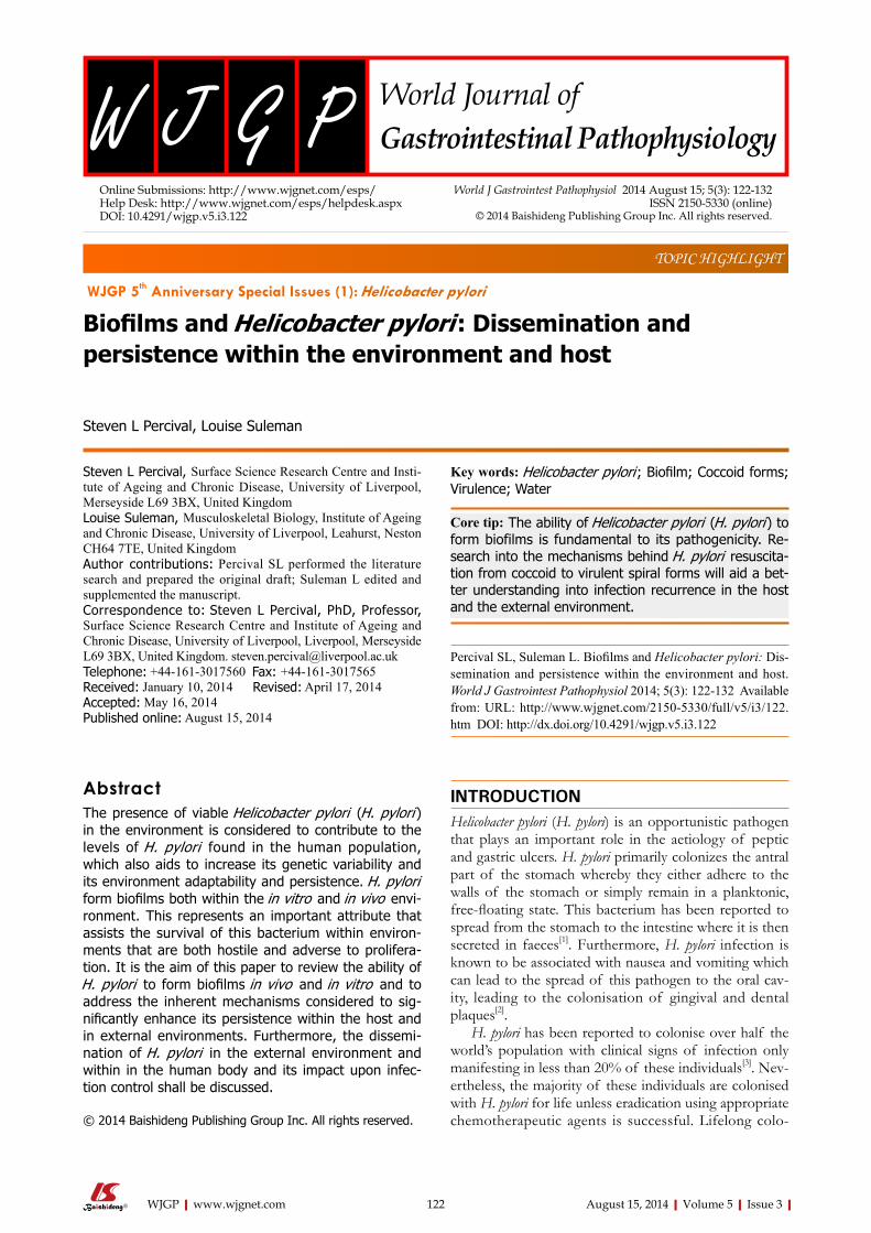
Biofilms and Helicobacter pylori : Dissemination and persistence within the environment and host
Steven L Percival, Louise Suleman
Steven L Percival, Surface Science Research Centre and Insti-tute of Ageing and Chronic Disease, University of Liverpool, Merseyside L69 3BX, United KingdomLouise Suleman, Musculoskeletal Biology, Institute of Ageing and Chronic Disease, University of Liverpool, Leahurst, Neston CH64 7TE, United KingdomAuthor contributions: Percival SL performed the literature search and prepared the original draft; Suleman L edited and supplemented the manuscript.Correspondence to: Steven L Percival, PhD, Professor, Surface Science Research Centre and Institute of Ageing and Chronic Disease, University of Liverpool, Liverpool, Merseyside L69 3BX, United Kingdom. [email protected] Telephone: +44-161-3017560 Fax: +44-161-3017565Received: January 10, 2014 Revised: April 17, 2014Accepted: May 16, 2014Published online: August 15, 2014
AbstractThe presence of viable Helicobacter pylori (H. pylori ) in the environment is considered to contribute to the levels of H. pylori found in the human population, which also aids to increase its genetic variability and its environment adaptability and persistence. H. pylori form biofilms both within the in vitro and in vivo envi-ronment. This represents an important attribute that assists the survival of this bacterium within environ-ments that are both hostile and adverse to prolifera-tion. It is the aim of this paper to review the ability of H. pylori to form biofilms in vivo and in vitro and to address the inherent mechanisms considered to sig-nificantly enhance its persistence within the host and in external environments. Furthermore, the dissemi-nation of H. pylori in the external environment and within in the human body and its impact upon infec-tion control shall be discussed.
© 2014 Baishideng Publishing Group Inc. All rights reserved.
Key words: Helicobacter pylori ; Biofilm; Coccoid forms; Virulence; Water
Core tip: The ability of Helicobacter pylori (H. pylori ) to form biofilms is fundamental to its pathogenicity. Re-search into the mechanisms behind H. pylori resuscita-tion from coccoid to virulent spiral forms will aid a bet-ter understanding into infection recurrence in the host and the external environment.
Percival SL, Suleman L. Biofilms and Helicobacter pylori: Dis-semination and persistence within the environment and host. World J Gastrointest Pathophysiol 2014; 5(3): 122-132 Available from: URL: http://www.wjgnet.com/2150-5330/full/v5/i3/122.htm DOI: http://dx.doi.org/10.4291/wjgp.v5.i3.122
INTRODUCTION Helicobacter pylori (H. pylori) is an opportunistic pathogen that plays an important role in the aetiology of peptic and gastric ulcers. H. pylori primarily colonizes the antral part of the stomach whereby they either adhere to the walls of the stomach or simply remain in a planktonic, free-floating state. This bacterium has been reported to spread from the stomach to the intestine where it is then secreted in faeces[1]. Furthermore, H. pylori infection is known to be associated with nausea and vomiting which can lead to the spread of this pathogen to the oral cav-ity, leading to the colonisation of gingival and dental plaques[2].
H. pylori has been reported to colonise over half the world’s population with clinical signs of infection only manifesting in less than 20% of these individuals[3]. Nev-ertheless, the majority of these individuals are colonised with H. pylori for life unless eradication using appropriate chemotherapeutic agents is successful. Lifelong colo-
World J Gastrointest Pathophysiol 2014 August 15; 5(3): 122-132ISSN 2150-5330 (online)
© 2014 Baishideng Publishing Group Inc. All rights reserved.
Online Submissions: http://www.wjgnet.com/esps/Help Desk: http://www.wjgnet.com/esps/helpdesk.aspxDOI: 10.4291/wjgp.v5.i3.122
August 15, 2014|Volume 5|Issue 3|WJGP|www.wjgnet.com 122
WJGP 5th Anniversary Special Issues (1): Helicobacter pylori
Percival SL et al . Biofilms and Helicobacter pylori
nisation seems to be due to the ability of some strains of H. pylori to both adapt to the host’s immunological responses and to also withstand the constantly changing gastric environment. In genetically predisposed individu-als, colonisation with H. pylori is reported to heighten the risk of developing cancer[4].
H. pylori can be described as Gram negative, spiral- (S-shape) or cocci-shaped bacteria. It has been reported to exist in three forms, the viable and culturable spiral form, the viable but non-culturable (VBNC) coccoid form, which are less virulent, and the non-viable de-generative H. pylori form[5]. It is their spiral shape that is thought to enhance their colonisation of the gastric mu-cosa. Whilst generally considered microaerophilic, there is now evidence that H. pylori can grow in humidified aerobic conditions[6].
The colonisation of H. pylori and its effect on resi-dent gastric microbiota is relatively unknown. A study by Bik et al[7] assessed the human gastric microbiota from 23 gastric biopsy samples using small subunit 16S rDNA clone library method and subsequently found that the presence of H. pylori had no effect on the microbial profile of the gut[7]. A recent study investigated the ef-fects of H. pylori on the gastric microbiota in a Rhesus macaque model. The authors found no significant im-pact upon the non-Helicobacter taxa after H. pylori chal-lenge[8]. However it appears that the microbial profile of the gut may have an effect on the degrees of patho-genicity of H. pylori. A germ-free gastric cancer mouse model showed less symptoms of disease and a later on-set of neoplasia upon H. pylori infection when compared to those mice with a typical gastric microbiota profile[9].
As an avid coloniser of the gastric mucosa H. pylori must possess a number of characteristics that include flagella, adhesions, urease production, and biofilm form-ing ability[10,11]. The importance of the biofilm forming potential of H. pylori is fundamental to its pathogenicity. The formation of a biofilm is a virulence mechanism that aids in the enhancement and longevity of H. pylori in “unfriendly” and hostile environments, such as in the human stomach and the natural environment.
H. pylori was first found to demonstrate an ability to form in vitro biofilms in the early and late 1990s with solid evidence of this ability reported by Stark et al[12] in 1999. More recent reports on the ability of H. pylori to form biofilms within in vitro[13,14] and in vivo environments, specifically the gastric mucosa, have now been demon-strated[14-16]. In particular the H. pylori strain TK1402 iso-lated from a patient with duodenal and gastric ulcers has been shown to have very strong biofilm forming ability both inside and outside the host[14,15,17-20]. In this mode of growth it is likely that H. pylori is protected from external perturbations[18,21].
Biofilms can develop on both biotic and abiotic sur-faces through the conversion of microorganisms in a free-floating or planktonic state, to a sessile state, where they become attached onto a surface. Once microorgan-isms attach onto a surface they proliferate, produce ex-
tracellular polymeric substance (EPS) and become firmly attached to that surface. The matrix of the biofilm is known to be composed of polysaccharides, extracellular DNA (eDNA), lipids and proteins that form the “house” of the biofilm[22,23]. It is the biofilm and the ability of microorganisms to form biofilms that form an essential element, aiding in their persistence, survivability, and recalcitrance to antimicrobial interventions and the hosts immune response. Furthermore the ability of pathogenic microbes to survive within diverse and hostile environ-ments is enhanced significantly when growing within a biofilm. Growth within a biofilm is known to cause and exacerbate infections and is responsible for prolonging infection, leading to chronicity[24].
A biofilm is dynamically and structurally complex and is often referred to as a “living organism” due to its ability to adapt to external perturbations. Of particular concern with biofilms of public health significance is the fact that sections of biofilms can easily detach or shear off, enabling these sections or individual bacteria to re-colonise other surfaces. Detachment or dissemination from the biofilm can be achieved by the dispersal of single cells or the detachment/shedding of large cellular aggregates. Both situations constitute a concern to pub-lic health particularly where fluid resides, as microbial dissemination is enhanced e.g., catheters, blood stream, drinking water[24]. Further to this there is growing evi-dence that within a biofilm the horizontal transfer of genes can occur, leading to large variations in H. pylori strains, particularly in one host, enhancing their survival and immune evasion. Moreover, gene transfer in situ has an important role to play in immunological effectiveness and eradication of pathogens by the host[25]. In addition to this it is well documented that when microorganisms are growing within a biofilm they have increased toler-ance to antimicrobial agents[26].
It is the aim of this paper to review the ability of H. pylori to form biofilms in vivo and in vitro and to address the inherent mechanisms considered to significantly enhance its persistence within the host and in external environments. Furthermore, the dissemination of H. pylori in the external environment and within in the hu-man body and its impact upon infection control shall be discussed.
TRANSMISSION OF H. PYLORIThe routes of transmission of H. pylori are said to occur via an array of different pathways[27,28]. Although H. pylori are considered to be pathogens commonly associated with the human stomach, Brown proposed that H. pylori are able to survive in environments that are external to that of the human stomach[29]. Dental plaque has also been reported to contain H. pylori; however, in plaque, H. pylori are thought to only exist in a transient state[30-32]. Young et al[2] reported that both the spiral and viable coccoid form of H. pylori are present in the oral cavity. Souto and Colombo found H. pylori in a subgingival bio-
August 15, 2014|Volume 5|Issue 3|WJGP|www.wjgnet.com 123
film in 11% of periodontally healthy patients compared to 50% of patients suffering from periodontitis[33]. The authors proposed that biofilm formation in the oral cav-ity should be considered as a potential reservoir for H. pylori.
There is building evidence to suggest that H. pylori may reside in potable water systems[11]. In general, wa-terborne bacteria can adhere to surfaces by aggregating matrix to form biofilms[26]. Information regarding the exact ecological niche where H. pylori reside and persist outside of the human host is limited. Despite this, there is growing evidence that external reservoirs of H. pylori may exist, potentially aiding transmission to the host. Furthermore there are also reports that H. pylori may have, as part of its life cycle, a zoonotic component. However, further scientific evidence of cultivability will be required to fully support this area.
The ability to form biofilms and the cell morphology and architecture formed depends greatly on the sup-port material. To date however, in potable water supplies there is not enough substantial evidence that H. pylori within the viable state, plays a role in the development of a biofilm. Despite this, there is significant evidence that, in terms of epidemiological evidence, the risk of acquiring H. pylori increases in individuals who drink well water and river water or swim in pools, streams and rivers in particular[27,34-37]. Consequently, environmental water is considered a risk for the acquisition of H. pylori and therefore H. pylori biofilms in these environments should be a very important consideration when investi-gating reservoirs of source. The association of H. pylori with biofilms in water distribution systems can offer bac-teria protection from disinfection and protozoan preda-tion[38]. The challenge however, remains to determine the importance of waterborne H. pylori. It may be possible that a specifically adapted form of H. pylori, or simple H. pylori within a biofilm, may be required for persistence and transmission[39].
Although based on scientific logic, if H. pylori is able to survive and persist outside of the human host, its ability to develop a biofilm and survive within a biofilm may well help to answer fundamental questions regard-ing acquisition and potential eradication, particularly in the developing world.
THE DETECTION OF H. PYLORI IN THE ENVIRONMENT AND HOSTThe ability of H. pylori to transform from a highly viru-lent, spiral shaped bacteria to a less virulent, non-cultur-able coccoid state, presents difficulties in the successful detection of this bacterium in both an environmental and clinical setting. In particular, the VBNC coccoid state is thought to arise under less favourable conditions, making the identification of H. pylori from water sources, particularly H. pylori within biofilms, unlikely using tradi-tional culture methods.
Molecular methods such as real-time polymerase
chain reaction (PCR) have been used to identify H. pylori in both spiral and coccoid states. Linke et al[40] used real-time PCR to target the ureA subunit of the H. pylori urea gene to identify H. pylori in drinking water biofilms. This in vitro study demonstrated successful identification of H. pylori from biofilms in silicone tubing, an imitation of drinking water systems. The study not only highlighted the capability of H. pylori to form biofilms in such a sys-tem but also emphasised the potential of using real-time PCR as a viable detection method. Although it is clear that more research into the identification of different strains of H. pylori using this method should be consid-ered.
In terms of identification within the host, a very re-cent research paper by Fontenete et al[41] demonstrated the use of fluorescent in situ hybridisation (FISH) to identify H. pylori in culture and human gastric biopsies. This study mimicked in vivo conditions using gastric bi-opsies and modified the FISH method by replacing toxic chemicals, giving rise to the opportunity of using this method, given further development and trails, in in vivo situations.
THE ABILITY OF H. PYLORI TO FORM A BIOFILMThe ability of H. pylori to form a biofilm has been docu-mented for over 15 years with biofilm growth height-ened in environments which are composed of high carbon:nitrogen ratios[12,39]. The ability of H. pylori to develop biofilms has been reported in many in vitro stud-ies[12-14,17,42,43]. The specific knowledge regarding the abil-ity of H. pylori to form biofilms has been made possible by observations using microscopic techniques in particu-lar scanning electron microscopy (SEM) specifically on glass but also on other materials[17].
Further to this, Yonezawa et al[18] reported that the H. pylori strain TK1402 (isolated from a Japanese patient with both duodenal and gastric ulcers) was able to form a biofilm but was dependent on the flagella, their abil-ity to form cellular aggregates, and its ability to produce outer membrane vesicles. This ability to form biofilms has been shown to be modulated by quorum sensing molecules; in particular the LuxS proteins have been identified in H. pylori[44].
Quorum sensing within H. pylori biofilmsQuorum sensing is an intercellular method of com-munication between microorganisms using chemical signalling. Quorum sensing molecules can be enzymes or peptides depending upon the signalling system. The accumulation of these signalling molecules leads to an interaction with cytoplasmic DNA-binding receptor proteins such as the lux protein family, whereby quorum sensing genes are modulated. Quorum sensing molecules however do not always bind to receptor proteins intra-cellularly; peptide molecule binding can occur on cell membranes whereby signal transduction leads to gene
August 15, 2014|Volume 5|Issue 3|WJGP|www.wjgnet.com 124
Percival SL et al . Biofilms and Helicobacter pylori

regulation[45]. In the case of H. pylori, this bacterium expresses a
homolog of the luxS gene, a gene responsible for the production of the quorum sensing molecule, autoin-ducer 2 (AI-2)[45]. The H. pylori luxS homolog has been implicated in bacterial attachment. Cole et al[13] revealed a two-fold increase in the biofilm formation of H. pylori luxS mutants when compared to the wild-type control. The authors concluded that in some strains of H. py-lori, a mutation in quorum sensing signalling actually increases biofilm formation[13]. Later work by Rader et al[46] demonstrated defective motility in luxS mutants and highlighted the importance of quorum sensing AI-2 molecules as a regulator of flagella-associated genes in H. pylori. Further work by Rader et al[47] revealed that the release of AI-2 molecules acts as a chemorepellent for H. pylori. At this stage, the authors hypothesised that this action may cause H. pylori to break away from the majority of the bacterial population, avoiding niche competition and encouraging H. pylori dispersal. In the context of both external environments and within the clinical setting of the host, quorum sensing within a H. pylori biofilm may encourage dispersal, a mechanism that may induce the likelihood of transmission to and from an external environment and the host, and dissemination within the host.
H. PYLORI GROWTH WITHIN BIOFILMS: THE IMPORTANCE OF COCCOID FORMS AND RESUSCITATIONUnderstanding the growth of H. pyloriIn vitro studies are an important starting point in the un-derstanding of the dynamics of H. pylori growth within a biofilm. In light of this, the study of H. pylori in biofilms present challenges in the laboratory; nevertheless, the growth of H. pylori has been documented to behave dif-ferently in different growth conditions.
Bessa et al[48] assessed the growth of H. pylori in four types of liquid culture medium to assess the physiologi-cal behaviour and growth standardisation of H. pylori. H. pylori in free-living and biofilm modes of growth were assessed in Brucella broth, brain heart infusion broth and Ham’s F-12 medium supplemented with 2% fetal calf serum and Ham’s F-12 without serum. Free-living growth was monitored for 72 h in each medium and characterised for bacterial density, cultivability, viability and morphology. Biofilm formation in the same medium was evaluated for biomass production, colony form-ing unit (CFU) counts and microscopic visualisation. Afterward, using Ham’s F-12, the effect of amoxicillin and clarithromycin at sub-minimum inhibitory concen-trations (sub-MICs) was evaluated on H. pylori biofilm formation and luxS gene expression. Differences in free-living growth were observed between the culture me-dium supplemented with serum and Ham’s F-12 without serum. Biofilm formation was significantly dependent
on the growth media used. Ham’s F-12 appeared to be a good medium to support both growth phenotypes of H. pylori. Moreover, sub-MICs of antibiotics increased the biofilm formation and affected the luxS gene expres-sion[48]. Optimising the growth conditions of H. pylori, especially in the biofilm mode, will be helpful to perform more accurate in-depth studies that will increase the knowledge about H. pylori biofilms, which should be a target to eradicate resistant infection. Humidified condi-tions with 5%-7% oxygen and 7%-10% CO2 with some H2 or 10% CO2 are also reported to be ideal for the growth of H. pylori[49]. However, the expression of cata-lase and superoxide dismutase (SOD), allows H. pylori to persist in higher levels of oxygen[50,51].
H. pylori biofilms, VBNC coccoid phenotypes and resuscitationThe emergence of VBNC pathogens has been of much interest in recent years due to the notion that this state is a form of survival and protection.
The VBNC coccoid form of H. pylori is formed dur-ing stress and starvation[52]; therefore it is in this form in which H. pylori is thought to reside in biofilms.
It has been reported that atmospheric conditions en-hance the formation of VBNC coccoid H. pylori which has been suggested to resemble the same characteristics of persister cells documented in biofilms[11,53]. Further-more, these cells then have the ability to resuscitate and lead to infection recurrence[54,55]. Cellini et al[20] identified the presence of H. pylori in gastric mucosa biopsies of patients treated for H. pylori infection. In this study, pa-tients were identified as harbouring H. pylori through cul-ture methods or, if non-culturable, the molecular meth-od, RT-PCR. Scanning electron microscopy (SEM) of biopsies from patients with culturable samples, revealed prevalent spiral forms, nonetheless, co-existant with coc-coid forms embedded within a matrix. In non-culturable cases, SEM showed the presence of coccoid clusters in a matrix that was shown to be a biofilms, through the fur-ther identification of the luxS quorum sensing gene[20]. This study highlighted the importance of H. pylori bio-films, the presence of coccoid forms within the biofilm and resistance. Furthermore, it provided insight into the prevalence of coccoid forms in the gastric mucosa. With this is mind, it is important to focus research on the identification of these VBNC coccoid forms, and more importantly, understand the mechanisms behind recal-citrant coccoid states and how they can phenotypically shift into more virulent spiral forms.
The resuscitation of a pathogen in a VBNC state is of great clinical importance, given the extensive dor-mancy within the host for years before infection recur-rence; thus the host is incorrectly diagnosed as infection-free. Therefore it is important to distinguish between vi-able and culturable pathogens and VBNC states in order understand the mechanism behind reactivation. Such detection methods can include Live/Dead assays and RT-PCR[40,56,57]. There have been several reported factors
August 15, 2014|Volume 5|Issue 3|WJGP|www.wjgnet.com 125
Percival SL et al . Biofilms and Helicobacter pylori
that induce resuscitation in a number of pathogenic spe-cies of bacteria such as temperature shifts, peptidoglycan hydrolases and the release of human norepinephrine fol-lowing tissue injury[58].
Earlier studies such as research by Cellini et al[59], stressed the importance of evaluating the survival po-tential of VBNC coccoid H. pylori. In this study, H. pylori ATCC 43504 was grown in vitro until a VBNC coccoid state was achieved, whereby “resuscitation” was then attempted using heat, pH and sonication shock meth-ods. Unfortunately the authors were not confident in whether true resuscitation actually occurred, or whether it was simply a re-growth of undetected culturable cells. Richards et al[60] sought to create a modified resuscitation broth containing serum and lysed erythrocytes for H. pylori in the VBNC state. The resuscitation of H. pylori was recorded and the assessment of a gene involved in growth repression (cdrA) showed low expression in re-suscitated H. pylori. These results show that although the cdrA gene is probably not responsible for loss of cultiva-bility in H. pylori, the modified broth can be successfully used to resuscitate and therefore explore other possible mechanisms.
THE SURVIVAL AND PERSISTENCE OF H. PYLORI AND BIOFILMSThe ability to H. pylori to persist as a infectious entity and resist the armoury of antimicrobials employed to eradi-cate it, is considered to be due to both genetic variability but in addition, the ability of H. pylori to form biofilms which significantly aids its survival[15,16,18,61]. The forma-tion of a biofilm by H. pylori has been shown to enable its protection from fluctuations in pH due to its ability to over produce EPS[12,62]. Siavoshi et al[6] set up a study to identify two mucoid strains of H. pylori and compare their growth under aerobic and microaerobic conditions with that of a control H. pylori strain. The authors found that the EPS produced by the two strains could serve as a physical barrier to reduce the oxygen diffusion and up-take of antibiotics into the bacterial cell. The EPS aimed to protect them against the increasing levels of oxygen, osmotic stress, acidic pH, host immune system, and an-tibiotics. The authors concluded that production of EPS by H. pylori could be an adaptation mechanism that facili-tates bacterial survival and growth. This survival strategy would prevent bacterial removal by the host defence fac-tors and antimicrobial therapy. Furthermore it would aid the persistent and long-term infection of H. pylori in the stomach and possibly the environment.
Survival and persistence in the environmentH. pylori in the viable and culturable form has been shown to survive > 10 d, whereas the VBNC coccoid form has been reported to survive for up to 1 year in fresh water[63]. Within distilled water West et al[64] re-ported that H. pylori can survive > 14 d, similar to that in saline, and > 7 d in sea water. More recent studies
have shown that H. pylori can survive in deep ground water[65]. Interestingly numerous studies have reported that H. pylori are able to survive within a cultivable state for numerous weeks in water and other natural systems when compared to that of growth in nutrient rich condi-tions. The adaptation of H. pylori in different environ-ments is reported to be intrinsic and consequently this may assist in the survival of the bacterium in the diverse environments outside of the human host. This potential persistence in the environment may not only be due to its ability to form biofilms but also its ability to survive within a community of other microorganisms within a polymicrobial ecosystem. This ability to survive hostile environments is made possible by a number of factors mentioned above but also by the ability of H. pylori to produce peptides[15].
An environment that has been reported to aid the survival of H. pylori, is that of water or more specifically in reference to public health, potable water - an oligotro-phic environment that contrasts significantly to that of the gastric mucosa.
Mackay et al [66] and Park et al [67] colleagues first provided evidence that biofilms in water distribution systems may harbour H. pylori. Within this study H. pylori incorporated itself into a laboratory-scale biofilm and persisted for over 8 d. Further to this Bunn et al[68] utilised 16S rDNA sequences and provided further evidence that H. pylori can survive in biofilms within water. Azevedo et al[21] and Bragança et al[69], have also shown that H. pylori may be present on pipe samples in drinking water systems which remain adhered and grow as biofilms. However, in this study it was found that a lack of recovery using culturable techniques occurred quickly over time indicating that H. pylori quickly enters a non-culturable state in more “hostile” environments to that of the gastric mucosa. The survival of H. pylori in well water has also been documented, suggesting this is related to the ability of H. pylori to integrate into bio-films[69,70]. Substratum material used in conjunction with both domestic and distribution systems are known to be one of the factors affecting the growth of biofilms. Sub-sequently, Azevedo et al[21] showed that H. pylori was able to adhere to different plumbing materials. Watson et al[57] also demonstrated a close link between Helicobacter DNA in showerhead biofilm used in domestic households.
All the research findings above support the concept that water may provide a route for the transmission of H. pylori outside of the human host.
Survival and persistence within the hostH. pylori has been detected and isolated from different regions of the human body. These have included gas-tric biopsies, gastric juice, dental plaque, saliva, bile and faecal matter, indicating its ability to colonise surfaces either transiently or in the case of the gastric mucosa, permanently[2,15,16,71]. The viable spiral-shaped H. pylori are referred to as more virulent and therefore infectious whereas the less virulent coccoid form have a reduced
August 15, 2014|Volume 5|Issue 3|WJGP|www.wjgnet.com 126
Percival SL et al . Biofilms and Helicobacter pylori

ability to colonise and induce inflammation and disease; an effect that has been observed in animal models[5].
Biofilms are reported to serve as population-level virulence factors. Consequently this will enable the resi-dent bacteria to acquire virulence attributes[25]. Biofilms provide ideal areas for bacterial horizontal gene transfer, which will help the production and provide a source of related strains, but with different antigenic and virulence profiles. Ultimately this will help to confuse the host im-mune system providing the bacterial community with a means to confuse and overwhelm the host’s immune system[72].
Grande et al[73] investigated that persistence of H. py-lori might be associated with genetic variability and bio-film development. The researchers investigated the in-teraction between two clinical strains of H. pylori so they could understand the balance between strains that could co-exist in the same niche to be cooperative/competitive in their colonisation.
Interestingly H. pylori are a species that are very ge-netically diverse. To date it has not been possible to iso-late two identical DNA patterns from different hosts[74]. This of course is significant in evading the immune response from the host and consequently will favour the survival of H. pylori. Such a difference may explain the long-term colonisation that occurs in some hosts. There is a high level of genetic recombination within biofilms[75]. It is within the biofilm that horizontal gene transfer can occur as evident by high levels of eDNA detected in H. pylori biofilms[76]. As the biofilm is highly tolerant to the host’s immune response, the availability of eDNA which is evident in the biofilm matrix could then be acquired by other H. pylori. This may therefore lead to the development of highly virulent strains of H. pylori in the host leading to their persistence.
ROLE OF BIOFILMS IN DISSEMINATION AND DISPERSAL OF H. PYLORI IN THE NATURAL ENVIRONMENT AND THE HOSTThe dissemination of H. pylori is thought to occur through person-to-person contact but it is now also evident as demonstrated above, that H. pylori may also reside in drinking water systems. Whether in planktonic or biofilm form, albeit in the human stomach or exter-nal water supplies; the spread of this bacterium in such adverse environments is inevitable. With in vitro evidence of H. pylori residing in these environments in biofilm form, it is important to contemplate another method of dissemination. Not only do biofilms demonstrate increased resistance towards antimicrobials; biofilms possess another mechanism that greatly impacts upon transmission and dissemination within the host. “Disper-sal” is a mechanism whereby members of the microbial community within a biofilm, detach and attach to new surfaces, effectively colonising a new site[77]. It is highly
possible that dispersal has great impact on the dissemi-nation of H. pylori not only within the host but also in the external environment, increasing the likelihood of transmission.
Dispersal can be described in three stages; the first being the detachment of bacterial cells from the bio-film, followed by the translocation of cells to a new site and finally the attachment of these cells to the new surface[77]. Given the adverse and hostile environment both outside and within the host, the dispersal of H. pylori may seem like an unavoidable process. However, in many microbial biofilms, dispersal is thought to be a carefully controlled mechanism. Bacterial cells that reach the end of their biofilm life cycle become differentiated and highly motile. These dispersal cells are specialised in that they are regulated by the intracellular molecule cyclic-di-GMP (c-di-GMP). In general, it is thought that a reduction in c-di-GMP leads to dispersal. Furthermore, genes that are associated with motility such as the flagel-lum are up-regulated[78].
In terms of H. pylori dispersal within biofilms, re-search to support this mechanism in both the environ-ment and in the human body is lacking. Evidence that does indicate that this is a likely occurrence in H. pylori biofilms relate to that of H. pylori motility within bio-films.
It has been known for over a decade now that motil-ity is essential for the survival and successful colonisa-tion of H. pylori within the host[79].
As mentioned earlier, research by Rader et al [47] showed that the presence of AI-2 quorum sensing mol-ecules that can be synthesised by H. pylori, act as a che-morepellent, affecting motility. Therefore the formation of H. pylori biofilms within the host and in the environ-ment, whereby quorum sensing is likely to occur, may encourage the dispersal of cells from the biofilm and thus new sites of infection.
H. PYLORI ERADICATION Environmental eradication of H. pyloriEarly research by Baker et al[80] has shown that H. pylori demonstrates resistance to low dosages of free chlorine that ordinarily kill the coliforms such as Escherichia coli. Consequently areas in water distribution systems may not prevent the entry and potential proliferation of H. pylori in water. Further studies by Mazari-Hiriart et al[81] and Moreno et al[82] have demonstrated that drinking wa-ter treatments employed to date may be ineffective par-ticularly when H. pylori are present in the coccoid shape, which is a well known VBNC and a potentially infective state of H. pylori.
Baker et al [80] (2002) and Johnson et al [83] (1997) demonstrated that H. pylori is inactivated by chlorine. However, their studies and conclusions did not recover culturable cells but reported only on the VBNC state. A more recent study by Moreno et al[82] also demonstrated the survival of H. pylori but again only in the VBNC
August 15, 2014|Volume 5|Issue 3|WJGP|www.wjgnet.com 127
Percival SL et al . Biofilms and Helicobacter pylori
state. Unfortunately all these studies did not take into ac-count the survival and association of H. pylori in biofilms and the tolerance when grown as part of a biofilm[84]. A later study by Gião et al[85] demonstrated that viable H. pylori can survive in the viable state in biofilms. The ef-ficacy of chlorine treatment on a biofilm that contained this bacterium was investigated further. In later studies, Gião et al[85] found that using a specific peptide nucleic acid (PNA) probe it could be demonstrated that H. pylori persist inside biofilms that had been exposed to chlorine at 0.2 and 1.2 mg/L. This occurred for at least 26 d. In this study, no culturable cells were recovered. However when viability stains were employed H. pylori was ob-served suggesting that it could survive within a biofilm at this concentration of chlorine[86].
If H. pylori are disseminated into the water cycle and allowing them to enter water distribution systems, it is possible that routinely used water treatment methods and disinfectants presently employed may not be as ef-fective as once thought. This seems to be due to the ability of H. pylori to survive within a biofilm.
Eradication within the hostThe first-line therapy for the eradication of H. pylori involves the combination of a proton pump inhibitor in conjunction with either clarithromycin (CLR) or met-ronidazole, and amoxicillin[87-89]. The antibiotic CLR is a macrolide antibiotic that is known to bind to the 50S subunit of the bacterial ribosome and thereby inhibit-ing the translation of peptides, leading to the inhibi-tion of growth. However, of growing significance to H. pylori eradication is the increasing problem of CLR-resistance[88-92].
H. PYLORI RESISTANCE WITHIN BIO-FILMSThere are growing reports regarding the resistance of H. pylori to clarithromycin, the common antibiotic which is used in its eradication in the human host[88]. The occur-rence of CLR resistant H. pylori is very common with ranges being reported between 10% to 30%[93,94]. The basis of resistance is a point mutation in the domain V loop of the 23S rRNA gene (commonly an adenine-to-guanine transition at position 2142 or 2143)[88,90-96].
Furthermore Yonezawa et al[97] investigated the effects of H. pylori biofilm formation in vitro on clarithromycin (CLR) susceptibility. Within this study CLR suscepti-bility of intermediate (2-d) and mature (3-d) H. pylori biofilms on glass coverslips was determined. Concentra-tions of CLR applied to the biofilm ranged from 0.03 to 0.5 mg/mL. It was found that the biomass of the H. pylori biofilm increased after treatment with CLR at minimum inhibitory concentration levels by up to 4-fold (2-d biofilm) and 16-fold (3-d biofilm). In addition to this the minimum bactericidal concentrations of CLR against cells in a biofilm was higher (1.0 mg/mL) for the biofilm-grown cells when compared with the planktonic
cells (0.25 mg/mL). Furthermore the expression of ef-flux pump genes significantly increased in the biofilm cells. Overall, this study demonstrated that H. pylori biofilm formation decreases the susceptibility to CLR. In addition it was found that H. pylori CLR resistance mutations were generated more frequently in biofilms than in planktonic cells. H. pylori has numerous constitu-tive genes which may help to rapidly neutralise oxida-tive antimicrobials. The rapid expression of constitutive enzymes may help to assist the survival of H. pylori in the environment. A survival strategy is the formation of coccoid phenotype.
CONCLUSIONThe ability to grow and proliferate within a biofilm is significant to the longevity, survival and also dissemina-tion of H. pylori. Growth within a biofilm is a significant risk factor in both its eradication and treatment and therefore its persistence both within the host and the en-vironment. Within this state, its recalcitrance is enhanced and its ability to acquire genes enhancing virulence is evident. This adaptation is effective for its survival, ge-netic variability and persistence. The characteristics of H. pylori provide evidence of survival in the environment and therefore acquisition is heightened. It is well known that H. pylori in stressful environments convert from the virulent infectious spiral phenotype to that of the less virulent VBNC coccoid state. It is within this VBNC coccoid state that H. pylori is thought to reside within biofilms. Biofilms have been associated with persistent infections and increased resistance to antimicrobial ac-tion. Thus, the ability of H. pylori to resuscitate and re-vert from the coccoid to spiral form is a mechanism that requires attention in terms understanding the factors that may lead to infection recurrence both in the host and the external environment.
The dissemination of H. pylori is significant in its acquisition by the host. Person-to-person transmission is a strong risk factor. However, there is more evidence growing following an initial report in early 2000, that contaminated water may be an important conduit for dissemination and acquisition. However the lack of evi-dence relating to the presence of H. pylori, particularly in biofilm form, in the environment is apparent and may be due to the transformation of H. pylori from cuturable spiral form to the VBNC coccoid form. The detection methods used to identify H. pylori, particularly in the VBNC coccoid state, need to be refined if successful identification of this microorganism is to be made.
The biofilm-forming potential of H. pylori means that eradication both within the host and the environment, is significantly reduced, which justifies the need to refine and develop treatment regimes and strategies that are more appropriate and effective than traditional therapies that have high failures rates in eradicating H. pylori. In the environment, present evidence suggests that tradi-tionally used disinfectants are effective on planktonic H.
August 15, 2014|Volume 5|Issue 3|WJGP|www.wjgnet.com 128
Percival SL et al . Biofilms and Helicobacter pylori
pylori but little evidence exists on the effectiveness of an-timicrobials on H. pylori in environmental biofilms. This environment, be it potable water biofilms or biofilms in hot water systems in domestic houses, may be a possible reservoir for H. pylori and aid in its transmission and dis-semination.
Appropriate anti-biofilm agents are therefore re-quired to ensure that in the host, H. pylori can be eradi-cated fully and continuing dissemination does not occur.
REFERENCES1 Blaser MJ, Kirschner D. Dynamics of Helicobacter pylori
colonization in relation to the host response. Proc Natl Acad Sci USA 1999; 96: 8359-8364 [PMID: 10411880 DOI: 10.1073/pnas.96.15.8359]
2 Young KA, Allaker RP, Hardie JM. Morphological analy-sis of Helicobacter pylori from gastric biopsies and dental plaque by scanning electron microscopy. Oral Microbiol Im-munol 2001; 16: 178-181 [PMID: 11358540]
3 Kandulski A, Selgrad M, Malfertheiner P. Helicobacter pylori infection: a clinical overview. Dig Liver Dis 2008; 40: 619-626 [PMID: 18396114 DOI: 10.1016/j.dld.2008.02.026]
4 Herrera V, Parsonnet J. Helicobacter pylori and gastric ad-enocarcinoma. Clin Microbiol Infect 2009; 15: 971-976 [PMID: 19874380 DOI: 10.1111/j.1469-0691.2009.03031.x]
5 Andersen LP, Rasmussen L. Helicobacter pylori-coccoid forms and biofilm formation. FEMS Immunol Med Microbiol 2009; 56: 112-115 [PMID: 19453756 DOI: 10.1111/j.1574-695X.2009.00556.x]
6 Siavoshi F, Saniee P, Atabakhsh M, Pedramnia S, Tavako-lian A, Mirzaei M. Mucoid Helicobacter pylori isolates with fast growth under microaerobic and aerobic conditions. He-licobacter 2012; 17: 62-67 [PMID: 22221618]
7 Bik EM, Eckburg PB, Gill SR, Nelson KE, Purdom EA, Fran-cois F, Perez-Perez G, Blaser MJ, Relman DA. Molecular analysis of the bacterial microbiota in the human stomach. Proc Natl Acad Sci USA 2006; 103: 732-737 [PMID: 16407106]
8 Martin ME, Bhatnagar S, George MD, Paster BJ, Canfield DR, Eisen JA, Solnick JV. The impact of Helicobacter pylori infection on the gastric microbiota of the rhesus macaque. PLoS One 2013; 8: e76375 [PMID: 24116104]
9 Lofgren JL, Whary MT, Ge Z, Muthupalani S, Taylor NS, Mobley M, Potter A, Varro A, Eibach D, Suerbaum S, Wang TC, Fox JG. Lack of commensal flora in Helicobacter pylori-infected INS-GAS mice reduces gastritis and delays intraep-ithelial neoplasia. Gastroenterology 2011; 140: 210-220 [PMID: 20950613]
10 Andersen LP. Colonization and infection by Helicobacter pylori in humans. Helicobacter 2007; 12 Suppl 2: 12-15 [PMID: 17991171 DOI: 10.1111/j.1523-5378.2007.00574.x]
11 Percival SL, Thomas JG. Transmission of Helicobacter py-lori and the role of water and biofilms. J Water Health 2009; 7: 469-477 [PMID: 19491497]
12 Stark RM, Gerwig GJ, Pitman RS, Potts LF, Williams NA, Greenman J, Weinzweig IP, Hirst TR, Millar MR. Biofilm formation by Helicobacter pylori. Lett Appl Microbiol 1999; 28: 121-126 [PMID: 10063642]
13 Cole SP, Harwood J, Lee R, She R, Guiney DG. Character-ization of monospecies biofilm formation by Helicobacter pylori. J Bacteriol 2004; 186: 3124-3132 [PMID: 15126474 DOI: 10.1128/JB.186.10.3124-3132.2004]
14 Cellini L, Grande R, Di Campli E, Di Bartolomeo S, Di Giulio M, Traini T, Trubiani O. Characterization of an Helicobacter pylori environmental strain. J Appl Microbiol 2008; 105: 761-769 [PMID: 18410343 DOI: 10.1111/j.1365-2672.2008.03808.x]
15 Carron MA, Tran VR, Sugawa C, Coticchia JM. Identifica-tion of Helicobacter pylori biofilms in human gastric mu-
cosa. J Gastrointest Surg 2006; 10: 712-717 [PMID: 16713544 DOI: 10.1016/j.gassur.2005.10.019]
16 Coticchia JM, Sugawa C, Tran VR, Gurrola J, Kowalski E, Carron MA. Presence and density of Helicobacter pylori biofilms in human gastric mucosa in patients with peptic ulcer disease. J Gastrointest Surg 2006; 10: 883-889 [PMID: 16769546 DOI: 10.1016/j.gassur.2005.12.009]
17 Yonezawa H, Osaki T, Kurata S, Fukuda M, Kawakami H, Ochiai K, Hanawa T, Kamiya S. Outer membrane vesicles of Helicobacter pylori TK1402 are involved in biofilm forma-tion. BMC Microbiol 2009; 9: 197 [PMID: 19751530]
18 Yonezawa H, Osaki T, Kurata S, Zaman C, Hanawa T, Kamiya S. Assessment of in vitro biofilm formation by He-licobacter pylori. J Gastroenterol Hepatol 2010; 25 Suppl 1: S90-S94 [PMID: 20586874]
19 Yonezawa H, Osaki T, Woo T, Kurata S, Zaman C, Hojo F, Hanawa T, Kato S, Kamiya S. Analysis of outer membrane vesicle protein involved in biofilm formation of Helico-bacter pylori. Anaerobe 2011; 17: 388-390 [PMID: 21515394]
20 Cellini L, Grande R, Di Campli E, Traini T, Giulio MD, Lan-nutti SN, Lattanzio R. Dynamic colonization of Helicobacter pylori in human gastric mucosa. Scand J Gastroenterol 2008; 43: 178-185 [PMID: 17918004 DOI: 10.1080/00365520701675965]
21 Azevedo NF, Pinto AR, Reis NM, Vieira MJ, Keevil CW. Shear stress, temperature, and inoculation concentration influence the adhesion of water-stressed Helicobacter pylori to stainless steel 304 and polypropylene. Appl Environ Mi-crobiol 2006; 72: 2936-2941 [PMID: 16598000 DOI: 10.1128/AEM.72.4.2936-2941.2006]
22 Whitchurch CB, Tolker-Nielsen T, Ragas PC, Mattick JS. Extracellular DNA required for bacterial biofilm formation. Science 2002; 295: 1487 [PMID: 11859186 DOI: 10.1126/sci-ence.295.5559.1487]
23 Costerton JW, Lewandowski Z, DeBeer D, Caldwell D, Korber D, James G. Biofilms, the customized microniche. J Bacteriol 1994; 176: 2137-2142 [PMID: 8157581]
24 Hall-Stoodley L, Costerton JW, Stoodley P. Bacterial bio-films: from the natural environment to infectious diseases. Nat Rev Microbiol 2004; 2: 95-108 [PMID: 15040259 DOI: 10.1038/nrmicro821]
25 Hu FZ, Ehrlich GD. Population-level virulence factors amongst pathogenic bacteria: relation to infection out-come. Future Microbiol 2008; 3: 31-42 [PMID: 18230032 DOI: 10.2217/17460913.3.1.31]
26 Costerton JW, Stewart PS, Greenberg EP. Bacterial bio-films: a common cause of persistent infections. Science 1999; 284: 1318-1322 [PMID: 10334980 DOI: 10.1126/sci-ence.284.5418.1318]
27 Goodman KJ, Correa P, Tenganá Aux HJ, Ramírez H, DeLany JP, Guerrero Pepinosa O, López Quiñones M, Collazos Parra T. Helicobacter pylori infection in the Co-lombian Andes: a population-based study of transmission pathways. Am J Epidemiol 1996; 144: 290-299 [PMID: 8686698 DOI: 10.1093/oxfordjournals.aje.a008924]
28 Velázquez M, Feirtag JM. Helicobacter pylori: charac-teristics, pathogenicity, detection methods and mode of transmission implicating foods and water. Int J Food Microbiol 1999; 53: 95-104 [PMID: 10634701 DOI: 10.1016/S0168-1605(99)00160-9]
29 Brown LM. Helicobacter pylori: epidemiology and routes of transmission. Epidemiol Rev 2000; 22: 283-297 [PMID: 11218379 DOI: 10.1093/oxfordjournals.epirev.a018040]
30 Kamat AH, Mehta PR, Natu AA, Phadke AY, Vora IM, De-sai PD, Koppikar GV. Dental plaque: an unlikely reservoir of Helicobacter pylori. Indian J Gastroenterol 1998; 17: 138-140 [PMID: 9795501]
31 Oshowo A, Gillam D, Botha A, Tunio M, Holton J, Boulos P, Hobsley M. Helicobacter pylori: the mouth, stomach, and gut axis. Ann Periodontol 1998; 3: 276-280 [PMID: 9722711
August 15, 2014|Volume 5|Issue 3|WJGP|www.wjgnet.com 129
Percival SL et al . Biofilms and Helicobacter pylori
DOI: 10.1902/annals.1998.3.1.276]32 Oshowo A, Tunio M, Gillam D, Botha AJ, Holton J, Bou-
los P, Hobsley M. Oral colonization is unlikely to play an important role in Helicobacter pylori infection. Br J Surg 1998; 85: 850-852 [PMID: 9667722 DOI: 10.1046/j.1365-2168.1998.00724.x]
33 Souto R, Colombo AP. Detection of Helicobacter pylori by polymerase chain reaction in the subgingival biofilm and saliva of non-dyspeptic periodontal patients. J Peri-odontol 2008; 79: 97-103 [PMID: 18166098 DOI: 10.1902/jop.2008.070241]
34 Karita M, Teramukai S, Matsumoto S. Risk of Helicobacter pylori transmission from drinking well water is higher than that from infected intrafamilial members in Japan. Dig Dis Sci 2003; 48: 1062-1067 [PMID: 12822863]
35 Klein PD, Graham DY, Gaillour A, Opekun AR, Smith EO. Water source as risk factor for Helicobacter pylori infection in Peruvian children. Gastrointestinal Physiology Working Group. Lancet 1991; 337: 1503-1506 [PMID: 1675369 DOI: 10.1016/0140-6736(91)93196-G]
36 Ahmed KS, Khan AA, Ahmed I, Tiwari SK, Habeeb A, Ahi JD, Abid Z, Ahmed N, Habibullah CM. Impact of household hygiene and water source on the prevalence and transmis-sion of Helicobacter pylori: a South Indian perspective. Sin-gapore Med J 2007; 48: 543-549 [PMID: 17538754]
37 Nurgalieva ZZ, Malaty HM, Graham DY, Almuchambetova R, Machmudova A, Kapsultanova D, Osato MS, Hollinger FB, Zhangabylov A. Helicobacter pylori infection in Ka-zakhstan: effect of water source and household hygiene. Am J Trop Med Hyg 2002; 67: 201-206 [PMID: 12389948]
38 Sibille I, Sime-Ngando T, Mathieu L, Block JC. Protozoan bacterivory and Escherichia coli survival in drinking wa-ter distribution systems. Appl Environ Microbiol 1998; 64: 197-202 [PMID: 9435076]
39 Vincent P. Transmission and acquisition of Helicobacter pylori infection: evidences and hypothesis. Biomed Pharma-cother 1995; 49: 11-18 [PMID: 7749074 DOI: 10.1016/0753-3322(96)82572-8]
40 Linke S, Lenz J, Gemein S, Exner M, Gebel J. Detection of Helicobacter pylori in biofilms by real-time PCR. Int J Hyg Environ Health 2010; 213: 176-182 [PMID: 20427237 DOI: 10.1016/j.ijheh.2010.03.006]
41 Fontenete S, Guimarães N, Leite M, Figueiredo C, Wengel J, Filipe Azevedo N. Hybridization-based detection of Heli-cobacter pylori at human body temperature using advanced locked nucleic acid (LNA) probes. PLoS One 2013; 8: e81230 [PMID: 24278398 DOI: 10.1371/journal.pone.0081230]
42 Williams JC, McInnis KA, Testerman TL. Adherence of He-licobacter pylori to abiotic surfaces is influenced by serum. Appl Environ Microbiol 2008; 74: 1255-1258 [PMID: 18156334 DOI: 10.1128/AEM.01958-07]
43 Di Campli E, Di Bartolomeo S, Grande R, Di Giulio M, Cel-lini L. Effects of extremely low-frequency electromagnetic fields on Helicobacter pylori biofilm. Curr Microbiol 2010; 60: 412-418 [PMID: 20033173 DOI: 10.1007/s00284-009-9558-9]
44 Cellini L, Grande R, Traini T, Di Campli E, Di Bartolomeo S, Di Iorio D, Caputi S. Biofilm formation and modulation of luxS and rpoD expression by Helicobacter pylori. Biofilms 2005; 2: 119-127 [DOI: 10.1017/S1479050505001845]
45 Parsek MR, Greenberg EP. Sociomicrobiology: the connections between quorum sensing and biofilms. Trends Microbiol 2005; 13: 27-33 [PMID: 15639629 DOI: 10.1016/j.tim.2004.11.007]
46 Rader BA, Campagna SR, Semmelhack MF, Bassler BL, Guillemin K. The quorum-sensing molecule autoinducer 2 regulates motility and flagellar morphogenesis in He-licobacter pylori. J Bacteriol 2007; 189: 6109-6117 [PMID: 17586631 DOI: 10.1128/JB.00246-07]
47 Rader BA, Wreden C, Hicks KG, Sweeney EG, Ottemann KM, Guillemin K. Helicobacter pylori perceives the quo-rum-sensing molecule AI-2 as a chemorepellent via the che-
moreceptor TlpB. Microbiology 2011; 157: 2445-2455 [PMID: 21602215 DOI: 10.1099/mic.0.049353-0]
48 Bessa LJ, Grande R, Di Iorio D, Di Giulio M, Di Campli E, Cellini L. Helicobacter pylori free-living and biofilm modes of growth: behavior in response to different culture media. APMIS 2013; 121: 549-560 [PMID: 23237527]
49 Goodwin CS, Armstrong JA. Microbiological aspects of Helicobacter pylori (Campylobacter pylori). Eur J Clin Mi-crobiol Infect Dis 1990; 9: 1-13 [PMID: 2406141 DOI: 10.1007/BF01969526]
50 Park AM, Li Q, Nagata K, Tamura T, Shimono K, Sato EF, Inoue M. Oxygen tension regulates reactive oxygen genera-tion and mutation of Helicobacter pylori. Free Radic Biol Med 2004; 36: 1126-1133 [PMID: 15082066 DOI: 10.1016/j.freerad-biomed.2004.02.001]
51 Kangatharalingam N, Amy PS. Helicobacter pylori comb. nov. Exhibits Facultative Acidophilism and Obligate Mi-croaerophilism. Appl Environ Microbiol 1994; 60: 2176-2179 [PMID: 16349304]
52 Pérez-Rosas N, Hazen TC. In situ survival of Vibrio chol-erae and Escherichia coli in a tropical rain forest watershed. Appl Environ Microbiol 1989; 55: 495-499 [PMID: 2655536]
53 Lewis K. Persister cells, dormancy and infectious disease. Nat Rev Microbiol 2007; 5: 48-56 [PMID: 17143318 DOI: 10.1038/nrmicro1557]
54 Cellini L, Allocati N, Di Campli E, Dainelli B. Helicobacter pylori: a fickle germ. Microbiol Immunol 1994; 38: 25-30 [PMID: 8052159 DOI: 10.1111/j.1348-0421.1994.tb01740.x]
55 Lewis K. Multidrug tolerance of biofilms and persister cells. Bacterial biofilms. Current Topics in Microbiology and Im-munology: Springer, 2008: 107-131
56 Pai SR, Actor JK, Sepulveda E, Hunter RL, Jagannath C. Identification of viable and non-viable Mycobacterium tuberculosis in mouse organs by directed RT-PCR for an-tigen 85B mRNA. Microb Pathog 2000; 28: 335-342 [PMID: 10839970 DOI: 10.1006/mpat.2000.0353]
57 Watson CL, Owen RJ, Said B, Lai S, Lee JV, Surman-Lee S, Nichols G. Detection of Helicobacter pylori by PCR but not culture in water and biofilm samples from drinking water dis-tribution systems in England. J Appl Microbiol 2004; 97: 690-698 [PMID: 15357718 DOI: 10.1111/j.1365-2672.2004.02360.x]
58 Oliver JD. Recent findings on the viable but nonculturable state in pathogenic bacteria. FEMS Microbiol Rev 2010; 34: 415-425 [PMID: 20059548]
59 Cellini L, Robuffo I, Di Campli E, Di Bartolomeo S, Taraborelli T, Dainelli B. Recovery of Helicobacter pylori ATCC43504 from a viable but not culturable state: regrowth or resuscita-tion? APMIS 1998; 106: 571-579 [PMID: 9674895 DOI: 10.1111/j.1699-0463.1998.tb01386.x]
60 Richards CL, Buchholz BJ, Ford TE, Broadaway SC, Pyle BH, Camper AK. Optimizing the growth of stressed Helico-bacter pylori. J Microbiol Methods 2011; 84: 174-182 [PMID: 21129415 DOI: 10.1016/j.mimet.2010.11.015]
61 Cammarota G, Branca G, Ardito F, Sanguinetti M, Ianiro G, Cianci R, Torelli R, Masala G, Gasbarrini A, Fadda G, Landolfi R, Gasbarrini G. Biofilm demolition and antibiotic treatment to eradicate resistant Helicobacter pylori: a clini-cal trial. Clin Gastroenterol Hepatol 2010; 8: 817-820.e3 [PMID: 20478402 DOI: 10.1016/j.cgh.2010.05.006]
62 Roberts IS. The biochemistry and genetics of capsular polysaccharide production in bacteria. Annu Rev Microbiol 1996; 50: 285-315 [PMID: 8905082 DOI: 10.1146/annurev.micro.50.1.285]
63 Shahamat M, Mai UE, Paszko-Kolva C, Yamamoto H, Colwell RR. Evaluation of liquid media for growth of Heli-cobacter pylori. J Clin Microbiol 1991; 29: 2835-2837 [PMID: 1757556]
64 West AP, Millar MR, Tompkins DS. Effect of physical en-vironment on survival of Helicobacter pylori. J Clin Pathol 1992; 45: 228-231 [PMID: 1556231 DOI: 10.1136/jcp.45.3.228]
August 15, 2014|Volume 5|Issue 3|WJGP|www.wjgnet.com 130
Percival SL et al . Biofilms and Helicobacter pylori
65 Konishi K, Saito N, Shoji E, Takeda H, Kato M, Asaka M, Ooi HK. Helicobacter pylori: longer survival in deep ground water and sea water than in a nutrient-rich environment. APMIS 2007; 115: 1285-1291 [PMID: 18092962 DOI: 10.1111/j.1600-0643.2007.00594.x]
66 Mackay WG, Gribbon LT, Barer MR, Reid DC. Biofilms in drinking water systems: a possible reservoir for Heli-cobacter pylori. J Appl Microbiol 1998; 85 Suppl 1: 52S-59S [PMID: 21182693 DOI: 10.1111/j.1365-2672.1998.tb05283.x]
67 Park SR, Mackay WG, Reid DC. Helicobacter sp. recovered from drinking water biofilm sampled from a water distribu-tion system. Water Res 2001; 35: 1624-1626 [PMID: 11317912 DOI: 10.1016/S0043-1354(00)00582-0]
68 Bunn JE, MacKay WG, Thomas JE, Reid DC, Weaver LT. Detection of Helicobacter pylori DNA in drinking water biofilms: implications for transmission in early life. Lett Appl Microbiol 2002; 34: 450-454 [PMID: 12028428 DOI: 10.1046/j.1472-765X.2002.01122.x]
69 Braganra SM, Azevedo NF, Simões LC, Keevil CW, Vieira MJ. Use of fluorescent in situ hybridisation for the visualisa-tion of Helicobacter pylori in real drinking water biofilms. Water Sci Technol 2007; 55: 387-393 [PMID: 17547009 DOI: 10.2166/wst.2007.282]
70 Azevedo NF, Pacheco AP, Keevil CW, Vieira MJ. Adhesion of water stressed Helicobacter pylori to abiotic surfaces. J Appl Microbiol 2006; 101: 718-724 [PMID: 16907822 DOI: 10.1111/j.1365-2672.2006.03029.x]
71 Thomas JE, Gibson GR, Darboe MK, Dale A, Weaver LT. Isolation of Helicobacter pylori from human faeces. Lancet 1992; 340: 1194-1195 [PMID: 1359263 DOI: 10.1016/0140-6736(92)92894-L]
72 Ehrlich GD, Ahmed A, Earl J, Hiller NL, Costerton JW, Stoodley P, Post JC, DeMeo P, Hu FZ. The distributed ge-nome hypothesis as a rubric for understanding evolution in situ during chronic bacterial biofilm infectious processes. FEMS Immunol Med Microbiol 2010; 59: 269-279 [PMID: 20618850 DOI: 10.1111/j.1574-695X.2010.00704.x]
73 Grande R, Di Campli E, Di Bartolomeo S, Verginelli F, Di Giulio M, Baffoni M, Bessa LJ, Cellini L. Helicobacter pylori biofilm: a protective environment for bacterial recombina-tion. J Appl Microbiol 2012; 113: 669-676 [PMID: 22639839 DOI: 10.1111/j.1365-2672.2012.05351.x]
74 Cellini L, Di Campli E, Di Candia M, Marzio L. Molecular fingerprinting of Helicobacter pylori strains from duodenal ulcer patients. Lett Appl Microbiol 2003; 36: 222-226 [PMID: 12641715 DOI: 10.1046/j.1472-765X.2003.01295.x]
75 Baltrus DA, Amieva MR, Covacci A, Lowe TM, Merrell DS, Ottemann KM, Stein M, Salama NR, Guillemin K. The com-plete genome sequence of Helicobacter pylori strain G27. J Bacteriol 2009; 191: 447-448 [PMID: 18952803 DOI: 10.1128/JB.01416-08]
76 Grande R, Di Giulio M, Bessa LJ, Di Campli E, Baffoni M, Guarnieri S, Cellini L. Extracellular DNA in Helicobacter py-lori biofilm: a backstairs rumour. J Appl Microbiol 2011; 110: 490-498 [PMID: 21143715 DOI: 10.1111/j.1365-2672.2010.04911.x]
77 Kaplan JB. Biofilm dispersal: mechanisms, clinical implica-tions, and potential therapeutic uses. J Dent Res 2010; 89: 205-218 [PMID: 20139339 DOI: 10.1177/0022034509359403]
78 McDougald D, Rice SA, Barraud N, Steinberg PD, Kjel-leberg S. Should we stay or should we go: mechanisms and ecological consequences for biofilm dispersal. Nat Rev Mi-crobiol 2012; 10: 39-50 [PMID: 22120588 DOI: 10.1038/nrmi-cro2695]
79 Eaton KA, Morgan DR, Krakowka S. Motility as a factor in the colonisation of gnotobiotic piglets by Helicobacter py-lori. J Med Microbiol 1992; 37: 123-127 [PMID: 1629897 DOI: 10.1099/00222615-37-2-123]
80 Baker KH, Hegarty JP, Redmond B, Reed NA, Herson DS. Effect of oxidizing disinfectants (chlorine, monochlora-
mine, and ozone) on Helicobacter pylori. Appl Environ Microbiol 2002; 68: 981-984 [PMID: 11823249 DOI: 10.1128/AEM.68.2.981-984.2002]
81 Mazari-Hiriart M, Lopez-Vidal Y, Ponce de Leon S, Castil-lo-Rojas G, Hernandez-Eugenio C, Rojo F. Bacteria and dis-infection byproducts in water from southern Mexico City. Arch Environ Health 2003; 58: 233-237 [PMID: 14655904]
82 Moreno Y, Piqueres P, Alonso JL, Jiménez A, González A, Ferrús MA. Survival and viability of Helicobacter pylori after inoculation into chlorinated drinking water. Water Res 2007; 41: 3490-3496 [PMID: 17585990 DOI: 10.1016/j.watres.2007.05.020]
83 Johnson CH, Rice EW, Reasoner DJ. Inactivation of Helico-bacter pylori by chlorination. Appl Environ Microbiol 1997; 63: 4969-4970 [PMID: 9406419]
84 De Beer D, Srinivasan R, Stewart PS. Direct measurement of chlorine penetration into biofilms during disinfection. Appl Environ Microbiol 1994; 60: 4339-4344 [PMID: 7811074]
85 Gião MS, Azevedo NF, Wilks SA, Vieira MJ, Keevil CW. Persistence of Helicobacter pylori in heterotrophic drinking-water biofilms. Appl Environ Microbiol 2008; 74: 5898-5904 [PMID: 18676697 DOI: 10.1128/AEM.00827-08]
86 Gião MS, Azevedo NF, Wilks SA, Vieira MJ, Keevil CW. Ef-fect of chlorine on incorporation of Helicobacter pylori into drinking water biofilms. Appl Environ Microbiol 2010; 76: 1669-1673 [PMID: 19966018 DOI: 10.1128/AEM.01378-09]
87 Lind T, Mégraud F, Unge P, Bayerdörffer E, O’morain C, Spiller R, Veldhuyzen Van Zanten S, Bardhan KD, Hellblom M, Wrangstadh M, Zeijlon L, Cederberg C. The MACH2 study: role of omeprazole in eradication of Helicobacter py-lori with 1-week triple therapies. Gastroenterology 1999; 116: 248-253 [PMID: 9922303 DOI: 10.1016/S0016-5085(99)70119-8]
88 Asaka M, Kato M, Takahashi S, Fukuda Y, Sugiyama T, Ota H, Uemura N, Murakami K, Satoh K, Sugano K. Guidelines for the management of Helicobacter pylori infection in Ja-pan: 2009 revised edition. Helicobacter 2010; 15: 1-20 [PMID: 20302585]
89 Malfertheiner P, Mégraud F, O’Morain C, Bell D, Bianchi Porro G, Deltenre M, Forman D, Gasbarrini G, Jaup B, Misiewicz JJ, Pajares J, Quina M, Rauws E. Current Euro-pean concepts in the management of Helicobacter pylori infection--the Maastricht Consensus Report. The European Helicobacter Pylori Study Group (EHPSG). Eur J Gastroen-terol Hepatol 1997; 9: 1-2 [PMID: 9031888]
90 Graham DY, de Boer WA, Tytgat GN. Choosing the best anti-Helicobacter pylori therapy: effect of antimicrobial resistance. Am J Gastroenterol 1996; 91: 1072-1076 [PMID: 8651150]
91 Adamek RJ, Suerbaum S, Pfaffenbach B, Opferkuch W. Primary and acquired Helicobacter pylori resistance to clarithromycin, metronidazole, and amoxicillin--influence on treatment outcome. Am J Gastroenterol 1998; 93: 386-389 [PMID: 9517645 DOI: 10.1016/S0002-9270(97)00110-X]
92 Mégraud F, Doermann HP. Clinical relevance of resistant strains of Helicobacter pylori: a review of current data. Gut 1998; 43 Suppl 1: S61-S65 [PMID: 9764043]
93 Malfertheiner P, Megraud F, O’Morain CA, Atherton J, Axon AT, Bazzoli F, Gensini GF, Gisbert JP, Graham DY, Rokkas T, El-Omar EM, Kuipers EJ. Management of Heli-cobacter pylori infection--the Maastricht IV/ Florence Con-sensus Report. Gut 2012; 61: 646-664 [PMID: 22491499 DOI: 10.1136/gutjnl-2012-302084]
94 Horiki N, Omata F, Uemura M, Suzuki S, Ishii N, Iizuka Y, Fukuda K, Fujita Y, Katsurahara M, Ito T, Cesar GE, Imoto I, Takei Y. Annual change of primary resistance to clar-ithromycin among Helicobacter pylori isolates from 1996 through 2008 in Japan. Helicobacter 2009; 14: 86-90 [PMID: 19751432 DOI: 10.1111/j.1523-5378.2009.00714.x]
95 Kobayashi I, Murakami K, Kato M, Kato S, Azuma T, Takahashi S, Uemura N, Katsuyama T, Fukuda Y, Haruma
August 15, 2014|Volume 5|Issue 3|WJGP|www.wjgnet.com 131
Percival SL et al . Biofilms and Helicobacter pylori
K, Nasu M, Fujioka T. Changing antimicrobial susceptibil-ity epidemiology of Helicobacter pylori strains in Japan between 2002 and 2005. J Clin Microbiol 2007; 45: 4006-4010 [PMID: 17942652 DOI: 10.1128/JCM.00740-07]
96 García-Arata MI, Baquero F, de Rafael L, Martín de Argila C, Gisbert JP, Bermejo F, Boixeda D, Cantón R. Mutations in 23S rRNA in Helicobacter pylori conferring resistance to
erythromycin do not always confer resistance to clarithro-mycin. Antimicrob Agents Chemother 1999; 43: 374-376 [PMID: 9925537]
97 Yonezawa H, Osaki T, Hanawa T, Kurata S, Ochiai K, Ka-miya S. Impact of Helicobacter pylori biofilm formation on clarithromycin susceptibility and generation of resistance mutations. PLoS One 2013; 8: e73301 [PMID: 24039906]
P- Reviewer: Blanco LP, Kim JM, Murakami K S- Editor: Wen LL L- Editor: A E- Editor: Lu YJ
August 15, 2014|Volume 5|Issue 3|WJGP|www.wjgnet.com 132
Percival SL et al . Biofilms and Helicobacter pylori
TOPIC HIGHLIGHT
Role of Toll-like receptors in Helicobacter pylori infection and immunity
Sinéad M Smith
Sinéad M Smith, Department of Clinical Medicine, Trinity Centre, Adelaide and Meath Hospital, Tallaght, Dublin 24, Ire-landSinéad M Smith, School of Pharmacy and Pharmaceutical Sci-ences, Trinity College Dublin, Dublin 2, IrelandAuthor contributions: Smith SM reviewed the literature, draft-ed and wrote the manuscript.Correspondence to: Sinéad Smith, PhD, Assistant Profes-sor in Applied and Translational Medicine, Department of Clinical Medicine, Trinity Centre, Adelaide and Meath Hospi-tal, Room 1.44, Tallaght, Dublin 24, Ireland. [email protected]: +353-1-8962998 Fax: +353-1-8962988Received: January 21, 2014 Revised: February 25, 2014Accepted: May 16, 2014Published online: August 15, 2014
Abstract The gram-negative bacterium Helicobacter pylori (H. pylori ) infects the stomachs of approximately half of the world’s population. Although infection induces an immune response that contributes to chronic gastric inflammation, the response is not sufficient to eliminate the bacterium. H. pylori infection causes peptic ulcers, gastric cancer and mucosa-associated lymphoid tissue lymphoma. Disease outcome is linked to the severity of the host inflammatory response. Gastric epithelial cells represent the first line of innate immune defence against H. pylori , and respond to infection by initiating numerous cell signalling cascades, resulting in cytokine induction and the subsequent recruitment of inflamma-tory cells to the gastric mucosa. Pathogen recognition receptors of the toll-like receptor (TLR) family mediate many of these cell signalling events. This review dis-cusses recent findings on the role of various TLRs in the recognition of H. pylori in distinct cell types, describes the TLRs responsible for the recognition of individual H. pylori components and outlines the influence of innate immune activation on the subsequent development of the adaptive immune response. The mechanistic iden-
tification of host mediators of H. pylori -induced patho-genesis has the potential to reveal drug targets and opportunities for therapeutic intervention or prevention of H. pylori -associated disease by means of vaccines or immunomodulatory therapy.
© 2014 Baishideng Publishing Group Inc. All rights reserved.
Key words: Helicobacter pylori ; Toll-like receptor; Gas-tric epithelium; Monocyte; Macrophage; Dendritic cell; Cytokine; Lipopolysaccharide
Core tip: Eradication rates for Helicobacter pylori (H. pylori ) infection have fallen. The development of therapeutic alternatives to antibiotics, such as immu-nomodulatory therapy and vaccines requires a clearer understanding of host-pathogen interactions. As Toll-like receptors are intimately involved in the regulation of inflammation in response to H. pylori and represent key activators of adaptive immunity, they represent a target for therapeutic manipulation. Elucidating innate immune signals triggered by H. pylori will provide an understand-ing of how the balance between pro-inflammatory and anti-inflammatory signals fine-tunes the response to in-fection and insight into how the immune response may be manipulated therapeutically to successfully eradicate the bacterium.
Smith SM. Role of Toll-like receptors in Helicobacter pylori infection and immunity. World J Gastrointest Pathophysiol 2014; 5(3): 133-146 Available from: URL: http://www.wjg-net.com/2150-5330/full/v5/i3/133.htm DOI: http://dx.doi.org/10.4291/wjgp.v5.i3.133
INTRODUCTIONHelicobacter pylori (H. pylori) is a gram-negative micro-aerophilic flagellated bacterium that specifically infects
World J Gastrointest Pathophysiol 2014 August 15; 5(3): 133-146ISSN 2150-5330 (online)
© 2014 Baishideng Publishing Group Inc. All rights reserved.
Online Submissions: http://www.wjgnet.com/esps/Help Desk: http://www.wjgnet.com/esps/helpdesk.aspxDOI: 10.4291/wjgp.v5.i3.133
August 15, 2014|Volume 5|Issue 3|WJGP|www.wjgnet.com 133
WJGP 5th Anniversary Special Issues (1): Helicobacter pylori
Smith SM. H. pylori and TLRs
the stomachs of approximately 50% of the world’s population. Infection is thought to be acquired in early childhood and persists for life if left untreated, despite triggering vigorous innate and adaptive immune respons-es[1-4]. Prevalence of H. pylori infection varies throughout the world and is associated with lower socioeconomic conditions[5]. Most infected individuals are asymptomatic. However, infection may cause chronic gastritis and con-fers a 1%-10% risk of developing gastric or duodenal ulcers, a 0.1%-3% risk of developing gastric adenocar-cinoma, and < 0.01% of developing mucosa-associated lymphoid tissue (MALT) lymphoma[2]. Disease risk var-ies in different populations and is associated with host genotype, strain-specific bacterial components and en-vironmental factors. H. pylori colonization of the gastric mucosa is followed by infiltration of polymorphonuclear leukocytes, monocytes and lymphocytes[6]. Mucosal levels of pro-inflammatory cytokines and chemokines such as interleukin-6 (IL-6), interleukin-8 (IL-8), tumor necrosis factor α (TNFα) and interleukin-1β (IL-1β) are significantly higher in H. pylori-positive compared to H. pylori-negative gastric specimens[7-9]. Although, H. pylori infection induces an immune response that contributes to chronic gastric inflammation, the response is not suf-ficient to eliminate the bacterium[6,10]. Progression of dis-ease from superficial gastritis to gastric cancer is linked to the severity of the host inflammatory response[11-13].
All consensus guidelines recommend eradication of H. pylori in symptomatic individuals using a standard first-line triple therapy consisting of a proton pump inhibitor together with the antibiotics clarithromycin and amoxicillin or metronidazole[4]. However, eradica-tion rates have fallen in recent years in line with a rapid increase in antimicrobial resistance[14]. The most recent multicentre European assessment on H. pylori antimicro-bial susceptibility has indicated that resistance rates for metronidazole and clarithromycin are 34.9% and 17.5% respectively[15]. Clarithromycin resistance has almost doubled in Europe in the last 10 years[15]. Furthermore, a high resistance rate of 14.1% has emerged for levo-floxacin, which is used in rescue therapy for H. pylori infection[15]. This rapid emergence of antibiotic resistant strains of H. pylori is a cause for concern. The develop-ment of therapeutic alternatives to antibiotics, such as immunomodulatory therapy and vaccines requires a more lucid understanding of host-pathogen interactions. The mechanistic identification of host mediators of H. pylori-induced pathogenesis has the potential to reveal drug targets and opportunities for therapeutic interven-tion or prevention of H. pylori-associated disease.
The immune system consists of innate and adaptive immunity, that cooperate to efficiently control infections. The evolutionary conserved innate immune system pro-vides the first line of defence against invading microbes, whereas the adaptive immune system is developed in later phases of infection and is highly specific, long last-ing and possesses immunological memory[16]. Innate im-mune recognition of microbes is mediated by families
of pathogen recognition receptors (PRRs), which recog-nize pathogen-associated molecular patterns (PAMPs) that are broadly shared by pathogens[17]. Upon PAMP recognition by a particular PRR, cell signalling cascades are triggered that are necessary for initiation of the host response. Additionally, PRR signalling induces the matu-ration of the major antigen presenting cells, dendritic cells (DCs), and the subsequent induction of adaptive immunity[17].
Gastric epithelial cells of the stomach mucosa repre-sent the first line of innate immune defence against H. pylori, and respond to infection by initiating numerous cell signalling cascades[11]. PRRs of the Toll-like receptor (TLR) family have been shown to mediate many of these cell signalling events. In particular, a key role for TLR2 has been described in the response to Helicobacter in mul-tiple cell contexts[11,18-21]. Recent data also suggest associa-tions between TLR2 polymorphisms and the severity of intestinal metaplasia in H. pylori-positive patients[22] and with gastric cancer risk[23]. Additionally, polymorphisms in the TLR1 gene, which encodes a TLR2 co-receptor, are associated with H. pylori prevalence[24].
TLRS AND PATHOGEN RECOGNITIONTLRs are the most widely studied of the PRRs. Mem-bers of the TLR family are type Ⅰ transmembrane proteins, consisting of a leucine-rich repeat-containing ectodomain involved in PAMP recognition, a transmem-brane region and an intracellular portion that harbours a Toll-IL-1 receptor (TIR) domain involved in the activa-tion of downstream signalling pathways. There are 10 TLR genes in humans[25]. TLRs are expressed on the cell surface or associated with intracellular vesicles, such as endosomes[16,17] (Figure 1). TLR1, TLR2, TLR4, TLR5 and TLR6 bind their respective ligands on the cell sur-face and recognize microbial membrane components such as lipids, lipoproteins and proteins[16,17]. TLR3, TLR7, TLR8, TLR9 are found in intracellular vesicles such as the endosome or lysosome and the endoplasmic reticulum, and are mainly involved in the recognition of microbial nucleic acids[16,17].
TLR4 was the first human TLR to be identified and recognizes bacterial lipopolysaccharide (LPS), which is a major constituent of the outer membrane of gram-negative bacteria[26]. LPS is a surface exposed glycolipid that consists of a hydrophobic membrane anchor por-tion, known as lipid A, and a non-repeating core oligo-saccharide coupled to a distal polysaccharide (O-antigen) that extends from the bacterial surface[27,28]. The lipid A domain is responsible for the endotoxic properties as-sociated with LPS. There is considerable LPS structural variability, due to diversity in both the chemical com-position of the polysaccharide O-antigen and in lipid A variations, which contribute to the ability of some gram-negative bacteria to evade immune detection[27,28]. Smooth LPS is composed of a polysaccharide O-antigen side chain and has complete core oligosaccharides,
August 15, 2014|Volume 5|Issue 3|WJGP|www.wjgnet.com 134

whereas rough LPS lacks O-antigen and has shorter core oligosaccharides[16]. MD-2 is closely associated with TLR4 on the cell surface and is required for strong in-flammatory cytokine induction in response to LPS. LPS-binding protein (LBP) and CD14 are also involved in the TLR4-mediated response to LPS[16]. Cells lacking CD14 are not responsive to smooth LPS but still respond to rough LPS or lipid A[16].
TLR2 recognizes a number of PAMPs on a variety of microorganisms, including zymosan from fungi, tria-cyl lipopeptides from bacteria and mycobacteria, diacyl lipopeptides from mycoplasma, and peptidoglycan and lipoteichoic acid from gram-positive bacteria[16,17]. TLR2 distinguishes between PRRs by hetero-dimerization with TLR1, TLR6, dectin-1 or CD14. TLR2 hetero-dimerizes with TLR1 to recognize triacylated lipopep-tides from gram-positive bacteria[29,30] or with TLR6 to
recognize diacylated lipopeptides, lipoteichoic acid and zymosan[31,32]. CD14 is involved in the recognition of diacylated lipopeptide, whereas the C-type lectin recep-tor dectin-1 collaborates with TLR2 in the recognition of β-glucan[16] found in the cell walls of fungi and yeasts. TLR2 has also been shown to recognize atypical forms of LPS[33-37]. TLR5 recognizes flagellin[38], a protein com-ponent of bacterial flagella. A role for TLR10 has not yet been shown, but the TLR10 sequence is most similar to TLR1 so TLR10 may heterodimerize with TLR2[25]. TLR3 recognizes double stranded RNA[39], which is a major component of many viruses. TLR9 is the receptor for CpG-rich hypomethylated DNA motifs[40], frequently found in bacterial DNA. TLR9 also responds to herpes virus DNA[41]. TLR7 and TLR8 sense single-stranded viral RNA[42-44].
August 15, 2014|Volume 5|Issue 3|WJGP|www.wjgnet.com 135
Flagellin Lipoproteins
TLR5
TLR2-TLR1 orTLR2-TLR6
Mal
MyD88
IRAK4
TRIF
TRAM
TAK1
MKK
AP1
IKK
NF-kB
RIP1
MyD88
IRAK1 IRAK2
TRAF6
AP1 NF-kB
Pro-inflammatory cytokines
Mal
MyD88
TLR4
LPS
LBPMD-2
CD14
TLR4
TRIF
TRAF3
TRAF6
IRF3
IRF3
IRF7
IRF7
Type I interferon (IFNα and IFNβ)
Nucleus
TRAF3
IRAK2
IRAK4
IRAK1
MyD88MyD88
TLR3 TLR7/8
TLR9
EndosomeCytoplasm
dsRNA ssRNADNA
Figure 1 Toll-like receptor signalling. Toll-like receptors (TLRs) are type Ⅰ transmembrane proteins, consisting of a leucine-rich repeat-containing ectodomain involved in pathogen-associated molecular pattern (PAMP) recognition, a transmembrane region and an intracellular portion that harbours a Toll-IL-1 receptor (TIR) domain involved in adapter protein recruitment and the activation of downstream signalling pathways. TLR1, TLR2, TLR4, TLR5 and TLR6 bind to their ligands on the cell surface and recognize microbial membrane components. TLR3, TLR7, TLR8, TLR9 are found in intracellular vesicles are mainly involved in the recognition of mi-crobial nucleic acids. TLR signalling is initiated by ligand-induced receptor dimerization and TIR engagement with the adapter proteins MyD88 or TRIF. TLR4 localises from the cell membrane to endosomes to change signalling through MyD88 to TRIF. MyD88 is a central TLR adapter protein utilized by all TLRs, with the exception of TLR3, and transmits signals that result in the induction of inflammatory cytokines. The association between a TLR and MyD88 recruits members of the IRAK family. IRAK1 and IRAK4 are sequentially phosphorylated and dissociated from MyD88. This results in the activation of TRAF6, which in turn activates TAK1. TAK1 acti-vates the IKK complex. In most resting cells, NF-kB is bound to the inhibitory IkB proteins (IkBα and IkBβ) in the cytoplasm. Upon activation of the IKK complex, IĸB becomes phosphorylated and degraded, thus releasing NF-kB for translocation to the nucleus, where it interacts with promoters harboring ĸB binding elements. In addition, TAK1 stimulation results in the induction of MAP kinases kinases (MKKs) that activate p38, JNK and ERK, resulting in the subsequent activation of AP-1. In the case of TLR4, and to a lesser extent TLR2, the activation of this pathway involves the bridging adapter protein MAL, which links MyD88 to the TLR. The adapter protein TRIF is involved in the MyD88-independent TLR4 pathway, as well as the TLR3 signalling pathway. TRAM links TRIF to TLR4. Endosomal TLR-mediated sig-nalling leads to the induction of type I interferon through the activation of the transcription factors IRF3 and IRF7.
Smith SM. H. pylori and TLRs
TLR SIGNALLING Upon PAMP recognition, TLRs trigger cell signalling pathways resulting in (1) the activation of the transcrip-tion factors nuclear factor-kB (NF-kB), activating pro-tein-1 (AP-1) and interferon regulatory factors (IRFs); (2) expression of inflammatory cytokines, antimicrobial peptides and type Ⅰ interferon (IFN); and (3) the sub-sequent recruitment of neutrophils, activation of mac-rophages and dendritic cells and the induction of IFN-stimulated genes. The specific response triggered by an individual TLR depends on the recruitment of a single or combination of TIR-domain containing adapter proteins[17]. MyD88 (myeloid differentiation primary re-sponse protein 88) is a key TLR adapter protein utilized by all TLRs, with the exception of TLR3, and transmits signals that result in the induction of inflammatory cy-tokines (Figure 1). The association between a TLR and MyD88 recruits members of the interleukin-1 receptor-associated kinase (IRAK) family. IRAK1 and IRAK4 are sequentially phosphorylated and dissociated from MyD88. This results in the activation of tumor necrosis factor receptor-associated factor 6 (TRAF6), which in turn activates transforming growth factor B-activated protein kinase 1 (TAK1). TAK1 activates the IKK [in-hibitor of NF-kB (IkB) kinase] complex. In most rest-ing cells, NF-kB is bound to the inhibitory IkB proteins (IkBα and IkBβ) in the cytoplasm. Upon activation of the IKK complex, IkB becomes phosphorylated and degraded, thus releasing NF-kB for translocation to the nucleus, where it interacts with promoters harboring kB binding elements to regulate gene transcription[45]. In addition, TAK1 stimulation results in activation of MAP kinase kinases (MKK) leading to the induction of the MAP kinases p38, JNK and ERK, resulting in the subsequent activation of AP-1[45] (Figure 1). In the case of TLR4, and to a lesser extent TLR2, the activation of this pathway involves the bridging adapter protein, MAL (MyD88 adapter-like, also known as TIR-domain-containing adapter protein, TIRAP)[46-49] , which links MyD88 to the TLR. The TIR-domain-containing adapter protein inducing IFNβ (TRIF, also known as TICAM1) is involved in the MyD88-independent TLR4 pathway, as well as the TLR3 signalling pathway[50-53] (Figure 1). TRIF-related adapter molecule (TRAM, also known as TICAM2) links TRIF to TLR4[54-56]. Endosomal TLR-mediated signalling leads to the induction of type I inter-feron through the activation of the transcription factors IRF3 and IRF7[25] (Figure 1).
THE ROLE OF TLRS IN H. PYLORI INFEC-TION Epithelial cellsAs gastric epithelial cells represent the first point of contact between H. pylori and the host, there has been a focus on the individual TLRs involved in the response to H. pylori infection in this cell context (Table 1). Expres-
sion of numerous TLRs has been confirmed in many gastric epithelial cells lines, including AGS, MKN28, MKN45, NUGC3 and KATOIII[19,57-60]. In addition TLR2 has been detected in epithelial cells from human gastric biopsy samples, with increased TLR2 expression reported in samples from H. pylori-infected patients[61,62]. Increased TLR4 expression has also been reported in the gastric mucosa of H. pylori-infected patients[58]. The Goldberg laboratory have reported a role for both TLR2 and TLR5 during H. pylori infection of MKN45 cells; inhibition of TLR2 or TLR5 (but not TLR4) function using dominant-negative mutant constructs decreased H. pylori-driven NF-kB activation[19]. In order to assess the contribution of individual TLRs during H. pylori infec-tion, many investigators have utilised human embryonic kidney 293 (HEK293) cells stably expressing specific TLRs. HEK293 cells act as a suitable negative control as they do not express TLR2 or TLR4 endogenously[19,63,64]. Indeed additional studies from the Goldberg group have supported a role for TLRs during H. pylori infection by demonstrating that over-expression of TLR2 or TLR5 in HEK293 cells enhanced NF-kB activation and IL-8, macrophage inflammatory protein 3α (MIP-3α) and growth regulated protein α (GROα) mRNA expression in response to H. pylori[19]. Others have also confirmed that TLR2 expression in HEK293 cells results in en-hanced IL-8 expression following Helicobacter infec-tion[7,11]. Using HEK-TLR2 cells, the Goldberg group subsequently utilised microarray analysis to identify 28 TLR2-dependent genes whose expression was altered in response to H. pylori infection[20]. A number of these genes demonstrated distinct expression patterns between AGS cells (which do not express TLR2 endogenous-ly[20,62]) and MKN45 cells (which express TLR2[19,57])[20].
Monocytes/macrophages Following H. pylori infection, epithelial cells release a va-riety of cytokines and chemokines leading to the recruit-ment of monocytes/macrophages to the gastric mucosa. Mononuclear cell infiltration in the lamina propria is characteristic of H. pylori-induced chronic infection[65]. Human monocytes and macrophages express a wide repertoire of PRRs. H. pylori has been shown to induce secretion of inflammatory cytokines (IL-1β, IL-6, IL-8) from peripheral blood mononuclear cells and IL-8 from purified human monocytes and monocyte-derived mac-rophages[11]. Different studies have implicated alternative TLRs in the H. pylori-mediated response in monoctyes/macrophages (Table 1). Maeda et al[57] (2001) demonstrat-ed that peritoneal macrophages from C3H/HeJ mice carrying a point mutation in the TLR4 gene showed decreased NF-kB activation and TNFα secretion com-pared with C3H/HeN macrophages in response to H. pylori infection. On the other hand, Gobert et al[66] (2004) found no significant difference in terms of IL-6 mRNA induction between peritoneal macrophages isolated from wild-type mice, TLR2-, TLR4- and MyD88-deficient mice in response to H. pylori infection. Using bone-
August 15, 2014|Volume 5|Issue 3|WJGP|www.wjgnet.com 136
Smith SM. H. pylori and TLRs
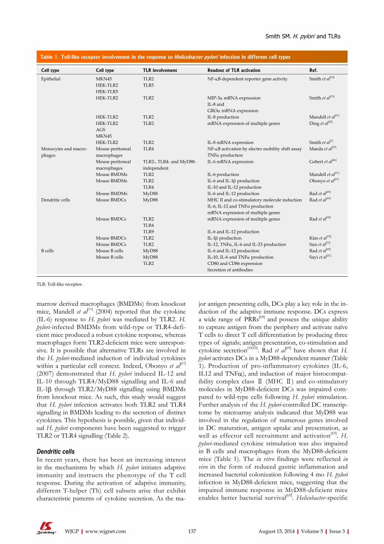
marrow derived macrophages (BMDMs) from knockout mice, Mandell et al[11] (2004) reported that the cytokine (IL-6) response to H. pylori was mediated by TLR2. H. pylori-infected BMDMs from wild-type or TLR4-defi-cient mice produced a robust cytokine response, whereas macrophages form TLR2-deficient mice were unrespon-sive. It is possible that alternative TLRs are involved in the H. pylori-mediated induction of individual cytokines within a particular cell context. Indeed, Obonyo et al[67] (2007) demonstrated that H. pylori induced IL-12 and IL-10 through TLR4/MyD88 signalling and IL-6 and IL-1β through TLR2/MyD88 signalling using BMDMs from knockout mice. As such, this study would suggest that H. pylori infection activates both TLR2 and TLR4 signalling in BMDMs leading to the secretion of distinct cytokines. This hypothesis is possible, given that individ-ual H. pylori components have been suggested to trigger TLR2 or TLR4 signalling (Table 2).
Dendritic cellsIn recent years, there has been an increasing interest in the mechanisms by which H. pylori initiates adaptive immunity and instructs the phenotype of the T cell response. During the activation of adaptive immunity, different T-helper (Th) cell subsets arise that exhibit characteristic patterns of cytokine secretion. As the ma-
jor antigen presenting cells, DCs play a key role in the in-duction of the adaptive immune response. DCs express a wide range of PRRs[68] and possess the unique ability to capture antigen from the periphery and activate naïve T cells to direct T cell differentiation by producing three types of signals; antigen presentation, co-stimulation and cytokine secretion[18,69]. Rad et al[69] have shown that H. pylori activates DCs in a MyD88-dependent manner (Table 1). Production of pro-inflammatory cytokines (IL-6, IL12 and TNFα), and induction of major histocompat-ibility complex class Ⅱ (MHC Ⅱ) and co-stimulatory molecules in MyD88-deficient DCs was impaired com-pared to wild-type cells following H. pylori stimulation. Further analysis of the H. pylori-controlled DC transcrip-tome by microarray analysis indicated that MyD88 was involved in the regulation of numerous genes involved in DC maturation, antigen uptake and presentation, as well as effector cell recruitment and activation[69]. H. pylori-mediated cytokine stimulation was also impaired in B cells and macrophages from the MyD88-deficient mice (Table 1). The in vitro findings were reflected in vivo in the form of reduced gastric inflammation and increased bacterial colonization following 4 mo H. pylori infection in MyD88-deficient mice, suggesting that the impaired immune response in MyD88-deficient mice enables better bacterial survival[69]. Helicobacter-specific
August 15, 2014|Volume 5|Issue 3|WJGP|www.wjgnet.com 137
Table 1 Toll-like receptor involvement in the response to Helicobacter pylori infection in different cell types
Cell type Cell type TLR involvement Readout of TLR activation Ref.
Epithelial MKN45 TLR2 NF-kB-dependent reporter gene activity Smith et al[19]
HEK-TLR2 TLR5HEK-TLR5HEK-TLR2 TLR2 MIP-3α mRNA expression Smith et al[19]
IL-8 andGROα mRNA expression
HEK-TLR2 TLR2 IL-8 production Mandell et al[11] HEK-TLR2 TLR2 mRNA expression of multiple genes Ding et al[20] AGSMKN45HEK-TLR2 TLR2 IL-8 mRNA expression Smith et al[7]
Monocytes and macro-phages
Mouse peritoneal macrophages
TLR4 NF-kB activation by electro mobility shift assay Maeda et al[57]
TNFα productionMouse peritoneal macrophages
TLR2-, TLR4- and MyD88-independent
IL-6 mRNA expression Gobert et al[66]
Mouse BMDMs TLR2 IL-6 production Mandell et al[11] Mouse BMDMs TLR2 IL-6 and IL-1β production Obonyo et al[67]
TLR4 IL-10 and IL-12 productionMouse BMDMs MyD88 IL-6 and IL-12 production Rad et al[69]
Dendritic cells Mouse BMDCs MyD88 MHC II and co-stimulatory molecule induction Rad et al[69] IL-6, IL-12 and TNFα productionmRNA expression of multiple genes
Mouse BMDCs TLR2 mRNA expression of multiple genes Rad et al[18]
TLR4TLR9 IL-6 and IL-12 production
Mouse BMDCs TLR2 IL-1β production Kim et al[70]
Mouse BMDCs TLR2 IL-12, TNFα, IL-6 and IL-23 production Sun et al[73]
B cells Mouse B cells MyD88 IL-6 and IL-12 production Rad et al[69] Mouse B cells MyD88 IL-10, IL-6 and TNFα production Sayi et al[21]
TLR2 CD80 and CD86 expressionSecretion of antibodies
TLR: Toll-like receptor.
Smith SM. H. pylori and TLRs
IgG2c/IgG1 ratios were reduced in MyD88-deficient mice, implying the involvement of the MyD88 pathway in the instruction of a Th1 phenotype[69]. Subsequent re-search by Rad et al[18] (2009) further characterized TLR-mediated signalling in DCs during H. pylori infection. They identified a MyD88-dependent component of the DC activation program that was induced by TLR2 and to a minor extent TLR4. Microarray analysis of H. pylori-stimulated DCs showed complementary, redundant and synergistic interactions between TLRs. Using TLR2-deficient cells the anti-inflammatory cytokine IL-10 was identified as a TLR2-dependent H. pylori responsive gene in DCs[18]. In addition, they demonstrated that IL-6 and IL-12 production was inhibited by approximately 50% in TLR2/TLR4/TLR9-deficient BMDCs compared to TLR2/TLR4-deficient cells in response to H. pylori in-fection, implying that TLR9-dependent recognition of H. pylori in DCs contributes to the cytokine response[18]. More recently Kim et al[70] (2013) have also implicated TLR signalling in response to H. pylori by demonstrating a role for TLR2 in H. pylori-induced IL-1β production in mouse BMDCs.
Although H. pylori-infected individuals generate a strong immune response, they fail to eradicate the bac-terium. Emerging evidence suggests that failure to elimi-nate H. pylori may be due to its ability to induce a regula-tory T cell (Treg) response, as expression of the Treg marker Foxp3 is increased in H. pylori-infected gastric tissue compared to that of uninfected individuals[71,72]. Sun et al[73] (2013) have recently investigated the func-tional role of TLR2 signalling in BMDCs in response to H. pylori and the subsequent effects on T cell responses. Firstly, they demonstrated that H. pylori-infected BMDCs from TLR2-deficient mice exhibited impaired produc-tion of the pro-inflammatory cytokines that promote both Th1 responses (IL-12 and TNFα) and Th17 re-sponses (IL-6 and IL-23) compared to wild-type cells. Additionally, this report suggests that H. pylori may skew differentiation of naïve T cells towards Th17 and Treg responses as opposed to Th1 responses, as H. pylori-stimulated BMDCs from TLR2 knock-out mice induced a higher splenocyte production of IFNg (Th1 response) and lower production of IL-17 (Th17 response) and IL-10 (Treg response)[73]. In vivo analyses following H. py-lori infection for 2 mo showed a lower degree of gastric H. pylori colonization in TLR2 knock-out mice and more se-vere gastritis, implying that the TLR2-mediated response to H. pylori promotes a bacterial survival advantage. Sun et al[73] also demonstrated that the gastric mucosa of the infected TLR2 knock-out mice had lower Foxp3, IL-10 and IL-17A expression, but higher expression of IFNg compared to wild-type mice. The H. pylori-specific Th1 response was higher and the Treg and Th17 responses were lower in the spleens of infected TLR2 knock-out mice, suggesting that H. pylori mediates immune toler-ance through TLR2-derived signals and inhibits Th1 im-munity, thus evading the host defence[73]. It is notewor-thy that the Rad et al[69] (2007) study suggested MyD88-dependent TLR signalling promotes a Th1 response in
H. pylori-infected mice and is protective against H. pylori colonization, while the Sun et al[73] (2013) study implies that TLR2 signalling inhibits Th1 immunity, supports a Treg/Th17 response and promotes H. pylori coloniza-tion. It is possible that different TLR ligands from the same pathogen induce distinct but opposing signals. Fur-thermore, it is likely that the complementary, redundant and synergistic interactions between TLRs in DCs subse-quently reported by Rad et al[18] (2009) contribute to the observations from studies involving MyD88-deficient mice.
B cellsEvidence suggests that B cells contribute to the immune-pathogenesis of H. pylori infection[74]. Sayi et al[21] (2011) have demonstrated that B cells play a role in regulating T cell responses and gastric immunopathology in response to Helicobacter. Building on the finding by Rad et al[69] (2007) that TLR2 is required for Helicobacter- mediated IL-6 and IL-12 induction in B cells, Sayi et al[21] showed that cytokine production (IL-10, IL-6 and TNFα), sur-face expression of the activation markers CD80 and CD86 and induction of antibody secretion was impaired in Helicobacter-stimulated B cells from both MyD88- and TLR2-deficient mice compared to wild type control cells (Table 1). The Helicobacter-stimulated B cells induced IL-10-producing CD4+CD25+ T regulatory-1 (Tr-1)-like cells in a TLR2- and MyD88-dependent manner[21]. The Tr-1 cells acquired suppressive activity in vitro and sup-pressed excessive gastric Helicobacter-associated immuno-pathology in vivo, suggesting that TLR2-mediated signal-ling in B cells plays a role in regulating the balance of Helicobacter-specific T cell responses to prevent excessive Th1-driven immunopathology and promote mucosal ho-meostasis, but enabling bacterial persistence[21].
RECOGNITION OF DISTINCT H. PYLORI COMPONENTS BY SPECIFIC TLRSLPSBased on the involvement of TLRs in regulating immu-nopathology in the context of H. pylori infection, many investigators have set out to elucidate the contribution of individual H. pylori components to the control of TLR-driven innate immune responses. H. pylori LPS has a lower endotoxicity than other gram-negative bacteria such as Escherichia coli or Salmonella enterica[75-78]. Although there has been substantial investigation into the innate immune response to H. pylori LPS, there have been conflicting findings with regard to the TLR responsible for its recognition (Table 2). Some studies have impli-cated the classic gram-negative bacterial LPS receptor TLR4[11,28,58,60,79,80], while others have suggested a role for TLR2[7,12,19,37,63]. Initial evidence for TLR4-mediated recognition of H. pylori LPS was provided by Kawahara et al[79] (2001) who demonstrated that LPS from clinical isolates of H. pylori induced increased superoxide anion (O2
-) production in guinea pig gastric pit cells that ex-
August 15, 2014|Volume 5|Issue 3|WJGP|www.wjgnet.com 138
Smith SM. H. pylori and TLRs
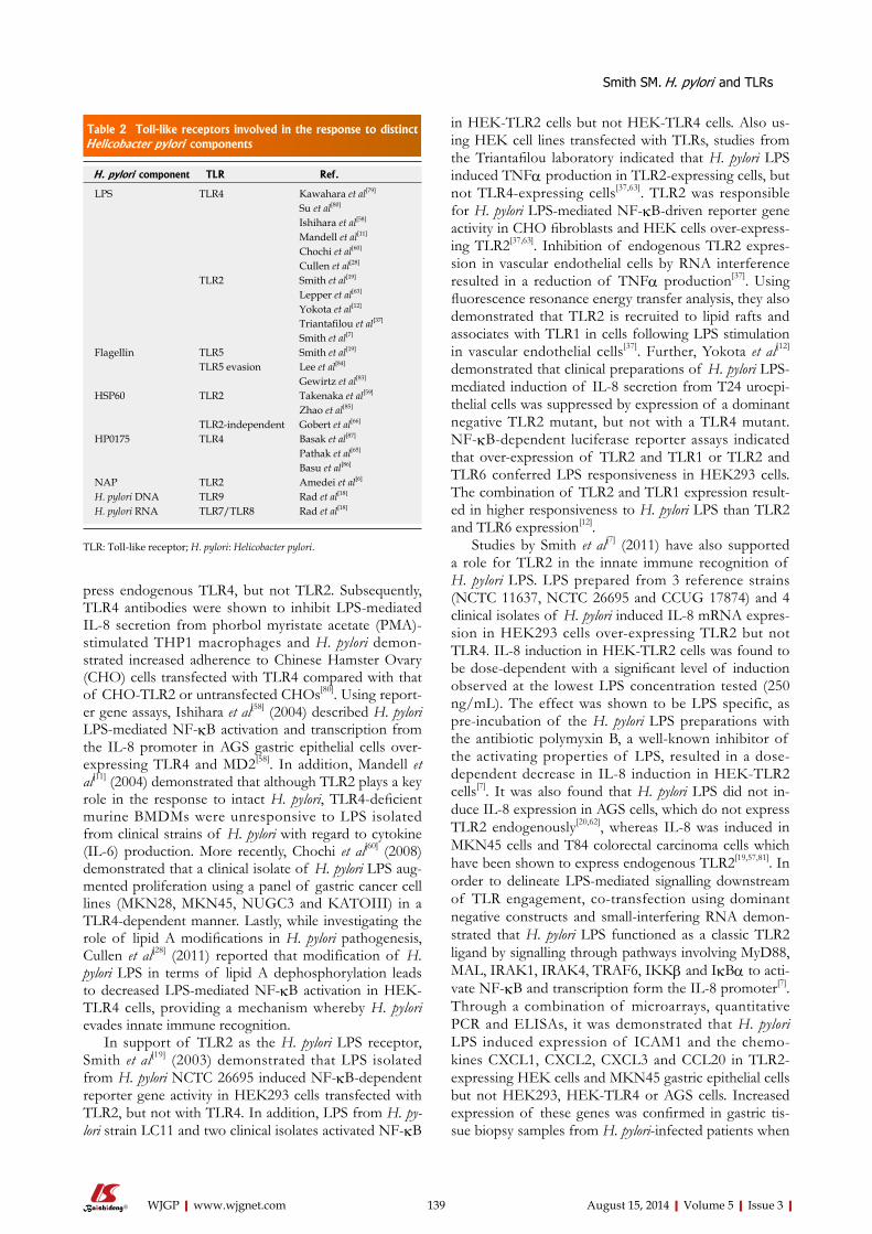
Table 2 Toll-like receptors involved in the response to distinct Helicobacter pylori components
H. pylori component TLR Ref.
LPS TLR4 Kawahara et al[79] Su et al[80]
Ishihara et al[58]
Mandell et al[11]
Chochi et al[60]
Cullen et al[28]
TLR2 Smith et al[19]
Lepper et al[63]
Yokota et al[12]
Triantafilou et al[37]
Smith et al[7]
Flagellin TLR5 Smith et al[19]
TLR5 evasion Lee et al[84]
Gewirtz et al[83]
HSP60 TLR2 Takenaka et al[59]
Zhao et al[85]
TLR2-independent Gobert et al[66]
HP0175 TLR4 Basak et al[87]
Pathak et al[65]
Basu et al[86]
NAP TLR2 Amedei et al[6]
H. pylori DNA TLR9 Rad et al[18]
H. pylori RNA TLR7/TLR8 Rad et al[18]
TLR: Toll-like receptor; H. pylori: Helicobacter pylori.
press endogenous TLR4, but not TLR2. Subsequently, TLR4 antibodies were shown to inhibit LPS-mediated IL-8 secretion from phorbol myristate acetate (PMA)-stimulated THP1 macrophages and H. pylori demon-strated increased adherence to Chinese Hamster Ovary (CHO) cells transfected with TLR4 compared with that of CHO-TLR2 or untransfected CHOs[80]. Using report-er gene assays, Ishihara et al[58] (2004) described H. pylori LPS-mediated NF-kB activation and transcription from the IL-8 promoter in AGS gastric epithelial cells over-expressing TLR4 and MD2[58]. In addition, Mandell et al[11] (2004) demonstrated that although TLR2 plays a key role in the response to intact H. pylori, TLR4-deficient murine BMDMs were unresponsive to LPS isolated from clinical strains of H. pylori with regard to cytokine (IL-6) production. More recently, Chochi et al[60] (2008) demonstrated that a clinical isolate of H. pylori LPS aug-mented proliferation using a panel of gastric cancer cell lines (MKN28, MKN45, NUGC3 and KATOIII) in a TLR4-dependent manner. Lastly, while investigating the role of lipid A modifications in H. pylori pathogenesis, Cullen et al[28] (2011) reported that modification of H. pylori LPS in terms of lipid A dephosphorylation leads to decreased LPS-mediated NF-kB activation in HEK-TLR4 cells, providing a mechanism whereby H. pylori evades innate immune recognition.
In support of TLR2 as the H. pylori LPS receptor, Smith et al[19] (2003) demonstrated that LPS isolated from H. pylori NCTC 26695 induced NF-kB-dependent reporter gene activity in HEK293 cells transfected with TLR2, but not with TLR4. In addition, LPS from H. py-lori strain LC11 and two clinical isolates activated NF-kB
in HEK-TLR2 cells but not HEK-TLR4 cells. Also us-ing HEK cell lines transfected with TLRs, studies from the Triantafilou laboratory indicated that H. pylori LPS induced TNFα production in TLR2-expressing cells, but not TLR4-expressing cells[37,63]. TLR2 was responsible for H. pylori LPS-mediated NF-kB-driven reporter gene activity in CHO fibroblasts and HEK cells over-express-ing TLR2[37,63]. Inhibition of endogenous TLR2 expres-sion in vascular endothelial cells by RNA interference resulted in a reduction of TNFα production[37]. Using fluorescence resonance energy transfer analysis, they also demonstrated that TLR2 is recruited to lipid rafts and associates with TLR1 in cells following LPS stimulation in vascular endothelial cells[37]. Further, Yokota et al[12] demonstrated that clinical preparations of H. pylori LPS-mediated induction of IL-8 secretion from T24 uroepi-thelial cells was suppressed by expression of a dominant negative TLR2 mutant, but not with a TLR4 mutant. NF-kB-dependent luciferase reporter assays indicated that over-expression of TLR2 and TLR1 or TLR2 and TLR6 conferred LPS responsiveness in HEK293 cells. The combination of TLR2 and TLR1 expression result-ed in higher responsiveness to H. pylori LPS than TLR2 and TLR6 expression[12].
Studies by Smith et al[7] (2011) have also supported a role for TLR2 in the innate immune recognition of H. pylori LPS. LPS prepared from 3 reference strains (NCTC 11637, NCTC 26695 and CCUG 17874) and 4 clinical isolates of H. pylori induced IL-8 mRNA expres-sion in HEK293 cells over-expressing TLR2 but not TLR4. IL-8 induction in HEK-TLR2 cells was found to be dose-dependent with a significant level of induction observed at the lowest LPS concentration tested (250 ng/mL). The effect was shown to be LPS specific, as pre-incubation of the H. pylori LPS preparations with the antibiotic polymyxin B, a well-known inhibitor of the activating properties of LPS, resulted in a dose-dependent decrease in IL-8 induction in HEK-TLR2 cells[7]. It was also found that H. pylori LPS did not in-duce IL-8 expression in AGS cells, which do not express TLR2 endogenously[20,62], whereas IL-8 was induced in MKN45 cells and T84 colorectal carcinoma cells which have been shown to express endogenous TLR2[19,57,81]. In order to delineate LPS-mediated signalling downstream of TLR engagement, co-transfection using dominant negative constructs and small-interfering RNA demon-strated that H. pylori LPS functioned as a classic TLR2 ligand by signalling through pathways involving MyD88, MAL, IRAK1, IRAK4, TRAF6, IKKβ and IkBα to acti-vate NF-kB and transcription form the IL-8 promoter[7]. Through a combination of microarrays, quantitative PCR and ELISAs, it was demonstrated that H. pylori LPS induced expression of ICAM1 and the chemo-kines CXCL1, CXCL2, CXCL3 and CCL20 in TLR2-expressing HEK cells and MKN45 gastric epithelial cells but not HEK293, HEK-TLR4 or AGS cells. Increased expression of these genes was confirmed in gastric tis-sue biopsy samples from H. pylori-infected patients when
August 15, 2014|Volume 5|Issue 3|WJGP|www.wjgnet.com 139
Smith SM. H. pylori and TLRs
compared to uninfected controls[7]. The reasons for the conflicting results between the
studies are unclear. Possible explanations include dif-ferences in experimental systems involving alternative read-outs for TLR activation and various cell lines from different species. In addition, contamination of the LPS preparation with other components, such as protein, nucleic acids or other bacterial LPS molecules could ac-count for conflicting findings. However, results from Smith et al[7] (2011) demonstrating that polymyxin B inhibited TLR2-mediated IL-8 induction in HEK-TLR2 cells would imply that the TLR2-mediated response observed was LPS-specific and not due to the presence of other contaminating TLR ligands, at least in this cell context. Contrasting findings may also have arisen due to heterogeneity of the structures of H. pylori LPS mol-ecules resulting from strain differences and/or culturing conditions. Tran et al[75] (2005) reported that the lipid A portion of H. pylori LPS undergoes several structural modifications through the action of specific modifying enzymes. There is also considerable LPS structural vari-ability due to diversity in the chemical composition of the polysaccharide O-antigen[27,28]. The study by Yokota et al[12] (2007) reported similar TLR2-dependent activities using LPS isolated from 6 different clinical isolates of H. pylori that demonstrated various characteristics, such as smooth/rough phenotypes and antigenicity of the poly-saccharide portion[12]. Additionally, it has been shown that LPS isolated from other gram-negative bacteria that produce a mixture of lipid A species with modi-fied forms of lipid A, such as Porphyromonas gingivalis and Leptospira interrogans, elicit immune responses through TLR2[33-37].
FlagellinTLR5 has been identified as the receptor for bacterial flagellin[38], the protein subunit of the polymeric flagellar filament of different gram-positive and gram-negative bacteria. H. pylori flagella (5-7 per cell) confer motility and are composed of polymers of two protein subunits, the major flagellin FlaA and the minor flagellin FlaB[82,83], both of which are essential for the bacteria to survive in the stomach musica[84]. TLR5 is expressed on primary gastric epithelial cells and gastric epithelial cell lines, including AGS, HM02, MKN28 and MKN45[19,62,84]. Initial investigations into the innate immune recogni-tion of H. pylori flagellin indicated that TLR5 expression in HEK293 cells conferred responsiveness to partially purified flagellin from H. pylori in terms of NF-kB-dependent reporter gene activity[19] (Table 2). In addition, transfection of MKN45 cells with a dominant negative TLR5 construct inhibited NF-kB activity in response to H. pylori flagellin[19]. However, other studies have since demonstrated that H. pylori flagellin is a significantly less potent stimulator of TLR5 signalling than flagellin from other gram-negative bacteria, such as Salmonella typhimurium[83,84]. Lee et al[84] (2003) demonstrated that al-though IL-8 release induced by H. pylori with mutations
in one or both flagellins was delayed compared to wild type H. pylori, purified native or recombinant flagellins did not significantly stimulate IL-8 secretion from gastric epithelial cells despite the presence of TLR5, suggesting that the delayed effect with the mutant strains may have been a result of decreased bacterial motility or adher-ence. Gewirtz et al[83] (2004) found no impairment in the IL-8 inducing ability of H. pylori in AGS cells as a result of FlaA mutations compared to the wild type strain. In keeping with the findings of Lee et al[84] (2003), purified H. pylori flagellin failed to induce significant innate immune responses in gastric epithelial cells as assessed by p38 MAPK induction and IL-8 secretion. The low innate im-mune response to H. pylori flagellin in the stomach in vivo may provide another mechanism that contributes to the ability of H. pylori to evade host responses and to pro-mote long term bacterial persistence.
Heat shock protein 60 The 60 kDa heat-shock protein (HSP60) of H. pylori plays a role in the adherence and attachment of H. pylori to the gastric epithelium and is a potent immune antigen that stimulates IL-8 induction in gastric epithelial cells[59]. HSP60-induced immune responses are associated with gastric inflammation and the pathogenesis of MALT[59]. Takenaka et al[59] (2004) have suggested that H. pylori HSP60 is a TLR2 ligand as HSP60-mediated NF-kB ac-tivation and IL-8 production in KATO Ⅲ human gastric epithelial cells was inhibited using a TLR2 blocking anti-body (Table 2). H. pylori HSP60 has also been shown to induce IL-8 production in human monocytes. In support of TLR2 in the recognition of H. pylori HSP60, Zhao et al[85] (2007) reported that treatment of NOMO1 hu-man monocytes with an anti-TLR2 blocking antibody or small interfering RNA for TLR2 inhibited NF-kB, ERK and p38 MAPK activation as well as IL-8 secretion in response to recombinant H. pylori HSP60 stimulation. In contrast to the findings in human cells, peritoneal mac-rophages from mice deficient in TLR2, TLR4, MyD88 or both TLR2 and TLR4 produced the same amount of IL-6 in response to H. pylori HSP60 as wild type macro-phages, indicating TLR-independent IL-6 induction in this cell context[66].
H. pylori peptidyl prolyl cis-,trans-isomerase HP0175H. pylori secretes the peptidyl prolyl cis-, trans-isomerase HP0175, which can induce apoptosis in gastric epithelial cells and is one of the highly and consistently reactive H. pylori antigens recognized in the sera of H. pylori-infected patients[65,86]. Studies from the Kundu laboratory have described a role for TLR4 in the recognition of HP0175 (Table 2). Initially, Basak et al[87] (2005) demonstrated in-teraction between TLR4 and HP0175 in AGS cells using pull-down immunoassays. Inhibition of TLR4 using a neutralizing antibody or a dominant negative construct inhibited HP0175-induced apoptosis in AGS cells[87]. Pathak et al[65] (2006) subsequently reported that HP0175 induced the release of IL-6 from PMA-differentiated
August 15, 2014|Volume 5|Issue 3|WJGP|www.wjgnet.com 140
Smith SM. H. pylori and TLRs
THP1 macrophages, whereas isogenic mutants of H. py-lori 26695, in which the Hp0175 gene was disrupted, elic-ited decreased IL-6 production. A role for TLR4 in this process was suggested because pre-treatment of cells with a TLR4 (but not TLR2) neutralising antibody or transfection with a dominant-negative TLR4 construct blocked HP0175-mediated IL-6 release. In addition, TLR4 expression (but not TLR2) in HEK293 cells con-ferred responsiveness to HP0175. Using ELISA-based binding assays, Pathak et al[65] also showed that HP0175 interacts with the extracellular domain of TLR4 in the absence of any accessory molecules. Finally, Basu et al[86] (2008) showed that HP0175 transactivates the epidermal growth factor receptor (EGFR) and stimulates EGFR-dependent vascular endothelial growth factor (VEGF) production in AGS cells in a TLR4-dependent manner.
NapAThe H. pylori neutrophil-activating protein (NAP) is a 150 kDa oligomeric virulence factor that is chemotactic for neutrophils, stimulates high production of oxygen radicals in neutrophils and their adhesion to endothelial cells. Amedei et al[6] (2006) have reported that the H. pylori NAP is a TLR2 agonist, because over-expression of TLR2 in HEK293 cells resulted in NAP-mediated NF-kB-dependent reporter gene activity (Table 2). NAP stimulation had no effect on untransfected HEK293 cells, or HEK293 cells over-expressing TLR3, TLR4, TLR5, TLR7, TLR8 or TLR9. In human monocytes and neutrophils, NAP stimulation induced the expression of IL-12[6], which is an important cytokine for the dif-ferentiation of naïve Th cells into the Th1 phenotype. NAP also induced monocytes to produce IL-23 and dif-ferentiate towards mature DCs[6]. Stimulation of antigen-induced T-cells with NAP resulted in increased numbers of IFN-γ-producing T cells and decreased numbers of IL-4-secreting cells, thus promoting a Th1 phenotype. In addition, using T cell clones generated from in vivo-activated T cells derived from the gastric mucosa of H. pylori-infected patients, NAP was shown to elicit Th1-polarizing capacity[6], implying that the TLR2: H. pylori NAP interactions promote the activation of innate immunity to drive IL-12 and IL-23 production and the subsequent promotion of Th1 immune responses.
Nucleic acidsTLR9 recognises unmethylated CpG DNA in bacte-ria[40] and also detects herpes virus DNA[41]. TLR7 and TLR8 have been shown to sense single-stranded viral RNA[42-44]. Rad et al[18] (2009) have demonstrated TLR9-mediated recognition of H. pylori DNA in DCs and the subsequent induction of pro-inflammatory cytokine secretion. They showed that IL-6 and IL-12 produc-tion was completely abrogated in TLR2/TLR4/TLR9-deficient BMDCs compared to TLR2/TLR4-deficient cells in response to purified H. pylori DNA following pre-treatment with ribonuclease. Expression of TLR9 is increased in mouse gastric tissue following H. pylori in-
fection and is mainly localised to macrophages, DCs and CD3+ cells in the gastric mucosa[88]. Although purified H. pylori DNA was reported to induce a TLR9-mediated increase in IL-6 and IL-12 expression in BMDCs[18], in a mouse model of H. pylori infection TLR9 signalling was shown to have an anti-inflammatory effect on the early phase of H. pylori-induced gastritis as genetic disrup-tion of TLR9 resulted in an increase in H. pylori-induced gastric mucosal inflammation characterized by neu-trophil infiltration and increased expression of TNFα and IFNg[88]. In relation to TLR7 and TLR8-mediated recognition of H. pylori, Rad et al[18] (2009) showed that purified H. pylori RNA (pre-treated with deoxyribonucle-ase) induced pro-inflammatory cytokines in BMDCs in a MyD88-dependent manner involving the endosomal TLR8, possibly in collaboration with TLR7.
TARGETING TLR SIGNALLING THERA-PEUTICALLY As TLRs are intimately involved in the regulation of inflammation during innate immunity and represent key activators of adaptive immunity, they represent an at-tractive therapeutic target for treatment of inflammatory diseases. Indeed, oligonucleotide inhibitors of TLR7 and/or TLR9 have been shown to have therapeutic potential in animal models of systemic lupus erythema-tosus[89]. Additionally, an inhibitory TLR2 antibody was demonstrated to limit ischemia-reperfusion injury in the hearts of pigs[90] and kidneys of mice[91]. In the clinic, therapies involving the synthetic small molecule inhibi-tor of TLR4, Eritoran (also known as E5564), were used in trials for patients with sepsis, but only had mar-ginal effects possibly as treatment was administered too late following disease onset[92,93]. TLR activation is also important for adjuvancy in vaccines and several TLR ligands have been shown to be efficacious as vaccine adjuvants[94,95]. For example, the vaccine adjuvant mono-phosphoryl lipid A, which is a less toxic version of LPS, promotes antibody responses via TLR4 activation[96]. Efficient preventative or therapeutic vaccination for H. pylori has not been achieved in humans to date[97]. Early signs of promise in animal models of H. pylori infection have been unsuccessful in humans. In a recent study to investigate the vaccine potential of H. pylori LPS, Alt-man et al[98] (2012) demonstrated enhanced antibody responses to a chemically modified LPS in mice and rabbits and partial protection against H. pylori challenge, warranting further investigation in this area. In terms of adjuvancy, immunization against Helicobacter using CpG and cholera toxin demonstrated synergism leading to sterile immunity in mice[99]. More recently, Mori et al[100] (2012) constructed a chimeric flagellin by replacing the N- and T-terminal segments of H. pylori flagellin with a TLR5-stimulating adjuvant component of E. coli flagellin in order to enhance innate and adaptive immunity. The resulting chimeric flagellin activated TLR5 signalling and elicited a strong antibody response in mice. Together
August 15, 2014|Volume 5|Issue 3|WJGP|www.wjgnet.com 141
Smith SM. H. pylori and TLRs
with alum, vaccination with the chimeric flagellin pro-tected mice from H. pylori infection[100]. In other disease settings, local administration of the TLR2 ligand H. pylo-ri NAP was demonstrated to decrease tumour growth by activating a cytotoxic Th1 response in a mouse model of bladder cancer[101]. Taken together, these studies indicate that defining H. pylori-derived molecules responsible for TLR activation and elucidating innate immune signals triggered by H. pylori, may provide insight into the design and development of novel human vaccine adjuvants and therapeutics.
H. PYLORI RECOGNITION BY OTHER PRRSMicrobial pathogens activate multiple PRRs and dif-ferent PRRs may recognize the same PAMP within an organism[17]. Insight into the co-operation between TLRs and other PRRs during infection is necessary for a complete understanding of the innate immune response during infection. These PRRs include nucleotide-binding oligomerization domain (NOD)-like receptors (NLRs), retinoic acid-inducible gene-I (RIG-I)-like receptors (RLRs) and C-type lectin receptors (CLRs). H. pylori peptidoglycan, delivered either by the type IV secretion system or through outer membrane vesicles secreted from the bacterium, is recognised by NOD1 in epithe-lial cells[102-107]. Moreover, studies by Kim et al[70] (2013) have described the cooperative interaction of TLR2 and NOD2 in the regulation of IL-1β in H. pylori-infected DCs. In addition to a role for TLR8 in the response to H. pylori RNA, findings by Rad et al (2009) indicated that H. pylori induces expression of type I interferon and interferon-stimulated genes in a MyD88- and TRIF-independent manner and demonstrated that the MyD88-independent type I IFN induction by H. pylori RNA was mediated by RIG-I[18]. In relation to H. pylori-mediated CLR signalling, Gringhuis et al[108] (2009) have reported that the fucose residues of H. pylori DC-SIGN ligands actively disrupt signalling down-stream of DC-SIGN to suppress pro-inflammatory cytokine induction.
CONCLUSIONAlthough, H. pylori induces a strong immune response, elimination of infection is not achieved. The pathogene-sis of H. pylori-associated disease is linked to the severity of the host inflammatory response. Emerging evidence suggests that failure to eliminate H. pylori may be due to the ability of the bacterium to control T-cell responses. As TLRs are intimately involved in the regulation of inflammation during the innate immune response to H. pylori and represent key activators of adaptive immunity, a significant effort has been made to elucidate their role in the recognition of H. pylori and its components in multiple cell types. Much of the literature has focussed on the involvement of individual TLRs in the induction of pro-inflammatory cytokines in various in vitro cell cul-
ture models. There is substantial evidence to support the role of TLR2 in activating NF-kB or inducing cytokine induction in response to H. pylori infection in epithelial cells[7,11,19,20], monocytes/macrophages[11,67], dendritic cells[18,70,73] and B cells[21]. Numerous H. pylori ligands have been suggested to date that may contribute to these TLR2-dependent responses, including LPS[7,12,19,37,63], HSP60[59,85] and NAP[6]. TLR4 has also been implicated in the response to H. pylori[18,57,67], which may be mediated by LPS[11,28,58,60,79,80] and/or HP0175[65,86,87]. TLR9 has been identified as the receptor for H. pylori DNA[18]. Although H. pylori flagellin has been suggested as a TLR5 ligand[19], its activity as a TLR5 activator is low[83,84], providing a possible mechanism that contributes H. pylori persis-tence.
Recent studies using mouse models of infection have provided insight into the role of TLR signalling in regulating H. pylori-mediated T cell responses, gastric immunopathology and colonization in vivo. Interestingly, although a demonstrated role for TLR2 in the induction of pro-inflammatory cytokines has been described in distinct cell populations, the net effect of TLR2 signal-ling has been reported to mediate tolerance and promote bacterial persistence in mouse models of infection by skewing T cell responses[21,73]. Further in vivo studies elu-cidating innate immune signals triggered by H. pylori-me-diated activation of TLRs, especially in cooperation with other PRRs, are necessary for a complete understanding of how the balance between pro-inflammatory and anti-inflammatory signals fine-tunes the immune response to H. pylori infection, and may provide insight into how the immune response may be manipulated therapeutically to successfully eradicate the bacterium.
REFERENCES1 Polk DB, Peek RM. Helicobacter pylori: gastric cancer and
beyond. Nat Rev Cancer 2010; 10: 403-414 [PMID: 20495574 DOI: 10.1038/nrc2857]
2 McColl KE. Clinical practice. Helicobacter pylori infection. N Engl J Med 2010; 362: 1597-1604 [PMID: 20427808 DOI: 10.1056/NEJMcp1001110]
3 Salama NR, Hartung ML, Müller A. Life in the human stomach: persistence strategies of the bacterial pathogen He-licobacter pylori. Nat Rev Microbiol 2013; 11: 385-399 [PMID: 23652324 DOI: 10.1038/nrmicro3016]
4 Malfertheiner P, Megraud F, O’Morain CA, Atherton J, Axon AT, Bazzoli F, Gensini GF, Gisbert JP, Graham DY, Rokkas T, El-Omar EM, Kuipers EJ. Management of Heli-cobacter pylori infection--the Maastricht IV/ Florence Con-sensus Report. Gut 2012; 61: 646-664 [PMID: 22491499 DOI: 10.1136/gutjnl-2012-302084]
5 Queiroz DM, Harris PR, Sanderson IR, Windle HJ, Walker MM, Rocha AM, Rocha GA, Carvalho SD, Bittencourt PF, de Castro LP, Villagrán A, Serrano C, Kelleher D, Crabtree JE. Iron status and Helicobacter pylori infection in symptom-atic children: an international multi-centered study. PLoS One 2013; 8: e68833 [PMID: 23861946 DOI: 10.1371/journal.pone.0068833]
6 Amedei A, Cappon A, Codolo G, Cabrelle A, Polenghi A, Benagiano M, Tasca E, Azzurri A, D’Elios MM, Del Prete G, de Bernard M. The neutrophil-activating protein of He-licobacter pylori promotes Th1 immune responses. J Clin
August 15, 2014|Volume 5|Issue 3|WJGP|www.wjgnet.com 142
Smith SM. H. pylori and TLRs
Invest 2006; 116: 1092-1101 [PMID: 16543949 DOI: 10.1172/JCI27177]
7 Smith SM, Moran AP, Duggan SP, Ahmed SE, Mohamed AS, Windle HJ, O’Neill LA, Kelleher DP. Tribbles 3: a novel regulator of TLR2-mediated signaling in response to Helicobacter pylori lipopolysaccharide. J Immunol 2011; 186: 2462-2471 [PMID: 21220698 DOI: 10.4049/jimmu-nol.1000864]
8 Yamaoka Y, Kita M, Kodama T, Sawai N, Kashima K, Iman-ishi J. Expression of cytokine mRNA in gastric mucosa with Helicobacter pylori infection. Scand J Gastroenterol 1995; 30: 1153-1159 [PMID: 9053967]
9 Moss SF, Legon S, Davies J, Calam J. Cytokine gene expres-sion in Helicobacter pylori associated antral gastritis. Gut 1994; 35: 1567-1570 [PMID: 7828974]
10 Windle HJ, Ang YS, Athie-Morales V, McManus R, Kelleher D. Human peripheral and gastric lymphocyte responses to Helicobacter pylori NapA and AphC differ in infected and uninfected individuals. Gut 2005; 54: 25-32 [PMID: 15591500 DOI: 10.1136/gut.2003.025494]
11 Mandell L, Moran AP, Cocchiarella A, Houghton J, Taylor N, Fox JG, Wang TC, Kurt-Jones EA. Intact gram-negative He-licobacter pylori, Helicobacter felis, and Helicobacter hepati-cus bacteria activate innate immunity via toll-like receptor 2 but not toll-like receptor 4. Infect Immun 2004; 72: 6446-6454 [PMID: 15501775 DOI: 10.1128/IAI.72.11.6446-6454.2004]
12 Yokota S, Ohnishi T, Muroi M, Tanamoto K, Fujii N, Ama-no K. Highly-purified Helicobacter pylori LPS preparations induce weak inflammatory reactions and utilize Toll-like receptor 2 complex but not Toll-like receptor 4 complex. FEMS Immunol Med Microbiol 2007; 51: 140-148 [PMID: 17645528 DOI: 10.1111/j.1574-695X.2007.00288.x]
13 Bodger K, Crabtree JE. Helicobacter pylori and gastric in-flammation. Br Med Bull 1998; 54: 139-150 [PMID: 9604438]
14 O’Connor A, Gisbert JP, McNamara D, O’Morain C. Treat-ment of Helicobacter pylori infection 2011. Helicobacter 2011; 16 Suppl 1: 53-58 [PMID: 21896086 DOI: 10.1111/j.1523-5378.2011.00881.x]
15 Megraud F, Coenen S, Versporten A, Kist M, Lopez-Brea M, Hirschl AM, Andersen LP, Goossens H, Glupczynski Y. He-licobacter pylori resistance to antibiotics in Europe and its relationship to antibiotic consumption. Gut 2013; 62: 34-42 [PMID: 22580412 DOI: 10.1136/gutjnl-2012-302254]
16 Kawai T, Akira S. The roles of TLRs, RLRs and NLRs in pathogen recognition. Int Immunol 2009; 21: 317-337 [PMID: 19246554 DOI: 10.1093/intimm/dxp017]
17 Kawai T, Akira S. Toll-like receptors and their crosstalk with other innate receptors in infection and immunity. Im-munity 2011; 34: 637-650 [PMID: 21616434 DOI: 10.1016/j.immuni.2011.05.006]
18 Rad R, Ballhorn W, Voland P, Eisenächer K, Mages J, Rad L, Ferstl R, Lang R, Wagner H, Schmid RM, Bauer S, Prinz C, Kirschning CJ, Krug A. Extracellular and intracellular pattern recognition receptors cooperate in the recognition of Helicobacter pylori. Gastroenterology 2009; 136: 2247-2257 [PMID: 19272387 DOI: 10.1053/j.gastro.2009.02.066]
19 Smith MF, Mitchell A, Li G, Ding S, Fitzmaurice AM, Ryan K, Crowe S, Goldberg JB. Toll-like receptor (TLR) 2 and TLR5, but not TLR4, are required for Helicobacter pylori-induced NF-kappa B activation and chemokine expression by epithelial cells. J Biol Chem 2003; 278: 32552-32560 [PMID: 12807870 DOI: 10.1074/jbc.M305536200]
20 Ding SZ, Torok AM, Smith MF, Goldberg JB. Toll-like re-ceptor 2-mediated gene expression in epithelial cells during Helicobacter pylori infection. Helicobacter 2005; 10: 193-204 [PMID: 15904477 DOI: 10.1111/j.1523-5378.2005.00311.x]
21 Sayi A, Kohler E, Toller IM, Flavell RA, Müller W, Roers A, Müller A. TLR-2-activated B cells suppress Helicobacter-induced preneoplastic gastric immunopathology by induc-ing T regulatory-1 cells. J Immunol 2011; 186: 878-890 [PMID:
21149607 DOI: 10.4049/jimmunol.1002269]22 Tahara T, Arisawa T, Wang F, Shibata T, Nakamura M,
Sakata M, Hirata I, Nakano H. Toll-like receptor 2 (TLR) -196 to 174del polymorphism in gastro-duodenal diseases in Japanese population. Dig Dis Sci 2008; 53: 919-924 [PMID: 17934843 DOI: 10.1007/s10620-007-9950-x]
23 Tahara T, Arisawa T, Wang F, Shibata T, Nakamura M, Sakata M, Hirata I, Nakano H. Toll-like receptor 2 -196 to 174del polymorphism influences the susceptibility of Japa-nese people to gastric cancer. Cancer Sci 2007; 98: 1790-1794 [PMID: 17711514 DOI: 10.1111/j.1349-7006.2007.00590.x]
24 Mayerle J, den Hoed CM, Schurmann C, Stolk L, Homuth G, Peters MJ, Capelle LG, Zimmermann K, Rivadeneira F, Gruska S, Völzke H, de Vries AC, Völker U, Teumer A, van Meurs JB, Steinmetz I, Nauck M, Ernst F, Weiss FU, Hofman A, Zenker M, Kroemer HK, Prokisch H, Uitterlin-den AG, Lerch MM, Kuipers EJ. Identification of genetic loci associated with Helicobacter pylori serologic status. JAMA 2013; 309: 1912-1920 [PMID: 23652523 DOI: 10.1001/jama.2013.4350]
25 O’Neill LA, Golenbock D, Bowie AG. The history of Toll-like receptors - redefining innate immunity. Nat Rev Immu-nol 2013; 13: 453-460 [PMID: 23681101 DOI: 10.1038/nri3446]
26 Poltorak A, He X, Smirnova I, Liu MY, Van Huffel C, Du X, Birdwell D, Alejos E, Silva M, Galanos C, Freudenberg M, Ricciardi-Castagnoli P, Layton B, Beutler B. Defective LPS signaling in C3H/HeJ and C57BL/10ScCr mice: mutations in Tlr4 gene. Science 1998; 282: 2085-2088 [PMID: 9851930]
27 Matsuura M. Structural Modifications of Bacterial Lipopoly-saccharide that Facilitate Gram-Negative Bacteria Evasion of Host Innate Immunity. Front Immunol 2013; 4: 109 [PMID: 23745121 DOI: 10.3389/fimmu.2013.00109]
28 Cullen TW, Giles DK, Wolf LN, Ecobichon C, Boneca IG, Trent MS. Helicobacter pylori versus the host: remodeling of the bacterial outer membrane is required for survival in the gastric mucosa. PLoS Pathog 2011; 7: e1002454 [PMID: 22216004 DOI: 10.1371/journal.ppat.1002454]
29 Takeuchi O, Sato S, Horiuchi T, Hoshino K, Takeda K, Dong Z, Modlin RL, Akira S. Cutting edge: role of Toll-like receptor 1 in mediating immune response to microbial lipo-proteins. J Immunol 2002; 169: 10-14 [PMID: 12077222]
30 Jin MS, Kim SE, Heo JY, Lee ME, Kim HM, Paik SG, Lee H, Lee JO. Crystal structure of the TLR1-TLR2 heterodi-mer induced by binding of a tri-acylated lipopeptide. Cell 2007; 130: 1071-1082 [PMID: 17889651 DOI: 10.1016/j.cell.2007.09.008]
31 Takeuchi O, Kawai T, Mühlradt PF, Morr M, Radolf JD, Zy-chlinsky A, Takeda K, Akira S. Discrimination of bacterial lipoproteins by Toll-like receptor 6. Int Immunol 2001; 13: 933-940 [PMID: 11431423]
32 Kang JY, Nan X, Jin MS, Youn SJ, Ryu YH, Mah S, Han SH, Lee H, Paik SG, Lee JO. Recognition of lipopeptide patterns by Toll-like receptor 2-Toll-like receptor 6 heterodimer. Immunity 2009; 31: 873-884 [PMID: 19931471 DOI: 10.1016/j.immuni.2009.09.018]
33 Werts C, Tapping RI, Mathison JC, Chuang TH, Kravchenko V, Saint Girons I, Haake DA, Godowski PJ, Hayashi F, Ozin-sky A, Underhill DM, Kirschning CJ, Wagner H, Aderem A, Tobias PS, Ulevitch RJ. Leptospiral lipopolysaccharide acti-vates cells through a TLR2-dependent mechanism. Nat Im-munol 2001; 2: 346-352 [PMID: 11276206 DOI: 10.1038/86354]
34 Darveau RP, Pham TT, Lemley K, Reife RA, Bainbridge BW, Coats SR, Howald WN, Way SS, Hajjar AM. Porphy-romonas gingivalis lipopolysaccharide contains multiple lipid A species that functionally interact with both toll-like receptors 2 and 4. Infect Immun 2004; 72: 5041-5051 [PMID: 15321997 DOI: 10.1128/IAI.72.9.5041-5051.2004]
35 Que-Gewirth NL, Ribeiro AA, Kalb SR, Cotter RJ, Bulach DM, Adler B, Girons IS, Werts C, Raetz CR. A methylated phosphate group and four amide-linked acyl chains in
August 15, 2014|Volume 5|Issue 3|WJGP|www.wjgnet.com 143
Smith SM. H. pylori and TLRs
leptospira interrogans lipid A. The membrane anchor of an unusual lipopolysaccharide that activates TLR2. J Biol Chem 2004; 279: 25420-25429 [PMID: 15044492 DOI: 10.1074/jbc.M400598200]
36 Hirschfeld M, Weis JJ, Toshchakov V, Salkowski CA, Cody MJ, Ward DC, Qureshi N, Michalek SM, Vogel SN. Signaling by toll-like receptor 2 and 4 agonists results in differential gene expression in murine macrophages. Infect Immun 2001; 69: 1477-1482 [PMID: 11179315 DOI: 10.1128/IAI.69.3.1477-1482.2001]
37 Triantafilou M, Gamper FG, Lepper PM, Mouratis MA, Schumann C, Harokopakis E, Schifferle RE, Hajishengallis G, Triantafilou K. Lipopolysaccharides from atherosclerosis-associated bacteria antagonize TLR4, induce formation of TLR2/1/CD36 complexes in lipid rafts and trigger TLR2-in-duced inflammatory responses in human vascular endothe-lial cells. Cell Microbiol 2007; 9: 2030-2039 [PMID: 17419716 DOI: 10.1111/j.1462-5822.2007.00935.x]
38 Hayashi F, Smith KD, Ozinsky A, Hawn TR, Yi EC, Good-lett DR, Eng JK, Akira S, Underhill DM, Aderem A. The innate immune response to bacterial flagellin is mediated by Toll-like receptor 5. Nature 2001; 410: 1099-1103 [PMID: 11323673 DOI: 10.1038/35074106]
39 Alexopoulou L, Holt AC, Medzhitov R, Flavell RA. Recog-nition of double-stranded RNA and activation of NF-kap-paB by Toll-like receptor 3. Nature 2001; 413: 732-738 [PMID: 11607032 DOI: 10.1038/35099560]
40 Hemmi H, Takeuchi O, Kawai T, Kaisho T, Sato S, Sanjo H, Matsumoto M, Hoshino K, Wagner H, Takeda K, Akira S. A Toll-like receptor recognizes bacterial DNA. Nature 2000; 408: 740-745 [PMID: 11130078 DOI: 10.1038/35047123]
41 Lund J, Sato A, Akira S, Medzhitov R, Iwasaki A. Toll-like receptor 9-mediated recognition of Herpes simplex virus-2 by plasmacytoid dendritic cells. J Exp Med 2003; 198: 513-520 [PMID: 12900525 DOI: 10.1084/jem.20030162]
42 Heil F, Hemmi H, Hochrein H, Ampenberger F, Kirschning C, Akira S, Lipford G, Wagner H, Bauer S. Species-specific recognition of single-stranded RNA via toll-like receptor 7 and 8. Science 2004; 303: 1526-1529 [PMID: 14976262 DOI: 10.1126/science.1093620]
43 Diebold SS, Kaisho T, Hemmi H, Akira S, Reis e Sousa C. Innate antiviral responses by means of TLR7-mediated recognition of single-stranded RNA. Science 2004; 303: 1529-1531 [PMID: 14976261 DOI: 10.1126/science.1093616]
44 Lund JM, Alexopoulou L, Sato A, Karow M, Adams NC, Gale NW, Iwasaki A, Flavell RA. Recognition of single-stranded RNA viruses by Toll-like receptor 7. Proc Natl Acad Sci USA 2004; 101: 5598-5603 [PMID: 15034168 DOI: 10.1073/pnas.0400937101]
45 Kawai T, Akira S. TLR signaling. Cell Death Differ 2006; 13: 816-825 [PMID: 16410796]
46 Fitzgerald KA, Palsson-McDermott EM, Bowie AG, Jefferies CA, Mansell AS, Brady G, Brint E, Dunne A, Gray P, Harte MT, McMurray D, Smith DE, Sims JE, Bird TA, O’Neill LA. Mal (MyD88-adapter-like) is required for Toll-like recep-tor-4 signal transduction. Nature 2001; 413: 78-83 [PMID: 11544529 DOI: 10.1038/35092578]
47 Horng T, Barton GM, Medzhitov R. TIRAP: an adapter molecule in the Toll signaling pathway. Nat Immunol 2001; 2: 835-841 [PMID: 11526399 DOI: 10.1038/ni0901-835]
48 Yamamoto M, Sato S, Hemmi H, Sanjo H, Uematsu S, Kai-sho T, Hoshino K, Takeuchi O, Kobayashi M, Fujita T, Take-da K, Akira S. Essential role for TIRAP in activation of the signalling cascade shared by TLR2 and TLR4. Nature 2002; 420: 324-329 [PMID: 12447441 DOI: 10.1038/nature01182]
49 Horng T, Barton GM, Flavell RA, Medzhitov R. The adaptor molecule TIRAP provides signalling specificity for Toll-like receptors. Nature 2002; 420: 329-333 [PMID: 12447442 DOI: 10.1038/nature01180]
50 Yamamoto M, Sato S, Mori K, Hoshino K, Takeuchi O,
Takeda K, Akira S. Cutting edge: a novel Toll/IL-1 receptor domain-containing adapter that preferentially activates the IFN-beta promoter in the Toll-like receptor signaling. J Im-munol 2002; 169: 6668-6672 [PMID: 12471095]
51 Oshiumi H, Matsumoto M, Funami K, Akazawa T, Seya T. TICAM-1, an adaptor molecule that participates in Toll-like receptor 3-mediated interferon-beta induction. Nat Immunol 2003; 4: 161-167 [PMID: 12539043 DOI: 10.1038/ni886]
52 Hoebe K, Du X, Georgel P, Janssen E, Tabeta K, Kim SO, Goode J, Lin P, Mann N, Mudd S, Crozat K, Sovath S, Han J, Beutler B. Identification of Lps2 as a key transducer of MyD88-independent TIR signalling. Nature 2003; 424: 743-748 [PMID: 12872135 DOI: 10.1038/nature01889]
53 Yamamoto M, Sato S, Hemmi H, Hoshino K, Kaisho T, San-jo H, Takeuchi O, Sugiyama M, Okabe M, Takeda K, Akira S. Role of adaptor TRIF in the MyD88-independent toll-like re-ceptor signaling pathway. Science 2003; 301: 640-643 [PMID: 12855817 DOI: 10.1126/science.1087262]
54 Fitzgerald KA, Rowe DC, Barnes BJ, Caffrey DR, Visintin A, Latz E, Monks B, Pitha PM, Golenbock DT. LPS-TLR4 signaling to IRF-3/7 and NF-kappaB involves the toll adapt-ers TRAM and TRIF. J Exp Med 2003; 198: 1043-1055 [PMID: 14517278 DOI: 10.1084/jem.20031023]
55 Yamamoto M, Sato S, Hemmi H, Uematsu S, Hoshino K, Kaisho T, Takeuchi O, Takeda K, Akira S. TRAM is specifi-cally involved in the Toll-like receptor 4-mediated MyD88-independent signaling pathway. Nat Immunol 2003; 4: 1144-1150 [PMID: 14556004 DOI: 10.1038/ni986]
56 Oshiumi H, Sasai M, Shida K, Fujita T, Matsumoto M, Seya T. TIR-containing adapter molecule (TICAM)-2, a bridg-ing adapter recruiting to toll-like receptor 4 TICAM-1 that induces interferon-beta. J Biol Chem 2003; 278: 49751-49762 [PMID: 14519765 DOI: 10.1074/jbc.M305820200]
57 Maeda S, Akanuma M, Mitsuno Y, Hirata Y, Ogura K, Yoshida H, Shiratori Y, Omata M. Distinct mechanism of Helicobacter pylori-mediated NF-kappa B activation be-tween gastric cancer cells and monocytic cells. J Biol Chem 2001; 276: 44856-44864 [PMID: 11546774 DOI: 10.1074/jbc.M105381200]
58 Ishihara S, Rumi MA, Kadowaki Y, Ortega-Cava CF, Yuki T, Yoshino N, Miyaoka Y, Kazumori H, Ishimura N, Amano Y, Kinoshita Y. Essential role of MD-2 in TLR4-dependent signaling during Helicobacter pylori-associated gastritis. J Immunol 2004; 173: 1406-1416 [PMID: 15240737]
59 Takenaka R, Yokota K, Ayada K, Mizuno M, Zhao Y, Fujin-ami Y, Lin SN, Toyokawa T, Okada H, Shiratori Y, Oguma K. Helicobacter pylori heat-shock protein 60 induces inflam-matory responses through the Toll-like receptor-triggered pathway in cultured human gastric epithelial cells. Microbi-ology 2004; 150: 3913-3922 [PMID: 15583145 DOI: 10.1099/mic.0.27527-0]
60 Chochi K, Ichikura T, Kinoshita M, Majima T, Shinomiya N, Tsujimoto H, Kawabata T, Sugasawa H, Ono S, Seki S, Mo-chizuki H. Helicobacter pylori augments growth of gastric cancers via the lipopolysaccharide-toll-like receptor 4 path-way whereas its lipopolysaccharide attenuates antitumor activities of human mononuclear cells. Clin Cancer Res 2008; 14: 2909-2917 [PMID: 18483357 DOI: 10.1158/1078-0432.CCR-07-4467]
61 Uno K, Kato K, Atsumi T, Suzuki T, Yoshitake J, Morita H, Ohara S, Kotake Y, Shimosegawa T, Yoshimura T. Toll-like receptor (TLR) 2 induced through TLR4 signaling initi-ated by Helicobacter pylori cooperatively amplifies iNOS induction in gastric epithelial cells. Am J Physiol Gastrointest Liver Physiol 2007; 293: G1004-G1012 [PMID: 17855767 DOI: 10.1152/ajpgi.00096.2007]
62 Bäckhed F, Rokbi B, Torstensson E, Zhao Y, Nilsson C, Seguin D, Normark S, Buchan AM, Richter-Dahlfors A. Gastric mucosal recognition of Helicobacter pylori is inde-pendent of Toll-like receptor 4. J Infect Dis 2003; 187: 829-836
August 15, 2014|Volume 5|Issue 3|WJGP|www.wjgnet.com 144
Smith SM. H. pylori and TLRs
[PMID: 12599057 DOI: 10.1086/367896]63 Lepper PM, Triantafilou M, Schumann C, Schneider EM,
Triantafilou K. Lipopolysaccharides from Helicobacter pylori can act as antagonists for Toll-like receptor 4. Cell Microbiol 2005; 7: 519-528 [PMID: 15760452 DOI: 10.1111/j.1462-5822.2005.00482.x]
64 Torok AM, Bouton AH, Goldberg JB. Helicobacter pylori induces interleukin-8 secretion by Toll-like receptor 2- and Toll-like receptor 5-dependent and -independent pathways. Infect Immun 2005; 73: 1523-1531 [PMID: 15731050 DOI: 10.1128/IAI.73.3.1523-1531.2005]
65 Pathak SK, Basu S, Bhattacharyya A, Pathak S, Banerjee A, Basu J, Kundu M. TLR4-dependent NF-kappaB activation and mitogen- and stress-activated protein kinase 1-triggered phosphorylation events are central to Helicobacter pylori peptidyl prolyl cis-, trans-isomerase (HP0175)-mediated induction of IL-6 release from macrophages. J Immunol 2006; 177: 7950-7958 [PMID: 17114467]
66 Gobert AP, Bambou JC, Werts C, Balloy V, Chignard M, Moran AP, Ferrero RL. Helicobacter pylori heat shock pro-tein 60 mediates interleukin-6 production by macrophages via a toll-like receptor (TLR)-2-, TLR-4-, and myeloid dif-ferentiation factor 88-independent mechanism. J Biol Chem 2004; 279: 245-250 [PMID: 14573621 DOI: 10.1074/jbc.M307858200]
67 Obonyo M, Sabet M, Cole SP, Ebmeyer J, Uematsu S, Akira S, Guiney DG. Deficiencies of myeloid differentiation fac-tor 88, Toll-like receptor 2 (TLR2), or TLR4 produce specific defects in macrophage cytokine secretion induced by He-licobacter pylori. Infect Immun 2007; 75: 2408-2414 [PMID: 17353291 DOI: 10.1128/IAI.01794-06]
68 Muzio M, Bosisio D, Polentarutti N, D’amico G, Stoppac-ciaro A, Mancinelli R, van’t Veer C, Penton-Rol G, Ruco LP, Allavena P, Mantovani A. Differential expression and regulation of toll-like receptors (TLR) in human leukocytes: selective expression of TLR3 in dendritic cells. J Immunol 2000; 164: 5998-6004 [PMID: 10820283]
69 Rad R, Brenner L, Krug A, Voland P, Mages J, Lang R, Schwendy S, Reindl W, Dossumbekova A, Ballhorn W, Wagner H, Schmid RM, Bauer S, Prinz C. Toll-like receptor-dependent activation of antigen-presenting cells affects adaptive immunity to Helicobacter pylori. Gastroenterol-ogy 2007; 133: 150-163.e3 [PMID: 17631139 DOI: 10.1053/j.gastro.2007.04.071]
70 Kim DJ, Park JH, Franchi L, Backert S, Núñez G. The Cag pathogenicity island and interaction between TLR2/NOD2 and NLRP3 regulate IL-1β production in Helicobacter pylori infected dendritic cells. Eur J Immunol 2013; 43: 2650-2658 [PMID: 23818043 DOI: 10.1002/eji.201243281]
71 Rad R, Brenner L, Bauer S, Schwendy S, Layland L, da Costa CP, Reindl W, Dossumbekova A, Friedrich M, Saur D, Wagner H, Schmid RM, Prinz C. CD25+/Foxp3+ T cells regulate gastric inflammation and Helicobacter pylori colo-nization in vivo. Gastroenterology 2006; 131: 525-537 [PMID: 16890606 DOI: 10.1053/j.gastro.2006.05.001]
72 Lundgren A, Strömberg E, Sjöling A, Lindholm C, Enarsson K, Edebo A, Johnsson E, Suri-Payer E, Larsson P, Rudin A, Svennerholm AM, Lundin BS. Mucosal FOXP3-expressing CD4+ CD25high regulatory T cells in Helicobacter pylori-infected patients. Infect Immun 2005; 73: 523-531 [PMID: 15618192 DOI: 10.1128/IAI.73.1.523-531.2005]
73 Sun X, Zhang M, El-Zataari M, Owyang SY, Eaton KA, Liu M, Chang YM, Zou W, Kao JY. TLR2 mediates Helicobacter pylori-induced tolerogenic immune response in mice. PLoS One 2013; 8: e74595 [PMID: 24058595 DOI: 10.1371/journal.pone.0074595]
74 Peek RM, Fiske C, Wilson KT. Role of innate immunity in Helicobacter pylori-induced gastric malignancy. Physiol Rev 2010; 90: 831-858 [PMID: 20664074 DOI: 10.1152/phys-rev.00039.2009]
75 Tran AX, Stead CM, Trent MS. Remodeling of Helicobacter pylori lipopolysaccharide. J Endotoxin Res 2005; 11: 161-166 [PMID: 15949144 DOI: 10.1179/096805105X37349]
76 Moran AP, Lindner B, Walsh EJ. Structural characteriza-tion of the lipid A component of Helicobacter pylori rough- and smooth-form lipopolysaccharides. J Bacteriol 1997; 179: 6453-6463 [PMID: 9335296]
77 Muotiala A, Helander IM, Pyhälä L, Kosunen TU, Moran AP. Low biological activity of Helicobacter pylori lipopolysaccha-ride. Infect Immun 1992; 60: 1714-1716 [PMID: 1548097]
78 Pérez-Pérez GI, Shepherd VL, Morrow JD, Blaser MJ. Acti-vation of human THP-1 cells and rat bone marrow-derived macrophages by Helicobacter pylori lipopolysaccharide. Infect Immun 1995; 63: 1183-1187 [PMID: 7890370]
79 Kawahara T, Teshima S, Oka A, Sugiyama T, Kishi K, Rokutan K. Type I Helicobacter pylori lipopolysaccharide stimulates toll-like receptor 4 and activates mitogen oxidase 1 in gastric pit cells. Infect Immun 2001; 69: 4382-4389 [PMID: 11401977 DOI: 10.1128/IAI.69.7.4382-4389.2001]
80 Su B, Ceponis PJ, Lebel S, Huynh H, Sherman PM. Helico-bacter pylori activates Toll-like receptor 4 expression in gas-trointestinal epithelial cells. Infect Immun 2003; 71: 3496-3502 [PMID: 12761134]
81 Cario E, Brown D, McKee M, Lynch-Devaney K, Gerken G, Podolsky DK. Commensal-associated molecular patterns induce selective toll-like receptor-trafficking from apical membrane to cytoplasmic compartments in polarized in-testinal epithelium. Am J Pathol 2002; 160: 165-173 [PMID: 11786410 DOI: 10.1016/S0002-9440(10)64360-X]
82 Perrais M, Rousseaux C, Ducourouble MP, Courcol R, Vin-cent P, Jonckheere N, Van Seuningen I. Helicobacter pylori urease and flagellin alter mucin gene expression in human gastric cancer cells. Gastric Cancer 2014; 17: 235-246 [PMID: 23703470 DOI: 10.1007/s10120-013-0267-5]
83 Gewirtz AT, Yu Y, Krishna US, Israel DA, Lyons SL, Peek RM. Helicobacter pylori flagellin evades toll-like recep-tor 5-mediated innate immunity. J Infect Dis 2004; 189: 1914-1920 [PMID: 15122529 DOI: 10.1086/386289]
84 Lee SK, Stack A, Katzowitsch E, Aizawa SI, Suerbaum S, Josenhans C. Helicobacter pylori flagellins have very low in-trinsic activity to stimulate human gastric epithelial cells via TLR5. Microbes Infect 2003; 5: 1345-1356 [PMID: 14670447]
85 Zhao Y, Yokota K, Ayada K, Yamamoto Y, Okada T, Shen L, Oguma K. Helicobacter pylori heat-shock protein 60 induces interleukin-8 via a Toll-like receptor (TLR)2 and mitogen-activated protein (MAP) kinase pathway in hu-man monocytes. J Med Microbiol 2007; 56: 154-164 [PMID: 17244794 DOI: 10.1099/jmm.0.46882-0]
86 Basu S, Pathak SK, Chatterjee G, Pathak S, Basu J, Kundu M. Helicobacter pylori protein HP0175 transactivates epider-mal growth factor receptor through TLR4 in gastric epithe-lial cells. J Biol Chem 2008; 283: 32369-32376 [PMID: 18806258 DOI: 10.1074/jbc.M805053200]
87 Basak C, Pathak SK, Bhattacharyya A, Pathak S, Basu J, Kundu M. The secreted peptidyl prolyl cis,trans-isomerase HP0175 of Helicobacter pylori induces apoptosis of gastric epithelial cells in a TLR4- and apoptosis signal-regulating kinase 1-dependent manner. J Immunol 2005; 174: 5672-5680 [PMID: 15843568]
88 Otani K, Tanigawa T, Watanabe T, Nadatani Y, Sogawa M, Yamagami H, Shiba M, Watanabe K, Tominaga K, Fujiwara Y, Arakawa T. Toll-like receptor 9 signaling has anti-inflam-matory effects on the early phase of Helicobacter pylori-induced gastritis. Biochem Biophys Res Commun 2012; 426: 342-349 [PMID: 22940550 DOI: 10.1016/j.bbrc.2012.08.080]
89 Guiducci C, Gong M, Xu Z, Gill M, Chaussabel D, Meeker T, Chan JH, Wright T, Punaro M, Bolland S, Soumelis V, Banchereau J, Coffman RL, Pascual V, Barrat FJ. TLR recog-nition of self nucleic acids hampers glucocorticoid activity in lupus. Nature 2010; 465: 937-941 [PMID: 20559388 DOI:
August 15, 2014|Volume 5|Issue 3|WJGP|www.wjgnet.com 145
Smith SM. H. pylori and TLRs
10.1038/nature09102]90 Arslan F, Houtgraaf JH, Keogh B, Kazemi K, de Jong R, Mc-
Cormack WJ, O’Neill LA, McGuirk P, Timmers L, Smeets MB, Akeroyd L, Reilly M, Pasterkamp G, de Kleijn DP. Treatment with OPN-305, a humanized anti-Toll-Like recep-tor-2 antibody, reduces myocardial ischemia/reperfusion injury in pigs. Circ Cardiovasc Interv 2012; 5: 279-287 [PMID: 22354933 DOI: 10.1161/CIRCINTERVENTIONS.111.967596]
91 Farrar CA, Keogh B, McCormack W, O’Shaughnessy A, Parker A, Reilly M, Sacks SH. Inhibition of TLR2 promotes graft function in a murine model of renal transplant isch-emia-reperfusion injury. FASEB J 2012; 26: 799-807 [PMID: 22042224 DOI: 10.1096/fj.11-195396]
92 Bennett-Guerrero E, Grocott HP, Levy JH, Stierer KA, Hogue CW, Cheung AT, Newman MF, Carter AA, Ros-signol DP, Collard CD. A phase II, double-blind, placebo-controlled, ascending-dose study of Eritoran (E5564), a lipid A antagonist, in patients undergoing cardiac surgery with cardiopulmonary bypass. Anesth Analg 2007; 104: 378-383 [PMID: 17242095 DOI: 10.1213/01.ane.0000253501.07183.2a]
93 Kalil AC, LaRosa SP, Gogate J, Lynn M, Opal SM. Influ-ence of severity of illness on the effects of eritoran tetra-sodium (E5564) and on other therapies for severe sepsis. Shock 2011; 36: 327-331 [PMID: 21701421 DOI: 10.1097/SHK.0b013e318227980e]
94 Ulevitch RJ. Therapeutics targeting the innate immune system. Nat Rev Immunol 2004; 4: 512-520 [PMID: 15229470 DOI: 10.1038/nri1396]
95 Connolly DJ, O’Neill LA. New developments in Toll-like receptor targeted therapeutics. Curr Opin Pharmacol 2012; 12: 510-518 [PMID: 22748800 DOI: 10.1016/j.coph.2012.06.002]
96 Persing DH, Coler RN, Lacy MJ, Johnson DA, Baldridge JR, Hershberg RM, Reed SG. Taking toll: lipid A mimetics as adjuvants and immunomodulators. Trends Microbiol 2002; 10: S32-S37 [PMID: 12377566]
97 Koch M, Meyer TF, Moss SF. Inflammation, immunity, vac-cines for Helicobacter pylori infection. Helicobacter 2013; 18 Suppl 1: 18-23 [PMID: 24011240 DOI: 10.1111/hel.12073]
98 Altman E, Chandan V, Harrison BA, Veloso-Pita R, Li J, KuoLee R, Chen W, Vérez-Bencomo V. Design and immuno-logical properties of Helicobacter pylori glycoconjugates based on a truncated lipopolysaccharide lacking Lewis antigen and comprising an α-1,6-glucan chain. Vaccine 2012; 30: 7332-7341 [PMID: 22534169 DOI: 10.1016/j.vaccine.2012.04.035]
99 Jiang W, Baker HJ, Smith BF. Mucosal immunization with helicobacter, CpG DNA, and cholera toxin is protective. In-fect Immun 2003; 71: 40-46 [PMID: 12496147]
100 Mori J, Vranac T, Smrekar B, Cernilec M, Serbec VČ, Horvat S, Ihan A, Benčina M, Jerala R. Chimeric flagellin as the self-
adjuvanting antigen for the activation of immune response against Helicobacter pylori. Vaccine 2012; 30: 5856-5863 [PMID: 22819990 DOI: 10.1016/j.vaccine.2012.07.011]
101 Codolo G, Fassan M, Munari F, Volpe A, Bassi P, Rugge M, Pagano F, D’Elios MM, de Bernard M. HP-NAP inhibits the growth of bladder cancer in mice by activating a cytotoxic Th1 response. Cancer Immunol Immunother 2012; 61: 31-40 [PMID: 21833592 DOI: 10.1007/s00262-011-1087-2]
102 Viala J, Chaput C, Boneca IG, Cardona A, Girardin SE, Mo-ran AP, Athman R, Mémet S, Huerre MR, Coyle AJ, DiSte-fano PS, Sansonetti PJ, Labigne A, Bertin J, Philpott DJ, Fer-rero RL. Nod1 responds to peptidoglycan delivered by the Helicobacter pylori cag pathogenicity island. Nat Immunol 2004; 5: 1166-1174 [PMID: 15489856 DOI: 10.1038/ni1131]
103 Allison CC, Kufer TA, Kremmer E, Kaparakis M, Ferrero RL. Helicobacter pylori induces MAPK phosphorylation and AP-1 activation via a NOD1-dependent mechanism. J Immunol 2009; 183: 8099-8109 [PMID: 20007577 DOI: 10.4049/jimmunol.0900664]
104 Grubman A, Kaparakis M, Viala J, Allison C, Badea L, Kar-rar A, Boneca IG, Le Bourhis L, Reeve S, Smith IA, Hartland EL, Philpott DJ, Ferrero RL. The innate immune molecule, NOD1, regulates direct killing of Helicobacter pylori by an-timicrobial peptides. Cell Microbiol 2010; 12: 626-639 [PMID: 20039881 DOI: 10.1111/j.1462-5822.2009.01421.x]
105 Hutton ML, Kaparakis-Liaskos M, Turner L, Cardona A, Kwok T, Ferrero RL. Helicobacter pylori exploits cholester-ol-rich microdomains for induction of NF-kappaB-depen-dent responses and peptidoglycan delivery in epithelial cells. Infect Immun 2010; 78: 4523-4531 [PMID: 20713621 DOI: 10.1128/IAI.00439-10]
106 Kaparakis M, Turnbull L, Carneiro L, Firth S, Coleman HA, Parkington HC, Le Bourhis L, Karrar A, Viala J, Mak J, Hutton ML, Davies JK, Crack PJ, Hertzog PJ, Philpott DJ, Girardin SE, Whitchurch CB, Ferrero RL. Bacterial mem-brane vesicles deliver peptidoglycan to NOD1 in epithelial cells. Cell Microbiol 2010; 12: 372-385 [PMID: 19888989 DOI: 10.1111/j.1462-5822.2009.01404.x]
107 Watanabe T, Asano N, Fichtner-Feigl S, Gorelick PL, Tsuji Y, Matsumoto Y, Chiba T, Fuss IJ, Kitani A, Strober W. NOD1 contributes to mouse host defense against Helicobacter py-lori via induction of type I IFN and activation of the ISGF3 signaling pathway. J Clin Invest 2010; 120: 1645-1662 [PMID: 20389019 DOI: 10.1172/JCI39481]
108 Gringhuis SI, den Dunnen J, Litjens M, van der Vlist M, Geijtenbeek TB. Carbohydrate-specific signaling through the DC-SIGN signalosome tailors immunity to Mycobacterium tuberculosis, HIV-1 and Helicobacter pylori. Nat Immunol 2009; 10: 1081-1088 [PMID: 19718030 DOI: 10.1038/ni.1778]
P- Reviewer: Matsuo Y, Mavrogiannaki AN S- Editor: Wen LL L- Editor: A E- Editor: Lu YJ
August 15, 2014|Volume 5|Issue 3|WJGP|www.wjgnet.com 146
Smith SM. H. pylori and TLRs
TOPIC HIGHLIGHT
Molecular mechanisms of alcohol associated pancreatitis
Dahn L Clemens, Mark A Wells, Katrina J Schneider, Shailender Singh
Dahn L Clemens, Nebraska-Western Iowa Veterans Adminis-tration Medical Center, Omaha, NE 68105, United StatesDahn L Clemens, Mark A Wells, Katrina J Schneider, Shailender Singh, Department of Internal Medicine, Univer-sity of Nebraska Medical Center, Omaha, NE 68198, United StatesDahn L Clemens, Shailender Singh, Fred and Pamela Buffett Cancer, University of Nebraska Medical Center, Omaha, NE 68198, United StatesAuthor contributions: All the authors solely contributed to this paper.Correspondence to: Dahn L Clemens, PhD, Department of Internal Medicine, University of Nebraska Medical Center, 4400 Emile St, Omaha, NE 68198, United States. [email protected]: +1-402-9953738 Fax: +1-402-4490604Received: March 21, 2014 Revised: April 26, 2014Accepted: June 10, 2014Published online: August 15, 2014
AbstractAlcohol abuse is commonly associated with the devel-opment of both acute and chronic pancreatitis. Despite this close association, the fact that only a small per-centage of human beings who abuse alcohol develop pancreatitis indicates that alcohol abuse alone is not sufficient to initiate clinical pancreatitis. This contention is further supported by the fact that administration of ethanol to experimental animals does not cause pan-creatitis. Because of these findings, it is widely believed that ethanol sensitizes the pancreas to injury and ad-ditional factors trigger the development of overt pan-creatitis. How ethanol sensitizes the pancreas to pan-creatitis is not entirely known. Numerous studies have demonstrated that ethanol and its metabolites have a number of deleterious effects on acinar cells. Impor-tant acinar cells properties that are affected by etha-nol include: calcium signaling, secretion of zymogens, autophagy, cellular regeneration, the unfolded protein response, and mitochondrial membrane integrity. In addition to the actions of ethanol on acinar cells, it is apparent that ethanol also affects pancreatic stellate
cells. Pancreatic stellate cells have a critical role in nor-mal tissue repair and the pathologic fibrotic response. Given that ethanol and its metabolites affect so many pancreatic functions, and that all of these effects occur simultaneously, it is likely that none of these effects is “THE” effect. Instead, it is most likely that the cumu-lative effect of ethanol on the pancreas predisposes the organ to pancreatitis. The focus of this article is to highlight some of the important mechanisms by which ethanol alters pancreatic functions and may predispose the pancreas to disease.
© 2014 Baishideng Publishing Group Inc. All rights reserved.
Key words: Pancreatitis; Alcoholic pancreatitis; Alcoholic acute pancreatitis; Alcoholic chronic pancreatitis
Core tip: Alcohol abuse is commonly associated with the development of acute and chronic pancreatitis. De-spite this close association, the fact that only a small percentage of human beings who abuse alcohol de-velop pancreatitis indicates that alcohol abuse alone is not sufficient to initiate clinical pancreatitis. It is widely believed that ethanol sensitizes the pancreas to injury and additional factors trigger the development of overt pancreatitis. How ethanol sensitizes the pancreas to pancreatitis in not entirely known. We will review the mechanisms by which ethanol is thought to sensitize human beings to pancreatic injury.
Clemens DL, Wells MA, Schneider KJ, Singh S. Molecular mechanisms of alcohol associated pancreatitis. World J Gastrointest Pathophysiol 2014; 5(3): 147-157 Available from: URL: http://www.wjgnet.com/2150-5330/full/v5/i3/147.htm DOI: http://dx.doi.org/10.4291/wjgp.v5.i3.147
INTRODUCTIONThe pancreas is a complex organ, containing both exo-
World J Gastrointest Pathophysiol 2014 August 15; 5(3): 147-157ISSN 2150-5330 (online)
© 2014 Baishideng Publishing Group Inc. All rights reserved.
Online Submissions: http://www.wjgnet.com/esps/Help Desk: http://www.wjgnet.com/esps/helpdesk.aspxDOI: 10.4291/wjgp.v5.i3.147
August 15, 2014|Volume 5|Issue 3|WJGP|www.wjgnet.com 147
WJGP 5th Anniversary Special Issues (3): Pancreatitis
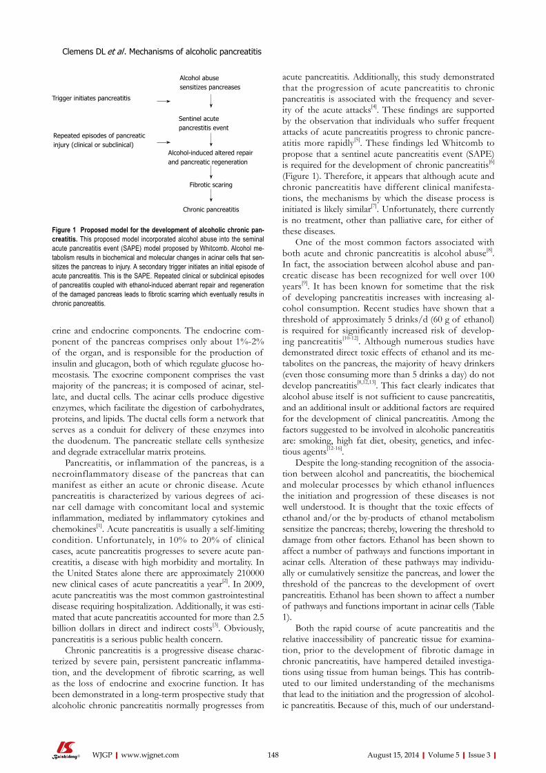
Clemens DL et al . Mechanisms of alcoholic pancreatitis
crine and endocrine components. The endocrine com-ponent of the pancreas comprises only about 1%-2% of the organ, and is responsible for the production of insulin and glucagon, both of which regulate glucose ho-meostasis. The exocrine component comprises the vast majority of the pancreas; it is composed of acinar, stel-late, and ductal cells. The acinar cells produce digestive enzymes, which facilitate the digestion of carbohydrates, proteins, and lipids. The ductal cells form a network that serves as a conduit for delivery of these enzymes into the duodenum. The pancreatic stellate cells synthesize and degrade extracellular matrix proteins.
Pancreatitis, or inflammation of the pancreas, is a necroinflammatory disease of the pancreas that can manifest as either an acute or chronic disease. Acute pancreatitis is characterized by various degrees of aci-nar cell damage with concomitant local and systemic inflammation, mediated by inflammatory cytokines and chemokines[1]. Acute pancreatitis is usually a self-limiting condition. Unfortunately, in 10% to 20% of clinical cases, acute pancreatitis progresses to severe acute pan-creatitis, a disease with high morbidity and mortality. In the United States alone there are approximately 210000 new clinical cases of acute pancreatitis a year[2]. In 2009, acute pancreatitis was the most common gastrointestinal disease requiring hospitalization. Additionally, it was esti-mated that acute pancreatitis accounted for more than 2.5 billion dollars in direct and indirect costs[3]. Obviously, pancreatitis is a serious public health concern.
Chronic pancreatitis is a progressive disease charac-terized by severe pain, persistent pancreatic inflamma-tion, and the development of fibrotic scarring, as well as the loss of endocrine and exocrine function. It has been demonstrated in a long-term prospective study that alcoholic chronic pancreatitis normally progresses from
acute pancreatitis. Additionally, this study demonstrated that the progression of acute pancreatitis to chronic pancreatitis is associated with the frequency and sever-ity of the acute attacks[4]. These findings are supported by the observation that individuals who suffer frequent attacks of acute pancreatitis progress to chronic pancre-atitis more rapidly[5]. These findings led Whitcomb to propose that a sentinel acute pancreatitis event (SAPE) is required for the development of chronic pancreatitis[6] (Figure 1). Therefore, it appears that although acute and chronic pancreatitis have different clinical manifesta-tions, the mechanisms by which the disease process is initiated is likely similar[7]. Unfortunately, there currently is no treatment, other than palliative care, for either of these diseases.
One of the most common factors associated with both acute and chronic pancreatitis is alcohol abuse[8]. In fact, the association between alcohol abuse and pan-creatic disease has been recognized for well over 100 years[9]. It has been known for sometime that the risk of developing pancreatitis increases with increasing al-cohol consumption. Recent studies have shown that a threshold of approximately 5 drinks/d (60 g of ethanol) is required for significantly increased risk of develop-ing pancreatitis[10-12]. Although numerous studies have demonstrated direct toxic effects of ethanol and its me-tabolites on the pancreas, the majority of heavy drinkers (even those consuming more than 5 drinks a day) do not develop pancreatitis[8,12,13]. This fact clearly indicates that alcohol abuse itself is not sufficient to cause pancreatitis, and an additional insult or additional factors are required for the development of clinical pancreatitis. Among the factors suggested to be involved in alcoholic pancreatitis are: smoking, high fat diet, obesity, genetics, and infec-tious agents[12-16].
Despite the long-standing recognition of the associa-tion between alcohol and pancreatitis, the biochemical and molecular processes by which ethanol influences the initiation and progression of these diseases is not well understood. It is thought that the toxic effects of ethanol and/or the by-products of ethanol metabolism sensitize the pancreas; thereby, lowering the threshold to damage from other factors. Ethanol has been shown to affect a number of pathways and functions important in acinar cells. Alteration of these pathways may individu-ally or cumulatively sensitize the pancreas, and lower the threshold of the pancreas to the development of overt pancreatitis. Ethanol has been shown to affect a number of pathways and functions important in acinar cells (Table 1).
Both the rapid course of acute pancreatitis and the relative inaccessibility of pancreatic tissue for examina-tion, prior to the development of fibrotic damage in chronic pancreatitis, have hampered detailed investiga-tions using tissue from human beings. This has contrib-uted to our limited understanding of the mechanisms that lead to the initiation and the progression of alcohol-ic pancreatitis. Because of this, much of our understand-
August 15, 2014|Volume 5|Issue 3|WJGP|www.wjgnet.com 148
Trigger initiates pancreatitis
Repeated episodes of pancreaticinjury (clinical or subclinical)
Alcohol abuse sensitizes pancreases
Sentinel acute pancrestitis event
Alcohol-induced altered repair and pancreatic regeneration
Fibrotic scaring
Chronic pancreatitis
Figure 1 Proposed model for the development of alcoholic chronic pan-creatitis. This proposed model incorporated alcohol abuse into the seminal acute pancreatitis event (SAPE) model proposed by Whitcomb. Alcohol me-tabolism results in biochemical and molecular changes in acinar cells that sen-sitizes the pancreas to injury. A secondary trigger initiates an initial episode of acute pancreatitis. This is the SAPE. Repeated clinical or subclinical episodes of pancreatitis coupled with ethanol-induced aberrant repair and regeneration of the damaged pancreas leads to fibrotic scarring which eventually results in chronic pancreatitis.
ing of pancreatitis in general, and alcoholic pancreatitis in particular, has come from the use of preclinical animal models. Preclinical models used to investigate alcoholic pancreatitis normally utilize mice or rats administered ethanol. Ethanol administration to experimental animals is commonly accomplished through the Tsukamoto-French intragastric method[17], the Lieber-DeCarli pair feeding method[18], or the Cook-Meadows model of pro-viding ethanol in the drinking water[19,20]. Pancreatic cells are either isolated from the animals administered ethanol or pancreatitis is induced. Among the more common methods of inducing pancreatitis in these animals are: bile duct ligation, treatment with supraphysiological concentrations of the cholecystokinin (CCK) analogue caerulein, or treatment with trinitrobenzene sulfonic acid (TNBS)[21]. More recently, methods designed to be more clinically relevant have been reported. These methods include chronic ethanol administration followed by treat-ment with gram-negative bacterial lipopolysaccharide (LPS)[22,23], or infection with Coxsackievirus CVB3[16,24,25].
Unfortunately, no animal model of chronic pancre-atitis recapitulates all of the manifestations of chronic pancreatitis in human beings. It has been demonstrated that alcohol administration to rats and mice results in ac-inar cell loss and enhanced fibrosis in animals subjected to caerulein-induced pancreatic injury[26,27]. Therefore, these models may be useful in elucidating the mecha-nisms by which ethanol alters normal pancreatic repair, and predisposes the pancreas to fibrosis.
It is the focus of this article to review and highlight some of the molecular events that may adversely affect the pancreas, and sensitize the pancreas to the initiation or progression of alcoholic pancreatitis.
ETHANOL METABOLISMMany of the deleterious effects of ethanol are attributed to the by-products produced during its metabolism. Like the hepatocytes of the liver, the pancreatic acinar cells have the ability to metabolize ethanol by both oxidative and nonoxidative pathways. The oxidative metabolism of ethanol is catalyzed by two enzymes: the cytosolic enzyme, alcohol dehydrogenase, and the microsomal enzyme, cytochrome P450 2E1. Ethanol metabolism by both of these enzymes generates acetaldehyde and reac-tive oxygen species. Although the pancreas expresses both alcohol dehydrogenase and cytochrome P450 2E1, the capacity for ethanol oxidation by the pancreas is sig-
nificantly less than that of the liver[28,29]. Therefore, the actions of the oxidative metabolites of ethanol oxidation may result from both pancreatic metabolism and sys-temic metabolism of ethanol.
Nonoxidative metabolism of ethanol is carried out by a number of enzymes, the most important being the fatty acid ethyl ester synthases. Metabolism of ethanol by these enzymes generates fatty acid ethyl esters (FAEEs). The pancreas possesses high fatty acid ester synthase activity. Thus, the capacity for nonoxidative metabolism of ethanol in the pancreas is high[30]. In fact, a study of individuals who were intoxicated at the time of death revealed that the concentration of FAEEs in the pan-creas was higher than any other organ analyzed[30]. Thus, because the oxidative metabolism of ethanol in the pan-creas is relatively low, the nonoxidative metabolism of ethanol may be more important and the production of FAEEs, and their toxic effects, may be accentuated. Be-cause the by-products of ethanol metabolism have been demonstrated to cause toxicity in other organs, a great deal of work has been performed investigating the ac-tions of the various ethanol metabolites on the pancreas.
CELL DEATHCell death during an episode of acute pancreatitis can occur by one of two mechanisms: apoptosis or necrosis. The distinction between the two types of cell death not only has biological implications in the development of acute pancreatitis, but also affects the clinical presenta-tion by influencing the severity of the illness[8]. Clinically, according to the 2012 Atlanta Classification of Acute Pancreatitis, the presence of necrosis and the number of organs affected by the subsequent inflammatory re-sponse determines the severity of acute pancreatitis (mild, moderate, severe) and dictates the short-term and long-term management of these patients[31].
While necrosis and apoptosis both lead to cell death, their respective mechanisms of achieving this end are quite different. Apoptosis, or programmed cell death, is a process by which cellular constituents are cleaved by cysteine-dependent, aspartate-directed enzymes, known as caspases. Apoptosis is mediated by caspases 3 and cas-pases 8. Caspase 8 is the initiator of the caspase cascade and cleaves caspase 3, which mediates many of the cellu-lar changes that lead to apoptotic death. In pancreatitis, these caspases are activated by the release of cytochrome c from mitochondria[32]. The release of cytochrome c is caused by the depolarization of mitochondria. It appears this depolarization is a result of the opening of the mi-tochondrial permeability transition pore, which is caused by sustained increased calcium levels in the cytosol[33]. Ultimately, there is an organized dismantling of the cell. This leads to cell shrinkage and nuclear chromatin con-densation, while preserving the integrity of the plasma membrane. Because the plasma membrane remains in-tact, there is very little leakage of intracellular material into the extracellular space, and therefore; there is little
August 15, 2014|Volume 5|Issue 3|WJGP|www.wjgnet.com 149
Table 1 Mechanisms by which ethanol is thought to sensitize the pancreas to pancreatitis
Alteration of cell death pathwaysAltered vesicular traffickingImpaired autophagyImpaired tissue repairER stressMitochondrial dysfunction
Clemens DL et al . Mechanisms of alcoholic pancreatitis
activation of inflammatory cytokines. In contrast to the organized dismantling of the cell
in apoptosis, necrosis involves intracellular swelling of organelles and rupture of the plasma membrane. This results in the release of the contents of the cell into the extracellular space, which causes an inflammatory response. It has been shown in a number of preclinical animal models of pancreatitis that the severity of pan-creatitis is increased with increasing necrotic cell death[8]. Additionally, perhaps the most important prognostic indicator of the severity of pancreatitis in human beings is the amount of necrosis[31].
In preclinical animal models of pancreatitis, ethanol has been shown to cause a shift in cell death from apop-tosis to necrosis. This shift has been shown to occur through several mechanisms. It has been shown that the nonoxidative metabolites of ethanol, FAEEs, activate inositol trisphosphate receptors on the endoplasmic re-ticulum. Activation of these receptors causes release of calcium into the cytosol. As stated above, the sustained increases in cytosolic calcium results in mitochondria de-polarization and loss of ATP production. Without ATP, the cells are unable to complete the apoptotic process and necrosis occurs[8].
Ethanol has also been shown to inhibit the JAK2/STAT1 pathway. Attenuated activity of this pathway leads to decreased activity of both caspase 8 and caspase 3[32]. With lower activity of these caspases, cell death by necrosis is increased while apoptotic cell death is re-duced.
Ethanol also increases the pancreatic expression of cathepsin B[32]. Cathepsin B is a cysteine protease that is thought to play a major role in the intrapancreatic conversion of trypsinogen to trypsin. It has been shown that in pancreata of ethanol-fed rats, increased expres-sion of cathepsin B result from increased levels of the transcriptional activators Ets-1 and Sp1[32]. Increases in Sp1 and Ets-1 enhance expression of cathepsin B, which leads to activation of trypsin and a shift from apoptosis to necrosis in pancreatic acinar cells[32]. These findings demonstrate that ethanol can affect the mechanism of cell death in acinar cells, and thereby influence the sever-ity of the disease.
EFFECTS OF ETHANOL ON ZYMOGEN SECRETIONOne of the primary roles of the exocrine pancreas is the synthesis and secretion of digestive enzymes. The pan-creas is protected from the actions of these potentially dangerous enzymes because they are synthesized as inac-tive zymogens and packaged into exocytotic vacuoles, known as zymogen granules. Although ethanol has many effects on acinar cells that contribute to the development of pancreatitis, the inappropriate activation of zymogens is likely a critical component of this pathologic process.
Activation of trypsinogen is generally considered a pivotal event in the initiation of pancreatitis[34]. It has
been reported by Gorlelich that treatment of isolated acinar cells with intoxicating concentrations of ethanol (25 mmol/L) sensitizes acinar cells to damage by causing the activation of zymogens[35]. The activation of these zymogens required an increase in cytosolic calcium and appeared to involve a low pH compartment (acid granu-lar compartment).
Local cytosolic spikes of calcium in the apical region of acinar cells control the exocytotic secretion of zy-mogens. These spikes are generated by release of small quantities of calcium from internal stores[36]. In contrast, prolonged, global elevation of calcium results in the formation of empty looking zymogen granules, this is thought to be the site where trypsin is activated. In aci-nar cells treated with the FAEE palmitoleic acid ethyl ester, calcium was released from both the endoplasmic reticulum (the major calcium storage compartment of the cell) and the acid granular compartment, located near the apical surface. Additionally, it was demonstrated that the calcium release was primarily mediated by type 2 and 3 inositol 1,4,5 trisphosphate receptors[37].
Normally, zymogens are released from acinar cells by fusion of zymogen granules with the apical membrane. This fusion results in their release into the ducts, where they are transported to the duodenum and activated. The components absolutely required for membrane fusion consist of: SNAREs (soluble NSF [N-ethylmaleimide-sensitive fusion proteins] attachment proteins recep-tors) located on the target membrane, t-SNARES, and v-SNAREs, also known as vesicle-associated membrane proteins (VAMPs), located on the membrane of the vesicle. The t-SNARES syntaxin and synaptosome-associated proteins (SNAPs), form a SNARE complex that binds to its cognate v-SNARE; thus, juxtaposing the two membranes and facilitating the fusion of the mem-branes.
Interestingly, it has been demonstrated both in vivo and in vitro, that supramaximal treatment with cholecys-tokinin (CCK) causes basolateral exocytosis of zymogen granules in acinar cells[38]. Additionally, in both ethanol-fed rats or isolated acinar cells treated with physiologic concentrations of ethanol (20 mmol/L), stimulation with submaximal concentration of CCK or carbachol resulted in the exocytosis being redirected from the api-cal surface, where zymogens are normally secreted, to the basolateral surface[39]. The authors postulate that the ensuing ectopic activation of the zymogens in the interstitial space results in pancreatitis[39]. More detailed investigations demonstrated that this inappropriate exocytosis was mediated by phosphorylation of mam-malian uncoordinated-18c (Munc 18c) by protein kinase C-alpha (PKC-α). Phosphorylation of Munc-18c results in its release from syntaxin-4, which is located on the basolateral surface of acinar cells. Syntaxin-4 is then able to complex with SNAP-23 and VAMP-8, located on the zymogen granules, to form the SNARE complex, which mediates the inappropriate basolateral exocytosis of zy-mogens[40]. Importantly, basolateral exocytosis has been
August 15, 2014|Volume 5|Issue 3|WJGP|www.wjgnet.com 150
Clemens DL et al . Mechanisms of alcoholic pancreatitis
observed in tissue samples from a patient suffering from chronic alcoholic pancreatitis[41].
IMPAIRMENT OF AUTOPHAGYAutophagy is a cellular process in which unnecessary or damaged cellular components or organelles are se-questered in vacuoles and transported to the lysosomes. Upon fusion with the lysosomes, the contents of the autophagic vacuoles, the autophagosomes, are degraded. Not only does this process perform an important role in ridding cells of unneeded components, but during times of low nutrient availability autophagy can provide the cell with needed constituents.
Impaired autophagy has been implicated in the pathogenesis of many diseases, including pancreati-tis[15,42-45]. Importantly, it has been shown that ethanol can alter the process of autophagy in a number of organs, including the pancreas[43,46,47].
One of the histological hallmarks of pancreatitis is the accumulation of large vacuoles within acinar cells[48]. In a number of preclinical animal models of pancreatitis, as well as in tissue from a patient with acute pancreatitis, it has been demonstrated that these vacuoles are in fact autophagic vacuoles[44,45]. Further investigation revealed that these vacuoles possessed markers of both autopha-gosomes and lysosomes, and contained undegraded or partially degraded cellular material[45]. These findings in-dicate that at least the very late events in the autophagic process, namely the degradation of the components of the autolysosomes, are impaired during pancreatitis[45]. Thus, autophagy is activated during pancreatitis, and it appears that the impairment in the ability to complete this process is responsible for the vacuolization charac-teristic of this disease.
As mentioned above, trypsin activation is thought to be an early event in the initiation of pancreatitis. How this activation occurs is not well understood. It is generally thought that cathepsin B, is mis-sorted to the zymogens granules, where it co-localizes with trypsino-gen. Subsequent cleavage of trypsinogen by cathepsin B results in the production of active trypsin. How tryp-sinogen and cathepsin B come in contact has always been a mystery. It now appears that the impairment in the completion of the autophagy may have a role in the co-mingling of these two enzymes.
Cathepsin L is an enzyme that degrades trypsinogen and trypsin, and cathepsin B is an enzyme that cleaves trypsinogen forming active trypsin. The two are impor-tant lysosomal hydrolases. During pancreatitis, increased levels of these enzymes are found in the zymogen gran-ule fraction. Additionally, in alcoholic pancreatitis, as well as other forms of acute pancreatitis, the processing and activation of cathepsin L and cathepsin B is im-paired[45,49]. Furthermore, it appears that the impairment in cathepsin L activity is more severe than the impair-ment in cathepsin B activity, particularly in the zymogen granule fraction[45]. Importantly, zymogen granules were
detected in the autophagosomes/autoloysosomes. The authors propose that it is in these autophagosomes/au-toloysosomes that trypsinogen and cathepsin B come in contact[45]. The imbalance between cathepsin B and cathepsin L activity in these vacuoles would favor the activation of trypsin, and the initiation of pancreatitis. Thus, impairment in the completion of the autophagic process and subsequent increase in autolysosomes may contribute not only to the accumulation of vacuoles, but also to the inappropriate intracellular activation of tryp-sin and the initiation of pancreatitis.
Ethanol has been shown to impair other aspects of autophagy. Using a model of alcoholic pancreatitis in which rats were chronically fed ethanol and then treated with LPS to induce acute pancreatitis, Fortunato et al[43] demonstrated that in the pancreata of these animals fusion of autophagosomes with the lysosome was im-paired. Additional studies demonstrated that Lamp-2, a lysosomal membrane protein required for the fusion of autophagosomes with lysosomes, was depleted in the pancreata of rats suffering from alcoholic pancreati-tis[43,50]. Furthermore, analysis of pancreata from human beings revealed that Lamp-2 was also decreased in the pancreata of patients suffering from chronic alcoholic pancreatitis. These results indicate that the ethanol-mediated reduction in lysosomal proteins, particularly Lamp-2, and subsequent impairment in autophagy may be a contributing factor to alcoholic pancreatitis in hu-man beings. Although not investigated, the authors spec-ulated that disruption in the autophagic pathway may contribute to bioenergenic failure in mitochondria. Lack of mitochondrial ATP would favor necrosis, as opposed to apoptosis. Necrotic cell death would cause inflamma-tion and lead to the initiation of pancreatitis[43].
MITOCHONDRIAL DYSFUNCTIONPancreatic acinar cells are among the most synthetically active cells in the body[51]. This synthetic activity requires a great deal of energy. Because of this, acinar cells con-tain an inordinate number of mitochondria. Thus, the actions of toxins, such as ethanol, that affect mitochon-dria can dramatically affect acinar cells.
Normally, acetylcholine or cholecystokinin bind to G-protein linked receptors that are located on the plasma membrane of acinar cells and stimulate the production of secondary messengers. The secondary messengers bind to inositol trisphosphate or ryanodine receptors located on the endoplasmic reticulum, zymo-gen granules, and endo-lysosomes. This binding results in the transient release of free calcium. Mitochondria take up this calcium, which results in their activation, the synthesis of ATP, and the secretion of zymogens.
Aberrant calcium signaling has long been consid-ered an important factor in the initiation of pancreatic injury[52]. Pathological calcium signaling in acinar cells re-sults from prolonged global release of calcium from the endoplasmic reticulum, as well as zymogen granules and
August 15, 2014|Volume 5|Issue 3|WJGP|www.wjgnet.com 151
Clemens DL et al . Mechanisms of alcoholic pancreatitis
endo-lysosomes. In fact, early acinar cell injury (vacu-olization, trypsin activation, and basolateral zymogen secretion) does not occur without prolonged, sustained release of calcium[53].
Both nonoxidative and oxidative metabolism of ethanol has been shown to contribute to mitochondrial dysfunction and acinar cell death. FAEEs, the nonoxida-tive metabolites of ethanol, have been shown to cause pancreatic injury by affecting calcium signaling in acinar cells[54,55]. FAEEs increase the intracellular concentration of calcium to toxic levels. This calcium increase is medi-ated by activation of the inositol trisphosphate receptors located on the endoplasmic reticulum, and results in global sustained increase in intracellular calcium, which causes mitochondrial membrane permeability. Mito-chondrial membrane permeability can lead to cell death by either apoptosis or necrosis[56,57].
Mitochondrial membrane permeability results from opening of the mitochondrial permeability transition pore. The mitochondrial permeability transition pore is thought to have at least three major components, the voltage dependent anion channel (VDAC) located on the outer mitochondrial membrane, adenine nucleotide translocase (ANT) located in the inner mitochondrial membrane and cyclophlin-D located within the mito-chondrial matrix[53].
One of the important consequences of the opening of the mitochondrial permeability transition pore and mitochondrial membrane permeability can be loss of the mitochondrial membrane potential (∆ΨΜ). Loss of the ∆ΨΜ results in the decreased ability of TP.
Depleted levels of ATP exacerbate the cells ability to regulate calcium by inhibiting the activity of the impor-tant ATP-dependent calcium pumps, the sacroplasmic/endoplasmic reticular calcium ATPase (SERCA) located on the ER, and the plasma membrane calcium ATPase (PMCA) located on the plasma membrane. Thus, mi-tochondrial membrane permeability can exacerbate the dysregulation of calcium homeostasis and lead to acinar cell necrosis.
The oxidative metabolism of ethanol also has delete-rious effects on pancreatic mitochondria. Oxidative me-tabolism of ethanol by alcohol dehydrogenase requires oxidized nicotinamide adenine dinucleotide (NAD+) as a cofactor, and results in the production of acetalde-hyde and reduced nicotinamide adenine dinucleotide (NADH)[58,59]. Acetaldehyde is then metabolized to ac-etate, primarily by the mitochondrial enzyme aldehyde dehydrogenase-2. Importantly, this reaction also requires NAD+ as a cofactor, and also results in the production of NADH[58,59]. Thus, metabolism of acetaldehyde to acetate further depletes the availability of NAD+.
Using isolated acinar cells treated with ethanol, Shal-bueva et al[60] demonstrated that ethanol treatment led to a decrease in the NAD+/NADH ratio. This reduction in NAD+ resulted in activation of the mitochondrial permeability transition pore, mitochondrial depolariza-tion, ATP depletion, and eventually cellular necrosis[60].
Furthermore, their studies revealed that the ethanol oxidation-mediated polarization of pancreatic mitochon-dria was attenuated in acinar cells isolated from mice deficient in cyclophilin-D. These results indicate a role for cyclophilin-D in this ethanol metabolism-mediated mitochondrial dysfunction.
Interestingly, it has been shown in mitochondria iso-lated from the liver that ethanol metabolism sensitizes the mitochondrial permeability transition pore to open, in part, through increased cyclophilin-D activity and in-creased association of cyclophilin-D with ANT[61]. This increased activity is associated with hyperacetylation of cyclophilin-D. Acetylation of cyclophilin-D is regulated by sirtuin-3, a NAD+-dependent deacetylase localized in the mitochondrial matrix[62]. The ethanol oxidation-mediated decrease in NAD+ leads to decreased sirtuin-3 activity and the hyperacetylation of cyclophilin-D. Hyperacetylation of cyclophilin-D results in increased cyclophilin-D activity, increased binding to ANT, and mitochondrial permeability transition pore induction[61]. Thus, it is tempting to speculate that the ethanol oxida-tion-mediated induction of the mitochondrial perme-ability transition pore in pancreatic mitochondria is me-diated by a similar NAD+-sirtuin-3-cyclophilin-D axis.
ENDOPLASMIC RETICULUM STRESS AND THE UNFOLDED PROTEIN RESPONSE Acinar cells are responsible for the production and se-cretion of large quantities of digestive enzymes. Because of this, in addition to large numbers of mitochondria, acinar cells possess an extensive endoplasmic reticulum network. The endoplasmic reticulum is the major storage site of calcium in the cell, and is the cellular organelle where the proper folding and trafficking of secretory proteins is determined. Endoplasmic reticulum stress re-sulting from excessive accumulation of proteins, calcium imbalance, oxidative stress, or accumulation of damaged or misfolded protein leads to a response known as the unfolded protein response (UPR)[63].
One hallmark of the UPR is the activation of the IRE1/XBP1 pathway. Inositol-requiring transmembrane kinase/endonuclease 1 (IRE1) splices X-box binding protein 1 (XBP1) messenger RNA, resulting in spliced XBP1. Spliced XBP1 is a transcriptional activator that regulates a number of genes, which encode proteins that act as ER chaperones, are involved in the proper folding of proteins, or are involved in the degradation of dam-aged or misfolded proteins.
The UPR is activated by pancreatic injury[64]. Ad-ditionally, it has been shown that the UPR is activated in acinar cells by long-term ethanol administration to mice[65]. Ethanol mediated UPR was characterized by in-creased expression of IRE1 and spliced XBP1. Although the UPR was activated, ethanol administration alone did not result in histopathologic changes to the pancreas. In contrast, administration of ethanol to mice with di-minished XBP1 expression (XBP1+/- mice) resulted in
August 15, 2014|Volume 5|Issue 3|WJGP|www.wjgnet.com 152
Clemens DL et al . Mechanisms of alcoholic pancreatitis
acinar vacuolization, cell necrosis, and inflammation[65]. The presence of pathologic changes in the pancreata of XBP1+/- mice led the authors to suggest that the UPR is a protective mechanism in acinar cells during endoplas-mic reticulum stress. Exceeding the capacity of the UPR to compensate for endoplasmic reticulum stress results in overt pancreatitis. Thus, if the protective capacity of the UPR is exceeded, this pathway may contribute to the induction and progression of pancreatitis.
THE ROLE OF STELLATE CELLS IN ALCO-HOLIC PANCREATITISThe pancreas, like the liver, has a population of vitamin A storing cells known as stellate cells. Pancreatic stel-late cells are periacinar cells located in interacinar and interlobular areas of the pancreas[66,67]. These cells are responsible for the synthesis of extracellular matrix proteins, as well as matrix metalloproteinases (enzymes that degrade extracellular matrix proteins). Thus, it ap-pears that in the healthy organ, pancreatic stellate cells function to maintain the architecture of the pancreas by regulating the deposition and degradation of extracel-lular matrix components[68]. In response to pancreatic in-jury, pancreatic stellate cells are activated and transform into myofibroblast-like cells. Activated pancreatic stellate cells synthesize excessive amounts of extracellular matrix proteins. The accumulation of these proteins results in fibrosis. Thus, pancreatic stellate cells are intimately in-volved in the regulation of both normal and pathologic aspects of the pancreatitis[68,69].
Pancreatic stellate cells of both rat and human origin have the ability to metabolize ethanol through the oxi-dative pathway[70,71]. Rat pancreatic stellate cells possess alcohol dehydrogenase, the activity of this enzyme is in-duced when cells are exposed to ethanol concentrations routinely found in the blood of inebriated individuals[70]. Recently, it has also been reported that quiescent pancre-atic stellate cells in human beings possess alcohol dehy-drogenase activity. Additionally, this activity appeared to be upregulated in pancreatic stellate cells of individuals suffering from chronic pancreatitis and pancreatic can-cer[71].
The fact that pancreatic stellate cells possess alcohol dehydrogenase activity may contribute to the develop-ment of alcoholic pancreatitis. Pancreatic stellate cells are activated when exposed to concentrations of ethanol detected in the blood of inebriated individuals (10-50 mmol/L)[70,72]. Additionally, pancreatic stellate cells iso-lated from both rats and human beings are activated by acetaldehyde. Ethanol and acetaldehyde not only activate pancreatic stellate cells, but also elicit responses that may have important biological consequences. Both ethanol and acetaldehyde have been shown to induce the secre-tion of matrix metalloproteinases in pancreatic stellate cells[73]. Furthermore, treatment of pancreatic stellate cells with ethanol induces the synthesis of interleukin-8 and connective tissue growth factor (CTGF)[72,74]. It has
been suggested that these factors act in an autocrine manner to perpetuate the activation of pancreatic stellate cells[13]. This finding may help to explain both the appar-ent inability of the pancreas to fully recover from injury in the continued presence of ethanol, and the extremely common association between alcohol abuse and chronic pancreatitis.
Although it is well established that pancreatic stel-late cells are primarily responsible for the deposition and degradation of components of the extracellular matrix, it appears that acinar cells exposed to ethanol may also contribute to the increase in extracellular matrix deposi-tion. It has been shown that FAEEs can increase the levels of extracellular matrix proteins by inhibiting the acinar cell activity of plasmin and urokinase-type plas-minogen activator (uPA) proteins involved in the degra-dation of the extracellular matrix components[75].
THE ROLE OF THE INFLAMMATORY RE-SPONSEInflammation mediated by cytokines, chemokines, and adhesion molecules is involved in the development of pancreatitis[1,76,77]. Interestingly, it appears that ethanol and its metabolites have a differential effect on the ex-pression of molecules that regulate the inflammatory response. It has been shown that treatment of isolated acini with ethanol or acetaldehyde decreased the activity of two important transcriptional activators involved in the inflammatory response, specifically nuclear factor-κB (NF-κB) and activator protein 1 (AP-1). Conversely, treatment of acini with FAEEs increased the activation of these regulators of the inflammatory response[78].
The activity of NF-κB is also reduced in the pan-creata of animals chronically fed ethanol[79]. However, it was demonstrated that induction of pancreatitis in rats chronically administered ethanol resulted in increased NF-κB activity, as well as increases in the mRNA levels of a number of proinflammatory cytokines, includ-ing: tumor necrosis factor-alpha (TNF-α), interleukin-6 (IL-6), macrophage inflammatory protein-1 (MIP-1), and monocyte chemotactic protein-1 (MCP-1)[79]. These re-sults led the authors to suggest that the in vitro and in vivo down-regulation of these factors by ethanol reflected a protective mechanism to prevent the development of alcohol-induced pancreas[78,79].
The role of the inflammatory response in chronic al-coholic pancreatitis has also been investigated[80]. Focus-ing on the resident mononuclear cells of the pancreas, Deng et al[80] demonstrated that chronic ethanol admin-istration reduced the number of these cells present in the pancreas. In agreement with others, they suggested that this reduction likely reflected a general immunologic suppression in the pancreas of ethanol-fed rats, and may explain why animals chronically provided ethanol do not develop chronic pancreatitis in the absence of acute pancreatic damage[80].
Despite this immunologic suppression, when pan-
August 15, 2014|Volume 5|Issue 3|WJGP|www.wjgnet.com 153
Clemens DL et al . Mechanisms of alcoholic pancreatitis
creatitis was induced by caerulein, the inflammatory response in these animals was enhanced[80]. Further-more, following repeated caerulein-induced episodes of pancreatitis, it was shown that the expression of both pro-inflammatory cytokines such as TNF-α, MIP-1α, and RANTES (regulated on activation normal T cell expressed and secreted), as well as the anti-inflammatory cytokines tissue growth factor-b (TGF-b) and interleu-kin-10 (IL-10) was enhanced. The increase in cytokine expression was only observed in rats fed ethanol and subjected to repeated episodes of acute pancreatitis, and was also associated with increased activation of pan-creatic stellate cells and fibrosis. These findings led the authors to suggest that ethanol acts not only to sensitize the pancreas to acute pancreatitis, but also aids in the progression of chronic pancreatitis if repeated episodes of acute pancreatitis occur[80].
EFFECTS OF ETHANOL ON PANCREATIC REPAIRIt is generally accepted that fibrosis is an aberrant repair response. It appears that in the presence of ethanol, repair of the damaged pancreas is altered or never fully completed[26,27]. This may help to explain the extremely common association between alcohol abuse and chronic pancreatitis. Because ethanol and acetaldehyde can acti-vate stellate cells, and FAEEs inhibit the degradation of extracellular matrix proteins, it is obvious that ethanol can also influence recovery of the pancreas after damage has occurred[70,72,75].
It has been demonstrated that chronic ethanol ad-ministration also delays regeneration of the damaged pancreas[81]. This delay was associated with an ethanol-mediated decrease in the expression of important devel-opmental factors, such as PDX-1 and PTF-1a, as well as impaired activation of the Notch signaling pathway[24]. Normal pancreatic repair requires the dedifferentiation of mature acinar cells followed by their redifferentia-tion[82]. Thus, ethanol-mediated alterations in the expres-sion of these important developmental factors affect the dedifferentiation/redifferentiation of acinar cells. These alterations may dramatically influence pancreatic repair.
As mentioned above, there is a close association between alcohol abuse and chronic pancreatitis. In fact, in developed countries, alcohol abuse is associated with over 70% of the reported cases[83]. Importantly, individu-als suffering from chronic pancreatitis have a 20-fold greater likelihood of developing pancreatic cancer[84], a disease with a dismal prognosis. It is thought that chang-es that occur in the pancreas during chronic injury are associated with, or predispose the organ to, the initiation of pancreatic neoplasia. Because one of the seminal characteristics of chronic pancreatitis is aberrant tissue repair, resulting in fibrotic scarring, and ethanol con-sumption alters pancreatic repair, ethanol may have an indirect role in the initiation of pancreatic cancer. Thus, the effects of ethanol on repair of the damaged pan-
creas may be a contributing factor in pancreatic cancer, as well as alcoholic pancreatitis.
CONCLUSIONDespite the dramatic expansion of our understanding of pancreatitis in general, and how ethanol and its metabo-lites affect pancreatic cells, we still have not defined the mechanism of alcoholic pancreatitis. Instead, it is evident that ethanol has a plethora of toxic affects on pancreatic cells. Because all of these effects occur simultaneously, it is likely that the cumulative effects of ethanol sensitize the pancreas to damage, and that “alcoholic pancreati-tis” is a multifactorial disease. Paradoxically, despite the demonstration that ethanol has numerous toxic effects on the pancreas, data from demographic studies and pre-clinical animal models has firmly established that ethanol itself does not cause pancreatitis. Because ethanol does not cause pancreatitis, but only sensitizes the pancreas to disease, it appears that the pancreas has developed protective mechanisms that can partially compensate for ethanol-induced cellular damage. Some of these protective mechanisms have been identified. It is likely that additional compensatory mechanisms exist. Further defining the mechanisms of ethanol-induced pancreatic injury may help define these protective mechanisms. It is hoped that this strategy will lead to the development of therapeutic targets that will prevent or reduce the sever-ity of alcoholic pancreatitis.
REFERENCES1 Vonlaufen A, Apte MV, Imhof BA, Frossard JL. The role of
inflammatory and parenchymal cells in acute pancreatitis. J Pathol 2007; 213: 239-248 [PMID: 17893879 DOI: 10.1002/path.2231]
2 Carroll JK, Herrick B, Gipson T, Lee SP. Acute pancreatitis: diagnosis, prognosis, and treatment. Am Fam Physician 2007; 75: 1513-1520 [PMID: 17555143]
3 Peery AF, Dellon ES, Lund J, Crockett SD, McGowan CE, Bulsiewicz WJ, Gangarosa LM, Thiny MT, Stizenberg K, Morgan DR, Ringel Y, Kim HP, Dibonaventura MD, Carroll CF, Allen JK, Cook SF, Sandler RS, Kappelman MD, Sha-heen NJ. Burden of gastrointestinal disease in the United States: 2012 update. Gastroenterology 2012; 143: 1179-1187.e1-3 [PMID: 22885331 DOI: 10.1053/j.gastro.2012.08.002]
4 Ammann RW, Muellhaupt B. Progression of alcoholic acute to chronic pancreatitis. Gut 1994; 35: 552-556 [PMID: 8174996]
5 Lankisch PG, Breuer N, Bruns A, Weber-Dany B, Lowenfels AB, Maisonneuve P. Natural history of acute pancreatitis: a long-term population-based study. Am J Gastroenterol 2009; 104: 2797-2805; quiz 2806 [PMID: 19603011 DOI: 10.1038/ajg.2009.405]
6 Whitcomb DC. Hereditary pancreatitis: new insights into acute and chronic pancreatitis. Gut 1999; 45: 317-322 [PMID: 10446089]
7 Vonlaufen A, Wilson JS, Apte MV. Molecular mechanisms of pancreatitis: current opinion. J Gastroenterol Hepatol 2008; 23: 1339-1348 [PMID: 18853993 DOI: 10.1111/j.1440-1746.2008.05520.x]
8 Pandol SJ, Saluja AK, Imrie CW, Banks PA. Acute pancreati-tis: bench to the bedside. Gastroenterology 2007; 132: 1127-1151 [PMID: 17383433 DOI: 10.1053/j.gastro.2007.01.055]
August 15, 2014|Volume 5|Issue 3|WJGP|www.wjgnet.com 154
Clemens DL et al . Mechanisms of alcoholic pancreatitis
9 Friederich. Disease of the Pancreas. Cycloaedia of the Prac-tice of Medicine. Vol 18. New York: William Wood, 1878: 254-312
10 Yadav D, Hawes RH, Brand RE, Anderson MA, Money ME, Banks PA, Bishop MD, Baillie J, Sherman S, DiSario J, Burton FR, Gardner TB, Amann ST, Gelrud A, Lawrence C, Elinoff B, Greer JB, O’Connell M, Barmada MM, Slivka A, Whitcomb DC. Alcohol consumption, cigarette smoking, and the risk of recurrent acute and chronic pancreatitis. Arch Intern Med 2009; 169: 1035-1045 [PMID: 19506173 DOI: 10.1001/archinternmed.2009.125]
11 Kristiansen L, Grønbaek M, Becker U, Tolstrup JS. Risk of pancreatitis according to alcohol drinking habits: a popula-tion-based cohort study. Am J Epidemiol 2008; 168: 932-937 [PMID: 18779386 DOI: 10.1093/aje/kwn222]
12 Yadav D, Whitcomb DC. The role of alcohol and smoking in pancreatitis. Nat Rev Gastroenterol Hepatol 2010; 7: 131-145 [PMID: 20125091]
13 Apte MV, Pirola RC, Wilson JS. Mechanisms of alcoholic pancreatitis. J Gastroenterol Hepatol 2010; 25: 1816-1826 [PMID: 21091991 DOI: 10.1111/j.1440-1746.2010.06445.x]
14 Yadav D, Papachristou GI, Whitcomb DC. Alcohol-asso-ciated pancreatitis. Gastroenterol Clin North Am 2007; 36: 219-238, vii [PMID: 17533076]
15 Gukovsky I, Li N, Todoric J, Gukovskaya A, Karin M. In-flammation, autophagy, and obesity: common features in the pathogenesis of pancreatitis and pancreatic cancer. Gas-troenterology 2013; 144: 1199-1209.e4 [PMID: 23622129 DOI: 10.1053/j.gastro.2013.02.007]
16 Jerrells TR, Chapman N, Clemens DL. Animal model of alcoholic pancreatitis: role of viral infections. Pancreas 2003; 27: 301-304 [PMID: 14576491]
17 Ueno A, Lazaro R, Wang PY, Higashiyama R, Machida K, Tsukamoto H. Mouse intragastric infusion (iG) model. Nat Protoc 2012; 7: 771-781 [PMID: 22461066 DOI: 10.1038/nprot.2012.014]
18 Lieber CS, DeCarli LM. The feeding of alcohol in liquid diets: two decades of applications and 1982 update. Alcohol Clin Exp Res 1982; 6: 523-531 [PMID: 6758624]
19 Song K, Coleman RA, Zhu X, Alber C, Ballas ZK, Wald-schmidt TJ, Cook RT. Chronic ethanol consumption by mice results in activated splenic T cells. J Leukoc Biol 2002; 72: 1109-1116 [PMID: 12488491]
20 D’Souza El-Guindy NB, Kovacs EJ, De Witte P, Spies C, Littleton JM, de Villiers WJ, Lott AJ, Plackett TP, Lan-zke N, Meadows GG. Laboratory models available to study alcohol-induced organ damage and immune varia-tions: choosing the appropriate model. Alcohol Clin Exp Res 2010; 34: 1489-1511 [PMID: 20586763 DOI: 10.1111/j.1530-0277.2010.01234.x]
21 Puig-Diví V, Molero X, Vaquero E, Salas A, Guarner F, Mal-agelada J. Ethanol feeding aggravates morphological and biochemical parameters in experimental chronic pancreati-tis. Digestion 1999; 60: 166-174 [PMID: 10095159]
22 Vonlaufen A, Xu Z, Daniel B, Kumar RK, Pirola R, Wilson J, Apte MV. Bacterial endotoxin: a trigger factor for alco-holic pancreatitis? Evidence from a novel, physiologically relevant animal model. Gastroenterology 2007; 133: 1293-1303 [PMID: 17919500 DOI: 10.1053/j.gastro.2007.06.062]
23 Vonlaufen A, Phillips PA, Xu Z, Zhang X, Yang L, Pirola RC, Wilson JS, Apte MV. Withdrawal of alcohol promotes regression while continued alcohol intake promotes persis-tence of LPS-induced pancreatic injury in alcohol-fed rats. Gut 2011; 60: 238-246 [PMID: 20870739]
24 Schneider KJ, Scheer M, Suhr M, Clemens DL. Ethanol administration impairs pancreatic repair after injury. Pan-creas 2012; 41: 1272-1279 [PMID: 22617711 DOI: 10.1097/MPA.0b013e31824bde37]
25 Jerrells TR, Vidlak D, Strachota JM. Alcoholic pancreatitis: mechanisms of viral infections as cofactors in the develop-
ment of acute and chronic pancreatitis and fibrosis. J Leukoc Biol 2007; 81: 430-439 [PMID: 17095612]
26 Gukovsky I, Lugea A, Shahsahebi M, Cheng JH, Hong PP, Jung YJ, Deng QG, French BA, Lungo W, French SW, Tsukamoto H, Pandol SJ. A rat model reproducing key pathological responses of alcoholic chronic pancreatitis. Am J Physiol Gastrointest Liver Physiol 2008; 294: G68-G79 [PMID: 17884979 DOI: 10.1152/ajpgi.00006.2007]
27 Perides G , Tao X, West N, Sharma A, Steer ML. A mouse model of ethanol dependent pancreatic fibrosis. Gut 2005; 54: 1461-1467 [PMID: 15870229 DOI: 10.1136/gut.2004.062919]
28 Haber PS, Apte MV, Applegate TL, Norton ID, Korsten MA, Pirola RC, Wilson JS. Metabolism of ethanol by rat pan-creatic acinar cells. J Lab Clin Med 1998; 132: 294-302 [PMID: 9794700]
29 Norton ID, Apte MV, Haber PS, McCaughan GW, Pirola RC, Wilson JS. Cytochrome P4502E1 is present in rat pan-creas and is induced by chronic ethanol administration. Gut 1998; 42: 426-430 [PMID: 9577353]
30 Laposata EA, Lange LG. Presence of nonoxidative ethanol metabolism in human organs commonly damaged by etha-nol abuse. Science 1986; 231: 497-499 [PMID: 3941913]
31 Banks PA, Bollen TL, Dervenis C, Gooszen HG, Johnson CD, Sarr MG, Tsiotos GG, Vege SS. Classification of acute pancreatitis--2012: revision of the Atlanta classification and definitions by international consensus. Gut 2013; 62: 102-111 [PMID: 23100216 DOI: 10.1136/gutjnl-2012-302779]
32 Wang YL, Hu R, Lugea A, Gukovsky I, Smoot D, Gukovs-kaya AS, Pandol SJ. Ethanol feeding alters death signaling in the pancreas. Pancreas 2006; 32: 351-359 [PMID: 16670617 DOI: 10.1097/01.mpa.0000220859.93496.e1]
33 Odinokova IV, Sung KF, Mareninova OA, Hermann K, Evtodienko Y, Andreyev A, Gukovsky I, Gukovskaya AS. Mechanisms regulating cytochrome c release in pancreatic mitochondria. Gut 2009; 58: 431-442 [PMID: 18596195 DOI: 10.1136/gut.2007.147207]
34 Petersen OH, Gerasimenko OV, Gerasimenko JV. Pathobi-ology of acute pancreatitis: focus on intracellular calcium and calmodulin. F1000 Med Rep 2011; 3: 15 [PMID: 21876721 DOI: 10.3410/M3-15]
35 Gorelick FS. Alcohol and zymogen activation in the pancre-atic acinar cell. Pancreas 2003; 27: 305-310 [PMID: 14576492 DOI: 10.1097/00006676-200311000-00006]
36 Petersen OH, Tepikin AV. Polarized calcium signaling in exocrine gland cells. Annu Rev Physiol 2008; 70: 273-299 [PMID: 17850212 DOI: 10.1146/annurev.physiol.70.113006.100618]
37 Gerasimenko JV, Lur G, Sherwood MW, Ebisui E, Tepikin AV, Mikoshiba K, Gerasimenko OV, Petersen OH. Pancre-atic protease activation by alcohol metabolite depends on Ca2+ release via acid store IP3 receptors. Proc Natl Acad Sci USA 2009; 106: 10758-10763 [PMID: 19528657 DOI: 10.1073/pnas.0904818106]
38 Gaisano HY, Lutz MP, Leser J, Sheu L, Lynch G, Tang L, Tamori Y, Trimble WS, Salapatek AM. Supramaximal cholecystokinin displaces Munc18c from the pancreatic acinar basal surface, redirecting apical exocytosis to the basal membrane. J Clin Invest 2001; 108: 1597-1611 [PMID: 11733555 DOI: 10.1172/JCI9110]
39 Cosen-Binker LI, Lam PP, Binker MG, Reeve J, Pandol S, Gaisano HY. Alcohol/cholecystokinin-evoked pancre-atic acinar basolateral exocytosis is mediated by protein kinase C alpha phosphorylation of Munc18c. J Biol Chem 2007; 282: 13047-13058 [PMID: 17324928 DOI: 10.1074/jbc.M611132200]
40 Cosen-Binker LI, Binker MG, Wang CC, Hong W, Gaisano HY. VAMP8 is the v-SNARE that mediates basolateral exo-cytosis in a mouse model of alcoholic pancreatitis. J Clin Invest 2008; 118: 2535-2551 [PMID: 18535671 DOI: 10.1172/JCI34672]
August 15, 2014|Volume 5|Issue 3|WJGP|www.wjgnet.com 155
Clemens DL et al . Mechanisms of alcoholic pancreatitis
41 Gaisano HY, Sheu L, Whitcomb D. Alcoholic chronic pan-creatitis involves displacement of Munc18c from the pan-creatic acinar basal membrane surface. Pancreas 2004; 28: 395-400 [PMID: 15097857]
42 Kroemer G, Mariño G, Levine B. Autophagy and the inte-grated stress response. Mol Cell 2010; 40: 280-293 [PMID: 20965422 DOI: 10.1016/j.cell.2007.12.018]
43 Fortunato F, Bürgers H, Bergmann F, Rieger P, Büchler MW, Kroemer G, Werner J. Impaired autolysosome forma-tion correlates with Lamp-2 depletion: role of apoptosis, autophagy, and necrosis in pancreatitis. Gastroenterology 2009; 137: 350-360, 360.e1-5 [PMID: 19362087 DOI: 10.1053/j.gastro.2009.04.003]
44 Hashimoto D, Ohmuraya M, Hirota M, Yamamoto A, Suyama K, Ida S, Okumura Y, Takahashi E, Kido H, Araki K, Baba H, Mizushima N, Yamamura K. Involvement of autophagy in trypsinogen activation within the pancreatic acinar cells. J Cell Biol 2008; 181: 1065-1072 [PMID: 18591426 DOI: 10.1083/jcb.200712156]
45 Mareninova OA, Hermann K, French SW, O’Konski MS, Pandol SJ, Webster P, Erickson AH, Katunuma N, Gorelick FS, Gukovsky I, Gukovskaya AS. Impaired autophagic flux mediates acinar cell vacuole formation and trypsinogen activation in rodent models of acute pancreatitis. J Clin Invest 2009; 119: 3340-3355 [PMID: 19805911 DOI: 10.1172/JCI38674]
46 Ding WX, Li M, Chen X, Ni HM, Lin CW, Gao W, Lu B, Stolz DB, Clemens DL, Yin XM. Autophagy reduces acute ethanol-induced hepatotoxicity and steatosis in mice. Gas-troenterology 2010; 139: 1740-1752 [PMID: 20659474 DOI: 10.1053/j.gastro.2010.07.041]
47 Thomes PG, Ehlers RA, Trambly CS, Clemens DL, Fox HS, Tuma DJ, Donohue TM. Multilevel regulation of autophago-some content by ethanol oxidation in HepG2 cells. Autopha-gy 2013; 9: 63-73 [PMID: 23090141 DOI: 10.4161/auto.22490]
48 Helin H, Mero M, Markkula H, Helin M. Pancreatic acinar ultrastructure in human acute pancreatitis. Virchows Arch A Pathol Anat Histol 1980; 387: 259-270 [PMID: 7456314]
49 Mareninova OA, Yakubov I, French SW, O’Konski MS, Pandol SJ, Webster P, Erickson AH, Katunuma N, Gorelick FS, Gukovsky I, Gukovskaya AS. Ethanol feeding causes ly-sosomal dysfunction in exocrine pancreas similar to pancre-atitis; but in contrast to pancreatitis, ethanol down-regulates autophagy. Gastroenterol 2010; 138: S-148
50 Huynh KK, Eskelinen EL, Scott CC, Malevanets A, Saftig P, Grinstein S. LAMP proteins are required for fusion of lyso-somes with phagosomes. EMBO J 2007; 26: 313-324 [PMID: 17245426 DOI: 10.1038/sj.emboj.7601511]
51 Case RM. Synthesis, intracellular transport and discharge of exportable proteins in the pancreatic acinar cell and other cells. Biol Rev Camb Philos Soc 1978; 53: 211-354 [PMID: 208670]
52 Sah RP, Dawra RK, Saluja AK. New insights into the patho-genesis of pancreatitis. Curr Opin Gastroenterol 2013; 29: 523-530 [PMID: 23892538 DOI: 10.1097/MOG.0b013e328363e399]
53 Mukherjee R, Criddle DN, Gukovskaya A, Pandol S, Pe-tersen OH, Sutton R. Mitochondrial injury in pancreatitis. Cell Calcium 2008; 44: 14-23 [PMID: 18207570 DOI: 10.1016/j.ceca.2007.11.013]
54 Criddle DN, Murphy J, Fistetto G, Barrow S, Tepikin AV, Ne-optolemos JP, Sutton R, Petersen OH. Fatty acid ethyl esters cause pancreatic calcium toxicity via inositol trisphosphate re-ceptors and loss of ATP synthesis. Gastroenterology 2006; 130: 781-793 [PMID: 16530519 DOI: 10.1053/j.gastro.2005.12.031]
55 Criddle DN, Raraty MG, Neoptolemos JP, Tepikin AV, Petersen OH, Sutton R. Ethanol toxicity in pancreatic acinar cells: mediation by nonoxidative fatty acid metabolites. Proc Natl Acad Sci USA 2004; 101: 10738-10743 [PMID: 15247419 DOI: 10.1073/pnas.0403431101]
56 Kroemer G, Galluzzi L, Brenner C. Mitochondrial mem-
brane permeabilization in cell death. Physiol Rev 2007; 87: 99-163 [PMID: 17237344 DOI: 10.1152/physrev.00013.2006]
57 Rasola A, Bernardi P. Mitochondrial permeability transi-tion in Ca(2+)-dependent apoptosis and necrosis. Cell Calcium 2011; 50: 222-233 [PMID: 21601280 DOI: 10.1016/j.ceca.2011.04.007]
58 Ehrig T, Bosron WF, Li TK. Alcohol and aldehyde dehydro-genase. Alcohol Alcohol 1990; 25: 105-116 [PMID: 2198030]
59 Crabb DW, Bosron WF, Li TK. Ethanol metabolism. Pharma-col Ther 1987; 34: 59-73 [PMID: 3310044]
60 Shalbueva N, Mareninova OA, Gerloff A, Yuan J, Waldron RT, Pandol SJ, Gukovskaya AS. Effects of oxidative alcohol metabolism on the mitochondrial permeability transition pore and necrosis in a mouse model of alcoholic pancreati-tis. Gastroenterology 2013; 144: 437-446.e6 [PMID: 23103769 DOI: 10.1053/j.gastro.2012.10.037]
61 Shulga N, Pastorino JG. Ethanol sensitizes mitochondria to the permeability transition by inhibiting deacetylation of cyclophilin-D mediated by sirtuin-3. J Cell Sci 2010; 123: 4117-4127 [PMID: 21062897 DOI: 10.1242/jcs.073502]
62 Shulga N, Wilson-Smith R, Pastorino JG. Sirtuin-3 deacety-lation of cyclophilin D induces dissociation of hexokinase II from the mitochondria. J Cell Sci 2010; 123: 894-902 [PMID: 20159966 DOI: 10.1242/jcs.061846]
63 Kim I, Xu W, Reed JC. Cell death and endoplasmic reticu-lum stress: disease relevance and therapeutic opportunities. Nat Rev Drug Discov 2008; 7: 1013-1030 [PMID: 19043451 DOI: 10.1038/nrd2755]
64 Kubisch CH, Sans MD, Arumugam T, Ernst SA, Williams JA, Logsdon CD. Early activation of endoplasmic reticulum stress is associated with arginine-induced acute pancreatitis. Am J Physiol Gastrointest Liver Physiol 2006; 291: G238-G245 [PMID: 16574987 DOI: 10.1152/ajpgi.00471.2005]
65 Lugea A, Tischler D, Nguyen J, Gong J, Gukovsky I, French SW, Gorelick FS, Pandol SJ. Adaptive unfolded protein response attenuates alcohol-induced pancreatic damage. Gastroenterology 2011; 140: 987-997 [PMID: 21111739 DOI: 10.1053/j.gastro.2010.11.038]
66 Apte MV, Haber PS, Applegate TL, Norton ID, McCaughan GW, Korsten MA, Pirola RC, Wilson JS. Periacinar stellate shaped cells in rat pancreas: identification, isolation, and culture. Gut 1998; 43: 128-133 [PMID: 9771417]
67 Bachem MG, Schneider E, Gross H, Weidenbach H, Schmid RM, Menke A, Siech M, Beger H, Grünert A, Adler G. Iden-tification, culture, and characterization of pancreatic stellate cells in rats and humans. Gastroenterology 1998; 115: 421-432 [PMID: 9679048]
68 Apte M, Pirola R, Wilson J. The fibrosis of chronic pancre-atitis: new insights into the role of pancreatic stellate cells. Antioxid Redox Signal 2011; 15: 2711-2722 [PMID: 21728885]
69 Apte MV, Pirola RC, Wilson JS. Pancreatic stellate cells: a starring role in normal and diseased pancreas. Front Physiol 2012; 3: 344 [PMID: 22973234 DOI: 10.1089/ars.2011.4079]
70 Apte MV, Phillips PA, Fahmy RG, Darby SJ, Rodgers SC, McCaughan GW, Korsten MA, Pirola RC, Naidoo D, Wilson JS. Does alcohol directly stimulate pancreatic fibrogenesis? Studies with rat pancreatic stellate cells. Gastroenterology 2000; 118: 780-794 [PMID: 10734030]
71 Chiang CP, Wu CW, Lee SP, Ho JL, Lee SL, Nieh S, Yin SJ. Expression pattern, ethanol-metabolizing activities, and cellular localization of alcohol and aldehyde dehy-drogenases in human small intestine. Alcohol Clin Exp Res 2012; 36: 2047-2058 [PMID: 23231010 DOI: 10.1111/j.1530-0277.2009.00927.x]
72 Masamune A, Satoh A, Watanabe T, Kikuta K, Satoh M, Su-zuki N, Satoh K, Shimosegawa T. Effects of ethanol and its metabolites on human pancreatic stellate cells. Dig Dis Sci 2010; 55: 204-211 [PMID: 19165599 DOI: 10.1007/s10620-008-0695-y]
73 Phillips PA, McCarroll JA, Park S, Wu MJ, Pirola R, Korsten
August 15, 2014|Volume 5|Issue 3|WJGP|www.wjgnet.com 156
Clemens DL et al . Mechanisms of alcoholic pancreatitis
M, Wilson JS, Apte MV. Rat pancreatic stellate cells secrete matrix metalloproteinases: implications for extracellular matrix turnover. Gut 2003; 52: 275-282 [PMID: 12524413]
74 Lawrencia C, Charrier A, Huang G, Brigstock DR. Ethanol-mediated expression of connective tissue growth factor (CCN2) in mouse pancreatic stellate cells. Growth Factors 2009; 27: 91-99 [PMID: 19280452 DOI: 10.1080/08977190902786319]
75 Lugea A, Gukovsky I, Gukovskaya AS, Pandol SJ. Nonoxi-dative ethanol metabolites alter extracellular matrix protein content in rat pancreas. Gastroenterology 2003; 125: 1845-1859 [PMID: 14724836]
76 Bhatia M, Brady M, Shokuhi S, Christmas S, Neoptolemos JP, Slavin J. Inflammatory mediators in acute pancreatitis. J Pathol 2000; 190: 117-125 [PMID: 10657008 DOI: 10.1002/(SICI)1096-9896(200002)190:2<117::AID-PATH494>3.0.CO;2-K]
77 Norman J. The role of cytokines in the pathogenesis of acute pancreatitis. Am J Surg 1998; 175: 76-83 [PMID: 9445247]
78 Gukovskaya AS, Mouria M, Gukovsky I, Reyes CN, Kasho VN, Faller LD, Pandol SJ. Ethanol metabolism and tran-scription factor activation in pancreatic acinar cells in rats. Gastroenterology 2002; 122: 106-118 [PMID: 11781286]
79 Pandol SJ, Periskic S, Gukovsky I, Zaninovic V, Jung Y,
Zong Y, Solomon TE, Gukovskaya AS, Tsukamoto H. Etha-nol diet increases the sensitivity of rats to pancreatitis in-duced by cholecystokinin octapeptide. Gastroenterology 1999; 117: 706-716 [PMID: 10464148]
80 Deng X, Wang L, Elm MS, Gabazadeh D, Diorio GJ, Eagon PK, Whitcomb DC. Chronic alcohol consumption acceler-ates fibrosis in response to cerulein-induced pancreatitis in rats. Am J Pathol 2005; 166: 93-106 [PMID: 15632003]
81 Clemens DL, Jerrells TR. Ethanol consumption potenti-ates viral pancreatitis and may inhibit pancreas regenera-tion: preliminary findings. Alcohol 2004; 33: 183-189 [PMID: 15596086]
82 Jensen JN, Cameron E, Garay MV, Starkey TW, Gianani R, Jensen J. Recapitulation of elements of embryonic develop-ment in adult mouse pancreatic regeneration. Gastroenterol-ogy 2005; 128: 728-741 [PMID: 15765408]
83 Ammann RW, Heitz PU, Klöppel G. Course of alcoholic chronic pancreatitis: a prospective clinicomorphological long-term study. Gastroenterology 1996; 111: 224-231 [PMID: 8698203]
84 Krejs GJ. Pancreatic cancer: epidemiology and risk factors. Dig Dis 2010; 28: 355-358 [PMID: 20814212 DOI: 10.1159/000319414]
P- Reviewer: Pan WS, Pezzilli R, Sakata N S- Editor: Song XX L- Editor: A E- Editor: Lu YJ
August 15, 2014|Volume 5|Issue 3|WJGP|www.wjgnet.com 157
Clemens DL et al . Mechanisms of alcoholic pancreatitis
TOPIC HIGHLIGHT
Early phase of acute pancreatitis: Assessment and management
Veit Phillip, Jörg M Steiner, Hana Algül
Veit Phillip, Jörg M Steiner, Hana Algül, II. Medizinische Klinik und Poliklinik, Klinikum rechts der Isar der Technischen Universität München, 81675 München, GermanyJörg M Steiner, Gastrointestinal Laboratory, Department of Small Animal Clinical Sciences, College of Veterinary Medi-cine and Biomedical Sciences, Texas A and M University, Col-lege Station, TX 77843-4474, United StatesAuthor contributions: Phillip V and Algül H collected and analyzed the literature; Phillip V prepared the original draft; Steiner JM and Algül H supplemented the material and revised the manuscript; all authors approved the final version of the pa-per to be published.Correspondence to: Hana Algül, MD, MPH, II. Medizinische Klinik und Poliklinik, Klinikum rechts der Isar der Technische Universität München, Ismaninger Straße 22, 81675 München, Germany. [email protected]: +49-89-41405215 Fax: +49-89-41406794Received: January 29, 2014 Revised: March 25, 2014 Accepted: May 29, 2014Published online: August 15, 2014
AbstractAcute pancreatitis (AP) is a potentially life-threatening disease with a wide spectrum of severity. The overall mortality of AP is approximately 5%. According to the revised Atlanta classification system, AP can be clas-sified as mild, moderate, or severe. Severe AP often takes a clinical course with two phases, an early and a late phase, which should both be considered separately. In this review article, we first discuss general aspects of AP, including incidence, pathophysiology, etiology, and grading of severity, then focus on the assessment of patients with suspected AP, including diagnosis and risk stratification, followed by the management of AP during the early phase, with special emphasis on fluid therapy, pain management, nutrition, and antibiotic prophylaxis.
© 2014 Baishideng Publishing Group Inc. All rights reserved.
Key words: Acute pancreatitis; Incidence; Pathophysiol-ogy; Etiology; Severity; Risk stratification; Fluid thera-py; Pain management; Nutrition; Antibiotic prophylaxis
Core tip: Acute pancreatitis is a frequent and potential-ly life-threatening disease. Therapy is currently mostly symptomatic with fluid resuscitation, pain manage-ment, and early oral feeding. Vigorous fluid resuscita-tion remains a cornerstone of early management of acute pancreatitis. Cross-sectional imaging during the early phase of evaluation has not been associated with improvement in outcome. There is no role for prophy-lactic antibiotics in the management of the early phase of acute pancreatitis (AP). Enteral nutrition in AP can reduce mortality, systemic infections, and multiorgan dysfunction compared to parenteral nutrition. Immedi-ate endoscopic retrograde cholangiography is indicated only in patients with biliary pancreatitis with common bile duct obstruction and cholangitis.
Phillip V, Steiner JM, Algül H. Early phase of acute pancre-atitis: Assessment and management. World J Gastrointest Pathophysiol 2014; 5(3): 158-168 Available from: URL: http://www.wjgnet.com/2150-5330/full/v5/i3/158.htm DOI: http://dx.doi.org/10.4291/wjgp.v5.i3.158
INTRODUCTIONAcute pancreatitis (AP) is a potentially life-threatening disease with a wide spectrum of severity. The reported incidence of acute pancreatitis differs depending on geo-graphic location and ranges from 14.7/100000 person years in the Netherlands to 45.1/100000 person years in Japan[1,2]. However, most studies show an incidence between 30 and 45/100000 person years[2-7]. Many stud-ies report an increase in incidence over the last few de-cades[2,3,8], however, it is a matter of debate whether this
World J Gastrointest Pathophysiol 2014 August 15; 5(3): 158-168ISSN 2150-5330 (online)
© 2014 Baishideng Publishing Group Inc. All rights reserved.
Online Submissions: http://www.wjgnet.com/esps/Help Desk: http://www.wjgnet.com/esps/helpdesk.aspxDOI: 10.4291/wjgp.v5.i3.158
August 15, 2014|Volume 5|Issue 3|WJGP|www.wjgnet.com 158
WJGP 5th Anniversary Special Issues (3): Pancreatitis
Phillip V et al . Early phase of acute pancreatitis
represents a real increase in incidence due to increasing biliary AP in an increasingly obese population or wheth-er this rise in incidence is due to improved diagnostic capabilities, a higher level of suspicion of this disease, or an overestimation of retrospective studies using admin-istrative diagnostic codes[9-11]. In 2009, AP was the most common principal gastrointestinal diagnosis at discharge in the Unite States with estimated inpatient costs of $2.6 billion per year. Furthermore, it was the 14th most com-mon cause of death with a crude rate of 1.0 per 100000 inhabitants[12]. The overall mortality of AP is about 5% and can reach up to 20%-30% in patients with severe AP and infected necrosis[13,14]. While there seems to be an increase in incidence, several studies have reported a decrease in mortality. Again, this could be a real decrease due to an earlier diagnosis and better therapeutic options or it may also be due to an improved sensitivity of diag-nostic modalities, leading to an increase in the diagnosis of mild forms of pancreatitis[15].
In this review article, we first discuss general aspects of AP, including pathophysiology, etiology, and grading of severity, then focus on the assessment of patients with suspected AP, including diagnosis and risk stratifi-cation, followed by the management of AP during the early phase with special emphasis on fluid therapy, pain management, nutrition, and antibiotic prophylaxis.
PATHOPHYSIOLOGYThe pathophysiology of AP with multi organ failure (MOF) is poorly understood. Researchers have long hy-pothesized that AP results from premature activation of digestive enzymes within the pancreas, a process referred to as autodigestion. Indeed, inherited mutations in genes encoding for digestive enzymes have been found in pa-tients with a hereditary form of pancreatitis[16]. However, affected patients develop chronic, rather than acute pan-creatitis. Therefore, in recent years, a novel concept has evolved, suggesting that systemic complications during AP result from uncontrolled activation of the inflamma-tory cascade. As indicated above, severe AP is associated with a significant mortality. Thus, early identification of severe forms of AP is crucial for outcome. In an attempt to identify surrogate parameters as predictors for severe AP, several association studies linking cytokines and chemokines with AP severity have been conducted[17]. Among these, serum levels of interleukin (IL)-6 and the IL-6-dependent acute phase protein, C-reactive protein (CRP) were identified as the most reliable predictors for severe AP[18,19]. Recent results from basic research have established that IL-6 or CRP are not only relevant mark-ers to predict the severity of AP, but that the cytokine IL-6 also has a substantial pathophysiological impact on the course of the disease[19]. While excessive stimulation of the inflammatory cascade [hyper-inflammatory state, systemic inflammatory response syndrome (SIRS)] ac-counts for early systemic complications, paralysis of the inflammatory response, also termed compensatory anti-
inflammatory response syndrome (CARS), contributes to local complications and sepsis associated with the late phase of the disease. Although these definitions are largely non-specific, they are undeniably useful in the clinical and research setting. Among the agents con-tributing to this anti-inflammatory response, IL-10 may be of importance. In fact, the protective role of IL-10 in experimental studies in animal models has been well documented[20]. Thus, the hypo-inflammatory status of CARS might facilitate superinfections that lead to exten-sive necrosis and/or septic complications. This interplay of these two contrasting phenomena requires an indi-vidualized therapeutic approach[20-22].
ETIOLOGYThe identification of the etiology of AP is crucial for the management during the early phase of the disease and also for the prevention of recurrence of AP. Although there is no specific therapy for AP, the causing factor, e.g., choledocholithiasis in biliary AP, must be investi-gated and eliminated if identified. The most common causes of AP are gallstones and prolonged heavy use of alcohol, which together account for about 60%-80% of all cases. The incidence of biliary etiology differs con-siderably between different geographic regions. For ex-ample, there is a clear predominance for biliary AP over alcoholic AP in Greece (71.4% vs 6.0%) whereas the op-posite is the case for Finland (6.3% vs 79.3%)[23,24]. The regional differences in frequency of biliary and alcoholic etiology are shown in Figure 1[6,7,23-31].
Other causes of AP include ERCP (0.4% to 11%[32,33]), idiosyncratic reactions to drugs (0.1% to 2%)[34], hypertri-glyceridemia (1.1%-3.8%)[6,23,35], anatomic alterations[36], ge-netic predispositions[37], and other rare causes[38,39]. Despite a thorough clinical workup, 10%-25% of all cases remain idiopathic[6,11,23,33].
NATURAL COURSE OF ACUTE PANCRE-ATITISThe severity of AP can be subclinical, mild without organ dysfunction, or can be severe. Patients with mild disease often improve spontaneously and heal within a few days. However, patients with severe disease may develop life-threatening local and/or systemic complica-tions. According to the revised Atlanta classification sys-tem, AP can be classified as mild, moderate, or severe[40]. However, it is important to remember that AP is a rap-idly evolving, dynamic condition in which the severity may change rapidly during the course of the disease[40]. Severe AP often takes a clinical course with two phases, an early and a late one, which should both be considered separately[40].
The early phase, which usually lasts for about one week, is characterized by a complex inflammatory re-action. The course of AP starts with a systemic pro-inflammatory phase [systemic inflammatory response
August 15, 2014|Volume 5|Issue 3|WJGP|www.wjgnet.com 159
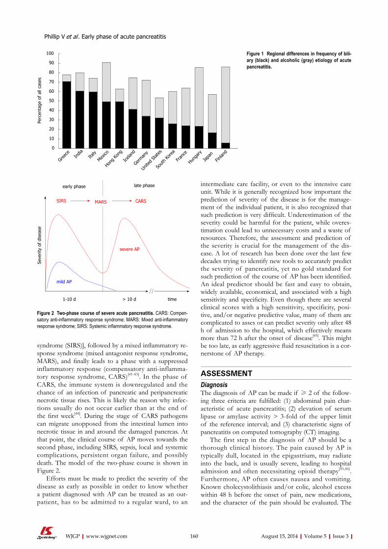
syndrome (SIRS)], followed by a mixed inflammatory re-sponse syndrome (mixed antagonist response syndrome, MARS), and finally leads to a phase with a suppressed inflammatory response (compensatory anti-inflamma-tory response syndrome, CARS)[41-43]. In the phase of CARS, the immune system is downregulated and the chance of an infection of pancreatic and peripancreatic necrotic tissue rises. This is likely the reason why infec-tions usually do not occur earlier than at the end of the first week[44]. During the stage of CARS pathogens can migrate unopposed from the intestinal lumen into necrotic tissue in and around the damaged pancreas. At that point, the clinical course of AP moves towards the second phase, including SIRS, sepsis, local and systemic complications, persistent organ failure, and possibly death. The model of the two-phase course is shown in Figure 2.
Efforts must be made to predict the severity of the disease as early as possible in order to know whether a patient diagnosed with AP can be treated as an out-patient, has to be admitted to a regular ward, to an
intermediate care facility, or even to the intensive care unit. While it is generally recognized how important the prediction of severity of the disease is for the manage-ment of the individual patient, it is also recognized that such prediction is very difficult. Underestimation of the severity could be harmful for the patient, while overes-timation could lead to unnecessary costs and a waste of resources. Therefore, the assessment and prediction of the severity is crucial for the management of the dis-ease. A lot of research has been done over the last few decades trying to identify new tools to accurately predict the severity of pancreatitis, yet no gold standard for such prediction of the course of AP has been identified. An ideal predictor should be fast and easy to obtain, widely available, economical, and associated with a high sensitivity and specificity. Even though there are several clinical scores with a high sensitivity, specificity, posi-tive, and/or negative predictive value, many of them are complicated to asses or can predict severity only after 48 h of admission to the hospital, which effectively means more than 72 h after the onset of disease[45]. This might be too late, as early aggressive fluid resuscitation is a cor-nerstone of AP therapy.
ASSESSMENTDiagnosisThe diagnosis of AP can be made if ≥ 2 of the follow-ing three criteria are fulfilled: (1) abdominal pain char-acteristic of acute pancreatitis; (2) elevation of serum lipase or amylase activity > 3-fold of the upper limit of the reference interval; and (3) characteristic signs of pancreatitis on computed tomography (CT) imaging.
The first step in the diagnosis of AP should be a thorough clinical history. The pain caused by AP is typically dull, located in the epigastrium, may radiate into the back, and is usually severe, leading to hospital admission and often necessitating opioid therapy[45,46]. Furthermore, AP often causes nausea and vomiting. Known cholecystolithiasis and/or colic, alcohol excess within 48 h before the onset of pain, new medications, and the character of the pain should be evaluated. The
August 15, 2014|Volume 5|Issue 3|WJGP|www.wjgnet.com 160
100
90
80
70
60
50
40
30
20
10
0
Perc
enta
ge o
f al
l cas
es
Greec
eIn
diaIta
ly
Mexico
Hong K
ong
Icelan
d
German
y
Unite
d Stat
es
Sout
h Kor
ea
Franc
e
Hunga
ry
Japa
nFin
land
Figure 1 Regional differences in frequency of bili-ary (black) and alcoholic (gray) etiology of acute pancreatitis.
severe AP
SIRS MARS CARS
mild AP
early phase late phase
1-10 d > 10 d time
Seve
rity
of d
isea
se
Figure 2 Two-phase course of severe acute pancreatitis. CARS: Compen-satory anti-inflammatory response syndrome; MARS: Mixed anti-inflammatory response syndrome; SIRS: Systemic inflammatory response syndrome.
Phillip V et al . Early phase of acute pancreatitis
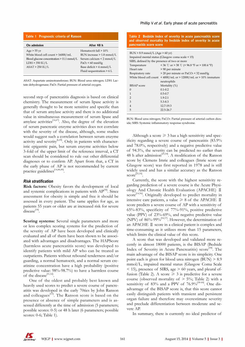
second step of pancreatitis diagnosis is based on clinical chemistry. The measurement of serum lipase activity is generally thought to be more sensitive and specific than that of serum amylase activity and there is no additional value in simultaneous measurement of serum lipase and amylase activities[14,47]. Also, the degree of the elevation of serum pancreatic enzyme activities does not correlate with the severity of the disease, although, some studies would suggest such a correlation between serum enzyme activity and severity[6,45]. Only in patients with character-istic epigastric pain, but serum enzyme activities below 3-fold of the upper limit of the reference interval, a CT scan should be considered to rule out other differential diagnoses or to confirm AP. Apart from that, a CT in the early phase of AP is not recommended by current practice guidelines[14,48,49].
Risk stratificationRisk factors: Obesity favors the development of local and systemic complications in patients with AP[50]. Since assessment for obesity is simple and free it should be assessed in every patient. The same applies for age, as patients 55 years or older are at increased risk for severe disease[14].
Scoring systems: Several single parameters and more or less complex scoring systems for the prediction of the severity of AP have been developed and clinically evaluated and all of them have been shown to be associ-ated with advantages and disadvantages. The HAPScore (harmless acute pancreatitis score) was developed to identify patients with mild AP who can be treated as outpatients. Patients without rebound tenderness and/or guarding, a normal hematocrit, and a normal serum cre-atinine concentration have a high probability (positive predictive value: 98%-98.7%) to have a harmless course of the disease[51,52].
One of the oldest and probably best known and heavily used scores to predict a severe course of pancre-atitis was developed in the early 70ties by John Ranson and colleagues[53]. The Ranson score is based on the presence or absence of simple parameters and is as-sessed differently at the time of admission (5 parameters; possible scores: 0-5) or 48 h later (6 parameters; possible scores: 0-6; Table 1).
Although a score ≥ 3 has a high sensitivity and spec-ificity regarding a severe course of pancreatitis (83.9% and 78.0%, respectively) and a negative predictive value of 94.5%, the severity can be predicted no earlier than 48 h after admission[25,54]. A modification of the Ranson score by Clemens Imrie and colleagues (Imrie score or Glasgow score) was first reported in 1978 and is still widely used and has a similar accuracy as the Ranson score[25,55].
Currently, the score with the highest sensitivity re-garding prediction of a severe course is the Acute Physi-ology And Chronic Health Evaluation (APACHE) Ⅱ score[14,56]. Originally developed to predict mortality in intensive care patients, a value ≥ 8 of the APACHE Ⅱ score predicts a severe course of AP with a sensitivity of 65%-83%, specificity of 77%-91%, positive predictive value (PPV) of 23%-69%, and negative predictive value (NPV) of 86%-99%[54,57]. However, the determination of an APACHE Ⅱ score in a clinical patient is complex and time-consuming as it utilizes more than 15 parameters, which limits the clinical value of this score.
A score that was developed and validated more re-cently in almost 18000 patients, is the BISAP (Bedside Index of Severity in Acute Pancreatitis) score[58]. The main advantage of the BISAP score is its simplicity. One point each is given for blood urea nitrogen (BUN) > 8.9 mmol/L, impaired mental status (Glasgow Coma Scale < 15), presence of SIRS, age > 60 years, and pleural ef-fusion (Table 2). A score ≥ 3 is predictive for a severe course (observed mortality of > 5%; Table 2) with a sensitivity of 83% and a PPV of 76.9%[58-60]. One dis-advantage of the BISAP score is, that this score cannot easily distinguish patients with transient and persistent organ failure and therefore may overestimate severity and preclude differentiation between moderate and se-vere AP.
In summary, there is currently no ideal predictor of
August 15, 2014|Volume 5|Issue 3|WJGP|www.wjgnet.com 161
Table 1 Prognostic criteria of Ranson
On admission After 48 h
Age > 55 yr Hematocrit fall > 10%White blood cell count > 16000/mL BUN increase > 1.8 mmol/LBlood glucose concentration > 11.1 mmol/L Serum calcium < 2 mmol/L LDH > 350 IU/L PaO2 < 60 mmHgASAT > 250 IU/L Base deficit > 4 mmol/L
Fluid sequestration > 6 L
ASAT: Aspartate aminotransferase; BUN: Blood urea nitrogen; LDH: Lac-tate dehydrogenase; PaO2: Partial pressure of arterial oxygen.
Table 2 Bedside index of severity in acute pancreatitis score and observed mortality by bedside index of severity in acute pancreatitis score score
BUN > 8.9 mmol/L (Age > 60 yr)Impaired mental status (Glasgow coma scale < 15)SIRS, defined by the presence of two or moreTemperature < 36 ℃ or > 38 ℃ (< 96.8 °F or > 100.4 °F)Heart rate > 90 per minuteRespiratory rate > 20 per minute or PaCO2 < 32 mmHgWhite blood cell count < 4000/mL or > 12000/mL or > 10% immature
neutrophilsBISAP score Mortality (%)0 0.1-0.21 0.5-0.72 1.9-2.13 5.3-8.34 12.7-19.35 22.5-26.7
BUN: Blood urea nitrogen; PaCO2: Partial pressure of arterial carbon diox-ide; SIRS: Systemic inflammatory response syndrome.
Phillip V et al . Early phase of acute pancreatitis
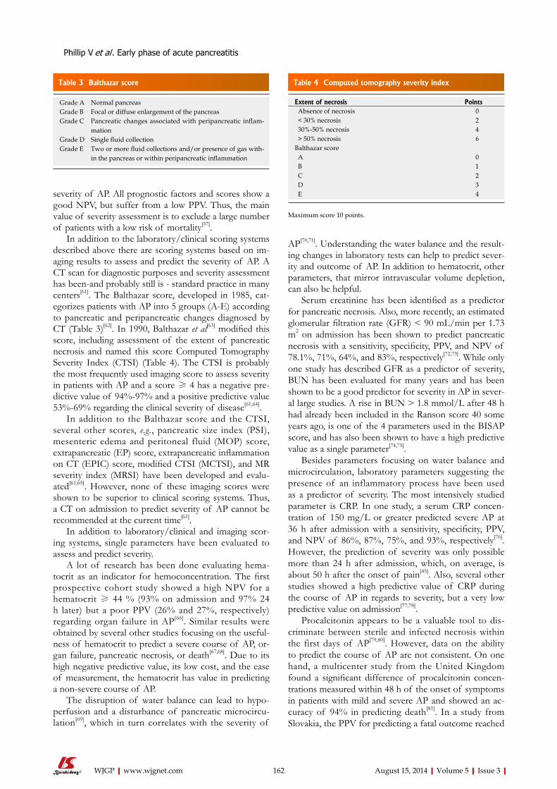
severity of AP. All prognostic factors and scores show a good NPV, but suffer from a low PPV. Thus, the main value of severity assessment is to exclude a large number of patients with a low risk of mortality[57].
In addition to the laboratory/clinical scoring systems described above there are scoring systems based on im-aging results to assess and predict the severity of AP. A CT scan for diagnostic purposes and severity assessment has been-and probably still is - standard practice in many centers[61]. The Balthazar score, developed in 1985, cat-egorizes patients with AP into 5 groups (A-E) according to pancreatic and peripancreatic changes diagnosed by CT (Table 3)[62]. In 1990, Balthazar et al[63] modified this score, including assessment of the extent of pancreatic necrosis and named this score Computed Tomography Severity Index (CTSI) (Table 4). The CTSI is probably the most frequently used imaging score to assess severity in patients with AP and a score ≥ 4 has a negative pre-dictive value of 94%-97% and a positive predictive value 53%-69% regarding the clinical severity of disease[61,64].
In addition to the Balthazar score and the CTSI, several other scores, e.g., pancreatic size index (PSI), mesenteric edema and peritoneal fluid (MOP) score, extrapancreatic (EP) score, extrapancreatic inflammation on CT (EPIC) score, modified CTSI (MCTSI), and MR severity index (MRSI) have been developed and evalu-ated[61,65]. However, none of these imaging scores were shown to be superior to clinical scoring systems. Thus, a CT on admission to predict severity of AP cannot be recommended at the current time[61].
In addition to laboratory/clinical and imaging scor-ing systems, single parameters have been evaluated to assess and predict severity.
A lot of research has been done evaluating hema-tocrit as an indicator for hemoconcentration. The first prospective cohort study showed a high NPV for a hematocrit ≥ 44 % (93% on admission and 97% 24 h later) but a poor PPV (26% and 27%, respectively) regarding organ failure in AP[66]. Similar results were obtained by several other studies focusing on the useful-ness of hematocrit to predict a severe course of AP, or-gan failure, pancreatic necrosis, or death[67,68]. Due to its high negative predictive value, its low cost, and the ease of measurement, the hematocrit has value in predicting a non-severe course of AP.
The disruption of water balance can lead to hypo-perfusion and a disturbance of pancreatic microcircu-lation[69], which in turn correlates with the severity of
AP[70,71]. Understanding the water balance and the result-ing changes in laboratory tests can help to predict sever-ity and outcome of AP. In addition to hematocrit, other parameters, that mirror intravascular volume depletion, can also be helpful.
Serum creatinine has been identified as a predictor for pancreatic necrosis. Also, more recently, an estimated glomerular filtration rate (GFR) < 90 mL/min per 1.73 m2 on admission has been shown to predict pancreatic necrosis with a sensitivity, specificity, PPV, and NPV of 78.1%, 71%, 64%, and 83%, respectively[72,73]. While only one study has described GFR as a predictor of severity, BUN has been evaluated for many years and has been shown to be a good predictor for severity in AP in sever-al large studies. A rise in BUN > 1.8 mmol/L after 48 h had already been included in the Ranson score 40 some years ago, is one of the 4 parameters used in the BISAP score, and has also been shown to have a high predictive value as a single parameter[74,75].
Besides parameters focusing on water balance and microcirculation, laboratory parameters suggesting the presence of an inflammatory process have been used as a predictor of severity. The most intensively studied parameter is CRP. In one study, a serum CRP concen-tration of 150 mg/L or greater predicted severe AP at 36 h after admission with a sensitivity, specificity, PPV, and NPV of 86%, 87%, 75%, and 93%, respectively[76]. However, the prediction of severity was only possible more than 24 h after admission, which, on average, is about 50 h after the onset of pain[45]. Also, several other studies showed a high predictive value of CRP during the course of AP in regards to severity, but a very low predictive value on admission[77,78].
Procalcitonin appears to be a valuable tool to dis-criminate between sterile and infected necrosis within the first days of AP[79,80]. However, data on the ability to predict the course of AP are not consistent. On one hand, a multicenter study from the United Kingdom found a significant difference of procalcitonin concen-trations measured within 48 h of the onset of symptoms in patients with mild and severe AP and showed an ac-curacy of 94% in predicting death[81]. In a study from Slovakia, the PPV for predicting a fatal outcome reached
August 15, 2014|Volume 5|Issue 3|WJGP|www.wjgnet.com 162
Table 3 Balthazar score
Grade A Normal pancreasGrade B Focal or diffuse enlargement of the pancreasGrade C Pancreatic changes associated with peripancreatic inflam-
mationGrade D Single fluid collectionGrade E Two or more fluid collections and/or presence of gas with-
in the pancreas or within peripancreatic inflammation
Table 4 Computed tomography severity index
Extent of necrosis Points Absence of necrosis 0 < 30% necrosis 2 30%-50% necrosis 4 > 50% necrosis 6Balthazar score A 0 B 1 C 2 D 3 E 4
Maximum score 10 points.
Phillip V et al . Early phase of acute pancreatitis
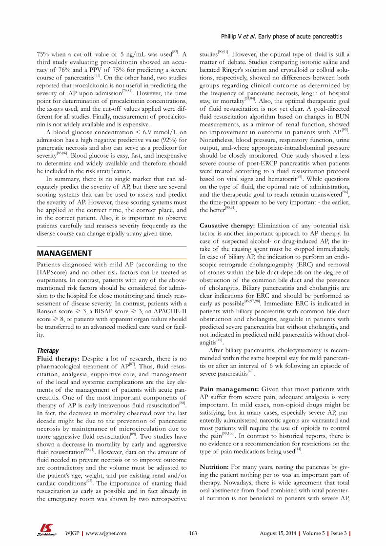
75% when a cut-off value of 5 ng/mL was used[82]. A third study evaluating procalcitonin showed an accu-racy of 76% and a PPV of 75% for predicting a severe course of pancreatitis[83]. On the other hand, two studies reported that procalcitonin is not useful in predicting the severity of AP upon admission[79,84]. However, the time point for determination of procalcitonin concentrations, the assays used, and the cut-off values applied were dif-ferent for all studies. Finally, measurement of procalcito-nin is not widely available and is expensive.
A blood glucose concentration < 6.9 mmol/L on admission has a high negative predictive value (92%) for pancreatic necrosis and also can serve as a predictor for severity[85,86]. Blood glucose is easy, fast, and inexpensive to determine and widely available and therefore should be included in the risk stratification.
In summary, there is no single marker that can ad-equately predict the severity of AP, but there are several scoring systems that can be used to assess and predict the severity of AP. However, these scoring systems must be applied at the correct time, the correct place, and in the correct patient. Also, it is important to observe patients carefully and reassess severity frequently as the disease course can change rapidly at any given time.
MANAGEMENTPatients diagnosed with mild AP (according to the HAPScore) and no other risk factors can be treated as outpatients. In contrast, patients with any of the above-mentioned risk factors should be considered for admis-sion to the hospital for close monitoring and timely reas-sessment of disease severity. In contrast, patients with a Ranson score ≥ 3, a BISAP score ≥ 3, an APACHE-II score ≥ 8, or patients with apparent organ failure should be transferred to an advanced medical care ward or facil-ity.
TherapyFluid therapy: Despite a lot of research, there is no pharmacological treatment of AP[87]. Thus, fluid resus-citation, analgesia, supportive care, and management of the local and systemic complications are the key ele-ments of the management of patients with acute pan-creatitis. One of the most important components of therapy of AP is early intravenous fluid resuscitation[88]. In fact, the decrease in mortality observed over the last decade might be due to the prevention of pancreatic necrosis by maintenance of microcirculation due to more aggressive fluid resuscitation[89]. Two studies have shown a decrease in mortality by early and aggressive fluid resuscitation[90,91]. However, data on the amount of fluid needed to prevent necrosis or to improve outcome are contradictory and the volume must be adjusted to the patient’s age, weight, and pre-existing renal and/or cardiac conditions[92]. The importance of starting fluid resuscitation as early as possible and in fact already in the emergency room was shown by two retrospective
studies[90,91]. However, the optimal type of fluid is still a matter of debate. Studies comparing isotonic saline and lactated Ringer’s solution and crystalloid vs colloid solu-tions, respectively, showed no differences between both groups regarding clinical outcome as determined by the frequency of pancreatic necrosis, length of hospital stay, or mortality[93,94]. Also, the optimal therapeutic goal of fluid resuscitation is not yet clear. A goal-directed fluid resuscitation algorithm based on changes in BUN measurements, as a mirror of renal function, showed no improvement in outcome in patients with AP[93]. Nonetheless, blood pressure, respiratory function, urine output, and-where appropriate-intraabdominal pressure should be closely monitored. One study showed a less severe course of post-ERCP pancreatitis when patients were treated according to a fluid resuscitation protocol based on vital signs and hematocrit[95]. While questions on the type of fluid, the optimal rate of administration, and the therapeutic goal to reach remain unanswered[96], the time-point appears to be very important - the earlier, the better[90,91].
Causative therapy: Elimination of any potential risk factor is another important approach to AP therapy. In case of suspected alcohol- or drug-induced AP, the in-take of the causing agent must be stopped immediately. In case of biliary AP, the indication to perform an endo-scopic retrograde cholangiography (ERC) and removal of stones within the bile duct depends on the degree of obstruction of the common bile duct and the presence of cholangitis. Biliary pancreatitis and cholangitis are clear indications for ERC and should be performed as early as possible[49,97,98]. Immediate ERC is indicated in patients with biliary pancreatitis with common bile duct obstruction and cholangitis, arguable in patients with predicted severe pancreatitis but without cholangitis, and not indicated in predicted mild pancreatitis without chol-angitis[49].
After biliary pancreatitis, cholecystectomy is recom-mended within the same hospital stay for mild pancreati-tis or after an interval of 6 wk following an episode of severe pancreatitis[49].
Pain management: Given that most patients with AP suffer from severe pain, adequate analgesia is very important. In mild cases, non-opioid drugs might be satisfying, but in many cases, especially severe AP, par-enterally administered narcotic agents are warranted and most patients will require the use of opioids to control the pain[99,100]. In contrast to historical reports, there is no evidence or a recommendation for restrictions on the type of pain medications being used[14].
Nutrition: For many years, resting the pancreas by giv-ing the patient nothing per os was an important part of therapy. Nowadays, there is wide agreement that total oral abstinence from food combined with total parenter-al nutrition is not beneficial to patients with severe AP,
August 15, 2014|Volume 5|Issue 3|WJGP|www.wjgnet.com 163
Phillip V et al . Early phase of acute pancreatitis
but may in fact be detrimental. A recent meta-analysis showed a statistically significant association of early enteral nutrition and reductions in systemic infections, pancreatic infections, length of hospital stay, and mortal-ity[101]. Also, in patients with severe AP, enteral nutrition was significantly superior to total parenteral nutrition regarding mortality, infectious complications, and organ failure[102]. Gut barrier function is compromised in pa-tients with acute pancreatitis, likely leading to bacterial translocation and potentially causing infected necrosis or even sepsis[103,104]. Because enteral feeding stabilizes gut barrier function, thereby reducing bacterial translocation, it is important early during the course of AP[14,105].
Therefore, whenever possible, i.e., when dissipating pain allows the patient to eat and infectious parameters do not continue to rise, oral food intake should be initi-ated as early as possible[49]. If oral food intake is not pos-sible and the patient needs nutritional support, enteral tube feeding is preferred over total parenteral nutrition. However, the composition of an optimal diet has not yet been evaluated.
Antibiotic prophylaxis: There also has been a change regarding prophylactic antibiotic therapy in patients with AP. While in the 90ties, prophylactic antibiotics where thought to improve the outcome in patients with AP, there is no emerging evidence that prophylactic antibiot-ics reduce infectious complications or mortality[106-108]. Today, there is no clear evidence that supports antibi-otic prophylaxis as a routine treatment in patients with severe AP[109-111]. Prophylactic antibiotics may reduce pancreatic infection in special subgroups of patients, but further well-designed and adequately-powered studies are needed to definitively answer the clinical usefulness of antibiotic prophylaxis in these patients[108]. Therefore, antibiotic prophylaxis is currently not recommended by international guidelines for the treatment of acute pan-creatitis[14,49].
CONCLUSIONAcute pancreatitis is a frequent and potentially life-threatening disease. Numerous clinical prognostic scor-ing systems have been developed, and yet tools to dis-criminate between mild, moderate, and severe AP early during the course of the disease are not well advanced. Therapy is currently mostly symptomatic with fluid re-suscitation, pain management, and early oral feeding. However, most of these therapeutic approaches are not well-defined. Vigorous fluid resuscitation remains a cornerstone of early management of acute pancreati-tis. Cross-sectional imaging during the early phase of evaluation has not been associated with improvement in outcome. There is no role for prophylactic antibiotics in the management of the early phase of AP. Enteral nutri-tion in AP can reduce mortality, systemic infections, and multiorgan dysfunction compared to parenteral nutri-tion. Immediate ERC is indicated only in patients with
biliary pancreatitis with common bile duct obstruction and cholangitis. These developments have contributed to an improved outcome for patients with acute pancreati-tis, but further studies are still required to tackle the high mortality in this disease.
REFERENCES1 Spanier B, Bruno MJ, Dijkgraaf MG. Incidence and mortali-
ty of acute and chronic pancreatitis in the Netherlands: a na-tionwide record-linked cohort study for the years 1995-2005. World J Gastroenterol 2013; 19: 3018-3026 [PMID: 23716981 DOI: 10.3748/wjg.v19.i20.3018]
2 Satoh K, Shimosegawa T, Masamune A, Hirota M, Kikuta K, Kihara Y, Kuriyama S, Tsuji I, Satoh A, Hamada S. Nation-wide epidemiological survey of acute pancreatitis in Japan. Pancreas 2011; 40: 503-507 [PMID: 21499203 DOI: 10.1097/MPA.0b013e318214812b]
3 Roberts SE, Akbari A, Thorne K, Atkinson M, Evans PA. The incidence of acute pancreatitis: impact of social depri-vation, alcohol consumption, seasonal and demographic factors. Aliment Pharmacol Ther 2013; 38: 539-548 [PMID: 23859492 DOI: 10.1111/apt.12408]
4 Shen HN, Lu CL, Li CY. Epidemiology of first-attack acute pancreatitis in Taiwan from 2000 through 2009: a nation-wide population-based study. Pancreas 2012; 41: 696-702 [PMID: 22699142 DOI: 10.1097/MPA.0b013e31823db941]
5 Oskarsson V, Sadr-Azodi O, Orsini N, Andrén-Sandberg Å, Wolk A. High dietary glycemic load increases the risk of non-gallstone-related acute pancreatitis: a prospective co-hort study. Clin Gastroenterol Hepatol 2014; 12: 676-682 [PMID: 24100113 DOI: 10.1016/j.cgh.2013.09.058]
6 Phillip V, Huber W, Hagemes F, Lorenz S, Matheis U, Preinfalk S, Schuster T, Lippl F, Saugel B, Schmid RM. Incidence of acute pancreatitis does not increase during Oktoberfest, but is higher than previously described in Ger-many. Clin Gastroenterol Hepatol 2011; 9: 995-1000.e3 [PMID: 21723238 DOI: 10.1016/j.cgh.2011.06.016]
7 Frey CF, Zhou H, Harvey DJ, White RH. The incidence and case-fatality rates of acute biliary, alcoholic, and idiopathic pancreatitis in California, 1994-2001. Pan-creas 2006; 33: 336-344 [PMID: 17079936 DOI: 10.1097/01.mpa.0000236727.16370.99]
8 Yadav D, Lowenfels AB. Trends in the epidemiology of the first attack of acute pancreatitis: a systematic review. Pan-creas 2006; 33: 323-330 [PMID: 17079934 DOI: 10.1097/01.mpa.0000236733.31617.52]
9 Banks PA. Epidemiology, natural history, and predictors of disease outcome in acute and chronic pancreatitis. Gas-trointest Endosc 2002; 56: S226-S230 [PMID: 12447272 DOI: 10.1067/mge.2002.129022]
10 Yadav D, Ng B, Saul M, Kennard ED. Relationship of serum pancreatic enzyme testing trends with the diagnosis of acute pancreatitis. Pancreas 2011; 40: 383-389 [PMID: 21283039 DOI: 10.1097/MPA.0b013e3182062970]
11 Saligram S, Lo D, Saul M, Yadav D. Analyses of hospital administrative data that use diagnosis codes overestimate the cases of acute pancreatitis. Clin Gastroenterol Hepa-tol 2012; 10: 805-811.e1 [PMID: 22504004 DOI: 10.1016/j.cgh.2012.03.025]
12 Peery AF, Dellon ES, Lund J, Crockett SD, McGowan CE, Bulsiewicz WJ, Gangarosa LM, Thiny MT, Stizenberg K, Morgan DR, Ringel Y, Kim HP, Dibonaventura MD, Carroll CF, Allen JK, Cook SF, Sandler RS, Kappelman MD, Sha-heen NJ. Burden of gastrointestinal disease in the United States: 2012 update. Gastroenterology 2012; 143: 1179-1187.e1-e3 [PMID: 22885331 DOI: 10.1053/j.gastro.2012.08.002]
13 Pavlidis P, Crichton S, Lemmich Smith J, Morrison D, At-
August 15, 2014|Volume 5|Issue 3|WJGP|www.wjgnet.com 164
Phillip V et al . Early phase of acute pancreatitis

kinson S, Wyncoll D, Ostermann M. Improved outcome of severe acute pancreatitis in the intensive care unit. Crit Care Res Pract 2013; 2013: 897107 [PMID: 23662207 DOI: 10.1155/2013/897107]
14 Banks PA, Freeman ML. Practice guidelines in acute pan-creatitis. Am J Gastroenterol 2006; 101: 2379-2400 [PMID: 17032204 DOI: 10.1111/j.1572-0241.2006.00856.x]
15 Lowenfels AB, Maisonneuve P, Sullivan T. The changing character of acute pancreatitis: epidemiology, etiology, and prognosis. Curr Gastroenterol Rep 2009; 11: 97-103 [PMID: 19281696]
16 Mounzer R, Whitcomb DC. Genetics of acute and chronic pancreatitis. Curr Opin Gastroenterol 2013; 29: 544-551 [PMID: 23872486 DOI: 10.1097/MOG.0b013e3283639383]
17 Papachristou GI. Prediction of severe acute pancreatitis: current knowledge and novel insights. World J Gastroenterol 2008; 14: 6273-6275 [PMID: 19009638]
18 Leser HG, Gross V, Scheibenbogen C, Heinisch A, Salm R, Lausen M, Rückauer K, Andreesen R, Farthmann EH, Schölmerich J. Elevation of serum interleukin-6 concentra-tion precedes acute-phase response and reflects severity in acute pancreatitis. Gastroenterology 1991; 101: 782-785 [PMID: 1907253]
19 Zhang H, Neuhöfer P, Song L, Rabe B, Lesina M, Kurkows-ki MU, Treiber M, Wartmann T, Regnér S, Thorlacius H, Saur D, Weirich G, Yoshimura A, Halangk W, Mizgerd JP, Schmid RM, Rose-John S, Algül H. IL-6 trans-signaling promotes pancreatitis-associated lung injury and lethal-ity. J Clin Invest 2013; 123: 1019-1031 [PMID: 23426178 DOI: 10.1172/JCI64931]
20 Van Laethem JL, Marchant A, Delvaux A, Goldman M, Robberecht P, Velu T, Devière J. Interleukin 10 prevents necrosis in murine experimental acute pancreatitis. Gastro-enterology 1995; 108: 1917-1922 [PMID: 7539389]
21 Lesina M, Wörmann SM, Neuhöfer P, Song L, Algül H. In-terleukin-6 in inflammatory and malignant diseases of the pancreas. Semin Immunol 2014; 26: 80-87 [PMID: 24572992 DOI: 10.1016/j.smim.2014.01.002]
22 Hoque R, Malik AF, Gorelick F, Mehal WZ. Sterile inflamma-tory response in acute pancreatitis. Pancreas 2012; 41: 353-357 [PMID: 22415665 DOI: 10.1097/MPA.0b013e3182321500]
23 Gullo L, Migliori M, Oláh A, Farkas G, Levy P, Arvanitakis C, Lankisch P, Beger H. Acute pancreatitis in five European countries: etiology and mortality. Pancreas 2002; 24: 223-227 [PMID: 11893928]
24 Halonen KI, Leppaniemi AK, Puolakkainen PA, Lundin JE, Kemppainen EA, Hietaranta AJ, Haapiainen RK. Severe acute pancreatitis: prognostic factors in 270 consecutive pa-tients. Pancreas 2000; 21: 266-271 [PMID: 11039471]
25 Khanna AK, Meher S, Prakash S, Tiwary SK, Singh U, Srivastava A, Dixit VK. Comparison of Ranson, Glasgow, MOSS, SIRS, BISAP, APACHE-II, CTSI Scores, IL-6, CRP, and Procalcitonin in Predicting Severity, Organ Failure, Pancreatic Necrosis, and Mortality in Acute Pancreati-tis. HPB Surg 2013; 2013: 367581 [PMID: 24204087 DOI: 10.1155/2013/367581]
26 Sánchez-Lozada R, Acosta-Rosero AV, Chapa-Azuela O, Hurtado-López LM. [Etiology on determining the severity of acute pancreatitis]. Gac Med Mex 2003; 139: 27-31 [PMID: 12666407]
27 Sánchez-Lozada R, Camacho-Hernández MI, Vega-Chavaje RG, Garza-Flores JH, Campos-Castillo C, Gutiérrez-Vega R. [Acute pancreatitis: five year experience at the Hospital General de Mexico]. Gac Med Mex 2005; 141: 123-127 [PMID: 15892460]
28 Fan ST, Choi TK, Lai CS, Wong J. Influence of age on the mortality from acute pancreatitis. Br J Surg 1988; 75: 463-466 [PMID: 3390679]
29 Birgisson H, Möller PH, Birgisson S, Thoroddsen A, As-geirsson KS, Sigurjónsson SV, Magnússon J. Acute pancre-
atitis: a prospective study of its incidence, aetiology, sever-ity, and mortality in Iceland. Eur J Surg 2002; 168: 278-282 [PMID: 12375609 DOI: 10.1002/ejs.46]
30 Otsuki M, Matsuno S, Shimosegawa T, Williams JA, Go VL. International symposium: mechanism of pancreatitis--between bedside and laboratory. Pancreas 2002; 24: 391-407 [PMID: 11961493]
31 Takeyama Y. Long-term prognosis of acute pancreatitis in Japan. Clin Gastroenterol Hepatol 2009; 7: S15-S17 [PMID: 19896091 DOI: 10.1016/j.cgh.2009.08.022]
32 Arata S, Takada T, Hirata K, Yoshida M, Mayumi T, Hirota M, Yokoe M, Hirota M, Kiriyama S, Sekimoto M, Amano H, Wada K, Kimura Y, Gabata T, Takeda K, Kataoka K, Ito T, Tanaka M. Post-ERCP pancreatitis. J Hepatobiliary Pancreat Sci 2010; 17: 70-78 [PMID: 20012323 DOI: 10.1007/s00534-009-0220-5]
33 Easler J, Muddana V, Furlan A, Dasyam A, Vipperla K, Slivka A, Whitcomb DC, Papachristou GI, Yadav D. Porto-splenomesenteric venous thrombosis in patients with acute pancreatitis is associated with pancreatic necrosis and usu-ally has a benign course. Clin Gastroenterol Hepatol 2014; 12: 854-862 [PMID: 24161350 DOI: 10.1016/j.cgh.2013.09.068]
34 Nitsche CJ, Jamieson N, Lerch MM, Mayerle JV. Drug in-duced pancreatitis. Best Pract Res Clin Gastroenterol 2010; 24: 143-155 [PMID: 20227028 DOI: 10.1016/j.bpg.2010.02.002]
35 Fortson MR, Freedman SN, Webster PD. Clinical assess-ment of hyperlipidemic pancreatitis. Am J Gastroenterol 1995; 90: 2134-2139 [PMID: 8540502]
36 Wang DB, Yu J, Fulcher AS, Turner MA. Pancreatitis in patients with pancreas divisum: imaging features at MRI and MRCP. World J Gastroenterol 2013; 19: 4907-4916 [PMID: 23946595 DOI: 10.3748/wjg.v19.i30.4907]
37 Whitcomb DC. Genetic risk factors for pancreatic disorders. Gastroenterology 2013; 144: 1292-1302 [PMID: 23622139 DOI: 10.1053/j.gastro.2013.01.069]
38 DiMagno MJ, DiMagno EP. New advances in acute pan-creatitis. Curr Opin Gastroenterol 2007; 23: 494-501 [PMID: 17762554 DOI: 10.1097/MOG.0b013e3282ba566d]
39 Löhr JM, Dinter D, Diehl SJ, Haas SL, Veeser M, Pfützer R, Retter J, Schönberg SO, Düber C, Keim V, Schadendorf D, Witt H. Rapid progression of a splenic aneurysm due to segmental arterial mediolysis: a rare cause of acute pancre-atitis. Pancreatology 2013; 13: 553-556 [PMID: 24075524 DOI: 10.1016/j.pan.2013.06.001]
40 Banks PA, Bollen TL, Dervenis C, Gooszen HG, Johnson CD, Sarr MG, Tsiotos GG, Vege SS. Classification of acute pancreatitis--2012: revision of the Atlanta classification and definitions by international consensus. Gut 2013; 62: 102-111 [PMID: 23100216 DOI: 10.1136/gutjnl-2012-302779]
41 Gunjaca I, Zunic J, Gunjaca M, Kovac Z. Circulating cyto-kine levels in acute pancreatitis-model of SIRS/CARS can help in the clinical assessment of disease severity. Inflamma-tion 2012; 35: 758-763 [PMID: 21826480 DOI: 10.1007/s10753-011-9371-z]
42 Isenmann R, Rau B, Beger HG. Early severe acute pancre-atitis: characteristics of a new subgroup. Pancreas 2001; 22: 274-278 [PMID: 11291929]
43 Cobb JP, O’Keefe GE. Injury research in the genomic era. Lancet 2004; 363: 2076-2083 [PMID: 15207961 DOI: 10.1016/S0140-6736(04)16460-X]
44 Besselink MG, van Santvoort HC, Boermeester MA, Nieu-wenhuijs VB, van Goor H, Dejong CH, Schaapherder AF, Gooszen HG. Timing and impact of infections in acute pan-creatitis. Br J Surg 2009; 96: 267-273 [PMID: 19125434 DOI: 10.1002/bjs.6447]
45 Phillip V, Schuster T, Hagemes F, Lorenz S, Matheis U, Pre-infalk S, Lippl F, Saugel B, Schmid RM, Huber W. Time pe-riod from onset of pain to hospital admission and patients’ awareness in acute pancreatitis. Pancreas 2013; 42: 647-654 [PMID: 23303202 DOI: 10.1097/MPA.0b013e3182714565]
August 15, 2014|Volume 5|Issue 3|WJGP|www.wjgnet.com 165
Phillip V et al . Early phase of acute pancreatitis

46 Swaroop VS, Chari ST, Clain JE. Severe acute pancreatitis. JAMA 2004; 291: 2865-2868 [PMID: 15199038 DOI: 10.1001/jama.291.23.2865]
47 Keim V, Teich N, Fiedler F, Hartig W, Thiele G, Mössner J. A comparison of lipase and amylase in the diagnosis of acute pancreatitis in patients with abdominal pain. Pancreas 1998; 16: 45-49 [PMID: 9436862]
48 American Gastroenterological Association (AGA) Insti-tute on "Management of Acute Pancreatits" Clinical Prac-tice and Economics Committee; AGA Institute Governing Board. AGA Institute medical position statement on acute pancreatitis. Gastroenterology 2007; 132: 2019-2021 [PMID: 17484893]
49 Working Group IAP/APA Acute Pancreatitis Guidelines. IAP/APA evidence-based guidelines for the management of acute pancreatitis. Pancreatology 2013; 13: e1-15 [PMID: 24054878 DOI: 10.1016/j.pan.2013.07.063]
50 Martínez J, Sánchez-Payá J, Palazón JM, Suazo-Barahona J, Robles-Díaz G, Pérez-Mateo M. Is obesity a risk factor in acute pancreatitis? A meta-analysis. Pancreatology 2004; 4: 42-48 [PMID: 14988657]
51 Lankisch PG, Weber-Dany B, Hebel K, Maisonneuve P, Lowenfels AB. The harmless acute pancreatitis score: a clinical algorithm for rapid initial stratification of nonsevere disease. Clin Gastroenterol Hepatol 2009; 7: 702-705; quiz 607 [PMID: 19245846 DOI: 10.1016/j.cgh.2009.02.020]
52 Oskarsson V, Mehrabi M, Orsini N, Hammarqvist F, Segersvärd R, Andrén-Sandberg A, Sadr Azodi O. Valida-tion of the harmless acute pancreatitis score in predicting nonsevere course of acute pancreatitis. Pancreatology 2011; 11: 464-468 [PMID: 21968430 DOI: 10.1159/000331502]
53 Ranson JH, Rifkind KM, Roses DF, Fink SD, Eng K, Spencer FC. Prognostic signs and the role of operative management in acute pancreatitis. Surg Gynecol Obstet 1974; 139: 69-81 [PMID: 4834279]
54 Yeung YP, Lam BY, Yip AW. APACHE system is better than Ranson system in the prediction of severity of acute pancreatitis. Hepatobiliary Pancreat Dis Int 2006; 5: 294-299 [PMID: 16698595]
55 Imrie CW, Benjamin IS, Ferguson JC, McKay AJ, Mackenzie I, O’Neill J, Blumgart LH. A single-centre double-blind trial of Trasylol therapy in primary acute pancreatitis. Br J Surg 1978; 65: 337-341 [PMID: 348250]
56 Knaus WA, Zimmerman JE, Wagner DP, Draper EA, Law-rence DE. APACHE-acute physiology and chronic health evaluation: a physiologically based classification system. Crit Care Med 1981; 9: 591-597 [PMID: 7261642]
57 Gravante G, Garcea G, Ong SL, Metcalfe MS, Berry DP, Lloyd DM, Dennison AR. Prediction of mortality in acute pancreatitis: a systematic review of the published evi-dence. Pancreatology 2009; 9: 601-614 [PMID: 19657215 DOI: 10.1159/000212097]
58 Wu BU, Johannes RS, Sun X, Tabak Y, Conwell DL, Banks PA. The early prediction of mortality in acute pancreatitis: a large population-based study. Gut 2008; 57: 1698-1703 [PMID: 18519429 DOI: 10.1136/gut.2008.152702]
59 Cho YS, Kim HK, Jang EC, Yeom JO, Kim SY, Yu JY, Kim YJ, Do KR, Kim SS, Chae HS. Usefulness of the Bedside Index for severity in acute pancreatitis in the early pre-diction of severity and mortality in acute pancreatitis. Pancreas 2013; 42: 483-487 [PMID: 23429493 DOI: 10.1097/MPA.0b013e318267c879]
60 Bone RC, Balk RA, Cerra FB, Dellinger RP, Fein AM, Knaus WA, Schein RM, Sibbald WJ. Definitions for sepsis and organ failure and guidelines for the use of innovative thera-pies in sepsis. The ACCP/SCCM Consensus Conference Committee. American College of Chest Physicians/Society of Critical Care Medicine. Chest 1992; 101: 1644-1655 [PMID: 1303622]
61 Bollen TL, Singh VK, Maurer R, Repas K, van Es HW,
Banks PA, Mortele KJ. A comparative evaluation of radio-logic and clinical scoring systems in the early prediction of severity in acute pancreatitis. Am J Gastroenterol 2012; 107: 612-619 [PMID: 22186977 DOI: 10.1038/ajg.2011.438]
62 Balthazar EJ, Ranson JH, Naidich DP, Megibow AJ, Cac-cavale R, Cooper MM. Acute pancreatitis: prognostic value of CT. Radiology 1985; 156: 767-772 [PMID: 4023241 DOI: 10.1148/radiology.156.3.4023241]
63 Balthazar EJ, Robinson DL, Megibow AJ, Ranson JH. Acute pancreatitis: value of CT in establishing prognosis. Radiol-ogy 1990; 174: 331-336 [PMID: 2296641 DOI: 10.1148/radiol-ogy.174.2.2296641]
64 Bollen TL, Singh VK, Maurer R, Repas K, van Es HW, Banks PA, Mortele KJ. Comparative evaluation of the modi-fied CT severity index and CT severity index in assessing severity of acute pancreatitis. AJR Am J Roentgenol 2011; 197: 386-392 [PMID: 21785084 DOI: 10.2214/AJR.09.4025]
65 Tang W, Zhang XM, Xiao B, Zeng NL, Pan HS, Feng ZS, Xu XX. Magnetic resonance imaging versus Acute Physiology And Chronic Healthy Evaluation II score in predicting the severity of acute pancreatitis. Eur J Radiol 2011; 80: 637-642 [PMID: 20843620 DOI: 10.1016/j.ejrad.2010.08.020]
66 Brown A, Orav J, Banks PA. Hemoconcentration is an early marker for organ failure and necrotizing pancreatitis. Pan-creas 2000; 20: 367-372 [PMID: 10824690]
67 Lankisch PG, Mahlke R, Blum T, Bruns A, Bruns D, Mai-sonneuve P, Lowenfels AB. Hemoconcentration: an early marker of severe and/or necrotizing pancreatitis? A criti-cal appraisal. Am J Gastroenterol 2001; 96: 2081-2085 [PMID: 11467635 DOI: 10.1111/j.1572-0241.2001.03966.x]
68 Gan SI, Romagnuolo J. Admission hematocrit: a simple, useful and early predictor of severe pancreatitis. Dig Dis Sci 2004; 49: 1946-1952 [PMID: 15628731]
69 Knoefel WT, Kollias N, Warshaw AL, Waldner H, Nishioka NS, Rattner DW. Pancreatic microcirculatory changes in ex-perimental pancreatitis of graded severity in the rat. Surgery 1994; 116: 904-913 [PMID: 7940196]
70 Bassi D, Kollias N, Fernandez-del Castillo C, Foitzik T, Warshaw AL, Rattner DW. Impairment of pancreatic micro-circulation correlates with the severity of acute experimen-tal pancreatitis. J Am Coll Surg 1994; 179: 257-263 [PMID: 8069418]
71 Mann O, Kaifi J, Bloechle C, Schneider CG, Yekebas E, Klu-th D, Izbicki JR, Strate T. Therapeutic small-volume resusci-tation preserves pancreatic microcirculation in acute experi-mental pancreatitis of graded severity in rats. Pancreatology 2009; 9: 652-661 [PMID: 19684429 DOI: 10.1159/000212100]
72 Muddana V, Whitcomb DC, Khalid A, Slivka A, Papachris-tou GI. Elevated serum creatinine as a marker of pancreatic necrosis in acute pancreatitis. Am J Gastroenterol 2009; 104: 164-170 [PMID: 19098865 DOI: 10.1038/ajg.2008.66]
73 Lipinski M, Rydzewski A, Rydzewska G. Early changes in serum creatinine level and estimated glomerular filtra-tion rate predict pancreatic necrosis and mortality in acute pancreatitis: Creatinine and eGFR in acute pancreatitis. Pan-creatology 2013; 13: 207-211 [PMID: 23719589 DOI: 10.1016/j.pan.2013.02.002]
74 Wu BU, Johannes RS, Sun X, Conwell DL, Banks PA. Early changes in blood urea nitrogen predict mortality in acute pancreatitis. Gastroenterology 2009; 137: 129-135 [PMID: 19344722 DOI: 10.1053/j.gastro.2009.03.056]
75 Wu BU, Bakker OJ, Papachristou GI, Besselink MG, Repas K, van Santvoort HC, Muddana V, Singh VK, Whitcomb DC, Gooszen HG, Banks PA. Blood urea nitrogen in the early assessment of acute pancreatitis: an international validation study. Arch Intern Med 2011; 171: 669-676 [PMID: 21482842 DOI: 10.1001/archinternmed.2011.126]
76 Pongprasobchai S, Jianjaroonwong V, Charatcharoenwit-thaya P, Komoltri C, Tanwandee T, Leelakusolvong S, Pausawasdi N, Srikureja W, Chainuvati S, Prachayakul V,
August 15, 2014|Volume 5|Issue 3|WJGP|www.wjgnet.com 166
Phillip V et al . Early phase of acute pancreatitis
Manatsathit S, Kachintorn U. Erythrocyte sedimentation rate and C-reactive protein for the prediction of severity of acute pancreatitis. Pancreas 2010; 39: 1226-1230 [PMID: 20531240 DOI: 10.1097/MPA.0b013e3181deb33e]
77 Hjalmarsson C, Stenflo J, Borgström A. Activated protein C-protein C inhibitor complex, activation peptide of car-boxypeptidase B and C-reactive protein as predictors of se-vere acute pancreatitis. Pancreatology 2009; 9: 700-707 [PMID: 19684435 DOI: 10.1159/000215577]
78 Cardoso FS, Ricardo LB, Oliveira AM, Canena JM, Horta DV, Papoila AL, Deus JR. C-reactive protein prognostic ac-curacy in acute pancreatitis: timing of measurement and cutoff points. Eur J Gastroenterol Hepatol 2013; 25: 784-789 [PMID: 23492986 DOI: 10.1097/MEG.0b013e32835fd3f0]
79 Rau B, Steinbach G, Gansauge F, Mayer JM, Grünert A, Be-ger HG. The potential role of procalcitonin and interleukin 8 in the prediction of infected necrosis in acute pancreatitis. Gut 1997; 41: 832-840 [PMID: 9462219]
80 Rau BM, Kemppainen EA, Gumbs AA, Büchler MW, We-gscheider K, Bassi C, Puolakkainen PA, Beger HG. Early assessment of pancreatic infections and overall prognosis in severe acute pancreatitis by procalcitonin (PCT): a prospec-tive international multicenter study. Ann Surg 2007; 245: 745-754 [PMID: 17457167]
81 Ammori BJ, Becker KL, Kite P, Snider RH, Nylén ES, White JC, Larvin M, McMahon MJ. Calcitonin precursors in the prediction of severity of acute pancreatitis on the day of admission. Br J Surg 2003; 90: 197-204 [PMID: 12555296 DOI: 10.1002/bjs.4036]
82 Pindak D, Parrak V, Pechan J, Vavrecka A, Kuzela L, Fuchs D, Irsakova J. The clinical value of the procalcitonin in predic-tion of severity and outcome in acute pancreatitis. Hepatogas-troenterology 2003; 50 Suppl 2: ccviii-ccccix [PMID: 15244180]
83 Kim BG, Noh MH, Ryu CH, Nam HS, Woo SM, Ryu SH, Jang JS, Lee JH, Choi SR, Park BH. A comparison of the BISAP score and serum procalcitonin for predicting the severity of acute pancreatitis. Korean J Intern Med 2013; 28: 322-329 [PMID: 23682226 DOI: 10.3904/kjim.2013.28.3.322]
84 Modrau IS, Floyd AK, Thorlacius-Ussing O. The clinical value of procalcitonin in early assessment of acute pan-creatitis. Am J Gastroenterol 2005; 100: 1593-1597 [PMID: 15984987 DOI: 10.1111/j.1572-0241.2005.41456.x]
85 Lankisch PG, Blum T, Bruns A, Dröge M, Brinkmann G, Struckmann K, Nauck M, Maisonneuve P, Lowenfels AB. Has blood glucose level measured on admission to hos-pital in a patient with acute pancreatitis any prognostic value? Pancreatology 2001; 1: 224-229 [PMID: 12120199 DOI: 10.1159/000055815]
86 Rajaratnam SG, Martin IG. Admission serum glucose level: an accurate predictor of outcome in gallstone pancreatitis. Pancreas 2006; 33: 27-30 [PMID: 16804409 DOI: 10.1097/01.mpa.0000222315.36490.9b]
87 Pezzilli R. Pharmacotherapy for acute pancreatitis. Expert Opin Pharmacother 2009; 10: 2999-3014 [PMID: 19925044 DOI: 10.1517/14656560903382630]
88 Fisher JM, Gardner TB. The “golden hours” of management in acute pancreatitis. Am J Gastroenterol 2012; 107: 1146-1150 [PMID: 22858994 DOI: 10.1038/ajg.2012.91]
89 Wall I, Badalov N, Baradarian R, Iswara K, Li JJ, Tenner S. Decreased mortality in acute pancreatitis related to early aggressive hydration. Pancreas 2011; 40: 547-550 [PMID: 21499208 DOI: 10.1097/MPA.0b013e318215368d]
90 Gardner TB, Vege SS, Chari ST, Petersen BT, Topazian MD, Clain JE, Pearson RK, Levy MJ, Sarr MG. Faster rate of ini-tial fluid resuscitation in severe acute pancreatitis diminish-es in-hospital mortality. Pancreatology 2009; 9: 770-776 [PMID: 20110744 DOI: 10.1159/000210022]
91 Warndorf MG, Kurtzman JT, Bartel MJ, Cox M, Mackenzie T, Robinson S, Burchard PR, Gordon SR, Gardner TB. Early
fluid resuscitation reduces morbidity among patients with acute pancreatitis. Clin Gastroenterol Hepatol 2011; 9: 705-709 [PMID: 21554987 DOI: 10.1016/j.cgh.2011.03.032]
92 Haydock MD, Mittal A, Wilms HR, Phillips A, Petrov MS, Windsor JA. Fluid therapy in acute pancreatitis: anybody’s guess. Ann Surg 2013; 257: 182-188 [PMID: 23207241 DOI: 10.1097/SLA.0b013e31827773ff]
93 Wu BU, Hwang JQ, Gardner TH, Repas K, Delee R, Yu S, Smith B, Banks PA, Conwell DL. Lactated Ringer’s solu-tion reduces systemic inflammation compared with sa-line in patients with acute pancreatitis. Clin Gastroenterol Hepatol 2011; 9: 710-717.e1 [PMID: 21645639 DOI: 10.1016/j.cgh.2011.04.026]
94 Du XJ, Hu WM, Xia Q, Huang ZW, Chen GY, Jin XD, Xue P, Lu HM, Ke NW, Zhang ZD, Li QS. Hydroxyethyl starch re-suscitation reduces the risk of intra-abdominal hypertension in severe acute pancreatitis. Pancreas 2011; 40: 1220-1225 [PMID: 21775917 DOI: 10.1097/MPA.0b013e3182217f17]
95 Reddy N, Wilcox CM, Tamhane A, Eloubeidi MA, Varada-rajulu S. Protocol-based medical management of post-ERCP pancreatitis. J Gastroenterol Hepatol 2008; 23: 385-392 [PMID: 18318823 DOI: 10.1111/j.1440-1746.2007.05180.x]
96 Sarr MG. Early fluid “resuscitation/therapy” in acute pan-creatitis: which fluid? What rate? What parameters to gauge effectiveness? Ann Surg 2013; 257: 189-190 [PMID: 23291660 DOI: 10.1097/SLA.0b013e318280e19e]
97 Neoptolemos JP, Carr-Locke DL, London NJ, Bailey IA, James D, Fossard DP. Controlled trial of urgent endoscopic retrograde cholangiopancreatography and endoscopic sphincterotomy versus conservative treatment for acute pancreatitis due to gallstones. Lancet 1988; 2: 979-983 [PMID: 2902491]
98 Tse F, Yuan Y. Early routine endoscopic retrograde chol-angiopancreatography strategy versus early conservative management strategy in acute gallstone pancreatitis. Co-chrane Database Syst Rev 2012; 5: CD009779 [PMID: 22592743 DOI: 10.1002/14651858.CD009779.pub2]
99 Pezzilli R, Uomo G, Gabbrielli A, Zerbi A, Frulloni L, De Rai P, Castoldi L, Cavallini G, Di Carlo V. A prospective multicentre survey on the treatment of acute pancreatitis in Italy. Dig Liver Dis 2007; 39: 838-846 [PMID: 17602904 DOI: 10.1016/j.dld.2007.05.014]
100 Ebbehøj N, Friis J, Svendsen LB, Bülow S, Madsen P. Indo-methacin treatment of acute pancreatitis. A controlled dou-ble-blind trial. Scand J Gastroenterol 1985; 20: 798-800 [PMID: 2413519]
101 Li JY, Yu T, Chen GC, Yuan YH, Zhong W, Zhao LN, Chen QK. Enteral nutrition within 48 hours of admission im-proves clinical outcomes of acute pancreatitis by reducing complications: a meta-analysis. PLoS One 2013; 8: e64926 [PMID: 23762266 DOI: 10.1371/journal.pone.0064926]
102 Yi F, Ge L, Zhao J, Lei Y, Zhou F, Chen Z, Zhu Y, Xia B. Meta-analysis: total parenteral nutrition versus total enteral nutrition in predicted severe acute pancreatitis. Intern Med 2012; 51: 523-530 [PMID: 22449657]
103 Ammori BJ, Becker KL, Kite P, Snider RH, Nylén ES, White JC, Barclay GR, Larvin M, McMahon MJ. Calcitonin precur-sors: early markers of gut barrier dysfunction in patients with acute pancreatitis. Pancreas 2003; 27: 239-243 [PMID: 14508129]
104 Rahman SH, Ammori BJ, Holmfield J, Larvin M, McMahon MJ. Intestinal hypoperfusion contributes to gut barrier fail-ure in severe acute pancreatitis. J Gastrointest Surg 2003; 7: 26-35; discussion 35-6 [PMID: 12559182]
105 Dervenis C, Smailis D, Hatzitheoklitos E. Bacterial translo-cation and its prevention in acute pancreatitis. J Hepatobili-ary Pancreat Surg 2003; 10: 415-418 [PMID: 14714160 DOI: 10.1007/s00534-002-0727-5]
106 Sharma VK, Howden CW. Prophylactic antibiotic admin-
August 15, 2014|Volume 5|Issue 3|WJGP|www.wjgnet.com 167
Phillip V et al . Early phase of acute pancreatitis
istration reduces sepsis and mortality in acute necrotizing pancreatitis: a meta-analysis. Pancreas 2001; 22: 28-31 [PMID: 11138967]
107 Bassi C, Larvin M, Villatoro E. Antibiotic therapy for pro-phylaxis against infection of pancreatic necrosis in acute pancreatitis. Cochrane Database Syst Rev 2003; (4): CD002941 [PMID: 14583957 DOI: 10.1002/14651858.CD002941]
108 Villatoro E, Mulla M, Larvin M. Antibiotic therapy for pro-phylaxis against infection of pancreatic necrosis in acute pancreatitis. Cochrane Database Syst Rev 2010; (5): CD002941 [PMID: 20464721 DOI: 10.1002/14651858.CD002941.pub3]
109 Wittau M, Mayer B, Scheele J, Henne-Bruns D, Dellinger
EP, Isenmann R. Systematic review and meta-analysis of antibiotic prophylaxis in severe acute pancreatitis. Scand J Gastroenterol 2011; 46: 261-270 [PMID: 21067283 DOI: 10.3109/00365521.2010.531486]
110 Jiang K, Huang W, Yang XN, Xia Q. Present and future of prophylactic antibiotics for severe acute pancreatitis. World J Gastroenterol 2012; 18: 279-284 [PMID: 22294832 DOI: 10.3748/wjg.v18.i3.279]
111 Jafri NS, Mahid SS, Minor KS, Idstein SR, Hornung CA, Galan-diuk S. Meta-analysis: antibiotic prophylaxis to prevent peristo-mal infection following percutaneous endoscopic gastrostomy. Aliment Pharmacol Ther 2007; 25: 647-656 [PMID: 17311597]
P- Reviewer: Bradley EL, Goral V, Pezzilli R, Sakata N S- Editor: Ji FF L- Editor: A E- Editor: Lu YJ
August 15, 2014|Volume 5|Issue 3|WJGP|www.wjgnet.com 168
Phillip V et al . Early phase of acute pancreatitis
TOPIC HIGHLIGHT
Potential role of NADPH oxidase in pathogenesis of pancreatitis
Wei-Li Cao, Xiao-Hui Xiang, Kai Chen, Wei Xu, Shi-Hai Xia
Wei-Li Cao, Xiao-Hui Xiang, Kai Chen, Wei Xu, Shi-Hai Xia, Department of Hepatopancreatobiliary and Splenic Medicine, Affiliated Hospital, Logistics University of the Chinese People’s Armed Police Forces, Tianjin 300162, ChinaAuthor contributions: Cao WL and Xiang XH contributed equally to this work; Xia SH contributed to the conception of this work; Cao WL, Xiang XH, Chen K and Xu W prepared the manuscript; Xia SH revised and approved the manuscript.Supported by The National Natural Science Foundation of China, No. 81173393; the Natural Science Foundation of Tian-jin City, No. 12YFJZJC00800; the Scientific Research Founda-tion (No. WHM201222, FYM201114) and the Innovation Team Program (No. WHTD201310) from Logistics University of the Chinese People’s Armed Police ForcesCorrespondence to: Shi-Hai Xia, MD, PhD, Department of Hepatopancreatobiliary and Splenic Medicine, Affiliated Hospi-tal, Logistics University of the Chinese People’s Armed Police Forces, 220 Chenglin Road, Hedong District, Tianjin 300162, China. [email protected]: +86-22-60578765 Fax: +86-22-24370605Received: December 27, 2013 Revised: June 1, 2014Accepted: June 14, 2014Published online: August 15, 2014
AbstractStudies have demonstrated that reactive oxygen spe-cies (ROS) are closely related to inflammatory disor-ders. Nicotinamide adenine dinucleotide phosphate oxi-dase (NOX), originally found in phagocytes, is the main source of ROS in nonphagocytic cells. Besides directly producing the detrimental highly reactive ROS to act on biomolecules (lipids, proteins, and nucleic acids), NOX can also activate multiple signal transduction pathways, which regulate cell growth, proliferation, differentiation and apoptosis by producing ROS. Recently, research on pancreatic NOX is no longer limited to inflammatory cells, but extends to the aspect of pancreatic acinar cells and pancreatic stellate cells, which are considered to be potentially associated with pancreatitis. In this
review, we summarize the literature on NOX protein structure, activation, function and its role in the patho-genesis of pancreatitis.
© 2014 Baishideng Publishing Group Inc. All rights reserved.
Key words: Nicotinamide adenine dinucleotide phos-phate oxidase; Reactive oxygen species; Pancreatitis; Pancreatic acinar cells; Pancreatic stellate cells
Core tip: Besides directly producing the detrimental highly reactive reactive oxygen species (ROS) to act on biomolecules, nicotinamide adenine dinucleotide phos-phate (NADPH) oxidase can also activate multiple sig-nal transduction pathways, which regulate cell growth, proliferation, differentiation and apoptosis by producing ROS. Recently, research on pancreatic NADPH oxidase is no longer limited to inflammatory cells, but extends to the aspect of pancreatic acinar cells and pancreatic stellate cells, which are considered to be potentially as-sociated with pancreatitis.
Cao WL, Xiang XH, Chen K, Xu W, Xia SH. Potential role of NADPH oxidase in pathogenesis of pancreatitis. World J Gastrointest Pathophysiol 2014; 5(3): 169-177 Available from: URL: http://www.wjgnet.com/2150-5330/full/v5/i3/169.htm DOI: http://dx.doi.org/10.4291/wjgp.v5.i3.169
INTRODUCTION Studies have demonstrated that reactive oxygen species (ROS) are involved in the pathogenesis of pancreatitis[1]. Nicotinamide adenine dinucleotide phosphate (NADPH) oxidase (NOX), a transmembrane flavoprotein enzyme, uses NADPH as an electron donor to catalyze the univa-lent reduction of oxygen, resulting in the production of superoxide free radical, which might be a source of oxi-
World J Gastrointest Pathophysiol 2014 August 15; 5(3): 169-177ISSN 2150-5330 (online)
© 2014 Baishideng Publishing Group Inc. All rights reserved.
Online Submissions: http://www.wjgnet.com/esps/Help Desk: http://www.wjgnet.com/esps/helpdesk.aspxDOI: 10.4291/wjgp.v5.i3.169
August 15, 2014|Volume 5|Issue 3|WJGP|www.wjgnet.com 169
WJGP 5th Anniversary Special Issues (3): Pancreatitis
Cao WL et al . NADPH oxidase and pancreatitis
dants in injured pancreas[1]. NOX is mainly distributed in the phagocytic cell membrane with cytochrome C and flavin adenine dinucleotide groups, which can produce ROS, scavenging pathogenic microorganisms such as bacte-ria[2]. ROS, being generated by NOX, also participate in intracellular signaling processes in the pancreas. Recently, research on NOX is no longer limited to inflammatory cells, but extends to the aspect of pancreatic acinar cells and pancreatic stellate cells (PSCs) in pancreatitis pa-tients[2]. The function of NOX, which is involved in the pathogenesis of inflammation in pancreatic acinar cells and PSCs, has become the hotspot of research. Non-phagocytic NOX derived ROS function as a messenger molecule to participate in the modulation of cell differ-entiation, proliferation and apoptosis in the pancreas. In this review, we summarize the literature on NOX protein structure, activation, function and its role in the patho-genesis of pancreatitis.
STRUCTURE, LOCATION AND FUNCTION OF NOX IN THE PANCREASNOX is a multicomponent enzyme consisting of five different subunits, including the subunits p22phox and gp-91phox (also known as NOX2) located in the membrane, together with the cytosolic subunits p40phox, p47phox and p67phox. The participation of Rac would elicit full oxidase activity[3-5]. Relative to gp91phox
(the catalytic subunit of NOX), p22phox, p47phox, p40phox and p67phox are regula-tory subunits. Gp91phox in different types of cells has other six homologues, termed NOX1, NOX3, NOX4, NOX5, DUOX1 and DUOX2, which constitute the NOX family proteins[6-8]. NOX is an enzyme which was initially discovered in phagocytes[4,5]. NOX in neutrophils is composed of constitutive subunits (p22phox and gp-91phox) positioned in membrane and regulatory subunits (p47phox and p67phox, and possibly p40phox) stationed in the cytosol[9]. In recent years, NOX has been discovered in several nonphagocytic cells such as fibroblasts[10], vas-cular smooth muscle cells[11] and hepatic stellate cells[12]. More recently, it has been found that NOX was pres-ent in pancreatic β cells[13,14], pancreatic acinar cells[15-18] and PSCs[19,20]. The main intrinsic components of NOX comprising the NOX2 isoform are present in human pancreatic islets[14]. Cytosolic subunits p47phox and p67phox as well as membrane-bound subunits p22phox and NOX1 are constitutively expressed in pancreatic acinar AR42J cells[16,21,22]. The key subunits of NOX including p22phox, p47phox, NOX activator 1 (a homologue of p67phox), NOX1, NOX4, and NOX2 (gp91phox) are expressed in PSCs[19,20]. The activation of non-phagocytic NOX is simi-lar to that in neutrophils[23]. Upon activation of NOX, p47 translocates to the membrane and then recruits p67 to interact with the p22 subunit, thus facilitating NADPH-dependent formation of superoxide (O2-), which increas-es the production of secondary ROS such as hydrogen peroxide (H2O2)[21]. Non-phagocytic NOX derived ROS function as a messenger molecule to participate
in the modulation of cell differentiation, proliferation and apoptosis[6-8]. NOX protein family can be activated quickly under pathophysiological conditions, leading to high production of ROS, which contributes to oxidative stress and a wide range of diseases.
ACTIVATION AND INHIBITION FACTORS OF NOX IN THE PATHOGENESIS OF PANCREATITISCholecystokinin analoguesCerulein, an analogue of cholecystokinin (CCK), can stimulate the pancreatic exocrine secretion by binding CCK receptors, causing the autolysis of pancreatic acinar cell[24]. There are two kinds of CCK receptor subtypes, CCK1 and CCK2 receptors. CCK1 receptors regulate pancreatic digestive enzymes, satiety and feeding behav-ior, while CCK2 receptors enhance the level of gastric acid, as well as gastrin which has anti-apoptotic effects on pancreatic cells[25]. Experimental pancreatitis induced with high dosages of cerulean, similar to human edema-tous pancreatitis, is characterized by cytoplasmic vacuol-ization, formation of edema and acinar cell death as well as elevation in serum levels of digestive enzymes caused by unconventional secretion of digestive enzymes[26]. ROS are involved in the activation of oxidant-sensitive nuclear transcription factor (NF-κB), expression of cytokine, apoptosis and further occurrence of pancre-atitis[27]. P47phox, p67phox, NOX1 and p22phox in pancreatic AR42J cells could produce ROS after cerulein stimula-tion[21]. Intrapancreatic trypsin is not only activated by high-dose cerulein, but also regulated by neutrophils via NADPH oxidase[28]. The mechanism for the activation of NF-κB and expression of cytokines in pancreatic acinar cells stimulated by cerulein may be summarized as the following steps. Cerulein binds to the CCK receptor, a G-protein-coupled receptor, to activate phospholipase C (PLC) and inositol 1,4,5-trisphosphate (IP3), triggering transient Ca2+ release from the endoplasmic reticulum in pancreatic acinar cells. NOX activated by Ca2+ produces ROS to activate IκB kinase and then to phosphorylate IκB. Phosphorylated IκB can be ubiquitinated and de-graded in a proteasome dependent manner to eliminate the inhibition of NF-κB, a p65/p50 heterodimer in the cytosol. NF-κB then translocates to the nucleus to medi-ate the expression of cytokines which are involved in the pathogenesis of pancreatitis (Figures 1 and 2)[27].
Renin-angiotensin systemThe Renin-angiotensin system (RAS) is generally consid-ered to regulate blood pressure and body fluid homeo-stasis[29]. The pancreatic RAS activation that is related to the production of ROS might contribute to oxidative stress and tissue injury[30,31]. Angiotensin Ⅱ, an active mediator of RAS, is transformed from angiotensin Ⅰ by the angiotensin-converting enzyme (ACE)[32]. The effect of angiotensin Ⅱ is regulated by its receptors, including
August 15, 2014|Volume 5|Issue 3|WJGP|www.wjgnet.com 170
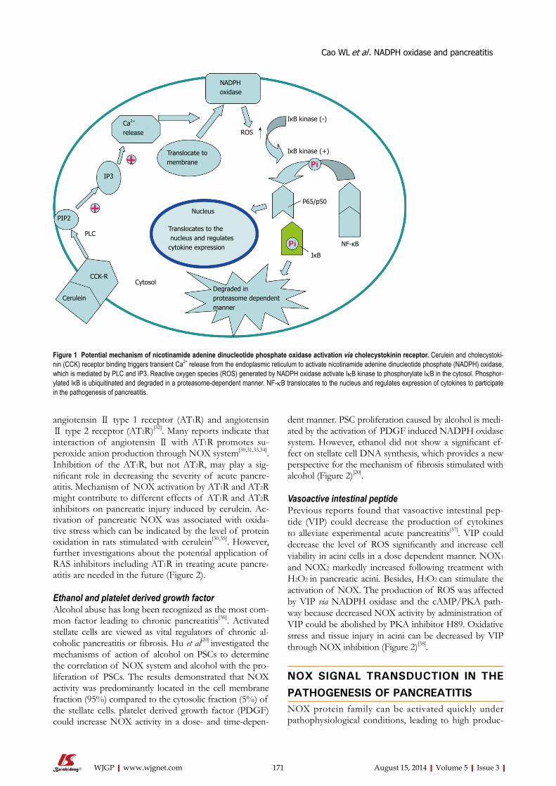
angiotensin Ⅱ type 1 receptor (AT1R) and angiotensin Ⅱ type 2 receptor (AT2R)[32]. Many reports indicate that interaction of angiotensin Ⅱ with AT1R promotes su-peroxide anion production through NOX system[30,31,33,34]. Inhibition of the AT1R, but not AT2R, may play a sig-nificant role in decreasing the severity of acute pancre-atitis. Mechanism of NOX activation by AT1R and AT2R might contribute to different effects of AT1R and AT2R inhibitors on pancreatic injury induced by cerulein. Ac-tivation of pancreatic NOX was associated with oxida-tive stress which can be indicated by the level of protein oxidation in rats stimulated with cerulein[30,35]. However, further investigations about the potential application of RAS inhibitors including AT1R in treating acute pancre-atitis are needed in the future (Figure 2).
Ethanol and platelet derived growth factorAlcohol abuse has long been recognized as the most com-mon factor leading to chronic pancreatitis[36]. Activated stellate cells are viewed as vital regulators of chronic al-coholic pancreatitis or fibrosis. Hu et al[20] investigated the mechanisms of action of alcohol on PSCs to determine the correlation of NOX system and alcohol with the pro-liferation of PSCs. The results demonstrated that NOX activity was predominantly located in the cell membrane fraction (95%) compared to the cytosolic fraction (5%) of the stellate cells. platelet derived growth factor (PDGF) could increase NOX activity in a dose- and time-depen-
dent manner. PSC proliferation caused by alcohol is medi-ated by the activation of PDGF induced NADPH oxidase system. However, ethanol did not show a significant ef-fect on stellate cell DNA synthesis, which provides a new perspective for the mechanism of fibrosis stimulated with alcohol (Figure 2)[20].
Vasoactive intestinal peptidePrevious reports found that vasoactive intestinal pep-tide (VIP) could decrease the production of cytokines to alleviate experimental acute pancreatitis[37]. VIP could decrease the level of ROS significantly and increase cell viability in acini cells in a dose dependent manner. NOX1 and NOX2 markedly increased following treatment with H2O2 in pancreatic acini. Besides, H2O2 can stimulate the activation of NOX. The production of ROS was affected by VIP via NADPH oxidase and the cAMP/PKA path-way because decreased NOX activity by administration of VIP could be abolished by PKA inhibitor H89. Oxidative stress and tissue injury in acini can be decreased by VIP through NOX inhibition (Figure 2)[38].
NOX SIGNAL TRANSDUCTION IN THE PATHOGENESIS OF PANCREATITISNOX protein family can be activated quickly under pathophysiological conditions, leading to high produc-
August 15, 2014|Volume 5|Issue 3|WJGP|www.wjgnet.com 171
NADPHoxidase
Ca2+
release
IP3
PIP2
CCK-R
Cerulein
IκB kinase (-)
IκB kinase (+)
P65/p50
ROS
Translocate tomembrane
Translocates to the nucleus and regulates cytokine expression
Nucleus
Degraded inproteasome dependent manner
Cytosol
PLC
IκB
NF-κB
Figure 1 Potential mechanism of nicotinamide adenine dinucleotide phosphate oxidase activation via cholecystokinin receptor. Cerulein and cholecystoki-nin (CCK) receptor binding triggers transient Ca2+ release from the endoplasmic reticulum to activate nicotinamide adenine dinucleotide phosphate (NADPH) oxidase, which is mediated by PLC and IP3. Reactive oxygen species (ROS) generated by NADPH oxidase activate IκB kinase to phosphorylate IκB in the cytosol. Phosphor-ylated IκB is ubiquitinated and degraded in a proteasome-dependent manner. NF-κB translocates to the nucleus and regulates expression of cytokines to participate in the pathogenesis of pancreatitis.
Cao WL et al . NADPH oxidase and pancreatitis
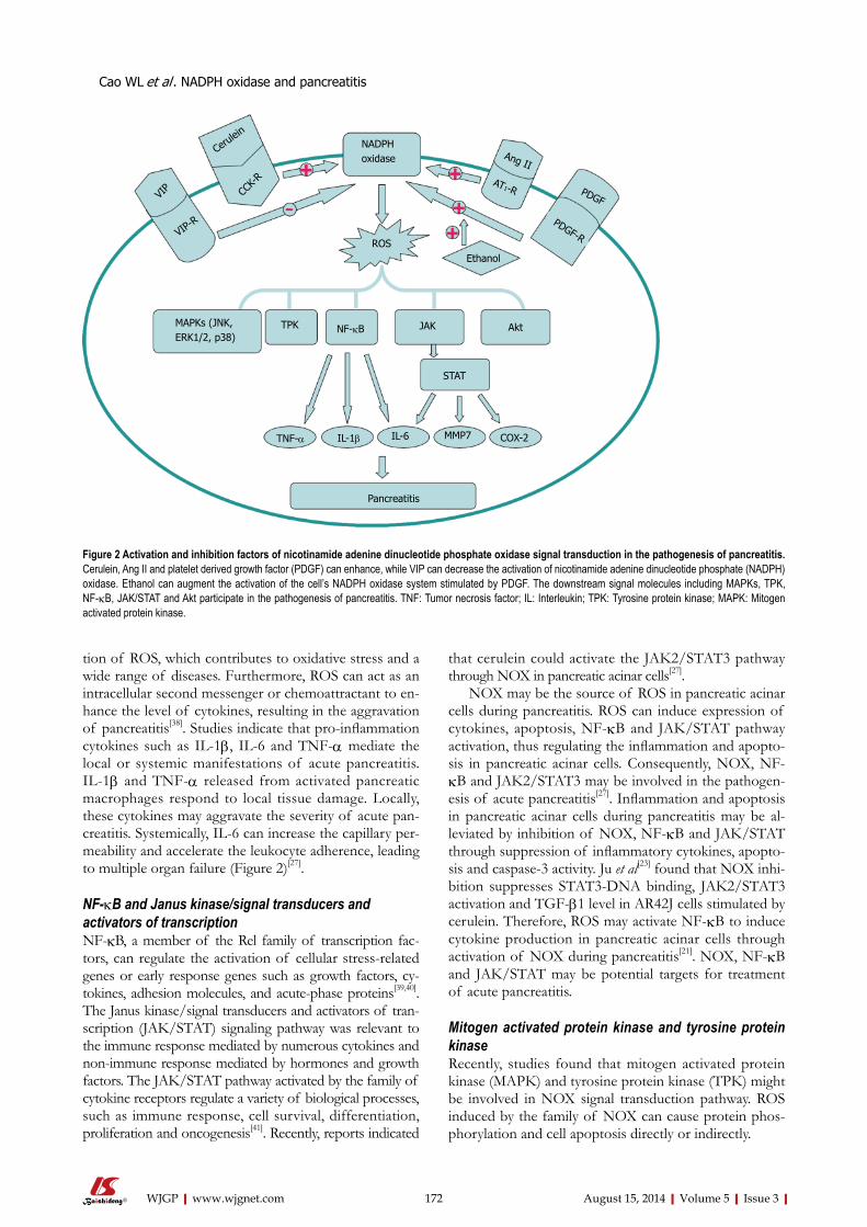
tion of ROS, which contributes to oxidative stress and a wide range of diseases. Furthermore, ROS can act as an intracellular second messenger or chemoattractant to en-hance the level of cytokines, resulting in the aggravation of pancreatitis[38]. Studies indicate that pro-inflammation cytokines such as IL-1β, IL-6 and TNF-α mediate the local or systemic manifestations of acute pancreatitis. IL-1β and TNF-α released from activated pancreatic macrophages respond to local tissue damage. Locally, these cytokines may aggravate the severity of acute pan-creatitis. Systemically, IL-6 can increase the capillary per-meability and accelerate the leukocyte adherence, leading to multiple organ failure (Figure 2)[27].
NF-κB and Janus kinase/signal transducers and activators of transcriptionNF-κB, a member of the Rel family of transcription fac-tors, can regulate the activation of cellular stress-related genes or early response genes such as growth factors, cy-tokines, adhesion molecules, and acute-phase proteins[39,40]. The Janus kinase/signal transducers and activators of tran-scription (JAK/STAT) signaling pathway was relevant to the immune response mediated by numerous cytokines and non-immune response mediated by hormones and growth factors. The JAK/STAT pathway activated by the family of cytokine receptors regulate a variety of biological processes, such as immune response, cell survival, differentiation, proliferation and oncogenesis[41]. Recently, reports indicated
that cerulein could activate the JAK2/STAT3 pathway through NOX in pancreatic acinar cells[27].
NOX may be the source of ROS in pancreatic acinar cells during pancreatitis. ROS can induce expression of cytokines, apoptosis, NF-κB and JAK/STAT pathway activation, thus regulating the inflammation and apopto-sis in pancreatic acinar cells. Consequently, NOX, NF-κB and JAK2/STAT3 may be involved in the pathogen-esis of acute pancreatitis[27]. Inflammation and apoptosis in pancreatic acinar cells during pancreatitis may be al-leviated by inhibition of NOX, NF-κB and JAK/STAT through suppression of inflammatory cytokines, apopto-sis and caspase-3 activity. Ju et al[23] found that NOX inhi-bition suppresses STAT3-DNA binding, JAK2/STAT3 activation and TGF-β1 level in AR42J cells stimulated by cerulein. Therefore, ROS may activate NF-κB to induce cytokine production in pancreatic acinar cells through activation of NOX during pancreatitis[21]. NOX, NF-κB and JAK/STAT may be potential targets for treatment of acute pancreatitis.
Mitogen activated protein kinase and tyrosine protein kinaseRecently, studies found that mitogen activated protein kinase (MAPK) and tyrosine protein kinase (TPK) might be involved in NOX signal transduction pathway. ROS induced by the family of NOX can cause protein phos-phorylation and cell apoptosis directly or indirectly.
August 15, 2014|Volume 5|Issue 3|WJGP|www.wjgnet.com 172
Cerulein
CCK-
R
VIP
VIP-R
MAPKs (JNK, ERK1/2, p38)
TPK NF-κB JAK Akt
TNF-α IL-1β IL-6 MMP7 COX-2
STAT
Pancreatitis
NADPHoxidase Ang II
AT1-R PDGF
PDGF-R
EthanolROS
Figure 2 Activation and inhibition factors of nicotinamide adenine dinucleotide phosphate oxidase signal transduction in the pathogenesis of pancreatitis. Cerulein, Ang II and platelet derived growth factor (PDGF) can enhance, while VIP can decrease the activation of nicotinamide adenine dinucleotide phosphate (NADPH) oxidase. Ethanol can augment the activation of the cell’s NADPH oxidase system stimulated by PDGF. The downstream signal molecules including MAPKs, TPK, NF-κB, JAK/STAT and Akt participate in the pathogenesis of pancreatitis. TNF: Tumor necrosis factor; IL: Interleukin; TPK: Tyrosine protein kinase; MAPK: Mitogen activated protein kinase.
Cao WL et al . NADPH oxidase and pancreatitis
In the direct way, ROS mediate the activation of the MAPK pathway and TPK pathway to promote protein phosphorylation in pancreatic acinar cells. ROS activate the signal transduction pathway which consists of dif-ferent MAPK family members probably owing to the activation of the upstream ERK1/2 kinase pathway. ROS stimulate TPK signaling pathway through increas-ing the TPK activity, thereby promoting protein tyrosine phosphorylation and affecting signal transduction to regulate cell proliferation, differentiation, metabolism and apoptosis. Inhibition of NOX or ROS significantly reduced the p38MAPK signaling cascade[42]. Activation of the MAPK signaling pathway including SAPK/JNK, ERK1/2 and p38 by ROS induce cell apoptosis. The ac-tivation of the MAPK pathway is mainly dependent on the inhibition of tyrosine phosphatase by ROS[43].
In the indirect way, ROS reduce phosphatase activity, decrease protein dephosphorylation, and thus indirectly increase protein phosphorylation. ROS injure DNA, lipid and protein, thus indirectly inducing apoptosis. In some cases, NOX family can also inhibit cell apoptosis through ROS, which activate the pathway of NF-κB and Akt/ASK1, thereby reducing cell apoptosis[44].
NOX ACTIVATION IN DIFFERENT PANCRE-ATIC CELLS INVOLVED IN THE PATHO-GENESIS OF PANCREATITISPhagocytesIn support of the involvement of oxygen free radicals in acute pancreatitis, studies have addressed the possibility that the severity of pancreatitis can be reduced by inhib-iting the activity of oxygen-derived free radicals[45]. ROS could have different origins, and the role of the NOX system in neutrophils but not pancreatic acinar tissue is originally considered essential. The phagocytic NOX is a multicomponent enzyme complex that is composed of membranous and cytosolic proteins in the resting cell. During activation, approximately 10% of cytosolic pro-teins including p47phox and p67phox are phosphorylated and translocate to the cell membrane to form active catalytic complexes with p22phox and gp91phox, resulting in the generation of ROS[4]. Intrapancreatic trypsin ac-tivation and acinar cell trypsin-activation peptide (TAP) labeling induced by high dose cerulein were significantly decreased in neutrophil depleted rats. NOX deficient mice displayed attenuation of the cerulean-induced tryp-sin activation, while myeloperoxidase (MPO) deficient mice did not. Neutrophils have been considered to be implicated in pathologic activation of digestive enzymes by infiltrating the pancreas in acute pancreatitis, which is mediated by products of NOX[28].
Evidence suggests that inflammatory cell infiltration is an early and vital event in acute pancreatitis, which will lead to local and systemic complications[46]. Many of the pathological failures of acute pancreatitis may be a consequence of the overstimulation of leukocytes[47].
The argument put forward was that once pancreatitis has been initiated, chemoattractants for polymorphonuclear leukocytes, macrophages and platelets are released, pos-sibly via the action of oxygen derived free radicals. The chemoattractants induce leukocytes and macrophages to adhere to the endothelium of the postcapillary ven-ule and to migrate into the interstitial spaces. Stimulus-secretion coupling causes synthesis of a range of en-zymes including elastase, cathepsins, phospholipase A2, phospholipase C, platelet-activating factor (PAF) and MPO. When the quantity of material to be digested is excessive, phagocytosis may become so vigorous that the contents of leukocyte and macrophage granules are spilled outside the cell where they increase the severity of inflammation. As a result, large amounts of oxygen-derived free radicals are produced and may exceed the capacity of superoxide dismutase (SOD) and catalase to inactivate them[48].
Pancreatic acinar cellsROS and apoptosis can be observed in pancreatic aci-nar cells in cerulein induced pancreatitis[49,50]. NADPH has been considered to be the major source of ROS in pancreatitis[18,21,22]. Oxidative stress induced inflammation and apoptosis have been implicated in pancreatitis[51,52]. Cerulein induced the expression of apoptosis-inducing factor (AIF). AIF is located in the mitochondrial mem-brane of pancreatic acinar cells. During apoptosis, AIF translocates from mitochondria to the cytoplasm and then enters into the nucleus, resulting in nuclear DNA aggregation and breakage to induce apoptosis of pan-creatic acinar cells[53,54]. Antisense oligonucleotides (AS ODN) transfection or Ca2+ chelator treatment decreased the expression of AIF induced by cerulein in AR42J cells. These results suggested that intracellular Ca2+ increase and NOX activation might be the upstream events of AIF expression, which result in cerulein in-duced apoptosis of AR42J cells[18,55].
The activation of NOX was inhibited and the pro-duction of ROS was decreased when cerulein-stimulated pancreatic acinar cells were treated with Ca2+ chelator, which indicates that Ca2+ activate NOX and ROS. Trans-fection with AS ODN for NOX subunits p22phox and p47phox can inhibit the ROS generation, illustrating that NOX mediates the production of ROS. The apoptotic indices including apoptotic genes bax and p53, DNA fragmentation, caspase 3 activity, TUNEL staining and cell viability were inhibited by treatment with Ca2+ chela-tor or AS ODN transfection, indicating that NOX regu-lates ROS-induced apoptosis in a Ca2+ dependent man-ner in pancreatic acinar cells[22]. Diphenyleneiodonium (DPI), an inhibitor of NOX, reduces the AIF expression and caspase-3 activation, and thus inhibits apoptosis of AR42J cells[16]. During the stimulation with cerulein, the increase of NOX accelerates the formation of ROS in cells and mitochondria, thus further inducing the apop-tosis of acinar cells[56,57]. ROS generated by pancreatic acinar cells stimulated with bile acids or cerulein can
August 15, 2014|Volume 5|Issue 3|WJGP|www.wjgnet.com 173
Cao WL et al . NADPH oxidase and pancreatitis
induce apoptosis and, at the same time, induce pancre-atitis[58-60].
Research indicates that JAK2/STAT3 activation and increases of MAPKs and TGF-β1 induced by adminis-tration of cerulein were inhibited by AS ODN transfec-tion in AR42J cells, which shows that NOX can activate JAK2/STAT3, MAPKs and TGF-β1[23]. NOX may regulate the production of cytokines by activating NF-κB in AR42J cells stimulated with cerulein. Rebamipide, an antiulcer agent, can scavenge ROS and decrease the level of superoxide[61,62]. Transfection with AS ODN for NOX subunits or administration of DPI or rebamipide inhibited cerulein induced NF-κB activation and IL-6 expression[21]. Cerulein also could produce large amounts of ROS to activate NF-κB and thus stimulate the ex-pression of cytokines in freshly isolated pancreatic acinar cells without inflammation[63].
Numerous studies have shown that increases of ROS and peroxidation products are accompanied with endog-enous antioxidant depletion in the early stage of pancre-atitis. Many preclinical antioxidant treatments, including genetic manipulation, significantly reduce pancreatic injury and inflammation[1,64-66]. However, randomized clinical trials of antioxidants have produced conflicting results[67], and treatment of pancreatitis with antioxidants has even been discontinued because of adverse events[68]. Moreover, several studies indicated that NOX was only present in neutrophils but not in pancreatic acinar cells[28,69].
PSCsPSCs are the major fibrogenic cells in chronic injury of the pancreas, which encircle the acinus[70,71]. PSCs account for approximately 4% of the total pancreatic cells[72]. PSCs are quiescent in normal pancreas and can be identified by the character of vitamin A containing lipid droplets in the cytoplasm. When chronic pancre-atitis happens, PSCs are activated and transformed into myofibroblast-like cells. As a result, intracellular lipid droplets disappear and α-smooth muscle actin (α-SMA) and extracellular components such as fibronectin and collagen arise[19,73]. Besides, PSCs may be involved in the pathogenesis of acute pancreatitis[72]. Therefore, sup-pression of PSC activation is a potential target to treat pancreatic inflammation and fibrosis.
Studies showed that p22phox, p47phox, NOX1, gp91phox (NOX2), and NOX4 were expressed in rat quiescent and culture-activated PSCs as well as human activated PSCs, while p67phox and NOX3 were not detected. NOX acti-vator 1 was present in human PSCs, while NOX orga-nizer 1 was not detected. NOX can activate PSCs, which can be verified by DPI inhibition experiments. Studies showed that DPI could inhibit the activation of PSCs, that is, to inhibit proliferation, chemokine production, α-SMA and collagen expression. Platelet-derived growth factor BB (PDGF-BB) promoted proliferation of rat PSCs, which was inhibited by DPI in a dose-dependent manner, showing that NOX underlies the PDGF in-
duced PSC proliferation. DPI decreased the chemokine production, which indicates that NOX also regulates the production of chemokines. DPI decreased the levels of α-SMA and collagen, once again, proving that NOX acti-vate PSCs. DPI also inhibited interleukin 1β (IL-1β) and PDGF induced activation of MAPKs in PSCs, and this evidence indicates that NOX mediates the activation of MAPKs induced by IL-1β and PDGF in PSCs[19].
FUTURE RESEARCH ON THE PATHOGEN-ESIS OF PANCREATITIS IN NOXAccumulated evidence suggested that ROS induced by NOX play a significant role in pancreatitis. The activa-tion of ROS mediates the activation of many cyto-kines[56,57]. ROS can induce cell apoptosis through direct and indirect pathways[43,44]. ROS induced by bile acids and cerulein can promote apoptosis of pancreatic acinar cells[18,69]. NOX is usually induced by cerulein, inflam-matory factors, cytokines and growth factors as well as other stimuli in pancreatic acinar cells and PSCs. NOX can generate ROS, which in turn increase cytokines lev-els downstream to initiate the next activation cycle. The positive feedback of activation process might be one of the causes of pancreatitis. Although many scholars have made a great deal of research about the pathogenic mechanisms of NOX in the inflammation of pancreatic acinar cells and stellate cells, the relative importance of different pathogenic mechanisms of NOX in the patho-genesis of pancreatitis, the relationship between various pathogenic mechanisms of NOX, the specific pathways involved in each mechanism of NOX in pancreatitis, and the feasibility of NOX targeted therapy applied to pancreatitis are all needed to be studied in the future.
REFERENCES1 Leung PS, Chan YC. Role of oxidative stress in pancreatic
inflammation. Antioxid Redox Signal 2009; 11: 135-165 [PMID: 18837654 DOI: 10.1089/ars.2008.2109]
2 Brown DI, Griendling KK. Nox proteins in signal transduc-tion. Free Radic Biol Med 2009; 47: 1239-1253 [PMID: 19628035 DOI: 10.1016/j.freeradbiomed.2009.07.023]
3 Lambeth JD. NOX enzymes and the biology of reactive oxygen. Nat Rev Immunol 2004; 4: 181-189 [PMID: 15039755 DOI: 10.1038/nri1312]
4 Babior BM. NADPH oxidase: an update. Blood 1999; 93: 1464-1476 [PMID: 10029572]
5 Bedard K, Krause KH. The NOX family of ROS-generating NADPH oxidases: physiology and pathophysiology. Physiol Rev 2007; 87: 245-313 [PMID: 17237347 DOI: 10.1152/phys-rev.00044.2005]
6 Babior BM. The respiratory burst oxidase. Curr Opin Hema-tol 1995; 2: 55-60 [PMID: 9371972 DOI: 10.1097/00062752-199502010-00008]
7 Lambeth JD, Cheng G, Arnold RS, Edens WA. Novel homo-logs of gp91phox. Trends Biochem Sci 2000; 25: 459-461 [PMID: 11050424 DOI: 10.1016/S0968-0004(00)01658-3]
8 Cheng G, Cao Z, Xu X, van Meir EG, Lambeth JD. Homo-logs of gp91phox: cloning and tissue expression of Nox3, Nox4, and Nox5. Gene 2001; 269: 131-140 [PMID: 11376945 DOI: 10.1016/S0378-1119(01)00449-8]
August 15, 2014|Volume 5|Issue 3|WJGP|www.wjgnet.com 174
Cao WL et al . NADPH oxidase and pancreatitis
9 Pongnimitprasert N, El-Benna J, Foglietti MJ, Gougerot-Pocidalo MA, Bernard M, Braut-Boucher F. Potential role of the “NADPH oxidases” (NOX/DUOX) family in cystic fibrosis. Ann Biol Clin (Paris) 2008; 66: 621-629 [PMID: 19091660 DOI: 10.1684/abc.2008.0285]
10 Meier B, Cross AR, Hancock JT, Kaup FJ, Jones OT. Identi-fication of a superoxide-generating NADPH oxidase system in human fibroblasts. Biochem J 1991; 275 ( Pt 1): 241-245 [PMID: 1850240]
11 Griendling KK, Sorescu D, Ushio-Fukai M. NAD(P)H oxidase: role in cardiovascular biology and disease. Circ Res 2000; 86: 494-501 [PMID: 10720409 DOI: 10.1161/01.RES.86.5.494]
12 Bataller R , Schwabe RF, Choi YH, Yang L, Paik YH, Lindquist J, Qian T, Schoonhoven R, Hagedorn CH, Lemas-ters JJ, Brenner DA. NADPH oxidase signal transduces an-giotensin II in hepatic stellate cells and is critical in hepatic fibrosis. J Clin Invest 2003; 112: 1383-1394 [PMID: 14597764 DOI: 10.1172/JCI18212]
13 Mohammed AM, Syeda K, Hadden T, Kowluru A. Upregu-lation of phagocyte-like NADPH oxidase by cytokines in pancreatic beta-cells: attenuation of oxidative and nitrosa-tive stress by 2-bromopalmitate. Biochem Pharmacol 2013; 85: 109-114 [PMID: 23092759 DOI: 10.1016/j.bcp.2012.09.024]
14 Rebelato E, Mares-Guia TR, Graciano MF, Labriola L, Britto LR, Garay-Malpartida HM, Curi R, Sogayar MC, Carpinelli AR. Expression of NADPH oxidase in human pancreatic islets. Life Sci 2012; 91: 244-249 [PMID: 22820165 DOI: 10.1016/j.lfs.2012.07.004]
15 Criddle DN, Gillies S, Baumgartner-Wilson HK, Jaffar M, Chinje EC, Passmore S, Chvanov M, Barrow S, Gerasimenko OV, Tepikin AV, Sutton R, Petersen OH. Menadione-in-duced reactive oxygen species generation via redox cycling promotes apoptosis of murine pancreatic acinar cells. J Biol Chem 2006; 281: 40485-40492 [PMID: 17088248 DOI: 10.1074/jbc.M607704200]
16 Yu JH, Kim KH, Kim DG, Kim H. Diphenyleneiodonium suppresses apoptosis in cerulein-stimulated pancreatic aci-nar cells. Int J Biochem Cell Biol 2007; 39: 2063-2075 [PMID: 17625947 DOI: 10.1016/j.biocel.2007.05.021]
17 Kim H. Inhibitory mechanism of lycopene on cytokine expression in experimental pancreatitis. Ann N Y Acad Sci 2011; 1229: 99-102 [PMID: 21793844 DOI: 10.1111/j.1749-6632.2011.06107.x]
18 Yu JH, Kim KH, Kim H. Role of NADPH oxidase and cal-cium in cerulein-induced apoptosis: involvement of apop-tosis-inducing factor. Ann N Y Acad Sci 2006; 1090: 292-297 [PMID: 17384272 DOI: 10.1196/annals.1378.031]
19 Masamune A, Watanabe T, Kikuta K, Satoh K, Shimosega-wa T. NADPH oxidase plays a crucial role in the activation of pancreatic stellate cells. Am J Physiol Gastrointest Liver Physiol 2008; 294: G99-G108 [PMID: 17962358 DOI: 10.1152/ajpgi.00272.2007]
20 Hu R, Wang YL, Edderkaoui M, Lugea A, Apte MV, Pandol SJ. Ethanol augments PDGF-induced NADPH oxidase activ-ity and proliferation in rat pancreatic stellate cells. Pancre-atology 2007; 7: 332-340 [PMID: 17627098]
21 Yu JH, Lim JW, Kim H, Kim KH. NADPH oxidase mediates interleukin-6 expression in cerulein-stimulated pancreatic acinar cells. Int J Biochem Cell Biol 2005; 37: 1458-1469 [PMID: 15833277 DOI: 10.1016/j.biocel.2005.02.004]
22 Yu JH, Lim JW, Kim KH, Morio T, Kim H. NADPH oxi-dase and apoptosis in cerulein-stimulated pancreatic acinar AR42J cells. Free Radic Biol Med 2005; 39: 590-602 [PMID: 16085178]
23 Ju KD, Lim JW, Kim KH, Kim H. Potential role of NADPH oxidase-mediated activation of Jak2/Stat3 and mitogen-activated protein kinases and expression of TGF-β1 in the pathophysiology of acute pancreatitis. Inflamm Res 2011; 60: 791-800 [PMID: 21509626]
24 Yu JH, Seo JY, Kim KH, Kim H. Differentially expressed proteins in cerulein-stimulated pancreatic acinar cells: impli-cation for acute pancreatitis. Int J Biochem Cell Biol 2008; 40: 503-516 [PMID: 18024178 DOI: 10.1016/j.biocel.2007.09.001]
25 Noble F, Wank SA, Crawley JN, Bradwejn J, Seroogy KB, Hamon M, Roques BP. International Union of Pharmacol-ogy. XXI. Structure, distribution, and functions of cholecys-tokinin receptors. Pharmacol Rev 1999; 51: 745-781 [PMID: 10581329]
26 Lerch MM, Adler G. Experimental animal models of acute pancreatitis. Int J Pancreatol 1994; 15: 159-170 [PMID: 7930776]
27 Kim H. Cerulein pancreatitis: oxidative stress, inflamma-tion, and apoptosis. Gut Liver 2008; 2: 74-80 [PMID: 20485614 DOI: 10.5009/gnl.2008.2.2.74]
28 Gukovskaya AS, Vaquero E, Zaninovic V, Gorelick FS, Lu-sis AJ, Brennan ML, Holland S, Pandol SJ. Neutrophils and NADPH oxidase mediate intrapancreatic trypsin activation in murine experimental acute pancreatitis. Gastroenterology 2002; 122: 974-984 [PMID: 11910350]
29 Oparil S, Haber E. The renin-angiotensin system (first of two parts). N Engl J Med 1974; 291: 389-401 [PMID: 4367917 DOI: 10.1056/NEJM197408222910805]
30 Sakurai T, Kudo M, Fukuta N, Nakatani T, Kimura M, Park AM, Munakata H. Involvement of angiotensin II and reac-tive oxygen species in pancreatic fibrosis. Pancreatology 2011; 11 Suppl 2: 7-13 [PMID: 21464581 DOI: 10.1159/000323478]
31 Chan YC, Leung PS. Angiotensin II type 1 receptor-depen-dent nuclear factor-kappaB activation-mediated proinflam-matory actions in a rat model of obstructive acute pancre-atitis. J Pharmacol Exp Ther 2007; 323: 10-18 [PMID: 17616560 DOI: 10.1124/jpet.107.124891]
32 Timmermans PB, Wong PC, Chiu AT, Herblin WF, Benfield P, Carini DJ, Lee RJ, Wexler RR, Saye JA, Smith RD. Angio-tensin II receptors and angiotensin II receptor antagonists. Pharmacol Rev 1993; 45: 205-251 [PMID: 8372104]
33 Dijkhorst-Oei LT, Stroes ES, Koomans HA, Rabelink TJ. Acute simultaneous stimulation of nitric oxide and oxygen radicals by angiotensin II in humans in vivo. J Cardiovasc Pharmacol 1999; 33: 420-424 [PMID: 10069678 DOI: 10.1097/00005344-199903000-00012]
34 Landmesser U, Cai H, Dikalov S, McCann L, Hwang J, Jo H, Holland SM, Harrison DG. Role of p47(phox) in vascular oxidative stress and hypertension caused by angiotensin II. Hypertension 2002; 40: 511-515 [PMID: 12364355 DOI: 10.1161/01.HYP.0000032100.23772.98]
35 Tsang SW, Ip SP, Leung PS. Prophylactic and therapeutic treatments with AT 1 and AT 2 receptor antagonists and their effects on changes in the severity of pancreatitis. Int J Biochem Cell Biol 2004; 36: 330-339 [PMID: 14643897]
36 Lowenfels AB, Maisonneuve P, Cavallini G, Ammann RW, Lankisch PG, Andersen JR, Dimagno EP, Andrén-Sandberg A, Domellöf L. Pancreatitis and the risk of pancreatic cancer. International Pancreatitis Study Group. N Engl J Med 1993; 328: 1433-1437 [PMID: 8479461 DOI: 10.1056/NEJM199305203282001]
37 Kojima M, Ito T, Oono T, Hisano T, Igarashi H, Arita Y, Kawabe K, Coy DH, Jensen RT, Nawata H. VIP attenuation of the severity of experimental pancreatitis is due to VPAC1 receptor-mediated inhibition of cytokine production. Pan-creas 2005; 30: 62-70 [PMID: 15632701]
38 Fujimori N, Oono T, Igarashi H, Ito T, Nakamura T, Uchida M, Coy DH, Jensen RT, Takayanagi R. Vasoactive intestinal peptide reduces oxidative stress in pancreatic acinar cells through the inhibition of NADPH oxidase. Peptides 2011; 32: 2067-2076 [PMID: 21924308 DOI: 10.1016/j.peptides.2011.08.027]
39 Wulczyn FG, Krappmann D, Scheidereit C. The NF-kappa B/Rel and I kappa B gene families: mediators of immune re-sponse and inflammation. J Mol Med (Berl) 1996; 74: 749-769
August 15, 2014|Volume 5|Issue 3|WJGP|www.wjgnet.com 175
Cao WL et al . NADPH oxidase and pancreatitis
[PMID: 8974017]40 Barnes PJ, Karin M. Nuclear factor-kappaB: a pivotal tran-
scription factor in chronic inflammatory diseases. N Engl J Med 1997; 336: 1066-1071 [PMID: 9091804 DOI: 10.1056/NEJM199704103361506]
41 Rawlings JS, Rosler KM, Harrison DA. The JAK/STAT signaling pathway. J Cell Sci 2004; 117: 1281-1283 [PMID: 15020666 DOI: 10.1242/jcs.00963]
42 Corbi G, Conti V, Russomanno G, Longobardi G, Furgi G, Filippelli A, Ferrara N. Adrenergic signaling and oxidative stress: a role for sirtuins? Front Physiol 2013; 4: 324 [PMID: 24265619 DOI: 10.3389/fphys.2013.00324]
43 Kamata H, Honda S, Maeda S, Chang L, Hirata H, Karin M. Reactive oxygen species promote TNFalpha-induced death and sustained JNK activation by inhibiting MAP kinase phosphatases. Cell 2005; 120: 649-661 [PMID: 15766528 DOI: 10.1016/j.cell.2004.12.041]
44 Goldstein BJ, Mahadev K, Wu X. Redox paradox: insulin action is facilitated by insulin-stimulated reactive oxygen species with multiple potential signaling targets. Diabetes 2005; 54: 311-321 [PMID: 15677487 DOI: 10.2337/diabe-tes.54.2.311]
45 Otamiri T, Sjödahl R. Oxygen radicals: their role in selected gastrointestinal disorders. Dig Dis 1991; 9: 133-141 [PMID: 1868620]
46 Vonlaufen A, Apte MV, Imhof BA, Frossard JL. The role of inflammatory and parenchymal cells in acute pancreatitis. J Pathol 2007; 213: 239-248 [PMID: 17893879]
47 Tsuji N, Watanabe N, Okamoto T, Niitsu Y. Specific interac-tion of pancreatic elastase and leucocytes to produce oxygen radicals and its implication in pancreatitis. Gut 1994; 35: 1659-1664 [PMID: 7828993]
48 Dabrowski A, Konturek SJ, Konturek JW, Gabryelewicz A. Role of oxidative stress in the pathogenesis of caerulein-induced acute pancreatitis. Eur J Pharmacol 1999; 377: 1-11 [PMID: 10448919]
49 Gukovskaya AS, Gukovsky I, Zaninovic V, Song M, San-doval D, Gukovsky S, Pandol SJ. Pancreatic acinar cells produce, release, and respond to tumor necrosis factor-alpha. Role in regulating cell death and pancreatitis. J Clin Invest 1997; 100: 1853-1862 [PMID: 9312187 DOI: 10.1172/JCI119714]
50 Kimura K, Shimosegawa T, Sasano H, Abe R, Satoh A, Masamune A, Koizumi M, Nagura H, Toyota T. Endog-enous glucocorticoids decrease the acinar cell sensitivity to apoptosis during cerulein pancreatitis in rats. Gastroen-terology 1998; 114: 372-381 [PMID: 9453499 DOI: 10.1016/S0016-5085(98)70490-1]
51 Kaiser AM, Saluja AK, Sengupta A, Saluja M, Steer ML. Relationship between severity, necrosis, and apoptosis in five models of experimental acute pancreatitis. Am J Physiol 1995; 269: C1295-C1304 [PMID: 7491921]
52 Sata N, Klonowski-Stumpe H, Han B, Lüthen R, Häussinger D, Niederau C. Supraphysiologic concentrations of cerulein induce apoptosis in the rat pancreatic acinar cell line AR4-2J. Pancreas 1999; 19: 76-82 [PMID: 10416696 DOI: 10.1097/00006676-199907000-00012]
53 Wang X, Yang C, Chai J, Shi Y, Xue D. Mechanisms of AIF-mediated apoptotic DNA degradation in Caenorhabditis elegans. Science 2002; 298: 1587-1592 [PMID: 12446902 DOI: 10.1126/science.1076194]
54 Cregan SP, Dawson VL, Slack RS. Role of AIF in caspase-de-pendent and caspase-independent cell death. Oncogene 2004; 23: 2785-2796 [PMID: 15077142 DOI: 10.1038/sj.onc.1207517]
55 Yu JH, Kim H, Kim KH. Calcium-dependent apoptotic gene expression in cerulein-treated AR42J cells. Ann N Y Acad Sci 2003; 1010: 66-69 [PMID: 15033695 DOI: 10.1196/an-nals.1299.009]
56 Snook JH, Li J, Helmke BP, Guilford WH. Peroxynitrite inhibits myofibrillar protein function in an in vitro assay of
motility. Free Radic Biol Med 2008; 44: 14-23 [PMID: 18045543 DOI: 10.1016/j.freeradbiomed.2007.09.004]
57 Crimi E, Ignarro LJ, Napoli C. Microcirculation and oxidati-ve stress. Free Radic Res 2007; 41: 1364-1375 [PMID: 18075839 DOI: 10.1080/10715760701732830]
58 Okumura N, Sakakibara A, Hayakawa T, Noda A. Pancre-atic endocrine function in experimental pancreatolithiasis in dogs. Am J Gastroenterol 1982; 77: 392-396 [PMID: 7046428]
59 Aho HJ, Nevalainen TJ, Havia VT, Heinonen RJ, Aho AJ. Human acute pancreatitis: a light and electron microscopic study. Acta Pathol Microbiol Immunol Scand A 1982; 90: 367-373 [PMID: 7148455]
60 Uys CJ, Bank S, Marks IN. The pathology of chronic pan-creatitis in Cape Town. Digestion 1973; 9: 454-468 [PMID: 4784707 DOI: 10.1159/000197474]
61 Naito Y, Yoshikawa T, Tanigawa T, Sakurai K, Yamasaki K, Uchida M, Kondo M. Hydroxyl radical scavenging by rebamipide and related compounds: electron paramagnetic resonance study. Free Radic Biol Med 1995; 18: 117-123 [PMID: 7896165 DOI: 10.1016/0891-5849(94)00110-6]
62 Ogino K, Hobara T, Ishiyama H, Yamasaki K, Kobayashi H, Izumi Y, Oka S. Antiulcer mechanism of action of rebamip-ide, a novel antiulcer compound, on diethyldithiocarba-mate-induced antral gastric ulcers in rats. Eur J Pharmacol 1992; 212: 9-13 [PMID: 1313372 DOI: 10.1016/0014-2999(92)90065-C]
63 Yu JH, Lim JW, Namkung W, Kim H, Kim KH. Suppression of cerulein-induced cytokine expression by antioxidants in pancreatic acinar cells. Lab Invest 2002; 82: 1359-1368 [PMID: 12379770]
64 Park BK, Chung JB, Lee JH, Suh JH, Park SW, Song SY, Kim H, Kim KH, Kang JK. Role of oxygen free radicals in patients with acute pancreatitis. World J Gastroenterol 2003; 9: 2266-2269 [PMID: 14562390]
65 Verlaan M, Roelofs HM, van-Schaik A, Wanten GJ, Jansen JB, Peters WH, Drenth JP. Assessment of oxidative stress in chronic pancreatitis patients. World J Gastroenterol 2006; 12: 5705-5710 [PMID: 17007026]
66 Yoo BM, Oh TY, Kim YB, Yeo M, Lee JS, Surh YJ, Ahn BO, Kim WH, Sohn S, Kim JH, Hahm KB. Novel antioxidant ame-liorates the fibrosis and inflammation of cerulein-induced chronic pancreatitis in a mouse model. Pancreatology 2005; 5: 165-176 [PMID: 15849487 DOI: 10.1159/000085268]
67 Bhardwaj P, Garg PK, Maulik SK, Saraya A, Tandon RK, Acharya SK. A randomized controlled trial of antioxidant supplementation for pain relief in patients with chronic pancreatitis. Gastroenterology 2009; 136: 149-159.e2 [PMID: 18952082 DOI: 10.1053/j.gastro.2008.09.028]
68 Siriwardena AK, Mason JM, Balachandra S, Bagul A, Gal-loway S, Formela L, Hardman JG, Jamdar S. Randomised, double blind, placebo controlled trial of intravenous anti-oxidant (n-acetylcysteine, selenium, vitamin C) therapy in severe acute pancreatitis. Gut 2007; 56: 1439-1444 [PMID: 17356040 DOI: 10.1136/gut.2006.115873]
69 Booth DM, Murphy JA, Mukherjee R, Awais M, Neop-tolemos JP, Gerasimenko OV, Tepikin AV, Petersen OH, Sutton R, Criddle DN. Reactive oxygen species induced by bile acid induce apoptosis and protect against necrosis in pancreatic acinar cells. Gastroenterology 2011; 140: 2116-2125 [PMID: 21354148 DOI: 10.1053/j.gastro.2011.02.054]
70 Apte MV, Haber PS, Applegate TL, Norton ID, McCaughan GW, Korsten MA, Pirola RC, Wilson JS. Periacinar stellate shaped cells in rat pancreas: identification, isolation, and culture. Gut 1998; 43: 128-133 [PMID: 9771417 DOI: 10.1136/gut.43.1.128]
71 Bachem MG, Schünemann M, Ramadani M, Siech M, Beger H, Buck A, Zhou S, Schmid-Kotsas A, Adler G. Pancreatic carcinoma cells induce fibrosis by stimulating prolifera-tion and matrix synthesis of stellate cells. Gastroenterol-ogy 2005; 128: 907-921 [PMID: 15825074 DOI: 10.1053/
August 15, 2014|Volume 5|Issue 3|WJGP|www.wjgnet.com 176
Cao WL et al . NADPH oxidase and pancreatitis
j.gastro.2004.12.036]72 Omary MB, Lugea A, Lowe AW, Pandol SJ. The pancreatic
stellate cell: a star on the rise in pancreatic diseases. J Clin Invest 2007; 117: 50-59 [PMID: 17200706]
73 Apte MV, Phillips PA, Fahmy RG, Darby SJ, Rodgers SC,
McCaughan GW, Korsten MA, Pirola RC, Naidoo D, Wilson JS. Does alcohol directly stimulate pancreatic fibrogenesis? Studies with rat pancreatic stellate cells. Gastroenterology 2000; 118: 780-794 [PMID: 10734030 DOI: 10.1016/S0016-5085(00)70148-X]
P- Reviewer: Coelho AMM S- Editor: Wen LL L- Editor: Wang TQ E- Editor: Lu YJ
August 15, 2014|Volume 5|Issue 3|WJGP|www.wjgnet.com 177
Cao WL et al . NADPH oxidase and pancreatitis
TOPIC HIGHLIGHT
Barrett’s oesophagus: Evidence from the current meta-analyses
Piers Gatenby, Yuen Soon
Piers Gatenby, Division of Surgery and Interventional Science, University College London, London NW32QG, United KingdomPiers Gatenby, Yuen Soon, Regional Oesophagogastric Unit, Royal Surrey County Hospital, Guildford GU2 7XX, United KingdomAuthor contributions: Gatenby P concepted and designed the manuscript; acquisited and analysed data; and drafted the paper; Gatenby P and Soon Y interpreted the data and final approved of the version to be published; Soon Y concepted and revised the article critically.Supported by Barrett’s Oesophagus Campaign; the Wexham Gastrointestinal Trust, the Childwick Trust; the R.L. St J. Harmsworth Memorial Research Fund and the David and Fred-erick Barclay FoundationCorrespondence to: Piers Gatenby, MA, MD, FRCS, UCL, Di-vision of Surgery and Interventional Science, University College London, Royal Free Campus, Pond Street, London NW32QG, United Kingdom. [email protected]: +44-020-74726223 Fax: +44-020-74726224Received: December 31, 2013 Revised: April 5, 2014Accepted: May 29, 2014Published online: August 15, 2014
AbstractGuidelines have been published regarding the man-agement of Barrett’s oesophagus (columnar-lined oesophagus). These have examined the role of surveil-lance in an effort to detect dysplasia and early cancer. The guidelines have provided criteria for enrolment into surveillance and some risk stratification with re-gard to surveillance interval. The research basis for the decisions reached with regard to cancer risk is weak and this manuscript has examined the available data published from meta-analyses up to 25th April 2013 (much of which has been published since the guidelines and their most recent updates have been written). There were 9 meta-analyses comparing pa-tients with Barrett’s oesophagus to control populations. These have demonstrated that Barrett’s oesophagus is
more common in males than females, in subjects who have ever smoked, in subjects with obesity, in subjects with prolonged symptoms of gastro-oesophageal reflux disease, in subjects who do not have infection with Helicobacter pylori and in subjects with hiatus hernia. These findings should inform public health measures in reducing the risk of Barrett’s oesophagus and sub-sequent surveillance burden and cancer risk. There were 8 meta-analyses comparing different groups of patients with Barrett’s oesophagus with regard to can-cer risk. These have demonstrated that there was no statistically significant benefit of antireflux surgery over medical therapy, that endoscopic ablative therapy was effective in reducing cancer risk that there was similar cancer risk in patients with Barrett’s oesophagus inde-pendent of geographic origin, that the adenocarcinoma incidence in males is twice the rate in females, that the cancer risk in long segment disease showed a trend to be higher than in short segment disease, that there was a trend for higher cancer risk in low-grade dys-plasia over non-dysplastic Barrett’s oesophagus, that there is a lower risk in patients with Helicobacter pylori infection and that there is a significant protective effect of aspirin and statins. There were no meta-analyses examining the role of intestinal metaplasia. These re-sults demonstrate that guidance regarding surveillance based on the presence of intestinal metaplasia, seg-ment length and the presence of low-grade dysplasia has a weak basis, and further consideration should be given to gender and helicobacter status, ablation of the metaplastic segment as well as the chemoprotec-tive role of aspirin and statins.
© 2014 Baishideng Publishing Group Inc. All rights reserved.
Key words: Barrett esophagus; Esophageal neoplasms; Meta-analysis; Review; Systematic
Core tip: The presence of intestinal metaplasia on bi-opsy has been regarded as a necessity for enrolment
World J Gastrointest Pathophysiol 2014 August 15; 5(3): 178-187ISSN 2150-5330 (online)
© 2014 Baishideng Publishing Group Inc. All rights reserved.
Online Submissions: http://www.wjgnet.com/esps/Help Desk: http://www.wjgnet.com/esps/helpdesk.aspxDOI: 10.4291/wjgp.v5.i3.178
August 15, 2014|Volume 5|Issue 3|WJGP|www.wjgnet.com 178
WJGP 5th Anniversary Special Issues (4): Barrett's
Gatenby P et al . Meta-analyses of Barrett’s oesophagus
in a surveillance programme for Barrett’s oesophagus and surveillance intervals have been based on segment length and the presence or absence of dysplasia. Evi-dence from meta-analyses supports male gender and negative Helicobacter pylori infection status as impor-tant markers of cancer risk and of the role of aspirin, statins and ablation of the Barrett’s segment to reduce cancer risk. The evidence from meta-analyses support-ing segment length and dysplasia as markers of cancer risk is poor and for intestinal metaplasia has not been shown.
Gatenby P, Soon Y. Barrett’s oesophagus: Evidence from the current meta-analyses. World J Gastrointest Pathophysiol 2014; 5(3): 178-187 Available from: URL: http://www.wjg-net.com/2150-5330/full/v5/i3/178.htm DOI: http://dx.doi.org/10.4291/wjgp.v5.i3.178
INTRODUCTIONBarrett’s columnar-lined oesophagus is a metaplastic change to the squamous mucosa of the oesophagus associated with gastro-oesophageal reflux disease[1]. Guidelines concerning management of patients with Barrett’s oesophagus have been published with recom-mendations on the control of pathological reflux and on periodic surveillance of this pre-malignant condition[2-4]. There has been a rapid increase in the number of meta-analyses published, with over half published in the last 5 years and an increase in the focus of these on pharma-cotherapy and reflux control to reduce cancer incidence, associations with smoking and obesity as well as new estimates on cancer incidence. In an attempt to examine the available best evidence since these guidelines were published/updated (in 2013[2], 2011[4] and 2008[3]), this review has conducted a systematic review of the current-ly published meta-analyses to aid clinicians and patients in optimum decision making for the risk assessment and management of Barrett’s oesophagus.
RESEARCHA search was made of the Pubmed database for the search terms “Barrett’s oesophagus” and “meta-analysis”. The full search terms are listed in Table 1 with publica-tion dates up to and including 25th April 2013 (includ-ing epublication). Papers were included in the analysis if the type of study was a meta-analysis of previously published data concerning Barrett’s oesophagus in hu-man subjects and published in English language. Studies were included if they compared subjects with Barrett’s oesophagus to control groups or compared different groups of patients with Barrett’s oesophagus with re-spect to cancer risk. Studies were then categorized into the following groups: (1) comparison of patients with Barrett’s oesophagus to control groups; and (2) com-parison of different groups of patients with Barrett’s
oesophagus with regard to cancer risk. Where the papers retrieved did not contain meta-analyses, but useful ob-servations were presented, these have been described in this manuscript, but not included in the results tables.
The literature search yielded 50 papers. Of these pa-pers, 10 were excluded after retrieving the abstracts and 6 after retrieving the full papers (2 were letters concerning meta analyses, 1 examined cell culture lines rather than studying human subjects, 4 were in foreign language-3 German and 1 Spanish, 1 was a systematic review with-out a meta-analysis, 1 was an economic review without a meta-analysis, 2 were reviews only, 3 did not contain a meta-analysis comparing any different groups and 2 were single studies). There were 34 remaining studies and the full manuscript of each was obtained. Eleven stud-ies were excluded as they examined oesophageal cancer compared to control groups without an examination of a comparative risk in Barrett’s oesophagus. Three exam-ined diagnostic techniques only and have been excluded. Two examined the risk of adenocarcinoma development within high-grade dysplasia and were excluded. One ex-amined the association of Barrett’s oesophagus with co-lonic tumours (which demonstrated the increased risk of colonic tumours and colorectal cancer in subjects with Barrett’s oesophagus[5]).
There were no studies comparing cancer risk in pa-tients with Barrett’s oesophagus to control groups. The remaining 17 studies are examined below.
The retrieved studies spanned the last 10 years. As would be anticipated with the growing popularity of meta-analyses, over half of the eligible studies were pub-lished since the beginning of 2010. The United States and United Kingdom guidelines were most recently updated in 2013[2], 2011[4] and 2008[3]. With the time re-quired for preparation of these guidelines, this indicates that only a handful of the meta-analyses had been pub-lished sufficiently early for their results to be incorporat-ed in the compilation of the American College of Gas-troenterology guidelines and a limited number into the American Gastroenterological Association guidelines. In general, the guidelines have not examined differences in cancer risk between individuals beyond segment length, presence of intestinal metaplasia and dysplasia.
Comparison of patients with Barrett’s oesophagus to control groupsThere were 11 papers comparing patients with Barrett’s oesophagus to control groups, usually taken from the general population, but also other endoscopic popula-tions including those with reflux disease but no Barrett’s oesophagus. These studies examined gender, smoking habits, obesity, symptom association, presence of Helico-bacter pylori (H. pylori), presence of hiatus hernia and pat-tern of proton pump inhibitor usage. Of these, 9 were meta-analyses (Table 2).
Gender: The association between male gender and Bar-rett’s oesophagus was demonstrated by Cook et al[6]. They
August 15, 2014|Volume 5|Issue 3|WJGP|www.wjgnet.com 179
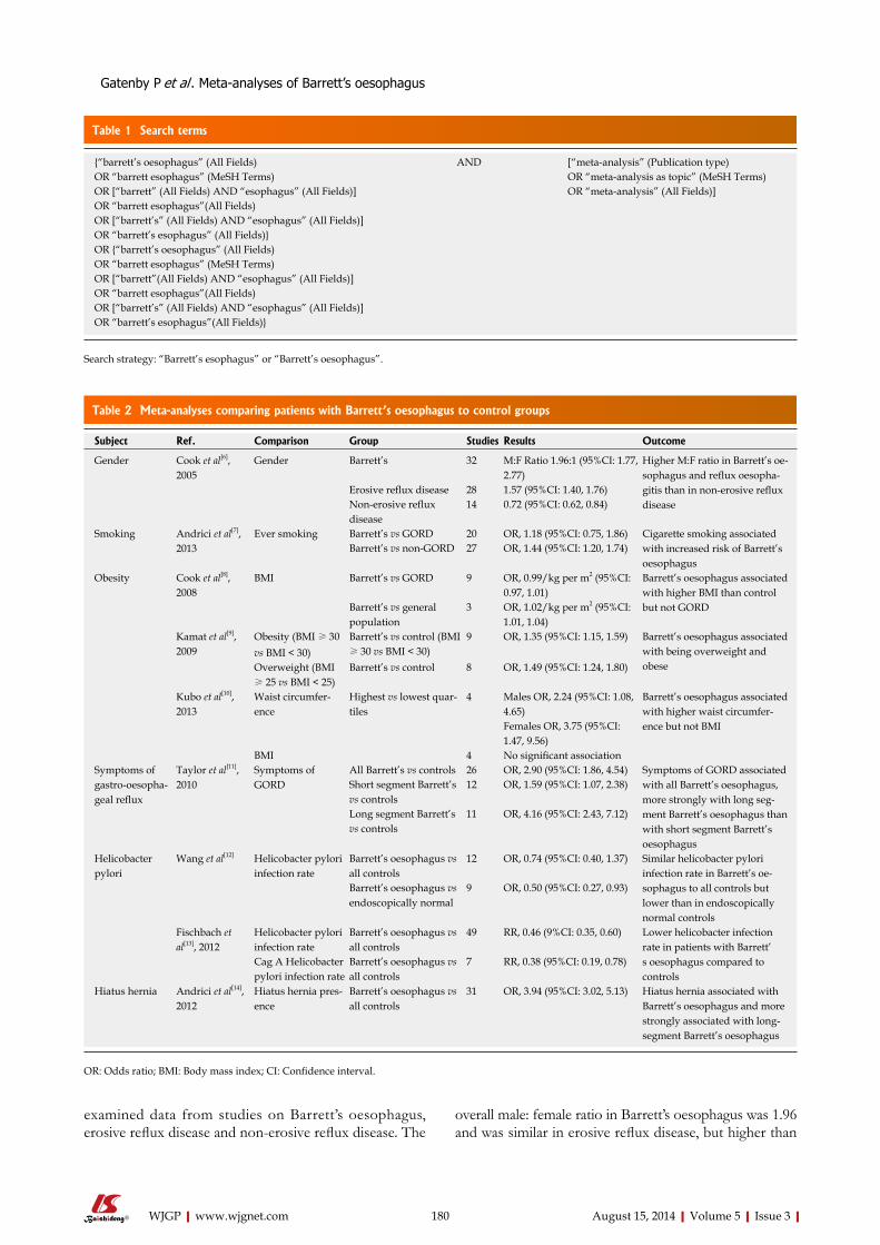
examined data from studies on Barrett’s oesophagus, erosive reflux disease and non-erosive reflux disease. The
overall male: female ratio in Barrett’s oesophagus was 1.96 and was similar in erosive reflux disease, but higher than
August 15, 2014|Volume 5|Issue 3|WJGP|www.wjgnet.com 180
Table 1 Search terms
{“barrett’s oesophagus” (All Fields) AND [“meta-analysis” (Publication type) OR “barrett esophagus” (MeSH Terms) OR “meta-analysis as topic” (MeSH Terms) OR [“barrett” (All Fields) AND “esophagus” (All Fields)] OR “meta-analysis” (All Fields)]OR “barrett esophagus”(All Fields) OR [“barrett’s” (All Fields) AND “esophagus” (All Fields)] OR “barrett’s esophagus” (All Fields)} OR {“barrett’s oesophagus” (All Fields) OR “barrett esophagus” (MeSH Terms) OR [“barrett”(All Fields) AND “esophagus” (All Fields)] OR “barrett esophagus”(All Fields) OR [“barrett’s” (All Fields) AND “esophagus” (All Fields)] OR “barrett’s esophagus”(All Fields)}
Search strategy: “Barrett’s esophagus” or “Barrett’s oesophagus”.
Table 2 Meta-analyses comparing patients with Barrett’s oesophagus to control groups
Subject Ref. Comparison Group Studies Results Outcome
Gender Cook et al[6], 2005
Gender Barrett’s 32 M:F Ratio 1.96:1 (95%CI: 1.77, 2.77)
Higher M:F ratio in Barrett’s oe-sophagus and reflux oesopha-gitis than in non-erosive reflux disease
Erosive reflux disease 28 1.57 (95%CI: 1.40, 1.76)Non-erosive reflux disease
14 0.72 (95%CI: 0.62, 0.84)
Smoking Andrici et al[7], 2013
Ever smoking Barrett’s vs GORD 20 OR, 1.18 (95%CI: 0.75, 1.86) Cigarette smoking associated with increased risk of Barrett’s oesophagus
Barrett’s vs non-GORD 27 OR, 1.44 (95%CI: 1.20, 1.74)
Obesity Cook et al[8], 2008
BMI Barrett’s vs GORD 9 OR, 0.99/kg per m2 (95%CI: 0.97, 1.01)
Barrett’s oesophagus associated with higher BMI than control but not GORDBarrett’s vs general
population3 OR, 1.02/kg per m2 (95%CI:
1.01, 1.04)Kamat et al[9], 2009
Obesity (BMI ≥ 30 vs BMI < 30)
Barrett’s vs control (BMI ≥ 30 vs BMI < 30)
9 OR, 1.35 (95%CI: 1.15, 1.59) Barrett’s oesophagus associated with being overweight and obeseOverweight (BMI
≥ 25 vs BMI < 25)Barrett’s vs control 8 OR, 1.49 (95%CI: 1.24, 1.80)
Kubo et al[10], 2013
Waist circumfer-ence
Highest vs lowest quar-tiles
4 Males OR, 2.24 (95%CI: 1.08, 4.65)
Barrett’s oesophagus associated with higher waist circumfer-ence but not BMIFemales OR, 3.75 (95%CI:
1.47, 9.56)BMI 4 No significant association
Symptoms of gastro-oesopha-geal reflux
Taylor et al[11], 2010
Symptoms of GORD
All Barrett’s vs controls 26 OR, 2.90 (95%CI: 1.86, 4.54) Symptoms of GORD associated with all Barrett’s oesophagus, more strongly with long seg-ment Barrett’s oesophagus than with short segment Barrett’s oesophagus
Short segment Barrett’s vs controls
12 OR, 1.59 (95%CI: 1.07, 2.38)
Long segment Barrett’s vs controls
11 OR, 4.16 (95%CI: 2.43, 7.12)
Helicobacter pylori
Wang et al[12] Helicobacter pylori infection rate
Barrett’s oesophagus vs all controls
12 OR, 0.74 (95%CI: 0.40, 1.37) Similar helicobacter pylori infection rate in Barrett’s oe-sophagus to all controls but lower than in endoscopically normal controls
Barrett’s oesophagus vs endoscopically normal
9 OR, 0.50 (95%CI: 0.27, 0.93)
Fischbach et al[13], 2012
Helicobacter pylori infection rate
Barrett’s oesophagus vs all controls
49 RR, 0.46 (9%CI: 0.35, 0.60) Lower helicobacter infection rate in patients with Barrett’s oesophagus compared to controls
Cag A Helicobacter pylori infection rate
Barrett’s oesophagus vs all controls
7 RR, 0.38 (95%CI: 0.19, 0.78)
Hiatus hernia Andrici et al[14], 2012
Hiatus hernia pres-ence
Barrett’s oesophagus vs all controls
31 OR, 3.94 (95%CI: 3.02, 5.13) Hiatus hernia associated with Barrett’s oesophagus and more strongly associated with long-segment Barrett’s oesophagus
OR: Odds ratio; BMI: Body mass index; CI: Confidence interval.
Gatenby P et al . Meta-analyses of Barrett’s oesophagus
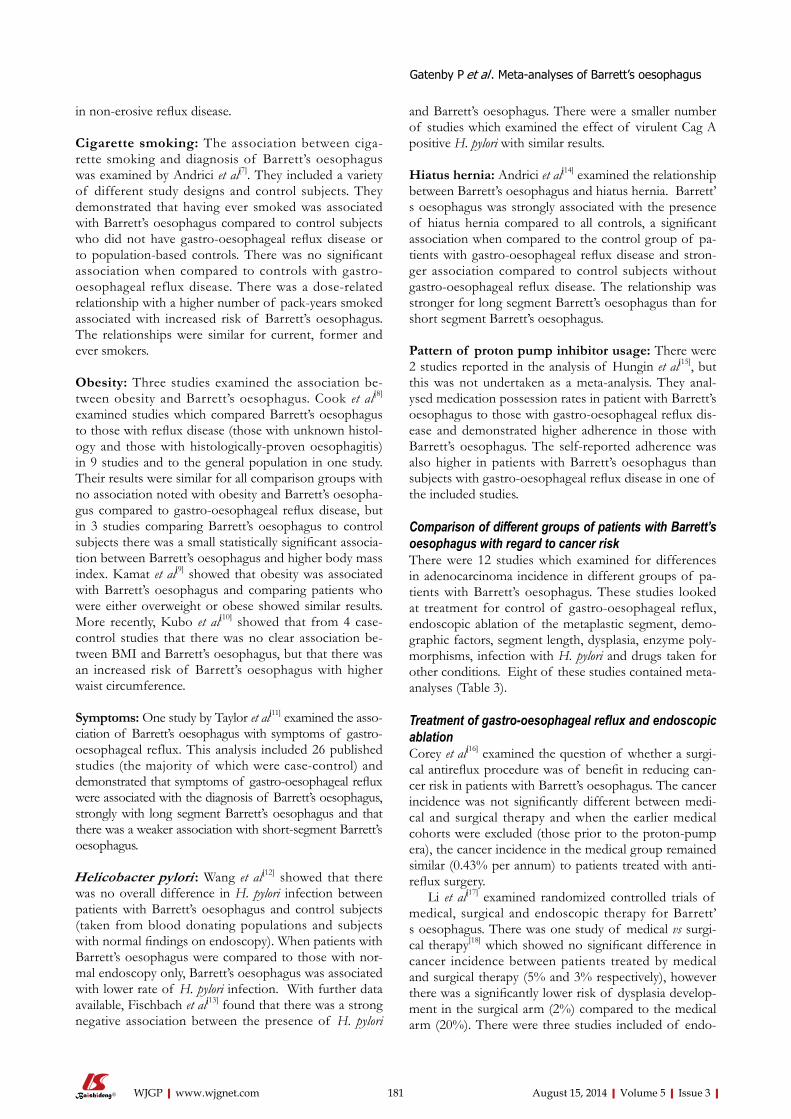
in non-erosive reflux disease.
Cigarette smoking: The association between ciga-rette smoking and diagnosis of Barrett’s oesophagus was examined by Andrici et al[7]. They included a variety of different study designs and control subjects. They demonstrated that having ever smoked was associated with Barrett’s oesophagus compared to control subjects who did not have gastro-oesophageal reflux disease or to population-based controls. There was no significant association when compared to controls with gastro-oesophageal reflux disease. There was a dose-related relationship with a higher number of pack-years smoked associated with increased risk of Barrett’s oesophagus. The relationships were similar for current, former and ever smokers.
Obesity: Three studies examined the association be-tween obesity and Barrett’s oesophagus. Cook et al[8] examined studies which compared Barrett’s oesophagus to those with reflux disease (those with unknown histol-ogy and those with histologically-proven oesophagitis) in 9 studies and to the general population in one study. Their results were similar for all comparison groups with no association noted with obesity and Barrett’s oesopha-gus compared to gastro-oesophageal reflux disease, but in 3 studies comparing Barrett’s oesophagus to control subjects there was a small statistically significant associa-tion between Barrett’s oesophagus and higher body mass index. Kamat et al[9] showed that obesity was associated with Barrett’s oesophagus and comparing patients who were either overweight or obese showed similar results. More recently, Kubo et al[10] showed that from 4 case-control studies that there was no clear association be-tween BMI and Barrett’s oesophagus, but that there was an increased risk of Barrett’s oesophagus with higher waist circumference.
Symptoms: One study by Taylor et al[11] examined the asso-ciation of Barrett’s oesophagus with symptoms of gastro-oesophageal reflux. This analysis included 26 published studies (the majority of which were case-control) and demonstrated that symptoms of gastro-oesophageal reflux were associated with the diagnosis of Barrett’s oesophagus, strongly with long segment Barrett’s oesophagus and that there was a weaker association with short-segment Barrett’s oesophagus.
Helicobacter pylori : Wang et al[12] showed that there was no overall difference in H. pylori infection between patients with Barrett’s oesophagus and control subjects (taken from blood donating populations and subjects with normal findings on endoscopy). When patients with Barrett’s oesophagus were compared to those with nor-mal endoscopy only, Barrett’s oesophagus was associated with lower rate of H. pylori infection. With further data available, Fischbach et al[13] found that there was a strong negative association between the presence of H. pylori
and Barrett’s oesophagus. There were a smaller number of studies which examined the effect of virulent Cag A positive H. pylori with similar results.
Hiatus hernia: Andrici et al[14] examined the relationship between Barrett’s oesophagus and hiatus hernia. Barrett’s oesophagus was strongly associated with the presence of hiatus hernia compared to all controls, a significant association when compared to the control group of pa-tients with gastro-oesophageal reflux disease and stron-ger association compared to control subjects without gastro-oesophageal reflux disease. The relationship was stronger for long segment Barrett’s oesophagus than for short segment Barrett’s oesophagus.
Pattern of proton pump inhibitor usage: There were 2 studies reported in the analysis of Hungin et al[15], but this was not undertaken as a meta-analysis. They anal-ysed medication possession rates in patient with Barrett’s oesophagus to those with gastro-oesophageal reflux dis-ease and demonstrated higher adherence in those with Barrett’s oesophagus. The self-reported adherence was also higher in patients with Barrett’s oesophagus than subjects with gastro-oesophageal reflux disease in one of the included studies.
Comparison of different groups of patients with Barrett’s oesophagus with regard to cancer riskThere were 12 studies which examined for differences in adenocarcinoma incidence in different groups of pa-tients with Barrett’s oesophagus. These studies looked at treatment for control of gastro-oesophageal reflux, endoscopic ablation of the metaplastic segment, demo-graphic factors, segment length, dysplasia, enzyme poly-morphisms, infection with H. pylori and drugs taken for other conditions. Eight of these studies contained meta-analyses (Table 3).
Treatment of gastro-oesophageal reflux and endoscopic ablationCorey et al[16] examined the question of whether a surgi-cal antireflux procedure was of benefit in reducing can-cer risk in patients with Barrett’s oesophagus. The cancer incidence was not significantly different between medi-cal and surgical therapy and when the earlier medical cohorts were excluded (those prior to the proton-pump era), the cancer incidence in the medical group remained similar (0.43% per annum) to patients treated with anti-reflux surgery.
Li et al[17] examined randomized controlled trials of medical, surgical and endoscopic therapy for Barrett’s oesophagus. There was one study of medical vs surgi-cal therapy[18] which showed no significant difference in cancer incidence between patients treated by medical and surgical therapy (5% and 3% respectively), however there was a significantly lower risk of dysplasia develop-ment in the surgical arm (2%) compared to the medical arm (20%). There were three studies included of endo-
August 15, 2014|Volume 5|Issue 3|WJGP|www.wjgnet.com 181
Gatenby P et al . Meta-analyses of Barrett’s oesophagus
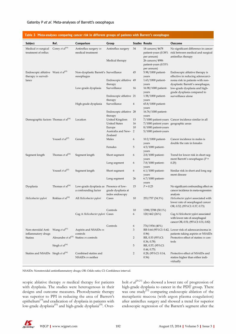
scopic ablative therapy vs medical therapy for patients with dysplasia. The studies were heterogenous in their designs and outcome measures. Photodynamic therapy was superior to PPI in reducing the area of Barrett’s epithelium[19]and eradication of dysplasia in patients with low-grade dysplasia[20] and high-grade dysplasia[20]. Over-
holt et al[20,21] also showed a lower rate of progression of high-grade dysplasia to cancer in the PDT group. There was one study[22] comparing endoscopic ablation of the metaplastic mucosa (with argon plasma coagulation) after antireflux surgery and showed a trend for superior endoscopic regression of the Barrett’s segment after the
August 15, 2014|Volume 5|Issue 3|WJGP|www.wjgnet.com 182
Table 3 Meta-analyses comparing cancer risk in different groups of patients with Barrett’s oesophagus
Subject Ref. Comparison Group Studies Results Outcome
Medical vs surgical treatment of reflux
Corey et al[16] Antireflux surgery vs medical treatment
Antireflux surgery 34 18 cancers/4678 patient-years (0.38% per annum)
No significant difference in cancer risk between medical and surgical antireflux therapy
Medical therapy 26 cancers/4906 patient-years (0.53% per annum)
Endoscopic ablative therapy vs surveil-lance
Wani et al[25] Non-dysplastic Barrett’s oesophagus
Surveillance 45 5.98/1000 patient-years
Endoscopic ablative therapy is effective in reducing adenocarci-noma risk in patients with non-dysplastic Barrett’s oesophagus, low-grade dysplasia and high-grade dysplasia compared to surveillance alone
Endoscopic ablative therapy
49 1.63/1000 patient-years
Low-grade dysplasia Surveillance 16 16.98/1000 patient-years
Endoscopic ablative therapy
21 1.58/1000 patient-years
High-grade dysplasia Surveillance 4 65.8/1000 patient-years
Endoscopic ablative therapy
28 16.76/1000 patient-years
Demographic factors Thomas et al[26] Location United Kingdom 13 7/1000 patient-years Cancer incidence similar in all geographic areasUnited States 16 7/1000 patient years
Europe 10 8/1000 patient-yearsAustralia and New-Zealand
2 5/1000 patient-years
Yousef et al[27] Gender Males 6 10.2/1000 patient-years
Cancer incidence in males is double the rate in females
Females 5 4.5/1000 patient-years
Segment length Thomas et al[26] Segment length Short segment 6 2.8/1000 patient-years
Trend for lower risk in short seg-ment Barrett’s oesophagus (P = 0.25)Long segment 6 7.8/1000 patient-
yearsYousef et al[27] Segment length Short segment 6 6.1/1000 patient-
yearsSimilar risk in short and long seg-ment disease
Long segment 26 6.7/1000 patient-years
Dysplasia Thomas et al[26] Low-grade dysplasia as a confounding factor
Presence of low-grade dysplasia at index endoscopy
15 P = 0.23 No significant confounding effect on cancer incidence in meta-regression analysis
Helicobacter pylori Rokkas et al[30] All Helicobacter pylori Cases 10 253/757 (34.3%) Helicobacter pylori associated with lower rate of oesophageal cancer OR, 0.52; (95%CI: 0.37, 0.73)
Controls 10 1398/2788 (50.1%)Cag A Helicobacter pylori Cases 6 120/462 (26%) Cag A Helicobacter pylori associated
with lower rate of oesophageal cancer OR, 0.51; (95%CI: 0.31, 0.82)
Controls 6 774/1936 (40%)Non-steroidal Anti-inflammatory drugs Statins
Wang et al[31] Aspirin and NSAIDs vs controls
3 RR 0.64 (95%CI: 0.42, 0.96)
Lower risk of adenocarcinoma in patients taking aspirin or NSAIDs
Alexandre et al[33] Statins vs controls 2 RR, 0.53 (95%CI: 0.36, 0.78)
Protective effect of statins vs con-trols
Singh et al[36] 5 RR, 0.57; (95%CI: 0.44, 0.75)
Statins and NSAIDs Singh et al[36] Combined statins and NSAIDs vs neither
2 0.28; (95%CI: 0.14, 0.56)
Protective effect of NSAIDs and statins higher than either indi-vidually
NSAIDs: Nonsteroidal antiinflammatory drugs; OR: Odds ratio; CI: Confidence interval.
Gatenby P et al . Meta-analyses of Barrett’s oesophagus
ablation, but no difference in cancer incidence[20]. In ab-lation of the metaplastic mucosa, 3 studies demonstrated that overall argon plasma coagulation was superior to photodynamic therapy with ablation rates of 59.0% and 27.5% respectively [odds ratio (OR), 3.46, 95% confi-dence interval (CI): 1.67, 7.81]. These studies did not ex-amine long-term cancer incidences. There were 2 stud-ies comparing argon plasma coagulation to multipolar electrocoagulation which demonstrated similar rates of successful ablation of the metaplastic segment (78.6% in patients treated with multipolar electrocoagulation and 64.4% treated with argon plasma coagulation) and again no long-term data on cancer incidence.
Fayter et al[23]examined the evidence from 11 ran-domised controlled trials of photodynamic therapy for Barrett’s oesophagus. The trials were heterogeneous in their design, the protocol of therapy used, the patients studied (most studies examined patients with high-grade dysplasia, but some had low-grade dysplasia, non-dysplastic epithelium or a combination of histological findings) and outcome measures. The conclusions drawn from this systematic review were: (1) it was not possible to determine whether there was a significant clinical difference between photodynamic therapy and argon plasma coagulation and which would be the most ap-propriate treatment; (2) photodynamic therapy was more effective than omeprazole alone in producing long-term ablation of high-grade dysplasia and slowing/preventing progression to cancer; (3) Photodynamic therapy with 5-ALA as the photosensitising agent was more effective than placebo in producing regression of dysplasia and reduction in the area of Barrett’s epithelium in patients with low-grade dysplasia; (4) photodynamic therapy with 5-ALA may be more effective than with Photofrin; (5) optimal treatment for patients without dysplasia had yet to be determined; and (6) side effects were similar be-tween 5-ALA and Photofrin with higher levels of pho-tosensitivity with Photofrin.
Rees et al[24]examined randomized controlled stud-ies only. They demonstrated that in the 3 studies which examined H2 receptor antagonists to proton pump inhibitors: cancer risk, eradication of dysplasia or com-plete regression of the metaplastic segment were not reported. There was a trend towards a reduction in the areas of metaplastic mucosa (but not the length of the Barretts’ segment) with PPI. There were no new studies available on antireflux surgery vs medical therapy, argon plasma ablation, argon plasma coagulation vs multipolar electrocoagulation or argon plasma coagulation vs pho-todynamic therapy since Li et al[17].
Wani et al[25] compared the rate of development of adenocarcinoma in published series of patients with non-dysplastic Barrett’s oesophagus, low-grade dysplasia and high-grade dysplasia comparing cohorts treated with endoscopic ablative therapy to those in surveillance pro-grammes without ablation of the mucosa. They found that there were significantly lower rates of adenocarci-noma incidence in the cohorts treated with ablative ther-
apies compared to the control cohorts. The differences were significant for examinations of non-dysplastic Bar-rett’s oesophagus, low-grade dysplasia and high-grade dysplasia.
Demographic factors: Thomas et al[26] showed that age did not influence cancer risk from 41 studies of 9469 patients undergoing surveillance (36635 patient-years follow-up). There was also no significant difference in cancer incidence depending on geographic origin of the included studies. Yousef et al[27] showed that the inci-dence of adenocarcinoma in males was double the rate in females.
Segment length: Thomas et al[26] showed that from 6 studies including 960 patients with long-segment Barrett’s oesophagus (4130 patient-years of follow-up) and 258 patients (1074 patient-years of follow up) that 32 of the 35 cancers which developed were in long segment Bar-rett’s oesophagus (but this did not reach statistical sig-nificance). Yousef et al[27] reported a cancer incidence of 0.67% per annum in long segment Barrett’s oesophagus and a similar incidence (0.61%) in short segment Barrett’s oesophagus in 30 studies.
Dysplasia: Thomas et al[26] did not demonstrate an in-creased cancer risk associated with dysplasia over non-dysplastic Barrett’s oesophagus. Desai et al[28] examined specifically patients without dysplasia at baseline, but there was no comparison cohort in this study and it has subsequently been excluded from this review.
Intestinal metaplasia: The question of the importance of intestinal metaplasia for cancer risk was not specifi-cally examined by any of the meta-analyses.
Enzyme polymorphisms: Bull et al[29] examined en-zyme polymorphisms in case-control studies and found an association between Barrett’s oesophagus and GSTP1 homozygotes for the Ile105 variant (OR, 1.50, 95%CI: 1.16, 1.95). This genetic variant results in increased IgE and immune-mediated inflammation. There was no oth-er significant association with Barrett’s oesophagus and a variety of metabolic gene polymorphisms[29].
Helicobacter pylori : Rokkas et al[30] showed similar results in studies of oesophageal cancer to those of Bar-rett’s oesophagus with a negative association between the presence of H. pylori and oesophageal cancer. The results were similar in studies of Cag A H. pylori.
Other medicationsThe reduction in cancer risk with aspirin and non-steroi-dal anti-inflammatory drugs was examined in 3 cohort studies by Wang et al[31]. They demonstrated that there was a trend towards lower cancer risk in patients taking aspirin and non-steroidal anti-inflammatory drugs, how-ever 2 case-control studies were excluded for unclear
August 15, 2014|Volume 5|Issue 3|WJGP|www.wjgnet.com 183
Gatenby P et al . Meta-analyses of Barrett’s oesophagus
reasons.Rees et al[24] reported one study[32] comparing cele-
coxib to placebo and found no difference in cancer risk at 2 years (3/49 and 3/51 patients respectively).
The effects of statins on the risk of oesophageal adenocarcinoma in Barrett’s oesophagus were examined by Alexandre et al[33], who found two prospective co-hort studies. The first was a multicentre study from the Netherlands of 570 patients and demonstrated a hazard ratio of 0.46 (95%CI: 0.21, 0.99) and in patients taking statins and non-steroidal anti-inflammatory drugs the hazards ratio was 0.22 (95%CI: 0.06, 0.85)[34]. Nguyen et al[35] examined 812 patients in a case-control cohort in the Veterans Affairs Healthcare System and showed an incidence density ratio of 0.56 (95%CI: 0.36, 0.86) for patients with Barrett’s oesophagus taking statins.
Singh et al[36] also demonstrated a protective effect of statins in their meta-analysis of 5 studies and a greater protective effect of combining statins with non-steroidal anti-inflammatory drugs with respect to oesophageal cancer risk.
Decision to enrol in surveillanceThe American College of Gastroenterology and Ameri-can Gastroenterological Association have defined Bar-rett’s oesophagus as any length of recognisable columnar mucosa which demonstrates intestinal metaplasia at bi-opsy[3,4], maintaining the dogma that intestinal metaplasia is necessary for malignant risk on the basis that in many cohort studies intestinal metaplasia has been demon-strated adjacent to adenocarcinoma of the oesophagus. The ACG acknowledge the difficulties associated with sampling error in the detection of intestinal metaplasia and also exclude “ultra-short” segments (< 1 cm) due to poor interobserver reliability of recognition. The BSG broadly agrees with this definition[2] and whilst there is no requirement for the presence of intestinal metapla-sia for diagnosis, on the basis of the higher cancer risk in subjects with intestinal metaplasia in the Northern Ireland pathology database cohort[37] and low rate of de-velopment of high-grade dysplasia and adenocarcinoma in the Danish pathology database cohort which only included subjects with intestinal metaplasia[38], surveil-lance is only recommended if intestinal metaplasia is detected during the either the index or the first surveil-lance endoscopy in patients with short segment (< 3 cm) metaplasia. The rationale for this is that it is felt that the risks of endoscopy probably outweigh the benefits. Both guidelines have excluded very short segments or tongues of metaplasia due to difficulties in clinical assessment rather than on the basis of a proven low risk of compli-cations and there are no good data to support or refute these assertions. The evidence from meta-analyses con-cerning the role of segment length and intestinal meta-plasia is discussed below.
The ACG recommend that the consideration for beginning a surveillance programme should include age, likelihood of survival over the next 5 years, patient’s
understanding of the process and its limitations for the detection of cancer and the willingness of the patient to adhere to the recommendations.
The ACG supports surveillance of Barrett’s oesopha-gus as in 7 retrospective series the survival in cancers was improved over those detected outside of surveillance programmes. There has not yet been a trial published demonstrating benefits of surveillance in a prospective fashion, however the BOSS study (endoscopic surveil-lance vs endoscopy at time of need) remains underway at present[39].
The ACG, AGA and BSG recommend 4-quadrant biopsies taken every 2 cm throughout the metaplastic segment at index endoscopy and surveillance (if no dys-plasia has been previously detected or other macroscopic lesions are present). This biopsy protocol has not yet been tested in a meta-analysis. The difficulties involved in adequately sampling the tissue at risk and variability in histopathological interpretation of the tissue examined should be subject to further studies beyond the initial work done by Levine et al[40].
Risk stratification and frequency of surveillanceThe ACG recommend that the first two endoscopies are undertaken within a year and if no dysplasia is detected then the surveillance interval is 3 years. If low-grade dysplasia is detected then surveillance interval should be within 6 mo. This recommendation was based upon a poor level of evidence from cohort studies and expert opinion[3].
The BSG note that risk factors for cancer develop-ment include the presence of intestinal metaplasia (3 × compared to no intestinal metaplasia), low-grade dysplasia (5.67 × non-dysplastic Barrett’s oesophagus), male gender (2 × that of females), smoking (2 × non-smokers). They note that longer segment lengths were associated with a trend to increased risk and no relation-ship was demonstrated with alcohol consumption and obesity[2].
The ACG and AGA stratify risk based only on the presence of dysplasia after the diagnosis of Barrett’s oesophagus and that further work to assess the extent of dysplasia and develop biomarkers is required[3,4]. The BSG note that in future, surveillance intervals will take into account all of the socio-demographic risk factors and characteristics of the Barrett’s segment as well as biomarker panels[2] . Until such algorithms are developed, surveillance frequency is based on dysplasia and length only. The ACG also note that a randomised controlled trial to assess the impact of surveillance is required. The BSG also incorporate segment length and allow for con-sideration of other risk factors (see above)[2]. The BSG have lengthened the recommended surveillance interval for non-dysplastic Barrett’s oesophagus (based upon the recent lower cancer incidence estimates) in line with the AGA and allowed some further individualised risk strati-fication to be incorporated into the frequency of sur-veillance and in line with the ACG, the interval for low-
August 15, 2014|Volume 5|Issue 3|WJGP|www.wjgnet.com 184
Gatenby P et al . Meta-analyses of Barrett’s oesophagus
grade dysplasia is 6 mo. The AGA recommend surveil-lance of low-grade dysplasia in 6-12 mo. Inflammatory atypia is difficult to distinguish from true dysplasia[2,41] and the guidelines recommend repeat biopsy after treat-ment with acid suppression[3] and expert pathological review of biopsies which are dysplastic or have changes indefinite for dysplasia[2-4].
The evidence from the meta-analyses in supporting intestinal metaplasia and low-grade dysplasia as mark-ers of increased risk of malignancy is poor with no significant difference demonstrated in patients with low-grade dysplasia at index endoscopy[26] and no papers on the necessity for the detection of intestinal metaplasia to confer a malignant risk.
Evidence for difference in risk dependent on seg-ment length is also poor with only trends demonstrat-ed[26,27] and it is only on weak evidence that decisions on consideration of surveillance as well as surveillance interval are made on these features.
There is greater evidence for a lower risk of oesoph-ageal cancer development in patients who have H, pylori infection[30] and for a higher risk in males over females[27].
What steps to minimise risk of developing Barrett’sThe ACG notes that older Caucasian males with chronic reflux symptoms are the group with the highest preva-lence of Barrett’s oesophagus and there were no direct recommendations from the ACG to reduce the risk of development of Barrett’s oesophagus[3]. The BSG state that the known risk factors are male gender, older age and history of reflux symptoms as well as an as-sociation with white race, higher waist: hip ration and abdominal circumference. There is a less clear relation-ship with obesity as measured by body mass index and cigarette smoking. The BSG also note the small degree of familial clustering[2]. The AGA go one step further in recommending consideration of screening for Barrett’s oesophagus in patients with multiple risk factors for oe-sophageal adenocarcinoma (age 50 years or older, male sex, white race, chronic gastro-oesophageal reflux dis-ease, hiatal hernia, elevated body mass index and intra-abdominal distribution of body fat).
The published meta-analyses have demonstrated that the significant risk factors associated with Barrett’s oesoph-agus are male gender[6], smoking[7], obesity[8-10], prolonged symptoms of gastro-oesophageal reflux[11], absence of H. pylori infection[12,13] and the presence of hiatus her-nia[14]. Age has not been demonstrated to influence can-cer risk in the meta-analyses[26].
Minimisation of risk of cancer development in Barrett’sThe ACG, AGA and BSG did not recommend fundo-plication over medical therapy to reduce cancer develop-ment[2-4] and this review supports this strategy[16,17], how-ever there were encouraging data concerning reduction in risk of development of dysplasia with surgical therapy over acid suppression therapy[18].
The question of ablation of the metaplastic mucosa
is a complex one requiring further examination, however there are promising results[25] and the SURF trial com-paring radiofrequency ablation to surveillance in low-grade dysplasia remains underway[42].
The ACG note that a meta-analysis did demonstrate a lower risk of cancer development in patients taking non-steroidal anti-inflammatory drugs[43] and that the ASPECT study (a randomised study of aspirin and low and high-dose esomeprazole) remains underway[44]. The ACG, AGA and BSG did not recommend chemopre-vention with aspirin or non-steroidal anti-inflammatory drugs. The ACG cites two cohort studies demonstrating a lower risk of dysplasia development in patients taking PPI therapy, but no evidence to support a reduction in cancer development[3]. The BSG recommendations are similar to those of the ACG and also do not advocate acid suppression drugs as chemopreventive agents[2], but they are effective in symptom control.
The AGA note that the patients may derive benefit from aspirin if they have cardiovascular risk factors for which aspirin therapy is indicated, but that neither the use of aspirin or non-steroidal anti-inflammatory drugs are recommended solely to prevent oesophageal adeno-carcinoma and that the evidence to support the use of PPI therapy to reduce the risk of cancer and dysplasia is indirect and not been proven in a long-term controlled trial[4].The results from the meta-analyses of Alexandre et al[33] and Singh et al[36] showing the protective effect of aspirin and in particular the effect in conjunction with statins is exciting and may form the basis for effective chemoprevention in the future.
CONCLUSIONThe evidence to support the current decisions to enrol patients with Barrett’s oesophagus in surveillance pro-grammes and surveillance interval are based on weak evidence on the clinical outcome of features of the metaplastic segment. Further consideration should be given to the role of gender and helicobacter status in examining cancer risk as well as the role of aspirin and statins in chemopreventive strategies and ablation of the metaplastic segment. Public health programmes should also examine measures to reduce the associations of Barrett’s oesophagus, notably, smoking and obesity. The relevance of male gender and absence of helicobacter infection should also be considered.
REFERENCES1 Gatenby PA, Caygill CP, Ramus JR, Charlett A, Watson
A. Barrett’s columnar-lined oesophagus: demographic and lifestyle associations and adenocarcinoma risk. Dig Dis Sci 2008; 53: 1175-1185 [PMID: 17939050]
2 Fitzgerald RC, di Pietro M, Ragunath K, Ang Y, Kang JY, Watson P, Trudgill N, Patel P, Kaye PV, Sanders S, O’Donovan M, Bird-Lieberman E, Bhandari P, Jankowski JA, Attwood S, Parsons SL, Loft D, Lagergren J, Moayyedi P, Lyratzopoulos G, de Caestecker J. British Society of Gastro-
August 15, 2014|Volume 5|Issue 3|WJGP|www.wjgnet.com 185
Gatenby P et al . Meta-analyses of Barrett’s oesophagus
enterology guidelines on the diagnosis and management of Barrett’s oesophagus. Gut 2014; 63: 7-42 [PMID: 24165758 DOI: 10.1136/gutjnl-2013-305372]
3 Wang KK, Sampliner RE. Updated guidelines 2008 for the diagnosis, surveillance and therapy of Barrett’s esophagus. Am J Gastroenterol 2008; 103: 788-797 [PMID: 18341497]
4 Spechler SJ, Sharma P, Souza RF, Inadomi JM, Shaheen NJ. American Gastroenterological Association medical posi-tion statement on the management of Barrett’s esophagus. Gastroenterology 2011; 140: 1084-1091 [PMID: 21376940 DOI: 10.1053/j.gastro.2011.01.030]
5 Andrici J, Tio M, Cox MR, Eslick GD. Meta-analysis: Bar-rett’s oesophagus and the risk of colonic tumours. Aliment Pharmacol Ther 2013; 37: 401-410 [PMID: 23163592 DOI: 10.1111/apt.12146]
6 Cook MB, Wild CP, Forman D. A systematic review and meta-analysis of the sex ratio for Barrett’s esophagus, ero-sive reflux disease, and nonerosive reflux disease. Am J Epi-demiol 2005; 162: 1050-1061 [PMID: 16221805 DOI: 10.1093/aje/kwi325]
7 Andrici J, Cox MR, Eslick GD. Cigarette smoking and the risk of Barrett’s esophagus: a systematic review and meta-analysis. J Gastroenterol Hepatol 2013; 28: 1258-1273 [PMID: 23611750 DOI: 10.1111/jgh.12230]
8 Cook MB, Greenwood DC, Hardie LJ, Wild CP, Forman D. A systematic review and meta-analysis of the risk of increasing adiposity on Barrett’s esophagus. Am J Gastro-enterol 2008; 103: 292-300 [PMID: 17986313 DOI: 10.1111/j.1572-0241.2007.01621.x]
9 Kamat P, Wen S, Morris J, Anandasabapathy S. Exploring the association between elevated body mass index and Bar-rett’s esophagus: a systematic review and meta-analysis. Ann Thorac Surg 2009; 87: 655-662 [PMID: 19161814 DOI: 10.1016/j.athoracsur.2008.08.003]
10 Kubo A, Cook MB, Shaheen NJ, Vaughan TL, Whiteman DC, Murray L, Corley DA. Sex-specific associations between body mass index, waist circumference and the risk of Bar-rett’s oesophagus: a pooled analysis from the international BEACON consortium. Gut 2013; 62: 1684-1691 [PMID: 23355549 DOI: 10.1136/gutjnl-2012-303753]
11 Taylor JB, Rubenstein JH. Meta-analyses of the effect of symptoms of gastroesophageal reflux on the risk of Barrett’s esophagus. Am J Gastroenterol 2010; 105: 1729, 1730-1737; quiz 1738 [PMID: 20485283 DOI: 10.1038/ajg.2010.194]
12 Wang C, Yuan Y, Hunt RH. Helicobacter pylori infection and Barrett’s esophagus: a systematic review and meta-analysis. Am J Gastroenterol 2009; 104: 492-500; quiz 491, 501 [PMID: 19174811 DOI: 10.1038/ajg.2008.37]
13 Fischbach LA, Nordenstedt H, Kramer JR, Gandhi S, Dick-Onuoha S, Lewis A, El-Serag HB. The association between Barrett’s esophagus and Helicobacter pylori infection: a me-ta-analysis. Helicobacter 2012; 17: 163-175 [PMID: 22515353 DOI: 10.1111/j.1523-5378.2011.00931.x]
14 Andrici J, Tio M, Cox MR, Eslick GD. Hiatal hernia and the risk of Barrett’s esophagus. J Gastroenterol Hepatol 2013; 28: 415-431 [PMID: 22694245 DOI: 10.1111/j.1440-1746.2012.07199.x]
15 Hungin AP, Hill C, Molloy-Bland M, Raghunath A. Sys-tematic review: Patterns of proton pump inhibitor use and adherence in gastroesophageal reflux disease. Clin Gas-troenterol Hepatol 2012; 10: 109-116 [PMID: 21782770 DOI: 10.1016/j.cgh.2011.07.008]
16 Corey KE, Schmitz SM, Shaheen NJ. Does a surgical antire-flux procedure decrease the incidence of esophageal adeno-carcinoma in Barrett’s esophagus? A meta-analysis. Am J Gastroenterol 2003; 98: 2390-2394 [PMID: 14638338]
17 Li YM, Li L, Yu CH, Liu YS, Xu CF. A systematic review and meta-analysis of the treatment for Barrett’s esopha-gus. Dig Dis Sci 2008; 53: 2837-2846 [PMID: 18427992 DOI: 10.1007/s10620-008-0257-3]
18 Parrilla P, Martínez de Haro LF, Ortiz A, Munitiz V, Molina J, Bermejo J, Canteras M. Long-term results of a randomized prospective study comparing medical and surgical treat-ment of Barrett’s esophagus. Ann Surg 2003; 237: 291-298 [PMID: 12616111 DOI: 10.1097/00000658-200303000-00001]
19 Ackroyd R, Tam W, Schoeman M, Devitt P, Watson D. Pro-spective randomized controlled trial of argon plasma coagu-lation ablation vs. endoscopic surveillance of patients with Barrett’s esophagus after antireflux surgery. Gastrointest Endosc 2004; 59: 1-7 [DOI: 10.1016/S0016-5107(03)02528-8]
20 Overholt BF, Lightdale CJ, Wang KK, Canto MI, Burdick S, Haggitt RC, Bronner MP, Taylor SL, Grace MGA, Depot M, [On behalf of the International Photodynamic Group for High-Grade Dysplasia in Barrett's Esophagus]. Pho-todynamic therapy with porfimer sodium for ablation of high-grade dysplasia in Barrett's esophagus: international, partially blinded, randomized phase III trial. Gastrointestinal Endoscopy 2005; 62: 488-498 [DOI: 10.1016/j.gie.2005.06.047]
21 Overholt BF, Wang KK, Burdick JS, Lightdale CJ, Kimmey M, Nava HR, Sivak MV, Nishioka N, Barr H, Marcon N, Pedrosa M, Bronner MP, Grace M, Depot M. Five-year effi-cacy and safety of photodynamic therapy with Photofrin in Barrett’s high-grade dysplasia. Gastrointest Endosc 2007; 66: 460-468 [PMID: 17643436 DOI: 10.1016/j.gie.2006.12.037]
22 Bright T, Watson DI, Tam W, Game PA, Astill D, Ackroyd R, Wijnhoven BP, Devitt PG, Schoeman MN. Randomized trial of argon plasma coagulation versus endoscopic surveil-lance for barrett esophagus after antireflux surgery: late re-sults. Ann Surg 2007; 246: 1016-1020 [PMID: 18043104 DOI: 10.1097/SLA.0b013e318133fa85]
23 Fayter D, Corbett M, Heirs M, Fox D, Eastwood A. A sys-tematic review of photodynamic therapy in the treatment of pre-cancerous skin conditions, Barrett’s oesophagus and cancers of the biliary tract, brain, head and neck, lung, oesophagus and skin. Health Technol Assess 2010; 14: 1-288 [PMID: 20663420 DOI: 10.3310/hta14370]
24 Rees JR, Lao-Sirieix P, Wong A, Fitzgerald RC. Treatment for Barrett’s oesophagus. Cochrane Database Syst Rev 2010; (1): CD004060 [PMID: 20091557 DOI: 10.1002/14651858.CD004060.pub2]
25 Wani S, Puli SR, Shaheen NJ, Westhoff B, Slehria S, Bansal A, Rastogi A, Sayana H, Sharma P. Esophageal adenocar-cinoma in Barrett’s esophagus after endoscopic ablative therapy: a meta-analysis and systematic review. Am J Gas-troenterol 2009; 104: 502-513 [PMID: 19174812 DOI: 10.1038/ajg.2008.31]
26 Thomas T, Abrams KR, De Caestecker JS, Robinson RJ. Meta analysis: Cancer risk in Barrett’s oesophagus. Aliment Pharmacol Ther 2007; 26: 1465-1477 [PMID: 17900269]
27 Yousef F, Cardwell C, Cantwell MM, Galway K, Johnston BT, Murray L. The incidence of esophageal cancer and high-grade dysplasia in Barrett’s esophagus: a systematic review and meta-analysis. Am J Epidemiol 2008; 168: 237-249 [PMID: 18550563 DOI: 10.1093/aje/kwn121]
28 Desai TK, Krishnan K, Samala N, Singh J, Cluley J, Perla S, Howden CW. The incidence of oesophageal adenocarcino-ma in non-dysplastic Barrett’s oesophagus: a meta-analysis. Gut 2012; 61: 970-976 [PMID: 21997553 DOI: 10.1136/gutjnl-2011-300730]
29 Bull LM, White DL, Bray M, Nurgalieva Z, El-Serag HB. Phase I and II enzyme polymorphisms as risk factors for Bar-rett’s esophagus and esophageal adenocarcinoma: a system-atic review and meta-analysis. Dis Esophagus 2009; 22: 571-587 [PMID: 19222528 DOI: 10.1111/j.1442-2050.2009.00947.x]
30 Rokkas T, Pistiolas D, Sechopoulos P, Robotis I, Margan-tinis G. Relationship between Helicobacter pylori infection and esophageal neoplasia: a meta-analysis. Clin Gastroenterol Hepatol 2007; 5: 1413-1417, 1413-1417 [PMID: 17997357 DOI: 10.1016/j.cgh.2007.08.010]
31 Wang F, Lv ZS, Fu YK. Nonsteroidal anti-inflammatory drugs
August 15, 2014|Volume 5|Issue 3|WJGP|www.wjgnet.com 186
Gatenby P et al . Meta-analyses of Barrett’s oesophagus
and esophageal inflammation - Barrett’s esophagus - adeno-carcinoma sequence: a meta-analysis. Dis Esophagus 2010 Dec 17; Epub ahead of print [PMID: 21166737 DOI: 10.1111/j.1442-2050.2010.01153.x]
32 Heath EI, Canto MI, Piantadosi S, Montgomery E, Wein-stein WM, Herman JG, Dannenberg AJ, Yang VW, Shar AO, Hawk E, Forastiere AA. Secondary chemoprevention of Barrett’s esophagus with celecoxib: results of a randomized trial. J Natl Cancer Inst 2007; 99: 545-557 [PMID: 17405999 DOI: 10.1093/jnci/djk112]
33 Alexandre L, Clark AB, Cheong E, Lewis MP, Hart AR. Sys-tematic review: potential preventive effects of statins against oesophageal adenocarcinoma. Aliment Pharmacol Ther 2012; 36: 301-311 [PMID: 22716127 DOI: 10.1111/j.1365-2036.2012.05194.x]
34 Kastelein F, Spaander MC, Biermann K, Steyerberg EW, Kuipers EJ, Bruno MJ. Nonsteroidal anti-inflammatory drugs and statins have chemopreventative effects in pa-tients with Barrett’s esophagus. Gastroenterology 2011; 141: 2000-2008; quiz 2000-2008 [PMID: 21878200 DOI: 10.1053/j.gastro.2011.08.036]
35 Nguyen DM, Richardson P, El-Serag HB. Medications (NSAIDs, statins, proton pump inhibitors) and the risk of esophageal adenocarcinoma in patients with Barrett’s esophagus. Gastroenterology 2010; 138: 2260-2266 [PMID: 20188100 DOI: 10.1053/j.gastro.2010.02.045]
36 Singh S, Singh AG, Singh PP, Murad MH, Iyer PG. Statins are associated with reduced risk of esophageal cancer, par-ticularly in patients with Barrett’s esophagus: a systematic review and meta-analysis. Clin Gastroenterol Hepatol 2013; 11: 620-629 [PMID: 23357487 DOI: 10.1016/j.cgh.2012.12.036]
37 Bhat S, Coleman HG, Yousef F, Johnston BT, McManus DT, Gavin AT, Murray LJ. Risk of malignant progression in Bar-rett’s esophagus patients: results from a large population-
based study. J Natl Cancer Inst 2011; 103: 1049-1057 [PMID: 21680910 DOI: 10.1093/jnci/djr203]
38 Hvid-Jensen F, Pedersen L, Drewes AM, Sørensen HT, Funch-Jensen P. Incidence of adenocarcinoma among pa-tients with Barrett’s esophagus. N Engl J Med 2011; 365: 1375-1383 [PMID: 21995385 DOI: 10.1056/NEJMoa1103042]
39 Jankowski J, Barr H. Improving surveillance for Barrett’s oesophagus: AspECT and BOSS trials provide an evidence base. BMJ 2006; 332: 1512 [PMID: 16793832 DOI: 10.1136/bmj.332.7556.1512]
40 Levine DS, Blount PL, Rudolph RE, Reid BJ. Safety of a sys-tematic endoscopic biopsy protocol in patients with Barrett’s esophagus. Am J Gastroenterol 2000; 95: 1152-1157 [PMID: 10811320 DOI: 10.1111/j.1572-0241.2000.02002.x]
41 Gatenby P, Ramus J, Caygill C, Shepherd N, Winslet M, Watson A. Routinely diagnosed low-grade dysplasia in Bar-rett’s oesophagus: a population-based study of natural his-tory. Histopathology 2009; 54: 814-819 [PMID: 19635100 DOI: 10.1111/j.1365-2559.2009.03316.x]
42 Alvarez Herrero L, van Vilsteren FG, Pouw RE, ten Kate FJ, Visser M, Seldenrijk CA, van Berge Henegouwen MI, Fock-ens P, Weusten BL, Bergman JJ. Endoscopic radiofrequency ablation combined with endoscopic resection for early neo-plasia in Barrett’s esophagus longer than 10 cm. Gastrointest Endosc 2011; 73: 682-690 [PMID: 21292262 DOI: 10.1016/j.gie.2010.11.016]
43 Corley DA, Kerlikowske K, Verma R, Buffler P. Protective association of aspirin/NSAIDs and esophageal cancer: a systematic review and meta-analysis. Gastroenterology 2003; 124: 47-56 [PMID: 12512029]
44 Jankowski J, Moayyedi P. Re: Cost-effectiveness of aspirin chemoprevention for Barrett’s esophagus. J Natl Cancer Inst 2004; 96: 885-887; author reply 887 [PMID: 15173278]
P- Reviewer: Durand L, Rippe RA, Van Rensburg C S- Editor: Ji FF L- Editor: A E- Editor: Lu YJ
August 15, 2014|Volume 5|Issue 3|WJGP|www.wjgnet.com 187
Gatenby P et al . Meta-analyses of Barrett’s oesophagus
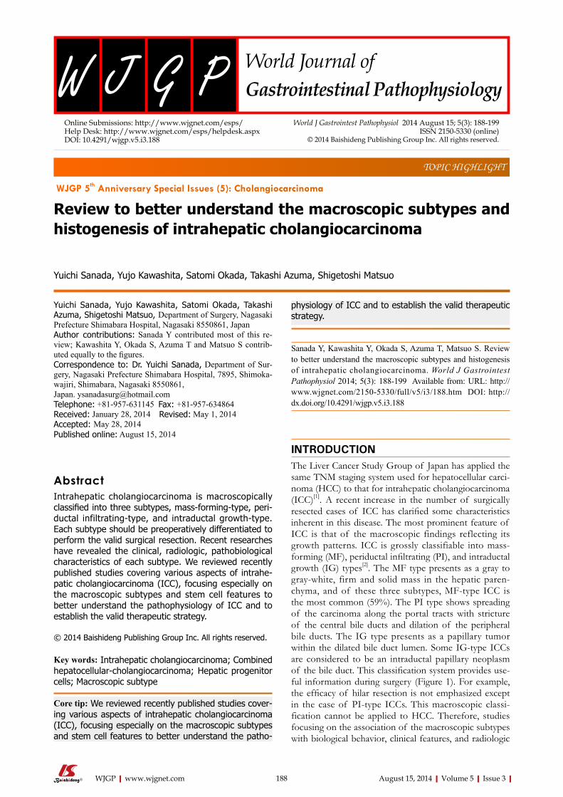
TOPIC HIGHLIGHT
Review to better understand the macroscopic subtypes and histogenesis of intrahepatic cholangiocarcinoma
Yuichi Sanada, Yujo Kawashita, Satomi Okada, Takashi Azuma, Shigetoshi Matsuo
Yuichi Sanada, Yujo Kawashita, Satomi Okada, Takashi Azuma, Shigetoshi Matsuo, Department of Surgery, Nagasaki Prefecture Shimabara Hospital, Nagasaki 8550861, Japan Author contributions: Sanada Y contributed most of this re-view; Kawashita Y, Okada S, Azuma T and Matsuo S contrib-uted equally to the figures. Correspondence to: Dr. Yuichi Sanada, Department of Sur-gery, Nagasaki Prefecture Shimabara Hospital, 7895, Shimoka-wajiri, Shimabara, Nagasaki 8550861, Japan. [email protected]: +81-957-631145 Fax: +81-957-634864Received: January 28, 2014 Revised: May 1, 2014Accepted: May 28, 2014Published online: August 15, 2014
AbstractIntrahepatic cholangiocarcinoma is macroscopically classified into three subtypes, mass-forming-type, peri-ductal infiltrating-type, and intraductal growth-type. Each subtype should be preoperatively differentiated to perform the valid surgical resection. Recent researches have revealed the clinical, radiologic, pathobiological characteristics of each subtype. We reviewed recently published studies covering various aspects of intrahe-patic cholangiocarcinoma (ICC), focusing especially on the macroscopic subtypes and stem cell features to better understand the pathophysiology of ICC and to establish the valid therapeutic strategy.
© 2014 Baishideng Publishing Group Inc. All rights reserved.
Key words: Intrahepatic cholangiocarcinoma; Combined hepatocellular-cholangiocarcinoma; Hepatic progenitor cells; Macroscopic subtype
Core tip: We reviewed recently published studies cover-ing various aspects of intrahepatic cholangiocarcinoma (ICC), focusing especially on the macroscopic subtypes and stem cell features to better understand the patho-
physiology of ICC and to establish the valid therapeutic strategy.
Sanada Y, Kawashita Y, Okada S, Azuma T, Matsuo S. Review to better understand the macroscopic subtypes and histogenesis of intrahepatic cholangiocarcinoma. World J Gastrointest Pathophysiol 2014; 5(3): 188-199 Available from: URL: http://www.wjgnet.com/2150-5330/full/v5/i3/188.htm DOI: http://dx.doi.org/10.4291/wjgp.v5.i3.188
INTRODUCTIONThe Liver Cancer Study Group of Japan has applied the same TNM staging system used for hepatocellular carci-noma (HCC) to that for intrahepatic cholangiocarcinoma (ICC)[1]. A recent increase in the number of surgically resected cases of ICC has clarified some characteristics inherent in this disease. The most prominent feature of ICC is that of the macroscopic findings reflecting its growth patterns. ICC is grossly classifiable into mass-forming (MF), periductal infiltrating (PI), and intraductal growth (IG) types[2]. The MF type presents as a gray to gray-white, firm and solid mass in the hepatic paren-chyma, and of these three subtypes, MF-type ICC is the most common (59%). The PI type shows spreading of the carcinoma along the portal tracts with stricture of the central bile ducts and dilation of the peripheral bile ducts. The IG type presents as a papillary tumor within the dilated bile duct lumen. Some IG-type ICCs are considered to be an intraductal papillary neoplasm of the bile duct. This classification system provides use-ful information during surgery (Figure 1). For example, the efficacy of hilar resection is not emphasized except in the case of PI-type ICCs. This macroscopic classi-fication cannot be applied to HCC. Therefore, studies focusing on the association of the macroscopic subtypes with biological behavior, clinical features, and radiologic
World J Gastrointest Pathophysiol 2014 August 15; 5(3): 188-199ISSN 2150-5330 (online)
© 2014 Baishideng Publishing Group Inc. All rights reserved.
Online Submissions: http://www.wjgnet.com/esps/Help Desk: http://www.wjgnet.com/esps/helpdesk.aspxDOI: 10.4291/wjgp.v5.i3.188
August 15, 2014|Volume 5|Issue 3|WJGP|www.wjgnet.com 188
WJGP 5th Anniversary Special Issues (5): Cholangiocarcinoma
Sanada Y et al . Subtypes of intrahepatic cholangiocarcinoma
findings are needed to establish the therapeutic strategy for ICC. Although the macroscopic features are promi-nent in ICCs, another aspect of ICCs, in which ICCs cannot be discussed independently of other primary liver cancers, exists. Recent histopathologic and im-munohistochemical studies have reported that hepatic progenitor cells (HPC) or stem cells play important roles in liver carcinogenesis including both HCCs and ICCs, supporting the hypothesis that HCCs and ICCs share a common evolutionary origin[3,4]. In 2010, the World Health Organization (WHO) established a new classi-fication system of combined hepatocellular-cholangio-carcinoma (cHCC-CC) based on the presence of stem-cell features[5]. According to this new system, cHCC-CCs are classified into two major subtypes, classic type and subtypes with stem-cell features. Subtypes with stem-cell features are further subclassified into three types: typical type, intermediate-cell type, and cholangiocellular type. In addition, recent reports showed that some cases of HCCs and ICCs are associated with hepatic stem cells. However, little is known about the clinical significance of stem cells in ICCs. This review summarizes recently published studies (from 2011 to 2013) covering various aspects of ICC and cHCC-CC, focusing especially on the macroscopic subtypes and stem-cell features.
CLINICAL STUDIES OF ICCRecent clinical researches of ICC are summarized in Tables 1 and 2[6-24]. The association between macroscopic subtypes and survival rate and lymph node metastasis has been discussed ever since the macroscopic subtype was established. IG-type ICCs have a favorable outcome because this tumor type shows intraductal growth with-out invasiveness[2]. Of the three subtypes, MF+PI-type ICCs have the highest incidence of lymph node metas-tasis (50% to 73%)[15] and are associated with the lowest 5-year survival rate (0% to 19.4%). PI-type and MF-type have relatively favorable outcomes when lymph node metastasis or hilar invasion is absent.
Over the most recent 3 years, 19 studies have been published (Tables 1 and 2). Most of these studies de-scribe the poor prognostic factors of resected cases of ICC. The most significant prognostic factor is lymph node metastasis. However, whether routine lymph node dissection improves postoperative survival is still unclear.
The literature on the macroscopic subtypes is very scant. Uchiyama et al[15] and Uno et al[17] reported that the PI type showed significantly better survival than the MF and MF+PI types, supporting the results of previous reports. The difference in malignant potential between
August 15, 2014|Volume 5|Issue 3|WJGP|www.wjgnet.com 189
A B
C D
Figure 1 Some intraductal growth-type intrahepatic cholangiocarcinomas are considered to be an intraductal papillary neoplasm of the bile duct, this classification system provides useful information during surgery. A: Gross feature of mass-forming (MF) + periductal infiltrating (PI)-type intrahepatic cholangio-carcinoma (ICC) obtained by left hepatectomy with bile duct resection. The carcinoma spreads along the hilar biliary tree (arrow) in communication with a white firm mass; B-D: Operative findings and resected specimens of MF + intraductal growth (IG)-type ICC. The common hepatic duct is incised, and the soft tumor comprising the intraductal components is easily removed (arrow) without infiltration to the ductal wall; C: Hepatic anterior segmentectomy is performed for MF components; D: IG components are composed of tan-colored soft tissues with necrosis.
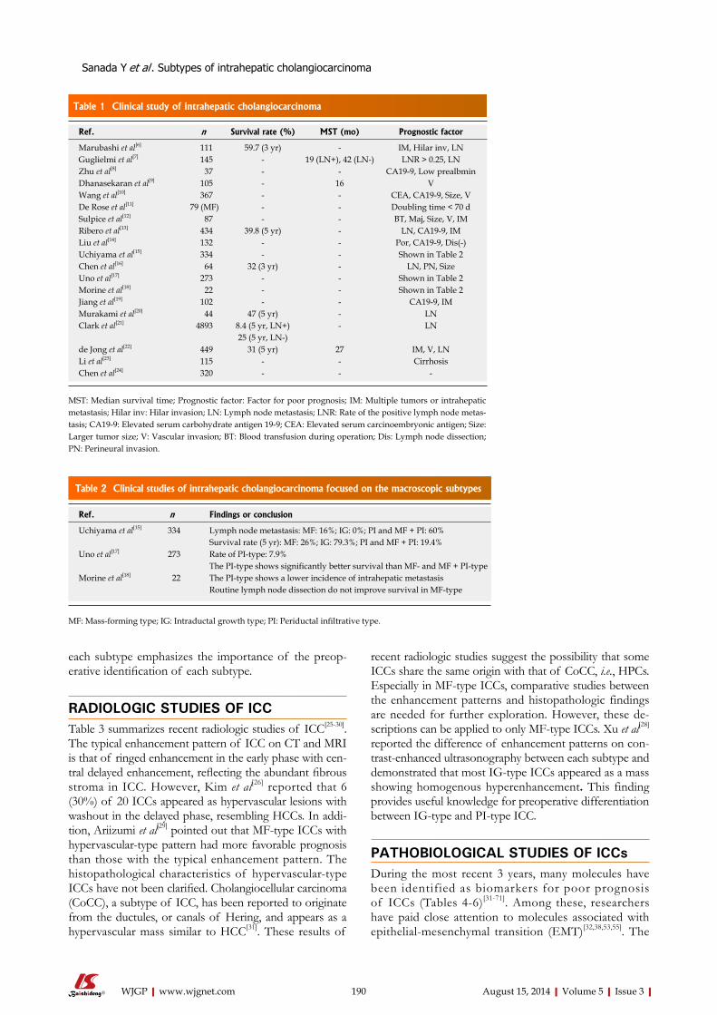
each subtype emphasizes the importance of the preop-erative identification of each subtype.
RADIOLOGIC STUDIES OF ICC Table 3 summarizes recent radiologic studies of ICC[25-30]. The typical enhancement pattern of ICC on CT and MRI is that of ringed enhancement in the early phase with cen-tral delayed enhancement, reflecting the abundant fibrous stroma in ICC. However, Kim et al[26] reported that 6 (30%) of 20 ICCs appeared as hypervascular lesions with washout in the delayed phase, resembling HCCs. In addi-tion, Ariizumi et al[29] pointed out that MF-type ICCs with hypervascular-type pattern had more favorable prognosis than those with the typical enhancement pattern. The histopathological characteristics of hypervascular-type ICCs have not been clarified. Cholangiocellular carcinoma (CoCC), a subtype of ICC, has been reported to originate from the ductules, or canals of Hering, and appears as a hypervascular mass similar to HCC[31]. These results of
recent radiologic studies suggest the possibility that some ICCs share the same origin with that of CoCC, i.e., HPCs. Especially in MF-type ICCs, comparative studies between the enhancement patterns and histopathologic findings are needed for further exploration. However, these de-scriptions can be applied to only MF-type ICCs. Xu et al[28] reported the difference of enhancement patterns on con-trast-enhanced ultrasonography between each subtype and demonstrated that most IG-type ICCs appeared as a mass showing homogenous hyperenhancement. This finding provides useful knowledge for preoperative differentiation between IG-type and PI-type ICC.
PATHOBIOLOGICAL STUDIES OF ICCsDuring the most recent 3 years, many molecules have been identified as biomarkers for poor prognosis of ICCs (Tables 4-6)[31-71]. Among these, researchers have paid close attention to molecules associated with epithelial-mesenchymal transition (EMT)[32,38,53,55]. The
August 15, 2014|Volume 5|Issue 3|WJGP|www.wjgnet.com 190
Table 1 Clinical study of intrahepatic cholangiocarcinoma
Ref. n Survival rate (%) MST (mo) Prognostic factor
Marubashi et al[6] 111 59.7 (3 yr) - IM, Hilar inv, LNGuglielmi et al[7] 145 - 19 (LN+), 42 (LN-) LNR > 0.25, LNZhu et al[8] 37 - - CA19-9, Low prealbminDhanasekaran et al[9] 105 - 16 VWang et al[10] 367 - - CEA, CA19-9, Size, VDe Rose et al[11] 79 (MF) - - Doubling time < 70 d Sulpice et al[12] 87 - - BT, Maj, Size, V, IMRibero et al[13] 434 39.8 (5 yr) - LN, CA19-9, IMLiu et al[14] 132 - - Por, CA19-9, Dis(-)Uchiyama et al[15] 334 - - Shown in Table 2Chen et al[16] 64 32 (3 yr) - LN, PN, SizeUno et al[17] 273 - - Shown in Table 2Morine et al[18] 22 - - Shown in Table 2Jiang et al[19] 102 - - CA19-9, IMMurakami et al[20] 44 47 (5 yr) - LNClark et al[21] 4893 8.4 (5 yr, LN+) - LN
25 (5 yr, LN-)de Jong et al[22] 449 31 (5 yr) 27 IM, V, LNLi et al[23] 115 - - CirrhosisChen et al[24] 320 - - -
MST: Median survival time; Prognostic factor: Factor for poor prognosis; IM: Multiple tumors or intrahepatic metastasis; Hilar inv: Hilar invasion; LN: Lymph node metastasis; LNR: Rate of the positive lymph node metas-tasis; CA19-9: Elevated serum carbohydrate antigen 19-9; CEA: Elevated serum carcinoembryonic antigen; Size: Larger tumor size; V: Vascular invasion; BT: Blood transfusion during operation; Dis: Lymph node dissection; PN: Perineural invasion.
Table 2 Clinical studies of intrahepatic cholangiocarcinoma focused on the macroscopic subtypes
Ref. n Findings or conclusion
Uchiyama et al[15] 334 Lymph node metastasis: MF: 16%; IG: 0%; PI and MF + PI: 60%Survival rate (5 yr): MF: 26%; IG: 79.3%; PI and MF + PI: 19.4%
Uno et al[17] 273 Rate of PI-type: 7.9%The PI-type shows significantly better survival than MF- and MF + PI-type
Morine et al[18] 22 The PI-type shows a lower incidence of intrahepatic metastasisRoutine lymph node dissection do not improve survival in MF-type
MF: Mass-forming type; IG: Intraductal growth type; PI: Periductal infiltrative type.
Sanada Y et al . Subtypes of intrahepatic cholangiocarcinoma
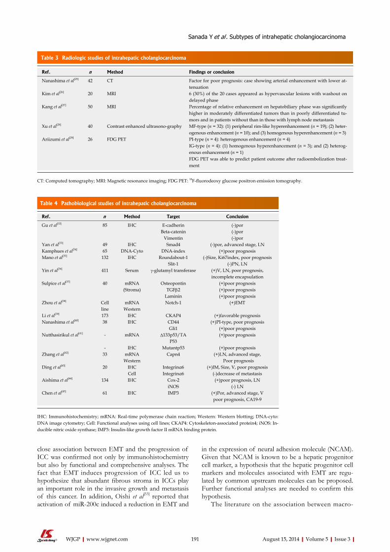
close association between EMT and the progression of ICC was confirmed not only by immunohistochemistry but also by functional and comprehensive analyses. The fact that EMT induces progression of ICC led us to hypothesize that abundant fibrous stroma in ICCs play an important role in the invasive growth and metastasis of this cancer. In addition, Oishi et al[53] reported that activation of miR-200c induced a reduction in EMT and
in the expression of neural adhesion molecule (NCAM). Given that NCAM is known to be a hepatic progenitor cell marker, a hypothesis that the hepatic progenitor cell markers and molecules associated with EMT are regu-lated by common upstream molecules can be proposed. Further functional analyses are needed to confirm this hypothesis.
The literature on the association between macro-
August 15, 2014|Volume 5|Issue 3|WJGP|www.wjgnet.com 191
Table 3 Radiologic studies of intrahepatic cholangiocarcinoma
Ref. n Method Findings or conclusion
Nanashima et al[25] 42 CT Factor for poor prognosis: case showing arterial enhancement with lower at-tenuation
Kim et al[26] 20 MRI 6 (30%) of the 20 cases appeared as hypervascular lesions with washout on delayed phase
Kang et al[27] 50 MRI Percentage of relative enhancement on hepatobiliary phase was significantly higher in moderately differentiated tumors than in poorly differentiated tu-mors and in patients without than in those with lymph node metastasis
Xu et al[28] 40 Contrast enhanced ultrasono-graphy MF-type (n = 32): (1) peripheral rim-like hyperenhancement (n = 19); (2) heter-ogenous enhancement (n = 10); and (3) homogenous hyperenhancement (n = 3)
Ariizumi et al[29] 26 FDG PET PI-type (n = 4): heterogenous enhancement (n = 4)IG-type (n = 4): (1) homogenous hyperenhancement (n = 3); and (2) heterog-enous enhancement (n = 1)FDG PET was able to predict patient outcome after radioembolization treat-ment
CT: Computed tomography; MRI: Magnetic resonance imaging; FDG PET: 18F-fluorodeoxy glucose positron emission tomography.
Table 4 Pathobiological studies of intrahepatic cholangiocarcinoma
Ref. n Method Target Conclusion
Gu et al[32] 85 IHC E-cadherin (-)porBeta-catenin (-)por
Vimentin (-)porYan et al[33] 49 IHC Smad4 (-)por, advanced stage, LNKamphues et al[34] 65 DNA-Cyto DNA-index (+)poor prognosisMano et al[35] 132 IHC Roundabout-1 (-)Size, Ki67index, poor prognosis
Slit-1 (-)PN, LNYin et al[36] 411 Serum γ-glutamyl transferase (+)V, LN, poor prognosis,
incomplete encapsulationSulpice et al[37] 40 mRNA Osteopontin (+)poor prognosis
(Stroma) TGFβ2 (+)poor prognosisLaminin (+)poor prognosis
Zhou et al[38] Cell mRNA Notch-1 (+)EMTline Western
Li et al[39] 173 IHC CKAP4 (+)favorable prognosisNanashima et al[40] 38 IHC CD44 (+)PI-type, poor prognosis
Gli1 (+)poor prognosisNutthasirikul et al[41] - mRNA ∆133p53/TA (+)poor prognosis
P53- IHC Mutantp53 (+)poor prognosis
Zhang et al[42] 33 mRNA Capn4 (+)LN, advanced stage, Western Poor prognosis
Ding et al[43] 20 IHC Integrinα6 (+)IM, Size, V, poor prognosisCell Integrinα6 (-)decrease of metastasis
Aishima et al[44] 134 IHC Cox-2 (+)poor prognosis, LNiNOS (-) LN
Chen et al[45] 61 IHC IMP3 (+)Por, advanced stage, Vpoor prognosis, CA19-9
IHC: Immunohistochemistry; mRNA: Real-time polymerase chain reaction; Western: Western blotting; DNA-cyto: DNA image cytometry; Cell: Functional analyses using cell lines; CKAP4: Cytoskeleton-associated protein4; iNOS: In-ducible nitric oxide synthase; IMP3: Insulin-like growth factor II mRNA binding protein.
Sanada Y et al . Subtypes of intrahepatic cholangiocarcinoma
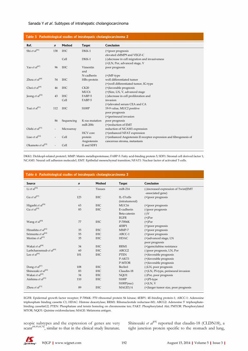
scopic subtypes and the expression of genes are very scant[48,49,68,70], similar to that in the clinical study literature.
Shinozaki et al[68] reported that claudin-18 (CLDN18), a tight junction protein specific to the stomach and lung,
Table 6 Pathobiological studies of intrahepatic cholangiocarcinoma 3
August 15, 2014|Volume 5|Issue 3|WJGP|www.wjgnet.com 192
Table 5 Pathobiological studies of intrahepatic cholangiocarcinoma 2
Ref. n Method Target Conclusion
Shi et al[46] 138 IHC DKK-1 (+)poor prognosiselevated sMMP9 and VEGF-C
Cell DKK-1 (-)decrease in cell migration and invasiveness(+)LN, Por, advanced stage, V
Yao et al[47] 96 IHC Vimentin poor prognosisand N-cadherin (+)MF-type
Zhou et al[48] 54 IHC HBx-protein well differentiated tumor(+)well differentiated tumor, IG-type
Choi et al[49] 46 IHC CK20 (+)favorable prognosisMUC6 (+)Size, LN, V, advanced stage
Jeong et al[50] 43 IHC FABP-5 (-)decrease in cell proliferation andCell FABP-5 invasion
(+)elevated serum CEA and CATsai et al[51] 112 IHC S100P 19-9 value, MUC2 positive
poor prognosis(+)perineural invasion
86 Sequencing K-ras mutation poor prognosismiR-200c (+)reduction of EMT
Oishi et al[53] - Microarray reduction of NCAM1 expressionHCV core (+)enhanced NFAT expression
Liao et al[54] - Cell protein (+)enhanced Angiotensin II receptor expression and fibrogenesis of Angiotensin cancerous stroma, metastasis
Okamoto et al[55] - Cell II and SDF1
DKK1: Dickkopf-related protein1; MMP: Matrix metalloproteinase; FABP-5: Fatty acid-binding protein 5; SDF1: Stromal cell derived factor 1; NCAM1: Neural cell adhesion molecule1; EMT: Epithelial mesenchymal transition; NFAT1: Nuclear factor of activated T-cells.
Source n Method Target Conclusion
Li et al[56] - Tissues miR-214 (-)increased expression of Twist(EMT -associated gene)
Gu et al[57] 123 IHC IL-17cells (+)poor prognosis(intratumoral)
Higashi et al[58] 63 IHC MUC16 (+)poor prognosisGu et al[59] 83 IHC E-cadherin (-)poor prognosis
Beta-catenin (-)VEGFR (+)Por
Wang et al[60] 77 IHC P-70S6K (+)Por4EBP1 (+)poor prognosis
Hirashita et al[61] 35 IHC MMP-7 (+)poor prognosisSrimunta et al[62] 55 IHC ABCC-1 (+)poor prognosisMorine et al[63] 35 IHC HDAC (+)advanced stage, LN
poor prognosisWakai et al[64] 34 IHC RRM1 (+)gemcitabine resistanceLarbcharoensub et al[65] 60 IHC ABCG2 (-)poor prognosis, LN, PorLee et al[66] 101 IHC PTEN (+)favorable prognosis
P-AKT1 (+)favorable prognosisP-MTOR (+)favorable prognosis
Dong et al[67] 108 IHC Beclin1 (-)LN, poor prognosisShinozaki et al[68] 83 IHC Claudin-18 (+)LN, PI-type, perineural invasionWakai et al[69] 34 IHC NQO1 (-)Por, poor prognosisAishima et al[70] 110 IHC S100P (+)PI-type
S100P(nuc) (+)LN, VZhou et al[71] 89 IHC MAGE3/4 (+)larger tumor size, poor prognosis
EGFR: Epidermal growth factor receptor; P-70S6K: P70 ribosomal protein S6 kinase; 4EBP1: 4E-binding protein-1; ABCC-1: Adenosine triphosphate binding cassette C1; HDAC: Histone deacetylase; RRM1: Ribonucleotide reductase-M1; ABCG2: Adenosine 5’ triphosphate-binding cassetteG2; PTEN: Phosphatase and tensin homolog on chromosome ten; PAKT: Phosphorylated Akt; PMTOR: Phosphorylated MTOR; NQO1: Quinine oxidoredactase; MAGE: Melanoma antigen.
Sanada Y et al . Subtypes of intrahepatic cholangiocarcinoma
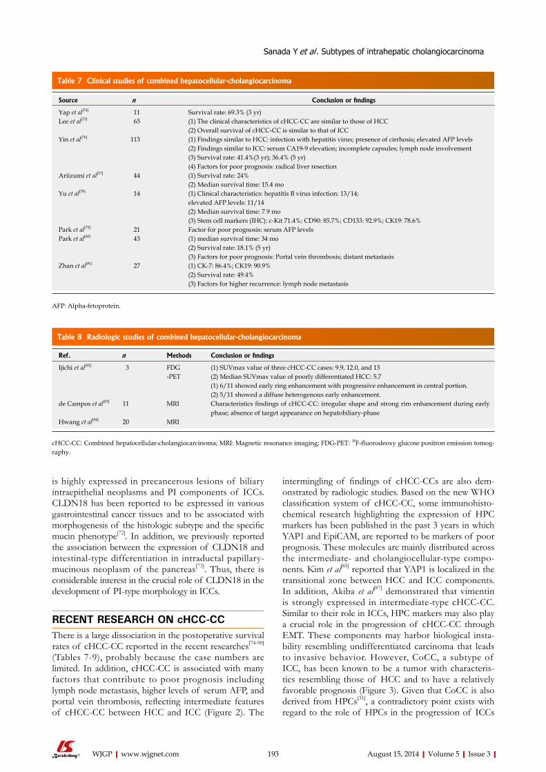
is highly expressed in precancerous lesions of biliary intraepithelial neoplasms and PI components of ICCs. CLDN18 has been reported to be expressed in various gastrointestinal cancer tissues and to be associated with morphogenesis of the histologic subtype and the specific mucin phenotype[72]. In addition, we previously reported the association between the expression of CLDN18 and intestinal-type differentiation in intraductal papillary-mucinous neoplasm of the pancreas[73]. Thus, there is considerable interest in the crucial role of CLDN18 in the development of PI-type morphology in ICCs.
RECENT RESEARCH ON cHCC-CC There is a large dissociation in the postoperative survival rates of cHCC-CC reported in the recent researches[74-90] (Tables 7-9), probably because the case numbers are limited. In addition, cHCC-CC is associated with many factors that contribute to poor prognosis including lymph node metastasis, higher levels of serum AFP, and portal vein thrombosis, reflecting intermediate features of cHCC-CC between HCC and ICC (Figure 2). The
intermingling of findings of cHCC-CCs are also dem-onstrated by radiologic studies. Based on the new WHO classification system of cHCC-CC, some immunohisto-chemical research highlighting the expression of HPC markers has been published in the past 3 years in which YAP1 and EpiCAM, are reported to be markers of poor prognosis. These molecules are mainly distributed across the intermediate- and cholangiocellular-type compo-nents. Kim et al[85] reported that YAP1 is localized in the transitional zone between HCC and ICC components. In addition, Akiba et al[87] demonstrated that vimentin is strongly expressed in intermediate-type cHCC-CC. Similar to their role in ICCs, HPC markers may also play a crucial role in the progression of cHCC-CC through EMT. These components may harbor biological insta-bility resembling undifferentiated carcinoma that leads to invasive behavior. However, CoCC, a subtype of ICC, has been known to be a tumor with characteris-tics resembling those of HCC and to have a relatively favorable prognosis (Figure 3). Given that CoCC is also derived from HPCs[31], a contradictory point exists with regard to the role of HPCs in the progression of ICCs
August 15, 2014|Volume 5|Issue 3|WJGP|www.wjgnet.com 193
Table 7 Clinical studies of combined hepatocellular-cholangiocarcinoma
Source n Conclusion or findings
Yap et al[74] 11 Survival rate: 69.3% (3 yr)Lee et al[75] 65 (1) The clinical characteristics of cHCC-CC are similar to those of HCC
(2) Overall survival of cHCC-CC is similar to that of ICCYin et al[76] 113 (1) Findings similar to HCC: infection with hepatitis virus; presence of cirrhosis; elevated AFP levels
(2) Findings similar to ICC: serum CA19-9 elevation; incomplete capsules; lymph node involvement(3) Survival rate: 41.4%(3 yr); 36.4% (5 yr)(4) Factors for poor prognosis: radical liver resection
Ariizumi et al[77] 44 (1) Survival rate: 24%(2) Median survival time: 15.4 mo
Yu et al[78] 14 (1) Clinical characteristics: hepatitis B virus infection: 13/14; elevated AFP levels: 11/14(2) Median survival time: 7.9 mo(3) Stem cell markers (IHC): c-Kit 71.4%; CD90: 85.7%; CD133: 92.9%; CK19: 78.6%
Park et al[79] 21 Factor for poor prognosis: serum AFP levelsPark et al[80] 43 (1) median survival time: 34 mo
(2) Survival rate: 18.1% (5 yr)(3) Factors for poor prognosis: Portal vein thrombosis; distant metastasis
Zhan et al[81] 27 (1) CK-7: 86.4%; CK19: 90.9%(2) Survival rate: 49.4%(3) Factors for higher recurrence: lymph node metastasis
AFP: Alpha-fetoprotein.
Table 8 Radiologic studies of combined hepatocellular-cholangiocarcinoma
Ref. n Methods Conclusion or findings
Ijichi et al[82] 3 FDG (1) SUVmax value of three cHCC-CC cases: 9.9, 12.0, and 13-PET (2) Median SUVmax value of poorly differentiated HCC: 5.7
(1) 6/11 showed early ring enhancement with progressive enhancement in central portion. (2) 5/11 showed a diffuse heterogenous early enhancement.
de Campos et al[83] 11 MRI Characteristics findings of cHCC-CC: irregular shape and strong rim enhancement during early phase; absence of target appearance on hepatobiliary-phase
Hwang et al[84] 20 MRI
cHCC-CC: Combined hepatocellular-cholangiocarcinoma; MRI: Magnetic resonance imaging; FDG-PET: 18F-fluorodeoxy glucose positron emission tomog-raphy.
Sanada Y et al . Subtypes of intrahepatic cholangiocarcinoma
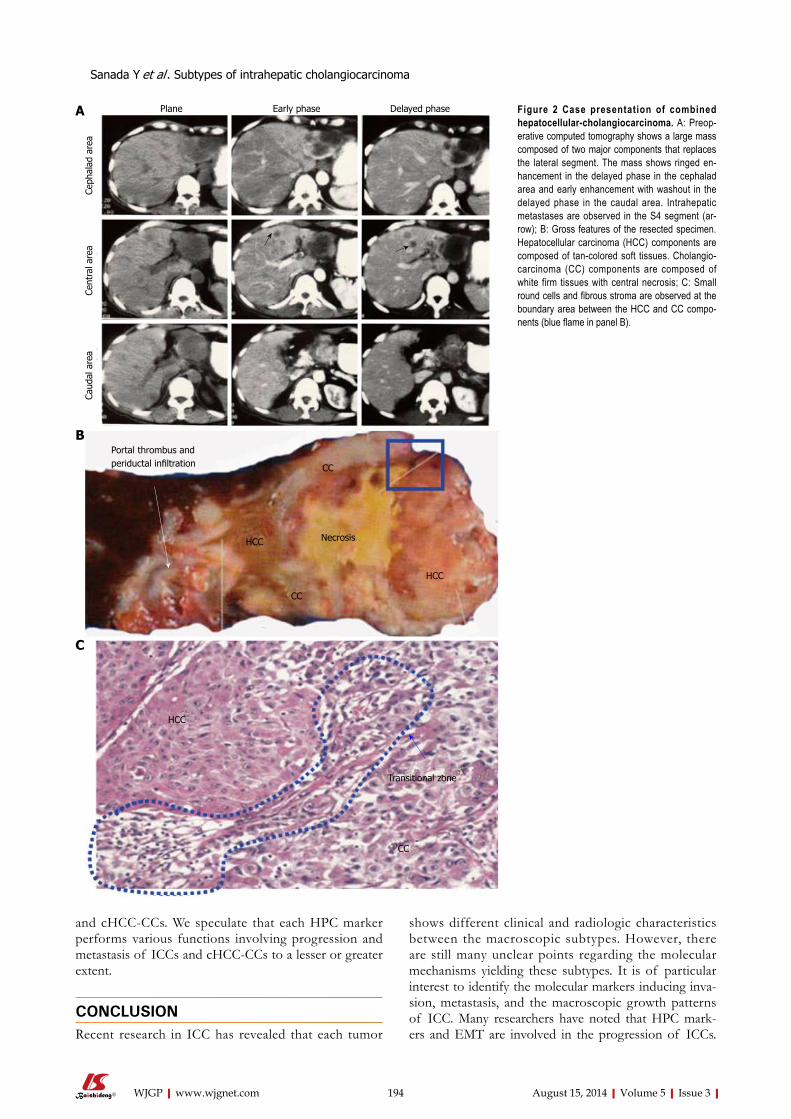
and cHCC-CCs. We speculate that each HPC marker performs various functions involving progression and metastasis of ICCs and cHCC-CCs to a lesser or greater extent.
CONCLUSIONRecent research in ICC has revealed that each tumor
shows different clinical and radiologic characteristics between the macroscopic subtypes. However, there are still many unclear points regarding the molecular mechanisms yielding these subtypes. It is of particular interest to identify the molecular markers inducing inva-sion, metastasis, and the macroscopic growth patterns of ICC. Many researchers have noted that HPC mark-ers and EMT are involved in the progression of ICCs.
August 15, 2014|Volume 5|Issue 3|WJGP|www.wjgnet.com 194
CC
HCC
HCC
CC
Necrosis
HCC
Transitional zone
CC
Portal thrombus and periductal infiltration
Plane Early phase Delayed phase
Ceph
alad
are
aCe
ntra
l are
aCa
udal
are
a
A
B
C
Figure 2 Case presentation of combined hepatocellular-cholangiocarcinoma. A: Preop-erative computed tomography shows a large mass composed of two major components that replaces the lateral segment. The mass shows ringed en-hancement in the delayed phase in the cephalad area and early enhancement with washout in the delayed phase in the caudal area. Intrahepatic metastases are observed in the S4 segment (ar-row); B: Gross features of the resected specimen. Hepatocellular carcinoma (HCC) components are composed of tan-colored soft tissues. Cholangio-carcinoma (CC) components are composed of white firm tissues with central necrosis; C: Small round cells and fibrous stroma are observed at the boundary area between the HCC and CC compo-nents (blue flame in panel B).
Sanada Y et al . Subtypes of intrahepatic cholangiocarcinoma

Because most cHCC-CCs show MF-type morphology, we infer that HPC markers are closely associated with the morphogenesis and histogenesis of MF-type ICCs. Therefore, studies of ICC, and especially of its molecu-lar pathology, should be designed in conjunction with those of cHCC-CC.
REFERENCES1 Igami T, Ebata T, Yokoyama Y, Sugawara G, Takahashi Y,
Nagino M. Staging of peripheral-type intrahepatic cholan-giocarcinoma: appraisal of the new TNM classification and its modifications. World J Surg 2011; 35: 2501-2509 [PMID: 21879422]
2 Nakanuma Y, Sato Y, Harada K, Sasaki M, Xu J, Ikeda H. Pathological classification of intrahepatic cholangiocarcino-ma based on a new concept. World J Hepatol 2010; 2: 419-427 [PMID: 21191517]
3 Lo RC, Ng IO. Hepatic progenitor cells: their role and func-tional significance in the new classification of primary liver cancers. Liver Cancer 2013; 2: 84-92 [PMID: 24159600]
4 Zheng YW, Nie YZ, Taniguchi H. Cellular reprogramming and hepatocellular carcinoma development. World J Gastro-enterol 2013; 19: 8850-8860 [PMID: 24379607 DOI: 10.3748/wjg.v19.i47.8850]
5 Theise ND, Nakashima O, Park YN. Combined hepatocel-lular-cholangiocacinoma in WHO classification of tumors of the digestive system; World health organization of tumors. IARC, Lyon, 2010: 225-227
6 Marubashi S, Gotoh K, Takahashi H, Ohigashi H, Yano M, Ishikawa O, Sakon M. Prediction of the postoperative prog-nosis of intrahepatic cholangiocarcinoma (ICC): importance of preoperatively- determined anatomic invasion level and number of tumors. Dig Dis Sci 2014; 59: 201-213 [PMID: 24122559]
7 Guglielmi A, Ruzzenente A, Campagnaro T, Valdegamberi A, Bagante F, Bertuzzo F, Conci S, Iacono C. Patterns and prognostic significance of lymph node dissection for surgi-cal treatment of perihilar and intrahepatic cholangiocarcino-ma. J Gastrointest Surg 2013; 17: 1917-1928 [PMID: 24048613 DOI: 10.1007/s11605-013-2331-1]
8 Zhu HF, Li J, Huang L, Yan YQ. Intrahepatic cholangiocar-cinoma: a clinicopathologic study of 37 resected cases. Hepa-togastroenterology 2013; 60: 263-267 [PMID: 23574653 DOI:
August 15, 2014|Volume 5|Issue 3|WJGP|www.wjgnet.com 195
Table 9 Pathobiological studies of combined hepatocellular-cholangiocarcinoma
Ref. n Method Target Conclusion
Kim et al[85] 58 IHC YAP1 (+): transition zone, poor prognosisEpiCAM (-)favorable prognosisCK19 (-)favorable prognosis
Ikeda et al[86] 36 IHC DLK1 (+)poor prognosisAkiba et al[87] 54 IHC CD56 (+): components apart from HCC
c-Kit (+): components apart from HCCEpiCAM (+): components apart from HCCCD133 (+): intermediate type or cholangiolocellular typeVimentin (+): intermediate type or cholangiolocellular type
Coulouarn et al[88] 152 Microarray - (1) TGFbeta and beta-catenin are identified as the two major signals in the progression of cHCC-CC/ (2) cHCC-CC shares the characteristics of poorly differentiated HCC. (+)poor prognosisBoth HCC and CC components of mostOf the cHCC-CC express both AFP and
Cai et al[89] 80 IHC PCNA CK19Itoyama et al[90] 20 IHC AFP and
CK19
cHCC-CC: Combined hepatocellular-cholangiocarcinoma; YAP1: Yes-associated protein 1; EpiCAM: Epithelial cell adhesion molecule; DLK1: Delta-like 1 homolog; PCNA: Proliferating cell nuclear antigen index in nontumor ductular reaction.
Figure 3 Resected case of cholangiocellular carcinoma. A: Computed tomography (CT) reveals a mass showing ringed enhancement with portal venous penetra-tion; B: CT findings reflect the non-infiltrative growth of the tumor to the portal tract; C: Histopathologically, the size of the carcinoma cells is small, with the cells form-ing anastomosing patterns with abundant fibrous stroma.
A B C
Sanada Y et al . Subtypes of intrahepatic cholangiocarcinoma
10.5754/hge12711]9 Dhanasekaran R, Hemming AW, Zendejas I, George T,
Nelson DR, Soldevila-Pico C, Firpi RJ, Morelli G, Clark V, Cabrera R. Treatment outcomes and prognostic factors of intrahepatic cholangiocarcinoma. Oncol Rep 2013; 29: 1259-1267 [PMID: 23426976 DOI: 10.3892/or.2013.2290]
10 Wang Y, Li J, Xia Y, Gong R, Wang K, Yan Z, Wan X, Liu G, Wu D, Shi L, Lau W, Wu M, Shen F. Prognostic nomogram for intrahepatic cholangiocarcinoma after partial hepatec-tomy. J Clin Oncol 2013; 31: 1188-1195 [PMID: 23358969 DOI: 10.1200/JCO.2012.41.5984]
11 De Rose AM, Cucchetti A, Clemente G, Ardito F, Giovanni-ni I, Ercolani G, Giuliante F, Pinna AD, Nuzzo G. Prognostic significance of tumor doubling time in mass-forming type cholangiocarcinoma. J Gastrointest Surg 2013; 17: 739-747 [PMID: 23292461]
12 Sulpice L, Rayar M, Boucher E, Pele F, Pracht M, Meunier B, Boudjema K. Intrahepatic cholangiocarcinoma: impact of genetic hemochromatosis on outcome and overall survival after surgical resection. J Surg Res 2013; 180: 56-61 [PMID: 23183056 DOI: 10.1016/j.jss.2012.10.051]
13 Ribero D, Pinna AD, Guglielmi A, Ponti A, Nuzzo G, Gi-ulini SM, Aldrighetti L, Calise F, Gerunda GE, Tomatis M, Amisano M, Berloco P, Torzilli G, Capussotti L. Surgical Ap-proach for Long-term Survival of Patients With Intrahepatic Cholangiocarcinoma: A Multi-institutional Analysis of 434 Patients. Arch Surg 2012; 147: 1107-1113 [PMID: 22910846]
14 Liu ZH, Chen Z, Ma LL, Li XH, Wang LX. Factors influenc-ing the prognosis of patients with intrahepatic cholangio-carcinoma. Acta Gastroenterol Belg 2012; 75: 215-218 [PMID: 22870785]
15 Uchiyama K, Yamamoto M, Yamaue H, Ariizumi S, Aoki T, Kokudo N, Ebata T, Nagino M, Ohtsuka M, Miyazaki M, Tanaka E, Kondo S, Uenishi T, Kubo S, Yoshida H, Unno M, Imura S, Shimada M, Ueno M, Takada T. Impact of nodal involvement on surgical outcomes of intrahepatic cholan-giocarcinoma: a multicenter analysis by the Study Group for Hepatic Surgery of the Japanese Society of Hepato-Biliary-Pancreatic Surgery. J Hepatobiliary Pancreat Sci 2011; 18: 443-452 [PMID: 21132443 DOI: 10.1007/s00534-010-0349-2]
16 Chen LP, Li C, Wang C, Wen TF, Yan LN, Li B. Predictive factors of recurrence for patients with intrahepatic cholan-giocarcinoma after hepatectomy. Hepatogastroenterology 2012; 59: 1765-1768 [PMID: 22369746 DOI: 10.5754/hge11820]
17 Uno M, Shimada K, Yamamoto Y, Nara S, Esaki M, Saka-moto Y, Kosuge T, Ojima H. Periductal infiltrating type of intrahepatic cholangiocarcinoma: a rare macroscopic type without any apparent mass. Surg Today 2012; 42: 1189-1194 [PMID: 22350300 DOI: 10.1007/s00595-012-0145-5]
18 Morine Y, Shimada M, Utsunomiya T, Imura S, Ikemoto T, Mori H, Hanaoka J, Kanamoto M, Miyake H. Clinical impact of lymph node dissection in surgery for peripheral-type intrahepatic cholangiocarcinoma. Surg Today 2012; 42: 147-151 [PMID: 22124809 DOI: 10.1007/s00595-011-0057-9]
19 Jiang BG, Ge RL, Sun LL, Zong M, Wei GT, Zhang YJ. Clini-cal parameters predicting survival duration after hepatecto-my for intrahepatic cholangiocarcinoma. Can J Gastroenterol 2011; 25: 603-608 [PMID: 22059167]
20 Murakami Y, Uemura K, Sudo T, Hashimoto Y, Nakashima A, Sueda T. Intrahepatic cholangiocarcinoma: clinicopatho-logical differences between peripheral type and hilar type. J Gastrointest Surg 2012; 16: 540-548 [PMID: 22012305 DOI: 10.1007/s11605-011-1730-4]
21 Clark CJ, Wood-Wentz CM, Reid-Lombardo KM, Kendrick ML, Huebner M, Que FG. Lymphadenectomy in the staging and treatment of intrahepatic cholangiocarcinoma: a popu-lation-based study using the National cancer institute SEER database. HPB (Oxford) 2011; 13: 612-620 [DOI: 10.1111/j.1477-2574.2011.00340.x]
22 de Jong MC, Nathan H, Sotiropoulos GC, Paul A, Alex-
andrescu S, Marques H, Pulitano C, Barroso E, Clary BM, Aldrighetti L, Ferrone CR, Zhu AX, Bauer TW, Walters DM, Gamblin TC, Nguyen KT, Turley R, Popescu I, Hubert C, Meyer S, Schulick RD, Choti MA, Gigot JF, Mentha G, Paw-lik TM. Intrahepatic cholangiocarcinoma: an international multi-institutional analysis of prognostic factors and lymph node assessment. J Clin Oncol 2011; 29: 3140-3145 [PMID: 21730269 DOI: 10.1200/JCO.2011.35.6519]
23 Li YY, Li H, Lv P, Liu G, Li XR, Tian BN, Chen DJ. Prognos-tic value of cirrhosis for intrahepatic cholangiocarcinoma after surgical treatment. J Gastrointest Surg 2011; 15: 608-613 [PMID: 21246412 DOI: 10.1007/s11605-011-1419-8]
24 Chen YX, Zeng ZC, Tang ZY, Fan J, Zhou J, Jiang W, Zeng MS, Tan YS. Prediction of the lymph node status in patients with intrahepatic cholangiocarcinoma: analysis of 320 sur-gical cases. Front Oncol 2011; 1: 42 [PMID: 22649763 DOI: 10.3389/fonc.2011.00042]
25 Nanashima A, Abo T, Murakami G, Matsumoto A, Tou K, Takeshita H, Kunizaki M, Hidaka S, Sakamoto I, Hayashi H, Fukuda T, Kudo T, Nagayasu T. Intrahepatic cholangiocar-cinoma: relationship between tumor imaging enhancement by measuring attenuation and clinicopathologic characteris-tics. Abdom Imaging 2013; 38: 785-792 [PMID: 23232581 DOI: 10.1007/s00261-012-9974-3]
26 Kim SH, Lee CH, Kim BH, Kim WB, Yeom SK, Kim KA, Park CM. Typical and atypical imaging findings of intrahe-patic cholangiocarcinoma using gadolinium ethoxybenzyl diethylenetriamine pentaacetic acid-enhanced magnetic resonance imaging. J Comput Assist Tomogr 2012; 36: 704-709 [PMID: 23192208 DOI: 10.1097/RCT.0b013e3182706562]
27 Kang Y, Lee JM, Kim SH, Han JK, Choi BI. Intrahepatic mass-forming cholangiocarcinoma: enhancement patterns on gadoxetic acid-enhanced MR images. Radiology 2012; 264: 751-760 [PMID: 22798225 DOI: 10.1148/radiol.12112308]
28 Xu HX, Chen LD, Liu LN, Zhang YF, Guo LH, Liu C. Contrast-enhanced ultrasound of intrahepatic cholangio-carcinoma: correlation with pathological examination. Br J Radiol 2012; 85: 1029-1037 [PMID: 22374276 DOI: 10.1259/bjr/21653786]
29 Ariizumi S, Kotera Y, Takahashi Y, Katagiri S, Chen IP, Ota T, Yamamoto M. Mass-forming intrahepatic cholan-giocarcinoma with marked enhancement on arterial-phase computed tomography reflects favorable surgical outcomes. J Surg Oncol 2011; 104: 130-139 [PMID: 21448898]
30 Haug AR, Heinemann V, Bruns CJ, Hoffmann R, Jakobs T, Bartenstein P, Hacker M. 18F-FDG PET independently pre-dicts survival in patients with cholangiocellular carcinoma treated with 90Y microspheres. Eur J Nucl Med Mol Imaging 2011; 38: 1037-1045 [PMID: 21308371 DOI: 10.1007/s00259-011-1736-x]
31 Komuta M, Spee B, Vander Borght S, De Vos R, Verslype C, Aerts R, Yano H, Suzuki T, Matsuda M, Fujii H, Desmet VJ, Kojiro M, Roskams T. Clinicopathological study on cholan-giolocellular carcinoma suggesting hepatic progenitor cell origin. Hepatology 2008; 47: 1544-1556 [PMID: 18393293 DOI: 10.1002/hep.22238]
32 Gu MJ, Choi JH. Epithelial-mesenchymal transition phe-notypes are associated with patient survival in intrahepatic cholangiocarcinoma. J Clin Pathol 2014; 67: 229-234 [PMID: 24062361 DOI: 10.1136/jclinpath-2013-201806]
33 Yan XQ, Zhang W, Zhang BX, Liang HF, Zhang WG, Chen XP. Inactivation of Smad4 is a prognostic factor in intra-hepatic cholangiocarcinoma. Chin Med J (Engl) 2013; 126: 3039-3043 [PMID: 23981608]
34 Kamphues C, Al-Abadi N, Dürr A, Bova R, Klauschen F, Stenzinger A, Bahra M, Al-Abadi H, Neuhaus P, Seehofer D. The DNA index is a strong predictive marker in intrahepatic cholangiocarcinoma: the results of a five-year prospective study. Surg Today 2014; 44: 1336-1342 [PMID: 23975588]
35 Mano Y, Aishima S, Fukuhara T, Tanaka Y, Kubo Y, Mo-
August 15, 2014|Volume 5|Issue 3|WJGP|www.wjgnet.com 196
Sanada Y et al . Subtypes of intrahepatic cholangiocarcinoma
tomura T, Toshima T, Iguchi T, Shirabe K, Maehara Y, Oda Y. Decreased roundabout 1 expression promotes development of intrahepatic cholangiocarcinoma. Hum Pathol 2013; 44: 2419-2426 [PMID: 23953227 DOI: 10.1016/j.humpath.2013.03.022]
36 Yin X, Zheng SS, Zhang BH, Zhou Y, Chen XH, Ren ZG, Qiu SJ, Fan J. Elevation of serum γ-glutamyltransferase as a predictor of aggressive tumor behaviors and unfavorable prognosis in patients with intrahepatic cholangiocarcinoma: analysis of a large monocenter study. Eur J Gastroenterol Hepatol 2013; 25: 1408-1414 [PMID: 23839159 DOI: 10.1097/MEG.0b013e328364130f]
37 Sulpice L, Rayar M, Desille M, Turlin B, Fautrel A, Boucher E, Llamas-Gutierrez F, Meunier B, Boudjema K, Clément B, Coulouarn C. Molecular profiling of stroma identifies osteopontin as an independent predictor of poor prognosis in intrahepatic cholangiocarcinoma. Hepatology 2013; 58: 1992-2000 [PMID: 23775819 DOI: 10.1002/hep.26577]
38 Zhou Q, Wang Y, Peng B, Liang L, Li J. The roles of Notch1 expression in the migration of intrahepatic cholangiocar-cinoma. BMC Cancer 2013; 13: 244 [PMID: 23688168 DOI: 10.1186/1471-2407-13-244]
39 Li MH, Dong LW, Li SX, Tang GS, Pan YF, Zhang J, Wang H, Zhou HB, Tan YX, Hu HP, Wang HY. Expression of cyto-skeleton-associated protein 4 is related to lymphatic metas-tasis and indicates prognosis of intrahepatic cholangiocarci-noma patients after surgery resection. Cancer Lett 2013; 337: 248-253 [PMID: 23665508 DOI: 10.1016/j.canlet.2013.05.003]
40 Nanashima A, Hatachi G, Tsuchiya T, Matsumoto H, Arai J, Abo T, Murakami G, Tominaga T, Takagi K, Nagayasu T. Clinical significances of cancer stem cells markers in patients with intrahepatic cholangiocarcinoma who under-went hepatectomy. Anticancer Res 2013; 33: 2107-2114 [PMID: 23645762]
41 Nutthasirikul N, Limpaiboon T, Leelayuwat C, Patrakit-komjorn S, Jearanaikoon P. Ratio disruption of the ∆133p53 and TAp53 isoform equilibrium correlates with poor clinical outcome in intrahepatic cholangiocarcinoma. Int J Oncol 2013; 42: 1181-1188 [PMID: 23404110 DOI: 10.3892/ijo.2013.1818]
42 Zhang C, Bai DS, Huang XY, Shi GM, Ke AW, Yang LX, Yang XR, Zhou J, Fan J. Prognostic significance of Capn4 overexpression in intrahepatic cholangiocarcinoma. PLoS One 2013; 8: e54619 [PMID: 23349941 DOI: 10.1371/journal.pone.0054619]
43 Ding YB, Deng B, Huang YS, Xiao WM, Wu J, Zhang YQ, Wang YZ, Wu DC, Lu GT, Wu KY. A high level of integrin α6 expression in human intrahepatic cholangiocarcinoma cells is associated with a migratory and invasive pheno-type. Dig Dis Sci 2013; 58: 1627-1635 [PMID: 23306848 DOI: 10.1007/s10620-012-2524-6]
44 Aishima S, Mano Y, Tanaka Y, Kubo Y, Shirabe K, Mae-hara Y, Oda Y. Different roles of inducible nitric oxide synthase and cyclooxygenase-2 in carcinogenesis and metastasis of intrahepatic cholangiocarcinoma. Hum Pathol 2013; 44: 1031-1037 [PMID: 23260331 DOI: 10.1016/j.humpath.2012.09.004]
45 Chen YL, Jeng YM, Hsu HC, Lai HS, Lee PH, Lai PL, Yuan RH. Expression of insulin-like growth factor II mRNA-binding protein 3 predicts early recurrence and poor prog-nosis in intrahepatic cholangiocarcinoma. Int J Surg 2013; 11: 85-91 [PMID: 23246869 DOI: 10.1016/j.ijsu.2012.11.021]
46 Shi RY, Yang XR, Shen QJ, Yang LX, Xu Y, Qiu SJ, Sun YF, Zhang X, Wang Z, Zhu K, Qin WX, Tang ZY, Fan J, Zhou J. High expression of Dickkopf-related protein 1 is related to lymphatic metastasis and indicates poor prognosis in intrahepatic cholangiocarcinoma patients after surgery. Cancer 2013; 119: 993-1003 [PMID: 23132676 DOI: 10.1002/cncr.27788]
47 Yao X, Wang X, Wang Z, Dai L, Zhang G, Yan Q, Zhou W.
Clinicopathological and prognostic significance of epithe-lial mesenchymal transition-related protein expression in intrahepatic cholangiocarcinoma. Onco Targets Ther 2012; 5: 255-261 [PMID: 23091390 DOI: 10.2147/OTT.S36213]
48 Zhou YM, Cao L, Li B, Zhang XZ, Yin ZF. Expression of HBx protein in hepatitis B virus-infected intrahepatic chol-angiocarcinoma. Hepatobiliary Pancreat Dis Int 2012; 11: 532-535 [PMID: 23060400]
49 Choi JE, Noh SJ, Lee JH, Bae JS, Chu HH, Park HS, Jang KY, Chung MJ, Kang MJ, Lee DG, Moon WS. Expression of keratin 20 and its clinicopathological significance in intrahe-patic cholangiocarcinoma. Oncol Lett 2012; 4: 534-540 [PMID: 22970052]
50 Jeong CY, Hah YS, Cho BI, Lee SM, Joo YT, Jung EJ, Jeong SH, Lee YJ, Choi SK, Ha WS, Park ST, Hong SC. Fatty acid-binding protein 5 promotes cell proliferation and invasion in human intrahepatic cholangiocarcinoma. Oncol Rep 2012; 28: 1283-1292 [PMID: 22825302 DOI: 10.3892/or.2012.1922]
51 Tsai JH , Huang WC, Kuo KT, Yuan RH, Chen YL, Jeng YM. S100P immunostaining identifies a subset of peripheral-type intrahepatic cholangiocarcinomas with morphological and molecular features similar to those of perihilar and extrahepatic cholangiocarcinomas. Histopa-thology 2012; 61: 1106-1116 [PMID: 22882148 DOI: 10.1111/j.1365-2559.2012.04316.x]
52 Chen TC, Jan YY, Yeh TS. K-ras mutation is strongly as-sociated with perineural invasion and represents an inde-pendent prognostic factor of intrahepatic cholangiocarci-noma after hepatectomy. Ann Surg Oncol 2012; 19 Suppl 3: S675-S681 [PMID: 22805857 DOI: 10.1245/s10434-012-2451-y]
53 Oishi N, Kumar MR, Roessler S, Ji J, Forgues M, Budhu A, Zhao X, Andersen JB, Ye QH, Jia HL, Qin LX, Yamashita T, Woo HG, Kim YJ, Kaneko S, Tang ZY, Thorgeirsson SS, Wang XW. Transcriptomic profiling reveals hepatic stem-like gene signatures and interplay of miR-200c and epithe-lial-mesenchymal transition in intrahepatic cholangiocarci-noma. Hepatology 2012; 56: 1792-1803 [PMID: 22707408 DOI: 10.1002/hep.25890]
54 Liao Q, Li Z, Chen R, Guo N, Zeng B, Cheng D, Zheng L. [Effect of hepatitis C virus core gene transfection on NFAT1 expression in human intrahepatic cholangiocarcinoma cells]. Nan Fang Yi Ke Da Xue Xue Bao 2012; 32: 789-793 [PMID: 22699055]
55 Okamoto K, Tajima H, Nakanuma S, Sakai S, Makino I, Kinoshita J, Hayashi H, Nakamura K, Oyama K, Nakaga-wara H, Fujita H, Takamura H, Ninomiya I, Kitagawa H, Fushida S, Fujimura T, Harada S, Wakayama T, Iseki S, Ohta T. Angiotensin II enhances epithelial-to-mesenchymal transition through the interaction between activated hepatic stellate cells and the stromal cell-derived factor-1/CXCR4 axis in intrahepatic cholangiocarcinoma. Int J Oncol 2012; 41: 573-582 [PMID: 22664794 DOI: 10.3892/ijo.2012.1499]
56 Li B, Han Q, Zhu Y, Yu Y, Wang J, Jiang X. Down-regulation of miR-214 contributes to intrahepatic cholangiocarcinoma metastasis by targeting Twist. FEBS J 2012; 279: 2393-2398 [PMID: 22540680 DOI: 10.1111/j.1742-4658.2012.08618.x]
57 Gu FM, Gao Q, Shi GM, Zhang X, Wang J, Jiang JH, Wang XY, Shi YH, Ding ZB, Fan J, Zhou J. Intratumoral IL-17⁺ cells and neutrophils show strong prognostic significance in intrahepatic cholangiocarcinoma. Ann Surg Oncol 2012; 19: 2506-2514 [PMID: 22411204 DOI: 10.1245/s10434-012-2268-8]
58 Higashi M, Yamada N, Yokoyama S, Kitamoto S, Tabata K, Koriyama C, Batra SK, Yonezawa S. Pathobiological implications of MUC16/CA125 expression in intrahepatic cholangiocarcinoma-mass forming type. Pathobiology 2012; 79: 101-106 [PMID: 22286058 DOI: 10.1159/000335164]
59 Gu MJ , Choi JH. Clinicopathological significance of E-cadherin, β-catenin and epidermal growth factor recep-tor expression in intrahepatic cholangiocarcinoma. Hepato-
August 15, 2014|Volume 5|Issue 3|WJGP|www.wjgnet.com 197
Sanada Y et al . Subtypes of intrahepatic cholangiocarcinoma
gastroenterology 2012; 59: 1241-1244 [PMID: 22281980 DOI: 10.5754/hge11881]
60 Wang Z, Zheng T, Wu Q, Wang J, Wu C, Wang J. Immuno-histochemical analysis of the mTOR pathway in intrahepatic cholangiocarcinoma. Neoplasma 2012; 59: 137-141 [PMID: 22248270]
61 Hirashita T, Iwashita Y, Ohta M, Komori Y, Eguchi H, Yada K, Kitano S. Expression of matrix metalloproteinase-7 is an unfavorable prognostic factor in intrahepatic cholan-giocarcinoma. J Gastrointest Surg 2012; 16: 842-848 [PMID: 22246855 DOI: 10.1007/s11605-011-1813-2]
62 Srimunta U, Sawanyawisuth K, Kraiklang R, Pairojkul C, Puapairoj A, Titipungul T, Hahnvajanawong C, Tas-saneeyakul W, Wongkham C, Wongkham S, Vaeteewoot-tacharn K. High expression of ABCC1 indicates poor prog-nosis in intrahepatic cholangiocarcinoma. Asian Pac J Cancer Prev 2012; 13 Suppl: 125-130 [PMID: 23480753]
63 Morine Y, Shimada M, Iwahashi S, Utsunomiya T, Imura S, Ikemoto T, Mori H, Hanaoka J, Miyake H. Role of histone deacetylase expression in intrahepatic cholangiocarcinoma. Surgery 2012; 151: 412-419 [PMID: 21982637 DOI: 10.1016/j.surg.2011.07.038]
64 Wakai T, Shirai Y, Sakata J, Takamura M, Matsuda Y, Korita PV, Muneoka K, Sasaki M, Ajioka Y, Hatakeyama K. Ribo-nucleotide reductase M1 expression in intrahepatic chol-angiocarcinoma. Hepatogastroenterology 2011; 58: 1659-1663 [PMID: 21940346 DOI: 10.5754/hge11175]
65 Larbcharoensub N, Sornmayura P, Sirachainan E, Wilas-rusmee C, Wanmoung H, Janvilisri T. Prognostic value of ABCG2 in moderately and poorly differentiated intrahe-patic cholangiocarcinoma. Histopathology 2011; 59: 235-246 [PMID: 21884202 DOI: 10.1111/j.1365-2559.2011.03935.x]
66 Lee D, Do IG, Choi K, Sung CO, Jang KT, Choi D, Heo JS, Choi SH, Kim J, Park JY, Cha HJ, Joh JW, Choi KY, Kim DS. The expression of phospho-AKT1 and phospho-MTOR is associated with a favorable prognosis independent of PTEN expression in intrahepatic cholangiocarcinomas. Mod Pathol 2012; 25: 131-139 [PMID: 21874010 DOI: 10.1038/mod-pathol.2011.133]
67 Dong LW, Hou YJ, Tan YX, Tang L, Pan YF, Wang M, Wang HY. Prognostic significance of Beclin 1 in intrahepatic cholangiocellular carcinoma. Autophagy 2011; 7: 1222-1229 [PMID: 21654208 DOI: 10.4161/auto.7.10.16610]
68 Shinozaki A, Shibahara J, Noda N, Tanaka M, Aoki T, Kokudo N, Fukayama M. Claudin-18 in biliary neoplasms. Its significance in the classification of intrahepatic chol-angiocarcinoma. Virchows Arch 2011; 459: 73-80 [PMID: 21607649 DOI: 10.1007/s00428-011-1092-z]
69 Wakai T, Shirai Y, Sakata J, Matsuda Y, Korita PV, Taka-mura M, Ajioka Y, Hatakeyama K. Prognostic significance of NQO1 expression in intrahepatic cholangiocarcinoma. Int J Clin Exp Pathol 2011; 4: 363-370 [PMID: 21577322]
70 Aishima S, Fujita N, Mano Y, Kubo Y, Tanaka Y, Taketomi A, Shirabe K, Maehara Y, Oda Y. Different roles of S100P over-expression in intrahepatic cholangiocarcinoma: carcinogen-esis of perihilar type and aggressive behavior of peripheral type. Am J Surg Pathol 2011; 35: 590-598 [PMID: 21412073 DOI: 10.1097/PAS.0b013e31820ffdf1]
71 Zhou JX, Li Y, Chen SX, Deng AM. Expression and prog-nostic significance of cancer-testis antigens (CTA) in intra-hepatic cholagiocarcinoma. J Exp Clin Cancer Res 2011; 30: 2 [PMID: 21211023 DOI: 10.1186/1756-9966-30-2]
72 Sanada Y, Oue N, Mitani Y, Yoshida K, Nakayama H, Ya-sui W. Down-regulation of the claudin-18 gene, identified through serial analysis of gene expression data analysis, in gastric cancer with an intestinal phenotype. J Pathol 2006; 208: 633-642 [PMID: 16435283]
73 Sanada Y, Hirose Y, Osada S, Tanaka Y, Takahashi T, Yama-guchi K, Yoshida K. Immunohistochemical study of claudin 18 involvement in intestinal differentiation during the pro-
gression of intraductal papillary mucinous neoplasm. Anti-cancer Res 2010; 30: 2995-3003 [PMID: 20683045]
74 Yap AQ, Chen CL, Yong CC, Kuo FY, Wang SH, Lin CC, Liu YW, Lin TL, Li WF, Millan CA, Wang CC. Clinicopatho-logical factors impact the survival outcome following the resection of combined hepatocellular carcinoma and chol-angiocarcinoma. Surg Oncol 2013; 22: 55-60 [PMID: 23102615 DOI: 10.1016/j.suronc.2012.09.003]
75 Lee CH, Hsieh SY, Chang CJ, Lin YJ. Comparison of clini-cal characteristics of combined hepatocellular-cholangio-carcinoma and other primary liver cancers. J Gastroenterol Hepatol 2013; 28: 122-127 [PMID: 23034166 DOI: 10.1111/j.1440-1746.2012.07289.x]
76 Yin X, Zhang BH, Qiu SJ, Ren ZG, Zhou J, Chen XH, Zhou Y, Fan J. Combined hepatocellular carcinoma and cholangio-carcinoma: clinical features, treatment modalities, and prog-nosis. Ann Surg Oncol 2012; 19: 2869-2876 [PMID: 22451237 DOI: 10.1245/s10434-012-2328-0]
77 Ariizumi S, Kotera Y, Katagiri S, Nakano M, Yamamoto M. Combined hepatocellular-cholangiocarcinoma had poor out-comes after hepatectomy regardless of Allen and Lisa class or the predominance of intrahepatic cholangiocarcinoma cells within the tumor. Ann Surg Oncol 2012; 19: 1628-1636 [PMID: 22113592 DOI: 10.1245/s10434-011-2150-0]
78 Yu XH, Xu LB, Zeng H, Zhang R, Wang J, Liu C. Clinico-pathological analysis of 14 patients with combined hepato-cellular carcinoma and cholangiocarcinoma. Hepatobiliary Pancreat Dis Int 2011; 10: 620-625 [PMID: 22146626]
79 Park HS, Bae JS, Jang KY, Lee JH, Yu HC, Jung JH, Cho BH, Chung MJ, Moon WS. Clinicopathologic study on combined hepatocellular carcinoma and cholangiocarcinoma: with emphasis on the intermediate cell morphology. J Korean Med Sci 2011; 26: 1023-1030 [PMID: 21860552 DOI: 10.3346/jkms.2011.26.8.1023]
80 Park H, Choi KH, Choi SB, Choi JW, Kim do Y, Ahn SH, Kim KS, Choi JS, Han KH, Chon CY, Park JY. Clinico-pathological characteristics in combined hepatocellular-cholangiocarcinoma: a single center study in Korea. Yonsei Med J 2011; 52: 753-760 [PMID: 21786439 DOI: 10.3349/ymj.2011.52.5.753]
81 Zhan Q, Shen BY, Deng XX, Zhu ZC, Chen H, Peng CH, Li HW. Clinical and pathological analysis of 27 patients with combined hepatocellular-cholangiocarcinoma in an Asian center. J Hepatobiliary Pancreat Sci 2012; 19: 361-369 [PMID: 21744084 DOI: 10.1007/s00534-011-0417-2]
82 Ijichi H, Shirabe K, Taketomi A, Yoshizumi T, Ikegami T, Mano Y, Aishima S, Abe K, Honda H, Maehara Y. Clinical usefulness of (18) F-fluorodeoxyglucose positron emission tomography/computed tomography for patients with pri-mary liver cancer with special reference to rare histological types, hepatocellular carcinoma with sarcomatous change and combined hepatocellular and cholangiocarcinoma. Hepatol Res 2013; 43: 481-487 [PMID: 23145869 DOI: 10.1111/j.1872-034X.2012.01107.x]
83 de Campos RO, Semelka RC, Azevedo RM, Ramalho M, Heredia V, Armao DM, Woosley JT. Combined hepatocel-lular carcinoma-cholangiocarcinoma: report of MR ap-pearance in eleven patients. J Magn Reson Imaging 2012; 36: 1139-1147 [PMID: 22782783 DOI: 10.1002/jmri.23754]
84 Hwang J, Kim YK, Park MJ, Lee MH, Kim SH, Lee WJ, Rhim HC. Differentiating combined hepatocellular and cholangiocarcinoma from mass-forming intrahepatic chol-angiocarcinoma using gadoxetic acid-enhanced MRI. J Magn Reson Imaging 2012; 36: 881-889 [PMID: 22730271 DOI: 10.1002/jmri.23728]
85 Kim GJ, Kim H, Park YN. Increased expression of Yes-asso-ciated protein 1 in hepatocellular carcinoma with stemness and combined hepatocellular-cholangiocarcinoma. PLoS One 2013; 8: e75449 [PMID: 24086533 DOI: 10.1371/journal.pone.0075449]
August 15, 2014|Volume 5|Issue 3|WJGP|www.wjgnet.com 198
Sanada Y et al . Subtypes of intrahepatic cholangiocarcinoma
86 Ikeda H, Harada K, Sato Y, Sasaki M, Yoneda N, Kitamura S, Sudo Y, Ooi A, Nakanuma Y. Clinicopathologic significance of combined hepatocellular-cholangiocarcinoma with stem cell subtype components with reference to the expression of putative stem cell markers. Am J Clin Pathol 2013; 140: 329-340 [PMID: 23955451 DOI: 10.1309/AJCP66AVBANVNTQJ]
87 Akiba J, Nakashima O, Hattori S, Tanikawa K, Takenaka M, Nakayama M, Kondo R, Nomura Y, Koura K, Ueda K, Sanada S, Naito Y, Yamaguchi R, Yano H. Clinicopathologic analysis of combined hepatocellular-cholangiocarcinoma according to the latest WHO classification. Am J Surg Pathol 2013; 37: 496-505 [PMID: 23388123 DOI: 10.1097/PAS.0b013e31827332b0]
88 Coulouarn C, Cavard C, Rubbia-Brandt L, Audebourg A, Dumont F, Jacques S, Just PA, Clément B, Gilgenkrantz H, Perret C, Terris B. Combined hepatocellular-cholangio-
carcinomas exhibit progenitor features and activation of Wnt and TGFβ signaling pathways. Carcinogenesis 2012; 33: 1791-1796 [PMID: 22696594 DOI: 10.1093/carcin/bgs208]
89 Cai X, Zhai J, Kaplan DE, Zhang Y, Zhou L, Chen X, Qian G, Zhao Q, Li Y, Gao L, Cong W, Zhu M, Yan Z, Shi L, Wu D, Wei L, Shen F, Wu M. Background progenitor activation is associated with recurrence after hepatectomy of combined hepatocellular-cholangiocarcinoma. Hepatology 2012; 56: 1804-1816 [PMID: 22684921 DOI: 10.1002/hep.25874]
90 Itoyama M, Hata M, Yamanegi K, Yamada N, Ohyama H, Hirano H, Terada N, Nakasho K. Expression of both hepa-tocellular carcinoma and cholangiocarcinoma phenotypes in hepatocellular carcinoma and cholangiocarcinoma compo-nents in combined hepatocellular and cholangiocarcinoma. Med Mol Morphol 2012; 45: 7-13 [PMID: 22431178 DOI: 10.1007/s00795-010-0534-z]
P- Reviewer: Basoli A, Lin ZY, Qin JM, Ramia JM, Xu R S- Editor: Wen LL L- Editor: A E- Editor: Lu YJ
August 15, 2014|Volume 5|Issue 3|WJGP|www.wjgnet.com 199
Sanada Y et al . Subtypes of intrahepatic cholangiocarcinoma
TOPIC HIGHLIGHT
Laparoscopic surgery in the management of Crohn's disease
James Y Lim, Joseph Kim, Scott Q Nguyen
James Y Lim, Joseph Kim, Scott Q Nguyen, Department of Surgery, Icahn School of Medicine at Mount Sinai, New York, NY 10029, United StatesAuthor contributions: Lim JY performed the searches and prepared the initial draft; Kim J and Nguyen SQ edited and sup-plemented the manuscript.Correspondence to: Scott Q Nguyen, MD, Department of Surgery, Icahn School of Medicine at Mount Sinai, 1 Gustave Levy Place, New York, NY 10029, United States. [email protected]: +1-212-2418672 Fax: +1-212-2415979Received: December 29, 2013 Revised: April 9, 2014Accepted: May 29, 2014Published online: August 15, 2014
AbstractCrohn’s disease is a chronic inflammatory bowel disease with surgery still frequently necessary in its treatment. Since the 1990’s, laparoscopic surgery has become in-creasingly common for primary resections in patients with Crohn’s disease and has now become the standard of care. Studies have shown no difference in recurrence rates when compared to open surgery and benefits in-clude shorter hospital stay, lower rates of wound infec-tion and decreased time to bowel function. This review highlights studies comparing the laparoscopic approach to the open approach in specific situations, including cases of complicated Crohn’s disease.
© 2014 Baishideng Publishing Group Inc. All rights reserved.
Key words: Crohn’s; Laparoscopy; Surgery; Colon; Il-eum
Core tip: Laparoscopy is now increasingly used in cases of Crohn’s disease. Recurrence rates are similar to that of open surgery and studies have shown benefits of de-creased hospital stay as well as earlier bowel function. This review highlights several studies that looked at pa-tients who underwent ileocolic and colon resections as well as more complicated cases of Crohn’s.
Lim JY, Kim J, Nguyen SQ. Laparoscopic surgery in the management of Crohn’s disease. World J Gastrointest Pathophysiol 2014; 5(3): 200-204 Available from: URL: http://www.wjgnet.com/2150-5330/full/v5/i3/200.htm DOI: http://dx.doi.org/10.4291/wjgp.v5.i3.200
INTRODUCTIONCrohn’s disease is an autoimmune disorder that causes chronic transmural inflammation of the gastrointestinal tract and makes up one of the two main components of inflammatory bowel disease[1]. The terminal ileum and proximal colon are the most frequently affected and initial diagnosis is made early, between the ages of 20-30[2]. Despite the advances in medical therapies with increasingly new immunomodulator use, the rate of re-fractory disease requiring surgery has not changed over the years[3]. Surgery is still common and up to 80% of patients with Crohn’s disease will require an operation during their lifetime, with 15%-20% requiring an opera-tion within the first year after diagnosis[4-6]. Of those pa-tients that undergo surgery, studies have shown that ap-proximately 40%-50% will likely need additional surgical intervention within 10-15 years[7,8]. The likelihood of a second surgery within one’s lifetime is high, with several studies having identified the median age of first surgical resection to be in a patient’s third decade[6,9].
Initially, laparoscopic surgery was not attempted for Crohn’s disease due to the intraoperative characteristics that made a laparoscopic approach challenging. These findings often included extensive inflammation, enteric fistulae, thickened mesentery, and skip lesions through-out the bowel[10]. This belief has changed over time and laparoscopy has become increasingly accepted in patients with Crohn’s disease as the use of laparoscopy in a majority of gastrointestinal procedures has become standard[11]. Crohn’s patients are typically young and benefit from a laparoscopic procedure that reduces scar and adhesion formation. In addition, given their high risk of surgical recurrence, Crohn’s patients benefit from
World J Gastrointest Pathophysiol 2014 August 15; 5(3): 200-204ISSN 2150-5330 (online)
© 2014 Baishideng Publishing Group Inc. All rights reserved.
Online Submissions: http://www.wjgnet.com/esps/Help Desk: http://www.wjgnet.com/esps/helpdesk.aspxDOI: 10.4291/wjgp.v5.i3.200
August 15, 2014|Volume 5|Issue 3|WJGP|www.wjgnet.com 200
WJGP 5th Anniversary Special Issues (6): Crohn’s disease

Lim JY et al . Laparoscopic surgery in the management of Crohn's disease
surgical approaches that maximize abdominal wall integ-rity[2,10,12,13].
This article review will evaluate surgical resections as well as common surgical scenarios commonly seen with Crohn’s disease and compare the laparoscopic and open approaches. A search was conducted in the PubMed, Co-chrane, MEDLINE, and Scopus libraries with the follow-ing individual and combined key words: Crohn’s disease, laparoscopy, surgery, cost, colon, ileocolic, fistula, recur-rent, small bowel, outcome, minimally invasive surgery, inflammatory bowel disease, randomized, metaanaly-sis. References cited in the articles retrieved were also searched in order to identify other potential sources of information. The results were limited to human studies available in English.
LAPAROSCOPIC ILEOCOLIC RESECTIONSOne of the first randomized trials comparing laparo-scopic resections to open resections for refractory ileo-colic disease was published in 2001 by Milsom et al[14] Sixty patients were randomized to undergo either lapa-roscopic or open procedures. The authors reported im-proved morbidity rates and hospital length of stay rates in the laparoscopic group, although the anastomotic leak rate was similar between the two groups. Length of surgery favored the open group. Long term follow up showed no difference between the groups in terms of disease recurrence rates.
A similar long term prospective study undertaken from 1999-2003 in the Netherlands showed similar results of no difference in overall disease recurrence between the laparoscopic and the open groups. Ad-ditionally, there were fewer incidences of small bowel obstruction and incisional hernias in the laparoscopic group. Overall, patient quality of life and cosmesis scores favored the laparoscopic group[15].
One of the weaknesses of these randomized pro-spective studies is that the overall number of patients treated was small. However, metaanalysis studies with a larger number of subjects show that these findings for laparoscopic surgeries are consistent. In a large meta-analysis by Tilney, data from over 15 different studies looking specifically at laparoscopic ileocolic resections was compiled. The analysis included 783 patients, 338 (43.2%) of which had undergone laparoscopic resection. The overall conversion rate to open surgery was 6.8%. As seen in earlier studies, overall surgery duration was longer in the laparoscopic group with a difference of 29.6 min. Perioperative complications and anastomotic leak rates were similar between the two groups. Benefits of laparoscopy were significantly shorter time till bowel function was regained and a shorter hospital stay by 2.7 d[16].
These findings are also supported by other smaller pro-spective and retrospective studies comparing open vs laparo-scopic ileocolic resections in patients with Crohn’s disease. There were no differences in morbidity and mortality.
Furthermore lengths of time till return of bowel func-tion and hospital stay were consistently shorter in the laparoscopic groups[17-20].
More recently, data reviewed from the National Sur-gical Quality Improvement Program from 2005-2009 compiled perioperative results from over 1900 ileocolic resections for Crohn’s disease, 34% of which were per-formed laparoscopically. On multivariate analysis, the laparoscopic group was associated with an overall de-crease in major and minor perioperative complications as well as a significant decrease in overall hospital stay by 1.08 d[21].
Long term studies following open and laparoscopic ileocolic resection patients showed no difference in recurrence rates[22,23]. In one study, the average time to recurrence was 60 mo in the laparoscopic group and 62 mo in the open group. Another study reported the aver-age five year recurrence rates to be 29.1% in laparoscop-ic patients and 27.7% in open patients. Median times to recurrence were 48 and 56 mo, respectively. These times were not significant with a P-value of 0.9104. Of note, the laparoscopic group was found to have lower bowel obstruction rates over that time period[18].
LAPAROSCOPIC COLON RESECTIONSMuch of the literature focuses on laparoscopic surgery at the ileocolic region. Because Crohn’s disease can affect any part of the gastrointestinal tract, other ana-tomical locations can pose different challenges. Acute colitis rates in Crohn’s disease patients ranges from 5% to 10%[24]. Given the larger size of the colon and poten-tially broader thicker diseased mesentery of the colon, laparoscopic surgery for Crohn’s colitis was slower to become accepted.
One of the earliest studies comparing minimally invasive surgery in Crohn’s colitis to open surgery was in 2007. This study case matched 27 patients based on various patient factors including comorbidities and types of surgery, looking at patients only with disease in the colon. The authors found that although overall surgery was longer in the laparoscopic group, complications and estimated blood loss were the same in both groups. Length of hospital stay was significantly shorter in the laparoscopic group when 30 d readmissions were includ-ed[25].
Another study retrospectively looked at 92 patients with Crohn’s disease that underwent minimally invasive colon resections. Forty-three cases (47%) were total col-ectomies, 17 (18%) were subtotal colectomies, 32 (35%) were segmental resections. There were 15 conversions to open resections, but conversions were not associ-ated with longer hospital stay or increased postoperative complications. Five patients required reoperation, three for obstruction and two for anastomotic leak. The only prognostic factor for a complicated hospital course was evidence of perianal disease and 30-d mortality was zero[26].
August 15, 2014|Volume 5|Issue 3|WJGP|www.wjgnet.com 201
One of the largest studies looking at laparoscopic colon resections in patients with Crohn’s disease pro-spectively compared 55 laparoscopic resections to 70 open resections. The conversion rate to open resection of 10.9 was similar to that for ileocolic resections. Of note, 34.5% of patients who underwent laparoscopic surgery had had prior abdominal surgery as compared to 65.7% of the open group. This is one of the weaknesses of this study as surgeon preference dictated which pro-cedure was performed. Although there was likely selec-tion bias, the laparoscopic group was associated with similar benefits that were identified in the randomized studies for ileocolic resections. These benefits included less intraoperative blood loss, shorter hospital stays and quicker return of bowel function after surgery in the laparoscopic group[27].
LAPAROSCOPIC SURGERY FOR FISTU-LIZING DISEASEEnteric fistulas are challenging complications in Crohn’s patients as this finding often implies the presence of a large inflammatory mass, a history of prior surgeries, or use of steroids-all of which can make the surgery techni-cally difficult. Surgery for enteric fistulas requires resec-tion of the involved segment and primary anastomosis in the elective setting. Fistulas involving other organs are treated with bowel resection of the involved segment and primary repair of the other involved organ[10,28]. Some studies have cited intraoperative discovery of an intraabdominal abscess or fistula as an independent risk factor for conversion from a laparoscopic procedure[29]. In addition, a recent consensus conference was unable to recommend a laparoscopic approach for cases of com-plex Crohn’s disease[30].
A laparoscopic approach in these patients with com-plicated Crohn’s can be treacherous but as surgeons have become more skilled with laparoscopy, more studies have shown its feasibility. One retrospective review looked at 72 patients who underwent laparoscopic surgery for enteric fistulas. This study included enterocolic, ileo-ileal, enterocutaneous, ileovesical, colovesical, colocutaneous, and colovaginal fistulas. Prior abdominal surgery was present in 39.7% of the patients. Approximately 30% of the patients had multiple fistulas and 12.3% of those underwent multiple resections. The rate of conversion to open resection was low at 4.1% and overall morbidity was 11%[28].
In a more recent case-matched study 11 patients pre-senting with 13 fistulas were matched to 22 controls with non-fistulizing disease according to age, sex, nutritional state, steroid use, and type of laparoscopic resection[31]. Although the sample size was small, the authors were unable to show any difference in operative time, conver-sion rates, or morbidity rates between the two groups.
A larger prospective comparative study compared laparoscopic ileocolonic resections in patients with complex Crohn’s disease (abscess and/or fistula) to pa-
tients without complex Crohn’s disease. There was no significant difference in postoperative complications but overall operative time, conversion rates, and frequency of temporary stoma creation were all significantly in-creased in the complex Crohn’s group[32]. These findings are suggestive of a more challenging operation, although the lack of increased morbidity demonstrates that a laparoscopic approach is still feasible. This is an area that needs to be continued to be studied.
LAPAROSCOPIC SURGERY FOR RECUR-RENT DISEASEAs indications for laparoscopic surgery in patients with Crohn’s disease expands, its use in patients with recur-rent disease seems natural. Several studies have com-pared laparoscopic resection to open resection for recur-rent disease with no significant difference in surgical out-comes[33,34]. The reported conversion rates within these studies ranged from 6.7%-42% with the most common reasons for conversion being adhesions, intraoperative discovery of fistula/abscess, or need for associated bow-el resection. In general, the conversion rate for recurrent Crohn’s disease was similar to numbers seen in surgeries for initial disease. Only in one study was the conver-sion rate higher in the recurrent disease group and risk factors for conversion were age greater than 40, repeat resection for recurrent disease, and operative findings of an abscess[35].
Laparoscopic surgery is also possible in patients whose primary operation was a midline laparotomy. A recent study compared laparoscopic vs open surgery for patients with recurrent Crohn’s disease where their pri-mary surgery was a bowel resection through a midline laparotomy. The study was a retrospective case matched study comparing 26 patients who underwent laparo-scopic resection to 26 patients that underwent open resection. Both groups had comparable demographics in terms of comorbidities and prior number of abdomi-nal surgeries. Of note, the recovery benefits of shorter hospital stay and earlier return of bowel function that are seen in all other studies were not maintained in the laparoscopic group. However, there was a significant de-crease in wound complication rates when compared to the open group[36].
ROLE OF SINGLE INCISION SURGERY IN CROHN’S DISEASEAs the role of laparoscopy has increased in patients with Crohn’s disease, other advances such as single incision surgeries have also been studied in these patients. Of the few studies published, there is a significant amount of heterogeneity in terms of the technical aspects of the procedures and long term data is not available. Initial results though, show that the single incision approach is feasible without a large increase in complications and with the benefit of decreased postoperative analgesia.
August 15, 2014|Volume 5|Issue 3|WJGP|www.wjgnet.com 202
Lim JY et al . Laparoscopic surgery in the management of Crohn's disease
Other studies have shown that complication profile is similar to laparoscopic surgery with the only advantage appearing to be the decreased number of trocar sites while all other factors were equivalent[8,37-39].
COST EFFECTIVENESS OF LAPARO-SCOPIC SURGERY IN CROHN’S DISEASEThe overall cost of care for Crohn’s disease continues to increase, with some estimates placing the annual cost in the United States anywhere from $10-15.9 billion and $2.1-16.7 billion in Europe[40-42]. These estimates are expected to increase as newer biologic drugs are increas-ingly available and used in management[43]. Of these costs, hospitalizations accounted for 53%-66% of the total in the United States with an average of $37459 per hospitalization[41].
Laparoscopy has the potential to decrease these costs per hospitalization as studies have shown that, when compared to open surgeries, laparoscopic surger-ies reduce length of hospital stays and concomitant complications. A recent study comparing laparoscopic to open cases found the difference in hospital charges were significantly different, on average $27575 vs $38713 respectively[44]. These savings are consistent with those seen in colorectal cancer resections when comparing laparoscopic to open surgeries[45]. These savings can be potentially further reduced with the increasing adoption of single port surgery as well[46].
CONCLUSIONCurrent literature lacks a large number of randomized trials, but the consistent outcomes seen in the numerous retrospective studies and the small number of random-ized studies shows that minimally invasive surgical ap-proaches for Crohn’s disease patients are both feasible and safe. It is important to remember that patient selec-tion and surgeon experience are important factors for successful laparoscopic surgery. Complicated Crohn’s cases with recurrent disease and enteric fistulas require knowledge of advanced laparoscopic techniques. The primary benefits of laparoscopic surgery over open surgery are quicker return to bowel function, decreased wound infection rates and shorter hospital stays. With no difference in recurrence rates seen, laparoscopy is emerging as the standard approach for patients with Crohn’s disease for initial surgery, and even in select cases of patients with recurrent and complicated Crohn’s dis-ease.
REFERENCES1 Bandzar S, Gupta S, Platt MO. Crohn’s disease: a review of
treatment options and current research. Cell Immunol 2013; 286: 45-52 [PMID: 24321565 DOI: 10.1016/j.cellimm.2013.11.003]
2 Spinelli A, Sacchi M, Bazzi P, Leone N, Danese S, Montorsi M. Laparoscopic surgery for recurrent Crohn’s disease. Gas-troenterol Res Pract 2012; 2012: 381017 [PMID: 22253619 DOI:
10.1155/2012/381017]3 Rink AD, Fischer IR, Vestweber B, Vestweber KH. Long-
term outcome of laparoscopic ileocecal resection for Crohn’s disease before the era of biologics. Int J Colorectal Dis 2014; 29: 127-132 [PMID: 23857597 DOI: 10.1007/s00384-013-1744-3]
4 Tavernier M, Lebreton G, Alves A. Laparoscopic surgery for complex Crohn’s disease. J Visc Surg 2013; 150: 389-393 [PMID: 24119432 DOI: 10.1016/j.jviscsurg.2013.09.004]
5 Sica GS, Biancone L. Surgery for inflammatory bowel dis-ease in the era of laparoscopy. World J Gastroenterol 2013; 19: 2445-2448 [PMID: 23674844 DOI: 10.3748/wjg.v19.i16.2445]
6 Patel SV, Patel SV, Ramagopalan SV, Ott MC. Laparoscopic surgery for Crohn’s disease: a meta-analysis of periopera-tive complications and long term outcomes compared with open surgery. BMC Surg 2013; 13: 14 [PMID: 23705825 DOI: 10.1186/1471-2482-13-14]
7 Hasegawa H, Watanabe M, Nishibori H, Okabayashi K, Hibi T, Kitajima M. Laparoscopic surgery for recurrent Crohn’s disease. Br J Surg 2003; 90: 970-973 [PMID: 12905550 DOI: 10.1002/bjs.4136]
8 Neumann PA, Rijcken EJ, Bruewer M. Current status of laparoscopic surgery for patients with Crohn’s disease. Int J Colorectal Dis 2013; 28: 599-610 [PMID: 23588872 DOI: 10.1007/s00384-013-1684-y]
9 Bernell O, Lapidus A, Hellers G. Risk factors for surgery and recurrence in 907 patients with primary ileocae-cal Crohn’s disease. Br J Surg 2000; 87: 1697-1701 [PMID: 11122187 DOI: 10.1046/j.1365-2168.2000.01589.x]
10 Nguyen SQ, Teitelbaum E, Sabnis AA, Bonaccorso A, Tabrizian P, Salky B. Laparoscopic resection for Crohn’s disease: an experience with 335 cases. Surg Endosc 2009; 23: 2380-2384 [PMID: 19263141 DOI: 10.1007/s00464-009-0362-1]
11 Canin-Endres J, Salky B, Gattorno F, Edye M. Laparoscopi-cally assisted intestinal resection in 88 patients with Crohn’s disease. Surg Endosc 1999; 13: 595-599 [PMID: 10347299]
12 Marcello PW. Laparoscopy for inflammatory bowel disease: pushing the envelope. Clin Colon Rectal Surg 2006; 19: 26-32 [PMID: 20011450 DOI: 10.1055/s-2006-939528]
13 Milsom JW. Laparoscopic surgery in the treatment of Crohn’s disease. Surg Clin North Am 2005; 85: 25-34; vii [PMID: 15619526 DOI: 10.1016/j.suc.2004.10.002]
14 Milsom JW, Hammerhofer KA, Böhm B, Marcello P, Elson P, Fazio VW. Prospective, randomized trial comparing lapa-roscopic vs. conventional surgery for refractory ileocolic Crohn’s disease. Dis Colon Rectum 2001; 44: 1-8; discussion 8-9 [PMID: 11805557]
15 Eshuis EJ, Slors JF, Stokkers PC, Sprangers MA, Ubbink DT, Cuesta MA, Pierik EG, Bemelman WA. Long-term outcomes following laparoscopically assisted versus open ileocolic re-section for Crohn’s disease. Br J Surg 2010; 97: 563-568 [PMID: 20175126 DOI: 10.1002/bjs.6918]
16 Tilney HS, Constantinides VA, Heriot AG, Nicolaou M, Athanasiou T, Ziprin P, Darzi AW, Tekkis PP. Comparison of laparoscopic and open ileocecal resection for Crohn’s dis-ease: a metaanalysis. Surg Endosc 2006; 20: 1036-1044 [PMID: 16715212 DOI: 10.1007/s00464-005-0500-3]
17 Huilgol RL, Wright CM, Solomon MJ. Laparoscopic versus open ileocolic resection for Crohn’s disease. J Laparoendosc Adv Surg Tech A 2004; 14: 61-65 [PMID: 15107212 DOI: 10.1089/109264204322973808]
18 Bergamaschi R, Pessaux P, Arnaud JP. Comparison of con-ventional and laparoscopic ileocolic resection for Crohn’s disease. Dis Colon Rectum 2003; 46: 1129-1133 [PMID: 12907912 DOI: 10.1097/01.DCR.0000074727.07777.FB]
19 Polle SW, Wind J, Ubbink DT, Hommes DW, Gouma DJ, Bemelman WA. Short-term outcomes after laparoscopic ileocolic resection for Crohn’s disease. A systematic re-view. Dig Surg 2006; 23: 346-357 [PMID: 17164582 DOI: 10.1159/000097950]
August 15, 2014|Volume 5|Issue 3|WJGP|www.wjgnet.com 203
Lim JY et al . Laparoscopic surgery in the management of Crohn's disease
20 Bemelman WA, Slors JF, Dunker MS, van Hogezand RA, van Deventer SJ, Ringers J, Griffioen G, Gouma DJ. Lapa-roscopic-assisted vs. open ileocolic resection for Crohn’s disease. A comparative study. Surg Endosc 2000; 14: 721-725 [PMID: 10954817]
21 Lee Y, Fleming FJ, Deeb AP, Gunzler D, Messing S, Monson JR. A laparoscopic approach reduces short-term complica-tions and length of stay following ileocolic resection in Crohn’s disease: an analysis of outcomes from the NSQIP database. Colorectal Dis 2012; 14: 572-577 [PMID: 21831174 DOI: 10.1111/j.1463-1318.2011.02756.x]
22 Lowney JK, Dietz DW, Birnbaum EH, Kodner IJ, Mutch MG, Fleshman JW. Is there any difference in recurrence rates in laparoscopic ileocolic resection for Crohn’s disease compared with conventional surgery? A long-term, follow-up study. Dis Colon Rectum 2006; 49: 58-63 [PMID: 16328612 DOI: 10.1007/s10350-005-0214-6]
23 Stocchi L, Milsom JW, Fazio VW. Long-term outcomes of laparoscopic versus open ileocolic resection for Crohn’s dis-ease: follow-up of a prospective randomized trial. Surgery 2008; 144: 622-627; discussion 622-627 [PMID: 18847647 DOI: 10.1016/j.surg.2008.06.016]
24 Maggiori L, Panis Y. Laparoscopy in Crohn’s disease. Best Pract Res Clin Gastroenterol 2014; 28: 183-194 [PMID: 24485265 DOI: 10.1016/j.bpg.2013.11.004]
25 da Luz Moreira A, Stocchi L, Remzi FH, Geisler D, Hammel J, Fazio VW. Laparoscopic surgery for patients with Crohn’s colitis: a case-matched study. J Gastrointest Surg 2007; 11: 1529-1533 [PMID: 17786528 DOI: 10.1007/s11605-007-0284-y]
26 Holubar SD, Dozois EJ, Privitera A, Pemberton JH, Cima RR, Larson DW. Minimally invasive colectomy for Crohn’s colitis: a single institution experience. Inflamm Bowel Dis 2010; 16: 1940-1946 [PMID: 20848480 DOI: 10.1002/ibd.21265]
27 Umanskiy K, Malhotra G, Chase A, Rubin MA, Hurst RD, Fichera A. Laparoscopic colectomy for Crohn’s coli-tis. A large prospective comparative study. J Gastrointest Surg 2010; 14: 658-663 [PMID: 20127200 DOI: 10.1007/s11605-010-1157-3]
28 Regan JP, Salky BA. Laparoscopic treatment of enteric fis-tulas. Surg Endosc 2004; 18: 252-254 [PMID: 14691702 DOI: 10.1007/s00464-003-8904-4]
29 Alves A , Panis Y, Bouhnik Y, Marceau C, Rouach Y, Lavergne-Slove A, Vicaut E, Valleur P. Factors that predict conversion in 69 consecutive patients undergoing laparo-scopic ileocecal resection for Crohn’s disease: a prospec-tive study. Dis Colon Rectum 2005; 48: 2302-2308 [PMID: 16228824 DOI: 10.1007/s10350-005-0190-x]
30 Dignass A, Van Assche G, Lindsay JO, Lémann M, Sö-derholm J, Colombel JF, Danese S, D’Hoore A, Gassull M, Gomollón F, Hommes DW, Michetti P, O’Morain C, Oresland T, Windsor A, Stange EF, Travis SP. The second European evidence-based Consensus on the diagnosis and management of Crohn’s disease: Current management. J Crohns Colitis 2010; 4: 28-62 [PMID: 21122489 DOI: 10.1016/j.crohns.2009.12.002]
31 Beyer-Berjot L, Mancini J, Bege T, Moutardier V, Brunet C, Grimaud JC, Berdah S. Laparoscopic approach is feasible in Crohn’s complex enterovisceral fistulas: a case-match review. Dis Colon Rectum 2013; 56: 191-197 [PMID: 23303147 DOI: 10.1097/DCR.0b013e31826fedeb]
32 Goyer P, Alves A, Bretagnol F, Bouhnik Y, Valleur P, Panis Y. Impact of complex Crohn’s disease on the outcome of lapa-
roscopic ileocecal resection: a comparative clinical study in 124 patients. Dis Colon Rectum 2009; 52: 205-210 [PMID: 19279413 DOI: 10.1007/DCR.0b013e31819c9c08]
33 Brouquet A, Bretagnol F, Soprani A, Valleur P, Bouhnik Y, Panis Y. A laparoscopic approach to iterative ileoco-lonic resection for the recurrence of Crohn’s disease. Surg Endosc 2010; 24: 879-887 [PMID: 19730944 DOI: 10.1007/s00464-009-0682-1]
34 Chaudhary B, Glancy D, Dixon AR. Laparoscopic surgery for recurrent ileocolic Crohn’s disease is as safe and effec-tive as primary resection. Colorectal Dis 2011; 13: 1413-1416 [PMID: 21087388 DOI: 10.1111/j.1463-1318.2010.02511.x]
35 Moorthy K, Shaul T, Foley RJ. Factors that predict conver-sion in patients undergoing laparoscopic surgery for Crohn’s disease. Am J Surg 2004; 187: 47-51 [PMID: 14706585]
36 Aytac E, Stocchi L, Remzi FH, Kiran RP. Is laparoscopic sur-gery for recurrent Crohn’s disease beneficial in patients with previous primary resection through midline laparotomy? A case-matched study. Surg Endosc 2012; 26: 3552-3556 [PMID: 22648125 DOI: 10.1007/s00464-012-2361-x]
37 Gardenbroek TJ, Verlaan T, Tanis PJ, Ponsioen CY, D’Haens GR, Buskens CJ, Bemelman WA. Single-port versus multiport laparoscopic ileocecal resection for Crohn’s dis-ease. J Crohns Colitis 2013; 7: e443-e448 [PMID: 23507422 DOI: 10.1016/j.crohns.2013.02.015]
38 Rijcken E, Mennigen R, Argyris I, Senninger N, Bruewer M. Single-incision laparoscopic surgery for ileocolic resection in Crohn’s disease. Dis Colon Rectum 2012; 55: 140-146 [PMID: 22228156 DOI: 10.1097/DCR.0b013e31823d0e0d]
39 Maeda K, Noda E, Nagahara H, Inoue T, Takii M, Wata-nabe K, Yamagami H, Sogawa M, Kamata N, Hirakawa K. A comparative study of single-incision versus conventional multiport laparoscopic ileocecal resection for Crohn’s dis-ease with strictures. Asian J Endosc Surg 2012; 5: 118-122 [PMID: 22776543 DOI: 10.1111/j.1758-5910.2012.00132.x]
40 Cohen RD, Larson LR, Roth JM, Becker RV, Mummert LL. The cost of hospitalization in Crohn’s disease. Am J Gas-troenterol 2000; 95: 524-530 [PMID: 10685762 DOI: 10.1111/j.1572-0241.2000.01779.x]
41 Yu AP, Cabanilla LA, Wu EQ, Mulani PM, Chao J. The costs of Crohn’s disease in the United States and other Western countries: a systematic review. Curr Med Res Opin 2008; 24: 319-328 [PMID: 18067689 DOI: 10.1185/030079908X260790]
42 Feagan BG, Vreeland MG, Larson LR, Bala MV. Annual cost of care for Crohn’s disease: a payor perspective. Am J Gastroenterol 2000; 95: 1955-1960 [PMID: 10950042 DOI: 10.1111/j.1572-0241.2000.02261.x]
43 Keller DS, Katz J, Stein SL, Delaney CP. Surgical cost of care in Crohn’s disease. Pol Przegl Chir 2013; 85: 511-516 [PMID: 24133109 DOI: 10.2478/pjs-2013-0079]
44 Lesperance K, Martin MJ, Lehmann R, Brounts L, Steele SR. National trends and outcomes for the surgical therapy of ileocolonic Crohn’s disease: a population-based analy-sis of laparoscopic vs. open approaches. J Gastrointest Surg 2009; 13: 1251-1259 [PMID: 19301075 DOI: 10.1007/s11605-009-0853-3]
45 Jensen CC, Prasad LM, Abcarian H. Cost-effectiveness of laparoscopic vs open resection for colon and rectal cancer. Dis Colon Rectum 2012; 55: 1017-1023 [PMID: 22965399 DOI: 10.1097/DCR.0b013e3182656898]
46 Moftah M, Nazour F, Cunningham M, Cahill RA. Single port laparoscopic surgery for patients with complex and recurrent Crohn’s disease. J Crohns Colitis 2014 Feb 28; Epub ahead of print [PMID: 24589026 DOI: 10.1016/j.crohns.2014.02.003]
P- Reviewer: Agresta F, Amornyotin S, Filik L, Sajid MS S- Editor: Ji FF L- Editor: A E- Editor: Lu YJ
August 15, 2014|Volume 5|Issue 3|WJGP|www.wjgnet.com 204
Lim JY et al . Laparoscopic surgery in the management of Crohn's disease
TOPIC HIGHLIGHT
Pathophysiology of fistula formation in Crohn's disease
Michael Scharl, Gerhard Rogler
Michael Scharl, Gerhard Rogler, Division of Gastroenterol-ogy and Hepatology, University Hospital Zürich, 8091 Zürich, Switzerland Michael Scharl, Gerhard Rogler, Zurich Center for Integrative Human Physiology, University of Zürich, 8057 Zürich, Switzer-landAuthor contributions: Scharl M and Rogler G wrote and dis-cussed the paper.Supported by A grant from Fonds zur Förderung des akad-emischen Nachwuchses (FAN) of the Zürcher Universitäts-verein (ZUNIV) to MS; by a research grant from the Swiss Philanthropy Foundation to MS and GR; by a research credit from the University of Zurich to MS; by research grants from the Swiss National Science Foundation (SNF) to MS, No. 314730-146204, GR, No. 310030-120312, and the Swiss IBD Cohort, No. 3347CO-108792; and by the Zurich Center for In-tegrative Human Physiology (ZIHP) of the University of ZurichCorrespondence to: Dr. Michael Scharl, MD, PhD, Divi-sion of Gastroenterology and Hepatology, University Hospital Zürich, Rämistrasse 100, 8091 Zürich, Switzerland. [email protected]: +41-44-2559519 Fax: +41-44-2559497Received: December 9, 2013 Revised: April 4, 2014Accepted: May 29, 2014 Published online: August 15, 2014
AbstractFistulae represent an important complication in patient suffering from Crohn’s disease (CD). Cumulative inci-dence of fistula formation in CD patients is 17%-50% and about one third of patients suffer from recurring fistulae formation. Medical treatment options often fail and also surgery frequently is not successful. Available data indicate that CD-associated fistulae originate from an epithelial defect that may be caused by ongoing inflammation. Having undergone epithelial to mesen-chymal transition (EMT), intestinal epithelial cells (IEC) penetrate into deeper layers of the mucosa and the gut wall causing localized tissue damage formation of a tube like structure and finally a connection to other organs or the body surface. EMT of IEC may be initially aimed to
improve wound repair mechanisms since “conventional” wound healing mechanisms, such as migration of fibrob-lasts, are impaired in CD patients. EMT also enhances activation of matrix remodelling enzymes such as matrix metalloproteinase (MMP)-3 and MMP-9 causing further tissue damage and inflammation. Finally, soluble media-tors like TNF and interleukin-13 further induce their own expression in an autocrine manner and enhance expres-sion of molecules associated with cell invasiveness ag-gravating the process. Additionally, pathogen-associated molecular patterns also seem to play a role for induction of EMT and fistula development. Though current knowl-edge suggests a number of therapeutic options, new and more effective therapeutic approaches are urgently needed for patients suffering from CD-associated fistu-lae. A better understanding of the pathophysiology of fistula formation, however, is a prerequisite for the de-velopment of more efficacious medical anti-fistula treat-ments.
© 2014 Baishideng Publishing Group Inc. All rights reserved.
Key words: Crohn’s disease; Fistula; Tumor necrosis factor; Interleukin-13; Transforming growth factor; Epi-thelial to mesenchymal transition
Core tip: Fistulae represent an important complication in Crohn’s disease (CD) patients. CD-associated fistulae originate from an epithelial defect due to destructive inflammation. Having undergone epithelial-to-mesen-chymal transition (EMT), intestinal epithelial cells (IEC) penetrate into deeper layers of the gut wall causing further tissue damage finally forming connections to other organs or the body surface. EMT of IEC results in activation of matrix remodelling enzymes. Soluble me-diators like TNF and IL-13 induce their own expression and expression of molecules associated with cell inva-siveness. A better understanding of the pathophysiol-ogy of fistula formation is a prerequisite for developing more efficacious medical anti-fistula treatments.
World J Gastrointest Pathophysiol 2014 August 15; 5(3): 205-212ISSN 2150-5330 (online)
© 2014 Baishideng Publishing Group Inc. All rights reserved.
Online Submissions: http://www.wjgnet.com/esps/Help Desk: http://www.wjgnet.com/esps/helpdesk.aspxDOI: 10.4291/wjgp.v5.i3.205
August 15, 2014|Volume 5|Issue 3|WJGP|www.wjgnet.com 205
WJGP 5th Anniversary Special Issues (6): Crohn’s disease
Scharl M et al . Crohn’s disease fistula
Scharl M, Rogler G. Pathophysiology of fistula formation in Crohn’s disease. World J Gastrointest Pathophysiol 2014; 5(3): 205-212 Available from: URL: http://www.wjgnet.com/2150-5330/full/v5/i3/205.htm DOI: http://dx.doi.org/10.4291/wjgp.v5.i3.205
INTRODUCTIONCrohn’s disease (CD) and ulcerative colitis are the two main forms of inflammatory bowel diseases (IBD) and are characterized by chronic intestinal inflammation. An important complication of CD is the formation of fistulae. This frequent clinical problem often causes a severely impaired quality of life in the affected patients. According to population-based studies, the cumula-tive incidence of fistula formation in CD patients is 17%-50%[1-3]. About 35% of patients suffer from at least one fistula during their disease course[2]. After 10 years of disease, the cumulative incidence for fistulas is reported to be up to 33% and after 20 years it is about 50%[2]. About one third of patients suffer from recur-ring fistulae[2]. In roughly 10% of the CD patients fistu-lae are the initial disease presentation and may precede the manifestation of intestinal disease by several years[1]. Therapeutic options are limited: Medical options include antibiotics, immunosuppressives (such as azathioprine or cyclosporine) and anti-TNF antibodies. Their clinical ef-fect is often limited and despite medical treatment more than one third of patients suffers from recurring fistu-lae[4]. However, also surgical option do not always pro-vide a definitive solution and permanent fistula closure can only be achieved in about 34% of CD patients[5].
MORPHOLOGICAL CHARACTERISATION OF CD FISTULAECD fistulae are found perianal in the majority of cases (54%), as well as entero-enteric (24%), recto-vaginal (9%) or in other locations, such as entero-cutaneous or en-tero-vesical[2]. On a histomorphologic level, CD-associat-ed fistulae reveal a central fissure that penetrates through the lamina propria and muscularis mucosa into deeper layers of the underlying gut wall. In general, fistulae are lined by granulation tissue consisting of histiocytes and a dense network of tender capillaries. The lumen is fre-quently filled up by nuclear debris, erythrocytes as well as neutrophils and lymphocytes indicating non-specific acute or chronic inflammation. In more than 80% there are signs of moderate to severe acute inflammation. In CD fistulae, the wall of the fistulae is frequently infil-trated by CD45RO+ positive T-cells, followed by a small band of CD68+ positive macrophages and finally a dense infiltrate of CD20+ B-cells. This is in contrast to non-CD fistulae, where often an intense infiltration by CD68+ macrophages, but only a few CD20+ B-lymphocytes and CD45RO+ T-lymphocytes are observed[6].
Independent of the inflammatory infiltrate about one
fourth of CD fistulae feature a lining epithelium central in the fistulising inflammation. Depending on the fistula location, this lining epithelium consists of flattened epi-thelial cells of the small intestine or colon without goblet cells or of a narrow squamous epithelium in cutaneous or perianal fistulae. The cells reveal tight junctions and a basement membrane. In “non-epithelialized” fistulae some areas are lined with myofibroblast-like cells, so-called transitional cells (TC). The region where the mu-cosal epithelial cells transform into the TC is called tran-sitional zone. The TC are connected by gap junctions to each other and in certain areas a new basement mem-brane develops between TC and the underlying granula-tion tissue. The TC and the new basement membrane are connected by fibronexus. However, in adjacent areas there are also disordered myofibroblasts showing no gap junctions and a fragmented basement membrane[6].
ALTERED MIGRATORY POTENTIAL OF COLONIC LAMINA PROPRIA FIBRO-BLASTSOn a functional level, the migratory potential of colonic lamina propria fibroblasts (CLPF) in the intestine of CD patients is clearly less than in non-IBD or UC patients. Of note, mucosal fibroblasts derived from CD-associated fistulae reveal an even further reduced migratory poten-tial what might contribute to decreased wound healing potential in this disease[7,8]. The decrease in the migratory potential of the CLPF can be induced by pro-inflamma-tory cytokines, such as TNF or IFN and is a persistent functional change since it is not reversible even after removing the cytokines. Additionally, it is accompanied by a decreased expression and phosphorylation, meaning activation, of the focal adhesion kinase (FAK)[7,9] which is a central regulator of cell migration[10]. Fibronectin is also an essential factor for the induction of migration of CLPF. In CD fistulae, the ED-A and ED-B isoforms of fibronectin are almost absent[11]. This might also critically contribute to the reduced migratory potential of CD fistula CLPF, since the ED-A subunit is usually increased during wound healing and is an important inducer of fibroblast migration[12]. A further stimulator of CLPF mi-gration in the intestine is galectin-3 which is secreted by colonic epithelial cells[13,14]. Though galectin-3 is able to induce the migration of CLPF derived from CD fistulae, its expression is clearly decreased in CD fistulae[14]. These observations might explain the reduced mesenchyme-mediated wound healing potential in patients with CD fistulae. Other mechanisms have to step in to repair the epithelial barrier. Increased IEC migration is a mecha-nism aiming to replace the malfunctioning fibroblast migration, to improve wound repair and to restore intes-tinal barrier function. Thus, the migration of epithelial towards the defect might be induced as part of a com-pensatory mechanism, since the migratory potential of CD fistula CLPF is critically impaired[15].
August 15, 2014|Volume 5|Issue 3|WJGP|www.wjgnet.com 206
A KEY ROLE FOR EPITHELIAL TO MES-ENCHYMAL TRANSITIONIn order to migrate, IEC must undergo a conversion into mesenchymal-like cells so-called myofibroblasts. This process is called epithelial-to-mesenchymal transition. During EMT, differentiated IEC characterized by strong intercellular junctions and cell polarity lose their epithe-lial phenotype and acquire a mesenchymal differentiation featuring reduced cell-cell contacts and a fibroblast-like morphology and function[16,17]. EMT plays an important role during embryogenesis and organ development, but also for tissue remodelling and wound healing[16-18]. How-ever, recent studies also suggested that EMT is critically involved in pathologic conditions, such as cancer growth and fibrosis[16,17]. While TGF is the most potent inducer of EMT in vivo[19], there are further markers for the onset of EMT, such as decreased expression of E-cadherin and b-catenin, translocation of membrane-bound-catenin into the cytosol or the nucleus and increased expression of b6-integrin[16,17,20,21].
Available data strongly suggest that EMT might also be critically involved in the formation of the TC layer covering the fistula tracts of CD patients. TGF-1 and TGF-2 expression are both upregulated in fistula lining cells as compared to regular IEC[22]. E-cadherin is involved in the formation of intercellular zonulae adherentes and is found at the lateral cell membrane at the cell-cell contact sites of normal IEC. In the TCs lining the CD fistula tract not only a decreased expression, but also redistribution of membranous E-cadherin is detectable. This change in localization of E-cadherin can especially be observed in the transition zone[22]. In TC-catenin expression is dif-fuse, much weaker and found in the cytoplasm and nuclei whereas it is located in the lateral cell membrane of nor-mal mucosal IEC[22]. Six integrin expression normally is restricted to epithelial cells during embryonic development and organogenesis. Its re-induction indicates an important role of this molecule during intestinal EMT[20,21]. In con-trast to normal IEC, TC localised in the transitional zones feature a strong staining pattern for 6-integrin[22]. IEC as well as mesenchymal-like myofibroblast-like TC expressed both of the epithelial markers, cytokeratin (CK) 8 and CK 20[22]. These staining patterns of the TC strongly suggest an epithelial origin of these myofibroblast-like cells that show characteristic features of EMT.
A further event that is characteristic to EMT, is the nuclear expression of the transcription factors SNAIL1 and SLUG (SNAIL2) which are activated by TGF and are involved in the down-regulation of E-cadherin[16,23,24]. Interestingly, there are different expression pattern for SNAIL1 and SLUG in CD fistulae. While SNAIL1 is heavily expressed in the nuclei of the TC lining the fistula tracts indicating transcriptionally active protein, SLUG is mainly detected in cells around the fistula tract and only to a very limited level in the fistula tract lin-ing TC[25]. CLPF from CD patients with fistulae express higher SLUG mRNA levels than CLPF from patients
without fistulae[26]. Expression of fibroblast growth factors 1 and 2 that are also associated with increased SNAIL1 expression, reduced E-cadherin expression and the onset of EMT are detectable in the tissue layers be-low the fistula tract and in the fistula-associated inflam-matory infiltrates[25]. A number of EMT characteristic events are detectable in and around fistula tracts clearly supporting a role for EMT in the pathogenesis of CD-associated fistulae. The detection of epithelial markers in submucosal myofibroblast-like cells further supports this hypothesis and demonstrates the transformation of former IEC into these mesenchymal-like cells.
INVOLVEMENT OF MATRIX REMODEL-LING MOLECULESThe intercellular matrix is constantly remodelled by a number of enzymes that degrade all components of the extracellular matrix (ECM), namely the matrix metal-loproteinases (MMP). Increased MMP activity finally results in immune-mediated tissue injury and is associated with a number of pathologies, such as cancer growth and CD[27-29]. The importance of MMPs for the development of CD is highlighted by the fact that in the murine DSS-induced colitis model, targeted deletion of MMP-9 has a protective effect[30,31] while mice overexpressing MMP-9 in the intestinal epithelium develop more severe colitis when compared to wild type animals[32]. Further, addition of MMP-3 caused extensive tissue injury in an ex vivo human fetal model of intestinal inflammation and tissue injury was effectively blocked by inhibiting MMP activity[33]. The physiological inhibitors of MMPs are the tissue inhibitors of MMP (TIMP) which are also secreted by the MMP producing cells[27].
In CD fistulae, strong MMP-3 expression is observed independent of the stage of inflammation. MMP3 mRNA and protein expression is detected in mononuclear cells and fibroblasts[34]. Inactive and active MMP-9 is expressed around CD fistulae and mRNA and protein levels are found in granulocytes and fibroblasts[34,35]. The activated isoform of MMP-13 is present in the supernatant of untreated CD fistula CLPF, but is almost absent in the su-pernatant of non-fistula CLPF. MMP-13 protein expres-sion also is clearly detectable in mononuclear cells around CD fistulae[26,35]. In contrast, expression of MMP-1 and MMP-7 is only weak around CD fistulae, MMP-10 is not detectable and MMP-2 protein is equally expressed in fistula and control tissue. Activated MMP-2 is only detect-able in CD fistulae[34]. Protein levels of TIMP-1, TIMP-2 and TIMP-3 are low around CD fistulae[34]. These obser-vations suggest a critical role for matrix remodelling en-zymes in fistula pathogenesis.
MOLECULES ASSOCIATED WITH CELL MIGRATION AND INVASIONThe published data on fistula pathogenesis strongly sug-
August 15, 2014|Volume 5|Issue 3|WJGP|www.wjgnet.com 207
Scharl M et al . Crohn’s disease fistula
gest that fistula originate from an epithelial injury and due to defective wound healing mechanisms IEC re-differentiate into mesenchymal-like cells acquiring mi-gratory potential. In line with this assumption, molecules associated with IEC migration, such as 6-integrin, Ets-1 transcription factor and the Wnt-inhibitor Dickkopf-homolog 1 (DKK-1) are detected in CD fistulae.
While regular mucosal epithelial cells do not express 6-integrin, TC located in the transitional zone are strong-ly positive for b6-integrin staining[22] and fistula CLPF express higher 6-integrin mRNA levels than CLPF from control or CD patients without fistulae[26]. These obser-vations are of particular interest, since 6-integrin expres-sion has been associated with migration, invasion, metas-tasis and shortened survival in certain carcinomas, such as colorectal cancer, head-and-neck cancer, breast cancer or squamous cell cancer[20,36-39]. Up-regulated b6-integrin is associated with increased levels of EMT[20,38-41], repre-sents a receptor for fibronectin and tenascin what might be important for cell migration, regulates secretion and activation of MMP-9[42,43] and mediates TGF activation and adhesion what has also been implicated in increased survival, progression and metastases of various tu-mours[44,45]. Additionally, 6-integrin can induce its own expression in an autocrine manner[46]. On a transcrip-tional level, 6-integrin is regulated by Ets-1 transcription factor[20]. While tissue samples from control and IBD-patients in remission display only low expression levels of Ets-1 protein, a strong staining signal is detected in tissue samples derived from patients with active inflam-mation and in TC along the fistula tract[47] providing further support for the regulatory effect of Ets-1 on 6-integrin expression.
The Wnt-inhibitor DKK-1 represents an important factor involved in the regulation of cell migration. Loss of DKK-1 has been associated with progression of cer-tain types of carcinomas, such as CRC[48]. The secreted glycoprotein is capable to block IEC migration, is a po-tent antagonist of the canonical Wnt/b-catenin signal-ling pathway and has been implicated to act as a media-tor of inflammation[49,50]. In the intestinal tissue of non-IBD control patients, Dkk-1 staining is very weak, while tissue samples from CD-patients with a perianal fistulae reveal strong DKK-1 staining in TC lining the fistula tracts and patients with active CD also exhibit consider-able DKK-1 expression in inflammatory infiltrates[51].
TGFTGF is the most important inducer of EMT[19] and ex-pression of TGF-1 and TGF-2 is higher in TC than in normal IEC[22]. TGF induces the mRNA expression of interleukin-13 (IL-13) in the HT-29 IEC spheroid model of EMT and the secretion of IL-13 from fistula CLPF, but not from CLPF from non-IBD control patients or CD patients without fistulae[26]. The effect of TGF on IL-13 expression in IEC is mediated via -catenin and DKK-1[51]. In HT-29 IEC, TGF treatment induces
DKK-1 levels and this effect is inhibited by knock-down of -catenin. Interestingly, knock-down of either-catenin or DKK-1 prevents the TGF-induced increase in IL-13 expression[51]. These observations fit to the nuclear stain-ing pattern of -catenin in TC. Increased TGF levels in TC induce nuclear accumulation, meaning transcrip-tional activation, of -catenin what results in enhanced expression of DKK-1 and IL-13. The TGF-induced up-regulation of DKK-1 might serve hereby as a negative feed-back mechanism to control TGF-mediated effects. However, IL-13 decreases the expression of DKK-1 in IEC and fistula CLPF[51] what finally dis-ruptures this regulatory mechanism and might result in uncontrolled secretion of IL-13.
IL-13IL-13 has been implicated in the pathogenesis of tissue fibrosis, such as in the lung or liver, and, in this context, induces the secretion of TGF[52-54]. It is mainly secreted by Th2-cells and its alpha 1 receptor (IL-13R1) is the signal transducing receptor while the alpha 2 recep-tor (IL-13R2) acts as a decoy receptor[55,56]. In TC lining the fistula tracts as well as in deformed crypts adjacent to the fistula, IL-13 and IL-13R1 are heavily expressed, while they are almost absent in the intestine of non-IBD patients, UC patients and CD patients without fistula regardless their inflammation status[26]. These observa-tions suggest that IL-13 expression and associated ef-fects might be induced in CLPF and fistula-associated IEC in a positive feedback mechanism. On a functional level, IL-13 induces the expression of SLUG and 6-in-tegrin in HT29 cells grown as monolayers or spheroids. Interestingly the IL-13 induced 6-integrin expression is mediated, at least in part, via SLUG transcription factor and SLUG expression is sensitive to anti-IL-13 antibody treatment. However, in contrast to TGF, IL-13 treatment is not sufficient to induce EMT in the HT29-IEC spher-oid model[26].
TNFTNF has been demonstrated to induce EMT in IEC and is able to induce the expression of TGF[47,57,58]. Similar to IL-13, strong staining for TNF and its receptor, TNF-receptor I (TNF-RI) is detected in TC lining the fistula tracts as well as in IEC of adjacent crypts in CD patients. TNF is also expressed in fistula surrounding immune cells[25]. This observation further supports the hypothesis that TNF, similar to IL-13, induces its expression in an autocrine manner. Correlating, TNF induces its own expression in IEC and fistula CLPF in vitro[47,57]. In IEC and CLPF, TNF stimulates the expression of 6-integrin and Ets-1 transcription factor and knock-down of Ets-1 results in diminished 6-integrin levels in response to TNF. Of note, while TNF induces TGF and EMT in IEC, it is not sufficient to stimulate IL-13 neither in IEC nor in fistula CLPF[47]. These observations suggest that
August 15, 2014|Volume 5|Issue 3|WJGP|www.wjgnet.com 208
Scharl M et al . Crohn’s disease fistula
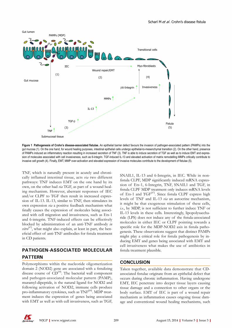
TNF, which is naturally present in acutely and chroni-cally inflamed intestinal tissue, acts via two different pathways: TNF induces EMT on the one hand by its own, on the other had via TGF, as part of a wound heal-ing mechanism. However, aberrant responses of IEC and/or CLPF to TGF then result in increased expres-sion of IL-13. IL-13, similar to TNF, then stimulates its own expression via a positive feedback mechanism what finally causes the expression of molecules being associ-ated with cell migration and invasiveness, such as Ets-1 and 6-integrin. TNF-induced effects can be effectively blocked by administration of an anti-TNF antibody in vitro[47], what might also explain, at least in part, the ben-eficial effect of anti-TNF antibodies for fistula treatment in CD patients.
PATHOGEN-ASSOCIATED MOLECULAR PATTERNPolymorphisms within the nucleotide oligomerization domain 2 (NOD2) gene are associated with a fistulizing disease course of CD[59]. The bacterial wall component and pathogen-associated molecular pattern (PAMP), muramyl-dipeptide, is the natural ligand for NOD2 and following activation of NOD2, immune cells produce pro-inflammatory cytokines, such as TNF[60]. MDP treat-ment induces the expression of genes being associated with EMT as well as with cell invasiveness, such as TGF,
SNAIL1, IL-13 and 6-Integrin, in IEC. While in non-fistula CLPF, MDP significantly induced mRNA expres-sion of Ets-1, 6-Integrin, TNF, SNAIL1 and TGF, in fistula CLPF MDP treatment only induces mRNA levels of Ets-1 and TGF[47]. Since fistula CLPF express high levels of TNF and IL-13 via an autocrine mechanism, it might be that exogenous stimulation of these cells, i.e., by MDP, is not sufficient to further induce TNF or IL-13 levels in these cells. Interestingly, lipopolysaccha-ride (LPS) does not induce any of the fistula-associated molecules in either IEC or CLPF pointing towards a specific role for the MDP-NOD2 axis in fistula patho-genesis. These observations suggest that distinct PAMPs might play a critical role for fistula pathogenesis by in-ducing EMT and genes being associated with EMT and cell invasiveness what makes the use of antibiotics in fistula treatment plausible.
CONCLUSIONTaken together, available data demonstrate that CD-associated fistulae originate from an epithelial defect that occurs during chronic inflammation. Having undergone EMT, IEC penetrate into deeper tissue layers causing tissue damage and a connection to other organs or the body surface. EMT of IEC is part of a wound repair mechanism as inflammation causes ongoing tissue dam-age and conventional wound healing mechanisms, such
August 15, 2014|Volume 5|Issue 3|WJGP|www.wjgnet.com 209
Gut lumenPAMPs (MDP)
IEC
Gut mucosa
Wound repair/EMT
Transitional cells
Myo-fibroblasts
b6-Integrin
MMPs
IL-13
TGF-b
TNF
Submucosal tissue
Invasiveness
Fistula
(1)(2)
(3) (5)
(4)
Figure 1 Pathogenesis of Crohn’s disease-associated fistulae. An epithelial barrier defect favours the invasion of pathogen-associated pattern (PAMPs) into the gut mucosa (1). On the one hand, for wound healing purposes, intestinal epithelial cells undergo epithelial-to-mesenchymal transition (2). On the other hand, presence of PAMPs induced an inflammatory reaction resulting in increased secretion of TNF (3). TNF is able to induce secretion of TGF as well as to induce EMT and expres-sion of molecules associated with cell invasiveness, such as 6-integrin. TGF-induced IL-13 and elevated activation of matrix remodelling MMPs critically contribute to invasive cell growth (4). Finally, EMT, MMP over-activation and elevated expression of invasive molecules contribute to the development of fistulae (5).
Scharl M et al . Crohn’s disease fistula
as migration of fibroblasts, are impaired. The expression of EMT-associated molecules results in enhanced activa-tion of matrix remodelling enzymes such as MMP-3 and MMP-9 causing further tissue damage and inflamma-tion. Finally, soluble mediators such as TNF and IL-13 promote their own expression in an autocrine manner and enhance expression of molecules being associated with cell invasiveness. Subsequently, fistula formation and “growth” is constantly promoted and further sup-ported by the presence of EMT-inducers, such as TGF, and PAMPs (Figure 1). Though current knowledge sug-gests a number of therapeutic options, new and more effective therapeutic approaches are urgently needed for patients suffering from CD-associated fistulae.
REFERENCES1 Hellers G, Bergstrand O, Ewerth S, Holmström B. Occur-
rence and outcome after primary treatment of anal fistulae in Crohn’s disease. Gut 1980; 21: 525-527 [PMID: 7429313]
2 Schwartz DA, Loftus EV, Tremaine WJ, Panaccione R, Harmsen WS, Zinsmeister AR, Sandborn WJ. The natural history of fistulizing Crohn’s disease in Olmsted County, Minnesota. Gastroenterology 2002; 122: 875-880 [PMID: 11910338]
3 Solomon MJ. Fistulae and abscesses in symptomatic peri-anal Crohn’s disease. Int J Colorectal Dis 1996; 11: 222-226 [PMID: 8951512]
4 Nielsen OH, Rogler G, Hahnloser D, Thomsen OØ. Diagno-sis and management of fistulizing Crohn‘s disease. Nat Clin Pract Gastroenterol Hepatol 2009; 6: 92-106 [PMID: 19153563 DOI: 10.1038/ncpgasthep1340]
5 Loftus EV, Schoenfeld P, Sandborn WJ. The epidemiology and natural history of Crohn’s disease in population-based patient cohorts from North America: a systematic review. Aliment Pharmacol Ther 2002; 16: 51-60 [PMID: 11856078]
6 Bataille F, Klebl F, Rümmele P, Schroeder J, Farkas S, Wild PJ, Fürst A, Hofstädter F, Schölmerich J, Herfarth H, Rogler G. Morphological characterisation of Crohn’s disease fistu-lae. Gut 2004; 53: 1314-1321 [PMID: 15306592 DOI: 10.1136/gut.2003.038208]
7 Leeb SN, Vogl D, Gunckel M, Kiessling S, Falk W, Göke M, Schölmerich J, Gelbmann CM, Rogler G. Reduced migration of fibroblasts in inflammatory bowel disease: role of inflam-matory mediators and focal adhesion kinase. Gastroenterol-ogy 2003; 125: 1341-1354 [PMID: 14598250]
8 Leeb SN, Vogl D, Falk W, Schölmerich J, Rogler G, Gelb-mann CM. Regulation of migration of human colonic myofi-broblasts. Growth Factors 2002; 20: 81-91 [PMID: 12148566]
9 Meier JK, Scharl M, Miller SN, Brenmoehl J, Hausmann M, Kellermeier S, Schölmerich J, Rogler G. Specific differ-ences in migratory function of myofibroblasts isolated from Crohn’s disease fistulae and strictures. Inflamm Bowel Dis 2011; 17: 202-212 [PMID: 20848526 DOI: 10.1002/ibd.21344]
10 Schlaepfer DD, Hauck CR, Sieg DJ. Signaling through fo-cal adhesion kinase. Prog Biophys Mol Biol 1999; 71: 435-478 [PMID: 10354709]
11 Brenmoehl J, Lang M, Hausmann M, Leeb SN, Falk W, Schölmerich J, Göke M, Rogler G. Evidence for a differential expression of fibronectin splice forms ED-A and ED-B in Crohn’s disease (CD) mucosa. Int J Colorectal Dis 2007; 22: 611-623 [PMID: 17136547 DOI: 10.1007/s00384-006-0188-4]
12 Ffrench-Constant C, Van de Water L, Dvorak HF, Hynes RO. Reappearance of an embryonic pattern of fibronectin splicing during wound healing in the adult rat. J Cell Biol 1989; 109: 903-914 [PMID: 2760116]
13 Lippert E, Falk W, Bataille F, Kaehne T, Naumann M, Goeke
M, Herfarth H, Schoelmerich J, Rogler G. Soluble galectin-3 is a strong, colonic epithelial-cell-derived, lamina propria fibroblast-stimulating factor. Gut 2007; 56: 43-51 [PMID: 16709662 DOI: 10.1136/gut.2005.081646]
14 Lippert E, Gunckel M, Brenmoehl J, Bataille F, Falk W, Scholmerich J, Obermeier F, Rogler G. Regulation of galec-tin-3 function in mucosal fibroblasts: potential role in muco-sal inflammation. Clin Exp Immunol 2008; 152: 285-297 [PMID: 18336593 DOI: 10.1111/j.1365-2249.2008.03618.x]
15 Dignass AU. Mechanisms and modulation of intestinal epithelial repair. Inflamm Bowel Dis 2001; 7: 68-77 [PMID: 11233665]
16 Kalluri R, Neilson EG. Epithelial-mesenchymal transi-tion and its implications for fibrosis. J Clin Invest 2003; 112: 1776-1784 [PMID: 14679171 DOI: 10.1172/JCI20530]
17 Lee JM, Dedhar S, Kalluri R, Thompson EW. The epithelial-mesenchymal transition: new insights in signaling, devel-opment, and disease. J Cell Biol 2006; 172: 973-981 [PMID: 16567498 DOI: 10.1083/jcb.200601018]
18 Arias AM. Epithelial mesenchymal interactions in cancer and development. Cell 2001; 105: 425-431 [PMID: 11371340]
19 Zavadil J, Böttinger EP. TGF-beta and epithelial-to-mes-enchymal transitions. Oncogene 2005; 24: 5764-5774 [PMID: 16123809 DOI: 10.1038/sj.onc.1208927]
20 Bates RC , Bellovin DI, Brown C, Maynard E, Wu B, Kawakatsu H, Sheppard D, Oettgen P, Mercurio AM. Tran-scriptional activation of integrin beta6 during the epithelial-mesenchymal transition defines a novel prognostic indica-tor of aggressive colon carcinoma. J Clin Invest 2005; 115: 339-347 [PMID: 15668738 DOI: 10.1172/JCI23183]
21 Bates RC. Colorectal cancer progression: integrin alphav-beta6 and the epithelial-mesenchymal transition (EMT). Cell Cycle 2005; 4: 1350-1352 [PMID: 16123591]
22 Bataille F, Rohrmeier C, Bates R, Weber A, Rieder F, Bren-moehl J, Strauch U, Farkas S, Fürst A, Hofstädter F, Schölm-erich J, Herfarth H, Rogler G. Evidence for a role of epithe-lial mesenchymal transition during pathogenesis of fistulae in Crohn’s disease. Inflamm Bowel Dis 2008; 14: 1514-1527 [PMID: 18626977 DOI: 10.1002/ibd.20590]
23 Peinado H, Quintanilla M, Cano A. Transforming growth factor beta-1 induces snail transcription factor in epithelial cell lines: mechanisms for epithelial mesenchymal transi-tions. J Biol Chem 2003; 278: 21113-21123 [PMID: 12665527 DOI: 10.1074/jbc.M211304200]
24 Zheng G, Lyons JG, Tan TK, Wang Y, Hsu TT, Min D, Suc-car L, Rangan GK, Hu M, Henderson BR, Alexander SI, Harris DC. Disruption of E-cadherin by matrix metallopro-teinase directly mediates epithelial-mesenchymal transition downstream of transforming growth factor-beta1 in renal tubular epithelial cells. Am J Pathol 2009; 175: 580-591 [PMID: 19590041 DOI: 10.2353/ajpath.2009.080983]
25 Scharl M, Weber A, Fürst A, Farkas S, Jehle E, Pesch T, Kell-ermeier S, Fried M, Rogler G. Potential role for SNAIL fam-ily transcription factors in the etiology of Crohn’s disease-associated fistulae. Inflamm Bowel Dis 2011; 17: 1907-1916 [PMID: 21830269 DOI: 10.1002/ibd.21555]
26 Scharl M, Frei S, Pesch T, Kellermeier S, Arikkat J, Frei P, Fried M, Weber A, Jehle E, Rühl A, Rogler G. Interleukin-13 and transforming growth factor β synergise in the patho-genesis of human intestinal fistulae. Gut 2013; 62: 63-72 [PMID: 22287592 DOI: 10.1136/gutjnl-2011-300498]
27 Nelson AR, Fingleton B, Rothenberg ML, Matrisian LM. Matrix metalloproteinases: biologic activity and clinical im-plications. J Clin Oncol 2000; 18: 1135-1149 [PMID: 10694567]
28 Baugh MD, Evans GS, Hollander AP, Davies DR, Perry MJ, Lobo AJ, Taylor CJ. Expression of matrix metalloproteases in inflammatory bowel disease. Ann N Y Acad Sci 1998; 859: 249-253 [PMID: 9928398]
29 von Lampe B, Barthel B, Coupland SE, Riecken EO, Rose-wicz S. Differential expression of matrix metalloproteinases
August 15, 2014|Volume 5|Issue 3|WJGP|www.wjgnet.com 210
Scharl M et al . Crohn’s disease fistula
and their tissue inhibitors in colon mucosa of patients with inflammatory bowel disease. Gut 2000; 47: 63-73 [PMID: 10861266]
30 Castaneda FE, Walia B, Vijay-Kumar M, Patel NR, Roser S, Kolachala VL, Rojas M, Wang L, Oprea G, Garg P, Gewirtz AT, Roman J, Merlin D, Sitaraman SV. Targeted deletion of metalloproteinase 9 attenuates experimental colitis in mice: central role of epithelial-derived MMP. Gastroenterol-ogy 2005; 129: 1991-2008 [PMID: 16344067 DOI: 10.1053/j.gastro.2005.09.017]
31 Santana A, Medina C, Paz-Cabrera MC, Díaz-Gonzalez F, Farré E, Salas A, Radomski MW, Quintero E. Attenuation of dextran sodium sulphate induced colitis in matrix metal-loproteinase-9 deficient mice. World J Gastroenterol 2006; 12: 6464-6472 [PMID: 17072979]
32 Liu H, Patel NR, Walter L, Ingersoll S, Sitaraman SV, Garg P. Constitutive expression of MMP9 in intestinal epithe-lium worsens murine acute colitis and is associated with increased levels of proinflammatory cytokine Kc. Am J Physiol Gastrointest Liver Physiol 2013; 304: G793-G803 [PMID: 23471340 DOI: 10.1152/ajpgi.00249.2012]
33 Pender SL, Tickle SP, Docherty AJ, Howie D, Wathen NC, MacDonald TT. A major role for matrix metalloproteinases in T cell injury in the gut. J Immunol 1997; 158: 1582-1590 [PMID: 9029093]
34 Kirkegaard T, Hansen A, Bruun E, Brynskov J. Expression and localisation of matrix metalloproteinases and their nat-ural inhibitors in fistulae of patients with Crohn’s disease. Gut 2004; 53: 701-709 [PMID: 15082589]
35 Frei SM LS, Jehle EC, Fried M, Rogler G, Scharl M. Expres-sion of Interleukins 22 and 33, Matrix Metalloproteinases 9 and 13, Mast Cell Markers and Hypoxia-Inducible Factor 1α in Crohn’s Disease Associated Fistulae Gastroenterology. DDW 2013; 144 (5, Supplement 1): S441-S442
36 Hazelbag S, Kenter GG, Gorter A, Dreef EJ, Koopman LA, Violette SM, Weinreb PH, Fleuren GJ. Overexpression of the alpha v beta 6 integrin in cervical squamous cell carcinoma is a prognostic factor for decreased survival. J Pathol 2007; 212: 316-324 [PMID: 17503414 DOI: 10.1002/path.2168]
37 Arihiro K, Kaneko M, Fujii S, Inai K, Yokosaki Y. Signifi-cance of alpha 9 beta 1 and alpha v beta 6 integrin expres-sion in breast carcinoma. Breast Cancer 2000; 7: 19-26 [PMID: 11029766]
38 Li X, Yang Y, Hu Y, Dang D, Regezi J, Schmidt BL, At-akilit A, Chen B, Ellis D, Ramos DM. Alphavbeta6-Fyn signaling promotes oral cancer progression. J Biol Chem 2003; 278: 41646-41653 [PMID: 12917446 DOI: 10.1074/jbc.M306274200]
39 Thomas GJ, Lewis MP, Hart IR, Marshall JF, Speight PM. AlphaVbeta6 integrin promotes invasion of squamous carci-noma cells through up-regulation of matrix metalloprotein-ase-9. Int J Cancer 2001; 92: 641-650 [PMID: 11340566 DOI: 10.1002/1097-0215(20010601)92: ]
40 Thomas GJ, Lewis MP, Whawell SA, Russell A, Sheppard D, Hart IR, Speight PM, Marshall JF. Expression of the alphavbeta6 integrin promotes migration and invasion in squamous carcinoma cells. J Invest Dermatol 2001; 117: 67-73 [PMID: 11442751 DOI: 10.1046/j.0022-202x.2001.01379.x]
41 Thomas GJ, Poomsawat S, Lewis MP, Hart IR, Speight PM, Marshall JF. alpha v beta 6 Integrin upregulates matrix metalloproteinase 9 and promotes migration of normal oral keratinocytes. J Invest Dermatol 2001; 116: 898-904 [PMID: 11407978 DOI: 10.1046/j.1523-1747.2001.01352.x]
42 Agrez M, Gu X, Turton J, Meldrum C, Niu J, Antalis T, How-ard EW. The alpha v beta 6 integrin induces gelatinase B se-cretion in colon cancer cells. Int J Cancer 1999; 81: 90-97 [PMID: 10077158 DOI: 10.1002/(SICI)1097-0215(19990331)81]
43 Agrez MV, Bates RC, Mitchell D, Wilson N, Ferguson N, Anseline P, Sheppard D. Multiplicity of fibronectin-binding alpha V integrin receptors in colorectal cancer. Br J Cancer
1996; 73: 887-892 [PMID: 8611401 DOI: 10.1038/bjc.1996.158]44 Weinacker A, Chen A, Agrez M, Cone RI, Nishimura S,
Wayner E, Pytela R, Sheppard D. Role of the integrin alpha v beta 6 in cell attachment to fibronectin. Heterologous ex-pression of intact and secreted forms of the receptor. J Biol Chem 1994; 269: 6940-6948 [PMID: 8120056]
45 Munger JS, Huang X, Kawakatsu H, Griffiths MJ, Dalton SL, Wu J, Pittet JF, Kaminski N, Garat C, Matthay MA, Rifkin DB, Sheppard D. The integrin alpha v beta 6 binds and activates latent TGF beta 1: a mechanism for regulating pulmonary inflammation and fibrosis. Cell 1999; 96: 319-328 [PMID: 10025398]
46 Niu J, Gu X, Ahmed N, Andrews S, Turton J, Bates R, Agrez M. The alphaVbeta6 integrin regulates its own expression with cell crowding: implications for tumour progression. Int J Cancer 2001; 92: 40-48 [PMID: 11279604 DOI: 10.1002/1097-0215(20010401)92]
47 Frei SM, Pesch T, Lang S, Weber A, Jehle E, Vavricka SR, Fried M, Rogler G, Scharl M. A role for tumor necrosis factor and bacterial antigens in the pathogenesis of Crohn’s disease-associated fistulae. Inflamm Bowel Dis 2013; 19: 2878-2887 [PMID: 24189042 DOI: 10.1097/01.MIB.0000435760.82705.23]
48 Aguilera O, Fraga MF, Ballestar E, Paz MF, Herranz M, Espada J, García JM, Muñoz A, Esteller M, González-Sancho JM. Epigenetic inactivation of the Wnt antagonist DICK-KOPF-1 (DKK-1) gene in human colorectal cancer. Onco-gene 2006; 25: 4116-4121 [PMID: 16491118 DOI: 10.1038/sj.onc.1209439]
49 Koch S, Capaldo CT, Samarin S, Nava P, Neumaier I, Skerra A, Sacks DB, Parkos CA, Nusrat A. Dkk-1 inhibits intestinal epithelial cell migration by attenuating directional polariza-tion of leading edge cells. Mol Biol Cell 2009; 20: 4816-4825 [PMID: 19776352 DOI: 10.1091/mbc.E09-05-0415]
50 Niehrs C. Function and biological roles of the Dickkopf family of Wnt modulators. Oncogene 2006; 25: 7469-7481 [PMID: 17143291 DOI: 10.1038/sj.onc.1210054]
51 Frei SM HC, Pesch T, Lang S, Weber A, Jehle E, R�hl A, Fried M, Rogler G, Scharl M. The Role for Dickkopf-Ho-molog-1 in the Pathogenesis of Crohn’s Disease-Associated Fistulae. PLoS One 2013; In press
52 Zhu Z, Homer RJ, Wang Z, Chen Q, Geba GP, Wang J, Zhang Y, Elias JA. Pulmonary expression of interleukin-13 causes inflammation, mucus hypersecretion, subepithelial fibrosis, physiologic abnormalities, and eotaxin produc-tion. J Clin Invest 1999; 103: 779-788 [PMID: 10079098 DOI: 10.1172/JCI5909]
53 Lee CG, Homer RJ, Zhu Z, Lanone S, Wang X, Koteliansky V, Shipley JM, Gotwals P, Noble P, Chen Q, Senior RM, Elias JA. Interleukin-13 induces tissue fibrosis by selectively stim-ulating and activating transforming growth factor beta(1). J Exp Med 2001; 194: 809-821 [PMID: 11560996]
54 Chiaramonte MG, Donaldson DD, Cheever AW, Wynn TA. An IL-13 inhibitor blocks the development of hepatic fibrosis during a T-helper type 2-dominated inflammatory response. J Clin Invest 1999; 104: 777-785 [PMID: 10491413 DOI: 10.1172/JCI7325]
55 Wynn TA. IL-13 effector functions. Annu Rev Immunol 2003; 21: 425-456 [PMID: 12615888 DOI: 10.1146/annurev.immu-nol.21.120601.141142]
56 Donaldson DD, Whitters MJ, Fitz LJ, Neben TY, Finnerty H, Henderson SL, O’Hara RM, Beier DR, Turner KJ, Wood CR, Collins M. The murine IL-13 receptor alpha 2: molecu-lar cloning, characterization, and comparison with murine IL-13 receptor alpha 1. J Immunol 1998; 161: 2317-2324 [PMID: 9725226]
57 Bates RC, Mercurio AM. Tumor necrosis factor-alpha stim-ulates the epithelial-to-mesenchymal transition of human colonic organoids. Mol Biol Cell 2003; 14: 1790-1800 [PMID: 12802055 DOI: 10.1091/mbc.E02-09-0583]
58 Sullivan DE, Ferris M, Pociask D, Brody AR. Tumor necro-
August 15, 2014|Volume 5|Issue 3|WJGP|www.wjgnet.com 211
Scharl M et al . Crohn’s disease fistula
sis factor-alpha induces transforming growth factor-beta1 expression in lung fibroblasts through the extracellular signal-regulated kinase pathway. Am J Respir Cell Mol Biol 2005; 32: 342-349 [PMID: 15653932 DOI: 10.1165/rcmb.2004-0288OC]
59 Radlmayr M, Török HP, Martin K, Folwaczny C. The
c-insertion mutation of the NOD2 gene is associated with fistulizing and fibrostenotic phenotypes in Crohn’s disease. Gastroenterology 2002; 122: 2091-2092 [PMID: 12055616]
60 Xavier RJ, Podolsky DK. Unravelling the pathogenesis of inflammatory bowel disease. Nature 2007; 448: 427-434 [PMID: 17653185 DOI: 10.1038/nature06005]
P- Reviewer: Keshteli AH, Kellermayer R, Mullin JM S- Editor: Ji FF L- Editor: A E- Editor: Lu YJ
August 15, 2014|Volume 5|Issue 3|WJGP|www.wjgnet.com 212
Scharl M et al . Crohn’s disease fistula
TOPIC HIGHLIGHT
Escherichia coli in chronic inflammatory bowel diseases: An update on adherent invasive Escherichia coli pathogenicity
Margarita Martinez-Medina, Librado Jesus Garcia-Gil
Margarita Martinez-Medina, Librado Jesus Garcia-Gil, Laboratory of Molecular Microbiology, Biology Department, University of Girona, E-17071 Girona, SpainAuthor contributions: Martinez-Medina M wrote the paper; Garcia-Gil LJ revised the paper. Correspondence to: Margarita Martinez-Medina, PhD, Laboratory of Molecular Microbiology, Biology Department, University of Girona, Campus de Montilivi, E-17071 Girona, Spain. [email protected]: +34-972-418175 Fax: +34-972-418150Received: January 28, 2014 Revised: April 8, 2014Accepted: May 29, 2014Published online: August 15, 2014
AbstractEscherichia coli (E. coli ), and particularly the adherent invasive E. coli (AIEC) pathotype, has been increasingly implicated in the ethiopathogenesis of Crohn’s disease (CD). E. coli strains with similar pathogenic features to AIEC have been associated with other intestinal disor-ders such as ulcerative colitis, colorectal cancer, and coeliac disease, but AIEC prevalence in these diseases remains largely unexplored. Since AIEC was described one decade ago, substantial progress has been made in deciphering its mechanisms of pathogenicity. However, the molecular bases that characterize the phenotypic properties of this pathotype are still not well resolved. A review of studies focused on E. coli populations in in-flammatory bowel disease (IBD) is presented here and we discuss about the putative role of this species on each IBD subtype. Given the relevance of AIEC in CD pathogenesis, we present the latest research findings concerning AIEC host-microbe interactions and patho-genicity. We also review the existing data regarding the prevalence and abundance of AIEC in CD and its asso-ciation with other intestinal diseases from humans and animals, in order to discuss the AIEC disease- and host-specificity. Finally, we highlight the fact that dietary
components frequently found in industrialized countries may enhance AIEC colonization in the gut, which merits further investigation and the implementation of pre-ventative measures.
© 2014 Baishideng Publishing Group Inc. All rights reserved.
Key words: Adherent invasive Escherichia coli ; Inflam-matory bowel disease; Crohn’s disease; Pathogenesis; Epidemiology
Core tip: In this review we critically revise the findings on Escherichia coli (E. coli ) populations associated with Crohn’s disease and ulcerative colitis. Then we focus on adherent invasive E. coli (AIEC), especially in its mech-anisms of pathogenicity and epidemiology. We discuss about AIEC disease- and host-specificity and we under-line the importance of discovering specific molecular tools to detect AIEC for further epidemiologic studies. Finally we point out to a putative role of diet on AIEC gut colonization.
Martinez-Medina M, Garcia-Gil LJ. Escherichia coli in chronic inflammatory bowel diseases: An update on adherent invasive Escherichia coli pathogenicity. World J Gastrointest Pathophysiol 2014; 5(3): 213-227 Available from: URL: http://www.wjg-net.com/2150-5330/full/v5/i3/213.htm DOI: http://dx.doi.org/10.4291/wjgp.v5.i3.213
ESCHERICHIA COLI IN INFLAMMATORY BOWEL DISEASEThe intestinal microbiota has been implicated in the pathogenesis of Crohn’s disease (CD) and ulcerative colitis (UC), the main idiopathic inflammatory bowel diseases (IBDs)[1]. CD patients demonstrate an altered
World J Gastrointest Pathophysiol 2014 August 15; 5(3): 213-227ISSN 2150-5330 (online)
© 2014 Baishideng Publishing Group Inc. All rights reserved.
Online Submissions: http://www.wjgnet.com/esps/Help Desk: http://www.wjgnet.com/esps/helpdesk.aspxDOI: 10.4291/wjgp.v5.i3.213
August 15, 2014|Volume 5|Issue 3|WJGP|www.wjgnet.com 213
WJGP 5th Anniversary Special Issues (6): Crohn’s disease
Martinez-Medina M et al . AIEC in inflammatory bowel diseases
intestinal microbial community, and the dysbioses pres-ent in colonic CD and ileal CD are different[2]. In con-trast, a specific dysbiosis in UC is starting to be defined, although differences between studies have hampered attempts to reach a clear consensus to date[2-5]. A number of culture-based and molecular-based studies support the theory that Escherichia coli (E. coli) is a microbiologi-cal factor implicated in CD, but some controversy exists regarding its role in UC[2,6-17]. In this section, we examine data on E. coli populations in CD and UC related to abundance, association with disease activity, transloca-tion of the gut mucosa, and pathogenic features of the strains to highlight the evidence that supports or refutes putative implications for this bacterium in each IBD subtype.
Abundance in the intestinal mucosa and correlation with disease activitySeveral independent studies based on quantitative Poly-merase Chain Reaction (PCR) have indicated that E. coli is augmented in CD patients in comparison with controls[2,6,11,13]. However, differences are especially sig-nificant for CD patients with ileal disease, and no clear association with colonic or ileocolonic CD has been demonstrated. On average, in our cohort, E. coli 16S rRNA gene copies accounted for 14% and 33% of total bacteria 16S rRNA gene copies in healthy subjects and ileal CD patients, respectively (P < 0.001)[13]. Of note, a higher abundance of E. coli was observed in active CD patients than in patients in remission[6,11,18]. Accordingly, a previous study using Fluorescent In Situ Hybridiza-tion (FISH) demonstrated increased E. coli numbers in the epithelium and within the lamina propria in active CD patients compared to inactive CD patients[14]. In ad-dition, we determined that higher numbers of this spe-cies correlated with a reduced amount of time before relapse[11]. These findings are in agreement with previous data reporting that the higher numbers of E. coli isolated from the neoterminal ileum of CD patients are associ-ated with early recurrence of the disease[7], and that high levels of antibodies against the E. coli outer membrane protein C (OmpC) correlate with disease progression, longer duration, and greater need for surgery among CD patients[19-21].
There is substantial controversy regarding the abun-dance of E. coli in the colonic mucosa of UC patients (Table 1). Several works have consistently reported no increase with respect to healthy subjects[2,6,7,11-13], argu-ing against a putative role for E. coli in UC, while oth-ers have reported increased E. coli abundance in UC patients[8,10,14,16,18,22,23]. As in the majority of these studies both CD and UC patients were analyzed, these contro-versial observations can not be explained by differences in methodology between studies. We postulate that they can be attributable to differences in the disease severity of the patients included in the studies, as increased num-bers of E. coli have been associated with activity status in UC patients. Using FISH, epithelium-associated E. coli
were found to be more abundant in active UC compared to inactive UC or controls[14], and quantitative PCR indi-cated that increased numbers of E. coli were present in active UC patients compared to inactive UC patients[22] as well as in inflamed vs non-inflamed UC tissue[23].
Altogether, substantial evidence supports an over-growth of E. coli in ileal CD patients, while there is still no convincing data that exists for other IBD subtypes. Further studies aimed at comparing the abundance of E. coli in IBD patients categorized by disease subtype and assessing any correlation with activity status of the disease would shed light on the role of this bacterium in each IBD subtype and its putative application as a diag-nostic and/or prognostic tool.
E. coli localization in the intestinal mucosaE. coli has been found in the mucus layer, close to the in-testinal epithelial cells and in ulcers of both CD and UC patients[24,25]. Translocation of the intestinal mucosa has been primarily observed in CD[6] and higher amounts of intracellular E. coli were detected in inflamed compared to non-inflamed mucosa[6,26]. With FISH and immuno-histochemistry, E. coli has been detected scattered within the lamina propria, either in the extracellular space or inside macrophages, as well as in the subserosal layer, the perivascular areas of the submucosa, the muscle layer, and in germinal centers of lymph follicles of CD patients[8,14,27]. A recent study using high throughput se-quencing indicated a greater proportion of E. coli reads in the lymph nodes of ileal CD patients than other CD patients[28]. Interestingly, E. coli DNA was also more fre-quently found in the granulomas of CD patients (80%) than in non-CD control patients (10%) in a study that used Laser Capture Microdissection and PCR[29]. In con-trast, E. coli has not been frequently found to translocate the mucosa of UC patients[8,24,25], although some contro-versy exists as some authors have detected E. coli in the lamina propria of UC patients[14,27].
The majority of the aforementioned studies are based on techniques that do not distinguish viable bac-teria from dead bacteria. Further studies should study the viability of translocated E. coli, particularly in lymph nodes and granulomas, as these locations would be more relevant to establish a link between this bacterium and CD pathogenesis. These studies should also focus on UC patients to clarify the existing controversial data. A lack of E. coli translocation in UC would suggest that E. coli does not play a primary role in UC pathogenesis or that it plays a different role than in CD.
Pathogenic features of the strainsE. coli strains isolated from IBD patients are clonally diverse[6,13,17] and belong to distinct serotypes[6,13,30] and to different sequence types[6,31-33]. Although a close genetic relationship was detected in a study of IBD pediatric patients[34], the hypothesis that there is a particular clone associated with IBD has largely been ruled out.
In turn, E. coli strains isolated from IBD patients
August 15, 2014|Volume 5|Issue 3|WJGP|www.wjgnet.com 214
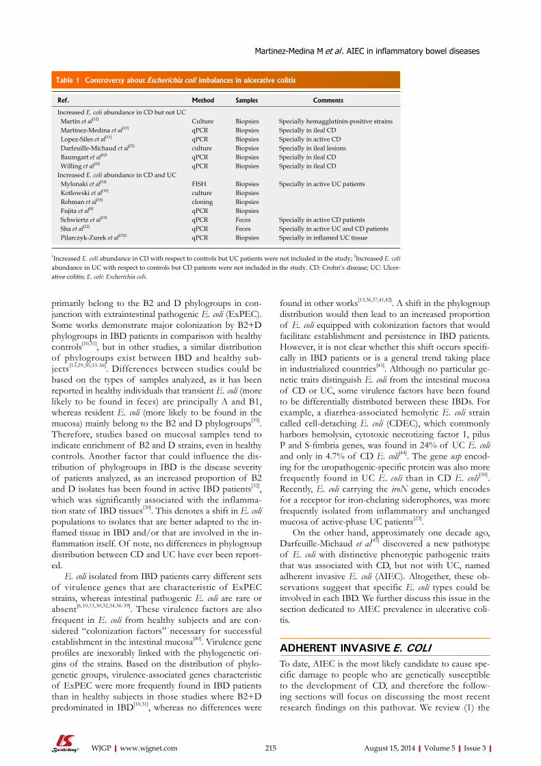
primarily belong to the B2 and D phylogroups in con-junction with extraintestinal pathogenic E. coli (ExPEC). Some works demonstrate major colonization by B2+D phylogroups in IBD patients in comparison with healthy controls[10,31], but in other studies, a similar distribution of phylogroups exist between IBD and healthy sub-jects[13,29,30,33-36]. Differences between studies could be based on the types of samples analyzed, as it has been reported in healthy individuals that transient E. coli (more likely to be found in feces) are principally A and B1, whereas resident E. coli (more likely to be found in the mucosa) mainly belong to the B2 and D phylogroups[35]. Therefore, studies based on mucosal samples tend to indicate enrichment of B2 and D strains, even in healthy controls. Another factor that could influence the dis-tribution of phylogroups in IBD is the disease severity of patients analyzed, as an increased proportion of B2 and D isolates has been found in active IBD patients[32], which was significantly associated with the inflamma-tion state of IBD tissues[30]. This denotes a shift in E. coli populations to isolates that are better adapted to the in-flamed tissue in IBD and/or that are involved in the in-flammation itself. Of note, no differences in phylogroup distribution between CD and UC have ever been report-ed.
E. coli isolated from IBD patients carry different sets of virulence genes that are characteristic of ExPEC strains, whereas intestinal pathogenic E. coli are rare or absent[6,10,13,30,32,34,36-39]. These virulence factors are also frequent in E. coli from healthy subjects and are con-sidered “colonization factors” necessary for successful establishment in the intestinal mucosa[40]. Virulence gene profiles are inexorably linked with the phylogenetic ori-gins of the strains. Based on the distribution of phylo-genetic groups, virulence-associated genes characteristic of ExPEC were more frequently found in IBD patients than in healthy subjects in those studies where B2+D predominated in IBD[10,31], whereas no differences were
found in other works[13,36,37,41,42]. A shift in the phylogroup distribution would then lead to an increased proportion of E. coli equipped with colonization factors that would facilitate establishment and persistence in IBD patients. However, it is not clear whether this shift occurs specifi-cally in IBD patients or is a general trend taking place in industrialized countries[43]. Although no particular ge-netic traits distinguish E. coli from the intestinal mucosa of CD or UC, some virulence factors have been found to be differentially distributed between these IBDs. For example, a diarrhea-associated hemolytic E. coli strain called cell-detaching E. coli (CDEC), which commonly harbors hemolysin, cytotoxic necrotizing factor 1, pilus P and S-fimbria genes, was found in 24% of UC E. coli and only in 4.7% of CD E. coli[44]. The gene usp encod-ing for the uropathogenic-specific protein was also more frequently found in UC E. coli than in CD E. coli[30]. Recently, E. coli carrying the iroN gene, which encodes for a receptor for iron-chelating siderophores, was more frequently isolated from inflammatory and unchanged mucosa of active-phase UC patients[23].
On the other hand, approximately one decade ago, Darfeuille-Michaud et al[45] discovered a new pathotype of E. coli with distinctive phenotypic pathogenic traits that was associated with CD, but not with UC, named adherent invasive E. coli (AIEC). Altogether, these ob-servations suggest that specific E. coli types could be involved in each IBD. We further discuss this issue in the section dedicated to AIEC prevalence in ulcerative coli-tis.
ADHERENT INVASIVE E. COLITo date, AIEC is the most likely candidate to cause spe-cific damage to people who are genetically susceptible to the development of CD, and therefore the follow-ing sections will focus on discussing the most recent research findings on this pathovar. We review (1) the
August 15, 2014|Volume 5|Issue 3|WJGP|www.wjgnet.com 215
Table 1 Controversy about Escherichia coli imbalances in ulcerative colitis
Ref. Method Samples Comments
Increased E. coli abundance in CD but not UC Martin et al[12] Culture Biopsies Specially hemagglutinin-positive strains Martinez-Medina et al[13] qPCR Biopsies Specially in ileal CD Lopez-Siles et al[11] qPCR Biopsies Specially in active CD Darfeuille-Michaud et al[7]1 culture Biopsies Specially in ileal lesions Baumgart et al[6]1 qPCR Biopsies Specially in ileal CD Willing et al[2]1 qPCR Biopsies Specially in ileal CDIncreased E. coli abundance in CD and UC Mylonaki et al[14] FISH Biopsies Specially in active UC patients Kotlowski et al[10] culture Biopsies Rehman et al[16] cloning Biopsies Fujita et al[8] qPCR Biopsies Schwiertz et al[18] qPCR Feces Specially in active CD patients Sha et al[22] qPCR Feces Specially in active UC and CD patients Pilarczyk-Zurek et al[23]2 qPCR Biopsies Specially in inflamed UC tissue
1Increased E. coli abundance in CD with respect to controls but UC patients were not included in the study; 2Increased E. coli abundance in UC with respect to controls but CD patients were not included in the study. CD: Crohn’s disease; UC: Ulcer-ative colitis; E. coli: Escherichia coli.
Martinez-Medina M et al . AIEC in inflammatory bowel diseases
latest research regarding AIEC pathogenicity; (2) the prevalence and abundance of the pathotype in several intestinal disorders, discussing its putative contribution to other intestinal diseases in addition to CD; (3) the evi-dence that supports a lack of host-specificity and thus a risk for zoonosis; and (4) recent research that points to a putative role for environmental factors in the fate of AIEC development in the intestine.
DefinitionThe AIEC pathotype was defined as E. coli strains that (1) are able to adhere to differentiated Caco-2 and/or undifferentiated I-407 intestinal epithelial cells with an adhesion index equal or superior to 1 bacteria per cell; (2) are able to invade I-407 cells with an invasion index equal or superior to 0.1% of the original inoculum; (3) involve host cell actin polymerization and microtubule recruitment in bacterial uptake; (4) do not have known invasive determinants; and (5) are able to survive and replicate within J774-A1 macrophages[45]. Since its defini-tion, invasive determinants characteristic from ExPEC have been detected in some AIEC, but not consistently in all AIEC, and thus are not a particularity of the AIEC pathotype[6,13,36,46-48].
Molecular basis of AIEC pathogenicityPathogenicity mechanisms of AIEC have mainly been studied in the reference AIEC strain LF82, and its fea-tures have been comprehensively linked to many charac-teristics of CD pathogenesis.
Adhesion to intestinal epithelial cells is in part me-diated by type 1 pili, which interact with the glycopro-tein CEACAM6 in a mannose-associated manner[49,50]. CEACAM6 is overexpressed in CD patients with ileal disease, which makes them more susceptible to over-colonization by AIEC. Although type 1 pili is present in almost all E. coli, including non-pathogenic strains, we have recently demonstrated that AIEC strains usually present FimH adhesin variants that allow them to more efficiently bind intestinal epithelial cells[31]. Some non-AIEC strains carry these mutations as well, but they do not express type 1 pili. Flagella are also important for adhesion to and invasion of intestinal epithelial cells and elicit the secretion of the pro-inflammatory cyto-kine IL-8 and chemokine CCL20 in polarized intestinal epithelial cells, which in turn leads to the recruitment of macrophages and dendritic cells to the site of infec-tion[51,52]. The further secretion of INFγ and TNFα by macrophages and lymphocytes leads to CEACAM6 expression, which enhances AIEC colonization. The binding of LF82 type 1 pili to CEACAM6 and flagella to TLR5 in intestinal epithelial cells induces the production of HIF-1a and activation of the classical NF-κB path-way[53]. In turn, these molecules cooperatively control the transcription of IL-8 and pro-angiogenic factors con-tributing to inflammation and vascularization.
The intermediate filament vimentin, expressed on the host cell surface of mesenchymal cells, has been re-
cently proposed to act as a receptor for AIEC[54]. At the intracellular side, vimentin leucine-rich repeats interact with NOD2 leading to the recruitment of these proteins at the plasma membrane. This is necessary for a proper function of NOD2 in terms of antigen detection, NF-κB activation and autophagy induction. CD patients have specific NOD2 variants (L1007fs and R702W) that are unable to interact with vimentin and, in turn, they localize in the cytosol. That leads to a defective inflam-matory response, autophagy induction and handling of CD-associated AIEC. Altogether, NOD2 and vimentin appear to play an important role in AIEC recognition and polymorphisms in these two proteins may have an impact on the ability of AIEC to colonize the host.
A new host-microbe interaction that mediates adhe-sion of LF82 to intestinal epithelial cells and involves a bacterial and a human chitinase has recently been pro-posed[55]. Chitinases are enzymes that hydrolyze chitin, a long-chain polymer of an N-acetylglucosamine. The authors demonstrate that specific polymorphisms in two chitin binding domains characteristic of LF82 and other pathogenic E. coli are required to interact with an N-gly-cosylated asparagine of the human chitinase CHI3L1. Interestingly, human chitinases are overexpressed in in-testinal epithelial cells and moderately expressed in cells of the lamina propria during inflammation.
Outer membrane vesicles (OMVs) containing the transmembrane protein OmpA play a role in LF82 in-vasion of intestinal epithelial cells[48]. OmpA binds the endoplasmic reticulum-localized stress response chaper-one Gp96 that is overexpressed on the apical surface of ileal epithelial cells in patients with CD. OMVs fuse with host cells, and it is thought that the release of bacterial effectors that are still undefined is involved in the actin polymerization and microtubule recruitment that occurs during invasion. Point mutations in the ompA sequence of LF82 and other B2 strains mediate better interactions with Gp96[56]. In turn, Gp96 is overexpressed in the ileum of CD patients, which renders them more suscep-tible to AIEC infection.
Once inside the host cell, LF82 bacteria can be found in several types of intracellular compartments: indi-vidually or in groups within single membrane vacuoles, within damaged vacuoles, or within LC3-positive au-tophagosomes, which indicates that autophagy restricts a subpopulation of intracellular LF82 bacteria[57]. Nev-ertheless, it was recently demonstrated that AIEC can abrogate the autophagic process[58]. Intracellular LF82 activates NF-κB, leading to the increased expression of MIR30C and MIR130A in T84 cells and in mouse enterocytes, and the upregulation of these microRNAs reduces levels of ATG5 and ATG16L1, inhibiting au-tophagy and enhancing the inflammatory response. In turn, defects in autophagy mechanisms related to the ATG16L1 and IRGM genes have been associated with CD patients, and these defects confer an advantage for AIEC to survive inside human cells[57]. Therefore, it is a combination of host deficiency factors and AIEC
August 15, 2014|Volume 5|Issue 3|WJGP|www.wjgnet.com 216
Martinez-Medina M et al . AIEC in inflammatory bowel diseases
pathogenicity that determines the fate of intracellular E. coli survival.
In addition to adhesion and invasion capacity, LF82 is also able of translocating via the M cells of the Peyer’s patches, gaining access to the lamina propria. This inter-action is mediated by type 1 pili and long polar fimbriae (Lpf), which can interact independently with GP2, a sur-face protein specific to M cells. It is of note that the sites of initial inflammation in CD are the Peyer’s patches and colonic lymphoid follicles; thus, this mechanism of translocation is consistent with early clinical signs of the disease[59].
Another mechanism that can facilitate bacterial trans-location is the ability of LF82 to alter intestinal perme-ability by inducing the expression of the pore-forming protein claudin-2[60] and by displacing ZO-1 and E-cad-herin from apical tight junctions, leading to decreased transepithelial resistance and loss of barrier function[17,61]. Besides, pro-inflammatory cytokines like TNFα can drive alterations in intestinal permeability[62]. As AIEC infec-tion induces the secretion of large amounts of TNFα and IL-8[17]; thus, the loss of barrier function induced by LF82 can in part be mediated by the induction of TNFα secretion.
A novel mechanism of pathogenicity observed in LF82 and two other AIEC strains (O83:H1 and UM146) is the evasion of host immune responses via subver-sion of the IFNγ pathway in intestinal epithelial cells[63]. Phosphorylation of the Signal Transducer and Activator of Transcription STAT-1 is blocked, thus preventing the transcription of IFNγ-dependent genes, which re-duces host immune responses and results in an inability to mount an appropriate anti-microbiocidal response. Enterohemorrhagic E. coli (EHEC) strain O157:H7, in part through its Shiga toxin, is also able to block ty-rosine phosphorylation and activation of STAT1 after IFNγ stimulation, in contrast with enteropathogenic E. coli E2348/69 or commensal E. coli HB101 which do not present this mechanism of pathogenicity. However, AIEC do not present Shiga toxins. Presumably a small secreted peptide may be responsible for this pathogenic mechanism in AIEC[63].
Once AIEC has gained access to the lamina propria, these bacteria can be engulfed by macrophages. Intram-acrophage LF82 do not escape into the cytoplasm but induce the formation of a large vacuole (phagosome) that fuses with lysosomes[64], suggesting that AIEC bac-teria have the ability to replicate in an environment with acidic pH, oxidative stress, active proteolytic enzymes, and antimicrobial compounds. Indeed, it was demon-strated in vitro that an acidic environment is necessary for replication of AIEC LF82 bacteria[64]. The protease HtrA and the thiol-disulfide oxidoreductase DsbA have been reported to be important for survival and replica-tion within macrophages[65,66]. The authors linked these proteins to the ability of LF82 to resist the stress condi-tions of the phagolysosomes, as isogenic mutants for these proteins were less efficient in growing in acidic and
nutrient-poor medium, and these proteins were overex-pressed not only in LF82 during macrophage infection but also in acidic nutrient-poor medium. Interestingly, the overexpression of HtrA is dependent on the LF82 background, as non-pathogenic E. coli do not overex-press that protein under similar growth conditions. The RNA-binding protein Hfq, which functions as a global posttranscriptional regulator of gene expression, has also been implicated in survival and replication within mac-rophages and in stress tolerance but also other aspects of LF82 pathogenicity, such as adhesion and invasion capability[67]. Hfq binds small regulatory RNA molecules, facilitating their interaction with mRNA, but the target genes are still unknown.
Continuous replication of LF82 within macrophages results in the secretion of high levels of TNFα without inducing host cell death[68]. This can explain inflamma-tion and granuloma formation in the gut of CD patients, which has been demonstrated in vitro[50,69,70]. A direct role for LF82 in delaying apoptosis of infected macrophages and dendritic cells has recently been reported[71]. LF82 infection was found to alter the function of caspase-3, a protease that plays an essential role in apoptosis, and to increase degradation of this molecule in the proteasome.
Also supporting AIEC capability to replicate within immune cells, strain LF82 was able to replicate within monocytes isolated from CD patients for the first 20 h after infection but then CD monocytes started to clear intracellular bacteria[72]. Interestingly, those patients with polymorphisms in CARD15 gene (R702W, G908R and 1007fs) showed reduced early inflammatory response towards AIEC infection with decreased levels of IL-1β, IL-6 and IL-10. In contrast, Asp299Gly mutation in TLR4 had no effect on monocyte response to AIEC. Be-sides, a recent study revealed that CD monocyte-derived dendritic cells stimulated with lipopolysaccharide show an attenuated inflammatory response with decreased lev-els of IL-6 and IL-1β, as well as an impaired autophagy with reduced LC3 expression[73]. Moreover, these cells had a reduced capacity to support the expansion of al-logeneic Th17 cells from CD4+ memory T cells. The authors propose that mucosal Th17 activation in CD patients is a secondary event in response of poor bacte-rial clearance due to defects in innate immunity. Further studies showing AIEC effects on CD defective den-dritic cells regarding, not only cytokine release, but also autophagy function and the level of IL-17A response induction in T cells, are necessary to decipher whether the alterations observed in lipopolysaccharide-stimulated dendritic cells equally occur after AIEC exposure.
AIEC LF82 bacteria are also able to invade and repli-cate within human neutrophils, but in contrast to its be-havior inside macrophages and intestinal epithelial cells, LF82 induces the autophagic death of infected neutro-phils, which later undergo an alternative cell death pro-cess called NETosis[74]. In neutrophils, LF82 are localized inside autolysosomes, as observed by the colocalization of phagosome and lysosome markers, but there is no
August 15, 2014|Volume 5|Issue 3|WJGP|www.wjgnet.com 217
Martinez-Medina M et al . AIEC in inflammatory bowel diseases
acidification, which suggests that LF82 avoids autolyso-some maturation. Infected neutrophils secrete cytokines, in particular IL-8, contributing to mucosal inflammation.
The ability to form biofilms is a pathogenic feature frequently found among AIEC strains. We found that 17 out of 27 AIEC strains and only 9 out of 38 intestinal non-AIEC strains were biofilm producers[75]. Motility and flagellar type are of relevance in biofilm production, as non-motile strains were not able to form biofilms, and all strains with the H1 flagellar antigen were strong biofilm producers. Recently, Chassaing et al[76] have dem-onstrated the ability of the LF82 strain to form biofilms on intestinal epithelial cells using cell culture and animal models.
Genetic factors characteristic of the AIEC pathotypeDespite all the research conducted on AIEC pathogenic-ity, we still do not know the genetic factors that are char-acteristic of the AIEC pathotype. The majority of genes related to its pathogenicity are not AIEC-specific, as is the case for fimH, ompA, dsbA or htrA, and are present in the majority of E. coli strains, including non-pathogenic strains[31,48,65,66]. Point mutations or differential gene ex-pression are involved in the increased fitness and/or virulence of AIEC strains. Unfortunately, these genetic factors have been studied in very few strains or exclu-sively in the prototype strain LF82. Conversely, virulence genes that are not usually present in non-pathogenic E. coli, such as afaC, pks or lpf, have been found frequently, but not consistently, in AIEC strains[13,59,77]. AIEC strains are clonally diverse, belong to different serotypes and carry different sets of virulence genes that are charac-teristic of ExPEC strains; these features also describe non-AIEC ExPEC-like strains inhabiting the intestinal mucosa[13]. The AIEC pathotype comprises high geno-type variability, which complicates the identification of specific genetic factors of the pathotype.
It is of note that despite the genetic similarity be-tween AIEC and ExPEC, the latter generally does not exhibit the AIEC phenotype. We determined that only 4 out of 63 ExPEC strains of different origins were AIEC-like[78], conferring a particular identity on the pa-thotype. Identification of additional genetic elements or the differential expression of key genes that must be involved in AIEC pathogenicity represents an important milestone that can be achieved through genome- and transcriptome-based studies.
Four AIEC genomes belonging to B2 strains have been sequenced and published to date[46,47,79,80], and com-parative genomics have been carried out for the LF82 and NRG857c strains[47]. Although novel virulence fac-tors not previously found in AIEC by PCR genotyping, such as a type-6 secretion system, have been detected in genomic islands of the sequenced strains, genomic stud-ies have corroborated the notion that AIEC resembles ExPEC. Unique sequences for AIEC were found in common between the LF82 and NRG857 strains. How-ever, both strains belong to the same phylogroup and
serotype (B2 O83:H1), which indicates they are geneti-cally very close. Given the high variability of AIEC seropathotypes, studying the distribution of these genes in other AIEC strains is essential to confirm whether these elements are common features of the pathotype or are strain-specific. Comparative genomics of phyloge-netically distant AIEC strains would presumably reveal a significantly greater number of genetic differences. Although it will complicate the situation, sequencing ad-ditional AIEC strains from different phylogenetic origins is crucial to determine the common genetic features in-volved in the AIEC phenotype.
AIEC localization in the intestinal mucosaAIEC have generally been isolated from tissue samples, but there is no evidence regarding its exact localization within the intestinal mucosa. Although AIEC are inva-sive bacteria, they have not been convincingly observed within intestinal epithelial cells or in the lamina propria in resected tissue or mucosal biopsies. The studies con-ducted by Martin et al[12] and Eliott et al[36] addressed whether E. coli are intraepithelial or mucosa-associated by treating biopsies with gentamicin. This approach has brought indirect evidence of E. coli invasion of intestinal epithelial cells in CD but not in UC. However, the com-plete AIEC phenotype was not studied in the intracellu-lar E. coli strains obtained from these studies.
Currently, identifying the exact localization of AIEC strains in the mucosa is nearly impossible to do, as no molecular tools that specifically target the AIEC path-ovar are available. Some evidence has been obtained us-ing animal models infected with known reference strains. For example, by staining for the O83 antigen, it has re-cently been demonstrated that the LF82 and NRG857c strains colonize the ileum, cecum and colon of several mouse models and that they are located at the base of the crypts and within goblet cells[81]. Engineered LF82 with a plasmid containing the GFP protein permitted fluorescence-microscopic examination of the localiza-tion of LF82 in the nematode C. elegans. In this situation, there was robust gut colonization, but bacteria remained in the lumen and were not attached to intestinal epithe-lial cells[67]. To visualize the extent of bacterial adhesion and invasion in in vivo infection, Low et al[55] stained E. coli lipopolysaccharides with specific antibodies and com-pared basal levels of fluorescence in uninfected mice (corresponding to indigenous bacteria) with levels in in-fected mice. They found higher counts of stained bacte-ria in the intestinal epithelial cells and lamina propria of infected mice, suggesting AIEC intestinal epithelial cell invasion and translocation.
A pathobiont rather than a true pathogenDespite the virulence genes that are encoded in the ge-nome of many AIEC strains and the mechanisms of pathogenicity reported for the prototype strain LF82, AIEC are generally considered pathobionts. This as-sumption is supported by the fact that, although at a
August 15, 2014|Volume 5|Issue 3|WJGP|www.wjgnet.com 218
Martinez-Medina M et al . AIEC in inflammatory bowel diseases
lower frequency than in CD, healthy subjects can carry AIEC in their intestinal mucosa[6,13,17,45,82]. The preva-lence varies between studies, ranging from the absence to 15.8% of colonic samples with AIEC and from 6.2% to 18% in ileal samples. Although AIEC bacteria may colonize the intestinal mucosa of non-IBD patients, these bacteria usually do not translocate a healthy muco-sal barrier, as bacterial invasion of the mucosa has not frequently been observed in control patients[14] and intra-cellular E. coli was rarely cultivated from the colonic mu-cosa of healthy subjects (from the absence to 9%)[6,26,83]. AIEC strains are more abundant, and consequently more frequently found, in the ileum than in the colon of healthy subjects. We found that AIEC accounted for 3.58% and 0.95%, respectively, of the ileal and colonic E. coli populations[13]. Accordingly, a larger number of AIEC LF82 bacteria were attached to ileal than to colon tissue in ex vivo samples from healthy subjects infected with this AIEC strain[84]. Altogether, these data suggest that AIEC can more easily colonize the ileum with re-spect to other E. coli, and at least approximately 1 out of 6 healthy individuals can be considered “asymptomatic carriers”.
Genetic or environment-derived host defects at the intestinal barrier may determine the ability of AIEC to colonize and translocate the gut. A number of host deficiencies frequently found in CD patients have been linked with the increased ability of AIEC LF82 to cause infection. For example, these defects include the over-expression of the CEACAM6 and Gp96 receptors in the apical membrane of intestinal epithelial cells, which facilitates AIEC adhesion and invasion[48,49], or defects in autophagy related to NOD2, ATG16L1 and IRGM function and expression, which impair the ability of host cells to resolve infections[57,85]. Additionally, it has been suggested that the altered bile salts metabolism in CD patients could enhance the expression of long polar fim-briae in AIEC, which could permit better translocation via M cells[86]. Moreover, decreased levels of the protease meprin, which are characteristic of severe inflammation in IBD patients, have been proposed to determine the fate of AIEC in terms of their ability to colonize the host, as these proteases degrade type 1 pili[87].
PREVALENCE AND ABUNDANCE OF AIEC IN IBDCDIntraepithelial E. coli with adherent and invasive prop-erties were isolated from the sigmoid colon mucosa in 29% of CD patients[12] and in 90% of CD patients in a cohort composed of ileal, ileocolonic and colonic dis-ease phenotypes[36]. Differences between studies could be explained by the disease activity status of the cohort of patients, who were mainly in the relapse stage in the latter study.
In the last decade, several independent laboratories
have reported a higher prevalence of AIEC in CD pa-tients than in healthy subjects[6,13,17,45,82]. Unfortunately, not all of these studies categorized CD patients by their disease subtype or analyzed prevalence based on ana-tomic location in the gut. The first study was conducted by Darfeuille-Michaud et al[45] in 2004 and revealed that 22% of CD patients with ileal involvement harbored AIEC strains in ileal chronic lesions and at a similar frequency in healthy mucosa. However, AIEC bacteria were more likely to be found in the early ileal lesions that occurred in patients after ileostomy (36.4%). AIEC strains were only isolated from the colon of 3.7% of CD patients with a colonic disease phenotype. The au-thors proposed an association between AIEC and ileal CD and suggested that the pathovar could be involved in the initiation of the inflammatory process. Conversely, Baumgart et al[6] reported a prevalence of AIEC strains in the ileum of 38.5% of CD patients with ileal involve-ment and 37.5% with colonic CD, indicating that AIEC is associated both with ileal and colonic disease pheno-types. Sasaki et al[17] demonstrated that 24.3% of CD pa-tients exhibited AIEC strains, but neither the localization of these strains in the gut nor the disease phenotypes of the positive patients were detailed. A similar preva-lence was reported by Dogan et al[82] in the ileum of CD patients with ileal disease. We detected AIEC strains at a higher frequency in comparison with previous studies, most likely due to the methodological approach used. Whereas other studies analyzed from 1 to 15 E. coli colonies per patient, we searched for AIEC strains in a collection of 95 - 150 E. coli colonies per patient. This approach not only enabled us to obtain a more accurate prevalence value but also to study the abundance of AIEC strains within the E. coli population. We detected AIEC strains in the ileum of 54.5% of CD patients and in the colon of 50% of CD patients[13]. Although data depicted by disease subtype were not reported in the original work, we also found a higher prevalence in CD patients with ileal involvement (66.7% of ileal and 58.3% of colonic samples) than those with colonic disease (50% of ileal and 25% of colonic samples). Colonic CD patients denoted also a high prevalence of AIEC, what supports the observations of Baumgart et al[6], but the pathotype was more frequently found in the ileum than in the colon of CD patients, in line with the findings of Darfeuille-Michaud et al[45] The abundance of AIEC, defined as the percentage of AIEC within the E. coli population, was low and variable, ranging from 1% to 50%. On average, AIEC isolates represented 9.3%, 3.7% and 3.1% of E. coli isolates in ileal, ileocolonic and co-lonic CD patients, respectively. Jensen et al[84] supported these data using quantitative PCR targeting indigenous LF82 bacteria. The increased expression of CEACAM6 in the ileum of ileal CD patients may explain the higher prevalence and abundance of AIEC in CD patients with ileal involvement. However, additional host-microbial interactions or environmental factors may be involved in colonization of the colonic mucosa, as no differences
August 15, 2014|Volume 5|Issue 3|WJGP|www.wjgnet.com 219
Martinez-Medina M et al . AIEC in inflammatory bowel diseases
in CEACAM6 expression exist at the level of the colon between CD patients and control subjects[49]. Our work demonstrates that AIEC are more prevalent than ex-pected in all CD disease subtypes, reinforces the hypoth-esis that the microenvironment of ileal CD specifically favors AIEC expansion, and suggests that the colon is also a niche effectively colonized by AIEC.
UCMore than two decades ago, the adhesion capabilities of E. coli from both UC and CD patients were assessed. Mannose-resistant adhesion was characteristic of E. coli from both IBDs, which raised the question of whether adhesive E. coli could also be involved in UC patho-genesis[88,89]. Recent studies have confirmed that, in UC patients, adherent E. coli strains are found as frequently as[90] or even more frequently than[34,91] in CD patients. An undefined adhesion pattern was most prevalent in E. coli from both UC and CD patients[42], although ag-gregative adherence was particularly frequent in UC patients[42,90]. Molecular tools to detect adhesive deter-minants of IBD E. coli did not demonstrate specific adhesion factors in UC E. coli in comparison to CD E. coli[10,37,41,42], whereas in other studies UC E. coli carried some adhesion factors more frequently than CD E. coli[30,34,44]. Some of these studies are based on pediatric or newly diagnosed patients, which provides supporting arguments for the early contribution of adherent E. coli to IBD rather than being its development a consequence of inflammation. Moreover, the higher frequency of E. coli B2 strains with at least one positive adhesion-related gene was correlated with disease activity in UC patients (86% in active vs. 13% in non-active patents)[32]. There-fore, there is substantial agreement among studies re-garding the adhesion capacity of E. coli strains from UC patients.
Intracellular E. coli were cultured from 47%[36] and 19%[12] of UC patients in two studies using gentamicin protection assay. However, few works have sought to identify the AIEC pathovar in UC patients, and some controversial results have been obtained. In the first study that searched for AIEC in UC any of UC patients had AIEC bacteria in their colon[45], and similar results were obtained in a later study[92]. In contrast, in stud-ies with larger cohorts, one of them based on pediatric patients, AIEC were detected in 7.2% to 10% of UC pa-tients[17,93]. Other investigators that studied the invasion ability of IBD E. coli, but did not study the complete AIEC phenotype, detected a high prevalence of invasive strains in UC patients (37.5%)[44]. Moreover, similar inva-sion rates in I407 cells were observed for E. coli from pediatric UC and CD patients[42], whereas in a previous study the invasion index using differentiated Caco-2 cells was lower in E. coli from UC than CD patients[17]. Note-worthy, the intra-macrophage survival capacity of E. coli strains was found to be highest in UC patients from a cohort of newly diagnosed IBD patients. Unfortunately, no information about adhesion and invasion abilities was
provided[30].Sasaki et al[17] observed that although AIEC from
UC were less invasive than CD E. coli, they induced the secretion of similar amounts of TNFα and higher amounts of IL-8, suggesting that UC-associated E. coli are distinct from those associated with CD. Accordingly, a recent study reported that CD E. coli are frequently lpf+ afaC+, whereas UC E. coli do not possess lpf gene and frequently harbor the afaC and pks genes together[77]. Lpf mediate translocation of bacteria via M cells, while the afimbrial adhesin AfaC mediates a diffuse adher-ence to and invasion of intestinal epithelial cells and also induces vascular endothelial growth factor expression. The polyketide synthase gene complex (pks) contains the genes to synthesize the metabolite colibactin, a geno-toxin with the ability to cause epithelial DNA damage.
The evidence collected to date suggests that E. coli strains with adhesive and other virulence properties could be involved in UC pathogenesis, but further work clarifying the role of these strains in conjunction with host defects in the mucosal barrier is needed. Further-more, in view of the few studies and conflicting results regarding AIEC prevalence in UC, additional studies characterizing E. coli populations from different anatom-ical sites, and for both affected and unaffected tissue, in active and inactive UC patients would be of relevance to elucidate the possible role of AIEC in UC.
E. COLI POPULATIONS IN OTHER INTES-TINAL DISEASES: IS AIEC INVOLVED?Colorectal cancerAn analysis of fecal bacterial diversity by pyrosequencing demonstrated that the Escherichia/Shigella genus was en-riched in colorectal cancer (CRC) patients[94]. In contrast, studies conducting quantitative PCR did not find an increase in the E. coli population in CRC[8,91]. However, intracellular E. coli has frequently been found in CRC pa-tients. Swidsinski and collaborators detected intracellular E. coli in 87% of patients with CRC and not in controls using a gentamicin protection assay[95]. Similarly, Martin et al[12] isolated intramucosal E. coli from 33% of tumors in CRC patients and 9% of control subjects, surpassing the prevalence found among IBD patients, and Bonnet et al [96] isolated intramucosal E. coli in 86% of colon can-cer tumor specimens and 48% of diverticulosis samples. Moreover, high levels of mucosa-associated E. coli corre-lated with poor colorectal carcinoma prognostic factors and a higher proliferative index of epithelial cells, sug-gesting a role for these bacteria in tumor progression.
E. coli strains isolated from the study by Prorok-Hamon et al[77] were hemagglutination-positive, adherent to HT29 and I407 intestinal epithelial cells and frequently able to invade I407 cells, all characteristics that resemble the AIEC pathotype. A recent study conducted by the same research group showed that at least one of the iso-lates obtained from a patient with CRC shared the com-
August 15, 2014|Volume 5|Issue 3|WJGP|www.wjgnet.com 220
Martinez-Medina M et al . AIEC in inflammatory bowel diseases
plete AIEC phenotype. In addition, E. coli isolated from a pediatric cohort with polyposis, who were included as a healthy control group, showed the highest invasion ef-ficiency compared with E. coli strains isolated from IBD children[42]. However, as far as we know, there is no data regarding the prevalence of AIEC in patients with CRC.
Several studies have demonstrated that E. coli associ-ated with CRC are frequently colibactin-producing[72,93-95]. Not only is the pks genomic island encoding for the genotoxin colibactin frequent in CRC, but other cyclo-modulins such as CNF, CDT and CIF. Buc et al[97] found that cyclomodulin-encoding genes were over-represented among E. coli from CRC patients (65.8%), particularly distal colon cancer (76.5%), compared with diverticulo-sis samples (19.54%). These molecules can be genotoxic and/or modulate cellular differentiation, apoptosis, and proliferation. Prorok-Hamon et al[77] observed that CRC E. coli frequently harbored the pks gene but also the ad-hesins AfaC and LpfA, partially resembling those E. coli isolated from CD and UC. These factors confer the abil-ity to adhere to and invade I407 cells, to upregulate vas-cular endothelial growth factor expression in intestinal epithelial cells, and presumably, to translocate via M cells and cause genotoxicity to host cells. Recently, pathogenic cyclomodulin-positive E. coli strains were found to be more prevalent in the mucosa of patients with advanced stages of the disease[96].
Few studies have been focused on E. coli populations in CRC patients do date, and the results obtained point to a putative role for a subset of E. coli with pathogenic features relevant to CRC pathogenesis. Given that AIEC possessing virulence factors relevant to enterocyte adhe-sion and invasion, vascular endothelial growth factor expression and carcinogenesis have been detected in CRC patients and the fact that intramucosal E. coli with features similar to AIEC have been more frequently found in CRC than in IBD patients, further studies de-termining the prevalence of AIEC in CRC are needed to corroborate or refute the hypothesis for a putative role for AIEC in CRC.
Coeliac diseaseCoeliac disease is a chronic inflammatory disorder exclu-sively affecting the small intestine, in which genetically predisposed individuals feature a permanent intoler-ance to dietary gluten. Several studies have provided evidence that coeliac patients exhibit intestinal microbial dysbiosis, similar to what occurs in IBD patients. In a study based on PCR-TGGE of duodenal samples, E. coli was found more frequently in coeliac children (92.1%) than in healthy children (20%)[98]. Quantification of E. coli by FISH showed also that this species was more abundant in active coeliac patients than in inactive pa-tients and controls[99], but this was not observed in fecal samples[100]. Another study found changes in Enterobacte-riaceae diversity and increased virulence-gene carriage in E. coli isolates from coeliac children[101]. In particular, E. coli strains largely belonging to the B2 and D phyloge-
netic groups and carrying ExPEC-like features, e.g., pilus P and hemolysin A, were found to be more abundant in celiac patients when compared to healthy controls. This dysbiosis of the E. coli population is similar to that found in CD patients.
Given the association between E. coli and coeliac disease in terms of abundance and the correlation with disease activity, as well as the genetic similarities between isolates from the intestinal mucosa of coeliac patients and CD patients, further studies aimed at identifying the AIEC phenotype amongst coeliac E. coli isolates are of interest to better define the disease specificity of the AIEC pathotype.
ADHERENT-INVASIVE E. COLI IN ANI-MALS WITH INTESTINAL DISEASEAIEC strains isolated from CD patients genetically resemble avian pathogenic E. coli and other animal Ex-PEC. We studied the AIEC phenotype in a strain collec-tion obtained from animals with extraintestinal infection and intestinal disease to determine the disease and host specificity of the AIEC pathotype. All these strains were classified as ExPEC in terms of their phylogenetic origin and virulence genotype. ExPEC strains of extraintesti-nal origin rarely shared the AIEC phenotype, whereas ExPEC-like strains of intestinal origin were frequently AIEC-like in cats (82%), dogs (35%) and swine (32%) with intestinal disease[102]. The high prevalence of AIEC in companion and farm animals highlights a putative risk of zoonosis between humans and animals. In a previous study, Simpson et al[103] detected AIEC in boxer dogs. In-terestingly, these dogs suffered from granulomatous coli-tis, a disease with pathological features that overlap with CD, which supports the role of AIEC in human CD and analogous diseases in animals.
Altogether, these results suggest that the AIEC pa-thotype is disease-specific rather than host-specific and raises the question of whether there is a possible route of transmission between animals and humans. Further studies examining the distribution of AIEC strains in different healthy and diseased animals and in the envi-ronment would contribute to our understanding of the epidemiology, putative reservoirs, host-specificity and possible routes of transmission of AIEC.
ENVIRONMENTAL FACTORS INVOLVED IN THE SUCCESSFUL COLONIZATION OF AIEC Recent studies have implicated some emulsifiers and food stabilizers frequently used in developed countries as having a role in AIEC colonization. Maltodextrin, a polysaccharide derived from starch hydrolysis that is used as food additive, has been shown to markedly en-hance AIEC biofilm formation and adhesion to intesti-nal epithelial cells and macrophages[104]. Maltodextrin fa-
August 15, 2014|Volume 5|Issue 3|WJGP|www.wjgnet.com 221
Martinez-Medina M et al . AIEC in inflammatory bowel diseases
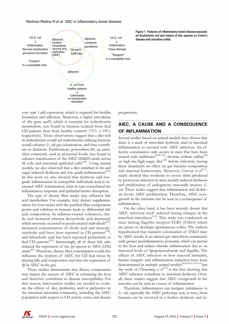
vors type 1 pili expression, which is required for biofilm formation and adhesion. Moreover, a higher prevalence of the gene malX, which is essential for maltodextrin metabolism, was found in bacteria isolated from ileal CD patients than from healthy controls (71% vs 18%, respectively). These observations suggest that a diet rich in maltodextrin would aid maltodextrin-utilizing bacteria, would enhance E. coli gut colonization, and thus contrib-ute to dysbiosis. Furthermore, polysorbate-80, an emul-sifier commonly used in processed foods, was found to enhance translocation of the AIEC HM605 strain across M cells and intestinal epithelial cells[105]. Using animal models, we also observed that a diet enriched in fat and sugar induced dysbiosis and low grade-inflammation[106]. In this work we also showed that dysbiosis and low-grade inflammation in susceptible individuals lead to in-creased AIEC colonization, what in turn exacerbated the inflammatory response and epithelial barrier disruption.
The type of dietary fiber intake may influence bile acid metabolism. For example, daily dietary supplemen-tation for four weeks with the purified fiber components pectin and cellulose in humans leads to differential bile acid composition. In cellulose-treated volunteers, cho-lic acid increased whereas deoxycholic acid decreased, which inversely occurred in pectin-treated individuals[107]. Increased concentrations of cholic acid and chenode-oxycholic acid have been reported in CD patients[108], and lithocholic acid has been reported particularly in ileal CD patients[109]. Interestingly, all of these bile salts induced the expression of the lpf operon in AIEC LF82 strain[86]. Therefore, dietary fiber consumption could also influence the tropism of AIEC for CD ileal tissue by altering bile acid composition and thus the expression of lpf in AIEC in the gut.
These studies demonstrate that dietary components may impact the success of AIEC in colonizing the host and therefore contribute to disease susceptibility. For that reason, intervention studies are needed to evalu-ate the effects of diet, probiotics, and/or prebiotics on the intestinal microbial community, including the AIEC population with respect to CD activity status and disease
progression.
AIEC, A CAUSE AND A CONSEQUENCE OF INFLAMMATIONSeveral studies based on animal models have shown that there is a need of microbial dysbiosis and/or intestinal inflammation to succeed with AIEC infection. An ef-fective colonization only occurs in mice that have been treated with antibiotics[50,81,110], dextran sodium sulfate[69] or high-fat/high-sugar diet[106] before infection, having these treatments an effect on gut bacteria composition and mucosal homeostasis. Moreover, Craven et al[111], nicely showed that moderate to severe ileitis produced by protozoan infection in mice models induced dysbiosis and proliferation of endogenous mucosally invasive E. coli. These works suggest that inflammation and dysbio-sis favors AIEC proliferation. Therefore, AIEC over-growth in the intestine can be seen as a consequence of inflammation.
On the other hand, it has been recently shown that AIEC infection itself induced lasting changes in the intestinal microbiota[112]. This study was conducted on mice lacking flagellin receptor TLR5 (T5KO) which are prone to develope spontaneous colitis. The authors hypothesized that transient colonization of T5KO mice by AIEC results in an altered gut microbiota community with greater proinflammatory potential, which can persist in the host and induce chronic inflammation due to its increased levels of lipopolysaccharide and flagellin. The effects of AIEC infection on host mucosal immunity, barrier integrity and inflammation induction have been demonstrated in multiple animal models[50,60,69,81,106,110] but the work of Chassaing et al[112] is the first showing that AIEC infection contribute to intestinal dysbiosis. Over-all, these studies suggest that AIEC overgrowth in the intestine can be seen as a cause of inflammation.
Therefore, inflammation can instigate imbalances in E. coli, especially the AIEC pathotype and, in turn, these bacteria can be involved in a further dysbiosis and in-
August 15, 2014|Volume 5|Issue 3|WJGP|www.wjgnet.com 222
CD-E. coli
InflammationMucosal translocationgranuloma formation
"Invasive"in a susceptible host
UC-E. coli
InflammationTissue damage
"Toxigenic"in a susceptible host
Adherentcytotoxins genotoxins
B2 and D ExPEC-like
Adherent
Adherentinvasive intracellular survival and replication (AIEC)
E. coli from healthy subjects
Colonization no translocation
mutualism
Figure 1 Features of inflammatory bowel disease-associat-ed Escherichia coli and impact of this species on Crohn’s disease and ulcerative colitis.
Martinez-Medina M et al . AIEC in inflammatory bowel diseases
creased intestinal inflammation.
CONCLUSIONSubstantial evidence indicates that E. coli is involved in CD and growing data suggest that this species is also a contributing factor in UC pathogenesis (Figure 1). Studies focused on defining virulence gene profiles of E. coli populations have shown that E. coli associated to the mucosa of healthy subjects resemble those of IBD patients. Genes related with adhesion, iron transport, capsule formation and toxins are present in E. coli from both healthy subjects and IBD patients. These features are thought to be necessary for an effective colonization of the intestinal tract. However, the intestinal microen-vironment in IBD patients, especially those in relapse, would predispose to E. coli proliferation. Moreover, E. coli from CD patients have probably evolved towards the AIEC pathotype, which has the capacity to adhere to and to invade intestinal epithelial cells, as well as to sur-vive and replicate within a number of cell types. Viru-lence properties of AIEC described to date can explain several features of CD pathophysiology such as inflam-mation, mucosal bacterial translocation and granuloma formation. Conversely, E. coli strains from UC patients appear to present a “toxigenic” behavior rather than the “invasive” pathogenic mechanism of CD- E. coli. Recent research has pointed out that E. coli from UC patients frequently carry virulence genes related to cytotoxic-ity and genotoxicity, which can contribute to mucosal inflammation and tissue damage. This is in accordance with previous works that did not found E. coli translocat-ing the epithelial barrier of UC patients, and could be linked with some aspects of UC pathophysiology.
Since the AIEC pathotype was defined one decade ago, substantial research has been conducted focusing on the identification of the mechanisms of pathogenic-ity and also in the field of epidemiology with regard to CD. However, additional epidemiologic studies are still needed to corroborate the role of AIEC in CD and to clarify the AIEC disease- and host-specificity. An impor-tant limitation to epidemiological studies is the absence of specific molecular tools to detect and quantify this pathotype, as the current available techniques to identify the AIEC pathotype are based exclusively on phenotypic screening of cultured bacteria, which is highly time-consuming. The execution of large-scale epidemiologic studies would also provide new insights into its distri-bution, putative reservoirs and transmission pathways. Moreover, the molecular bases of AIEC pathogenicity are still not fully understood, as only a few model strains have been studied and there is a wide variety of sero-pathotypes and phylotypes within the AIEC pathotype. Genomic and transcriptomic studies including wider and more diverse AIEC strain collections could assist in identifying new genetic elements associated with the AIEC phenotype, which may help us to gain a better understanding of the mechanisms of pathogenicity and
could result in significant advances in the detection of new therapeutic targets for CD.
REFERENCES1 Sartor RB. Microbial influences in inflammatory bowel dis-
eases. Gastroenterology 2008; 134: 577-594 [PMID: 18242222 DOI: 10.1053/j.gastro.2007.11.059]
2 Willing B, Halfvarson J, Dicksved J, Rosenquist M, Järnerot G, Engstrand L, Tysk C, Jansson JK. Twin studies reveal specific imbalances in the mucosa-associated microbiota of patients with ileal Crohn’s disease. Inflamm Bowel Dis 2009; 15: 653-660 [PMID: 19023901 DOI: 10.1002/ibd.20783]
3 Andoh A, Imaeda H, Aomatsu T, Inatomi O, Bamba S, Sa-saki M, Saito Y, Tsujikawa T, Fujiyama Y. Comparison of the fecal microbiota profiles between ulcerative colitis and Crohn’s disease using terminal restriction fragment length polymorphism analysis. J Gastroenterol 2011; 46: 479-486 [PMID: 21253779 DOI: 10.1007/s00535-010-0368-4]
4 Machiels K, Joossens M, Sabino J, De Preter V, Arijs I, Eeck-haut V, Ballet V, Claes K, Van Immerseel F, Verbeke K, Fer-rante M, Verhaegen J, Rutgeerts P, Vermeire S. A decrease of the butyrate-producing species Roseburia hominis and Faecalibacterium prausnitzii defines dysbiosis in patients with ulcerative colitis. Gut 2014; 63: 1275-1283 [PMID: 24021287 DOI: 10.1136/gutjnl-2013-304833]
5 Martinez-Medina M, Aldeguer X, Gonzalez-Huix F, Acero D, Garcia-Gil LJ. Abnormal microbiota composition in the ileocolonic mucosa of Crohn’s disease patients as revealed by polymerase chain reaction-denaturing gradient gel elec-trophoresis. Inflamm Bowel Dis 2006; 12: 1136-1145 [PMID: 17119388 DOI: 10.1097/01.mib.0000235828.09305.0c]
6 Baumgart M, Dogan B, Rishniw M, Weitzman G, Bosworth B, Yantiss R, Orsi RH, Wiedmann M, McDonough P, Kim SG, Berg D, Schukken Y, Scherl E, Simpson KW. Culture independent analysis of ileal mucosa reveals a selective in-crease in invasive Escherichia coli of novel phylogeny rela-tive to depletion of Clostridiales in Crohn’s disease involv-ing the ileum. ISME J 2007; 1: 403-418 [PMID: 18043660 DOI: 10.1038/ismej.2007.52]
7 Darfeuille-Michaud A, Neut C, Barnich N, Lederman E, Di Martino P, Desreumaux P, Gambiez L, Joly B, Cortot A, Colombel JF. Presence of adherent Escherichia coli strains in ileal mucosa of patients with Crohn’s disease. Gastroen-terology 1998; 115: 1405-1413 [PMID: 9834268 DOI: 10.1016/S0016-5085(98)70019-8]
8 Fujita H, Eishi Y, Ishige I, Saitoh K, Takizawa T, Arima T, Koike M. Quantitative analysis of bacterial DNA from My-cobacteria spp., Bacteroides vulgatus, and Escherichia coli in tissue samples from patients with inflammatory bowel diseases. J Gastroenterol 2002; 37: 509-516 [PMID: 12162408 DOI: 10.1007/s005350200079]
9 Gophna U, Sommerfeld K, Gophna S, Doolittle WF, Veld-huyzen van Zanten SJ. Differences between tissue-associ-ated intestinal microfloras of patients with Crohn’s disease and ulcerative colitis. J Clin Microbiol 2006; 44: 4136-4141 [PMID: 16988016 DOI: 10.1128/JCM.01004-06]
10 Kotlowski R, Bernstein CN, Sepehri S, Krause DO. High prevalence of Escherichia coli belonging to the B2+D phylo-genetic group in inflammatory bowel disease. Gut 2007; 56: 669-675 [PMID: 17028128 DOI: 10.1136/gut.2006.099796]
11 Lopez-Siles M, Martinez-Medina M, Busquets D, Sabat-Mir M, Duncan S, Flint H, Aldeguer X, Garcia-Gil L. Mucosa-associated Faecalibacterium prausnitzii and Escherichia coli co-abundance can distinguish Irritable Bowel Syndrome and Inflammatory Bowel Disease phenotypes. Int J Med Mi-crobiol 2014; [DOI: 10.1016/j.ijmm.2014.02.009]
12 Martin HM, Campbell BJ, Hart CA, Mpofu C, Nayar M, Singh R, Englyst H, Williams HF, Rhodes JM. Enhanced
August 15, 2014|Volume 5|Issue 3|WJGP|www.wjgnet.com 223
Martinez-Medina M et al . AIEC in inflammatory bowel diseases

Escherichia coli adherence and invasion in Crohn’s disease and colon cancer. Gastroenterology 2004; 127: 80-93 [PMID: 15236175 DOI: 10.1053/j.gastro.2004.03.054]
13 Martinez-Medina M, Aldeguer X, Lopez-Siles M, González-Huix F, López-Oliu C, Dahbi G, Blanco JE, Blanco J, Garcia-Gil LJ, Darfeuille-Michaud A. Molecular diversity of Escherichia coli in the human gut: new ecological evidence supporting the role of adherent-invasive E. coli (AIEC) in Crohn’s disease. Inflamm Bowel Dis 2009; 15: 872-882 [PMID: 19235912 DOI: 10.1002/ibd.20860]
14 Mylonaki M, Rayment NB, Rampton DS, Hudspith BN, Brostoff J. Molecular characterization of rectal mucosa-associated bacterial flora in inflammatory bowel disease. Inflamm Bowel Dis 2005; 11: 481-487 [PMID: 15867588 DOI: 10.1097/01.MIB.0000159663.62651.4f]
15 Neut C, Bulois P, Desreumaux P, Membré JM, Lederman E, Gambiez L, Cortot A, Quandalle P, van Kruiningen H, Colombel JF. Changes in the bacterial flora of the neoter-minal ileum after ileocolonic resection for Crohn’s disease. Am J Gastroenterol 2002; 97: 939-946 [PMID: 12003430 DOI: 10.1111/j.1572-0241.2002.05613.x]
16 Rehman A, Lepage P, Nolte A, Hellmig S, Schreiber S, Ott SJ. Transcriptional activity of the dominant gut mucosal microbiota in chronic inflammatory bowel disease patients. J Med Microbiol 2010; 59: 1114-1122 [PMID: 20522625 DOI: 10.1099/jmm.0.021170-0]
17 Sasaki M, Sitaraman SV, Babbin BA, Gerner-Smidt P, Ribot EM, Garrett N, Alpern JA, Akyildiz A, Theiss AL, Nusrat A, Klapproth JM. Invasive Escherichia coli are a feature of Crohn’s disease. Lab Invest 2007; 87: 1042-1054 [PMID: 17660846 DOI: 10.1038/labinvest.3700661]
18 Schwiertz A, Jacobi M, Frick JS, Richter M, Rusch K, Köhler H. Microbiota in pediatric inflammatory bowel disease. J Pediatr 2010; 157: 240-244.e1 [PMID: 20400104 DOI: 10.1016/j.jpeds.2010.02.046]
19 Beaven SW, Abreu MT. Biomarkers in inflammatory bowel disease. Curr Opin Gastroenterol 2004; 20: 318-327 [PMID: 15703659 DOI: 10.1097/00001574-200407000-00004]
20 Landers CJ, Cohavy O, Misra R, Yang H, Lin YC, Braun J, Targan SR. Selected loss of tolerance evidenced by Crohn’s disease-associated immune responses to auto- and mi-crobial antigens. Gastroenterology 2002; 123: 689-699 [PMID: 12198693 DOI: 10.1053/gast.2002.35379]
21 Mow WS, Vasiliauskas EA, Lin YC, Fleshner PR, Papada-kis KA, Taylor KD, Landers CJ, Abreu-Martin MT, Rotter JI, Yang H, Targan SR. Association of antibody responses to microbial antigens and complications of small bowel Crohn’s disease. Gastroenterology 2004; 126: 414-424 [PMID: 14762777 DOI: 10.1053/j.gastro.2003.11.015]
22 Sha S, Xu B, Wang X, Zhang Y, Wang H, Kong X, Zhu H, Wu K. The biodiversity and composition of the dominant fe-cal microbiota in patients with inflammatory bowel disease. Diagn Microbiol Infect Dis 2013; 75: 245-251 [PMID: 23276768 DOI: 10.1016/j.diagmicrobio.2012.11.022]
23 Pilarczyk-Zurek M, Chmielarczyk A, Gosiewski T, Tomu-siak A, Adamski P, Zwolinska-Wcislo M, Mach T, Heczko PB, Strus M. Possible role of Escherichia coli in propagation and perpetuation of chronic inflammation in ulcerative colitis. BMC Gastroenterol 2013; 13: 61 [PMID: 23566070 DOI: 10.1186/1471-230X-13-61]
24 Swidsinski A, Loening-Baucke V, Lochs H, Hale LP. Spa-tial organization of bacterial flora in normal and inflamed intestine: a fluorescence in situ hybridization study in mice. World J Gastroenterol 2005; 11: 1131-1140 [PMID: 15754393 DOI: 10.1128/JCM.43.7.3380-3389.2005]
25 Walmsley RS, Anthony A, Sim R, Pounder RE, Wakefield AJ. Absence of Escherichia coli, Listeria monocytogenes, and Klebsiella pneumoniae antigens within inflammatory bowel disease tissues. J Clin Pathol 1998; 51: 657-661 [PMID: 9930068 DOI: 10.1136/jcp.51.9.657]
26 Vasquez N, Mangin I, Lepage P, Seksik P, Duong JP, Blum S, Schiffrin E, Suau A, Allez M, Vernier G, Tréton X, Doré J, Marteau P, Pochart P. Patchy distribution of mucosal le-sions in ileal Crohn’s disease is not linked to differences in the dominant mucosa-associated bacteria: a study using fluorescence in situ hybridization and temporal tempera-ture gradient gel electrophoresis. Inflamm Bowel Dis 2007; 13: 684-692 [PMID: 17206669 DOI: 10.1002/ibd.20084]
27 Kleessen B, Kroesen AJ, Buhr HJ, Blaut M. Mucosal and invading bacteria in patients with inflammatory bowel dis-ease compared with controls. Scand J Gastroenterol 2002; 37: 1034-1041 [PMID: 12374228 DOI: 10.1080/003655202320378220]
28 O'Brien CL, Pavli P, Gordon DM, Allison GE. Detection of bacterial DNA in lymph nodes of Crohn’s disease patients us-ing high throughput sequencing. Gut 2014 Jan 15; Epub ahead of print [PMID: 24429583 DOI: 10.1136/gutjnl-2013-305320]
29 Ryan P, Bennett MW, Aarons S, Lee G, Collins JK, O’Sul-livan GC, O’Connell J, Shanahan F. PCR detection of Myco-bacterium paratuberculosis in Crohn’s disease granulomas isolated by laser capture microdissection. Gut 2002; 51: 665-670 [PMID: 12377804 DOI: 10.1136/gut.51.5.665]
30 Sepehri S, Khafipour E, Bernstein CN, Coombes BK, Pilar AV, Karmali M, Ziebell K, Krause DO. Characterization of Escherichia coli isolated from gut biopsies of newly diagnosed patients with inflammatory bowel disease. In-flamm Bowel Dis 2011; 17: 1451-1463 [PMID: 21674703 DOI: 10.1002/ibd.21509]
31 Dreux N, Denizot J, Martinez-Medina M, Mellmann A, Billig M, Kisiela D, Chattopadhyay S, Sokurenko E, Neut C, Gower-Rousseau C, Colombel JF, Bonnet R, Darfeuille-Michaud A, Barnich N. Point mutations in FimH adhesin of Crohn’s disease-associated adherent-invasive Escherichia coli enhance intestinal inflammatory response. PLoS Pathog 2013; 9: e1003141 [PMID: 23358328 DOI: 10.1371/journal.ppat.1003141]
32 Petersen AM, Nielsen EM, Litrup E, Brynskov J, Mirse-pasi H, Krogfelt KA. A phylogenetic group of Escherichia coli associated with active left-sided inflammatory bowel disease. BMC Microbiol 2009; 9: 171 [PMID: 19695087 DOI: 10.1186/1471-2180-9-171]
33 Sepehri S, Kotlowski R, Bernstein CN, Krause DO. Phylo-genetic analysis of inflammatory bowel disease associated Escherichia coli and the fimH virulence determinant. In-flamm Bowel Dis 2009; 15: 1737-1745 [PMID: 19462430 DOI: 10.1002/ibd.20966]
34 Schippa S, Conte MP, Borrelli O, Iebba V, Aleandri M, Se-ganti L, Longhi C, Chiarini F, Osborn J, Cucchiara S. Domi-nant genotypes in mucosa-associated Escherichia coli strains from pediatric patients with inflammatory bowel disease. Inflamm Bowel Dis 2009; 15: 661-672 [PMID: 19067417 DOI: 10.1002/ibd.20818]
35 Nowrouzian FL, Adlerberth I, Wold AE. Enhanced persis-tence in the colonic microbiota of Escherichia coli strains belonging to phylogenetic group B2: role of virulence fac-tors and adherence to colonic cells. Microbes Infect 2006; 8: 834-840 [PMID: 16483819 DOI: 10.1016/j.micinf.2005.10.011]
36 Elliott TR, Hudspith BN, Wu G, Cooley M, Parkes G, Qui-ñones B, Randall L, Mandrell RE, Fagerquist CK, Brostoff J, Rayment NB, Boussioutas A, Petrovska L, Sanderson JD. Quantification and characterization of mucosa-associated and intracellular Escherichia coli in inflammatory bowel dis-ease. Inflamm Bowel Dis 2013; 19: 2326-2338 [PMID: 23989750 DOI: 10.1097/MIB.0b013e3182a38a92]
37 Gombošová L , Lazúrová I, Zakuciová M, Curová K, Kmeťová M, Petrášová D, Siegfried L. Genes of intestinal Escherichia coli and their relation to the inflammatory activ-ity in patients with ulcerative colitis and Crohn’s disease. Folia Microbiol (Praha) 2011; 56: 367-372 [PMID: 21877213 DOI: 10.1007/s12223-011-0051-z]
August 15, 2014|Volume 5|Issue 3|WJGP|www.wjgnet.com 224
Martinez-Medina M et al . AIEC in inflammatory bowel diseases
38 Schultsz C, Moussa M, van Ketel R, Tytgat GN, Dankert J. Frequency of pathogenic and enteroadherent Escherichia coli in patients with inflammatory bowel disease and con-trols. J Clin Pathol 1997; 50: 573-579 [PMID: 9306938 DOI: 10.1136/jcp.50.7.573]
39 Vejborg RM, Hancock V, Petersen AM, Krogfelt KA, Kl-emm P. Comparative genomics of Escherichia coli isolated from patients with inflammatory bowel disease. BMC Ge-nomics 2011; 12: 316 [PMID: 21676223 DOI: 10.1186/1471-2164-12-316]
40 Nowrouzian F, Adlerberth I, Wold AE. P fimbriae, capsule and aerobactin characterize colonic resident Escherichia coli. Epidemiol Infect 2001; 126: 11-18 [PMID: 11293669 DOI: 10.1017/S0950268801005118]
41 de Souza HL, de Carvalho VR, Romeiro FG, Sassaki LY, Keller R, Rodrigues J. Mucosa-associated but not luminal Escherichia coli is augmented in Crohn’s disease and ulcer-ative colitis. Gut Pathog 2012; 4: 21 [PMID: 23234341 DOI: 10.1186/1757-4749-4-21]
42 Sobieszczańska BA, Duda-Madej AB, Turniak MB, Franic-zek R, Kasprzykowska U, Duda AK, Rzeszutko M, Iwańczak B. Invasive properties, adhesion patterns and phylogroup profiles among Escherichia coli strains isolated from children with inflammatory bowel disease. Adv Clin Exp Med 2012; 21: 591-599 [PMID: 23356195]
43 Tenaillon O, Skurnik D, Picard B, Denamur E. The popula-tion genetics of commensal Escherichia coli. Nat Rev Micro-biol 2010; 8: 207-217 [PMID: 20157339 DOI: 10.1038/nrmi-cro2298]
44 Curová K, Kmetová M, Sabol M, Gombosová L, Lazúrová I, Siegfried L. Enterovirulent E. coli in inflammatory and non-inflammatory bowel diseases. Folia Microbiol (Praha) 2009; 54: 81-86 [PMID: 19330549 DOI: 10.1007/s12223-009-0012-y]
45 Darfeuille-Michaud A, Boudeau J, Bulois P, Neut C, Glass-er AL, Barnich N, Bringer MA, Swidsinski A, Beaugerie L, Colombel JF. High prevalence of adherent-invasive Esch-erichia coli associated with ileal mucosa in Crohn’s disease. Gastroenterology 2004; 127: 412-421 [PMID: 15300573 DOI: 10.1053/j.gastro.2004.04.061]
46 Miquel S, Peyretaillade E, Claret L, de Vallée A, Dossat C, Vacherie B, Zineb el H, Segurens B, Barbe V, Sauvanet P, Neut C, Colombel JF, Medigue C, Mojica FJ, Peyret P, Bon-net R, Darfeuille-Michaud A. Complete genome sequence of Crohn’s disease-associated adherent-invasive E. coli strain LF82. PLoS One 2010; 5: e12714 [PMID: 20862302 DOI: 10.1371/journal.pone.0012714]
47 Nash JH, Villegas A, Kropinski AM, Aguilar-Valenzuela R, Konczy P, Mascarenhas M, Ziebell K, Torres AG, Karmali MA, Coombes BK. Genome sequence of adherent-invasive Escherichia coli and comparative genomic analysis with other E. coli pathotypes. BMC Genomics 2010; 11: 667 [PMID: 21108814 DOI: 10.1186/1471-2164-11-667]
48 Rolhion N, Barnich N, Bringer MA, Glasser AL, Ranc J, Hébuterne X, Hofman P, Darfeuille-Michaud A. Abnor-mally expressed ER stress response chaperone Gp96 in CD favours adherent-invasive Escherichia coli invasion. Gut 2010; 59: 1355-1362 [PMID: 20587550 DOI: 10.1136/gut.2010.207456]
49 Barnich N, Carvalho FA, Glasser AL, Darcha C, Jantscheff P, Allez M, Peeters H, Bommelaer G, Desreumaux P, Colom-bel JF, Darfeuille-Michaud A. CEACAM6 acts as a receptor for adherent-invasive E. coli, supporting ileal mucosa colo-nization in Crohn disease. J Clin Invest 2007; 117: 1566-1574 [PMID: 17525800 DOI: 10.1172/JCI30504]
50 Carvalho FA, Barnich N, Sivignon A, Darcha C, Chan CH, Stanners CP, Darfeuille-Michaud A. Crohn’s disease ad-herent-invasive Escherichia coli colonize and induce strong gut inflammation in transgenic mice expressing human CEACAM. J Exp Med 2009; 206: 2179-2189 [PMID: 19737864 DOI: 10.1084/jem.20090741]
51 Eaves-Pyles T, Allen CA, Taormina J, Swidsinski A, Tutt CB, Jezek GE, Islas-Islas M, Torres AG. Escherichia coli isolated from a Crohn’s disease patient adheres, invades, and induces inflammatory responses in polarized intestinal epithelial cells. Int J Med Microbiol 2008; 298: 397-409 [PMID: 17900983 DOI: 10.1016/j.ijmm.2007.05.011]
52 Subramanian S, Rhodes JM, Hart CA, Tam B, Roberts CL, Smith SL, Corkill JE, Winstanley C, Virji M, Campbell BJ. Characterization of epithelial IL-8 response to inflammatory bowel disease mucosal E. coli and its inhibition by mesala-mine. Inflamm Bowel Dis 2008; 14: 162-175 [PMID: 17941093 DOI: 10.1002/ibd.20296]
53 Mimouna S, Gonçalvès D, Barnich N, Darfeuille-Michaud A, Hofman P, Vouret-Craviari V. Crohn disease-associated Escherichia coli promote gastrointestinal inflammatory disor-ders by activation of HIF-dependent responses. Gut Microbes 2011; 2: 335-346 [PMID: 22157238 DOI: 10.4161/gmic.18771]
54 Stevens C, Henderson P, Nimmo ER, Soares DC, Dogan B, Simpson KW, Barrett JC, Wilson DC, Satsangi J. The intermediate filament protein, vimentin, is a regulator of NOD2 activity. Gut 2013; 62: 695-707 [PMID: 22684479 DOI: 10.1136/gutjnl-2011-301775]
55 Low D, Tran HT, Lee IA, Dreux N, Kamba A, Reinecker HC, Darfeuille-Michaud A, Barnich N, Mizoguchi E. Chitin-binding domains of Escherichia coli ChiA mediate interac-tions with intestinal epithelial cells in mice with colitis. Gastroenterology 2013; 145: 602-612.e9 [PMID: 23684751 DOI: 10.1053/j.gastro.2013.05.017]
56 Miquel S, Claret L, Bonnet R, Dorboz I, Barnich N, Dar-feuille-Michaud A. Role of decreased levels of Fis histone-like protein in Crohn’s disease-associated adherent invasive Escherichia coli LF82 bacteria interacting with intestinal epi-thelial cells. J Bacteriol 2010; 192: 1832-1843 [PMID: 20118249 DOI: 10.1128/JB.01679-09]
57 Lapaquette P, Glasser AL, Huett A, Xavier RJ, Darfeuille-Michaud A. Crohn’s disease-associated adherent-invasive E. coli are selectively favoured by impaired autophagy to rep-licate intracellularly. Cell Microbiol 2010; 12: 99-113 [PMID: 19747213 DOI: 10.1111/j.1462-5822.2009.01381.x]
58 Nguyen HT, Dalmasso G, Müller S, Carrière J, Seibold F, Darfeuille-Michaud A. Crohn’s disease-associated adherent invasive Escherichia coli modulate levels of microRNAs in intestinal epithelial cells to reduce autophagy. Gastroen-terology 2014; 146: 508-519 [PMID: 24148619 DOI: 10.1053/j.gastro.2013.10.021]
59 Chassaing B, Rolhion N, de Vallée A, Salim SY, Prorok-Hamon M, Neut C, Campbell BJ, Söderholm JD, Hugot JP, Colombel JF, Darfeuille-Michaud A. Crohn disease--asso-ciated adherent-invasive E. coli bacteria target mouse and human Peyer’s patches via long polar fimbriae. J Clin Invest 2011; 121: 966-975 [PMID: 21339647 DOI: 10.1172/JCI44632]
60 Denizot J, Sivignon A, Barreau F, Darcha C, Chan HF, Stanners CP, Hofman P, Darfeuille-Michaud A, Barnich N. Adherent-invasive Escherichia coli induce claudin-2 expres-sion and barrier defect in CEABAC10 mice and Crohn’s disease patients. Inflamm Bowel Dis 2012; 18: 294-304 [PMID: 21688348 DOI: 10.1002/ibd.21787]
61 Wine E, Ossa JC, Gray-Owen SD, Sherman PM. Adherent-invasive Escherichia coli, strain LF82 disrupts apical junc-tional complexes in polarized epithelia. BMC Microbiol 2009; 9: 180 [PMID: 19709415 DOI: 10.1186/1471-2180-9-180]
62 Mankertz J, Amasheh M, Krug SM, Fromm A, Amasheh S, Hillenbrand B, Tavalali S, Fromm M, Schulzke JD. TNFal-pha up-regulates claudin-2 expression in epithelial HT-29/B6 cells via phosphatidylinositol-3-kinase signaling. Cell Tissue Res 2009; 336: 67-77 [PMID: 19214581 DOI: 10.1007/s00441-009-0751-8]
63 Ossa JC , Ho NK, Wine E, Leung N, Gray-Owen SD, Sherman PM. Adherent-invasive Escherichia coli blocks interferon-γ-induced signal transducer and activator of tran-
August 15, 2014|Volume 5|Issue 3|WJGP|www.wjgnet.com 225
Martinez-Medina M et al . AIEC in inflammatory bowel diseases

scription (STAT)-1 in human intestinal epithelial cells. Cell Microbiol 2013; 15: 446-457 [PMID: 23072252 DOI: 10.1111/cmi.12048]
64 Bringer MA, Glasser AL, Tung CH, Méresse S, Darfeuille-Mi-chaud A. The Crohn’s disease-associated adherent-invasive Escherichia coli strain LF82 replicates in mature phagolyso-somes within J774 macrophages. Cell Microbiol 2006; 8: 471-484 [PMID: 16469058 DOI: 10.1111/j.1462-5822.2005.00639.x]
65 Bringer MA, Barnich N, Glasser AL, Bardot O, Darfeuille-Michaud A. HtrA stress protein is involved in intramacro-phagic replication of adherent and invasive Escherichia coli strain LF82 isolated from a patient with Crohn’s disease. In-fect Immun 2005; 73: 712-721 [PMID: 15664909 DOI: 10.1128/IAI.73.2.712-721.2005]
66 Bringer MA, Rolhion N, Glasser AL, Darfeuille-Michaud A. The oxidoreductase DsbA plays a key role in the ability of the Crohn’s disease-associated adherent-invasive Esch-erichia coli strain LF82 to resist macrophage killing. J Bac-teriol 2007; 189: 4860-4871 [PMID: 17449627 DOI: 10.1128/JB.00233-07]
67 Simonsen KT, Nielsen G, Bjerrum JV, Kruse T, Kallipolitis BH, Møller-Jensen J. A role for the RNA chaperone Hfq in controlling adherent-invasive Escherichia coli colonization and virulence. PLoS One 2011; 6: e16387 [PMID: 21298102 DOI: 10.1371/journal.pone.0016387]
68 Glasser AL, Boudeau J, Barnich N, Perruchot MH, Colom-bel JF, Darfeuille-Michaud A. Adherent invasive Escherichia coli strains from patients with Crohn’s disease survive and replicate within macrophages without inducing host cell death. Infect Immun 2001; 69: 5529-5537 [PMID: 11500426 DOI: 10.1128/IAI.69.9.5529-5537.2001]
69 Carvalho FA, Barnich N, Sauvanet P, Darcha C, Gelot A, Darfeuille-Michaud A. Crohn’s disease-associated Esche-richia coli LF82 aggravates colitis in injured mouse colon via signaling by flagellin. Inflamm Bowel Dis 2008; 14: 1051-1060 [PMID: 18338780 DOI: 10.1002/ibd.20423]
70 Meconi S, Vercellone A, Levillain F, Payré B, Al Saati T, Capilla F, Desreumaux P, Darfeuille-Michaud A, Altare F. Adherent-invasive Escherichia coli isolated from Crohn’s disease patients induce granulomas in vitro. Cell Mi-crobiol 2007; 9: 1252-1261 [PMID: 17223928 DOI: 10.1111/j.1462-5822.2006.00868.x]
71 Dunne KA, Allam A, McIntosh A, Houston SA, Cerovic V, Goodyear CS, Roe AJ, Beatson SA, Milling SW, Walker D, Wall DM. Increased S-nitrosylation and proteasomal degradation of caspase-3 during infection contribute to the persistence of adherent invasive Escherichia coli (AIEC) in immune cells. PLoS One 2013; 8: e68386 [PMID: 23861899 DOI: 10.1371/journal.pone.0068386]
72 Peeters H, Bogaert S, Laukens D, Rottiers P, De Keyser F, Darfeuille-Michaud A, Glasser AL, Elewaut D, De Vos M. CARD15 variants determine a disturbed early response of monocytes to adherent-invasive Escherichia coli strain LF82 in Crohn’s disease. Int J Immunogenet 2007; 34: 181-191 [PMID: 17504508 DOI: 10.1111/j.1744-313X.2007.00670.x]
73 Nieminen JK, Sipponen T, Färkkilä M, Vaarala O. Mono-cyte-derived dendritic cells from Crohn’s disease patients ex-hibit decreased ability to activate T helper type 17 responses in memory cells. Clin Exp Immunol 2014; 177: 190-202 [PMID: 24635023 DOI: 10.1111/cei.12326]
74 Chargui A, Cesaro A, Mimouna S, Fareh M, Brest P, Naquet P, Darfeuille-Michaud A, Hébuterne X, Mograbi B, Vouret-Craviari V, Hofman P. Subversion of autophagy in adherent invasive Escherichia coli-infected neutrophils induces in-flammation and cell death. PLoS One 2012; 7: e51727 [PMID: 23272151 DOI: 10.1371/journal.pone.0051727]
75 Martinez-Medina M, Naves P, Blanco J, Aldeguer X, Blanco JE, Blanco M, Ponte C, Soriano F, Darfeuille-Michaud A, Gar-cia-Gil LJ. Biofilm formation as a novel phenotypic feature of adherent-invasive Escherichia coli (AIEC). BMC Microbiol
2009; 9: 202 [PMID: 19772580 DOI: 10.1186/1471-2180-9-202]76 Chassaing B, Darfeuille-Michaud A. The σE pathway is
involved in biofilm formation by Crohn’s disease-associated adherent-invasive Escherichia coli. J Bacteriol 2013; 195: 76-84 [PMID: 23104802 DOI: 10.1128/JB.01079-12]
77 Prorok-Hamon M, Friswell MK, Alswied A, Roberts CL, Song F, Flanagan PK, Knight P, Codling C, Marchesi JR, Winstanley C, Hall N, Rhodes JM, Campbell BJ. Colonic mucosa-associated diffusely adherent afaC+ Escherichia coli expressing lpfA and pks are increased in inflammatory bowel disease and colon cancer. Gut 2014; 63: 761-770 [PMID: 23846483 DOI: 10.1136/gutjnl-2013-304739]
78 Martinez-Medina M, Mora A, Blanco M, López C, Alonso MP, Bonacorsi S, Nicolas-Chanoine MH, Darfeuille-Mi-chaud A, Garcia-Gil J, Blanco J. Similarity and divergence among adherent-invasive Escherichia coli and extraint-estinal pathogenic E. coli strains. J Clin Microbiol 2009; 47: 3968-3979 [PMID: 19828750 DOI: 10.1128/JCM.01484-09]
79 Clarke DJ, Chaudhuri RR, Martin HM, Campbell BJ, Rhodes JM, Constantinidou C, Pallen MJ, Loman NJ, Cun-ningham AF, Browning DF, Henderson IR. Complete ge-nome sequence of the Crohn’s disease-associated adherent-invasive Escherichia coli strain HM605. J Bacteriol 2011; 193: 4540 [PMID: 21705601 DOI: 10.1128/JB.05374-11]
80 Krause DO, Little AC, Dowd SE, Bernstein CN. Complete genome sequence of adherent invasive Escherichia coli UM146 isolated from Ileal Crohn’s disease biopsy tissue. J Bacteriol 2011; 193: 583 [PMID: 21075930 DOI: 10.1128/JB.01290-10]
81 Small CL, Reid-Yu SA, McPhee JB, Coombes BK. Persistent infection with Crohn’s disease-associated adherent-invasive Escherichia coli leads to chronic inflammation and intestinal fibrosis. Nat Commun 2013; 4: 1957 [PMID: 23748852 DOI: 10.1038/ncomms2957]
82 Dogan B, Scherl E, Bosworth B, Yantiss R, Altier C, Mc-Donough PL, Jiang ZD, Dupont HL, Garneau P, Harel J, Rishniw M, Simpson KW. Multidrug resistance is common in Escherichia coli associated with ileal Crohn’s disease. Inflamm Bowel Dis 2013; 19: 141-150 [PMID: 22508665 DOI: 10.1002/ibd.22971]
83 Seksik P, Rigottier-Gois L, Gramet G, Sutren M, Pochart P, Marteau P, Jian R, Doré J. Alterations of the dominant fae-cal bacterial groups in patients with Crohn’s disease of the colon. Gut 2003; 52: 237-242 [PMID: 12524406 DOI: 10.1136/gut.52.2.237]
84 Jensen SR, Fink LN, Nielsen OH, Brynskov J, Brix S. Ex vivo intestinal adhesion of Escherichia coli LF82 in Crohn’s disease. Microb Pathog 2011; 51: 426-431 [PMID: 21911052 DOI: 10.1016/j.micpath.2011.08.006]
85 Lapaquette P, Bringer MA, Darfeuille-Michaud A. Defects in autophagy favour adherent-invasive Escherichia coli persistence within macrophages leading to increased pro-inflammatory response. Cell Microbiol 2012; 14: 791-807 [PMID: 22309232 DOI: 10.1111/j.1462-5822.2012.01768.x]
86 Chassaing B, Etienne-Mesmin L, Bonnet R, Darfeuille-Michaud A. Bile salts induce long polar fimbriae expression favouring Crohn’s disease-associated adherent-invasive Escherichia coli interaction with Peyer’s patches. Environ Microbiol 2013; 15: 355-371 [PMID: 22789019 DOI: 10.1111/j.1462-2920.2012.02824.x]
87 Vazeille E, Bringer MA, Gardarin A, Chambon C, Becker-Pauly C, Pender SL, Jakob C, Müller S, Lottaz D, Darfeuille-Michaud A. Role of meprins to protect ileal mucosa of Crohn’s disease patients from colonization by adherent-invasive E. coli. PLoS One 2011; 6: e21199 [PMID: 21698174 DOI: 10.1371/journal.pone.0021199]
88 Burke DA, Axon AT. Adhesive Escherichia coli in inflam-matory bowel disease and infective diarrhoea. BMJ 1988; 297: 102-104 [PMID: 3044496 DOI: 10.1136/bmj.297.6641.102]
89 Giaffer MH, Holdsworth CD, Duerden BI. Virulence prop-
August 15, 2014|Volume 5|Issue 3|WJGP|www.wjgnet.com 226
Martinez-Medina M et al . AIEC in inflammatory bowel diseases

erties of Escherichia coli strains isolated from patients with inflammatory bowel disease. Gut 1992; 33: 646-650 [PMID: 1612481 DOI: 10.1136/gut.33.5.646]
90 Thomazini CM, Samegima DA, Rodrigues MA, Victoria CR, Rodrigues J. High prevalence of aggregative adherent Escherichia coli strains in the mucosa-associated microbiota of patients with inflammatory bowel diseases. Int J Med Microbiol 2011; 301: 475-479 [PMID: 21616711 DOI: 10.1016/j.ijmm.2011.04.015]
91 Iebba V, Conte MP, Lepanto MS, Di Nardo G, Santangelo F, Aloi M, Totino V, Checchi MP, Longhi C, Cucchiara S, Schippa S. Microevolution in fimH gene of mucosa-associ-ated Escherichia coli strains isolated from pediatric patients with inflammatory bowel disease. Infect Immun 2012; 80: 1408-1417 [PMID: 22290143 DOI: 10.1128/IAI.06181-11]
92 Raso T, Crivellaro S, Chirillo MG, Pais P, Gaia E, Savoia D. Analysis of Escherichia coli isolated from patients affected by Crohn’s disease. Curr Microbiol 2011; 63: 131-137 [PMID: 21626145 DOI: 10.1007/s00284-011-9947-8]
93 Negroni A, Costanzo M, Vitali R, Superti F, Bertuccini L, Tinari A, Minelli F, Di Nardo G, Nuti F, Pierdomenico M, Cucchiara S, Stronati L. Characterization of adherent-invasive Escherichia coli isolated from pediatric patients with inflammatory bowel disease. Inflamm Bowel Dis 2012; 18: 913-924 [PMID: 21994005 DOI: 10.1002/ibd.21899]
94 Wang T, Cai G, Qiu Y, Fei N, Zhang M, Pang X, Jia W, Cai S, Zhao L. Structural segregation of gut microbiota between colorectal cancer patients and healthy volunteers. ISME J 2012; 6: 320-329 [PMID: 21850056 DOI: 10.1038/is-mej.2011.109]
95 Swidsinski A, Khilkin M, Kerjaschki D, Schreiber S, Ortner M, Weber J, Lochs H. Association between intraepithelial Esch-erichia coli and colorectal cancer. Gastroenterology 1998; 115: 281-286 [PMID: 9679033 DOI: 10.1016/S0016-5085(98)70194-5]
96 Bonnet M, Buc E, Sauvanet P, Darcha C, Dubois D, Pereira B, Déchelotte P, Bonnet R, Pezet D, Darfeuille-Michaud A. Colonization of the human gut by E. coli and colorectal can-cer risk. Clin Cancer Res 2014; 20: 859-867 [PMID: 24334760 DOI: 10.1158/1078-0432.CCR-13-1343]
97 Buc E, Dubois D, Sauvanet P, Raisch J, Delmas J, Darfeuille-Michaud A, Pezet D, Bonnet R. High prevalence of mucosa-associated E. coli producing cyclomodulin and genotoxin in colon cancer. PLoS One 2013; 8: e56964 [PMID: 23457644 DOI: 10.1371/journal.pone.0056964]
98 Schippa S, Iebba V, Barbato M, Di Nardo G, Totino V, Chec-chi MP, Longhi C, Maiella G, Cucchiara S, Conte MP. A dis-tinctive ‘microbial signature’ in celiac pediatric patients. BMC Microbiol 2010; 10: 175 [PMID: 20565734 DOI: 10.1186/1471-2180-10-175]
99 Nadal I, Donat E, Ribes-Koninckx C, Calabuig M, Sanz Y. Imbalance in the composition of the duodenal microbiota of children with coeliac disease. J Med Microbiol 2007; 56: 1669-1674 [PMID: 18033837 DOI: 10.1099/jmm.0.47410-0]
100 De Palma G, Nadal I, Medina M, Donat E, Ribes-Koninckx C, Calabuig M, Sanz Y. Intestinal dysbiosis and reduced im-munoglobulin-coated bacteria associated with coeliac disease in children. BMC Microbiol 2010; 10: 63 [PMID: 20181275 DOI: 10.1186/1471-2180-10-63]
101 Sánchez E, Nadal I, Donat E, Ribes-Koninckx C, Calabuig M, Sanz Y. Reduced diversity and increased virulence-gene carriage in intestinal enterobacteria of coeliac chil-dren. BMC Gastroenterol 2008; 8: 50 [PMID: 18983674 DOI: 10.1186/1471-230X-8-50]
102 Martinez-Medina M, Garcia-Gil J, Barnich N, Wieler LH, Ewers C. Adherent-invasive Escherichia coli phenotype displayed by intestinal pathogenic E. coli strains from cats, dogs, and swine. Appl Environ Microbiol 2011; 77: 5813-5817 [PMID: 21705530 DOI: 10.1128/AEM.02614-10]
103 Simpson KW, Dogan B, Rishniw M, Goldstein RE, Klaes-sig S, McDonough PL, German AJ, Yates RM, Russell DG, Johnson SE, Berg DE, Harel J, Bruant G, McDonough SP, Schukken YH. Adherent and invasive Escherichia coli is associated with granulomatous colitis in boxer dogs. Infect Immun 2006; 74: 4778-4792 [PMID: 16861666 DOI: 10.1128/IAI.00067-06]
104 Nickerson KP, McDonald C. Crohn’s disease-associated adherent-invasive Escherichia coli adhesion is enhanced by exposure to the ubiquitous dietary polysaccharide malto-dextrin. PLoS One 2012; 7: e52132 [PMID: 23251695 DOI: 10.1371/journal.pone.0052132]
105 Roberts CL, Keita AV, Duncan SH, O’Kennedy N, Söder-holm JD, Rhodes JM, Campbell BJ. Translocation of Crohn’s disease Escherichia coli across M-cells: contrasting effects of soluble plant fibres and emulsifiers. Gut 2010; 59: 1331-1339 [PMID: 20813719 DOI: 10.1136/gut.2009.195370]
106 Martinez-Medina M, Denizot J, Dreux N, Robin F, Billard E, Bonnet R, Darfeuille-Michaud A, Barnich N. Western diet induces dysbiosis with increased E coli in CEABAC10 mice, alters host barrier function favouring AIEC colonisa-tion. Gut 2014; 63: 116-124 [PMID: 23598352 DOI: 10.1136/gutjnl-2012-304119]
107 Hillman LC, Peters SG, Fisher CA, Pomare EW. Effects of the fibre components pectin, cellulose, and lignin on bile salt metabolism and biliary lipid composition in man. Gut 1986; 27: 29-36 [PMID: 3005138 DOI: 10.1136/gut.27.1.29]
108 Rutgeerts P, Ghoos Y, Vantrappen G, Fevery J. Biliary lipid composition in patients with nonoperated Crohn’s disease. Dig Dis Sci 1986; 31: 27-32 [PMID: 3940821 DOI: 10.1007/BF01347906]
109 Lapidus A, Akerlund JE, Einarsson C. Gallbladder bile com-position in patients with Crohn ‘s disease. World J Gastroen-terol 2006; 12: 70-74 [PMID: 16440420]
110 Drouet M, Vignal C, Singer E, Djouina M, Dubreuil L, Cor-tot A, Desreumaux P, Neut C. AIEC colonization and patho-genicity: influence of previous antibiotic treatment and pre-existing inflammation. Inflamm Bowel Dis 2012; 18: 1923-1931 [PMID: 22344932 DOI: 10.1002/ibd.22908]
111 Craven M, Egan CE, Dowd SE, McDonough SP, Dogan B, Denkers EY, Bowman D, Scherl EJ, Simpson KW. Inflamma-tion drives dysbiosis and bacterial invasion in murine mod-els of ileal Crohn’s disease. PLoS One 2012; 7: e41594 [PMID: 22848538 DOI: 10.1371/journal.pone.0041594]
112 Chassaing B, Koren O, Carvalho FA, Ley RE, Gewirtz AT. AIEC pathobiont instigates chronic colitis in susceptible hosts by altering microbiota composition. Gut 2014; 63: 1069-1080 [PMID: 23896971 DOI: 10.1136/gutjnl-2013-304909]
P- Reviewer: Gassler N, Silva MA S- Editor: Song XX L- Editor: A E- Editor: Lu YJ
August 15, 2014|Volume 5|Issue 3|WJGP|www.wjgnet.com 227
Martinez-Medina M et al . AIEC in inflammatory bowel diseases
TOPIC HIGHLIGHT
Similarities and differences between Behçet's disease and Crohn's disease
Veli Yazısız
Veli Yazısız, Division of Rheumatology, Department of Internal Medicine, Medical School, Akdeniz University, Kampus 07058, Antalya, TurkeyAuthor contributions: Yazısız V contributed to this paper.Correspondence to: Veli Yazısız, MD, Associate Profes-sor, Division of Rheumatology, Department of Internal Medi-cine, Medical School, Akdeniz Universitesi, Tıp Fakültesi, İç Hastalıkları AD, Romatoloji BD, Dumlupınar Bulvarı, Kampus 07058, Antalya, Turkey. [email protected]: +90-505-3149901 Fax: +90-242-2496040Received: January 2, 2014 Revised: March 9, 2014 Accepted: May 29, 2014Published online: August 15, 2014
AbstractBehçet’s disease (BD) is a chronic inflammatory con-dition with multisystem involvement. Approximately 10%-15% of patients present with gastrointestinal involvement. Involved sites and the endoscopic view usually resemble Crohn’s disease (CD). In addition to intestinal involvement, oral mucosa, the eyes, skin, and joints are commonly affected. No pathognomonic laboratory test is available for the diagnosis of either disease. Management approaches are also similar in various aspects. Differentiating BD from CD is highly challenging. In this article, the similarities and differ-ences between BD and CD in terms of epidemiology, etiopathogenesis, clinical and imaging findings, and histopathological and therapeutic approaches are re-viewed.
© 2014 Baishideng Publishing Group Inc. All rights reserved.
Key words: Behçet’s disease; Crohn’s disease
Core tip: Behçet’s disease and Crohn’s disease are chronic inflammatory conditions caused by lesions similar to those seen in the bowels. There are similar
and different clinical findings, however both diseases show intestinal inflammation. The differential diagnosis may be difficult when the symptoms of the two disease processes are very similar. This review focuses on the similar and different characteristics of Behçet’s disease and Crohn’s disease.
Yazısız V. Similarities and differences between Behçet’s disease and Crohn’s disease. World J Gastrointest Pathophysiol 2014; 5(3): 228-238 Available from: URL: http://www.wjgnet.com/2150-5330/full/v5/i3/228.htm DOI: http://dx.doi.org/10.4291/wjgp.v5.i3.228
INTRODUCTIONBehçet’s disease (BD), which was first defined by Hulusi Behçet, a Turkish dermatologist, in 1937, is a chronic inflammatory disease with multisystem involvement[1]. It presents with remission and exacerbation of muco-cutaneous, ocular, articular, vascular, or gastrointestinal lesions. Crohn’s disease (CD), on the other hand, is a chronic relapsing inflammatory disorder of the gastro-intestinal tract, presenting with BD-like extra-intestinal manifestations[2]. Both of these chronic immune-mediat-ed inflammatory disorders are likely to affect patients at a younger age accompanied by fluctuating courses. It is possible that a patient with CD meets the criteria for BD. The differential diagnosis in some cases is quite difficult, particularly in the presence of gastrointestinal involve-ment. Differentiation is usually based on the involve-ment of different organs. This review aims to investigate the similar and different characteristics of BD and CD.
EPIDEMIOLOGYThe prevalence of BD varies geographically and the
World J Gastrointest Pathophysiol 2014 August 15; 5(3): 228-238ISSN 2150-5330 (online)
© 2014 Baishideng Publishing Group Inc. All rights reserved.
Online Submissions: http://www.wjgnet.com/esps/Help Desk: http://www.wjgnet.com/esps/helpdesk.aspxDOI: 10.4291/wjgp.v5.i3.228
August 15, 2014|Volume 5|Issue 3|WJGP|www.wjgnet.com 228
WJGP 5th Anniversary Special Issues (6): Crohn’s disease
Yazısız V. Behçet’s or Crohn’s disease?
disease is more prevalent in certain groups. It is most common in populations clustered along the ancient Silk Road. Turkey has the highest prevalence (80-370 cases/100000), followed by Asia and the Middle East-ern countries, including Israel, Saudi Arabia and Iran. BD can be seen in all countries worldwide due to im-migration[3-6]. The age at onset of the disease is usually between 20.8 and 40 years, as it is more common in young individuals[3]. Patients aged 16 years with initial symptoms, considered as childhood-onset BD, have also been reported. The male-to-female ratio varies region-ally. The disease is more common among men in Russia, Saudi Arabia, Iraq, Lebanon, Jordan, Kuwait, Greece, Italy, Turkey, and Iran, while it is more frequent among women in Japan, South Korea, and Israel[3,7].
The incidence of CD may also vary regionally. The incidence of the disease is highest in the United King-dom, North America, and the northern part of Europe. The prevalence of CD was found to be 133/100000 in the state of Minnesota, United States in 1991. In recent years, several studies showing an increasing incidence ratio have been reported. The incidence is highest in young individuals aged 15 to 29 years. The prevalence of the disease is similar in men and women (the male/fe-male ratio is 2.9-0.76/1)[8-14].
GENETIC FACTORSThere are familial BD cases in the literature, suggesting that genetic factors play a role in the pathogenesis of the disease. The ratio of familial cases is between 0 and 18.2%. The genetic association between the HLA-B51 gene and BD was first reported in 1982 by Ohno[15]. This association has been confirmed in many different ethnic groups. The HLA-B5 gene, particularly the HLA-B5101 allele gene, may be a strong candidate locus responsible for the development of BD and HLA-B51 itself may be the major disease susceptibility gene for BD[16]. It is more likely that the HLA-B51 gene is directly involved in the hyperactivity of neutrophils. Increased neutrophil func-tion has also been reported in HLA-B51-positive BD patients[17,18].
Familial aggregations and a high degree of disease concordance in twins with CD have been recognized for quite some time. The concordance rate has been reported to be 3% in dizygotic twins and up to 35% in monozygotic twins[2]. Recent studies have provided an insight into genetic disorders responsible for suscepti-bility of the disease. Furthermore, these studies have strengthened the evidence that major cytokines, cytokine receptors and cell types are involved in the underlying pathogenesis of the disease. Nucleotide oligomeriza-tion domain 2 (NOD2) is the major susceptibility gene for CD. Genome-wide association studies have demon-strated a number of susceptibility genes where NOD2 is encoded. The nucleotide oligomerization domain 2 gene is located at the CD susceptibility locus on chromosome 16q12[19,20].
PATHOGENESISImmunosuppressive agents, which are used in the man-agement of autoimmune disorders, are highly effective in BD, and the role of autoimmunity has been widely dis-cussed in the pathogenesis of the disease[21,22]. However, anti-nuclear antibody (ANA) positivity, anti-Ro, and anti-La antibodies, which are usually found in autoimmune disorders, have not been found in BD. Several studies have demonstrated the presence of anti-endothelial antibodies, anti-lymphocytic antibodies, and heat-shock protein 60 (HSP60) in BD; however, these antibodies have not been strongly associated with the disease[23]. Additionally, major histocompatibility complex (MHC) Class Ⅱ molecules have been associated with autoimmu-nity. However, BD is strongly associated with HLA-B5, a MHC Class I antigen.
BD is likely to be an autoinflammatory disease, as it presents with mucocutaneous lesions and episodic arthri-tis without deformity with a very strong acute phase re-sponse during these episodes. In BD, neutrophils are im-plicated in the inflammatory process of natural immune system-mediated disease (caspase pathway, IL-1, IL-18) similar to autoinflammatory diseases[21]. Mediterranean fever (MEFV) gene mutations, which are the main causes of familial Mediterranean fever (FMF), an auto-inflammatory disease, are frequently found in BD[24,25]. However, the presence of clinical manifestations includ-ing uveitis, vasculitis, and thrombosis, which are not seen in autoinflammatory diseases, and the absence of serositis, a very common pathology in autoinflammatory diseases, does not suggest its autoinflammatory nature. Thus, currently, BD is considered to be neither an auto-inflammatory nor an autoimmune disorder[21].
Furthermore, large and small vessel vasculitides may be present in BD. Thrombotic occlusions of the venous branches and aneurysm formations in the arterial vessels may develop. Arterial involvement may lead to bleeding and organ failure, and ultimately death. Immunosuppres-sive therapies can be effective in the resolution of vascu-litis[26]. Vasculitis-related alterations have been observed in biopsy specimens of oral aphthae, genital ulcers, and skin lesions[27]. As vasculitis is considered to be a major component involved in the pathogenesis of BD, it is rec-ommended that the disease should be evaluated under systemic vasculitides[28].
Several microorganisms of the oral microbial flora have been indicated in the pathogenesis of BD[29,30]. Atypical streptococcal colonization is increased in the oral mucosa. A hyperimmune activity against Streptococci has been shown in various studies. Streptococcus sanguinis causes increased interleukin-6 (IL-6) and interferon gam-ma (IFN-γ) secretions in the peripheral blood T-cells[31]. Escherichia coli and Staphylococcaceae species have been reported to increase inflammatory cytokines in BD pa-tients. There are also studies showing regression of BD lesions with antibiotherapy in the literature[32].
Microorganisms that are involved in normal colon
August 15, 2014|Volume 5|Issue 3|WJGP|www.wjgnet.com 229
microflora with a mutual relationship with the immune system are also considered to play a role in the underly-ing pathogenesis of CD. Several products that are pro-duced by these microorganisms such as butyrate and propionate contribute to intestinal inflammation, by affecting immune system cells and cytokines in patients with genetic susceptibility to CD. Reduced mucin pro-duction in epithelial cells of the intestinal mucosa is an-other possible culprit. Genome-wide association studies have revealed a relationship between gene mutations in mucin expression (MUC1, MUC19 and PTGER4) and CD[2,19].
Innate immune system cells are mostly implicated in the immunopathogenesis of CD[2]. Pattern recogni-tion receptors such as Toll-like receptors (TLR) and nucleotide binding domain (NOD) like receptors (NLR) have a critical role in the recognition of the molecu-lar patterns of innate immune system cell pathogens. There is a strong association between NOD2/CARD15 polymorphisms and CD. NOD2/CARD15 encodes an intracellular receptor that is expressed predominantly in monocytes and Paneth cells. These pattern receptors are substantially expressed by dendritic cells lying be-neath the intestinal epithelium. Dendritic cells may have reduced regulatory T-cell stimulation, which leads to immune tolerance in CD. These cells are responsible for organization of the relationship between microbial prod-ucts and immune system cells, and identify immunity or tolerance development. The inflammasome complex of the lamina propria, which is implicated in mononuclear cells, is crucial for the immune response. The stimula-tion of NLPR3, caspase-1, and pro-interleukin-1 causes a significant increase in pro-inflammatory cytokines including tumor necrosis factor alpha (TNF-alpha) and interleukin (IL)-6. IL-17, IL-23, and IL-27 are crucial players in inflammatory alterations during the disease process[19,33]. Some authorities have adopted CD as an autoinflammatory disease due to its potent inflammation pathways[34]. Unlike patients with BD, the incidence of MEFV gene mutations remains unchanged in patients with CD[35]. The ANA and anti-neutrophil cytoplasmic antibodies (ANCA) positivity is higher in the patient population than healthy individuals. Nearly 40% to 70% of patients also have positive anti-Saccharomyces cerevisiae antibodies (ASCA), which are associated with disease se-verity. The ASCA positivity is higher in patients with CD than BD[36-38].
CLINICAL MANIFESTATIONSExtra-intestinal manifestationsBD is a multisystem condition usually presenting with oral mucosa, ocular, articular, and vascular involvement. Gastrointestinal, neurological, and cardiac involvement are relatively infrequent. Nearly all patients suffer from recurrent oral ulcers. These ulcers are classified as large, small, or herpetiform, based on their size. They are extremely painful and may involve the buccal mucosa,
labial mucosa, tongue, the soft and hard palate, and the pharynx. The incidence of genital ulcers with scar formation is relatively low compared with oral ulcers. These painful ulcers are quite similar to oral ulcers in appearance. They may be found in the scrotum and pe-nis in men, and the vulva, vagina, and cervix in women. Additionally, nearly two-thirds of patients with BD have skin changes including acne-like papules, pustules, pseu-dofolliculitis, and erythema nodosum-like lesions. Due to superficial thrombophlebitis, shifting, and painful subcu-taneous nodules can be palpable[7,39-43].
The pathergy phenomenon is a hyper-reactive re-sponse to minor trauma. The test is based on the prin-ciple of using a 21-25-gauge needle inserted into the skin. Positive test results show papulopustular lesions in the skin or erythematous reactions of the surrounding tissue within 24-48 h. The positive predictive value of the pathergy test varies regionally as the rate of positive pathergy test differs in different countries, and is highest in countries along the ancient Silk Road (30%-70%). The diagnostic value of diagnostic criteria is reduced when pathergy positivity is excluded. In addition, the pathergy test, which involves intradermal monosodium urate crys-tals, is more sensitive[7,43].
Ocular involvement in BD includes anterior or pos-terior uveitis, vitritis, retinal vasculitis, retinal vein throm-bosis, corneal ulcers, and retrobulbar neuritis. Ocular disease may be the initial manifestation of the disease. BD-associated uveitis is defined as chronic and recurrent non-granulomatous panuveitis and retinal vasculitis with a bilateral course. The disease usually presents with acute inflammatory episodes that resolve within days or weeks. Recurrent episodes may result in permanent vision loss. Furthermore, as uveitis is rarely accompanied by con-junctivitis, scleritis, episcleritis, or sicca syndrome, other conditions should be suspected in patients with ocular involvement[40-44].
Musculoskeletal disorders are also common in pa-tients with BD. Palindromic asymmetric arthritic exacer-bations involving the knee, wrist, and ankle may develop. Chronic erosive arthritis is relatively rare. The incidence of sacroiliitis has been reported to increase in patients with BD. Due to peripheral arthritis characteristics and sacroiliac joint involvement, BD is evaluated in the spec-trum of seronegative spondyloarthropathy. An overlap of relapsing polychondritis and BD, known as mouth and genital ulcers with inflamed cartilage (MAGIC) syn-drome, may also develop in patients with cartilaginous inflammation[42-45].
BD-related vasculopathy differs from other vasculi-tides, due to its pattern of arterial and venous involve-ment. Venous thrombus may develop. It may present with superficial thrombophlebitis or involve deep veins, as well as the inferior/superior vena cava, the right atri-um, or intracranial large sinuses. The major hepatobiliary disease is Budd-Chiari syndrome, which is one of the leading causes of mortality[46]. Unlike other thrombotic events, embolism is not anticipated. Primary thrombosis,
August 15, 2014|Volume 5|Issue 3|WJGP|www.wjgnet.com 230
Yazısız V. Behçet’s or Crohn’s disease?
which is often accompanied by right atrial thrombi, may occur in the pulmonary artery and its thin branches. In addition, arterial aneurysms are common. Pulmonary ar-tery aneurysms may lead to massive bleeding and a fatal outcome[40-42].
Moreover, central nervous system (CNS)-related symptoms may develop secondary to vascular events, such as sinus thrombus and intracranial aneurysms. Primary parenchymal involvement including meningitis and en-cephalitis, mostly in the pons and mesencephalon, is also seen in patients with BD. It is also known as Neuro-BD, accounting for 10% of patients. In addition, longitudinal extensive transverse myelitis (LETM), characterized by spinal cord lesions, may occur. Histopathological examina-tion of neurological lesions typically shows an inflamma-tory cellular infiltration of the surrounding vessels[40,41].
CD is a complex disorder, which primarily involves the small intestine and the colon. However, various extra-intestinal manifestations of the disease includ-ing oral and genital ulcers, erythema nodosum, uveitis, and arthritis may also be observed[2]. Skin changes may be seen in 5%-10% of patients. Erythema nodosum (5.6%-13.5%), pyoderma gangrenosum (0.75%-0.15%), and acute neutrophilic dermatoses, also termed Sweet’s disease, are among the main skin lesions. Other skin conditions include oral aphthous lesions, perianal lesions, large ulcers, fissures, fistulas, and aseptic abscesses[47,48]. Pathergy positivity is extremely low in patients with CD, compared to those with BD[49].
The most common ocular conditions are uveitis, episcleritis, conjunctivitis, and blepharitis. Non-gran-ulomatous anterior uveitis may develop and recurrent episodes may result in permanent vision loss. Ocular complications are not associated with disease severity. Additionally, retinal vasculitis, which is extremely rare, has been reported in the literature as case studies[47-51].
The clinical association between spondyloarthropa-thy and CD has been well-established. Nearly 10%-15% of CD patients are complicated by spondyloarthropathy. Both peripheral and axial arthropathies may be seen in CD. Peripheral arthropathies often present as asym-metric pauciarticular involvement. It is usually acute and self-limited, and the severity of the disease is reduced in parallel with decreased disease activity without sequelae. Persistent erosive monoarthritis has been described. Axi-al involvement resembles ankylosing spondylitis. Bilateral sacroiliitis, as well as spondylitis of the lumbar vertebrae and syndesmophytes may be seen. Chronic low back pain is the main symptom. It is frequent in asymptom-atic sacroiliitis. Half of patients with CD have sacroiliac joint abnormalities, as evidenced by X-ray images[52,53].
There are several studies showing a 1.5- to 3.5-fold increase in the risk of venous thromboembolism in CD. Some authors have suggested that it can be attributed to increased hospitalization and surgical interventions. On the other hand, the risk of arterial aneurysm and throm-boembolism remain unchanged. However, mesenteric ischemia may occur[54,55]. The incidence of Takayasu’s
arteritis has been reported to increase in patients with CD[56].
Primary sclerosing cholangitis is common in patients with CD, which has been reported in up to 10% of cas-es[48]. Although neurological signs of CD are not evident, neuroradiological imaging studies have demonstrated alterations in brain morphology[57].
Intestinal manifestationsGastrointestinal manifestations are quite common in patients with BD. The most frequently observed signs include abdominal pain, diarrhea, nausea, anorexia, and abdominal distension. Despite the diffuse nature of the symptoms, ulcerations known as intestinal BD are rela-tively few. Gastrointestinal involvement varies region-ally and according to the diagnostic method used. The incidence ranges from 15% to 50% based on symptoms alone and from 0.7% to 30% based on imaging or endo-scopic findings[58]. Gastrointestinal involvement is higher in patients with childhood-onset BD[59]. BD-associated gastrointestinal involvement may affect all areas from the mouth to the anus. The terminal ileum and cecum are the main sites of ulcers, while few ulcers are seen in the esophagus and gastric duodenum. The most common site of involvement is in the segmental colon. Less than 15% of patients have diffuse intestinal involvement. The differentiation of intestinal BD from inflammatory bow-el disease is sometimes quite challenging. The disease can be misdiagnosed as CD or ulcerative colitis during endoscopic examination. Fistulas, hemorrhage, and per-forations mimicking CD may also be present. The shape of ulcers varies endoscopically from irregular to round and oval with a punched-out appearance, they are large (> 1 cm) and typically located in the deep layers. Longi-tudinal ulcers are rarely seen. The presence of less than six round and focal ulcers strongly indicate intestinal BD. Colonic ulcers include volcano-type and aphthous type lesions. Rectal and anal lesions are extremely rare[36,60-62].
Abdominal pain, diarrhea with or without bleeding, fatigue, weight loss, and fever are common manifesta-tions of CD. Odynophagia, dysphagia, and dyspeptic symptoms are also seen in the case of esophageal and gastroduodenal involvement. Diarrhea is a common presentation, but often fluctuates over a long period of time. Fibrotic strictures may lead to repeated episodes of small bowel, or less commonly colonic, obstruction. Transmural bowel inflammation is associated with the development of sinus tracts, which may give rise to a fis-tula or abscess formation. Perianal disease, such as anal fissures, perirectal abscesses, and anorectal fistulas, oc-cur in more than one-third of patients with CD[63]. CD may affect all areas from lips to the anus. Lesions were located in the terminal ileum in 40%-83%, colon in 32%, perianal region in 10%-15%, and the upper gastrointes-tinal tract in 4%[2,58,63]. Endoscopic findings of proximal CD include mucosal edema, focal and diffuse erythema, nodular lesions, erosion, and ulcers[64]. A diagnosis of CD should be considered in any patient who presents
August 15, 2014|Volume 5|Issue 3|WJGP|www.wjgnet.com 231
Yazısız V. Behçet’s or Crohn’s disease?
with isolated terminal ileum involvement and ileoscopy should be performed in all patients. The earliest lesions in CD consist of tiny punched-out ulcers. Deeper ulcers can occur throughout the entire wall of the colon. Cob-blestoning–Serpiginous and linear ulcers are seen along the longitudinal axis of the entire colon. CD lesions are discontinuous and can be adjacent to normal tissue. Rec-tal involvement is suggestive of ulcerative colitis rather than CD. In addition, perianal lesions are frequently seen in CD with fistula formation[36,65].
PATHOLOGYIn BD, neutrophilic infiltration, lymphocyte aggregation of the surrounding vessels, and vascular proliferation have been observed in biopsy specimens of oral apthae and genital ulcers. Neutrophil-predominating infiltration, abscess formation and vasculitis-related changes may be present in skin lesions. Aggregation of lymphocytes, neutrophils, and eosinophils as well as edema and leuko-cytoclasia occur in the pathergy test site within the first 12 h. In the presence of large vessel involvement such as aortic involvement, medial elastic fiber ruptures or loss may be seen, while proliferation of the vaso vasorum and lymphocytic infiltration of the surrounding tissue may develop. Lymphocytic and necrotizing vasculitides are other conditions involving pulmonary arteries, veins, and septal capillaries. In addition, transmural necrosis and aneurysms of great vessels and pulmonary arteries may arise. Despite the non-specific nature, perivascular lymphocyte/plasma cell infiltration and myelin loss of parenchymal CNS lesions may develop[26,27,66-68].
Furthermore, inflamed intestinal BD may lead to mesenteric vasculitis with ischemia or necrosis of the intestines. Ulcer specimens often show non-specific pat-terns, including fibrinopurulent exudates and necrotic debris in active ulcers and transmural fibrosis in chronic ulcers. Inflammation from the lumen to the serosa is present in the perforated site with mural necrosis. Vas-culitic changes secondary to the inflamed surrounding tissue and thrombus formation in the small vessels in-cluding both arteries and veins are other critical manifes-tations. Lymphoid follicles may be seen due to mucosal erosion in some cases. The differential diagnosis of these lesions, which are histopathologically suggestive of CD is highly challenging[26,68].
Histopathological characteristics of CD are discon-tinuous cryptic architectural abnormalities, mucin pres-ervation at active sites, discontinuous inflammation, fo-cal cryptitis, and epithelioid granulomas. Granulomas in histological sections are key features of CD, but are not necessary for diagnosis. In the submucosa, fibromuscu-lar obliteration, nerve fiber hyperplasia and transmural lymphoid aggregates are found. Transmucosal increases in lamina propria cellularity and neutrophils are an indi-cator of disease activity[69].
Both BD and CD may present with transmural enter-itis and colitis. Longitudinal ulcers, cobblestone appear-
ance, and anorectal fistula are usual findings in Crohn’s colitis. The presence of granulomas in biopsy specimens indicates CD, while vasculitides are suggestive of BD[36].
DIAGNOSTIC CRITERIAAlthough there is no specific diagnostic test for BD, diagnostic criteria sets described at different time points are available. The International Study Group (ISG) cri-teria[70], which were defined in 1990, are the most com-monly used criteria for the diagnosis of BD (Table 1). These criteria are based on the most frequent clinical signs of BD. In addition, some cases of CD meet these criteria[71].
Several diagnostic and classification criteria for CD have been proposed[8,72-75] (Table 1). The location and appearance of lesions are important for the diagnosis of CD. According to the Vienna[74] and Montreal[75] clas-sifications, the diagnosis of CD is established by three variables: (1) age at diagnosis; (2) disease location; and (3) behavior of the disease. The Lennard-Jones criteria are based on endoscopic, surgical/histopathological, radiological and clinical findings[73]. The Copenhagen criteria include histopathological confirmation of CD[8]. A diagnostic criteria set for CD based on alterations in gastrointestinal morphology was published in 2011[72]. However, no validated and widely adopted criteria set is currently available for the diagnosis of CD in clinical practice. The diagnosis usually relies on the patient his-tory, physical examination, laboratory results, imaging studies, and endoscopic findings in combination with histopathological examination. Patients with BD, partic-ularly with intestinal involvement, may be misdiagnosed and mismanaged as CD by clinicians with insufficient experience and knowledge on BD.
MANAGEMENTAs BD is a multisystem condition, effective management of the disease requires a multidisciplinary approach. Although the disease should be primarily managed by a rheumatologist, consultation is provided by a dermatolo-gist, neurologist, gastroenterologist and cardiovascular surgeon, if necessary. The disease is inflammatory; therefore, immunosuppressive and immunomodulatory agents are first-line therapies. Due to the limited number of randomized-controlled clinical trials, management usually depends on the clinical experience of the treating physician. In 2008, the European League Against Rheu-matism (EULAR) published a recommendation guide-line for the management of BD[76].
The management of patients with BD is based on the presence of organ involvement and disease severity. Colchicine is a widely used treatment for BD. Cortico-steroids and azathioprine can be prescribed if colchicine monotherapy is inadequate. Colchicine is used for the management of mucocutaneous and musculoskeletal findings. Corticosteroids and azathioprine can be com-
August 15, 2014|Volume 5|Issue 3|WJGP|www.wjgnet.com 232
Yazısız V. Behçet’s or Crohn’s disease?
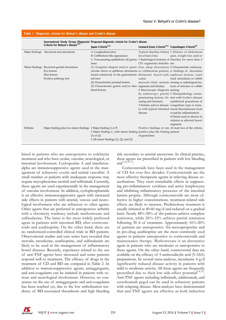
bined in patients who are unresponsive to colchicine treatment and who have ocular, vascular, neurological, or intestinal involvement. Cyclosporine A and interferon-alpha are immunosuppressive agents used in the man-agement of refractory uveitis and retinal vasculitis. A small number or patients with inadequate response may require mycophenolate mofetil and infliximab. Currently, these agents are used experimentally in the management of vascular involvement. In addition, cyclophosphamide is an effective immunosuppressive agent with increased side effects in patients with arterial, venous and neuro-logical involvement who are refractory to other agents. Other agents that are preferred in unresponsive arthritis with a chronicity tendency include methotrexate and sulfasalazine. The latter is the most widely preferred agent in patients with intestinal BD, after corticoste-roids and azathioprine. On the other hand, there are no randomized-controlled clinical trials in BD patients. Observational studies and case series have revealed that steroids, mesalazine, azathioprine, and sulfasalazine are likely to be used in the management of inflammatory bowel diseases. Recently, experience related to the use of anti-TNF agents have increased and some patients respond well to treatment. The efficacy of drugs in the treatment of CD and BD are compared in Table 2. In addition to immunosuppressive agents, antiaggregants, and anti-coagulants can be initiated in patients with ve-nous and neurological involvement. However, no con-sensus on the use of antiaggregants and anti-coagulants has been reached yet, due to the low embolization ten-dency of BD-associated thrombosis and high bleeding
risk secondary to arterial aneurysms. In clinical practice, these agents are prescribed in patients with low bleeding risk[7,41,76,77].
Corticosteroids have been used in the management of CD for over five decades. Corticosteroids are the most effective therapeutic agents in relieving disease ex-acerbations. They exert remarkable effects in suppress-ing pro-inflammatory cytokines and active lymphocytes and inhibiting inflammatory processes of the intestinal lamina propria. Although corticosteroids are more ef-fective in higher concentrations, treatment-related side effects are likely to increase. Prednisolone treatment is usually initiated at 40-60 mg/d and reduced on a gradual basis. Nearly 48%-58% of the patients achieve complete remission, while 26%-32% achieve partial remission following 30 d of treatment. Approximately 16%-20% of patients are unresponsive. Six-mercaptopurine and its pro-drug azathioprine are the most commonly used agents in patients unresponsive to corticosteroids and maintenance therapy. Methotrexate is an alternative agent in patients who are intolerant or unresponsive to these agents. On the other hand, controversial data are available on the efficacy of 5-aminosalicylic acid (5-ASA) preparations. In several meta-analyses, mesalazine 4 g/d significantly reduced disease activity in patients with mild to moderate activity. All these agents are frequently prescribed due to their low side-effect potential[78,79]. Anti-TNF agents including infliximab, adalimumab, and certolizumab pegol can be used in refractory patients with relapsing disease. Meta-analyses have demonstrated that anti-TNF agents are effective as both induction
August 15, 2014|Volume 5|Issue 3|WJGP|www.wjgnet.com 233
Table 1 Diagnostic criteria for Behçet’s disease and Crohn’s disease
International Study Group Diagnostic Criteria for Behçet’s disease[70]
Proposed diagnostic criteria for Crohn’s disease
Japan Criteria[72] Lennard-Jones Criteria[73] Copenhagen Criteria[8]
Major findings Recurrent oral ulcerations A: Longitudinal ulcerB: Cobblestone-like appearanceC: Noncaseating epithelioid cell granu-loma
Typical diarrhea history for at least 2 mo;1 Radiological features of CD: segmental distribu-tion, deep ulcerations or cobblestone pattern, thickened bowel wall, coarse mucosal relief, stenotic segments and fistulae;2 Macroscopic diagnosis by endoscopy: patchy penetrating lesions, fis-suring and strictures3 Fistulas and/or abscess-es with typical intestinal disease
1 History of abdominal pain, weight loss and/or diarrhea for more than 3 mo2 Characteristic endoscop-ic findings of ulceration (aphtous lesions, snail track ulceration) or cobble stoning or radiological fea-tures of stricture or cobble stoning3 Histopathology consis-tent with Crohn’s disease (epitheloid granuloma of Langerhans type or trans-mural discontinuous focal or patchy inflammation)4 Fistula and/or abscess in relation to affected bowel segments
Minor findings Recurrent genital ulcerations Eye lesionsSkin lesionsPositive pathergy test
(1) Irregular-shaped and/or quasi-circular ulcers or aphthous ulcerations found extensively in the gastrointesti-nal tract(2) Characteristic perianal lesions(3) Characteristic gastric and/or duo-denal lesions
Definite Major finding plus two minor findings 1 Major finding A or B2 Major finding C, with minor finding (1) or (2)3 All minor findings (1), (2), and (3)
Positive findings or one positive plus the finding of granuloma
At least two of the criteria present
Yazısız V. Behçet’s or Crohn’s disease?
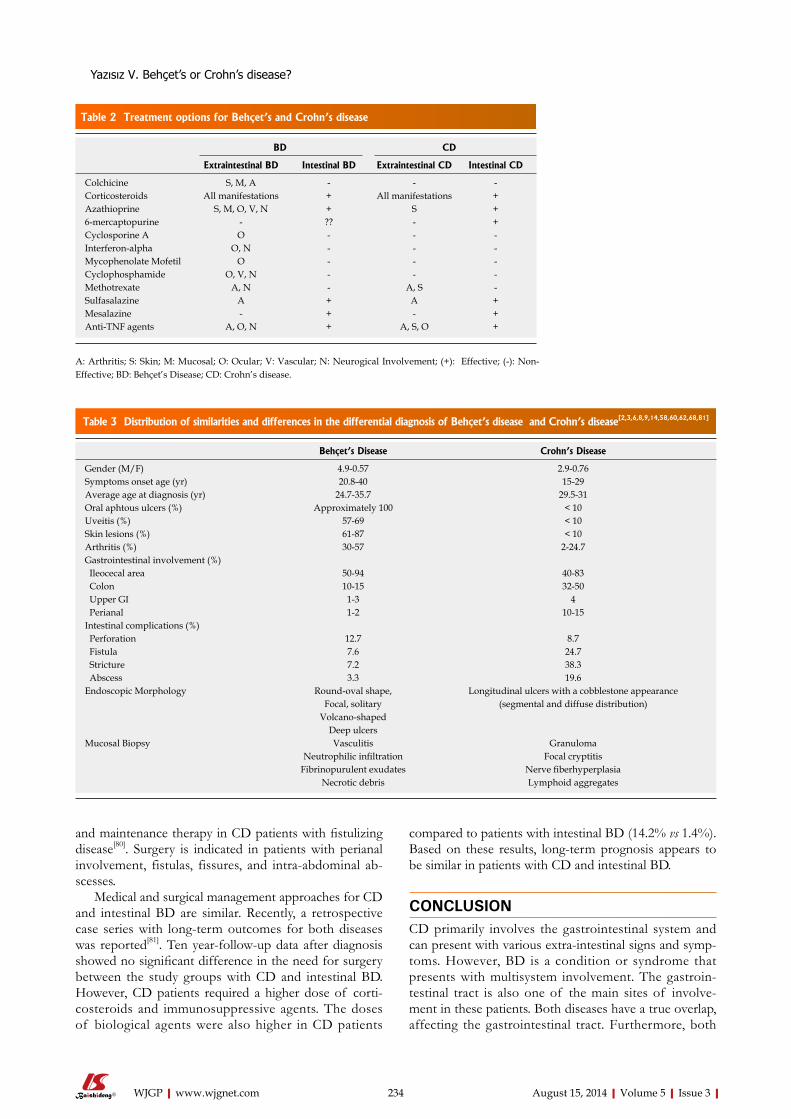
Table 2 Treatment options for Behçet’s and Crohn’s disease
BD CD
Extraintestinal BD Intestinal BD Extraintestinal CD Intestinal CD
Colchicine S, M, A - - -Corticosteroids All manifestations + All manifestations +Azathioprine S, M, O, V, N + S +6-mercaptopurine - ?? - +Cyclosporine A O - - -Interferon-alpha O, N - - -Mycophenolate Mofetil O - - -Cyclophosphamide O, V, N - - -Methotrexate A, N - A, S -Sulfasalazine A + A +Mesalazine - + - +Anti-TNF agents A, O, N + A, S, O +
A: Arthritis; S: Skin; M: Mucosal; O: Ocular; V: Vascular; N: Neurogical Involvement; (+): Effective; (-): Non-Effective; BD: Behçet’s Disease; CD: Crohn’s disease.
and maintenance therapy in CD patients with fistulizing disease[80]. Surgery is indicated in patients with perianal involvement, fistulas, fissures, and intra-abdominal ab-scesses.
Medical and surgical management approaches for CD and intestinal BD are similar. Recently, a retrospective case series with long-term outcomes for both diseases was reported[81]. Ten year-follow-up data after diagnosis showed no significant difference in the need for surgery between the study groups with CD and intestinal BD. However, CD patients required a higher dose of corti-costeroids and immunosuppressive agents. The doses of biological agents were also higher in CD patients
compared to patients with intestinal BD (14.2% vs 1.4%). Based on these results, long-term prognosis appears to be similar in patients with CD and intestinal BD.
CONCLUSIONCD primarily involves the gastrointestinal system and can present with various extra-intestinal signs and symp-toms. However, BD is a condition or syndrome that presents with multisystem involvement. The gastroin-testinal tract is also one of the main sites of involve-ment in these patients. Both diseases have a true overlap, affecting the gastrointestinal tract. Furthermore, both
August 15, 2014|Volume 5|Issue 3|WJGP|www.wjgnet.com 234
Table 3 Distribution of similarities and differences in the differential diagnosis of Behçet’s disease and Crohn’s disease[2,3,6,8,9,14,58,60,62,68,81]
Behçet’s Disease Crohn’s Disease
Gender (M/F) 4.9-0.57 2.9-0.76Symptoms onset age (yr) 20.8-40 15-29Average age at diagnosis (yr) 24.7-35.7 29.5-31Oral aphtous ulcers (%) Approximately 100 < 10Uveitis (%) 57-69 < 10Skin lesions (%) 61-87 < 10Arthritis (%) 30-57 2-24.7Gastrointestinal involvement (%) Ileocecal area 50-94 40-83 Colon 10-15 32-50 Upper GI 1-3 4 Perianal 1-2 10-15Intestinal complications (%) Perforation 12.7 8.7 Fistula 7.6 24.7 Stricture 7.2 38.3 Abscess 3.3 19.6Endoscopic Morphology Round-oval shape, Longitudinal ulcers with a cobblestone appearance
Focal, solitary (segmental and diffuse distribution)Volcano-shaped
Deep ulcersMucosal Biopsy Vasculitis Granuloma
Neutrophilic infiltration Focal cryptitisFibrinopurulent exudates Nerve fiberhyperplasia
Necrotic debris Lymphoid aggregates
Yazısız V. Behçet’s or Crohn’s disease?

conditions share similar characteristics with respect to age of onset, gender, and inflammation biomarkers such as erythrocyte sedimentation rate and C-reactive protein (increased levels). Despite these similarities, the immu-nopathogenesis, genetic factors, and regional distribution are quite different. Although both diseases involve simi-lar systems, they have distinct histopathological charac-teristics. For instance, uveitis is more common in BD, and CD patients are more likely to suffer from episcle-ritis or conjunctivitis. Figure 1 shows the similarities and differences in BD and CD. Table 3 summarizes the incidence of similarities, the distribution of gastrointes-tinal involvement, and endoscopic and histopathological differences.
As mentioned above, BD is more common in Asian and Mediterranean populations, while CD is more fre-quently seen in north European and American individu-als. However, given the fact that we live in a globalizing world, the number of patients in whom the differential diagnosis of both conditions is of the utmost impor-tance has increased. Therefore, rheumatologists and gas-troenterologists who are mainly involved in the diagnosis and management of BD and CD should be well aware of the typical characteristics of both diseases.
REFERENCES1 Behçet H. Uber rezidivierende, aphthose, durch ein Virus
verursachte Geschwure am Mund, am Auge und an den Genitalien. Dermatol Wochenschr 1937; 36: 1152-1157
2 Baumgart DC, Sandborn WJ. Crohn‘s disease. Lancet 2012; 380: 1590-1605 [PMID: 22914295 DOI: 10.1016/S0140-6736(12)60026-9]
3 Davatchi F, Shahram F, Chams-Davatchi C, Shams H, Nadji
A, Akhlaghi M, Faezi T, Ghodsi Z, Faridar A, Ashofteh F, Sadeghi Abdollahi B. Behcet‘s disease: from East to West. Clin Rheumatol 2010; 29: 823-833 [PMID: 20354748 DOI: 10.1007/s10067-010-1430-6]
4 Mahr A, Belarbi L, Wechsler B, Jeanneret D, Dhote R, Fain O, Lhote F, Ramanoelina J, Coste J, Guillevin L. Population-based prevalence study of Behçet‘s disease: differences by ethnic origin and low variation by age at immigration. Arthritis Rheum 2008; 58: 3951-3959 [PMID: 19035493 DOI: 10.1002/art.24149]
5 Yurdakul S, Günaydin I, Tüzün Y, Tankurt N, Pazarli H, Ozyazgan Y, Yazici H. The prevalence of Behçet‘s syndro-me in a rural area in northern Turkey. J Rheumatol 1988; 15: 820-822 [PMID: 3172095]
6 Yurdakul S, Yazıcı Y. Epidemiology of Behçet’s Syndrome and Regional Differences in Disease Expression. 1st ed. In: Yazici Y, Yazici H, editors. Behçet’s Syndrome (NY): Spring-er, 2010: 35-53
7 Ambrose NL, Haskard DO. Differential diagnosis and man-agement of Behçet syndrome. Nat Rev Rheumatol 2013; 9: 79-89 [PMID: 23007742]
8 Vind I, Riis L, Jess T, Knudsen E, Pedersen N, Elkjaer M, Bak Andersen I, Wewer V, Nørregaard P, Moesgaard F, Bendtsen F, Munkholm P. Increasing incidences of inflam-matory bowel disease and decreasing surgery rates in Co-penhagen City and County, 2003-2005: a population-based study from the Danish Crohn colitis database. Am J Gastro-enterol 2006; 101: 1274-1282 [PMID: 16771949]
9 Thia KT, Loftus EV, Sandborn WJ, Yang SK. An update on the epidemiology of inflammatory bowel disease in Asia. Am J Gastroenterol 2008; 103: 3167-3182 [PMID: 19086963 DOI: 10.1111/j.1572-0241.2008.02158.x]
10 Yapp TR, Stenson R, Thomas GA, Lawrie BW, Williams GT, Hawthorne AB. Crohn’s disease incidence in Cardiff from 1930: an update for 1991-1995. Eur J Gastroenterol Hepatol 2000; 12: 907-911 [PMID: 10958218]
11 Hovde Ø, Moum BA. Epidemiology and clinical course of Crohn’s disease: results from observational studies. World J Gastroenterol 2012; 18: 1723-1731 [PMID: 22553396 DOI:
August 15, 2014|Volume 5|Issue 3|WJGP|www.wjgnet.com 235
Behçet’s Disease Crohn’s Disease
HLA-B51
Silk road
Episcleritis
NOD2/CARD15
Northern EuropeNorthern America
United Kingdom
F/M
Age of diagnosis
Anywhere in the World
ASCA, ANCA
Granulomatous disease
Autoinflammation?
Immunosuppressive theraphy
Vasculitis
Nonspecific inflammation
Perianal fistula/fissureConjunctivitis
Pyoderma gangrenosumSweet's syndromeNeutrophilic dermatosesPrimary sclerosing
cholangitis
Retinal vasculitis/vitritisRetrobulbar neuritisGenital ulcers with scar
Epididimit/orchitisPseudofolliculitisPathergy reactionSuperficial thrombophlebitisVenous thrombosisBudd-Chiary syndrome
Arterial aneurysmNeuro-Behcet
Oral aphtous ulcersUveitis
Erytma nodosumPeripheral arthritis
Sacroileitis
Gastrointestinal involvementSpondyloarthropathy
Figure 1 Similar and different characteristics of Behçet’s disease and Crohn’s disease. F: Female; M: Male; ASCA: Anti-Saccharomyces cerevisiae antibodies; ANCA: Anti-neutrophil cytoplasmic antibodies.
Yazısız V. Behçet’s or Crohn’s disease?
10.3748/wjg.v18.i15.1723]12 Fonager K, Sørensen HT, Olsen J. Change in incidence of
Crohn’s disease and ulcerative colitis in Denmark. A study based on the National Registry of Patients, 1981-1992. Int J Epidemiol 1997; 26: 1003-1008 [PMID: 9363521]
13 Loftus EV, Silverstein MD, Sandborn WJ, Tremaine WJ, Harmsen WS, Zinsmeister AR. Crohn’s disease in Olm-sted County, Minnesota, 1940-1993: incidence, prevalence, and survival. Gastroenterology 1998; 114: 1161-1168 [PMID: 9609752]
14 Bernstein CN, Wajda A, Svenson LW, MacKenzie A, Koe-hoorn M, Jackson M, Fedorak R, Israel D, Blanchard JF. The epidemiology of inflammatory bowel disease in Canada: a population-based study. Am J Gastroenterol 2006; 101: 1559-1568 [PMID: 16863561]
15 Ohno S, Ohguchi M, Hirose S, Matsuda H, Wakisaka A, Aizawa M. Close association of HLA-Bw51 with Behçet’s disease. Arch Ophthalmol 1982; 100: 1455-1458 [PMID: 6956266]
16 Durrani K, Papaliodis GN. The genetics of Adamantiades-Behcet’s disease. Semin Ophthalmol 2008; 23: 73-79 [PMID: 18214795 DOI: 10.1080/08820530701745264]
17 Takeno M , Kariyone A, Yamashita N, Takiguchi M, Mizushima Y, Kaneoka H, Sakane T. Excessive function of peripheral blood neutrophils from patients with Behçet’s disease and from HLA-B51 transgenic mice. Arthritis Rheum 1995; 38: 426-433 [PMID: 7880197]
18 Kaneoka H, Furukawa H, Takeno M, Mizushima Y, Sakane T. HLA-B51 involved in the hyperfunction of periph-eral blood neutrophils from patients Behcet’s disease. In: Wechsler B, Godeau P, editors. Behcet’s Disease Amster-dam: Excerpta Medica, 1993: 29-32
19 Knights D, Lassen KG, Xavier RJ. Advances in inflamma-tory bowel disease pathogenesis: linking host genetics and the microbiome. Gut 2013; 62: 1505-1510 [PMID: 24037875 DOI: 10.1136/gutjnl-2012-303954]
20 Parkes M. The genetics universe of Crohn’s disease and ulcerative colitis. Dig Dis 2012; 30 Suppl 1: 78-81 [PMID: 23075873 DOI: 10.1159/000341130]
21 Direskeneli H. Autoimmunity vs autoinflammation in Behcet’s disease: do we oversimplify a complex disorder? Rheumatology (Oxford) 2006; 45: 1461-1465 [PMID: 16998234]
22 Yazici H. The place of Behçet’s syndrome among the au-toimmune diseases. Int Rev Immunol 1997; 14: 1-10 [PMID: 9203022]
23 Kibaroglu A, Eksioglu-Demiralp E, Akoglu T, Direskeneli H. T and NK cell subset changes with microbial extracts and human HSP60-derived peptides in Behçet’s disease. Clin Exp Rheumatol 2004; 22: S59-S63 [PMID: 15515788]
24 Atagunduz P, Ergun T, Direskeneli H. MEFV mutations are increased in Behçet’s disease (BD) and are associated with vascular involvement. Clin Exp Rheumatol 2003; 21: S35-S37 [PMID: 14727457]
25 Tasliyurt T, Yigit S, Rustemoglu A, Gul U, Ates O. Common MEFV gene mutations in Turkish patients with Behcet’s disease. Gene 2013; 530: 100-103 [PMID: 23973724 DOI: 10.1016/j.gene.2013.08.026]
26 Hamuryudan V, Melikoğlu M. Vascular Disease in Behçet’s Syndrome. 1st ed. In: Yazici Y, Yazici H, editors. Behçet’s Syndrome (NY): Springer, 2010: 115-135
27 Chun SI, Su WP, Lee S. Histopathologic study of cutaneous lesions in Behçet’s syndrome. J Dermatol 1990; 17: 333-341 [PMID: 2384635]
28 Jennette JC, Falk RJ, Bacon PA, Basu N, Cid MC, Ferrario F, Flores-Suarez LF, Gross WL, Guillevin L, Hagen EC, Hoff-man GS, Jayne DR, Kallenberg CG, Lamprecht P, Langford CA, Luqmani RA, Mahr AD, Matteson EL, Merkel PA, Ozen S, Pusey CD, Rasmussen N, Rees AJ, Scott DG, Specks U, Stone JH, Takahashi K, Watts RA. 2012 revised International Chapel Hill Consensus Conference Nomenclature of Vascu-
litides. Arthritis Rheum 2013; 65: 1-11 [PMID: 23045170 DOI: 10.1002/art.37715]
29 Mumcu G, Ergun T, Inanc N, Fresko I, Atalay T, Hayran O, Direskeneli H. Oral health is impaired in Behçet’s disease and is associated with disease severity. Rheumatology (Ox-ford) 2004; 43: 1028-1033 [PMID: 15161982]
30 Direskeneli H. Behçet’s disease: infectious aetiology, new autoantigens, and HLA-B51. Ann Rheum Dis 2001; 60: 996-1002 [PMID: 11602462]
31 Hirohata S, Oka H, Mizushima Y. Streptococcal-related antigens stimulate production of IL6 and interferon-gamma by T cells from patients with Behcet’s disease. Cell Immunol 1992; 140: 410-419 [PMID: 1544169]
32 Calgüneri M, Kiraz S, Ertenli I, Benekli M, Karaarslan Y, Celik I. The effect of prophylactic penicillin treatment on the course of arthritis episodes in patients with Behçet’s dis-ease. A randomized clinical trial. Arthritis Rheum 1996; 39: 2062-2065 [PMID: 8961912]
33 Franke A, McGovern DP, Barrett JC, Wang K, Radford-Smith GL, Ahmad T, Lees CW, Balschun T, Lee J, Roberts R, Anderson CA, Bis JC, Bumpstead S, Ellinghaus D, Festen EM, Georges M, Green T, Haritunians T, Jostins L, Latiano A, Mathew CG, Montgomery GW, Prescott NJ, Raychaudhuri S, Rotter JI, Schumm P, Sharma Y, Simms LA, Taylor KD, Whiteman D, Wijmenga C, Baldassano RN, Barclay M, Bay-less TM, Brand S, Büning C, Cohen A, Colombel JF, Cottone M, Stronati L, Denson T, De Vos M, D’Inca R, Dubinsky M, Edwards C, Florin T, Franchimont D, Gearry R, Glas J, Van Gossum A, Guthery SL, Halfvarson J, Verspaget HW, Hugot JP, Karban A, Laukens D, Lawrance I, Lemann M, Levine A, Libioulle C, Louis E, Mowat C, Newman W, Panés J, Phillips A, Proctor DD, Regueiro M, Russell R, Rutgeerts P, Sanderson J, Sans M, Seibold F, Steinhart AH, Stokkers PC, Torkvist L, Kullak-Ublick G, Wilson D, Walters T, Targan SR, Brant SR, Rioux JD, D’Amato M, Weersma RK, Kugath-asan S, Griffiths AM, Mansfield JC, Vermeire S, Duerr RH, Silverberg MS, Satsangi J, Schreiber S, Cho JH, Annese V, Hakonarson H, Daly MJ, Parkes M. Genome-wide meta-analysis increases to 71 the number of confirmed Crohn’s disease susceptibility loci. Nat Genet 2010; 42: 1118-1125 [PMID: 21102463 DOI: 10.1038/ng.717]
34 Masters SL, Simon A, Aksentijevich I, Kastner DL. Hor-ror autoinflammaticus: the molecular pathophysiology of autoinflammatory disease (*). Annu Rev Immunol 2009; 27: 621-668 [PMID: 19302049 DOI: 10.1146/annurev]
35 Fidder H, Chowers Y, Ackerman Z, Pollak RD, Crusius JB, Livneh A, Bar-Meir S, Avidan B, Shinhar Y. The familial Mediterranean fever (MEVF) gene as a modifier of Crohn’s disease. Am J Gastroenterol 2005; 100: 338-343 [PMID: 15667491]
36 Grigg EL, Kane S, Katz S. Mimicry and deception in inflam-matory bowel disease and intestinal behçet disease. Gastro-enterol Hepatol (N Y) 2012; 8: 103-112 [PMID: 22485077]
37 Filik L, Biyikoglu I. Differentiation of Behcet’s disease from inflammatory bowel diseases: anti-Saccharomyces cerevi-siae antibody and anti-neutrophilic cytoplasmic antibody. World J Gastroenterol 2008; 14: 7271 [PMID: 19084948]
38 Choi CH, Kim TI, Kim BC, Shin SJ, Lee SK, Kim WH, Kim HS. Anti-Saccharomyces cerevisiae antibody in intestinal Behçet’s disease patients: relation to clinical course. Dis Co-lon Rectum 2006; 49: 1849-1859 [PMID: 17080284]
39 Shang Y, Han S, Li J, Ren Q, Song F, Chen H. The clinical feature of Behçet’s disease in Northeastern China. Yonsei Med J 2009; 50: 630-636 [PMID: 19881965 DOI: 10.3349/ymj.2009.50.5.630]
40 Tursen U, Gurler A, Boyvat A. Evaluation of clinical find-ings according to sex in 2313 Turkish patients with Behçet’s disease. Int J Dermatol 2003; 42: 346-351 [PMID: 12755969]
41 Yilmaz S, Karadag O, Yazisiz V, Altun B, Gezer M, Kara-man M, Cinar M, Erdem H, Pay S, Dinc A. Systemic involve-
August 15, 2014|Volume 5|Issue 3|WJGP|www.wjgnet.com 236
Yazısız V. Behçet’s or Crohn’s disease?
ments and preferred treatments in a large cohort of Behçet’s disease. Rheumatol Int 2013; 33: 3025-3030 [PMID: 23881265]
42 Alpsoy E, Donmez L, Onder M, Gunasti S, Usta A, Kar-incaoglu Y, Kandi B, Buyukkara S, Keseroglu O, Uzun S, Tursen U, Seyhan M, Akman A. Clinical features and natu-ral course of Behçet’s disease in 661 cases: a multicentre study. Br J Dermatol 2007; 157: 901-906 [PMID: 17711526]
43 Mat MC, Bang D, Melikoğlu M. The Mucocutaneous Mani-festations and Pathergy Reaction in Behcet’s Disease. 1st ed. In: Yazici Y, Yazici H, editors. Behçet’s Syndrome (NY): Springer, 2010: 53-73
44 Tugal-Tutkun I, Gupta V, Cunningham ET. Differential diagnosis of behçet uveitis. Ocul Immunol Inflamm 2013; 21: 337-350 [PMID: 23730816]
45 Benamour S, Zeroual B, Alaoui FZ. Joint manifestations in Behçet’s disease. A review of 340 cases. Rev Rhum Engl Ed 1998; 65: 299-307 [PMID: 9636948]
46 Ben Ghorbel I, Ennaifer R, Lamloum M, Khanfir M, Miled M, Houman MH. Budd-Chiari syndrome associated with Behçet’s disease. Gastroenterol Clin Biol 2008; 32: 316-320 [PMID: 18400436 DOI: 10.1016/j.gcb.2007.12.022]
47 Larsen S, Bendtzen K, Nielsen OH. Extraintestinal manifes-tations of inflammatory bowel disease: epidemiology, diag-nosis, and management. Ann Med 2010; 42: 97-114 [PMID: 20166813 DOI: 10.3109/07853890903559724]
48 Ardizzone S, Puttini PS, Cassinotti A, Porro GB. Extraint-estinal manifestations of inflammatory bowel disease. Dig Liver Dis 2008; 40 Suppl 2: S253-S259 [PMID: 18598997 DOI: 10.1016/S1590-8658(08)60534-4]
49 Hatemi I, Hatemi G, Celik AF, Melikoglu M, Arzuhal N, Mat C, Ozyazgan Y, Yazici H. Frequency of pathergy phe-nomenon and other features of Behçet’s syndrome among patients with inflammatory bowel disease. Clin Exp Rheuma-tol 2008; 26: S91-S95 [PMID: 19026122]
50 Lanna CC, Ferrari Mde L, Rocha SL, Nascimento E, de Carvalho MA, da Cunha AS. A cross-sectional study of 130 Brazilian patients with Crohn’s disease and ulcerative coli-tis: analysis of articular and ophthalmologic manifestations. Clin Rheumatol 2008; 27: 503-509 [PMID: 18097711]
51 Yilmaz S, Aydemir E, Maden A, Unsal B. The prevalence of ocular involvement in patients with inflammatory bowel disease. Int J Colorectal Dis 2007; 22: 1027-1030 [PMID: 17262200]
52 Colombo E, Latiano A, Palmieri O, Bossa F, Andriulli A, Annese V. Enteropathic spondyloarthropathy: a common genetic background with inflammatory bowel disease? World J Gastroenterol 2009; 15: 2456-2462 [PMID: 19468994]
53 Gravallese EM, Kantrowitz FG. Arthritic manifestations of inflammatory bowel disease. Am J Gastroenterol 1988; 83: 703-709 [PMID: 3289378]
54 Miehsler W, Reinisch W, Valic E, Osterode W, Tillinger W, Feichtenschlager T, Grisar J, Machold K, Scholz S, Vo-gelsang H, Novacek G. Is inflammatory bowel disease an independent and disease specific risk factor for thromboem-bolism? Gut 2004; 53: 542-548 [PMID: 15016749]
55 Fumery M, Xiaocang C, Dauchet L, Gower-Rousseau C, Peyrin-Biroulet L, Colombel JF. Thromboembolic events and cardiovascular mortality in inflammatory bowel dis-eases: A meta-analysis of observational studies. J Crohns Colitis 2014; 8: 469-479 [PMID: 24183231 DOI: 10.1016/j.crohns.2013.09.021]
56 Reny JL, Paul JF, Lefèbvre C, Champion K, Emmerich J, Blétry O, Piette JC, Fiessinger JN. Association of Takayasu’s arteritis and Crohn’s disease. Results of a study on 44 Takayasu patients and review of the literature. Ann Med In-terne (Paris) 2003; 154: 85-90 [PMID: 12746644]
57 Agostini A, Benuzzi F, Filippini N, Bertani A, Scarcelli A, Farinelli V, Marchetta C, Calabrese C, Rizzello F, Gionchetti P, Ercolani M, Campieri M, Nichelli P. New insights into the brain involvement in patients with Crohn’s disease: a voxel-
based morphometry study. Neurogastroenterol Motil 2013; 25: 147-e82 [PMID: 22998431 DOI: 10.1111/nmo.12017]
58 Cheon JH, Çelik AF, Kim WH. Behçet’s Disease: Gastroin-testinal Involvement. 1st ed. In: Yazici Y, Yazici H, editors. Behçet’s Syndrome (NY): Springer, 2010: 165-189
59 Hung CH, Lee JH, Chen ST, Yang YH, Lin YT, Wang LC, Yu HH, Chiang BL. Young children with Behçet disease have more intestinal involvement. J Pediatr Gastroenterol Nutr 2013; 57: 225-229 [PMID: 23880628 DOI: 10.1097/MPG.0b013e3182936ec4]
60 Wu QJ, Zhang FC, Zhang X. Adamantiades-Behcet’s dis-ease-complicated gastroenteropathy. World J Gastroenterol 2012; 18: 609-615 [PMID: 22363131 DOI: 10.3748/wjg.v18.i7.609]
61 Lee HJ, Kim YN, Jang HW, Jeon HH, Jung ES, Park SJ, Hong SP, Kim TI, Kim WH, Nam CM, Cheon JH. Correlations between endoscopic and clinical disease activity indices in intestinal Behcet’s disease. World J Gastroenterol 2012; 18: 5771-5778 [PMID: 23155319 DOI: 10.3748/wjg.v18.i40.5771]
62 Lee SK, Kim BK, Kim TI, Kim WH. Differential diagnosis of intestinal Behçet’s disease and Crohn’s disease by colo-noscopic findings. Endoscopy 2009; 41: 9-16 [PMID: 19160153 DOI: 10.1055/s-0028-1103481]
63 Mekhjian HS, Switz DM, Melnyk CS, Rankin GB, Brooks RK. Clinical features and natural history of Crohn’s disease. Gastroenterology 1979; 77: 898-906 [PMID: 381094]
64 Annunziata ML, Caviglia R, Papparella LG, Cicala M. Upper gastrointestinal involvement of Crohn’s disease: a prospective study on the role of upper endoscopy in the diagnostic work-up. Dig Dis Sci 2012; 57: 1618-1623 [PMID: 22350786]
65 Pera A, Bellando P, Caldera D, Ponti V, Astegiano M, Barlet-ti C, David E, Arrigoni A, Rocca G, Verme G. Colonoscopy in inflammatory bowel disease. Diagnostic accuracy and proposal of an endoscopic score. Gastroenterology 1987; 92: 181-185 [PMID: 3781186]
66 Matsumoto T, Uekusa T, Fukuda Y. Vasculo-Behçet’s dis-ease: a pathologic study of eight cases. Hum Pathol 1991; 22: 45-51 [PMID: 1985077]
67 Hamuryudan V, Oz B, Tüzün H, Yazici H. The menacing pulmonary artery aneurysms of Behçet’s syndrome. Clin Exp Rheumatol 2004; 22: S1-S3 [PMID: 15515774]
68 Demirkesen C, Oz B, Göksal S. Behçet’s Disease: Pathology. 1st ed. In: Yazici Y, Yazici H, editors. Behçet’s Syndrome (NY): Springer, 2010: 215-243
69 Geboes K. What histologic features best differentiate Crohn’s disease from ulcerative colitis? Inflamm Bowel Dis 2008; 14 Suppl 2: S168-S169 [PMID: 18816725]
70 Criteria for diagnosis of Behçet's disease. International Study Group for Behçet's Disease. Lancet 1990; 335: 1078-1080 [PMID: 1970380]
71 Tunç R, Uluhan A, Melikoğlu M, Ozyazgan Y, Ozdoğan H, Yazici H. A reassessment of the International Study Group criteria for the diagnosis (classification) of Behçet’s syndrome. Clin Exp Rheumatol 2001; 19: S45-S47 [PMID: 11760398]
72 Matsui T, Hirai F, Hisabe T. Proposed diagnostic criteria for Crohn’s disease. Annual reports of the research group of intractable inflammatory bowel disease subsidized by the Ministry of Health, Labour, and Welfare of Japan, Tokyo, Japan, 2011: 52-54
73 Lennard-Jones JE. Classification of inflammatory bowel disease. Scand J Gastroenterol Suppl 1989; 170: 2-6; discussion 16-19 [PMID: 2617184]
74 Gasche C, Scholmerich J, Brynskov J, D’Haens G, Hanauer SB, Irvine EJ, Jewell DP, Rachmilewitz D, Sachar DB, Sand-born WJ, Sutherland LR. A simple classification of Crohn’s disease: report of the Working Party for the World Con-gresses of Gastroenterology, Vienna 1998. Inflamm Bowel Dis 2000; 6: 8-15 [PMID: 10701144]
August 15, 2014|Volume 5|Issue 3|WJGP|www.wjgnet.com 237
Yazısız V. Behçet’s or Crohn’s disease?
75 Silverberg MS, Satsangi J, Ahmad T, Arnott ID, Bernstein CN, Brant SR, Caprilli R, Colombel JF, Gasche C, Geboes K, Jewell DP, Karban A, Loftus EV, Peña AS, Riddell RH, Sachar DB, Schreiber S, Steinhart AH, Targan SR, Vermeire S, Warren BF. Toward an integrated clinical, molecular and serological classification of inflammatory bowel disease: report of a Working Party of the 2005 Montreal World Con-gress of Gastroenterology. Can J Gastroenterol 2005; 19 Suppl A: 5A-36A [PMID: 16151544]
76 Hatemi G, Silman A, Bang D, Bodaghi B, Chamberlain AM, Gul A, Houman MH, Kötter I, Olivieri I, Salvarani C, Sfika-kis PP, Siva A, Stanford MR, Stübiger N, Yurdakul S, Yazici H. EULAR recommendations for the management of Behçet disease. Ann Rheum Dis 2008; 67: 1656-1662 [PMID: 18245110 DOI: 10.1136/ard.2007.080432]
77 Arida A, Fragiadaki K, Giavri E, Sfikakis PP. Anti-TNF agents for Behçet’s disease: analysis of published data on 369 patients. Semin Arthritis Rheum 2011; 41: 61-70 [PMID:
21168186 DOI: 10.1016/j.semarthrit.2010.09.002]78 Girardin M, Manz M, Manser C, Biedermann L, Wanner R,
Frei P, Safroneeva E, Mottet C, Rogler G, Schoepfer AM. First-line therapies in inflammatory bowel disease. Digestion 2012; 86 Suppl 1: 6-10 [PMID: 23051720 DOI: 10.1159/000341951]
79 Ueno F, Matsui T, Matsumoto T, Matsuoka K, Watanabe M, Hibi T. Evidence-based clinical practice guidelines for Crohn’s disease, integrated with formal consensus of experts in Japan. J Gastroenterol 2013; 48: 31-72 [PMID: 23090001 DOI: 10.1007/s00535-012-0673-1]
80 Kawalec P, Mikrut A, Wiśniewska N, Pilc A. Tumor necro-sis factor-α antibodies (infliximab, adalimumab and certoli-zumab) in Crohn’s disease: systematic review and meta-analysis. Arch Med Sci 2013; 9: 765-779 [PMID: 24273556]
81 Jung YS, Cheon JH, Park SJ, Hong SP, Kim TI, Kim WH. Long-term clinical outcomes of Crohn’s disease and intes-tinal Behcet’s disease. Inflamm Bowel Dis 2013; 19: 99-105 [PMID: 22508364 DOI: 10.1002/ibd.22991]
P- Reviewer: Ahluwalia NK, Castro FJ, Garip Y, Lakatos PL S- Editor: Ji FF L- Editor: Webster JR E- Editor: Lu YJ
August 15, 2014|Volume 5|Issue 3|WJGP|www.wjgnet.com 238
Yazısız V. Behçet’s or Crohn’s disease?
TOPIC HIGHLIGHT
Multidisciplinary and evidence-based management of fistulizing perianal Crohn’s disease
Ricardo Sordo-Mejia, Wolfgang B Gaertner
Ricardo Sordo-Mejia, Wolfgang B Gaertner, Division of Co-lon and Rectal Surgery, Department of Surgery, ABC Medical Center, 01120 Mexico City, MexicoAuthor contributions: Sordo-Mejia R and Gaertner WB con-tributed to literature search and manuscript preparation.Correspondence to: Wolfgang B Gaertner, MSc, MD, Divi-sion of Colon and Rectal Surgery, Department of Surgery, ABC Medical Center, Sur 136. No. 116-1A, Colonia Las Americas, 01120 Mexico City, Mexico. [email protected]: +52-55-10406569Received: November 28, 2013 Revised: May 7, 2014Accepted: May 28, 2014Published online: August 15, 2014
AbstractPerianal symptoms are common in patients with Crohn’s disease and cause considerable morbidity. The etiology of these symptoms include skin tags, ulcers, fissures, abscesses, fistulas or stenoses. Fistula is the most common perianal manifestation. Multiple treatment op-tions exist although very few are evidence-based. The phases of treatment include: drainage of infection, as-sessment of Crohn’s disease status and fistula tracts, medical therapy, and selective operative management. The impact of biological therapy on perianal Crohn’s disease is uncertain given that outcomes are conflict-ing. Operative treatment to eradicate the fistula tract can be attempted once infection has resolved and Crohn’s disease activity is controlled. The operative ap-proach should be tailored according to the anatomy of the fistula tract. Definitive treatment is challenging with medical and operative treatment rarely leading to true healing with frequent complications and recurrence. Treatment success must be weighed against the risk of complications, specially anal sphincter injury. A full un-derstanding of the etiology and all potential therapeutic options is critical for success. Multidisciplinary manage-ment of fistulizing perianal Crohn’s disease is crucial to
improve outcomes.
© 2014 Baishideng Publishing Group Inc. All rights reserved.
Key words: Perianal Crohn’s disease; Fistula; Abscess; Management; Review
Core tip: This manuscript is a comprehensive review that focuses on the multidisciplinary management of fistulizing perianal Crohn’s disease. The treatment op-tions discussed in this review are based on a current literature review as well as our experience with the disease. Diagnostic and treatment algorithms are also provided.
Sordo-Mejia R, Gaertner WB. Multidisciplinary and evidence-based management of fistulizing perianal Crohn’s disease. World J Gastrointest Pathophysiol 2014; 5(3): 239-251 Available from: URL: http://www.wjgnet.com/2150-5330/full/v5/i3/239.htm DOI: http://dx.doi.org/10.4291/wjgp.v5.i3.239
INTRODUCTIONAlthough Gabriel[1] first described patients with granu-lomatous perianal disease 17 years before the formal description of the disease by Burrill Crohn’s[2] in 1932, Bissell[3] was the first to describe the associated perianal manifestations of Crohn’s disease (CD). Furthermore, Morson et al[4] documented the appearance of perianal non-caseating granulomas and fistulas many years before the onset of intestinal CD.
The reported prevalence of anorectal involvement in patients with CD has varied but the most current popu-lation-based series have found involvement in 14 to 38 percent of patients[5-7], with isolated perianal disease seen in only five percent[8]. The prevalence of perianal mani-
World J Gastrointest Pathophysiol 2014 August 15; 5(3): 239-251ISSN 2150-5330 (online)
© 2014 Baishideng Publishing Group Inc. All rights reserved.
Online Submissions: http://www.wjgnet.com/esps/Help Desk: http://www.wjgnet.com/esps/helpdesk.aspxDOI: 10.4291/wjgp.v5.i3.239
August 15, 2014|Volume 5|Issue 3|WJGP|www.wjgnet.com 239
WJGP 5th Anniversary Special Issues (6): Crohn’s disease
Sordo-Mejia R et al . Perianal Crohn’s disease
festations increases as the disease progresses distally, with up to 92 percent of patients with CD involving the colon and rectum developing fistulas[9]. In most cases, bowel involvement precedes perianal disease[9], but up to 40 percent of patients can experience perianal symptoms before intestinal manifestations[10]. There does not seem to be a predilection for age but a younger age of onset increases the odds of developing perianal disease over time[11-12].
The most common presentation of perianal CD is abscess and fistula. However, patients with CD are fre-quently affected by other perianal pathologies including hemorrhoids, fissures, skin tags, ulcers, and strictures. Perianal CD has been associated with a disabling natural history[13], with common extraintestinal manifestations[14] and greater steroid resistance[15]. Perianal disease is often recurrent, with 35 to 59 percent of patients relapsing within two years[16]. More than 80 percent of patients require operative treatment, and up to 20 percent may require proctectomy[5,7]. Patients with perianal CD have also shown an increased risk for anal malignancies[17,18], with active and long duration of disease being identified risk factors[19-21].
The treatment of perianal CD continues to be a challenge, especially with the plethora of literature ad-dressing both medical and operative treatment strategies. The purpose of this review is to summarize the efficacy of currently described methods for the management of fistulizing perianal CD and its complications.
ABSCESS Abscesses usually occur with active perianal CD with an incidence of up to 62 percent during the course of the disease[22]. Ischiorectal abscesses account for 40 percent of all perianal abscesses[23]. Fistula tract location can in-fluence abscess development and transsphincteric fistu-las pose the greatest risk[23].
Abscesses are uncommon with superficial fistula tracts. Makowiec et al[24] evaluated 61 patients with peri-anal CD and found that 73 percent of all abscesses were related to an ischiorectal fistula and 50 percent with a transsphincteric fistula. Recurrences occurred in 53 per-cent with a median time to recurrence of 14 mo. No pa-tients with superficial fistula tracts had a second abscess, whereas about two thirds of patients with transsphinc-teric and ischiorectal fistulas recurred after 36 mo.
A detailed anorectal exam should be performed be-fore any type of treatment is initiated. This frequently requires evaluation under anesthesia (EUA) with evalu-ation of the rectum to rule-out active disease. Perianal infection can occur in any anatomic plane (superficial, intersphincteric, ischiorectal, or supralevator), and re-quires immediate drainage and treatment of systemic symptoms with broad-spectrum antibiotics[6,25]. Many authors recommend drain placement or partial sphincter division to facilitate drainage, but these have not been associated with better outcomes[26,27]. In the setting of
persistent perianal sepsis, imaging modalities such as magnetic resonance imaging (MRI) and computed to-mography (CT) are used to guide the drainage of deep or complex abscesses[28,29].
When a fistula is encountered, a non-cutting seton should be placed to facilitate drainage and prevent recur-rent infection, with improvement seen in 79 to 100 per-cent of patients[30-35]. Long-term drainage with non-cut-ting setons without definitive therapy has been reported to result in fistula recurrence in 20 to 80 percent of cases[33,36,37]. The combination of non-cutting setons and anti-tumor necrosis factor (TNF) therapy has been asso-ciated with fistula healing rates of up to 67 percent and will be discussed below[38,39]. Fecal diversion to increase fistula healing and control perianal sepsis continues to be controversial with no level A data supporting its role but in the setting of persistent perianal sepsis, a tempo-rary diverting stoma can be effective. Patients should be aware that these stomas are rarely reversed[40].
Cryptoglandular abscess/fistulas can and do occur in patients with CD and should be recognized as so because treatment differs. These abscess/fistulas tend to be superficial and are not associated with active ano-rectal CD; therefore, anti-TNF therapy is not indicated. Abscess drainage should follow the same principles as mentioned above. Placement of a non-cutting seton is encouraged and any attempt of local surgical treatment should take into consideration the patients underlying continence and CD status. Supplemental imaging stud-ies, such as endoanal ultrasound (EAUS), are very help-ful even when cryptoglandular etiology is suspected.
FISTULAA population-based study[7] with up to 20 years of fol-low-up showed that one out every two patients with CD develop perianal fistulas. The etiology of perianal fistula formation in CD is not completely clear but genetic, microbiological, and immunological factors play a role. Most authors believe that fistulas originate either from the penetration of a rectal ulcer or from cryptitis spread-ing to the intersphincteric space. Intersphincteric and transsphincteric fistulas are the most common fistula tracts of cryptoglandular origin that occur in patients with CD. Suprasphincteric fistulas result from crypto-glandular disease or rectal ulceration, and extra sphinc-teric fistulas are frequently seen in patients with severe proctitis or iatrogenic injury.
At St. Marks Hospital, Tozer[41] studied biopsy sam-ples from Crohn’s and idiopathic anal fistulas. Although immunological analysis showed no significant differ-ences in interleukin (IL)-2, IL-4, IL-6, IL-10, TNF, and interferon levels, CD patients had significantly higher interleukin 17 levels and significantly lower CD65 levels. The authors showed data suggesting aberrant expres-sion of homing molecules on dendritic cells in Crohn’s anal fistulas suggesting a non-directed immune response, which may contribute to the pathophysiology.
August 15, 2014|Volume 5|Issue 3|WJGP|www.wjgnet.com 240
Pelvic MRI is the preferred imaging study to assess fistulizing perianal disease. It has an accuracy of 90 per-cent for evaluating fistula tracts and of 97 percent for characterizing complex abscesses[42,43]. Furthermore, op-erative management may be altered in ten to 20 percent of patients by the addition of MRI to EUA, and this increases to up to 40 percent in patients with CD[44,45].
Once the anorectal disease is delineated, evaluation for proximal CD with endoscopy should also be consid-ered. Although some studies have found an association between proximal fistulizing disease and perianal fistu-las[46], other investigators have not observed this find-ing[47,48]. In patients with fistulizing perianal CD it is our practice to combine a pelvic MRI with EUA and rigid proctoscopy to evaluate for rectal inflammation.
Medical treatmentOnce perianal infection is controlled, the fistula tract is characterized, and CD status is assessed; combined de-finitive medical and surgical therapy should be initiated (Figure 1). When active proctitis is encountered, this must be aggressively treated. Medical therapy includes antibiotics (metronidozole and ciprofloxacin), immuno-suppressives (6-mercaptopurine, azathioprine, cyclospo-rine, and tacrolimus), and immunomodulators (infliximab, adalimumab, and certolizumab pegol). Although steroids are frequently used to manage concomitant luminal dis-ease, there is no demonstrable role for corticosteroids in perianal CD. Medical treatment of perianal CD demands significant cooperation between gastroenterologists and surgeons as patient management is challenging and re-quires frequent feedback between medical professionals to optimize therapeutic strategies.
Antibiotic therapyAntibiotics are commonly initiated when perianal in-fection is diagnosed and are frequently continued until immunosuppressive therapy is initiated[49], with 70 to 95 percent of patients having a positive response within six weeks[50,51]. It is our practice to continue antibiotic ther-apy for two weeks with perianal infection, and for three to four weeks with active proctitis. Metronidazole is the most common antibiotic prescribed for perianal CD and has been associated with fistula healing rates rang-ing from zero to 56 percent[50,52,53]. Seventy-five percent of patients relapse after suspending treatment and side effects which include nausea and peripheral neuropathy commonly limit its long-term use.
Ciprofloxacin has been studied in small, uncontrolled series of patients with perianal CD[54,55]. Improvement has been shown in approximately half of patients with-out detailed data on fistula healing. Ciprofloxacin was compared to metronidazole and placebo in a small ran-domized study including 25 patients[53]. After receiving treatment for ten weeks, clinical remission and response were 30 percent and 40 percent with ciprofloxacin, 12.5 percent and 12.5 percent with placebo, and 0 percent and 14 percent with metronidazole; none of these dif-
ferences being significant.
ImmunomodulatorsThe definitive medical treatment of perianal CD includes immunomodulation. A meta-analysis of five randomized controlled trials evaluated the efficacy of 6-mercaptopu-rine and azathioprine[56]. Fistula healing occurred in 54 percent of patients vs 21 percent of controls (OR 3.09; 95%CI, 2.45 to 3.91). Intravenous cyclosporine has also shown to have a good response in up to 83 percent of patients[57,58], but the effect is short-lasting when it is dis-continued or transitioned to oral formulations[59]. Tacro-limus has also been effective in the treatment of perianal CD, as shown in one randomized controlled trial. Clini-cal improvement was seen in 43 percent of patients vs 8 percent receiving placebo (P = 0.004)[60].
Anti-TNF therapyAnti-TNF therapy, which includes monoclonal antibod-ies that are given intravenously [Infliximab (chimeric – murine/human)] or subcutaneously [Adalimumab and Certolizumab pegol (human)], has shown good results in the multidisciplinary management of perianal CD. Most patients who receive anti-TNF therapy receive concomi-tant immunomodulators. This combination has been poorly studied, specifically in perianal CD, but may be associated with less perianal complications and increased fistula healing[61]. What must be taken into consideration is that most studies evaluating anti-TNF therapy in the setting of perianal CD are of small numbers that involve heterogeneous patient groups with short follow-up. These studies also use varying definitions of fistula heal-ing, disease improvement and “response”.
Infliximab alone: Present et al[62] reported that three infusions of infliximab resulted in closure of perianal fistulas in 46 percent of patients over 3 mo follow-up. A large study from Hungary including 148 patients with CD reported a perianal fistula closure rate of 49 percent at a mean of 3 mo follow-up[63]. A multicenter Italian study evaluating the impact of infliximab alone in 188 patients with perianal CD reported an initial response in 76 percent of patients with a 44 percent fistula closure rate[64]. Ng et al[65] prospectively evaluated the response to infliximab therapy with MRI in 34 patients with perianal Crohn’s fistulas. At six months, 58 percent of patients showed fistula closure, with 37 percent showing good clinical response.
Infliximab plus surgery: Regueiro et al[66] demonstrated an improved clinical response and less fistula recurrence when patients had EUA and seton placement before starting infliximab compared to patients who received infliximab alone. Topstad et al[38] also achieved improved outcomes with combined seton drainage, infliximab infusion, and immunosuppressives in 29 patients. At a mean follow-up of nine months, 67 percent of patients showed a complete response. Hyder et al[67] evaluated
August 15, 2014|Volume 5|Issue 3|WJGP|www.wjgnet.com 241
Sordo-Mejia R et al . Perianal Crohn’s disease
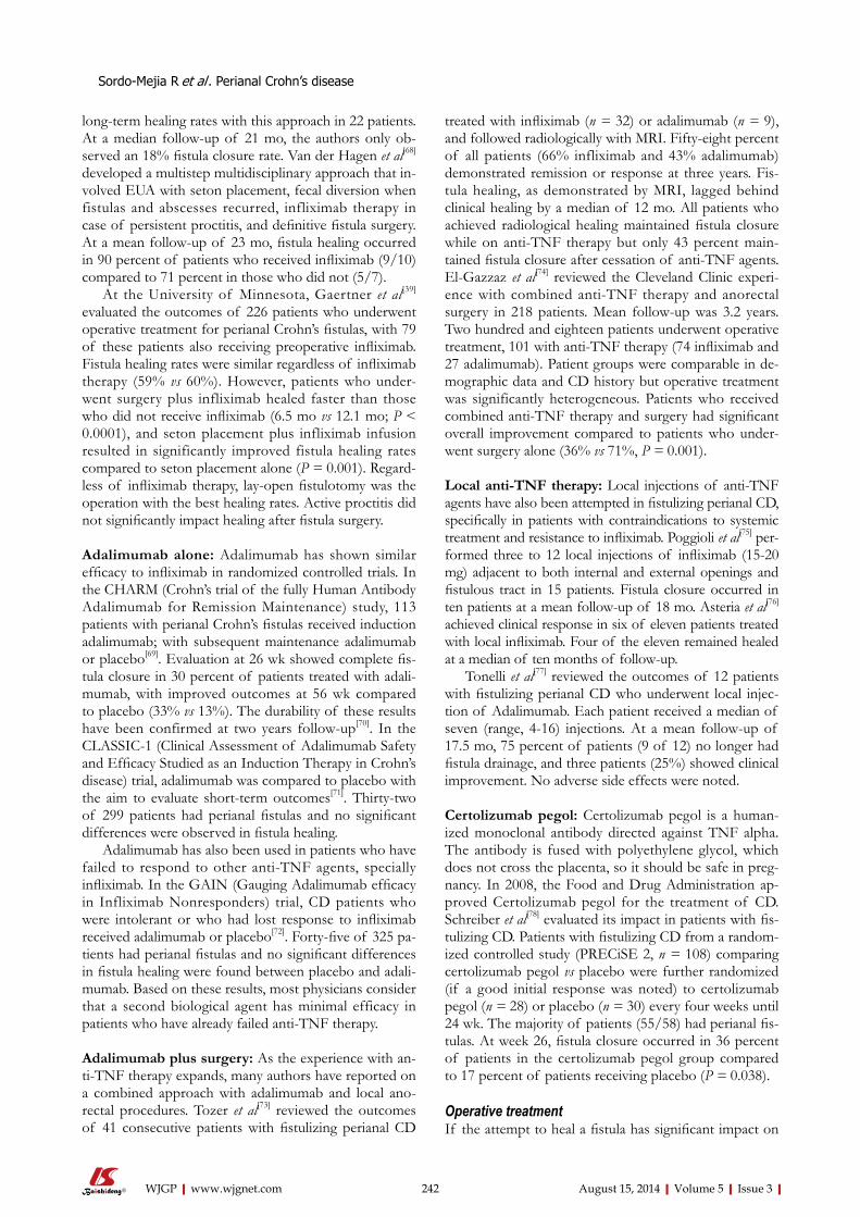
long-term healing rates with this approach in 22 patients. At a median follow-up of 21 mo, the authors only ob-served an 18% fistula closure rate. Van der Hagen et al[68] developed a multistep multidisciplinary approach that in-volved EUA with seton placement, fecal diversion when fistulas and abscesses recurred, infliximab therapy in case of persistent proctitis, and definitive fistula surgery. At a mean follow-up of 23 mo, fistula healing occurred in 90 percent of patients who received infliximab (9/10) compared to 71 percent in those who did not (5/7).
At the University of Minnesota, Gaertner et al[39] evaluated the outcomes of 226 patients who underwent operative treatment for perianal Crohn’s fistulas, with 79 of these patients also receiving preoperative infliximab. Fistula healing rates were similar regardless of infliximab therapy (59% vs 60%). However, patients who under-went surgery plus infliximab healed faster than those who did not receive infliximab (6.5 mo vs 12.1 mo; P < 0.0001), and seton placement plus infliximab infusion resulted in significantly improved fistula healing rates compared to seton placement alone (P = 0.001). Regard-less of infliximab therapy, lay-open fistulotomy was the operation with the best healing rates. Active proctitis did not significantly impact healing after fistula surgery.
Adalimumab alone: Adalimumab has shown similar efficacy to infliximab in randomized controlled trials. In the CHARM (Crohn’s trial of the fully Human Antibody Adalimumab for Remission Maintenance) study, 113 patients with perianal Crohn’s fistulas received induction adalimumab; with subsequent maintenance adalimumab or placebo[69]. Evaluation at 26 wk showed complete fis-tula closure in 30 percent of patients treated with adali-mumab, with improved outcomes at 56 wk compared to placebo (33% vs 13%). The durability of these results have been confirmed at two years follow-up[70]. In the CLASSIC-1 (Clinical Assessment of Adalimumab Safety and Efficacy Studied as an Induction Therapy in Crohn’s disease) trial, adalimumab was compared to placebo with the aim to evaluate short-term outcomes[71]. Thirty-two of 299 patients had perianal fistulas and no significant differences were observed in fistula healing.
Adalimumab has also been used in patients who have failed to respond to other anti-TNF agents, specially infliximab. In the GAIN (Gauging Adalimumab efficacy in Infliximab Nonresponders) trial, CD patients who were intolerant or who had lost response to infliximab received adalimumab or placebo[72]. Forty-five of 325 pa-tients had perianal fistulas and no significant differences in fistula healing were found between placebo and adali-mumab. Based on these results, most physicians consider that a second biological agent has minimal efficacy in patients who have already failed anti-TNF therapy.
Adalimumab plus surgery: As the experience with an-ti-TNF therapy expands, many authors have reported on a combined approach with adalimumab and local ano-rectal procedures. Tozer et al[73] reviewed the outcomes of 41 consecutive patients with fistulizing perianal CD
treated with infliximab (n = 32) or adalimumab (n = 9), and followed radiologically with MRI. Fifty-eight percent of all patients (66% infliximab and 43% adalimumab) demonstrated remission or response at three years. Fis-tula healing, as demonstrated by MRI, lagged behind clinical healing by a median of 12 mo. All patients who achieved radiological healing maintained fistula closure while on anti-TNF therapy but only 43 percent main-tained fistula closure after cessation of anti-TNF agents. El-Gazzaz et al[74] reviewed the Cleveland Clinic experi-ence with combined anti-TNF therapy and anorectal surgery in 218 patients. Mean follow-up was 3.2 years. Two hundred and eighteen patients underwent operative treatment, 101 with anti-TNF therapy (74 infliximab and 27 adalimumab). Patient groups were comparable in de-mographic data and CD history but operative treatment was significantly heterogeneous. Patients who received combined anti-TNF therapy and surgery had significant overall improvement compared to patients who under-went surgery alone (36% vs 71%, P = 0.001).
Local anti-TNF therapy: Local injections of anti-TNF agents have also been attempted in fistulizing perianal CD, specifically in patients with contraindications to systemic treatment and resistance to infliximab. Poggioli et al[75] per-formed three to 12 local injections of infliximab (15-20 mg) adjacent to both internal and external openings and fistulous tract in 15 patients. Fistula closure occurred in ten patients at a mean follow-up of 18 mo. Asteria et al[76] achieved clinical response in six of eleven patients treated with local infliximab. Four of the eleven remained healed at a median of ten months of follow-up.
Tonelli et al[77] reviewed the outcomes of 12 patients with fistulizing perianal CD who underwent local injec-tion of Adalimumab. Each patient received a median of seven (range, 4-16) injections. At a mean follow-up of 17.5 mo, 75 percent of patients (9 of 12) no longer had fistula drainage, and three patients (25%) showed clinical improvement. No adverse side effects were noted.
Certolizumab pegol: Certolizumab pegol is a human-ized monoclonal antibody directed against TNF alpha. The antibody is fused with polyethylene glycol, which does not cross the placenta, so it should be safe in preg-nancy. In 2008, the Food and Drug Administration ap-proved Certolizumab pegol for the treatment of CD. Schreiber et al[78] evaluated its impact in patients with fis-tulizing CD. Patients with fistulizing CD from a random-ized controlled study (PRECiSE 2, n = 108) comparing certolizumab pegol vs placebo were further randomized (if a good initial response was noted) to certolizumab pegol (n = 28) or placebo (n = 30) every four weeks until 24 wk. The majority of patients (55/58) had perianal fis-tulas. At week 26, fistula closure occurred in 36 percent of patients in the certolizumab pegol group compared to 17 percent of patients receiving placebo (P = 0.038).
Operative treatmentIf the attempt to heal a fistula has significant impact on
August 15, 2014|Volume 5|Issue 3|WJGP|www.wjgnet.com 242
Sordo-Mejia R et al . Perianal Crohn’s disease
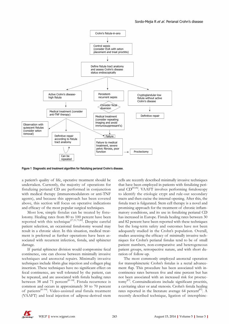
a patient’s quality of life, operative treatment should be undertaken. Currently, the majority of operations for fistulizing perianal CD are performed in conjunction with medical therapy (immunomodulators or anti-TNF agents), and because this approach has been covered above, this section will focus on operative indications and efficacy of the most popular surgical techniques.
Most low, simple fistulas can be treated by fistu-lotomy. Healing rates from 80 to 100 percent have been reported with this technique[27,31,79,80]. Despite careful patient selection, an occasional fistulotomy wound may result in a chronic ulcer. In this situation, medical treat-ment is preferred as further operations have been as-sociated with recurrent infection, fistula, and sphincter damage.
If partial sphincter division would compromise fecal continence, one can choose between minimally invasive techniques and anorectal repairs. Minimally invasive techniques include fibrin glue injection and collagen plug insertion. These techniques have no significant effect on fecal continence, are well tolerated by the patient, can be repeated, and are associated with fistula healing rates between 38 and 71 percent[81-84]. Fistula recurrence is common and occurs in approximately 50 to 70 percent of patients[81-84]. Video-assisted anal fistula treatment (VAAFT) and local injection of adipose-derived stem
cells are recently described minimally invasive techniques that have been employed in patients with fistulizing peri-anal CD[85,86]. VAAFT involves performing fistuloscopy to identify the etiologic crypt and rule-out secondary tracts and then excise the internal opening. After this, the fistula tract is fulgurated. Stem cell therapy is a novel and promising approach for the treatment of chronic inflam-matory conditions, and its use in fistulizing perianal CD has increased in Europe. Fistula healing rates between 30 and 82 percent have been reported with these techniques but the long-term safety and outcomes have not been adequately studied in the Crohn’s population. Overall, studies assessing the efficacy of minimally invasive tech-niques for Crohn’s perianal fistulas tend to be of small patient numbers, non-comparative and heterogeneous patient groups, retrospective nature, and with short du-ration of follow-up.
The most commonly employed anorectal operation for transsphincteric Crohn’s fistulas is a rectal advance-ment flap. This procedure has been associated with in-continence rates between five and nine percent but has not been associated with an increased risk for proctec-tomy[87]. Contraindications include significant proctitis, a cavitating ulcer or anal stenosis. Crohn’s fistula healing rates reported in the literature average 64 percent[87]. A recently described technique, ligation of intersphinc-
August 15, 2014|Volume 5|Issue 3|WJGP|www.wjgnet.com 243
Crohn's fistula-in-ano
Control sepsis(consider EUA with seton placement and treat proctitis)
Define fistula tract anatomy and assess Crohn's disease status endoscopically
Active Crohn's disease-high fistula
Observation with quiescent fistulas (consider seton removal)
Medical treatment (consider anti-TNF therapy)
Definitive repair according to fistula tract anatomy
Can be repeated
Cryptoglandular-low fistula without active Crohn's disease
Definitive repair
Proctectomy
Failure to medical treatment, severe pelvic fibrosis, poor function
Persistent-recurrent sepsis
Consider fecal diversion
Medical treatment (consider repeating imaging and avoid immunosuppressant's)
Failure
Figure 1 Diagnostic and treatment algorithm for fistulizing perianal Crohn’s disease.
Sordo-Mejia R et al . Perianal Crohn’s disease
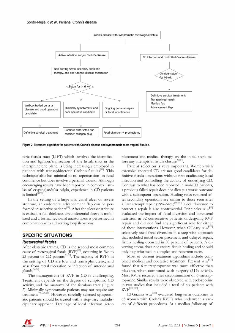
teric fistula tract (LIFT) which involves the identifica-tion and ligation/transection of the fistula tract in the intersphincteric plane, is being increasingly employed in patients with transsphincteric Crohn’s fistulas[88]. This technique also has minimal to no repercussion on fecal continence but does involve a perianal wound. Although encouraging results have been reported in complex fistu-las of cryptoglandular origin, experience in CD patients is limited[89,90].
In the setting of a large anal canal ulcer or severe stricture, an endorectal advancement flap can be per-formed in selective patients[91]. After the ulcer or stricture is excised, a full-thickness circumferential sleeve is mobi-lized and a formal rectoanal anastomosis is performed in combination with a diverting loop ileostomy.
SPECIFIC SITUATIONSRectovaginal fistulasAfter obstetric trauma, CD is the second most common cause of rectovaginal fistula (RVF)[92], occurring in five to 23 percent of CD patients[93-95]. The majority of RVF’s in the setting of CD are low and transsphincteric, and arise from rectal ulceration or infection of anterior anal glands[94,96].
The management of RVF in CD is challenging. Treatment depends on the degree of symptoms, CD activity, and the anatomy of the fistulous tract (Figure 2). Minimally symptomatic patients may not require any treatment[7,94,97,98]. However, carefully selected symptom-atic patients should be treated with a step-wise multidis-ciplinary approach. Drainage of local infection, seton
placement and medical therapy are the initial steps be-fore any attempts at fistula closure[92,94].
Patient selection is very important. Women with extensive anorectal CD are not good candidates for de-finitive fistula operations without first eradicating local infection and controlling the activity of underlying CD. Contrast to what has been reported in non-CD patients, a previous failed repair does not dictate a worse outcome with a subsequent operation. Healing rates reported af-ter secondary operations are similar to those seen after a first attempt repair (29%-54%)[99-101]. Fecal diversion to protect a repair is also controversial. Penninckx et al[99] evaluated the impact of fecal diversion and parenteral nutrition in 32 consecutive patients undergoing RVF repair and did not find any significant role for either of these interventions. However, when O’Leary et al[102]
selectively used fecal diversion in a step-wise approach that included initial seton placement and delayed repair, fistula healing occurred in 80 percent of patients. A di-verting stoma does not ensure fistula healing and should only be performed in complex and recurrent cases.
Most of current treatment algorithms include com-bined medical and operative treatment. Present et al[103] found that 6-mercaptopurine was more effective than placebo, when combined with surgery (31% vs 6%). Most RVF’s recurred after discontinuation of 6-mercap-topurine. Similar results were observed with cyclosporine in two studies that included a total of six patients with RVF[104,105].
El-Gazzaz et al[106] evaluated long-term outcomes in 65 women with Crohn’s RVF´s who underwent a vari-ety of different procedures. At a median follow-up of
August 15, 2014|Volume 5|Issue 3|WJGP|www.wjgnet.com 244
Crohn's disease with symptomatic rectovaginal fistula
Seton for ≥ 6 wk
Definitive surgical treatment
Minimally symptomatic and poor operative candidate
Continue with seton and consider collagen plug
No infection and controlled Crohn's disease
Consider seton for 4-6 wk
Definitive surgical treatment:Transperineal repairMartius flapAdvancement flap
Active infection and/or Crohn’s disease
Non-cutting seton insertion, antibiotic therapy, and anti-Crohn's disease medication
Well-controlled perianal disease and good operative candidate
Fecal diversion ± proctectomy
Ongoing perianal sepsis or fecal incontinence
Figure 2 Treatment algorithm for patients with Crohn’s disease and symptomatic recto-vaginal fistulae.
Sordo-Mejia R et al . Perianal Crohn’s disease
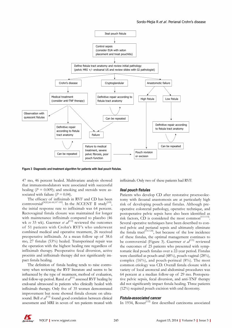
47 mo, 46 percent healed. Multivariate analysis showed that immunomodulators were associated with successful healing (P = 0.009); and smoking and steroids were as-sociated with failure (P = 0.04).
The efficacy of infliximab in RVF and CD has been controversial[38,62,66-68,107-109]. In the ACCENT Ⅱ study[109], the initial response rate to infliximab was 64 percent. Rectovaginal fistula closure was maintained for longer with maintenance infliximab compared to placebo (46 wk vs 33 wk). Gaertner et al[110] reviewed the outcomes of 51 patients with Crohn’s RVF’s who underwent combined medical and operative treatment, 26 received preoperative infliximab. At a mean follow up of 38.6 mo, 27 fistulas (53%) healed. Transperineal repair was the operation with the highest healing rate regardless of infliximab therapy. Preoperative fecal diversion, active proctitis and infliximab therapy did not significantly im-pact fistula healing.
The definition of fistula healing tends to raise contro-versy when reviewing the RVF literature and seems to be influenced by the type of treatment, method of evaluation, and follow-up period. Rasul et al[111] assessed RVF healing by endoanal ultrasound in patients who clinically healed with infliximab therapy. Only five of 35 women demonstrated improvement but none showed fistula closure on ultra-sound. Bell et al[112] found good correlation between clinical assessment and MRI in seven of ten patients treated with
infliximab. Only two of these patients had RVF.
Ileal pouch fistulasPatients who develop CD after restorative proctocolec-tomy with ileoanal anastomosis are at particularly high risk of developing pouch-anal fistulas. Although pre-operative colorectal pathology, operative technique, and postoperative pelvic sepsis have also been identified as risk factors, CD is considered the most common[113-115]. Several operative techniques have been described to con-trol pelvic and perianal sepsis and ultimately eliminate the fistula tract[116-120], but because of the low incidence of these fistulas, the optimal management continues to be controversial (Figure 3). Gaertner et al[121] reviewed the outcomes of 25 patients who presented with symp-tomatic ileal pouch fistulas over a 22-year period. Fistulas were classified as pouch-anal (48%), pouch-vaginal (28%), complex (16%), and pouch-perineal (8%). The most common etiology was CD. Overall fistula closure with a variety of local anorectal and abdominal procedures was 64 percent at a median follow-up of 29 mo. Postopera-tive pelvic sepsis, fecal diversion, and anti-TNF therapy did not significantly impact fistula healing. Three patients (12%) required pouch excision with end ileostomy.
Fistula-associated cancerIn 1934, Rosser[122] first described carcinoma associated
August 15, 2014|Volume 5|Issue 3|WJGP|www.wjgnet.com 245
Ileal pouch fistula
Crohn’s disease Cryptoglandular Anastomotic failure
Observation with quiescent fistulas
Medical treatment(consider anti-TNF therapy)
Definitive repair according to fistula tract anatomy
High fistula Low fistula
Can be repeated
Failure
Can be repeated
Failure to medical treatment, severe pelvic fibrosis, poor pouch function
Pouch revision or excision
Control sepsis(consider EUA with seton placement and treat pouchitis)
Define fistula tract anatomy and review initial pathology(pelvic MRI +/- endoanal US and review slides with GI pathologist)
Can be repeated
Definitive repair according to fistula tract anatomyDefinitive repair
according to fistula tract anatomy
Figure 3 Diagnostic and treatment algorithm for patients with ileal pouch fistulas.
Sordo-Mejia R et al . Perianal Crohn’s disease
with a chronic perianal fistula. Fistula-associated adeno-carcinoma is a rare but increasingly reported malignan-cy[18,21,123-131] that is commonly found in CD patients with chronic anal fistulas[18,21]. This malignancy is frequently associated with chronic, complex fistulas and can be particularly difficult to diagnose. High clinical suspicion is crucial to avoid any delay in diagnosis and treatment. Chronic infection and inflammation (i.e., CD and radia-tion) are the most frequently associated risk factors but even when the diagnosis is suspected clinically, confirma-tion requires EUA with biopsy. Misdiagnosis commonly occurs in elderly patients and patients with long-standing anorectal disease. Once the diagnosis of cancer has been established, EAUS and MRI are recommended for stag-ing[132].
Mucinous adenocarcinoma is the most common ma-lignancy reported in long-standing perianal fistulas. It is typically a slow growing, locally aggressive neoplasm that mainly spreads via the inguinal lymphatic’s[133]. Outcomes are good when malignancy is diagnosed early[131,133-136]. Oncologic resection remains the standard treatment op-tion. Abdominoperineal resection is the most frequently employed operation[131,137,138]. The role of neoadjuvant chemoradiotherapy in the treatment of this neoplasm has not been well studied, probably because of its rarity, but results are promising[21,131]. Neoadjuvant therapy may play a significant role to improve outcomes but remains investigational.
Gaertner et al[131] identified 14 patients with fistula-associated anal adenocarcinoma. The most common presentation was persistent perianal fistula (n = 9). Ten patients (71%) had CD. Abdominoperineal resection was performed in eleven patients, seven following neoadju-vant chemoradiotherapy. At a mean follow-up of 64 mo, ten patients were alive without evidence of disease and four patients died with metastatic disease. All seven pa-tients who received neoadjuvant chemoradiotherapy had a complete pathologic response. In a systematic review by Iesalnieks et al[21], a total of 23 publications including 65 patients with fistula-associated adenocarcinoma and CD were reviewed. Abdominoperineal resection was performed in 56 patients with an overall 3-year survival rate of 54 percent.
We recommend that tissue from refractory, recurrent and chronic anal fistula tracts, regardless of their etiol-ogy, should be submitted for pathologic evaluation. All patients with long-standing perianal CD should undergo cancer surveillance. Although the impact of neoadjuvant chemoradiotherapy remains controversial, oncologic resection continues to be the standard treatment option for fistula-associated adenocarcinoma.
ProctectomyProctectomy is appropriate in patients in whom repeated medical and operative strategies fail. Historically, it is required in ten to 20 percent of patients with perianal CD[6], and is commonly associated with perineal wound breakdown, chronic open wounds and sinus formation
in up to half of patients[139,140]. In our experience, inter-sphincteric proctectomy (when feasible) and the use of rectus abdominal and gracilis flaps can help with avoid-ing these complications.
A low Hartmann’s procedure is an alternative ap-proach that may result in a healed perineum in up to 60 percent of patients with perianal CD[141]. Despite this approach, Guillem et al[142] reported a 54 percent comple-tion proctectomy rate in 28 patients who underwent rectal exclusion, plus an additional nine patients had per-sistent active disease at the rectal stump.
CONCLUSIONThe appropriate treatment of fistulizing perianal CD must be individualized to each patient. The primary goals are to ameliorate symptoms and prevent complica-tions. Overall, a less aggressive approach is preferred as many patients will require repetitive operations that can often result in outcomes that are worse than the disease itself.
Based on the current literature, multidisciplinary treatment includes: eradication of infection, assessment of CD status and fistula tract(s), medical therapy, and selective operative management. The first phase of treat-ment is to drain the perianal infection. This typically involves an EUA, seton drainage and a short course of antibiotics. The second phase consists of endoscopically evaluating the extent of CD and delimiting the anatomy of the fistula tract with EUA and either EAUS or MRI, or both. During this phase, medical therapy with immu-nomodulators and anti-TNF agents is typically initiated but if the fistula is thought to be of cryptoglandular eti-ology, CD medications are rarely required.
The third phase should ideally involve healing of the perianal pathology. Many patients who have minimal symptoms elect to continue with a non-cutting seton or removal and expect healing in some cases. On many oc-casions a non-cutting seton may actually act as a cutting seton, specially in low superficial fistula tracts. The ex-tensive range of operations highlights the complexity of operative treatment. These include a variety of minimally invasive techniques and anorectal operations. Sphincter injury and fecal incontinence should be the main con-cern with any anorectal operation. The operative ap-proach depends on the anatomy of the fistula tract, CD status, and the patients’ functional status. Attempts to heal a fistula in the setting of active infection and proc-titis are likely to fail. If the patient’s symptoms persist or increase despite adequate medical and surgical treatment, a diverting stoma or proctectomy should be considered.
REFERENCES1 Gabriel WB. Results of an Experimental and Histological
Investigation into Seventy-five Cases of Rectal Fistulæ. Proc R Soc Med 1921; 14: 156-161 [PMID: 20906483]
2 Crohn BB, Ginzberg L, Oppenheimer GD. Regional ileitis: a pathologic and clinical entity. JAMA 1932; 99: 1323 [DOI:
August 15, 2014|Volume 5|Issue 3|WJGP|www.wjgnet.com 246
Sordo-Mejia R et al . Perianal Crohn’s disease
10.1001/jama.1932.02740680019005]3 Bissell AD. Localized Chronic Ulcerative Ileitis. Ann Surg
1934; 99: 957-966 [PMID: 17867210 DOI: 10.1097/00000658-193406000-00011]
4 Morson BC, Lockhart-Mummery HE. Anal lesions in Crohn’s disease. Lancet 1959; 2: 1122-1123 [PMID: 14424442 DOI: 10.1016/S0140-6736(59)90104-7]
5 Hellers G, Bergstrand O, Ewerth S, Holmström B. Occur-rence and outcome after primary treatment of anal fistulae in Crohn’s disease. Gut 1980; 21: 525-527 [PMID: 7429313 DOI: 10.1136/gut.21.6.525]
6 Sandborn WJ, Fazio VW, Feagan BG, Hanauer SB. AGA technical review on perianal Crohn’s disease. Gastroenterol-ogy 2003; 125: 1508-1530 [PMID: 14598268 DOI: 10.1016/j.gastro.2003.08.025]
7 Schwartz DA, Loftus EV, Tremaine WJ, Panaccione R, Harmsen WS, Zinsmeister AR, Sandborn WJ. The natural history of fistulizing Crohn’s disease in Olmsted County, Minnesota. Gastroenterology 2002; 122: 875-880 [PMID: 11910338 DOI: 10.1053/gast.2002.32362]
8 Lockhart-Mummery HE. Symposium. Crohn’s disease: anal lesions. Dis Colon Rectum 1975; 18: 200-202 [PMID: 1140047 DOI: 10.1007/BF02587272]
9 Williams DR, Coller JA, Corman ML, Nugent FW, Vei-denheimer MC. Anal complications in Crohn‘s disease. Dis Colon Rectum 1981; 24: 22-24 [PMID: 7472097 DOI: 10.1007/BF02603444]
10 Gray BK, Lockhartmummery HE, Morson BC. Crohn’s dis-ease of the anal region. Gut 1965; 6: 515-524 [PMID: 5857889 DOI: 10.1136/gut.6.6.515]
11 Cosnes J, Cattan S, Blain A, Beaugerie L, Carbonnel F, Parc R, Gendre JP. Long-term evolution of disease behavior of Crohn’s disease. Inflamm Bowel Dis 2002; 8: 244-250 [PMID: 12131607 DOI: 10.1097/00054725-200207000-00002]
12 Roberts PL, Schoetz DJ, Pricolo R, Veidenheimer MC. Clinical course of Crohn‘s disease in older patients. A ret-rospective study. Dis Colon Rectum 1990; 33: 458-462 [PMID: 2350997 DOI: 10.1007/BF02052138]
13 Beaugerie L, Seksik P, Nion-Larmurier I, Gendre JP, Cosnes J. Predictors of Crohn’s disease. Gastroenterology 2006; 130: 650-656 [PMID: 16530505 DOI: 10.1053/j.gastro.2005.12.019]
14 Rankin GB, Watts HD, Melnyk CS, Kelley ML. National Cooperative Crohn’s Disease Study: extraintestinal manifes-tations and perianal complications. Gastroenterology 1979; 77: 914-920 [PMID: 467943]
15 Gelbmann CM, Rogler G, Gross V, Gierend M, Bregenzer N, Andus T, Schölmerich J. Prior bowel resections, perianal dis-ease, and a high initial Crohn’s disease activity index are as-sociated with corticosteroid resistance in active Crohn’s dis-ease. Am J Gastroenterol 2002; 97: 1438-1445 [PMID: 12094862 DOI: 10.1111/j.1572-0241.2002.05685.x]
16 Makowiec F, Jehle EC, Starlinger M. Clinical course of peri-anal fistulas in Crohn’s disease. Gut 1995; 37: 696-701 [PMID: 8549948 DOI: 10.1136/gut.37.5.696]
17 Sjödahl RI, Myrelid P, Söderholm JD. Anal and rectal can-cer in Crohn’s disease. Colorectal Dis 2003; 5: 490-495 [PMID: 12925087 DOI: 10.1046/j.1463-1318.2003.00510.x]
18 Ky A, Sohn N, Weinstein MA, Korelitz BI. Carcinoma aris-ing in anorectal fistulas of Crohn’s disease. Dis Colon Rectum 1998; 41: 992-996 [DOI: 10.1007/BF02237388]
19 Connell WR, Sheffield JP, Kamm MA, Ritchie JK, Hawley PR, Lennard-Jones JE. Lower gastrointestinal malignancy in Crohn’s disease. Gut 1994; 35: 347-352 [PMID: 8150345 DOI: 10.1136/gut.35.3.347]
20 Weedon DD, Shorter RG, Ilstrup DM, Huizenga KA, Taylor WF. Crohn’s disease and cancer. N Engl J Med 1973; 289: 1099-1103 [PMID: 4754948 DOI: 10.1056/NEJM197311222892101]
21 Iesalnieks I, Gaertner WB, Glass H, Strauch U, Hipp M, Agha A, Schlitt HJ. Fistula-associated anal adenocarcinoma in Crohn’s disease. Inflamm Bowel Dis 2010; 16: 1643-1648
[PMID: 20186945 DOI: 10.1002/ibd.21228]22 Wolff BG, Culp CE, Beart RW, Ilstrup DM, Ready RL. Ano-
rectal Crohn’s disease. A long-term perspective. Dis Colon Rectum 1985; 28: 709-711 [PMID: 4053875 DOI: 10.1007/BF02560279]
23 Solomon MJ. Fistulae and abscesses in symptomatic peri-anal Crohn’s disease. Int J Colorectal Dis 1996; 11: 222-226 [PMID: 8951512 DOI: 10.1007/s003840050051]
24 Makowiec F, Jehle EC, Becker HD, Starlinger M. Perianal abscess in Crohn’s disease. Dis Colon Rectum 1997; 40: 443-450 [PMID: 9106694 DOI: 10.1007/BF02258390]
25 Singh B, McC Mortensen NJ, Jewell DP, George B. Perianal Crohn’s disease. Br J Surg 2004; 91: 801-814 [PMID: 15227686 DOI: 10.1002/bjs.4613]
26 Pritchard TJ, Schoetz DJ, Roberts PL, Murray JJ, Coller JA, Veidenheimer MC. Perirectal abscess in Crohn’s disease. Drainage and outcome. Dis Colon Rectum 1990; 33: 933-937 [PMID: 2226080 DOI: 10.1007/BF02139102]
27 Sohn N, Korelitz BI, Weinstein MA. Anorectal Crohn’s dis-ease: definitive surgery for fistulas and recurrent abscesses. Am J Surg 1980; 139: 394-397 [PMID: 7362011 DOI: 10.1016/0002-9610(80)90301-3]
28 Giovannini M, Bories E, Moutardier V, Pesenti C, Guil-lemin A, Lelong B, Delpéro JR. Drainage of deep pelvic ab-scesses using therapeutic echo endoscopy. Endoscopy 2003; 35: 511-514 [PMID: 12783350 DOI: 10.1055/s-2003-39673]
29 Schratter-Sehn AU, Lochs H, Vogelsang H, Schurawitzki H, Herold C, Schratter M. Endoscopic ultrasonography versus computed tomography in the differential diagnosis of peri-anorectal complications in Crohn’s disease. Endoscopy 1993; 25: 582-586 [PMID: 8119208 DOI: 10.1055/s-2007-1010409]
30 Williams JG, Rothenberger DA, Nemer FD, Goldberg SM. Fistula-in-ano in Crohn’s disease. Results of aggressive sur-gical treatment. Dis Colon Rectum 1991; 34: 378-384 [PMID: 2022142 DOI: 10.1007/BF02053687]
31 Halme L, Sainio AP. Factors related to frequency, type, and outcome of anal fistulas in Crohn’s disease. Dis Colon Rectum 1995; 38: 55-59 [PMID: 7813346 DOI: 10.1007/BF02053858]
32 Sangwan YP, Schoetz DJ, Murray JJ, Roberts PL, Coller JA. Perianal Crohn’s disease. Results of local surgical treatment. Dis Colon Rectum 1996; 39: 529-535 [PMID: 8620803 DOI: 10.1007/BF02058706]
33 Faucheron JL, Saint-Marc O, Guibert L, Parc R. Long-term seton drainage for high anal fistulas in Crohn’s disease--a sphincter-saving operation? Dis Colon Rectum 1996; 39: 208-211 [PMID: 8620789 DOI: 10.1007/BF02068077]
34 Pearl RK, Andrews JR, Orsay CP, Weisman RI, Prasad ML, Nelson RL, Cintron JR, Abcarian H. Role of the seton in the management of anorectal fistulas. Dis Colon Rectum 1993; 36: 573-577; discussion 573-577 [PMID: 8500375 DOI: 10.1007/BF02049864]
35 Thornton M, Solomon MJ. Long-term indwelling seton for complex anal fistulas in Crohn’s disease. Dis Colon Rectum 2005; 48: 459-463 [PMID: 15719188 DOI: 10.1007/s10350-004-0830-6]
36 Buchanan GN, Owen HA, Torkington J, Lunniss PJ, Nich-olls RJ, Cohen CR. Long-term outcome following loose-seton technique for external sphincter preservation in com-plex anal fistula. Br J Surg 2004; 91: 476-480 [PMID: 15048751 DOI: 10.1002/bjs.4466]
37 Eitan A, Koliada M, Bickel A. The use of the loose seton tech-nique as a definitive treatment for recurrent and persistent high trans-sphincteric anal fistulas: a long-term outcome. J Gastrointest Surg 2009; 13: 1116-1119 [PMID: 19238493 DOI: 10.1007/s11605-009-0826-6]
38 Topstad DR, Panaccione R, Heine JA, Johnson DR, Ma-cLean AR, Buie WD. Combined seton placement, infliximab infusion, and maintenance immunosuppressives improve healing rate in fistulizing anorectal Crohn’s disease: a single center experience. Dis Colon Rectum 2003; 46: 577-583 [PMID:
August 15, 2014|Volume 5|Issue 3|WJGP|www.wjgnet.com 247
Sordo-Mejia R et al . Perianal Crohn’s disease
12792431 DOI: 10.1007/s10350-004-6611-4]39 Gaertner WB, Decanini A, Mellgren A, Lowry AC, Gold-
berg SM, Madoff RD, Spencer MP. Does infliximab infusion impact results of operative treatment for Crohn’s perianal fistulas? Dis Colon Rectum 2007; 50: 1754-1760 [PMID: 17899271 DOI: 10.1007/s10350-007-9077-3]
40 Edwards CM, George BD, Jewell DP, Warren BF, Mortensen NJ, Kettlewell MG. Role of a defunctioning stoma in the man-agement of large bowel Crohn’s disease. Br J Surg 2000; 87: 1063-1066 [PMID: 10931051 DOI: 10.1046/j.1365-2168.2000.01467.x]
41 Tozer P. Dendritic cell homing and immune cell function in Crohn’s anal fistulae. Gut 2011; 60: A220-1 [DOI: 10.1136/gut.2011.239301.465]
42 Buchanan GN, Halligan S, Bartram CI, Williams AB, Tar-roni D, Cohen CR. Clinical examination, endosonography, and MR imaging in preoperative assessment of fistula in ano: comparison with outcome-based reference standard. Radiology 2004; 233: 674-681 [PMID: 15498901 DOI: 10.1148/radiol.2333031724]
43 Schaefer O, Lohrmann C, Langer M. Assessment of anal fistulas with high-resolution subtraction MR-fistulography: comparison with surgical findings. J Magn Reson Imaging 2004; 19: 91-98 [PMID: 14696225 DOI: 10.1002/jmri.10436]
44 Beets-Tan RG, Beets GL, van der Hoop AG, Kessels AG, Vliegen RF, Baeten CG, van Engelshoven JM. Preoperative MR imaging of anal fistulas: Does it really help the surgeon? Radiology 2001; 218: 75-84 [PMID: 11152782 DOI: 10.1148/radiology.218.1.r01dc0575]
45 Buchanan GN, Halligan S, Williams AB, Cohen CR, Tarroni D, Phillips RK, Bartram CI. Magnetic resonance imaging for primary fistula in ano. Br J Surg 2003; 90: 877-881 [PMID: 12854117 DOI: 10.1002/bjs.4125]
46 Sachar DB, Bodian CA, Goldstein ES, Present DH, Bay-less TM, Picco M, van Hogezand RA, Annese V, Schneider J, Korelitz BI, Cosnes J. Is perianal Crohn’s disease as-sociated with intestinal fistulization? Am J Gastroenterol 2005; 100: 1547-1549 [PMID: 15984979 DOI: 10.1111/j.1572-0241.2005.40980.x]
47 Marks CG, Ritchie JK, Lockhart-Mummery HE. Anal fis-tulas in Crohn’s disease. Br J Surg 1981; 68: 525-527 [PMID: 7272665 DOI: 10.1002/bjs.1800680802]
48 Orkin BA, Telander RL. The effect of intra-abdominal re-section or fecal diversion on perianal disease in pediatric Crohn’s disease. J Pediatr Surg 1985; 20: 343-347 [PMID: 4045658 DOI: 10.1016/S0022-3468(85)80216-5]
49 Dejaco C, Harrer M, Waldhoer T, Miehsler W, Vogelsang H, Reinisch W. Antibiotics and azathioprine for the treatment of perianal fistulas in Crohn’s disease. Aliment Pharmacol Ther 2003; 18: 1113-1120 [PMID: 14653831 DOI: 10.1046/j.1365-2036.2003.01793.x]
50 Bernstein LH, Frank MS, Brandt LJ, Boley SJ. Healing of perineal Crohn’s disease with metronidazole. Gastroenterol-ogy 1980; 79: 599 [PMID: 7429123]
51 Turunen UM, Färkkilä MA, Hakala K, Seppälä K, Sivonen A, Ogren M, Vuoristo M, Valtonen VV, Miettinen TA. Long-term treatment of ulcerative colitis with ciprofloxa-cin: a prospective, double-blind, placebo-controlled study. Gastroenterology 1998; 115: 1072-1078 [PMID: 9797360 DOI: 10.1016/S0016-5085(98)70076-9]
52 Brandt LJ, Bernstein LH, Boley SJ, Frank MS. Metronidazole therapy for perineal Crohn’s disease: a follow-up study. Gastroenterology 1982; 83: 383-387 [PMID: 7084615]
53 Thia KT, Mahadevan U, Feagan BG, Wong C, Cockeram A, Bitton A, Bernstein CN, Sandborn WJ. Ciprofloxacin or met-ronidazole for the treatment of perianal fistulas in patients with Crohn’s disease: a randomized, double-blind, placebo-controlled pilot study. Inflamm Bowel Dis 2009; 15: 17-24 [PMID: 18668682 DOI: 10.1002/ibd.20608]
54 Turunen U, Farkkila M, Seppala K. What is New and What
is Established in Inflammatory Bowel Diseases: Proceedings of the Athens International Meeting on Inflammatory Bowel Diseases Athens, 19–23 April, 1989. Longterm treatment of peri-anal or fistulous Crohn’s disease with ciprofloxacin. Scand J Gastroenterol 1989; 24: 144
55 Solomon M, McLeod R, O’Connor B, Steinhart A, Green-berg G, Cohen Z. Combination ciprofloxacin and metroni-dazole in severe perineal Crohn’s disease. Can J Gastroenterol 1993; 7: 571-573
56 Pearson DC, May GR, Fick GH, Sutherland LR. Azathio-prine and 6-mercaptopurine in Crohn disease. A meta-analysis. Ann Intern Med 1995; 123: 132-142 [PMID: 7778826 DOI: 10.7326/0003-4819-123-2-199507150-00009]
57 Hinterleitner TA, Petritsch W, Aichbichler B, Fickert P, Ranner G, Krejs GJ. Combination of cyclosporine, azathio-prine and prednisolone for perianal fistulas in Crohn’s dis-ease. Z Gastroenterol 1997; 35: 603-608 [PMID: 9297775]
58 Egan LJ, Sandborn WJ, Tremaine WJ. Clinical outcome fol-lowing treatment of refractory inflammatory and fistulizing Crohn’s disease with intravenous cyclosporine. Am J Gas-troenterol 1998; 93: 442-448 [PMID: 9517654 DOI: 10.1111/j.1572-0241.1998.00442.x]
59 Gurudu SR, Griffel LH, Gialanella RJ, Das KM. Cyclospo-rine therapy in inflammatory bowel disease: short-term and long-term results. J Clin Gastroenterol 1999; 29: 151-154 [PMID: 10478875 DOI: 10.1097/00004836-199909000-00009]
60 Sandborn WJ, Present DH, Isaacs KL, Wolf DC, Greenberg E, Hanauer SB, Feagan BG, Mayer L, Johnson T, Galanko J, Martin C, Sandler RS. Tacrolimus for the treatment of fistu-las in patients with Crohn’s disease: a randomized, placebo-controlled trial. Gastroenterology 2003; 125: 380-388 [PMID: 12891539 DOI: 10.1016/S0016-5085(03)00877-1]
61 Sokol H, Seksik P, Carrat F, Nion-Larmurier I, Vienne A, Beaugerie L, Cosnes J. Usefulness of co-treatment with im-munomodulators in patients with inflammatory bowel dis-ease treated with scheduled infliximab maintenance therapy. Gut 2010; 59: 1363-1368 [PMID: 20587545 DOI: 10.1136/gut.2010.212712]
62 Present DH, Rutgeerts P, Targan S, Hanauer SB, Mayer L, van Hogezand RA, Podolsky DK, Sands BE, Braakman T, DeWoody KL, Schaible TF, van Deventer SJ. Infliximab for the treatment of fistulas in patients with Crohn’s disease. N Engl J Med 1999; 340: 1398-1405 [PMID: 10228190 DOI: 10.1056/NEJM199905063401804]
63 Miheller P, Lakatos PL, Horváth G, Molnár T, Szamosi T, Czeglédi Z, Salamon A, Czimmer J, Rumi G, Palatka K, Papp M, Jakab Z, Szabó A, Gelley A, Lakatos L, Barta Z, Balázs C, Rácz I, Zeher M, Döbrönte Z, Altorjay I, Hunyady B, Simon L, Papp J, Banai J, Nagy F, Lonovics J, Ujszászy L, Muzes G, Herszényi L, Tulassay Z. Efficacy and safety of infliximab induction therapy in Crohn’s Disease in Central Europe--a Hungarian nationwide observational study. BMC Gastroenterol 2009; 9: 66 [PMID: 19740450 DOI: 10.1186/1471-230X-9-66]
64 Orlando A, Colombo E, Kohn A, Biancone L, Rizzello F, Viscido A, Sostegni R, Benazzato L, Castiglione F, Papi C, Meucci G, Riegler G, Mocciaro F, Cassinotti A, Cosintino R, Geremia A, Morselli C, Angelucci E, Lavagna A, Rispo A, Bossa F, Scimeca D, Cottone M. Infliximab in the treatment of Crohn’s disease: predictors of response in an Italian mul-ticentric open study. Dig Liver Dis 2005; 37: 577-583 [PMID: 15886081 DOI: 10.1016/j.dld.2005.01.019]
65 Ng SC, Plamondon S, Gupta A, Burling D, Swatton A, Vaizey CJ, Kamm MA. Prospective evaluation of anti-tumor necrosis factor therapy guided by magnetic resonance im-aging for Crohn’s perineal fistulas. Am J Gastroenterol 2009; 104: 2973-2986 [PMID: 19755971 DOI: 10.1038/ajg.2009.509]
66 Regueiro M, Mardini H. Treatment of perianal fistulizing Crohn’s disease with infliximab alone or as an adjunct to exam under anesthesia with seton placement. Inflamm Bowel
August 15, 2014|Volume 5|Issue 3|WJGP|www.wjgnet.com 248
Sordo-Mejia R et al . Perianal Crohn’s disease
Dis 2003; 9: 98-103 [PMID: 12769443 DOI: 10.1097/00054725-200303000-00003]
67 Hyder SA, Travis SP, Jewell DP, McC Mortensen NJ, George BD. Fistulating anal Crohn’s disease: results of combined surgical and infliximab treatment. Dis Colon Rectum 2006; 49: 1837-1841 [PMID: 17041753 DOI: 10.1007/s10350-006-0656-5]
68 van der Hagen SJ, Baeten CG, Soeters PB, Russel MG, Beets-Tan RG, van Gemert WG. Anti-TNF-alpha (infliximab) used as induction treatment in case of active proctitis in a multistep strategy followed by definitive surgery of com-plex anal fistulas in Crohn’s disease: a preliminary report. Dis Colon Rectum 2005; 48: 758-767 [PMID: 15750797 DOI: 10.1007/s10350-004-0828-0]
69 Colombel JF, Sandborn WJ, Rutgeerts P, Enns R, Hanauer SB, Panaccione R, Schreiber S, Byczkowski D, Li J, Kent JD, Pollack PF. Adalimumab for maintenance of clinical response and remission in patients with Crohn’s disease: the CHARM trial. Gastroenterology 2007; 132: 52-65 [PMID: 17241859 DOI: 10.1053/j.gastro.2006.11.041]
70 Colombel JF, Schwartz DA, Sandborn WJ, Kamm MA, D’Haens G, Rutgeerts P, Enns R, Panaccione R, Schreiber S, Li J, Kent JD, Lomax KG, Pollack PF. Adalimumab for the treat-ment of fistulas in patients with Crohn’s disease. Gut 2009; 58: 940-948 [PMID: 19201775 DOI: 10.1136/gut.2008.159251]
71 Hanauer SB, Sandborn WJ, Rutgeerts P, Fedorak RN, Lukas M, MacIntosh D, Panaccione R, Wolf D, Pollack P. Human anti-tumor necrosis factor monoclonal antibody (adalimum-ab) in Crohn’s disease: the CLASSIC-I trial. Gastroenterology 2006; 130: 323-333; quiz 591 [PMID: 16472588 DOI: 10.1053/j.gastro.2005.11.030]
72 Sandborn WJ, Rutgeerts P, Enns R, Hanauer SB, Colombel JF, Panaccione R, D’Haens G, Li J, Rosenfeld MR, Kent JD, Pollack PF. Adalimumab induction therapy for Crohn dis-ease previously treated with infliximab: a randomized trial. Ann Intern Med 2007; 146: 829-838 [PMID: 17470824 DOI: 10.7326/0003-4819-146-12-200706190-00159]
73 Tozer P, Ng SC, Siddiqui MR, Plamondon S, Burling D, Gupta A, Swatton A, Tripoli S, Vaizey CJ, Kamm MA, Phillips R, Hart A. Long-term MRI-guided combined anti-TNF-α and thiopurine therapy for Crohn’s perianal fistulas. Inflamm Bowel Dis 2012; 18: 1825-1834 [PMID: 22223472 DOI: 10.1002/ibd.21940]
74 El-Gazzaz G, Hull T, Church JM. Biological immunomodu-lators improve the healing rate in surgically treated perianal Crohn’s fistulas. Colorectal Dis 2012; 14: 1217-1223 [PMID: 22251452 DOI: 10.1111/j.1463-1318.2012.02944.x]
75 Poggioli G, Laureti S, Pierangeli F, Rizzello F, Ugolini F, Gionchetti P, Campieri M. Local injection of Infliximab for the treatment of perianal Crohn’s disease. Dis Colon Rectum 2005; 48: 768-774 [PMID: 15768185 DOI: 10.1007/s10350-004-0832-4]
76 Asteria CR, Ficari F, Bagnoli S, Milla M, Tonelli F. Treat-ment of perianal fistulas in Crohn’s disease by local injection of antibody to TNF-alpha accounts for a favourable clinical response in selected cases: a pilot study. Scand J Gastroenterol 2006; 41: 1064-1072 [PMID: 16938720 DOI: 10.1080/00365520600609941]
77 Tonelli F, Giudici F, Asteria CR. Effectiveness and safety of local adalimumab injection in patients with fistuliz-ing perianal Crohn’s disease: a pilot study. Dis Colon Rectum 2012; 55: 870-875 [PMID: 22810472 DOI: 10.1097/DCR.0b013e31825af532]
78 Schreiber S, Lawrance IC, Thomsen OØ, Hanauer SB, Bloomfield R, Sandborn WJ. Randomised clinical trial: cer-tolizumab pegol for fistulas in Crohn’s disease - subgroup results from a placebo-controlled study. Aliment Pharmacol Ther 2011; 33: 185-193 [PMID: 21083671 DOI: 10.1111/j.1365-2036.2010.04509.x]
79 Fry RD, Shemesh EI, Kodner IJ, Timmcke A. Techniques and results in the management of anal and perianal Crohn’
s disease. Surg Gynecol Obstet 1989; 168: 42-48 [PMID: 2909131]
80 Levien DH, Surrell J, Mazier WP. Surgical treatment of ano-rectal fistula in patients with Crohn’s disease. Surg Gynecol Obstet 1989; 169: 133-136 [PMID: 2756461]
81 Lindsey I, Smilgin-Humphreys MM, Cunningham C, Mortensen NJ, George BD. A randomized, controlled trial of fibrin glue vs. conventional treatment for anal fistula. Dis Colon Rectum 2002; 45: 1608-1615 [PMID: 12473883 DOI: 10.1007/s10350-004-7247-0]
82 Grimaud JC, Munoz-Bongrand N, Siproudhis L, Abramow-itz L, Sénéjoux A, Vitton V, Gambiez L, Flourié B, Héb-uterne X, Louis E, Coffin B, De Parades V, Savoye G, Soulé JC, Bouhnik Y, Colombel JF, Contou JF, François Y, Mary JY, Lémann M. Fibrin glue is effective healing perianal fis-tulas in patients with Crohn’s disease. Gastroenterology 2010; 138: 2275-2281, 2281.e1 [PMID: 20178792 DOI: 10.1053/j.gastro.2010.02.013]
83 Vitton V, Gasmi M, Barthet M, Desjeux A, Orsoni P, Gri-maud JC. Long-term healing of Crohn’s anal fistulas with fi-brin glue injection. Aliment Pharmacol Ther 2005; 21: 1453-1457 [PMID: 15948812 DOI: 10.1111/j.1365-2036.2005.02456.x]
84 O’Riordan JM, Datta I, Johnston C, Baxter NN. A sys-tematic review of the anal fistula plug for patients with Crohn’s and non-Crohn’s related fistula-in-ano. Dis Colon Rectum 2012; 55: 351-358 [PMID: 22469804 DOI: 10.1097/DCR.0b013e318239d1e4]
85 de la Portilla F, Alba F, García-Olmo D, Herrerías JM, González FX, Galindo A. Expanded allogeneic adipose-de-rived stem cells (eASCs) for the treatment of complex peri-anal fistula in Crohn’s disease: results from a multicenter phase I/IIa clinical trial. Int J Colorectal Dis 2013; 28: 313-323 [PMID: 23053677 DOI: 10.1007/s00384-012-1581-9]
86 Schwandner O. Video-assisted anal fistula treatment (VAAFT) combined with advancement flap repair in Crohn’s disease. Tech Coloproctol 2013; 17: 221-225 [PMID: 23179892 DOI: 10.1007/s10151-012-0921-7]
87 Soltani A , Kaiser AM. Endorectal advancement flap for cryptoglandular or Crohn’s fistula-in-ano. Dis Colon Rectum 2010; 53: 486-495 [PMID: 20305451 DOI: 10.1007/DCR.0b013e3181ce8b01]
88 Vergara-Fernandez O, Espino-Urbina LA. Ligation of inter-sphincteric fistula tract: what is the evidence in a review? World J Gastroenterol 2013; 19: 6805-6813 [PMID: 24187455 DOI: 10.3748/wjg.v19.i40.6805]
89 Abcarian AM, Estrada JJ, Park J, Corning C, Chaudhry V, Cintron J, Prasad L, Abcarian H. Ligation of intersphinc-teric fistula tract: early results of a pilot study. Dis Colon Rectum 2012; 55: 778-782 [PMID: 22706130 DOI: 10.1097/DCR.0b013e318255ae8a]
90 Ellis CN. Outcomes with the use of bioprosthetic grafts to re-inforce the ligation of the intersphincteric fistula tract (BioLIFT procedure) for the management of complex anal fistulas. Dis Colon Rectum 2010; 53: 1361-1364 [PMID: 20847616 DOI: 10.1007/DCR.0b013e3181ec4470]
91 Marchesa P, Hull TL, Fazio VW. Advancement sleeve flaps for treatment of severe perianal Crohn’s disease. Br J Surg 1998; 85: 1695-1698 [PMID: 9876077 DOI: 10.1046/j.1365-2168.1998.00959.x]
92 Saclarides TJ. Rectovaginal fistula. Surg Clin North Am 2002; 82: 1261-1272 [PMID: 12516853 DOI: 10.1016/S0039-6109(02)00055-5]
93 Platell C, Mackay J, Collopy B, Fink R, Ryan P, Woods R. Anal pathology in patients with Crohn’s disease. Aust N Z J Surg 1996; 66: 5-9 [PMID: 8629983 DOI: 10.1111/j.1445-2197.1996.tb00690.x]
94 Radcliffe AG, Ritchie JK, Hawley PR, Lennard-Jones JE, Nor-thover JM. Anovaginal and rectovaginal fistulas in Crohn’s disease. Dis Colon Rectum 1988; 31: 94-99 [PMID: 3338350 DOI: 10.1007/BF02562636]
95 Galandiuk S, Kimberling J, Al-Mishlab TG, Stromberg AJ.
August 15, 2014|Volume 5|Issue 3|WJGP|www.wjgnet.com 249
Sordo-Mejia R et al . Perianal Crohn’s disease
Perianal Crohn disease: predictors of need for permanent diversion. Ann Surg 2005; 241: 796-801; discussion 801-802 [PMID: 15849515 DOI: 10.1097/01.sla.0000161030.25860.c1]
96 McClane SJ, Rombeau JL. Anorectal Crohn’s disease. Surg Clin North Am 2001; 81: 169-183, ix [PMID: 11218163 DOI: 10.1016/S0039-6109(05)70279-6]
97 Tuxen PA, Castro AF. Rectovaginal fistula in Crohn’s dis-ease. Dis Colon Rectum 1979; 22: 58-62 [PMID: 421652 DOI: 10.1007/BF02586761]
98 Morrison JG, Gathright JB, Ray JE, Ferrari BT, Hicks TC, Timmcke AE. Results of operation for rectovaginal fistula in Crohn’s disease. Dis Colon Rectum 1989; 32: 497-499 [PMID: 2791787 DOI: 10.1007/BF02554505]
99 Penninckx F, Moneghini D, D’Hoore A, Wyndaele J, Core-mans G, Rutgeerts P. Success and failure after repair of rec-tovaginal fistula in Crohn’s disease: analysis of prognostic factors. Colorectal Dis 2001; 3: 406-411 [PMID: 12790939 DOI: 10.1046/j.1463-1318.2001.00274.x]
100 Lowry AC, Thorson AG, Rothenberger DA, Goldberg SM. Repair of simple rectovaginal fistulas. Influence of previous repairs. Dis Colon Rectum 1988; 31: 676-678 [PMID: 3168676 DOI: 10.1007/BF02552581]
101 MacRae HM, McLeod RS, Cohen Z, Stern H, Reznick R. Treatment of rectovaginal fistulas that has failed previous repair attempts. Dis Colon Rectum 1995; 38: 921-925 [PMID: 7656738 DOI: 10.1007/BF02049726]
102 O’Leary DP, Milroy CE, Durdey P. Definitive repair of anovaginal fistula in Crohn’s disease. Ann R Coll Surg Engl 1998; 80: 250-252 [PMID: 9771223]
103 Present DH, Korelitz BI, Wisch N, Glass JL, Sachar DB, Pasternack BS. Treatment of Crohn’s disease with 6-mercap-topurine. A long-term, randomized, double-blind study. N Engl J Med 1980; 302: 981-987 [PMID: 6102739 DOI: 10.1056/NEJM198005013021801]
104 Present DH, Lichtiger S. Efficacy of cyclosporine in treat-ment of fistula of Crohn’s disease. Dig Dis Sci 1994; 39: 374-380 [PMID: 8313821 DOI: 10.1007/BF02090211]
105 Hanauer SB, Smith MB. Rapid closure of Crohn’s disease fistulas with continuous intravenous cyclosporin A. Am J Gastroenterol 1993; 88: 646-649 [PMID: 8480725]
106 El-Gazzaz G, Hull T, Mignanelli E, Hammel J, Gurland B, Zutshi M. Analysis of function and predictors of failure in women undergoing repair of Crohn’s related rectovaginal fistula. J Gastrointest Surg 2010; 14: 824-829 [PMID: 20232172 DOI: 10.1007/s11605-010-1167-1]
107 Talbot C, Sagar PM, Johnston MJ, Finan PJ, Burke D. Inf-liximab in the surgical management of complex fistulating anal Crohn’s disease. Colorectal Dis 2005; 7: 164-168 [PMID: 15720356 DOI: 10.1111/j.1463-1318.2004.00749.x]
108 Van Assche G, Vanbeckevoort D, Bielen D, Coremans G, Aerden I, Noman M, D’Hoore A, Penninckx F, Marchal G, Cornillie F, Rutgeerts P. Magnetic resonance imaging of the effects of infliximab on perianal fistulizing Crohn’s disease. Am J Gastroenterol 2003; 98: 332-339 [PMID: 12591051 DOI: 10.1016/S0002-9270(02)05909-9]
109 Sands BE, Anderson FH, Bernstein CN, Chey WY, Fea-gan BG, Fedorak RN, Kamm MA, Korzenik JR, Lashner BA, Onken JE, Rachmilewitz D, Rutgeerts P, Wild G, Wolf DC, Marsters PA, Travers SB, Blank MA, van Deventer SJ. Infliximab maintenance therapy for fistulizing Crohn’s dis-ease. N Engl J Med 2004; 350: 876-885 [PMID: 14985485 DOI: 10.1056/NEJMoa030815]
110 Gaertner WB, Madoff RD, Spencer MP, Mellgren A, Gold-berg SM, Lowry AC. Results of combined medical and surgical treatment of recto-vaginal fistula in Crohn’s dis-ease. Colorectal Dis 2011; 13: 678-683 [PMID: 20163426 DOI: 10.1111/j.1463-1318.2010.02234.x]
111 Rasul I, Wilson SR, MacRae H, Irwin S, Greenberg GR. Clinical and radiological responses after infliximab treat-ment for perianal fistulizing Crohn’s disease. Am J Gas-
troenterol 2004; 99: 82-88 [PMID: 14687146 DOI: 10.1046/j.1572-0241.2003.04009.x]
112 Bell SJ, Halligan S, Windsor AC, Williams AB, Wiesel P, Kamm MA. Response of fistulating Crohn’s disease to inf-liximab treatment assessed by magnetic resonance imaging. Aliment Pharmacol Ther 2003; 17: 387-393 [PMID: 12562451 DOI: 10.1046/j.1365-2036.2003.01427.x]
113 Meagher AP, Farouk R, Dozois RR, Kelly KA, Pemberton JH. J ileal pouch-anal anastomosis for chronic ulcerative colitis: complications and long-term outcome in 1310 pa-tients. Br J Surg 1998; 85: 800-803 [PMID: 9667712 DOI: 10.1046/j.1365-2168.1998.00689.x]
114 Fleshman JW, Cohen Z, McLeod RS, Stern H, Blair J. The ileal reservoir and ileoanal anastomosis procedure. Factors affecting technical and functional outcome. Dis Colon Rectum 1988; 31: 10-16 [PMID: 3366021 DOI: 10.1007/BF02552562]
115 Keighley MR, Grobler SP. Fistula complicating restorative proctocolectomy. Br J Surg 1993; 80: 1065-1067 [PMID: 8402069 DOI: 10.1002/bjs.1800800849]
116 Ozuner G, Hull T, Lee P, Fazio VW. What happens to a pel-vic pouch when a fistula develops? Dis Colon Rectum 1997; 40: 543-547 [PMID: 9152180 DOI: 10.1007/BF02055375]
117 Zinicola R, Wilkinson KH, Nicholls RJ. Ileal pouch-vaginal fistula treated by abdominoanal advancement of the ileal pouch. Br J Surg 2003; 90: 1434-1435 [PMID: 14598427 DOI: 10.1002/bjs.4314]
118 Wexner SD, Rothenberger DA, Jensen L, Goldberg SM, Bal-cos EG, Belliveau P, Bennett BH, Buls JG, Cohen JM, Ken-nedy HL. Ileal pouch vaginal fistulas: incidence, etiology, and management. Dis Colon Rectum 1989; 32: 460-465 [PMID: 2676425 DOI: 10.1007/BF02554497]
119 Tulchinsky H, Cohen CR, Nicholls RJ. Salvage surgery af-ter restorative proctocolectomy. Br J Surg 2003; 90: 909-921 [PMID: 12905542 DOI: 10.1002/bjs.4278]
120 Cohen Z, Smith D, McLeod R. Reconstructive surgery for pelvic pouches. World J Surg 1998; 22: 342-346 [PMID: 9523514 DOI: 10.1007/s002689900394]
121 Gaertner WB, Witt J, Madoff RD, Mellgren A, Finne CO, Spencer MP. Ileal pouch fistulas after restorative procto-colectomy: management and outcomes. Poster presentation at the Annual meeting of the American Society of Colon and Rectal Surgeons, Phoenix, Arizona, 2013
122 Rosser C. The relation of fistula-in-ano to cancer of the anal canal. Am Proct Soc 1934; 35: 65-71
123 Korelitz BI. Carcinoma arising in Crohn’s disease fistulae: another concern warranting another type of surveillance. Am J Gastroenterol 1999; 94: 2337-2339 [PMID: 10483989 DOI: 10.1111/j.1572-0241.1999.02337.x]
124 Anthony T, Simmang C, Lee EL, Turnage RH. Perianal mucinous adenocarcinoma. J Surg Oncol 1997; 64: 218-221 [PMID: 9121153]
125 Erhan Y, Sakarya A, Aydede H, Demir A, Seyhan A, Atici E. A case of large mucinous adenocarcinoma arising in a long-standing fistula-in-ano. Dig Surg 2003; 20: 69-71 [PMID: 12637812 DOI: 10.1159/000068857]
126 Marti L, Nussbaumer P, Breitbach T, Hollinger A. [Perianal mucinous adenocarcinoma. A further reason for histological study of anal fistula or anorectal abscess]. Chirurg 2001; 72: 573-577 [PMID: 11383070 DOI: 10.1007/s001040170137]
127 Navarra G, Ascanelli S, Turini A, Lanza G, Gafà R, Tonini G. Mucinous adenocarcinoma in chronic anorectal fistula. Chir Ital 1999; 51: 413-416 [PMID: 10738618]
128 Papapolychroniadis C, Kaimakis D, Giannoulis K, Berovalis P, Karamanlis E, Haritanti A, Leukopoulos A, Kokkonis G, Masoura OM, Dimitriadis A, Giala M, Harlaftis N. A case of mucinous adenocarcinoma arising in long-standing multiple perianal and presacral fistulas. Tech Coloproctol 2004; 8 Suppl 1: s138-s140 [PMID: 15655599 DOI: 10.1007/s10151-004-0136-7]
129 Patrinou V, Petrochilos J, Batistatou A, Oneniadum A, Venetsanou-Petrochilou C. Mucinous adenocarcinoma aris-
August 15, 2014|Volume 5|Issue 3|WJGP|www.wjgnet.com 250
Sordo-Mejia R et al . Perianal Crohn’s disease

ing in chronic perianal fistulas. J Clin Gastroenterol 2001; 33: 175-176 [PMID: 11468456 DOI: 10.1097/00004836-200108000-00024]
130 Schaffzin DM, Stahl TJ, Smith LE. Perianal mucinous ad-enocarcinoma: unusual case presentations and review of the literature. Am Surg 2003; 69: 166-169 [PMID: 12641361]
131 Gaertner WB, Hagerman GF, Finne CO, Alavi K, Jessurun J, Rothenberger DA, Madoff RD. Fistula-associated anal adenocarcinoma: good results with aggressive therapy. Dis Colon Rectum 2008; 51: 1061-1067 [PMID: 18418652 DOI: 10.1007/s10350-008-9294-4]
132 Spencer JA, Ward J, Beckingham IJ, Adams C, Ambrose NS. Dynamic contrast-enhanced MR imaging of perianal fistu-las. AJR Am J Roentgenol 1996; 167: 735-741 [PMID: 8751692 DOI: 10.2214/ajr.167.3.8751692]
133 Lee SH, Zucker M, Sato T. Primary adenocarcinoma of an anal gland with secondary perianal fistulas. Hum Pathol 1981; 12: 1034-1037 [PMID: 6274783 DOI: 10.1016/S0046-8177(81)80263-8]
134 Getz SB, Ough YD, Patterson RB, Kovalcik PJ. Mucinous adenocarcinoma developing in chronic anal fistula: report of two cases and review of the literature. Dis Colon Rectum 1981; 24: 562-566 [PMID: 6271516 DOI: 10.1007/BF02604325]
135 Tarazi R, Nelson RL. Anal adenocarcinoma: a compre-hensive review. Semin Surg Oncol 1994; 10: 235-240 [PMID: 8085101 DOI: 10.1002/ssu.2980100312]
136 Zaren HA, Delone FX, Lerner HJ. Carcinoma of the anal gland:
case report and review of the literature. J Surg Oncol 1983; 23: 250-254 [PMID: 6308351 DOI: 10.1002/jso.2930230407]
137 Basik M, Rodriguez-Bigas MA, Penetrante R, Petrelli NJ. Prognosis and recurrence patterns of anal adenocarcinoma. Am J Surg 1995; 169: 233-237 [PMID: 7840386 DOI: 10.1016/S0002-9610(99)80143-3]
138 Nelson RL, Prasad ML, Abcarian H. Anal carcinoma present-ing as a perirectal abscess or fistula. Arch Surg 1985; 120: 632-635 [PMID: 3985803 DOI: 10.1001/archsurg.1985.01390290106019]
139 Cohen JL, Stricker JW, Schoetz DJ, Coller JA, Veidenheimer MC. Rectovaginal fistula in Crohn’s disease. Dis Colon Rectum 1989; 32: 825-828 [PMID: 2791765 DOI: 10.1007/BF02554548]
140 Yamamoto T, Bain IM, Allan RN, Keighley MR. Persistent perineal sinus after proctocolectomy for Crohn’s disease. Dis Colon Rectum 1999; 42: 96-101 [PMID: 10211527 DOI: 10.1007/BF02235190]
141 Sher ME, Bauer JJ, Gorphine S, Gelernt I. Low Hartmann’s procedure for severe anorectal Crohn’s disease. Dis Colon Rectum 1992; 35: 975-980 [PMID: 1395986 DOI: 10.1007/BF02253501]
142 Guillem JG, Roberts PL, Murray JJ, Coller JA, Veidenheimer MC, Schoetz DJ. Factors predictive of persistent or recur-rent Crohn’s disease in excluded rectal segments. Dis Colon Rectum 1992; 35: 768-772 [PMID: 1644001 DOI: 10.1007/BF02050327]
P- Reviewer: Liu HM, Perakath B S- Editor: Wen LL L- Editor: A E- Editor: Lu YJ
August 15, 2014|Volume 5|Issue 3|WJGP|www.wjgnet.com 251
Sordo-Mejia R et al . Perianal Crohn’s disease
REVIEW
Pancreatitis-imaging approach
Kiran K Busireddy, Mamdoh AlObaidy, Miguel Ramalho, Janaka Kalubowila, Liu Baodong, Ilaria Santagostino, Richard C Semelka
Kiran K Busireddy, Mamdoh AlObaidy, Miguel Ramalho, Janaka Kalubowila, Liu Baodong, Ilaria Santagostino, Ri-chard C Semelka, Department of Radiology, University of North Carolina at Chapel Hill, Chapel Hill, NC 27599-7510, United StatesAuthor contributions: All authors contributed to this paper.Correspondence to: Richard C Semelka, MD, Department of Radiology, University Of North Carolina at Chapel Hill, CB 7510-2001 Old Clinic Bldg., Chapel Hill, NC 27599-7510, United States. [email protected]: +1-919-9669676 Fax: +1-919-8437147Received: November 5, 2013 Revised: February 13, 2014Accepted: May 15, 2014Published online: August 15, 2014
AbstractPancreatitis is defined as the inflammation of the pan-creas and considered the most common pancreatic dis-ease in children and adults. Imaging plays a significant role in the diagnosis, severity assessment, recognition of complications and guiding therapeutic interven-tions. In the setting of pancreatitis, wider availability and good image quality make multi-detector contrast-enhanced computed tomography (MD-CECT) the most used imaging technique. However, magnetic resonance imaging (MRI) offers diagnostic capabilities similar to those of CT, with additional intrinsic advantages includ-ing lack of ionizing radiation and exquisite soft tissue characterization. This article reviews the proposed definitions of revised Atlanta classification for acute pancreatitis, illustrates a wide range of morphologic pancreatic parenchymal and associated peripancreatic changes for different types of acute pancreatitis. It also describes the spectrum of early and late chronic pan-creatitis imaging findings and illustrates some of the less common types of chronic pancreatitis, with special emphasis on the role of CT and MRI.
© 2014 Baishideng Publishing Group Inc. All rights reserved.
Key words: Computed tomography; Magnetic reso-nance imaging; Acute pancreatitis; Chronic pancreatitis; Autoimmune pancreatitis; Chronic pancreatitis; Revised Atlanta classification; Motion-resistant imaging
Core tip: Imaging plays an important role in the diagno-sis and staging of acute and chronic pancreatitis. Wider availability and good image quality makes computed tomography (CT) the mostly used imaging technique; however, magnetic resonance imaging (MRI) offers diagnostic capabilities similar to those of CT, with ad-ditional intrinsic advantages including lack of ionizing radiation and exquisite soft tissue characterization. This article reviews and illustrates the proposed definitions of the revised Atlanta classification for acute pancre-atitis. It also describes the spectrum of early and late chronic pancreatitis imaging findings, with special em-phasis on the role of CT and MRI.
Busireddy KK, AlObaidy M, Ramalho M, Kalubowila J, Baodong L, Santagostino I, Semelka RC. Pancreatitis-imaging approach. World J Gastrointest Pathophysiol 2014; 5(3): 252-270 Available from: URL: http://www.wjgnet.com/2150-5330/full/v5/i3/252.htm DOI: http://dx.doi.org/10.4291/wjgp.v5.i3.252
INTRODUCTION Pancreatitis is defined as the inflammation of the pan-creas and considered the most common pancreatic disease in children and adults. It can be acute; represent-ing an acute inflammatory process of the pancreas, or chronic; progressing slowly with continued, permanent inflammatory injury to the pancreas.
The incidence of acute pancreatitis is increasing in the United States and worldwide contributing to be one of the major sources of hospitalization. Acute pancreati-tis was the most common gastrointestinal diagnosis for hospitalization (with 274119 discharges) in the United
World J Gastrointest Pathophysiol 2014 August 15; 5(3): 252-270ISSN 2150-5330 (online)
© 2014 Baishideng Publishing Group Inc. All rights reserved.
Online Submissions: http://www.wjgnet.com/esps/Help Desk: http://www.wjgnet.com/esps/helpdesk.aspxDOI: 10.4291/wjgp.v5.i3.252
August 15, 2014|Volume 5|Issue 3|WJGP|www.wjgnet.com 252
Busireddy KK et al . Pancreatitis-imaging approach
States in 2009[1], usually running a mild clinical course[2]. However, a subset of patients develop severe disease in-dependent of the degree of initial insult or etiology, with high morbidity and mortality up to 45%[3]. Over one-half of cases of acute pancreatitis in adults are related to cholelithiasis or alcohol consumption; whereas trauma, viral infections and systemic diseases account for the majority of cases in children[4].
The incidence of chronic pancreatitis is between five and twelve cases per 100000 persons per year; ac-counting for more than 120000 outpatient visits and 50000 hospitalizations annually[5]. Alcohol consumption accounts for the majority (80%) of cases of chronic pancreatitis in adults in developed countries; whereas malnutrition is the most common cause worldwide[4].
The purpose of our review is to illustrate the differ-ent imaging findings of pancreatitis on computed to-mography (CT) and magnetic resonance imaging (MRI); with special emphasis on the revised terminology for acute pancreatitis and substantiate the increasing impor-tance of imaging in the diagnosis, staging and follow-up of acute and chronic pancreatitis[5].
Acute pancreatitis Acute pancreatitis results from the exudation of fluid containing activated proteolytic enzymes into the inter-stitium of the pancreas and leakage of this fluid into surrounding tissue.
There is general acceptance that a diagnosis of acute pancreatitis requires two of the following three features: (1) Sudden onset abdominal pain suggestive of acute pancreatitis (epigastric pain radiating to the back); (2) Se-rum amylase and/or lipase levels at least 3 times greater than the upper limit of normal; and (3) Characteristic imaging findings of acute pancreatitis on contrast-enhanced computerized tomography (CECT), MRI, or transabdominal ultrasonography (US) studies.
If abdominal pain is strongly suggestive of acute pancreatitis but the serum amylase and/or lipase activity is less than 3 times the upper limit of normal, character-istic findings on a CECT or MRI are required to confirm the diagnosis[6].
In order to assess and predict local or systemic ef-fects of pancreatic injury, several disease severity-scoring systems were developed (e.g., Ranson score, APACHE-II). In 1992, the Atlanta classification for acute pancreati-tis was introduced to establish international standards of definitions of acute pancreatitis and its complications[7]. This system was designed to facilitate understanding and correlation of findings seen by gastroenterologists, pathologists, radiologists and surgeons; aiding improved communication between clinicians.
This initial Atlanta classification system represented major progress; however, advancing knowledge of the disease process, improved imaging and ever-changing treatment options warranted a revision, which was under-taken in 2012.
The revision of the Atlanta classification focuses heavily
on morphologic criteria for defining the various manifesta-tions of acute pancreatitis as outlined principally by means of CT and MRI.
Two distinct phases of acute pancreatitis were intro-duced: a first, or early, phase that occurs within the 1st wk of onset of disease; and a second, or late, phase that takes place after the 1st week of onset[7].
Early or first phase (less than 1 wk)During this phase, pancreatic or peripancreatic ischemia or edema may completely resolve, develop fluid collec-tions or progress to permanent necrosis and liquefac-tion. Severity of the acute pancreatitis in the early phase is entirely based on clinical parameters; mainly deter-mined by the presence and duration of organ failure, but not the morphologic characteristics and its extent in and around the pancreas[8].
Late or second phase (after 1 wk from onset)This phase occurs mostly in patients with moderate to severe acute pancreatitis and may extend for weeks to months. It is characterized by the presence of local com-plications, systemic manifestations (due to ongoing in-flammation) and/or by transient or persistent organ fail-ure. In this stage, the need for treatment is determined by presence of symptoms or complications, and the type of management is mainly based on the morphologic characteristics of pancreatic and peripancreatic region seen on cross sectional imaging. The severity of acute pancreatitis in late phase is determined by both morpho-logic criteria and clinical criteria like persistence of organ failure.
Updated terminology of acute pancreatitisThe web based international consensus[7] revised the original Atlanta classification of 1992 and proposed a new classification of acute pancreatitis to avoid the con-fusion in terminology seen over the last 2 decades. This consensus classification defines criteria for the diagno-sis of acute pancreatitis (see above), differentiates the two types of acute pancreatitis (interstitial edematous pancreatitis and necrotizing pancreatitis) classifies the severity of acute pancreatitis into three categories and defines the morphology seen on imaging of pancreatic and peripancreatic collections that arise as complications of acute pancreatitis.
Role of imaging in acute pancreatitis Imaging plays a significant role in the diagnosis of acute pancreatitis in clinically suspected cases or suggesting alternative diagnoses. It helps determine the causes of pancreatitis: gallstones, biliary duct obstruction or struc-tural abnormalities. It also helps in grading the severity of the disease and identifying pancreatic or peripancre-atic complications. Additionally, imaging can be utilized to guide therapeutic interventions.
The choice of appropriate imaging modality de-pends on the reason for investigation, clinical symptoms,
August 15, 2014|Volume 5|Issue 3|WJGP|www.wjgnet.com 253
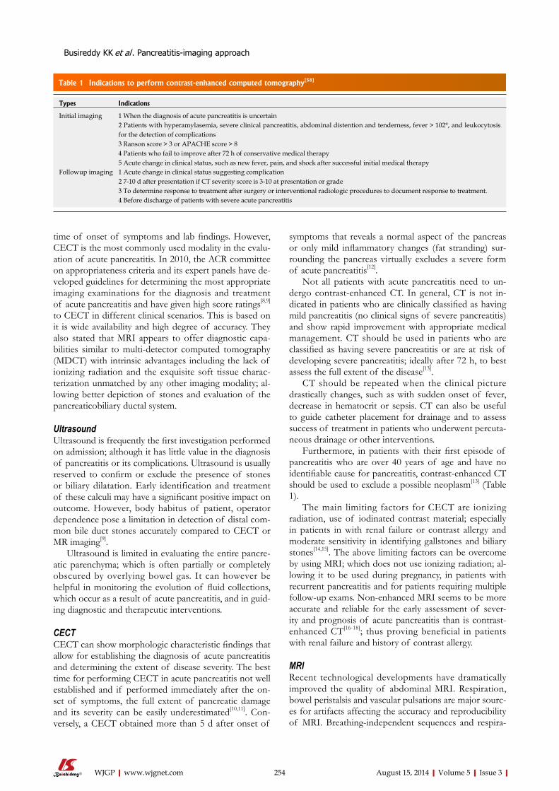
time of onset of symptoms and lab findings. However, CECT is the most commonly used modality in the evalu-ation of acute pancreatitis. In 2010, the ACR committee on appropriateness criteria and its expert panels have de-veloped guidelines for determining the most appropriate imaging examinations for the diagnosis and treatment of acute pancreatitis and have given high score ratings[8,9]
to CECT in different clinical scenarios. This is based on it is wide availability and high degree of accuracy. They also stated that MRI appears to offer diagnostic capa-bilities similar to multi-detector computed tomography (MDCT) with intrinsic advantages including the lack of ionizing radiation and the exquisite soft tissue charac-terization unmatched by any other imaging modality; al-lowing better depiction of stones and evaluation of the pancreaticobiliary ductal system.
Ultrasound Ultrasound is frequently the first investigation performed on admission; although it has little value in the diagnosis of pancreatitis or its complications. Ultrasound is usually reserved to confirm or exclude the presence of stones or biliary dilatation. Early identification and treatment of these calculi may have a significant positive impact on outcome. However, body habitus of patient, operator dependence pose a limitation in detection of distal com-mon bile duct stones accurately compared to CECT or MR imaging[9].
Ultrasound is limited in evaluating the entire pancre-atic parenchyma; which is often partially or completely obscured by overlying bowel gas. It can however be helpful in monitoring the evolution of fluid collections, which occur as a result of acute pancreatitis, and in guid-ing diagnostic and therapeutic interventions.
CECTCECT can show morphologic characteristic findings that allow for establishing the diagnosis of acute pancreatitis and determining the extent of disease severity. The best time for performing CECT in acute pancreatitis not well established and if performed immediately after the on-set of symptoms, the full extent of pancreatic damage and its severity can be easily underestimated[10,11]. Con-versely, a CECT obtained more than 5 d after onset of
symptoms that reveals a normal aspect of the pancreas or only mild inflammatory changes (fat stranding) sur-rounding the pancreas virtually excludes a severe form of acute pancreatitis[12].
Not all patients with acute pancreatitis need to un-dergo contrast-enhanced CT. In general, CT is not in-dicated in patients who are clinically classified as having mild pancreatitis (no clinical signs of severe pancreatitis) and show rapid improvement with appropriate medical management. CT should be used in patients who are classified as having severe pancreatitis or are at risk of developing severe pancreatitis; ideally after 72 h, to best assess the full extent of the disease[13].
CT should be repeated when the clinical picture drastically changes, such as with sudden onset of fever, decrease in hematocrit or sepsis. CT can also be useful to guide catheter placement for drainage and to assess success of treatment in patients who underwent percuta-neous drainage or other interventions.
Furthermore, in patients with their first episode of pancreatitis who are over 40 years of age and have no identifiable cause for pancreatitis, contrast-enhanced CT should be used to exclude a possible neoplasm[13] (Table 1).
The main limiting factors for CECT are ionizing radiation, use of iodinated contrast material; especially in patients in with renal failure or contrast allergy and moderate sensitivity in identifying gallstones and biliary stones[14,15]. The above limiting factors can be overcome by using MRI; which does not use ionizing radiation; al-lowing it to be used during pregnancy, in patients with recurrent pancreatitis and for patients requiring multiple follow-up exams. Non-enhanced MRI seems to be more accurate and reliable for the early assessment of sever-ity and prognosis of acute pancreatitis than is contrast-enhanced CT[16-18]; thus proving beneficial in patients with renal failure and history of contrast allergy.
MRIRecent technological developments have dramatically improved the quality of abdominal MRI. Respiration, bowel peristalsis and vascular pulsations are major sourc-es for artifacts affecting the accuracy and reproducibility of MRI. Breathing-independent sequences and respira-
August 15, 2014|Volume 5|Issue 3|WJGP|www.wjgnet.com 254
Table 1 Indications to perform contrast-enhanced computed tomography[58]
Types Indications
Initial imaging
1 When the diagnosis of acute pancreatitis is uncertain2 Patients with hyperamylasemia, severe clinical pancreatitis, abdominal distention and tenderness, fever > 102°, and leukocytosis for the detection of complications3 Ranson score > 3 or APACHE score > 84 Patients who fail to improve after 72 h of conservative medical therapy 5 Acute change in clinical status, such as new fever, pain, and shock after successful initial medical therapy
Followup imaging
1 Acute change in clinical status suggesting complication2 7-10 d after presentation if CT severity score is 3-10 at presentation or grade3 To determine response to treatment after surgery or interventional radiologic procedures to document response to treatment.4 Before discharge of patients with severe acute pancreatitis
Busireddy KK et al . Pancreatitis-imaging approach
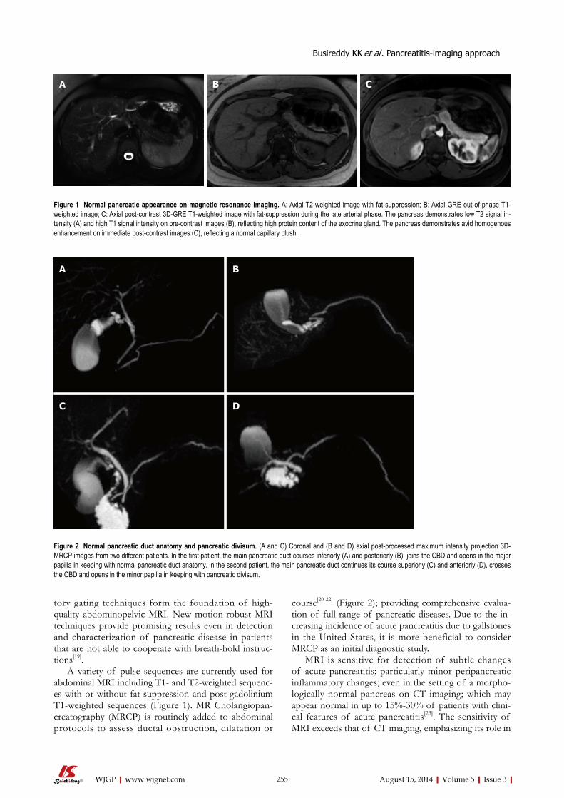
tory gating techniques form the foundation of high-quality abdominopelvic MRI. New motion-robust MRI techniques provide promising results even in detection and characterization of pancreatic disease in patients that are not able to cooperate with breath-hold instruc-tions[19].
A variety of pulse sequences are currently used for abdominal MRI including T1- and T2-weighted sequenc-es with or without fat-suppression and post-gadolinium T1-weighted sequences (Figure 1). MR Cholangiopan-creatography (MRCP) is routinely added to abdominal protocols to assess ductal obstruction, dilatation or
course[20-22] (Figure 2); providing comprehensive evalua-tion of full range of pancreatic diseases. Due to the in-creasing incidence of acute pancreatitis due to gallstones in the United States, it is more beneficial to consider MRCP as an initial diagnostic study.
MRI is sensitive for detection of subtle changes of acute pancreatitis; particularly minor peripancreatic inflammatory changes; even in the setting of a morpho-logically normal pancreas on CT imaging; which may appear normal in up to 15%-30% of patients with clini-cal features of acute pancreatitis[23]. The sensitivity of MRI exceeds that of CT imaging, emphasizing its role in
August 15, 2014|Volume 5|Issue 3|WJGP|www.wjgnet.com 255
Figure 1 Normal pancreatic appearance on magnetic resonance imaging. A: Axial T2-weighted image with fat-suppression; B: Axial GRE out-of-phase T1-weighted image; C: Axial post-contrast 3D-GRE T1-weighted image with fat-suppression during the late arterial phase. The pancreas demonstrates low T2 signal in-tensity (A) and high T1 signal intensity on pre-contrast images (B), reflecting high protein content of the exocrine gland. The pancreas demonstrates avid homogenous enhancement on immediate post-contrast images (C), reflecting a normal capillary blush.
A B C
Figure 2 Normal pancreatic duct anatomy and pancreatic divisum. (A and C) Coronal and (B and D) axial post-processed maximum intensity projection 3D-MRCP images from two different patients. In the first patient, the main pancreatic duct courses inferiorly (A) and posteriorly (B), joins the CBD and opens in the major papilla in keeping with normal pancreatic duct anatomy. In the second patient, the main pancreatic duct continues its course superiorly (C) and anteriorly (D), crosses the CBD and opens in the minor papilla in keeping with pancreatic divisum.
A B
C D
Busireddy KK et al . Pancreatitis-imaging approach
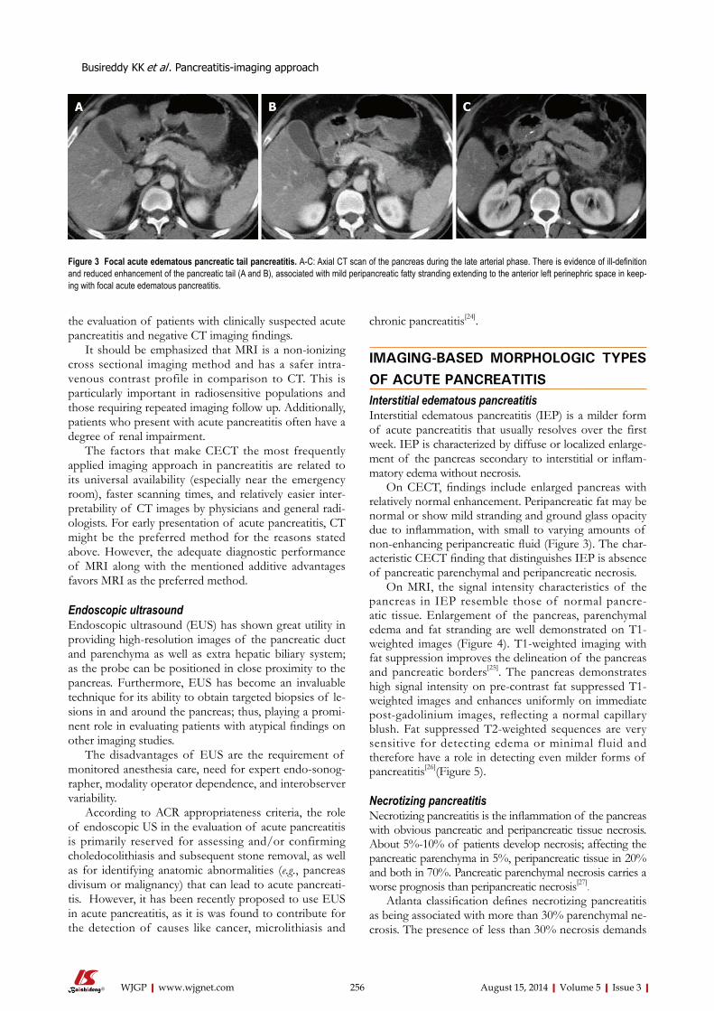
the evaluation of patients with clinically suspected acute pancreatitis and negative CT imaging findings.
It should be emphasized that MRI is a non-ionizing cross sectional imaging method and has a safer intra-venous contrast profile in comparison to CT. This is particularly important in radiosensitive populations and those requiring repeated imaging follow up. Additionally, patients who present with acute pancreatitis often have a degree of renal impairment.
The factors that make CECT the most frequently applied imaging approach in pancreatitis are related to its universal availability (especially near the emergency room), faster scanning times, and relatively easier inter-pretability of CT images by physicians and general radi-ologists. For early presentation of acute pancreatitis, CT might be the preferred method for the reasons stated above. However, the adequate diagnostic performance of MRI along with the mentioned additive advantages favors MRI as the preferred method. Endoscopic ultrasound Endoscopic ultrasound (EUS) has shown great utility in providing high-resolution images of the pancreatic duct and parenchyma as well as extra hepatic biliary system; as the probe can be positioned in close proximity to the pancreas. Furthermore, EUS has become an invaluable technique for its ability to obtain targeted biopsies of le-sions in and around the pancreas; thus, playing a promi-nent role in evaluating patients with atypical findings on other imaging studies.
The disadvantages of EUS are the requirement of monitored anesthesia care, need for expert endo-sonog-rapher, modality operator dependence, and interobserver variability.
According to ACR appropriateness criteria, the role of endoscopic US in the evaluation of acute pancreatitis is primarily reserved for assessing and/or confirming choledocolithiasis and subsequent stone removal, as well as for identifying anatomic abnormalities (e.g., pancreas divisum or malignancy) that can lead to acute pancreati-tis. However, it has been recently proposed to use EUS in acute pancreatitis, as it is was found to contribute for the detection of causes like cancer, microlithiasis and
chronic pancreatitis[24].
IMAGING-BASED MORPHOLOGIC TYPES OF ACUTE PANCREATITISInterstitial edematous pancreatitisInterstitial edematous pancreatitis (IEP) is a milder form of acute pancreatitis that usually resolves over the first week. IEP is characterized by diffuse or localized enlarge-ment of the pancreas secondary to interstitial or inflam-matory edema without necrosis.
On CECT, findings include enlarged pancreas with relatively normal enhancement. Peripancreatic fat may be normal or show mild stranding and ground glass opacity due to inflammation, with small to varying amounts of non-enhancing peripancreatic fluid (Figure 3). The char-acteristic CECT finding that distinguishes IEP is absence of pancreatic parenchymal and peripancreatic necrosis.
On MRI, the signal intensity characteristics of the pancreas in IEP resemble those of normal pancre-atic tissue. Enlargement of the pancreas, parenchymal edema and fat stranding are well demonstrated on T1-weighted images (Figure 4). T1-weighted imaging with fat suppression improves the delineation of the pancreas and pancreatic borders[25]. The pancreas demonstrates high signal intensity on pre-contrast fat suppressed T1-weighted images and enhances uniformly on immediate post-gadolinium images, reflecting a normal capillary blush. Fat suppressed T2-weighted sequences are very sensitive for detecting edema or minimal fluid and therefore have a role in detecting even milder forms of pancreatitis[26](Figure 5).
Necrotizing pancreatitisNecrotizing pancreatitis is the inflammation of the pancreas with obvious pancreatic and peripancreatic tissue necrosis. About 5%-10% of patients develop necrosis; affecting the pancreatic parenchyma in 5%, peripancreatic tissue in 20% and both in 70%. Pancreatic parenchymal necrosis carries a worse prognosis than peripancreatic necrosis[27].
Atlanta classification defines necrotizing pancreatitis as being associated with more than 30% parenchymal ne-crosis. The presence of less than 30% necrosis demands
August 15, 2014|Volume 5|Issue 3|WJGP|www.wjgnet.com 256
A B C
Figure 3 Focal acute edematous pancreatic tail pancreatitis. A-C: Axial CT scan of the pancreas during the late arterial phase. There is evidence of ill-definition and reduced enhancement of the pancreatic tail (A and B), associated with mild peripancreatic fatty stranding extending to the anterior left perinephric space in keep-ing with focal acute edematous pancreatitis.
Busireddy KK et al . Pancreatitis-imaging approach

follow-up scanning in 1 wk to confirm true necrosis vs IEP[27].
On CECT, findings include areas of compromised pancreatic parenchymal enhancement on the post-Gad-olinium images with or without peripancreatic inhomo-geneous fluid collections (Figures 6 and 7). The impair-ment of pancreatic perfusion and signs of peripancreatic necrosis evolve over several days[28], which explains why an early CECT may underestimate the eventual extent of pancreatic and peripancreatic necrosis.
On MRI, necrosis shows appears as hypointense areas on T1-weighted images corresponding to areas of increased signal on fat-suppressed T2 weighted-images, associated with well defined areas of non-enhancing pancreatic parenchyma on post-Gadolinium sequenc-es[29-31] (Figure 8).
For both CT and MRI, acquisition of an adequate arterial phase is of the utmost importance; as the maxi-mum enhancement of pancreas is reached on the late arterial phase, and higher difference in signal between
August 15, 2014|Volume 5|Issue 3|WJGP|www.wjgnet.com 257
A B C
Figure 4 Gallstone acute edematous pancreatic tail pancreatitis. A: Axial fast spin-echo (FSE) T2-weighted image with fat-suppression; B and C: Post-contrast 3D-GRE T1-weighted images with fat-suppression during the late arterial and portal venous phases. There is mild diffuse lace-like increased T2 signal involving the pancreatic parenchyma, associated with a small amount of peripancreatic fluid near the pancreatic tail (A). The pancreas demonstrates diffuse minimal decrease in T1 signal intensity (B) and minimally reduced enhancement on the late arterial phase (C) in keeping with diffuse edematous pancreatitis. There are also innumerable gallstones (A).
A B C
Figure 5 Subtle focal acute edematous pancreatic tail pancreatitis. A: Axial T2 weighted-image with fat-suppression; B and C: Axial post-contrast 3D-GRE T1- weighted images with fat-suppression during the late arterial and portal venous phases. There is a very subtle area of increased T2 signal seen around the pancreatic tail (arrow, A), with fairly normal enhancement of the pancreas on the post-contrast images (B and C) in keeping with subtle focal acute edematous pancreatic tail pancreatitis.
A B
Figure 6 Focal pancreatic head necrotizing pancreatitis confined to the pancreatic parenchyma. A and B: Axial CT scan during the portal venous phase. There is evidence of significantly reduced enhancement of the pancreatic head (B), without peripancreatic extension or necrosis in keeping with focal acute necrotizing pan-creatitis.
Busireddy KK et al . Pancreatitis-imaging approach
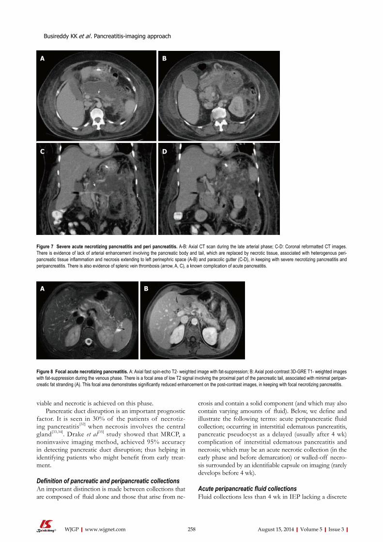
viable and necrotic is achieved on this phase.Pancreatic duct disruption is an important prognostic
factor. It is seen in 30% of the patients of necrotiz-ing pancreatitis[32] when necrosis involves the central gland[33,34]. Drake et al[35] study showed that MRCP, a noninvasive imaging method, achieved 95% accuracy in detecting pancreatic duct disruption; thus helping in identifying patients who might benefit from early treat-ment.
Definition of pancreatic and peripancreatic collectionsAn important distinction is made between collections that are composed of fluid alone and those that arise from ne-
crosis and contain a solid component (and which may also contain varying amounts of fluid). Below, we define and illustrate the following terms: acute peripancreatic fluid collection; occurring in interstitial edematous pancreatitis, pancreatic pseudocyst as a delayed (usually after 4 wk) complication of interstitial edematous pancreatitis and necrosis; which may be an acute necrotic collection (in the early phase and before demarcation) or walled-off necro-sis surrounded by an identifiable capsule on imaging (rarely develops before 4 wk).
Acute peripancreatic fluid collections Fluid collections less than 4 wk in IEP lacking a discrete
August 15, 2014|Volume 5|Issue 3|WJGP|www.wjgnet.com 258
A B
Figure 8 Focal acute necrotizing pancreatitis. A: Axial fast spin-echo T2- weighted image with fat-suppression; B: Axial post-contrast 3D-GRE T1- weighted images with fat-suppression during the venous phase. There is a focal area of low T2 signal involving the proximal part of the pancreatic tail, associated with minimal peripan-creatic fat stranding (A). This focal area demonstrates significantly reduced enhancement on the post-contrast images, in keeping with focal necrotizing pancreatitis.
Figure 7 Severe acute necrotizing pancreatitis and peri pancreatitis. A-B: Axial CT scan during the late arterial phase; C-D: Coronal reformatted CT images. There is evidence of lack of arterial enhancement involving the pancreatic body and tail, which are replaced by necrotic tissue, associated with heterogenous peri-pancreatic tissue inflammation and necrosis extending to left perinephric space (A-B) and paracolic gutter (C-D), in keeping with severe necrotizing pancreatitis and peripancreatitis. There is also evidence of splenic vein thrombosis (arrow, A, C), a known complication of acute pancreatitis.
A B
C D
Busireddy KK et al . Pancreatitis-imaging approach
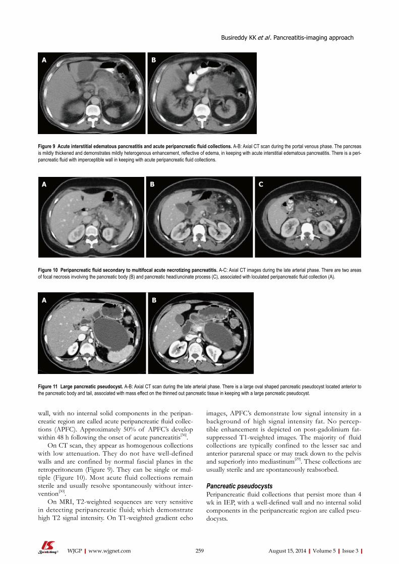
wall, with no internal solid components in the peripan-creatic region are called acute peripancreatic fluid collec-tions (APFC). Approximately 50% of APFC’s develop within 48 h following the onset of acute pancreatitis[36].
On CT scan, they appear as homogenous collections with low attenuation. They do not have well-defined walls and are confined by normal fascial planes in the retroperitoneum (Figure 9). They can be single or mul-tiple (Figure 10). Most acute fluid collections remain sterile and usually resolve spontaneously without inter-vention[30].
On MRI, T2-weighted sequences are very sensitive in detecting peripancreatic fluid; which demonstrate high T2 signal intensity. On T1-weighted gradient echo
images, APFC’s demonstrate low signal intensity in a background of high signal intensity fat. No percep-tible enhancement is depicted on post-gadolinium fat-suppressed T1-weighted images. The majority of fluid collections are typically confined to the lesser sac and anterior pararenal space or may track down to the pelvis and superiorly into mediastinum[29]. These collections are usually sterile and are spontaneously reabsorbed.
Pancreatic pseudocystsPeripancreatic fluid collections that persist more than 4 wk in IEP, with a well-defined wall and no internal solid components in the peripancreatic region are called pseu-docysts.
August 15, 2014|Volume 5|Issue 3|WJGP|www.wjgnet.com 259
A B
Figure 9 Acute interstitial edematous pancreatitis and acute peripancreatic fluid collections. A-B: Axial CT scan during the portal venous phase. The pancreas is mildly thickened and demonstrates mildly heterogenous enhancement, reflective of edema, in keeping with acute interstitial edematous pancreatitis. There is a peri-pancreatic fluid with imperceptible wall in keeping with acute peripancreatic fluid collections.
A B C
Figure 10 Peripancreatic fluid secondary to multifocal acute necrotizing pancreatitis. A-C: Axial CT images during the late arterial phase. There are two areas of focal necrosis involving the pancreatic body (B) and pancreatic head/uncinate process (C), associated with loculated peripancreatic fluid collection (A).
A B
Figure 11 Large pancreatic pseudocyst. A-B: Axial CT scan during the late arterial phase. There is a large oval shaped pancreatic pseudocyst located anterior to the pancreatic body and tail, associated with mass effect on the thinned out pancreatic tissue in keeping with a large pancreatic pseudocyst.
Busireddy KK et al . Pancreatitis-imaging approach
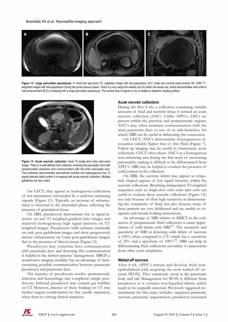
On CECT, they appear as homogenous collections of low-attenuation surrounded by a uniform enhancing capsule (Figure 11). Typically, an increase of enhance-ment is observed in the interstitial phase; reflecting the presence of granulation tissue.
On MRI, pseudocysts demonstrate low in signal in-tensity on and T1-weighted gradient-echo images and relatively homogeneous high signal intensity on T2-weighted images. Pseudocysts walls enhance minimally on early post-gadolinium images and show progressively intense enhancement on 5-min post-gadolinium images; due to the presence of fibrous tissue (Figure 12).
Pseudocysts may sometime have communication with pancreatic duct and detecting this communication is helpful in the further patients’ management. MRCP; a noninvasive imaging modality has an advantage of dem-onstrating possible communication between pancreatic pseudocyst and pancreatic duct.
The majority of pseudocysts resolve spontaneously. Infection and hemorrhage may complicate simple pseu-docysts. Infected pseudocyst may contain gas bubbles on CT. However, absence of these findings on CT may further require confirmation by fine needle aspiration, when there is a strong clinical suspicion.
Acute necrotic collectionsDuring the first 4 wk, a collection containing variable amounts of fluid and necrotic tissue is termed an acute necrotic collection (ANC). Unlike APFCs, ANCs are present within the pancreas and peripancreatic regions. ANC’s may often maintain communication with the main pancreatic duct or one of its side-branches; for which, MRI can be useful in delineating this connection.
On CECT, ANC’s demonstrate heterogeneous at-tenuation variably higher that of thin fluid (Figure 7). Follow-up imaging may be useful to characterize acute collections. CECT often shows ANC’s as a homogenous non-enhancing area during the first week of necrotizing pancreatitis; making it difficult to be differentiated from APFC’s. MRI may be helpful to confirm the presence of solid content in the collection.
On MRI, the necrotic debris may appear as irregu-larly shaped regions of low signal intensity within the necrotic collections. Breathing-independent T2-weighted sequences such as single-shot echo-train spin echo are useful to evaluate these necrotic collections (Figure 13); not only because of their high sensitivity in demonstrat-ing the complexity of fluid, but also because many of these patients are very debilitated and are unable to co-operate with breath-holding instructions.
An advantage of MRI relative to MDCT in the eval-uation of peripancreatic fluid collections is easier appre-ciation of solid debris with MRI[37]. The sensitivity and specificity of MRI in detecting solid debris of necrosis is 100% when compared to CT; which has a sensitivity of 25% and a specificity of 100%[38]. MRI can help in differentiating fluid collections secondary to pancreatitis from other cystic neoplasms.
Walled-off necrosis After 4 wk, APFC’s mature and develop thick non-epithelialized wall; acquiring the term walled-off ne-crosis (WON). They commonly occur in the pancreatic body and tail. Management for WON is different from pseudocyst as it contains non-liquefied debris; which needs to be surgically removed. Previously suggested no-menclature for this entity includes: organized pancreatic necrosis, pancreatic sequestration, pseudocyst associated
August 15, 2014|Volume 5|Issue 3|WJGP|www.wjgnet.com 260
A B C
Figure 12 Large pancreatic pseudocyst. A: Axial fast spin-echo T2- weighted image with fat-suppression; B-C: Axial and coronal post-contrast 3D- GRE T1-weighted images with fat-suppression during the portal venous phase. There is a very large thin-walled cyst (A) within the lesser sac; which demonstrates mild uniform wall enhancement (B-C) in keeping with a large pancreatic pseudocyst. The central drop of signal on (A) is related to dielectric shading artifact.
Figure 13 Acute necrotic collection. Axial T2 single-shot turbo spin-echo image. There is a well-defined fluid collection involving the pancreatic neck with peripancreatic extension and communication with the main pancreatic duct. This collection demonstrates well-defined outlines and heterogenous low T2 signal intensity debris within it in keeping with acute necrotic collection. Multiple gallstones are also noted.
Busireddy KK et al . Pancreatitis-imaging approach

with necrosis and subacute pancreatic necrosis.On CECT, walled-off necrosis demonstrates a het-
erogeneous fluid and non-fluid attenuation with varying degree of loculations surrounded by a well-defined and enhancing encapsulating wall; which may involve both the pancreatic and extrapancreatic tissue. CECT, howev-er, may not readily distinguish solid from fluid contents; as a result, pancreatic and peripancreatic necrosis may be misdiagnosed as a pancreatic pseudocyst. For this pur-pose, MRI may be required for this distinction (Figure 14).
Infected pancreatic necrosisPancreatic and peripancreatic necrosis can remain ster-ile or become infected. The development of secondary infection in pancreatic necrosis is associated with in-creased morbidity and mortality[3]. Most studies suggest that there is no absolute correlation between the extent of necrosis and the risk of infection and duration of symptoms[7]. The early diagnosis of infected pancreatic necrosis is very important in the initiation of antibiotic therapy.
The diagnosis of infected ANC or WON can be suspected in the presence of extraluminal gas on CT or MRI. This extraluminal gas is present in areas of necro-sis and may or may not form a gas/fluid level depending on the amount of fluid content present at that stage of
the disease (Figure 15). The diagnosis may be confirmed by aspiration and analysis including microscopy and cul-ture.
SEVERITY OF ACUTE PANCREATITISClinical vs MCTSI vs MRSI severity indexSeveral clinical scoring systems like Marshal, SOFA. APACHE or Ranson criteria were designed to accurately correlate the complications like organ failure and mortal-ity in acute pancreatitis. In the last two decades, radio-logical scoring systems were developed to accurately di-agnose and correlate complications in acute pancreatitis.
For the first time in 1990, Balthazar et al[28] introduced the CT severity index for assessment of AP; which correlated well with morbidity, mortality and length of hospital stay. CTSI was widely adopted in clinical and research settings; however, a potential limitation was its inability to detect pancreatic necrosis. MCTSI introduced by Mortele et al[39] in 2004 to account for the limitations of CTSI (Table 2); which showed improved correlation with severity.
MCTSI incorporated extrapancreatic manifestations and simplified the evaluation of extent of parenchymal necrosis by categorizing into none, less than 30% or more than 30%; in addition to evaluating peripancreatic inflammation by detecting the presence or absence of
August 15, 2014|Volume 5|Issue 3|WJGP|www.wjgnet.com 261
A B C
Figure 14 Necrotizing pancreatitis, with peripancreatic walled-off necrosis. A: Axial fast spin-echo T2-weighted image with fat-suppression; B-C: Axial post-con-trast 3D-GRE T1-weighted images with fat-suppression during the late arterial and venous phase. There is a focal area of heterogeneous iso to slightly high T2 signal involving the pancreatic body-tail junction (A); which demonstrates lack of enhancement on the post-contrast images (B-C). There is associated sizable peripancreatic fluid collection; which demonstrates heterogeneous T2 signal intensity and thick enhancing wall post-contrast in keeping with walled-off necrosis.
A B C
Figure 15 Infected peripancreatic fluid in a patient with acute pancreatitis. A-C: Axial CT scan during the portal venous phase. There are a few gas bubbles seen within a small peripancreatic fluid (arrows, A-C). In the absence of any intervention in keeping with infected peripancreatic fluid.
Busireddy KK et al . Pancreatitis-imaging approach
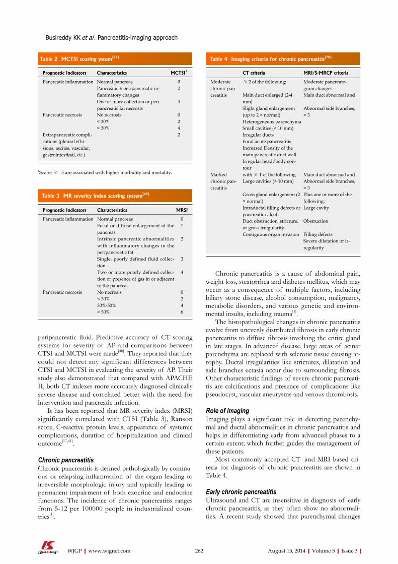
Table 2 MCTSI scoring ystem[39]
peripancreatic fluid. Predictive accuracy of CT scoring systems for severity of AP and comparisons between CTSI and MCTSI were made[40]. They reported that they could not detect any significant differences between CTSI and MCTSI in evaluating the severity of AP. Their study also demonstrated that compared with APACHE II, both CT indexes more accurately diagnosed clinically severe disease and correlated better with the need for intervention and pancreatic infection.
It has been reported that MR severity index (MRSI) significantly correlated with CTSI (Table 3), Ranson score, C-reactive protein levels, appearance of systemic complications, duration of hospitalization and clinical outcome[17,41].
Chronic pancreatitis Chronic pancreatitis is defined pathologically by continu-ous or relapsing inflammation of the organ leading to irreversible morphologic injury and typically leading to permanent impairment of both exocrine and endocrine functions. The incidence of chronic pancreatitis ranges from 5-12 per 100000 people in industrialized coun-tries[1].
Chronic pancreatitis is a cause of abdominal pain, weight loss, steatorrhea and diabetes mellitus, which may occur as a consequence of multiple factors, including biliary stone disease, alcohol consumption, malignancy, metabolic disorders, and various genetic and environ-mental insults, including trauma[1].
The histopathological changes in chronic pancreatitis evolve from unevenly distributed fibrosis in early chronic pancreatitis to diffuse fibrosis involving the entire gland in late stages. In advanced disease, large areas of acinar parenchyma are replaced with sclerotic tissue causing at-rophy. Ductal irregularities like strictures, dilatation and side branches ectasia occur due to surrounding fibrosis. Other characteristic findings of severe chronic pancreati-tis are calcifications and presence of complications like pseudocyst, vascular aneurysms and venous thrombosis.
Role of imagingImaging plays a significant role in detecting parenchy-mal and ductal abnormalities in chronic pancreatitis and helps in differentiating early from advanced phases to a certain extent; which further guides the management of these patients.
Most commonly accepted CT- and MRI-based cri-teria for diagnosis of chronic pancreatitis are shown in Table 4.
Early chronic pancreatitisUltrasound and CT are insensitive in diagnosis of early chronic pancreatitis, as they often show no abnormali-ties. A recent study showed that parenchymal changes
August 15, 2014|Volume 5|Issue 3|WJGP|www.wjgnet.com 262
Prognostic Indicators Characteristics MCTSI1
Pancreatic inflammation Normal pancreas 0Pancreatic ± peripancreatic in-flammatory changes
2
One or more collection or peri-pancreatic fat necrosis
4
Pancreatic necrosis No necrosis 0< 30% 2> 30% 4
Extrapancreatic compli-cations (pleural effu-sions, ascites, vascular, gastrointestinal, etc.)
2
1Scores ≥ 5 are associated with higher morbidity and mortality.
Table 3 MR severity index scoring system[69]
Prognostic Indicators Characteristics MRSI
Pancreatic inflammation Normal pancreas 0Focal or diffuse enlargement of the pancreas
1
Intrinsic pancreatic abnormalities with inflammatory changes in the peripancreatic fat
2
Single, poorly defined fluid collec-tion
3
Two or more poorly defined collec-tion or presence of gas in or adjacent to the pancreas
4
Pancreatic necrosis No necrosis 0< 30% 230%-50% 4> 50% 6
Table 4 Imaging criteria for chronic pancreatitis[70]
CT criteria MRI/S-MRCP criteria
Moderate chronic pan-creatitis
≥ 2 of the following: Moderate pancreato-gram changes
Main duct enlarged (2-4 mm)
Main duct abnormal and
Slight gland enlargement (up to 2 × normal)
Abnormal side branches, > 3
Heterogeneous parenchymaSmall cavities (< 10 mm)Irregular ductsFocal acute pancreatitisIncreased Density of the main pancreatic duct wallIrregular head/body con-tour
Marked chronic pan-creatitis
with ≥ 1 of the following Main duct abnormal andLarge cavities (> 10 mm) Abnormal side branches,
> 3Gross gland enlargement (2 × normal)
Plus one or more of the following:
Intraductal filling defects or pancreatic calculi
Large cavity
Duct obstruction, stricture, or gross irregularity
Obstruction
Contiguous organ invasion Filling defectsSevere dilatation or ir-regularity
Busireddy KK et al . Pancreatitis-imaging approach
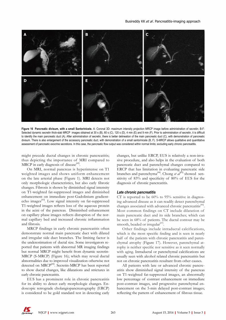
might precede ductal changes in chronic pancreatitis; thus depicting the importance of MRI compared to MRCP in early diagnosis of disease[42].
On MRI, normal pancreas is hyperintense on T1 weighted images and shows uniform enhancement on the late arterial phase (Figure 1). MRI detects not only morphologic characteristics, but also early fibrotic changes. Fibrosis is shown by diminished signal intensity on T1-weighted fat-suppressed images and diminished enhancement on immediate post-Gadolinium gradient-echo images[43]. Low signal intensity on fat-suppressed T1-weighted images reflects loss of the aqueous protein in the acini of the pancreas. Diminished enhancement on capillary phase images reflects disruption of the nor-mal capillary bed and increased chronic inflammation and fibrosis.
MRCP findings in early chronic pancreatitis often demonstrate normal main pancreatic duct with dilated and irregular side duct branches. The limiting factor is the underestimation of ductal size. Some investigators re-ported that patients with abnormal MR imaging findings but normal MRCP might benefit from dynamic secretin-MRCP (S-MRCP) (Figure 16); which may reveal ductal abnormalities due to improved visualization otherwise not detected on MRCP[42]. Secretin-MRCP has been reported to show ductal changes, like dilatations and strictures in early chronic pancreatitis.
EUS has a prominent role in chronic pancreatitis for its ability to detect early morphologic changes. En-doscopic retrograde cholangiopancreatography (ERCP) is considered to be gold standard test in detecting early
changes, but unlike ERCP, EUS is relatively a non-inva-sive procedure, and also helps in the evaluation of both pancreatic duct and parenchymal changes compared to ERCP that has limitation in evaluating pancreatic side branches and parenchyma[44]. Chong et al[45] showed sen-sitivity of 83% and specificity of 80% of EUS for the diagnosis of chronic pancreatitis.
Late chronic pancreatitisCT is reported to be 60% to 95% sensitive in diagnos-ing advanced disease as it can readily detect parenchymal changes associated with advanced chronic pancreatitis[46]. Most common findings on CT include dilatation of main pancreatic duct and its side branches; which can be seen in 68% of patients. The ductal contour may be smooth, beaded or irregular[47].
Other findings include intraductal calcifications, which is the most specific finding and is seen in nearly half of the patients with chronic pancreatitis and paren-chymal atrophy (Figure 17). However, parenchymal at-rophy is neither specific nor sensitive as it seen normally with aging. Intraductal or parenchymal calcifications are usually seen with alcohol related chronic pancreatitis but not on chronic pancreatitis resultant from other causes.
All patients with late or advanced chronic pancre-atitis show diminished signal intensity of the pancreas on T1-weighted fat-suppressed images, an abnormally low percentage of contrast enhancement on immediate post-contrast images, and progressive parenchymal en-hancement on the 5-min delayed post-contrast images; reflecting the pattern of enhancement of fibrous tissue.
August 15, 2014|Volume 5|Issue 3|WJGP|www.wjgnet.com 263
Figure 16 Pancreatic divisum, with a small Santorinicele. A: Coronal 3D- maximum intensity projection MRCP image before administration of secretin; B-F: Selected dynamic secretin thick-slab MRCP images obtained at 30 s (B), 60 s (C), 120 s (D), 4 min (E) and 9 min (F). Prior to administration of secretin, it is difficult to identify the main pancreatic duct (A). After administration of secretin, there is better delineation of the main pancreatic duct (C), with demonstration of pancreatic divisum. There is also enlargement of the accessory pancreatic duct, with demonstration of a small santorinicele (B, F). S-MRCP allows qualitative and quantitative assessment of pancreatic exocrine secretions. In this case, the pancreatic flow output was considered within normal limits; excluding early chronic pancreatitis.
A B
D E
C
F
Busireddy KK et al . Pancreatitis-imaging approach
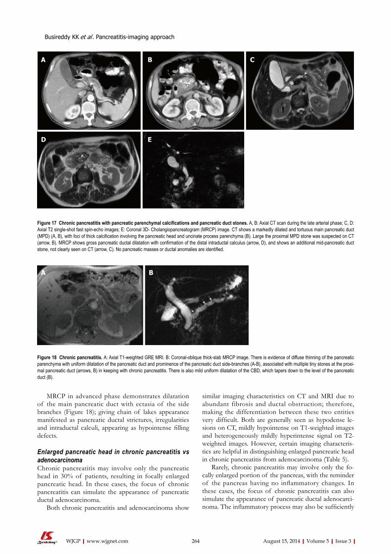
MRCP in advanced phase demonstrates dilatation of the main pancreatic duct with ectasia of the side branches (Figure 18); giving chain of lakes appearance manifested as pancreatic ductal strictures, irregularities and intraductal calculi, appearing as hypointense filling defects.
Enlarged pancreatic head in chronic pancreatitis vs adenocarcinomaChronic pancreatitis may involve only the pancreatic head in 30% of patients, resulting in focally enlarged pancreatic head. In these cases, the focus of chronic pancreatitis can simulate the appearance of pancreatic ductal adenocarcinoma.
Both chronic pancreatitis and adenocarcinoma show
similar imaging characteristics on CT and MRI due to abundant fibrosis and ductal obstruction; therefore, making the differentiation between these two entities very difficult. Both are generally seen as hypodense le-sions on CT, mildly hypointense on T1-weighted images and heterogeneously mildly hyperintense signal on T2-weighted images. However, certain imaging characteris-tics are helpful in distinguishing enlarged pancreatic head in chronic pancreatitis from adenocarcinoma (Table 5).
Rarely, chronic pancreatitis may involve only the fo-cally enlarged portion of the pancreas, with the reminder of the pancreas having no inflammatory changes. In these cases, the focus of chronic pancreatitis can also simulate the appearance of pancreatic ductal adenocarci-noma. The inflammatory process may also be sufficiently
August 15, 2014|Volume 5|Issue 3|WJGP|www.wjgnet.com 264
Figure 17 Chronic pancreatitis with pancreatic parenchymal calcifications and pancreatic duct stones. A, B: Axial CT scan during the late arterial phase; C, D: Axial T2 single-shot fast spin-echo images; E: Coronal 3D- Cholangiopancreatogram (MRCP) image. CT shows a markedly dilated and tortuous main pancreatic duct (MPD) (A, B), with foci of thick calcification involving the pancreatic head and uncinate process parenchyma (B). Large the proximal MPD stone was suspected on CT (arrow, B). MRCP shows gross pancreatic ductal dilatation with confirmation of the distal intraductal calculus (arrow, D), and shows an additional mid-pancreatic duct stone, not clearly seen on CT (arrow, C). No pancreatic masses or ductal anomalies are identified.
A B
D E
C
A B
Figure 18 Chronic pancreatitis. A: Axial T1-weighted GRE MRI. B: Coronal-oblique thick-slab MRCP image. There is evidence of diffuse thinning of the pancreatic parenchyma with uniform dilatation of the pancreatic duct and prominence of the pancreatic duct side-branches (A-B), associated with multiple tiny stones at the proxi-mal pancreatic duct (arrows, B) in keeping with chronic pancreatitis. There is also mild uniform dilatation of the CBD, which tapers down to the level of the pancreatic duct (B).
Busireddy KK et al . Pancreatitis-imaging approach
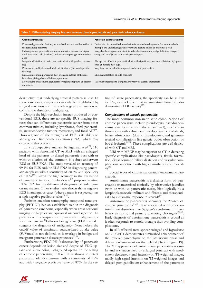
destructive that underlying stromal pattern is lost. In these rare cases, diagnosis can only be established by surgical resection and histopathological examination to confirm the absence of malignancy.
Despite the high-resolution images produced by con-ventional EUS, there are no specific EUS imaging fea-tures that can differentiate pancreatic cancer from other common mimics, including lymphoma, focal pancreati-tis, neuroendocrine tumors, metastases, and focal AIP[48]. However, one of the strengths of EUS is its ability to allow guided fine needle aspiration (FNA); which may overcome this problem.
In a retrospective analysis by Agarwal et al[49], 110 patients with abnormal CT or MRI with an enlarged head of the pancreas or dilated pancreatic duct with or without dilation of the common bile duct underwent EUS or EUS-FNA. The study revealed an accuracy of 99.1% for EUS and/or EUS-FNA in diagnosing pancre-atic neoplasm with a sensitivity of 88.8% and specificity of 100%[49]. Given the high accuracy in the evaluation of pancreatic tumors, Eloubeidi et al[50] proposed routine EUS-FNA for the differential diagnosis of solid pan-creatic masses. Other studies have shown that a negative EUS in ambiguous cases (where a mass is suspected) has a high negative predictive value[51,52].
Positron emission tomography-computed tomogra-phy (PET-CT) has an established role in the diagnosis of pancreatic carcinoma, especially when cross sectional imaging or biopsies are equivocal or nondiagnostic. In patients with a suspicion of pancreatic malignancy, a focal increase in 18F-fluorodeoxyglucose (FDG) uptake suggests the diagnosis of malignancy. Nonetheless, the cutoff value of maximum standardized uptake value (SUVmax) is not defined, as it overlaps in benign and malignant pancreatic disease processes[53,54].
Furthermore, FDG-PET’s detectability of pancreatic cancer depends on lesion size and degree of FDG up-take and surrounding background uptake. In the setting of chronic pancreatitis, FDG-PET is shown to detect pancreatic adenocarcinoma with a sensitivity of 92% and with a negative predictive value of 87%. In the set-
ting of acute pancreatitis, the specificity can be as low as 50%, as it is known that inflammatory tissue can also demonstrate FDG activity[47].
Complications of chronic pancreatitisThe most common non-neoplastic complications of chronic pancreatitis include pseudocysts, pseudoaneu-rysms (due to erosion of the arterial wall), splenic vein thrombosis with subsequent development of collaterals, biliary obstruction (due to pseudocysts), and gastroin-testinal complications like gastric outlet obstruction or bowel ischemia[6,55]. These complications are well depict-ed with CT and MRI.
MRI with MRCP may be superior to CT in detecting specific complications like pseudocysts, fistula forma-tion, distal common biliary dilatation and vascular com-plications associated with higher morbidity and mortal-ity[46].
Special types of chronic pancreatitis autoimmune pan-creatitis.
Autoimmune pancreatitis is a distinct form of pan-creatitis characterized clinically by obstructive jaundice (with or without pancreatic mass), histologically by a lymphoplasmacytic infiltrate and fibrosis and therapeuti-cally by a dramatic response to steroids[56].
Autoimmune pancreatitis accounts for 2%-6% of chronic pancreatitis[57,58]. It is associated with other au-toimmune disorders like Sjogren’s syndrome, primary biliary cirrhosis, and primary sclerosing cholangitis[59,60]. Early diagnosis of autoimmune pancreatitis is crucial as it often responds to steroid therapy; thus avoiding com-plications.
In AIP, affected areas appear enlarged and hypodense on CT. CECT demonstrates diminished enhancement of the involved parenchyma on the late arterial phase and delayed enhancement on the delayed phase (Figure 19). The MR appearance of autoimmune pancreatitis is simi-lar and is characterized by enlarged pancreas with mod-erately decreased signal intensity on T1-weighted images, mildly high signal intensity on T2-weighted images and delayed post-gadolinium enhancement of the pancreatic
August 15, 2014|Volume 5|Issue 3|WJGP|www.wjgnet.com 265
Table 5 Differentiating imaging features between chronic pancreatitis and pancreatic adenocarcinoma
Chronic pancreatitis Pancreatic adenocarcinoma
Preserved glandular, feathery or marbled texture similar to that of the remaining pancreas
Definable, circumscribed mass lesion is most often diagnostic for tumor, which disrupts the underlying architecture and results in loss of anatomic detail
Heterogeneous pancreatic enhancement with presence of signal void (cysts and calcifications) on immediate post-gadolinium im-ages
Irregular, heterogeneous, diminished enhancement on postgadolinium images compared to adjacent pancreatic parenchyma
Irregular dilatation of main pancreatic duct with gradual narrow-ingPresence of multiple intraductal calcifications (the most specific finding)
Abrupt cut off of the pancreatic duct with significant proximal dilatation +/- pres-ence of double duct signVery few ductal calculi compared to chronic pancreatitis
Dilatation of main pancreatic duct with and ectasia of the side branches, giving chain of lakes appearance
Minimal dilatation of side branches
No vascular encasement, significant lymphadenopathy or distant metastasis
Vascular encasement, lymphadenopathy or distant metastasis
Busireddy KK et al . Pancreatitis-imaging approach
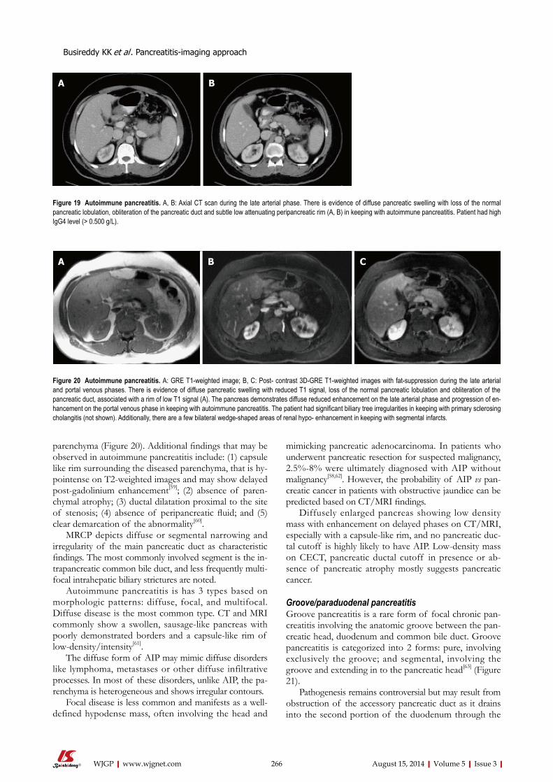
parenchyma (Figure 20). Additional findings that may be observed in autoimmune pancreatitis include: (1) capsule like rim surrounding the diseased parenchyma, that is hy-pointense on T2-weighted images and may show delayed post-gadolinium enhancement[59]; (2) absence of paren-chymal atrophy; (3) ductal dilatation proximal to the site of stenosis; (4) absence of peripancreatic fluid; and (5) clear demarcation of the abnormality[60].
MRCP depicts diffuse or segmental narrowing and irregularity of the main pancreatic duct as characteristic findings. The most commonly involved segment is the in-trapancreatic common bile duct, and less frequently multi-focal intrahepatic biliary strictures are noted.
Autoimmune pancreatitis is has 3 types based on morphologic patterns: diffuse, focal, and multifocal. Diffuse disease is the most common type. CT and MRI commonly show a swollen, sausage-like pancreas with poorly demonstrated borders and a capsule-like rim of low-density/intensity[61].
The diffuse form of AIP may mimic diffuse disorders like lymphoma, metastases or other diffuse infiltrative processes. In most of these disorders, unlike AIP, the pa-renchyma is heterogeneous and shows irregular contours.
Focal disease is less common and manifests as a well-defined hypodense mass, often involving the head and
mimicking pancreatic adenocarcinoma. In patients who underwent pancreatic resection for suspected malignancy, 2.5%-8% were ultimately diagnosed with AIP without malignancy[58,62]. However, the probability of AIP vs pan-creatic cancer in patients with obstructive jaundice can be predicted based on CT/MRI findings.
Diffusely enlarged pancreas showing low density mass with enhancement on delayed phases on CT/MRI, especially with a capsule-like rim, and no pancreatic duc-tal cutoff is highly likely to have AIP. Low-density mass on CECT, pancreatic ductal cutoff in presence or ab-sence of pancreatic atrophy mostly suggests pancreatic cancer.
Groove/paraduodenal pancreatitisGroove pancreatitis is a rare form of focal chronic pan-creatitis involving the anatomic groove between the pan-creatic head, duodenum and common bile duct. Groove pancreatitis is categorized into 2 forms: pure, involving exclusively the groove; and segmental, involving the groove and extending in to the pancreatic head[63] (Figure 21).
Pathogenesis remains controversial but may result from obstruction of the accessory pancreatic duct as it drains into the second portion of the duodenum through the
August 15, 2014|Volume 5|Issue 3|WJGP|www.wjgnet.com 266
A B
Figure 19 Autoimmune pancreatitis. A, B: Axial CT scan during the late arterial phase. There is evidence of diffuse pancreatic swelling with loss of the normal pancreatic lobulation, obliteration of the pancreatic duct and subtle low attenuating peripancreatic rim (A, B) in keeping with autoimmune pancreatitis. Patient had high IgG4 level (> 0.500 g/L).
A B C
Figure 20 Autoimmune pancreatitis. A: GRE T1-weighted image; B, C: Post- contrast 3D-GRE T1-weighted images with fat-suppression during the late arterial and portal venous phases. There is evidence of diffuse pancreatic swelling with reduced T1 signal, loss of the normal pancreatic lobulation and obliteration of the pancreatic duct, associated with a rim of low T1 signal (A). The pancreas demonstrates diffuse reduced enhancement on the late arterial phase and progression of en-hancement on the portal venous phase in keeping with autoimmune pancreatitis. The patient had significant biliary tree irregularities in keeping with primary sclerosing cholangitis (not shown). Additionally, there are a few bilateral wedge-shaped areas of renal hypo- enhancement in keeping with segmental infarcts.
Busireddy KK et al . Pancreatitis-imaging approach
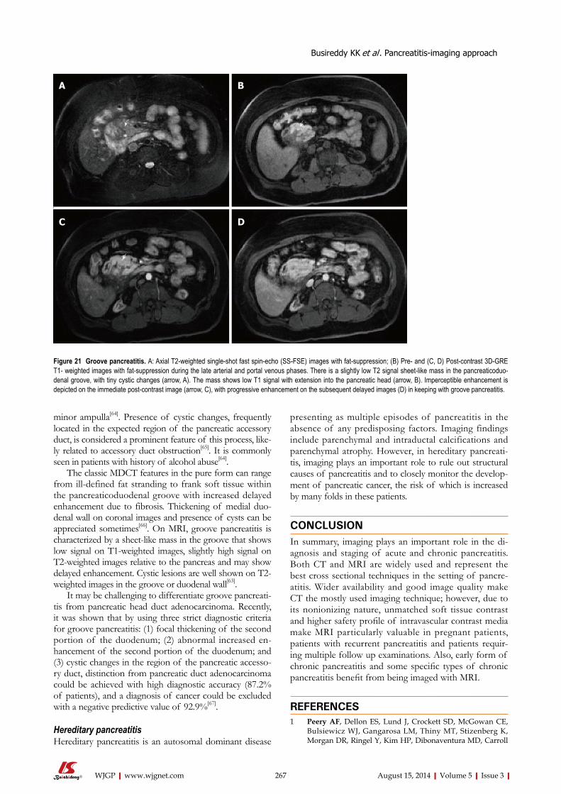
minor ampulla[64]. Presence of cystic changes, frequently located in the expected region of the pancreatic accessory duct, is considered a prominent feature of this process, like-ly related to accessory duct obstruction[65]. It is commonly seen in patients with history of alcohol abuse[64].
The classic MDCT features in the pure form can range from ill-defined fat stranding to frank soft tissue within the pancreaticoduodenal groove with increased delayed enhancement due to fibrosis. Thickening of medial duo-denal wall on coronal images and presence of cysts can be appreciated sometimes[66]. On MRI, groove pancreatitis is characterized by a sheet-like mass in the groove that shows low signal on T1-weighted images, slightly high signal on T2-weighted images relative to the pancreas and may show delayed enhancement. Cystic lesions are well shown on T2-weighted images in the groove or duodenal wall[63].
It may be challenging to differentiate groove pancreati-tis from pancreatic head duct adenocarcinoma. Recently, it was shown that by using three strict diagnostic criteria for groove pancreatitis: (1) focal thickening of the second portion of the duodenum; (2) abnormal increased en-hancement of the second portion of the duodenum; and (3) cystic changes in the region of the pancreatic accesso-ry duct, distinction from pancreatic duct adenocarcinoma could be achieved with high diagnostic accuracy (87.2% of patients), and a diagnosis of cancer could be excluded with a negative predictive value of 92.9%[67].
Hereditary pancreatitis Hereditary pancreatitis is an autosomal dominant disease
presenting as multiple episodes of pancreatitis in the absence of any predisposing factors. Imaging findings include parenchymal and intraductal calcifications and parenchymal atrophy. However, in hereditary pancreati-tis, imaging plays an important role to rule out structural causes of pancreatitis and to closely monitor the develop-ment of pancreatic cancer, the risk of which is increased by many folds in these patients.
CONCLUSIONIn summary, imaging plays an important role in the di-agnosis and staging of acute and chronic pancreatitis. Both CT and MRI are widely used and represent the best cross sectional techniques in the setting of pancre-atitis. Wider availability and good image quality make CT the mostly used imaging technique; however, due to its nonionizing nature, unmatched soft tissue contrast and higher safety profile of intravascular contrast media make MRI particularly valuable in pregnant patients, patients with recurrent pancreatitis and patients requir-ing multiple follow up examinations. Also, early form of chronic pancreatitis and some specific types of chronic pancreatitis benefit from being imaged with MRI.
REFERENCES1 Peery AF, Dellon ES, Lund J, Crockett SD, McGowan CE,
Bulsiewicz WJ, Gangarosa LM, Thiny MT, Stizenberg K, Morgan DR, Ringel Y, Kim HP, Dibonaventura MD, Carroll
August 15, 2014|Volume 5|Issue 3|WJGP|www.wjgnet.com 267
Figure 21 Groove pancreatitis. A: Axial T2-weighted single-shot fast spin-echo (SS-FSE) images with fat-suppression; (B) Pre- and (C, D) Post-contrast 3D-GRE T1- weighted images with fat-suppression during the late arterial and portal venous phases. There is a slightly low T2 signal sheet-like mass in the pancreaticoduo-denal groove, with tiny cystic changes (arrow, A). The mass shows low T1 signal with extension into the pancreatic head (arrow, B). Imperceptible enhancement is depicted on the immediate post-contrast image (arrow, C), with progressive enhancement on the subsequent delayed images (D) in keeping with groove pancreatitis.
A B
C D
Busireddy KK et al . Pancreatitis-imaging approach
CF, Allen JK, Cook SF, Sandler RS, Kappelman MD, Shaheen NJ. Burden of gastrointestinal disease in the United States: 2012 update. Gastroenterology 2012; 143: 1179-1187.e1-3 [PMID: 22885331 DOI: 10.1053/j.gastro.2012.08.002]
2 Bradley EL. A clinically based classification system for acute pancreatitis. Summary of the International Symposium on Acute Pancreatitis, Atlanta, Ga, September 11 through 13, 1992. Arch Surg 1993; 128: 586-590 [PMID: 8489394]
3 Petrov MS, Shanbhag S, Chakraborty M, Phillips AR, Wind-sor JA. Organ failure and infection of pancreatic necrosis as determinants of mortality in patients with acute pancreatitis. Gastroenterology 2010; 139: 813-820 [PMID: 20540942 DOI: 10.1053/j.gastro.2010.06.010]
4 Shanbhogue AK, Fasih N, Surabhi VR, Doherty GP, Shanb-hogue DK, Sethi SK. A clinical and radiologic review of un-common types and causes of pancreatitis. Radiographics 2009; 29: 1003-1026 [PMID: 19605653 DOI: 10.1148/rg.294085748]
5 Etemad B, Whitcomb DC. Chronic pancreatitis: diagnosis, classification, and new genetic developments. Gastroenterol-ogy 2001; 120: 682-707 [PMID: 11179244]
6 Bollen TL. Imaging of acute pancreatitis: update of the re-vised Atlanta classification. Radiol Clin North Am 2012; 50: 429-445 [PMID: 22560690 DOI: 10.1016/j.rcl.2012.03.015]
7 Banks PA, Bollen TL, Dervenis C, Gooszen HG, Johnson CD, Sarr MG, Tsiotos GG, Vege SS. Classification of acute pancreatitis--2012: revision of the Atlanta classification and definitions by international consensus. Gut 2013; 62: 102-111 [PMID: 23100216 DOI: 10.1136/gutjnl-2012-302779]
8 Perez A, Whang EE, Brooks DC, Moore FD, Hughes MD, Sica GT, Zinner MJ, Ashley SW, Banks PA. Is severity of nec-rotizing pancreatitis increased in extended necrosis and in-fected necrosis? Pancreas 2002; 25: 229-233 [PMID: 12370532]
9 Megibow AJ, Ralls PW, Balfe DM, Bree RL, DiSantis DJ, Glick SN, Kidd R, Levine MS, Mezwa DG, Saini S, Shuman WP, Greene FL, Laine LA, Lillemoe K. Acute pancreatitis. American College of Radiology. ACR Appropriateness Cri-teria. Radiology 2000; 215 Suppl: 203-207 [PMID: 11037427]
10 Banks PA, Freeman ML. Practice guidelines in acute pan-creatitis. Am J Gastroenterol 2006; 101: 2379-2400 [PMID: 17032204 DOI: 10.1111/j.1572-0241.2006.00856.x]
11 Morgan DE. Imaging of acute pancreatitis and its complica-tions. Clin Gastroenterol Hepatol 2008; 6: 1077-1085 [PMID: 18928934 DOI: 10.1016/j.cgh.2008.07.012]
12 Moulton JS. The radiologic assessment of acute pancreati-tis and its complications. Pancreas 1991; 6 Suppl 1: S13-S22 [PMID: 1788247]
13 Thoeni RF. The revised Atlanta classification of acute pan-creatitis: its importance for the radiologist and its effect on treatment. Radiology 2012; 262: 751-764 [PMID: 22357880 DOI: 10.1148/radiol.11110947]
14 Anderson SW, Lucey BC, Varghese JC, Soto JA. Accuracy of MDCT in the diagnosis of choledocholithiasis. AJR Am J Roentgenol 2006; 187: 174-180 [PMID: 16794173 DOI: 10.2214/ajr.05.0459]
15 Anderson SW, Rho E, Soto JA. Detection of biliary duct nar-rowing and choledocholithiasis: accuracy of portal venous phase multidetector CT. Radiology 2008; 247: 418-427 [PMID: 18372450 DOI: 10.1148/radiol.2472070473]
16 Kim YK, Kim CS, Han YM. Role of fat-suppressed t1-weighted magnetic resonance imaging in predicting severity and prognosis of acute pancreatitis: an intraindividual com-parison with multidetector computed tomography. J Com-put Assist Tomogr 2009; 33: 651-656 [PMID: 19820487 DOI: 10.1097/RCT.0b013e3181979282]
17 Stimac D, Miletić D, Radić M, Krznarić I, Mazur-Grbac M, Perković D, Milić S, Golubović V. The role of nonenhanced magnetic resonance imaging in the early assessment of acute pancreatitis. Am J Gastroenterol 2007; 102: 997-1004 [PMID: 17378903 DOI: 10.1111/j.1572-0241.2007.01164.x]
18 Viremouneix L, Monneuse O, Gautier G, Gruner L, Giorgi
R, Allaouchiche B, Pilleul F. Prospective evaluation of non-enhanced MR imaging in acute pancreatitis. J Magn Reson Imaging 2007; 26: 331-338 [PMID: 17654731 DOI: 10.1002/jmri.21037]
19 Azevedo RM, de Campos RO, Ramalho M, Herédia V, Dale BM, Semelka RC. Free-breathing 3D T1-weighted gradient-echo sequence with radial data sampling in abdominal MRI: preliminary observations. AJR Am J Roentgenol 2011; 197: 650-657 [PMID: 21862807 DOI: 10.2214/ajr.10.5881]
20 Bret PM, Reinhold C, Taourel P, Guibaud L, Atri M, Barkun AN. Pancreas divisum: evaluation with MR cholangiopan-creatography. Radiology 1996; 199: 99-103 [PMID: 8633179]
21 Soto JA, Barish MA, Yucel EK, Clarke P, Siegenberg D, Chuttani R, Ferrucci JT. Pancreatic duct: MR cholangiopan-creatography with a three-dimensional fast spin-echo tech-nique. Radiology 1995; 196: 459-464 [PMID: 7617861]
22 Takehara Y, Ichijo K, Tooyama N, Kodaira N, Yamamoto H, Tatami M, Saito M, Watahiki H, Takahashi M. Breath-hold MR cholangiopancreatography with a long-echo-train fast spin-echo sequence and a surface coil in chronic pancreatitis. Radiology 1994; 192: 73-78 [PMID: 8208969]
23 Balthazar EJ. CT diagnosis and staging of acute pancreatitis. Radiol Clin North Am 1989; 27: 19-37 [PMID: 2642273]
24 Scaglione M, Casciani E, Pinto A, Andreoli C, De Vargas M, Gualdi GF. Imaging assessment of acute pancreatitis: a review. Semin Ultrasound CT MR 2008; 29: 322-340 [PMID: 18853839]
25 Miller FH, Keppke AL, Dalal K, Ly JN, Kamler VA, Sica GT. MRI of pancreatitis and its complications: part 1, acute pancreatitis. AJR Am J Roentgenol 2004; 183: 1637-1644 [PMID: 15547203 DOI: 10.2214/ajr.183.6.01831637]
26 Kim YK, Ko SW, Kim CS, Hwang SB. Effectiveness of MR imaging for diagnosing the mild forms of acute pancreatitis: comparison with MDCT. J Magn Reson Imaging 2006; 24: 1342-1349 [PMID: 17083111 DOI: 10.1002/jmri.20801]
27 Ashley SW, Perez A, Pierce EA, Brooks DC, Moore FD, Whang EE, Banks PA, Zinner MJ. Necrotizing pancreatitis: contemporary analysis of 99 consecutive cases. Ann Surg 2001; 234: 572-579; discussion 572-579 [PMID: 11573050]
28 Balthazar EJ, Robinson DL, Megibow AJ, Ranson JH. Acute pancreatitis: value of CT in establishing prognosis. Radiology 1990; 174: 331-336 [PMID: 2296641]
29 Balthazar EJ, Freeny PC, vanSonnenberg E. Imaging and in-tervention in acute pancreatitis. Radiology 1994; 193: 297-306 [PMID: 7972730 DOI: 10.1148/radiology.193.2.7972730]
30 Lenhart DK, Balthazar EJ. MDCT of acute mild (nonnecro-tizing) pancreatitis: abdominal complications and fate of flu-id collections. AJR Am J Roentgenol 2008; 190: 643-649 [PMID: 18287434 DOI: 10.2214/ajr.07.2761]
31 Matos C, Cappeliez O, Winant C, Coppens E, Devière J, Metens T. MR imaging of the pancreas: a pictorial tour. Ra-diographics 2002; 22: e2 [PMID: 11796914 DOI: 10.1148/radio-graphics.22.1.g02jae2e2]
32 Lau ST, Simchuk EJ, Kozarek RA, Traverso LW. A pan-creatic ductal leak should be sought to direct treatment in patients with acute pancreatitis. Am J Surg 2001; 181: 411-415 [PMID: 11448431]
33 Sandrasegaran K, Tann M, Jennings SG, Maglinte DD, Peter SD, Sherman S, Howard TJ. Disconnection of the pancreatic duct: an important but overlooked complication of severe acute pancreatitis. Radiographics 2007; 27: 1389-1400 [PMID: 17848698 DOI: 10.1148/rg.275065163]
34 Tann M, Maglinte D, Howard TJ, Sherman S, Fogel E, Madura JA, Lehman GA. Disconnected pancreatic duct syn-drome: imaging findings and therapeutic implications in 26 surgically corrected patients. J Comput Assist Tomogr 2003; 27: 577-582 [PMID: 12886147]
35 Drake LM, Anis M, Lawrence C. Accuracy of magnetic reso-nance cholangiopancreatography in identifying pancreatic duct disruption. J Clin Gastroenterol 2012; 46: 696-699 [PMID:
August 15, 2014|Volume 5|Issue 3|WJGP|www.wjgnet.com 268
Busireddy KK et al . Pancreatitis-imaging approach
22565603 DOI: 10.1097/MCG.0b013e31825003b3]36 Zaheer A, Singh VK, Qureshi RO, Fishman EK. The revised
Atlanta classification for acute pancreatitis: updates in im-aging terminology and guidelines. Abdom Imaging 2013; 38: 125-136 [PMID: 22584543 DOI: 10.1007/s00261-012-9908-0]
37 Macari M, Finn ME, Bennett GL, Cho KC, Newman E, Hajdu CH, Babb JS. Differentiating pancreatic cystic neo-plasms from pancreatic pseudocysts at MR imaging: value of perceived internal debris. Radiology 2009; 251: 77-84 [PMID: 19332847 DOI: 10.1148/radiol.2511081286]
38 Morgan DE, Baron TH, Smith JK, Robbin ML, Kenney PJ. Pancreatic fluid collections prior to intervention: evaluation with MR imaging compared with CT and US. Radiology 1997; 203: 773-778 [PMID: 9169703]
39 Mortele KJ, Wiesner W, Intriere L, Shankar S, Zou KH, Ka-lantari BN, Perez A, vanSonnenberg E, Ros PR, Banks PA, Silverman SG. A modified CT severity index for evaluat-ing acute pancreatitis: improved correlation with patient outcome. AJR Am J Roentgenol 2004; 183: 1261-1265 [PMID: 15505289 DOI: 10.2214/ajr.183.5.1831261]
40 Bollen TL, Singh VK, Maurer R, Repas K, van Es HW, Banks PA, Mortele KJ. Comparative evaluation of the modified CT severity index and CT severity index in assessing severity of acute pancreatitis. AJR Am J Roentgenol 2011; 197: 386-392 [PMID: 21785084 DOI: 10.2214/AJR.09.4025]
41 Arvanitakis M, Koustiani G, Gantzarou A, Grollios G, Tsi-touridis I, Haritandi-Kouridou A, Dimitriadis A, Arvanitakis C. Staging of severity and prognosis of acute pancreatitis by computed tomography and magnetic resonance imaging-a comparative study. Dig Liver Dis 2007; 39: 473-482 [PMID: 17363349 DOI: 10.1016/j.dld.2007.01.015]
42 Balci NC, Alkaade S, Magas L, Momtahen AJ, Burton FR. Suspected chronic pancreatitis with normal MRCP: findings on MRI in correlation with secretin MRCP. J Magn Reson Imaging 2008; 27: 125-131 [PMID: 18058927 DOI: 10.1002/jmri.21241]
43 Semelka RC, Shoenut JP, Kroeker MA, Micflikier AB. Chronic pancreatitis: MR imaging features before and after administration of gadopentetate dimeglumine. J Magn Reson Imaging 1993; 3: 79-82 [PMID: 8428105]
44 Raman SP, Fishman EK, Lennon AM. Endoscopic ultra-sound and pancreatic applications: what the radiologist needs to know. Abdom Imaging 2013; 38: 1360-1372 [PMID: 23334660 DOI: 10.1007/s00261-013-9979-6]
45 Chong AK, Hawes RH, Hoffman BJ, Adams DB, Lewin DN, Romagnuolo J. Diagnostic performance of EUS for chronic pancreatitis: a comparison with histopathology. Gastrointest Endosc 2007; 65: 808-814 [PMID: 17466199 DOI: 10.1016/j.gie.2006.09.026]
46 Choueiri NE, Balci NC, Alkaade S, Burton FR. Advanced im-aging of chronic pancreatitis. Curr Gastroenterol Rep 2010; 12: 114-120 [PMID: 20424983 DOI: 10.1007/s11894-010-0093-4]
47 Perez-Johnston R, Sainani NI, Sahani DV. Imaging of chron-ic pancreatitis (including groove and autoimmune pancreati-tis). Radiol Clin North Am 2012; 50: 447-466 [PMID: 22560691 DOI: 10.1016/j.rcl.2012.03.005]
48 Papanikolaou IS, Adler A, Neumann U, Neuhaus P, Rösch T. Endoscopic ultrasound in pancreatic disease--its influence on surgical decision-making. An update 2008. Pancreatology 2009; 9: 55-65 [PMID: 19077455 DOI: 10.1159/000178875]
49 Agarwal B, Krishna NB, Labundy JL, Safdar R, Akduman EI. EUS and/or EUS-guided FNA in patients with CT and/or magnetic resonance imaging findings of enlarged pancreatic head or dilated pancreatic duct with or without a dilated common bile duct. Gastrointest Endosc 2008; 68: 237-42; quiz 334, 335 [PMID: 18423464 DOI: 10.1016/j.gie.2008.01.026]
50 Eloubeidi MA, Varadarajulu S, Desai S, Shirley R, Heslin MJ, Mehra M, Arnoletti JP, Eltoum I, Wilcox CM, Vickers SM. A prospective evaluation of an algorithm incorporat-ing routine preoperative endoscopic ultrasound-guided fine
needle aspiration in suspected pancreatic cancer. J Gastroin-test Surg 2007; 11: 813-819 [PMID: 17440790 DOI: 10.1007/s11605-007-0151-x]
51 Voss M, Hammel P, Molas G, Palazzo L, Dancour A, O’Toole D, Terris B, Degott C, Bernades P, Ruszniewski P. Val-ue of endoscopic ultrasound guided fine needle aspiration biopsy in the diagnosis of solid pancreatic masses. Gut 2000; 46: 244-249 [PMID: 10644320]
52 Williams DB, Sahai AV, Aabakken L, Penman ID, van Velse A, Webb J, Wilson M, Hoffman BJ, Hawes RH. Endoscopic ultrasound guided fine needle aspiration biopsy: a large sin-gle centre experience. Gut 1999; 44: 720-726 [PMID: 10205212]
53 Santhosh S, Mittal BR, Bhasin D, Srinivasan R, Rana S, Das A, Nada R, Bhattacharya A, Gupta R, Kapoor R. Role of (18)F-fluorodeoxyglucose positron emission tomography/computed tomography in the characterization of pancreatic masses: experience from tropics. J Gastroenterol Hepatol 2013; 28: 255-261 [PMID: 23278193 DOI: 10.1111/jgh.12068]
54 Asagi A, Ohta K, Nasu J, Tanada M, Nadano S, Nishimura R, Teramoto N, Yamamoto K, Inoue T, Iguchi H. Utility of contrast-enhanced FDG-PET/CT in the clinical management of pancreatic cancer: impact on diagnosis, staging, evalu-ation of treatment response, and detection of recurrence. Pancreas 2013; 42: 11-19 [PMID: 22699206 DOI: 10.1097/MPA.0b013e3182550d77]
55 Remer EM, Baker ME. Imaging of chronic pancreatitis. Ra-diol Clin North Am 2002; 40: 1229-1242, v [PMID: 12479708]
56 Shimosegawa T, Chari ST, Frulloni L, Kamisawa T, Kawa S, Mino-Kenudson M, Kim MH, Klöppel G, Lerch MM, Löhr M, Notohara K, Okazaki K, Schneider A, Zhang L. International consensus diagnostic criteria for autoimmune pancreatitis: guidelines of the International Association of Pancreatology. Pancreas 2011; 40: 352-358 [PMID: 21412117 DOI: 10.1097/MPA.0b013e3182142fd2]
57 Sah RP, Pannala R, Chari ST, Sugumar A, Clain JE, Levy MJ, Pearson RK, Smyrk TC, Petersen BT, Topazian MD, Taka-hashi N, Vege SS. Prevalence, diagnosis, and profile of au-toimmune pancreatitis presenting with features of acute or chronic pancreatitis. Clin Gastroenterol Hepatol 2010; 8: 91-96 [PMID: 19800984 DOI: 10.1016/j.cgh.2009.09.024]
58 Nishimori I, Tamakoshi A, Otsuki M. Prevalence of auto-immune pancreatitis in Japan from a nationwide survey in 2002. J Gastroenterol 2007; 42 Suppl 18: 6-8 [PMID: 17520216 DOI: 10.1007/s00535-007-2043-y]
59 Irie H, Honda H, Baba S, Kuroiwa T, Yoshimitsu K, Tajima T, Jimi M, Sumii T, Masuda K. Autoimmune pancreatitis: CT and MR characteristics. AJR Am J Roentgenol 1998; 170: 1323-1327 [PMID: 9574610 DOI: 10.2214/ajr.170.5.9574610]
60 Van Hoe L, Gryspeerdt S, Ectors N, Van Steenbergen W, Aerts R, Baert AL, Marchal G. Nonalcoholic duct-destructive chronic pancreatitis: imaging findings. AJR Am J Roent-genol 1998; 170: 643-647 [PMID: 9490945 DOI: 10.2214/ajr.170.3.9490945]
61 Takahashi N, Fletcher JG, Fidler JL, Hough DM, Kawashima A, Chari ST. Dual-phase CT of autoimmune pancreatitis: a multireader study. AJR Am J Roentgenol 2008; 190: 280-286 [PMID: 18212210 DOI: 10.2214/ajr.07.2309]
62 Hardacre JM, Iacobuzio-Donahue CA, Sohn TA, Abraham SC, Yeo CJ, Lillemoe KD, Choti MA, Campbell KA, Schulick RD, Hruban RH, Cameron JL, Leach SD. Results of pan-creaticoduodenectomy for lymphoplasmacytic sclerosing pancreatitis. Ann Surg 2003; 237: 853-858; discussion 853-858 [PMID: 12796582 DOI: 10.1097/01.sla.0000071516.54864.c1]
63 Blasbalg R, Baroni RH, Costa DN, Machado MC. MRI fea-tures of groove pancreatitis. AJR Am J Roentgenol 2007; 189: 73-80 [PMID: 17579155 DOI: 10.2214/ajr.06.1244]
64 Chatelain D, Vibert E, Yzet T, Geslin G, Bartoli E, Manaouil D, Delcenserie R, Brevet M, Dupas JL, Regimbeau JM. Groove pancreatitis and pancreatic heterotopia in the minor duode-nal papilla. Pancreas 2005; 30: e92-e95 [PMID: 15841034]
August 15, 2014|Volume 5|Issue 3|WJGP|www.wjgnet.com 269
Busireddy KK et al . Pancreatitis-imaging approach
65 Triantopoulou C, Dervenis C, Giannakou N, Papailiou J, Prassopoulos P. Groove pancreatitis: a diagnostic chal-lenge. Eur Radiol 2009; 19: 1736-1743 [PMID: 19238393 DOI: 10.1007/s00330-009-1332-7]
66 Raman SP, Salaria SN, Hruban RH, Fishman EK. Groove pancreatitis: spectrum of imaging findings and radiology-pa-thology correlation. AJR Am J Roentgenol 2013; 201: W29-W39 [PMID: 23789694 DOI: 10.2214/AJR.12.9956]
67 Kalb B, Martin DR, Sarmiento JM, Erickson SH, Gober D, Tapper EB, Chen Z, Adsay NV. Paraduodenal pancreatitis: clinical performance of MR imaging in distinguishing from carcinoma. Radiology 2013; 269: 475-481 [PMID: 23847255
DOI: 10.1148/radiol.13112056]68 O'Connor OJ, McWilliams S, Maher MM. Imaging of acute
pancreatitis. AJR Am J Roentgenol 2011; 197: W221-W225 [PMID: 21785045 DOI: 10.2214/AJR.10.4338]
69 Yang R, Jing ZL, Zhang XM, Tang W, Xiao B, Huang XH, Yang L, Feng ZS. MR imaging of acute pancreatitis: correla-tion of abdominal wall edema with severity scores. Eur J Radiol 2012; 81: 3041-3047 [PMID: 22571930 DOI: 10.1016/j.ejrad.2012.04.005]
70 Conwell DL, Wu BU. Chronic pancreatitis: making the di-agnosis. Clin Gastroenterol Hepatol 2012; 10: 1088-1095 [PMID: 22642958 DOI: 10.1016/j.cgh.2012.05.015]
P- Reviewer: Clark CJ, Demir IE S- Editor: Ji FF L- Editor: A E- Editor: Lu YJ
August 15, 2014|Volume 5|Issue 3|WJGP|www.wjgnet.com 270
Busireddy KK et al . Pancreatitis-imaging approach
REVIEW
New insights to occult gastrointestinal bleeding: From pathophysiology to therapeutics
Antonio Damián Sánchez-Capilla, Paloma De La Torre-Rubio, Eduardo Redondo-Cerezo
Antonio Damián Sánchez-Capilla, Paloma De La Torre-Rubio, Department of Gastroenterology, University Hospital Virgen de, Las Nieves, 18014 Granada, SpainEduardo Redondo-Cerezo, Endoscopy Unit, Department of Gastroenterology, University Hospital Virgen de Las Nieves, 18014 Granada, SpainAuthor contributions: Sánchez-Capilla AD and De La Torre-Rubio P reviewed the bibliography and wrote the first draft; Redondo-Cerezo E overviewed the paper and wrote the final paper in English. Correspondence to: Eduardo Redondo-Cerezo, MD, PhD, Endoscopy Unit, Department of Gastroenterology, University Hospital Virgen de Las Nieves, Avenida de las Fuerzas Armadas 2, 18014 Granada, Spain. [email protected] Telephone: +34-958-020146 Fax: +34-958-120169Received: March 11, 2014 Revised: June 1, 2014 Accepted: June 18, 2014Published online: August 15, 2014
AbstractObscure gastrointestinal bleeding is still a clinical chal-lenge for gastroenterologists. The recent development of novel technologies for the diagnosis and treatment of different bleeding causes has allowed a better man-agement of patients, but it also determines the need of a deeper comprehension of pathophysiology and the analysis of local expertise in order to develop a rational management algorithm. Obscure gastrointes-tinal bleeding can be divided in occult, when a positive occult blood fecal test is the main manifestation, and overt, when external sings of bleeding are visible. In this paper we are going to focus on overt gastrointes-tinal bleeding, describing the physiopathology of the most usual causes, analyzing the diagnostic procedures available, from the most classical to the novel ones, and establishing a standard algorithm which can be adapted depending on the local expertise or availability. Finally, we will review the main therapeutic options for this complex and not so uncommon clinical problem.
© 2014 Baishideng Publishing Group Inc. All rights reserved.
Key words: Obscure gastrointestinal bleeding; Angio-dysplasia; Wireless capsule endoscopy; Double balloon enteroscopy
Core tip: This is an invited in depth review of occult gastrointestinal bleeding, addressing its pathophysiol-ogy, diagnosis and treatment. Our paper tries to unify in one single manuscript all what a general gastroen-terologist should know about those items. From the essentials of pathophysiology, we have tried to build a rational approach to those patients’ management de-pending on the severity of the condition, proposing an evidence-based management algorithm.
Sánchez-Capilla AD, De La Torre-Rubio P, Redondo-Cerezo E. New insights to occult gastrointestinal bleeding: From pathophysiology to therapeutics. World J Gastrointest Pathophysiol 2014; 5(3): 271-283 Available from: URL: http://www.wjgnet.com/2150-5330/full/v5/i3/271.htm DOI: http://dx.doi.org/10.4291/wjgp.v5.i3.271
INTRODUCTIONGastrointestinal bleeding is a term that includes any bleeding originating from the esophagus to the anus. Classically, it has been classified in upper or lower de-pending on the location of the bleeding source, proximal or distal to the angle of Treitz.
The usual management of gastrointestinal bleeding (GIB) involves an upper endoscopy and colonoscopy in a first attempt to find the bleeding lesion. If those are unsuc-cessful, and there’s a bleeding persistence or recurrence, the entity is called gastrointestinal bleeding of obscure origin or obscure gastrointestinal bleeding (OGIB), being the source of bleeding usually located in the small bowel. This seg-
World J Gastrointest Pathophysiol 2014 August 15; 5(3): 271-283ISSN 2150-5330 (online)
© 2014 Baishideng Publishing Group Inc. All rights reserved.
Online Submissions: http://www.wjgnet.com/esps/Help Desk: http://www.wjgnet.com/esps/helpdesk.aspxDOI: 10.4291/wjgp.v5.i3.271
August 15, 2014|Volume 5|Issue 3|WJGP|www.wjgnet.com 271
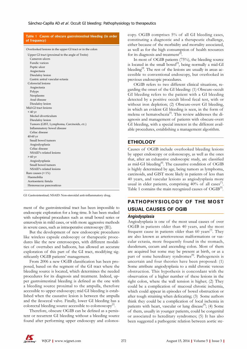
Sánchez-Capilla AD et al . Occult GI bleeding: Pathophysiology to therapeutics
ment of the gastrointestinal tract has been impossible to endoscopic exploration for a long time. It has been studied with suboptimal procedures such as small bowel series or enteroclysis in mild cases, or with more aggressive methods in severe cases, such as intraoperative enteroscopy (IE).
But the development of new endoscopic procedures like wireless capsule endoscopy or therapeutic proce-dures like the new enteroscopes, with different modali-ties of overtubes and balloons, has allowed an accurate exploration of this part of the GI tract, modifying sig-nificantly OGIB patients’ management.
From 2006 a new OGIB classification has been pro-posed, based on the segment of the GI tract where the bleeding source is located, which determines the needed procedures for its diagnosis and treatment. Indeed, up-per gastrointestinal bleeding is defined as the one with a bleeding source proximal to the ampulla, therefore accessible to upper endoscopy; mid GI bleeding is estab-lished when the causative lesion is between the ampulla and the ileocecal valve. Finally, lower GI bleeding has a colorectal bleeding source accessible to colonoscopy[1].
Therefore, obscure OGIB can be defined as a persis-tent or recurrent GI bleeding without a bleeding source found after performing upper endoscopy and colonos-
copy. OGIB comprises 5% of all GI bleeding cases, constituting a diagnostic and a therapeutic challenge, either because of the morbidity and mortality associated, as well as for the high consumption of health resources for its diagnosis and treatment[2].
In most of OGIB patients (75%), the bleeding source is located in the small bowel[3], being normally a mid-GI bleeding[4]. The rest of the lesions are usually in areas ac-cessible to conventional endoscopy, but overlooked in previous endoscopic procedures.
OGIB refers to two different clinical situations, re-garding the onset of the GI bleeding: (1) Obscure-occult GI bleeding refers to the patient with a GI bleeding detected by a positive occult blood fecal test, with or without iron depletion; (2) Obscure-overt GI bleeding, in which an evident GI bleeding is seen, in the form of melena or hematochezia[5]. This review addresses the di-agnosis and management of patients with obscure-overt GI bleeding, with a special interest in the different avail-able procedures, establishing a management algorithm.
ETHIOLOGYCauses of OGIB include overlooked bleeding lesions by upper endoscopy or colonoscopy, as well as the ones that, after an exhaustive endoscopic study, are classified as mid-GI bleeding[6]. The causative condition of OGIB is highly determined by age, being tumors as lymphoma, carcinoids, and GIST more likely in patients of less than 40 years, and vascular lesions as angiodysplasia more usual in elder patients, comprising 40% of all cases[7]. Table 1 contains the main recognized causes of OGIB[5].
PATHOPHYSIOLOGY OF THE MOST USUAL CAUSES OF OGIBAngiodysplasiaAngiodysplasia is one of the most usual causes of over OGIB in patients older than 40 years, and the most frequent cause in patients older than 60 years[7]. They are also known as arteriovenous malformations or vas-cular ectasia, more frequently found in the stomach, duodenum, cecum and ascending colon. Most of them are acquired but some may be present at birth, or as a part of some hereditary syndromes[8]. Pathogenesis is uncertain and four theories have been proposed: (1) Some attribute angiodysplasia to a mild chronic venous obstruction. This hypothesis is concordant with the observation of a higher number of these lesions in the right colon, where the wall tension is higher; (2) They could be a complication of mucosal chronic ischemia, which could appear in episodes of bowel obstruction or after tough straining when defecating; (3) Some authors think they could be a complication of local ischemia in patients with heart, vascular or lung disease[9]; (4) Some of them, usually in younger patients, could be congenital or associated to hereditary syndromes; (5) It has also been suggested a pathogenic relation between aortic ste-
August 15, 2014|Volume 5|Issue 3|WJGP|www.wjgnet.com 272
Table 1 Causes of obscure gastrointestinal bleeding (in order of frequency)
Overlooked lesions in the upper GI tract or in the colon
Upper GI tract (proximal to the angle of Treitz) Cameron ulcers Fundic varices Peptic ulcer Angiectasia Dieulafoy lesion Gastric antral vascular ectasia Colorectal lesions Angiectasia Polyps Neoplasms Anal disease Dieulafoy lesionMid-GI tract lesions < 40 yr Meckel diverticulum Dieulafoy lesion Tumors (GIST, Lymphoma, Carcinoids, etc.) Inflammatory bowel disease Celiac disease 40-60 yr Small bowel tumors Angiodysplasia Celiac disease NSAID’s related lesions > 60 yr Angiodysplasia Small bowel tumors NSAID’s related lesionsRare causes (< 1%) Haemobilia Aortoenteric fistula Hemosuccus pancreaticus
GI: Gastrointestinal; NSAID: Non-steroidal anti-inflammatory drug.
nosis and angiodysplasia, caused by the haemodinamic abnormalities determined by the valvular disease (Heyde Syndrome)[10]. Therapy is controversial, but some studies have shown a reduction in bleeding episodes after valvu-lar replacement; and (6) In terminal cardiac failure, left ventricular assisted devices have been associated with in-creased bleeding episodes from angiodysplasia. In these cases, pathogenesis seems related with anticoagulant therapy, vascular malformations, loss of activity of Von Willebrand factor and mucosal ischemia[11].
Small bowel tumorsDespite infrequent, GI bleeding is the usual clinical on-set, being more frequent in benign tumors as leyomioma than in malignant lesions as leyomiosarcoma[12]. Bleeding is caused by erosion of the tumor surface or by the rup-ture of aberrant vascular structures within the lesion.
Meckel diverticulumThis is a relevant condition in patients of less than 25 years old. Despite rare, it is the most frequent con-genital abnormality in the GI tract. They are caused by the incomplete obliteration of the vitelin duct during embryogenesis, which leads to the formation of a true small bowel diverticulum[13]. Meckel diverticulum has all the bowel wall layers, and in 12%-21% of cases it may contain ectopic tissues (gastric or duodenal mucosa or even pancreatic ducts). They are usually asymptomatic, but may also cause abdominal pain or OGIB. Bleeding is caused by an ulceration of the small bowel due to acid secretion by heterotopic gastric mucosa contained within the diverticular layers. Bleeding can be chronic and insid-ious, or acute and massive, but transfusion is hardly ever required. The main anatomical risk factor that makes bleeding more likely is diverticula size of more than 2 cm[14].
Dieulafoy’s lesion Etiology is unknown. Lesions are normally found in the proximal stomach, in the lesser curvature, near de esophago-gastric junction. It is usually a submucosal, di-lated, aberrant vessel that erodes the overlaying mucosa without a previous ulcer[15]. This is caused by the lack of ramification of the submucosal artery which makes its diameter ten times the normal diameter of a mucosal capillary. Triggering causes are unclear and it usually ap-pears in male patients with comorbidities such as cardio-vascular diseases, arterial hypertension, chronic kidney disease, diabetes or alcohol abuse. It is important to mention that this lesion can be overlooked in an endo-scopic exam[16], given that quite frequently the aberrant vessel cannot be seen unless it bleeds actively.
Celiac disease and inflammatory bowel diseaseGI bleeding is usually associated to complications in both conditions, which can be ulcers or tumors like ad-enocarcinoma or lymphoma.
At lasts, we would like to emphasize three rare OGIB
causes, associated to a high mortality and a difficult diag-nosis[17].
Haemobilia: It consists in the bleeding from the biliary tree caused by a communication with vascular structures. The most frequent causes are a closed traumatisms, hepatic artery or portal vein aneurisms, liver abscesses, neoplasms or secondary to procedures such as liver biopsy or bile duct stones extraction[18]. Diagnosis is always difficult[19]. It should be suspected in the anam-nesis, when the patient presents upper right quadrant pain, jaundice and OGIB, but this is an unusual form of presentation. Diagnosis can be confirmed by direct endoscopic visualization of blood passing through the papilla. Angiography is a therapeutic option but, despite a successful embolization or surgical treatment of the originating vessel, mortality is high.
Aortoenteric fistula: It is an exceptional but severe cause of OGIB, usually related to a previous aortoiliac surgery. The most common cause of primary aortoen-teric fistula is an arteriosclerotic aneurism, infectious aortitis or tuberculosis[20]. The most common cause of secondary aortoenteric fistula is an abdominal vascular graft infected, usually some years after its positioning. Pathophysiology involves a graft and surrounding tis-sue infection of low aggressiveness, usually caused by S. aureus or E. coli, with causes erosion and communica-tion between the graft and the lumen of the GI tract[21]. Other secondary causes are penetrating ulcers, tumor invasion of the aorta, trauma or radiation. The onset is usually a self-limited premonitory bleeding episode fol-lowed, days or weeks later, by a second episode typically massive and life-threatening.
Pancreatic hemosuccus: It is usually caused by the ero-sion of the splenic artery by a pseudocyst which causes a pseudoaneurysm communicated with the pancreatic duct. Suspicion is arisen by the observation of blood emerging from the ampulla, in a plausible clinical scenario. Angio-CT scan can be diagnostic. Angiography can help to establish a diagnosis, and it can be also therapeutic, but frequently surgery is needed for bleeding control[21].
OGIB AND ANTICOAGULATIONOral hypercoagulation therapy has been described as a factor increasing OGIB incidence, worsening prognosis and changing management. In a 2014 study the risk of a severe bleeding episode increased up to 4%-23%, being higher when INR was above 4[22]. Despite this, antico-agulants did not seem to modify the type of lesion that caused the bleeding[23,24].
Risk factors associated with a higher bleeding risk in patients under oral anticoagulants therapy are: (1) Age: In patients older than 70 years annual bleeding risk is 3%; (2) A previous episode of GI bleeding or peptic ulcer increases the risk in up to 2.1% to 6.5%; (3) Co-
August 15, 2014|Volume 5|Issue 3|WJGP|www.wjgnet.com 273
Sánchez-Capilla AD et al . Occult GI bleeding: Pathophysiology to therapeutics
morbidities: Chronic kidney failure, diabetes, cardiac dis-ease, alcohol abuse; and (4) Association with antiplatelet drugs.
Recently, some new anticoagulants have been devel-oped with lower rates of intracranial bleedings[25] but with a likely increase in GI bleeding[26].
DIAGNOSTIC AND THERAPEUTIC PROCE-DURES IN THE PATIENT WITH AN OVERT OGIBFor the evaluation of OGIB, particularly mid GI bleed-ing, angiography, gamma praphy and intraoperative enteroscopy have been classically performed. But the technological improvements with capsule endoscopy, CT-angiography and balloon assisted enteroscopy (BAE) have relegated the classical techniques to a second step and are nowadays used only in selected patients. More-over, the diagnostic procedure selected in each case de-pends largely on different factors, as patient’s symptoms, bleeding severity, as well as local expertise and availabil-ity, or the need of therapeutic procedures.
Repeated upper and lower endoscopyBleeding lesions within reach of upper endoscopy have been indentified in 10%-64% of patients who under-went push enteroscopy and in 24%-25% of patients who underwent BAE because of a suspected OGIB. Nevertheless, few missing lesions are found in lower enteroscopy, with about 7% of findings within the reach of a conventional colonoscope, usually in patients with a previous poor bowel cleansing or with profuse bleeding. In the previously mentioned study, repeated endoscopy (upper or lower) revealed overlooked lesions in 15% of patients[27-32]. However, in another Australian paper, only 4% of 50 patients submitted for enteroscopy had over-looked lesions by upper or lower endoscopy, concluding that repeated endoscopy is not cost-effective[33].
Therefore, despite lesions within the reach of con-ventional endoscopy might be overlooked, it is not recommended to repeat these procedures in all cases, because this would raise the costs, delaying the definitive diagnosis and overloading endoscopy units. So, it is only recommended to repeat these procedures in selected cases, as in those with previous suboptimal results due to a bad bowel cleansing or with a recurrent GI bleeding with a high suspicion of an upper GI tract origin. If he-mobilia or hemosuccus are suspected, upper endoscopy with a duodenoscope is mandatory.
Some authors recommend that, if needed, the sec-ond conventional endoscopy should be performed with a push enteroscope, which would allow a deeper explo-ration in case no other lesions are found in the upper GI tract [34,35].
Small bowel seriesNeither small bowel series nor enteroclysis have a diag-
nostic accuracy of more than 5% (22) and 21% respec-tively[36], with a particular lack of accuracy in flat mucosal lesions, as angiodysplasia, a frequent cause of bleeding cause in the small bowel. The development of capsule endoscopy and enteroscopy has limited its use to a few situations[25,37,38].
The development of other radiologic methods as CT or MRI enteroclysis with new multidetector equipment, offers higher diagnostic capabilities, even for flat vascu-lar lesions[39].
CT angiographyIt has been recently added to the diagnostic armamentar-ium for OGIB, with a reported sensibility and specificity of 79%-90% and 95%-99% respectively[40,41]. It detects bleedings of 0.3-0.5 mL/min with a diagnostic accuracy near 100%, having the advantages of its availability and non-invasiveness. Nevertheless it lacks therapeutic capa-bilities, requires radiation exposure and need intravenous contrast with a known association with nephropathy and allergic reactions.
For all those reasons, CT angiography should be con-sidered as the first diagnostic procedure in patients with active bleeding and hemodynamic impairment, instead of other procedures with a longer duration as gammag-raphy, or more invasive as arteriography, which should be reserved for therapeutic purposes in patients with an active bleeding in CT angiography.
Furthermore, CT angiography has shown its useful-ness in patients with an intermittent OGIB and a normal endoscopic study, leading to the detection of unusual cases of OGIB, like stromal tumors up to 1-2 cm. It is also the first option in diverticular disease with an excel-lent accuracy when studying vascular abnormalities caus-ing GI bleeding, like aortoenteric fistulae.
GammagraphyGammagraphy consists in the injection of patient’s red cells tagged with Tc99 that survive in the bloodstream up to 24 h, leading to the detection of GI bleedings even of a very low rate (> 0.1 mL/min)[42]. Both properties make the procedure highly sensitive but with poor specificity, finding positive results in around 45% of patients in dif-ferent published series[43]. The use of colloidal-sulphur Tc99 determines a quicker exploration because there is no need to tag red cells, but it has a lower accuracy because of the quicker dilution of the isotope in the bloodstream.
The main drawback of gammagraphy is its low pre-cision when locating the bleeding source in the bowel, which can be mistaken in up to 25% of patients[44]. For these reasons, as well as for the high rate of false posi-tive and negative results and the lack of therapeutic abilities, this procedure has a very limited role in OGIB, sometimes only as a previous step to angiography[45].
AngiographyIt has diagnostic and therapeutic capabilities. It needs higher bleeding rate than angiography (> 0.5 mL/min),
August 15, 2014|Volume 5|Issue 3|WJGP|www.wjgnet.com 274
Sánchez-Capilla AD et al . Occult GI bleeding: Pathophysiology to therapeutics
with a better performance in severe cases. However, it allows the diagnosis of non-bleeding lesions as angiodys-plasia or tumors and, for this reason, its sensibility shifts between 30% and 47%[38,46], while its specificity is usually near 100%. Nevertheless, angiography is an invasive pro-cedure with associated risks and complications in up to 9.3% of patients[47]. Therefore, it is considered a second line procedure, limited to clinical pictures in which a le-sion is likely.
A provocative test, giving to the patient hypo coagu-lants drugs, fibrinolytic agents or vasodilators, has not shown an improvement on angiography accuracy[48].
As a therapeutic method, it allows the administra-tion of vasoactive agents inside the responsible vessel or to perform an embolization with different substances, leading to bleeding resolution in up to 70%-100% of pa-tients[49,50].
Wireless capsule endoscopyWireless capsule endoscopy (WCE) has been a great progress in small bowel examination, representing a safe and minimally invasive method for the diagnosis of OGIB.
In a 2010 systematic review, 66% of WCE indica-tions were OGIB, with a diagnostic yield of 60.5%, be-ing angiodysplasia the most frequent finding (50%), fol-lowed by ulcers (26.8%) and neoplasms (8.8%)[51]. Differ-ent studies have shown that WCE is more useful in overt OGIB than in occult OGIB[51-55]. Other factors related to an increase in WCE yield are[56]: (1) Performance within two weeks after the bleeding episode; (2) Hemoglobin< 10 g/dL; (3) Persistence of GI bleeding for more than 6 mo; and (4) More than one overt bleeding episode.
But WCE has other potential roles, as the detection of overlooked lesions on conventional procedures like upper endoscopy[56] or colonoscopy[57], assessing number, size and location of lesions for a better planning of the therapeutic procedure. Indeed, in a 2008 study[58] from our group, 30 patients with angiodysplasia found on CE were followed for one year, observing that patients with big-ger angiodysplasia (> 1 cm) have a higher clinical impact (lower hemoglobin rates, higher transfusion requirements) and therefore higher needs of therapeutic interventions after WCE (75% vs 18.2%), which lead to lower rates of rebleeding. In conclusion, this paper found that angiodys-plasia size (> 1 cm) and number (> 10) is related with a higher mortality (20% vs 4% and 25% vs 0% respectively).
When compared with push enteroscopy or small bowel series, WCE has proved to be superior in OGIB: In a metaanalysis published by Triester in 2005, diagnos-tic yield of WCE was 63% compared to 28% and 6% of push enteroscopy and small bowel series respectively. Another meta-analysis of the same year showed similar results[59-61].
Regarding other procedures, CE has shown a higher yield than angiography or CT-angiography but very simi-lar to BAE, with the differences of its invasiveness and its ability to explore the whole small bowel in a single
procedure[62-64]. This higher yield has shown to have a direct impact
on management of two thirds patients with OGIB[65,66], as well as a high negative predictive value, with a rebleed-ing rate after a normal CE in the following 19 mo of 5.6%[67].
Therefore, CE is a first line procedure for OGIB management, although it is far from the ideal. Important disadvantages, like biopsy sampling, lack of therapeutic abilities, lack of a remote motion control, battery limita-tions etc. imply the need of other methods to manage those patients[68,69].
Anyway, significant research is being conducted in this field, with devices that will likely allow biopsy sam-pling, therapeutic interventions, real time motion control from outside the patient by means of magnetic fields control or articulated arms (Spider Pill), improvements in batteries durability etc. Some of those advances have already been incorporated, as bleeding lesions detection improvements by pattern differences in color wavelength (FICE-CE, Given Imagin)[70].
EnteroscopyEnteroscopy allows the endoscopic observation of the small bowel beyond the angle of Treitz by means of an enteroscope.
Push enteroscopy: Until recently, push enteroscopy (PE) has been the standard of care in patients with OGIB, after a normal upper endoscopy and colonos-copy. It consists in the passage of an enteroscope by mouth, which makes possible the exploration of a vari-able length of the small bowel, ranging from 30-160 cm beyond the angle of Treitz[71]. PE permits only a partial vision of the small bowel, but its main indication is still OGIB, with a global diagnostic yield of 12%-80% and better results in overt OGIB.
In conclusion, PE has the advantage of its therapeu-tic capabilities but also the important drawbacks of a partial exploration of the GI tract and its invasiveness. Thus, it should be carefully used for previously identified lesions which are likely within the reach of this entero-scope[25,71-75].
Double balloon enteroscopy: Double balloon enter-oscopy (DBE) has been a great improvement in small bowel exploration, because it provides a complete exam-ination of the bowel lumen, as well as biopsy sampling and therapeutic abilities[76].
First described in 2001, it was widely available in 2004, consisting in an enteroscope with a balloon at-tached to its tip, as well as another balloon over an over-tube. The alternative inflation and progression with the overtube and the balloon-enteroscope system provides a deeper progression through the small bowel, with a significantly increased mean bowel length explored as compared to PE[77,78].
The combination of lower and upper DBE grants
August 15, 2014|Volume 5|Issue 3|WJGP|www.wjgnet.com 275
Sánchez-Capilla AD et al . Occult GI bleeding: Pathophysiology to therapeutics
a visualization of the whole length of the small bowel, which is not always needed[79].
Diagnostic yield of DBE in OGIB ranges between 47%-80%[5], similar to that of WCE[58]. Its yield is in-creased when the procedure is performed within one month after the bleeding event.
Keeping in sight the similar diagnostic yield of WCE and DBE, they are actually considered complementary procedures[80], being WCE the first tool to be used, be-cause of its lower cost, less invasiveness and higher avail-ability. Information from WCE examination is helpful to decide between an upper or lower enteroscopy. In case we don’t have a previous WCE, or if an upper and lower enteroscopy is needed, it is usually recommended to be-gin the endoscopic examination with the upper enteros-copy, because it is technically easier and has an increased likelihood of finding the causative lesion[81,82].
The main drawback of DBE is that a complete small bowel examination is not feasible in up to 29%[5], it needs sedation (usually under general anesthesia), its availability is limited to referral centers, and it has a pro-longed examination time and other difficulties usually found in the lower approach due to poor bowel prepara-tion, abdominal adhesions etc.
Nevertheless, DBE is a safe procedure, with less than 1% complications, being the most usual perforation (0.4%), pancreatitis (0.3%), and ileus[83,84]. Complications are not related to age, but with therapeutic maneuvers or anatomical abnormalities of the bowel (i.e., previous sur-geries, abdominal radiotherapy or intestinal lymphoma treated with chemotherapy)[6].
Other enteroscopies: Other enteroscopes used with overtube and balloons are: (1) Single balloon enteros-copy (Olympus Inc.): Exploration times and depth of insertion in small bowel enteroscopy are similar to these of DBE. In a 2010 paper[85] 100 patients were studied (50 patients with DBE and 50 with SBE) achieving DBE a higher number of complete enteroscopies when compared with SBE (66% vs 22%) and a higher number of findings. However, in this study, a system different from the original SBE (Olympus®) was used (Fujifilm®), having a different flexibility and balloon pressure. Later, Takano et al[86] had to stop prematurely a study comparing DBE with SBE, because of the differences in complete enteroscopies between both systems (57.1% vs 0%), finding no differences with regards to findings or complications[86]. Finally, in 2011 and 2012 two studies with 130 and 107 patients respectively[87,88] showed no differences between both systems regarding depth of in-sertion, complete bowel examination, complications and findings; (2) Spiral enteroscopy (Endo-Ease Discovery SB, Spirus Medical Inc.): The device includes an over-tube with a helical portion which grasps the bowel folds, reaching as far as 200 cm beyond the angle of Treitz; and (3) Shapelock (USGI Medical Inc.): It consists in an overtube with multiple titanium rings, joined by four ti-tanium wires and covered by a detachable sheath. When
tension is applied on the wires, the overtube is fixed, allowing the passage of the enteroscope. Today, it has more applicability in patients with altered anatomy due to previous surgeries, in incomplete colonoscopy and in NOTES[89-92].
Intraoperative enteroscopy: It has been considered the gold standard for small bowel examination for long time[4], and although balloon assisted enteroscopy (BAE) is preferred because it is less invasive and have similar results in the diagnosis and management of small bowel disorders, the IE is an important reserve tool. It consists in the insertion of the endoscope through an enteroto-my, exploring the mucosa while the surgeon facilitates the advance of the endoscope and observes the serosal surface. Palpation and transilumination play an impor-tant role in this procedure, which allows the whole bowel examination in more than 90% of patients.
Intraoperative enteroscopy (IE) has a diagnostic yield of 50%-100%[4,93], with therapeutic possibilities, but it is invasive. 12%-33%[77,78] of complications and 8%[94-96] of mortality limit its use to cases in which the other di-agnostic methods are contraindicated or impossible, and always in patients with clinically significant OGIB[93-98].
PROPOSAL OF A DIAGNOSTIC ALGORITHMIn a patient with OGIB, after conventional upper en-doscopy and colonoscopy, we should consider to repeat colonoscopy when a poor bowel cleansing is reported, or when we suspect an incomplete colonic evaluation. Upper endoscopy should be repeated if a strong suspi-cion of an upper GI tract bleeding lesion is still a con-cern despite a previous normal upper endoscopy.
Once a bleeding cause within reach to conventional endoscopy has been ruled out, depending on patient’s situation, an evaluation of the small bowel by WCE and BAE (balloon assisted enteroscopy, DBE or SBE) should be the next step, beginning with the less invasive one, which is WCE[5,67,75,99].
After normal WCE, if the bleeding stops spontane-ously, a conservative attitude is recommended, with a clinical follow-up of the patient. If there is a strong suspicion of small bowel disease, even with a previous normal WCE, BAE should be performed[100].
Nevertheless, some authors think that if pretest like-lihood of a correct diagnosis and treatment with BAE is higher than 25%-30%, we should proceed directly with BAE, because it is the most cost-effective option[101,102]. Moreover, in centers with a high number of patients and experienced endoscopists, DBE can be considered as a first step procedure in some clinical settings[102].
After rebleeding, repeated WCE finds lesions in up to 20%-35% of patients. Those lesions can be found and treated afterwards with BAE, which can also detect up to 30% of lesions previously overlooked by WCE[103-105].
If a patient presents hemodynamic instability and an active bleeding, angiography and IE should be the first
August 15, 2014|Volume 5|Issue 3|WJGP|www.wjgnet.com 276
Sánchez-Capilla AD et al . Occult GI bleeding: Pathophysiology to therapeutics
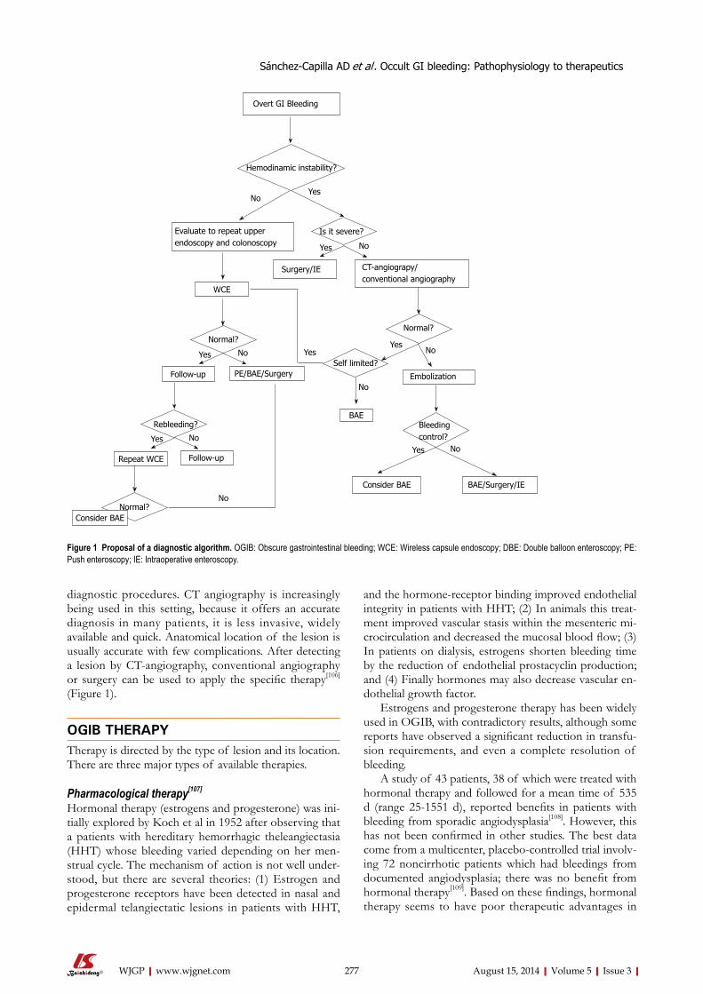
diagnostic procedures. CT angiography is increasingly being used in this setting, because it offers an accurate diagnosis in many patients, it is less invasive, widely available and quick. Anatomical location of the lesion is usually accurate with few complications. After detecting a lesion by CT-angiography, conventional angiography or surgery can be used to apply the specific therapy[106] (Figure 1).
OGIB THERAPYTherapy is directed by the type of lesion and its location. There are three major types of available therapies.
Pharmacological therapy[107]
Hormonal therapy (estrogens and progesterone) was ini-tially explored by Koch et al in 1952 after observing that a patients with hereditary hemorrhagic theleangiectasia (HHT) whose bleeding varied depending on her men-strual cycle. The mechanism of action is not well under-stood, but there are several theories: (1) Estrogen and progesterone receptors have been detected in nasal and epidermal telangiectatic lesions in patients with HHT,
and the hormone-receptor binding improved endothelial integrity in patients with HHT; (2) In animals this treat-ment improved vascular stasis within the mesenteric mi-crocirculation and decreased the mucosal blood flow; (3) In patients on dialysis, estrogens shorten bleeding time by the reduction of endothelial prostacyclin production; and (4) Finally hormones may also decrease vascular en-dothelial growth factor.
Estrogens and progesterone therapy has been widely used in OGIB, with contradictory results, although some reports have observed a significant reduction in transfu-sion requirements, and even a complete resolution of bleeding.
A study of 43 patients, 38 of which were treated with hormonal therapy and followed for a mean time of 535 d (range 25-1551 d), reported benefits in patients with bleeding from sporadic angiodysplasia[108]. However, this has not been confirmed in other studies. The best data come from a multicenter, placebo-controlled trial involv-ing 72 noncirrhotic patients which had bleedings from documented angiodysplasia; there was no benefit from hormonal therapy[109]. Based on these findings, hormonal therapy seems to have poor therapeutic advantages in
August 15, 2014|Volume 5|Issue 3|WJGP|www.wjgnet.com 277
Overt GI Bleeding
Hemodinamic instability?
Is it severe?
WCE
Normal?
Follow-up PE/BAE/Surgery
Rebleeding?
Repeat WCE
Normal?
Surgery/IE CT-angiograpy/conventional angiography
Normal?
Self limited?
Embolization
BAEBleeding control?
Consider BAE BAE/Surgery/IE
Evaluate to repeat upper endoscopy and colonoscopy
Follow-up
Consider BAE
No
No
No
NoYes
Yes
Yes
NoYes
YesNo
No
NoYes
Yes
Figure 1 Proposal of a diagnostic algorithm. OGIB: Obscure gastrointestinal bleeding; WCE: Wireless capsule endoscopy; DBE: Double balloon enteroscopy; PE: Push enteroscopy; IE: Intraoperative enteroscopy.
Sánchez-Capilla AD et al . Occult GI bleeding: Pathophysiology to therapeutics

patients with sporadic angiodysplasia.Some other papers do not recommend their use be-
cause of their lack of beneficial effects on OGIB and their adverse events (thrombosis, gynecomastia, loss of libido in males, metrorragia…). In general, their efficacy has not been proved, except for the treatment of heredi-tary hemorrhagic telangiectasia, von Willebrand disease, chronic kidney failure and gastric antral vascular ectasia (GAVE), in which hormonal therapy reduces transfusion requirements but not the size or number of lesions[98-100].
Somatostatin analogs: Octeotride reduces splacnic arterial flow by inhibiting angiogenesis and endothelial related growth factors[101-103]. Also, octeotride can inhibit angiogenesis by inhibiting endothelial cell proliferation. It has shown efficacy in acute and chronic GI bleed-ing, and can be used in patients with contraindications or a poor response to hormonal therapy. In Rossini et al study[110], treatment with octreotide in 3 patients decreased the need for blood transfusions during the follow-up period (8 to 17 mo). Other authors have pub-lished similar results[111], and have observed comparable side effects including diarrhea, steatorrhea, or changes in glucose metabolism.
A 2010 meta-analysis[112] analyzed 3 studies with a total of 62 patients, observing that 76% of patients re-sponded to this therapy, achieving a significant reduction in transfusion requirements.
Depot formulations like LAR-Octeotride, which al-low intramuscular administration once a month, have gained acceptance in selected cases[113,114]. In a study with 15 patients[115] treated with LAR-Octeotride for a recur-rent bleeding from gastrointestinal angiodysplasia, the proportion of patients who experienced a bleeding event was lower during treatment than prior to treatment (20 vs 73), median transfusion requirements were reduced (2 vs 10 units), and median hemoglobin levels were higher during therapy (10 vs 7 g/dL).
Non-selective beta-blockers: They reduce splacnic flow, pulse and cardiac output. They are usually used in portal hypertension related OGIB and monotherapy or in association with LAR-Octeotride.
Thalidomide: It was retired in the 60s because of its teratogenic effect. However, thalidomide has recently shown to be an effective anti-inflammatory treatment in Crohn’s disease. In addition to its anti-inflammatory ef-fects, it also displays antiangiogenic activity, which may be useful for the treatment of GI bleeding. It can be tak-en orally and it could be used in patients with contrain-dications to other therapies. Obviously it is forbidden in childbearing aged women and in patients with peripheral neuropathy. It must be used cautiously in patients with cardiovascular or neurologic diseases, chronic kidney or liver failure and in immunosuppressed patients.
Some reports show promising outcomes in bleed-ing control[112]. In a randomized trial in 2011[116] patients treated with thalidomide were more likely than those treated with iron supplements to experience a positive
clinical outcome (71% vs 4%).
Other drugs: (1) Antifibrinolitics: Tranexamic acid is an antifibrinolytic agent whose haemostatic effect is due to the inhibition of plasminogen activation in body fluids and tissues. Epsilon-aminocaproic acid has controlled chronic bleeding in patients suffering from HHT. These drugs have a prothrombotic activity and, for this reason, coagulation abnormalities or thrombophilia have to be ruled out before initiating the therapy; (2) Danazol: There are two single reports with positive results after hormonal therapy failure in patients with hereditary hemorrhagic teleangiectasia; (3) Desmopresin; and (4) Recombinant factor Ⅶ: Reserved to cases of massive overt OGIB.
Endoscopic therapyThere are different methods, injection therapies, thermal methods or mechanical devices which can be used with different endoscopes, depending on the location of the bleeding cause.
Argon plasma coagulation: It is safe and the most common and successful method used to treat angiodys-plasia because of its easy application (especially for large superficial lesions), low cost, and reported limited depth of coagulation. Argon plasma coagulation (APC) has been used for a variety of bleeding lesions, including an-giodysplasia, in these lesions submucosal saline injection prior to treatment with APC may protect against deep wall injury.
In a study of 50 patients with small bowel lesions, 44 patients were treated with APC for angiodysplasia[117]. After a mean follow-up of 55 mo, hemoglobin levels increased from a mean of 7.6 g/dL prior to treatment to 11.0 g/dL following it, and there was a significant decrease in the number of patients requiring blood transfusions. However, small bowel bleeding recurred in 21 of the patients treated with APC. A later study with 98 patients[118] reported similar results. The risk factors associated with rebleeding were the number of lesions and the presence of valvular and or arrhythmic cardiac disease.
Electrocoagulation: Bipolar or heater probe coagula-tion is effective for treatment of angiodysplasia in the colon or upper gastrointestinal tract. The risk of perfo-ration with heater probe coagulation may be increased in the colon and small bowel, beyond the duodenum. Monopolar coagulation may be less effective and is as-sociated with an increased rate of complications.
Mechanical hemostasis: Mechanical hemostatic meth-ods such as endoscopic clips have the advantage of avoiding tissue injury, which may be particularly desir-able in patients taking anticoagulants and/or antiplatelet agents, or in patients with coagulation defects.
Another mechanical method that has been described in some case reports is band ligation[119], that is safe and
August 15, 2014|Volume 5|Issue 3|WJGP|www.wjgnet.com 278
Sánchez-Capilla AD et al . Occult GI bleeding: Pathophysiology to therapeutics
effective for the treatment of acutely bleeding small bowel vascular lesions with similar results to APC (re-current bleeding in 43%) and which can be a definitive treatment for Dieulafoy’s lesion.
AngiographyAngiography is indicated in patients with GI bleed-ing who fail to respond to medical and/or endoscopic therapy, as an alternative to surgery in hemodynamically unstable patients with severe bleeding or for patients with ongoing or recurrent bleeding following attempts to control the bleeding endoscopically. Angiographic thera-pies include the infusion of vasoactive drugs (vasopres-sin) or the delivery of agents to mechanically occlude the vascular supply of the bleeding lesion (embolization).
Vasopressin causes generalized vasoconstriction via a direct action upon vessel walls, especially the arterioles, capillaries, and venules. It should be used with caution in patients with coronary artery disease, congestive car-diomyopathy, severe hypertension, or severe peripheral vascular disease. Other side effects are arrhythmias and water retention leading to hyponatremia.
Agents used for embolization include biodegradable gelatin sponge, polyvinyl alcohol particles, liquid agents, and metallic coils. Microcoils have become the preferred agent for embolizing bleeding vessels and can be de-ployed by means of a microcatheter to the site of bleed-ing. The complications of embolization include those associated with arteriography itself (e.g., hematomas, arterial thrombosis, dissection, embolism, and pseudoan-eurysm formation), and bowel infarction.
The choice between vasopressin and embolization should be individualized for each patient, taking into account angiography experience. Embolization with mi-crocoils may be more successful than vasopressin infu-sion (95% vs 80%-90%)[120,121]) but it is associated with a higher rate of complications.
Initial hemostasis may be achieved in up to 80%-95% of patients in whom angiographic therapy is technically feasible, but rebleeding is a common problem (9%-56% in embolization and 5%-50% in intra-arterial vasopressin infusion).
Surgery Surgical therapy is reserved for patients with a known bleeding cause, found with other methods, patients with increasing transfusion requirements or life-threatening bleedings from clearly identified origins, or for cases in which haemodinamic instability does not allow the clini-cians to complete the diagnostic algorithm and an IE is mandatory. In this last situation, rebleeding is usual[86,87].
CONCLUSIONDespite technological advances, OGIB is still a diag-nostic challenge for gastroenterologists, with important hospital resources consumption and delayed diagnoses. WCE is the most cost-effective diagnostic procedure to
identify the bleeding source and its location. In selected cases, with an outstanding severity, CT-angiography is an alternative.
Although therapy depends on the bleeding cause, BAE plays an important role in the management of le-sions found in WCE. It is less aggressive than intraop-erative enteroscopy and has a high index of success. A pharmacological alternative to surgery or endoscopy are depot formulations of somatostatin analogs.
REFERENCES1 Ell C, May A. Mid-gastrointestinal bleeding: capsule en-
doscopy and push-and-pull enteroscopy give rise to a new medical term. Endoscopy 2006; 38: 73-75 [PMID: 16429358 DOI: 10.1055/s-2005-921131]
2 Prakash C, Zuckerman GR. Acute small bowel bleeding: a distinct entity with significantly different economic implica-tions compared with GI bleeding from other locations. Gas-trointest Endosc 2003; 58: 330-335 [PMID: 14528203]
3 Moawad FJ, Veerappan GR, Wong RK. Small bowel is the primary source of obscure gastrointestinal bleeding. Gastro-enterology 2008; 135: 1016 [PMID: 18694750 DOI: 10.1053/j.gastro.2008.05.083]
4 Gralnek IM. Obscure-overt gastrointestinal bleeding. Gas-troenterology 2005; 128: 1424-1430 [PMID: 15887123 DOI: 10.1053/j.gastro.2005.03.067]
5 Raju GS, Gerson L, Das A, Lewis B. American Gastroen-terological Association (AGA) Institute technical review on obscure gastrointestinal bleeding. Gastroenterology 2007; 133: 1697-1717 [PMID: 17983812 DOI: 10.1053/j.gastro.2007.06.007]
6 Mujica VR, Barkin JS. Occult gastrointestinal bleeding. General overview and approach. Gastrointest Endosc Clin N Am 1996; 6: 833-845 [PMID: 8899413]
7 Meyer CT, Troncale FJ, Galloway S, Sheahan DG. Arterio-venous malformations of the bowel: an analysis of 22 cases and a review of the literature. Medicine (Baltimore) 1981; 60: 36-48 [PMID: 6969839 DOI: 10.1097/00005792-198101000-00004]
8 Sami SS, Al-Araji SA, Ragunath K. Review article: gas-trointestinal angiodysplasia - pathogenesis, diagnosis and management. Aliment Pharmacol Ther 2014; 39: 15-34 [PMID: 24138285 DOI: 10.1111/apt.12527]
9 Foutch PG. Angiodysplasia of the gastrointestinal tract. Am J Gastroenterol 1993; 88: 807-818 [PMID: 8389094]
10 Scheffer SM, Leatherman LL. Resolution of Heyde’s syn-drome of aortic stenosis and gastrointestinal bleeding after aortic valve replacement. Ann Thorac Surg 1986; 42: 477-480 [PMID: 3490235]
11 Islam S, Cevik C, Madonna R, Frandah W, Islam E, Islam S, Nugent K. Left ventricular assist devices and gastrointes-tinal bleeding: a narrative review of case reports and case series. Clin Cardiol 2013; 36: 190-200 [PMID: 23378047 DOI: 10.1002/clc.22096]
12 Soulellis CA, Carpentier S, Chen YI, Fallone CA, Barkun AN. Lower GI hemorrhage controlled with endoscopically applied TC-325 (with videos). Gastrointest Endosc 2013; 77: 504-507 [PMID: 23290722 DOI: 10.1016/j.gie.2012.10.014]
13 Sagar J, Kumar V, Shah DK. Meckel’s diverticulum: a systematic review. J R Soc Med 2006; 99: 501-505 [PMID: 17021300 DOI: 10.1258/jrsm.99.10.501]
14 Yahchouchy EK, Marano AF, Etienne JC, Fingerhut AL. Meckel’s diverticulum. J Am Coll Surg 2001; 192: 658-662 [PMID: 11333103 DOI: 10.1016/S1072-7515(01)00817-1]
15 Lee YT, Walmsley RS, Leong RW, Sung JJ. Dieulafoy’s le-sion. Gastrointest Endosc 2003; 58: 236-243 [PMID: 12872092
August 15, 2014|Volume 5|Issue 3|WJGP|www.wjgnet.com 279
Sánchez-Capilla AD et al . Occult GI bleeding: Pathophysiology to therapeutics
DOI: 10.1067/mge.2003.126]16 Lara LF, Sreenarasimhaiah J, Tang SJ, Afonso BB, Rockey
DC. Dieulafoy lesions of the GI tract: localization and thera-peutic outcomes. Dig Dis Sci 2010; 55: 3436-3441 [PMID: 20848205 DOI: 10.1007/s10620-010-1385-0]
17 Ferguson A, Arranz E, O’Mahony S. Clinical and pathologi-cal spectrum of coeliac disease--active, silent, latent, poten-tial. Gut 1993; 34: 150-151 [PMID: 8432463 DOI: 10.1136/gut.34.2.150]
18 Moreno RD, Harris M, Bryk HB, Pachter HL, Migliet-ta MA. Late presentation of a hepatic pseudoaneurysm with hemobilia after angioembolization for blunt hepatic trauma. J Trauma 2007; 62: 1048-1050 [PMID: 17426568 DOI: 10.1097/01.ta.0000235295.02414.d7]
19 Bloechle C, Izbicki JR, Rashed MY, el-Sefi T, Hosch SB, Knoefel WT, Rogiers X, Broelsch CE. Hemobilia: presenta-tion, diagnosis, and management. Am J Gastroenterol 1994; 89: 1537-1540 [PMID: 8079933]
20 Saers SJ, Scheltinga MR. Primary aortoenteric fistula. Br J Surg 2005; 92: 143-152 [PMID: 15685700 DOI: 10.1002/bjs.4928]
21 Suter M, Doenz F, Chapuis G, Gillet M, Sandblom P. Haem-orrhage into the pancreatic duct (Hemosuccus pancreati-cus): recognition and management. Eur J Surg 1995; 161: 887-892 [PMID: 8775630]
22 Nantes O, Zozaya JM, Montes R, Hermida J. Gastrointes-tinal lesions and characteristics of acute gastrointestinal bleeding in acenocoumarol-treated patients. Gastroenterol Hepatol 2014: S0210-5705(14)00024-7 [PMID: 24582763 DOI: 10.1016/j.gastrohep.2013.12.008]
23 Barada K, Abdul-Baki H, El Hajj II, Hashash JG, Green PH. Gastrointestinal bleeding in the setting of anticoagulation and antiplatelet therapy. J Clin Gastroenterol 2009; 43: 5-12 [PMID: 18607297 DOI: 10.1097/MCG.0b013e31811edd13]
24 Thomopoulos KC, Mimidis KP, Theocharis GJ, Gatopoulou AG, Kartalis GN, Nikolopoulou VN. Acute upper gastroin-testinal bleeding in patients on long-term oral anticoagula-tion therapy: endoscopic findings, clinical management and outcome. World J Gastroenterol 2005; 11: 1365-1368 [PMID: 15761977]
25 Rasmussen LH, Larsen TB, Graungaard T, Skjøth F, Lip GY. Primary and secondary prevention with new oral anti-coagulant drugs for stroke prevention in atrial fibrillation: indirect comparison analysis. BMJ 2012; 345: e7097 [PMID: 23129490 DOI: 10.1136/bmj.e7097]
26 Holster IL, Valkhoff VE, Kuipers EJ, Tjwa ET. New oral anticoagulants increase risk for gastrointestinal bleeding: a systematic review and meta-analysis. Gastroenterology 2013; 145: 105-112.e15 [PMID: 23470618 DOI: 10.1053/j.gastro.2013.02.041]
27 Zaman A, Katon RM. Push enteroscopy for obscure gas-trointestinal bleeding yields a high incidence of proximal lesions within reach of a standard endoscope. Gastrointest Endosc 1998; 47: 372-376 [PMID: 9609429 DOI: 10.1016/S0016-5107(98)70221-4]
28 Descamps C, Schmit A, Van Gossum A. “Missed” upper gastrointestinal tract lesions may explain “occult” bleeding. Endoscopy 1999; 31: 452-455 [PMID: 10494684 DOI: 10.1055/s-1999-151]
29 Chak A, Cooper GS, Canto MI, Pollack BJ, Sivak MV. Enter-oscopy for the initial evaluation of iron deficiency. Gastroin-test Endosc 1998; 47: 144-148 [PMID: 9512279 DOI: 10.1016/S0016-5107(98)70347-5]
30 Fry LC, Bellutti M, Neumann H, Malfertheiner P, Mönke-müller K. Incidence of bleeding lesions within reach of con-ventional upper and lower endoscopes in patients undergo-ing double-balloon enteroscopy for obscure gastrointestinal bleeding. Aliment Pharmacol Ther 2009; 29: 342-349 [PMID: 19035975 DOI: 10.1111/j.1365-2036.2008.03888.x]
31 Tee HP, Kaffes AJ. Non-small-bowel lesions encountered
during double-balloon enteroscopy performed for obscure gastrointestinal bleeding. World J Gastroenterol 2010; 16: 1885-1889 [PMID: 20397267 DOI: 10.3748/wjg.v16.i15.1885]
32 Leaper M, Johnston MJ, Barclay M, Dobbs BR, Frizelle FA. Reasons for failure to diagnose colorectal carcinoma at colo-noscopy. Endoscopy 2004; 36: 499-503 [PMID: 15202045 DOI: 10.1055/s-2004-814399]
33 Gilbert D, O’Malley S, Selby W. Are repeat upper gastro-intestinal endoscopy and colonoscopy necessary within six months of capsule endoscopy in patients with obscure gastrointestinal bleeding? J Gastroenterol Hepatol 2008; 23: 1806-1809 [PMID: 19032448]
34 Lin S, Branch MS, Shetzline M. The importance of indication in the diagnostic value of push enteroscopy. Endoscopy 2003; 35: 315-321 [PMID: 12664388 DOI: 10.1055/s-2003-38144]
35 Rabe FE, Becker GJ, Besozzi MJ, Miller RE. Efficacy study of the small-bowel examination. Radiology 1981; 140: 47-50 [PMID: 7244242]
36 Moch A, Herlinger H, Kochman ML, Levine MS, Rube-sin SE, Laufer I. Enteroclysis in the evaluation of obscure gastrointestinal bleeding. AJR Am J Roentgenol 1994; 163: 1381-1384 [PMID: 7992733 DOI: 10.2214/ajr.163.6.7992733]
37 Jonnalagadda S, Prakash C. Intestinal strictures can impede wireless capsule enteroscopy. Gastrointest Endosc 2003; 57: 418-420 [PMID: 12612534 DOI: 10.1067/mge.2003.128]
38 Graça BM, Freire PA, Brito JB, Ilharco JM, Carvalheiro VM, Caseiro-Alves F. Gastroenterologic and radiologic ap-proach to obscure gastrointestinal bleeding: how, why, and when? Radiographics 2010; 30: 235-252 [PMID: 20083596 DOI: 10.1148/rg.301095091]
39 Huprich JE, Fletcher JG, Alexander JA, Fidler JL, Burton SS, McCullough CH. Obscure gastrointestinal bleeding: evalu-ation with 64-section multiphase CT enterography--initial experience. Radiology 2008; 246: 562-571 [PMID: 18227546 DOI: 10.1148/radiol.2462061920]
40 Kennedy DW, Laing CJ, Tseng LH, Rosenblum DI, Tam-arkin SW. Detection of active gastrointestinal hemorrhage with CT angiography: a 4(1/2)-year retrospective review. J Vasc Interv Radiol 2010; 21: 848-855 [PMID: 20400333 DOI: 10.1016/j.jvir.2010.01.039]
41 Yoon W, Jeong YY, Shin SS, Lim HS, Song SG, Jang NG, Kim JK, Kang HK. Acute massive gastrointestinal bleed-ing: detection and localization with arterial phase multi-detector row helical CT. Radiology 2006; 239: 160-167 [PMID: 16484350 DOI: 10.1148/radiol.2383050175]
42 Som P, Oster ZH, Atkins HL, Goldman AG, Sacker DF, Harold WH, Fairchild RG, Richards P, Brill AB. Detection of gastrointestinal blood loss with 99mTc-labeled, heat-treated red blood cells. Radiology 1981; 138: 207-209 [PMID: 6969900]
43 Zuckerman GR, Prakash C, Askin MP, Lewis BS. AGA technical review on the evaluation and management of occult and obscure gastrointestinal bleeding. Gastroenter-ology 2000; 118: 201-221 [PMID: 10611170 DOI: 10.1016/S0016-5085(00)70430-6]
44 Hunter JM, Pezim ME. Limited value of technetium 99m-labeled red cell scintigraphy in localization of lower gas-trointestinal bleeding. Am J Surg 1990; 159: 504-506 [PMID: 2334015 DOI: 10.1016/S0002-9610(05)81256-5]
45 Ng DA, Opelka FG, Beck DE, Milburn JM, Witherspoon LR, Hicks TC, Timmcke AE, Gathright JB. Predictive value of technetium Tc 99m-labeled red blood cell scintigraphy for positive angiogram in massive lower gastrointestinal hem-orrhage. Dis Colon Rectum 1997; 40: 471-477 [PMID: 9106699 DOI: 10.1007/BF02258395]
46 Fiorito JJ, Brandt LJ, Kozicky O, Grosman IM, Sprayragen S. The diagnostic yield of superior mesenteric angiography: correlation with the pattern of gastrointestinal bleeding. Am J Gastroenterol 1989; 84: 878-881 [PMID: 2667335]
47 Egglin TK, O‘Moore PV, Feinstein AR, Waltman AC. Com-plications of peripheral arteriography: a new system to iden-
August 15, 2014|Volume 5|Issue 3|WJGP|www.wjgnet.com 280
Sánchez-Capilla AD et al . Occult GI bleeding: Pathophysiology to therapeutics
tify patients at increased risk. J Vasc Surg 1995; 22: 787-794 [PMID: 8523614 DOI: 10.1016/S0741-5214(95)70070-6]
48 Bloomfeld RS, Smith TP, Schneider AM, Rockey DC. Provoc-ative angiography in patients with gastrointestinal hemor-rhage of obscure origin. Am J Gastroenterol 2000; 95: 2807-2812 [PMID: 11051352 DOI: 10.1111/j.1572-0241.2000.03191.x]
49 Miller M, Smith TP. Angiographic diagnosis and endovas-cular management of nonvariceal gastrointestinal hemor-rhage. Gastroenterol Clin North Am 2005; 34: 735-752 [PMID: 16303580 DOI: 10.1016/j.gtc.2005.09.001]
50 Busch OR, van Delden OM, Gouma DJ. Therapeutic options for endoscopic haemostatic failures: the place of the surgeon and radiologist in gastrointestinal tract bleeding. Best Pract Res Clin Gastroenterol 2008; 22: 341-354 [PMID: 18346688 DOI: 10.1016/j.bpg.2007.10.018]
51 Liao Z, Gao R, Xu C, Li ZS. Indications and detection, completion, and retention rates of small-bowel capsule en-doscopy: a systematic review. Gastrointest Endosc 2010; 71: 280-286 [PMID: 20152309 DOI: 10.1016/j.gie.2009.09.031]
52 Fireman Z, Kopelman Y. The role of video capsule en-doscopy in the evaluation of iron deficiency anaemia. Dig Liver Dis 2004; 36: 97-102 [PMID: 15002814 DOI: 10.1016/j.dld.2003.10.009]
53 Rastogi A, Schoen RE, Slivka A. Diagnostic yield and clinical outcomes of capsule endoscopy. Gastrointest Endosc 2004; 60: 959-964 [PMID: 15605012 DOI: 10.1016/S0016-5107(04)02226-6]
54 May A, Wardak A, Nachbar L, Remke S, Ell C. Influence of patient selection on the outcome of capsule endoscopy in patients with chronic gastrointestinal bleeding. J Clin Gastro-enterol 2005; 39: 684-688 [PMID: 16082277 DOI: 10.1097/01.mcg.0000173857.22933.3b]
55 Bresci G, Parisi G, Bertoni M, Tumino E, Capria A. The role of video capsule endoscopy for evaluating obscure gastrointestinal bleeding: usefulness of early use. J Gastro-enterol 2005; 40: 256-259 [PMID: 15830284 DOI: 10.1007/s00535-004-1532-5]
56 Sidhu R, Sanders DS, McAlindon ME. Does capsule en-doscopy recognise gastric antral vascular ectasia more fre-quently than conventional endoscopy? J Gastrointestin Liver Dis 2006; 15: 375-377 [PMID: 17205150]
57 Estévez E, González-Conde B, Vázquez-Iglesias JL, Alonso PA, Vázquez-Millán Mde L, Pardeiro R. Incidence of tumor-al pathology according to study using capsule endoscopy for patients with obscure gastrointestinal bleeding. Surg Endosc 2007; 21: 1776-1780 [PMID: 17356941]
58 Redondo-Cerezo E, Gómez-Ruiz CJ, Sánchez-Manjavacas N, Viñuelas M, Jimeno C, Pérez-Vigara G, Morillas J, Pérez-García JI, García-Cano J, Pérez-Sola A. Long-term follow-up of patients with small-bowel angiodysplasia on capsule endoscopy. Determinants of a higher clinical impact and rebleeding rate. Rev Esp Enferm Dig 2008; 100: 202-207 [PMID: 18563976 DOI: 10.4321/S1130-01082008000400002]
59 Marmo R, Rotondano G, Piscopo R, Bianco MA, Cipolletta L. Meta-analysis: capsule enteroscopy vs. conventional mo-dalities in diagnosis of small bowel diseases. Aliment Phar-macol Ther 2005; 22: 595-604 [PMID: 16181299 DOI: 10.1111/j.1365-2036.2005.02625.x]
60 Lewis BS, Eisen GM, Friedman S. A pooled analysis to evaluate results of capsule endoscopy trials. Endoscopy 2005; 37: 960-965 [PMID: 16189768 DOI: 10.1055/s-2005-870353]
61 Triester SL, Leighton JA, Leontiadis GI, Fleischer DE, Hara AK, Heigh RI, Shiff AD, Sharma VK. A meta-analysis of the yield of capsule endoscopy compared to other diagnostic modalities in patients with obscure gastrointestinal bleed-ing. Am J Gastroenterol 2005; 100: 2407-2418 [PMID: 16279893 DOI: 10.1111/j.1572-0241.2005.00274.x]
62 Saperas E, Dot J, Videla S, Alvarez-Castells A, Perez-La-fuente M, Armengol JR, Malagelada JR. Capsule endoscopy versus computed tomographic or standard angiography for
the diagnosis of obscure gastrointestinal bleeding. Am J Gas-troenterol 2007; 102: 731-737 [PMID: 17397406 DOI: 10.1111/j.1572-0241.2007.01058.x]
63 Pasha SF, Leighton JA, Das A, Harrison ME, Decker GA, Fleischer DE, Sharma VK. Double-balloon enteroscopy and capsule endoscopy have comparable diagnostic yield in small-bowel disease: a meta-analysis. Clin Gastroenterol Hepatol 2008; 6: 671-676 [PMID: 18356113 DOI: 10.1016/j.cgh.2008.01.005]
64 Hartmann D, Schmidt H, Bolz G, Schilling D, Kinzel F, Eick-hoff, Huschner W, Möller K, Jakobs R, Reitzig P, Weickert U, Gellert K, Schultz H, Guenther K, Hollerbuhl H, Schoen-leben K, Schulz HJ, Riemann JF. A prospective two-center study comparing wireless capsule endoscopy with intraop-erative enteroscopy in patients with obscure GI bleeding. Gastrointest Endosc 2005; 61: 826-832 [DOI: 10.1016/S0016-5107(05)00372-X]
65 Redondo-Cerezo E, Pérez-Vigara G, Pérez-Sola A, Gómez-Ruiz CJ, Chicano MV, Sánchez-Manjavacas N, Morillas J, Pérez-García JI, García-Cano J. Diagnostic yield and impact of capsule endoscopy on management of patients with gas-trointestinal bleeding of obscure origin. Dig Dis Sci 2007; 52: 1376-1381 [PMID: 17356913 DOI: 10.1007/s10620-006-9605-3]
66 Mylonaki M, Fritscher-Ravens A, Swain P. Wireless capsule endoscopy: a comparison with push enteroscopy in patients with gastroscopy and colonoscopy negative gastrointestinal bleeding. Gut 2003; 52: 1122-1126 [PMID: 12865269 DOI: 10.1136/gut.52.8.1122]
67 Lai LH, Wong GL, Chow DK, Lau JY, Sung JJ, Leung WK. Long-term follow-up of patients with obscure gastrointesti-nal bleeding after negative capsule endoscopy. Am J Gastro-enterol 2006; 101: 1224-1228 [PMID: 16771942 DOI: 10.1111/j.1572-0241.2006.00565.x]
68 Swain P, Fritscher-Ravens A. Role of video endoscopy in managing small bowel disease. Gut 2004; 53: 1866-1875 [PMID: 15542530 DOI: 10.1136/gut.2003.035576]
69 Ben-Soussan E, Savoye G, Antonietti M, Ramirez S, Lere-bours E, Ducrotté P. Factors that affect gastric passage of video capsule. Gastrointest Endosc 2005; 62: 785-790 [PMID: 16246700 DOI: 10.1016/j.gie.2005.07.040]
70 Nogales Rincón O, Merino Rodríguez B, González Asanza C, Fernández-Pacheco PM. [Utility of capsule endoscopy with flexible spectral imaging color enhancement in the diagnosis of small bowel lesions]. Gastroenterol Hepatol 2013; 36: 63-68 [PMID: 23140757 DOI: 10.1016/j.gastrohep.2012.08.004]
71 Sidhu R, Sanders DS, Morris AJ, McAlindon ME. Guide-lines on small bowel enteroscopy and capsule endoscopy in adults. Gut 2008; 57: 125-136 [PMID: 18094205 DOI: 10.1136/gut.2007.129999]
72 Benz C, Jakobs R, Riemann JF. Do we need the overtube for push-enteroscopy? Endoscopy 2001; 33: 658-661 [PMID: 11490380 DOI: 10.1055/s-2001-16208]
73 Taylor AC, Chen RY, Desmond PV. Use of an overtube for enteroscopy--does it increase depth of insertion? A prospec-tive study of enteroscopy with and without an overtube. Endoscopy 2001; 33: 227-230 [PMID: 11293754 DOI: 10.1055/s-2001-12799]
74 Carey EJ, Fleischer DE. Investigation of the small bowel in gastrointestinal bleeding--enteroscopy and capsule endos-copy. Gastroenterol Clin North Am 2005; 34: 719-734 [PMID: 16303579 DOI: 10.1016/j.gtc.2005.08.009]
75 Mergener K, Ponchon T, Gralnek I, Pennazio M, Gay G, Selby W, Seidman EG, Cellier C, Murray J, de Franchis R, Rösch T, Lewis BS. Literature review and recommenda-tions for clinical application of small-bowel capsule endos-copy, based on a panel discussion by international experts. Consensus statements for small-bowel capsule endoscopy, 2006/2007. Endoscopy 2007; 39: 895-909 [PMID: 17968807 DOI: 10.1055/s-2007-966930]
76 May A, Nachbar L, Pohl J, Ell C. Endoscopic interventions
August 15, 2014|Volume 5|Issue 3|WJGP|www.wjgnet.com 281
Sánchez-Capilla AD et al . Occult GI bleeding: Pathophysiology to therapeutics
in the small bowel using double balloon enteroscopy: feasi-bility and limitations. Am J Gastroenterol 2007; 102: 527-535 [PMID: 17222315 DOI: 10.1111/j.1572-0241.2007.01063.x]
77 May A, Nachbar L, Schneider M, Ell C. Prospective com-parison of push enteroscopy and push-and-pull enteros-copy in patients with suspected small-bowel bleeding. Am J Gastroenterol 2006; 101: 2016-2024 [PMID: 16968508 DOI: 10.1111/j.1572-0241.2006.00745.x]
78 Matsumoto T, Moriyama T, Esaki M, Nakamura S, Iida M. Performance of antegrade double-balloon enteroscopy: com-parison with push enteroscopy. Gastrointest Endosc 2005; 62: 392-398 [PMID: 16111958 DOI: 10.1016/j.gie.2005.04.052]
79 Yamamoto H, Sekine Y, Sato Y, Higashizawa T, Miyata T, Iino S, Ido K, Sugano K. Total enteroscopy with a non-surgical steerable double-balloon method. Gastrointest Endosc 2001; 53: 216-220 [PMID: 11174299 DOI: 10.1067/mge.2001.112181]
80 Alexander JA , Leighton JA. Capsule endoscopy and balloon-assisted endoscopy: competing or complementary technologies in the evaluation of small bowel disease? Curr Opin Gastroenterol 2009; 25: 433-437 [PMID: 19461511 DOI: 10.1097/MOG.0b013e32832d641e]
81 Gay G, Delvaux M, Fassler I. Outcome of capsule endos-copy in determining indication and route for push-and-pull enteroscopy. Endoscopy 2006; 38: 49-58 [PMID: 16429355 DOI: 10.1055/s-2005-921176]
82 Gerson LB. Double-balloon enteroscopy: the new gold stan-dard for small-bowel imaging? Gastrointest Endosc 2005; 62: 71-75 [PMID: 15990822 DOI: 10.1016/S0016-5107(05)00507-9]
83 Mensink PB, Haringsma J, Kucharzik T, Cellier C, Pérez-Cuadrado E, Mönkemüller K, Gasbarrini A, Kaffes AJ, Na-kamura K, Yen HH, Yamamoto H. Complications of double balloon enteroscopy: a multicenter survey. Endoscopy 2007; 39: 613-615 [PMID: 17516287 DOI: 10.1055/s-2007-966444]
84 Gerson LB, Tokar J, Chiorean M, Lo S, Decker GA, Cave D, Bouhaidar D, Mishkin D, Dye C, Haluszka O, Leighton JA, Zfass A, Semrad C. Complications associated with double balloon enteroscopy at nine US centers. Clin Gastroenterol Hepatol 2009; 7: 1177-1182, 1177-1182 [PMID: 19602453 DOI: 10.1016/j.cgh.2009.07.005]
85 May A, Färber M, Aschmoneit I, Pohl J, Manner H, Lotterer E, Möschler O, Kunz J, Gossner L, Mönkemüller K, Ell C. Prospective multicenter trial comparing push-and-pull en-teroscopy with the single- and double-balloon techniques in patients with small-bowel disorders. Am J Gastroenterol 2010; 105: 575-581 [PMID: 20051942 DOI: 10.1038/ajg.2009.712]
86 Takano N, Yamada A, Watabe H, Togo G, Yamaji Y, Yoshi-da H, Kawabe T, Omata M, Koike K. Single-balloon versus double-balloon endoscopy for achieving total enteroscopy: a randomized, controlled trial. Gastrointest Endosc 2011; 73: 734-739 [PMID: 21272875 DOI: 10.1016/j.gie.2010.10.047]
87 Domagk D, Mensink P, Aktas H, Lenz P, Meister T, Lueger-ing A, Ullerich H, Aabakken L, Heinecke A, Domschke W, Kuipers E, Bretthauer M. Single- vs. double-balloon enteros-copy in small-bowel diagnostics: a randomized multicenter trial. Endoscopy 2011; 43: 472-476 [PMID: 21384320 DOI: 10.1055/s-0030-1256247]
88 Efthymiou M, Desmond PV, Brown G, La Nauze R, Kaffes A, Chua TJ, Taylor AC. SINGLE-01: a randomized, controlled trial comparing the efficacy and depth of insertion of single- and double-balloon enteroscopy by using a novel method to determine insertion depth. Gastrointest Endosc 2012; 76: 972-980 [PMID: 22980289 DOI: 10.1016/j.gie.2012.06.033]
89 Pai RD, Carr-Locke DL, Thompson CC. Endoscopic evalu-ation of the defunctionalized stomach by using ShapeLock technology (with video). Gastrointest Endosc 2007; 66: 578-581 [PMID: 17725949 DOI: 10.1016/j.gie.2007.02.062]
90 Kuga R, Furuya CK, Hondo FY, Ide E, Ishioka S, Sakai P. ERCP using double-balloon enteroscopy in patients with Roux-en-Y anatomy. Dig Dis 2008; 26: 330-335 [PMID:
19188724 DOI: 10.1159/000177018]91 Rex DK, Khashab M, Raju GS, Pasricha J, Kozarek R. Insert-
ability and safety of a shape-locking device for colonoscopy. Am J Gastroenterol 2005; 100: 817-820 [PMID: 15784024 DOI: 10.1111/j.1572-0241.2005.40801.x]
92 Haber GWL. A prospective study to evaluate the ShapeLock guide to enable complete colonoscopy in previously failed cases. Gastrointest Endosc 2006; 63: AB226 [DOI: 10.1016/j.gie.2006.03.573]
93 Schulz HJ, Schmidt H. Intraoperative enteroscopy. Gastro-intest Endosc Clin N Am 2009; 19: 371-379 [PMID: 19647646 DOI: 10.1016/j.giec.2009.04.011]
94 Douard R, Wind P, Berger A, Maniere T, Landi B, Cellier C, Cugnenc PH. Role of intraoperative enteroscopy in the management of obscure gastointestinal bleeding at the time of video-capsule endoscopy. Am J Surg 2009; 198: 6-11 [PMID: 19393986 DOI: 10.1016/j.amjsurg.2008.06.036]
95 Zaman A, Sheppard B, Katon RM. Total peroral intraopera-tive enteroscopy for obscure GI bleeding using a dedicated push enteroscope: diagnostic yield and patient outcome. Gastrointest Endosc 1999; 50: 506-510 [PMID: 10502171 DOI: 10.1016/S0016-5107(99)70073-8]
96 Lewis MP, Khoo DE, Spencer J. Value of laparotomy in the diagnosis of obscure gastrointestinal haemorrhage. Gut 1995; 37: 187-190 [PMID: 7557565 DOI: 10.1136/gut.37.2.187]
97 Douard R, Wind P, Panis Y, Marteau P, Bouhnik Y, Cellier C, Cugnenc P, Valleur P. Intraoperative enteroscopy for diagnosis and management of unexplained gastrointestinal bleeding. Am J Surg 2000; 180: 181-184 [PMID: 11084125 DOI: 10.1016/S0002-9610(00)00447-5]
98 Kovacs TO, Jensen DM. Recent advances in the endoscopic di-agnosis and therapy of upper gastrointestinal, small intestinal, and colonic bleeding. Med Clin North Am 2002; 86: 1319-1356 [PMID: 12510456 DOI: 10.1016/S0025-7125(02)00079-2]
99 Apostolopoulos P, Liatsos C, Gralnek IM, Kalantzis C, Gi-annakoulopoulou E, Alexandrakis G, Tsibouris P, Kalafatis E, Kalantzis N. Evaluation of capsule endoscopy in active, mild-to-moderate, overt, obscure GI bleeding. Gastrointest Endosc 2007; 66: 1174-1181 [PMID: 18061718 DOI: 10.1016/j.gie.2007.06.058]
100 Cellier C. Obscure gastrointestinal bleeding: role of vid-eocapsule and double-balloon enteroscopy. Best Pract Res Clin Gastroenterol 2008; 22: 329-340 [PMID: 18346687 DOI: 10.1016/j.bpg.2007.12.006]
101 Somsouk M, Gralnek IM, Inadomi JM. Management of ob-scure occult gastrointestinal bleeding: a cost-minimization analysis. Clin Gastroenterol Hepatol 2008; 6: 661-670 [PMID: 18550005 DOI: 10.1016/j.cgh.2008.02.033]
102 Albert JG, Nachtigall F, Wiedbrauck F, Dollinger MM, Git-tinger FS, Hollerbach S, Wienke A. Minimizing procedural cost in diagnosing small bowel bleeding: comparison of a strategy based on initial capsule endoscopy versus initial double-balloon enteroscopy. Eur J Gastroenterol Hepatol 2010; 22: 679-688 [PMID: 20446352]
103 Viazis N, Papaxoinis K, Vlachogiannakos J, Efthymiou A, Theodoropoulos I, Karamanolis DG. Is there a role for second-look capsule endoscopy in patients with obscure GI bleeding after a nondiagnostic first test? Gastrointest Endosc 2009; 69: 850-856 [PMID: 18950762 DOI: 10.1016/j.gie.2008.05.053]
104 Bar-Meir S. Video capsule endoscopy or double-balloon en-teroscopy: are they equivalent? Gastrointest Endosc 2009; 69: 875-876 [PMID: 19327474 DOI: 10.1016/j.gie.2008.07.051]
105 Kaffes AJ, Siah C, Koo JH. Clinical outcomes after double-balloon enteroscopy in patients with obscure GI bleeding and a positive capsule endoscopy. Gastrointest Endosc 2007; 66: 304-309 [PMID: 17643704 DOI: 10.1016/j.gie.2007.02.044]
106 González-Galilea A, Gálvez-Calderón C, García Sánchez V, de Dios-Vega J.F. Hemorragia digestiva de origen oscuro. Orientación diagnóstica y terapóutica. Publicacién electróni-
August 15, 2014|Volume 5|Issue 3|WJGP|www.wjgnet.com 282
Sánchez-Capilla AD et al . Occult GI bleeding: Pathophysiology to therapeutics
ca. Available from: URL: http: //www.sapd.es/revista/ar-ticle.php?file=vol34_n1/03
107 van Cutsem E, Rutgeerts P, Vantrappen G. Treatment of bleeding gastrointestinal vascular malformations with oestrogen-progesterone. Lancet 1990; 335: 953-955 [PMID: 1970032 DOI: 10.1016/0140-6736(90)91010-8]
108 Barkin JS, Ross BS. Medical therapy for chronic gastro-intestinal bleeding of obscure origin. Am J Gastroenterol 1998; 93: 1250-1254 [PMID: 9707046 DOI: 10.1111/j.1572-0241.1998.404_i.x]
109 Junquera F, Feu F, Papo M, Videla S, Armengol JR, Bordas JM, Saperas E, Piqué JM, Malagelada JR. A multicenter, randomized, clinical trial of hormonal therapy in the pre-vention of rebleeding from gastrointestinal angiodysplasia. Gastroenterology 2001; 121: 1073-1079 [PMID: 11677198 DOI: 10.1053/gast.2001.28650]
110 Blich M, Fruchter O, Edelstein S, Edoute Y. Somatostatin therapy ameliorates chronic and refractory gastrointesti-nal bleeding caused by diffuse angiodysplasia in a patient on anticoagulation therapy. Scand J Gastroenterol 2003; 38: 801-803 [PMID: 12889571 DOI: 10.1080/00365520310001969]
111 Rossini FP, Arrigoni A, Pennazio M. Octreotide in the treat-ment of bleeding due to angiodysplasia of the small intes-tine. Am J Gastroenterol 1993; 88: 1424-1427 [PMID: 8362842]
112 Bauditz J, Wedel S, Lochs H. Thalidomide reduces tumour necrosis factor alpha and interleukin 12 production in pa-tients with chronic active Crohn’s disease. Gut 2002; 50: 196-200 [PMID: 11788559 DOI: 10.1136/gut.50.2.196]
113 Krikis N, Tziomalos K, Perifanis V, Vakalopoulou S, Kara-giannis A, Garipidou V, Harsoulis F. Treatment of recur-rent gastrointestinal haemorrhage in a patient with von Willebrand’s disease with octreotide LAR and propranolol. Gut 2005; 54: 171-172 [PMID: 15591529 DOI: 10.1136/gut.2004.049031]
114 Molina-Infante J, Perez-Gallardo B, Gonzalez-Garcia G, Fernandez-Bermejo M, Mateos-Rodriguez JM, Robledo-An-dres P. Octreotide LAR for severe obscure-overt gastrointes-tinal haemorrhage in high-risk patients on anticoagulation
therapy. Gut 2007; 56: 447 [PMID: 17339258 DOI: 10.1136/gut.2006.113878]
115 Bon C, Aparicio T, Vincent M, Mavros M, Bejou B, Raynaud JJ, Zampeli E, Airinei G, Sautereau D, Benamouzig R, Mi-chopoulos S. Long-acting somatostatin analogues decrease blood transfusion requirements in patients with refractory gastrointestinal bleeding associated with angiodysplasia. Aliment Pharmacol Ther 2012; 36: 587-593 [PMID: 22831465 DOI: 10.1111/apt.12000]
116 Ge ZZ, Chen HM, Gao YJ, Liu WZ, Xu CH, Tan HH, Chen HY, Wei W, Fang JY, Xiao SD. Efficacy of thalidomide for refractory gastrointestinal bleeding from vascular malfor-mation. Gastroenterology 2011; 141: 1629-37.e1-4 [PMID: 21784047 DOI: 10.1053/j.gastro.2011.07.018]
117 May A, Friesing-Sosnik T, Manner H, Pohl J, Ell C. Long-term outcome after argon plasma coagulation of small-bow-el lesions using double-balloon enteroscopy in patients with mid-gastrointestinal bleeding. Endoscopy 2011; 43: 759-765 [PMID: 21544778 DOI: 10.1055/s-0030-1256388]
118 Samaha E, Rahmi G, Landi B, Lorenceau-Savale C, Mal-amut G, Canard JM, Bloch F, Jian R, Chatellier G, Cellier C. Long-term outcome of patients treated with double balloon enteroscopy for small bowel vascular lesions. Am J Gastro-enterol 2012; 107: 240-246 [PMID: 21946281 DOI: 10.1038/ajg.2011.325]
119 Junquera F, Brullet E, Campo R, Calvet X, Puig-Diví V, Ver-gara M. Usefulness of endoscopic band ligation for bleeding small bowel vascular lesions. Gastrointest Endosc 2003; 58: 274-279 [PMID: 12872104 DOI: 10.1067/mge.2003.357]
120 Cherian MP, Mehta P, Kalyanpur TM, Hedgire SS, Nars-inghpura KS. Arterial interventions in gastrointestinal bleeding. Semin Intervent Radiol 2009; 26: 184-196 [PMID: 21326563 DOI: 10.1055/s-0029-1225661]
121 Mirsadraee S, Tirukonda P, Nicholson A, Everett SM, McPherson SJ. Embolization for non-variceal upper gas-trointestinal tract haemorrhage: a systematic review. Clin Radiol 2011; 66: 500-509 [PMID: 21371695 DOI: 10.1016/j.crad.2010.11.016]
P- Reviewer: Calabrese C, Moeschler O, Misra SP, Sung J, Yang MH S- Editor: Ji FF L- Editor: A E- Editor: Lu YJ
August 15, 2014|Volume 5|Issue 3|WJGP|www.wjgnet.com 283
Sánchez-Capilla AD et al . Occult GI bleeding: Pathophysiology to therapeutics
REVIEW
Role of hemostatic powders in the endoscopic management of gastrointestinal bleeding
Marco Bustamante-Balén, Gema Plumé
Marco Bustamante-Balén, Digestive Endoscopy Unit, Gas-troenterology Department, La Fe University Hospital, Valencia 46026, SpainGema Plumé, Valencian Institute of Pathology, Universidad Católica de Valencia, Calle Quevedo 2, Valencia 46001, SpainAuthor contributions: Bustamante-Balén M and Plumé G designed the study, reviewed the literature, drafted and revised the manuscript and gave final approval of the version to be pub-lished; both authors contributed equally to this work.Correspondence to: Marco Bustamante, MD, PhD, Diges-tive Endoscopy Unit, Gastroenterology Department, La Fe Uni-versity Hospital, Avinguda Fernando Abril Martorell, no. 106, Valencia 46026, Spain. [email protected] Telephone: +34-67-6092456 Fax: +34-96-1622410Received: April 1, 2014 Revised: May 16, 2014Accepted: June 18, 2014Published online: August 15, 2014
AbstractAcute gastrointestinal bleeding (AGIB) is a prevalent condition with significant influence on healthcare costs. Endoscopy is essential for the management of AGIB with a pivotal role in diagnosis, risk stratification and management. Recently, hemostatic powders have been added to our endoscopic armamentarium to treat gastrointestinal (GI) bleeding. These substances are intended to control active bleeding by delivering a pow-dered product over the bleeding site that forms a solid matrix with a tamponade function. Local activation of platelet aggregation and coagulation cascade may be also boosted. There are currently three powders com-mercially available: hemostatic agent TC-325 (Hemo-spray®), EndoClot™ polysaccharide hemostatic system, and Ankaferd Bloodstopper®. Although the available ev-idence is based on short series of cases and there is no randomized controlled trial yet, these powders seem to be effective in controlling GI bleeding from a variety of origins with a very favorable side effects profile. They can be used either as a primary therapy or a second-line treatment, and they seem to be especially indi-
cated in cases of cancer-related bleeding and lesions with difficult access. In this review, we will comment on the mechanism of action, efficacy, safety and technical challenges of the use of powders in several clinical sce-narios and we will try to define the main current indica-tions of use and propose new lines of research in this area.
© 2014 Baishideng Publishing Group Inc. All rights reserved.
Key words: Gastrointestinal hemorrhage; Endoscopy; Powders; Endoscopic hemostasis
Core tip: Hemostatic powders are a new endoscopic therapeutic modality for gastrointestinal bleeding. Based on their characteristics and mechanism of ac-tion, they may be very useful in controlling bleeding in some situations. In the last two years, a large number of studies, mainly short series of cases, have been pub-lished on this topic but their role in the management al-gorithm is not yet defined. In this review, we will com-ment on the efficacy and safety of the use of powders in several clinical scenarios and we will try to define the main current indications of use and propose new lines of research in this area.
Bustamante-Balén M, Plumé G. Role of hemostatic powders in the endoscopic management of gastrointestinal bleeding. World J Gastrointest Pathophysiol 2014; 5(3): 284-292 Available from: URL: http://www.wjgnet.com/2150-5330/full/v5/i3/284.htm DOI: http://dx.doi.org/10.4291/wjgp.v5.i3.284
INTRODUCTIONAcute gastrointestinal bleeding is a prevalent condition with significant influence on healthcare costs. The an-nual rate of hospitalizations from acute upper GI bleed-ing (AUGIB) in the United States is around 160 hospital
World J Gastrointest Pathophysiol 2014 August 15; 5(3): 284-292ISSN 2150-5330 (online)
© 2014 Baishideng Publishing Group Inc. All rights reserved.
Online Submissions: http://www.wjgnet.com/esps/Help Desk: http://www.wjgnet.com/esps/helpdesk.aspxDOI: 10.4291/wjgp.v5.i3.284
August 15, 2014|Volume 5|Issue 3|WJGP|www.wjgnet.com 284
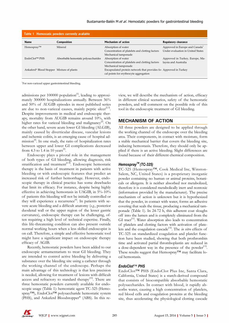
Bustamante-Balén M et al . Hemostatic powders for gastrointestinal bleeding
admissions per 100000 population[1], leading to approxi-mately 300000 hospitalizations annually. Between 36% and 50% of AUGIB episodes in most published series are due to non-variceal causes, mainly peptic ulcer[2,3]. Despite improvements in medical and endoscopic ther-apy, mortality from AUGIB remains around 10%, with higher rates for variceal bleeding and malignancy[2]. On the other hand, severe acute lower GI bleeding (ALGIB), mainly caused by diverticular disease, vascular lesions and ischemic colitis, is an emerging cause of hospital ad-mission[4]. In one study, the ratio of hospitalization rates between upper and lower GI complications decreased from 4.3 to 1.4 in 10 years[5].
Endoscopy plays a pivotal role in the management of both types of GI bleeding, allowing diagnosis, risk stratification and treatment[6-8]. Endoscopic hemostatic therapy is the basis of treatment in patients with active bleeding or with endoscopic features that predict an increased risk of further hemorrhage. However, endo-scopic therapy in clinical practice has some drawbacks that limit its efficacy. For instance, despite being highly effective in achieving hemostasis in UAGIB, in 5%-10% of patients this bleeding will not be initially controlled or they will experience a recurrence[9]. In patients with se-vere acute bleeding and a difficult anatomy (e.g., posterior duodenal wall or the upper region of the lesser gastric curvatures), endoscopic therapy can be challenging, of-ten requiring a high level of technical expertise. Finally, this life-threatening condition can also present outside normal working hours when a less skilled endoscopist is on call. Therefore, a simple and effective hemostatic tool might have a significant impact on endoscopic therapy efficacy of AGIB.
Recently, hemostatic powders have been added to our endoscopic armamentarium to treat GI bleeding. They are intended to control active bleeding by delivering a substance over the bleeding site using a catheter through the working channel of the endoscope. Perhaps the main advantage of this technology is that less precision is needed, allowing for treatment of lesions with difficult access and refractory to standard therapy[10]. There are three hemostatic powders currently available for endo-scopic usage (Table 1): hemostatic agent TC-325 (Hemo-spray™), EndoClot™ polysaccharide hemostatic system (PHS), and Ankaferd Bloodstopper® (ABS). In this re-
view, we will describe the mechanism of action, efficacy in different clinical scenarios, safety of the hemostatic powders, and will comment on the possible role of this tool in the endoscopic treatment of GI bleeding.
MECHANISM OF ACTIONAll three powders are designed to be applied through the working channel of the endoscope over the bleeding area. Their components, in contact with moisture, form a stable mechanical barrier that covers the bleeding site, inducing hemostasis. Therefore, they should only be ap-plied if there is an active bleeding. Slight differences are found because of their different chemical composition.
HemosprayTM (TC-325)TC-325 (Hemospray™, Cook Medical Inc, Winston-Salem, NC, United States) is a proprietary inorganic powder containing no human or animal proteins, botani-cals or allergens. It is neither absorbed nor metabolized, therefore it is considered metabolically inert and nontoxic (information provided by the manufacturer). The precise mechanism of action is unknown but it is hypothesized that the powder, in contact with water, forms an adhesive covering that seals the tissue, producing a mechanical tam-ponade (Table 1). In 24-72 h, this adherent coat sloughs off into the lumen and is completely eliminated from the GI tract[11]. Water absorption also leads to concentration of platelets and clotting factors with activation of plate-lets and the coagulation cascade[12]. The in vitro effects of TC-325 on standardized coagulation and platelet func-tion have been studied, showing that both prothrombin time and activated partial thromboplastin are reduced in a dose-dependent way in the presence of the powder[13]. These results suggest that Hemospray™ may facilitate lo-cal hemostasis.
EndoClot™ PHSEndoClot™ PHS (EndoClot Plus Inc, Santa Clara, California, United States) is a starch-derived compound that consists of biocompatible absorbable hemostatic polysaccharides. In contact with blood, it rapidly ab-sorbs water, causing a high concentration of platelets, red blood cells and coagulation proteins at the bleeding site, thus accelerating the physiological clotting cascade
August 15, 2014|Volume 5|Issue 3|WJGP|www.wjgnet.com 285
Table 1 Hemostatic powders currently available
Name Composition Mechanism of action Regulatory clearance
Hemospray™ Mineral Absorption of water Approved in Europe and Canada1
Concentration of platelets and clotting factors Under evaluation in United StatesMechanical tamponade
EndoClot™ PHS Absorbable hemostatic polysaccharides Absorption of water Approved in Turkey, Europe, Ma-laysia and AustraliaConcentration of platelets and clotting factors
Mechanical tamponadeAnkaferd® Blood Stopper Mixture of plants Encapsulated protein network that provides fo-
cal points for erythrocyte aggregationApproved in Turkey
1For non-variceal upper gastrointestinal bleeding.
(Table 1). A gelled matrix is formed that adheres to and seals the bleeding tissue. This matrix is cleared from the bleeding site in a few hours or days[14]. When applied to skin wounds, it seems to improve healing by activating fibroblasts and transforming growth factor (TGF)-β1 release[15], but this effect has not been studied in the GI mucosa.
ABSThis is a mixture of plants, including Thymus vulgaris, Glycyrrhiza glabra, Vitis vinifera, Alpinia officinarum and Urtica dioica. In vitro and ultrastructural studies suggest that ABS rapidly forms an encapsulated protein network that provides focal points for erythrocyte and activated leukocyte aggregation (Table 1)[16,17]. This network stems from interactions between ABS and blood proteins, such as fibrinogen, inducing protein agglutination. Total pro-tein, albumin and globulin levels also decrease in serum, probably via agglutination of these molecules in the growing protein network. However, most coagulation factors are not affected by the addition of ABS to fresh normal plasma or serum[18].
ABS also has functional properties, inhibiting fibri-nolysis and some natural anticoagulant pathways via in-teraction with the protein C anticoagulation pathway. On one hand, it enhances the expression of plasminogen activator inhibitor 1 (PAI-1), one of the major inhibi-tors of fibrinolysis, thus increasing clot stability. On the other hand, ABS has also been shown to down-regulate the endothelial cell protein C receptor (EPCR), a natural enhancer of protein C activity, therefore taking part in inactivation of factors Ⅴa and Ⅶa[19].
EVIDENCE SUPPORTING THE ROLE OF POWDERS IN ENDOSCOPIC HEMOSTA-SISThe evidence supporting the role of powders in GI bleeding is of moderate quality and based mainly on short series of cases and retrospective studies without a control group.
Hemospray™ (TC-325)Animal models: This powder has been tested in animal models of arterial gastrointestinal bleeding. Giday et al[10] performed a randomized controlled trial on 10 pigs allocated to treatment with TC-325 or sham after surgi-cal creation of an arterial bleeding from a gastroepiploic vessel opened up to the gastric lumen. The endpoint of the study was the proportion of animals in which hemostasis was achieved at 1 h. In the treatment group, acute hemostasis was achieved in the whole group with no rebleeding in the first 6 h compared to 0% of ani-mals in the sham group. Mean time to hemostasis was 13.8 min.
Ulcer bleeding: Sung et al[11] carried out a pilot study
on the efficacy of TC-325 as the primary hemostatic method in 20 patients with active peptic ulcer bleeding. Hemostasis was achieved in all but one (95%), a patient with a Forrest Ia ulcer who ultimately needed emboli-zation to stop bleeding. Two patients met the criteria for rebleeding during follow-up, but no active bleeding was detected in the second-look endoscopy. However, it must be pointed out that most treated bleedings were moderate and the only patient with a high risk lesion had a worse outcome.
Cancer-related GI bleeding: Conventional therapy in this kind of bleeding has moderate success and high rates of rebleeding. Chen et al[20] reported on a short series of 5 patients with upper GI bleeding secondary to gastroduo-denal tumors. The authors reported control of bleeding in all cases with only one case of rebleeding in a patient with disseminated intravascular coagulation. Leblanc et al[21] treated 5 patients with bleeding from GI neoplasms (2 esophageal, 2 gastric and 1 pancreatic) with TC-325. Successful hemostasis was achieved in all patients. Two patients showed recurrent bleeding, again successfully treated with TC-325.
Patients on antithrombotic therapy: Hostel et al[22] evaluated 16 patients with upper GI bleeding treated with TC-325 either as a monotherapy or as salvage therapy. In 9 patients, the source of bleeding was a peptic ulcer and in 2, a neoplasm. Eight patients were on antithrom-botic therapy (ATT), including patients on antiplatelet agents, NSAIDs or VKA/heparin. Initial hemostasis was achieved in 5/8 patients on ATT and in all patients of the non-ATT group (P = 0.2). The source of bleeding was a spurting arterial vessel in two of the three failures of TC-325 in patients of the ATT group. Rates of rebleed-ing were similar in both groups (around 25%) and in most cases bleeding was retreated with TC-325.
Bleeding secondary to a therapeutic intervention: Leblanc et al[21] used TC-325 to control bleeding after a therapeutic endoscopic intervention in 13 patients (5 esophageal EMR, 4 duodenal EMR, 2 ampullary resec-tions and 1 biliary sphincterotomy). The powder achieved complete hemostasis in all patients, either as a first-line treatment or a rescue therapy, including 2 cases with spurting arterial vessels. There were no rebleedings. Very recently, TC-325 has been successfully applied to a severe bleeding after endoscopic ultrasound-guided pseudocyst drainage which had been refractory to adrenaline and fibrin glue injection[23], and in a case of bleeding after a rectal submucosal endoscopic dissection[24].
Bleeding related to portal hypertension: TC-325 has been used in cases of both esophageal and gastric variceal bleeding with good short-term results[25-27]. Smith et al[28] controlled acute bleeding from severe portal hypertensive gastropathy in 3 patients. However, it is only able to con-trol the initial bleeding and cannot prevent further bleed-
August 15, 2014|Volume 5|Issue 3|WJGP|www.wjgnet.com 286
Bustamante-Balén M et al . Hemostatic powders for gastrointestinal bleeding
ings.
Lower GI bleeding: TC-325 has also been used for lower GI bleeding[29] which is currently not a licensed use of the powder. In the largest series published to date, 9 patients with lower GI bleeding were treated with TC-325, 4 of them with post-polypectomy bleedings. Successful initial hemostasis was achieved in all patients, with 2 cases (22%) of rebleeding[30]. Smith et al[28] treated one patient with a portal hypertensive colopathy with TC-325, achieving a decrease in transfusion require-ments. Very recently, Kraft et al[31] described the use of TC-325 for the treatment of a lower GI bleeding from diffuse colonic ulcers secondary to diclofenac.
Larger case series: A multicenter European trial has been published on the use of Hemospray™ in non-vari-ceal upper GI bleeding[32]. In this trial, 63 patients with a variety of indications, including ulcers, tumors and post-therapeutic bleeding, were treated with Hemospray™ as either monotherapy or second-line therapy. Primary he-mostasis was achieved in 85% of patients when Hemo-spray was used as monotherapy. Seven patients rebled by the 7th day, therefore 15 patients (27%) failed to achieve sustained hemostasis. The 3 patients who rebled from a peptic ulcer had a Forrest Ia lesion. Hemospray was used as a second-line therapy in 8 patients, with two early re-bleedings.
Very recently, a case series from two Swiss hospi-tals evaluated the performance of Hemospray™ on 16 patients with bleeding from different sources. In most cases, the powder was used as a rescue therapy with an initial hemostasis rate of 93%. Two patients rebled (12.5%), both presenting with oozing bleeding in the previous endoscopy[33].
EndoClotThere is only one publication in a peer reviewed journal reporting on EndoClot in control and prevention of EMR-related bleeding. EndoClot was applied to muco-sal defects after resection of 181 lesions (82 patients) regardless of if there was immediate post-resection bleeding. Among them, 20 lesions in 18 patients had early bleeding (five of them showing spurting bleed-ing). Bleeding was controlled with a single round of spray in 18 lesions (90%) and two cases needed hot biopsy forceps applied to achieve hemostasis. Bleeding recurred in three of these 18 patients but no therapy was needed. The authors concluded that EndoClot ef-fectively achieves hemostasis in controlling and prevent-ing EMR-related bleeding[34]. Two trials on the preven-tion of bleeding after endoscopic mucosal resection (NCT01496781 and NCT01735786) are ongoing but there are no data available yet[35,36]. Finally, there are some studies presented only in abstract form on short series of patients with a variety of bleeding lesions, reporting a success rate of around 80%, including some with coagu-lation disorders[37-39].
ABSThis agent has been approved in Turkey (Table 1) for clinical hemorrhages refractory to conventional hemo-static measures. There are several reports on the mecha-nism of action and clinical efficacy of ABS, almost all from the same Turkish groups.
Animal studies: Several authors have shown that ABS has a clinically meaningful hemostatic effect in rats and swine models with arterial sections, skin lacerations and liver puncture wounds, even if they were treated with warfarin[40-43].
Peptic ulcer bleeding: ABS has shown efficacy in peptic ulcer bleeding in some reports with a very low number of patients, including a child and a patient with thrombocytopenia[44,45].
Cancer-related GI bleeding: Several studies have as-sessed ABS efficacy on malignant GI bleeding, showing a good performance[46,47]. Clinical observation suggests that the hemostatic effect of ABS on malignant bleeding persists for a long time after its delivery. Some authors have suggested that this may be due to an inhibitory effect of ABS on tumor angiogenesis. A decrease in microves-sel density (MVD) in tumoral sections stained with CD34 after treatment with Ankaferd has been described[48].
Other indications: Case reports on the treatment with ABS of post-sphincterotomy bleeding[49], Mallory-Weiss syndrome[44,50] and gastric post-polypectomy bleeding[51] have been reported. Two reports on the use of ABS to control esophageal variceal bleeding have also been pub-lished[52,53].
Lower GI bleeding: ABS has been applied in patients with radiation proctitis, with transient control of bleed-ing, but with no effect on telangiectasias[54]. There are also anecdotal cases of ABS use on post-polypectomy bleeding[50,51] and solitary rectal ulcer[55].
Larger case series: The most recent series on the use of ABS to control upper GI bleeding included 27 pa-tients with active non-variceal UGIB[56]. Bleeding lesions were not described. In one patient, bleeding ceased after spraying isotonic saline and in the other, 26 ABS was applied. Bleeding stopped in 19 cases (73%). In 6 of the remaining 7 patients, ABS was sprayed again plus anoth-er endoscopic hemostatic method (clip, injection, APC), achieving an adequate control in all cases. The overall rate of rebleeding was 20%. Bleeding control with ABS was more difficult in patients with a coagulopathy or who were taking AAS.
TECHNICAL ISSUESHemospray™The Hemospray™ package includes a delivering device
August 15, 2014|Volume 5|Issue 3|WJGP|www.wjgnet.com 287
Bustamante-Balén M et al . Hemostatic powders for gastrointestinal bleeding
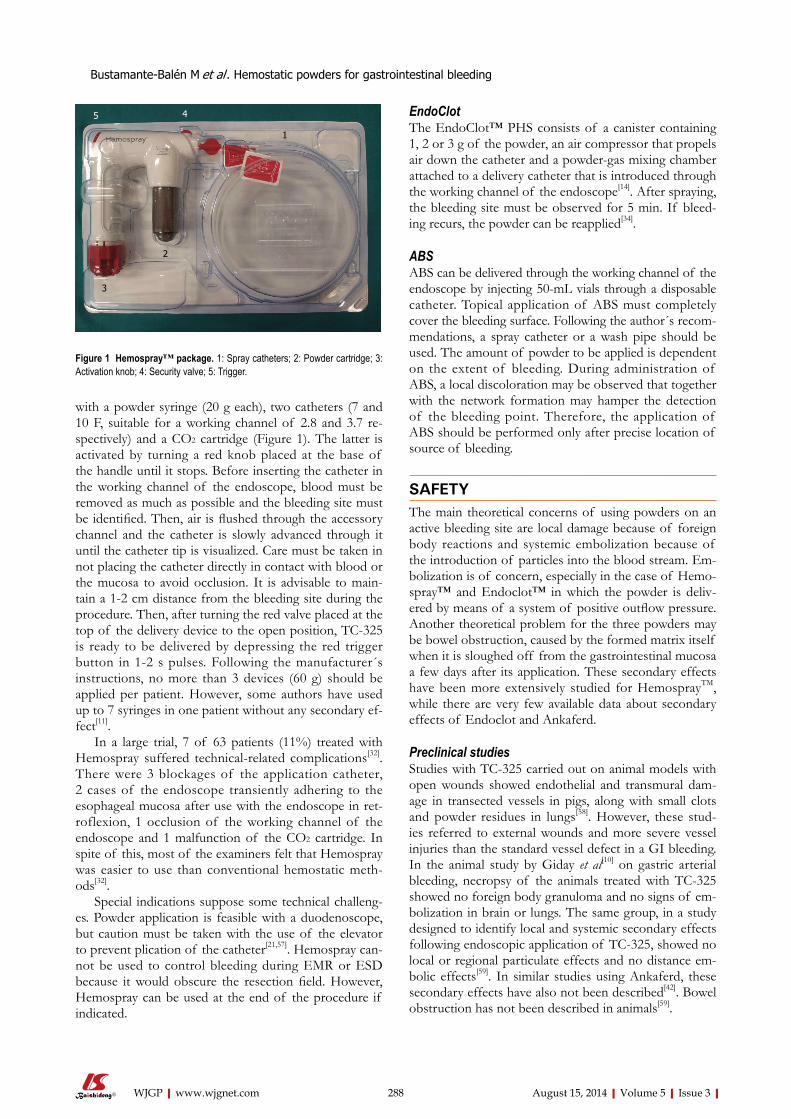
with a powder syringe (20 g each), two catheters (7 and 10 F, suitable for a working channel of 2.8 and 3.7 re-spectively) and a CO2 cartridge (Figure 1). The latter is activated by turning a red knob placed at the base of the handle until it stops. Before inserting the catheter in the working channel of the endoscope, blood must be removed as much as possible and the bleeding site must be identified. Then, air is flushed through the accessory channel and the catheter is slowly advanced through it until the catheter tip is visualized. Care must be taken in not placing the catheter directly in contact with blood or the mucosa to avoid occlusion. It is advisable to main-tain a 1-2 cm distance from the bleeding site during the procedure. Then, after turning the red valve placed at the top of the delivery device to the open position, TC-325 is ready to be delivered by depressing the red trigger button in 1-2 s pulses. Following the manufacturer´s instructions, no more than 3 devices (60 g) should be applied per patient. However, some authors have used up to 7 syringes in one patient without any secondary ef-fect[11].
In a large trial, 7 of 63 patients (11%) treated with Hemospray suffered technical-related complications[32]. There were 3 blockages of the application catheter, 2 cases of the endoscope transiently adhering to the esophageal mucosa after use with the endoscope in ret-roflexion, 1 occlusion of the working channel of the endoscope and 1 malfunction of the CO2 cartridge. In spite of this, most of the examiners felt that Hemospray was easier to use than conventional hemostatic meth-ods[32].
Special indications suppose some technical challeng-es. Powder application is feasible with a duodenoscope, but caution must be taken with the use of the elevator to prevent plication of the catheter[21,57]. Hemospray can-not be used to control bleeding during EMR or ESD because it would obscure the resection field. However, Hemospray can be used at the end of the procedure if indicated.
EndoClotThe EndoClot™ PHS consists of a canister containing 1, 2 or 3 g of the powder, an air compressor that propels air down the catheter and a powder-gas mixing chamber attached to a delivery catheter that is introduced through the working channel of the endoscope[14]. After spraying, the bleeding site must be observed for 5 min. If bleed-ing recurs, the powder can be reapplied[34].
ABSABS can be delivered through the working channel of the endoscope by injecting 50-mL vials through a disposable catheter. Topical application of ABS must completely cover the bleeding surface. Following the author´s recom-mendations, a spray catheter or a wash pipe should be used. The amount of powder to be applied is dependent on the extent of bleeding. During administration of ABS, a local discoloration may be observed that together with the network formation may hamper the detection of the bleeding point. Therefore, the application of ABS should be performed only after precise location of source of bleeding.
SAFETYThe main theoretical concerns of using powders on an active bleeding site are local damage because of foreign body reactions and systemic embolization because of the introduction of particles into the blood stream. Em-bolization is of concern, especially in the case of Hemo-spray™ and Endoclot™ in which the powder is deliv-ered by means of a system of positive outflow pressure. Another theoretical problem for the three powders may be bowel obstruction, caused by the formed matrix itself when it is sloughed off from the gastrointestinal mucosa a few days after its application. These secondary effects have been more extensively studied for HemosprayTM, while there are very few available data about secondary effects of Endoclot and Ankaferd.
Preclinical studiesStudies with TC-325 carried out on animal models with open wounds showed endothelial and transmural dam-age in transected vessels in pigs, along with small clots and powder residues in lungs[58]. However, these stud-ies referred to external wounds and more severe vessel injuries than the standard vessel defect in a GI bleeding. In the animal study by Giday et al[10] on gastric arterial bleeding, necropsy of the animals treated with TC-325 showed no foreign body granuloma and no signs of em-bolization in brain or lungs. The same group, in a study designed to identify local and systemic secondary effects following endoscopic application of TC-325, showed no local or regional particulate effects and no distance em-bolic effects[59]. In similar studies using Ankaferd, these secondary effects have also not been described[42]. Bowel obstruction has not been described in animals[59].
August 15, 2014|Volume 5|Issue 3|WJGP|www.wjgnet.com 288
Figure 1 Hemospray™ package. 1: Spray catheters; 2: Powder cartridge; 3: Activation knob; 4: Security valve; 5: Trigger.
1
2
3
45
Bustamante-Balén M et al . Hemostatic powders for gastrointestinal bleeding
Clinical studiesThere are no trials specifically designed to address sec-ondary effects of powders. However, a recent European multicenter study has shown no secondary effects when using TC-325 for a variety of indications, including peptic ulcers, vascular lesions, malignancies and post-therapeutic bleedings[32]. No secondary effects of Ankaf-erd and EndoClot have been described in the scarce literature available. Bowel obstruction has not been de-scribed with TC-325, even when maximum doses were delivered[11]. Intestinal blockage seems to be rare with EndoClot because in most cases bleeding is controlled only with 3 g of powder and starch particles are rapidly degraded in the GI tract.
Regarding Hemospray™, specific concerns have been raised for some indications. For instance, when treating bleeding from esophageal or gastric varices, thromboembolism may be an issue because particles might enter the vascular system. In fact, its use in this setting is contraindicated by the manufacturer. However, the Hemospray™ outflow pressure is less than the intra-variceal pressure of a bleeding varix when applied from a distance of 1-2 cm and no embolism has been shown in this indication[25,27]. In vitro coagulation time modifica-tions caused by TC-325 do not seem to pose any clinical problem in cirrhotic patients[25]. A case of biliary block-age has been described when TC-325 was applied in a patient with post-sphincterotomy bleeding[57].
The application of a pressure spray on the resection area after EMR could theoretically cause a perforation. However, no perforation was detected in a small se-ries[21]. Only one case of bowel perforation after treat-ment of a severe portal hypertensive gastropathy with TC-325 has been reported[28] but it was not clear if the perforation was related to the procedure. Following the manufacturer´s instructions, Hemospray™ use is con-traindicated in patients with suspected GI perforation or those at high risk of perforation during the endoscopic procedure (information provided by the manufacturer). Some secondary effects of TC-325 when applied in the large bowel have been described. A case of abdominal cramps after each application of Hemospray™ on a resection area in the rectum was described. This patient did not show any long-term secondary effect[22].
CONCLUSIONRandomized controlled trials comparing powders with
standard endoscopic methods are not yet available, thus the current evidence must be considered as moderate at best. The precise role of this technology in the thera-peutic algorithm or GI bleeding is yet to be defined but from the present review some practical conclusions can be drawn.
Topical hemostatic powders seem to be effective to control both upper and lower gastrointestinal bleeding from a variety of sources. They can be used as a primary method or a second-line therapy and in combination with standard hemostatic methods. However, there is a substantial proportion of patients who fail to achieve primary hemostasis, mainly Forrest Ia peptic ulcer bleed-ings. In case of a primary failure, an adjuvant conven-tional endoscopic method can be applied after removing the adherent matrix with water flushing. There is some risk of rebleeding in the first week after the initial hem-orrhagic episode, probably because the mineral matrix sloughs off from the mucosa after 24-72 h. A second-look endoscopy may be appropriate in this subset of patients with special risk of rebleeding.
Perhaps the most specific indication for the use of powders in GI bleeding is hemorrhage from a neoplastic lesion, which may have several bleeding points. Powders may be very useful in this setting because, when applied, they cover a large area of mucosa. Failure to achieve hemostasis with conventional methods is the other main indication for powders (Table 2).
However, active research is needed to clarify grey ar-eas, like secondary effects and long-term efficacy. Future areas of research should be the development of well-designed randomized trials to assess efficacy of pow-ders vs conventional endoscopic treatment as a primary therapeutic option, paying special attention to safety issues. Possible outcomes would be rates of rebleeding, need for adjuvant endoscopic therapy, and transfusion requirements. Sample size should be large enough to evaluate the efficacy of powders in the management of high risk bleeding lesions (e.g., Forrest Ia). Rebleeding may be an underreported event in the literature; there-fore, long-term efficacy must be addressed in the incom-ing trials. Long-term secondary effects on GI mucosa should also be addressed. Finally, since many conven-tional hemostatic methods are considerably cheaper, an economic analysis of the use of powders on GI bleeding should also be carried out. Larger trials, which may give response to some of these answers, are eagerly awaited.
REFERENCES1 Lewis JD, Bilker WB, Brensinger C, Farrar JT, Strom BL.
Hospitalization and mortality rates from peptic ulcer dis-ease and GI bleeding in the 1990s: relationship to sales of nonsteroidal anti-inflammatory drugs and acid suppression medications. Am J Gastroenterol 2002; 97: 2540-2549 [PMID: 12385436 DOI: 10.1111/j.1572-0241.2002.06037.x]
2 Hearnshaw SA, Logan RF, Lowe D, Travis SP, Murphy MF, Palmer KR. Acute upper gastrointestinal bleeding in the UK: patient characteristics, diagnoses and outcomes in the 2007 UK audit. Gut 2011; 60: 1327-1335 [PMID: 21490373
August 15, 2014|Volume 5|Issue 3|WJGP|www.wjgnet.com 289
Table 2 Possible indications for the use of hemostatic powders
Primary hemostatic method Adjuvant therapy
Lesions with a difficult endoscopic access Failure of conventional methods
Less experienced examinerMalignant gastrointestinal bleedingMassive bleeding as a mean to achieve an initial hemostasis
Bustamante-Balén M et al . Hemostatic powders for gastrointestinal bleeding

DOI: 10.1136/gut.2010.228437]3 Pérez Aisa A, Nuevo J, López Morante AA, González Gali-
lea A, Martin de Argila C, Aviñoa Arreal D, Feu F, Borda Celaya F, Gisbert JP, Pérez Roldan F, Gonzalvo Sorribes JM, Palazón Azorín JM, Ponce Romero M, Castro Fernández M, Catalina Rodriguez MV, Gallego Montañés S, Calvet X, Rodrigo Saez L, Montoro Huguet M, González Méndez Y, Sierra Hernández A, Sánchez Hernández E, Dominguez Muñoz E, Pérez Cuadrado E, Muñoz M, Lanas A. [Current management of nonvariceal upper gastrointestinal bleed-ing in Spain]. Gastroenterol Hepatol 2012; 35: 468-475 [PMID: 22542917 DOI: 10.1016/j.gastrohep.2012.02.007]
4 Hreinsson JP, Gumundsson S, Kalaitzakis E, Björnsson ES. Lower gastrointestinal bleeding: incidence, etiology, and outcomes in a population-based setting. Eur J Gastroenterol Hepatol 2013; 25: 37-43 [PMID: 23013623 DOI: 10.1097/MEG.0b013e32835948e3]
5 Lanas A, García-Rodríguez LA, Polo-Tomás M, Ponce M, Alonso-Abreu I, Perez-Aisa MA, Perez-Gisbert J, Bujanda L, Castro M, Muñoz M, Rodrigo L, Calvet X, Del-Pino D, Gar-cia S. Time trends and impact of upper and lower gastro-intestinal bleeding and perforation in clinical practice. Am J Gastroenterol 2009; 104: 1633-1641 [PMID: 19574968 DOI: 10.1038/ajg.2009.164]
6 Rockey DC. Lower gastrointestinal bleeding. Gastroenter-ology 2006; 130: 165-171 [PMID: 16401479 DOI: 10.1053/j.gastro.2005.11.042]
7 Laine L, McQuaid KR. Endoscopic therapy for bleeding ulcers: an evidence-based approach based on meta-analyses of randomized controlled trials. Clin Gastroenterol Hepatol 2009; 7: 33-47; quiz 1-2 [PMID: 18986845 DOI: 10.1016/j.cgh.2008.08.016]
8 Green BT, Rockey DC. Lower gastrointestinal bleeding--management. Gastroenterol Clin North Am 2005; 34: 665-678 [PMID: 16303576 DOI: 10.1016/j.gtc.2005.10.001]
9 Gralnek IM, Barkun AN, Bardou M. Management of acute bleeding from a peptic ulcer. N Engl J Med 2008; 359: 928-937 [PMID: 18753649 DOI: 10.1056/NEJMra0706113]
10 Giday SA, Kim Y, Krishnamurty DM, Ducharme R, Liang DB, Shin EJ, Dray X, Hutcheon D, Moskowitz K, Donatelli G, Rueben D, Canto MI, Okolo PI, Kalloo AN. Long-term ran-domized controlled trial of a novel nanopowder hemostatic agent (TC-325) for control of severe arterial upper gastro-intestinal bleeding in a porcine model. Endoscopy 2011; 43: 296-299 [PMID: 21384319 DOI: 10.1055/s-0030-1256125]
11 Sung JJ, Luo D, Wu JC, Ching JY, Chan FK, Lau JY, Mack S, Ducharme R, Okolo P, Canto M, Kalloo A, Giday SA. Early clinical experience of the safety and effectiveness of Hemo-spray in achieving hemostasis in patients with acute peptic ulcer bleeding. Endoscopy 2011; 43: 291-295 [PMID: 21455870 DOI: 10.1055/s-0030-1256311]
12 Cox ED, Schreiber MA, McManus J, Wade CE, Holcomb JB. New hemostatic agents in the combat setting. Transfusion 2009; 49 Suppl 5: 248S-255S [PMID: 19954487 DOI: 10.1111/j.1537-2995.2008.01988.x]
13 Holster ILDM MP, Ducharme R, Kuipers EJ, Tjwa ET. In vivo examination of the effects of the hemostatic powder (HemosprayTM) on coagulation and thrombus formation in humans. Gastrointest Endosc 2012: AB240
14 AMP technology. Polymer Solution for hemostasis. 2011. Available from: URL: http: //endoclot.com/technology.html. Last accessed: May 2014
15 Wang Y, Xu M, Dong H, Liu Y, Zhao P, Niu W, Xu D, Ji X, Xing C, Lu D, Li Z. Effects of PerClot® on the healing of full-thickness skin wounds in rats. Acta Histochem 2012; 114: 311-317 [PMID: 21782216 DOI: 10.1016/j.acthis.2011.06.012]
16 Beyazit Y, Kurt M, Kekilli M, Goker H, Haznedaroglu IC. Evaluation of hemostatic effects of Ankaferd as an alter-native medicine. Altern Med Rev 2010; 15: 329-336 [PMID:
21194248]17 Haznedaroglu BZ, Haznedaroglu IC, Walker SL, Bilgili H,
Goker H, Kosar A, Aktas A, Captug O, Kurt M, Ozdemir O, Kirazli S, Firat HC. Ultrastructural and morphological analyses of the in vitro and in vivo hemostatic effects of Ankaferd Blood Stopper. Clin Appl Thromb Hemost 2010; 16: 446-453 [PMID: 19833624 DOI: 10.1177/1076029609343706]
18 Goker H, Haznedaroglu IC, Ercetin S, Kirazli S, Akman U, Ozturk Y, Firat HC. Haemostatic actions of the folkloric me-dicinal plant extract Ankaferd Blood Stopper. J Int Med Res 2008; 36: 163-170 [PMID: 18304416]
19 Esmon CT, Esmon NL. Regulatory mechanisms in hemosta-sis. In: Hoffman RBEJ, Silberstein LE, Heslop HE, Wietz JI, Anastasi J, editors. Hematology: basic principles and prac-tice. 6th ed. Eselvier, 2013
20 Chen YI, Barkun AN, Soulellis C, Mayrand S, Ghali P. Use of the endoscopically applied hemostatic powder TC-325 in cancer-related upper GI hemorrhage: preliminary experi-ence (with video). Gastrointest Endosc 2012; 75: 1278-1281 [PMID: 22482923 DOI: 10.1016/j.gie.2012.02.009]
21 Leblanc S, Vienne A, Dhooge M, Coriat R, Chaussade S, Prat F. Early experience with a novel hemostatic powder used to treat upper GI bleeding related to malignancies or after therapeutic interventions (with videos). Gastrointest Endosc 2013; 78: 169-175 [PMID: 23622976 DOI: 10.1016/j.gie.2013.03.006]
22 Holster IL, Kuipers EJ, Tjwa ET. Hemospray in the treat-ment of upper gastrointestinal hemorrhage in patients on antithrombotic therapy. Endoscopy 2013; 45: 63-66 [PMID: 23208778 DOI: 10.1055/s-0032-1325793]
23 Tarantino I, Barresi L, Granata A, Curcio G, Traina M. Hemospray for arterial hemorrhage following endoscopic ultrasound-guided pseudocyst drainage. Endoscopy 2014; 46 Suppl 1: E71 [PMID: 24523191 DOI: 10.1055/s-0033-1359164]
24 Curcio G, Granata A, Traina M. Hemospray for multifocal bleeding following ultra-low rectal endoscopic submucosal dissection. Dig Endosc 2014; 26: 606-607 [PMID: 24734986 DOI: 10.1111/den.12301]
25 Holster IL, Poley JW, Kuipers EJ, Tjwa ET. Controlling gastric variceal bleeding with endoscopically applied hemo-static powder (Hemospray™). J Hepatol 2012; 57: 1397-1398 [PMID: 22864337 DOI: 10.1016/j.jhep.2012.07.024]
26 Ibrahim M, El-Mikkawy A, Mostafa I, Devière J. Endoscop-ic treatment of acute variceal hemorrhage by using hemo-static powder TC-325: a prospective pilot study. Gastrointest Endosc 2013; 78: 769-773 [PMID: 24120338 DOI: 10.1016/j.gie.2013.07.037]
27 Stanley AJ, Smith LA, Morris AJ. Use of hemostatic powder (Hemospray) in the management of refractory gastric vari-ceal hemorrhage. Endoscopy 2013; 45 Suppl 2: E86-E87 [PMID: 23526533 DOI: 10.1055/s-0032-1326258]
28 Smith LA, Morris AJ, Stanley AJ. The use of hemospray in portal hypertensive bleeding; a case series. J Hepatol 2014; 60: 457-460 [PMID: 24140803 DOI: 10.1016/j.jhep.2013.10.008]
29 Granata A, Curcio G, Azzopardi N, Barresi L, Tarantino I, Traina M. Hemostatic powder as rescue therapy in a patient with H1N1 influenza with uncontrolled colon bleeding. Gas-trointest Endosc 2013; 78: 451 [PMID: 23948196 DOI: 10.1016/j.gie.2013.07.010]
30 Holster IL, Brullet E, Kuipers EJ, Campo R, Fernández-At-utxa A, Tjwa ET. Hemospray treatment is effective for lower gastrointestinal bleeding. Endoscopy 2014; 46: 75-78 [PMID: 24218304 DOI: 10.1055/s-0033-1344988]
31 Kratt T, Lange J, Königsrainer A, Malek N, Adam P, Bös-müller H, Goetz M. Successful Hemospray treatment for recurrent diclofenac-induced severe diffuse lower gastroin-testinal bleeding avoiding the need for colectomy. Endoscopy 2014; 46 Suppl 1: E173-E174 [PMID: 24756280 DOI: 10.1055/s-0034-1365100]
August 15, 2014|Volume 5|Issue 3|WJGP|www.wjgnet.com 290
Bustamante-Balén M et al . Hemostatic powders for gastrointestinal bleeding
32 Smith LA, Stanley AJ, Bergman JJ, Kiesslich R, Hoffman A, Tjwa ET, Kuipers EJ, von Holstein CS, Oberg S, Brullet E, Schmidt PN, Iqbal T, Mangiavillano B, Masci E, Prat F, Mor-ris AJ. Hemospray Application in Nonvariceal Upper Gas-trointestinal Bleeding: Results of the Survey to Evaluate the Application of Hemospray in the Luminal Tract. J Clin Gas-troenterol 2013 Dec 10; Epub ahead of print [PMID: 24326829 DOI: 10.1097/MCG.0000000000000054]
33 Sulz MC, Frei R, Meyenberger C, Bauerfeind P, Semadeni GM, Gubler C. Routine use of Hemospray for gastrointes-tinal bleeding: prospective two-center experience in Swit-zerland. Endoscopy 2014; 46: 619-624 [PMID: 24770964 DOI: 10.1055/s-0034-1365505]
34 Huang R, Pan Y, Hui N, Guo X, Zhang L, Wang X, Zhang R, Luo H, Zhou X, Tao Q, Liu Z, Wu K. Polysaccharide he-mostatic system for hemostasis management in colorectal endoscopic mucosal resection. Dig Endosc 2014; 26: 63-68 [PMID: 23551344 DOI: 10.1111/den.12054]
35 Liu Z. EndoClot for preventing rebleeding after endoscopic mucosal resection (EMR). 2013. Available from: URL: http: //clinicaltrials.gov/show/NCT01735786. Last accessed: may 2014
36 Liu Z. EndoClot for Hemostasis and preventing post-pro-cedure bleeding after endoscopic mucosal resection (EMR). 2013. Available from: URL: http: //clinicaltrials.gov/show/NCT01496781. Accessed on May 2014
37 Halkerston K, Evans J, Ismail D, Catnach S, Chaudhary R, Fullard M, King A, Leahy A. Early clinical experience of EndoclotTM in the treatment of acute gastro-intestinal bleeding. Gut 2013; 62 (Suppl 1): A149 [DOI: 10.1136/gutnl-2013-304907.335]
38 Müller-Gerbes D, Beek A, Dormann A. Erfahrungen mit EndoClotTM PHS bei blutungen im oberen gastrointe-stinaltrakt. Z Gastroenterol 2013; 51: K457 [DOI: 10.1055/s-0033-1353097]
39 Sörensen AS, Kalmar G, Schmidt A, Caca K. Endoskopische bluststillung mit absorbierbaren modifizierten polymeren (AMP; EndoClotTM)-erste Erfahrungen im oberen gastro-intestinaltrakt. Z Gastroenterol 2013; 51: K270 [DOI: 10.1055/s-0033-1352910]
40 Bilgili H, Kosar A, Kurt M, Onal IK, Goker H, Captug O, Shorbagi A, Turgut M, Kekilli M, Kurt OK, Kirazli S, Aksu S, Haznedaroglu IC. Hemostatic efficacy of Ankaferd Blood Stopper in a swine bleeding model. Med Princ Pract 2009; 18: 165-169 [PMID: 19349716 DOI: 10.1159/000204344]
41 Cipil HS, Kosar A, Kaya A, Uz B, Haznedaroglu IC, Goker H, Ozdemir O, Koroglu M, Kirazli S, Firat HC. In vivo hemo-static effect of the medicinal plant extract Ankaferd Blood Stopper in rats pretreated with warfarin. Clin Appl Thromb Hemost 2009; 15: 270-276 [PMID: 19117967 DOI: 10.1177/1076029608329578]
42 Kandemir O, Buyukates M, Kandemir NO, Aktunc E, Gul AE, Gul S, Turan SA. Demonstration of the histopathologi-cal and immunohistochemical effects of a novel hemostatic agent, Ankaferd Blood Stopper, on vascular tissue in a rat aortic bleeding model. J Cardiothorac Surg 2010; 5: 110 [PMID: 21073754 DOI: 10.1186/1749-8090-5-110]
43 Kosar A, Cipil HS, Kaya A, Uz B, Haznedaroglu IC, Goker H, Ozdemir O, Ercetin S, Kirazli S, Firat HC. The efficacy of Ankaferd Blood Stopper in antithrombotic drug-induced primary and secondary hemostatic abnormalities of a rat-bleeding model. Blood Coagul Fibrinolysis 2009; 20: 185-190 [PMID: 19657315 DOI: 10.1097/MBC.0b013e32831c4cb0]
44 Ozaslan E, Purnak T, Yildiz A, Haznedaroglu IC. The effect of a new hemostatic agent for difficult cases of non-variceal gastrointestinal bleeding: Ankaferd blood stopper. Hepato-gastroenterology 2010; 57: 191-194 [PMID: 20583410]
45 Yarali N, Oruc M, Bay A, Dalgic B, Bozkaya IO, Arıkoglu T, Kara A, Tunc B. A new hemostatic agent--Ankaferd blood
stopper: management of gastrointestinal bleeding in an in-fant and other experiences in children. Pediatr Hematol Oncol 2010; 27: 592-596 [PMID: 20863156 DOI: 10.3109/08880018.2010.503337]
46 Kurt M, Akdogan M, Onal IK, Kekilli M, Arhan M, Shorba-gi A, Aksu S, Kurt OK, Haznedaroglu IC. Endoscopic topi-cal application of Ankaferd Blood Stopper for neoplastic gastrointestinal bleeding: A retrospective analysis. Dig Liver Dis 2010; 42: 196-199 [PMID: 19540818 DOI: 10.1016/j.dld.2009.05.006]
47 Zulfikar OB, Emiroglu HH, Kebudi R. Nasogastric applica-tion of topical Ankaferd Blood Stopper for bleeding from primary esophageal adenocarcinoma in a child with dis-seminated intravascular coagulation. Dig Liver Dis 2011; 43: 247-248 [PMID: 21177146 DOI: 10.1016/j.dld.2010.10.002]
48 Turhan N, Kurt M, Shorbagi A, Akdogan M, Haznedaro-glu IC. Topical Ankaferd Blood Stopper administration to bleeding gastrointestinal carcinomas decreases tumor vas-cularization. Am J Gastroenterol 2009; 104: 2874-2877 [PMID: 19888263 DOI: 10.1038/ajg.2009.431]
49 Beyazit Y, Köklü S, Akbal E, Kurt M, Kekilli M, Haznedaro-glu IC. Successful treatment of endoscopic sphincterotomy-induced early hemorrhage with application of Ankaferd Blood Stopper. Gastrointest Endosc 2010; 72: 1325-1326 [PMID: 21111880 DOI: 10.1016/j.gie.2010.03.1119]
50 Kurt M, Onal I, Akdogan M, Kekilli M, Arhan M, Sayilir A, Oztas E, Haznedaroglu I. Ankaferd Blood Stopper for con-trolling gastrointestinal bleeding due to distinct benign le-sions refractory to conventional antihemorrhagic measures. Can J Gastroenterol 2010; 24: 380-384 [PMID: 20559581]
51 Karaman A, Torun E, Gürsoy S, Yurci A, Ozbakir O. Effi-cacy of Ankaferd Blood Stopper in postpolypectomy bleed-ing. J Altern Complement Med 2010; 16: 1027-1028 [PMID: 20954959 DOI: 10.1089/acm.2010.0089]
52 Tuncer I, Doganay L, Ozturk O. Instant control of fundal variceal bleeding with a folkloric medicinal plant extract: Ankaferd Blood Stopper. Gastrointest Endosc 2010; 71: 873-875 [PMID: 19922917 DOI: 10.1016/j.gie.2009.08.021]
53 Ozaslan E, Purnak T, Yildiz A, Haznedaroglu IC. Bleeding due to slippage of elastic band during variceal ligation: suc-cessful use of Ankaferd blood stopper. Indian J Gastroenterol 2010; 29: 166-168 [PMID: 20814774 DOI: 10.1007/s12664-010-0043-y]
54 Ozaslan E, Purnak T, Ozyigit G, Akyol F, Yildiz A, Hazne-daroglu IC. No prolonged effect of Ankaferd Blood Stopper on chronic radiation proctitis. Endoscopy 2010; 42 Suppl 2: E271-E272 [PMID: 20931479 DOI: 10.1055/s-0030-1255773]
55 Ibis M, Kurt M, Onal IK, Haznedaroglu IC. Successful man-agement of bleeding due to solitary rectal ulcer via topical application of Ankaferd blood stopper. J Altern Complement Med 2008; 14: 1073-1074 [PMID: 19055332 DOI: 10.1089/acm.2008.0314]
56 Gungor G, Goktepe MH, Biyik M, Polat I, Tuna T, Ataseven H, Demir A. Efficacy of ankaferd blood stopper application on non-variceal upper gastrointestinal bleeding. World J Gastrointest Endosc 2012; 4: 556-560 [PMID: 23293725 DOI: 10.4253/wjge.v4.i12.556]
57 Moosavi S, Chen YI, Barkun AN. TC-325 application lead-ing to transient obstruction of a post-sphincterotomy biliary orifice. Endoscopy 2013; 45 Suppl 2: E130 [PMID: 23716095 DOI: 10.1055/s-0032-1326370]
58 Kheirabadi BS, Mace JE, Terrazas IB, Fedyk CG, Estep JS, Dubick MA, Blackbourne LH. Safety evaluation of new hemostatic agents, smectite granules, and kaolin-coated gauze in a vascular injury wound model in swine. J Trauma 2010; 68: 269-278 [PMID: 20154537 DOI: 10.1097/TA.0b013e3181c97ef1]
59 Giday S, Van Alstine W, Van Vleet J, Ducharme R, Brand-ner E, Florea M, Johnston K, Negron-Garcia J, Ringenberger
August 15, 2014|Volume 5|Issue 3|WJGP|www.wjgnet.com 291
Bustamante-Balén M et al . Hemostatic powders for gastrointestinal bleeding
K. Safety analysis of a hemostatic powder in a porcine model of acute severe gastric bleeding. Dig Dis Sci 2013;
58: 3422-3428 [PMID: 23982209 DOI: 10.1007/s10620-013-2846-z]
P- Reviewer: Fallone CA, Hokama A, Kate V, Li YY, Thomopoulos KC S- Editor: Ji FF L- Editor: Roemmele A
E- Editor: Lu YJ
August 15, 2014|Volume 5|Issue 3|WJGP|www.wjgnet.com 292
Bustamante-Balén M et al . Hemostatic powders for gastrointestinal bleeding
REVIEW
Predictors of response to anti-tumor necrosis factor therapy in ulcerative colitis
Evanthia Zampeli, Michalis Gizis, Spyros I Siakavellas, Giorgos Bamias
Evanthia Zampeli, Gastroenterology Department, Alexandra General Hospital, 11528 Athens, Greece Michalis Gizis, Spyros I Siakavellas, Giorgos Bamias, Aca-demic Department of Gastroenterology, Ethnikon and Kapodis-triakon University of Athens, Laikon Hospital, 15235 Athens, Greece Author contributions: Zampeli E, Gizis M and Siakavellas SI reviewed the literature; Zampeli E, Gizis M, Siakavellas SI and Bamias G analyzed the data; Zampeli E and Bamias G designed the structure of the review; Zampeli E and Bamias G wrote the paper.Correspondence to: Giorgos Bamias, Consultant in Gas-troenterology, Academic Department of Gastroenterology, Ethnikon and Kapodistriakon University of Athens, 17 Agiou Thoma st., 15235 Athens, Greece. [email protected]: +30-21-06456504 Fax: +30-21-07791839Received: January 4, 2014 Revised: March 7, 2014Accepted: June 10, 2014Published online: August 15, 2014
AbstractUlcerative colitis (UC) is an immune-mediated, chronic inflammatory disease of the large intestine. Its course is characterized by flares of acute inflammation and periods of low-grade chronic inflammatory activity or remission. Monoclonal antibodies against tumor ne-crosis factor (anti-TNF) are part of the therapeutic armamentarium and are used in cases of moderate to severe UC that is refractory to conventional treat-ment with corticosteroids and/or immunosuppressants. Therapeutic response to these agents is not uniform and a large percentage of patients either fail to im-prove (primary non-response) or lose response after a period of improvement (secondary non-response/loss of response). In addition, the use of anti-TNF agents has been related to uncommon but potentially seri-ous adverse effects that preclude their administration or lead to their discontinuation. Finally, use of these medications is associated with a considerable cost for the health system. The identification of parameters that
may predict response to anti-TNF drugs in UC would help to better select for patients with a high probability to respond and minimize risk and costs for those who will not respond. Analysis of the major clinical trials and the accumulated experience with the use of anti-TNF drugs in UC has resulted to the report of such prog-nostic factors. Included are clinical and epidemiological characteristics, laboratory markers, endoscopic indica-tors and molecular (immunological/genetic) signatures. Such predictive parameters of long-term outcomes may either be present at the commencement of treatment or determined during the early period of therapy. Vali-dation of these prognostic markers in large cohorts of patients with variable characteristics will facilitate their introduction into clinical practice and the best selection of UC patients who will benefit from anti-TNF therapy.
© 2014 Baishideng Publishing Group Inc. All rights reserved.
Key words: Ulcerative colitis; Infliximab; Adalimumab; Anti-tumor necrosis factor; Predictors of response; Per-sonalized treatment
Core tip: The use of anti-tumor necrosis factor (TNF) monoclonal antibodies for the treatment of ulcerative colitis has been associated with high rates of primary and secondary non-response, important safety issues and considerable cost. Selection of patients with the highest probability to response to anti-TNF treatment would overcome these problems. Analysis of the pivotal trials and accumulated experience from clinical practice has led to the identification of certain prognostic factors for favorable or adverse outcomes. These include clini-cal and epidemiological parameters, biological markers of inflammation, endoscopic findings, molecular sig-natures and pharmacological factors. Incorporation of such predictors into the current therapeutic protocols may lead to the optimization of anti-TNF treatment in ulcerative colitis.
Zampeli E, Gizis M, Siakavellas SI, Bamias G. Predictors of
World J Gastrointest Pathophysiol 2014 August 15; 5(3): 293-303ISSN 2150-5330 (online)
© 2014 Baishideng Publishing Group Inc. All rights reserved.
Online Submissions: http://www.wjgnet.com/esps/Help Desk: http://www.wjgnet.com/esps/helpdesk.aspxDOI: 10.4291/wjgp.v5.i3.293
August 15, 2014|Volume 5|Issue 3|WJGP|www.wjgnet.com 293
Zampeli E et al . Anti-TNF treatment for ulcerative colitis
response to anti-tumor necrosis factor therapy in ulcerative colitis. World J Gastrointest Pathophysiol 2014; 5(3): 293-303 Available from: URL: http://www.wjgnet.com/2150-5330/full/v5/i3/293.htm DOI: http://dx.doi.org/10.4291/wjgp.v5.i3.293
INTRODUCTIONUlcerative colitis (UC) is a chronic inflammatory disease of the colon, which affects almost 0.1% of the Western population[1]. Its natural history is dominated by chronic, relapsing intestinal inflammation, extra-intestinal involve-ment, and the development of long-term complications, which lead a considerable percentage of patients to col-ectomy.
Treatment for UC has been traditionally aimed against controlling acute and chronic inflammation[2]. Conven-tional therapy consists of 5-aminosalicylic acid (5-ASA) compounds, whereas more severe cases are handled with steroids during the acute phase and immunosup-pressants (thiopurines) as the maintenance regimen. Despite the proven efficacy of these drugs, a significant number of patients do not accomplish durable remission and/or experience side effects. Furthermore, there has been a change in the therapeutic goals in UC in recent years. Traditionally, such goals have been considered the achievement of clinical remission and the avoidance of colectomy. Nowadays, however, it has become clear that treatment should include the complete elimination of ac-tive inflammation in the colon without long-term use of corticosteroids. In this context, mucosal healing and deep remission which both indicate the absence of endoscopi-cally and biologically (i.e., serological and/or fecal inflam-matory markers) evident inflammation may be the ulti-mate endpoint. The accomplishment of such demanding endpoints has been linked to better long-term outcomes including colectomy and cancer prevention[3].
In recent years, treatment of UC has been revolu-tionized by the therapeutic application of monoclonal antibodies against tumor necrosis factor (TNF) as these agents offer effective long-term treatment for the most difficult cases.
ANTI-TNF TREATMENT IN UCThere are currently three monoclonal antibodies against human TNF that are licensed for the treatment of UC, infliximab (IFX), adalimumab (ADA), and Golimumab[4].
The data regarding Golimumab are limited. Therefore, our review will focus on IFX and ADA. IFX is a chime-ric mouse/human IgG1 antibody that is administered intravenously. On the other hand, ADA is a humanized IgG1 antibody administered as subcutaneous injection. The two clinical scenarios for anti-TNF therapy in UC are: firstly, outpatient cases with moderate to severe UC who are refractory or intolerant to first-line treatment; and, secondly, patients with acute severe disease refrac-tory to intravenous steroids[4]. In regards to the latter
scenario, data exist only for IFX.Recent clinical trials have established the efficacy of
anti-TNF treatment in UC. In the two pivotal IFX tri-als, ACT 1/2, the primary short-term (8 wk) response of moderate to severe UC to IFX has been reported to be 65.5%/69.4% for clinical response and 33.9%/39% for remission, respectively (dose regimen 5 mg/kg at 0-2-6)[5]. Among patients who responded to the induc-tion regimen nearly 50% maintained their response at week 30. Similarly, in the definitive clinical trial (ULTRA) for ADA, short-term response at week 8 was achieved in nearly 50% of patients, whereas long-term remission rate at week 52 was 17%[6].
Despite these encouraging results, the use of anti-TNF monoclonal antibodies is compromised in clinical practice by certain issues of efficacy and safety. Anti-TNF failure is an intriguing issue as it may be attributed to both disease characteristics and the drugs’ interfer-ence with the immune system. Primary non-response is characterized by lack of response to induction therapy. The incidence ranges between 20%-40% for both anti-TNF agents. Switching to another drug is common prac-tice, with a success rate of more than 50%[7,8]. On the other hand, loss of response is defined as the recurrence of the patient’s symptoms following successful induction of remission. In the case of CD it has been estimated between 23%-46%[9], whereas no solid data exists for UC. It is believed that immunogenicity underlies second-ary failure, as antibodies against anti-TNF drugs and re-duced trough levels have been implicated in the majority of studies[10-12]. Optimization of treatment (dose increase and/or shortening of the administration interval) leads to recovery of response in 60%-90% of patients[10].
The use of anti-TNF has also been associated with safety concerns. Among the most fearful ones are: severe infectious including reactivation of latent tuberculosis, neurological manifestations and risk of neoplasia. In ad-dition, infusion reactions and delayed hypersensitivity to IFX occurred in 10% and 1% of patients, respectively, in the ACT trials. The most significant side effects are probably associated with long-term administration and combination with other immunomodulatory medica-tions. It should be noted that in the ACT and ULTRA studies there were no differences between active drug and placebo.
Taken together, it is currently evident that fine-tuning of the use of anti-TNF therapy in UC is required. The ultimate goal should be to achieve maximum efficacy with a minimum risk for side effects. When therapeutic strategies are designed the following parameters should be taken into consideration: (1) the patients who receive anti-TNF therapy are the ones with the most difficult-to-treat disease; (2) the drugs’ efficiencies are far from perfect with high rates of primary and secondary fail-ures; (3) the potential for serious side effects especially with chronic use; and (4) the high cost of these medica-tions. One significant way to address these problems and optimize the clinical use of anti-TNF agents would be
August 15, 2014|Volume 5|Issue 3|WJGP|www.wjgnet.com 294
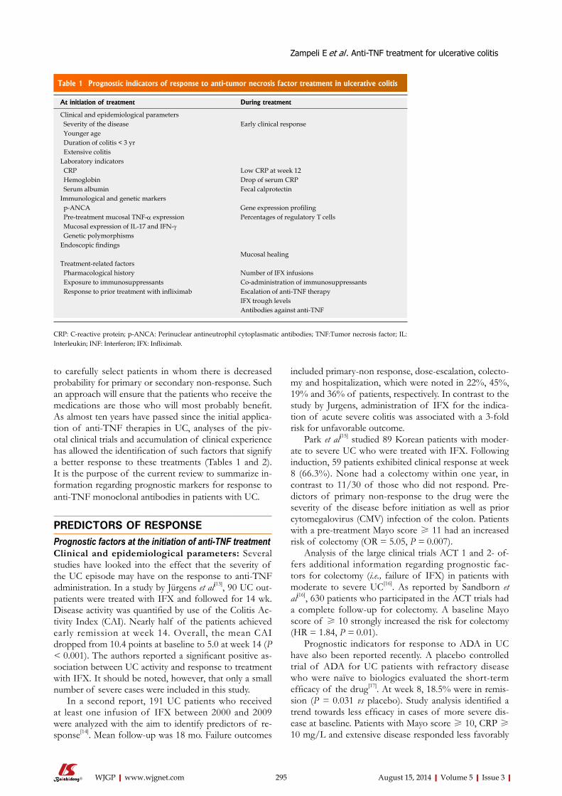
to carefully select patients in whom there is decreased probability for primary or secondary non-response. Such an approach will ensure that the patients who receive the medications are those who will most probably benefit. As almost ten years have passed since the initial applica-tion of anti-TNF therapies in UC, analyses of the piv-otal clinical trials and accumulation of clinical experience has allowed the identification of such factors that signify a better response to these treatments (Tables 1 and 2). It is the purpose of the current review to summarize in-formation regarding prognostic markers for response to anti-TNF monoclonal antibodies in patients with UC.
PREDICTORS OF RESPONSEPrognostic factors at the initiation of anti-TNF treatmentClinical and epidemiological parameters: Several studies have looked into the effect that the severity of the UC episode may have on the response to anti-TNF administration. In a study by Jürgens et al[13], 90 UC out-patients were treated with IFX and followed for 14 wk. Disease activity was quantified by use of the Colitis Ac-tivity Index (CAI). Nearly half of the patients achieved early remission at week 14. Overall, the mean CAI dropped from 10.4 points at baseline to 5.0 at week 14 (P < 0.001). The authors reported a significant positive as-sociation between UC activity and response to treatment with IFX. It should be noted, however, that only a small number of severe cases were included in this study.
In a second report, 191 UC patients who received at least one infusion of IFX between 2000 and 2009 were analyzed with the aim to identify predictors of re-sponse[14]. Mean follow-up was 18 mo. Failure outcomes
included primary-non response, dose-escalation, colecto-my and hospitalization, which were noted in 22%, 45%, 19% and 36% of patients, respectively. In contrast to the study by Jurgens, administration of IFX for the indica-tion of acute severe colitis was associated with a 3-fold risk for unfavorable outcome.
Park et al[15] studied 89 Korean patients with moder-ate to severe UC who were treated with IFX. Following induction, 59 patients exhibited clinical response at week 8 (66.3%). None had a colectomy within one year, in contrast to 11/30 of those who did not respond. Pre-dictors of primary non-response to the drug were the severity of the disease before initiation as well as prior cytomegalovirus (CMV) infection of the colon. Patients with a pre-treatment Mayo score ≥ 11 had an increased risk of colectomy (OR = 5.05, P = 0.007).
Analysis of the large clinical trials ACT 1 and 2- of-fers additional information regarding prognostic fac-tors for colectomy (i.e., failure of IFX) in patients with moderate to severe UC[16]. As reported by Sandborn et al[16], 630 patients who participated in the ACT trials had a complete follow-up for colectomy. A baseline Mayo score of ≥ 10 strongly increased the risk for colectomy (HR = 1.84, P = 0.01).
Prognostic indicators for response to ADA in UC have also been reported recently. A placebo controlled trial of ADA for UC patients with refractory disease who were naïve to biologics evaluated the short-term efficacy of the drug[17]. At week 8, 18.5% were in remis-sion (P = 0.031 vs placebo). Study analysis identified a trend towards less efficacy in cases of more severe dis-ease at baseline. Patients with Mayo score ≥ 10, CRP ≥ 10 mg/L and extensive disease responded less favorably
August 15, 2014|Volume 5|Issue 3|WJGP|www.wjgnet.com 295
Table 1 Prognostic indicators of response to anti-tumor necrosis factor treatment in ulcerative colitis
At initiation of treatment During treatment
Clinical and epidemiological parameters Severity of the disease Early clinical response Younger age Duration of colitis < 3 yr Extensive colitisLaboratory indicators CRP Low CRP at week 12 Hemoglobin Drop of serum CRP Serum albumin Fecal calprotectinImmunological and genetic markers p-ANCA Gene expression profiling Pre-treatment mucosal TNF-a expression Percentages of regulatory T cells Mucosal expression of IL-17 and IFN-γ Genetic polymorphismsEndoscopic findings
Mucosal healingTreatment-related factors Pharmacological history Number of IFX infusions Exposure to immunosuppressants Co-administration of immunosuppressants Response to prior treatment with infliximab Escalation of anti-TNF therapy
IFX trough levels Antibodies against anti-TNF
CRP: C-reactive protein; p-ANCA: Perinuclear antineutrophil cytoplasmatic antibodies; TNF:Tumor necrosis factor; IL: Interleukin; INF: Interferon; IFX: Infliximab.
Zampeli E et al . Anti-TNF treatment for ulcerative colitis
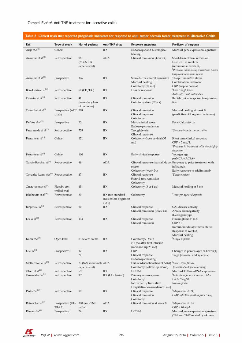
August 15, 2014|Volume 5|Issue 3|WJGP|www.wjgnet.com 296
Table 2 Clinical trials that reported prognostic indicators for response to anti- tumor necrosis factor treatment in Ulcerative Colitis
Ref. Type of study No. of patients Anti-TNF drug Response endpoints Predictor of response
Arijs et al[26] Cohort IFX Endoscopic and histological healing
Mucosal gene expression signature
Armuzzi et al[31] Retrospective 88(78.4% IFX experienced)
ADA Clinical remission (4-54 wk) Short-term clinical remissionLow CRP at week 12 (remission at week 54)1Previous immunosupressant use (lower long-term remission rates)
Armuzzi et al[27] Prospective 126 IFX Steroid–free clinical remission Thiopurine-naïve statusMucosal healing Combination treatmentColectomy (12 mo) CRP drop to normal
Ben–Horin et al[10] Retrospective 62 (CD/UC) IFX Loss or response 1Low trough levelsAnti-infliximab antibodies
Cesarini et al[39] Retrospective 41 (secondary loss of response)
IFX Clinical remission Rapid clinical response to optimiza-tionColectomy–free (52 wk)
Colombel et al[3] Prospective (ACT trials)
728 IFX Clinical remission Mucosal healing at week 8Clinical response (predictive of long-term outcome)Colectomy
De Vos et al[32] Prospective 53 IFX Mayo clinical score Fecal Calprotectin Endoscopic remission
Fasanmade et al[23] Retrospective 728 IFX Trough levels 1Serum albumin concentrationClinical response
Ferrante et al[21] Cohort 121 IFX Colectomy-free survival (33 mo)
Short term clinical responseCRP > 5 mg/L 1Previous iv treatment with steroids/cy-closporin
Ferrante et al[18] Cohort 100 IFX Early clinical response Younger agepANCA-/ACSA+
Garcia-Bosch et al[28] Retrospective 48 ADA Clinical response (partial Mayo score)
Response to prior treatment with infliximab
Colectomy (week 54) Early response to adalimumabGonzalez-Lama et al[20] Retrospective 47 IFX Clinical response 1Disease extent
Steroid-free remissionColectomy
Gustavsson et al[35] Placebo con-trolled trial
45 IFX Colectomy (3 yr f-up) Mucosal healing at 3 mo
Jakobovits et al[19] Retrospective 30 IFX (not standard induction regimen 0-2-6)
Colectomy 1Younger age at diagnosis
Jürgens et al[13] Retrospective 90 IFX Clinical response CAI-disease activityClinical remission (week 14) ANCA seronegativity
IL23R genotypeLee et al[22] Retrospective 134 IFX Clinical response Haemoglobin > 11.5
Clinical remission CRP > 3Immunomodulator-naïve statusResponse at week 2Mucosal healing
Kohn et al[36] Open label 83 severe colitis IFX Colectomy/Death 1Single infusion> 2 mo after first infusion (median f-up 23 mo)
Li et al[34] Prospective? 17 IFX CRP Changes in percentages of Foxp3(+) Tregs (mucosal and systemic)24 Clinical response
Endoscopic healingMcDermott et al[30] Retrospective 23 (86% infliximab
experienced)ADA Failure (discontinuation of ADA) 1Short–term failure
Colectomy (follow-up 22 mo) (increased risk for colectomy) Olsen et al[24] Retrospective 59 IFX UCDAI Mucosal TNF-a mRNA expressionOussalah et al[14] Retrospective 191 IFX (≥1 infusion) Primary non-response 1Indication for acute severe colitis
Colectomy Hb ≤ 9.4 g/dLInfliximab optimization Non-responseHospitalization (median 18 mo)
Park et al[15] Retrospective 89 IFX Clinical response 1Mayo score ≥ 11)Clinical remission CMV infection (within prior 3 mo)Colectomy
Reinisch et al[17] Prospective (UL-TRA 1)
390 (anti-TNF naïve)
ADA Clinical remission at week 8 1Mayo score ≥ 10CRP = 10 mg/L
Rismo et al[25] Prospective 74 IFX UCDAI Mucosal gene expression signature (Th1 and Th17 related cytokines)
Zampeli E et al . Anti-TNF treatment for ulcerative colitis
to ADA in the short-term. It should be noted, however, that these parameters did not strongly affect the result and their consideration as predictive factors must be cautious.
In all, the majority of studies appear to support the notion that severe UC demonstrates a less favorable re-sponse to treatment with anti-TNF monoclonal antibod-ies. From the pure clinical standpoint, the best candidate for anti-TNF administration may be an outpatient with moderate to severe UC but not severe disease requiring hospitalization, as defined by the criteria of Truelove and Witts.
In addition to disease severity, other clinical param-eters may also affect the response to anti-TNF in UC. Ferrante et al[18] studied a cohort of 100 UC patients who were treated with IFX. More than half had extensive dis-ease, were on immunosuppressants and received a single infusion as opposed to the standard induction scheme. Early clinical response was accomplished in 65% of patients. Younger age was associated with a higher per-centage of early clinical response (responders: median age 35.7 years vs non-responders: 41.6, P = 0.049). Dif-ferent results were obtained by Jakobovits et al[19] who reviewed the records of 30 patients with refractory UC who had received a single IFX infusion over the period 2000-2006. Half of the patients underwent colectomy over a median follow-up period of 140 d. In this co-hort, younger age at diagnosis correlated with increased risk of surgery (colectomy: mean age 27.5 years vs non-colectomy 38.7 years, P = 0.016). In contrast, the indica-tion before starting IFX was not relevant to colectomy rates. The number of patients in this study was too small for definitive conclusions to be drawn. In the analysis of the ACT trials duration of colitis ≤ 3 years strongly in-creased the risk for colectomy (hazard ratio = 0.36, P < 0.001, respectively)[16]. Finally, disease extent may also af-fect response to treatment. Gonzalez-Lama et al[20] stud-ied 47 UC patients who were treated with IFX and were followed for a mean duration of 8 mo. Pre-treatment predictive factors were sought: extent of the disease was the only factor that was related to higher response rates
to IFX (P = 0.02). Extensive colitis appeared to respond less favorably in the short term in the aforementioned study of ADA as well[17].
Laboratory indicators: Among the various laboratory biomarkers of inflammation, C-reactive protein (CRP) has been the most extensively applied to clinical practice. The association between CRP and inflammatory activ-ity in UC has not been equally strong as it is for Crohn’s disease. Nevertheless, its relevance increases when cases of severe UC are studied. As these are the patients that usually require administration of anti-TNF agents, the predictive value of CRP for treatment efficacy/failure may be increased in this population. Ferrante et al[21] re-ported on a cohort of 121 UC outpatients treated with IFX and followed for a median of 33 mo. Eighty-one patients (67%) exhibited short-term response and 21 (17%) underwent colectomy. A value of pre-treatment CRP ≥ 5 mg/L was an independent predictor for col-ectomy (HR = 14.5, P = 0.006). Similar results were pre-sented in a study of 134 Korean patients with UC who had received at least one infusion of IFX[22]. At week 8, 87% and 45% achieved response and remission, respec-tively. A pre-treatment CRP ≥ 3 mg/dL was predictive of clinical remission at week 8 (OR = 4.77, P = 0.01). The association between elevated CRP and less favor-able response to anti-TNF was also confirmed in the analysis of the ACT trials[16]. A baseline CRP ≥ 2 mg/L was significantly associated with increased colectomy risk (HR = 1.73, P = 0.04). Of note, several studies found an association between elevated CRP and colectomy[21]. Therefore, increased CRP may represent a strong marker of inflammation that requires potent treatment and will respond optimally to anti-TNF. Alternatively, CRP may be an indicator of refractory disease.
In the previous Korean study, high pre-treatment hemoglobin was also a predictor of good response to IFX[22]. Baseline haemoglobin of ≥ 11.5 g/dL was asso-ciated with higher probability for remission at week 8 (OR = 4.47, P = 0008). This is in accordance with the study by Oussalah[14] who reported that pre-treatment hemo-
August 15, 2014|Volume 5|Issue 3|WJGP|www.wjgnet.com 297
Rostholder et al[38] Retrospective observa-tional
56 IFX Clinical remission Escalation of infliximab therapy
Sandborn et al[16] Prospective (ACT1&2) 630 IFX Colectomy (54 wk) 1Concomitant steroidsCRP ≥ 2 mg/dLDisease duration < 3 yrMayo ≥ 10
Seow et al[40] Cohort 115 IFX Clinical remissionEndoscopic improvementColectomy
Trough levels
Steenholdt et al[41] Retrospective 106 (CD/UC) IFX Loss of response 1Trough levelsAnti-infliximab antibodies
Taxonera et al[29] Retrospective 30(IFX experienced)
ADA Clinical response at week12Colectomy (follow-up 48 wk)
Short–term response at week-12(Associated with less with-drawal and colectomy rates)
Toedter et al[33] Prospective (ACT-1) 48 IFX Clinical response Mucosal gene expression signature
1Italics correspond to prognostic factors for adverse outcome. IFX: Infliximab; ADA: Adalimumab; UCDAI: Ulcerative colitis disease activity index; HACA: Human anti-chimeric antibodies; CRP: C-reactive protein.
Zampeli E et al . Anti-TNF treatment for ulcerative colitis
globin ≤ 9.4 g/dL predicted primary non-response to IFX (OR = 4.35). This occurred in 22% of 191 treated patients who were included in the study. According to Truelove criteria low hemoglobin is an indicator of se-vere disease, which increases the risk of non-response to IFX. High pre-treatment hemoglobulin may reflect the presence of milder disease that responds better to anti-TNF treatment.
Serum albumin concentration may also have prog-nostic value. A study by Fasanmade et al[23] focused on the association between serum IFX and albumin concen-tration. Data from 728 patients who participated in two clinical trials were analyzed. A value of serum albumin that was outside the normal range was directly related to trough IFX levels and clinical response. Patients with low serum albumin had reduced IFX concentration and worse clinical outcomes. This correlation may reflect a common clearance pathway for albumin and anti-TNF antibodies that belong to the IgG class of immunoglob-ulins. In all, measurement of albumin before commence-ment of treatment may serve as a predictive marker of the drug’s pharmacokinetics.
Immunological and genetic markers: In recent years significant advances have taken place in our understand-ing of the immunopathogenesis of UC. In addition, ge-nome wide association studies have discovered polymor-phisms which confer susceptibility to or protect from developing UC. These studies led to the identification of several immunological markers with may serve as indica-tors of disease activity and severity. The possibility that such markers may also serve as predictors of response to treatment, in particular to therapy with anti-TNF mono-clonal antibodies, has been increasingly explored.
One of the classical immunological markers that are associated with UC is the presence of perinuclear antineutrophil cytoplasmatic antibodies (p-ANCA). In two recent studies absence of this marker was strongly associated with better response to IFX. In a retrospec-tive study of 90 patients who were evaluated up to week 14 on scheduled IFX infusions, negativity for p-ANCA (along with disease severity and IL23R genotype) was predictive of IFX efficacy[13]. Similar results were ob-tained in the study by Ferrante et al[18]. The authors fol-lowed 100 UC patients treated with IFX (84 patients received a single infusion). ANCA seronegativity served as predictor of good response. Notably, a serological phenotype of ANCA+/ASCA- status was particularly correlated with lower rates of response (P = 0.049).
During acute flares of UC an abundance of inflam-matory mediators are upregulated at the intestinal mucosa and can be detected at both the mRNA and the protein level, whereas, anti-inflammatory treatment is paralleled by a decrease or even disappearance of these markers. Therefore, such markers may hold predictive value for the response to anti-TNF treatment. A first obvious target has been TNF itself. Olsen et al[24] looked for predictive factors of response to induction treatment (weeks 0, 2,
6) with IFX in a cohort of 59 patients with moderate to severe disease. The outcome was assessed based on UC disease activity index (UCDAI). Among various param-eters elevated pre-treatment mucosal TNF-a expression was the only independent predictive factor of clinical and endoscopic remission (P = 0.01 and P = 0.003, OR = 2.5 and 4.8, respectively).
UC-related intestinal inflammation has been charac-terized by upregulation of several components of the major adaptive immunity pathways (Th1, Th2, Th17). A recent study looked at the expression of the pivotal Th1 (IFN-γ) and Th17 (IL-17) cytokines before and after treatment with IFX[25]. Mucosal cytokine profile was de-termined by PCR and confirmed by immunohistochem-istry in biopsies of 74 UC patients. Efficacy was evalu-ated after 3 infusions and was based on UCDAI. High pre-treatment mucosal expression of IL-17 and IFN-γ significantly correlated with remission after induction therapy (OR = 5.4, P = 0.013 and OR = 5.5, P = 0.011, respectively).
In a much broader approach, Arijs et al[26] performed a gene-array study in mRNA from colonic mucosal biop-sies obtained from UC patients who received induction therapy with IFX. Analysis of the arrays revealed genes that were differentially expressed among responders and non-responders. Genes that showed a highly differential expression were osteoprotegerin, stanniocalcin-1, prosta-glandin-endoperoxide synthase 2, interleukin 13 receptor alpha-2 and interleukin 11. The sensitivity and specificity in predicting response to IFX based on this gene profil-ing was 95% and 85%, respectively.
The effect of genetic polymorphisms to response to treatment remains unknown. In the aforementioned study by Jurgens the effect of UC-associated, IL-23R variants on the efficacy of IFX was reported[13]. In this study of 90 patients, homozygosity for the IBD-risk-increasing IL23R variants was associated with higher probability to respond to IFX than homozygosity for IBD-risk-decreasing IL23R variants (74.1% vs 34.6%; P = 0.001).
Treatment-related factors: Several studies have shown that the pharmacological history plays an important role in the response to anti-TNF treatment. In the study by Ferrante et al[21], 121 UC patients received IFX and were followed-up for a median of 33 mo. Colectomy was per-formed in 21 patients (21%). Previous iv treatment with steroids and/or cyclosporine significantly increased the risk for colectomy (HR = 2.4, P = 0.033). A similar asso-ciation was seen in the study by Oussalah et al[14]. Previ-ous use of cyclosporine was a positive predictive factor for colectomy (hazard ratio = 2.53). Finally, in the analy-sis of the colectomy rates in the context of the ACT-tri-als patients who were on steroids when IFX was started had an increased risk for surgery (HR = 1.84, P = 0.01)[16]. However, caution is required for the interpretation of these associations, which should take into consideration the severity of the disease. Indeed, in all of these studies
August 15, 2014|Volume 5|Issue 3|WJGP|www.wjgnet.com 298
Zampeli E et al . Anti-TNF treatment for ulcerative colitis
more severe disease was associated to adverse outcomes and less favorable response to anti-TNF. Therefore, the use of iv steroids and/or cyclosporine may simply reflect severe disease.
The association between exposure to immunosup-pressants and efficacy of anti-TNF therapy merits spe-cial attention. Converging lines of evidence indicate that immunosuppressant-naïve patients respond better to anti-TNF. The efficacy of IFX was evaluated in a cohort of 126 steroid-dependent patients[27]. Approximately half of the patients achieved steroid-free remission, whereas mucosal healing at 12 mo was accomplished in one third. Thiopurine-naïve status was positively associ-ated to steroid-free remission as well as mucosal healing at 12 mo (HR = 2.8 and OR = 3.6, respectively). In the aforementioned Korean study[22] immunomodulator-na-ïve status was an independent predictors for early clinical remission (OR = 4.89, P = 0.01). This consistent finding is in agreement with the growing evidence regarding ear-lier introduction of biologics in patients with moderate disease, as patients who never received thiopurines may had suffered a shorter disease course.
Finally, for patients who receive ADA as a second anti-TNF monoclonal antibody, the treatment efficacy is affected by the response to prior treatment with IFX. This was shown in a recent retrospective study that evaluated the clinical response and colectomy rate in a cohort of 48 UC patients treated with ADA[28]. The majority (81.3%) was previously exposed to IFX. Early response to ADA at week 12 was significantly more fre-quent in patients who achieved remission on prior treat-ment with IFX (P = 0.01).
Prognostic factors during anti-TNF treatmentSeveral recent studies have provided evidence to support the notion that patients with early response to anti-TNF (i.e., within 3 mo) are the ones who will also benefit in the long-term. Early response was defined by a variety of clinical and biological markers in these publications.
Clinical parameters: A Spanish study evaluated the ef-ficacy of ADA in 48 UC patients who were followed-up to week 54[28]. In this cohort the only predictive factor for colectomy was the absence of early clinical response, which was determined by partial Mayo score at week 12 (colectomy: 14.7% vs no colectomy: 42.9%, P = 0.035).
These results were replicated in a cohort of 121 UC outpatients[21] Eighty-one patients initially responded to IFX with 2/3 maintaining clinical response throughout follow-up. Twenty-one patients ended up with colec-tomy after a median follow-up of 33 mo. No predictors for durable response were identified. Colectomy on the other hand strongly correlated with early non-response to IFX (HR = 10.8, P < 0.001).
In the study by Lee et al[22], 45% of 134 patients with UC who received at least a single IFX infusion, achieved remission at week 8. Short-term remission rates were higher in patients who responded very early, at week 2
(OR = 20.54, P = 0.006). The value of ADA in 30 UC patients who had failed
IFX was studied retrospectively[29]. Response and remis-sion rates were assessed at weeks 4 and 12 and colec-tomy rates over a mean follow-up of 48 mo. In the long-term 50% were still on ADA and 20% underwent col-ectomy. The risk of surgery was higher for patients who did not achieve response at week 12 (P = 0.001).
Similarly, Mc Dermott et al[30] studied 23 patients who received ADA induction and maintenance treatment. Of note, 86% had previously failed IFX. Discontinuation of ADA over a follow-up period of 22 mo was the primary endpoint and occurred in 70% of patients. Colectomy-free survival at 24 mo was 59%. The only factor associ-ated with increased risk for surgery was the absence of early response to ADA. Among patients who underwent colectomy, 55% had failed ADA at week 12.
Armuzzi et al[31] evaluated the short- and long-term effects of ADA in 88 UC patients out of whom 78% had previously received IFX. The rates of clinical remis-sion increased from 17% to 43% at weeks 4 and 54, re-spectively. Interestingly, achievement of early remission as well as low CRP at week 12 predicted remission at week 54 (OR = 4.17 and 2.63, respectively).
Laboratory indicators: The same conclusion regard-ing the predictive value of early response was obtained when laboratory markers of inflammation were stud-ied. We already mentioned the predictive value of low CRP at week 12 in the study by Arnuzzi[31]. In another publication from the same group regarding 126 steroid-dependent patients who received IFX[27] drop of serum CRP value to normal after the induction-regimen pre-dicted steroid-free remission and mucosal healing at 12 mo (HR = 4.6, OR = 6.0, respectively). Similar results were reported in a study that used fecal calprotectin as an inflammatory marker. Serial weekly measurements of fecal calprotectin were performed in a cohort of 53 patients who received IFX[32]. Two thirds of patients achieved endoscopic remission at week 10, whereas the median calprotectin level significantly drop from base-line (P < 0.001). Early reduction of calprotectin at week 2 predicted endoscopic remission. At week 10, clinical and endoscopic remission strongly correlated to fecal calprotectin concentration.
Immunological markers: Early post-IFX changes of the mucosal and peripheral immunophenotype of UC patients showed strong correlation with clinical response to the drug. Toedter et al[33] studied 113 colonic biopsies from 48 patients who participated in the ACT-1 trial. Biopsies were taken before and after treatment with IFX up to week 30. Gene expression profiling was per-formed. The investigators were able to identify certain genes that demonstrated significant alterations in pa-tients that responded to treatment with IFX but not in non-responders.
In a study that included both Crohn’s and UC, the
August 15, 2014|Volume 5|Issue 3|WJGP|www.wjgnet.com 299
Zampeli E et al . Anti-TNF treatment for ulcerative colitis

effect of IFX on the percentages of regulatory T cells (Treg) was investigated[34]. Flow cytometry, PCR and im-munohistochemistry were applied to quantify the expres-sion of Forkhead box protein3 (Foxp3)-positive T cells in both peripheral blood samples and mucosal biopsies before and after IFX treatment. Responders to IFX were characterized by significantly increased numbers of CD4(+) CD25(+) Foxp3(+)Treg and CD4(+) CD25(-) Foxp3(+) Tregs in blood (P < 0.05) and a significant down-regulation in the tissue (P < 0.001). The duration of clinical response to IFX correlated to a sustainable peripheral increase of Foxp3 (+) Treg cells.
Although such individual molecular characterization is far from being clinically applicable, it shows that per-sonalized therapy which will be based on the particular immunophenotype may guide the therapeutic approach in the future.
Endoscopic findings: In recent years, mucosal healing (i.e., the disappearance of visible active inflammatory le-sions in endoscopy) has emerged as a definitive endpoint in the natural history of UC and an indispensable thera-peutic target both in clinical trials and “real-life” practice. This is because mucosal healing has been shown to be associated with sustained long-term remission in patients with UC[3].
In the pivotal ACT trials endoscopic evaluations were performed at various time points and mucosal healing was defined as Mayo subscore of 0 (normal) or 1 (mild). Early endoscopic improvement at week 8 was associ-ated to improved clinical outcomes[3]. Accordingly, low endoscopy subscores at week 8 predicted reduced risk of colectomy through week 54 (P = 0.0004) as well as higher remission and steroid-free remission rates (P < 0.0001).
A single IFX infusion or placebo was administered to 45 patients with acute, steroid-refractory UC[35]. Three years later the beneficial effect of the drug persisted as less patients in the IFX group underwent operation (50% vs 76%, P = 0.012). Endoscopic remission at month 3 strongly predicted a reduced long-term risk for colec-tomy (P = 0.02).
Mucosal healing was also a positive predictive factor for long-term remission in the study by Lee et al[22]. A variety of predictors for short-term outcome were iden-tified whereas the only parameter associated with sus-tained long-term benefit was endoscopic remission (OR = 4.66, P = 0.04).
Treatment-related factors: The number of IFX infu-sions was associated with improved sustained response to anti-TNF treatment. Kohn et al[36] studied the effect of IFX treatment in 83 patients with severe steroid-refractory UC.Patients received ≥ 1 infusions and were followed for a median of 23 mo. Twelve out of 83 pa-tients (15%) had a colectomy within 2 mo. The risk for a prime adverse event was significantly higher among patients who received a single IFX infusion as opposed
to those who were given two or more doses (OR = 9.53, P = 0.001).
The combined administration with immunosup-pressants appears to have an advantage in comparison to single IFX therapy. This was shown in the study by Armuzzi et al[27]. In this cohort of 126 steroid-dependent UC patients combination treatment with IFX and thio-purines was a predictor of steroid-free remission (HR = 2.2). In another prospective trial Panaccione studied 231 patients with moderate disease who were biologics-naïve and had not received azathioprine over the 3 mo before enrollment. Patients were offered IFX monotherapy, azathioprine monotherapy or combination treatment. Steroid-free remission at week 16 was significantly more common in the combination arm of the study (P < 0.05 compared to both monotherapies)[37].
The need for escalation of anti-TNF therapy is also a poor prognostic factor for long-term outcome. In a cohort of 56 patients with moderate colitis who were treated with IFX, 89% proceeded to maintenance treat-ment[38]. During a mean follow-up of 38 mo, clinical remission was achieved in 36% of patients at 12 mo, whereas 54% required escalation of treatment. Inten-sification of IFX treatment was a negative predictive factor of remission at 12 mo (P = 0.01). In accordance, colectomy was performed more often in the “escalation” group (33% vs 21%).
In a related study, Cesarini et al[39] showed that rapid response to escalation treatment has a favorable effect on long-term outcome. They studied the records of 41 UC patients with loss of response to IFX who were treated with either dose doubling or interval shortening. The primary outcome was rapid response which was evaluated at the follow-up visit after treatment escalation. Remission and colectomy were evaluated by week 52. The majority (90%) responded rapidly and 46% achieved rapid remission. Only 4 patients (9.8%) underwent col-ectomy by week 52. The main predictor for avoidance of colectomy was initial response to intensification treat-ment (P = 0.002).
Recent developments emphasize the importance of serum trough levels of IFX and ADA and the formation of antibodies against anti-TNF monoclonal antibodies for the pharmacokinetics as well as the therapeutic ef-ficacy of these drugs. In a study of 115 UC patients on maintenance treatment, clinical outcomes were associat-ed to IFX trough levels[40]. Detectable drug in serum pre-dicted clinical remission and endoscopic improvement at week 54 (P < 0.001 for both parameters). Reduced trough levels correlated with increased risk of colectomy in this cohort (P < 0.001). Interestingly, antibody-status was not predictive of response to IFX treatment.
Steenholdt et al[41] retrospectively studied 106 IBD patients on IFX, who either maintained or lost their response. Significantly higher IFX levels and lower an-tibodies titer were measured in patients with sustained response to IFX (P < 0.0001). Moreover, the authors suggested threshold values for the two parameters to ac-
August 15, 2014|Volume 5|Issue 3|WJGP|www.wjgnet.com 300
Zampeli E et al . Anti-TNF treatment for ulcerative colitis
curately predict and/or explain loss of response to IFX. Similarly, Ben-Horin et al[10] tested the samples of 62
mixed IBD patients for anti- IFX antibodies and serum trough levels. Low trough levels and high antibodies titer were found in 83% of patients with loss of response and in 8% of patients who maintained remission (P < 0.001).
Critique of available markersAs the number of UC patients who have been exposed to anti-TNF monoclonal antibodies steadily increases, more factors will be reported that may be associated with better or worse response to these medications. Be-fore, however, their use is recommended for the selec-tion of patients in clinical practice, careful analysis of the specifics of each marker should be performed and inherent problems with the interpretation of the results from clinical trials should be kept in mind.
Clinical markers have the advantage to be readily available and identifiable in a straightforward fash-ion. They are easy to use, replicable, non-invasive and, overall, convenient for use in clinical practice. Caution, however, is needed when data from clinical trials are ana-lyzed as the definition of a certain parameter may vary between different studies. In particular, clinical response and remission may be related to a variety of activity scor-ing systems or arbitrarily defined clinical criteria. In addi-tion, the time point in which a certain clinical marker is reported is of pivotal significance. This is so because UC is a lifelong condition and, therefore, only time points with significant length are relevant to a true remission. Criticism also occurs regarding RCTs in the means that they may not always include patients that reflect ‘real-life’ IBD populations[42].
Endoscopic markers such as mucosal healing are of significance as recent studies have shown that they are indeed associated with better disease outcomes. It should be noted, however, that the major clinical trials have de-fined mucosal healing as Endoscopy Mayo score of 0 or 1. Whether the latter score truly represents absolute and complete elimination of inflammation is questionable. In addition, such markers require the performance of an invasive procedure (colonoscopy) soon after the com-mencement of treatment (≤ 3 mo), which may not be easily acceptable from a patient, in particular when clini-cal remission has taken place.
Serological markers such as CRP are also easy to ob-tain. Nevertheless, there has not been good correlation between CRP and clinical activity of UC with the excep-tion of severe cases. In addition, its prognostic value has only been reported in a minority of trials, given the fact that CRP is usually determined in every case of UC. Fecal calprotectin is a good indicator of ongoing acute (neutrophilic) inflammation in the colon. However, no studies have indicated that the magnitude of pre-treat-ment fecal calprotectin predicts the response to anti-TNF. In addition, the measurement of fecal calprotectin is not widely applied in practice and technical issues exist
regarding the standardization of methodology. It should be noted, however, that both serum CRP and fecal cal-protectin may be more useful when their short-term change in response to anti-TNF is considered rather that their absolute pre-treatment values.
Immunological and genetic markers are important as they hold promise for individualized therapy based on the specific characteristics of each individual patient. The major drawbacks for the application of such mark-ers are technical challenges and lack of replication for most results. An additional problem is the redundancy of the immunological pathways that underlie inflam-mation in UC. Therefore, a single marker may not be sufficient enough to cover the whole mechanism of injury. Similarly, UC is a polygenetic trait and single gene polymorphisms do not usually lead to the manifestation of the disease phenotype. Nonetheless, as additional bio-logical drugs will become available for the treatment of UC, selection of patients according to the predominant immunogenetic pathway may become the most cost-effective approach.
CONCLUSION Currently, no single marker fulfils all criteria for being an appropriate prognostic indicator for response to anti-TNF treatment in UC. The ideal predictor should be clearly defined, simple and easy to obtain, as well as of repetitive association between different trials. Alterna-tively, a predictive model which includes clinical, labora-tory and even genetic and/or immunological parameters may be more difficult to develop but more accurate in its predictive value. In that context, and whilst our experi-ence with anti-TNF therapy in UC expands, it is impor-tant to continue the search for optimal predictive factors of response or failure. Each of the proposed prognostic parameters should be validated in large populations of patients and across clinical trials of different ethnicities. Eventually, personalized treatment may be the best, saf-est and most cost-effective strategy in diseases with such a complex pathogenetic background.
REFERENCES1 Ordás I, Eckmann L, Talamini M, Baumgart DC, Sandborn
WJ. Ulcerative colitis. Lancet 2012; 380: 1606-1619 [PMID: 22914296 DOI: 10.1016/S0140-6736(12)60150-0]
2 Dignass A, Lindsay JO, Sturm A, Windsor A, Colombel JF, Allez M, D’Haens G, D’Hoore A, Mantzaris G, Novacek G, Oresland T, Reinisch W, Sans M, Stange E, Vermeire S, Travis S, Van Assche G. Second European evidence-based consensus on the diagnosis and management of ulcerative colitis part 2: current management. J Crohns Colitis 2012; 6: 991-1030 [PMID: 23040451 DOI: 10.1016/j.crohns.2012.09.002]
3 Colombel JF, Rutgeerts P, Reinisch W, Esser D, Wang Y, Lang Y, Marano CW, Strauss R, Oddens BJ, Feagan BG, Hanauer SB, Lichtenstein GR, Present D, Sands BE, Sand-born WJ. Early mucosal healing with infliximab is associ-ated with improved long-term clinical outcomes in ulcer-ative colitis. Gastroenterology 2011; 141: 1194-1201 [PMID:
August 15, 2014|Volume 5|Issue 3|WJGP|www.wjgnet.com 301
Zampeli E et al . Anti-TNF treatment for ulcerative colitis

21723220 DOI: 10.1053/j.gastro.2011.06.054]4 Blonski W, Buchner AM, Lichtenstein GR. Treatment of ul-
cerative colitis. Curr Opin Gastroenterol 2014; 30: 84-96 [PMID: 24285003 DOI: 10.1097/MOG.0000000000000031]
5 Rutgeerts P, Sandborn WJ, Feagan BG, Reinisch W, Olson A, Johanns J, Travers S, Rachmilewitz D, Hanauer SB, Lich-tenstein GR, de Villiers WJ, Present D, Sands BE, Colombel JF. Infliximab for induction and maintenance therapy for ulcerative colitis. N Engl J Med 2005; 353: 2462-2476 [PMID: 16339095]
6 Sandborn WJ, van Assche G, Reinisch W, Colombel JF, D’Haens G, Wolf DC, Kron M, Tighe MB, Lazar A, Thakkar RB. Adalimumab induces and maintains clinical remission in patients with moderate-to-severe ulcerative colitis. Gas-troenterology 2012; 142: 257-65.e1-3 [PMID: 22062358 DOI: 10.1053/j.gastro.2011.10.032]
7 Ho GT, Smith L, Aitken S, Lee HM, Ting T, Fennell J, Lees CW, Palmer KR, Penman ID, Shand AG, Arnott ID, Satsangi J. The use of adalimumab in the management of refractory Crohn’s disease. Aliment Pharmacol Ther 2008; 27: 308-315 [PMID: 18081730]
8 Swaminath A, Ullman T, Rosen M, Mayer L, Lichtiger S, Abreu MT. Early clinical experience with adalim-umab in treatment of inflammatory bowel disease with infliximab-treated and naïve patients. Aliment Pharmacol Ther 2009; 29: 273-278 [PMID: 19006540 DOI: 10.1111/j.1365-2036.2008.03878.x]
9 Siegel CA, Melmed GY. Predicting response to Anti-TNF Agents for the treatment of crohn’s disease. Therap Adv Gas-troenterol 2009; 2: 245-251 [PMID: 21180547 DOI: 10.1177/1756283X09336364]
10 Ben-Horin S, Yavzori M, Katz L, Kopylov U, Picard O, Fu-dim E, Coscas D, Bar-Meir S, Goldstein I, Chowers Y. The immunogenic part of infliximab is the F(ab’)2, but measur-ing antibodies to the intact infliximab molecule is more clinically useful. Gut 2011; 60: 41-48 [PMID: 20519742 DOI: 10.1136/gut.2009.201533]
11 Maser EA, Villela R, Silverberg MS, Greenberg GR. Asso-ciation of trough serum infliximab to clinical outcome after scheduled maintenance treatment for Crohn‘s disease. Clin Gastroenterol Hepatol 2006; 4: 1248-1254 [PMID: 16931170]
12 Karmiris K, Paintaud G, Noman M, Magdelaine-Beuzelin C, Ferrante M, Degenne D, Claes K, Coopman T, Van Schuer-beek N, Van Assche G, Vermeire S, Rutgeerts P. Influence of trough serum levels and immunogenicity on long-term outcome of adalimumab therapy in Crohn’s disease. Gas-troenterology 2009; 137: 1628-1640 [PMID: 19664627 DOI: 10.1053/j.gastro.2009.07.062]
13 Jürgens M, Laubender RP, Hartl F, Weidinger M, Seiderer J, Wagner J, Wetzke M, Beigel F, Pfennig S, Stallhofer J, Schnit-zler F, Tillack C, Lohse P, Göke B, Glas J, Ochsenkühn T, Brand S. Disease activity, ANCA, and IL23R genotype sta-tus determine early response to infliximab in patients with ulcerative colitis. Am J Gastroenterol 2010; 105: 1811-1819 [PMID: 20197757 DOI: 10.1038/ajg.2010.95]
14 Oussalah A, Evesque L, Laharie D, Roblin X, Boschetti G, Nancey S, Filippi J, Flourié B, Hebuterne X, Bigard MA, Peyrin-Biroulet L. A multicenter experience with infliximab for ulcerative colitis: outcomes and predictors of response, optimization, colectomy, and hospitalization. Am J Gastro-enterol 2010; 105: 2617-2625 [PMID: 20736936 DOI: 10.1038/ajg.2010.345]
15 Park SH, Yang SK, Hong SM, Park SK, Kim JW, Lee HJ, Yang DH, Jung KW, Kim KJ, Ye BD, Byeon JS, Myung SJ, Kim JH. Severe disease activity and cytomegalovirus colitis are predictive of a nonresponse to infliximab in patients with ulcerative colitis. Dig Dis Sci 2013; 58: 3592-3599 [PMID: 23979435 DOI: 10.1007/s10620-013-2828-1]
16 Sandborn WJ, Rutgeerts P, Feagan BG, Reinisch W, Olson A, Johanns J, Lu J, Horgan K, Rachmilewitz D, Hanauer
SB, Lichtenstein GR, de Villiers WJ, Present D, Sands BE, Colombel JF. Colectomy rate comparison after treatment of ulcerative colitis with placebo or infliximab. Gastroenterol-ogy 2009; 137: 1250-160; quiz 1520 [PMID: 19596014 DOI: 10.1053/j.gastro.2009.06.061]
17 Reinisch W, Sandborn WJ, Hommes DW, D’Haens G, Hanauer S, Schreiber S, Panaccione R, Fedorak RN, Tighe MB, Huang B, Kampman W, Lazar A, Thakkar R. Adalim-umab for induction of clinical remission in moderately to severely active ulcerative colitis: results of a randomised controlled trial. Gut 2011; 60: 780-787 [PMID: 21209123 DOI: 10.1136/gut.2010.221127]
18 Ferrante M, Vermeire S, Katsanos KH, Noman M, Van Assche G, Schnitzler F, Arijs I, De Hertogh G, Hoffman I, Geboes JK, Rutgeerts P. Predictors of early response to inf-liximab in patients with ulcerative colitis. Inflamm Bowel Dis 2007; 13: 123-128 [PMID: 17206703]
19 Jakobovits SL, Jewell DP, Travis SP. Infliximab for the treat-ment of ulcerative colitis: outcomes in Oxford from 2000 to 2006. Aliment Pharmacol Ther 2007; 25: 1055-1060 [PMID: 17439506]
20 Gonzalez-Lama Y, Fernandez-Blanco I, Lopez-SanRoman A, Taxonera C, Casis B, Tabernero S, Bermejo F, Martinez-Silva F, Mendoza JL, Martinez-Montiel P, Carneros JA, Sanchez F, Maté J, Gisbert JP. Open-label infliximab therapy in ul-cerative colitis: a multicenter survey of results and predic-tors of response. Hepatogastroenterology 2008; 55: 1609-1614 [PMID: 19102352]
21 Ferrante M, Vermeire S, Fidder H, Schnitzler F, Noman M, Van Assche G, De Hertogh G, Hoffman I, D’Hoore A, Van Steen K, Geboes K, Penninckx F, Rutgeerts P. Long-term outcome after infliximab for refractory ulcerative coli-tis. J Crohns Colitis 2008; 2: 219-225 [PMID: 21172214 DOI: 10.1016/j.crohns.2008.03.004]
22 Lee KM, Jeen YT, Cho JY, Lee CK, Koo JS, Park DI, Im JP, Park SJ, Kim YS, Kim TO, Lee SH, Jang BI, Kim JW, Park YS, Kim ES, Choi CH, Kim HJ. Efficacy, safety, and predic-tors of response to infliximab therapy for ulcerative colitis: a Korean multicenter retrospective study. J Gastroenterol Hepatol 2013; 28: 1829-1833 [PMID: 23829336 DOI: 10.1111/jgh.12324]
23 Fasanmade AA, Adedokun OJ, Olson A, Strauss R, Davis HM. Serum albumin concentration: a predictive factor of in-fliximab pharmacokinetics and clinical response in patients with ulcerative colitis. Int J Clin Pharmacol Ther 2010; 48: 297-308 [PMID: 20420786]
24 Olsen T, Goll R, Cui G, Christiansen I, Florholmen J. TNF-alpha gene expression in colorectal mucosa as a predictor of remission after induction therapy with infliximab in ul-cerative colitis. Cytokine 2009; 46: 222-227 [PMID: 19286392 DOI: 10.1016/j.cyto.2009.02.001]
25 Rismo R, Olsen T, Cui G, Christiansen I, Florholmen J, Goll R. Mucosal cytokine gene expression profiles as biomarkers of response to infliximab in ulcerative colitis. Scand J Gastro-enterol 2012; 47: 538-547 [PMID: 22486187 DOI: 10.3109/00365521.2012.667146]
26 Arijs I, Li K, Toedter G, Quintens R, Van Lommel L, Van Steen K, Leemans P, De Hertogh G, Lemaire K, Ferrante M, Schnitzler F, Thorrez L, Ma K, Song XY, Marano C, Van Assche G, Vermeire S, Geboes K, Schuit F, Baribaud F, Rut-geerts P. Mucosal gene signatures to predict response to infliximab in patients with ulcerative colitis. Gut 2009; 58: 1612-1619 [PMID: 19700435 DOI: 10.1136/gut.2009.178665]
27 Armuzzi A, Pugliese D, Danese S, Rizzo G, Felice C, Marzo M, Andrisani G, Fiorino G, Sociale O, Papa A, De Vitis I, Rapaccini GL, Guidi L. Infliximab in steroid-dependent ul-cerative colitis: effectiveness and predictors of clinical and endoscopic remission. Inflamm Bowel Dis 2013; 19: 1065-1072 [PMID: 23448790 DOI: 10.1097/MIB.0b013e3182802909]
28 García-Bosch O, Gisbert JP, Cañas-Ventura A, Merino O,
August 15, 2014|Volume 5|Issue 3|WJGP|www.wjgnet.com 302
Zampeli E et al . Anti-TNF treatment for ulcerative colitis
Cabriada JL, García-Sánchez V, Gutiérrez A, Nos P, Peñalva M, Hinojosa J, García-Planella E, Muñoz F, Calvet X, Panés J. Observational study on the efficacy of adalimumab for the treatment of ulcerative colitis and predictors of out-come. J Crohns Colitis 2013; 7: 717-722 [PMID: 23142005 DOI: 10.1016/j.crohns.2012.10.004]
29 Taxonera C, Estellés J, Fernández-Blanco I, Merino O, Marín-Jiménez I, Barreiro-de Acosta M, Saro C, García-Sánchez V, Gento E, Bastida G, Gisbert JP, Vera I, Martinez-Montiel P, Garcia-Morán S, Sánchez MC, Mendoza JL. Adalimumab induction and maintenance therapy for patients with ulcerative colitis previously treated with in-fliximab. Aliment Pharmacol Ther 2011; 33: 340-348 [PMID: 21133961 DOI: 10.1111/j.1365-2036.2010.04531.x]
30 McDermott E, Murphy S, Keegan D, O’Donoghue D, Mulcahy H, Doherty G. Efficacy of Adalimumab as a long term maintenance therapy in ulcerative colitis. J Crohns Colitis 2013; 7: 150-153 [PMID: 22520592 DOI: 10.1016/j.crohns.2012.03.016]
31 Armuzzi A, Biancone L, Daperno M, Coli A, Pugliese D, Annese V, Aratari A, Ardizzone S, Balestrieri P, Bossa F, Cappello M, Castiglione F, Cicala M, Danese S, D’Incà R, Dulbecco P, Feliciangeli G, Fries W, Genise S, Gionchetti P, Gozzi S, Kohn A, Lorenzetti R, Milla M, Onali S, Orlando A, Papparella LG, Renna S, Ricci C, Rizzello F, Sostegni R, Guidi L, Papi C. Adalimumab in active ulcerative colitis: a “real-life” observational study. Dig Liver Dis 2013; 45: 738-743 [PMID: 23683530 DOI: 10.1016/j.dld.2013.03.018]
32 De Vos M, Dewit O, D’Haens G, Baert F, Fontaine F, Ver-meire S, Franchimont D, Moreels T, Staessen D, Terriere L, Vander Cruyssen B, Louis E. Fast and sharp decrease in calprotectin predicts remission by infliximab in anti-TNF naïve patients with ulcerative colitis. J Crohns Colitis 2012; 6: 557-562 [PMID: 22398050 DOI: 10.1016/j.crohns.2011.11.002]
33 Toedter G, Li K, Marano C, Ma K, Sague S, Huang CC, Song XY, Rutgeerts P, Baribaud F. Gene expression profil-ing and response signatures associated with differential responses to infliximab treatment in ulcerative colitis. Am J Gastroenterol 2011; 106: 1272-1280 [PMID: 21448149 DOI: 10.1038/ajg.2011.83]
34 Li Z, Arijs I, De Hertogh G, Vermeire S, Noman M, Bullens D, Coorevits L, Sagaert X, Schuit F, Rutgeerts P, Ceuppens JL, Van Assche G. Reciprocal changes of Foxp3 expression in blood and intestinal mucosa in IBD patients responding to infliximab. Inflamm Bowel Dis 2010; 16: 1299-1310 [PMID:
20196149 DOI: 10.1002/ibd.21229]35 Gustavsson A , Järnerot G, Hertervig E, Friis-Liby I,
Blomquist L, Karlén P, Grännö C, Vilien M, Ström M, Ver-baan H, Hellström PM, Magnuson A, Halfvarson J, Tysk C. Clinical trial: colectomy after rescue therapy in ulcerative colitis - 3-year follow-up of the Swedish-Danish controlled infliximab study. Aliment Pharmacol Ther 2010; 32: 984-989 [PMID: 20937043 DOI: 10.1111/j.1365-2036.2010.04435.x]
36 Kohn A, Daperno M, Armuzzi A, Cappello M, Biancone L, Orlando A, Viscido A, Annese V, Riegler G, Meucci G, Marrollo M, Sostegni R, Gasbarrini A, Peralta S, Prantera C. Infliximab in severe ulcerative colitis: short-term results of different infusion regimens and long-term follow-up. Ali-ment Pharmacol Ther 2007; 26: 747-756 [PMID: 17697208]
37 Panaccione R, Sandborn WJ, Gordon G. Infliximab, azathio-prine, or infliximab azathioprine for treatment of moderate to severe ulcerative colitis: The UC SUCCESS trial. J Crohns Colitis 2011; 5: 2
38 Rostholder E, Ahmed A, Cheifetz AS, Moss AC. Outcomes after escalation of infliximab therapy in ambulatory patients with moderately active ulcerative colitis. Aliment Pharma-col Ther 2012; 35: 562-567 [PMID: 22239070 DOI: 10.1111/j.1365-2036.2011.04986.x]
39 Cesarini M, Katsanos K, Papamichael K, Ellul P, Lakatos PL, Caprioli F, Kopylov U, Tsianos E, Mantzaris GJ, Ben-Horin S, Danese S, Fiorino G. Dose optimization is effective in ulcerative colitis patients losing response to infliximab: a collaborative multicentre retrospective study. Dig Liver Dis 2014; 46: 135-139 [PMID: 24246151 DOI: 10.1016/j.dld.2013.10.007]
40 Seow CH, Newman A, Irwin SP, Steinhart AH, Silverberg MS, Greenberg GR. Trough serum infliximab: a predictive factor of clinical outcome for infliximab treatment in acute ulcerative colitis. Gut 2010; 59: 49-54 [PMID: 19651627 DOI: 10.1136/gut.2009.183095]
41 Steenholdt C, Bendtzen K, Brynskov J, Thomsen OØ, Ain-sworth MA. Cut-off levels and diagnostic accuracy of infliximab trough levels and anti-infliximab antibodies in Crohn’s disease. Scand J Gastroenterol 2011; 46: 310-318 [PMID: 21087119 DOI: 10.3109/00365521.2010.536254]
42 Ha C, Ullman TA, Siegel CA, Kornbluth A. Patients enrolled in randomized controlled trials do not represent the inflam-matory bowel disease patient population. Clin Gastroenterol Hepatol 2012; 10: 1002-1007; quiz e78 [PMID: 22343692 DOI: 10.1016/j.cgh.2012.02.004]
P- Reviewer: Akiho H, Zoller M S- Editor: Wen LL L- Editor: A E- Editor: Lu YJ
August 15, 2014|Volume 5|Issue 3|WJGP|www.wjgnet.com 303
Zampeli E et al . Anti-TNF treatment for ulcerative colitis
REVIEW
Genetic update on inflammatory factors in ulcerative colitis: Review of the current literature
Patricia Sarlos, Erzsebet Kovesdi, Lili Magyari, Zsolt Banfai, Andras Szabo, Andras Javorhazy, Bela Melegh
Patricia Sarlos, 1st Department of Internal Medicine, Univer-sity of Pecs, 7623 Pecs, HungaryErzsebet Kovesdi, Lili Magyari, Zsolt Banfai, Andras Szabo, Bela Melegh, Department of Medical Genetics, University of Pecs, 7624 Pecs, HungaryErzsebet Kovesdi, Lili Magyari, Zsolt Banfai, Andras Szabo, Bela Melegh, Szentagothai Research Centre, 7624 Pecs, Hun-garyAndras Javorhazy, Department of Urology, University of Pecs, 7621 Pecs, HungaryAuthor contributions: Sarlos P, Kovesdi E, Magyari L, Banfai Z, Szabo A, Javorhazy A, Melegh B contributed equally to this work; Melegh B conceived the study; Kovesdi E, Magyari L, Banfai Z wrote the genetic part of the study; Sarlos P, Szabo A wrote the clinical section of the review; Javorhazy A prepared the Figure.Supported by The grant of the Hungarian Science Foundation, No. OTKA K103983Correspondence to: Bela Melegh, MD, PhD, DSc, Depart-ment of Medical Genetics, University of Pecs, Szigeti 12, 7624 Pecs, Hungary. [email protected] Telephone: +36-72-536427 Fax: +36-72-536032Received: January 28, 2014 Revised: March 19, 2014Accepted: July 12, 2014Published online: August 15, 2014
AbstractUlcerative colitis (UC) is one of the main types of inflam-matory bowel disease, which is caused by dysregulated immune responses in genetically predisposed individu-als. Several genetic factors, including interleukin and interleukin receptor gene polymorphisms and other in-flammation-related genes play central role in mediating and modulating the inflammation in the human body, thereby these can be the main cause of development of the disease. It is clear these data are very important for understanding the base of the disease, especially in terms of clinical utility and validity, but summarized literature is exiguous for challenge health specialist that can used in the clinical practice nowadays. This review
summarizes the current literature on inflammation-related genetic polymorphisms which are associated with UC. We performed an electronic search of Pubmed Database among publications of the last 10 years, us-ing the following medical subject heading terms: UC, ulcerative colitis, inflammation, genes, polymorphisms, and susceptibility.
© 2014 Baishideng Publishing Group Inc. All rights reserved.
Key words: Ulcerative colitis; Inflammatory factors; Genes; Polymorphisms; Susceptibility
Core tip: Ulcerative colitis (UC) is a disorder of the idio-pathic and chronic inflammation of the colonic mucosa. Several genetics factors influence the development of the disease, especially interleukin and interleukin recep-tor gene polymorphisms and other inflammation-related genes. In this review we collected the current literature on PubMed Database about those genetic markers that are associated with UC, we focused on the following terminology: UC, inflammation, genes, polymorphisms, susceptibility.
Sarlos P, Kovesdi E, Magyari L, Banfai Z, Szabo A, Javorhazy A, Melegh B. Genetic update on inflammatory factors in ulcerative colitis: Review of the current literature. World J Gastrointest Pathophysiol 2014; 5(3): 304-321 Available from: URL: http://www.wjgnet.com/2150-5330/full/v5/i3/304.htm DOI: http://dx.doi.org/10.4291/wjgp.v5.i3.304
INTRODUCTIONUlcerative colitis (UC; MIM 191390) and Crohn’s disease (CD; MIM 26600) are the two main, related forms of inflammatory bowel disease (IBD) which are chronic, relapsing inflammatory disorders of the gastrointestinal tract[1]. The highest annual incidence rate of UC was re-
World J Gastrointest Pathophysiol 2014 August 15; 5(3): 304-321ISSN 2150-5330 (online)
© 2014 Baishideng Publishing Group Inc. All rights reserved.
Online Submissions: http://www.wjgnet.com/esps/Help Desk: http://www.wjgnet.com/esps/helpdesk.aspxDOI: 10.4291/wjgp.v5.i3.304
August 15, 2014|Volume 5|Issue 3|WJGP|www.wjgnet.com 304
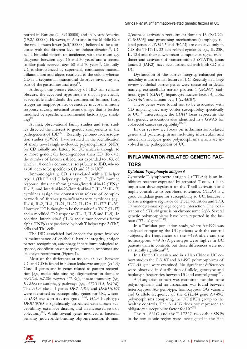
Sarlos P et al . Inflammation-related genetic factors in UC
ported in Europe (24.3/100000) and in North America (19.2/100000). However, in Asia and in the Middle East the rate is much lower (6.3/100000) believed to be asso-ciated with the different level of industrialization[2]. UC has a bimodal pattern of incidence, with the mean age diagnosis between ages 15 and 30 years, and a second smaller peak between ages 50 and 70 years[3]. Clinically, UC is characterized by superficial, continuous mucosal inflammation and ulcers restricted to the colon, whereas CD is a segmental, transmural disorder involving any part of the gastrointestinal tract[4].
Although the precise etiology of IBD still remains obscure, the accepted hypothesis is that in genetically susceptible individuals the commensal luminal flora trigger an inappropriate, overactive mucosal immune response causing intestinal tissue damage that is further modified by specific environmental factors (e.g., smok-ing)[5].
At first, observational family studies and twin stud-ies directed the interest to genetic components in the pathogenesis of IBD[6, 7]. Recently, genome-wide associa-tion studies (GWAS) have resulted in the identification of many novel single nucleotide polymorphisms (SNPs) for CD initially and latterly for UC which is thought to be more genetically heterogeneous than CD. To date, the number of known risk loci has expanded to 163, of which 110 confer common susceptibility to IBD, where-as 30 seem to be specific to CD and 23 to UC[8].
Immunologically, CD is associated with a T helper type 1 (Th1)[9] and T helper type 17 (Th17)[10] immune response, thus interferon gamma/interleukin-12 (IFNγ/IL-12) and interleukin-23/interleukin-17 (IL-23/IL-17) cytokines assign the downstream release of complex network of further pro-inflammatory cytokines (e.g., IL-18, IL-2, IL-1, IL-21, IL-22, IL-17A, IL-17F, IL-26). However, UC is thought to be the result of a Th17 (IL-17) and a modified Th2 response (IL-13, IL-5 and IL-9). In addition, interleukin-6 (IL-6) and tumor necrosis factor alpha (TNFα) are produced by both T helper type 2 (Th2) cells and Th1 cells.
The IBD-associated loci encode for genes involved in maintenance of epithelial barrier integrity, antigen pattern recognition, autophagy, innate immunological re-sponse, coordination of adaptive immune responses and leukocyte recruitment (Figure 1).
Most of the difference at molecular level between UC and CD is found in human leukocyte antigen (HLA) Class Ⅱ genes and in genes related to pattern recogni-tion [e.g., nucleotide-binding oligomerization domains (NODs), toll-like receptors (TLRs]), innate immunity (e.g., IL-23R) or autophagy pathways (e.g., ATG16L1, IRGM). The HLA class Ⅱ genes DR2, DR9, and DRB1*0103 were identified as susceptibility genes for UC, where-as DR4 was a protective gene[11,12]. HLA haplotype DRB1*0103 is significantly associated with disease sus-ceptibility, extensive disease, and an increased risk of colectomy[13]. While several genes involved in bacterial sensing [nucleotide-binding oligomerization domain
2/caspase activation recruitment domain 15 (NOD2/CARD15)] and processing mechanisms (autophagy re-lated genes ATG16L1 and IRGM) are defective only in CD, the Th17/IL-23 axis related cytokines [e.g., IL-23R, IL-12B and their downstream components signal trans-ducer and activator of transcription 3 (STAT3), janus kinase 2 (JAK2)] have been associated with both CD and UC.
Dysfunction of the barrier integrity, enhanced per-meability is also a main feature in UC. Recently, in a large review epithelial barrier genes were discussed in detail, namely, extracellular matrix protein 1 (ECM1), cad-herin type 1 (CDH1), hepatocyte nuclear factor 4, alpha (HNF4α ), and laminin beta 1 (LAMB1).
These genes were found not to be associated with CD, implying they may confer susceptibility specifically to UC[14]. Interestingly, the CDH1 locus represents the first genetic association also identified in a GWAS for colorectal cancer susceptibility[15, 16].
In our review we focus on inflammation-related genes and polymorphisms including interleukin and interleukin receptor gene polymorphisms which are in-volved in the pathogenesis of UC.
INFLAMMATION-RELATED GENETIC FAC-TORSCytotoxic T-lymphocyte antigen 4 Cytotoxic T-lymphocyte antigen 4 (CTLA4) is an in-hibitory receptor expressed by activated T cells. It is an important downregulator of the T cell activation and might contribute to peripheral tolerance. CTLA4 is a good candidate gene for susceptibility to UC, because it acts as a negative regulator of T cell activation and T/B, T/monocyte-macrophage cognate interaction. The local-ization of CTLA4 gene is on chromosome 2q33. Several genetic polymorphisms have been reported in the hu-man CTLA4 gene[17, 18].
In a Tunisian population study, where A+49G was analyzed comparing the UC patients with the control subjects, the frequencies of the +49A allele and the homozygous +49 A/A genotype were higher in UC patients than in controls, but those differences were not statistically significant[17].
In a Dutch Caucasian and in a Han Chinese UC co-hort studies the C-318T and A+49G polymorphisms of CTLA4 gene were examined. No significant differences were observed in distribution of allele, genotype and haplotype frequencies between UC and control group[19].
A Hungarian cohort was examined for the same polymorphisms and no association was found between heterozygous AG genotype, homozygous GG variant, and G allele frequency of the CTLA4 gene A+49G polymorphisms comparing the UC (IBD) group to the healthy controls. The A+49G does not represent an obligatory susceptibility factor for UC[20].
The A-1661G and the T-1722C two other SNPs in the non-exonic region were investigated in the Han
August 15, 2014|Volume 5|Issue 3|WJGP|www.wjgnet.com 305
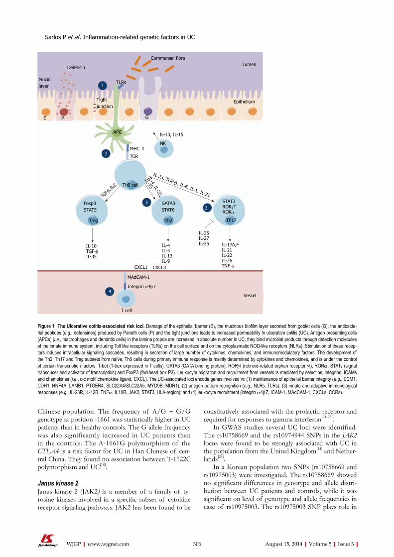
Chinese population. The frequency of A/G + G/G genotype at position -1661 was statistically higher in UC patients than in healthy controls. The G allele frequency was also significantly increased in UC patients than in the controls. The A-1661G polymorphism of the CTLA4 is a risk factor for UC in Han Chinese of cen-tral China. They found no association between T-1722C polymorphism and UC[18].
Janus kinase 2 Janus kinase 2 (JAK2) is a member of a family of ty-rosine kinases involved in a specific subset of cytokine receptor signaling pathways. JAK2 has been found to be
constitutively associated with the prolactin receptor and required for responses to gamma interferon[21-23].
In GWAS studies several UC loci were identified. The rs10758669 and the rs10974944 SNPs in the JAK2 locus were found to be strongly associated with UC in the population from the United Kingdom[14] and Nether-lands[24].
In a Korean population two SNPs (rs10758669 and rs10975003) were investigated. The rs10758669 showed no significant differences in genotype and allele distri-bution between UC patients and controls, while it was significant on level of genotype and allele frequencies in case of rs10975003. The rs10975003 SNP plays role in
August 15, 2014|Volume 5|Issue 3|WJGP|www.wjgnet.com 306
Defensin
Mucin layer
E P
TLRs1
Tight junction
Commensal floraLumen
Epithelium
G
IL-13, IL-15
NKMHC Ⅱ
TCR
APC
2
IL-23, TGF-b, IL-6, IL-1, IL-21
IL-4, IL-25,
IL-33
3TG
F-b, IL
-2 Th0 cell
Th2 Th17Treg
3Foxp3 STAT5
GATA3 STAT6
STAT3RORγTRORα
IL-10TGF-bIL-35
IL-4IL-5IL-13IL-9
IL-25IL-27IL-35 IL-17A,F
IL-21IL-22IL-26TNF-α
Vessel
CXCL1 CXCL3
T cell
MAdCAM-1
Integrin α4b74
Figure 1 The Ulcerative colitis-associated risk loci. Damage of the epithelial barrier (E), the mucinous biofilm layer secreted from goblet cells (G), the antibacte-rial peptides (e.g., defensines) produced by Paneth cells (P) and the tight junctions leads to increased permeability in ulcerative colitis (UC). Antigen presenting cells (APCs) (i.e., macrophages and dendritic cells) in the lamina propria are increased in absolute number in UC, they bind microbial products through detection molecules of the innate immune system, including Toll like receptors (TLRs) on the cell surface and on the cytoplasmatic NOD-like receptors (NLRs). Stimulation of these recep-tors induces intracellular signaling cascades, resulting in secretion of large number of cytokines, chemokines, and immunomodulatory factors. The development of the Th2, Th17 and Treg subsets from naïve, Th0 cells during primary immune response is mainly determined by cytokines and chemokines, and is under the control of certain transcription factors: T-bet (T-box expressed in T cells), GATA3 (GATA binding protein), RORγt (retinoid-related orphan receptor γt), RORα, STATs (signal transducer and activator of transcription) and FoxP3 (forkhead box P3). Leukocyte migration and recruitment from vessels is mediated by selectins, integrins, ICAMs and chemokines (i.e., c-c motif chemokine ligand, CXCL). The UC-associated loci encode genes involved in: (1) maintenance of epithelial barrier integrity (e.g., ECM1, CDH1, HNF4A, LAMB1, PTGER4, SLC22A4/SLC22A5, MYO9B, MDR1); (2) antigen pattern recognition (e.g., NLRs, TLRs); (3) innate and adaptive immunological responses (e.g., IL-23R, IL-12B, TNFα, IL10R, JAK2, STAT3, HLA-region); and (4) leukocyte recruitment (integrin α4b7, ICAM-1, MAdCAM-1, CXCLs, CCRs).
Sarlos P et al . Inflammation-related genetic factors in UC
the pathogenesis of UC in Koreans[25].
Signal transducer and activator of transcription 3 This protein is a member of the signal transducer and activator of transcription (STAT) protein family. It is encoded by the signal transducer and activator of tran-scription 3 (STAT3) gene. In response to cytokines and growth factors, STAT family members are phosphory-lated by the receptor associated kinases, and then form homo- or heterodimers that translocate to the cell nucle-us where they act as transcription activators. This pro-tein is activated by phosphorylation in response to vari-ous cytokines and growth factors including interferons (IFNs), epidermal growth factor (EGF), interleukin-5 (IL-5), IL-6, hepatocyte growth factor (HGF), leukemia inhibitory factor (LIF) and bone morphogenetic protein 2 (BMP2). STAT3 relays the expression of a variety of genes in response to cell stimuli, and thus plays pivotal role in several cellular processes, such as cell growth and apoptosis[26-28].
In a large GWAS study the rs12948909 SNPs in the STAT3 locus was identified and found to be strongly as-sociated with UC in the United Kingdom population[14].
In the Hungarian population the STAT3 rs744166 was investigated. STAT3 rs744166 TT genotype and T allele frequencies were significantly higher in patients with UC than in controls. Logistic regression analysis re-vealed that the TT genotype confers as an increased risk for the development of UC[23].
In a North American study the same polymorphisms were tested, but no significant differences were found between the UC group and healthy controls[29].
Tumor necrosis factor alpha The pro-inflammatory cytokine, tumor necrosis factor alpha (TNFα) has important pathogenic role both in CD and in UC[30,31]. Through its ability to cause epithe-lial barrier disruption in colonic epithelial cells[32,33] it is responsible for tissue damage. The TNFα gene can be found at the inflammatory bowel disease 3 (IBD3) locus within the major histocompatibility complex (MHC) region. In several studies it has been found as a suscep-tibility locus for IBD[34-36]. Level of TNFα is elevated in serum, stools, and inflamed bowel mucosa of patients with IBD[37-41].
The polymorphism at position -308 is a point mu-tation, where the presence of G defines the common variant TNF1, and A the less common variant TNF2. Susceptibility to UC has been positively[42] and nega-tively[43] associated with carriage of TNF2 allele. Some studies suggested that this allele might have a small but significant association with greater levels of TNF transcription[44, 45]. However other authors did not find any influence of TNFα bi-allelic polymorphism on UC susceptibility, although they reported a higher frequency of the TNF2 allele in women with extensive disease compared with those with distal colitis[46]. In Mexican Mestizo UC patients increased frequency of TNF2 allele
and TNF 1/2 genotype was found, suggesting this could be an additional genetic marker for the susceptibility to UC[47]. Similar findings have been reported in patients with UC with Caucasian origin[42, 46, 48, 49].
The TNFα polymorphisms A-308G and T-857C increase the TNFα production, raising the possibility of correlation with different disease course or response to therapy[50]. The A-238G and A-308G in TNFα promoter region have been found as a susceptibility factor in dif-ferent autoimmune disorders, including asthma[51,52], pso-riasis[53] and rheumatoid arthritis[54]. The polymorphism A-238G was associated with lower production of TNFα in Caucasian UC patients[46].
In a New Zealand Caucasian UC cohort was found, that carriers of the TNFα -308A allele may give higher risk for pancolitis and the necessity for bowel resec-tion[55]. In Israeli Jewish patients having CD or UC, the allele and carrier frequencies of -857T allele did not dif-fer between IBD patients and controls, suggesting this SNP in Ashkenazi Jewish patients neither determines the susceptibility, nor influences the clinical phenotype of CD or UC[56].
Different studies supported that TNFa -308 in UC may be an ethnic population-specific risk factor. Stud-ies from East Asia suggested strong association of the TNFα -308 gene promoter polymorphism for UC in East Asians. Allele frequency of TNFα -308A was sig-nificantly higher in Han Chinese UC patients than in healthy controls. Haplotype analysis revealed 6 haplo-types including H5 (TNF 1031T/863C/857C/380G/308A/238G/) and H3 (TNF 1031C/863C/857C/ 380G/308A/238G/). Haplotype frequency of H5 was significantly higher in UC patients, suggesting that H5 is associated with UC and the TNFα gene may be a sus-ceptibility gene to UC[57]. Interestingly, the meta-analysis did not reveal any association of the TNFα -308 gene promoter polymorphism with UC in Europeans[58]. In a Caucasoid population from the North of Spain the TNFα -308 alleles did not influence the appearance of steroid dependency either in UC or in CD[59]. In Italian pediatric patients the TNFα -308 was significantly in-creased in patients with UC[60].
In Czech pediatric IBD patients significant differenc-es in TNFα -308 A polymorphism were found between UC group and controls, but no differences were noted between this polymorphism and the clinical characteris-tics of UC[61].
Significant correlation of the TNFα -863A vari-ant was demonstrated with colonic disease and greater height at diagnosis[62], but in this study they could not find any significant difference for the -857 allele. In patients with UC only a trend toward an increased fre-quency of steroid resistance was found in carriers of the TNFα risk genotype compared to non-carriers[60].
In the Han Chinese population the TNFα C-1031T, A-863C and T-857C allele/carrier frequencies were ana-lyzed between UC patients and healthy controls. They did not find any significant difference of the tested al-
August 15, 2014|Volume 5|Issue 3|WJGP|www.wjgnet.com 307
Sarlos P et al . Inflammation-related genetic factors in UC
lele/carrier frequencies between UC patients and con-trols, only the TNFα -857T was increased in UC patients but did not reach statistical significance[57].
Organic cation transporter 1/2 OCTN1 (SLC22A4) and OCTN2 (SLC22A5) are widely expressed [63-66], but specifically expressed in principal intestinal cell types affected by CD: epithelial cells, CD68+ macrophages and CD43+ T cells. SLC22A4 and SLC22A5 encode the polytopic transmembrane sodium-dependent carnitine and sodium-independent organic cation transporters OCTN1 and OCTN2[67]. OCTNs have important role in the maintenance of intracellular homeostasis and in the energy production of the cell [68]. Both OCTNs play important role in the maintenance of gastrointestinal health and in the prevention of gut inflammation[69, 70].
CD associated variants, the OCTN1 T1672C and OCTN2 -207G were as strongly associated with UC in unrelated Caucasian subjects[71].
Homozygous patients for the OCTN1 1672T variant were significantly associated with UC in a study cohort from Italy, suggesting that OCTN1 could have a role in modulating the severity of chronic inflammation in UC[72].
The mutation that leads to L503F substitution in the OCTN1 protein can alter the transporter’s activity[73, 74]. Only a weak gender-specific effect of L503F was ob-served at male UC patients in a cohort of familial and sporadic IBD from the central Pennsylvania, United States[75].
Multidrug-resistant transporter-1 The multidrug-resistant transporter-1 (MDR1) gene en-codes the transmembrane protein, P-glycoprotein 170 (Pgp)[76]. This gene is an excellent candidate gene for the pathogenesis of IBD[77]. Pgp functions as an ATP-dependent efflux transporter pump and is expressed in many normal tissues like in the epithelial surfaces of the intestine, biliary ductules, proximal tubules of kidneys and central nervous system[78-80].
One of the most significant MDR1 gene mutations is the C3435T polymorphism. Decreased expression of the MDR1 gene and lower Pgp activity has been associated with this variant. However, studies showed conflicting results. In a German study the T allele and TT genotype frequencies of C3435T polymorphism were significantly increased in UC patients[81]. Glas et al[82] found in a small group of UC patients partial accordance with a trend towards an increased frequency of T allele compared to controls, but a statistical difference was detected only in one of two different control groups. In a meta-analysis significant association of the 3435T allele and the 3435TT genotype has been found with UC[83]. The triallelic G2677T/A and the C3435T have been shown to correlate with Pgp expression[84-87]. Significant asso-ciation of C3435T and G2677T was detected with UC: UC patients had significantly higher frequency of 2677T
allele and of the 3435TT genotype. Haplotype analysis revealed that carriers of 3435T/2677T haplotype have significantly higher risk of having UC[88]. In a Japanese UC cohort the C3435T was predictive of susceptibility to later onset UC, but not for the early onset of UC[89].
Large study with German and British UC and CD patients failed to demonstrate association. It was con-firmed, that this SNP is associated with UC especially in patients with extensive colitis[90]. In addition completely negative findings have been reported in large studies from North America[7], Slovenia[91] and Italy[92]. Similarly to these results, UC patients with Caucasian origin from central Poland were found that MDR1 C3435T polymor-phism is not a risk factor for IBD, including both UC and CD[93].
A study with New Zealand IBD patients supported the role of MDR1 as a candidate gene for UC. Hetero-zygous carriers for the variants C1236T, rs2235046 and G2677T/A showed a lower risk of developing UC com-pared with homozygotes. Subgroup analysis revealed that C1236T and rs3789243 are associated with IBD when stratified for age of onset. The MDR1 variant rs3789243 was found to be associated with pancolitis in UC patients[94]. In the genetically heterogeneous North Indian UC cohort was found that this SNP is signifi-cantly overrepresented in UC patients.
When German IBD patients were genotyped for the two MDR1 SNPs in positions 2677G>T/A and 3435C>T it was found that the combined genotypes derived from these positions are possibly associated with young age onset of UC and severe course of disease[95]. The 2677T allele was significantly increased in British UC cases com-pared with controls. The TT genotype was significantly associated with severe UC. No significant association was seen with C3435T and UC or any clinical subgroup. A meta-analysis of 9 association studies of C3435T showed a significant association of the 3435T allele with UC, but not with CD. These results indicated that MDR1 sequence variants are associated with a small increase in the risk of developing UC and may influence disease behavior[96]. The MDR1 gene polymorphism G2677T/A showed sig-nificant association with CD, and the C3435T with Span-ish UC patients[97]. The MDR1 3435 TT genotype and T-allelic frequencies were significantly higher in patients with UC compared with controls. The association was strongest with extensive UC, and this was also confirmed with multivariate analysis. However G2677T was not as-sociated with UC or CD. Two-locus haplotypes showed both positive (3435T/G2677 haplotype) and negative (C3435/2677T haplotype) associations with UC. Homo-zygotes for the haplotype 3435T/G2677 were significantly increased in UC. Allelic variations of the MDR1 gene de-termined the disease extent as well as susceptibility to UC in the Scottish population[90].
Nucleotide-binding oligomerization domain1/ caspase activation recruitment domain 4 Nucleotide-binding oligomerization domain1/ caspase
August 15, 2014|Volume 5|Issue 3|WJGP|www.wjgnet.com 308
Sarlos P et al . Inflammation-related genetic factors in UC
activation recruitment domain 4 (NOD1/CARD4) is a member of the Nod-like receptor family, which is phy-logenetically conserved[5, 98]. It is constitutively expressed in epithelial cells throughout the gastrointestinal tract[99]. NOD1/CARD4 contains leucine-rich repeat (LRR) do-main and NOD domains and has only one CARD do-main[100].
Polymorphism in LRR domain of the NOD1/CARD4 gene showed association with disease sever-ity of UC in North Indian patients. This might be due to disruption of the LRR region critical for NOD1-mediated bacterial sensing. Haplotype-based approach showed that GTTG haplotype carriers were over repre-sented in UC patients which could increase the risk of the disease[101].
Initially, it was suggested that there is association of the deletion variant of NOD1/CARD4 +32656 (complex intronic insertion-deletion polymorphism) with suscep-tibility to IBD using a combination of transmission dis-equilibrium testing (TDT) and case-control analysis[102]. However this variant was not associated with a strong effect on susceptibility to IBD in children and adults in a Northern Europe study cohort[103]. Similar results have been found in the East Anglia IBD cohort, where no as-sociation was found between NOD1 +32656 and IBD and also no heterogeneity between UC and CD[104].
Toll-like receptors Essential components of innate immunity are the Toll-like receptors (TLRs). These are transmembrane recep-tors which recognize the microbial compounds from different bacteria, fungi and viruses[105-107]. TLRs are ex-pressed by intestinal epithelial cells and immune cells in IBD patients[108,109]. TLR signaling in the intestinal sites of the colon can inhibit the inflammatory responses and maintains the colonic homeostasis[110-112]. TLRs can be found on the cell membrane (TLR1, 2, 4, 5 and 9) or on intracellular organelles (TLR3, 7 and 8)[113]. From the 10 human TLRs we review only three members regarding to their association to UC.
TLR2 is localized on the cell’s surface. With its cofac-tors (TLR1 and TLR6) it binds lipoproteins, which are important surface antigen of the Gram-negative outer membrane[114], TLR4 consists of a leucine-rich repeat region (LRR) and an intracellular domain homologous to IL-1 receptor[115]. It recognizes conserved pathogenic motifs of Gram-negative bacteria, mainly lipopolysac-charides (LPS). Signaling through TLR4 results in the activation of the transcriptional activator, known as nuclear factor κB (NF-κB)[116]. Similarly to TLR2, TLR9 is localized on the cell’s surface. It recognizes unmethyl-ated CpG DNA in bacteria and viruses[117,118].
The allele and carrier frequencies of the Thr399Ile mutation in the TLR4 gene were significantly associated with UC in a Caucasian population[119]. Association of TLR4 Asp299Gly polymorphism with UC was reported first in Caucasian UC patients[120]. In a study, mentioned before[119], increased frequency of this polymorphism
was observed, but it did not reach statistical signifi-cance. Similarly to Török et al study, the Asp299Gly and Thr399Ile mutations in TLR4 gene were associated with UC in Greek and in North Indian patients[121,122], but not in Dutch or Italian patients[123,124]. Interestingly, the TLR4 Asp299Gly did not show association with UC in different Asian UC populations[125-128].
The TLR2 Arg677Trp and Arg753Glu, TLR4 Asp299Gly and Thr399Ile, and TLR9 gene C1237T polymorphisms were genotyped in Chinese Han IBD patients; however none of these polymorphisms was associated with IBD. In Caucasians, both TLR4 299Gly and 399Ile conferred as a significant risk factor for de-veloping UC and CD[127].
Three SNPs of TLR9 (C-1486T, G1174A, A2848G) were genotyped. These variations were associated with an increased risk of UC in the Japanese population. TLR9 -1486CC, 1174GG and 2848AA showed increased risk for UC, but TLR9 -1486TT, 1174AA and 2848GG decreased the risk of UC, although there were no cor-relations between SNPs and disease phenotype or TLR9 mRNA expression[129]. Possible associations between genetic variations in TLR9 and IBD in the German population were investigated, but no associations were detected between TLR9 gene variations and UC suscep-tibility[130].
Cell adhesion molecules Cell adhesion molecules (CAM) mediate the extravasa-tion of leukocytes and their accumulation in inflamed in-testinal mucosa. This process is controlled by a family of CAM including the intercellular cell adhesion molecule (ICAM-1), the platelet endothelial cell adhesion molecule (PECAM-1), the selectins (E, L, and P selectin) and the integrins[131,132].
ICAM-1 (CD54) is a cell surface glycoprotein belong-ing to the immunoglobulin superfamily. It plays key role in transendothelial migration of leukocytes, and lympho-cyte activation. The membrane glycoprotein PECAM-1 (CD31) is expressed on vascular endothelial cells, plate-lets, some lymphocyte subsets, and monocytes[133-135]. It has important role in transendothelial migration of circulating leukocytes during inflammatory process[136], apoptosis[137] and integrin regulation[138]. The E-selectin (CD62E) is a glycoprotein, which is expressed on endo-thelial cells in response to pro-inflammatory cytokines (IL-1, TNF). It supports rolling of leukocytes at sites of inflammation and tissue injury. E-selectin expression is upregulated both in CD and in UC patients playing an important role in mediating of the inflammatory process in IBD[139]. L-selectin (CD62L) is expressed on normal naive T and B cells, leukocytes and on natural killer (NK) cells. It is involved in the adhesion of T cells to endo-thelial cells, which are regarded as crucial in the selective migration of lymphocytes to inflamed tissue sites during an inflammatory response[140].
Several mutations in ICAM1 (G241R and K469E), PECAM-1 (V125L), PECAM-1 (G98T and S128R),
August 15, 2014|Volume 5|Issue 3|WJGP|www.wjgnet.com 309
Sarlos P et al . Inflammation-related genetic factors in UC
E-selectin (L554F) and L-selectin (F206L) were analyzed in Tunisian IBD patients and controls. A significant increase in allele frequencies of 206L of L-selectin and the associated genotype F/L was observed both in UC and in CD patients. In the subgroup analysis the L206 allele and F/L206 genotype frequencies were signifi-cantly increased in UC patients with left-sided type. No significant differences in allele or genotype frequencies were observed for ICAM-1, E-selectin, and PECAM-1 polymorphisms between UC patients, CD patients, and controls[141].
INTERLEUKINS IN UCInterleukin 1Interleukin 1 (IL-1) is pro-inflammatory cytokine, which affects cell proliferation, differentiation, and the function of many innate and specific immunocompetent cells, and acts as an endogenous pyrogen. It broadcast many inflammatory diseases by initiating and potentiating im-mune and inflammatory responses[142].
IL-1 is composed of two main proteins the IL-1A and the IL-1B[143]. IL-1B has major role in initiating and amplifying the inflammatory response[144]. The IL-1 re-ceptor antagonist (IL-1RN) is an anti-inflammatory cy-tokine, which lacks the IL-1 receptor accessory protein (IL-1RAP) interacting domain[142].
In Mexican Mestizo UC patients five SNPs were ana-lyzed; the rs419598, the rs315951 and the rs315952 in the IL-1RN gene, the rs16955 in the IL-1B gene and the 3811058 in the IL-1F10 gene. Significant increased fre-quencies of IL-1RN6/1TC (rs315952), IL-1RN6/2CC (rs315951) and decreased frequency of IL-1B-511 TC (rs16944) genotypes were found in UC patients. The patients group showed increased frequencies of IL-1RN CTC and TCG haplotypes, whereas TTG and CTG hap-lotypes frequencies were decreased[145].
IL-2IL-2 functions as a T cell growth factor, furthermore it supports the proliferation and differentiation of NK cells to increase their cytolytic functions. This IL plays important role in the development of Th1, Th2, Treg, and Th17 differentiation[146].
In the IL-2/IL-21 region several polymorphisms (rs6822844, rs13151961, rs13119723 and rs6840978) were studies. In a Dutch population the minor alleles of the examined SNPs were associated with IBD. The strongest association of these SNPs was found in the UC patients. In an Italian UC cohort the same strong association of the minor alleles was observed with UC. Similarly to this results, in the North American study was demonstrated, that these alleles have the strongest effect among the IBD patients in the UC subgroup [147].
IL-6 IL-6 is a multifunctional, pleiotropic cytokine that is responsible for regulation of immune responses, acute-
phase responses, hematopoiesis, and inflammation[148]. An Irish population study the IL-6 -174 genotype
frequency showed significant difference between CD and UC group[149]. In the Caucasian population the same polymorphism was examined in CD and UC patients and found significant difference UC and CD susceptibil-ity[150].
IL-8IL-8 is member of the CXC chemokine family[151]. Its has two receptors the CXCR1 (IL-8RA) and the CXCR2 (IL-8RB)[152]. It exerts effect mainly on the chemotaxis and migration of neutrophils, monocytes, lymphocytes, and fibroblasts[153].
IL-8 T-251A was analyzed in a Polish population. The allele frequency showed significant difference com-paring the UC group to the controls[154], however this as-sociation was not observable in a Chinese UC cohort[155]. Additional polymorphisms were also tested and their effect on the serum level of IL-18. Haplotype frequency of the -353A/ -251A/ +678T haplotype was consider-ably higher in UC group compared to controls, suggest-ing this haplotype is likely to be more common in severe UC patients than in mild to moderate cases[155].
IL-10 The anti-inflammatory cytokine IL-10 is produced by many cells like monocytes, T cells, B cells, NK cells, macrophages, and dendritic cells (DCs). It prevents the antigen presentation and also the subsequent release of pro-inflammatory cytokines, so it alleviates the activated immune system[156].
In a GWAS, the polymorphism rs3024505 demon-strated the most meaningful association in the combined verification UC samples, suggesting that defective IL-10 function plays important role in the pathogenesis of the UC[157]. The same polymorphisms was investigated in Australian population[158] and Danish cohort[159] and found that the rs3024505 was associated with the risk of UC.
Three promoter polymorphisms of the IL-10 gene G-1082A, C-819T, and C-592A were studied in many population but the results are contrary. In an Italian cohort the G-1082A and the C-819T SNPs were investi-gated. The -1082 genotype frequencies were significantly different between UC patients and controls. The fre-quency of the -1082A allele was also significantly higher in the UC patients than in controls. Allele and genotype frequencies of T-819C were not significantly associ-ated with the disease. Furthermore, the frequencies of haplotypes -1082A/-819C and -1082A/-819T, which have been described to have a decreased promoter activ-ity, were significantly increased in UC patients than in controls[160]. In a North-Eastern Mexican population the G-1082A and the C-592A SNPs were examined. The -1082 AA and -592 AA genotypes showed significantly lower frequencies in UC compared to healthy controls, while individuals heterozygous at IL-10-1082 have sig-
August 15, 2014|Volume 5|Issue 3|WJGP|www.wjgnet.com 310
Sarlos P et al . Inflammation-related genetic factors in UC
nificantly increased occurrence of UC[161]. In a Tunisian group the A-627C and the G-1117A polymorphisms were examined and found that these two variants influ-encing the UC susceptibility and phenotype[162]. In the Asian population the association was confirmed between A-1082G polymorphism and UC[125, 163].
IL-12IL-12 is an interleukin that is naturally produced by den-dritic cells, macrophages and human B-lymphoblastoid cells in response to antigenic stimulation. It participates in the differentiation of naive T cells into Th1 cells and involved in the activities of natural killer cells and T lymphocytes. IL-12 mediates gradation of the cytotoxic activity of NK cells and CD8+ cytotoxic T lympho-cytes[164]. IL-12 consist of two subunit p35 (IL-12A) and p40 (IL-12B), which is shared by IL-12 and IL-23 cyto-kines. The IL-12 receptor has two subunits: IL-12RB1 and IL-12RB2[165, 166].
In a German population four SNPs (rs3212227, rs17860508, rs10045431 and rs6887695) of the IL-12B were investigated. Two SNPs, the rs10045431 and rs6887695 showed association with increased UC susceptibility[167]. From these SNPs the rs6887695 was investigated in a Japanese population where significant association was manifested between UC patients and controls[168].
IL-17The interleukin 17 (IL-17 or IL-17A) is a pro-inflamma-tory cytokine secreted by activated T cells its main tasks is inducing and mediating pro-inflammatory responses. It induces the production of many other cytokines, chemokines and prostaglandins from fibroblasts, endo-thelial cells, epithelial cells, keratinocytes, and macro-phages[169-171].
In a Japanese population the rs2275913 SNP in the IL-17A gene and the rs763780 SNP in the IL-17F gene were investigated and found significant differences between UC group and healthy controls on level of -197A/A and 7488T/T genotype frequencies[172].
Even more recently, a GWAS in a very large Europe-an UC cohort identified an association between another IL-17 pathway gene (IL-17REL) and UC[173].
IL-18IL-18 is produced by macrophages and other cells. It functions by binding to the interleukin-18 receptor, and together with IL-12 it induces cell-mediated immunity following infection. IL-18 induces gene expression and synthesis of TNF, IL-1, Fas ligand, and several chemo-kines[174].
In the IL-18 gene several polymorphisms were ex-amined. The G-137C, the C-607A and the G-656T are located in the promoter region, while the A105C, the T113G and the C127T are coding variants. In a Japanese study the G allele at 113 and the T allele at 127 were significantly higher in patients with UC compared to the
control[175]. In another Japanese study allele and genotype frequency of G-137C were significantly higher in the proctitis-type UC patients than in controls[176]. The fre-quency of haplotype 2 (-607A, -137C), which have lower promoter activity and IFNγ-mRNA level was significant-ly increased in the proctitis-type patients than in the con-trol group[176]. The C-607A and the G-137C SNPs were associated with the development of UC in Tunisian pa-tients. The -137GG genotype frequency was significantly higher in UC than in controls and statistically significant association was found between -607AA genotype in UC patients and the distal localization of the lesions[177].
IL-23 IL-23 main functions are very important in innate and adaptive immunity to regulate Th17 function and expan-sion[178]. This cytokine induces CD8+ memory T cells to proliferate and produce IL-17. IL-23 binds to its recep-tor IL-23R, which polymorphisms’ play the main role in the autoimmune diseases[179-181] especially in IBD[182].
Several independent functional SNPs of the IL-23R gene and its neighboring region were determined; sev-eral were found susceptible to (rs10889677, rs11209032, rs11465804, rs11805303, rs1495965, rs2201841, rs1004819) CD and UC in non-Jewish subjects[183].
In a Chinese cohort the rs7530511 and rs11805303 SNPs were studied and positive association was found between these variants and UC susceptibility[184]. In the Jiangsu Han population the rs11805303 was found as a susceptibility factor to UC[185].
In a Swedish population the rs10889677, the rs11209032, the rs11465804, the rs2201841 and the rs1004819 polymor-phisms were investigated, and found that the rs11465804G, rs2201841C and rs1004819T allele frequencies showed sig-nificant differences between UC patient group and control. These genetic variants are individual risk factor for develop-ing the disease[186].
In Hungarian UC patients for the IL-23R rs1004819A allele we found significantly higher allele frequency com-pared to control subjects and the SNP rs2201841 showed significant association with UC risk for homozygotes[187].
IL-26 Expression of IL-26 seems to be restricted to memory T cells, NK cells, and Th17 cells. Thereby it could have pro-inflammatory effects in IBD[188].
Only a few markers were investigated, from these the rs2870946-G and the rs1558744-A showed association with UC[189]. Further meta-analysis study confirmed the association of rs1558744-A with UC[190].
CONCLUSIONThis review shows that substantial progress has been made in the past 10 years in to find inflammatory related genetic factors and cytokines in UC. We reviewed differ-ent genes and gene polymorphisms which play role in the inflammatory process of UC. These genes could be
August 15, 2014|Volume 5|Issue 3|WJGP|www.wjgnet.com 311
Sarlos P et al . Inflammation-related genetic factors in UC
potential targets of novel treatment strategies. From the reviewed genes contributing to inflamma-
tion TNFα , MDR1 and TLRs were the most investigated genes. TNFα has been found as a susceptibility locus for both UC and CD[34-36]. The TNF2 allele and TNF 1/2 could be good candidate markers for the susceptibility to UC. Based on the different studies with different popu-lations (East Asians, Han Chinese, Spanish Caucasoid, Italian, and Czech), the TNFα -308 is the most studied SNP and this may be an ethnic population-specific risk factor for UC especially for Asian populations but not for Europeans. It should be noted this polymorphism is also a susceptibility factor in other autoimmune disor-ders (asthma, psoriasis, and rheumatoid arthritis) too.
The MDR1 C3435T is one of the most tested SNP in UC, but with conflicting results. Some studies showed significantly increased 3435T allele and 3435TT genotype frequencies of C3435T[81,83,97], or only a trend towards an increased frequency of T allele[82], or the T allele was predictive of susceptibility to later onset UC, but not for the early onset of UC[89]. But some studies failed to demonstrate association with UC[91-93]. Other SNPs of MDR1 (C1236T and rs3789243) were associ-ated with IBD when stratified for age of onset. The rs3789243 was found to be significantly overrepresented in genetically heterogeneous North Indian UC patients.
We reviewed 3 members from the 10 human TLRs regarding to their association to UC. Allele and carrier frequencies of the TLR4 Asp299Gly and Thr399Ile were significantly associated with UC in Caucasian[119,120], Greek and in North Indian patients[121,122], but not with Dutch, Italian[123,124] or Asian patients[125-128]. Interestingly the TLR9 polymorphisms (C-1486T, G1174A, A2848G) were associated with an increased risk of UC in the Japa-nese population. TLR9 -1486CC, 1174GG and 2848AA polymorphisms show increased risk for UC, but TLR9 -1486TT, 1174AA and 2848GG decrease the risk of UC[129].
From the reviewed cytokines IL-10, IL-18 and IL-23 were the most investigated genes. The IL-10 is a major anti-inflammatory cytokine, which attenuates the activated immune system with inhibiting both the antigen presentation and subsequent release of pro-inflammatory cytokines. IL-10 is a shared risk gene for CD and UC too. The promoter polymorphisms of this gene (G-1082A, C-819T, and C-592A) which are in tight linkage disequilibrium were extensively studied in many populations but with contradictory results. In the Cau-casian population the carriers of G-1082A SNP were more susceptible to UC, whereas in another study carri-ers were associated with lower UC incidence[160,161]. In the Asian population the results strengthened the positive re-lationship between this SNP and UC susceptibility[125,163]. In a North-Eastern Mexican population the -592AA genotypes showed significantly decreased frequency in UC compared the results to the healthy controls[161]. In a Tunisian group the A-627C and the G-1117A vari-ants influencing the UC susceptibility and phenotype[162].
Several other studies handle with these non-coding SNP in CD too and determine susceptibility to the disease or not.
The G-137C, the C-607A and the G-656T promoter SNPs and several others in the coding regions of IL-18 gene (A105C, the T113G and the C127T) were exam-ined. The Japanese population is the most studies for these SNPs, significant difference was found in the al-lele frequency of the A105C between CD patients and controls, while this correlation could not be detected in UC patients. The 113G and 127T allele frequencies were significantly increased in patients with UC compared the results to the healthy controls[175]. In case of promoter polymorphisms, the -137CC genotype frequency was sig-nificantly increased in proctitis-type UC patients than in controls, while the other two C-607A and G-137C SNPs were associated with the development of UC in Tunisian patients[177].
The IL23R gene was identified as a CD susceptibility gene in North American non-Jewish subjects. Several in-dependent functional SNPs in the gene and its neighbor-ing region were determined[183]. After the primary publica-tions, several studies have been published these SNPs in IBD and other autoimmune disease too (ankylosing spon-dylitis, psoriasis, Sjögren syndrome, systemic lupus ery-thematosus). From these SNPs (rs10889677, rs11209032, rs11465804, rs11805303, rs1495965, rs2201841, rs100481) several are risk factor to IBD both in European and Asian populations[185-187].
It can be established that these interleukin gene vari-ants are strongly population dependent but in the given population they can be predictors for CD or UC. Despite the advances in the field of UC/IBD genetics, testing for these genetic variants is currently not recommended for clinical purposes[191].
Understanding of the detailed pathogenesis of IBD and identifying new disease associated SNPs led to the development of selective inhibitors for ILs, chemokines and their receptors. This strategies can optimize treat-ment efficacy and lead to personalized medicine based on the patient‘s genotype.
Biological agents are used in patients with moderate to severe disease activity who have failed conventional therapy with glucocorticoids and thiopurines. Today, the most effective and best studied anti-cytokine agents in IBD are the anti-TNFα antibodies. The mechanism of action of TNFα antagonists is based on the neutraliza-tion of both soluble TNFα and membrane TNFα and has a more global effect on inflammation than the block-ade of other cytokines. Currently, three TNFα inhibitors are approved by the United States Food and Drug Ad-ministration (FDA) for inducing and maintaining clinical remission in UC: the chimeric (25% murine and 75% human sequence) monoclonal full-length IgG1 mAb inf-liximab[192], the fully human mAbs adalimumab[193,194] and golimumab[195]. The pegylated humanised antibody cer-tolizumab pegol is approved only for CD (beside rheu-matoid arthritis, RA and psoriatic arthritis). Etanercept, a
August 15, 2014|Volume 5|Issue 3|WJGP|www.wjgnet.com 312
Sarlos P et al . Inflammation-related genetic factors in UC
dimeric fusion protein consisting of soluble p75-TNFR2 and the Fc portion of human IgG1, used in rheumatoid arthritis therapy, is not efficient for the treatment of in-testinal inflammation[196].
Despite the expeditious development of newer bio-logical therapies, only few have shown benefit in clinical trials in UC. Targeting of IL-23 or the IL-23 receptor or IL-23 axis is a potential therapeutic approach for autoimmune diseases including psoriasis, IBD, RA and multiple sclerosis[197]. Recently, testing of anti-IL-12/23 treatment in patients with CD has been performed. In Phase Ⅱ trial, patients with moderate to severe CD that was resistant to TNF antagonists had an increased rate of response to induction with the fully human mAb ustekinumab directed against the p40, as compared with placebo[198]. However, due to the common p40 subunit and IL-12RB1 chain, the major drawback of anti-IL-23 treatment can be the simultaneous inhibition of IL-12 and a possible shutdown of the immune system. Nev-ertheless, it would be much more useful to design drugs that target the IL-23p19 or IL-23RA itself, so inhibiting IL-23 without modifying the effects of IL-12[197].
Treatment of CD patients with the IL-17 blocker secukinumab (anti-IL-17A) was ineffective and higher rates of adverse events were noted compared with pla-cebo[199].
One new additional treatment for UC may be tofaci-tinib, an inhibitor of Janus kinases 1, 2, and 3 with in vitro functional specificity for kinases 1 and 3 over kinase 2, which is expected to block signaling involving gamma chain-containing cytokines including ILs-2, 4, 7, 9, 15, and 21. Tofacitinib, was approved for the treatment of RA in the USA, Japan and Russia in April 2013. In a double-blind, placebo-controlled, Phase Ⅱ trial, patients with moderately to severely active UC treated with to-facitinib were more likely to have clinical response and remission than those receiving placebo[200].
Targeting leukocyte recruitment and cell adhesion molecules could be also an option for IBD therapy. Na-talisumab, a recombinant humanised monoclonal IgG4 antibody, targets both the α4b1 heterodimer located in the central nervous system and the α4b7 integrin in the gut. The FDA approved natalizumab for both induction of remission and maintenance of remission for moder-ate to severe CD, though it has not been approved for this use in the European Union due to concerns over its risk/benefit ratio (risk of progressive multifocal leukoencephalopathy)[201]. Vedolizumab is a humanized mAb that specifically recognizes the α4b7 heterodimer, selectively blocks gut lymphocyte trafficking without in-terfering with trafficking to the central nervous system. In the Phase Ⅲ study, vedolizumab was more effective than placebo as induction and maintenance therapy for UC suggesting that blockade of T cell homing in the gut may favor mucosal healing in UC[202, 203].
The exact positioning of these promising new therapies in the management of UC remains uncertain currently. Additional long-term safety data and clinical
experience will be needed to determine an overall ben-efit/harm ratio of newly developed biological agents.
The identified separate loci in IBD research individu-ally have only modest effects on IBD susceptibility. They account together for only 20%-25% of the heritability, suggesting that gene-gene interactions as well as gene-environmental interactions could play a key role in IBD pathogenesis and fill the so called “genetic vacuum” of polygenic diseases[204]. More complete understanding of the immunopathogenic role of the various genes and ILs in intestinal inflammation will help in the develop-ment of more effective novel therapeutic strategies in UC. Next generation techniques in combination with the data analysis by systems-biology approach hopefully will contribute to the personalized therapy of the patients in the near future.
REFERENCES1 Podolsky DK. Inflammatory bowel disease. N Engl J Med
2002; 347: 417-429 [PMID: 12167685]2 Molodecky NA, Soon IS, Rabi DM, Ghali WA, Ferris M,
Chernoff G, Benchimol EI, Panaccione R, Ghosh S, Barkema HW, Kaplan GG. Increasing incidence and prevalence of the inflammatory bowel diseases with time, based on system-atic review. Gastroenterology 2012; 142: 46-54.e42; quiz e30 [PMID: 22001864 DOI: 10.1053/j.gastro.2011.10.001]
3 Loftus EV. Clinical epidemiology of inflammatory bowel disease: Incidence, prevalence, and environmental influenc-es. Gastroenterology 2004; 126: 1504-1517 [PMID: 15168363]
4 Silverberg MS, Satsangi J, Ahmad T, Arnott ID, Bernstein CN, Brant SR, Caprilli R, Colombel JF, Gasche C, Geboes K, Jewell DP, Karban A, Loftus EV, Peña AS, Riddell RH, Sachar DB, Schreiber S, Steinhart AH, Targan SR, Vermeire S, Warren BF. Toward an integrated clinical, molecular and serological classification of inflammatory bowel disease: report of a Working Party of the 2005 Montreal World Con-gress of Gastroenterology. Can J Gastroenterol 2005; 19 Suppl A: 5A-36A [PMID: 16151544]
5 Van Limbergen J, Russell RK, Nimmo ER, Ho GT, Arnott ID, Wilson DC, Satsangi J. Genetics of the innate immune response in inflammatory bowel disease. Inflamm Bowel Dis 2007; 13: 338-355 [PMID: 17206667]
6 Orholm M, Munkholm P, Langholz E, Nielsen OH, Sø-rensen TI, Binder V. Familial occurrence of inflamma-tory bowel disease. N Engl J Med 1991; 324: 84-88 [PMID: 1984188]
7 Brant SR, Panhuysen CI, Nicolae D, Reddy DM, Bonen DK, Karaliukas R, Zhang L, Swanson E, Datta LW, Moran T, Ravenhill G, Duerr RH, Achkar JP, Karban AS, Cho JH. MDR1 Ala893 polymorphism is associated with inflam-matory bowel disease. Am J Hum Genet 2003; 73: 1282-1292 [PMID: 14610718]
8 Jostins L, Ripke S, Weersma RK, Duerr RH, McGovern DP, Hui KY, Lee JC, Schumm LP, Sharma Y, Anderson CA, Es-sers J, Mitrovic M, Ning K, Cleynen I, Theatre E, Spain SL, Raychaudhuri S, Goyette P, Wei Z, Abraham C, Achkar JP, Ahmad T, Amininejad L, Ananthakrishnan AN, Andersen V, Andrews JM, Baidoo L, Balschun T, Bampton PA, Bitton A, Boucher G, Brand S, Büning C, Cohain A, Cichon S, D’Amato M, De Jong D, Devaney KL, Dubinsky M, Edwards C, Ellinghaus D, Ferguson LR, Franchimont D, Fransen K, Gearry R, Georges M, Gieger C, Glas J, Haritunians T, Hart A, Hawkey C, Hedl M, Hu X, Karlsen TH, Kupcinskas L, Kugathasan S, Latiano A, Laukens D, Lawrance IC, Lees CW, Louis E, Mahy G, Mansfield J, Morgan AR, Mowat C,
August 15, 2014|Volume 5|Issue 3|WJGP|www.wjgnet.com 313
Sarlos P et al . Inflammation-related genetic factors in UC
Newman W, Palmieri O, Ponsioen CY, Potocnik U, Prescott NJ, Regueiro M, Rotter JI, Russell RK, Sanderson JD, Sans M, Satsangi J, Schreiber S, Simms LA, Sventoraityte J, Targan SR, Taylor KD, Tremelling M, Verspaget HW, De Vos M, Wijmenga C, Wilson DC, Winkelmann J, Xavier RJ, Zeissig S, Zhang B, Zhang CK, Zhao H, Silverberg MS, Annese V, Ha-konarson H, Brant SR, Radford-Smith G, Mathew CG, Rioux JD, Schadt EE, Daly MJ, Franke A, Parkes M, Vermeire S, Barrett JC, Cho JH. Host-microbe interactions have shaped the genetic architecture of inflammatory bowel disease. Na-ture 2012; 491: 119-124 [PMID: 23128233]
9 Fuss IJ, Neurath M, Boirivant M, Klein JS, de la Motte C, Strong SA, Fiocchi C, Strober W. Disparate CD4+ lamina propria (LP) lymphokine secretion profiles in inflammatory bowel disease. Crohn’s disease LP cells manifest increased secretion of IFN-gamma, whereas ulcerative colitis LP cells manifest increased secretion of IL-5. J Immunol 1996; 157: 1261-1270 [PMID: 8757634]
10 Fujino S, Andoh A, Bamba S, Ogawa A, Hata K, Araki Y, Bamba T, Fujiyama Y. Increased expression of interleukin 17 in inflammatory bowel disease. Gut 2003; 52: 65-70 [PMID: 12477762]
11 Ferguson LR, Shelling AN, Browning BL, Huebner C, Peter-mann I. Genes, diet and inflammatory bowel disease. Mutat Res 2007; 622: 70-83 [PMID: 17628615]
12 Stokkers PC, van Aken BE, Basoski N, Reitsma PH, Tytgat GN, van Deventer SJ. Five genetic markers in the interleu-kin 1 family in relation to inflammatory bowel disease. Gut 1998; 43: 33-39 [PMID: 9771403]
13 Bouma G, Crusius JB, Garcia-Gonzalez MA, Meijer BU, Hellemans HP, Hakvoort RJ, Schreuder GM, Kostense PJ, Meuwissen SG, Pena AS. Genetic markers in clinically well defined patients with ulcerative colitis (UC). Clin Exp Immu-nol 1999; 115: 294-300
14 Anderson CA, Massey DC, Barrett JC, Prescott NJ, Tremel-ling M, Fisher SA, Gwilliam R, Jacob J, Nimmo ER, Drum-mond H, Lees CW, Onnie CM, Hanson C, Blaszczyk K, Ravindrarajah R, Hunt S, Varma D, Hammond N, Lewis G, Attlesey H, Watkins N, Ouwehand W, Strachan D, McArdle W, Lewis CM, Lobo A, Sanderson J, Jewell DP, Deloukas P, Mansfield JC, Mathew CG, Satsangi J, Parkes M. Investiga-tion of Crohn’s disease risk loci in ulcerative colitis further defines their molecular relationship. Gastroenterology 2009; 136: 523-9.e3 [PMID: 19068216]
15 Houlston RS, Webb E, Broderick P, Pittman AM, Di Ber-nardo MC, Lubbe S, Chandler I, Vijayakrishnan J, Sullivan K, Penegar S, Carvajal-Carmona L, Howarth K, Jaeger E, Spain SL, Walther A, Barclay E, Martin L, Gorman M, Domingo E, Teixeira AS, Kerr D, Cazier JB, Niittymäki I, Tuupanen S, Karhu A, Aaltonen LA, Tomlinson IP, Farrington SM, Tene-sa A, Prendergast JG, Barnetson RA, Cetnarskyj R, Porteous ME, Pharoah PD, Koessler T, Hampe J, Buch S, Schafmayer C, Tepel J, Schreiber S, Völzke H, Chang-Claude J, Hoffmeister M, Brenner H, Zanke BW, Montpetit A, Hudson TJ, Gall-inger S, Campbell H, Dunlop MG. Meta-analysis of genome-wide association data identifies four new susceptibility loci for colorectal cancer. Nat Genet 2008; 40: 1426-1435 [PMID: 19011631]
16 Barrett JC, Lee JC, Lees CW, Prescott NJ, Anderson CA, Phillips A, Wesley E, Parnell K, Zhang H, Drummond H, Nimmo ER, Massey D, Blaszczyk K, Elliott T, Cotterill L, Dallal H, Lobo AJ, Mowat C, Sanderson JD, Jewell DP, Newman WG, Edwards C, Ahmad T, Mansfield JC, Satsangi J, Parkes M, Mathew CG, Donnelly P, Peltonen L, Blackwell JM, Bramon E, Brown MA, Casas JP, Corvin A, Craddock N, Deloukas P, Duncanson A, Jankowski J, Markus HS, Mathew CG, McCarthy MI, Palmer CN, Plomin R, Rautanen A, Sawcer SJ, Samani N, Trembath RC, Viswanathan AC, Wood N, Spencer CC, Barrett JC, Bellenguez C, Davison D, Freeman C, Strange A, Donnelly P, Langford C, Hunt SE,
Edkins S, Gwilliam R, Blackburn H, Bumpstead SJ, Dronov S, Gillman M, Gray E, Hammond N, Jayakumar A, McCann OT, Liddle J, Perez ML, Potter SC, Ravindrarajah R, Ricketts M, Waller M, Weston P, Widaa S, Whittaker P, Deloukas P, Peltonen L, Mathew CG, Blackwell JM, Brown MA, Cor-vin A, McCarthy MI, Spencer CC, Attwood AP, Stephens J, Sambrook J, Ouwehand WH, McArdle WL, Ring SM, Strachan DP. Genome-wide association study of ulcerative colitis identifies three new susceptibility loci, including the HNF4A region. Nat Genet 2009; 41: 1330-1334 [PMID: 19915572 DOI: 10.1038/ng.483]
17 Ben Alaya W, Sfar I, Aouadi H, Jendoubi S, Najjar T, Filali A, Gorgi Y, Ben Abdallah T, Mouelhi L, Matri S, Ayed K. Association between CTLA-4 gene promoter (49 A/G) in exon 1 polymorphisms and inflammatory bowel disease in the Tunisian population. Saudi J Gastroenterol 2009; 15: 29-34 [PMID: 19568552 DOI: 10.4103/1319-3767.43285]
18 Zhou F, Xia B, Guo QS, Wang Q, Li L, Jiang L, Cheng H. [Cytotoxic T lymphocyte antigen-4 promoter gene poly-morphism is significantly associated with ulcerative colitis]. Zhonghua Nei Ke Za Zhi 2006; 45: 478-481 [PMID: 16831326]
19 Xia B, Crusius JB, Wu J, Zwiers A, van Bodegraven AA, Peña AS. CTLA4 gene polymorphisms in Dutch and Chi-nese patients with inflammatory bowel disease. Scand J Gas-troenterol 2002; 37: 1296-1300 [PMID: 12465728]
20 Magyari L, Faragó B, Bene J, Horvatovich K, Lakner L, Varga M, Figler M, Gasztonyi B, Mózsik G, Melegh B. No association of the cytotoxic T-lymphocyte associated gene CTLA4 +49A/G polymorphisms with Crohn’s disease and ulcerative colitis in Hungarian population samples. World J Gastroenterol 2007; 13: 2205-2208 [PMID: 17465502]
21 Ghoreschi K, Laurence A, O’Shea JJ. Janus kinases in im-mune cell signaling. Immunol Rev 2009; 228: 273-287 [PMID: 19290934 DOI: 10.1111/j.1600-065X.2008.00754.x]
22 Vera-Lastra O, Jara LJ, Espinoza LR. Prolactin and autoim-munity. Autoimmun Rev 2002; 1: 360-364
23 Polgar N, Csongei V, Szabo M, Zambo V, Melegh BI, Sume-gi K, Nagy G, Tulassay Z, Melegh B. Investigation of JAK2, STAT3 and CCR6 polymorphisms and their gene-gene in-teractions in inflammatory bowel disease. Int J Immunogenet 2012; 39: 247-252 [DOI: 10.1111/j.1744-313X.2012.01084.x]
24 Festen EA, Stokkers PC, van Diemen CC, van Bodegraven AA, Boezen HM, Crusius BJ, Hommes DW, van der Woude CJ, Balschun T, Verspaget HW, Schreiber S, de Jong DJ, Franke A, Dijkstra G, Wijmenga C, Weersma RK. Genetic analysis in a Dutch study sample identifies more ulcerative colitis susceptibility loci and shows their additive role in disease risk. Am J Gastroenterol 2010; 105: 395-402 [PMID: 19861958 DOI: 10.1038/ajg.2009.576]
25 Yang SK, Jung Y, Kim H, Hong M, Ye BD, Song K. Associa-tion of FCGR2A, JAK2 or HNF4A variants with ulcerative colitis in Koreans. Dig Liver Dis 2011; 43: 856-861 [PMID: 21831733 DOI: 10.1016/j.dld.2011.07.006]
26 Jabalameli MR. Role of STAT3 Locus in autoimmune dis-eases. Genes Immun 2011; 12: 155; author reply 156 [PMID: 21209620 DOI: 10.1038/gene.2010.55]
27 Cénit MC, Alcina A, Márquez A, Mendoza JL, Díaz-Rubio M, de las Heras V, Izquierdo G, Arroyo R, Fernández O, de la Concha EG, Matesanz F, Urcelay E. STAT3 locus in inflammatory bowel disease and multiple sclerosis suscepti-bility. Genes Immun 2010; 11: 264-268 [PMID: 20200543 DOI: 10.1038/gene.2010.10]
28 Schultz KR. STAT3 mutations and persistence of autoim-munity. Blood 2013; 122: 2295-2296 [PMID: 24092924 DOI: 10.1182/blood-2013-08-521138]
29 Plevy S, Silverberg MS, Lockton S, Stockfisch T, Croner L, Stachelski J, Brown M, Triggs C, Chuang E, Princen F, Singh S. Combined serological, genetic, and inflammatory mark-ers differentiate non-IBD, Crohn’s disease, and ulcerative colitis patients. Inflamm Bowel Dis 2013; 19: 1139-1148 [PMID:
August 15, 2014|Volume 5|Issue 3|WJGP|www.wjgnet.com 314
Sarlos P et al . Inflammation-related genetic factors in UC
23518807 DOI: 10.1097/MIB.0b013e318280b19e]30 Nikolaus S, Bauditz J, Gionchetti P, Witt C, Lochs H, Sch-
reiber S. Increased secretion of pro-inflammatory cytokines by circulating polymorphonuclear neutrophils and regula-tion by interleukin 10 during intestinal inflammation. Gut 1998; 42: 470-476 [PMID: 9616306]
31 Van Deventer SJ. Tumour necrosis factor and Crohn’s dis-ease. Gut 1997; 40: 443-448 [PMID: 9176068]
32 Deem RL, Shanahan F, Targan SR. Triggered human mu-cosal T cells release tumour necrosis factor-alpha and inter-feron-gamma which kill human colonic epithelial cells. Clin Exp Immunol 1991; 83: 79-84 [PMID: 1899066]
33 Bruewer M, Luegering A, Kucharzik T, Parkos CA, Madara JL, Hopkins AM, Nusrat A. Proinflammatory cytokines disrupt epithelial barrier function by apoptosis-indepen-dent mechanisms. J Immunol 2003; 171: 6164-6172 [PMID: 14634132]
34 Hampe J, Shaw SH, Saiz R, Leysens N, Lantermann A, Mascheretti S, Lynch NJ, MacPherson AJ, Bridger S, van Deventer S, Stokkers P, Morin P, Mirza MM, Forbes A, Len-nard-Jones JE, Mathew CG, Curran ME, Schreiber S. Link-age of inflammatory bowel disease to human chromosome 6p. Am J Hum Genet 1999; 65: 1647-1655 [PMID: 10577918 DOI: 10.1086/302677]
35 Dechairo B, Dimon C, van Heel D, Mackay I, Edwards M, Scambler P, Jewell D, Cardon L, Lench N, Carey A. Replica-tion and extension studies of inflammatory bowel disease susceptibility regions confirm linkage to chromosome 6p (IBD3). Eur J Hum Genet 2001; 9: 627-633 [PMID: 11528509 DOI: 10.1038/sj.ejhg.5200687]
36 Yang H, Plevy SE, Taylor K, Tyan D, Fischel-Ghodsian N, McElree C, Targan SR, Rotter JI. Linkage of Crohn’s disease to the major histocompatibility complex region is detected by multiple non-parametric analyses. Gut 1999; 44: 519-526 [PMID: 10075959]
37 Borruel N, Carol M, Casellas F, Antolín M, de Lara F, Espín E, Naval J, Guarner F, Malagelada JR. Increased mucosal tumour necrosis factor alpha production in Crohn’s disease can be downregulated ex vivo by probiotic bacteria. Gut 2002; 51: 659-664 [PMID: 12377803]
38 Komatsu M, Kobayashi D, Saito K, Furuya D, Yagihashi A, Araake H, Tsuji N, Sakamaki S, Niitsu Y, Watanabe N. Tu-mor necrosis factor-alpha in serum of patients with inflam-matory bowel disease as measured by a highly sensitive im-muno-PCR. Clin Chem 2001; 47: 1297-1301 [PMID: 11427462]
39 Braegger CP, Nicholls S, Murch SH, Stephens S, MacDon-ald TT. Tumour necrosis factor alpha in stool as a marker of intestinal inflammation. Lancet 1992; 339: 89-91 [PMID: 1345871]
40 Murch SH, Braegger CP, Walker-Smith JA, MacDonald TT. Location of tumour necrosis factor alpha by immunohisto-chemistry in chronic inflammatory bowel disease. Gut 1993; 34: 1705-1709 [PMID: 8031350]
41 Breese EJ, Michie CA, Nicholls SW, Murch SH, Williams CB, Domizio P, Walker-Smith JA, MacDonald TT. Tumor necrosis factor alpha-producing cells in the intestinal mu-cosa of children with inflammatory bowel disease. Gastroen-terology 1994; 106: 1455-1466 [PMID: 8194690]
42 Sashio H, Tamura K, Ito R, Yamamoto Y, Bamba H, Kosaka T, Fukui S, Sawada K, Fukuda Y, Tamura K, Satomi M, Shi-moyama T, Furuyama J. Polymorphisms of the TNF gene and the TNF receptor superfamily member 1B gene are associated with susceptibility to ulcerative colitis and Crohn’s disease, respectively. Immunogenetics 2002; 53: 1020-1027 [PMID: 11904678 DOI: 10.1007/s00251-001-0423-7]
43 Vatay A, Bene L, Kovács A, Prohászka Z, Szalai C, Romics L, Fekete B, Karádi I, Füst G. Relationship between the tumor necrosis factor alpha polymorphism and the serum C-reactive protein levels in inflammatory bowel disease. Immunogenetics 2003; 55: 247-252 [PMID: 12811429 DOI:
10.1007/s00251-003-0575-8]44 Louis E, Franchimont D, Piron A, Gevaert Y, Schaaf-
Lafontaine N, Roland S, Mahieu P, Malaise M, De Groote D, Louis R, Belaiche J. Tumour necrosis factor (TNF) gene polymorphism influences TNF-alpha production in lipo-polysaccharide (LPS)-stimulated whole blood cell culture in healthy humans. Clin Exp Immunol 1998; 113: 401-406 [PMID: 9737669]
45 Bouma G, Crusius JB, Oudkerk Pool M, Kolkman JJ, von Blomberg BM, Kostense PJ, Giphart MJ, Schreuder GM, Meuwissen SG, Peña AS. Secretion of tumour necrosis factor alpha and lymphotoxin alpha in relation to polymorphisms in the TNF genes and HLA-DR alleles. Relevance for inflam-matory bowel disease. Scand J Immunol 1996; 43: 456-463 [PMID: 8668926]
46 Koss K, Satsangi J, Fanning GC, Welsh KI, Jewell DP. Cyto-kine (TNF alpha, LT alpha and IL-10) polymorphisms in in-flammatory bowel diseases and normal controls: differential effects on production and allele frequencies. Genes Immun 2000; 1: 185-190 [PMID: 11196710]
47 Yamamoto-Furusho JK, Uscanga LF, Vargas-Alarcón G, Rodríguez-Pérez JM, Zuñiga J, Granados J. Polymorphisms in the promoter region of tumor necrosis factor alpha (TNF-alpha) and the HLA-DRB1 locus in Mexican mestizo pa-tients with ulcerative colitis. Immunol Lett 2004; 95: 31-35 [PMID: 15325795 DOI: 10.1016/j.imlet.2004.05.015]
48 Wilson AG, di Giovine FS, Blakemore AI, Duff GW. Single base polymorphism in the human tumour necrosis factor al-pha (TNF alpha) gene detectable by NcoI restriction of PCR product. Hum Mol Genet 1992; 1: 353 [PMID: 1363876]
49 Hirv K, Seyfarth M, Uibo R, Kull K, Salupere R, Latza U, Rink L. Polymorphisms in tumour necrosis factor and adhe-sion molecule genes in patients with inflammatory bowel disease: associations with HLA-DR and -DQ alleles and subclinical markers. Scand J Gastroenterol 1999; 34: 1025-1032 [PMID: 10563674]
50 Yap LM, Ahmad T, Jewell DP. The contribution of HLA genes to IBD susceptibility and phenotype. Best Pract Res Clin Gastroenterol 2004; 18: 577-596 [PMID: 15157829 DOI: 10.1016/j.bpg.2004.01.003]
51 Moffatt MF, Cookson WO. Tumour necrosis factor haplo-types and asthma. Hum Mol Genet 1997; 6: 551-554 [PMID: 9097957]
52 Witte JS, Palmer LJ, O’Connor RD, Hopkins PJ, Hall JM. Relation between tumour necrosis factor polymorphism TNFalpha-308 and risk of asthma. Eur J Hum Genet 2002; 10: 82-85 [PMID: 11896460 DOI: 10.1038/sj.ejhg.5200746]
53 Balding J, Kane D, Livingstone W, Mynett-Johnson L, Bresnihan B, Smith O, FitzGerald O. Cytokine gene poly-morphisms: association with psoriatic arthritis susceptibil-ity and severity. Arthritis Rheum 2003; 48: 1408-1413 [PMID: 12746914 DOI: 10.1002/art.10935]
54 Mulcahy B, Waldron-Lynch F, McDermott MF, Adams C, Amos CI, Zhu DK, Ward RH, Clegg DO, Shanahan F, Mol-loy MG, O’Gara F. Genetic variability in the tumor necrosis factor-lymphotoxin region influences susceptibility to rheu-matoid arthritis. Am J Hum Genet 1996; 59: 676-683 [PMID: 8751869]
55 Ferguson LR, Huebner C, Petermann I, Gearry RB, Barclay ML, Demmers P, McCulloch A, Han DY. Single nucleotide polymorphism in the tumor necrosis factor-alpha gene af-fects inflammatory bowel diseases risk. World J Gastroenterol 2008; 14: 4652-4661 [PMID: 18698679]
56 Fidder HH, Heijmans R, Chowers Y, Bar-Meir S, Avidan B, Pena AS, Crusius JB. TNF-857 polymorphism in Israeli Jew-ish patients with inflammatory bowel disease. Int J Immuno-genet 2006; 33: 81-85 [PMID: 16611251 DOI: 10.1111/j.1744-313X.2006.00572.x]
57 Cao Q, Zhu Q, Wu ML, Hu WL, Gao M, Si JM. Genetic susceptibility to ulcerative colitis in the Chinese Han ethnic
August 15, 2014|Volume 5|Issue 3|WJGP|www.wjgnet.com 315
Sarlos P et al . Inflammation-related genetic factors in UC
population: association with TNF polymorphisms. Chin Med J (Engl) 2006; 119: 1198-1203 [PMID: 16863613]
58 Lu Z, Chen L, Li H, Zhao Y, Lin L. Effect of the polymor-phism of tumor necrosis factor-alpha-308 G/A gene pro-moter on the susceptibility to ulcerative colitis: a meta-analysis. Digestion 2008; 78: 44-51 [PMID: 18827481 DOI: 10.1159/000158605]
59 Castro-Santos P, Suarez A, López-Rivas L, Mozo L, Guti-errez C. TNFalpha and IL-10 gene polymorphisms in in-flammatory bowel disease. Association of -1082 AA low producer IL-10 genotype with steroid dependency. Am J Gastroenterol 2006; 101: 1039-1047 [PMID: 16573780 DOI: 10.1111/j.1572-0241.2006.00501.x]
60 Cucchiara S, Latiano A, Palmieri O, Canani RB, D’Inca R, Guariso G, Vieni G, De Venuto D, Riegler G, De’Ange-lis GL, Guagnozzi D, Bascietto C, Miele E, Valvano MR, Bossa F, Annese V. Polymorphisms of tumor necrosis factor-alpha but not MDR1 influence response to medical therapy in pediatric-onset inflammatory bowel disease. J Pediatr Gastroenterol Nutr 2007; 44: 171-179 [DOI: 10.1097/MPG.0b013e31802c41f3]
61 Sýkora J, Subrt I, Dìdek P, Siala K, Schwarz J, Machalová V, Varvarovská J, Pazdiora P, Pozler O, Stozický F. Cytokine tumor necrosis factor-alpha A promoter gene polymorphism at position -308 G--& gt; A and pediatric inflammatory bowel disease: implications in ulcerative colitis and Crohn’s disease. J Pediatr Gastroenterol Nutr 2006; 42: 479-487 [PMID: 16707968 DOI: 10.1097/01.mpg.0000221917.80887.9e]
62 Levine A, Karban A, Eliakim R, Shaoul R, Reif S, Pacht A, Wardi J, Yakir B, Silver EL. A polymorphism in the TNF-alpha promoter gene is associated with pediatric onset and colonic location of Crohn’s disease. Am J Gastroen-terol 2005; 100: 407-413 [PMID: 15667501 DOI: 10.1111/j.1572-0241.2005.41126.x]
63 Lamhonwah AM, Skaug J, Scherer SW, Tein I. A third hu-man carnitine/organic cation transporter (OCTN3) as a candidate for the 5q31 Crohn’s disease locus (IBD5). Biochem Biophys Res Commun 2003; 301: 98-101 [PMID: 12535646]
64 Wu X, George RL, Huang W, Wang H, Conway SJ, Leibach FH, Ganapathy V. Structural and functional characteristics and tissue distribution pattern of rat OCTN1, an organic cation transporter, cloned from placenta. Biochim Biophys Acta 2000; 1466: 315-327 [PMID: 10825452]
65 Wu X, Huang W, Prasad PD, Seth P, Rajan DP, Leibach FH, Chen J, Conway SJ, Ganapathy V. Functional characteristics and tissue distribution pattern of organic cation transporter 2 (OCTN2), an organic cation/carnitine transporter. J Phar-macol Exp Ther 1999; 290: 1482-1492 [PMID: 10454528]
66 Elimrani I, Lahjouji K, Seidman E, Roy MJ, Mitchell GA, Qureshi I. Expression and localization of organic cation/car-nitine transporter OCTN2 in Caco-2 cells. Am J Physiol Gas-trointest Liver Physiol 2003; 284: G863-G871 [PMID: 12684216 DOI: 10.1152/ajpgi.00220.2002]
67 Burckhardt G, Wolff NA. Structure of renal organic anion and cation transporters. Am J Physiol Renal Physiol 2000; 278: F853-F866 [PMID: 10836973]
68 Li M, Gao X, Guo CC, Wu KC, Zhang X, Hu PJ. OCTN and CARD15 gene polymorphism in Chinese patients with inflammatory bowel disease. World J Gastroenterol 2008; 14: 4923-4927 [PMID: 18756601]
69 García-Miranda P, Durán JM, Peral MJ, Ilundáin AA. De-velopmental maturation and segmental distribution of rat small intestinal L-carnitine uptake. J Membr Biol 2005; 206: 9-16 [PMID: 16440177 DOI: 10.1007/s00232-005-0769-0]
70 Kato Y, Sugiura M, Sugiura T, Wakayama T, Kubo Y, Ko-bayashi D, Sai Y, Tamai I, Iseki S, Tsuji A. Organic cation/carnitine transporter OCTN2 (Slc22a5) is responsible for carnitine transport across apical membranes of small intesti-nal epithelial cells in mouse. Mol Pharmacol 2006; 70: 829-837 [PMID: 16754783 DOI: 10.1124/mol.106.024158]
71 Peltekova VD, Wintle RF, Rubin LA, Amos CI, Huang Q, Gu X, Newman B, Van Oene M, Cescon D, Greenberg G, Griffiths AM, St George-Hyslop PH, Siminovitch KA. Func-tional variants of OCTN cation transporter genes are associ-ated with Crohn disease. Nat Genet 2004; 36: 471-475 [PMID: 15107849 DOI: 10.1038/ng1339]
72 Martini M, Ferrara AM, Giachelia M, Panieri E, Simino-vitch K, Galeotti T, Larocca LM, Pani G. Association of the OCTN1/1672T variant with increased risk for colorectal cancer in young individuals and ulcerative colitis patients. Inflamm Bowel Dis 2012; 18: 439-448 [PMID: 21793125 DOI: 10.1002/ibd.21814]
73 Seth P, Wu X, Huang W, Leibach FH, Ganapathy V. Muta-tions in novel organic cation transporter (OCTN2), an or-ganic cation/carnitine transporter, with differential effects on the organic cation transport function and the carnitine transport function. J Biol Chem 1999; 274: 33388-33392 [PMID: 10559218]
74 Durán JM, Peral MJ, Calonge ML, Ilundáin AA. Functional characterization of intestinal L-carnitine transport. J Membr Biol 2002; 185: 65-74 [PMID: 11891565]
75 Lin Z, Nelson L, Franke A, Poritz L, Li TY, Wu R, Wang Y, MacNeill C, Thomas NJ, Schreiber S, Koltun WA. OCTN1 variant L503F is associated with familial and sporadic in-flammatory bowel disease. J Crohns Colitis 2010; 4: 132-138 [PMID: 21122496 DOI: 10.1016/j.crohns.2009.09.003]
76 Schinkel AH. The physiological function of drug-trans-porting P-glycoproteins. Semin Cancer Biol 1997; 8: 161-170 [PMID: 9441946 DOI: 10.1006/scbi.1997.0068]
77 Ho GT, Moodie FM, Satsangi J. Multidrug resistance 1 gene (P-glycoprotein 170): an important determinant in gastroin-testinal disease? Gut 2003; 52: 759-766 [PMID: 12692067]
78 Juliano RL, Ling V. A surface glycoprotein modulating drug permeability in Chinese hamster ovary cell mutants. Biochim Biophys Acta 1976; 455: 152-162 [PMID: 990323]
79 Thiebaut F, Tsuruo T, Hamada H, Gottesman MM, Pastan I, Willingham MC. Cellular localization of the multidrug-resistance gene product P-glycoprotein in normal human tissues. Proc Natl Acad Sci USA 1987; 84: 7735-7738 [PMID: 2444983]
80 Cordon-Cardo C, O’Brien JP, Casals D, Rittman-Grauer L, Biedler JL, Melamed MR, Bertino JR. Multidrug-resistance gene (P-glycoprotein) is expressed by endothelial cells at blood-brain barrier sites. Proc Natl Acad Sci USA 1989; 86: 695-698 [PMID: 2563168]
81 Schwab M, Schaeffeler E, Marx C, Fromm MF, Kaskas B, Metzler J, Stange E, Herfarth H, Schoelmerich J, Gregor M, Walker S, Cascorbi I, Roots I, Brinkmann U, Zanger UM, Eichelbaum M. Association between the C3435T MDR1 gene polymorphism and susceptibility for ulcerative coli-tis. Gastroenterology 2003; 124: 26-33 [PMID: 12512026 DOI: 10.1053/gast.2003.50010]
82 Glas J, Török HP, Schiemann U, Folwaczny C. MDR1 gene polymorphism in ulcerative colitis. Gastroenterology 2004; 126: 367 [PMID: 14724799]
83 Annese V, Valvano MR, Palmieri O, Latiano A, Bossa F, Andriulli A. Multidrug resistance 1 gene in inflammatory bowel disease: a meta-analysis. World J Gastroenterol 2006; 12: 3636-3644 [PMID: 16773678]
84 Hoffmeyer S, Burk O, von Richter O, Arnold HP, Brock-möller J, Johne A, Cascorbi I, Gerloff T, Roots I, Eichelbaum M, Brinkmann U. Functional polymorphisms of the human multidrug-resistance gene: multiple sequence variations and correlation of one allele with P-glycoprotein expression and activity in vivo. Proc Natl Acad Sci USA 2000; 97: 3473-3478 [PMID: 10716719 DOI: 10.1073/pnas.050585397]
85 Tanabe M, Ieiri I, Nagata N, Inoue K, Ito S, Kanamori Y, Takahashi M, Kurata Y, Kigawa J, Higuchi S, Terakawa N, Otsubo K. Expression of P-glycoprotein in human pla-centa: relation to genetic polymorphism of the multidrug
August 15, 2014|Volume 5|Issue 3|WJGP|www.wjgnet.com 316
Sarlos P et al . Inflammation-related genetic factors in UC
resistance (MDR)-1 gene. J Pharmacol Exp Ther 2001; 297: 1137-1143 [PMID: 11356939]
86 Nakamura T, Sakaeda T, Horinouchi M, Tamura T, Aoya-ma N, Shirakawa T, Matsuo M, Kasuga M, Okumura K. Effect of the mutation (C3435T) at exon 26 of the MDR1 gene on expression level of MDR1 messenger ribonucleic acid in duodenal enterocytes of healthy Japanese subjects. Clin Pharmacol Ther 2002; 71: 297-303 [PMID: 11956513 DOI: 10.1067/mcp.2002.122055]
87 Kim RB, Leake BF, Choo EF, Dresser GK, Kubba SV, Schwarz UI, Taylor A, Xie HG, McKinsey J, Zhou S, Lan LB, Schuetz JD, Schuetz EG, Wilkinson GR. Identification of functionally variant MDR1 alleles among European Ameri-cans and African Americans. Clin Pharmacol Ther 2001; 70: 189-199 [PMID: 11503014 DOI: 10.1067/mcp.2001.117412]
88 Brinar M, Cukovic-Cavka S, Bozina N, Ravic KG, Markos P, Ladic A, Cota M, Krznaric Z, Vucelic B. MDR1 polymor-phisms are associated with inflammatory bowel disease in a cohort of Croatian IBD patients. BMC Gastroenterol 2013; 13: 57 [PMID: 23537364 DOI: 10.1186/1471-230X-13-57]
89 Osuga T, Sakaeda T, Nakamura T, Yamada T, Koyama T, Tamura T, Aoyama N, Okamura N, Kasuga M, Okumura K. MDR1 C3435T polymorphism is predictive of later onset of ulcerative colitis in Japanese. Biol Pharm Bull 2006; 29: 324-329 [PMID: 16462040]
90 Ho GT, Nimmo ER, Tenesa A, Fennell J, Drummond H, Mowat C, Arnott ID, Satsangi J. Allelic variations of the multidrug resistance gene determine susceptibility and dis-ease behavior in ulcerative colitis. Gastroenterology 2005; 128: 288-296 [PMID: 15685540]
91 Potocnik U, Ferkolj I, Glavac D, Dean M. Polymorphisms in multidrug resistance 1 (MDR1) gene are associated with refractory Crohn disease and ulcerative colitis. Genes Immun 2004; 5: 530-539 [PMID: 15505619 DOI: 10.1038/sj.gene.6364123]
92 Palmieri O, Latiano A, Valvano R, D’Incà R, Vecchi M, Stur-niolo GC, Saibeni S, Bossa F, Latiano T, Devoto M, Andriulli A, Annese V. Multidrug resistance 1 gene polymorphisms are not associated with inflammatory bowel disease and response to therapy in Italian patients. Aliment Pharmacol Ther 2005; 22: 1129-1138 [PMID: 16305727 DOI: 10.1111/j.1365-2036.2005.02701.x]
93 Dudarewicz M, Barańska M, Rychlik-Sych M, Trzciński R, Dziki A, Skrętkowicz J. C3435T polymorphism of the ABCB1/MDR1 gene encoding P-glycoprotein in patients with inflammatory bowel disease in a Polish population. Pharmacol Rep 2012; 64: 343-350 [PMID: 22661185]
94 Huebner C, Browning BL, Petermann I, Han DY, Philpott M, Barclay M, Gearry R, McCulloch A, Demmers P, Fergu-son LR. Genetic analysis of MDR1 and inflammatory bowel disease reveals protective effect of heterozygous variants for ulcerative colitis. Inflamm Bowel Dis 2009; 15: 1784-1793 [PMID: 19685447 DOI: 10.1002/ibd.21019]
95 Fiedler T, Büning C, Reuter W, Pitre G, Gentz E, Schmidt HH, Büttner J, Ockenga J, Gerloff T, Meisel C, Lochs H, Roots I, Köpke K, Johne A. Possible role of MDR1 two-locus genotypes for young-age onset ulcerative colitis but not Crohn’s disease. Eur J Clin Pharmacol 2007; 63: 917-925 [PMID: 17665184 DOI: 10.1007/s00228-007-0334-0]
96 Onnie CM, Fisher SA, Pattni R, Sanderson J, Forbes A, Lew-is CM, Mathew CG. Associations of allelic variants of the multidrug resistance gene (ABCB1 or MDR1) and inflam-matory bowel disease and their effects on disease behav-ior: a case-control and meta-analysis study. Inflamm Bowel Dis 2006; 12: 263-271 [PMID: 16633048 DOI: 10.1097/01.MIB.0000209791.98866.ba]
97 Urcelay E, Mendoza JL, Martín MC, Mas A, Martínez A, Tax-onera C, Fernandez-Arquero M, Díaz-Rubio M, de la Concha EG. MDR1 gene: susceptibility in Spanish Crohn’s disease and ulcerative colitis patients. Inflamm Bowel Dis 2006; 12:
33-37 [PMID: 16374256]98 Fritz JH, Ferrero RL, Philpott DJ, Girardin SE. Nod-like pro-
teins in immunity, inflammation and disease. Nat Immunol 2006; 7: 1250-1257 [PMID: 17110941 DOI: 10.1038/ni1412]
99 Hysi P, Kabesch M, Moffatt MF, Schedel M, Carr D, Zhang Y, Boardman B, von Mutius E, Weiland SK, Leupold W, Fritzsch C, Klopp N, Musk AW, James A, Nunez G, Inohara N, Cookson WO. NOD1 variation, immunoglobulin E and asthma. Hum Mol Genet 2005; 14: 935-941 [PMID: 15718249 DOI: 10.1093/hmg/ddi087]
100 Ting JP, Kastner DL, Hoffman HM. CATERPILLERs, pyrin and hereditary immunological disorders. Nat Rev Immunol 2006; 6: 183-195 [PMID: 16498449 DOI: 10.1038/nri1788]
101 Verma R, Ahuja V, Paul J. Detection of single-nucleotide polymorphisms in the intron 9 region of the nucleotide oligomerization domain-1 gene in ulcerative colitis patients of North India. J Gastroenterol Hepatol 2012; 27: 96-103 [PMID: 21722177 DOI: 10.1111/j.1440-1746.2011.06832.x]
102 McGovern DP, Hysi P, Ahmad T, van Heel DA, Moffatt MF, Carey A, Cookson WO, Jewell DP. Association between a complex insertion/deletion polymorphism in NOD1 (CARD4) and susceptibility to inflammatory bowel disease. Hum Mol Genet 2005; 14: 1245-1250 [PMID: 15790594 DOI: 10.1093/hmg/ddi135]
103 Van Limbergen J, Russell RK, Nimmo ER, Törkvist L, Lees CW, Drummond HE, Smith L, Anderson NH, Gil-lett PM, McGrogan P, Hassan K, Weaver LT, Bisset WM, Mahdi G, Arnott ID, Sjöqvist U, Lördal M, Farrington SM, Dunlop MG, Wilson DC, Satsangi J. Contribution of the NOD1/CARD4 insertion/deletion polymorphism +32656 to inflammatory bowel disease in Northern Europe. Inflamm Bowel Dis 2007; 13: 882-889 [PMID: 17285593 DOI: 10.1002/ibd.20124]
104 Tremelling M, Hancock L, Bredin F, Sharpstone D, Bing-ham SA, Parkes M. Complex insertion/deletion polymor-phism in NOD1 (CARD4) is not associated with inflam-matory bowel disease susceptibility in East Anglia panel. Inflamm Bowel Dis 2006; 12: 967-971 [PMID: 17012967 DOI: 10.1097/01.mib.0000234131.89971.e5]
105 Aderem A, Ulevitch RJ. Toll-like receptors in the induction of the innate immune response. Nature 2000; 406: 782-787 [PMID: 10963608 DOI: 10.1038/35021228]
106 Beutler B. Inferences, questions and possibilities in Toll-like receptor signalling. Nature 2004; 430: 257-263 [PMID: 15241424 DOI: 10.1038/nature02761]
107 Kawai T, Akira S. Innate immune recognition of viral in-fection. Nat Immunol 2006; 7: 131-137 [PMID: 16424890 DOI: 10.1038/ni1303]
108 Cario E, Podolsky DK. Differential alteration in intestinal epithelial cell expression of toll-like receptor 3 (TLR3) and TLR4 in inflammatory bowel disease. Infect Immun 2000; 68: 7010-7017 [PMID: 11083826]
109 Cario E, Rosenberg IM, Brandwein SL, Beck PL, Reinecker HC, Podolsky DK. Lipopolysaccharide activates distinct signaling pathways in intestinal epithelial cell lines express-ing Toll-like receptors. J Immunol 2000; 164: 966-972 [PMID: 10623846]
110 Rachmilewitz D, Katakura K, Karmeli F, Hayashi T, Reinus C, Rudensky B, Akira S, Takeda K, Lee J, Takabayashi K, Raz E. Toll-like receptor 9 signaling mediates the anti-inflammatory effects of probiotics in murine experimental colitis. Gastroenterology 2004; 126: 520-528 [PMID: 14762789]
111 Rakoff-Nahoum S, Paglino J, Eslami-Varzaneh F, Edberg S, Medzhitov R. Recognition of commensal microflora by toll-like receptors is required for intestinal homeostasis. Cell 2004; 118: 229-241 [PMID: 15260992 DOI: 10.1016/j.cell.2004.07.002]
112 Lee J, Mo JH, Katakura K, Alkalay I, Rucker AN, Liu YT, Lee HK, Shen C, Cojocaru G, Shenouda S, Kagnoff M, Eckmann L, Ben-Neriah Y, Raz E. Maintenance of colonic
August 15, 2014|Volume 5|Issue 3|WJGP|www.wjgnet.com 317
Sarlos P et al . Inflammation-related genetic factors in UC
homeostasis by distinctive apical TLR9 signalling in intes-tinal epithelial cells. Nat Cell Biol 2006; 8: 1327-1336 [PMID: 17128265 DOI: 10.1038/ncb1500]
113 Zhang D, Zhang G, Hayden MS, Greenblatt MB, Bussey C, Flavell RA, Ghosh S. A toll-like receptor that prevents infec-tion by uropathogenic bacteria. Science 2004; 303: 1522-1526 [PMID: 15001781 DOI: 10.1126/science.1094351]
114 Vermeire S, Rutgeerts P, Van Steen K, Joossens S, Claessens G, Pierik M, Peeters M, Vlietinck R. Genome wide scan in a Flemish inflammatory bowel disease population: support for the IBD4 locus, population heterogeneity, and epistasis. Gut 2004; 53: 980-986
115 Medzhitov R. Toll-like receptors and innate immunity. Nat Rev Immunol 2001; 1: 135-145 [PMID: 11905821 DOI: 10.1038/35100529]
116 Barton GM, Medzhitov R. Toll-like receptor signaling path-ways. Science 2003; 300: 1524-1525 [PMID: 12791976 DOI: 10.1126/science.1085536]
117 Hemmi H, Takeuchi O, Kawai T, Kaisho T, Sato S, Sanjo H, Matsumoto M, Hoshino K, Wagner H, Takeda K, Akira S. A Toll-like receptor recognizes bacterial DNA. Nature 2000; 408: 740-745 [PMID: 11130078 DOI: 10.1038/35047123]
118 Lund J, Sato A, Akira S, Medzhitov R, Iwasaki A. Toll-like receptor 9-mediated recognition of Herpes simplex virus-2 by plasmacytoid dendritic cells. J Exp Med 2003; 198: 513-520 [PMID: 12900525 DOI: 10.1084/jem.20030162]
119 Török HP, Glas J, Tonenchi L, Mussack T, Folwaczny C. Polymorphisms of the lipopolysaccharide-signaling complex in inflammatory bowel disease: association of a mutation in the Toll-like receptor 4 gene with ulcerative colitis. Clin Immunol 2004; 112: 85-91 [PMID: 15207785 DOI: 10.1016/j.clim.2004.03.002]
120 Franchimont D, Vermeire S, El Housni H, Pierik M, Van Steen K, Gustot T, Quertinmont E, Abramowicz M, Van Gossum A, Devière J, Rutgeerts P. Deficient host-bacteria interactions in inflammatory bowel disease? The toll-like re-ceptor (TLR)-4 Asp299gly polymorphism is associated with Crohn’s disease and ulcerative colitis. Gut 2004; 53: 987-992 [PMID: 15194649]
121 Manolakis AC, Kapsoritakis AN, Kapsoritaki A, Tiaka EK, Oikonomou KA, Lotis V, Vamvakopoulou D, Davidi I, Vamvakopoulos N, Potamianos SP. Readressing the role of Toll-like receptor-4 alleles in inflammatory bowel disease: colitis, smoking, and seroreactivity. Dig Dis Sci 2013; 58: 371-380 [PMID: 22918682 DOI: 10.1007/s10620-012-2348-4]
122 Meena NK, Verma R, Verma N, Ahuja V, Paul J. TLR4 D299G polymorphism modulates cytokine expression in ulcerative colitis. J Clin Gastroenterol 2013; 47: 773-780 [PMID: 23470644 DOI: 10.1097/MCG.0b013e31828a6e93]
123 Oostenbrug LE, Drenth JP, de Jong DJ, Nolte IM, Oosterom E, van Dullemen HM, van der Linde K, te Meerman GJ, van der Steege G, Kleibeuker JH, Jansen PL. Association be-tween Toll-like receptor 4 and inflammatory bowel disease. Inflamm Bowel Dis 2005; 11: 567-575 [PMID: 15905704]
124 Rigoli L, Romano C, Caruso RA, Lo Presti MA, Di Bella C, Procopio V, Lo Giudice G, Amorini M, Costantino G, Sergi MD, Cuppari C, Calabro GE, Gallizzi R, Salpietro CD, Fries W. Clinical significance of NOD2/CARD15 and Toll-like receptor 4 gene single nucleotide polymorphisms in inflammatory bowel disease. World J Gastroenterol 2008; 14: 4454-4461 [PMID: 18680223]
125 Kim EJ, Chung WC, Lee KM, Paik CN, Jung SH, Lee BI, Chae HS, Choi KY. Association between toll-like receptors/CD14 gene polymorphisms and inflammatory bowel dis-ease in Korean population. J Korean Med Sci 2012; 27: 72-77 [PMID: 22219617 DOI: 10.3346/jkms.2012.27.1.72]
126 Okayama N, Fujimura K, Suehiro Y, Hamanaka Y, Fujiwara M, Matsubara T, Maekawa T, Hazama S, Oka M, Nohara H, Kayano K, Okita K, Hinoda Y. Simple genotype analysis of the Asp299Gly polymorphism of the Toll-like receptor-4
gene that is associated with lipopolysaccharide hyporespon-siveness. J Clin Lab Anal 2002; 16: 56-58 [PMID: 11835533 DOI: 10.1002/jcla.2075]
127 Shen XY, Shi RH, Wang Y, Zhang HJ, Zhou XQ, Shen FC, Li KB. [Toll-like receptor gene polymorphisms and suscep-tibility to inflammatory bowel disease in Chinese Han and Caucasian populations]. Zhonghua Yi Xue Za Zhi 2010; 90: 1416-1420 [PMID: 20646633]
128 Guo QS, Xia B, Jiang Y, Morré SA, Cheng L, Li J, Crusius JB, Peña AS. Polymorphisms of CD14 gene and TLR4 gene are not associated with ulcerative colitis in Chinese patients. Postgrad Med J 2005; 81: 526-529 [PMID: 16085746 DOI: 10.1136/pgmj.2004.030825]
129 Fuse K, Katakura K, Sakamoto N, Ohira H. Toll-like re-ceptor 9 gene mutations and polymorphisms in Japanese ulcerative colitis patients. World J Gastroenterol 2010; 16: 5815-5821 [PMID: 21155002]
130 Török HP, Glas J, Tonenchi L, Bruennler G, Folwaczny M, Folwaczny C. Crohn’s disease is associated with a toll-like receptor-9 polymorphism. Gastroenterology 2004; 127: 365-366 [PMID: 15236225]
131 Salmi M, Granfors K, MacDermott R, Jalkanen S. Aberrant binding of lamina propria lymphocytes to vascular endothe-lium in inflammatory bowel diseases. Gastroenterology 1994; 106: 596-605 [PMID: 8119529]
132 Nakamura S, Ohtani H, Watanabe Y, Fukushima K, Matsu-moto T, Kitano A, Kobayashi K, Nagura H. In situ expres-sion of the cell adhesion molecules in inflammatory bowel disease. Evidence of immunologic activation of vascular endothelial cells. Lab Invest 1993; 69: 77-85 [PMID: 7687311]
133 Ohto H, Maeda H, Shibata Y, Chen RF, Ozaki Y, Higashi-hara M, Takeuchi A, Tohyama H. A novel leukocyte differ-entiation antigen: two monoclonal antibodies TM2 and TM3 define a 120-kd molecule present on neutrophils, mono-cytes, platelets, and activated lymphoblasts. Blood 1985; 66: 873-881 [PMID: 2412619]
134 Newman PJ, Albelda SM. Cellular and molecular aspects of PECAM-1. Nouv Rev Fr Hematol 1992; 34 Suppl: S9-13 [PMID: 1340533]
135 Newman PJ, Berndt MC, Gorski J, White GC, Lyman S, Paddock C, Muller WA. PECAM-1 (CD31) cloning and re-lation to adhesion molecules of the immunoglobulin gene superfamily. Science 1990; 247: 1219-1222 [PMID: 1690453]
136 Vaporciyan AA, DeLisser HM, Yan HC, Mendiguren II, Thom SR, Jones ML, Ward PA, Albelda SM. Involvement of platelet-endothelial cell adhesion molecule-1 in neutrophil recruitment in vivo. Science 1993; 262: 1580-1582 [PMID: 8248808]
137 Gao C, Sun W, Christofidou-Solomidou M, Sawada M, Newman DK, Bergom C, Albelda SM, Matsuyama S, New-man PJ. PECAM-1 functions as a specific and potent inhibi-tor of mitochondrial-dependent apoptosis. Blood 2003; 102: 169-179 [PMID: 12649141 DOI: 10.1182/blood-2003-01-0003]
138 Piali L, Hammel P, Uherek C, Bachmann F, Gisler RH, Du-non D, Imhof BA. CD31/PECAM-1 is a ligand for alpha v beta 3 integrin involved in adhesion of leukocytes to endo-thelium. J Cell Biol 1995; 130: 451-460 [PMID: 7542249]
139 Pooley N, Ghosh L, Sharon P. Up-regulation of E-selectin and intercellular adhesion molecule-1 differs between Crohn’s disease and ulcerative colitis. Dig Dis Sci 1995; 40: 219-225 [PMID: 7529672]
140 Steeber DA, Green NE, Sato S, Tedder TF. Humoral im-mune responses in L-selectin-deficient mice. J Immunol 1996; 157: 4899-4907 [PMID: 8943394]
141 Khazen D, Jendoubi-Ayed S, Aleya WB, Sfar I, Mouelhi L, Matri S, Najjar T, Filali A, Gorgi Y, Abdallah TB, Ayed K. Polymorphism in ICAM-1, PECAM-1, E-selectin, and L-se-lectin genes in Tunisian patients with inflammatory bowel disease. Eur J Gastroenterol Hepatol 2009; 21: 167-175 [PMID: 19212205 DOI: 10.1097/MEG.0b013e32830e6fc8]
August 15, 2014|Volume 5|Issue 3|WJGP|www.wjgnet.com 318
Sarlos P et al . Inflammation-related genetic factors in UC
142 Akdis M, Burgler S, Crameri R, Eiwegger T, Fujita H, Gomez E, Klunker S, Meyer N, O’Mahony L, Palomares O, Rhyner C, Ouaked N, Schaffartzik A, Van De Veen W, Zeller S, Zimmermann M, Akdis CA. Interleukins, from 1 to 37, and interferon-γ: receptors, functions, and roles in dis-eases. J Allergy Clin Immunol 2011; 127: 701-21.e1-70 [PMID: 21377040 DOI: 10.1016/j.jaci.2010.11.050]
143 Dinarello CA. Immunological and inflammatory functions of the interleukin-1 family. Annu Rev Immunol 2009; 27: 519-550 [PMID: 19302047 DOI: 10.1146/annurev.immu-nol.021908.132612]
144 Dinarello CA. Interleukin-1, interleukin-1 receptors and interleukin-1 receptor antagonist. Int Rev Immunol 1998; 16: 457-499 [PMID: 9646173 DOI: 10.3109/08830189809043005]
145 Yamamoto-Furusho JK, Santiago-Hernández JJ, Pérez-Hernández N, Ramírez-Fuentes S, Fragoso JM, Vargas-Alar-cón G. Interleukin 1 β (IL-1B) and IL-1 antagonist receptor (IL-1RN) gene polymorphisms are associated with the ge-netic susceptibility and steroid dependence in patients with ulcerative colitis. J Clin Gastroenterol 2011; 45: 531-535 [PMID: 20975573 DOI: 10.1097/MCG.0b013e3181faec51]
146 Malek TR. The biology of interleukin-2. Annu Rev Immunol 2008; 26: 453-479 [PMID: 18062768 DOI: 10.1146/annurev.immunol.26.021607.090357]
147 Festen EA, Goyette P, Scott R, Annese V, Zhernakova A, Lian J, Lefèbvre C, Brant SR, Cho JH, Silverberg MS, Taylor KD, de Jong DJ, Stokkers PC, Mcgovern D, Palmieri O, Ach-kar JP, Xavier RJ, Daly MJ, Duerr RH, Wijmenga C, Weers-ma RK, Rioux JD. Genetic variants in the region harbouring IL2/IL21 associated with ulcerative colitis. Gut 2009; 58: 799-804 [PMID: 19201773]
148 Boulanger MJ, Chow DC, Brevnova EE, Garcia KC. Hexa-meric structure and assembly of the interleukin-6/IL-6 alpha-receptor/gp130 complex. Science 2003; 300: 2101-2104 [PMID: 12829785 DOI: 10.1126/science.1083901]
149 Balding J, Livingstone WJ, Conroy J, Mynett-Johnson L, Weir DG, Mahmud N, Smith OP. Inflammatory bowel disease: the role of inflammatory cytokine gene poly-morphisms. Mediators Inflamm 2004; 13: 181-187 [PMID: 15223609 DOI: 10.1080/09511920410001713529]
150 Cantor MJ, Nickerson P, Bernstein CN. The role of cytokine gene polymorphisms in determining disease susceptibility and phenotype in inflammatory bowel disease. Am J Gastro-enterol 2005; 100: 1134-1142 [PMID: 15842590 DOI: 10.1111/j.1572-0241.2005.40979.x]
151 Yoshimura T, Matsushima K, Tanaka S, Robinson EA, Ap-pella E, Oppenheim JJ, Leonard EJ. Purification of a human monocyte-derived neutrophil chemotactic factor that has peptide sequence similarity to other host defense cytokines. Proc Natl Acad Sci USA 1987; 84: 9233-9237 [PMID: 3480540]
152 Holmes WE, Lee J, Kuang WJ, Rice GC, Wood WI. Structure and functional expression of a human interleukin-8 recep-tor. Science 1991; 253: 1278-1280 [PMID: 1840701]
153 Miller MD, Krangel MS. Biology and biochemistry of the chemokines: a family of chemotactic and inflammatory cy-tokines. Crit Rev Immunol 1992; 12: 17-46 [PMID: 1418604]
154 Walczak A, Przybylowska K, Dziki L, Sygut A, Chojnacki C, Chojnacki J, Dziki A, Majsterek I. The lL-8 and IL-13 gene polymorphisms in inflammatory bowel disease and colorectal cancer. DNA Cell Biol 2012; 31: 1431-1438 [PMID: 22741617]
155 Li K, Yao S, Liu S, Wang B, Mao D. Genetic polymorphisms of interleukin 8 and risk of ulcerative colitis in the Chinese population. Clin Chim Acta 2009; 405: 30-34 [PMID: 19348790 DOI: 10.1016/j.cca.2009.03.053]
156 Deniz G, Erten G, Kücüksezer UC, Kocacik D, Karagianni-dis C, Aktas E, Akdis CA, Akdis M. Regulatory NK cells suppress antigen-specific T cell responses. J Immunol 2008; 180: 850-857 [PMID: 18178824]
157 Franke A, Balschun T, Karlsen TH, Sventoraityte J, Niko-
laus S, Mayr G, Domingues FS, Albrecht M, Nothnagel M, Ellinghaus D, Sina C, Onnie CM, Weersma RK, Stokkers PC, Wijmenga C, Gazouli M, Strachan D, McArdle WL, Ver-meire S, Rutgeerts P, Rosenstiel P, Krawczak M, Vatn MH, Mathew CG, Schreiber S. Sequence variants in IL10, ARPC2 and multiple other loci contribute to ulcerative colitis sus-ceptibility. Nat Genet 2008; 40: 1319-1323 [PMID: 18836448 DOI: 10.1038/ng.221]
158 Doecke JD, Simms LA, Zhao ZZ, Huang N, Hanigan K, Krishnaprasad K, Roberts RL, Andrews JM, Mahy G, Bamp-ton P, Lewindon P, Florin T, Lawrance IC, Gearry RB, Mont-gomery GW, Radford-Smith GL. Genetic susceptibility in IBD: overlap between ulcerative colitis and Crohn’s disease. Inflamm Bowel Dis 2013; 19: 240-245 [PMID: 23348120 DOI: 10.1097/MIB.0b013e3182810041]
159 Andersen V, Ernst A, Christensen J, Østergaard M, Jacob-sen BA, Tjønneland A, Krarup HB, Vogel U. The polymor-phism rs3024505 proximal to IL-10 is associated with risk of ulcerative colitis and Crohns disease in a Danish case-control study. BMC Med Genet 2010; 11: 82 [PMID: 20509889 DOI: 10.1186/1471-2350-11-82]
160 Tedde A, Laura Putignano A, Bagnoli S, Congregati C, Mil-la M, Sorbi S, Genuardi M, Papi L. Interleukin-10 promoter polymorphisms influence susceptibility to ulcerative colitis in a gender-specific manner. Scand J Gastroenterol 2008; 43: 712-718 [PMID: 18569989 DOI: 10.1080/00365520701885507]
161 Garza-González E, Pérez-Pérez GI, Mendoza-Ibarra SI, Flores-Gutiérrez JP, Bosques-Padilla FJ. Genetic risk factors for inflammatory bowel disease in a North-eastern Mexi-can population. Int J Immunogenet 2010; 37: 355-359 [PMID: 20518842 DOI: 10.1111/j.1744-313X.2010.00932.x]
162 Marrakchi R, Moussa A, Ouerhani S, Bougatef K, Bouhaha R, Messai Y, Rouissi K, Khadimallah I, Khodjet-el-Khil H, Na-jar T, Benammar-Elgaaeid A. Interleukin 10 promoter region polymorphisms in inflammatory bowel disease in Tunisian population. Inflamm Res 2009; 58: 155-160 [PMID: 19184348 DOI: 10.1007/s00011-008-8265-5]
163 Ahirwar DK, Kesarwani P, Singh R, Ghoshal UC, Mittal RD. Role of tumor necrosis factor-alpha (C-863A) polymor-phism in pathogenesis of inflammatory bowel disease in Northern India. J Gastrointest Cancer 2012; 43: 196-204 [PMID: 21249467 DOI: 10.1007/s12029-010-9238-9]
164 Hsieh CS, Macatonia SE, Tripp CS, Wolf SF, O’Garra A, Murphy KM. Development of TH1 CD4+ T cells through IL-12 produced by Listeria-induced macrophages. Science 1993; 260: 547-549 [PMID: 8097338]
165 Wolf SF, Sieburth D, Sypek J. Interleukin 12: a key modula-tor of immune function. Stem Cells 1994; 12: 154-168 [PMID: 7911046 DOI: 10.1002/stem.5530120203]
166 Parham C, Chirica M, Timans J, Vaisberg E, Travis M, Cheung J, Pflanz S, Zhang R, Singh KP, Vega F, To W, Wag-ner J, O’Farrell AM, McClanahan T, Zurawski S, Hannum C, Gorman D, Rennick DM, Kastelein RA, de Waal Malefyt R, Moore KW. A receptor for the heterodimeric cytokine IL-23 is composed of IL-12Rbeta1 and a novel cytokine recep-tor subunit, IL-23R. J Immunol 2002; 168: 5699-5708 [PMID: 12023369]
167 Glas J, Seiderer J, Wagner J, Olszak T, Fries C, Tillack C, Friedrich M, Beigel F, Stallhofer J, Steib C, Wetzke M, Göke B, Ochsenkühn T, Diegelmann J, Czamara D, Brand S. Analy-sis of IL12B gene variants in inflammatory bowel disease. PLoS One 2012; 7: e34349 [PMID: 22479607 DOI: 10.1371/journal.pone.0034349]
168 Yamazaki K, Takahashi A, Takazoe M, Kubo M, Onouchi Y, Fujino A, Kamatani N, Nakamura Y, Hata A. Positive asso-ciation of genetic variants in the upstream region of NKX2-3 with Crohn’s disease in Japanese patients. Gut 2009; 58: 228-232 [PMID: 18936107 DOI: 10.1136/gut.2007.140764]
169 Kobori A, Yagi Y, Imaeda H, Ban H, Bamba S, Tsujikawa T, Saito Y, Fujiyama Y, Andoh A. Interleukin-33 expression is
August 15, 2014|Volume 5|Issue 3|WJGP|www.wjgnet.com 319
Sarlos P et al . Inflammation-related genetic factors in UC
specifically enhanced in inflamed mucosa of ulcerative coli-tis. J Gastroenterol 2010; 45: 999-1007 [PMID: 20405148 DOI: 10.1007/s00535-010-0245-1]
170 Seiderer J, Elben I, Diegelmann J, Glas J, Stallhofer J, Tillack C, Pfennig S, Jürgens M, Schmechel S, Konrad A, Göke B, Ochsenkühn T, Müller-Myhsok B, Lohse P, Brand S. Role of the novel Th17 cytokine IL-17F in inflammatory bowel disease (IBD): upregulated colonic IL-17F expression in ac-tive Crohn’s disease and analysis of the IL17F p.His161Arg polymorphism in IBD. Inflamm Bowel Dis 2008; 14: 437-445 [PMID: 18088064 DOI: 10.1002/ibd.20339]
171 Schwartz S, Beaulieu JF, Ruemmele FM. Interleukin-17 is a potent immuno-modulator and regulator of normal hu-man intestinal epithelial cell growth. Biochem Biophys Res Commun 2005; 337: 505-509 [PMID: 16198312 DOI: 10.1016/j.bbrc.2005.09.075]
172 Arisawa T, Tahara T, Shibata T, Nagasaka M, Nakamura M, Kamiya Y, Fujita H, Nakamura M, Yoshioka D, Arima Y, Okubo M, Hirata I, Nakano H. The influence of polymor-phisms of interleukin-17A and interleukin-17F genes on the susceptibility to ulcerative colitis. J Clin Immunol 2008; 28: 44-49 [PMID: 17828618 DOI: 10.1007/s10875-007-9125-8]
173 Franke A, McGovern DP, Barrett JC, Wang K, Radford-Smith GL, Ahmad T, Lees CW, Balschun T, Lee J, Roberts R, Anderson CA, Bis JC, Bumpstead S, Ellinghaus D, Festen EM, Georges M, Green T, Haritunians T, Jostins L, Latiano A, Mathew CG, Montgomery GW, Prescott NJ, Raychaudhuri S, Rotter JI, Schumm P, Sharma Y, Simms LA, Taylor KD, Whiteman D, Wijmenga C, Baldassano RN, Barclay M, Bay-less TM, Brand S, Büning C, Cohen A, Colombel JF, Cottone M, Stronati L, Denson T, De Vos M, D’Inca R, Dubinsky M, Edwards C, Florin T, Franchimont D, Gearry R, Glas J, Van Gossum A, Guthery SL, Halfvarson J, Verspaget HW, Hugot JP, Karban A, Laukens D, Lawrance I, Lemann M, Levine A, Libioulle C, Louis E, Mowat C, Newman W, Panés J, Phillips A, Proctor DD, Regueiro M, Russell R, Rutgeerts P, Sanderson J, Sans M, Seibold F, Steinhart AH, Stokkers PC, Torkvist L, Kullak-Ublick G, Wilson D, Walters T, Targan SR, Brant SR, Rioux JD, D’Amato M, Weersma RK, Kugath-asan S, Griffiths AM, Mansfield JC, Vermeire S, Duerr RH, Silverberg MS, Satsangi J, Schreiber S, Cho JH, Annese V, Hakonarson H, Daly MJ, Parkes M. Genome-wide meta-analysis increases to 71 the number of confirmed Crohn’s disease susceptibility loci. Nat Genet 2010; 42: 1118-1125 [PMID: 21102463 DOI: 10.1038/ng.717]
174 Dinarello CA. Interleukin-18. Methods 1999; 19: 121-132 [PMID: 10525448 DOI: 10.1006/meth.1999.0837]
175 Aizawa Y, Sutoh S, Matsuoka M, Negishi M, Torii A, Mi-yakawa Y, Sugisaka H, Nakamura M, Toda G. Association of interleukin-18 gene single-nucleotide polymorphisms with susceptibility to inflammatory bowel disease. Tissue Antigens 2005; 65: 88-92 [PMID: 15663745 DOI: 10.1111/j.1399-0039.2005.00336.x]
176 Takagawa T, Tamura K, Takeda N, Tomita T, Ohda Y, Fu-kunaga K, Hida N, Ohnishi K, Hori K, Kosaka T, Fukuda Y, Ikeuchi H, Yamamura T, Miwa H, Matsumoto T. Associa-tion between IL-18 gene promoter polymorphisms and in-flammatory bowel disease in a Japanese population. Inflamm Bowel Dis 2005; 11: 1038-1043 [PMID: 16306765]
177 Ben Aleya W, Sfar I, Habibi I, Mouelhi L, Aouadi H, Makhlouf M, Ayed-Jendoubi S, Najjar T, Ben Abdallah T, Ayed K, Gorgi Y. Interleukin-18 gene polymorphisms in tu-nisian patients with inflammatory bowel disease. Digestion 2011; 83: 269-274 [PMID: 21273776]
178 Bin C, Zhirong Z, Xiaoqin W, Minhu C, Mei L, Xiang G, Baili C, Pinjin H. Contribution of rs11465788 in IL23R gene to Crohn’s disease susceptibility and phenotype in Chinese population. J Genet 2009; 88: 191-196 [PMID: 19700857]
179 Cargill M, Schrodi SJ, Chang M, Garcia VE, Brandon R, Cal-lis KP, Matsunami N, Ardlie KG, Civello D, Catanese JJ, Le-
ong DU, Panko JM, McAllister LB, Hansen CB, Papenfuss J, Prescott SM, White TJ, Leppert MF, Krueger GG, Begovich AB. A large-scale genetic association study confirms IL12B and leads to the identification of IL23R as psoriasis-risk genes. Am J Hum Genet 2007; 80: 273-290 [PMID: 17236132 DOI: 10.1086/511051]
180 Sáfrány E, Pazár B, Csöngei V, Járomi L, Polgár N, Sipeky C, Horváth IF, Zeher M, Poór G, Melegh B. Variants of the IL23R gene are associated with ankylosing spondylitis but not with Sjögren syndrome in Hungarian population sam-ples. Scand J Immunol 2009; 70: 68-74 [PMID: 19522770 DOI: 10.1111/j.1365-3083.2009.02265.x]
181 Safrany E, Szell M, Csongei V, Jaromi L, Sipeky C, Szabo T, Kemeny L, Nagy J, Melegh B. Polymorphisms of the IL23R gene are associated with psoriasis but not with immuno-globulin A nephropathy in a Hungarian population. Inflam-mation 2011; 34: 603-608 [PMID: 20978829]
182 Safrany E, Melegh B. Functional variants of the inter-leukin-23 receptor gene in non-gastrointestinal autoim-mune diseases. Curr Med Chem 2009; 16: 3766-3774 [PMID: 19747142]
183 Duerr RH, Taylor KD, Brant SR, Rioux JD, Silverberg MS, Daly MJ, Steinhart AH, Abraham C, Regueiro M, Griffiths A, Dassopoulos T, Bitton A, Yang H, Targan S, Datta LW, Kist-ner EO, Schumm LP, Lee AT, Gregersen PK, Barmada MM, Rotter JI, Nicolae DL, Cho JH. A genome-wide association study identifies IL23R as an inflammatory bowel disease gene. Science 2006; 314: 1461-1463 [PMID: 17068223 DOI: 10.1126/science.1135245]
184 Yu P, Shen F, Zhang X, Cao R, Zhao X, Liu P, Tu H, Yang X, Shi R, Zhang H. Association of single nucleotide polymor-phisms of IL23R and IL17 with ulcerative colitis risk in a Chinese Han population. PLoS One 2012; 7: e44380 [PMID: 22984500 DOI: 10.1371/journal.pone.0044380]
185 Zhao XD, Shen FC, Zhang HJ, Shen XY, Wang YM, Yang XZ, Tu HM, Tai YH, Shi RH. [Association of interleukin-23 receptor gene polymorphisms with susceptibility and phe-notypes of inflammatory bowel diseases in Jiangsu Han population]. Zhonghua Nei Ke Za Zhi 2011; 50: 935-941 [PMID: 22333126]
186 Einarsdottir E, Koskinen LL, Dukes E, Kainu K, Suomela S, Lappalainen M, Ziberna F, Korponay-Szabo IR, Kurppa K, Kaukinen K, Adány R, Pocsai Z, Széles G, Färkkilä M, Turunen U, Halme L, Paavola-Sakki P, Not T, Vatta S, Ven-tura A, Löfberg R, Torkvist L, Bresso F, Halfvarson J, Mäki M, Kontula K, Saarialho-Kere U, Kere J, D’Amato M, Saa-valainen P. IL23R in the Swedish, Finnish, Hungarian and Italian populations: association with IBD and psoriasis, and linkage to celiac disease. BMC Med Genet 2009; 10: 8 [PMID: 19175939 DOI: 10.1186/1471-2350-10-8]
187 Sarlos P, Varszegi D, Csongei V, Magyari L, Jaromi L, Nagy L, Melegh B. Susceptibility to ulcerative colitis in Hungarian patients determined by gene-gene interactions. World J Gas-troenterol 2014; 20: 219-227 [PMID: 24415875]
188 Pène J, Chevalier S, Preisser L, Vénéreau E, Guilleux MH, Ghannam S, Molès JP, Danger Y, Ravon E, Lesaux S, Yssel H, Gascan H. Chronically inflamed human tissues are infil-trated by highly differentiated Th17 lymphocytes. J Immunol 2008; 180: 7423-7430 [PMID: 18490742]
189 Silverberg MS, Cho JH, Rioux JD, McGovern DP, Wu J, An-nese V, Achkar JP, Goyette P, Scott R, Xu W, Barmada MM, Klei L, Daly MJ, Abraham C, Bayless TM, Bossa F, Griffiths AM, Ippoliti AF, Lahaie RG, Latiano A, Paré P, Proctor DD, Regueiro MD, Steinhart AH, Targan SR, Schumm LP, Kistner EO, Lee AT, Gregersen PK, Rotter JI, Brant SR, Tay-lor KD, Roeder K, Duerr RH. Ulcerative colitis-risk loci on chromosomes 1p36 and 12q15 found by genome-wide asso-ciation study. Nat Genet 2009; 41: 216-220 [PMID: 19122664 DOI: 10.1038/ng.275]
190 McGovern DP, Gardet A, Törkvist L, Goyette P, Essers J,
August 15, 2014|Volume 5|Issue 3|WJGP|www.wjgnet.com 320
Sarlos P et al . Inflammation-related genetic factors in UC
Taylor KD, Neale BM, Ong RT, Lagacé C, Li C, Green T, Stevens CR, Beauchamp C, Fleshner PR, Carlson M, D’Amato M, Halfvarson J, Hibberd ML, Lördal M, Padyukov L, Andriulli A, Colombo E, Latiano A, Palmieri O, Bernard EJ, Deslandres C, Hommes DW, de Jong DJ, Stokkers PC, Weersma RK, Sharma Y, Silverberg MS, Cho JH, Wu J, Ro-eder K, Brant SR, Schumm LP, Duerr RH, Dubinsky MC, Glazer NL, Haritunians T, Ippoliti A, Melmed GY, Siscovick DS, Vasiliauskas EA, Targan SR, Annese V, Wijmenga C, Pettersson S, Rotter JI, Xavier RJ, Daly MJ, Rioux JD, Seiel-stad M. Genome-wide association identifies multiple ulcer-ative colitis susceptibility loci. Nat Genet 2010; 42: 332-337 [PMID: 20228799 DOI: 10.1038/ng.549]
191 Dignass A, Eliakim R, Magro F, Maaser C, Chowers Y, Ge-boes K, Mantzaris G, Reinisch W, Colombel JF, Vermeire S, Travis S, Lindsay JO, Van Assche G. Second European evidence-based consensus on the diagnosis and manage-ment of ulcerative colitis part 1: definitions and diagnosis. J Crohns Colitis 2012; 6: 965-990 [PMID: 23040452]
192 Rutgeerts P, Sandborn WJ, Feagan BG, Reinisch W, Olson A, Johanns J, Travers S, Rachmilewitz D, Hanauer SB, Lich-tenstein GR, de Villiers WJ, Present D, Sands BE, Colombel JF. Infliximab for induction and maintenance therapy for ulcerative colitis. N Engl J Med 2005; 353: 2462-2476 [PMID: 16339095 DOI: 10.1056/NEJMoa050516]
193 Reinisch W, Sandborn WJ, Hommes DW, D’Haens G, Hanauer S, Schreiber S, Panaccione R, Fedorak RN, Tighe MB, Huang B, Kampman W, Lazar A, Thakkar R. Adalim-umab for induction of clinical remission in moderately to severely active ulcerative colitis: results of a randomised controlled trial. Gut 2011; 60: 780-787 [PMID: 21209123]
194 Sandborn WJ, van Assche G, Reinisch W, Colombel JF, D’Haens G, Wolf DC, Kron M, Tighe MB, Lazar A, Thakkar RB. Adalimumab induces and maintains clinical remission in patients with moderate-to-severe ulcerative colitis. Gas-troenterology 2012; 142: 257-65.e1-3 [PMID: 22062358 DOI: 10.1053/j.gastro.2011.10.032]
195 Sandborn WJ, Feagan BG, Marano C, Zhang H, Strauss R, Johanns J, Adedokun OJ, Guzzo C, Colombel JF, Reinisch W, Gibson PR, Collins J, Järnerot G, Rutgeerts P. Subcutaneous golimumab maintains clinical response in patients with mod-erate-to-severe ulcerative colitis. Gastroenterology 2014; 146: 96-109.e1 [PMID: 23770005 DOI: 10.1053/j.gastro.2013.06.010]
196 Sandborn WJ, Hanauer SB, Katz S, Safdi M, Wolf DG, Baerg RD, Tremaine WJ, Johnson T, Diehl NN, Zinsmeister AR. Etanercept for active Crohn’s disease: a randomized,
double-blind, placebo-controlled trial. Gastroenterology 2001; 121: 1088-1094 [PMID: 11677200]
197 Tang C, Chen S, Qian H, Huang W. Interleukin-23: as a drug target for autoimmune inflammatory diseases. Im-munology 2012; 135: 112-124 [PMID: 22044352 DOI: 10.1111/j.1365-2567.2011.03522.x]
198 Sandborn WJ, Gasink C, Gao LL, Blank MA, Johanns J, Guzzo C, Sands BE, Hanauer SB, Targan S, Rutgeerts P, Ghosh S, de Villiers WJ, Panaccione R, Greenberg G, Sch-reiber S, Lichtiger S, Feagan BG. Ustekinumab induction and maintenance therapy in refractory Crohn’s disease. N Engl J Med 2012; 367: 1519-1528 [PMID: 23075178]
199 Hueber W, Sands BE, Lewitzky S, Vandemeulebroecke M, Reinisch W, Higgins PD, Wehkamp J, Feagan BG, Yao MD, Karczewski M, Karczewski J, Pezous N, Bek S, Bruin G, Mellgard B, Berger C, Londei M, Bertolino AP, Tougas G, Travis SP. Secukinumab, a human anti-IL-17A monoclonal antibody, for moderate to severe Crohn’s disease: unexpect-ed results of a randomised, double-blind placebo-controlled trial. Gut 2012; 61: 1693-1700 [PMID: 22595313]
200 Sandborn WJ, Ghosh S, Panes J, Vranic I, Su C, Rousell S, Niezychowski W. Tofacitinib, an oral Janus kinase inhibitor, in active ulcerative colitis. N Engl J Med 2012; 367: 616-624 [PMID: 22894574 DOI: 10.1056/NEJMoa1112168]
201 Targan SR, Feagan BG, Fedorak RN, Lashner BA, Panac-cione R, Present DH, Spehlmann ME, Rutgeerts PJ, Tulassay Z, Volfova M, Wolf DC, Hernandez C, Bornstein J, Sand-born WJ. Natalizumab for the treatment of active Crohn’s disease: results of the ENCORE Trial. Gastroenterology 2007; 132: 1672-1683 [PMID: 17484865]
202 Feagan BG, Greenberg GR, Wild G, Fedorak RN, Paré P, McDonald JW, Dubé R, Cohen A, Steinhart AH, Landau S, Aguzzi RA, Fox IH, Vandervoort MK. Treatment of ulcer-ative colitis with a humanized antibody to the alpha4beta7 integrin. N Engl J Med 2005; 352: 2499-2507 [PMID: 15958805 DOI: 10.1056/NEJMoa042982]
203 Feagan BG, Rutgeerts P, Sands BE, Hanauer S, Colombel JF, Sandborn WJ, Van Assche G, Axler J, Kim HJ, Danese S, Fox I, Milch C, Sankoh S, Wyant T, Xu J, Parikh A. Vedolizumab as induction and maintenance therapy for ulcerative coli-tis. N Engl J Med 2013; 369: 699-710 [PMID: 23964932 DOI: 10.1056/NEJMoa1215734]
204 Zuk O, Hechter E, Sunyaev SR, Lander ES. The mystery of missing heritability: Genetic interactions create phantom heritability. Proc Natl Acad Sci USA 2012; 109: 1193-1198 [PMID: 22223662 DOI: 10.1073/pnas.1119675109]
P- Reviewer: Akarsu M, Gazouli M, Yamakawa M S- Editor: Wen LL L- Editor: A E- Editor: Lu YJ
August 15, 2014|Volume 5|Issue 3|WJGP|www.wjgnet.com 321
Sarlos P et al . Inflammation-related genetic factors in UC
REVIEW
Current status of predictive biomarkers for neoadjuvant therapy in esophageal cancer
Norihisa Uemura, Tadashi Kondo
Norihisa Uemura, Department of Gastroenterological Surgery, Aichi Cancer Center Hospital, Nagoya 464-8681, Aichi, JapanTadashi Kondo, Division of Pharmacoproteomics, National Cancer Center Research Institute, Tokyo 104-0045, JapanAuthor contributions: Uemura N and Kondo T equally con-tributed to this study.Correspondence to: Tadashi Kondo, MD, PhD, Division of Pharmacoproteomics, National Cancer Center Research Insti-tute, 5-1-1 Tsukiji, Chuo-ku, Tokyo 104-0045, Japan. [email protected]: +81-3-35422511 Fax: +81-3-35475298 Received: December 25, 2013 Revised: January 27, 2014 Accepted: April 25, 2014Published online: August 15, 2014
AbstractNeoadjuvant therapy has been proven to be extremely valuable and is widely used for advanced esophageal cancer. However, a significant proportion of treated patients (60%-70%) does not respond well to neo-adjuvant treatments and develop severe adverse ef-fects. Therefore, predictive markers for individualiza-tion of multimodality treatments are urgently needed in esophageal cancer. Recently, molecular biomarkers that predict the response to neoadjuvant therapy have been explored in multimodal approaches in esophageal cancer and successful examples of biomarker identifica-tion have been reported. In this review, promising can-didates for predictive molecular biomarkers developed by using multiple molecular approaches are reviewed. Moreover, treatment strategies based on the status of predicted biomarkers are discussed, while considering the international differences in the clinical background. However, in the absence of adequate treatment options related to the results of the biomarker test, the useful-ness of these diagnostic tools is limited and new effec-tive therapies for biomarker-identified nonresponders to cancer treatment should be concurrent with the prog-ress of predictive technologies. Further improvement
in the prognosis of esophageal cancer patients can be achieved through the introduction of novel therapeutic approaches in clinical practice.
© 2014 Baishideng Publishing Group Inc. All rights reserved.
Key words: Esophageal cancer; Neoadjuvant therapy; Response prediction; Molecular biomarker; Chemora-diation
Core tip: To achieve individualization of neoadjuvant therapy for locally advanced esophageal cancers, pre-dictive biomarkers are urgently needed. Biomarker development using multimodal approaches, including gene expression profiling, single nucleotide polymor-phisms, microRNAs, proteomics, immunohistochemistry, serum biomarkers and conventional blood tests, seem promising. Independent validation studies will establish novel prognostic modalities based on molecular bio-markers. Progress of predictive modalities and further studies on the molecular background of patients with a poor prognosis will facilitate the development of new effective therapies for patients resistant to the pres-ent neoadjuvant therapy. Prognostic stratification of patients will promote efforts toward novel therapeutic strategies.
Uemura N, Kondo T. Current status of predictive biomarkers for neoadjuvant therapy in esophageal cancer. World J Gastrointest Pathophysiol 2014; 5(3): 322-334 Available from: URL: http://www.wjgnet.com/2150-5330/full/v5/i3/322.htm DOI: http://dx.doi.org/10.4291/wjgp.v5.i3.322
INTRODUCTIONEsophageal cancer is the fifth most common cause of cancer-related death for men and the eighth for women worldwide[1]. Despite the use of modern surgical tech-
World J Gastrointest Pathophysiol 2014 August 15; 5(3): 322-334ISSN 2150-5330 (online)
© 2014 Baishideng Publishing Group Inc. All rights reserved.
Online Submissions: http://www.wjgnet.com/esps/Help Desk: http://www.wjgnet.com/esps/helpdesk.aspxDOI: 10.4291/wjgp.v5.i3.322
August 15, 2014|Volume 5|Issue 3|WJGP|www.wjgnet.com 322
Uemura N et al . Predictive biomarkers in esophageal cancer
niques in combination with radio- and chemotherapy, early recurrence is common and the overall 5-year survival rate remains below 40%[2]. Consequently, there is a great interest in multimodal approaches to the treatment of esophageal cancer and neoadjuvant chemotherapy, alone or in combination with chemoradiotherapy (CRT), is be-coming the standard approach of care in locally advanced esophageal cancers. Randomized trials of different neoad-juvant therapy protocols have been conducted in patients with locally advanced cancers. Meta-analyses of those randomized trials have revealed only modest survival ad-vantages, except in the case of patients who achieved a complete histopathological response and seemed to highly benefit from a neoadjuvant regimen[3-9]. However, a sig-nificant proportion (60%-70%) of treated patients did not respond well to these treatments and experienced severe adverse effects[8,10]. In addition, nonresponsive patients may lose the option of surgical resection after ineffective chemotherapy[11] and the prognosis of nonresponders has been found to be inferior to that for patients treated by surgery alone[12]. While there is an obvious correla-tion between the response and prognosis, the response to chemotherapy or radiotherapy is variable, even when patients are at the same clinical stage. Thus, an accurate risk stratification of cancer patients for therapy is of para-mount importance for avoiding potential morbidity due to ineffective treatment and prevention of further disease progression. With this background, identification of pre-dictive markers would allow accurate risk stratification and individualization of multimodality treatment for patients with locally advanced esophageal cancer[13].
In recent years, molecular biomarkers that can pre-dict the response to neoadjuvant therapy in esophageal cancer have been investigated by using multidimensional approaches. Global expression transcriptomics and proteomics studies allow for simultaneous screening of several thousand molecules and knowledge-based meth-odologies such as immunohistochemistry are focused on a specific molecule or pathway. These approaches are based on their own unique principles and the per-formance of predictive molecular biomarkers developed by using each approach seems to be equally promising. Here, we have reviewed the current status of molecu-lar biomarkers predictive for response to neoadjuvant therapy in esophageal cancer. We have focused on pre-dictive markers that can be used to analyze pretreatment samples such as diagnostic biopsies or serum specimens obtained before neoadjuvant treatment. These biomark-ers will help avoid unnecessarily invasive treatments. We have summarized promising candidates for predictive molecular biomarkers in esophageal cancer according to the type of development modality.
MOLECULAR BIOMARKERS FOR RE-SPONSE PREDICTIONGene expression profilingHigh throughput technology such as gene expression
microarray has been considered as one of the most pow-erful tools for understanding the biological characteris-tics of malignancies. Microarray-based gene expression profiling generates quantitative expression data for thou-sands of genes, which can be further analyzed by various bioinformatics approaches to identify the most informa-tive genes relevant to cancer prognosis. In particular, the gene expression signatures determined by microarrays have been used to predict the response to neoadjuvant treatment among cancer patients[14].
Maher et al[15] investigated gene expression profiles in a cohort comprising 13 patients who were the most responsive or resistant to a standard combination of chemotherapy and radiation therapy. The authors identi-fied five genes (EPB41L3, RNPC1, RTKN, STAT5B and NMES1) as predictive biomarkers by using DNA micro-arrays and validated the results by qRT-PCR, confirming that the expression level of five genes could be used to predict the response to neoadjuvant CRT in esophageal cancer with 95% accuracy. Luthra et al[16] profiled pretreat-ment endoscopic cancer biopsies from 19 patients using an AffymetrixU133A Chip (Santa Clara, CA) and noted correlation of the molecular profiles with pathological re-sponse to neoadjuvant treatments. The authors reported that the expression levels of three genes (PERP, S100A2 and SPRR3) helped discriminate between patients with complete histopathological response and those resistant to treatment, with high sensitivity (86%) and specific-ity (85%). Schauer et al[17] performed microarray analysis in 47 patients who had a locally advanced esophageal adenocarcinoma (AC) and had undergone neoadjuvant chemotherapy with cisplatin, leucovorin and 5-fluoro-uracil, followed by resection. The authors found that the gene encoding the ephrin B3 receptor showed the most prominent differential expression between responders and nonresponders and validated these results by im-munohistochemistry. Motoori et al[18] performed compre-hensive gene expression profiling of pretreatment biopsy samples from 25 patients with esophageal squamous cell carcinoma (SCC) to identify expression patterns predic-tive for cisplatin-based neoadjuvant chemotherapy. Their system consisted of 199 most informative genes and had the prediction accuracy of 82%. Duong et al[19] performed microarray analysis for 46 esophageal cancer patients, that is, 21 SCC and 25 AC patients for whom neoad-juvant CRT had been recommended. Their study was based on two-color competitive hybridization to a cDNA array printed at the Peter MacCallum Cancer Centre Mi-croarray Core Facility[19] and identified a 32-gene classifier that could be used to predict a response to neoadjuvant CRT in SCCs, whereas a negative predictive profile was observed for AC patients.
These examples suggest that gene expression profil-ing is a powerful tool to identify gene sets for selection of optimal and personalized therapy for patients with esophageal cancer. In breast cancer, mRNA expression signatures strongly predictive of metastasis have been identified and a novel prognostic test for assessing the
August 15, 2014|Volume 5|Issue 3|WJGP|www.wjgnet.com 323
risk of metastasis and benefits of chemotherapy has been introduced in clinical settings. This test, named MammaPrint, effectively identifies breast cancer pa-tients with a high risk of recurrence after local treatment alone[20]. The Oncotype DX assay (Genomic Health, Redwood, CA) is another test aimed at better discerning breast cancer patients who would benefit from chemo-therapy and those who can safely avoid it. By using the Oncotype DX, we measured the status of 21 genes and could predict the benefits of chemotherapy and the rate of cancer recurrence in 10 years[21]. Similar diagnostic predictive tests are desired for esophageal cancer; how-ever, in this case, different prognostic biomarkers have been identified by using similar technical platforms. The results of these studies need further validation in order to forward their clinical application.
Single nucleotide polymorphismsIn the process of generating a draft sequence of the human genome, it has become clear that the extent of genetic variation is much larger than previously esti-mated[22,23]. The most common sequence variation in the human genome is the stable substitution of a single base called single-nucleotide polymorphism (SNP). By defini-tion, SNP has a minor allele frequency of greater than 1% in at least one population[24]. Most SNPs are silent and do not alter gene expression or function. The cancer genomics research on SNP variation provides an oppor-tunity for the detection of molecular biomarkers predic-tive of the response to cancer therapy[25].
Wu et al[26] investigated the association between SNPs in multigenic cascades involved in radiation and che-motherapy-dependent responses and clinical outcomes for esophageal cancer patients. The authors applied the pathway-based approach to examine the impact of a comprehensive SNP panel on clinical outcomes in 210 esophageal cancer patients and found that among the genes involved in DNA base excision repair, the vari-ant alleles R399Q in the XRCC1 gene were significantly associated with the absence of complete pathological response and poor survival. Warnecke-Eberz et al[27] investigated a panel of selected gene SNPs to predict re-sponses to neoadjuvant radiochemotherapy in 52 esoph-ageal cancer patients. The authors showed that SNP of C118T in the ERCC1 gene and the rarely occurring AA genotype of the XRCC1 gene were predictive of therapy response. Both ERCC1 and XRCC1 genes are com-ponents of the nucleotide excision repair pathway that protects the integrity of the genome by removing a wide variety of DNA lesions including inter- and intra-strand crosslinks caused by platinum agents or radiation[28]. These SNPs in ERCC1 appeared to have functional significance because a low intra-tumoral expression of the ERCC1 protein was found to be strongly associated with a major pathological response[29,30]. Moreover, Bra-bender et al[31] reported that ERCC1 RNA expression in peripheral blood could be a predictor of the response to neoadjuvant therapy. Functional contribution of SNPs
in other genes involved in nucleotide excision repair should be investigated for further understanding of the pathogenesis of esophageal cancer.
Clinical applications of SNP testing in cancer are quite realistic. In other types of cancer, the cancer ge-nomics research on SNP variation has provided clinical applications. For example, genetic polymorphisms of the UGT1A1 gene would affect inter-individual variations in the toxic response to irinotecan by altering the bioavail-ability of the irinotecan active metabolite SN-38[32,33]. Genetic testing for the presence of the UGT1A1*28 allele has been approved by the FDA and has become available in hospitals. Similar tests for genetic polymor-phisms in esophageal cancer would be extremely useful and validation studies for the predictive potential of SNPs would promote their introduction in clinics.
MicroRNAsMicroRNAs (miRNAs) are short (19-24 nucleotides) noncoding RNA sequences involved in the regulation of gene expression via the inhibition of mRNA transla-tion[33,34]. Many lines of evidence suggest that miRNAs exist stably in tissues and body fluids and play a key role in various biological processes, including carcinogenesis. Aberrant miRNA expression has been shown to corre-late with the inhibition of tumor suppressor genes or in-appropriate activation of oncogenes. Recent studies have shown that the abnormal miRNA expression patterns frequently detected in esophageal cancers have strong prognostic values[35-38]. The predictive utility of miRNAs has also been demonstrated by global expression studies.
Odenthal et al[39] assessed miRNA profiles of re-sponders and nonresponders to neoadjuvant therapy for esophageal cancer in order to identify possible predic-tive markers. The authors found that the pre-therapeutic intra-tumor expression of miR-192 and miR-194 was significantly associated with the histopathological re-sponse of esophageal SCCs to multimodal therapy. Us-ing pretreatment biopsy specimens, Ko et al[40] showed that the miRNA expression profile was significantly different between groups with and without complete pathological response. Among the 71 differentially regulated miRNAs, five showed the difference of more than two-fold; these included miR-296[41], which has recently been shown to be of prognostic significance in esophageal cancer. The inhibition of miR-296 also resulted in the increased chemosensitivity of esophageal cancer cells to standard chemotherapeutic agents such as 5-fluorouracil and cisplatin[41]. Tanaka et al[42] investigated the serum levels of miR-21, miR-145, miR-200c and let-7c by qRT-PCR in 64 esophageal cancer patients treated with neoadjuvant chemotherapy. The authors revealed a significant correlation of miR-200c high expression with poor response to chemotherapy. The possible prognostic utility of miR-200c was also reported by Hamano et al[43], who in a study of 98 patients found that miR-200c was involved in resistance to chemotherapy. Lynam-Lennon et al[44] demonstrated that resistance to radiation was sig-
August 15, 2014|Volume 5|Issue 3|WJGP|www.wjgnet.com 324
Uemura N et al . Predictive biomarkers in esophageal cancer
nificantly associated with the downregulation of miR-31 and that the ectopic re-expression of miR-31 consider-ably restored radiosensitivity of the resistant cells. The authors also showed that miR-31 expression was mark-edly reduced in patients with poor pathological response to neoadjuvant CRT, whereas the expression of the miR-31-regulated DNA repair genes significantly increased[44].
Clinical application of miRNAs as predictive bio-markers is quite feasible because miRNAs are relatively stable and their expression levels can be quantitatively as-sessed by qRT-PCR. Currently, several clinical trials have already been approved by the FDA to evaluate the value of serum miRNAs in therapeutic response prediction (http://clinicaltrials.gov). Clinical trials evaluating serum miRNAs include the search for predictors of therapeu-tic response in ovarian carcinoma and miRNA profiling of breast cancer in patients undergoing neoadjuvant or adjuvant treatment[45]. Further functional studies would hopefully validate the functional relevance of miRNAs in esophageal cancer and result in diagnostic and novel therapeutic approaches.
ProteomicsThe proteome is a functional translation of the genome. The genomic aberrations in cancer cells are translated to the proteome determining cancer phenotypes and regu-lating tumor behavior. Because proteins are the main executioner biomolecules, which influence the molecular pathways in normal and tumor cells, proteomic mark-ers are closer and more relevant to cancer initiation and progression than other biomarkers. Proteomic studies can therefore generate unique data related to cancer phe-notypes. Many lines of evidence have demonstrated the discordance between mRNA and protein expression[46-48]. In addition, DNA sequence and mRNA expression can-not accurately predict post-translational modifications such as phosphorylation and glycosylation, which play a key role in regulating the malignant behavior of cancer cells. Taken together, proteomic studies can provide valuable information for biomarker identification in vari-ous cancers[49-51].
Aichler et al[52] analyzed proteomic changes associ-ated with response to chemotherapy by MALDI imag-ing mass spectrometry using pre-therapeutic biopsy samples of 23 esophageal ACs. Proteins related to clini-cal response were identified by liquid chromatography-tandem mass spectrometry (LC-MS/MS). The authors discovered that clinical response to cisplatin was associ-ated with the defects in the mitochondrial respiratory chain of cancer cells caused by the loss of specific cyto-chrome c oxidase subunits. Maher et al[53] examined the proteomic profiles of serum samples by using surface-enhanced laser desorption/ionization time-of-flight (SELDL-TOF) mass spectrometry and validated the results with an enzyme-linked immunosorbent assay. By comparing pre-treatment serum samples from 16 poor responders and 15 good responders, the authors found that higher serum levels of complement factors C4a
and C3a were significantly associated with favorable re-sponse to treatments. The leave-one-out cross-validation analysis revealed that these serum proteins could predict the response to neoadjuvant CRT with a sensitivity and specificity of 78.6% and 83.3%, respectively.
Although there are various reports about biomarker candidates identified by proteomics studies, only a few of them have been proven to be clinically useful[54] be-cause of the lack of independent validation studies. However, the prognostic utility of protein biomarkers has been successfully validated for gastrointestinal stro-mal tumors in extensive multi-institutional studies[55]. Further validation studies will promote the clinical appli-cation of promising protein biomarkers for esophageal cancer.
ImmunohistochemistryBy focusing on functionally important molecules or pathways, discovery of biomarker candidates can be per-formed effectively. Global expression studies based on statistical data may not be able to identify functionally important genes and proteins because expression levels do not always reflect functional activity. In this sense, a knowledge-dependent approach such as immunohisto-chemistry has unique advantages over the other methods for expression assessment because it allows for the anal-ysis of a large number of formalin-fixed and paraffin-embedded tissue sample archives and provides detailed spacious information not available by other methods. Immunohistochemistry has been successfully used for hypothesis-driven biomarker discovery[56].
Solid tumors are driven and managed by a small population of cancer stem cells (CSCs), tumor-initiating cells or cancer stem-like cells[57-60]. Among these cells, CSCs are found to be more resistant to treatment[61,62]; therefore, CSC markers have been considered promising candidates for predictive biomarkers. Previous reports have demonstrated the importance of CSC markers in-cluding growth factor receptors, tumor suppressor genes and DNA-repair pathway factors in malignant features of esophageal cancer cells. Smit et al[63] investigated the expression of CSC markers, in vitro growth of spher-oids, sensitivity to radiation and in vivo growth of several esophageal cancer-derived cell sub-populations. The authors found that the CD44+/CD24- subpopulation of esophageal cancer cells exhibited a higher prolifera-tion rate and sphere forming potential and was more radioresistant in vitro than unselected or CD44+/CD24+ cells. In a study of the archival pre-neoadjuvant CRT biopsy material from esophageal AC patients (N = 27), CD44+/CD24- cells could only be identified in 50% (9/18) of poor responders to neoadjuvant CRT, but never (0/9) in complete responders. These results war-rant further investigation into the possible clinical utility of CD44+/CD24- phenotype as a predictive biomarker for the response to CRT in patients with esophageal cancer.
Human epidermal growth factor receptors 1 and 2
August 15, 2014|Volume 5|Issue 3|WJGP|www.wjgnet.com 325
Uemura N et al . Predictive biomarkers in esophageal cancer
(EGFR and HER2/neu) are known to be involved in malignant transformation and tumor growth. Yamamoto et al[64] assessed the expression of EGFR, HER2/neu, HER3, Ki-67 and p53 by immunohistochemistry in 37 esophageal SCC patients treated with neoadjuvant che-motherapy and found that EGFR expression correlated with pathological response to neoadjuvant chemothera-py. Akamatsu et al[65] reported similar findings in 34 pa-tients who had esophageal SCC and were receiving neo-adjuvant CRT, i.e., positive staining for HER2/neu was found to be associated with CRT resistance. In contrast, Arsenijevic et al[66] and Schena et al[67] found no statisti-cally significant difference between EGFR and HER2/neu expression and the clinical response to neoadjuvant CRT. Further verification studies are necessary to clarify the role of EGFR and HER2 expression in the response of esophageal cancer patients to CRT.
The tumor suppressor gene p53, which is involved in cell cycle regulation, apoptosis and DNA repair, has been identified as an important molecular factor in the response to neoadjuvant therapy in patients with esophageal cancer[68]. However, the predictive value of p53 status for chemotherapy response in esophageal cancer patients has not been established. Kitamura et al[69] performed a study involving 95 patients with esophageal SCC and showed that p53 protein expression was significantly associated with increased sensitivity to neoadjuvant CRT. In contrast to these findings, Shimada et al[70] demonstrated that p53 protein expression was negatively associated with histopathological response to chemotherapy, whereas other similar studies did not find any predictive value for p53 in multimodality therapy for esophageal cancer[66,71]. Zhang et al[72] conducted a meta-analysis of 28 studies comprising 1497 cases to elucidate the correlation of p53 status with the response to che-motherapy-based treatment. The authors concluded that patients with low expression of wild-type p53 had higher rates of complete pathological response to neoadjuvant CRT. The clinical significance of p53 as a predictive bio-marker for the treatment of esophageal cancer should be further evaluated.
DNA repair pathways are essential for the cell re-sponses to DNA damage induced by CRT. Aberrant regulation of DNA repair proteins is frequently reported in cancers and the reduced expression of these proteins correlated with poor prognosis in esophageal can-cers[73-75]. Alexander et al[76] assessed major DNA repair proteins such as XPF, FANCD2, PAR, MLH1, PARP1 and phosphorylated MAPKAP kinase 2 in 79 patients with esophageal cancer by tissue microarray. The authors showed that higher scores for MLH1 and lower scores for FANCD2 were significantly associated with patho-logical response to neoadjuvant CRT on multivariable analysis.
Expression of heat-shock proteins (HSPs) and glu-cose-regulated proteins (GRPs) can be induced in cells following exposure to different insults, allowing cells to survive stress conditions. The regulation and expression
of these proteins have an important impact on the biol-ogy of esophageal cancer with respect to prognosis[77] and response to chemotherapy[78]. Slotta-Huspenina et al[79] assessed HSPs and GRPs by reverse phase protein arrays (RPPAs), immunohistochemistry and quantitative RT–PCR in pretherapeutic biopsies of 90 patients with esophageal AC. The authors showed that low expression of HSP90, HSP27 and p-HSP27(Ser15, Ser78, Ser82) and high expression of GRP78, GRP94, HSP70 and HSP60 were significantly associated with pathological response to neoadjuvant chemotherapy.
Even with the advances in modern technologies, the emergence of new biomarkers for esophageal cancer has been relatively slow because biomarker discovery has been generally hypothesis-driven and depended on in-vestigation of individual genes or proteins. Data-driven approaches such as global expression studies provide a considerable number of biomarker candidates and once their functional and clinical significance is established, they are worth validating by immunohistochemistry. Im-munohistochemistry is an established clinical examina-tion method and further validation studies on biomarker candidates confirmed by immunohistochemistry should be relatively easily performed. A possible utility of these candidate proteins as predictive biomarkers for neoadju-vant CRT should be further validated.
Serum biomarkers with response to treatmentsThe hypothesis-driven approach is used to examine se-rum proteins, which have been previously established as biomarkers but have not been considered as predictive biomarker candidates. Serum samples can be obtained by a minimally invasive procedure at a relatively low cost and thus can be repeatedly examined. There are several reports that conventional serum biomarkers could be predictive in esophageal cancer.
Makuuchi et al[80] examined the expression levels of 84 cytokines in serum samples obtained from 37 esopha-geal SCC patients treated with neoadjuvant CRT. They found that the level of serum soluble IL-6 receptor was significantly higher in 30 patients who failed to achieve a complete histological response, thereby revealing a cor-relation between serum IL-6 receptor levels and the his-tological response to neoadjuvant CRT. These observa-tions suggest that persistent systemic inflammation can be a possible mechanism of resistance to CRT therapy in esophageal cancers.
Brabender et al[81] assessed thymidylate synthetase and dihydropyrimidine dehydrogenase RNA expression in the peripheral blood of 29 patients who had esophageal cancer and had been treated with neoadjuvant CRT. The authors showed that high thymidylate synthetase expres-sion was associated with a minor response to neoadju-vant treatment, while there was no significant association between dihydropyrimidine dehydrogenase and treat-ment response. They also reported that the specificity of response prediction reached 100% when the levels of thymidylate synthetase and dihydropyrimidine dehydro-
August 15, 2014|Volume 5|Issue 3|WJGP|www.wjgnet.com 326
Uemura N et al . Predictive biomarkers in esophageal cancer
genase were assessed simultaneously.Only a few serum biomarkers have been examined
for predictive utility in cancers and it is challenging to investigate the rest of them. Such an examination does not require significant sample volumes and it is quite fea-sible to examine multiple serum biomarkers in identical cohorts. Serum biomarkers can be routinely examined in the clinical setting and their application to the prediction of treatment responses seems to be quite promising.
Common blood testsData obtained by common blood tests can be an indica-tor of response to neoadjuvant therapy. It is noteworthy that, although serum examination may lack specificity and sensitivity, its combination with common blood tests can provide predictive stratification of esophageal can-cer patients for chemotherapy.
Sato et al[82] investigated the correlation between the pre-therapeutic neutrophil to lymphocyte ratio (NLR) and pathological response to neoadjuvant chemotherapy in patients with advanced esophageal cancer. The au-thors showed that the pretreatment NLR (< 2.2/ ≥ 2.2) was significantly correlated with pathological response: the pathological response rates were 56% and 21% in patients with the NLR < 2.2 and NLR > 2.2, respec-tively. Similar results were reported by Noble et al[83], who examined the correlation of blood-borne inflammatory and nutritional markers with response to neoadjuvant chemotherapy in radically treated esophagogastric cancer patients. The authors demonstrated that only serum al-bumin (P = 0.037) had a predictive value for the patho-logical response to chemotherapy and that a higher NLR was associated with poor overall survival. In contrast, Hsu et al[84] reported that none of the clinical parameters, including blood profiles, images and baseline tumor characteristics, predicted the response to CRT.
Cancer always unfolds on a background of chronic inflammation and it is an interesting idea that inflamma-tory markers can also serve as prognostic biomarkers for cancer therapy. On the other hand, parameters of systemic inflammation can be confounding factors in a cancer biomarker study. Stricter sample stratification for biomarker studies and extensive independent valida-tion by independent researchers may distinguish true biomarkers from the confounding factors. The results obtained by current studies seem to be promising and further validation will confirm the prognostic utility of candidate biomarkers for clinical applications (Table 1).
TREATMENT STRATEGY BASED ON THE STATUS OF PREDICTIVE BIOMARKERSAs described above, a number of molecules have emerged as predictive candidate biomarkers for the treatment of esophageal cancers and will hopefully re-sult in establishment of biomarkers for routine clinical use. By combining several promising markers in a cross-modality manner, we may be able to develop versatile
predictive tools that are more effective than single markers. This approach should be achieved by linking the biomarker components to stratified patient informa-tion. The diagnostic kit may be developed such that it gets a local makeover to adjust for variations in clinical therapeutic approaches. The effectiveness of response prediction depends on therapeutic strategies, including the surgical procedure and neoadjuvant therapy, and the clinical background of patients with esophageal cancer. For example, neoadjuvant chemotherapy with cisplatin plus 5-fluorouracil is the current standard treatment for locally advanced esophageal cancer in Japan[85], while neoadjuvant CRT with cisplatin plus 5-fluorouracil is the standard in Western countries[86]. In Japan, a three-arm Phase Ⅲ trial started in November 2012 to confirm the superiority of docetaxel and cisplatin plus 5-fluoroura-cil over cisplatin plus 5-fluorouracil and the superiority of cisplatin plus 5-fluorouracil with CRT over cisplatin plus 5-fluorouracil as neoadjuvant therapy for esopha-geal SCC[87]. If neoadjuvant chemotherapy is combined with radiation therapy, the prediction kit should include the biomarkers associated with sensitivity to radiation, such as RNA-binding protein RNPC1[88]. On the other hand, if the combination chemotherapy regimen in-cludes docetaxel, a docetaxel-specific biomarker, such as RPN2[89], should be present. In addition, a predominant histological type of esophageal cancer has been found to exhibit region-dependent differences. Thus, SCC is the predominant histological type of esophageal carcinoma worldwide; however, in Australia, the United Kingdom, the United States, and some Western European countries (e.g., Finland, France, and the Netherlands), the incidence of esophageal AC now exceeds that of SCC[90,91]. In a study on 8562 patients who underwent surgical resec-tion, Merkow et al[92] found that the only factor predictive of pathological complete response was SCC histology. The response pattern to neoadjuvant therapy is different in each histological type[93]. Thus, to increase the specific-ity of response prediction, different molecules can serve as biomarkers depending on histological type. Any article clubbing two diseases together is not appropriate. Surgi-cal procedures are also different in each country. Surgi-cal options for the resection of esophageal carcinoma include the following: trans-hiatal esophagectomy and trans-thoracic approaches, such as Ivor Lewis esophagec-tomy (abdominal and right thoracic approach also called the Lewis-Tanner approach), the three-incision modified McKeown esophagectomy (involving laparotomy, right thoracotomy, neck anastomosis, and left thoracotomy) and the left thoraco-abdominal approach[94-100]. In Japan and several other countries, extended lymphadenec-tomy is a common procedure, but this is not the case elsewhere[101-103]. In conclusion, because the sensitivity and specificity of response prediction vary according to regional differences in therapeutic strategies and clinical background, it may be necessary to customize a predic-tion kit for each country rather than to adopt a universal prediction strategy.
August 15, 2014|Volume 5|Issue 3|WJGP|www.wjgnet.com 327
Uemura N et al . Predictive biomarkers in esophageal cancer
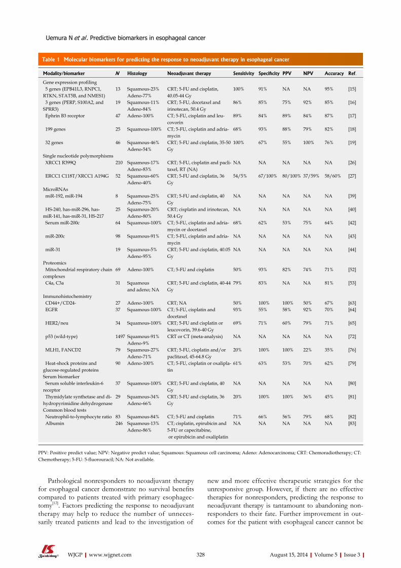
Table 1 Molecular biomarkers for predicting the response to neoadjuvant therapy in esophageal cancer
Modality/biomarker N Histology Neoadjuvant therapy Sensitivity Specificity PPV NPV Accuracy Ref.
Gene expression profiling 5 genes (EPB41L3, RNPC1, RTKN, STAT5B, and NMES1)
13 Squamous-23% Adeno-77%
CRT; 5-FU and cisplatin, 40.05-44 Gy
100% 91% NA NA 95% [15]
3 genes (PERP, S100A2, and SPRR3)
19 Squamous-11% Adeno-84%
CRT; 5-FU, docetaxel and irinotecan, 50.4 Gy
86% 85% 75% 92% 85% [16]
Ephrin B3 receptor 47 Adeno-100% CT; 5-FU, cisplatin and leu-covorin
89% 84% 89% 84% 87% [17]
199 genes 25 Squamous-100% CT; 5-FU, cisplatin and adria-mycin
68% 93% 88% 79% 82% [18]
32 genes 46 Squamous-46% Adeno-54%
CRT; 5-FU and cisplatin, 35-50 Gy
100% 67% 55% 100% 76% [19]
Single nucleotide polymorphisms XRCC1 R399Q 210 Squamous-17%
Adeno-83%CRT; 5-FU, cisplatin and pacli-taxel, RT (NA)
NA NA NA NA NA [26]
ERCC1 C118T/XRCC1 A194G 52 Squamous-60% Adeno-40%
CRT; 5-FU and cisplatin, 36 Gy
54/5% 67/100% 80/100% 37/59% 58/60% [27]
MicroRNAs miR-192, miR-194 8 Squamous-25%
Adeno-75%CRT; 5-FU and cisplatin, 40 Gy
NA NA NA NA NA [39]
HS-240, has-miR-296, has-miR-141, has-miR-31, HS-217
25 Squamous-20% Adeno-80%
CRT; cisplatin and irinotecan, 50.4 Gy
NA NA NA NA NA [40]
Serum miR-200c 64 Squamous-100% CT; 5-FU, cisplatin and adria-mycin or docetaxel
68% 62% 53% 75% 64% [42]
miR-200c 98 Squamous-91% CT; 5-FU, cisplatin and adria-mycin
NA NA NA NA NA [43]
miR-31 19 Squamous-5% Adeno-95%
CRT; 5-FU and cisplatin, 40.05 Gy
NA NA NA NA NA [44]
Proteomics Mitochondrial respiratory chain complexes
69 Adeno-100% CT; 5-FU and cisplatin 50% 93% 82% 74% 71% [52]
C4a, C3a 31 Squamous and adeno; NA
CRT; 5-FU and cisplatin, 40-44 Gy
79% 83% NA NA 81% [53]
Immunohistochemistry CD44+/CD24- 27 Adeno-100% CRT; NA 50% 100% 100% 50% 67% [63] EGFR 37 Squamous-100% CT; 5-FU, cisplatin and
docetaxel93% 55% 58% 92% 70% [64]
HER2/neu 34 Squamous-100% CRT; 5-FU and cisplatin or leucovorin, 39.6-40 Gy
69% 71% 60% 79% 71% [65]
p53 (wild-type) 1497 Squamous-91% Adeno-9%
CRT or CT (meta-analysis) NA NA NA NA NA [72]
MLH1, FANCD2 79 Squamous-27% Adeno-71%
CRT; 5-FU, cisplatin and/or paclitaxel, 45-64.8 Gy
20% 100% 100% 22% 35% [76]
Heat-shock proteins and glucose-regulated proteins
90 Adeno-100% CT; 5-FU, cisplatin or oxalipla-tin
61% 63% 53% 70% 62% [79]
Serum biomarker Serum soluble interleukin-6 receptor
37 Squamous-100% CRT; 5-FU and cisplatin, 40 Gy
NA NA NA NA NA [80]
Thymidylate synthetase and di-hydropyrimidine dehydrogenase
29 Squamous-34%Adeno-66%
CRT; 5-FU and cisplatin, 36 Gy
20% 100% 100% 36% 45% [81]
Common blood tests Neutrophil-to-lymphocyte ratio 83 Squamous-84% CT; 5-FU and cisplatin 71% 66% 56% 79% 68% [82] Albumin 246 Squamous-13%
Adeno-86%CT; cisplatin, epirubicin and 5-FU or capecitabine, or epirubicin and oxaliplatin
NA NA NA NA NA [83]
PPV: Positive predict value; NPV: Negative predict value; Squamous: Squamous cell carcinoma; Adeno: Adenocarcinoma; CRT: Chemoradiotherapy; CT: Chemotherapy; 5-FU: 5-fluorouracil; NA: Not available.
Pathological nonresponders to neoadjuvant therapy for esophageal cancer demonstrate no survival benefits compared to patients treated with primary esophagec-tomy[12]. Factors predicting the response to neoadjuvant therapy may help to reduce the number of unneces-sarily treated patients and lead to the investigation of
new and more effective therapeutic strategies for the unresponsive group. However, if there are no effective therapies for nonresponders, predicting the response to neoadjuvant therapy is tantamount to abandoning non-responders to their fate. Further improvement in out-comes for the patient with esophageal cancer cannot be
August 15, 2014|Volume 5|Issue 3|WJGP|www.wjgnet.com 328
Uemura N et al . Predictive biomarkers in esophageal cancer
achieved without improvement of the prognosis of non-responders. Therefore, the development of new effective therapies for nonresponders concurrently with progress in predictive methodology is necessary. Recently, novel therapeutic approaches, such as new targeted strategies, epigenetic therapeutics, monoclonal antibody therapy and carbon-ion radiotherapy, are being developed[104-105]. Although initially many of these studies involved pa-tients with metastatic disease, these therapies are now being increasingly investigated in the preoperative setting as components of multimodality therapy[104]. The effi-cacy of targeted agents for neoadjuvant therapy of pa-tients with esophageal cancer has yet to be established in previous and ongoing clinical trials[105]. Additional trials to examine new targeted agents have been performed. Further improvement of the prognosis of esophageal cancer patients can be achieved through the introduction of these novel therapeutic approaches in practice, which provides prognostic improvement for nonresponders identified by predictive biomarkers.
CLINICAL APPLICATION OF BIOMARK-ERSAdvances in modern omics technologies and the integra-tion of the results into clinical practice provide valuable opportunities for biomarker discovery research. As dis-cussed in this review, considerable numbers of promising biomarkers in esophageal cancer have been established and more biomarker candidates are likely to be identi-fied by the application of novel technologies. These biomarkers have been discovered through a hypothesis-driven approach by medical doctors for specific clinical applications and they seem to have great potential in providing benefits to patients. However, only a few of the biomarkers discovered in the last decade have been introduced into clinical practice and skepticism about the clinical utility of biomarkers in the diagnosis and treatment of cancer has been expressed[106]. As discussed here, treatments based on the results of biomarker stud-ies should be further developed to benefit all patient subgroups. To establish the reliability of biomarkers before clinical trials, the reproducibility of the results should be assessed by independent investigators. How-ever, we found that none of the biomarkers reviewed in this article had been validated by other researchers. Small sample sizes may be the most serious obstacle for valida-tion of predictive biomarkers. Although it is generally accepted that multi-institutional and inter-disciplinary collaboration is required for biomarker validation, until now no serious validation studies have been performed for any predictive biomarkers in esophageal cancer and this issue requires further analysis.
CONCLUSIONWe have reviewed the current status of biomarkers in esophageal cancer, especially focusing on the utility for
predicting responses to neoadjuvant therapy. The report-ed biomarkers seem to be promising because they have been developed based on clinical research and their pre-dictive performance has been examined by using clinical samples. Further validation and functional evaluation will increase the reliability of these biomarkers. Combined use of the reported biomarkers may increase prognostic performance and this concept is worth further research. Prognostic modalities should be tailored to specific clinical therapeutic approaches that differ according to individual cases. The development of new effective therapies for nonresponders can be hoped for with the progress in predictive techniques. Further understanding of the molecular mechanisms underlying the resistance to CRT in cancers can be achieved by investigating the functional effects of biomarkers on the malignant prop-erties of tumor cells and such efforts will pave the way to novel therapeutic strategies.
REFERENCES1 Jemal A, Bray F, Center MM, Ferlay J, Ward E, Forman
D. Global cancer statistics. CA Cancer J Clin 2011; 61: 69-90 [PMID: 21296855 DOI: 10.3322/caac.20107]
2 Rice TW, Rusch VW, Apperson-Hansen C, Allen MS, Chen LQ, Hunter JG, Kesler KA, Law S, Lerut TE, Reed CE, Salo JA, Scott WJ, Swisher SG, Watson TJ, Blackstone EH. Worldwide esophageal cancer collaboration. Dis Esophagus 2009; 22: 1-8 [PMID: 19196264 DOI: 10.1111/j.1442-2050.2008.00901.x]
3 Urschel JD, Vasan H. A meta-analysis of randomized con-trolled trials that compared neoadjuvant chemoradiation and surgery to surgery alone for resectable esophageal cancer. Am J Surg 2003; 185: 538-543 [PMID: 12781882 DOI: 10.1016/S0002-9610(03)00066-7]
4 Kaklamanos IG, Walker GR, Ferry K, Franceschi D, Living-stone AS. Neoadjuvant treatment for resectable cancer of the esophagus and the gastroesophageal junction: a meta-analysis of randomized clinical trials. Ann Surg Oncol 2003; 10: 754-761 [PMID: 12900366]
5 Malthaner RA, Wong RK, Rumble RB, Zuraw L. Neoadju-vant or adjuvant therapy for resectable esophageal cancer: a systematic review and meta-analysis. BMC Med 2004; 2: 35 [PMID: 15447788 DOI: 10.1186/1741-7015-2-35]
6 Fiorica F, Di Bona D, Schepis F, Licata A, Shahied L, Venturi A, Falchi AM, Craxì A, Cammà C. Preoperative chemora-diotherapy for oesophageal cancer: a systematic review and meta-analysis. Gut 2004; 53: 925-930 [PMID: 15194636]
7 Greer SE, Goodney PP, Sutton JE, Birkmeyer JD. Neoadju-vant chemoradiotherapy for esophageal carcinoma: a meta-analysis. Surgery 2005; 137: 172-177 [PMID: 15674197 DOI: 10.1016/j.surg.2004.06.033]
8 Gebski V, Burmeister B, Smithers BM, Foo K, Zalcberg J, Simes J. Survival benefits from neoadjuvant chemoradio-therapy or chemotherapy in oesophageal carcinoma: a meta-analysis. Lancet Oncol 2007; 8: 226-234 [PMID: 17329193 DOI: 10.1016/S1470-2045(07)70039-6]
9 Xu XH, Peng XH, Yu P, Xu XY, Cai EH, Guo P, Li K. Neoad-juvant chemotherapy for resectable esophageal carcinoma: a meta-analysis of randomized clinical trials. Asian Pac J Can-cer Prev 2012; 13: 103-110 [PMID: 22502650]
10 Nguyen NP, Krafft SP, Vinh-Hung V, Vos P, Almeida F, Jang S, Ceizyk M, Desai A, Davis R, Hamilton R, Modar-resifar H, Abraham D, Smith-Raymond L. Feasibility of tomotherapy to reduce normal lung and cardiac toxicity for distal esophageal cancer compared to three-dimensional
August 15, 2014|Volume 5|Issue 3|WJGP|www.wjgnet.com 329
Uemura N et al . Predictive biomarkers in esophageal cancer
radiotherapy. Radiother Oncol 2011; 101: 438-442 [PMID: 21908064 DOI: 10.1016/j.radonc.2011.07.015]
11 Blencowe NS, McNair AG, Davis CR, Brookes ST, Blazeby JM. Standards of outcome reporting in surgical oncology: a case study in esophageal cancer. Ann Surg Oncol 2012; 19: 4012-4018 [PMID: 22820935 DOI: 10.1245/s10434-012-2497-x]
12 Dittrick GW, Weber JM, Shridhar R, Hoffe S, Melis M, Alm-hanna K, Barthel J, McLoughlin J, Karl RC, Meredith KL. Pathologic nonresponders after neoadjuvant chemoradia-tion for esophageal cancer demonstrate no survival benefit compared with patients treated with primary esophagec-tomy. Ann Surg Oncol 2012; 19: 1678-1684 [PMID: 22045465 DOI: 10.1245/s10434-011-2078-4]
13 Vallböhmer D, Hölscher AH, DeMeester S, DeMeester T, Salo J, Peters J, Lerut T, Swisher SG, Schröder W, Bollschweiler E, Hofstetter W. A multicenter study of sur-vival after neoadjuvant radiotherapy/chemotherapy and esophagectomy for ypT0N0M0R0 esophageal cancer. Ann Surg 2010; 252: 744-749 [PMID: 21037429 DOI: 10.1097/SLA.0b013e3181fb8dde]
14 Quackenbush J. Microarray analysis and tumor classifica-tion. N Engl J Med 2006; 354: 2463-2472 [PMID: 16760446 DOI: 10.1056/NEJMra042342]
15 Maher SG, Gillham CM, Duggan SP, Smyth PC, Miller N, Muldoon C, O’Byrne KJ, Sheils OM, Hollywood D, Reyn-olds JV. Gene expression analysis of diagnostic biopsies predicts pathological response to neoadjuvant chemoradio-therapy of esophageal cancer. Ann Surg 2009; 250: 729-737 [PMID: 19801928 DOI: 10.1097/SLA.0b013e3181bce7e1]
16 Luthra R, Wu TT, Luthra MG, Izzo J, Lopez-Alvarez E, Zhang L, Bailey J, Lee JH, Bresalier R, Rashid A, Swisher SG, Ajani JA. Gene expression profiling of localized esopha-geal carcinomas: association with pathologic response to preoperative chemoradiation. J Clin Oncol 2006; 24: 259-267 [PMID: 16344314 DOI: 10.1200/JCO.2005.03.3688]
17 Schauer M, Janssen KP, Rimkus C, Raggi M, Feith M, Friess H, Theisen J. Microarray-based response predic-tion in esophageal adenocarcinoma. Clin Cancer Res 2010; 16: 330-337 [PMID: 20028767 DOI: 10.1158/1078-0432.CCR-09-1673]
18 Motoori M, Takemasa I, Yamasaki M, Komori T, Takeno A, Miyata H, Takiguchi S, Fujiwara Y, Yasuda T, Yano M, Matsuura N, Matsubara K, Monden M, Mori M, Doki Y. Prediction of the response to chemotherapy in advanced esophageal cancer by gene expression profiling of biopsy samples. Int J Oncol 2010; 37: 1113-1120 [PMID: 20878059 DOI: 10.3892/ijo_00000763]
19 Duong C, Greenawalt DM, Kowalczyk A, Ciavarella ML, Raskutti G, Murray WK, Phillips WA, Thomas RJ. Pre-treatment gene expression profiles can be used to predict response to neoadjuvant chemoradiotherapy in esophageal cancer. Ann Surg Oncol 2007; 14: 3602-3609 [PMID: 17896157 DOI: 10.1245/s10434-007-9550-1]
20 van ‘t Veer LJ, Dai H, van de Vijver MJ, He YD, Hart AA, Mao M, Peterse HL, van der Kooy K, Marton MJ, Witteveen AT, Schreiber GJ, Kerkhoven RM, Roberts C, Linsley PS, Bernards R, Friend SH. Gene expression profiling predicts clinical outcome of breast cancer. Nature 2002; 415: 530-536 [PMID: 11823860 DOI: 10.1038/415530a]
21 Paik S, Shak S, Tang G, Kim C, Baker J, Cronin M, Baehner FL, Walker MG, Watson D, Park T, Hiller W, Fisher ER, Wickerham DL, Bryant J, Wolmark N. A multigene assay to predict recurrence of tamoxifen-treated, node-negative breast cancer. N Engl J Med 2004; 351: 2817-2826 [PMID: 15591335 DOI: 10.1056/NEJMoa041588]
22 Lander ES, Linton LM, Birren B, Nusbaum C, Zody MC, Baldwin J, Devon K, Dewar K, Doyle M, FitzHugh W, Funke R, Gage D, Harris K, Heaford A, Howland J, Kann L, Lehoc-zky J, LeVine R, McEwan P, McKernan K, Meldrim J, Mesi-
rov JP, Miranda C, Morris W, Naylor J, Raymond C, Rosetti M, Santos R, Sheridan A, Sougnez C, Stange-Thomann N, Stojanovic N, Subramanian A, Wyman D, Rogers J, Sulston J, Ainscough R, Beck S, Bentley D, Burton J, Clee C, Carter N, Coulson A, Deadman R, Deloukas P, Dunham A, Dunham I, Durbin R, French L, Grafham D, Gregory S, Hubbard T, Humphray S, Hunt A, Jones M, Lloyd C, McMurray A, Matthews L, Mercer S, Milne S, Mullikin JC, Mungall A, Plumb R, Ross M, Shownkeen R, Sims S, Waterston RH, Wilson RK, Hillier LW, McPherson JD, Marra MA, Mardis ER, Fulton LA, Chinwalla AT, Pepin KH, Gish WR, Chis-soe SL, Wendl MC, Delehaunty KD, Miner TL, Delehaunty A, Kramer JB, Cook LL, Fulton RS, Johnson DL, Minx PJ, Clifton SW, Hawkins T, Branscomb E, Predki P, Richardson P, Wenning S, Slezak T, Doggett N, Cheng JF, Olsen A, Lu-cas S, Elkin C, Uberbacher E, Frazier M, Gibbs RA, Muzny DM, Scherer SE, Bouck JB, Sodergren EJ, Worley KC, Rives CM, Gorrell JH, Metzker ML, Naylor SL, Kucherlapati RS, Nelson DL, Weinstock GM, Sakaki Y, Fujiyama A, Hattori M, Yada T, Toyoda A, Itoh T, Kawagoe C, Watanabe H, Totoki Y, Taylor T, Weissenbach J, Heilig R, Saurin W, Arti-guenave F, Brottier P, Bruls T, Pelletier E, Robert C, Wincker P, Smith DR, Doucette-Stamm L, Rubenfield M, Weinstock K, Lee HM, Dubois J, Rosenthal A, Platzer M, Nyakatura G, Taudien S, Rump A, Yang H, Yu J, Wang J, Huang G, Gu J, Hood L, Rowen L, Madan A, Qin S, Davis RW, Federspiel NA, Abola AP, Proctor MJ, Myers RM, Schmutz J, Dickson M, Grimwood J, Cox DR, Olson MV, Kaul R, Raymond C, Shimizu N, Kawasaki K, Minoshima S, Evans GA, Atha-nasiou M, Schultz R, Roe BA, Chen F, Pan H, Ramser J, Lehrach H, Reinhardt R, McCombie WR, de la Bastide M, Dedhia N, Blocker H, Hornischer K, Nordsiek G, Agarwala R, Aravind L, Bailey JA, Bateman A, Batzoglou S, Birney E, Bork P, Brown DG, Burge CB, Cerutti L, Chen HC, Church D, Clamp M, Copley RR, Doerks T, Eddy SR, Eichler EE, Furey TS, Galagan J, Gilbert JG, Harmon C, Hayashizaki Y, Haussler D, Hermjakob H, Hokamp K, Jang W, Johnson LS, Jones TA, Kasif S, Kaspryzk A, Kennedy S, Kent WJ, Kitts P, Koonin EV, Korf I, Kulp D, Lancet D, Lowe TM, McLysaght A, Mikkelsen T, Moran JV, Mulder N, Pollara VJ, Ponting CP, Schuler G, Schultz J, Slater G, Smit AF, Stupka E, Szus-takowski J, Thierry-Mieg D, Thierry-Mieg J, Wagner L, Wal-lis J, Wheeler R, Williams A, Wolf YI, Wolfe KH, Yang SP, Yeh RF, Collins F, Guyer MS, Peterson J, Felsenfeld A, Wet-terstrand KA, Patrinos A, Morgan MJ, de Jong P, Catanese JJ, Osoegawa K, Shizuya H, Choi S, Chen YJ; International Human Genome Sequencing C. Initial sequencing and ana-lysis of the human genome. Nature 2001; 409: 860-921 [PMID: 11237011 DOI: 10.1038/35057062]
23 Venter JC, Adams MD, Myers EW, Li PW, Mural RJ, Sutton GG, Smith HO, Yandell M, Evans CA, Holt RA, Gocayne JD, Amanatides P, Ballew RM, Huson DH, Wortman JR, Zhang Q, Kodira CD, Zheng XH, Chen L, Skupski M, Subramanian G, Thomas PD, Zhang J, Gabor Miklos GL, Nelson C, Broder S, Clark AG, Nadeau J, McKusick VA, Zinder N, Levine AJ, Roberts RJ, Simon M, Slayman C, Hunkapiller M, Bolanos R, Delcher A, Dew I, Fasulo D, Flanigan M, Florea L, Halpern A, Hannenhalli S, Kravitz S, Levy S, Mobarry C, Reinert K, Re-mington K, Abu-Threideh J, Beasley E, Biddick K, Bonazzi V, Brandon R, Cargill M, Chandramouliswaran I, Charlab R, Chaturvedi K, Deng Z, Di Francesco V, Dunn P, Eilbeck K, Evangelista C, Gabrielian AE, Gan W, Ge W, Gong F, Gu Z, Guan P, Heiman TJ, Higgins ME, Ji RR, Ke Z, Ketchum KA, Lai Z, Lei Y, Li Z, Li J, Liang Y, Lin X, Lu F, Merkulov GV, Milshina N, Moore HM, Naik AK, Narayan VA, Neelam B, Nusskern D, Rusch DB, Salzberg S, Shao W, Shue B, Sun J, Wang Z, Wang A, Wang X, Wang J, Wei M, Wides R, Xiao C, Yan C, Yao A, Ye J, Zhan M, Zhang W, Zhang H, Zhao Q, Zheng L, Zhong F, Zhong W, Zhu S, Zhao S, Gilbert D, Baumhueter S, Spier G, Carter C, Cravchik A, Woodage T,
August 15, 2014|Volume 5|Issue 3|WJGP|www.wjgnet.com 330
Uemura N et al . Predictive biomarkers in esophageal cancer
Ali F, An H, Awe A, Baldwin D, Baden H, Barnstead M, Barrow I, Beeson K, Busam D, Carver A, Center A, Cheng ML, Curry L, Danaher S, Davenport L, Desilets R, Dietz S, Dodson K, Doup L, Ferriera S, Garg N, Gluecksmann A, Hart B, Haynes J, Haynes C, Heiner C, Hladun S, Hostin D, Houck J, Howland T, Ibegwam C, Johnson J, Kalush F, Kline L, Koduru S, Love A, Mann F, May D, McCawley S, McIntosh T, McMullen I, Moy M, Moy L, Murphy B, Nelson K, Pfannkoch C, Pratts E, Puri V, Qureshi H, Reardon M, Rodriguez R, Rogers YH, Romblad D, Ruhfel B, Scott R, Sit-ter C, Smallwood M, Stewart E, Strong R, Suh E, Thomas R, Tint NN, Tse S, Vech C, Wang G, Wetter J, Williams S, Wil-liams M, Windsor S, Winn-Deen E, Wolfe K, Zaveri J, Zaveri K, Abril JF, Guigo R, Campbell MJ, Sjolander KV, Karlak B, Kejariwal A, Mi H, Lazareva B, Hatton T, Narechania A, Diemer K, Muruganujan A, Guo N, Sato S, Bafna V, Istrail S, Lippert R, Schwartz R, Walenz B, Yooseph S, Allen D, Basu A, Baxendale J, Blick L, Caminha M, Carnes-Stine J, Caulk P, Chiang YH, Coyne M, Dahlke C, Mays A, Dombroski M, Donnelly M, Ely D, Esparham S, Fosler C, Gire H, Glanow-ski S, Glasser K, Glodek A, Gorokhov M, Graham K, Grop-man B, Harris M, Heil J, Henderson S, Hoover J, Jennings D, Jordan C, Jordan J, Kasha J, Kagan L, Kraft C, Levitsky A, Lewis M, Liu X, Lopez J, Ma D, Majoros W, McDaniel J, Murphy S, Newman M, Nguyen T, Nguyen N, Nodell M, Pan S, Peck J, Peterson M, Rowe W, Sanders R, Scott J, Simp-son M, Smith T, Sprague A, Stockwell T, Turner R, Venter E, Wang M, Wen M, Wu D, Wu M, Xia A, Zandieh A, Zhu X. The sequence of the human genome. Science 2001; 291: 1304-1351 [PMID: 11181995 DOI: 10.1126/science.1058040]
24 Risch NJ. Searching for genetic determinants in the new millennium. Nature 2000; 405: 847-856 [PMID: 10866211 DOI: 10.1038/35015718]
25 Glinsky GV. Integration of HapMap-based SNP pattern analysis and gene expression profiling reveals common SNP profiles for cancer therapy outcome predictor genes. Cell Cycle 2006; 5: 2613-2625 [PMID: 17172834]
26 Wu X, Gu J, Wu TT, Swisher SG, Liao Z, Correa AM, Liu J, Etzel CJ, Amos CI, Huang M, Chiang SS, Milas L, Hittelman WN, Ajani JA. Genetic variations in radiation and chemo-therapy drug action pathways predict clinical outcomes in esophageal cancer. J Clin Oncol 2006; 24: 3789-3798 [PMID: 16785472 DOI: 10.1200/JCO.2005.03.6640]
27 Warnecke-Eberz U, Vallböhmer D, Alakus H, Kütting F, Lurje G, Bollschweiler E, Wienand-Dorweiler A, Dreb-ber U, Hölscher AH, Metzger R. ERCC1 and XRCC1 gene polymorphisms predict response to neoadjuvant radioche-motherapy in esophageal cancer. J Gastrointest Surg 2009; 13: 1411-1421 [PMID: 19421825 DOI: 10.1007/s11605-009-0881-z]
28 Houtsmuller AB, Rademakers S, Nigg AL, Hoogstraten D, Hoeijmakers JH, Vermeulen W. Action of DNA repair endonuclease ERCC1/XPF in living cells. Science 1999; 284: 958-961 [PMID: 10320375]
29 Kim MK, Cho KJ, Kwon GY, Park SI, Kim YH, Kim JH, Song HY, Shin JH, Jung HY, Lee GH, Choi KD, Kim SB. ERCC1 predicting chemoradiation resistance and poor outcome in oesophageal cancer. Eur J Cancer 2008; 44: 54-60 [PMID: 17976974 DOI: 10.1016/j.ejca.2007.09.006]
30 Schneider S, Uchida K, Brabender J, Baldus SE, Yochim J, Danenberg KD, Salonga D, Chen P, Tsao-Wei D, Groshen S, Hoelscher AH, Schneider PM, Danenberg PV. Downregu-lation of TS, DPD, ERCC1, GST-Pi, EGFR, and HER2 gene expression after neoadjuvant three-modality treatment in patients with esophageal cancer. J Am Coll Surg 2005; 200: 336-344 [PMID: 15737843 DOI: 10.1016/j.jamcollsurg.2004.10.035]
31 Brabender J, Vallböhmer D, Grimminger P, Hoffmann AC, Ling F, Lurje G, Bollschweiler E, Schneider PM, Hölscher AH, Metzger R. ERCC1 RNA expression in peripheral blood
predicts minor histopathological response to neoadjuvant radio-chemotherapy in patients with locally advanced can-cer of the esophagus. J Gastrointest Surg 2008; 12: 1815-1821 [PMID: 18769985 DOI: 10.1007/s11605-008-0668-7]
32 Ando Y, Saka H, Ando M, Sawa T, Muro K, Ueoka H, Yo-koyama A, Saitoh S, Shimokata K, Hasegawa Y. Polymor-phisms of UDP-glucuronosyltransferase gene and irinotecan toxicity: a pharmacogenetic analysis. Cancer Res 2000; 60: 6921-6926 [PMID: 11156391]
33 Minami H, Sai K, Saeki M, Saito Y, Ozawa S, Suzuki K, Kaniwa N, Sawada J, Hamaguchi T, Yamamoto N, Shirao K, Yamada Y, Ohmatsu H, Kubota K, Yoshida T, Ohtsu A, Saijo N. Irinotecan pharmacokinetics/pharmacodynam-ics and UGT1A genetic polymorphisms in Japanese: roles of UGT1A1*6 and *28. Pharmacogenet Genomics 2007; 17: 497-504 [PMID: 17558305]
34 Bhatti I, Lee A, Lund J, Larvin M. Small RNA: a large contributor to carcinogenesis? J Gastrointest Surg 2009; 13: 1379-1388 [PMID: 19373515 DOI: 10.1007/s11605-009-0887-6]
35 Guo Y, Chen Z, Zhang L, Zhou F, Shi S, Feng X, Li B, Meng X, Ma X, Luo M, Shao K, Li N, Qiu B, Mitchelson K, Cheng J, He J. Distinctive microRNA profiles relating to patient survival in esophageal squamous cell carcinoma. Cancer Res 2008; 68: 26-33 [PMID: 18172293 DOI: 10.1158/0008-5472.CAN-06-4418]
36 Hu Y, Correa AM, Hoque A, Guan B, Ye F, Huang J, Swish-er SG, Wu TT, Ajani JA, Xu XC. Prognostic significance of differentially expressed miRNAs in esophageal cancer. Int J Cancer 2011; 128: 132-143 [PMID: 20309880 DOI: 10.1002/ijc.25330]
37 Mathé EA, Nguyen GH, Bowman ED, Zhao Y, Budhu A, Schetter AJ, Braun R, Reimers M, Kumamoto K, Hughes D, Altorki NK, Casson AG, Liu CG, Wang XW, Yanaihara N, Hagiwara N, Dannenberg AJ, Miyashita M, Croce CM, Harris CC. MicroRNA expression in squamous cell carci-noma and adenocarcinoma of the esophagus: associations with survival. Clin Cancer Res 2009; 15: 6192-6200 [PMID: 19789312 DOI: 10.1158/1078-0432.CCR-09-1467]
38 Akagi I, Miyashita M, Ishibashi O, Mishima T, Kikuchi K, Makino H, Nomura T, Hagiwara N, Uchida E, Takizawa T. Relationship between altered expression levels of MIR21, MIR143, MIR145, and MIR205 and clinicopatho-logic features of esophageal squamous cell carcinoma. Dis Esophagus 2011; 24: 523-530 [PMID: 21453382 DOI: 10.1111/j.1442-2050.2011.01177.x]
39 Odenthal M, Bollschweiler E, Grimminger PP, Schröder W, Brabender J, Drebber U, Hölscher AH, Metzger R, Vallböh-mer D. MicroRNA profiling in locally advanced esophageal cancer indicates a high potential of miR-192 in prediction of multimodality therapy response. Int J Cancer 2013; 133: 2454-2463 [PMID: 23649428 DOI: 10.1002/ijc.28253]
40 Ko MA, Zehong G, Virtanen C, Guindi M, Waddell TK, Keshavjee S, Darling GE. MicroRNA expression profiling of esophageal cancer before and after induction chemora-diotherapy. Ann Thorac Surg 2012; 94: 1094-1102; discussion 1102-1103 [PMID: 22939244 DOI: 10.1016/j.athoracsur.2012.04.145]
41 Hong L, Han Y, Zhang H, Li M, Gong T, Sun L, Wu K, Zhao Q, Fan D. The prognostic and chemotherapeutic value of miR-296 in esophageal squamous cell carcinoma. Ann Surg 2010; 251: 1056-1063 [PMID: 20485139 DOI: 10.1097/SLA.0b013e3181dd4ea9]
42 Tanaka K, Miyata H, Yamasaki M, Sugimura K, Takahashi T, Kurokawa Y, Nakajima K, Takiguchi S, Mori M, Doki Y. Circulating miR-200c levels significantly predict response to chemotherapy and prognosis of patients undergoing neoad-juvant chemotherapy for esophageal cancer. Ann Surg Oncol 2013; 20 Suppl 3: S607-S615 [PMID: 23838916 DOI: 10.1245/s10434-013-3093-4]
43 Hamano R, Miyata H, Yamasaki M, Kurokawa Y, Hara J,
August 15, 2014|Volume 5|Issue 3|WJGP|www.wjgnet.com 331
Uemura N et al . Predictive biomarkers in esophageal cancer
Moon JH, Nakajima K, Takiguchi S, Fujiwara Y, Mori M, Doki Y. Overexpression of miR-200c induces chemore-sistance in esophageal cancers mediated through activa-tion of the Akt signaling pathway. Clin Cancer Res 2011; 17: 3029-3038 [PMID: 21248297 DOI: 10.1158/1078-0432.CCR-10-2532]
44 Lynam-Lennon N, Reynolds JV, Marignol L, Sheils OM, Pidgeon GP, Maher SG. MicroRNA-31 modulates tumour sensitivity to radiation in oesophageal adenocarcinoma. J Mol Med (Berl) 2012; 90: 1449-1458 [PMID: 22706599 DOI: 10.1007/s00109-012-0924-x]
45 Zhang J, Zhao H, Gao Y, Zhang W. Secretory miRNAs as novel cancer biomarkers. Biochim Biophys Acta 2012; 1826: 32-43 [PMID: 22440944 DOI: 10.1016/j.bbcan.2012.03.001]
46 Chen G, Gharib TG, Huang CC, Taylor JM, Misek DE, Kar-dia SL, Giordano TJ, Iannettoni MD, Orringer MB, Hanash SM, Beer DG. Discordant protein and mRNA expression in lung adenocarcinomas. Mol Cell Proteomics 2002; 1: 304-313 [PMID: 12096112]
47 Varambally S, Yu J, Laxman B, Rhodes DR, Mehra R, Tom-lins SA, Shah RB, Chandran U, Monzon FA, Becich MJ, Wei JT, Pienta KJ, Ghosh D, Rubin MA, Chinnaiyan AM. Integra-tive genomic and proteomic analysis of prostate cancer re-veals signatures of metastatic progression. Cancer Cell 2005; 8: 393-406 [PMID: 16286247 DOI: 10.1016/j.ccr.2005.10.001]
48 Gygi SP, Rochon Y, Franza BR, Aebersold R. Correlation between protein and mRNA abundance in yeast. Mol Cell Biol 1999; 19: 1720-1730 [PMID: 10022859]
49 Uemura N, Nakanishi Y, Kato H, Saito S, Nagino M, Hiro-hashi S, Kondo T. Transglutaminase 3 as a prognostic bio-marker in esophageal cancer revealed by proteomics. Int J Cancer 2009; 124: 2106-2115 [PMID: 19142970 DOI: 10.1002/ijc.24194]
50 Hanash SM, Pitteri SJ, Faca VM. Mining the plasma pro-teome for cancer biomarkers. Nature 2008; 452: 571-579 [PMID: 18385731 DOI: 10.1038/nature06916]
51 Cox J, Mann M. Is proteomics the new genomics? Cell 2007; 130: 395-398 [PMID: 17693247 DOI: 10.1016/j.cell.2007.07.032]
52 Aichler M, Elsner M, Ludyga N, Feuchtinger A, Zangen V, Maier SK, Balluff B, Schöne C, Hierber L, Braselmann H, Meding S, Rauser S, Zischka H, Aubele M, Schmitt M, Feith M, Hauck SM, Ueffing M, Langer R, Kuster B, Zitzelsberger H, Höfler H, Walch AK. Clinical response to chemotherapy in oesophageal adenocarcinoma patients is linked to defects in mitochondria. J Pathol 2013; 230: 410-419 [PMID: 23592244 DOI: 10.1002/path.4199]
53 Maher SG, McDowell DT, Collins BC, Muldoon C, Gallagh-er WM, Reynolds JV. Serum proteomic profiling reveals that pretreatment complement protein levels are predictive of esophageal cancer patient response to neoadjuvant chemo-radiation. Ann Surg 2011; 254: 809-816; discussion 816-817 [PMID: 22005152 DOI: 10.1097/SLA.0b013e31823699f2]
54 Fung ET. A recipe for proteomics diagnostic test develop-ment: the OVA1 test, from biomarker discovery to FDA clearance. Clin Chem 2010; 56: 327-329 [PMID: 20110452 DOI: 10.1373/clinchem.2009.140855]
55 Kondo T, Suehara Y, Kikuta K, Kubota D, Tajima T, Mukai-hara K, Ichikawa H, Kawai A. Proteomic approach toward personalized sarcoma treatment: lessons from prognostic biomarker discovery in gastrointestinal stromal tumor. Proteomics Clin Appl 2013; 7: 70-78 [PMID: 23281253 DOI: 10.1002/prca.201200085]
56 Taylor CR, Levenson RM. Quantification of immunohis-tochemistry--issues concerning methods, utility and semi-quantitative assessment II. Histopathology 2006; 49: 411-424 [PMID: 16978205 DOI: 10.1111/j.1365-2559.2006.02513.x]
57 Al-Hajj M, Wicha MS, Benito-Hernandez A, Morrison SJ, Clarke MF. Prospective identification of tumorigenic breast cancer cells. Proc Natl Acad Sci USA 2003; 100: 3983-3988 [PMID: 12629218 DOI: 10.1073/pnas.0530291100]
58 Zhu L, Gibson P, Currle DS, Tong Y, Richardson RJ, Bayazi-tov IT, Poppleton H, Zakharenko S, Ellison DW, Gilbertson RJ. Prominin 1 marks intestinal stem cells that are suscep-tible to neoplastic transformation. Nature 2009; 457: 603-607 [PMID: 19092805 DOI: 10.1038/nature07589]
59 O’Brien CA, Pollett A, Gallinger S, Dick JE. A human colon cancer cell capable of initiating tumour growth in immuno-deficient mice. Nature 2007; 445: 106-110 [PMID: 17122772 DOI: 10.1038/nature05372]
60 Vermeulen L, De Sousa E Melo F, van der Heijden M, Cam-eron K, de Jong JH, Borovski T, Tuynman JB, Todaro M, Merz C, Rodermond H, Sprick MR, Kemper K, Richel DJ, Stassi G, Medema JP. Wnt activity defines colon cancer stem cells and is regulated by the microenvironment. Nat Cell Biol 2010; 12: 468-476 [PMID: 20418870 DOI: 10.1038/ncb2048]
61 Phillips TM, McBride WH, Pajonk F. The response of CD24(-/low)/CD44+ breast cancer-initiating cells to radia-tion. J Natl Cancer Inst 2006; 98: 1777-1785 [PMID: 17179479 DOI: 10.1093/jnci/djj495]
62 Todaro M, Alea MP, Di Stefano AB, Cammareri P, Vermeu-len L, Iovino F, Tripodo C, Russo A, Gulotta G, Medema JP, Stassi G. Colon cancer stem cells dictate tumor growth and resist cell death by production of interleukin-4. Cell Stem Cell 2007; 1: 389-402 [PMID: 18371377 DOI: 10.1016/j.stem.2007.08.001]
63 Smit JK, Faber H, Niemantsverdriet M, Baanstra M, Bussink J, Hollema H, van Os RP, Plukker JT, Coppes RP. Prediction of response to radiotherapy in the treatment of esophageal cancer using stem cell markers. Radiother Oncol 2013; 107: 434-441 [PMID: 23684587 DOI: 10.1016/j.radonc.2013.03.027]
64 Yamamoto Y, Yamai H, Seike J, Yoshida T, Takechi H, Fu-rukita Y, Kajiura K, Minato T, Bando Y, Tangoku A. Prog-nosis of esophageal squamous cell carcinoma in patients positive for human epidermal growth factor receptor family can be improved by initial chemotherapy with docetaxel, fluorouracil, and cisplatin. Ann Surg Oncol 2012; 19: 757-765 [PMID: 21947696 DOI: 10.1245/s10434-011-2071-y]
65 Akamatsu M, Matsumoto T, Oka K, Yamasaki S, Sonoue H, Kajiyama Y, Tsurumaru M, Sasai K. c-erbB-2 oncopro-tein expression related to chemoradioresistance in esopha-geal squamous cell carcinoma. Int J Radiat Oncol Biol Phys 2003; 57: 1323-1327 [PMID: 14630269 DOI: 10.1016/S0360-3016(03)00782-X]
66 Arsenijevic T, Micev M, Nikolic V, Gavrilovic D, Radulovic S, Pesko P. Is there a correlation between molecular markers and response to neoadjuvant chemoradiotherapy in locally advanced squamous cell esophageal cancer? J BUON 2012; 17: 706-711 [PMID: 23335529]
67 Schena M, La Rovere E, Solerio D, Bustreo S, Barone C, Dan-iele L, Buffoni L, Bironzo P, Sapino A, Gasparri G, Ciuffreda L, Ricardi U. Neoadjuvant chemo-radiotherapy for locally ad-vanced esophageal cancer: a monocentric study. Tumori 2012; 98: 451-457 [PMID: 23052161 DOI: 10.1700/1146.12639]
68 Vallböhmer D, Lenz HJ. Predictive and prognostic mo-lecular markers in outcome of esophageal cancer. Dis Esophagus 2006; 19: 425-432 [PMID: 17069584 DOI: 10.1111/j.1442-2050.2006.00622.x]
69 Kitamura K, Saeki H, Kawaguchi H, Araki K, Ohno S, Ku-wano H, Maehara Y, Sugimachi K. Immunohistochemical status of the p53 protein and Ki-67 antigen using biopsied specimens can predict a sensitivity to neoadjuvant therapy in patients with esophageal cancer. Hepatogastroenterology 2000; 47: 419-423 [PMID: 10791203]
70 Shimada Y, Watanabe G, Yamasaki S, Maeda M, Kawabe A, Kaganoi JI, Itami A, Fukumoto M, Kanda Y, Imamura M. Histological response of cisplatin predicts patients’ survival in oesophageal cancer and p53 protein accumulation in pre-treatment biopsy is associated with cisplatin sensitivity. Eur J Cancer 2000; 36: 987-993 [PMID: 10885602]
71 Sarbia M, Ott N, Pühringer-Oppermann F, Brücher BL. The
August 15, 2014|Volume 5|Issue 3|WJGP|www.wjgnet.com 332
Uemura N et al . Predictive biomarkers in esophageal cancer
predictive value of molecular markers (p53, EGFR, ATM, CHK2) in multimodally treated squamous cell carcinoma of the oesophagus. Br J Cancer 2007; 97: 1404-1408 [PMID: 17940507 DOI: 10.1038/sj.bjc.6604037]
72 Zhang SS, Huang QY, Yang H, Xie X, Luo KJ, Wen J, Cai XL, Yang F, Hu Y, Fu JH. Correlation of p53 status with the response to chemotherapy-based treatment in esophageal cancer: a meta-analysis. Ann Surg Oncol 2013; 20: 2419-2427 [PMID: 23515910 DOI: 10.1245/s10434-012-2859-4]
73 Kishi K, Doki Y, Yano M, Yasuda T, Fujiwara Y, Takiguchi S, Kim S, Higuchi I, Monden M. Reduced MLH1 expression after chemotherapy is an indicator for poor prognosis in esophageal cancers. Clin Cancer Res 2003; 9: 4368-4375 [PMID: 14555508]
74 Nam TK, Lee JH, Cho SH, Chung IJ, Ahn SJ, Song JY, Yoon MS, Chung WK, Nah BS. Low hMLH1 expression prior to definitive chemoradiotherapy predicts poor prognosis in esophageal squamous cell carcinoma. Cancer Lett 2008; 260: 109-117 [PMID: 18053639 DOI: 10.1016/j.canlet.2007.10.026]
75 Uehara H, Miyamoto M, Kato K, Cho Y, Kurokawa T, Mu-rakami S, Fukunaga A, Ebihara Y, Kaneko H, Hashimoto H, Murakami Y, Shichinohe T, Kawarada Y, Itoh T, Okushiba S, Kondo S, Katoh H. Deficiency of hMLH1 and hMSH2 expression is a poor prognostic factor in esophageal squa-mous cell carcinoma. J Surg Oncol 2005; 92: 109-115 [PMID: 16231369 DOI: 10.1002/jso.20332]
76 Alexander BM, Wang XZ, Niemierko A, Weaver DT, Mak RH, Roof KS, Fidias P, Wain J, Choi NC. DNA repair bio-markers predict response to neoadjuvant chemoradiothera-py in esophageal cancer. Int J Radiat Oncol Biol Phys 2012; 83: 164-171 [PMID: 22000749 DOI: 10.1016/j.ijrobp.2011.05.033]
77 Langer R, Feith M, Siewert JR, Wester HJ, Hoefler H. Ex-pression and clinical significance of glucose regulated proteins GRP78 (BiP) and GRP94 (GP96) in human adeno-carcinomas of the esophagus. BMC Cancer 2008; 8: 70 [PMID: 18331622 DOI: 10.1186/1471-2407-8-70]
78 Langer R, Ott K, Specht K, Becker K, Lordick F, Burian M, Herrmann K, Schrattenholz A, Cahill MA, Schwaiger M, Hofler H, Wester HJ. Protein expression profiling in esopha-geal adenocarcinoma patients indicates association of heat-shock protein 27 expression and chemotherapy response. Clin Cancer Res 2008; 14: 8279-8287 [PMID: 19088045 DOI: 10.1158/1078-0432.CCR-08-0679]
79 Slotta-Huspenina J, Wolff C, Drecoll E, Feith M, Bettstet-ter M, Malinowsky K, Bauer L, Becker K, Ott K, Höfler H, Becker KF, Langer R. A specific expression profile of heat-shock proteins and glucose-regulated proteins is associated with response to neoadjuvant chemotherapy in oesophageal adenocarcinomas. Br J Cancer 2013; 109: 370-378 [PMID: 23839491 DOI: 10.1038/bjc.2013.319]
80 Makuuchi Y, Honda K, Osaka Y, Kato K, Kojima T, Daiko H, Igaki H, Ito Y, Hoshino S, Tachibana S, Watanabe T, Furuta K, Sekine S, Umaki T, Watabe Y, Miura N, Ono M, Tsuchida A, Yamada T. Soluble interleukin-6 receptor is a serum bio-marker for the response of esophageal carcinoma to neoad-juvant chemoradiotherapy. Cancer Sci 2013; 104: 1045-1051 [PMID: 23648090 DOI: 10.1111/cas.12187]
81 Brabender J, Metzger R, Vallböhmer D, Ling F, Neiss S, Bollschweiler E, Schneider PM, Hölscher AH, Grimminger PP. Roles of thymidylate synthase and dihydropyrimi-dine dehydrogenase expression in blood as predictors of response to multimodal therapy in esophageal cancer. Surgery 2012; 151: 306-312 [PMID: 21982526 DOI: 10.1016/j.surg.2011.07.018]
82 Sato H, Tsubosa Y, Kawano T. Correlation between the pre-therapeutic neutrophil to lymphocyte ratio and the patho-logic response to neoadjuvant chemotherapy in patients with advanced esophageal cancer. World J Surg 2012; 36: 617-622 [PMID: 22223293 DOI: 10.1007/s00268-011-1411-1]
83 Noble F, Hopkins J, Curtis N, Kelly JJ, Bailey IS, Byrne JP,
Bateman AC, Bateman AR, Underwood TJ. The role of sys-temic inflammatory and nutritional blood-borne markers in predicting response to neoadjuvant chemotherapy and survival in oesophagogastric cancer. Med Oncol 2013; 30: 596 [PMID: 23690267 DOI: 10.1007/s12032-013-0596-6]
84 Hsu PK, Chien LI, Huang CS, Hsieh CC, Wu YC, Hsu WH, Chou TY. Comparison of survival among neoadjuvant chemoradiation responders, non-responders and patients receiving primary resection for locally advanced oesopha-geal squamous cell carcinoma: does neoadjuvant chemora-diation benefit all? Interact Cardiovasc Thorac Surg 2013; 17: 460-466 [PMID: 23728085 DOI: 10.1093/icvts/ivt216]
85 Ando N, Kato H, Igaki H, Shinoda M, Ozawa S, Shimizu H, Nakamura T, Yabusaki H, Aoyama N, Kurita A, Ikeda K, Kanda T, Tsujinaka T, Nakamura K, Fukuda H. A random-ized trial comparing postoperative adjuvant chemotherapy with cisplatin and 5-fluorouracil versus preoperative che-motherapy for localized advanced squamous cell carcinoma of the thoracic esophagus (JCOG9907). Ann Surg Oncol 2012; 19: 68-74 [PMID: 21879261 DOI: 10.1245/s10434-011-2049-9]
86 Almhanna K, Shridhar R, Meredith KL. Neoadjuvant or adjuvant therapy for resectable esophageal cancer: is there a standard of care? Cancer Control 2013; 20: 89-96 [PMID: 23571699]
87 Nakamura K, Kato K, Igaki H, Ito Y, Mizusawa J, Ando N, Udagawa H, Tsubosa Y, Daiko H, Hironaka S, Fukuda H, Kitagawa Y. Three-arm phase III trial comparing cisplatin plus 5-FU (CF) versus docetaxel, cisplatin plus 5-FU (DCF) versus radiotherapy with CF (CF-RT) as preoperative therapy for locally advanced esophageal cancer (JCOG1109, NExT study). Jpn J Clin Oncol 2013; 43: 752-755 [PMID: 23625063 DOI: 10.1093/jjco/hyt061]
88 Hötte GJ, Linam-Lennon N, Reynolds JV, Maher SG. Radia-tion sensitivity of esophageal adenocarcinoma: the contribu-tion of the RNA-binding protein RNPC1 and p21-mediated cell cycle arrest to radioresistance. Radiat Res 2012; 177: 272-279 [PMID: 22214381]
89 Kurashige J, Watanabe M, Iwatsuki M, Kinoshita K, Saito S, Nagai Y, Ishimoto T, Baba Y, Mimori K, Baba H. RPN2 expression predicts response to docetaxel in oesophageal squamous cell carcinoma. Br J Cancer 2012; 107: 1233-1238 [PMID: 22955852 DOI: 10.1038/bjc.2012.396]
90 Lepage C, Rachet B, Jooste V, Faivre J, Coleman MP. Con-tinuing rapid increase in esophageal adenocarcinoma in England and Wales. Am J Gastroenterol 2008; 103: 2694-2699 [PMID: 18853967 DOI: 10.1111/j.1572-0241.2008.02191.x]
91 Pohl H, Welch HG. The role of overdiagnosis and reclas-sification in the marked increase of esophageal adenocarci-noma incidence. J Natl Cancer Inst 2005; 97: 142-146 [PMID: 15657344 DOI: 10.1093/jnci/dji024]
92 Merkow RP, Bilimoria KY, McCarter MD, Chow WB, Ko CY, Bentrem DJ. Use of multimodality neoadjuvant therapy for esophageal cancer in the United States: assessment of 987 hospitals. Ann Surg Oncol 2012; 19: 357-364 [PMID: 21769460 DOI: 10.1245/s10434-011-1945-3]
93 Bollschweiler E, Metzger R, Drebber U, Baldus S, Vall-böhmer D, Kocher M, Hölscher AH. Histological type of esophageal cancer might affect response to neo-adjuvant radiochemotherapy and subsequent prognosis. Ann Oncol 2009; 20: 231-238 [PMID: 18836090 DOI: 10.1093/annonc/mdn622]
94 Pennathur A, Luketich JD. Resection for esophageal cancer: strategies for optimal management. Ann Thorac Surg 2008; 85: S751-S756 [PMID: 18222210 DOI: 10.1016/j.athoracsur.2007.11.078]
95 Pennathur A, Zhang J, Chen H, Luketich JD. The “best op-eration” for esophageal cancer? Ann Thorac Surg 2010; 89: S2163-S2167 [PMID: 20494003 DOI: 10.1016/j.athoracsur.2010.03.068]
96 Hagen JA, DeMeester SR, Peters JH, Chandrasoma P, De-
August 15, 2014|Volume 5|Issue 3|WJGP|www.wjgnet.com 333
Uemura N et al . Predictive biomarkers in esophageal cancer
Meester TR. Curative resection for esophageal adenocarci-noma: analysis of 100 en bloc esophagectomies. Ann Surg 2001; 234: 520-530; discussion 530-531 [PMID: 11573045]
97 Orringer MB, Marshall B, Iannettoni MD. Transhiatal esoph-agectomy: clinical experience and refinements. Ann Surg 1999; 230: 392-400; discussion 400-403 [PMID: 10493486]
98 Altorki N, Kent M, Ferrara C, Port J. Three-field lymph node dissection for squamous cell and adenocarcinoma of the esophagus. Ann Surg 2002; 236: 177-183 [PMID: 12170022 DOI: 10.1097/01.SLA.0000021583.51164.F4]
99 Swanson SJ, Batirel HF, Bueno R, Jaklitsch MT, Lukanich JM, Allred E, Mentzer SJ, Sugarbaker DJ. Transthoracic esophagectomy with radical mediastinal and abdominal lymph node dissection and cervical esophagogastros-tomy for esophageal carcinoma. Ann Thorac Surg 2001; 72: 1918-1924; discussion 1918-1924 [PMID: 11789772]
100 Visbal AL, Allen MS, Miller DL, Deschamps C, Trastek VF, Pairolero PC. Ivor Lewis esophagogastrectomy for esopha-geal cancer. Ann Thorac Surg 2001; 71: 1803-1808 [PMID: 11426751]
101 Hiranyatheb P, Osugi H. Radical lymphadenectomy in esophageal cancer: from the past to the present. Dis Esopha-gus 2013 Jun 24; Epub ahead of print [PMID: 23796327 DOI:
10.1111/dote.12091]102 Wong J, Weber J, Almhanna K, Hoffe S, Shridhar R, Karl R,
Meredith KL. Extent of lymphadenectomy does not predict survival in patients treated with primary esophagectomy. J Gastrointest Surg 2013; 17: 1562-1568; discussion 1569 [PMID: 23818125 DOI: 10.1007/s11605-013-2259-5]
103 Stiles BM, Nasar A, Mirza FA, Lee PC, Paul S, Port JL, Al-torki NK. Worldwide Oesophageal Cancer Collaboration guidelines for lymphadenectomy predict survival follow-ing neoadjuvant therapy. Eur J Cardiothorac Surg 2012; 42: 659-664 [PMID: 22491667 DOI: 10.1093/ejcts/ezs105]
104 Akutsu Y, Yasuda S, Nagata M, Izumi Y, Okazumi S, Shi-mada H, Nakatani Y, Tsujii H, Kamada T, Yamada S, Mat-subara H. A phase I/II clinical trial of preoperative short-course carbon-ion radiotherapy for patients with squamous cell carcinoma of the esophagus. J Surg Oncol 2012; 105: 750-755 [PMID: 22012645 DOI: 10.1002/jso.22127]
105 Toomey PG, Vohra NA, Ghansah T, Sarnaik AA, Pilon-Thomas SA. Immunotherapy for gastrointestinal malignan-cies. Cancer Control 2013; 20: 32-42 [PMID: 23302905]
106 Saijo N. Critical comments for roles of biomarkers in the diagnosis and treatment of cancer. Cancer Treat Rev 2012; 38: 63-67 [PMID: 21652149 DOI: 10.1016/j.ctrv.2011.02.004]
P- Reviewer: Goenka MK, Kim GH, Scherer A S- Editor: Ji FF L- Editor: Roemmele A E- Editor: Lu YJ
August 15, 2014|Volume 5|Issue 3|WJGP|www.wjgnet.com 334
Uemura N et al . Predictive biomarkers in esophageal cancer
REVIEW
Epidemiological studies of esophageal cancer in the era of genome-wide association studies
An-Hui Wang, Yuan Liu, Bo Wang, Yi-Xuan He, Ye-Xian Fang, Yong-Ping Yan
An-Hui Wang, Bo Wang, Yong-Ping Yan, Department of Epi-demiology, School of Public Health, Fourth Military Medical University, Xi’an 710032, Shaanxi Province, ChinaYuan Liu, Clinic of Xi’an Communication College, Xi’an 710032, Shaanxi Province, ChinaYi-Xuan He, Ye-Xian Fang, Medical Student of Fourth Mili-tary Medical University, Xi’an 710032, Shaanxi Province, ChinaAuthor contributions: Wang AH contributed to the concep-tion, design, editing and revision of the manuscript; Liu Y, He YX and Fang YX contributed to drafting the article; Wang B and Yan YP contributed to manuscript review and revision.Correspondence to: An-Hui Wang, Associate Professor, Department of Epidemiology, School of Public Health, Fourth Military Medical University, No. 169 Changle West Road, Xi’an 710032, Shaanxi Province, China. [email protected]: +86-29-84774871 Fax: +86-29-84774876Received: January 27, 2014 Revised: April 17, 2014 Accepted: May 31, 2014Published online: August 15, 2014
Abstract Esophageal cancer (EC) caused about 395000 deaths in 2010. China has the most cases of EC and EC is the fourth leading cause of cancer death in China. Esopha-geal squamous cell carcinoma (ESCC) is the predomi-nant histologic type (90%-95%), while the incidence of esophageal adenocarcinoma (EAC) remains extremely low in China. Traditional epidemiological studies have revealed that environmental carcinogens are risk fac-tors for EC. Molecular epidemiological studies revealed that susceptibility to EC is influenced by both environ-mental and genetic risk factors. Of all the risk factors for EC, some are associated with the risk of ESCC and others with the risk of EAC. However, the details and mechanisms of risk factors involved in the process for EC are unclear. The advanced methods and techniques used in human genome studies bring a great opportu-nity for researchers to explore and identify the details of those risk factors or susceptibility genes involved in
the process of EC. Human genome epidemiology is a new branch of epidemiology, which leads the epidemi-ology study from the molecular epidemiology era to the era of genome wide association studies (GWAS). Here we review the epidemiological studies of EC (especially ESCC) in the era of GWAS, and provide an overview of the general risk factors and those genomic variants (genes, SNPs, miRNAs, proteins) involved in the pro-cess of ESCC.
© 2014 Baishideng Publishing Group Inc. All rights reserved.
Key words: Esophageal cancer; Epidemiology; Genome wide association study; Single nucleotide polymor-phism; MicroRNA
Core tip: Epidemiological study methods advance as the science and technique progress. In the era of genome wide association studies (GWAS), human genome epi-demiology (HuGE) provide a great chance for epidemi-ologists and clinical scientists to explore the cause of disease and evaluate genomic biomarkers for diagnosis or prognosis. More and more epidemiological studies use GWAS methods to analyze genomic variants and the association with esophageal cancer. Here we review epidemiological studies of esophageal cancer in the era of GWAS, and briefly introduce the case-control study and cohort study methods in HuGE studies.
Wang AH, Liu Y, Wang B, He YX, Fang YX, Yan YP. Epidemiological studies of esophageal cancer in the era of genome-wide association studies. World J Gastrointest Pathophysiol 2014; 5(3): 335-343 Available from: URL: http://www.wjgnet.com/2150-5330/full/v5/i3/335.htm DOI: http://dx.doi.org/10.4291/wjgp.v5.i3.335
INTRODUCTIONEsophageal cancer (EC) caused about 395000 deaths
World J Gastrointest Pathophysiol 2014 August 15; 5(3): 335-343ISSN 2150-5330 (online)
© 2014 Baishideng Publishing Group Inc. All rights reserved.
Online Submissions: http://www.wjgnet.com/esps/Help Desk: http://www.wjgnet.com/esps/helpdesk.aspxDOI: 10.4291/wjgp.v5.i3.335
August 15, 2014|Volume 5|Issue 3|WJGP|www.wjgnet.com 335
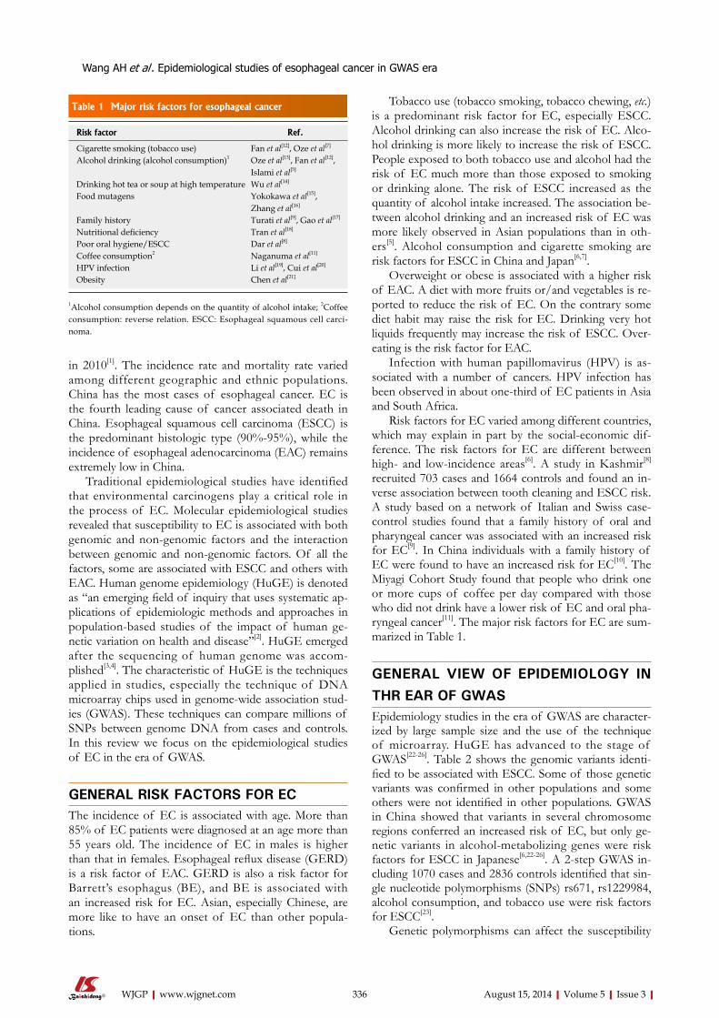
Wang AH et al . Epidemiological studies of esophageal cancer in GWAS era
in 2010[1]. The incidence rate and mortality rate varied among different geographic and ethnic populations. China has the most cases of esophageal cancer. EC is the fourth leading cause of cancer associated death in China. Esophageal squamous cell carcinoma (ESCC) is the predominant histologic type (90%-95%), while the incidence of esophageal adenocarcinoma (EAC) remains extremely low in China.
Traditional epidemiological studies have identified that environmental carcinogens play a critical role in the process of EC. Molecular epidemiological studies revealed that susceptibility to EC is associated with both genomic and non-genomic factors and the interaction between genomic and non-genomic factors. Of all the factors, some are associated with ESCC and others with EAC. Human genome epidemiology (HuGE) is denoted as “an emerging field of inquiry that uses systematic ap-plications of epidemiologic methods and approaches in population-based studies of the impact of human ge-netic variation on health and disease”[2]. HuGE emerged after the sequencing of human genome was accom-plished[3,4]. The characteristic of HuGE is the techniques applied in studies, especially the technique of DNA microarray chips used in genome-wide association stud-ies (GWAS). These techniques can compare millions of SNPs between genome DNA from cases and controls. In this review we focus on the epidemiological studies of EC in the era of GWAS.
GENERAL RISK FACTORS FOR ECThe incidence of EC is associated with age. More than 85% of EC patients were diagnosed at an age more than 55 years old. The incidence of EC in males is higher than that in females. Esophageal reflux disease (GERD) is a risk factor of EAC. GERD is also a risk factor for Barrett’s esophagus (BE), and BE is associated with an increased risk for EC. Asian, especially Chinese, are more like to have an onset of EC than other popula-tions.
Tobacco use (tobacco smoking, tobacco chewing, etc.) is a predominant risk factor for EC, especially ESCC. Alcohol drinking can also increase the risk of EC. Alco-hol drinking is more likely to increase the risk of ESCC. People exposed to both tobacco use and alcohol had the risk of EC much more than those exposed to smoking or drinking alone. The risk of ESCC increased as the quantity of alcohol intake increased. The association be-tween alcohol drinking and an increased risk of EC was more likely observed in Asian populations than in oth-ers[5]. Alcohol consumption and cigarette smoking are risk factors for ESCC in China and Japan[6,7].
Overweight or obese is associated with a higher risk of EAC. A diet with more fruits or/and vegetables is re-ported to reduce the risk of EC. On the contrary some diet habit may raise the risk for EC. Drinking very hot liquids frequently may increase the risk of ESCC. Over-eating is the risk factor for EAC.
Infection with human papillomavirus (HPV) is as-sociated with a number of cancers. HPV infection has been observed in about one-third of EC patients in Asia and South Africa.
Risk factors for EC varied among different countries, which may explain in part by the social-economic dif-ference. The risk factors for EC are different between high- and low-incidence areas[6]. A study in Kashmir[8] recruited 703 cases and 1664 controls and found an in-verse association between tooth cleaning and ESCC risk. A study based on a network of Italian and Swiss case-control studies found that a family history of oral and pharyngeal cancer was associated with an increased risk for EC[9]. In China individuals with a family history of EC were found to have an increased risk for EC[10]. The Miyagi Cohort Study found that people who drink one or more cups of coffee per day compared with those who did not drink have a lower risk of EC and oral pha-ryngeal cancer[11]. The major risk factors for EC are sum-marized in Table 1.
GENERAL VIEW OF EPIDEMIOLOGY IN THR EAR OF GWASEpidemiology studies in the era of GWAS are character-ized by large sample size and the use of the technique of microarray. HuGE has advanced to the stage of GWAS[22-26]. Table 2 shows the genomic variants identi-fied to be associated with ESCC. Some of those genetic variants was confirmed in other populations and some others were not identified in other populations. GWAS in China showed that variants in several chromosome regions conferred an increased risk of EC, but only ge-netic variants in alcohol-metabolizing genes were risk factors for ESCC in Japanese[6,22-26]. A 2-step GWAS in-cluding 1070 cases and 2836 controls identified that sin-gle nucleotide polymorphisms (SNPs) rs671, rs1229984, alcohol consumption, and tobacco use were risk factors for ESCC[23].
Genetic polymorphisms can affect the susceptibility
August 15, 2014|Volume 5|Issue 3|WJGP|www.wjgnet.com 336
Table 1 Major risk factors for esophageal cancer
Risk factor Ref.
Cigarette smoking (tobacco use) Fan et al[12], Oze et al[7] Alcohol drinking (alcohol consumption)1 Oze et al[13], Fan et al[12],
Islami et al[5]
Drinking hot tea or soup at high temperature Wu et al[14]
Food mutagens Yokokawa et al[15], Zhang et al[16]
Family history Turati et al[9], Gao et al[17]
Nutritional deficiency Tran et al[18]
Poor oral hygiene/ESCC Dar et al[8] Coffee consumption2 Naganuma et al[11]
HPV infection Li et al[19], Cui et al[20]
Obesity Chen et al[21]
1Alcohol consumption depends on the quantity of alcohol intake; 2Coffee consumption: reverse relation. ESCC: Esophageal squamous cell carci-noma.
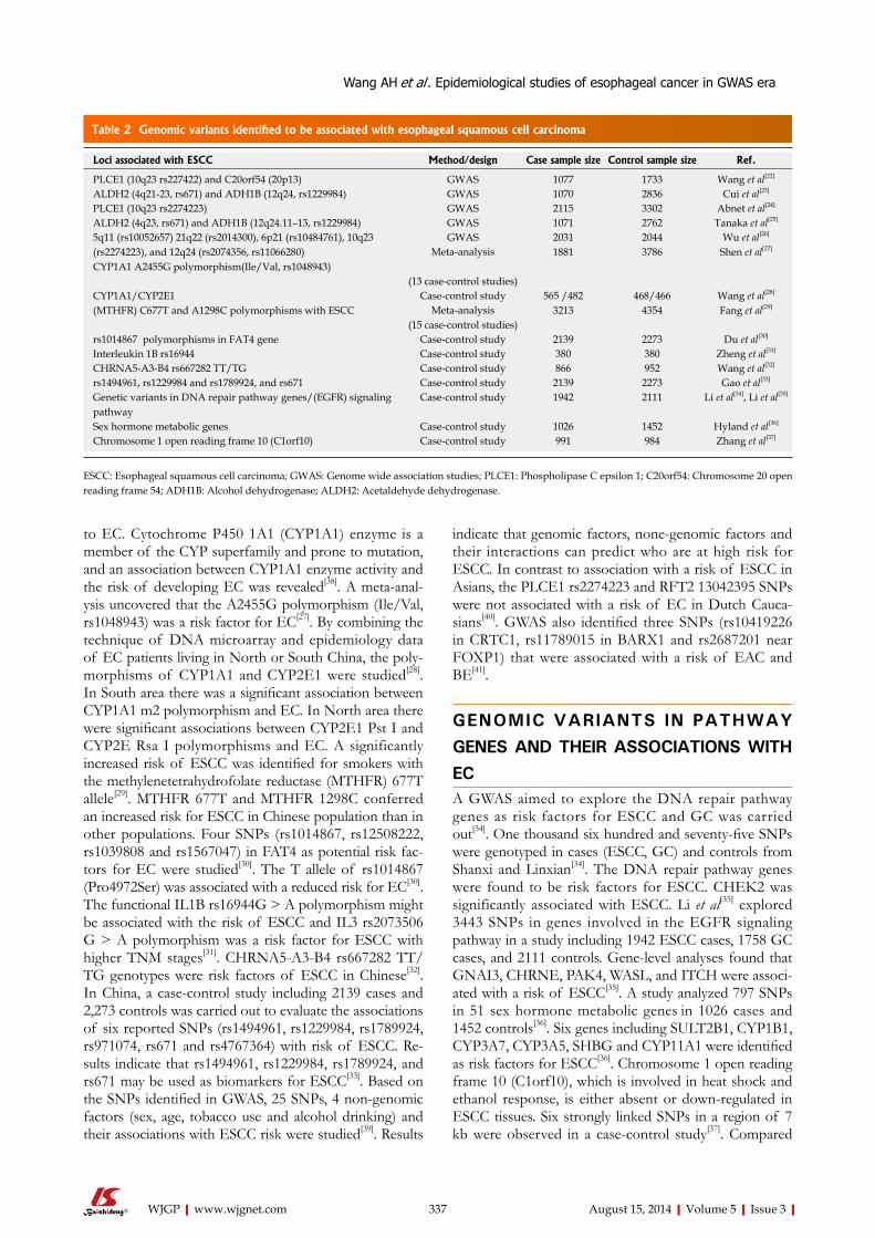
to EC. Cytochrome P450 1A1 (CYP1A1) enzyme is a member of the CYP superfamily and prone to mutation, and an association between CYP1A1 enzyme activity and the risk of developing EC was revealed[38]. A meta-anal-ysis uncovered that the A2455G polymorphism (Ile/Val, rs1048943) was a risk factor for EC[27]. By combining the technique of DNA microarray and epidemiology data of EC patients living in North or South China, the poly-morphisms of CYP1A1 and CYP2E1 were studied[28]. In South area there was a significant association between CYP1A1 m2 polymorphism and EC. In North area there were significant associations between CYP2E1 Pst I and CYP2E Rsa I polymorphisms and EC. A significantly increased risk of ESCC was identified for smokers with the methylenetetrahydrofolate reductase (MTHFR) 677T allele[29]. MTHFR 677T and MTHFR 1298C conferred an increased risk for ESCC in Chinese population than in other populations. Four SNPs (rs1014867, rs12508222, rs1039808 and rs1567047) in FAT4 as potential risk fac-tors for EC were studied[30]. The T allele of rs1014867 (Pro4972Ser) was associated with a reduced risk for EC[30]. The functional IL1B rs16944G > A polymorphism might be associated with the risk of ESCC and IL3 rs2073506 G > A polymorphism was a risk factor for ESCC with higher TNM stages[31]. CHRNA5-A3-B4 rs667282 TT/TG genotypes were risk factors of ESCC in Chinese[32]. In China, a case-control study including 2139 cases and 2,273 controls was carried out to evaluate the associations of six reported SNPs (rs1494961, rs1229984, rs1789924, rs971074, rs671 and rs4767364) with risk of ESCC. Re-sults indicate that rs1494961, rs1229984, rs1789924, and rs671 may be used as biomarkers for ESCC[33]. Based on the SNPs identified in GWAS, 25 SNPs, 4 non-genomic factors (sex, age, tobacco use and alcohol drinking) and their associations with ESCC risk were studied[39]. Results
indicate that genomic factors, none-genomic factors and their interactions can predict who are at high risk for ESCC. In contrast to association with a risk of ESCC in Asians, the PLCE1 rs2274223 and RFT2 13042395 SNPs were not associated with a risk of EC in Dutch Cauca-sians[40]. GWAS also identified three SNPs (rs10419226 in CRTC1, rs11789015 in BARX1 and rs2687201 near FOXP1) that were associated with a risk of EAC and BE[41].
GENOMIC VARIANTS IN PATHWAY GENES AND THEIR ASSOCIATIONS WITH ECA GWAS aimed to explore the DNA repair pathway genes as risk factors for ESCC and GC was carried out[34]. One thousand six hundred and seventy-five SNPs were genotyped in cases (ESCC, GC) and controls from Shanxi and Linxian[34]. The DNA repair pathway genes were found to be risk factors for ESCC. CHEK2 was significantly associated with ESCC. Li et al[35] explored 3443 SNPs in genes involved in the EGFR signaling pathway in a study including 1942 ESCC cases, 1758 GC cases, and 2111 controls. Gene-level analyses found that GNAI3, CHRNE, PAK4, WASL, and ITCH were associ-ated with a risk of ESCC[35]. A study analyzed 797 SNPs in 51 sex hormone metabolic genes in 1026 cases and 1452 controls[36]. Six genes including SULT2B1, CYP1B1, CYP3A7, CYP3A5, SHBG and CYP11A1 were identified as risk factors for ESCC[36]. Chromosome 1 open reading frame 10 (C1orf10), which is involved in heat shock and ethanol response, is either absent or down-regulated in ESCC tissues. Six strongly linked SNPs in a region of 7 kb were observed in a case-control study[37]. Compared
August 15, 2014|Volume 5|Issue 3|WJGP|www.wjgnet.com 337
Table 2 Genomic variants identified to be associated with esophageal squamous cell carcinoma
Loci associated with ESCC Method/design Case sample size Control sample size Ref.
PLCE1 (10q23 rs227422) and C20orf54 (20p13) GWAS 1077 1733 Wang et al[22]
ALDH2 (4q21-23, rs671) and ADH1B (12q24, rs1229984) GWAS 1070 2836 Cui et al[23]
PLCE1 (10q23 rs2274223) GWAS 2115 3302 Abnet et al[24]
ALDH2 (4q23, rs671) and ADH1B (12q24.11–13, rs1229984) GWAS 1071 2762 Tanaka et al[25]
5q11 (rs10052657) 21q22 (rs2014300), 6p21 (rs10484761), 10q23 (rs2274223), and 12q24 (rs2074356, rs11066280)CYP1A1 A2455G polymorphism(Ile/Val, rs1048943)
GWAS 20311881
20443786
Wu et al[26]
Shen et al[27]Meta-analysis
(13 case-control studies)CYP1A1/CYP2E1(MTHFR) C677T and A1298C polymorphisms with ESCC
Case-control study 565 /4823213
468/4664354
Wang et al[28]
Fang et al[29]Meta-analysis(15 case-control studies)
rs1014867 polymorphisms in FAT4 gene Case-control study 2139 2273 Du et al[30]
Interleukin 1B rs16944 Case-control study 380 380 Zheng et al[31]
CHRNA5-A3-B4 rs667282 TT/TG Case-control study 866 952 Wang et al[32]
rs1494961, rs1229984 and rs1789924, and rs671 Case-control study 2139 2273 Gao et al[33]
Genetic variants in DNA repair pathway genes/(EGFR) signaling pathway
Case-control study 1942 2111 Li et al[34], Li et al[35]
Sex hormone metabolic genes Case-control study 1026 1452 Hyland et al[36]
Chromosome 1 open reading frame 10 (C1orf10) Case-control study 991 984 Zhang et al[37]
ESCC: Esophageal squamous cell carcinoma; GWAS: Genome wide association studies; PLCE1: Phospholipase C epsilon 1; C20orf54: Chromosome 20 open reading frame 54; ADH1B: Alcohol dehydrogenase; ALDH2: Acetaldehyde dehydrogenase.
Wang AH et al . Epidemiological studies of esophageal cancer in GWAS era
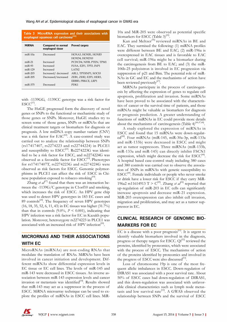
with -1139GG, -1139CC genotype was a risk factor for ESCC[37].
The HuGE progressed form the discovery of novel genes or SNPs to the functional or mechanistic study of those genes or SNPs. Moreover, HuGE studies try to screen some of those genes, SNPs or miRNAs that are clinical treatment targets or biomarkers for diagnosis or prognosis. A low mtDNA copy number variant (CNV) was a risk factor for EAC[42]. A case-control study was carried out to analyze the relationship between SNPs (rs17417407, rs2274223 and rs22744224) in PLCE1 and susceptibility to ESCC[43]. Rs2274223G was identi-fied to be a risk factor for ESCC, and rs2274224G was observed as a favorable factor for ESCC[43]. Phenotypes for rs17417407T, rs2274223G and rs2274224G were observed as risk factors for ESCC. Genomic polymor-phisms in PLCE1 can affect the risk of ESCC in Chi-nese population exposed to tobacco smoking[43].
Zhang et al[37] found that there was an interaction be-tween the -1139G/C genotype in C1orf10 and smoking, which increases the risk of ESCC. An HPV gene chip was used to detect HPV genotypes in 183 EC cases and 89 controls[20]. The frequency of seven HPV genotypes (16, 18, 35, 52, 6, 11, 43) in EC tissues was higher (31.7%) than that in controls (9.0%, P < 0.001), indicating that HPV infection was a risk factor for EC in Kazakh popu-lation. Moreover, heterozygote rs2274223 in PLCE1 was associated with an increased risk of HPV infection[20].
MICRORNAS AND THEIR ASSOCIATIONS WITH ECMicroRNAs (miRNAs) are non-coding RNAs that modulate the translation of RNAs. MiRNAs have been involved in cancer initiation and development. Dif-ferent miRNAs show differential expression levels in EC tissue or EC cell lines. The levels of miR-145 and miR-143 were decreased in ESCC tissues. An inverse as-sociation between miR-143 expression levels and cancer invasion or metastasis was identified[44]. Results showed that miR-143 may act as a suppressor in the process of ESCC. MiRNA microarray technique can be used to ex-plore the profiles of miRNAs in ESCC cell lines. MiR-
10a and MiR-205 were observed as potential specific biomarkers for ESCC (Table 3)[45].
Kan and Meltzer[46] reviewed miRNAs in BE and EAC. They surmised the following: (1) miRNA profiles were different between BE and EAC; (2) miR-196a is overexpressed in EAC tissues and is favorable to EAC cell survival; miR-196a might be a biomarker during the carcinogenesis from BE to EAC; and (3) the miR-106b-25 polycistron is involved in EC progression via suppression of p21 and Bim. The potential role of miR-NAs in GC and EC and the mechanisms of action have been reviewed previously[47].
MiRNAs participate in the process of carcinogen-esis by affecting the expression of genes to regulate cell apoptosis, proliferation and invasion. Some miRNAs have been proved to be associated with the characteris-tics of cancer or the survival time of patients, and those miRNAs might be valuable as biomarkers for diagnosis or prognosis prediction. A greater understanding of functions of miRNAs in EC could provide more details about the mechanisms of carcinogenesis (Table 4)[44,47,48].
A study explored the expression of miRNAs in ESCC and found that 15 miRNAs were down-regulat-ed[48]. Four miRNAs (miR-145, miR-30a-3p, miR-133a and miR-133b) were decreased in ESCC and might act as tumor suppressors. Three miRNAs (miR-133b, miR-133a and miR-145) can directly inhibit FSCN1 expression, which might decrease the risk for ESCC[48].
A hospital based case-control study including 380 cases and 380 controls was carried out to observe the associa-tion of SNPs in miRNAs with genetic susceptibility to ESCC[49]. Female individuals or people who never smoke or drink have a lower risk for ESCC if they carry MiR-196a2 rs11614913 T > C[49]. Zhang et al[50] reported that up-regulation of miR-203 in EC cells can significantly increase apoptosis and decrease miR-21 expression. MiR-203 overexpression can also inhibit cell invasion, migration and proliferation, and may act as a tumor sup-pressor in EC.
CLINICAL RESEARCH OF GENOMIC BIO-MARKERS FOR ECEC is a disease with a poor prognosis[51]. It is urgent to identify valuable biomarkers involved in the diagnosis, progress or therapy targets for ESCC. Qi[52] reviewed the proteins, identified by proteomics, which were associated with the process of ESCC. The mechanisms of action of the proteins identified by proteomics and involved in the progress of ESCC were also discussed[53].
Loss of chromosome 19p is one of the most fre-quent allelic imbalances in ESCC. Down-regulation of DIRAS1 was associated with a poor survival rate. About 50% of ESCC cases had down-regulation of DIRAS1, and this down-regulation was associated with unfavor-able clinical characteristics such as lymph node metas-tasis and low survival rate[53]. A GWAS observed the relationship between SNPs and the survival of ESCC
August 15, 2014|Volume 5|Issue 3|WJGP|www.wjgnet.com 338
Table 3 MicroRNA expression and their associations with esophageal squamous cell carcinoma[45]
MiRNA Compared to normal esophageal tissue
Proved targets
miR-10a Decreased HOXA3, HOXB1, HOXB3HOXD4, HOXD10
miR-21 Increased PCDCD4, NFIB, PTEN, TPM1miR-93 Increased FUSA, E2F1, TP53, INP1miR-129 Increased LATS2miR-203 Increased/ decreased ABL1, TP53INP1, SOCS3miR-205 Decreased/increased ZEB1, ZEB2, E2F5, HER3,
ERBB3, PRKCE, LRP1miR-375 Decreased PDK1
Wang AH et al . Epidemiological studies of esophageal cancer in GWAS era
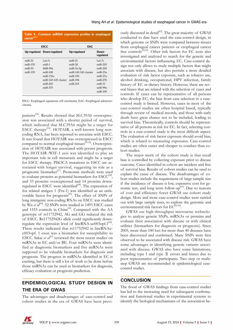
patients[54]. Results showed that SLC39A6 overexpres-sion was associated with a shorter period of survival, which indicated that SLC39A6 might be a target for ESCC therapy[54]. HOTAIR, a well-known long non-coding RNA, has been reported to associate with ESCC. It was found that HOTAIR was overexpressed in ESCC compared to normal esophageal tissues[55,56]. Overexpres-sion of HOTAIR was associated with poorer prognosis. The HOTAIR ⁄WIF-1 axis was identified to play an important role in cell metastasis and might be a target for ESCC therapy. PIK3CA mutations in ESCC are as-sociated with longer survival, suggesting its role as a prognostic biomarker[57]. Proteomic methods were used to evaluate proteins as potential biomarkers for ESCC[58], and 33 proteins overexpressed and 14 proteins down-regulated in ESCC were identified[58]. The expression of fos related antigen 1 (Fra-1) was identified as an unfa-vorable factor for prognosis[59]. The effect of SNPs of long intergenic non-coding RNAs on ESCC was studied by Wu et al[60]. 52 SNPs were studied in 1493 ESCC cases and 1553 controls in China[60]. Compared with the AA genotype of rs11752942, AG and GG reduced the risk of ESCC. Rs11752942G allele could significantly down-regulate the expression level of lincRNA-uc003opf.1[60]. These results indicated that rs11752942 in lincRNAu-c003opf. 1 exon was a biomarker for susceptibility to ESCC. Sakai et al[61] reviewed the most recent studies on miRNAs in EC and/or BE. Four miRNAs were identi-fied as diagnostic biomarkers and five miRNAs were supposed to be valuable biomarkers for diagnosis and prognosis. The progress in miRNAs identified in EC is exciting, but there is still a lot of work to be done before those miRNAs can be used as biomarkers for diagnosis, efficacy evaluation or prognosis prediction.
EPIDEMIOLOGICAL STUDY DESIGN IN THE ERA OF GWAS The advantages and disadvantages of case-control and cohort studies in the era of GWAS have been previ-
ously discussed in detail[62]. The great majority of GWAS conducted to date have used the case-control design, in which genome or SNPs were compared between tissues from esophageal cancer patients or esophageal cancer free controls[22,26]. Other risk factors for EC were also investigated and analyzed to search for the genetic and environmental factors influencing EC. Case-control de-sign not only allows to study multiple factors that might associate with disease, but also permits a more detailed evaluation of risk factor exposure, such as tobacco use, alcohol drinking, occupational, HPV infection, family history of EC or dietary history. However, there are sev-eral biases that are related with the selection of cases and controls. If cases can be representative of all persons who develop EC, the bias from case selection in a case-control study is limited. However, cases in most of the case-control studies are often hospital based, typically through review of medical records, and those with early death have great chance not to be included, leading to survival bias. Theoretically, controls should be represen-tative of all persons at risk for EC. In fact, selecting con-trols in a case-control study is the most difficult aspect. The evaluation of risk factor exposure should avoid bias, which is related to measuring exposures. Case-control studies are often easier and cheaper to conduct than co-hort studies.
The major merit of the cohort study is that recall bias is controlled by collecting exposure prior to disease outcome. Cases identified in cohort are incident and free of survival bias. Results of cohort studies can be used to explain the cause of disease. The disadvantages of co-hort studies include the requirement of large sample size if the incidence of disease is low, expensive cost for ge-nomic test, and long term follow-up[63]. Due to reasons of cost and efficiency fewer GWAS use cohort study design. More and more case-control studies were carried out with large sample sizes, to explore the genomic and environmental risk factors for EC[23,25].
GWAS use high-throughput microarray technolo-gies to analyze genetic SNPs, miRNAs or proteins and evaluate their association with disease or with clinical utilities (biomarkers for diagnosis or prognosis). Since 2005, more than 100 loci for more than 40 diseases have been discovered and confirmed. Many SNPs were first observed to be associated with disease risk. GWAS have some advantages in identifying genetic variants associ-ated with disease. GWAS also have some limitations, including type Ⅰ and type Ⅱ errors and biases due to poor representative of participants. Two step or multi-step GWAS are recommended in epidemiological case-control studies.
CONCLUSIONThe flood of GWAS findings from case-control studies has led to the increasing need for subsequent confirma-tion and functional studies in experimental systems to identify the biological mechanisms of the association be-
August 15, 2014|Volume 5|Issue 3|WJGP|www.wjgnet.com 339
Table 4 Common miRNA expression profiles in esophageal cancer[47]
ESCC EAC
Up-regulated Down-regulated Up-regulated Down-regulated
miR-21 Let-7c miR-21 Let-7cmiR-155 miR-1 miR-28 miR-203miR-93 MiR-99a miR-3a-5p miR-205miR-129 miR-100 miR-143-145 cluster miR-23a
miR-133a miR-192 miR-27amiR-143-145 cluster miR-194 miR-27b miR-203 miR-215 miR-31miR-375 miR-99a
miR-100
ESCC: Esophageal squamous cell carcinoma; EAC: Esophageal adenocar-cinoma.
Wang AH et al . Epidemiological studies of esophageal cancer in GWAS era
tween genomic variants and EC. Epidemiological studies of EC in the era of GWAS have explored the genomic variants affecting signaling, epigenetic regulation, RNAs, proteins and pathways involved in cell proliferation or invasion. However, much work remains to be done in-cluding identifying the biomarkers for screening, efficacy evaluation and prognosis prediction. In the future, more and more epidemiological studies will take the advantag-es of population-based, very large sample-sized GWAS.
REFERENCES1 Lozano R, Naghavi M, Foreman K, Lim S, Shibuya K, Aboy-
ans V, Abraham J, Adair T, Aggarwal R, Ahn SY, Alvarado M, Anderson HR, Anderson LM, Andrews KG, Atkinson C, Baddour LM, Barker-Collo S, Bartels DH, Bell ML, Ben-jamin EJ, Bennett D, Bhalla K, Bikbov B, Bin Abdulhak A, Birbeck G, Blyth F, Bolliger I, Boufous S, Bucello C, Burch M, Burney P, Carapetis J, Chen H, Chou D, Chugh SS, Coffeng LE, Colan SD, Colquhoun S, Colson KE, Condon J, Connor MD, Cooper LT, Corriere M, Cortinovis M, de Vaccaro KC, Couser W, Cowie BC, Criqui MH, Cross M, Dabhadkar KC, Dahodwala N, De Leo D, Degenhardt L, Delossantos A, Denenberg J, Des Jarlais DC, Dharmaratne SD, Dorsey ER, Driscoll T, Duber H, Ebel B, Erwin PJ, Espindola P, Ezzati M, Feigin V, Flaxman AD, Forouzanfar MH, Fowkes FG, Franklin R, Fransen M, Freeman MK, Gabriel SE, Gakidou E, Gaspari F, Gillum RF, Gonzalez-Medina D, Halasa YA, Har-ing D, Harrison JE, Havmoeller R, Hay RJ, Hoen B, Hotez PJ, Hoy D, Jacobsen KH, James SL, Jasrasaria R, Jayaraman S, Johns N, Karthikeyan G, Kassebaum N, Keren A, Khoo JP, Knowlton LM, Kobusingye O, Koranteng A, Krish-namurthi R, Lipnick M, Lipshultz SE, Ohno SL, Mabweijano J, MacIntyre MF, Mallinger L, March L, Marks GB, Marks R, Matsumori A, Matzopoulos R, Mayosi BM, McAnulty JH, McDermott MM, McGrath J, Mensah GA, Merriman TR, Michaud C, Miller M, Miller TR, Mock C, Mocumbi AO, Mokdad AA, Moran A, Mulholland K, Nair MN, Naldi L, Narayan KM, Nasseri K, Norman P, O’Donnell M, Omer SB, Ortblad K, Osborne R, Ozgediz D, Pahari B, Pandian JD, Rivero AP, Padilla RP, Perez-Ruiz F, Perico N, Phillips D, Pierce K, Pope CA 3rd, Porrini E, Pourmalek F, Raju M, Ranganathan D, Rehm JT, Rein DB, Remuzzi G, Rivara FP, Roberts T, De León FR, Rosenfeld LC, Rushton L, Sacco RL, Salomon JA, Sampson U, Sanman E, Schwebel DC, Segui-Gomez M, Shepard DS, Singh D, Singleton J, Sliwa K, Smith E, Steer A, Taylor JA, Thomas B, Tleyjeh IM, Towbin JA, Truelsen T, Undurraga EA, Venketasubramanian N, Vi-jayakumar L, Vos T, Wagner GR, Wang M, Wang W, Watt K, Weinstock MA, Weintraub R, Wilkinson JD, Woolf AD, Wulf S, Yeh PH, Yip P, Zabetian A, Zheng ZJ, Lopez AD, Murray CJ, AlMazroa MA, Memish ZA. Global and regional mortality from 235 causes of death for 20 age groups in 1990 and 2010: a systematic analysis for the Global Burden of Disease Study 2010. Lancet 2012; 380:2095–2128 [PMID: 23245604 DOI: 10.1016/S0140-6736(12)61728-0]
2 Khoury MJ, Dorman JS. The Human Genome Epidemiol-ogy Network. Am J Epidemiol 1998; 148: 1-3 [PMID: 9663396]
3 Lander ES, Linton LM, Birren B, Nusbaum C, Zody MC, Baldwin J, Devon K, Dewar K, Doyle M, FitzHugh W, Funke R, Gage D, Harris K, Heaford A, Howland J, Kann L, Lehoczky J, LeVine R, McEwan P, McKernan K, Meldrim J, Mesirov JP, Miranda C, Morris W, Naylor J, Raymond C, Rosetti M, Santos R, Sheridan A, Sougnez C, Stange-Thomann N, Stojanovic N, Subramanian A, Wyman D, Rogers J, Sulston J, Ainscough R, Beck S, Bentley D, Burton J, Clee C, Carter N, Coulson A, Deadman R, Deloukas P, Dunham A, Dunham I, Durbin R, French L, Grafham D,
Gregory S, Hubbard T, Humphray S, Hunt A, Jones M, Lloyd C, McMurray A, Matthews L, Mercer S, Milne S, Mullikin JC, Mungall A, Plumb R, Ross M, Shownkeen R, Sims S, Waterston RH, Wilson RK, Hillier LW, McPherson JD, Marra MA, Mardis ER, Fulton LA, Chinwalla AT, Pepin KH, Gish WR, Chissoe SL, Wendl MC, Delehaunty KD, Miner TL, Delehaunty A, Kramer JB, Cook LL, Fulton RS, Johnson DL, Minx PJ, Clifton SW, Hawkins T, Branscomb E, Predki P, Richardson P, Wenning S, Slezak T, Doggett N, Cheng JF, Olsen A, Lucas S, Elkin C, Uberbacher E, Frazier M, Gibbs RA, Muzny DM, Scherer SE, Bouck JB, Sodergren EJ, Worley KC, Rives CM, Gorrell JH, Metzker ML, Naylor SL, Kucherlapati RS, Nelson DL, Weinstock GM, Sakaki Y, Fujiyama A, Hattori M, Yada T, Toyoda A, Itoh T, Kawagoe C, Watanabe H, Totoki Y, Taylor T, Weissenbach J, Heilig R, Saurin W, Artiguenave F, Brottier P, Bruls T, Pelletier E, Robert C, Wincker P, Smith DR, Doucette-Stamm L, Ru-benfield M, Weinstock K, Lee HM, Dubois J, Rosenthal A, Platzer M, Nyakatura G, Taudien S, Rump A, Yang H, Yu J, Wang J, Huang G, Gu J, Hood L, Rowen L, Madan A, Qin S, Davis RW, Federspiel NA, Abola AP, Proctor MJ, Myers RM, Schmutz J, Dickson M, Grimwood J, Cox DR, Olson MV, Kaul R, Raymond C, Shimizu N, Kawasaki K, Minoshi-ma S, Evans GA, Athanasiou M, Schultz R, Roe BA, Chen F, Pan H, Ramser J, Lehrach H, Reinhardt R, McCombie WR, de la Bastide M, Dedhia N, Blöcker H, Hornischer K, Nor-dsiek G, Agarwala R, Aravind L, Bailey JA, Bateman A, Bat-zoglou S, Birney E, Bork P, Brown DG, Burge CB, Cerutti L, Chen HC, Church D, Clamp M, Copley RR, Doerks T, Eddy SR, Eichler EE, Furey TS, Galagan J, Gilbert JG, Harmon C, Hayashizaki Y, Haussler D, Hermjakob H, Hokamp K, Jang W, Johnson LS, Jones TA, Kasif S, Kaspryzk A, Kennedy S, Kent WJ, Kitts P, Koonin EV, Korf I, Kulp D, Lancet D, Lowe TM, McLysaght A, Mikkelsen T, Moran JV, Mulder N, Pollara VJ, Ponting CP, Schuler G, Schultz J, Slater G, Smit AF, Stupka E, Szustakowski J, Thierry-Mieg D, Thierry-Mieg J, Wagner L, Wallis J, Wheeler R, Williams A, Wolf YI, Wolfe KH, Yang SP, Yeh RF, Collins F, Guyer MS, Peterson J, Felsenfeld A, Wetterstrand KA, Patrinos A, Morgan MJ, de Jong P, Catanese JJ, Osoegawa K, Shizuya H, Choi S, Chen YJ; International Human Genome Sequencing Consortium. Initial sequencing and analysis of the human genome. Na-ture 2001; 409: 860–921 [PMID: 11237011]
4 Venter JC, Adams MD, Myers EW, Li PW, Mural RJ, Sutton GG, Smith HO, Yandell M, Evans CA, Holt RA, Gocayne JD, Amanatides P, Ballew RM, Huson DH, Wortman JR, Zhang Q, Kodira CD, Zheng XH, Chen L, Skupski M, Subrama-nian G, Thomas PD, Zhang J, Gabor Miklos GL, Nelson C, Broder S, Clark AG, Nadeau J, McKusick VA, Zinder N, Levine AJ, Roberts RJ, Simon M, Slayman C, Hunkapiller M, Bolanos R, Delcher A, Dew I, Fasulo D, Flanigan M, Florea L, Halpern A, Hannenhalli S, Kravitz S, Levy S, Mobarry C, Reinert K, Remington K, Abu-Threideh J, Beasley E, Biddick K, Bonazzi V, Brandon R, Cargill M, Chandramouliswaran I, Charlab R, Chaturvedi K, Deng Z, Di Francesco V, Dunn P, Eilbeck K, Evangelista C, Gabrielian AE, Gan W, Ge W, Gong F, Gu Z, Guan P, Heiman TJ, Higgins ME, Ji RR, Ke Z, Ketchum KA, Lai Z, Lei Y, Li Z, Li J, Liang Y, Lin X, Lu F, Merkulov GV, Milshina N, Moore HM, Naik AK, Narayan VA, Neelam B, Nusskern D, Rusch DB, Salzberg S, Shao W, Shue B, Sun J, Wang Z, Wang A, Wang X, Wang J, Wei M, Wides R, Xiao C, Yan C, Yao A, Ye J, Zhan M, Zhang W, Zhang H, Zhao Q, Zheng L, Zhong F, Zhong W, Zhu S, Zhao S, Gilbert D, Baumhueter S, Spier G, Carter C, Crav-chik A, Woodage T, Ali F, An H, Awe A, Baldwin D, Baden H, Barnstead M, Barrow I, Beeson K, Busam D, Carver A, Center A, Cheng ML, Curry L, Danaher S, Davenport L, Desilets R, Dietz S, Dodson K, Doup L, Ferriera S, Garg N, Gluecksmann A, Hart B, Haynes J, Haynes C, Heiner C, Hladun S, Hostin D, Houck J, Howland T, Ibegwam C,
August 15, 2014|Volume 5|Issue 3|WJGP|www.wjgnet.com 340
Wang AH et al . Epidemiological studies of esophageal cancer in GWAS era
Johnson J, Kalush F, Kline L, Koduru S, Love A, Mann F, May D, McCawley S, McIntosh T, McMullen I, Moy M, Moy L, Murphy B, Nelson K, Pfannkoch C, Pratts E, Puri V, Qureshi H, Reardon M, Rodriguez R, Rogers YH, Rom-blad D, Ruhfel B, Scott R, Sitter C, Smallwood M, Stewart E, Strong R, Suh E, Thomas R, Tint NN, Tse S, Vech C, Wang G, Wetter J, Williams S, Williams M, Windsor S, Winn-Deen E, Wolfe K, Zaveri J, Zaveri K, Abril JF, Guigó R, Campbell MJ, Sjolander KV, Karlak B, Kejariwal A, Mi H, Lazareva B, Hatton T, Narechania A, Diemer K, Muruganujan A, Guo N, Sato S, Bafna V, Istrail S, Lippert R, Schwartz R, Walenz B, Yooseph S, Allen D, Basu A, Baxendale J, Blick L, Caminha M, Carnes-Stine J, Caulk P, Chiang YH, Coyne M, Dahlke C, Mays A, Dombroski M, Donnelly M, Ely D, Esparham S, Fo-sler C, Gire H, Glanowski S, Glasser K, Glodek A, Gorokhov M, Graham K, Gropman B, Harris M, Heil J, Henderson S, Hoover J, Jennings D, Jordan C, Jordan J, Kasha J, Kagan L, Kraft C, Levitsky A, Lewis M, Liu X, Lopez J, Ma D, Majoros W, McDaniel J, Murphy S, Newman M, Nguyen T, Nguyen N, Nodell M, Pan S, Peck J, Peterson M, Rowe W, Sanders R, Scott J, Simpson M, Smith T, Sprague A, Stockwell T, Turner R, Venter E, Wang M, Wen M, Wu D, Wu M, Xia A, Zandieh A, Zhu X. The sequence of the human genome. Science 2001; 291: 1304-1351 [ PMID: 11181995]
5 Islami F, Fedirko V, Tramacere I, Bagnardi V, Jenab M, Scotti L, Rota M, Corrao G, Garavello W, Schüz J, Straif K, Negri E, Boffetta P, La Vecchia C. Alcohol drinking and esophageal squamous cell carcinoma with focus on light-drinkers and never-smokers: a systematic review and meta-analysis. Int J Cancer 2011; 129: 2473-2484 [PMID: 21190191 DOI: 10.1002/ijc.25885]
6 Lin Y, Totsuka Y, He Y, Kikuchi S, Qiao Y, Ueda J, Wei W, Inoue M, Tanaka H. Epidemiology of esophageal cancer in Japan and China. J Epidemiol 2013; 23: 233-242 [PMID: 23629646 DOI: 10.2188/jea.JE20120162]
7 Oze I, Matsuo K, Ito H, Wakai K, Nagata C, Mizoue T, Tanaka K, Tsuji I, Tamakoshi A, Sasazuki S, Inoue M, Tsu-gane S. Cigarette smoking and esophageal cancer risk: an evaluation based on a systematic review of epidemiologic evidence among the Japanese population. Jpn J Clin Oncol 2012; 42: 63-73 [PMID: 22131340 DOI: 10.1093/jjco/hyr170]
8 Dar NA, Islami F, Bhat GA, Shah IA, Makhdoomi MA, Iqbal B, Rafiq R, Lone MM, Abnet CC, Boffetta P. Poor oral hygiene and risk of esophageal squamous cell carcinoma in Kashmir. Br J Cancer 2013; 109: 1367-1372 [PMID: 23900216 DOI: 10.1038/bjc.2013.437]
9 Turati F, Edefonti V, Bosetti C, Ferraroni M, Malvezzi M, Franceschi S, Talamini R, Montella M, Levi F, Dal Maso L, Serraino D, Polesel J, Negri E, Decarli A, La Vecchia C. Family history of cancer and the risk of cancer: a network of case-control studies. Ann Oncol 2013; 24: 2651-2656 [PMID: 23884440 DOI: 10.1093/annonc/mdt280]
10 Wang AH, Sun CS, Li LS, Huang JY, Chen QS, Xu DZ. Ge-netic susceptibility and environmental factors of esophageal cancer in Xi’an. World J Gastroenterol 2004; 10: 940-944 [PMID: 15052670]
11 Naganuma T, Kuriyama S, Kakizaki M, Sone T, Nakaya N, Ohmori-Matsuda K, Nishino Y, Fukao A, Tsuji I. Coffee con-sumption and the risk of oral, pharyngeal, and esophageal cancers in Japan: the Miyagi Cohort Study. Am J Epidemiol 2008; 168: 1425-1432 [PMID: 18974083 DOI: 10.1093/aje/kwn282]
12 Fan Y, Yuan JM, Wang R, Gao YT, Yu MC. Alcohol, tobacco, and diet in relation to esophageal cancer: the Shanghai Co-hort Study. Nutr Cancer 2008; 60: 354-363 [PMID: 18444169 DOI: 10.1080/01635580701883011]
13 Oze I, Matsuo K, Wakai K, Nagata C, Mizoue T, Tanaka K, Tsuji I, Sasazuki S, Inoue M, Tsugane S. Alcohol drinking and esophageal cancer risk: an evaluation based on a sys-tematic review of epidemiologic evidence among the Japa-
nese population. Jpn J Clin Oncol 2011; 41: 677-692 [PMID: 21430021 DOI: 10.1093/jjco/hyr026]
14 Wu M, Zhao JK, Zhang ZF, Han RQ, Yang J, Zhou JY, Wang XS, Zhang XF, Liu AM, van’ t Veer P, Kok FJ, Kampman E. Smoking and alcohol drinking increased the risk of esopha-geal cancer among Chinese men but not women in a high-risk population. Cancer Causes Control 2011; 22: 649-657 [PMID: 21321789 DOI: 10.1007/s10552-011-9737-4]
15 Yokokawa Y, Ohta S, Hou J, Zhang XL, Li SS, Ping YM, Na-kajima T. Ecological study on the risks of esophageal cancer in Ci-Xian, China: the importance of nutritional status and the use of well water. Int J Cancer 1999; 83: 620-624 [PMID: 10521797]
16 Zhang N, Yu C, Wen D, Chen J, Ling Y, Terajima K, Akaza-wa K, Shan B, Wang S. Association of nitrogen compounds in drinking water with incidence of esophageal squamous cell carcinoma in Shexian, China. Tohoku J Exp Med 2012; 226: 11-17 [PMID: 22146401]
17 Gao Y, Hu N, Han X, Giffen C, Ding T, Goldstein A, Taylor P. Family history of cancer and risk for esophageal and gastric cancer in Shanxi, China. BMC Cancer 2009; 9: 269 [PMID: 19656375 DOI: 10.1186/1471-2407-9-269]
18 Tran GD, Sun XD, Abnet CC, Fan JH, Dawsey SM, Dong ZW, Mark SD, Qiao YL, Taylor PR. Prospective study of risk factors for esophageal and gastric cancers in the Linxian general population trial cohort in China. Int J Cancer 2005; 113: 456-463 [PMID: 15455378]
19 Li X, Gao C, Yang Y, Zhou F, Li M, Jin Q, Gao L. Systematic review with meta-analysis: the association between human papillomavirus infection and oesophageal cancer. Aliment Pharmacol Ther 2014; 39: 270-281 [PMID: 24308856 DOI: 10.1111/apt.12574]
20 Cui X, Chen Y, Liu L, Li L, Hu J, Yang L, Liang W, Li F. Heterozygote of PLCE1 rs2274223 increases susceptibility to human papillomavirus infection in patients with esophageal carcinoma among the Kazakh populations. J Med Virol 2014; 86: 608-617 [PMID: 24127316 DOI: 10.1002/jmv.23775]
21 Chen Q, Zhuang H, Liu Y. The association between obesity factor and esophageal caner. J Gastrointest Oncol 2012; 3: 226-231 [PMID: 22943013 DOI: 10.3978/j.issn.2078-6891.2012.026]
22 Wang LD, Zhou FY, Li XM, Sun LD, Song X, Jin Y, Li JM, Kong GQ, Qi H, Cui J, Zhang LQ, Yang JZ, Li JL, Li XC, Ren JL, Liu ZC, Gao WJ, Yuan L, Wei W, Zhang YR, Wang WP, Sheyhidin I, Li F, Chen BP, Ren SW, Liu B, Li D, Ku JW, Fan ZM, Zhou SL, Guo ZG, Zhao XK, Liu N, Ai YH, Shen FF, Cui WY, Song S, Guo T, Huang J, Yuan C, Huang J, Wu Y, Yue WB, Feng CW, Li HL, Wang Y, Tian JY, Lu Y, Yuan Y, Zhu WL, Liu M, Fu WJ, Yang X, Wang HJ, Han SL, Chen J, Han M, Wang HY, Zhang P, Li XM, Dong JC, Xing GL, Wang R, Guo M, Chang ZW, Liu HL, Guo L, Yuan ZQ, Liu H, Lu Q, Yang LQ, Zhu FG, Yang XF, Feng XS, Wang Z, Li Y, Gao SG, Qige Q, Bai LT, Yang WJ, Lei GY, Shen ZY, Chen LQ, Li EM, Xu LY, Wu ZY, Cao WK, Wang JP, Bao ZQ, Chen JL, Ding GC, Zhuang X, Zhou YF, Zheng HF, Zhang Z, Zuo XB, Dong ZM, Fan DM, He X, Wang J, Zhou Q, Zhang QX, Jiao XY, Lian SY, Ji AF, Lu XM, Wang JS, Chang FB, Lu CD, Chen ZG, Miao JJ, Fan ZL, Lin RB, Liu TJ, Wei JC, Kong QP, Lan Y, Fan YJ, Gao FS, Wang TY, Xie D, Chen SQ, Yang WC, Hong JY, Wang L, Qiu SL, Cai ZM, Zhang XJ. Genome-wide association study of esophageal squamous cellcarcino-ma in Chinese subjects identifies susceptibility loci at PLCE1 and C20orf54. Nat Genet 2010; 42: 759-763 [PMID: 20729853 DOI: 10.1038/ng.648]
23 Cui R, Kamatani Y, Takahashi A, Usami M, Hosono N, Kawaguchi T, Tsunoda T, Kamatani N, Kubo M, Nakamura Y, Matsuda K. Functional variants in ADH1B and ALDH2 coupled with alcohol and smoking synergistically enhance esophageal cancer risk. Gastroenterology 2009; 137: 1768-1775 [PMID: 19698717 DOI: 10.1053/j.gastro.2009.07.070]
August 15, 2014|Volume 5|Issue 3|WJGP|www.wjgnet.com 341
Wang AH et al . Epidemiological studies of esophageal cancer in GWAS era
24 Abnet CC, Freedman ND, Hu N, Wang Z, Yu K, Shu XO, Yuan JM, Zheng W, Dawsey SM, Dong LM, Lee MP, Ding T, Qiao YL, Gao YT, Koh WP, Xiang YB, Tang ZZ, Fan JH, Wang C, Wheeler W, Gail MH, Yeager M, Yuenger J, Hutchinson A, Jacobs KB, Giffen CA, Burdett L, Fraumeni JF, Tucker MA, Chow WH, Goldstein AM, Chanock SJ, Tay-lor PR. A shared susceptibility locus in PLCE1 at 10q23 for gastric adenocarcinoma and esophageal squamous cell car-cinoma. Nat Genet 2010; 42: 764-767 [PMID: 20729852 DOI: 10.1038/ng.649]
25 Tanaka F, Yamamoto K, Suzuki S, Inoue H, Tsurumaru M, Kajiyama Y, Kato H, Igaki H, Furuta K, Fujita H, Tanaka T, Tanaka Y, Kawashima Y, Natsugoe S, Setoyama T, Toku-dome S, Mimori K, Haraguchi N, Ishii H, Mori M. Strong interaction between the effects of alcohol consumption and smoking on oesophageal squamous cell carcinoma among individuals with ADH1B and/or ALDH2 risk alleles. Gut 2010; 59: 1457-1464 [PMID: 20833657 DOI: 10.1136/gut.2009.205724]
26 Wu C, Hu Z, He Z, Jia W, Wang F, Zhou Y, Liu Z, Zhan Q, Liu Y, Yu D, Zhai K, Chang J, Qiao Y, Jin G, Liu Z, Shen Y, Guo C, Fu J, Miao X, Tan W, Shen H, Ke Y, Zeng Y, Wu T, Lin D. Genome-wide association study identifies three new susceptibility loci for esophageal squamous-cell carcinoma in Chinese populations. Nat Genet 2011; 43: 679-684 [PMID: 21642993 DOI: 10.1038/ng.849]
27 Shen FF, Zhou FY, Xue QS, Pan Y, Zheng L, Zhang H, Wang LD, Zheng HF. Association between CYP1A1 poly-morphisms and esophageal cancer: a meta-analysis. Mol Biol Rep 2013; 40: 6035-6042 [PMID: 24065535 DOI: 10.1007/s11033-013-2713-1]
28 Wang D, Su M, Tian D, Liang S, Zhang J. Associations between CYP1A1 and CYP2E1 polymorphisms and sus-ceptibility to esophageal cancer in Chaoshan and Taihang areas of China. Cancer Epidemiol 2012; 36: 276-282 [PMID: 22088806 DOI: 10.1016/j.canep.2011.10.008]
29 Fang Y, Xiao F, An Z, Hao L. Systematic review on the relationship between genetic polymorphisms of methylene-tetrahydrofolate reductase and esophageal squamous cell carcinoma. Asian Pac J Cancer Prev 2011; 12: 1861-1866 [PMID: 22126580]
30 Du J, Ji J, Gao Y, Xu L, Xu J, Zhu C, Gu H, Jiang J, Li H, Ma H, Hu Z, Jin G, Guo W, Chen X, Shen H. Nonsynonymous polymorphisms in FAT4 gene are associated with the risk of esophageal cancer in an Eastern Chinese population. Int J Cancer 2013; 133: 357-361 [PMID: 23319386 DOI: 10.1002/ijc.28033]
31 Zheng L, Yin J, Wang L, Wang X, Shi Y, Shao A, Tang W, Ding G, Liu C, Chen S, Gu H. Interleukin 1B rs16944 G& gt; A polymorphism was associated with a decreased risk of esophageal cancer in a Chinese population. Clin Biochem 2013; 46: 1469-1473 [PMID: 23726808 DOI: 10.1016/j.clinbiochem.2013.05.050]
32 Wang Y, Wu H, Liu Q, Wang C, Fu L, Wang H, Zhu W, Fu W, Lv Y, Wang S, Hu L. Association of CHRNA5-A3-B4 variation with esophageal squamous cell carcinoma risk and smoking behaviors in a Chinese population. PLoS One 2013; 8: e67664 [PMID: 23844051 DOI: 10.1371/journal.pone.0067664]
33 Gao Y, He Y, Xu J, Xu L, Du J, Zhu C, Gu H, Ma H, Hu Z, Jin G, Chen X, Shen H. Genetic variants at 4q21, 4q23 and 12q24 are associated with esophageal squamous cell carci-noma risk in a Chinese population. Hum Genet 2013; 132: 649-656 [PMID: 23430454 DOI: 10.1007/s00439-013-1276-5]
34 Li WQ, Hu N, Hyland PL, Gao Y, Wang ZM, Yu K, Su H, Wang CY, Wang LM, Chanock SJ, Burdett L, Ding T, Qiao YL, Fan JH, Wang Y, Xu Y, Shi JX, Gu F, Wheeler W, Xiong XQ, Giffen C, Tucker MA, Dawsey SM, Freedman ND, Ab-net CC, Goldstein AM, Taylor PR. Genetic variants in DNA repair pathway genes and risk of esophageal squamous cell
carcinoma and gastric adenocarcinoma in a Chinese popu-lation. Carcinogenesis 2013; 34: 1536-1542 [PMID: 23504502 DOI: 10.1093/carcin/bgt094]
35 Li WQ, Hu N, Wang Z, Yu K, Su H, Wang L, Wang C, Cha-nock SJ, Burdett L, Ding T, Qiao YL, Fan JH, Wang Y, Xu Y, Giffen C, Xiong X, Murphy G, Tucker MA, Dawsey SM, Freedman ND, Abnet CC, Goldstein AM, Taylor PR. Genetic variants in epidermal growth factor receptor pathway genes and risk of esophageal squamous cell carcinoma and gastric cancer in a Chinese population. PLoS One 2013; 8: e68999 [PMID: 23874846 DOI: 10.1371/journal.pone.0068999]
36 Hyland PL, Freedman ND, Hu N, Tang ZZ, Wang L, Wang C, Ding T, Fan JH, Qiao YL, Golozar A, Wheeler W, Yu K, Yuenger J, Burdett L, Chanock SJ, Dawsey SM, Tucker MA, Goldstein AM, Abnet CC, Taylor PR. Genetic variants in sex hormone metabolic pathway genes and risk of esophageal squamous cell carcinoma. Carcinogenesis 2013; 34: 1062-1068 [PMID: 23358850 DOI: 10.1093/carcin/bgt030]
37 Zhang W, Chen X, Luo A, Lin D, Tan W, Liu Z. Genetic vari-ants of C1orf10 and risk of esophageal squamous cell carci-noma in a Chinese population. Cancer Sci 2009; 100: 1695-1700 [PMID: 19558548 DOI: 10.1111/j.1349-7006.2009.01240.x]
38 Agundez JA. Cytochrome P450 gene polymorphism and cancer. Curr Drug Metab 2004; 5: 211-224 [PMID: 15180491]
39 Chang J, Huang Y, Wei L, Ma B, Miao X, Li Y, Hu Z, Yu D, Jia W, Liu Y, Tan W, He Z, Ke Y, Wu T, Shen H, Zeng Y, Wu C, Lin D. Risk prediction of esophageal squamous-cell car-cinoma with common genetic variants and lifestyle factors in Chinese population. Carcinogenesis 2013; 34: 1782-1786 [PMID: 23536576 DOI: 10.1093/carcin/bgt106]
40 Dura P, Bregitha CV, te Morsche RH, Roelofs HM, Kris-tinsson JO, Wobbes T, Witteman BJ, Tan AC, Drenth JP, Peters WH. GWAS-uncovered SNPs in PLCE1 and RFT2 genes are not implicated in Dutch esophageal adenocarci-noma and squamous cell carcinoma etiology. Eur J Cancer Prev 2013; 22: 417-419 [PMID: 23222411 DOI: 10.1097/CEJ.0b013e32835c7f53]
41 Levine DM, Ek WE, Zhang R, Liu X, Onstad L, Sather C, Lao-Sirieix P, Gammon MD, Corley DA, Shaheen NJ, Bird NC, Hardie LJ, Murray LJ, Reid BJ, Chow WH, Risch HA, Nyrén O, Ye W, Liu G, Romero Y, Bernstein L, Wu AH, Casson AG, Chanock SJ, Harrington P, Caldas I, Debiram-Beecham I, Caldas C, Hayward NK, Pharoah PD, Fitzgerald RC, Macgregor S, Whiteman DC, Vaughan TL. A genome-wide association study identifies new susceptibility loci for esophageal adenocarcinoma and Barrett’s esophagus. Nat Genet 2013; 45: 1487-1493 [PMID: 24121790 DOI: 10.1038/ng.2796]
42 Xu E, Sun W, Gu J, Chow WH, Ajani JA, Wu X. Association of mitochondrial DNA copy number in peripheral blood leukocytes with risk of esophageal adenocarcinoma. Carci-nogenesis 2013; 34: 2521-2524 [PMID: 23803692 DOI: 10.1093/carcin/bgt230]
43 Duan F, Xie W, Cui L, Wang P, Song C, Qu H, Wang K, Zhang J, Dai L. Novel functional variants locus in PLCE1 and susceptibility to esophageal squamous cell carcinoma: based on published genome-wide association studies in a central Chinese population. Cancer Epidemiol 2013; 37: 647-652 [PMID: 23688607 DOI: 10.1016/j.canep.2013.04.009]
44 Ni Y, Meng L, Wang L, Dong W, Shen H, Wang G, Liu Q, Du J. MicroRNA-143 functions as a tumor suppressor in hu-man esophageal squamous cell carcinoma. Gene 2013; 517: 197-204 [PMID: 23276710 DOI: 10.1016/j.gene.2012.12.031]
45 Matsushima K, Isomoto H, Kohno S, Nakao K. MicroRNAs and esophageal squamous cell carcinoma. Digestion 2010; 82: 138-144 [PMID: 20588024 DOI: 10.1159/000310918]
46 Kan T, Meltzer SJ. MicroRNAs in Barrett’s esophagus and esophageal adenocarcinoma. Curr Opin Pharmacol 2009; 9: 727-732 [PMID: 19773200 DOI: 10.1016/j.coph.2009.08.009]
47 Song JH, Meltzer SJ. MicroRNAs in pathogenesis, diagno-
August 15, 2014|Volume 5|Issue 3|WJGP|www.wjgnet.com 342
Wang AH et al . Epidemiological studies of esophageal cancer in GWAS era
sis, and treatment of gastroesophageal cancers. Gastroenter-ology 2012; 143: 35-47.e2 [PMID: 22580099]
48 Kano M, Seki N, Kikkawa N, Fujimura L, Hoshino I, Akut-su Y, Chiyomaru T, Enokida H, Nakagawa M, Matsubara H. miR-145, miR-133a and miR-133b: Tumor-suppressive miR-NAs target FSCN1 in esophageal squamous cell carcinoma. Int J Cancer 2010; 127: 2804-2814 [PMID: 21351259 DOI: 10.1002/ijc.25284]
49 Wei J, Zheng L, Liu S, Yin J, Wang L, Wang X, Shi Y, Shao A, Tang W, Ding G, Liu C, Chen S, Gu H. MiR-196a2 rs11614913 T & gt; C polymorphism and risk of esophageal cancer in a Chinese population. Hum Immunol 2013; 74: 1199-1205 [PMID: 23792053 DOI: 10.1016/j.humimm.2013.06.012]
50 Zhang F, Yang Z, Cao M, Xu Y, Li J, Chen X, Gao Z, Xin J, Zhou S, Zhou Z, Yang Y, Sheng W, Zeng Y. MiR-203 sup-presses tumor growth and invasion and down-regulates MiR-21 expression through repressing Ran in esophageal cancer. Cancer Lett 2014; 342: 121-129 [PMID: 24001611 DOI: 10.1016/j.canlet.2013.08.037]
51 Kim T, Grobmyer SR, Smith R, Ben-David K, Ang D, Vogel SB, Hochwald SN. Esophageal cancer--the five year survi-vors. J Surg Oncol 2011; 103: 179-183 [PMID: 21259254 DOI: 10.1002/jso.21784]
52 Qi YJ, Chao WX, Chiu JF. An overview of esophageal squamous cell carcinoma proteomics. J Proteomics 2012; 75: 3129-3137 [PMID: 22564818 DOI: 10.1016/j.jprot.2012.04.025]
53 Zhu YH, Fu L, Chen L, Qin YR, Liu H, Xie F, Zeng T, Dong SS, Li J, Li Y, Dai Y, Xie D, Guan XY. Downregulation of the novel tumor suppressor DIRAS1 predicts poor prognosis in esopha-geal squamous cell carcinoma. Cancer Res 2013; 73: 2298-2309 [PMID: 23436800 DOI: 10.1158/0008-5472.CAN-12-2663]
54 Wu C, Li D, Jia W, Hu Z, Zhou Y, Yu D, Tong T, Wang M, Lin D, Qiao Y, Zhou Y, Chang J, Zhai K, Wang M, Wei L, Tan W, Shen H, Zeng Y, Lin D. Genome-wide association study iden-tifies common variants in SLC39A6 associated with length of survival in esophageal squamous-cell carcinoma. Nat Genet 2013; 45: 632-638 [PMID: 23644492 DOI: 10.1038/ng.2638]
55 Ge XS, Ma HJ, Zheng XH, Ruan HL, Liao XY, Xue WQ, Chen YB, Zhang Y, Jia WH. HOTAIR, a prognostic factor in esophageal squamous cell carcinoma, inhibits WIF-1 ex-
pression and activates Wnt pathway. Cancer Sci 2013; 104: 1675-1682 [PMID: 24118380 DOI: 10.1111/cas.12296]
56 Li X, Wu Z, Mei Q, Li X, Guo M, Fu X, Han W. Long non-coding RNA HOTAIR, a driver of malignancy, predicts negative prognosis and exhibits oncogenic activity in oe-sophageal squamous cell carcinoma. Br J Cancer 2013; 109: 2266-2278 [PMID: 24022190 DOI: 10.1038/bjc.2013.548]
57 Shigaki H, Baba Y, Watanabe M, Murata A, Ishimoto T, Iwatsuki M, Iwagami S, Nosho K, Baba H. PIK3CA muta-tion is associated with a favorable prognosis among patients with curatively resected esophageal squamous cell carci-noma. Clin Cancer Res 2013; 19: 2451-2459 [PMID: 23532889 DOI: 10.1158/1078-0432.CCR-12-3559]
58 Zhang J, Wang K, Zhang J, Liu SS, Dai L, Zhang JY. Using proteomic approach to identify tumor-associated proteins as biomarkers in human esophageal squamous cell carcinoma. J Proteome Res 2011; 10: 2863-2872 [PMID: 21517111 DOI: 10.1021/pr200141c]
59 Usui A, Hoshino I, Akutsu Y, Sakata H, Nishimori T, Mu-rakami K, Kano M, Shuto K, Matsubara H. The molecular role of Fra-1 and its prognostic significance in human esoph-ageal squamous cell carcinoma. Cancer 2012; 118: 3387-3396 [PMID: 22028113 DOI: 10.1002/cncr.26652]
60 Wu H, Zheng J, Deng J, Hu M, You Y, Li N, Li W, Lu J, Zhou Y. A genetic polymorphism in lincRNA-uc003opf.1 is associated with susceptibility to esophageal squamous cell carcinoma in Chinese populations. Carcinogenesis 2013; 34: 2908-2917 [PMID: 23872665 DOI: 10.1093/carcin/bgt252]
61 Sakai NS, Samia-Aly E, Barbera M, Fitzgerald RC. A review of the current understanding and clinical utility of miRNAs in esophageal cancer. Semin Cancer Biol 2013; 23: 512-521 [PMID: 24013023 DOI: 10.1016/j.semcancer.2013.08.005]
62 Manolio T. Case-control and cohort studies in the age of genome-wide associations. In: Khoury MJ, Bedrosian SR, Gwinn M, Higgins JPT, Ioannidis JPA, Little J, editors. Hu-man genome epidemiology. Oxford: Oxford University Press, 2010: 100-119
63 Pearson TA, Manolio TA. How to interpret a genome-wide association study. JAMA 2008; 299: 1335-1344 [PMID: 18349094 DOI: 10.1001/jama.299]
P- Reviewer: Bustamante-Balen M, Ding XW, Lisotti A S- Editor: Ji FF L- Editor: Wang TQ E- Editor: Lu YJ
August 15, 2014|Volume 5|Issue 3|WJGP|www.wjgnet.com 343
Wang AH et al . Epidemiological studies of esophageal cancer in GWAS era
REVIEW
Perihilar cholangiocarcinoma: Current therapy
Wei Zhang, Lu-Nan Yan
Wei Zhang, Lu-Nan Yan, Department of Liver Surgery, Liver Transplantation Division, West China Hospital, Sichuan Uni-versity, Chengdu 610041, Sichuan Province, ChinaAuthor contributions: Zhang W and Yan LN contributed equally to this work; Zhang W and Yan LN designed and per-formed the research; ZW wrote the paper.Correspondence to: Lu-Nan Yan, MD, PhD, Department of Liver Surgery, Liver Transplantation Division, West China Hos-pital, Sichuan University, Wuhou District, Chengdu 610041, Sichuan Province, China. [email protected]: +86-28-81812453 Fax: +86-28-85423724Received: January 16, 20104 Revised: April 11, 2014 Accepted: June 10, 2014Published online: August 15, 2014
AbstractPerihilar cholangiocarcinoma, which is a rare primary malignancy, originates from the epithelial cells of the bile duct. Usually invading the periductal tissues and the lymph nodes, perihilar cholangiocarcinoma is com-monly diagnosed in the advanced stage of the disease and has a dismal prognosis. Currently, complete hepa-tectomy is the primary therapy for curing this disease. Perioperative assessment and available surgical pro-cedures can be considered for achieving a negative margin resection, which is associated with long-term survival and better quality of life. For patients with un-resectable cholangiocarcinoma, several palliative treat-ments have been demonstrated to produce a better outcome; and liver transplantation for selected patients with perihilar cholangiocarcinoma is promising and de-sirable. However, the role of palliative treatments and liver transplantation was controversial and requires more evidence and substantial validity from multiple institutions. In this article, we summarize the data from multiple institutions and discuss the resectability, mor-tality, morbidity and outcome with different approaches.
© 2014 Baishideng Publishing Group Inc. All rights reserved.
Key words: Cholangiocarcinoma; Klatskin tumor; Sur-
gery; Liver transplantation; Therapy
Core tip: Perihilar cholangiocarcinoma is a type of ma-lignant tumor with vague and insidious symptoms, and is often diagnosed at an advanced stage. Currently, negative margin resection (R0) is the only way to cure patients with perihilar cholangiocarcinoma. In this ar-ticle, we describe the surgical procedure and the crite-ria for operation and illustrate the palliative therapy and liver transplantation options for unresectable perihilar cholangiocarcinoma.
Zhang W, Yan LN. Perihilar cholangiocarcinoma: Current therapy. World J Gastrointest Pathophysiol 2014; 5(3): 344-354 Available from: URL: http://www.wjgnet.com/2150-5330/full/v5/i3/344.htm DOI: http://dx.doi.org/10.4291/wjgp.v5.i3.344
INTRODUCTIONCholangiocarcinoma, which is a rare malignant tumor, constitutes less than 1% of all human malignancies[1]. The spectrum of cholangiocarcinoma is divided into three types, according to the anatomical location. Perihi-lar cholangiocarcinoma (PHC) is the most common type of the malignant tumor accounting for 50%-67% of all cases, followed by distal cholangiocarcinoma (DCCA) and intrahepatic cholangiocarcinoma (ICCA), which ac-count for 27%-42% and 6%-8%, respectively[2,3]. When first described by Klatskin, PHC was commonly called Klatskin tumor[4]. Ben-Menachem summarized that the most common risk factors of PHC were liver flukes, primary sclerosing cholangitis, choledochal cysts, hepa-tolithiasis and cirrhosis, which account for 10% of the cases[5]. Patients with PHC are usually admitted to the hospital with severe painless jaundice and are diagnosed at an advanced stage, which means a poor prognosis and a shortened life span.
Complete resection is recognized as an effective ther-apy for many carcinomas. Similarly, resection has long
World J Gastrointest Pathophysiol 2014 August 15; 5(3): 344-354ISSN 2150-5330 (online)
© 2014 Baishideng Publishing Group Inc. All rights reserved.
Online Submissions: http://www.wjgnet.com/esps/Help Desk: http://www.wjgnet.com/esps/helpdesk.aspxDOI: 10.4291/wjgp.v5.i3.344
August 15, 2014|Volume 5|Issue 3|WJGP|www.wjgnet.com 344
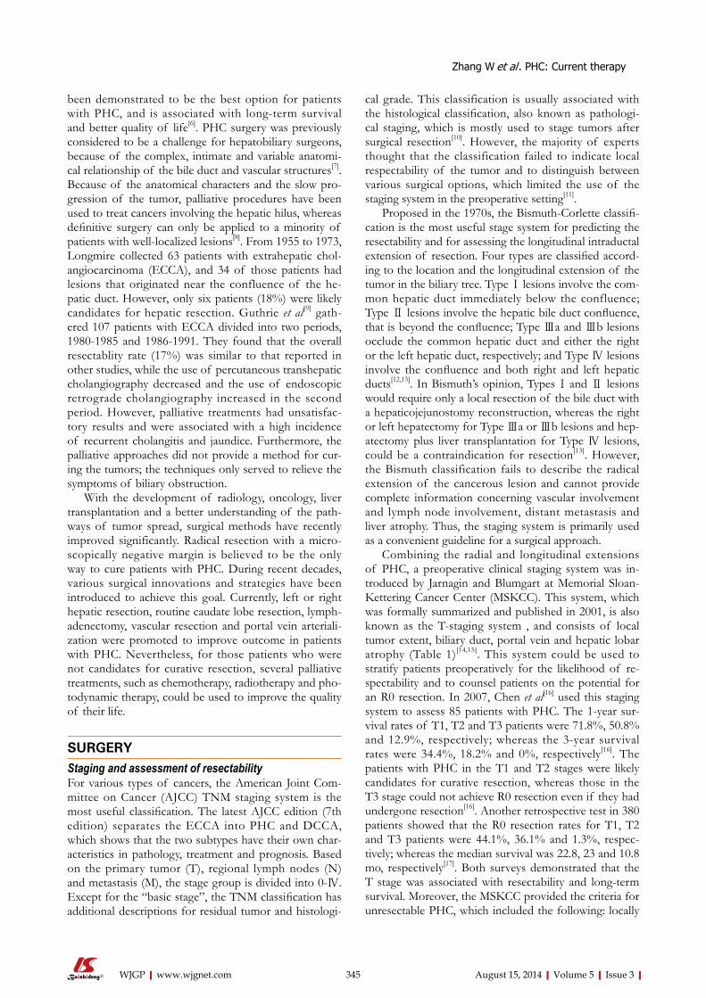
Zhang W et al . PHC: Current therapy
been demonstrated to be the best option for patients with PHC, and is associated with long-term survival and better quality of life[6]. PHC surgery was previously considered to be a challenge for hepatobiliary surgeons, because of the complex, intimate and variable anatomi-cal relationship of the bile duct and vascular structures[7]. Because of the anatomical characters and the slow pro-gression of the tumor, palliative procedures have been used to treat cancers involving the hepatic hilus, whereas definitive surgery can only be applied to a minority of patients with well-localized lesions[8]. From 1955 to 1973, Longmire collected 63 patients with extrahepatic chol-angiocarcinoma (ECCA), and 34 of those patients had lesions that originated near the confluence of the he-patic duct. However, only six patients (18%) were likely candidates for hepatic resection. Guthrie et al[9] gath-ered 107 patients with ECCA divided into two periods, 1980-1985 and 1986-1991. They found that the overall resectablity rate (17%) was similar to that reported in other studies, while the use of percutaneous transhepatic cholangiography decreased and the use of endoscopic retrograde cholangiography increased in the second period. However, palliative treatments had unsatisfac-tory results and were associated with a high incidence of recurrent cholangitis and jaundice. Furthermore, the palliative approaches did not provide a method for cur-ing the tumors; the techniques only served to relieve the symptoms of biliary obstruction.
With the development of radiology, oncology, liver transplantation and a better understanding of the path-ways of tumor spread, surgical methods have recently improved significantly. Radical resection with a micro-scopically negative margin is believed to be the only way to cure patients with PHC. During recent decades, various surgical innovations and strategies have been introduced to achieve this goal. Currently, left or right hepatic resection, routine caudate lobe resection, lymph-adenectomy, vascular resection and portal vein arteriali-zation were promoted to improve outcome in patients with PHC. Nevertheless, for those patients who were not candidates for curative resection, several palliative treatments, such as chemotherapy, radiotherapy and pho-todynamic therapy, could be used to improve the quality of their life.
SURGERYStaging and assessment of resectabilityFor various types of cancers, the American Joint Com-mittee on Cancer (AJCC) TNM staging system is the most useful classification. The latest AJCC edition (7th
edition) separates the ECCA into PHC and DCCA, which shows that the two subtypes have their own char-acteristics in pathology, treatment and prognosis. Based on the primary tumor (T), regional lymph nodes (N) and metastasis (M), the stage group is divided into 0-Ⅳ. Except for the “basic stage”, the TNM classification has additional descriptions for residual tumor and histologi-
cal grade. This classification is usually associated with the histological classification, also known as pathologi-cal staging, which is mostly used to stage tumors after surgical resection[10]. However, the majority of experts thought that the classification failed to indicate local respectability of the tumor and to distinguish between various surgical options, which limited the use of the staging system in the preoperative setting[11].
Proposed in the 1970s, the Bismuth-Corlette classifi-cation is the most useful stage system for predicting the resectability and for assessing the longitudinal intraductal extension of resection. Four types are classified accord-ing to the location and the longitudinal extension of the tumor in the biliary tree. Type Ⅰ lesions involve the com-mon hepatic duct immediately below the confluence; Type Ⅱ lesions involve the hepatic bile duct confluence, that is beyond the confluence; Type Ⅲa and Ⅲb lesions occlude the common hepatic duct and either the right or the left hepatic duct, respectively; and Type Ⅳ lesions involve the confluence and both right and left hepatic ducts[12,13]. In Bismuth’s opinion, Types Ⅰ and Ⅱ lesions would require only a local resection of the bile duct with a hepaticojejunostomy reconstruction, whereas the right or left hepatectomy for Type Ⅲa or Ⅲb lesions and hep-atectomy plus liver transplantation for Type Ⅳ lesions, could be a contraindication for resection[13]. However, the Bismuth classification fails to describe the radical extension of the cancerous lesion and cannot provide complete information concerning vascular involvement and lymph node involvement, distant metastasis and liver atrophy. Thus, the staging system is primarily used as a convenient guideline for a surgical approach.
Combining the radial and longitudinal extensions of PHC, a preoperative clinical staging system was in-troduced by Jarnagin and Blumgart at Memorial Sloan-Kettering Cancer Center (MSKCC). This system, which was formally summarized and published in 2001, is also known as the T-staging system , and consists of local tumor extent, biliary duct, portal vein and hepatic lobar atrophy (Table 1)[14,15]. This system could be used to stratify patients preoperatively for the likelihood of re-spectability and to counsel patients on the potential for an R0 resection. In 2007, Chen et al[16] used this staging system to assess 85 patients with PHC. The 1-year sur-vival rates of T1, T2 and T3 patients were 71.8%, 50.8% and 12.9%, respectively; whereas the 3-year survival rates were 34.4%, 18.2% and 0%, respectively[16]. The patients with PHC in the T1 and T2 stages were likely candidates for curative resection, whereas those in the T3 stage could not achieve R0 resection even if they had undergone resection[16]. Another retrospective test in 380 patients showed that the R0 resection rates for T1, T2 and T3 patients were 44.1%, 36.1% and 1.3%, respec-tively; whereas the median survival was 22.8, 23 and 10.8 mo, respectively[17]. Both surveys demonstrated that the T stage was associated with resectability and long-term survival. Moreover, the MSKCC provided the criteria for unresectable PHC, which included the following: locally
August 15, 2014|Volume 5|Issue 3|WJGP|www.wjgnet.com 345
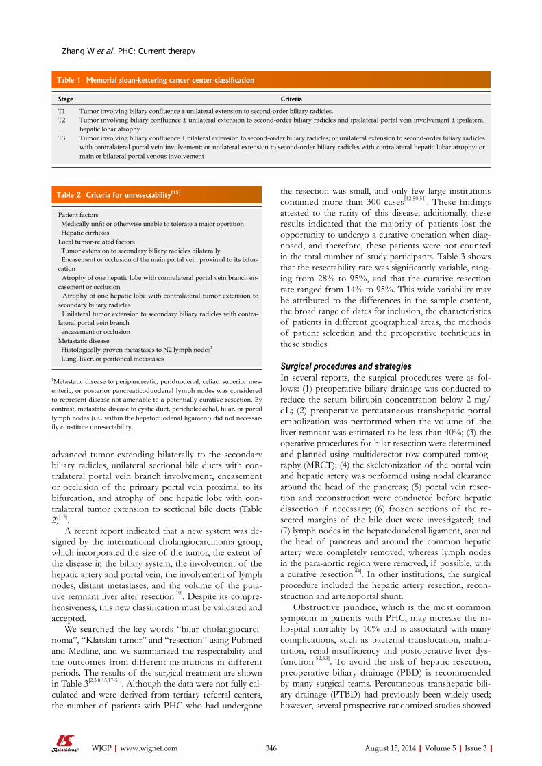
Table 2 Criteria for unresectability[15]
advanced tumor extending bilaterally to the secondary biliary radicles, unilateral sectional bile ducts with con-tralateral portal vein branch involvement, encasement or occlusion of the primary portal vein proximal to its bifurcation, and atrophy of one hepatic lobe with con-tralateral tumor extension to sectional bile ducts (Table 2)[15].
A recent report indicated that a new system was de-signed by the international cholangiocarcinoma group, which incorporated the size of the tumor, the extent of the disease in the biliary system, the involvement of the hepatic artery and portal vein, the involvement of lymph nodes, distant metastases, and the volume of the puta-tive remnant liver after resection[10]. Despite its compre-hensiveness, this new classification must be validated and accepted.
We searched the key words “hilar cholangiocarci-noma”, “Klatskin tumor” and “resection” using Pubmed and Medline, and we summarized the respectability and the outcomes from different institutions in different periods. The results of the surgical treatment are shown in Table 3[2,3,8,15,17-51]. Although the data were not fully cal-culated and were derived from tertiary referral centers, the number of patients with PHC who had undergone
the resection was small, and only few large institutions contained more than 300 cases[42,50,51]. These findings attested to the rarity of this disease; additionally, these results indicated that the majority of patients lost the opportunity to undergo a curative operation when diag-nosed, and therefore, these patients were not counted in the total number of study participants. Table 3 shows that the resectability rate was significantly variable, rang-ing from 28% to 95%, and that the curative resection rate ranged from 14% to 95%. This wide variability may be attributed to the differences in the sample content, the broad range of dates for inclusion, the characteristics of patients in different geographical areas, the methods of patient selection and the preoperative techniques in these studies.
Surgical procedures and strategiesIn several reports, the surgical procedures were as fol-lows: (1) preoperative biliary drainage was conducted to reduce the serum bilirubin concentration below 2 mg/dL; (2) preoperative percutaneous transhepatic portal embolization was performed when the volume of the liver remnant was estimated to be less than 40%; (3) the operative procedures for hilar resection were determined and planned using multidetector row computed tomog-raphy (MRCT); (4) the skeletonization of the portal vein and hepatic artery was performed using nodal clearance around the head of the pancreas; (5) portal vein resec-tion and reconstruction were conducted before hepatic dissection if necessary; (6) frozen sections of the re-sected margins of the bile duct were investigated; and (7) lymph nodes in the hepatoduodenal ligament, around the head of pancreas and around the common hepatic artery were completely removed, whereas lymph nodes in the para-aortic region were removed, if possible, with a curative resection[44]. In other institutions, the surgical procedure included the hepatic artery resection, recon-struction and arterioportal shunt.
Obstructive jaundice, which is the most common symptom in patients with PHC, may increase the in-hospital mortality by 10% and is associated with many complications, such as bacterial translocation, malnu-trition, renal insufficiency and postoperative liver dys-function[52,53]. To avoid the risk of hepatic resection, preoperative biliary drainage (PBD) is recommended by many surgical teams. Percutaneous transhepatic bili-ary drainage (PTBD) had previously been widely used; however, several prospective randomized studies showed
August 15, 2014|Volume 5|Issue 3|WJGP|www.wjgnet.com 346
Table 1 Memorial sloan-kettering cancer center classification
Stage Criteria
T1 Tumor involving biliary confluence ± unilateral extension to second-order biliary radicles. T2 Tumor involving biliary confluence ± unilateral extension to second-order biliary radicles and ipsilateral portal vein involvement ± ipsilateral
hepatic lobar atrophyT3 Tumor involving biliary confluence + bilateral extension to second-order biliary radicles; or unilateral extension to second-order biliary radicles
with contralateral portal vein involvement; or unilateral extension to second-order biliary radicles with contralateral hepatic lobar atrophy; or main or bilateral portal venous involvement
Patient factors Medically unfit or otherwise unable to tolerate a major operation Hepatic cirrhosisLocal tumor-related factors Tumor extension to secondary biliary radicles bilaterally Encasement or occlusion of the main portal vein proximal to its bifur-cation Atrophy of one hepatic lobe with contralateral portal vein branch en-casement or occlusion Atrophy of one hepatic lobe with contralateral tumor extension to secondary biliary radicles Unilateral tumor extension to secondary biliary radicles with contra-lateral portal vein branch encasement or occlusionMetastatic disease Histologically proven metastases to N2 lymph nodes1
Lung, liver, or peritoneal metastases
1Metastatic disease to peripancreatic, periduodenal, celiac, superior mes-enteric, or posterior pancreaticoduodenal lymph nodes was considered to represent disease not amenable to a potentially curative resection. By contrast, metastatic disease to cystic duct, pericholedochal, hilar, or portal lymph nodes (i.e., within the hepatoduodenal ligament) did not necessar-ily constitute unresectability.
Zhang W et al . PHC: Current therapy

that PTBD had no benefit in postoperative morbidity and mortality but increased potential risks, such as vas-cular injury, infectious complications and tumor seeding metastasis[54-56]. Currently, endoscopic nasobiliary drain-age (ENBD) is performed instead of PTBD because of fewer complications and better outcomes. More re-cently, the Nagoya Institute demonstrated that unilateral ENBD of the future remnant lobe(s) exhibited a high success rate as an effective and suitable PBD method even in BC type Ⅲ to Ⅳ lesions[57]. To avoid the postop-erative liver dysfunction resulting from extended hepatic resection, many institutions have promoted portal vein embolization (PVE) to increase volume of the future liver remnant (FLR). In several cautious surgical centers, when the FLR was 40% or less of the total liver volume, PVE was performed because the serum bilirubin level had decreased to less than 10 mg/dL[41,46]. Subsequently, surgery was performed after 2-4 wk of liver hypertrophy due to clonal expansion and cellular response[58].
When determining the surgical approach, the local
excision, hepatectomy, and extended hepatectomy with or without caudate resection should be considered. In the Bismuth’s opinion, Bismuth Type Ⅰ and Ⅱ would re-quire only a local resection. Recently, bile duct resection alone without hepatectomy has been largely abandoned in favor of a more aggressive approach. Capussotti et al[59] conducted a systematic review of the effect of local resection compared with hepatectomy. In the pathologic aspect, the isolated bile duct cannot be adequately re-sected, because of the following: the necessity for wide surgical margins; neoplastic extension along the perineu-ral sheaths and segment 1 neoplastic invasion. From an-other perspective, the R0 resection rate was higher after combined liver resection, although, in the earlier years of its application, local resection could be associated with fewer complications and shorter lengths of hospital stay[15,21,35]. In conclusion, according to this systematic re-view, local resection should only be scheduled for small papillary Klatskin tumors without bile duct confluence involvement confined to the bile duct wall[59]. Because
August 15, 2014|Volume 5|Issue 3|WJGP|www.wjgnet.com 347
Table 3 Results of surgical resection for perihilar cholangiocarcinoma
Ref. Published year Resections Resectablity(%)
Negative margin(%)
Liver resection(%)
Morbidity Mortality 5-yr survival rate (%)
Hadjis et al[18] 1990 27 NA 56 60 NA 7 22Nakeeb et al[2] 1996 109 56 26 14 47 4 11Su et al[19] 1996 49 28 49 57 47 10 15Klempnauer et al[20] 1997 151 45 77 77 NA 10 28Miyazaki et al[21] 1998 76 NA 71 86 34 13 26Neuhaus et al[22] 1999 80 NA 61 85 55 8 22Kosuge et al[8] 1999 65 73 52 80 37 9 33Gerhards et al[23] 2000 112 NA 14 29 65 18 NANimura et al[24] 2000 142 80 61 90 49 9 26Todoroki et al[25] 2000 101 89 14 58 14 4 28Jarnagin et al[15] 2001 80 50 78 78 64 10 26Kawarada et al[27] 2002 65 89 64 75 28 2.3 26Capussotti et al[26] 2002 36 NA 89 83 47 3 27Kawasaki et al[28] 2003 79 75 68 87 14 1.3 22Seyama et al[29] 2003 87 94 64 67 43 0 40Rea et al[32] 2004 46 NA 80 100 52 9 26Kondo et al[31] 2004 40 95 95 65 48 0 NAI.Jitsma et al[30] 2004 42 NA 65 100 76 12 19Hemming et al[33] 2005 53 50 80 98 40 9 35Jarnagin et al[34] 2005 106 70 77 82 62 8 NADinant et al[35] 2006 99 NA 31 38 66 15 27DeOliveira et al[3] 2007 173 62 19 20 61 5 10Ito et al[36] 2008 38 55 63 53 32 0 33Konstadoulakis et al[37] 2008 59 81 68.6 86.4 25.5 6.8 34.9Igami et al[41] 2010 298 70 74 98 43 2 42Hirano et al[40] 2010 146 NA 87 87 44 3.4 35.5Lee et al[42] 2010 302 86 70.9 89 43 1.7 32.5Unno et al[44] 2010 125 NA 63.2 100 48.7 8 34.7Ercolani et al[38] 2010 51 49.6 72.5 98 51 10 34.1Shimizu et al[43] 2010 224 NA 69.1 78 47.6 10.7 30.3Giuliante et al[39] 2010 43 29 77 93 52.5 6.9 36.1Regimbeau et al[45] 2011 56 NA 76.9 100 72 8 NAYoung et al[48] 2012 83 92 42.2 93 62.7 7 20Saxena et al[47] 2012 54 64 64.3 42 45.2 2.4 24Ribero et al[46] 2012 82 NA 81.7 91.5 64.6 9.7 28De Jong et al[50] 2012 305 NA 64.2 73 NA 10.6 20.2Matsuo et al[17] 2012 157 78 76 90 59.2 7.6 37.5Cheng et al[49] 2012 176 34 78.4 97 26.3 2.9 13.5Nagino et al[51] 2013 574 76.1 76.5 96.7 57.3 4.7 32.5
Zhang W et al . PHC: Current therapy
NA: Not applicable.
of the rarity and the advanced stage of the disease at the time of diagnosis, a local resection was rarely performed.
Despite the incomplete accuracy, the Bismuth classi-fication initiated the idea of wider resection for PHC[13]. Table 3 shows that liver resection rates increased from 14% to 100% with an increased R0 resection rate. The common liver resection strategies are as follows: right or left hepatectomy (resection of hepatic segments 5, 6, 7, 8 or 2, 3, 4 ± 1), right or left hepatic trisectionectomy, also called extended right or left hepatectomy (resection of hepatic segments 4, 5, 6, 7, 8 or 2, 3, 4, 5, 8 ± 1), and central hepatectomy. Bisectionectomy or more was de-fined as a major hepatectomy; sectionectomy or less was defined as a minor hepatectomy[38]. Currently, for those patients with Bismuth type Ⅰ and Ⅱ, the right hepatec-tomy with caudate lobectomy was recommended, which has been demonstrated to decrease the rate of recur-rence[29]. However, for those patients with Bismuth types Ⅲ and Ⅳ lesions, the approaches varied in different institutions. Recently, Cheng et al[49] reported 171 patients with PHC of Bismuth types Ⅲ and Ⅳ lesions. For Bis-muth Type Ⅲ lesions, right, left or central hepatectomy with caudate lobectomy was performed. For Bismuth Ⅳ lesions, the right or left hepatectomy or extended right or left hepatectomy with caudate lobectomy was con-ducted to increase the negative margin rates. The choice of surgical side may depend on the predominance of the tumor; however, the right trisectionectomy is indi-cated for centrally located tumors because of the length of each hepatic duct, the location of the hilar common bile duct in the hepatoduodenal ligament, the ease of complete caudate lobectomy and portal vein reconstruc-tion, and the frequent involvement of the right hepatic artery[7,28]. The left hepatectomy is considered to be a more complicated procedure than the right hepatectomy and requires greater skill, especially in cases involving portal vein resection and reconstruction. Moreover, pre-serving the right hepatic artery and the right portal vein could be an oncological problem with left or extended left resection, which could increase the tumor cell dis-semination. Therefore, the rate of left hepatectomies is approximately 25%-30% of all resections[60]. In the study by Shimizu et al[43], the R0 resection was achieved in all 7 patients who underwent right trisectionectomy, but in only 8 (61.5%) of 13 patients who underwent left trisec-tionectomy. This finding suggests that a more extended resection from the right side, but not from the left side, may provide greater potential for curability. However, several authors believed that the left extended hepa-tectomy could achieve the same result. Nagino et al[51] analyzed the patients with PHC who underwent surgery and compared the surgical strategies in different periods (Table 4). From their experience, the incidence of left hepatic trisectionectomies gradually increased while the incidence of central hepatectomies decreased. Totally, the left or extended left hepatectomy represented nearly 55% of all of the resections performed on patients with PHC.
Nimura et al[61] introduced the concept of routine caudate lobectomy (CL). Bilateral biliary branches of the caudate lobe are confluent with the right hepatic duct, the left hepatic duct, the confluence of these and the right posterior hepatic duct. Therefore, the caudate lobe is usually involved in PHC in 40% to 98% of patients, which indicates a need for CL[61-63]. Moreover, routine CL combined with resection had high curative resectablity rates and increased the likelihood of long-term survival for patients with advanced stage PHC[49]. Similarly, Kow et al[64] showed that the patients with CL had a signifi-cantly better overall survival rate of 64.0 mo compared to the survival rate of 34.6 mo in type Ⅲ PHC patients in the group without CL. Although mechanisms for CL have not been established, the outcome remains optimis-tic while undertaking CL in PHC.
A major hepatectomy combined with pancreatoduo-denectomy, for example, hepatopancreatoduodenectomy (HPD), was routinely used in the PHC surgery in several institutions. This procedure occupied 12.9% of the total surgery cases, and was indicated in the following cases: (1) diffusely infiltrating tumors of the entire extrahepatic bile duct; and (2) downward superficial spreading, or bulky nodal metastases of the pancreatoduodenal region (Table 4)[65]. Therefore, HPB provides an important method for treating spreading unresectable cholangio-carcinoma; thus, it is now the fourth standard procedure following hepatectomy, bile duct resection, and pancre-atoduodenectomy[66].
In several high-volume samples, PHC was frequently reported to metastasize via the lymphatics in 24% to 75% of the patients[42,51]. Moreover, many authors had demonstrated that lymph node metastasis had a nega-tive impact on survival in PHC[3,28,29,31,33,42,51]. Thereafter, lymphadenectomy played a crucial role in the outcome of patients with PHC. However, the 5-year survival rate is related to the location of the metastasis of the lymph node. Therefore, lymph node metastasis that is confined to the hepatic pedicle or the hepatoduodenal ligament is not a reason for abandoning resection. The tumor posi-tive lymph nodes along the common hepatic artery or celiac axis are usually considered a contraindication for resection[7]. Kitagawa et al[67] showed that, in 110 patients after resection of PHC, there was a 5-year survival rate of 31%, if the lymph nodes were negative. However, in patients suffering from a local or a para-aortic lymph node infiltration, the 5-year survival rates were 15% and 12%, respectively. Interestingly, in the same report, 12% of the patients with positive para-aortic lymph nodes who lived more than 5 years were found to have mac-roscopically negative nodes in surgery[67]. Although the routine lymph node dissection beyond the hepatoduo-denal ligament is not generally recommended, several authors still believe that lymph node dissection is benefi-cial.
Due to the intimate relationship between the bile duct and vessels, PHC could usually infiltrate the portal vein and hepatic artery. The indication for portal vein
August 15, 2014|Volume 5|Issue 3|WJGP|www.wjgnet.com 348
Zhang W et al . PHC: Current therapy
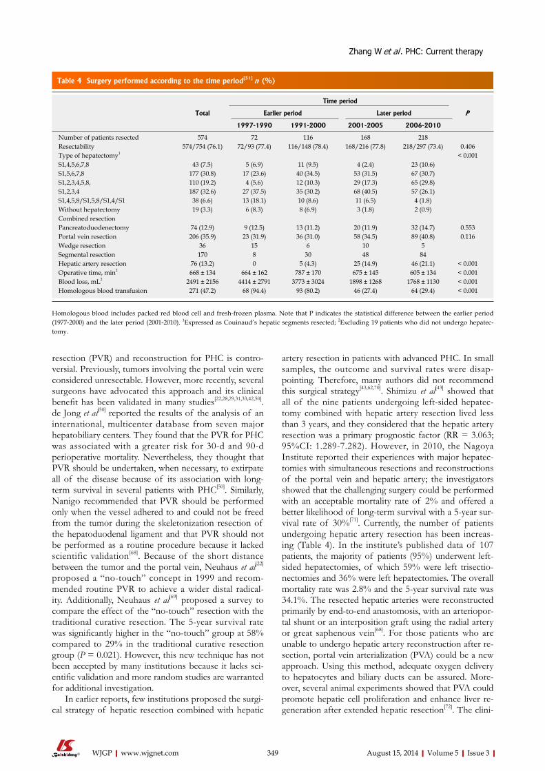
resection (PVR) and reconstruction for PHC is contro-versial. Previously, tumors involving the portal vein were considered unresectable. However, more recently, several surgeons have advocated this approach and its clinical benefit has been validated in many studies[22,28,29,31,33,42,50]. de Jong et al[50] reported the results of the analysis of an international, multicenter database from seven major hepatobiliary centers. They found that the PVR for PHC was associated with a greater risk for 30-d and 90-d perioperative mortality. Nevertheless, they thought that PVR should be undertaken, when necessary, to extirpate all of the disease because of its association with long-term survival in several patients with PHC[50]. Similarly, Nanigo recommended that PVR should be performed only when the vessel adhered to and could not be freed from the tumor during the skeletonization resection of the hepatoduodenal ligament and that PVR should not be performed as a routine procedure because it lacked scientific validation[68]. Because of the short distance between the tumor and the portal vein, Neuhaus et al[22] proposed a “no-touch” concept in 1999 and recom-mended routine PVR to achieve a wider distal radical-ity. Additionally, Neuhaus et al[69] proposed a survey to compare the effect of the “no-touch” resection with the traditional curative resection. The 5-year survival rate was significantly higher in the “no-touch” group at 58% compared to 29% in the traditional curative resection group (P = 0.021). However, this new technique has not been accepted by many institutions because it lacks sci-entific validation and more random studies are warranted for additional investigation.
In earlier reports, few institutions proposed the surgi-cal strategy of hepatic resection combined with hepatic
artery resection in patients with advanced PHC. In small samples, the outcome and survival rates were disap-pointing. Therefore, many authors did not recommend this surgical strategy[43,62,70]. Shimizu et al[43] showed that all of the nine patients undergoing left-sided hepatec-tomy combined with hepatic artery resection lived less than 3 years, and they considered that the hepatic artery resection was a primary prognostic factor (RR = 3.063; 95%CI: 1.289-7.282). However, in 2010, the Nagoya Institute reported their experiences with major hepatec-tomies with simultaneous resections and reconstructions of the portal vein and hepatic artery; the investigators showed that the challenging surgery could be performed with an acceptable mortality rate of 2% and offered a better likelihood of long-term survival with a 5-year sur-vival rate of 30%[71]. Currently, the number of patients undergoing hepatic artery resection has been increas-ing (Table 4). In the institute’s published data of 107 patients, the majority of patients (95%) underwent left-sided hepatectomies, of which 59% were left trisectio-nectomies and 36% were left hepatectomies. The overall mortality rate was 2.8% and the 5-year survival rate was 34.1%. The resected hepatic arteries were reconstructed primarily by end-to-end anastomosis, with an arteriopor-tal shunt or an interposition graft using the radial artery or great saphenous vein[68]. For those patients who are unable to undergo hepatic artery reconstruction after re-section, portal vein arterialization (PVA) could be a new approach. Using this method, adequate oxygen delivery to hepatocytes and biliary ducts can be assured. More-over, several animal experiments showed that PVA could promote hepatic cell proliferation and enhance liver re-generation after extended hepatic resection[72]. The clini-
August 15, 2014|Volume 5|Issue 3|WJGP|www.wjgnet.com 349
Table 4 Surgery performed according to the time period[51] n (%)
Time period
Total Earlier period Later period P
1997-1990 1991-2000 2001-2005 2006-2010
Number of patients resected 574 72 116 168 218Resectability 574/754 (76.1) 72/93 (77.4) 116/148 (78.4) 168/216 (77.8) 218/297 (73.4) 0.406Type of hepatectomy1 < 0.001S1,4,5,6,7,8 43 (7.5) 5 (6.9) 11 (9.5) 4 (2.4) 23 (10.6)S1,5,6,7,8 177 (30.8) 17 (23.6) 40 (34.5) 53 (31.5) 67 (30.7)S1,2,3,4,5,8, 110 (19.2) 4 (5.6) 12 (10.3) 29 (17.3) 65 (29.8)S1,2,3,4 187 (32.6) 27 (37.5) 35 (30.2) 68 (40.5) 57 (26.1)S1,4,5,8/S1,5,8/S1,4/S1 38 (6.6) 13 (18.1) 10 (8.6) 11 (6.5) 4 (1.8)Without hepatectomy 19 (3.3) 6 (8.3) 8 (6.9) 3 (1.8) 2 (0.9)Combined resectionPancreatoduodenectomy 74 (12.9) 9 (12.5) 13 (11.2) 20 (11.9) 32 (14.7) 0.553Portal vein resection 206 (35.9) 23 (31.9) 36 (31.0) 58 (34.5) 89 (40.8) 0.116Wedge resection 36 15 6 10 5Segmental resection 170 8 30 48 84Hepatic artery resection 76 (13.2) 0 5 (4.3) 25 (14.9) 46 (21.1) < 0.001Operative time, min2 668 ± 134 664 ± 162 787 ± 170 675 ± 145 605 ± 134 < 0.001Blood loss, mL2 2491 ± 2156 4414 ± 2791 3773 ± 3024 1898 ± 1268 1768 ± 1130 < 0.001Homologous blood transfusion 271 (47.2) 68 (94.4) 93 (80.2) 46 (27.4) 64 (29.4) < 0.001
Homologous blood includes packed red blood cell and fresh-frozen plasma. Note that P indicates the statistical difference between the earlier period (1977-2000) and the later period (2001-2010). 1Expressed as Couinaud’s hepatic segments resected; 2Excluding 19 patients who did not undergo hepatec-tomy.
Zhang W et al . PHC: Current therapy
cal significance of hepatic artery resection is debatable, yet also promising and encouraging.
Morbidity and mortalityIn Table 3, we summarize the morbidity and mortality which show significant variations, ranging from 14% to 76% and from 0% to 18%, respectively. Sano et al[73]
defined the complications as major when they resulted in organ failure or required another surgery or interven-tional radiology, such as liver failure, lung failure and renal failure[51,73]. Complications that were classified as minor include pleural effusion necessitating thoracocen-tesis, wound infection, intra-abdominal infection with positive culture of the drainage fluid, delayed gastric emptying, anastomotic leakage, clinically silent pancreatic fistula with amylase-rich serous fluid or contaminated fluid with positive culture, and bile leakage from the raw surface of the liver healing spontaneously or respond-ing to conservative management[73]. The most common complications observed in most institutions were infec-tive complications, especially during earlier years of the use of these procedures, representing 50% or more of the observed complications[3,15,36]. Nagiono et al[51] compared the complications between the earlier years and the more recent years, and they demonstrated that the incidence of grade C liver failure, which is clinically serious, decreased markedly from 18.2% from 1977 to 1990 to 3.2% from 2006 to 2010. Wound sepsis was the second most common complication, followed by intra-abdominal abscess and bile leakage[51].
The operative mortality included all in-hospital deaths as defined by Sano. All postoperative complica-tions that affected the outcome or lengthened the hospi-tal stay were considered. Death may be associated with acute liver failure after extended right hepatectomy and combined portal vein resection, and sepsis with multi-organ failure[45]. Overall, these extended liver as well as vascular resections were found to be significant predic-tors of increased mortality[23]. In addition to liver func-tion, operative time and blood loss may be associated with mortality[51]. Several reports have demonstrated that preoperative portal vein embolization may decrease mor-tality even with extended hepatectomy[73].
Outcomes and recurrenceThe average 5-year survival rates after resection for PHC range from 11% to 42% (Table 3). Factors associated with favorable outcome include the following: R0 resec-tion, no lymph node metastasis, absence of perineural and perivascular invasion, and well-differentiated histo-logical grade. Complete resection with negative histo-logic margins is the only modifiable factor and, for that reason, the primary aim of surgical therapy. Recently, several reports demonstrated that patients undergoing R1 resection (microscopically positive margin) had a lon-ger overall survival rate than patients with unresectable PHC[36,74]. Moreover, patients undergoing R0 resections with a margin less than 5 mm had the same survival
rate as those patients undergoing R1 resections[29]. The surgeons were encouraged to perform more aggressive surgery to achieve a better outcome.
Few studies have analyzed recurrence patterns and time to recurrence in patients with PHC. In several re-ports, tumor recurrence rates can be as high as 50% to 76%, and the median time to recurrence rates has been re-ported to be 12 to 43 mo[36,47,75,76]. The most common site of recurrence is a local site, followed by the liver, lymph node, peritoneum and other organs. Only histologic grade was associated with recurrence-free survival[47]. Generally, the patients with recurrent disease are not candidates for curative therapy and can only receive adjuvant therapy to improve long-term outcome.
ORTHOTOPIC LIVER TRANSPLANTATIONTheoretically, orthotopic liver transplantation (OLT) offers the advantage of the resection of all of the struc-tures that may be affected by tumor, for example, the portal vein, bilateral hepatic ducts and atrophic liver lobes. Compared to surgical resection, OLT has several advantages: (1) patients with Bismuth Ⅳ type lesions and peripheral vascular lesions cannot undergo resec-tion; (2) patients with PHC arising from primary scleros-ing cholangitis (PSC) will tolerate resection poorly be-cause of the underlying liver impairment; (3) dissection in the hepatic hilum has the potential for causing spill-age, which is an adverse prognostic factor; and (4) a clear circumferential margin is usually not achievable, which might increase the recurrence rates of PHC[77]. However, in the early years of the application of this procedure, the results were disappointing. The Cincinnati Trans-plant Tumor Registry collected global data between 1968 and 1997. The 1-, 2-, and 5-year survival rates were 72%, 48%, and 23%, respectively. Eighty four percent of the patients had a recurrence within 2 years of transplanta-tion[78]. This undesirable result may have been associated with the unselected patients who had distant metastasis. Despite this finding, PHC was considered to be a rela-tive contraindication to OLT due to the lack of organs. Interestingly, several investigations found that those patients with negative margins in transplantation and the absence of regional lymph node metastases had a better survival rate. Moreover, 22% of the patients receiving radiotherapy and chemotherapy alone had a 5-year sur-vival, which inspired several surgeons to explore a new OLT approach for PHC.
From 1987 to 2000, Miyazaki et al[70] collected 17 patients who were treated with systemic chemotherapy and intraluminal bile duct irradiation as they awaited liver transplantation. Eleven patients underwent liver transplantation, and until 2000, five patients were alive without evidence of tumor recurrence with a median follow-up of 7.5 years (range, 2.8-14.5 years). In 1994, the Mayo Clinic developed a protocol employing preop-erative chemoradiation therapy followed by liver trans-plantation, which showed encouraging results. Currently,
August 15, 2014|Volume 5|Issue 3|WJGP|www.wjgnet.com 350
Zhang W et al . PHC: Current therapy
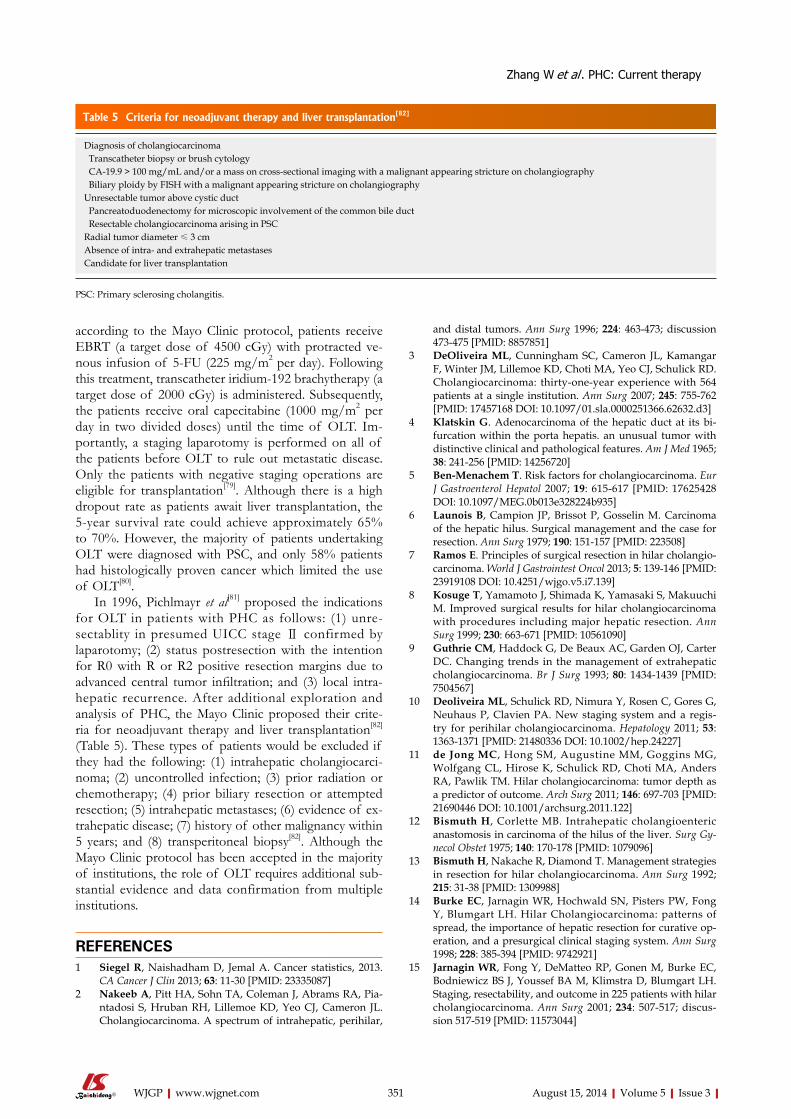
according to the Mayo Clinic protocol, patients receive EBRT (a target dose of 4500 cGy) with protracted ve-nous infusion of 5-FU (225 mg/m2 per day). Following this treatment, transcatheter iridium-192 brachytherapy (a target dose of 2000 cGy) is administered. Subsequently, the patients receive oral capecitabine (1000 mg/m2 per day in two divided doses) until the time of OLT. Im-portantly, a staging laparotomy is performed on all of the patients before OLT to rule out metastatic disease. Only the patients with negative staging operations are eligible for transplantation[79]. Although there is a high dropout rate as patients await liver transplantation, the 5-year survival rate could achieve approximately 65% to 70%. However, the majority of patients undertaking OLT were diagnosed with PSC, and only 58% patients had histologically proven cancer which limited the use of OLT[80].
In 1996, Pichlmayr et al[81] proposed the indications for OLT in patients with PHC as follows: (1) unre-sectablity in presumed UICC stage Ⅱ confirmed by laparotomy; (2) status postresection with the intention for R0 with R or R2 positive resection margins due to advanced central tumor infiltration; and (3) local intra-hepatic recurrence. After additional exploration and analysis of PHC, the Mayo Clinic proposed their crite-ria for neoadjuvant therapy and liver transplantation[82] (Table 5). These types of patients would be excluded if they had the following: (1) intrahepatic cholangiocarci-noma; (2) uncontrolled infection; (3) prior radiation or chemotherapy; (4) prior biliary resection or attempted resection; (5) intrahepatic metastases; (6) evidence of ex-trahepatic disease; (7) history of other malignancy within 5 years; and (8) transperitoneal biopsy[82]. Although the Mayo Clinic protocol has been accepted in the majority of institutions, the role of OLT requires additional sub-stantial evidence and data confirmation from multiple institutions.
REFERENCES1 Siegel R, Naishadham D, Jemal A. Cancer statistics, 2013.
CA Cancer J Clin 2013; 63: 11-30 [PMID: 23335087]2 Nakeeb A, Pitt HA, Sohn TA, Coleman J, Abrams RA, Pia-
ntadosi S, Hruban RH, Lillemoe KD, Yeo CJ, Cameron JL. Cholangiocarcinoma. A spectrum of intrahepatic, perihilar,
and distal tumors. Ann Surg 1996; 224: 463-473; discussion 473-475 [PMID: 8857851]
3 DeOliveira ML, Cunningham SC, Cameron JL, Kamangar F, Winter JM, Lillemoe KD, Choti MA, Yeo CJ, Schulick RD. Cholangiocarcinoma: thirty-one-year experience with 564 patients at a single institution. Ann Surg 2007; 245: 755-762 [PMID: 17457168 DOI: 10.1097/01.sla.0000251366.62632.d3]
4 Klatskin G. Adenocarcinoma of the hepatic duct at its bi-furcation within the porta hepatis. an unusual tumor with distinctive clinical and pathological features. Am J Med 1965; 38: 241-256 [PMID: 14256720]
5 Ben-Menachem T. Risk factors for cholangiocarcinoma. Eur J Gastroenterol Hepatol 2007; 19: 615-617 [PMID: 17625428 DOI: 10.1097/MEG.0b013e328224b935]
6 Launois B, Campion JP, Brissot P, Gosselin M. Carcinoma of the hepatic hilus. Surgical management and the case for resection. Ann Surg 1979; 190: 151-157 [PMID: 223508]
7 Ramos E. Principles of surgical resection in hilar cholangio-carcinoma. World J Gastrointest Oncol 2013; 5: 139-146 [PMID: 23919108 DOI: 10.4251/wjgo.v5.i7.139]
8 Kosuge T, Yamamoto J, Shimada K, Yamasaki S, Makuuchi M. Improved surgical results for hilar cholangiocarcinoma with procedures including major hepatic resection. Ann Surg 1999; 230: 663-671 [PMID: 10561090]
9 Guthrie CM, Haddock G, De Beaux AC, Garden OJ, Carter DC. Changing trends in the management of extrahepatic cholangiocarcinoma. Br J Surg 1993; 80: 1434-1439 [PMID: 7504567]
10 Deoliveira ML, Schulick RD, Nimura Y, Rosen C, Gores G, Neuhaus P, Clavien PA. New staging system and a regis-try for perihilar cholangiocarcinoma. Hepatology 2011; 53: 1363-1371 [PMID: 21480336 DOI: 10.1002/hep.24227]
11 de Jong MC, Hong SM, Augustine MM, Goggins MG, Wolfgang CL, Hirose K, Schulick RD, Choti MA, Anders RA, Pawlik TM. Hilar cholangiocarcinoma: tumor depth as a predictor of outcome. Arch Surg 2011; 146: 697-703 [PMID: 21690446 DOI: 10.1001/archsurg.2011.122]
12 Bismuth H, Corlette MB. Intrahepatic cholangioenteric anastomosis in carcinoma of the hilus of the liver. Surg Gy-necol Obstet 1975; 140: 170-178 [PMID: 1079096]
13 Bismuth H, Nakache R, Diamond T. Management strategies in resection for hilar cholangiocarcinoma. Ann Surg 1992; 215: 31-38 [PMID: 1309988]
14 Burke EC, Jarnagin WR, Hochwald SN, Pisters PW, Fong Y, Blumgart LH. Hilar Cholangiocarcinoma: patterns of spread, the importance of hepatic resection for curative op-eration, and a presurgical clinical staging system. Ann Surg 1998; 228: 385-394 [PMID: 9742921]
15 Jarnagin WR, Fong Y, DeMatteo RP, Gonen M, Burke EC, Bodniewicz BS J, Youssef BA M, Klimstra D, Blumgart LH. Staging, resectability, and outcome in 225 patients with hilar cholangiocarcinoma. Ann Surg 2001; 234: 507-517; discus-sion 517-519 [PMID: 11573044]
August 15, 2014|Volume 5|Issue 3|WJGP|www.wjgnet.com 351
Table 5 Criteria for neoadjuvant therapy and liver transplantation[82]
Diagnosis of cholangiocarcinoma Transcatheter biopsy or brush cytology CA-19.9 > 100 mg/mL and/or a mass on cross-sectional imaging with a malignant appearing stricture on cholangiography Biliary ploidy by FISH with a malignant appearing stricture on cholangiographyUnresectable tumor above cystic duct Pancreatoduodenectomy for microscopic involvement of the common bile duct Resectable cholangiocarcinoma arising in PSCRadial tumor diameter ≤ 3 cmAbsence of intra- and extrahepatic metastasesCandidate for liver transplantation
PSC: Primary sclerosing cholangitis.
Zhang W et al . PHC: Current therapy
16 Chen RF, Li ZH, Zhou JJ, Wang J, Chen JS, Lin Q, Tang QB, Peng NF, Jiang ZP, Zhou QB. Preoperative evaluation with T-staging system for hilar cholangiocarcinoma. World J Gas-troenterol 2007; 13: 5754-5759 [PMID: 17963304]
17 Matsuo K, Rocha FG, Ito K, D’Angelica MI, Allen PJ, Fong Y, Dematteo RP, Gonen M, Endo I, Jarnagin WR. The Blumgart preoperative staging system for hilar cholangiocarcinoma: analysis of resectability and outcomes in 380 patients. J Am Coll Surg 2012; 215: 343-355 [PMID: 22749003 DOI: 10.1016/j.jamcollsurg.2012.05.025]
18 Hadjis NS , Blenkharn JI, Alexander N, Benjamin IS, Blumgart LH. Outcome of radical surgery in hilar cholan-giocarcinoma. Surgery 1990; 107: 597-604 [PMID: 2162082]
19 Su CH, Tsay SH, Wu CC, Shyr YM, King KL, Lee CH, Lui WY, Liu TJ, P’eng FK. Factors influencing postoperative morbidity, mortality, and survival after resection for hilar cholangiocarcinoma. Ann Surg 1996; 223: 384-394 [PMID: 8633917]
20 Klempnauer J, Ridder GJ, von Wasielewski R, Werner M, Weimann A, Pichlmayr R. Resectional surgery of hilar chol-angiocarcinoma: a multivariate analysis of prognostic fac-tors. J Clin Oncol 1997; 15: 947-954 [PMID: 9060532]
21 Miyazaki M, Ito H, Nakagawa K, Ambiru S, Shimizu H, Shimizu Y, Kato A, Nakamura S, Omoto H, Nakajima N, Kimura F, Suwa T. Aggressive surgical approaches to hilar cholangiocarcinoma: hepatic or local resection? Surgery 1998; 123: 131-136 [PMID: 9481397]
22 Neuhaus P, Jonas S, Bechstein WO, Lohmann R, Radke C, Kling N, Wex C, Lobeck H, Hintze R. Extended resections for hilar cholangiocarcinoma. Ann Surg 1999; 230: 808-818; discussion 819 [PMID: 10615936]
23 Gerhards MF, van Gulik TM, de Wit LT, Obertop H, Gou-ma DJ. Evaluation of morbidity and mortality after resection for hilar cholangiocarcinoma--a single center experience. Surgery 2000; 127: 395-404 [PMID: 10776430]
24 Nimura Y, Kamiya J, Kondo S, Nagino M, Uesaka K, Oda K, Sano T, Yamamoto H, Hayakawa N. Aggressive preopera-tive management and extended surgery for hilar cholan-giocarcinoma: Nagoya experience. J Hepatobiliary Pancreat Surg 2000; 7: 155-162 [PMID: 10982608 DOI: 10.1007/s005340000070155.534]
25 Todoroki T, Kawamoto T, Koike N, Takahashi H, Yoshida S, Kashiwagi H, Takada Y, Otsuka M, Fukao K. Radical resec-tion of hilar bile duct carcinoma and predictors of survival. Br J Surg 2000; 87: 306-313 [PMID: 10718799 DOI: 10.1046/j.1365-2168.2000.01343.x]
26 Capussotti L, Muratore A, Polastri R, Ferrero A, Massucco P. Liver resection for hilar cholangiocarcinoma: in-hospital mortality and longterm survival. J Am Coll Surg 2002; 195: 641-647 [PMID: 12437251]
27 Kawarada Y, Das BC, Naganuma T, Tabata M, Taoka H. Surgical treatment of hilar bile duct carcinoma: experience with 25 consecutive hepatectomies. J Gastrointest Surg 2002; 6: 617-624 [PMID: 12127130]
28 Kawasaki S, Imamura H, Kobayashi A, Noike T, Miwa S, Miyagawa S. Results of surgical resection for patients with hilar bile duct cancer: application of extended hepatectomy after biliary drainage and hemihepatic portal vein embo-lization. Ann Surg 2003; 238: 84-92 [PMID: 12832969 DOI: 10.1097/01.sla.0000074984.83031.02]
29 Seyama Y, Kubota K, Sano K, Noie T, Takayama T, Kosuge T, Makuuchi M. Long-term outcome of extended hemihepatec-tomy for hilar bile duct cancer with no mortality and high survival rate. Ann Surg 2003; 238: 73-83 [PMID: 12832968 DOI: 10.1097/01.sla.0000074960.55004.72]
30 IJitsma AJ, Appeltans BM, de Jong KP, Porte RJ, Peeters PM, Slooff MJ. Extrahepatic bile duct resection in combina-tion with liver resection for hilar cholangiocarcinoma: a report of 42 cases. J Gastrointest Surg 2004; 8: 686-694 [PMID: 15358329 DOI: 10.1016/j.gassur.2004.04.006]
31 Kondo S, Hirano S, Ambo Y, Tanaka E, Okushiba S, Mori-kawa T, Katoh H. Forty consecutive resections of hilar cholangiocarcinoma with no postoperative mortality and no positive ductal margins: results of a prospective study. Ann Surg 2004; 240: 95-101 [PMID: 15213624]
32 Rea DJ, Munoz-Juarez M, Farnell MB, Donohue JH, Que FG, Crownhart B, Larson D, Nagorney DM. Major hepatic resection for hilar cholangiocarcinoma: analysis of 46 pa-tients. Arch Surg 2004; 139: 514-523; discussion 523-525 [PMID: 15136352 DOI: 10.1001/archsurg.139.5.514]
33 Hemming AW, Reed AI, Fujita S, Foley DP, Howard RJ. Surgical management of hilar cholangiocarcinoma. Ann Surg 2005; 241: 693-699; discussion 693-699 [PMID: 15849505]
34 Jarnagin WR, Bowne W, Klimstra DS, Ben-Porat L, Roggin K, Cymes K, Fong Y, DeMatteo RP, D’Angelica M, Koea J, Blumgart LH. Papillary phenotype confers improved sur-vival after resection of hilar cholangiocarcinoma. Ann Surg 2005; 241: 703-712; discussion 712-714 [PMID: 15849506]
35 Dinant S, Gerhards MF, Rauws EA, Busch OR, Gouma DJ, van Gulik TM. Improved outcome of resection of hilar chol-angiocarcinoma (Klatskin tumor). Ann Surg Oncol 2006; 13: 872-880 [PMID: 16614876 DOI: 10.1245/aso.2006.05.053]
36 Ito F, Agni R, Rettammel RJ, Been MJ, Cho CS, Mahvi DM, Rikkers LF, Weber SM. Resection of hilar cholangiocarci-noma: concomitant liver resection decreases hepatic recur-rence. Ann Surg 2008; 248: 273-279 [PMID: 18650638 DOI: 10.1097/SLA.0b013e31817f2bfd]
37 Konstadoulakis MM, Roayaie S, Gomatos IP, Labow D, Fiel MI, Miller CM, Schwartz ME. Aggressive surgical resection for hilar cholangiocarcinoma: is it justified? Audit of a single center’s experience. Am J Surg 2008; 196: 160-169 [PMID: 18466862 DOI: 10.1016/j.amjsurg.2007.07.033]
38 Ercolani G, Zanello M, Grazi GL, Cescon M, Ravaioli M, Del Gaudio M, Vetrone G, Cucchetti A, Brandi G, Ramaccia-to G, Pinna AD. Changes in the surgical approach to hilar cholangiocarcinoma during an 18-year period in a Western single center. J Hepatobiliary Pancreat Sci 2010; 17: 329-337 [PMID: 20464563 DOI: 10.1007/s00534-009-0249-5]
39 Giuliante F, Ardito F, Vellone M, Nuzzo G. Liver resections for hilar cholangiocarcinoma. Eur Rev Med Pharmacol Sci 2010; 14: 368-370 [PMID: 20496550]
40 Hirano S, Kondo S, Tanaka E, Shichinohe T, Tsuchikawa T, Kato K, Matsumoto J, Kawasaki R. Outcome of surgical treatment of hilar cholangiocarcinoma: a special reference to postoperative morbidity and mortality. J Hepatobiliary Pan-creat Sci 2010; 17: 455-462 [PMID: 19820891 DOI: 10.1007/s00534-009-0208-1]
41 Igami T, Nishio H, Ebata T, Yokoyama Y, Sugawara G, Nimura Y, Nagino M. Surgical treatment of hilar cholangio-carcinoma in the “new era”: the Nagoya University experi-ence. J Hepatobiliary Pancreat Sci 2010; 17: 449-454 [PMID: 19806294 DOI: 10.1007/s00534-009-0209-0]
42 Lee SG, Song GW, Hwang S, Ha TY, Moon DB, Jung DH, Kim KH, Ahn CS, Kim MH, Lee SK, Sung KB, Ko GY. Surgi-cal treatment of hilar cholangiocarcinoma in the new era: the Asan experience. J Hepatobiliary Pancreat Sci 2010; 17: 476-489 [PMID: 19851704 DOI: 10.1007/s00534-009-0204-5]
43 Shimizu H, Kimura F, Yoshidome H, Ohtsuka M, Kato A, Yoshitomi H, Furukawa K, Miyazaki M. Aggressive surgical resection for hilar cholangiocarcinoma of the left-side pre-dominance: radicality and safety of left-sided hepatectomy. Ann Surg 2010; 251: 281-286 [PMID: 20054275 DOI: 10.1097/SLA.0b013e3181be0085]
44 Unno M, Katayose Y, Rikiyama T, Yoshida H, Yamamoto K, Morikawa T, Hayashi H, Motoi F, Egawa S. Major hepatec-tomy for perihilar cholangiocarcinoma. J Hepatobiliary Pan-creat Sci 2010; 17: 463-469 [PMID: 19941010 DOI: 10.1007/s00534-009-0206-3]
45 Regimbeau JM, Fuks D, Le Treut YP, Bachellier P, Belghiti J, Boudjema K, Baulieux J, Pruvot FR, Cherqui D, Farges O.
August 15, 2014|Volume 5|Issue 3|WJGP|www.wjgnet.com 352
Zhang W et al . PHC: Current therapy
Surgery for hilar cholangiocarcinoma: a multi-institutional update on practice and outcome by the AFC-HC study group. J Gastrointest Surg 2011; 15: 480-488 [PMID: 21249527 DOI: 10.1007/s11605-011-1414-0]
46 Ribero D, Amisano M, Lo Tesoriere R, Rosso S, Ferrero A, Capussotti L. Additional resection of an intraoperative margin-positive proximal bile duct improves survival in patients with hilar cholangiocarcinoma. Ann Surg 2011; 254: 776-781; discussion 781-783 [PMID: 22042470 DOI: 10.1097/SLA.0b013e3182368f85]
47 Saxena A, Chua TC, Chu FC, Morris DL. Improved out-comes after aggressive surgical resection of hilar cholangio-carcinoma: a critical analysis of recurrence and survival. Am J Surg 2011; 202: 310-320 [PMID: 21871986 DOI: 10.1016/j.amjsurg.2010.08.041]
48 Young AL, Igami T, Senda Y, Adair R, Farid S, Toogood GJ, Prasad KR, Lodge JP. Evolution of the surgical manage-ment of perihilar cholangiocarcinoma in a Western centre demonstrates improved survival with endoscopic bili-ary drainage and reduced use of blood transfusion. HPB (Oxford) 2011; 13: 483-493 [PMID: 21689232 DOI: 10.1111/j.1477-2574.2011.00328.x]
49 Cheng QB, Yi B, Wang JH, Jiang XQ, Luo XJ, Liu C, Ran RZ, Yan PN, Zhang BH. Resection with total caudate lobectomy confers survival benefit in hilar cholangiocarcinoma of Bis-muth type III and IV. Eur J Surg Oncol 2012; 38: 1197-1203 [PMID: 22992326 DOI: 10.1016/j.ejso.2012.08.009]
50 de Jong MC, Marques H, Clary BM, Bauer TW, Marsh JW, Ribero D, Majno P, Hatzaras I, Walters DM, Barbas AS, Mega R, Schulick RD, Choti MA, Geller DA, Barroso E, Mentha G, Capussotti L, Pawlik TM. The impact of portal vein resection on outcomes for hilar cholangiocarcinoma: a multi-institutional analysis of 305 cases. Cancer 2012; 118: 4737-4747 [PMID: 22415526 DOI: 10.1002/cncr.27492]
51 Nagino M, Ebata T, Yokoyama Y, Igami T, Sugawara G, Takahashi Y, Nimura Y. Evolution of surgical treatment for perihilar cholangiocarcinoma: a single-center 34-year review of 574 consecutive resections. Ann Surg 2013; 258: 129-140 [PMID: 23059502 DOI: 10.1097/SLA.0b013e3182708b57]
52 Iacono C, Ruzzenente A, Campagnaro T, Bortolasi L, Val-degamberi A, Guglielmi A. Role of preoperative biliary drainage in jaundiced patients who are candidates for pan-creatoduodenectomy or hepatic resection: highlights and drawbacks. Ann Surg 2013; 257: 191-204 [PMID: 23013805 DOI: 10.1097/SLA.0b013e31826f4b0e]
53 Guiu B, Bize P, Demartines N, Lesurtel M, Denys A. Si-multaneous biliary drainage and portal vein embolization before extended hepatectomy for hilar cholangiocarcinoma: preliminary experience. Cardiovasc Intervent Radiol 2014; 37: 698-704 [PMID: 23842686 DOI: 10.1007/s00270-013-0699-7]
54 Hatfield AR, Tobias R, Terblanche J, Girdwood AH, Fataar S, Harries-Jones R, Kernoff L, Marks IN. Preoperative external biliary drainage in obstructive jaundice. A prospective con-trolled clinical trial. Lancet 1982; 2: 896-899 [PMID: 6126752]
55 McPherson GA, Benjamin IS, Hodgson HJ, Bowley NB, Allison DJ, Blumgart LH. Pre-operative percutaneous tran-shepatic biliary drainage: the results of a controlled trial. Br J Surg 1984; 71: 371-375 [PMID: 6372935]
56 Pitt HA, Gomes AS, Lois JF, Mann LL, Deutsch LS, Long-mire WP. Does preoperative percutaneous biliary drainage reduce operative risk or increase hospital cost? Ann Surg 1985; 201: 545-553 [PMID: 2986562]
57 Kawashima H, Itoh A, Ohno E, Itoh Y, Ebata T, Nagino M, Goto H, Hirooka Y. Preoperative endoscopic nasobiliary drainage in 164 consecutive patients with suspected perihilar cholangiocarcinoma: a retrospective study of efficacy and risk factors related to complications. Ann Surg 2013; 257: 121-127 [PMID: 22895398 DOI: 10.1097/SLA.0b013e318262b2e9]
58 Abdalla EK, Hicks ME, Vauthey JN. Portal vein embo-lization: rationale, technique and future prospects. Br J
Surg 2001; 88: 165-175 [PMID: 11167863 DOI: 10.1046/j.1365-2168.2001.01658.x]
59 Capussotti L, Vigano L, Ferrero A, Muratore A. Local sur-gical resection of hilar cholangiocarcinoma: is there still a place? HPB (Oxford) 2008; 10: 174-178 [PMID: 18773049 DOI: 10.1080/13651820801992534]
60 Otto G, Romaneehsen B, Hoppe-Lotichius M, Bittinger F. Hilar cholangiocarcinoma: resectability and radicality after routine diagnostic imaging. J Hepatobiliary Pancreat Surg 2004; 11: 310-318 [PMID: 15549429 DOI: 10.1007/s00534-004-0912-9]
61 Nimura Y, Hayakawa N, Kamiya J, Kondo S, Shionoya S. Hepatic segmentectomy with caudate lobe resection for bile duct carcinoma of the hepatic hilus. World J Surg 1990; 14: 535-443; discussion 544 [PMID: 2166381]
62 Ogura Y, Mizumoto R, Tabata M, Matsuda S, Kusuda T. Surgical treatment of carcinoma of the hepatic duct conflu-ence: analysis of 55 resected carcinomas. World J Surg 1993; 17: 85-92; discussion 92-93 [PMID: 8447147]
63 Sugiura Y, Nakamura S, Iida S, Hosoda Y, Ikeuchi S, Mori S, Sugioka A, Tsuzuki T. Extensive resection of the bile ducts combined with liver resection for cancer of the main hepatic duct junction: a cooperative study of the Keio Bile Duct Cancer Study Group. Surgery 1994; 115: 445-451 [PMID: 8165536]
64 Kow AW, Wook CD, Song SC, Kim WS, Kim MJ, Park HJ, Heo JS, Choi SH. Role of caudate lobectomy in type III A and III B hilar cholangiocarcinoma: a 15-year experience in a tertiary institution. World J Surg 2012; 36: 1112-1121 [PMID: 22374541 DOI: 10.1007/s00268-012-1497-0]
65 Nagino M. Perihilar cholangiocarcinoma: a surgeon’s view-point on current topics. J Gastroenterol 2012; 47: 1165-1176 [PMID: 22847554 DOI: 10.1007/s00535-012-0628-6]
66 Ebata T, Yokoyama Y, Igami T, Sugawara G, Takahashi Y, Nimura Y, Nagino M. Hepatopancreatoduodenectomy for cholangiocarcinoma: a single-center review of 85 consecu-tive patients. Ann Surg 2012; 256: 297-305 [PMID: 22750757 DOI: 10.1097/SLA.0b013e31826029ca]
67 Kitagawa Y, Nagino M, Kamiya J, Uesaka K, Sano T, Yama-moto H, Hayakawa N, Nimura Y. Lymph node metastasis from hilar cholangiocarcinoma: audit of 110 patients who underwent regional and paraaortic node dissection. Ann Surg 2001; 233: 385-392 [PMID: 11224627]
68 Nagino M. Cutting edge of an aggressive surgical approach for perihilar cholangiocarcinoma. Updates Surg 2013; 65: 81-83 [PMID: 23460256 DOI: 10.1007/s13304-013-0204-5]
69 Neuhaus P, Thelen A, Jonas S, Puhl G, Denecke T, Veltzke-Schlieker W, Seehofer D. Oncological superiority of hilar en bloc resection for the treatment of hilar cholangiocarcinoma. Ann Surg Oncol 2012; 19: 1602-1608 [PMID: 21964888 DOI: 10.1245/s10434-011-2077-5]
70 Miyazaki M, Kato A, Ito H, Kimura F, Shimizu H, Ohtsuka M, Yoshidome H, Yoshitomi H, Furukawa K, Nozawa S. Combined vascular resection in operative resection for hilar cholangiocarcinoma: does it work or not? Surgery 2007; 141: 581-588 [PMID: 17462457 DOI: 10.1016/j.surg.2006.09.016]
71 Nagino M, Nimura Y, Nishio H, Ebata T, Igami T, Matsu-shita M, Nishikimi N, Kamei Y. Hepatectomy with simul-taneous resection of the portal vein and hepatic artery for advanced perihilar cholangiocarcinoma: an audit of 50 con-secutive cases. Ann Surg 2010; 252: 115-123 [PMID: 20531001 DOI: 10.1097/SLA.0b013e3181e463a7]
72 Chen YL, Chen WB, Wan YY, Li WG, Huang ZQ, Wu XT, Yang J, Yang L. Effects of partial portal vein arterialization on liver regeneration after hepatectomy in minipigs with obstructive jaundice. Chin Med J (Engl) 2012; 125: 2302-2305 [PMID: 22882852]
73 Sano T, Shimada K, Sakamoto Y, Yamamoto J, Yamasaki S, Kosuge T. One hundred two consecutive hepatobiliary resections for perihilar cholangiocarcinoma with zero mor-
August 15, 2014|Volume 5|Issue 3|WJGP|www.wjgnet.com 353
Zhang W et al . PHC: Current therapy

tality. Ann Surg 2006; 244: 240-247 [PMID: 16858186 DOI: 10.1097/01.sla.0000217605.66519.38]
74 Schiffman SC, Reuter NP, McMasters KM, Scoggins CR, Martin RC. Overall survival peri-hilar cholangiocarcinoma: R1 resection with curative intent compared to primary endoscopic therapy. J Surg Oncol 2012; 105: 91-96 [PMID: 21815152 DOI: 10.1002/jso.22054]
75 Hasegawa S, Ikai I, Fujii H, Hatano E, Shimahara Y. Surgical resection of hilar cholangiocarcinoma: analysis of survival and postoperative complications. World J Surg 2007; 31: 1256-1263 [PMID: 17453285 DOI: 10.1007/s00268-007-9001-y]
76 Cannon RM, Brock G, Buell JF. Surgical resection for hilar cholangiocarcinoma: experience improves resectability. HPB (Oxford) 2012; 14: 142-149 [PMID: 22221577 DOI: 10.1111/j.1477-2574.2011.00419.x]
77 Pandey D, Lee KH, Tan KC. The role of liver transplanta-tion for hilar cholangiocarcinoma. Hepatobiliary Pancreat Dis
Int 2007; 6: 248-253 [PMID: 17548246]78 Meyer CG, Penn I, James L. Liver transplantation for chol-
angiocarcinoma: results in 207 patients. Transplantation 2000; 69: 1633-1637 [PMID: 10836374]
79 Gores GJ, Darwish Murad S, Heimbach JK, Rosen CB. Liver transplantation for perihilar cholangiocarcinoma. Dig Dis 2013; 31: 126-129 [PMID: 23797134 DOI: 10.1159/000347207]
80 Ito F, Cho CS, Rikkers LF, Weber SM. Hilar cholangiocar-cinoma: current management. Ann Surg 2009; 250: 210-218 [PMID: 19638920 DOI: 10.1097/SLA.0b013e3181afe0ab]
81 Pichlmayr R, Weimann A, Klempnauer J, Oldhafer KJ, Mas-chek H, Tusch G, Ringe B. Surgical treatment in proximal bile duct cancer. A single-center experience. Ann Surg 1996; 224: 628-638 [PMID: 8916878]
82 Rosen CB, Heimbach JK, Gores GJ. Liver transplantation for cholangiocarcinoma. Transpl Int 2010; 23: 692-697 [PMID: 20497401]
P- Reviewer: Chetty R, Plentz RR S- Editor: Wen LL L- Editor: Wang TQ E- Editor: Lu YJ
August 15, 2014|Volume 5|Issue 3|WJGP|www.wjgnet.com 354
Zhang W et al . PHC: Current therapy
MINIREVIEWS
Helicobacter pylori as a risk factor for central serous chorioretinopathy: Literature review
Aránzazu Mateo-Montoya, Martine Mauget-Faÿse
Aránzazu Mateo-Montoya, Martine Mauget-Faÿse, Ophthal-mology Service, Fondation Ophtalmologique Adolphe de Roth-schild, 75019 Paris, FranceAuthor contributions: Mauget-Faÿse M designed the study; Mateo-Montoya A performed the research and wrote the paper; Mauget-Faÿse M and Mateo-Montoya A reviewed the paper and approved it.Correspondence to: Aránzazu Mateo-Montoya, MD, Oph-thalmology Service, Fondation Ophtalmologique Adolphe de Rothschild, 25 rue Manin, 75019 Paris, France. [email protected]: +33-148-036671 Fax: +33-148-036523Received: January 26, 2014 Revised: May 4, 2014Accepted: May 28, 2014Published online: August 15, 2014
Abstract Helicobacter pylori (H. pylori ), a Gram-negative bacte-rium, is one of the most frequent causes of gastrointes-tinal infections worldwide. It has been associated as a pathogen for the human body with many systemic dis-eases, including different eye diseases. We will focus on a specific eye disease called idiopathic central serous chorioretinopathy (ICSCR). This disease is characterized by a serous detachment of the neurosensory retina in the macular region, which affects the vision to differ-ent degrees. Currently, the pathophysiology of ICSCR is not clear and there is no effective treatment. However, several potential risk factors have been elucidated. One of the factors that has more frequently been associated with ICSCR is stress. As H. pylori was identified as a possible etiological factor for occlusive arterial diseases in young people who were particularly stressed, it was thought that H. pylori might also be present in ICSCR. Therefore, some physicians started to test its presence in patents with ICSCR. If H. pylori happened to be as-sociated with ICSCR, the treatment of gastrointestinal infection could also improve visual symptoms and help to remediate this eye disease. Although H. pylori is highly prevalent in the general population, a true cor-
relation seems to exist. We present a review on the relationship between ICSCR and H. pylori .
© 2014 Baishideng Publishing Group Inc. All rights reserved.
Key words: Helicobacter pylori ; Idiopathic central se-rous chorioretinopathy; Retina; Eye disease; Occlusive arterial disease
Core tip: Helicobacter pylori (H. pylori ) has been as-sociated with many systemic diseases. We focus on a specific eye disease called idiopathic central serous chorioretinopathy (ICSCR), which is characterized by a serous detachment of the neurosensory retina in the macular region and affects vision to different degrees. One factor frequently associated with ICSCR is stress. As H. pylori was identified as a possible etiological fac-tor for occlusive arterial diseases in young people who were particularly stressed, it was thought that H. pylori might also be present in ICSCR. We present a review on the relationship between ICSCR and H. pylori .
Mateo-Montoya A, Mauget-Faÿse M. Helicobacter pylori as a risk factor for central serous chorioretinopathy: Literature review. World J Gastrointest Pathophysiol 2014; 5(3): 355-358 Available from: URL: http://www.wjgnet.com/2150-5330/full/v5/i3/355.htm DOI: http://dx.doi.org/10.4291/wjgp.v5.i3.355
INTRODUCTIONHelicobacter pylori (H. pylori), a Gram-negative bacterium, is one of the most frequent causes of gastrointestinal in-fections worldwide. It has been associated as a pathogen for the human body with many systemic diseases, includ-ing vascular (atherosclerosis and cardiovascular diseases, Raynaud’s syndrome, primary headache), autoimmune (Sjögren syndrome, autoimmune thyroiditis, idiopathic arrythmias, Parkinson’s disease, nonarterial anterior
World J Gastrointest Pathophysiol 2014 August 15; 5(3): 355-358ISSN 2150-5330 (online)
© 2014 Baishideng Publishing Group Inc. All rights reserved.
Online Submissions: http://www.wjgnet.com/esps/Help Desk: http://www.wjgnet.com/esps/helpdesk.aspxDOI: 10.4291/wjgp.v5.i3.355
August 15, 2014|Volume 5|Issue 3|WJGP|www.wjgnet.com 355

Mateo-Montoya A et al . Helicobacter pylori and idiopathic central serous chorioretinopathy
optic ischemic neuropathy) and skin diseases (urticaria, rosacea), iron deficiency anemia, growth retardation, late menarche, extra-gastric MALT lymphoma, duodenal ulcer, gastric cancer, gastro-oesophageal reflux disease, diabetes mellitus, hepatic encephalopathy, sudden infant death syndrome, and anorexia of aging[1-4].
H. pylori has also been associated with eye diseases such as Sjögren syndrome, blepharitis, glaucoma, uveitis and idiopathic central serous chorioretinopathy (IC-SCR)[5-9].
Our review focuses on the relation between ICSCR and H. pylori. ICSCR was first described by von Graefe in 1886[10]. ICSCR affects middle-aged adults (between 25-45 years old), predominantly men, and is character-ized by a serous detachment of the neurosensory retina in the macular region. It is usually unilateral (90% of the patients). Patients may develop metamorphopsia, central positive scotoma, micropsia, and impaired color vision. Additional retinal findings include retinal pigment epi-thelium (RPE) detachment, RPE atrophic tracks, capil-lary telangiectasia, retinal or choroidal neovascularisation, and intraretinal deposits[11-13], which may be visualized with fluorescein and indocyanine green angiography, and optical coherence tomography (OCT). (Figures 1 and 2).
Most of the cases spontaneously resolve with recov-ery of good visual function. However, recurrences have been observed in 50% or more of the cases[14]. A small percentage of subjects experience chronic decompensa-tion of the RPE and develop severe vision loss.
The pathophysiology of ICSCR is poorly under-stood. It is thought that damage to the RPE active fluid transport mechanisms that usually dehydrate the subreti-
nal space may play a role[14]. Cigarette smoking, systemic hypertension, pregnancy, allergic respiratory disease, antibiotic or alcohol ingestion[15], sildenafil citrate[16] or systemic corticosteroids[17], sympathomimetic agents[18], antiphospholipid antibodies[19], retinitis pigmentosa[20], psoriasis[21], and endogenous mineralcorticoid dysfunc-tion[17] have been cited as potential risk factors for this disease. ICSCR has also been reported in patients with a benign tumor of the adrenal gland[22], cryoglobuline-mia[23], systemic lupus erythematosus[24], or after bone marrow transplantation[25]; and has been strongly associ-ated with individuals with type A personality[26].
Currently, there is no effective treatment for ICSCR. Photodynamic therapy with verteporfin has been used in the last few years. Although it decreases serous detach-ment and improves visual acuity, it results in scotomas in some patients. A new treatment has recently been pro-posed based on oral eplerenone. Experimental data has shown that central chorioretinopathy could result from an overactivity of the mineralcorticoid receptor pathway in choroid vessels. Eplerenone is a mineralcorticoid re-ceptor antagonist and has therefore been considered as a potential treatment for ICSCR. Randomized controlled trials are needed to confirm if this therapy could help in the treatment of ICSCR[17].
DISCUSSIONHP was first associated with ICSCR in 2000. A French team (Mauget-Faÿse et al[7]) presented their first results on a poster at the Association for Research in Vision and Ophthalmology (ARVO) congress. Knowing that
August 15, 2014|Volume 5|Issue 3|WJGP|www.wjgnet.com 356
Figure 1 Optical coherence tomography image showing separation of the sensory retina from the retinal pigment epithelium.
Figure 2 Fluorescein angiography at 2 (A) and 20 (B) min. A: The early phase shows a hyperfluorescent spot due to leak-age of dye through the RPE; B: During the late venous phase, fluorescein passes into the subretinal space and spreads until the entire area is filled with dye.
A B
H. pylori was identified as a possible etiological factor for occlusive arterial diseases in young people who were particularly stressed[27], the authors of this study decided to test the presence of HP infection in ICSCR patients. As occlusive arterial disease shared some characteristics with ICSCR (i.e., associated with type A personality and ischemia), it was believed that HP infection might be a common factor.
This prospective pilot study of 16 patients affected by active ICSCR or by its variant, diffuse retinal epitheli-opathy, found that the prevalence of H. pylori infection; determined by one of more of the following methods: histology of gastric biopsy specimens, C-urea breath test, or serology test (Boehringer Mannhein test); was significantly higher in subjects with ICSCR[7]. A comple-mentary study including more patients and confirming the results was published in 2004[28]. A few months ear-lier, a case report of a 43-year-old man suggested that ICSCR recurrences were associated with the presence of H. pylori. Resolution of ICSCR was correlated with the eradication of the bacterium using the conventional triple-therapy regimen (amoxicillin, clarithromycin, omeprazole)[29].
Some further studies confirmed this relationship. A Spanish team observed that 68.75% of ICSCR patients were infected with H. pylori, compared with 30% of the control population[30]. Recently, Casella et al[18] suggested that chronic ICSCR patients could be infected with H. pylori and that the treatment of the infection could have a positive impact on the outcome of chronic ICSCR re-garding the improvement of final best-corrected visual acuity and resolution of the serous detachment. Lastly, Dang et al[17] reported that, although H. pylori eradication does not increase visual acuity and does not diminish subretinal fluid, it could benefit central retinal sensitivity in ICSCR patients. A statistical difference was observed in central retinal sensitivity at 3 mo after HP eradication therapy. Macular sensitivity was measured using micro-perimeter-1 (Nidek, Vigonza, Italy) after pupil dilatation and not with contrast sensitivity charts. Thirty-three stimulus points located in the area of the central 15° di-ameter around the macula were examined. The average sensitivity of the 33 points was defined as the central retinal sensitivity[17].
It is difficult to determine the potential role of H. pylori in the pathogenesis of ICSCR. Giusti elucidated several hypotheses regarding pathogenesis[31]. A possible explanation might be the link between H. pylori infection and atherosclerosis. A cross reactivity of anti-Cag A an-tibodies, whose presence is more frequently associated in atherosclerosis, and the presence of immunoglobulin-G (Ig-G) antibody have been considered as risk factors for endothelial dysfunction[32]. Another mechanism is the role of heat shock proteins expressed by several patho-gens, e.g., H. pylori. It has been hypothesized that an im-mune response against antigens located on pathogenic organisms would cross-react with homologous host pro-teins, e.g., with the endothelial vascular wall[33].
Further implications of H. pylori infections have lately been proposed: increase of lipids and fibrinogen levels[32], upregulation of endothelial adhesion molecules and increase of polymorphonuclear leucocyte adhe-sion[34], and increase of platelet activation and aggrega-tion[35].
CONCLUSIONSeveral studies indicate that many ICSCR patients could be infected with H. pylori and that the treatment of the infection could have a positive impact on the outcome of the disease. Due to the high prevalence of H. pylori infection in the general population, it is difficult to estab-lish a true correlation. Prospective and masked clinical trials are necessary to confirm the relationship between ICSCR and H. pylori, as well as the benefits to ICSCR patients from receiving H. pylori treatment.
REFERENCES1 De Koster E, De Bruyne I, Langlet P, Deltenre M. Evidence
based medicine and extradigestive manifestations of Helico-bacter pylori. Acta Gastroenterol Belg 2000; 63: 388-392 [PMID: 11233523]
2 Carloni E, Cremonini F, Di Caro S, Padalino C, Gerardino L, Santoliquido A, Colasanti S, Pola P, Gasbarrini A. Helico-bacter pylori-related extradigestive diseases and effects of eradication therapy. Dig Liver Dis 2000; 32 Suppl 3: S214-S216 [PMID: 11245299 DOI: 10.1016/S1590-8658(00)80282-0]
3 Zbinden R. Expanding the spectrum of Helicobacter pylori-associated diseases. Infection 2005; 33: 49 [PMID: 15827869 DOI: 10.1007/s15010-005-7205-3]
4 Roussos A, Philippou N, Gourgoulianis KI. Helicobacter pylori infection and respiratory diseases: a review. World J Gastroenterol 2003; 9: 5-8 [PMID: 12508341]
5 El Miedany YM, Baddour M, Ahmed I, Fahmy H. Sjogren’s syndrome: concomitant H. pylori infection and possible cor-relation with clinical parameters. Joint Bone Spine 2005; 72: 135-141 [PMID: 15797493 DOI: 10.1016/j.jbspin.2004.04.005]
6 Saccà SC, Pascotto A, Venturino GM, Prigione G, Mastro-marino A, Baldi F, Bilardi C, Savarino V, Brusati C, Rebora A. Prevalence and treatment of Helicobacter pylori in patients with blepharitis. Invest Ophthalmol Vis Sci 2006; 47: 501-508 [PMID: 16431942 DOI: 10.1167/iovs.05-0323]
7 Mauget-Faÿsse M, Kodjikian L, Quaranta M, Ben Ezra D, Trepsat C, Mion F, Mégraud F. [Helicobacter pylori in central serous chorioretinopathy and diffuse retinal epithe-liopathy. Results of the first prospective pilot study]. J Fr Ophtalmol 2002; 25: 1021-1025 [PMID: 12527825]
8 Otasevic L, Zlatanovic G, Stanojevic-Paovic A, Miljkovic-Selimovic B, Dinic M, Djordjevic-Jocic J, Stankovic A. Heli-cobacter pylori: an underestimated factor in acute anterior uveitis and spondyloarthropathies? Ophthalmologica 2007; 221: 6-13 [PMID: 17183194 DOI: 10.1159/000096515]
9 Izzotti A, Saccà SC, Bagnis A, Recupero SM. Glaucoma and Helicobacter pylori infection: correlations and controversies. Br J Ophthalmol 2009; 93: 1420-1427 [PMID: 19854738 DOI: 10.1136/bjo.2008.150409]
10 Von Graefe A. Ueber centrale rezidivierende keratitis. Al-brecht von Graefes Arch Klin Ophthalmol 1886; 12: 211
11 Iida T, Yannuzzi LA, Spaide RF, Borodoker N, Carvalho CA, Negrao S. Cystoid macular degeneration in chronic central serous chorioretinopathy. Retina 2003; 23: 1-7; quiz 137-138 [PMID: 12652224 DOI: 10.1097/00006982-200302000-00001]
12 Teschner S, Noack J, Birngruber R, Schmidt-Erfurth U.
August 15, 2014|Volume 5|Issue 3|WJGP|www.wjgnet.com 357
Mateo-Montoya A et al . Helicobacter pylori and idiopathic central serous chorioretinopathy

Characterization of leakage activity in exudative chorioreti-nal disease with three-dimensional confocal angiography. Ophthalmology 2003; 110: 687-697 [PMID: 12689887]
13 Spaide RF. Deposition of yellow submacular material in central serous chorioretinopathy resembling adult-onset foveomacular vitelliform dystrophy. Retina 2004; 24: 301-304 [PMID: 15097895 DOI: 10.1097/00006982-200404000-00019]
14 Albert DM, Jakobiec FA. Principles and practice of ophthal-mology. Philadelpia: Saunders, 1994
15 Haimovici R, Koh S, Gagnon DR, Lehrfeld T, Wellik S. Risk factors for central serous chorioretinopathy: a case-control study. Ophthalmology 2004; 111: 244-249 [PMID: 15019370 DOI: 10.1016/j.ophtha.2003.09.024]
16 Allibhai ZA, Gale JS, Sheidow TS. Central serous chorio-retinopathy in a patient taking sildenafil citrate. Ophthalmic Surg Lasers Imaging 2004; 35: 165-167 [PMID: 15088831]
17 Dang Y, Mu Y, Zhao M, Li L, Guo Y, Zhu Y. The effect of eradicating Helicobacter pylori on idiopathic central serous chorioretinopathy patients. Ther Clin Risk Manag 2013; 9: 355-360 [PMID: 24043941 DOI: 10.2147/TCRM.S50407]
18 Casella AM, Berbel RF, Bressanim GL, Malaguido MR, Cardillo JA. Helicobacter pylori as a potential target for the treatment of central serous chorioretinopathy. Clinics (Sao Paulo) 2012; 67: 1047-1052 [PMID: 23018302 DOI: 10.6061/clinics/2012(09)11]
19 Costen MT, Parkin BT, Davison CR, Crick MP. Central se-rous chorioretinopathy and antiphospholipid antibodies--results of a pilot study. Eye (Lond) 2004; 18: 938 [PMID: 15002021 DOI: 10.1038/sj.eye.6701348]
20 Dorenboim Y, Rehany U, Rumelt S. Central serous cho-rioretinopathy associated with retinitis pigmentosa. Grae-fes Arch Clin Exp Ophthalmol 2004; 242: 346-349 [PMID: 14997320 DOI: 10.1007/s00417-003-0819-1]
21 Nucci C, Corsi A, Mancino R, Macri G. Central serous cho-rioretinopathy in patients with psoriasis. Acta Ophthalmol Scand 2004; 82: 105-107 [PMID: 14738494 DOI: 10.1111/j.1600-0420.2004.00189c.x]
22 Katsimpris JM, Vandoros M, Petropoulos IK, Andriko-poulos P. Central serous chorioretinopathy associated with adrenal myelolipoma. Klin Monbl Augenheilkd 2003; 220: 199-203 [PMID: 12664380 DOI: 10.1055/s-2003-38187]
23 Zamir E, Chowers I. Central serous chorioretinopathy in a patient with cryoglobulinaemia. Eye (Lond) 1999; 13 ( Pt 2): 265-266 [PMID: 10450398 DOI: 10.1038/eye.1999.67]
24 Cunningham ET, Alfred PR, Irvine AR. Central serous cho-rioretinopathy in patients with systemic lupus erythema-tosus. Ophthalmology 1996; 103: 2081-2090 [PMID: 9003342 DOI: 10.1016/S0161-6420(96)30385-0]
25 Karashima K, Fujioka S, Harino S. Two cases of central
serous chorioretinopathy treated with photocoagulation after bone marrow transplantation. Retina 2002; 22: 651-653 [PMID: 12441737 DOI: 10.1097/00006982-200210000-00022]
26 Spahn C, Wiek J, Burger T, Hansen L. Psychosomatic as-pects in patients with central serous chorioretinopathy. Br J Ophthalmol 2003; 87: 704-708 [PMID: 12770965 DOI: 10.1136/bjo.87.6.704]
27 Libby P, Egan D, Skarlatos S. Roles of infectious agents in atherosclerosis and restenosis: an assessment of the evi-dence and need for future research. Circulation 1997; 96: 4095-4103 [PMID: 9403635 DOI: 10.1161/01.CIR.96.11.4095]
28 Ahnoux-Zabsonre A, Quaranta M, Mauget-Faÿsse M. [Prevalence of Helicobacter pylori in central serous chorio-retinopathy and diffuse retinal epitheliopathy: a comple-mentary study]. J Fr Ophtalmol 2004; 27: 1129-1133 [PMID: 15687922 DOI: 10.1016/S0181-5512(04)96281-X]
29 Giusti C. [Central serous chorioretinopathy: a new extra-gastric manifestation of Helicobacter pylori?: Analysis of a clinical case]. Clin Ter 2001; 152: 393-397 [PMID: 11865536]
30 Asensio-Sánchez VM, Rodríguez-Delgado B, García-Herrero E, Cabo-Vaquera V, García-Loygorri C. [Central serous chorioretinopathy as an extradigestive manifesta-tion of Helicobacter pylori gastric infection]. Arch Soc Esp Oftalmol 2008; 83: 177-182 [PMID: 18311677 DOI: 10.4321/S0365-66912008000300009]
31 Giusti C. Association of Helicobacter pylori with central serous chorioretinopathy: hypotheses regarding pathogen-esis. Med Hypotheses 2004; 63: 524-527 [PMID: 15288381 DOI: 10.1016/j.mehy.2004.02.020]
32 Franceschi F, Sepulveda AR, Gasbarrini A, Pola P, Silveri NG, Gasbarrini G, Graham DY, Genta RM. Cross-reactivity of anti-CagA antibodies with vascular wall antigens: pos-sible pathogenic link between Helicobacter pylori infection and atherosclerosis. Circulation 2002; 106: 430-434 [PMID: 12135941 DOI: 10.1161/01.CIR.0000024100.90140.19]
33 Lamb DJ, El-Sankary W, Ferns GA. Molecular mimicry in atherosclerosis: a role for heat shock proteins in immunisa-tion. Atherosclerosis 2003; 167: 177-185 [PMID: 12818399]
34 Byrne MF, Corcoran PA, Atherton JC, Sheehan KM, Mur-ray FE, Fitzgerald DJ, Murphy JF. Stimulation of adhesion molecule expression by Helicobacter pylori and increased neutrophil adhesion to human umbilical vein endothelial cells. FEBS Lett 2002; 532: 411-414 [PMID: 12482602 DOI: 10.1016/S0014-5793(02)03728-6]
35 Byrne MF, Kerrigan SW, Corcoran PA, Atherton JC, Murray FE, Fitzgerald DJ, Cox DM. Helicobacter pylori binds von Willebrand factor and interacts with GPIb to induce platelet aggregation. Gastroenterology 2003; 124: 1846-1854 [PMID: 12806618 DOI: 10.1016/S0016-5085(03)00397-4]
P- Reviewer: Velez-Montoya R S- Editor: Wen LL L- Editor: A E- Editor: Lu YJ
August 15, 2014|Volume 5|Issue 3|WJGP|www.wjgnet.com 358
Mateo-Montoya A et al . Helicobacter pylori and idiopathic central serous chorioretinopathy
MINIREVIEWS
Risk of cardiovascular disease in inflammatory bowel disease
Nynne Nyboe Andersen, Tine Jess
Nynne Nyboe Andersen, Tine Jess, Department of Epidemi-ology Research, Statens Serum Institut, DK-2300 Copenhagen, DenmarkAuthor contributions: Andersen NN collected the material and drafted the manuscript; Jess T discussed the topic and revised the manuscript. Correspondence to: Nynne Nyboe Andersen, MD, Depart-ment of Epidemiology Research, Statens Serum Institut, Artill-erivej 5, DK-2300 Copenhagen S, Denmark. [email protected]: +45-32-683139 Fax: +45-32-683165Received: December 11, 2013 Revised: April 22, 2014Accepted: May 28, 2014Published online: August 15, 2014
AbstractAbundant scientific evidence supporting an association between inflammatory bowel disease (IBD) and venous thromboembolic events, caused by an IBD related hy-percoagulability, is acknowledged and thromboprophy-lactic treatment strategies are now implemented in the management of IBD patients. In contrary, the risk of arterial thromboembolic disease, as ischemic heart dis-ease, cerebrovascular events, and mesenteric ischemia in patients with IBD remains uncertain and the mag-nitude of a potentially increased risk is continuously debated, with ambiguous risk estimates among studies. The evident role of inflammation in the pathogenesis of atherosclerosis forms the basis of a biological plausible link; the chronic systemic inflammation in IBD patients increases the risk of atherosclerosis and thereby the risk of thrombotic events. Further, studies have shown that the burden of traditional risk factors for atheroscle-rosis, such as obesity, diabetes mellitus, and dyslipid-emia is lower in IBD populations, thus further strength-en the role of non-traditional risk factors, as chronic inflammation in the linking of the two disease entities. Likewise, mortality from cardiovascular disease in IBD remains questioned. The aim of the current review is to give an up-date on the existing evidence of the possible
association between IBD and cardiovascular disease and to discuss traditional and non-traditional risk fac-tors.
© 2014 Baishideng Publishing Group Inc. All rights reserved.
Key words: Inflammatory bowel disease; cardiovascular disease; Risk; Ulcerative colitis; Crohn’s disease
Core tip: The increased risk of venous thromboembolic events in inflammatory bowel disease (IBD) patients is well-established and prophylactic strategies are implemented in current guidelines. The risk of arterial thromboembolic complications in IBD remains uncer-tain. Together, the systemic inflammation in patients with IBD and the inflammation-driven development of atherosclerosis form the basis of a potential association between the two disease entities. The present review will provide a summary of the existing literature on the association between IBD and thromboembolic diseases and discuss potential risk and preventive factors.
Andersen NN, Jess T. Risk of cardiovascular disease in inflamma-tory bowel disease. World J Gastrointest Pathophysiol 2014; 5(3): 359-365 Available from: URL: http://www.wjgnet.com/2150-5330/full/v5/i3/359.htm DOI: http://dx.doi.org/10.4291/wjgp.v5.i3.359
INTRODUCTIONInflammatory bowel disease (IBD), comprising ulcer-ative colitis (UC) and Crohn’s disease (CD) are systemic, chronic inflammatory conditions that predominately af-fect the gastrointestinal tract but are also characterized by numerous extraintestinal manifestations, assumedly caused by concomitant systemic inflammation. It is well-established that the risk of venous thromboembolic event is increased in IBD patients[1], primarily during flares[2], potentially due to an inflammation induced state
World J Gastrointest Pathophysiol 2014 August 15; 5(3): 359-365ISSN 2150-5330 (online)
© 2014 Baishideng Publishing Group Inc. All rights reserved.
Online Submissions: http://www.wjgnet.com/esps/Help Desk: http://www.wjgnet.com/esps/helpdesk.aspxDOI: 10.4291/wjgp.v5.i3.359
August 15, 2014|Volume 5|Issue 3|WJGP|www.wjgnet.com 359

Andersen NN et al . IBD and risk of cardiovascular disease
of hypercoagulability. However, the true magnitude of this risk and the associated mortality rate remains de-bated.
In the last decade, it has become increasingly evi-dent that chronic systemic inflammation plays a pivotal role in the pathogenesis of atherosclerosis[3]. Further, the observation of increased thickness of the carotid intimal-media (a measure of atherosclerotic burden), endothelia dysfunction, and atherogenic alterations in the lipid profile of patients with IBD has further fuelled the hypothesis of a potential increased risk of athero-sclerosis-driven vascular diseases in IBD[4-6]. Likewise, an increased risk of cardiovascular diseases (CVD) in other inflammatory conditions as rheumatoid arthritis[7], pso-riasis[8] and systemic lupus erythematous[9] is now estab-lished, independent of traditional cardiovascular risk fac-tors. Currently, reported results on risk of CVD in IBD have been ambiguous with studies revealing an increased risk of both ischemic heart disease (IHD) and cerebro-vascular accidents (CVA) while others have shown no as-sociation[10-13]. Additionally, a few studies have suggested that IBD patients have a lower burden of some of the traditional risk factors for CVD, such as hypertension, diabetes mellitus, dyslipidemia, and obesity, and that non-traditional risk factors could play an important role for IBD patients[13,14]. Overall, this has led to an ongoing debate of whether the risk of arterial thrombotic disease is increased in IBD patients, what the underlying mecha-nisms are, and whether a strategy for disease specific risk assessment should be implemented in the management of IBD patients.
The aim of the current review is to give an update on the existing evidence on risk of atherosclerosis-related vascular disease, including ischemic heart disease, cerebrovascular accidents, mesenteric thrombosis, and venous thromboembolic events and associated risk fac-tors and mortality rates in patients with IBD and further to evaluate on future prospects and preventive factors.
VeNOUs ThROmbOembOlIC eVeNTsThe association between venous thromboembolic events (VTEs), comprising deep venous thrombosis (DVT) and pulmonary embolism (PE), and IBD was indicated as early as in 1936 by Bargen et al[15]. In 1986, fifty years af-ter the suggested association, Talbot et al[16] was the first to report valid results on the incidence of VTE’s in 7199 IBD patients from the Mayo Clinic, US and revealed a potentially increased risk.
VTEs are a serious concern with a significant mor-bidity and mortality. The risk of VTEs is associated with the hypercoagulability related to IBD. The specific clotting mechanism have been attributed to a range of factors including thrombocytosis[17], increased levels of clotting factors Ⅴ/Ⅷ/fibrinogen[18], acquired antithrom-bin Ⅲ deficiency[19,20] and decreased levels of protein C and S[21-23]. The exact mechanism, the interplay between the variable factors and whether the hypercoagulability
is a secondary phenomenon to IBD or represents an underlying pathological mechanism for IBD remain un-certain.
In 2001, the first large population-based study on risk of VTE’s in IBD was reported from the Canadian Manitoba database. In a cohort of 5,529 IBD patients matched 1:10 with healthy controls from the general population, the risk of DVT and PE was significantly increased in IBD patients compared to controls (inci-dence rate ratio, IRR = 3.54, 95%CI: 2.9-4.3; and IRR = 3.3, 95%CI: 2.5-4.3 for DVT and PE respectively). IBD patients < 40 years of age were at particular high risk of VTEs with a six-fold increased risk (IRR = 6.02; 95%CI: 3.92-9.12). No sex or IBD subtype differences were observed[24]. This study led to the introduction of thromboprophylaxis as the standard care for IBD pa-tients with active inflammation admitted to hospital. A later population-based study from the United Kingdom by Grainge et al[2] sought to elucidate the risk of VTE’s during different stages of disease activity as they hypoth-esized that the more severe inflammation the greater risk of VTEs. In 13756 IBD patients, matched with 71627 non-IBD controls, the risk of developing VTE’s was similar to the results from Canada with a hazard ratio (HR) of 3.4 (95%CI: 2.7-4.3). Further the study found that the risk of VTEs during a flare (defined as the period 120 d after a new corticosteroid prescription) was much more prominent with a HR of 8.4 (95%CI: 5.5-12.8). The highest relative risk of VTEs was found for IBD patients non-hospitalized during a flare with an almost 16-fold increased risk (HR = 15.8; 95%CI: 9.8-25.5). A recent meta-analysis identified 10 studies assessing the risk of VTEs in 72205 IBD patients and 891840 controls and found that the overall risk of VTEs in IBD was increased by 96% compared to the general population (RR = 1.96; 95%CI: 1.67-2.30)[25]. No difference in risk was found be-tween UC and CD. The meta-analysis further confirmed that the risk of VTEs was greater in studies including IBD patients in general (RR = 2.48; 95%CI: 2.04-3.00) compared to studies evaluating on hospitalized IBD pa-tients (RR 1.47; 95%CI: 1.17-1.86). This observation is potentially due to an effect of thromboprophylactic treat-ment strategies for hospitalized IBD patients.
Only few studies have evaluated on mortality rates in VTE complicated IBD patients. From the Mayo Clinic, Solem et al[26] reported a 22% mortality rate after a median follow-up of 1.8 years among 98 IBD patients diagnosed with VTE however, no comparison was made with post-VTE mortality rates in the general popula-tion. A large nation-wide population-based study from the United States by Nguyen and Sam [27], including more than a hundred thousand IBD patients, revealed that the in-hospital mortality was significantly higher for IBD patients with VTE compared with non-VTE IBD patients and this was valid for both CD (17.0 vs 4.2 per 1000 hospitalizations, P < 0.0001) and UC (37.4 vs 9.9 per 1000 hospitalizations, P < 0.0001). The excess mortality associated with VTE was 2.1 fold higher for
August 15, 2014|Volume 5|Issue 3|WJGP|www.wjgnet.com 360
IBD patients than non-IBD individuals with VTEs (P < 0.0001) thereby indicating that VTEs have a more severe prognosis in IBD patients than in non-IBD individuals.
To summarize, it appears evident that IBD is a moder-ate independent risk factor for the development of VTEs and that the risk is highest among IBD patients with a flare in disease, not admitted to hospital. Further, there is a significant mortality associated with VTEs in IBD patients that is even greater than in non-IBD patient with VTEs. This calls for the importance of preventative and treatment strategies of VTEs in the IBD population, es-pecially in the light of results from a recent survey involv-ing 591 United States physicians; only 35% would give pharmacologic VTE prophylaxis to a hospitalized patient with severe UC[28].
ARTeRIAl ThROmbOembOlIsmIn contrast to the well-established association between IBD and VTE, the risk of arterial thromboembolic events (ATE) in IBD is less elucidated in the literature. In the following, for simplicity, ATE will comprehend ischemic heart disease (IHD), cerebrovascular disease (CVD) and mesenteric ischemia.
Several circumstances could suggest that IBD pa-tients are at increased risk of ATE. First of all IBD patients, particular CD patients are more likely to be cur-rent or past smokers. Further, some IBD-related drugs, e.g., corticosteroids which increases the blood pressure and change the glucose homeostasis, and in contrary, the avoidance of aspirin-containing medications (due to potential fear of exacerbating IBD) could potentially increase the risk of ATE in IBD. Additionally, the pres-ence of a chronic systemic inflammation in IBD, a well-known independent risk factor for atherosclerosis, as-sumedly augments the risk.
IsChemIC heART DIseAseIschemic heart disease is caused by atherosclerotic plaque formation in coronary arteries and it is the most common type of heart disease and the leading cause of death in the world. Several inflammatory media-tors as high C-reactive protein, and further up-stream inflammation markers such as tumor necrosis factor-α, interleukin-6 and 18 and the CD40 ligand are involved in the pathogenesis of both chronic inflammatory con-ditions including IBD and atherosclerosis[17,29]. Further, studies have revealed that IBD patients, compared to non-IBD individuals, have an increased carotid intima-media thickness, a surrogate marker for IHD and have a higher risk of early onset of atherosclerosis[6,30]. Thus, it appears biologically plausible that IBD patients carry an augmented risk of IHD compared to the general popu-lation.
In 2008, the first large study on risk of IHD in IBD patients, a population-based study from the Manitoba Database, Canada conducted by Bernstein et al[31], report-
ed a 26% increased risk (IRR = 1.26; 95%CI: 1.11-1.44) of IHD in 8060 IBD patients compared to non-IBD individuals. No difference in risk was observed between sex and subtype of IBD.
In contrary, a retrospective matched cohort study from United States by Ha et al[10] including 17487 IBD patients did not reveal any overall increased risk of IHD in either CD or UC, but in sub-analyses the risk of myocardial infarction was significantly increased in IBD women aged above 40 years (HR = 1.16; P = 0.003).
In a matched cohort study by Yarur et al[13] from 2011, the risk of IHD was assessed among 356 IBD patients and 712 matched controls and the authors reported a nearly 3-fold increased risk of IHD in IBD (HR = 2.85; 95%CI: 1.82-4.46). A nationwide Danish population-based cohort study of 4570820 individuals by Rungoe et al[12] reported a lower, although significant increased risk of IHD (IRR = 1.59; 95%CI: 1.50-1.69) in IBD patients compared to non-IBD individuals[12]. Analyzing risk of IHD solely in the first three months and during the first year after IBD diagnosis revealed particularly high risk estimates (IRR = 4.57; 95%CI: 3.89-5.36 and IRR = 2.13; 95%CI: 1.91-2.38 respectively), hence also reflecting the potential role of ascertainment bias when assessing two chronic diseases (i.e. that hospitalization for one of the diseases increases the potential for discovery and record-ing of the other disease). However, analyses disregarding the first year after diagnosis and fully adjusted for co-morbidity related medications revealed a persistent 22% increased risk of IHD over time (IRR = 1.22; 95%CI: 1.14-1.30). A following population-based Danish study by Kristensen et al[11] reported risk of myocardial infarc-tion (MI) in more than 20.000 IBD patients according to disease activity. Analyses revealed an increased risk of MI in IBD patients during flare (RR = 1.49; 95%CI: 1.16-1.93) and during persistent activity (RR = 2.05; 95%CI: 1.58-2.65), whereas the risk was not increased during periods of remission (RR = 1.01; 95%CI: 0.89-1.15). In accordance with the Danish findings, a meta-analysis on risk of IHD in IBD by Singh et al[32] reported a 19% in-creased risk of IHD in IBD patients (OR = 1.19; 95%CI: 1.08-1.31) with the risk being higher in female gender (OR = 1.26; 95%CI: 1.18-1-35). Interestingly, another meta-analysis by Fumery et al[25], solely including observational studies on risk of IHD in IBD did not (potentially due to lack of power) reveal a statistically increased risk, al-though the magnitude of risk was similar (RR = 1.23; 95%CI: 0.94-1.62). The main difference between the two meta-analyses was the inclusion of a cross-sectional study by Sridhar et al[33] only in the latter meta-analysis; a study that contrary to expected found an inverse association between IHD and hospitalized IBD patients with a sig-nificant protective effect of IBD on risk of IHD (OR = 0.60; 95%CI: 0.56-0.65). With results paradoxical to the hypothesis authors explained this protective association could be caused by a direct result of Berkson’s fallacy[34], a form of selection bias that causes hospital cases and non-hospital controls in a case control study to be systemati-
August 15, 2014|Volume 5|Issue 3|WJGP|www.wjgnet.com 361
Andersen NN et al . IBD and risk of cardiovascular disease
cally different from one another, leading to a systemati-cally higher exposure rate among hospital patients, and thereby distorting the risk estimate. This explanation is strengthened by the fact that the risk of IHD is increased in the meta-analysis by Fumery et al[25], when studies in-cluding hospitalized IBD patients were omitted (RR = 1.35; 95%CI: 1.19-1.52).
CeRebROVAsCUlAR DIseAse Several case reports of ischemic stroke in remarkably young patients with CD has additionally led to the hy-pothesis of a potential association between IBD and CVE[35-38].
Bernstein and colleagues reported a slightly in-creased risk of cerebrovascular disease in patients with CD (but not UC) in a population based setting (IRR = 1.32; 95%CI: 1.05-1.66), but adjustments were insuf-ficient, lacking several important cerebrovascular risk factors, such as smoking, obesity and hypertension[31]. A population-based case-control study from the United States evaluated on risk of ischemic stroke among 8054 CD patients matched with 161078 non-CD patients and results revealed an insignificant overall increased risk of ischemic stroke (OR = 1.10; 95%CI: 0.85-1.43)[39]. A sig-nificant almost 3-fold increased risk of ischemic stroke was estimated in younger CD patients below 50 years of age (OR = 2.93; 95%CI: 1.44-5.89). A large United States conducted population-based matched cohort study found no overall increased risk of cerebrovascular disease in IBD patients, but stratified analyses revealed a significantly increased risk of stroke among women with IBD below the age of 40 compared to non-IBD con-trols (HR = 2.1, P < 0.05)[10]. Only in a Danish setting an overall slightly increased risk of stroke in IBD patients has been estimated (RR = 1.15; 95%CI: 1.04-1.27)[11] and during flares this risk was further increased (RR = 1.53; 95%CI: 1.22-1.92).
The meta-analysis by Singh et al[32] reported pooled OR from five studies on cerebrovascular events in IBD and the meta-analysis revealed an adjusted 18% increased risk of CVE in IBD (OR = 1.18; 95%CI: 1.09-1.27), with a higher magnitude of risk estimates in women and patients at younger age.
INTesTINAl IsChemIAThe association between intestinal ischemia (including acute/chronic mesenteric ischemia and ischemic colitis) and IBD is vaguely elucidated. A population-based case-control study from the United Kingdom from 2011 studied risk factors for intestinal ischemia from the General Practice Research Database (GPRD)[40]. Of the 71 cases of intestinal ischemia derived from the data-base only one patient had intestinal ischemia and IBD corresponding to an insignificant 4-fold increased risk (OR = 4.19; 95%CI: 0.46-38.43). From the Nationwide Inpatient Sample (NIS), the largest inpatient database
in the United States, the risk of mesenteric ischemia was assessed among nearly 150000 discharges with a diagnosis of IBD and revealed a significant association between IBD and mesenteric ischemia (adjusted OR = 3.4; 95%CI: 2.90-4.00) with a higher risk among UC patients (OR = 5.3; 95%CI: 4.24-6.74) than CD patients (2.58; 95%CI: 2.09-3.17). Young females with UC in the age group from 18 -39 years had the highest risk (OR = 15.48; 95%CI: 8.98-26.67). Likewise, a large cohort study[10] reported increased risk of mesenteric ischemia in IBD patients with a HR of 11.2 compared with con-trols (P < 0.0001) and found the risk to be highest in UC patients (HR = 12.5; P < 0.0001) and females aged between 18-39 years (HR = 22.3; P < 0.0001). Although the absolute risk may be limited, mesenteric ischemia remains a very serious condition and IBD practitioners should be aware of the importance of recognizing these events.
CARDIOVAsCUlAR mORTAlITySeveral studies have assessed the mortality rate from CVD in IBD and reports on both increased and de-creased mortality rates exist[41,42]. In a recent meta-analysis by Bewtra et al[43] of cause-specific standardized mortality ratios in both population-based and inception cohort studies of IBD patients, no increased mortality from cardiovascular disease in neither UC nor CD was found (SMRUC = 0.90; 95%CI: 0.80-1.02 and SMRCD = 1.00; 95%CI: 0.88-1.13). Similar insignificant risk esti-mates of cardiovascular mortality in IBD patients was reported in the meta-analysis by Fumery and colleges (pooled SMR = 1.03; 95%CI: 0.93-1.14)[25]. Nevertheless, it is important to keep in mind that although cardiovas-cular mortality is a hard end-point and less prone to as-certainment bias it does not capture the entire spectrum of cardiovascular disease and with improving therapeu-tic options the mortality rate is decreasing and obser-vational studies on the association between IBD and cardiovascular mortality often does not reach statistical significance due to the low mortality rates. In the large-scale population-based study by Kristensen et al[11] with non-increased overall CV mortality among patients in remission (RR = 0.98; 95%CI: 0.89-1.09), authors were able to show increased CV mortality during flares (RR = 2.32; 95%CI: 2.01-2.68) and in patients with persistent disease activity (RR = 2.50; 95%CI: 2.14-2.92).
RIsk fACTORsThe traditional risk factors for CVD are hypertension, diabetes mellitus, obesity, smoking, dyslipidemia, and physical inactivity.
A small Indian study by Sappati Biyyani et al[14] aimed at evaluating the presence of traditional atherosclerotic risk factors in patients with IBD and coronary artery disease (CAD) compared to a control group (only CAD) by using the Framingham risk score. The Framingham
August 15, 2014|Volume 5|Issue 3|WJGP|www.wjgnet.com 362
Andersen NN et al . IBD and risk of cardiovascular disease
risk score is a 10-year risk of CAD score based on the following risk factors: age, hypertension, diabetes mel-litus, tobacco use and dyslipidemia. Among 42 cases and 137 controls the Framingham risk score was significantly lower in patients with both IBD and CAD compared to controls (8.1 vs 10.0; P = 0.002). Yarur et al[13] further assessed traditional and nontraditional risk factors in IBD related CAD and found that several traditional risk factors usually linked with patients’ anthropometric sta-tus were less common in IBD. Kristensen et al[11] made subgroup analyses stratifying IBD patients according to presence of traditional risk factors and showed a strong association between the number of risk factors and the risk of cardiovascular events. Additionally it is interest-ing that this study found an association between disease activity and risk of CV events, thereby supporting the hypothesis that the chronic inflammation acts as a risk factor for CVD in IBD patients. This is in accordance with another Danish study by Rungoe and colleagues stratifying risk according to use of oral corticosteroids, used as a proxy for both current and later disease activ-ity, and the study revealed a higher risk of IHD in IBD patients with a history of oral corticosteroids compared to never users (IRR = 1.37 vs 1.23 respectively; P < 0.01)[12].
POTeNTIAl PReVeNTIVe TReATmeNTsConsidering chronic systemic inflammation as a po-tential nontraditional risk factor for CVD in IBD, it is interesting to evaluate the effect of treatments lowering the inflammatory burden on risk of CVD; despite the fact that anti-inflammatory therapy as treatment for ath-erosclerosis has received little attention. However, only few studies have addressed the impact of inflammation lowering drugs use in the management of IBD on risk of CVD.
In the study by Bewtra et al[44] sub-analyses stratify-ing between users and non-users of 5-aminosalicylic (5-ASA), a drug potentially possessing aspirin like prop-erties, revealed a significant decreased risk of IHD in IBD patients receiving 5-ASA compared to never users (IRR = 1.16 vs 1.36 respectively; P = 0.02)[12]. Restricting analyses to long-term use of 5-ASA (defined as three or more redeemed prescriptions) further strengthened the finding of a preventive effect of 5-ASA on IHD (further decrease in IRR = of IHD to 1.08; 95%CI: 0.98-1.19). Interestingly, this observation of a preventive effect of 5-ASA on IHD was only present in IBD patients receiv-ing oral corticosteroids which in this case was used as a proxy for disease severity. These results could indicate that only IBD patients with more severe disease or in-creased disease activity, are at increased risk of IHD and in this case the aspirin-like moiety of 5-ASA may have preventive properties.
As stated previously, the pro-inflammatory cytokine TNF-α plays an important role in the inflammatory process in both the intestine and in development of atherosclerosis. Accordingly, biological drugs impair-
ing this cytokine, e.g., infliximab and adalimumab, have been outlined not only as potential preventive treat-ments lowering the risk of CVD in IBD but also as a potential treatment for atherosclerotic disease as IHD in the general population. The direct and indirect effects of the TNF-α cytokine on the cardiovascular system is very complex and to some extend paradoxical. It is be-yond the scope of the present review to give a detailed description of the pathological effects of TNF-α, but overall TNF-α tends to have both beneficial and harm-ful effects on the cardiovascular system, both in in vitro and in vivo studies; suggestively caused by a TNF-α concentration-related difference in effect and activation of different receptors[45-49]. This might also be the reason for conflicting results in studies evaluating the effect of TNF-α antagonist as a potential treatment option for atherosclerosis and IHD[48,50].
A study by Greenberg et al[51] evaluated on CV events associated with TNF-α antagonist treatment among more than 10000 patients with reumathoid arthritis (RA) and found that TNF-α antagonists treatment was as-sociated with a reduced risk of cardiovascular events compared to RA patients treated with traditional disease-modifying antirheumatic drugs (HR = 0.39; 95%CI: 0.19-0.82). The risk of CVD, including both IHD and CVE, in IBD patients treated with TNF-α antagonists was elucidated in a Danish population-based study including more than 50000 IBD patients. Thirty-one TNF-α antagonist-exposed patients and 2641 unexposed patients developed IHD, yielding an adjusted RR of 0.85 (95%CI: 0.59-1.24) whereas the risk of CVE associated with TNF-α antagonists was 1.42 (95%CI: 0.82-2.45)[52]. Thus, point estimates indicate a protective effect of TNF-α antagonist on IHD but at the same time suggest TNF-α antagonists to be a risk factor for CVE, though noteworthy none of the estimates reached statistical sig-nificance. The complexity of TNF-α and the therapies targeting the cytokine demands for forthcoming inten-sive and thorough research in the field before any clear evaluation can be fulfilled.
A recent interest has been raised to the HMG-CoA-reductase inhibitors (statins), drugs mainly used for hy-perlipidemia but comprise pleiotropic properties as pro-apoptotic, anti-angiogenic, and anti-inflammatory effects. The anti-inflammatory capacity of statins has been evaluated in IBD patients in a large retrospective study by Crockett et al[53] revealing a 18% reduction in initiation of oral steroids in IBD patients (HR = 0.82; 95%CI: 0.71-0.94) and an even greater reduction for UC patients (HR = 0.75; 95%CI: 0.62, 0.91). Future studies are need-ed to clarify the beneficial effect of statins in IBD and whether a potential synergetic effect may develop due to the potential of both lowering the risk of atherosclerosis and the inflammation in IBD.
CONClUsIONThe association between venous thromboembolic events and IBD is well-established and may cause significant
August 15, 2014|Volume 5|Issue 3|WJGP|www.wjgnet.com 363
Andersen NN et al . IBD and risk of cardiovascular disease
morbidity and mortality. Although antithrombotic pro-phylactic treatment is recommended for hospitalized IBD patients, surveys have shown that these recommen-dations are by far not followed in practice and greater attention to this issue is warranted.
Regarding arterial thromboembolic diseases, it seems plausible and it is further supported by recent literature, that the risk of CVD is increased in IBD patients, par-ticularly during flares. The elevated risk is most likely due to an increased atherosclerotic burden triggered by inflammatory mediators, such as CRP, interleukin 6, and TNF-α.
Future large, prospective longitudinal studies are needed to determine the true risk of CVD in IBD and to further characterize preventive and risk factors. It is of particular interest whether tight control of the IBD-related inflammation could lower the progression and early development of atherosclerosis in these patients.
RefeReNCes1 Murthy SK, Nguyen GC. Venous thromboembolism in
inflammatory bowel disease: an epidemiological review. Am J Gastroenterol 2011; 106: 713-718 [PMID: 21407182 DOI: 10.1038/ajg.2011.53]
2 Grainge MJ, West J, Card TR. Venous thromboembolism during active disease and remission in inflammatory bowel disease: a cohort study. Lancet 2010; 375: 657-663 [PMID: 20149425 DOI: 10.1016/S0140-6736(09)61963-2]
3 Hansson GK. Inflammation, atherosclerosis, and coronary artery disease. N Engl J Med 2005; 352: 1685-1695 [PMID: 15843671 DOI: 10.1056/NEJMra043430]
4 Theocharidou E, Gossios TD, Griva T, Giouleme O, Douma S, Athyros VG, Karagiannis A. Is There an Association Between Inflammatory Bowel Diseases and Carotid Intima-media Thickness? Preliminary Data. Angiology 2013 Jun 6; Epub ahead of print [PMID: 23748978 DOI: 10.1177/0003319713489876]
5 Roifman I, Sun YC, Fedwick JP, Panaccione R, Buret AG, Liu H, Rostom A, Anderson TJ, Beck PL. Evidence of endothelial dysfunction in patients with inflammatory bowel disease. Clin Gastroenterol Hepatol 2009; 7: 175-182 [PMID: 19121648 DOI: 10.1016/j.cgh.2008.10.021]
6 van Leuven SI, Hezemans R, Levels JH, Snoek S, Stokkers PC, Hovingh GK, Kastelein JJ, Stroes ES, de Groot E, Hom-mes DW. Enhanced atherogenesis and altered high density lipoprotein in patients with Crohn’s disease. J Lipid Res 2007; 48: 2640-2646 [PMID: 17890779 DOI: 10.1194/jlr.M700176-JLR200]
7 Avina-Zubieta JA, Thomas J, Sadatsafavi M, Lehman AJ, Lacaille D. Risk of incident cardiovascular events in patients with rheumatoid arthritis: a meta-analysis of observational studies. Ann Rheum Dis 2012; 71: 1524-1529 [PMID: 22425941 DOI: 10.1136/annrheumdis-2011-200726]
8 Patel RV, Shelling ML, Prodanovich S, Federman DG, Kirsner RS. Psoriasis and vascular disease-risk factors and outcomes: a systematic review of the literature. J Gen Intern Med 2011; 26: 1036-1049 [PMID: 21472501 DOI: 10.1007/s11606-011-1698-5]
9 Cervera R, Khamashta MA, Font J, Sebastiani GD, Gil A, Lavilla P, Mejía JC, Aydintug AO, Chwalinska-Sadowska H, de Ramón E, Fernández-Nebro A, Galeazzi M, Valen M, Mathieu A, Houssiau F, Caro N, Alba P, Ramos-Casals M, Ingelmo M, Hughes GR. Morbidity and mortality in systemic lupus erythematosus during a 10-year period: a comparison of early and late manifestations in a cohort of 1,000 patients.
Medicine (Baltimore) 2003; 82: 299-308 [PMID: 14530779 DOI: 10.1097/01.md.0000091181.93122.55]
10 Ha C, Magowan S, Accortt NA, Chen J, Stone CD. Risk of arterial thrombotic events in inflammatory bowel disease. Am J Gastroenterol 2009; 104: 1445-1451 [PMID: 19491858 DOI: 10.1038/ajg.2009.81]
11 Kristensen SL, Ahlehoff O, Lindhardsen J, Erichsen R, Jensen GV, Torp-Pedersen C, Nielsen OH, Gislason GH, Hansen PR. Disease activity in inflammatory bowel disease is associated with increased risk of myocardial infarction, stroke and car-diovascular death--a Danish nationwide cohort study. PLoS One 2013; 8: e56944 [PMID: 23457642 DOI: 10.1371/journal.pone.0056944]
12 Rungoe C, Basit S, Ranthe MF, Wohlfahrt J, Langholz E, Jess T. Risk of ischaemic heart disease in patients with in-flammatory bowel disease: a nationwide Danish cohort study. Gut 2013; 62: 689-694 [PMID: 22961677 DOI: 10.1136/gutjnl-2012-303285]
13 Yarur AJ, Deshpande AR, Pechman DM, Tamariz L, Abreu MT, Sussman DA. Inflammatory bowel disease is associ-ated with an increased incidence of cardiovascular events. Am J Gastroenterol 2011; 106: 741-747 [PMID: 21386828 DOI: 10.1038/ajg.2011.63]
14 Sappati Biyyani RS, Fahmy NM, Baum E, Nelson KM, King JF. Inflammatory bowel disease and coronary artery disease. Indian J Gastroenterol 2009; 28: 28-30 [PMID: 19529899 DOI: 10.1007/s12664-009-0006-3]
15 Bargen JA, Barker NW. Extensive arterial and venous throm-bosis complicating chronic ulcerative colitis. Arch Intern Med 1938; 31: 17-31
16 Talbot RW, Heppell J, Dozois RR, Beart RW. Vascular com-plications of inflammatory bowel disease. Mayo Clin Proc 1986; 61: 140-145 [PMID: 3080643]
17 Hatoum OA, Binion DG. The vasculature and inflammatory bowel disease: contribution to pathogenesis and clinical pa-thology. Inflamm Bowel Dis 2005; 11: 304-313 [PMID: 15735437 DOI: 10.1097/01.MIB.0000160772.78951.61]
18 Lam A, Borda IT, Inwood MJ, Thomson S. Coagulation stud-ies in ulcerative colitis and Crohn’s disease. Gastroenterology 1975; 68: 245-251 [PMID: 1116673]
19 Chamouard P, Grunebaum L, Wiesel ML, Frey PL, Witter-sheim C, Sapin R, Baumann R, Cazenave JP. Prothrombin fragment 1 + 2 and thrombin-antithrombin III complex as markers of activation of blood coagulation in inflammatory bowel diseases. Eur J Gastroenterol Hepatol 1995; 7: 1183-1188 [PMID: 8789309]
20 Souto JC, Martínez E, Roca M, Mateo J, Pujol J, González D, Fontcuberta J. Prothrombotic state and signs of endothelial le-sion in plasma of patients with inflammatory bowel disease. Dig Dis Sci 1995; 40: 1883-1889 [PMID: 7555437]
21 Aadland E, Odegaard OR, Røseth A, Try K. Free protein S deficiency in patients with chronic inflammatory bowel dis-ease. Scand J Gastroenterol 1992; 27: 957-960 [PMID: 1455194]
22 Aadland E, Odegaard OR, Røseth A, Try K. Free protein S deficiency in patients with Crohn’s disease. Scand J Gastroen-terol 1994; 29: 333-335 [PMID: 8047807]
23 Jorens PG, Hermans CR, Haber I, Kockx MM, Vermylen J, Parizel GA. Acquired protein C and S deficiency, inflam-matory bowel disease and cerebral arterial thrombosis. Blut 1990; 61: 307-310 [PMID: 2148695]
24 Bernstein CN, Blanchard JF, Houston DS, Wajda A. The inci-dence of deep venous thrombosis and pulmonary embolism among patients with inflammatory bowel disease: a popu-lation-based cohort study. Thromb Haemost 2001; 85: 430-434 [PMID: 11307809]
25 Fumery M, Xiaocang C, Dauchet L, Gower-Rousseau C, Peyrin-Biroulet L, Colombel JF. Thromboembolic events and cardiovascular mortality in inflammatory bowel diseases: A meta-analysis of observational studies. J Crohns Colitis 2013 Jun 1; Epub ahead of print [PMID: 24183231 DOI: 10.1016/j.crohns.2013.09.021]
August 15, 2014|Volume 5|Issue 3|WJGP|www.wjgnet.com 364
Andersen NN et al . IBD and risk of cardiovascular disease
26 Solem CA, Loftus EV, Tremaine WJ, Sandborn WJ. Venous thromboembolism in inflammatory bowel disease. Am J Gas-troenterol 2004; 99: 97-101 [PMID: 14687149]
27 Nguyen GC, Sam J. Rising prevalence of venous throm-boembolism and its impact on mortality among hospital-ized inflammatory bowel disease patients. Am J Gastroen-terol 2008; 103: 2272-2280 [PMID: 18684186 DOI: 10.1111/j.1572-0241.2008.02052.x]
28 Tinsley A, Naymagon S, Enomoto LM, Hollenbeak CS, Sands BE, Ullman TA. Rates of pharmacologic venous throm-boembolism prophylaxis in hospitalized patients with active ulcerative colitis: results from a tertiary care center. J Crohns Colitis 2013; 7: e635-e640 [PMID: 23706933 DOI: 10.1016/j.crohns.2013.05.002]
29 Kaptoge S, Seshasai SR, Gao P, Freitag DF, Butterworth AS, Borglykke A, Di Angelantonio E, Gudnason V, Rumley A, Lowe GD, Jørgensen T, Danesh J. Inflammatory cytokines and risk of coronary heart disease: new prospective study and updated meta-analysis. Eur Heart J 2014; 35: 578-589 [PMID: 24026779 DOI: 10.1093/eurheartj/eht367]
30 Dagli N, Poyrazoglu OK, Dagli AF, Sahbaz F, Karaca I, Kobat MA, Bahcecioglu IH. Is inflammatory bowel disease a risk factor for early atherosclerosis? Angiology 2010; 61: 198-204 [PMID: 19398421 DOI: 10.1177/0003319709333869]
31 Bernstein CN, Wajda A, Blanchard JF. The incidence of arte-rial thromboembolic diseases in inflammatory bowel disease: a population-based study. Clin Gastroenterol Hepatol 2008; 6: 41-45 [PMID: 18063423 DOI: 10.1016/j.cgh.2007.09.016]
32 Singh S, Singh H, Loftus EV, Pardi DS. Risk of cerebrovas-cular accidents and ischemic heart disease in patients with inflammatory bowel disease: a systematic review and meta-analysis. Clin Gastroenterol Hepatol 2014; 12: 382-393.e1: quiz e22 [PMID: 23978350 DOI: 10.1016/j.cgh.2013.08.023]
33 Sridhar AR, Parasa S, Navaneethan U, Crowell MD, Olden K. Comprehensive study of cardiovascular morbidity in hospital-ized inflammatory bowel disease patients. J Crohns Colitis 2011; 5: 287-294 [PMID: 21683298 DOI: 10.1016/j.crohns.2011.01.011]
34 BERKSON J. Limitations of the application of fourfold table analysis to hospital data. Biometrics 1946; 2: 47-53 [PMID: 21001024]
35 Freilinger T, Riedel E, Holtmannspötter M, Dichgans M, Peters N. Ischemic stroke and peripheral arterial thromboem-bolism in a patient with Crohn’s disease: a case presentation. J Neurol Sci 2008; 266: 177-179 [PMID: 17904158 DOI: 10.1016/j.jns.2007.08.035]
36 Halliday CE, Farthing MJ. Arterial thrombosis in Crohn’s disease. Med J Aust 1988; 149: 559-560 [PMID: 3185327]
37 Silverstein A, Present DH. Cerebrovascular occlusions in relatively young patients with regional enteritis. JAMA 1971; 215: 976-977 [PMID: 5107514]
38 Younes-Mhenni S, Derex L, Berruyer M, Nighoghossian N, Philippeau F, Salzmann M, Trouillas P. Large-artery stroke in a young patient with Crohn’s disease. Role of vitamin B6 deficiency-induced hyperhomocysteinemia. J Neurol Sci 2004; 221: 113-115 [PMID: 15178225 DOI: 10.1016/j.jns.2004.03.016]
39 Andersohn F, Waring M, Garbe E. Risk of ischemic stroke in patients with Crohn’s disease: a population-based nested case-control study. Inflamm Bowel Dis 2010; 16: 1387-1392 [PMID: 20014016 DOI: 10.1002/ibd.21187]
40 Huerta C, Rivero E, Montoro MA, García-Rodriguez LA. Risk factors for intestinal ischaemia among patients registered in a UK primary care database: a nested case-control study. Ali-ment Pharmacol Ther 2011; 33: 969-978 [PMID: 21366637 DOI: 10.1111/j.1365-2036.2011.04614.x]
41 Ekbom A, Helmick CG, Zack M, Holmberg L, Adami HO. Survival and causes of death in patients with inflammatory
bowel disease: a population-based study. Gastroenterology 1992; 103: 954-960 [PMID: 1499945]
42 Jess T, Frisch M, Simonsen J. Trends in overall and cause-specific mortality among patients with inflammatory bowel disease from 1982 to 2010. Clin Gastroenterol Hepatol 2013; 11: 43-48 [PMID: 23022699 DOI: 10.1016/j.cgh.2012.09.026]
43 Bewtra M, Kaiser LM, TenHave T, Lewis JD. Crohn’s dis-ease and ulcerative colitis are associated with elevated standardized mortality ratios: a meta-analysis. Inflamm Bowel Dis 2013; 19: 599-613 [PMID: 23388544 DOI: 10.1097/MIB.0b013e31827f27ae]
44 Desreumaux P, Ghosh S. Review article: mode of action and delivery of 5-aminosalicylic acid - new evidence. Aliment Pharmacol Ther 2006; 24 Suppl 1: 2-9 [PMID: 16939423 DOI: 10.1111/j.1365-2036.2006.03069.x]
45 Bozkurt B, Kribbs SB, Clubb FJ, Michael LH, Didenko VV, Hornsby PJ, Seta Y, Oral H, Spinale FG, Mann DL. Patho-physiologically relevant concentrations of tumor necrosis factor-alpha promote progressive left ventricular dysfunction and remodeling in rats. Circulation 1998; 97: 1382-1391 [PMID: 9577950]
46 Lecour S, Smith RM, Woodward B, Opie LH, Rochette L, Sack MN. Identification of a novel role for sphingolipid sig-naling in TNF alpha and ischemic preconditioning mediated cardioprotection. J Mol Cell Cardiol 2002; 34: 509-518 [PMID: 12056855 DOI: 10.1006/jmcc.2002.1533]
47 McMurray J, Abdullah I, Dargie HJ, Shapiro D. Increased concentrations of tumour necrosis factor in ”cachectic” pa-tients with severe chronic heart failure. Br Heart J 1991; 66: 356-358 [PMID: 1747295]
48 Padfield GJ, Din JN, Koushiappi E, Mills NL, Robinson SD, Cruden Nle M, Lucking AJ, Chia S, Harding SA, Newby DE. Cardiovascular effects of tumour necrosis factor α antago-nism in patients with acute myocardial infarction: a first in human study. Heart 2013; 99: 1330-1335 [PMID: 23574969 DOI: 10.1136/heartjnl-2013-303648]
49 Roubille C, Martel-Pelletier J, Haraoui B, Tardif JC, Pelletier JP. Biologics and the cardiovascular system: a double-edged sword. Antiinflamm Antiallergy Agents Med Chem 2013; 12: 68-82 [PMID: 23286291]
50 Bissonnette R, Tardif JC, Harel F, Pressacco J, Bolduc C, Guertin MC. Effects of the tumor necrosis factor-α antagonist adalim-umab on arterial inflammation assessed by positron emission tomography in patients with psoriasis: results of a randomized controlled trial. Circ Cardiovasc Imaging 2013; 6: 83-90 [PMID: 23204039 DOI: 10.1161/CIRCIMAGING.112.975730]
51 Greenberg JD, Kremer JM, Curtis JR, Hochberg MC, Reed G, Tsao P, Farkouh ME, Nasir A, Setoguchi S, Solomon DH. Tumour necrosis factor antagonist use and associated risk reduction of cardiovascular events among patients with rheumatoid arthritis. Ann Rheum Dis 2011; 70: 576-582 [PMID: 21109516 DOI: 10.1136/ard.2010.129916]
52 Andersen NN, Rungoe C, Andersson M, Munkholm P, Pasternak B, Jess T. Tumor Necrosis Factor-Alpha Antago-nists And Cardiovascular Disease In Inflammatory Bowel Disease. European Crohn Colitis Organisation Congress, Vienna, Austria, February 14-16, 2013. Available from: URL: https://www.ecco-ibd.eu/publications/congress-abstract-s/abstracts-2013/item/19-tumor-necrosis-factor-alpha-antago-nists-and-cardiovascular-disease-in-inflammatory-bowel-dis-ease.html
53 Crockett SD, Hansen RA, Stürmer T, Schectman R, Darter J, Sandler RS, Kappelman MD. Statins are associated with re-duced use of steroids in inflammatory bowel disease: a retro-spective cohort study. Inflamm Bowel Dis 2012; 18: 1048-1056 [PMID: 21826766 DOI: 10.1002/ibd.21822]
P- Reviewer: Bokemeyer B, Tasci I S- Editor: Ma YJ L- Editor: A E- Editor: Lu YJ
August 15, 2014|Volume 5|Issue 3|WJGP|www.wjgnet.com 365
Andersen NN et al . IBD and risk of cardiovascular disease
RETROSPECTIVE STUDY
Cancer stem cells in Helicobacter pylori infection and aging: Implications for gastric carcinogenesis
Edi Levi, Paula Sochacki, Nabiha Khoury, Bhaumik B Patel, Adhip PN Majumdar
Edi Levi, Paula Sochacki, Nabiha Khoury, Bhaumik B Patel, Adhip PN Majumdar, Department of Veterans Affairs, John D Dingell VA Medical Center, Wayne State University, Detroit, MI 48201, United StatesEdi Levi, Pathology, Wayne State University, Detroit, MI 48201, United StatesBhaumik B Patel, Adhip PN Majumdar, Karmanos Cancer Center, Wayne State University, Detroit, MI 48201, United StatesAdhip PN Majumdar, Departments of Internal Medicine, Wayne State University, Detroit, MI 48201, United StatesAuthor contributions: Levi E and Patel BB performed the experiments and wrote the manuscript; Sochacki P and Khoury N evaluated the slides, verified the diagnoses and scored the im-munohistochemical stains, they also participated in the drafting of the manuscript; Majumdar APN participated in the design, evaluation of data and writing the manuscript. Supported by Grants to Dr. Majumdar from NIH/NIA, No. AG014343; and the Department of Veterans Affairs (VA Merit Review)Correspondence to: Adhip PN Majumdar, PhD, DSc, De-partment of Veterans Affairs, John D Dingell VA Medical Cent-er, 4646 John R, Room B-4238, Detroit, MI 48201, United States. [email protected]: +1-313-5764460 Fax: +1-313-5761112Received: November 2, 2013 Revised: April 3, 2014Accepted: May 8, 2014Published online: August 15, 2014
AbstractAIM: To demonstrated the combined effects of aging and carcinogen treatment on cancer stem/stem-like cells (CSCs) of gastric mucosa in an animal model.
METHODS: In this study we investigated the effects of aging and Helicobacter pylori (H. pylori ) inflammation as a model for inflammation induced carcinogenesis in human and rat gastric mucosa samples. In aging stud-ies, we compared 4-mo old (young) with 22 mo (aged) old Fischer-344 rats. For human studies, gastric biop-
sies and resection specimens representing normal mu-cosa or different stages of H. pylori gastritis and gastric adenocarcinomas were used for determining the ex-pression of stem cell markers CD166, ALDH1 and LGR5. In addition we performed immunofluorescent double labeling for B-catenin and Lgr5 in both rat and human gastric tissues to examine the status of Wnt signaling in these cells.
RESULTS: CSC markers ALDH1, LGR5, and CD166 were expressed in very low levels in normal human gastric mucosa or young rat gastric mucosa. In con-trast, level of expression for all three markers signifi-cantly increased in H. pylori gastritis and gastric adeno-carcinomas as well as in normal gastric mucosa in aged rats. We also observed cytoplasmic B-catenin staining in both aged rat and human H. pylori inflamed gastric mucosa, which were found to be colocalized with Lgr5 immunoreactive cells. The increased number of ALDH1, CD166 and LGR5 positive cells in H. pylori gastritis indi-cates that increased number of stem-like cells in gastric mucosa is an early event, and may constitute an impor-tant step in the progression to neoplasia.
CONCLUSION: Our observation of the age-related increase in cancer stem/stem-like cells in the gastric mucosa may explain the increased incidence of gastric cancer during aging. Combination of aging and H. py-lori infection may have additive effects in progression to neoplasia.
© 2014 Baishideng Publishing Group Inc. All rights reserved.
Key words: Cancer stem cells; Aging; CD166; ALDH1; LGR5; Gastric cancer; Helicobacter pylori
Core tip: In this study we demonstrated an age-related increase in cancer stem/stem-like cells (CSCs) in normal appearing gastric mucosa with activated Wnt signal-ing. In addition, we have shown that gastric infection
World J Gastrointest Pathophysiol 2014 August 15; 5(3): 366-372ISSN 2150-5330 (online)
© 2014 Baishideng Publishing Group Inc. All rights reserved.
Online Submissions: http://www.wjgnet.com/esps/Help Desk: http://www.wjgnet.com/esps/helpdesk.aspxDOI: 10.4291/wjgp.v5.i3.366
August 15, 2014|Volume 5|Issue 3|WJGP|www.wjgnet.com 366
Levi E et al . Aging and H. pylori gastric cancer stem cells
by Helicobacter pylori (H. pylori ) induces an increase in CSC population in the gastric mucosa. Based on our observations we believe that aging and chronic inflam-mation with H. pylori are two significant factors that overlap and presumably exacerbate each other in gas-tric carcinogenesis.
Levi E, Sochacki P, Khoury N, Patel BB, Majumdar APN. Cancer stem cells in Helicobacter pylori infection and aging: Implications for gastric carcinogenesis. World J Gastrointest Pathophysiol 2014; 5(3): 366-372 Available from: URL: http://www.wjgnet.com/2150-5330/full/v5/i3/366.htm DOI: http://dx.doi.org/10.4291/wjgp.v5.i3.366
INTRODUCTIONIt has been well established that the incidences of cancer rise sharply with age and the majority of cancer cases are detected in patients over the age of 65 years[1]. Such a direct correlation between cancer incidence and ad-vanced age in most cancers clearly suggests that the phe-nomenon of aging and cancer are intricately connected. Accumulating evidence also suggests that the increase in tumor incidence with advancing age is preceded in part by chronic disorders including inflammation[1,2]. The etiological causes of inflammation are many folds and include viruses, bacteria, environmental pollutants, and stress as well as food factors. Chronic inflammation as risk factor for most cancers is well recognized[2].
Aging and chronic inflammation are two factors asso-ciated with an increased risk for gastric cancer[1,2]. Within the gastrointestinal tract, inflammatory conditions such as gastroesophageal reflux disease, Helicobacter pylori in-fection (H. pylori), inflammatory bowel disease, and viral hepatitis are well known to be associated with cancers[1,2]. The possible explanations for the link between cancer and inflammation are accumulating gene mutations, inhibition of apoptosis, increased cell proliferation and pro-inflammatory cytokine release which creates a pro-carcinogenic microenvironment[1,2].
A growing body of evidence supports the contention that cancers, including the gastric cancer are diseases driven by a small set of self renewing cells, termed can-cer stem cells (CSC) or cancer-initiating cells, that are distinct from the bulk of the cells in the tumor[3-9]. CSCs are widely believed to arise from the normal stem cells or progenitor cells upon mutations[7].
The putative progenitor/stem cell in the stomach is thought to reside in the isthmic region of the fundic epithelium[6,7,10]. In mice, granule free cells in the isthmus have been shown to act as stem cells[11].
The gastric progenitor/stem cells accomodate to acute and chronic injury to the gastric epithelium and replenish the destroyed epithelium during the lifetime of the organism of interest, which creates the risk of accu-mulating mutations and giving rise to gastric cancers[7,8,12]. H. pylori gastritis which is a known preneoplastic condi-
tion, is a good model to study the response of stem cells to chronic injury and mutagenesis[13-15]. A recent study has shown a direct interaction between H. pylori organ-isms and gastric stem cells[15].
We have recently demonstrated the combined effects of aging and carcinogen treatment on the colon CSCs in a rat model[16,17]. In this model, carcinogen treated rats had more dramatic increase in CSCs if they were also aged. Based on these and other relevant observations[1,9,17-21], we hypothesize that, aging and chronic inflammation are two parallel events leading to an increased incidence of cancers in the gastrointestinal tract, including colon and gastric cancers. We further hypothesize that the initiating factor in this scenario is the alteration of the CSC popula-tion in the normal appearing mucosa.
To test our hypothesis that combination of the ef-fects of aging and inflammation on CSCs exacerbates cancer development, we made an attempt to identify gas-tric CSCs by using immunohistochemical (IHC) markers in young and old rat gastric mucosa samples. We then expanded our studies to human gastric mucosa with vari-ous degrees of H. pylori induced inflammation in order to show the alterations in CSC compartment during the course of H. pylori- induced disease.
MATERIALS AND METHODSAnimalsMale Fischer-344 rats, aged 4-6 (young) or 22-24 mo (old) were purchased from the National Institute on Aging (Bethesda, MD). All procedures were performed accord-ing to the standards for use of laboratory animals estab-lished by the Institute of Laboratory Animal Resources, National academy of Sciences, and were approved by the Animal Investigation Committee at Wayne State Univer-sity School of Medicine. The details of animal handling have been previously published[16,20].
Human gastric tissuesFormalin fixed-paraffin embedded gastric tissue samples representing normal/uninfected mucosa (n = 10), he-licobacter pylori gastritis (n = 12), helicobacter pylori gastritis with intestinal metaplasia (n = 10), dysplasia (n = 6) and gastric cancer (n = 12) were retrieved from the Pathology archives of John D. Dingell VA Medical Cen-ter, Detroit MI. The diagnoses were confirmed by three pathologists who are co-authors of this study. The study was approved by the IRB committee of Wayne State University, and the R&D committee of John D. Dingell VA Medical Center.
The mean age of the patients was 46 ± 6 (SD). They were all male, reflecting the population profile of the hospital. The difference of age between the control and the inflamed mucosa samples was not statistically signifi-cant (not shown).
ImmunohistochemistryThe antibodies utilized for immunohistochemical stains
August 15, 2014|Volume 5|Issue 3|WJGP|www.wjgnet.com 367
were LGR5 (dilution at 1:200, ABGENT, San Diego CA), CD166 (dilution at 1:200 RD systems, Minneapolis MN), ALDH1 (dilution at 1:100 BD Biosciences, San Jose CA) and B-catenin (SCBT, Dallas TX at 1:100 dilu-tion).
Immunohistochemistry was performed according to our standard protocol[8,13,19]. Briefly, the paraffin blocks of the fixed colon tissues were cut into 5 μm sections. The slides were deparaffinized. For antigen retrieval, tis-sues were microwaved for 15 min in Citrate pH = 6.0 buffer, then allowed to cool to room temperature. En-dogenous peroxide was quenched by incubation of the sections with 3% hydrogen peroxide. Non specific bind-ing was blocked application of 5% horse serum. Primary antibodies were applied overnight at 4 ℃ and antibody detection was completed utilizing the Vecstatin Elite ABC system detection kit from Vector (Burlingame CA). AEC was used as chromogen.
We defined positivity in normal and H. pylori cases as membranous and cytoplasmic staining in number of cells per gland (CPG). For cancer cases we used percent-age of tumor cells to define positivity.
Immunofluorescence double labeling of B-catenin and LGR5We have performed double labeling for B-catenin and Lgr5 on sections from rat gastric mucosa and human gastric epithelium by using immunofluorescent second-ary antibodies to demonstrate the co-expression of these markers. For B-catenin primary antibody (Santa Cruz BT) anti-mouse IgG TRITC (Sigma, St Louis MO) sec-ondary antibody was used. For LGR5 (ABGENT) anti-body, anti-rabbit IgG FITC (Sigma) antibody was used. The slides were evaluated by a fluorescent microscope with green and red filters. Gastric cancer specimens were used as positive controls. For negative controls, we omit-ted the primary antibody, and applied only secondary antibody.
Statistical analysisFor statistical analysis we assessed the CSC expression as
low vs high expression, with 0 and 1-2 CSC considered as low expression and ≥ 3-4 CSC as high expression. This cut off value was based on the observation that in normal mucosa we rarely encountered more than 1-2 CSC per gland counted. Statistical significance was as-sessed by χ2 test.
RESULTSRat gastric mucosaIn the 4 mo old (young) rats ALDH1, CD166 and LGR5 staining were rare events. Very few cells in the isthmic region of the fundic mucosa demonstrated cytoplasmic staining for the three markers (Figure 1). The results are summarized in Table 1.
The expression of LGR5 and CD166 was significant-ly increased in the 24 mo old rats (Figure 1 and Table 1). The staining location was still in the isthmus region. The B-catenin staining was limited to few stem cells and was membranous in the young rat mucosa and cytoplasmic in the aged mucosa (Figure 1).
B-catenin LGR5 double labeling We also investigated the expression of B-catenin and LGR5 in a double immunofluorescent labeling study. B-catenin is normally expressed in the cell membrane in the inactive state. Activated B-catenin pathway can be detected by nuclear or cytoplasmic staining. We there-fore investigated the status of B-catenin signaling in the LGR5 expressing putative stem cells. As shown in Figure 2A, a rare cell with LGR5 expression (green signal) also revealed cytoplasmic B-catenin immunoreactivity (red signal).
Human gastric mucosa We first investigated the expression of CSC markers in normal mucosa which included both antral and fundic mucosa. The staining of the cells was localized to the isthmic region of the fundic mucosa (Figure 3). In an-trum, the staining was present in the base of the glands.
In H. pylori infected gastric mucosa, the expression of CSC markers was significantly increased for all three markers examined (Table 1 and Figure 3).
In the H. pylori infected gastric mucosa with intestinal metaplasia and also in gastric cancer, the level of staining was also significantly increased (Table 1).
In addition, we performed a double labeling immu-nofluorescent staining for LGR5 and B-catenin in H. pylori infected gastric mucosa. We observed that in the LGR5 expressing cells in the gastric mucosa; the expres-sion of B-catenin is cytoplasmic, implying an activation of B-catenin signaling (Figure 2B).
DISCUSSIONIn this study we demonstrated an age-related in CSCs in normal appearing gastric mucosa with activated WNT signaling. In addition, we have shown that, gastric infec-
August 15, 2014|Volume 5|Issue 3|WJGP|www.wjgnet.com 368
Table 1 Staining scores for stem cell markers in human gas-tric mucosa and rat gastric mucosa
n ALDH CD166 LGR5
Normal 10 1-2 0 1-2H. pylori without IM 12 5-6a 3-4a 3-4a
H. pylori with IM 10 7-8a 5-6a 5-6a
Gastric adenocarcinoma 12 85% 75% 70%4-mo-old rat 6 1-2 1-2 024-mo-old rat 6 3-4 5-6a 3-4a
The numbers are cells expressing the marker per a crypt counted. We grouped the cell counts as 0; 1-2; 3-4; 5-6; 7-8; etc. For evaluation of gastric adenocarcinoma, we expressed the percentage of cells staining with the related marker. aP < 0.05 vs normal human gastric mucosa or 4-mo-old rat gastric mucosa. Gastric adenocarcinoma was not included in statistical analysis.
Levi E et al . Aging and H. pylori gastric cancer stem cells
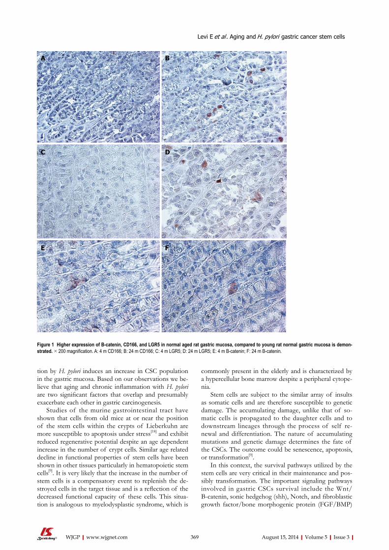
tion by H. pylori induces an increase in CSC population in the gastric mucosa. Based on our observations we be-lieve that aging and chronic inflammation with H. pylori are two significant factors that overlap and presumably exacerbate each other in gastric carcinogenesis.
Studies of the murine gastrointestinal tract have shown that cells from old mice at or near the position of the stem cells within the crypts of Lieberkuhn are more susceptible to apoptosis under stress[18] and exhibit reduced regenerative potential despite an age dependent increase in the number of crypt cells. Similar age related decline in functional properties of stem cells have been shown in other tissues particularly in hematopoietic stem cells[9]. It is very likely that the increase in the number of stem cells is a compensatory event to replenish the de-stroyed cells in the target tissue and is a reflection of the decreased functional capacity of these cells. This situa-tion is analogous to myelodysplastic syndrome, which is
commonly present in the elderly and is characterized by a hypercellular bone marrow despite a peripheral cytope-nia.
Stem cells are subject to the similar array of insults as somatic cells and are therefore susceptible to genetic damage. The accumulating damage, unlike that of so-matic cells is propagated to the daughter cells and to downstream lineages through the process of self re-newal and differentiation. The nature of accumulating mutations and genetic damage determines the fate of the CSCs. The outcome could be senescence, apoptosis, or transformation[9].
In this context, the survival pathways utilized by the stem cells are very critical in their maintenance and pos-sibly transformation. The important signaling pathways involved in gastric CSCs survival include the Wnt/B-catenin, sonic hedgehog (shh), Notch, and fibroblastic growth factor/bone morphogenic protein (FGF/BMP)
August 15, 2014|Volume 5|Issue 3|WJGP|www.wjgnet.com 369
Figure 1 Higher expression of B-catenin, CD166, and LGR5 in normal aged rat gastric mucosa, compared to young rat normal gastric mucosa is demon-strated. × 200 magnification. A: 4 m CD166; B: 24 m CD166; C: 4 m LGR5; D: 24 m LGR5; E: 4 m B-catenin; F: 24 m B-catenin.
A B
C D
E F
Levi E et al . Aging and H. pylori gastric cancer stem cells
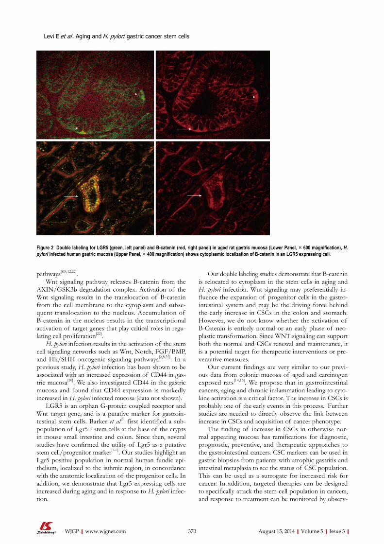
pathways[8,9,12,22]. Wnt signaling pathway releases B-catenin from the
AXIN/GSK3b degradation complex. Activation of the Wnt signaling results in the translocation of B-catenin from the cell membrane to the cytoplasm and subse-quent translocation to the nucleus. Accumulation of B-catenin in the nucleus results in the transcriptional activation of target genes that play critical roles in regu-lating cell proliferation[22].
H. pylori infection results in the activation of the stem cell signaling networks such as Wnt, Notch, FGF/BMP, and Hh/SHH oncogenic signaling pathways[2,8,22]. In a previous study, H. pylori infection has been shown to be associated with an increased expression of CD44 in gas-tric mucosa[10]. We also investigated CD44 in the gastric mucosa and found that CD44 expression is markedly increased in H. pylori infected mucosa (data not shown).
LGR5 is an orphan G-protein coupled receptor and Wnt target gene, and is a putative marker for gastroin-testinal stem cells. Barker et al[3] first identified a sub-population of Lgr5+ stem cells at the base of the crypts in mouse small intestine and colon. Since then, several studies have confirmed the utility of Lgr5 as a putative stem cell/progenitor marker[3-7]. Our studies highlight an Lgr5 positive population in normal human fundic epi-thelium, localized to the isthmic region, in concordance with the anatomic localization of the progenitor cells. In addition, we demonstrate that Lgr5 expressing cells are increased during aging and in response to H. pylori infec-tion.
Our double labeling studies demonstrate that B-catenin is relocated to cytoplasm in the stem cells in aging and H. pylori infection. Wnt signaling may preferentially in-fluence the expansion of progenitor cells in the gastro-intestinal system and may be the driving force behind the early increase in CSCs in the colon and stomach. However, we do not know whether the activation of B-Catenin is entirely normal or an early phase of neo-plastic transformation. Since WNT signaling can support both the normal and CSCs renewal and maintenance, it is a potential target for therapeutic interventions or pre-ventative measures.
Our current findings are very similar to our previ-ous data from colonic mucosa of aged and carcinogen exposed rats[14,16]. We propose that in gastrointestinal cancers, aging and chronic inflammation leading to cyto-kine activation is a critical factor. The increase in CSCs is probably one of the early events in this process. Further studies are needed to directly observe the link between increase in CSCs and acquisition of cancer phenotype.
The finding of increase in CSCs in otherwise nor-mal appearing mucosa has ramifications for diagnostic, prognostic, preventive, and therapeutic approaches to the gastrointestinal cancers. CSC markers can be used in gastric biopsies from patients with atrophic gastritis and intestinal metaplasia to see the status of CSC population. This can be used as a surrogate for increased risk for cancer. In addition, targeted therapies can be designed to specifically attack the stem cell population in cancers, and response to treatment can be monitored by observ-
August 15, 2014|Volume 5|Issue 3|WJGP|www.wjgnet.com 370
Figure 2 Double labeling for LGR5 (green, left panel) and B-catenin (red, right panel) in aged rat gastric mucosa (Lower Panel, × 600 magnification), H. pylori infected human gastric mucosa (Upper Panel, × 400 magnification) shows cytoplasmic localization of B-catenin in an LGR5 expressing cell.
Levi E et al . Aging and H. pylori gastric cancer stem cells
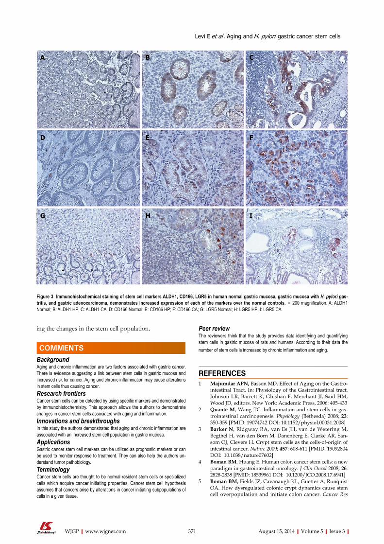
ing the changes in the stem cell population.
COMMENTSBackgroundAging and chronic inflammation are two factors associated with gastric cancer. There is evidence suggesting a link between stem cells in gastric mucosa and increased risk for cancer. Aging and chronic inflammation may cause alterations in stem cells thus causing cancer. Research frontiersCancer stem cells can be detected by using specific markers and demonstrated by immunohistochemistry. This approach allows the authors to demonstrate changes in cancer stem cells associated with aging and inflammation. Innovations and breakthroughsIn this study the authors demonstrated that aging and chronic inflammation are associated with an increased stem cell population in gastric mucosa. ApplicationsGastric cancer stem cell markers can be utilized as prognostic markers or can be used to monitor response to treatment. They can also help the authors un-derstand tumor pathobiology. TerminologyCancer stem cells are thought to be normal resident stem cells or specialized cells which acquire cancer initiating properties. Cancer stem cell hypothesis assumes that cancers arise by alterations in cancer initiating subpopulations of cells in a given tissue.
Peer reviewThe reviewers think that the study provides data identifying and quantifying stem cells in gastric mucosa of rats and humans. According to their data the number of stem cells is increased by chronic inflammation and aging.
REFERENCES1 Majumdar APN, Basson MD. Effect of Aging on the Gastro-
intestinal Tract. In: Physiology of the Gastrointestinal tract. Johnson LR, Barrett K, Ghishan F, Merchant JI, Said HM, Wood JD, editors. New York: Academic Press, 2006: 405-433
2 Quante M, Wang TC. Inflammation and stem cells in gas-trointestinal carcinogenesis. Physiology (Bethesda) 2008; 23: 350-359 [PMID: 19074742 DOI: 10.1152/physiol.00031.2008]
3 Barker N, Ridgway RA, van Es JH, van de Wetering M, Begthel H, van den Born M, Danenberg E, Clarke AR, San-som OJ, Clevers H. Crypt stem cells as the cells-of-origin of intestinal cancer. Nature 2009; 457: 608-611 [PMID: 19092804 DOI: 10.1038/nature07602]
4 Boman BM, Huang E. Human colon cancer stem cells: a new paradigm in gastrointestinal oncology. J Clin Oncol 2008; 26: 2828-2838 [PMID: 18539961 DOI: 10.1200/JCO.2008.17.6941]
5 Boman BM, Fields JZ, Cavanaugh KL, Guetter A, Runquist OA. How dysregulated colonic crypt dynamics cause stem cell overpopulation and initiate colon cancer. Cancer Res
August 15, 2014|Volume 5|Issue 3|WJGP|www.wjgnet.com 371
A B C
D E F
G H I
Figure 3 Immunohistochemical staining of stem cell markers ALDH1, CD166, LGR5 in human normal gastric mucosa, gastric mucosa with H. pylori gas-tritis, and gastric adenocarcinoma, demonstrates increased expression of each of the markers over the normal controls. × 200 magnification. A: ALDH1 Normal; B: ALDH1 HP; C: ALDH1 CA; D: CD166 Normal; E: CD166 HP; F: CD166 CA; G: LGR5 Normal; H: LGR5 HP; I: LGR5 CA.
COMMENTS
Levi E et al . Aging and H. pylori gastric cancer stem cells
2008; 68: 3304-3313 [DOI: 10.1158/0008-5472.CAN-07-2061]6 Fukuda K, Saikawa Y, Ohashi M, Kumagai K, Kitajima M,
Okano H, Matsuzaki Y, Kitagawa Y. Tumor initiating po-tential of side population cells in human gastric cancer. Int J Oncol 2009; 34: 1201-1207 [PMID: 19360333]
7 Jordan CT, Guzman ML, Noble M. Cancer stem cells. N Engl J Med 2006; 355: 1253-1261 [PMID: 16990388]
8 Pilpilidis I, Kountouras J, Zavos C, Katsinelos P. Upper gastrointestinal carcinogenesis: H. pylori and stem cell cross-talk. J Surg Res 2011; 166: 255-264 [PMID: 20452613 DOI: 10.1016/j.jss.2010.02.012]
9 Rossi DJ, Jamieson CH, Weissman IL. Stems cells and the pathways to aging and cancer. Cell 2008; 132: 681-696 [PMID: 18295583 DOI: 10.1016/j.cell.2008.01.036]
10 Takaishi S, Okumura T, Tu S, Wang SS, Shibata W, Vignesh-waran R, Gordon SA, Shimada Y, Wang TC. Identification of gastric cancer stem cells using the cell surface marker CD44. Stem Cells 2009; 27: 1006-1020 [PMID: 19415765 DOI: 10.1002/stem.30]
11 Karam SM, Leblond CP. Dynamics of epithelial cells in the corpus of the mouse stomach. I. Identification of proliferative cell types and pinpointing of the stem cell. Anat Rec 1993; 236: 259-279 [PMID: 8338232 DOI: 10.1002/ar.1092360202]
12 Saikawa Y, Fukuda K, Takahashi T, Nakamura R, Takeu-chi H, Kitagawa Y. Gastric carcinogenesis and the cancer stem cell hypothesis. Gastric Cancer 2010; 13: 11-24 [PMID: 20373071 DOI: 10.1007/s10120-009-0537-4]
13 Kato K, Hasui K, Wang J, Kawano Y, Aikou T, Murata F. Homeostatic mass control in gastric non-neoplastic epi-thelia under infection of Helicobacter pylori: an immuno-histochemical analysis of cell growth, stem cells and pro-grammed cell death. Acta Histochem Cytochem 2008; 41: 23-38 [DOI: 10.1267/ahc.07021]
14 McDonald SA, Greaves LC, Gutierrez-Gonzalez L, Rodri-guez-Justo M, Deheragoda M, Leedham SJ, Taylor RW, Lee CY, Preston SL, Lovell M, Hunt T, Elia G, Oukrif D, Harrison R, Novelli MR, Mitchell I, Stoker DL, Turnbull DM, Jankowski JA, Wright NA. Mechanisms of field can-
cerization in the human stomach: the expansion and spread of mutated gastric stem cells. Gastroenterology 2008; 134: 500-510 [PMID: 18242216 DOI: 10.1053/j.gastro.2007.11.035]
15 Oh JD, Kling-Bäckhed H, Giannakis M, Engstrand LG, Gor-don JI. Interactions between gastric epithelial stem cells and Helicobacter pylori in the setting of chronic atrophic gastri-tis. Curr Opin Microbiol 2006; 9: 21-27 [PMID: 16406776]
16 Levi E, Misra S, Du J, Patel BB, Majumdar AP. Combina-tion of aging and dimethylhydrazine treatment causes an increase in cancer-stem cell population of rat colonic crypts. Biochem Biophys Res Commun 2009; 385: 430-433 [PMID: 19465005 DOI: 10.1016/j.bbrc.2009.05.080]
17 Nautiyal J, Du J, Yu Y, Kanwar SS, Levi E, Majumdar APN: EGFR regulation of colon cancer stem-like cells during aging and in response to the colonic carcinogen dimethyl-hydrazine. Am J Physiol Gastrointest Liver Physiol 2012; 302: G655-63 [PMID: 22281474 DOI: 10.1152/ajpgi.00323.2011]
18 Martin K, Potten CS, Roberts SA, Kirkwood TB. Altered stem cell regeneration in irradiated intestinal crypts of se-nescent mice. J Cell Sci 1998; 111 (Pt 16): 2297-2303 [PMID: 9683625]
19 Patel BB, Yu Y, Du J, Levi E, Philip PA, Majumdar AP. Age related increase in colorectal cancer stem cells in macro-scopically normal mucosa of patients with adenomas: A risk factor for colon cancer. Biochem Biophys Res Commun 2009; 378: 344-347 [DOI: 10.1016/j.bbrc.2008.10.179]
20 Schmelz EM, Levi E, Du J, Xu H, Majumdar AP. Age-relat-ed loss of EGF-receptor related protein (ERRP) in the aging colon is a potential risk factor for colon cancer. Mech Age-ing Dev 2004; 125: 917-922 [PMID: 15563939 DOI: 10.1016/j.mad.2004.03.010]
21 Xiao ZQ, Moragoda L, Jaszewski R, Hatfield JA, Fligiel SE, Majumdar AP. Aging is associated with increased prolifera-tion and decreased apoptosis in the colonic mucosa. Mech Ageing Dev 2001; 122: 1849-1864 [PMID: 11557285 DOI: 10.1016/S0047-6374(01)00323-2]
22 Reya T, Clevers H. Wnt signalling in stem cells and cancer. Nature 2005; 434: 843-850 [PMID: 15829953]
P- Reviewer: Langner C S- Editor: Qi Y L- Editor: A E- Editor: Lu YJ
August 15, 2014|Volume 5|Issue 3|WJGP|www.wjgnet.com 372
Levi E et al . Aging and H. pylori gastric cancer stem cells
SYSTEMATIC REVIEWS
Oxidative and nitrosative stress enzymes in relation to nitrotyrosine in Helicobacter pylori -infected humans
Anders Elfvin, Anders Edebo, Peter Hallersund, Anna Casselbrant, Lars Fändriks
Anders Elfvin, Anders Edebo, Peter Hallersund, Anna Cas-selbrant, Lars Fändriks, Department of Gastrosurgical Re-search and Education, Institute of Clinical Sciences, Sahlgren-ska Academy at University of Gothenburg, 416 85 Gothenburg, SwedenAnders Elfvin, Department of Pediatrics, Institute of Clinical Sciences, Sahlgrenska Academy at University of Gothenburg, 416 85 Gothenburg, SwedenAuthor contributions: Elfvin A and Edebo A performed all the gastroscopies; Elfvin A, Hallersund P and Casselbrant A per-formed the laboratory work, and analysis; Elfvin A and Fändriks L designed the study; Fändriks L coordinated the study and provided financial support; Elfvin A and Hallersund P wrote the manuscript; all authors were involved in editing the manuscript.Supported by The grants financially from the Swedish Medical Research Council and the Gothenburg Medical SocietyCorrespondence to: Dr. Anders Elfvin, MD, PhD, Depart-ment of Pediatrics, Institute of Clinical Sciences, Sahlgrenska Academy at University of Gothenburg, Diagnosroad 15, 416 85 Gothenburg, Sweden. [email protected]: +46-31-3438073 Fax: +46-31-3436696Received: January 28, 2014 Revised: April 25, 2014 Accepted: June 10, 2014Published online: August 15, 2014
AbstractAIM: To compare a possible relation between Helico-bacter pylori (H. pylori ) and the oxygen- and nitrogen radical system in humans.
METHODS: Mechanisms for H. pylori to interfere with the oxygen and nitrogen radical system is of great im-portance for understanding of the H. pylori persistence and pathogenesis. Biopsies were obtained from the gastric wall of 21 individuals. Ongoing infection with H. pylori was detected using direct analyze from the biop-sies using campylobacter-like organism test (CLO-test) and/or by using 14C-urea breath test. The individuals were divided in a negative H. pylori and a positive H. pylori group. Expression in the gastric mucosa of induc-
ible nitric oxide syntase (iNOS), nicotinamide adenine dinucleotide phosphate-oxidase (NADPH-oxidase) my-eloperoxidase (MPO), and nitrotyrosine were assessed by Western blotting.
RESULTS: The individuals who undervent gastroscopy were divided in a H. pylori neg. [n = 13, m/f = 7/6, age (mean) = 39] and a H. pylori pos. group [n= 8, m/f = 5/3, age (mean) = 53]. Using western blot analy-sis iNOS was detected as a 130 kDa band. The iNOS expression was upregulated in the antrum of H. pylori infected individuals in comparison to the controls, mean ± SD being 12.6 ± 2.4 vs 8.3 ± 3.1, P < 0.01. There was a markedly upregulated expression of MPO in the antrum of H. pylori infected individuals in comparison to the control group without infection. In several of non-infected controls it was not possible to detect any MPO expression at all, whereas the expression was high in all the infected subjects, mean ± SD being 5.1 ± 3.4 vs 2.1 ± 1.9, P < 0.05. The NADPH-oxidase expression was analysed by detecting the NADPH-oxidase subunit p47-phox expression. P47-phox was detected as a 47 kDa band using Western blot, and showed a signifi-cantly higher expression of p47-phox in the antrum of the H. pylori infected individuals compared to the con-trols, mean ± SD being 3.1 ± 2.2 vs 0.3 ± 0.2, P < 0.01. Regarding nitrotyrosine formation, Western blot did not show any significant increase or decrease compared to controls, 7.0 ± 0.9 vs 6.9 ± 1.1, not significant.
CONCLUSION: iNOS, MPO and NADPH-oxidase was up-regulated among H. pylori infected. Regarding ni-trotyrosine no difference was found. This support an H. pylori related inhibition of radical formation.
© 2014 Baishideng Publishing Group Inc. All rights reserved.
Key words: Helicobacter pylori ; Radical; Myeloperoxi-dase; Nicotinamide adenine dinucleotide phosphate; Nitrotyrosine; Gastric
World J Gastrointest Pathophysiol 2014 August 15; 5(3): 373-379ISSN 2150-5330 (online)
© 2014 Baishideng Publishing Group Inc. All rights reserved.
Online Submissions: http://www.wjgnet.com/esps/Help Desk: http://www.wjgnet.com/esps/helpdesk.aspxDOI: 10.4291/wjgp.v5.i3.373
August 15, 2014|Volume 5|Issue 3|WJGP|www.wjgnet.com 373
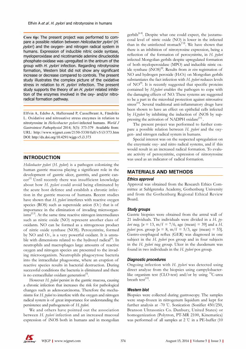
Elfvin A et al . H. pylori and nitrotyrosine in humans
Core tip: The present project was performed to com-pare a possible relation between Helicobacter pylori (H. pylori ) and the oxygen- and nitrogen radical system in humans. Expression of inducible nitric oxide syntase, myeloperoxidase and nicotinamide adenine dinucleotide phosphate-oxidase was upregulted in the antrum of the group with H. pylori infection. Regarding nitrotyrosine formation, Western blot did not show any significant increase or decrease compared to controls. The present study illustrates the complex picture of the oxidative stress in relation to H. pylori infection. The present study supports the theory of an H. pylori related inhibi-tion of the enzymes involved in the oxy- and/or nitro- radical formation pathway.
Elfvin A, Edebo A, Hallersund P, Casselbrant A, Fändriks L. Oxidative and nitrosative stress enzymes in relation to nitrotyrosine in Helicobacter pylori-infected humans. World J Gastrointest Pathophysiol 2014; 5(3): 373-379 Available from: URL: http://www.wjgnet.com/2150-5330/full/v5/i3/373.htm DOI: http://dx.doi.org/10.4291/wjgp.v5.i3.373
INTRODUCTIONHelicobacter pylori (H. pylori) is a pathogen colonizing the human gastric mucosa playing a significant role in the development of gastric ulcer, gastritis, and gastric can-cer[1] Until recently there was insufficient knowledge about how H. pylori could avoid being eliminated by the acute host defence and establish a chronic infec-tion in the gastric mucosa of humans. Recent studies have shown that H. pylori interferes with reactive oxygen species (ROS) such as superoxide anion (O2
-) that is of importance in the elimination of invading microorgan-isms[2,3]. At the same time reactive nitrogen intermediates such as nitric oxide (NO) represent another class of oxidants. NO can be formed as a nitrogenous product of nitric oxide synthase (NOS). Peroxynitrite, formed by NO and O2
-, is a very powerful oxidant. It is unsta-ble with dimensions related to the hydroxyl radical[4]. In neutrophils and macrophages large amounts of reactive oxygen and nitrogen species are presented to the invad-ing microorganism. Neutrophils phagocytose bacteria into the intracellular phagosome, where an eruption of reactive species results in bacterial destruction. During successful conditions the bacteria is eliminated and there is no extracellular oxidant generation[5].
However H. pylori persist in the gastric mucosa, causing a chronic infection that increases the risk for pathological changes such as adenocarcinoma. Therefore the mecha-nisms for H. pylori to interfere with the oxygen and nitrogen radical system is of great importance for understanding the persistence and pathogenesis of H. pylori.
We and others have pointed out the association between H. pylori infection and an increased mucosal expression of iNOS both in humans and in mongolian
gerbils[6-8]. Despite what one could expect, the juxtamu-cosal level of nitric oxide (NO) is lower in the infected than in the uninfected stomach[7,8]. We have shown that there is an inhibition of nitrotyrosine expression, being a reflection of the formation of peroxynitrite, in H. pylori infected Mongolian gerbils despite upregulated formation of both myeloperoxidase (MPO) and inducible nitric ox-ide synthase (iNOS)[6]. Results from in vivo registration of NO and hydrogen peroxide (H2O2) on Mongolian gerbils substantiates the fact infection with H. pylori reduces levels of NO[9]. It is recently suggested that specific proteins contained by H.pylori enables the pathogen to cope with the damaging effects of NO. These systems are suggested to be a part in the microbial protection against nitrosative stress[10]. Several traditional anti-inflammatory drugs have been shown to have an effect on epithelial cells infected by H.pylori by inhibiting the induction of iNOS by sup-pressing the activation of NADPH oxidase[11] .
The present project was performed to further com-pare a possible relation between H. pylori and the oxy-gen- and nitrogen radical system in humans.
Special interest was on the suspected upregulation on the enzymatic oxy- and nitro radical systems, and if this would result in an increased radical formation. To evalu-ate activity of peroxynitrite, expression of nitrotyrosine was used as an indicator of radical formation.
MATERIALS AND METHODSEthics approvalApproval was obtained from the Research Ethics Com-mittee at Sahlgrenska Academy, Gothenburg University and from the Gothenburg Regional Ethical Review Board.
Study groupsGastric biopsies were obtained from the antral wall of 21 individuals. The individuals were divided in a H. py-lori neg. [n = 13, m/f = 7/6, age (mean) = 39] and a H. pylori pos. group [n = 8, m/f = 5/3, age (mean) = 53]. Gastro-esophageal reflux (GER) was diagnosed in one subject in the H. pylori pos group and in four subjects in the H. pylori neg group. Ulcer in the duodenum was found in two individuals in the H. pylori pos group.
Diagnostic proceduresOngoing infection with H. pylori was detected using direct analyze from the biopsies using campylobacter-like organism test (CLO-test) and/or by using 14C-urea breath test[12].
Western blotBiopsies were collected during gastroscopy. The samples were snap-frozen in nitrogenum liquidum and kept for further analysis at -70 ℃. Sonication (Sonifier 450/250, Branson Ultrasonics Co. Danbury, United States) or homogenization (Polytron, PT-MR 2100, Kinematica) was performed of all samples at 2 ℃ in a PE-buffer (10
August 15, 2014|Volume 5|Issue 3|WJGP|www.wjgnet.com 374

mmol/L potassium Phosphate buffer, pH 6.8, and 1 mmol/L EDTA) containing CHAPS {3-[(3-cholamido-propyl) dimethyl-ammanio] 1-propanesulfonate}, apro-tinin (1 µg/mL), leupeptin (10 µg/ mL), pepstatin (10 µg/mL) and Pefablock (1 mg/mL) (Boeringer Mannheim, Mannheim, Germany). All samples were centrifugated at 10.000 g for 10 min at 4 ℃. Analysation was performed of the supernatant for protein content using the method of Bradford12 and then kept at -70 ℃ for future analysis. Samples were diluted in SDS-buffer and heated at 70 ℃ for 10 min before they were loaded on a NuPage 10% BisTris gel (Invitrogen, Carlsbad, CA, United States). One lane of each gel was loaded with prestained molecular weight standards (SeeBlue™, Invitrogen, Carlsbad, CA, United States). Following the electrophoresis the pro-teins were transferred to a polyvinyldifluoride membrane (Amersham, Buckinghamshire, United Kingdom) which was incubated with antibodies directed against iNOS, MPO, NADPH-oxidase or nitrotyrosine containing pro-teins. For identifying iNOS the NOS2 (H-174) sc-8310 (Santa Cruz Biotechnology inc) antibody was used. It is a rabbit polyclonal antibody raised against a recombinant protein corresponding to amino acids 2-175 mapping at the amino terminus of iNOS of human origin. Lack of cross-reaction with nNOS or eNOS was reported by manufacturer. Antibody Anti-myeloperoxidase 07-496 lot 24587 (Upstate, Lake Placid, NY, United States) was used for detecting MPO. This is a rabbit antibody that recog-nizes MPO subunits at 12 and 60 kDa. In the present study the 60 kDa band was used for quantification of the protein. Anti-nitrotyrosine rabbit immunoaffinity puri-fied IgG catalog 06-284, lot 26427 (Upstate, Lake Placid, NY, United States) was used to assess nitrated proteins. For identifying NADPH-oxidase the p47-phox (H-195) sc 14015 (Santa Cruz Biotechnology inc.) was used. This is a rabbit polyclonal antibody raised against amino acids 196-390 of p47-phox of human origin. P47-phox is re-quired for activation of NADPH-oxidase in neutrophils and other phagocytic cells. During activation of NADPH-oxidase, p47-phox migrate to the plasma membrane where it associates with other subunits to form the active complex. Goat anti-rabbit antibodies were used to identify immunoreactive proteins by chemiluminescence [iNOS, NADPH-oxidase (p47-phox) and nitrotyrosine; goat-anti rabbit sc 2007(Santa Cruz, CA, United States)] [MPO; IgG 12-448 (Upstate Lake Placid, NY, United States)]. CDP-Star (Tropix, Bedford, MA, United States) was used as a substrate. Images were captured by a LAS-100 cooled CCD-camera (Fujifilm, Tokyo, Japan) and semi-quantifi-cation was performed using the soft ware Gauge 3.3 (Fu-jifilm, Tokyo, Japan). As positive controls, to confirm the identity of the protein, RAW 264.7 (sc 2212, Santa Cruz Biotech) was used for iNOS, HL60 was used for MPO and NADPH-oxidase (p47-phox).For nitrotyrosine the immunoblotting control (12-354, Upstate) was used.
Statistical analysis Statistical analysis was performed using non parametric
Mann-Whitney U-test. P-values of < 0.05 were regarded as being of statistical significance.
RESULTSInducible nitric oxide synthaseUsing western blot analysis iNOS was detected as a 130 kDa band. The iNOS expression was upregulated in the antrum of H. pylori infected individuals in comparison to the control group without infection as shown in Figure 1A, mean ± SD being 12.6 ± 2.4 vs 8.3 ± 3.1, P < 0.01. Western blot detecting iNOS with a band at 130 kDa in RAW 264.7 (pos contr.), and in human antral mucosa retrieved from H. pylori pos. and H. pylori neg. volunteers during endoscopy is shown in Figure 2.
MyeloperoxidaseAs shown in Figure 1B, MPO expression was mark-edly upregulated in the antrum of the H. pylori infected individuals in comparison to the control group without infection, mean ± SD being 5.1 ± 3.4 vs 2.1 ± 1.9, P < 0.05. In several of the non-infected controls it was not possible to detect any MPO expression at all, whereas the expression was high in all the infected subjects. Western blot of the MPO positive 60 kDa band in the positive HL60 control and in gastric mucosal specimens of H. pylori pos. and H. pylori neg. volunteers is shown in Figure 2.
NADPH-oxidaseThe expression of NADPH-oxidase was analysed by detecting the NADPH-oxidase subunit p47-phox ex-pression. P47-phox was detected as a 47 kDa band us-ing Western blot. Figure 1C shows a significantly higher expression of p47-phox in the the antrum of H. pylori infected individuals in comparison to the control group without infection, mean ± SD being 3.1 ± 2.2 vs 0.3 ± 0.2, P < 0.01. P47-phox was low in all non-infected con-trols. In the H. pylori infected subjects there was a large spreading of the p47-phox expression. A typical Western blot result is shown in Figure 2.
NitrotyrosineWestern blot analysis did not show any significant in-crease nor decrease in nitrotyrosine expression the an-trum of H. pylori infected individuals in comparison to the control group without infection, 7.0 ± 0.9 vs 6.9 ± 1.1, not significant (Figure 3). Regarding Western blot repre-senting Nitrotyrosine, several bands of Nitrated proteins could be analysed. Shown in the Figure 2 is a typical 66 kDa band in the positive control and in H.pylori pos. and H. pylori neg. subjects.
DISCUSSIONThe findings of the present investigation can confirm that H. pylori infection in humans is related to an up reg-ulation of the expression of MPO, iNOS and NADPH-
August 15, 2014|Volume 5|Issue 3|WJGP|www.wjgnet.com 375
Elfvin A et al . H. pylori and nitrotyrosine in humans
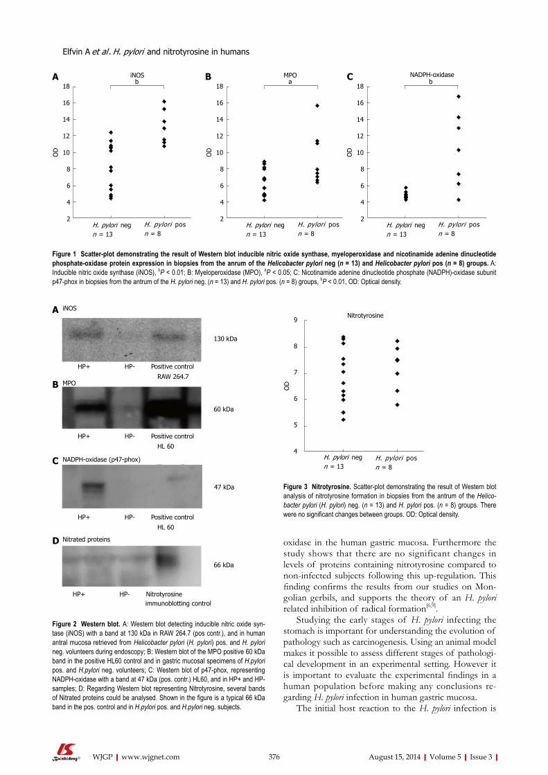
oxidase in the human gastric mucosa. Furthermore the study shows that there are no significant changes in levels of proteins containing nitrotyrosine compared to non-infected subjects following this up-regulation. This finding confirms the results from our studies on Mon-golian gerbils, and supports the theory of an H. pylori related inhibition of radical formation[6,9].
Studying the early stages of H. pylori infecting the stomach is important for understanding the evolution of pathology such as carcinogenesis. Using an animal model makes it possible to assess different stages of pathologi-cal development in an experimental setting. However it is important to evaluate the experimental findings in a human population before making any conclusions re-garding H. pylori infection in human gastric mucosa.
The initial host reaction to the H. pylori infection is
August 15, 2014|Volume 5|Issue 3|WJGP|www.wjgnet.com 376
biNOS
18
16
14
12
10
8
6
4
2
OD
H. pylori neg n = 13
H. pylori pos n = 8
aMPO
18
16
14
12
10
8
6
4
2
OD
H. pylori neg n = 13
H. pylori pos n = 8
bNADPH-oxidase
18
16
14
12
10
8
6
4
2
OD
H. pylori neg n = 13
H. pylori pos n = 8
Figure 1 Scatter-plot demonstrating the result of Western blot inducible nitric oxide synthase, myeloperoxidase and nicotinamide adenine dinucleotide phosphate-oxidase protein expression in biopsies from the anrum of the Helicobacter pylori neg (n = 13) and Helicobacter pylori pos (n = 8) groups. A: Inducible nitric oxide synthase (iNOS), bP < 0.01; B: Myeloperoxidase (MPO), aP < 0.05; C: Nicotinamide adenine dinucleotide phosphate (NADPH)-oxidase subunit p47-phox in biopsies from the antrum of the H. pylori neg. (n = 13) and H. pylori pos. (n = 8) groups, bP < 0.01, OD: Optical density.
HP+ HP- Positive control
RAW 264.7
HP+ HP- Positive control
HL 60
HP+ HP- Positive control
HL 60
HP+ HP- Nitrotyrosine immunoblotting control
130 kDa
60 kDa
47 kDa
66 kDa
A iNOS
B MPO
C NADPH-oxidase (p47-phox)
D Nitrated proteins
Figure 2 Western blot. A: Western blot detecting inducible nitric oxide syn-tase (iNOS) with a band at 130 kDa in RAW 264.7 (pos contr.), and in human antral mucosa retrieved from Helicobacter pylori (H. pylori) pos. and H. pylori neg. volunteers during endoscopy; B: Western blot of the MPO positive 60 kDa band in the positive HL60 control and in gastric mucosal specimens of H.pylori pos. and H.pylori neg. volunteers; C: Western blot of p47-phox, representing NADPH-oxidase with a band at 47 kDa (pos. contr.) HL60, and in HP+ and HP- samples; D: Regarding Western blot representing Nitrotyrosine, several bands of Nitrated proteins could be analysed. Shown in the figure is a typical 66 kDa band in the pos. control and in H.pylori pos. and H.pylori neg. subjects.
Nitrotyrosine9
8
7
6
5
4
OD
H. pylori neg n = 13
H. pylori pos n = 8
Figure 3 Nitrotyrosine. Scatter-plot demonstrating the result of Western blot analysis of nitrotyrosine formation in biopsies from the antrum of the Helico-bacter pylori (H. pylori) neg. (n = 13) and H. pylori pos. (n = 8) groups. There were no significant changes between groups. OD: Optical density.
Elfvin A et al . H. pylori and nitrotyrosine in humans
A B C
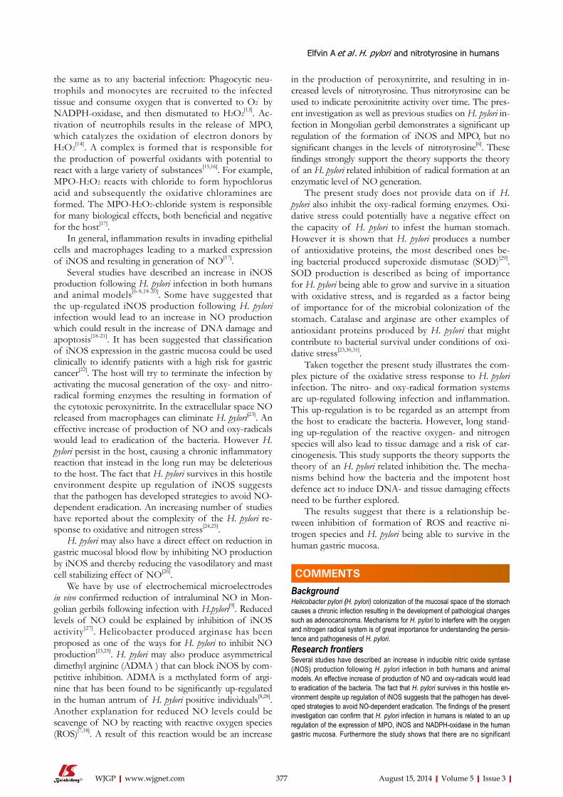
the same as to any bacterial infection: Phagocytic neu-trophils and monocytes are recruited to the infected tissue and consume oxygen that is converted to O2
- by NADPH-oxidase, and then dismutated to H2O2
[13]. Ac-tivation of neutrophils results in the release of MPO, which catalyzes the oxidation of electron donors by H2O2
[14]. A complex is formed that is responsible for the production of powerful oxidants with potential to react with a large variety of substances[15,16]. For example, MPO-H2O2 reacts with chloride to form hypochlorus acid and subsequently the oxidative chloramines are formed. The MPO-H2O2-chloride system is responsible for many biological effects, both beneficial and negative for the host[17].
In general, inflammation results in invading epithelial cells and macrophages leading to a marked expression of iNOS and resulting in generation of NO[17].
Several studies have described an increase in iNOS production following H. pylori infection in both humans and animal models[6-8,18-20]. Some have suggested that the up-regulated iNOS production following H. pylori infection would lead to an increase in NO production which could result in the increase of DNA damage and apoptosis[18-21]. It has been suggested that classification of iNOS expression in the gastric mucosa could be used clinically to identify patients with a high risk for gastric cancer[22]. The host will try to terminate the infection by activating the mucosal generation of the oxy- and nitro-radical forming enzymes the resulting in formation of the cytotoxic peroxynitrite. In the extracellular space NO released from macrophages can eliminate H. pylori[23]. An effective increase of production of NO and oxy-radicals would lead to eradication of the bacteria. However H. pylori persist in the host, causing a chronic inflammatory reaction that instead in the long run may be deleterious to the host. The fact that H. pylori survives in this hostile environment despite up regulation of iNOS suggests that the pathogen has developed strategies to avoid NO-dependent eradication. An increasing number of studies have reported about the complexity of the H. pylori re-sponse to oxidative and nitrogen stress[24,25].
H. pylori may also have a direct effect on reduction in gastric mucosal blood flow by inhibiting NO production by iNOS and thereby reducing the vasodilatory and mast cell stabilizing effect of NO[26].
We have by use of electrochemical microelectrodes in vivo confirmed reduction of intraluminal NO in Mon-golian gerbils following infection with H.pylori[9]. Reduced levels of NO could be explained by inhibition of iNOS activity[27]. Helicobacter produced arginase has been proposed as one of the ways for H. pylori to inhibit NO production[23,25]. H. pylori may also produce asymmetrical dimethyl arginine (ADMA ) that can block iNOS by com-petitive inhibition. ADMA is a methylated form of argi-nine that has been found to be significantly up-regulated in the human antrum of H. pylori positive individuals[8,28]. Another explanation for reduced NO levels could be scavenge of NO by reacting with reactive oxygen species (ROS)[7,18]. A result of this reaction would be an increase
in the production of peroxynitrite, and resulting in in-creased levels of nitrotyrosine. Thus nitrotyrosine can be used to indicate peroxinitrite activity over time. The pres-ent investigation as well as previous studies on H. pylori in-fection in Mongolian gerbil demonstrates a significant up regulation of the formation of iNOS and MPO, but no significant changes in the levels of nitrotyrosine[6]. These findings strongly support the theory supports the theory of an H. pylori related inhibition of radical formation at an enzymatic level of NO generation.
The present study does not provide data on if H. pylori also inhibit the oxy-radical forming enzymes. Oxi-dative stress could potentially have a negative effect on the capacity of H. pylori to infest the human stomach. However it is shown that H. pylori produces a number of antioxidative proteins, the most described ones be-ing bacterial produced superoxide dismutase (SOD)[29]. SOD production is described as being of importance for H. pylori being able to grow and survive in a situation with oxidative stress, and is regarded as a factor being of importance for of the microbial colonization of the stomach. Catalase and arginase are other examples of antioxidant proteins produced by H. pylori that might contribute to bacterial survival under conditions of oxi-dative stress[23,30,31].
Taken together the present study illustrates the com-plex picture of the oxidative stress response to H. pylori infection. The nitro- and oxy-radical formation systems are up-regulated following infection and inflammation. This up-regulation is to be regarded as an attempt from the host to eradicate the bacteria. However, long stand-ing up-regulation of the reactive oxygen- and nitrogen species will also lead to tissue damage and a risk of car-cinogenesis. This study supports the theory supports the theory of an H. pylori related inhibition the. The mecha-nisms behind how the bacteria and the impotent host defence act to induce DNA- and tissue damaging effects need to be further explored.
The results suggest that there is a relationship be-tween inhibition of formation of ROS and reactive ni-trogen species and H. pylori being able to survive in the human gastric mucosa.
COMMENTSBackgroundHelicobacter pylori (H. pylori) colonization of the mucosal space of the stomach causes a chronic infection resulting in the development of pathological changes such as adenocarcinoma. Mechanisms for H. pylori to interfere with the oxygen and nitrogen radical system is of great importance for understanding the persis-tence and pathogenesis of H. pylori.Research frontiersSeveral studies have described an increase in inducible nitric oxide syntase (iNOS) production following H. pylori infection in both humans and animal models. An effective increase of production of NO and oxy-radicals would lead to eradication of the bacteria. The fact that H. pylori survives in this hostile en-vironment despite up regulation of iNOS suggests that the pathogen has devel-oped strategies to avoid NO-dependent eradication. The findings of the present investigation can confirm that H. pylori infection in humans is related to an up regulation of the expression of MPO, iNOS and NADPH-oxidase in the human gastric mucosa. Furthermore the study shows that there are no significant
August 15, 2014|Volume 5|Issue 3|WJGP|www.wjgnet.com 377
COMMENTS
Elfvin A et al . H. pylori and nitrotyrosine in humans
changes in levels of proteins containing nitrotyrosine compared to non-infected subjects following this up-regulation. Breakthroughs and innovationsThe investigation presented here illustrates the complex picture of the oxidative stress response to H. pylori infection. The nitro- and oxy-radical formation sys-tems are up-regulated following infection and inflammation. This up-regulation is to be regarded as an attempt from the host to eradicate the bacteria. How-ever, long standing up-regulation of the reactive oxygen- and nitrogen species will also lead to tissue damage and a risk of carcinogenesis. This study sup-ports the theory of an H.pylori related inhibition of formation of reactive oxygen species (ROS) and reactive nitrogen species. The mechanisms behind how the bacteria and the impotent host defence act to induce DNA- and tissue damag-ing effects need to be further explored. ApplicationsBy understanding how H. pylori manages not to be extinguished in the hostile environment by hindering the formation of reactive oxygen species and reactive nitrogen intermediates we will gain a greater understanding of the mechanisms involved in H. pylori related disease. Terminology Myeloperoxidase (MPO) is an enzyme of importance in the microbicidal role of phagocytes. iNOS was first identified in macrophages. iNOS is involved in the production of NO, but has also many other functions. Nicotinamide adenine dinucleotide phosphate-oxidase (NADPH-oxidase) is a transmembrane electron transport chain involved in the production of different ROS. Nitrotyrosine can be used as marking the activity of peroxynitrite.Peer reviewIn this study the authors demonstrated, in human gastric mucosa of H.pylori positive patients, an increase of some enzymes belonging to oxidative stress pathway, while the amount of nitrotyrosine rich proteins did not differ from H.pylori negative tissues.
REFERENCES1 Parsonnet J, Friedman GD, Vandersteen DP, Chang Y,
Vogelman JH, Orentreich N, Sibley RK. Helicobacter py-lori infection and the risk of gastric carcinoma. N Engl J Med 1991; 325: 1127-1131 [PMID: 1891020 DOI: 10.1056/NEJM199110173251603]
2 Babior BM. NADPH oxidase: an update. Blood 1999; 93: 1464-1476 [PMID: 10029572]
3 Kawahara T, Kohjima M, Kuwano Y, Mino H, Teshima-Kondo S, Takeya R, Tsunawaki S, Wada A, Sumimoto H, Rokutan K. Helicobacter pylori lipopolysaccharide activates Rac1 and transcription of NADPH oxidase Nox1 and its organizer NOXO1 in guinea pig gastric mucosal cells. Am J Physiol Cell Physiol 2005; 288: C450-C457 [PMID: 15469954 DOI: 10.1152/ajpcell.00319.2004]
4 Nathan C, Shiloh MU. Reactive oxygen and nitrogen in-termediates in the relationship between mammalian hosts and microbial pathogens. Proc Natl Acad Sci USA 2000; 97: 8841-8848 [PMID: 10922044 DOI: 10.1073/pnas.97.16.8841]
5 Thomas MJ, Hedrick CC, Smith S, Pang J, Jerome WG, Wil-lard AS, Shirley PS. Superoxide generation by the human polymorphonuclear leukocyte in response to latex beads. J Leukoc Biol 1992; 51: 591-596 [PMID: 1319445]
6 Elfvin A, Bölin I, Lönroth H, Fändriks L. Gastric expression of inducible nitric oxide synthase and myeloperoxidase in relation to nitrotyrosine in Helicobacter pylori-infected Mongolian gerbils. Scand J Gastroenterol 2006; 41: 1013-1018 [PMID: 16938713 DOI: 10.1080/00365520600633537]
7 Iguchi M, Shiotani A, Nishioka S. Helicobacter pylori infec-tion reduces intraluminal nitric oxide. Scand J Gastroenterol 2000; 35: 694-698 [PMID: 10972171 DOI: 10.1080/003655200750023345]
8 von Bothmer C, Edebo A, Lönroth H, Olbe L, Pettersson A, Fändriks L. Helicobacter pylori infection inhibits antral mucosal nitric oxide production in humans. Scand J Gastro-enterol 2002; 37: 404-408 [PMID: 11989830 DOI: 10.1080/0036
55202317316024]9 Elfvin A, Edebo A, Bölin I, Fändriks L. Quantitative mea-
surement of nitric oxide and hydrogen peroxide in Heli-cobacter pylori-infected Mongolian gerbils in vivo. Scand J Gastroenterol 2007; 42: 1175-1181 [PMID: 17852850 DOI: 10.1080/00365520701288306]
10 Justino MC, Ecobichon C, Fernandes AF, Boneca IG, Sa-raiva LM.Helicobacter pylori has an unprecedented nitric oxide detoxifying system. Antioxid Redox Signal 2012; 17: 1190200 [DOI: 10.1089/ars.2011.4304]
11 Cho SO, Lim JW, Kim H Red ginseng extract inhibits the expression of MCP-1 and iNOS in Helicobacter pylori-infected gastric epithelial cells by suppressing the activation of NADPH oxidase and Jak2/Stat3. J Ethnopharmacol 2013; 150: 761-764 [DOI: 10.1016/j.jep.2013.09.013]
12 Hamlet AK, Erlandsson KI, Olbe L, Svennerholm AM, Backman VE, Pettersson AB. A simple, rapid, and highly reliable capsule-based 14C urea breath test for diagnosis of Helicobacter pylori infection. Scand J Gastroenterol 1995; 30: 1058-1063 [PMID: 8578164 DOI: 10.3109/00365529509101607]
13 Hampton MB, Kettle AJ, Winterbourn CC. Inside the neu-trophil phagosome: oxidants, myeloperoxidase, and bacte-rial killing. Blood 1998; 92: 3007-3017 [PMID: 9787133]
14 Krawisz JE, Sharon P, Stenson WF. Quantitative assay for acute intestinal inflammation based on myeloperoxidase ac-tivity. Assessment of inflammation in rat and hamster mod-els. Gastroenterology 1984; 87: 1344-1350 [PMID: 6092199]
15 Hataishi R, Kobayashi H, Takahashi Y, Hirano S, Zapol WM, Jones RC. Myeloperoxidase-associated tyrosine nitra-tion after intratracheal administration of lipopolysaccharide in rats. Anesthesiology 2002; 97: 887-895 [PMID: 12357155 DOI: 10.1097/00000542-200210000-00021]
16 Klebanoff SJ. Myeloperoxidase: friend and foe. J Leukoc Biol 2005; 77: 598-625 [PMID: 15689384 DOI: 10.1189/jlb.1204697]
17 Jaiswal M, LaRusso NF, Gores GJ. Nitric oxide in gastroin-testinal epithelial cell carcinogenesis: linking inflammation to oncogenesis. Am J Physiol Gastrointest Liver Physiol 2001; 281: G626-G634 [PMID: 11518674]
18 Goto T, Haruma K, Kitadai Y, Ito M, Yoshihara M, Sumii K, Hayakawa N, Kajiyama G. Enhanced expression of in-ducible nitric oxide synthase and nitrotyrosine in gastric mucosa of gastric cancer patients. Clin Cancer Res 1999; 5: 1411-1415 [PMID: 10389926]
19 Mannick EE, Bravo LE, Zarama G, Realpe JL, Zhang XJ, Ruiz B, Fontham ET, Mera R, Miller MJ, Correa P. Inducible nitric oxide synthase, nitrotyrosine, and apoptosis in Helico-bacter pylori gastritis: effect of antibiotics and antioxidants. Cancer Res 1996; 56: 3238-3243 [PMID: 8764115]
20 Son HJ, Rhee JC, Park DI, Kim YH, Rhee PL, Koh KC, Paik SW, Choi KW, Kim JJ. Inducible nitric oxide synthase expression in gastroduodenal diseases infected with Heli-cobacter pylori. Helicobacter 2001; 6: 37-43 [PMID: 11328364 DOI: 10.1046/j.1523-5378.2001.00004.x]
21 Li CQ, Pignatelli B, Ohshima H. Increased oxidative and ni-trative stress in human stomach associated with cagA+ Heli-cobacter pylori infection and inflammation. Dig Dis Sci 2001; 46: 836-844 [PMID: 11330421 DOI: 10.1023/A:1010764720524]
22 Naito Y, Takagi T, Okada H, Nukigi Y, Uchiyama K, Ku-roda M, Handa O, Kokura S, Yagi N, Kato Y, Osawa T, Yoshikawa T. Expression of inducible nitric oxide synthase and nitric oxide-modified proteins in Helicobacter pylori-associated atrophic gastric mucosa. J Gastroenterol Hepatol 2008; 23 Suppl 2: S250-S257 [PMID: 19120907 DOI: 10.1111/j.1440-1746.2008.05412.x]
23 Gobert AP, McGee DJ, Akhtar M, Mendz GL, Newton JC, Cheng Y, Mobley HL, Wilson KT. Helicobacter pylori argi-nase inhibits nitric oxide production by eukaryotic cells: a strategy for bacterial survival. Proc Natl Acad Sci USA 2001; 98: 13844-13849 [PMID: 11717441 DOI: 10.1073/pnas.241443798]
24 Nardone G, Rocco A, Malfertheiner P. Review article: heli-
August 15, 2014|Volume 5|Issue 3|WJGP|www.wjgnet.com 378
Elfvin A et al . H. pylori and nitrotyrosine in humans

cobacter pylori and molecular events in precancerous gas-tric lesions. Aliment Pharmacol Ther 2004; 20: 261-270 [PMID: 15274662 DOI: 10.1111/j.1365-2036.2004.02075.x]
25 Wang G, Alamuri P, Maier RJ. The diverse antioxidant sys-tems of Helicobacter pylori. Mol Microbiol 2006; 61: 847-860 [PMID: 16879643 DOI: 10.1111/j.1365-2958.2006.05302.x]
26 Henriksnäs J, Atuma C, Phillipson M, Sandler S, Engstrand L, Holm L. Acute effects of Helicobacter pylori extracts on gastric mucosal blood flow in the mouse. World J Gastroenterol 2009; 15: 219-225 [PMID: 19132773 DOI: 10.3748/wjg.15.219]
27 von Bothmer C, Bölin I, Pettersson A, Fändriks L. Stimulated murine macrophages as a bioassay for H. pylori-related inhibi-tion of nitric oxide production. Scand J Gastroenterol 2003; 38: 380-386 [PMID: 12739709 DOI: 10.1080/00365520310000816]
28 Fändriks L, von Bothmer C, Johansson B, Holm M, Bölin I, Pettersson A. Water extract of Helicobacter pylori inhibits duodenal mucosal alkaline secretion in anesthetized rats.
Gastroenterology 1997; 113: 1570-1575 [PMID: 9352859 DOI: 10.1053/gast.1997.v113.pm9352859]
29 Seyler RW, Olson JW, Maier RJ. Superoxide dismutase-deficient mutants of Helicobacter pylori are hypersensitive to oxidative stress and defective in host colonization. Infect Immun 2001; 69: 4034-4040 [PMID: 11349073 DOI: 10.1128/IAI.69.6.4034-4040.2001]
30 Harris AG, Hazell SL: Localisation of Helicobacter pylori catalase in both the periplasm and cytoplasm, and its depen-dence on the twin-arginine target protein, KapA, for activity. FEMS microbiology letters 2003; 229: 283-289 [DOI: 10.1016/S0378-1097(03)00850-4]
31 McGee DJ, Radcliff FJ, Mendz GL, Ferrero RL, Mobley HL. Helicobacter pylori rocF is required for arginase activity and acid protection in vitro but is not essential for colonization of mice or for urease activity. J Bacteriol 1999; 181: 7314-7322 [PMID: 10572136]
P- Reviewer: Balaban YH, Cardaropoli S, Zhu YL S- Editor: Ji FF L- Editor: A E- Editor: Lu YJ
August 15, 2014|Volume 5|Issue 3|WJGP|www.wjgnet.com 379
Elfvin A et al . H. pylori and nitrotyrosine in humans
GENERAL INFORMATIONWorld Journal of Gastrointestinal Pathophysiology (World J Gastrointest Pathophysiol, WJGP, online ISSN 2150-5330, DOI: 10.4291) is a peer-reviewed open access (OA) academic journal that aims to guide clinical practice and improve diagnostic and therapeutic skills of clinicians.
Aim and scopeWJGP is to report rapidly the most recent results in basic and clin-ical research on gastrointestinal pathophysiology, including all asp-ects of normal or abnormal function of the gastrointestinal tract, hepatobiliary system, and pancreas. WJGP specifically covers gro-wth and development, digestion, secretion, absorption, metabolis-m and motility relative to the gastrointestinal organs, as well as immune and inflammatory processes, and neural, endocrine and circulatory control mechanisms that affect these organs. This journal will also report new methods and techniques in gastrointestinal pathophysiological research. We encourage authors to submit their manuscripts to WJGP. We will give priority to manuscripts that are supported by major national and international foundations and those that are of great basic and clinical significance. WJGP is edited and published by Baishideng Publishing Group (BPG). BPG has a strong professional editorial team composed of science editors, language editors and electronic editors. BPG current-ly publishes 43 OA clinical medical journals, including 42 in English, has a total of 15471 editorial board members or peer reviewers, and is a world first-class publisher.
ColumnsThe columns in the issues of WJGP will include: (1) Editorial: The editorial board members are invited to make comments on an important topic in their field in terms of its current research status and future directions to lead the development of this discipline; (2) Frontier: The editorial board members are invited to select a highly cited cutting-edge original paper of his/her own to sum-marize major findings, the problems that have been resolved and remain to be resolved, and future research directions to help read-ers understand his/her important academic point of view and fu-ture research directions in the field; (3) Diagnostic Advances: The editorial board members are invited to write high-quality diagnostic advances in their field to improve the diagnostic skills of readers. The topic covers general clinical diagnosis, differential diagnosis, pathological diagnosis, laboratory diagnosis, imaging diagnosis, endoscopic diagnosis, biotechnological diagnosis, functional diag-nosis, and physical diagnosis; (4) Therapeutics Advances: The edi-torial board members are invited to write high-quality therapeutic advances in their field to help improve the therapeutic skills of readers. The topic covers medication therapy, psychotherapy, phys-ical therapy, replacement therapy, interventional therapy, minimally invasive therapy, endoscopic therapy, transplantation therapy, and surgical therapy; (5) Field of Vision: The editorial board members are invited to write commentaries on classic articles, hot topic arti-cles, or latest articles to keep readers at the forefront of research and increase their levels of clinical research. Classic articles refer to papers that are included in Web of Knowledge and have received a large number of citations (ranking in the top 1%) after being published for more than years, reflecting the quality and impact of papers. Hot topic articles refer to papers that are included in
Web of Knowledge and have received a large number of citations after being published for no more than 2 years, reflecting cutting-edge trends in scientific research. Latest articles refer to the latest published high-quality papers that are included in PubMed, reflect-ing the latest research trends. These commentary articles should focus on the status quo of research, the most important research topics, the problems that have now been resolved and remain to be resolved, and future research directions. Basic information about the article to be commented (including authors, article title, journal name, year, volume, and inclusive page numbers; (6) Minireviews: The editorial board members are invited to write short reviews on recent advances and trends in research of molecular biology, genomics, and related cutting-edge technologies to provide read-ers with the latest knowledge and help improve their diagnostic and therapeutic skills; (7) Review: To make a systematic review to focus on the status quo of research, the most important research topics, the problems that have now been resolved and remain to be resolved, and future research directions; (8) Topic Highlight: The editorial board members are invited to write a series of articles (7-10 articles) to comment and discuss a hot topic to help improve the diagnostic and therapeutic skills of readers; (9) Medical Eth-ics: The editorial board members are invited to write articles about medical ethics to increase readers’ knowledge of medical ethics. The topic covers international ethics guidelines, animal studies, clinical trials, organ transplantation, etc.; (10) Clinical Case Confer-ence or Clinicopathological Conference: The editorial board mem-bers are invited to contribute high-quality clinical case conference; (11) Original Articles: To report innovative and original findings in gastrointestinal pathophysiology; (12) Research Report: To briefly report the novel and innovative findings in gastrointestinal patho-physiology; (13) Meta-Analysis: Covers the systematic review, mixed treatment comparison, meta-regression, and overview of reviews, in order to summarize a given quantitative effect, e.g., the clinical ef-fectiveness and safety of clinical treatments by combining data from two or more randomized controlled trials, thereby providing more precise and externally valid estimates than those which would stem from each individual dataset if analyzed separately from the others; (14) Case Report: To report a rare or typical case; (15) Letters to the Editor: To discuss and make reply to the contributions published in WJGP, or to introduce and comment on a controversial issue of general interest; (16) Book Reviews: To introduce and comment on quality monographs of gastrointestinal pathophysiology; and (17) Autobiography: The editorial board members are invited to write their autobiography to provide readers with stories of success or failure in their scientific research career. The topic covers their basic personal information and information about when they start-ed doing research work, where and how they did research work, what they have achieved, and their lessons from success or failure.
Name of journalWorld Journal of Gastrointestinal Pathophysiology
ISSNISSN 2150-5330 (online)
Launch dateApril 15, 2010
FrequencyQuarterly
IWJGP|www.wjgnet.com August 15, 2014|Volume 5|Issue 3|
INSTRUCTIONS TO AUTHORS
Submit a Manuscript: http://www.wjgnet.com/esps/Help Desk: http://www.wjgnet.com/esps/helpdesk.aspxwww.wjgnet.com
World J Gastrointest Pathophysiol 2014 August 15; 5(3): I-VISSN 2150-5330 (online)
© 2014 Baishideng Publishing Group Inc. All rights reserved.
Instructions to authors
Editor-in-ChiefThomas Y Ma, MD, PhD, Professor, Chief, Division of Gastro-enterology and Hepatology, University of New Mexico, MSC10 5550, 1 UNM, Albuquerque, NM 87131, United States
Editorial officeJin-Lei Wang, DirectorXiu-Xia Song, Vice DirectorWorld Journal of Gastrointestinal PathophysiologyRoom 903, Building D, Ocean International Center,No. 62 Dongsihuan Zhonglu, Chaoyang District,Beijing 100025, ChinaTelephone: +86-10-85381891Fax: +86-10-85381893E-mail: [email protected] Desk: http://www.wjgnet.com/esps/helpdesk.aspxhttp://www.wjgnet.com
PublisherBaishideng Publishing Group Inc8226 Regency Drive, Pleasanton, CA 94588, USATelephone: +1-925-223-8242Fax: +1-925-223-8243E-mail: [email protected] Desk: http://www.wjgnet.com/esps/helpdesk.aspxhttp://www.wjgnet.com
Instructions to authorsFull instructions are available online at http://www.wjgnet.com/ 2218-6182/g_info_20100722172951.htm.
Indexed and Abstracted inPubMed Central, PubMed, Digital Object Identifier, and Direc-tory of Open Access Journals.
SPECIAL STATEMENTAll articles published in journals owned by the BPG represent the views and opinions of their authors, and not the views, opinions or policies of the BPG, except where otherwise explicitly indicated.
Biostatistical editingStatistical review is performed after peer review. We invite an ex-pert in Biomedical Statistics to evaluate the statistical method used in the paper, including t-test (group or paired comparisons), chi-squared test, Ridit, probit, logit, regression (linear, curvilinear, or stepwise), correlation, analysis of variance, analysis of covariance, etc. The reviewing points include: (1) Statistical methods should be described when they are used to verify the results; (2) Whether the statistical techniques are suitable or correct; (3) Only homogeneous data can be averaged. Standard deviations are preferred to standard errors. Give the number of observations and subjects (n). Losses in observations, such as drop-outs from the study should be re-ported; (4) Values such as ED50, LD50, IC50 should have their 95% confidence limits calculated and compared by weighted probit analysis (Bliss and Finney); and (5) The word ‘significantly’ should be replaced by its synonyms (if it indicates extent) or the P value (if it indicates statistical significance).
Conflict-of-interest statementIn the interests of transparency and to help reviewers assess any poten-tial bias, WJGP requires authors of all papers to declare any compet-ing commercial, personal, political, intellectual, or religious interests in relation to the submitted work. Referees are also asked to indi-cate any potential conflict they might have reviewing a particular paper. Before submitting, authors are suggested to read “Uniform Requirements for Manuscripts Submitted to Biomedical Journals: Ethical Considerations in the Conduct and Reporting of Research: Conflicts of Interest” from International Committee of Medical Journal Editors (ICMJE), which is available at: http://www.icmje.
org/ethical_4conflicts.html. Sample wording: [Name of individual] has received fees for serv-
ing as a speaker, a consultant and an advisory board member for [names of organizations], and has received research funding from [names of organization]. [Name of individual] is an employee of [name of or-ganization]. [Name of individual] owns stocks and shares in [name of organization]. [Name of individual] owns patent [patent identification and brief description].
Statement of informed consentManuscripts should contain a statement to the effect that all human studies have been reviewed by the appropriate ethics committee or it should be stated clearly in the text that all persons gave their informed consent prior to their inclusion in the study. Details that might disclose the identity of the subjects under study should be omitted. Authors should also draw attention to the Code of Ethics of the World Med-ical Association (Declaration of Helsinki, 1964, as revised in 2004).
Statement of human and animal rightsWhen reporting the results from experiments, authors should follow the highest standards and the trial should conform to Good Clinical Practice (for example, US Food and Drug Administration Good Clinical Practice in FDA-Regulated Clinical Trials; UK Medicines Research Council Guidelines for Good Clinical Practice in Clinical Trials) and/or the World Medical Association Declaration of Hel-sinki. Generally, we suggest authors follow the lead investigator’s na-tional standard. If doubt exists whether the research was conducted in accordance with the above standards, the authors must explain the rationale for their approach and demonstrate that the institutional review body explicitly approved the doubtful aspects of the study.
Before submitting, authors should make their study approved by the relevant research ethics committee or institutional review board. If human participants were involved, manuscripts must be accompanied by a statement that the experiments were under-taken with the understanding and appropriate informed consent of each. Any personal item or information will not be published without explicit consents from the involved patients. If experi-mental animals were used, the materials and methods (experimental procedures) section must clearly indicate that appropriate meas-ures were taken to minimize pain or discomfort, and details of animal care should be provided.
SUBMISSION OF MANUSCRIPTSManuscripts should be typed in 1.5 line spacing and 12 pt. Book Antiqua with ample margins. Number all pages consecutively, and start each of the following sections on a new page: Title Page, Abstract, Introduction, Materials and Methods, Results, Discus-sion, Acknowledgements, References, Tables, Figures, and Figure Legends. Neither the editors nor the publisher are responsible for the opinions expressed by contributors. Manuscripts formally ac-cepted for publication become the permanent property of BPG, and may not be reproduced by any means, in whole or in part, with-out the written permission of both the authors and the publisher. We reserve the right to copy-edit and put onto our website accepted manuscripts. Authors should follow the relevant guidelines for the care and use of laboratory animals of their institution or national animal welfare committee. For the sake of transparency in regard to the performance and reporting of clinical trials, we endorse the policy of the ICMJE to refuse to publish papers on clinical trial results if the trial was not recorded in a publicly-accessible registry at its outset. The only register now available, to our knowledge, is http://www.clinicaltrials.gov sponsored by the United States National Library of Medicine and we encourage all potential con-tributors to register with it. However, in the case that other registers become available you will be duly notified. A letter of recommenda-tion from each author’s organization should be provided with the contributed article to ensure the privacy and secrecy of research is protected.
Authors should retain one copy of the text, tables, photo-graphs and illustrations because rejected manuscripts will not be
IIWJGP|www.wjgnet.com August 15, 2014|Volume 5|Issue 3|
Instructions to authors
returned to the author(s) and the editors will not be responsible for loss or damage to photographs and illustrations sustained dur-ing mailing.
Online submissionsManuscripts should be submitted through the Online Submis-sion System at: http://www.wjgnet.com/esps/. Authors are highly recommended to consult the ONLINE INSTRUCTIONS TO AUTHORS (http://www.wjgnet.com/2150-5330/g_info_ 20100316161927.htm) before attempting to submit online. For assist-ance, authors encountering problems with the Online Submission System may send an email describing the problem to [email protected], or by telephone: +86-10-85381892. If you submit your manuscript online, do not make a postal contribution. Repeated online submission for the same manuscript is strictly prohibited.
MANUSCRIPT PREPARATIONAll contributions should be written in English. All articles must be submitted using word-processing software. All submissions must be typed in 1.5 line spacing and 12 pt. Book Antiqua with ample mar-gins. Style should conform to our house format. Required informa-tion for each of the manuscript sections is as follows:
Title pageTitle: Title should be less than 12 words.
Running title: A short running title of less than 6 words should be provided.
Authorship: Authorship credit should be in accordance with the standard proposed by ICMJE, based on (1) substantial contribu-tions to conception and design, acquisition of data, or analysis and interpretation of data; (2) drafting the article or revising it critically for important intellectual content; and (3) final approval of the ver-sion to be published. Authors should meet conditions 1, 2, and 3.
Institution: Author names should be given first, then the complete name of institution, city, province and postcode. For example, Xu-Chen Zhang, Li-Xin Mei, Department of Pathology, Chengde Med-ical College, Chengde 067000, Hebei Province, China. One author may be represented from two institutions, for example, George Sgourakis, Department of General, Visceral, and Transplantation Surgery, Essen 45122, Germany; George Sgourakis, 2nd Surgical Department, Korgialenio-Benakio Red Cross Hospital, Athens 15451, Greece
Author contributions: The format of this section should be: Author contributions: Wang CL and Liang L contributed equally to this work; Wang CL, Liang L, Fu JF, Zou CC, Hong F and Wu XM designed the research; Wang CL, Zou CC, Hong F and Wu XM performed the research; Xue JZ and Lu JR contributed new reagents/analytic tools; Wang CL, Liang L and Fu JF analyzed the data; and Wang CL, Liang L and Fu JF wrote the paper.
Supportive foundations: The complete name and number of sup-portive foundations should be provided, e.g. Supported by National Natural Science Foundation of China, No. 30224801
Correspondence to: Only one corresponding address should be provided. Author names should be given first, then author title, af-filiation, the complete name of institution, city, postcode, province, country, and email. All the letters in the email should be in lower case. A space interval should be inserted between country name and email address. For example, Montgomery Bissell, MD, Professor of Medicine, Chief, Liver Center, Gastroenterology Division, Uni-versity of California, Box 0538, San Francisco, CA 94143, United States. [email protected]
Telephone and fax: Telephone and fax should consist of +, coun-try number, district number and telephone or fax number, e.g. Tele-
phone: +86-10-85381892 Fax: +86-10-85381893
Peer reviewers: All articles received are subject to peer review. Normally, three experts are invited for each article. Decision on acceptance is made only when at least two experts recommend publication of an article. All peer-reviewers are acknowledged on Express Submission and Peer-review System website.
AbstractThere are unstructured abstracts (no less than 200 words) and struc-tured abstracts. The specific requirements for structured abstracts are as follows:
An informative, structured abstract should accompany each manuscript. Abstracts of original contributions should be struc-tured into the following sections: AIM (no more than 20 words; Only the purpose of the study should be included. Please write the Aim in the form of “To investigate/study/…”), METHODS (no less than 140 words for Original Articles; and no less than 80 words for Brief Articles), RESULTS (no less than 150 words for Original Articles and no less than 120 words for Brief Articles; You should present P values where appropriate and must provide relevant data to illustrate how they were obtained, e.g. 6.92 ± 3.86 vs 3.61 ± 1.67, P < 0.001), and CONCLUSION (no more than 26 words).
Key wordsPlease list 5-10 key words, selected mainly from Index Medicus, which reflect the content of the study.
Core tip Please write a summary of less than 100 words to outline the most innovative and important arguments and core contents in your paper to attract readers.
TextFor articles of these sections, original articles and brief articles, the main text should be structured into the following sections: INTRO-DUCTION, MATERIALS AND METHODS, RESULTS and DISCUSSION, and should include appropriate Figures and Tables. Data should be presented in the main text or in Figures and Tables, but not in both.
IllustrationsFigures should be numbered as 1, 2, 3, etc., and mentioned clearly in the main text. Provide a brief title for each figure on a separate page. Detailed legends should not be provided under the figures. This part should be added into the text where the figures are applicable. Keep-ing all elements compiled is necessary in line-art image. Scale bars should be used rather than magnification factors, with the length of the bar defined in the legend rather than on the bar itself. File names should identify the figure and panel. Avoid layering type directly over shaded or textured areas. Please use uniform legends for the same subjects. For example: Figure 1 Pathological changes in atrophic gas-tritis after treatment. A: ...; B: ...; C: ...; D: ...; E: ...; F: ...; G: …etc. It is our principle to publish high resolution-figures for the E-versions.
TablesThree-line tables should be numbered 1, 2, 3, etc., and mentioned clearly in the main text. Provide a brief title for each table. Detailed legends should not be included under tables, but rather added into the text where applicable. The information should complement, but not duplicate the text. Use one horizontal line under the title, a second under column heads, and a third below the Table, above any footnotes. Vertical and italic lines should be omitted.
Notes in tables and illustrationsData that are not statistically significant should not be noted. aP < 0.05, bP < 0.01 should be noted (P > 0.05 should not be noted). If there are other series of P values, cP < 0.05 and dP < 0.01 are used. A third series of P values can be expressed as eP < 0.05 and fP < 0.01. Other notes in tables or under illustrations should be expressed as 1F, 2F, 3F; or sometimes as other symbols with a superscript (Arabic numer-als) in the upper left corner. In a multi-curve illustration, each curve
IIIWJGP|www.wjgnet.com August 15, 2014|Volume 5|Issue 3|
should be labeled with ●, ○, ■, □, ▲, △, etc., in a certain sequence.
AcknowledgmentsBrief acknowledgments of persons who have made genuine con-tributions to the manuscript and who endorse the data and conclu-sions should be included. Authors are responsible for obtaining written permission to use any copyrighted text and/or illustrations.
REFERENCESCoding systemThe author should number the references in Arabic numerals ac-cording to the citation order in the text. Put reference numbers in square brackets in superscript at the end of citation content or after the cited author’s name. For citation content which is part of the narration, the coding number and square brackets should be typeset normally. For example, “Crohn’s disease (CD) is associated with increased intestinal permeability[1,2]”. If references are cited directly in the text, they should be put together within the text, for example, “From references[19,22-24], we know that...”
When the authors write the references, please ensure that the order in text is the same as in the references section, and also ensure the spelling accuracy of the first author’s name. Do not list the same citation twice.
PMID and DOIPleased provide PubMed citation numbers to the reference list, e.g. PMID and DOI, which can be found at http://www.ncbi.nlm.nih.gov/sites/entrez?db=pubmed and http://www.crossref.org/Sim-pleTextQuery/, respectively. The numbers will be used in E-version of this journal.
Style for journal referencesAuthors: the name of the first author should be typed in bold-faced letters. The family name of all authors should be typed with the in-itial letter capitalized, followed by their abbreviated first and middle initials. (For example, Lian-Sheng Ma is abbreviated as Ma LS, Bo-Rong Pan as Pan BR). The title of the cited article and italicized journal title (journal title should be in its abbreviated form as shown in PubMed), publication date, volume number (in black), start page, and end page [PMID: 11819634 DOI: 10.3748/wjg.13.5396].
Style for book referencesAuthors: the name of the first author should be typed in bold-faced letters. The surname of all authors should be typed with the initial letter capitalized, followed by their abbreviated middle and first initials. (For example, Lian-Sheng Ma is abbreviated as Ma LS, Bo-Rong Pan as Pan BR) Book title. Publication number. Publication place: Publication press, Year: start page and end page.
FormatJournals English journal article (list all authors and include the PMID where applicable)1 Jung EM, Clevert DA, Schreyer AG, Schmitt S, Rennert J, Ku-
bale R, Feuerbach S, Jung F. Evaluation of quantitative contrast harmonic imaging to assess malignancy of liver tumors: A prospective controlled two-center study. World J Gastroenterol 2007; 13: 6356-6364 [PMID: 18081224 DOI: 10.3748/wjg.13. 6356]
Chinese journal article (list all authors and include the PMID where applicable)2 Lin GZ, Wang XZ, Wang P, Lin J, Yang FD. Immunologic
effect of Jianpi Yishen decoction in treatment of Pixu-diar-rhoea. Shijie Huaren Xiaohua Zazhi 1999; 7: 285-287
In press3 Tian D, Araki H, Stahl E, Bergelson J, Kreitman M. Signature
of balancing selection in Arabidopsis. Proc Natl Acad Sci USA 2006; In press
Organization as author4 Diabetes Prevention Program Research Group. Hyperten-
sion, insulin, and proinsulin in participants with impaired glu-cose tolerance. Hypertension 2002; 40: 679-686 [PMID: 12411462 PMCID:2516377 DOI:10.1161/01.HYP.0000035706.28494.09]
Both personal authors and an organization as author 5 Vallancien G, Emberton M, Harving N, van Moorselaar RJ;
Alf-One Study Group. Sexual dysfunction in 1, 274 European men suffering from lower urinary tract symptoms. J Urol 2003; 169: 2257-2261 [PMID: 12771764 DOI:10.1097/01.ju. 0000067940.76090.73]
No author given6 21st century heart solution may have a sting in the tail. BMJ
2002; 325: 184 [PMID: 12142303 DOI:10.1136/bmj.325. 7357.184]
Volume with supplement7 Geraud G, Spierings EL, Keywood C. Tolerability and safety
of frovatriptan with short- and long-term use for treatment of migraine and in comparison with sumatriptan. Headache 2002; 42 Suppl 2: S93-99 [PMID: 12028325 DOI:10.1046/j.1526-4610.42.s2.7.x]
Issue with no volume8 Banit DM, Kaufer H, Hartford JM. Intraoperative frozen
section analysis in revision total joint arthroplasty. Clin Orthop Relat Res 2002; (401): 230-238 [PMID: 12151900 DOI:10.1097/00003086-200208000-00026]
No volume or issue9 Outreach: Bringing HIV-positive individuals into care. HRSA
Careaction 2002; 1-6 [PMID: 12154804]
BooksPersonal author(s)10 Sherlock S, Dooley J. Diseases of the liver and billiary system.
9th ed. Oxford: Blackwell Sci Pub, 1993: 258-296Chapter in a book (list all authors)11 Lam SK. Academic investigator’s perspectives of medical
treatment for peptic ulcer. In: Swabb EA, Azabo S. Ulcer disease: investigation and basis for therapy. New York: Marcel Dekker, 1991: 431-450
Author(s) and editor(s)12 Breedlove GK, Schorfheide AM. Adolescent pregnancy.
2nd ed. Wieczorek RR, editor. White Plains (NY): March of Dimes Education Services, 2001: 20-34
Conference proceedings13 Harnden P, Joffe JK, Jones WG, editors. Germ cell tumours V.
Proceedings of the 5th Germ cell tumours Conference; 2001 Sep 13-15; Leeds, UK. New York: Springer, 2002: 30-56
Conference paper14 Christensen S, Oppacher F. An analysis of Koza's computa-
tional effort statistic for genetic programming. In: Foster JA, Lutton E, Miller J, Ryan C, Tettamanzi AG, editors. Genetic programming. EuroGP 2002: Proceedings of the 5th Euro-pean Conference on Genetic Programming; 2002 Apr 3-5; Kinsdale, Ireland. Berlin: Springer, 2002: 182-191
Electronic journal (list all authors)15 Morse SS. Factors in the emergence of infectious diseases.
Emerg Infect Dis serial online, 1995-01-03, cited 1996-06-05; 1(1): 24 screens. Available from: URL: http://www.cdc.gov/ncidod/eid/index.htm
Patent (list all authors)16 Pagedas AC, inventor; Ancel Surgical R&D Inc., assignee. Flex-
ible endoscopic grasping and cutting device and positioning tool assembly. United States patent US 20020103498. 2002 Aug 1
Statistical dataWrite as mean ± SD or mean ± SE.
Statistical expressionExpress t test as t (in italics), F test as F (in italics), chi square test as χ2 (in Greek), related coefficient as r (in italics), degree of freedom as υ (in Greek), sample number as n (in italics), and probability as P (in italics).
UnitsUse SI units. For example: body mass, m (B) = 78 kg; blood pres-sure, p (B) = 16.2/12.3 kPa; incubation time, t (incubation) = 96 h, blood glucose concentration, c (glucose) 6.4 ± 2.1 mmol/L; blood
IVWJGP|www.wjgnet.com
Instructions to authors
August 15, 2014|Volume 5|Issue 3|
VWJGP|www.wjgnet.com
Instructions to authors
August 15, 2014|Volume 5|Issue 3|
CEA mass concentration, p (CEA) = 8.6 24.5 mg/L; CO2 volume fraction, 50 mL/L CO2, not 5% CO2; likewise for 40 g/L formal-dehyde, not 10% formalin; and mass fraction, 8 ng/g, etc. Arabic numerals such as 23, 243, 641 should be read 23243641.
The format for how to accurately write common units and quantums can be found at: http://www.wjgnet.com/2150-5330/g_info_20100312200347.htm.
AbbreviationsStandard abbreviations should be defined in the abstract and on first mention in the text. In general, terms should not be abbrevi-ated unless they are used repeatedly and the abbreviation is helpful to the reader. Permissible abbreviations are listed in Units, Symbols and Abbreviations: A Guide for Biological and Medical Editors and Authors (Ed. Baron DN, 1988) published by The Royal Society of Medicine, London. Certain commonly used abbreviations, such as DNA, RNA, HIV, LD50, PCR, HBV, ECG, WBC, RBC, CT, ESR, CSF, IgG, ELISA, PBS, ATP, EDTA, mAb, can be used directly without further explanation.
ItalicsQuantities: t time or temperature, c concentration, A area, l length, m mass, V volume.Genotypes: gyrA, arg 1, c myc, c fos, etc.Restriction enzymes: EcoRI, HindI, BamHI, Kbo I, Kpn I, etc.Biology: H. pylori, E coli, etc.
Examples for paper writingAll types of articles’ writing style and requirement will be found in the link: http://www.wjgnet.com/esps/NavigationInfo.aspx?id=15
RESUBMISSION OF THE REVISEDMANUSCRIPTSAuthors must revise their manuscript carefully according to the revision policies of BPG. The revised version, along with the signed copyright transfer agreement, responses to the reviewers, and English language Grade A certificate (for non-native speakers of English), should be submitted to the online system via the link contained in the e-mail sent by the editor. If you have any questions about the revision, please send e-mail to [email protected].
Language evaluation The language of a manuscript will be graded before it is sent for re-vision. (1) Grade A: priority publishing; (2) Grade B: minor language polishing; (3) Grade C: a great deal of language polishing needed; and (4) Grade D: rejected. Revised articles should reach Grade A.
Copyright assignment formPlease download a Copyright assignment form from http://www.wjgnet.com/2150-5330/g_info_20100312200118.htm.
Responses to reviewersPlease revise your article according to the comments/suggestions provided by the reviewers. The format for responses to the reviewers’ comments can be found at: http://www.wjgnet.com/2150-5330/g_info_20100312195923.htm.
Proof of financial supportFor papers supported by a foundation, authors should provide a copy of the approval document and serial number of the foundation.
STATEMENT ABOUT ANONYMOUS PUBLICA-TION OF THE PEER REVIEWERS’ COMMENTSIn order to increase the quality of peer review, push authors to carefully revise their manuscripts based on the peer reviewers' com-ments, and promote academic interactions among peer reviewers, authors and readers, we decide to anonymously publish the review-ers’ comments and author’s responses at the same time the manu-script is published online.
PUBLICATION FEEWJGP is an international, peer-reviewed, OA online journal. Articles published by this journal are distributed under the terms of the Cre-ative Commons Attribution Non-commercial License, which per-mits use, distribution, and reproduction in any medium and format, provided the original work is properly cited. The use is non-com-mercial and is otherwise in compliance with the license. Authors of accepted articles must pay a publication fee. Publication fee: 698 USD per article. All invited articles are published free of charge.

© 2014 Baishideng Publishing Group Inc. All rights reserved.
Published by Baishideng Publishing Group Inc8226 Regency Drive, Pleasanton, CA 94588, USA
Telephone: +1-925-223-8242Fax: +1-925-223-8243
E-mail: [email protected] Desk: http://www.wjgnet.com/esps/helpdesk.aspx
http://www.wjgnet.com
Related Documents