World Journal of Clinical Oncology ISSN 2218-4333 (online) World J Clin Oncol 2021 March 24; 12(3): 115-194 Published by Baishideng Publishing Group Inc

Welcome message from author
This document is posted to help you gain knowledge. Please leave a comment to let me know what you think about it! Share it to your friends and learn new things together.
Transcript

World Journal ofClinical Oncology
ISSN 2218-4333 (online)
World J Clin Oncol 2021 March 24; 12(3): 115-194
Published by Baishideng Publishing Group Inc
WJCO https://www.wjgnet.com I March 24, 2021 Volume 12 Issue 3
World Journal of
Clinical OncologyW J C OContents Monthly Volume 12 Number 3 March 24, 2021
GUIDELINES
GOECP/SEOR radiotherapy guidelines for small-cell lung cancer115
Couñago F, de la Pinta C, Gonzalo S, Fernández C, Almendros P, Calvo P, Taboada B, Gómez-Caamaño A, Guerra JLL, Chust M, González Ferreira JA, Álvarez González A, Casas F
OPINION REVIEW
Cardiovascular risk management in cancer survivors: Are we doing it right?144
Mohammed T, Parekh T, Desai A
MINIREVIEWS
Systemic adverse effects and toxicities associated with immunotherapy: A review150
Kichloo A, Albosta M, Dahiya D, Guidi JC, Aljadah M, Singh J, Shaka H, Wani F, Kumar A, Lekkala M
Overview of recent advances in metastatic triple negative breast cancer164
O'Reilly D, Sendi MA, Kelly CM
ORIGINAL ARTICLE
Observational Study
Thromboembolic events in metastatic testicular cancer treated with cisplatin-based chemotherapy183
Shields LBE, Daniels MW, Mar N, Rezazadeh Kalebasty A

WJCO https://www.wjgnet.com II March 24, 2021 Volume 12 Issue 3
World Journal of Clinical OncologyContents
Monthly Volume 12 Number 3 March 24, 2021
ABOUT COVER
Melek Yakar, MD, Assistant Professor, Department of Radiation Oncology, Medical Faculty, Osmangazi University, Meşelik Campus, Odunpazarı 26040, Eskişehir, Turkey. [email protected]
AIMS AND SCOPE
The primary aim of World Journal of Clinical Oncology (WJCO, World J Clin Oncol) is to provide scholars and readers from various fields of oncology with a platform to publish high-quality basic and clinical research articles and communicate their research findings online. WJCO mainly publishes articles reporting research results and findings obtained in the field of oncology and covering a wide range of topics including art of oncology, biology of neoplasia, breast cancer, cancer prevention and control, cancer-related complications, diagnosis in oncology, gastrointestinal cancer, genetic testing for cancer, gynecologic cancer, head and neck cancer, hematologic malignancy, lung cancer, melanoma, molecular oncology, neurooncology, palliative and supportive care, pediatric oncology, surgical oncology, translational oncology, and urologic oncology.
INDEXING/ABSTRACTING
The WJCO is now abstracted and indexed in PubMed, PubMed Central, Emerging Sources Citation Index (Web of Science), China National Knowledge Infrastructure (CNKI), China Science and Technology Journal Database (CSTJ), and Superstar Journals Database.
RESPONSIBLE EDITORS FOR THIS ISSUE
Production Editor: Li-Li Wang; Production Department Director: Yun-Xiaojian Wu; Editorial Office Director: Ze-Mao Gong.
NAME OF JOURNAL INSTRUCTIONS TO AUTHORS
World Journal of Clinical Oncology https://www.wjgnet.com/bpg/gerinfo/204
ISSN GUIDELINES FOR ETHICS DOCUMENTS
ISSN 2218-4333 (online) https://www.wjgnet.com/bpg/GerInfo/287
LAUNCH DATE GUIDELINES FOR NON-NATIVE SPEAKERS OF ENGLISH
November 10, 2010 https://www.wjgnet.com/bpg/gerinfo/240
FREQUENCY PUBLICATION ETHICS
Monthly https://www.wjgnet.com/bpg/GerInfo/288
EDITORS-IN-CHIEF PUBLICATION MISCONDUCT
Hiten RH Patel, Stephen Safe https://www.wjgnet.com/bpg/gerinfo/208
EDITORIAL BOARD MEMBERS ARTICLE PROCESSING CHARGE
https://www.wjgnet.com/2218-4333/editorialboard.htm https://www.wjgnet.com/bpg/gerinfo/242
PUBLICATION DATE STEPS FOR SUBMITTING MANUSCRIPTS
March 24, 2021 https://www.wjgnet.com/bpg/GerInfo/239
COPYRIGHT ONLINE SUBMISSION
© 2021 Baishideng Publishing Group Inc https://www.f6publishing.com
© 2021 Baishideng Publishing Group Inc. All rights reserved. 7041 Koll Center Parkway, Suite 160, Pleasanton, CA 94566, USA
E-mail: [email protected] https://www.wjgnet.com

WJCO https://www.wjgnet.com 115 March 24, 2021 Volume 12 Issue 3
World Journal of
Clinical OncologyW J C OSubmit a Manuscript: https://www.f6publishing.com World J Clin Oncol 2021 March 24; 12(3): 115-143
DOI: 10.5306/wjco.v12.i3.115 ISSN 2218-4333 (online)
GUIDELINES
GOECP/SEOR radiotherapy guidelines for small-cell lung cancer
Felipe Couñago, Carolina de la Pinta, Susana Gonzalo, Castalia Fernández, Piedad Almendros, Patricia Calvo, Begoña Taboada, Antonio Gómez-Caamaño, José Luis López Guerra, Marisa Chust, José Antonio González Ferreira, Ana Álvarez González, Francesc Casas
ORCID number: Felipe Couñago 0000-0001-7233-0234; Carolina de la Pinta 0000-0002-5425-3471; Susana Gonzalo 0000-0002-1838-1070; Castalia Fernández 0000-0002-6005-3521; Piedad Almendros 0000-0002-4045-6307; Patricia Calvo 0000-0002-0198-8992; Begoña Taboada 0000-0003-1635-7955; Antonio Gómez-Caamaño 0000-0002-9773-4590; José Luis López Guerra 0000-0001-6646-3751; Marisa Chust 0000-0002-1239-1753; José Antonio González Ferreira 0000-0003-0812-9936; Ana Álvarez González 0000-0002-7850-4167; Francesc Casas 0000-0001-6464-0603.
Author contributions: All the authors contributed equally to this work.
Conflict-of-interest statement: Authors declare no conflict of interests for this article.
Open-Access: This article is an open-access article that was selected by an in-house editor and fully peer-reviewed by external reviewers. It is distributed in accordance with the Creative Commons Attribution NonCommercial (CC BY-NC 4.0) license, which permits others to distribute, remix, adapt, build upon this work non-commercially, and license their derivative works on different terms, provided the original work is properly cited and the use is non-commercial. See: htt
Felipe Couñago, Department of Radiation Oncology, Hospital Universitario Quirónsalud Madrid, Hospital La Luz, Universidad Europea de Madrid, Madrid 28223, Madrid, Spain
Carolina de la Pinta, Department of Radiation Oncology, Hospital Universitario Ramón y Cajal, Madrid 28034, Spain
Susana Gonzalo, Department of Radiation Oncology, Hospital Universitario La Princesa, Madrid 28006, Spain
Castalia Fernández, Department of Radiation Oncology, GenesisCare Madrid, Madrid 28043, Spain
Piedad Almendros, Department of Radiation Oncology, Hospital General Universitario, Valencia 46014, Spain
Patricia Calvo, Begoña Taboada, Antonio Gómez-Caamaño, Department of Radiation Oncology, Hospital Clínico Universitario Santiago de Compostela, Santiago de Compostela 15706, Spain
José Luis López Guerra, Department of Radiation Oncology, Hospital Universitario Virgen del Rocío, Sevilla 41013, Spain
Marisa Chust, Department of Radiation Oncology, Fundación Instituto Valenciano de Oncología, Valencia 46009, Spain
José Antonio González Ferreira, Department of Radiation Oncology, GenesisCare-Spain, Sevilla 41092, Spain
Ana Álvarez González, Department of Radiation Oncology, HGU Gregorio Marañón, Madrid 28007, Spain
Francesc Casas, Department of Radiation Oncology, Thoracic Unit, Hospital Clinic, Barcelona 08036, Spain
Corresponding author: Felipe Couñago, PhD, Chief Physician, Department of Radiation Oncology, Hospital Universitario Quirónsalud Madrid, Hospital La Luz, Universidad Europea de Madrid, C/Diego de Velázquez, 2, Pozuelo de Alarcón, Madrid 28223, Madrid, Spain. [email protected]
AbstractSmall cell lung cancer (SCLC) accounts for approximately 20% of all lung cancers.

Couñago F et al. Guidelines for small-cell lung cancer
WJCO https://www.wjgnet.com 116 March 24, 2021 Volume 12 Issue 3
p://creativecommons.org/Licenses/by-nc/4.0/
Manuscript source: Invited manuscript
Specialty type: Oncology
Country/Territory of origin: Spain
Peer-review report’s scientific quality classificationGrade A (Excellent): A Grade B (Very good): B Grade C (Good): 0 Grade D (Fair): 0 Grade E (Poor): 0
Received: December 14, 2020 Peer-review started: December 14, 2020 First decision: January 25, 2021 Revised: January 25, 2021 Accepted: February 12, 2021 Article in press: February 12, 2021 Published online: March 24, 2021
P-Reviewer: Brody AR, Rajer M S-Editor: Gao CC L-Editor: A P-Editor: Wang LL
The main treatment is chemotherapy (Ch). However, the addition of radiotherapy significantly improves overall survival (OS) in patients with non-metastatic SCLC and in those with metastatic SCLC who respond to Ch. Prophylactic cranial irradiation reduces the risk of brain metastases and improves OS in both metastatic and non-metastatic patients. The 5-year OS rate in patients with limited-stage disease (non-metastatic) is slightly higher than 30%, but less than 5% in patients with extensive-stage disease (metastatic). The present clinical guidelines were developed by Spanish radiation oncologists on behalf of the Oncologic Group for the Study of Lung Cancer/Spanish Society of Radiation Oncology to provide a current review of the diagnosis, planning, and treatment of SCLC. These guidelines emphasise treatment fields, radiation techniques, fractionation, concomitant treatment, and the optimal timing of Ch and radiotherapy. Finally, we discuss the main indications for reirradiation in local recurrence.
Key Words: Small cell lung cancer; Chemotherapy; Hyperfractionated radiation therapy; Prophylactic brain irradiation; Brain metastases; Reirradiation
©The Author(s) 2021. Published by Baishideng Publishing Group Inc. All rights reserved.
Core Tip: Small cell lung cancer is the paradigmatic model of multidisciplinary cancer treatment. The improvement in overall survival over the last 70 years is primarily due to combined therapies and close collaboration among specialists to administer local and systemic therapies. This cooperation has enabled clinicians to achieve an additive effect by combining chemotherapy and radiotherapy while ensuring that the overall toxicity is manageable. The present review describes the evolution of the treatment of small cell lung cancer in recent decades as well as continuing advances in the multimodal therapeutic management of this disease.
Citation: Couñago F, de la Pinta C, Gonzalo S, Fernández C, Almendros P, Calvo P, Taboada B, Gómez-Caamaño A, Guerra JLL, Chust M, González Ferreira JA, Álvarez González A, Casas F. GOECP/SEOR radiotherapy guidelines for small-cell lung cancer. World J Clin Oncol 2021; 12(3): 115-143URL: https://www.wjgnet.com/2218-4333/full/v12/i3/115.htmDOI: https://dx.doi.org/10.5306/wjco.v12.i3.115
INTRODUCTIONEvolution of the multimodal treatment of small-cell lung cancerSmall cell lung cancer (SCLC) was first described by Barnard[1] in 1926. Prior to that time, SCLC was considered a mediastinal lymphosarcoma and later it was known as “oat cell carcinoma of the bronchus”[2]. The treatment of SCLC has evolved over time and this evolution is reflected in published randomised controlled trials (RCT). Table 1 summarizes the main clinical trials carried out to date to evaluate the treatment of SCLC. Based on advances in thoracic surgery during World War II, surgery became the treatment of choice in SCLC until 1960[3].
The first RCT comparing surgery to radiotherapy (RT) in SCLC was performed in the 1960s[4,5]. After that, the Veterans Administration Lung Study Group (VALSG) defined SCLC as either limited-stage (LS) or extensive-stage (ES) disease depending on whether or not the disease could be encompassed within a single RT field[6].
Later, a RCT comparing cyclophosphamide (CP) to placebo found that CP significantly improved overall survival (OS)[7]. In a subsequent clinical trial, combined therapy (RT + CP) increased survival vs RT alone[8]. Other RCTs later confirmed those findings[9,10], while the findings of a subsequent RCT conducted by Perez et al[11] suggested that prophylactic cranial irradiation (PCI) could further prolong survival.
Polychemotherapy (PCh) regimens were evaluated in various clinical trials[12-14], one of which concluded that PCh was superior to CP[15]. As a result, RT was relegated to being used as consolidation therapy[16,17]. Other studies found that combining

Couñago F et al. Guidelines for small-cell lung cancer
WJCO https://www.wjgnet.com 117 March 24, 2021 Volume 12 Issue 3
Table 1 Chronological changes in the combined treatment of small cell lung cancer
Chronological changes in SCLC treatment
Ref. Stage (LS or ES) Phase Treatment Median
survival 5-yr survival P value Level of evidence
Miller et al[4] 1969 LS III RT 28.5 wk; 43 wk 1%; 5% 0.04 A I
Bergsagel et al[8] 1972
LS III RT; RT + Ch 21 wk; 42 wk NR < 0.05 A I
Einhorn et al[13] 1976
LS II RT + PCT 12 mo 10% C III
Bunn et al[18] 1987 LS III PCT; PCT + RT 12 mo; 15 mo 10%; 15% < 0.05 A I
Turrisi et al[27] 1988 LS II Early AHF-RT + CE 23 mo 30% at 3 yr C III
Murray et al[26] 1993
LS III Early RT + CE; late RT + CE
21 mo; 19 mo 20%; 13% < 0.05 A I
Pignon et al[21] 1992
LS Meta-analysis PCT (no CE); PCT (no CE) + TRT
< 14% mortality > 5% at 3 yr 0.001 A I
Jeremic et al[29] 1997
LS III Early AHF-RT + CE; late AHF-RT + CE
36 mo; 34 mo 30%; 15% 0.0027 A I
Turrisi et al[31] 1999 LS III Early AHF-RT + CE; early NFRT + CE
23 mo; 19 mo 26%; 16% 0.04 A I
Jeremic et al[41] 1999
ES III PCT + RT + PCI; PCT + PCI
17 mo; 11 mo 9.1%; 3.7% 0.0041 A I
Aupérin et al[33] 1999
LS Meta-analysis PCI; no PCI > 6% at 3 yr A I
Takada et al[30] 2002
LS III Early AHF-RT + CE; late AHF-RT + CE
31.3 mo; 20.8 mo 24%; 18% < 0.05 A I
Slotman et al[38] 2007
ES III CE + PCI; CE 27% at 1 yr; 13% at 1 yr
< 0.001 A I
Slotman et al[42] 2015
ES III CE + PCI; CE + TRT + PCI
3% at 3 yr; 13% at 3 yr
< 0.03 A I
Faivre-Finn et al[43] 2017
LS III CE + AHF-RT 45 Gy; CE + NFRT 66 Gy
29 mo; 19 mo 34%; 31% NS A I
LS: Limited-stage; ES: Extensive-stage; TRT: Thoracic radiotherapy; AHF-RT: Accelerated hyperfractionated radiotherapy; NFRT: Normofractionated radiotherapy; Ch: Chemotherapy; PCT: Polychemotherapy; PCI: Prophylactic cranial irradiation; CE: Cisplatin + etoposide; NS: Not significant.
chemotherapy (Ch) and RT (chemoradiotherapy; CRT) resulted in median survival outcomes of 12-15 mo and 5-year OS rates ranging from 10%-15%. While CRT seemed to yield comparable OS outcomes, treatment-related toxicity was substantially greater when the two treatment modalities were combined[18,19]. Two meta-analyses (MTA) found that adding RT to Ch improved 3-year OS[20-22].
Based on previous experience with PCh regimens, a treatment protocol involving RT to the brain and thorax combined with high dose Ch [CP, Adriamycin and Vincristine (CAV)] was developed, achieving greater local control (LC) than either treatment alone[23]. However, that approach was abandoned due to high levels of treatment-related toxicity[24]. Cisplatin-etoposide Ch combined with RT has shown to be effective in the treatment of SCLC with a manageable toxicity profile[25].
The next advance in treatment protocols for LS-SCLC came from a RCT comparing early thoracic RT (TRT) to late TRT, both groups received concurrent PCh, showing a significant benefit in 5-year OS for early TRT[26]. A phase II trial compared once-daily TRT to twice-daily (1.5 Gy/session) accelerated hyperfractionated RT (AHF-RT) (45 Gy). All patients received concurrent cisplatin-etoposide PCh[27]. The median survival in the AHF-RT group was 23 mo, with a 3-year OS of 30%[28].
Another RCT[29] was performed to compare initial (weeks 1 to 4) to delayed (weeks 6-9) AHF-RT (54 Gy), with all patients receiving concurrent low-dose PCh. Median survival in the early group was 34 mo vs 26 mo in the delayed group; 5-year OS was 30% vs 15%, respectively (P = 0.027). Another RCT[30] compared AHF-RT combined with cisplatin-etoposide administered concurrently or sequentially. The results of that trial showed that concurrent CRT was associated with significantly better OS outcomes

Couñago F et al. Guidelines for small-cell lung cancer
WJCO https://www.wjgnet.com 118 March 24, 2021 Volume 12 Issue 3
(P < 0.05) than sequential treatment. Later, another phase III trial[31] compared concurrent AHF-RT to normofractionated TRT, with both groups also receiving adjuvant cisplatin-etoposide PCh. OS was significantly better in the AHF-RT group (P = 0.04). Nevertheless, this OS benefit was not confirmed in a subsequent RCT comparing normofractionated RT to AHF-RT[32]. A MTA found that patients with LS-SCLC who underwent PCI after achieving complete remission had a 6% improvement in 3-year OS[33].
The so-called “Turrisi scheme” (AHF-RT) became controversial when critics rejected this treatment approach for the following reasons: (1) The TRT doses in the accelerated and normofractionated regimens are not biologically-equivalent; (2) This scheme implies higher treatment-related costs for the health care system; (3) Twice-daily RT means important organizational challenges; and (4) AHF-RT is associated with higher rates of esophageal toxicity. Furthermore, high-dose, normofractionated regimens seem to show equivalent results in terms of OS[34] and some MTA have found contradictory results[35,36].
Advances in SCLC staging, PCI in stage IV diseaseTwo important studies were published in 2007: The first proposed staging SCLC according to the AJCC (American Joint Committee on Cancer) TNM (tumor-node-metastasis) criteria[37]. The second study reported improved OS outcomes in patients with ES-SCLC who had a confirmed response to PCh and subsequently received PCI[38]. Based on those findings, PCI became the standard treatment for patients with LS-SCLC who showed a partial response (PR) to treatment.
Regarding diagnostic techniques, 18F-fluorodeoxyglucose (FDG) positron-emission tomography-computed tomography (PET/CT) was incorporated into clinical practice because it provides more accurate staging of supraclavicular and distant disease, thus reducing the need for prophylactic mediastinal irradiation[39,40].
Phase III trials: CREST for stage IV disease and CONVERT for stages I-IIIIn recent years, the results of two landmark RCT were published: CREST and CONVERT[41-43]. CREST was performed to evaluate consolidative TRT in patients with ES-SCLC who respond to Ch. Median survival was significantly longer in the treatment group with better 5-year OS rates. In the CREST trial[41,42], patients were randomised to consolidative TRT (30 Gy in 10 fractions) or no TRT (control group). All patients received PCI. Thoracic recurrences were significantly reduced in the intervention group vs control (80% vs 44%). The 2-year OS was 13% vs 3%. A post hoc analysis revealed a greater survival benefit in patients who underwent thoracic RT after PCh[44] and in patients with ≤ 3 extrapulmonary metastases who received consolidative TRT[45].
In the CONVERT trial[43], patients were randomised to twice-daily AHF-RT (45 Gy) vs once-daily RT (66 Gy). Both groups received PCI. There were no significant differences between the groups in OS, progression-free survival (PFS), LC, or metastatic progression. Based on these findings, the Turrisi scheme remained the treatment of choice due to higher compliance rates (98% vs 83%) with the TRT dose. A subsequent MTA showed that OS was better in patients treated with early vs late TRT in patients who complied with the prescribed dose intensity of the Ch regimen[46].
Consolidation RT for metastatic lesions in stage IV SCLCConsolidation immunotherapy in ES-SCLC: In a RCT, patients with stage IV ES-SCLC and ≤ 4 extracranial metastases after complete or partial response to PCh were randomised to PCI alone or PCI plus consolidative TRT for intrathoracic disease and extracranial metastases. The trial was closed at the interim analysis due to slow recruitment and the absence of significant differences in OS[47]. The risk of first thoracic recurrence was lower in the consolidative TRT group and in patients whose metastases were irradiated.
Recently, several phase III trials have reported better survival outcomes in patients with ES-SCLC when combining carboplatin-etoposide with atezolizumab[48] or durvalumab[49,50]. The results of two RCTs comparing dose escalation with the normofractionated scheme vs the “Turrisi” scheme are pending: CALGB 30610 (normofractionation to 70 Gy) and a Norwegian study involving high-dose (60 Gy) AHF-RT in the experimental arm[51].

Couñago F et al. Guidelines for small-cell lung cancer
WJCO https://www.wjgnet.com 119 March 24, 2021 Volume 12 Issue 3
DIAGNOSIS, EVALUATION OF DISEASE SPREAD AND STAGING OF SCLCSCLC is commonly diagnosed in advanced stages. Early detection of SCLC with computed tomography has not shown to provide any benefit. Given the aggressive nature of advanced SCLC, the diagnosis should be made in a few weeks, but treatment should not be initiated until histological confirmation[52] is done.
In patients with SCLC, the following key variables should be registered: Age, comorbidities, tobacco use, and ECOG status. Patients should undergo a complete physical examination with comprehensive blood tests. Molecular markers provide little value, although pro-gastrin-releasing peptide has shown specificity in the diagnosis and follow-up of some patients with SCLC[53].
Paraneoplastic syndromes can occur due to the production of polypeptides (syndrome of inappropriate antidiuretic hormone secretion or Cushing’s syndrome), or autoimmune phenomena (Lambert-Eaton syndrome, anti-Hu-associated encephalomyelitis, among others).
Staging is performed via thoracoabdominal computed tomography with intravenous contrast (IVC), unless contraindicated. Preferably, magnetic resonance imaging (MRI) should be used to perform the brain study due to its greater sensitivity and better definition of brain structures. Brain metastases (BM) are present in more than 12% of patients previously classified with LS-SCLC.
18F-FDG PET/CT imaging is recommended in LS-SCLC, but considered optional in ES-SCLC if it implies a delay in treatment. This imaging modality provides the best mediastinal staging and distant localization of metastases (adrenal and bone)[54]. Due to its high sensitivity (close to 100%) and specificity (90%), 18F-FDG PET/CT imaging changes the therapeutic approach in 28% of patients[55], including re-staging in 15% of cases [95% confidence interval (CI): 9-21][56].
18F-FDG PET/CT images should be acquired under the same positioning and immobilization conditions of the planned treatment. This imaging modality can help to optimize radiation doses and treatment volumes[57]. One study reported a higher risk of supraclavicular lymph node (LN) recurrence when CT-based nodal irradiation was omitted[58]. It has been suggested that better definition of volumes could influence both LC and OS[59]. Although this improvement in OS has not been demonstrated in prospective studies[60], some have reported lower rates of esophagitis[57]. In patients who undergo sequential treatment, Ch can reduce glucose consumption; consequently, restaging with 18F-FDG PET/CT may underestimate mediastinal involvement, which is why this imaging modality is not appropriate for planning RT[61,62].
Bony metastases are often present in these patients. A bone scan is indicated if 18F-FDG PET/CT is not available[63]. If there is suspected bone marrow involvement, a biopsy is indicated (level of evidence V, grade of recommendation C)[64]. Abdominal MRI with IVC may be necessary to identify hepatic or adrenal lesions (level of evidence V, grade of recommendation C).
Histologic confirmation is generally obtained through bronchoscopy, endobronchial ultrasound (EBUS), or transesophageal endoscopic ultrasound (level of evidence III, grade of recommendation C)[65]. EBUS-guided needle aspiration is highly effective, with a sensitivity of 97.4%, specificity of 100%, negative predictive value of 60%, and positive predictive value of 100%[66]. Peripheral nodal biopsy, mediastinoscopy, or diagnostic thoracoscopy will rarely be necessary. In patients with ES-SCLC, it is recommended to biopsy the soft tissue lesions that offer the highest yield with the least risk (e.g., skin, liver). Prior to surgery or RT, all patients should undergo respiratory function testing. Table 2 summarizes the imaging tests recommended for staging SCLC.
According to the World Health Organization[67], SCLC is an epithelial neoplasm of neuroendocrine origin, with small cells on Alcian blue staining, scant cytoplasm, dispersed chromatin, inconspicuous nucleoli, numerous mitoses (> 10/2 mm2) and abundant necrosis. These lesions may be found as well in adenocarcinoma cells or squamous cell carcinoma. Immunohistochemistry will clarify diagnostic uncertainty based on expression of chromogranin, synaptophysin, and CD56, although these markers may be absent in 5%-10% of cases. CK7 is present in 25% and TTF-1 in 85%-90% of cases. Ki-67 Levels are elevated in most patients, thus facilitating differential diagnosis with other neuroendocrine tumours. Programmed death ligand 1 expression is currently being studied to determine if immunotherapy can be added to current treatment approaches. To identify new therapeutic targets, other molecular profiles can be considered in nonsmokers with ES-SCLC.

Couñago F et al. Guidelines for small-cell lung cancer
WJCO https://www.wjgnet.com 120 March 24, 2021 Volume 12 Issue 3
Table 2 Diagnostic staging recommendations for small cell lung cancer
Diagnosis of small cell lung carcinoma
Staging with combined VALSG and TNM AJCC 8th edition (I, A)
Baseline study
Age, tobacco use, comorbidities, complete physical examination, and ECOG PS
Complete blood analysis: Blood count, biochemistry, liver and kidney function, alkaline phosphatase, LDH
Cardiology study: Electrocardiogram +/- echocardiogram
Respiratory function testing in patients expected to receive locoregional treatment
CT with intravenous contrast (unless medically contraindicated)
Upper thoracoabdominal CT with intravenous contrast; include pelvis in advanced stages
Intravenous contrast improves the definition of central tumours and lymph node involvement (III, A)
18F-FDG PET/CT
18F-FDG PET/CT recommended in patients expected to undergo locoregional treatment (III, A)
Images are acquired with the patient in the radiotherapy treatment position according to consensus protocol between Nuclear Medicine and Radiation Oncology departments (IV, A)
Not recommended for restaging after chemotherapy in sequential treatment
Brain staging
Brain MRI is preferable
Brain CT with IV contrast (without contrast is inadequate)
Bone scintigraphy
Only indicated if PET/CT is not available
Abdominal MRI
Only indicated to assess uncertain liver or adrenal lesions (V, C)
Histological confirmation
Invasive tests used as appropriate according to tumour location
Follow WHO criteria for cell typing. Immunohistochemistry for differential diagnosis
VALSG: Veterans Administration Lung Study Group; TNM: Tumor-node-metastasis; AJCC: American Joint Committee on Cancer; PS: Performance status; LDH: Lactate dehydrogenase; CT: Computed tomography; PET/CT: Positron emission tomography/computed tomography; FDG: Fluorodeoxyglucose; MRI: Magnetic resonance imaging; WHO: World Health Organization.
TNM stagingStaging for SCLC is based on the VALSG classification system. However, the TNM staging described in the AJCC 8th edition (level of evidence I, grade of recommendation A) should also be utilized[68,69]. Tables 3 and 4 summarize TNM staging for SCLC.
In patient with LS-SCLC, the tumour is confined to the hemithorax (stages I-III). All other cases of SCLC are considered ES-SCLC (stage IV). However, there are discrepancies between the various guidelines (NCCN/CSCO, ESMO) with regard to the presence of pleural effusion, large primary tumour, or contralateral supraclavicular LNs. If the only evidence of metastasis is pleural or pericardial effusion without the presence of malignant cells, these cases should be considered non-metastatic (M0) (level of evidence V, grade of recommendation B).
In patients with a single metastatic site, initiation of PCh should not be delayed. The patient should be re-evaluated after two cycles of PCh to determine the status of the target lesion (level of evidence V, grade of recommendation C).

Couñago F et al. Guidelines for small-cell lung cancer
WJCO https://www.wjgnet.com 121 March 24, 2021 Volume 12 Issue 3
Table 3 Tumor-node-metastasis American Joint Committee on Cancer 8th edition lung cancer
TNM AJCC 8th edition lung cancer
T: Primary tumour
Tx Not evaluable by imaging or malignant cells in sputum or bronchial lavage
T0 No evidence of primary tumour
Tis Carcinoma in situ
T1 ≤ 3 cm surrounded by lung or visceral pleura, or lobar bronchus
T1a (mi)
Minimally invasive
T1a ≤ 1 cm
T1b > 1 cm to ≤ 2 cm
T1c > 2 cm to ≤ 3 cm
T2 > 3 cm to ≤ 5 cm, or involving main bronchus without affecting the carina, visceral pleura, or atelectasis or obstructive pneumonitis extending to the hilar region, affecting part or all of the lung
T2a > 3 cm to ≤ 4 cm
T2b > 4 cm to ≤ 5 cm
T3 > 5 cm to ≤ 7 cm, or tumour nodules in the same lobe, or invasion of the chest wall (parietal pleura), phrenic nerve, parietal pericardium
T4 > 7 cm, or nodules in a different ipsilateral lobe or invasion of the diaphragm, mediastinum, heart, great vessels, trachea, recurrent laryngeal nerve, esophagus, vertebral body, and carina
N: Regional lymph node involvement
Nx Not evaluable
N0 No node involvement
N1 Ipsilateral peribronchial and/or hilar and intrapulmonary nodes
N2 Ipsilateral mediastinal nodes and/or subcarinal
N3 Contralateral mediastinal or contralateral hilar nodes, or any scalene or supraclavicular nodes
M: Distant metastasis
M0 No metastasis
M1 Distant metastasis
M1a Nodules in contralateral lobe; pleural or pericardial or pleural or pericardial effusion
M1b Single extrathoracic metastasis (including non-regional lymph node)
M1c Multiple extrathoracic metastases
TNM: Tumor-node-metastasis; AJCC: American Joint Committee on Cancer.
CLINICAL TREATMENT INDICATIONS FOR EACH SCLC STAGELS disease: early stages I-IIA (T1-2N0M0)Surgery: Patients with stage I (T1-2N0) disease may benefit from surgery[70]. These patients should undergo invasive mediastinal staging with 18F-FDG PET/CT imaging[71]. Surgery consists of lobectomy with mediastinal lymphadenectomy (level of evidence II, grade of recommendation A). Based on data from retrospective studies, 5-year OS is approximately 57%[70].
Stereotactic body radiation therapy: CRT are indicated in patients with inoperable stage cT1-2N0M0 SCLC. Data from the National Cancer Data Base showed that there are no differences in OS between patients treated with stereotactic body radiation therapy (SBRT) followed by PCh and those treated with concurrent CRT[72]. As a result of those findings, the proportion of patients treated with SBRT increased sharply from 2004 (0.4%) to 2013 (6.4%). A multicenter study reported that SBRT (50 Gy, 5 fractions) yielded a LC rate of 97.4% at 1 year and 96.1% at 3 years, with a median PFS at 1 and 3 years of 58.3% and 53.2%, respectively[73]. The median survival was 17.8 mo (69.9% at 1

Couñago F et al. Guidelines for small-cell lung cancer
WJCO https://www.wjgnet.com 122 March 24, 2021 Volume 12 Issue 3
Table 4 Grouping by tumor-node-metastasis stage: American Joint Committee on Cancer 8th edition lung cancer
TNM AJCC 8th edition lung cancer grouping by stage
Occult carcinoma Tx N0 M0
Stage 0 Tis N0 M0
Stage IA1 T1a (mi)-T1a N0 M0
Stage IA2 T1b N0 M0
Stage IA3 T1c N0 M0
Stage IB T2a N0 M0
Stage IIA T2b N0 M0
T1-2 N1 M0Stage IIB
T3 N0 M0
T1-2 N2 M0
T3 N1 M0
Stage IIIA
T4 N0-1 M0
T1-2 N3 M0Stage IIIB
T3-4 N2 M0
Stage IIIC T3-4 N3 M0
Stage IVA Any T Any N M1a-b
Stage IVB Any T Any N M1c
TNM: Tumor-node-metastasis; AJCC: American Joint Committee on Cancer.
year and 34% at 3 years), with only minimal toxicity (grade 2 pneumonitis: 5.2%). Thus, SBRT achieves a high LC rate in patients with early, inoperable SCLC; however, sequential treatment with PCh is essential[74].
Adjuvant treatment: Adjuvant PCh should be administered after surgical resection. RT should be included in patients with mediastinal nodal involvement. Figure 1 shows the evaluation algorithm for adjuvant TRT.
A review of studies that included adjuvant TRT found that the addition of this treatment modality improved median OS in pN1 and pN2 disease[75]. Adjuvant TRT was associated with a lower risk of death in both pN1 [hazard ratio (HR): 0.79; 95%CI: 0.63-1.00, P = 0.05] and pN2 disease (HR: 0.60; 95%CI: 0.48-0.75, P < 0.0001), but not with better OS in patients without nodal involvement. OS was better (HR 0.72, 95%CI: 0.57-0.90, P = 0.004) in patients who underwent sublobar resection.
LS disease: IIB–IIIC (T3-4, N0, M0; T 1-4, N1-3, M0)The standard treatment for stage IIB and IIIC is concomitant CRT (level of evidence I, grade of recommendation A).
Two MTA concluded that PCh plus TRT improve OS[20,21] in LS-SCLC. Another MTA[36] demonstrated that early TRT is superior to late TRT in patients receiving concomitant CRT. Ideally, TRT should start with the first or second cycle of PCh (level of evidence I, grade of recommendation A). However, PCh should not be delayed in order to initiate concomitant TRT. Another study[76] found that a shorter interval between initiation of PCh and completion of TRT is associated with a better OS (level of evidence I, grade of recommendation A).
A recent MTA[77] compared the efficacy and toxicity of twice-daily to once-daily CRT, finding significantly better overall survival with the twice-daily regimen and no differences in toxicity.
AHF-RT (45 Gy) with an interfraction interval of ≥ 6 h is recommended for normal tissue repair. Alternatively, higher dose (66 Gy) normofractionated RT can be considered as an individual option for each patient. AHF-RT is preferable in patients with a good performance status (PS) and baseline lung function.

Couñago F et al. Guidelines for small-cell lung cancer
WJCO https://www.wjgnet.com 123 March 24, 2021 Volume 12 Issue 3
Figure 1 Therapeutic algorithm. Indications for adjuvant treatment. TNM: Tumor-node-metastasis; RT: Radiotherapy.
Extensive diseaseConsolidative TRT: Prior to the introduction of immunotherapy, the main treatment for ES-SCLC was platinum-based PCh, preferably with carboplatin rather than cisplatin due to the more tolerable toxicity profile. The median OS ranges from 8-13 mo[78].
Although platinum-based PCh is the mainstay of treatment for ES-SCLC, 80% of patients present residual intrathoracic disease or progression. For this reason, consolidative TRT has been evaluated in these patients. This approach should be considered in patients with ES-SCLC who present residual thoracic disease and low volume extrathoracic metastatic disease with a complete or good response to PCh (level of evidence I, grade of recommendation A). One meta-analysis found that consolidative TRT prolongs both OS and PFS, with only a small increase in the risk of esophagitis[79].
ES-SCLC with new-onset brain metastasis (BM): BM are treated with whole-brain RT (WBRT). In asymptomatic patients, the recommended approach is PCh followed by WBRT. For symptomatic patients, the recommendation is WBRT +/- concomitant PCh.
In selected cases with only a few BM, stereotactic radiosurgery (SRS) followed by active surveillance with MRI can be considered in order to avoid PCI/WBRT; SRS can also be performed to treat new BM[80]. The FIRE SCLC study[81] was a multicenter cohort study comparing SRS to WBRT. In that study, although WBRT prolonged the interval to brain progression, it did not improve OS.
ENCEPHALON (NCT03297788) is an ongoing phase II RCT designed to compare WBRT to SRS in patients with 1–10 BM. Another trial (NCT03391362) is currently underway to compare SRS in patients diagnosed with ES-SCLC and 1-6 BM.
Oligometastatic disease: The number of metastases is a prognostic factor[82], but the differences in OS between oligometastatic and polymetastatic SCLC remain unclear. In one trial, patients with stage IV SCLC and ≤ 4 extracranial metastases after response to PCh were randomised to PCI with or without consolidative TRT and irradiation of extracranial metastases. TRT reduced the risk of the first thoracic recurrence (62.5% vs 25.8%) and the risk of recurrence in previous metastases (78.1% to 41.9%).
Elderly patientsThe incidence of SCLC increases as a function of age, with patients ≥ age 70 accounting for approximately one-third of cases. Data on the treatment of these patients are limited because the elderly are underrepresented in clinical trials[83]. The use of CRT in elderly patients has been limited because these patients are less likely to be able to tolerate the potential toxicity of treatment due to lower functional reserve levels. However, elderly patients with LS-SCLC who are in good general condition can be treated with CRT and modern RT techniques such as 3-dimensional (3D)-RT or intensity modulated RT (IMRT). A RCT found that disease-free survival and response rates among elderly patients were similar to those observed in patients younger than age 70[84]. Even though 5-year OS was higher in younger patients, 16% of patients ≥ age 70 remained alive at 5 years. In the CONVERT trial, elderly and younger patients presented comparable OS outcomes (29 vs 30 mo; P = 0.38)[85]. Although many elderly

Couñago F et al. Guidelines for small-cell lung cancer
WJCO https://www.wjgnet.com 124 March 24, 2021 Volume 12 Issue 3
patients with LS-SCLC have comorbidities, they are generally able to tolerate CRT as well as younger patients, though they present a higher risk of asthenia and myelosuppression[86]. Patients with ES-SCLC with good PS and adequate supportive treatment can tolerate standard PCh.
DEFINITION OF VOLUMES AND ORGANS AT RISKThe ESTRO/ACROP guidelines for the target volume delineation for non-SCLC (NSCLC) categorised the recommendations as follows: Mandatory (M), recommended (R); optional (O); and discouraged (D)[87]. Some of these indications have been adopted for the treatment of SCLC[52].
Treatment planning should be based on computed tomography with the patient placed in the treatment position (M) with IVC (R) and a slice thickness ≤ 3 mm (M). Preferably, 4D-CT (R) should be used to evaluate the gross tumour volume (GTV) displacement in the respiratory cycle, or with breathing coordination methods (ABC) or gating techniques (R).
The primary GTV (GTVp) is the macroscopic tumour visible on CT or 18F-FDG PET/CT. The LN GTV (GTVn) corresponds to the LNs involved prior to PCh (M) that are visible on CT or 18F-FDG PET/CT (short axis > 10 mm on CT or with significant uptake), and/or have been histologically confirmed. Grouped nodes with a short axis < 10 mm adjacent to the tumour are considered to be involved (R).
The GTVp and GTVn must be contoured separately (O). The recommended lung window parameters (R) are: W = 1600 and L= -600 for lung parenchymal tumour. W = 400 and l = 20 for involved LNs/tumours invading the mediastinum.
At present, there are no validated data to support the fusion of diagnostic 18F-FDG PET/CT with treatment planning CT[88]. There may be discrepancies between these images, for the following reasons: (1) The GTV on 18F-FDG PET/CT frequently corresponds to areas of normal tissue on the CT. The 18F-FDG PET/CT images are acquired over several minutes, which allows us to define the motion of the primary tumour. When these images are associated with those obtained in a 3D planning CT, some areas of the GTV on 18F-FDG PET/CT may correspond to “air” areas on the CT due to lesion movement; (2) When the tumour is contiguous to areas of similar density, it must be defined by 18F-FDG PET/CT; in cases in which FDG uptake is not due to the tumour (e.g., heart, active infection), this should be contoured on CT; and (3) LNs that are enlarged on CT but negative on 18F-FDG PET/CT imaging should not be considered part of the GTV unless pathological data is available. Small nodes without uptake adjacent to the primary tumour or those located between pathological nodes, or with evident growth on consecutive CT scans, should be considered part of the GTV[89,90].
Disease confined to the chestPostoperative RT: To date, postoperative RT (PORT) after surgery and sequential PCh of early -stage SCLC (N0) has not been shown to improve OS[91-93].
PORT can improve 5-year OS in pN2 disease[94-96], but does not significantly improve OS in stage pN1[75]. A MTA[97] concluded that PORT improves 1-3 year LC rates in pN1 and pN2 and 1-5 year OS in pN2 disease. However, PORT provides no benefit in pN0[98,99]. Clinical guidelines recommend PORT for the treatment of microscopic or macroscopic residual disease (R1 or R2) or pN2+ disease (level of evidence V, grade of recommendation C)[100,101].
The recommended treatment volumes are as follows: Bronchial stump, ipsilateral hilum, preoperatively involved regions, and pathologically-positive areas[102-104]. The treatment fields proposed in the randomised Lung ART trial for PORT in patients with pN2+ NSCLC[105] could be applied to patients with SCLC. However, PORT should be used cautiously in the treatment of SCLC given the negative results reported in a recent RCT in NSCLC[106]. Consequently, the treatment approach should be individualised and determined by consensus decision at the thoracic tumour board at each hospital.
The margin added to the clinical target volume (CTV) to define the planning target volume (PTV) will depend on the immobilization system used and other centre-specific factors. A 5 mm margin added to the involved LNs may be sufficient[107]. A 3 mm margin may be recommended for nodes < 2 cm and 12 mm for nodes > 2 cm[108].
Extracranial stereotactic radiation therapyRespiratory motion should be compensated for by defining the internal target volume

Couñago F et al. Guidelines for small-cell lung cancer
WJCO https://www.wjgnet.com 125 March 24, 2021 Volume 12 Issue 3
(ITV) based on reconstruction of the phases of the respiratory cycle obtained through 4D-CT imaging[109]. Published series for SBRT do not differentiate between the GTV and CTV. The CTV margin for the PTV is 5 mm (range, 3-7 mm).
Radical RTIn the 1980s, studies were performed to determine whether delimitation of the GTV should be based on the pre- or post-induction PCh tumour volume[110], with the available evidence suggesting that there were no apparent differences in OS or relapse patterns. One study[111] evaluated 59 patients treated with neoadjuvant CT followed by CRT; 31 patients received TRT to the pre-PCh volume and 28 to the post-PCh volume. The use of the post-PCh volume did not increase out-of-field margin failures or thoracic recurrences. These data suggest that reduced fields are an acceptable strategy.
In a recent RCT[112] patients with stage I-III SCLC (92.9% stage III) underwent neoadjuvant PCh and were then randomised to TRT to the primary pre-PCh GTV (control arm) or the post-PCh GTV (experimental arm). Initially involved nodal regions were included in both treatments. There were no differences in locoregional PFS or OS at 3 and 5 years, although significant grade 3 esophagitis was observed in the pre-PCh arm. In the CONVERT and CALBG30610/RTOG 0538 trials, the treatment volume was the residual post-PCh GTV.
Another controversy surrounds the use of elective irradiation of mediastinal and supraclavicular LNs[27,28]. The study conducted by Baas et al[113] was the first to omit elective mediastinal irradiation. The volume irradiated was the involved primary tumour and LNs with a diameter ≥ 1 cm. In that study, 16% of patients developed local recurrence within the RT field. Updated data from that trial[114] reported out-of-field nodal recurrences in two patients and in-field recurrences in 9 (sole site of recurrence in 4). From the out-of-field relapses, one occurred in the ipsilateral supraclavicular fossae. The incidence of recurrences in the elective mediastinum is low (range, 2%-11%) and in more than 50% of cases these occur in the ipsilateral supraclavicular fossae, which underscores the crucial importance of accurate staging (with 18F-FDG PET/CT and/or supraclavicular ultrasound)[115]. In the CONVERT trial, elective mediastinal irradiation was not permitted. In the CALBG 30610/RTOG 0538 (NCT00632853) trial, the CTV included the ipsilateral hilum. Staging with 18F-FDG PET/CT was not mandatory in either trial.
The post-PCh primary GTV and pre-PCh nodal areas should be included in the treatment volume. For the primary GTV, if the tumour invades the mediastinum, the pre-PCh volume should be contoured at the tumour-parenchyma interface; only the residual post-PCh GTV should be considered. If CR in the primary tumour has been achieved, the primary CTV (CTVp) should be contoured on the pre-PCh image. Elective irradiation of areas located between involved mediastinal stations is optional, as is irradiation of the ipsilateral supraclavicular fossae in cases with extensive mediastinal involvement (level of evidence II, grade of recommendation B).
To account for microscopic disease after neoadjuvant CT, an adequate GTVp margin would be 1.4 mm. To reduce the irradiation volume, the CTV can be omitted with no significant differences in the recurrence rate. Micrometastases in the CTV may be small enough to be sterilized by PCh. In most studies, a 5-8 mm margin is added to the GTVp or ITV to establish the CTVp. When contouring the CTVn, the entire area encompassing the pathological nodes should be included with a 5-8 mm margin. The PTV margin is generally obtained by adding 5-10 mm to the CTV.
Metastatic diseaseThe optimal treatment volume for consolidation TRT in ES-SCLC has not been established[116-121]. Table 5 shows the volumes used in recent studies. According to ESTRO/ACROP recommendations, the following should be included: Post-CT GTVp, the hilar region, and the mediastinum (not only the involved area), in all cases accounting for the decreased nodal volume after Ch (R).
PCIThe indication for PCI in ES-SCLC is controversial[122]. Standard WBRT is recommended. To protect the hippocampus, this structure should be contoured according to the RTOG atlas[123] on a T1-weighted 3D contrast-enhanced MRI. For hippocampal protection, the planning CT should have a slice thickness ≤ 1.25-1.3 mm to ensure correct fusion with the MRI. In this case, the CTV should be defined as the whole brain minus the bilateral hippocampus + 5 mm[124,125]. To define the PTV, a 3-5 mm margin is added to the CTV to protect the outer table of the skull to avoid alopecia.

Couñago F et al. Guidelines for small-cell lung cancer
WJCO https://www.wjgnet.com 126 March 24, 2021 Volume 12 Issue 3
Table 5 Planning volumes in the principal studies of concurrent chemoradiotherapy in small cell lung cancer
Ref. Study Volume
Jeremic et al[41] 1999 Prospective, randomised GTV + hilum + 2 cm; entire mediastinum + both supraclavicular fossae + 1 cm
Zhu et al[116] 2011 Retrospective Primary GTV + GTVn > 1 cm short axis
Yee et al[117] 2012 Prospective, phase II GTVp + GTVn visible on planning CT
Slotman et al[44] 2015 Phase III, randomised GTVp post-CT + 15 mm + ipsilateral hilum + nodes involved pre-CT
Luan et al[119] 2015 Retrospective CR post-CT: Primary GTV bed and GTVn involved pre-CT; SD post-CT: GTVp + GTVn; PD post-CT: New GTVp + GTVn + GTV
Qin et al[118] 2016 Retrospective GTV: Thoracic, mediastinal, and supraclavicular fossae
Gore et al[47] 2017 Phase II, randomised Post-CT volume including the primary tumour and nodal areas involved at diagnosis
Luo et al[120] 2017 Retrospective Post-CT GTVp + pre-CT primary tumour bed + GTVn of nodes involved pre-CT
Zhang et al[121] 2017 Literature review CR: Mediastinum initially involved; PR: Residual pulmonary lesions + initially involved lymph nodes
GTV: Gross tumour volume; GTVn: Lymph node gross tumour volume; GTVp: Primary gross tumour volume; CT: Computed tomography; CR: Complete response; PR: Partial response; SD: Stable disease; PD: Progressive disease.
Organs at riskA fundamental aspect of RT planning in SCLC is to delineate the organs-at-risk (OAR) with adequate dose limitations.
Lung: The dose limits on the dose/volume histogram (DVH) for the lung are based on both lungs minus the target volume (GTV)[126,127].
DVH dosimetric parameters in TRT [V5, V13, V30, V20 and mean lung dose (MLD)] may be associated with the risk of pneumonitis, mainly grade ≥ 3. The most commonly used parameters are the V20 and MLD. When V20 is limited to ≤ 30%-35% and MLD ≤ 20-23 Gy, the reported rate of radiation pneumonitis is ≤ 20%[128]. The incidence of grade 5 pneumonitis is extremely low and associated with dose fractions totalling > 2 Gy, elevated V20 values, and lower lobe tumours.
Based on RTOG 0813 and 0915 recommendations, the 2016 SABR guidelines 2016, and data from TG 101[126], the recommended dose limits for SBRT per fraction are as follows: One fraction: 7 Gy/1500 cc, 7.4 Gy/1000 cc; three fractions: 11.6 Gy/1500 cc, 12.4 Gy/1000 cc, V20 < 10%, V12.5 < 15%; four fractions: 11.6 Gy/1500 cc, 12.4 Gy/1000 cc, V20 ≤ 12%, MLD < 6 Gy; five fractions: 12.5 Gy/1500 cc, 13.5 Gy/1000 cc, V12.5 < 15%, V20 < 10%; and eight fractions: V12.5 < 15%, V20 < 10%.
Trachea-proximal bronchial tree: The trachea should be contoured in the mediastinal window, including all layers. The upper limit is the cricoid cartilage and the lower limit is the upper border of the proximal bronchial tree (2 cm above the carina). The bronchial tree includes 2 cm distal from the trachea, the carina, the right and left main bronchi to the lower-lobe bronchi. Contouring ends at the segmental bifurcation.
The RTOG recommends the following dose/volume parameters for the trachea and ipsilateral bronchus: 20.2 Gy (Dmax)/10.5 Gy (< 4 cm3) in one-fraction; 30 Gy (Dmax)/15 Gy (< 4 cm3) in three fractions; 34.8 Gy (Dmax)/15.6 Gy (< 4cm3) in four fractions; 105% of the prescribed dose to the PTV/18 Gy (< 4 cm3) in five fractions; and 44 Gy (Dmax) in eight fractions[129].
Esophagus: All layers of the esophagus should be contoured from the cricoid to the gastroesophageal junction. Several factors—concomitant boost; AHF-RT; concurrent CRT—may increase the risk of severe acute esophagitis (grade ≥ 3), which can occur in up to 15%-25% of cases[130]. The V60 is the most accurate predictor of grade 3 esophagitis[131]: V60 < 0.07% = 4%, V60 0.07%-17% = 10%, and V60 > 17% = 22%. Dmean < 34 Gy may be associated with ≥ grade 3 acute esophagitis in 5%-20% cases; V35 < 50%, V50 < 40%, and V70 < 20% may limit ≥ grade 2 esophagitis to less than 30%[132]. The maximum dose is a better predictor of severe late toxicity than Dmean. In SBRT treatments, the incidence of ≥ G3 esophagitis is low; studies of single-fraction RT suggest that D2.5 cm3 to the esophagus < 14 Gy will maintain a rate less than 5% and a Dmax < 22 Gy[133]. Other studies with one or more fractions report dose/volume limits as follow: D5cc <14.5 Gy; D2cc 15-20 Gy; and Dmax < 19 Gy[134].

Couñago F et al. Guidelines for small-cell lung cancer
WJCO https://www.wjgnet.com 127 March 24, 2021 Volume 12 Issue 3
Spinal cord: The spinal cord is contoured in the mediastinal window from the cricoid to below L2. The structure should be contoured following the bony limits of the spinal canal. Another possibility is to delineate the visible spinal cord on the CT and apply a 2 mm margin for the DVH analysis to estimate all uncertainties. In tumours located close to this organ, or for SBRT treatments, fusion with MRI is useful[126,127] The use of a planning OAR volume margin around a critical organ such as the spinal cord is sufficient to avoid overdose (level of evidence IV, grade of recommendation C).
In normofractionated treatments (1.8-2 Gy/day) that include the cord circumference, myelopathy rates of 0.2%, 6%, and 50% have been described after 50 Gy, 60 Gy, and 69 Gy, respectively. The NCCN recommends Dmax ≤ 50 Gy. In SBRT for tumours in which only part of the spinal cord is irradiated, the risk of myelopathy is < 1% with Dmax 13 Gy/1 fraction or 20 Gy/3 fractions. RTOG 0915 establishes limits of 14 Gy in one fraction and 30 Gy in 5 fractions; a strict limit of 22 Gy in five fractions should also be considered[135]. Although the main variable associated with myelopathy is the maximum dose, the volume irradiated is also important. When the D0.1cc parameter is used, lower dose levels are established in each fraction.
Brachial plexus: The brachial plexus is divided into five nerve roots, including C5 at the exit through the neural hole, and C4-C5 to T1 as they exit through the T1-T2 foramen. To delineate the brachial plexus in the mediastinal window setting, it is preferable to use IV contrast to distinguish the nerves from the adjacent vessels. If no contrast is used, the subclavian and axillary arteries and veins should be used for reference. In the treatment of apical tumours with SBRT, CT-MRI fusion is recommended[126,127]. The upper limit is the exit point of the C5 root through the C4-C5 neural foramen, while the lower limit is the subclavian and axillary neurovascular bundle not including the vessels to identify the lateral border, and the second rib as the medial limit; external limit: Space between the anterior and middle scalene muscles from the vertebral body of C5 to its insertion at the first rib; internal limit: At the space at the conjunction of the lateral border of the spinal canal and the space or soft tissue between the scalene muscles at the vertebral body. To reduce the risk of radiation-induced plexopathy[136], the Dmedian should be ≤ 69 Gy with a Dmax at 2 cm3 < 75 Gy. The risk of plexopathy is greater for brachial plexus doses > 26 Gy in 3-4 fractions, and for maximum doses > 35 Gy and V30 > 0.2 cm3.
Heart: The entire pericardial sac should be contoured, including the atria and ventricles. The upper limit is below the aortic arch (aorto-pulmonary window) and the lower limit is the cardiac apex, excluding the pulmonary artery trunk, aorta, and superior vena cava[126]. The trade-off between protecting the heart and the likelihood of controlling the lung cancer must always be considered.
In normofractionated RT, the risk of toxicity for Dmean values < 10 Gy, 10-20 Gy, and ≥ 20 Gy is 4%, 7%, and 21%, respectively. V50 values ≤ 25% and Dmean ≤ 20 Gy are recommended and the risk of pericarditis increases with pericardial Dmean > 26 Gy and V30 > 46% according to QUANTEC. In SBRT, dose/volume limits[135] are as follows: Four fraction treatments: Dmax < 45 Gy, V40 ≤ 1 cm3, and V20 ≤ 5 cm3. RTOG 0618, 0813, and 0915 used Dmax values of 22 Gy/16 Gy < 15 cm for single fractions, Dmax 30 Gy/24 Gy < 15 cm3 in 3 fractions, Dmax 34 Gy/< 15 cm3 in 4 fractions, and 105% of the dose prescribed at PTV/32 Gy < 15 cm3 in 5-fraction regimens[129].
Great vessels: The great vessels are contoured in SBRT, including the aorta and vena cava, from 10 cm above the PTV to 10 cm below it. Dose limits are Dmax 37 Gy/31 Gy < 10 cm3 in single fraction treatments, Dmax 45 Gy/39 Gy < 10 cm3 in three fractions, Dmax 49 Gy/43 Gy < 10 cm3 in four fractions and 105% of the dose prescribed at PTV/47 Gy < 10 cm3 in five fractions.
Chest wall: In peripheral tumours treated with SBRT, the chest well is considered an OAR. Each rib should be contoured at 5 cm from the PTV, excluding the intercostal space. Descriptive parameters are: Dmax 30 Gy/22 Gy < 1 cm3 in a single fraction, Dmax 36.9 Gy/28.8 Gy/< 1 cm3 in three fractions, and Dmax 40 Gy/32 Gy < 1 cm3 in four fraction schemes.
PCI IN LIMITED AND ES DISEASEThe capacity of standard PCh agents (cisplatin-etoposide) to penetrate the blood-brain barrier is limited and 60% of patients will develop BM within two years of diagnosis, which explains the importance of PCI. The value of PCI is supported by the highest
Couñago F et al. Guidelines for small-cell lung cancer
WJCO https://www.wjgnet.com 128 March 24, 2021 Volume 12 Issue 3
quality scientific evidence. Aupérin et al[33] performed a MTA of seven clinical trials (mostly LS-SCLC), finding that PCI resulted in an absolute decrease of 25.3% in BM and a 5.4% improvement in 3-year OS.
A RCT conducted in Japan compared PCI to MRI-based observation in patients with ES-SCLC[122], finding that PCI did not provide any survival benefit, thus calling into question the role of PCI in these patients. Nevertheless, while PCI is considered standard in LS-SCLC, some patients—such as those older than age 75 or with high risk factors for neurotoxicity due to pre-existing disorders—may opt for close follow-up with MRI.
Prior to initiating PCI after completion of CRT, a brain MRI should be performed for re-evaluation. In patients with chest CR, MRI performed after CRT shows an incidence of BM ranging from 20%-32%[137,138].
Another MRI-based study found that the cumulative incidence (prior to PCI) of BM was 6.67% 4 mo after the initial treatment, rising to 12.2% at month 5 (1 mo later) until gradually reaching 21.82% at month 9[139]. These data indicate that PCI should be administered after completion of CRT. BM were detected in 10% of SCLC patients in the computed tomography era vs 24% in the MRI era. The emergence of MRI reduced the use of PCI from 42% of patients to only 13%[140].
Recent data from non-randomised studies suggest that PCI significantly benefits patients with LS disease, with better OS in patients re-evaluated with MRI[141].
Data from retrospective studies involving patients with LS-SCLC who achieve CR after CRT suggest that PCI does not improve OS, provided that MRI and SRS are available; however, those same data show that PCI reduces the incidence of BM[142]. SRS is an effective treatment for selected cases of SCLC with BM[143].
A MTA of seven randomised trials involving > 2000 patients found that PCI improved OS and significantly decreased the incidence of BM compared to observation[144,145]. However, it is important to note that those trials were highly heterogeneous in terms of the OS analyses due to the wide range of imaging tests utilized.
PCI is the standard treatment for most patients with LS-SLC who respond to radical CRT (level of evidence I, grade of recommendation A). Although the MTA by Aupérin et al[33] included only patients who achieved CR after CRT, the role of PCI can be extrapolated to patients with partial response based on current radiological tests. The NVALT-11/DLCRG-02 Study and SWOG S1827 phase III (MAVERICK) non-inferiority trial (NCT04155034) are currently being performed to determine whether active surveillance with MRI yields similar survival outcomes to MRI plus PCI[146].
The use of PCI in patients with ES-SCLC is controversial. The EORTC study[38] showed that PCI reduced the incidence of BM from 40% to 15%, prolonged median survival (from 5.4 mo to 6.7 mo) and improved OS rates (27% to 13%). However, prior to initiation of PCI, no imaging test was required in that study. In the study conducted by Takahashi et al[122] all patients underwent MRI prior to PCI. The cumulative incidence of BM in the two arms (PCI vs observation) was 48% and 69%, respectively. Despite the lower rate of BM with PCI, this did not improve OS (Table 6).
The PCI dose in ES-SCLC is 2500 cGy in 10 fractions (level of evidence I, grade of recommendation A) and 2000 cGy in 5 fractions[38]; however, the neurocognitive effects of the latter scheme have not been fully investigated.
Although two MTA and one pooled analysis have shown that PCI improves OS in patients with ES-SCLC[157-149], the result of another MTA called into question the value of PCI[150]. Consequently, the decision to use PCI or not should be discussed with the radiation oncologist. Patients with ES-SCLC who progress after PCh should not receive PCI.
PCI is associated with several acute toxicities, including alopecia, headache, fatigue, and, in rare instances, nausea and vomiting. Long-term sequelae such as severe memory loss, intellectual impairment, or even dementia and ataxia (both rare) have been reported. Chronic neurotoxicity (CNT)[151] may be associated with age, high total doses, dose fractions > 3 Gy, and concomitant PCh. In the prospective RTOG 0212 trial[152], age was the most significant factor for the development of CNT, with 83% of patients over age 60 developing CNT at 12 mo compared to only 56% of younger patients.
A recent systematic review assessed the risk factors for CNT after PCI[153], concluding that there is a lack of sufficient quality data to define these factors. However, disease-related factors (paraneoplastic syndromes, undiagnosed cerebral micrometastases, steroid use, etc.) are known to influence the development of CNT. Numerous factors have been shown to exacerbate the toxicity of cancer treatment, including treatment-related factors (CT or CT-induced anemia); smoking; excessive alcohol intake; comorbidities (e.g., depression and anxiety); hypertension;

Couñago F et al. Guidelines for small-cell lung cancer
WJCO https://www.wjgnet.com 129 March 24, 2021 Volume 12 Issue 3
Table 6 Summary of the main studies of prophylactic cranial irradiation in small cell lung cancer
PCI in SCLC
Ref. Stage (LS or ES) Phase Treatment Survival P value Incidence of BM P value Level of evidence
Arriagada et al[144] 2002 LS III PCI; no PCI 18% at 5 yr; 15% at 5 yr
0.06 20% at 5 yr; 37% at 5 yr < 0.001 A I
Aupérin et al[33] 1999 LS Meta-analysis PCI; no PCI 20.7%; 15.3% 0.01 0.38; 0.57 0.001 A I
Warde et al[20] 1992 LS Meta-analysis PCI; no PCI HR 0.82 HR 0.48 A I
Takahashi et al[122] 2017 ES III PCI; MRI + no PCI 13.6 mo; 11.6 mo 48%; 69% A I
Rusthoven et al[81] 2020 III WBRT; SRS 5.2 mo; 6.5 mo 0.003 A I
Yin et al[145] 2019 Meta-analysis PCI; observation HR 0.81 < 0.001 HR 0.45 < 0.001 A II
De Ruysscher et al[146] 2018
III PCI; observation 24.2 mo; 21.9 mo 0.56 7%; 27.2% 0.001 A I
Slotman et al[38] 2007 ES III PCI; no PCI 6.7 mo; 5.4 mo 15% at 1 yr; 40% at 1 yr < 0.001 A I
Le Péchoux et al[154] 2009 LS III Standard dose PCI; high dose PCI 42%; 37% 0.05 29% at 2 yr; 23% at 2 yr 0.18 A I
Ref. Stage (LS or ES) Phase Treatment Median Survival P value Incidence of neurological deficit P value Level of evidence
Yang et al[157] 2018 LS Meta-analysis PCI; no PCI HR 0.52 RR 0.5 A I
Viani et al[148] 2012 LS-ES Meta-analysis PCI; no PCI OR 0.73 0.01
Ge et al[149] 2018 ES Meta-analysis PCI; no PCI HR 0.57 < 0.001 RR 0.47 < 0.01 A I
Brown et al[164] 2020 III HA + WBRT + Memantine; WBRT + Memantine
Learning 11.5%, memory 16.4%; learning 24.7%, memory 33.3%
0.049; 0.02 AI
van Meerbeeck et al[165] 2019
III PCI; PCI + HA HVLT-R 28%; 29% > 0.05 A I
De Dios et al[167] 2019 LS-ES III PCI; PCI + HA FCSRT 21.7%, 32.6%, and 18.5% at 3, 6 and 12 mo; FCSRT 5.1%, 7.3% and 3.8% at 3, 6 and 12 mo
< 0.05 AI
LS: Limited-stage; ES: Extensive-stage; PCI: Prophylactic cranial irradiation; SCLC: Small cell lung cancer; WBRT: Whole brain radiotherapy; SRS: Stereotactic radiosurgery; HR: Hazard ratio; RR: Relative risk; OR: Odds ratio; MRI: Magnetic resonance imaging; HA: Hippocampal avoidance; BM: Brain metastasis; HVLT-R: Hopkins Revised Verbal Learning Test; FCSRT: Free and cued selective reminding test.
hyperlipidemia; and diabetes caused by vascular damage and cerebral hypoperfusion.A dose escalation study[154] found no significant reduction in the 2-year incidence of
BM at doses > 25 Gy, but did find an increase in mortality. A neurocognitive analysis of data from RTOG 0212 (2500 vs 3600 cGy) showed a significant increase in CNT with higher doses[152].

Couñago F et al. Guidelines for small-cell lung cancer
WJCO https://www.wjgnet.com 130 March 24, 2021 Volume 12 Issue 3
In patients with stage I SCLC, the risk of BM is minimal and there does not appear to be any survival benefit associated with PCI in surgically-treated stage pT1-2N0M0 disease[155-157]. Consequently, follow-up in this patient population should be limited to brain MRI.
Few studies have evaluated the impact of PCI on survival outcomes in elderly patients. Eaton et al[158] evaluated PCI in 1926 patients, finding that this treatment improved OS in patients ≥ 75 years of age but not in those ≥ age 80. Other series suggest that patients older than 65-70 years of age—especially those with large tumours and/or women—are unlikely to achieve an OS benefit even after a good response to CRT[159,160]. One study suggested that patients with LS-SCLC and incomplete response to CRT may not benefit from PCI[161]. Active surveillance with brain MRI is preferable in elderly patients and in those with limited functional status, pre-existing neurocognitive disorders, or significant comorbidities.
Neurocognitive deterioration associated with PCI[162] is caused in part by irradiation of the hippocampus. For this reason, hippocampal sparing techniques have been evaluated, with promising results in metastatic patients treated with WBRT and hippocampal avoidance[163,164]. As a result, several multicenter RCTs are currently underway. NCT01780675 is a trial[165] performed to assess the safety of PCI and its impact on memory with or without hippocampal avoidance in patients with SCLC. That trial included 168 patients, finding that the rate of cognitive decline [Hopkins Revised Verbal Learning Test (HVLT-R)] at 4 and 8 mo did not significantly differ for PCI alone vs PCI with hippocampal avoidance (28% vs 29%). Moreover, the incidence of metastases did not significantly differ between the groups, and none of the patients developed BM in the hippocampus[166].
The Spanish PREMER trial (NCT02397733) reported contrary results. Among the 118 eligible patients, a significant decrease in memory was observed in the PCI group vs the hippocampal avoidance group, as follows: At 3 mo: 21.7% vs 5.1%; at 6 mo: 32.6 vs 7.3%; and 12 mo: 18.5 vs 3.8%). That trial used the Free and Cued Selective Reminding Test (FCSRT) to test neurocognitive function, but this test (FCSRT) was not used in the other randomised trial[167].
The study endpoints of the NRG-CC003 trial (NCT02635009) were 12-mo intracranial relapse and 6-mo impairment of delayed recall on the HVLT-R[168].
The latest NCCN guidelines recommend hippocampal avoidance and the use of memantine during and after PCI because this delayed the time to cognitive deterioration in patients who received WBRT for BM (RTOG 0614)[169].
For the radiation oncologist, the decision to recommend PCI or not is challenging, as it is not always clear which patients are likely to benefit and which can be offered MRI surveillance. In some cases, surveillance with MRI may be preferred, especially for patients who show a CR to the initial treatment[80]. Risk assessment should be individualized and decision-making should be shared with patients[100,101].
RT TECHNIQUESImage-guided radiation therapy (level of evidence IV, grade of recommendation C)Published evidence on the role of image-guided radiation therapy (IGRT) in SCLC is limited. One study involving a series of 132 patients with SCLC[170] found no significant differences in OS between patients treated with or without IGRT. No toxicity information was reported.
IMRT (level of evidence IV, grade of recommendation C)There are no randomised studies comparing IMRT to 3D-RT in LS-SCLC. Data from retrospective studies[171] suggest that OS is comparable, although IMRT is associated with a significantly lower rate of percutaneous feeding tube placement due to esophagitis (5% vs 17%).
Volumetric modulated arc therapy (level of evidence IV, grade of recommendation C)Comparative dosimetric studies between IMRT and volumetric modulated arc therapy (VMAT), including patients with SCLC[172], show that each technique has specific advantages depending on the tumour location. In peripheral tumours, the lung V5 (%) is lower with VMAT than IMRT while the lung V30 (%) is lower with IMRT. In central locations with mediastinal involvement, VMAT has a lower V20 (%) than IMRT.

Couñago F et al. Guidelines for small-cell lung cancer
WJCO https://www.wjgnet.com 131 March 24, 2021 Volume 12 Issue 3
Proton therapy (level of evidence III, grade of recommendation D)The first prospective study of proton therapy with concomitant CT in LS-SCLC was published in 2017[173]. The median dose was 63.9 cobalt gray equivalents (range, 45-66.6) in 33-37 fractions administered once [n = 18 (60.0%)] or twice daily [n = 12 (40.0%)]. Compared to IMRT, proton RT achieved a statistically significant reduction in mean doses to the spinal cord, heart, and lung, but not in mean esophageal dose or V20. At a median follow-up of 14 mo, LC and OS survival rates were 85% and 72% at one year and 63% and 58% at two years, respectively. There was only one case each (3.3%) of grade ≥ 3 esophagitis and pneumonitis. Grade 2 pneumonitis and esophagitis occurred in 10% and 43% of patients, respectively.
SABRSABR treatment options include 60 Gy delivered in 3 fractions, 48 Gy in 4 fractions, and 50 Gy in 5 fractions for peripheral lesions. Doses of 40-45 Gy in 15 daily fractions or 50-55 Gy in 20-25 daily fractions are potential alternative treatment schemes during periods such as the recent COVID-19 pandemic[101,174] (level of evidence III, grade of recommendation D).
Compensation for treatment interruptionAt least five studies have been published on compensation for treatment interruptions in SCLC. Of these, four have shown that delays in completing RT can have deleterious effects on the following: (1) Significant loss of locoregional control and worse OS/PFS outcomes, especially in patients who do not receive PCI after TRT and in patients with a SER (start and end of RT) interval ≥ 30-31 d[175-177]; (2) Significant decrease in 5-year OS, especially in men, (> 6 times higher)[178]; (3) Delays > 7 d for hypofractionated (40 Gy in 15 fractions) and normofractionated (50 Gy in 25 fractions) schemes, as well as delays > 29-30 d in patients treated with accelerated regimens (Turrisi scheme) appear to be deleterious[175-177]; (4) The only study that does not show a deleterious effect (subanalysis of CALGB-9235, F-III) involved normofractionated CRT (50 Gy), with RT initiated in the 4th cycle of CT (possible effect due to accelerated repopulation at the start of “late” RT after CT[179]; and (5) No values have been reported for Tk (time from the start of RT at which accelerated repopulation begins) or K (estimated loss of biological efficacy in Gy per day of delay that would need to be added to compensate). Similarly, compensation techniques for the Turrisi scheme have not been described.
The potential loss of radiobiological effectiveness due to treatment interruption should be considered as a risk exposure to potentially avoidable threats. Thus, this would not be subject to the rules of evidence-based medicine, which refers to the evaluation of the benefits of therapeutic/diagnostic techniques[180]. However, the following recommendations can be made: (1) Avoid and/or compensate if the interruption is > 7 d in hypofractionated (40 Gy, 15 fractions) or normofractionated (50 Gy, 25 fractions) schemes. By extension, this rule should apply to normofractionated schemes > 60 Gy (perhaps even when the interruption is > 5-7 d); (2) Avoid/ compensate if total treatment time > 29 days in patients treated with accelerated schemes (Turrisi); (3) Avoid/compensate for interruptions, especially in men; and (4) compensation methods: In the absence of published studies, the following recommendations should be considered as general guidelines. For normofractionated schemes, any compensation method would be, in principle, appropriate. However, each case should be evaluated individually; the absence of Tk and K values makes it difficult to perform radiobiological calculations in cases in which it is necessary. For this reason, it would be preferable to use double treatment sessions and/or to perform RT on holidays/Saturdays whenever possible. For hyperfractionated schemes, compensation is more challenging for the same reasons, especially given the nature of such schemes. In addition to considering treatment on holidays/Saturdays, a reasonable alternative would be to increase the number of sessions (add 1-2 more days of treatment, which would mean 32 to 34 fractions rather than 30), or to increase the dose per fraction for the remaining sessions (increased the dose from 1.5 to 1.6-1.8 Gy/fraction), or to combine both approaches. In all cases, it is essential to assess OAR tolerances. If it is not feasible to continue with AHF-RT, another alternative would be to switch to a hypofractionated scheme (≥ 2.67 Gy/d).
REIRRADIATION IN SCLCVery few studies have been published on reirradiation (reRT) in lung cancer. As a result, most of the available data come from retrospective studies, which are highly

Couñago F et al. Guidelines for small-cell lung cancer
WJCO https://www.wjgnet.com 132 March 24, 2021 Volume 12 Issue 3
heterogeneous in terms of histology (both SCLC and NSCLC), disease stage, treatments received (time interval between RT treatments, doses, fractionations, techniques, use of PCh or not, etc.) and in the parameters used to assess treatment response[181].
Approximately 30% of patients with SCLC are diagnosed with LS disease; of these 30%, will progress locally[32]. LC is an important factor that may influence OS. Thoracic recurrence can produce symptoms that affect the patient's quality of life and may even require emergency treatment. The review that included the largest number of lung cancer patients (any histological type) to date was published in 2017[182]. That study reviewed data from 13 clinical trials involving 435 patients treated with 3D-RT or IMRT and another 10 trials (253 patients) of SBRT.
Below we review and discuss the published reports on reRT that provide specific data for SCLC. One of the limited recommendations from these studies is the need to assess the indication for reRT in SCLC independently from other histologies due to the unique behavior of SCLC[183]. Reirradiation is especially indicated to treat hemoptysis and vena cava syndrome, but it is less effective for dyspnea[184]. Hypofractionated schemes are recommended, with doses > 40 Gy (EQD2)[185]. If the patient is in poor general condition (PS > 3) or presents extrathoracic metastases, reRT should only be performed in selected symptomatic patients. In these cases, reRT vs supportive treatment should be assessed.
In terms of the recommended doses and fractionations for reRT, reported treatment schemes range from 25-37.5 Gy in 9-15 fractions for symptomatic patients or those with metastatic disease. In general, the reported benefits of reRT in terms of symptom palliation lasts only for approximately 1 mo, probably due, in part, to the short OS in these patients [media survival: 1.7 mo (1.0-4.0)]. The risk of chronic toxicity is greater if there are overlapping fields in central tumours. Cumulative doses of 90-150 Gy in central structures should be avoided. If less than 6 mo have elapsed between one RT treatment and reRT, the spinal cord dose should be < 50 Gy (EQD2)[184]. However, if more than 6 mo have passed, doses of 40-45 Gy have been used in 20-25 fractions, with a safe cumulative mean dose of 87.4 Gy.
The following factors are associated with better OS: KPS > 70%; asymptomatic patient without metastatic disease; reRT dose > 40 Gy (EQD2); cumulative dose > 90 Gy (EQD2)[185]. Predictors of good response are PS, small recurrence, and a long interval between RT.
Based on the available data, we can conclude that reRT for SCLC at palliative doses (< 40 Gy) is useful to treat symptoms such as hemoptysis, vena cava syndrome, and rib pain; however, this technique should only be used in selected patients. In asymptomatic patients without distant disease and good PS, higher doses may improve both quality of life and OS. Thus, we recommend treating selected asymptomatic patients without metastatic disease with radical intent RT; in other cases, we recommend considering hypofractionated reRT vs supportive treatment to prevent toxicity.
APPENDIX: LEVELS OF EVIDENCEThe present review was carried out by Spanish radiation oncologists from the GOECP/SEOR to review RT and combined treatment of SCLC. This review is based on a quality analysis of the published literature according to internationally-accepted classification of the levels of recommendation and the grades or quality of scientific evidence[188].
REFERENCESBarnard WG. The nature of the “oat-celled sarcoma” of the mediastinum. J Pathol Bacteriol 1926; 29: 241-244 [DOI: 10.1002/path.1700290304]
1
Azzopardi JG. Oat-cell carcinoma of the bronchus. J Pathol Bacteriol 1959; 78: 513-519 [PMID: 13795444 DOI: 10.1002/path.1700780218]
2
Higgins GA, Shields TW, Keehn RJ. The solitary pulmonary nodule. Ten-year follow-up of veterans administration-armed forces cooperative study. Arch Surg 1975; 110: 570-575 [PMID: 1093512 DOI: 10.1001/archsurg.1975.01360110116019]
3
Miller AB, Fox W, Tall R. Five-year follow-up of the Medical Research Council comparative trial of surgery and radiotherapy for the primary treatment of small-celled or oat-celled carcinoma of the bronchus. Lancet 1969; 2: 501-505 [PMID: 4184834 DOI: 10.1016/s0140-6736(69)90212-8]
4

Couñago F et al. Guidelines for small-cell lung cancer
WJCO https://www.wjgnet.com 133 March 24, 2021 Volume 12 Issue 3
Fox W, Scadding JG. Medical Research Council comparative trial of surgery and radiotherapy for primary treatment of small-celled or oat-celled carcinoma of bronchus. Ten-year follow-up. Lancet 1973; 2: 63-65 [PMID: 4123619 DOI: 10.1016/s0140-6736(73)93260-1]
5
Zelen M. Keynote address on biostatistics and data retrieval. Cancer Chemother Rep 3 1973; 4: 31-42 [PMID: 4580860]
6
Green RA, Humphrey E, Close H, Patno ME. Alkylating agents in bronchogenic carcinoma. Am J Med 1969; 46: 516-525 [PMID: 5791000 DOI: 10.1016/0002-9343(69)90071-0]
7
Bergsagel DE, Jenkin RD, Pringle JF, White DM, Fetterly JC, Klaassen DJ, McDermot RS. Lung cancer: clinical trial of radiotherapy alone vs. radiotherapy plus cyclophosphamide. Cancer 1972; 30: 621-627 [PMID: 4342407 DOI: 10.1002/1097-0142(197209)30:3<621::aid-cncr2820300305>3.0.co;2-0]
8
alone or with chemotherapy in the treatment of small-cell carcinoma of the lung. Medical Research Council Lung Cancer Working Party. Br J Cancer 1979; 40: 1-10 [PMID: 222312 DOI: 10.1038/bjc.1979.136]
9
Petrovich Z, Ohanian M, Cox J. Clinical research on the treatment of locally advanced lung cancer: final report of VALG Protocol 13 Limited. Cancer 1978; 42: 1129-1134 [PMID: 212170 DOI: 10.1002/1097-0142(197809)42:3<1129::aid-cncr2820420315>3.0.co;2-f]
10
Perez CA, Krauss S, Bartolucci AA, Durant JR, Lowenbraun S, Salter MM, Storaalsi J, Kellermeyer R, Comas F. Thoracic and elective brain irradiation with concomitant or delayed multiagent chemotherapy in the treatment of localized small cell carcinoma of the lung: a randomized prospective study by the Southeastern Cancer Study Group. Cancer 1981; 47: 2407-2413 [PMID: 6268269 DOI: 10.1002/1097-0142(19810515)47:10<2407::aid-cncr2820471015>3.0.co;2-r]
11
Eagan RT, Maurer LH, Forcier RJ, Tulloh M. Combination chemotherapy and radiation therapy in small cell carcinoma of the lung. Cancer 1973; 32: 371-379 [PMID: 4722918 DOI: 10.1002/1097-0142(197308)32:2<371::aid-cncr2820320213>3.0.co;2-3]
12
Einhorn LH, Fee WH, Farber MO, Livingston RB, Gottlieb JA. Improved chemotherapy for small-cell undifferentiated lung cancer. JAMA 1976; 235: 1225-1229 [PMID: 55504 DOI: 10.1001/jama.235.12.1225]
13
Einhorn L, Krause M, Hornback N, Furnas B. Enhanced pulmonary toxicity with bleomycin and radiotherapy in oat cell lung cancer. Cancer 1976; 37: 2414-2416 [PMID: 56987 DOI: 10.1002/1097-0142(197605)37:5<2414::aid-cncr2820370533>3.0.co;2-j]
14
DeVita VT Jr, Young RC, Canellos GP. Combination versus single agent chemotherapy: a review of the basis for selection of drug treatment of cancer. Cancer 1975; 35: 98-110 [PMID: 162854 DOI: 10.1002/1097-0142(197501)35:1<98::aid-cncr2820350115>3.0.co;2-b]
15
Byhardt RW, Cox JD, HOloye PY, Libnoch JA. The role of consolidation irradiation in combined modality therapy of small cell carcinoma of the lung. Int J Radiat Oncol Biol Phys 1982; 8: 1271-1276 [PMID: 6292141 DOI: 10.1016/0360-3016(82)90575-2]
16
Cohen MH, Ihde DC, Bunn PA Jr, Fossieck BE Jr, Matthews MJ, Shackney SE, Johnston-Early A, Makuch R, Minna JD. Cyclic alternating combination chemotherapy for small cell bronchogenic carcinoma. Cancer Treat Rep 1979; 63: 163-170 [PMID: 221114]
17
Bunn PA Jr, Lichter AS, Makuch RW, Cohen MH, Veach SR, Matthews MJ, Anderson AJ, Edison M, Glatstein E, Minna JD. Chemotherapy alone or chemotherapy with chest radiation therapy in limited stage small cell lung cancer. A prospective, randomized trial. Ann Intern Med 1987; 106: 655-662 [PMID: 3032033 DOI: 10.7326/0003-4819-106-5-655]
18
Perez CA, Einhorn L, Oldham RK, Greco FA, Cohen HJ, Silberman H, Krauss S, Hornback N, Comas F, Omura G. Randomized trial of radiotherapy to the thorax in limited small-cell carcinoma of the lung treated with multiagent chemotherapy and elective brain irradiation: a preliminary report. J Clin Oncol 1984; 2: 1200-1208 [PMID: 6092555 DOI: 10.1200/JCO.1984.2.11.1200]
19
Warde P, Payne D. Does thoracic irradiation improve survival and local control in limited-stage small-cell carcinoma of the lung? J Clin Oncol 1992; 10: 890-895 [PMID: 1316951 DOI: 10.1200/JCO.1992.10.6.890]
20
Pignon JP, Arriagada R, Ihde DC, Johnson DH, Perry MC, Souhami RL, Brodin O, Joss RA, Kies MS, Lebeau B. A meta-analysis of thoracic radiotherapy for small-cell lung cancer. N Engl J Med 1992; 327: 1618-1624 [PMID: 1331787 DOI: 10.1056/NEJM199212033272302]
21
Kent CH, Brereton HD, Johnson RE. "Total" therapy for oat cell carcinoma of the lung. Int J Radiat Oncol Biol Phys 1977; 2: 427-432 [PMID: 195919 DOI: 10.1016/0360-3016(77)90153-5]
22
Catane R, Lichter A, Lee YJ, Brereton HD, Schwade JG, Glatstein E. Small cell lung cancer: analysis of treatment factors contributing to prolonged survival. Cancer 1981; 48: 1936-1943 [PMID: 6271384 DOI: 10.1002/1097-0142(19811101)48:9<1936::aid-cncr2820480904>3.0.co;2-w]
23
Livingston RB, Moore TN, Heilbrun L, Bottomley R, Lehane D, Rivkin SE, Thigpen T. Small-cell carcinoma of the lung: combined chemotherapy and radiation: a Southwest Oncology Group study. Ann Intern Med 1978; 88: 194-199 [PMID: 204239 DOI: 10.7326/0003-4819-88-2-194]
24
Murray N, Shah A, Brown E, Kostashuk E, Laukkanen E, Goldie J, Band P, Van den Hoek J, Murphy K, Sparling T. Alternating chemotherapy and thoracic radiotherapy with concurrent cisplatin-etoposide for limited-stage small-cell carcinoma of the lung. Semin Oncol 1986; 13: 24-30 [PMID: 3020695]
25
Murray N, Coy P, Pater JL, Hodson I, Arnold A, Zee BC, Payne D, Kostashuk EC, Evans WK, Dixon P. Importance of timing for thoracic irradiation in the combined modality treatment of limited-stage small-cell lung cancer. The National Cancer Institute of Canada Clinical Trials Group.
26

Couñago F et al. Guidelines for small-cell lung cancer
WJCO https://www.wjgnet.com 134 March 24, 2021 Volume 12 Issue 3
J Clin Oncol 1993; 11: 336-344 [PMID: 8381164 DOI: 10.1200/JCO.1993.11.2.336]Turrisi AT 3rd, Glover DJ, Mason BA. A preliminary report: concurrent twice-daily radiotherapy plus platinum-etoposide chemotherapy for limited small cell lung cancer. Int J Radiat Oncol Biol Phys 1988; 15: 183-187 [PMID: 2839439 DOI: 10.1016/0360-3016(88)90364-1]
27
Turrisi AT 3rd, Glover DJ. Thoracic radiotherapy variables: influence on local control in small cell lung cancer limited disease. Int J Radiat Oncol Biol Phys 1990; 19: 1473-1479 [PMID: 2175739 DOI: 10.1016/0360-3016(90)90360-v]
28
Jeremic B, Shibamoto Y, Acimovic L, Milisavljevic S. Initial versus delayed accelerated hyperfractionated radiation therapy and concurrent chemotherapy in limited small-cell lung cancer: a randomized study. J Clin Oncol 1997; 15: 893-900 [PMID: 9060525 DOI: 10.1200/JCO.1997.15.3.893]
29
Takada M, Fukuoka M, Kawahara M, Sugiura T, Yokoyama A, Yokota S, Nishiwaki Y, Watanabe K, Noda K, Tamura T, Fukuda H, Saijo N. Phase III study of concurrent versus sequential thoracic radiotherapy in combination with cisplatin and etoposide for limited-stage small-cell lung cancer: results of the Japan Clinical Oncology Group Study 9104. J Clin Oncol 2002; 20: 3054-3060 [PMID: 12118018 DOI: 10.1200/JCO.2002.12.071]
30
Turrisi AT 3rd, Kim K, Blum R, Sause WT, Livingston RB, Komaki R, Wagner H, Aisner S, Johnson DH. Twice-daily compared with once-daily thoracic radiotherapy in limited small-cell lung cancer treated concurrently with cisplatin and etoposide. N Engl J Med 1999; 340: 265-271 [PMID: 9920950 DOI: 10.1056/NEJM199901283400403]
31
Bonner JA, Sloan JA, Shanahan TG, Brooks BJ, Marks RS, Krook JE, Gerstner JB, Maksymiuk A, Levitt R, Mailliard JA, Tazelaar HD, Hillman S, Jett JR. Phase III comparison of twice-daily split-course irradiation versus once-daily irradiation for patients with limited stage small-cell lung carcinoma. J Clin Oncol 1999; 17: 2681-2691 [PMID: 10561342 DOI: 10.1200/JCO.1999.17.9.2681]
32
Aupérin A, Arriagada R, Pignon JP, Le Péchoux C, Gregor A, Stephens RJ, Kristjansen PE, Johnson BE, Ueoka H, Wagner H, Aisner J. Prophylactic cranial irradiation for patients with small-cell lung cancer in complete remission. Prophylactic Cranial Irradiation Overview Collaborative Group. N Engl J Med 1999; 341: 476-484 [PMID: 10441603 DOI: 10.1056/NEJM199908123410703]
33
Bogart JA, Herndon JE 2nd, Lyss AP, Watson D, Miller AA, Lee ME, Turrisi AT, Green MR; Cancer and Leukemia Group B study 39808. 70 Gy thoracic radiotherapy is feasible concurrent with chemotherapy for limited-stage small-cell lung cancer: analysis of Cancer and Leukemia Group B study 39808. Int J Radiat Oncol Biol Phys 2004; 59: 460-468 [PMID: 15145163 DOI: 10.1016/j.ijrobp.2003.10.021]
34
Fried DB, Morris DE, Poole C, Rosenman JG, Halle JS, Detterbeck FC, Hensing TA, Socinski MA. Systematic review evaluating the timing of thoracic radiation therapy in combined modality therapy for limited-stage small-cell lung cancer. J Clin Oncol 2004; 22: 4837-4845 [PMID: 15570087 DOI: 10.1200/JCO.2004.01.178]
35
Spiro SG, James LE, Rudd RM, Trask CW, Tobias JS, Snee M, Gilligan D, Murray PA, Ruiz de Elvira MC, O'Donnell KM, Gower NH, Harper PG, Hackshaw AK; London Lung Cancer Group. Early compared with late radiotherapy in combined modality treatment for limited disease small-cell lung cancer: a London Lung Cancer Group multicenter randomized clinical trial and meta-analysis. J Clin Oncol 2006; 24: 3823-3830 [PMID: 16921033 DOI: 10.1200/JCO.2005.05.3181]
36
Shepherd FA, Crowley J, Van Houtte P, Postmus PE, Carney D, Chansky K, Shaikh Z, Goldstraw P; International Association for the Study of Lung Cancer International Staging Committee and Participating Institutions. The International Association for the Study of Lung Cancer lung cancer staging project: proposals regarding the clinical staging of small cell lung cancer in the forthcoming (seventh) edition of the tumor, node, metastasis classification for lung cancer. J Thorac Oncol 2007; 2: 1067-1077 [PMID: 18090577 DOI: 10.1097/JTO.0b013e31815bdc0d]
37
Slotman B, Faivre-Finn C, Kramer G, Rankin E, Snee M, Hatton M, Postmus P, Collette L, Musat E, Senan S; EORTC Radiation Oncology Group and Lung Cancer Group. Prophylactic cranial irradiation in extensive small-cell lung cancer. N Engl J Med 2007; 357: 664-672 [PMID: 17699816 DOI: 10.1056/NEJMoa071780]
38
van Loon J, De Ruysscher D, Wanders R, Boersma L, Simons J, Oellers M, Dingemans AM, Hochstenbag M, Bootsma G, Geraedts W, Pitz C, Teule J, Rhami A, Thimister W, Snoep G, Dehing-Oberije C, Lambin P. Selective nodal irradiation on basis of (18)FDG-PET scans in limited-disease small-cell lung cancer: a prospective study. Int J Radiat Oncol Biol Phys 2010; 77: 329-336 [PMID: 19782478 DOI: 10.1016/j.ijrobp.2009.04.075]
39
Shirvani SM, Komaki R, Heymach JV, Fossella FV, Chang JY. Positron emission tomography/computed tomography-guided intensity-modulated radiotherapy for limited-stage small-cell lung cancer. Int J Radiat Oncol Biol Phys 2012; 82: e91-e97 [PMID: 21489716 DOI: 10.1016/j.ijrobp.2010.12.072]
40
Jeremic B, Shibamoto Y, Nikolic N, Milicic B, Milisavljevic S, Dagovic A, Aleksandrovic J, Radosavljevic-Asic G. Role of radiation therapy in the combined-modality treatment of patients with extensive disease small-cell lung cancer: A randomized study. J Clin Oncol 1999; 17: 2092-2099 [PMID: 10561263 DOI: 10.1200/JCO.1999.17.7.2092]
41
Slotman BJ, van Tinteren H, Praag JO, Knegjens JL, El Sharouni SY, Hatton M, Keijser A, Faivre-Finn C, Senan S. Use of thoracic radiotherapy for extensive stage small-cell lung cancer: a phase 3
42

Couñago F et al. Guidelines for small-cell lung cancer
WJCO https://www.wjgnet.com 135 March 24, 2021 Volume 12 Issue 3
randomised controlled trial. Lancet 2015; 385: 36-42 [PMID: 25230595 DOI: 10.1016/S0140-6736(14)61085-0]Faivre-Finn C, Snee M, Ashcroft L, Appel W, Barlesi F, Bhatnagar A, Bezjak A, Cardenal F, Fournel P, Harden S, Le Pechoux C, McMenemin R, Mohammed N, O'Brien M, Pantarotto J, Surmont V, Van Meerbeeck JP, Woll PJ, Lorigan P, Blackhall F; CONVERT Study Team. Concurrent once-daily versus twice-daily chemoradiotherapy in patients with limited-stage small-cell lung cancer (CONVERT): an open-label, phase 3, randomised, superiority trial. Lancet Oncol 2017; 18: 1116-1125 [PMID: 28642008 DOI: 10.1016/S1470-2045(17)30318-2]
43
Slotman BJ, van Tinteren H. Which patients with extensive stage small-cell lung cancer should and should not receive thoracic radiotherapy? Transl Lung Cancer Res 2015; 4: 292-294 [PMID: 26207217 DOI: 10.3978/j.issn.2218-6751.2015.04.07]
44
Slotman BJ, Faivre-Finn C, van Tinteren H, Keijser A, Praag J, Knegjens J, Hatton M, van Dam I, van der Leest A, Reymen B, Stigt J, Haslett K, Tripathi D, Smit EF, Senan S. Which patients with ES-SCLC are most likely to benefit from more aggressive radiotherapy: A secondary analysis of the Phase III CREST trial. Lung Cancer 2017; 108: 150-153 [PMID: 28625628 DOI: 10.1016/j.lungcan.2017.03.007]
45
De Ruysscher D, Lueza B, Le Péchoux C, Johnson DH, O'Brien M, Murray N, Spiro S, Wang X, Takada M, Lebeau B, Blackstock W, Skarlos D, Baas P, Choy H, Price A, Seymour L, Arriagada R, Pignon JP; RTT-SCLC Collaborative Group. Impact of thoracic radiotherapy timing in limited-stage small-cell lung cancer: usefulness of the individual patient data meta-analysis. Ann Oncol 2016; 27: 1818-1828 [PMID: 27436850 DOI: 10.1093/annonc/mdw263]
46
Gore EM, Hu C, Sun AY, Grimm DF, Ramalingam SS, Dunlap NE, Higgins KA, Werner-Wasik M, Allen AM, Iyengar P, Videtic GMM, Hales RK, McGarry RC, Urbanic JJ, Pu AT, Johnstone CA, Stieber VW, Paulus R, Bradley JD. Randomized Phase II Study Comparing Prophylactic Cranial Irradiation Alone to Prophylactic Cranial Irradiation and Consolidative Extracranial Irradiation for Extensive-Disease Small Cell Lung Cancer (ED SCLC): NRG Oncology RTOG 0937. J Thorac Oncol 2017; 12: 1561-1570 [PMID: 28648948 DOI: 10.1016/j.jtho.2017.06.015]
47
Horn L, Mansfield AS, Szczęsna A, Havel L, Krzakowski M, Hochmair MJ, Huemer F, Losonczy G, Johnson ML, Nishio M, Reck M, Mok T, Lam S, Shames DS, Liu J, Ding B, Lopez-Chavez A, Kabbinavar F, Lin W, Sandler A, Liu SV; IMpower133 Study Group. First-Line Atezolizumab plus Chemotherapy in Extensive-Stage Small-Cell Lung Cancer. N Engl J Med 2018; 379: 2220-2229 [PMID: 30280641 DOI: 10.1056/NEJMoa1809064]
48
Higgins KA, Gorgens S, Sudmeier LJ, Faivre-Finn C. Recent developments in limited stage small cell lung cancer. Transl Lung Cancer Res 2019; 8: S147-S152 [PMID: 31673519 DOI: 10.21037/tlcr.2019.05.13]
49
Paz-Ares L, Dvorkin M, Chen Y, Reinmuth N, Hotta K, Trukhin D, Statsenko G, Hochmair MJ, Özgüroğlu M, Ji JH, Voitko O, Poltoratskiy A, Ponce S, Verderame F, Havel L, Bondarenko I, Kazarnowicz A, Losonczy G, Conev NV, Armstrong J, Byrne N, Shire N, Jiang H, Goldman JW; CASPIAN investigators. Durvalumab plus platinum-etoposide versus platinum-etoposide in first-line treatment of extensive-stage small-cell lung cancer (CASPIAN): a randomised, controlled, open-label, phase 3 trial. Lancet 2019; 394: 1929-1939 [PMID: 31590988 DOI: 10.1016/S0140-6736(19)32222-6]
50
Gronberg BH, Killingberg KT, Fløtten Ø, Bjaanæs MM, Madebo T, Langer S, Schytte T, Brustugun OT, Nyman J, Stokke K, Halvorsen TO. Randomized phase II trial comparing the efficacy of standard-dose with high-dose twice-daily thoracic radiotherapy (TRT) in limited disease small-cell lung cancer (LD SCLC). J Clin Oncol 2020; 38: 9007 [DOI: 10.1200/JCO.2020.38.15_suppl.9007]
51
Le Pechoux C, Faivre-Finn C, Ramella S, McDonald F, Manapov F, Putora PM, Slotman B, De Ruysscher D, Ricardi U, Geets X, Belderbos J, Pöttgen C, Dziadiuszko R, Peeters S, Lievens Y, Hurkmans C, Van Houtte P, Nestle U. ESTRO ACROP guidelines for target volume definition in the thoracic radiation treatment of small cell lung cancer. Radiother Oncol 2020; 152: 89-95 [PMID: 32673777 DOI: 10.1016/j.radonc.2020.07.012]
52
Wang H, Qian J. Serum pro-gastrin-releasing peptide in diagnosis of small cell lung cancer: A meta-analysis. J Cancer Res Ther 2016; 12: C260-C263 [PMID: 28230031 DOI: 10.4103/jcrt.JCRT_1118_16]
53
Expert Panel on Thoracic Imaging, de Groot PM, Chung JH, Ackman JB, Berry MF, Carter BW, Colletti PM, Hobbs SB, McComb BL, Movsas B, Tong BC, Walker CM, Yom SS, Kanne JP. ACR Appropriateness Criteria® Noninvasive Clinical Staging of Primary Lung Cancer. J Am Coll Radiol 2019; 16: S184-S195 [PMID: 31054745 DOI: 10.1016/j.jacr.2019.02.008]
54
Ruben JD, Ball DL. The efficacy of PET staging for small-cell lung cancer: a systematic review and cost analysis in the Australian setting. J Thorac Oncol 2012; 7: 1015-1020 [PMID: 22534816 DOI: 10.1097/JTO.0b013e31824fe90a]
55
Martucci F, Pascale M, Valli MC, Pesce GA, Froesch P, Giovanella L, Richetti A, Treglia G. Impact of 18F-FDG PET/CT in Staging Patients With Small Cell Lung Cancer: A Systematic Review and Meta-Analysis. Front Med (Lausanne) 2019; 6: 336 [PMID: 32118000 DOI: 10.3389/fmed.2019.00336]
56
van Loon J, van Baardwijk A, Boersma L, Ollers M, Lambin P, De Ruysscher D. Therapeutic implications of molecular imaging with PET in the combined modality treatment of lung cancer. Cancer Treat Rev 2011; 37: 331-343 [PMID: 21320756 DOI: 10.1016/j.ctrv.2011.01.005]
57

Couñago F et al. Guidelines for small-cell lung cancer
WJCO https://www.wjgnet.com 136 March 24, 2021 Volume 12 Issue 3
De Ruysscher D, Bremer RH, Koppe F, Wanders S, van Haren E, Hochstenbag M, Geeraedts W, Pitz C, Simons J, ten Velde G, Dohmen J, Snoep G, Boersma L, Verschueren T, van Baardwijk A, Dehing C, Pijls M, Minken A, Lambin P. Omission of elective node irradiation on basis of CT-scans in patients with limited disease small cell lung cancer: a phase II trial. Radiother Oncol 2006; 80: 307-312 [PMID: 16949169 DOI: 10.1016/j.radonc.2006.07.029]
58
Xanthopoulos EP, Corradetti MN, Mitra N, Fernandes AT, Kim M, Grover S, Christodouleas JP, Evans TL, Stevenson JP, Langer CJ, Lee TT, Pryma DA, Lin LL, Simone CB 2nd, Apisarnthanarax S, Rengan R. Impact of PET staging in limited-stage small-cell lung cancer. J Thorac Oncol 2013; 8: 899-905 [PMID: 23608814 DOI: 10.1097/JTO.0b013e31828e8996]
59
Manoharan P, Salem A, Mistry H, Gornall M, Harden S, Julyan P, Locke I, McAleese J, McMenemin R, Mohammed N, Snee M, Woods S, Westwood T, Faivre-Finn C. 18F-Fludeoxyglucose PET/CT in SCLC: Analysis of the CONVERT Randomized Controlled Trial. J Thorac Oncol 2019; 14: 1296-1305 [PMID: 31002954 DOI: 10.1016/j.jtho.2019.03.023]
60
Weber WA, Petersen V, Schmidt B, Tyndale-Hines L, Link T, Peschel C, Schwaiger M. Positron emission tomography in non-small-cell lung cancer: prediction of response to chemotherapy by quantitative assessment of glucose use. J Clin Oncol 2003; 21: 2651-2657 [PMID: 12860940 DOI: 10.1200/JCO.2003.12.004]
61
De Ruysscher D, Faivre-Finn C, Moeller D, Nestle U, Hurkmans CW, Le Péchoux C, Belderbos J, Guckenberger M, Senan S; Lung Group and the Radiation Oncology Group of the European Organization for Research and Treatment of Cancer (EORTC). European Organization for Research and Treatment of Cancer (EORTC) recommendations for planning and delivery of high-dose, high precision radiotherapy for lung cancer. Radiother Oncol 2017; 124: 1-10 [PMID: 28666551 DOI: 10.1016/j.radonc.2017.06.003]
62
Qu X, Huang X, Yan W, Wu L, Dai K. A meta-analysis of ¹⁸FDG-PET-CT, ¹⁸FDG-PET, MRI and bone scintigraphy for diagnosis of bone metastases in patients with lung cancer. Eur J Radiol 2012; 81: 1007-1015 [PMID: 21354739 DOI: 10.1016/j.ejrad.2011.01.126]
63
Früh M, De Ruysscher D, Popat S, Crinò L, Peters S, Felip E; ESMO Guidelines Working Group. Small-cell lung cancer (SCLC): ESMO Clinical Practice Guidelines for diagnosis, treatment and follow-up. Ann Oncol 2013; 24 Suppl 6: vi99-v105 [PMID: 23813929 DOI: 10.1093/annonc/mdt178]
64
Vilmann P, Clementsen PF, Colella S, Siemsen M, De Leyn P, Dumonceau JM, Herth FJ, Larghi A, Vazquez-Sequeiros E, Hassan C, Crombag L, Korevaar DA, Konge L, Annema JT. Combined endobronchial and esophageal endosonography for the diagnosis and staging of lung cancer: European Society of Gastrointestinal Endoscopy (ESGE) Guideline, in cooperation with the European Respiratory Society (ERS) and the European Society of Thoracic Surgeons (ESTS). Endoscopy 2015; 47: 545-559 [PMID: 26030890 DOI: 10.1055/s-0034-1392040]
65
Kang HK, Um SW, Jeong BH, Lee KJ, Kim H, Kwon OJ, Han J. The Utility of Endobronchial Ultrasound-guided Transbronchial Needle Aspiration in Patients with Small-cell Lung Cancer. Intern Med 2016; 55: 1061-1066 [PMID: 27150855 DOI: 10.2169/internalmedicine.55.6082]
66
Travis WD, Brambilla E, Nicholson AG, Yatabe Y, Austin JHM, Beasley MB, Chirieac LR, Dacic S, Duhig E, Flieder DB, Geisinger K, Hirsch FR, Ishikawa Y, Kerr KM, Noguchi M, Pelosi G, Powell CA, Tsao MS, Wistuba I; WHO Panel. The 2015 World Health Organization Classification of Lung Tumors: Impact of Genetic, Clinical and Radiologic Advances Since the 2004 Classification. J Thorac Oncol 2015; 10: 1243-1260 [PMID: 26291008 DOI: 10.1097/JTO.0000000000000630]
67
Nicholson AG, Chansky K, Crowley J, Beyruti R, Kubota K, Turrisi A, Eberhardt WE, van Meerbeeck J, Rami-Porta R; Staging and Prognostic Factors Committee; Advisory Boards; and Participating Institutions; Staging and Prognostic Factors Committee Advisory Boards and Participating Institutions. The International Association for the Study of Lung Cancer Lung Cancer Staging Project: Proposals for the Revision of the Clinical and Pathologic Staging of Small Cell Lung Cancer in the Forthcoming Eighth Edition of the TNM Classification for Lung Cancer. J Thorac Oncol 2016; 11: 300-311 [PMID: 26723244 DOI: 10.1016/j.jtho.2015.10.008]
68
Ignatius Ou SH, Zell JA. The applicability of the proposed IASLC staging revisions to small cell lung cancer (SCLC) with comparison to the current UICC 6th TNM Edition. J Thorac Oncol 2009; 4: 300-310 [PMID: 19156001 DOI: 10.1097/JTO.0b013e318194a355]
69
Inoue M, Miyoshi S, Yasumitsu T, Mori T, Iuchi K, Maeda H, Matsuda H. Surgical results for small cell lung cancer based on the new TNM staging system. Thoracic Surgery Study Group of Osaka University, Osaka, Japan. Ann Thorac Surg 2000; 70: 1615-1619 [PMID: 11093496 DOI: 10.1016/s0003-4975(00)01401-6]
70
Shields TW, Higgins GA Jr, Matthews MJ, Keehn RJ. Surgical resection in the management of small cell carcinoma of the lung. J Thorac Cardiovasc Surg 1982; 84: 481-488 [PMID: 6289013 DOI: 10.1016/S0022-5223(19)38975-5]
71
Verma V, Hasan S, Wegner RE, Abel S, Colonias A. Stereotactic ablative radiation therapy versus conventionally fractionated radiation therapy for stage I small cell lung cancer. Radiother Oncol 2019; 131: 145-149 [PMID: 30773182 DOI: 10.1016/j.radonc.2018.12.006]
72
Verma V, Simone CB 2nd, Allen PK, Gajjar SR, Shah C, Zhen W, Harkenrider MM, Hallemeier CL, Jabbour SK, Matthiesen CL, Braunstein SE, Lee P, Dilling TJ, Allen BG, Nichols EM, Attia A, Zeng J, Biswas T, Paximadis P, Wang F, Walker JM, Stahl JM, Daly ME, Decker RH, Hales RK, Willers H, Videtic GM, Mehta MP, Lin SH. Multi-Institutional Experience of Stereotactic Ablative
73

Couñago F et al. Guidelines for small-cell lung cancer
WJCO https://www.wjgnet.com 137 March 24, 2021 Volume 12 Issue 3
Radiation Therapy for Stage I Small Cell Lung Cancer. Int J Radiat Oncol Biol Phys 2017; 97: 362-371 [PMID: 28011047 DOI: 10.1016/j.ijrobp.2016.10.041]Paximadis P, Beebe-Dimmer JL, George J, Schwartz AG, Wozniak A, Gadgeel S. Comparing Treatment Strategies for Stage I Small-cell lung Cancer. Clin Lung Cancer 2018; 19: e559-e565 [PMID: 29656869 DOI: 10.1016/j.cllc.2018.03.017]
74
Wakeam E, Giuliani M, Leighl NB, Finlayson SRG, Varghese TK, Darling GE. Indications for Adjuvant Mediastinal Radiotherapy in Surgically Resected Small Cell Lung Cancer. Ann Thorac Surg 2017; 103: 1647-1653 [PMID: 28267977 DOI: 10.1016/j.athoracsur.2016.11.039]
75
De Ruysscher D, Pijls-Johannesma M, Bentzen SM, Minken A, Wanders R, Lutgens L, Hochstenbag M, Boersma L, Wouters B, Lammering G, Vansteenkiste J, Lambin P. Time between the first day of chemotherapy and the last day of chest radiation is the most important predictor of survival in limited-disease small-cell lung cancer. J Clin Oncol 2006; 24: 1057-1063 [PMID: 16505424 DOI: 10.1200/JCO.2005.02.9793]
76
Wu Q, Xiong Y, Zhang S, Chen X, Yi F, Wei Y, Zhang W. A Meta-Analysis of the Efficacy and Toxicity of Twice-Daily vs. Once-Daily Concurrent Chemoradiotherapy for Limited-Stage Small Cell Lung Cancer Based on Randomized Controlled Trials. Front Oncol 2020; 9: 1460 [PMID: 31970086 DOI: 10.3389/fonc.2019.01460]
77
Rossi A, Di Maio M, Chiodini P, Rudd RM, Okamoto H, Skarlos DV, Früh M, Qian W, Tamura T, Samantas E, Shibata T, Perrone F, Gallo C, Gridelli C, Martelli O, Lee SM. Carboplatin- or cisplatin-based chemotherapy in first-line treatment of small-cell lung cancer: the COCIS meta-analysis of individual patient data. J Clin Oncol 2012; 30: 1692-1698 [PMID: 22473169 DOI: 10.1200/JCO.2011.40.4905]
78
Palma DA, Warner A, Louie AV, Senan S, Slotman B, Rodrigues GB. Thoracic Radiotherapy for Extensive Stage Small-Cell Lung Cancer: A Meta-Analysis. Clin Lung Cancer 2016; 17: 239-244 [PMID: 26498503 DOI: 10.1016/j.cllc.2015.09.007]
79
Rusthoven CG, Kavanagh BD. Prophylactic Cranial Irradiation (PCI) versus Active MRI Surveillance for Small Cell Lung Cancer: The Case for Equipoise. J Thorac Oncol 2017; 12: 1746-1754 [PMID: 28882584 DOI: 10.1016/j.jtho.2017.08.016]
80
Rusthoven CG, Yamamoto M, Bernhardt D, Smith DE, Gao D, Serizawa T, Yomo S, Aiyama H, Higuchi Y, Shuto T, Akabane A, Sato Y, Niranjan A, Faramand AM, Lunsford LD, McInerney J, Tuanquin LC, Zacharia BE, Chiang V, Singh C, Yu JB, Braunstein S, Mathieu D, Touchette CJ, Lee CC, Yang HC, Aizer AA, Cagney DN, Chan MD, Kondziolka D, Bernstein K, Silverman JS, Grills IS, Siddiqui ZA, Yuan JC, Sheehan JP, Cordeiro D, Nosaki K, Seto T, Deibert CP, Verma V, Day S, Halasz LM, Warnick RE, Trifiletti DM, Palmer JD, Attia A, Li B, Cifarelli CP, Brown PD, Vargo JA, Combs SE, Kessel KA, Rieken S, Patel S, Guckenberger M, Andratschke N, Kavanagh BD, Robin TP. Evaluation of First-line Radiosurgery vs Whole-Brain Radiotherapy for Small Cell Lung Cancer Brain Metastases: The FIRE-SCLC Cohort Study. JAMA Oncol 2020; 6: 1028-1037 [PMID: 32496550 DOI: 10.1001/jamaoncol.2020.1271]
81
Albain KS, Crowley JJ, Livingston RB. Long-term survival and toxicity in small cell lung cancer. Expanded Southwest Oncology Group experience. Chest 1991; 99: 1425-1432 [PMID: 2036826 DOI: 10.1378/chest.99.6.1425]
82
Hurria A, Kris MG. Management of lung cancer in older adults. CA Cancer J Clin 2003; 53: 325-341 [PMID: 15224973 DOI: 10.3322/canjclin.53.6.325]
83
Yuen AR, Zou G, Turrisi AT, Sause W, Komaki R, Wagner H, Aisner SC, Livingston RB, Blum R, Johnson DH. Similar outcome of elderly patients in intergroup trial 0096: Cisplatin, etoposide, and thoracic radiotherapy administered once or twice daily in limited stage small cell lung carcinoma. Cancer 2000; 89: 1953-1960 [PMID: 11064352 DOI: 10.1002/1097-0142(20001101)89:9<1953::aid-cncr11>3.3.co;2-y]
84
Christodoulou M, Blackhall F, Mistry H, Leylek A, Knegjens J, Remouchamps V, Martel-Lafay I, Farré N, Zwitter M, Lerouge D, Pourel N, Janicot H, Scherpereel A, Tissing-Tan C, Peignaux K, Geets X, Konopa K, Faivre-Finn C. Compliance and Outcome of Elderly Patients Treated in the Concurrent Once-Daily Versus Twice-Daily Radiotherapy (CONVERT) Trial. J Thorac Oncol 2019; 14: 63-71 [PMID: 30391573 DOI: 10.1016/j.jtho.2018.09.027]
85
Halvorsen TO, Sundstrøm S, Fløtten Ø, Brustugun OT, Brunsvig P, Aasebø U, Bremnes RM, Kaasa S, Grønberg BH. Comorbidity and outcomes of concurrent chemo- and radiotherapy in limited disease small cell lung cancer. Acta Oncol 2016; 55: 1349-1354 [PMID: 27549509 DOI: 10.1080/0284186X.2016.1201216]
86
Nestle U, De Ruysscher D, Ricardi U, Geets X, Belderbos J, Pöttgen C, Dziadiuszko R, Peeters S, Lievens Y, Hurkmans C, Slotman B, Ramella S, Faivre-Finn C, McDonald F, Manapov F, Putora PM, LePéchoux C, Van Houtte P. ESTRO ACROP guidelines for target volume definition in the treatment of locally advanced non-small cell lung cancer. Radiother Oncol 2018; 127: 1-5 [PMID: 29605476 DOI: 10.1016/j.radonc.2018.02.023]
87
Konert T, Vogel W, MacManus MP, Nestle U, Belderbos J, Grégoire V, Thorwarth D, Fidarova E, Paez D, Chiti A, Hanna GG. PET/CT imaging for target volume delineation in curative intent radiotherapy of non-small cell lung cancer: IAEA consensus report 2014. Radiother Oncol 2015; 116: 27-34 [PMID: 25869338 DOI: 10.1016/j.radonc.2015.03.014]
88
Berberoğlu K. Use of Positron Emission Tomography/Computed Tomography in Radiation Treatment Planning for Lung Cancer. Mol Imaging Radionucl Ther 2016; 25: 50-62 [PMID: 27277321 DOI: 10.4274/mirt.19870]
89

Couñago F et al. Guidelines for small-cell lung cancer
WJCO https://www.wjgnet.com 138 March 24, 2021 Volume 12 Issue 3
Thureau S, Hapdey S, Vera P. [Role of functional imaging in the definition of target volumes for lung cancer radiotherapy]. Cancer Radiother 2016; 20: 699-704 [PMID: 27614514 DOI: 10.1016/j.canrad.2016.08.121]
90
Yang CF, Chan DY, Speicher PJ, Gulack BC, Wang X, Hartwig MG, Onaitis MW, Tong BC, D'Amico TA, Berry MF, Harpole DH. Role of Adjuvant Therapy in a Population-Based Cohort of Patients With Early-Stage Small-Cell Lung Cancer. J Clin Oncol 2016; 34: 1057-1064 [PMID: 26786925 DOI: 10.1200/JCO.2015.63.8171]
91
Yu JB, Decker RH, Detterbeck FC, Wilson LD. Surveillance epidemiology and end results evaluation of the role of surgery for stage I small cell lung cancer. J Thorac Oncol 2010; 5: 215-219 [PMID: 20101146 DOI: 10.1097/JTO.0b013e3181cd3208]
92
Varlotto JM, Recht A, Flickinger JC, Medford-Davis LN, Dyer AM, DeCamp MM. Lobectomy leads to optimal survival in early-stage small cell lung cancer: a retrospective analysis. J Thorac Cardiovasc Surg 2011; 142: 538-546 [PMID: 21684554 DOI: 10.1016/j.jtcvs.2010.11.062]
93
Schreiber D, Rineer J, Weedon J, Vongtama D, Wortham A, Kim A, Han P, Choi K, Rotman M. Survival outcomes with the use of surgery in limited-stage small cell lung cancer: should its role be re-evaluated? Cancer 2010; 116: 1350-1357 [PMID: 20082453 DOI: 10.1002/cncr.24853]
94
Liu WS, Zhao LJ, Wang S, Gong LL, Liu ZY, Yuan ZY, Wang P. Benefits of postoperative radiotherapy in multimodality treatment of resected small-cell lung cancer with lymph node metastasis. Eur J Surg Oncol 2014; 40: 1156-1162 [PMID: 24655801 DOI: 10.1016/j.ejso.2014.02.232]
95
Wong AT, Rineer J, Schwartz D, Schreiber D. Assessing the Impact of Postoperative Radiation Therapy for Completely Resected Limited-Stage Small Cell Lung Cancer Using the National Cancer Database. J Thorac Oncol 2016; 11: 242-248 [PMID: 26845117 DOI: 10.1016/j.jtho.2015.10.011]
96
Zhang S, Sun X, Sun L, Xiong Z, Ma J, Han C. Benefits of postoperative thoracic radiotherapy for small cell lung cancer subdivided by lymph node stage: a systematic review and meta-analysis. J Thorac Dis 2017; 9: 1257-1264 [PMID: 28616276 DOI: 10.21037/jtd.2017.03.174]
97
Saji H, Miyazawa T, Marushima H, Nakamura H. Pathological upstaging and treatment strategy of clinical stage I small cell lung cancer following surgery. J Thorac Dis 2017; 9: E285-E289 [PMID: 28449522 DOI: 10.21037/jtd.2017.03.51]
98
Thomas DC, Arnold BN, Rosen JE, Salazar MC, Blasberg JD, Detterbeck FC, Boffa DJ, Kim AW. Defining outcomes of patients with clinical stage I small cell lung cancer upstaged at surgery. Lung Cancer 2017; 103: 75-81 [PMID: 28024700 DOI: 10.1016/j.lungcan.2016.11.016]
99
Simone CB 2nd, Bogart JA, Cabrera AR, Daly ME, DeNunzio NJ, Detterbeck F, Faivre-Finn C, Gatschet N, Gore E, Jabbour SK, Kruser TJ, Schneider BJ, Slotman B, Turrisi A, Wu AJ, Zeng J, Rosenzweig KE. Radiation Therapy for Small Cell Lung Cancer: An ASTRO Clinical Practice Guideline. Pract Radiat Oncol 2020; 10: 158-173 [PMID: 32222430 DOI: 10.1016/j.prro.2020.02.009]
100
Ettinger DS, Wood DE, Aggarwal C, Aisner DL, Akerley W, Bauman JR, Bharat A, Bruno DS, Chang JY, Chirieac LR, D'Amico TA, Dilling TJ, Dobelbower M, Gettinger S, Govindan R, Gubens MA, Hennon M, Horn L, Lackner RP, Lanuti M, Leal TA, Lin J, Loo BW Jr, Martins RG, Otterson GA, Patel SP, Reckamp KL, Riely GJ, Schild SE, Shapiro TA, Stevenson J, Swanson SJ, Tauer KW, Yang SC, Gregory K; OCN; Hughes M. NCCN Guidelines Insights: Non-Small Cell Lung Cancer, Version 1.2020. J Natl Compr Canc Netw 2019; 17: 1464-1472 [PMID: 31805526 DOI: 10.6004/jnccn.2019.0059]
101
Kelsey CR, Light KL, Marks LB. Patterns of failure after resection of non-small-cell lung cancer: implications for postoperative radiation therapy volumes. Int J Radiat Oncol Biol Phys 2006; 65: 1097-1105 [PMID: 16682136 DOI: 10.1016/j.ijrobp.2006.02.007]
102
Feng W, Fu XL, Cai XW, Yang HJ, Wu KL, Fan M, Xiang JQ, Zhang YW, Chen HQ. Patterns of local-regional failure in completely resected stage IIIA(N2) non-small cell lung cancer cases: implications for postoperative radiation therapy clinical target volume design. Int J Radiat Oncol Biol Phys 2014; 88: 1100-1107 [PMID: 24529715 DOI: 10.1016/j.ijrobp.2013.12.048]
103
Kępka L, Bujko K, Bujko M, Matecka-Nowak M, Salata A, Janowski H, Rogowska D, Cieślak-Żerańska E, Komosińska K, Zawadzka A. Target volume for postoperative radiotherapy in non-small cell lung cancer: results from a prospective trial. Radiother Oncol 2013; 108: 61-65 [PMID: 23791302 DOI: 10.1016/j.radonc.2013.05.023]
104
Spoelstra FO, Senan S, Le Péchoux C, Ishikura S, Casas F, Ball D, Price A, De Ruysscher D, van Sörnsen de Koste JR; Lung Adjuvant Radiotherapy Trial Investigators Group. Variations in target volume definition for postoperative radiotherapy in stage III non-small-cell lung cancer: analysis of an international contouring study. Int J Radiat Oncol Biol Phys 2010; 76: 1106-1113 [PMID: 19560881 DOI: 10.1016/j.ijrobp.2009.02.072]
105
Le Pechoux C, Pourel N, Barlesi F, Faivre-Finn C, Lerouge D, Zalcman G, Antoni D, Lamezec B, Nestle U, Boisseller P, Thillays F, Paumier A, Dansin E, Peignaux, Madelaine J, Pichon E, Larrouy A, Riesterer O, Lavole A, Bardet A. An international randomized trial comparing post-operative confromal radiotehrapy (PORT) to no PORT, in patients with completely resected non-small cell lung cancer (NSCLC) and mediastinal N involvement :primary end-point analysis of LungART (ICF-0503,UK NCRI, SAKK) NCT00410683. Ann Oncol 2020; 31: s1178 [DOI: 10.1016/j.annonc.2020.08.2280]
106
van Sörnsen de Koste JR, Lagerwaard FJ, Nijssen-Visser MR, Schuchhard-Schipper R, Joosten H, 107

Couñago F et al. Guidelines for small-cell lung cancer
WJCO https://www.wjgnet.com 139 March 24, 2021 Volume 12 Issue 3
Senan S. What margins are necessary for incorporating mediastinal nodal mobility into involved-field radiotherapy for lung cancer? Int J Radiat Oncol Biol Phys 2002; 53: 1211-1215 [PMID: 12128122 DOI: 10.1016/s0360-3016(02)02853-5]Yuan S, Meng X, Yu J, Mu D, Chao KS, Zhang J, Zhong W, Yu Y, Wang J, Sun X, Yang G, Wang Y. Determining optimal clinical target volume margins on the basis of microscopic extracapsular extension of metastatic nodes in patients with non-small-cell lung cancer. Int J Radiat Oncol Biol Phys 2007; 67: 727-734 [PMID: 17293231 DOI: 10.1016/j.ijrobp.2006.08.057]
108
Guckenberger M, Andratschke N, Dieckmann K, Hoogeman MS, Hoyer M, Hurkmans C, Tanadini-Lang S, Lartigau E, Méndez Romero A, Senan S, Verellen D. ESTRO ACROP consensus guideline on implementation and practice of stereotactic body radiotherapy for peripherally located early stage non-small cell lung cancer. Radiother Oncol 2017; 124: 11-17 [PMID: 28687397 DOI: 10.1016/j.radonc.2017.05.012]
109
Kies MS, Mira JG, Crowley JJ, Chen TT, Pazdur R, Grozea PN, Rivkin SE, Coltman CA Jr, Ward JH, Livingston RB. Multimodal therapy for limited small-cell lung cancer: a randomized study of induction combination chemotherapy with or without thoracic radiation in complete responders; and with wide-field versus reduced-field radiation in partial responders: a Southwest Oncology Group Study. J Clin Oncol 1987; 5: 592-600 [PMID: 3031226 DOI: 10.1200/JCO.1987.5.4.592]
110
Liengswangwong V, Bonner JA, Shaw EG, Foote RL, Frytak S, Eagan RT, Jett JR, Richardson RL, Creagan ET, Su JQ. Limited-stage small-cell lung cancer: patterns of intrathoracic recurrence and the implications for thoracic radiotherapy. J Clin Oncol 1994; 12: 496-502 [PMID: 8120547 DOI: 10.1200/JCO.1994.12.3.496]
111
Hu X, Bao Y, Xu YJ, Zhu HN, Liu JS, Zhang L, Guo Y, Jin Y, Wang J, Ma HL, Xu XL, Song ZB, Tang HR, Peng F, Fang M, Kong Y, Chen MY, Dong BQ, Zhu L, Yu C, Yu XM, Hong W, Fan Y, Zhang YP, Chen PC, Zhao Q, Jiang YH, Zhou XM, Chen QX, Sun WY, Mao WM, Chen M. Final report of a prospective randomized study on thoracic radiotherapy target volume for limited-stage small cell lung cancer with radiation dosimetric analyses. Cancer 2020; 126: 840-849 [PMID: 31714592 DOI: 10.1002/cncr.32586]
112
Baas P, Belderbos JS, Senan S, Kwa HB, van Bochove A, van Tinteren H, Burgers JA, van Meerbeeck JP. Concurrent chemotherapy (carboplatin, paclitaxel, etoposide) and involved-field radiotherapy in limited stage small cell lung cancer: a Dutch multicenter phase II study. Br J Cancer 2006; 94: 625-630 [PMID: 16465191 DOI: 10.1038/sj.bjc.6602979]
113
Belderbos J, Baas P, Senan S. Reply: Patterns of nodal recurrence after omission of elective nodal irradiation for limited-stage small-cell lung cancer. Br J Cancer 2007; 97: 276 [DOI: 10.1038/sj.bjc.6603861]
114
Giuliani ME, Lindsay PE, Sun A, Bezjak A, Le LW, Brade A, Cho J, Leighl NB, Shepherd FA, Hope AJ. Locoregional failures following thoracic irradiation in patients with limited-stage small cell lung carcinoma. Radiother Oncol 2012; 102: 263-267 [PMID: 22285072 DOI: 10.1016/j.radonc.2011.12.009]
115
Zhu H, Zhou Z, Wang Y, Bi N, Feng Q, Li J, Lv J, Chen D, Shi Y, Wang L. Thoracic radiation therapy improves the overall survival of patients with extensive-stage small cell lung cancer with distant metastasis. Cancer 2011; 117: 5423-5431 [PMID: 21563176 DOI: 10.1002/cncr.26206]
116
Yee D, Butts C, Reiman A, Joy A, Smylie M, Fenton D, Chu Q, Hanson J, Roa W. Clinical trial of post-chemotherapy consolidation thoracic radiotherapy for extensive-stage small cell lung cancer. Radiother Oncol 2012; 102: 234-238 [PMID: 21930323 DOI: 10.1016/j.radonc.2011.08.042]
117
Qin T, Zhou N, Zeng YD, Dinglin X, Zhao Y, Liu H, Chen L. Benefit from thoracic radiotherapy in patients with extensive-disease small-cell lung cancer with elevated lactate dehydrogenase. Onco Targets Ther 2016; 9: 1095-1103 [PMID: 27042102 DOI: 10.2147/OTT.S97131]
118
Luan Z, Wang Z, Huang W, Zhang J, Dong W, Zhang W, Li B, Zhou T, Li H, Zhang Z, Wang Z, Sun H, Yi Y. Efficacy of 3D conformal thoracic radiotherapy for extensive-stage small-cell lung cancer: A retrospective study. Exp Ther Med 2015; 10: 671-678 [PMID: 26622373 DOI: 10.3892/etm.2015.2526]
119
Luo J, Xu L, Zhao L, Cao Y, Pang Q, Wang J, Yuan Z, Wang P. Timing of thoracic radiotherapy in the treatment of extensive-stage small-cell lung cancer: important or not? Radiat Oncol 2017; 12: 42 [PMID: 28245874 DOI: 10.1186/s13014-017-0779-y]
120
Zhang X, Yu J, Zhu H, Meng X, Li M, Jiang L, Ding X, Sun X. Consolidative thoracic radiotherapy for extensive stage small cell lung cancer. Oncotarget 2017; 8: 22251-22261 [PMID: 28118612 DOI: 10.18632/oncotarget.14759]
121
Takahashi T, Yamanaka T, Seto T, Harada H, Nokihara H, Saka H, Nishio M, Kaneda H, Takayama K, Ishimoto O, Takeda K, Yoshioka H, Tachihara M, Sakai H, Goto K, Yamamoto N. Prophylactic cranial irradiation versus observation in patients with extensive-disease small-cell lung cancer: a multicentre, randomised, open-label, phase 3 trial. Lancet Oncol 2017; 18: 663-671 [PMID: 28343976 DOI: 10.1016/S1470-2045(17)30230-9]
122
Gondi V, Tolakanahalli R, Mehta MP, Tewatia D, Rowley H, Kuo JS, Khuntia D, Tomé WA. Hippocampal-sparing whole-brain radiotherapy: a "how-to" technique using helical tomotherapy and linear accelerator-based intensity-modulated radiotherapy. Int J Radiat Oncol Biol Phys 2010; 78: 1244-1252 [PMID: 20598457 DOI: 10.1016/j.ijrobp.2010.01.039]
123
Redmond KJ, Hales RK, Anderson-Keightly H, Zhou XC, Kummerlowe M, Sair HI, Duhon M, Kleinberg L, Rosner GL, Vannorsdall T. Prospective Study of Hippocampal-Sparing Prophylactic Cranial Irradiation in Limited-Stage Small Cell Lung Cancer. Int J Radiat Oncol Biol Phys 2017; 98:
124

Couñago F et al. Guidelines for small-cell lung cancer
WJCO https://www.wjgnet.com 140 March 24, 2021 Volume 12 Issue 3
603-611 [PMID: 28581401 DOI: 10.1016/j.ijrobp.2017.03.009]Rodríguez de Dios N, Couñago F, López JL, Calvo P, Murcia M, Rico M, Vallejo C, Luna J, Trueba I, Cigarral C, Farre N, Manero RM, Durán X, Samper P. Treatment Design and Rationale for a Randomized Trial of Prophylactic Cranial Irradiation With or Without Hippocampal Avoidance for SCLC: PREMER Trial on Behalf of the Oncologic Group for the Study of Lung Cancer/Spanish Radiation Oncology Group-Radiation Oncology Clinical Research Group. Clin Lung Cancer 2018; 19: e693-e697 [PMID: 29891263 DOI: 10.1016/j.cllc.2018.05.003]
125
De Rose F, Franceschini D, Reggiori G, Stravato A, Navarria P, Ascolese AM, Tomatis S, Mancosu P, Scorsetti M. Organs at risk in lung SBRT. Phys Med 2017; 44: 131-138 [PMID: 28433508 DOI: 10.1016/j.ejmp.2017.04.010]
126
Kong FM, Ritter T, Quint DJ, Senan S, Gaspar LE, Komaki RU, Hurkmans CW, Timmerman R, Bezjak A, Bradley JD, Movsas B, Marsh L, Okunieff P, Choy H, Curran WJ Jr. Consideration of dose limits for organs at risk of thoracic radiotherapy: atlas for lung, proximal bronchial tree, esophagus, spinal cord, ribs, and brachial plexus. Int J Radiat Oncol Biol Phys 2011; 81: 1442-1457 [PMID: 20934273 DOI: 10.1016/j.ijrobp.2010.07.1977]
127
Marks LB, Bentzen SM, Deasy JO, Kong FM, Bradley JD, Vogelius IS, El Naqa I, Hubbs JL, Lebesque JV, Timmerman RD, Martel MK, Jackson A. Radiation dose-volume effects in the lung. Int J Radiat Oncol Biol Phys. 2010 Mar 1;76(3 Suppl):S70-6. [PMID: 20171521 DOI: 10.1016/j.ijrobp.2009.06.091]
128
Lo SS, Sahgal A, Chang EL, Mayr NA, Teh BS, Huang Z, Schefter TE, Yao M, Machtay M, Slotman BJ, Timmerman RD. Serious complications associated with stereotactic ablative radiotherapy and strategies to mitigate the risk. Clin Oncol (R Coll Radiol) 2013; 25: 378-387 [PMID: 23375484 DOI: 10.1016/j.clon.2013.01.003]
129
Werner-Wasik M, Yorke E, Deasy J, Nam J, Marks LB. Radiation dose-volume effects in the esophagus. Int J Radiat Oncol Biol Phys 2010; 76: S86-S93 [PMID: 20171523 DOI: 10.1016/j.ijrobp.2009.05.070]
130
Palma DA, Senan S, Oberije C, Belderbos J, de Dios NR, Bradley JD, Barriger RB, Moreno-Jiménez M, Kim TH, Ramella S, Everitt S, Rengan R, Marks LB, De Ruyck K, Warner A, Rodrigues G. Predicting esophagitis after chemoradiation therapy for non-small cell lung cancer: an individual patient data meta-analysis. Int J Radiat Oncol Biol Phys 2013; 87: 690-696 [PMID: 24035329 DOI: 10.1016/j.ijrobp.2013.07.029]
131
Marks LB, Yorke ED, Jackson A, Ten Haken RK, Constine LS, Eisbruch A, Bentzen SM, Nam J, Deasy JO. Use of normal tissue complication probability models in the clinic. Int J Radiat Oncol Biol Phys 2010; 76: S10-S19 [PMID: 20171502 DOI: 10.1016/j.ijrobp.2009.07.1754]
132
Cox BW, Jackson A, Hunt M, Bilsky M, Yamada Y. Esophageal toxicity from high-dose, single-fraction paraspinal stereotactic radiosurgery. Int J Radiat Oncol Biol Phys 2012; 83: e661-e667 [PMID: 22572079 DOI: 10.1016/j.ijrobp.2012.01.080]
133
Abelson JA, Murphy JD, Loo BW Jr, Chang DT, Daly ME, Wiegner EA, Hancock S, Chang SD, Le QT, Soltys SG, Gibbs IC. Esophageal tolerance to high-dose stereotactic ablative radiotherapy. Dis Esophagus 2012; 25: 623-629 [PMID: 22168251 DOI: 10.1111/j.1442-2050.2011.01295.x]
134
Chang BK, Timmerman RD. Stereotactic body radiation therapy: a comprehensive review. Am J Clin Oncol 2007; 30: 637-644 [PMID: 18091059 DOI: 10.1097/COC.0b013e3180ca7cb1]
135
Amini A, Yang J, Williamson R, McBurney ML, Erasmus J Jr, Allen PK, Karhade M, Komaki R, Liao Z, Gomez D, Cox J, Dong L, Welsh J. Dose constraints to prevent radiation-induced brachial plexopathy in patients treated for lung cancer. Int J Radiat Oncol Biol Phys 2012; 82: e391-e398 [PMID: 22284035 DOI: 10.1016/j.ijrobp.2011.06.1961]
136
Yılmaz U, Kırakli EK, Gürlek Ü, Özdoğan Y, Gümüş B, Akşit S. Frequency of Silent Brain Metastasis Before Prophylactic Cranial Irradiation in Small Cell Lung Cancer. Turk Thorac J 2017; 18: 11-13 [PMID: 29404151 DOI: 10.5152/TurkThoracJ.2017.16022]
137
Manapov F, Klautke G, Fietkau R. Prevalence of brain metastases immediately before prophylactic cranial irradiation in limited disease small cell lung cancer patients with complete remission to chemoradiotherapy: a single institution experience. J Thorac Oncol 2008; 3: 652-655 [PMID: 18520807 DOI: 10.1097/JTO.0b013e3181757a76]
138
Chu X, Li S, Xia B, Chu L, Yang X, Ni J, Zou L, Li Y, Xie C, Lin J, Zhu Z. Patterns of brain metastasis immediately before prophylactic cranial irradiation (PCI): implications for PCI optimization in limited-stage small cell lung cancer. Radiat Oncol 2019; 14: 171 [PMID: 31533763 DOI: 10.1186/s13014-019-1371-4]
139
Seute T, Leffers P, ten Velde GP, Twijnstra A. Detection of brain metastases from small cell lung cancer: consequences of changing imaging techniques (CT versus MRI). Cancer 2008; 112: 1827-1834 [PMID: 18311784 DOI: 10.1002/cncr.23361]
140
Eze C, Roengvoraphoj O, Niyazi M, Hildebrandt G, Fietkau R, Belka C, Manapov F. Treatment Response and Prophylactic Cranial Irradiation Are Prognostic Factors in a Real-life Limited-disease Small-cell Lung Cancer Patient Cohort Comprehensively Staged With Cranial Magnetic Resonance Imaging. Clin Lung Cancer 2017; 18: e243-e249 [PMID: 28065620 DOI: 10.1016/j.cllc.2016.11.005]
141
Ozawa Y, Omae M, Fujii M, Matsui T, Kato M, Sagisaka S, Asada K, Karayama M, Shirai T, Yasuda K, Nakamura Y, Inui N, Yamada K, Yokomura K, Suda T. Management of brain metastasis with magnetic resonance imaging and stereotactic irradiation attenuated benefits of prophylactic cranial irradiation in patients with limited-stage small cell lung cancer. BMC Cancer 2015; 15: 589
142
Couñago F et al. Guidelines for small-cell lung cancer
WJCO https://www.wjgnet.com 141 March 24, 2021 Volume 12 Issue 3
[PMID: 26275617 DOI: 10.1186/s12885-015-1593-2]Farris MK, Wheless WH, Hughes RT, Soike MH, Masters AH, Helis CA, Chan MD, Cramer CK, Ruiz J, Lycan T, Petty WJ, Ahmed T, Leyrer CM, Blackstock AW. Limited-Stage Small Cell Lung Cancer: Is Prophylactic Cranial Irradiation Necessary? Pract Radiat Oncol 2019; 9: e599-e607 [PMID: 31271904 DOI: 10.1016/j.prro.2019.06.014]
143
Arriagada R, Le Chevalier T, Rivière A, Chomy P, Monnet I, Bardet E, Santos Miranda JA, Le Péhoux C, Tarayre M, Benhamou S, Laplanche A. Patterns of failure after prophylactic cranial irradiation in small-cell lung cancer: analysis of 505 randomized patients. Ann Oncol 2002; 13: 748-754 [PMID: 12075744 DOI: 10.1093/annonc/mdf123]
144
Yin X, Yan D, Qiu M, Huang L, Yan SX. Prophylactic cranial irradiation in small cell lung cancer: a systematic review and meta-analysis. BMC Cancer 2019; 19: 95 [PMID: 30665432 DOI: 10.1186/s12885-018-5251-3]
145
De Ruysscher D, Dingemans AC, Praag J, Belderbos J, Tissing-Tan C, Herder J, Haitjema T, Ubbels F, Lagerwaard F, El Sharouni SY, Stigt JA, Smit E, van Tinteren H, van der Noort V, Groen HJM. Prophylactic Cranial Irradiation Versus Observation in Radically Treated Stage III Non-Small-Cell Lung Cancer: A Randomized Phase III NVALT-11/DLCRG-02 Study. J Clin Oncol 2018; 36: 2366-2377 [PMID: 29787357 DOI: 10.1200/JCO.2017.77.5817]
146
Schild SE, Foster NR, Meyers JP, Ross HJ, Stella PJ, Garces YI, Olivier KR, Molina JR, Past LR, Adjei AA; North Central Cancer Treatment Group. Prophylactic cranial irradiation in small-cell lung cancer: findings from a North Central Cancer Treatment Group Pooled Analysis. Ann Oncol 2012; 23: 2919-2924 [PMID: 22782333 DOI: 10.1093/annonc/mds123]
147
Viani GA, Boin AC, Ikeda VY, Vianna BS, Silva RS, Santanella F. Thirty years of prophylactic cranial irradiation in patients with small cell lung cancer: a meta-analysis of randomized clinical trials. J Bras Pneumol 2012; 38: 372-381 [PMID: 22782608 DOI: 10.1590/s1806-37132012000300013]
148
Ge W, Xu H, Yan Y, Cao D. The effects of prophylactic cranial irradiation versus control on survival of patients with extensive-stage small-cell lung cancer: a meta-analysis of 14 trials. Radiat Oncol 2018; 13: 155 [PMID: 30139360 DOI: 10.1186/s13014-018-1101-3]
149
Maeng CH, Song JU, Shim SR, Lee J. The Role of Prophylactic Cranial Irradiation in Patients With Extensive Stage Small Cell Lung Cancer: A Systematic Review and Meta-Analysis. J Thorac Oncol 2018; 13: 840-848 [PMID: 29526825 DOI: 10.1016/j.jtho.2018.02.024]
150
Crossen JR, Garwood D, Glatstein E, Neuwelt EA. Neurobehavioral sequelae of cranial irradiation in adults: a review of radiation-induced encephalopathy. J Clin Oncol 1994; 12: 627-642 [PMID: 8120563 DOI: 10.1200/JCO.1994.12.3.627]
151
Wolfson AH, Bae K, Komaki R, Meyers C, Movsas B, Le Pechoux C, Werner-Wasik M, Videtic GM, Garces YI, Choy H. Primary analysis of a phase II randomized trial Radiation Therapy Oncology Group (RTOG) 0212: impact of different total doses and schedules of prophylactic cranial irradiation on chronic neurotoxicity and quality of life for patients with limited-disease small-cell lung cancer. Int J Radiat Oncol Biol Phys 2011; 81: 77-84 [PMID: 20800380 DOI: 10.1016/j.ijrobp.2010.05.013]
152
Zeng H, Hendriks LEL, van Geffen WH, Witlox WJA, Eekers DBP, De Ruysscher DKM. Risk factors for neurocognitive decline in lung cancer patients treated with prophylactic cranial irradiation: A systematic review. Cancer Treat Rev 2020; 88: 102025 [PMID: 32512415 DOI: 10.1016/j.ctrv.2020.102025]
153
Le Péchoux C, Dunant A, Senan S, Wolfson A, Quoix E, Faivre-Finn C, Ciuleanu T, Arriagada R, Jones R, Wanders R, Lerouge D, Laplanche A; Prophylactic Cranial Irradiation (PCI) Collaborative Group. Standard-dose versus higher-dose prophylactic cranial irradiation (PCI) in patients with limited-stage small-cell lung cancer in complete remission after chemotherapy and thoracic radiotherapy (PCI 99-01, EORTC 22003-08004, RTOG 0212, and IFCT 99-01): a randomised clinical trial. Lancet Oncol 2009; 10: 467-474 [PMID: 19386548 DOI: 10.1016/S1470-2045(09)70101-9]
154
Xu J, Yang H, Fu X, Jin B, Lou Y, Zhang Y, Zhang X, Zhong H, Wang H, Wu D, Han B. Prophylactic Cranial Irradiation for Patients with Surgically Resected Small Cell Lung Cancer. J Thorac Oncol 2017; 12: 347-353 [PMID: 27725211 DOI: 10.1016/j.jtho.2016.09.133]
155
Eze C, Roengvoraphoj O, Manapov F. Prophylactic Cranial Irradiation in Resected Early-Stage Small Cell Lung Cancer. Int J Radiat Oncol Biol Phys 2017; 98: 612-614 [PMID: 28581402 DOI: 10.1016/j.ijrobp.2017.03.002]
156
Yang Y, Zhang D, Zhou X, Bao W, Ji Y, Sheng L, Cheng L, Chen Y, Du X, Qiu G. Prophylactic cranial irradiation in resected small cell lung cancer: A systematic review with meta-analysis. J Cancer 2018; 9: 433-439 [PMID: 29344290 DOI: 10.7150/jca.21465]
157
Eaton BR, Kim S, Marcus DM, Prabhu R, Chen Z, Ramalingam SS, Curran WJ Jr, Higgins KA. Effect of prophylactic cranial irradiation on survival in elderly patients with limited-stage small cell lung cancer. Cancer 2013; 119: 3753-3760 [PMID: 23921891 DOI: 10.1002/cncr.28267]
158
Farooqi AS, Holliday EB, Allen PK, Wei X, Cox JD, Komaki R. Prophylactic cranial irradiation after definitive chemoradiotherapy for limited-stage small cell lung cancer: Do all patients benefit? Radiother Oncol 2017; 122: 307-312 [PMID: 28073578 DOI: 10.1016/j.radonc.2016.11.012]
159
Kim TG, Pyo H, Ahn YC, Noh JM, Oh D. Role of prophylactic cranial irradiation for elderly patients with limited-disease small-cell lung cancer: inverse probability of treatment weighting using propensity score. J Radiat Res 2019; 60: 630-638 [PMID: 31165148 DOI: 10.1093/jrr/rrz040]
160
Couñago F et al. Guidelines for small-cell lung cancer
WJCO https://www.wjgnet.com 142 March 24, 2021 Volume 12 Issue 3
Tai P, Assouline A, Joseph K, Stitt L, Yu E. Prophylactic cranial irradiation for patients with limited-stage small-cell lung cancer with response to chemoradiation. Clin Lung Cancer 2013; 14: 40-44 [PMID: 22673624 DOI: 10.1016/j.cllc.2012.04.005]
161
Simó M, Vaquero L, Ripollés P, Gurtubay-Antolin A, Jové J, Navarro A, Cardenal F, Bruna J, Rodríguez-Fornells A. Longitudinal Brain Changes Associated with Prophylactic Cranial Irradiation in Lung Cancer. J Thorac Oncol 2016; 11: 475-486 [PMID: 26804637 DOI: 10.1016/j.jtho.2015.12.110]
162
Gondi V, Pugh SL, Tome WA, Caine C, Corn B, Kanner A, Rowley H, Kundapur V, DeNittis A, Greenspoon JN, Konski AA, Bauman GS, Shah S, Shi W, Wendland M, Kachnic L, Mehta MP. Preservation of memory with conformal avoidance of the hippocampal neural stem-cell compartment during whole-brain radiotherapy for brain metastases (RTOG 0933): a phase II multi-institutional trial. J Clin Oncol 2014; 32: 3810-3816 [PMID: 25349290 DOI: 10.1200/JCO.2014.57.2909]
163
Brown PD, Gondi V, Pugh S, Tome WA, Wefel JS, Armstrong TS, Bovi JA, Robinson C, Konski A, Khuntia D, Grosshans D, Benzinger TLS, Bruner D, Gilbert MR, Roberge D, Kundapur V, Devisetty K, Shah S, Usuki K, Anderson BM, Stea B, Yoon H, Li J, Laack NN, Kruser TJ, Chmura SJ, Shi W, Deshmukh S, Mehta MP, Kachnic LA; for NRG Oncology. Hippocampal Avoidance During Whole-Brain Radiotherapy Plus Memantine for Patients With Brain Metastases: Phase III Trial NRG Oncology CC001. J Clin Oncol 2020; 38: 1019-1029 [PMID: 32058845 DOI: 10.1200/JCO.19.02767]
164
van Meerbeeck J, De Ruysscher D, Belderbos J, De Jaeger K, Koppe F, Lambrechts M, Lievens Y, Dieleman E, Ubbels JJ, Kwint M, Kuenen M, Deprez S, De Ruiter M, Sikorska K, Van Tinteren H, Schagen S. Neuro-cognitive (HVLT-R total recall) functioning in localized vs. metastatic small-cell lung cancer with or without hippocampus sparing PCI: Results from a phase III trial. Eur Respir J 2019; 54: OA5103 [DOI: 10.1183/13993003.congress-2019.OA5103]
165
Belderbos J, de Ruysscher D, DeJaeger K, Koppe F, Lambrecht M, Dieleman EM, Jaspers J, Van Meerbeeck JP, Ubbels FF, Kwint M, Muller P, Kuenen M, Deprez S, de Ruiter M, van Tinteren H, Schagen, S. The Incidence and Location of Brain Metastases Following HA-PCI Compared with Standard PCI in Small Cell Lung Cancer (SCLC): A Phase III Trial. Int J Radiat Oncol 2019; 105: S35 [DOI: 10.1016/j.ijrobp.2019.06.450]
166
De Dios NR, Murcia M, Counago F, Lopez J, Oses MR, Ots PMS, Vallejo C, Luna Tirado FJ, Garayo IT, Garcia CC, Sotoca A, Gispert JD, Farré N, Manero RM, Font JC, de la Peña JM, Jordà XD, Gonzalez CO, Trueba TR, Ulla MB, Bacaicoa MC, Torrente M, Montero M, Torres AA, Vera JE, San Millán JM, Crespo PC. Phase III Trial of Prophylactic Cranial Irradiation with or without Hippocampal Avoidance for SMALL-CELL LUNG Cancer. Int J Radiat Oncol 2019; 105: S35-S36 [DOI: 10.1016/j.ijrobp.2019.06.451]
167
Gondi V, Pugh SL, Mehta MP, Tome W, Benzinger T, Bovi JA, Corn BW, Fogh SE, Robinson CG, Wefel JS, Kachnic LA. NRG Oncology CC003: A randomized phase II/III trial of prophylactic cranial irradiation with or without hippocampal avoidance for small cell lung cancer. J Clin Oncol 2019; 37: TPS8578-TPS8578 [DOI: 10.1200/JCO.2019.37.15_suppl.TPS8578]
168
Brown PD, Pugh S, Laack NN, Wefel JS, Khuntia D, Meyers C, Choucair A, Fox S, Suh JH, Roberge D, Kavadi V, Bentzen SM, Mehta MP, Watkins-Bruner D; Radiation Therapy Oncology Group (RTOG). Memantine for the prevention of cognitive dysfunction in patients receiving whole-brain radiotherapy: a randomized, double-blind, placebo-controlled trial. Neuro Oncol 2013; 15: 1429-1437 [PMID: 23956241 DOI: 10.1093/neuonc/not114]
169
Liang JA, Tu CY, Hsia TC, Fang HY, Li CC, Chien CR. Effectiveness of image-guided radiotherapy for locally advanced lung cancer patients treated with definitive concurrent chemoradiotherapy. Thorac Cancer 2020; 11: 2639-2649 [PMID: 32725779 DOI: 10.1111/1759-7714.13596]
170
Shirvani SM, Juloori A, Allen PK, Komaki R, Liao Z, Gomez D, O'Reilly M, Welsh J, Papadimitrakopoulou V, Cox JD, Chang JY. Comparison of 2 common radiation therapy techniques for definitive treatment of small cell lung cancer. Int J Radiat Oncol Biol Phys 2013; 87: 139-147 [PMID: 23920393 DOI: 10.1016/j.ijrobp.2013.05.040]
171
Li Y, Wang J, Tan L, Hui B, Ma X, Yan Y, Xue C, Shi X, Drokow EK, Ren J. Dosimetric comparison between IMRT and VMAT in irradiation for peripheral and central lung cancer. Oncol Lett 2018; 15: 3735-3745 [PMID: 29467890 DOI: 10.3892/ol.2018.7732]
172
Rwigema JM, Verma V, Lin L, Berman AT, Levin WP, Evans TL, Aggarwal C, Rengan R, Langer C, Cohen RB, Simone CB 2nd. Prospective study of proton-beam radiation therapy for limited-stage small cell lung cancer. Cancer 2017; 123: 4244-4251 [PMID: 28678434 DOI: 10.1002/cncr.30870]
173
Liao Z, Rivin Del Campo E, Salem A, Pang Q, Liu H, Lopez Guerra JL. Optimizing lung cancer radiation treatment worldwide in COVID-19 outbreak. Lung Cancer 2020; 146: 230-235 [PMID: 32585497 DOI: 10.1016/j.lungcan.2020.05.029]
174
Videtic GM, Fung K, Tomiak AT, Stitt LW, Dar AR, Truong PT, Yu EW, Vincent MD, Kocha WI. Using treatment interruptions to palliate the toxicity from concurrent chemoradiation for limited small cell lung cancer decreases survival and disease control. Lung Cancer 2001; 33: 249-258 [PMID: 11551420 DOI: 10.1016/s0169-5002(00)00240-3]
175
Morimoto M, Okishio K, Akira M, Omachi N, Tamiya A, Asami K, Kawaguchi T, Atagi S. Duration of Twice-Daily Thoracic Radiotherapy and Time From the Start of Any Treatment to the End of Chest Irradiation as Significant Predictors of Outcomes in Limited-Disease Small-Cell Lung Cancer. Clin Lung Cancer 2017; 18: e117-e127 [PMID: 28340925 DOI: 10.1016/j.cllc.2016.09.004]
176

Couñago F et al. Guidelines for small-cell lung cancer
WJCO https://www.wjgnet.com 143 March 24, 2021 Volume 12 Issue 3
Zhao S, Zhou T, Ma S, Zhao Y, Zhan J, Fang W, Yang Y, Hou X, Zhang Z, Chen G, Zhang Y, Huang Y, Zhang L. Effects of thoracic radiotherapy timing and duration on progression-free survival in limited-stage small cell lung cancer. Cancer Med 2018; 7: 4208-4216 [PMID: 30019533 DOI: 10.1002/cam4.1616]
177
Videtic GM, Truong PT, Ash RB, Yu EW, Kocha WI, Vincent MD, Tomiak AT, Dar AR, Whiston F, Stitt LW. Does sex influence the impact that smoking, treatment interruption and impaired pulmonary function have on outcomes in limited stage small cell lung cancer treatment? Can Respir J 2005; 12: 245-250 [PMID: 16107912 DOI: 10.1155/2005/376404]
178
Bogart JA, Watson D, McClay EF, Evans L, Herndon JE, Laurie F, Seagren SL, Fitzgerald TJ, Vokes E, Green MR. Interruptions of once-daily thoracic radiotherapy do not correlate with outcomes in limited stage small cell lung cancer: analysis of CALGB phase III trial 9235. Lung Cancer 2008; 62: 92-98 [PMID: 18367288 DOI: 10.1016/j.lungcan.2008.02.006]
179
The Royal College of Radiologists. Timely delivery of radical radiotherapy: guidelines for the management of unscheduled treatment interruptions. 4th ed. London: The Royal College of Radiologists; 2019
180
Drodge CS, Ghosh S, Fairchild A. Thoracic reirradiation for lung cancer: a literature review and practical guide. Ann Palliat Med 2014; 3: 75-91 [PMID: 25841506 DOI: 10.3978/j.issn.2224-5820.2014.03.04]
181
Nieder C, Langendijk JA. Re-Irradiation: New FrontiersSpringer, Heidelberg; 2017182 De Ruysscher D, Faivre-Finn C, Le Pechoux C, Peeters S, Belderbos J. High-dose re-irradiation following radical radiotherapy for non-small-cell lung cancer. Lancet Oncol 2014; 15: e620-e624 [PMID: 25456380 DOI: 10.1016/S1470-2045(14)70345-6]
183
Rulach R, Hanna GG, Franks K, McAleese J, Harrow S. Re-irradiation for Locally Recurrent Lung Cancer: Evidence, Risks and Benefits. Clin Oncol (R Coll Radiol) 2018; 30: 101-109 [PMID: 29223641 DOI: 10.1016/j.clon.2017.11.003]
184
Käsmann L, Janssen S, Baschnagel AM, Kruser TJ, Harada H, Aktan M, Rades D. Prognostic factors and outcome of reirradiation for locally recurrent small cell lung cancer-a multicenter study. Transl Lung Cancer Res 2020; 9: 232-238 [PMID: 32420062 DOI: 10.21037/tlcr.2020.01.19]
185

WJCO https://www.wjgnet.com 144 March 24, 2021 Volume 12 Issue 3
World Journal of
Clinical OncologyW J C OSubmit a Manuscript: https://www.f6publishing.com World J Clin Oncol 2021 March 24; 12(3): 144-149
DOI: 10.5306/wjco.v12.i3.144 ISSN 2218-4333 (online)
OPINION REVIEW
Cardiovascular risk management in cancer survivors: Are we doing it right?
Turab Mohammed, Tarang Parekh, Aakash Desai
ORCID number: Turab Mohammed 0000-0001-5063-6419; Tarang Parekh 0000-0002-3494-0020; Aakash Desai 0000-0002-2943-3817.
Author contributions: Mohammed T and Desai A contributed to the conception and design, provision of study material or patients; all authors contributed to the collection and assembly of data, data analysis, and interpretation of manuscript writing and final approval of the manuscript.
Conflict-of-interest statement: The authors declare that they have no conflict of interest.
Open-Access: This article is an open-access article that was selected by an in-house editor and fully peer-reviewed by external reviewers. It is distributed in accordance with the Creative Commons Attribution NonCommercial (CC BY-NC 4.0) license, which permits others to distribute, remix, adapt, build upon this work non-commercially, and license their derivative works on different terms, provided the original work is properly cited and the use is non-commercial. See: http://creativecommons.org/Licenses/by-nc/4.0/
Manuscript source: Invited manuscript
Turab Mohammed, Department of Medicine, University of Connecticut, Farmington, CT 06030, United States
Tarang Parekh, Department of Health Administration and Policy, George Mason University, Fairfax, VA 22030, United States
Aakash Desai, Department of Hematology and Oncology, Mayo Clinic, Rochester, MN 55902, United States
Corresponding author: Aakash Desai, MBBS, Postdoctoral Fellow, Department of Hematology and Oncology, Mayo Clinic, Rochester, MN 55902, United States. [email protected]
AbstractAlthough under-recognized, cancer survivors continue to be at an increased risk of death from cardiovascular complications post-remission or cure. This increased burden of cardiovascular disease results from the interplay of various factors. Adequate cardiovascular risk assessment and timely intervention through a multi-disciplinary approach in these patients plays a pivotal role in the prevention of cardiovascular morbidity and mortality. We discuss the shortcomings of using current risk prediction scores in cancer survivors and provide some insights into cardiovascular risk management relevant for primary care physicians, oncologists, and cardiologists alike.
Key Words: Cancer survivors; Cardio-oncology; Atherosclerotic cardiovascular disease risk estimator; Cardiovascular disease risk; Childhood cancer survivor study; Cancer policy
©The Author(s) 2021. Published by Baishideng Publishing Group Inc. All rights reserved.
Core Tip: Accurate assessment of cardiovascular risk in adult cancer survivors is important. Cancer survivors form a unique subset of a vulnerable population given their exposures to potential cardiotoxic therapies. The importance of common preventive cardiovascular strategies in the long-term outcomes of cancer survivors is understated in clinical practice.
Citation: Mohammed T, Parekh T, Desai A. Cardiovascular risk management in cancer
Mohammed T et al. CVD risk management in cancer survivors
WJCO https://www.wjgnet.com 145 March 24, 2021 Volume 12 Issue 3
Specialty type: Oncology
Country/Territory of origin: United States
Peer-review report’s scientific quality classificationGrade A (Excellent): 0 Grade B (Very good): 0 Grade C (Good): C Grade D (Fair): 0 Grade E (Poor): 0
Received: December 16, 2020 Peer-review started: December 16, 2020 First decision: January 7, 2021 Revised: January 12, 2021 Accepted: February 12, 2021 Article in press: February 12, 2021 Published online: March 24, 2021
P-Reviewer: Sardesai S S-Editor: Zhang H L-Editor: A P-Editor: Wang LL
survivors: Are we doing it right? World J Clin Oncol 2021; 12(3): 144-149URL: https://www.wjgnet.com/2218-4333/full/v12/i3/144.htmDOI: https://dx.doi.org/10.5306/wjco.v12.i3.144
INTRODUCTIONA 40-year-old African American female, with a past medical history of Hodgkin’s lymphoma, and hypertension, comes to your clinic for follow-up. At age 18, she was diagnosed with Hodgkin’s lymphoma and treated with doxorubicin, bleomycin, vincristine, etoposide, prednisone, and cyclophosphamide chemotherapy along with 24Gy radiation to the neck, mediastinum, and para-aortic nodes. Today, her blood pressure is well controlled with losartan. She has never smoked, does not have diabetes, and has a normal body mass index. Her last lipid profile revealed total cholesterol of 197 mg/dL, high-density lipoprotein cholesterol of 36 mg/dL, and low-density lipoprotein cholesterol of 156 mg/dL.
There are mainly two questions discussed here: How would you calculate her cardiovascular disease (CVD) risk? How can you reduce the risk of future cardiovascular events in her case?
According to the commonly used American Heart Association/American College of Cardiology (AHA/ACC) atherosclerotic cardiovascular disease (ASCVD) risk estimator, her 10-year risk of cardiovascular disease is 3.8%, putting her into the low-risk criteria[1]. However, this does not account for the increased risk of cardiovascular events from her exposure to potentially cardiotoxic chemotherapy and radiation, both known to increase cardiovascular (CV) morbidity and mortality in cancer survivors[2,3]. Here, we discuss the shortcomings of using ASCVD risk calculators in cancer survivors and provide some insights into cardiovascular risk management among cancer survivors relevant for primary care physicians, oncologists, and cardiologists alike.
As we know, advances in cancer medicine have resulted in long-term remission and even cure in some early-stage cancers. Although less recognized, cancer survivors continue to be at an increased risk of death from cardiovascular complications post-remission or cure[4]. Interestingly, cardiovascular disease is now one of the leading causes of death among long-term breast cancer survivors[5]. This increased burden of the cardiovascular disease results from the interplay of various factors such as chemotherapy (type, formulation, or cumulative dosage), exposure to radiation therapy or indirect factors (stress, deconditioning from cancer), or other pre-existing patient-related risk factors (increased age, obesity, hypertension, and diabetes). Also, demographic and socioeconomic disparities in cancer survivors could have an additive effect on outcomes[6]. Thus, adequate cardiovascular risk assessment and timely intervention through a multi-disciplinary approach in these patients play a pivotal role in the prevention of cardiovascular morbidity and mortality in this patient population.
CARDIOVASCULAR RISK PREDICTIONFor childhood cancer survivors, a risk prediction tool for heart failure, ischemic heart disease, and stroke (by age 50) currently exists[7]. The childhood cancer survivor study cardiovascular risk calculator includes parameters such as type and dose of chemotherapy, and dose and extent of radiation in addition to the conventional cardiovascular risk factors utilized by the ASCVD risk calculator (Table 1). Going back to our patient, the estimated probability of developing heart failure for her by 50 years of age is 13.7% (95%CI: 7.9%-19.5%) with a relative risk of 33 (95%CI: 18.0-60.7) for developing heart failure compared to a non-cancer sibling. This would put her at intermediate risk (7.5% to 19.9%) of future cardiovascular events. Thus, the ACC/AHA ASCVD risk calculator severely underestimates the risk of future cardiovascular events and its use is not sufficient in this special group of patients. A similar model was developed for predicting heart failure and cardiomyopathy in breast cancer patients, however, it was limited to trastuzumab exposure[8]. Currently, no validated risk prediction tools exist for accurate cardiovascular risk assessment in cancer survivors who have undergone cardiotoxic chemotherapy or radiation treatments for adult cancers. Thus, it is imperative for primary care providers to

Mohammed T et al. CVD risk management in cancer survivors
WJCO https://www.wjgnet.com 146 March 24, 2021 Volume 12 Issue 3
Table 1 Comparison of the risk factors included in the American Heart Association/American College of Cardiology atherosclerotic cardiovascular disease risk calculator and the childhood cancer survivor study cardiovascular risk calculator
ASCVD risk calculator CCSS cardiovascular risk calculator
Age Age
Gender Gender
Race Age at diagnosis of cancer
Cholesterol-total cholesterol, HDL levels Dyslipidemia medication use
Blood pressure-systolic, diastolic, medication use Hypertension medication use
Diabetes Diabetes Medication use
Smoking Anthracycline chemotherapy exposure and dosage (if known)
Alkylating chemotherapy exposure
Platinum agents chemotherapy exposure
Radiation exposure to the brain, neck, and chest
Chest radiation dose
Heart-specific radiation dose
ASCVD: Atherosclerotic cardiovascular disease; CCSS: Childhood cancer survivor study; HDL: High-density lipoprotein.
aggressively treat known risk factors in cancer survivors to reduce their cardiovascular disease burden.
Hypertension, hyperlipidemia, obesity, and diabetes are common comorbidities among cancer survivors, as with the general population[9]. Cancer therapeutics related cardiac dysfunction (CTRCD) [defined as a decline in ejection fraction (EF) from baseline > 10% to a value < 53%] is often diagnosed late as symptoms of heart failure appear. Anthracycline and trastuzumab are one of the most commonly used chemotherapeutic agents known to cause CTRCD. Anthracyclines lead to a dose-dependent irreversible structural damage to cardiomyocytes (Type I CTCRD) resulting in heart failure, whereas trastuzumab causes inhibition of signal transduction predominantly (reversible injury, type II) and has an additive effect to the cardiotoxicity from other modalities when used in a combinational anticancer treatment regimen[10]. Radiation-induced heart disease, from radiation treatment to prevent cancer recurrence is another cause for major cardiovascular events among long-term cancer survivors that need to be accounted for where assessing CVD risk among patients[11]. An area of active research is assessing the utility of various CV imaging techniques for the early detection of individuals at a higher risk of future CV events following cardiotoxic therapy. Myocardial strain assessment through echocardiography and cardiac magnetic resonance to detect myocardial fibrosis when other imaging techniques are nondiagnostic have been an important addition to the existing diagnostic tools to detect early myocardial injury and follow-up CTRCD[12]. However, early intervention and effective cardioprevention as guided by accurate risk assessment can delay the occurrence or possibly prevent CTRCD. Hence, aggressive risk factor reduction should be pursued in all cancer survivors as outlined below.
CARDIOVASCULAR RISK MANAGEMENTHypertensionAs we know, patients with uncontrolled hypertension are at an increased risk of heart failure[13]. Given the increased incidence of cardiotoxicity of numerous anticancer agents, cardio-oncology experts recommend a target BP below 140/90 mmHg in general and below 130/80 mmHg in patients who have diabetes mellitus or kidney disease for the maximal benefit[14]. However, this should change with the new AHA/ACC hypertension guidelines[15]. A meta-analysis evaluating the efficacy of neurohormonal blockade in preventing the decline of left ventricular EF after chemotherapy suggested that patients on beta-blockers and/or angiotensin-converting enzyme inhibitors or angiotensin receptor blockers had higher EF at 4 wk follow up

Mohammed T et al. CVD risk management in cancer survivors
WJCO https://www.wjgnet.com 147 March 24, 2021 Volume 12 Issue 3
compared to those on placebo with usual care[16]. Hence, these drug classes should be preferentially used as anti-hypertensives in patients with cancer for their added benefit in preventing cardiac dysfunction in cancer survivors.
Diabetes mellitusRecent data suggest that diabetic risk reduction among cancer survivors was much lower compared to their non-cancer counterparts[17]. Uncontrolled diabetes, obesity, and hyperlipidemia have been shown to induce myocardial fibrosis which subsequently increases the incidence of symptomatic cancer treatment-related cardiotoxicity[18]. Hence, increased vigilance and multi-disciplinary collaborations between expert diabetic educator, a nutritionist and/or dietician, and primary care physicians are required for optimal diabetes management and maintaining the HbA1c goal of < 7% in most patients.
ObesityUp to 40% of cancer survivors develop obesity placing them at an increased risk of adverse cardiovascular outcomes and secondary cancers in the future. Growing evidence suggests the importance of adequate weight management in improving overall health and wellbeing especially cardiovascular health among survivors. In a study of breast cancer patients treated with anthracyclines and trastuzumab, obesity was found to be an independent risk factor associated with greater cardiotoxicity when compared to non-obese patients[19]. Given the high prevalence of obesity and associated inactivity in cancer survivors, energy balance interventions need to be incorporated in the treatment plan right from the time of the first diagnosis. A motivating diet plan- and exercise-induced weight management have led to clinically meaningful weight loss and subsequent control of risk factors related to cardiovascular health[20].
Nutrition and exerciseThe role of nutrition and exercise is often underestimated despite robust evidence on its effects on cardiovascular morbidity and mortality among cancer survivors. It has been reported that adult survivors of pediatric cancers were found to have low cardio-respiratory reserve compared to age-matched healthy individuals[21]. Exercise training has been shown to significantly improve the decline in cardio-respiratory reserve after cancer therapy[22]. A large-scale study called TITAN is currently underway to evaluate the effect of a multidisciplinary approach to reduce the CTRCD. Exercise training is the cornerstone of the intervention arm in the study along with pharmacy, nutritional counseling, and cardiology team support[23]. A specific model for exercise and physical training for cancer patients and survivors, called cardio-oncology rehabilitation has also been proposed to effectively manage the CVD risk factors in this vulnerable population[24].
While researchers continue to develop a tool to estimate the cardiovascular risk among adult cancer survivors, the physician should focus on managing these patients by utilizing the ABCDE steps to prevent heart disease which has been largely applied to breast and prostate cancer survivors but can be adapted for all types of cancer survivors[25,26]. A: Being Awareness of risks of heart disease and symptoms associated with cardiovascular illness, and Aspirin prophylaxis for eligible patients; B: Maintaining Blood pressure below cutoffs; C: Controlling Cholesterol levels and promoting Cigarette/tobacco smoking cessation; D: Diet and Diabetes management; and E: Exercise (Figure 1).
CONCLUSIONIn conclusion, cardio-oncologist care must be initiated right from the pre-treatment stage for identifying high-risk patients and mitigating the factors known to contribute to cardiotoxicity to after treatment phase where future cardiovascular risk is frequently assessed and appropriate measures are undertaken to maintain long-term cardiovascular health. Recent progress in terms of utilizing advanced imaging techniques and identifying the role of cardioprotective medications to prevent the progression of ventricular dysfunction has been a step in the right direction, and now the urgent need is to direct future studies with the focus to develop an accurate cardiovascular risk prediction and assessment tool for adult cancer survivors to continue to provide comprehensive cancer. In the meantime, physicians should work collaboratively and must strive to optimize and aggressively manage cardiovascular

Mohammed T et al. CVD risk management in cancer survivors
WJCO https://www.wjgnet.com 148 March 24, 2021 Volume 12 Issue 3
Figure 1 ABCDE steps to prevent heart disease in cancer survivors. CVD: Cardiovascular disease; AHA/ACC: American Heart Association/American College of Cardiology; ACE/BB: Angiotensin-converting enzyme/beta-blockers; ADA: American Diabetes Association.
risk factors in cancer survivors since they form a unique subset of a vulnerable population given their exposures to potential cardiotoxic therapies.
REFERENCESGoff DC Jr, Lloyd-Jones DM, Bennett G, Coady S, D'Agostino RB, Gibbons R, Greenland P, Lackland DT, Levy D, O'Donnell CJ, Robinson JG, Schwartz JS, Shero ST, Smith SC Jr, Sorlie P, Stone NJ, Wilson PW, Jordan HS, Nevo L, Wnek J, Anderson JL, Halperin JL, Albert NM, Bozkurt B, Brindis RG, Curtis LH, DeMets D, Hochman JS, Kovacs RJ, Ohman EM, Pressler SJ, Sellke FW, Shen WK, Smith SC Jr, Tomaselli GF; American College of Cardiology/American Heart Association Task Force on Practice Guidelines. 2013 ACC/AHA guideline on the assessment of cardiovascular risk: A report of the American College of Cardiology/American Heart Association Task Force on Practice Guidelines. Circulation 2014; 129: S49-S73 [PMID: 24222018 DOI: 10.1161/01.cir.0000437741.48606.98]
1
Desai R, Desai A, Abbas SA, Patel U, Bansod S, Damarlapally N, Doshi R, Savani S, Gangani K, Sachdeva R, Kumar G. National prevalence, trends and outcomes of takotsubo syndrome in hospitalizations with prior history of mediastinal/intrathoracic cancer and radiation therapy. Int J Cardiol 2020; 309: 14-18 [PMID: 32087939 DOI: 10.1016/j.ijcard.2020.02.036]
2
Desai A, Noor A, Joshi S, Kim AS. Takotsubo cardiomyopathy in cancer patients. Cardiooncology 2019; 5: 7 [PMID: 32154014 DOI: 10.1186/s40959-019-0042-9]
3
Hanrahan EO, Gonzalez-Angulo AM, Giordano SH, Rouzier R, Broglio KR, Hortobagyi GN, Valero V. Overall survival and cause-specific mortality of patients with stage T1a,bN0M0 breast carcinoma. J Clin Oncol 2007; 25: 4952-4960 [PMID: 17971593 DOI: 10.1200/JCO.2006.08.0499]
4
Bradshaw PT, Stevens J, Khankari N, Teitelbaum SL, Neugut AI, Gammon MD. Cardiovascular Disease Mortality Among Breast Cancer Survivors. Epidemiology 2016; 27: 6-13 [PMID: 26414938 DOI: 10.1097/EDE.0000000000000394]
5
Parekh T, Desai A. Demographic and Socioeconomic Disparities Among Cancer Survivors in Clinical Trials Participation, USA, 2016-2018. J Cancer Educ 2020 [PMID: 32529604 DOI: 10.1007/s13187-020-01790-6]
6
Chen Y, Chow EJ, Oeffinger KC, Border WL, Leisenring WM, Meacham LR, Mulrooney DA, Sklar CA, Stovall M, Robison LL, Armstrong GT, Yasui Y. Traditional Cardiovascular Risk Factors and Individual Prediction of Cardiovascular Events in Childhood Cancer Survivors. J Natl Cancer Inst 2020; 112: 256-265 [PMID: 31161223 DOI: 10.1093/jnci/djz108]
7
Ezaz G, Long JB, Gross CP, Chen J. Risk prediction model for heart failure and cardiomyopathy after adjuvant trastuzumab therapy for breast cancer. J Am Heart Assoc 2014; 3: e000472 [PMID: 24584736 DOI: 10.1161/JAHA.113.000472]
8
Koene RJ, Prizment AE, Blaes A, Konety SH. Shared Risk Factors in Cardiovascular Disease and 9

Mohammed T et al. CVD risk management in cancer survivors
WJCO https://www.wjgnet.com 149 March 24, 2021 Volume 12 Issue 3
Cancer. Circulation 2016; 133: 1104-1114 [PMID: 26976915 DOI: 10.1161/CIRCULATIONAHA.115.020406]Nicolazzi MA, Carnicelli A, Fuorlo M, Scaldaferri A, Masetti R, Landolfi R, Favuzzi AMR. Anthracycline and trastuzumab-induced cardiotoxicity in breast cancer. Eur Rev Med Pharmacol Sci 2018; 22: 2175-2185 [PMID: 29687878 DOI: 10.26355/eurrev_201804_14752]
10
Raghunathan D, Khilji MI, Hassan SA, Yusuf SW. Radiation-Induced Cardiovascular Disease. Curr Atheroscler Rep 2017; 19: 22 [PMID: 28315200 DOI: 10.1007/s11883-017-0658-x]
11
Gilon D, Leibowitz D, Durst R, Iakobishvili Z. Imaging in Cardio-oncology: An Overview of an Emerging Medical Discipline. J Thorac Imaging 2020; 35: 4-11 [PMID: 31513052 DOI: 10.1097/RTI.0000000000000442]
12
Kuriakose RK, Kukreja RC, Xi L. Potential Therapeutic Strategies for Hypertension-Exacerbated Cardiotoxicity of Anticancer Drugs. Oxid Med Cell Longev 2016; 2016: 8139861 [PMID: 27829985 DOI: 10.1155/2016/8139861]
13
Mouhayar E, Salahudeen A. Hypertension in cancer patients. Tex Heart Inst J 2011; 38: 263-265 [PMID: 21720467]
14
Whelton PK, Carey RM, Aronow WS, Casey DE Jr, Collins KJ, Dennison Himmelfarb C, DePalma SM, Gidding S, Jamerson KA, Jones DW, MacLaughlin EJ, Muntner P, Ovbiagele B, Smith SC Jr, Spencer CC, Stafford RS, Taler SJ, Thomas RJ, Williams KA Sr, Williamson JD, Wright JT Jr. 2017 ACC/AHA/AAPA/ABC/ACPM/AGS/APhA/ASH/ASPC/NMA/PCNA Guideline for the Prevention, Detection, Evaluation, and Management of High Blood Pressure in Adults: Executive Summary: A Report of the American College of Cardiology/American Heart Association Task Force on Clinical Practice Guidelines. Hypertension 2018; 71: 1269-1324 [PMID: 29133354 DOI: 10.1161/HYP.0000000000000066]
15
Vaduganathan M, Hirji SA, Qamar A, Bajaj N, Gupta A, Zaha V, Chandra A, Haykowsky M, Ky B, Moslehi J, Nohria A, Butler J, Pandey A. Efficacy of Neurohormonal Therapies in Preventing Cardiotoxicity in Patients with Cancer Undergoing Chemotherapy. JACC CardioOncol 2019; 1: 54-65 [PMID: 33083790 DOI: 10.1016/j.jaccao.2019.08.006]
16
Hwangbo Y, Kang D, Kang M, Kim S, Lee EK, Kim YA, Chang YJ, Choi KS, Jung SY, Woo SM, Ahn JS, Sim SH, Hong YS, Pastor-Barriuso R, Guallar E, Lee ES, Kong SY, Cho J. Incidence of Diabetes After Cancer Development: A Korean National Cohort Study. JAMA Oncol 2018; 4: 1099-1105 [PMID: 29879271 DOI: 10.1001/jamaoncol.2018.1684]
17
Kosalka P, Johnson C, Turek M, Sulpher J, Law A, Botros J, Dent S, Aseyev O. Effect of obesity, dyslipidemia, and diabetes on trastuzumab-related cardiotoxicity in breast cancer. Curr Oncol 2019; 26: e314-e321 [PMID: 31285674 DOI: 10.3747/co.26.4823]
18
Guenancia C, Lefebvre A, Cardinale D, Yu AF, Ladoire S, Ghiringhelli F, Zeller M, Rochette L, Cottin Y, Vergely C. Obesity As a Risk Factor for Anthracyclines and Trastuzumab Cardiotoxicity in Breast Cancer: A Systematic Review and Meta-Analysis. J Clin Oncol 2016; 34: 3157-3165 [PMID: 27458291 DOI: 10.1200/JCO.2016.67.4846]
19
Rock CL, Flatt SW, Byers TE, Colditz GA, Demark-Wahnefried W, Ganz PA, Wolin KY, Elias A, Krontiras H, Liu J, Naughton M, Pakiz B, Parker BA, Sedjo RL, Wyatt H. Results of the Exercise and Nutrition to Enhance Recovery and Good Health for You (ENERGY) Trial: A Behavioral Weight Loss Intervention in Overweight or Obese Breast Cancer Survivors. J Clin Oncol 2015; 33: 3169-3176 [PMID: 26282657 DOI: 10.1200/JCO.2015.61.1095]
20
Miller AM, Lopez-Mitnik G, Somarriba G, Lipsitz SR, Hinkle AS, Constine LS, Lipshultz SE, Miller TL. Exercise capacity in long-term survivors of pediatric cancer: an analysis from the Cardiac Risk Factors in Childhood Cancer Survivors Study. Pediatr Blood Cancer 2013; 60: 663-668 [PMID: 23255294 DOI: 10.1002/pbc.24410]
21
Scott JM, Zabor EC, Schwitzer E, Koelwyn GJ, Adams SC, Nilsen TS, Moskowitz CS, Matsoukas K, Iyengar NM, Dang CT, Jones LW. Efficacy of Exercise Therapy on Cardiorespiratory Fitness in Patients With Cancer: A Systematic Review and Meta-Analysis. J Clin Oncol 2018; 36: 2297-2305 [PMID: 29894274 DOI: 10.1200/JCO.2017.77.5809]
22
Pituskin E, Haykowsky M, McNeely M, Mackey J, Chua N, Paterson I. Rationale and design of the multidisciplinary team IntervenTion in cArdio-oNcology study (TITAN). BMC Cancer 2016; 16: 733 [PMID: 27629548 DOI: 10.1186/s12885-016-2761-8]
23
Gilchrist SC, Barac A, Ades PA, Alfano CM, Franklin BA, Jones LW, La Gerche A, Ligibel JA, Lopez G, Madan K, Oeffinger KC, Salamone J, Scott JM, Squires RW, Thomas RJ, Treat-Jacobson DJ, Wright JS; American Heart Association Exercise; Cardiac Rehabilitation; and Secondary Prevention Committee of the Council on Clinical Cardiology; Council on Cardiovascular and Stroke Nursing; and Council on Peripheral Vascular Disease. Cardio-Oncology Rehabilitation to Manage Cardiovascular Outcomes in Cancer Patients and Survivors: A Scientific Statement From the American Heart Association. Circulation 2019; 139: e997-e1012 [PMID: 30955352 DOI: 10.1161/CIR.0000000000000679]
24
Bhatia N, Santos M, Jones LW, Beckman JA, Penson DF, Morgans AK, Moslehi J. Cardiovascular Effects of Androgen Deprivation Therapy for the Treatment of Prostate Cancer: ABCDE Steps to Reduce Cardiovascular Disease in Patients With Prostate Cancer. Circulation 2016; 133: 537-541 [PMID: 26831435 DOI: 10.1161/CIRCULATIONAHA.115.012519]
25
Montazeri K, Unitt C, Foody JM, Harris JR, Partridge AH, Moslehi J. ABCDE steps to prevent heart disease in breast cancer survivors. Circulation 2014; 130: e157-e159 [PMID: 25462826 DOI: 10.1161/CIRCULATIONAHA.114.008820]
26

WJCO https://www.wjgnet.com 150 March 24, 2021 Volume 12 Issue 3
World Journal of
Clinical OncologyW J C OSubmit a Manuscript: https://www.f6publishing.com World J Clin Oncol 2021 March 24; 12(3): 150-163
DOI: 10.5306/wjco.v12.i3.150 ISSN 2218-4333 (online)
MINIREVIEWS
Systemic adverse effects and toxicities associated with immunotherapy: A review
Asim Kichloo, Michael Albosta, Dushyant Dahiya, Jean Claude Guidi, Michael Aljadah, Jagmeet Singh, Hafeez Shaka, Farah Wani, Akshay Kumar, Manidhar Lekkala
ORCID number: Asim Kichloo 0000-0003-4788-8572; Michael Albosta 0000-0003-4187-4911; Dushyant Dahiya 0000-0002-8544-9039; Jean Claude Guidi 0000-0002-5593-1174; Michael Aljadah 0000-0003-1858-2670; Jagmeet Singh 0000-0001-7179-1020; Hafeez Shaka 0000-0002-9456-4581; Farah Wani 0000-0002-4683-6845; Akshay Kumar 0000-0003-2718-0606; Manidhar Lekkala 0000-0003-3302-1855.
Author contributions: All authors have contributed to the manuscript and agree with the final version of the manuscript. Kichloo A and Dahiya DS are credited with substantial contribution to the design of the work, literature review of all the sections discussed, the revision of critically important intellectual content, final approval of the published version, and agreement of accountability for all aspects of the work; Albosta M, Guidi JC, Singh J, Aljadah M, Shaka H, and Wani F are credited with significant design of the tables and graphs, literature review of all sections, drafting of the manuscript, revision of important intellectual content for the discussion, and agreement of accountability for all parts of the work; Kumar A and Lekkala M is credited with assisting in literature review, final content write up and
Asim Kichloo, Michael Albosta, Dushyant Dahiya, Department of Internal Medicine, Central Michigan University, Saginaw, MI 48603, United States
Asim Kichloo, Jean Claude Guidi, Department of Internal Medicine, Samaritan Medical Center, Watertown, NY 13601, United States
Michael Aljadah, Department of Internal Medicine, Medical College of Wisconsin, Milwaukee, WI 53201, United States
Jagmeet Singh, Department of Nephrology, Guthrie Robert Packer Hospital, Sayre, PA 18840, United States
Hafeez Shaka, Department of Internal Medicine, John H Stroger Jr. Hospital of Cook County, Chicago, IL 60612, United States
Farah Wani, Department of Family Medicine, Samaritan Medical Center, Watertown, NY 13601, United States
Akshay Kumar, Department of Surgery, University of Pittsburgh, Pittsburgh, PA 15213, United States
Manidhar Lekkala, Department of Hematology and Oncology, University of Rochester Medical Center, Rochester, NY 14642, United States
Corresponding author: Michael Albosta, MD, Doctor, Department of Internal Medicine, Central Michigan University, 1000 Houghton Ave, Saginaw, MI 48603, United States. [email protected]
AbstractImmunotherapy is rapidly evolving secondary to the advent of newer immunotherapeutic agents and increasing approval of the current agents by the United States Food and Drug Administration to treat a wide spectrum of cancers. Immunotherapeutic agents have gained immense popularity due to their tumor-specific action. Immunotherapy is slowly transforming into a separate therapeutic entity, and the fifth pillar of management for cancers alongside surgery, radiotherapy, chemotherapy, and targeted therapy. However, like any therapeutic entity it has its own adverse effects. With the increasing use of immuno-therapeutic agents, it is vital for physicians to acquaint themselves with these

Kichloo A et al. Adverse effects of immunotherapy
WJCO https://www.wjgnet.com 151 March 24, 2021 Volume 12 Issue 3
agreement of accountability for all aspects of the work.
Conflict-of-interest statement: The authors declare no potential conflicts of interest with respect to the research, authorship, and/or publication of this article.
Open-Access: This article is an open-access article that was selected by an in-house editor and fully peer-reviewed by external reviewers. It is distributed in accordance with the Creative Commons Attribution NonCommercial (CC BY-NC 4.0) license, which permits others to distribute, remix, adapt, build upon this work non-commercially, and license their derivative works on different terms, provided the original work is properly cited and the use is non-commercial. See: http://creativecommons.org/Licenses/by-nc/4.0/
Manuscript source: Unsolicited manuscript
Specialty type: Oncology
Country/Territory of origin: United States
Peer-review report’s scientific quality classificationGrade A (Excellent): 0 Grade B (Very good): B Grade C (Good): C Grade D (Fair): 0 Grade E (Poor): 0
Received: November 3, 2020 Peer-review started: November 3, 2020 First decision: January 11, 2021 Revised: January 21, 2021 Accepted: February 19, 2021 Article in press: February 19, 2021 Published online: March 24, 2021
P-Reviewer: Wakao H, Zhong ZH S-Editor: Zhang H L-Editor: A P-Editor: Yuan YY
adverse effects. The aim of this review is to investigate the common systemic adverse effects and toxicities associated with the use of different classes of immunotherapeutic agents. We provide an overview of potential adverse effects and toxicities associated with different classes of immunotherapeutic agents organized by organ systems, as well as an extensive discussion of the current recommendations for treatment and clinical trial data. As we continue to see increasing usage of these agents in clinical practice, it is vital for physicians to familiarize themselves with these effects.
Key Words: Immunotherapy; Cancer; Malignancy; Adverse effects; Immune checkpoint inhibitor; Monoclonal antibody
©The Author(s) 2021. Published by Baishideng Publishing Group Inc. All rights reserved.
Core Tip: While immunotherapy is widely becoming a popular therapeutic treatment modality for a broad range of malignancies, it also has been known to cause a number of adverse effects. These effects have been found to be prevalent in a large number of organ systems. Clinicians should be aware of the various adverse effects associated with these novel therapeutic agents, as well as recommendations for management of these side effects in order to facilitate improved outcomes for patients undergoing these therapies.
Citation: Kichloo A, Albosta M, Dahiya D, Guidi JC, Aljadah M, Singh J, Shaka H, Wani F, Kumar A, Lekkala M. Systemic adverse effects and toxicities associated with immunotherapy: A review. World J Clin Oncol 2021; 12(3): 150-163URL: https://www.wjgnet.com/2218-4333/full/v12/i3/150.htmDOI: https://dx.doi.org/10.5306/wjco.v12.i3.150
INTRODUCTIONImmunotherapy has become an increasingly popular therapeutic option for patients suffering from a variety of malignancies over the past several years. The development of these therapies began nearly two decades ago, when Leach et al[1] discovered that inhibition of cytotoxic T-lymphocyte associated protein 4 (CTLA-4) in vivo led to the rejection of tumor cells. Furthermore, in 2004, Chen described the role of programmed cell death protein 1 (PD-1) and programmed death-ligand 1 (PD-L1) in the suppression of T-cell responses in the tumor microenvironment[2]. These discoveries have sparked extensive research in the field of immunotherapy, ultimately leading to the approval of ipilimumab (CTLA-4 inhibitor) by the United States Food and Drug Administration (FDA) for the treatment of melanoma in 2011[3]. Since then, immunotherapy has attracted immense attention as a therapeutic modality. The idea behind the utilization of these agents is based upon the premise of using the body’s own immune system to attack and destroy cancerous cells[4]. Currently, immunotherapeutic agents have been approved for a wide variety of malignancies, including but not limited to melanoma, non-small cell lung cancer, colorectal cancer, bladder cancer, breast cancer, and head and neck cancers[5]. Although the success of these therapies has been well documented, the literature does report significant side effects closely associated with their use, known as immune related adverse effects (irAE). In this review, we provide a system-based overview of the documented adverse effects associated with the use of immunotherapy. It is essential for care providers to know these systemic adverse effects and toxicities when prescribing these agents for the treatment of cancers.
CLASSES OF IMMUNOTHERAPY AGENTS AND MECHANISMSImmunotherapy works by stimulating the body’s own immune system to target cancerous cells. With a better understanding of the workings of the immune system and the identification of new molecular targets, the classes of agents continue to grow.

Kichloo A et al. Adverse effects of immunotherapy
WJCO https://www.wjgnet.com 152 March 24, 2021 Volume 12 Issue 3
Currently, immunotherapeutic agents can be classified into 5 major types including[6,7]: Immune checkpoint inhibitors (ICI); Adoptive cell therapy; Monoclonal antibodies; Treatment vaccines; Immune system modulators.
The National Institute of Health along with the approval of immunotherapeutic agents from the FDA have led to the recommendations for use of these agents in different types of cancers as summarized in Table 1. The mechanism of action of the 4 most frequently used immunotherapeutic agents is described as follows (see Table 2 for an overview of individual immunotherapeutic agents and their proposed mechanisms): (1) ICIs work through therapeutic targeting of the checkpoint molecules CTLA-4, PD-1, and PD-L1, which are normally utilized by healthy cells to promote self-tolerance and inhibit destruction by autoreactive T-cells[8]. However, tumor cells often utilize these proteins to avoid T-cell mediated destruction. By utilization of the ICIs, T-cells may have a greater ability to recognize tumor cells as foreign entities and promote cell lysis; (2) T-Cell transfer therapy is a treatment method in which a patient’s T cells are collected and modified in a laboratory setting in order to improve their ability to bind and kill cancer cells. By collecting activated T-cells from cancer tissue, or genetically engineering T-cells, transfer therapy allows for passive immunization against cancers[9]; (3) Monoclonal antibodies are engineered to be antigen specific, often tumor-specific, and mediate the destruction of tumor cells via antibody-dependent cell-mediated cytotoxicity, promotion of phagocytosis or complement activation (referred to as complement-dependent cytotoxicity), or vascular and stromal ablation. A multitude of monoclonal antibodies have been developed to target various endogenous molecules[10]; and (4) Therapeutic cancer vaccines deliver concentrated antigen to both HLA class I and II molecules of antigen presenting cells, promoting both CD4 and CD8 T-cell responses which help to generate an anti-tumor response[11].
SYSTEM BASED CONSEQUENCES OF IMMUNOTHERAPEUTIC AGENTSThe following sections, organized by organ system, will describe and detail the potential adverse effects and toxicities associated with the use of currently approved immunotherapeutic agents. The systems specific adverse effects described are presented in the context of following systems: Cardiovascular, dermatologic, endocrine, gastrointestinal, neurologic, and pulmonary. In addition, we will provide an overview of some of the adverse events unique to chimeric antigen receptor (CAR) T-cell therapy.
CARDIOVASCULARThe cardiovascular consequences of immunotherapeutic agents are being increasingly documented in medical literature, especially with the use of ICIs. The reported adverse effects can be further classified into two specific subtypes including structural, such as pericarditis and myocarditis, and arrhythmogenic, such as sick sinus syndrome. Cardiotoxicity has been most frequently reported with the use of ICIs and CAR T-cell therapy.
Structural consequencesThe structural consequences may manifest in several ways. Patients may be asymptomatic, but laboratory investigations may report elevated markers of myocardial necrosis[12]. Furthermore, they can present with systolic dysfunction, symptoms of myocarditis, pericarditis, pericardial effusions, cardiac tamponade, or stress induced cardiomyopathies[12]. Structural cardiotoxicity secondary to the use of immunotherapy may be life-threatening and high suspicion for early recognition is imperative to prevent adverse outcomes.
The cause of cardiotoxicity due to immunotherapy is still largely under investigation. For immune checkpoint inhibitors, the presence of similar T-cell receptor sequences in tumor and in cardiac muscle is presumed to be the reason for myocardial and pericardial inflammation[13]. Furthermore, this can be confirmed with histopathological findings of fibrinous exudates with lymphocyte, plasma cell, and macrophage infiltration in patients with pericarditis[14]. Additionally, histopathological findings of endomyocardial CD8+ T-cell cells on post-mortem biopsy of those who had myocarditis secondary to immune checkpoint inhibitors also supports this

Kichloo A et al. Adverse effects of immunotherapy
WJCO https://www.wjgnet.com 153 March 24, 2021 Volume 12 Issue 3
Table 1 Immunotherapy agents approved for treatment by the United States Food and Drug Administration in various types of cancers
Organ system Cancer Tumor marker Staging at initiation of immunotherapy Immunotherapy
GBM IDH mutations, 1p19q deletion, MGMT promoter methylation, EGFRVIII amplification
Recurrent GBM BevacizumabBrain and central nervous system
Pediatric neuroblastoma
Homovanillic acid, Vanillylmandelic acid, LDH, NSE
High risk patients with partial response to first line agents Dinutuximab
Head and neck Head and neck SCC CEA, SSC-Ag Recurrent/metastatic SSC which progressed during/after platinum-based chemotherapy
Pembrolizumab
HER2/neu positive TrastuzumabBreast Breast cancer BRCA1, BRCA2, CA 15-3, CA27.29, ER positive, PR positive, HER2/neu Unresectable locally advanced or metastatic triple-negative,
PD-L1-positive breast cancerAtezolizumab
SCC of the Esophagus
CEA, CA 19-9, SSC-Ag Locally advanced or metastatic SSC, that progressed after treatment with one or more lines of standard therapy
Pembrolizumab
Gastric and gastroesophageal junction cancer
CEA, CA 19-9, CA 72-4, DPD
Advanced cancer, that progressed despite two or more lines of standard treatment
Pembrolizumab
Gastric cancer CEA, CA 19-9, CA 72-4, DPD
Advanced cancer Nivolumab (only approved in Japan)
Pancreatic cancer CA 19-9, DPD, CEA: Not frequently used
Advanced cancer with high microsatellite instability or high tumor mutational burden
Pembrolizumab
Gastrointestinal
Colorectal cancer CEA, KRAS, BRAF V600, CA 19-9: Not frequently used, DPD, MSU, dMMR
Progressive CRC after Fluoropyrimidine Oxaliplatin and irinotecan treatment regimen
Pembrolizumab
Relapsed CLL, in combination with Fludrabine and cyclophosphamide
OfatumumabCLL Beta-2-microglobulin, chromosome 17p deletion
Combination with Chlorambucil Obinutuzumab
B-cell precursor ALL
BCR-ABL Relapsed/refractory Ph-negative B-cell Precursor ALL Blinatumomab
Follicular lymphoma
Beta-2-microglobulin Relapse/refractory Obinutuzumab
Hodgkin’s lymphoma
CD20 Non-responsive to therapy or relapse after > 3 therapies Pembrolizumab
Non-Hodgkin’s lymphoma
CD20 Relapsed/progressing after autologous Hematopoietic stem cell transfer + Brentuximab Vedotrin or after 3 systemic therapies
Nivolumab
Hematological
Multiple myeloma Beta-2-microglobulin, immunoglobulins
(1) Initial treatment in combination with dexamethasone, in patients eligible for autologous HSCT and maintenance therapy after autologous HSCT; (2) After > 3 therapies or non-responsive to proteasome inhibitor and immunomodulatory drug; (3) Combination with lenalidomide + dexamethasone or bortezomib + dexamethasone after > 1 therapy; And (4) Combination with revlimid + dexamethasone after 1-3 therapies
(1) Linalidomide; (2) Daratumab; (3) Daratumab; And (4) Elotuzumab
Metastatic cancer expressing PD-L1 and progressing during/after platinum-based chemotherapy or with EGFR or ALK mutations
Pembrolizumab
In combination with pemetrexed and carboplatin, with or without PD-L1 expression
Pembrolizumab
Progressing during/after platinum-based chemotherapy Nivolumab
NSCLC
Progressing cancer while using approved therapy for the mutation in metastatic cancer expressing PD-L1 and progressing during/after platinum-based chemotherapy or with EGFR or ALK mutations
Atezolizumab
Respiratory
Squamous NSCLC
ALK gene, BRAF V600, KRAS, PD-L1, ROS1
First line in combination with gemcitabine + cisplatin Necitumumab
Renal cell Renal Advanced RCC after antiangiogenic therapy Nivolumab

Kichloo A et al. Adverse effects of immunotherapy
WJCO https://www.wjgnet.com 154 March 24, 2021 Volume 12 Issue 3
carcinoma
Unresectable cutaneous, subcutaneous or nodular lesions in relapsing melanoma after surgical resection
Talimogene Iaherparepvec (vaccine)
Unresectable/metastatic Pembrolizumab
Unresectable/metastatic without BRAFV600 mutation Nivolumab, Ipilimumab
Skin Melanoma BRAF V600
Adjuvant therapy for stage III Ipilimumab
Locally advanced or metastatic bladder cancer, progressed during/after platinum-based chemotherapy or within 12 mo neoadjuvant/adjuvant treatment
Avelumab, Durvalumab, Pembrolizumab, Nivolumab, Atezolizumab
Urinary Bladder cancer Bladder tumor antigen, chromosome 3, 7, 17, 9p21 mutation, FGFR2, FGFR3 mutation
Locally advanced or metastatic cancer with patient ineligible for platinum-based chemotherapy
Pembrolizumab, Atezolizumab
GBM: Glioblastoma multiforme; SCC: Squamous cell carcinoma; SSC-Ag: Squamous cell carcinoma antigen; CLL: Chronic lymphocytic leukemia; ALL: Acute lymphoblastic leukemia; HSCT: Hematopoietic stem cell transfer; NSCLC: Non small-cell lung carcinoma.
Table 2 Overview of some of the common immunotherapeutic agents and their proposed mechanisms
Agent Mechanism of action
Ipilimumab CTLA-4 inhibitor
Nivolumab PD-1 inhibitor
Pembrolizumab PD-1 inhibitor
Atezolizumab PD-L1 inhibitor
Avelumab PD-L1 inhibitor
Durvalumab PD-L1 inhibitor
Talimogene Iaherparepvec Cancer vaccine (directly destroys cancer cells, upregulates production of GM-CSF)
Necitumumab EGFR inhibitor
Bevacizumab VEGF inhibitor
Elotuzumab (anti-SLAMF7 monoclonal antibody) Anti-SLAMF7 monoclonal antibody
Daratumumab (anti-CD38 monoclonal antibody) Anti-CD38 monoclonal antibody
Lenalidomide Immunomodulatory agent
Obinutuzumab CD20 inhibitor
Ofatumumab CD20 inhibitor
Blinatumomab Bispecific T-cell engager
Trastuzumab HER2/neu inhibitor
Dinutuximab GD2-binding monoclonal antibody
CTLA-4: Cytotoxic T-lymphocyte associated protein 4; PD-1: Programmed cell death protein 1; PD-L1: Programmed death-ligand 1.
theory[15]. With respect to CAR T-Cell therapy, the mechanism of action of cardiotoxicity is presumed to be secondary to a cytokine storm generated by the reprogrammed T-cells leading to cardiovascular collapse[13]. Furthermore, like immune checkpoint inhibitors, common antigens between cardiac tissues and tumor cells, specifically the protein titin, has been hypothesized as a mechanism of action of cardiotoxicity from CAR T-cell therapy[13].
The literature also reports structural consequences with the use of monoclonal antibody therapy. Patient’s with non-small-cell lung cancer treated with pem-brolizumab, a PD-1 inhibitor, were found to have immune-mediated pericarditis[16]. Additionally, trastuzumab, a HER2 receptor blocker used in breast cancer treatment, is commonly known to cause cardiotoxicity. While the exact mechanism of cardiotoxicity

Kichloo A et al. Adverse effects of immunotherapy
WJCO https://www.wjgnet.com 155 March 24, 2021 Volume 12 Issue 3
is unknown, studies have shown that the structural changes are different from those found with anthracyclines[17]. The most current hypotheses focuses on trastuzumab’s interference in the integral HER2 signaling cascade resulting in the accumulation of toxic metabolites leading to cardiotoxicity. However, the exact molecular contributor is still unknown[18].
Treatment for the cardiotoxicity is centered around treating the over-active T-cell response with therapies such as high dose intravenous steroids, immunosuppressants, plasmapheresis, and intravenous immunoglobulins[14]. Furthermore, cessation of therapy should be considered with persistent cardiac symptoms longer than three months, if immunosuppressive therapy will need to be ongoing, or if there is evidence of life-threatening side-effects[14].
Rhythm consequencesArrhythmias that have been well documented in the setting of immunotherapy use include atrial fibrillation, supraventricular arrhythmias, ventricular arrhythmias, and heart blocks[15].
The cause of rhythm consequences secondary to the use of immunotherapy is also still largely under investigation with even less knowledge about the mechanism when compared to structural consequences. If an arrhythmia presents in the setting of immunotherapy use, it is most often a result of persistent myocarditis that disrupts cardiac conduction[15]. However, post-mortem histopathologic analysis has demonstrated lymphocyte infiltration in both the sinoatrial node and the atrioventricular node, which provides strong evidence for a T-cell mediated insult similar to the mechanism of action of structural cardiotoxicity[15].
Patients who develop arrhythmias in the setting of immunotherapy should be treated with antiarrhythmics[15]. If the arrhythmia persists, an evaluation for a permanent pacemaker should be considered[15]. Similarly to structural consequences, cessation of immunotherapy should be considered with persistent cardiotoxic side effects and evidence of life-threatening consequences[14].
DERMATOLOGICALDermatologic toxicities are extremely common with immunotherapy, most commonly resulting in a maculopapular rash and pruritis[19]. However, lichen dermatitis, psoriasis, bullous pemphigoid, as well as life-threatening adverse events like erythema multiforme or Stevens-Johnson syndrome (SJS) have also been reported[19].
The exact mechanism of causation of these lesions is not well understood, but several hypotheses have been put forth. Antibodies directed against melanoma-associated antigens can cross-react with the antigens produced by normal melanocytes. The presence of a Vitiligo-like lesion has been associated with higher objective tumor response rates.
Monoclonal antibodies are also frequently linked to dermatological adverse events. Rituximab, a CD20 antagonist, has been associated with morbilliform skin eruptions or even SJS, with the former requiring steroid therapy, while the latter requires immediate discontinuation of the agent[20]. Anakinra, an IL-1 antagonist, is associated with new-onset psoriasis as well as injection-site reactions. Tocilizumab, an IL-6 receptor antagonist, may rarely present with skin rash[20]. Ipilimumab, a CTLA-4 inhibitor used in the treatment of melanoma, has a diverse range of dermatologic adverse effects associated with its use such as rashes, folliculitis, mucositis, rosacea, SJS and vitiligo[20].
It is highly recommended for patients to undergo a dermatologic exam prior to starting immunotherapy to establish a baseline[19]. Depending on the severity of the condition, topical or systemic corticosteroids can be considered. However, prompt discontinuation is necessary in the event of severe reactions such as SJS[20]. Early detection and prompt management of these reactions is necessary as they profoundly diminish quality of life.
ENDOCRINEEndocrine irAEs are also well documented, with the most common being acute hypophysitis and thyroid dysfunction[21]. Additionally, adrenal insufficiency, pancreatic dysfunction resulting in worsening glycemic control with subsequent

Kichloo A et al. Adverse effects of immunotherapy
WJCO https://www.wjgnet.com 156 March 24, 2021 Volume 12 Issue 3
diabetic ketoacidosis (DKA) or hyperosmolar hyperglycemic state, and hypo-parathyroidism have also been reported[21]. These adverse events are most commonly associated with ICI therapy. Conversely, oncolytic viruses, adoptive T-cell transfer, and cancer vaccines may rarely cause endocrinological dysfunction[22]. Currently, there are no specific recommendations for the screening of patients on ICI therapy for endocrine dysfunction, thereby making establishing a diagnosis of endocrine irAE quite challenging. Therefore, it is of critical importance that care providers monitor these patients closely to avoid morbidity and mortality secondary to the use of these therapeutic agents. The specific endocrine system related side effects, including proposed mechanism, presentation, and management strategies are discussed in detail in the following sections.
Thyroid As discussed, thyroid dysfunction is one of the most common irAEs secondary to the use of ICI therapy. The mechanism of the development of thyroid dysfunction is currently being studied; however, there are several theories that have been proposed as the possible cause. First, it is assumed that increased immune system activation secondary to the use of ICIs may lead to an enhanced cytotoxic immune reaction that damages healthy tissue[23]. Second, it has been recognized that patients suffering from autoimmune damage associated with ICIs have an increase in the HLA-DR expression on CD14+CD16+ monocytes[23]. Some investigators have hypothesized that this overexpression may lead to excess monocytic activation, which infiltrate the thyroid gland, recognize self-antigens and induce subsequent cytotoxic damage to the normal cells[23]. However, it is not entirely clear whether the monocytes are directly responsible for the cytotoxic damage, or whether the damage is mediated by monocyte-activated autoreactive CD8+ T-cells. It has been well recognized that the incidence of autoimmune thyroid dysfunction occurs at much higher rate with PD-1/PD-L1 inhibitors as compared to CTLA-4 inhibitors. It is believed that this observation could be because healthy thyroid tissue has an increased expression of PD-L1, or because intra-thyroid lymphocytes have lower CTLA-4 expression[23].
Thyroid dysfunction could lead to features of hypo- or hyperthyroidism in these patients. Symptoms of hypothyroidism include fatigue, cold intolerance, weight gain, constipation, and hair loss, among many others. These patients may present with elevated Thyroid stimulating hormone (TSH) and low levels of T4. According to a retrospective cohort study by Ma et al[24], the median time to onset of hypothyroidism in patients receiving pembrolizumab (PD-1 inhibitor) was found to be 84 d (range: 43-544; P = 0.333) . For those receiving nivolumab (PD-1 inhibitor) and ipilimumab (CTLA-4 inhibitor) + nivolumab (PD-1 inhibitor), the median time to onset was 84 d (range: 14–154; P = 0.002) and 62 d (range: 21–141; P = 0.057) respectively[24].
Patients may also present with thyrotoxicosis, often secondary to thyroiditis or Graves’ disease[21]. These patients will have typical features of hyperthyroidism such as weight loss, palpitations, tremors, diarrhea, and heat intolerance. Furthermore, many patients may commonly be asymptomatic, with the hyperthyroid state uncovered on routine laboratory investigations[21]. Ma et al[24] found that the median time to onset of thyrotoxicosis in patients treated with pembrolizumab (PD-1 inhibitor) was 44 d (range: 19-447; P = 0.593), while time for those receiving nivolumab (PD-1 inhibitor) was 56 d (range: 13-126; P = 0.004) and ipilimumab (CTLA-4 inhibitor) + nivolumab (PD-1 inhibitor) was 21 d (range: 7-64; P = 0.158). It is abundantly evident from these studies that the use of combination therapy leads to a more rapid symptom onset.
The treatment of thyroid dysfunction in these patients varies and depends on several factors such as whether the patient is suffering from hypo- vs hyperthyroidism, the severity of symptoms, and whether additional intervention such as hospitalization or surgery are indicated. The Society for Immunotherapy of Cancer has put forth guidelines for patients with hypothyroidism. These guidelines recommend that patients with severe symptoms with limitations of activities of daily living should discontinue ICI therapy, and should start standard thyroid replacement therapy[21]. Additionally, TSH and free T4 should be repeated after 6-8 wk of therapy to allow for titration of the dose. Resolution of symptoms is not needed prior to restarting ICI therapy, and these patients may resume ICI therapy once symptoms are no longer limiting self-care activities of daily living[21]. For patients with hyperthyroidism, the recommendation for discontinuation of ICIs is similar. In patients with severe symptoms that limit self-care activities of daily living, ICI should be discontinued immediately. Additionally, patients may benefit from beta-blockers for symptomatic improvement of hyperthyroid symptoms[21]. Of note, corticosteroids are not required for patients with thyroid irAEs.

Kichloo A et al. Adverse effects of immunotherapy
WJCO https://www.wjgnet.com 157 March 24, 2021 Volume 12 Issue 3
PancreasAnother frequently effected organ is the pancreas, as patients on ICIs have been reported to have diabetes as a consequence of the therapy[25]. These patients frequently present with symptoms consistent with DKA and may be subsequently diagnosed with new onset type 1 diabetes mellitus. However there have also been reports of insidious worsening of type 2 diabetes mellitus presenting as ketoacidosis as well[25,26]. The pathophysiology of the development of diabetes in these patients is not well understood, but it is postulated that it may be due to autoreactive CD8+ T cell activation against pancreatic beta cells as a consequence of the blockade of inhibitory pathways, such as the PD-1/PD-L1 pathway[27]. Patients may present with symptoms typical of hyperglycemia or ketoacidosis, including polyuria, polydipsia, weight loss, nausea, and vomiting. In a review of cases reported by Tan et al[25], it was found that the median onset of clinical presentation was 7.5 wk after starting ICI therapy, although cases have been described as early as one week or as late as one year after initiation. While more than two-thirds of the patients presented in DKA, only about 50% had positive auto-antibodies (anti-GAD, anti-insulin, anti-islet cell A, anti-zinc transport 8)[25].
Patients presenting with features of DKA should discontinue ICI therapy immediately and should be managed according to standard guidelines for DKA management[21]. If patients do not have DKA, but fasting glucose is in the 250-500 mg/dL range, ICI should also be held and the patient should be treated with insulin with a goal of fasting glucose level below 160 mg/dL[21]. All patients with new onset diabetes secondary to the use of ICIs should receive an endocrinology consultation.
Pituitary/adrenal insufficiencyPituitary dysfunction and adrenal insufficiency have also been well documented irAEs of ICI therapy. In a meta-analysis conducted by Lu et al[28], the incidence of adrenal insufficiency was found to be 2.43% (95%CI: 1.73%-3.22%), while the rate of hypophysitis was reported to be 3.25% (95%CI: 2.15%-4.51%). Hypophysitis most frequently occurs secondary to CTLA-4 inhibitor therapy[28]. The specific mechanism is not well understood; however, multiple theories are currently being investigated. First, similar to all irAEs, it is believed that inhibition of self-tolerance leads to an autoimmune response against pituitary endocrine cells[29]. It has also been shown that CTLA-4 expression is greater on endocrine cells of the pituitary gland, especially prolactin and TSH secreting cells, which could possibly explain as to why both pituitary and thyroid dysfunction are the most common irAEs, and why symptoms occur more frequently in patients receiving CTLA-4 inhibitor therapy[29]. Patients may present with symptoms of headache, vision changes, and fatigue[21]. If patients subsequently develop adrenal insufficiency secondary to pituitary dysfunction, symptoms may include abdominal pain, vomiting, weakness, fatigue, hypotension, weight loss, and features of adrenal crisis. Additionally, the loss of function of other anterior pituitary hormones (FSH, LH, ACTH, TSH, Prolactin, GH) may lead to presenting symptoms consistent with the effects of the deficient hormone. Literature reports the median onset of clinical presentation to be 12 wk (range 3-76 wk) from initiation of ICI therapy.
Management of the pituitary dysfunction is focused on the replacement of the deficient hormone. In patients with adrenal insufficiency, steroids should be started to replace the corticosteroid deficit[21]. ICI therapy should be held in patients with moderate symptoms requiring local or non-invasive intervention, who have some limitation of instrumental activities of daily living[21]. ICI may be resumed when an initial work up is completed and the hormone replacement has begun[21].
GASTROINTESTINALThe literature reports abundant gastrointestinal (GI) adverse effects associated with the use of immunotherapeutic agents. These have been reported across every class of immunotherapeutic agent, except for therapeutic cancer vaccines. In contrast to other cytotoxic therapies, cancer vaccines have reported minimal toxicities in a majority of clinical trials[30]. However these therapies are largely still in the development phase and adverse events will most likely be elucidated in the future.
The side effects associated with ICIs tend to be mild and transient with watery, non-bloody diarrhea being the most commonly reported[31]. Colitis, presenting with bloody diarrhea and fever, has also been reported. Typically, these adverse effects are more frequent with anti-CTLA-4 and anti-PD-1 agents[32]. Mouth ulcers, anal lesions like

Kichloo A et al. Adverse effects of immunotherapy
WJCO https://www.wjgnet.com 158 March 24, 2021 Volume 12 Issue 3
fistulas, abscesses or fissures, and other extra-intestinal manifestations may also occur[33]. Some patients may also have asymptomatic elevation in the liver function tests[34]. Although the upper GI tract is rarely involved, some reported adverse effects include gastritis, esophagitis and aphthous ulcers[32].
The management of GI adverse effects as a result of ICI therapy is stratified based on the severity of the diarrhea or colitis. They are graded from 1-5 based on the severity, number of stools per day, and the need for hospitalization or additional intervention[35,36]. Treatment typically ranges from supportive (antidiarrheals, fluids/electrolytes) to hospitalization (IV corticosteroids and discontinuation of immunotherapy). Life threatening enterocolitis with perforation requires immediate surgical intervention.
T-cell therapy is associated with on-target adverse effects which are usually not life-threatening but may be treatment limiting. These on-target adverse effects are expected as the engineered T-cells may share specific target antigens with different organ systems with the most common T-cell adverse event being colitis[37]. The decision to discontinue T-cell therapy should be individualized to the patient and based on the severity of illness.
Monoclonal antibody agents have also been associated with diverse GI toxicities. Anti-angiogenic agents such as bevacizumab (VEGF inhibitor) and aflibercept (VEGF inhibitor) have been associated with dose-dependent GI perforations[38]. It is hypothesized that this may be due to ischemia secondary to limitation of blood flow to the GI tract[39]. The management of GI perforation includes prompt surgical intervention, bowel rest, and initiation of intravenous antibiotics. It has been reported that EGFR inhibitors such as cetuximab and panitumumab have a 66% increased risk of diarrhea compared to chemotherapy alone[40]. Hypomagnesemia is another common adverse effect of EGFR inhibitor therapy. It is dependent on the duration of the therapy and could possibly be the result of chronic diarrhea[41]. The management of diarrhea consists of bowel rest, hydration, electrolyte repletion, and the use of anti-motility agents. Hospitalization may be necessary in patients with severe dehydration. Finally, Trastuzumab (HER2/neu inhibitor) has been found to be associated with a higher prevalence of diarrhea compared to chemotherapy alone[42].
NEUROLOGICALAlthough rare, neurological irAEs are well reported side effects of ICI therapy, occurring in 1% of patients receiving monotherapy and 2%-3% of patients receiving a combination of ICIs[43]. Median onset of symptom occurrence is 6 wk; however, symptoms have been shown to occur as early as after the first dose and as late as at the 68th week after initiation of therapy[43]. Neurologic events are more commonly reported with CTLA-4 inhibitor therapy[44].
Numerous neurologic irAE have been reported. These include inflammatory myopathies, myasthenia gravis, vasculitis, small fiber sensory type neuropathies, immune-mediated neuropathies, cranial mononeuropathies, aseptic meningitis, autoimmune encephalitis, and multiple sclerosis (both exacerbation and new onset CNS demyelinating disease)[44]. The presentation is largely dependent on the type of irAE. The mechanism for neurological symptoms is most likely due to the increased T-cell and macrophage activation secondary to immune checkpoint inhibition, leading to autoimmune damage to parts of the nervous system[45]. Furthermore, it has been suggested that the aberrant T-cell activation and release of inflammatory cytokines may lead to sub-perineural edema and inflammation of the endoneurial microvessels, which is in part responsible for the nervous system dysfunction[46]. The treatment of the neurological adverse effects depends on the severity of the symptoms.
For any neurologic irAE, ICI therapy should be paused immediately, with considerations for possible permanent discontinuation of therapy if symptoms are severe (limiting ability to perform self-care activities of daily living) or progressive[21]. Moreover, patients with moderate symptoms or greater, based on limitation of daily activities, should be started on methylprednisolone (0.5 to 1.0 mg/kg/d for moderate; 1-2 mg/kg/d for severe) after a central nervous system infection has been ruled out as a cause of the neurologic dysfunction[21]. Additionally, in patients with involvement of the respiratory muscles, admission to the intensive care unit for close observation and ventilator therapy may be warranted[46]. The decision to restart the patient on immunotherapy should be a joint decision between the patient, oncologist and the neurology consultant.
Monoclonal antibodies and cancer vaccines have not demonstrated consistent

Kichloo A et al. Adverse effects of immunotherapy
WJCO https://www.wjgnet.com 159 March 24, 2021 Volume 12 Issue 3
evidence of causing adverse neurological consequences. However, additional research is required to elucidate whether this is due to a lack of evidence or the true absence of adverse effects.
PULMONARYThe most frequently cited pulmonary adverse effects of immunotherapy are interstitial lung disease and concomitant pneumonitis. Ipilimumab, a CTLA-4 inhibitor, has been shown to be a common agent responsible for pulmonary adverse effects[47]. Most clinical trials reported a 3%-5% incidence of pneumonitis following treatment with immunotherapy. Studies by Delaunay et al[47] and Naidoo et al[48] reported that the incidence was higher in current or ex-smokers. A recent metanalysis by Jiang et al[49] investigated fatal immune-related adverse events by checkpoint inhibitors, with ten out of twelve trials evaluating CTLA-4 inhibitors. Based on the analysis, CTLA-4 inhibitors had a higher risk of fatal adverse events of pulmonary toxicity, with an overall risk of 20.5% (OR = 3.48, P = 0.10). A subgroup analysis was also performed according to the different dosing regimens and it was reported that the occurrence of toxicity with a 10 mg/kg dose was greater when compared to a 3 mg/kg dose (dose-dependent toxicity)[49]. The study by Nishino et al[50] reviewed ten different nivolumab (PD-1 inhibitor) trials alone or in combination with other immune checkpoint inhibitors for advanced cancers such as melanoma, lung cancer, and lymphoma, and identified pneumonitis in 11.8% patients. Radiological findings of sarcoid-like granulomatosis has also been reported in a patient receiving both anti-CTLA4 and anti-PD-1 agents. Dose-related adverse effects are less frequently encountered with the use of PD-1 inhibitors, and most toxicities are similar at doses ranging from 0.3 to 10 mg/kg.
The preferred initial modality for identifying features of pneumonitis is computed tomography scan. Pneumonitis can be classified as grade 1 with subclinical radio-graphic changes, grade 2 with mild dyspnea and cough, grade 3 and 4 with severe and life-threatening symptoms, and radiographic findings suggestive of acute interstitial pneumonia, acute respiratory distress syndrome, and/or cryptogenic organizing pneumonia[51]. The base of the lungs are most frequently involved, followed by the middle and upper regions; however, most often it may be mixed and multifocal. Bronchoalveolar lavage may be helpful in patients with cellular interstitial pneumonitis in that the fluid may show lymphocytosis, however there have been relatively few reports describing this finding and further research is needed[53].
The first step in management of all cases is the discontinuation of the immuno-therapy, and initiation of corticosteroids[51]. The average duration of corticosteroid therapy also varies, but in most cases the median time is about 6.1 wk. Grade 1 Pneumonitis can be treated by interrupting immunotherapy, whereas grade 2 responds to oral corticosteroids. Grade 3 and 4 may need IV steroids, with some cases requiring infliximab, mycophenolate, tocilizumab, and IL-1 targeted agents[51].
Restarting immunotherapy in these patients is a challenge as pneumonitis can reoccur at any time; hence, close follow-up is recommended. Reinitiating ICIs can lead to recurrence of Pneumonitis in up to 33% of cases[52]. A flare of Pneumonitis can also occur during the steroid taper phase, and manifest similar to the initial episode. Data shows that steroids are responsive in subsequent flares as well as recurrence. Additionally, another challenging scenario is the relationship between radiotherapy and chemotherapy in aiding the development of side effects when used both prior to the initiation of immunotherapy as well as in combination with it[52].
TOXICITY RELATED TO CAR T-CELL THERAPYCAR T-cell therapy is a type of T-cell transfer therapy currently approved for the treatment of several hematologic malignancies, including acute lymphoblastic leukemia and diffuse large B-cell lymphoma[53]. The mechanism of action involves the creation of CAR T-cells directed against the CD19 protein, which is present on B-cells[53]. While this treatment modality has proven effective in destroying chemo-therapy-resistant tumor cells, it does not come without its own adverse events. It is thought that the irAEs resulting from CD19-directed CAR T cells are due to the fact that these modified T cells maintain their native receptors, and have the potential to expand rapidly once activated via interaction with the CD19 antigen[54]. These cells then have the potential to damage normal tissues expressing non CD19 antigens[54].

Kichloo A et al. Adverse effects of immunotherapy
WJCO https://www.wjgnet.com 160 March 24, 2021 Volume 12 Issue 3
Several irAEs have been reported, including cytokine release syndrome (CRS), neurotoxicity, and hematologic abnormalities such as B cell aplasia, hypogam-maglobulinemia, anemia, or thrombocytopenia[55].
CRS is an overwhelming release of cytokines, especially IL-1 and IL-6, that occurs as a result of CAR T-cell therapy. According to a review by Long et al[55], it may occur in up to 90% of patients. The symptoms of CRS vary greatly, and can involve a number of different organ systems. One of the most common symptoms is fever, and initial symptoms may mimic an acute infectious process[56]. In addition, there are several life-threatening complications that have been reported, including cardiac dysfunction, acute respiratory distress syndrome, neurologic toxicities, renal failure, hepatic failure, and hypercoagulability[55,56].
Laboratory findings in CRS are variable. Inflammatory markers such as C-reactive protein are often elevated[57]. In addition, patients may present with leukocytosis or leukopenia, anemia or thrombocytopenia, and elevated levels of D-dimer or ferritin[57]. Elevated levels of inflammatory cytokines, such as interferon gamma, IL-6, IL-10, or IL-2 may be present[57]. It should be noted that elevated levels of IL-6 are often found in CAR T-cell therapy induced CRS[57].
The National Cancer Institute Common Terminology Criteria for Adverse Events (CTCAE v5.0) has established a grading system to assess the severity of CRS[58]. The grading criteria for CRS according to the CTCAE v5.0 can be classified as follows[58]: Grade 1: Fever with or without constitutional symptoms; Grade 2: Hypotension responding to fluids; hypoxia responding to < 40% O2; Grade 3: Hypotension managed with one pressor; hypoxia requiring ≥ 40% O2; Grade 4: Life-threatening consequences; urgent intervention indicated; Grade 5: Death.
The management of CRS is centered around preventing life-threatening effects of the syndrome while allowing for the CAR T-cells to continue their anti-tumor activity[56]. For patients with grade 1 CRS, supportive care may be most appropriate treatment option[56]. Patients with grade 2 CRS may be treated conservatively, however for patients with extensive comorbidities and those of advanced age, treatment with Tocilizumab (IL-6 receptor inhibitor) and corticosteroids should be considered[56]. Finally, patients with grade 3 or 4 CRS should be admitted to the intensive care unit and treated with Tocilizumab (IL-6 receptor inhibitor) and corticosteroids. Tocilizumab has been evaluated and has been found to have considerable efficacy as a treatment option for CAR T-cell induced CRS[56,57]. Lee et al[56] recommend giving tocilizumab at a dose of 4 mg/kg over the course of one hour in adults. They also recommend repeating the dose if there are no signs of clinical improvement over 24 h to 48 h[56].
Neurotoxicity is another common adverse effect secondary to CAR T-cell therapy. According to a review by Gust et al[59], the incidence of neurotoxicity in patients undergoing CAR T-cell therapy varies between 2% to 70% of patients. The clinical syndrome of CAR T-cell induced neurotoxicity is often referred to as immune effector cell-associated neurotoxicity syndrome (ICANS)[59]. The most well known risk factors for developing ICANS are systemic cytokine release in addition to the severity of CRS[59]. Symptoms normally begin within the first 7 d following CAR T-cell infusion[59]. Symptoms include cognitive impairment, tremor, headache, altered levels of consciousness, seizures, and even coma[59]. The feared complication is cerebral edema, which is often fatal[59]. Currently, there is no evidence that blockade of a single inflammatory mediator is beneficial in ICANS. Therefore, it is recommended that corticosteroids be used as first line treatment due to their ability to reduce cerebral edema as well as to induce an immunosuppressive effect that decreases the secretion of pro-inflammatory cytokines[59].
CONCLUSIONImmunotherapy, a rapidly evolving therapeutic entity, has proven to be a worthwhile addition to the arsenal of therapeutic modalities care providers have against cancers. Advancement in the field is highly dependent on a better understanding of the immune system and the advent of newer agents. However, like any other therapeutic modality, it has its own set of adverse effects and toxicities. It is essential that physicians familiarize themselves with the adverse effects and toxicities associated with these agents as their role in management of cancers continues to rise. Additionally, proper patient education on the adverse effects of these agents along with the process of joint decision making between the provider and physician will hopefully result in a decrease in unfavorable outcomes. It is also worth noting that

Kichloo A et al. Adverse effects of immunotherapy
WJCO https://www.wjgnet.com 161 March 24, 2021 Volume 12 Issue 3
immunotherapy is still in its infancy phase, and as newer, more specific therapies emerge secondary to a better understanding of the workings of the immune system, the ratio of benefits to risks may increase significantly.
REFERENCESLeach DR, Krummel MF, Allison JP. Enhancement of antitumor immunity by CTLA-4 blockade. Science 1996; 271: 1734-1736 [PMID: 8596936 DOI: 10.1126/science.271.5256.1734]
1
Chen L. Co-inhibitory molecules of the B7-CD28 family in the control of T-cell immunity. Nat Rev Immunol 2004; 4: 336-347 [PMID: 15122199 DOI: 10.1038/nri1349]
2
Yang Y. Cancer immunotherapy: harnessing the immune system to battle cancer. J Clin Invest 2015; 125: 3335-3337 [PMID: 26325031 DOI: 10.1172/JCI83871]
3
Li X, Shao C, Shi Y, Han W. Lessons learned from the blockade of immune checkpoints in cancer immunotherapy. J Hematol Oncol 2018; 11: 31 [PMID: 29482595 DOI: 10.1186/s13045-018-0578-4]
4
Cancer Research Institute. FDA Approval Timeline of Active Immunotherapies. [cited 13 January 2021]. Available from: https://www.cancerresearch.org/scientists/immuno-oncology-landscape/fda-approval-timeline-of-active-immunotherapies
5
Yaguchi T, Kawakami Y. Cancer-induced heterogeneous immunosuppressive tumor microenvironments and their personalized modulation. Int Immunol 2016; 28: 393-399 [PMID: 27401477 DOI: 10.1093/intimm/dxw030]
6
National Cancer Institute. Cancer Immunotherapy. [cited 13 January 2021]. Available from: https://www.cancer.gov/about-cancer/treatment/types/immunotherapy
7
van den Bulk J, Verdegaal EM, de Miranda NF. Cancer immunotherapy: broadening the scope of targetable tumours. Open Biol 2018; 8 [PMID: 29875199 DOI: 10.1098/rsob.180037]
8
Zhao Q, Yu J, Meng X. A good start of immunotherapy in esophageal cancer. Cancer Med 2019; 8: 4519-4526 [PMID: 31231980 DOI: 10.1002/cam4.2336]
9
Scott AM, Allison JP, Wolchok JD. Monoclonal antibodies in cancer therapy. Cancer Immun 2012; 12: 14 [PMID: 22896759]
10
Melief CJ, van Hall T, Arens R, Ossendorp F, van der Burg SH. Therapeutic cancer vaccines. J Clin Invest 2015; 125: 3401-3412 [PMID: 26214521 DOI: 10.1172/JCI80009]
11
Jagielska B, Ozdowska P, Gepner K, Kubala S, Siedlecki JA, Sarnowski TJ, Sarnowska E. Cardiotoxicity danger in immunotherapy. IUBMB Life 2020; 72: 1160-1167 [PMID: 32359132 DOI: 10.1002/iub.2299]
12
Ganatra S, Parikh R, Neilan TG. Cardiotoxicity of Immune Therapy. Cardiol Clin 2019; 37: 385-397 [PMID: 31587780 DOI: 10.1016/j.ccl.2019.07.008]
13
Ala CK, Klein AL, Moslehi JJ. Cancer Treatment-Associated Pericardial Disease: Epidemiology, Clinical Presentation, Diagnosis, and Management. Curr Cardiol Rep 2019; 21: 156 [PMID: 31768769 DOI: 10.1007/s11886-019-1225-6]
14
Yang S, Asnani A. Cardiotoxicities associated with immune checkpoint inhibitors. Curr Probl Cancer 2018; 42: 422-432 [PMID: 30173878 DOI: 10.1016/j.currproblcancer.2018.07.002]
15
Dhenin A, Samartzi V, Lejeune S, Seront E. Cascade of immunologic adverse events related to pembrolizumab treatment. BMJ Case Rep 2019; 12 [PMID: 31167767 DOI: 10.1136/bcr-2018-229149]
16
Ewer MS, Ewer SM. Cardiotoxicity of anticancer treatments. Nat Rev Cardiol 2015; 12: 547-558 [PMID: 25962976 DOI: 10.1038/nrcardio.2015.65]
17
Mohan N, Jiang J, Dokmanovic M, Wu WJ. Trastuzumab-mediated cardiotoxicity: current understanding, challenges, and frontiers. Antib Ther 2018; 1: 13-17 [PMID: 30215054 DOI: 10.1093/abt/tby003]
18
Si X, He C, Zhang L, Liu X, Li Y, Wang H, Guo X, Zhou J, Duan L, Wang M, Zhang L. Management of immune checkpoint inhibitor-related dermatologic adverse events. Thorac Cancer 2020; 11: 488-492 [PMID: 31814310 DOI: 10.1111/1759-7714.13275]
19
Pasadyn SR, Knabel D, Fernandez AP, Warren CB. Cutaneous adverse effects of biologic medications. Cleve Clin J Med 2020; 87: 288-299 [PMID: 32357984 DOI: 10.3949/ccjm.87a.19119]
20
Puzanov I, Diab A, Abdallah K, Bingham CO 3rd, Brogdon C, Dadu R, Hamad L, Kim S, Lacouture ME, LeBoeuf NR, Lenihan D, Onofrei C, Shannon V, Sharma R, Silk AW, Skondra D, Suarez-Almazor ME, Wang Y, Wiley K, Kaufman HL, Ernstoff MS; Society for Immunotherapy of Cancer Toxicity Management Working Group. Managing toxicities associated with immune checkpoint inhibitors: consensus recommendations from the Society for Immunotherapy of Cancer (SITC) Toxicity Management Working Group. J Immunother Cancer 2017; 5: 95 [PMID: 29162153 DOI: 10.1186/s40425-017-0300-z]
21
Chalan P, Di Dalmazi G, Pani F, De Remigis A, Corsello A, Caturegli P. Thyroid dysfunctions secondary to cancer immunotherapy. J Endocrinol Invest 2018; 41: 625-638 [PMID: 29238906 DOI: 10.1007/s40618-017-0778-8]
22
Jannin A, Penel N, Ladsous M, Vantyghem MC, Do Cao C. Tyrosine kinase inhibitors and immune checkpoint inhibitors-induced thyroid disorders. Crit Rev Oncol Hematol 2019; 141: 23-35 [PMID: 31202955 DOI: 10.1016/j.critrevonc.2019.05.015]
23
Ma C, Hodi FS, Giobbie-Hurder A, Wang X, Zhou J, Zhang A, Zhou Y, Mao F, Angell TE, Andrews 24

Kichloo A et al. Adverse effects of immunotherapy
WJCO https://www.wjgnet.com 162 March 24, 2021 Volume 12 Issue 3
CP, Hu J, Barroso-Sousa R, Kaiser UB, Tolaney SM, Min L. The Impact of High-Dose Glucocorticoids on the Outcome of Immune-Checkpoint Inhibitor-Related Thyroid Disorders. Cancer Immunol Res 2019; 7: 1214-1220 [PMID: 31088848 DOI: 10.1158/2326-6066.CIR-18-0613]Tan MH, Iyengar R, Mizokami-Stout K, Yentz S, MacEachern MP, Shen LY, Redman B, Gianchandani R. Spectrum of immune checkpoint inhibitors-induced endocrinopathies in cancer patients: a scoping review of case reports. Clin Diabetes Endocrinol 2019; 5: 1 [PMID: 30693099 DOI: 10.1186/s40842-018-0073-4]
25
Kichloo A, Albosta MS, McMahon S, Movsesian K, Wani F, Jamal SM, Aljadah M, Singh J. Pembrolizumab-Induced Diabetes Mellitus Presenting as Diabetic Ketoacidosis in a Patient With Metastatic Colonic Adenocarcinoma. J Investig Med High Impact Case Rep 2020; 8: 2324709620951339 [PMID: 32830561 DOI: 10.1177/2324709620951339]
26
Clotman K, Janssens K, Specenier P, Weets I, De Block CEM. Programmed Cell Death-1 Inhibitor-Induced Type 1 Diabetes Mellitus. J Clin Endocrinol Metab 2018; 103: 3144-3154 [PMID: 29955867 DOI: 10.1210/jc.2018-00728]
27
Lu J, Li L, Lan Y, Liang Y, Meng H. Immune checkpoint inhibitor-associated pituitary-adrenal dysfunction: A systematic review and meta-analysis. Cancer Med 2019; 8: 7503-7515 [PMID: 31679184 DOI: 10.1002/cam4.2661]
28
Wehbeh L, Alreddawi S, Salvatori R. Hypophysitis in the era of immune checkpoint inhibitors and immunoglobulin G4-related disease. Expert Rev Endocrinol Metab 2019; 14: 167-178 [PMID: 30939947 DOI: 10.1080/17446651.2019.1598260]
29
Guo C, Manjili MH, Subjeck JR, Sarkar D, Fisher PB, Wang XY. Therapeutic cancer vaccines: past, present, and future. Adv Cancer Res 2013; 119: 421-475 [PMID: 23870514 DOI: 10.1016/B978-0-12-407190-2.00007-1]
30
Assarzadegan N, Montgomery E, Anders RA. Immune checkpoint inhibitor colitis: the flip side of the wonder drugs. Virchows Arch 2018; 472: 125-133 [PMID: 29143108 DOI: 10.1007/s00428-017-2267-z]
31
Marin-Acevedo JA, Harris DM, Burton MC. Immunotherapy-Induced Colitis: An Emerging Problem for the Hospitalist. J Hosp Med 2018; 13: 413-418 [PMID: 29419822 DOI: 10.12788/jhm.2925]
32
Marthey L, Mateus C, Mussini C, Nachury M, Nancey S, Grange F, Zallot C, Peyrin-Biroulet L, Rahier JF, Bourdier de Beauregard M, Mortier L, Coutzac C, Soularue E, Lanoy E, Kapel N, Planchard D, Chaput N, Robert C, Carbonnel F. Cancer Immunotherapy with Anti-CTLA-4 Monoclonal Antibodies Induces an Inflammatory Bowel Disease. J Crohns Colitis 2016; 10: 395-401 [PMID: 26783344 DOI: 10.1093/ecco-jcc/jjv227]
33
Morgan RA, Yang JC, Kitano M, Dudley ME, Laurencot CM, Rosenberg SA. Case report of a serious adverse event following the administration of T cells transduced with a chimeric antigen receptor recognizing ERBB2. Mol Ther 2010; 18: 843-851 [PMID: 20179677 DOI: 10.1038/mt.2010.24]
34
Rocha M, Correia de Sousa J, Salgado M, Araújo A, Pedroto I. Management of Gastrointestinal Toxicity from Immune Checkpoint Inhibitor. GE Port J Gastroenterol 2019; 26: 268-274 [PMID: 31328141 DOI: 10.1159/000494569]
35
Samaan MA, Pavlidis P, Papa S, Powell N, Irving PM. Gastrointestinal toxicity of immune checkpoint inhibitors: from mechanisms to management. Nat Rev Gastroenterol Hepatol 2018; 15: 222-234 [PMID: 29512649 DOI: 10.1038/nrgastro.2018.14]
36
Tey SK. Adoptive T-cell therapy: adverse events and safety switches. Clin Transl Immunology 2014; 3: e17 [PMID: 25505965 DOI: 10.1038/cti.2014.11]
37
Qi WX, Shen Z, Tang LN, Yao Y. Bevacizumab increases the risk of gastrointestinal perforation in cancer patients: a meta-analysis with a focus on different subgroups. Eur J Clin Pharmacol 2014; 70: 893-906 [PMID: 24858820 DOI: 10.1007/s00228-014-1687-9]
38
Qi WX, Shen F, Qing Z, Xiao-Mao G. Risk of gastrointestinal perforation in cancer patients treated with aflibercept: a systematic review and meta-analysis. Tumour Biol 2014; 35: 10715-10722 [PMID: 25070487 DOI: 10.1007/s13277-014-2369-z]
39
Miroddi M, Sterrantino C, Simonelli I, Ciminata G, Phillips RS, Calapai G. Risk of grade 3-4 diarrhea and mucositis in colorectal cancer patients receiving anti-EGFR monoclonal antibodies regimens: A meta-analysis of 18 randomized controlled clinical trials. Crit Rev Oncol Hematol 2015; 96: 355-371 [PMID: 26160607 DOI: 10.1016/j.critrevonc.2015.06.004]
40
Arora N, Gupta A, Singh PP. Biological agents in gastrointestinal cancers: adverse effects and their management. J Gastrointest Oncol 2017; 8: 485-498 [PMID: 28736636 DOI: 10.21037/jgo.2017.01.07]
41
Bang YJ, Van Cutsem E, Feyereislova A, Chung HC, Shen L, Sawaki A, Lordick F, Ohtsu A, Omuro Y, Satoh T, Aprile G, Kulikov E, Hill J, Lehle M, Rüschoff J, Kang YK; ToGA Trial Investigators. Trastuzumab in combination with chemotherapy versus chemotherapy alone for treatment of HER2-positive advanced gastric or gastro-oesophageal junction cancer (ToGA): a phase 3, open-label, randomised controlled trial. Lancet 2010; 376: 687-697 [PMID: 20728210 DOI: 10.1016/S0140-6736(10)61121-X]
42
Reynolds KL, Guidon AC. Diagnosis and Management of Immune Checkpoint Inhibitor-Associated Neurologic Toxicity: Illustrative Case and Review of the Literature. Oncologist 2019; 24: 435-443 [PMID: 30482825 DOI: 10.1634/theoncologist.2018-0359]
43
Dalakas MC. Neurological complications of immune checkpoint inhibitors: what happens when you 'take the brakes off' the immune system. Ther Adv Neurol Disord 2018; 11: 1756286418799864
44

Kichloo A et al. Adverse effects of immunotherapy
WJCO https://www.wjgnet.com 163 March 24, 2021 Volume 12 Issue 3
[PMID: 30245744 DOI: 10.1177/1756286418799864]Manousakis G, Koch J, Sommerville RB, El-Dokla A, Harms MB, Al-Lozi MT, Schmidt RE, Pestronk A. Multifocal radiculoneuropathy during ipilimumab treatment of melanoma. Muscle Nerve 2013; 48: 440-444 [PMID: 23447136 DOI: 10.1002/mus.23830]
45
Choi J, Lee SY. Clinical Characteristics and Treatment of Immune-Related Adverse Events of Immune Checkpoint Inhibitors. Immune Netw 2020; 20: e9 [PMID: 32158597 DOI: 10.4110/in.2020.20.e9]
46
Delaunay M, Prévot G, Collot S, Guilleminault L, Didier A, Mazières J. Management of pulmonary toxicity associated with immune checkpoint inhibitors. Eur Respir Rev 2019; 28 [PMID: 31694838 DOI: 10.1183/16000617.0012-2019]
47
Naidoo J, Page DB, Wolchok JD. Immune modulation for cancer therapy. Br J Cancer 2014; 111: 2214-2219 [PMID: 25211661 DOI: 10.1038/bjc.2014.348]
48
Jiang Y, Zhang N, Pang H, Gao X, Zhang H. Risk and incidence of fatal adverse events associated with immune checkpoint inhibitors: a systematic review and meta-analysis. Ther Clin Risk Manag 2019; 15: 293-302 [PMID: 30858709 DOI: 10.2147/TCRM.S191022]
49
Nishino M, Ramaiya NH, Awad MM, Sholl LM, Maattala JA, Taibi M, Hatabu H, Ott PA, Armand PF, Hodi FS. PD-1 Inhibitor-Related Pneumonitis in Advanced Cancer Patients: Radiographic Patterns and Clinical Course. Clin Cancer Res 2016; 22: 6051-6060 [PMID: 27535979 DOI: 10.1158/1078-0432.CCR-16-1320]
50
Chuzi S, Tavora F, Cruz M, Costa R, Chae YK, Carneiro BA, Giles FJ. Clinical features, diagnostic challenges, and management strategies in checkpoint inhibitor-related pneumonitis. Cancer Manag Res 2017; 9: 207-213 [PMID: 28652812 DOI: 10.2147/CMAR.S136818]
51
Abou Alaiwi S, Xie W, Nassar AH, Dudani S, Martini D, Bakouny Z, Steinharter JA, Nuzzo PV, Flippot R, Martinez-Chanza N, Wei X, McGregor BA, Kaymakcalan MD, Heng DYC, Bilen MA, Choueiri TK, Harshman LC. Safety and efficacy of restarting immune checkpoint inhibitors after clinically significant immune-related adverse events in metastatic renal cell carcinoma. J Immunother Cancer 2020; 8 [PMID: 32066646 DOI: 10.1136/jitc-2019-000144]
52
Feins S, Kong W, Williams EF, Milone MC, Fraietta JA. An introduction to chimeric antigen receptor (CAR) T-cell immunotherapy for human cancer. Am J Hematol 2019; 94: S3-S9 [PMID: 30680780 DOI: 10.1002/ajh.25418]
53
Kambhampati S, Gray L, Fakhri B, Lo M, Vu K, Arora S, Kaplan L, Ai WZ, Andreadis C. Immune-related Adverse Events Associated With Checkpoint Inhibition in the Setting of CAR T Cell Therapy: A Case Series. Clin Lymphoma Myeloma Leuk 2020; 20: e118-e123 [PMID: 31948859 DOI: 10.1016/j.clml.2019.12.014]
54
Long B, Brém E, Koyfman A. Oncologic Emergencies: Immune-Based Cancer Therapies and Complications. West J Emerg Med 2020; 21: 566-580 [PMID: 32421502 DOI: 10.5811/westjem.2020.1.45898]
55
Lee DW, Gardner R, Porter DL, Louis CU, Ahmed N, Jensen M, Grupp SA, Mackall CL. Current concepts in the diagnosis and management of cytokine release syndrome. Blood 2014; 124: 188-195 [PMID: 24876563 DOI: 10.1182/blood-2014-05-552729]
56
Fajgenbaum DC, June CH. Cytokine Storm. N Engl J Med 2020; 383: 2255-2273 [PMID: 33264547 DOI: 10.1056/NEJMra2026131]
57
National Cancer Institute Division of Cancer Treatment and Diagnosis. Common Terminology Criteria for Adverse Events. [cited 13 January 2021]. Available from: https://ctep.cancer.gov/protocolDevelopment/electronic_applications/docs/CTCAE_v5_Quick_Reference_5x7.pdf
58
Gust J, Ponce R, Liles WC, Garden GA, Turtle CJ. Cytokines in CAR T Cell-Associated Neurotoxicity. Front Immunol 2020; 11: 577027 [PMID: 33391257 DOI: 10.3389/fimmu.2020.577027]
59
WJCO https://www.wjgnet.com 164 March 24, 2021 Volume 12 Issue 3
World Journal of
Clinical OncologyW J C OSubmit a Manuscript: https://www.f6publishing.com World J Clin Oncol 2021 March 24; 12(3): 164-182
DOI: 10.5306/wjco.v12.i3.164 ISSN 2218-4333 (online)
MINIREVIEWS
Overview of recent advances in metastatic triple negative breast cancer
David O'Reilly, Maha Al Sendi, Catherine M Kelly
ORCID number: David O'Reilly 0000-0001-8491-8916; Maha Al Sendi 0000-0003-2806-7568; Catherine M Kelly 0000-0001-8355-6714.
Author contributions: O'Reilly D wrote the main body of the manuscript; Sendi MA wrote the section on targeted therapies and reviewed the manuscript; Kelly CM provided guidance in the structure of the manuscript and reviewed the manuscript as the senior author.
Conflict-of-interest statement: The authors have no conflict of interests to declare.
Open-Access: This article is an open-access article that was selected by an in-house editor and fully peer-reviewed by external reviewers. It is distributed in accordance with the Creative Commons Attribution NonCommercial (CC BY-NC 4.0) license, which permits others to distribute, remix, adapt, build upon this work non-commercially, and license their derivative works on different terms, provided the original work is properly cited and the use is non-commercial. See: http://creativecommons.org/Licenses/by-nc/4.0/
Manuscript source: Invited manuscript
David O'Reilly, Maha Al Sendi, Catherine M Kelly, Department of Medical Oncology, Mater Misericordiae University Hospital, Dublin 1, Ireland
Corresponding author: David O'Reilly, MBChB, MRCP, Academic Fellow, Department of Medical Oncology, Mater Misericordiae University Hospital, Eccles St, Dublin 1, Ireland. [email protected]
AbstractMetastatic triple negative breast cancer (TNBC) has an aggressive phenotype with a predilection for visceral organs and brain. Best responses to chemotherapy are predominately in the first line. Recent studies have demonstrated improved progression free survival with the combination of atezolizumab/pembrolizumab and chemotherapy in programmed death-ligand 1 positive metastatic TNBC. However, a recent trial in a similar population showed no benefit for atezoli-zumab and paclitaxel which led to a Food and Drug Administration alert. Two phase III trials (OLYMPIAD and BROCADE3) demonstrated a benefit in progression free survival (PFS) but not overall survival in patients with BRCA-associated metastatic TNBC treated with Olaparib or Talazoparib respectively. For those treated with Talazoparib, the time to deterioration in health related-quality of life was also longer compared to chemotherapy. The BROCADE3 trial demonstrated that the combination of a platinum and veliparib increased PFS in first-line metastatic TNBC but at the cost of increased toxicity. There are no head-to-head comparisons of a poly (adenosine diphosphate-ribose) polymerase inhibitors (PARPi) and platinums. There are unanswered questions regarding the role of PARPi maintenance after platinum therapy as is standard of care in BRCA-associated ovarian cancer. Other areas of therapeutic interest include targeting aberrations in the phosphoinositide 3-kinase pathway, protein kinase B, mammalian target of rapamycin or utilising antibody drug conjugates. This review focusses on recent and emerging therapeutic options in metastatic TNBC. We searched PubMed, clinicaltrials.gov and recent international meetings from American Society of Clinical Oncology, San Antonio Breast Cancer Conference and the European Society of Medical Oncology.
Key Words: Triple negative breast cancer; Immunotherapy; Poly (adenosine diphosphate-ribose) polymerase inhibitors; Breast cancer
©The Author(s) 2021. Published by Baishideng Publishing Group Inc. All rights reserved.

O'Reilly D et al. Advances in TNBC
WJCO https://www.wjgnet.com 165 March 24, 2021 Volume 12 Issue 3
Specialty type: Oncology
Country/Territory of origin: Ireland
Peer-review report’s scientific quality classificationGrade A (Excellent): 0 Grade B (Very good): B Grade C (Good): 0 Grade D (Fair): 0 Grade E (Poor): 0
Received: November 26, 2020 Peer-review started: November 26, 2020 First decision: December 11, 2020 Revised: January 2, 2021 Accepted: March 1, 2021 Article in press: March 1, 2021 Published online: March 24, 2021
P-Reviewer: Wongkajornsilp A S-Editor: Fan JR L-Editor: Filipodia P-Editor: Yuan YY
Core Tip: Despite recent advances, chemotherapy remains integral to the management of advanced triple negative breast cancer. Immunotherapy and poly (adenosine diphosphate-ribose) polymerase inhibitors have shown much promise but have yet to demonstrate a proven overall survival benefit in this disease. Antibody drug conjugates and other targeted therapies may ultimately prove to be the next frontier in treating this illness.
Citation: O'Reilly D, Sendi MA, Kelly CM. Overview of recent advances in metastatic triple negative breast cancer. World J Clin Oncol 2021; 12(3): 164-182URL: https://www.wjgnet.com/2218-4333/full/v12/i3/164.htmDOI: https://dx.doi.org/10.5306/wjco.v12.i3.164
INTRODUCTIONTriple negative breast cancer (TNBC) accounts for approximately 15% of breast cancers and is characterised by the absence of expression of the estrogen receptor (ER), progesterone receptor (PR) and human epidermal growth factor 2 receptor (HER2) expression[1-3].
Triple negative breast cancers are most often high grade invasive ductal carcinomas which are characterised by an aggressive clinical phenotype. There are some rarer histological subtypes such as adenoid cystic carcinoma of the breast that is associated with an excellent prognosis.
Even for those with localised disease, approximately 25% of patients will relapse with distant metastasis. For patients with advanced or stage IV disease, the median overall survival (OS) is in the region of 12 mo with fewer than 20 % of patients alive at four-years. This is in stark contrast to ER-positive/PR-positive/HER2-negative (ER +/PR +/HER2-) disease where the median OS is closer to 36 mo and an estimated 40% of patients are alive at four years.
TNBC disproportionately effects younger women and black women, with these groups three-times as likely to be diagnosed with TNBC[4,5]. It has been estimated that 170000 women worldwide are diagnosed with TNBC each year of a total of 1 million breast cancer diagnoses[6]. It is also the most common breast cancer subtype in patients who carry a mutation in the BRCA1 gene.
Advances in the treatment of HER2-positive breast cancer have resulted in clinical outcomes similar to those with ER+/PR+/HER2- disease however advances in triple negative breast cancer have been much slower[7]. In this article, we will review the biological features of advanced TNBC and explore the expanding treatment options for this aggressive disease.
CLINICAL FEATURES OF METASTATIC TNBCOnly 5% of patients with TNBC present with de novo metastatic disease[8]. The majority of patients unfortunately relapse following treatment with curative intent. The biological features of TNBC result in a unique clinical phenotype. It is characterized by a propensity for visceral and brain metastases, absence of bone metastases and typically early relapse (< 3 years).
Data from a Canadian breast cancer cohort with 180 TNBC (1601 in total cohort) patients showed that these patients were much more likely to develop distant recurrence (HR = 2.6, P < 0.0001) or death (HR 3.2, P < 0.00001) compared to other breast cancer subtypes. The risk of distant recurrence peaked at three years and declined rapidly thereafter[9]. A large cohort study from MD Anderson Cancer Centre identified similar patterns of distant recurrence and death[10].
TNBC is most commonly associated with visceral metastases including lung, liver and brain. Jin et al[11] identified 433 women with metastatic TNBC and found that 29% of them had 1 or greater brain metastases[11]. Median survival from time of diagnosis of brain metastases in this study was just 7.3 mo highlighting the significant mortality associated with intracranial disease.

O'Reilly D et al. Advances in TNBC
WJCO https://www.wjgnet.com 166 March 24, 2021 Volume 12 Issue 3
THE BIOLOGY OF TNBCGenomic features of TNBC Triple negative breast cancer is characterised by the absence of expression of ER/PR/HER2. Almost 20 years ago breast cancer was classified using gene expression profiling into four main subtypes; Luminal A (ER+/PR+ with a low proliferation index), Luminal B (ER+/PR + with a high proliferation index), HER2-overexpressing (HER2+ disease) and basal-like. Although basal-like broadly corresponds to TNBC, the terms are not synomonous[1,3,12]. In one study, 70% of TNBC belonged to the basal subtype and 76% of basal-type tumours would be classified as TNBC[13]. A small proportion of basal-like tumours express ER or express HER2[14,15].
Importantly, basal-like tumours express cytokeratins such as CK5/6, cadherin as well as epidermal like growth factor (EGFR)[3]. Contrary to previous doctrine, it appears that basal-like tumours do not arise from normal breast tissue (basal cells) but instead arise from luminal progenitor cells[16,17].
Mutations in BRCA1/BRCA2 are commonly associated with the basal-like subtype of breast cancer on a genomic level[18,19]. Of course, BRCA1/BRCA2 is associated with a high lifetime incidence of all breast cancers[20]. However, the highest incidence of BRCA1/BRCA2 is found within the triple negative subgroup. It is estimated that approximately 20% of patients with TNBC may harbour a germline defect in BRCA1/BRCA2[20]. As a consequence, it is now recommended that all patients with TNBC should have BRCA1/BRCA2 testing particularly if they are under 50 years old[21]. It is hypothesised that BRCA1/BRCA2 results in the suppression of basal-like genes thus a pathogenic mutation acts as an oncogene specifically within the basal subtype[20].
Conversely, basal-like breast cancers may be a surrogate for cancers which behave biologically similar to BRCA1/BRCA2-mutated disease. These cancers are considered under the term ‘BRCAness’[20]. ‘BRCAness’ refers to cancers without BRCA1/BRCA2 mutations but have other causes of homologous recombination deficiency (HRD) rendering susceptibility to poly adenosine diphosphate (ADP) ribose polymerase inhibitors (PARPi)[21]. Basal-like tumours associated with a BRCAness phenotype are characterized by high tumour grade, lymphocytic infiltrate, pushing margins, ER and HER2-negativity, an association with TP53 mutations, c-myc amplification, and multiple chromosome abnormalities[22]. Candidate genes which may result in a BRCA-like phenotype include ATM, CDK1/2, PALB2 and many others. However, the clinical significance of these and their sensitivity to PARPi has generally been significantly less compared to patients with BRCA1/BRCA2 mutations[23]. The American Society of Clinical Oncology (ASCO) Annual Meeting in June 2020 included presentations which showed objective responses similar to those seen in germline BRCA-mutation associated breast cancer in patients with somatic BRCA gene mutations and with PALB2 mutations which are discussed later in this article.
Immunogenic potential of TNBCThe tumour microenvironment (TME) plays an important role in defining the interaction of our immune system with tumours. In TNBC, the TME is characterized by higher levels of vascular endothelial like growth factor (VEGF), tumour infiltrating lymphocytes (TILs) and tumour associated macrophages in contrast to other types of breast cancer[24]. Additionally, there is a high level of expression of TILs in patients with TNBC[24-29]. These have been shown to be a useful prognostic indicator across malignancies[30]. TNBC has been shown to have consistently elevated TILs in contrast to other subtypes and TILs have been shown to be associated with improved survival[29]. Ibrahim et al[29] found that patients with lymphocyte-predominant breast cancer had a 40% pathological complete response rate compared to 7% of those patients without[29]. High TILS are more frequent in TNBC (30%) compared to HER2-positive (19%) and luminal tumours (13%) and are associated with improved disease free survival and OS in early stage breast cancer[27,31,32]. This is consistent with findings in other malignancies demonstrating the important role of the immune system in cancer biology and prognostication. All of these features demonstrate that the TME of TNBC is highly immunogenic.
It is recognised that TNBC typically has higher levels of programmed cell death ligand [programmed death-ligand 1 (PD-L1)] expression in contrast to other subtypes of breast cancer[33-35]. PD-L1 has an important role in regulating our immune system, preventing overactivation of T cells and promoting the differentiation of regulatory T cells[36]. PD-L1 is the most agnostic and clinically utilised biomarker of response to checkpoint inhibition in patients with advanced malignancies. However, it’s

O'Reilly D et al. Advances in TNBC
WJCO https://www.wjgnet.com 167 March 24, 2021 Volume 12 Issue 3
sensitivity and specificity as an immunotherapy (IO) biomarker is variable across malignancies. There are several different antibodies used to detect it and there are also different staining algorithms adopted to measure it. This will be discussed in greater detail below (See PD-L1 assays).
TNBC has a relatively high tumour mutational burden (TMB) in contrast to other histological subtypes of breast cancer[37]. On average, TNBCs carry 1.68 somatic mutations per Mb of coding regions (approximately 60 somatic mutations in each tumour)[34]. The mutation burden is not uniform across TNBC, and some tumours have a high mutation burden (more than 4.68 somatic mutations per Mb) and a frequent occurrence of multiple copy-number aberrations involving genes that lead to multiple pathway alterations. TMB has been identified as a potential biomarker of IO response across malignancies[38]. There is a strong biological rationale for the use of TMB. Higher levels of TMB results in greater neoantigen expression and presentation to our immune cells enhancing our immune response. However, the clinical utility of TMB has not been fully demonstrated and it has failed to enter routine practice in most disease subtypes[39]. The Food and Drug Administration (FDA) has recently licensed pembrolizumab for the treatment of high TMB tumours (> 10 mutations/Megabase) with the FoundationCDx assay as a companion diagnostic[40].
THERAPIES IN METASTATIC TNBCChemotherapyChemotherapy remains the cornerstone of therapy in the treatment of metastatic TNBC (Table 1). It is well recognised that TNBC is intrinsically chemo-sensitive but unfortunately prone to rapid relapse and resistance, this is referred to as the triple negative paradox[41]. Most guidelines recommend a first-line anthracycline or taxane-based regimen for BRCA1/BRCA2 wild-type patients who have not received these agents in the neoadjuvant or adjuvant settings[42,43]. There is evidence that patients may respond to re-challenge with these agents however most physician’s would favour avoiding this in the case of anthracyclines due to the cumulative cardiac toxicity[44]. Much debate over the years has focused on the benefits of single-agent vs combination regimens. Combination regimens are now generally reserved for patients who are at-risk of or in visceral crisis[45]. Platinum-based regimens have demonstrated significant efficacy for patients with BRCA1/BRCA2 mutant TNBC and other deficiencies in homologous recombination[46-48]. The TNT study directly studied platinum therapy responses in comparison to standard of care in advanced unselected TNBC[48]. The study, which randomised 376 patients to docetaxel vs carboplatin, found no evidence of a difference between carboplatin and docetaxel in objective response rate, progression free- or OS in the overall population. However, a prespecified subgroup analyses of patients with germline BRCA1/BRCA2 mutations demonstrated improved Overall response rate (ORR) (68% vs 33%) and progression free survival (PFS) (6.8 mo vs 4.4 mo) but there was no OS advantage observed. The interpretation of OS is complex by the protocol specified planned cross over at progression.
Finally, a variety of other cytotoxic can be used in later lines of treatments including gemcitabine, capecitabine and the more recent addition-eribulin[49]. However, 30 years of experimentation with a variety of chemotherapeutics has yielded overall disappointing results. There is a significant unmet clinical need for newer more effective treatments which results in durable remissions for this patient population.
Targeted agents such as PARPi, drugs targeting the phosphoinositide 3-kinase (PI3K) pathway, immunotherapy and antibody drug conjugates are being incor-porated alone or in combination with chemotherapy in treatment approaches.
IMMUNOTHERAPY IN METASTATIC TNBCMonotherapy trialsIn the Phase 1b KEYNOTE- 012 trial, published in 2016, patients with pre-treated TNBC were treated with pembrolizumab (Table 2) TNBC population as part of a larger basket trial[50]. A modest response rate of 18% (5/27) was seen with a further 25.9% of patients having stable disease. There was a suggestion of increased likelihood of response for patients with a higher PD-L1 score (P = 0.028).
In the JAVELIN Phase 1b trial, authors’ investigated the use of avelumab in patients with metastatic, heavily pre-treated breast cancer with 58 patients in the group having

O'Reilly D et al. Advances in TNBC
WJCO https://www.wjgnet.com 168 March 24, 2021 Volume 12 Issue 3
Table 1 Historical outcomes in metastatic triple negative breast cancer
ORR (%) PFS (mo) OS (mo)
Single agent chemotherapy
1L 10.0-28.0 3.5-5.4 9.9-17.5
2L 6.0-18.0 2.7-3.4 9.2-15.2
Combination chemotherapy
1L 14.8-64.3 4.8-9.0 13.9-24.2
2L+ 27.01-60.0 2.9-7.0 8.1-16.5
11-3Lines. Adapted from: Li et al[89]. ORR: Overall response rate; PFS: Progression free survival; OS: Overall survival.
Table 2 Immunotherapy as a monotherapy in metastatic triple negative breast cancer
Trial n Drug ORR 1st line
ORR ≥ 1 prior line
Median OS (mo) 1st line Median OS (mo) ≥ 1 line Ref.
NCT01375842 Phase Ia
116 Atezolizumab 24% 6% 17.6 7.3 Emens et al[52], 2019
KEYNOTE-012 Phase Ib
32 Pembrolizumab 18.5 11.2 Nanda et al[50], 2016
JAVELIN/Phase Ib 58 Avelumab 5.2 9.2 Dirix et al[51], 2018
KEYNOTE-086 Phase II
170 Pembrolizumab 23.1% 5.3 18.0 9.0 Adams et al[53], 2019
KEYNOTE-119/Phase III
622 Pembrolizumab vs chemo
9.6 vs 10.6 CPS ≥ 1; 10.7 vs 10.2. CPS ≥10; 12.7 vs 11.6. CPS ≥ 20; 14.9 vs 12
Verret et al[83], 2019
ORR: Overall response rate; OS: Overall survival; CPS: Combined positive score.
TNBC[51]. The response rate within the TNBC cohort was disappointing at 5.2% with stable disease in a further 25.9% of patients. The combined positive score (CPS) was associated with higher likelihood of response (22.2% vs 2.2% within the TNBC population).
In a Phase 1a trial of atezolizumab in TNBC, authors’ investigated the use of atezolizumab in TNBC in both the first line and second line setting[52]. Overall response rates were significantly higher in the first-line setting in contrast to the second-line setting (24% vs 6%) with a median duration of response of 21 mo. Patients with a higher immune cell (IC) PD-L1 score had improved clinical outcomes in contrast to patients with a negative PD-L1 IC.
In the KEYNOTE-086 study, authors’ investigated pembrolizumab monotherapy in patients with heavily pretreated TNBC[53]. They included 170 patients in a single-arm phase 2 study. The majority of patients (61.8%) had PD-L1 positive tumours. Almost half of patients received 3 or more prior lines of therapy. Median PFS was modest at 2 mo with a 6 mo and 12 mo PFS of 14.9% and 6.2% respectively.
These early phase studies culminated in the phase III KEYNOTE-119 study which investigated pembrolizumab vs chemotherapy in patients who had received 1-2 prior lines of systemic therapy for patients with TNBC[54]. Patients had received at least one anthracycline or taxane based treatment and were randomised to either pembro-lizumab or physician’s choice of gemcitabine/eribulin/capecitabine. This study was powered for OS in the intention-to-treat (ITT) population. The PD-L1 immunohistochemistry (IHC) 22C3 pharmDX assay was used to determine the CPS on a specimen from a site of metastatic disease. Patients were randomised in a 1:1 manner between pembrolizumab and physician’s choice of chemotherapy (n = 611). The majority of patients (61 %) had a CPS > 1. Pembrolizumab did not improve OS in patients with a CPS > 10 or CPS > 1 with a median OS of 9.6 mo for pembrolizumab and 10.6 mo for chemotherapy in the overall population. In an exploratory analysis, they did find that patients with a CPS > 20 had an improved OS with pembrolizumab (14.9 mo compared to 12.5 mo, HR 0.58). Grade 3-5 adverse events were significantly

O'Reilly D et al. Advances in TNBC
WJCO https://www.wjgnet.com 169 March 24, 2021 Volume 12 Issue 3
higher in the chemotherapy group compared to the pembrolizumab arm (49% vs 34.9%). Although results only showed modest activity, it did suggest a relationship between efficacy and PD-L1 expression.
Combination studies-immunotherapy and chemotherapyThe early phase studies in metastatic TNBC indicated that treating patients with IO at earlier time points in their disease before exposure to multiple lines of treatment is associated with improved response (Table 2).
There was subsequently a shift of focus to combination chemotherapy and IO in TNBC (Table 3 and 4). In the phase 1a trial of atezolizumab and nab-paclitaxel, 33 patients were treated with the combination approach. The response rate was 39.1 % with a median duration of response of 9.1 mo. PD-L1 status did not stratify for responders. However, patients in the first-line setting had significantly higher response rates than those in the second-line setting or later (53.8% vs 30.0%)[55].
Phase III IMpassion 130 trialThis led to the pivotal IMpassion-130 study which was a phase 3, first-line study investigating atezolizumab + nab-paclitaxel vs nab-paclitaxel/placebo in 902 patients with advanced TNBC[56]. The trial was initially due to enroll 300 patients but the primary endpoint was expanded to include OS. The PD-L1 SP142 assay was used for PD-L1 assessment. Patients were excluded if they had completed treatment with curative intent < 12 mo before registration or if they had untreated or symptomatic brain metastases. The median PFS in the ITT population favoured the group receiving atezolizumab with a PFS of 7.2 mo vs 5.5 mo (HR = 0.80; 9, P = 0.002). However, within the PD-L1 positive subgroup (PD-L1 > 1%) the median PFS benefit was greater favouring the atezolizumab group with a PFS of 7.5 mo vs 5 mo (HR 0.62; P < 0.001). Final OS was presented at the European Society of Medical Oncology (ESMO) congress in 2020. In the ITT population, the median OS was 21 mo in the atezolizumab/nab-paclitaxel arm and 18.7 mo in the nab-paclitaxel arm (HR = 0.87; P = 0.07). The median OS in the PD-L1 positive group reached 25.4 mo in the atezolizumab arm vs 17.9 mo (HR 0.67; 95%CI: 0.53-0.86). However, this benefit was not statistically significant as the prespecified statistical hierarchical testing required a benefit to be seen in the ITT population to allow formal statistical analysis of the PD-L1 positive subgroup. No new safety signals emerged. Toxicity with combination approaches appears to be representative of the toxicity of each individual drug without evidence of synergistic effects thus far. The incidence of grade 3/grade 4 adverse events was higher in the atezolizumab arm (42% vs 32%). However, there was similar numbers of serious adverse events in each group (24% in the atezolizumab arm vs 19% in the placebo arm).
Phase III IMpassion 131The IMpassion-131 study investigated if nab-paclitaxel could be replaced with paclitaxel in combination with atezolizumab in the first-line setting of advanced TNBC. Inclusion criteria were identical to the IMpassion130 trial, but the primary endpoint pertained to investigator-assessed PFS/OS tested first in the PD-L1 positive population. Patients were randomised in a 2:1 ratio to atezolizumab/paclitaxel vs placebo/paclitaxel (n = 651). In the PD-L1 positive population, there was no significant improvement in the atezolizumab arm with a PFS of 6 mo compared to 5.7 in the placebo arm (HR 0.82, 95%CI: 0.6-1.12). There were also no significant differences in PFS in the overall population (5.7 mo vs 5.6 mo). In an interim OS analysis, there was no significant differences in OS in the PD-L1 population (28.3 mo with placebo vs 22.1 mo with atezolizumab, HR 1.12, 95%CI: 0.76-1.65) or the ITT population (22.8 mo vs 19.2 mo, HR 1.11, 95%CI: 0.87-1.42). The trend towards an improvement in OS was somewhat of a concern for investigators and the medical oncology community. Further analysis demonstrated that patients in both arm had an equivalent exposure to paclitaxel. The reasons for this trend however remain unclear. Speculation includes the potential immune mitigating effects of dexamethasone usage for paclitaxel treatment. This trial resulted in an FDA alert warning against the use of paclitaxel in combination with atezolizumab in TNBC. No new safety signals emerged.
Pembrolizumab and eribulin were studied in a phase 1b study which enrolled 81 patients who had 0-2 Lines of previous treatment with advanced TNBC[57]. Overall response rate was disappointing-25.6%. Median PFS was again disappointing at 4.1 mo.
Another phase 1b study investigated (Table 3) the combination of pembro-lizumab/capecitabine vs pembrolizumab/paclitaxel in the first-line setting in TNBC (n

O'Reilly D et al. Advances in TNBC
WJCO https://www.wjgnet.com 170 March 24, 2021 Volume 12 Issue 3
Table 3 Early studies of Immunotherapy and chemotherapy in metastatic triple negative breast cancer
Trial n Drug ORR 1st line ORR ≥ 2 line mOS 1st line mOS 2nd
line Ref.
NCT01633970 Phase Ib
33 Atezolizumab + nab-paclitaxel 53.8% 30 24.2 12.4 Adams et al[55], 2019
KEYNOTE-150 Phase 1b/II
82 Pembrolizumab + Eribulin 25% 26.5 17.7 NE Tolaney et al[57], 2018
Pilot and phase II. 1-2L1
29 Pembrolizumab + capecitabine or paclitaxel
43% pembro + cap. 23% pembro + paclitaxel
13.8 pembro + cap 7.9 pembro + pac
Page et al[90], 2019
1One to two lines of prior treatment. Pembro: Pembrolizumab; Cap: Capecitabine; Pac: Paclitaxel; ORR: Overall response rate; OS: Overall survival.
Table 4 Phase III first line metastatic immunotherapy + chemotherapy
IMpassion130 (PD-L1 inhibitor) Keynote-355 (PD1 inhibitor) IMpassion131 (PD-L1 inhibitor)
Drugs Atezolizumab/nab-paclitaxel vs placebo/nab-paclitaxel
Pembrolizumab + chemotherapy (nab-paclitaxel or paclitaxel or gemcitabine/carboplatin vs placebo + chemo
Atezolizumab/paclitaxel vs placebo/paclitaxel
ITT (N) 451 vs 451 (1:1 randomisation) 566 vs 281 (2:1 randomisation) 430 vs 221 (2:1 randomisation)
Inclusion ≥ 1 yr DFI ≥ 6 mo DFI ≥ 1 yr DFI
IC [positive (≥ 1%) vs negative (< 1%)]
CPS [positive (≥ 1%) vs negative (< 1%)] IC [positive (≥ 1%) vs negative (< 1%)]
PD-L1 status
SP142 antibody ventana platform PD-L1 IHC 22C3 pharmDx kit SP142 antibody ventana platform
Primary endpoints PFS and OS in ITT population PFS and OS by PD-L1 status (CPS ≥ 10 and ≥ 1) in ITT PFS and OS in PD-L1 positive cohort
Median FU 18.0 mo (ASCO 2019) 25.9 mo and 26.3 mo (ASCO 2020) 8.6 and 9 mo (ESMO 2020)
PFS in PD-L1 + 7.5 mo vs 5 mo 9.6 mo vs 5.6 mo 5.7 mo vs 5.6 mo
OS in PD-L1 + 25.4 mo vs 17.9 mo Awaited 22.1 mo vs 28.3 mo
PD-1: Programmed death 1; PD-L1: Programmed death-ligand 1; ITT: Intention-to-treat; DFI: Disease Free Interval; IHC: Immunohistochemistry; OS: Overall survival; ASCO: American Society of Clinical Oncology; ESMO: European Society of Medical Oncology; PFS: Progression free survival; CPS: Combined positive score.
= 28). Response rates were higher in the capecitabine cohort (ORR = 43%) compared to the paclitaxel cohort (ORR = 25%). Overall response rates were higher in those treated greater than > 12 mo from primary treatment (ORR 45% vs 27%).
Phase III KEYNOTE 355 trialThe much anticipated KEYNOTE-355 trial was presented at the inaugural virtual ASCO annual meeting in June 2020. This trial investigated pembrolizumab/chemo vs chemo (taxane vs gemcitabine/carboplatin) in patients with treatment-naïve, metastatic TNBC[58]. Patients were excluded if they had active brain metastases or recurrence of disease < 6 mo prior to primary treatment. PD-L1 was assessed with the IHC 22C3 pharmDx CPS assay in a central laboratory. The primary outcome measure was pre-defined as OS and PFS in the PD-L1 positive population (CPS > 1/CPS > 10) and the ITT population. In this trial, a hierarchial statistical testing method involved statistical testing of OS and PFS in the CPS > 10 group initially, followed by CPS > 1 and then the ITT population. The trial included 566 patients in the chemotherapy/IO arm vs 281 in the chemotherapy arm. In patients with a CPS score of 10 or greater, the median PFS favoured pembrolizumab with a PFS of 9.6 mo vs 5.6 mo (P = 0.0012, HR = 0.65). In patients with a CPS score of 1 or greater, the median PFS favoured the pembrolizumab arm with a PFS of 7.6 mo vs 5.6 mo (P = 0.0014, HR = 0.74). This was not statistically significant. This was similar to the ITT population where the PFS was 7.5 mo in the pembrolizumab arm and 5.6 mo in the placebo arm (HR = 0.82). OS data is awaited. This progression free survival improvement led to accelerated FDA approval for pembrolizumab in combination with chemotherapy in the first-line setting in November of 2020.

O'Reilly D et al. Advances in TNBC
WJCO https://www.wjgnet.com 171 March 24, 2021 Volume 12 Issue 3
PD-L1 assaysA major challenge in IO trials is defining appropriate biomarkers to aid patient selection. However even within PD-L1, not all assays are equal[59]. The CPS utilises staining of both tumour and immune cells to reach a combined score which is thought to be enhance clinical utility of PD-L1[60]. Rugo et al[59] performed a post-hoc analysis of the IMpassion130 study investigating three PD-L1 assays; SP142, VENTANA SP263 IHC assay (IC ≥ 1%) and Dako 22C3A assay (CPS ≥ 1, 22C3+)[59]. They found that the clinical benefit seen in patients with positive PD-L1 scores using the Dako 22C3A and SP263 subgroups was driven by the SP142 PD-L1 subgroup. This study demonstrates that greater collaboration is needed to harmonise the assays utilised for PD-L1 scoring in clinical trials and clinical practice. The FDA appropriately has linked licensing approval of regimens with biomarker assays but this practice has not yet occurred in Europe.
In KEYNOTE-522, patients were randomised to receive chemotherapy + pembrolizumab vs chemotherapy + placebo[61]. Patients with PD-L1 positive and negative TNBC had an improvement in pathological complete response (pCR) with the addition of pembrolizumab. This is in contrast to the metastatic setting (in IMpassion130 and KEYNOTE-355), patients with high PD-L1 expression derived the benefit from the addition of IO. This would indicate that in the metastatic setting PD-L1 expression is required for response[56,58].
Adoptive immunotherapy approaches Much of our focus in clinical practice involves utilising checkpoint inhibitors to enhance our immune response to malignancies. Adoptive immunotherapy involves infusing or adopting T cells or other immune cells in order to enhance the host vs malignancy response. Such approaches have been demonstrated to be effective in specific clinical circumstances. For example, tumor infiltrating lymphocytes have been used in melanoma and chimeric antigen receptor T cell therapies have demonstrated efficacy in leukaemia. There has been limited application of these treatments to TNBC thus far. Studies are limited to small numbers (< 10) of patients with limited evidence of activity. However, these treatments do offer a compelling rationale for harnessing the power of our immune system and it is likely they will be part of the treatment paradigm in years to come[62].
Take home messageTargeting PD-L1 in first-line, treatment naïve metastatic TNBC has resulted in the demonstration of clinical activity. The combination of atezolizumab and nab-paclitaxel has demonstrated an impressive 6 mo’ OS advantage in the PD-L1 positive subgroup, however due to the hierarchial testing model, formal significance testing was not conducted. The phase III trial KEYNOTE-355 also demonstrated an improvement in PFS in patients with a CPS > 10 but OS data is awaited. The recently presented IMpassion-131 did not demonstrate any improvement in PFS and has led to an FDA alert cautioning against the use of this combination due to lack of efficacy and potentially increased toxicity. Further results will be needed to confirm the activity of IO in this setting.
It is important to note that all of these trials excluded patients that relapsed within either 6 or 12 mo of primary treatment. It is important that we do not extrapolate these clinical trial outcomes to our entire TNBC population.
Targeting homologous recombination deficiency in TNBCPARPi offer a biologically appealing treatment for patients with intrinsic HRD. HRD renders cells vulnerable to neoplastic transformation. However, this vulnerability to neoplastic changes also renders tumour cells vulnerable to genotoxic cell death via PARP inhibition as cells are reliant on base excision repair by PARP so it represents an ‘Achilles Heel’. By inhibiting two pathways of DNA repair, the tumour cells have impaired DNA replication. The combination of PARP inhibition and BRCA1/BRCA2 mutations is termed synthetic lethality.
GERMLINE BRCA1/BRCA2 MUTATIONSEarly stage clinical trialsIn a proof of concept study published in the Lancet, authors’ investigated olaparib in patients with advanced metastatic breast cancer (MBC) with germline BRCA1/BRCA2

O'Reilly D et al. Advances in TNBC
WJCO https://www.wjgnet.com 172 March 24, 2021 Volume 12 Issue 3
(gBRCA) mutations. They investigated two doses of olaparib at 400 mg BD and 100 mg BD. Approximately half of patients in this study (26 of 51 patients) had TNBC with the remainder having other histological subtypes. Patients were heavily pretreated with a median of 3 prior chemotherapy regimens and platinum sensitivity was not needed for trial enrolment. Overall response rates were impressive in this heavily pre-treated population at 41% in the group receiving the higher dose and 22% in the group receiving the lower dose[63].
Kaufman et al[64] investigated olaparib further in a large phase 2 basket trial with 298 patients in a single-arm study[64]. Patients with any advanced solid-organ malignancy were included if they harboured a gBRCA mutation. In the breast cohort, patients may have received multiple lines of treatment and there was no requirement for platinum sensitivity. Response rates were modest with only 8 of 62 (12.9%) patients responding in this unselected population.
In the ABRAZO trial, investigators studied talazoparib in patients with MBC with g BRCA mutations in two cohorts (n = 84). In cohort 1, patients had responded to platinum based chemotherapy. In cohort 2, they had progressed through multiple lines of non-platinum based regimens and had gBRCA mutations. In cohort 1, 60% of patients had TNBC. Response rates in TNBC were modest at 26% (including both cohorts). There was a subset of patients with durable responses with 11% having prolonged response at the time of data cutoff[65].
In the phase II BROCADE trial, investigators studied the addition of veliparib in a randomised (1:1:1) trial with three arms with intermittent Veliparib/Carbopla-tin/Paclitaxel (VCP), Placebo/Carboplatin/Paclitaxel or Veliparib/Temozolomide[66]. Investigators identified a non-significant PFS benefit of 1.8 mo with the addition of veliparib to carboplatin/paclitaxel (14.1 mo vs 12.3 mo, HR = 0.79, P = 0.22) There was also no significant OS difference between these arms (28.3 mo vs 25.9 mo). The temozolomide/veliparib arm was significantly inferior with a median PFS of 7.4 mo and OS of 19.1 mo.
Phase III OLYMPIAD trialIn the phase 3 study, OLYMPIAD investigators (Table 5) studied olaparib in patients with MBC and gBRCA[67]. Half of patients had ER/PR-positive breast cancer with the remainder having TNBC. The cohort was heterogenous with 71.2% of patients having received any lines of treatments previously and 29.3% of patients having had prior exposure to platinum-based chemotherapy. Patients were randomised in a 2:1 manner (201:95) to receive olaparib vs standard therapy (capecitabine/eribulin/vinorelbine). Median PFS was significantly longer in the olaparib group in contrast to the chemotherapy group (7 mo vs 4.2 mo). In a subgroup analysis, the HR of benefit was significantly elevated in the TNBC group (0.43 vs 0.82 in the HR positive group). The response rate was 59.9% in the olaparib group vs 28.8% in the standard group. However, OS did not significantly differ between groups-19.3 mo in the olaparib group and 17.1 mo in the control group.
Phase III EMBRCA trialIn the pivotal phase 3 study EMBRCA, author’s investigated talazoparib in 431 patients with gBRCA mutations and MBC[68]. Approximately half of patients had TNBC with the remainder having ER/PR-positive breast cancer. Patients had a median of 2 prior lines of chemotherapy and were randomised in a 2:1 manner to receive talazoparib vs physician’s choice (eribulin/capecitabine/gemcitabine/vin-orelbine). Median PFS was greater in the talazoparib group compared to the control group-8.6 mo vs 5.6 mo with an objective response rate of 62.6% vs 27.2%. Benefit within the TNBC and HR positive subgroups was equivalent. Crucially however, median OS was not significantly greater in the talazoparib group compared to the placebo group (19.3 mo vs 19.5 mo)[69]. Patients in the talazoparib group did however have improved health related quality of life outcomes. More than a quarter (25.5%) of patients suffered from a grade 3 or grade 4 adverse event in the talazoparib group which was similar to the control group (25.4%). Notably, one patient suffered from the rare but well described PARPi toxicity of acute myeloid leukaemia.
Phase III BROCADE3 trialIn the phase III study presented at ESMO in 2019, the BROCADE3 investigators compared VCP compared to carboplatin/paclitaxel in patients with MBC and a gBRCA mutation[70]. Patients were randomised in a 2:1 manner, with 337 patients in the veliparib group and 172 patients in the control group. Once again, half of patients had TNBC (52%). Only 19% of patients had previously received any line of treatment for

O'Reilly D et al. Advances in TNBC
WJCO https://www.wjgnet.com 173 March 24, 2021 Volume 12 Issue 3
Table 5 Pivotal Phase III studies of poly adenosine diphosphate ribose polymerase inhibitors in patients with germline BRCA1/BRCA2 mutations
Trial n Drug Median PFS (mo) 1st line
Media PFS (mo) ≥ 1 line
Media OS (mo) 1st line
Media OS (mo) ≥ 1 line Ref.
OLYMPIAD 296 Olaparib 7.3 19.3 Robson et al[67], 2017
EMBRCA 431 Talazoparib 8.6 19.6 Litton et al[69], 2020
BROCADE3 (1st line)
337 Veliparib 14.5 33.5 Bardia et al[77], 2020
PFS: Progression free survival; OS: Overall survival.
MBC. Patients had an improved PFS with veliparib compared to placebo (14.5 mo vs 12.6 mo, HR = 0.70). PFS in the ER/PR-positive group and TNBC groups were equivalent. However, OS was not significantly different between groups at an interim analysis (33.5 mo vs 28.2 mo, HR 0.95). The addition of veliparib did cause increased toxicities including any adverse event leading to discontinuation (15.6% vs 10.8%), anaemia (81.1% vs 69.1%), thrombocytopenia (79.6% vs 70.5%) and diarrhoea (48% vs 38.1%). At ASCO 2020, further analysis was presented which investigated patients who transitioned to monotherapy prior to progression in patients in either arm of the study[71]. In the VCP arm, 136 patients crossed over to veliparib monotherapy and 58 patients in the carboplatin/paclitaxel crossed over to monotherapy. The analysis suggests that the PFS benefit seen in the overall population is at least partially contributed to by those patients receiving veliparib monotherapy and the trial suggests significant antitumour activity with veliparib monotherapy. It remains unclear if a carboplatin induction regimen with PARPi maintenance may result in similar efficacy outcomes while sparing patients of some of the toxicity of combination approaches.
Beyond BRCAThe antitumor activity of PARP inhibitors has been established in BRCA1/BRCA2 germline mutation carriers however whether they have a role in patients with somatic mutations in BRCA1/BRCA2 or in germline mutations in DNA damage response genes other that BRCA1/BRCA2 remains unclear. Recent studies have tried to provide data to answer the question.
In the TBCRC 048 study presented at ASCO in 2020, investigators studied the antitumour activity of olaparib in a basket study. Cohort 1 included patients with germline mutations in HRD excluding BRCA1/BRCA2 and Cohort 2 included somatic mutations in these genes or BRCA1/BRCA2[72]. 27 patients were enrolled in cohort 1 and 26 patients in cohort 2. Most notably, only 19% of patients had TNBC with the majority of the remainder diagnosed with ER/PR-positive tumours. The most common mutations included BRCA1 (6), BRCA2 (9), ATM (10), CHEK2 (8), PALB2 (13). In the germline cohort, the overall response rate was 33% however all responses were in the PALB2 cohort with an 82% response within that group. The median duration of response was 9 mo. For the somatic cohort, the overall response rate was 31% however all responses were in the BRCA1/BRCA2 cohort with a 50% response within that group. The study met its primary endpoint of greater than 20% overall response rate in the cohort.
The SWOG S1416 study, presented at ASCO in 2020, investigated the combination of veliparib and cisplatin in patients with metastatic TNBC whom were mostly (70%) chemotherapy naïve[73]. Patients were enrolled and treated up-front with the combination approach. During their treatment, blood and tissue samples were analysed for g BRCA mutations, HRD score, germline non-BRCA1/BRCA2 HRD associated mutations and BRCA1 associated methylation mutations. The HRD score utilises loss of heterozygosity, telomeric allelic imbalance, and large-scale state transitions to develop a score which identifies tumours with a BRCA-like phenotype[74]. 37 patients with gBRCA mutations were identified, 101 patients with BRCA1/BRCA2-like phenotype (most identified via HRD score) and 110 non-BRCA1/BRCA2 like patients. The gBRCA group was underpowered. Within the BRCA1/BRCA2-like group, PFS was significantly greater within the veliparib group in contrast to the placebo arm (5.7 mo vs 4.3 mo, P = 0.02). Within this same cohort, there was a numerical but non-

O'Reilly D et al. Advances in TNBC
WJCO https://www.wjgnet.com 174 March 24, 2021 Volume 12 Issue 3
significant improvement in OS in patients in the veliparib group in contrast to the placebo group (13.7 mo vs 12.1 mo, P = 0.14). There was no improvement in PFS in the non-HRD group. No new safety signals emerged.
Take home messageIn patients with gBRCA mutations, three phase III studies have demonstrated efficacy in terms of improvements in PFS and quality of life compared to chemotherapy. No study has demonstrated an OS advantage however cross over to a PARPi at progression complicates the analysis of this endpoint. These trials identified a subset of patients with long and durable responses however the majority of patients become resistant to these drugs (median PFS of 7 and 8.6 mo in the OLYMPIAD and EMBRCA study). Clinical trials in progress are examining PARPi in combination with immunotherapy and other combinations which may prevent the development of resistance to therapy.
Antibody drug conjugatesAntibody drug conjugates (ADC) offer the potential to deliver highly potent cytotoxic chemotherapy to tumour cells with reduced systemic toxicity (Table 6).
Sacituzumab govitecan-hziySacituzumab govitecan (SG)-hziy is an ADC in which a topoisomerase I inhibitor, is coupled to the humanized antitrophoblast cell-surface antigen 2 (Trop-2) monoclonal antibody hRS7 IgG1κ through the cleavable CL2A linker. SN-38, a derivative of irinotecan, is subsequently delivered into the cells both intracellularly and into the tumour microenvironment and has demonstrated potent antitumour activity[75]. SG-hziy has been investigated in multiple epithelial tumours including TNBC. In a phase 2 single arm study, SG-Hziy demonstrated impressive response rates in a heavily pre-treated TNBC population[76]. 108 patients with metastatic TNBC were enrolled in the trial whom had multiple previous lines of treatment with a median of 3 prior treatments received. Overall response rate was 33% with 3 complete responses. The median duration of response was 7.7 mo with a median PFS of 5.5 mo. Notably, patients were able to remain on treatment longer than they had on prior therapies, suggesting a lack of cross resistance. The safety profile was acceptable with only 2.8% of patients discontinuing due to an adverse event. Grade 3 events included neutropenia (26%), anaemia (11%), fatigue and asthenia (11%). Grade 4 neutropenia was reported in 16% of patients.
At the ESMO congress in 2020, authors presented results from the ASCENT study, a randomized phase 3 study of sacituzumab govitecan (n = 267) vs treatment of physician’s choice (n = 262) in patients (pts) with previously treated metastatic TNBC[77]. Patients had received at least 2 prior lines of treatment prior to enrolment. The primary outcome was investigator assessed PFS in the brain metastases free population. Progression free survival was significantly prolonged in the investigation arm with a PFS (5.6 mo vs 1.7 mo, HR 0.41, P < 0.0001). Median OS was significantly prolonged with SG (12.1 mo vs 6.7 mo, HR 0.48, P < 0.0001). The most common grade 3 or 4 adverse events with SG were diarrhoea (10%), anaemia (8%) and leukopenia (10%). Only 4.7% of patients discontinued the drug due to toxicity and there was no treatment related deaths.
Ladiratuzumab vedotinLadiratuzumab vedotin (LV) targets LIV-1, a transmembrane cell adhesion molecule highly overexpressed in TNBC. The drug’s payload is the microtubule disrupting agent-monomethyl auristatin E. A Phase 1b/2 trial of LV in combination with pembrolizumab was investigated in a treatment naïve population with metastatic TNBC[78]. The trial was based on the biological rationale for a synergistic effect of the addition of two immune modulating agents in the first-line setting. 19 patients were included in the dose finding cohort with a further 32 in the dose expansion cohort. Patients were not pre-selected for LIV1 or PD-L1 expression. Response rates were encouraging at 54% in 26 evaluable patients regardless of their PD-L1 expression. Further work will be needed to clarify where LV may fit into the treatment paradigm for TNBC in the crowded field of the first-line setting.
Trastuzumab detuxtecanTrastuzumab deruxtecan (DS-8201) is an ADC with an anti-HER2 antibody, a cleavable tetrapeptide-based linker, and a topoisomerase I inhibitor[79]. It has shown promising activity in HER2 + metastatic breast cancer and is part of the treatment
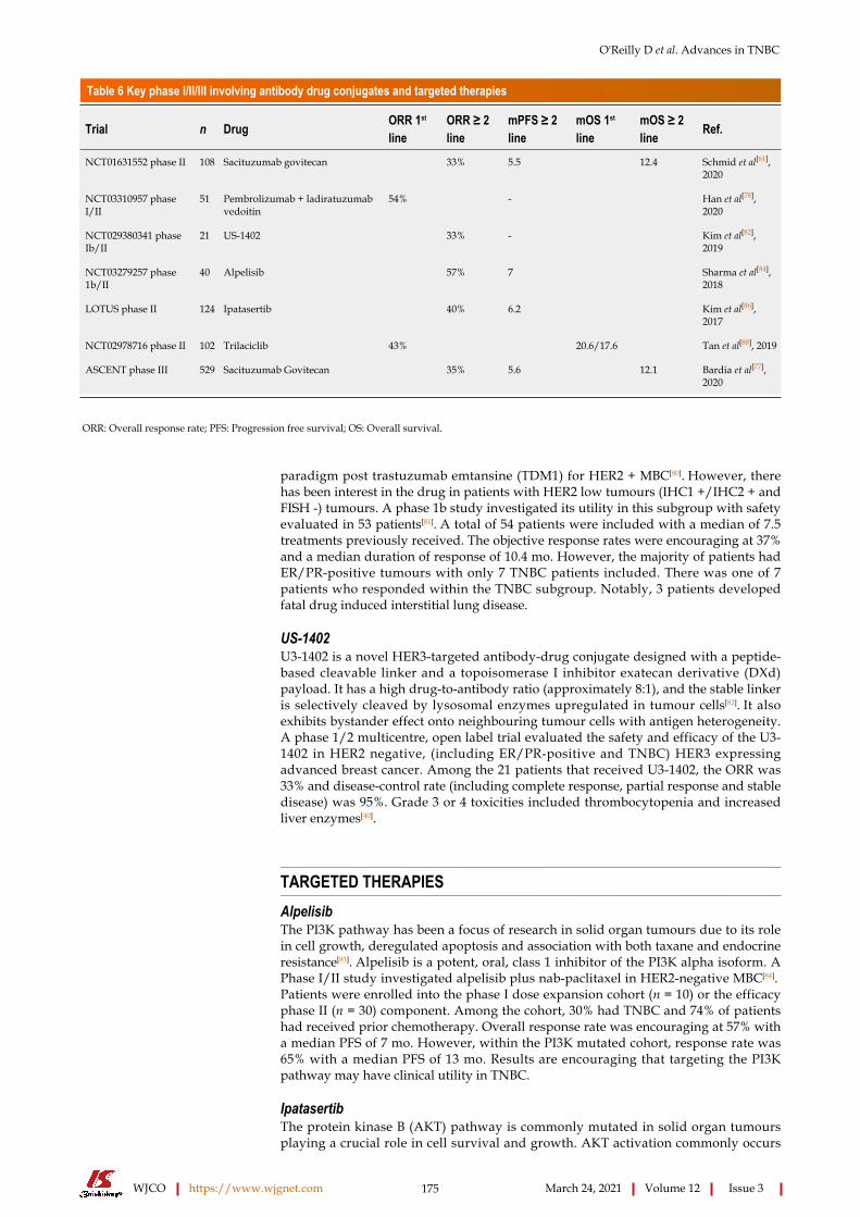
O'Reilly D et al. Advances in TNBC
WJCO https://www.wjgnet.com 175 March 24, 2021 Volume 12 Issue 3
Table 6 Key phase I/II/III involving antibody drug conjugates and targeted therapies
Trial n Drug ORR 1st line
ORR ≥ 2 line
mPFS ≥ 2 line
mOS 1st line
mOS ≥ 2 line Ref.
NCT01631552 phase II 108 Sacituzumab govitecan 33% 5.5 12.4 Schmid et al[61], 2020
NCT03310957 phase I/II
51 Pembrolizumab + ladiratuzumab vedoitin
54% - Han et al[78], 2020
NCT029380341 phase Ib/II
21 US-1402 33% - Kim et al[82], 2019
NCT03279257 phase 1b/II
40 Alpelisib 57% 7 Sharma et al[84], 2018
LOTUS phase II 124 Ipatasertib 40% 6.2 Kim et al[86], 2017
NCT02978716 phase II 102 Trilaciclib 43% 20.6/17.6 Tan et al[88], 2019
ASCENT phase III 529 Sacituzumab Govitecan 35% 5.6 12.1 Bardia et al[77], 2020
ORR: Overall response rate; PFS: Progression free survival; OS: Overall survival.
paradigm post trastuzumab emtansine (TDM1) for HER2 + MBC[80]. However, there has been interest in the drug in patients with HER2 low tumours (IHC1 +/IHC2 + and FISH -) tumours. A phase 1b study investigated its utility in this subgroup with safety evaluated in 53 patients[81]. A total of 54 patients were included with a median of 7.5 treatments previously received. The objective response rates were encouraging at 37% and a median duration of response of 10.4 mo. However, the majority of patients had ER/PR-positive tumours with only 7 TNBC patients included. There was one of 7 patients who responded within the TNBC subgroup. Notably, 3 patients developed fatal drug induced interstitial lung disease.
US-1402U3-1402 is a novel HER3-targeted antibody-drug conjugate designed with a peptide-based cleavable linker and a topoisomerase I inhibitor exatecan derivative (DXd) payload. It has a high drug-to-antibody ratio (approximately 8:1), and the stable linker is selectively cleaved by lysosomal enzymes upregulated in tumour cells[82]. It also exhibits bystander effect onto neighbouring tumour cells with antigen heterogeneity. A phase 1/2 multicentre, open label trial evaluated the safety and efficacy of the U3-1402 in HER2 negative, (including ER/PR-positive and TNBC) HER3 expressing advanced breast cancer. Among the 21 patients that received U3-1402, the ORR was 33% and disease-control rate (including complete response, partial response and stable disease) was 95%. Grade 3 or 4 toxicities included thrombocytopenia and increased liver enzymes[40].
TARGETED THERAPIESAlpelisibThe PI3K pathway has been a focus of research in solid organ tumours due to its role in cell growth, deregulated apoptosis and association with both taxane and endocrine resistance[83]. Alpelisib is a potent, oral, class 1 inhibitor of the PI3K alpha isoform. A Phase I/II study investigated alpelisib plus nab-paclitaxel in HER2-negative MBC[84]. Patients were enrolled into the phase I dose expansion cohort (n = 10) or the efficacy phase II (n = 30) component. Among the cohort, 30% had TNBC and 74% of patients had received prior chemotherapy. Overall response rate was encouraging at 57% with a median PFS of 7 mo. However, within the PI3K mutated cohort, response rate was 65% with a median PFS of 13 mo. Results are encouraging that targeting the PI3K pathway may have clinical utility in TNBC.
IpatasertibThe protein kinase B (AKT) pathway is commonly mutated in solid organ tumours playing a crucial role in cell survival and growth. AKT activation commonly occurs

O'Reilly D et al. Advances in TNBC
WJCO https://www.wjgnet.com 176 March 24, 2021 Volume 12 Issue 3
through phosphate and tensin homolog (PTEN) loss or PIK3CA mutations. However, targeting the AKT pathway has proven to be challenging due to the associated toxicities. Ipatasertib is a potent AKT pathway signalling inhibitor which has demonstrated tolerability and antitumour activity in early clinical studies[85]. The LOTUS trial investigated ipatasertib in 124 patients in a randomised phase 2 study of ipatasertib/paclitaxel vs placebo/paclitaxel as first-line therapy for TNBC[86]. In the overall population, the median PFS was enhanced with ipatasertib (6.2 mo vs 4.9 mo, HR =0.6, P = 0.037). In patients with PTEN-low tumours (identified via immuno-histochemistry), median PFS was 6.2 mo with ipatasertib vs 3.7 mo with placebo. However, within the PIK3CA/AKT1/PTEN-altered tumours, PFS was 9 mo vs 4.9 mo (HR 0.44, P = 0.041). The most common toxicity was diarrhoea in 23 % of patients in the ipatasertib arm leading to discontinuation in 3% of patients.
TrilaciclibTrilaciclib is a potent, intravenous cyclin dependent kinase 4/6 (CDK4/6) inhibitor which is thought to acutely protect from cytotoxic associated myelosuppression and may promote immunogenic tumour cell death[87]. A phase II study of trilaciclib in TNBC in combination with the doublet of gemcitabine/carboplatin was designed to identify a reduction in myelosuppression associated with chemotherapy[88]. Patients (n = 102) were assigned in a 1:1:1 fashion to (Cohort 1) gemcitabine/carboplatin alone vs (Cohort 2) gemcitabine/carboplatin/trilaciclib (D1/D8) vs (Cohort 3) gemci-tabine/carboplatin (D2/D9) and Trilaciclib (D1/2/8/9). Approximately 2/3rds of patients were treatment naïve (in the metastatic setting). There was no significant difference in myelosuppression between the groups, however there was a significant OS benefit in the trilaciclib arms. Patients in Cohort 1 had a median OS of 12.6 mo vs 20.6 mo in Cohort 2 and 17.6 mo in Cohort 3.
CONCLUSIONDespite recent advances, metastatic TNBC remains an aggressive disease which predominantly affects younger patients. Recent advances in pre-clinical science have demonstrated an impressive rationale for the use of IO and PARPi.
There is evidence of activity for the use PD-L1 or PD-1 inhibitors in the first-line setting of TNBC. However, this has not yet resulted in statistically significant improvements in OS. Additionally, it remains unclear why findings with the combination of nab-paclitaxel and atezolizumab were not reproducible when atezolizumab was combined with paclitaxel. OS analysis from the KEYNOTE-355 study may assist us in reaching final conclusions for the up-front combination of IO and chemotherapy. However, these conflicting results suggest that the addition of IO into routine practice should be done so with caution.
Patients with g BRCA mutations have a consistent but modest PFS benefit of 1-3 mo across multiple phase I/II/III studies. However, these have unfortunately not translated into an OS benefit. While PARPi may have a future role in the treatment paradigm for TNBC, the OS benefits for patients remains unclear.
Encouragingly, antibody-drug conjugates and targeted therapies have demon-strated impressive response rates and PFS benefits in the monotherapy or combination settings in patients with TNBC. Most recently, SG has demonstrated an impressive 6 mo’ OS benefit in a heavily pre-treated population. It is likely that SG will have a significant role to play in the future of TNBC in the monotherapy or combi-nation setting.
It is likely that the future of metastatic TNBC will involve treatment algorithms with combination approaches using chemotherapy, immunotherapy, PARPi, ADC and targeted therapies. Hopefully, the combination of the old and new will ensure that clinical outcomes continue to improve for our patients.
Clinical practice points(1) Despite recent drug developments, chemotherapy remains integral to the management of advanced TNBC; (2) Immunotherapy and PARPi have shown much promise but have yet to demonstrate a proven OS benefit in this disease; and (3) Antibody drug conjugates and other targeted therapies may ultimately prove to be the next frontier in treating this illness.

O'Reilly D et al. Advances in TNBC
WJCO https://www.wjgnet.com 177 March 24, 2021 Volume 12 Issue 3
REFERENCESPerou CM, Sørlie T, Eisen MB, van de Rijn M, Jeffrey SS, Rees CA, Pollack JR, Ross DT, Johnsen H, Akslen LA, Fluge O, Pergamenschikov A, Williams C, Zhu SX, Lønning PE, Børresen-Dale AL, Brown PO, Botstein D. Molecular portraits of human breast tumours. Nature 2000; 406: 747-752 [PMID: 10963602 DOI: 10.1038/35021093]
1
Costa RLB, Gradishar WJ. Triple-Negative Breast Cancer: Current Practice and Future Directions. J Oncol Pract 2017; 13: 301-303 [PMID: 28489982 DOI: 10.1200/JOP.2017.023333]
2
Nielsen TO, Hsu FD, Jensen K, Cheang M, Karaca G, Hu Z, Hernandez-Boussard T, Livasy C, Cowan D, Dressler L, Akslen LA, Ragaz J, Gown AM, Gilks CB, van de Rijn M, Perou CM. Immunohistochemical and clinical characterization of the basal-like subtype of invasive breast carcinoma. Clin Cancer Res 2004; 10: 5367-5374 [PMID: 15328174 DOI: 10.1158/1078-0432.CCR-04-0220]
3
Liu Y, Xin T, Huang DY, Shen WX, Li L, Lv YJ, Jin YH, Song XW, Teng C, Jiang QY. Prognosis in very young women with triple-negative breast cancer: retrospective study of 216 cases. Med Oncol 2014; 31: 222 [PMID: 25391919 DOI: 10.1007/s12032-014-0222-2]
4
Stead LA, Lash TL, Sobieraj JE, Chi DD, Westrup JL, Charlot M, Blanchard RA, Lee JC, King TC, Rosenberg CL. Triple-negative breast cancers are increased in black women regardless of age or body mass index. Breast Cancer Res 2009; 11: R18 [PMID: 19320967 DOI: 10.1186/bcr2242]
5
Sørlie T, Wang Y, Xiao C, Johnsen H, Naume B, Samaha RR, Børresen-Dale AL. Distinct molecular mechanisms underlying clinically relevant subtypes of breast cancer: gene expression analyses across three different platforms. BMC Genomics 2006; 7: 127 [PMID: 16729877 DOI: 10.1186/1471-2164-7-127]
6
Howlader N, Cronin KA, Kurian AW, Andridge R. Differences in Breast Cancer Survival by Molecular Subtypes in the United States. Cancer Epidemiol Biomarkers Prev 2018; 27: 619-626 [PMID: 29593010 DOI: 10.1158/1055-9965.EPI-17-0627]
7
Yao Y, Chu Y, Xu B, Hu Q, Song Q. Risk factors for distant metastasis of patients with primary triple-negative breast cancer. Biosci Rep 2019; 39: BSR20190288 [PMID: 31113872 DOI: 10.1042/BSR20190288]
8
Dent R, Trudeau M, Pritchard KI, Hanna WM, Kahn HK, Sawka CA, Lickley LA, Rawlinson E, Sun P, Narod SA. Triple-negative breast cancer: clinical features and patterns of recurrence. Clin Cancer Res 2007; 13: 4429-4434 [PMID: 17671126 DOI: 10.1158/1078-0432.CCR-06-3045]
9
Liedtke C, Mazouni C, Hess KR, André F, Tordai A, Mejia JA, Symmans WF, Gonzalez-Angulo AM, Hennessy B, Green M, Cristofanilli M, Hortobagyi GN, Pusztai L. Response to neoadjuvant therapy and long-term survival in patients with triple-negative breast cancer. J Clin Oncol 2008; 26: 1275-1281 [PMID: 18250347 DOI: 10.1200/JCO.2007.14.4147]
10
Jin J, Gao Y, Zhang J, Wang L, Wang B, Cao J, Shao Z, Wang Z. Incidence, pattern and prognosis of brain metastases in patients with metastatic triple negative breast cancer. BMC Cancer 2018; 18: 446 [PMID: 29673325 DOI: 10.1186/s12885-018-4371-0]
11
Fan C, Oh DS, Wessels L, Weigelt B, Nuyten DS, Nobel AB, van't Veer LJ, Perou CM. Concordance among gene-expression-based predictors for breast cancer. N Engl J Med 2006; 355: 560-569 [PMID: 16899776 DOI: 10.1056/NEJMoa052933]
12
Bertucci F, Finetti P, Cervera N, Esterni B, Hermitte F, Viens P, Birnbaum D. How basal are triple-negative breast cancers? Int J Cancer 2008; 123: 236-240 [PMID: 18398844 DOI: 10.1002/ijc.23518]
13
Rakha EA, Reis-Filho JS, Ellis IO. Basal-like breast cancer: a critical review. J Clin Oncol 2008; 26: 2568-2581 [PMID: 18487574 DOI: 10.1200/JCO.2007.13.1748]
14
Reis-Filho JS, Tutt AN. Triple negative tumours: a critical review. Histopathology 2008; 52: 108-118 [PMID: 18171422 DOI: 10.1111/j.1365-2559.2007.02889.x]
15
Foulkes WD, Smith IE, Reis-Filho JS. Triple-negative breast cancer. N Engl J Med 2010; 363: 1938-1948 [PMID: 21067385 DOI: 10.1056/NEJMra1001389]
16
Lim E, Vaillant F, Wu D, Forrest NC, Pal B, Hart AH, Asselin-Labat ML, Gyorki DE, Ward T, Partanen A, Feleppa F, Huschtscha LI, Thorne HJ; kConFab, Fox SB, Yan M, French JD, Brown MA, Smyth GK, Visvader JE, Lindeman GJ. Aberrant luminal progenitors as the candidate target population for basal tumor development in BRCA1 mutation carriers. Nat Med 2009; 15: 907-913 [PMID: 19648928 DOI: 10.1038/nm.2000]
17
Foulkes WD, Stefansson IM, Chappuis PO, Bégin LR, Goffin JR, Wong N, Trudel M, Akslen LA. Germline BRCA1 mutations and a basal epithelial phenotype in breast cancer. J Natl Cancer Inst 2003; 95: 1482-1485 [PMID: 14519755 DOI: 10.1093/jnci/djg050]
18
Lakhani SR, Reis-Filho JS, Fulford L, Penault-Llorca F, van der Vijver M, Parry S, Bishop T, Benitez J, Rivas C, Bignon YJ, Chang-Claude J, Hamann U, Cornelisse CJ, Devilee P, Beckmann MW, Nestle-Krämling C, Daly PA, Haites N, Varley J, Lalloo F, Evans G, Maugard C, Meijers-Heijboer H, Klijn JG, Olah E, Gusterson BA, Pilotti S, Radice P, Scherneck S, Sobol H, Jacquemier J, Wagner T, Peto J, Stratton MR, McGuffog L, Easton DF; Breast Cancer Linkage Consortium. Prediction of BRCA1 status in patients with breast cancer using estrogen receptor and basal phenotype. Clin Cancer Res 2005; 11: 5175-5180 [PMID: 16033833 DOI: 10.1158/1078-0432.CCR-04-2424]
19
Peshkin BN, Alabek ML, Isaacs C. BRCA1/2 mutations and triple negative breast cancers. Breast Dis 2010; 32: 25-33 [PMID: 21778580 DOI: 10.3233/BD-2010-0306]
20
Byrum AK, Vindigni A, Mosammaparast N. Defining and Modulating 'BRCAness'. Trends Cell Biol 21

O'Reilly D et al. Advances in TNBC
WJCO https://www.wjgnet.com 178 March 24, 2021 Volume 12 Issue 3
2019; 29: 740-751 [PMID: 31362850 DOI: 10.1016/j.tcb.2019.06.005]Turner N, Tutt A, Ashworth A. Hallmarks of 'BRCAness' in sporadic cancers. Nat Rev Cancer 2004; 4: 814-819 [PMID: 15510162 DOI: 10.1038/nrc1457]
22
Keung MYT, Wu Y, Vadgama JV. PARP Inhibitors as a Therapeutic Agent for Homologous Recombination Deficiency in Breast Cancers. J Clin Med 2019; 8 [PMID: 30934991 DOI: 10.3390/jcm8040435]
23
Yu T, Di G. Role of tumor microenvironment in triple-negative breast cancer and its prognostic significance. Chin J Cancer Res 2017; 29: 237-252 [PMID: 28729775 DOI: 10.21147/j.issn.1000-9604.2017.03.10]
24
Nagarajan D, McArdle SEB. Immune Landscape of Breast Cancers. Biomedicines 2018; 6 [PMID: 29439457 DOI: 10.3390/biomedicines6010020]
25
García-Teijido P, Cabal ML, Fernández IP, Pérez YF. Tumor-Infiltrating Lymphocytes in Triple Negative Breast Cancer: The Future of Immune Targeting. Clin Med Insights Oncol 2016; 10: 31-39 [PMID: 27081325 DOI: 10.4137/CMO.S34540]
26
Loi S, Drubay D, Adams S, Pruneri G, Francis PA, Lacroix-Triki M, Joensuu H, Dieci MV, Badve S, Demaria S, Gray R, Munzone E, Lemonnier J, Sotiriou C, Piccart MJ, Kellokumpu-Lehtinen PL, Vingiani A, Gray K, Andre F, Denkert C, Salgado R, Michiels S. Tumor-Infiltrating Lymphocytes and Prognosis: A Pooled Individual Patient Analysis of Early-Stage Triple-Negative Breast Cancers. J Clin Oncol 2019; 37: 559-569 [PMID: 30650045 DOI: 10.1200/JCO.18.01010]
27
Zhang L, Wang XI, Ding J, Sun Q, Zhang S. The predictive and prognostic value of Foxp3+/CD25+ regulatory T cells and PD-L1 expression in triple negative breast cancer. Ann Diagn Pathol 2019; 40: 143-151 [PMID: 31096176 DOI: 10.1016/j.anndiagpath.2019.04.004]
28
Ibrahim EM, Al-Foheidi ME, Al-Mansour MM, Kazkaz GA. The prognostic value of tumor-infiltrating lymphocytes in triple-negative breast cancer: a meta-analysis. Breast Cancer Res Treat 2014; 148: 467-476 [PMID: 25361613 DOI: 10.1007/s10549-014-3185-2]
29
Pagès F, Galon J, Dieu-Nosjean MC, Tartour E, Sautès-Fridman C, Fridman WH. Immune infiltration in human tumors: a prognostic factor that should not be ignored. Oncogene 2010; 29: 1093-1102 [PMID: 19946335 DOI: 10.1038/onc.2009.416]
30
Salgado R, Denkert C, Demaria S, Sirtaine N, Klauschen F, Pruneri G, Wienert S, Van den Eynden G, Baehner FL, Penault-Llorca F, Perez EA, Thompson EA, Symmans WF, Richardson AL, Brock J, Criscitiello C, Bailey H, Ignatiadis M, Floris G, Sparano J, Kos Z, Nielsen T, Rimm DL, Allison KH, Reis-Filho JS, Loibl S, Sotiriou C, Viale G, Badve S, Adams S, Willard-Gallo K, Loi S; International TILs Working Group 2014. The evaluation of tumor-infiltrating lymphocytes (TILs) in breast cancer: recommendations by an International TILs Working Group 2014. Ann Oncol 2015; 26: 259-271 [PMID: 25214542 DOI: 10.1093/annonc/mdu450]
31
Denkert C, Wienert S, Poterie A, Loibl S, Budczies J, Badve S, Bago-Horvath Z, Bane A, Bedri S, Brock J, Chmielik E, Christgen M, Colpaert C, Demaria S, Van den Eynden G, Floris G, Fox SB, Gao D, Ingold Heppner B, Kim SR, Kos Z, Kreipe HH, Lakhani SR, Penault-Llorca F, Pruneri G, Radosevic-Robin N, Rimm DL, Schnitt SJ, Sinn BV, Sinn P, Sirtaine N, O'Toole SA, Viale G, Van de Vijver K, de Wind R, von Minckwitz G, Klauschen F, Untch M, Fasching PA, Reimer T, Willard-Gallo K, Michiels S, Loi S, Salgado R. Standardized evaluation of tumor-infiltrating lymphocytes in breast cancer: results of the ring studies of the international immuno-oncology biomarker working group. Mod Pathol 2016; 29: 1155-1164 [PMID: 27363491 DOI: 10.1038/modpathol.2016.109]
32
Mittendorf EA, Philips AV, Meric-Bernstam F, Qiao N, Wu Y, Harrington S, Su X, Wang Y, Gonzalez-Angulo AM, Akcakanat A, Chawla A, Curran M, Hwu P, Sharma P, Litton JK, Molldrem JJ, Alatrash G. PD-L1 expression in triple-negative breast cancer. Cancer Immunol Res 2014; 2: 361-370 [PMID: 24764583 DOI: 10.1158/2326-6066.CIR-13-0127]
33
Narang P, Chen M, Sharma AA, Anderson KS, Wilson MA. The neoepitope landscape of breast cancer: implications for immunotherapy. BMC Cancer 2019; 19: 200 [PMID: 30832597 DOI: 10.1186/s12885-019-5402-1]
34
Sabatier R, Finetti P, Mamessier E, Adelaide J, Chaffanet M, Ali HR, Viens P, Caldas C, Birnbaum D, Bertucci F. Prognostic and predictive value of PDL1 expression in breast cancer. Oncotarget 2015; 6: 5449-5464 [PMID: 25669979 DOI: 10.18632/oncotarget.3216]
35
Salmaninejad A, Valilou SF, Shabgah AG, Aslani S, Alimardani M, Pasdar A, Sahebkar A. PD-1/PD-L1 pathway: Basic biology and role in cancer immunotherapy. J Cell Physiol 2019; 234: 16824-16837 [PMID: 30784085 DOI: 10.1002/jcp.28358]
36
Barroso-Sousa R, Keenan TE, Pernas S, Exman P, Jain E, Garrido-Castro AC, Hughes M, Bychkovsky B, Umeton R, Files JL, Lindeman NI, MacConaill LE, Hodi FS, Krop IE, Dillon D, Winer EP, Wagle N, Lin NU, Mittendorf EA, Van Allen EM, Tolaney SM. Tumor Mutational Burden and PTEN Alterations as Molecular Correlates of Response to PD-1/L1 Blockade in Metastatic Triple-Negative Breast Cancer. Clin Cancer Res 2020; 26: 2565-2572 [PMID: 32019858 DOI: 10.1158/1078-0432.CCR-19-3507]
37
Goodman AM, Kato S, Bazhenova L, Patel SP, Frampton GM, Miller V, Stephens PJ, Daniels GA, Kurzrock R. Tumor Mutational Burden as an Independent Predictor of Response to Immunotherapy in Diverse Cancers. Mol Cancer Ther 2017; 16: 2598-2608 [PMID: 28835386 DOI: 10.1158/1535-7163.MCT-17-0386]
38
Chan TA, Yarchoan M, Jaffee E, Swanton C, Quezada SA, Stenzinger A, Peters S. Development of tumor mutation burden as an immunotherapy biomarker: utility for the oncology clinic. Ann Oncol 2019; 30: 44-56 [PMID: 30395155 DOI: 10.1093/annonc/mdy495]
39

O'Reilly D et al. Advances in TNBC
WJCO https://www.wjgnet.com 179 March 24, 2021 Volume 12 Issue 3
Yonemori K, Masuda N, Takahashi S, Kogawa T, Iwata, H. Single agent activity of U3-1402, a HER3- targeting antibody–drug conjugate, in HER3- overexpressing metastatic breast cancer: updated results from a phase I/II trial. Ann Oncol 2019; 30 [DOI: 10.1093/annonc/mdz100.002]
40
Carey LA, Dees EC, Sawyer L, Gatti L, Moore DT, Collichio F, Ollila DW, Sartor CI, Graham ML, Perou CM. The triple negative paradox: primary tumor chemosensitivity of breast cancer subtypes. Clin Cancer Res 2007; 13: 2329-2334 [PMID: 17438091 DOI: 10.1158/1078-0432.CCR-06-1109]
41
Cardoso F, Senkus E, Costa A, Papadopoulos E, Aapro M, André F, Harbeck N, Aguilar Lopez B, Barrios CH, Bergh J, Biganzoli L, Boers-Doets CB, Cardoso MJ, Carey LA, Cortés J, Curigliano G, Diéras V, El Saghir NS, Eniu A, Fallowfield L, Francis PA, Gelmon K, Johnston SRD, Kaufman B, Koppikar S, Krop IE, Mayer M, Nakigudde G, Offersen BV, Ohno S, Pagani O, Paluch-Shimon S, Penault-Llorca F, Prat A, Rugo HS, Sledge GW, Spence D, Thomssen C, Vorobiof DA, Xu B, Norton L, Winer EP. 4th ESO-ESMO International Consensus Guidelines for Advanced Breast Cancer (ABC 4)†. Ann Oncol 2018; 29: 1634-1657 [PMID: 30032243 DOI: 10.1093/annonc/mdy192]
42
Smith KL, Smith M Lou, Burns J. Breast Cancer. NCCN Guidelines. 202043 Sparano JA, Makhson AN, Semiglazov VF, Tjulandin SA, Balashova OI, Bondarenko IN, Bogdanova NV, Manikhas GM, Oliynychenko GP, Chatikhine VA, Zhuang SH, Xiu L, Yuan Z, Rackoff WR. Pegylated liposomal doxorubicin plus docetaxel significantly improves time to progression without additive cardiotoxicity compared with docetaxel monotherapy in patients with advanced breast cancer previously treated with neoadjuvant-adjuvant anthracycline therapy: results from a randomized phase III study. J Clin Oncol 2009; 27: 4522-4529 [PMID: 19687336 DOI: 10.1200/JCO.2008.20.5013]
44
Cardoso F, Bedard PL, Winer EP, Pagani O, Senkus-Konefka E, Fallowfield LJ, Kyriakides S, Costa A, Cufer T, Albain KS; ESO-MBC Task Force. International guidelines for management of metastatic breast cancer: combination vs sequential single-agent chemotherapy. J Natl Cancer Inst 2009; 101: 1174-1181 [PMID: 19657108 DOI: 10.1093/jnci/djp235]
45
Isakoff SJ, Mayer EL, He L, Traina TA, Carey LA, Krag KJ, Rugo HS, Liu MC, Stearns V, Come SE, Timms KM, Hartman AR, Borger DR, Finkelstein DM, Garber JE, Ryan PD, Winer EP, Goss PE, Ellisen LW. TBCRC009: A Multicenter Phase II Clinical Trial of Platinum Monotherapy With Biomarker Assessment in Metastatic Triple-Negative Breast Cancer. J Clin Oncol 2015; 33: 1902-1909 [PMID: 25847936 DOI: 10.1200/JCO.2014.57.6660]
46
Telli ML, Timms KM, Reid J, Hennessy B, Mills GB, Jensen KC, Szallasi Z, Barry WT, Winer EP, Tung NM, Isakoff SJ, Ryan PD, Greene-Colozzi A, Gutin A, Sangale Z, Iliev D, Neff C, Abkevich V, Jones JT, Lanchbury JS, Hartman AR, Garber JE, Ford JM, Silver DP, Richardson AL. Homologous Recombination Deficiency (HRD) Score Predicts Response to Platinum-Containing Neoadjuvant Chemotherapy in Patients with Triple-Negative Breast Cancer. Clin Cancer Res 2016; 22: 3764-3773 [PMID: 26957554 DOI: 10.1158/1078-0432.CCR-15-2477]
47
Tutt A, Tovey H, Cheang MCU, Kernaghan S, Kilburn L, Gazinska P, Owen J, Abraham J, Barrett S, Barrett-Lee P, Brown R, Chan S, Dowsett M, Flanagan JM, Fox L, Grigoriadis A, Gutin A, Harper-Wynne C, Hatton MQ, Hoadley KA, Parikh J, Parker P, Perou CM, Roylance R, Shah V, Shaw A, Smith IE, Timms KM, Wardley AM, Wilson G, Gillett C, Lanchbury JS, Ashworth A, Rahman N, Harries M, Ellis P, Pinder SE, Bliss JM. Carboplatin in BRCA1/2-mutated and triple-negative breast cancer BRCAness subgroups: the TNT Trial. Nat Med 2018; 24: 628-637 [PMID: 29713086 DOI: 10.1038/s41591-018-0009-7]
48
Cortes J, O'Shaughnessy J, Loesch D, Blum JL, Vahdat LT, Petrakova K, Chollet P, Manikas A, Diéras V, Delozier T, Vladimirov V, Cardoso F, Koh H, Bougnoux P, Dutcus CE, Seegobin S, Mir D, Meneses N, Wanders J, Twelves C; EMBRACE (Eisai Metastatic Breast Cancer Study Assessing Physician's Choice Versus E7389) investigators. Eribulin monotherapy versus treatment of physician's choice in patients with metastatic breast cancer (EMBRACE): a phase 3 open-label randomised study. Lancet 2011; 377: 914-923 [PMID: 21376385 DOI: 10.1016/S0140-6736(11)60070-6]
49
Nanda R, Chow LQ, Dees EC, Berger R, Gupta S, Geva R, Pusztai L, Pathiraja K, Aktan G, Cheng JD, Karantza V, Buisseret L. Pembrolizumab in Patients With Advanced Triple-Negative Breast Cancer: Phase Ib KEYNOTE-012 Study. J Clin Oncol 2016; 34: 2460-2467 [PMID: 27138582 DOI: 10.1200/JCO.2015.64.8931]
50
Dirix LY, Takacs I, Jerusalem G, Nikolinakos P, Arkenau HT, Forero-Torres A, Boccia R, Lippman ME, Somer R, Smakal M, Emens LA, Hrinczenko B, Edenfield W, Gurtler J, von Heydebreck A, Grote HJ, Chin K, Hamilton EP. Avelumab, an anti-PD-L1 antibody, in patients with locally advanced or metastatic breast cancer: a phase 1b JAVELIN Solid Tumor study. Breast Cancer Res Treat 2018; 167: 671-686 [PMID: 29063313 DOI: 10.1007/s10549-017-4537-5]
51
Emens LA, Cruz C, Eder JP, Braiteh F, Chung C, Tolaney SM, Kuter I, Nanda R, Cassier PA, Delord JP, Gordon MS, ElGabry E, Chang CW, Sarkar I, Grossman W, O'Hear C, Fassò M, Molinero L, Schmid P. Long-term Clinical Outcomes and Biomarker Analyses of Atezolizumab Therapy for Patients With Metastatic Triple-Negative Breast Cancer: A Phase 1 Study. JAMA Oncol 2019; 5: 74-82 [PMID: 30242306 DOI: 10.1001/jamaoncol.2018.4224]
52
Adams S, Schmid P, Rugo HS, Winer EP, Loirat D, Awada A, Cescon DW, Iwata H, Campone M, Nanda R, Hui R, Curigliano G, Toppmeyer D, O'Shaughnessy J, Loi S, Paluch-Shimon S, Tan AR, Card D, Zhao J, Karantza V, Cortés J. Pembrolizumab monotherapy for previously treated metastatic triple-negative breast cancer: cohort A of the phase II KEYNOTE-086 study. Ann Oncol 2019; 30: 397-404 [PMID: 30475950 DOI: 10.1093/annonc/mdy517]
53
Cortés J, Lipatov O, Im S-A, Gonalves A, Winer EP. LBA21 - KEYNOTE-119: Phase III study of 54

O'Reilly D et al. Advances in TNBC
WJCO https://www.wjgnet.com 180 March 24, 2021 Volume 12 Issue 3
pembrolizumab (pembro) vs single-agent chemotherapy (chemo) for metastatic triple negative breast cancer (mTNBC). Ann Oncol 2019; 30: v859-v860 [DOI: 10.1093/annonc/mdz394.010]Adams S, Diamond JR, Hamilton E, Pohlmann PR, Tolaney SM, Chang CW, Zhang W, Iizuka K, Foster PG, Molinero L, Funke R, Powderly J. Atezolizumab Plus nab-Paclitaxel in the Treatment of Metastatic Triple-Negative Breast Cancer With 2-Year Survival Follow-up: A Phase 1b Clinical Trial. JAMA Oncol 2019; 5: 334-342 [PMID: 30347025 DOI: 10.1001/jamaoncol.2018.5152]
55
Schmid P, Adams S, Rugo HS, Schneeweiss A, Barrios CH, Iwata H, Diéras V, Hegg R, Im SA, Shaw Wright G, Henschel V, Molinero L, Chui SY, Funke R, Husain A, Winer EP, Loi S, Emens LA; IMpassion130 Trial Investigators. Atezolizumab and Nab-Paclitaxel in Advanced Triple-Negative Breast Cancer. N Engl J Med 2018; 379: 2108-2121 [PMID: 30345906 DOI: 10.1056/NEJMoa1809615]
56
Tolaney SM, Kalinsky K, Kaklamani V, Savulsky C, Diab S. Abstract PD6-13: Phase 1b/2 study to evaluate eribulin mesylate in combination with pembrolizumab in patients with metastatic triple-negative breast cancer. Cancer Res 2018; 78: PD6-13 [DOI: 10.1158/1538-7445.SABCS17-PD6-13]
57
Castan JC, Guo Z, Karantza V, Aktan G. Keynote-355: randomized, double-blind, phase iii study of pembrolizumab (pembro) + chemotherapy (chemo) vs placebo (pbo) + chemo for previously untreated, locally recurrent, inoperable or metastatic triple-negative breast cancer (mtnbc) - sciencedirect. Ann Oncol 2017; 28: x25 [DOI: 10.1093/annonc/mdx656]
58
Rugo HS, Loi S, Adams S, Schmid P, Schneeweiss A, Barrios CH, Iwata H, Dieras VC, Winer EP, Kockx M, Peeters D, Chui SY, Lin JC, Nguyen Duc A, Viale G, Molinero L, Emens LA. LBA20 - Performance of PD-L1 immunohistochemistry (IHC) assays in unresectable locally advanced or metastatic triple-negative breast cancer (mTNBC): Post-hoc analysis of IMpassion130. Ann Oncol 2019; 30: v858-v859 [DOI: 10.1093/annonc/mdz394.009]
59
Kulangara K, Zhang N, Corigliano E, Guerrero L, Waldroup S, Jaiswal D, Ms MJ, Shah S, Hanks D, Wang J, Lunceford J, Savage MJ, Juco J, Emancipator K. Clinical Utility of the Combined Positive Score for Programmed Death Ligand-1 Expression and the Approval of Pembrolizumab for Treatment of Gastric Cancer. Arch Pathol Lab Med 2019; 143: 330-337 [PMID: 30028179 DOI: 10.5858/arpa.2018-0043-OA]
60
Schmid P, Cortes J, Pusztai L, McArthur H, Kümmel S, Bergh J, Denkert C, Park YH, Hui R, Harbeck N, Takahashi M, Foukakis T, Fasching PA, Cardoso F, Untch M, Jia L, Karantza V, Zhao J, Aktan G, Dent R, O'Shaughnessy J; KEYNOTE-522 Investigators. Pembrolizumab for Early Triple-Negative Breast Cancer. N Engl J Med 2020; 382: 810-821 [PMID: 32101663 DOI: 10.1056/NEJMoa1910549]
61
Fuentes-Antrás J, Guevara-Hoyer K, Baliu-Piqué M, García-Sáenz JÁ, Pérez-Segura P, Pandiella A, Ocaña A. Adoptive Cell Therapy in Breast Cancer: A Current Perspective of Next-Generation Medicine. Front Oncol 2020; 10: 605633 [PMID: 33194771 DOI: 10.3389/fonc.2020.605633]
62
Tutt A, Robson M, Garber JE, Domchek SM, Audeh MW, Weitzel JN, Friedlander M, Arun B, Loman N, Schmutzler RK, Wardley A, Mitchell G, Earl H, Wickens M, Carmichael J. Oral poly(ADP-ribose) polymerase inhibitor olaparib in patients with BRCA1 or BRCA2 mutations and advanced breast cancer: a proof-of-concept trial. Lancet 2010; 376: 235-244 [PMID: 20609467 DOI: 10.1016/S0140-6736(10)60892-6]
63
Kaufman B, Shapira-Frommer R, Schmutzler RK, Audeh MW, Friedlander M, Balmaña J, Mitchell G, Fried G, Stemmer SM, Hubert A, Rosengarten O, Steiner M, Loman N, Bowen K, Fielding A, Domchek SM. Olaparib monotherapy in patients with advanced cancer and a germline BRCA1/2 mutation. J Clin Oncol 2015; 33: 244-250 [PMID: 25366685 DOI: 10.1200/JCO.2014.56.2728]
64
Turner NC, Telli ML, Rugo HS, Mailliez A, Robson ME. Final results of a phase 2 study of talazoparib (TALA) following platinum or multiple cytotoxic regimens in advanced breast cancer patients (pts) with germline BRCA1/2 mutations (ABRAZO). J Clin Oncol 2017; 35: 1007 [DOI: 10.1200/JCO.2017.35.15_suppl.1007]
65
Han HS, Diéras V, Robson M, Palácová M, Marcom PK, Jager A, Bondarenko I, Citrin D, Campone M, Telli ML, Domchek SM, Friedlander M, Kaufman B, Garber JE, Shparyk Y, Chmielowska E, Jakobsen EH, Kaklamani V, Gradishar W, Ratajczak CK, Nickner C, Qin Q, Qian J, Shepherd SP, Isakoff SJ, Puhalla S. Veliparib with temozolomide or carboplatin/paclitaxel versus placebo with carboplatin/paclitaxel in patients with BRCA1/2 locally recurrent/metastatic breast cancer: randomized phase II study. Ann Oncol 2018; 29: 154-161 [PMID: 29045554 DOI: 10.1093/annonc/mdx505]
66
Robson M, Im SA, Senkus E, Xu B, Domchek SM, Masuda N, Delaloge S, Li W, Tung N, Armstrong A, Wu W, Goessl C, Runswick S, Conte P. Olaparib for Metastatic Breast Cancer in Patients with a Germline BRCA Mutation. N Engl J Med 2017; 377: 523-533 [PMID: 28578601 DOI: 10.1056/NEJMoa1706450]
67
Litton JK, Rugo HS, Ettl J, Hurvitz SA, Gonçalves A, Lee KH, Fehrenbacher L, Yerushalmi R, Mina LA, Martin M, Roché H, Im YH, Quek RGW, Markova D, Tudor IC, Hannah AL, Eiermann W, Blum JL. Talazoparib in Patients with Advanced Breast Cancer and a Germline BRCA Mutation. N Engl J Med 2018; 379: 753-763 [PMID: 30110579 DOI: 10.1056/NEJMoa1802905]
68
Litton JK, Hurvitz SA, Mina LA, Rugo HS, Lee KH, Gonçalves A, Diab S, Woodward N, Goodwin A, Yerushalmi R, Roché H, Im YH, Eiermann W, Quek RGW, Usari T, Lanzalone S, Czibere A, Blum JL, Martin M, Ettl J. Talazoparib versus chemotherapy in patients with germline BRCA1/2-mutated HER2-negative advanced breast cancer: final overall survival results from the EMBRACA trial. Ann Oncol 2020; 31: 1526-1535 [PMID: 32828825 DOI: 10.1016/j.annonc.2020.08.2098]
69

O'Reilly D et al. Advances in TNBC
WJCO https://www.wjgnet.com 181 March 24, 2021 Volume 12 Issue 3
Diéras VC, Han HS, Kaufman B, Wildiers H, Arun BK. LBA9 - Phase III study of veliparib with carboplatin and paclitaxel in HER2-negative advanced/metastatic gBRCA-associated breast cancer. Ann Oncol 2019; 30: v857-v858 [DOI: 10.1093/annonc/mdz394.008]
70
Han HS, Arun B, Kaufman B, Wildiers H, Arun BK. Veliparib (V) monotherapy (monoTx) following combination therapy with V + carboplatin/paclitaxel (CP) in patients with gBRCA-associated advanced breast cancer: Exploratory results from BROCADE3. J Clin Oncol 2020; 38: 1091 [DOI: 10.1200/JCO.2020.38.15_suppl.1091]
71
Tung NM, Robson ME, Ventz S, Santa-Maria CA, Garber JE. TBCRC 048: A phase II study of olaparib monotherapy in metastatic breast cancer patients with germline or somatic mutations in DNA damage response (DDR) pathway genes (Olaparib Expanded). J Clin Oncol 2020; 38: 1002 [DOI: 10.1200/JCO.2020.38.15_suppl.1002]
72
Sharma P, Rodler E, Barlow WE, Gralow J, Hortobagyi GN. Results of a phase II randomized trial of cisplatin +/- veliparib in metastatic triple-negative breast cancer (TNBC) and/or germline BRCA-associated breast cancer (SWOG S1416). J Clin Oncol 2020; 38: 1001 [DOI: 10.1200/JCO.2020.38.15_suppl.1001]
73
Takaya H, Nakai H, Takamatsu S, Mandai M, Matsumura N. Homologous recombination deficiency status-based classification of high-grade serous ovarian carcinoma. Sci Rep 2020; 10: 2757 [PMID: 32066851 DOI: 10.1038/s41598-020-59671-3]
74
Starodub AN, Ocean AJ, Shah MA, Guarino MJ, Picozzi VJ Jr, Vahdat LT, Thomas SS, Govindan SV, Maliakal PP, Wegener WA, Hamburger SA, Sharkey RM, Goldenberg DM. First-in-Human Trial of a Novel Anti-Trop-2 Antibody-SN-38 Conjugate, Sacituzumab Govitecan, for the Treatment of Diverse Metastatic Solid Tumors. Clin Cancer Res 2015; 21: 3870-3878 [PMID: 25944802 DOI: 10.1158/1078-0432.CCR-14-3321]
75
Bardia A, Mayer IA, Vahdat LT, Tolaney SM, Isakoff SJ, Diamond JR, O'Shaughnessy J, Moroose RL, Santin AD, Abramson VG, Shah NC, Rugo HS, Goldenberg DM, Sweidan AM, Iannone R, Washkowitz S, Sharkey RM, Wegener WA, Kalinsky K. Sacituzumab Govitecan-hziy in Refractory Metastatic Triple-Negative Breast Cancer. N Engl J Med 2019; 380: 741-751 [PMID: 30786188 DOI: 10.1056/NEJMoa1814213]
76
Bardia A, Tolaney SM, Loirat D, Punie K, Oliveira M, Rugo HS, Brufsky A, Kalinsky K, Cortés J, O’Shaughnessy J, Dieras VC, Carey LA, Gianni L, Piccart M, Loibl S, Goldenberg D, Hong Q, Olivo MS, Hurvitz SA. LBA17 - ASCENT: A randomized phase III study of sacituzumab govitecan (SG) vs treatment of physician’s choice (TPC) in patients (pts) with previously treated metastatic triple-negative breast cancer (mTNBC). Ann Oncol 2020; 31: S1142-S1215 [DOI: 10.1016/j.annonc.2020.08.2245]
77
Han H (Heather), Diab S, Alemany C. Abstract PD1-06: Open label phase 1b/2 study of ladiratuzumab vedotin in combination with pembrolizumab for first-line treatment of patients with unresectable locally-advanced or metastatic triple-negative breast cancer. Cancer Res 2020; 80: PD1-06 LP [DOI: 10.1158/1538-7445.SABCS19-PD1-06]
78
Doi T, Shitara K, Naito Y, Shimomura A, Fujiwara Y, Yonemori K, Shimizu C, Shimoi T, Kuboki Y, Matsubara N, Kitano A, Jikoh T, Lee C, Fujisaki Y, Ogitani Y, Yver A, Tamura K. Safety, pharmacokinetics, and antitumour activity of trastuzumab deruxtecan (DS-8201), a HER2-targeting antibody-drug conjugate, in patients with advanced breast and gastric or gastro-oesophageal tumours: a phase 1 dose-escalation study. Lancet Oncol 2017; 18: 1512-1522 [PMID: 29037983 DOI: 10.1016/S1470-2045(17)30604-6]
79
Modi S, Saura C, Yamashita T, Park YH, Kim SB, Tamura K, Andre F, Iwata H, Ito Y, Tsurutani J, Sohn J, Denduluri N, Perrin C, Aogi K, Tokunaga E, Im SA, Lee KS, Hurvitz SA, Cortes J, Lee C, Chen S, Zhang L, Shahidi J, Yver A, Krop I; DESTINY-Breast01 Investigators. Trastuzumab Deruxtecan in Previously Treated HER2-Positive Breast Cancer. N Engl J Med 2020; 382: 610-621 [PMID: 31825192 DOI: 10.1056/NEJMoa1914510]
80
Modi S, Park H, Murthy RK, Iwata H, Tamura K, Tsurutani J, Moreno-Aspitia A, Doi T, Sagara Y, Redfern C, Krop IE, Lee C, Fujisaki Y, Sugihara M, Zhang L, Shahidi J, Takahashi S. Antitumor Activity and Safety of Trastuzumab Deruxtecan in Patients With HER2-Low-Expressing Advanced Breast Cancer: Results From a Phase Ib Study. J Clin Oncol 2020; 38: 1887-1896 [PMID: 32058843 DOI: 10.1200/JCO.19.02318]
81
Kim SB, Meric-Bernstam F, Kalyan A, Babich A, Berlin J. First-in-human phase i study of aprutumab ixadotin, a fibroblast growth factor receptor 2 antibody–drug conjugate (bay 1187982) in patients with advanced cancer. Target Oncol 2019; 14: 591-601 [PMID: 31502117 DOI: 10.1007/s11523-019-00670-4]
82
Verret B, Cortes J, Bachelot T, Andre F, Arnedos M. Efficacy of PI3K inhibitors in advanced breast cancer. Ann Oncol 2019; 30: x12-x20 [PMID: 31928690 DOI: 10.1093/annonc/mdz381]
83
Sharma P, Abramson VG, O’Dea A, Pathak HB, Godwin AK. Clinical and biomarker results from phase I/II study of PI3K inhibitor BYL 719 (alpelisib) plus nab-paclitaxel in HER2-negative metastatic breast cancer. J Clin Oncol 2018; 36: 1018 [DOI: 10.1200/JCO.2018.36.15_suppl.1018]
84
A First-in-Human Phase I Study of the ATP-Competitive AKT Inhibitor Ipatasertib Demonstrates Robust and Safe Targeting of AKT in Patients with Solid Tumors. Cancer Discov 2018; 8: 1490 [PMID: 30385525 DOI: 10.1158/2159-8290.CD-18-1114]
85
Kim SB, Dent R, Im SA, Espié M, Blau S, Tan AR, Isakoff SJ, Oliveira M, Saura C, Wongchenko MJ, Kapp AV, Chan WY, Singel SM, Maslyar DJ, Baselga J; LOTUS investigators. Ipatasertib plus paclitaxel versus placebo plus paclitaxel as first-line therapy for metastatic triple-negative breast
86

O'Reilly D et al. Advances in TNBC
WJCO https://www.wjgnet.com 182 March 24, 2021 Volume 12 Issue 3
cancer (LOTUS): a multicentre, randomised, double-blind, placebo-controlled, phase 2 trial. Lancet Oncol 2017; 18: 1360-1372 [PMID: 28800861 DOI: 10.1016/S1470-2045(17)30450-3]He S, Roberts PJ, Sorrentino JA, Bisi JE, Storrie-White H, Tiessen RG, Makhuli KM, Wargin WA, Tadema H, van Hoogdalem EJ, Strum JC, Malik R, Sharpless NE. Transient CDK4/6 inhibition protects hematopoietic stem cells from chemotherapy-induced exhaustion. Sci Transl Med 2017; 9 [PMID: 28446688 DOI: 10.1126/scitranslmed.aal3986]
87
Tan AR, Wright GS, Thummala AR, Danso MA, Popovic L, Pluard TJ, Han HS, Vojnović Ž, Vasev N, Ma L, Richards DA, Wilks ST, Milenković D, Yang Z, Antal JM, Morris SR, O'Shaughnessy J. Trilaciclib plus chemotherapy versus chemotherapy alone in patients with metastatic triple-negative breast cancer: a multicentre, randomised, open-label, phase 2 trial. Lancet Oncol 2019; 20: 1587-1601 [PMID: 31575503 DOI: 10.1016/S1470-2045(19)30616-3]
88
Li CH, Karantza V, Aktan G, Lala M. Current treatment landscape for patients with locally recurrent inoperable or metastatic triple-negative breast cancer: a systematic literature review. Breast Cancer Res 2019; 21: 143 [PMID: 31842957 DOI: 10.1186/s13058-019-1210-4]
89
Page DB, Chun B, Pucilowska J, Kim I, Mcarthur HL. Pembrolizumab (pembro) with paclitaxel (taxol) or capecitabine (cape) as early treatment of metastatic triple-negative breast cancer (mTNBC). J Clin Oncol 15: 1015-1015 [DOI: 10.1200/JCO.2019.37.15_suppl.1015]
90

WJCO https://www.wjgnet.com 183 March 24, 2021 Volume 12 Issue 3
World Journal of
Clinical OncologyW J C OSubmit a Manuscript: https://www.f6publishing.com World J Clin Oncol 2021 March 24; 12(3): 183-194
DOI: 10.5306/wjco.v12.i3.183 ISSN 2218-4333 (online)
ORIGINAL ARTICLE
Observational Study
Thromboembolic events in metastatic testicular cancer treated with cisplatin-based chemotherapy
Lisa B E Shields, Michael W Daniels, Nataliya Mar, Arash Rezazadeh Kalebasty
ORCID number: Lisa B E Shields 0000-0002-1526-4063; Michael W Daniels 0000-0003-2441-3136; Nataliya Mar 0000-0003-2485-6201; Arash Rezazadeh Kalebasty 0000-0002-3701-5084.
Author contributions: Shields LBE, Daniels MW, Mar N, and Rezazadeh Kalebasty A contributed to the conception, design, acquisition, analysis, and interpretation of data; Shields LBE drafted the manuscript; Shields LBE, Daniels MW, Mar N, and Rezazadeh Kalebasty A critically revised the manuscript and gave final approval.
Institutional review board statement: The University of Louisville Institutional Review Board determined that this study was exempt according to 45 CFR 46.101(b). The IRB number is 19.1288.
Informed consent statement: All study participants provided informed written consent prior to study enrollment.
Conflict-of-interest statement: There are no conflicts of interest to report.
Data sharing statement: No additional data were available.
Lisa B E Shields, Norton Neuroscience Institute, Norton Healthcare, Louisville, KY 40202, United States
Michael W Daniels, Department of Bioinformatics and Biostatistics, University of Louisville, Louisville, KY 40292, United States
Nataliya Mar, Arash Rezazadeh Kalebasty, Division of Hematology/Oncology, Department of Medicine, UCI Medical Center, Orange, CA 92868, United States
Corresponding author: Arash Rezazadeh Kalebasty, MD, Doctor, Division of Hematology/ Oncology, Department of Medicine, UCI Medical Center, 101 The City Drive South, Zot 4061, Orange, CA 92868, United States. [email protected]
AbstractBACKGROUND Testicular germ cell tumor (TGCT) is the most curable solid tumor and most common cancer among men 18-39 years. While cisplatin-based chemotherapy has significantly lengthened the survival of patients with TGCT, it is associated with a high rate of thromboembolic events (TEE).
AIM To summarize our single-center experience highlighting patients who were diagnosed with TGCT and received platinum-based chemotherapy, with special attention to those patients who suffered a TEE.
METHODS A retrospective analysis of the medical records and imaging studies of 68 consecutive individuals who were diagnosed with TGCT and received platinum-based chemotherapy at our Institution in a metropolitan community between January 1, 2014 and December 31, 2019.
RESULTS A total of 19 (28%) patients experienced a TEE following orchiectomy which occurred during chemotherapy in 13 (68%) of these patients. Patients with a higher pathologic stage (stage III) were significantly (P = 0.023) more likely to experience a TEE compared to patients who had a lower stage. Additionally, patients who were treated with 3 cycles of bleomycine, etoposide, and cisplatin and 1 cycle of etoposide and cisplatin or 4 cycles of etoposide and cisplatin were

Shields LBE et al. TEE in metastatic testicular cancer
WJCO https://www.wjgnet.com 184 March 24, 2021 Volume 12 Issue 3
STROBE statement: The authors have read the STROBE Statement—checklist of items, and the manuscript was prepared and revised according to the STROBE Statement—checklist of items.
Open-Access: This article is an open-access article that was selected by an in-house editor and fully peer-reviewed by external reviewers. It is distributed in accordance with the Creative Commons Attribution NonCommercial (CC BY-NC 4.0) license, which permits others to distribute, remix, adapt, build upon this work non-commercially, and license their derivative works on different terms, provided the original work is properly cited and the use is non-commercial. See: http://creativecommons.org/Licenses/by-nc/4.0/
Manuscript source: Unsolicited manuscript
Specialty type: Oncology
Country/Territory of origin: United States
Peer-review report’s scientific quality classificationGrade A (Excellent): 0 Grade B (Very good): B Grade C (Good): 0 Grade D (Fair): 0 Grade E (Poor): 0
Received: August 11, 2020 Peer-review started: August 11, 2020 First decision: December 18, 2020 Revised: December 18, 2020 Accepted: February 11, 2021 Article in press: February 11, 2021 Published online: March 24, 2021
P-Reviewer: Baxter MA S-Editor: Zhang L L-Editor: A P-Editor: Wang LL
significantly 5 (P = 0.02) times more likely to experience a TEE compared to patients who were treated with only 3 cycles of bleomycine, etoposide, and cisplatin.
CONCLUSION Due to numerous factors that predispose to a TEE such as large retroperitoneal disease, higher clinical stage, greater number of chemotherapy cycle, central venous catheter, cigarette smoking, and possible cannabis use, high-risk ambulatory patients with TGCT treated with cisplatin-based chemotherapy may benefit from prophylactic anticoagulation. Randomized studies to evaluate the safety and efficacy of prophylactic anticoagulants are warranted in this young patient population generally devoid of medical co-morbidities.
Key Words: Oncology; Testicular cancer; Thromboembolic; Cisplatin; Pulmonary embolism; Thromboprophylaxis
©The Author(s) 2021. Published by Baishideng Publishing Group Inc. All rights reserved.
Core Tip: While cisplatin-based chemotherapy has significantly lengthened the survival of patients with testicular germ cell tumor, it is associated with a high rate of thromboembolic events (TEE). We analyzed 68 patients who were diagnosed with testicular germ cell tumor and received platinum-based chemotherapy. A total of 19 (28%) patients experienced a TEE following orchiectomy which occurred during chemotherapy in 13 (68%) of these patients. Patients with a higher pathologic stage (stage III) were significantly more likely to experience a TEE compared to patients with a lower stage. Randomized studies to evaluate the safety and efficacy of prophylactic anticoagulants are necessary in this young patient population generally without medical co-morbidities.
Citation: Shields LBE, Daniels MW, Mar N, Rezazadeh Kalebasty A. Thromboembolic events in metastatic testicular cancer treated with cisplatin-based chemotherapy. World J Clin Oncol 2021; 12(3): 183-194URL: https://www.wjgnet.com/2218-4333/full/v12/i3/183.htmDOI: https://dx.doi.org/10.5306/wjco.v12.i3.183
INTRODUCTIONAccording to the American Cancer Society, approximately 9610 new cases of testicular cancer will be diagnosed in 2020 with 440 deaths[1]. One in every 250 males will develop testicular cancer during their lifetime[1,2]. With the emergence of cisplatin-based chemotherapy in the 1970’s[3], patients with testicular cancer are able to attain a 5-year survival rate of more than 95%[1,4,5]. However, the risk of a thromboembolic event (TEE) poses a significant concern in this population who are usually young- and middle-aged adults without medical co-morbidities. Several factors predispose to experiencing a TEE in the testicular oncologic setting including bulky retroperitoneal disease leading to vascular compression and stasis, endothelial damage resulting from vascular invasion, platinum-based chemotherapy with cisplatin and bleomycin that causes alterations of the clotting system, cigarette smoking, stress, central venous catheter (CVCs), presence of the germ cell tumor marker bhCG that predisposes to thrombosis, and recent surgery that enhances inflammation and disrupts thrombotic and hemostatic pathways[6-11].
The standard chemotherapy regimen is 3 cycles of bleomycine, etoposide, cisplatin (BEP) or 4 cycles etoposide, cisplatin (EP) for good-risk testicular germ cell tumor (TGCT), or 4 cycles of BEP or 4 cycles of vasoactive intestinal peptide (etoposide, ifosphamide, cisplatin) for intermediate- or poor-risk TGCT[5,12,13]. Given the cumulative toxicity of bleomycine, a regimen of 3 cycles of BEP followed by 1 cycle of EP has been utilized in certain patients with intermediate risk. This schedule may be curtailed if a patient experiences a TEE due to safety concerns. Treating a TEE in cancer patients is challenging due to the risk of recurrent TEE with continuation of systemic

Shields LBE et al. TEE in metastatic testicular cancer
WJCO https://www.wjgnet.com 185 March 24, 2021 Volume 12 Issue 3
chemotherapy and a higher bleeding risk than in patients without cancer[14].Herein, we present 68 patients with TGCT all of whom underwent cisplatin-based
chemotherapy. The factors associated with an increased risk of TEE in this patient population are discussed. An analysis of thromboprophylaxis in the ambulatory setting for patients with high-risk cancer is also presented.
MATERIALS AND METHODSStudy population and data collectionUnder an Institutional Review Board-approved protocol, we reviewed the medical records and imaging studies of 68 consecutive individuals who were diagnosed with TGCT and received platinum-based chemotherapy at our Institution between January 1, 2014 and December 31, 2019. All patient in our study underwent orchiectomy (without any exceptions), and the pathology was determined based on the orchiectomy. Standard dosing and cycles of BEP and EP were planned to be completed unless patients experienced complications from chemotherapy. A TEE was defined as a venous or arterial complication that occurred following the patient’s orchiectomy for TGCT. A TEE was confirmed by venous Doppler ultrasonography or contrasted computed tomography scans. Patients with a deep venous thrombosis (DVT) had a contrasted chest computed tomography scan at baseline to rule out pulmonary embolism (PE) and to determine the length of anticoagulation treatment. None of the patients was treated with primary thromboprophylaxis.
Statistical analysisThe statistical analysis was performed by stratisfying each metric (TEE, retroperitoneal lymph nodes (RPLN) ≥ 3.0 cm before chemotherapy, age, body mass index (BMI), cigarette smoker, marijuana use, side of orchiectomy, pathologic stage, CVC, type of chemotherapy, number of chemotherapy cycles, RPLN dissection (RPLND), and recurrence after chemotherapy by those patients who experienced a TEE and by those who did not. When comparing the two groups, the Fisher’s exact test and the Mann-Whitney U-test compared binary/categorical and continuous data, respectively. All analyses were performed using R software (R: A language and environment for statistical computing. R Foundation for Statistical Computing, Vienna, Austria).
RESULTSDemographics and clinicopathologic characteristicsThe mean age of the 68 patients with TCGT at orchiectomy was 31 years (16-53 years) (Table 1). The mean BMI at the first medical oncology visit following orchiectomy and before the initiation of chemotherapy was 29 (18.6-58.4). A total of 35 (51%) patients were current or previous cigarette smokers, and 16 (23.5%) patients were current cannabis users.
The majority [49 (72%)] of patients were diagnosed with a non-seminoma and were pathologic stage II (Table 1). A total of 18 (26%) patients had retroperitoneal lymph nodes RPLN ≥ 3.0 cm before chemotherapy, and a slightly higher number of TCGT were on the left [35 (52%)]. Most patients were treated with 3 cycles of BEP. A total of 18 (26.5%) patients underwent a RPLND, consisting of 6 before chemotherapy and 12 afterwards. RPLND is an option prior to chemotherapy in cases of mixed germ cell tumors with negative tumor markers and evidence of retroperitoneal adenopathy. RPLND is a viable possibility especially if a teratoma is suspected, the tumor is not bulky, or if the patient prefers to avoid chemotherapy. Only 1 patient had a family history of testicular cancer, specifically, in a paternal uncle. One patient was treated with 17 fractions of 2250 cGy radiation after the orchiectomy and prior to the chemotherapy.
Five patients experienced recurrent disease necessitating a second course of chemotherapy after their initial cisplatin-based chemotherapy regimen. One of these patients underwent several additional courses of chemotherapy including high dose chemotherapy with autotransplant, however, succumbed to progressive disease 5 years after his orchiectomy. His death was the only one in our study.
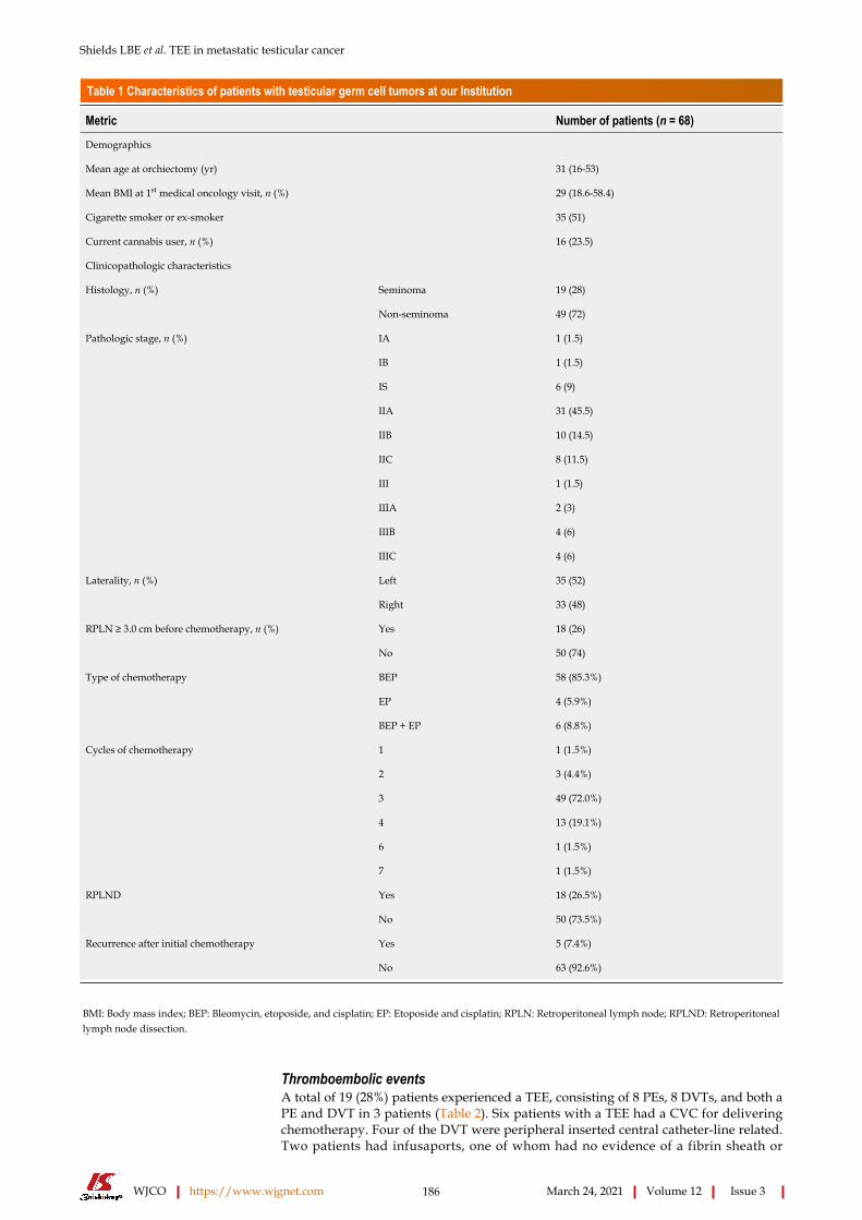
Shields LBE et al. TEE in metastatic testicular cancer
WJCO https://www.wjgnet.com 186 March 24, 2021 Volume 12 Issue 3
Table 1 Characteristics of patients with testicular germ cell tumors at our Institution
Metric Number of patients (n = 68)
Demographics
Mean age at orchiectomy (yr) 31 (16-53)
Mean BMI at 1st medical oncology visit, n (%) 29 (18.6-58.4)
Cigarette smoker or ex-smoker 35 (51)
Current cannabis user, n (%) 16 (23.5)
Clinicopathologic characteristics
Histology, n (%) Seminoma 19 (28)
Non-seminoma 49 (72)
Pathologic stage, n (%) IA 1 (1.5)
IB 1 (1.5)
IS 6 (9)
IIA 31 (45.5)
IIB 10 (14.5)
IIC 8 (11.5)
III 1 (1.5)
IIIA 2 (3)
IIIB 4 (6)
IIIC 4 (6)
Laterality, n (%) Left 35 (52)
Right 33 (48)
RPLN ≥ 3.0 cm before chemotherapy, n (%) Yes 18 (26)
No 50 (74)
Type of chemotherapy BEP 58 (85.3%)
EP 4 (5.9%)
BEP + EP 6 (8.8%)
Cycles of chemotherapy 1 1 (1.5%)
2 3 (4.4%)
3 49 (72.0%)
4 13 (19.1%)
6 1 (1.5%)
7 1 (1.5%)
RPLND Yes 18 (26.5%)
No 50 (73.5%)
Recurrence after initial chemotherapy Yes 5 (7.4%)
No 63 (92.6%)
BMI: Body mass index; BEP: Bleomycin, etoposide, and cisplatin; EP: Etoposide and cisplatin; RPLN: Retroperitoneal lymph node; RPLND: Retroperitoneal lymph node dissection.
Thromboembolic eventsA total of 19 (28%) patients experienced a TEE, consisting of 8 PEs, 8 DVTs, and both a PE and DVT in 3 patients (Table 2). Six patients with a TEE had a CVC for delivering chemotherapy. Four of the DVT were peripheral inserted central catheter-line related. Two patients had infusaports, one of whom had no evidence of a fibrin sheath or

Shields LBE et al. TEE in metastatic testicular cancer
WJCO https://www.wjgnet.com 187 March 24, 2021 Volume 12 Issue 3
Table 2 Thromboembolic events in patients with testicular germ cell tumors who underwent cisplatin-based chemotherapy at our Institution (n = 19)
Type/pathologic stage of testicular tumor
RPLN ≥ 3.0 cm before chemotherapy
Type/number of cycles of chemotherapy
PE/DVTTiming of PE/DVT in relation to chemotherapy
Anticoagulant Hospitalization
Seminoma IIB Yes BEP (2 cycles), EP (1 cycles)
DVT Same day immediately prior to Cycle 1
Fondaparinux sodium Yes
Seminoma IIA No EP (3 cycles) DVT1 During (Cycle 1) Heparin → apixaban Yes
Seminoma IIIB No EP (4 cycles) PE + DVT2
3 yr after initial chemotherapy, recurrent disease
Enoxaparin sodium Yes
Non-seminoma IIB Yes BEP (2 cycles) DVT2 During (Cycle 2) Warfarin →enoxaparin sodium
Yes
Non-seminoma IIB No BEP (3 cycles) PE During (Cycle 2) Heparin → enoxaparin sodium → rivaroxaban → apixaban
Yes
Non-seminoma IIC Yes EP (4 cycles) PE + DVT
1 wk before Cycle 1 Heparin (lovenox) → warfarin
Yes
Non-seminoma IIA No BEP (3 cycles) DVT2 During (Cycle 2) Fondaparinux sodium No
Non-seminoma IIB Yes BEP (3 cycles) PE1 During (Cycle 3) Fondaparinux sodium → warfarin
No
Seminoma IIA No BEP (3 cycles) DVT2 During (Cycle 3) None No
Seminoma III No BEP (3 cycles) PE During (Cycle 3) Enoxaparin sodium No
Non-seminoma IIIC Yes BEP (4 cycles) PE During (Cycle 4) Enoxaparin sodium No
Non-seminoma IIIC Yes BEP (4 cycles) DVT During (Cycle 3) Enoxaparin sodium No
Non-seminoma IIA No BEP (3 cycles) PE During (Cycle 3) Heparin → rivaroxaban Yes
Non-seminoma IIA No BEP (2 cycles) PE During (Cycle 2) Enoxaparin sodium No
Seminoma IIC Yes BEP (3 cycles) PE 1 mo after chemotherapy ended
Heparin → enoxaparin sodium
Yes
Seminoma IIA No BEP (3 cycles) PE + DVT
During (Cycle 2) Heparin → enoxaparin sodium
Yes
Non-seminoma IIB No BEP (3 cycles) DVT 1 d prior to Cycle 1 Heparin → fondaparinux sodium
Yes
Non-seminoma IIIA No BEP (3 cycles) PE 2 wk after chemotherapy ended
Warfarin + enoxaparin sodium
No
Seminoma IIC Yes EP (4 cycles) DVT2 During (Cycle 2) Enoxaparin sodium → fondaparinux
Yes
1Infusaport.2Peripherally inserted central catheter line. PE: Pulmonary embolism; BEP: Bleomycin, etoposide, and cisplatin; DVT: Deep vein thrombosis; EP: Etoposide and cisplatin.
pericatheter thrombus detected by fluoroscopic evaluation. The other patient had a family history of thromboembolic conditions.
Eight (42%) patients were diagnosed with a seminoma, and patients were either pathologic stage II [14 (74%)] or stage III [5 (26%)] (Table 2). The RPLN were ≥ 3.0 cm before chemotherapy in 8 patients (42%). The relative proportions of TEEs by at least one type stage was significantly different from the other stages (P = 0.05) (Table 3). Patients with stage IS testicular cancer were significantly (P = 0.03) less likely to experience a TEE compared to patients who had stages II or III. Note that no patients with stage IS testicular cancer had a TEE.
Most patients were scheduled to undergo 3 cycles of BEP. The relative proportions of TEEs by at least one type of chemotherapy treatment was significantly different from the other chemotherapy types (P = 0.004) (Table 3). Patients who were treated with BEP/EP and EP were significantly 5-times (1.02-28.3, P = 0.02) more likely to experience a TEE compared to patients who were only treated with BEP.

Shields LBE et al. TEE in metastatic testicular cancer
WJCO https://www.wjgnet.com 188 March 24, 2021 Volume 12 Issue 3
Table 3 Comparison of metrics between patients with testicular germ cell tumors at our Institution stratified by those who experienced a thromboembolic event and those who did not
Thromboembolic event
Metric No (%) Yes (%) P valuen 49 19
Age [mean (SD)] 30.9 (7.86) 30.79 (8.67) 0.909
BMI [mean (SD)] 27.87 (6.38) 30.22 (9.25) 0.237
Cigarette smoker = Yes (%) 27 (55.1) 8 (42.1) 0.421
Marijuana use = Yes (%) 13 (26.5) 3 (15.8) 0.526
Side of orchiectomy = Right (%) 27 (55.1) 6 (31.6) 0.107
Pathology = Seminoma (%) 11 (22.4) 8 (42.1) 0.135
Pathologic stage (%) 0.050
I 10 (20.4) 0 (0.0)
II 33 (67.3) 14 (73.7)
III 6 (12.2) 5 (26.3)
Port = Yes (%) 4 (8.2) 3 (15.8) 0.390
Type of chemotherapy (%)
BEP X 3 45 (91.8) 13 (68.4)
BEP X 3 + EP X 1 4 (8.2) 2 (10.5)
EP X 4 0 (0.0) 4 (21.1)
RPLN ≥ 3.0 cm = Yes (%) 10 (20.4) 8 (42.1) 0.123
RPLND = Yes (%) 12 (24.5) 6 (31.6) 0.555
Recurrence after chemotherapy = Yes (%) 3 (6.1) 2 (10.5) 0.614
BMI: Body mass index; BEP: Bleomycin, etoposide, and cisplatin; EP: Etoposide and cisplatin; RPLN: Retroperitoneal lymph node; RPLND: Retroperitoneal lymph node dissection.
The TEE occurred during the cisplatin-based chemotherapy in 13 (68%) patients. A total of 18 (95%) patients underwent anticoagulant therapy, and 10 (56%) patients were hospitalized for management of their TEE. Eight (42%) patients were current or ex-cigarette smokers, and 3 (16%) were current cannabis users.
One patient sustained bilateral PE and a DVT 3 years after the completion of 4 cycles of EP when he experienced recurrent testicular disease. He was hospitalized and treated with enoxaparin. This patient also experienced recurrent disease and was the sole death in our study.
DISCUSSIONCisplatin chemotherapy has been reported to cause PE, DVT, myocardial infarction, and peripheral arterial thrombosis[11,15,16]. Several pathogenic mechanisms have been identified between cisplatin and thrombosis, including endothelial cell damage marked by increased plasma levels of Von Willebrand factor during chemotherapy, platelet activation, and up-regulation of prothrombotic factors. Lauritsen et al[17] investigated cardiovascular risk factors and disease following BEP treatment for male germ cell cancer[17]. These authors reported that patients had a more than 20-fold increased hazard ratio of venous TEE compared with the normal population which decreased to a hazard ratio of 1.4 after 10 years.
In our literature review the percentage of patients who experienced a TEE with testicular cancer ranged between 5.2% and 19%[6,9,16,18,19]. Most of the TEE occurred during chemotherapy[9,16,18,19]. Various risk factors for a TEE were confirmed, including retroperitoneal and supraclavicular lymph node metastases, elevated serum lactic acid dehydrogenase, higher clinical stage, CVC, high body surface area, and febrile

Shields LBE et al. TEE in metastatic testicular cancer
WJCO https://www.wjgnet.com 189 March 24, 2021 Volume 12 Issue 3
neutropenia. Paffenholz et al[16] reported a significantly reduced overall survival in patients with a TEE during a median follow-up of 8 mo[16]. The present study concurs with this finding, as the TEE occurred during the cisplatin-based chemotherapy in 13 (68%) patients.
The Khorana score is a predictive model for chemotherapy-associated TEE and is based on the following: (1) Site of cancer; (2) Prechemotherapy platelet count 350 × 109
/L or more; (3) Hemoglobin level less than 100 g/L or use of red cell growth factors; (4) Prechemotherapy leukocyte count more than 11 × 109/L; and (5) BMI 35 kg/m2 or more[2,20,21]. The risk of TEE is 4-6-fold higher in patients with cancer than in those without, and the TEE incidence is 3%-15% annually in cancer patients[14,18]. The use of thromboprophylaxis to reduce the recurrence of cancer-associated TEE have been examined primarily in cancers such as breast, colorectal, lung, pancreatic, and multiple myeloma that usually affect older individuals[14,21-24].
Carrier et al[22] determined that apixaban resulted in a significantly lower rate of venous thromboembolism (VTE) than placebo among intermediate-to-high-risk ambulatory patients with cancer who started chemotherapy[22]. In the intention-to-treat analysis, major bleeding occurred in 10 (3.5%) patients in the apixaban group and in 5 (1.8%) patients in the placebo group (P = 0.046). Khorana et al[25] concluded that high-risk ambulatory patients with metastatic cancer who were receiving chemotherapy and were treated with rivaroxaban did not have a significantly lower incidence of VTE or death due to VTE in a 180-d trial period[25]. Despite these two large randomized phase III trials, routine prophylaxis for TEE has not been embraced by clinicians in the outpatient setting as the absolute risk reduction is small and thromboprophylaxis is associated with a heightened risk of major bleeding and high cost[14,18,22].
Of the 574 patients in Carrier and colleagues’ placebo-controlled study, only 3 patients were diagnosed with testicular cancer. Khorana et al[25]’s study of 1080 patients did not specify the number of patients with testicular cancer, however, mentioned that there were 32 cases of “genitourinary” cancers that included renal, bladder, ureteral, and testicular cancers but not prostate cancer. Young men with TGCT are generally without co-morbidities. This patient population was not represented in large trials investigating thromboprophylaxis in metastatic cancer during chemotherapy. This is largely due to inclusion and exclusion criteria based on the Khorana scoring system used in these trials.
The cisplatin-based chemotherapy used to treat TGCT often does not cause severe thrombocytopenia. A relatively short course of chemotherapy in metastatic testicular cancer often leads to a cure despite the need for ongoing systemic therapy in most other metastatic solid tumors. Additionally, the cisplatin dose in the BEP regimen is higher compared to most regimens used for non-testicular cancer. This important difference may lead to better outcomes with the use of thromboprophylaxis. TGCT possesses numerous risk factors that increase the risk for clotting including bulky retroperitoneal disease, cisplatin-based chemotherapy, cigarette smoking, and possibly marijuana use. Furthermore, the risk of bleeding in TGCT is likely to be lower compared to other types of cancers due to the shorter duration of chemotherapy in TGCT and younger patient population with better bone marrow function. These factors support further investigation into the use of thromboprophylaxis as patients receive chemotherapy for TGCT.
We propose conducting a multicenter randomized double blind placebo control trial examining the efficacy and safety of thromboprophylaxis in patients undergoing systemic treatment for metastatic TGCT. Such a trial will use a novel inclusion scoring system specific for TGCT that incorporates risk factors often encountered with this cancer. Carrier et al[22]’s as well as Khorana et al[25]’s studies only included patients who had 2 risk factors on the Khorana scale[22,25]. Most of the patients who experienced a TEE in the present study would not have fulfilled the criteria for inclusion for thromboprophylaxis in these 2 trials. The new TGCT scoring system should incorporate the following risk factors: Higher pathologic stage and greater number of chemotherapy cycles as confirmed by the current study as well as elevated serum lactate dehydrogenase, CVC, large RPLN, and seminoma[6,9,16,18,19].
Several studies have reported the limited use of thromboprophylaxis in TGCT with cisplatin-based chemotherapy (Table 4). Paffenholz et al[16] reported that 93% of all of their TCGT received prophylactic anticoagulation which is customary in Germany[16]. Thromboprophylaxis during chemotherapy for TGCT is not customary in the United States.
Kentucky has both the highest incidence and death rates for lung cancer in the United States[26,27]. In 2017, a total of 24.6% of adults in Kentucky smoked while the national average was 14.0%[26,28]. Cigarette smoking is a well-recognized risk factor for both atherosclerotic disease and TEE[29]. Half of the patients in our study were either

Shields LBE et al. TEE in metastatic testicular cancer
WJCO https://www.wjgnet.com 190 March 24, 2021 Volume 12 Issue 3
Table 4 Patients with testicular germ cell tumors who underwent cisplatin-based chemotherapy and experienced a thromboembolic event in the literature
Ref. Number of patients Percentage of patients with TE Main findings
> All stages of TGCT;
> TE: 1st day of chemo to 6 mo after;
> Of 19 TEE, 14 occurred during chemotherapy, 5 after chemo;
Piketty et al[9], 2005
100 patients with GCT; 100 controls with various neoplasms
19 (19%)
> Risk factors for TEE: High body surface area, elevated serum LDH
> All stages of TGCT;
> TEE: Considered therapy-associated if occurred from start of chemotherapy to 6 wk after;
> 18 (%) TEE occurred before chemotherapy;
Honecker et al[6], 2013
193 22 (11%)
> Risk factors for TEE: Pure seminoma, retroperitoneal or supraclavicular lymph node metastases, elevated LDH, CVC
> All stages of TGCT;
> TEE: During 1st year of follow-up;
Bezan et al[18], 2017
657 34 (5.2%)
> Risk factors for TEE: Higher clinical stage and RPLN, increased number of cycles of chemotherapy
> Metastatic TGCT;
> TEE: During or within 90 d of chemotherapy;
Tran et al[19], 2019 1135 150 (10%)
> Risk factors for TEE: Large RPLN, CVC
> All stages of TGCT;
> TEE: Start of chemotherapy to 6 mo after;
> Risk factors for TEE: Higher clinical stage, elevated serum LDH, febrile neutropenia, CVC;
Paffenholz et al[16], 2019
225 49 (19%)
> Patients with TEE had significantly reduced overall survival during median follow-up of 8 mo
> All stages of TCGT;
> TEE: Following orchiectomy;
Current Study, 2020
68 18 (26.5%)
> Risk factors for TEE: Higher pathologic stage, greater number of chemotherapy cycles
TGCT: Testicular germ cell tumor; TEE: Thromboembolic event; LDH: Lactic acid dehydrogenase; RPLN: Retroperitoneal lymph node; CVC: Central venous catheter.
current or previous cigarette smokers, and 42% of these patients experienced a TEE while undergoing cisplatin-based chemotherapy. Cannabis is the most commonly used recreation drug in the United States and has been suggested to cause myocardial infarction and ischemic stroke[30]. Only a single case of PE in a young male following heavy cannabis use has been reported[30]. It has been suggested that cannabinoids, the active component of cannabis, cause endothelial cell disruption leading to vascular thrombosis. A total of 23.5% of the patients in our study were current cannabis users, and 16% of the cannabis users suffered a TEE while undergoing chemotherapy.
Our study of TCGT offers several unique aspects that shed light on the risk factors for a TEE and educational opportunities for preventing a TEE. Our study, consisting of a single Institution in a metropolitan community in Kentucky, has a large number of patients with TCGT over a 6-year period. The high percentage (28%) of patients who experienced a TEE compared to the literature may be attributed to numerous cumulative factors leading to a hypercoagulable state that may provoke a TEE, including bulky retroperitoneal disease, higher pathologic stage, greater number of chemotherapy cycles, metastatic disease, the remarkably large number of patients who smoke cigarettes and use cannabis, high number of patients with CVC, and lack of patients who were treated with thromboprophylaxis. The limitation of the present study is its retrospective nature and limited number of patients.

Shields LBE et al. TEE in metastatic testicular cancer
WJCO https://www.wjgnet.com 191 March 24, 2021 Volume 12 Issue 3
Several risk factors have been identified as increasing the risk of developing VTE, including the patient’s age (> 60 years), obesity, and history of anterior VTE[31]. The tumor’s site, histological type, and stage also elevate the risk of VTE. Pancreatic cancer is the solid tumor with the highest likelihood of VTE, while lymphoma, acute leukemia, and multiple myeloma represent hematologic malignancies that pose a strong risk of VTE. Adenocarcinomas have a higher risk of VTE compared to squamous cell carcinomas, and an advanced tumor stage increases the prospect of developing a VTE[31]. Advanced tumor stage and use of subclavian catheters are the main risk factors for CVC-associated thrombosis[32]. Our Institution generally does not use CVC for delivering chemotherapy in patients with TCGT. However, CVC placement may be unavoidable for patients with poor venous access or patient preference. Educating patients and providers about the thromboembolic risk of CVC may discourage them from selecting CVC as the desired route of chemotherapy infusion. Further educational opportunities with patients that may decrease the likelihood of TEE and enhance overall medical health include an emphasis on discontinuing cigarette smoking and cannabis use as well as promoting weight loss.
CONCLUSIONAlthough patients with TGCT are a relatively healthy population due to their young age, myriad risk factors such as bulky retroperitoneal disease, higher clinical stage, CVC, cigarette smoking, and cannabis use may heighten their risk of developing a TEE during cisplatin-based chemotherapy. Patients with TGCT who experience a TEE may be unable to complete the course of prescribed chemotherapy despite initiating treatment with an anticoagulant. There may be a significant increase in cost of care following a TEE, including hospital stay, potential requirement for an aggressive procedure, and need for a longer duration of anticoagulation. Patients who could not complete the course of prescribed chemotherapy due to TEE will likely need more frequent medical oncology appointments with monitoring tumor markers as well as closer follow-up imaging studies which can substantially escalate the cost of patient care. High-risk ambulatory patients with TGCT treated with cisplatin-based chemotherapy may benefit from treatment with primary prophylactic anticoagulation to mitigate the potential morbidity and mortality associated with a TEE. Multi-institutional randomized studies are warranted to evaluate the safety and efficacy of thromboprophylaxis during chemotherapy for TGCT in this young patient population.
ARTICLE HIGHLIGHTSResearch backgroundCisplatin-based chemotherapy has significantly increased the survival of patients with testicular germ cell tumor (TGCT), although it is associated with a high rate of thromboembolic events (TEE).
Research motivationAs TGCT is the most curable solid tumor and most common cancer among men 18-39 years, we sought to evaluate our single-center experience of patients who were diagnosed with TGCT and received platinum-based chemotherapy. Patients who suffered a TEE were the primary focus of this study.
Research objectivesOur objective was to identify patients who were diagnosed with TGCT and received platinum-based chemotherapy, with particular attention to those patients who experienced a TEE.
Research methodsThe medical records and imaging studies of 68 consecutive individuals who were diagnosed with TGCT and received platinum-based chemotherapy at our Institution in a metropolitan community between January 1, 2014 and December 31, 2019 were reviewed. Statistical analysis was performed by stratisfying each metric (TEE, retroperitoneal lymph nodes ≥ 3.0 cm before chemotherapy, age, body mass index, cigarette smoker, marijuana use, side of orchiectomy, pathologic stage, central venous

Shields LBE et al. TEE in metastatic testicular cancer
WJCO https://www.wjgnet.com 192 March 24, 2021 Volume 12 Issue 3
catheter, type of chemotherapy, number of chemotherapy cycles, retroperitoneal lymph nodes dissection, and recurrence after chemotherapy by those patients who experienced a TEE and by those who did not.
Research resultsA total of 19 (28%) patients experienced a TEE following orchiectomy which occurred during chemotherapy in 13 (68%) of these patients. Patients with a higher pathologic stage (stage III) were significantly (P = 0.023) more likely to experience a TEE compared to patients who had a lower stage. Patients who were treated with 3 cycles of bleomycine, etoposide, and cisplatin and 1 cycle of etoposide and cisplatin or 4 cycles of etoposide and cisplatin were significantly 5 (P = 0.02) times more likely to experience a TEE compared to patients who were treated with only 3 cycles of bleomycine, etoposide, and cisplatin.
Research conclusionsSince myriad factors predispose to a TEE such as large retroperitoneal disease, higher clinical stage, greater number of chemotherapy cycle, central venous catheter, cigarette smoking, and possible cannabis use, high-risk ambulatory patients with TGCT treated with cisplatin-based chemotherapy may benefit from prophylactic anticoagulation.
Research perspectivesRandomized studies to evaluate the safety and efficacy of prophylactic anticoagulants are essential in this young patient population generally lacking medical co-morbidities.
ACKNOWLEDGEMENTSWe acknowledge Norton Healthcare for their continued support.
REFERENCESAmerican Cancer Society. Testicular cancer. [cited February 3, 2021]. Available from: https://www.cancer.org/cancer/testicular-cancer.html
1
Abdel-Razeq H, Mansour A, Abdulelah H, Al-Shwayat A, Makoseh M, Ibrahim M, Abunasser M, Rimawi D, Al-Rabaiah A, Alfar R, Abufara A, Ibrahim A, Bawaliz A, Ismael Y. Thromboembolic events in cancer patients on active treatment with cisplatin-based chemotherapy: another look! Thromb J 2018; 16: 2 [PMID: 29507532 DOI: 10.1186/s12959-018-0161-9]
2
Einhorn LH, Donohue J. Cis-diamminedichloroplatinum, vinblastine, and bleomycin combination chemotherapy in disseminated testicular cancer. Ann Intern Med 1977; 87: 293-298 [PMID: 71004 DOI: 10.7326/0003-4819-87-3-293]
3
Adra N, Einhorn LH. Testicular cancer update. Clin Adv Hematol Oncol 2017; 15: 386-396 [PMID: 28591093]
4
Fung C, Sesso HD, Williams AM, Kerns SL, Monahan P, Abu Zaid M, Feldman DR, Hamilton RJ, Vaughn DJ, Beard CJ, Kollmannsberger CK, Cook R, Althouse S, Ardeshir-Rouhani-Fard S, Lipshultz SE, Einhorn LH, Fossa SD, Travis LB; Platinum Study Group. Multi-Institutional Assessment of Adverse Health Outcomes Among North American Testicular Cancer Survivors After Modern Cisplatin-Based Chemotherapy. J Clin Oncol 2017; 35: 1211-1222 [PMID: 28240972 DOI: 10.1200/JCO.2016.70.3108]
5
Honecker F, Koychev D, Luhmann AD, Langer F, Dieckmann KP, Bokemeyer C, Oechsle K. Venous thromboembolic events in germ cell cancer patients undergoing platinum-based chemotherapy. Onkologie 2013; 36: 663-668 [PMID: 24192771 DOI: 10.1159/000355652]
6
Koga F, Yamada T, Ishimaru H, Sadaoka SI, Mizuo T. Deep vein thrombosis during chemotherapy in a patient with advanced testicular cancer: successful percutaneous thrombectomy under temporary placement of retrievable inferior vena cava filter. Int J Urol 2001; 8: 90-93 [PMID: 11240834 DOI: 10.1046/j.1442-2042.2001.00256.x]
7
Navi BB, Howard G, Howard VJ, Zhao H, Judd SE, Elkind MSV, Iadecola C, DeAngelis LM, Kamel H, Okin PM, Gilchrist S, Soliman EZ, Cushman M, Safford M, Muntner P. The risk of arterial thromboembolic events after cancer diagnosis. Res Pract Thromb Haemost 2019; 3: 639-651 [PMID: 31624783 DOI: 10.1002/rth2.12223]
8
Piketty AC, Fléchon A, Laplanche A, Nouyrigat E, Droz JP, Théodore C, Fizazi K. The risk of thrombo-embolic events is increased in patients with germ-cell tumours and can be predicted by serum lactate dehydrogenase and body surface area. Br J Cancer 2005; 93: 909-914 [PMID: 16205699 DOI: 10.1038/sj.bjc.6602791]
9
Seng S, Liu Z, Chiu SK, Proverbs-Singh T, Sonpavde G, Choueiri TK, Tsao CK, Yu M, Hahn NM, 10

Shields LBE et al. TEE in metastatic testicular cancer
WJCO https://www.wjgnet.com 193 March 24, 2021 Volume 12 Issue 3
Oh WK, Galsky MD. Risk of venous thromboembolism in patients with cancer treated with Cisplatin: a systematic review and meta-analysis. J Clin Oncol 2012; 30: 4416-4426 [PMID: 23150697 DOI: 10.1200/JCO.2012.42.4358]Solari L, Krönig M, Ihorst G, Drognitz K, Heinz J, Jilg CA, Schultze-Seemann W, Engelhardt M, Waller CF. High Rates of Thromboembolic Events in Patients with Germ Cell Cancer Undergoing Cisplatin-Based Polychemotherapy. Urol Int 2016; 96: 399-405 [PMID: 27074038 DOI: 10.1159/000445126]
11
Kerns SL, Fung C, Monahan PO, Ardeshir-Rouhani-Fard S, Abu Zaid MI, Williams AM, Stump TE, Sesso HD, Feldman DR, Hamilton RJ, Vaughn DJ, Beard C, Huddart RA, Kim J, Kollmannsberger C, Sahasrabudhe DM, Cook R, Fossa SD, Einhorn LH, Travis LB; Platinum Study Group. Cumulative Burden of Morbidity Among Testicular Cancer Survivors After Standard Cisplatin-Based Chemotherapy: A Multi-Institutional Study. J Clin Oncol 2018; 36: 1505-1512 [PMID: 29617189 DOI: 10.1200/JCO.2017.77.0735]
12
International Germ Cell Consensus Classification: a prognostic factor-based staging system for metastatic germ cell cancers. International Germ Cell Cancer Collaborative Group. J Clin Oncol 1997; 15: 594-603 [PMID: 9053482 DOI: 10.1200/JCO.1997.15.2.594]
13
Mulder FI, van Es N, Kraaijpoel N, Di Nisio M, Carrier M, Duggal A, Gaddh M, Garcia D, Grosso MA, Kakkar AK, Mercuri MF, Middeldorp S, Royle G, Segers A, Shivakumar S, Verhamme P, Wang T, Weitz JI, Zhang G, Büller HR, Raskob G. Edoxaban for treatment of venous thromboembolism in patient groups with different types of cancer: Results from the Hokusai VTE Cancer study. Thromb Res 2020; 185: 13-19 [PMID: 31733403 DOI: 10.1016/j.thromres.2019.11.007]
14
Cantwell BM, Mannix KA, Roberts JT, Ghani SE, Harris AL. Thromboembolic events during combination chemotherapy for germ cell-malignancy. Lancet 1988; 2: 1086-1087 [PMID: 2903318 DOI: 10.1016/s0140-6736(88)90113-4]
15
Paffenholz P, Grein K, Heidegger I, Nestler T, Grabbert M, Salem J, Hellmich M, Pfister D, Heidenreich A. Predictors of thrombosis in testicular cancer during platinum-based chemotherapy. World J Urol 2019; 37: 1907-1916 [PMID: 30547197 DOI: 10.1007/s00345-018-2598-7]
16
Lauritsen J, Hansen MK, Bandak M, Kreiberg MB, Skøtt JW, Wagner T, Gundgaard Kier MG, Holm NV, Agerbæk M, Gupta R, Dehlendorff C, Andersen KK, Daugaard G. Cardiovascular Risk Factors and Disease After Male Germ Cell Cancer. J Clin Oncol 2020; 38: 584-592 [PMID: 31821065 DOI: 10.1200/JCO.19.01180]
17
Bezan A, Posch F, Ploner F, Bauernhofer T, Pichler M, Szkandera J, Hutterer GC, Pummer K, Gary T, Samonigg H, Beyer J, Winder T, Hermanns T, Fankhauser CD, Gerger A, Stotz M. Risk stratification for venous thromboembolism in patients with testicular germ cell tumors. PLoS One 2017; 12: e0176283 [PMID: 28430804 DOI: 10.1371/journal.pone.0176283]
18
Tran B, Ruiz-Morales JM, Gonzalez-Billalabeitia E, Patrikidou A, Amir E, Seidel C, Bokemeyer C, Fankhauser C, Hermanns T, Rumyantsev A, Tryakin A, Brito M, Fléchon A, Kwan EM, Cheng T, Castellano D, Garcia Del Muro X, Hamid AA, Ottaviano M, Palmieri G, Kitson R, Reid A, Heng DYC, Bedard PL. Large retroperitoneal lymphadenopathy and increased risk of venous thromboembolism in patients receiving first-line chemotherapy for metastatic germ cell tumors: A study by the global germ cell cancer group (G3). Cancer Med 2020; 9: 116-124 [PMID: 31715650 DOI: 10.1002/cam4.2674]
19
Khorana AA, Kuderer NM, Culakova E, Lyman GH, Francis CW. Development and validation of a predictive model for chemotherapy-associated thrombosis. Blood 2008; 111: 4902-4907 [PMID: 18216292 DOI: 10.1182/blood-2007-10-116327]
20
Mulder FI, Candeloro M, Kamphuisen PW, Di Nisio M, Bossuyt PM, Guman N, Smit K, Büller HR, van Es N; CAT-prediction collaborators. The Khorana score for prediction of venous thromboembolism in cancer patients: a systematic review and meta-analysis. Haematologica 2019; 104: 1277-1287 [PMID: 30606788 DOI: 10.3324/haematol.2018.209114]
21
Carrier M, Abou-Nassar K, Mallick R, Tagalakis V, Shivakumar S, Schattner A, Kuruvilla P, Hill D, Spadafora S, Marquis K, Trinkaus M, Tomiak A, Lee AYY, Gross PL, Lazo-Langner A, El-Maraghi R, Goss G, Le Gal G, Stewart D, Ramsay T, Rodger M, Witham D, Wells PS; AVERT Investigators. Apixaban to Prevent Venous Thromboembolism in Patients with Cancer. N Engl J Med 2019; 380: 711-719 [PMID: 30511879 DOI: 10.1056/NEJMoa1814468]
22
McBane RD 2nd, Wysokinski WE, Le-Rademacher JG, Zemla T, Ashrani A, Tafur A, Perepu U, Anderson D, Gundabolu K, Kuzma C, Perez Botero J, Leon Ferre RA, Henkin S, Lenz CJ, Houghton DE, Vishnu P, Loprinzi CL. Apixaban and dalteparin in active malignancy-associated venous thromboembolism: The ADAM VTE trial. J Thromb Haemost 2020; 18: 411-421 [PMID: 31630479 DOI: 10.1111/jth.14662]
23
McBane RD 2nd, Loprinzi CL, Ashrani A, Lenz CJ, Houghton D, Zemla T, Le-Rademacher JG, Wysokinski WE. Extending venous thromboembolism secondary prevention with apixaban in cancer patients: The EVE trial. Eur J Haematol 2020; 104: 88-96 [PMID: 31606897 DOI: 10.1111/ejh.13338]
24
Khorana AA, Soff GA, Kakkar AK, Vadhan-Raj S, Riess H, Wun T, Streiff MB, Garcia DA, Liebman HA, Belani CP, O'Reilly EM, Patel JN, Yimer HA, Wildgoose P, Burton P, Vijapurkar U, Kaul S, Eikelboom J, McBane R, Bauer KA, Kuderer NM, Lyman GH; CASSINI Investigators. Rivaroxaban for Thromboprophylaxis in High-Risk Ambulatory Patients with Cancer. N Engl J Med 2019; 380: 720-728 [PMID: 30786186 DOI: 10.1056/NEJMoa1814630]
25
Shields LBE, Wilkett Barnes JG, Buckley C, Mikos GJ, Rogers KN, Hamm JT, Flynn JM, Hester ST, 26

Shields LBE et al. TEE in metastatic testicular cancer
WJCO https://www.wjgnet.com 194 March 24, 2021 Volume 12 Issue 3
Honaker JT. Multidisciplinary approach to low-dose CT screening for lung cancer in a metropolitan community. Fam Pract 2020; 37: 25-29 [PMID: 31273379 DOI: 10.1093/fampra/cmz036]United States Preventive Services Task Force. Lung cancer: screening. [cited February 3, 2021]. Available from: https://www.uspreventiveservicestaskforce.org/Page/Document/UpdateSummaryFinal/Lung-cancer-screening
27
Centers for Disease Control and Prevention. Current cigarette smoking among adults in the United States. [cited February 3, 2021]. Available from: https://www.cdc.gov/tobacco/data_statistics/fact_sheets/adult_data/cig_smoking/
28
Cheng YJ, Liu ZH, Yao FJ, Zeng WT, Zheng DD, Dong YG, Wu SH. Current and former smoking and risk for venous thromboembolism: a systematic review and meta-analysis. PLoS Med 2013; 10: e1001515 [PMID: 24068896 DOI: 10.1371/journal.pmed.1001515]
29
Salhan D, Abdulfattah O, Roy S, Kandel S, Agu C, Basunia M, Enriquez D, Quist J, Schmidt FM. Cannabis-induced VTE: is it a safe recreational drug? Chest 2016; 150: 909A [DOI: 10.1016/j.chest.2016.08.1009]
30
Khalil J, Bensaid B, Elkacemi H, Afif M, Bensaid Y, Kebdani T, Benjaafar N. Venous thromboembolism in cancer patients: an underestimated major health problem. World J Surg Oncol 2015; 13: 204 [PMID: 26092573 DOI: 10.1186/s12957-015-0592-8]
31
Wiegering V, Schmid S, Andres O, Wirth C, Wiegering A, Meyer T, Winkler B, Schlegel PG, Eyrich M. Thrombosis as a complication of central venous access in pediatric patients with malignancies: a 5-year single-center experience. BMC Hematol 2014; 14: 18 [PMID: 25317335 DOI: 10.1186/2052-1839-14-18]
32
https://www.uspreventiveservicestaskforce.org/Page/Document/UpdateSummaryFinal/Lung-cancer-screening

Published by Baishideng Publishing Group Inc
7041 Koll Center Parkway, Suite 160, Pleasanton, CA 94566, USA
Telephone: +1-925-3991568
E-mail: [email protected]
Help Desk: https://www.f6publishing.com/helpdesk
https://www.wjgnet.com
© 2021 Baishideng Publishing Group Inc. All rights reserved.
Related Documents