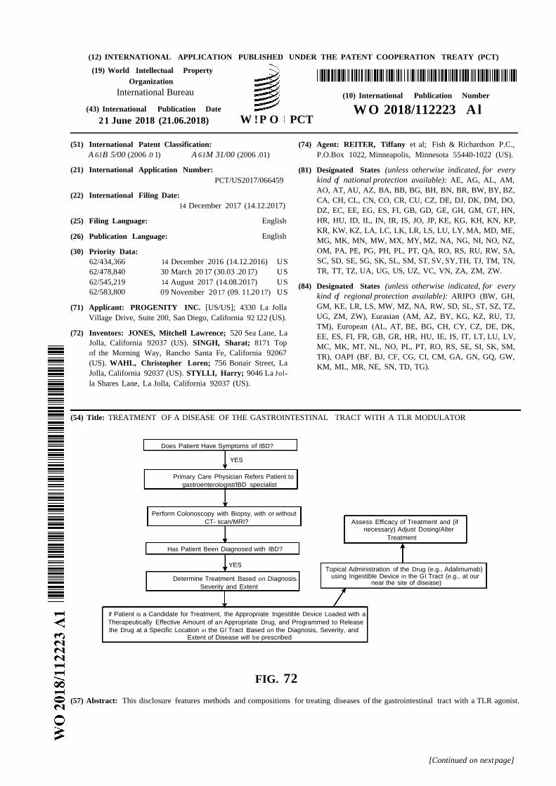
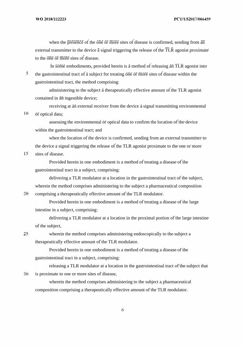
(12) INTERNATIONAL APPLICATION PUBLISHED UNDER THE PATENT COOPERATION TREATY (PCT) (19) World Intellectual Property Organization International Bureau (10) International Publication Number (43) International Publication Date W O 2018/112223 Al 21 June 2018 (21.06.2018) W!PO PCT (51) International Patent Classification: (74) Agent: REITER, Tiffany et al; Fish & Richardson P.C., A 61B 5/00 (2006 .0 1) A 61M 31/00 (2006 .01) P.O.Box 1022, Minneapolis, Minnesota 55440-1022 (US). (21) International Application Number: (81) Designated States (unless otherwise indicated, for every PCT/US2017/066459 kind of nationalprotection available): AE, AG, AL, AM, AO, AT, AU, AZ, BA, BB, BG, BH, BN, BR, BW, BY, BZ, (22) International Filing Date: CA, CH, CL, CN, CO, CR, CU, CZ, DE, DJ, DK, DM, DO, 14 December 2017 (14.12.2017) DZ, EC, EE, EG, ES, FI, GB, GD, GE, GH, GM, GT, HN, (25) Filing Language: English HR, HU, ID, IL, IN, IR, IS, JO, JP, KE, KG, KH, KN, KP, KR, KW, KZ, LA, LC, LK, LR, LS, LU, LY, MA, MD, ME, (26) Publication Language: English MG, MK, MN, MW, MX, MY, MZ, NA, NG, NI, NO, NZ, (30) Priority Data: OM, PA, PE, PG, PH, PL, PT, QA, RO, RS, RU, RW, SA, 62/434,366 14 December 2016 (14.12.2016) US SC, SD, SE, SG, SK, SL, SM, ST, SV, SY, TH, TJ, TM, TN, 62/478,840 30 March 20 17 (30.03 .20 1 7) US TR, TT, TZ, UA, UG, US, UZ, VC, VN, ZA, ZM, ZW. 62/545,219 14 August 2017 (14.08.2017) US (84) Designated States (unless otherwise indicated, for every 62/583,800 09 November 20 17 (09. 11.20 17) US kind of regionalprotection available): ARIPO (BW, GH, (71) Applicant: PROGENITY INC. [US/US]; 4330 La Jolla GM, KE, LR, LS, MW, MZ, NA, RW, SD, SL, ST, SZ, TZ, Village Drive, Suite 200, San Diego, California 92 122 (US). UG, ZM, ZW), Eurasian (AM, AZ, BY, KG, KZ, RU, TJ, TM), European (AL, AT, BE, BG, CH, CY, CZ, DE, DK, (72) Inventors: JONES, Mitchell Lawrence; 520 Sea Lane, La EE, ES, FI, FR, GB, GR, HR, HU, IE, IS, IT, LT, LU, LV, Jolla, California 92037 (US). SINGH, Sharat; 8171 Top MC, MK, MT, NL, NO, PL, PT, RO, RS, SE, SI, SK, SM, of the Morning Way, Rancho Santa Fe, California 92067 TR), OAPI (BF, BJ, CF, CG, CI, CM, GA, GN, GQ, GW, (US). WAHL, Christopher Loren; 756 Bonair Street, La KM, ML, MR, NE, SN, TD, TG). Jolla, California 92037 (US). STYLLI, Harry; 9046 La Jol la Shares Lane, La Jolla, California 92037 (US). (54) Title: TREATMENT OF A DISEASE OF THE GASTROINTESTINAL TRACT WITH A TLR MODULATOR Does Patient Have Symptoms of IBD? YES Primary Care Physician Refers Patient to gastroenterologist/IBD specialist Perform Colonoscopy with Biopsy, with or without CT- scan/MRI? Assess Efficacy of Treatment and (if necessary) Adjust Dosing/Alter Treatment Has Patient Been Diagnosed with IBD? YES Topical Administration of the Drug (e.g., Adalimumab) Determine Treatment Based on Diagnosis using Ingestible Device in the Gl Tract (e.g., at our near the site of disease) Severity and Extent If Patient is a Candidate for Treatment, the Appropriate Ingestible Device Loaded with a Therapeutically Effective Amount of an Appropriate Drug, and Programmed to Release the Drug at a Specific Location in the Gl Tract Based on the Diagnosis, Severity, and Extent of Disease will be prescribed FIG. 72 (57) Abstract: This disclosure features methods and compositions for treating diseases of the gastrointestinal tract with a TLR agonist. [Continued on next page]

Welcome message from author
This document is posted to help you gain knowledge. Please leave a comment to let me know what you think about it! Share it to your friends and learn new things together.
Transcript
(12) INTERNATIONAL APPLICATION PUBLISHED UNDER THE PATENT COOPERATION TREATY (PCT)
(19) World Intellectual PropertyOrganization
International Bureau (10) International Publication Number
(43) International Publication Date WO 2018/112223 Al21 June 2018 (21.06.2018) W !P O PCT
(51) International Patent Classification: (74) Agent: REITER, Tiffany et al; Fish & Richardson P.C.,A 61B 5/00 (2006 .0 1) A 61M 31/00 (2006 .01) P.O.Box 1022, Minneapolis, Minnesota 55440-1022 (US).
(21) International Application Number: (81) Designated States (unless otherwise indicated, for everyPCT/US2017/066459 kind of national protection available): AE, AG, AL, AM,
AO, AT, AU, AZ, BA, BB, BG, BH, BN, BR, BW, BY, BZ,(22) International Filing Date:
CA, CH, CL, CN, CO, CR, CU, CZ, DE, DJ, DK, DM, DO,14 December 2017 (14.12.2017)
DZ, EC, EE, EG, ES, FI, GB, GD, GE, GH, GM, GT, HN,(25) Filing Language: English HR, HU, ID, IL, IN, IR, IS, JO, JP, KE, KG, KH, KN, KP,
KR, KW, KZ, LA, LC, LK, LR, LS, LU, LY, MA, MD, ME,(26) Publication Language: English
MG, MK, MN, MW, MX, MY, MZ, NA, NG, NI, NO, NZ,(30) Priority Data: OM, PA, PE, PG, PH, PL, PT, QA, RO, RS, RU, RW, SA,
62/434,366 14 December 2016 (14.12.2016) US SC, SD, SE, SG, SK, SL, SM, ST, SV, SY, TH, TJ, TM, TN,62/478,840 30 March 20 17 (30.03 .20 17) US TR, TT, TZ, UA, UG, US, UZ, VC, VN, ZA, ZM, ZW.
62/545,219 14 August 2017 (14.08.2017) US (84) Designated States (unless otherwise indicated, for every62/583,800 09 November 20 17 (09. 11.20 17) US kind of regional protection available): ARIPO (BW, GH,
(71) Applicant: PROGENITY INC. [US/US]; 4330 La Jolla GM, KE, LR, LS, MW, MZ, NA, RW, SD, SL, ST, SZ, TZ,
Village Drive, Suite 200, San Diego, California 92 122 (US). UG, ZM, ZW), Eurasian (AM, AZ, BY, KG, KZ, RU, TJ,TM), European (AL, AT, BE, BG, CH, CY, CZ, DE, DK,
(72) Inventors: JONES, Mitchell Lawrence; 520 Sea Lane, La EE, ES, FI, FR, GB, GR, HR, HU, IE, IS, IT, LT, LU, LV,Jolla, California 92037 (US). SINGH, Sharat; 8171 Top MC, MK, MT, NL, NO, PL, PT, RO, RS, SE, SI, SK, SM,of the Morning Way, Rancho Santa Fe, California 92067 TR), OAPI (BF, BJ, CF, CG, CI, CM, GA, GN, GQ, GW,(US). WAHL, Christopher Loren; 756 Bonair Street, La KM, ML, MR, NE, SN, TD, TG).Jolla, California 92037 (US). STYLLI, Harry; 9046 La Jol
la Shares Lane, La Jolla, California 92037 (US).
(54) Title: TREATMENT OF A DISEASE OF THE GASTROINTESTINAL TRACT WITH A TLR MODULATOR
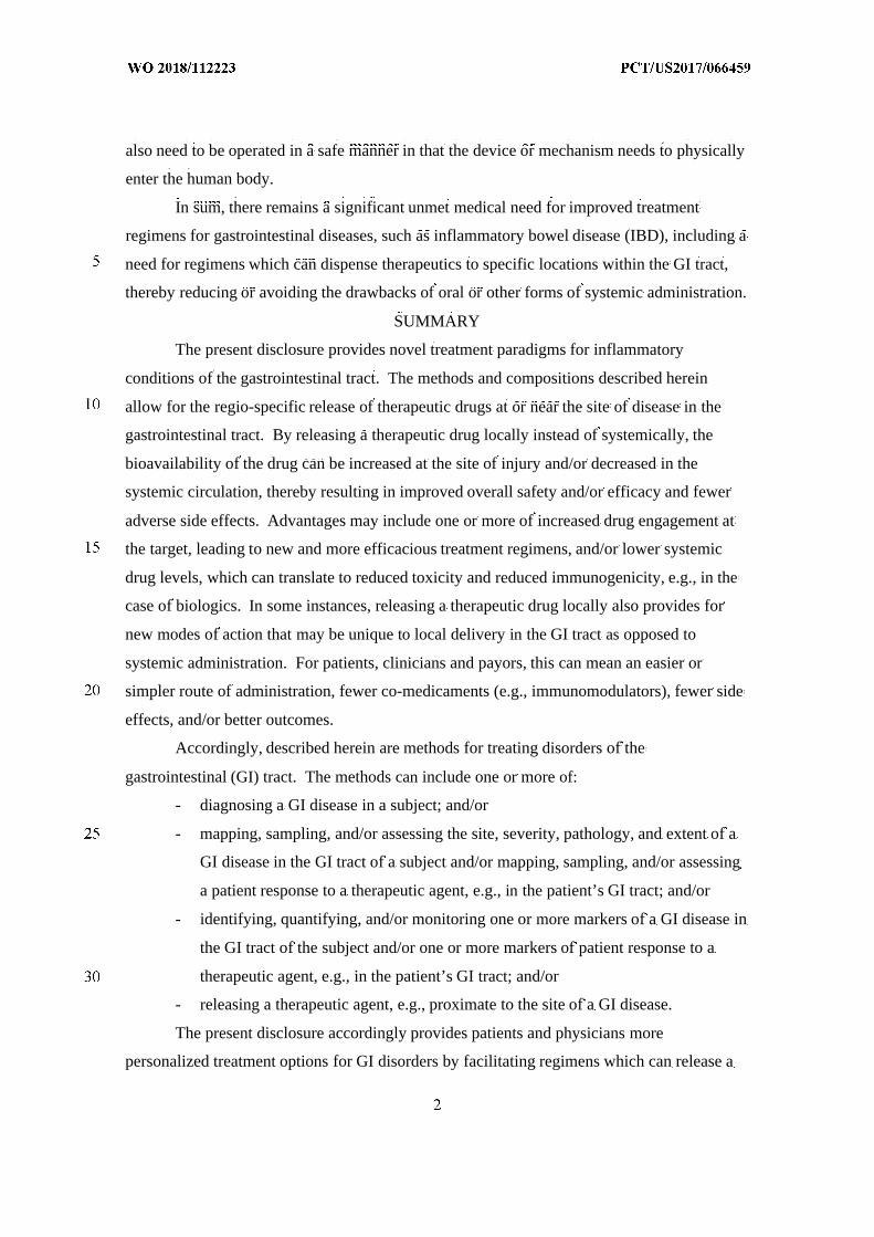
Does Patient Have Symptoms of IBD?
YES
Primary Care Physician Refers Patient togastroenterologist/IBD specialist
Perform Colonoscopy with Biopsy, with or withoutCT- scan/MRI? Assess Efficacy of Treatment and (if
necessary) Adjust Dosing/AlterTreatment
Has Patient Been Diagnosed with IBD?
YESTopical Administration of the Drug (e.g., Adalimumab)
Determine Treatment Based on Diagnosis using Ingestible Device in the Gl Tract (e.g., at ournear the site of disease)
Severity and Extent
If Patient is a Candidate for Treatment, the Appropriate Ingestible Device Loaded with aTherapeutically Effective Amount of an Appropriate Drug, and Programmed to Releasethe Drug at a Specific Location in the Gl Tract Based on the Diagnosis, Severity, and
Extent of Disease will be prescribed
FIG. 72
(57) Abstract: This disclosure features methods and compositions for treating diseases of the gastrointestinal tract with a TLR agonist.
[Continued on nextpage]

WO 2018/112223 Al llll I I I I 11 III II I I II I I I I I I III III II I II
Published:— with international search report (Art. 21(3))— before the expiration of the time limit for amending the
claims and to be republished in the event of receipt ofamendments (Rule 48.2(h))
TREATMENT OF A DISEASE OF THE GASTROINTESTINAL TRACT WITH A TLRMODULATOR
CROSS-REFERENCE TO RELATED APPLICATIONS
This application claims the benefit of the following U.S. Provisional Applications:
62/434,366 filed December 14, 2016; 62/478,840 filed March 30, 2017; 62/545,219 filed
August 14, 2017; and 62/583,800 filed November 9, 2017. This disclosure of the prior
applications is considered part of (and is incorporated by reference in its entirety in) the
disclosure of this application.
TECHNICAL FIELD
This disclosure features methods and compositions for treating diseases of the
gastrointestinal tract with a TLR modulator.
BACKGROUND
Toll-like receptor 9 (TLR9, also knowns as CD289 (cluster of differentiation 289)) is
a member of the toll-like receptor (TLR) family. TLR9 is present inside immune cells, as
well as on the surface of epithelial cells. TLR9 has been implicated in progression of
Crohn’s disease and ulcerative colitis.
The gastrointestinal (GI) tract generally provides a therapeutic medium for an
individual’s body. At times, therapeutic drugs may need to be dispensed to specified
locations within the small intestine or large intestine, which is more effective than oral
administration of the therapeutic drugs to cure or alleviate the symptoms of some medical
conditions. For example, therapeutic drugs dispensed directly within the small intestine
would not be contaminated, digested or otherwise compromised in the stomach, and thus
allow a higher dose to be delivered at a specific location within the small intestine. However,
dispensing therapeutic drugs directly within the small intestine inside a human body (e.g., the
cecum, the ascending colon) can be difficult, because a device or mechanism (e.g., special
formulation) would be needed to transport a therapeutically effective dose of drug to a
desired location within the small intestine and then automatically deliver the therapeutic drug
at the desired location. Dispensing therapeutic drugs directly within other locations in the GI
tract of the human body can be similarly difficult. Such a device or mechanism also would
also need to be operated in a safe manner in that the device or mechanism needs to physically
enter the human body.
In sum, there remains a significant unmet medical need for improved treatment
regimens for gastrointestinal diseases, such as inflammatory bowel disease (IBD), including a
need for regimens which can dispense therapeutics to specific locations within the GI tract,
thereby reducing or avoiding the drawbacks of oral or other forms of systemic administration.
SUMMARY
The present disclosure provides novel treatment paradigms for inflammatory
conditions of the gastrointestinal tract. The methods and compositions described herein
allow for the regio-specific release of therapeutic drugs at or near the site of disease in the
gastrointestinal tract. By releasing a therapeutic drug locally instead of systemically, the
bioavailability of the drug can be increased at the site of injury and/or decreased in the
systemic circulation, thereby resulting in improved overall safety and/or efficacy and fewer
adverse side effects. Advantages may include one or more of increased drug engagement at
the target, leading to new and more efficacious treatment regimens, and/or lower systemic
drug levels, which can translate to reduced toxicity and reduced immunogenicity, e.g., in the
case of biologics. In some instances, releasing a therapeutic drug locally also provides for
new modes of action that may be unique to local delivery in the GI tract as opposed to
systemic administration. For patients, clinicians and payors, this can mean an easier or
simpler route of administration, fewer co-medicaments (e.g., immunomodulators), fewer side
effects, and/or better outcomes.
Accordingly, described herein are methods for treating disorders of the
gastrointestinal (GI) tract. The methods can include one or more of:
- diagnosing a GI disease in a subject; and/or
- mapping, sampling, and/or assessing the site, severity, pathology, and extent of a
GI disease in the GI tract of a subject and/or mapping, sampling, and/or assessing
a patient response to a therapeutic agent, e.g., in the patient’s GI tract; and/or
- identifying, quantifying, and/or monitoring one or more markers of a GI disease in
the GI tract of the subject and/or one or more markers of patient response to a
therapeutic agent, e.g., in the patient’s GI tract; and/or
- releasing a therapeutic agent, e.g., proximate to the site of a GI disease.
The present disclosure accordingly provides patients and physicians more
personalized treatment options for GI disorders by facilitating regimens which can release a
therapeutic agent according to desired (e.g., customized or optimized) dosage, timing, and/or
location parameters. In some cases, the treatment methods can employ one or more
ingestible devices to achieve the benefits disclosed herein.
In some embodiments, provided herein is a method of treating a disease of the
gastrointestinal tract in a subject, comprising:
administering to the subject a pharmaceutical formulation that comprises an TLR
agonist,
wherein the pharmaceutical formulation is released at a location in the gastrointestinal
tract of the subject that is proximate to one or more sites of disease.
In some embodiments, provided herein the pharmaceutical formulation is
administered in an ingestible device. In some embodiments, the pharmaceutical formulation
is released from an ingestible device. In some embodiments, the ingestible device comprises
a housing, a reservoir containing the pharmaceutical formulation, and a release mechanism
for releasing the pharmaceutical formulation from the device,
wherein the reservoir is releasably or permanently attached to the exterior of the
housing or internal to the housing.
In some embodiments, provided herein is a method of treating a disease of the
gastrointestinal tract in a subject, comprising:
administering to the subject an ingestible device comprising a housing, a reservoir
containing a pharmaceutical formulation, and a release mechanism for releasing the
pharmaceutical formulation from the device,
wherein the reservoir is releasably or permanently attached to the exterior of the
housing or internal to the housing;
wherein the pharmaceutical formulation comprises an TLR agonist, and
the ingestible device releases the pharmaceutical formulation at a location in the
gastrointestinal tract of the subject that is proximate to one or more sites of disease.
In some embodiments, the housing is non-biodegradable in the GI tract.
In some embodiments, the release of the formulation is triggered autonomously. In some
embodiments, the device is programmed to release the formulation with one or more release
profiles that may be the same or different at one or more locations. In some embodiments,
the device is programmed to release the formulation at a location proximate to one or more
sites of disease. In some embodiments, the location of one or more sites of disease is
predetermined.
In some embodiments, the reservoir is made of a material that allows the formulation
to leave the reservoir, such as a biodegradable material.
In some embodiments, the release of the formulation is triggered by a pre-
programmed algorithm. In some embodiments, the release of the formulation is triggered by
data from a sensor or detector to identify the location of the device. In some more particular
embodiments, the data is not based solely on a physiological parameter (such as pH,
temperature, and/or transit time).
In some embodiments, the device comprises a detector configured to detect light
reflectance from an environment external to the housing. In some more particular
embodiments, the release is triggered autonomously or based on the detected reflectance.
In some embodiments, the device releases the formulation at substantially the same
time as one or more sites of disease are detected. In some embodiments, the one or more
sites of disease are detected by the device (e.g., by imaging the GI tract).
In some embodiments, the release mechanism is an actuation system. In some
embodiments, the release mechanism is a chemical actuation system. In some embodiments,
the release mechanism is a mechanical actuation system. In some embodiments, the release
mechanism is an electrical actuation system. In some embodiments, the actuation system
comprises a pump and releasing the formulation comprises pumping the formulation out of
the reservoir. In some embodiments, the actuation system comprises a gas generating cell.
In some embodiments, the device further comprises an anchoring mechanism. In some
embodiments, the formulation comprises a therapeutically effective amount of the TLR
agonist. In some embodiments, the formulation comprises a human equivalent dose (HED) of
the TLR agonist.
In some embodiments, the device is a device capable of releasing a solid TLR agonist
or a solid formulation comprising the TLR agonist. In some embodiments, the device is a
device capable of releasing a liquid TLR agonist or a liquid formulation comprising the TLR
agonist. Accordingly, in some embodiments of the methods herein, the pharmaceutical
formulation release from the device is a solid formulation. Accordingly, in some
embodiments of the methods herein, the pharmaceutical formulation release from the device
is a liquid formulation.
The devices disclosed herein are capable of releasing a TLR agonist or a formulation
comprising the TLR agonist irrespective of the particular type of TLR agonist. For example,
the TLR agonist may be a small molecule, a biological, a nucleic acid, an antibody, a fusion
protein, and so on.
In some embodiments, provided herein is a method of releasing an TLR agonist into
the gastrointestinal tract of a subject for treating one or more sites of disease within the
gastrointestinal tract, the method comprising:
administering to the subject a therapeutically effective amount of the TLR agonist
housed in an ingestible device, wherein the ingestible device comprises
a detector configured to detect the presence of the one or more sites of disease, and
a controller or processor configured to trigger the release of the TLR agonist
proximate to the one or more sites of disease in response to the detector detecting the
presence of the one or more sites of disease.
In some embodiments, provided herein is a method of releasing an TLR agonist into
the gastrointestinal tract of a subject for treating one or more pre-determined sites of disease
within the gastrointestinal tract, the method comprising:
administering to the subject a therapeutically effective amount of the TLR agonist
contained in an ingestible device, wherein the ingestible device comprises
a detector configured to detect the location of the device within the gastrointestinal
tract, and
a controller or processor configured to trigger the release of the TLR agonist
proximate to the one or more predetermined sites of disease in response to the detector
detecting a location of the device that corresponds to the location of the one or more pre-
determined sites of disease.
In some embodiments, provided herein is a method of releasing an TLR agonist into
the gastrointestinal tract of a subject for treating one or more sites of disease within the
gastrointestinal tract, the method comprising:
administering to the subject a therapeutically effective amount of the TLR agonist
contained in an ingestible device;
receiving at an external receiver from the device a signal transmitting environmental
data;
assessing the environmental data to confirm the presence of the one or more sites of
disease; and
when the presence of the one or more sites of disease is confirmed, sending from an
external transmitter to the device a signal triggering the release of the TLR agonist proximate
to the one or more sites of disease.
In some embodiments, provided herein is a method of releasing an TLR agonist into
the gastrointestinal tract of a subject for treating one or more sites of disease within the
gastrointestinal tract, the method comprising:
administering to the subject a therapeutically effective amount of the TLR agonist
contained in an ingestible device;
receiving at an external receiver from the device a signal transmitting environmental
or optical data;
assessing the environmental or optical data to confirm the location of the device
within the gastrointestinal tract; and
when the location of the device is confirmed, sending from an external transmitter to
the device a signal triggering the release of the TLR agonist proximate to the one or more
sites of disease.
Provided herein in one embodiment is a method of treating a disease of the
gastrointestinal tract in a subject, comprising:
delivering a TLR modulator at a location in the gastrointestinal tract of the subject,
wherein the method comprises administering to the subject a pharmaceutical composition
comprising a therapeutically effective amount of the TLR modulator.
Provided herein in one embodiment is a method of treating a disease of the large
intestine in a subject, comprising:
delivering a TLR modulator at a location in the proximal portion of the large intestine
of the subject,
wherein the method comprises administering endoscopically to the subject a
therapeutically effective amount of the TLR modulator.
Provided herein in one embodiment is a method of treating a disease of the
gastrointestinal tract in a subject, comprising:
releasing a TLR modulator at a location in the gastrointestinal tract of the subject that
is proximate to one or more sites of disease,
wherein the method comprises administering to the subject a pharmaceutical
composition comprising a therapeutically effective amount of the TLR modulator.
Provided herein in one embodiment is a method of treating a disease of the
gastrointestinal tract in a subject, comprising:
releasing a TLR modulator at a location in the gastrointestinal tract of the subject that
is proximate to one or more sites of disease,
wherein the method comprises administering to the subject a pharmaceutical
composition comprising a therapeutically effective amount of the TLR modulator, wherein
the pharmaceutical composition is an ingestible device. and the method comprises
administering orally to the subject the pharmaceutical composition.
Provided herein in one embodiment is a method of treating a disease of the
gastrointestinal tract in a subject, comprising:
releasing a TLR modulator at a location in the gastrointestinal tract of the subject that
is proximate to one or more sites of disease, wherein the method comprises administering to
the subject a pharmaceutical composition comprising a therapeutically effective amount of
the TLR modulator, wherein the method provides a concentration of the TLR modulator in
the plasma of the subject that is less than 3 µg/ml.
Provided herein in one embodiment is a method of treating a disease of the large
intestine in a subject, comprising:
releasing a TLR modulator at a location in the proximal portion of the large intestine
of the subject that is proximate to one or more sites of disease,
wherein the method comprises administering endoscopically to the subject a
therapeutically effective amount of the TLR modulator.
In another aspect of the present invention, there is provided a TLR modulator for use
in a method of treating a disease of the gastrointestinal tract in a subject, wherein the method
comprises orally administering to the subject an ingestible device loaded with the TLR
modulator, wherein the TLR modulator is released by the device at a location in the
gastrointestinal tract of the subject that is proximate to one or more sites of disease.
In another aspect, the present invention provides a composition comprising or
consisting of an ingestible device loaded with a therapeutically effective amount of a TLR
modulator, for use in a method of treatment, wherein the method comprises orally
administering the composition to the subject, wherein the TLR modulator is released by the
device at a location in the gastrointestinal tract of the subject that is proximate to one or more
sites of disease.
In another aspect, the present invention provides an ingestible device loaded with a
therapeutically effective amount of a TLR modulator, wherein the device is controllable to
release the TLR modulator at a location in the gastrointestinal tract of the subject that is
proximate to one or more sites of disease. The device may be for use in a method of
treatment of the human or animal body, for example, any method as described herein.
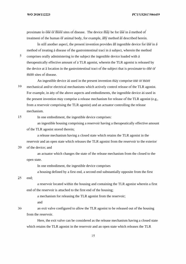
In still another aspect, the present invention provides an ingestible device for use in a
method of treating a disease of the gastrointestinal tract in a subject, wherein the method
comprises orally administering to the subject the ingestible device loaded with a
therapeutically effective amount of a TLR modulator, wherein the TLR modulator is released
by the device at a location in the gastrointestinal tract of the subject that is proximate to one
or more sites of disease.
An ingestible device as used in the present invention may comprise one or more
mechanical and/or electrical mechanisms which actively control release of the TLR
modulator. For example, in any of the above aspects and embodiments, the ingestible device
as used in the present invention may comprise a release mechanism for release of the TLR
modulator (e.g., from a reservoir comprising the TLR modulator) and an actuator controlling
the release mechanism.
In one embodiment, the ingestible device comprises:
an ingestible housing comprising a reservoir having a therapeutically effective amount
of the TLR modulator stored therein;
a release mechanism having a closed state which retains the TLR modulator in the
reservoir and an open state which releases the TLR modulator from the reservoir to the
exterior of the device; and
an actuator which changes the state of the release mechanism from the closed to the
open state.
In one embodiment, the ingestible device comprises:
a housing defined by a first end, a second end substantially opposite from the first
end;
a reservoir located within the housing and containing the TLR modulator wherein a
first end of the reservoir is attached to the first end of the housing;
a mechanism for releasing the TLR modulator from the reservoir;
and
an exit valve configured to allow the TLR modulator to be released out of the housing
from the reservoir.
Here, the exit valve can be considered as the release mechanism having a closed state
which retains the TLR modulator in the reservoir and an open state which releases the TLR
modulator from the reservoir to the exterior of the device, and the mechanism for releasing
the TLR modulator from the reservoir can be considered as the actuator.
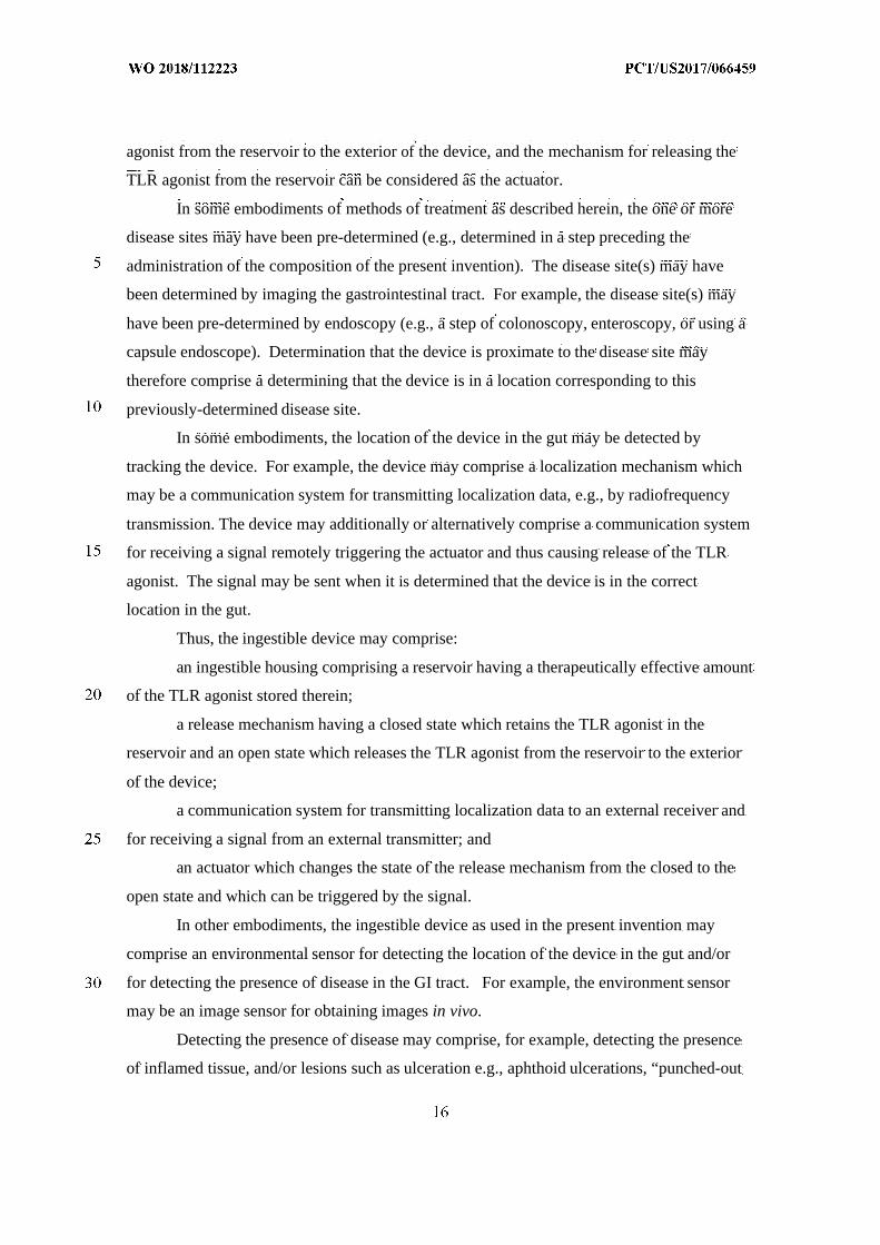
In some embodiments of methods of treatment as described herein, the one or more
disease sites may have been pre-determined (e.g., determined in a step preceding the
administration of the composition of the present invention). The disease site(s) may have
been determined by imaging the gastrointestinal tract. For example, the disease site(s) may
have been pre-determined by endoscopy (e.g., a step of colonoscopy, enteroscopy, or using a
capsule endoscope). Determination that the device is proximate to the disease site may
therefore comprise a determining that the device is in a location corresponding to this
previously-determined disease site.
In some embodiments, the location of the device in the gut may be detected by
tracking the device. For example, the device may comprise a localization mechanism which
may be a communication system for transmitting localization data, e.g., by radiofrequency
transmission. The device may additionally or alternatively comprise a communication system
for receiving a signal remotely triggering the actuator and thus causing release of the TLR
modulator. The signal may be sent when it is determined that the device is in the correct
location in the gut.
Thus, the ingestible device may comprise:
an ingestible housing comprising a reservoir having a therapeutically effective amount
of the TLR modulator stored therein;
a release mechanism having a closed state which retains the TLR modulator in the
reservoir and an open state which releases the TLR modulator from the reservoir to the
exterior of the device;
a communication system for transmitting localization data to an external receiver and
for receiving a signal from an external transmitter; and
an actuator which changes the state of the release mechanism from the closed to the
open state and which can be triggered by the signal.
In other embodiments, the ingestible device as used in the present invention may
comprise an environmental sensor for detecting the location of the device in the gut and/or

for detecting the presence of disease in the GI tract. For example, the environment sensor
may be an image sensor for obtaining images in vivo.
Detecting the presence of disease may comprise, for example, detecting the presence
of inflamed tissue, and/or lesions such as ulceration e.g., aphthoid ulcerations, “punched-out
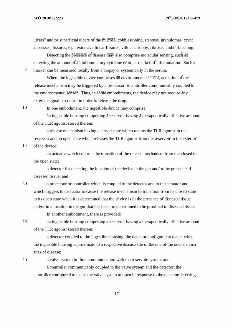
ulcers” and/or superficial ulcers of the mucosa, cobblestoning, stenosis, granulomas, crypt
abscesses, fissures, e.g., extensive linear fissures, villous atrophy, fibrosis, and/or bleeding.
Detecting the presence of disease may also comprise molecular sensing, such as
detecting the amount of an inflammatory cytokine or other marker of inflammation. Such a
marker can be measured locally from a biopsy or systemically in the serum.
Where the ingestible device comprises an environmental sensor, actuation of the
release mechanism may be triggered by a processor or controller communicably coupled to
the environmental sensor. Thus, in some embodiments, the device may not require any
external signal or control in order to release the drug.
In one embodiment, the ingestible device may comprise:
an ingestible housing comprising a reservoir having a therapeutically effective amount
of the TLR modulator stored therein;
a release mechanism having a closed state which retains the TLR modulator in the
reservoir and an open state which releases the TLR modulator from the reservoir to the
exterior of the device;
an actuator which controls the transition of the release mechanism from the closed to
the open state;
a detector for detecting the location of the device in the gut and/or the presence of
diseased tissue; and
a processor or controller which is coupled to the detector and to the actuator and
which triggers the actuator to cause the release mechanism to transition from its closed state
to its open state when it is determined that the device is in the presence of diseased tissue
and/or in a location in the gut that has been predetermined to be proximal to diseased tissue.
In another embodiment, there is provided:
an ingestible housing comprising a reservoir having a therapeutically effective amount
of the TLR modulator stored therein;
a detector coupled to the ingestible housing, the detector configured to detect when
the ingestible housing is proximate to a respective disease site of the one of the one or more
sites of disease;
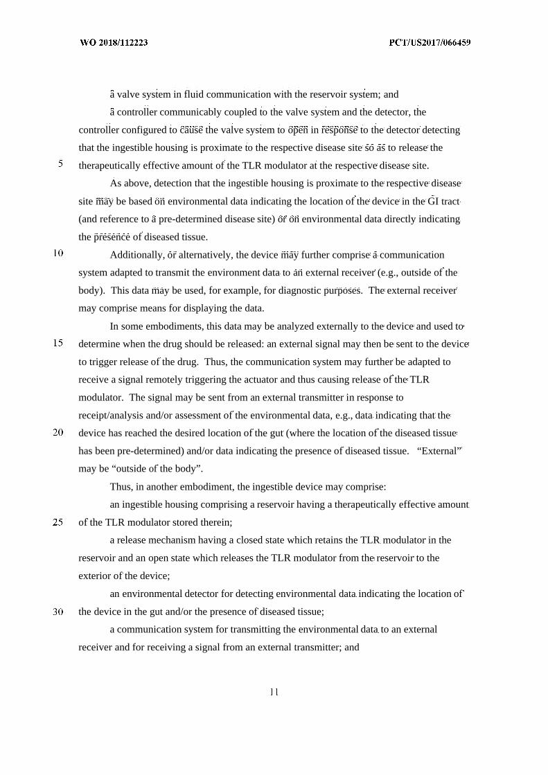
a valve system in fluid communication with the reservoir system; and
a controller communicably coupled to the valve system and the detector, the
controller configured to cause the valve system to open in response to the detector detecting
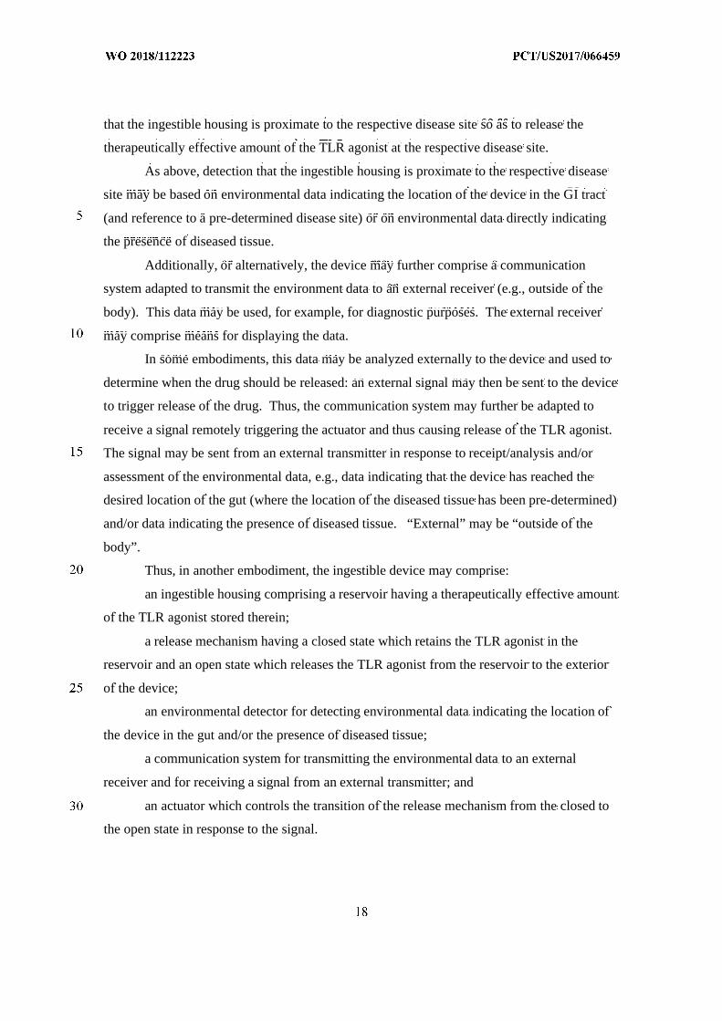
that the ingestible housing is proximate to the respective disease site so as to release the
therapeutically effective amount of the TLR modulator at the respective disease site.
As above, detection that the ingestible housing is proximate to the respective disease
site may be based on environmental data indicating the location of the device in the GI tract
(and reference to a pre-determined disease site) or on environmental data directly indicating
the presence of diseased tissue.
Additionally, or alternatively, the device may further comprise a communication
system adapted to transmit the environment data to an external receiver (e.g., outside of the
body). This data may be used, for example, for diagnostic purposes. The external receiver
may comprise means for displaying the data.
In some embodiments, this data may be analyzed externally to the device and used to
determine when the drug should be released: an external signal may then be sent to the device
to trigger release of the drug. Thus, the communication system may further be adapted to
receive a signal remotely triggering the actuator and thus causing release of the TLR
modulator. The signal may be sent from an external transmitter in response to
receipt/analysis and/or assessment of the environmental data, e.g., data indicating that the
device has reached the desired location of the gut (where the location of the diseased tissue
has been pre-determined) and/or data indicating the presence of diseased tissue. “External”
may be “outside of the body”.
Thus, in another embodiment, the ingestible device may comprise:
an ingestible housing comprising a reservoir having a therapeutically effective amount
of the TLR modulator stored therein;
a release mechanism having a closed state which retains the TLR modulator in the
reservoir and an open state which releases the TLR modulator from the reservoir to the
exterior of the device;
an environmental detector for detecting environmental data indicating the location of
the device in the gut and/or the presence of diseased tissue;
a communication system for transmitting the environmental data to an external
receiver and for receiving a signal from an external transmitter; and
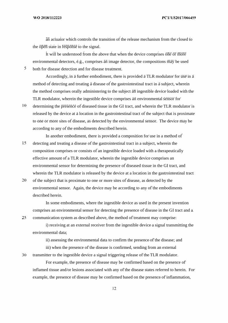
an actuator which controls the transition of the release mechanism from the closed to
the open state in response to the signal.
It will be understood from the above that when the device comprises one or more
environmental detectors, e.g., comprises an image detector, the compositions may be used
both for disease detection and for disease treatment.
Accordingly, in a further embodiment, there is provided a TLR modulator for use in a
method of detecting and treating a disease of the gastrointestinal tract in a subject, wherein
the method comprises orally administering to the subject an ingestible device loaded with the
TLR modulator, wherein the ingestible device comprises an environmental sensor for
determining the presence of diseased tissue in the GI tract, and wherein the TLR modulator is
released by the device at a location in the gastrointestinal tract of the subject that is proximate
to one or more sites of disease, as detected by the environmental sensor. The device may be
according to any of the embodiments described herein.
In another embodiment, there is provided a composition for use in a method of
detecting and treating a disease of the gastrointestinal tract in a subject, wherein the
composition comprises or consists of an ingestible device loaded with a therapeutically
effective amount of a TLR modulator, wherein the ingestible device comprises an
environmental sensor for determining the presence of diseased tissue in the GI tract, and
wherein the TLR modulator is released by the device at a location in the gastrointestinal tract
of the subject that is proximate to one or more sites of disease, as detected by the
environmental sensor. Again, the device may be according to any of the embodiments
described herein.
In some embodiments, where the ingestible device as used in the present invention
comprises an environmental sensor for detecting the presence of disease in the GI tract and a
communication system as described above, the method of treatment may comprise:
i) receiving at an external receiver from the ingestible device a signal transmitting the
environmental data;
ii) assessing the environmental data to confirm the presence of the disease; and
iii) when the presence of the disease is confirmed, sending from an external
transmitter to the ingestible device a signal triggering release of the TLR modulator.
For example, the presence of disease may be confirmed based on the presence of
inflamed tissue and/or lesions associated with any of the disease states referred to herein. For
example, the presence of disease may be confirmed based on the presence of inflammation,
ulceration e.g., aphthoid ulcerations, “punched-out ulcers” and/or superficial ulcers of the
mucosa, cobblestoning, stenosis, granulomas, crypt abscesses, fissures, e.g., extensive linear
fissures, villous atrophy, fibrosis, and/or bleeding.
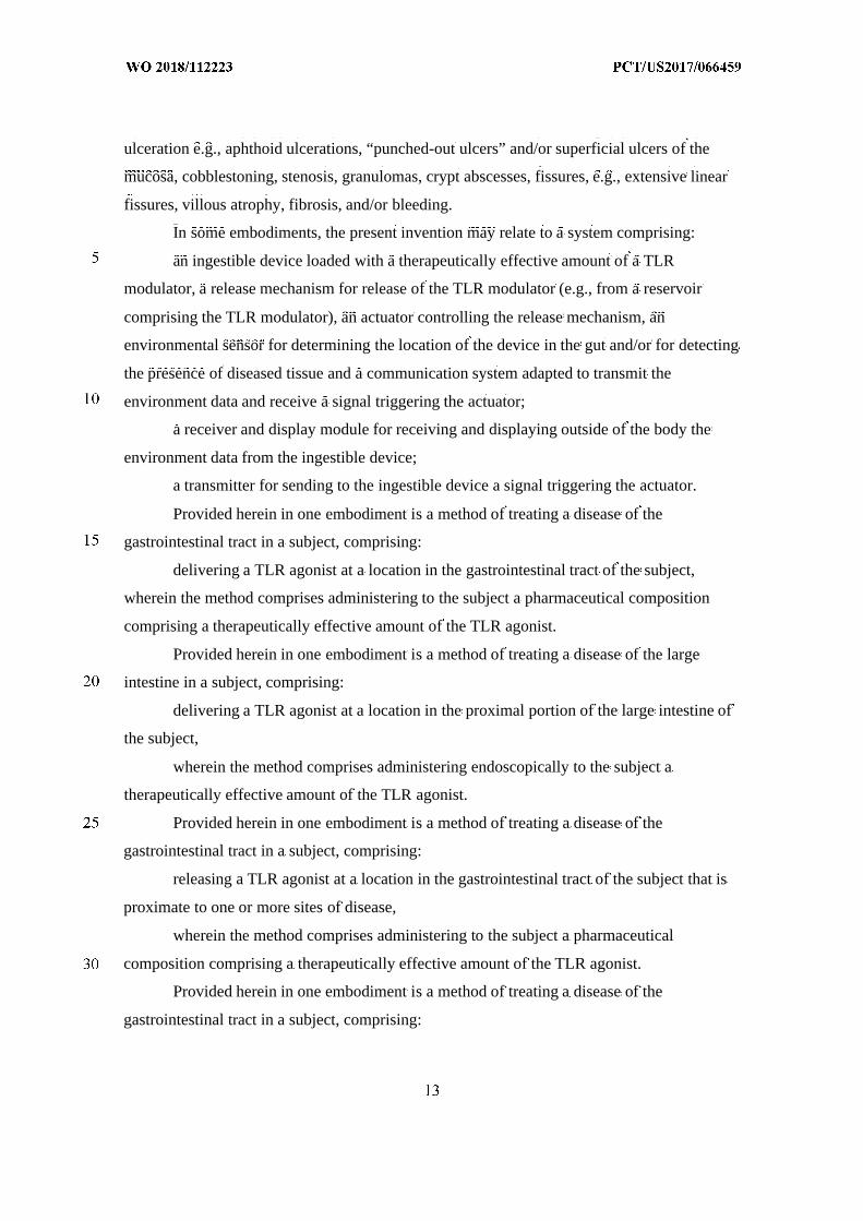
In some embodiments, the present invention may relate to a system comprising:
an ingestible device loaded with a therapeutically effective amount of a TLR
modulator, a release mechanism for release of the TLR modulator (e.g., from a reservoir
comprising the TLR modulator), an actuator controlling the release mechanism, an
environmental sensor for determining the location of the device in the gut and/or for detecting
the presence of diseased tissue and a communication system adapted to transmit the
environment data and receive a signal triggering the actuator;
a receiver and display module for receiving and displaying outside of the body the
environment data from the ingestible device;
a transmitter for sending to the ingestible device a signal triggering the actuator.
Provided herein in one embodiment is a method of treating a disease of the
gastrointestinal tract in a subject, comprising:
delivering a TLR agonist at a location in the gastrointestinal tract of the subject,
wherein the method comprises administering to the subject a pharmaceutical composition
comprising a therapeutically effective amount of the TLR agonist.
Provided herein in one embodiment is a method of treating a disease of the large
intestine in a subject, comprising:
delivering a TLR agonist at a location in the proximal portion of the large intestine of
the subject,
wherein the method comprises administering endoscopically to the subject a
therapeutically effective amount of the TLR agonist.
Provided herein in one embodiment is a method of treating a disease of the
gastrointestinal tract in a subject, comprising:
releasing a TLR agonist at a location in the gastrointestinal tract of the subject that is
proximate to one or more sites of disease,
wherein the method comprises administering to the subject a pharmaceutical
composition comprising a therapeutically effective amount of the TLR agonist.
Provided herein in one embodiment is a method of treating a disease of the
gastrointestinal tract in a subject, comprising:
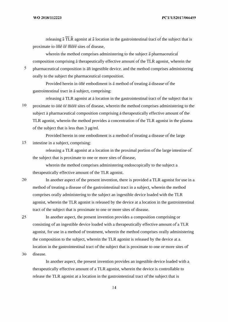
releasing a TLR agonist at a location in the gastrointestinal tract of the subject that is
proximate to one or more sites of disease,
wherein the method comprises administering to the subject a pharmaceutical
composition comprising a therapeutically effective amount of the TLR agonist, wherein the
pharmaceutical composition is an ingestible device. and the method comprises administering
orally to the subject the pharmaceutical composition.
Provided herein in one embodiment is a method of treating a disease of the
gastrointestinal tract in a subject, comprising:
releasing a TLR agonist at a location in the gastrointestinal tract of the subject that is
proximate to one or more sites of disease, wherein the method comprises administering to the
subject a pharmaceutical composition comprising a therapeutically effective amount of the
TLR agonist, wherein the method provides a concentration of the TLR agonist in the plasma
of the subject that is less than 3 µg/ml.
Provided herein in one embodiment is a method of treating a disease of the large
intestine in a subject, comprising:
releasing a TLR agonist at a location in the proximal portion of the large intestine of
the subject that is proximate to one or more sites of disease,
wherein the method comprises administering endoscopically to the subject a
therapeutically effective amount of the TLR agonist.
In another aspect of the present invention, there is provided a TLR agonist for use in a
method of treating a disease of the gastrointestinal tract in a subject, wherein the method
comprises orally administering to the subject an ingestible device loaded with the TLR
agonist, wherein the TLR agonist is released by the device at a location in the gastrointestinal
tract of the subject that is proximate to one or more sites of disease.
In another aspect, the present invention provides a composition comprising or
consisting of an ingestible device loaded with a therapeutically effective amount of a TLR
agonist, for use in a method of treatment, wherein the method comprises orally administering
the composition to the subject, wherein the TLR agonist is released by the device at a
location in the gastrointestinal tract of the subject that is proximate to one or more sites of
disease.
In another aspect, the present invention provides an ingestible device loaded with a
therapeutically effective amount of a TLR agonist, wherein the device is controllable to
release the TLR agonist at a location in the gastrointestinal tract of the subject that is
proximate to one or more sites of disease. The device may be for use in a method of
treatment of the human or animal body, for example, any method as described herein.
In still another aspect, the present invention provides an ingestible device for use in a
method of treating a disease of the gastrointestinal tract in a subject, wherein the method
comprises orally administering to the subject the ingestible device loaded with a
therapeutically effective amount of a TLR agonist, wherein the TLR agonist is released by
the device at a location in the gastrointestinal tract of the subject that is proximate to one or
more sites of disease.
An ingestible device as used in the present invention may comprise one or more
mechanical and/or electrical mechanisms which actively control release of the TLR agonist.
For example, in any of the above aspects and embodiments, the ingestible device as used in
the present invention may comprise a release mechanism for release of the TLR agonist (e.g.,
from a reservoir comprising the TLR agonist) and an actuator controlling the release
mechanism.
In one embodiment, the ingestible device comprises:
an ingestible housing comprising a reservoir having a therapeutically effective amount
of the TLR agonist stored therein;
a release mechanism having a closed state which retains the TLR agonist in the
reservoir and an open state which releases the TLR agonist from the reservoir to the exterior
of the device; and
an actuator which changes the state of the release mechanism from the closed to the
open state.
In one embodiment, the ingestible device comprises
a housing defined by a first end, a second end substantially opposite from the first
end;
a reservoir located within the housing and containing the TLR agonist wherein a first
end of the reservoir is attached to the first end of the housing;
a mechanism for releasing the TLR agonist from the reservoir;
and
an exit valve configured to allow the TLR agonist to be released out of the housing
from the reservoir.
Here, the exit valve can be considered as the release mechanism having a closed state
which retains the TLR agonist in the reservoir and an open state which releases the TLR
agonist from the reservoir to the exterior of the device, and the mechanism for releasing the
TLR agonist from the reservoir can be considered as the actuator.
In some embodiments of methods of treatment as described herein, the one or more
disease sites may have been pre-determined (e.g., determined in a step preceding the
administration of the composition of the present invention). The disease site(s) may have
been determined by imaging the gastrointestinal tract. For example, the disease site(s) may
have been pre-determined by endoscopy (e.g., a step of colonoscopy, enteroscopy, or using a
capsule endoscope). Determination that the device is proximate to the disease site may
therefore comprise a determining that the device is in a location corresponding to this
previously-determined disease site.
In some embodiments, the location of the device in the gut may be detected by
tracking the device. For example, the device may comprise a localization mechanism which
may be a communication system for transmitting localization data, e.g., by radiofrequency
transmission. The device may additionally or alternatively comprise a communication system
for receiving a signal remotely triggering the actuator and thus causing release of the TLR
agonist. The signal may be sent when it is determined that the device is in the correct
location in the gut.
Thus, the ingestible device may comprise:
an ingestible housing comprising a reservoir having a therapeutically effective amount
of the TLR agonist stored therein;
a release mechanism having a closed state which retains the TLR agonist in the
reservoir and an open state which releases the TLR agonist from the reservoir to the exterior
of the device;
a communication system for transmitting localization data to an external receiver and
for receiving a signal from an external transmitter; and
an actuator which changes the state of the release mechanism from the closed to the
open state and which can be triggered by the signal.
In other embodiments, the ingestible device as used in the present invention may
comprise an environmental sensor for detecting the location of the device in the gut and/or
for detecting the presence of disease in the GI tract. For example, the environment sensor
may be an image sensor for obtaining images in vivo.
Detecting the presence of disease may comprise, for example, detecting the presence
of inflamed tissue, and/or lesions such as ulceration e.g., aphthoid ulcerations, “punched-out
ulcers” and/or superficial ulcers of the mucosa, cobblestoning, stenosis, granulomas, crypt
abscesses, fissures, e.g., extensive linear fissures, villous atrophy, fibrosis, and/or bleeding.
Detecting the presence of disease may also comprise molecular sensing, such as
detecting the amount of an inflammatory cytokine or other marker of inflammation. Such a
marker can be measured locally from a biopsy or systemically in the serum.
Where the ingestible device comprises an environmental sensor, actuation of the
release mechanism may be triggered by a processor or controller communicably coupled to
the environmental sensor. Thus, in some embodiments, the device may not require any
external signal or control in order to release the drug.
In one embodiment, the ingestible device may comprise:
an ingestible housing comprising a reservoir having a therapeutically effective amount
of the TLR agonist stored therein;
a release mechanism having a closed state which retains the TLR agonist in the
reservoir and an open state which releases the TLR agonist from the reservoir to the exterior
of the device;
an actuator which controls the transition of the release mechanism from the closed to
the open state;
a detector for detecting the location of the device in the gut and/or the presence of
diseased tissue; and
a processor or controller which is coupled to the detector and to the actuator and
which triggers the actuator to cause the release mechanism to transition from its closed state
to its open state when it is determined that the device is in the presence of diseased tissue
and/or in a location in the gut that has been predetermined to be proximal to diseased tissue.
In another embodiment, there is provided:
an ingestible housing comprising a reservoir having a therapeutically effective amount
of the TLR agonist stored therein;
a detector coupled to the ingestible housing, the detector configured to detect when
the ingestible housing is proximate to a respective disease site of the one of the one or more
sites of disease;
a valve system in fluid communication with the reservoir system; and
a controller communicably coupled to the valve system and the detector, the
controller configured to cause the valve system to open in response to the detector detecting
that the ingestible housing is proximate to the respective disease site so as to release the
therapeutically effective amount of the TLR agonist at the respective disease site.
As above, detection that the ingestible housing is proximate to the respective disease
site may be based on environmental data indicating the location of the device in the GI tract
(and reference to a pre-determined disease site) or on environmental data directly indicating
the presence of diseased tissue.
Additionally, or alternatively, the device may further comprise a communication
system adapted to transmit the environment data to an external receiver (e.g., outside of the
body). This data may be used, for example, for diagnostic purposes. The external receiver
may comprise means for displaying the data.
In some embodiments, this data may be analyzed externally to the device and used to
determine when the drug should be released: an external signal may then be sent to the device
to trigger release of the drug. Thus, the communication system may further be adapted to
receive a signal remotely triggering the actuator and thus causing release of the TLR agonist.
The signal may be sent from an external transmitter in response to receipt/analysis and/or
assessment of the environmental data, e.g., data indicating that the device has reached the
desired location of the gut (where the location of the diseased tissue has been pre-determined)
and/or data indicating the presence of diseased tissue. “External” may be “outside of the
body”.
Thus, in another embodiment, the ingestible device may comprise:
an ingestible housing comprising a reservoir having a therapeutically effective amount
of the TLR agonist stored therein;
a release mechanism having a closed state which retains the TLR agonist in the
reservoir and an open state which releases the TLR agonist from the reservoir to the exterior
of the device;
an environmental detector for detecting environmental data indicating the location of
the device in the gut and/or the presence of diseased tissue;
a communication system for transmitting the environmental data to an external
receiver and for receiving a signal from an external transmitter; and
an actuator which controls the transition of the release mechanism from the closed to
the open state in response to the signal.
It will be understood from the above that when the device comprises one or more
environmental detectors, e.g., comprises an image detector, the compositions may be used
both for disease detection and for disease treatment.
Accordingly, in a further embodiment, there is provided a TLR agonist for use in a
method of detecting and treating a disease of the gastrointestinal tract in a subject, wherein
the method comprises orally administering to the subject an ingestible device loaded with the
TLR agonist, wherein the ingestible device comprises an environmental sensor for
determining the presence of diseased tissue in the GI tract, and wherein the TLR agonist is
released by the device at a location in the gastrointestinal tract of the subject that is proximate
to one or more sites of disease, as detected by the environmental sensor. The device may be
according to any of the embodiments described herein.
In another embodiment, there is provided a composition for use in a method of
detecting and treating a disease of the gastrointestinal tract in a subject, wherein the
composition comprises or consists of an ingestible device loaded with a therapeutically
effective amount of a TLR agonist, wherein the ingestible device comprises an environmental
sensor for determining the presence of diseased tissue in the GI tract, and wherein the TLR
agonist is released by the device at a location in the gastrointestinal tract of the subject that is
proximate to one or more sites of disease, as detected by the environmental sensor. Again,
the device may be according to any of the embodiments described herein.
In some embodiments, where the ingestible device as used in the present invention
comprises an environmental sensor for detecting the presence of disease in the GI tract and a
communication system as described above, the method of treatment may comprise:
i) receiving at an external receiver from the ingestible device a signal transmitting the
environmental data;
ii) assessing the environmental data to confirm the presence of the disease; and
iii) when the presence of the disease is confirmed, sending from an external
transmitter to the ingestible device a signal triggering release of the TLR agonist.
For example, the presence of disease may be confirmed based on the presence of
inflamed tissue and/or lesions associated with any of the disease states referred to herein. For
example, the presence of disease may be confirmed based on the presence of inflammation,
ulceration e.g., aphthoid ulcerations, “punched-out ulcers” and/or superficial ulcers of the
mucosa, cobblestoning, stenosis, granulomas, crypt abscesses, fissures, e.g., extensive linear
fissures, villous atrophy, fibrosis, and/or bleeding.
In some embodiments, the present invention may relate to a system comprising:
an ingestible device loaded with a therapeutically effective amount of a TLR agonist,
a release mechanism for release of the TLR agonist (e.g., from a reservoir comprising the
TLR agonist), an actuator controlling the release mechanism, an environmental sensor for
determining the location of the device in the gut and/or for detecting the presence of diseased
tissue and a communication system adapted to transmit the environment data and receive a
signal triggering the actuator;
a receiver and display module for receiving and displaying outside of the body the
environment data from the ingestible device;
a transmitter for sending to the ingestible device a signal triggering the actuator.
In any of the above embodiments, the ingestible device may further comprise an
anchoring system for anchoring the device or a portion thereof in a location and an actuator
for the anchoring system. This may be triggered in response to a determination that the
device is at a location in the gastrointestinal tract of the subject proximate to one or more
sites of disease. For instance, this may be detected by the environmental sensor. The
triggering may be controlled by a processor in the device, that is, autonomously. A device
where the triggering is controlled by a processor in the device is said to be an autonomous
device. Alternatively, it may be controlled by a signal sent from outside of the body, as
described above.
In any of the above aspects and embodiments, disease of the GI tract may be an
inflammatory bowel disease.
In some embodiments, the disease of the GI tract is ulcerative colitis.
In some embodiments, the disease of the GI tract is Crohn’s disease.
In general, apparatuses, compositions, and methods disclosed herein are useful in the
treatment of diseases of the gastrointestinal tract. Exemplary gastrointestinal tract diseases
that can be treated include, without limitation, inflammatory bowel disease (IBD), Crohn’s
disease (e.g., active Crohn’s disease, refractory Crohn’s disease, or fistulizing Crohn’s
disease), ulcerative colitis, indeterminate colitis, microscopic colitis, infectious colitis, drug
or chemical-induced colitis, diverticulitis, and ischemic colitis, gastritis, peptic ulcers, stress
ulcers, bleeding ulcers, gastric hyperacidity, dyspepsia, gastroparesis, Zollinger-Ellison
syndrome, gastroesophageal reflux disease, short-bowel (anastomosis) syndrome, a
hypersecretory state associated with systemic mastocytosis or basophilic leukemia or
hyperhistaminemia, Celiac disease (e.g., nontropical Sprue), enteropathy associated with
seronegative arthropathies, microscopic colitis, collagenous colitis, eosinophilic
gastroenteritis, colitis associated with radiotherapy or chemotherapy, colitis associated with
disorders of innate immunity as in leukocyte adhesion deficiency-1, chronic granulomatous
disease, food allergies, gastritis, infectious gastritis or enterocolitis (e.g., Helicobacter pylori-
infected chronic active gastritis), other forms of gastrointestinal inflammation caused by an
infectious agent, pseudomembranous colitis, hemorrhagic colitis, hemolytic-uremic syndrome
colitis, diversion colitis, irritable bowel syndrome, irritable colon syndrome, and pouchitis.
In some embodiments, apparatuses, compositions, and methods disclosed herein are
used to treat one gastrointestinal disease. In some embodiments, apparatuses, compositions,
and methods disclosed herein are used to treat more than one gastrointestinal disease. In
some embodiments, apparatuses, compositions, and methods disclosed herein are used to
treat multiple gastrointestinal diseases that occur in the same area of the gastrointestinal tract
(e.g., each disease can occur in the small intestine, large intestine, colon, or any sub-region
thereof). In some embodiments, apparatuses, compositions, and methods disclosed herein are
used to treat multiple gastrointestinal diseases that occur in different areas of the
gastrointestinal tract. In some embodiments, administration (e.g., local administration to the
gastrointestinal tract) of TLR agonist is useful in the treatment of gastrointestinal diseases
including, but not limited to, inflammatory bowel disease (IBD), ulcerative colitis, Crohn's
disease, or any of the other gastrointestinal diseases described herein.
Aspects and embodiments as described herein are intended to be freely combinable.
For example, any details or embodiments described herein for methods of treatment apply
equally to a TLR agonist, composition or ingestible device for use in said treatment. Any
details or embodiments described for a device apply equally to methods of treatment using
the device, or to a TLR agonist or composition for use in a method of treatment involving the
device.
BRIEF DESCRIPTION OF THE DRAWINGS
FIG. 1 is a view of an example embodiment of an ingestible device, in accordance
with some embodiments of the disclosure;
FIG. 2 is an exploded view of the ingestible device of FIG. 1, in accordance with
some embodiments of the disclosure;
FIG. 3 is a diagram of an ingestible device during an example transit through a GI
tract, in accordance with some embodiments of the disclosure;
FIG. 4 is a diagram of an ingestible device during an example transit through a
jejunum, in accordance with some embodiments of the disclosure;
FIG. 5 is a flowchart of illustrative steps for determining a location of an ingestible
device as it transits through a GI tract, in accordance with some embodiments of the
disclosure;
FIG. 6 is a flowchart of illustrative steps for detecting transitions from a stomach to a
duodenum and from a duodenum back to a stomach, which may be used when determining a
location of an ingestible device as it transits through a GI tract, in accordance with some
embodiments of the disclosure;
FIG. 7 is a plot illustrating data collected during an example operation of an ingestible
device, which may be used when determining a location of an ingestible device as it transits
through a GI tract, in accordance with some embodiments of the disclosure;
FIG. 8 is another plot illustrating data collected during an example operation of an
ingestible device, which may be used when determining a location of an ingestible device as
it transits through a GI tract, in accordance with some embodiments of the disclosure;
FIG. 9 is a flowchart of illustrative steps for detecting a transition from a duodenum to
a jejunum, which may be used when determining a location of an ingestible device as it
transits through a GI tract, in accordance with some embodiments of the disclosure;
FIG. 10 is a plot illustrating data collected during an example operation of an
ingestible device, which may be used when detecting a transition from a duodenum to a
jejunum, in accordance with some embodiments of the disclosure;
FIG. 11 is a plot illustrating muscle contractions detected by an ingestible device over
time, which may be used when determining a location of an ingestible device as it transits
through a GI tract, in accordance with some embodiments of the disclosure;
FIG. 12 is a flowchart of illustrative steps for detecting a transition from a jejenum to
an ileum, which may be used when determining a location of an ingestible device as it
transits through a GI tract, in accordance with some embodiments of the disclosure;
FIG. 13 is a flowchart of illustrative steps for detecting a transition from a jejenum to
an ileum, which may be used when determining a location of an ingestible device as it
transits through a GI tract, in accordance with some embodiments of the disclosure;
FIG. 14 is a flowchart of illustrative steps for detecting a transition from an ileum to a
cecum, which may be used when determining a location of an ingestible device as it transits
through a GI tract, in accordance with some embodiments of the disclosure;
FIG. 15 is a flowchart of illustrative steps for detecting a transition from a cecum to a
colon, which may be used when determining a location of an ingestible device as it transits
through a GI tract, in accordance with some embodiments of the disclosure;
FIG. 16 illustrates an ingestible device for delivering a substance in the GI tract;
FIG. 17 illustrates aspects of a mechanism for an ingestible device with a gas
generating cell configured to generate a gas to dispense a substance;
FIG. 18 illustrates an ingestible device having a piston to push for drug delivery;
FIG. 19 illustrates an ingestible device having a bellow structure for a storage
reservoir of dispensable substances;
FIG. 20 illustrates an ingestible device having a flexible diaphragm to deform for drug
delivery;
FIG. 21 shows an illustrative embodiment of an ingestible device with multiple
openings in the housing;
FIG. 22 shows a highly cross-section of an ingestible device including a valve system
and a sampling system;
FIG. 23 illustrates a valve system;
FIGs. 24A and 24B illustrate a portion of a two-stage valve system in its first and
second stages, respectively;
FIGs. 25A and 25B illustrate a portion of a two-stage valve system in its first and
second stages, respectively;
FIGs. 26A and 26B illustrate a portion of a two-stage valve system in its first and
second stages, respectively;
FIG. 27 illustrates a more detailed view of an ingestible device including a valve
system and a sampling system;
FIG. 28 illustrates a portion of an ingestible device including a sampling system and a
two-stage valve system in its second stage; and
FIG. 29 is a highly schematic illustrate of an ingestible device.
FIG. 30 is a graph shiwng the percentage (%) change in body weight at day 14 (±
SEM) for DSS mice treated with anti-IL-12 p40 antibody intraperitoneally (10 mg/kg) every
third day (Q3D) or intracecally (10 mg/kg or 1 mg/kg) daily (QD), when compared to mice
treated with anti-IL-12 p40 antibody intraperitoneally (10 mg/kg) every third day (Q3D) and
vehicle control (Vehicle). Mann-Whitney’s U¬- test and Student’s t-test were used for
statistical analysis on non-Gaussian and Gaussian data respectively. A value of p < 0.05 was
considered significant (Graph Pad Software, Inc.).
FIG. 31 is a graph showing the concentration of anti-IL-12 p40 rat IgG2A (µg/mL) in
plasma of anti-IL-12 p40 intraperitoneally (10 mg/kg) and intracecally (10 mg/kg and 1
mg/kg) administered treatment groups given daily (QD) or every third day (Q3D) when
compared to vehicle control (Vehicle) and when IP is compared to IC. ELISA analysis was
used to determine the concentration of anti-IL-12 p40 (IgG2A). Data presented as mean ±
SEM. Mann-Whitney’s U¬- test and Student’s t-test were used for statistical analysis on non-
Gaussian and Gaussian data respectively. A value of p < 0.05 was considered significant
(Graph Pad Software, Inc.).
FIG. 32 is a graph showing the concentration of anti-IL-12 p40 antibody (IgG2A)
(µg/mL) in the cecum and colon content of anti-IL-12 p40 antibody intraperitoneally (10
mg/kg) and intracecally (10 mg/kg and 1 mg/kg) administered treatment groups given daily
(QD) or every third day (Q3D), when compared to vehicle control (Vehicle) and when IP is
compared to IC. ELISA analysis was used to determine the concentration of rat IgG2A. Data
presented as mean ± SEM. Mann-Whitney’s U- test and Student’s t-test were used for
statistical analysis on non-Gaussian and Gaussian data respectively. A value of p < 0.05 was
considered significant (Graph Pad Software, Inc.).
FIG. 33 is a graph showing the mean overall tissue immunolabel scores (intensity and
extent) in acute DSS colitis mouse colon of anti-IL-12 p40 antibody intracecally-treated
versus vehicle control-treated DSS mice. Data presented as mean ± SEM.
FIG. 34 is a graph showing the mean location-specific immunolabel scores in acute
DSS colitis mouse colon of anti-IL-12 p40 intracecally-treated versus vehicle control-treated
DSS mice. Data presented as mean ± SEM. Mann-Whitney’s U- test and Student’s t-test
were used for statistical analysis on non-Gaussian and Gaussian data respectively. A value of
p < 0.05 was considered significant (Graph Pad Software, Inc.).
FIG. 35 is a graph showing the ratio of anti-IL-12 p40 antibody in the colon tissue to
the plasma concentration of the anti-IL-12 p40 antibody in mice treated with the anti-IL-12
p40 antibody on day 0 (Q0) or day 3 (Q3D) of the study, when measured at the same time
point after the initial dosing. An outlier animal was removed from Group 5.
FIG. 36 is a graph showing the concentration of Il-1β (µg/mL) in colon tissue lysate
of acute DSS colitis mice treated with anti-IL-12 p40 intraperitoneally (10 mg/kg) every third
day (Q3D) or intracecally (10 mg/kg or 1 mg/kg) administered daily (QD), when compared to
vehicle control (Vehicle). Data presented as mean ± SEM. Mann-Whitney’s U- test and
Student’s t-test were used for statistical analysis on non-Gaussian and Gaussian data
respectively. A value of p < 0.05 was considered significant (Graph Pad Software, Inc.).
FIG. 37 is a graph showing the concentration of Il-6 (µg/mL) in colon tissue lysate of
acute DSS colitis mice treated with anti-IL-12 p40 intraperitoneally (10 mg/kg) every third
day (Q3D) or intracecally (10 mg/kg or 1 mg/kg) administered daily (QD), when compared to
vehicle control (Vehicle). Data presented as mean ± SEM. Mann-Whitney’s U- test and
Student’s t-test were used for statistical analysis on non-Gaussian and Gaussian data
respectively. A value of p < 0.05 was considered significant (Graph Pad Software, Inc.
FIG. 38 is a graph showing the concentration of Il-17A (µg/mL) in colon tissue lysate
of acute DSS colitis mice treated with anti-IL-12 p40 intraperitoneally (10 mg/kg) every third
day (Q3D) or intracecally (10 mg/kg and 1 mg/kg) administered daily (QD), when compared
to vehicle control (Vehicle). Data presented as mean ± SEM. Mann-Whitney’s U- test and
Student’s t-test were used for statistical analysis on non-Gaussian and Gaussian data
respectively. A value of p < 0.05 was considered significant (Graph Pad Software, Inc.).
FIG. 39 is a graph showing the percentage (%) change in body weight at day 14 (±
SEM) for DSS mice treated with DATK32 (anti-α4^7) antibody intraperitoneally (25 mg/kg)
every third day (Q3D) or intracecally (25 mg/kg or 5 mg/kg) administered daily (QD), when
compared to vehicle control (Vehicle) and when IC is compared to IP. Data presented as
mean ± SEM. Mann-Whitney’s U- test and Student’s t-test were used for statistical analysis
on non-Gaussian and Gaussian data respectively. A value of p < 0.05 was considered
significant (Graph Pad Software, Inc.).
FIG. 40 is a graph showing the plasma concentration of DATK32 rat IgG2A (µg/mL)
of intraperitoneally (25mg/kg) and intracecally (25 mg/kg and 5 mg/kg) administered
treatment groups given daily (QD) or every third day (Q3D), where IP is compared to IC.
Data presented as mean ± SEM. Mann-Whitney’s U- test and Student’s t-test were used for
statistical analysis on non-Gaussian and Gaussian data respectively. A value of p < 0.05 was
considered significant (Graph Pad Software, Inc.).
FIG. 41 is a graph showing the concentration of DATK32 rat IgG2A antibody
(µg/mL) in cecum and colon content of intraperitoneally (25mg/kg) or intracecally (25 mg/kg
and 5 mg/kg) administered treatment groups given daily (QD) or every third day (Q3D),
where IP is compared to IC. Data presented as mean ± SEM. Mann-Whitney’s U- test and
Student’s t-test were used for statistical analysis on non-Gaussian and Gaussian data
respectively. A value of p < 0.05 was considered significant (Graph Pad Software, Inc.).
FIG. 42 is a graph showing the concentration of DATK32 rat IgG2A (µg/mL) in the
colon content of intraperitoneally (25mg/kg) or intracecally (25 mg/kg and 5 mg/kg)
administered treatment groups given daily (QD), and concentration over time (1, 2 ,4, 24, and
48 hours), where IP is compared to IC. Data presented as mean ± SEM. Mann-Whitney’s U-
test and Student’s t-test were used for statistical analysis on non-Gaussian and Gaussian data
respectively. A value of p<0.05 was considered significant (Graph Pad Software, Inc.).
FIG. 43 is a graph showing the concentration of DATK32 rat IgG2A (µg/g) in colon
tissue of intraperitoneally (25mg/kg) or intracecally (25 mg/kg and 5 mg/kg) administered
treatment groups given daily (QD) or every third day (Q3D), where IP is compared to IC.
Data presented as mean ± SEM. Mann-Whitney’s U- test and Student’s t-test were used for
statistical analysis on non-Gaussian and Gaussian data respectively. A value of p<0.05 was
considered significant (Graph Pad Software, Inc.).
FIG. 44 is a graph showing the concentration of DATK32 rat IgG2A (µg/g) in the
colon tissue of intraperitoneally (25mg/kg) or intracecally (25 mg/kg and 5 mg/kg)
administered treatment groups given daily (QD), and the concentration over time (1, 2, 4, 24,
and 48 hours) was determined, where IP is compared to IC. Data presented as mean ± SEM.
Mann-Whitney’s U- test and Student’s t-test were used for statistical analysis on non-
Gaussian and Gaussian data respectively. A value of p < 0.05 was considered significant
(Graph Pad Software, Inc.).
FIG. 45 is a graph showing the mean overall tissue immunolabel scores (intensity and
extent) in acute DSS colitis mouse colon of DATK32 (anti-α4^7) antibody treated versus
vehicle control (Vehicle) treated DSS mice. The data are presented as mean ± SEM.
FIG. 46 is a graph showing the mean location-specific immunolabel scores in acute
DSS colitis mouse colon of DATK32 (anti-α4^7) antibody-treated versus vehicle control
(Vehicle)-treated DSS mice. Data presented as mean ± SEM. Mann-Whitney’s U- test and
Student’s t-test were used for statistical analysis on non-Gaussian and Gaussian data
respectively. A value of p < 0.05 was considered significant (Graph Pad Software, Inc.).
FIG. 47 is a graph showing the ratio of the DATK-32 antibody in the colon tissue to
the plasma concentration of the DATK-32 antibody in mice treated with the DATK-32
antibody on day 0 (Q0) or day 3 (Q3D) of the study (Groups 9-12), when measured after
initial dosing.
FIG. 48 is a graph showing the mean percentage of Th memory cells (mean ± SEM)
in blood for DATK32 (anti-α4^7) antibody intraperitoneally (25mg/kg) or intracecally (25
mg/kg or 5 mg/kg) administered treatment groups given daily (QD) or every third day (Q3D),
when compared to vehicle control (Vehicle) and when IP is compared to IC. Mean
percentage Th memory cells were measured using FACS analysis. Data presented as mean ±
SEM. Mann-Whitney’s U- test and Student’s t-test were used for statistical analysis on non-
Gaussian and Gaussian data respectively. A value of p < 0.05 was considered significant
(Graph Pad Software, Inc.).
FIG. 49 is an exemplary image of a histological section of a distal transverse colon of
Animal 1501 showing no significant lesions (i.e., normal colon).
FIG. 50 is an exemplary image of a histological section of a distal transverse colon of
Animal 2501 (treated with TNBS) showing areas of necrosis and inflammation.
FIG. 51 is a representative graph of plasma adalimumab concentrations over time
following a single subcutaneous (SQ) or topical administration of adalimumab. The plasma
concentrations of adalimumab were determined 6, 12, 24, and 48 hours after administration
of adalimumab. N/D = not detectable.
FIG. 52 is a representative table of the plasma adalimumab concentrations (µg/mL) as
shown in Figure 4.6.
FIG. 53 is a graph showing the concentration of TNFα(pg/mL per mg of total
protein) in non-inflamed and inflamed colon tissue after intracecal administration of
adalimumab, as measured 6, 12, 24, and 24 hours after the initial dosing.
FIG. 54 is a graph showing the concentration of TNFα(pg/mL per mg of total
protein) in colon tissue after subcutaneous or intracecal (topical) administration of
adalimumab, as measured 48 hours after the initial dosing.
FIG. 55 is a graph showing the percentage (%) change in body weight at day 14 (±
SEM) in acute DSS colitis mice treated with cyclosporine A orally (10 mg/kg) every third
day (Q3D) or intracecally (10 mg/kg or 3 mg/kg) daily (QD), when compared to vehicle
control (Vehicle). Data presented as mean ± SEM. Mann-Whitney’s U- test and Student’s t-
test were used for statistical analysis on non-Gaussian and Gaussian data respectively. A
value of p <0.05 was considered significant (Graph Pad Software, Inc.).
FIG. 56 is a graph showing the plasma cyclosporine A (CsA) (ng/mL) concentration
over time (1 h, 2 h, 4 h, and 24 h) in acute DSS colitis mice treated daily (QD) with orally
(PO) (10 mg/kg) or intracecally (IC) (10 mg/kg or 3 mg/kg) administered CsA. Data
presented as mean ± SEM.
FIG. 57 is a graph showing the colon tissue cyclosporine A (CsA) (ng/g)
concentration over time (1 h, 2 h ,4 h and 24 h) in acute DSS colitis mice treated daily (QD)
with orally (PO) (10 mg/kg) or intracecally (IC) (10 mg/kg or 3 mg/kg) administered CsA.
Data presented as mean ± SEM.
FIG. 58 is a graph showing the peak colon tissue cyclosporine A (CsA) (ng/g)
concentration in acute DSS colitis mice treated daily (QD) with orally (PO) (10 mg/kg) or
intracecally (IC) (10 mg/kg or 3 mg/kg) administered CsA. Data presented as mean ± SEM.
FIG. 59 is a graph showing the trough tissue concentration of cyclosporine (CsA)
(ng/g) in colon of acute DSS colitis mice treated daily (QD) with orally (PO) (10 mg/kg) or
intracecally (IC) (10 mg/kg or 3 mg/kg) administered CsA. Data presented as mean ± SEM.
FIG. 60 is a graph showing the interleukin-2 (Il-2) concentration (µg/mL) in colon
tissue of acute DSS colitis mice treated daily (QD) with orally (PO) (10 mg/kg) or
intracecally (IC) (10 mg/kg or 3 mg/kg) administered CsA, where PO is compared to IC.
Data presented as mean ± SEM. Mann-Whitney’s U- test and Student’s t-test were used for
statistical analysis on non-Gaussian and Gaussian data respectively. A value of p < 0.05 was
considered significant (Graph Pad Software, Inc.).
FIG. 61 is a graph showing the interleukin-6 (Il-6) concentration (µg/mL) in colon
tissue of acute DSS colitis mice treated daily (QD) with orally (PO) (10 mg/kg) or
intracecally (IC) (10 mg/kg or 3 mg/kg) administered CsA. Data presented as mean ± SEM.
FIG. 62 illustrates a nonlimiting example of a system for collecting, communicating
and/or analyzing data about a subject, using an ingestible device.
FIGs. 63A-F are graphs showing rat IgG2A concentration as measured in (A) colon
homogenate, (B) mLN homogenate, (C) small intestine homogenate, (D) cecum contents, (E)
colon contents, and (F) plasma by ELISA. Standards were prepared with plasma matrix.
Samples were diluted 1:50 before analysis. Sample 20 was removed from cecum contents
analysis graph (outlier). *p<0.05; **p<0.01; ****p<0.001 were determined using the
unpaired t test.
FIG. 64 illustrates a tapered silicon bellows.
FIG. 65 illustrates a tapered silicone bellows in the simulated device jig.
FIG. 66 illustrates a smooth PVC bellows.
FIG. 67 illustrates a smooth PVC bellows in the simulated device jig.
FIG. 68 demonstrates a principle of a competition assay performed in an experiment.
FIG. 69 shows AlphaLISA data.
FIG. 70 shows AlphaLISA data.
FIG. 71 shows AlphaLISA data.
FIG. 72 is a flowchart of illustrative steps of a clinical protocol, in accordance with
some embodiments of the disclosure.
FIG. 73 is a graph showing the level of FAM-SMAD7-AS oligonucleotide in the
cecum tissue of DSS-induced colitis mice at 12-hours. The bars represent from left to right,
Groups 2 through 5 in the experiment described in Example 9.
FIG. 74 is a graph showing the level of FAM-SMAD7-AS oligonucleotide in the
colon tissue of DSS-induced colitis mice at 12-hours. The bars represent from left to right,
Groups 2 through 5 in the experiment described in Example 9.
FIG. 75 is a graph showing the level of FAM-SMAD7-AS oligonucleotide in the
cecum contents of DSS-induced colitis mice at 12-hours. The bars represent from left to
right, Groups 2 through 5 in the experiment described in Example 9.
FIG. 76 is a graph showing the mean concentration of tacrolimus in the cecum tissue
and the proximal colon tissue 12 hours after intra-cecal or oral administration of tacrolimus to
swine as described in Example 10.
DETAILED DESCRIPTION
The present disclosure is directed to various methods and formulations for treating
diseases of the gastrointestinal tract with an TLR agonist. For example, in an embodiment, a
method of treating a disease of the gastrointestinal tract in a subject comprises administering
to the subject a pharmaceutical formulation comprising an TLR agonist wherein the
pharmaceutical formulation is released in the subject’s gastrointestinal tract proximate to one
or more sites of disease. For example, in an embodiment, the pharmaceutical formulation
comprises a therapeutically effective amount of an TLR agonist.
In some embodiments, the formulation is contained in an ingestible device, and the
device releases the formulation at a location proximate to the site of disease. The location of
the site of disease may be predetermined. For example, an ingestible device, the location of
which within the GI tract can be accurately determined as disclosed herein, may be used to
sample one or more locations in the GI tract and to detect one or more analytes, including
markers of the disease, in the GI tract of the subject. A pharmaceutical formulation may be
then administered via an ingestible device and released at a location proximate to the
predetermined site of disease. The release of the formulation may be triggered
autonomously, as further described herein.
The following disclosure illustrates aspects of the formulations and methods
embodied in the claims.
Formulations, including Pharmaceutical Formulations
As used herein, a “formulation” of an TLR agonist may refer to either the TLR
agonist in pure form, such as, for example, a lyophilized TLR agonist, or a mixture of the
TLR agonist with one or more physiologically acceptable carriers, excipients or stabilizers.
Thus, therapeutic formulations or medicaments can be prepared by mixing the TLR agonist
having the desired degree of purity with optional physiologically acceptable carriers,
excipients or stabilizers (Remington's Pharmaceutical Sciences 16th edition, Osol, A. Ed.
(1980)), in the form of lyophilized formulations or aqueous solutions. Acceptable carriers,
excipients, or stabilizers are nontoxic to recipients at the dosages and concentrations
employed, and include buffers such as phosphate, citrate, and other organic acids;
antioxidants including ascorbic acid and methionine; preservatives (such as
octadecyldimethylbenzyl ammonium chloride; hexamethonium chloride; benzalkonium
chloride, benzethonium chloride; phenol, butyl or benzyl alcohol; alkyl parabens such as
methyl or propyl paraben; catechol; resorcinol; cyclohexanol; 3-pentanol; and m-cresol); low
molecular weight (less than about 10 residues) antibody; proteins, such as serum albumin,
gelatin, or immunoglobulins; hydrophilic polymers such as polyvinylpyrrolidone; amino
acids such as glycine, glutamine, asparagine, histidine, arginine, or lysine; monosaccharides,
disaccharides, and other carbohydrates including glucose, mannose, or dextrins; chelating
agents such as EDTA; sugars such as sucrose, mannitol, trehalose or sorbitol; salt- forming
counter-ions such as sodium; metal complexes (e.g., Zn- protein complexes); and/or non-
ionic surfactants such as TWEEN™, PLURONICS™ or polyethylene glycol (PEG).
Exemplary pharmaceutically acceptable carriers herein further include insterstitial drug
dispersion agents such as soluble neutral-active hyaluronidase glycoproteins (sHASEGP), for
example, human soluble PH-20 hyaluronidase glycoproteins, such as rHuPH20
(HYLENEX<®>, Baxter International, Inc.). Certain exemplary sHASEGPs and methods of
use, including rHuPH20, are described in US Patent Publication Nos. 2005/0260186 and
2006/0104968. In one aspect, a sHASEGP is combined with one or more additional
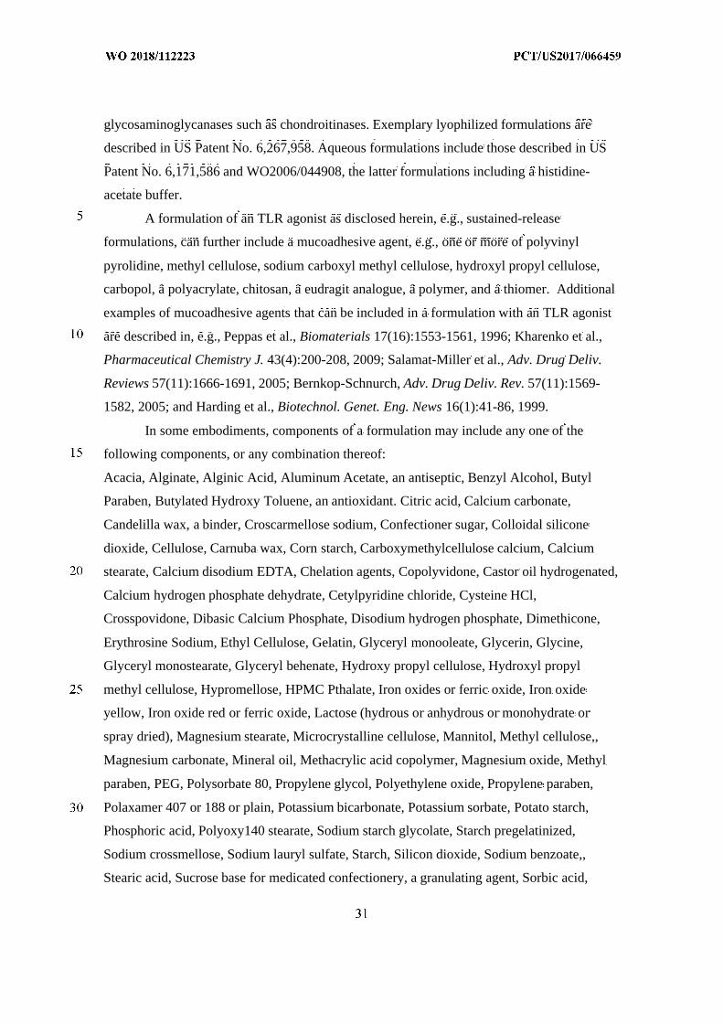
glycosaminoglycanases such as chondroitinases. Exemplary lyophilized formulations are
described in US Patent No. 6,267,958. Aqueous formulations include those described in US
Patent No. 6,171,586 and WO2006/044908, the latter formulations including a histidine-
acetate buffer.
A formulation of an TLR agonist as disclosed herein, e.g., sustained-release
formulations, can further include a mucoadhesive agent, e.g., one or more of polyvinyl
pyrolidine, methyl cellulose, sodium carboxyl methyl cellulose, hydroxyl propyl cellulose,
carbopol, a polyacrylate, chitosan, a eudragit analogue, a polymer, and a thiomer. Additional
examples of mucoadhesive agents that can be included in a formulation with an TLR agonist
are described in, e.g., Peppas et al., Biomaterials 17(16):1553-1561, 1996; Kharenko et al.,
Pharmaceutical Chemistry J. 43(4):200-208, 2009; Salamat-Miller et al., Adv. Drug Deliv.
Reviews 57(11):1666-1691, 2005; Bernkop-Schnurch, Adv. Drug Deliv. Rev. 57(11):1569-
1582, 2005; and Harding et al., Biotechnol. Genet. Eng. News 16(1):41-86, 1999.
In some embodiments, components of a formulation may include any one of the
following components, or any combination thereof:
Acacia, Alginate, Alginic Acid, Aluminum Acetate, an antiseptic, Benzyl Alcohol, Butyl
Paraben, Butylated Hydroxy Toluene, an antioxidant. Citric acid, Calcium carbonate,
Candelilla wax, a binder, Croscarmellose sodium, Confectioner sugar, Colloidal silicone
dioxide, Cellulose, Carnuba wax, Corn starch, Carboxymethylcellulose calcium, Calcium
stearate, Calcium disodium EDTA, Chelation agents, Copolyvidone, Castor oil hydrogenated,
Calcium hydrogen phosphate dehydrate, Cetylpyridine chloride, Cysteine HCl,
Crosspovidone, Dibasic Calcium Phosphate, Disodium hydrogen phosphate, Dimethicone,
Erythrosine Sodium, Ethyl Cellulose, Gelatin, Glyceryl monooleate, Glycerin, Glycine,
Glyceryl monostearate, Glyceryl behenate, Hydroxy propyl cellulose, Hydroxyl propyl
methyl cellulose, Hypromellose, HPMC Pthalate, Iron oxides or ferric oxide, Iron oxide
yellow, Iron oxide red or ferric oxide, Lactose (hydrous or anhydrous or monohydrate or
spray dried), Magnesium stearate, Microcrystalline cellulose, Mannitol, Methyl cellulose,,
Magnesium carbonate, Mineral oil, Methacrylic acid copolymer, Magnesium oxide, Methyl
paraben, PEG, Polysorbate 80, Propylene glycol, Polyethylene oxide, Propylene paraben,
Polaxamer 407 or 188 or plain, Potassium bicarbonate, Potassium sorbate, Potato starch,
Phosphoric acid, Polyoxy140 stearate, Sodium starch glycolate, Starch pregelatinized,
Sodium crossmellose, Sodium lauryl sulfate, Starch, Silicon dioxide, Sodium benzoate,,
Stearic acid, Sucrose base for medicated confectionery, a granulating agent, Sorbic acid,
Sodium carbonate, Saccharin sodium, Sodium alginate, Silica gel, Sorbiton monooleate,
Sodium stearyl fumarate, Sodium chloride, Sodium metabisulfite, Sodium citrate dehydrate,
Sodium starch, Sodium carboxy methyl cellulose, Succinic acid, Sodium propionate,
Titanium dioxide, Talc, Triacetin, Triethyl citrate.
Accordingly, in some embodiments of the method of treating a disease as disclosed
herein, the method comprises administering to the subject a pharmaceutical composition that
is a formulation as disclosed herein. In some embodiments the formulation is a dosage form,
which may be, as an example, a solid form such as, for example, a capsule, a tablet, a sachet,
or a lozenge; or which may be, as an example, a liquid form such as, for example, a solution,
a suspension, an emulsion, or a syrup.
In some embodiments, the formulation is not comprised in an ingestible device. In
some embodiments wherein the formulation is not comprised in an ingestible device, the
formulation may be suitable for oral administration. The formulation may be, for example, a
solid dosage form or a liquid dosage form as disclosed herein. In some embodiments wherein
the formulation is not comprised in an ingestible device, the formulation may be suitable for
rectal administration. The formulation may be, for example, a dosage form such as a
suppository or an enema. In embodiments where the formulation is not comprised in an
ingestible device, the formulation releases the TLR agonist at a location in the gastrointestinal
tract of the subject that is proximate to one or more sites of disease. Such localized release
may be achieved, for example, with a formulation comprising an enteric coating. Such
localized release may be achieved, an another example, with a formulation comprising a core
comprising one or more polymers suitable for controlled release of an active substance. A
non-limiting list of such polymers includes: poly(2-(diethylamino)ethyl methacrylate, 2-
(dimethylamino)ethyl methacrylate, poly(ethylene glycol), poly(2-aminoethyl methacrylate),
(2-hydroxypropyl)methacrylamide, poly(β-benzyl-l-aspartate), poly(N-isopropylacrylamide),
and cellulose derivatives.
In some embodiments, the formulation is comprised in an ingestible device as
disclosed herein. In some embodiments wherein the formulation is comprised in an
ingestible device, the formulation may be suitable for oral administration. The formulation
may be, for example, a solid dosage form or a liquid dosage form as disclosed herein. In
some embodiments the formulation is suitable for introduction and optionally for storage in
the device. In some embodiments the formulation is suitable for introduction and optionally
for storage in a reservoir comprised in the device. In some embodiments the formulation is
suitable for introduction and optionally for storage in a reservoir comprised in the device.
Thus, in some embodiments, provided herein is a reservoir comprising a therapeutically
effective amount of an TLR agonist, wherein the reservoir is configured to fit into an
ingestible device. In some embodiments, the reservoir comprising a therapeutically effective
amount of an TLR agonist is attachable to an ingestible device. In some embodiments, the
reservoir comprising a therapeutically effective amount of an TLR agonist is capable of
anchoring itself to the subject’s tissue. As an example, the reservoir capable of anchoring
itself to the subject’s tissue comprises silicone. As an example, the reservoir capable of
anchoring itself to the subject’s tissue comprises polyvinyl chloride.
In some embodiments the formulation is suitable for introduction in a spray catheter,
as disclosed herein.
The formulation herein may also contain more than one active compound as necessary
for the particular indication being treated, for example, those with complementary activities
that do not adversely affect each other. For instance, the formulation may further comprise
another TLR agonist or a chemotherapeutic agent. Such molecules are suitably present in
combination in amounts that are effective for the purpose intended.
The active ingredients may also be entrapped in microcapsules prepared, for example,
by coacervation techniques or by interfacial polymerization, for
example, hydroxymethylcellulose or gelatin-microcapsule and poly-(methylmethacylate)
microcapsule, respectively, in colloidal drug delivery systems (for example, liposomes,
albumin microspheres, microemulsions, nano-particles and nanocapsules) or in
macroemulsions. Such techniques are disclosed in Remington's Pharmaceutical Sciences 16th
edition, Osol, A. Ed. (1980).
The formulations to be used for in vivo administration must be sterile. This is readily
accomplished by filtration through sterile filtration membranes.
Sustained-release preparations may be prepared. Suitable examples of sustained-
release preparations include semipermeable matrices of solid hydrophobic polymers
containing the TLR agonist, which matrices are in the form of shaped articles, e.g., films, or
microcapsule. Examples of sustained-release matrices include polyesters, hydrogels (for
example, poly(2- hydroxyethyl-methacrylate), or poly(vinylalcohol)), polylactides (U.S. Pat.
No. 3,773,919), copolymers of L-glutamic acid and γ ethyl-L-glutamate, non-degradable
ethylene-vinyl acetate, degradable lactic acid-glycolic acid copolymers such as the LUPRON
DEPOT™ (injectable microspheres composed of lactic acid-glycolic acid copolymer and
leuprolide acetate), and poly-D-(-)-3-hydroxybutyric acid. While polymers such as ethylene-
vinyl acetate and lactic acid-glycolic acid enable release of molecules for over 100 days,
certain hydrogels release proteins for shorter time periods. When encapsulated TLR agonists
remain in the body for a long time, they may denature or aggregate as a result of exposure to
moisture at 37°C, resulting in a loss of biological activity and possible changes in
immunogenicity. Rational strategies can be devised for stabilization depending on the
mechanism involved. For example, if the aggregation mechanism is discovered to be
intermolecular S-S bond formation through thio-disulfide interchange, stabilization may be
achieved by modifying sulfhydryl residues, lyophilizing from acidic solutions, controlling
moisture content, using appropriate additives, and developing specific polymer matrix
compositions.
Pharmaceutical formulations may contain one or more TLR agonists. The
pharmaceutical formulations may be formulated in any manner known in the art. In some
embodiments the formulations include one or more of the following components: a sterile
diluent (e.g., sterile water or saline), a fixed oil, polyethylene glycol, glycerin, propylene
glycol, or other synthetic solvents, antibacterial or antifungal agents, such as benzyl alcohol
or methyl parabens, chlorobutanol, phenol, ascorbic acid, thimerosal, and the like,
antioxidants, such as ascorbic acid or sodium bisulfite, chelating agents, such as
ethylenediaminetetraacetic acid, buffers, such as acetates, citrates, or phosphates, and isotonic
agents, such as sugars (e.g., dextrose), polyalcohols (e.g., mannitol or sorbitol), or salts (e.g.,
sodium chloride), or any combination thereof. Liposomal suspensions can also be used as
pharmaceutically acceptable carriers (see, e.g., U.S. Patent No. 4,522,811, incorporated by
reference herein in its entirety). The formulations can be formulated and enclosed in
ampules, disposable syringes, or multiple dose vials. Where required, proper fluidity can be
maintained by, for example, the use of a coating, such as lecithin, or a surfactant. Controlled
release of the TLR agonist can be achieved by implants and microencapsulated delivery
systems, which can include biodegradable, biocompatible polymers (e.g., ethylene vinyl
acetate, polyanhydrides, polyglycolic acid, collagen, polyorthoesters, and polylactic acid;
Alza Corporation and Nova Pharmaceutical, Inc.).
In some embodiments, the TLR agonist is present in a pharmaceutical formulation
within the device.
In some embodiments, the TLR agonist is present in solution within the device.
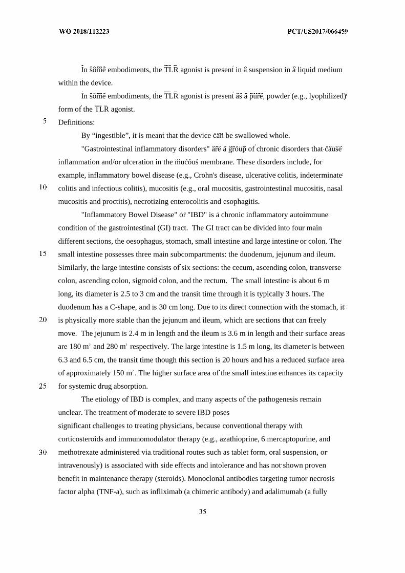
In some embodiments, the TLR agonist is present in a suspension in a liquid medium
within the device.
In some embodiments, the TLR agonist is present as a pure, powder (e.g., lyophilized)
form of the TLR agonist.
Definitions:
By “ingestible”, it is meant that the device can be swallowed whole.
"Gastrointestinal inflammatory disorders" are a group of chronic disorders that cause
inflammation and/or ulceration in the mucous membrane. These disorders include, for
example, inflammatory bowel disease (e.g., Crohn's disease, ulcerative colitis, indeterminate
colitis and infectious colitis), mucositis (e.g., oral mucositis, gastrointestinal mucositis, nasal
mucositis and proctitis), necrotizing enterocolitis and esophagitis.
"Inflammatory Bowel Disease" or "IBD" is a chronic inflammatory autoimmune
condition of the gastrointestinal (GI) tract. The GI tract can be divided into four main
different sections, the oesophagus, stomach, small intestine and large intestine or colon. The
small intestine possesses three main subcompartments: the duodenum, jejunum and ileum.
Similarly, the large intestine consists of six sections: the cecum, ascending colon, transverse
colon, ascending colon, sigmoid colon, and the rectum. The small intestine is about 6 m
long, its diameter is 2.5 to 3 cm and the transit time through it is typically 3 hours. The
duodenum has a C-shape, and is 30 cm long. Due to its direct connection with the stomach, it
is physically more stable than the jejunum and ileum, which are sections that can freely
move. The jejunum is 2.4 m in length and the ileum is 3.6 m in length and their surface areas
are 180 m2 and 280 m2 respectively. The large intestine is 1.5 m long, its diameter is between
6.3 and 6.5 cm, the transit time though this section is 20 hours and has a reduced surface area
of approximately 150 m2 . The higher surface area of the small intestine enhances its capacity
for systemic drug absorption.
The etiology of IBD is complex, and many aspects of the pathogenesis remain
unclear. The treatment of moderate to severe IBD poses
significant challenges to treating physicians, because conventional therapy with
corticosteroids and immunomodulator therapy (e.g., azathioprine, 6 mercaptopurine, and
methotrexate administered via traditional routes such as tablet form, oral suspension, or
intravenously) is associated with side effects and intolerance and has not shown proven
benefit in maintenance therapy (steroids). Monoclonal antibodies targeting tumor necrosis
factor alpha (TNF-a), such as infliximab (a chimeric antibody) and adalimumab (a fully
human antibody), are currently used in the management of CD. Infliximab has also shown
efficacy and has been approved for use in UC. However, approximately 10%-20% of patients
with CD are primary nonresponders to anti TNF therapy, and another ~20%-30% of CD
patients lose response over time (Schnitzler et al., Gut 58:492-500 (2009)). Other adverse
events (AEs) associated with anti TNFs include elevated rates of bacterial infection,
including tuberculosis, and, more rarely, lymphoma and demyelination (Chang et al., Nat
Clin Pract Gastroenterol Hepatology 3:220 (2006); Hoentjen et al., World J. Gastroenterol.
15(17):2067 (2009)). No currently available therapy achieves sustained remission
in more than 20%-30% of IBD patients with chronic disease (Hanauer et al, Lancet 359:
1541-49 (2002); Sandborn et al, N Engl J Med 353: 1912-25 (2005)). In addition, most
patients do not achieve sustained steroid-free remission and mucosal healing, clinical
outcomes that correlate with true disease modification.
Although the cause of IBD remains unknown, several factors such as genetic,
infectious and immunologic susceptibility have been implicated. IBD is much more common
in Caucasians, especially those of Jewish descent. The chronic inflammatory nature of the
condition has prompted an intense search for a possible infectious cause. Although agents
have been found which stimulate acute inflammation, none has been found to cause the
chronic inflammation associated with IBD. The hypothesis that IBD is an autoimmune
disease is supported by the previously mentioned extraintestinal manifestation of IBD as joint
arthritis, and the known positive response to IBD by treatment with therapeutic agents such
as adrenal glucocorticoids, cyclosporine and azathioprine, which are known to suppress
immune response. In addition, the GI tract, more than any other organ of the body, is
continuously exposed to potential antigenic substances such as proteins from food, bacterial
byproducts (LPS), etc.
A chronic inflammatory autoimmune condition of the gastrointestinal (GI) tract
presents clinically as either ulcerative colitis (UC) or Crohn's disease (CD). Both IBD
conditions are associated with an increased risk for malignancy of the GI tract.
“Crohn's disease” (“CD”) is a chronic transmural inflammatory disease
with the potential to affect any part of the entire GI tract, and UC is a mucosal
inflammation of the colon. Both conditions are characterized clinically by frequent bowel
motions, malnutrition, and dehydration, with disruption in the activities of daily living.
CD is frequently complicated by the development of malabsorption, strictures, and
fistulae and may require repeated surgery. UC, less frequently, may be complicated by severe
bloody diarrhea and toxic megacolon, also requiring surgery. The most prominent feature
Crohn's disease is the granular, reddish-purple edematous thickening of the bowel wall. With
the development of inflammation, these granulomas often lose their circumscribed borders
and integrate with the surrounding tissue. Diarrhea and obstruction of the bowel are the
predominant clinical features. As with ulcerative colitis, the course of Crohn's disease may be
continuous or relapsing, mild or severe, but unlike ulcerative colitis, Crohn's disease is not
curable by resection of the involved segment of bowel. Most patients with Crohn's disease
require surgery at some point, but subsequent relapse is common and continuous medical
treatment is usual. Crohn's disease may involve any part of the alimentary tract from the
mouth to the anus, although typically it appears in the ileocolic, small-intestinal or colonic-
anorectal regions. Histopathologically, the disease manifests by discontinuous
granulomatomas, crypt abscesses, fissures and aphthous ulcers. The inflammatory infiltrate is
mixed, consisting of lymphocytes (both T and B cells), plasma cells, macrophages, and
neutrophils. There is a disproportionate increase in IgM- and IgG-secreting plasma cells,
macrophages and neutrophils.
To date, the primary outcome measure in Crohn's Disease clinical trials is the Crohn's
Disease Activity Index (CDAI), which has served as the basis for approval of multiple drug
treatments, including for example, vedolizumab and natalizumab. The CDAI was developed
by regressing clinician global assessment of disease activity on eighteen potential items
representing patient reported outcomes (PROs) (i.e. abdominal pain, pain awakening patient
from sleep, appetite), physical signs (i.e. average daily temperature, abdominal mass),
medication use (i.e. loperamide or opiate use for diarrhea) and a laboratory test (i.e.
hematocrit). Backward stepwise regression analysis identified eight independent predictors
which are the number of liquid or soft stools, severity of abdominal pain, general well-being,
occurrence of extra-intestinal symptoms, need for anti-diarrheal drugs, presence of an
abdominal mass, hematocrit, and body weight. The final score is a composite of these eight
items, adjusted using regression coefficients and standardization to construct an overall CDAI
score, ranging from 0 to 600 with higher score indicating greater disease activity. Widely
used benchmarks are: CDAI <150 is defined as clinical remission, 150 to 219 is defined as
mildly active disease, 220 to 450 is defined as moderately active disease, and above 450 is
defined as very severe disease (Best WR, et al., Gastroenterology 77:843-6, 1979).
Vedolizumab and natalizumab have been approved on the basis of demonstrated clinical
remission, i.e. CDAI < 150.
Although the CDAI has been in use for over 40 years, and has served as the basis for
drug approval, it has several limitations as an outcome measure for clinical trials. For
example, most of the overall score comes from the patient diary card items (pain,
number of liquid bowel movements, and general well-being), which are vaguely defined and
not standardized terms (Sandler et al., J. Clin. Epidemiol 41 :451-8, 1988; Thia et al.,
Inflamm Bowel Dis 17: 105-11, 2011). In addition, measurement of pain is based on a four-
point scale rather than an updated seven-point scale. The remaining 5 index items contribute
very little to identifying an efficacy signal and may be a source of measurement noise.
Furthermore, concerns have been raised about poor criterion validity for the CDAI, a reported
lack of correlation between the CDAI and endoscopic measures of inflammation (which may
render the CDAI as a poor discriminator of active CD and irritable bowel syndrome) and high
reported placebo rates (Korzenik et al., N Engl J Med. 352:2193-201, 2005; Sandborn WJ, et
al., N Engl J Med 353 : 1912-25, 2005; Sandborn WJ, et al., Ann Intern 19; 146:829-38,
2007, Epub 2007 Apr 30; Kim et al., Gastroenterology 146: (5 supplement 1) S-368, 2014).
It is, thus, generally recognized that additional or alternative measures of CD
symptoms are needed, such as new PRO tools or adaptations of the CDAI to derive a new
PRO. The PRO2 and PRO3 tools are such adaptations of the CDAI and have been recently
described in Khanna et al., Aliment Pharmacol. Ther. 41: 77-86, 2015. The PRO2
evaluates the frequency of loose/liquid stools and abdominal pain {Id). These items are
derived and weighted accordingly from the CDAI and are the CDAI diary card items, along
with general well-being, that contribute most to the observed clinical benefit measured by
CDAI (Sandler et al., J. Clin. Epidemiol 41 :451-8, 1988; Thia et al., Inflamm Bowel Dis 17:
105-11, 2011; Kim et al., Gastroenterology 146: (5 supplement 1) S-368,
2014). The remission score of < 11 is the CDAI-weighted sum of the average stool frequency
and pain scores in a 7-day period, which yielded optimum sensitivity and specificity for
identification of CDAI remission (score of < 150) in a retrospective data
analysis of ustekinumab induction treatment for moderate to severe CD in a Phase II clinical
study (Gasink C, et al., abstract, ACG Annual Meeting 2014). The PRO2 was shown to be
sensitive and responsive when used as a continuous outcome measure in a retrospective data
analysis of MTX treatment in active CD (Khanna R, et al., Inflamm Bowel Dis 20: 1850-61,
2014) measured by CDAI. Additional outcome measures include the Mayo Clinic Score, the
Crohn disease endoscopic index of severity (CDEIS), and the Ulcerative colitis endoscopic
index of severity (UCEIS). Additional outcome measures include Clinical remission,
Mucosal healing, Histological healing (transmural), MRI or ultrasound for measurement or
evaluation of bowel wall thickness, abscesses, fistula and histology.
An additional means of assessing the extent and severity of Crohn's Disease is
endoscopy. Endoscopic lesions typical of Crohn's disease have been described in numerous
studies and include, e.g., aphthoid ulcerations, "punched-out ulcers," cobblestoning and
stenosis. Endoscopic evaluation of such lesions was used to develop the first validated
endoscopic score, the Crohn's Disease Endoscopic Index of Severity (CDEIS) (Mary et al.,
Gut 39:983-9, 1989). More recently, because the CDEIS is time-consuming, complicated and
impractical for routine use, a Simplified Endoscopic Activity Score for Crohn's Disease
(SES- CD) was developed and validated (Daperno et al., Gastrointest. Endosc. 60(4):505-12,
2004). The SES-CD consists of four endoscopic variables (size of ulcers,
proportion of surface covered by ulcers, proportion of surface with any other lesions (e.g.,
inflammation), and presence of narrowings [stenosis]) that are scored in five ileocolonic
segments, with each variable, or assessment, rated from 0 to 3.
To date, there is no cure for CD. Accordingly, the current treatment goals for CD are
to induce and maintain symptom improvement, induce mucosal healing, avoid surgery, and
improve quality of life (Lichtenstein GR, et al., Am J Gastroenterol 104:465-83, 2009; Van
Assche G, et al., J Crohns Colitis. 4:63-101, 2010). The current therapy of IBD usually
involves the administration of antiinflammatory or immunosuppressive agents, such as
sulfasalazine, corticosteroids, 6- mercaptopurine/azathioprine, or cyclosporine, all of which
are not typically delivered by localized release of a drug at the site or location of disease.
More recently, biologics like TNF-alpha inhibitors and IL-12/IL-23 blockers, are used to treat
IBD. If anti-inflammatory/immunosuppressive/biologic therapies fail, colectomies are the last
line of defense. The typical operation for CD not involving the rectum is resection (removal
of a diseased segment of bowel) and anastomosis (reconnection) without an ostomy. Sections
of the small or large intestine may be removed. About 30% of CD patients will need surgery
within the first year after diagnosis. In the subsequent years, the rate is about 5% per year.
Unfortunately, CD is characterized by a high rate of recurrence; about 5% of patients need a
second surgery each year after initial surgery.
Refining a diagnosis of inflammatory bowel disease involves evaluating the
progression status of the diseases using standard classification criteria. The classification
systems used in IBD include the Truelove and Witts Index (Truelove S. C. and Witts, L.J. Br
Med J. 1955;2: 1041-1048), which classifies colitis as mild, moderate, or severe, as well as
Lennard- Jones. (Lennard-Jones JE. Scand J Gastroenterol Suppl 1989; 170:2-6) and the
simple clinical colitis activity index (SCCAI). (Walmsley et. al. Gut. 1998; 43:29-32) These
systems track such variables as daily bowel movements, rectal bleeding, temperature, heart
rate, hemoglobin levels, erythrocyte sedimentation rate, weight, hematocrit score, and the
level of serum albumin.
There is sufficient overlap in the diagnostic criteria for UC and CD that it is
sometimes impossible to say which a given patient has; however, the type of lesion typically
seen is different, as is the localization. UC mostly appears in the colon, proximal to the
rectum, and the characteristic lesion is a superficial ulcer of the mucosa; CD can appear
anywhere in the bowel, with occasional involvement of stomach, esophagus and duodenum,
and the lesions are usually described as extensive linear fissures.
In approximately 10-15% of cases, a definitive diagnosis of ulcerative colitis or
Crohn's disease cannot be made and such cases are often referred to as "indeterminate
colitis." Two antibody detection tests are available that can help the diagnosis, each of which
assays for antibodies in the blood. The antibodies are "perinuclear anti-neutrophil antibody"
(pANCA) and "anti-Saccharomyces cervisiae antibody" (ASCA). Most patients with
ulcerative colitis have the pANCA antibody but not the ASCA antibody, while most patients
with Crohn's disease have the ASCA antibody but not the pANCA antibody. However, these
two tests have shortcomings as some patients have neither antibody and some Crohn's disease
patients may have only the pANCA antibody. A third test, which measures the presence and
accumulation of circulating anti-microbial antibodies – particularly flagellin antibodies, has
proven to be useful for detecting susceptibility to Crohn’s Disease before disease
development. See Choung, R. S., et al. "Serologic microbial associated markers can predict
Crohn's disease behaviour years before disease diagnosis." Alimentary pharmacology &
therapeutics 43.12 (2016): 1300-1310.
"Ulcerative colitis (UC)" afflicts the large intestine. The course of the disease may be
continuous or relapsing, mild or severe. The earliest lesion is an inflammatory infiltration
with abscess formation at the base of the crypts of Lieberkuhn. Coalescence of these
distended and ruptured crypts tends to separate the overlying mucosa from its blood supply,
leading to ulceration. Symptoms of the disease include cramping, lower abdominal pain,
rectal bleeding, and frequent, loose discharges consisting mainly of blood, pus and mucus
with scanty fecal particles. A total colectomy may be required for acute, severe or chronic,
unremitting ulcerative colitis.
The clinical features of UC are highly variable, and the onset may be insidious or
abrupt, and may include diarrhea, tenesmus and relapsing rectal bleeding. With fulminant
involvement of the entire colon, toxic megacolon, a life-threatening emergency, may occur.
Extraintestinal manifestations include arthritis, pyoderma gangrenoum, uveitis, and erythema
nodosum.
The terms "antibody" and "immunoglobulin" are used interchangeably in the broadest
sense and include monoclonal antibodies (for example, full length or intact monoclonal
antibodies), polyclonal antibodies, multivalent antibodies, multispecific antibodies (e.g.,
bispecific, trispecific etc. antibodies so long as they exhibit the desired biological activity)
and may also include certain antibody fragments (as described in greater detail herein). An
antibody can be human, humanized and/or affinity matured.
"Antibody fragments" comprise only a portion of an intact antibody, where in certain
embodiments, the portion retains at least one, and typically most or all, of the functions
normally associated with that portion when present in an intact antibody. In one embodiment,
an antibody fragment comprises an antigen binding site of the intact antibody and thus retains
the ability to bind antigen. In another embodiment, an antibody fragment, for example one
that comprises the Fc region, retains at least one of the biological functions normally
associated with the Fc region when present in an intact antibody, such as FcRn binding,
antibody half-life modulation, ADCC function and complement binding. In one embodiment,
an antibody fragment is a monovalent antibody that has an in vivo half-life substantially
similar to an intact antibody. For example, such an antibody fragment may comprise on
antigen binding arm linked to an Fc sequence capable of conferring in vivo stability to the
fragment.
The term "monoclonal antibody" as used herein refers to an antibody obtained from a
population of substantially homogeneous antibodies, i.e., the individual antibodies
comprising the population are identical except for possible naturally occurring mutations that
may be present in minor amounts. Monoclonal antibodies are highly specific, being directed
against a single antigen. Furthermore, in contrast to polyclonal antibody preparations that
typically include different antibodies directed against different determinants (epitopes), each
monoclonal antibody is directed against a single determinant on the antigen.
The monoclonal antibodies herein specifically include "chimeric" antibodies in which
a portion of the heavy and/or light chain is identical with or homologous to corresponding
sequences in antibodies derived from a particular species or belonging to a particular
antibody class or subclass, while the remainder of the chain(s) is identical with or
homologous to corresponding sequences in antibodies derived from another species or
belonging to another antibody class or subclass, as well as fragments of such antibodies, so
long as they exhibit the desired biological activity (U.S. Patent No. 4,816,567; and Morrison
et al, Proc. Natl. Acad. Sci. USA 81 :6851-6855 (1984)).
"Treatment regimen" refers to a combination of dosage, frequency of administration,
or duration of treatment, with or without addition of a second medication.
"Effective treatment regimen" refers to a treatment regimen that will offer beneficial
response to a patient receiving the treatment.
"Effective amount" refers to an amount of drug that offers beneficial response to a
patient receiving the treatment. For example, an effective amount may be a Human
Equivalent Dose (HED).
“Dispensable”, with reference to any substance, refers to any substance that may be
released from an ingestible device as disclosed herein, or from a component of the device
such as a reservoir. For example, a dispensable substance may be an TLR agonist, and/or a
formulation comprising an TLR agonist.
"Patient response" or "patient responsiveness" can be assessed using any endpoint
indicating a benefit to the patient, including, without limitation, (1) inhibition, to some extent,
of disease progression, including slowing down and complete arrest; (2) reduction in the
number of disease episodes and/or symptoms; (3) reduction in lesional size; (4) inhibition
(i.e., reduction, slowing down or complete stopping) of disease cell infiltration into adjacent
peripheral organs and/or tissues; (5) inhibition (i.e., reduction, slowing down or complete
stopping) of disease spread; (6) decrease of auto-immune response, which may, but does not
have to, result in the regression or ablation of the disease lesion; (7) relief, to some extent, of
one or more symptoms associated with the disorder; (8) increase in the length of disease-free
presentation following treatment; and/or (9) decreased mortality at a given point of time
following treatment. The term "responsiveness" refers to a measurable response, including
complete response (CR) and partial response (PR).
As used herein, "complete response" or "CR" means the disappearance of all signs of
inflammation or remission in response to treatment. This does not necessarily mean the
disease has been cured.
"Partial response" or "PR" refers to a decrease of at least 50% in the severity of
inflammation, in response to treatment.
A "beneficial response" of a patient to treatment with a therapeutic agent and similar
wording refers to the clinical or therapeutic benefit imparted to a patient at risk for or
suffering from a gastrointestinal inflammatory disorder from or as a result of the treatment
with the agent. Such benefit includes cellular or biological responses, a complete response, a
partial response, a stable disease (without progression or relapse), or a response with a later
relapse of the patient from or as a result of the treatment with the agent.
As used herein, "non-response" or "lack of response" or similar wording means an
absence of a complete response, a partial response, or a beneficial response to treatment with
a therapeutic agent.
"A patient maintains responsiveness to a treatment" when the patient' s responsiveness
does not decrease with time during the course of a treatment.
A "symptom" of a disease or disorder (e.g., inflammatory bowel disease, e.g.,
ulcerative colitis or Crohn's disease) is any morbid phenomenon or departure from the normal
in structure, function, or sensation, experienced by a subject and indicative of disease.
TLR Modulators
A “TLR modulator” is an agent that functionally interacts with a toll-like receptor (TLR)
expressed in a mammalian cell (e.g., a human cell). In some embodiments, the modulator is a
TLR agonist. In some embodiments, the modulator is a TLR antagonist.
TLR Agonists
The term “TLR agonist” is an agent that binds to and activates a toll-like receptor
(TLR) expressed in a mammalian cell (e.g., a human cell). In some embodiments, the TLR
agonist binds to and activates TLR1. In some embodiments, the TLR agonist binds to and
activates TLR2. In some embodiments, the TLR agonist binds to and activates TLR3. In
some embodiments, the TLR agonist binds to and activates TLR4. In some embodiments, the
TLR agonist binds to and activates TLR5. In some embodiments, the TLR agonist binds to
and activates TLR6. In some embodiments, the TLR agonist binds to and activates TLR7. In
some embodiments, the TLR agonist binds to and activates TLR8. In some embodiments, the
TLR agonist binds to and activates TLR9. In some embodiments, the TLR agonist binds to
and activates TLR10. In some embodiments, the TLR agonist binds to and activates TLR11.
In some embodiments, the TLR agonist binds to and activates two or more (e.g., three, four,
five, six, seven, eight, nine, ten, or eleven) TLRs (e.g., two or more of any of TLR1, TLR2,
TLR3, TLR4, TLR5, TLR6, TLR7, TLR8, TLR9, TLR10, and TLR11 (in any combination)).
In some embodiments, the TLR agonist is a synthetic TLR agonist, a TLR mimic, or a
small molecule. Non-limiting examples of TLR agonists are described in Bhardwaj et al.,
Cancer J. 16(4):382-391, 2010; Meyer et al., Exp. Opin. Investig. Drugs 17(7):1051-1065,
2008; Adams, Immunotherapy 1(6):949-964, 2009; Hennessy et al., Nat. Rev. Drug Discov.
9:293-307, 2010; and U.S. Patent Nos. 7,498,409; 9,421,254; 8,409,813; 8,361,986;
8,795,678; 8,728,486; 8,636,979; 8,999,946; 9,359,360; 9,050,376; and 9,556,167; US
2014/0322271; US 2016/0206690; US 2009/0253622; US 2011/0135669; US 2011/0250175;
US 2014/0220074; and US 2012/0219615; each incorporated in its entirety herein. In some
embodiments, the TLR agonist is a peptide or a fusion protein (Huleatt et al., Vaccine 25:
763-775, 2007).
In some embodiments, a TLR agonist specifically binds to and activates a single TLR
(e.g., TLR4, TLR7, TLR8, or TLR9; Zhu et al., J. Clin. Invest. 120:607-616, 2010; Zhu et al.,
PNAS 105:16260-16265, 2008; Wang et al., J. Virol. 79(22):14355-14370, 2005). In some
embodiments, the TLR agonist binds to and activates more than one TLR (e.g., Bacillus of
Calmette-Guerin, Myobacterium bovis (BCG); Morton et al., Ann. Surg. 180(4):635-643,
1974; Mortoon et al., J. Clin. Oncol. ASCO Ann. Meeting Proceedings Part I 25(18 Suppl),
2007). In some embodiments, the TLR agonist is a TLR2/TLR6 agonist (e.g., Pam2 CSK4 or
MALP-2 (Agnihotri et al., J. Med. Chem. 54: 8148-8160, 2011; Wu et al., J. Med. Chem. 53:
3198-3213, 2010)).
In some embodiments, the TLR agonist is administrated in combination with another
composition (Dowling et al., Clin. Transl. Immunol. 5:e85, 2016). In some embodiments, the
TLR agonist is an endogenous molecule released from dead cells (e.g., a heat shock protein
(HSP) and mobility group box 1 (HMGB1); Asea et al., J. Biol. Chem. 277:15028-15034,
2002; Kepp et al., Cancer Metastasis 30: 61-69, 2011).
TLR3 Agonists
In some embodiments, the TLR agonist specifically binds and activates TLR3 (e.g., a
synthetic agonist). Non-limiting examples of TLR agonists that bind and activate TLR3 are
described in Nicodemus et al., Immunotherapy 2:137-140, 2010. In some embodiments, the
TLR3 agonist is a synthetic double-stranded RNA (dsRNA) complex (e.g., polyribosinic:
polyribocytidic acid (polyI:C); Sivori et al., PNAS 101:10116-10121, 2004; Sloat et al.,
Pharmaceutical Res. 23:1217-1226, 2006; Ichinohe et al., Microbes and infection/ Institut
Pasteur 9:1333-1340, 2007; Robinson et al., J. Natl. Cancer Inst. 57(3):599-602, 1976). In
some embodiments, the TLR3 agonist is a TLR3 mimic (e.g., polyadenosine-polyuridylic
acid (poly A:U) (Veyrat et al., Oncotarget 7(50):82580-82593, 2016; Alizadeh et al., Iran J.
Allergy Asthma Immunol. 12(2):161-167, 2013); rintatolimod (polyI: polyCU, Ampligen®)
(Steinman et al., Nature 449: 419-426, 2007; Jasani et al., Vaccine 27(25-26):3401-3404,
2009; Strayer et al., PLoS One 7(3): e31334, 2012). In some embodiments, the TLR3 mimic
is polyionisinic-polycytidylic acid stabilized with poly-L-lysine and carboxymethylcellulose
(Poly-ICLC, Hiltonol®; Hawkins et al., J. Biol. Resp. Mod. 4:664-668, 1985; Butowski et al.,
J. Neurooncol. 91:175-182, 2009; Jeong et al., J. Neurochem. doi.10.1111, 2015). In some
embodiments, the TLR3 agonist is RGC100 (Naumann et al., Clin. Dev. Immunol. 283649,
2013), IPH-3102 (Basith et al., Exp. Opin. Ther. Pat. 21: 927-944, 2011), or a variant thereof.
In some embodiments, the TLR3 agonist is CQ-07001 (Clinquest). In some embodiments,
the TLR3 agonist is Ampligen poly(I):poly(C12U) (Hemispherx Biopharma). In some
embodiments, the TLR3 agonist is IPH-31XX (Innate Pharma). In some embodiments, the
TLR3 agonist is MCT-465-dsRNA (MultiCell Technologies).
TLR4 Agonists
In some embodiments, the TLR agonist specifically binds to and activates TLR4 (Peri
et al., J. Med. Chem. 57(9):3612-3622, 2014). In some embodiments, the TLR4 agonist is
bacterial lipopolysaccharide (LPS) or a variant thereof. In some embodiments, the TLR4
agonist is monophosphoryl lipid A (MPL, MPLA, GLA, GLA-SE) (Ribi et al., J. Immunol.
6:567-572, 1984; Okemoto et al., J. Immunol. 176:1203-1208, 2006; Matzner et al., Int. J.
Cancer 138:1754-1764, 2016; Cauwelaert et al., PLoS One 11(1):e0146372, 2016). In some
embodiments, the TLR agonist is AS15 or AS02b (Brichard et al., Vaccine 25(Suppl. 2):B61-
B71, 2007; Kruit et al., J. Clin. Oncol. 26(Suppl): Abstract 9065, 2008). In some
embodiments, the TLR agonist is an aminoalkyl glucosaminide 4-phosphate (e.g., RC-529,
Ribi.529, E6020) or a variant thereof (Baldridge et al., J. Endotoxin Res. 8:453-458, 2002;
Morefield et al., Clin. Vaccine Immunol. 14: 1499-1504, 2007). In some embodiments, the
TLR agonist is picibanil (OK-432) (Hazim et al., Med. J. Malaysia 71(6):328-330, 2016;
Tian et al., Asian Pac J. Cancer Prev. 16(11):4537-4542, 2015; Rebuffini et al., Dent Rese. J.
9(Suppl. 2):S192-S196, 2012). In some embodiments, the TLR4 agonist is Spirulina
complex polysaccharide (Kwanishi et al., Microbiol. Immunol. 57:63-73, 2013). In some
embodiments, the TLR4 agonist is chitohexaose or a variant thereof (Panda et al.,
8:e1002717, 2012; Barman et al., Cell Death Dis. 7:e2224, 2016). In some embodiments, the
TLR4 agonist is E5564 (Eritoran) (Eisai). In some embodiments, the TLR4 agonist is CRX-
675 or CRX-527 (GSK).
TLR5 Agonists
In some embodiments, the TLR agonist binds and activates TLR5. In some
embodiments, the TLR5 agonist is flagellin or a variant thereof (e.g., entolimod (CBLB502))
(Yoon et al., Science 335: 859-864, 2012; Fukuzawa et al., J. Immunol. 187:3831-3839,
2011; Brackett et al., PNAS 113(7):E874-E883, 2015; Leigh et al., PLoS One 9(1):e85587,
2014; Hossain et al., Blood 120:255, 2012). In some embodiments, the TLR5 agonist is
flagellin HuHa (Vaxinate) or flagellin HuM2e (Vaxinate).
TLR7/8 Agonists
In some embodiments, the TLR agonist binds and activates TLR7/8 (e.g., TLR7
agonist, TLR8 agonist, or a TLR7 and TLR8 agonist). In some embodiments, the TLR7/8
agonist is ANA975 (isotorabine) (Anadys/Novartis), ANA773 (Anadys/Novartis),
In some embodiments, the TLR7/8 agonist is an imidazoquinoline or a variant thereof
(e.g., imiquimod (Aldara™; Kaspari et al., British J. Dermatology 147: 757-759, 2002;
Smorlesi et al., Gene Therapy 12: 1324-133, 2005; Prins et al., J. Immunol. 176: 157-164,
2006; Shackleton et al., Cancer Immun. 4:9, 2004; Green et al., Br. J. Dermatol. 156(2):337-
345, 2007; Geisse et al., Am. Acad. Dermatol. 50(5):722-733, 2004; Wolf et al., Arch.
Dermatol. 139(3):273-276, 2003), resiquimod (R848; Hemmi et al., Nat. Immunol. 3:196-
200, 2002; Jurk et al., Nat. Immunol. 3:49, 2002; Rook et al., Blood 126(12):1452-1461,
2015; Dovedi et al., Blood 121: 251-259, 2013). In some embodiments, the TLR agonist is a
synthetic imiadzoquinoline mimicking viral single stranded RNA (ssRNA) (852A) or a
variant thereof (Dudek et al., Clin. Cancer Res. 13(23):7119-7125, 2007; Dummer et al.,
Clin. Cancer Res. 14(3):856-864, 2008; Weigel et al., Am. J. Hematol. 87(10):953-956, 2012;
Geller et al., Cancer Immunol. Immunother. 59(12):1877-1884, 2010; Inglefield et al., J.
Interferon Cytokine Res. 28(4):253-263, 2008). In some embodiments, the TLR agonist is a
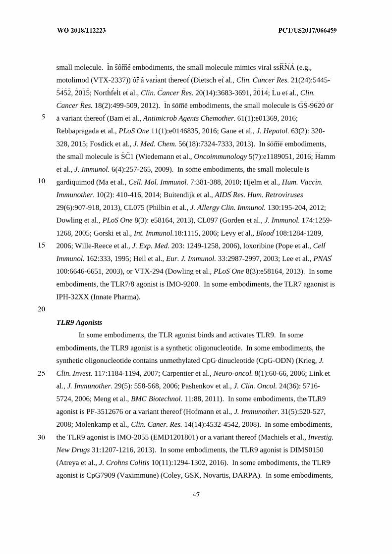
small molecule. In some embodiments, the small molecule mimics viral ssRNA (e.g.,
motolimod (VTX-2337)) or a variant thereof (Dietsch et al., Clin. Cancer Res. 21(24):5445-
5452, 2015; Northfelt et al., Clin. Cancer Res. 20(14):3683-3691, 2014; Lu et al., Clin.
Cancer Res. 18(2):499-509, 2012). In some embodiments, the small molecule is GS-9620 or
a variant thereof (Bam et al., Antimicrob Agents Chemother. 61(1):e01369, 2016;
Rebbapragada et al., PLoS One 11(1):e0146835, 2016; Gane et al., J. Hepatol. 63(2): 320-
328, 2015; Fosdick et al., J. Med. Chem. 56(18):7324-7333, 2013). In some embodiments,
the small molecule is SC1 (Wiedemann et al., Oncoimmunology 5(7):e1189051, 2016; Hamm
et al., J. Immunol. 6(4):257-265, 2009). In some embodiments, the small molecule is
gardiquimod (Ma et al., Cell. Mol. Immunol. 7:381-388, 2010; Hjelm et al., Hum. Vaccin.
Immunother. 10(2): 410-416, 2014; Buitendijk et al., AIDS Res. Hum. Retroviruses
29(6):907-918, 2013), CL075 (Philbin et al., J. Allergy Clin. Immunol. 130:195-204, 2012;
Dowling et al., PLoS One 8(3): e58164, 2013), CL097 (Gorden et al., J. Immunol. 174:1259-
1268, 2005; Gorski et al., Int. Immunol.18:1115, 2006; Levy et al., Blood 108:1284-1289,
2006; Wille-Reece et al., J. Exp. Med. 203: 1249-1258, 2006), loxoribine (Pope et al., Cell
Immunol. 162:333, 1995; Heil et al., Eur. J. Immunol. 33:2987-2997, 2003; Lee et al., PNAS
100:6646-6651, 2003), or VTX-294 (Dowling et al., PLoS One 8(3):e58164, 2013). In some
embodiments, the TLR7/8 agonist is IMO-9200. In some embodiments, the TLR7 agaonist is
IPH-32XX (Innate Pharma).
TLR9 Agonists
In some embodiments, the TLR agonist binds and activates TLR9. In some
embodiments, the TLR9 agonist is a synthetic oligonucleotide. In some embodiments, the
synthetic oligonucleotide contains unmethylated CpG dinucleotide (CpG-ODN) (Krieg, J.
Clin. Invest. 117:1184-1194, 2007; Carpentier et al., Neuro-oncol. 8(1):60-66, 2006; Link et
al., J. Immunother. 29(5): 558-568, 2006; Pashenkov et al., J. Clin. Oncol. 24(36): 5716-
5724, 2006; Meng et al., BMC Biotechnol. 11:88, 2011). In some embodiments, the TLR9
agonist is PF-3512676 or a variant thereof (Hofmann et al., J. Immunother. 31(5):520-527,
2008; Molenkamp et al., Clin. Caner. Res. 14(14):4532-4542, 2008). In some embodiments,
the TLR9 agonist is IMO-2055 (EMD1201801) or a variant thereof (Machiels et al., Investig.
New Drugs 31:1207-1216, 2013). In some embodiments, the TLR9 agonist is DIMS0150
(Atreya et al., J. Crohns Colitis 10(11):1294-1302, 2016). In some embodiments, the TLR9
agonist is CpG7909 (Vaximmune) (Coley, GSK, Novartis, DARPA). In some embodiments,
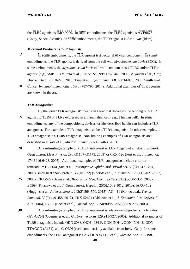
the TLR9 agonist is IMO-9200. In some embodiments, the TLR9 agonist is AVE0675
(Coley, Sanofi Aventis). In some embodiments, the TLR9 agonist is Amplivax (Idera).
Microbial Products as TLR Agonists
In some embodiments, the TLR agonist is a bacterial or viral component. In some
embodiments, the TLR agonist is derived from the cell wall Mycobacterium bovis (BCG). In
some embodiments, the Mycobacterium bovis cell wall component is a TLR2 and/or TLR4
agonist (e.g., SMP105 (Murata et al., Cancer Sci. 99:1435-1440, 2008; Miyauchi et al., Drug
Discov. Ther. 6: 218-225, 2013; Tsuji et al., Infect Immun. 68: 6883-6890, 2000; Smith et al.,
Cancer Immunol. Immunother. 63(8):787-796, 2014). Additional examples of TLR agonists
are known in the art.
TLR Antagonists
By the term “TLR antagonist” means an agent that decreases the binding of a TLR
agonist to TLR4 or TLR9 expressed in a mammalian cell (e.g., a human cell). In some
embodiments, any of the compositions, devices, or kits described herein can include a TLR
antagonist. For example, a TLR antagonist can be a TLR4 antagonist. In other examples, a
TLR antagonist is a TLR9 antagonist. Non-limiting examples of TLR antagonists are
described in Fukata et al., Mucosal Immunity 6:451-463, 2013.
A non-limiting example of a TLR4 antagonist is 1A6 (Ungaro et al., Am. J. Physiol.
Gastrointest. Liver Physiol. 296:G1167-G1179, 2009) or CRX-526 (Fort et al., J. Immunol.
174:6416-6423, 2005). Additional examples of TLR4 antagonists include eritoran
tetrasodium (E5564) (Sun et al., Investigative Ophthalmol. Visual Sci. 50(3):1247-1254,
2009), small heat shock protein B8 (HSP22) (Roelofs et al., J. Immunol. 176(11):7021-7027,
2006), CRX-527 (Bazin et al., Bioorganic Med. Chem. Letters 18(2):5350-5354, 2008),
E5564 (Kitazawa et al., J. Gastroentrol. Hepatol. 25(5):1009-1012, 2010), IAXO-102
(Huggins et al., Atherosclerosis 242(2):563-570, 2015), AG-411 (Kondo et al., Trends
Immunol. 33(9):449-458, 2012), CRX-52624 (Alderson et al., J. Endotoxin Res. 12(5):313-
319, 2006), E5531 (Becker et al., Toxicol. Appl. Pharmacol. 207(2):269-275, 2005).
A non-limiting example of a TLR9 antagonist is adenoviral oligodeoxynucleotides
(AV-ODN) (Obermeier et al., Gastroenterology 129:913-927, 2005). Additional examples of
TLR9 antagonists include ODN 2088, ODN 4084-F, ODN INH-1, ODN INH-18, ODN
TTAGGG (A151), and G-ODN (each commercially available from InvivoGen). In some
embodiments, the TLR9 antagonist is CpG-ODN c41 (Li et al., Vaccine 29:2193-2198,
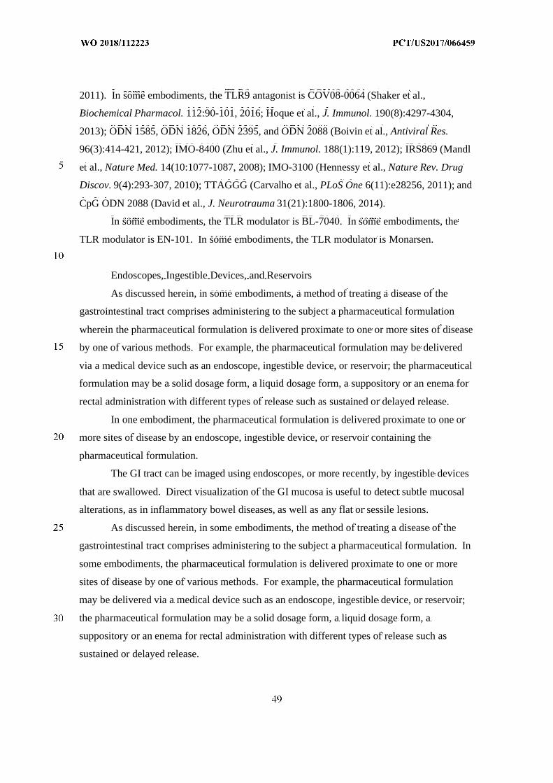
2011). In some embodiments, the TLR9 antagonist is COV08-0064 (Shaker et al.,
Biochemical Pharmacol. 112:90-101, 2016; Hoque et al., J. Immunol. 190(8):4297-4304,
2013); ODN 1585, ODN 1826, ODN 2395, and ODN 2088 (Boivin et al., Antiviral Res.
96(3):414-421, 2012); IMO-8400 (Zhu et al., J. Immunol. 188(1):119, 2012); IRS869 (Mandl
et al., Nature Med. 14(10:1077-1087, 2008); IMO-3100 (Hennessy et al., Nature Rev. Drug
Discov. 9(4):293-307, 2010); TTAGGG (Carvalho et al., PLoS One 6(11):e28256, 2011); and
CpG ODN 2088 (David et al., J. Neurotrauma 31(21):1800-1806, 2014).
In some embodiments, the TLR modulator is BL-7040. In some embodiments, the
TLR modulator is EN-101. In some embodiments, the TLR modulator is Monarsen.
Endoscopes, Ingestible Devices, and Reservoirs
As discussed herein, in some embodiments, a method of treating a disease of the
gastrointestinal tract comprises administering to the subject a pharmaceutical formulation
wherein the pharmaceutical formulation is delivered proximate to one or more sites of disease
by one of various methods. For example, the pharmaceutical formulation may be delivered
via a medical device such as an endoscope, ingestible device, or reservoir; the pharmaceutical
formulation may be a solid dosage form, a liquid dosage form, a suppository or an enema for
rectal administration with different types of release such as sustained or delayed release.
In one embodiment, the pharmaceutical formulation is delivered proximate to one or
more sites of disease by an endoscope, ingestible device, or reservoir containing the
pharmaceutical formulation.
The GI tract can be imaged using endoscopes, or more recently, by ingestible devices
that are swallowed. Direct visualization of the GI mucosa is useful to detect subtle mucosal
alterations, as in inflammatory bowel diseases, as well as any flat or sessile lesions.
As discussed herein, in some embodiments, the method of treating a disease of the
gastrointestinal tract comprises administering to the subject a pharmaceutical formulation. In
some embodiments, the pharmaceutical formulation is delivered proximate to one or more
sites of disease by one of various methods. For example, the pharmaceutical formulation
may be delivered via a medical device such as an endoscope, ingestible device, or reservoir;
the pharmaceutical formulation may be a solid dosage form, a liquid dosage form, a
suppository or an enema for rectal administration with different types of release such as
sustained or delayed release.
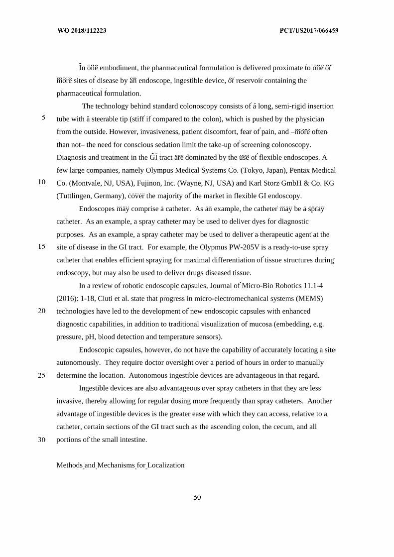
In one embodiment, the pharmaceutical formulation is delivered proximate to one or
more sites of disease by an endoscope, ingestible device, or reservoir containing the
pharmaceutical formulation.
The technology behind standard colonoscopy consists of a long, semi-rigid insertion
tube with a steerable tip (stiff if compared to the colon), which is pushed by the physician
from the outside. However, invasiveness, patient discomfort, fear of pain, and –more often
than not– the need for conscious sedation limit the take-up of screening colonoscopy.
Diagnosis and treatment in the GI tract are dominated by the use of flexible endoscopes. A
few large companies, namely Olympus Medical Systems Co. (Tokyo, Japan), Pentax Medical
Co. (Montvale, NJ, USA), Fujinon, Inc. (Wayne, NJ, USA) and Karl Storz GmbH & Co. KG
(Tuttlingen, Germany), cover the majority of the market in flexible GI endoscopy.
Endoscopes may comprise a catheter. As an example, the catheter may be a spray
catheter. As an example, a spray catheter may be used to deliver dyes for diagnostic
purposes. As an example, a spray catheter may be used to deliver a therapeutic agent at the
site of disease in the GI tract. For example, the Olypmus PW-205V is a ready-to-use spray
catheter that enables efficient spraying for maximal differentiation of tissue structures during
endoscopy, but may also be used to deliver drugs diseased tissue.
In a review of robotic endoscopic capsules, Journal of Micro-Bio Robotics 11.1-4
(2016): 1-18, Ciuti et al. state that progress in micro-electromechanical systems (MEMS)
technologies have led to the development of new endoscopic capsules with enhanced
diagnostic capabilities, in addition to traditional visualization of mucosa (embedding, e.g.
pressure, pH, blood detection and temperature sensors).
Endoscopic capsules, however, do not have the capability of accurately locating a site
autonomously. They require doctor oversight over a period of hours in order to manually
determine the location. Autonomous ingestible devices are advantageous in that regard.
Ingestible devices are also advantageous over spray catheters in that they are less
invasive, thereby allowing for regular dosing more frequently than spray catheters. Another
advantage of ingestible devices is the greater ease with which they can access, relative to a
catheter, certain sections of the GI tract such as the ascending colon, the cecum, and all
portions of the small intestine.
Methods and Mechanisms for Localization
In addition to, or as an alternative, to directly visualizing the GI tract, one or more
different mechanisms can be used to determine the location of an ingestible device within the
GI tract. Various implementations may be used for localization of ingestible devices within
the GI tract.
For example, various implementations may be used for localization of ingestible
devices within the GI tract. For example, certain implementations can include one or more
electromagnetic sensor coils, magnetic fields, electromagnetic waves, electric potential
values, ultrasound positioning systems, gamma scintigraphy techniques or other radio-tracker
technology have been described by others. Alternatively, imaging can be used to localize, for
example, using anatomical landmarks or more complex algorithms for 3D reconstruction
based on multiple images. Other technologies rely on radio frequency, which relies on
sensors placed externally on the body to receive the strength of signals emitted by the
capsule. Ingestible devices may also be localized based on reflected light in the medium
surrounding the device; pH; temperature; time following ingestion; and/or acoustic signals.
The disclosure provides an ingestible device, as well as related systems and methods
that provide for determining the position of the ingestible device within the GI tract of a
subject with very high accuracy. In some embodiments, the ingestible device can
autonomously determine its position within the GI tract of the subject.
Typically, the ingestible device includes one or more processing devices, and one
more machine readable hardware storage devices. In some embodiments, the one or more
machine readable hardware storage devices store instructions that are executable by the one
or more processing devices to determine the location of the ingestible device in a portion of a
GI tract of the subject. In certain embodiments, the one or more machine readable hardware
storage devices store instructions that are executable by the one or more processing devices to
transmit data to an external device (e.g., a base station external to the subject, such as a base
station carried on an article worn by the subject) capable of implementing the data to
determine the location of the device within the GI tract of the subject.
In some embodiments, the location of the ingestible device within the GI tract of the
subject can be determined to an accuracy of at least 85%, e.g., at least 90%, at least 95%, at
least 97%, at least 98%, at least 99%, 100%. In some embodiments, the location of the
ingestible device within the GI tract of the subject can be determined to an accuracy of at
least 85%, e.g., at least 90%, at least 95%, at least 97%, at least 98%, at least 99%, 100%. In
such embodiments, the portion of the GI tract of the subject can include, for example, the
esophagus, the stomach, duodenum, the jejunum, and/or the terminal ileum, cecum and colon.
An exemplary and non-limiting embodiment is provided below in Example 13.
In certain embodiments, the location of the ingestible device within the esophagus of
the subject can be determined to an accuracy of at least 85%, e.g., at least 90%, at least 95%,
at least 97%, at least 98%, at least 99%, 100%. An exemplary and non-limiting embodiment
is provided below in Example 13.
In some embodiments, the location of the ingestible device within the stomach of the
subject can be determined to an accuracy of at least 85%, e.g., at least 90%, at least 95%, at
least 97%, at least 98%, at least 99%, 100%. An exemplary and non-limiting embodiment is
provided below in Example 13.
In certain embodiments, the location of the ingestible device within the duodenum of
the subject can be determined to an accuracy of at least 85%, e.g., at least 90%, at least 95%,
at least 97%, at least 98%, at least 99%, 100%. An exemplary and non-limiting embodiment
is provided below in Example 13.
In some embodiments, the location of the ingestible device within the jejunum of the
subject can be determined to an accuracy of at least 85%, e.g., at least 90%, at least 95%, at
least 97%, at least 98%, at least 99%, 100%. An exemplary and non-limiting embodiment is
provided below in Example 13.
In certain embodiments, the location of the ingestible device within the terminal
ileum, cecum and colon of the subject can be determined to an accuracy of at least 85%, e.g.,
at least 90%, at least 95%, at least 97%, at least 98%, at least 99%, 100%.
In some embodiments, the location of the ingestible device within the cecum of the
subject can be determined to an accuracy of at least 85%, e.g., at least 90%, at least 95%, at
least 97%, at least 98%, at least 99%, 100%. An exemplary and non-limiting embodiment is
provided below in Example 13. In such embodiments, the portion of the portion of the GI
tract of the subject can include, for example, the esophagus, the stomach, duodenum, the
jejunum, and/or the terminal ileum, cecum and colon.
In certain embodiments, the location of the ingestible device within the esophagus of
the subject can be determined to an accuracy of at least 85%, e.g., at least 90%, at least 95%,
at least 97%, at least 98%, at least 99%, 100%.
In some embodiments, the location of the ingestible device within the stomach of the
subject can be determined to an accuracy of at least 85%, e.g., at least 90%, at least 95%, at
least 97%, at least 98%, at least 99%, 100%.
In certain embodiments, the location of the ingestible device within the duodenum of
the subject can be determined to an accuracy of at least 85%, e.g., at least 90%, at least 95%,
at least 97%, at least 98%, at least 99%, 100%.
In some embodiments, the location of the ingestible device within the jejunum of the
subject can be determined to an accuracy of at least 85%, e.g., at least 90%, at least 95%, at
least 97%, at least 98%, at least 99%, 100%.
In certain embodiments, the location of the ingestible device within the terminal
ileum, cecum and colon of the subject can be determined to an accuracy of at least 85%, e.g.,
at least 90%, at least 95%, at least 97%, at least 98%, at least 99%, 100%.
In some embodiments, the location of the ingestible device within the cecum of the
subject can be determined to an accuracy of at least 85%, e.g., at least 90%, at least 95%, at
least 97%, at least 98%, at least 99%, 100%.
As used herein, the term “reflectance” refers to a value derived from light emitted by
the device, reflected back to the device, and received by a detector in or on the device. For
example, in some embodiments this refers to light emitted by the device, wherein a portion of
the light is reflected by a surface external to the device, and the light is received by a detector
located in or on the device.
As used herein, the term “illumination” refers to any electromagnetic emission. In
some embodiments, an illumination may be within the range of Infrared Light (IR), the
visible spectrum and ultraviolet light (UV), and an illumination may have a majority of its
power centered at a particular wavelength in the range of 100nm to 1000nm. In some
embodiments, it may be advantageous to use an illumination with a majority of its power
limited to one of the infrared (750nm-1000nm), red (600nm-750nm), green (495nm-600nm),
blue (400nm-495nm), or ultraviolet (100nm-400nm) spectrums. In some embodiments a
plurality of illuminations with different wavelengths may be used. For illustrative purposes,
the embodiments described herein may refer to the use of green or blue spectrums of light.
However, it is understood that these embodiments may use any suitable light having a
wavelength that is substantially or approximately within the green or blue spectra defined
above, and the localization systems and methods described herein may use any suitable
spectra of light.
Referring now to FIG. 1, shown therein is a view of an example embodiment of an
ingestible device 100, which may be used to identify a location within a gastrointestinal (GI)
tract. In some embodiments, ingestible device 100 may be configured to autonomously
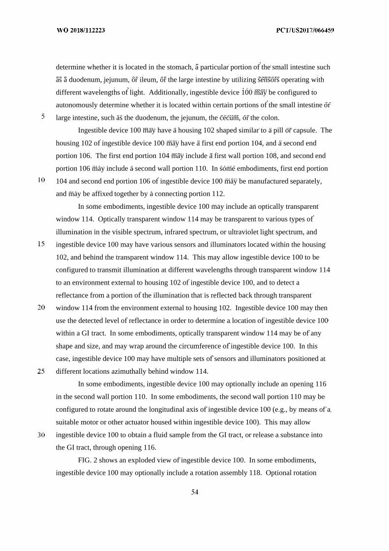
determine whether it is located in the stomach, a particular portion of the small intestine such
as a duodenum, jejunum, or ileum, or the large intestine by utilizing sensors operating with
different wavelengths of light. Additionally, ingestible device 100 may be configured to
autonomously determine whether it is located within certain portions of the small intestine or
large intestine, such as the duodenum, the jejunum, the cecum, or the colon.
Ingestible device 100 may have a housing 102 shaped similar to a pill or capsule. The
housing 102 of ingestible device 100 may have a first end portion 104, and a second end
portion 106. The first end portion 104 may include a first wall portion 108, and second end
portion 106 may include a second wall portion 110. In some embodiments, first end portion
104 and second end portion 106 of ingestible device 100 may be manufactured separately,
and may be affixed together by a connecting portion 112.
In some embodiments, ingestible device 100 may include an optically transparent
window 114. Optically transparent window 114 may be transparent to various types of
illumination in the visible spectrum, infrared spectrum, or ultraviolet light spectrum, and
ingestible device 100 may have various sensors and illuminators located within the housing
102, and behind the transparent window 114. This may allow ingestible device 100 to be
configured to transmit illumination at different wavelengths through transparent window 114
to an environment external to housing 102 of ingestible device 100, and to detect a
reflectance from a portion of the illumination that is reflected back through transparent
window 114 from the environment external to housing 102. Ingestible device 100 may then
use the detected level of reflectance in order to determine a location of ingestible device 100
within a GI tract. In some embodiments, optically transparent window 114 may be of any
shape and size, and may wrap around the circumference of ingestible device 100. In this
case, ingestible device 100 may have multiple sets of sensors and illuminators positioned at
different locations azimuthally behind window 114.
In some embodiments, ingestible device 100 may optionally include an opening 116
in the second wall portion 110. In some embodiments, the second wall portion 110 may be
configured to rotate around the longitudinal axis of ingestible device 100 (e.g., by means of a
suitable motor or other actuator housed within ingestible device 100). This may allow
ingestible device 100 to obtain a fluid sample from the GI tract, or release a substance into
the GI tract, through opening 116.
FIG. 2 shows an exploded view of ingestible device 100. In some embodiments,
ingestible device 100 may optionally include a rotation assembly 118. Optional rotation
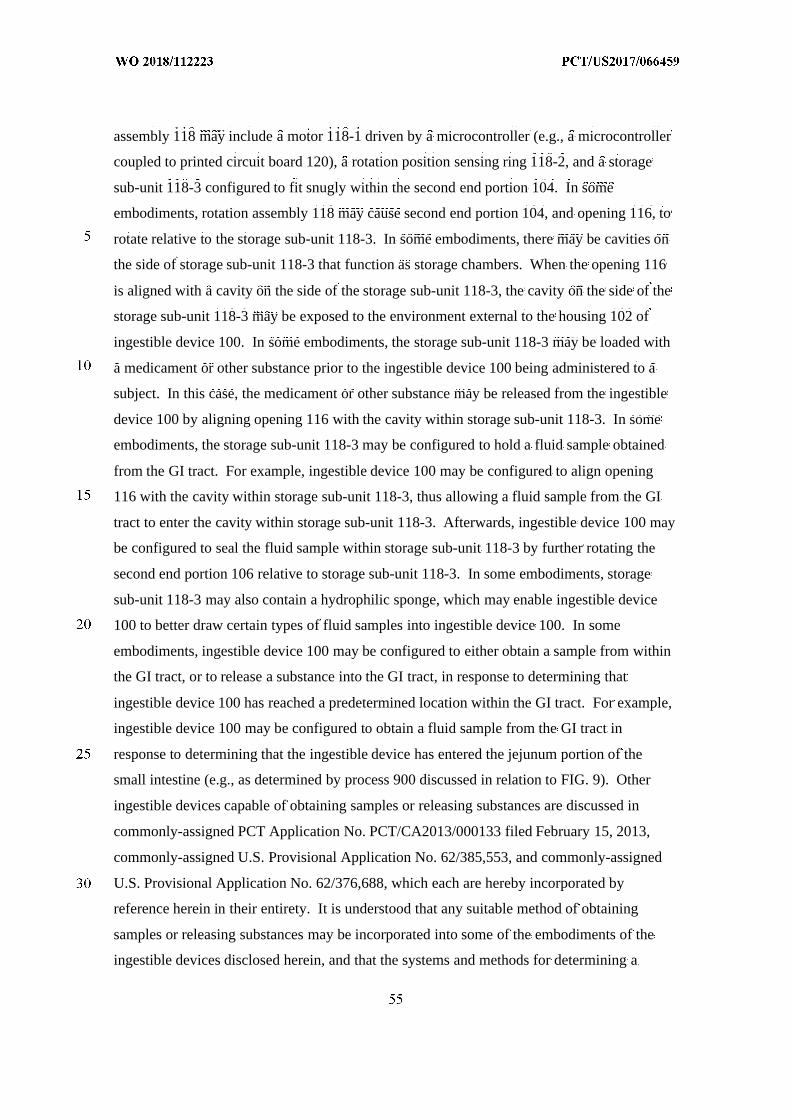
assembly 118 may include a motor 118-1 driven by a microcontroller (e.g., a microcontroller
coupled to printed circuit board 120), a rotation position sensing ring 118-2, and a storage
sub-unit 118-3 configured to fit snugly within the second end portion 104. In some
embodiments, rotation assembly 118 may cause second end portion 104, and opening 116, to
rotate relative to the storage sub-unit 118-3. In some embodiments, there may be cavities on
the side of storage sub-unit 118-3 that function as storage chambers. When the opening 116
is aligned with a cavity on the side of the storage sub-unit 118-3, the cavity on the side of the
storage sub-unit 118-3 may be exposed to the environment external to the housing 102 of
ingestible device 100. In some embodiments, the storage sub-unit 118-3 may be loaded with
a medicament or other substance prior to the ingestible device 100 being administered to a
subject. In this case, the medicament or other substance may be released from the ingestible
device 100 by aligning opening 116 with the cavity within storage sub-unit 118-3. In some
embodiments, the storage sub-unit 118-3 may be configured to hold a fluid sample obtained
from the GI tract. For example, ingestible device 100 may be configured to align opening
116 with the cavity within storage sub-unit 118-3, thus allowing a fluid sample from the GI
tract to enter the cavity within storage sub-unit 118-3. Afterwards, ingestible device 100 may
be configured to seal the fluid sample within storage sub-unit 118-3 by further rotating the
second end portion 106 relative to storage sub-unit 118-3. In some embodiments, storage
sub-unit 118-3 may also contain a hydrophilic sponge, which may enable ingestible device
100 to better draw certain types of fluid samples into ingestible device 100. In some
embodiments, ingestible device 100 may be configured to either obtain a sample from within
the GI tract, or to release a substance into the GI tract, in response to determining that
ingestible device 100 has reached a predetermined location within the GI tract. For example,
ingestible device 100 may be configured to obtain a fluid sample from the GI tract in
response to determining that the ingestible device has entered the jejunum portion of the
small intestine (e.g., as determined by process 900 discussed in relation to FIG. 9). Other
ingestible devices capable of obtaining samples or releasing substances are discussed in
commonly-assigned PCT Application No. PCT/CA2013/000133 filed February 15, 2013,
commonly-assigned U.S. Provisional Application No. 62/385,553, and commonly-assigned
U.S. Provisional Application No. 62/376,688, which each are hereby incorporated by
reference herein in their entirety. It is understood that any suitable method of obtaining
samples or releasing substances may be incorporated into some of the embodiments of the
ingestible devices disclosed herein, and that the systems and methods for determining a
location of an ingestible device may be incorporated into any suitable type of ingestible
device.
Ingestible device 100 may include a printed circuit board (PCB) 120, and a battery
128 configured to power PCB 120. PCB 120 may include a programmable microcontroller,
and control and memory circuitry for holding and executing firmware or software for
coordinating the operation of ingestible device 100, and the various components of ingestible
device 100. For example, PCB 120 may include memory circuitry for storing data, such as
data sets of measurements collected by sensing sub-unit 126, or instructions to be executed
by control circuitry to implement a localization process, such as, for example, one or more of
the processes, discussed herein, including those discussed below in connection with one or
more of the associated flow charts. PCB 120 may include a detector 122 and an illuminator
124, which together form sensing sub-unit 126. In some embodiments, control circuitry
within PCB 120 may include processing units, communication circuitry, or any other suitable
type of circuitry for operating ingestible device 100. For illustrative purposes, only a single
detector 122 and a single illuminator 124 forming a single sensing sub-unit 126 are shown.
However, it is understood that in some embodiments there may be multiple sensing sub-units,
each with a separate illuminator and detector, within ingestible device 100. For example,
there may be several sensing sub-units spaced azimuthally around the circumference of the
PCB 120, which may enable ingestible device 100 to transmit illumination and detect
reflectances or ambient light in all directions around the circumference of the device. In
some embodiments, sensing sub-unit 126 may be configured to generate an illumination
using illuminator 124, which is directed through the window 114 in a radial direction away
from ingestible device 100. This illumination may reflect off of the environment external to
ingestible device 100, and the reflected light coming back into ingestible device 100 through
window 114 may be detected as a reflectance by detector 122.
In some embodiments, window 114 may be of any suitable shape and size. For
example, window 114 may extend around a full circumference of ingestible device 100. In
some embodiments there may be a plurality of sensing sub-units (e.g., similar to sensing sub-
unit 126) located at different positions behind the window. For example, three sensing sub-
units may be positioned behind the window at the same longitudinal location, but spaced 120
degrees apart azimuthally. This may enable ingestible device 100 to transmit illuminations in
all directions radially around ingestible device 100, and to measure each of the corresponding
reflectances.
In some embodiments, illuminator 124 may be capable of producing illumination at a
variety of different wavelengths in the ultraviolet, infrared, or visible spectrum. For example,
illuminator 124 may be implemented by using Red-Green-Blue Light-Emitting diode
packages (RGB-LED). These types of RGB-LED packages are able to transmit red, blue, or
green illumination, or combinations of red, blue, or green illumination. Similarly, detector
122 may be configured to sense reflected light of the same wavelengths as the illumination
produced by illuminator 124. For example, if illuminator 124 is configured to produce red,
blue, or green illumination, detector 122 may be configured to detect different reflectances
produced by red, blue, or green illumination (e.g., through the use of an appropriately
configured photodiode). These detected reflectances may be stored by ingestible device 100
(e.g., within memory circuitry of PCB 120), and may then be used by ingestible device 100 in
determining a location of ingestible device 100 within the GI tract (e.g., through the use of
process 500 (FIG. 5), process 600 (FIG. 6), or process 900 (FIG. 9)).
It is understood that ingestible device 100 is intended to be illustrative, and not
limiting. It will be understood that modifications to the general shape and structure of the
various devices and mechanisms described in relation to FIG. 1 and FIG. 2 may be made
without significantly changing the functions and operations of the devices and mechanisms.
For example, ingestible device 100 may have a housing formed from a single piece of molded
plastic, rather than being divided into a first end portion 104 and a second end portion 106.
As an alternate example, the location of window 114 within ingestible device 100 may be
moved to some other location, such as the center of ingestible device 100, or to one of the
ends of ingestible device 100. Moreover, the systems and methods discussed in relation to
FIGS. 1-10 may be implemented on any suitable type of ingestible device, provided that the
ingestible device is capable of detecting reflectances or levels of illumination in some
capacity. For example, in some embodiments ingestible device 100 may be modified to
replace detector 122 with an image sensor, and the ingestible device may be configured to
measure relative levels of red, blue, or green light by decomposing a recorded image into its
individual spectral components. Other examples of ingestible devices with localization
capabilities, which may be utilized in order to implement the systems and methods discussed
in relation to FIG. 1-11, are discussed in co-owned PCT Application No.
PCT/US2015/052500 filed on September 25, 2015, which is hereby incorporated by
reference herein in its entirety. Furthermore, it should be noted that the features and
limitations described in any one embodiment may be applied to any other embodiment
herein, and the descriptions and examples relating to one embodiment may be combined with
any other embodiment in a suitable manner.
FIG. 3 is a diagram of an ingestible device during an example transit through a
gastrointestinal (GI) tract, in accordance with some embodiments of the disclosure.
Ingestible device 300 may include any portion of any other ingestible device discussed in this
disclosure (e.g., ingestible device 100 (FIG. 1)), and may be any suitable type of ingestible
device with localization capabilities. For example, ingestible device 300 may be one
embodiment of ingestible device 100 without the optional opening 116 (FIG. 1) or optional
rotation assembly 118 (FIG. 2)). In some embodiments, ingestible device 300 may be
ingested by a subject, and as ingestible device 300 traverses the GI tract, ingestible device
300 may be configured to determine its location within the GI tract. For example, the
movement of ingestible device 300 and the amount of light detected by ingestible device 300
(e.g., via detector 122 (FIG. 2)) may vary substantially depending on the location of
ingestible device 300 within the GI tract, and ingestible device 300 may be configured to use
this information to determine a location of ingestible device 300 within the GI tract. For
instance, ingestible device 300 may detect ambient light from the surrounding environment,
or reflectances based on illumination generated by ingestible device 300 (e.g., generated by
illuminator 124 (FIG. 1)), and use this information to determine a location of ingestible
device 300 through processes, such as described herein. The current location of ingestible
device 300, and the time that ingestible device 300 detected each transition between the
various portions of the GI tract, may then be stored by ingestible device 300 (e.g., in memory
circuitry of PCB 120 (FIG. 2)), and may be used for any suitable purpose.
Shortly after ingestible device 300 is ingested, ingestible device will traverse the
esophagus 302, which may connect the subject’s mouth to a stomach 306. In some
embodiments, ingestible device 300 may be configured to determine that it has entered the
esophagus portion GI tract by measuring the amount and type of light (e.g., via detector 122
(FIG. 2)) in the environment surrounding the ingestible device 300. For instance, ingestible
device 300 may detect higher levels of light in the visible spectrum (e.g., via detector 122
(FIG. 2)) while outside the subject’s body, as compared to the levels of light detected while
within the GI tract. In some embodiments, ingestible device 300 may have previously stored
data (e.g., on memory circuitry of PCB 120 (FIG. 2)) indicating a typical level of light
detected when outside of the body, and the ingestible device 300 may be configured to
determine that entry to the body has occurred when a detected level of light (e.g., detected via
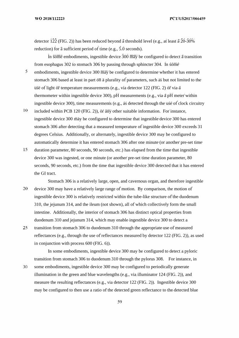
detector 122 (FIG. 2)) has been reduced beyond a threshold level (e.g., at least a 20-30%
reduction) for a sufficient period of time (e.g., 5.0 seconds).
In some embodiments, ingestible device 300 may be configured to detect a transition
from esophagus 302 to stomach 306 by passing through sphincter 304. In some
embodiments, ingestible device 300 may be configured to determine whether it has entered
stomach 306 based at least in part on a plurality of parameters, such as but not limited to the
use of light or temperature measurements (e.g., via detector 122 (FIG. 2) or via a
thermometer within ingestible device 300), pH measurements (e.g., via a pH meter within
ingestible device 300), time measurements (e.g., as detected through the use of clock circuitry
included within PCB 120 (FIG. 2)), or any other suitable information. For instance,
ingestible device 300 may be configured to determine that ingestible device 300 has entered
stomach 306 after detecting that a measured temperature of ingestible device 300 exceeds 31
degrees Celsius. Additionally, or alternately, ingestible device 300 may be configured to
automatically determine it has entered stomach 306 after one minute (or another pre-set time
duration parameter, 80 seconds, 90 seconds, etc.) has elapsed from the time that ingestible
device 300 was ingested, or one minute (or another pre-set time duration parameter, 80
seconds, 90 seconds, etc.) from the time that ingestible device 300 detected that it has entered
the GI tract.
Stomach 306 is a relatively large, open, and cavernous organ, and therefore ingestible
device 300 may have a relatively large range of motion. By comparison, the motion of
ingestible device 300 is relatively restricted within the tube-like structure of the duodenum
310, the jejunum 314, and the ileum (not shown), all of which collectively form the small
intestine. Additionally, the interior of stomach 306 has distinct optical properties from
duodenum 310 and jejunum 314, which may enable ingestible device 300 to detect a
transition from stomach 306 to duodenum 310 through the appropriate use of measured
reflectances (e.g., through the use of reflectances measured by detector 122 (FIG. 2)), as used
in conjunction with process 600 (FIG. 6)).
In some embodiments, ingestible device 300 may be configured to detect a pyloric
transition from stomach 306 to duodenum 310 through the pylorus 308. For instance, in
some embodiments, ingestible device 300 may be configured to periodically generate
illumination in the green and blue wavelengths (e.g., via illuminator 124 (FIG. 2)), and
measure the resulting reflectances (e.g., via detector 122 (FIG. 2)). Ingestible device 300
may be configured to then use a ratio of the detected green reflectance to the detected blue
reflectance to determine whether ingestible device 300 is located within the stomach 306, or
duodenum 310 (e.g., via process 600 (FIG. 6)). In turn, this may enable ingestible device 300
to detect a pyloric transition from stomach 306 to duodenum 310, an example of which is
discussed in relation to FIG. 6.
Similarly, in some embodiments, ingestible device 300 may be configured to detect a
reverse pyloric transition from duodenum 310 to stomach 306. Ingestible device 300 will
typically transition naturally from stomach 306 to duodenum 310, and onward to jejunum 314
and the remainder of the GI tract. However, similar to other ingested substances, ingestible
device 300 may occasionally transition from duodenum 310 back to stomach 306 as a result
of motion of the subject, or due to the natural behavior of the organs with the GI tract. To
accommodate this possibility, ingestible device 300 may be configured to continue to
periodically generate illumination in the green and blue wavelengths (e.g., via illuminator
124 (FIG. 2)), and measure the resulting reflectances (e.g., via detector 122 (FIG. 2)) to
detect whether or not ingestible device 300 has returned to stomach 306. An exemplary
detection process is described in additional detail in relation to FIG. 6.
After entering duodenum 310, ingestible device 300 may be configured to detect a
transition to the jejunum 314 through the duodenojejunal flexure 312. For example,
ingestible device 300 may be configured to use reflectances to detect peristaltic waves within
the jejunum 314, caused by the contraction of the smooth muscle tissue lining the walls of the
jejunum 314. In particular, ingestible device 300 may be configured to begin periodically
transmitting illumination (and measuring the resulting reflectances (e.g., via detector 122 and
illuminator 124 of sensing sub-unit 126 (FIG. 2)) at a sufficiently high frequency in order to
detect muscle contractions within the jejunum 314. Ingestible device 300 may then
determine that it has entered the jejunum 314 in response to having detected either a first
muscle contraction, or a predetermined number of muscle contractions (e.g., after having
detected three muscle contractions in sequence). The interaction of ingestible device 300
with the walls of jejunum 314 is also discussed in relation to FIG. 4, and an example of this
detection process is described in additional detail in relation to FIG. 9.
FIG. 4 is a diagram of an ingestible device during an example transit through a
jejunum, in accordance with some embodiments of the disclosure. Diagrams 410, 420, 430,
and 440 depict ingestible device 400 as it traverses through a jejunum (e.g., jejunum 314),
and how ingestible device 400 interacts with peristaltic waves formed by walls 406A and
406B (collectively, walls 406) of the jejunum. In some implementations, ingestible device
400 may include any portion of any other ingestible device discussed in this disclosure (e.g.,
ingestible device 100 (FIG. 1) or ingestible device 300 (FIG. 3)), and may be any suitable
type of ingestible device with localization capabilities. For example, ingestible device 400
may be substantially similar to the ingestible device 300 (FIG. 3) or ingestible device 100
(FIG. 1), with window 404 being the same as window 114 (FIG. 1), and sensing sub-unit 402
being the same as sensing sub-unit 126 (FIG. 2).
Diagram 410 depicts ingestible device 400 within the jejunum, when the walls 406 of
the jejunum are relaxed. In some embodiments, the confined tube-like structure of the
jejunum naturally causes ingestible device 400 to be oriented longitudinally along the length
of the jejunum, with window 404 facing walls 406. In this orientation, ingestible device 400
may use sensing sub-unit 402 to generate illumination (e.g., via illuminator 124 (FIG. 2))
oriented towards walls 406, and to detect the resulting reflectances (e.g., via detector 122
(FIG. 2)) from the portion of the illumination reflected off of walls 406 and back through
window 404. In some embodiments, ingestible device 400 may be configured to use sensing
sub-unit 402 to generate illumination and measure the resulting reflectance with sufficient
frequency to detect peristaltic waves within the jejunum. For instance, in a healthy human
subject, peristaltic waves may occur at a rate of approximately 0.1 Hz to 0.2 Hz. Therefore,
the ingestible device 400 may be configured to generate illumination and measure the
resulting reflectance at least once every 2.5 seconds (i.e., the minimum rate necessary to
detect a 0.2 Hz signal), and preferably at a higher rate, such as once every 0.5 seconds, which
may improve the overall reliability of the detection process due to more data points being
available. It is understood that the ingestible device 400 need not gather measurements at
precise intervals, and in some embodiments the ingestible device 400 may be adapted to
analyze data gathered at more irregular intervals, provided that there are still a sufficient
number of appropriately spaced data points to detect 0.1 Hz to 0.2 Hz signals.
Diagram 420 depicts ingestible device 400 within the jejunum, when the walls 406 of
the jejunum begin to contract and form a peristaltic wave. Diagram 420 depicts contracting
portion 408A of wall 406A and contracting portion 408B of wall 406B (collectively,
contracting portion 408 of wall 406) that form a peristaltic wave within the jejunum. The
peristaltic wave proceeds along the length of the jejunum as different portions of wall 406
contract and relax, causing it to appear as if contracting portions 408 of wall 406 proceed
along the length of the jejunum (i.e., as depicted by contracting portions 408 proceeding from
left to right in diagrams 410-430). While in this position, ingestible device 400 may detect a
similar level of reflectance (e.g., through the use of illuminator 124 and detector 122 of
sensing sub-unit 126 (FIG. 2)) as detected when there is no peristaltic wave occurring (e.g.,
as detected when ingestible device 400 is in the position indicated in diagram 410).
Diagram 430 depicts ingestible device 400 within the jejunum, when the walls 406 of
the jejunum continue to contract, squeezing around ingestible device 400. As the peristaltic
wave proceeds along the length of the jejunum, contracting portions 408 of wall 406 may
squeeze tightly around ingestible device 400, bringing the inner surface of wall 406 into
contact with window 404. While in this position, ingestible device 400 may detect a change
in a reflectance detected as a result of illumination produced by sensing sub-unit 402. The
absolute value of the change in the measured reflectance may depend on several factors, such
as the optical properties of the window 404, the spectral components of the illumination, and
the optical properties of the walls 406. However, ingestible device 400 may be configured to
store a data set with the reflectance values over time, and search for periodic changes in the
data set consistent with the frequency of the peristaltic waves (e.g., by analyzing the data set
in the frequency domain, and searching for peaks between 0.1 Hz to 0.2 Hz). This may
enable ingestible device 400 to detect muscle contractions due to peristaltic waves without
foreknowledge of the exact changes in reflectance signal amplitude that may occur as a result
of detecting the muscle contractions of the peristaltic wave. An example procedure for
detecting muscle contractions is discussed further in relation to FIG. 9, and an example of a
reflectance data set gathered while ingestible device 400 is located within the jejunum is
discussed in relation to FIG. 10.
Diagram 440 depicts ingestible device 400 within the jejunum, when the peristaltic
wave has moved past ingestible device 400. Diagram 440 depicts contracting portions 408
that form the peristaltic wave within the jejunum having moved past the end of ingestible
device 400. The peristaltic wave proceeds along the length of the jejunum as different
portions of wall 406 contract and relax, causing it to appear as if contracting portions 408 of
wall 406 proceed along the length of the jejunum (i.e., as depicted by contracting portions
408 proceeding from left to right in diagrams 410-430). While in this position, ingestible
device 400 may detect a similar level of reflectance (e.g., through the use of illuminator 124
and detector 122 of sensing sub-unit 126 (FIG. 2)) as detected when there is no peristaltic
wave occurring (e.g., as detected when ingestible device 400 is in the position indicated in
diagram 410, or diagram 420).
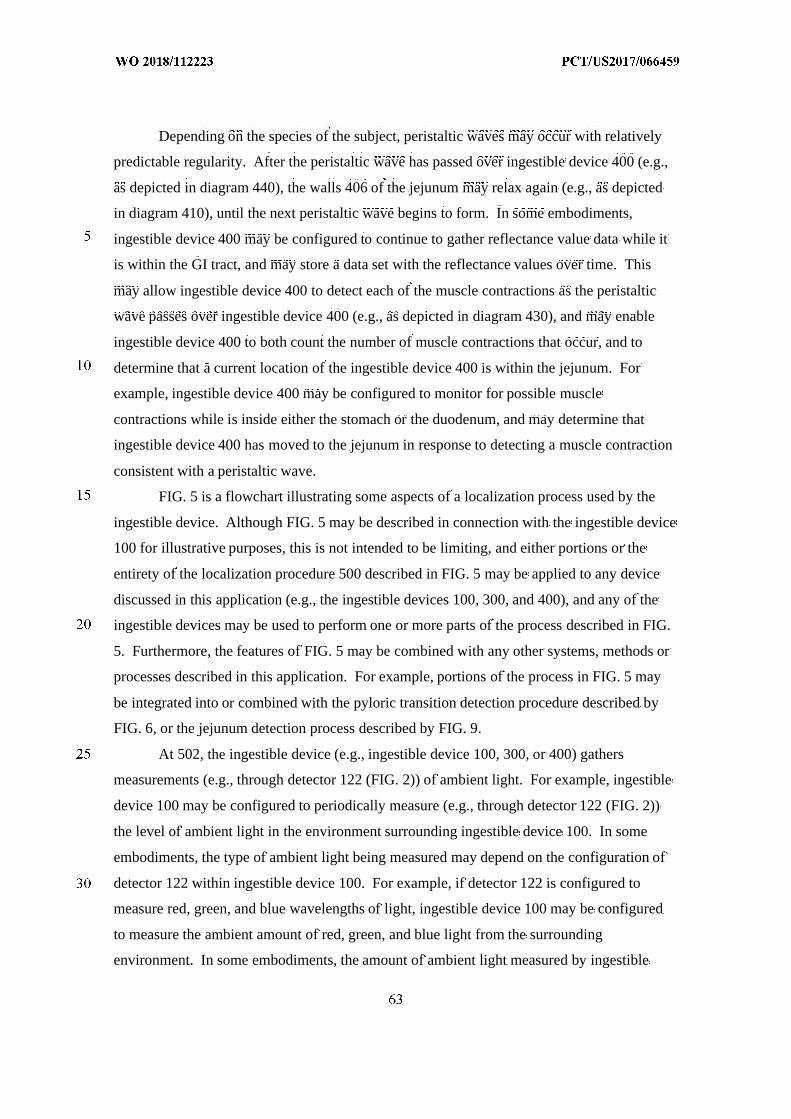
Depending on the species of the subject, peristaltic waves may occur with relatively
predictable regularity. After the peristaltic wave has passed over ingestible device 400 (e.g.,
as depicted in diagram 440), the walls 406 of the jejunum may relax again (e.g., as depicted
in diagram 410), until the next peristaltic wave begins to form. In some embodiments,
ingestible device 400 may be configured to continue to gather reflectance value data while it
is within the GI tract, and may store a data set with the reflectance values over time. This
may allow ingestible device 400 to detect each of the muscle contractions as the peristaltic
wave passes over ingestible device 400 (e.g., as depicted in diagram 430), and may enable
ingestible device 400 to both count the number of muscle contractions that occur, and to
determine that a current location of the ingestible device 400 is within the jejunum. For
example, ingestible device 400 may be configured to monitor for possible muscle
contractions while is inside either the stomach or the duodenum, and may determine that
ingestible device 400 has moved to the jejunum in response to detecting a muscle contraction
consistent with a peristaltic wave.
FIG. 5 is a flowchart illustrating some aspects of a localization process used by the
ingestible device. Although FIG. 5 may be described in connection with the ingestible device
100 for illustrative purposes, this is not intended to be limiting, and either portions or the
entirety of the localization procedure 500 described in FIG. 5 may be applied to any device
discussed in this application (e.g., the ingestible devices 100, 300, and 400), and any of the
ingestible devices may be used to perform one or more parts of the process described in FIG.
5. Furthermore, the features of FIG. 5 may be combined with any other systems, methods or
processes described in this application. For example, portions of the process in FIG. 5 may
be integrated into or combined with the pyloric transition detection procedure described by
FIG. 6, or the jejunum detection process described by FIG. 9.
At 502, the ingestible device (e.g., ingestible device 100, 300, or 400) gathers
measurements (e.g., through detector 122 (FIG. 2)) of ambient light. For example, ingestible
device 100 may be configured to periodically measure (e.g., through detector 122 (FIG. 2))
the level of ambient light in the environment surrounding ingestible device 100. In some
embodiments, the type of ambient light being measured may depend on the configuration of
detector 122 within ingestible device 100. For example, if detector 122 is configured to
measure red, green, and blue wavelengths of light, ingestible device 100 may be configured
to measure the ambient amount of red, green, and blue light from the surrounding
environment. In some embodiments, the amount of ambient light measured by ingestible
device 100 will be larger in the area external to the body (e.g., a well-lit room where
ingestible device 100 is being administered to a subject) and in the oral cavity of the subject,
as compared to the ambient level of light measured by ingestible device 100 when inside of
an esophagus, stomach, or other portion of the GI tract (e.g., esophagus 302, stomach 306,
duodenum 310, or jejunum 314 (FIG. 3)).
At 504, the ingestible device (e.g., ingestible device 100, 300, or 400) determines
(e.g., via control circuitry within PCB 120 (FIG. 2)) whether the ingestible device has
detected entry into the GI tract. For example, ingestible device 100 may be configured to
determine when the most recent measurement of ambient light (e.g., the measurement
gathered at 502) indicates that the ingestible device has entered the GI tract. For instance, the
first time that ingestible device 100 gatherers a measurement of ambient light at 502,
ingestible device 100 may store that measurement (e.g., via storage circuitry within PCB 120
(FIG. 2)) as a typical level of ambient light external to the body. Ingestible device 100 may
be configured to then compare the most recent measurement of ambient light to the typical
level of ambient light external to the body (e.g., via control circuitry within PCB 120 (FIG.
2)), and determine that ingestible device 100 has entered the GI tract when the most recent
measurement of ambient light is substantially smaller than the typical level of ambient light
external to the body. For example, ingestible device 100 may be configured to detect that it
has entered the GI tract in response to determining that the most recent measurement of
ambient light is less than or equal to 20% of the typical level of ambient light external to the
body. If ingestible device 100 determines that it has detected entry into the GI tract (e.g., that
ingestible device 100 has entered at least the esophagus 302 (FIG. 3)), process 500 proceeds
to 506. Alternately, if ingestible device 100 determines that it has not detected entry into the
GI tract (e.g., as a result of the most recent measurement being similar to the typical level of
ambient light external to the body), process 500 proceeds back to 502 where the ingestible
device 100 gathers further measurements. For instance, ingestible device 100 may be
configured to wait a predetermined amount of time (e.g., five seconds, ten seconds, etc.), and
then gather another measurement of the level of ambient light from the environment
surrounding ingestible device 100.
At 506, the ingestible device (e.g., ingestible device 100, 300, or 400) waits for a
transition from the esophagus to the stomach (e.g., from esophagus 302 to stomach 306 (FIG.
3)). For example, ingestible device 100 may be configured to determine that it has entered
the stomach (e.g., stomach 306 (FIG. 3)) after waiting a predetermined period of time after
having entered the GI tract. For instance, a typical esophageal transit time in a human patient
may be on the order of 15-30 seconds. In this case, after having detected that ingestible
device 100 has entered the GI tract at 504 (i.e., after detecting that ingestible device 100 has
reached at least esophagus 302 (FIG. 3)), ingestible device 100 may be configured to wait
one minute, or a similar amount of time longer than the typical esophageal transmit time
(e.g., ninety-seconds), before automatically determining that ingestible device 100 has
entered at least the stomach (e.g., stomach 306 (FIG. 3)).
In some embodiments, the ingestible device (e.g., ingestible device 100, 300, or 400)
may also determine it has entered the stomach based on measurements of pH or temperature.
For example, ingestible device 100 may be configured to determine that it has entered the
stomach if a temperature of ingestible device has increased to at least 31 degrees Celsius (i.e.,
consistent with the temperature inside the stomach), or if a measured pH of the environment
surrounding ingestible device 100 is sufficiently acidic (i.e., consistent with the acidic nature
of gastric juices that may be found inside the stomach).
At 508, the ingestible device (e.g., ingestible device 100, 300, or 400) stores data
indicating the ingestible device has entered the stomach (e.g., stomach 306 (FIG. 3)). For
example, after having waited a sufficient amount of time at 506, ingestible device 100 may
store data (e.g., within storage circuitry of PCB 120 (FIG. 2)) indicative of ingestible device
100 having entered at least the stomach. Once ingestible device 100 reaches at least the
stomach, process 500 proceeds to 510 where ingestible device 100 may be configured to
gather data to detect entry into the duodenum (e.g., duodenum 310 (FIG. 3)).
In some embodiments, process 500 may also simultaneously proceed from 508 to 520,
where ingestible device 100 may be configured to gather data in order to detect muscle
contractions and detect entry into the jejunum (e.g., jejunum 314 (FIG. 3)). In some
embodiments, ingestible device 100 may be configured to simultaneously monitor for entry
into the duodenum at 516-518, as well as detect for entry into the jejunum at 520-524. This
may allow ingestible device 100 to determine when it has entered the jejunum (e.g., as a
result of detecting muscle contractions), even when it fails to first detect entry into the
duodenum (e.g., as a result of very quick transit times of the ingestible device through the
duodenum).
At 510, the ingestible device (e.g., ingestible device 100, 300, or 400) gathers
measurements of green and blue reflectance levels (e.g., through the use of illuminator 124
and detector 122 of sensing sub-unit 126 (FIG. 2)) while in the stomach (e.g., stomach 306
(FIG. 3)). For example, ingestible device 100 may be configured to periodically gather
measurements of green and blue reflectance levels while in the stomach. For instance,
ingestible device 100 may be configured to transmit a green illumination and a blue
illumination (e.g., via illuminator 124 (FIG. 2)) every five to fifteen seconds, and measure the
resulting reflectance (e.g., via detector 122 (FIG. 2)). Every time that ingestible device 100
gathers a new set of measurements, the measurements may be added to a stored data set (e.g.,
stored within memory circuitry of PCB 120 (FIG. 2)). The ingestible device 100 may then
use this data set to determine whether or not ingestible device 100 is still within a stomach
(e.g., stomach 306 (FIG. 3)), or a duodenum (e.g., duodenum 310 (FIG. 3)).
In some embodiments, the ingestible device (e.g., ingestible device 100, 300, or 400)
may be configured to detect a first reflectance based on generating an illumination of a first
wavelength in approximately the green spectrum of light (between 495–600 nm), and
detecting a second reflectance based on generating an illumination of the second wavelength
in approximately the blue spectrum of light (between 400–495 nm). In some embodiments,
the ingestible device may ensure that the illumination in the green spectrum and the
illumination in the blue spectrum have wavelengths separated by at least 50 nm. This may
enable ingestible device 100 to sufficiently distinguish between the two wavelengths when
detecting the reflectances (e.g., via detector 122 (FIG. 2)). It is understood that the separation
of 50 nm is intended to be illustrative, and not limiting, and depending on the accuracy of the
detectors within ingestible device 100, smaller separations may be possible to be used.
At 512, the ingestible device (e.g., ingestible device 100, 300, or 400) determines
(e.g., using control circuitry within PCB 120 (FIG. 2)) whether the ingestible device has
detected a transition from the stomach (e.g., stomach 306 (FIG. 3)) to a duodenum (e.g.,
duodenum 310 (FIG. 3)) based on a ratio of green and blue (G/B) reflectance levels. For
example, ingestible device 100 may obtain (e.g., from memory circuitry of PCB 120 (FIG.
2)) a data set containing historical data for the respective ratio of the green reflectance to the
blue reflectance as measured at a respective time. Generally speaking, a duodenum (e.g.,
duodenum 310 (FIG. 3)) of a human subject reflects a higher ratio of green light to blue light,
as compared to the ratio of green light to blue light that is reflected by a stomach (e.g.,
stomach 306 (FIG. 3)). Based on this, ingestible device 100 may be configured to take a first
set of ratios from the data set, representing the result of recent measurements, and compare
them to a second set of ratios from the data set, representing the results of past measurements.
When the ingestible device 100 determines that the mean value of the first set of ratios is
substantially larger than the mean value of the second set of ratios (i.e., that the ratio of
reflected green light to reflected blue light has increased), the ingestible device 100 may
determine that it has entered the duodenum (e.g., duodenum 310 (FIG. 3)) from the stomach
(e.g., stomach 306 (FIG. 3)). If the ingestible device 100 detects a transition from the
stomach (e.g., stomach 306 (FIG. 3)) to a duodenum (e.g., duodenum 310 (FIG. 3)), process
500 proceeds to 514, where ingestible device 100 stores data indicating that the ingestible
device 100 has entered the duodenum (e.g., duodenum 310 (FIG. 3)). Alternatively, if the
ingestible device determines that the ingestible device has not transitioned from the stomach
(e.g., stomach 306 (FIG. 3)) to the duodenum (e.g., duodenum 310 (FIG. 3)), process 500
proceeds back to 510 to gather more measurements of green and blue reflectance levels while
still in the stomach (e.g., stomach 306 (FIG. 3)). An example procedure for using
measurements of green and blue reflectances to monitor for transitions between the stomach
and the duodenum is discussed in greater detail in relation to FIG. 6.
In some embodiments, the first time that ingestible device 100 detects a transition
from the stomach (e.g., stomach 306 (FIG. 3)) to the duodenum (e.g., duodenum 310 (FIG.
3)), ingestible device 100 may be configured to take a mean of the second set of data, (e.g.,
the set of data previously recorded while in stomach 306 (FIG. 3)) and store this as a typical
ratio of green light to blue light detected within the stomach (e.g., stomach 306 (FIG. 3))
(e.g., within memory circuitry of PCB 120 (FIG. 2)). This stored information may later be
used by ingestible device 100 to determine when ingestible device 100 re-enters the stomach
(e.g., stomach 306 (FIG. 3)) from the duodenum (e.g., duodenum 310 (FIG. 3)) as a result of
a reverse pyloric transition.
At 514, the ingestible device (e.g., ingestible device 100, 300, or 400) stores data
indicating that the ingestible device has entered the duodenum (e.g., duodenum 310 (FIG. 3)).
For example, ingestible device 100 may store a flag within local memory (e.g., memory
circuitry of PCB 120) indicating that the ingestible device 100 is currently in the duodenum.
In some embodiments, the ingestible device 100 may also store a timestamp indicating the
time when ingestible device 100 entered the duodenum. Once ingestible device 100 reaches
the duodenum, process 500 proceeds to 520 where ingestible device 100 may be configured
to gather data in order to detect muscle contractions and detect entry into the jejunum (e.g.,
jejunum 314 (FIG. 3)). Process 500 also proceeds from 514 to 516, where ingestible device
100 may be configured to gather data additional data in order to detect re-entry into the
stomach (e.g., stomach 306 (FIG. 3)) from the duodenum (e.g., duodenum 310 (FIG. 3)).
At 516, the ingestible device (e.g., ingestible device 100, 300, or 400) gathers
measurements (e.g., via sensing sub-unit 126 (FIG. 2)) of green and blue reflectance levels
while in the duodenum (e.g., duodenum 310 (FIG. 3)). For example, ingestible device 100
may be configured to periodically gather measurements (e.g., via sensing sub-unit 126 (FIG.
2)) of green and blue reflectance levels while in the duodenum, similar to the measurements
made at 510 while in the stomach. For instance, ingestible device 100 may be configured to
transmit a green illumination and a blue illumination (e.g., via illuminator 124 (FIG. 2)) every
five to fifteen seconds, and measure the resulting reflectance (e.g., via detector 122 (FIG. 2)).
Every time that ingestible device 100 gathers a new set of measurements, the measurements
may be added to a stored data set (e.g., stored within memory circuitry of PCB 120 (FIG. 2)).
The ingestible device 100 may then use this data set to determine whether or not ingestible
device 100 is still within the duodenum (e.g., duodenum 310 (FIG. 3)), or if the ingestible
device 100 has transitioned back into the stomach (e.g., stomach 306 (FIG. 3)).
At 518, the ingestible device (e.g., ingestible device 100, 300, or 400) determines a
transition from the duodenum (e.g., duodenum 310 (FIG. 3)) to the stomach (e.g., stomach
306 (FIG. 3)) based on a ratio of the measured green reflectance levels to the measured blue
reflectance levels. In some embodiments, ingestible device 100 may compare the ratio of the
measured green reflectance levels to the measured blue reflectance levels recently gathered
by ingestible device 100 (e.g., measurements gathered at 516), and determine whether or not
the ratio of the measured green reflectance levels to the measured blue reflectance levels is
similar to the average ratio of the measured green reflectance levels to the measured blue
reflectance levels seen in the stomach (e.g., stomach 306 (FIG. 3)). For instance, ingestible
device 100 may retrieve data (e.g., from memory circuitry of PCB 120 (FIG. 2)) indicative of
the average ratio of the measured green reflectance levels to the measured blue reflectance
levels seen in the stomach, and determine that ingestible device 100 has transitioned back to
the stomach if the recently measured ratio of the measured green reflectance levels to the
measured blue reflectance levels is sufficiently similar to the average level in the stomach
(e.g., within 20% of the average ratio of the measured green reflectance levels to the
measured blue reflectance levels seen in the stomach, or within any other suitable threshold
level). If the ingestible device detects a transition from the duodenum (e.g., duodenum 310
(FIG. 3)) to the stomach (e.g., stomach 306 (FIG. 3)), process 500 proceeds to 508 to store
data indicating the ingestible device has entered the stomach (e.g., stomach 306 (FIG. 3)),
and continues to monitor for further transitions. Alternatively, if the ingestible device does
not detect a transition from the duodenum (e.g., duodenum 310 (FIG. 3)) to the stomach (e.g.,
stomach 306 (FIG. 3)), process 500 proceeds to 516 to gather additional measurements of
green and blue reflectance levels while in the duodenum (e.g., duodenum 310 (FIG. 3)),
which may be used to continuously monitor for possible transitions back into the stomach.
An example procedure for using measurements of green and blue reflectances to monitor for
transitions between the stomach and the duodenum is discussed in greater detail in relation to
FIG. 6.
At 520, the ingestible device (e.g., ingestible device 100, 300, or 400) gathers periodic
measurements of the reflectance levels (e.g., via sensing sub-unit 126 (FIG. 2)) while in the
duodenum (e.g., duodenum 310 (FIG. 3)). In some embodiments, the ingestible device (e.g.,
ingestible device 100, 300, or 400) may gather similar periodic measurements while in the
stomach as well. In some embodiments, these periodic measurements may enable ingestible
device 100 to detect muscle contractions (e.g., muscle contractions due to a peristaltic wave
as discussed in relation to FIG. 4), which may be indicative of entry into a jejunum (e.g.,
jejunum 314 (FIG. 3)). Ingestible device 100 may be configured to gather periodic
measurements using any suitable wavelength of illumination (e.g., by generating illumination
using illuminator 124, and detecting the resulting reflectance using detector 122 (FIG. 2)), or
combinations of wavelengths of illumination. For example, in some embodiments, ingestible
device 100 may be configured to generate red, green, and blue illumination, store separate
data sets indicative of red, green, and blue illumination, and analyze each of the data sets
separately to search for frequency components in the recorded data indicative of detected
muscle contractions. In some embodiments, the measurements gathered by ingestible device
100 at 520 may be sufficiently fast as to detect peristaltic waves in a subject. For instance, in
a healthy human subject, peristaltic waves may occur at a rate of approximately 0.1 Hz to 0.2
Hz. Therefore, the ingestible device 400 may be configured to generate illumination and
measure the resulting reflectance at least once every 2.5 seconds (i.e., the minimum rate
necessary to detect a 0.2 Hz signal), and preferably at a higher rate, such as once every 0.5
seconds or faster, and store values indicative of the resulting reflectances in a data set (e.g.,
within memory circuitry of PCB 120 (FIG. 2)). After gathering additional data (e.g., after
gathering one new data point, or a predetermined number of new data points), process 500
proceeds to 522, where ingestible device 100 determines whether or not a muscle contraction
has been detected.
At 522, the ingestible device (e.g., ingestible device 100, 300, or 400) determines
(e.g., via control circuitry within PCB 120 (FIG .2)) whether the ingestible device detects a
muscle contraction based on the measurements of reflectance levels (e.g., as gathered by
sensing sub-unit 126 (FIG. 2)). For example, ingestible device 100 may obtain a fixed
amount of data stored as a result of measurements made at 520 (e.g., retrieve the past minute
of data from memory circuitry within PCB 120 (FIG. 2)). Ingestible device 100 may then
convert the obtained data into the frequency domain, and search for peaks in a frequency
range that would be consistent with peristaltic waves. For example, in a healthy human
subject, peristaltic waves may occur at a rate of approximately 0.1 Hz to 0.2 Hz, and an
ingestible device 100 may be configured to search for peaks in the frequency domain
representation of the data between 0.1 Hz and 0.2 Hz above a threshold value. If the
ingestible device 100 detects a contraction based on the reflectance levels (e.g., based on
detecting peaks in the frequency domain representation of the data between 0.1 Hz and 0.2
Hz), process 500 proceeds to 524 to store data indicating that the device has entered the
jejunum. Alternatively, if the ingestible device 100 does not detect a muscle contraction,
process 500 proceeds to 520 to gather periodic measurements of the reflectance levels while
in the duodenum (e.g., duodenum 310 (FIG. 3)). In some embodiments, the ingestible device
(e.g., ingestible device 100, 300, or 400) may store data (e.g., within memory circuitry of
PCB 120 (FIG. 2)) indicating that a muscle contraction was detected, and process 500 will
not proceed from 522 to 524 until a sufficient number of muscle contractions have been
detected.
At 524, the ingestible device (e.g., ingestible device 100, 300, or 400) stores data
(e.g., within memory circuitry of PCB 120 (FIG. 2)) indicating that the device has entered the
jejunum (e.g., jejunum 314 (FIG. 3)). For example, in response to detecting that muscle
contraction has occurred at 522, ingestible device 100 may determine that it has entered the
jejunum 314, and is no longer inside of the duodenum (e.g., duodenum 310 (FIG. 3)) or the
stomach (e.g., stomach 306 (FIG. 3)). In some embodiments, the ingestible device 100 may
continue to measure muscle contractions while in the jejunum, and may store data indicative
of the frequency, number, or strength of the muscle contractions over time (e.g., within
memory circuitry of PCB 120 (FIG. 2)). In some embodiments, the ingestible device 100
may also be configured to monitor for one or more transitions. Such transitions can include a
transition from the jejunum to the ileum, an ileoceacal transition from the ileum to the cecum,
a transition from the cecum to the colon, or detect exit from the body (e.g., by measuring
reflectances, temperature, or levels of ambient light).
In some embodiments, the ingestible device (e.g., ingestible device 100, 300, or 400)
may also determine that it has entered the jejunum (e.g., jejunum 314 (FIG. 3)) after a pre-
determined amount of time has passed after having detected entry into the duodenum (e.g.,
duodenum 310 (FIG. 3)). For example, barring a reverse pyloric transition from the
duodenum (e.g., duodenum 310 (FIG. 3)) back to the stomach (e.g., stomach 306 (FIG. 3)),
the typical transit time for an ingestible device to reach the jejunum from the duodenum in a
healthy human subject is less than three minutes. In some embodiments, the ingestible device
(e.g., ingestible device 100, 300, or 400) may therefore be configured to automatically
determine that it has entered the jejunum after spending at least three minutes within the
duodenum. This determination may be made separately from the determination made based
on measured muscle contractions (e.g., the determination made at 522), and in some
embodiments, ingestible device 100 may determine that it has entered the jejunum in
response to either detecting muscle contractions, or after three minutes has elapsed from
having entered the duodenum (e.g., as determined by storing data at 514 indicative of the
time that ingestible device entered the duodenum).
For illustrative purposes, 512-518 of process 500 describe the ingestible device (e.g.,
ingestible device 100, 300, or 400) measuring green reflectances and blue reflectances,
calculating a ratio of the two reflectances, and using this information to determine when the
ingestible device has transitioned between the duodenum and stomach. However, in some
embodiments, other wavelengths of light may be used other than green and blue, provided
that the wavelengths of light chosen have different reflective properties within the stomach
and the duodenum (e.g., as a result of different reflection coefficients of the stomach tissue
and the tissue of the duodenum).
It will be understood that the steps and descriptions of the flowcharts of this
disclosure, including FIG. 5, are merely illustrative. Any of the steps and descriptions of the
flowcharts, including FIG. 5, may be modified, omitted, rearranged, and performed in
alternate orders or in parallel, two or more of the steps may be combined, or any additional
steps may be added, without departing from the scope of the present disclosure. For example,
the ingestible device 100 may calculate the mean and the standard deviation of multiple data
sets in parallel in order to speed up the overall computation time. As another example,
ingestible device 100 may gather data periodic measurements and detect possible muscle
contractions (e.g., at 520-522) while simultaneously gathering green and blue reflectance
levels to determine transitions to and from the stomach and duodenum (e.g., at 510-518).
Furthermore, it should be noted that the steps and descriptions of FIG. 5 may be combined
with any other system, device, or method described in this application, including processes
600 (FIG. 6) and 900 (FIG. 9), and any of the ingestible devices or systems discussed in this
application (e.g., ingestible devices 100, 300, or 400) could be used to perform one or more
of the steps in FIG. 5.
FIG. 6 is a flowchart illustrating some aspects of a process for detecting transitions
from a stomach to a duodenum and from a duodenum back to a stomach, which may be used
when determining a location of an ingestible device as it transits through a gastrointestinal
(GI) tract, in accordance with some embodiments of the disclosure. In some embodiments,
process 600 may begin when an ingestible device first detects that it has entered the stomach,
and will continue as long as the ingestible device determines that it is within the stomach or
the duodenum. In some embodiments, process 600 may only be terminated when an
ingestible device determines that it has entered the jejunum, or otherwise progressed past the
duodenum and the stomach. Although FIG. 6 may be described in connection with the
ingestible device 100 for illustrative purposes, this is not intended to be limiting, and either
portions or the entirety of the duodenum detection process 600 described in FIG. 6 may be
applied to any device discussed in this application (e.g., the ingestible devices 100, 300, or
400), and any of the ingestible devices may be used to perform one or more parts of the
process described in FIG. 6. Furthermore, the features of FIG. 6 may be combined with any
other systems, methods or processes described in this application. For example, portions of
the process described by the process in FIG. 6 may be integrated into process 500 discussed
in relation to FIG. 5.
At 602, the ingestible device (e.g., ingestible device 100, 300, or 400) retrieves a data
set (e.g., from memory circuitry within PCB 120 (FIG. 2)) with ratios of the measured green
reflectance levels to the measured blue reflectance levels over time. For example, ingestible
device 100 may retrieve a data set from PCB 120 containing recently recorded ratios of the
measured green reflectance levels to the measured blue reflectance levels (e.g., as recorded at
510 or 516 of process 500 (FIG. 5)). In some embodiments, the retrieved data set may
include the ratios of the measured green reflectance levels to the measured blue reflectance
levels over time. Example plots of data sets of ratios of the measured green reflectance levels
to the measured blue reflectance levels are discussed further in relation to FIG. 7 and FIG. 8.
At 604, the ingestible device (e.g., ingestible device 100, 300, or 400) includes a new
measurement (e.g., as made with sensing sub-unit 126 (FIG. 2)) of a ratio of the measured
green reflectance level to the measured blue reflectance level in the data set. For example,
ingestible device 100 may be configured to occasionally record new data by transmitting
green and blue illumination (e.g., via illuminator 124 (FIG. 2)), detecting the amount of
reflectance received due to the green and blue illumination (e.g., via detector 122 (FIG. 2)),
and storing data indicative of the amount of the received reflectance (e.g., in memory
circuitry of PCB 120 (FIG. 2)). The ingestible device 100 may be configured to record new
data every five to fifteen seconds, or at any other convenient interval of time. For illustrative
purposes, ingestible device 100 is described as storing and retrieving the ratio of the
measured green reflectance levels to the measured blue reflectance levels (e.g., if the amount
of detected green reflectance was identical to the amount of detected blue reflectance at a
given time, the ratio of the green and blue reflectances would be “1.0” at that given time);
however, it is understood that the green reflectance data and the blue reflectance data may be
stored separately within the memory of ingestible device 100 (e.g., stored as two separate
data sets within memory circuitry of PCB 120 (FIG. 2)).
At 606, the ingestible device (e.g., ingestible device 100, 300, or 400) retrieves a first
subset of recent data by applying a first sliding window filter to the data set. For example,
ingestible device 100 may use a sliding window filter to obtain a predetermined amount of
the most recent data within the data set, which may include any new values of the ratio of the
measured green reflectance level to the measured blue reflectance level obtained at 604. For
instance, the ingestible device may be configured to select between ten and forty data points
from the data set, or ingestible device 100 may be configured to select a predetermined range
of data values between fifteen seconds of data and five minutes of data. In some
embodiments, other ranges of data may be selected, depending on how frequently
measurements are recorded, and the particular application at hand. For instance, any suitable
amount of data may be selected in the sliding window, provided that it is sufficient to detect
statistically significant differences between the data selected in a second sliding window
(e.g., the second subset of data selected at 614).
In some embodiments, the ingestible device (e.g., ingestible device 100, 300, or 400)
may also be configured to remove outliers from the data set, or to smooth out unwanted noise
in the data set. For example, ingestible device 100 may select the first subset of data, or any
other subset of data, by first obtaining a raw set of values by applying a window filter to the
data set (e.g., selecting a particular range of data to be included). Ingestible device 100 may
then be configured to identify outliers in the raw set of values; for instance, by identifying
data points that are over three standard deviations away from the mean value of the raw set of
values, or any other suitable threshold. Ingestible device 100 may then determine the subset
of data by removing outliers from the raw set of values. This may enable ingestible device
100 to avoid spurious information when determining whether or not it is located within the
stomach or the duodenum.
At 608, the ingestible device (e.g., ingestible device 100, 300, or 400) determines
whether the most recently detected location was the duodenum (e.g., duodenum 310 (FIG.
3)). In some embodiments, ingestible device 100 may store a data flag (e.g., within memory
circuitry of PCB 120 (FIG. 2)) indicating the most recent portion of the GI tract that the
ingestible device 100 detected itself to be within. For instance, every time ingestible device
100 detects entry to the stomach (e.g., detects entry into stomach 306 (FIG. 3) as a result of
the decision made at 610), a flag is stored in memory indicating the ingestible device 100 is
in the stomach (e.g., as part of storing data at 612). If ingestible device 100 subsequently
detects entry into the duodenum (e.g., detects entry into duodenum 310 (FIG. 3) as a result of
a decision made at 624), another different flag is stored in memory indicating that the
ingestible device 100 is in the duodenum (e.g., as part of storing data at 624). In this case,
ingestible device 100 may retrieve the most recently stored flag at 608, and determine
whether or not the flag indicates that the ingestible device 100 was most recently within the
duodenum. If ingestible device 100 detects that it was most recently in the duodenum,
process 600 proceeds to 610 where the ingestible device compares the recent measurements
of the ratios of the measured green reflectance levels to the measured blue reflectance levels
(e.g., measurements that include the recent measurement made at 606) to the typical ratios
measured within the stomach, and uses this information to determine whether a reverse
pyloric transition from the duodenum back to the stomach has occurred. Alternately, if
ingestible device 100 detects that it was not most recently in the duodenum (e.g., because it
was in the stomach instead), process 600 proceeds to 614 where the ingestible device
compares the recent measurements of the ratios of the measured green reflectance levels to
the measured blue reflectance levels (e.g., measurements that include the recent measurement
made at 606) to past measurements, and uses this information to determine whether a pyloric
transition from the stomach to the duodenum has occurred.
Process 600 proceeds from 608 to 610 when the ingestible device determined that it
was most recently in the duodenum. At 610, the ingestible device (e.g., ingestible device
100, 300, or 400) determines (e.g., via control circuitry within PCB 120 (FIG. 2)) whether the
current G/B signal is similar to a recorded average G/B signal in the stomach. For example,
ingestible device 100 may be configured to have previously stored data (e.g., within memory
circuitry of PCB 120 (FIG. 2)) indicative of the average ratio of the measured green
reflectance levels to the measured blue reflectance levels measured in the stomach. Ingestible
device 100 may then retrieve this stored data indicative of the average ratio of the measured
green reflectance levels to the measured blue reflectance levels in the stomach, and compare
this against the recent measurements in order to determine whether or not ingestible device
100 has returned back to the stomach from the duodenum. For instance, ingestible device
100 may determine if the mean value of the first subset of recent data (i.e., the average value
of the recently measured ratios of the measured green reflectance levels to the measured blue
reflectance levels) is less than the average ratio of the measured green reflectance levels to
the measured blue reflectance levels within the stomach, or less that the average ratio
measured within the stomach plus a predetermined number times the standard deviation of
the ratios measured within the stomach. For instance, if the average ratio of the measured
green reflectance levels to the measured blue reflectance levels in the stomach was “1,” with
a standard deviation of “0.2,” ingestible device 100 may determine whether or not the mean
value of the first subset of data is less than “1.0 + k*0.2,” where “k” is a number between
zero and five. It is understood that, in some embodiments, the ingestible device 100 may be
configured to use a different threshold level to determine whether or not the mean value of
the first subset of recent data is sufficiently similar to the average ratio of the measured green
reflectance levels to the measured blue reflectance levels within the stomach. In response to
determining that the recent ratio of the measured green reflectance levels to the measured
blue reflectance levels is similar to the average ratio of measured green and blue reflectance
levels seen in the stomach, process 600 proceeds to 612 where ingestible device 100 stores
data indicating that it has re-entered the stomach from the duodenum. Alternately, in
response to determining that the recent ratio of measured green and blue reflectance levels is
sufficiently different from the average ratio of measured green and blue reflectance levels
seen in the stomach, ingestible device 100 proceeds directly to 604, and continues to obtain
new data on an ongoing basis.

At 612, the ingestible device (e.g., ingestible device 100, 300, or 400) stores data
indicating a reverse pyloric transition from the duodenum to the stomach was detected. For
example, ingestible device 100 may store a data flag (e.g., within memory circuitry of PCB
120 (FIG. 2)) indicating that the ingestible device 100 most recently detected itself to be
within the stomach portion of the GI tract (e.g., stomach 306 (FIG. 3)). In some
embodiments, ingestible device 100 may also store data (e.g., within memory circuitry of
PCB 120 (FIG. 2)) indicating a time that ingestible device 100 detected the reverse pyloric
transition from the duodenum to the stomach. This information may be used by ingestible
device 100 at 608, and as a result process 600 may proceed from 608 to 614, rather than
proceeding from 618 to 610. After ingestible device 100 stores the data indicating a reverse
pyloric transition from the duodenum to the stomach was detected, process 600 proceeds to
604 where ingestible device 100 continues to gather additional measurements, and continues
to monitor for further transitions between the stomach and the duodenum.
Process 600 proceeds from 608 to 614 when the ingestible device determined that it
was not most recently in the duodenum (e.g., as a result of having most recently been in the
stomach instead). At 614, the ingestible device (e.g., ingestible device 100, 300, or 400)
retrieves a second subset of previous data by applying a second sliding window filter to the
data set. For example, ingestible device 100 may use a sliding window filter to obtain a
predetermined amount of older data from a past time range, which may be separated from
recent time range used to select the first subset of data gathered at 606 by a predetermined
period of time. In some embodiments, any suitable amount of data may be selected by the
first and second window filters, and the first and second window filters may be separated by
any appropriate predetermined amount of time. For example, in some embodiments, the first
window filter and the second window filter may each be configured to select a predetermined
range of data values from the data set, the predetermined range being between fifteen seconds
of data and five minutes of data. In some embodiments, the recent measurements and the
past measurements may then be separated by a predetermined period of time that is between
one to five times the predetermined range of data values. For instance, ingestible device 100
may select the first subset of data and the second subset of data to each be one minute of data
selected from the dataset (i.e., selected to have a predetermined range of one minute), and the
first subset of data and the second subset of data are selected from recorded measurements
that are at least two minutes apart (i.e., the predetermined period of time is two minutes,
which is twice the range used to select the subsets of data using the window filters). As
another example, ingestible device 100 may select the first subset of data and the second
subset of data to each be five minutes of data selected from the dataset (i.e., selected to have a
predetermined range of five minutes), and the first subset of data and the second subset of
data are selected from recorded measurements that are at least 10 minutes apart (i.e., the
predetermined period of time is two minutes, which is twice the range used to select the
subsets of data using the window filters).
In some embodiments, if ingestible device 100 recently transitioned to the stomach
from the duodenum (e.g., as determined by checking for recent data stored within ingestible
device 100 at 612), ingestible device 100 may select the second subset of data at 614 from a
time frame when ingestible device 100 is known to be within the stomach. In some
embodiments, ingestible device 100 may alternately select a previously recorded average and
standard deviation for ratios of green reflectances and blue reflectances within the stomach
(e.g., an average and standard deviation typical of data recorded within the stomach, as
previously recorded within memory circuitry of PCB 120 at 620) in place of the second
subset of data. In this case, ingestible device 100 may simply use the previously recorded
average and previously recorded standard deviation when making a determination at 616,
rather than expending resources to calculate the mean and standard deviation of the second
subset.
At 616, the ingestible device (e.g., ingestible device 100, 300, or 400) determines
whether the difference between the mean of the second subset and the mean of the first subset
is greater than a predetermined multiple of the standard deviation of the first subset. For
example, ingestible device 100 may compute a difference between a mean of the first subset
of recent data and a mean of a second subset of past data, and determine whether this
difference is greater than three times the standard deviation of the second subset of past data.
In some embodiments, it is understood that any convenient threshold level may be used other
than three times the standard deviation, such as any value between one and five times the
standard deviation. Also, in some embodiments, the ingestible device may instead set the
threshold level based on the standard deviation of the second subset instead of the first subset.
In response to determining that the difference between the mean of the first subset and the
mean of the second subset is greater than a predetermined multiple of the standard deviation
of the second subset, process 600 proceeds to 618. Otherwise, process 600 proceeds back to
604, where the ingestible device 604 continues to gather new data to be used in monitoring
for transitions between the stomach (e.g., stomach 306 (FIG. 3)) and the duodenum (e.g.,
duodenum 310 (FIG. 3)).
At 618, the ingestible device (e.g., ingestible device 100, 300, or 400) determines
(e.g., via control circuitry within PCB 120 (FIG. 2)) whether the determination made at 616 is
the first time that the difference between the mean of the first subset of recent data and the
mean of the second subset of past data is calculated to be greater than the standard deviation
of the second subset. If the ingestible device determines that this is the first time that the
difference between the mean of the first subset and the mean of the second subset is
calculated to be greater than the standard deviation of the second subset, process 600
proceeds to 620 to store the mean of the second subset of past data as an average G/B signal
in the stomach. Alternatively, if the ingestible device determines that the immediately
preceding determination made at 616 is not the first time that the difference between the
mean of the first subset of recent data and the mean of the second subset of past data is
calculated to be greater than the standard deviation of the second subset, process 600
proceeds directly to 622.
At 620, the ingestible device (e.g., ingestible device 100, 300, or 400) stores the mean
of the second subset as an average G/B signal in the stomach. For example, ingestible device
100 may be configured to store the mean of the second subset of past data (e.g., store within
memory circuitry of PCB 120 (FIG. 2)) as the average ratio of the measured green reflectance
levels to the measured blue reflectance levels measured in the stomach. In some
embodiments, ingestible device 100 may also store the standard deviation of the second
subset of past data as a typical standard deviation of the ratios of the measured green
reflectance levels to the measured blue reflectance levels detected within the stomach. This
stored information may be used by the ingestible device later on (e.g., at 610) to compare
against future data, which may enable the ingestible device to detect reverse pyloric
transitions from the duodenum (e.g., duodenum 310 (FIG. 3)) back to the stomach (e.g.,
stomach 306 (FIG. 3)), and may generally be used in place of other experimental data
gathered from the stomach (e.g., in place of the second subset of data at 616). After storing
the mean of the second subset as an average G/B signal in the stomach, process 600 proceeds
to 622.
At 622, the ingestible device (e.g., ingestible device 100, 300, or 400) determines
whether a difference of the mean of the first subset of recent data to the mean of the second
subset of past data is greater than a predetermined threshold, “M”. In some embodiments, the
predetermined threshold, “M,” will be sufficiently large to ensure that the mean of the first
subset is substantially larger than the mean of the second subset, and may enable ingestible
device 100 to ensure that it detected an actual transition to the duodenum. This may be
particularly advantageous when the determination made at 616 is potentially unreliable due to
the standard deviation of the second subset of past data being abnormally small. For
example, a typical value of the predetermined threshold “M,” may be on the order of 0.1 to
0.5. If ingestible device 100 determines that the difference of the mean of the first subset of
recent data to the second subset of past data is greater than a predetermined threshold, process
600 proceeds to 624 to store data indicating that a pyloric transition from the stomach to the
duodenum (e.g., from stomach 306 to duodenum 310 (FIG. 3)) was detected. Alternatively,
if the ingestible device determines that the ratio of the mean of the first subset to the second
subset is less than or equal to the predetermined threshold, “M” (i.e., determines that a
transition to the duodenum has not occurred), process 600 proceeds directly to 604 where
ingestible device 100 continues to make new measurements and monitor for possible
transitions between the stomach and the duodenum.
In some embodiments, instead of using a difference of the mean of the first subset of
recent data to the mean of the second subset of past data, the ingestible device (e.g., ingestible
device 100, 300, or 400) determines whether the ratio of the mean of the first subset of recent
data to the mean of the second subset of past data is greater than a predetermined threshold,
“M”. In some embodiments, the predetermined threshold, “M,” will be sufficiently large to
ensure that the mean of the first subset is substantially larger than the mean of the second
subset, and may enable ingestible device 100 to ensure that it detected an actual transition to
the duodenum. This may be particularly advantageous when the determination made at 616
is potentially unreliable due to the standard deviation of the second subset of past data being
abnormally small. For example, a typical value of the predetermined threshold “M,” may be
on the order of 1.2 to 2.0. It is understood any convenient type of threshold or calculation
may be used to determine whether or not the first subset of data and the second subset of data
are both statistically distinct from one another, and also substantially different from one
another in terms of overall average value.
At 624, the ingestible device (e.g., ingestible device 100, 300, or 400) stores data
indicating a pyloric transition from the stomach to the duodenum was detected. For example,
ingestible device 100 may store a data flag (e.g., within memory circuitry of PCB 120 (FIG.
2)) indicating that the ingestible device 100 most recently detected itself to be within the
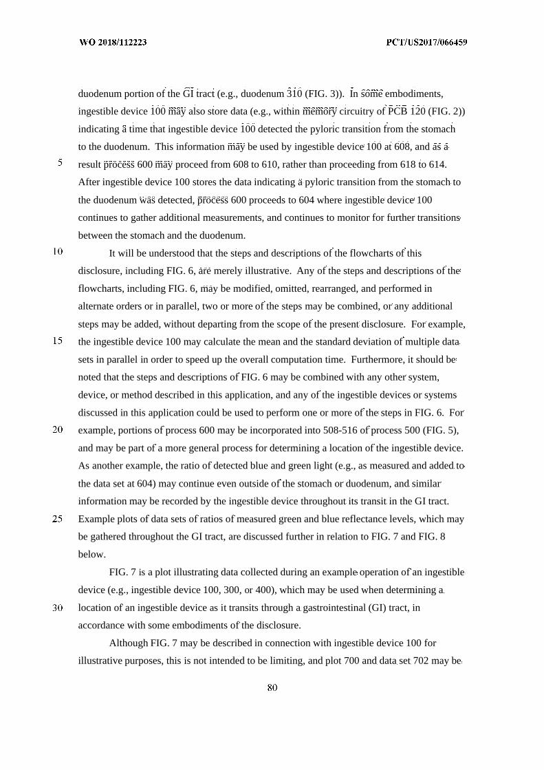
duodenum portion of the GI tract (e.g., duodenum 310 (FIG. 3)). In some embodiments,
ingestible device 100 may also store data (e.g., within memory circuitry of PCB 120 (FIG. 2))
indicating a time that ingestible device 100 detected the pyloric transition from the stomach
to the duodenum. This information may be used by ingestible device 100 at 608, and as a
result process 600 may proceed from 608 to 610, rather than proceeding from 618 to 614.
After ingestible device 100 stores the data indicating a pyloric transition from the stomach to
the duodenum was detected, process 600 proceeds to 604 where ingestible device 100
continues to gather additional measurements, and continues to monitor for further transitions
between the stomach and the duodenum.
It will be understood that the steps and descriptions of the flowcharts of this
disclosure, including FIG. 6, are merely illustrative. Any of the steps and descriptions of the
flowcharts, including FIG. 6, may be modified, omitted, rearranged, and performed in
alternate orders or in parallel, two or more of the steps may be combined, or any additional
steps may be added, without departing from the scope of the present disclosure. For example,
the ingestible device 100 may calculate the mean and the standard deviation of multiple data
sets in parallel in order to speed up the overall computation time. Furthermore, it should be
noted that the steps and descriptions of FIG. 6 may be combined with any other system,
device, or method described in this application, and any of the ingestible devices or systems
discussed in this application could be used to perform one or more of the steps in FIG. 6. For
example, portions of process 600 may be incorporated into 508-516 of process 500 (FIG. 5),
and may be part of a more general process for determining a location of the ingestible device.
As another example, the ratio of detected blue and green light (e.g., as measured and added to
the data set at 604) may continue even outside of the stomach or duodenum, and similar
information may be recorded by the ingestible device throughout its transit in the GI tract.
Example plots of data sets of ratios of measured green and blue reflectance levels, which may
be gathered throughout the GI tract, are discussed further in relation to FIG. 7 and FIG. 8
below.
FIG. 7 is a plot illustrating data collected during an example operation of an ingestible
device (e.g., ingestible device 100, 300, or 400), which may be used when determining a
location of an ingestible device as it transits through a gastrointestinal (GI) tract, in
accordance with some embodiments of the disclosure.
Although FIG. 7 may be described in connection with ingestible device 100 for
illustrative purposes, this is not intended to be limiting, and plot 700 and data set 702 may be
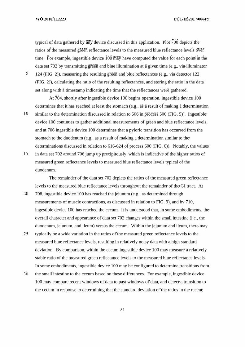
typical of data gathered by any device discussed in this application. Plot 700 depicts the
ratios of the measured green reflectance levels to the measured blue reflectance levels over
time. For example, ingestible device 100 may have computed the value for each point in the
data set 702 by transmitting green and blue illumination at a given time (e.g., via illuminator
124 (FIG. 2)), measuring the resulting green and blue reflectances (e.g., via detector 122
(FIG. 2)), calculating the ratio of the resulting reflectances, and storing the ratio in the data
set along with a timestamp indicating the time that the reflectances were gathered.
At 704, shortly after ingestible device 100 begins operation, ingestible device 100
determines that it has reached at least the stomach (e.g., as a result of making a determination
similar to the determination discussed in relation to 506 in process 500 (FIG. 5)). Ingestible
device 100 continues to gather additional measurements of green and blue reflectance levels,
and at 706 ingestible device 100 determines that a pyloric transition has occurred from the
stomach to the duodenum (e.g., as a result of making a determination similar to the
determinations discussed in relation to 616-624 of process 600 (FIG. 6)). Notably, the values
in data set 702 around 706 jump up precipitously, which is indicative of the higher ratios of
measured green reflectance levels to measured blue reflectance levels typical of the
duodenum.
The remainder of the data set 702 depicts the ratios of the measured green reflectance
levels to the measured blue reflectance levels throughout the remainder of the GI tract. At
708, ingestible device 100 has reached the jejunum (e.g., as determined through
measurements of muscle contractions, as discussed in relation to FIG. 9), and by 710,
ingestible device 100 has reached the cecum. It is understood that, in some embodiments, the
overall character and appearance of data set 702 changes within the small intestine (i.e., the
duodenum, jejunum, and ileum) versus the cecum. Within the jejunum and ileum, there may
typically be a wide variation in the ratios of the measured green reflectance levels to the
measured blue reflectance levels, resulting in relatively noisy data with a high standard
deviation. By comparison, within the cecum ingestible device 100 may measure a relatively
stable ratio of the measured green reflectance levels to the measured blue reflectance levels.
In some embodiments, ingestible device 100 may be configured to determine transitions from
the small intestine to the cecum based on these differences. For example, ingestible device
100 may compare recent windows of data to past windows of data, and detect a transition to
the cecum in response to determining that the standard deviation of the ratios in the recent
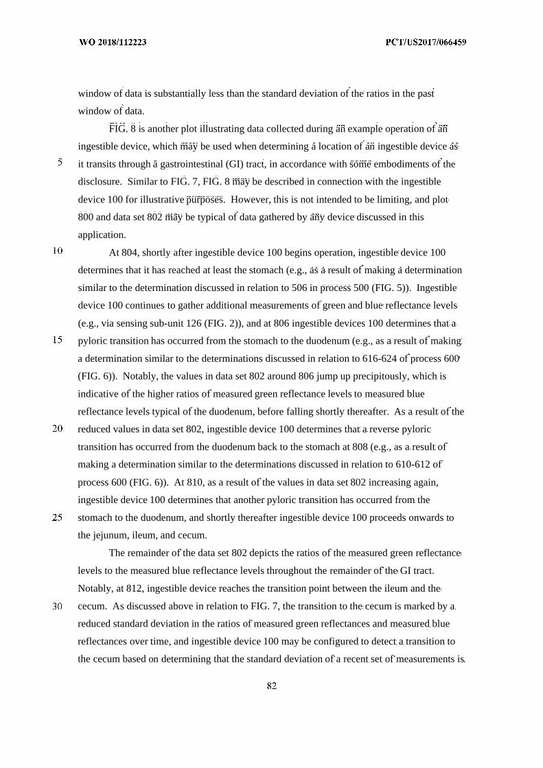
window of data is substantially less than the standard deviation of the ratios in the past
window of data.
FIG. 8 is another plot illustrating data collected during an example operation of an
ingestible device, which may be used when determining a location of an ingestible device as
it transits through a gastrointestinal (GI) tract, in accordance with some embodiments of the
disclosure. Similar to FIG. 7, FIG. 8 may be described in connection with the ingestible
device 100 for illustrative purposes. However, this is not intended to be limiting, and plot
800 and data set 802 may be typical of data gathered by any device discussed in this
application.
At 804, shortly after ingestible device 100 begins operation, ingestible device 100
determines that it has reached at least the stomach (e.g., as a result of making a determination
similar to the determination discussed in relation to 506 in process 500 (FIG. 5)). Ingestible
device 100 continues to gather additional measurements of green and blue reflectance levels
(e.g., via sensing sub-unit 126 (FIG. 2)), and at 806 ingestible devices 100 determines that a
pyloric transition has occurred from the stomach to the duodenum (e.g., as a result of making
a determination similar to the determinations discussed in relation to 616-624 of process 600
(FIG. 6)). Notably, the values in data set 802 around 806 jump up precipitously, which is
indicative of the higher ratios of measured green reflectance levels to measured blue
reflectance levels typical of the duodenum, before falling shortly thereafter. As a result of the
reduced values in data set 802, ingestible device 100 determines that a reverse pyloric
transition has occurred from the duodenum back to the stomach at 808 (e.g., as a result of
making a determination similar to the determinations discussed in relation to 610-612 of
process 600 (FIG. 6)). At 810, as a result of the values in data set 802 increasing again,
ingestible device 100 determines that another pyloric transition has occurred from the
stomach to the duodenum, and shortly thereafter ingestible device 100 proceeds onwards to
the jejunum, ileum, and cecum.
The remainder of the data set 802 depicts the ratios of the measured green reflectance
levels to the measured blue reflectance levels throughout the remainder of the GI tract.
Notably, at 812, ingestible device reaches the transition point between the ileum and the
cecum. As discussed above in relation to FIG. 7, the transition to the cecum is marked by a
reduced standard deviation in the ratios of measured green reflectances and measured blue
reflectances over time, and ingestible device 100 may be configured to detect a transition to
the cecum based on determining that the standard deviation of a recent set of measurements is
substantially smaller than the standard deviation of past measurements taken from the
jejunum or ileum.
FIG. 9 is a flowchart of illustrative steps for detecting a transition from a duodenum to
a jejunum, which may be used when determining a location of an ingestible device as it
transits through a gastrointestinal (GI) tract, in accordance with some embodiments of the
disclosure. Although FIG. 9 may be described in connection with the ingestible device 100
for illustrative purposes, this is not intended to be limiting, and either portions or the entirety
of process 900 described in FIG. 9 may be applied to any device discussed in this application
(e.g., the ingestible devices 100, 300, and 400), and any of these ingestible devices may be
used to perform one or more parts of the process described in FIG. 9. Furthermore, the
features of FIG. 9 may be combined with any other systems, methods or processes described
in this application. For example, portions of the process described by the process in FIG. 9
may be integrated into the localization process described by FIG. 5 (e.g., as part of 520-524
of process 500 (FIG. 5)). In some embodiments, an ingestible device 100 may perform
process 900 while in the duodenum, or in response to detecting entry to the duodenum. In
other embodiments, an ingestible device 100 may perform process 900 while in the stomach,
or in response to detecting entry into the GI tract. It is also understood that process 900 may
be performed in parallel with any other process described in this disclosure (e.g., process 600
(FIG. 6)), which may enable ingestible device 100 to detect entry into various portions of the
GI tract, without necessarily detecting entry into a preceding portion of the GI tract.
For illustrative purposes, FIG. 9 may be discussed in terms of ingestible device 100
generating and making determinations based on a single set of reflectance levels generated at
a single wavelength by a single sensing sub-unit (e.g., sensing sub-unit 126 (FIG. 2)).
However, it is understood that ingestible device 100 may generate multiple wavelengths of
illumination from multiple different sensing sub-units positioned around the circumference of
ingestible device (e.g., multiple sensing sub-units positioned at different locations behind
window 114 of ingestible device 100 (FIG. 1), and each of the resulting reflectances may be
stored as a separate data set. Moreover, each of these sets of reflectance levels may be used
to detect muscle contractions by running multiple versions of process 900, each one of which
processes data for a different set of reflectances corresponding to data sets obtained from
measurements of different wavelengths or measurements made by different sensing sub-units.
At 902, the ingestible device (e.g., ingestible device 100, 300, or 400) retrieves a set
of reflectance levels. For example, ingestible device 100 may retrieve a data set of
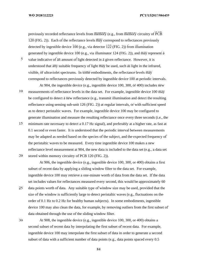
previously recorded reflectance levels from memory (e.g., from memory circuitry of PCB
120 (FIG. 2)). Each of the reflectance levels may correspond to reflectances previously
detected by ingestible device 100 (e.g., via detector 122 (FIG. 2)) from illumination
generated by ingestible device 100 (e.g., via illuminator 124 (FIG. 2)), and may represent a
value indicative of an amount of light detected in a given reflectance. However, it is
understood that any suitable frequency of light may be used, such as light in the infrared,
visible, or ultraviolet spectrums. In some embodiments, the reflectance levels may
correspond to reflectances previously detected by ingestible device 100 at periodic intervals.
At 904, the ingestible device (e.g., ingestible device 100, 300, or 400) includes new
measurements of reflectance levels in the data set. For example, ingestible device 100 may
be configured to detect a new reflectance (e.g., transmit illumination and detect the resulting
reflectance using sensing sub-unit 126 (FIG. 2)) at regular intervals, or with sufficient speed
as to detect peristaltic waves. For example, ingestible device 100 may be configured to
generate illumination and measure the resulting reflectance once every three seconds (i.e., the
minimum rate necessary to detect a 0.17 Hz signal), and preferably at a higher rate, as fast at
0.1 second or even faster. It is understood that the periodic interval between measurements
may be adapted as needed based on the species of the subject, and the expected frequency of
the peristaltic waves to be measured. Every time ingestible device 100 makes a new
reflectance level measurement at 904, the new data is included to the data set (e.g., a data set
stored within memory circuitry of PCB 120 (FIG. 2)).
At 906, the ingestible device (e.g., ingestible device 100, 300, or 400) obtains a first
subset of recent data by applying a sliding window filter to the data set. For example,
ingestible device 100 may retrieve a one-minute worth of data from the data set. If the data
set includes values for reflectances measured every second, this would be approximately 60
data points worth of data. Any suitable type of window size may be used, provided that the
size of the window is sufficiently large to detect peristaltic waves (e.g., fluctuations on the
order of 0.1 Hz to 0.2 Hz for healthy human subjects). In some embodiments, ingestible
device 100 may also clean the data, for example, by removing outliers from the first subset of
data obtained through the use of the sliding window filter.
At 908, the ingestible device (e.g., ingestible device 100, 300, or 400) obtains a
second subset of recent data by interpolating the first subset of recent data. For example,
ingestible device 100 may interpolate the first subset of data in order to generate a second
subset of data with a sufficient number of data points (e.g., data points spaced every 0.5
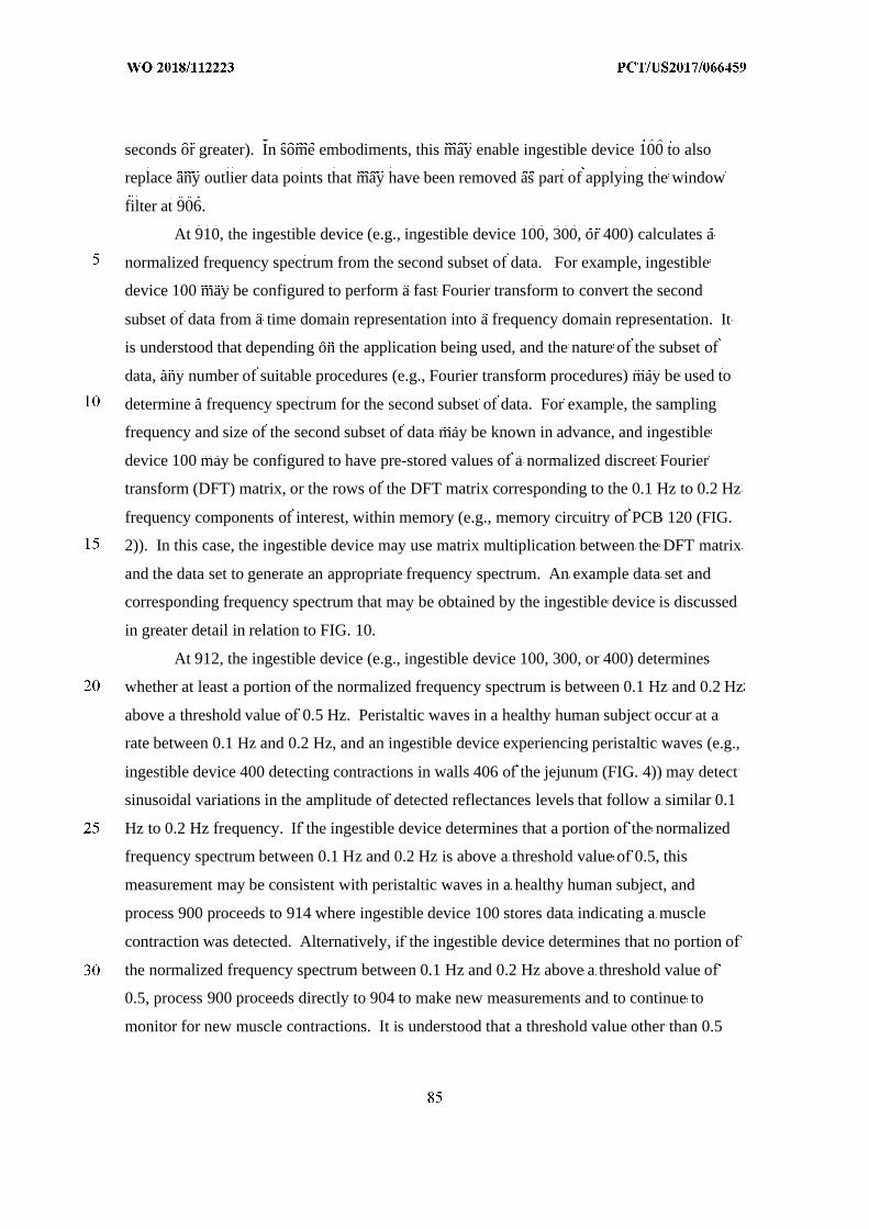
seconds or greater). In some embodiments, this may enable ingestible device 100 to also
replace any outlier data points that may have been removed as part of applying the window
filter at 906.
At 910, the ingestible device (e.g., ingestible device 100, 300, or 400) calculates a
normalized frequency spectrum from the second subset of data. For example, ingestible
device 100 may be configured to perform a fast Fourier transform to convert the second
subset of data from a time domain representation into a frequency domain representation. It
is understood that depending on the application being used, and the nature of the subset of
data, any number of suitable procedures (e.g., Fourier transform procedures) may be used to
determine a frequency spectrum for the second subset of data. For example, the sampling
frequency and size of the second subset of data may be known in advance, and ingestible
device 100 may be configured to have pre-stored values of a normalized discreet Fourier
transform (DFT) matrix, or the rows of the DFT matrix corresponding to the 0.1 Hz to 0.2 Hz
frequency components of interest, within memory (e.g., memory circuitry of PCB 120 (FIG.
2)). In this case, the ingestible device may use matrix multiplication between the DFT matrix
and the data set to generate an appropriate frequency spectrum. An example data set and
corresponding frequency spectrum that may be obtained by the ingestible device is discussed
in greater detail in relation to FIG. 10.
At 912, the ingestible device (e.g., ingestible device 100, 300, or 400) determines
whether at least a portion of the normalized frequency spectrum is between 0.1 Hz and 0.2 Hz
above a threshold value of 0.5 Hz. Peristaltic waves in a healthy human subject occur at a
rate between 0.1 Hz and 0.2 Hz, and an ingestible device experiencing peristaltic waves (e.g.,
ingestible device 400 detecting contractions in walls 406 of the jejunum (FIG. 4)) may detect
sinusoidal variations in the amplitude of detected reflectances levels that follow a similar 0.1
Hz to 0.2 Hz frequency. If the ingestible device determines that a portion of the normalized
frequency spectrum between 0.1 Hz and 0.2 Hz is above a threshold value of 0.5, this
measurement may be consistent with peristaltic waves in a healthy human subject, and
process 900 proceeds to 914 where ingestible device 100 stores data indicating a muscle
contraction was detected. Alternatively, if the ingestible device determines that no portion of
the normalized frequency spectrum between 0.1 Hz and 0.2 Hz above a threshold value of
0.5, process 900 proceeds directly to 904 to make new measurements and to continue to
monitor for new muscle contractions. It is understood that a threshold value other than 0.5
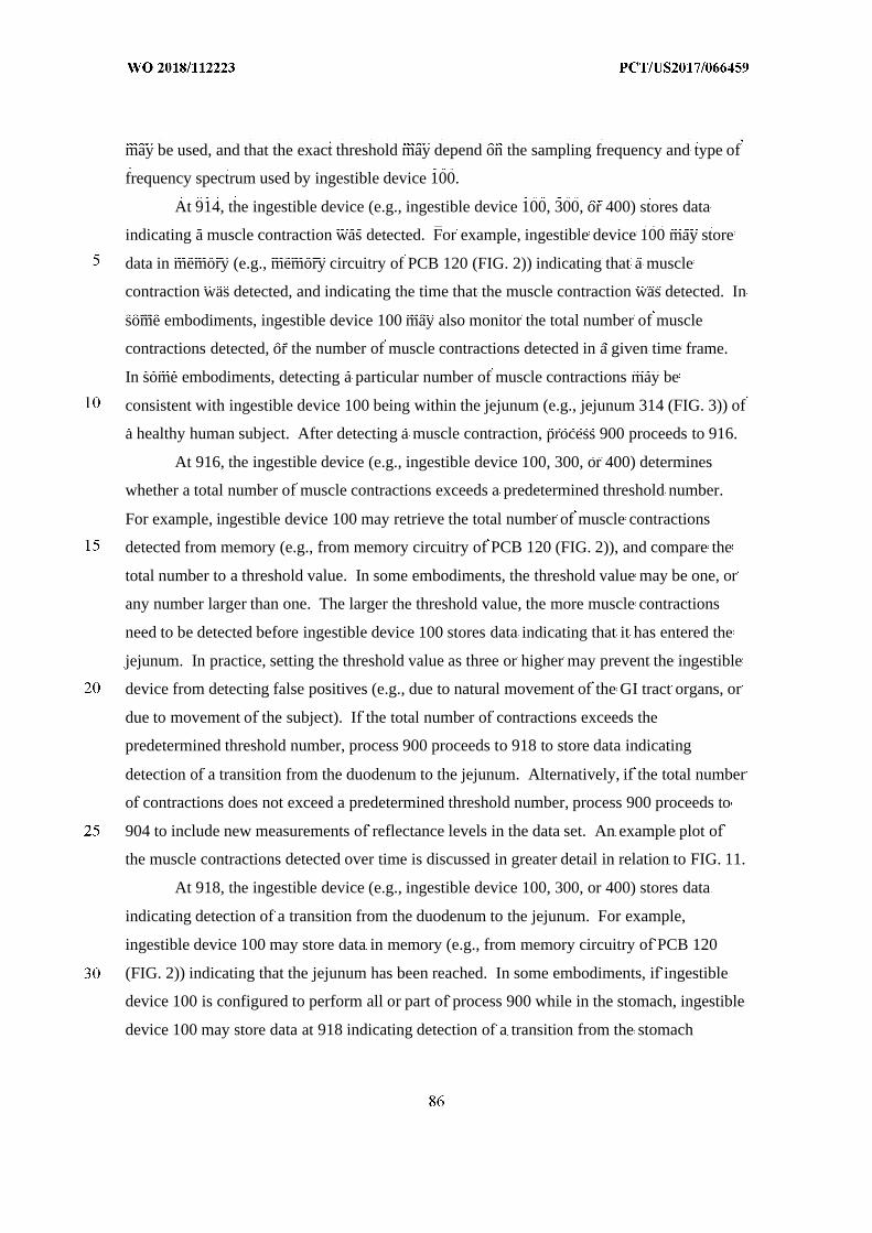
may be used, and that the exact threshold may depend on the sampling frequency and type of
frequency spectrum used by ingestible device 100.
At 914, the ingestible device (e.g., ingestible device 100, 300, or 400) stores data
indicating a muscle contraction was detected. For example, ingestible device 100 may store
data in memory (e.g., memory circuitry of PCB 120 (FIG. 2)) indicating that a muscle
contraction was detected, and indicating the time that the muscle contraction was detected. In
some embodiments, ingestible device 100 may also monitor the total number of muscle
contractions detected, or the number of muscle contractions detected in a given time frame.
In some embodiments, detecting a particular number of muscle contractions may be
consistent with ingestible device 100 being within the jejunum (e.g., jejunum 314 (FIG. 3)) of
a healthy human subject. After detecting a muscle contraction, process 900 proceeds to 916.
At 916, the ingestible device (e.g., ingestible device 100, 300, or 400) determines
whether a total number of muscle contractions exceeds a predetermined threshold number.
For example, ingestible device 100 may retrieve the total number of muscle contractions
detected from memory (e.g., from memory circuitry of PCB 120 (FIG. 2)), and compare the
total number to a threshold value. In some embodiments, the threshold value may be one, or
any number larger than one. The larger the threshold value, the more muscle contractions
need to be detected before ingestible device 100 stores data indicating that it has entered the
jejunum. In practice, setting the threshold value as three or higher may prevent the ingestible
device from detecting false positives (e.g., due to natural movement of the GI tract organs, or
due to movement of the subject). If the total number of contractions exceeds the
predetermined threshold number, process 900 proceeds to 918 to store data indicating
detection of a transition from the duodenum to the jejunum. Alternatively, if the total number
of contractions does not exceed a predetermined threshold number, process 900 proceeds to
904 to include new measurements of reflectance levels in the data set. An example plot of
the muscle contractions detected over time is discussed in greater detail in relation to FIG. 11.
At 918, the ingestible device (e.g., ingestible device 100, 300, or 400) stores data
indicating detection of a transition from the duodenum to the jejunum. For example,
ingestible device 100 may store data in memory (e.g., from memory circuitry of PCB 120
(FIG. 2)) indicating that the jejunum has been reached. In some embodiments, if ingestible
device 100 is configured to perform all or part of process 900 while in the stomach, ingestible
device 100 may store data at 918 indicating detection of a transition from the stomach
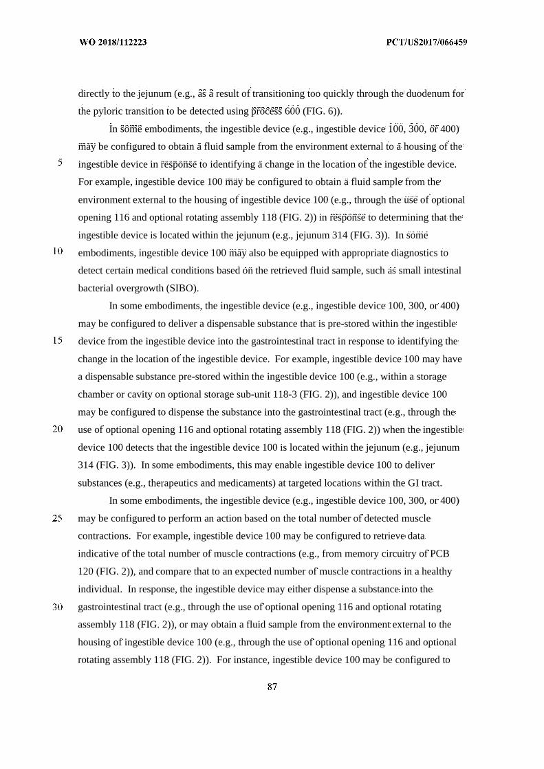
directly to the jejunum (e.g., as a result of transitioning too quickly through the duodenum for
the pyloric transition to be detected using process 600 (FIG. 6)).
In some embodiments, the ingestible device (e.g., ingestible device 100, 300, or 400)
may be configured to obtain a fluid sample from the environment external to a housing of the
ingestible device in response to identifying a change in the location of the ingestible device.
For example, ingestible device 100 may be configured to obtain a fluid sample from the
environment external to the housing of ingestible device 100 (e.g., through the use of optional
opening 116 and optional rotating assembly 118 (FIG. 2)) in response to determining that the
ingestible device is located within the jejunum (e.g., jejunum 314 (FIG. 3)). In some
embodiments, ingestible device 100 may also be equipped with appropriate diagnostics to
detect certain medical conditions based on the retrieved fluid sample, such as small intestinal
bacterial overgrowth (SIBO).
In some embodiments, the ingestible device (e.g., ingestible device 100, 300, or 400)
may be configured to deliver a dispensable substance that is pre-stored within the ingestible
device from the ingestible device into the gastrointestinal tract in response to identifying the
change in the location of the ingestible device. For example, ingestible device 100 may have
a dispensable substance pre-stored within the ingestible device 100 (e.g., within a storage
chamber or cavity on optional storage sub-unit 118-3 (FIG. 2)), and ingestible device 100
may be configured to dispense the substance into the gastrointestinal tract (e.g., through the
use of optional opening 116 and optional rotating assembly 118 (FIG. 2)) when the ingestible
device 100 detects that the ingestible device 100 is located within the jejunum (e.g., jejunum
314 (FIG. 3)). In some embodiments, this may enable ingestible device 100 to deliver
substances (e.g., therapeutics and medicaments) at targeted locations within the GI tract.
In some embodiments, the ingestible device (e.g., ingestible device 100, 300, or 400)
may be configured to perform an action based on the total number of detected muscle
contractions. For example, ingestible device 100 may be configured to retrieve data
indicative of the total number of muscle contractions (e.g., from memory circuitry of PCB
120 (FIG. 2)), and compare that to an expected number of muscle contractions in a healthy
individual. In response, the ingestible device may either dispense a substance into the
gastrointestinal tract (e.g., through the use of optional opening 116 and optional rotating
assembly 118 (FIG. 2)), or may obtain a fluid sample from the environment external to the
housing of ingestible device 100 (e.g., through the use of optional opening 116 and optional
rotating assembly 118 (FIG. 2)). For instance, ingestible device 100 may be configured to
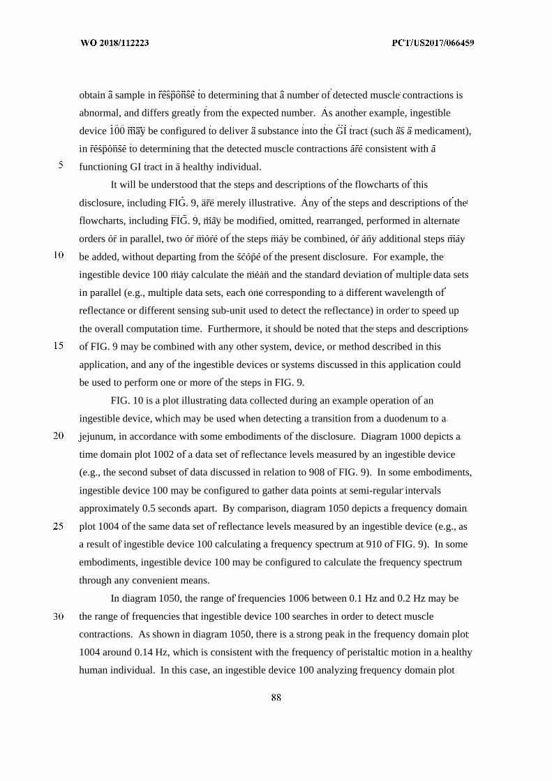
obtain a sample in response to determining that a number of detected muscle contractions is
abnormal, and differs greatly from the expected number. As another example, ingestible
device 100 may be configured to deliver a substance into the GI tract (such as a medicament),
in response to determining that the detected muscle contractions are consistent with a
functioning GI tract in a healthy individual.
It will be understood that the steps and descriptions of the flowcharts of this
disclosure, including FIG. 9, are merely illustrative. Any of the steps and descriptions of the
flowcharts, including FIG. 9, may be modified, omitted, rearranged, performed in alternate
orders or in parallel, two or more of the steps may be combined, or any additional steps may
be added, without departing from the scope of the present disclosure. For example, the
ingestible device 100 may calculate the mean and the standard deviation of multiple data sets
in parallel (e.g., multiple data sets, each one corresponding to a different wavelength of
reflectance or different sensing sub-unit used to detect the reflectance) in order to speed up
the overall computation time. Furthermore, it should be noted that the steps and descriptions
of FIG. 9 may be combined with any other system, device, or method described in this
application, and any of the ingestible devices or systems discussed in this application could
be used to perform one or more of the steps in FIG. 9.
FIG. 10 is a plot illustrating data collected during an example operation of an
ingestible device, which may be used when detecting a transition from a duodenum to a
jejunum, in accordance with some embodiments of the disclosure. Diagram 1000 depicts a
time domain plot 1002 of a data set of reflectance levels measured by an ingestible device
(e.g., the second subset of data discussed in relation to 908 of FIG. 9). In some embodiments,
ingestible device 100 may be configured to gather data points at semi-regular intervals
approximately 0.5 seconds apart. By comparison, diagram 1050 depicts a frequency domain
plot 1004 of the same data set of reflectance levels measured by an ingestible device (e.g., as
a result of ingestible device 100 calculating a frequency spectrum at 910 of FIG. 9). In some
embodiments, ingestible device 100 may be configured to calculate the frequency spectrum
through any convenient means.
In diagram 1050, the range of frequencies 1006 between 0.1 Hz and 0.2 Hz may be
the range of frequencies that ingestible device 100 searches in order to detect muscle
contractions. As shown in diagram 1050, there is a strong peak in the frequency domain plot
1004 around 0.14 Hz, which is consistent with the frequency of peristaltic motion in a healthy
human individual. In this case, an ingestible device 100 analyzing frequency domain plot
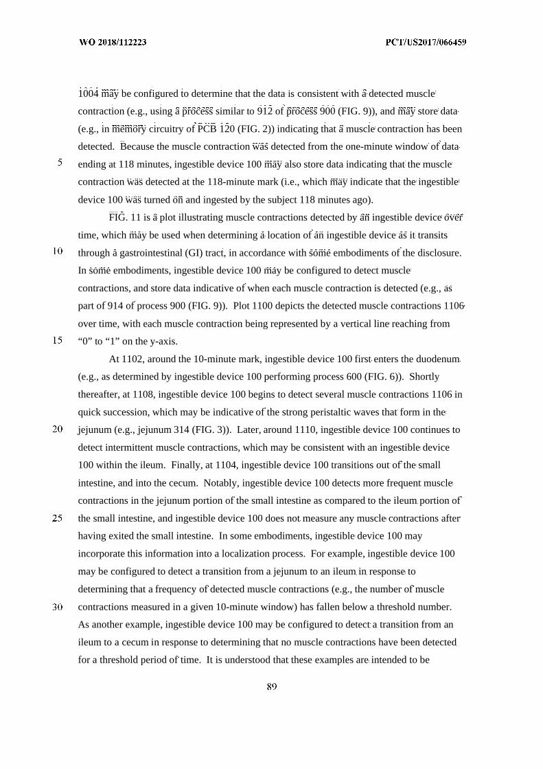
1004 may be configured to determine that the data is consistent with a detected muscle
contraction (e.g., using a process similar to 912 of process 900 (FIG. 9)), and may store data
(e.g., in memory circuitry of PCB 120 (FIG. 2)) indicating that a muscle contraction has been
detected. Because the muscle contraction was detected from the one-minute window of data
ending at 118 minutes, ingestible device 100 may also store data indicating that the muscle
contraction was detected at the 118-minute mark (i.e., which may indicate that the ingestible
device 100 was turned on and ingested by the subject 118 minutes ago).
FIG. 11 is a plot illustrating muscle contractions detected by an ingestible device over
time, which may be used when determining a location of an ingestible device as it transits
through a gastrointestinal (GI) tract, in accordance with some embodiments of the disclosure.
In some embodiments, ingestible device 100 may be configured to detect muscle
contractions, and store data indicative of when each muscle contraction is detected (e.g., as
part of 914 of process 900 (FIG. 9)). Plot 1100 depicts the detected muscle contractions 1106
over time, with each muscle contraction being represented by a vertical line reaching from
“0” to “1” on the y-axis.
At 1102, around the 10-minute mark, ingestible device 100 first enters the duodenum
(e.g., as determined by ingestible device 100 performing process 600 (FIG. 6)). Shortly
thereafter, at 1108, ingestible device 100 begins to detect several muscle contractions 1106 in
quick succession, which may be indicative of the strong peristaltic waves that form in the
jejunum (e.g., jejunum 314 (FIG. 3)). Later, around 1110, ingestible device 100 continues to
detect intermittent muscle contractions, which may be consistent with an ingestible device
100 within the ileum. Finally, at 1104, ingestible device 100 transitions out of the small
intestine, and into the cecum. Notably, ingestible device 100 detects more frequent muscle
contractions in the jejunum portion of the small intestine as compared to the ileum portion of
the small intestine, and ingestible device 100 does not measure any muscle contractions after
having exited the small intestine. In some embodiments, ingestible device 100 may
incorporate this information into a localization process. For example, ingestible device 100
may be configured to detect a transition from a jejunum to an ileum in response to
determining that a frequency of detected muscle contractions (e.g., the number of muscle
contractions measured in a given 10-minute window) has fallen below a threshold number.
As another example, ingestible device 100 may be configured to detect a transition from an
ileum to a cecum in response to determining that no muscle contractions have been detected
for a threshold period of time. It is understood that these examples are intended to be
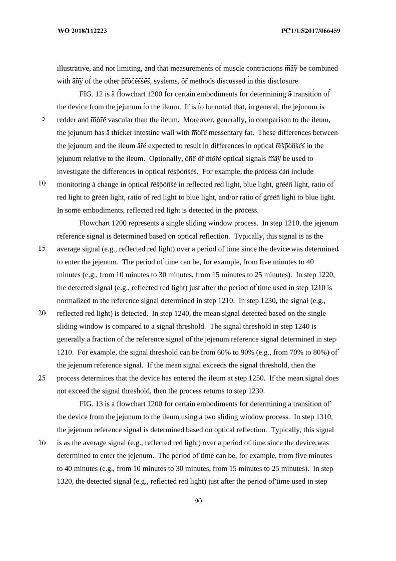
illustrative, and not limiting, and that measurements of muscle contractions may be combined
with any of the other processes, systems, or methods discussed in this disclosure.
FIG. 12 is a flowchart 1200 for certain embodiments for determining a transition of
the device from the jejunum to the ileum. It is to be noted that, in general, the jejunum is
redder and more vascular than the ileum. Moreover, generally, in comparison to the ileum,
the jejunum has a thicker intestine wall with more messentary fat. These differences between
the jejunum and the ileum are expected to result in differences in optical responses in the
jejunum relative to the ileum. Optionally, one or more optical signals may be used to
investigate the differences in optical responses. For example, the process can include
monitoring a change in optical response in reflected red light, blue light, green light, ratio of
red light to green light, ratio of red light to blue light, and/or ratio of green light to blue light.
In some embodiments, reflected red light is detected in the process.
Flowchart 1200 represents a single sliding window process. In step 1210, the jejenum
reference signal is determined based on optical reflection. Typically, this signal is as the
average signal (e.g., reflected red light) over a period of time since the device was determined
to enter the jejenum. The period of time can be, for example, from five minutes to 40
minutes (e.g., from 10 minutes to 30 minutes, from 15 minutes to 25 minutes). In step 1220,
the detected signal (e.g., reflected red light) just after the period of time used in step 1210 is
normalized to the reference signal determined in step 1210. In step 1230, the signal (e.g.,
reflected red light) is detected. In step 1240, the mean signal detected based on the single
sliding window is compared to a signal threshold. The signal threshold in step 1240 is
generally a fraction of the reference signal of the jejenum reference signal determined in step
1210. For example, the signal threshold can be from 60% to 90% (e.g., from 70% to 80%) of
the jejenum reference signal. If the mean signal exceeds the signal threshold, then the
process determines that the device has entered the ileum at step 1250. If the mean signal does
not exceed the signal threshold, then the process returns to step 1230.
FIG. 13 is a flowchart 1200 for certain embodiments for determining a transition of
the device from the jejunum to the ileum using a two sliding window process. In step 1310,
the jejenum reference signal is determined based on optical reflection. Typically, this signal
is as the average signal (e.g., reflected red light) over a period of time since the device was
determined to enter the jejenum. The period of time can be, for example, from five minutes
to 40 minutes (e.g., from 10 minutes to 30 minutes, from 15 minutes to 25 minutes). In step
1320, the detected signal (e.g., reflected red light) just after the period of time used in step
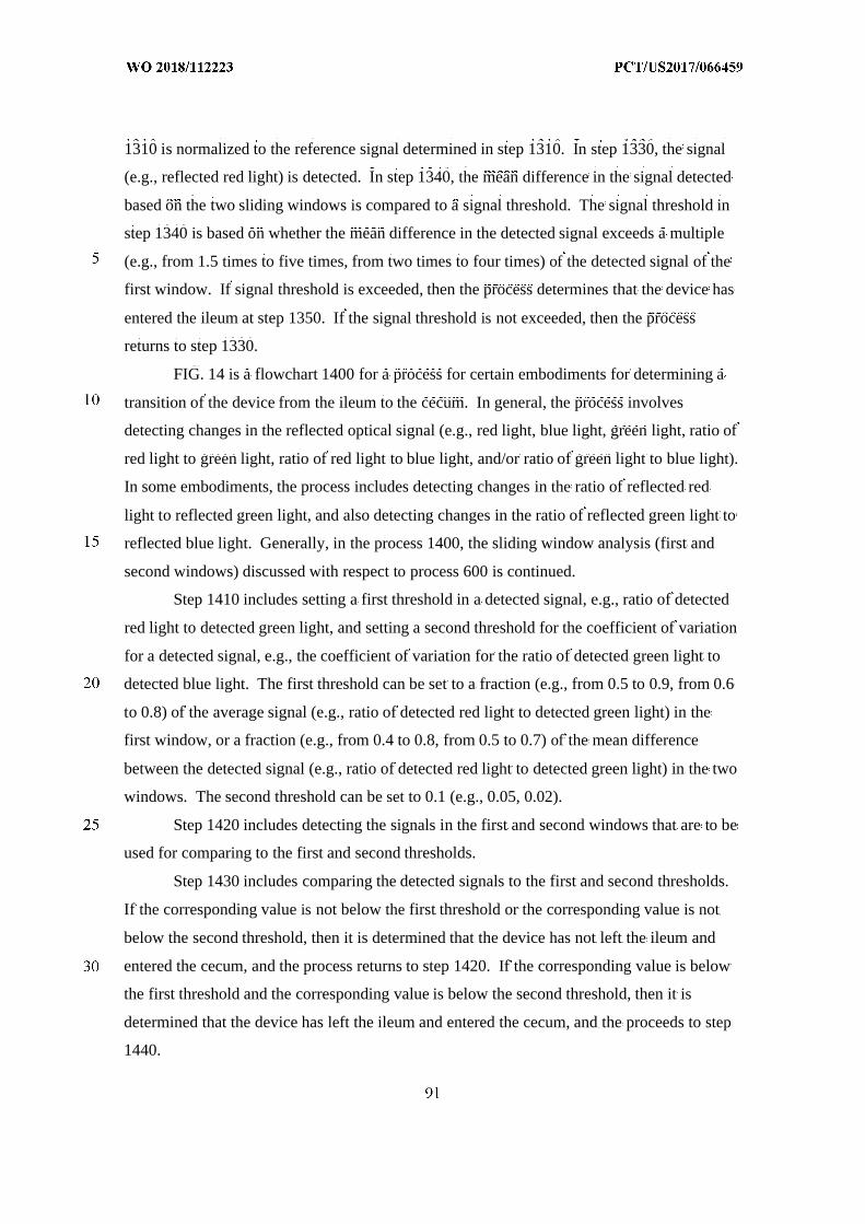
1310 is normalized to the reference signal determined in step 1310. In step 1330, the signal
(e.g., reflected red light) is detected. In step 1340, the mean difference in the signal detected
based on the two sliding windows is compared to a signal threshold. The signal threshold in
step 1340 is based on whether the mean difference in the detected signal exceeds a multiple
(e.g., from 1.5 times to five times, from two times to four times) of the detected signal of the
first window. If signal threshold is exceeded, then the process determines that the device has
entered the ileum at step 1350. If the signal threshold is not exceeded, then the process
returns to step 1330.
FIG. 14 is a flowchart 1400 for a process for certain embodiments for determining a
transition of the device from the ileum to the cecum. In general, the process involves
detecting changes in the reflected optical signal (e.g., red light, blue light, green light, ratio of
red light to green light, ratio of red light to blue light, and/or ratio of green light to blue light).
In some embodiments, the process includes detecting changes in the ratio of reflected red
light to reflected green light, and also detecting changes in the ratio of reflected green light to
reflected blue light. Generally, in the process 1400, the sliding window analysis (first and
second windows) discussed with respect to process 600 is continued.
Step 1410 includes setting a first threshold in a detected signal, e.g., ratio of detected
red light to detected green light, and setting a second threshold for the coefficient of variation
for a detected signal, e.g., the coefficient of variation for the ratio of detected green light to
detected blue light. The first threshold can be set to a fraction (e.g., from 0.5 to 0.9, from 0.6
to 0.8) of the average signal (e.g., ratio of detected red light to detected green light) in the
first window, or a fraction (e.g., from 0.4 to 0.8, from 0.5 to 0.7) of the mean difference
between the detected signal (e.g., ratio of detected red light to detected green light) in the two
windows. The second threshold can be set to 0.1 (e.g., 0.05, 0.02).
Step 1420 includes detecting the signals in the first and second windows that are to be
used for comparing to the first and second thresholds.
Step 1430 includes comparing the detected signals to the first and second thresholds.
If the corresponding value is not below the first threshold or the corresponding value is not
below the second threshold, then it is determined that the device has not left the ileum and
entered the cecum, and the process returns to step 1420. If the corresponding value is below
the first threshold and the corresponding value is below the second threshold, then it is
determined that the device has left the ileum and entered the cecum, and the proceeds to step
1440.
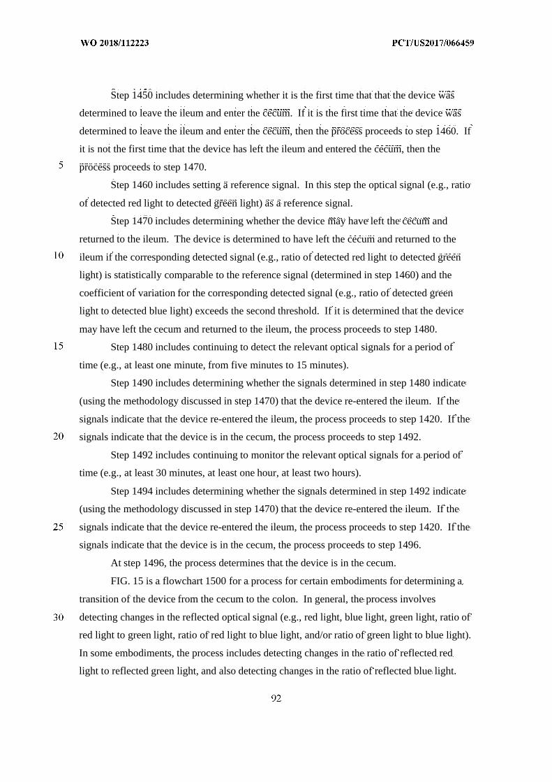
Step 1450 includes determining whether it is the first time that that the device was
determined to leave the ileum and enter the cecum. If it is the first time that the device was
determined to leave the ileum and enter the cecum, then the process proceeds to step 1460. If
it is not the first time that the device has left the ileum and entered the cecum, then the
process proceeds to step 1470.
Step 1460 includes setting a reference signal. In this step the optical signal (e.g., ratio
of detected red light to detected green light) as a reference signal.
Step 1470 includes determining whether the device may have left the cecum and
returned to the ileum. The device is determined to have left the cecum and returned to the
ileum if the corresponding detected signal (e.g., ratio of detected red light to detected green
light) is statistically comparable to the reference signal (determined in step 1460) and the
coefficient of variation for the corresponding detected signal (e.g., ratio of detected green
light to detected blue light) exceeds the second threshold. If it is determined that the device
may have left the cecum and returned to the ileum, the process proceeds to step 1480.
Step 1480 includes continuing to detect the relevant optical signals for a period of
time (e.g., at least one minute, from five minutes to 15 minutes).
Step 1490 includes determining whether the signals determined in step 1480 indicate
(using the methodology discussed in step 1470) that the device re-entered the ileum. If the
signals indicate that the device re-entered the ileum, the process proceeds to step 1420. If the
signals indicate that the device is in the cecum, the process proceeds to step 1492.
Step 1492 includes continuing to monitor the relevant optical signals for a period of
time (e.g., at least 30 minutes, at least one hour, at least two hours).
Step 1494 includes determining whether the signals determined in step 1492 indicate
(using the methodology discussed in step 1470) that the device re-entered the ileum. If the
signals indicate that the device re-entered the ileum, the process proceeds to step 1420. If the
signals indicate that the device is in the cecum, the process proceeds to step 1496.
At step 1496, the process determines that the device is in the cecum.
FIG. 15 is a flowchart 1500 for a process for certain embodiments for determining a
transition of the device from the cecum to the colon. In general, the process involves
detecting changes in the reflected optical signal (e.g., red light, blue light, green light, ratio of
red light to green light, ratio of red light to blue light, and/or ratio of green light to blue light).
In some embodiments, the process includes detecting changes in the ratio of reflected red
light to reflected green light, and also detecting changes in the ratio of reflected blue light.
Generally, in the process 1500, the sliding window analysis (first and second windows)
discussed with respect to process 1400 is continued.
In step 1510, optical signals (e.g., the ratio of reflected red signal to reflected green
signal, and reflected blue signal) are collected for a period of time (e.g., at least one minute,
at least five minutes, at least 10 minutes) while the device is in the cecum (e.g., during step
1480). The average values for the recorded optical signals (e.g., the ratio of reflected red
signal to reflected green signal, and reflected blue signal) establish the cecum reference
signals.
In step 1520, the optical signals are detected after it has been determined that the
device entered the cecum (e.g., at step 1440). The optical signals are normalized to the
cecum reference signals.
Step 1530 involves determining whether the device has entered the colon. This
includes determining whether any of three different criteria are satisfied. The first criterion is
satisfied if the mean difference in the ratio of a detected optical signal (e.g., ratio of detected
red signal to the detected green) is a multiple greater than one (e.g., 2X, 3X, 4X) the standard
deviation of the corresponding signal (e.g., ratio of detected red signal to the detected green)
in the second window. The second criterion is satisfied if the mean of a detected optical
signal (e.g., a ratio of detected red light to detected green light) exceeds a given value (e.g.,
exceeds one). The third criterion is satisfied if the coefficient of variation of an optical signal
(e.g., detected blue light) in the first window exceeds a given value (e.g., exceeds 0.2). If any
of the three criteria are satisfied, then the process proceeds to step 1540. Otherwise, none of
the three criteria are satisfied, the process returns to step 1520.
For illustrative purposes the disclosure focuses primarily on a number of different
example embodiments of an ingestible device, and example embodiments of methods for
determining a location of an ingestible device within a GI tract. However, the possible
ingestible devices that may be constructed are not limited to these embodiments, and
variations in the shape and design may be made without significantly changing the functions
and operations of the device. Similarly, the possible procedures for determining a location
of the ingestible device within the GI tract are not limited to the specific procedures and
embodiments discussed (e.g., process 500 (FIG. 5), process 600 (FIG. 6), process 900 (FIG.
9), process 1200 (FIG. 12), process 1300 (FIG. 13), process 1400 (FIG. 14) and process 1500
(FIG. 15)). Also, the applications of the ingestible devices described herein are not limited
merely to gathering data, sampling and testing portions of the gastrointestinal tract, or
delivering medicament. For example, in some embodiments the ingestible device may be
adapted to include a number of chemical, electrical, or optical diagnostics for diagnosing a
number of diseases. Similarly, a number of different sensors for measuring bodily
phenomenon or other physiological qualities may be included on the ingestible device. For
example, the ingestible device may be adapted to measure elevated levels of certain chemical
compounds or impurities in the gastrointestinal tract, or the combination of localization,
sampling, and appropriate diagnostic and assay techniques incorporated into a sampling
chamber may be particularly well suited to determine the presence of small intestinal
bacterial overgrowth (SIBO).
At least some of the elements of the various embodiments of the ingestible device
described herein that are implemented via software (e.g., software executed by control
circuitry within PCB 120 (FIG. 2)) may be written in a high-level procedural language such
as object oriented programming, a scripting language or both. Accordingly, the program code
may be written in C, C+ + or any other suitable programming language and may comprise
modules or classes, as is known to those skilled in object oriented programming.
Alternatively, or in addition, at least some of the elements of the embodiments of the
ingestible device described herein that are implemented via software may be written in
assembly language, machine language or firmware as needed. In either case, the language
may be a compiled or an interpreted language.
At least some of the program code used to implement the ingestible device can be
stored on a storage media or on a computer readable medium that is readable by a general or
special purpose programmable computing device having a processor, an operating system and
the associated hardware and software that is necessary to implement the functionality of at
least one of the embodiments described herein. The program code, when read by the
computing device, configures the computing device to operate in a new, specific and
predefined manner in order to perform at least one of the methods described herein.
Furthermore, at least some of the programs associated with the systems, devices, and
methods of the example embodiments described herein are capable of being distributed in a
computer program product comprising a computer readable medium that bears computer
usable instructions for one or more processors. The medium may be provided in various
forms, including non-transitory forms such as, but not limited to, one or more diskettes,
compact disks, tapes, chips, and magnetic and electronic storage. In some embodiments, the
medium may be transitory in nature such as, but not limited to, wire-line transmissions,
satellite transmissions, internet transmissions (e.g. downloads), media, digital and analog
signals, and the like. The computer useable instructions may also be in various formats,
including compiled and non-compiled code.
The techniques described above can be implemented using software for execution on
a computer. For instance, the software forms procedures in one or more computer programs
that execute on one or more programmed or programmable computer systems (which may be
of various architectures such as distributed, client/server, or grid) each including at least one
processor, at least one data storage system (including volatile and non-volatile memory
and/or storage elements), at least one input device or port, and at least one output device or
port.
The software may be provided on a storage medium, such as a CD-ROM, readable by
a general or special purpose programmable computer or delivered (encoded in a propagated
signal) over a communication medium of a network to the computer where it is executed. All
of the functions may be performed on a special purpose computer, or using special-purpose
hardware, such as coprocessors. The software may be implemented in a distributed manner
in which different parts of the computation specified by the software are performed by
different computers. Each such computer program is preferably stored on or downloaded to a
storage media or device (e.g., solid state memory or media, or magnetic or optical media)
readable by a general or special purpose programmable computer, for configuring and
operating the computer when the storage media or device is read by the computer system to
perform the procedures described herein. The inventive system may also be considered to be
implemented as a computer-readable storage medium, configured with a computer program,
where the storage medium so configured causes a computer system to operate in a specific
and predefined manner to perform the functions described herein.
Methods and Mechanisms of Delivery
FIG. 16 provides an example mock-up diagram illustrating aspects of a structure of an
ingestible device 1600 for delivering a dispensable substance, such as a formulation of a
therapeutic agent described herein, according to some embodiments described herein. In
some embodiments, the ingestible device 1600 may generally be in the shape of a capsule, a
pill or any swallowable form that may be orally consumed by an individual. In this way, the
ingestible device 1600 may be ingested by a patient and may be prescribed by healthcare
practitioners and patients.
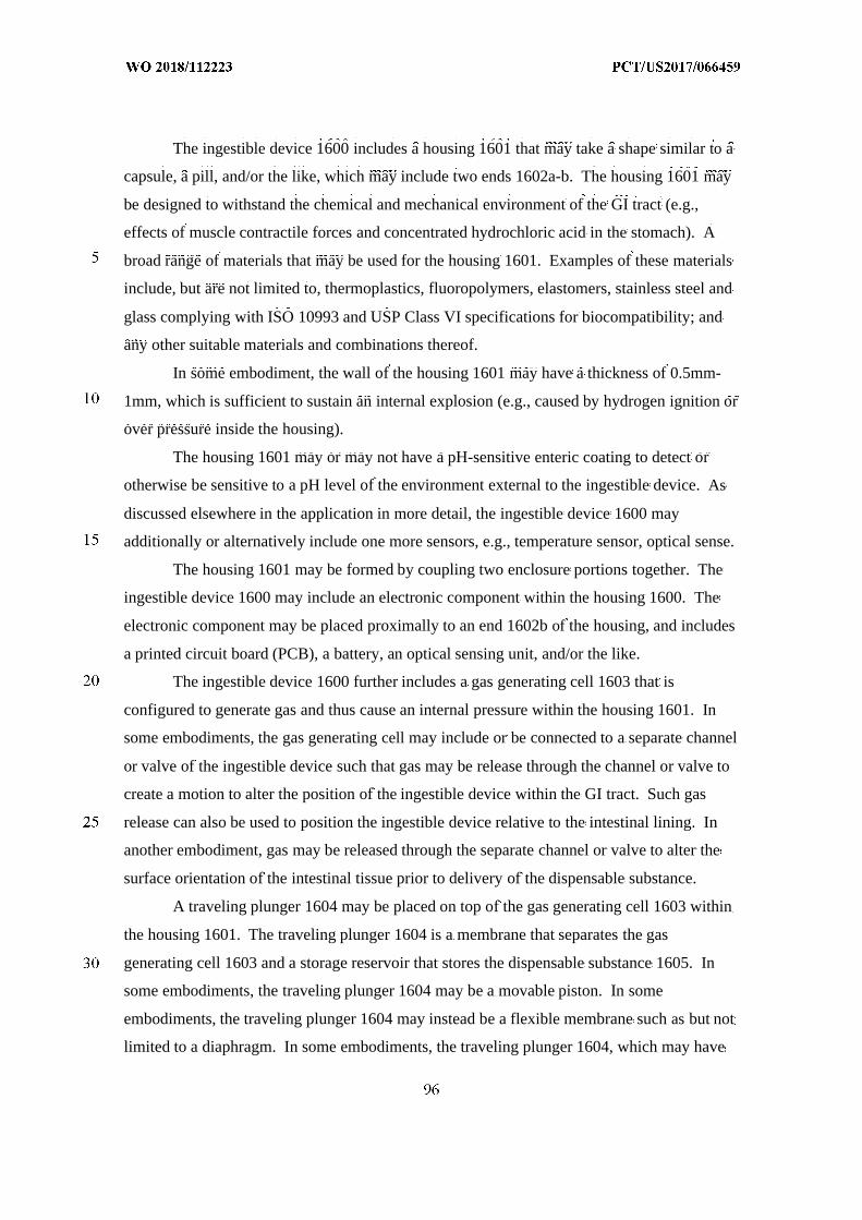
The ingestible device 1600 includes a housing 1601 that may take a shape similar to a
capsule, a pill, and/or the like, which may include two ends 1602a-b. The housing 1601 may
be designed to withstand the chemical and mechanical environment of the GI tract (e.g.,
effects of muscle contractile forces and concentrated hydrochloric acid in the stomach). A
broad range of materials that may be used for the housing 1601. Examples of these materials
include, but are not limited to, thermoplastics, fluoropolymers, elastomers, stainless steel and
glass complying with ISO 10993 and USP Class VI specifications for biocompatibility; and
any other suitable materials and combinations thereof.
In some embodiment, the wall of the housing 1601 may have a thickness of 0.5mm-
1mm, which is sufficient to sustain an internal explosion (e.g., caused by hydrogen ignition or
over pressure inside the housing).
The housing 1601 may or may not have a pH-sensitive enteric coating to detect or
otherwise be sensitive to a pH level of the environment external to the ingestible device. As
discussed elsewhere in the application in more detail, the ingestible device 1600 may
additionally or alternatively include one more sensors, e.g., temperature sensor, optical sense.
The housing 1601 may be formed by coupling two enclosure portions together. The
ingestible device 1600 may include an electronic component within the housing 1600. The
electronic component may be placed proximally to an end 1602b of the housing, and includes
a printed circuit board (PCB), a battery, an optical sensing unit, and/or the like.
The ingestible device 1600 further includes a gas generating cell 1603 that is
configured to generate gas and thus cause an internal pressure within the housing 1601. In
some embodiments, the gas generating cell may include or be connected to a separate channel
or valve of the ingestible device such that gas may be release through the channel or valve to
create a motion to alter the position of the ingestible device within the GI tract. Such gas
release can also be used to position the ingestible device relative to the intestinal lining. In
another embodiment, gas may be released through the separate channel or valve to alter the
surface orientation of the intestinal tissue prior to delivery of the dispensable substance.
A traveling plunger 1604 may be placed on top of the gas generating cell 1603 within
the housing 1601. The traveling plunger 1604 is a membrane that separates the gas
generating cell 1603 and a storage reservoir that stores the dispensable substance 1605. In
some embodiments, the traveling plunger 1604 may be a movable piston. In some
embodiments, the traveling plunger 1604 may instead be a flexible membrane such as but not
limited to a diaphragm. In some embodiments, the traveling plunger 1604, which may have
the form of a flexible diaphragm, may be placed along an axial direction of the housing 1601,
instead of being placed on top of the gas generating cell 1603. The traveling plunger or the
membrane 1604 may move (when the membrane 1604 is a piston) or deform (when the
membrane 1604 is a diaphragm) towards a direction of the end 1602a of the housing, when
the gas generating cell 1603 generates gas to create an internal pressure that pushes the
membrane 1604. In this way, the membrane or traveling plunger 1604 may push the
dispensable substance 1605 out of the housing via a dispensing outlet 1607.
The housing 1601 may include a storage reservoir storing one or more dispensable
substances 1605 adjacent to the traveling plunger 1604. The dispensable substance 1605 may
be a therapeutic or medical agent that may take a form of a powder, a compressed powder, a
fluid, a semi-liquid gel, or any other dispensable or deliverable form. The delivery of the
dispensable substance 1605 may take a form such as but not limited to bolus, semi-bolus,
continuous, systemic, burst drug delivery, and/or the like. In some embodiments, a single
bolus is delivered proximate to the disease location. In some embodiments, more than one
bolus is released at one location or more than one location. In some embodiments the release
of more than one bolus is triggered according to a pre-programmed algorithm. In some
embodiments the release profile is continuous. In some embodiments the release profile is
time-based. In some embodiments the release profile is location-based. In some
embodiments, the amount delivered is based on the severity and/or extent of the disease in the
following manner. In some embodiments, the bolus is delivered in one or more of the
following locations: stomach; duodenum; proximal jejunum; ileum; cecum; ascending colon;
transverse colon; descending colon. In some embodiments the dispensable substance is a
small molecule therapeutic that is released in the cecum and/or other parts of the large
intestine. Small molecules that are administerered by typical oral routes are primarily
absorbed in the small intestine, with much lower absorption taking place in the large intestine
(outside of the rectum). Accordingly, an ingestible device that is capable of releasing a small
molecule selectively in the large intestine (e.g., the cecum) with resulting low systemic levels
(even when high doses are used) is attractive for subjects with inflammatory bowel disease in
the large intestine.
In some embodiments, the storage reservoir may include multiple chambers, and each
chamber stores a different dispensable substance. For example, the different dispensable
substances can be released at the same time via the dispensing outlet 1607. Alternatively, the
multiple chambers may take a form of different layers within the storage reservoir such that
the different dispensable substance from each chamber is delivered sequentially in an order.
In one example, each of the multiple chambers is controlled by a separate traveling plunger,
which may be propelled by gas generation. The electronic component may control the gas
generating cell 1603 to generate gas to propel a specific traveling plunger, e.g., via a separate
gas generation chamber, etc., to deliver the respective substance. In some embodiments, the
content of the multiple chambers may be mixed or combined prior to release, for example, to
activate the drug.
The ingestible device 1600 may include a dispensing outlet 1607 at one end 1602a of
the housing 1601 to direct the dispensable substance 105 out of the housing. The dispensing
outlet 1607 may include an exit valve, a slit or a hole, a jet injection nozzle with a syringe,
and/or the like. When the traveling plunger 1604 moves towards the end 1602a of the
housing 1601, an internal pressure within the storage reservoir may increase and push the
dispensing outlet to be open to let the dispensable substance 1605 be released out of the
housing 1601.
In an embodiment, a pressure relief device 1606 may be placed within the housing
1601, e.g., at the end 1602a of the housing 1601.
In some embodiments, the housing 1601 may include small holes (e.g., with a
diameter smaller than 2 mm), e.g., on the side of the housing 1601, or at the end 1602a to
facilitate loading the dispensable substance into the storage reservoir.
In some embodiments, a feedback control circuit (e.g., a feedback resistor, etc.) may
be added to send feedback from the gas generating cell 1603 to the electronic component
such that when the internal pressure reaches a threshold level, the electronic component may
control the gas generating cell 1603 to turn off gas generation, or to activate other safety
mechanism (e.g., feedback-controlled release valve, etc.). For example, an internal pressure
sensor may be used to measure the internal pressure within the ingestible device and generate
feedback to the feedback control circuit.
FIG. 17 provides an example diagram illustrating aspects of a mechanism for a gas
generating cell 1603 configured to generate a gas to dispense a substance, according to some
embodiments described herein. As shown in FIG. 17, the gas generating cell 1603 generates
a gas 1611 which can propel the dispensable substance 1605 out of the dispensing outlet
1607. A variable resistor 1608 may be connected to a circuit with the gas generating cell
1603 such that the variable resistor 1608 may be used to control an intensity and/or an
amount of gas 1611 (e.g., hydrogen) generated by the cell 1603. Specifically, the gas
generating cell 1603 may be a battery form factor cell that is capable of generating hydrogen
when a resistor is applied. In this way, as the gas generating cell 1603 only needs the use of a
resistor only without any active power requirements, the gas generating cell 1603 may be
integrated into an ingestible device such as a capsule with limited energy/power available.
For example, the gas generating cell 1603 may be compatible with a capsule at a size of
26mm x 13mm or smaller.
In some embodiments, based on the elution rate of gas from the cell, and an internal
volume of the ingestible device, it may take time to generate sufficient gas 1611 to deliver the
substance 1605, and the time required may be 30 seconds or longer. For example, the time to
generate a volume of hydrogen equivalent to 500µL of fluid would be approximately 5
minutes. A longer period of time may be needed based upon non-ideal conditions within the
ingestible device, such as friction, etc. Thus, given that the production of gas (e.g., hydrogen)
may take time, gas generation may need to start prior to the ingestible device arriving at the
site of delivery to build pressure up within the device. The ingestible device may then need
to know when it is approaching the site of delivery. For example, the device may start
producing gas on an “entry transition,” which is determined by temperature, so as to produce
enough gas to be close to the pressure high enough to deliver the dispensable substance. The
ingestible device may then only start producing gas again when it arrives at the site of
delivery, which will cause the internal pressure within the ingestible device to reach a level
required by the dispensing outlet to release the dispensable substance. Also, for regio-
specific delivery, the ingestible device may estimate the time it takes to build up enough
pressure to deliver the dispensable substance before the ingestible device arrives at a specific
location, to activate gas generation.
For example, for systemic delivery, when an internal volume of the ingestible device
is around 500µL, a gas generation time of 2 hours, an initial pressure of approximately 300
pound per square inch absolute (psia) may be generated, with higher and lower pressures
possible. The generated pressure may drop when air enters the storage reservoir which was
previously occupied by the dispensable substance during the dispensing process. For
systemic drug delivery, a force with a generated pressure of approximately 100 to 360 pound
per square inch (psi) may be required for dermal penetration, e.g., to penetrate the mucosa or
epithelial layer. The pressure may also vary depending on the nozzle design at the dispensing
outlet, fluid viscosity, and surrounding tissue proximity and properties.
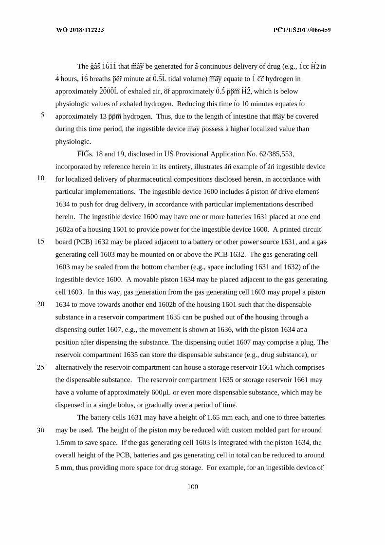
The gas 1611 that may be generated for a continuous delivery of drug (e.g., 1cc H2 in
4 hours, 16 breaths per minute at 0.5L tidal volume) may equate to 1 cc hydrogen in
approximately 2000L of exhaled air, or approximately 0.5 ppm H2, which is below
physiologic values of exhaled hydrogen. Reducing this time to 10 minutes equates to
approximately 13 ppm hydrogen. Thus, due to the length of intestine that may be covered
during this time period, the ingestible device may possess a higher localized value than
physiologic.
FIGs. 18 and 19, disclosed in US Provisional Application No. 62/385,553,
incorporated by reference herein in its entirety, illustrates an example of an ingestible device
for localized delivery of pharmaceutical compositions disclosed herein, in accordance with
particular implementations. The ingestible device 1600 includes a piston or drive element
1634 to push for drug delivery, in accordance with particular implementations described
herein. The ingestible device 1600 may have one or more batteries 1631 placed at one end
1602a of a housing 1601 to provide power for the ingestible device 1600. A printed circuit
board (PCB) 1632 may be placed adjacent to a battery or other power source 1631, and a gas
generating cell 1603 may be mounted on or above the PCB 1632. The gas generating cell
1603 may be sealed from the bottom chamber (e.g., space including 1631 and 1632) of the
ingestible device 1600. A movable piston 1634 may be placed adjacent to the gas generating
cell 1603. In this way, gas generation from the gas generating cell 1603 may propel a piston
1634 to move towards another end 1602b of the housing 1601 such that the dispensable
substance in a reservoir compartment 1635 can be pushed out of the housing through a
dispensing outlet 1607, e.g., the movement is shown at 1636, with the piston 1634 at a
position after dispensing the substance. The dispensing outlet 1607 may comprise a plug. The
reservoir compartment 1635 can store the dispensable substance (e.g., drug substance), or
alternatively the reservoir compartment can house a storage reservoir 1661 which comprises
the dispensable substance. The reservoir compartment 1635 or storage reservoir 1661 may
have a volume of approximately 600µL or even more dispensable substance, which may be
dispensed in a single bolus, or gradually over a period of time.
The battery cells 1631 may have a height of 1.65 mm each, and one to three batteries
may be used. The height of the piston may be reduced with custom molded part for around
1.5mm to save space. If the gas generating cell 1603 is integrated with the piston 1634, the
overall height of the PCB, batteries and gas generating cell in total can be reduced to around
5 mm, thus providing more space for drug storage. For example, for an ingestible device of
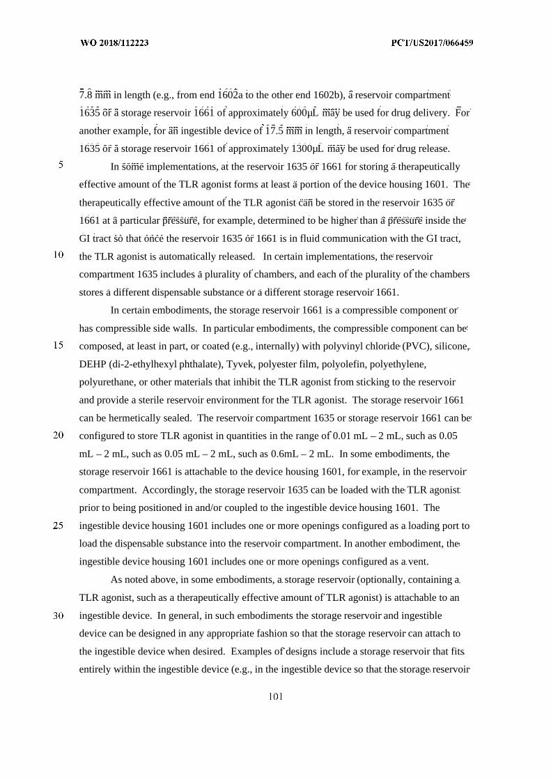
7.8 mm in length (e.g., from end 1602a to the other end 1602b), a reservoir compartment
1635 or a storage reservoir 1661 of approximately 600µL may be used for drug delivery. For
another example, for an ingestible device of 17.5 mm in length, a reservoir compartment
1635 or a storage reservoir 1661 of approximately 1300µL may be used for drug release.
In some implementations, at the reservoir 1635 or 1661 for storing a therapeutically
effective amount of the TLR agonist forms at least a portion of the device housing 1601. The
therapeutically effective amount of the TLR agonist can be stored in the reservoir 1635 or
1661 at a particular pressure, for example, determined to be higher than a pressure inside the
GI tract so that once the reservoir 1635 or 1661 is in fluid communication with the GI tract,
the TLR agonist is automatically released. In certain implementations, the reservoir
compartment 1635 includes a plurality of chambers, and each of the plurality of the chambers
stores a different dispensable substance or a different storage reservoir 1661.
In certain embodiments, the storage reservoir 1661 is a compressible component or
has compressible side walls. In particular embodiments, the compressible component can be
composed, at least in part, or coated (e.g., internally) with polyvinyl chloride (PVC), silicone,
DEHP (di-2-ethylhexyl phthalate), Tyvek, polyester film, polyolefin, polyethylene,
polyurethane, or other materials that inhibit the TLR agonist from sticking to the reservoir
and provide a sterile reservoir environment for the TLR agonist. The storage reservoir 1661
can be hermetically sealed. The reservoir compartment 1635 or storage reservoir 1661 can be
configured to store TLR agonist in quantities in the range of 0.01 mL – 2 mL, such as 0.05
mL – 2 mL, such as 0.05 mL – 2 mL, such as 0.6mL – 2 mL. In some embodiments, the
storage reservoir 1661 is attachable to the device housing 1601, for example, in the reservoir
compartment. Accordingly, the storage reservoir 1635 can be loaded with the TLR agonist
prior to being positioned in and/or coupled to the ingestible device housing 1601. The
ingestible device housing 1601 includes one or more openings configured as a loading port to
load the dispensable substance into the reservoir compartment. In another embodiment, the
ingestible device housing 1601 includes one or more openings configured as a vent.
As noted above, in some embodiments, a storage reservoir (optionally, containing a
TLR agonist, such as a therapeutically effective amount of TLR agonist) is attachable to an
ingestible device. In general, in such embodiments the storage reservoir and ingestible
device can be designed in any appropriate fashion so that the storage reservoir can attach to
the ingestible device when desired. Examples of designs include a storage reservoir that fits
entirely within the ingestible device (e.g., in the ingestible device so that the storage reservoir
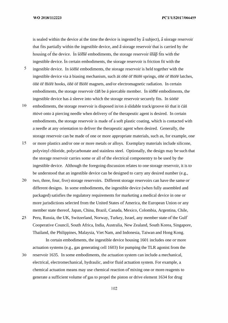
is sealed within the device at the time the device is ingested by a subject), a storage reservoir
that fits partially within the ingestible device, and a storage reservoir that is carried by the
housing of the device. In some embodiments, the storage reservoir snap fits with the
ingestible device. In certain embodiments, the storage reservoir is friction fit with the
ingestible device. In some embodiments, the storage reservoir is held together with the
ingestible device via a biasing mechanism, such as one or more springs, one or more latches,
one or more hooks, one or more magnets, and/or electromagnetic radiation. In certain
embodiments, the storage reservoir can be a piercable member. In some embodiments, the
ingestible device has a sleeve into which the storage reservoir securely fits. In some
embodiments, the storage reservoir is disposed in/on a slidable track/groove so that it can
move onto a piercing needle when delivery of the therapeutic agent is desired. In certain
embodiments, the storage reservoir is made of a soft plastic coating, which is contacted with
a needle at any orientation to deliver the therapeutic agent when desired. Generally, the
storage reservoir can be made of one or more appropriate materials, such as, for example, one
or more plastics and/or one or more metals or alloys. Exemplary materials include silicone,
polyvinyl chloride, polycarbonate and stainless steel. Optionally, the design may be such that
the storage reservoir carries some or all of the electrical componentry to be used by the
ingestible device. Although the foregoing discussion relates to one storage reservoir, it is to
be understood that an ingestible device can be designed to carry any desired number (e.g.,
two, three, four, five) storage reservoirs. Different storage reservoirs can have the same or
different designs. In some embodiments, the ingestible device (when fully assembled and
packaged) satisfies the regulatory requirements for marketing a medical device in one or
more jurisdictions selected from the United States of America, the European Union or any
member state thereof, Japan, China, Brazil, Canada, Mexico, Colombia, Argentina, Chile,
Peru, Russia, the UK, Switzerland, Norway, Turkey, Israel, any member state of the Gulf
Cooperative Council, South Africa, India, Australia, New Zealand, South Korea, Singapore,
Thailand, the Philippines, Malaysia, Viet Nam, and Indonesia, Taiwan and Hong Kong.
In certain embodiments, the ingestible device housing 1601 includes one or more
actuation systems (e.g., gas generating cell 1603) for pumping the TLR agonist from the
reservoir 1635. In some embodiments, the actuation system can include a mechanical,
electrical, electromechanical, hydraulic, and/or fluid actuation system. For example, a
chemical actuation means may use chemical reaction of mixing one or more reagents to
generate a sufficient volume of gas to propel the piston or drive element 1634 for drug

release. The actuation system can be integrated into the reservoir compartment 1635 or can
be an auxiliary system acting on or outside of the reservoir compartment 1635. For example,
the actuation system can include pumping system for pushing/pulling the TLR agonist out of
the reservoir compartment 1635 or the actuation system can be configured to cause the
reservoir compartment 1635 to change structurally so that the volume inside of the reservoir
compartment 1635 changes, thereby dispensing the TLR agonist from the reservoir
compartment 1635. The actuation system can include an energy storage component such as a
battery or a capacitor for powering the actuation system. The actuation system can be
actuated via gas pressure or a system storing potential energy, such as energy from an elastic
reservoir component being expanded during loading of the reservoir and after being
positioned in the ingestible device housing 1601 being subsequently released from the
expanded state when the ingestible device housing is at the location for release within the GI
tract. In certain embodiments, the reservoir compartment 1635 can include a membrane
portion, whereby the TLR agonist is dispensed from the reservoir compartment 1635 or
storage reservoir 1661 via osmotic pressure.
In particular embodiments the storage reservoir 1661 is in a form of a bellow that is
configured to be compressed via a pressure from the gas generating cell. The TLR agonist
may be loaded into the bellow, which may be compressed by gas generation from the gas
generating cell or other actuation means to dispense the dispensable substance through the
dispensing outlet 1607 and out of the housing 1601. In some embodiments, the ingestible
device includes a capillary plate placed between the gas generating cell and the first end of
the housing, and a wax seal between the gas generating cell and the reservoir, wherein the
wax seal is configured to melt and the dispensable substance is pushed through the capillary
plate by a pressure from the gas generating cell. The shape of the bellow may aid in
controlled delivery. The reservoir compartment 1635 includes a dispensing outlet, such as a
valve or dome slit 1662 extending out of an end of the housing 1601, in accordance with
particular implementations. Thus when the bellow is being compressed, the dispensable
substance may be propelled out of the bellow through the valve or the dome slit.
In certain embodiments, the reservoir compartment 1635 includes one or more valves
(e.g. a valve in the dispensing outlet 1607) that are configured to move or open to fluidly
couple the reservoir compartment 1635 to the GI tract. In certain embodiments, a housing
wall of the housing 1601 can form a portion of the reservoir compartment 1635. In certain
embodiments, the housing walls of the reservoir serve as a gasket. One or more of the one or
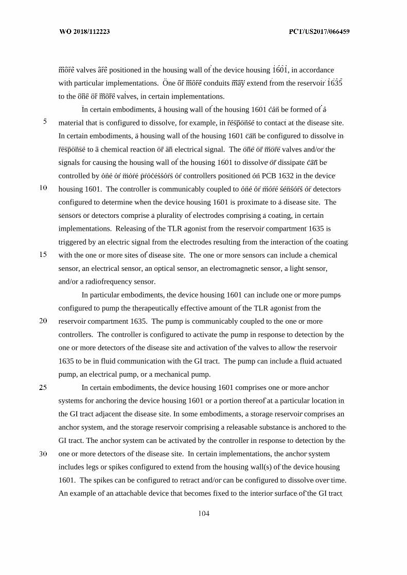
more valves are positioned in the housing wall of the device housing 1601, in accordance
with particular implementations. One or more conduits may extend from the reservoir 1635
to the one or more valves, in certain implementations.
In certain embodiments, a housing wall of the housing 1601 can be formed of a
material that is configured to dissolve, for example, in response to contact at the disease site.
In certain embodiments, a housing wall of the housing 1601 can be configured to dissolve in
response to a chemical reaction or an electrical signal. The one or more valves and/or the
signals for causing the housing wall of the housing 1601 to dissolve or dissipate can be
controlled by one or more processors or controllers positioned on PCB 1632 in the device
housing 1601. The controller is communicably coupled to one or more sensors or detectors
configured to determine when the device housing 1601 is proximate to a disease site. The
sensors or detectors comprise a plurality of electrodes comprising a coating, in certain
implementations. Releasing of the TLR agonist from the reservoir compartment 1635 is
triggered by an electric signal from the electrodes resulting from the interaction of the coating
with the one or more sites of disease site. The one or more sensors can include a chemical
sensor, an electrical sensor, an optical sensor, an electromagnetic sensor, a light sensor,
and/or a radiofrequency sensor.
In particular embodiments, the device housing 1601 can include one or more pumps
configured to pump the therapeutically effective amount of the TLR agonist from the
reservoir compartment 1635. The pump is communicably coupled to the one or more
controllers. The controller is configured to activate the pump in response to detection by the
one or more detectors of the disease site and activation of the valves to allow the reservoir
1635 to be in fluid communication with the GI tract. The pump can include a fluid actuated
pump, an electrical pump, or a mechanical pump.
In certain embodiments, the device housing 1601 comprises one or more anchor
systems for anchoring the device housing 1601 or a portion thereof at a particular location in
the GI tract adjacent the disease site. In some embodiments, a storage reservoir comprises an
anchor system, and the storage reservoir comprising a releasable substance is anchored to the
GI tract. The anchor system can be activated by the controller in response to detection by the
one or more detectors of the disease site. In certain implementations, the anchor system
includes legs or spikes configured to extend from the housing wall(s) of the device housing
1601. The spikes can be configured to retract and/or can be configured to dissolve over time.
An example of an attachable device that becomes fixed to the interior surface of the GI tract
is described in PCT Patent Application PCT/US2015/012209, “Gastrointestinal Sensor
Implantation System”, filed January 21, 2015, which is hereby incorporated by reference
herein in its entirety.
FIG. 20 provides an example structural diagram having a flexible diaphragm 1665
that may deform towards the dispensing outlet 1607 when the gas generating cell 1603
generates gas. The dispensable substance may then be propelled by the deformed diaphragm
out of the housing through the dispensing outlet 1607. The dispensing outlet 1607 shown at
FIG. 20 is in the form of a ring valve, however, any outlet design can be applied.
In some embodiments, an ingestible device can have an umbrella-shaped exit valve
structure as a dispensing outlet of the ingestible device. Optionally, an ingestible device can
have a flexible diaphragm to deform for drug delivery, and/or an integrated piston and gas
generating cell such that the gas generating cell is movable with the piston to push for drug
delivery.
In certain embodiments, an ingestible device can be anchored within the intestine by
extending hooks from the ingestible device after it has entered the region of interest. For
example, when the ingestible device determines it has arrived at a location within the GI
tract, the hooks can be actuated to extend outside of the ingestible device to catch in the
intestinal wall and hold the ingestible device in the respective location. In some
embodiments, the hook can pierce into the intestinal wall to hold the ingestible device 100 in
place. The hooks can be hollow. A hollow hook can be used to anchor the ingestible device
and/or to dispense a substance from the dispensable substance, e.g., into the intestinal wall.
In some embodiments an ingestible device includes an intestinal gripper to grip a
portion of the intestinal wall for delivering the dispensable substance. Such a gripper can
include two or more arms configured to out of the device and close to grip a portion of the
intestinal wall.
An injecting needle can be used with the anchoring arms to inject dispensable
substance into the intestinal wall after a portion of the intestinal wall is gripped.
In some embodiments, when the gas generating cell generates gas to propel the piston
to move towards the nozzle such that the dispensable substance can be pushed under the
pressure to break a burst disc to be injected via the nozzle.
In some embodiments, an ingestible device has a jet delivery mechanism with
enhanced usable volume of dispensable substance. For example, the nozzle may be placed at
the center of the ingestible device, and gas channels may be placed longitudinally along the
wall of the ingestible device to transport gas from the gas generating cell to propel the piston,
which is placed at an end of the ingestible device.
In some embodiments, the ingestible device can use osmotic pressure to adhere a
suction device of the ingestible device to the intestinal wall. For example, the ingestible
device may have an osmotic mechanism that has a chamber storing salt crystals. The
chamber can include a mesh placed in proximate to a burst valve at one end of the chamber,
and a reverse osmosis (RO) membrane placed in proximate to a valve on the other end of the
chamber. A suction device, e.g., two or more suction fingers, is placed outside of the
chamber with an open outlet exposed to luminal fluid in the GI tract. When the osmotic
mechanism is inactivated, e.g., the valve is closed so that no luminal fluid is drawn into the
osmotic chamber. When the osmotic mechanism is activated by opening the valve, luminal
fluid enters the ingestible device through an outlet of the suction device and enters the
osmotic chamber through the valve. The salt in the chamber is then dissolved into the fluid.
The RO membrane prevents any fluid to flow in the reverse direction, e.g., from inside the
chamber to the valve. The fluid continues to flow until all the salt contained in the chamber
is dissolved or until intestinal tissue is drawn into the suction device. As luminal fluid keeps
flowing into the chamber, the solution of the luminal fluid with dissolved salt in the chamber
may reduce osmotic pressure such that the suction force at may also be reduced. In this way,
suction of the intestinal tissue may stall before the tissue is in contact with the valve to avoid
damage to the intestinal tissue.
An ingestible device employing an osmotic mechanism can also include a suction device as
illustrated. The suction device can be two or more suction fingers 347a-b disposed proximate
to the outlet. The outlet can be connected to a storage reservoir storing the dispensable
substance (e.g., therapeutic agent). The storage reservoir can contact a piston (similar to 104
in FIG. 16), which can be propelled by pressure generated from the osmotic pump to move
towards the outlet. The osmotic pump can be similar to the osmotic mechanism described in
the preceding paragraph. A breakaway section can be placed in proximate to the other end
(opposite to the end where the outlet 107 is disposed) of the ingestible device.
In some embodiments, tumbling suction by an ingestible device is used. Such an
ingestible device does not require any electronics or other actuation elements. Such an
ingestible device may constantly, intermittently, or periodically tumble when travelling
through the intestine. When the ingestible device tumbles to a position that the outlet is in
direct contact with the intestinal wall, a suction process similar to that described in the
preceding paragraph may occur. Additional structural elements such as fins, flutes or the like
may be added to the outer wall of the ingestible device 100 to promote the tumbling motion.
In certain embodiments, the reservoir is an anchorable reservoir, which is a reservoir
comprising one or more anchor systems for anchoring the reservoir at a particular location in
the GI tract adjacent the disease site. In certain embodiments, the anchor system includes
legs or spikes or other securing means such as a piercing element, a gripping element, a
magnetic-flux-guiding element, or an adhesive material, configured to extend from the
anchorable reservoir of the device housing. The spikes can be configured to retract and/or
can be configured to dissolve over time. In some embodiments, the anchorable reservoir is
suitable for localizing, positioning and/or anchoring. In some embodiments, the anchorable
reservoir is suitable for localizing, and positioning and/or anchoring by an endoscope. In
some embodiments, the anchorable reservoir is connected to the endoscope. In some
embodiments, the anchorable reservoir is connected to the endoscope in a manner suitable for
oral administration. In some embodiments, the anchorable reservoir is connected to the
endoscope in a manner suitable for rectal administration. Accordingly, provided herein in
some embodiments is an anchorable reservoir is connected to an endoscope wherein the
anchorable reservoir comprises a therapeutically effective amount of the TLR agonist. In
some embodiments the endoscope is fitted with a spray catheter.
Exemplary embodiments of anchorable reservoirs are as follows. In more particular
examples of the following exemplary embodiments the reservoir is connected to an
endoscope.
In one embodiment, the anchorable reservoir comprises an implant capsule for
insertion into a body canal to apply radiation treatment to a selected portion of the body
canal. The reservoir includes a body member defining at least one therapeutic treatment
material receiving chamber and at least one resilient arm member associated with the body
member for removably engaging the body canal when the device is positioned therein.
In one embodiment the anchorable reservoir has multiple suction ports and permits
multiple folds of tissue to be captured in the suction ports with a single positioning of the
device and attached together by a tissue securement mechanism such as a suture, staple or
other form of tissue bonding. The suction ports may be arranged in a variety of configurations
on the reservoir to best suit the desired resulting tissue orientation.
In some embodiments an anchorable reservoir comprises a tract stimulator and/or
monitor IMD comprising a housing enclosing electrical stimulation and/or monitoring
circuitry and a power source and an elongated flexible member extending from the housing to
an active fixation mechanism adapted to be fixed into the GI tract wall is disclosed. After
fixation is effected, the elongated flexible member bends into a preformed shape that presses
the housing against the mucosa so that forces that would tend to dislodge the fixation
mechanism are minimized. The IMD is fitted into an esophageal catheter lumen with the
fixation mechanism aimed toward the catheter distal end opening whereby the bend in the
flexible member is straightened. The catheter body is inserted through the esophagus into the
GI tract cavity to direct the catheter distal end to the site of implantation and fix the fixation
mechanism to the GI tract wall. The IMD is ejected from the lumen, and the flexible member
assumes its bent configuration and lodges the hermetically sealed housing against the
mucosa. A first stimulation/sense electrode is preferably an exposed conductive portion of the
housing that is aligned with the bend of the flexible member so that it is pressed against the
mucosa. A second stimulation/sense electrode is located at the fixation site.
In some embodiments a reservoir for sensing one or more parameters of a patient is
anchored to a tissue at a specific site and is released from a device, using a single actuator
operated during a single motion. As an example, a delivery device may anchor the capsule to
the tissue site and release the reservoir from the delivery device during a single motion of the
actuator.
In some embodiments a device is provided comprising: a reservoir configured to
contain a fluid, the reservoir having at least one outlet through which the fluid may exit the
reservoir; a fluid contained within the reservoir; a primary material contained within the
reservoir and having a controllable effective concentration in the fluid; and at least one
electromagnetically responsive control element located in the reservoir or in a wall of the
reservoir and adapted for modifying the distribution of the primary material between a first
active form carried in the fluid and a second form within the reservoir in response to an
incident electromagnetic control signal, the effective concentration being the concentration of
the first active form in the fluid, whereby fluid exiting the reservoir carries the primary
material in the first active form at the effective concentration.
In some embodiments systems and methods are provided for implementing or
deploying medical or veterinary devices or reservoirs (a) operable for anchoring at least
partly within a digestive tract, (b) small enough to pass through the tract per vias naturales
and including a wireless-control component, (c) having one or more protrusions positionable
adjacent to a mucous membrane, (d) configured to facilitate redundant modes of anchoring,
(e) facilitating a "primary" material supply deployable within a stomach for an extended
and/or controllable period, (f) anchored by one or more adaptable extender modules
supported by a subject's head or neck, and/or (g) configured to facilitate supporting at least a
sensor within a subject's body lumen for up to a day or more.
In certain embodiments, the reservoir is attachable to an ingestible device. In certain
embodiments, the ingestible device comprises a housing and the reservoir is attachable to the
housing. In certain embodiments, the attachable reservoir is also an anchorable reservoir,
such as an anchorable reservoir comprising one or more anchor systems for anchoring the
reservoir at a particular location in the GI tract as disclosed hereinabove.
Accordingly, in certain embodiments, provided herein is a TLR agonist for use in a
method of treating a disease of the gastrointestinal tract as disclosed herein, wherein the TLR
agonist is contained in a reservoir suitable for attachment to a device housing, and wherein
the method comprises attaching the reservoir to the device housing to form the ingestible
device, prior to orally administering the ingestible device to the subject.
In certain embodiments, provided herein is an attachable reservoir containing a TLR
agonist for use in a method of treating a disease of the gastrointestinal tract, wherein the
method comprises attaching the reservoir to a device housing to form an ingestible device and
orally administering the ingestible device to a subject, wherein the TLR agonist is released by
device at a location in the gastrointestinal tract of the subject that is proximate to one or more
sites of disease.
In certain embodiments, provided herein is an attachable reservoir containing a TLR
agonist, wherein the reservoir is attachable to a device housing to form an ingestible device
that is suitable for oral administration to a subject and that is capable of releasing the TLR
agonist at a location in the gastrointestinal tract of the subject that is proximate to one or
more sites of disease.
In particular implementation the ingestible device includes cameras (e.g., video
cameras) that affords inspection of the entire GI tract without discomfort or the need for
sedation, thus avoiding many of the potential risks of conventional endoscopy. Video
imaging can be used to help determine one or more characteristics of the GI tract, including
the location of disease (e.g., presence or location of inflamed tissue and/or lesions associated
with inflammatory bowel disease). In some embodiments, the ingestible device 101 may
comprise a camera for generating video imaging data of the GI tract which can be used to
determine, among other things, the location of the device. Examples of video imaging
capsules include Medtronic’s PillCam™, Olympus’ Endocapsule®, and IntroMedic’s
MicroCam™. For a review of imaging capsules, see Basar et al. “Ingestible Wireless Capsule
Technology: A Review of Development and Future Indication” International Journal of
Antennas and Propagation (2012); 1-14). Other imaging technologies implemented with the
device 101 can include thermal imaging cameras, and those that employ ultrasound or
Doppler principles to generate different images (see Chinese patent application
CN104473611: “Capsule endoscope system having ultrasonic positioning function”.
Ingestible devices can be equipped with sources for generating reflected light,
including light in the Ultraviolet, Visible, Near-infrared and/or Mid-infrared spectrum, and
the corresponding detectors for spectroscopy and hyperspectral imaging. Likewise,
autofluorescense may be used to characterize GI tissue (e.g., subsurface vessel information),
or low-dose radiation (see Check-Cap™) can be used to obtain 3D reconstructed images.
Device Components
An ingestible device in accordance with particular embodiments of the present
invention may comprise a component made of a non-digestible material and contain the TLR
agonist. In some embodiments, the material is plastic.
It is envisaged that the device is single-use. The device is loaded with a drug prior to
the time of administration. In some embodiments, it may be preferred that there is provided a
medicinal product comprising the device pre-filled with the drug.
Anchoring components
Several systems may actively actuate and control the capsule position and orientation
in different sections of the GI tract. Examples include leg-like or anchor-like mechanisms that
can be deployed by an ingestible device to resist peristaltic forces in narrowed sections of the
GI tract, such as the intestine, and anchor the device to a location. Other systems employ
magnetic shields of different shapes that can interact with external magnetic fields to move
the device. These mechanisms may be particularly useful in areas outside of the small
intestine, like the cecum and large intestine.
An anchoring mechanism may be a mechanical mechanism. For example, a device
may be a capsule comprising a plurality of legs configured to steer the capsule. The number
of legs in the capsule may be, for example, two, four, six, eight, ten or twelve. The aperture
between the legs of the device may be up to about 35 mm; about 30 to about 35 mm; about 35
to about 75 mm; or about 70 to about 75 mm. The contact area of each leg may be varied to
reduce impact on the tissue. One or more motors in the capsule may each actuate a set of legs
independently from the other. The motors may be battery-powered motors.
An anchoring mechanism may be a non-mechanical mechanism. For example, a
device may be a capsule comprising a permanent magnet located inside the capsule. The
capsule may be anchored at the desired location of the GI tract by an external magnetic field.
An anchoring mechanism may comprise a non-mechanical mechanism and a
mechanical mechanism. For example, a device may be a capsule comprising one or more
legs, one or more of which are coated with an adhesive material.
Locomotion components
Ingestible devices can be active or passive, depending on whether they have
controlled or non-controlled locomotion. Passive (non-controlled) locomotion is more
commonly used among ingestible devices given the challenges of implementing a locomotion
module. Active (controlled) locomotion is more common in endoscopic ingestible capsules.
For example, a capsule may comprise a miniaturized locomotion system (internal
locomotion). Internal locomotion mechanisms may employ independent miniaturized
propellers actuated by DC brushed motors, or the use of water jets. As an example, a
mechanism may comprise flagellar or flap-based swimming mechanisms. As an example, a
mechanism may comprise cyclic compression/extension shape-memory alloy (SMA) spring
actuators and anchoring systems based on directional micro-needles. As an example, a
mechanism may comprise six SMA actuated units, each provided with two SMA actuators
for enabling bidirectional motion. As an example, a mechanism may comprise a motor
adapted to electrically stimulating the GI muscles to generate a temporary restriction in the
bowel.
As an example, a capsule may comprise a magnet and motion of the capsule is caused
by an external magnetic field. For example, a locomotion system may comprise an ingestible
capsule and an external magnetic field source. For example, the system may comprise an
ingestible capsule and magnetic guidance equipment such as, for example, magnetic
resonance imaging and computer tomography, coupled to a dedicated control interface.
In some embodiments drug release mechanisms may also be triggered by an external
condition, such as temperature, pH, movement, acoustics, or combinations thereof.
Sampling Components
Ingestible devices may comprise a mechanism adapted to permit the collection of
tissue samples. In some examples, this is achieved using electro-mechanical solutions to
collect and store the sample inside an ingestible device. As an example, a biopsy mechanism
may include a rotational tissue cutting razor fixed to a torsional spring or the use of
microgrippers to fold and collect small biopsies. As an example, Over-the-scope clips
(OTSC®) may be used to perform endoscopic surgery and/or biopsy. As an example of the
methods disclosed herein, the method may comprise releasing a TLR agonist and collecting a
sample inside the device. As an example, the method may comprise releasing a TLR agonist
and collecting a sample inside the device in a single procedure.
FIG. 21 illustrates an example ingestible device 2100 with multiple openings in the
housing. The ingestible device 2100 has an outer housing with a first end 2102A, a second
end 2102B, and a wall 2104 extending longitudinally from the first end 2102A to the second
end 2102B. Ingestible device 2100 has a first opening 2106 in the housing, which is
connected to a second opening 2108 in the housing. The first opening 2106 of the ingestible
device 2100 is oriented substantially perpendicular to the second opening 2108, and the
connection between the first opening 2106 and the second opening 2108 forms a curved
chamber 2110 within the ingestible device 2100.
The overall shape of the ingestible device 2100, or any of the other ingestible devices
discussed in this disclosure, may be similar to an elongated pill or capsule.
In some embodiments, a portion of the curved chamber 2110 may be used as a
sampling chamber, which may hold samples obtained from the GI tract. In some
embodiments the curved chamber 2110 is subdivided into sub-chambers, each of which may
be separated by a series of one or more valves or interlocks.
In some embodiments, the first opening 2106, the second opening 2108, or the curved
chamber 2110 include one or more of a hydrophilic or hydrophobic material, a sponge, a
valve, or an air permeable membrane.
The use of a hydrophilic material or sponge may allow samples to be retained within
the curved chamber 2110, and may reduce the amount of pressure needed for fluid to enter
through the first opening 2106 and dislodge air or gas in the curved chamber 2110. Examples
of hydrophilic materials that may be incorporated into the ingestible device 2100 include
hydrophilic polymers such as polyvinyl alcohol, polyvinyl pyrrolidone, and the like.
Similarly, materials that have undergone various types of treatments, such as plasma
treatments, may have suitable hydrophilic properties, and may be incorporated into the
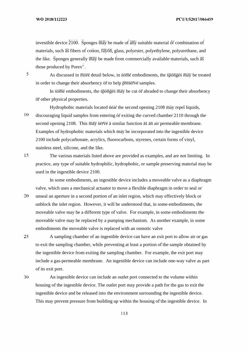
investible device 2100. Sponges may be made of any suitable material or combination of
materials, such as fibers of cotton, rayon, glass, polyester, polyethylene, polyurethane, and
the like. Sponges generally may be made from commercially available materials, such as
those produced by Porex® .
As discussed in more detail below, in some embodiments, the sponges may be treated
in order to change their absorbency or to help preserve samples.
In some embodiments, the sponges may be cut or abraded to change their absorbency
or other physical properties.
Hydrophobic materials located near the second opening 2108 may repel liquids,
discouraging liquid samples from entering or exiting the curved chamber 2110 through the
second opening 2108. This may serve a similar function as an air permeable membrane.
Examples of hydrophobic materials which may be incorporated into the ingestible device
2100 include polycarbonate, acrylics, fluorocarbons, styrenes, certain forms of vinyl,
stainless steel, silicone, and the like.
The various materials listed above are provided as examples, and are not limiting. In
practice, any type of suitable hydrophilic, hydrophobic, or sample preserving material may be
used in the ingestible device 2100.
In some embodiments, an ingestible device includes a moveable valve as a diaphragm
valve, which uses a mechanical actuator to move a flexible diaphragm in order to seal or
unseal an aperture in a second portion of an inlet region, which may effectively block or
unblock the inlet region. However, it will be understood that, in some embodiments, the
moveable valve may be a different type of valve. For example, in some embodiments the
moveable valve may be replaced by a pumping mechanism. As another example, in some
embodiments the moveable valve is replaced with an osmotic valve
A sampling chamber of an ingestible device can have an exit port to allow air or gas
to exit the sampling chamber, while preventing at least a portion of the sample obtained by
the ingestible device from exiting the sampling chamber. For example, the exit port may
include a gas-permeable membrane. An ingestible device can include one-way valve as part
of its exit port.
An ingestible device can include an outlet port connected to the volume within
housing of the ingestible device. The outlet port may provide a path for the gas to exit the
ingestible device and be released into the environment surrounding the ingestible device.
This may prevent pressure from building up within the housing of the ingestible device. In
some embodiments, an ingestible device does not include an outlet port, and the gas stays
inside the volume of the ingestible device. In some embodiments, the outlet port may contain
a gas permeable membrane, a one-way valve, a hydrophobic channel, or some other
mechanism to avoid unwanted material, (e.g., fluids and solid particulates from within the GI
tract), from entering the ingestible device through the outlet port.
In some embodiments, the ingestible device may include a sensor within or proximate
to the sampling chamber. For example, this sensor may be used to detect various properties
of a sample contained within the sampling chamber, or this sensor may be used to detect the
results of an assay technique applied to the sample contained within the sampling chamber.
In some embodiments, a hydrophilic sponge is located within the sampling chamber,
and the hydrophilic sponge may be configured to absorb the sample as the sample enters the
sampling chamber. In some embodiments, the hydrophilic sponge fills a substantial portion
of the sampling chamber, and holds the sample for an extended period of time. This may be
particularly advantageous if the sample is collected from the ingestible device after the
ingestible device exits the body. In some embodiments, the hydrophilic sponge is placed on
only certain surfaces or fills only certain portions of the sampling chamber. For example, it
may be possible to line certain walls (or all walls) of the sampling chamber with a
hydrophilic sponge to assist in drawing in the sample, while leaving some (or none) of the
walls of the sampling chamber uncovered. Leaving walls uncovered may allow the use of
diagnostics or assay techniques that require a relatively un-obscured optical path.
In some embodiments, the ingestible device may include a sealed vacuum chamber
connected to the exit port, or connected directly or indirectly to the sampling chamber. In
some embodiments a pin valve may be used as a moveable valve (e.g., as moveable valve of
ingestible device). In certain embodiments, a rotary valve may be used as a moveable valve
(e.g., as moveable valve of ingestible device). In some embodiments, a flexible diaphragm,
or diaphragm valve, may be used as a moveable valve (e.g., as moveable valve of ingestible
device). In certain embodiments, a mechanism is near the diaphragm or in direct contact with
the diaphragm. The spring mechanism may apply pressure to the diaphragm to oppose the
pressure applied by the mechanical actuator, which may cause the flexible diaphragm to be
moved into an open position when the mechanical actuator is not applying pressure to the
flexible diaphragm. Additionally, this may ensure that the diaphragm valve remains open
when the mechanical actuator is not applying pressure across the flexible diaphragm. In
some embodiments, moving the mechanical actuator from a closed position to an open
position causes a volume of the inlet region within the ingestible device to increase. This
may cause the pressure within the inlet region to be reduced, generating suction to draw a
sample into the inlet region. Similarly, moving the mechanical actuator from an open
position to a closed position may cause the volume of the inlet region to be reduced. This
may cause the pressure within the inlet region to be increased, pushing the sample out of the
inlet region. Depending on the design of the inlet region, the mechanical actuator, and the
moveable valve, this may push the sample into the sampling chamber rather than pushing the
sample back through the opening in the ingestible device.
FIG. 22 depicts a cross-sectional view of a portion of the interior of ingestible device
3000. As shown in FIG. 22, the interior of ingestible device 3000 includes a valve system
3100 and a sampling system 3200. Valve system 3100 is depicted as having a portion that is
flush with the opening 3018 so that valve system 3100 prevents fluid exterior to ingestible
device 2000 from entering sampling system 3200. However, as described in more detail
below with reference to FIGs. 22-27, valve system 3100 can change position so that valve
system 3100 allows fluid exterior to ingestible device 3000 to enter sampling system 3200.
FIGs. 23 and 27 illustrate valve system 3100 in more detail. As shown in FIG. 23,
valve system 3100 includes an actuation mechanism 3110, a trigger 3120, and a gate 3130.
In FIGs. 23 and 7, a leg 3132 of gate 3130 is flush against, and parallel with, housing wall
3016 so that gate leg 3132 covers opening 3018 to prevent fluid exterior to ingestible device
3000 (e.g., fluid in the GI tract) from entering the interior of ingestible device 3000. A
protrusion 3134 of gate 3130 engages a lip 3122 of trigger 3120. A peg 3124 of trigger 3120
engages a wax pot 3112 of actuation mechanism 3110. Referring to FIG. 27, a biasing
mechanism 3140 includes a compression spring 3142 that applies an upward force on gate
3130. Biasing mechanism 3140 also includes a torsion spring 3144 that applies a force on
trigger 3120 in the counter-clockwise direction. In FIGs. 23 and 27, the force applied by
torsion spring 3144 is counter-acted by the solid wax in pot 3112, and the force applied by
compression spring 3142 is counter-acted by lip 3122.
FIGs. 24A and FIG 24B show an embodiment of the manner in which actuation
mechanism 3110 actuates movement of trigger 3120. Similar to FIGs. 23 and 27, FIG. 24A
shows a configuration in which peg 3124 applies a force against solid wax pot 3112 due to
torsion spring 3144, and in which the solid nature of wax pot 3112 resists the force applied by
peg 3124. A control unit 3150 is in signal communication with valve system 3100. During
use of ingestible device 3000, a control unit 3150 receives a signal, indicating that the
position of valve system 3100 should change, e.g., so that ingestible device 3000 can take a
sample of a fluid in the GI tract. Control unit 3150 sends a signal that causes a heating
system 3114 of actuation system 3100 to heat the wax in pot 3112 so that the wax melts. As
shown in FIG. 24B, the melted wax is not able to resist the force applied by peg 3124 so that,
under the force of torsion spring 3144, trigger 3120 moves in a counter-clockwise fashion.
FIGs. 25A and 25B illustrate the interaction of trigger 3120 and gate 3130 before and
after actuation. As shown in FIG 25A, when wax pot 3112 is solid (corresponding to the
configuration shown in FIG. 24A), protrusion 3134 engages lip 3122, which prevents the
force of compression spring 3142 from moving gate 3130 upward. As shown in FIG. 25B,
when the wax in pot 3112 melts (FIG. 24B), trigger 3120 moves counter-clockwise, and lip
3122 disengages from protrusion 3134. This allows the force of compression spring 3142 to
move gate 3130 upward. As seen by comparing FIG. 25A to FIG. 25B, the upward
movement of gate 3130 results in an upward movement of an opening 3136 in gate leg 3132.
FIGs. 26A and 26B illustrate the impact of the upward movement of opening 3136 on
the ability of ingestible device 3000 to obtain a sample. As shown in FIG. 26A, when the
wax in pot 3112 is solid (FIGs. 24A and 25A), opening 3136 in is not aligned with opening
3018 in wall 3016 of ingestible device 3000. Instead, gate leg 3132 covers opening 3018 and
blocks fluid from entering the interior of ingestible device 3000. As shown in FIG. 26B,
when the wax in pot 3112 is melted and trigger 3120 and gate 3130 have moved (FIGs. 24B
and 42B), opening 3136 in gate 3130 is aligned with opening 3018 in wall 3016. In this
configuration, fluid that is exterior to ingestible device 3000 (e.g., in the GI tract) can enter
the interior of ingestible device 3000 via openings 3018 and 3036.
FIG. 27 illustrates a more detailed view of ingestible device 3000 including valve
system 3100 and sampling system 3200.
While the foregoing description is made with regard to a valve system having one
open position and one closed position (e.g., a two-stage valve system), the disclosure is not
limited in this sense. Rather, the concepts described above with regard to a two stage valve
system can be implemented with a valve system have more than two stages (e.g., three stages,
four stages, five stages, etc.).
As noted above in addition to a valve system, an ingestible device includes a sampling
system. FIG. 28 illustrates a partial cross sectional view of ingestible device 3000 with
sampling system 3200 and certain components of valve system 3100. Sampling system 3200
includes a series of sponges configured to absorb fluid from an opening, move the fluid to a
location within the housing, and prepare the fluid for testing. Preparation for testing may
include filtering the fluid and combining the fluid with a chemical assay. The assay may be
configured to dye cells in the filtered sample. The series of sponges includes a wicking
sponge 3210, a transfer sponge 3220, a volume sponge 3230, and an assay sponge 3240.
Sampling system 3200 also includes a membrane 3270 located between assay sponge 3240
and a vent 3280 for gases to leave sampling system 3200. A cell filter 3250 is located
between distal end 3214 of wicking sponge 3210 and a first end 3222 of transfer sponge
3220. Membrane 3270 is configured to allow one or more gases to leave sampling system
3200 via an opening 3280, while maintaining liquid in sampling system 3200.
FIG. 29 is a highly schematic illustration of an ingestible device 4000 that contains
multiple different systems that cooperate for obtaining a sample and analyzing a sample, e.g.,
within the GI tract of a subject. Ingestible device 4000 includes a power system 4100 (e.g.,
one or more batteries), configured to power an electronics system 4200 (e.g., including a
control system, optionally in signal communication with an external base station), a valve
system 4300, a sampling system 4400, and an analytic system 4500. Exemplary analytical
systems include assay systems, such as, for example, optical systems containing one or more
sources of radiation and/or one more detectors.
Some or all of the sponges of the above-described sampling systems may contain one
or more preservatives (see discussion above). Typically, the assay sponge and/or the volume
sponge 3230 and/or the transfer sponge contain one or more preservatives. Typically, the
preservative(s) are selected based on the analyte of interest, e.g., an analyte (such as a protein
biomarker) for a GI disorder.
Communication systems
An ingestible device may be equipped with a communication system adapted to
transmit and/or receive data, including imaging and/or localization data. As an example, a
communication system may employ radiofrequency transmission. Ingestible devices using
radiofrequency communication are attractive because of their efficient transmission through
the layers of the skin. This is especially true for low frequency transmission (UHF-433 ISM
and lower, including the Medical Device Radio Communication Service band (MDRS) band
402-406MHz). In another embodiment, acoustics are used for communications, including the
transmission of data. For example, an ingestible capsule may be able to transmit information
by applying one or more base voltages to an electromechanical transducer or piezoelectric
(e.g., PZT, PVDF, etc.) device to cause the piezoelectric device to ring at particular
frequencies, resulting in an acoustic transmission. A multi-sensor array for receiving the
acoustic transmission may include a plurality of acoustic transducers that receive the acoustic
transmission from a movable device such as an ingestible capsule as described in US Patent
Application No. 11/851214 filed September 6, 2007, incorporated by reference herein in its
entirety.
As an example, a communication system may employ human body communication
technology. Human body communication technology uses the human body as a conductive
medium, which generally requires a large number of sensor electrodes on the skin. As an
example, a communication system may integrate a data storage system.
Environmental Sensors
In some embodiments the device may comprise environmental sensors to measure pH,
temperature, transit times, or combinations thereof. Other examples of environmental sensors
include, but are not limited to a capacitance sensor, an impedance sensor, a heart rate sensor,
acoustic sensor such as a microphone or hydrophone, image sensor, and/or a movement
sensor. In one embodiment, the ingestible device comprises a plurality of different
environmental sensors for generating different kinds of environmental data.
In order to avoid the problem of capsule retention, a thorough past medical and
surgical history should be undertaken. In addition, several other steps have been proposed,
including performing investigations such as barium follow-through. In cases where it is
suspected that there is a high risk of retention, the patient is given a patency capsule a few
days before swallowing an ingestible device. Any dissolvable non-endoscopic capsule may
be used to determine the patency of the GI tract. The patency capsule is usually the same size
as the ingestible device and can be made of cellophane. In some embodiments, the patency
capsule contains a mixture of barium and lactose, which allows visualization by x-ray.
The patency capsule may also include a radiotag or other label, which allows for it to be
detected by radio-scanner externally. The patency capsule may comprise wax plugs, which
allow for intestinal fluid to enter and dissolve the content, thereby dividing the capsule into
small particles.
Accordingly, in some embodiments, the methods herein comprise (a) identifying a
subject having a disease of the gastrointestinal tract and (b) evaluating the subject for
suitability to treatment. In some embodiments, the methods herein comprise evaluating for
suitability to treatment a subject identified as having a disease of the gastrointestinal tract. In
some embodiments, evaluating the subject for suitability to treatment comprises determining
the patency of the subject’s GI tract.
In some embodiments, an ingestible device comprises a tissue anchoring mechanism
for anchoring the ingestible device to a subject’s tissue. For example, an ingestible device
could be administered to a subject and once it reaches the desired location, the tissue
attachment mechanism can be activated or deployed such that the ingestible device, or a
portion thereof, is anchored to the desired location. In some embodiments, the tissue
anchoring mechanism is reversible such that after initial anchoring, the tissue attachment
device is retracted, dissolved, detached, inactivated or otherwise rendered incapable of
anchoring the ingestible device to the subject’s tissue. In some embodiments the attachment
mechanism is placed endoscopically.
In some embodiments, a tissue anchoring mechanism comprises an osmotically-
driven sucker. In some embodiments, the osmotically-driven sucker comprises a first valve
on the near side of the osmotically-driven sucker (e.g., near the subject’s tissue) and a second
one-way valve that is opened by osmotic pressure on the far side of the osmotically-driven
sucker, and an internal osmotic pump system comprising salt crystals and semi-permeable
membranes positioned between the two valves. In such embodiments, osmotic pressure is
used to adhere the ingestible device to the subject’s tissue without generating a vacuum
within the ingestible capsule. After the osmotic system is activated by opening the first
valve, fluid is drawn in through the sucker and expelled through the second burst valve.
Fluid continues to flow until all the salt contained in the sucker is dissolved or until tissue is
drawn into the sucker. As liminal fluid is drawn through the osmotic pump system, solutes
build up between the tissue and the first valve, reducing osmotic pressure. In some
embodiments, the solute buildup stalls the pump before the tissue contacts the valve,
preventing tissue damage. In some embodiments, a burst valve is used on the far side of the
osmotically-driven sucker rather than a one-way valve, such that luminal fluid eventually
clears the saline chamber and the osmotic flow reverses, actively pushing the subject’s tissue
out of the sucker. In some embodiments, the ingestible device may be anchored to the
interior surface of tissues forming the GI tract of a subject. In one embodiment, the ingestible
device comprises a connector for anchoring the device to the interior surface of the GI tract.
The connector may be operable to ingestible device to the interior surface of the GI tract
using an adhesive, negative pressure and/or fastener.
In some embodiments a device comprises a tract stimulator and/or monitor IMD
comprising a housing enclosing electrical stimulation and/or monitoring circuitry and a
power source and an elongated flexible member extending from the housing to an active
fixation mechanism adapted to be fixed into the GI tract wall is disclosed. After fixation is
effected, the elongated flexible member bends into a preformed shape that presses the
housing against the mucosa so that forces that would tend to dislodge the fixation mechanism
are minimized. The IMD is fitted into an esophageal catheter lumen with the fixation
mechanism aimed toward the catheter distal end opening whereby the bend in the flexible
member is straightened. The catheter body is inserted through the esophagus into the GI tract
cavity to direct the catheter distal end to the site of implantation and fix the fixation
mechanism to the GI tract wall. The IMD is ejected from the lumen, and the flexible member
assumes its bent configuration and lodges the hermetically sealed housing against the
mucosa. A first stimulation/sense electrode is preferably an exposed conductive portion of the
housing that is aligned with the bend of the flexible member so that it is pressed against the
mucosa. A second stimulation/sense electrode is located at the fixation site.
In some embodiments a device includes a fixation mechanism to anchor the device to
tissue within a body lumen, and a mechanism to permit selective de-anchoring of the device
from the tissue anchoring site without the need for endoscopic or surgical intervention. An
electromagnetic device may be provided to mechanically actuate the de-anchoring
mechanism. Alternatively, a fuse link may be electrically blown to de-anchor the device. As a
further alternative, a rapidly degradable bonding agent may be exposed to a degradation agent
to de-anchor the device from a bonding surface within the body lumen.
In some embodiments a device is as disclosed in patent publication
WO2015112575A1, incorporated by reference herein in its entirety. The patent publication is
directed to a gastrointestinal sensor implantation system. In some embodiments an orally-
administrable capsule comprises a tissue capture device or reservoir removably coupled to the
orally-administrable capsule, where the tissue capture device including a plurality of fasteners
for anchoring the tissue capture device to gastrointestinal tissue within a body
In some embodiments, the ingestible device contains an electric energy emitting
means, a radio signal transmitting means, a medicament storage means and a remote
actuatable medicament releasing means. The capsule signals a remote receiver as it
progresses through the alimentary tract in a previously mapped route and upon reaching a
specified site is remotely triggered to release a dosage of medicament. Accordingly, in some
embodiments, releasing the TLR agonist is triggered by a remote electromagnetic signal.
In some embodiments, the ingestible device includes a housing introducible into a
body cavity and of a material insoluble in the body cavity fluids, but formed with an opening
covered by a material which is soluble in body cavity fluids. A diaphragm divides the interior
of the housing into a medication chamber including the opening, and a control chamber. An
electrolytic cell in the control chamber generates a gas when electrical current is passed
therethrough to deliver medication from the medication chamber through the opening into the
body cavity at a rate controlled by the electrical current. Accordingly, in some embodiments,
releasing the TLR agonist is triggered by generation in the composition of a gas in an amount
sufficient to expel the TLR agonist.
In some embodiments, the ingestible device includes an oral drug delivery device
having a housing with walls of water permeable material and having at least two chambers
separated by a displaceable membrane. The first chamber receives drug and has an orifice
through which the drug is expelled under pressure. The second chamber contains at least one
of two spaced apart electrodes forming part of an electric circuit which is closed by the
ingress of an aqueous ionic solution into the second chamber. When current flows through
the circuit, gas is generated and acts on the displaceable membrane to compress the first
chamber and expel the active ingredient through the orifice for progressive delivery to the
gastrointestinal tract.
In some embodiments, the ingestible device includes an ingestible device for
delivering a substance to a chosen location in the GI tract of a mammal includes a receiver of
electromagnetic radiation for powering an openable part of the device to an opened position
for dispensing of the substance. The receiver includes a coiled wire that couples the energy
field, the wire having an air or ferrite core. In a further embodiment the invention includes an
apparatus for generating the electromagnetic radiation, the apparatus including one or more
pairs of field coils supported in a housing. The device optionally includes a latch defined by a
heating resistor and a fusible restraint. The device may also include a flexible member that
may serve one or both the functions of activating a transmitter circuit to indicate dispensing
of the substance; and restraining of a piston used for expelling the substance.
In some embodiments, the ingestible device is a device a swallowable capsule. A
sensing module is disposed in the capsule. A bioactive substance dispenser is disposed in the
capsule. A memory and logic component is disposed in the capsule and in communication
with the sensing module and the dispenser.
In some embodiments, localized administration is implemented via an electronic
probe which is introduced into the intestinal tract of a living organism and which operates
autonomously therein, adapted to deliver one or more therapy agents. In one embodiment, the
method includes loading the probe with one or more therapy agents, and selectively releasing
the agents from the probe at a desired location of the intestinal tract in order to provide
increased efficacy over traditional oral ingestion or intravenous introduction of the agent(s).
In some embodiments, the ingestible device includes electronic control means for
dispensing the drug substantially to the diseased tissue sites of the GI tract, according to a
pre-determined drug release profile obtained prior to administration from the specific
mammal. Accordingly, in some embodiments, releasing the TLR agonist is triggered by an
electromagnetic signal generated within the device. The releasing may occur according to a
pre-determined drug release profile.
In some embodiments, the ingestible device can include at least one guide tube, one or
more tissue penetrating members positioned in the guide tube, a delivery member, an
actuating mechanism and a release element. The release element degrades upon exposure to
various conditions in the intestine so as to release and actuate the actuating mechanism.
Embodiments of the invention are particularly useful for the delivery of drugs which are
poorly absorbed, tolerated and/or degraded within the GI tract.
In some embodiments, the ingestible device includes an electronic pill comprising at
least one reservoir with a solid powder or granulate medicament or formulation, a discharge
opening and an actuator responsive to control circuitry for displacing medicine from the
reservoir to the discharge opening. The medicament or formulation comprises a dispersion of
one or more active ingredients--e.g., solids in powder or granulate form--in an inert carrier
matrix. Optionally, the active ingredients are dispersed using intestinal moisture absorbed
into the pill via a semi-permeable wall section.
In some embodiments, the ingestible device includes a sensor comprising a plurality
of electrodes having a miniature size and a lower power consumption and a coating exterior
to the electrodes, wherein the coating interacts with a target condition thereby producing a
change in an electrical property of the electrodes, wherein the change is transduced into an
electrical signal by the electrodes. Accordingly, in some embodiments, releasing the TLR
agonist is triggered by an electric signal by the electrodes resulting from the interaction of the
coating with the one or more sites of disease. Further provided herein is a system for
medication delivery comprising such sensor and a pill.
In some embodiments, the ingestible device includes an electronic pill comprising a
plurality of reservoirs, each of the reservoirs comprising a discharge opening covered by a
removable cover. The pill comprises at least one actuator responsive to control circuitry for
removing the cover from the discharge opening. The actuator can for example be a spring
loaded piston breaking a foil cover when dispensing the medicament. Alternatively, the
cover can be a rotatable disk or cylinder with an opening which can be brought in line with
the discharge opening of a reservoir under the action of the actuator.
In some embodiments, the ingestible device includes an electronically and remotely
controlled pill or medicament delivery system. The pill includes a housing; a reservoir for
storing a medicament; an electronically controlled release valve or hatch for dispensing one
or more medicaments stored in the reservoir while traversing the gastrointestinal tract; control
and timing circuitry for opening and closing the valve; and a battery. The control and timing
circuitry opens and closes the valve throughout a dispensing time period in accordance with a
preset dispensing timing pattern which is programmed within the control and timing circuitry.
RF communication circuitry receives control signals for remotely overriding the preset
dispensing timing pattern, reprogramming the control and timing circuitry or terminating the
dispensing of the medicament within the body. The pill includes an RFID tag for tracking,
identification, inventory and other purposes.
In some embodiments, the ingestible device includes an electronic capsule which has
a discrete drive element comprising: a housing, electronics for making the electronic capsule
operable, a pumping mechanism for dosing and displacing a substance, a power source for
powering the electronic capsule and enabling the electronics and the pumping mechanism to
operate, and a locking mechanism; and a discrete payload element comprising: a housing, a
reservoir for storing the substance, one or more openings in the housing for releasing the
substance from the reservoir and a locking mechanism for engaging the drive element locking
mechanism. Engagement of the drive element locking mechanism with the payload element
locking mechanism secures the drive element to the payload element, thereby making the
electronic capsule operable and specific.
In some embodiments, the ingestible device may be a mucoadhesive device
configured for release of an active agent.
In some embodiments, the ingestible device includes an apparatus that includes an
ingestible medical treatment device, which is configured to initially assume a contracted state
having a volume of less than 4 cm3 . The device includes a gastric anchor, which initially
assumes a contracted size, and which is configured to, upon coming in contact with a liquid,
expand sufficiently to prevent passage of the anchor through a round opening having a
diameter of between 1 cm and 3 cm. The device also includes a duodenal unit, which is
configured to pass through the opening, and which is coupled to the gastric anchor such that
the duodenal unit is held between 1 cm and 20 cm from the gastric anchor.
In some embodiments, the ingestible device includes a medical robotic system and
method of operating such comprises taking intraoperative external image data of a patient
anatomy, and using that image data to generate a modeling adjustment for a control system of
the medical robotic system (e.g., updating anatomic model and/or refining instrument
registration), and/or adjust a procedure control aspect (e.g., regulating substance or therapy
delivery, improving targeting, and/or tracking performance).
In one embodiment the ingestible device may also include one or more environmental
sensors. Environmental sensor may be used to generate environmental data for the
environment external to device in the gastrointestinal (GI) tract of the subject. In some
embodiments, environmental data is generated at or near the location within the GI tract of
the subject where a drug is delivered. Examples of environmental sensor include, but are not
limited to a capacitance sensor, a temperature sensor, an impedance sensor, a pH sensor, a
heart rate sensor, acoustic sensor, image sensor (e.g., a hydrophone), and/or a movement
sensor (e.g., an accelerometer). In one embodiment, the ingestible device comprises a
plurality of different environmental sensors for generating different kinds of environmental
data.
In one embodiment, the image sensor is a video camera suitable for obtaining images
in vivo of the tissues forming the GI tract of the subject. In one embodiment, the
environmental data is used to help determine one or more characteristics of the GI tract,
including the location of disease (e.g., presence or location of inflamed tissue and/or lesions
associated with inflammatory bowel disease). In some embodiments, the ingestible device
may comprise a camera for generating video imaging data of the GI tract which can be used
to determine, among other things, the location of the device.
In another embodiment, the ingestible device described herein may be localized using
a gamma scintigraphy technique or other radio-tracker technology as employed by Phaeton
Research’s Enterion™ capsule (See Teng, Renli, and Juan Maya. "Absolute bioavailability
and regional absorption of ticagrelor in healthy volunteers." Journal of Drug Assessment 3.1
(2014): 43-50), or monitoring the magnetic field strength of permanent magnet in the
ingestible device (see T. D. Than, et al., “A review of localization systems for robotic
endoscopic capsules,” IEEE Trans. Biomed. Eng., vol. 59, no. 9, pp. 2387–2399, Sep. 2012).
In one embodiment, drug delivery is triggered when it encounters the site of disease in
the GI tract.
In one embodiment, the one or more environmental sensors measure pH, temperature,
transit times, or combinations thereof.
In some embodiments, releasing the TLR agonist is dependent on the pH at or in the
vicinity of the location. In some embodiments the pH in the jejunum is from 6.1 to 7.2, such
as 6.6. In some embodiments the pH in the mid small bowel is from 7.0 to 7.8, such as 7.4.
In some embodiments the pH in the ileum is from 7.0 to 8.0, such as 7.5. In some
embodiments the pH in the right colon is from 5.7 to 7.0, such as 6.4. In some embodiments
the pH in the mid colon is from 5.7 to 7.4, such as 6.6. In some embodiments the pH in the
left colon is from 6.3 to 7.7, such as 7.0. In some embodiments, the gastric pH in fasting
subjects is from about 1.1 to 2.1, such as from 1.4 to 2.1, such as from 1.1 to 1.6, such as
from 1.4 to 1.6. In some embodiments, the gastric pH in fed subjects is from 3.9 to 7.0, such
as from 3.9 to 6.7, such as from 3.9 to 6.4, such as from 3.9 to 5.8, such as from 3.9 to 5.5,
such as from 3.9 to 5.4, such as from 4.3 to 7.0, such as from 4.3 to 6.7, such as from 4.3 to
6.4, such as from 4.3 to 5.8, such as from 4.3 to 5.5, such as from 4.3 to 5.4. In some
embodiments, the pH in the duodenum is from 5.8 to 6.8, such as from 6.0 to 6.8, such as
from 6.1 to 6.8, such as from 6.2 to 6.8, such as from 5.8 to 6.7, such as from 6.0 to 6.7, such
as from 6.1 to 6.7, such as from 6.2 to 6.7, such as from 5.8 to 6.6, such as from 6.0 to 6.6,
such as from 6.1 to 6.6, such as from 6.2 to 6.6, such as from 5.8 to 6.5, such as from 6.0 to
6.5, such as from 6.1 to 6.5, such as from 6.2 to 6.5.
In some embodiments, releasing the TLR agonist is not dependent on the pH at or in
the vicinity of the location. In some embodiments, releasing the TLR agonist is triggered by
degradation of a release component located in the capsule. In some embodiments, the TLR
agonist is not triggered by degradation of a release component located in the capsule. In
some embodiments, wherein releasing the TLR agonist is not dependent on enzymatic
activity at or in the vicinity of the location. In some embodiments, releasing the TLR agonist
is not dependent on bacterial activity at or in the vicinity of the location.
In some embodiments, the pharmaceutical composition is an ingestible device,
comprising:
a housing defined by a first end, a second end substantially opposite from the first
end, and a wall extending longitudinally from the first end to the second end;
a reservoir located within the housing and containing the TLR agonist,
wherein a first end of the reservoir is attached to the first end of the housing;
a mechanism for releasing the TLR agonist from the reservoir;
and;
an exit valve configured to allow the TLR agonist to be released out of the housing
from the reservoir.
In some embodiments, the ingestible device further comprises:
an electronic component located within the housing; and
a gas generating cell located within the housing and adjacent to the electronic
component,
wherein the electronic component is configured to activate the gas generating cell to
generate gas.
In some embodiments, the ingestible device further comprises:
a safety device placed within or attached to the housing,
wherein the safety device is configured to relieve an internal pressure within
the housing when the internal pressure exceeds a threshold level.
In some embodiments, the pharmaceutical composition is an ingestible device,
comprising:
a housing defined by a first end, a second end substantially opposite from the first
end, and a wall extending longitudinally from the first end to the second end;
an electronic component located within the housing;
a gas generating cell located within the housing and adjacent to the electronic
component,
wherein the electronic component is configured to activate the gas generating
cell to generate gas;
a reservoir located within the housing,
wherein the reservoir stores a dispensable substance and a first end of the
reservoir is attached to the first end of the housing;
an exit valve located at the first end of the housing,
wherein the exit valve is configured to allow the dispensable substance to be
released out of the first end of the housing from the reservoir; and
a safety device placed within or attached to the housing,
wherein the safety device is configured to relieve an internal pressure within
the housing when the internal pressure exceeds a threshold level.
In some embodiments, the pharmaceutical composition is an ingestible device,
comprising:
a housing defined by a first end, a second end substantially opposite from the first
end, and a wall extending longitudinally from the first end to the second end;
an electronic component located within the housing,
a gas generating cell located within the housing and adjacent to the electronic
component,
wherein the electronic component is configured to activate the gas generating
cell to generate gas;
a reservoir located within the housing,
wherein the reservoir stores a dispensable substance and a first end of the
reservoir is attached to the first end of the housing;
an injection device located at the first end of the housing,
wherein the jet injection device is configured to inject the dispensable
substance out of the housing from the reservoir; and
a safety device placed within or attached to the housing,
wherein the safety device is configured to relieve an internal pressure within
the housing.
In some embodiments, the pharmaceutical composition is an ingestible device,
comprising:
a housing defined by a first end, a second end substantially opposite from the first
end, and a wall extending longitudinally from the first end to the second end;
an optical sensing unit located on a side of the housing,
wherein the optical sensing unit is configured to detect a reflectance from an
environment external to the housing;
an electronic component located within the housing;
a gas generating cell located within the housing and adjacent to the electronic
component,
wherein the electronic component is configured to activate the gas generating
cell to generate gas in response to identifying a location of the ingestible device based on the
reflectance;
a reservoir located within the housing,
wherein the reservoir stores a dispensable substance and a first end of the
reservoir is attached to the first end of the housing;
a membrane in contact with the gas generating cell and configured to move or deform
into the reservoir by a pressure generated by the gas generating cell; and
a dispensing outlet placed at the first end of the housing,
wherein the dispensing outlet is configured to deliver the dispensable
substance out of the housing from the reservoir.
In some embodiments, the pharmaceutical composition is an ingestible device as
disclosed in US Patent Application Ser. No. 62/385,553, incorporated by reference herein in
its entirety.
In some embodiments, the pharmaceutical composition is an ingestible device as
disclosed in the following applications, each of which is incorporated by reference herein in
its entirety:
USSNs 14/460,893; 15/514,413; 62/376,688; 62/385,344; 62/478,955; 62/434,188;
62/434,320; 62/431,297; 62/434,797; 62/480,187; 62/502,383; and 62/540,873.
In some embodiments, the pharmaceutical composition is an ingestible device
comprising a localization mechanism as disclosed in international patent application
PCT/US2015/052500, incorporated by reference herein in its entirety.
In some embodiments, the pharmaceutical composition is not a dart-like dosage form.
In some embodiments of any ingestible device disclosed herein comprising a TLR
agonist, the TLR agonist is present in a therapeutically effective amount.
In case of conflict between the present specification and any subject matter
incorporated by reference herein, the present specification, including definitions, will control.
Devices and Methods for Detection of Analytes in GI tract
Detection of certain analytes in the GI tract may be useful in the identification of the
nature and severity of the disease, in accurately locating the site(s) of disease, and in
assessing patient response to a therapeutic agent. The appropriate therapeutic agent may
accordigly be released at the correct locations(s), dosage, or timing for the disease. As
discussed further herein, analytes may include biomarkers associated with a disease or
associated with patient response and/or therapeutic agents previously administered to treat the
disease.In some embodiments, the disclosure provides an ingestible device for detecting an
analyte in a sample, the ingestible device comprising a sampling chamber that is configured
to hold a composition comprising: (1) a plurality of donor particles, each of the plurality of
donor particles comprising a photosensitizer and having coupled thereto a first antigen-
binding agent that binds to the analyte, wherein the photosensitizer, in its excited state, is
capable of generating singlet oxygen; and (2) a plurality of acceptor particles, each of the
plurality of acceptor particles comprising a chemiluminescent compound and having coupled
thereto a second antigen-binding agent that binds to the analyte, wherein the
chemiluminescent compound is capable of reacting with singlet oxygen to emit
luminescence. In some embodiments, the first and the second analyte-binding agents are
antigen-binding agents (e.g., antibodies). In some embodiments, the first and the second
antigen-binding agents bind to the same epitope of the analyte (e.g., a protein). In some
embodiments, the first and the second antigen-binding agents bind to separate epitopes of the
analyte (e.g., a protein) that spatially overlap. In some embodiments, the first and the second
antigen-binding agents bind to the separate epitopes of the analyte (e.g., a protein) that do not
spatially overlap.
In some embodiments, this disclcosure provides an ingestible device for detecting an
analyte in a sample, the ingestible device comprising a sampling chamber that is configured
to hold an absorbable material (e.g., an absorbable pad or sponge) having absorbed therein a
composition comprising: (1) a plurality of donor particles, each of the plurality of donor
particles comprising a photosensitizer and having coupled thereto a first antigen-binding
agent that binds to the analyte, wherein the photosensitizer, in its excited state, is capable of
generating singlet oxygen; and (2) a plurality of acceptor particles, each of the plurality of
acceptor particles comprising a chemiluminescent compound and having coupled thereto a
second antigen-binding agent that binds to the analyte, wherein the chemiluminescent
compound is capable of reacting with singlet oxygen to emit luminescence. In some
embodiments, the first and the second analyte-binding agents are antigen-binding agents
(e.g., antibodies). In some embodiments, the first and the second antigen-binding agents bind
to the same epitope of the analyte (e.g., a protein). In some embodiments, the first and the
second antigen-binding agents bind to separate epitopes of the analyte (e.g., a protein) that
spatially overlap. In some embodiments, the first and the second antigen-binding agents bind
to the separate epitopes of the analyte (e.g., a protein) that do not spatially overlap.
In certain embodiments, the disclosure provides a kit comprising an ingestible device
as described herein. In some embodiments, the kit further comprises instructions, e.g., for
detecting or quantifying an analyte in a sample.
In some embodiments, the disclosure provides methods for determining an analyte in
a sample. In certain embodiments, this disclosure provides a method of detecting an analyte
in a fluid sample of a subject, comprising: (1) providing an ingestible device; (2) transferring
the fluid sample of the subject into the sampling chamber of the ingestible device in vivo; (3)
irradiating the composition held in the sampling chamber of the ingestible device with light to
excite the photosensitizer; and (4) measuring total luminescence or rate of change of
luminescence emitted from the composition held in the sampling chamber of the ingestible
device as a function of time, thereby determining the level of the analyte in the fluid sample.
In some embodiments, the method further comprises comparing the level of the analyte in the
fluid sample with the level of analyte in a reference sample (e.g., a reference sample obtained
from a healthy subject). In some embodiments, the level of the analyte in the sample is used
to diagnose and/or monitor a disease or disorder in the subject.
In some embodiments, the disclosure provides a method of detecting an analyte in a
fluid sample of a subject, comprising: (1) providing an ingestible device, the device
comprising a sampling chamber that is configured to hold an absorbable material (e.g., an
absorbable pad or sponge) having absorbed therein a composition, as described herein; (2)
transferring the fluid sample of the subject into the sampling chamber of the ingestible device
in vivo; (3) fully or partially saturating the absorbable material held in the sampling chamber
of the ingestible device with the fluid sample; (4) irradiating the absorbable material held in
the sampling chamber of the ingestible device with light to excite the photosensitizer; and (5)
measuring total luminescence or rate of change of luminescence emitted from the
composition held in the sampling chamber of the ingestible device as a function of time,
thereby determining the level of the analyte in the fluid sample. In some embodiments, the
method further comprises comparing the level of the analyte in the fluid sample with the level
of analyte in a reference sample (e.g., a reference sample obtained from a healthy subject). In
some embodiments, the level of the analyte in the sample is used to diagnose and/or monitor
a disease or disorder in the subject.
In some embodiments, the disclosure provides a method of assessing or monitoring
the need to treat a subject suffering from or at risk of overgrowth of bacterial cells in the
gastrointestinal (GI) tract, comprising: (1) providing an ingestible device for detecting an
analyte; (2) transferring a fluid sample from the GI tract of the subject into the sampling
chamber of the ingestible device in vivo; (3) irradiating the composition held in the sampling
chamber of the ingestible device with light to excite the photosensitizer; (4) measuring total
luminescence or rate of change of luminescence emitted from the composition held in the
sampling chamber of the ingestible device as a function of time; (5) correlating the total
luminescence or the rate of change of luminescence as a function of time measured in step (4)
to the amount of the analyte in the fluid sample; and (6) correlating the amount of the analyte
in the fluid sample to the number of viable bacterial cells in the fluid sample.. In some
embodiments, a number of viable bacterial cells determined in step (6) greater than a control
number of viable bacterial cells, indicates a need for treatment (e.g., with an antibiotic agent
described herein). In some embodiments, the control number of viable bacterial cells is 103 ,
104 , 105 , 106 , 107 , 108 , 109 , or more. For example, in some embodiments, a number of viable
bacterial cells determined in step (6) greater that about 103 CFU/mL indicates a need for
treatment. In some embodiments, a number of viable bacterial cells determined in step (6)
greater that about 104 CFU/mL indicates a need for treatment. In some embodiments, a
number of the viable bacterial cells determined in step (6) greater than about 105 CFU/mL
indicates a need for treatment, e.g., with an antibiotic agent as described herein. In some
embodiments, a number of viable bacterial cells determined in step (6) greater that about 106
or more CFU/mL indicates a need for treatment.
In some embodiments, the total luminescence or the rate of change of luminescence as
a function of time of the sponge is measured over multiple time points for an extended period
of time in step (4). For instance, in some embodiments, the total luminescence or rate of
change of luminescence as a function of time of the sample is measured continuously for a
period of 0-1800 minutes, 0-1600 minutes, 0-1500 minutes, 0-1440 minutes, 0-1320 minutes,
0-1000 minutes, 0-900 minutes, 0-800 minutes, 0-700 minutes, 0-600 minutes, 0-500
minutes, 0-400 minutes, 0-350 minutes, 0-330 minutes, 0-300 minutes, 0-270 minutes, or 0-
220 minutes. In some embodiments, the total luminescence or the rate of change of
luminescence as a function of time of said sample is measured continuously for a period of 0-
330 minutes. In some embodiments, the method is performed in vivo. In some embodiments,
the method includes communicating the results of the onboard assay(s) to an ex vivo receiver.
In some embodiments, the total luminescence or the rate of change of luminescence as a
function of time of the sponge is measured over multiple time points for an extended period
of time in step (5). For instance, in some embodiments, the total luminescence or rate of
change of luminescence as a function of time of the sample is measured continuously for a
period of 0-1800 minutes, 0-1600 minutes, 0-1500 minutes, 0-1440 minutes, 0-1320 minutes,
0-1000 minutes, 0-900 minutes, 0-800 minutes, 0-700 minutes, 0-600 minutes, 0-500
minutes, 0-400 minutes, 0-350 minutes, 0-330 minutes, 0-300 minutes, 0-270 minutes, or 0-
220 minutes. In some embodiments, the total luminescence or the rate of change of
luminescence as a function of time of said sample is measured continuously for a period of 0-
330 minutes. In some embodiments, the method is performed in vivo. In some embodiments,
the method includes communicating the results of the onboard assay(s) to an ex vivo receiver.
In some embodiments, the disclosure provides a method of assessing or monitoring
the need to treat a subject suffering from or at risk of overgrowth of bacterial cells in the
gastrointestinal tract, comprising: (1) providing an ingestible device for detecting an analyte,
the device comprising a sampling chamber that is configured to hold an absorbable material
(e.g., an absorbable pad or sponge) having absorbed therein a composition, as described
herein; (2) transferring a fluid sample from the GI tract of the subject into the sampling
chamber of the ingestible device in vivo; (3) fully or partially saturating the absorbable
material held in the sampling chamber of the ingestible device with the fluid sample; (4)
irradiating the absorbable material held in the sampling chamber of the ingestible device with
light to excite the photosensitizer; (5) measuring total luminescence or rate of change of
luminescence emitted from the composition held in the sampling chamber of the ingestible
device as a function of time; (6) correlating the total luminescence or the rate of change of
luminescence as a function of time measured in step (5) to the amount of the analyte in the
fluid sample; and (7) correlating the amount of the analyte in the fluid sample to the number
of viable bacterial cells in the fluid sample. In some embodiments, a number of viable
bacterial cells determined in step (7) greater than a control number of viable bacterial cells
indicates a need for treatment (e.g., with an antibiotic agent described herein). In some
embodiments, the control number of viable bacterial cells is 103 , 104 , 105 , 106 , 107 , 108 , 109 ,
or more. For example, in some embodiments, a number of viable bacterial cells determined
in step (7) greater that about 103 CFU/mL indicates a need for treatment. In some
embodiments, a number of viable bacterial cells determined in step (7) greater that about 104
CFU/mL indicates a need for treatment. In some embodiments, a number of the viable
bacterial cells determined in step (7) greater than about 105 CFU/mL indicates a need for
treatment, e.g., with an antibiotic agent as described herein. In some embodiments, a number
of viable bacterial cells determined in step (7) greater that about 106 or more CFU/mL
indicates a need for treatment.
In some embodiments, the disclosure, provides a method of measuring the presence,
absence or amount of one or more analytes from one or more samples in the gastrointestinal
tract. In some embodiments the one or more analytes are measured multiple times, for
example, at different time points or at different locations. In one embodiment, a single device
measures one or more analytes or more time points or locations; thereby creating a
“molecular map” of a physiological region. Measurements can be taken at any location in the
gastrointestinal tract. For example, in one aspect, analytes from samples from one or more of
the duodenum, jejunum, ileum, ascending colon, transverse colon or descending colon can be
measured to create a molecular map of the small and large intestine. In one aspect, the sample
is from the duodenum. In one aspect, the sample is from the jejunum. In one aspect, the
sample is from the ileum. In one aspect, the sample is from the ascending colon. In one
aspect, the sample is from the transverse colon. In one aspect, the sample is from the
descending colon.
In another aspect, a series of measurements can be taken over a shorter distance of the
gastrointestinal tract (e.g., the ileum) to create a higher resolution molecular map. In some
embodiments, previous endoscopic imaging may identify a diseased area for molecular
mapping. For example, a gastroenterologist may use imaging (e.g., an endoscope equipped
with a camera) to identify the presence of Crohn’s Disease in the ileum and cecum of a
patient, and the methods and techniques herein may be used to measure inflammation-
associated analytes in this diseased area of the patient. In a related embodiment, the
inflammation-associated analytes, or any analyte, may be measured every one or more days
to monitor disease flare-ups, or response to therapeutics.
Analytes
The compositions and methods described herein can be used to detect, analyze, and/or
quantitate a variety of analytes in a human subject. “Analyte” as used herein refers to a
compound or composition to be detected in a sample. Exemplary analytes suitable for use
herein include those described in U.S. Patent 6,251,581, which is incorporated by reference
herein in its entirety. Broadly speaking, an analyte can be any substance (e.g., a substance
with one or more antigens) capable of being detected. An exemplary and non-limiting list of
analytes includes ligands, proteins, blood clotting factors, hormones, cytokines,
polysaccharides, mucopolysaccharides, microorganisms (e.g., bacteria), microbial antigens,
and therapeutic agents (including fragments and metabolites thereof).
For instance, the analyte may be a ligand, which is monovalent (monoepitopic) or
polyvalent (polyepitopic), usually antigenic or haptenic, and is a single compound or plurality
of compounds which share at least one common epitopic or determinant site. The analyte can
be a part of a cell such as bacteria or a cell bearing a blood group antigen such as A, B, D,
etc., a human leukocyte antigen (HLA), or other cell surface antigen, or a microorganism,
e.g., bacterium (e.g. a pathogenic bacterium), a fungus, protozoan, or a virus (e.g., a protein, a
nucleic acid, a lipid, or a hormone). In some embodiments, the analyte can be a part of an
exosome (e.g., a bacterial exosome). In some embodiments, the analyte is derived from a
subject (e.g., a human subject). In some embodiments, the analyte is derived from a
microorganism present in the subject. In some embodiments, the analyte is a nucleic acid
(e.g., a DNA molecule or a RNA molecule), a protein (e.g., a soluble protein, a cell surface
protein), or a fragment thereof, that can be detected using any of the devices and methods
provided herein.
The polyvalent ligand analytes will normally be poly(amino acids), i.e., a polypeptide
(i.e., protein) or a peptide, polysaccharides, nucleic acids (e.g., DNA or RNA), and
combinations thereof. Such combinations include components of bacteria, viruses,
chromosomes, genes, mitochondria, nuclei, cell membranes, and the like.
In some embodiments, the polyepitopic ligand analytes have a molecular weight of at
least about 5,000 Da, more usually at least about 10,000 Da. In the poly(amino acid)
category, the poly(amino acids) of interest may generally have a molecular weight from about
5,000 Da to about 5,000,000 Da, more usually from about 20,000 Da to 1,000,000 Da; among
the hormones of interest, the molecular weights will usually range from about 5,000 Da to
60,000 Da.
In some embodiments, the monoepitopic ligand analytes generally have a molecular
weight of from about 100 to 2,000 Da, more usually from 125 to 1,000 Da.
A wide variety of proteins may be considered as to the family of proteins having
similar structural features, proteins having particular biological functions, proteins related to
specific microorganisms, particularly disease causing microorganisms, etc. Such proteins
include, for example, immunoglobulins, cytokines, enzymes, hormones, cancer antigens,
nutritional markers, tissue specific antigens, etc.
In some embodiments, the analyte is a protein. In some embodiments, the analyte is a
protein, e.g., an enzyme (e.g., a hemolysin, a protease, a phospholipase), a soluble protein, an
exotoxin. In some embodiments, the analyte is a fragment of a protein, a peptide, or an
antigen. In some embodiments, the analyte is a peptide of at least 5 amino acids (e.g., at least
6, at least 7, at least 8, at least 9, at least 10, at least 25, at least, 50, or at least 100 amino
acids). Exemplary lengths include 6, 7, 8, 9, 10, 11, 12, 13, 14, 15, 16, 17, 18, 19, 20, 21, 22,
23, 24, 25, 26, 27, 28, 29, 30, 50, 75, or 100 amino acids. Exemplary classes of protein
analytes include, but are not limited to: protamines, histones, albumins, globulins,
scleroproteins, phosphoproteins, mucoproteins, chromoproteins, lipoproteins, nucleoproteins,
glycoproteins, T-cell receptors, proteoglycans, cell surface receptors, membrane-anchored
proteins, transmembrane proteins, secreted proteins, HLA, and unclassified proteins.
In some embodiments, the analyte is an affimer (see, e.g., Tiede et al. (2017) eLife 6:
e24903, which is expressly incorporated herein by reference).
Exemplary analytes include: Prealbumin, Albumin, α1 -Lipoprotein, α1 -Antitrypsin,
α1-Glycoprotein, Transcortin, 4.6S-Postalbumin, α1-glycoprotein, α1X-Glycoprotein,
Thyroxin-binding globulin, Inter-α-trypsin-inhibitor, Gc-globulin (Gc 1-1, Gc 2-1, Gc 2-2),
Haptoglobin (Hp 1-1, Hp 2-1, Hp 2-2), Ceruloplasmin, Cholinesterase, α2-Lipoprotein(s),
Myoglobin, C-Reactive Protein, α2 -Macroglobulin, α2 -HS-glycoprotein, Zn-α2 -glycoprotein,
α2-Neuramino-glycoprotein, Erythropoietin, β-lipoprotein, Transferrin, Hemopexin,
Fibrinogen, Plasminogen, β2 -glycoprotein I, β2 -glycoprotein II, Immunoglobulin G (IgG) or
γG-globulin, Immunoglobulin A (IgA) or γA-globulin, Immunoglobulin M (IgM) or γM-
globulin, Immunoglobulin D (IgD) or γD-Globulin (γD), Immunoglobulin E (IgE) or γE-
Globulin (γE), Free κand λ light chains, and Complement factors: C′1, (C′1q, C′1r, C′1s, C′2,
C′3 (β1 A, α2 D), C′4, C′5, C′6, C′7, C′8, C′9.
Additional examples of analytes include tumor necrosis factor-α(TNFα), interleukin-
12 (IL-12), IL-23, IL-6, α2β1 integrin, α1β1 integrin, α4β7 integrin, integrin α4β1 (VLA-4),
E-selectin, ICAM-1, α5β1 integrin, α4β1 integrin, VLA-4, α2β1 integrin, α5β3 integrin, α5β5
integrin, αIIbβ3 integrin, MAdCAM-1, SMAD7, JAK1, JAK2, JAK3, TYK-2, CHST15, IL-
1, IL-1α, IL-1^, IL-18, IL-36α, IL-36^, IL-36 ,̂ IL-38, IL-33, IL-13, CD40L, CD40, CD3^,
CD3 ,̂ CD3ε, CD3ζ, TCR, TCRα, TCR^, TCR ,̂ TCR ,̂ CD14, CD20, CD25, IL-2, IL-2 ^
chain, IL-2 ^ chain, CD28, CD80, CD86, CD49, MMP1, CD89, IgA, CXCL10, CCL11, an
ELR chemokine, CCR2, CCR9, CXCR3, CCR3, CCR5, CCL2, CCL8, CCL16, CCL25,
CXCR1m CXCR2m CXCL1, CXCL2, CXCL3, CXCL4, CXCL5, CXCL6, CXCL7, and
CXCL8, and a nucleic acid (e.g., mRNA) encoding any of the same.
In some embodiments, the analyte is a blood clotting factor. Exemplary blood
clotting factors include, but are not limited to:
In some embodiments, the analyte is a hormone. Exemplary hormones include, but
are not limited to: Peptide and Protein Hormones, Parathyroid hormone, (parathromone),
Thyrocalcitonin, Insulin, Glucagon, Relaxin, Erythropoietin, Melanotropin (melancyte-
stimulating hormone; intermedin), Somatotropin (growth hormone), Corticotropin
(adrenocorticotropic hormone), Thyrotropin, Follicle-stimulating hormone, Luteinizing
hormone (interstitial cell-stimulating hormone), Luteomammotropic hormone (luteotropin,
prolactin), Gonadotropin (chorionic gonadotropin), Secretin, Gastrin, Angiotensin I and II,
Bradykinin, and Human placental lactogen, thyroxine, cortisol, triiodothyronine, testosterone,
estradiol, estrone, progestrone, luteinizing hormone-releasing hormone (LHRH), and
immunosuppressants such as cyclosporin, FK506, mycophenolic acid, and so forth.
In some embodiments, the analyte is a peptide hormone (e.g., a peptide hormone from
the neurohypophysis). Exemplary peptide hormones from the neurohypophysis include, but
are not limited to: Oxytocin, Vasopressin, and releasing factors (RF) (e.g., corticotropin
releasing factor (CRF), luteinizing hormone releasing factor (LRF), thyrotropin releasing
factor (TRF), Somatotropin-RF, growth hormone releasing factor (GRF), follicle stimulating
hormone-releasing factor (FSH-RF), prolactin inhibiting factor (PIF), and melanocyte
stimulating hormone inhibiting factor (MIF)).
In some embodiments, the analyte is a cytokine or a chemokine. Exemplary
cytokines include, but are not limited to: interleukin-1 (IL-1), interleukin-2 (IL-2),
interleukin-6 (IL-6), epidermal growth factor (EGF), tumor necrosis factor (TNF, e.g., TNF-^
or TNF-^), and nerve growth factor (NGF).
In some embodiments, the analyte is a cancer antigen. Exemplary cancer antigens
include, but are not limited to: prostate-specific antigen (PSA), carcinoembryonic antigen
(CEA), α-fetoprotein, Acid phosphatase, CA19.9, and CA125.
In some embodiments, the analyte is a tissue-specific antigen. Exemplary tissue
specific antigens include, but are not limited to: alkaline phosphatase, myoglobin, CPK-MB,
calcitonin, and myelin basic protein.
In some embodiments, the analyte is a mucopolysaccharide or a polysaccharide.
In some embodiments, the analyte is a microorganism, or a molecule derived from or
produced by a microorganism (e.g., a bacteria, a virus, prion, or a protozoan). For example,
in some embodiments, the analyte is a molecule (e.g., a protein or a nucleic acid) that is
specific for a particular microbial genus, species, or strain (e.g., a specific bacterial genus,
species, or strain). In some embodiments, the microorganism is pathogenic (i.e., causes
disease). In some embodiments, the microorganism is non-pathogenic (e.g., a commensal
microorganism). Exemplary microorganisms include, but are not limited to:
CorynebacteriaCorynebacterium diphtheriaPneumococciDiplococcus pneumoniaeStreptococciStreptococcus pyrogenesStreptococcus salivarusStaphylococciStaphylococcus aureusStaphylococcus albusNeisseriaNeisseria meningitidisNeisseria gonorrheaEnterobacteriaciaeEscherichia coliAerobacter aerogenes The coliformKlebsiella pneumoniae bacteriaSalmonella typhosaSalmonella choleraesuis The SalmonellaeSalmonella typhimuriumShigella dysenteriaShigella schmitziiShigella arabinotarda
The ShigellaeShigella flexneriShigella boydiiShigella sonneiOther enteric bacilliProteus vulgarisProteus mirabilis Proteus speciesProteus morganiPseudomonas aeruginosaAlcaligenes faecalisVibrio choleraeHemophilus-Bordetella group Rhizopus oryzaeHemophilus influenza, H. ducryi Rhizopus arrhizua
PhycomycetesHemophilus hemophilus Rhizopus nigricans
Hemophilus aegypticus Sporotrichum schenkiiHemophilus parainfluenza Flonsecaea pedrosoiBordetella pertussis Fonsecacea compactPasteurellae Fonsecacea dermatidisPasteurella pestis Cladosporium carrioniiPasteurella tulareusis Phialophora verrucosaBrucellae Aspergillus nidulansBrucella melltensis Madurella mycetomiBrucella abortus Madurella griseaBrucella suis Allescheria boydiiAerobic Spore-forming Bacilli Phialophora jeanselmeiBacillus anthracis Microsporum gypseumBacillus subtilis Trichophyton mentagrophytesBacillus megaterium Keratinomyces ajelloiBacillus cereus Microsporum canisAnaerobic Spore-forming Bacilli Trichophyton rubrumClostridium botulinum Microsporum adouiniClostridium tetani VirusesClostridium perfringens AdenovirusesClostridium novyi Herpes VirusesClostridium septicum Herpes simplexClostridium histoyticum Varicella (Chicken pox)Clostridium tertium Herpes Zoster (Shingles)Clostridium bifermentans Virus BClostridium sporogenes CytomegalovirusMycobacteria Pox VirusesMycobacterium tuberculosis hominis Variola (smallpox)Mycobacterium bovis VacciniaMycobacterium avium Poxvirus bovisMycobacterium leprae ParavacciniaMycobacterium paratuberculosis Molluscum contagiosumActinomycetes (fungus-ike bacteria) PicornavirusesActinomyces Isaeli PoliovirusActinomyces bovis CoxsackievirusActinomyces naeslundii EchovirusesNocardia asteroides RhinovirusesNocardia brasiliensis MyxovirusesThe Spirochetes Influenza(A, B, and C)Treponema pallidum Parainfluenza (1-4)Treponema pertenue Mumps VirusSpirillum minus
Streptobacillus monoiliformis Newcastle Disease VirusTreponema carateum Measles VirusBorrelia recurrentis Rinderpest VirusLeptospira icterohemorrhagiae Canine Distemper VirusLeptospira canicola Respiratory Syncytial VirusTrypanasomes Rubella VirusMycoplasmas ArbovirusesMycoplasma pneumoniaeOther pathogens Eastern Equine Encephalitis VirusListeria monocytogenes Western Equine Encephalitis VirusErysipeothrix rhusiopathiae Sindbis VirusStreptobacillus moniliformis Chikugunya VirusDonvania granulomatis Semliki Forest VirusEntamoeba histolytica Mayora VirusPlasmodium falciparum St. Louis EncephalitisPlasmodium japonicum California Encephalitis VirusBartonella bacilliformis Colorado Tick Fever VirusRickettsia (bacteria-like parasites) Yellow Fever VirusRickettsia prowazekii Dengue VirusRickettsia mooseri ReovirusesRickettsia rickettsii Reovirus Types 1-3Rickettsia conori RetrovirusesRickettsia australis Human ImmunodeficiencyRickettsia sibiricus Viruses I and II (HTLV)Rickettsia akari Human T-cell LymphotrophicRickettsia tsutsugamushi Virus I & II (HIV)Rickettsia burnetti HepatitisRickettsia quintana Hepatitis A VirusChlamydia (unclassifiable parasites Hepatitis B Virusbacterial/viral) Hepatitis C VirusChlamydia agents (naming uncertain) Tumor VirusesChlamydia trachomatisFungi Rauscher Leukemia VirusCryptococcus neoformans Gross VirusBlastomyces dermatidis Maloney Leukemia VirusHistoplasma capsulatumCoccidioides immitis Human Papilloma VirusParacoccidioides brasliensisCandida albicansAspergillus fumigatusMucor corymbifer (Absidia corymbifera)
In some embodiments, the analyte is a bacterium. Exemplary bacteria include, but are
not limited to: Escherichia coli (or E. coli), Bacillus anthracis, Bacillus cereus, Clostridium
botulinum, Clostridium difficile, Yersinia pestis, Yersinia enterocolitica, Francisella
tularensis, Brucella species, Clostridium perfringens, Burkholderia mallei, Burkholderia
pseudomallei, Staphylococcus species, Mycobacterium species, Group A Streptococcus,
Group B Streptococcus, Streptococcus pneumoniae, Helicobacter pylori, Salmonella
enteritidis, Mycoplasma hominis, Mycoplasma orale, Mycoplasma salivarium, Mycoplasma
fermentans, Mycoplasma pneumoniae, Mycobacterium bovis, Mycobacterium tuberculosis,
Mycobacterium avium, Mycobacterium leprae, Rickettsia rickettsii, Rickettsia akari,
Rickettsia prowazekii, Rickettsia canada, Bacillus subtilis, Bacillus subtilis niger, Bacillus
thuringiensis, Coxiella burnetti, Faecalibacterium prausnitzii (also known as Bacteroides
praussnitzii), Roseburia hominis, Eubacterium rectale, Dialister invisus, Ruminococcus
albus, Ruminococcus callidus, and Ruminococcus bromii. Additional exemplary bacteria
include bacteria of the phyla Firmicutes (e.g., Clostridium clusters XIVa and IV), bacteria of
the phyla Bacteroidetes (e.g., Bacteroides fragilis or Bacteroides vulgatus), and bacteria of
the phyla Actinobacteria (e.g., Coriobacteriaceae spp. or Bifidobacterium adolescentis).
Bacteria of the Clostridium cluster XIVa includes species belonging to, for example, the
Clostridium, Ruminococcus, Lachnospira, Roseburia, Eubacterium, Coprococcus, Dorea,
and Butyrivibrio genera. Bacteria of the Clostridium cluster IV includes species belonging
to, for example, the Clostridium, Ruminococcus, Eubacterium and Anaerofilum genera. In
some embodiments, the analyte is Candida, e.g., Candida albicans. In some embodiments,
the analyte is a byproduct from a bacterium or other microorganism, e.g., helminth ova,
enterotoxin (Clostridium difficile toxin A; TcdA) or cytotoxin (Clostridium difficile toxin B;
TcdB).
In some embodiments, the bacterium is a pathogenic bacterium. Non-limiting
examples of pathogenic bacteria belong to the genera Bacillus, Bordetella, Borrelia,
Brucella, Campylobacter, Chlamydia, Chlamydophila, Clostridium, Corynebacterium,
Enterobacter, Enterococcus, Escherichia, Francisella, Haemophilus, Helicobacter,
Legionella, Leptospira, Listeria, Mycobacterium, Mycoplasma, Neisseria, Pseudomonas,
Rickettsia, Salmonella, Shigella, Staphylococcus, Streptococcus, Treponema, Vibrio, and
Yersinia. Non-limiting examples of specific pathogenic bacterial species include a strain of
Bacillus anthracis, a strain of a strain of Bordetella pertussis, a strain of a strain of Borrelia
burgdorferi, a strain of a strain of Brucella abortus, a strain of a strain of Brucella canis, a
strain of a strain of Brucella melitensis, a strain of a strain of Brucella suis, a strain of a strain
of Campylobacter jejuni, a strain of Chlamydia pneumoniae, a strain of Chlamydia
trachomatis, a strain of Chlamydophila psittaci, a strain of Clostridium botulinum, a strain of
Clostridium difficile, a strain of Clostridium perfringens, a strain of Clostridium tetani, a
strain of Corynebacterium diphtheria, a strain of Enterobacter sakazakii, a strain of
Enterococcus faecalis, a strain of Enterococcus faecium, a strain of Escherichia coli (e.g., E.
coli O157 H7), a strain of Francisella tularensis, a strain of Haemophilus influenza, a strain
of Helicobacter pylori, a strain of Legionella pneumophila, a strain of Leptospira
interrogans, a strain of Listeria monocytogenes, a strain of Mycobacterium leprae, a strain of
Mycobacterium tuberculosis, a strain of Mycobacterium ulcerans, a strain of Mycoplasma
pneumonia, a strain of Neisseria gonorrhoeae, a strain of Neisseria meningitides, a strain of
Pseudomonas aeruginosa, a strain of Rickettsia rickettsia, a strain of Salmonella typhi and
Salmonella typhimurium, a strain of Shigella sonnei, a strain of Staphylococcus aureus, a
strain of Staphylococcus epidermidis, a strain of Staphylococcus saprophyticus, a strain of
Streptococcus agalactiae, a strain of Streptococcus pneumonia, a strain of Streptococcus
pyogenes, a strain of Treponema pallidum, a strain of Vibrio cholera, a strain of Yersinia
enterocolitica, and, a strain of Yersinia pestis.
In some embodiments, the bacterium is a commensal bacterium (e.g., a probiotic). In
some embodiments, the bacterium has been previously administered to a subject, e.g., as a
live biotherapeutic agent. Exemplary commensal bacteria include, but are not limited to,
Faecalibacterium prausnitzii (also referred to as Bacteroides praussnitzii), Roseburia
hominis, Eubacterium rectale, Dialister invisus, Ruminococcus albus, Ruminococcus gnavus,
Ruminococcus torques, Ruminococcus callidus, and Ruminococcus bromii.
In some embodiments, the analyte is a virus. In some embodiments, the virus is a
pathogenic virus. Non-limiting examples of pathogenic viruses belong to the families
Adenoviridae, Picornaviridae, Herpesviridae, Hepadnaviridae, Flaviviridae, Retroviridae,
Orthomyxoviridae, Paramyxoviridae, Papovaviridae, Polyomavirus, Rhabdoviridae, and
Togaviridae.
In some embodiments, the analyte is a fungus. In some embodiments, the fungi is a
pathogenic fungus. Non-limiting examples of pathogenic fungi belong to the genera
Asperfillus, Canidia, Cryptococcus, Histoplasma, Pneumocystis, and Stachybotrys. Non-
limiting examples of specific pathogenic fungi species include a strain of Aspergillus
clavatus, Aspergillus fumigatus, Aspergillus flavus, Canidia albicans, Cryptococcus albidus,
Cryptococcus gattii, Cryptococcus laurentii, Cryptococcus neoformans, Histoplasma
capsulatum, Pneumocystis jirovecii, Pneumocystis carinii, and Stachybotrys chartarum.
In some embodiments, the analyte is a protozoan. In some embodiments, the analyte
is a pathogenic protozoan. Non-limiting examples of pathogenic protozoa belong to the
genera Acanthamoeba, Balamuthia, Cryptosporidium, Dientamoeba, Endolimax, Entamoeba,
Giardia, Iodamoeba, Leishmania, Naegleria, Plasmodium, Sappinia, Toxoplasma,
Trichomonas, and Trypanosoma. Non-limiting examples of specific pathogenic protozoa
species include a strain of Acanthamoeba spp., Balamuthia mandrillaris, Cryptosporidium
canis, Cryptosporidium felis, Cryptosporidium hominis, Cryptosporidium meleagridis,
Cryptosporidium muris, Cryptosporidium parvum, Dientamoeba fragilis, Endolimax nana,
Entamoeba dispar, Entamoeba hartmanni, Entamoeba histolytica, Entamoeba coli,
Entamoeba moshkovskii, Giardia lamblia, Iodamoeba butschlii, Leishmania aethiopica,
Leishmania braziliensis, Leishmania chagasi, Leishmania donovani, Leishmania infantum,
Leishmania major, Leishmania mexicana, Leishmania tropica, Naegleria fowleri,
Plasmodium falciparum, Plasmodium knowlesi, Plasmodium malariae, Plasmodium ovale,
Plasmodium vivax, Sappinia diploidea, Toxoplasma gondii, Trichomonas vaginalis,
Trypanosoma brucei, and Trypanosoma cruzi.
In some embodiments, the analyte is secreted by or expressed on the cell surface of a
microorganism (e.g., a bacterium, a colonic bacterium, a viable bacterium, a dead bacterium,
a parasite (e.g., Giardia lamblia, Cryptosporidium, Cystoisosporiasis belli, and Balantidium
coli), a virus (e.g., a herpes virus, a cytomegalovirus, a herpes simplex virus, an Epstein-Barr
virus, a human papilloma virus, a rotavirus, a human herpesvirus-8; Goodgame (1999) Curr.
Gastroenterol. Rep. 1(4): 292-300). In some embodiments, the analyte is secreted by or
expressed on the cell surface of a Gram-negative bacterium (e.g., E. coli, Helicobacter
pylori). In some embodiments, the analyte is secreted by or expressed on the cell surface
(e.g., a bacterial surface epitope) of a Gram-positive bacterium (e.g., Staphylococcus aureus,
Clostridium botulinum, Clostridium difficile).
In some embodiments, the analyte is a molecule expressed on the surface of a
bacterial cell (e.g., a bacterial cell surface protein). In some embodiments, the analyte is a
bacterial toxin (e.g., TcdA and/or TcdB from Clostridium difficile). In some embodiments,
the analyte is CFA/I fimbriae, flagella, lipopolysaccharide (LPS), lipoteichoic acid, or a
peptidoglycan. Non-limiting examples of bacterium that may express an analyte that can be
detected using any of the devices and methods described herein include: Bacillus anthracis,
Bacillus cereus, Clostridium botulinum, Clostridium difficile, Escherichia coli, Yersinia
pestis, Yersinia enterocolitica, Francisella tularensis, Brucella species, Clostridium
perfringens, Burkholderia mallei, Burkholderia pseudomallei, Helicobacter pylori,
Staphylococcus species, Mycobacterium species, Group A Streptococcus, Group B
Streptococcus, Streptococcus pneumoniae, Francisella tularensis, Salmonella enteritidis,
Mycoplasma hominis, Mycoplasma orale, Mycoplasma salivarium, Mycoplasma fermentans,
Mycoplasma pneumoniae, Mycobacterium bovis, Mycobacterium tuberculosis,
Mycobacterium avium, Mycobacterium leprae, Rickettsia rickettsii, Rickettsia akari,
Rickettsia prowazekii, Rickettsia canada, Bacillus subtilis, Bacillus subtilis niger, Bacillus
thuringiensis, Coxiella bumetti, Candida albicans, Bacteroides fragilis, Leptospira
interrogans, Listeria monocytogenes, Pasteurella multocida, Salmonella typhi, Salmonella
typhimurium, Shigella dysenteriae, Shigella flexneria, Shigella sonnei, Vibrio cholera, and
Vibrio parahaemolyticus.
In some embodiments, the analyte is a byproduct from a bacterium or another
microorganism, e.g., helminth ova, enterotoxin (Clostridium difficile toxin A; TcdA),
cytotoxin (Clostridium difficile toxin B; TcdB), ammonia. In some embodiments, the analyte
is an antigen from a microorganism (e.g., a bacteria, virus, prion, fungus, protozoan or a
parasite).
In some embodiments, the analytes include drugs, metabolites, pesticides, pollutants,
and the like. Included among drugs of interest are the alkaloids. Among the alkaloids are
morphine alkaloids, which includes morphine, codeine, heroin, dextromethorphan, their
derivatives and metabolites; cocaine alkaloids, which include cocaine and benzyl ecgonine,
their derivatives and metabolites; ergot alkaloids, which include the diethylamide of lysergic
acid; steroid alkaloids; iminazoyl alkaloids; quinazoline alkaloids; isoquinoline alkaloids;
quinoline alkaloids, which include quinine and quinidine; diterpene alkaloids, their
derivatives and metabolites.
In some embodiments, the analyte is a steroid selected from the estrogens, androgens,
andreocortical steroids, bile acids, cardiotonic glycosides and aglycones, which includes
digoxin and digoxigenin, saponins and sapogenins, their derivatives and metabolites. Also
included are the steroid mimetic substances, such as diethylstilbestrol.
In some embodiments, the analyte is a bile acid. In some embodiments, the presence,
absence, and/or a specific level of one or more bile acids in the GI tract of a subject is
indicative of a condition or disease state (e.g., a GI disorder and/or a non-GI disorder (e.g., a
systemic disorder). For example, in some embodiments, the compositions and methods
described herein may be used to detect and/or quantify a bile acid in the GI tract of the
subject to diagnose a condition such as bile acid malabsorption (also known as bile acid
diarrhea). In some embodiments, the analyte is a metabolite in the serotonin, tryptophan
and/or kynurenine pathways, including but not limited to, serotonin (5-HT), 5-hydroxyindole
acetic acid (5-HIAA), 5-hydroxytryptophan (5-HTP), kynurenine (K), kynurenic acid (KA),
3-hydroxykynurenine (3-HK), 3-hydroxyanthranilic acid (3-HAA), quinolinic acid,
anthranilic acid, and combinations thereof. 5-HT is a molecule that plays a role in the
regulation of gastrointestinal motility, secretion, and sensation. Imbalances in the levels of 5-
HT are associated with several diseases including inflammatory bowel syndrome (IBS),
autism, gastric ulcer formation, non-cardiac chest pain, and functional dyspepsia (see, e.g.,
Faure et al. (2010) Gastroenterology 139(1): 249-58 and Muller et al. (2016) Neuroscience
321: 24-41, and International Publication No. WO 2014/188377, each of which are
incorporated herein by reference). Conversion of metabolites within the serotonin,
tryptophan and/or kynurenine pathways affects the levels of 5-HT in a subject. Therefore,
measuring the levels of one or more of the metabolites in this pathway may be used for the
diagnosis, management and treatment of a disease or disorder associated with 5-HT
imbalance including but not limited to IBS, autism, carcinoid syndrome, depression,
hypertension, Alzheimer’s disease, constipation, migraine, and serotonin syndrome. One or
more analytes in the serotonin, tryptophan and/or kynurenine pathways can be detected
and/or quantitated using, for example, methods and analyte-binding agents that bind to these
metabolites including, e.g., antibodies, known in the art (see, e.g., International Publication
No. WO2014/188377, the entire contents of which are expressly incorporated herein by
reference).
In some embodiments, the analyte is a lactam having from 5 to 6 annular members
selected from barbituates, e.g., phenobarbital and secobarbital, diphenylhydantonin,
primidone, ethosuximide, and metabolites thereof.
In some embodiments, the analyte is an aminoalkylbenzene, with alkyl of from 2 to 3
carbon atoms, selected from the amphetamines; catecholamines, which includes ephedrine,
L-dopa, epinephrine; narceine; papaverine; and metabolites thereof.
In some embodiments, the analyte is a benzheterocyclic selected from oxazepam,
chlorpromazine, tegretol, their derivatives and metabolites, the heterocyclic rings being
azepines, diazepines and phenothiazines.
In some embodiments, the analyte is a purine selected from theophylline, caffeine,
their metabolites and derivatives.
In some embodiments, the analyte is marijuana, cannabinol or tetrahydrocannabinol.
In some embodiments, the analyte is a vitamin such as vitamin A, vitamin B, e.g.
vitamin B1 2 , vitamin C, vitamin D, vitamin E and vitamin K, folic acid, thiamine.
In some embodiments, the analyte is selected from prostaglandins, which differ by the
degree and sites of hydroxylation and unsaturation.
In some embodiments, the analyte is a tricyclic antidepressant selected from
imipramine, dismethylimipramine, amitriptyline, nortriptyline, protriptyline, trimipramine,
chlomipramine, doxepine, and desmethyldoxepin.
In some embodiments, the analyte is selected from anti-neoplastics, including
methotrexate.
In some embodiments, the analyte is an antibiotic as described herein, including, but
not limited to, penicillin, chloromycetin, actinomycetin, tetracycline, terramycin, and
metabolites and derivatives.
In some embodiments, the analyte is a nucleoside and nucleotide selected from ATP,
NAD, FMN, adenosine, guanosine, thymidine, and cytidine with their appropriate sugar and
phosphate substituents.
In some embodiments, the analyte is selected from methadone, meprobamate,
serotonin, meperidine, lidocaine, procainamide, acetylprocainamide, propranolol,
griseofulvin, valproic acid, butyrophenones, antihistamines, chloramphenicol, anticholinergic
drugs, such as atropine, their metabolites and derivatives.
In some embodiments, the analyte is a metabolite related to a diseased state. Such
metabolites include, but are not limited to spermine, galactose, phenylpyruvic acid, and
porphyrin Type 1.
In some embodiments, the analyte is an aminoglycoside, such as gentamicin,
kanamicin, tobramycin, or amikacin.
In some embodiments, the analyte is a pesticide. Among pesticides of interest are
polyhalogenated biphenyls, phosphate esters, thiophosphates, carbamates, polyhalogenated
sulfenamides, their metabolites and derivatives.
In some embodiments, the analyte has a molecular weight of about 500 Da to about
1,000,000 Da (e.g., about 500 to about 500,000 Da, about 1,000 to about 100,000 Da).
In some embodiments, the analyte is a receptor, with a molecular weight ranging from
10,000 to 2 x 10⁸ Da, more usually from 10,000 to 106 Da. For immunoglobulins, IgA, IgG,
IgE and IgM, the molecular weights will generally vary from about 160,000 Da to about 106
Da. Enzymes will normally range in molecular weight from about 10,000 Da to about
1,000,000 Da. Natural receptors vary widely, generally having a molecular weight of at least
about 25,000 Da and may be 106 or higher Da, including such materials as avidin, DNA,
RNA, thyroxine binding globulin, thyroxine binding prealbumin, transcortin, etc.
In some embodiments, the term “analyte” further includes polynucleotide analytes
such as those polynucleotides defined below. These include m-RNA, r-RNA, t-RNA, DNA,
DNA-RNA duplexes, etc. The term analyte also includes polynucleotide-binding agents,
such as, for example, restriction enzymes, trascription factors, transcription activators,
transcription repressors, nucleases, polymerases, histones, DNA repair enzymes, intercalating
gagents, chemotherapeutic agents, and the like.
In some embodiments, the analyte may be a molecule found directly in a sample such
as a body fluid from a host. The sample can be examined directly or may be pretreated to
render the analyte more readily detectible. Furthermore, the analyte of interest may be
determined by detecting an agent probative of the analyte of interest (i.e., an analyte-binding
agent), such as a specific binding pair member complementary to the analyte of interest,
whose presence will be detected only when the analyte of interest is present in a sample.
Thus, the agent probative of the analyte becomes the analyte that is detected in an assay.
In some embodiments, the analyte a nucleic acid (e.g., a bacterial DNA molecule or a
bacterial RNA molecule (e.g., a bacterial tRNA, a transfer-messenger RNA (tmRNA)). See,
e.g., Sjostrom et al. (2015) Scientific Reports 5: 15329; Ghosal (2017) Microbial
Pathogenesis 104: 161-163; Shen et al. (2012) Cell Host Microbe. 12(4): 509-520.
In some embodiments, the analyte is a component of an outer membrane vesicle
(OMV) (e.g., an OmpU protein, Elluri et al. (2014) PloS One 9: e106731). See, e.g., Kulp and
Kuehn (2010) Annual Review of microbiology 64: 163-184; Berleman and Auer (2013)
Environmental microbiology 15: 347-354; Wai et al. (1995) Microbiology and immunology
39: 451-456; Lindmark et al. (2009) BMC microbiology 9: 220; Sjostrom et al. (2015)
Scientific Reports 5: 15329.
In some embodiments, the analyte is G-CSF, which can stimulate the bone marrow to
produce granulocytes and stem cells and release them into the bloodstream.
In some embodiments, the analyte is an enzyme such as glutathione S-transferase. For
example, the ingestible device can include P28GST, a 28 kDa helminth protein from
Schistosoma with potent immunogenic and antioxidant properties. P28GST prevents
intestinal inflammation in experimental colitis through a Th2-type response with mucosal
eosinophils and can be recombinantly produced (e.g., in S. cerevisiae). See, for example, U.S.
Patent No. 9,593,313, Driss et al., Mucosal Immunology, 2016 9, 322–335; and Capron et al.,
Gastroenterology, 146(5):S-638.
In some embodiments, the analyte is a metabolite in the serotonin, tryptophan and/or
kynurenine pathways, including but not limited to, serotonin (5-HT), 5-hydroxyindole acetic
acid (5-HIAA), 5-hydroxytryptophan (5-HTP), kynurenine (K), kynurenic acid (KA), 3-
hydroxykynurenine (3-HK), 3-hydroxyanthranilic acid (3-HAA), quinolinic acid, anthranilic
acid, and combinations thereof.
In some embodiments, analytes are therapeutic agents or drugs. In some
embodiments, analytes are biomarkers. The therapeutic agents disclosed herein are can also
be analytes. Examples of biomarkers are provided herein.
In some embodiments, analytes are therapeutic agents, fragments thereof, and
metabolites thereof (e.g., antibiotics). In some embodiments, the analytes are antibodies. In
some embodiments, the analytes are antibiotics. Additional exemplary analytes (e.g.,
antibodies and antibiotics) are provided below.
a. Antibodies
In some embodiments, the analyte or the analyte-binding agent is an antibody. An
"antibody" is an immunoglobulin molecule capable of specific binding to a target, such as a
carbohydrate, polynucleotide, lipid, polypeptide, etc., through at least one antigen recognition
site, located in the variable region of the immunoglobulin molecule. As used herein, the term
encompasses not only intact polyclonal or monoclonal antibodies, but also fragments thereof
(such as Fab, Fab', F(ab')2, Fv), single chain (ScFv) and domain antibodies), and fusion
proteins including an antibody portion, and any other modified configuration of the
immunoglobulin molecule that includes an antigen recognition site. The term antibody
includes antibody fragments (e.g., antigen-binding fragments) such as an Fv fragment, a Fab
fragment, a F(ab')2 fragment, and a Fab' fragment. Additional examples of antigen-binding
fragments include an antigen-binding fragment of an IgG (e.g., an antigen-binding fragment
of IgG1, IgG2, IgG3, or IgG4) (e.g., an antigen-binding fragment of a human or humanized
IgG, e.g., human or humanized IgG1, IgG2, IgG3, or IgG4); an antigen-binding fragment of
an IgA (e.g., an antigen-binding fragment of IgA1 or IgA2) (e.g., an antigen-binding
fragment of a human or humanized IgA, e.g., a human or humanized IgA1 or IgA2); an
antigen-binding fragment of an IgD (e.g., an antigen-binding fragment of a human or
humanized IgD); an antigen-binding fragment of an IgE (e.g., an antigen-binding fragment of
a human or humanized IgE); or an antigen-binding fragment of an IgM (e.g., an antigen-
binding fragment of a human or humanized IgM). An antibody includes an antibody of any
class, such as IgG, IgA, or IgM (or sub-class thereof), and the antibody need not be of any
particular class. Depending on the antibody amino acid sequence of the constant domain of
its heavy chains, immunoglobulins can be assigned to different classes. There are five major
classes of immunoglobulins: IgA, IgD, IgE, IgG, and IgM, and several of these may be
further divided into subclasses (isotypes), e.g., IgG1, IgG2, IgG3, IgG4, IgA1 and IgA2. The
heavy-chain constant domains that correspond to the different classes of immunoglobulins
are called alpha, delta, epsilon, gamma, and mu, respectively. The subunit structures and
three-dimensional configurations of different classes of immunoglobulins are well known.
As used herein, "monoclonal antibody" refers to an antibody obtained from a
population of substantially homogeneous antibodies, i.e., the individual antibodies including
the population are identical except for possible naturally-occurring mutations that may be
present in minor amounts. Monoclonal antibodies are highly specific, being directed against
a single antigenic site. Furthermore, in contrast to polyclonal antibody preparations, which
typically include different antibodies directed against different determinants (epitopes), each
monoclonal antibody is directed against a single determinant on the antigen. The modifier
"monoclonal" indicates the character of the antibody as being obtained from a substantially
homogeneous population of antibodies, and is not to be construed as requiring production of
the antibody by any particular method. For example, the monoclonal antibodies to be used in
accordance with the present invention may be made by the hybridoma method first described
by Kohler and Milstein, 1975, Nature 256:495, or may be made by recombinant DNA
methods such as described in U.S. Patent No. 4,816,567. The monoclonal antibodies may
also be isolated from phage libraries generated using the techniques described in McCafferty
et al., 1990, Nature 348:552-554, for example.
A "variable region" of an antibody refers to the variable region of the antibody light
chain or the variable region of the antibody heavy chain, either alone or in combination. As
known in the art, the variable regions of the heavy and light chain each consist of four
framework regions (FR) connected by three complementarity determining regions (CDRs)
that contain hypervariable regions. The CDRs in each chain are held together in close
proximity by the FRs and, with the CDRs from the other chain, contribute to the formation of
the antigen-binding site of antibodies. There are at least two techniques for determining
CDRs: (1) an approach based on cross-species sequence variability (i.e., Kabat et al.
Sequences of Proteins of Immunological Interest, (5th ed., 1991, National Institutes of
Health, Bethesda MD)); and (2) an approach based on crystallographic studies of antigen-
antibody complexes (Al-Lazikani et al, 1997, J. Molec. Biol. 273:927-948). As used herein,
a CDR may refer to CDRs defined by either approach or by a combination of both
approaches.
As known in the art, a "constant region" of an antibody refers to the constant region of
the antibody light chain or the constant region of the antibody heavy chain, either alone or in
combination.
A "derivative" refers to any polypeptide (e.g., an antibody) having a substantially
identical amino acid sequence to the naturally occurring polypeptide, in which one or more
amino acids have been modified at side groups of the amino acids (e.g., a biotinylated protein
or antibody). The term "derivative" shall also include any polypeptide (e.g., an antibody)
which has one or more amino acids deleted from, added to, or substituted from the natural
polypeptide sequence, but which retains a substantial amino acid sequence homology to the
natural sequence. A substantial sequence homology is any homology greater than 50 percent.
In some embodiments, the antibody can be a humanized antibody, a chimeric
antibody, a multivalent antibody, or a fragment thereof. In some embodiments, an antibody
can be a scFv-Fc (Sokolowska-Wedzina et al., Mol. Cancer Res. 15(8):1040-1050, 2017), a
VHH domain (Li et al., Immunol. Lett. 188:89-95, 2017), a VNAR domain (Hasler et al.,
Mol. Immunol. 75:28-37, 2016), a (scFv)2, a minibody (Kim et al., PLoS One 10(1):e113442,
2014), or a BiTE. In some embodiments, an antibody can be a DVD-Ig (Wu et al., Nat.
Biotechnol. 25(11):1290-1297, 2007; WO 08/024188; WO 07/024715), and a dual-affinity
re-targeting antibody (DART) (Tsai et al., Mol. Ther. Oncolytics 3:15024, 2016), a triomab
(Chelius et al., MAbs 2(3):309-319, 2010), kih IgG with a common LC (Kontermann et al.,
Drug Discovery Today 20(7):838-847, 2015), a crossmab (Regula et al., EMBO Mol. Med.
9(7):985, 2017), an ortho-Fab IgG (Kontermann et al., Drug Discovery Today 20(7):838-847,
2015), a 2-in-1-IgG (Kontermann et al., Drug Discovery Today 20(7):838-847, 2015), IgG-
scFv (Cheal et al., Mol. Cancer Ther. 13(7):1803-1812, 2014), scFv2-Fc (Natsume et al., J.
Biochem. 140(3):359-368, 2006), a bi-nanobody (Kontermann et al., Drug Discovery Today
20(7):838-847, 2015), tanden antibody (Kontermann et al., Drug Discovery Today 20(7):838-
847, 2015), a DART-Fc (Kontermann et al., Drug Discovery Today 20(7):838-847, 2015), a
scFv-HSA-scFv (Kontermann et al., Drug Discovery Today 20(7):838-847, 2015), DNL-
Fab3 (Kontermann et al., Drug Discovery Today 20(7):838-847, 2015), DAF (two-in-one or
four-in-one), DutaMab, DT-IgG, knobs-in-holes common LC, knobs-in-holes assembly,
charge pair antibody, Fab-arm exchange antibody, SEEDbody, Triomab, LUZ-Y, Fcab, kλ-
body, orthogonal Fab, DVD-IgG, IgG(H)-scFv, scFv-(H)IgG, IgG(L)-scFv, scFv-(L)-IgG,
IgG (L,H)-Fc, IgG(H)-V, V(H)-IgG, IgG(L)-V, V(L)-IgG, KIH IgG-scFab, 2scFv-IgG, IgG-
2scFv, scFv4-Ig, Zybody, DVI-IgG, nanobody (e.g., antibodies derived from Camelus
bactriamus, Calelus dromaderius, or Lama paccos) (U.S. Patent No. 5,759,808; Stijlemans et
al., J. Biol. Chem. 279:1256-1261, 2004; Dumoulin et al., Nature 424:783-788, 2003; and
Pleschberger et al., Bioconjugate Chem. 14:440-448, 2003), nanobody-HSA, a diabody (e.g.,
Poljak, Structure 2(12):1121-1123, 1994; Hudson et al., J. Immunol. Methods 23(1-2):177-
189, 1999), a TandAb (Reusch et al., mAbs 6(3):727-738, 2014), scDiabody (Cuesta et al.,
Trends in Biotechnol. 28(7):355-362, 2010), scDiabody-CH3 (Sanz et al., Trends in Immunol.
25(2):85-91, 2004), Diabody-CH3 (Guo et al.), Triple Body, miniantibody, minibody, TriBi
minibody, scFv-CH3 KIH, Fab-scFv, scFv-CH-CL-scFv, F(ab')2-scFV2, scFv-KIH, Fab-
scFv-Fc, tetravalent HCAb, scDiabody-Fc, diabody-Fc, tandem scFv-Fc, intrabody (Huston
et al., Human Antibodies 10(3-4):127-142, 2001; Wheeler et al., Mol. Ther. 8(3):355-366,
2003; Stocks, Drug Discov. Today 9(22):960-966, 2004), dock and lock bispecific antibody,
ImmTAC, HSAbody, scDiabody-HSA, tandem scFv, IgG-IgG, Cov-X-Body, and scFv1-
PEG-scFv2.
In some embodiments, an antibody can be an IgNAR, a bispecific antibody (Milstein
and Cuello, Nature 305:537-539, 1983; Suresh et al., Methods in Enzymology 121:210, 1986;
WO 96/27011; Brennan et al., Science 229:81, 1985; Shalaby et al., J. Exp. Med. 175:217-
225, 1992; Kolstelny et al., J. Immunol. 148(5):1547-1553, 1992; Hollinger et al., Proc. Natl.
Acad. Sci. U.S.A. 90:6444-6448, 1993; Gruber et al., J. Immunol. 152:5368, 1994; Tutt et al.,
J. Immunol. 147:60, 1991), a bispecific diabody, a triabody (Schoonooghe et al., BMC
Biotechnol. 9:70, 2009), a tetrabody, scFv-Fc knobs-into-holes, a scFv-Fc-scFv, a
(Fab’scFv)2, a V-IgG, a IvG-V, a dual V domain IgG, a heavy chain immunoglobulin or a
camelid (Holt et al., Trends Biotechnol. 21(11):484-490, 2003), an intrabody, a monoclonal
antibody (e.g., a human or humanized monoclonal antibody), a heteroconjugate antibody
(e.g., U.S. Patent No. 4,676,980), a linear antibody (Zapata et al., Protein Eng. 8(10:1057-
1062, 1995), a trispecific antibody (Tutt et al., J. Immunol. 147:60, 1991), a Fabs-in-Tandem
immunoglobulin (WO 15/103072), or a humanized camelid antibody.
In some embodiments, the antibody binds specifically to a metabolite in the serotonin,
tryptophan and/or kynurenine pathways, including but not limited to, serotonin (5-HT), 5-
hydroxyindole acetic acid (5-HIAA), 5-hydroxytryptophan (5-HTP), kynurenine (K),
kynurenic acid (KA), 3-hydroxykynurenine (3-HK), 3-hydroxyanthranilic acid (3-HAA),
quinolinic acid, anthranilic acid. Exemplary antibodies that bind to metabolites in these
pathways are disclosed, for example, in International Publication No. WO2014/188377, the
entire contents of which are incorporated herein by reference.
In some embodiments, the antibody is specific for a particular genus, species, or strain
of a microorganism, and may therefore be used for the detection, analysis and/or quantitation
of the microorganism using the detection methods described below. In some embodiments,
the antibody specifically binds to a surface-specific biomolecule (e.g., a pilus subunit or a
flagella protein) present in a particular genus, species or strain of microorganism, and does
not cross-react with other microorganisms. In some embodiments, these antibodies may be
used in the methods described herein to diagnose a subject with a particular infection or
disease, or to monitor an infection (e.g., during or after treatment). In some embodiments,
the antibody specifically binds to an antigen present in a particular genera, species or strain of
a microorganism. Exemplary antigens, the corresponding microorganism that can be
detected, and the disease caused by the microorganism (in parentheticals) include: outer
membrane protein A OmpA (Acinetobacter baumannii, Acinetobacter infections)); HIV p24
antigen, HIV Eenvelope proteins (Gp120, Gp41, Gp160) (HIV (Human immunodeficiency
virus), AIDS (Acquired immunodeficiency syndrome)); galactose-inhibitable adherence
protein GIAP, 29 kDa antigen Eh29, GaVGaINAc lectin, protein CRT, 125 kDa
immunodominant antigen, protein M17, adhesin ADH112, protein STIRP (Entamoeba
histolytica, Amoebiasis); protective Antigen PA, edema factor EF, lethal facotor LF, the S-
layer homology proteins SLH (Bacillus anthracis, Anthrax); nucleocapsid protein NP,
glycoprotein precursor GPC, glycoprotein GP1, glycoprotein GP2 (Junin virus, Argentine
hemorrhagic fever); 41 kDa allergen Asp v13, allergen Asp f3, major conidial surface protein
rodlet A, protease Pep1p, GPI-anchored protein Gel1p, GPI-anchored protein Crf1p
(Aspergillus genus, Aspergillosis); outer surface protein A OspA, outer surface protein OspB,
outer surface protein OspC, decorin binding protein A DbpA, flagellar filament 41 kDa core
protein Fla, basic membrane protein A precursor BmpA (Immunodominant antigen P39),
outer surface 22 kDa lipoprotein precursor (antigen IPLA7), variable surface lipoprotein vIsE
(Borrelia genus, Borrelia infection); OmpA-like transmembrane domain-containing protein
Omp31, immunogenic 39-kDa protein M5 P39, 25 kDa outer-membrane immunogenic
protein precursor Omp25, outer membrane protein MotY Omp16, conserved outer membrane
protein D15, malate dehydrogenase Mdh, component of the Type-IV secretion system (T4SS)
VirJ, lipoprotein of unknown function BAB1— 0187 (Brucella genus, Brucellosis); major
outer membrane protein PorA, flagellin FIaA, surface antigen CjaA, fibronectin binding
protein CadF, aspartate/glutamate-binding ABC transporter protein Peb1A, protein FspA1,
protein FspA2 (Campylobacter genus, Campylobacteriosis); glycolytic enzyme enolase,
secreted aspartyl proteinases SAP1-10, glycophosphatidylinositol (GPI)-linked cell wall
protein, adhesin Als3p, cell surface hydrophobicity protein CSH (usually Candida albicans
and other Candida species, Candidiasis); envelope glycoproteins (gB, gC, gE, gH, gI, gK,
gL) (Varicella zoster virus (VZV), Chickenpox); major outer membrane protein MOMP,
probable outer membrane protein PMPC, outer membrane complex protein B OmcB
(Chlamydia trachomatis, Chlamydia); major outer membrane protein MOMP, outer
membrane protein 2 Omp2, (Chlamydophila pneumoniae, Chlamydophila pneumoniae
infection); outer membrane protein U Porin ompU, (Vibrio cholerae, Cholera); surface layer
proteins SLPs, Cell Wall Protein CwpV, flagellar protein FliC, flagellar protein FliD
(Clostridium difficile, Clostridium difficile infection); acidic ribosomal protein P2 CpP2,
mucin antigens Muc1, Muc2, Muc3 Muc4, Muc5, Muc6, Muc7, surface adherence protein
CP20, surface adherence protein CP23, surface protein CP12, surface protein CP21, surface
protein CP40, surface protein CP60, surface protein CP15, surface-associated glycopeptides
gp40, surface-associated glycopeptides gp15, oocyst wall protein AB, profilin PRF, apyrase
(Cryptosporidium genus, Cryptosporidiosis); membrane protein pp15, capsid-proximal
tegument protein pp150 (Cytomegalovirus, Cytomegalovirus infection); prion protein (vCJD
prion, Variant Creutzfeldt-Jakob disease (vCJD, nvCJD)); cyst wall proteins CWP1, CWP2,
CWP3, variant surface protein VSP, VSP1, VSP2, VSP3, VSP4, VSP5, VSP6, 56 kDa
antigen (Giardia intestinalis, Giardiasis); minor pilin-associated subunit pilC, major pilin
subunit and variants pilE, pilS (Neisseria gonorrhoeae, Gonorrhea); outer membrane protein
A OmpA, outer membrane protein C OmpC, outer membrane protein K17 OmpK17
(Klebsiella granulomatis, Granuloma inguinale (Donovanosis)); fibronectin-binding protein
Sfb (Streptococcus pyogenes, Group A streptococcal infection); outer membrane protein P6
(Haemophilus influenzae, Haemophilus influenzae infection); integral membrane proteins,
aggregation-prone proteins, O-antigen, toxin-antigens Stx2B, toxin-antigen Stx1B, adhesion-
antigen fragment Int28, protein EspA, protein EspB, Intimin, protein Tir, protein IntC300,
protein Eae (Escherichia coli O157:H7, O111 and O104:H4, Hemolytic-uremic syndrome
(HUS)); hepatitis A surface antigen HBAg (Hepatitis A Virus, Hepatitis A); hepatitis B
surface antigen HBsAg (Hepatitis B Virus, Hepatitis B); envelope glycoprotein E1 gp32
gp35, envelope glycoprotein E2 NS1 gp68 gp70, capsid protein C, (Hepatitis C Virus,
Hepatitis C); type IV pilin PilE, outer membrane protein MIP, major outer membrane protein
MompS (Legionella pneumophila, Legionellosis (Legionnaires' disease, Pontiac fever));
minor pilin-associated subunit pilC, major pilin subunit and variants pilE, pilS (Neisseria
meningitidis, Meningococcal disease); adhesin P1, adhesion P30 (Mycoplasma pneumoniae,
Mycoplasma pneumonia); F1 capsule antigen, outer membrane protease Pla, (Yersinia pestis,
Plague); surface adhesin PsaA, cell wall surface anchored protein psrP (Streptococcus
pneumoniae, Pneumococcal infection); flagellin FliC, invasion protein SipC, glycoprotein
gp43, outer membrane protein LamB, outer membrane protein PagC, outer membrane protein
TolC, outer membrane protein NmpC, outer membrane protein FadL, transport protein SadA
(Salmonella genus, Salmonellosis); collagen adhesin Cna, fibronectin-binding protein A
FnbA, secretory antigen SssA (Staphylococcus genus, Staphylococcal food poisoning);
collagen adhesin Can (Staphylococcus genus, Staphylococcal infection); fibronectin-binding
protein A FbpA (Ag85A), fibronectin-binding protein D FbpD, fibronectin-binding protein C
FbpC1, heat-shock protein HSP65, protein PST-S (Mycobacterium tuberculosis,
Tuberculosis); and outer membrane protein FobA, outer membrane protein FobB, type IV pili
glycosylation protein, outer membrane protein tolC, protein TolQ (Francisella tularensis,
Tularemia). Additional exemplary microorganisms and corresponding antigens are disclosed,
e.g., in U.S. Publication No. 2015/0118264, the entire contents of which are expressly
incorporated herein by reference.
In some embodiments, a plurality of antibodies (e.g.,2, 3, 4, 5, 6, 7, 8, 9, 10, 15, 20,
25, 30, or more antibodies) are used as analyte-binding agents in any of the methods
described herein (e.g., to detect the presence of one or more analytes in a sample). In some
embodiments, the plurality of antibodies bind to the same analyte (e.g., an antigen). In some
embodiments, the plurality of antibodes bind to the same epitope present on the analyte (e.g.,
an antigen). In some embodiments, the plurality of antibodies bind to different epitopes

present on the same analyte. In some embodiments, the plurality of antibodies bind to
overlapping epitopes present on the same analyte. In some embodiments, the plurality of
antibodies bind to non-overlapping epitopes present on the same analyte.
b. Antibiotics
In some embodiments, the analyte or analyte-binding agent is an antibiotic. An
“antibiotic” or “antibiotic agent” refers to a substance that has the capacity to inhibit or slow
down the growth of, or to destroy bacteria and/or other microorganisms. In some
embodiments, the antibiotic agent is a bacteriostatic antibiotic agent. In some embodiments,
the antibiotic is a bacteriolytic antibiotic agent. Exemplary antibiotic agents are set forth in
the U.S. Patent Publication US 2006/0269485, which is hereby incorporated by reference
herein in its entirety.
In some embodiments, the antibiotic agent is selected from the classes consisting of
beta-lactam antibiotics, aminoglycosides, ansa-type antibiotics, anthraquinones, antibiotic
azoles, antibiotic glycopeptides, macrolides, antibiotic nucleosides, antibiotic peptides,
antibiotic polyenes, antibiotic polyethers, quinolones, antibiotic steroids, sulfonamides,
tetracycline, dicarboxylic acids, antibiotic metals, oxidizing agents, substances that release
free radicals and/or active oxygen, cationic antimicrobial agents, quaternary ammonium
compounds, biguanides, triguanides, bisbiguanides and analogs and polymers thereof and
naturally occurring antibiotic compounds. In some embodiments, the antibiotic is rifaximin.
Beta-lactam antibiotics include, but are not limited to, 2-(3-alanyl)clavam, 2-
hydroxymethylclavam, 8-epi-thienamycin, acetyl-thienamycin, amoxicillin, amoxicillin
sodium, amoxicillin trihydrate, amoxicillin-potassium clavulanate combination, ampicillin,
ampicillin sodium, ampicillin trihydrate, ampicillin-sulbactam, apalcillin, aspoxicillin,
azidocillin, azlocillin, aztreonam, bacampicillin, biapenem, carbenicillin, carbenicillin
disodium, carfecillin, carindacillin, carpetimycin, cefacetril, cefaclor, cefadroxil, cefalexin,
cefaloridine, cefalotin, cefamandole, cefamandole, cefapirin, cefatrizine, cefatrizine
propylene glycol, cefazedone, cefazolin, cefbuperazone, cefcapene, cefcapene pivoxil
hydrochloride, cefdinir, cefditoren, cefditoren pivoxil, cefepime, cefetamet, cefetamet
pivoxil, cefixime, cefinenoxime, cefinetazole, cefminox, cefminox, cefmolexin, cefodizime,
cefonicid, cefoperazone, ceforanide, cefoselis, cefotaxime, cefotetan, cefotiam, cefoxitin,
cefozopran, cefpiramide, cefpirome, cefpodoxime, cefpodoxime proxetil, cefprozil,
cefquinome, cefradine, cefroxadine, cefsulodin, ceftazidime, cefteram, cefteram pivoxil,
ceftezole, ceftibuten, ceftizoxime, ceftriaxone, cefuroxime, cefuroxime axetil, cephalosporin,
cephamycin, chitinovorin, ciclacillin, clavulanic acid, clometocillin, cloxacillin, cycloserine,
deoxy pluracidomycin, dicloxacillin, dihydro pluracidomycin, epicillin, epithienamycin,
ertapenem, faropenem, flomoxef, flucloxacillin, hetacillin, imipenem, lenampicillin,
loracarbef, mecillinam, meropenem, metampicillin, meticillin, mezlocillin, moxalactam,
nafcillin, northienamycin, oxacillin, panipenem, penamecillin, penicillin, phenethicillin,
piperacillin, tazobactam, pivampicillin, pivcefalexin, pivmecillinam, pivmecillinam
hydrochloride, pluracidomycin, propicillin, sarmoxicillin, sulbactam, sulbenicillin,
talampicillin, temocillin, terconazole, thienamycin, ticarcillin and analogs, salts and
derivatives thereof.
Aminoglycosides include, but are not limited to, 1,2′-N-DL-isoseryl-3′,4′-dideoxykanamycin B, 1,2′-N-DL-isoseryl-kanamycin B, 1,2′-N-[(S)-4-amino-2-
hydroxybutyryl]-3′,4′-dideoxykanamycin B, 1,2′-N-[(S)-4-amino-2-hydroxybutyryl]-
kanamycin B, 1-N-(2-Aminobutanesulfonyl) kanamycin A, 1-N-(2-
aminoethanesulfonyl)3′,4′-dideoxyribostamycin, 1-N-(2-Aminoethanesulfonyl)3′-deoxyribostamycin, 1-N-(2-aminoethanesulfonyl)3′4′-dideoxykanamycin B, 1-N-(2-
aminoethanesulfonyl)kanamycin A, 1-N-(2-aminoethanesulfonyl)kanamycin B, 1-N-(2-
aminoethanesulfonyl)ribostamycin, 1-N-(2-aminopropanesulfonyl)3′-deoxykanamycin B, 1-
N-(2-aminopropanesulfonyl)3′4′-dideoxykanamycin B, 1-N-(2-
aminopropanesulfonyl)kanamycin A, 1-N-(2-aminopropanesulfonyl)kanamycin B, 1-N-(L-4-
amino-2-hydroxy-butyryl)2,′3′-dideoxy-2′-fluorokanamycin A, 1-N-(L-4-amino-2-hydroxy-
propionyl)2,′3′-dideoxy-2′-fluorokanamycin A, 1-N-DL-3′,4′-dideoxy-isoserylkanamycin B,
1-N-DL-isoserylkanamycin, 1-N-DL-isoserylkanamycin B, 1-N-[L-(−)-(alpha-hydroxy-
gamma-aminobutyryl)]-XK-62-2,2′,3′-dideoxy-2′-fluorokanamycin A,2-hydroxygentamycin
A3,2-hydroxygentamycin B, 2-hydroxygentamycin B1, 2-hydroxygentamycin JI-20A, 2-
hydroxygentamycin JI-20B, 3″-N-methyl-4″-C-methyl-3′,4′-dodeoxy kanamycin A, 3″-N-
methyl-4″-C-methyl-3′,4′-dodeoxy kanamycin B, 3″-N-methyl-4″-C-methyl-3′,4′-dodeoxy-
6′-methyl kanamycin B, 3′,4′-Dideoxy-3′-eno-ribostamycin,3′,4′-dideoxyneamine,3′,4′-dideoxyribostamycin, 3′-deoxy-6′-N-methyl-kanamycin B,3′-deoxyneamine,3′-deoxyribostamycin, 3′-oxysaccharocin,3,3′-nepotrehalosadiamine, 3-demethoxy-2″-N-
formimidoylistamycin B disulfate tetrahydrate, 3-demethoxyistamycin B,3-O-demethyl-2-N-
formimidoylistamycin B, 3-O-demethylistamycin B,3-trehalosamine,4″,6″-dideoxydibekacin,
4-N-glycyl-KA-6606VI, 5″-Amino-3′,4′,5″-trideoxy-butirosin A, 6″-deoxydibekacin,6′-
epifortimicin A, 6-deoxy-neomycin (structure 6-deoxy-neomycin B),6-deoxy-neomycin B, 6-
deoxy-neomycin C, 6-deoxy-paromomycin, acmimycin, AHB-3′,4′-dideoxyribostamycin,
AHB-3′-deoxykanamycin B, AHB-3′-deoxyneamine, AHB-3′-deoxyribostamycin, AHB-4″-
6″-dideoxydibekacin, AHB-6″-deoxydibekacin, AHB-dideoxyneamine, AHB-kanamycin B,
AHB-methyl-3′-deoxykanamycin B, amikacin, amikacin sulfate, apramycin, arbekacin,
astromicin, astromicin sulfate, bekanamycin, bluensomycin, boholmycin, butirosin, butirosin
B, catenulin, coumamidine gamma1, coumamidine gamma2,D,L-1-N-(alpha-hydroxy-beta-
aminopropionyl)-XK-62-2, dactimicin, de-O-methyl-4-N-glycyl-KA-6606VI, de-O-methyl-
KA-6606I, de-O-methyl-KA-7038I, destomycin A, destomycin B, di-N6′,O3-
demethylistamycin A, dibekacin, dibekacin sulfate, dihydrostreptomycin,
dihydrostreptomycin sulfate, epi-formamidoylglycidylfortimicin B, epihygromycin,
formimidoyl-istamycin A, formimidoyl-istamycin B, fortimicin B, fortimicin C, fortimicin D,
fortimicin KE, fortimicin KF, fortimicin KG, fortimicin KG1 (stereoisomer KG1/KG2),
fortimicin KG2 (stereoisomer KG1/KG2), fortimicin KG3, framycetin, framycetin sulphate,
gentamicin, gentamycin sulfate, globeomycin, hybrimycin A1, hybrimycin A2, hybrimycin
B1, hybrimycin B2, hybrimycin C1, hybrimycin C2, hydroxystreptomycin, hygromycin,
hygromycin B, isepamicin, isepamicin sulfate, istamycin, kanamycin, kanamycin sulphate,
kasugamycin, lividomycin, marcomycin, micronomicin, micronomicin sulfate, mutamicin,
myomycin, N-demethyl-7-O-demethylcelesticetin, demethylcelesticetin, methanesulfonic
acid derivative of istamycin, nebramycin, nebramycin, neomycin, netilmicin, oligostatin,
paromomycin, quintomycin, ribostamycin, saccharocin, seldomycin, sisomicin, sorbistin,
spectinomycin, streptomycin, tobramycin, trehalosmaine, trestatin, validamycin, verdamycin,
xylostasin, zygomycin and analogs, salts and derivatives thereof.
Ansa-type antibiotics include, but are not limited to, 21-hydroxy-25-demethyl-25-
methylth ioprotostreptovaricin, 3-methylth iorifamycin, ansamitocin, atropisostreptovaricin,
awamycin, halomicin, maytansine, naphthomycin, rifabutin, rifamide, rifampicin, rifamycin,
rifapentine, rifaximin (e.g., Xifaxan®), rubradirin, streptovaricin, tolypomycin and analogs,
salts and derivatives thereof.
Antibiotic anthraquinones include, but are not limited to, auramycin, cinerubin,
ditrisarubicin, ditrisarubicin C, figaroic acid fragilomycin, minomycin, rabelomycin,
rudolfomycin, sulfurmycin and analogs, salts and derivatives thereof.
Antibiotic azoles include, but are not limited to, azanidazole, bifonazole, butoconazol,
chlormidazole, chlormidazole hydrochloride, cloconazole, cloconazole monohydrochloride,

clotrimazol, dimetridazole, econazole, econazole nitrate, enilconazole, fenticonazole,
fenticonazole nitrate, fezatione, fluconazole, flutrimazole, isoconazole, isoconazole nitrate,
itraconazole, ketoconazole, lanoconazole, metronidazole, metronidazole benzoate,
miconazole, miconazole nitrate, neticonazole, nimorazole, niridazole, omoconazol,
ornidazole, oxiconazole, oxiconazole nitrate, propenidazole, secnidazol, sertaconazole,
sertaconazole nitrate, sulconazole, sulconazole nitrate, tinidazole, tioconazole, voriconazol
and analogs, salts and derivatives thereof.
Antibiotic glycopeptides include, but are not limited to, acanthomycin, actaplanin,
avoparcin, balhimycin, bleomycin B (copper bleomycin), chloroorienticin, chloropolysporin,
demethylvancomycin, enduracidin, galacardin, guanidylfungin, hachimycin,
demethylvancomycin, N-nonanoyl-teicoplanin, phleomycin, platomycin, ristocetin,
staphylocidin, talisomycin, teicoplanin, vancomycin, victomycin, xylocandin, zorbamycin
and analogs, salts and derivatives thereof.
Macrolides include, but are not limited to, acetylleucomycin, acetylkitasamycin,
angolamycin, azithromycin, bafilomycin, brefeldin, carbomycin, chalcomycin, cirramycin,
clarithromycin, concanamycin, deisovaleryl-niddamycin, demycinosyl-mycinamycin, Di-O-
methyltiacumicidin, dirithromycin, erythromycin, erythromycin estolate, erythromycin ethyl
succinate, erythromycin lactobionate, erythromycin stearate, flurithromycin, focusin,
foromacidin, haterumalide, haterumalide, josamycin, josamycin ropionate, juvenimycin,
juvenimycin, kitasamycin, ketotiacumicin, lankavacidin, lankavamycin, leucomycin,
machecin, maridomycin, megalomicin, methylleucomycin, methymycin, midecamycin,
miocamycin, mycaminosyltylactone, mycinomycin, neutramycin, niddamycin, nonactin,
oleandomycin, phenylacetyideltamycin, pamamycin, picromycin, rokitamycin, rosaramicin,
roxithromycin, sedecamycin, shincomycin, spiramycin, swalpamycin, tacrolimus,
telithromycin, tiacumicin, tilmicosin, treponemycin, troleandomycin, tylosin, venturicidin
and analogs, salts and derivatives thereof.
Antibiotic nucleosides include, but are not limited to, amicetin, angustmycin,
azathymidine, blasticidin S, epiroprim, flucytosine, gougerotin, mildiomycin, nikkomycin,
nucleocidin, oxanosine, oxanosine, puromycin, pyrazomycin, showdomycin, sinefungin,
sparsogenin, spicamycin, tunicamycin, uracil polyoxin, vengicide and analogs, salts and
derivatives thereof.
Antibiotic peptides include, but are not limited to, actinomycin, aculeacin,
alazopeptin, amfomycin, amythiamycin, antifungal from Zalerion arboricola, antrimycin,

apid, apidaecin, aspartocin, auromomycin, bacileucin, bacillomycin, bacillopeptin, bacitracin,
bagacidin, beminamycin, beta-alanyl-L-tyrosine, bottromycin, capreomycin, caspofungine,
cepacidine, cerexin, cilofungin, circulin, colistin, cyclodepsipeptide, cytophagin,
dactinomycin, daptomycin, decapeptide, desoxymulundocandin, echanomycin, echinocandin
B, echinomycin, ecomycin, enniatin, etamycin, fabatin, ferrimycin, ferrimycin, ficellomycin,
fluoronocathiacin, fusaricidin, gardimycin, gatavalin, globopeptin, glyphomycin, gramicidin,
herbicolin, iomycin, iturin, iyomycin, izupeptin, janiemycin, janthinocin, jolipeptin,
katanosin, killertoxin, lipopeptide antibiotic, lipopeptide from Zalerion sp., lysobactin,
lysozyme, macromomycin, magainin, melittin, mersacidin, mikamycin, mureidomycin,
mycoplanecin, mycosubtilin, neopeptifluorin, neoviridogrisein, netropsin, nisin, nocathiacin,
nocathiacin 6-deoxyglycoside, nosiheptide, octapeptin, pacidamycin, pentadecapeptide,
peptifluorin, permetin, phytoactin, phytostreptin, planothiocin, plusbacin, polcillin,
polymyxin antibiotic complex, polymyxin B, polymyxin B1, polymyxin F,
preneocarzinostatin, quinomycin, quinupristin-dalfopristin, safracin, salmycin, salmycin,
salmycin, sandramycin, saramycetin, siomycin, sperabillin, sporamycin,
a Streptomyces compound, subtilin, teicoplanin aglycone, telomycin, thermothiocin,
thiopeptin, thiostrepton, tridecaptin, tsushimycin, tuberactinomycin, tuberactinomycin,
tyrothricin, valinomycin, viomycin, virginiamycin, zervacin and analogs, salts and derivatives
thereof.
In some embodiments, the antibiotic peptide is a naturally-occurring peptide that
possesses an antibacterial and/or an antifungal activity. Such peptide can be obtained from
an herbal or a vertebrate source.
Polyenes include, but are not limited to, amphotericin, amphotericin, aureofungin,
ayfactin, azalomycin, blasticidin, candicidin, candicidin methyl ester, candimycin,
candimycin methyl ester, chinopricin, filipin, flavofungin, fradicin, hamycin, hydropricin,
levorin, lucensomycin, lucknomycin, mediocidin, mediocidin methyl ester, mepartricin,
methylamphotericin, natamycin, niphimycin, nystatin, nystatin methyl ester, oxypricin,
partricin, pentamycin, perimycin, pimaricin, primycin, proticin, rimocidin, sistomycosin,
sorangicin, trichomycin and analogs, salts and derivatives thereof.
Polyethers include, but are not limited to, 20-deoxy-epi-narasin, 20-
deoxysalinomycin, carriomycin, dianemycin, dihydrolonomycin, etheromycin, ionomycin,
iso-lasalocid, lasalocid, lenoremycin, lonomycin, lysocellin, monensin, narasin,
oxolonomycin, a polycyclic ether antibiotic, salinomycin and analogs, salts and derivatives
thereof.
Quinolones include, but are not limited to, an alkyl-methylendioxy-4(1H)-
oxocinnoline-3-carboxylic acid, alatrofloxacin, cinoxacin, ciprofloxacin, ciprofloxacin
hydrochloride, danofloxacin, dermofongin A, enoxacin, enrofloxacin, fleroxacin, flumequine,
gatifloxacin, gemifloxacin, grepafloxacin, levofloxacin, lomefloxacin, lomefloxacin,
hydrochloride, miloxacin, moxifloxacin, nadifloxacin, nalidixic acid, nifuroquine,
norfloxacin, ofloxacin, orbifloxacin, oxolinic acid, pazufloxacine, pefloxacin, pefloxacin
mesylate, pipemidic acid, piromidic acid, premafloxacin, rosoxacin, rufloxacin, sparfloxacin,
temafloxacin, tosufloxacin, trovafloxacin and analogs, salts and derivatives thereof.
Antibiotic steroids include, but are not limited to, aminosterol, ascosteroside,
cladosporide A, dihydrofusidic acid, dehydro-dihydrofusidic acid, dehydrofusidic acid,
fusidic acid, squalamine and analogs, salts and derivatives thereof.
Sulfonamides include, but are not limited to, chloramine, dapsone, mafenide,
phthalylsulfathiazole, succinylsulfathiazole, sulfabenzamide, sulfacetamide,
sulfachlorpyridazine, sulfadiazine, sulfadiazine silver, sulfadicramide, sulfadimethoxine,
sulfadoxine, sulfaguanidine, sulfalene, sulfamazone, sulfamerazine, sulfamethazine,
sulfamethizole, sulfamethoxazole, sulfamethoxypyridazine, sulfamonomethoxine,
sulfamoxol, sulfanilamide, sulfaperine, sulfaphenazol, sulfapyridine, sulfaquinoxaline,
sulfasuccinamide, sulfathiazole, sulfathiourea, sulfatolamide, sulfatriazin, sulfisomidine,
sulfisoxazole, sulfisoxazole acetyl, sulfacarbamide and analogs, salts and derivatives thereof.
Tetracyclines include, but are not limited to, dihydrosteffimycin,
demethyltetracycline, aclacinomycin, akrobomycin, baumycin, bromotetracycline, cetocyclin,
chlortetracycline, clomocycline, daunorubicin, demeclocycline, doxorubicin, doxorubicin
hydrochloride, doxycycline, lymecyclin, marcellomycin, meclocycline, meclocycline
sulfosalicylate, methacycline, minocycline, minocycline hydrochloride, musettamycin,
oxytetracycline, rhodirubin, rolitetracycline, rubomycin, serirubicin, steffimycin, tetracycline
and analogs, salts and derivatives thereof.
Dicarboxylic acids, having between about 6 and about 14 carbon atoms in their
carbon atom skeleton are particularly useful in the treatment of disorders of the skin and
mucosal membranes that involve microbial. Suitable dicarboxylic acid moieties include, but
are not limited to, adipic acid, pimelic acid, suberic acid, azelaic acid, sebacic acid, 1,11-
undecanedioic acid, 1,12-dodecanedioic acid, 1,13-tridecanedioic acid and 1,14-
tetradecanedioic acid. Thus, in one or more embodiments of the present disclosure,
dicarboxylic acids, having between about 6 and about 14 carbon atoms in their carbon atom
skeleton, as well as their salts and derivatives (e.g., esters, amides, mercapto-derivatives,
anhydraides), are useful immunomodulators in the treatment of disorders of the skin and
mucosal membranes that involve inflammation. Azelaic acid and its salts and derivatives are
preferred. It has antibacterial effects on both aerobic and anaerobic organisms,
particularly Propionibacterium acnes and Staphylococcus epidermidis, normalizes
keratinization, and has a cytotoxic effect on malignant or hyperactive melanocytes. In a
preferred embodiment, the dicarboxylic acid is azelaic acid in a concentration greater than
10%. Preferably, the concentration of azelaic acid is between about 10% and about 25%. In
such concentrates, azelaic acid is suitable for the treatment of a variety of skin disorders, such
as acne, rosacea and hyperpigmentation.
In some embodiments, the antibiotic agent is an antibiotic metal. A number of metals
ions have been shown to possess antibiotic activity, including silver, copper, zinc, mercury,
tin, lead, bismutin, cadmium, chromium and ions thereof. It has been theorized that
these antibiotic metal ions exert their effects by disrupting respiration and electron transport
systems upon absorption into bacterial or fungal cells. Anti-microbial metal ions of silver,
copper, zinc, and gold, in particular, are considered safe for in vivo use. Anti-microbial silver
and silver ions are particularly useful due to the fact that they are not substantially absorbed
into the body. Thus, in one or more embodiment, the antibiotic metal consists of an
elemental metal, selected from the group consisting of silver, copper, zinc, mercury, tin, lead,
bismutin, cadmium, chromium and gold, which is suspended in the composition as particles,
microparticles, nanoparticles or colloidal particles. The antibiotic metal can further be
intercalated in a chelating substrate.
In further embodiments, the antibiotic metal is ionic. The ionic antibiotic metal can
be presented as an inorganic or organic salt (coupled with a counterion), an organometallic
complex or an intercalate. Non-binding examples of counter inorganic and organic ions are
sulfadiazine, acetate, benzoate, carbonate, iodate, iodide, lactate, laurate, nitrate, oxide, and
palmitate, a negatively charged protein. In preferred embodiments, the antibiotic metal salt is
a silver salt, such as silver acetate, silver benzoate, silver carbonate, silver iodate, silver
iodide, silver lactate, silver laurate, silver nitrate, silver oxide, silver palmitate, silver protein,
and silver sulfadiazine.
In one or more embodiments, the antibiotic metal or metal ion is embedded into a
substrate, such as a polymer, or a mineral (such as zeolite, clay and silica).
In one or more embodiments, the antibiotic agent includes strong oxidants and free
radical liberating compounds, such as oxygen, hydrogen peroxide, benzoyl peroxide,
elemental halogen species, as well as oxygenated halogen species, bleaching agents (e.g.,
sodium, calcium or magnesium hypochloride and the like), perchlorite species, iodine, iodate,
and benzoyl peroxide. Organic oxidizing agents, such as quinones, are also included. Such
agents possess a potent broad-spectrum activity.
In one or more embodiments, the antibiotic agent is a cationic antimicrobial agent.
The outermost surface of bacterial cells universally carries a net negative charge, making
them sensitive to cationic substances. Examples of cationic antibiotic agents include:
quaternary ammonium compounds (QAC’s)—QAC’s are surfactants, generally containing
one quaternary nitrogen associated with at least one major hydrophobic moiety;
alkyltrimethyl ammonium bromides are mixtures of where the alkyl group is between 8 and
18 carbons long, such as cetrimide (tetradecyltrimethylammonium bromide); benzalkonium
chloride, which is a mixture of n-alkyldimethylbenzyl ammonium chloride where the alkyl
groups (the hydrophobic moiety) can be of variable length; dialkylmethyl ammonium halides;
dialkylbenzyl ammonium halides; and QAC dimmers, which bear bi-polar positive charges in
conjunction with interstitial hydrophobic regions.
In one or more embodiments, the cationic antimicrobial agent is a polymer. Cationic
antimicrobial polymers include, for example, guanide polymers, biguanide polymers, or
polymers having side chains containing biguanide moieties or other cationic functional
groups, such as benzalkonium groups or quarternium groups (e.g., quaternary amine groups).
It is understood that the term “polymer” as used herein includes any organic material
including three or more repeating units, and includes oligomers, polymers, copolymers, block
copolymers, terpolymers, etc. The polymer backbone may be, for example a polyethylene,
ploypropylene or polysilane polymer.
In one or more embodiments, the cationic antimicrobial polymer is a polymeric
biguanide compound. When applied to a substrate, such a polymer is known to form a barrier
film that can engage and disrupt a microorganism. An exemplary polymeric biguanide
compound is polyhexamethylene biguanide (PHMB) salts. Other exemplary biguanide
polymers include, but are not limited to poly(hexamethylenebiguanide),
poly(hexamethylenebiguanide) hydrochloride, poly(hexamethylenebiguanide) gluconate,
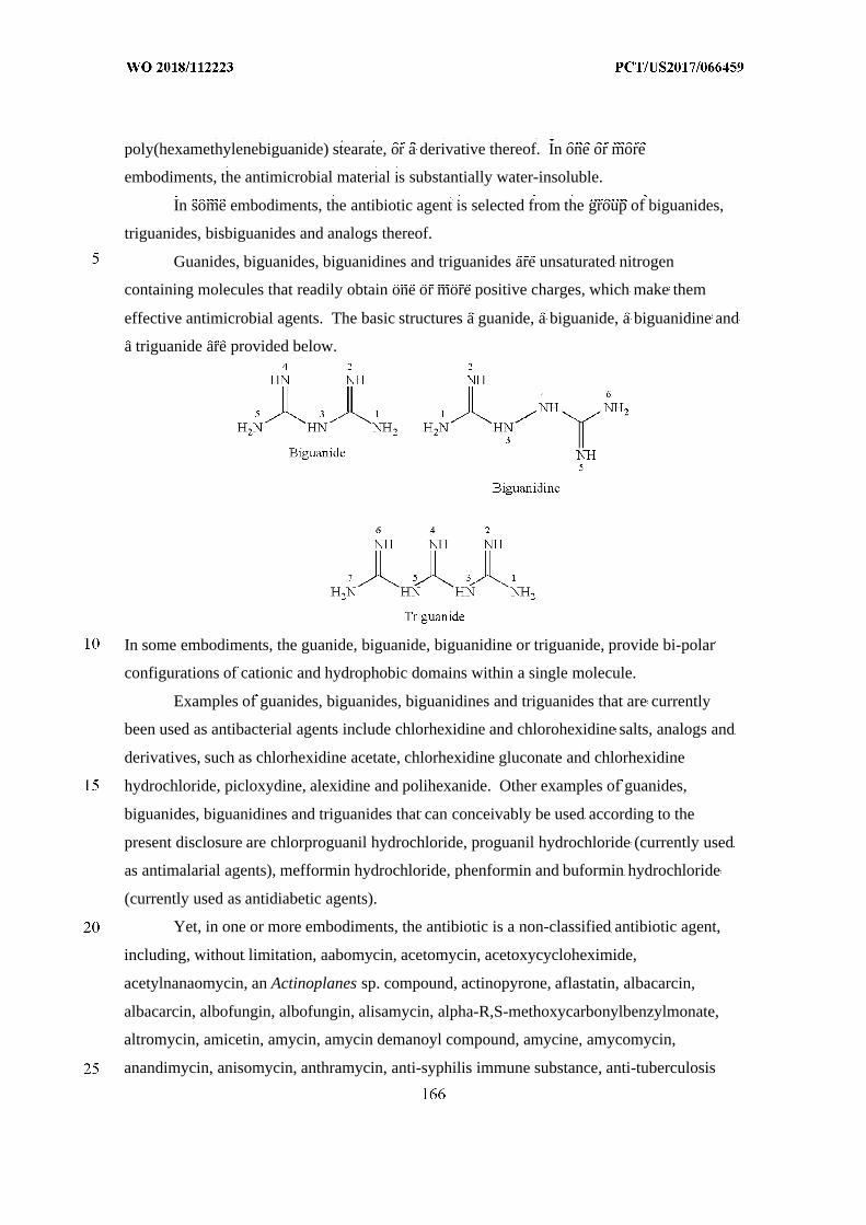
poly(hexamethylenebiguanide) stearate, or a derivative thereof. In one or more
embodiments, the antimicrobial material is substantially water-insoluble.
In some embodiments, the antibiotic agent is selected from the group of biguanides,
triguanides, bisbiguanides and analogs thereof.
Guanides, biguanides, biguanidines and triguanides are unsaturated nitrogen
containing molecules that readily obtain one or more positive charges, which make them
effective antimicrobial agents. The basic structures a guanide, a biguanide, a biguanidine and
a triguanide are provided below.
In some embodiments, the guanide, biguanide, biguanidine or triguanide, provide bi-polar
configurations of cationic and hydrophobic domains within a single molecule.
Examples of guanides, biguanides, biguanidines and triguanides that are currently
been used as antibacterial agents include chlorhexidine and chlorohexidine salts, analogs and
derivatives, such as chlorhexidine acetate, chlorhexidine gluconate and chlorhexidine
hydrochloride, picloxydine, alexidine and polihexanide. Other examples of guanides,
biguanides, biguanidines and triguanides that can conceivably be used according to the
present disclosure are chlorproguanil hydrochloride, proguanil hydrochloride (currently used
as antimalarial agents), mefformin hydrochloride, phenformin and buformin hydrochloride
(currently used as antidiabetic agents).
Yet, in one or more embodiments, the antibiotic is a non-classified antibiotic agent,
including, without limitation, aabomycin, acetomycin, acetoxycycloheximide,
acetylnanaomycin, an Actinoplanes sp. compound, actinopyrone, aflastatin, albacarcin,
albacarcin, albofungin, albofungin, alisamycin, alpha-R,S-methoxycarbonylbenzylmonate,
altromycin, amicetin, amycin, amycin demanoyl compound, amycine, amycomycin,
anandimycin, anisomycin, anthramycin, anti-syphilis immune substance, anti-tuberculosis

immune substance, an antibiotic from Escherichia coli, an antibiotic from Streptomyces
refuineus, anticapsin, antimycin, aplasmomycin, aranorosin, aranorosinol, arugomycin,
ascofuranone, ascomycin, ascosin, Aspergillus flavus antibiotic, asukamycin, aurantinin, an
Aureolic acid antibiotic substance, aurodox, avilamycin, azidamfenicol, azidimycin,
bacillaene, a Bacillus larvae antibiotic, bactobolin, benanomycin, benzanthrin, benzylmonate,
bicozamycin, bravomicin, brodimoprim, butalactin, calcimycin, calvatic acid, candiplanecin,
carumonam, carzinophilin, celesticetin, cepacin, cerulenin, cervinomycin, chartreusin,
chloramphenicol, chloramphenicol palmitate, chloramphenicol succinate sodium,
chlorflavonin, chlorobiocin, chlorocarcin, chromomycin, ciclopirox, ciclopirox olamine,
citreamicin, cladosporin, clazamycin, clecarmycin, clindamycin, coliformin, collinomycin,
copiamycin, corallopyronin, corynecandin, coumermycin, culpin, cuprimyxin,
cyclamidomycin, cycloheximide, dactylomycin, danomycin, danubomycin, delaminomycin,
demethoxyrapamycin, demethylscytophycin, dermadin, desdamethine, dexylosyl-
benanomycin, pseudoaglycone, dihydromocimycin, dihydronancimycin, diumycin, dnacin,
dorrigocin, dynemycin, dynemycin triacetate, ecteinascidin, efrotomycin, endomycin,
ensanchomycin, equisetin, ericamycin, esperamicin, ethylmonate, everninomicin, feldamycin,
flambamycin, flavensomycin, florfenicol, fluvomycin, fosfomycin, fosfonochlorin,
fredericamycin, frenolicin, fumagillin, fumifungin, funginon, fusacandin, fusafungin,
gelbecidine, glidobactin, grahamimycin, granaticin, griseofulvin, griseoviridin, grisonomycin,
hayumicin, hayumicin, hazymicin, hedamycin, heneicomycin, heptelicid acid, holomycin,
humidin, isohematinic acid, karnatakin, kazusamycin, kristenin, L-dihydrophenylalanine, a L-
isoleucyl-L-2-amino-4-(4′-amino-2′,5′-cyclohexadienyl) derivative, lanomycin, leinamycin,
leptomycin, libanomycin, lincomycin, lomofungin, lysolipin, magnesidin, manumycin,
melanomycin, methoxycarbonylmethylmonate, methoxycarbonylethylmonate,
methoxycarbonylphenylmonate, methyl pseudomonate, methylmonate, microcin, mitomalcin,
mocimycin, moenomycin, monoacetyl cladosporin, monomethyl cladosporin, mupirocin,
mupirocin calcium, mycobacidin, myriocin, myxopyronin, pseudoaglycone, nanaomycin,
nancimycin, nargenicin, neocarcinostatin, neoenactin, neothramycin, nifurtoinol, nocardicin,
nogalamycin, novobiocin, octylmonate, olivomycin, orthosomycin, oudemansin, oxirapentyn,
oxoglaucine methiodide, pactacin, pactamycin, papulacandin, paulomycin, phaeoramularia
fungicide, phenelfamycin, phenyl, cerulenin, phenylmonate, pholipomycin, pirlimycin,
pleuromutilin, a polylactone derivative, polynitroxin, polyoxin, porfiromycin, pradimicin,
prenomycin, prop-2-enylmonate, protomycin, Pseudomonas antibiotic, pseudomonic acid,

purpuromycin, pyrinodemin, pyrroInitrin, pyrrolomycin, amino, chloro pentenedioic acid,
rapamycin, rebeccamycin, resistomycin, reuterin, reveromycin, rhizocticin, roridin,
rubiflavin, naphthyridinomycin, saframycin, saphenamycin, sarkomycin, sarkomycin,
sclopularin, selenomycin, siccanin, spartanamicin, spectinomycin, spongistatin, stravidin,
streptolydigin, Streptomyces arenae antibiotic complex, streptonigrin, streptothricins,
streptovitacin, streptozotocine, a strobilurin derivative, stubomycin, sulfamethoxazol-
trimethoprim, sakamycin, tejeramycin, terpentecin, tetrocarcin, thermorubin,
thermozymocidin, thiamphenicol, thioaurin, thiolutin, thiomarinol, thiomarinol,
tirandamycin, tolytoxin, trichodermin, trienomycin, trimethoprim, trioxacarcin, tyrissamycin,
umbrinomycin, unphenelfamycin, urauchimycin, usnic acid, uredolysin, variotin,
vermisporin, verrucarin and analogs, salts and derivatives thereof.
In one or more embodiments, the antibiotic agent is a naturally
occurring antibiotic compound. As used herein, the term “naturally-occurring antibiotic
agent” includes all antibiotics that are obtained, derived or extracted from plant or vertebrate
sources. Non-limiting examples of families of naturally-occurring antibiotic agents include
phenol, resorcinol, antibiotic aminoglycosides, anamycin, quinines,
anthraquinones, antibiotic glycopeptides, azoles, macrolides, avilamycin, agropyrene, cnicin,
aucubin antibioticsaponin fractions, berberine (isoquinoline alkaloid), arctiopicrin
(sesquiterpene lactone), lupulone, humulone (bitter acids), allicin, hyperforin, echinacoside,
coniosetin, tetramic acid, imanine and novoimanine.
Ciclopirox and ciclopiroxolamine possess fungicidal, fungistatic and sporicidal
activity. They are active against a broad spectrum of dermatophytes, yeasts, moulds and
other fungi, such as Trichophytons species, Microsporum species, Epidermophyton species
and yeasts (Candida albicans, Candida glabrata, other candida species and Cryptococcus
neoformans). Some Aspergillus species are sensitive to ciclopirox as are some Penicillium.
Likewise, ciclopirox is effective against many Gram-positive and Gram-negative bacteria
(e.g., Escherichia coli, Proteus mirabilis, Pseudomonas aeruginosa, Staphylococcus and
Streptococcus species), as well as Mycoplasma species, Trichomonas vaginalis and
Actinomyces.
Plant oils and extracts which contain antibiotic agents are also useful. Non-limiting
examples of plants that contain agents include thyme, Perilla, lavender, tea tree, Terfezia
clayeryi, Micromonospora, Putterlickia verrucosa, Putterlickia pyracantha, Putterlickia
retrospinosa, Maytenus ilicifolia, Maytenus evonymoides, Maytenus aquifolia, Faenia
interjecta, Cordyceps sinensis, couchgrass, holy thistle, plantain, burdock, hops, echinacea,
buchu, chaparral, myrrh, red clover and yellow dock, garlic, and St. John’s wort.Mixtures of
the antibiotic agents as described herein may also be employed.
Combination Detection:
Any combination of the analytes disclosed herein can be detected using any of the
methods described herein. In particular, any combination disclosed herein can be detected
using any of the methods described herein.
A “photosensitizer” as used herein refers to a sensitizer for generation of singlet
oxygen usually by excitation with light. Exemplary photosensitizers suitable for use include
those described in U.S. Patent Nos. 6,251,581, 5,516,636, 8,907,081, 6,545,012, 6,331,530,
8,247,180, 5,763,602, 5,705,622, 5,516,636, 7,217,531, and U.S. Patent Publication No.
2007/0059316, all of which are herein expressly incorporated by reference in their entireties.
The photosensitizer can be photoactivatable (e.g., dyes and aromatic compounds) or
chemiactivated (e.g., enzymes and metal salts). When excited by light the photosensitizer is
usually a compound comprised of covalently bonded atoms, usually with multiple conjugated
double or triple bonds. The compound should absorb light in the wavelength range of 200-
1100 nm, usually 300-1000 nm, e.g., 450-950 nm, with an extinction coefficient at its
absorbance maximum greater than 500 M− 1 cm− 1 , e.g., at least 5000 M− 1cm− 1 , or at least
50,000 M− 1 cm− 1 at the excitation wavelength. The lifetime of an excited state produced
following absorption of light in the absence of oxygen will usually be at least 100 nsec, e.g.,
at least 1 µsec. In general, the lifetime must be sufficiently long to permit energy transfer to
oxygen, which will normally be present at concentrations in the range of 10− 5 to 103 1 3 M
depending on the medium. The sensitizer excited state will usually have a different spin
quantum number (S) than its ground state and will usually be a triplet (S=l) when, as is
usually the case, the ground state is a singlet (S=O). In some embodiments, the sensitizer
will have a high intersystem crossing yield. That is, photoexcitation of a sensitizer will
produce the long lived state (usually triplet) with an efficiency of at least 10%, at least 40%,
e.g., greater than 80%. The photosensitizer will usually be at most weakly fluorescent under
the assay conditions (quantum yield usually less that 0.5, or less that 0.1).
Photosensitizers that are to be excited by light will be relatively photostable and will
not react efficiently with singlet oxygen. Several structural features are present in most
useful sensitizers. Most sensitizers have at least one and frequently three or more conjugated
double or triple bonds held in a rigid, frequently aromatic structure. They will frequently
contain at least one group that accelerates intersystem crossing such as a carbonyl or imine
group or a heavy atom selected from rows 3-6 of the periodic table, especially iodine or
bromine, or they may have extended aromatic structures. Typical sensitizers include acetone,
benzophenone, 9-thioxanthone, eosin, 9,10-dibromoanthracene, methylene blue, metallo-
porphyrins, such as hematoporphyrin, phthalocyanines, chlorophylls, rose bengal,
buckminsterfullerene, etc., and derivatives of these compounds having substituents of 1 to 50
atoms for rendering such compounds more lipophilic or more hydrophilic and/or as attaching
groups for attachment. Examples of other photosensitizers that may be utilized are those that
have the above properties and are enumerated in N. J. Turro, “Molecular Photochemistry,”
page 132, W. A. Benjamin Inc., N.Y. 1965.
In some embodiments, the photosensitizers are relatively non-polar to assure
dissolution into a lipophilic member when the photosensitizer is incorporated in an oil
droplet, liposome, latex particle, etc.
In some embodiments, the photosensitizers suitable for use herein include other
substances and compositions that can produce singlet oxygen with or without activation by an
external light source. Thus, for example, molybdate (MoO4= ) salts and chloroperoxidase and
myeloperoxidase plus bromide or chloride ion (Kanofsky, J. Biol. Chem. (1983) 259 5596)
have been shown to catalyze the conversion of hydrogen peroxide to singlet oxygen and
water. Either of these compositions can, for example, be included in particles and used in the
assay method wherein hydrogen peroxide is included as an ancillary reagebly,
chloroperoxidase is bound to a surface and molybdate is incorporated in the aqueous phase of
a liposome. Also included within the scope of the invention as photosensitizers are
compounds that are not true sensitizers but which on excitation by heat, light, or chemical
activation will release a molecule of singlet oxygen. The best known members of this class
of compounds includes the endoperoxides such as 1,4-biscarboxyethyl-1,4-naphthalene
endoperoxide, 9,10-diphenylanthracene-9,10-endoperoxide and 5,6,11,12-tetraphenyl
naphthalene 5,12-endoperoxide. Heating or direct absorption of light by these compounds
releases singlet oxygen.
A “chemiluminescent compound” as used herein refers to a substance that undergoes
a chemical reaction with singlet oxygen to form a metastable intermediate that can
decompose with the simultaneous or subsequent emission of light within the wavelength
range of 250 to 1200 nm. Exemplary chemiluminescent compounds suitable for use include
those described in U.S. Patent Nos. 6,251,581 and 7,709,273, and Patent Cooperatio Treaty
(PCT) International Application Publication No. WO1999/042838. Examplery
chemiluminescent compound includes the following:
Chemiluminescer Half-Life Emission Max
Thioxene + Diphenyl anthracence: 0.6 seconds 430 nm
Thioxene + Umbelliferone derivative 0.6 seconds 500 nm
Thioxene + Europium chelate 0.6 seconds 615 nm
Thioxene + Samarium Chelate 0.6 seconds 648 nm
Thioxene + terbium Chelate 0.6 seconds 540nm
N-Phenyl Oxazine + Umbelliferone derivative 30 seconds 500 nm
N-Phenyl Oxazine + Europium chelate 30 seconds 613nm
N-phenyl Oxazine + Samarium Chelate 30 seconds 648 nm
N-phenyl Oxazine + terbium Chelate 30 seconds 540nm
Dioxene + Umbelliferone derivative 300 seconds 500 nm
Dioxene + Europium chelate 300 seconds 613nm
Dioxene + Samarium Chelate 300 seconds 648 nm
N-phenyl Oxazine + terbium Chelate 300 seconds 540nm
All of the above mentioned applications are herey expressly incorporated by reference
herein in their entireties. Emission will usually occur without the presence of an energy
acceptor or catalyst to cause decomposition and light emission. In some embodiments, the
intermediate decomposes spontaneously without heating or addition of ancillary reagents
following its formation. However, addition of a reagent after formation of the intermediate or
the use of elevated temperature to accelerate decomposition will be required for some
chemiluminescent compounds. The chemiluminescent compounds are usually electron rich
compounds that react with singlet oxygen, frequently with formation of dioxetanes or
dioxetanones. Exemplary of such compounds are enol ethers, enamines, 9-
alkylidenexanthans, 9-alkylidene-N-alkylacridans, aryl vinyl ethers, dioxenes, arylimidazoles
and lucigenin. Other chemiluminescent compounds give intermediates upon reaction with
singlet oxygen, which subsequently react with another reagent with light emission.
Exemplary compounds are hydrazides such as luminol and oxalate esters.
The chemiluminescent compounds of interest will generally emit at wavelengths
above 300 nanometers and usually above 400 nm. Compounds that alone or together with a
fluorescent molecule emit light at wavelengths beyond the region where serum components
absorb light will be of particular use. The fluorescence of serum drops off rapidly above 500
nm and becomes relatively unimportant above 550 nm. Therefore, when the analyte is in
serum, chemiluminescent compounds that emit light above 550 nm, e.g., above 600 nm may
be suitable for use. In order to avoid autosensitization of the chemiluminescent compound, in
some embodiments, the chemiluminescent compounds do not absorb light used to excite the
photosensitizer. In some embodiments, the sensitizer is excited with light wavelengths longer
than 500 nm, it will therefore be desirable that light absorption by the chemiluminescent
compound be very low above 500 nm.
Where long wave length emission from the chemiluminescent compound is desired, a
long wavelength emitter such as a pyrene, bound to the chemiluminescent compound can be
used. Alternatively, a fluorescent molecule can be included in the medium containing the
chemiluminescent compound. In some embodiments, fluorescent molecules will be excited
by the activated chemiluminescent compound and emit at a wavelength longer than the
emission wavelength of the chemiluminescent compound, usually greater that 550 nm. It is
usually also desirable that the fluorescent molecules do not absorb at the wavelengths of light
used to activate the photosensitizer. Examples of useful dyes include rhodamine, ethidium,
dansyl, Eu(fod)3 , Eu(TTA)3 , Ru(bpy)3+ + (wherein bpy=2,2′-dipyridyl, etc. In general, these
dyes act as acceptors in energy transfer processes and in some embodiments, have high
fluorescent quantum yields and do not react rapidly with singlet oxygen. They can be
incorporated into particles simultaneously with the incorporation of the chemiluminescent
compound into the particles.
In some embodiments, the disclosure provides diffractive optics detection technology
that can be used with, for example, ingestible device technology. In certain embodiments, an
ingestible device includes the diffractive optics technology (e.g., diffractive optics detection
system). In certain embodiments, the disclosure provides diffractive optics technology (e.g.,
diffractive optics detection systems) that are used outside the body of subject. As an
example, an ingestible device can be used to obtain one more samples in the body (e.g., in the
gastrointestinal tract) of a subject, and the diffractive optics technology can be used to
analyze the sample(s). Such analysis can be performed in vivo (e.g., when the ingestible
device contains the diffractive optics).
Diffraction is a phenomenon that occurs due to the wave nature of light. When light
hits an edge or passes through a small aperture, it is scattered in different directions. But light
waves can interfere to add (constructively) and subtract (destructively) from each other, so
that if light hits a non-random pattern of obstacles, the subsequent constructive and
destructive interference will result in a clear and distinct diffraction pattern. A specific
example is that of a diffraction grating, which is of uniformly spaced lines, typically prepared
by ruling straight, parallel grooves on a surface. Light incident on such a surface produces a
pattern of evenly spaced spots of high light intensity. This is called Bragg scattering, and the
distance between spots (or ‘Bragg scattering peaks’) is a unique function of the diffraction
pattern and the wavelength of the light source. Diffraction gratings, like focusing optics, can
be operated in both transmission and reflection modes.
In general, the light used in the diffractive optics can be of any appropriate
wavelength. Exemplary wavelengths include visible light, infrared red (IR) and ultraviolet
(UV). Optionally, the light can be monochromatic or polychromatic. The light can be
coherent or incoherent. The light can be collimated or non-collimated. In some
embodiments, the light is coherent and collimated. Generally, any appropriate light source
may be used, such as, for example, a laser (e.g., a laser diode) or a light emitting diode. In
some embodiments, the light source is a laser diode operating at 670 nm wavelength, e.g., at
3 mWatts power. Optionally, an operating wavelength of a laser diode can be 780 nm, e.g.,
when larger grating periods are used. In certain embodiments, the light source is a laser, such
as, for example, a He-Ne laser, a Nd:YVO4 laser, or an argon-ion laser. In some
embodiments, the light source is a low power, continuous waver laser.
The diffracted light can be detected using any appropriate light detector(s). Examples
of light detectors include photodetectors, such as, for example, position sensitive
photodiodes, photomultiplier tubes (PMTs), photodiodes (PDs), avalanche photodiodes
(APDs), charged-coupled device (CCD) arrays, and CMOS detectors. In some embodiments,
the diffracted light is detected via one or more individual photodiodes.
In general, the diffraction grating is made of a material that is transparent in the
wavelength of the radiation used to illuminate the sensor. Any appropriate material may be
used for the diffraction grating substrate, such as glass or a polymer. Exemplary polymers
include polystyrene polymers (PSEs), cyclo-olefin polymers (COPs), polycarbonate
polymers, polymethyl methacrylates, and methyl methacrylate styrene copolymers.
Exemplary COPs include Zeonex (e.g., Zeonex E48R, Zeonex F52R).
The light may be incident on the diffraction grating any appropriate angle. In some
embodiments, the light is incident on the diffraction grating with an angle of incidence of
from 30° to 80° (e.g., from 40° to 80°, from 50° to 70°, from 55° to 65°, 60°). Optionally, the
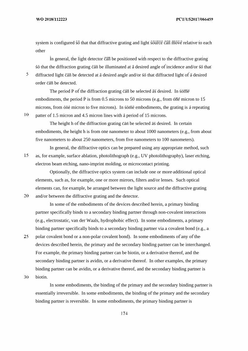
system is configured so that that diffractive grating and light source can move relative to each
other
In general, the light detector can be positioned with respect to the diffractive grating
so that the diffraction grating can be illuminated at a desired angle of incidence and/or so that
diffracted light can be detected at a desired angle and/or so that diffracted light of a desired
order can be detected.
The period P of the diffraction grating can be selected as desired. In some
embodiments, the period P is from 0.5 microns to 50 microns (e.g., from one micron to 15
microns, from one micron to five microns). In some embodiments, the grating is a repeating
patter of 1.5 micron and 4.5 micron lines with a period of 15 microns.
The height h of the diffraction grating can be selected as desired. In certain
embodiments, the height h is from one nanometer to about 1000 nanometers (e.g., from about
five nanometers to about 250 nanometers, from five nanometers to 100 nanometers).
In general, the diffractive optics can be prepared using any appropriate method, such
as, for example, surface ablation, photolithograph (e.g., UV photolithography), laser etching,
electron beam etching, nano-imprint molding, or microcontact printing.
Optionally, the diffractive optics system can include one or more additional optical
elements, such as, for example, one or more mirrors, filters and/or lenses. Such optical
elements can, for example, be arranged between the light source and the diffractive grating
and/or between the diffractive grating and the detector.
In some of the embodiments of the devices described herein, a primary binding
partner specifically binds to a secondary binding partner through non-covalent interactions
(e.g., electrostatic, van der Waals, hydrophobic effect). In some embodiments, a primary
binding partner specifically binds to a secondary binding partner via a covalent bond (e.g., a
polar covalent bond or a non-polar covalent bond). In some embodiments of any of the
devices described herein, the primary and the secondary binding partner can be interchanged.
For example, the primary binding partner can be biotin, or a derivative thereof, and the
secondary binding partner is avidin, or a derivative thereof. In other examples, the primary
binding partner can be avidin, or a derivative thereof, and the secondary binding partner is
biotin.
In some embodiments, the binding of the primary and the secondary binding partner is
essentially irreversible. In some embodiments, the binding of the primary and the secondary
binding partner is reversible. In some embodiments, the primary binding partner is
CaptAvidin™ biotin-binding protein and the secondary binding partner is biotin, or vice
versa. In some embodiments, the primary binding partner is DSB-X™ biotin and the
secondary binding partner is avidin, or vice versa. In some embodiments, the primary
binding partner is desthiobiotin and the secondary binding partner is avidin, or vice versa
(Hirsch et al., Anal Biochem. 308(2):343-357, 2002). In some embodiments, the primary
binding partner is glutathione (GSH) or a derivative thereof, and the secondary binding
partner is glutathione-S-transferase (GST).
In some embodiments, the primary binding partner can bind to a target analyte that is
a nucleic acid (e.g., a DNA molecule, a RNA molecule). In some embodiments, the primary
binding partner comprises a portion of a nucleic acid that is complementary to the nucleic
acid sequence of the target analyte.
In some embodiments of any of the devices described herein, the device can include a
label that binds to the target analyte and does not prevent binding of the target analyte to the
primary binding partner. In some embodiments, the label can amplify the diffraction signal
of the target analyte.
In some embodiments, the label is from about 1 nm to 200 nm (e.g., about 50 nm to
about 200 nm).
In some embodiments, the label (e.g., any of the labels described herein) includes one
or more antibodies (e.g., any of the antibodies and/or antibody fragments described herein).
In some embodiments, the label is a nanoparticle (e.g., a gold nanoparticle) that
includes the primary binding partner that has a nucleic acid sequence that is complementary
to the target analyte, and is covalently linked to the nanoparticle.
One or more additional steps can be performed in any of the methods described
herein. In some embodiments, the one or more additional steps are performed: prior to the
binding of the primary binding partner to the secondary binding partner, after the binding of
the primary binding partner to the secondary binding partner, prior to the binding of the
primary binding partner to the target analyte, or after the binding of the primary binding
partner to the target analyte.
In some embodiments of any of the methods described herein, the determining step
(during which the primary binding partner binds to the target analyte is detected) can occur in
at least 15 seconds. In some embodiments, the binding of the primary binding partner to the
target analyte can occur during a period of time of, for example, five at least seconds.
In some embodiments, the one or more additional steps can include: a blocking of the
sensors step, at least one wash step, a capturing step, and/or a filtering step. In some
embodiments, the blocking step can include blocking a sensor within the ingestible device
with a solution comprising at least 1% bovine serum albumin (BSA) in a buffered solution
(e.g., phosphate buffered saline (PBS), Tris buffered saline (TBS)). In some embodiments,
the at least one wash step can include washing with a buffered solution (e.g., phosphate
buffered saline (PBS), Tris buffered saline (TBS)). In general, blocking is performed during
capsule manufacture, rather than in vivo.
In some embodiments, the capturing step includes enriching the target analyte. In
some embodiments, the capturing step includes physically separating the target analyte from
the remaining sample using a filter, a pore, or a magnetic bead. In some embodiments, the
target analyte is captured by size exclusion.
In some embodiments, the disclosure provides methods of obtaining, culturing, and/or
detecting target cells and/or target analytes in vivo within the gastrointestinal (GI) tract or
reproductive tract of a subject. Associated devices are also disclosed. The methods and
devices described provide a number of advantages for obtaining and/or analyzing fluid
samples from a subject. In some embodiments, diluting the fluid sample increases the
dynamic range of analyte detection and/or reduces background signals or interference within
the sample. For example, interference may be caused by the presence of non-target analytes
or non-specific binding of a dye or label within the sample. In some embodiments, culturing
the sample increases the concentration of target cells and/or target analytes produced by the
target cells thereby facilitating their detection and/or characterization.
In certain embodiments, the methods and devices a described herein may be used to
obtain information regarding bacteria populations in the GI tract of a subject. This has a
number of advantages and is less invasive than surgical procedures such as intubation or
endoscopy to obtain fluid samples from the GI tract. The use of an ingestible device as
described herein also allows for fluid samples to be obtained and data to be generated on
bacterial populations from specific regions of the GI tract.
In some embodiments, the methods and devices described herein may be used to
generate data such as by analyzing the fluid sample, dilutions thereof or cultured samples for
one or more target cells and/or target analytes. The data may include, but is not limited to, the
types of bacteria present in the fluid sample or the concentration of bacteria in specific
regions of the GI tract. Such data may be used to determine whether a subject has an
infection, such as Small Intestinal Bacterial Overgrowth (SIBO), or to characterize bacterial
populations within the GI tract for diagnostic or other purposes. Thus, in some embodiments,
analytes disclosed herein are indicative of disorders of the gastrointestinal tract associated
with anomalous bacterial populations.
For example, in one aspect, the data may include, but is not limited to, the
concentration of bacteria in a specific region of the GI tract that is one or more of the
duodenum, jejunum, ileum, ascending colon, transverse colon or descending colon. . In one
aspect, the specific region of the GI tract is the duodenum. In one aspect, the specific region
of the GI tract is the jejunum. In one aspect, the specific region of the GI tract is the ileum.
In one aspect, the specific region of the GI tract is the ascending colon. In one aspect, the
specific region of the GI tract is the transverse colon. In one aspect, the specific region of the
GI tract is the descending colon. In a related embodiment, the data may be generated every
one or more days to monitor disease flare-ups, or response to the therapeutic agents disclosed
herein.
Data may be generated after the device has exited the subject, or the data may be
generated in vivo and stored on the device and recovered ex vivo. Alternatively, the data can
be transmitted wirelessly from the device while the device is passing through the GI tract of
the subject or in place within the reproductive tract of the subject.
In some embodiments, a method comprises: providing a device comprising one or
more dilution chambers and dilution fluid; transferring all or part of a fluid sample obtained
from the GI tract or reproductive tract of the subject into the one or more dilution chambers
in vivo; and combining the fluid sample and the dilution fluid to produce one or more diluted
samples in the one or more dilution chambers.
In certain embodiments, a method comprises: providing an ingestible device
comprising one or more dilution chambers; transferring all or part of a fluid sample obtained
from the GI tract into the one or more dilution chambers comprising sterile media; culturing
the sample in vivo within the one or more dilution chambers to produce one or more cultured
samples; and detecting bacteria in the one or more cultured samples.
In some embodiments, a method comprises: providing a device comprising one or
more dilution chambers; transferring all or part of a fluid sample obtained from the GI tract or
reproductive tract into the one or more dilution chambers; combining all or part of the fluid
sample with a dilution fluid in the one or more dilution chambers; and detecting the target
analyte in the one or more diluted samples.
In certain embodiments, a device comprises: one or more dilution chambers for
diluting a fluid sample obtained from the GI tract or reproductive tract; and dilution fluid for
diluting the sample within the one or more dilution chambers.
In some embodiments, the device comprises: one or more dilution chambers for
culturing a fluid sample obtained from the GI tract; sterile media for culturing the sample
within the one or more dilution chambers; and a detection system for detecting bacteria.
In certain embodiments, a device comprises: one or more dilution chambers for
culturing a fluid sample obtained from the GI tract; sterile media for culturing the sample
within the one or more dilution chambers; and a detection system for detecting bacteria.
Also provided is the use of a device as described herein for diluting one or more
samples obtained from the GI tract or reproductive tract of a subject. In one embodiment,
there is provided the use of an ingestible device as described herein for detecting target cells
and/or target analytes in vivo within the gastrointestinal (GI) tract of a subject.
Further provided is a system comprising a device as described herein and a base
station. In one embodiment, the device transmits data to the base station, such as data
indicative of the concentration and/or types of bacteria in the GI tract of the subject. In one
embodiment, the device receives operating parameters from the base station. Some
embodiments described herein provide an ingestible device for obtaining one or more
samples from the GI tract or reproductive tract of a subject and diluting and/or culturing all or
part of the one or more samples. The ingestible device includes a cylindrical rotatable
element having a port on the wall of the cylindrical rotatable element. The ingestible device
further includes a shell element wrapping around the cylindrical rotatable element to form a
first dilution chamber between the cylindrical rotatable element and the shell element. The
shell element has an aperture that exposes a portion of the wall of the cylindrical rotatable
element to an exterior of the ingestible device.
In certain embodiments, the medical device comprises one or more dilution chambers
for receiving a fluid sample from the GI tract or reproductive tract of a subject or a dilution
thereof. In some embodiments, one or more dilutions of the fluid sample are cultured in one
or more dilution chambers. In certain embodiments, the dilution chambers each define a
known volume, optionally the same volume or different volumes. In some embodiments, the
dilution chambers define a fluid volume ranging from about 10 µL to about 1 mL. The
dilution chambers may define a fluid volume less than or equal to about 500 µL, less than or
equal to about 250 µL, less than or equal to about 100 µL, or less than or equal to about 50
µL. In certain embodiments, the dilution chambers define a fluid volume of greater than or
equal to about 10 µL, greater than or equal to about 20 µL, greater than or equal to about 30
µL, or greater than or equal to about 50 µL. In some embodiments, the dilution chambers
define a fluid volume between about 10 µL and 500 µL, between about 20 µL and 250 µL,
between about 30 µL and 100 µL or about 50 µL.
In some embodiments, dilution fluid in the device is combined with all or part of the
fluid sample, or dilution thereof, to produce one or more dilutions. In certain embodiments,
the dilution fluid is sterile media suitable for culturing one or more target cells within the
dilution chambers.
In certain embodiments, the one or more dilution chambers may be filled with the
dilution fluid prior to a patient ingesting the ingestible device. In some embodiments, the
dilution fluid may be added into the one or more dilution chambers in vivo from a reservoir of
the ingestible device. Sampling and dilution of the GI fluid sample may take place in vivo.
For example, an actuator of the ingestible device may pump the dilution fluid from the
reservoir into a dilution chamber when it is determined that the ingestible device is located at
a predetermined location within the GI tract. In some embodiments, the dilution chambers
each contain a volume of sterile media suitable for culturing a fluid sample from the GI tract
or reproductive tract. In certain embodiments, the dilution chambers are at least 95%, at least
97%, at least 98%, or at least 99% full of sterile media. In some embodiments, the dilution
chambers each contain oxygen to facilitate aerobic bacteria growth. In certain embodiments,
a non-dilution chamber comprises oxygen and is added to one or more of the dilution
chambers to facilitate aerobic bacteria growth.
In some embodiments, the culturing may take place in vivo immediately after the GI
fluid sample has been diluted. Or alternatively, the culturing may take place ex vivo, e.g.,
when the ingestible device has been evacuated and recovered such that the dilution chamber
containing the diluted GI fluid sample may be extracted and the culturing may be performed
in a laboratory. The recovery of the ingestible device may be performed in a similar manner
as embodiments described in U.S. Provisional Application No. 62/434,188, filed on
December 14, 2016, which is herein expressly incorporated by reference in its entirety.
As used herein “culturing” refers to maintaining target cells in an environment that
allows a population of one or more target cells to increase in number through cell division.
For example, in some embodiments, “culturing” may include combining the cells with media
in an dilution chamber at a temperature that permits cell growth, optionally a temperature
found in vivo within the GI tract or reproductive tract of a subject. In certain embodiments,
the cells are cultured at a temperature between about 35 °C and 42 °C.
As used herein “dilution fluid” refers to a fluid within the device for diluting a fluid
sample from the GI tract or reproductive tract. In some embodiments, the dilution fluid is an
aqueous solution. In certain embodiments, the dilution fluid comprises one or more agents
that promote or inhibit the growth of an organism, such as a fungus or bacteria. In some
embodiments, the dilution fluid comprises one or more agents that facilitate the detection of a
target analyte, such as dyes or binding agents for target analytes.
In some embodiments, the dilution fluid is a sterile media. As used herein, “sterile
media” refers to media that does not contain any viable bacteria or other cells that would
grow and increase in number through cell division. Media may be rendered sterile by various
techniques known in the art such as, but not limited to, autoclaving and/or preparing the
media using asceptic techniques. In certain embodiments, the media is a liquid media.
Examples of media suitable for culturing bacteria include nutrient broth, Lysogeny Broth
(LB) (also known as Luria Broth), Wilkins chalgren, and Tryptic Soy Broth (TSB), Other
growth or culture media known in the art may also be used in the methods and devices
described herein. In some embodiments, the media has a carbon source, such as glucose or
glycerol, a nitrogen source such as ammonium salts or nitrates or amino acids, as well as salts
and/or trace elements and vitamins required for microbial growth. In certain embodiments,
the media is suitable for maintaining eukaryotic cells. In some embodiments, the media
comprises one or more agents that promote or inhibit the growth of bacteria, optionally
agents that promote or inhibit the growth of specific types of bacteria.
In certain embodiments, the media is a selective media. As used herein, “selective
media” refers to a media that allows certain types of target cells to grow and inhibits the
growth of other organisms. Accordingly, the growth of cells in a selective media indicates
the presence of certain types of cells within the cultured sample. For example, in some
embodiments, the media is selective for gram-positive or gram-negative bacteria. In certain
embodiments, the media contains crystal violet and bile salts (such as found in MacConkey
agar) that inhibit the growth of gram-positive organisms and allows for the selection and
isolation of gram-negative bacteria. In some embodiments, the media contains a high
concentration of salt (NaCl) (such as found in Mannitol salt agar) and is selective for Gram-
positive bacteria. In some embodiments, the media selectively kills eukaryotic cells or only
grows prokaryotic cells, for example, using a media comprising Triton™ X-100. In certain
embodiments, the media selectively kills prokaryotic cells (or alternatively only grows
eukaryotic cells), for example, using a media that comprises antibiotics.
In some embodiments, the media is an indicator media. As used herein, “indicator
media” refers to a media that contains specific nutrients or indicators (such as, but not limited
to neutral red, phenol red, eosin y, or methylene blue) that produce a detectable signal when a
certain type of cells are cultured in the indicator media.
In some embodiments, the disclosure provides a composition comprising a dye and
optionally a reagent for selective lysis of eukaryotic cells. In certain embodiments, the
composition comprises both a dye and a reagent for selective lysis of eukaryotic cells. In
some embodiments, the composition further comprises one or more reagents independently
selected from the group consisting of: a second reagent for selective lysis of eukaryotic cells
(e.g., Triton X-100), an electrolyte (e.g., MgCl2), an anti-fungi reagent (e.g., amphotericin-B),
and an antibiotic. In some embodiments, the composition comprises water and is in the form
of an aqueous solution. In some embodiments, the composition is a solid or semi-solid. In
some embodiments, the compositions described here are suitable for use in a kit or device for
detecting or quantifying viable bacterial cells in a sample. In some embodiments, such a
device is an ingestible device for detecting or quantifying viable bacterial cells in vivo (e.g.,
in the GI tract). In some embodiments, viable bacterial cells in a sample are detected or
quantified in the presence of one or more antibiotics to determine antibiotic resistance of the
bacteria in the sample. In some embodiments, anomalous bacterial populations in a sample
may be detected or quantified, for example through the use of one a composition comprising
a dye as disclosed herein, to determine whether a subject has an infection, such as Small
Intestinal Bacterial Overgrowth (SIBO), or to characterize bacterial populations within the GI
tract for diagnostic or other purposes.
In some embodiments, a method comprises: (a) contacting the sample with a
composition as described herein; and (b) measuring total fluorescence or rate of change of
fluorescence as a function of time of said sample, thereby detecting viable bacterial cells in
said sample. In some embodiments, a control as described herein may be employed in the
method. In some embodiments, the total fluorescence or the rate of change of fluorescence as
a function of time of the sample is measured over multiple time points for an extended period
of time in step (b), thereby detecting viable bacterial cells in said sample. In some
embodiments, the method further comprises correlating the total fluorescence or the rate of
change of fluorescence as a function of time determined in step (b) to the number of viable
bacterial cells in the sample. In some embodiments, the rate of change of fluorescence as a
function of time of the sample measured over multiple time points is determined and
compared to the rate of change of fluorescence as a function of time of a control measured
over the same time points to determine the number of viable bacterial cells in the sample. In
some embodiments, the method does not require ex vivo plating or culturing. In some
embodiments, the method does not require aspiration. In some embodiments, the method is
performed in vivo (e.g., in an ingestible device in vivo). In some embodiments, the method
comprises communicating the results of the onboard assay(s) to an ex vivo receiver.
In certain embodiments, a kit comprises a composition as described herein and
instructions, e.g., for detecting or quantifying viable bacterial cells in a sample. In some
embodiments, a device comprises a composition as described herein, e.g., for detecting or
quantifying viable bacterial cells in a sample. The detection of live cells, as opposed to the
detection of bacterial components (such as endotoxins) which can be present in the sample
environment and lead to conflicting results, is the gold standard of viable plate counting and
represents one of the advantages of the compositions and methods described herein.
The systems employ methods, compositions and detection systems found to
accurately and reliably correlate fluorescence to total bacteria count (TBC) in an autonomous,
ingestible device, or other similarly-sized device. The compositions include novel
combinations of dyes, buffers and detergents that allow for the selective staining of viable
bacterial cells in samples that comprise non-bacterial cells and other components that
otherwise make detecting or quantifying live bacterial cells challenging. In some
embodiments, the systems allow for bacteria to be quantified in near real-time and the results
to be shared telemetrically outside of the device.
In certain embodiments, the disclosure provides a method of assessing or monitoring
the need to treat a subject suffering from or at risk of overgrowth of bacterial cells in the
gastrointestinal tract, which comprises: (a) obtaining a sample from the gastrointestinal tract
of said subject; (b) contacting the sample with a composition as described herein; (c)
measuring total fluorescence or rate of change of fluorescence as a function of time of said
sample; and (d) correlating the total fluorescence or the rate of change of fluorescence as a
function of time measured in step (c) to the number of viable bacterial cells in the sample,
wherein the number of the viable bacterial cells determined in step (e) greater than about 105
CFU/mL indicates a need for treatment, e.g., with an antibiotic agent as described herein. In
some embodiments, a control as described herein may be employed in the method. In some
embodiments, the total fluorescence or the rate of change of fluorescence as a function of
time of the sample is measured over multiple time points for an extended period of time in
step (c). In some embodiments, the rate of change of fluorescence as a function of time of the
sample measured over multiple time points is determined and compared to the rate of change
of fluorescence as a function of time of a control measured over the same time points to
determine the number of viable bacterial cells in the sample. In some embodiments, the
method does not require ex vivo plating or culturing. In some embodiments, the method does
not require aspiration. In some embodiments, the method is performed in vivo (e.g., in an
ingestible device in vivo). In some embodiments, the method comprises communicating the
results of the onboard assay(s) to an ex vivo receiver. In some embodiments, the method may
be further used to monitor the subject after the treatment (e.g., with an antibiotic). In some
embodiments, the method may be used to assess the efficacy of the treatment. For example,
efficacious treatment may be indicated by the decrease of the number of viable bacterial cells
in a sample from the GI tract of the subject post-treatment. Efficacy of the treatment may be
evaluated by the rate of decrease of the number of viable bacterial cells in a sample from the
GI tract of the subject post-treatment. In some embodiments, the method may be used to
detect infection with antibiotic-resistant strains of bacteria in a subject. For instance, such
infection may be indicated where the number of viable bacterial cells in a sample from the GI
tract of the subject does not substantially decrease after antibiotic treatment.
In some embodiments, the disclosure provides an absorbable material, (e.g.,
absorbable sponge), having absorbed therein a composition as described herein. In some
embodiments, the absorbable sponge is Ahlstrom Grade 6613H (Lot 150191) or Porex PSU-
567, having absorbed therein a composition as described herein. In some embodiments, the
absorbable sponge may be prepared by injecting into the absorbable sponge an aqueous
solution comprising a composition as described herein, and optionally further comprising a
step of drying the resulting absorbable sponge.
In certain embodiments, the disclosure provides a method for detecting the presence
of viable bacterial cells in a sample, which comprises: (a) fully or partially saturating an
absorbable sponge as described herein, or an absorbable sponge prepared as described herein,
with the sample; and (b) measuring total fluorescence or rate of change of fluorescence as a
function of time of the fully or partially saturated sponge prepared in step (a), thereby
detecting viable bacterial cells. In some embodiments, a control as described herein may be
employed in the method. In some embodiments, the total fluorescence or the rate of change
of fluorescence as a function of time of the fully or partially saturated sponge is measured
over multiple time points for an extended period of time in step (b), thereby detecting viable
bacterial cells in said sample. In some embodiments, the method further comprises
correlating the total fluorescence or the rate of change of fluorescence as a function of time
measured in step (b) to the number of viable bacterial cells in the sample. In some
embodiments, the rate of change of fluorescence as a function of time of the fully or partially
saturated sponge measured over multiple time points is determined and compared to the rate
of change of fluorescence as a function of time of a control measured over the same time
points to determine the number of viable bacterial cells in the sample. In some embodiments,
the method does not require ex vivo plating or culturing. In some embodiments, the method
does not require aspiration. In some embodiments, the method is performed in vivo (e.g., in
an ingestible device in vivo). In some embodiments, the method comprises communicating
the results of the onboard assay(s) to an ex vivo receiver.
In one aspect, provided herein is a kit comprising an absorbable sponge as described herein
and instructions, e.g., for detecting or quantifying viable bacterial cells in a sample. In
another aspect, provided herein is a device comprising an absorbable sponge as described
herein, e.g., for detecting or quantifying viable bacterial cells in a sample.
In certain embodiments, the disclosure provides a method of assessing or monitoring
the need to treat a subject suffering from or at risk of overgrowth of bacterial cells in the
gastrointestinal tract, which comprises: (a) obtaining a sample from the gastrointestinal tract
of said subject; (b) fully or partially saturating an absorbable sponge described herein, or an
absorbable sponge prepared as described herein, with the sample; (c) measuring total
fluorescence or rate of change of fluorescence as a function of time of the fully or partially
saturated sponge prepared in step (b); (d) correlating the total fluorescence or the rate of
change of fluorescence as a function of time measured in step (c) to the number of viable
bacterial cells in the sample, wherein the number of the viable bacterial cells as determined in
step (e) greater than about 105 CFU/mL indicates a need for treatment, e.g., with an antibiotic
agent as described herein. In some embodiments, a control as described herein may be
employed in the method. In some embodiments, the total fluorescence or the rate of change
of fluorescence as a function of time of the fully or partially saturated sponge is measured
over multiple time points for an extended period of time in step (c). In some embodiments,
the rate of change of fluorescence as a function of time of the fully or partially saturated
sponge measured over multiple time points is determined and compared to the rate of change
of fluorescence as a function of time of a control measured over the same time points to
determine the number of viable bacterial cells in the sample. In some embodiments, the
method does not require ex vivo plating or culturing. In some embodiments, the method does
not require aspiration. In some embodiments, the method is performed in vivo (e.g., in an
ingestible device in vivo). In some embodiments, the method comprises communicating the
results of the onboard assay(s) to an ex vivo receiver. In some embodiments, the method may
be further used to monitor the subject after the treatment (e.g., with an antibiotic). In some
embodiments, the method may be used to assess the efficacy of the treatment. For example,
efficacious treatment may be indicated by the decrease of the number of viable bacterial cells
in a sample from the GI tract of the subject post-treatment. Efficacy of the treatment may be
evaluated by the rate of decrease of the number of viable bacterial cells in a sample from the
GI tract of the subject post-treatment. In some embodiments, the method may be used to
detect infection with antibiotic-resistant strains of bacteria in a subject. For instance, such
infection may be indicated where the number of viable bacterial cells in a sample from the GI
tract of the subject does not substantially decrease after antibiotic treatment
In certain embodiments, the disclosure provides and ingestible device comprising a
housing; a first opening in the wall of the housing; a second opening in the first end of the
housing; and a chamber connecting the first opening and the second opening, wherein at least
a portion of the chamber forms a sampling chamber within the ingestible device. In some
embodiments, the sampling chamber is configured to hold an absorbable sponge described
herein. In some embodiments, the sampling chamber is configured to hold a sample obtained
from a gastrointestinal (GI) tract of a body. In some embodiments, the ingestible device is
individually calibrated (for example, by comparing to a positive or negative control as
described herein), wherein the fluorescent properties of the absorbable sponge held in the
sampling chamber of the device are determined prior to the introduction of the sample. The
ingestible device as described herein is useful for detecting or quantifying viable bacterial
cells in vivo. In some embodiments, provided herein is a method for detecting or quantifying
viable bacterial cells in a GI tract sample in vivo using an ingestible device as described
herein. In some embodiments, provided herein is a method of assessing or monitoring the
need to treat a subject suffering from or at risk of overgrowth of bacterial cells in the GI tract
in vivo using an ingestible device as described herein. In some embodiments, provided herein
is a method of altering the treatment regimen of a subject suffering from or at risk of
overgrowth of bacterial cells in the GI tract in vivo using an ingestible device as described
herein. In one aspect, the subject is a subject suffering from or at risk of overgrowth of
bacterial cells in the duodenum. In one aspect, the subject is a subject suffering from or at
risk of overgrowth of bacterial cells in the jejunum. In one aspect, the subject is a subject
suffering from or at risk of overgrowth of bacterial cells in the ileum. In one aspect, the
subject is a subject suffering from or at risk of overgrowth of bacterial cells in the ascending
colon. In one aspect, the subject is a subject suffering from or at risk of overgrowth of
bacterial cells in the transverse colon. In one aspect, the subject is a subject suffering from or
at risk of overgrowth of bacterial cells in the descending colon. In some embodiments, the
method may be further used to monitor the subject after the treatment (e.g., with an
antibiotic). In some embodiments, the method may be used to assess the efficacy of the
treatment. For example, efficacious treatment may be indicated by the decrease of the
number of viable bacterial cells in a sample from the GI tract of the subject post-treatment.
Efficacy of the treatment may be evaluated by the rate of decrease of the number of viable
bacterial cells in a sample from the GI tract of the subject post-treatment. In some
embodiments, the method may be used to detect infection with antibiotic-resistant strains of
bacteria in a subject. For instance, such infection may be indicated where the number of
viable bacterial cells in a sample from the GI tract of the subject does not substantially
decrease after antibiotic treatment. In some embodiments, the method is performed
autonomously and does not require instructions, triggers or other inputs from outside the
body after the device has been ingested.
“Eukaryotic” as recited herein relates to any type of eukaryotic organism excluding
fungi, such as animals, in particular animals containing blood, and comprises invertebrate
animals such as crustaceans and vertebrates. Vertebrates comprise both cold-blooded (fish,
reptiles, amphibians) and warm blooded animal (birds and mammals). Mammals comprise in
particular primates and more particularly humans
“Selective lysis” as used herein is obtained in a sample when the percentage of
bacterial cells in that sample that remain intact is significantly higher (e.g. 2, 5, 10, 20, 50,
100, 250, 500, or 1,000 times more) than the percentage of the eukaryotic cells in that sample
that remain intact, upon treatment of or contact with a composition or device as described
herein.
In some embodiments, the dye suitable for use herein is a dye that is capable of being
internalized by a viable cell, binding to or reacting with a target component of the viable cell,
and having fluorescence properties that are measurably altered when the dye is bound to or
reacted with the target component of the viable cell. In some embodiments, the dye herein is
actively internalized by penetrating viable cells through a process other than passible
diffusion across cell membranes. Such internalization includes, but is not limited to,
internalization through cell receptors on cell surfaces or through channels in cell membranes.
In some embodiments, the target component of a viable cell to which the dye is bound to or
reacted with is selected from the group consisting of: nucleic acids, actin, tubulin, enzymes,
nucleotide-binding proteins, ion-transport proteins, mitochondria, cytoplasmic components,
and membrane components. In some embodiments, the dye suitable for use herein is a
fluorogenic dye that is capable of being internalized and metabolized by a viable cell, and
wherein said dye fluoresces when metabolized by the viable cell. In some embodiments, the
dye is a chemiluminescent dye that is capable of being internalized and metabolized by a
viable cell, and wherein said dye becomes chemiluminescent when metabolized by the viable
cell.
In some embodiments, the composition comprises a dye that fluoresces when bond to
nucleic acids. Examples of such dyes include, but are not limited to, acridine orange (U.S.
Pat. No. 4,190,328); calcein-AM (U.S. Pat. No. 5,314,805); DAPI; Hoechst 33342; Hoechst
33258; PicoGreen™; SYTO® 16; SYBR® Green I; Texas Red®; Redmond Red™; Bodipy®
Dyes; Oregon Green™; ethidium bromide; and propidium iodide.
In some embodiments, the composition comprises a lipophilic dye that fluoresces
when metabolized by a cell. In some embodiments, the dye fluoresces when reduced by a
cell or a cell component. Examples of dyes that fluoresce when reduced include, but are not
limited to, resazurin; C1 2 -resazurin; 7-hydroxy-9H-(1,3 dichloro-9,9-dimethylacridin-2-ol) N-
oxide; 6-chloro-9-nitro-5-oxo-5H-benzo[a]phenoxazine; and tetrazolium salts. In some
embodiment, the dye fluoresces when oxidized by a cell or a cell component. Examples of
such dyes include, but are not limited to, dihydrocalcein AM; dihydrorhodamine 123;
dihydroethidium; 2,3,4,5,6-pentafluorotetramethyldihydrorosamine; and 3′-(p-aminophenyl)
fluorescein.
In some embodiments, the composition comprises a dye that becomes
chemiluminescent when oxidized by a cell or a cell component, such as luminol.
In some embodiments, the composition comprises a dye that fluoresces when de-
acetylated and/or oxidized by a cell or a cell component. Examples of such dyes include, but
are not limited to, dihydrorhodamines; dihydrofluoresceins; 2’,7’-dichlorodihydrofluorescein
diacetate; 5-(and 6-)carboxy-2’,7’-dichlorodihydrofluorescein diacetate; and chloromethyl-
2’,7’-dichlorodihydrofluorescein diacetate acetyl ester.
In some embodiments, the composition comprises a dye that fluoresces when reacted
with a peptidase. Examples of such dyes include, but are not limited to, (CBZ-Ala-Ala-Ala-
Ala)2-R110 elastase 2; (CBZ-Ala-Ala-Asp)2-R110 granzyme B; and 7-amino-4-
methylcoumarin, N-CBZ-L-aspartyl-L-glutamyl-L-valyl-L-aspartic acid amide.
In some embodiments, the composition comprises a dye selected from the group
consisting of resazurin, FDA, Calcein AM, and SYTO® 9. In some embodiments, the dye is
FDA or SYTO® 9.
SYTO® 9, when used alone, labels the nucleic acid of bacteria cells. The
excitation/emission wavelengths for SYTO® 9 is 480/500 nm, with the background
remaining non-fluorescent. See, e.g., J. Appl. Bacteriol. 72, 410 (1992); Lett. Appl.
Microbiol. 13, 58 (1991); Curr. Microbiol. 4, 321 (1980); J. Microbiol. Methods 13, 87
(1991); and Microbiol. Rev. 51, 365 (1987); and J. Med. Microbiol. 39, 147 (1993).
FDA is a non-polar, non-fluorescent compound that can cross the membranes of
mammalian and bacterial cells. The acetyl esterases (present only within viable cells)
hydrolyze the FDA into the fluorescent compound fluorescein. Fluorescein is a fluorescent
polar compound that is retained within these cells. Living cells can be visualized in a
photospectrometer when assayed with an excitation wavelength of 494 nm and an emission
wavelength of 518 nm. See, e.g., Brunius, G. (1980). Technical aspects of the use of 3’, 6’ –
Diacetyl fluorescein for vital fluorescent staining of bacteria. Current Microbiol. 4: 321-323;
Jones, K. H. and Senft, J. A. (1985). An improved method to determine cellviability by
simultaneous staining with fluorescein diacetate - propidium iodide. J. Histochem.
Cytochem. 33: 77-79; Ross, R. D., Joneckis, C. C., Ordonez, J. V., Sisk, A. M., Wu, R. K.,
Hamburger, A. W., and Nora, R. E. (1989). Estimation of cell survival by flow cytometric
quantification of fluorescein diacetate/propidium iodide viable cell number. Cancer
Research. 49: 3776 - 3782.
Calcein-AM, which is an acetoxylmethyl ester of calcein, is highly lipophilic and cell
permeable. Calcein-AM in itself is not fluorescent, but the calcein generated by esterase in a
viable cell emits a green fluorescence with an excitation wavelength of 490 nm and an
emission of 515 nm. Therefore, Calcein-AM can only stain viable cells. See, e.g., Kimura,
K., et al., Neurosci. Lett., 208, 53 (1998); Shimokawa, I., et al., J. Geronto., 51a, b49 (1998);
Yoshida, S., et al., Clin. Nephrol., 49, 273 (1998); and Tominaga, H., et al., Anal. Commun.,
36, 47 (1999).
Resazuirn (also known as Alamar Blue) is a blue compound that can be reduced to
pink resorufin which is fluorescent. This dye is mainly used in viability assays for
mammalian cells. C1 2 –resazurin has better cell permeability than resazurin. When lipohilic
C1 2 –resazurin crosses the cell membranes, it is subsequently reduced by living cells to make
a red fluorescent resorufin. The adsorption/emission of C1 2 –resazurin is 563/587 nm. See,
e.g., Appl Environ Microbiol 56, 3785 (1990); J Dairy Res 57, 239 (1990); J Neurosci
Methods 70, 195 (1996); J Immunol Methods 210, 25 (1997); J Immunol Methods 213, 157
(1998); Antimicrob Agents Chemother 41, 1004 (1997).
In some embodiments, the composition optionally further comprises a reagent for
selective lysis of eukaryotic cells. In some embodiments, the composition comprises a dye as
described herein and a reagent for selective lysis of eukaryotic cells. In some embodiments,
the reagent for selective lysis of eukaryotic cells is a detergent, such as a non-ionic or an ionic
detergent. Examples of the reagent for selective lysis of eukaryotic cells include, but are not
limited to, alkylglycosides, Brij 35 (C12E23 Polyoxyethyleneglycol dodecyl ether), Brij 58
(C16E20 Polyoxyethyleneglycol dodecyl ether), Genapol, glucanids such as MEGA-8, -9, -
10, octylglucoside, Pluronic F127, Triton X-100 (C14H22O(C2H4O)n), Triton X-114
(C2 4 H4 2 O6 ), Tween 20 (Polysorbate 20) and Tween 80 (Polysorbate 80), Nonidet P40,
deoxycholate, reduced Triton X-100 and/or Igepal CA 630. In some embodiments, the
composition comprises a dye as described herein and deoxycholate (e.g., sodium
deoxycholate) as a reagent for selective lysis of eukaryotic cells. In some embodiments, the
composition comprises deoxycholate at a concentration selected from 0.0001% to 1 wt%. In
some embodiments, the composition comprises deoxycholate at a concentration of 0.005
wt%. In some embodiments, the composition may comprise more than one reagent for
selective lysis of eukaryotic cells.
In some embodiments, the composition may comprise two different reagents for
selective lysis of eukaryotic cells. In some instances, when more than one selective lysis
reagents are used, more effective and/or complete selective lysis of eukaryotic cells in a
sample may be achieved. For example, the composition may comprise deoxycholate (e.g.,
sodium deoxycholate) and Triton X-100 as two different reagents for selective lysis of
eukaryotic cells. In some embodiments, the composition comprises deoxycholate (e.g.,
sodium deoxycholate) at a concentration selected from 0.0001% to 1 wt% (e.g., 0.005 wt%)
and Triton X-100 at a concentration selected from 0.1 to 0.05 wt%.
In some embodiments, after a sample (e.g., a biological sample) is treated or
contacted with a composition comprising a dye and one or more reagents for selective lysis of
eukaryotic cells as described herein, the eukaryotic cells (e.g., animal cells) in the sample are
selectively lysed whereby a substantial percentage (e.g., more than 20%, 40%, 60%, 80%,
90% or even more that 95%) of the bacterial cells in the same sample remains intact or alive.
In some embodiments, the composition does not comprise a reagent for selective lysis
of eukaryotic cells, and such a composition is useful for detecting or quantifying viable
bacterial cells in a sample (e.g., an environmental sample such as a water sample) that does
not contain any eukaryotic cells.
In some embodiments, the composition further comprises an electrolyte, such as a
divalent electrolyte (e.g., MgCl2). In some embodiments, the composition comprises MgCl2
at a concentration selected from 0.1 mM to 100 mM (e.g., a concentration selected from 0.5
mM to 50 mM).
In some embodiments, the composition further comprises water and is in a form of an
aqueous solution. In some embodiments, the composition has a pH selected from 5-8 (e.g., a
pH selected from 6-7.8, such as pH being 6.0). In some embodiments, the composition is a
solid or a semi-solid.
In some embodiments, the composition further comprises an anti-fungal agent.
Suitable anti-fungal agents for use herein include, but are not limited to, fungicidal and
fungistatic agents including terbinafine, itraconazole, micronazole nitrate, thiapendazole,
tolnaftate, clotrimazole and griseofulvin. In some embodiments, the anti-fungal agent is a
polyene anti-fungal agent, such as amphotericin-B, nystatin, and pimaricin.
In some embodiments, the composition does not contain any anti-fungal agent. In
some embodiments, the composition contains broad spectrum antibiotics but not any anti-
fungal agent. Such compositions that do not contain anti-fungal agents but contain broad
spectrum antibiotics may be useful in detecting or quantifying fungi (e.g., yeast) in a sample.
In some embodiments, the composition does not contain any anti-fungal agent, any
antibiotics or any anti-mammalian agent. Such compositions that do not selectively lyse
mammalian cells may be useful in detecting or quantifying mammalian cells (e.g., cells from
the GI tract) in a sample since many dyes have a higher affinity for mammalian as compared
to bacteria or fungi cells. In some embodiments, the composition contains broad spectrum
antibiotics and one or more anti-fungal agents. Such compositions that contain anti-fungal
agents and broad spectrum antibiotics may be useful in detecting or quantifying mammalian
cells (e.g., cells from the GI tract) in a sample. The detection or quantification of mammalian
cells may be useful for determining cell turnover in a subject. High cell turnover is sometimes
associated with a GI injury (e.g., lesion), the presence of a tumor(s), or radiation-induced
colitis or radiation enteropathy.
In some embodiments, the composition further comprises an antibiotic agent as
described herein. Such a composition may be useful in detecting or quantifying antibiotic-
resistant strains of bacteria in a sample.
In certain embodiments, the composition comprises Triton X-100, deoxycholate,
resazurin, and MgCl2. In some embodiments, the composition comprises Triton X-100,
deoxycholate, resazurin, amphotericin-B and MgCl2 . In some embodiments, the composition
comprises 0.1 wt% or 0.05 wt% Triton X-100; 0.005 wt% deoxycholate; 10 mM resazurin;
2.5 mg/L amphotericin-B and 50 mM MgCl2. In some embodiments, the composition has a
pH of 6.0.
In certain embodiments, the compositions are suitable for use in a kit or device, e.g.,
for detecting or quantifying viable bacterial cells in a sample. In some embodiments, such a
device is an ingestible device for detecting or quantifying viable bacterial cells in vivo (e.g.,
in the GI tract).
FIG. 62 illustrates a nonlimiting example of a system for collecting, communicating
and/or analyzing data about a subject, using an ingestible device as disclosed herein. For
example, an ingestible device may be configured to communicate with an external base
station. As an example, an ingestible device can have a communications unit that
communicates with an external base station which itself has a communications unit. FIG. 62
illustrates exemplary implementation of such an ingestible device. As shown in FIG. 62, a
subject ingests an ingestible device as disclosed herein. Certain data about the subject (e.g.,
based on a collected sample) and/or the location of the ingestible device in the GI tract of the
subject is collected or otherwise available and provided to a mobile device, which then
forwards the data via the internet and a server/data store to a physician’s office computer.
The information collected by the ingestible device is communicated to a receiver, such as, for
example, a watch or other object worn by the subject. The information is then communicated
from the receiver to the mobile device which then forwards the data via the internet and a
server/data store to a physician’s office computer. The physician is then able to analyze some
or all of the data about the subject to provide recommendations, such as, for example,
delivery a therapeutic agent. While FIG. 62 shows a particular approach to collecting and
transferring data about a subject, the disclosure is not limited. As an example, one or more of
the receiver, mobile device, internet, and/or server/data store can be excluded from the data
communication channel. For example, a mobile device can be used as the receiver of the
device data, e.g., by using a dongle. In such embodiments, the item worn by the subject need
not be part of the communication chain. As another example, one or more of the items in the
data communication channel can be replaced with an alternative item. For example, rather
than be provided to a physician’s office computer, data may be provided to a service provider
network, such as a hospital network, an HMO network, or the like. In some embodiments,
subject data may be collected and/or stored in one location (e.g., a server/data store) while
device data may be collected and/or stored in a different location (e.g., a different server/data
store).
Locations of treatment
In some embodiments, the TLR agonist is delivered at a location in the large intestine
of the subject. In some embodiments, the location is in the proximal portion of the large
intestine. In some embodiments, the location is in the distal portion of the large intestine.
In some embodiments, the TLR agonist is delivered at a location in the ascending
colon of the subject. In some embodiments, the location is in the proximal portion of the
ascending colon. In some embodiments, the location is in the distal portion of the ascending
colon.
In some embodiments, the TLR agonist is delivered at a location in the cecum of the
subject. In some embodiments, the location is in the proximal portion of the cecum. In some
embodiments, the location is in the distal portion of the cecum.
in some embodiments, the TLR agonist is delivered at a location in the sigmoid colon
of the subject. In some embodiments, the location is in the proximal portion of the sigmoid
colon. In some embodiments, the location is in the distal portion of the sigmoid colon.
In some embodiments, the TLR agonist is delivered at a location in the transverse
colon of the subject. In some embodiments, the location is in the proximal portion of the
transverse colon. In some embodiments, the location is in the distal portion of the transverse
colon.
In some embodiments, the TLR agonist is delivered at a location in the descending
colon of the subject. In some embodiments, the location is in the proximal portion of the
descending colon. In some embodiments, the location is in the distal portion of the
descending colon.
In some embodiments, the TLR agonist is delivered at a location in the small intestine
of the subject. In some embodiments, the location is in the proximal portion of the small
intestine. In some embodiments, the location is in the distal portion of the small intestine.
In some embodiments, the TLR agonist is delivered at a location in the duodenum of
the subject. In some embodiments, the location is in the proximal portion of the duodenum.
In some embodiments, the location is in the distal portion of the duodenum.
In some embodiments, the TLR agonist is delivered at a location in the jejunum of the
subject. In some embodiments, the location is in the proximal portion of the jejunum. In
some embodiments, the location is in the distal portion of the jejunum.
In some embodiments, the TLR agonist is delivered at a location in the duodenum of
the subject and is not delivered at other locations in the gastrointestinal tract. In some
embodiments, the TLR agonist is delivered at a location in the duodenum of the subject and
is not delivered at other locations in the gastrointestinal tract, wherein a site of disease is in
the duodenum and no site of disease is present at other locations in the gastrointestinal tract.
In some embodiments, the TLR agonist is delivered at a location in the duodenum of
the subject and is not delivered at other locations in the gastrointestinal tract, wherein a first
site of disease is in the duodenum and a second site of disease is in the stomach and no site of
disease is present at other locations in the gastrointestinal tract.
In some embodiments, the TLR agonist is delivered at a location in the proximal
duodenum of the subject and is not delivered at other locations in the gastrointestinal tract. In
some embodiments, the TLR agonist is delivered at a location in the proximal duodenum of
the subject and is not delivered at other locations in the gastrointestinal tract, wherein a site of
disease is in the duodenum and no site of disease is present at other locations in the
gastrointestinal tract. In some embodiments, the TLR agonist is delivered at a location in the
proximal duodenum of the subject and is not delivered at other locations in the
gastrointestinal tract, wherein a first site of disease is in the duodenum and a second site of
disease is in the stomach and no site of disease is present at other locations in the
gastrointestinal tract.
In some embodiments, the TLR agonist is delivered at a location in the jejunum of the
subject and is not delivered at other locations in the gastrointestinal tract. In some
embodiments, the TLR agonist is delivered at a location in the jejunum of the subject and is
not delivered at other locations in the gastrointestinal tract, wherein a site of disease is in the
jejunum and no site of disease is present at other locations in the gastrointestinal tract. In
some embodiments, the TLR agonist is delivered at a location in the jejunum of the subject
and is not delivered at other locations in the gastrointestinal tract, wherein a first site of
disease is in the jejunum and a second site of disease is in the ileum and no site of disease is
present at other locations in the gastrointestinal tract.
In some embodiments, the TLR agonist is delivered at a location in the proximal
portion of the jejunum of the subject and is not delivered at other locations in the
gastrointestinal tract.
In some embodiments, the TLR agonist is delivered at a location in the proximal
portion of the jejunum of the subject and is not delivered at other locations in the
gastrointestinal tract, wherein a site of disease is in the jejunum and no site of disease is
present at other locations in the gastrointestinal tract. In some embodiments, the TLR agonist
is delivered at a location in the proximal portion of the jejunum of the subject and is not
delivered at other locations in the gastrointestinal tract, wherein a first site of disease is in the
jejunum and a second site of disease is in the ileum and no site of disease is present at other
locations in the gastrointestinal tract.
In some embodiments, the TLR agonist is delivered at a location in the distal portion
of the jejunum of the subject and is not delivered at other locations in the gastrointestinal
tract. In some embodiments, the TLR agonist is delivered at a location in the distal portion of
the jejunum of the subject and is not delivered at other locations in the gastrointestinal tract,
wherein a site of disease is in the jejunum and no site of disease is present at other locations
in the gastrointestinal tract. In some embodiments, the TLR agonist is delivered at a location
in the distal portion of the jejunum of the subject and is not delivered at other locations in the
gastrointestinal tract, wherein a first site of disease is in the jejunum and a second site of
disease is in the ileum and no site of disease is present at other locations in the
gastrointestinal tract.
In some embodiments, the TLR agonist is delivered at a location in the ileum of the
subject. In some embodiments, the location is in the proximal portion of the ileum. In some
embodiments, the location is in the distal portion of the ileum.
In some embodiments, the TLR agonist is delivered at a location in the ileum of the
subject and is not delivered at other locations in the gastrointestinal tract. In some
embodiments, the TLR agonist is delivered at a location in the ileum of the subject and is not
delivered at other locations in the gastrointestinal tract, wherein a site of disease is in the
ileum and no site of disease is present at other locations in the gastrointestinal tract. In some
embodiments, the TLR agonist is delivered at a location in the ileum of the subject and is not
delivered at other locations in the gastrointestinal tract, wherein a first site of disease is in the
ileum and a second site of disease is in the cecum and no site of disease is present at other
locations in the gastrointestinal tract. In some embodiments, the TLR agonist is delivered at a
location in the ileum of the subject and is not delivered at other locations in the
gastrointestinal tract, wherein a first site of disease is in the ileum and a second site of disease
is in the cecum and/or ascending colon, and no site of disease is present at other locations in
the gastrointestinal tract.
In some embodiments, the TLR agonist is delivered at a location in the proximal
portion of the ileum of the subject and is not delivered at other locations in the
gastrointestinal tract. In some embodiments, the TLR agonist is delivered at a location in the
proximal portion of the ileum of the subject and is not delivered at other locations in the
gastrointestinal tract, wherein a site of disease is in the ileum and no site of disease is present
at other locations in the gastrointestinal tract. In some embodiments, the TLR agonist is
delivered at a location in the proximal portion of the ileum of the subject and is not delivered
at other locations in the gastrointestinal tract, wherein a first site of disease is in the ileum and
a second site of disease is in the cecum and no site of disease is present at other locations in
the gastrointestinal tract. In some embodiments, the TLR agonist is delivered at a location in
the proximal portion of the ileum of the subject and is not delivered at other locations in the
gastrointestinal tract, wherein a first site of disease is in the ileum and a second site of disease
is in the cecum and/or ascending colon, and no site of disease is present at other locations in
the gastrointestinal tract.
In some embodiments, the TLR agonist is delivered at a location in the distal portion
of the ileum of the subject and is not delivered at other locations in the gastrointestinal tract.
In some embodiments, the TLR agonist is delivered at a location in the distal portion of the
ileum of the subject and is not delivered at other locations in the gastrointestinal tract,
wherein a site of disease is in the ileum and no site of disease is present at other locations in
the gastrointestinal tract. In some embodiments, the TLR agonist is delivered at a location in
the distal portion of the ileum of the subject and is not delivered at other locations in the
gastrointestinal tract, wherein a first site of disease is in the ileum and a second site of disease
is in the cecum and no site of disease is present at other locations in the gastrointestinal tract.
In some embodiments, the TLR agonist is delivered at a location in the distal portion of the
ileum of the subject and is not delivered at other locations in the gastrointestinal tract,
wherein a first site of disease is in the ileum and a second site of disease is in the cecum
and/or ascending colon, and no site of disease is present at other locations in the
gastrointestinal tract.
In some embodiments, the TLR agonist is delivered at a location in the cecum of the
subject and is not delivered at other locations in the gastrointestinal tract. In some
embodiments, the TLR agonist is delivered at a location in the distal portion of the cecum of
the subject and is not delivered at other locations in the gastrointestinal tract, wherein a site of
disease is in the cecum and/or ascending colon, and no site of disease is present at other
locations in the gastrointestinal tract. In some embodiments, the TLR agonist is delivered at
a location in the distal portion of the ileum or the proximal portion of the ascending colon of
the subject and is not delivered at other locations in the gastrointestinal tract, wherein a first
site of disease is in the cecum and a second site of disease is in the ascending colon, and no
site of disease is present at other locations in the gastrointestinal tract.
In some embodiments, a site of disease is in the colon and the TLR agonist is released
in the colon, such as in the cecum. In some embodiments, a site of disease is in the ascending
colon and the TLR agonist is released in the ascending colon, such as in the cecum. In some
embodiments, a site of disease is in the ileum and the TLR agonist is released in the ileum.
In some embodiments the subject is diagnosed with ileal Crohn’s disease and the TLR
agonist is released in the ileum.
In some embodiments the subject is diagnosed with ileal colonic Crohn’s disease and
the TLR agonist is released in both the ileum and the colon. In some more particular
embodiments, the TLR agonist is released in both the ileum and the colon from the same
ingestble device. In some more particular embodiments, the TLR agonist is released in the
ileum from a first ingestble device and in the colon from a second ingestible device, wherein
the first ingestble device and the second ingestible device are ingested at substantially the
same time or at different times.
In some embodiments the subject is diagnosed with colitis throughout the colon and
the TLR agonist is released (a) in the cecum, (b) in the cecum and in the transverse colon,
and/or release (c) in the descending colon.
In some embodiments the subject is diagnosed with right sided colitis and the TLR
agonist is released in the transverse colon or in the descending colon.
In some embodiments the subject is diagnosed with rectosigmoidal colitis and the
TLR agonist is released in the descending colon.
In some embodiments, the location at which the TLR agonist is delivered is
proximate to a site of disease. The site of disease may be, for example, an injury, inflamed
tissue, or one or more lesions. In some embodiments, the location at which the TLR agonist
is delivered is proximate to one or more sites of disease. In some embodiments, the TLR
agonist is delivered 150 cm or less from the one or more sites of disease. In some
embodiments, the TLR agonist is delivered 125 cm or less from the one or more sites of
disease. In some embodiments, the TLR agonist is delivered 100 cm or less from the one or
more sites of disease. In some embodiments, the TLR agonist is delivered 50 cm or less from
the one or more sites of disease. In some embodiments, the TLR agonist is delivered 40 cm
or less from the one or more sites of disease. In some embodiments, the TLR agonist is
delivered 30 cm or less from the one or more sites of disease. In some embodiments, the
TLR agonist is delivered 20 cm or less from the one or more sites of disease. In some
embodiments, the TLR agonist is delivered 10 cm or less from the one or more sites of
disease. In some embodiments, the TLR agonist is delivered 5 cm or less from the one or
more sites of disease. In some embodiments, the TLR agonist is delivered 2 cm or less from
the one or more sites of disease. In some embodiments, the method further comprises using
an ingestible device to deliver the TLR agonist and using localization methods disclosed
herein (e.g., such as discussed in Example 13 below) to determine the location of the
ingestible device within the GI tract (e.g., relative to the site of disease). In some
embodiments, the method further comprises using an ingestible device to deliver the TLR
agonist and determining the period of time since the ingestible device was ingested to
determine the location of the ingestible device within the GI tract (e.g., relative to the site of
disease). In some embodiments, the method further comprises identifying the one or more
sites of disease by a method comprising imaging of the gastrointestinal tract. In some
embodiments, imaging of the gastrointestinal tract comprises video imaging. In some
embodiments, imaging of the gastrointestinal tract comprises thermal imaging. In some
embodiments, imaging of the gastrointestinal tract comprises ultrasound imaging. In some
embodiments, imaging of the gastrointestinal tract comprises Doppler imaging.
In some embodiments the method does not comprise releasing more than 20 % of the
TLR agonist at a location that is not proximate to a site of disease. In some embodiments the
method does not comprise releasing more than 10 % of the TLR agonist at a location that is
not proximate to a site of disease. In some embodiments the method does not comprise
releasing more than 5 % of the TLR agonist at a location that is not proximate to a site of
disease. In some embodiments the method does not comprise releasing more than 4 % of the
TLR agonist at a location that is not proximate to a site of disease. In some embodiments the
method does not comprise releasing more than 3 % of the TLR agonist at a location that is
not proximate to a site of disease. In some embodiments the method does not comprise
releasing more than 2 % of the TLR agonist at a location that is not proximate to a site of
disease. In some embodiments the method comprises releasing at least 80% of the TLR
agonist at a location proximate to a site of disease. In some embodiments the method
comprise releasing at least 90 % of the TLR agonist at a location proximate to a site of
disease. In some embodiments the method comprises releasing at least 95 % of the TLR
agonist at a location proximate to a site of disease. In some embodiments the method
comprises releasing at least 96% of the TLR agonist at a location proximate to a site of
disease. In some embodiments the method comprises releasing at least 97 % of the TLR
agonist at a location proximate to a site of disease. In some embodiments the method
comprises releasing at least 98% of the TLR agonist at a location proximate to a site of
disease. In some embodiments, the at least 80%, at least 90%, at least 95%, at least 96%, at
least 97%, or at least 98% of the TLR agonist is delivered 150 cm or less from the one or
more sites of disease. In some embodiments, the at least 80%, at least 90%, at least 95%, at
least 96%, at least 97%, or at least 98% of the TLR agonist is delivered 125 cm or less from
the one or more sites of disease. In some embodiments, the at least 80%, at least 90%, at
least 95%, at least 96%, at least 97%, or at least 98% of the TLR agonist is delivered 100 cm
or less from the one or more sites of disease. In some embodiments, the at least 80%, at least
90%, at least 95%, at least 96%, at least 97%, or at least 98% of the TLR agonist is delivered
50 cm or less from the one or more sites of disease. In some embodiments, the at least 80%,
at least 90%, at least 95%, at least 96%, at least 97%, or at least 98% of the TLR agonist is
delivered 40 cm or less from the one or more sites of disease. In some embodiments, the at
least 80%, at least 90%, at least 95%, at least 96%, at least 97%, or at least 98% of the TLR
agonist is delivered 30 cm or less from the one or more sites of disease. In some
embodiments, the at least 80%, at least 90%, at least 95%, at least 96%, at least 97%, or at
least 98% of the TLR agonist is delivered 20 cm or less from the one or more sites of disease.
In some embodiments, the at least 80%, at least 90%, at least 95%, at least 96%, at least 97%,
or at least 98% of the TLR agonist is delivered 10 cm or less from the one or more sites of
disease. In some embodiments, the at least 80%, at least 90%, at least 95%, at least 96%, at
least 97%, or at least 98% of the TLR agonist is delivered 5 cm or less from the one or more
sites of disease. In some embodiments, the at least 80%, at least 90%, at least 95%, at least
96%, at least 97%, or at least 98% of the TLR agonist is delivered 2 cm or less from the one
or more sites of disease. In some embodiments, the method further comprises using an
ingestible device to deliver the TLR agonist and using localization methods disclosed herein
(e.g., such as discussed in Example 13 below) to determine the location of the ingestible
device within the GI tract (e.g., relative to the site of disease). In some embodiments, the
method further comprises using an ingestible device to deliver the TLR agonist and
determining the period of time since the ingestible device was ingested to determine the
location of the ingestible device within the GI tract (e.g., relative to the site of disease).
In some embodiments, the amount of TLR agonist that is delivered is a Human
Equivalent Dose.
In some embodiments the method comprises releasing the TLR agonist at a location
that is proximate to a site of disease, wherein the TLR agonist and, if applicable, any carriers,
excipients or stabilizers admixed with the TLR agonist, are substantially unchanged, at the
time of release of the TLR agonist at the location, relatively to the time of administration of
the composition to the subject.
In some embodiments the method comprises releasing the TLR agonist at a location
that is proximate to a site of disease, wherein the TLR agonist and, if applicable, any carriers,
excipients or stabilizers admixed with the TLR agonist, are substantially unchanged by any
physiological process (such as, but not limited to, degradation in the stomach), at the time of
release of the TLR agonist at the location, relatively to the time of administration of the
composition to the subject.
In some embodiments, the TLR agonist is delivered to the location by mucosal
contact.
In some embodiments, a method of treatment disclosed herein includes determining
the level of TLR agonist at a site of disease or a location in the gastrointestinal tract of the
subject that is proximate to one or more sites of disease. In some examples, a method of
treatment as described herein can include determining the level of TLR agonist at a site of
disease or a location in the gastrointestinal tract of the subject that is proximate to one or
more sites of disease within a time period of about 10 minutes to about 10 hours following
administration of the device.
In some examples, a method of treatment disclosed herein includes determining the
level of the TLR agonist at a site of disease or a location in the gastrointestinal tract of the
subject that is proximate to one or more sites of disease at a time point following
administration of the device that is elevated as compared to a level of the TLR agonist at the
same site of disease or location at substantially the same time point in a subject following
systemic administration of an equal amount of the TLR agonist.
In some examples where the TLR agonist is an antibody or an antigen-binding
fragment thereof (e.g., any of the antibodies or antigen-binding antibody fragments described
herein) are administered to a subject using any of the compositions or devices described
herein, the antibody or antigen-binding antibody fragment can penetrate the GI tissue of the
subject. As used herein, “GI tissue” refers to tissue in the gastrointestinal (GI) tract, such as
tissue in one or more of duodenum, jejunum, ileum, cecum, ascending colon, transverse
colon, descending colon, sigmoid colon, and rectum. In one particular embodiment, GI tissue
refers to tissue in the proximal portion of one or more of duodenum, jejunum, ileum, cecum,
ascending colon, transverse colon, descending colon, and sigmoid colon. In one particular
embodiment, GI tissue refers to tissue in the distal portion of one or more of duodenum,
jejunum, ileum, cecum, ascending colon, transverse colon, descending colon, and sigmoid
colon. The GI tissue may be, for example, GI tissue proximate to one or more sites of disease.
Accordingly, in some embodiments the antibody or antigen-binding antibody fragment can
penetrate the dudodenum tissue proximate to one or more sites of disease. In some
embodiments the antibody or antigen-binding antibody fragment can penetrate the jejunum
tissue proximate to one or more sites of disease. In some embodiments the antibody or
antigen-binding antibody fragment can penetrate the ileum tissue proximate to one or more
sites of disease. In some embodiments the antibody or antigen-binding antibody fragment
can penetrate the cecum tissue proximate to one or more sites of disease. In some
embodiments the antibody or antigen-binding antibody fragment can penetrate the ascending
colon tissue proximate to one or more sites of disease. In some embodiments the antibody or
antigen-binding antibody fragment can penetrate the transverse colon tissue proximate to one
or more sites of disease. In some embodiments the antibody or antigen-binding antibody
fragment can penetrate the descending colon tissue proximate to one or more sites of disease.
In some embodiments the antibody or antigen-binding antibody fragment can penetrate the
sigmoid colon tissue proximate to one or more sites of disease. For example, an antibody or
antigen-binding fragment thereof (e.g., a F(ab')2, a Fv, or a scFv) can penetrate one or more
(e.g., two, three, or four) of the lumen/superficial mucosa, the lamina propria, the submucosa,
and the tunica muscularis/serosa. In some embodiments, any of the devices or compositions
described herein can release a recombinant antibody (e.g., a humanized or fully human
antibody, e.g., human or humanized IgG1, human or humanized IgG2, human or humanized
IgG3, human or humanized IgG4, human or humanized IgA1, human or humanized IgA2,
human or humanized IgD, human or humanized IgE, or human or humanized IgM), which is
degraded into an antigen-binding antibody fragment (e.g., a Fab, a Fv, or a F(ab')2 ), which in
turn is able to penetrate GI tissue (e.g., one or more (e.g., two, three, or four) of the
lumen/superficial mucosa, the lamina propria, the submucosa, and the tunica
muscularis/serosa) of the subject. In some embodiments, the device releases an antigen-
binding antibody fragment (e.g., any of the antigen-binding antibody fragments described
herein).
In some examples, administration of an antibody or an antigen-binding fragment
thereof using any of the compositions or devices described herein results in penetration (e.g.,
a detectable level of penetration) of GI tissue (e.g., one or more (e.g., two, three, or four) of
the lumen/superficial mucosa, the lamina propria, the submucosa, and the tunica
muscularis/serosa) within a time period of about 10 minutes to about 10 hours, about 10
minutes to about 9 hours, about 10 minutes to about 8 hours, about 10 minutes to about 7
hours, about 10 minutes to about 6 hours, about 10 minutes to about 5 hours, about 10
minutes to about 4.5 hours, about 10 minutes to about 4 hours, about 10 minutes to about 3.5
hours, about 10 minutes to about 3 hours, about 10 minutes to about 2.5 hours, about 10
minutes to about 2 hours, about 10 minutes to about 1.5 hours, about 10 minutes to about 1
hour, about 10 minutes to about 55 minutes, about 10 minutes to about 50 minutes, about 10
minutes to about 45 minutes, about 10 minutes to about 40 minutes, about 10 minutes to
about 35 minutes, about 10 minutes to about 30 minutes, about 10 minutes to about 25
minutes, about 10 minutes to about 20 minutes, about 10 minutes to about 15 minutes, about
15 minutes to about 10 hours, about 15 minutes to about 9 hours, about 15 minutes to about 8
hours, about 15 minutes to about 7 hours, about 15 minutes to about 6 hours, about 15

minutes to about 5 hours, about 15 minutes to about 4.5 hours, about 15 minutes to about 4
hours, about 15 minutes to about 3.5 hours, about 15 minutes to about 3 hours, about 15
minutes to about 2.5 hours, about 15 minutes to about 2 hours, about 15 minutes to about 1.5
hours, about 15 minutes to about 1 hour, about 15 minutes to about 55 minutes, about 15
minutes to about 50 minutes, about 15 minutes to about 45 minutes, about 15 minutes to
about 40 minutes, about 15 minutes to about 35 minutes, about 15 minutes to about 30
minutes, about 15 minutes to about 25 minutes, about 15 minutes to about 20 minutes, about
20 minutes to about 10 hours, about 20 minutes to about 9 hours, about 20 minutes to about 8
hours, about 20 minutes to about 7 hours, about 20 minutes to about 6 hours, about 20
minutes to about 5 hours, about 20 minutes to about 4.5 hours, about 20 minutes to about 4
hours, about 20 minutes to about 3.5 hours, about 20 minutes to about 3 hours, about 20
minutes to about 2.5 hours, about 20 minutes to about 2 hours, about 20 minutes to about 1.5
hours, about 20 minutes to about 1 hour, about 20 minutes to about 55 minutes, about 20
minutes to about 50 minutes, about 20 minutes to about 45 minutes, about 20 minutes to
about 40 minutes, about 20 minutes to about 35 minutes, about 20 minutes to about 30
minutes, about 20 minutes to about 25 minutes, about 25 minutes to about 10 hours, about 25
minutes to about 9 hours, about 25 minutes to about 8 hours, about 25 minutes to about 7
hours, about 25 minutes to about 6 hours, about 25 minutes to about 5 hours, about 25
minutes to about 4.5 hours, about 25 minutes to about 4 hours, about 25 minutes to about 3.5
hours, about 25 minutes to about 3 hours, about 25 minutes to about 2.5 hours, about 25
minutes to about 2 hours, about 25 minutes to about 1.5 hours, about 25 minutes to about 1
hour, about 25 minutes to about 55 minutes, about 25 minutes to about 50 minutes, about 25
minutes to about 45 minutes, about 25 minutes to about 40 minutes, about 25 minutes to
about 35 minutes, about 25 minutes to about 30 minutes, about 30 minutes to about 10 hours,
about 30 minutes to about 9 hours, about 30 minutes to about 8 hours, about 30 minutes to
about 7 hours, about 30 minutes to about 6 hours, about 30 minutes to about 5 hours, about
30 minutes to about 4.5 hours, about 30 minutes to about 4 hours, about 30 minutes to about
3.5 hours, about 30 minutes to about 3 hours, about 30 minutes to about 2.5 hours, about 30
minutes to about 2 hours, about 30 minutes to about 1.5 hours, about 30 minutes to about 1
hour, about 30 minutes to about 55 minutes, about 30 minutes to about 50 minutes, about 30
minutes to about 45 minutes, about 30 minutes to about 40 minutes, about 30 minutes to
about 35 minutes, about 35 minutes to about 10 hours, about 35 minutes to about 9 hours,
about 35 minutes to about 8 hours, about 35 minutes to about 7 hours, about 35 minutes to
about 6 hours, about 35 minutes to about 5 hours, about 35 minutes to about 4.5 hours, about
35 minutes to about 4 hours, about 35 minutes to about 3.5 hours, about 35 minutes to about
3 hours, about 35 minutes to about 2.5 hours, about 35 minutes to about 2 hours, about 35
minutes to about 1.5 hours, about 35 minutes to about 1 hour, about 35 minutes to about 55
minutes, about 35 minutes to about 50 minutes, about 35 minutes to about 45 minutes, about
35 minutes to about 40 minutes, about 40 minutes to about 10 hours, about 40 minutes to
about 9 hours, about 40 minutes to about 8 hours, about 40 minutes to about 7 hours, about
40 minutes to about 6 hours, about 40 minutes to about 5 hours, about 40 minutes to about
4.5 hours, about 40 minutes to about 4 hours, about 40 minutes to about 3.5 hours, about 40
minutes to about 3 hours, about 40 minutes to about 2.5 hours, about 40 minutes to about 2
hours, about 40 minutes to about 1.5 hours, about 40 minutes to about 1 hour, about 40
minutes to about 55 minutes, about 40 minutes to about 50 minutes, about 40 minutes to
about 45 minutes, about 45 minutes to about 10 hours, about 45 minutes to about 9 hours,
about 45 minutes to about 8 hours, about 45 minutes to about 7 hours, about 45 minutes to
about 6 hours, about 45 minutes to about 5 hours, about 45 minutes to about 4.5 hours, about
45 minutes to about 4 hours, about 45 minutes to about 3.5 hours, about 45 minutes to about
3 hours, about 45 minutes to about 2.5 hours, about 45 minutes to about 2 hours, about 45
minutes to about 1.5 hours, about 45 minutes to about 1 hour, about 45 minutes to about 55
minutes, about 45 minutes to about 50 minutes, about 50 minutes to about 10 hours, about 50
minutes to about 9 hours, about 50 minutes to about 8 hours, about 50 minutes to about 7
hours, about 50 minutes to about 6 hours, about 50 minutes to about 5 hours, about 50
minutes to about 4.5 hours, about 50 minutes to about 4 hours, about 50 minutes to about 3.5
hours, about 50 minutes to about 3 hours, about 50 minutes to about 2.5 hours, about 50
minutes to about 2 hours, about 50 minutes to about 1.5 hours, about 50 minutes to about 1
hour, about 50 minutes to about 55 minutes, about 55 minutes to about 10 hours, about 55
minutes to about 9 hours, about 55 minutes to about 8 hours, about 55 minutes to about 7
hours, about 55 minutes to about 6 hours, about 55 minutes to about 5 hours, about 55
minutes to about 4.5 hours, about 55 minutes to about 4 hours, about 55 minutes to about 3.5
hours, about 55 minutes to about 3 hours, about 55 minutes to about 2.5 hours, about 55
minutes to about 2 hours, about 55 minutes to about 1.5 hours, about 55 minutes to about 1
hour, about 1 hour to about 10 hours, about 1 hour to about 9 hours, about 1 hour to about 8
hours, about 1 hour to about 7 hours, about 1 hour to about 6 hours, about 1 hour to about 5
hours, about 1 hour to about 4.5 hours, about 1 hour to about 4 hours, about 1 hour to about
3.5 hours, about 1 hour to about 3 hours, about 1 hour to about 2.5 hours, about 1 hour to
about 2 hours, about 1 hour to about 1.5 hours, about 1.5 hours to about 10 hours, about 1.5
hours to about 9 hours, about 1.5 hours to about 8 hours, about 1.5 hours to about 7 hours,
about 1.5 hours to about 6 hours, about 1.5 hours to about 5 hours, about 1.5 hours to about
4.5 hours, about 1.5 hours to about 4 hours, about 1.5 hours to about 3.5 hours, about 1.5
hours to about 3 hours, about 1.5 hours to about 2.5 hours, about 1.5 hours to about 2 hours,
about 2 hours to about 10 hours, about 2 hours to about 9 hours, about 2 hours to about 8
hours, about 2 hours to about 7 hours, about 2 hours to about 6 hours, about 2 hours to about
5 hours, about 2 hours to about 4.5 hours, about 2 hours to about 4 hours, about 2 hours to
about 3.5 hours, about 2 hours to about 3 hours, about 2 hours to about 2.5 hours, about 2.5
hours to about 10 hours, about 2.5 hours to about 9 hours, about 2.5 hours to about 8 hours,
about 2.5 hours to about 7 hours, about 2.5 hours to about 6 hours, about 2.5 hours to about 5
hours, about 2.5 hours to about 4.5 hours, about 2.5 hours to about 4 hours, about 2.5 hours to
about 3.5 hours, about 2.5 hours to about 3 hours, about 3 hours to about 10 hours, about 3
hours to about 9 hours, about 3 hours to about 8 hours, about 3 hours to about 7 hours, about
3 hours to about 6 hours, about 3 hours to about 5 hours, about 3 hours to about 4.5 hours,
about 3 hours to about 4 hours, about 3 hours to about 3.5 hours, about 3.5 hours to about 10
hours, about 3.5 hours to about 9 hours, about 3.5 hours to about 8 hours, about 3.5 hours to
about 7 hours, about 3.5 hours to about 6 hours, about 3.5 hours to about 5 hours, about 3.5
hours to about 4.5 hours, about 3.5 hours to about 4 hours, about 4 hours to about 10 hours,
about 4 hours to about 9 hours, about 4 hours to about 8 hours, about 4 hours to about 7
hours, about 4 hours to about 6 hours, about 4 hours to about 5 hours, about 4 hours to about
4.5 hours, about 4.5 hours to about 10 hours, about 4.5 hours to about 9 hours, about 4.5
hours to about 8 hours, about 4.5 hours to about 7 hours, about 4.5 hours to about 6 hours,
about 4.5 hours to about 5 hours, about 5 hours to about 10 hours, about 5 hours to about 9
hours, about 5 hours to about 8 hours, about 5 hours to about 7 hours, about 5 hours to about
6 hours, about 6 hours to about 10 hours, about 6 hours to about 9 hours, about 6 hours to
about 8 hours, about 6 hours to about 7 hours, about 7 hours to about 10 hours, about 7 hours
to about 9 hours, about 7 hours to about 8 hours, about 8 hours to about 10 hours, about 8
hours to about 9 hours, or about 9 hours to about 10 hours. Penetration of GI tissue by an
antibody or an antigen-binding antibody fragment can be detected by administering a labeled
antibody or labeled antigen-binding antibody fragment, and performing imaging on the
subject (e.g., ultrasound, computed tomography, or magnetic resonance imaging). For
example, the label can be a radioisotope, a heavy metal, a fluorophore, or a luminescent agent
(e.g., any suitable radioisotopes, heavy metals, fluorophores, or luminescent agents used for
imaging known in the art).
While not wishing to be bound to a particular theory, the inventors contemplate that at
or near the site of release a concentration gradient of the TLR agonist is generated in the
mucosa, and that administration of an TLR agonist using a device as described herein
advantageously results in a “reverse” concentration gradient when compared to the
concentration gradient resulting from systemic administration. In such “reverse”
concentration gradient, the drug concentration is highest from superficial to deep with respect
to the mucosal surface. Systemic administration instead typically results in concentrations of
the drug being highest from deep to superficial. A “reverse” concentration gradient as
described above aligns more favorably with the pathophysiology of IBD.
In some embodiments, administration of an antibody or an antigen-binding antibody
fragment can provide for treatment (e.g., a reduction in the number, severity, and/or duration
of one or more symptoms of any of the disorders described herein in a subject) for a time
period of between about 1 hour to about 30 days, about 1 hour to about 28 days, about 1 hour
to about 26 days, about 1 hour to about 24 days, about 1 hour to about 22 days, about 1 hour
to about 20 days, about 1 hour to about 18 days, about 1 hour to about 16 days, about 1 hour
to about 14 days, about 1 hour to about 12 days, about 1 hour to about 10 days, about 1 hour
to about 8 days, about 1 hour to about 6 days, about 1 hour to about 5 days, about 1 hour to
about 4 days, about 1 hour to about 3 days, about 1 hour to about 2 days, about 1 hour to
about 1 day, about 1 hour to about 12 hours, about 1 hour to about 6 hours, about 1 hour to
about 3 hours, about 3 hours to about 30 days, about 3 hours to about 28 days, about 3 hours
to about 26 days, about 3 hours to about 24 days, about 3 hours to about 22 days, about 3
hours to about 20 days, about 3 hours to about 18 days, about 3 hours to about 16 days, about
3 hours to about 14 days, about 3 hours to about 12 days, about 3 hours to about 10 days,
about 3 hours to about 8 days, about 3 hours to about 6 days, about 3 hours to about 5 days,
about 3 hours to about 4 days, about 3 hours to about 3 days, about 3 hours to about 2 days,
about 3 hours to about 1 day, about 3 hours to about 12 hours, about 3 hours to about 6 hours,
about 6 hours to about 30 days, about 6 hours to about 28 days, about 6 hours to about 26
days, about 6 hours to about 24 days, about 6 hours to about 22 days, about 6 hours to about
20 days, about 6 hours to about 18 days, about 6 hours to about 16 days, about 6 hours to
about 14 days, about 6 hours to about 12 days, about 6 hours to about 10 days, about 6 hours
to about 8 days, about 6 hours to about 6 days, about 6 hours to about 5 days, about 6 hours to
about 4 days, about 6 hours to about 3 days, about 6 hours to about 2 days, about 6 hours to
about 1 day, about 6 hours to about 12 hours, about 12 hours to about 30 days, about 12 hours
to about 28 days, about 12 hours to about 26 days, about 12 hours to about 24 days, about 12
hours to about 22 days, about 12 hours to about 20 days, about 12 hours to about 18 days,
about 12 hours to about 16 days, about 12 hours to about 14 days, about 12 hours to about 12
days, about 12 hours to about 10 days, about 12 hours to about 8 days, about 12 hours to
about 6 days, about 12 hours to about 5 days, about 12 hours to about 4 days, about 12 hours
to about 3 days, about 12 hours to about 2 days, about 12 hours to about 1 day, about 1 day to
about 30 days, about 1 day to about 28 days, about 1 day to about 26 days, about 1 day to
about 24 days, about 1 day to about 22 days, about 1 day to about 20 days, about 1 day to
about 18 days, about 1 day to about 16 days, about 1 day to about 14 days, about 1 day to
about 12 days, about 1 day to about 10 days, about 1 day to about 8 days, about 1 day to
about 6 days, about 1 day to about 5 days, about 1 day to about 4 days, about 1 day to about 3
days, about 1 day to about 2 days, about 2 days to about 30 days, about 2 days to about 28
days, about 2 days to about 26 days, about 2 days to about 24 days, about 2 days to about 22
days, about 2 days to about 20 days, about 2 days to about 18 days, about 2 days to about 16
days, about 2 days to about 14 days, about 2 days to about 12 days, about 2 days to about 10
days, about 2 days to about 8 days, about 2 days to about 6 days, about 2 days to about 5
days, about 2 days to about 4 days, about 2 days to about 3 days, about 3 days to about 30
days, about 3 days to about 28 days, about 3 days to about 26 days, about 3 days to about 24
days, about 3 days to about 22 days, about 3 days to about 20 days, about 3 days to about 18
days, about 3 days to about 16 days, about 3 days to about 14 days, about 3 days to about 12
days, about 3 days to about 10 days, about 3 days to about 8 days, about 3 days to about 6
days, about 3 days to about 5 days, about 3 days to about 4 days, about 4 days to about 30
days, about 4 days to about 28 days, about 4 days to about 26 days, about 4 days to about 24
days, about 4 days to about 22 days, about 4 days to about 20 days, about 4 days to about 18
days, about 4 days to about 16 days, about 4 days to about 14 days, about 4 days to about 12
days, about 4 days to about 10 days, about 4 days to about 8 days, about 4 days to about 6
days, about 4 days to about 5 days, about 5 days to about 30 days, about 5 days to about 28
days, about 5 days to about 26 days, about 5 days to about 24 days, about 5 days to about 22
days, about 5 days to about 20 days, about 5 days to about 18 days, about 5 days to about 16
days, about 5 days to about 14 days, about 5 days to about 12 days, about 5 days to about 10
days, about 5 days to about 8 days, about 5 days to about 6 days, about 6 days to about 30
days, about 6 days to about 28 days, about 6 days to about 26 days, about 6 days to about 24
days, about 6 days to about 22 days, about 6 days to about 20 days, about 6 days to about 18
days, about 6 days to about 16 days, about 6 days to about 14 days, about 6 days to about 12
days, about 6 days to about 10 days, about 6 days to about 8 days, about 8 days to about 30
days, about 8 days to about 28 days, about 8 days to about 26 days, about 8 days to about 24
days, about 8 days to about 22 days, about 8 days to about 20 days, about 8 days to about 18
days, about 8 days to about 16 days, about 8 days to about 14 days, about 8 days to about 12
days, about 8 days to about 10 days, about 10 days to about 30 days, about 10 days to about
28 days, about 10 days to about 26 days, about 10 days to about 24 days, about 10 days to
about 22 days, about 10 days to about 20 days, about 10 days to about 18 days, about 10 days
to about 16 days, about 10 days to about 14 days, about 10 days to about 12 days, about 12
days to about 30 days, about 12 days to about 28 days, about 12 days to about 26 days, about
12 days to about 24 days, about 12 days to about 22 days, about 12 days to about 20 days,
about 12 days to about 18 days, about 12 days to about 16 days, about 12 days to about 14
days, about 14 days to about 30 days, about 14 days to about 28 days, about 14 days to about
26 days, about 14 days to about 24 days, about 14 days to about 22 days, about 14 days to
about 20 days, about 14 days to about 18 days, about 14 days to about 16 days, about 16 days
to about 30 days, about 16 days to about 28 days, about 16 days to about 26 days, about 16
days to about 24 days, about 16 days to about 22 days, about 16 days to about 20 days, about
16 days to about 18 days, about 18 days to about 30 days, about 18 days to about 28 days,
about 18 days to about 26 days, about 18 days to about 24 days, about 18 days to about 22
days, about 18 days to about 20 days, about 20 days to about 30 days, about 20 days to about
28 days, about 20 days to about 26 days, about 20 days to about 24 days, about 20 days to
about 22 days, about 22 days to about 30 days, about 22 days to about 28 days, about 22 days
to about 26 days, about 22 days to about 24 days, about 24 days to about 30 days, about 24
days to about 28 days, about 24 days to about 26 days, about 26 days to about 30 days, about
26 days to about 28 days, or about 28 days to about 30 days in a subject following first
administration of an antibody or antigen-binding antibody fragment using any of the
compositions or devices described herein. Non-limiting examples of symptoms of a disease
described herein are described below.
For example, treatment can result in a decrease (e.g., about 1% to about 99%
decrease, about 1% to about 95% decrease, about 1% to about 90% decrease, about 1% to
about 85% decrease, about 1% to about 80% decrease, about 1% to about 75% decrease,
about 1% to about 70% decrease, about 1% to about 65% decrease, about 1% to about 60%
decrease, about 1% to about 55% decrease, about 1% to about 50% decrease, about 1% to
about 45% decrease, about 1% to about 40% decrease, about 1% to about 35% decrease,
about 1% to about 30% decrease, about 1% to about 25% decrease, about 1% to about 20%
decrease, about 1% to about 15% decrease, about 1% to about 10% decrease, about 1% to
about 5% decrease, about 5% to about 99% decrease, about 5% to about 95% decrease, about
5% to about 90% decrease, about 5% to about 85% decrease, about 5% to about 80%
decrease, about 5% to about 75% decrease, about 5% to about 70% decrease, about 5% to
about 65% decrease, about 5% to about 60% decrease, about 5% to about 55% decrease,
about 5% to about 50% decrease, about 5% to about 45% decrease, about 5% to about 40%
decrease, about 5% to about 35% decrease, about 5% to about 30% decrease, about 5% to
about 25% decrease, about 5% to about 20% decrease, about 5% to about 15% decrease,
about 5% to about 10% decrease, about 10% to about 99% decrease, about 10% to about 95%
decrease, about 10% to about 90% decrease, about 10% to about 85% decrease, about 10% to
about 80% decrease, about 10% to about 75% decrease, about 10% to about 70% decrease,
about 10% to about 65% decrease, about 10% to about 60% decrease, about 10% to about
55% decrease, about 10% to about 50% decrease, about 10% to about 45% decrease, about
10% to about 40% decrease, about 10% to about 35% decrease, about 10% to about 30%
decrease, about 10% to about 25% decrease, about 10% to about 20% decrease, about 10% to
about 15% decrease, about 15% to about 99% decrease, about 15% to about 95% decrease,
about 15% to about 90% decrease, about 15% to about 85% decrease, about 15% to about
80% decrease, about 15% to about 75% decrease, about 15% to about 70% decrease, about
15% to about 65% decrease, about 15% to about 60% decrease, about 15% to about 55%
decrease, about 15% to about 50% decrease, about 15% to about 45% decrease, about 15% to
about 40% decrease, about 15% to about 35% decrease, about 15% to about 30% decrease,
about 15% to about 25% decrease, about 15% to about 20% decrease, about 20% to about
99% decrease, about 20% to about 95% decrease, about 20% to about 90% decrease, about
20% to about 85% decrease, about 20% to about 80% decrease, about 20% to about 75%
decrease, about 20% to about 70% decrease, about 20% to about 65% decrease, about 20% to
about 60% decrease, about 20% to about 55% decrease, about 20% to about 50% decrease,
about 20% to about 45% decrease, about 20% to about 40% decrease, about 20% to about
35% decrease, about 20% to about 30% decrease, about 20% to about 25% decrease, about
25% to about 99% decrease, about 25% to about 95% decrease, about 25% to about 90%
decrease, about 25% to about 85% decrease, about 25% to about 80% decrease, about 25% to
about 75% decrease, about 25% to about 70% decrease, about 25% to about 65% decrease,
about 25% to about 60% decrease, about 25% to about 55% decrease, about 25% to about
50% decrease, about 25% to about 45% decrease, about 25% to about 40% decrease, about
25% to about 35% decrease, about 25% to about 30% decrease, about 30% to about 99%
decrease, about 30% to about 95% decrease, about 30% to about 90% decrease, about 30% to
about 85% decrease, about 30% to about 80% decrease, about 30% to about 75% decrease,
about 30% to about 70% decrease, about 30% to about 65% decrease, about 30% to about
60% decrease, about 30% to about 55% decrease, about 30% to about 50% decrease, about
30% to about 45% decrease, about 30% to about 40% decrease, about 30% to about 35%
decrease, about 35% to about 99% decrease, about 35% to about 95% decrease, about 35% to
about 90% decrease, about 35% to about 85% decrease, about 35% to about 80% decrease,
about 35% to about 75% decrease, about 35% to about 70% decrease, about 35% to about
65% decrease, about 35% to about 60% decrease, about 35% to about 55% decrease, about
35% to about 50% decrease, about 35% to about 45% decrease, about 35% to about 40%
decrease, about 40% to about 99% decrease, about 40% to about 95% decrease, about 40% to
about 90% decrease, about 40% to about 85% decrease, about 40% to about 80% decrease,
about 40% to about 75% decrease, about 40% to about 70% decrease, about 40% to about
65% decrease, about 40% to about 60% decrease, about 40% to about 55% decrease, about
40% to about 50% decrease, about 40% to about 45% decrease, about 45% to about 99%
decrease, about 45% to about 95% decrease, about 45% to about 90% decrease, about 45% to
about 85% decrease, about 45% to about 80% decrease, about 45% to about 75% decrease,
about 45% to about 70% decrease, about 45% to about 65% decrease, about 45% to about
60% decrease, about 45% to about 55% decrease, about 45% to about 50% decrease, about
50% to about 99% decrease, about 50% to about 95% decrease, about 50% to about 90%
decrease, about 50% to about 85% decrease, about 50% to about 80% decrease, about 50% to
about 75% decrease, about 50% to about 70% decrease, about 50% to about 65% decrease,
about 50% to about 60% decrease, about 50% to about 55% decrease, about 55% to about
99% decrease, about 55% to about 95% decrease, about 55% to about 90% decrease, about
55% to about 85% decrease, about 55% to about 80% decrease, about 55% to about 75%
decrease, about 55% to about 70% decrease, about 55% to about 65% decrease, about 55% to
about 60% decrease, about 60% to about 99% decrease, about 60% to about 95% decrease,
about 60% to about 90% decrease, about 60% to about 85% decrease, about 60% to about
80% decrease, about 60% to about 75% decrease, about 60% to about 70% decrease, about
60% to about 65% decrease, about 65% to about 99% decrease, about 65% to about 95%
decrease, about 65% to about 90% decrease, about 65% to about 85% decrease, about 65% to
about 80% decrease, about 65% to about 75% decrease, about 65% to about 70% decrease,
about 70% to about 99% decrease, about 70% to about 95% decrease, about 70% to about
90% decrease, about 70% to about 85% decrease, about 70% to about 80% decrease, about
70% to about 75% decrease, about 75% to about 99% decrease, about 75% to about 95%
decrease, about 75% to about 90% decrease, about 75% to about 85% decrease, about 75% to
about 80% decrease, about 80% to about 99% decrease, about 80% to about 95% decrease,
about 80% to about 90% decrease, about 80% to about 85% decrease, about 85% to about
99% decrease, about 85% to about 95% decrease, about 85% to about 90% decrease, about
90% to about 99% decrease, about 90% to about 95% decrease, or about 95% to about 99%
decrease) in one or more (e.g., two, three, four, five, six, seven, eight, or nine) of: the level of
interferon-^ in GI tissue, the level of IL-1^ in GI tissue, the level of IL-6 in GI tissue, the
level of IL-22 in GI tissue, the level of IL-17A in the GI tissue, the level of TNF^ in GI
tissue, the level of IL-2 in GI tissue, and endoscopy score in a subject (e.g., as compared to
the level in the subject prior to treatment or compared to a subject or population of subjects
having a similar disease but receiving a placebo or a different treatment) (e.g., for a time
period of between about 1 hour to about 30 days (e.g., or any of the subranges herein)
following the first administration of an antibody or antigen-binding antibody fragment using
any of the compositions or devices described herein. Exemplary methods for determining the
endoscopy score are described herein and other methods for determining the endoscopy score
are known in the art. Exemplary methods for determining the levels of interferon- ,̂ IL-1^,
IL-6, IL-22, IL-17A, TNF^, and IL-2 are described herein. Additional methods for
determining the levels of these cytokines are known in the art.
In some examples, treatment can result in an increase (e.g., about 1% to about 500%
increase, about 1% to about 400% increase, about 1% to about 300% increase, about 1% to
about 200% increase, about 1% to about 150% increase, about 1% to about 100% increase,
about 1% to about 90% increase, about 1% to about 80% increase, about 1% to about 70%
increase, about 1% to about 60% increase, about 1% to about 50% increase, about 1% to
about 40% increase, about 1% to about 30% increase, about 1% to about 20% increase, about
1% to about 10% increase, a 10% to about 500% increase, about 10% to about 400%
increase, about 10% to about 300% increase, about 10% to about 200% increase, about 10%
to about 150% increase, about 10% to about 100% increase, about 10% to about 90%
increase, about 10% to about 80% increase, about 10% to about 70% increase, about 10% to
about 60% increase, about 10% to about 50% increase, about 10% to about 40% increase,
about 10% to about 30% increase, about 10% to about 20% increase, about 20% to about
500% increase, about 20% to about 400% increase, about 20% to about 300% increase, about
20% to about 200% increase, about 20% to about 150% increase, about 20% to about 100%
increase, about 20% to about 90% increase, about 20% to about 80% increase, about 20% to
about 70% increase, about 20% to about 60% increase, about 20% to about 50% increase,
about 20% to about 40% increase, about 20% to about 30% increase, about 30% to about
500% increase, about 30% to about 400% increase, about 30% to about 300% increase, about
30% to about 200% increase, about 30% to about 150% increase, about 30% to about 100%
increase, about 30% to about 90% increase, about 30% to about 80% increase, about 30% to
about 70% increase, about 30% to about 60% increase, about 30% to about 50% increase,
about 30% to about 40% increase, about 40% to about 500% increase, about 40% to about
400% increase, about 40% to about 300% increase, about 40% to about 200% increase, about
40% to about 150% increase, about 40% to about 100% increase, about 40% to about 90%
increase, about 40% to about 80% increase, about 40% to about 70% increase, about 40% to
about 60% increase, about 40% to about 50% increase, about 50% to about 500% increase,
about 50% to about 400% increase, about 50% to about 300% increase, about 50% to about
200% increase, about 50% to about 150% increase, about 50% to about 100% increase, about
50% to about 90% increase, about 50% to about 80% increase, about 50% to about 70%
increase, about 50% to about 60% increase, about 60% to about 500% increase, about 60% to
about 400% increase, about 60% to about 300% increase, about 60% to about 200% increase,
about 60% to about 150% increase, about 60% to about 100% increase, about 60% to about
90% increase, about 60% to about 80% increase, about 60% to about 70% increase, about
70% to about 500% increase, about 70% to about 400% increase, about 70% to about 300%
increase, about 70% to about 200% increase, about 70% to about 150% increase, about 70%
to about 100% increase, about 70% to about 90% increase, about 70% to about 80% increase,
about 80% to about 500% increase, about 80% to about 400% increase, about 80% to about
300% increase, about 80% to about 200% increase, about 80% to about 150% increase, about
80% to about 100% increase, about 80% to about 90% increase, about 90% to about 500%
increase, about 90% to about 400% increase, about 90% to about 300% increase, about 90%
to about 200% increase, about 90% to about 150% increase, about 90% to about 100%
increase, about 100% to about 500% increase, about 100% to about 400% increase, about
100% to about 300% increase, about 100% to about 200% increase, about 100% to about
150% increase, about 150% to about 500% increase, about 150% to about 400% increase,
about 150% to about 300% increase, about 150% to about 200% increase, about 200% to
about 500% increase, about 200% to about 400% increase, about 200% to about 300%
increase, about 300% to about 500% increase, about 300% to about 400% increase, or about
400% to about 500% increase) in one or both of stool consistency score and weight of a
subject (e.g., as compared to the level in the subject prior to treatment or compared to a
subject or population of subjects having a similar disease but receiving a placebo or a
different treatment) (e.g., for a time period of between about 1 hour to about 30 days (e.g., or
any of the subranges herein) following the first administration of an antibody or antigen-
binding antibody fragment using any of the compositions or devices described herein.
Exemplary methods for determining stool consistency score are described herein. Additional
methods for determining a stool consistency score are known in the art.
In some examples, administration of an antibody or an antigen-binding antibody
fragment using any of the devices or compositions described herein can result in a ratio of GI
tissue concentration of the antibody or the antigen-binding antibody fragment to the blood,
serum, or plasma concentration of the antibody or the antigen-binding antibody fragment of,
e.g., about 2.8 to about 6.0, about 2.8 to about 5.8, about 2.8 to about 5.6, about 2.8 to about
5.4, about 2.8 to about 5.2, about 2.8 to about 5.0, about 2.8 to about 4.8, about 2.8 to about
4.6, about 2.8 to about 4.4, about 2.8 to about 4.2, about 2.8 to about 4.0, about 2.8 to about
3.8, about 2.8 to about 3.6, about 2.8 to about 3.4, about 2.8 to about 3.2, about 2.8 to about
3.0, about 3.0 to about 6.0, about 3.0 to about 5.8, about 3.0 to about 5.6, about 3.0 to about
5.4, about 3.0 to about 5.2, about 3.0 to about 5.0, about 3.0 to about 4.8, about 3.0 to about
4.6, about 3.0 to about 4.4, about 3.0 to about 4.2, about 3.0 to about 4.0, about 3.0 to about
3.8, about 3.0 to about 3.6, about 3.0 to about 3.4, about 3.0 to about 3.2, about 3.2 to about
6.0, about 3.2 to about 5.8, about 3.2 to about 5.6, about 3.2 to about 5.4, about 3.2 to about
5.2, about 3.2 to about 5.0, about 3.2 to about 4.8, about 3.2 to about 4.6, about 3.2 to about
4.4, about 3.2 to about 4.2, about 3.2 to about 4.0, about 3.2 to about 3.8, about 3.2 to about
3.6, about 3.2 to about 3.4, about 3.4 to about 6.0, about 3.4 to about 5.8, about 3.4 to about
5.6, about 3.4 to about 5.4, about 3.4 to about 5.2, about 3.4 to about 5.0, about 3.4 to about
4.8, about 3.4 to about 4.6, about 3.4 to about 4.4, about 3.4 to about 4.2, about 3.4 to about
4.0, about 3.4 to about 3.8, about 3.4 to about 3.6, about 3.6 to about 6.0, about 3.6 to about
5.8, about 3.6 to about 5.6, about 3.6 to about 5.4, about 3.6 to about 5.2, about 3.6 to about
5.0, about 3.6 to about 4.8, about 3.6 to about 4.6, about 3.6 to about 4.4, about 3.6 to about
4.2, about 3.6 to about 4.0, about 3.6 to about 3.8, about 3.8 to about 6.0, about 3.8 to about
5.8, about 3.8 to about 5.6, about 3.8 to about 5.4, about 3.8 to about 5.2, about 3.8 to about
5.0, about 3.8 to about 4.8, about 3.8 to about 4.6, about 3.8 to about 4.4, about 3.8 to about
4.2, about 3.8 to about 4.0, about 4.0 to about 6.0, about 4.0 to about 5.8, about 4.0 to about
5.6, about 4.0 to about 5.4, about 4.0 to about 5.2, about 4.0 to about 5.0, about 4.0 to about
4.8, about 4.0 to about 4.6, about 4.0 to about 4.4, about 4.0 to about 4.2, about 4.2 to about
6.0, about 4.2 to about 5.8, about 4.2 to about 5.6, about 4.2 to about 5.4, about 4.2 to about
5.2, about 4.2 to about 5.0, about 4.2 to about 4.8, about 4.2 to about 4.6, about 4.2 to about
4.4, about 4.4 to about 6.0, about 4.4 to about 5.8, about 4.4 to about 5.6, about 4.4 to about
5.4, about 4.4 to about 5.2, about 4.4 to about 5.0, about 4.4 to about 4.8, about 4.4 to about
4.6, about 4.6 to about 6.0, about 4.6 to about 5.8, about 4.6 to about 5.6, about 4.6 to about
5.4, about 4.6 to about 5.2, about 4.6 to about 5.0, about 4.6 to about 4.8, about 4.8 to about
6.0, about 4.8 to about 5.8, about 4.8 to about 5.6, about 4.8 to about 5.4, about 4.8 to about
5.2, about 4.8 to about 5.0, about 5.0 to about 6.0, about 5.0 to about 5.8, about 5.0 to about
5.6, about 5.0 to about 5.4, about 5.0 to about 5.2, about 5.2 to about 6.0, about 5.2 to about
5.8, about 5.2 to about 5.6, about 5.2 to about 5.4, about 5.4 to about 6.0, about 5.4 to about
5.8, about 5.4 to about 5.6, about 5.6 to about 6.0, about 5.6 to about 5.8, or about 5.8 to
about 6.0. Accordingly, in some embodiments, a method of treatment disclosed herein can
include determining the ratio of the level of the TLR agonist in the GI tissue to the level of
the TLR agonist in the blood, serum, or plasma of a subject at substantially the same time
point following administration of the device is about 2.8 to about 6.0. Exemplary methods
for measuring the concentration of an antibody or an antigen-binding antibody fragment in
the plasma or the GI tissue of a subject are described herein. Additional methods for
measuring the concentration of an antibody or an antigen-binding antibody fragment in the
plasma or the GI tissue of a subject are known in the art.
Accordingly, in some embodiments, a method of treatment disclosed herein includes
determining the level of the TLR agonist in the GI tissue (e.g., one or more of any of the
exemplary GI tissues described herein). In some embodiments, a method of treatment
disclosed herein can include determining the level of TLR agonist in one or more (e.g., two,
three, or four) of the lumen/superficial mucosa, the lamina propria, the submucosa, and the
tunica muscularis/serosa.
In some embodiments, a method of treatment disclosed herein includes determining
that the level of the TLR agonist in the GI tissue (e.g., one or more of any of the exemplary
types of GI tissues described herein) at a time point following administration of the device is
higher than the level of the TLR agonist in the GI tissue at substantially the same time point
following systemic administration of an equal amount of the TLR agonist. In some
embodiments, a method of treatment disclosed herein can include determining that the level
of the TLR agonist in one or more (e.g., two, three, or four) of the lumen/superficial mucosa,
the lamina propria, the submucosa, and the tunica muscularis/serosa at a time point following
administration of the device is higher than the level of the TLR agonist in one or more (e.g.,
two, three, or four) of the lumen/superficial mucosa, the lamina propria, the submucosa, and
the tunica muscularis/serosa at substantially the same time point following systemic
administration of an equal amount of the TLR agonist.
In some embodiments, a method of treatment disclosed herein includes determining
the level of TLR agonist in the feces of the subject. In some embodiments, a method of
treatment disclosed herein includes determining the level of TLR agonist in the GI tissue,
e.g., in one or more (e.g., two, three, or four) of the lumen/superficial mucosa, the lamina
propria, the submucosa, and the tunica muscularis/serosa within a time period of about 10
minutes to about 10 hours following administration of the device.
In some embodiments, a method of treatment as disclosed herein comprises
determining the level of the TLR agonist at the location of disease following administration
of the device.
In some embodiments, a method of treatment as disclosed herein comprises
determining that the level of TLR agonist at the location of disease at a time point following
administration of the device is higher than the level of the TLR agonist at the same location
of disease at substantially the same time point following systemic administration of an equal
amount of the TLR agonist.
In some embodiments, a method of treatment as disclosed herein comprises
determining that the level of TLR agonist in plasma in a subject at a time point following
administration of the device is lower than the level of the TLR agonist in plasma in a subject
at substantially the same time point following systemic administration of an equal amount of
the TLR agonist.
In some embodiments, a method of treatment as disclosed herein comprises
determining the level of the TLR agonist in the tissue of the subject within a time period of
about 10 minutes to 10 hours following administration of the device.
Some examples of any of the methods described herein can, e.g., result in a selective
suppression of a local inflammatory response (e.g., an inflammatory response in local GI
tissue), while maintaining the systemic immune response (e.g., blood). The GI tissue may be,
for example, GI tissue proximate to one or more sites of disease. FAs used herein, “GI
content” refers to the content of the gastrointestinal (GI) tract, such as the content of one or
more of duodenum, jejunum, ileum, cecum, ascending colon, transverse colon, descending
colon, sigmoid colon, and rectum, more particularly of the proximal portion of one or more of
duodenum, jejunum, ileum, cecum, ascending colon, transverse colon, descending colon, and
sigmoid colon, or of the distal portion of one or more of duodenum, jejunum, ileum, cecum,
ascending colon, transverse colon, descending colon, and sigmoid colon. Accordingly, in
some embodiments, the methods described herein can result in a selective suppression of the
inflammatory response in the dudodenum tissue proximate to one or more sites of disease,
while maintaining the systemic immune response. In some embodiments, the methods
described herein can result in a selective suppression of the inflammatory response in the
jejunum tissue proximate to one or more sites of disease, while maintaining the systemic
immune response. In some embodiments, the methods described herein can result in a
selective suppression of the inflammatory response in the ileum tissue proximate to one or
more sites of disease, while maintaining the systemic immune response. In some
embodiments, the methods described herein can result in a selective suppression of the
inflammatory response in the cecum tissue proximate to one or more sites of disease, while
maintaining the systemic immune response. In some embodiments, the methods described
herein can result in a selective suppression of the inflammatory response in the ascending
colon tissue proximate to one or more sites of disease, while maintaining the systemic
immune response. In some embodiments, the methods described herein can result in a
selective suppression of the inflammatory response in the transverse colon tissue proximate to
one or more sites of disease, while maintaining the systemic immune response. In some
embodiments, the methods described herein can result in a selective suppression of the
inflammatory response in the descending colon tissue proximate to one or more sites of
disease, while maintaining the systemic immune response. In some embodiments, the
methods described herein can result in a selective suppression of the inflammatory response
in the sigmoid colon tissue proximate to one or more sites of disease, while maintaining the
systemic immune response. In some examples, the methods described herein can result in a
1% increase to 500% increase (e.g., a 1% increase to 450% increase, a 1% increase to 400%
increase, a 1% increase to 350% increase, a 1% increase to 300% increase, a 1% increase to
250% increase, a 1% increase to 200% increase, a 1% increase to 190% increase, a 1%
increase to 180% increase, a 1% increase to 170% increase, a 1% increase to 160% increase,
a 1% increase to 150% increase, a 1% increase to 140% increase, a 1% increase to 130%
increase, a 1% increase to 120% increase, a 1% increase to 110% increase, a 1% increase to
100% increase, a 1% increase to 90% increase, a 1% increase to 80% increase, a 1% increase
to 70% increase, a 1% increase to 60% increase, a 1% increase to 50% increase, a 1%
increase to 40% increase, a 1% increase to 30% increase, a 1% increase to 25% increase, a
1% increase to 20% increase, a 1% increase to 15% increase, a 1% increase to 10% increase,
a 1% increase to 5% increase, a 5% increase to 500% increase, a 5% increase to 450%
increase, a 5% increase to 400% increase, a 5% increase to 350% increase, a 5% increase to
300% increase, a 5% increase to 250% increase, a 5% increase to 200% increase, a 5%
increase to 190% increase, a 5% increase to 180% increase, a 5% increase to 170% increase,
a 5% increase to 160% increase, a 5% increase to 150% increase, a 5% increase to 140%
increase, a 5% increase to 130% increase, a 5% increase to 120% increase, a 5% increase to
110% increase, a 5% increase to 100% increase, a 5% increase to 90% increase, a 5%
increase to 80% increase, a 5% increase to 70% increase, a 5% increase to 60% increase, a
5% increase to 50% increase, a 5% increase to 40% increase, a 5% increase to 30% increase,
a 5% increase to 25% increase, a 5% increase to 20% increase, a 5% increase to 15%
increase, a 5% increase to 10% increase, a 10% increase to 500% increase, a 10% increase to
450% increase, a 10% increase to 400% increase, a 10% increase to 350% increase, a 10%
increase to 300% increase, a 10% increase to 250% increase, a 10% increase to 200%
increase, a 10% increase to 190% increase, a 10% increase to 180% increase, a 10% increase
to 170% increase, a 10% increase to 160% increase, a 10% increase to 150% increase, a 10%
increase to 140% increase, a 10% increase to 130% increase, a 10% increase to 120%
increase, a 10% increase to 110% increase, a 10% increase to 100% increase, a 10% increase
to 90% increase, a 10% increase to 80% increase, a 10% increase to 70% increase, a 10%
increase to 60% increase, a 10% increase to 50% increase, a 10% increase to 40% increase, a
10% increase to 30% increase, a 10% increase to 25% increase, a 10% increase to 20%
increase, a 10% increase to 15% increase, a 15% increase to 500% increase, a 15% increase
to 450% increase, a 15% increase to 400% increase, a 15% increase to 350% increase, a 15%
increase to 300% increase, a 15% increase to 250% increase, a 15% increase to 200%
increase, a 15% increase to 190% increase, a 15% increase to 180% increase, a 15% increase
to 170% increase, a 15% increase to 160% increase, a 15% increase to 150% increase, a 15%
increase to 140% increase, a 15% increase to 130% increase, a 15% increase to 120%
increase, a 15% increase to 110% increase, a 15% increase to 100% increase, a 15% increase
to 90% increase, a 15% increase to 80% increase, a 15% increase to 70% increase, a 15%
increase to 60% increase, a 15% increase to 50% increase, a 15% increase to 40% increase, a
15% increase to 30% increase, a 15% increase to 25% increase, a 15% increase to 20%
increase, a 20% increase to 500% increase, a 20% increase to 450% increase, a 20% increase
to 400% increase, a 20% increase to 350% increase, a 20% increase to 300% increase, a 20%
increase to 250% increase, a 20% increase to 200% increase, a 20% increase to 190%
increase, a 20% increase to 180% increase, a 20% increase to 170% increase, a 20% increase
to 160% increase, a 20% increase to 150% increase, a 20% increase to 140% increase, a 20%
increase to 130% increase, a 20% increase to 120% increase, a 20% increase to 110%
increase, a 20% increase to 100% increase, a 20% increase to 90% increase, a 20% increase
to 80% increase, a 20% increase to 70% increase, a 20% increase to 60% increase, a 20%
increase to 50% increase, a 20% increase to 40% increase, a 20% increase to 30% increase, a
20% increase to 25% increase, a 25% increase to 500% increase, a 25% increase to 450%
increase, a 25% increase to 400% increase, a 25% increase to 350% increase, a 25% increase
to 300% increase, a 25% increase to 250% increase, a 25% increase to 200% increase, a 25%
increase to 190% increase, a 25% increase to 180% increase, a 25% increase to 170%
increase, a 25% increase to 160% increase, a 25% increase to 150% increase, a 25% increase
to 140% increase, a 25% increase to 130% increase, a 25% increase to 120% increase, a 25%
increase to 110% increase, a 25% increase to 100% increase, a 25% increase to 90% increase,
a 25% increase to 80% increase, a 25% increase to 70% increase, a 25% increase to 60%
increase, a 25% increase to 50% increase, a 25% increase to 40% increase, a 25% increase to
30% increase, a 30% increase to 500% increase, a 30% increase to 450% increase, a 30%
increase to 400% increase, a 30% increase to 350% increase, a 30% increase to 300%
increase, a 30% increase to 250% increase, a 30% increase to 200% increase, a 30% increase
to 190% increase, a 30% increase to 180% increase, a 30% increase to 170% increase, a 30%
increase to 160% increase, a 30% increase to 150% increase, a 30% increase to 140%
increase, a 30% increase to 130% increase, a 30% increase to 120% increase, a 30% increase
to 110% increase, a 30% increase to 100% increase, a 30% increase to 90% increase, a 30%
increase to 80% increase, a 30% increase to 70% increase, a 30% increase to 60% increase, a
30% increase to 50% increase, a 30% increase to 40% increase, a 40% increase to 500%
increase, a 40% increase to 450% increase, a 40% increase to 400% increase, a 40% increase
to 350% increase, a 40% increase to 300% increase, a 40% increase to 250% increase, a 40%
increase to 200% increase, a 40% increase to 190% increase, a 40% increase to 180%
increase, a 40% increase to 170% increase, a 40% increase to 160% increase, a 40% increase
to 150% increase, a 40% increase to 140% increase, a 40% increase to 130% increase, a 40%
increase to 120% increase, a 40% increase to 110% increase, a 40% increase to 100%
increase, a 40% increase to 90% increase, a 40% increase to 80% increase, a 40% increase to
70% increase, a 40% increase to 60% increase, a 40% increase to 50% increase, a 50%
increase to 500% increase, a 50% increase to 450% increase, a 50% increase to 400%
increase, a 50% increase to 350% increase, a 50% increase to 300% increase, a 50% increase
to 250% increase, a 50% increase to 200% increase, a 50% increase to 190% increase, a 50%
increase to 180% increase, a 50% increase to 170% increase, a 50% increase to 160%
increase, a 50% increase to 150% increase, a 50% increase to 140% increase, a 50% increase
to 130% increase, a 50% increase to 120% increase, a 50% increase to 110% increase, a 50%
increase to 100% increase, a 50% increase to 90% increase, a 50% increase to 80% increase,
a 50% increase to 70% increase, a 50% increase to 60% increase, a 60% increase to 500%
increase, a 60% increase to 450% increase, a 60% increase to 400% increase, a 60% increase
to 350% increase, a 60% increase to 300% increase, a 60% increase to 250% increase, a 60%
increase to 200% increase, a 60% increase to 190% increase, a 60% increase to 180%
increase, a 60% increase to 170% increase, a 60% increase to 160% increase, a 60% increase
to 150% increase, a 60% increase to 140% increase, a 60% increase to 130% increase, a 60%
increase to 120% increase, a 60% increase to 110% increase, a 60% increase to 100%
increase, a 60% increase to 90% increase, a 60% increase to 80% increase, a 60% increase to
70% increase, a 70% increase to 500% increase, a 70% increase to 450% increase, a 70%
increase to 400% increase, a 70% increase to 350% increase, a 70% increase to 300%
increase, a 70% increase to 250% increase, a 70% increase to 200% increase, a 70% increase
to 190% increase, a 70% increase to 180% increase, a 70% increase to 170% increase, a 70%
increase to 160% increase, a 70% increase to 150% increase, a 70% increase to 140%
increase, a 70% increase to 130% increase, a 70% increase to 120% increase, a 70% increase
to 110% increase, a 70% increase to 100% increase, a 70% increase to 90% increase, a 70%
increase to 80% increase, a 80% increase to 500% increase, a 80% increase to 450% increase,
a 80% increase to 400% increase, a 80% increase to 350% increase, a 80% increase to 300%
increase, a 80% increase to 250% increase, a 80% increase to 200% increase, a 80% increase
to 190% increase, a 80% increase to 180% increase, a 80% increase to 170% increase, a 80%
increase to 160% increase, a 80% increase to 150% increase, a 80% increase to 140%
increase, a 80% increase to 130% increase, a 80% increase to 120% increase, a 80% increase
to 110% increase, a 80% increase to 100% increase, a 80% increase to 90% increase, a 90%
increase to 500% increase, a 90% increase to 450% increase, a 90% increase to 400%
increase, a 90% increase to 350% increase, a 90% increase to 300% increase, a 90% increase
to 250% increase, a 90% increase to 200% increase, a 90% increase to 190% increase, a 90%
increase to 180% increase, a 90% increase to 170% increase, a 90% increase to 160%
increase, a 90% increase to 150% increase, a 90% increase to 140% increase, a 90% increase
to 130% increase, a 90% increase to 120% increase, a 90% increase to 110% increase, a 90%
increase to 100% increase, a 100% increase to 500% increase, a 100% increase to 450%
increase, a 100% increase to 400% increase, a 100% increase to 350% increase, a 100%
increase to 300% increase, a 100% increase to 250% increase, a 100% increase to 200%
increase, a 100% increase to 190% increase, a 100% increase to 180% increase, a 100%
increase to 170% increase, a 100% increase to 160% increase, a 100% increase to 150%
increase, a 100% increase to 140% increase, a 100% increase to 130% increase, a 100%
increase to 120% increase, a 100% increase to 110% increase, a 110% increase to 500%
increase, a 110% increase to 450% increase, a 110% increase to 400% increase, a 110%
increase to 350% increase, a 110% increase to 300% increase, a 110% increase to 250%
increase, a 110% increase to 200% increase, a 110% increase to 190% increase, a 110%
increase to 180% increase, a 110% increase to 170% increase, a 110% increase to 160%
increase, a 110% increase to 150% increase, a 110% increase to 140% increase, a 110%
increase to 130% increase, a 110% increase to 120% increase, a 120% increase to 500%
increase, a 120% increase to 450% increase, a 120% increase to 400% increase, a 120%
increase to 350% increase, a 120% increase to 300% increase, a 120% increase to 250%
increase, a 120% increase to 200% increase, a 120% increase to 190% increase, a 120%
increase to 180% increase, a 120% increase to 170% increase, a 120% increase to 160%
increase, a 120% increase to 150% increase, a 120% increase to 140% increase, a 120%
increase to 130% increase, a 130% increase to 500% increase, a 130% increase to 450%
increase, a 130% increase to 400% increase, a 130% increase to 350% increase, a 130%
increase to 300% increase, a 130% increase to 250% increase, a 130% increase to 200%
increase, a 130% increase to 190% increase, a 130% increase to 180% increase, a 130%
increase to 170% increase, a 130% increase to 160% increase, a 130% increase to 150%
increase, a 130% increase to 140% increase, a 140% increase to 500% increase, a 140%
increase to 450% increase, a 140% increase to 400% increase, a 140% increase to 350%
increase, a 140% increase to 300% increase, a 140% increase to 250% increase, a 140%
increase to 200% increase, a 140% increase to 190% increase, a 140% increase to 180%
increase, a 140% increase to 170% increase, a 140% increase to 160% increase, a 140%
increase to 150% increase, a 150% increase to 500% increase, a 150% increase to 450%
increase, a 150% increase to 400% increase, a 150% increase to 350% increase, a 150%
increase to 300% increase, a 150% increase to 250% increase, a 150% increase to 200%
increase, a 150% increase to 190% increase, a 150% increase to 180% increase, a 150%
increase to 170% increase, a 150% increase to 160% increase, a 160% increase to 500%
increase, a 160% increase to 450% increase, a 160% increase to 400% increase, a 160%
increase to 350% increase, a 160% increase to 300% increase, a 160% increase to 250%
increase, a 160% increase to 200% increase, a 160% increase to 190% increase, a 160%
increase to 180% increase, a 160% increase to 170% increase, a 170% increase to 500%
increase, a 170% increase to 450% increase, a 170% increase to 400% increase, a 170%
increase to 350% increase, a 170% increase to 300% increase, a 170% increase to 250%
increase, a 170% increase to 200% increase, a 170% increase to 190% increase, a 170%
increase to 180% increase, a 180% increase to 500% increase, a 180% increase to 450%
increase, a 180% increase to 400% increase, a 180% increase to 350% increase, a 180%
increase to 300% increase, a 180% increase to 250% increase, a 180% increase to 200%
increase, a 180% increase to 190% increase, a 190% increase to 500% increase, a 190%
increase to 450% increase, a 190% increase to 400% increase, a 190% increase to 350%
increase, a 190% increase to 300% increase, a 190% increase to 250% increase, a 190%
increase to 200% increase, a 200% increase to 500% increase, a 200% increase to 450%
increase, a 200% increase to 400% increase, a 200% increase to 350% increase, a 200%
increase to 300% increase, a 200% increase to 250% increase, a 250% increase to 500%
increase, a 250% increase to 450% increase, a 250% increase to 400% increase, a 250%
increase to 350% increase, a 250% increase to 300% increase, a 300% increase to 500%
increase, a 300% increase to 450% increase, a 300% increase to 400% increase, a 300%
increase to 350% increase, a 350% increase to 500% increase, a 350% increase to 450%

increase, a 350% increase to 400% increase, a 400% increase to 500% increase, a 400%
increase to 450% increase, or a 450% increase to 500% increase) in one or more (e.g., two,
three, four, five, six, seven, eight, nine, or ten) of: the plasma, serum, or blood level of IL-6;
the plasma, serum, or blood level of IL-2; the plasma, serum, or blood level of IL-1^; the
plasma, serum, or blood level of TNFα; the plasma, serum, or blood level of IL-17A; the
plasma, serum, or blood level of IL-22; the plasma, serum, or blood level of interferon-^; the
level of blood Th memory cells (CD44+ CD45RB-CD4+ cells); and the level of α4^7
expression in blood cells; e.g., each as compared to the corresponding level in a subject
systemically administered the same dose of the same TLR agonist. Methods for determining
the plasma, serum, or blood level of IL-6; the plasma, serum, or blood level of IL-2; the
plasma, serum, or blood level of IL-1^; the plasma, serum, or blood level of TNFα; the
plasma, serum, or blood level of IL-17A; the plasma, serum, or blood level of IL-22; the
plasma, serum, or blood level of interferon-^; the level of blood Th memory cells
(CD44+ CD45RB-CD4+ cells); and the level of α4^7 expression in blood cells are known in
the art.
In some examples of any of the methods described herein can result, e.g., in a 1% to
99% decrease (or any of the subranges of this range described herein) in one or more (e.g.,
two, three, four, five, six, or seven) of: the level of interferon-^ in GI tissue or GI content; the
level of IL-1^ in GI tissue or GI content; the level of IL-6 in GI tissue or GI content; the level
of IL-22 in GI tissue or GI content; the level of IL-17A in GI tissue or GI content; the level of
TNFαin GI tissue or GI content; and the level of IL-2 in GI tissue or GI content, e.g., as
compared to the corresponding level in a subject not administered a treatment, or not
administered a TLR agonist locally as disclosed herein. Accordingly, in some embodiments,
the methods described herein can result, e.g., in a 1% to 99% decrease (or any of the
subranges of this range described herein) in one or more (e.g., two, three, four, five, six, or
seven) of the level of interferon-^; the level of IL-1^; the level of IL-6; the level of IL-22; the
level of IL-17A; the level of TNFα; and the level of IL-2, in the duodenum tissue proximate
to one or more sites of disease. Accordingly, in some embodiments, the methods described
herein can result, e.g., in a 1% to 99% decrease (or any of the subranges of this range
described herein) in one or more (e.g., two, three, four, five, six, or seven) of the level of
interferon-^; the level of IL-1^; the level of IL-6; the level of IL-22; the level of IL-17A; the
level of TNFα; and the level of IL-2, in the ileum tissue proximate to one or more sites of
disease. Accordingly, in some embodiments, the methods described herein can result, e.g., in
a 1% to 99% decrease (or any of the subranges of this range described herein) in one or more
(e.g., two, three, four, five, six, or seven) of the level of interferon- ;̂ the level of IL-1^; the
level of IL-6; the level of IL-22; the level of IL-17A; the level of TNFα; and the level of IL-2,
in the jejunum tissue proximate to one or more sites of disease. Accordingly, in some
embodiments, the methods described herein can result, e.g., in a 1% to 99% decrease (or any
of the subranges of this range described herein) in one or more (e.g., two, three, four, five,
six, or seven) of the level of interferon- ;̂ the level of IL-1^; the level of IL-6; the level of IL-
22; the level of IL-17A; the level of TNFα; and the level of IL-2, in the cecum tissue
proximate to one or more sites of disease. Accordingly, in some embodiments, the methods
described herein can result, e.g., in a 1% to 99% decrease (or any of the subranges of this
range described herein) in one or more (e.g., two, three, four, five, six, or seven) of the level
of interferon- ;̂ the level of IL-1^; the level of IL-6; the level of IL-22; the level of IL-17A;
the level of TNFα; and the level of IL-2, in the ascending colon tissue proximate to one or
more sites of disease. Accordingly, in some embodiments, the methods described herein can
result, e.g., in a 1% to 99% decrease (or any of the subranges of this range described herein)
in one or more (e.g., two, three, four, five, six, or seven) of the level of interferon-^; the level
of IL-1^; the level of IL-6; the level of IL-22; the level of IL-17A; the level of TNFα; and the
level of IL-2, in the transverse colon tissue proximate to one or more sites of disease.
Accordingly, in some embodiments, the methods described herein can result, e.g., in a 1% to
99% decrease (or any of the subranges of this range described herein) in one or more (e.g.,
two, three, four, five, six, or seven) of the level of interferon- ;̂ the level of IL-1^; the level of
IL-6; the level of IL-22; the level of IL-17A; the level of TNFα; and the level of IL-2, in the
decending colon tissue proximate to one or more sites of disease. Accordingly, in some
embodiments, the methods described herein can result, e.g., in a 1% to 99% decrease (or any
of the subranges of this range described herein) in one or more (e.g., two, three, four, five,
six, or seven) of the level of interferon- ;̂ the level of IL-1^; the level of IL-6; the level of IL-
22; the level of IL-17A; the level of TNFα; and the level of IL-2, in the sigmoid colon tissue
proximate to one or more sites of disease.
In some embodiments, the TLR agonist is delivered to the location by a process that
does not comprise systemic transport of the TLR agonist.
In some embodiments, the amount of the TLR agonist that is administered is from
about 1 mg to about 500 mg. In some embodiments, the amount of the TLR agonist that is
administered is from about 1 mg to about 100 mg. In some embodiments, the amount of the
TLR agonist that is administered is from about 5 mg to about 40 mg. In some embodiments,
the amount of cobitolimod that is administered is about 30 mg.
In some embodiments, the amount of the TLR agonist that is administered is less than
an amount that is effective when the TLR agonist is delivered systemically.
In some embodiments, the amount of the TLR agonist that is administered is an
induction dose. In some embodiments, such induction dose is effective to induce remission
of the TNF and cytokine storm and healing of acute inflammation and lesions. In some
embodiments, the induction dose is administered once a day. In some embodiments, the
induction dose is administered once every three days. In some embodiments, the induction
dose is administered once a week. In some embodiments, the induction dose is administered
once a day, once every three days, or once a week, over a period of about 6-8 weeks.
In some embodiments, the method comprises administering (i) an amount of the TLR
agonist that is an induction dose, and (ii) an amount of the TLR agonist that is a maintenance
dose, in this order. In some embodiments, step (ii) is repeated one or more times. In some
embodiments, the induction dose is equal to the maintenance dose. In some embodiments,
the induction dose is greater than the maintenance dose. In some embodiments, the induction
dose is five times greater than the maintenance dose. In some embodiments, the induction
dose is two times greater than the maintenance dose.
In some embodiments, the induction dose is the same as or higher than an induction
dose administered systemically for treatment of the same disorder to a subject. In more
particular embodiments, the induction dose is the same as or higher than an induction dose
administered systemically for treatment of the same disorder to a subject, and the
maintenance dose is lower than the maintenance dose administered systemically for treatment
of the same disorder to a subject. In some embodiments, the induction dose is the same as or
higher than an induction dose administered systemically for treatment of the same disorder to
a subject, and the maintenance dose is higher than the maintenance dose administered
systemically for treatment of the same disorder to a subject.
In some embodiments an induction dose of TLR agonist and a maintenance dose of
TLR agonist are each administered to the subject by administering a pharmaceutical
composition comprising a therapeutically effective amount of the TLR agonist, wherein the
pharmaceutical composition is a device. In some embodiments an induction dose of TLR
agonist is administered to the subject in a different manner from the maintenance dose. As an
example, the induction dose may be administered systemically. In some embodiments, the
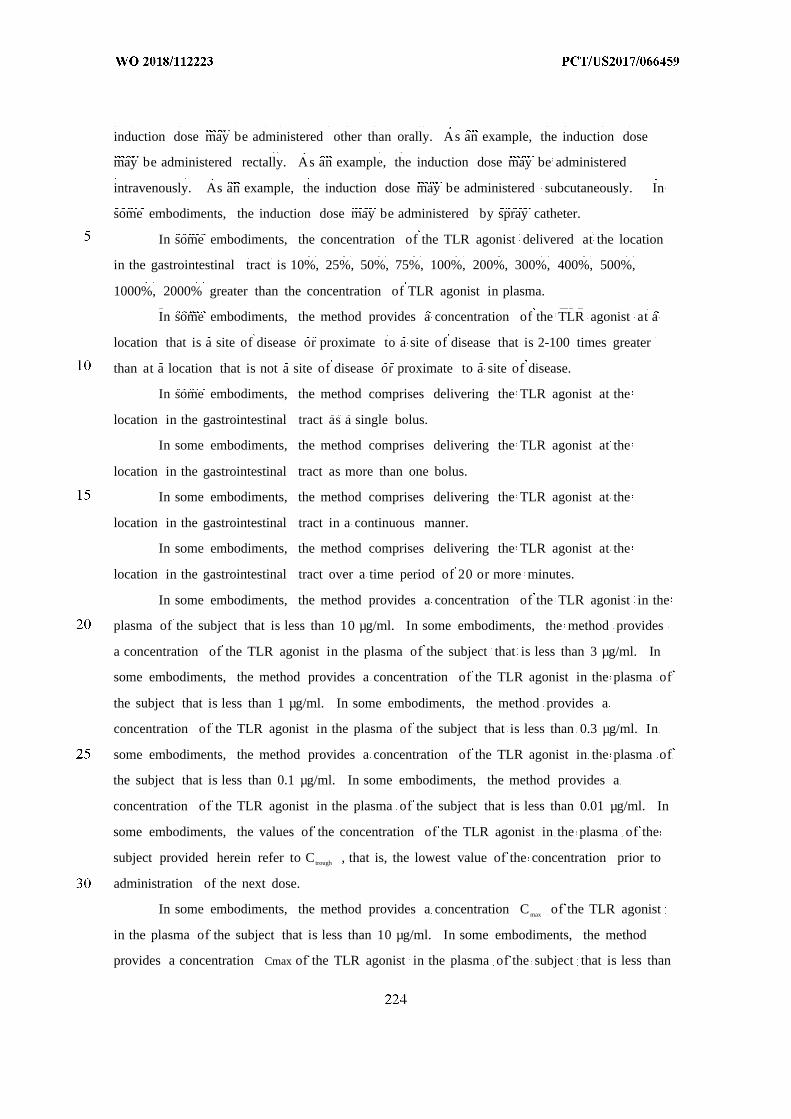
induction dose may be administered other than orally. As an example, the induction dose
may be administered rectally. As an example, the induction dose may be administered
intravenously. As an example, the induction dose may be administered subcutaneously. In
some embodiments, the induction dose may be administered by spray catheter.
In some embodiments, the concentration of the TLR agonist delivered at the location
in the gastrointestinal tract is 10%, 25%, 50%, 75%, 100%, 200%, 300%, 400%, 500%,
1000%, 2000% greater than the concentration of TLR agonist in plasma.
In some embodiments, the method provides a concentration of the TLR agonist at a
location that is a site of disease or proximate to a site of disease that is 2-100 times greater
than at a location that is not a site of disease or proximate to a site of disease.
In some embodiments, the method comprises delivering the TLR agonist at the
location in the gastrointestinal tract as a single bolus.
In some embodiments, the method comprises delivering the TLR agonist at the
location in the gastrointestinal tract as more than one bolus.
In some embodiments, the method comprises delivering the TLR agonist at the
location in the gastrointestinal tract in a continuous manner.
In some embodiments, the method comprises delivering the TLR agonist at the
location in the gastrointestinal tract over a time period of 20 or more minutes.
In some embodiments, the method provides a concentration of the TLR agonist in the
plasma of the subject that is less than 10 µg/ml. In some embodiments, the method provides
a concentration of the TLR agonist in the plasma of the subject that is less than 3 µg/ml. In
some embodiments, the method provides a concentration of the TLR agonist in the plasma of
the subject that is less than 1 µg/ml. In some embodiments, the method provides a
concentration of the TLR agonist in the plasma of the subject that is less than 0.3 µg/ml. In
some embodiments, the method provides a concentration of the TLR agonist in the plasma of
the subject that is less than 0.1 µg/ml. In some embodiments, the method provides a
concentration of the TLR agonist in the plasma of the subject that is less than 0.01 µg/ml. In
some embodiments, the values of the concentration of the TLR agonist in the plasma of the
subject provided herein refer to C trough , that is, the lowest value of the concentration prior to
administration of the next dose.
In some embodiments, the method provides a concentration Cmax of the TLR agonist
in the plasma of the subject that is less than 10 µg/ml. In some embodiments, the method
provides a concentration Cmax of the TLR agonist in the plasma of the subject that is less than
3 µg/ml. In some embodiments, the method provides a concentration Cmax of the TLR
agonist in the plasma of the subject that is less than 1 µg/ml. In some embodiments, the
method provides a concentration Cmax of the TLR agonist in the plasma of the subject that is
less than 0.3 µg/ml. In some embodiments, the method provides a concentration Cmax of the
TLR agonist in the plasma of the subject that is less than 0.1 µg/ml. In some embodiments,
the method provides a concentration Cmax of the TLR agonist in the plasma of the subject that
is less than 0.01 µg/ml.
In some embodiments, the method does not comprise delivering a TLR agonist
rectally to the subject.
In some embodiments, the method does not comprise delivering a TLR agonist via an
enema to the subject.
In some embodiments, the method does not comprise delivering a TLR agonist via
suppository to the subject.
In some embodiments, the method does not comprise delivering a TLR agonist via
instillation to the rectum of a subject.
In some embodiments, the methods disclosed herein comprise producing a
therapeutically effective degradation product of the TLR agonist in the gastrointestinal tract.
In some embodiments, the degradation product is a therapeutic antibody fragment. In some
embodiments, a therapeutically effective amount of the degradation product is produced.
In some embodiments, the antibody can be a humanized antibody, a chimeric
antibody, a multivalent antibody, or a fragment thereof. In some embodiments, an antibody
can be a scFv-Fc (Sokolowska-Wedzina et al., Mol. Cancer Res. 15(8):1040-1050, 2017), a
VHH domain (Li et al., Immunol. Lett. 188:89-95, 2017), a VNAR domain (Hasler et al.,
Mol. Immunol. 75:28-37, 2016), a (scFv)2, a minibody (Kim et al., PLoS One 10(1):e113442,
2014), or a BiTE. In some embodiments, an antibody can be a DVD-Ig (Wu et al., Nat.
Biotechnol. 25(11):1290-1297, 2007; WO 08/024188; WO 07/024715), and a dual-affinity
re-targeting antibody (DART) (Tsai et al., Mol. Ther. Oncolytics 3:15024, 2016), a triomab
(Chelius et al., MAbs 2(3):309-319, 2010), kih IgG with a common LC (Kontermann et al.,
Drug Discovery Today 20(7):838-847, 2015), a crossmab (Regula et al., EMBO Mol. Med.
9(7):985, 2017), an ortho-Fab IgG (Kontermann et al., Drug Discovery Today 20(7):838-847,
2015), a 2-in-1-IgG (Kontermann et al., Drug Discovery Today 20(7):838-847, 2015), IgG-
scFv (Cheal et al., Mol. Cancer Ther. 13(7):1803-1812, 2014), scFv2-Fc (Natsume et al., J.
Biochem. 140(3):359-368, 2006), a bi-nanobody (Kontermann et al., Drug Discovery Today
20(7):838-847, 2015), tanden antibody (Kontermann et al., Drug Discovery Today 20(7):838-
847, 2015), a DART-Fc (Kontermann et al., Drug Discovery Today 20(7):838-847, 2015), a
scFv-HSA-scFv (Kontermann et al., Drug Discovery Today 20(7):838-847, 2015), DNL-
Fab3 (Kontermann et al., Drug Discovery Today 20(7):838-847, 2015), DAF (two-in-one or
four-in-one), DutaMab, DT-IgG, knobs-in-holes common LC, knobs-in-holes assembly,
charge pair antibody, Fab-arm exchange antibody, SEEDbody, Triomab, LUZ-Y, Fcab, kλ-
body, orthogonal Fab, DVD-IgG, IgG(H)-scFv, scFv-(H)IgG, IgG(L)-scFv, scFv-(L)-IgG,
IgG (L,H)-Fc, IgG(H)-V, V(H)-IgG, IgG(L)-V, V(L)-IgG, KIH IgG-scFab, 2scFv-IgG, IgG-
2scFv, scFv4-Ig, Zybody, DVI-IgG, nanobody (e.g., antibodies derived from Camelus
bactriamus, Calelus dromaderius, or Lama paccos) (U.S. Patent No. 5,759,808; Stijlemans et
al., J. Biol. Chem. 279:1256-1261, 2004; Dumoulin et al., Nature 424:783-788, 2003; and
Pleschberger et al., Bioconjugate Chem. 14:440-448, 2003), nanobody-HSA, a diabody (e.g.,
Poljak, Structure 2(12):1121-1123, 1994; Hudson et al., J. Immunol. Methods 23(1-2):177-
189, 1999), a TandAb (Reusch et al., mAbs 6(3):727-738, 2014), scDiabody (Cuesta et al.,
Trends in Biotechnol. 28(7):355-362, 2010), scDiabody-CH3 (Sanz et al., Trends in Immunol.
25(2):85-91, 2004), Diabody-CH3 (Guo et al., Triple Body, miniantibody, minibody, TriBi
minibody, scFv-CH3 KIH, Fab-scFv, scFv-CH-CL-scFv, F(ab')2-scFV2, scFv-KIH, Fab-
scFv-Fc, tetravalent HCAb, scDiabody-Fc, diabody-Fc, tandem scFv-Fc, intrabody (Huston
et al., Human Antibodies 10(3-4):127-142, 2001; Wheeler et al., Mol. Ther. 8(3):355-366,
2003; Stocks, Drug Discov. Today 9(22):960-966, 2004), dock and lock bispecific antibody,
ImmTAC, HSAbody, scDiabody-HSA, tandem scFv, IgG-IgG, Cov-X-Body, and scFv1-
PEG-scFv2.
Non-limiting examples of an antigen-binding fragment of an antibody include an Fv
fragment, a Fab fragment, a F(ab')2 fragment, and a Fab' fragment. Additional examples of
an antigen-binding fragment of an antibody is an antigen-binding fragment of an IgG (e.g., an
antigen-binding fragment of IgG1, IgG2, IgG3, or IgG4) (e.g., an antigen-binding fragment
of a human or humanized IgG, e.g., human or humanized IgG1, IgG2, IgG3, or IgG4); an
antigen-binding fragment of an IgA (e.g., an antigen-binding fragment of IgA1 or IgA2) (e.g.,
an antigen-binding fragment of a human or humanized IgA, e.g., a human or humanized IgA1
or IgA2); an antigen-binding fragment of an IgD (e.g., an antigen-binding fragment of a
human or humanized IgD); an antigen-binding fragment of an IgE (e.g., an antigen-binding
fragment of a human or humanized IgE); or an antigen-binding fragment of an IgM (e.g., an
antigen-binding fragment of a human or humanized IgM).
In some embodiments, an antibody can be an IgNAR, a bispecific antibody (Milstein
and Cuello, Nature 305:537-539, 1983; Suresh et al., Methods in Enzymology 121:210, 1986;
WO 96/27011; Brennan et al., Science 229:81, 1985; Shalaby et al., J. Exp. Med. 175:217-
225, 1992; Kolstelny et al., J. Immunol. 148(5):1547-1553, 1992; Hollinger et al., Proc. Natl.
Acad. Sci. U.S.A. 90:6444-6448, 1993; Gruber et al., J. Immunol. 152:5368, 1994; Tutt et al.,
J. Immunol. 147:60, 1991), a bispecific diabody, a triabody (Schoonooghe et al., BMC
Biotechnol. 9:70, 2009), a tetrabody, scFv-Fc knobs-into-holes, a scFv-Fc-scFv, a
(Fab’scFv)2 , a V-IgG, a IvG-V, a dual V domain IgG, a heavy chain immunoglobulin or a
camelid (Holt et al., Trends Biotechnol. 21(11):484-490, 2003), an intrabody, a monoclonal
antibody (e.g., a human or humanized monoclonal antibody), a heteroconjugate antibody
(e.g., U.S. Patent No. 4,676,980), a linear antibody (Zapata et al., Protein Eng. 8(10:1057-
1062, 1995), a trispecific antibody (Tutt et al., J. Immunol. 147:60, 1991), a Fabs-in-Tandem
immunoglobulin (WO 15/103072), or a humanized camelid antibody.
In some embodiments, the methods comprising administering the TLR agonist in the
manner disclosed herein disclosed herein result in a reduced immunosuppressive properties
relative to methods of administration of the TLR agonist systemically.
In some embodiments, the methods comprising administering the TLR agonist in the
manner disclosed herein disclosed herein result in reduced immunogenicity relative to
methods of administration of the TLR agonist systemically.
Methods for treating colitis in subjects in immune-oncology therapy
In some embodiments, provided herein is a method for treating colitis as disclosed
herein in a subject, comprising releasing a TLR agonist at a location in the gastrointestinal
tract of the subject that is proximate to one or more sites of disease, wherein the method
comprises administering to the subject a pharmaceutical composition comprising a
therapeutically effective amount of the TLR agonist, , wherein the colitis is associated with
treatment of the subject with one or more immuno-oncology agents. In some embodiments,
the pharmaceutical composition is an ingestible device. In some embodiments, the
pharmaceutical composition is an ingestible device and the method comprises administering
orally to the subject the pharmaceutical composition.
In some embodiments, at least one of the one or more immuno-oncology agents is a
chemotherapeutic agent. In some embodiments, the chemotherapeutic agent is a
chemotherapeutic immunomodulator. In some embodiments, the chemotherapeutic
immunomodulator is an immune checkpoint inhibitor.
In some embodiments, the immune checkpoint inhibitor targets an immune
checkpoint protein or decreases an activity of an immune checkpoint protein selected from
the group of CTLA-4, PD-1, PD-L1, PD-1 – PD-L1, PD-1 – PD-L2, interleukin 2 (IL 2),
indoleamine 2,3-dioxygenase (IDO), IL 10, transforming growth factor-β (TGFβ), T cell
immunoglobulin and mucin 3 (TIM3 or HAVCR2), Galectin 9 – TIM3, Phosphatidylserine –
TIM3, lymphocyte activation gene 3 protein (LAG3), MHC class II – LAG3, 4 1BB–4 1BB
ligand, OX40–OX40 ligand, GITR, GITR ligand – GITR, CD27, CD70-CD27, TNFRSF25,
TNFRSF25–TL1A, CD40L, CD40–CD40 ligand, HVEM–LIGHT–LTA, HVEM, HVEM –
BTLA, HVEM – CD160, HVEM – LIGHT, HVEM–BTLA–CD160, CD80, CD80 – PDL-1,
PDL2 – CD80, CD244, CD48 – CD244, CD244, ICOS, ICOS–ICOS ligand, B7 H3, B7 H4,
VISTA, TMIGD2, HHLA2–TMIGD2, Butyrophilins, including BTNL2, Siglec family,
TIGIT and PVR family members, KIRs, ILTs and LIRs, NKG2D and NKG2A, MICA and
MICB, CD244, CD28, CD86 – CD28, CD86 – CTLA, CD80 – CD28, CD39, CD73
Adenosine–CD39–CD73, CXCR4–CXCL12, Phosphatidylserine, TIM3, Phosphatidylserine
– TIM3, SIRPA–CD47, VEGF, Neuropilin, CD160, CD30, and CD155.
In some examples, the immune checkpoint inhibitor is selected from the group
consisting of: Urelumab, PF 05082566, MEDI6469, TRX518, Varlilumab, CP 870893,
Pembrolizumab (PD1), Nivolumab (PD1), Atezolizumab (formerly MPDL3280A) (PDL1),
MEDI4736 (PD-L1), Avelumab (PD-L1), PDR001 (PD1), BMS 986016, MGA271,
Lirilumab, IPH2201, Emactuzumab, INCB024360, Galunisertib, Ulocuplumab, BKT140,
Bavituximab, CC 90002, Bevacizumab, and MNRP1685A, and MGA271.
In some examples, the immune checkpoint inhibitor targets or decreases an activity of
CTLA-4. In some embodiments, the immune checkpoint inhibitor is an antibody. In some
embodiments, the antibody is ipilimumab or tremelimumab.
In some examples, the immune checkpoint inhibitor targets PD1 or PD-L1. In some
examples, the immune checkpoint inhibitor is selected from nivolumab, lambroizumab, and
BMS-936559.
In some embodiments, at least one of the one or more immuno-oncology agents is a
T-cell capable of expressing a chimeric antigen receptor (CAR). In some embodiments, at
least one of the one or more immuno-oncology agents is a PI-3-kinase inhibitor.
In some embodiments, the treatment of the subject with one or more immuno-
oncology agents further comprises treatment of the subject with an immunosuppressant.
In some embodiments, provided herein is a method for reducing the development of
colitis in a subject administered an immuno-oncology agent, comprising releasing a TLR
agonist at a location in the gastrointestinal tract of the subject that is proximate to one or
more sites of disease, wherein the method comprises administering to the subject a
pharmaceutical composition comprising a therapeutically effective amount of the TLR
agonist. In some embodiments, the pharmaceutical composition is an ingestible device. In
some embodiments, the pharmaceutical composition is an ingestible device and the method
comprises administering orally to the subject the pharmaceutical composition.
In some embodiments of these methods, a subject is administered at least one dose of
an immuno-oncology agent prior to administering a pharmaceutical composition comprising
any of the devices described herein as described herein to the subject. In some embodiments
of these methods, a subject is first administered any of the devices as described herein, prior
to administration of the first dose of the immuno-oncology agent. In some embodiments of
these methods, the immuno-oncology agent is administered at substantially the same time as
the device described herein.
Also provided herein are methods of treating a subject having a cancer that include:
administering a first dose of an immuno-oncology agent to the subject; monitoring one or
more biomarkers, markers, or symptoms of colitis (e.g., any of the biomarkers, markers, or
symptoms of colitis described herein or known in the art); identifying a subject having a level
of a biomarker or marker, or having a symptom of colitis; and releasing a TLR agonist at a
location in the gastrointestinal tract of the subject that is proximate to one or more sites of
disease, wherein the method comprises administering to the subject a pharmaceutical
composition comprising a therapeutically effective amount of the TLR agonist. In some
embodiments, the pharmaceutical composition is an ingestible device. In some embodiments,
the pharmaceutical composition is an ingestible device and the method comprises
administering orally to the subject the pharmaceutical composition.
Also provided herein are methods of reducing the severity of colitis in a subject
having a cancer and administered an immuno-oncology agent that include administering to
the subject any of the devices described herein.
In some embodiments, provided herein is a method for treating colitis in a subject
comprising:
determining that the subject has colitis associated with treatment of the subject with
one or more immuno-oncology agents; and
releasing a TLR agonist at a location in the gastrointestinal tract of the subject that is
proximate to one or more sites of colitis, wherein the method comprises administering to the
subject a pharmaceutical composition comprising a therapeutically effective amount of the
TLR agonist. In some embodiments, the pharmaceutical composition is an ingestible device.
In some embodiments, the pharmaceutical composition is an ingestible device and the
method comprises administering orally to the subject the pharmaceutical composition.
In some embodiments, provided herein is a method for treating colitis in a subject
comprising:
determining that the subject has colitis associated with treatment of the subject with
one or more immuno-oncology agents; and
administering to the subject an ingestible device comprisingany of the TLR agonists
described herein, to treat the colitis.
In some embodiments, provided herein is a method for treating colitis,
comprising releasing a TLR agonist at a location in the gastrointestinal tract of a
subject who has been determined to have colitis associated with treatment of the subject with
one or more immuno-oncology agents, wherein the location is proximate to one or more sites
of colitis, wherein the method comprises administering to the subject a pharmaceutical
composition comprising a therapeutically effective amount of the TLR agonist. In some
embodiments, the pharmaceutical composition is an ingestible device. In some embodiments,
the pharmaceutical composition is an ingestible device and the method comprises
administering orally to the subject the pharmaceutical composition.
In some embodiments, provided herein is a method for treating colitis, comprising
administering an ingestible device comprising any of the TLR agonists described herein to a
subject who has been determined to have colitis associated with treatment of the subject with
one or more immuno-oncology agents.
In some embodiments, provided herein is an ingestible device comprising any of the
TLR agonists described herein for treating colitis associated with treatment of a subject with
one or more immuno-oncology agents.
Monitoring Progress of Disease
In some embodiments, the methods provided herein comprise monitoring the progress
of the disease. In some embodiments, monitoring the progress of the disease comprises
measuring the levels of IBD serological markers. In some embodiments, monitoring the
progress of the disease comprises determining mucosal healing at the location of release. In
some embodiments, monitoring the progress of the disease comprises determining the
Crohn’s Disease Activity Index (CDAI) over a period of about 6-8 weeks, or over a period of
about 52 weeks, following administration of the TLR agonist. In some embodiments,
monitoring the progress of the disease comprises determining the Harvey-Bradshaw Index
(HBI) following administration of the TLR agonist. Possible markers may include the
following: anti-glycan antibodies: anti-Saccharomices cerevisiae (ASCA); anti-
laminaribioside (ALCA); anti-chitobioside (ACCA); anti-mannobioside (AMCA); anti-
laminarin (anti-L); anti-chitin (anti-C) antibodies: anti-outer membrane porin C (anti-OmpC),
anti-Cbir1 flagellin; anti-12 antibody; autoantibodies targeting the exocrine pancreas (PAB);
perinuclear anti-neutrophil antibody (pANCA). In some embodiments, monitoring the
progress of the disease comprises measuring TLR agonist levels in serum over a period of
about 1-14 weeks, such as about 6-8 weeks following administration of the TLR agonist,
including at the 6-8 week time point. In some embodiments, monitoring the progress of the
disease comprises measuring TLR agonist levels in serum over a period of about 52 weeks
following administration of the TLR agonist, including at the 52 week time point.
Patients condition, diagnosis and treatment
In some embodiments herein, the method of treating a disease of the gastrointestinal tract
that comprises releasing a TLR agonist at a location in the gastrointestinal tract that is
proximate to one or more sites of disease comprises one or more of the following:
a) identifying a subject having a disease of the gastrointestinal tract, for example by
endoscopy or colonoscopy;
b) determination of the severity of the disease, for example with reference to the Mayo
Clinic Score, the Crohn’s Disease Activity Index (CDAI), the Harvey-Bradshaw
Index (HBI), or a combination of the above;
c) determination of the location of the disease, for example as determined by the
presence of lesions indicative of the disease;
d) evaluating the subject for suitability to treatment, for example by determining the
patency of the subject’s GI tract, for example if the indication is small intestinal
diseases, pancolitis, Crohn’s disease, or if the patients has strictures or fistulae;
e) administration of an induction dose or of a maintenance dose of a drug, such as the
TLR agonist or such as another drug that is effective in the treatment of IBD
conditions;
f) monitoring the progress of the disease, for example with reference to the Mayo Clinic
Score, the Crohn’s Disease Activity Index (CDAI), the Harvey-Bradshaw Index
(HBI), the PRO, PRO2 or PRO3 tools, or a combination of the above; and/or
g) optionally repeating steps e) and f) one or more times, for example over a period of
about 1-14 weeks, such as about 6-8 weeks following administration of the TLR
agonist, including at the 6-8 week time point, or over a period of about 52 weeks
following administration of the TLR agonist, including at the 52 week time point.
As used herein, an induction dose is a dose of drug that may be administered, for
example, at the beginning of a course of treatment, and that is higher than the maintenance
dose administered during treatment. An induction dose may also be administered during
treatment, for example if the condition of the patients becomes worse.
As used herein, a maintenance dose is a dose of drug that is provided on a repetitive
basis, for example at regular dosing intervals.
In some embodiments the TLR agonist is released from an ingestible device.
In some embodiments herein, the method of treating a disease of the gastrointestinal
tract that comprises releasing a TLR agonist at a location in the gastrointestinal tract that is
proximate to one or more sites of disease comprises a) hereinabove.
In some embodiments herein, the method of treating a disease of the gastrointestinal
tract that comprises releasing a TLR agonist at a location in the gastrointestinal tract that is
proximate to one or more sites of disease comprises b) hereinabove.
In some embodiments herein, the method of treating a disease of the gastrointestinal
tract that comprises releasing a TLR agonist at a location in the gastrointestinal tract that is
proximate to one or more sites of disease comprises c) hereinabove.
In some embodiments herein, the method of treating a disease of the gastrointestinal
tract that comprises releasing a TLR agonist at a location in the gastrointestinal tract that is
proximate to one or more sites of disease comprises d) hereinabove.
In some embodiments herein, the method of treating a disease of the gastrointestinal
tract that comprises releasing a TLR agonist at a location in the gastrointestinal tract that is
proximate to one or more sites of disease comprises e) hereinabove.
In some embodiments herein, the method of treating a disease of the gastrointestinal
tract that comprises releasing a TLR agonist at a location in the gastrointestinal tract that is
proximate to one or more sites of disease comprises f) hereinabove.
In some embodiments herein, the method of treating a disease of the gastrointestinal
tract that comprises releasing a TLR agonist at a location in the gastrointestinal tract that is
proximate to one or more sites of disease comprises g) hereinabove.
In some embodiments herein, the method of treating a disease of the gastrointestinal
tract that comprises releasing a TLR agonist at a location in the gastrointestinal tract that is
proximate to one or more sites of disease comprises a) and b) hereinabove. In some
embodiments herein, the method of treating a disease of the gastrointestinal tract that
comprises releasing a TLR agonist at a location in the gastrointestinal tract that is proximate
to one or more sites of disease comprises a) and c) hereinabove. In some embodiments
herein, the method of treating a disease of the gastrointestinal tract that comprises releasing a
TLR agonist at a location in the gastrointestinal tract that is proximate to one or more sites of
disease comprises a) and d) hereinabove. In some embodiments herein, the method of
treating a disease of the gastrointestinal tract that comprises releasing a TLR agonist at a
location in the gastrointestinal tract that is proximate to one or more sites of disease
comprises a) and e) hereinabove. In some embodiments herein, the method of treating a
disease of the gastrointestinal tract that comprises releasing a TLR agonist at a location in the
gastrointestinal tract that is proximate to one or more sites of disease comprises a) and f)
hereinabove. In some embodiments herein, the method of treating a disease of the
gastrointestinal tract that comprises releasing a TLR agonist at a location in the
gastrointestinal tract that is proximate to one or more sites of disease comprises a) and g)
hereinabove. In some embodiments herein, the method of treating a disease of the
gastrointestinal tract that comprises releasing a TLR agonist at a location in the
gastrointestinal tract that is proximate to one or more sites of disease comprises b) and c)
hereinabove. In some embodiments herein, the method of treating a disease of the
gastrointestinal tract that comprises releasing a TLR agonist at a location in the
gastrointestinal tract that is proximate to one or more sites of disease comprises b) and d)
hereinabove. In some embodiments herein, the method of treating a disease of the
gastrointestinal tract that comprises releasing a TLR agonist at a location in the
gastrointestinal tract that is proximate to one or more sites of disease comprises b) and e)
hereinabove. In some embodiments herein, the method of treating a disease of the
gastrointestinal tract that comprises releasing a TLR agonist at a location in the
gastrointestinal tract that is proximate to one or more sites of disease comprises b) and f)
hereinabove. In some embodiments herein, the method of treating a disease of the
gastrointestinal tract that comprises releasing a TLR agonist at a location in the
gastrointestinal tract that is proximate to one or more sites of disease comprises b) and g)
hereinabove. In some embodiments herein, the method of treating a disease of the
gastrointestinal tract that comprises releasing a TLR agonist at a location in the
gastrointestinal tract that is proximate to one or more sites of disease comprises c) and d)
hereinabove. In some embodiments herein, the method of treating a disease of the
gastrointestinal tract that comprises releasing a TLR agonist at a location in the
gastrointestinal tract that is proximate to one or more sites of disease comprises c) and e)
hereinabove. In some embodiments herein, the method of treating a disease of the
gastrointestinal tract that comprises releasing a TLR agonist at a location in the
gastrointestinal tract that is proximate to one or more sites of disease comprises c) and f)
hereinabove. In some embodiments herein, the method of treating a disease of the
gastrointestinal tract that comprises releasing a TLR agonist at a location in the
gastrointestinal tract that is proximate to one or more sites of disease comprises c) and g)
hereinabove. In some embodiments herein, the method of treating a disease of the
gastrointestinal tract that comprises releasing a TLR agonist at a location in the
gastrointestinal tract that is proximate to one or more sites of disease comprises d) and e)
hereinabove. In some embodiments herein, the method of treating a disease of the
gastrointestinal tract that comprises releasing a TLR agonist at a location in the
gastrointestinal tract that is proximate to one or more sites of disease comprises d) and f)
hereinabove. In some embodiments herein, the method of treating a disease of the
gastrointestinal tract that comprises releasing a TLR agonist at a location in the
gastrointestinal tract that is proximate to one or more sites of disease comprises d) and g)
hereinabove. In some embodiments herein, the method of treating a disease of the
gastrointestinal tract that comprises releasing a TLR agonist at a location in the
gastrointestinal tract that is proximate to one or more sites of disease comprises e) and f)
hereinabove. In some embodiments herein, the method of treating a disease of the
gastrointestinal tract that comprises releasing a TLR agonist at a location in the
gastrointestinal tract that is proximate to one or more sites of disease comprises g)
hereinabove.
In some embodiments, one or more steps a) to e) herein comprise endoscopy of the
gastrointestinal tract. In some embodiments, one or more steps a) to e) herein comprise
colonoscopy of the gastrointestinal tract. In some embodiments, one or more steps a) to e)
herein is performed one or more times. In some embodiments, such one or more of such one
or more steps a) to e) is performed after releasing the TLR agonist at the location in the
gastrointestinal tract that is proximate to one or more sites of disease.
In some embodiments, the method comprises administering one or more maintenance
doses following administration of the induction dose in step e). In some embodiments an
induction dose of TLR agonist and a maintenance dose of TLR agonist are each administered
to the subject by administering a pharmaceutical composition comprising a therapeutically
effective amount of the TLR agonist. In some embodiments an induction dose of TLR
agonist is administered to the subject in a different manner from the maintenance dose. As an
example, the maintenance dose may be administered systemically, while the maintenance
dose is administered locally using a device. In one embodiment, a maintenance dose is
administered systemically, and an induction dose is administered using a device every 1, 2, 3,
4, 5, 6, 7, 10, 15, 20, 25, 30, 35, 40, or 45 days. In another embodiment, a maintenance dose
is administered systemically, and an induction dose is administered when a disease flare up is
detected or suspected.
In some embodiments, the induction dose is a dose of the TLR agonist administered
in an ingestible device as disclosed herein. In some embodiments, the maintenance dose is a
dose of the TLR agonist administered in an ingestible device as disclosed herein.
In some embodiments, the induction dose is a dose of the TLR agonist administered
in an ingestible device as disclosed herein. In some embodiments, the maintenance dose is a
dose of the TLR agonist delivered systemically, such as orally with a tablet or capsule, or
subcutaneously, or intravenously.
In some embodiments, the induction dose is a dose of the TLR agonist delivered
systemically, such as orally with a tablet or capsule, or subcutaneously, or intravenously. In
some embodiments, the maintenance dose is a dose of the TLR agonist administered in an
ingestible device as disclosed herein.
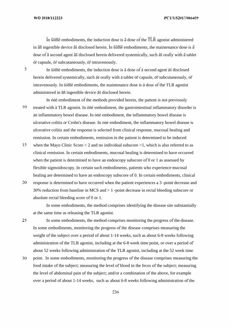
In some embodiments, the induction dose is a dose of the TLR agonist administered
in an ingestible device as disclosed herein. In some embodiments, the maintenance dose is a
dose of a second agent as disclosed herein delivered systemically, such as orally with a tablet
or capsule, or subcutaneously, or intravenously.
In some embodiments, the induction dose is a dose of a second agent as disclosed
herein delivered systemically, such as orally with a tablet or capsule, or subcutaneously, or
intravenously. In some embodiments, the maintenance dose is a dose of the TLR agonist
administered in an ingestible device as disclosed herein.
In one embodiment of the methods provided herein, the patient is not previously
treated with a TLR agonist. In one embodiment, the gastrointestinal inflammatory disorder is
an inflammatory bowel disease. In one embodiment, the inflammatory bowel disease is
ulcerative colitis or Crohn's disease. In one embodiment, the inflammatory bowel disease is
ulcerative colitis and the response is selected from clinical response, mucosal healing and
remission. In certain embodiments, remission in the patient is determined to be induced
when the Mayo Clinic Score < 2 and no individual subscore >1, which is also referred to as
clinical remission. In certain embodiments, mucosal healing is determined to have occurred
when the patient is determined to have an endoscopy subscore of 0 or 1 as assessed by
flexible sigmoidoscopy. In certain such embodiments, patients who experience mucosal
healing are determined to have an endoscopy subscore of 0. In certain embodiments, clinical
response is determined to have occurred when the patient experiences a 3 -point decrease and
30% reduction from baseline in MCS and > 1 -point decrease in rectal bleeding subscore or
absolute rectal bleeding score of 0 or 1.
In some embodiments, the method comprises identifying the disease site substantially
at the same time as releasing the TLR agonist.
In some embodiments, the method comprises monitoring the progress of the disease.
In some embodiments, monitoring the progress of the disease comprises measuring the
weight of the subject over a period of about 1-14 weeks, such as about 6-8 weeks following
administration of the TLR agonist, including at the 6-8 week time point, or over a period of
about 52 weeks following administration of the TLR agonist, including at the 52 week time
point. In some embodiments, monitoring the progress of the disease comprises measuring the
food intake of the subject; measuring the level of blood in the feces of the subject; measuring
the level of abdominal pain of the subject; and/or a combination of the above, for example
over a period of about 1-14 weeks, such as about 6-8 weeks following administration of the
TLR agonist, including at the 6-8 week time point, or over a period of about 52 weeks
following administration of the TLR agonist, including at the 52 week time point.
In some embodiments, the method comprises administering a TLR agonist with a
spray catheter. For example, administering a TLR agonist with a spray catheter may be
performed in step (e) hereinabove.
In some embodiments, the method does not comprise administering a TLR agonist
with a spray catheter.
In some embodiments, data obtained from cell culture assays and animal studies can
be used in formulating an appropriate dosage of any given TLR agonist. The effectiveness
and dosing of any TLR agonist can be determined by a health care professional or veterinary
professional using methods known in the art, as well as by the observation of one or more
disease symptoms in a subject (e.g., a human). Certain factors may influence the dosage and
timing required to effectively treat a subject (e.g., the severity of the disease or disorder,
previous treatments, the general health and/or age of the subject, and the presence of other
diseases).
In some embodiments, the subject is further administered an additional therapeutic
agent (e.g., any of the additional therapeutic agents described herein). The additional
therapeutic agent can be administered to the subject at substantially the same time as the TLR
agonist or pharmaceutical composition comprising it is administered and/or at one or more
other time points. In some embodiments, the additional therapeutic agent is formulated
together with the TLR agonist (e.g., using any of the examples of formulations described
herein).
In some embodiments, the subject is administered a dose of the TLR agonist at least
once a month (e.g., at least twice a month, at least three times a month, at least four times a
month, at least once a week, at least twice a week, three times a week, once a day, or twice a
day). The TLR agonist may be administered to a subject chronically. Chronic treatments
include any form of repeated administration for an extended period of time, such as repeated
administrations for one or more months, between a month and a year, one or more years,
more than five years, more than 10 years, more than 15 years, more than 20 years, more than
25 years, more than 30 years, more than 35 years, more than 40 years, more than 45 years, or
longer. Alternatively, or in addition, chronic treatments may be administered. Chronic
treatments can involve regular administrations, for example one or more times a day, one or
more times a week, or one or more times a month. For example, chronic treatment can
include administration (e.g., intravenous administration) about every two weeks (e.g.,
between about every 10 to 18 days).
A suitable dose may be the amount that is the lowest dose effective to produce a
desired therapeutic effect. Such an effective dose will generally depend upon the factors
described herein. If desired, an effective daily dose of TLR agonist can be administered as
two, three, four, five, or six or more sub-doses administered separately at appropriate
intervals throughout the day, optionally, in unit dosage forms.
In some examples, administration of an TLR agonistusing any of the compositions or
devices described herein can result in the onset of treatment (e.g., a reduction in the number,
severity, or duration of one or more symptoms and/or markers of any of the diseases
described herein) or drug-target engagement in a subject within a time period of about 10
minutes to about 10 hours, about 10 minutes to about 9 hours, about 10 minutes to about 8
hours, about 10 minutes to about 7 hours, about 10 minutes to about 6 hours, about 10
minutes to about 5 hours, about 10 minutes to about 4.5 hours, about 10 minutes to about 4
hours, about 10 minutes to about 3.5 hours, about 10 minutes to about 3 hours, about 10
minutes to about 2.5 hours, about 10 minutes to about 2 hours, about 10 minutes to about 1.5
hours, about 10 minutes to about 1 hour, about 10 minutes to about 55 minutes, about 10
minutes to about 50 minutes, about 10 minutes to about 45 minutes, about 10 minutes to
about 40 minutes, about 10 minutes to about 35 minutes, about 10 minutes to about 30
minutes, about 10 minutes to about 25 minutes, about 10 minutes to about 20 minutes, about
10 minutes to about 15 minutes, about 15 minutes to about 10 hours, about 15 minutes to
about 9 hours, about 15 minutes to about 8 hours, about 15 minutes to about 7 hours, about
15 minutes to about 6 hours, about 15 minutes to about 5 hours, about 15 minutes to about
4.5 hours, about 15 minutes to about 4 hours, about 15 minutes to about 3.5 hours, about 15
minutes to about 3 hours, about 15 minutes to about 2.5 hours, about 15 minutes to about 2
hours, about 15 minutes to about 1.5 hours, about 15 minutes to about 1 hour, about 15
minutes to about 55 minutes, about 15 minutes to about 50 minutes, about 15 minutes to
about 45 minutes, about 15 minutes to about 40 minutes, about 15 minutes to about 35
minutes, about 15 minutes to about 30 minutes, about 15 minutes to about 25 minutes, about
15 minutes to about 20 minutes, about 20 minutes to about 10 hours, about 20 minutes to
about 9 hours, about 20 minutes to about 8 hours, about 20 minutes to about 7 hours, about
20 minutes to about 6 hours, about 20 minutes to about 5 hours, about 20 minutes to about
4.5 hours, about 20 minutes to about 4 hours, about 20 minutes to about 3.5 hours, about 20
minutes to about 3 hours, about 20 minutes to about 2.5 hours, about 20 minutes to about 2
hours, about 20 minutes to about 1.5 hours, about 20 minutes to about 1 hour, about 20
minutes to about 55 minutes, about 20 minutes to about 50 minutes, about 20 minutes to
about 45 minutes, about 20 minutes to about 40 minutes, about 20 minutes to about 35
minutes, about 20 minutes to about 30 minutes, about 20 minutes to about 25 minutes, about
25 minutes to about 10 hours, about 25 minutes to about 9 hours, about 25 minutes to about 8
hours, about 25 minutes to about 7 hours, about 25 minutes to about 6 hours, about 25
minutes to about 5 hours, about 25 minutes to about 4.5 hours, about 25 minutes to about 4
hours, about 25 minutes to about 3.5 hours, about 25 minutes to about 3 hours, about 25
minutes to about 2.5 hours, about 25 minutes to about 2 hours, about 25 minutes to about 1.5
hours, about 25 minutes to about 1 hour, about 25 minutes to about 55 minutes, about 25
minutes to about 50 minutes, about 25 minutes to about 45 minutes, about 25 minutes to
about 40 minutes, about 25 minutes to about 35 minutes, about 25 minutes to about 30
minutes, about 30 minutes to about 10 hours, about 30 minutes to about 9 hours, about 30
minutes to about 8 hours, about 30 minutes to about 7 hours, about 30 minutes to about 6
hours, about 30 minutes to about 5 hours, about 30 minutes to about 4.5 hours, about 30
minutes to about 4 hours, about 30 minutes to about 3.5 hours, about 30 minutes to about 3
hours, about 30 minutes to about 2.5 hours, about 30 minutes to about 2 hours, about 30
minutes to about 1.5 hours, about 30 minutes to about 1 hour, about 30 minutes to about 55
minutes, about 30 minutes to about 50 minutes, about 30 minutes to about 45 minutes, about
30 minutes to about 40 minutes, about 30 minutes to about 35 minutes, about 35 minutes to
about 10 hours, about 35 minutes to about 9 hours, about 35 minutes to about 8 hours, about
35 minutes to about 7 hours, about 35 minutes to about 6 hours, about 35 minutes to about 5
hours, about 35 minutes to about 4.5 hours, about 35 minutes to about 4 hours, about 35
minutes to about 3.5 hours, about 35 minutes to about 3 hours, about 35 minutes to about 2.5
hours, about 35 minutes to about 2 hours, about 35 minutes to about 1.5 hours, about 35
minutes to about 1 hour, about 35 minutes to about 55 minutes, about 35 minutes to about 50
minutes, about 35 minutes to about 45 minutes, about 35 minutes to about 40 minutes, about
40 minutes to about 10 hours, about 40 minutes to about 9 hours, about 40 minutes to about 8
hours, about 40 minutes to about 7 hours, about 40 minutes to about 6 hours, about 40
minutes to about 5 hours, about 40 minutes to about 4.5 hours, about 40 minutes to about 4
hours, about 40 minutes to about 3.5 hours, about 40 minutes to about 3 hours, about 40
minutes to about 2.5 hours, about 40 minutes to about 2 hours, about 40 minutes to about 1.5
hours, about 40 minutes to about 1 hour, about 40 minutes to about 55 minutes, about 40
minutes to about 50 minutes, about 40 minutes to about 45 minutes, about 45 minutes to
about 10 hours, about 45 minutes to about 9 hours, about 45 minutes to about 8 hours, about
45 minutes to about 7 hours, about 45 minutes to about 6 hours, about 45 minutes to about 5
hours, about 45 minutes to about 4.5 hours, about 45 minutes to about 4 hours, about 45
minutes to about 3.5 hours, about 45 minutes to about 3 hours, about 45 minutes to about 2.5
hours, about 45 minutes to about 2 hours, about 45 minutes to about 1.5 hours, about 45
minutes to about 1 hour, about 45 minutes to about 55 minutes, about 45 minutes to about 50
minutes, about 50 minutes to about 10 hours, about 50 minutes to about 9 hours, about 50
minutes to about 8 hours, about 50 minutes to about 7 hours, about 50 minutes to about 6
hours, about 50 minutes to about 5 hours, about 50 minutes to about 4.5 hours, about 50
minutes to about 4 hours, about 50 minutes to about 3.5 hours, about 50 minutes to about 3
hours, about 50 minutes to about 2.5 hours, about 50 minutes to about 2 hours, about 50
minutes to about 1.5 hours, about 50 minutes to about 1 hour, about 50 minutes to about 55
minutes, about 55 minutes to about 10 hours, about 55 minutes to about 9 hours, about 55
minutes to about 8 hours, about 55 minutes to about 7 hours, about 55 minutes to about 6
hours, about 55 minutes to about 5 hours, about 55 minutes to about 4.5 hours, about 55
minutes to about 4 hours, about 55 minutes to about 3.5 hours, about 55 minutes to about 3
hours, about 55 minutes to about 2.5 hours, about 55 minutes to about 2 hours, about 55
minutes to about 1.5 hours, about 55 minutes to about 1 hour, about 1 hour to about 10 hours,
about 1 hour to about 9 hours, about 1 hour to about 8 hours, about 1 hour to about 7 hours,
about 1 hour to about 6 hours, about 1 hour to about 5 hours, about 1 hour to about 4.5 hours,
about 1 hour to about 4 hours, about 1 hour to about 3.5 hours, about 1 hour to about 3 hours,
about 1 hour to about 2.5 hours, about 1 hour to about 2 hours, about 1 hour to about 1.5
hours, about 1.5 hours to about 10 hours, about 1.5 hours to about 9 hours, about 1.5 hours to
about 8 hours, about 1.5 hours to about 7 hours, about 1.5 hours to about 6 hours, about 1.5
hours to about 5 hours, about 1.5 hours to about 4.5 hours, about 1.5 hours to about 4 hours,
about 1.5 hours to about 3.5 hours, about 1.5 hours to about 3 hours, about 1.5 hours to about
2.5 hours, about 1.5 hours to about 2 hours, about 2 hours to about 10 hours, about 2 hours to
about 9 hours, about 2 hours to about 8 hours, about 2 hours to about 7 hours, about 2 hours
to about 6 hours, about 2 hours to about 5 hours, about 2 hours to about 4.5 hours, about 2
hours to about 4 hours, about 2 hours to about 3.5 hours, about 2 hours to about 3 hours,
about 2 hours to about 2.5 hours, about 2.5 hours to about 10 hours, about 2.5 hours to about
9 hours, about 2.5 hours to about 8 hours, about 2.5 hours to about 7 hours, about 2.5 hours to
about 6 hours, about 2.5 hours to about 5 hours, about 2.5 hours to about 4.5 hours, about 2.5
hours to about 4 hours, about 2.5 hours to about 3.5 hours, about 2.5 hours to about 3 hours,
about 3 hours to about 10 hours, about 3 hours to about 9 hours, about 3 hours to about 8
hours, about 3 hours to about 7 hours, about 3 hours to about 6 hours, about 3 hours to about
5 hours, about 3 hours to about 4.5 hours, about 3 hours to about 4 hours, about 3 hours to
about 3.5 hours, about 3.5 hours to about 10 hours, about 3.5 hours to about 9 hours, about
3.5 hours to about 8 hours, about 3.5 hours to about 7 hours, about 3.5 hours to about 6 hours,
about 3.5 hours to about 5 hours, about 3.5 hours to about 4.5 hours, about 3.5 hours to about
4 hours, about 4 hours to about 10 hours, about 4 hours to about 9 hours, about 4 hours to
about 8 hours, about 4 hours to about 7 hours, about 4 hours to about 6 hours, about 4 hours
to about 5 hours, about 4 hours to about 4.5 hours, about 4.5 hours to about 10 hours, about
4.5 hours to about 9 hours, about 4.5 hours to about 8 hours, about 4.5 hours to about 7 hours,
about 4.5 hours to about 6 hours, about 4.5 hours to about 5 hours, about 5 hours to about 10
hours, about 5 hours to about 9 hours, about 5 hours to about 8 hours, about 5 hours to about
7 hours, about 5 hours to about 6 hours, about 6 hours to about 10 hours, about 6 hours to
about 9 hours, about 6 hours to about 8 hours, about 6 hours to about 7 hours, about 7 hours
to about 10 hours, about 7 hours to about 9 hours, about 7 hours to about 8 hours, about 8
hours to about 10 hours, about 8 hours to about 9 hours, or about 9 hours to about 10 hours of
administration of a dose of an TLR agonist using any of the devices or compositions
described herein. Drug-target engagement may be determined, for example, as disclosed in
Simon GM, Niphakis MJ, Cravatt BF, Nature chemical biology. 2013;9(4):200-205,
incorporated by reference herein in its entirety.
In some embodiments, administration of an TLR agonist using any of the devices or
compositions described herein can provide for treatment (e.g., a reduction in the number,
severity, and/or duration of one or more symptoms and/or markers of any of the disorders
described herein in a subject) for a time period of between about 1 hour to about 30 days,
about 1 hour to about 28 days, about 1 hour to about 26 days, about 1 hour to about 24 days,
about 1 hour to about 22 days, about 1 hour to about 20 days, about 1 hour to about 18 days,
about 1 hour to about 16 days, about 1 hour to about 14 days, about 1 hour to about 12 days,
about 1 hour to about 10 days, about 1 hour to about 8 days, about 1 hour to about 6 days,
about 1 hour to about 5 days, about 1 hour to about 4 days, about 1 hour to about 3 days,
about 1 hour to about 2 days, about 1 hour to about 1 day, about 1 hour to about 12 hours,
about 1 hour to about 6 hours, about 1 hour to about 3 hours, about 3 hours to about 30 days,
about 3 hours to about 28 days, about 3 hours to about 26 days, about 3 hours to about 24
days, about 3 hours to about 22 days, about 3 hours to about 20 days, about 3 hours to about
18 days, about 3 hours to about 16 days, about 3 hours to about 14 days, about 3 hours to
about 12 days, about 3 hours to about 10 days, about 3 hours to about 8 days, about 3 hours to
about 6 days, about 3 hours to about 5 days, about 3 hours to about 4 days, about 3 hours to
about 3 days, about 3 hours to about 2 days, about 3 hours to about 1 day, about 3 hours to
about 12 hours, about 3 hours to about 6 hours, about 6 hours to about 30 days, about 6 hours
to about 28 days, about 6 hours to about 26 days, about 6 hours to about 24 days, about 6
hours to about 22 days, about 6 hours to about 20 days, about 6 hours to about 18 days, about
6 hours to about 16 days, about 6 hours to about 14 days, about 6 hours to about 12 days,
about 6 hours to about 10 days, about 6 hours to about 8 days, about 6 hours to about 6 days,
about 6 hours to about 5 days, about 6 hours to about 4 days, about 6 hours to about 3 days,
about 6 hours to about 2 days, about 6 hours to about 1 day, about 6 hours to about 12 hours,
about 12 hours to about 30 days, about 12 hours to about 28 days, about 12 hours to about 26
days, about 12 hours to about 24 days, about 12 hours to about 22 days, about 12 hours to
about 20 days, about 12 hours to about 18 days, about 12 hours to about 16 days, about 12
hours to about 14 days, about 12 hours to about 12 days, about 12 hours to about 10 days,
about 12 hours to about 8 days, about 12 hours to about 6 days, about 12 hours to about 5
days, about 12 hours to about 4 days, about 12 hours to about 3 days, about 12 hours to about
2 days, about 12 hours to about 1 day, about 1 day to about 30 days, about 1 day to about 28
days, about 1 day to about 26 days, about 1 day to about 24 days, about 1 day to about 22
days, about 1 day to about 20 days, about 1 day to about 18 days, about 1 day to about 16
days, about 1 day to about 14 days, about 1 day to about 12 days, about 1 day to about 10
days, about 1 day to about 8 days, about 1 day to about 6 days, about 1 day to about 5 days,
about 1 day to about 4 days, about 1 day to about 3 days, about 1 day to about 2 days, about 2
days to about 30 days, about 2 days to about 28 days, about 2 days to about 26 days, about 2
days to about 24 days, about 2 days to about 22 days, about 2 days to about 20 days, about 2
days to about 18 days, about 2 days to about 16 days, about 2 days to about 14 days, about 2
days to about 12 days, about 2 days to about 10 days, about 2 days to about 8 days, about 2
days to about 6 days, about 2 days to about 5 days, about 2 days to about 4 days, about 2 days
to about 3 days, about 3 days to about 30 days, about 3 days to about 28 days, about 3 days to
about 26 days, about 3 days to about 24 days, about 3 days to about 22 days, about 3 days to
about 20 days, about 3 days to about 18 days, about 3 days to about 16 days, about 3 days to
about 14 days, about 3 days to about 12 days, about 3 days to about 10 days, about 3 days to
about 8 days, about 3 days to about 6 days, about 3 days to about 5 days, about 3 days to
about 4 days, about 4 days to about 30 days, about 4 days to about 28 days, about 4 days to
about 26 days, about 4 days to about 24 days, about 4 days to about 22 days, about 4 days to
about 20 days, about 4 days to about 18 days, about 4 days to about 16 days, about 4 days to
about 14 days, about 4 days to about 12 days, about 4 days to about 10 days, about 4 days to
about 8 days, about 4 days to about 6 days, about 4 days to about 5 days, about 5 days to
about 30 days, about 5 days to about 28 days, about 5 days to about 26 days, about 5 days to
about 24 days, about 5 days to about 22 days, about 5 days to about 20 days, about 5 days to
about 18 days, about 5 days to about 16 days, about 5 days to about 14 days, about 5 days to
about 12 days, about 5 days to about 10 days, about 5 days to about 8 days, about 5 days to
about 6 days, about 6 days to about 30 days, about 6 days to about 28 days, about 6 days to
about 26 days, about 6 days to about 24 days, about 6 days to about 22 days, about 6 days to
about 20 days, about 6 days to about 18 days, about 6 days to about 16 days, about 6 days to
about 14 days, about 6 days to about 12 days, about 6 days to about 10 days, about 6 days to
about 8 days, about 8 days to about 30 days, about 8 days to about 28 days, about 8 days to
about 26 days, about 8 days to about 24 days, about 8 days to about 22 days, about 8 days to
about 20 days, about 8 days to about 18 days, about 8 days to about 16 days, about 8 days to
about 14 days, about 8 days to about 12 days, about 8 days to about 10 days, about 10 days to
about 30 days, about 10 days to about 28 days, about 10 days to about 26 days, about 10 days
to about 24 days, about 10 days to about 22 days, about 10 days to about 20 days, about 10
days to about 18 days, about 10 days to about 16 days, about 10 days to about 14 days, about
10 days to about 12 days, about 12 days to about 30 days, about 12 days to about 28 days,
about 12 days to about 26 days, about 12 days to about 24 days, about 12 days to about 22
days, about 12 days to about 20 days, about 12 days to about 18 days, about 12 days to about
16 days, about 12 days to about 14 days, about 14 days to about 30 days, about 14 days to
about 28 days, about 14 days to about 26 days, about 14 days to about 24 days, about 14 days
to about 22 days, about 14 days to about 20 days, about 14 days to about 18 days, about 14
days to about 16 days, about 16 days to about 30 days, about 16 days to about 28 days, about
16 days to about 26 days, about 16 days to about 24 days, about 16 days to about 22 days,
about 16 days to about 20 days, about 16 days to about 18 days, about 18 days to about 30
days, about 18 days to about 28 days, about 18 days to about 26 days, about 18 days to about
24 days, about 18 days to about 22 days, about 18 days to about 20 days, about 20 days to
about 30 days, about 20 days to about 28 days, about 20 days to about 26 days, about 20 days
to about 24 days, about 20 days to about 22 days, about 22 days to about 30 days, about 22
days to about 28 days, about 22 days to about 26 days, about 22 days to about 24 days, about
24 days to about 30 days, about 24 days to about 28 days, about 24 days to about 26 days,
about 26 days to about 30 days, about 26 days to about 28 days, or about 28 days to about 30
days in a subject following first administration of an TLR agonist using any of the
compositions or devices described herein. Non-limiting examples of symptoms and/or
markers of a disease described herein are described below.
For example, treatment can result in a decrease (e.g., about 1% to about 99%
decrease, about 1% to about 95% decrease, about 1% to about 90% decrease, about 1% to
about 85% decrease, about 1% to about 80% decrease, about 1% to about 75% decrease,
about 1% to about 70% decrease, about 1% to about 65% decrease, about 1% to about 60%
decrease, about 1% to about 55% decrease, about 1% to about 50% decrease, about 1% to
about 45% decrease, about 1% to about 40% decrease, about 1% to about 35% decrease,
about 1% to about 30% decrease, about 1% to about 25% decrease, about 1% to about 20%
decrease, about 1% to about 15% decrease, about 1% to about 10% decrease, about 1% to
about 5% decrease, about 5% to about 99% decrease, about 5% to about 95% decrease, about
5% to about 90% decrease, about 5% to about 85% decrease, about 5% to about 80%
decrease, about 5% to about 75% decrease, about 5% to about 70% decrease, about 5% to
about 65% decrease, about 5% to about 60% decrease, about 5% to about 55% decrease,
about 5% to about 50% decrease, about 5% to about 45% decrease, about 5% to about 40%
decrease, about 5% to about 35% decrease, about 5% to about 30% decrease, about 5% to
about 25% decrease, about 5% to about 20% decrease, about 5% to about 15% decrease,
about 5% to about 10% decrease, about 10% to about 99% decrease, about 10% to about 95%
decrease, about 10% to about 90% decrease, about 10% to about 85% decrease, about 10% to
about 80% decrease, about 10% to about 75% decrease, about 10% to about 70% decrease,
about 10% to about 65% decrease, about 10% to about 60% decrease, about 10% to about
55% decrease, about 10% to about 50% decrease, about 10% to about 45% decrease, about
10% to about 40% decrease, about 10% to about 35% decrease, about 10% to about 30%
decrease, about 10% to about 25% decrease, about 10% to about 20% decrease, about 10% to
about 15% decrease, about 15% to about 99% decrease, about 15% to about 95% decrease,
about 15% to about 90% decrease, about 15% to about 85% decrease, about 15% to about
80% decrease, about 15% to about 75% decrease, about 15% to about 70% decrease, about
15% to about 65% decrease, about 15% to about 60% decrease, about 15% to about 55%
decrease, about 15% to about 50% decrease, about 15% to about 45% decrease, about 15% to
about 40% decrease, about 15% to about 35% decrease, about 15% to about 30% decrease,
about 15% to about 25% decrease, about 15% to about 20% decrease, about 20% to about
99% decrease, about 20% to about 95% decrease, about 20% to about 90% decrease, about
20% to about 85% decrease, about 20% to about 80% decrease, about 20% to about 75%
decrease, about 20% to about 70% decrease, about 20% to about 65% decrease, about 20% to
about 60% decrease, about 20% to about 55% decrease, about 20% to about 50% decrease,
about 20% to about 45% decrease, about 20% to about 40% decrease, about 20% to about
35% decrease, about 20% to about 30% decrease, about 20% to about 25% decrease, about
25% to about 99% decrease, about 25% to about 95% decrease, about 25% to about 90%
decrease, about 25% to about 85% decrease, about 25% to about 80% decrease, about 25% to
about 75% decrease, about 25% to about 70% decrease, about 25% to about 65% decrease,
about 25% to about 60% decrease, about 25% to about 55% decrease, about 25% to about
50% decrease, about 25% to about 45% decrease, about 25% to about 40% decrease, about
25% to about 35% decrease, about 25% to about 30% decrease, about 30% to about 99%
decrease, about 30% to about 95% decrease, about 30% to about 90% decrease, about 30% to
about 85% decrease, about 30% to about 80% decrease, about 30% to about 75% decrease,
about 30% to about 70% decrease, about 30% to about 65% decrease, about 30% to about
60% decrease, about 30% to about 55% decrease, about 30% to about 50% decrease, about
30% to about 45% decrease, about 30% to about 40% decrease, about 30% to about 35%
decrease, about 35% to about 99% decrease, about 35% to about 95% decrease, about 35% to
about 90% decrease, about 35% to about 85% decrease, about 35% to about 80% decrease,
about 35% to about 75% decrease, about 35% to about 70% decrease, about 35% to about
65% decrease, about 35% to about 60% decrease, about 35% to about 55% decrease, about
35% to about 50% decrease, about 35% to about 45% decrease, about 35% to about 40%
decrease, about 40% to about 99% decrease, about 40% to about 95% decrease, about 40% to
about 90% decrease, about 40% to about 85% decrease, about 40% to about 80% decrease,
about 40% to about 75% decrease, about 40% to about 70% decrease, about 40% to about
65% decrease, about 40% to about 60% decrease, about 40% to about 55% decrease, about
40% to about 50% decrease, about 40% to about 45% decrease, about 45% to about 99%
decrease, about 45% to about 95% decrease, about 45% to about 90% decrease, about 45% to
about 85% decrease, about 45% to about 80% decrease, about 45% to about 75% decrease,
about 45% to about 70% decrease, about 45% to about 65% decrease, about 45% to about
60% decrease, about 45% to about 55% decrease, about 45% to about 50% decrease, about
50% to about 99% decrease, about 50% to about 95% decrease, about 50% to about 90%
decrease, about 50% to about 85% decrease, about 50% to about 80% decrease, about 50% to
about 75% decrease, about 50% to about 70% decrease, about 50% to about 65% decrease,
about 50% to about 60% decrease, about 50% to about 55% decrease, about 55% to about
99% decrease, about 55% to about 95% decrease, about 55% to about 90% decrease, about
55% to about 85% decrease, about 55% to about 80% decrease, about 55% to about 75%
decrease, about 55% to about 70% decrease, about 55% to about 65% decrease, about 55% to
about 60% decrease, about 60% to about 99% decrease, about 60% to about 95% decrease,
about 60% to about 90% decrease, about 60% to about 85% decrease, about 60% to about
80% decrease, about 60% to about 75% decrease, about 60% to about 70% decrease, about
60% to about 65% decrease, about 65% to about 99% decrease, about 65% to about 95%
decrease, about 65% to about 90% decrease, about 65% to about 85% decrease, about 65% to
about 80% decrease, about 65% to about 75% decrease, about 65% to about 70% decrease,
about 70% to about 99% decrease, about 70% to about 95% decrease, about 70% to about
90% decrease, about 70% to about 85% decrease, about 70% to about 80% decrease, about
70% to about 75% decrease, about 75% to about 99% decrease, about 75% to about 95%
decrease, about 75% to about 90% decrease, about 75% to about 85% decrease, about 75% to
about 80% decrease, about 80% to about 99% decrease, about 80% to about 95% decrease,
about 80% to about 90% decrease, about 80% to about 85% decrease, about 85% to about
99% decrease, about 85% to about 95% decrease, about 85% to about 90% decrease, about
90% to about 99% decrease, about 90% to about 95% decrease, or about 95% to about 99%
decrease) in one or more (e.g., two, three, four, five, six, seven, eight, or nine) of: the level of
interferon-^ in GI tissue, the level of IL-1^ in GI tissue, the level of IL-6 in GI tissue, the
level of IL-22 in GI tissue, the level of IL-17A in the GI tissue, the level of TNF^ in GI
tissue, the level of IL-2 in GI tissue, and endoscopy score in a subject (e.g., as compared to
the level in the subject prior to treatment or compared to a subject or population of subjects
having a similar disease but receiving a placebo or a different treatment) (e.g., for a time
period of between about 1 hour to about 30 days (e.g., or any of the subranges herein)
following the first administration of an TLR agonist using any of the compositions or devices
described herein. As used herein, “GI tissue” refers to tissue in the gastrointestinal (GI) tract,
such as tissue in one or more of duodenum, jejunum, ileum, cecum, ascending colon,
transverse colon, descending colon, sigmoid colon, and rectum, more particularly in the
proximal portion of one or more of duodenum, jejunum, ileum, cecum, ascending colon,
transverse colon, descending colon, and sigmoid colon, or in the distal portion of one or more
of duodenum, jejunum, ileum, cecum, ascending colon, transverse colon, descending colon,
and sigmoid colon. The GI tissue may be, for example, GI tissue proximate to one or more
sites of disease. Exemplary methods for determining the endoscopy score are described
herein and other methods for determining the endoscopy score are known in the art.
Exemplary methods for determining the levels of interferon- ,̂ IL-1^, IL-6, IL-22, IL-17A,
TNF^, and IL-2 are described herein. Additional methods for determining the levels of these
cytokines are known in the art.
In some examples, treatment can result in an increase (e.g., about 1% to about 500%
increase, about 1% to about 400% increase, about 1% to about 300% increase, about 1% to
about 200% increase, about 1% to about 150% increase, about 1% to about 100% increase,
about 1% to about 90% increase, about 1% to about 80% increase, about 1% to about 70%
increase, about 1% to about 60% increase, about 1% to about 50% increase, about 1% to
about 40% increase, about 1% to about 30% increase, about 1% to about 20% increase, about
1% to about 10% increase, a 10% to about 500% increase, about 10% to about 400%
increase, about 10% to about 300% increase, about 10% to about 200% increase, about 10%
to about 150% increase, about 10% to about 100% increase, about 10% to about 90%
increase, about 10% to about 80% increase, about 10% to about 70% increase, about 10% to
about 60% increase, about 10% to about 50% increase, about 10% to about 40% increase,
about 10% to about 30% increase, about 10% to about 20% increase, about 20% to about
500% increase, about 20% to about 400% increase, about 20% to about 300% increase, about
20% to about 200% increase, about 20% to about 150% increase, about 20% to about 100%
increase, about 20% to about 90% increase, about 20% to about 80% increase, about 20% to
about 70% increase, about 20% to about 60% increase, about 20% to about 50% increase,
about 20% to about 40% increase, about 20% to about 30% increase, about 30% to about
500% increase, about 30% to about 400% increase, about 30% to about 300% increase, about
30% to about 200% increase, about 30% to about 150% increase, about 30% to about 100%
increase, about 30% to about 90% increase, about 30% to about 80% increase, about 30% to
about 70% increase, about 30% to about 60% increase, about 30% to about 50% increase,
about 30% to about 40% increase, about 40% to about 500% increase, about 40% to about
400% increase, about 40% to about 300% increase, about 40% to about 200% increase, about
40% to about 150% increase, about 40% to about 100% increase, about 40% to about 90%
increase, about 40% to about 80% increase, about 40% to about 70% increase, about 40% to
about 60% increase, about 40% to about 50% increase, about 50% to about 500% increase,
about 50% to about 400% increase, about 50% to about 300% increase, about 50% to about
200% increase, about 50% to about 150% increase, about 50% to about 100% increase, about
50% to about 90% increase, about 50% to about 80% increase, about 50% to about 70%
increase, about 50% to about 60% increase, about 60% to about 500% increase, about 60% to
about 400% increase, about 60% to about 300% increase, about 60% to about 200% increase,
about 60% to about 150% increase, about 60% to about 100% increase, about 60% to about
90% increase, about 60% to about 80% increase, about 60% to about 70% increase, about
70% to about 500% increase, about 70% to about 400% increase, about 70% to about 300%
increase, about 70% to about 200% increase, about 70% to about 150% increase, about 70%
to about 100% increase, about 70% to about 90% increase, about 70% to about 80% increase,
about 80% to about 500% increase, about 80% to about 400% increase, about 80% to about
300% increase, about 80% to about 200% increase, about 80% to about 150% increase, about
80% to about 100% increase, about 80% to about 90% increase, about 90% to about 500%
increase, about 90% to about 400% increase, about 90% to about 300% increase, about 90%
to about 200% increase, about 90% to about 150% increase, about 90% to about 100%
increase, about 100% to about 500% increase, about 100% to about 400% increase, about
100% to about 300% increase, about 100% to about 200% increase, about 100% to about
150% increase, about 150% to about 500% increase, about 150% to about 400% increase,
about 150% to about 300% increase, about 150% to about 200% increase, about 200% to
about 500% increase, about 200% to about 400% increase, about 200% to about 300%
increase, about 300% to about 500% increase, about 300% to about 400% increase, or about
400% to about 500% increase) in one or both of stool consistency score and weight of a
subject (e.g., as compared to the level in the subject prior to treatment or compared to a
subject or population of subjects having a similar disease but receiving a placebo or a
different treatment) (e.g., for a time period of between about 1 hour to about 30 days (e.g., or
any of the subranges herein) following the first administration of an TLR agonist using any of
the compositions or devices described herein. Exemplary methods for determining stool
consistency score are described herein. Additional methods for determining a stool
consistency score are known in the art.
Accordingly, in some embodiments, a method of treatment disclosed herein includes
determining the level of a marker at the location of disease in a subject (e.g., either before
and/or after administration of the device). In some embodiments, the marker is a biomarker
and the method of treatment disclosed herein comprises determining that the level of a
biomarker at the location of disease is a subject following administration of the device is
decreased as compared to the level of the biomarker at the same location of disease in a
subject either before administration or at the same time point following systemic
administration of an equal amount of the TLR agonist. In some examples, the level of the
biomarker at the same location of disease following administration of the device is 1%
decreased to 99% decreased as compared to the level of the biomarker at the same location of
disease in a subject either before administration or at the same time point following systemic
administration of an equal amount of the TLR agonist. In some embodiments, the level of the
marker is one or more of: the level of interferon-^ in GI tissue, the level of IL-17A in the GI
tissue, the level of TNFαin the GI tissue, the level of IL-2 in the GI tissue, and the endoscopy
score in a subject.
In some embodiments, the method of treatment disclosed herein includes determining
that the level of a marker at a time point following administration of a device is lower than
the level of the marker at a time point following administration of the device is lower than the
level of the marker in a subject prior to administration of the device or in a subject at
substantially the same time point following systemic administration of an equal amount of the
TLR agonist. In some examples, the level of the marker following administration of the
device is 1% decreased to 99% decreased as compared to the level of the marker in a subject
prior to administration of the device or in a subject at the same time point following systemic
administration of an equal amount of the TLR agonist. In some examples, a method of
treatment disclosed herein includes determining the level of the biomarker at the location of
disease in a subject within a time period of about 10 minutes to 10 hours following
administration of the device.
In some embodiments, a method of treatment described herein includes: (i)
determining the ratio RB of the level L1 B of a biomarker at the location of disease at a first
time point following administration of the device and the level L2B of the biomarker at the
same location of disease in a subject at substantially the same time point following systemic
administration of an equal amount of the TLR agonist; (ii) determining the ratio of R D of the
level of L1 D of the TLR agonist at the same location and the substantially the same time point
as in (i) and the level L2D of the TLR agonist at the same location of disease in a subject at
substantially the same time point following systemic administration of an equal amount of the
TLR agonist; and (iii) determining the ratio of RB/RD.
In some embodiments, a method of treatment disclosed herein can include: (i)
determining the ratio R B of the level L1B of a biomarker at the location of disease at a time
point following administration of the device and the level L2 B of the biomarker at the same
location of disease in a subject at substantially the same time point following systemic
administration of an equal amount of the TLR agonist; (ii) determining the ratio RD of the
level L1D of the TLR agonist at the same location and at substantially the time point as in (i)
and the level L2 D of the TLR agonist in a subject at the same location of disease at
substantially the same time point following systemic administration of an equal amount of the
TLR agonist; and (iii) determining the product R B x RD.
In some embodiments, a method of treatment disclosed herein can include
determining that the level of a marker in a subject at a time point following administration of
the device is elevated as compared to a level of the marker in a subject prior to administration
of the device or a level at substantially the same time point in a subject following systemic
administration of an equal amount of the TLR agonist. In some examples, the level of the
marker at a time point following administration of the device is 1% increased or 400%
increased as compared to the level of the marker in a subject prior to administration of the
device or a level at substantially the same time point in a subject following systemic
administration of an equal amount of the TLR agonist. In some examples, the level of the
marker is one or more of subject weight and stool consistency (e.g., stool consistency score).
In some examples, a method of treatment disclosed herein includes determining the level of
the marker in a subject within a period of about 10 minutes to about 10 hours following
administration of the device.
In some embodiments, a method of treatment disclosed herein can include
determining the level of a marker in a subject’s blood, serum or plasma.
An illustrative list of examples of biomarkers for GI disorders includes interferon- ,̂
IL-1^, IL-6, IL-22, IL-17A, TNF^, IL-2, memory cells (CD44 + CD45RB -CD4 + cells); α4^7;
VEGF; ICAM ; VCAM; SAA; Calprotectin; lactoferrin; FGF2; TGFb; ANG-1; ANG-2;
PLGF; Biologics (Infliximab; Humira; Stelara; Vedolizumab; Simponi; Jak inhibitors;
Others); EGF;; IL12/23p40; GMCSF; A4 B7; AeB7; CRP; SAA; ICAM; VCAM; AREG;
EREG; HB-EGF; HRG; BTC; TGFα; SCF; TWEAK; MMP-9; MMP-6; Ceacam CD66;
IL10; ADA; Madcam-1; CD166 (AL CAM); FGF2; FGF7; FGF9; FGF19; ANCA
Antineutrophil cytoplasmic antibody; ASCAA Anti-Saccharomyces Cerevisiae Antibody
IgA; ASCAG Anti-Saccharomyces Cerevisiae Antibody IgG; CBir1 Anti-Clostridium cluster
XIVa flagellin CBir1 antibody; A4-Fla2 Anti-Clostridium cluster XIVa flagellin 2 antibody;
FlaX Anti-Clostridium cluster XIVa flagellin X antibody; OmpC Anti-Escherichia coli Outer
Membrane Protein C; ANCA Perinuclear AntiNeutrophil Cytoplasmic Antibody; AREG
Amphiregulin Protein; BTC Betacellulin Protein; EGF Epidermal Growth Factor EREG
Epiregulin Protein; HBEGF Heparin Binding Epidermal Growth Factors; HGF
Hepatocyte Growth Factor; HRG Neuregulin-1; TGFA Transforming Growth Factor alpha;
CRP C-Reactive Protein; SAA Serum Amyloid A; ICAM-1 Intercellular Adhesion Molecule
1; VCAM-1 Vascular Cell Adhesion Molecule 1; fibroblasts underlying the intestinal
epithelium; and HGF.
In some embodiments, a marker is an IBD biomarker, such as, for example: anti-
glycan; anti-Saccharomices cerevisiae (ASCA); anti-laminaribioside (ALCA); anti-
chitobioside (ACCA); anti-mannobioside (AMCA); anti-laminarin (anti-L); anti-chitin (anti-
C) antibodies: anti-outer membrane porin C (anti-OmpC), anti-Cbir1 flagellin; anti-12
antibody; autoantibodies targeting the exocrine pancreas (PAB); and perinuclear anti-
neutrophil antibody (pANCA); and calprotectin.
In some embodiments, a biomarker is associated with membrane repair, fibrosis,
angiogenesis. In certain embodiments, a biomarker is an inflammatory biomarker, an anti-
inflammatory biomarker, an MMP biomarker, an immune marker, or a TNF pathway
biomarker. In some embodiments, a biomarker is gut specific.
For tissue samples, HER2 can be used as a biomarker relating to cytotoxic T cells.
Additionally, other cytokine levels can be used as biomarkers in tissue (e.g., phospho STAT
1, STAT 3 and STAT 5), in plasma (e.g., VEGF, VCAM, ICAM, IL-6), or both.
In some embodiments, the biomarkers include one or more immunoglobulins, such as,
for example, immunoglobulin M (IgM), immunoglobulin D (IgD), immunoglobulin G (IgG),
immunoglobulin E (IgE) and/or immunoglobulin A (IgA). In some embodiments, IgM is a
biomarker of infection and/or inflammation. In some embodiments, IgD is a biomarker of
autoimmune disease. In some embodiments, IgG is a biomarker of Alzheimer’s disease
and/or for cancer. In some embodiments, IgE is a biomarker of asthma and/or allergen
immunotherapy. In some embodiments, IgA is a biomarker of kidney disease.
In some embodiments, the biomarker is High Sensitivity C-reactive Protein (hsCRP);
7 α-hydroxy-4-cholesten-3-one (7C4); Anti-Endomysial IgA (EMA IgA); Anti-Human
Tissue Transglutaminase IgA (tTG IgA); Total Serum IgA by Nephelometry; Fecal
Calprotectin; or Fecal Gastrointestinal Pathogens.
In some embodiments, the biomarker is
a) an anti-gliadin IgA antibody, an anti-gliadin IgG antibody, an anti-tissue
transglutaminase (tTG) antibody, an anti-endomysial antibody;
b)i) a serological marker that is ASCA-A, ASCA-G, ANCA, pANCA, anti-OmpC
antibody, anti-CBir1 antibody, anti-FlaX antibody, or anti-A4-Fla2 antibody;
b)ii) an inflammation marker that is VEGF, ICAM, VCAM, SAA, or CRP;
b)iii)the genotype of the genetic markers ATG16L1, ECM1, NKX2-3, or STAT3;
c) a bacterial antigen antibody marker;
d) a mast cell marker;
e) an inflammatory cell marker;
f) a bile acid malabsorption (BAM) marker;
g) a kynurenine marker;
or
h) a serotonin marker.
In some embodiments, the bacterial antigen antibody marker is selected from the
group consisting of an anti-Fla1 antibody, anti-Fla2 antibody, anti-FlaA antibody, anti-FliC
antibody, anti-FliC2 antibody, anti-FliC3 antibody, anti-YBaN1 antibody, anti-ECFliC
antibody, anti-Ec0FliC antibody, anti-SeFljB antibody, anti-CjFlaA antibody, anti-CjFlaB
antibody, anti-SfFliC antibody, anti-CjCgtA antibody, anti-Cjdmh antibody, anti-CjGT-A
antibody, anti-EcYidX antibody, anti-EcEra antibody, anti-EcFrvX antibody, anti-EcGabT
antibody, anti-EcYedK antibody, anti-EcYbaN antibody, anti-EcYhgN antibody, anti-
RtMaga antibody, anti-RbCpaF antibody, anti-RgPilD antibody, anti-LaFrc antibody, anti-
LaEno antibody, anti-LjEFTu antibody, anti-BfOmpa antibody, anti-PrOmpA antibody, anti-
Cp10bA antibody, anti-CpSpA antibody, anti-EfSant antibody, anti-LmOsp antibody, anti-
SfET-2 antibody, anti-Cpatox antibody, anti-Cpbtox antibody, anti-EcSta2 antibody, anti-
Ec0Stx2A antibody, anti-CjcdtB/C antibody, anti-CdtcdA/B antibody, and combinations
thereof.
In some embodiments, the mast cell marker is selected from the group consisting of
beta-tryptase, histamine, prostaglandin E2 (PGE2), and combinations thereof.
In some embodiments, the inflammatory marker is selected from the group consisting
of CRP, ICAM, VCAM, SAA, GRO.alpha., and combinations thereof.
In some embodiments, the bile acid malabsorption marker is selected from the group
consisting of 7α-hydroxy-4-cholesten-3-one, FGF19, and a combination thereof.
In some embodiments, the kynurenine marker is selected from the group consisting of
kynurenine (K), kynurenic acid (KyA), anthranilic acid (AA), 3-hydroxykynurenine (3-HK),
3-hydroxyanthranilic acid (3-HAA), xanthurenic acid (XA), quinolinic acid (QA),
tryptophan, 5-hydroxytryptophan (5-HTP), and combinations thereof.
In some embodiments, the serotonin marker is selected from the group consisting of
serotonin (5-HT), 5-hydroxyindoleacetic acid (5-HIAA), serotonin-O-sulfate, serotonin-O-
phosphate, and combinations thereof.
In some embodiments, the biomarker is a biomarker as disclosed in US 9,739,786,
incorporated by reference herein in its entirety.
The following markers can be expressed by mesenchymal stem cells (MSC): CD105,
CD73, CD90, CD13, CD29, CD44, CD10, Stro-1, CD271, SSEA-4, CD146, CD49f, CD349,
GD2, 3G5, SSEA-3, SISD2, Stro-4, MSCA-1, CD56, CD200, PODXl, Sox11, or TM4SF1
(e.g., 2 or more, 3 or more, 4 or more, 5 or more, 6 or more, 7 or more, 8 or more , 9 or more,
or 10 or more of such markers), and lack expression of one or more of CD45, CD34, CD14,
CD19, and HLA-DR (e.g., lack expression of two or more, three or more, four or more, or
five or more such markers). In some embodiments, MSC can express CD105, CD73, and
CD90. In some embodiments, MSC can express CD105, CD73, CD90, CD13, CD29, CD44,
and CD10. In some embodiments, MSC can express CD105, CD73, and CD90 and one or
more stemness markers such as Stro-1, CD271, SSEA-4, CD146, CD49f, CD349, GD2, 3G5,
SSEA-3. SISD2, Stro-4, MSCA-1, CD56, CD200, PODXl, Sox11, or TM4SF1. In some
embodiments, MSC can express CD105, CD73, CD90, CD13, CD29, CD44, and CD10 and
one or more stemness markers such as Stro-1, CD271, SSEA-4, CD146, CD49f, CD349,
GD2, 3G5, SSEA-3. SISD2, Stro-4, MSCA-1, CD56, CD200, PODXl, Sox11, or TM4SF1.
See, e.g., Lv, et al., Stem Cells, 2014, 32:1408-1419.
Intestinal stem cells (ISC) can be positive for one or more markers such as Musashi-1
(Msi-1), Ascl2, Bmi-1, Doublecortin and Ca2+/calmodulin-dependent kinase-like 1
(DCAMKL1), and Leucin-rich repeat-containing G-protein-coupled receptor 5 (Lgr5). See,
e.g., Mohamed, et al., Cytotechnology, 2015 67(2): 177–189.
Any of the foregoing biomarkers can be used as a biomarker for one or more of other
conditions as appropriate.
In some embodiments of the methods herein, the methods comprise determining the
time period of onset of treatment following administration of the device.
Combination therapy
The TLR agonists disclosed herein may be optionally be used with additional agents
in the treatment of the diseases disclosed herein. Nonlimiting examples of such agents for
treating or preventing inflammatory bowel disease in such adjunct therapy (e.g., Crohn's
disease, ulcerative colitis) include substances that suppress cytokine production, down-
regulate or suppress self-antigen expression, or mask the MHC antigens. Examples of such
agents include 2- amino-6-aryl-5 -substituted pyrimidines (see U.S. Patent No. 4,665,077);
non-steroidal antiinflammatory drugs (NSAIDs); ganciclovir; tacrolimus; lucocorticoids such
as Cortisol or aldosterone; anti-inflammatory agents such as a cyclooxygenase inhibitor; a 5 -
lipoxygenase inhibitor; or a leukotriene receptor antagonist; purine antagonists such as
azathioprine or mycophenolate mofetil (MMF); alkylating agents such as cyclophosphamide;
bromocryptine; danazol; dapsone; glutaraldehyde (which masks the MHC antigens, as
described in U.S. Patent No. 4,120,649); anti-idiotypic antibodies for MHC antigens and
MHC fragments; cyclosporine; 6-mercaptopurine; steroids such as corticosteroids or
glucocorticosteroids or glucocorticoid analogs, e.g., prednisone, methylprednisolone,
including SOLU-MEDROL®, methylprednisolone sodium succinate, and dexamethasone;
dihydrofolate reductase inhibitors such as methotrexate (oral or subcutaneous); anti-malarial
agents such as chloroquine and hydroxychloroquine; sulfasalazine; leflunomide; cytokine or
cytokine receptor antibodies or antagonists including anti-interferon-alpha, -beta, or -gamma
antibodies, anti-tumor necrosis factor(TNF)-alpha antibodies (infliximab (REMICADE®) or
adalimumab), anti-TNF- alpha immunoadhesin (etanercept), anti-TNF-beta antibodies, anti-
interleukin-2 (IL-2) antibodies and anti-IL-2 receptor antibodies, and anti-interleukin-6 (IL-6)
receptor antibodies and antagonists; anti-LFA-1 antibodies, including anti-CD 1 la and anti-
CD 18 antibodies; anti- L3T4 antibodies; heterologous anti-lymphocyte globulin; pan-T
antibodies, anti-CD3 or anti- CD4/CD4a antibodies; soluble peptide containing a LFA-3
binding domain (WO 90/08187 published Jul. 26, 1990); streptokinase; transforming growth
factor-beta (TGF-beta); streptodomase; RNA or DNA from the host; FK506; RS-61443;
chlorambucil; deoxyspergualin; rapamycin; T-cell receptor (Cohen et al, U.S. Patent No.
5,114,721); T-cell receptor fragments (Offner et al, Science, 251 : 430-432 (1991); WO
90/11294; Ianeway, Nature, 341 : 482 (1989); and WO 91/01133); BAFF antagonists such as
BAFF or BR3 antibodies or immunoadhesins and zTNF4 antagonists (for review, see
Mackay and Mackay, Trends Immunol, 23: 113-5 (2002) and see also definition below);
biologic agents that interfere with T cell helper signals, such as anti-CD40 receptor or anti-
CD40 ligand (CD 154), including blocking antibodies to CD40-CD40 ligand.(e.g., Durie et
al, Science, 261 : 1328-30 (1993); Mohan et al, J. Immunol, 154: 1470-80 (1995)) and
CTLA4-Ig (Finck et al, Science, 265: 1225-7 (1994)); and T-cell receptor antibodies (EP
340,109) such as T10B9. Non-limiting examples of adjunct agents also include the
following: budenoside; epidermal growth factor; aminosalicylates; metronidazole;
mesalamine; olsalazine; balsalazide; antioxidants; thromboxane inhibitors; IL-1 receptor
antagonists; anti-IL-1 monoclonal antibodies; growth factors; elastase inhibitors; pyridinyl-
imidazole compounds; TNF antagonists; IL-4, IL-10, IL-13 and/or TGFβcytokines or
agonists thereof (e.g., agonist antibodies); IL-11; glucuronide- or dextran-conjugated
prodrugs of prednisolone, dexamethasone or budesonide; ICAM-I antisense phosphorothioate
oligodeoxynucleotides (ISIS 2302; Isis Pharmaceuticals, Inc.); soluble complement receptor
1 (TPlO; T Cell Sciences, Inc.); slow-release mesalazine; antagonists of platelet activating
factor (PAF); ciprofloxacin; and lignocaine. Examples of agents for UC are sulfasalazine and
related salicylate-containing drugs for mild cases and corticosteroid drugs in severe cases.
Topical administration of either salicylates or corticosteroids is sometimes effective,
particularly when the disease is limited to the distal bowel, and is associated with decreased
side effects compared with systemic use. Supportive measures such as administration of iron
and antidiarrheal agents are sometimes indicated. Azathioprine, 6-mercaptopurine and
methotrexate are sometimes also prescribed for use in refractory corticosteroid-dependent
cases.
In other embodiments, a TLR agonist as described herein can be administered with
one or more of: a CHST15 inhibitor, a IL-6 receptor inhibitor, a TNF inhibitor, an integrin
inhibitor, a JAK inhibitor, a SMAD7 inhibitor, a IL-13 inhibitor, an IL-1 receptor inhibitor,
an IL-12/IL-23 inhibitor, an immunosuppressant, a live biotherapeutic such as a stem cell, IL-
10 or an IL-10 agonist, copaxone, a CD40 inhibitor, an S1P-inhibitor, or a
chemokine/chemokine receptor inhibitor. In other embodiments, a TLR agonist as described
herein can be administered with a vitamin C infusion, one or more corticosteroids, and
optionally thiamine.
In some embodiments, the methods disclosed herein comprise administering (i) the
TLR agonist as disclosed herein, and (ii) a second agent orally, intravenously or
subcutaneously, wherein the second agent in (ii) is the same TLR agonist in (i); a different
TLR agonist; or an agent having a different biological target from the TLR agonist.
In some embodiments, the methods disclosed herein comprise administering (i) the
TLR agonist in the manner disclosed herein, and (ii) a second agent orally, intravenously or
subcutaneously, wherein the second agent in (ii) is an agent suitable for treating an
inflammatory bowel disease.
In some embodiments, the TLR agonist is administered prior to the second agent. In
some embodiments, the TLR agonist is administered after the second agent. In some
embodiments, the TLR agonist and the second agent are administered substantially at the
same time. In some embodiments, the TLR agonist is delivered prior to the second agent. In
some embodiments, the TLR agonist is delivered after the second agent. In some
embodiments, the TLR agonist and the second agent are delivered substantially at the same
time.
In some embodiments, the second agent is an agent suitable for the treatment of a
disease of the gastrointestinal tract. In some embodiments, the second agent is an agent
suitable for the treatment of an inflammatory bowel disease. In some embodiments, the
second agent is administered intravenously. In some embodiments, the second agent is
administered subcutaneously. In some embodiments, the second agent is methotrexate.
In some embodiments, delivery of the TLR agonist to the location, such as delivery to
the location by mucosal contact, results in systemic immunogenicity levels at or below
systemic immunogenicity levels resulting from administration of the TLR agonist
systemically. In some embodiments comprising administering the TLR agonist in the manner
disclosed herein and a second agent systemically, delivery of the TLR agonist to the location,
such as delivery to the location by mucosal contact, results in systemic immunogenicity
levels at or below systemic immunogenicity levels resulting from administration of the TLR
agonist systemically and the second agent systemically. In some embodiments, the method
comprises administering the TLR agonist in the manner disclosed herein and a second agent,
wherein the amount of the second agent is less than the amount of the second agent when the
TLR agonist and the second agent are both administered systemically. In some aspects of
these embodiments, the second agent is a TLR agonist.
In some embodiments, the method comprises administering the TLR agonist in the
manner disclosed herein and does not comprise administering a second agent.
Examples:Example 1 – Preclinical Murine Colitis Model
Experimental Induction of Colitis
Colitis is experimentally induced to mice via the dextran sulfate sodium (DSS)-
induced colitis model. This model is widely used because of its simplicity and many
similarities with human ulcerative colitis. Briefly, mice are subjected to DSS via cecal
catheterization, which is thought to be directly toxic to colonic epithelial cells of the basal
crypts, for several days until colitis is induced.
Groups
Mice are allocated to one of seven cohorts, depending on the agent that is
administered:
1. Control (no agent)
2. Adalimumab (2.5 mg/kg)
3. Adalimumab (5 mg/kg)
4. Adalimumab (10 mg/kg)
The control or agent is applied to a damaged mucosal surface of the bowel via
administration through a cecal catheter at the dose levels described above.
Additionally, for each cohort, the animals are separated into two groups. One group
receives a single dose of the control or agent on day 10 or 12. The other group receives daily
(or similar) dosing of the control or agent.
Analysis
For each animal, efficacy is determined (e.g., by endoscopy, histology, etc.), and
cytotoxic T-cell levels are determined in blood, feces, and tissue (tissue levels are determined
after animal sacrifice). For tissue samples, levels HER2 are additionally determined, and the
level of cytotoxic T cells is normalized to the level of HER2. Additionally, other cytokine
levels are determined in tissue (e.g., phospho STAT 1, STAT 3 and STAT 5), in plasma (e.g.,
VEGF, VCAM, ICAM, IL-6), or both.
Pharmacokinetics are determined both systemically (e.g., in the plasma) and locally
(e.g., in colon tissue). For systemic pharmacokinetic analysis, blood and/or feces is collected
from the animals at one or more timepoints after administration (e.g., plasma samples are
collected at 15 minutes, 30 minutes, 1 hour, 2 hours, 4 hours, and/or 8 hours after
administration). Local/colon tissue samples are collected once after animal sacrifice.
Example 2a – Development of Preclinical Porcine Colitis Model
Experimental Induction of Colitis
Female swine weighing approximately 35 to 45 kg at study start are fasted at least 24
hours prior to intra-rectal administration of trinitrobenzene sulfonic acid (TNBS). Animals
are lightly anesthetized during the dosing and endoscopy procedure. An enema to clean the
colon is used, if necessary. One animal is administered 40 ml of 100% EtOH mixed with 5
grams of TNBS diluted in 10 ml of water via an enema using a ball-tipped catheter. The
enema is deposited in the proximal portion of the descending colon just past the bend of the
transverse colon. The TNBS is retained at the dose site for 12 minutes by use of two Foley
catheters with 60-ml balloons placed in the mid-section of the descending colon below the
dose site. A second animal is similarly treated, but with a solution containing 10 grams of
TNBS. An Endoscope is employed to positively identify the dose site in both animals prior
to TNBS administration. Dosing and endoscopy are performed by a veterinary surgeon
Seven (7) days after TNBS administration, after light anesthesia, the dose site and
mucosal tissues above and below the dose site are evaluated by the veterinary surgeon using
an endoscope. Pinch Biopsies are obtained necessary, as determined by the surgeon. Based
on the endoscopy findings, the animals may be euthanized for tissue collection on that day, or
may proceed on study pending the results of subsequent endoscopy exams for 1 to 4 more
days. Macroscopic and microscopic alterations of colonic architecture, possible necrosis,
thickening of the colon, and substantial histologic changes are observed at the proper TNBS
dose.
Clinical signs (e.g., ill health, behavioral changes, etc.) are recorded at least daily
during acclimation and throughout the study. Additional pen-side observations are conducted
twice daily (once-daily on weekends). Body weight is measured for both animals Days 1 and
7 (and on the day of euthanasia if after Day 7).
On the day of necropsy, the animals are euthanized via injection of a veterinarian-
approved euthanasia solution. Immediately after euthanasia in order to avoid autolytic
changes, colon tissues are collected, opened, rinsed with saline, and a detailed macroscopic
examination of the colon is performed to identify macroscopic finings related to TNBS-
damage. Photos are taken. Tissue samples are taken from the proximal, mid, and distal
transverse colon; the dose site; the distal colon; the rectum; and the anal canal. Samples are
placed into NBF and evaluated by a board certified veterinary pathologist.
Example 2b - Pharmacokinetic/Pharmacodynamic and Bioavailability of Adalimumab
After Topical Application
Groups
Sixteen (16) swine (approximately 35 to 45 kg at study start) are allocated to one of five
groups:
1. Vehicle Control: (3.2 mL saline); intra-rectal; (n=2)
2. Treated Control: Adalimumab (40mg in 3.2mL saline); subcutaneous; (n=2)
3. Adalimumab (low): Adalimumab (40mg in 3.2mL saline); intra-rectal; (n=4)
4. Adalimumab (med): Adalimumab (80mg in 3.2 mL saline); intra-rectal; (n=4)
5. Adalimumab (high): Adalimumab (160mg in 3.2 mL saline); intra-rectal;
(n=4)
On Day 0, the test article is applied to a damaged mucosal surface of the bowel via
intra-rectal administration or subcutaneous injection by a veterinary surgeon at the dose
levels and volume described above.
Clinical Observations and Body Weight
Clinical observations are conducted at least once daily. Clinical signs (e.g., ill health,
behavioral changes, etc.) are recorded on all appropriate animals at least daily prior to the
initiation of experiment and throughout the study until termination. Additional clinical
observations may be performed if deemed necessary. Animals whose health condition
warrants further evaluation are examined by a Clinical Veterinarian. Body weight is
measured for all animals Days -6, 0, and after the last blood collections.
Samples
Blood:
Blood is collected (cephalic, jugular, and/or catheter) into EDTA tubes during
acclimation on Day-7, just prior to dose on Day 0, and 0.5, 1, 2, 4, 6, 8, 12, 24, and 48 hours
post-dose. The EDTA samples are split into two aliquots and one is centrifuged for
pharmacokinetic plasma and either analyzed immediately, or stored frozen (-80°C) for later
pharmacokinetic analyses. The remaining sample of whole blood is used for
pharmacodynamic analyses.
Feces:
Feces is collected Day -7, 0 and 0.5, 1, 2, 4, 6, 8, 12, 24 and 48 hours post-dose, and
either analyzed immediately, or flash-frozen on liquid nitrogen and stored frozen at -70°C
pending later analysis of drug levels and inflammatory cytokines.
Tissue:
Immediately after euthanasia in order to avoid autolytic changes, colon tissues are
collected, opened, rinsed with saline, and a detailed macroscopic examination of the colon is
performed to identify macroscopic finings related to TNBS-damage. Triplicate samples of
normal and damaged tissues are either analyzed immediately, or are flash-frozen on liquid
nitrogen and stored frozen at -70°C pending later analysis of drug concentration,
inflammatory cytokines and histology.
Samples are analyzed for adalimumab levels (local mucosal tissue levels and systemic
circulation levels), and for levels of inflammatory cytokines including TNF-alpha.
Terminal Procedures
Animals are euthanized as per the schedule in Table AA, where one animal each of
Vehicle and Treated Control groups is euthanized at 6 and 48 hours post-dose, and one
animal of each the adalimumab groups are euthanized at 6, 12, 24 and 48 hours post-dose.
Animals are discarded after the last blood collection unless retained for a subsequent study.
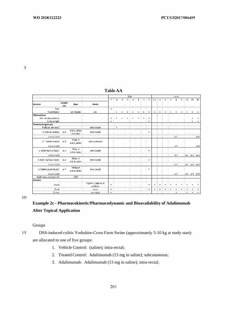
Table AA
Example 2c - Pharmacokinetic/Pharmacodynamic and Bioavailability of Adalimumab
After Topical Application
Groups
DSS-induced colitis Yorkshire-Cross Farm Swine (approximately 5-10 kg at study start)
are allocated to one of five groups:
1. Vehicle Control: (saline); intra-rectal;
2. Treated Control: Adalimumab (13 mg in saline); subcutaneous;
3. Adalimumab: Adalimumab (13 mg in saline); intra-rectal;
At t = 0, the test article is applied to a damaged mucosal surface of the bowel via
intra-rectal administration or subcutaneous injection by a veterinary surgeon at the dose
levels and volume described above.
Clinical Observations
Clinical signs (e.g., ill health, behavioral changes, etc.) are recorded on all appropriate
animals at least daily prior to the initiation of experiment and throughout the study until
termination. Additional clinical observations may be performed if deemed necessary.
Animals whose health condition warrants further evaluation are examined by a Clinical
Veterinarian.
Samples
Blood:
Blood is collected (cephalic, jugular, and/or catheter) into EDTA tubes during
acclimation on Day-7, just prior to dose on Day 0, and 12 hours post-dose. The EDTA
samples are split into two aliquots and one is centrifuged for pharmacokinetic plasma and
either analyzed immediately, or stored frozen (-80°C) for later pharmacokinetic analyses.
The remaining sample of whole blood is used for pharmacodynamic analyses.
Feces:
Feces is collected Day -7, 0 and 12 hours post-dose, and either analyzed immediately,
or flash-frozen on liquid nitrogen and stored frozen at -70°C pending later analysis of drug
levels and inflammatory cytokines.
Tissue:
Immediately after euthanasia (12 hours after dosing) in order to avoid autolytic
changes, colon tissues are collected, opened, rinsed with saline, and a detailed macroscopic
examination of the colon is performed to identify macroscopic finings related to DSS-
damage. Triplicate samples of normal and damaged tissues are either analyzed immediately,
or are flash-frozen on liquid nitrogen and stored frozen at -70°C pending later analysis of
drug concentration, inflammatory cytokines and histology.
Samples are analyzed for adalimumab levels (local mucosal tissue levels and systemic
circulation levels), and for levels of inflammatory cytokines including TNF-alpha.
Terminal Procedures
Animals are euthanized at 12 hours post-dose.
Example 3. Comparison of Systemic versus Intracecal Delivery of an Anti-IL-12 Antibody
The objective of this study was to compare the efficacy of an IL-12 inhibitor (anti-IL-
12 p40; anti-p40 mAb; BioXCell (Cat#: BE0051)), when dosed systemically versus
intracecally, to the treat dextran sulfate sodium salt (DSS)-induced colitis in male C57Bl/6
mice.
Materials and Methods
Mice
Normal male C57Bl/6 mice between the ages of 6-8 weeks old, weighing 20-24 g,
were obtained from Charles River Laboratories. The mice were randomized into thirteen
groups of twelve animals and two groups of eight animals, and housed in groups of 6-8 per
cage, and acclimatized for at least three days prior to entering the study. Animal rooms were
set to maintain a minimum of 12 to 15 air changes per hour, with an automatic timer for a
light/dark cycle of 12 hours on/off, and fed with Labdiet 5053 sterile rodent chow, with water
administered ad libitum.
Cecal Cannulation
Animals were placed under isoflurane anesthesia, with the cecum exposed via a
midline incision in the abdomen. A small point incision was made in the distal cecum where
1-2 cm of the cannula was inserted. The incision was closed with a purse string suture using
5-0 silk. An incision was then made in the left abdominal wall through which the distal end
of the cannula was inserted and pushed subcutaneously to the dorsal aspect of the back. The
site was then washed copiously with warmed saline prior to closing the abdominal wall. A
small incision was also made in the skin of the back between the shoulder blades, exposing
the tip of the cannula. The cannula was secured in place using suture, wound clips, and tissue
glue. All animals received 1 mL of warm sterile saline (subcutaneous injection) and were
monitored closely until recovery before returning to their cage. All animals received 0.6
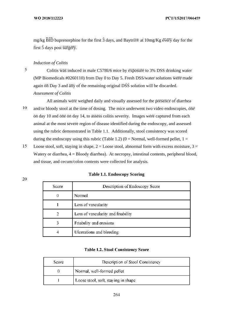
mg/kg BID buprenorphine for the first 3 days, and Baytril® at 10mg/Kg every day for the
first 5 days post surgery.
Induction of Colitis
Colitis was induced in male C57Bl/6 mice by exposure to 3% DSS drinking water
(MP Biomedicals #0260110) from Day 0 to Day 5. Fresh DSS/water solutions were made
again on Day 3 and any of the remaining original DSS solution will be discarded.
Assessment of Colitis
All animals were weighed daily and visually assessed for the presence of diarrhea
and/or bloody stool at the time of dosing. The mice underwent two video endoscopies, one
on day 10 and one on day 14, to assess colitis severity. Images were captured from each
animal at the most severe region of disease identified during the endoscopy, and assessed
using the rubric demonstrated in Table 1.1. Additionally, stool consistency was scored
during the endoscopy using this rubric (Table 1.2) (0 = Normal, well-formed pellet, 1 =
Loose stool, soft, staying in shape, 2 = Loose stool, abnormal form with excess moisture, 3 =
Watery or diarrhea, 4 = Bloody diarrhea). At necropsy, intestinal contents, peripheral blood,
and tissue, and cecum/colon contents were collected for analysis.
Table 1.1. Endoscopy Scoring
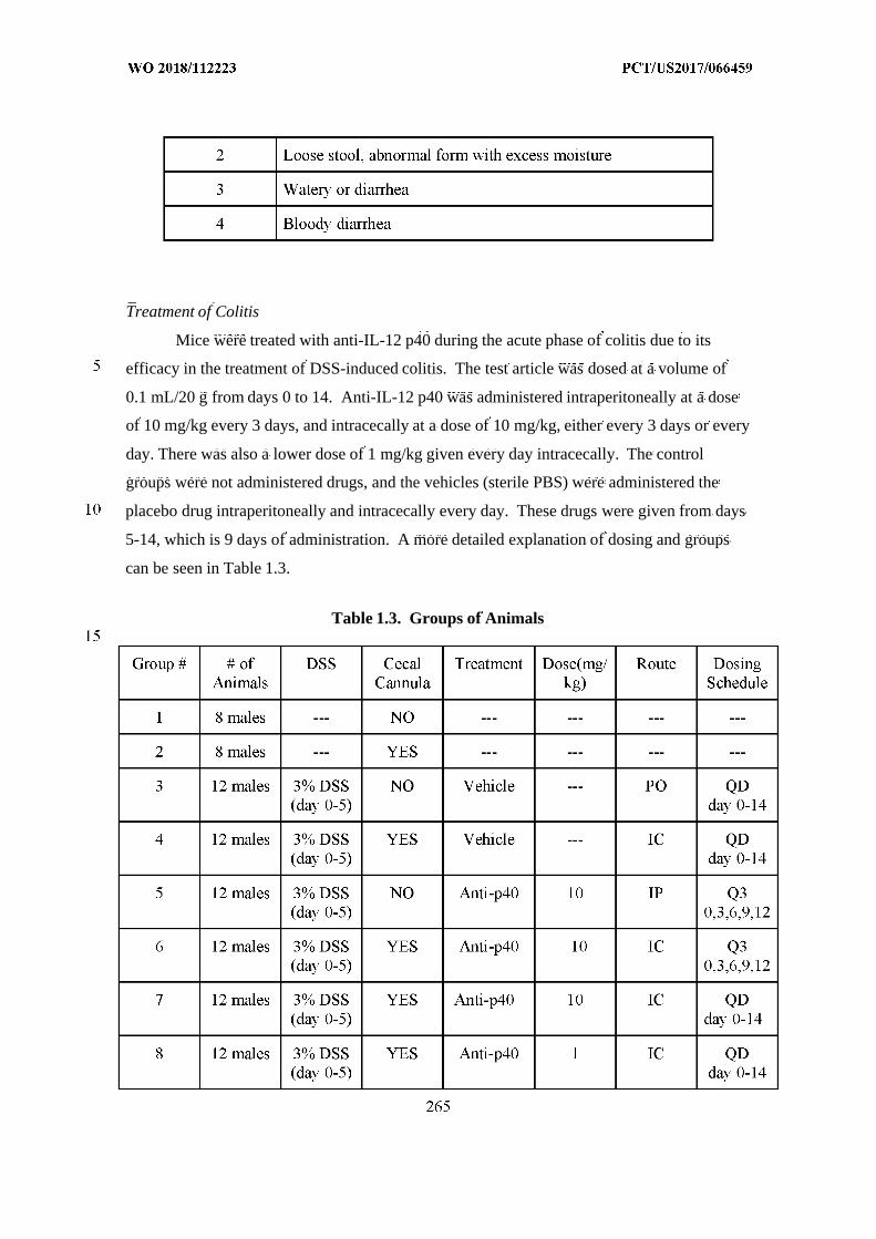
Treatment of Colitis
Mice were treated with anti-IL-12 p40 during the acute phase of colitis due to its
efficacy in the treatment of DSS-induced colitis. The test article was dosed at a volume of
0.1 mL/20 g from days 0 to 14. Anti-IL-12 p40 was administered intraperitoneally at a dose
of 10 mg/kg every 3 days, and intracecally at a dose of 10 mg/kg, either every 3 days or every
day. There was also a lower dose of 1 mg/kg given every day intracecally. The control
groups were not administered drugs, and the vehicles (sterile PBS) were administered the
placebo drug intraperitoneally and intracecally every day. These drugs were given from days
5-14, which is 9 days of administration. A more detailed explanation of dosing and groups
can be seen in Table 1.3.
Table 1.3. Groups of Animals
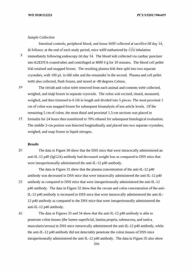
Sample Collection
Intestinal contents, peripheral blood, and tissue were collected at sacrifice on day 14,
as follows: at the end of each study period, mice were euthanized by CO2 inhalation
immediately following endoscopy on day 14. The blood was collected via cardiac puncture
into K2EDTA-coated tubes and centrifuged at 4000 x g for 10 minutes. The blood cell pellet
was retained and snapped frozen. The resulting plasma was then split into two separate
cryotubes, with 100 µL in one tube and the remainder in the second. Plasma and cell pellet
were also collected, flash frozen, and stored at -80 degrees Celsius.
The cecum and colon were removed from each animal and contents were collected,
weighed, and snap frozen in separate cryovials. The colon was excised, rinsed, measured,
weighed, and then trimmed to 6 cm in length and divided into 5 pieces. The most proximal 1
cm of colon was snapped frozen for subsequent bioanalysis of test article levels. Of the
remaining 5 cm of colon, the most distal and proximal 1.5-cm sections was placed in
formalin for 24 hours then transferred to 70% ethanol for subsequent histological evaluation.
The middle 2-cm portion was bisected longitudinally and placed into two separate cryotubes,
weighed, and snap frozen in liquid nitrogen.
Results
The data in Figure 30 show that the DSS mice that were intracecally administered an
anti-IL-12 p40 (IgG2A) antibody had decreased weight loss as compared to DSS mice that
were intraperitoneally administered the anti-IL-12 p40 antibody.
The data in Figure 31 show that the plasma concentration of the anti-IL-12 p40
antibody was decreased in DSS mice that were intracecally administered the anti-IL-12 p40
antibody as compared to DSS mice that were intraperitoneally administered the anti-IL-12
p40 antibody. The data in Figure 32 show that the cecum and colon concentration of the anti-
IL-12 p40 antibody is increased in DSS mice that were intracecally administered the anti-IL-
12 p40 antibody as compared to the DSS mice that were intraperitoneally administered the
anti-IL-12 p40 antibody.
The data in Figures 33 and 34 show that the anti-IL-12 p40 antibody is able to
penetrate colon tissues (the lumen superficial, lamina propria, submucosa, and tunica
muscularis/serosa) in DSS mice intracecally administered the anti-IL-12 p40 antibody, while
the anti-IL-12 p40 antibody did not detectably penetrate the colon tissues of DSS mice
intraperitoneally administered the anti-IL-12 p40 antibody. The data in Figure 35 also show

that the ratio of the concentration of anti-IL-12 p40 antibody in colon tissue to the
concentration of the anti-IL-12 p40 antibody in plasma is increased in DSS mice intracecally
administered the anti-IL-12 p40 antibody as compared to the ratio in DSS mice
intraperitoneally administered the anti-IL-12 p40 antibody.
The data in Figure 36 show that the concentration of IL-1^ in colon tissue is
decreased in DSS mice intracecally administered the anti-IL-12 p40 antibody as compared to
the concentration of IL-1^ in colon tissue in DSS mice intraperitoneally administered the
anti-IL-12 p40 antibody. The data in Figure 37 show that the concentration of IL-6 in colon
tissue is decreased in DSS mice intracecally administered the anti-IL-12 p40 antibody as
compared to the concentration of IL-6 in colon tissue in DSS mice intraperitoneally
administered the anti-IL-12 p40 antibody. The data in Figure 38 show that the concentration
of IL-17A in colon tissue is decreased in DSS mice intracecally administered the anti-IL-12
p40 antibody as compared to the concentration of IL-17A in colon tissue in DSS mice
intraperitoneally administered the anti-IL-12 p40 antibody.
No significant differences in clinical observations or gastrointestinal-specific adverse
effects, including stool consistency and/or bloody stool, were observed due to cannulation or
intra-cecal treatments when compared with vehicle. No toxicity resulting from the treatments
was reported. A significant reduction in body weight-loss (AUC) was found in groups
treated with anti-IL-12 p40 antibody (10 mg/kg and 1mg/kg, QD) via intra-cecal delivery
when compared with vehicle control and intraperitoneal delivery (10 mg/kg, Q3D). The
immunohistochemistry staining in anti-IL-12 p40 antibody (10 mg/kg, QD) treatment groups
showed penetration of the antibody in all layers of colon tissue, including lumen mucosa,
lamina propria, submucosa, tunica muscularis, via intra-cecal delivery. The distribution of
anti-IL-12 p40 antibody was found in all segments of the colon, however, higher levels were
detected in the proximal region. A significantly higher mean concentration of anti-IL-12 p40
antibody was found in the gastrointestinal contents and colon tissues when delivered via
intra-cecal administration (Anti-p40: 10 mg/kg and 1mg/kg, QD) compared with
intraperitoneal administration (anti-p40: 10 mg/kg, Q3D). The blood level of anti-IL-12 p40
antibody was significantly higher when delivered via intraperitoneal administration (Q3D) as
compared to intra-cecal administration (Q3D & QD). The concentrations of inflammatory
cytokines, including IL-1β, IL-6, and IL-17, were significantly reduced by anti-IL-12 p40
antibody (10 mg/kg, QD) treatment when delivered via intra-cecal administration as
compared to vehicle controls.
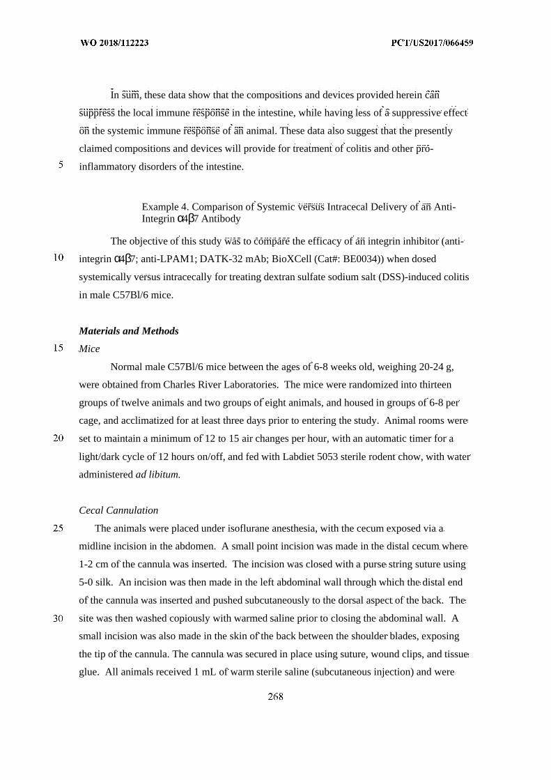
In sum, these data show that the compositions and devices provided herein can
suppress the local immune response in the intestine, while having less of a suppressive effect
on the systemic immune response of an animal. These data also suggest that the presently
claimed compositions and devices will provide for treatment of colitis and other pro-
inflammatory disorders of the intestine.
Example 4. Comparison of Systemic versus Intracecal Delivery of an Anti-Integrin α4β7 Antibody
The objective of this study was to compare the efficacy of an integrin inhibitor (anti-
integrin α4β7; anti-LPAM1; DATK-32 mAb; BioXCell (Cat#: BE0034)) when dosed
systemically versus intracecally for treating dextran sulfate sodium salt (DSS)-induced colitis
in male C57Bl/6 mice.
Materials and Methods
Mice
Normal male C57Bl/6 mice between the ages of 6-8 weeks old, weighing 20-24 g,
were obtained from Charles River Laboratories. The mice were randomized into thirteen
groups of twelve animals and two groups of eight animals, and housed in groups of 6-8 per
cage, and acclimatized for at least three days prior to entering the study. Animal rooms were
set to maintain a minimum of 12 to 15 air changes per hour, with an automatic timer for a
light/dark cycle of 12 hours on/off, and fed with Labdiet 5053 sterile rodent chow, with water
administered ad libitum.
Cecal Cannulation
The animals were placed under isoflurane anesthesia, with the cecum exposed via a
midline incision in the abdomen. A small point incision was made in the distal cecum where
1-2 cm of the cannula was inserted. The incision was closed with a purse string suture using
5-0 silk. An incision was then made in the left abdominal wall through which the distal end
of the cannula was inserted and pushed subcutaneously to the dorsal aspect of the back. The
site was then washed copiously with warmed saline prior to closing the abdominal wall. A
small incision was also made in the skin of the back between the shoulder blades, exposing
the tip of the cannula. The cannula was secured in place using suture, wound clips, and tissue
glue. All animals received 1 mL of warm sterile saline (subcutaneous injection) and were
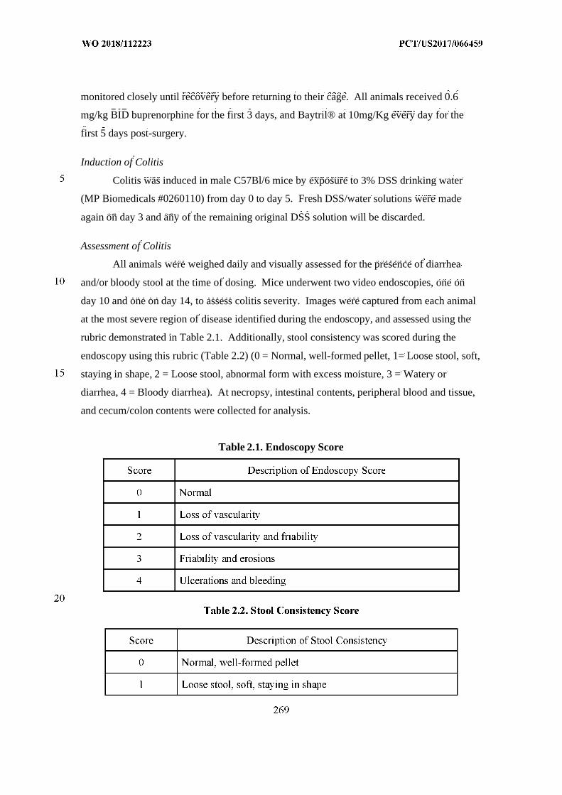
monitored closely until recovery before returning to their cage. All animals received 0.6
mg/kg BID buprenorphine for the first 3 days, and Baytril® at 10mg/Kg every day for the
first 5 days post-surgery.
Induction of Colitis
Colitis was induced in male C57Bl/6 mice by exposure to 3% DSS drinking water
(MP Biomedicals #0260110) from day 0 to day 5. Fresh DSS/water solutions were made
again on day 3 and any of the remaining original DSS solution will be discarded.
Assessment of Colitis
All animals were weighed daily and visually assessed for the presence of diarrhea
and/or bloody stool at the time of dosing. Mice underwent two video endoscopies, one on
day 10 and one on day 14, to assess colitis severity. Images were captured from each animal
at the most severe region of disease identified during the endoscopy, and assessed using the
rubric demonstrated in Table 2.1. Additionally, stool consistency was scored during the
endoscopy using this rubric (Table 2.2) (0 = Normal, well-formed pellet, 1= Loose stool, soft,
staying in shape, 2 = Loose stool, abnormal form with excess moisture, 3 = Watery or
diarrhea, 4 = Bloody diarrhea). At necropsy, intestinal contents, peripheral blood and tissue,
and cecum/colon contents were collected for analysis.
Table 2.1. Endoscopy Score
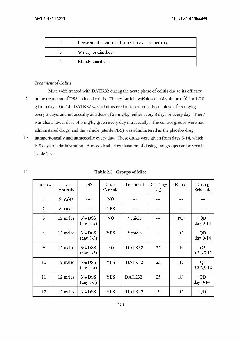
Treatment of Colitis
Mice were treated with DATK32 during the acute phase of colitis due to its efficacy
in the treatment of DSS-induced colitis. The test article was dosed at a volume of 0.1 mL/20
g from days 0 to 14. DATK32 was administered intraperitoneally at a dose of 25 mg/kg
every 3 days, and intracecally at a dose of 25 mg/kg, either every 3 days or every day. There
was also a lower dose of 5 mg/kg given every day intracecally. The control groups were not
administered drugs, and the vehicle (sterile PBS) was administered as the placebo drug
intraperitoneally and intracecally every day. These drugs were given from days 5-14, which
is 9 days of administration. A more detailed explanation of dosing and groups can be seen in
Table 2.3.
Table 2.3. Groups of Mice
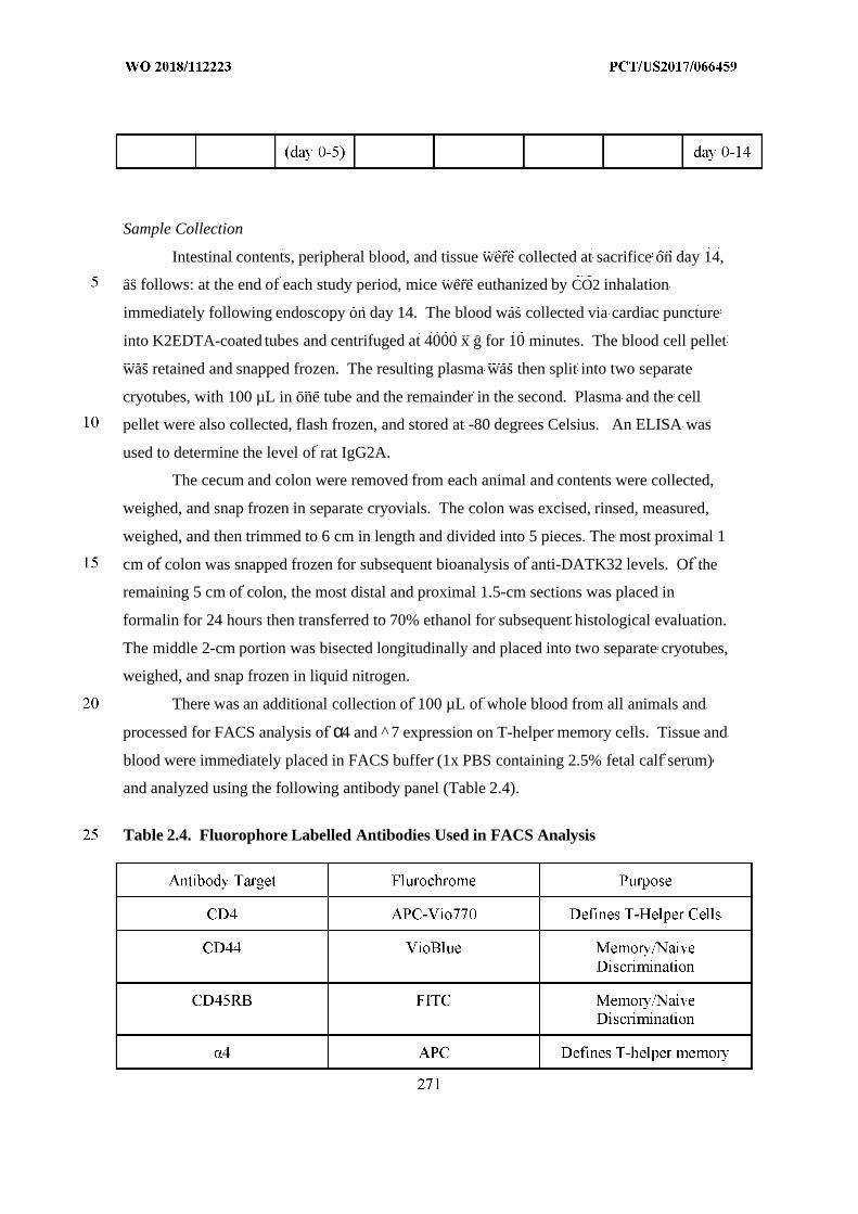
Sample Collection
Intestinal contents, peripheral blood, and tissue were collected at sacrifice on day 14,
as follows: at the end of each study period, mice were euthanized by CO2 inhalation
immediately following endoscopy on day 14. The blood was collected via cardiac puncture
into K2EDTA-coated tubes and centrifuged at 4000 x g for 10 minutes. The blood cell pellet
was retained and snapped frozen. The resulting plasma was then split into two separate
cryotubes, with 100 µL in one tube and the remainder in the second. Plasma and the cell
pellet were also collected, flash frozen, and stored at -80 degrees Celsius. An ELISA was
used to determine the level of rat IgG2A.
The cecum and colon were removed from each animal and contents were collected,
weighed, and snap frozen in separate cryovials. The colon was excised, rinsed, measured,
weighed, and then trimmed to 6 cm in length and divided into 5 pieces. The most proximal 1
cm of colon was snapped frozen for subsequent bioanalysis of anti-DATK32 levels. Of the
remaining 5 cm of colon, the most distal and proximal 1.5-cm sections was placed in
formalin for 24 hours then transferred to 70% ethanol for subsequent histological evaluation.
The middle 2-cm portion was bisected longitudinally and placed into two separate cryotubes,
weighed, and snap frozen in liquid nitrogen.
There was an additional collection of 100 µL of whole blood from all animals and
processed for FACS analysis of α4 and ^ 7 expression on T-helper memory cells. Tissue and
blood were immediately placed in FACS buffer (1x PBS containing 2.5% fetal calf serum)
and analyzed using the following antibody panel (Table 2.4).
Table 2.4. Fluorophore Labelled Antibodies Used in FACS Analysis
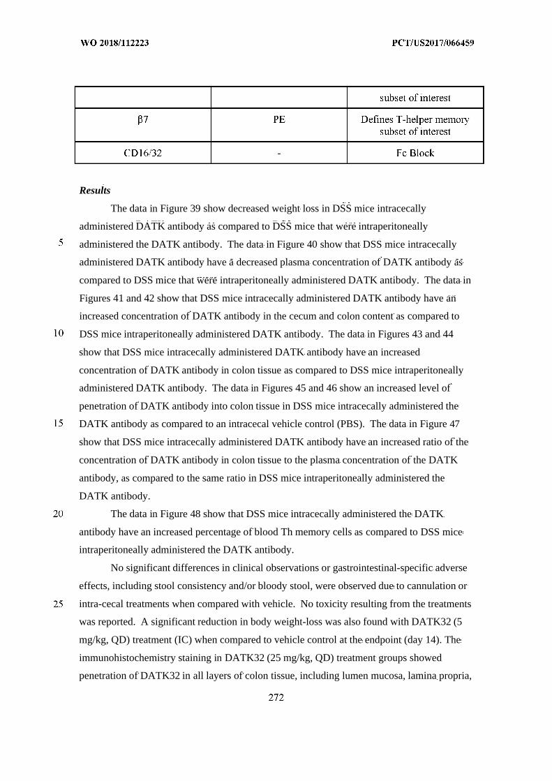
Results
The data in Figure 39 show decreased weight loss in DSS mice intracecally
administered DATK antibody as compared to DSS mice that were intraperitoneally
administered the DATK antibody. The data in Figure 40 show that DSS mice intracecally
administered DATK antibody have a decreased plasma concentration of DATK antibody as
compared to DSS mice that were intraperitoneally administered DATK antibody. The data in
Figures 41 and 42 show that DSS mice intracecally administered DATK antibody have an
increased concentration of DATK antibody in the cecum and colon content as compared to
DSS mice intraperitoneally administered DATK antibody. The data in Figures 43 and 44
show that DSS mice intracecally administered DATK antibody have an increased
concentration of DATK antibody in colon tissue as compared to DSS mice intraperitoneally
administered DATK antibody. The data in Figures 45 and 46 show an increased level of
penetration of DATK antibody into colon tissue in DSS mice intracecally administered the
DATK antibody as compared to an intracecal vehicle control (PBS). The data in Figure 47
show that DSS mice intracecally administered DATK antibody have an increased ratio of the
concentration of DATK antibody in colon tissue to the plasma concentration of the DATK
antibody, as compared to the same ratio in DSS mice intraperitoneally administered the
DATK antibody.
The data in Figure 48 show that DSS mice intracecally administered the DATK
antibody have an increased percentage of blood Th memory cells as compared to DSS mice
intraperitoneally administered the DATK antibody.
No significant differences in clinical observations or gastrointestinal-specific adverse
effects, including stool consistency and/or bloody stool, were observed due to cannulation or
intra-cecal treatments when compared with vehicle. No toxicity resulting from the treatments
was reported. A significant reduction in body weight-loss was also found with DATK32 (5
mg/kg, QD) treatment (IC) when compared to vehicle control at the endpoint (day 14). The
immunohistochemistry staining in DATK32 (25 mg/kg, QD) treatment groups showed
penetration of DATK32 in all layers of colon tissue, including lumen mucosa, lamina propria,
submucosa, tunica muscularis, via intra-cecal delivery. The distribution of DATK32 was
found in all segments of the colon, however, higher levels were detected in the proximal
region. A significantly higher mean concentration of DATK32 was found in gastrointestinal
contents and colon tissues when delivered via intra-cecal administration (DATK32: 25 mg/kg
and 5 mg/kg, QD) as compared to intraperitoneal administration (DATK32: 25 mg/kg, Q3D).
The blood level of DATK32 was significantly higher when delivered via intraperitoneal
administration (Q3D) as compared to intra-cecal administration (Q3D & QD). The
pharmacokinetics of DATK32 (25 mg/kg, QD) showed significantly higher mean
concentrations of DATK32 when delivered via intra-cecal administration at 1, 2, and 4 h
post-dose in the gastrointestinal contents, and 1, 2, 4 and 24 h in colon tissue as compared
with the mean concentrations of DATK32 following intraperitoneal administration. The
mean number of gut-homing T cells (Th memory cells) was significantly higher in the blood
of groups treated with DATK32 via intra-cecal administration (QD 25 mg/kg and QD 5
mg/kg) as compared to the groups treated with DATK32 via intraperitoneal administration
(Q3D 25 mg/kg). The mean number of Th memory cells was significantly lower in the
Peyer's Patches of groups treated with DATK32 via intra-cecal administration (QD 25 mg/kg
and 5 mg/kg) as compared to the groups treated with DATK32 via intraperitoneal
administration (Q3D 25 mg/kg). The mean number of Th memory cells in mesenteric lymph
nodes (MLN) was significantly lower in groups treated with DATK32 via intra-cecal
administratoin (QD and Q3D 25 mg/kg and QD 5 mg/kg) as compared to the groups treated
with DATK32 via intraperitoneal administration (Q3D 25 mg/kg).
In sum, these data show that the compositions and devices provided herein can
suppress the local immune response in the intestine, while having less of a suppressive effect
on the systemic immune response of an animal. These data also show that the release of
DATK-32 antibody in the colon can result in a suppression of leukocyte recruitment and may
provide for the treatment of colitis and other pro-inflammatory diseases of the intestine.
Example 5. An Assessment of DATK32 Bio-Distribution Following IntracecalAdministration in Male C57B1/6 Mice
The objective of this study is to assess DATK32 bio-distribution when dosed
intracecally in male C57Bl/6 mice. A minimum of 10 days prior to the start of the
experiment a cohort of animals will undergo surgical implantation of a cecal cannula. A
sufficient number of animals will undergo implantation to allow for 24 cannulated animals to
be enrolled in the main study (e.g., 31 animals). Animals were dosed with vehicle or test
article via intracecal injection (IC) on Day 0 as indicated in Table 3. Animals from all groups
were sacrificed for terminal sample collection three hours following test article
administration.
Materials and Methods
Mice
Normal male C57Bl/6 mice between the ages of 6-8 weeks old, weighing 20-24 g,
were obtained from Charles River Laboratories. The mice were randomized into two groups
of twelve animals, and housed in groups of 12 per cage, and acclimatized for at least three
days prior to entering the study. Animal rooms were set to maintain a minimum of 12 to 15
air changes per hour, with an automatic timer for a light/dark cycle of 12 hours on/off, and
fed with Labdiet 5053 sterile rodent chow, with water administered ad libitum.
Cecal Cannulation
The animals were placed under isoflurane anesthesia, with the cecum exposed via a
midline incision in the abdomen. A small point incision was made in the distal cecum where
1-2 cm of the cannula was inserted. The incision was closed with a purse string suture using
5-0 silk. An incision was then made in the left abdominal wall through which the distal end
of the cannula was inserted and pushed subcutaneously to the dorsal aspect of the back. The
site was then washed copiously with warmed saline prior to closing the abdominal wall. A
small incision was also made in the skin of the back between the shoulder blades, exposing
the tip of the cannula. The cannula was secured in place using suture, wound clips, and tissue
glue. All animals received 1 mL of warm sterile saline (subcutaneous injection) and were
monitored closely until recovery before returning to their cage. All animals received 0.6
mg/kg BID buprenorphine for the first 3 days, and Baytril® at 10mg/Kg every day for the
first 5 days post-surgery.
Dosing
Animals were dosed IC at a volume of 0.075 mL/animal on Days 0 as indicated in Table
3.
Sacrifice
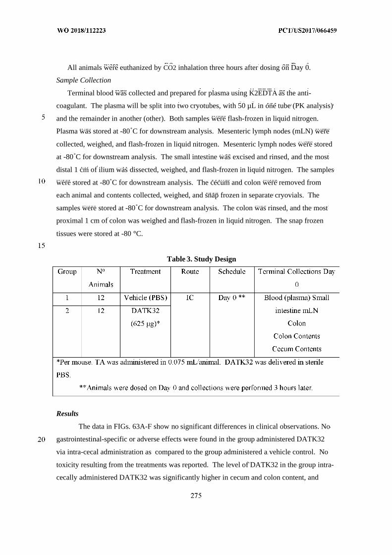
All animals were euthanized by CO2 inhalation three hours after dosing on Day 0.
Sample Collection
Terminal blood was collected and prepared for plasma using K2EDTA as the anti-
coagulant. The plasma will be split into two cryotubes, with 50 µL in one tube (PK analysis)
and the remainder in another (other). Both samples were flash-frozen in liquid nitrogen.
Plasma was stored at -80˚C for downstream analysis. Mesenteric lymph nodes (mLN) were
collected, weighed, and flash-frozen in liquid nitrogen. Mesenteric lymph nodes were stored
at -80˚C for downstream analysis. The small intestine was excised and rinsed, and the most
distal 1 cm of ilium was dissected, weighed, and flash-frozen in liquid nitrogen. The samples
were stored at -80˚C for downstream analysis. The cecum and colon were removed from
each animal and contents collected, weighed, and snap frozen in separate cryovials. The
samples were stored at -80˚C for downstream analysis. The colon was rinsed, and the most
proximal 1 cm of colon was weighed and flash-frozen in liquid nitrogen. The snap frozen
tissues were stored at -80 °C.
Table 3. Study Design
Results
The data in FIGs. 63A-F show no significant differences in clinical observations. No
gastrointestinal-specific or adverse effects were found in the group administered DATK32
via intra-cecal administration as compared to the group administered a vehicle control. No
toxicity resulting from the treatments was reported. The level of DATK32 in the group intra-
cecally administered DATK32 was significantly higher in cecum and colon content, and
colon tissue compared to the group administered a vehicle control at 3h post-dose. A small
amount of DATK32 was also detected in plasma, small intestine, and mesenteric lymph node
in the group intra-cecally administered DATK32.
Example 6. Pharmacokinectics/Pharmacodynamics and Bioavailability ofAdalimumab When Applied to a TNBS-damaged Mucosal Surface (InducedColitis) in Swine
The purpose of this non-Good Laboratory Practice (GLP) study was to explore the
PK/PD, and bioavailability of adalimumab when applied to a TNBS-damaged mucosal
surface (induced colitis) in Yorkshire-Cross farm swine, and to determine an appropriate dose
and frequency for studies where a drug will be delivered by the ingestible device system.
The ingestible device system will be capable of delivering a TNF inhibitor (adalimumab)
topically and locally to damaged mucosa in human patients with inflammatory bowel disease
(IBD). The TNBS-induced colitis model was validated when a single administration on Day
1 of 40 mL of 100% ethanol (EtOH) mixed with 5 grams of TNBS diluted in 10 mL of water
via an enema using a rubber catheter resulted in the intended reproducible induction of
damaged mucosal surface (induced colitis) in Yorkshire-Cross farm swine.
This study investigated whether topical delivery of adalimumab would result in
increased local mucosal tissue levels with limited drug reaching systemic circulation, as
compared to subcutaneous administration; whether local mucosal tissue levels of drug would
be greater in damaged tissues when compared to normal tissues; whether increasing the dose
of drug would result in increased mucosal tissue levels in local and distal TNBS-damaged
tissues; and whether topical delivery of adalimumab would result in reductions in
inflammatory cytokines such as TNF-αin damaged tissues, feces, and possibly blood.
All animals were subjected to intra-rectal administration of trinitrobenzene sulfonic
acid (TNBS) to induce chronic colitis on day -2. All animals were fasted prior to colitis
induction. Bedding was removed and replaced with rubber mats on day -3 to prevent
ingestion of straw bedding material. The dose was 40 mL of 100% EtOH mixed with 5
grams of TNBS diluted in 10 mL of water, then instilled into the colon intra-rectally using a
flexible gavage tube by a veterinary surgeon (deposited in a 10-cm portion of the distal colon
and proximal rectum, and retained for 12 minutes by use of two Foley catheters with 60-mL
balloons). Approximately 3 days after induction, macroscopic and microscopic alterations of
colonic architecture were apparent: some necrosis, thickening of the colon, and substantial
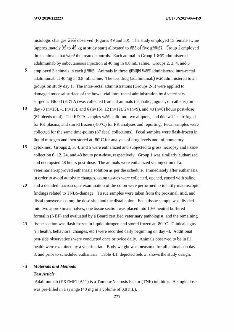
histologic changes were observed (Figures 49 and 50). The study employed 15 female swine
(approximately 35 to 45 kg at study start) allocated to one of five groups. Group 1 employed
three animals that were the treated controls. Each animal in Group 1 was administered
adalimumab by subcutaneous injection at 40 mg in 0.8 mL saline. Groups 2, 3, 4, and 5
employed 3 animals in each group. Animals in these groups were administered intra-rectal
adalimumab at 40 mg in 0.8 mL saline. The test drug (adalimumab) was administered to all
groups on study day 1. The intra-rectal administrations (Groups 2-5) were applied to
damaged mucosal surface of the bowel vial intra-rectal administration by a veterinary
surgeon. Blood (EDTA) was collected from all animals (cephalic, jugular, or catheter) on
day -3 (n=15), -1 (n=15), and 6 (n=15), 12 (n=12), 24 (n=9), and 48 (n=6) hours post-dose
(87 bleeds total). The EDTA samples were split into two aliquots, and one was centrifuged
for PK plasma, and stored frozen (-80oC) for PK analyses and reporting. Fecal samples were
collected for the same time-points (87 fecal collections). Fecal samples were flash-frozen in
liquid nitrogen and then stored at -80o C for analysis of drug levels and inflammatory
cytokines. Groups 2, 3, 4, and 5 were euthanized and subjected to gross necropsy and tissue
collection 6, 12, 24, and 48 hours post-dose, respectively. Group 1 was similarly euthanized
and necropsied 48 hours post-dose. The animals were euthanized via injection of a
veterinarian-approved euthanasia solution as per the schedule. Immediately after euthanasia
in order to avoid autolytic changes, colon tissues were collected, opened, rinsed with saline,
and a detailed macroscopic examination of the colon were performed to identify macroscopic
findings related to TNBS-damage. Tissue samples were taken from the proximal, mid, and
distal transverse colon; the dose site; and the distal colon. Each tissue sample was divided
into two approximate halves; one tissue section was placed into 10% neutral buffered
formalin (NBF) and evaluated by a Board certified veterinary pathologist, and the remaining
tissue section was flash frozen in liquid nitrogen and stored frozen at -80 o C. Clinical signs
(ill health, behavioral changes, etc.) were recorded daily beginning on day -3. Additional
pen-side observations were conducted once or twice daily. Animals observed to be in ill
health were examined by a veterinarian. Body weight was measured for all animals on day -
3, and prior to scheduled euthanasia. Table 4.1, depicted below, shows the study design.
Materials and Methods
Test Article
Adalimumab (EXEMPTIAT M ) is a Tumour Necrosis Factor (TNF) inhibitor. A single dose
was pre-filled in a syringe (40 mg in a volume of 0.8 mL).
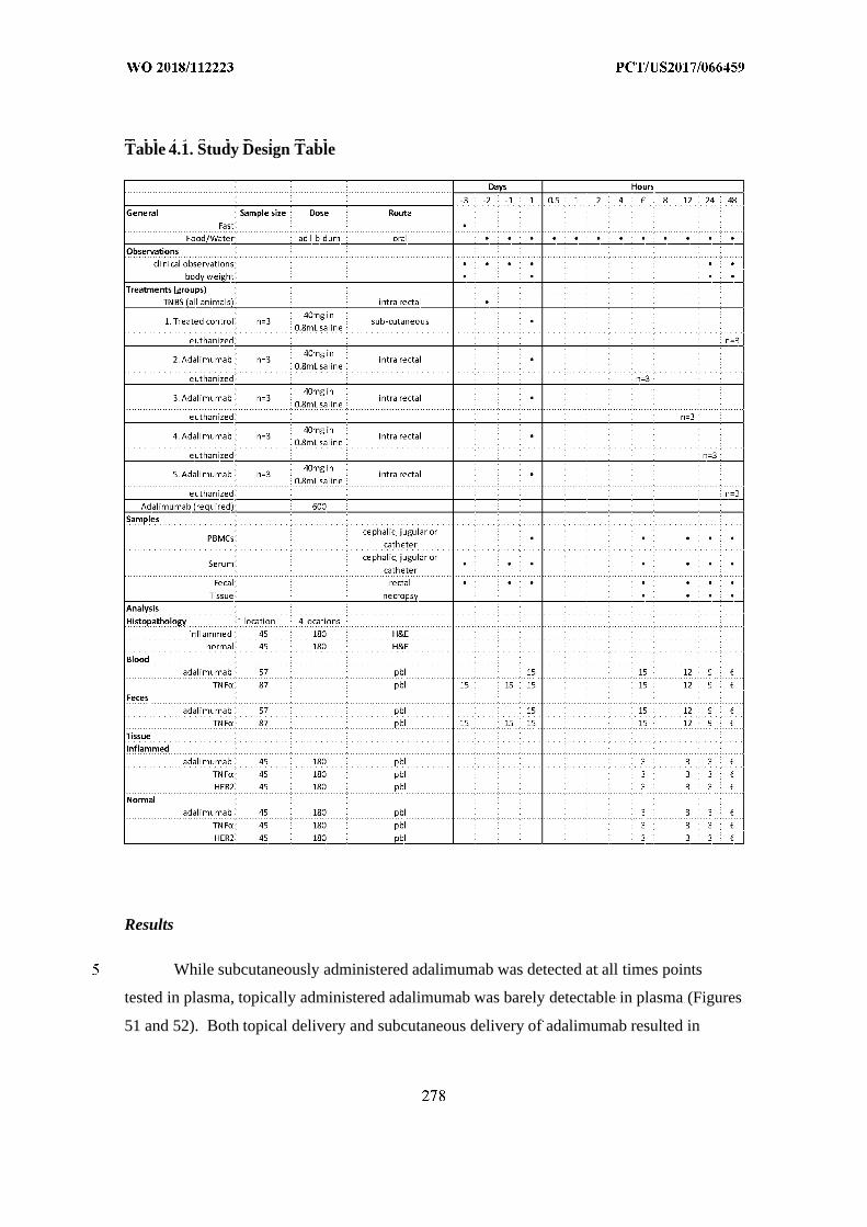
Table 4.1. Study Design Table
Results
While subcutaneously administered adalimumab was detected at all times points
tested in plasma, topically administered adalimumab was barely detectable in plasma (Figures
51 and 52). Both topical delivery and subcutaneous delivery of adalimumab resulted in
reduced levels of TNF-αin colon tissue of TNBS-induced colitis animals, yet topical delivery
of adalimumab was able to achieve a greater reduction in TNF-αlevels (Figures 53 and 54).
Either subcutaneous or intra-rectal administration of adalimumab was well tolerated
and did not result in death, morbidity, adverse clinical observations, or body weight changes.
A decreased level of total TNBS-related inflammatory response was observed by
adalimumab treatment via intra-rectal administration when applied to the damaged mucosal
surface of the bowel when compared to subcutaneous delivery. A significantly higher
concentration of adalimumab was measured in blood following subcutaneous delivery as
compared to the blood concentration following intra-rectal administration. Intra-rectal
administration of adalimumab decreased the total and normalized TNFαconcentration over
time (6~48h) and was more effective at reducing TNFαat the endpoint (48h) as compared to
groups administered adalimumab subcutaneously.
In sum, these data show that the compositions and devices provided herein can suppress the
local immune response in the intestine, while having less of a suppressive effect on the
systemic immune response of an animal. For example, these data show that intracecal
administration of adalimumab using a device as described herein can provide for local
delivery of adalimumab to the site of disease, without suppressing the systemic immune
response. These data also show that local administration of adalimumab using a device as
described herein can result in a significant reduction of the levels of TNFαin diseases
animals.
Example 7. Comparison of Systemic Versus Intracecal Delivery ofCyclosporine A
The objective of this study was to compare the efficacy of an immunosuppressant
agent (cyclosporine A; CsA) when dosed systemically versus intracecally to treat dextran
sulfate sodium salt (DSS)-induced colitis in male C57Bl/6 mice.
Experimental Design
A minimum of 10 days prior to the start of the experiment a cohort of animals
underwent surgical implantation of a cecal cannula. A sufficient number of animals
underwent implantation to allow for 44 cannulated animals to be enrolled in the main study
(e.g., 76 animals). Colitis was induced in 60 male C5Bl/6 mice by exposure to 3% DSS-
treated drinking water from day 0 to day 5. Two groups of eight additional animals
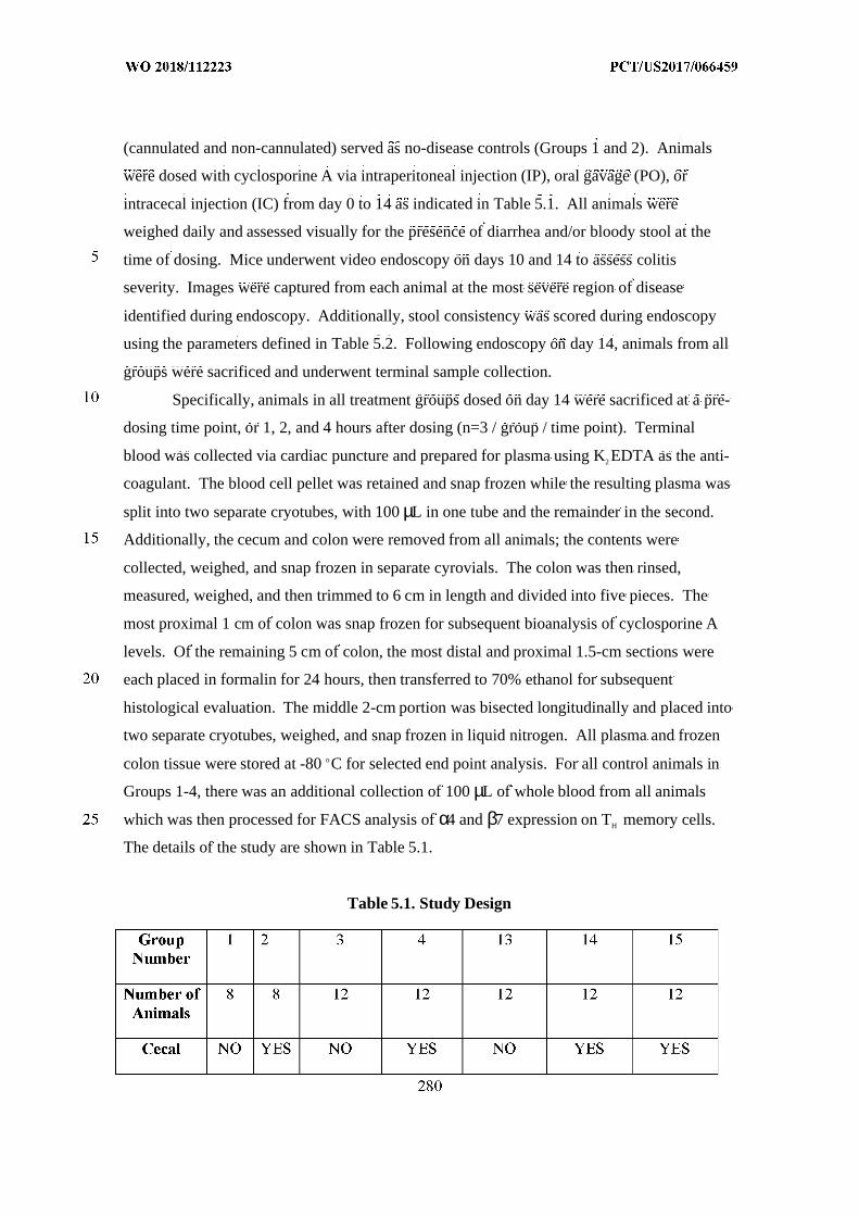
(cannulated and non-cannulated) served as no-disease controls (Groups 1 and 2). Animals
were dosed with cyclosporine A via intraperitoneal injection (IP), oral gavage (PO), or
intracecal injection (IC) from day 0 to 14 as indicated in Table 5.1. All animals were
weighed daily and assessed visually for the presence of diarrhea and/or bloody stool at the
time of dosing. Mice underwent video endoscopy on days 10 and 14 to assess colitis
severity. Images were captured from each animal at the most severe region of disease
identified during endoscopy. Additionally, stool consistency was scored during endoscopy
using the parameters defined in Table 5.2. Following endoscopy on day 14, animals from all
groups were sacrificed and underwent terminal sample collection.
Specifically, animals in all treatment groups dosed on day 14 were sacrificed at a pre-
dosing time point, or 1, 2, and 4 hours after dosing (n=3 / group / time point). Terminal
blood was collected via cardiac puncture and prepared for plasma using K2 EDTA as the anti-
coagulant. The blood cell pellet was retained and snap frozen while the resulting plasma was
split into two separate cryotubes, with 100 µL in one tube and the remainder in the second.
Additionally, the cecum and colon were removed from all animals; the contents were
collected, weighed, and snap frozen in separate cyrovials. The colon was then rinsed,
measured, weighed, and then trimmed to 6 cm in length and divided into five pieces. The
most proximal 1 cm of colon was snap frozen for subsequent bioanalysis of cyclosporine A
levels. Of the remaining 5 cm of colon, the most distal and proximal 1.5-cm sections were
each placed in formalin for 24 hours, then transferred to 70% ethanol for subsequent
histological evaluation. The middle 2-cm portion was bisected longitudinally and placed into
two separate cryotubes, weighed, and snap frozen in liquid nitrogen. All plasma and frozen
colon tissue were stored at -80 oC for selected end point analysis. For all control animals in
Groups 1-4, there was an additional collection of 100 µL of whole blood from all animals
which was then processed for FACS analysis of α4 and β7 expression on TH memory cells.
The details of the study are shown in Table 5.1.
Table 5.1. Study Design
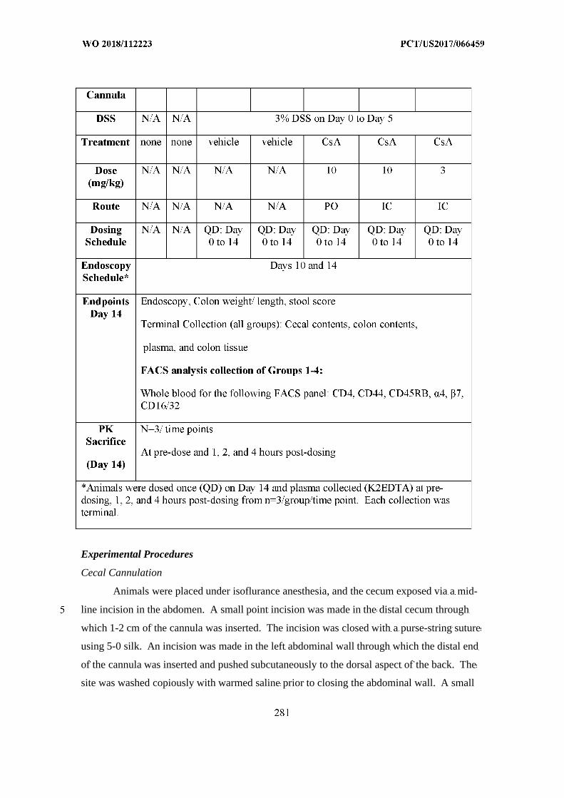
Experimental Procedures
Cecal Cannulation
Animals were placed under isoflurance anesthesia, and the cecum exposed via a mid-
line incision in the abdomen. A small point incision was made in the distal cecum through
which 1-2 cm of the cannula was inserted. The incision was closed with a purse-string suture
using 5-0 silk. An incision was made in the left abdominal wall through which the distal end
of the cannula was inserted and pushed subcutaneously to the dorsal aspect of the back. The
site was washed copiously with warmed saline prior to closing the abdominal wall. A small
incision was made in the skin of the back between the shoulder blades, exposing the tip of the
cannula. The cannula was secured in place using suture, wound clips, and tissue glue. All
animals received 1 mL of warm sterile saline (subcutaneous injection) and were monitored
closely until fully recovered before returning to the cage. All animals received
buprenorphine at 0.6 mg/kg BID for the first 3 days, and Baytril® at 10 mg/kg QD for the
first 5 days following surgery.
Disease Induction
Colitis was induced on day 0 via addition of 3% DSS (MP Biomedicals, Cat
#0260110) to the drinking water. Fresh DSS/water solutions were made on day 3 and any of
the remaining original DSS solution was discarded.
Dosing
Animals were dosed by oral gavage (PO), intraperitoneal injection (IP), or intracecal
injection (IC) at a volume of 0.1 mL/20 g on days 0 to 14 as indicated in Table 5.1.
Body Weight and Survival
Animals were observed daily (weight, morbidity, survival, presence of diarrhea,
and/or bloody stool) in order to assess possible differences among treatment groups and/or
possible toxicity resulting from the treatments.
Animals Found Dead or Moribund
Animals were monitored on a daily basis and those exhibiting weight loss greater than
30% were euthanized, and samples were not collected from these animals.
Endoscopy
Each mouse underwent video endoscopy on days 10 and 14 using a small animal
endoscope (Karl Storz Endoskope, Germany) under isoflurane anesthesia. During each
endoscopic procedure still images as well as video were recorded to evaluate the extent of
colitis and the response to treatment. Additionally, we attempted to capture an image from
each animal at the most severe region of disease identified during endoscopy. Colitis severity
was scored using a 0-4 scale (0 = normal; 1 = loss of vascularity; 2 =loss of vascularity and
friability; 3 = friability and erosions; 4 = ulcerations and bleeding). Additionally, stool
consistency was scored during endoscopy using the parameters defined in Table 5.2.
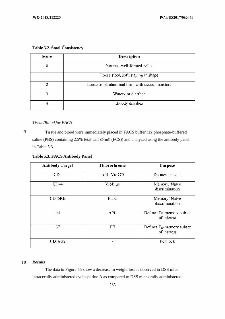
Table 5.2. Stool Consistency
Tissue/Blood for FACS
Tissue and blood were immediately placed in FACS buffer (1x phosphate-buffered
saline (PBS) containing 2.5% fetal calf serum (FCS)) and analyzed using the antibody panel
in Table 5.3.
Table 5.3. FACS Antibody Panel
Results
The data in Figure 55 show a decrease in weight loss is observed in DSS mice
intracecally administered cyclosporine A as compared to DSS mice orally administered
cyclosporine A. The data in Figure 56 show a decrease in plasma concentration of
cyclosporine A in DSS mice intracecally administered cyclosporine A as compared to DSS
mice orally administered cyclosporine A. The data in Figures 57-59 show an increased
concentration of cyclosporine A in the colon tissue of DSS mice intracecally administered
cyclosporine A as compared to the concentration of cyclosporine A in the colon tissue of
DSS mice orally administered cyclosporine A.
The data in Figure 60 show that DSS mice intracecally administered cyclosporine A
have an increased concentration of IL-2 in colon tissue as compared to DSS mice orally
administered cyclosporine A. The data in Figure 61 show that DSS mice intracecally
administered cyclosporine A have a decreased concentration of IL-6 in colon tissue as
compared to DSS mice orally administered cyclosporine A.
In sum, these data show that the compositions and devices provided herein can
suppress the local immune response in the intestine, while having less of a suppressive effect
on the systemic immune response of an animal. For example, these data demonstrate that the
present compositions and devices can be used to release cyclosporine A to the intestine and
that this results in a selective immune suppression in the colon, while having less of an effect
on the immune system outside of the intesting. These data also suggest that the present
compositions and devices will provide for the treatment of colitis and other pro-inflammatory
disorders of the intestine.
Example 8. Bellows Testing: Drug Stability Bench Test
Experiments were run to evaluate the effects that bellows material would have on the
function of a drug used as the dispensable substance. The experiments also evaluated the
effects on drug function due to shelf life in the bellows.
The adalimumab was loaded into simulated device jigs containing either tapered
silicone bellows or smooth PVC bellows and allowed to incubate for 4, 24, or 336 hours at
room temperature while protected from light. FIG. 64 illustrates the tapered silicone bellows,
and FIG. 65 illustrates the tapered silicone bellows in the simulated device jig. FIG. 66
illustrates the smooth PVC bellows, and FIG. 67 illustrates the smooth PVC in the simulated
device jig.
The drug was subsequently extracted using the respective dispensing systems and
tested by a competitive inhibition assay. The test method has been developed from the
literature (Velayudhan et al., “Demonstration of functional similarity of proposed biosimilar
ABP501 to adalimumab” BioDrugs 30:339-351 (2016) and Barbeauet et al., “Application
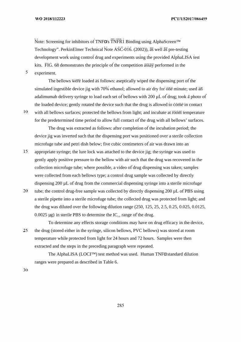
Note: Screening for inhibitors of TNFα/s TNFR1 Binding using AlphaScreen™
Technology”. PerkinElmer Technical Note ASC-016. (2002)), as well as pre-testing
development work using control drug and experiments using the provided AlphaLISA test
kits. FIG. 68 demonstrates the principle of the competition assay performed in the
experiment.
The bellows were loaded as follows: aseptically wiped the dispensing port of the
simulated ingestible device jig with 70% ethanol; allowed to air dry for one minute; used an
adalimumab delivery syringe to load each set of bellows with 200 µL of drug; took a photo of
the loaded device; gently rotated the device such that the drug is allowed to come in contact
with all bellows surfaces; protected the bellows from light; and incubate at room temperature
for the predetermined time period to allow full contact of the drug with all bellows’ surfaces.
The drug was extracted as follows: after completion of the incubation period; the
device jig was inverted such that the dispensing port was positioned over a sterile collection
microfuge tube and petri dish below; five cubic centimeters of air was drawn into an
appropriate syringe; the lure lock was attached to the device jig; the syringe was used to
gently apply positive pressure to the bellow with air such that the drug was recovered in the
collection microfuge tube; where possible, a video of drug dispensing was taken; samples
were collected from each bellows type; a control drug sample was collected by directly
dispensing 200 µL of drug from the commercial dispensing syringe into a sterile microfuge
tube; the control drug-free sample was collected by directly dispensing 200 µL of PBS using
a sterile pipette into a sterile microfuge tube; the collected drug was protected from light; and
the drug was diluted over the following dilution range (250, 125, 25, 2.5, 0.25, 0.025, 0.0125,
0.0025 µg) in sterile PBS to determine the IC5 0 range of the drug.
To determine any effects storage conditions may have on drug efficacy in the device,
the drug (stored either in the syringe, silicon bellows, PVC bellows) was stored at room
temperature while protected from light for 24 hours and 72 hours. Samples were then
extracted and the steps in the preceding paragraph were repeated.
The AlphaLISA (LOCI™) test method was used. Human TNFαstandard dilution
ranges were prepared as described in Table 6.
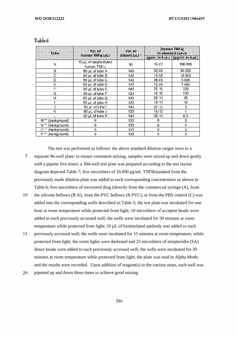
Table 6
The test was performed as follows: the above standard dilution ranges were in a
separate 96-well plate; to ensure consistent mixing, samples were mixed up and down gently
with a pipette five times; a 384-well test plate was prepared according to the test layout
diagram depicted Table 7; five microliters of 10,000 pg/mL TNFαstandard from the
previously made dilution plate was added to each corresponding concentration as shown in
Table 6; five microliters of recovered drug (directly from the commercial syringe (A), from
the silicone bellows (B Si), from the PVC bellows (B PVC), or from the PBS control (C) was
added into the corresponding wells described in Table 5; the test plate was incubated for one
hour at room temperature while protected from light; 10 microliters of acceptor beads were
added to each previously accessed well; the wells were incubated for 30 minutes at room
temperature while protected from light; 10 µL of biotinylated antibody was added to each
previously accessed well; the wells were incubated for 15 minutes at room temperature, while
protected from light; the room lights were darkened and 25 microliters of streptavidin (SA)
donor beads were added to each previously accessed well; the wells were incubated for 30
minutes at room temperature while protected from light; the plate was read in Alpha Mode;
and the results were recorded. Upon addition of reagent(s) in the various steps, each well was
pipetted up and down three times to achieve good mixing.
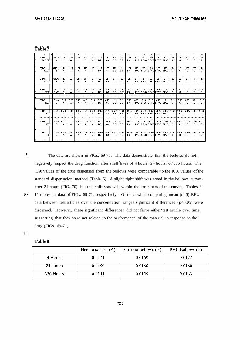
Table 7
The data are shown in FIGs. 69-71. The data demonstrate that the bellows do not
negatively impact the drug function after shelf lives of 4 hours, 24 hours, or 336 hours. The
IC50 values of the drug dispensed from the bellows were comparable to the IC50 values of the
standard dispensation method (Table 6). A slight right shift was noted in the bellows curves
after 24 hours (FIG. 70), but this shift was well within the error bars of the curves. Tables 8-
11 represent data of FIGs. 69-71, respectively. Of note, when comparing mean (n=5) RFU
data between test articles over the concentration ranges significant differences (p<0.05) were
discerned. However, these significant differences did not favor either test article over time,
suggesting that they were not related to the performance of the material in response to the
drug (FIGs. 69-71).
Table 8
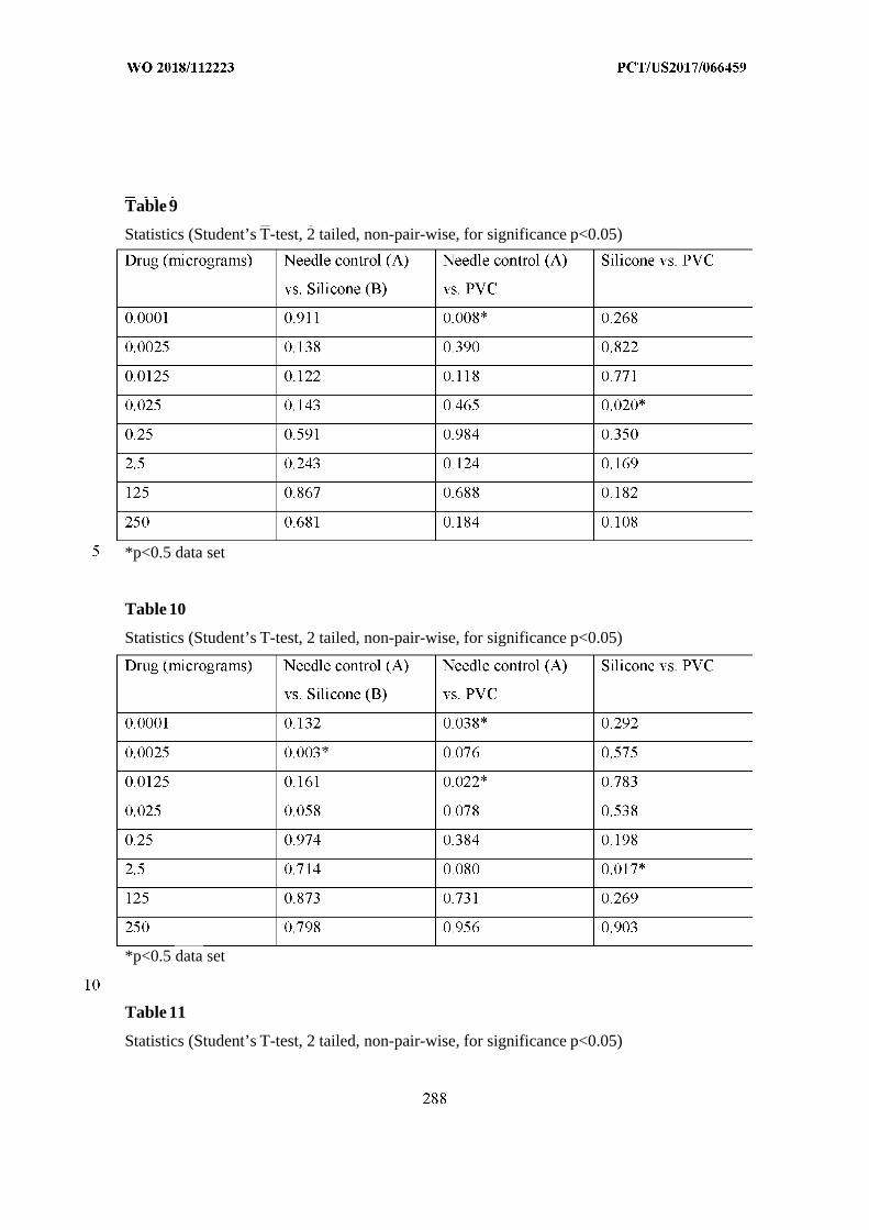
Table 9
Statistics (Student’s T-test, 2 tailed, non-pair-wise, for significance p<0.05)
*p<0.5 data set
Table 10
Statistics (Student’s T-test, 2 tailed, non-pair-wise, for significance p<0.05)
*p<0.5 data set
Table 11
Statistics (Student’s T-test, 2 tailed, non-pair-wise, for significance p<0.05)
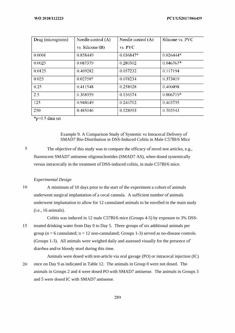
Example 9. A Comparison Study of Systemic vs Intracecal Delivery ofSMAD7 Bio-Distribution in DSS-Induced Colitis in Male C57Bl/6 Mice
The objective of this study was to compare the efficacy of novel test articles, e.g.,
fluorescent SMAD7 antisense oligonucleotides (SMAD7 AS), when dosed systemically
versus intracecally in the treatment of DSS-induced colitis, in male C57Bl/6 mice.
Experimental Design
A minimum of 10 days prior to the start of the experiment a cohort of animals
underwent surgical implantation of a cecal cannula. A sufficient number of animals
underwent implantation to allow for 12 cannulated animals to be enrolled in the main study
(i.e., 16 animals).
Colitis was induced in 12 male C57Bl/6 mice (Groups 4-5) by exposure to 3% DSS-
treated drinking water from Day 0 to Day 5. Three groups of six additional animals per
group (n = 6 cannulated; n = 12 non-cannulated; Groups 1-3) served as no-disease controls
(Groups 1-3). All animals were weighed daily and assessed visually for the presence of
diarrhea and/or bloody stool during this time.
Animals were dosed with test-article via oral gavage (PO) or intracecal injection (IC)
once on Day 9 as indicated in Table 12. The animals in Group 0 were not dosed. The
animals in Groups 2 and 4 were dosed PO with SMAD7 antisense. The animals in Groups 3
and 5 were dosed IC with SMAD7 antisense.
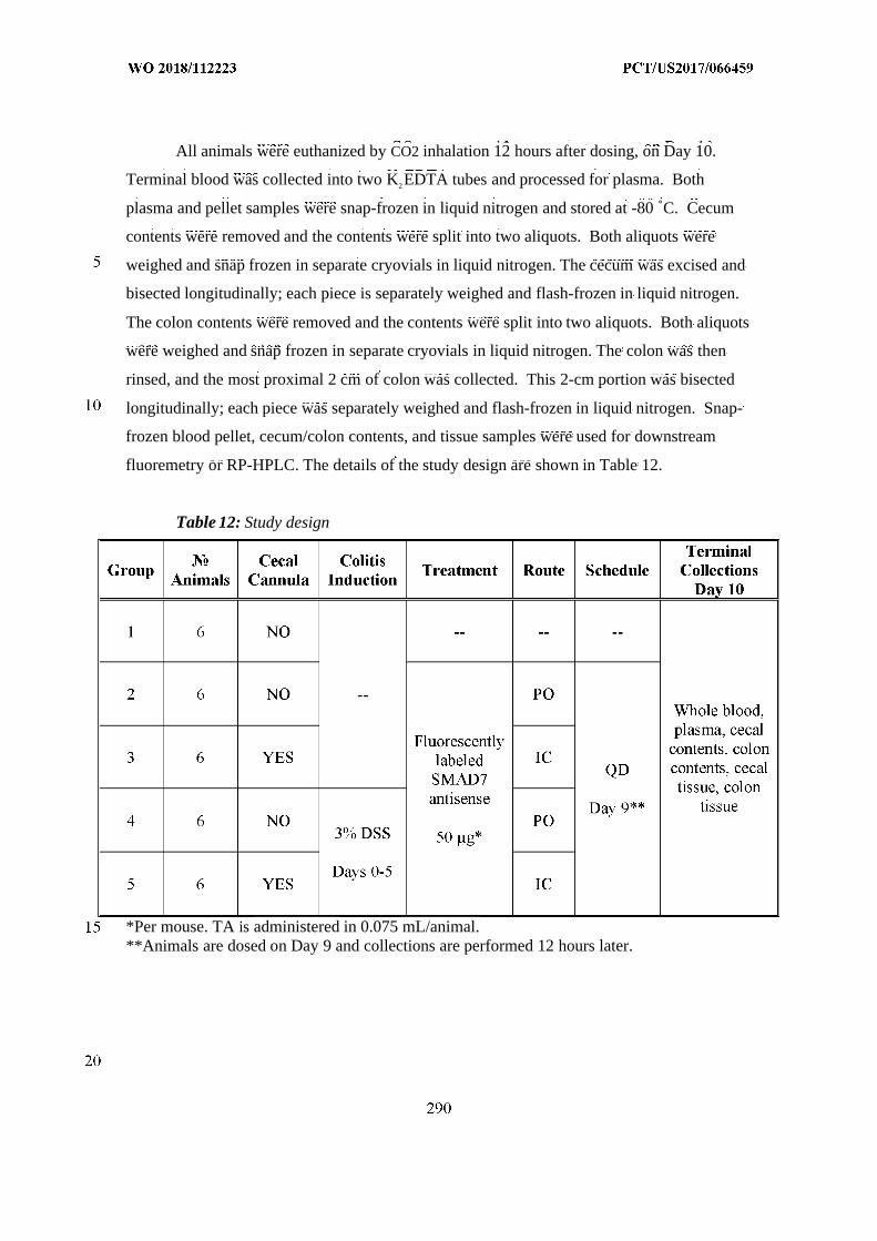
All animals were euthanized by CO2 inhalation 12 hours after dosing, on Day 10.
Terminal blood was collected into two K2EDTA tubes and processed for plasma. Both
plasma and pellet samples were snap-frozen in liquid nitrogen and stored at -80 ˚C. Cecum
contents were removed and the contents were split into two aliquots. Both aliquots were
weighed and snap frozen in separate cryovials in liquid nitrogen. The cecum was excised and
bisected longitudinally; each piece is separately weighed and flash-frozen in liquid nitrogen.
The colon contents were removed and the contents were split into two aliquots. Both aliquots
were weighed and snap frozen in separate cryovials in liquid nitrogen. The colon was then
rinsed, and the most proximal 2 cm of colon was collected. This 2-cm portion was bisected
longitudinally; each piece was separately weighed and flash-frozen in liquid nitrogen. Snap-
frozen blood pellet, cecum/colon contents, and tissue samples were used for downstream
fluoremetry or RP-HPLC. The details of the study design are shown in Table 12.
Table 12: Study design
*Per mouse. TA is administered in 0.075 mL/animal.**Animals are dosed on Day 9 and collections are performed 12 hours later.
Materials and Methods
Mice
Normal male C57Bl/6 mice between the ages of 6-8 weeks old, weighing 20-24 g,
were obtained from Charles River Laboratories. The mice were randomized into five groups
of six mice each, and housed in groups of 8-15 per cage, and acclimatized for at least three
days prior to entering the study. Animal rooms were set to maintain a minimum of 12 to 15
air changes per hour, with an automatic timer for a light/dark cycle of 12 hours on/off, and
fed with Labdiet 5053 sterile rodent chow, with water administered ad libitum.
Cecal Cannulation
The animals were placed under isoflurane anesthesia, with the cecum exposed via a
midline incision in the abdomen. A small point incision was made in the distal cecum, where
1-2 cm of the cannula was inserted. The incision was closed with a purse string suture using
5-0 silk. An incision was then made in the left abdominal wall through which the distal end
of the cannula was inserted and pushed subcutaneously to the dorsal aspect of the back. The
site was then washed copiously with warmed saline prior to closing the abdominal wall. A
small incision was also made in the skin of the back between the shoulder blades, exposing
the tip of the cannula. The cannula was secured in place using suture, wound clips, and tissue
glue. All animals were administered 1 mL of warm sterile saline (subcutaneous injection)
and were monitored closely until recovery before returning to their cage. All animals were
administered 0.6 mg/kg BID buprenorphine for the first 3 days, and Baytril® at 10mg/Kg
every day for the first 5 days post-surgery.
Disease Induction
Colitis was induced on Day 0 via addition of 3% DSS (MP Biomedicals, Cat
#0260110) to the drinking water. Fresh DSS/water solutions was provided on Day 3 and any
of the remaining original DSS solution is discarded.
Body Weight and Survival
Animals were observed daily (weight, morbidity, survival, presence of diarrhea and/or
bloody stool) in order to assess possible differences among treatment groups and/or possible
toxicity resulting from the treatments.
Animals Found Dead or Moribund
Animals were monitored on a daily basis. Animals exhibiting weight loss greater than
30% were euthanized, and samples were not collected from these animals.
Dosing
Animals were dosed with test-article via oral gavage (PO) or intracecal injection (IC)
once on Day 9 as indicated in Table 12. Animals in Group 0 were not dosed. Animals in
Groups 2 and 4 were dosed PO with SMAD7 antisense. Animals in Groups 3 and 5 were
dosed IC with SMAD7 antisense.
Sacrifice
All animals were euthanized by CO2 inhalation 12 hours after dosing, on Day 10.
Sample Collection
Intestinal contents, peripheral blood and tissue were collected at sacrifice on Day 10,
as follows:
Blood/Plasma
Terminal blood was collected into two K2 EDTA tubes and processed for plasma. The
approximate volume of each blood sample was recorded prior to centrifugation. Both plasma
and pellet samples were snap-frozen in liquid nitrogen and stored at -80 ˚C. The first pellet
sample (sample 1) was used for fluoremetry. The second pellet sample (sample 2) was used
for RP-HPLC.
Cecum Contents
Cecum contents was removed and contents were split into two aliquots. Both aliquots
were weighed and snap frozen in separate cryovials in liquid nitrogen. The first sample
(sample 1) was used for fluorometry. The second sample (sample 2) was used for RP-HPLC.
Cecum
The cecum was excised and bisected longitudinally; each piece was separately
weighed and snap-frozen. The first sample (sample 1) was used for fluoremetry. The second
sample (sample 2) was used for RP-HPLC.
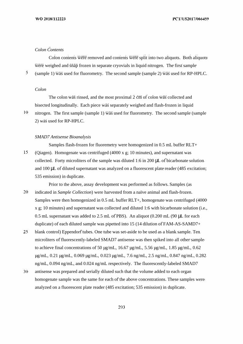
Colon Contents
Colon contents were removed and contents were split into two aliquots. Both aliquots
were weighed and snap frozen in separate cryovials in liquid nitrogen. The first sample
(sample 1) was used for fluorometry. The second sample (sample 2) was used for RP-HPLC.
Colon
The colon was rinsed, and the most proximal 2 cm of colon was collected and
bisected longitudinally. Each piece was separately weighed and flash-frozen in liquid
nitrogen. The first sample (sample 1) was used for fluorometry. The second sample (sample
2) was used for RP-HPLC.
SMAD7 Antisense Bioanalysis
Samples flash-frozen for fluoremetry were homogenized in 0.5 mL buffer RLT+
(Qiagen). Homogenate was centrifuged (4000 x g; 10 minutes), and supernatant was
collected. Forty microliters of the sample was diluted 1:6 in 200 µL of bicarbonate solution
and 100 µL of diluted supernatant was analyzed on a fluorescent plate reader (485 excitation;
535 emission) in duplicate.
Prior to the above, assay development was performed as follows. Samples (as
indicated in Sample Collection) were harvested from a naïve animal and flash-frozen.
Samples were then homogenized in 0.5 mL buffer RLT+, homogenate was centrifuged (4000
x g; 10 minutes) and supernatant was collected and diluted 1:6 with bicarbonate solution (i.e.,
0.5 mL supernatant was added to 2.5 mL of PBS). An aliquot (0.200 mL (90 µL for each
duplicate) of each diluted sample was pipetted into 15 (14 dilution of FAM-AS-SAMD7+
blank control) Eppendorf tubes. One tube was set-aside to be used as a blank sample. Ten
microliters of fluorescently-labeled SMAD7 antisense was then spiked into all other sample
to achieve final concentrations of 50 µg/mL, 16.67 µg/mL, 5.56 µg/mL, 1.85 µg/mL, 0.62
µg/mL, 0.21 µg/mL, 0.069 µg/mL, 0.023 µg/mL, 7.6 ng/mL, 2.5 ng/mL, 0.847 ng/mL, 0.282
ng/mL, 0.094 ng/mL, and 0.024 ng/mL respectively. The fluorescently-labeled SMAD7
antisense was prepared and serially diluted such that the volume added to each organ
homogenate sample was the same for each of the above concentrations. These samples were
analyzed on a fluorescent plate reader (485 excitation; 535 emission) in duplicate.
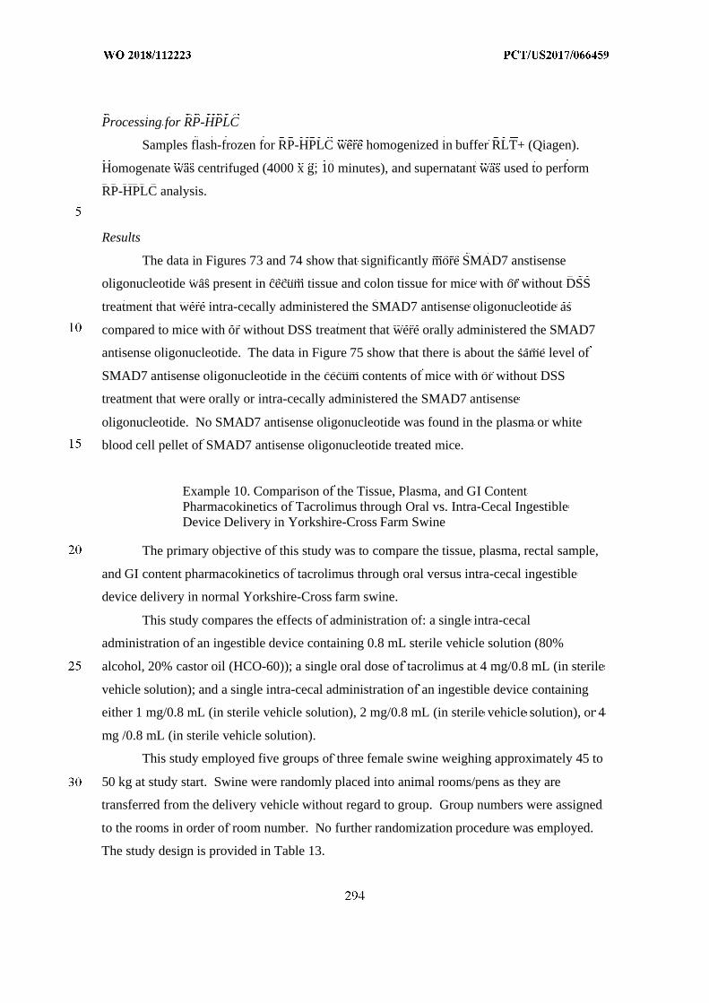
Processing for RP-HPLC
Samples flash-frozen for RP-HPLC were homogenized in buffer RLT+ (Qiagen).
Homogenate was centrifuged (4000 x g; 10 minutes), and supernatant was used to perform
RP-HPLC analysis.
Results
The data in Figures 73 and 74 show that significantly more SMAD7 anstisense
oligonucleotide was present in cecum tissue and colon tissue for mice with or without DSS
treatment that were intra-cecally administered the SMAD7 antisense oligonucleotide as
compared to mice with or without DSS treatment that were orally administered the SMAD7
antisense oligonucleotide. The data in Figure 75 show that there is about the same level of
SMAD7 antisense oligonucleotide in the cecum contents of mice with or without DSS
treatment that were orally or intra-cecally administered the SMAD7 antisense
oligonucleotide. No SMAD7 antisense oligonucleotide was found in the plasma or white
blood cell pellet of SMAD7 antisense oligonucleotide treated mice.
Example 10. Comparison of the Tissue, Plasma, and GI ContentPharmacokinetics of Tacrolimus through Oral vs. Intra-Cecal IngestibleDevice Delivery in Yorkshire-Cross Farm Swine
The primary objective of this study was to compare the tissue, plasma, rectal sample,
and GI content pharmacokinetics of tacrolimus through oral versus intra-cecal ingestible
device delivery in normal Yorkshire-Cross farm swine.
This study compares the effects of administration of: a single intra-cecal
administration of an ingestible device containing 0.8 mL sterile vehicle solution (80%
alcohol, 20% castor oil (HCO-60)); a single oral dose of tacrolimus at 4 mg/0.8 mL (in sterile
vehicle solution); and a single intra-cecal administration of an ingestible device containing
either 1 mg/0.8 mL (in sterile vehicle solution), 2 mg/0.8 mL (in sterile vehicle solution), or 4
mg /0.8 mL (in sterile vehicle solution).
This study employed five groups of three female swine weighing approximately 45 to
50 kg at study start. Swine were randomly placed into animal rooms/pens as they are
transferred from the delivery vehicle without regard to group. Group numbers were assigned
to the rooms in order of room number. No further randomization procedure was employed.
The study design is provided in Table 13.
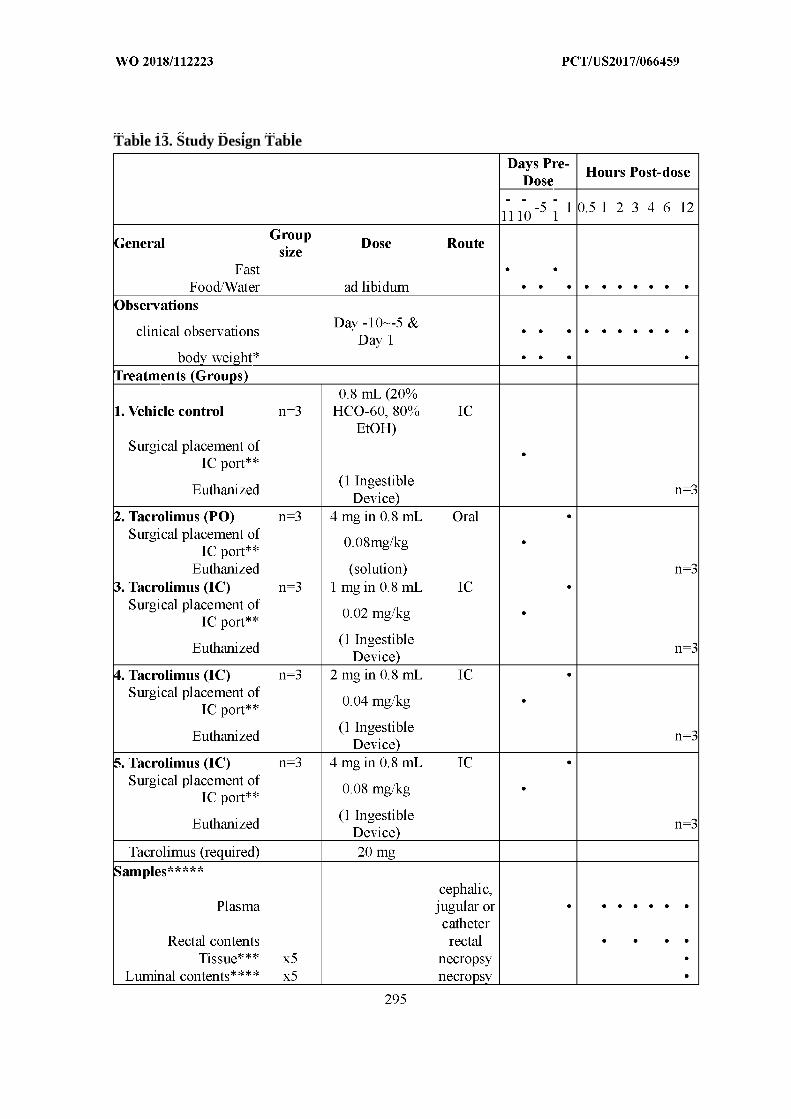
Table 13. Study Design Table
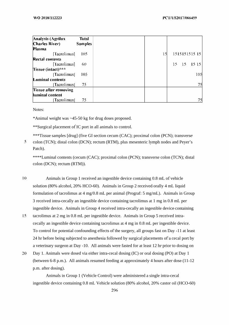
Notes:
*Animal weight was ~45-50 kg for drug doses proposed.
**Surgical placement of IC port in all animals to control.
***Tissue samples [drug] (five GI section cecum (CAC); proximal colon (PCN); transverse
colon (TCN); distal colon (DCN); rectum (RTM), plus mesenteric lymph nodes and Peyer’s
Patch).
****Luminal contents (cecum (CAC); proximal colon (PCN); transverse colon (TCN); distal
colon (DCN); rectum (RTM)).
Animals in Group 1 received an ingestible device containing 0.8 mL of vehicle
solution (80% alcohol, 20% HCO-60). Animals in Group 2 received orally 4 mL liquid
formulation of tacrolimus at 4 mg/0.8 mL per animal (Prograf: 5 mg/mL). Animals in Group
3 received intra-cecally an ingestible device containing tacrolimus at 1 mg in 0.8 mL per
ingestible device. Animals in Group 4 received intra-cecally an ingestible device containing
tacrolimus at 2 mg in 0.8 mL per ingestible device. Animals in Group 5 received intra-
cecally an ingestible device containing tacrolimus at 4 mg in 0.8 mL per ingestible device.
To control for potential confounding effects of the surgery, all groups fast on Day -11 at least
24 hr before being subjected to anesthesia followed by surgical placements of a cecal port by
a veterinary surgeon at Day -10. All animals were fasted for at least 12 hr prior to dosing on
Day 1. Animals were dosed via either intra-cecal dosing (IC) or oral dosing (PO) at Day 1
(between 6-8 p.m.). All animals resumed feeding at approximately 4 hours after dose (11-12
p.m. after dosing).
Animals in Group 1 (Vehicle Control) were administered a single intra-cecal
ingestible device containing 0.8 mL Vehicle solution (80% alcohol, 20% castor oil (HCO-60)
on Day 1. On Day -10 the animals were anesthetized, and a veterinary surgeon surgically
placed an intra-cecal port in each animal. On Day 1, each animal was placed into a sling then
a single intra-cecal ingestible device containing 0.8 mL vehicle solution (80% alcohol, 20%
castor oil (HCO-60)) is introduced by the veterinary surgeon into the cecum via the cecal port
in each animal. Following ingestible device placement, the animals were removed from the
slings and placed back into their pens with water. All animals resumed feeding at
approximately 4 hours after dose. Samples of rectal contents were collected for
pharmacokinetic analyses from each animal at each of 1, 3, 6, and 12 hours post-ingestible
device placement using a fecal swab (rectal swab). A total of 60 samples were collected.
Approximately 200~400 mg of rectal content were collected, if available, with a fecal
swab (Copan Diagnostics Nylon Flocked Dry Swabs, 502CS01). The fecal swab was pre-
weighed and weighed after collection in the collection tube (Sterile Tube and Cap No Media,
PFPM913S), and the sample weight was recorded. The fecal swab was broken via the
breakpoint, and was stored in the collection tube, and immediately frozen at -70 ºC. Whole
blood (2 mL) was collected into K2EDTA coated tubes for pharmacokinetics at each time-
point of pre-dose and 1, 2, 3, 4, 6 and 12 hours post-dose. Immediately following euthanasia,
tissue was collected. A total of 105 samples were collected.
For tissue necropsy, small intestine fluid and cecal fluid were collected separately
from all the animals into two separate square plastic bottles, and stored at -20 °C. The length
and diameter of the cecum and the colon was measured from one animal in each group and
recorded for reference. Tissues were collected for pharmacokinetic analyses and include
mesenteric lymph nodes, a Peyer’s Patch, and five gastrointestinal sections, including cecum,
proximal colon, transverse colon, distal colon, and rectum. All samples were weighed, and
the tissue sample weights were recorded. In each of the five gastrointestinal sections, tissue
samples were collected in three different areas where the mucosal surface was visible and not
covered by luminal content by using an 8.0-mm punch biopsy tool. Around 3 grams of the
total punched sample were collected into a pre-weighed 15-mL conical tube, and the tissue
weight was recorded. Three mesenteric lymph nodes were collected from different areas and
weighed. At least one Peyer’s Patch was collected and weighed. Tissues were snap-frozen in
liquid nitrogen and stored frozen at approximately -70 ºC or below (total of 105 samples).
Luminal contents were collected for pharmacokinetic analyses from the surface of the
tissue from each of five gastrointestinal sections: cecum, proximal colon, transverse colon,
distal colon, and rectum (total of 75). The contents were collected in pre-weighed 15-mL
conical tubes and the sample weights were recorded. Samples were snap-frozen in liquid
nitrogen stored frozen at approximately -70 ºC or below.
After removing the luminal content, another set of tissue samples from 3 different
areas were collected via an 8.0-mm punch biopsy in each section of the five tissue
gastrointestinal sections described above. Around 3 grams of the total punched sample were
collected into a pre-weighed 15-mL conical tube, and the tissue weight was recorded (total of
75). Tissues were snap-frozen in liquid nitrogen and stored frozen at approximately -70 °C
or below.
A 30-cm length of jejunum (separated into two 15 cm lengths), and the remaining
distal and transverse colon tissue sample (after tissue and luminal content were collected for
PK) were collected in one animal in each group of treatment, snap-frozen in liquid nitrogen
and stored frozen at approximately -70 ºC or below. All samples for pharmacokinetic
analyses were stored on dry ice before analyses.
Group 2 animals were administered a single oral dose of tacrolimus at 1 mg/0.8 mL
(in the vehicle solution) on Day 1. Plasma, rectal content sample, tissue collection, GI
content collection and related procedures/storage/shipments was the same as those employed
in Group 1.
Group 3 animals were administered a single intra-cecal ingestible device containing
tacrolimus at 0.5 mg/0.8 mL (in the vehicle solution) on Day 1 by a veterinary surgeon.
Plasma, rectal content sample, tissue collection, GI content collection and related
procedures/storage/shipments was the same as those employed in Group 1. All samples
wereanalyzed for tacrolimus.
Group 4 animals were administered a single intra-cecal ingestible device of
tacrolimus at 2 mg/0.8 mL (in sterile vehicle solution) on Day 1 by a veterinary surgeon.
Plasma, rectal content sample, tissue collection, GI content collection and related
procedures/storage/shipments were the same as those employed in Group 1. All samples
were analyzed for tacrolimus.
Group 5 animals are administered a single intra-cecal ingestible device containing
tacrolimus at 4 mg /0.8 mL (in the vehicle solution) on Day 1 by a veterinary surgeon.
Plasma, rectal content sample, tissue collection, GI content collection and related
procedures/storage/shipments were the same as those employed in Group 1. All samples
were analyzed for tacrolimus.
Detailed clinical observations were conducted daily from Day -10 to -5, and on Day 1.
Additional pen-side observations were conducted at least once each day. The animals
remained under constant clinical observation for the entire 12 hours from dose until
euthanasia. Body weights were collected on Day -10, Day -5, and pre-dose on Day 1.
Animals were euthanized via injection of a veterinarian-approved euthanasia.
Test Article and Formulation
1. Vehicle solution, 20 mL
Description: 80% alcohol, 20% PEG-60 castor oil
Physical characteristics: clear liquid solution.
2. Prograf (tacrolimus injection), 10 ampules
Description: A sterile solution containing the equivalent of 5 mg anhydrous tacrolimus in 1
mL. Tacrolimus is macrolide immunosuppressant and the active ingredient of Prograf. 0.8
mL of Prograf (5 mg/mL) was administrated through oral gavage per animal in group 2.
Prograf (5 mg/mL) was diluted 2x folds (2.5 mg/mL) and 4x folds (1.25 mg/mL) by using
vehicle solution. 0.8 mL of each concentration, 1.25 mg/mL, 2.5 mg/mL, and 5 mg/mL of
Prograf, was injected into a DSS ingestible device for group 3, 4, and 5.
Formulation: Each mL contained polyoxyl 60 hydrogenated castor oil (HCO-60), 200 mg,
and dehydrated alcohol, USP, 80.0% v/v.
Physical characteristics: clear liquid solution.
3. DDS ingestible device containing Tacrolimus
Description: Three (3) DDS ingestible devices containing vehicle solution for Group 1, three
(3) DSS ingestible devices containing 1 mg tacrolimus for Group 3, three (3) DDS ingestible
devices containing 2 mg tacrolimus for Group 4, and three (3) DDS ingestible devices
containing 4 mg tacrolimus for Group 5.
Acclimation
Animals were acclimated prior to study initiation for at least 7 days. Animals in
obvious poor health were not placed on study.
Concurrent Medication
Other than veterinary-approved anesthetics and medications used during surgery to
install the ileocecal ports, or for vehicle or test article administration, and analgesia and
antibiotics post-surgery, no further medications were employed.
Feed
All swine were fasted at least 24 hours before being anesthetized and properly
medicated for surgery or overnight before dosing. Otherwise, animals were fed ad-libitum.
Tap water was pressure-reduced and passed through a particulate filter, then a carbon filter
prior to supply to an automatic watering system. Water was supplied ad libitum. There were
no known contaminants in the feed or water that would be expected to interfere with this
study.
Results
The data in Figure 76 show that the mean concentration of tacrolimus in the cecum
tissue and the proximate colon tissue were higher in swine that were inta-cecally
administered tacrolimus as compared to swine that were orally administered tacrolimus.
These data suggest that intra-cecal administration of tacrolimus is able to locally deliver
tacrolimus to the tissues in the GI tract of a mammal, while not decreasing the systemic
immune system of a mammal.
Example 11. Comparison of the Tissue, Plasma, and GI ContentPharmacokinetics of Adalimumab through SC vs. Intra-Cecal IngestibleDevice Delivery in Yorkshire-Cross Farm Swine in DSS-induced Colitis
The purpose of this non-Good Laboratory Practice (GLP) study is to explore the
PK/PD and bioavailability of adalimumab when applied to DSS-induced colitis in Yorkshire-
cross farm swine. All animals are randomized into groups of three. Animals are dosed once
with adalimumab via subcutaneous (SC), perirectal (PR), or intracecal (IC) administration.
The concentration of adalimumab and TNFαis measured in plasma at 1, 2, 3, 4, 6,
and 12 hours post-dose. The concentration of adalimumab is measured in rectal contents at 1,
3, 6, and 12 hours post-dose and in luminal content at 12 hours post-dose. Concentration of
adalimumab and TNFα, HER2, and total protein is measured in gastrointestinal tissue, e.g.,
cecum sample (CAC), proximal colon sample (PCN), transverse colon sample (TCN), distal
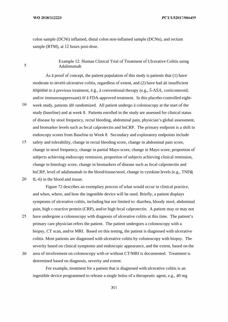
colon sample (DCNi) inflamed, distal colon non-inflamed sample (DCNn), and rectum
sample (RTM), at 12 hours post-dose.
Example 12. Human Clinical Trial of Treatment of Ulcerative Colitis usingAdalimumab
As a proof of concept, the patient population of this study is patients that (1) have
moderate to severe ulcerative colitis, regardless of extent, and (2) have had an insufficient
response to a previous treatment, e.g., a conventional therapy (e.g., 5-ASA, corticosteroid,
and/or immunosuppressant) or a FDA-approved treatment. In this placebo-controlled eight-
week study, patients are randomized. All patient undergo a colonoscopy at the start of the
study (baseline) and at week 8. Patients enrolled in the study are assessed for clinical status
of disease by stool frequency, rectal bleeding, abdominal pain, physician’s global assessment,
and biomarker levels such as fecal calprotectin and hsCRP. The primary endpoint is a shift in
endoscopy scores from Baseline to Week 8. Secondary and exploratory endpoints include
safety and tolerability, change in rectal bleeding score, change in abdominal pain score,
change in stool frequency, change in partial Mayo score, change in Mayo score, proportion of
subjects achieving endoscopy remission, proportion of subjects achieving clinical remission,
change in histology score, change in biomarkers of disease such as fecal calprotectin and
hsCRP, level of adalimumab in the blood/tissue/stool, change in cytokine levels (e.g., TNFα,
IL-6) in the blood and tissue.
Figure 72 describes an exemplary process of what would occur in clinical practice,
and when, where, and how the ingestible device will be used. Briefly, a patient displays
symptoms of ulcerative colitis, including but not limited to: diarrhea, bloody stool, abdominal
pain, high c-reactive protein (CRP), and/or high fecal calprotectin. A patient may or may not
have undergone a colonoscopy with diagnosis of ulcerative colitis at this time. The patient’s
primary care physician refers the patient. The patient undergoes a colonoscopy with a
biopsy, CT scan, and/or MRI. Based on this testing, the patient is diagnosed with ulcerative
colitis. Most patients are diagnosed with ulcerative colitis by colonoscopy with biopsy. The
severity based on clinical symptoms and endoscopic appearance, and the extent, based on the
area of involvement on colonoscopy with or without CT/MRI is documented. Treatment is
determined based on diagnosis, severity and extent.
For example, treatment for a patient that is diagnosed with ulcerative colitis is an
ingestible device programmed to release a single bolus of a therapeutic agent, e.g., 40 mg

adalimumab, in the cecum or proximal to the cecum. Prior to administration of the treatment,
the patient is fasted overnight and is allowed to drink clear fluids. Four hours after
swallowing the ingestible device, the patient can resume a normal diet. An ingestible device
is swallowed at the same time each day. The ingestible device is not recovered.
In some embodiments, there may be two different ingestible devices: one including an
induction dose (first 8 to 12 weeks) and a different ingestible device including a different
dose or a different dosing interval.
In some examples, the ingestible device can include a mapping tool, which can be
used after 8 to 12 weeks of induction therapy, to assess the response status (e.g., based on one
or more of the following: drug level, drug antibody level, biomarker level, and mucosal
healing status). Depending on the response status determined by the mapping tool, a subject
may continue to receive an induction regimen or maintenance regimen of adalimumab.
In different clinical studies, the patients may be diagnosed with Crohn’s disease and
the ingestible devices (including adalimumab) can be programmed to release adalimumab in
the cecum, or in both the cecum and transverse colon.
In different clinical studies, the patients may be diagnosed with illeocolonic Crohn’s
disease and the ingestible devices (including adalimumab) can be programmed to release
adalimumab in the late jejunum or in the jejunum and transverse colon.
EXAMPLE 13
An ingestible medical device according to the disclosure (“TLC1”) was tested on 20
subjects to investigate its localization ability. TLC1 was a biocompatible polycarbonate
ingestible device that contained a power supply, electronics and software. An onboard
software algorithm used time, temperature and reflected light spectral data to determine the
location of the ingestible device as it traveled the GI tract. The ingestible device is 0.51 x
1.22 inches which is larger than a vitamin pill which is 0.4 x 0.85 inches. The subjects fasted
overnight before participating in the study. Computerized tomography (“CT”) were used as a
basis for determining the accuracy of the localization data collected with TLC1. One of the
20 subjects did not follow the fasting rule. CT data was lacking for another one of the 20
subjects. Thus, these two subjects were excluded from further analysis. TLC1 sampled RGB
data (radially transmitted) every 15 seconds for the first 14 hours after it entered the subject’s
stomach, and then samples every five minutes after that until battery dies. TLC1 did not start
to record optical data until it reached the subject’s stomach. Thus, there was no RGB-based
data for the mouth-esophagus transition for any of the subjects.
In addition, a PillCam® SB (Given Imaging) device was tested on 57 subjects. The
subjects fasted overnight before joining the study. PillCam videos were recorded within each
subject. The sampling frequency of PillCam is velocity dependent. The faster PillCam
travels, the faster it would sample data. Each video is about seven to eight hours long, starting
from when the ingestible device was administrated into the subject’s mouth. RGB optical
data were recorded in a table. A physician provided notes on where stomach-duodenum
transition and ileum-cecum transition occured in each video. Computerized tomography
(“CT”) was used as a basis for determining the accuracy of the localization data collected
with PillCam.
Esophagus-Stomach Transition
For TLC1, it was assumed that this transition occurred one minute after the patient
ingested the device. For PillCam, the algorithm was as follows:
1. Start mouth-esophagus transition detection after ingestible device isactivated/administrated
2. Check whether Green < 102.3 and Blue < 94.6a. If yes, mark as mouth-esophagus transitionb. If no, continue to scan the data
3. After detecting mouth-esophagus transition, continue to monitor Green and Bluesignals for another 30 seconds, in case of location reversal
a. If either Green > 110.1 or Blue > 105.5, mark it as mouth-esophagus locationreversal
b. Reset the mouth-esophagus flag and loop through step 2 and 3 until theconfirmed mouth-esophagus transition detected
4. Add one minute to the confirmed mouth-esophagus transition and mark it asesophagus-stomach transition
For one of the PillCam subjects, there was not a clear cut difference between the
esophagus and stomach, so this subject was excluded from future analysis of stomach
localization. Among the 56 valid subjects, 54 of them have correct esophagus-stomach
transition localization. The total agreement is 54/56=96%. Each of the two failed cases had
prolonged esophageal of greater than one minute. Thus, adding one minute to mouth-
esophagus transition was not enough to cover the transition in esophagus for these two
subjects.
Stomach-Duodenum
For both TLC1 and PillCam, a sliding window analysis was used. The algorithm used
a dumbbell shape two-sliding-window approach with a two-minute gap between the front
(first) and back (second) windows. The two-minute gap was designed, at least in part, to skip
the rapid transition from stomach to small intestine and capture the small intestine signal after
ingestible device settles down in small intestine. The algorithm was as follows:
1. Start to check for stomach-duodenum transition after ingestible device enters stomach2. Setup the two windows (front and back)
a. Time length of each window: 3 minutes for TLC1; 30 seconds for PillCamb. Time gap between two windows: 2 minutes for both devicesc. Window sliding step size: 0.5 minute for both devices
3. Compare signals in the two sliding windowsa. If difference in mean is higher than 3 times the standard deviation of
Green/Blue signal in the back windowi. If this is the first time ever, record the mean and standard deviation of
signals in the back window as stomach referenceii. If mean signal in the front window is higher than stomach reference
signal by a certain threshold (0.3 for TLC1 and 0.18 for PillCam),mark this as a possible stomach-duodenum transition
b. If a possible pyloric transition is detected, continue to scan for another 10minutes in case of false positive flag
i. If within this 10 minutes, location reversal is detected, the previouspyloric transition flag is a false positive flag. Clear the flag andcontinue to check
ii. If no location reversal has been identified within 10 minutes followingthe possible pyloric transition flag, mark it as a confirmed pylorictransition
c. Continue monitoring Green/Blue data for another 2 hours after the confirmedpyloric transition, in case of location reversal
i. If a location reversal is identified, flag the timestamp when reversalhappened and then repeat steps a-c to look for the next pylorictransition
ii. If the ingestible device has not gone back to stomach 2 hours afterpreviously confirmed pyloric transition, stops location reversalmonitoring and assume the ingestible device would stay in intestinalarea
For TLC1, one of the 18 subjects had too few samples (<3 minutes) taken in the
stomach due to the delayed esophagus-stomach transition identification by previously
developed localization algorithm. Thus, this subject was excluded from the stomach-
duodenum transition algorithm test. For the rest of the TLC1 subjects, CT images confirmed
that the detected pyloric transitions for all the subjects were located somewhere between
stomach and jejunum. Two out of the 17 subjects showed that the ingestible device went back
to stomach after first the first stomach-duodenum transition. The total agreement between the
TLC1 algorithm detection and CT scans was 17/17 = 100%.
For one of the PillCam subjects, the ingestible device stayed in the subject’s stomach
all the time before the video ended. For another two of the PillCam subjects, too few samples
were taken in the stomach to run the localization algorithm. These three PillCam subjects
were excluded from the stomach-duodenum transition localization algorithm performance
test. The performance summary of pyloric transition localization algorithm for PillCam was
as follows:
1. Good cases (48 subjects):a. For 25 subjects, our detection matches exactly with the physician’s notesb. For 19 subjects, the difference between the two detections is less than five
minutesc. For four subjects, the difference between the two detections is less than 10
minutes (The full transition could take up to 10 minutes before the G/B signalsettled)
2. Failed cases (6 subjects):a. Four subjects had high standard deviation of Green/Blue signal in the stomachb. One subject had bile in the stomach, which greatly affected Green/Blue in
stomachc. One subject had no Green/Blue change at pyloric transition
The total agreement for the PillCam stomach-duodenum transition localization
algorithm detection and physician’s notes was 48/54 = 89%.
Duodenum-Jejenum Transition
For TLC1, it was assumed that the device left the duodenum and entered the jejenum
three minutes after it was determined that the device entered the duodenum. Of the 17
subjects noted above with respect to the TLC1 investigation of the stomach-duodenum
transition, 16 of the subjects mentioned had CT images that confirmed that the duodenum-
jejenum transition was located somewhere between stomach and jejunum. One of the 17
subjects had a prolonged transit time in duodenum. The total agreement between algorithm
detection and CT scans was 16/17 = 94%.
For PillCam, the duodenum-jejenum transition was not determined.
Jejenum-Ileum Transition
It is to be noted that the jejunum is redder and more vascular than ileum, and that the
jejenum has a thicker intestine wall with more mesentery fat. These differences can cause
various optical responses between jejunum and ileum, particularly for the reflected red light
signal. For both TLC1 and PillCam, two different approaches were explored to track the
change of red signal at the jejunum-ileum transition. The first approach was a single-sliding-
window analysis, where the window is 10 minutes long, and the mean signal was compared
with a threshold value while the window was moving along. The second approach was a
two-sliding-window analysis, where each window was 10 minutes long with a 20 minute
spacing between the two windows. The algorithm for the jejunum-ileum transition
localization was as follows:
1. Obtain 20 minutes of Red signal after the duodenum-jejenum transition, average thedata and record it as the jejunum reference signal
2. Start to check the jejunum-ileum transition 20 minutes after the device enters thejejunum
a. Normalize the newly received data by the jejunum reference signalb. Two approaches:
i. Single-sliding-window analysis^ Set the transition flag if the mean of reflected red signal is less
than 0.8ii. Two-sliding-window analysis:
^ Set the transition flag if the mean difference in reflected red ishigher than 2X the standard deviation of the reflected red signalin the front window
For TLC1, 16 of the 18 subjects had CT images that confirmed that the detected
jejunum-ileum transition fell between jejunum and cecum. The total agreement between
algorithm and CT scans was 16/18 = 89%. This was true for both the single-sliding-window
and double-sliding-window approaches, and the same two subjects failed in both approaches.
The performance summary of the jejunum-ileum transition detection for PillCam is
listed below:
1. Single-sliding-window analysis:a. 11 cases having jejunum-ileum transition detected somewhere between
jejunum and cecumb. 24 cases having jejunum-ileum transition detected after cecumc. 19 cases having no jejunum-ileum transition detected
d. Total agreement: 11/54 = 20%2. Two-sliding-window analysis:
a. 30 cases having jejunum-ileum transition detected somewhere betweenjejunum and cecum
b. 24 cases having jejunum-ileum transition detected after cecumc. Total agreement: 30/54 = 56%
Ileum-Cecum Transition
Data demonstrated that, for TLC1, mean signal of reflected red/green provided the
most statistical difference before and after the ileum-cecum transition. Data also
demonstrated that, for TLC1, the coefficient of variation of reflected green/blue provided the
most statistical contrast at ileum-cecum transition. The analysis based on PillCam videos
showed very similar statistical trends to those results obtained with TLC1 device. Thus, the
algortithm utilized changes in mean value of reflected red/green and the coefficient of
variation of reflected green/blue. The algorithm was as follows:
1. Start to monitor ileum-cecum transition after the ingestible device enters the stomach2. Setup the two windows (front (first) and back (second))
a. Use a five-minute time length for each windowb. Use a 10-minute gap between the two windowsc. Use a one-minute window sliding step size
3. Compare signals in the two sliding windowsa. Set ileum-cecum transition flag if
i. Reflected red/green has a significant change or is lower than athreshold
ii. Coefficient of variation of reflected green/blue is lower than athreshold
b. If this is the first ileum-cecum transition detected, record average reflectedred/green signal in small intestine as small intestine reference signal
c. Mark location reversal (i.e. ingestible device returns to terminal ileum) ifi. Reflected red/green is statistically comparable with small intestine
reference signalii. Coefficient of variation of reflected green/blue is higher than a
thresholdd. If a possible ileum-cecum transition is detected, continue to scan for another
10 minutes for TLC1 (15 minutes for PillCam) in case of false positive flagi. If within this time frame (10 minutes for TLC1, 15 minutes for
PillCam), location reversal is detected, the previous ileum-cecumtransition flag is a false positive flag. Clear the flag and continue tocheck
ii. If no location reversal has been identified within this time frame (10minutes for TLC1, 15 minutes for PillCam) following the possibleileum-cecum transition flag, mark it as a confirmed ileum-cecumtransition
e. Continue monitoring data for another 2 hours after the confirmed ileum-cecumtransition, in case of location reversal
i. If a location reversal is identified, flag the timestamp when reversalhappened and then repeat steps a-d to look for the next ileum-cecumtransition
ii. If the ingestible device has not gone back to small intestine 2 hoursafter previously confirmed ileum-cecum transition, stop locationreversal monitoring and assume the ingestible device would stay inlarge intestinal area
The flag setting and location reversal criteria particularly designed for TLC1 device
were as follows:
1. Set ileum-cecum transition flag ifa. The average reflected red/Green in the front window is less than 0.7 or mean
difference between the two windows is higher than 0.6b. And the coefficient of variation of reflected green/blue is less than 0.02
2. Define as location reversal ifa. The average reflected red/green in the front window is higher than small
intestine reference signalb. And the coefficient of variation of reflected green/blue is higher than 0.086
For TLC1, 16 of the 18 subjects had CT images that confirmed that the detected
ileum-cecum transition fell between terminal ileum and colon. The total agreement between
algorithm and CT scans was 16/18 = 89%. Regarding those two subject where the ileum-
cecum transition localization algorithm failed, for one subject the ileum-cecum transition was
detected while TLC1 was still in the subject’s terminal ileum, and for the other subject the
ileum-cecum transition was detected when the device was in the colon.
Among the 57 available PillCam endoscopy videos, for three subjects the endoscopy
video ended before PillCam reached cecum, and another two subjects had only very limited
video data (less than five minutes) in the large intestine. These five subjects were excluded
from ileum-cecum transition localization algorithm performance test. The performance
summary of ileum-cecum transition detection for PillCam is listed below:
1. Good cases (39 subjects):a. For 31 subjects, the difference between the PillCam detection and the
physician’s notes was less than five minutes
b. For 3 subjects, the difference between the PillCam detection and thephysician’s notes was less than 10 minutes
c. For 5 subjects, the difference between the PillCam detection and thephysician’s notes was less than 20 minutes (the full transition can take upto 20 minutes before the signal settles)
2. Marginal/bad cases (13 subjects):a. Marginal cases (9 subjects)
i. The PillCam ileum-cecum transition detection appeared in theterminal ileum or colon, but the difference between the twodetections was within one hour
b. Failed cases (4 subjects)i. Reasons of failure:
1. The signal already stabilized in the terminal ileum2. The signal was highly variable from the entrance to exit3. There was no statistically significant change in reflected
red/green at ileum-cecum transition
The total agreement between ileocecal transition localization algorithm detection and
the physician’s notes is 39/52 = 75% if considering good cases only. Total agreement
including possibly acceptable cases is 48/52 = 92.3%
Cecum-Colon Transition
Data demonstrated that, for TLC1, mean signal of reflected red/green provided the
most statistical difference before and after the cecum-colon transition. Data also
demonstrated that, for TLC1, the coefficient of variation of reflected bluee provided the most
statistical contrast at cecum-colon transition. The same signals were used for PillCam. The
cecum-colon transition localization algorithm was as follows:
1. Obtain 10 minutes of reflected red/green and reflected blue signals after ileum-cecumtransition, average the data and record it as the cecum reference signals
2. Start to check cecum-colon transition after ingestible device enters cecum (Thececum-colon transition algorithm is dependent on the ileum-cecum transition flag)
a. Normalize the newly received data by the cecum reference signalsb. Two-sliding-window analysis:
i. Use two adjacent 10 minute windowsii. Set the transition flag if any of the following criteria were met
^ The mean difference in reflected red/green was more than 4Xthe standard deviation of reflected red/green in the back(second) window
^ The mean of reflected red/green in the front (first) window washigher than 1.03
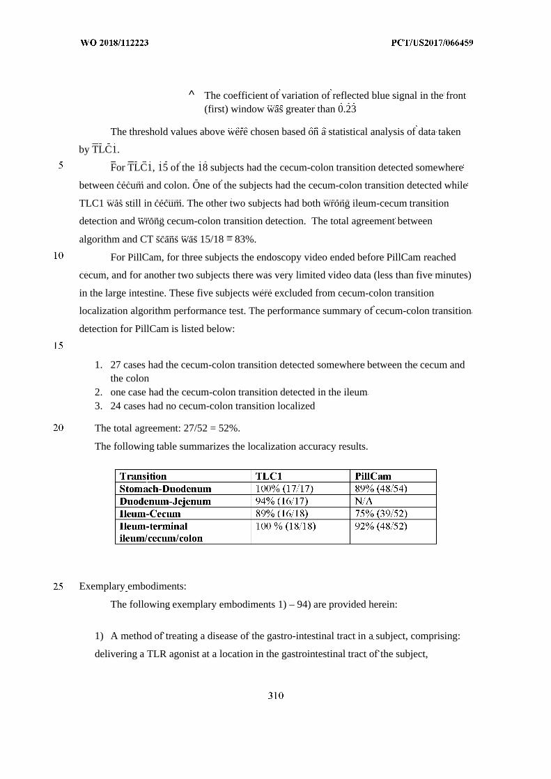
^ The coefficient of variation of reflected blue signal in the front(first) window was greater than 0.23
The threshold values above were chosen based on a statistical analysis of data taken
by TLC1.
For TLC1, 15 of the 18 subjects had the cecum-colon transition detected somewhere
between cecum and colon. One of the subjects had the cecum-colon transition detected while
TLC1 was still in cecum. The other two subjects had both wrong ileum-cecum transition
detection and wrong cecum-colon transition detection. The total agreement between
algorithm and CT scans was 15/18 = 83%.
For PillCam, for three subjects the endoscopy video ended before PillCam reached
cecum, and for another two subjects there was very limited video data (less than five minutes)
in the large intestine. These five subjects were excluded from cecum-colon transition
localization algorithm performance test. The performance summary of cecum-colon transition
detection for PillCam is listed below:
1. 27 cases had the cecum-colon transition detected somewhere between the cecum andthe colon
2. one case had the cecum-colon transition detected in the ileum3. 24 cases had no cecum-colon transition localized
The total agreement: 27/52 = 52%.
The following table summarizes the localization accuracy results.
Exemplary embodiments:
The following exemplary embodiments 1) – 94) are provided herein:
1) A method of treating a disease of the gastro-intestinal tract in a subject, comprising:
delivering a TLR agonist at a location in the gastrointestinal tract of the subject,
wherein the method comprises administering orally to the subject a pharmaceutical
composition comprising a therapeutically effective amount of the TLR agonist.
2) The method of exemplary embodiment 1, wherein the disease of the GI tract is an
inflammatory bowel disease.
3) The method of exemplary embodiment 1, wherein the disease of the GI tract is
ulcerative colitis.
4) The method of exemplary embodiment 1, wherein the disease of the GI tract is
Crohn’s disease.
5) The method of any one of exemplary embodiments 1, 2, or 3, 4, wherein the TLR
agonist is delivered at a location in the large intestine of the subject.
6) The method of exemplary embodiment 5, wherein the location is in the proximal
portion of the large intestine.
7) The method of exemplary embodiment 5, wherein the location is in the distal portion
of the large intestine.
8) The method of any one of exemplary embodiments 1, 2, or 3, 4, wherein the TLR
agonist is delivered at a location in the ascending colon of the subject.
9) The method of exemplary embodiment 8, wherein the location is in the proximal
portion of the ascending colon.
10) The method of exemplary embodiment 8, wherein the location is in the distal portion
of the ascending colon.
11) The method of any one of exemplary embodiments 1, 2, or 3, 4, wherein the TLR
agonist is delivered at a location in the cecum of the subject.
12) The method of exemplary embodiment 11, wherein the location is in the proximal
portion of the cecum.
13) The method of exemplary embodiment 11, wherein the location is in the distal portion
of the cecum.
14) The method of any one of exemplary embodiments 1, 2, or 3, 4, wherein the TLR
agonist is delivered at a location in the sigmoid colon of the subject.
15) The method of exemplary embodiment 14, wherein the location is in the proximal
portion of the sigmoid colon.
16) The method of exemplary embodiment 14, wherein the location is in the distal portion
of the sigmoid colon.
17) The method of any one of exemplary embodiments 1, 2, or 3, 4, wherein the TLR
agonist is delivered at a location in the transverse colon of the subject.
18) The method of exemplary embodiment 17, wherein the location is in the proximal
portion of the transverse colon.
19) The method of exemplary embodiment 17, wherein the location is in the distal portion
of the transverse colon.
20) The method of any one of exemplary embodiments 1, 2, or 3, 4, wherein the TLR
agonist is delivered at a location in the descending colon of the subject.
21) The method of exemplary embodiment 20, wherein the location is in the proximal
portion of the descending colon.
22) The method of exemplary embodiment 20, wherein the location is in the distal portion
of the descending colon.
23) The method of any one of exemplary embodiments 1, 2, or 3, 4, wherein the TLR
agonist is delivered at a location in the small intestine of the subject.
24) The method of exemplary embodiment 23, wherein the location is in the proximal
portion of the small intestine.
25) The method of exemplary embodiment 23, wherein the location is in the distal portion
of the small intestine.
26) The method of any one of exemplary embodiments 1, 2, or 3, 4, wherein the TLR
agonist is delivered at a location in the duodenum of the subject.
27) The method of exemplary embodiment 26, wherein the location is in the proximal
portion of the duodenum.
28) The method of exemplary embodiment 26, wherein the location is in the distal portion
of the duodenum.
29) The method of any one of exemplary embodiments 1, 2, or 3, 4, wherein the TLR
agonist is delivered at a location in the jejunum of the subject.
30) The method of exemplary embodiment 29, wherein the location is in the proximal
portion of the jejunum.
31) The method of exemplary embodiment 29, wherein the location is in the distal portion
of the jejunum.
32) The method of any one of exemplary embodiments 1, 2, or 3, 4, wherein the TLR
agonist is delivered at a location in the ileum of the subject.
33) The method of exemplary embodiment 32, wherein the location is in the proximal
portion of the ileum.
34) The method of exemplary embodiment 32, wherein the location is in the distal portion
of the ileum.
35) The method of any one of the preceding exemplary embodiments, wherein the
location is proximate to one or more sites of disease.
36) The method of exemplary embodiment 35, further comprising identifying the one or
more sites of disease by a method comprising imaging of the gastrointestinal tract.
37) The method of any one of the preceding exemplary embodiments, wherein the TLR
agonist is delivered to the location by mucosal contact.
38) The method of any one of the preceding exemplary embodiments, wherein the TLR
agonist is delivered to the location by a process that does not comprise systemic transport of
the TLR agonist.
39) The method of any one of the preceding exemplary embodiments, wherein the amount
of the TLR agonist that is administered is from about 1 mg to about 300 mg.
40) The method of exemplary embodiment 39, wherein the amount of the TLR agonist
that is administered is from about 1 mg to about 100 mg.
41) The method of exemplary embodiment 40, wherein the amount of the TLR agonist
that is administered is from about 5 mg to about 40 mg.
42) The method of any one of exemplary embodiments 1 to 41, wherein the amount of the
TLR agonist is less than an amount that is effective when the TLR agonist is administered
systemically.
43) The method of any one of the preceding exemplary embodiments, comprising
administering (i) an amount of the TLR agonist that is an induction dose.
44) The method of exemplary embodiment 43, further comprising (ii) administering an
amount of the TLR agonist that is a maintenance dose following the administration of the
induction dose.
45) The method of exemplary embodiment 43 or 44, wherein the induction dose is
administered once a day.
46) The method of exemplary embodiment 43 or 44, wherein the induction dose is
administered once every three days.
47) The method of exemplary embodiment 43 or 44, wherein the induction dose is
administered once a week.
48) The method of exemplary embodiment 44, wherein step (ii) is repeated one or more
times.
49) The method of exemplary embodiment 44, wherein the induction dose is equal to the
maintenance dose.
50) The method of exemplary embodiment 44, wherein the induction dose is greater than
the maintenance dose.
51) The method of exemplary embodiment 44, wherein the induction dose is 5 greater
than the maintenance dose.
52) The method of exemplary embodiment 44, wherein the induction dose is 2 greater
than the maintenance dose.
53) The method of any one of the preceding exemplary embodiments, wherein the method
comprises delivering the TLR agonist at the location in the gastrointestinal tract as a single
bolus.
54) The method of any one of exemplary embodiments 1 to 52, wherein the method
comprises delivering the TLR agonist at the location in the gastrointestinal tract as more than
one bolus.
55) The method of any one of exemplary embodiments 1 to 52, wherein the method
comprises delivering the TLR agonist at the location in the gastrointestinal tract in a
continuous manner.
56) The method of exemplary embodiment 55, wherein the method comprises delivering
the TLR agonist at the location in the gastrointestinal tract over a time period of 20 or more
minutes.
57) The method of any one of the preceding exemplary embodiments, wherein the method
provides a concentration of the TLR agonist in the plasma of the subject that is less than 3
µg/ml.
58) The method of exemplary embodiment 57, wherein the method provides a
concentration of the TLR agonist in the plasma of the subject that is less than 0.3 µg/ml.
59) The method of exemplary embodiment 58, wherein the method provides a
concentration of the TLR agonist in the plasma of the subject that is less than 0.01 µg/ml.
60) The method of any one of exemplary embodiments 1 to 59, wherein the method does
not comprise delivering a TLR agonist rectally to the subject.
61) The method of any one of exemplary embodiments 1 to 59, wherein the method does
not comprise delivering a TLR agonist via an enema to the subject.
62) The method of any one of exemplary embodiments 1 to 59, wherein the method does
not comprise delivering a TLR agonist via suppository to the subject.
63) The method of any one of exemplary embodiments 1 to 59, wherein the method does
not comprise delivering a TLR agonist via instillation to the rectum of the subject.
64) The method of exemplary embodiment 63, wherein the TLR9 agonist is a TLR9
agonist.
65) The method of exemplary embodiment 63, wherein the TLR9 agonist is selected from
cobitolimod (Kappaproct® or DIMS0150, InDex Pharmaceuticals); generic equivalents
thereof; modifications thereof having at least 90% sequence homology; modifications thereof
having at least 90% sequence homology; and modifications thereof having one or more
nucleotide insertions, deletions, or modifications.
66) The method of any one of the preceding exemplary embodiments, wherein the
pharmaceutical composition is an ingestible device, comprising:
a housing defined by a first end, a second end substantially opposite from the first
end, and a wall extending longitudinally from the first end to the second end;
a storage reservoir located within the housing and containing the TLR agonist,
wherein a first end of the storage reservoir is connected to the first end of the housing;
a mechanism for releasing the TLR agonist from the storage reservoir;
and;
an exit valve configured to allow the TLR agonist to be released out of the housing from
the storage reservoir.
67) The method of exemplary embodiment 66, wherein the ingestible device further
comprises:
an electronic component located within the housing; and
a gas generating cell located within the housing and adjacent to the electronic
component,
wherein the electronic component is configured to activate the gas generating cell to
generate gas.
68) The method of exemplary embodiment 66 or 67, wherein the ingestible device further
comprises:
a safety device placed within or attached to the housing,
wherein the safety device is configured to relieve an internal pressure within the housing
when the internal pressure exceeds a threshold level.
69) The method of exemplary embodiment 66, wherein the pharmaceutical composition is
an ingestible device, comprising:
a housing defined by a first end, a second end substantially opposite from the first
end, and a wall extending longitudinally from the first end to the second end;
an electronic component located within the housing;
a gas generating cell located within the housing and adjacent to the electronic
component,
wherein the electronic component is configured to activate the gas generating
cell to generate gas;
a storage reservoir located within the housing,
wherein the storage reservoir stores a dispensable substance and a first end of
the storage reservoir is connected to the first end of the housing;
an exit valve located at the first end of the housing,
wherein the exit valve is configured to allow the dispensable substance to be
released out of the first end of the housing from the storage reservoir; and
a safety device placed within or attached to the housing,
wherein the safety device is configured to relieve an internal pressure within
the housing when the internal pressure exceeds a threshold level.
70) The method of exemplary embodiment 66, wherein the pharmaceutical composition is
an ingestible device, comprising:
a housing defined by a first end, a second end substantially opposite from the first
end, and a wall extending longitudinally from the first end to the second end;
an electronic component located within the housing,
a gas generating cell located within the housing and adjacent to the electronic
component,
wherein the electronic component is configured to activate the gas generating
cell to generate gas;
a storage reservoir located within the housing,
wherein the storage reservoir stores a dispensable substance and a first end of
the storage reservoir is connected to the first end of the housing;
an injection device located at the first end of the housing,
wherein the jet injection device is configured to inject the dispensable
substance out of the housing from the storage reservoir; and
a safety device placed within or attached to the housing,
wherein the safety device is configured to relieve an internal pressure within
the housing.
71) The method of exemplary embodiment 66, wherein the pharmaceutical composition is
an ingestible device, comprising:
a housing defined by a first end, a second end substantially opposite from the first
end, and a wall extending longitudinally from the first end to the second end;
an optical sensing unit located on a side of the housing,
wherein the optical sensing unit is configured to detect a reflectance from an
environment external to the housing;
an electronic component located within the housing;
a gas generating cell located within the housing and adjacent to the electronic component,
wherein the electronic component is configured to activate the gas generating
cell to generate gas in response to identifying a location of the ingestible device based on the
reflectance;
a storage reservoir located within the housing,
wherein the storage reservoir stores a dispensable substance and a first end of
the storage reservoir is connected to the first end of the housing;
a membrane in contact with the gas generating cell and configured to move or deform
into the storage reservoir by a pressure generated by the gas generating cell; and
a dispensing outlet placed at the first end of the housing,
wherein the dispensing outlet is configured to deliver the dispensable
substance out of the housing from the storage reservoir.
72) The method of any one of exemplary embodiments 1-71, wherein the pharmaceutical
composition is an ingestible device as disclosed in US Patent Application Ser. No.
62/385,553, incorporated by reference herein in its entirety.
73) The method of any one of exemplary embodiments 1-71, wherein the pharmaceutical
composition is an ingestible device comprising a localization mechanism as disclosed in
international patent application PCT/US2015/052500, incorporated by reference herein in its
entirety.
74) The method of any one of exemplary embodiments 1-73, wherein the pharmaceutical
composition is not a dart-like dosage form.
75) A method of treating a disease of the large intestine of a subject, comprising:
delivering of a TLR agonist at a location in the proximal portion of the large intestine of
the subject,
wherein the method comprises administering endoscopically to the subject a
therapeutically effective amount of the TLR agonist.
76) The method of exemplary embodiment 75, wherein the disease of the large intestine is
an inflammatory bowel disease.
77) The method of exemplary embodiment 75, wherein the disease of the large intestine is
ulcerative colitis.
78) The method of exemplary embodiment 75, wherein the disease the large intestine is
Crohn’s disease.
79) The method of any one of exemplary embodiments 75 to 78, wherein the TLR agonist
is delivered at a location in the proximal portion of the ascending colon.
80) The method of any one of exemplary embodiments 75 to 78, wherein the TLR agonist
is delivered at a location in the proximal portion of the cecum.
81) The method of any one of exemplary embodiments 75 to 78, wherein the TLR agonist
is delivered at a location in the proximal portion of the sigmoid colon.
82) The method of any one of exemplary embodiments 75 to 78, wherein the TLR agonist
is delivered at a location in the proximal portion of the transverse colon.
83) The method of any one of exemplary embodiments 75 to 78, wherein the TLR agonist
is delivered at a location in the proximal portion of the descending colon.
84) The method of any one of the preceding exemplary embodiments, further comprising
administering a second agent orally, intravenously or subcutaneously, wherein the second
agent is the same TLR agonist as in exemplary embodiment 1 or 75; a different TLR agonist;
or an agent having a different biological target from TLR.
85) The method of any one of the preceding exemplary embodiments, further comprising
administering a second agent orally, intravenously or subcutaneously, wherein the second
agent is an agent suitable for treating an inflammatory bowel disease.
86) The method of exemplary embodiment 84 or 85, wherein the TLR agonist is
administered prior to the second agent.
87) The method of exemplary embodiment 84 or 85, wherein the TLR agonist is
administered after the second agent.
88) The method of exemplary embodiment 84 or 85, wherein the TLR agonist and the
second agent are administered substantially at the same time.
89) The method of any one of exemplary embodiments 84 to 88, wherein the second
agent is administered intravenously.
90) The method of any one of exemplary embodiments 84 to 88, wherein the second
agent is administered subcutaneously.
91) The method of any one of exemplary embodiments 84 to 90, wherein the amount of
the second agent is less than the amount of the second agent when the TLR agonist and the
second agent are both administered systemically.
92) The method of exemplary embodiment 91, wherein the second agent is an
immunosuppressant.
93) In some aspects of these embodiments, the second agent is methotrexate.
94) The method of any one of exemplary embodiments 1 to 83, wherein the method does
not comprise administering a second agent.
Other Embodiments
The various embodiments of systems, processes and apparatuses have been described
herein by way of example only. It is contemplated that the features and limitations described
in any one embodiment may be applied to any other embodiment herein, and flowcharts or
examples relating to one embodiment may be combined with any other embodiment in a
suitable manner, done in different orders, or done in parallel. It should be noted, the systems
and/or methods described above may be applied to, or used in accordance with, other systems
and/or methods. Various modifications and variations may be made to these example
embodiments without departing from the spirit and scope of the embodiments, and the
appended listing of embodiments should be given the broadest interpretation consistent with
the description as a whole.
Claims:
1. A method of treating a disease of the gastrointestinal tract in a subject,
comprising:
administering to the subject a pharmaceutical formulation that comprises an TLR
agonist,
wherein the pharmaceutical formulation is released at a location in the gastrointestinal
tract of the subject that is proximate to one or more sites of disease.
2. The method of claim 1, wherein the pharmaceutical formulation is
administered in an ingestible device.
3. The method of claim 1, wherein the pharmaceutical formulation is released
from an ingestible device.
4. The method of claim 2 or 3, wherein the ingestible device comprises a
housing, a reservoir containing the pharmaceutical formulation, and a release mechanism for
releasing the pharmaceutical formulation from the device, wherein the reservoir is releasably
or permanently attached to the exterior of the housing or internal to the housing.
5. The method of claim 2 or 3, wherein the ingestible device comprises a
housing, a reservoir containing the pharmaceutical formulation, and a release mechanism for
releasing the pharmaceutical formulation from the device,
wherein the reservoir is internal to the device.
6. A method of treating a disease of the gastrointestinal tract in a subject,
comprising:
administering to the subject an ingestible device comprising a housing, a reservoir
containing a pharmaceutical formulation, and a release mechanism for releasing the
pharmaceutical formulation from the device;
wherein the reservoir is releasably or permanently attached to the exterior of the
housing or internal to the housing;
wherein the pharmaceutical formulation comprises an TLR agonist, and
the ingestible device releases the pharmaceutical formulation at a location in the
gastrointestinal tract of the subject that is proximate to one or more sites of disease.
7. A method of treating a disease of the gastrointestinal tract in a subject, comprising:
administering to the subject an ingestible device comprising a housing, a reservoir
containing a pharmaceutical formulation, and a release mechanism for releasing the
pharmaceutical formulation from the device;
wherein the reservoir is internal to the device;
wherein the pharmaceutical formulation comprises an TLR agonist, and
the ingestible device releases the pharmaceutical formulation at a location in the
gastrointestinal tract of the subject that is proximate to one or more sites of disease.
8. The method of any one of claims 4 to 7, wherein the housing is non-
biodegradable in the GI tract.
9. The method of any one of claims 2 to 8, wherein the release of the formulation
is triggered autonomously.
10. The method of any one of claims 2 to 9, wherein the device is programmed to
release the formulation with one or more release profiles that may be the same or different at
one or more locations in the GI tract.
11. The method of any one of claims 2 to 10, wherein the device is programmed to
release the formulation at a location proximate to one or more sites of disease.
12. The method of claim 11, wherein the location of one or more sites of disease is
predetermined.
13. The method of any one of claims 4 to 12, wherein the reservoir is made of a
material that allows the formulation to leave the reservoir
14. The method of claim 13, wherein the material is a biodegradable material.
15. The method of any one of claims 2 to 14, wherein the release of the
formulation is triggered by a pre-programmed algorithm.
16. The method of any one of claims 2 to 15, wherein the release of the
formulation is triggered by data from a sensor or detector to identify the location of the
device.
17. The method of claim 16, wherein the data is not based solely on a
physiological parameter.
18. The method of any one of claims 2 to 17, wherein the device comprises a
detector configured to detect light reflectance from an environment external to the housing.
19. The method of claim 18, wherein the release is triggered autonomously or
based on the detected reflectance.
20. The method of any one of claims 2 to 19, wherein the device releases the
formulation at substantially the same time as one or more sites of disease are detected.
21. The method of any one of claims 4 to 20, wherein the release mechanism is an
actuation system.
22. The method of claim 21, wherein the actuation system is a chemical actuation
system.
23. The method of claim 21, wherein the actuation system is a mechanical
actuation system.
24. The method of claim 21, wherein the actuation system is an electrical
actuation system.
25. The method of claim 21, wherein the actuation system comprises a pump and
releasing the formulation comprises pumping the formulation out of the reservoir.
26. The method of claim 21, wherein the actuation system comprises a gas
generating cell.
27. The method of any one of claims 2 to 26, wherein the device comprises an
anchoring mechanism.
28. The method of any one of claims 1 to 27, wherein the formulation comprises a
therapeutically effective amount of the TLR agonist.
29. The method of any one of the preceding claims, wherein the formulation
comprises a human equivalent dose (HED) of the TLR agonist.
30. A method of treating a disease of the gastrointestinal tract in a subject, comprising:
releasing a TLR agonist at a location in the gastrointestinal tract of the subject that is
proximate to one or more sites of disease, wherein the method comprises administering to the
subject a pharmaceutical composition comprising the TLR agonist.
31. The method of claim 30, wherein the pharmaceutical composition is an
ingestible device and the method comprises administering orally to the subject the pharmaceutical
composition.
32. The method of claim 30 or 31, wherein the method does not comprise
releasing more than 10% of the TLR agonist at a location that is not proximate to a site of
disease.
33. The method of claim 30 or 31, wherein the method provides a concentration of
the TLR agonist at a location that is a site of disease or proximate to a site of disease that is 2-
100 times greater than at a location that is not proximate to a site of disease.
34. The method of any one of the preceding claims, wherein the method provides
a concentration of the TLR agonist in the plasma of the subject that is less than 3 µg/ml.
35. The method of claim 34, wherein the method provides a concentration of the
TLR agonist in the plasma of the subject that is less than 0.3 µg/ml.
36. The method of claim 35, wherein the method provides a concentration of the
TLR agonist in the plasma of the subject that is less than 0.01 µg/ml.
37. The method of any one of claims 30 to 4, wherein the method provides a C24
value of the TLR agonist in the plasma of the subject that is less than 3 µg/ml.
38. The method of claim 37, wherein the method provides a C24 value of the TLR
agonist in the plasma of the subject that is less than 0.3 µg/ml.
39. The method of any one of claims 30 to 38, wherein the TLR agonist is present
in a therapeutically effective amount.
40. The method of any one of claims 30 to 39, wherein the TLR agonist is a
synthetic TLR agonist.
41. The method of any one of claims 30 to 39, wherein the TLR agonist is a TLR
mimic.
42. The method of any one of claims 30 to 39, wherein the TLR agonist is a small
molecule.
43. The method of any one of claims 30 to 39, wherein the TLR agonist is a
peptide.
44. The method of any one of claims 30 to 39, wherein the TLR agonist is a fusion
protein.
45. The method of any one of claims 31 to 44, wherein the TLR agonist is present
in a pharmaceutical formulation within the device.
46. The method of claim 45, wherein the formulation is a solution of the TLR
agonist in a liquid medium.
47. The method of claim 46, wherein the formulation is a suspension of the TLR
agonist in a liquid medium.
48. The method of any one of claims 30 to 47, wherein the disease of the GI tract
is an inflammatory bowel disease.
49. The method of any one of claims 30 to 47, wherein the disease of the GI tract
is ulcerative colitis.
50. The method of any one of claims 30 to 47, wherein the disease of the GI tract
is Crohn’s disease.
51. The method of any one of claims 30 to 50, wherein the TLR agonist is
released at a location in the large intestine of the subject.
52. The method of claim 51, wherein the location is in the proximal portion of the
large intestine.
53. The method of claim 51, wherein the location is in the distal portion of the
large intestine.
54. The method of any one of claims 30 to 50, wherein the TLR agonist is
released at a location in the ascending colon of the subject.
55. The method of claim 54, wherein the location is in the proximal portion of the
ascending colon.
56. The method of claim 54, wherein the location is in the distal portion of the
ascending colon.
57. The method of any one of claims 30 to 50, wherein the TLR agonist is
released at a location in the cecum of the subject.
58. The method of claim 57, wherein the location is in the proximal portion of the
cecum.
59. The method of claim 57, wherein the location is in the distal portion of the
cecum.
60. The method of any one of claims 30 to 50, wherein the TLR agonist is
released at a location in the sigmoid colon of the subject.
61. The method of claim 60, wherein the location is in the proximal portion of the
sigmoid colon.
62. The method of claim 60, wherein the location is in the distal portion of the
sigmoid colon.
63. The method of any one of claims 30 to 50, wherein the TLR agonist is
released at a location in the transverse colon of the subject.
64. The method of claim 63, wherein the location is in the proximal portion of the
transverse colon.
65. The method of claim 63, wherein the location is in the distal portion of the
transverse colon.
66. The method of any one of claims 30 to 50, wherein the TLR agonist is
released at a location in the descending colon of the subject.
67. The method of claim 66, wherein the location is in the proximal portion of the
descending colon.
68. The method of claim 66, wherein the location is in the distal portion of the
descending colon.
69. The method of any one of claims 30 to 50, wherein the TLR agonist is
released at a location in the small intestine of the subject.
70. The method of claim 69, wherein the location is in the proximal portion of the
small intestine.
71. The method of claim 69, wherein the location is in the distal portion of the
small intestine.
72. The method of any one of claims 30 to 50, wherein the TLR agonist is
released at a location in the duodenum of the subject.
73. The method of claim 72, wherein the location is in the proximal portion of the
duodenum.
74. The method of claim 72, wherein the location is in the distal portion of the
duodenum.
75. The method of any one of claims 30 to 50, wherein the TLR agonist is
released at a location in the jejunum of the subject.
76. The method of claim 75, wherein the location is in the proximal portion of the
jejunum.
77. The method of claim 75, wherein the location is in the distal portion of the
jejunum.
78. The method of any one of claims 30 to 50, wherein the TLR agonist is
released at a location in the ileum of the subject.
79. The method of claim 78, wherein the location is in the proximal portion of the
ileum.
80. The method of claim 78, wherein the location is in the distal portion of the
ileum.
81. The method of any one of the preceding claims, wherein the location at which
the TLR agonist is released is 10 cm or less from one or more sites of disease.
82. The method of any one of the preceding claims, wherein the location at which
the TLR agonist is released is 5 cm or less from one or more sites of disease.
83. The method of any one of the preceding claims, wherein the location at which
the TLR agonist is released is 2 cm or less from one or more sites of disease.
84. The method of any one of the preceding claims, wherein the TLR agonist is
released by mucosal contact.
85. The method of any one of the preceding claims, wherein the TLR agonist is
delivered to the location by a process that does not comprise systemic transport of the TLR
agonist.
86. The method of any one of the preceding claims, further comprising identifying
the one or more sites of disease by a method comprising imaging of the gastrointestinal tract.
87. The method of claim any one of the preceding claims, wherein the method
comprises identifying the disease site prior to administering the pharmaceutical composition.
88. The method of claim 87, wherein the method comprises releasing the TLR
agonist substantially at the same time as identifying the disease site.
89. The method of any one of the preceding claims, comprising (a) identifying a
subject having a disease of the gastrointestinal tract and (b) evaluating the subject for
suitability to treatment.
90. The method of any one of claims 30 or 32 to 44 or 46 to 89, wherein releasing
the TLR agonist is triggered by one or more of: a pH in the jejunum from 6.1 to 7.2, a pH in
the mid small bowel from 7.0 to 7.8, a pH in the ileum from 7.0 to 8.0, a pH in the right colon
from 5.7 to 7.0, a pH in the mid colon from 5.7 to 7.4, a pH in the left colon from 6.3 to 7.7,
such as 7.0.
91. The method of any one of claims 30 to 89, wherein releasing the TLR agonist
is not dependent on the pH at or in the vicinity of the location.
92. The method of any one of claims 30 or 32 to 44 or 46 to 89, wherein releasing
the TLR agonist is triggered by degradation of a release component located in the device.
93. The method of any one of claims 30 to 89, wherein releasing the TLR agonist
is not triggered by degradation of a release component located in the device.
94. The method of any one of claims 30 to 89, wherein releasing the TLR agonist
is not dependent on enzymatic activity at or in the vicinity of the location.
95. The method of any one of claims 30 to 89, wherein releasing the TLR agonist
is not dependent on bacterial activity at or in the vicinity of the location.
96. The method of any one of claims 30 to 89, wherein the composition comprises
a plurality of electrodes comprising a coating, and releasing the TLR agonist is triggered by
an electric signal by the electrodes resulting from the interaction of the coating with the one
or more sites of disease.
97. The method of any one of claims 30 to 89, wherein releasing the TLR agonist
is triggered by a remote electromagnetic signal.
98. The method of any one of claims 30 to 89, wherein releasing the TLR agonist
is triggered by generation in the composition of a gas in an amount sufficient to expel the
TLR agonist.
99. The method of any one of claims 30 to 89, wherein releasing the TLR agonist
is triggered by an electromagnetic signal generated within the device according to a pre-
determined drug release profile.
100. The method of any one of claims 31 to 89, wherein the ingestible device
comprises an ingestible housing, wherein a reservoir storing the TLR agonist is attached to
the housing.
101. The method of claim 100, further comprising:
detecting when the ingestible housing is proximate to a respective disease site of the
one of the one or more sites of disease,
wherein releasing the TLR agonist comprises releasing the therapeutically effective
amount of the TLR agonist from the reservoir proximate the respective disease site in
response to the detection.
102. The method of claim 101, wherein detecting comprises detecting via one or
more sensors coupled to the ingestible housing.
103. The method of claim 102, wherein the one or more sensors comprise a
plurality of coated electrodes and wherein detecting comprises receiving an electric signal by
one or more of the coated electrodes responsive to the one or more electrode contacting the
respective disease site.
104. The method of claim 101, wherein releasing comprises opening one or more
valves in fluid communication with the reservoir.
105. The method of claim 104, wherein the one or more valves is communicably
coupled to a processor positioned in the housing, the processor communicably coupled to one
or more sensors configured to detect the one or more sites of disease.
106. The method of claim 101, wherein releasing comprises pumping the
therapeutically effective amount of the TLR agonist from the reservoir via pump positioned
in the ingestible housing.
107. The method of claim 106, wherein the pump is communicably coupled to a
processor positioned in the housing, the processor communicably coupled to one or more
sensors configured to detect the one or more sites of disease.
108. The method of claim 100, wherein the therapeutically effective amount of the
TLR agonist is stored in the reservoir at a reservoir pressure higher than a pressure in the
gastrointestinal tract of the subject.
109. The method of claim 100, further comprising anchoring the ingestible housing
at a location proximate to the respective disease site in response to the detection.
110. The method of claim 109, wherein anchoring the ingestible housing comprises
one or more legs to extend from the ingestible housing.
111. The method of any one of the preceding claims, wherein the amount of the
TLR agonist that is administered is from about 1 mg to about 500 mg.
112. The method of any one of the preceding claims, wherein the TLR9 agonist is a
TLR9 agonist.
113. The method of claim 112, wherein the TLR9 agonist is selected from
cobitolimod (Kappaproct® or DIMS0150, InDex Pharmaceuticals); generic equivalents
thereof; modifications thereof having at least 90% sequence homology; modifications thereof
having at least 90% sequence homology; and modifications thereof having one or more
nucleotide insertions, deletions, or modifications.
114. The method of any one of claims 30 to 113, wherein the amount of the TLR
agonist is less than an amount that is effective when TLR agonist is administered
systemically.
115. The method of any one of the preceding claims, comprising administering (i)
an amount of the TLR agonist that is an induction dose.
116. The method of claim 115, further comprising (ii) administering an amount of
the TLR agonist that is a maintenance dose following the administration of the induction
dose.
117. The method of claim 115 or 116, wherein the induction dose is administered
once a day.
118. The method of claim 115 or 116, wherein the induction dose is administered
once every three days.
119. The method of claim 115 or 116, wherein the induction dose is administered
once a week.
120. The method of claim 116, wherein step (ii) is repeated one or more times.
121. The method of claim 116, wherein step (ii) is repeated once a day over a
period of about 6-8 weeks.
122. The method of claim 116, wherein step (ii) is repeated once every three days
over a period of about 6-8 weeks.
123. The method of claim 116, wherein step (ii) is repeated once a week over a
period of about 6-8 weeks.
124. The method of claim 116, wherein the induction dose is equal to the
maintenance dose.
125. The method of claim 116, wherein the induction dose is greater than the
maintenance dose.
126. The method of claim 116, wherein the induction dose is 5 times greater than
the maintenance dose.
127. The method of claim 116, wherein the induction dose is 2 times greater than
the maintenance dose.
128. The method of any one of the preceding claims, wherein the method
comprises releasing the TLR agonist at the location in the gastrointestinal tract as a single
bolus.
129. The method of any one of claims 30 to 127, wherein the method comprises
releasing the TLR agonist at the location in the gastrointestinal tract as more than one bolus.
130. The method of any one of claims 30 to 127, wherein the method comprises
delivering the TLR agonist at the location in the gastrointestinal tract in a continuous manner.
131. The method of claim 130, wherein the method comprises delivering the TLR
agonist at the location in the gastrointestinal tract over a time period of 20 or more minutes.
132. The method of any one of claims 30 to 131, wherein the method does not
comprise delivering a TLR agonist rectally to the subject.
133. The method of any one of claims 30 to 131, wherein the method does not
comprise delivering a TLR agonist via an enema to the subject.
134. The method of any one of claims 30 to 131, wherein the method does not
comprise delivering a TLR agonist via suppository to the subject.
135. The method of any one of claims 30 to 131, wherein the method does not
comprise delivering a TLR agonist via instillation to the rectum of the subject.
136. The method of any one of claims 30 to 131, wherein the method does not
comprise surgical implantation.
137. The method of any one of claims 30 to 136, wherein the TLR agonist is a
TLR3 agonist.
138. The method of any one of claims 30 to 136, wherein the TLR agonist is a
TLR4 agonist.
139. The method of any one of claims 30 to 136, wherein the TLR agonist is a
TLR5 agonist.
140. The method of any one of claims 30 to 136, wherein the TLR agonist is a
TLR7/8 agonist.
141. The method of any one of claims 30 to 136, wherein the TLR agonist is a
TLR9 agonist.
142. The method of any one of claims 30 to 96 or 98 to 141, wherein the
composition is an autonomous device.
143. The method of any one of claims 30 to 142, wherein the composition
comprises a mechanism capable of releasing the TLR agonist.
144. The method of any one of claims 30 to 143, wherein the composition
comprises a tissue anchoring mechanism for anchoring the composition to the location.
145. The method of claim 144, wherein the tissue anchoring mechanism is capable
of activation for anchoring to the location.
146. The method of claim 144 to 145, wherein the tissue anchoring mechanism
comprises an osmotically-driven sucker.
147. The method of claim 144, 145, or 146, wherein the tissue anchoring
mechanism comprises a connector operable to anchor the composition to the location.
148. The method of claim 144, wherein the connector is operable to anchor the
composition to the location using an adhesive, negative pressure and/or fastener.
149. The method of claim 100, wherein the reservoir is an anchorable reservoir.
150. The method of any one of claims 30 to 89, wherein the pharmaceutical
composition is an ingestible device, comprising:
a housing;
a reservoir located within the housing and containing the TLR agonist,
a mechanism for releasing the TLR agonist from the reservoir;
and;
an exit valve configured to allow the TLR agonist to be released out of the housing
from the reservoir.
151. The method of claim 150, wherein the ingestible device further comprises:
an electronic component located within the housing; and
a gas generating cell located within the housing and adjacent to the electronic
component,
wherein the electronic component is configured to activate the gas generating
cell to generate gas.
152. The method of claim 150 or 151, wherein the ingestible device further
comprises:
a safety device placed within or attached to the housing,
wherein the safety device is configured to relieve an internal pressure within the
housing when the internal pressure exceeds a threshold level.
153. The method of claim 30 to 89, wherein the pharmaceutical composition is an
ingestible device, comprising:
a housing defined by a first end, a second end substantially opposite from the first
end, and a wall extending longitudinally from the first end to the second end;
an electronic component located within the housing;
a gas generating cell located within the housing and adjacent to the electronic
component,
wherein the electronic component is configured to activate the gas generating
cell to generate gas;
a reservoir located within the housing,
wherein the reservoir stores a dispensable substance and a first end of the
reservoir is attached to the first end of the housing;
an exit valve located at the first end of the housing,
wherein the exit valve is configured to allow the dispensable substance to be
released out of the first end of the housing from the reservoir; and
a safety device placed within or attached to the housing,
wherein the safety device is configured to relieve an internal pressure within
the housing when the internal pressure exceeds a threshold level.
154. The method of claim 30 to 89, wherein the pharmaceutical composition is an
ingestible device, comprising:
a housing defined by a first end, a second end substantially opposite from the first
end, and a wall extending longitudinally from the first end to the second end;
an electronic component located within the housing,
a gas generating cell located within the housing and adjacent to the electronic
component,
wherein the electronic component is configured to activate the gas generating
cell to generate gas;
a reservoir located within the housing,
wherein the reservoir stores a dispensable substance and a first end of the
reservoir is attached to the first end of the housing;
an injection device located at the first end of the housing,
wherein the jet injection device is configured to inject the dispensable
substance out of the housing from the reservoir; and
a safety device placed within or attached to the housing,
wherein the safety device is configured to relieve an internal pressure within
the housing.
155. The method of claim 30 to 89, wherein the pharmaceutical composition is an
ingestible device, comprising:
a housing defined by a first end, a second end substantially opposite from the first
end, and a wall extending longitudinally from the first end to the second end;
an optical sensing unit located on a side of the housing,
wherein the optical sensing unit is configured to detect a reflectance from an
environment external to the housing;
an electronic component located within the housing;
a gas generating cell located within the housing and adjacent to the electronic
component,
wherein the electronic component is configured to activate the gas generating
cell to generate gas in response to identifying a location of the ingestible device based on the
reflectance;
a reservoir located within the housing,
wherein the reservoir stores a dispensable substance and a first end of the
reservoir is attached to the first end of the housing;
a membrane in contact with the gas generating cell and configured to move or deform
into the reservoir by a pressure generated by the gas generating cell; and
a dispensing outlet placed at the first end of the housing,
wherein the dispensing outlet is configured to deliver the dispensable
substance out of the housing from the reservoir.
156. The method of any one of claims 30 to 89, wherein the pharmaceutical
composition is an ingestible device as disclosed in US Patent Application Ser. No.
62/385,553, incorporated by reference herein in its entirety.
157. The method of any one of claims 30 to 89, wherein the pharmaceutical
composition is an ingestible device as disclosed in US Patent Application Ser. No.
62/478,955, incorporated by reference herein in its entirety.
158. The method of any one of claims 30 to 89, wherein the pharmaceutical
composition is an ingestible device comprising a localization mechanism as disclosed in
international patent application PCT/US2015/052500, incorporated by reference herein in its
entirety.
159. A method of treating a disease of the large intestine of a subject, comprising:
releasing a TLR agonist at a location in the proximal portion of the large intestine of
the subject that is proximate to one or more sites of disease,
wherein the method comprises administering endoscopically to the subject a
therapeutically effective amount of the TLR agonist, wherein the method does not comprise
releasing more than 20% of the TLR agonist at a location that is not proximate to a site of
disease.
160. A method of treating a disease of the gastrointestinal tract in a subject,
comprising:
releasing a TLR agonist at a location in the proximal portion of the large intestine of
the subject that is proximate to one or more sites of disease, wherein the method comprises
administering endoscopically to the subject a pharmaceutical composition comprising a
therapeutically effective amount of the TLR agonist, wherein the pharmaceutical composition
is an ingestible device.
161. The method of claim 159 or 160, wherein the method does not comprise
releasing more than 20% of the TLR agonist at a location that is not proximate to a site of
disease
162. The method of claim 159, 160 or 161 wherein the method does not comprise
releasing more than 10% of the TLR agonist at a location that is not proximate to a site of
disease.
163. The method of any one of claims 159, 160 or 161, wherein the method
provides a concentration of the TLR agonist at a location that is a site of disease or proximate
to a site of disease that is 2-100 times greater than at a location that is not proximate to a site
of disease.
164. The method of any one of claims 159 to 163, wherein the method provides a
concentration of the TLR agonist in the plasma of the subject that is less than 3 µg/ml.
165. The method of claim 164, wherein the method provides a concentration of the
TLR agonist in the plasma of the subject that is less than 0.3 µg/ml.
166. The method of claim 165, wherein the method provides a concentration of the
TLR agonist in the plasma of the subject that is less than 0.01 µg/ml.
167. The method of any one of claims 159 to 163, wherein the method provides a
C2 4 value of the TLR agonist in the plasma of the subject that is less than 3 µg/ml.
168. The method of any one of claims 159 to 163, wherein the method provides a
C2 4 value of the TLR agonist in the plasma of the subject that is less than 0.3 µg/ml.
169. The method of any one of claims 159 to 163, wherein the method provides a
C24 value of the TLR agonist in the plasma of the subject that is less than 0.01 µg/ml.
170. The method of any one of claims 159 to 163, wherein the composition does
not comprise an enteric coating.
171. The method of any one of claims 159 to 170, wherein the TLR agonist is not a
cyclic peptide.
172. The method of any one of claims 159 to 170, wherein the TLR agonist is
present in a pharmaceutical formulation within the device.
173. The method of claim 172, wherein the formulation is a solution of the TLR
agonist in a liquid medium.
174. The method of claim 172, wherein the formulation is a suspension of the TLR
agonist in a liquid medium.
175. The method of any one of claims 159 to 174, wherein the disease of the large
intestine is an inflammatory bowel disease.
176. The method of any one of claims 159 to 174, wherein the disease of the large
intestine is ulcerative colitis.
177. The method of any one of claims 159 to 174, wherein the disease the large
intestine is Crohn’s disease.
178. The method of any one of claims 159 to 177, wherein the TLR agonist is
released at a location in the proximal portion of the ascending colon.
179. The method of any one of claims 159 to 177, wherein the TLR agonist is
released at a location in the proximal portion of the cecum.
180. The method of any one of claims 159 to 177, wherein the TLR agonist is
released at a location in the proximal portion of the sigmoid colon.
181. The method of any one of claims 159 to 177, wherein the TLR agonist is
released at a location in the proximal portion of the transverse colon.
182. The method of any one of claims 159 to 177, wherein the TLR agonist is
released at a location in the proximal portion of the descending colon.
183. The method of any one of claims 159 to 177, wherein the method comprises
administering to the subject a reservoir comprising the therapeutically effective amount of the TLR
agonist, wherein the reservoir is connected to the endoscope.
184. The method of any one of the preceding claims, further comprising
administering a second agent orally, intravenously or subcutaneously, wherein the second
agent is the same TLR agonist; a different TLR agonist; or an agent having a different
biological target from the TLR agonist, wherein the second agent is an agent suitable for
treating an inflammatory bowel disease.
185. The method of claim 184, wherein the TLR agonist is administered prior to the
second agent.
186. The method of claim 184, wherein the TLR agonist is administered after the
second agent.
187. The method of claim 184, wherein the TLR agonist and the second agent are
administered substantially at the same time.
188. The method of any one of claims 184, wherein the second agent is
administered intravenously.
189. The method of any one of claims 184, wherein the second agent is
administered subcutaneously.
190. The method of any one of claims 184 to 189, wherein the amount of the
second agent is less than the amount of the second agent when the TLR agonist and the
second agent are both administered systemically.
191. The method of claim 190, wherein the second agent is a TLR agonist.
192. The method of claim 190, wherein second agent is methotrexate.
193. The method of any one of claims 30 to 183, wherein the method does not
comprise administering a second agent.
194. The method of any one of claims 148 to 193, wherein the method comprises
identifying the disease site prior to endoscopic administration.
195. The method of any one of claims 148 to 193, wherein the method comprises
identifying the disease site substantially at the same time as releasing the TLR agonist.
196. The method of any one of the preceding claims, wherein the method
comprising monitoring the progress of the disease.
197. The method of claim 196, wherein monitoring the progress of the disease
comprises measuring the weight of the subject over a period of about 1-14 weeks, such as
about 6-8 weeks following administration of the TLR agonist.
198. The method of claim 196 or 197, wherein monitoring the progress of the
disease comprises measuring the food intake of the subject over a period of about 1-14
weeks, such as about 6-8 weeks following administration of the TLR agonist.
199. The method of claim 196, 197 or 198, wherein monitoring the progress of the
disease comprises measuring the level of blood in the feces of the subject over a period of
about 1-14 weeks, such as about 6-8 weeks following administration of the TLR agonist.
200. The method of claim 196, 197 or 198, wherein monitoring the progress of the
disease comprises measuring the level of abdominal pain of the subject over a period of about
1-14 weeks, such as about 6-8 weeks following administration of the TLR agonist.
201. The method of any one of claims 30 to 200, wherein the method does not
comprise administering a TLR agonist with a spray catheter.
202. The method of any one of claims 30 to 201, wherein the method comprises
administering a TLR agonist with a spray catheter.
203. A method of treating a disease of the gastrointestinal tract in a subject,
comprising:
releasing a TLR agonist at a location in the gastrointestinal tract of the subject that is
proximate to one or more sites of disease, wherein the method comprises administering to the
subject a pharmaceutical composition comprising a therapeutically effective amount of the
TLR agonist the method comprising one or more of the following steps:
a) identifying a subject having a disease of the gastrointestinal tract;
b) determination of the severity of the disease;
c) determination of the location of the disease;
d) evaluating the subject for suitability to treatment;
e) administration of an induction dose of the TLR agonist;
f) monitoring the progress of the disease; and/or
g) optionally repeating steps e) and f) one or more times.
204. The method of claim 203, wherein the pharmaceutical composition is an
ingestible device and the method comprises administering orally to the subject the
pharmaceutical composition.
205. The method of claim 203 or 204, wherein the method comprises administering
one or more maintenance doses following administration of the induction dose in step e).
206. The method of claim 205, wherein the induction dose is a dose of the TLR
agonist administered in an ingestible device.
207. The method of claim 205 or 206, wherein the maintenance dose is a dose of
the TLR agonist administered in an ingestible device as disclosed herein.
208. The method of claim 205 or 206, wherein the maintenance dose is a dose of
the TLR agonist delivered systemically.
209. The method of claim 205, wherein the induction dose is a dose of the TLR
agonist delivered systemically.
210. The method of claim 205 or 209, wherein the maintenance dose is a dose of
the TLR agonist administered in an ingestible device.
211. The method of claim 205, wherein the induction dose is a dose of a second
agent as delivered systemically.
212. The method of claim 205 or 209, wherein the maintenance dose is a dose of
the TLR agonist administered in an ingestible device.
213. An TLR agonist delivery apparatus comprising:
an ingestible housing comprising a reservoir having a pharmaceutical composition
comprising a therapeutically effective amount of the TLR agonist stored therein;
a detector coupled to the ingestible housing, the detector configured to detect when
the ingestible housing is proximate to a respective disease site of the one of the one or more
sites of disease;
a valve system in fluid communication with the reservoir system; and
a controller communicably coupled to the valve system and the detector, the
controller configured to cause the valve system to open in response to the detector detecting
that the ingestible housing is proximate to the respective disease site so as to release the
therapeutically effective amount of the TLR agonist at the respective disease site.
214. The TLR agonist delivery apparatus according to claim 213, further
comprising a pump positioned in the ingestible housing, the pump configured to pump the
therapeutically effective amount of the TLR agonist from the reservoir in response to
activation of the pump by the controller responsive to detection by the detector of the
ingestible housing being proximate to the respective disease site.
215. The TLR agonist delivery apparatus according to claim 214, wherein the
controller is configured to cause the pump to pump the therapeutically effective amount of
the TLR agonist from the reservoir according to the following protocol.
216. The TLR agonist delivery apparatus according to claim 213, wherein the valve
system comprises a dissolvable coating.
217. The TLR agonist delivery apparatus according to claim 213, wherein the valve
system comprises one or more doors configured for actuation by at least one of sliding,
pivoting, and rotating.
218. The TLR agonist delivery apparatus according to claim 213, wherein the valve
system comprises an electrostatic shield.
219. The TLR agonist delivery apparatus according to claim 213, wherein the
reservoir comprises a pressurized cell.
220. The TLR agonist delivery apparatus according to claim 213, further
comprising at least one actuatable anchor configured to retain the ingestible housing at the
respective disease site upon actuation.
221. The TLR agonist delivery apparatus according to claim 213, herein the
actuatable anchor is retractable.
222. A composition comprising a therapeutically effective amount of the TLR
agonist of any one of the preceding claims, wherein the composition is capable of releasing
the TLR agonist at a location in the gastrointestinal tract of the subject.
223. The composition of claim 222, wherein the composition comprises a tissue
anchoring mechanism for anchoring the composition to the location.
224. The composition of claim 223, wherein the tissue anchoring mechanism is
capable of anchoring for anchoring to the location.
225. The composition of claim 223 or 224, wherein the tissue anchoring
mechanism comprises an osmotically-driven sucker.
226. The composition of claim 223, 224 or 225, wherein the tissue anchoring
mechanism comprises a connector operable to anchor the composition to the location.
227. The composition of claim 226, wherein the connector is operable to anchor the
composition to the location using an adhesive, negative pressure and/or fastener.
228. An TLR agonist for use in a method of treating a disease of the gastrointestinal
tract in a subject, wherein the method comprises orally administering to the subject an
ingestible device loaded with the TLR agonist, wherein the TLR agonist is released by the
device at a location in the gastrointestinal tract of the subject that is proximate to one or more
sites of disease.
229. The TLR agonist for use of claim 228, wherein the TLR agonist is contained
in a reservoir suitable for attachment to a device housing, and wherein the method comprises
attaching the reservoir to the device housing to form the ingestible device, prior to orally
administering the ingestible device to the subject.
230. An attachable reservoir containing a TLR agonist for use in a method of
treating a disease of the gastrointestinal tract, wherein the method comprises attaching the
reservoir to a device housing to form an ingestible device and orally administering the
ingestible device to a subject, wherein the TLR agonist is released by device at a location in
the gastrointestinal tract of the subject that is proximate to one or more sites of disease.
231. A composition comprising or consisting of an ingestible device loaded with a
therapeutically effective amount of a TLR agonist, for use in a method of treatment, wherein
the method comprises orally administering the composition to the subject, wherein the TLR
agonist is released by the device at a location in the gastrointestinal tract of the subject that is
proximate to one or more sites of disease.
232. The TLR agonist for use according to claim 228 or 229, the attachable
reservoir compartment for use according to claim 230, or the composition for use according
to claim 231, wherein the sites of disease have been pre-determined.
233. The TLR agonist for use according to claim 228 or 229, the attachable
reservoir compartment for use according to claim 230, or the composition for use according
to claim 231, wherein the ingestible device further comprises an environmental sensor and
the method further comprises using the environmental sensor to identify the location of one
or more sites of disease.
234. The TLR agonist for use, the attachable reservoir compartment for use the
composition for use, according to claim 233, wherein the environmental sensor is an imaging
sensor and the method further comprising imaging the gastrointestinal tract to identify the
location of one or more sites of disease.
235. The TLR agonist for use, the attachable reservoir compartment for use, or the
composition for use, according to claim 234, wherein the imaging detects inflamed tissue
and/or lesions associated with a disease of the gastrointestinal tract.
236. The TLR agonist for use, the attachable reservoir compartment for use or the
composition for use, according to any one of claims 228 to 234, wherein the disease of the GI
tract is one or more of an inflammatory bowel disease, ulcerative colitis and Crohn’s disease.
237. An ingestible device loaded with a therapeutically effective amount of a TLR
agonist, wherein the device is controllable to release the TLR agonist at a location in the
gastrointestinal tract of the subject that is proximate to one or more sites of disease.
238. The device of claim 237 for use in a method of treatment of the human or
animal body.
239. The TLR agonist for use, the attachable reservoir compartment for use or the
composition for use according to any one of claims 228 to 236, or the device according to
claim 237 or claim 238, wherein the ingestible device comprises:
a housing defined by a first end, a second end substantially opposite from the first
end, and a wall extending longitudinally from the first end to the second end;
a reservoir located within the housing and containing the TLR agonist wherein a first
end of the reservoir is connected to the first end of the housing;
a mechanism for releasing the TLR agonist from the reservoir;
and
an exit value configured to allow the TLR agonist to be released out of the housing
from the reservoir.
240. The TLR agonist for use, the attachable reservoir compartment for use or the
composition for use according to any one of claims 228 to 236, or the device according to
claim 237 or claim 238, wherein the ingestible device comprises:
an ingestible housing comprising a reservoir compartment having a therapeutically
effective amount of the TLR agonist stored therein;
a release mechanism having a closed state which retains the TLR agonist in the
reservoir and an open state which releases the TLR agonist from the reservoir to the exterior
of the device; and
an actuator which changes the state of the release mechanism from the closed to the
open state.
241. The TLR agonist for use, the attachable reservoir compartment for use, the
composition for use, or the device according to claims 239 or 240, wherein the ingestible
device further comprises an environmental sensor for detecting the location of the device in
the gut and/or for detecting the presence of disease in the GI tract.
242. The TLR agonist for use, the attachable reservoir compartment for use, the
composition for use, or the device according to claim 241, wherein the ingestible device
further comprises a communication system for transmitting data from the environmental
sensor to an external receiver.
243. The TLR agonist for use, the attachable reservoir compartment for use, the
composition for use, or the device according to claim 241 or 242, wherein the ingestible
device further comprises a processor or controller which is coupled to the environmental
sensor and to the actuator and which triggers the actuator to cause the release mechanism to
transition from its closed state to its open state when it is determined that the device is in the
presence of diseased tissue and/or is in a location in the gut that has been predetermined to be
proximal to diseased tissue.
244. The TLR agonist for use, the attachable reservoir compartment for use, the
composition for use, or the device according to claim 242, wherein the communication
system further comprises means for receiving a signal from an external transmitter, and
wherein the actuator is adapted to be triggered in response to the signal.
245. The TLR agonist for use, the attachable reservoir compartment for use, the
composition for use, or the device according to any one of claims 239 to 244, wherein the
ingestible device further comprises a communication system for transmitting localization data
to an external receiver.
246. The TLR agonist for use, the attachable reservoir compartment for use, the
composition for use, or the device according to any one of claims 239 to 242, wherein the
ingestible device further comprises a communication system for transmitting localization data
to an external receiver and for receiving a signal from an external transmitter; wherein the
actuator is adapted to be triggered in response to the signal.
247. The TLR agonist for use, the attachable reservoir compartment for use, the
composition for use, or the device according to any one of claims 148 to 246, wherein the
ingestible device further comprises a deployable anchoring system and an actuator for
deploying the anchoring system, wherein the anchoring system is capable of anchoring or
attaching the ingestible device to the subject’s tissue.
248. The method of any one of claims 31 to 221, wherein the method comprises
determining the level of the TLR agonist at the location of disease following administration
of the device.
249. The method of any one of claims 31 to 221 or 248, wherein the method
comprises determining that the level of TLR agonist at the location of disease at the time
point following administration of the device is higher than the level of the TLR agonist at the
same location of disease at substantially the same time point following systemic
administration of an equal amount of the TLR agonist.
250. The method of claim 248, wherein the method comprises determining the level
of the TLR agonist in the GI tissue of the subject at a time point following administration of
the device.
251. The method of claim of any one of claims 31 to 221 or 250, wherein the
method comprises determining the level of the TLR agonist in one or more of the
lumen/superficial mucosa, the lamina propria, the submucosa, and the tunica
muscularis/serosa in the subject at a time point following administration of the device.
252. The method of any one of claims 31 to 221 or 250, wherein the method
comprises determining that the level of the TLR agonist in the GI tissue at a time point
following administration of the device is higher than the level of the TLR agonist in the GI
tissue of a subject at substantially the same time point following systemic administration of
an equal amount of the TLR agonist.
253. The method of any one of claims 31 to 221 or 251, wherein the method
comprises determining that the level of the TLR agonist in the lumen/superficial mucosa in
the subject following administration of the device is elevated as compared to the level of TLR
agonist in the lumen/superficial mucosa in a subject at substantially the same time point
following systemic administration of an equal amount of the TLR agonist.
254. The method of any one of claims 31 to 221 or 248 to 253, wherein the method
comprises determining the level of the TLR agonist in the tissue of the subject within a time
period of about 10 minutes to 10 hours following administration of the device.
255. The method of any one of claims 31 to 221 or 248 to 254, wherein the method
comprises determining a level of a marker at the location of disease in the subject following
administration of the device.
256. The method of claim 255, wherein the marker is a biomarker and the method
comprises determining that the level of the biomarker at the location of disease in the subject
at a time point following administration of the device is decreased as compared to a level of
the biomarker in the subject prior to administration of the device or a level of the biomarker
in a subject at the same location of disease at substantially the same time point following
systemic administration of an equal amount of the TLR agonist.
257. The method of claim 256, wherein the level of the biomarker in the subject at
a time point following administration of the device is 1% decreased to 99% decreased as
compared to the level of the biomarker in the subject prior to administration of the device or
the level of the biomarker in a subject at the same location of disease at substantially the
same time point following systemic administration of an equal amount of the TLR agonist.
258. The method of claim 256 or 257, wherein the method comprises determining
the level of the biomarker in the subject at a time point that is 10 minutes to 10 hours
following administration of the device.
259. The method of claim 256, 257, or 258, wherein the level of the biomarker is
one or more of: the level of interferon-^ in GI tissue, the level of IL-1^ in GI tissue, the level
of IL-6 in GI tissue, the level of IL-22 in GI tissue, the level of IL-17A in the GI tissue, the
level of TNFαin GI tissue, the level of IL-2 in GI tissue.
260. The method of claim 255, wherein the method comprises determining that the
level of the marker at the time point following administration of the device is decreased
relative to the level of the marker in the subject prior to administration of the device or the
level of the marker in a subject at the same location of disease at substantially the same time
point following systemic administration of an equal amount of the TLR agonist.
261. The method of claim 260, wherein the level of the marker in the subject at the
time point following administration of the device is 1% decreased to 99% decreased as
compared to the level of the marker in the subject prior to administration of the device or the
level of the marker in a subject at the same location of disease at substantially the same time
point following systemic administration of an equal amount of the TLR agonist.
262. The method of claim 260 or 261, wherein the method comprises determining
the level of the marker in the subject within a time period of about 10 minutes to about 10
hours following administration of the device.
263. The method of claim 260, 261 or 262, wherein the level of the marker is an
endoscopy score in the subject.
264. The method of claim 238, wherein the method comprises determining that the
level of the marker in the subject at the time point following administration of the device is
elevated as compared to the level of the marker in the subject prior to administration of the
device or the level of the marker in a subject at the same location of disease at substantially
the same time point following systemic administration of an equal amount of the TLR
agonist.
265. The method of claim 247, wherein the level of the marker in the subject
following administration of the device is 1% increased to 400% increased as compared to the
level of the marker in the subject prior to administration of the device or the level of the
marker in a subject at the same location of disease at substantially the same time point
following systemic administration of an equal amount of the TLR agonist.
266. The method of claim 264 or 265, wherein the method comprises determining
the level of the marker in the subject within a time period of about 10 minutes to about 10
hours of administration of the device.
267. The method of claim 264, 265 or 266 wherein the level of the marker is one or
both of subject weight and stool consistency.
268. The method of any one of claims 31 to 221 or 248 to 267, wherein the method
comprises determining the time period of onset of treatment following administration of the
device.
269. A method for treating colitis in a subject, wherein the colitis is associated with
treatment of the subject with one or more immuno-oncology agents, the method comprising
releasing an TLR agonist at a location in the gastrointestinal tract of the subject that is
proximate to one or more sites of disease, wherein the method comprises administering to the
subject a pharmaceutical composition comprising a therapeutically effective amount of the
TLR agonist.
270. The method of claim 269, wherein the pharmaceutical composition is an
ingestible device and the method comprises administering orally to the subject the
pharmaceutical composition.
271. The method of claim 269 or 270, wherein at least one of the one or more
immuno-oncology agents is a chemotherapeutic agent.
272. The method of claim 271, wherein the chemotherapeutic agent is a
chemotherapeutic immunomodulator.
273. The method of claim 272, wherein the chemotherapeutic immunomodulatory
is an immune checkpoint inhibitor.
274. The method of claim 273, wherein the immune checkpoint inhibitor targets or
decreases an activity of an immune checkpoint protein selected from the group consisting of:
CTLA-4, PD-1, PD-L1, PD-1 – PD-L1, PD-1 – PD-L2, interleukin 2 (IL 2), indoleamine 2,3-
dioxygenase (IDO), IL 10, transforming growth factor-β (TGFβ), T cell immunoglobulin and
mucin 3 (TIM3 or HAVCR2), Galectin 9 – TIM3, Phosphatidylserine – TIM3, lymphocyte
activation gene 3 protein (LAG3), MHC class II – LAG3, 4 1BB–4 1BB ligand, OX40–
OX40 ligand, GITR, GITR ligand – GITR, CD27, CD70-CD27, TNFRSF25, TNFRSF25–
TL1A, CD40L, CD40–CD40 ligand, HVEM–LIGHT–LTA, HVEM, HVEM – BTLA,
HVEM – CD160, HVEM – LIGHT, HVEM–BTLA–CD160, CD80, CD80 – PDL-1, PDL2 –
CD80, CD244, CD48 – CD244, CD244, ICOS, ICOS–ICOS ligand, B7 H3, B7 H4, VISTA,
TMIGD2, HHLA2–TMIGD2, Butyrophilins, including BTNL2, Siglec family, TIGIT and
PVR family members, KIRs, ILTs and LIRs, NKG2D and NKG2A, MICA and MICB,
CD244, CD28, CD86 – CD28, CD86 – CTLA, CD80 – CD28, CD39, CD73 Adenosine–
CD39–CD73, CXCR4–CXCL12, Phosphatidylserine, TIM3, Phosphatidylserine – TIM3,
SIRPA–CD47, VEGF, Neuropilin, CD160, CD30, and CD155.
275. The method of claim 273, wherein the immune checkpoint inhibitor is selected
from the group consisting of: Urelumab, PF 05082566, MEDI6469, TRX518, Varlilumab,
CP 870893, Pembrolizumab (PD1), Nivolumab (PD1), Atezolizumab (formerly
MPDL3570A) (PDL1), MEDI4736 (PD-L1), Avelumab (PD-L1), PDR001 (PD1), BMS
986016, MGA271, Lirilumab, IPH2201, Emactuzumab, INCB024360, Galunisertib,
Ulocuplumab, BKT140, Bavituximab, CC 90002, Bevacizumab, and MNRP1685A, and
MGA271.
276. The method of claim 273, wherein the immune checkpoint inhibitor targets
CTLA-4.
277. The method of claim 273, wherein the immune checkpoint inhibitor is an
antibody.
278. The method of claim 277, wherein the antibody is ipilimumab or
tremelimumab.

279. The method of claim 273, wherein the immune checkpoint inhibitor targets
PD1 or PD-L1.
280. The method of claim 273, wherein the immune checkpoint inhibitor is selected
from the group of: nivolumab, lambroizumab, and BMS-936559.
281. The method of claim 269, wherein at least one of the one or more immuno-
oncology agents is a T-cell that expresses a chimeric antigen receptor (a CAR-T cell).
282. The method of any one of claims 269 to 281, wherein the treatment of the
subject with one or more immuno-oncology agents further includes treatment of the patient
with an immunosuppressant.
283. The method of claim 269, wherein at least one of the one or more immuno-
oncology agents is a PI-3 kinase inhibitor.
284. A method for treating colitis in a subject comprising:
determining that the subject has colitis associated with treatment of the subject
with one or more immuno-oncology agents; and
releasing an TLR agonist at a location in the gastrointestinal tract of the subject that is
proximate to one or more sites of colitis, wherein the method comprises administering to the
subject a pharmaceutical composition comprising a therapeutically effective amount of the
TLR agonist. In some embodiments, the pharmaceutical composition is an ingestible device.
In some embodiments, the pharmaceutical composition is an ingestible device and the
method comprises administering orally to the subject the pharmaceutical composition.
285. A method for treating colitis, comprising releasing a TLR agonist at a location
in the gastrointestinal tract of a subject who has been determined to have colitis associated
with treatment of the subject with one or more immuno-oncology agents, wherein the
location is proximate to one or more sites of colitis, wherein the method comprises
administering to the subject a pharmaceutical composition comprising a therapeutically
effective amount of the TLR agonist.
286. The method of claim 254 or 285, wherein the pharmaceutical composition is
an ingestible device and the method comprises administering orally to the subject the
pharmaceutical composition.
287. An ingestible device, comprising:
an TLR agonist;
one or more processing devices; and
one more machine readable hardware storage devices storing instructions that are
executable by the one or more processing devices to determine a location of the ingestible
device in a portion of a GI tract of a subject to an accuracy of at least 85%.
288. The ingestible device of claim 287, wherein the accuracy is at least 90%.
289. The ingestible device of claim 287, wherein the accuracy is at least 95%.
290. The ingestible device of claim 287, wherein the accuracy is at least 97%.
291. The ingestible device of claim 287, wherein the accuracy is at least 98%
292. The ingestible device of claim 287, wherein the accuracy is at least 99%.
293. The ingestible device of claim 287, wherein the accuracy is 100%.
294. The ingestible device of claim 287, wherein the portion of the portion of the
GI tract of the subject comprises the duodenum.
295. The ingestible device of claim 287, wherein the portion of the portion of the
GI tract of the subject comprises the jejunum.
296. The ingestible device of claim 287, wherein the portion of the portion of the
GI tract of the subject comprises the terminal ileum, cecum and colon.
297. The ingestible device of any of claims 287-296, further comprising first and
second light sources, wherein the first light source is configured to emit light at a first
wavelength, and the second light source is configured to emit light at a second wavelength
different from the first wavelength.
298. The ingestible device of claim 297, further comprising first and second
detectors, wherein the first detector is configured to detect light at the first wavelength, and
the second detector is configured to detect light at the second wavelength.
299. An ingestible device, comprising:
an TLR agonist;
one or more processing devices; and
one more machine readable hardware storage devices storing instructions that are
executable by the one or more processing devices to determine that the ingestible device is in
the cecum of a subject to an accuracy of at least 70%.
300. The ingestible device of claim 299, wherein the accuracy is at least 75%.
301. The ingestible device of claim 299, wherein the accuracy is at least 80%.
302. The ingestible device of claim 299, wherein the accuracy is at least 85%.
303. The ingestible device of claim 299, wherein the accuracy is at least 88%
304. The ingestible device of claim 299, wherein the accuracy is at least 89%.
305. An ingestible device, comprising:
an TLR agonist;
one or more processing devices; and
one more machine readable hardware storage devices storing instructions that are
executable by the one or more processing devices to transmit data to a device capable of
implementing the data to determine a location of the medical device in a portion of a GI tract
of a subject to an accuracy of at least 85%.
306. The ingestible device of claim 305, wherein the accuracy is at least 90%.
307. The ingestible device of claim 305, wherein the accuracy is at least 95%.
308. The ingestible device of claim 305, wherein the accuracy is at least 97%.
309. The ingestible device of claim 305, wherein the accuracy is at least 98%
310. The ingestible device of claim 305, wherein the accuracy is at least 99%.
311. The ingestible device of claim 305, wherein the accuracy is 100%.
312. The ingestible device of claim 305, wherein the portion of the portion of the
GI tract of the subject comprises the duodenum.
313. The ingestible device of claim 305, wherein the portion of the portion of the
GI tract of the subject comprises the jejunum.
314. The ingestible device of claim 305, wherein the portion of the portion of the
GI tract of the subject comprises the terminal ileum, cecum and colon.
315. The ingestible device of any of claims 305 to 314, further comprising first and
second light sources, wherein the first light source is configured to emit light at a first
wavelength, and the second light source is configured to emit light at a second wavelength
different from the first wavelength.
316. The ingestible device of claim 315, further comprising first and second
detectors, wherein the first detector is configured to detect light at the first wavelength, and
the second detector is configured to detect light at the second wavelength.
317. The ingestible device of any of claims 305 to 315, wherein the data comprise
intensity data for at least two different wavelengths of light.
318. An ingestible device, comprising:
an TLR agonist;
one or more processing devices; and
one more machine readable hardware storage devices storing instructions that are
executable by the one or more processing devices to transmit data to an external device
capable of implementing the data to determine that the ingestible device is in the cecum of
subject to an accuracy of at least 70%.
319. The ingestible device of claim 318, wherein the accuracy is at least 75%.
320. The ingestible device of claim 318, wherein the accuracy is at least 80%.
321. The ingestible device of claim 318, wherein the accuracy is at least 85%.
322. The ingestible device of claim 318, wherein the accuracy is at least 88%
323. The ingestible device of claim 318, wherein the accuracy is at least 89%.
324. The device of any one of claims 287 to 317, wherein the TLR agonist is
present in a therapeutically effective amount.
325. A method of treating a disease of the gastrointestinal tract in a subject,
comprising:
releasing an TLR agonist at a location in the gastrointestinal tract of the subject that is
proximate to one or more sites of disease, wherein the method comprises administering orally
to the subject the ingestible device of any one of claims 287 to 324,
the method further comprising determining a location of the ingestible medical device
in a portion of a GI tract of a subject to an accuracy of at least 85%.
326. The method of claim 325, wherein the accuracy is at least 90%.
327. The method of claim 325, wherein the accuracy is at least 95%.
328. The method of claim 325, wherein the accuracy is at least 97%.
329. The method of claim 325, wherein the accuracy is at least 98%
330. The method of claim 325, wherein the accuracy is at least 99%.
331. The method of claim 325, wherein the accuracy is 100%.
332. The method of claim 325, wherein the portion of the portion of the GI tract of
the subject comprises the duodenum.
333. The method of claim 325, wherein the portion of the portion of the GI tract of
the subject comprises the jejunum.
334. The method of claim 325, wherein the portion of the portion of the GI tract of
the subject comprises the terminal ileum, cecum and colon.
335. The method of claim 325, wherein determining the location of the ingestible
device within the GI tract of a subject comprises determining reflected light signals within the
GI tract, wherein the reflected signals comprise light of at least two different wavelengths.
336. The method of claim 335, wherein the reflected signals comprise light of at
least three different wavelengths.
337. The method of claim 335 or 336, wherein:
the reflected light comprises first and second wavelengths;
the first wavelength is between 495–600 nm; and
the second wavelength is between 400–495 nm.
338. The method of claim 337, wherein the first and second wavelengths are
separated by at least 50 nm.
339. A method of treating a disease of the gastrointestinal tract in a subject,
comprising:
releasing an TLR agonist at a location in the gastrointestinal tract of the subject that is
proximate to one or more sites of disease, wherein the method comprises administering orally
to the subject the ingestible device of any one of claims 287 to 324,
the method further comprising determining a location of an ingestible medical device
within the GI tract of a subject based on measured reflected light signals within the GI tract,
wherein the reflected signals comprise light of at least two different wavelengths.
340. The method of claim 339, wherein the reflected signals comprise light of at
least three different wavelengths.
341. The method of claim 339, wherein:
the at least two different wavelengths comprise first and second wavelengths;
the first wavelength is between 495–600 nm; and
the second wavelength is between 400–495 nm.
342. The method of claim 341, wherein the first and second wavelengths are
separated by at least 50 nm.
343. The method of any one of claims 325 to 342, wherein the TLR agonist is
present in a therapeutically effective amount
344. An ingestible device, comprising:
a housing;
a gas generating cell located within the housing; and
a storage reservoir located within the housing,
wherein:
the storage reservoir stores a TLR agonist; and
the ingestible device is configured so that, when the gas generating cell generates a
gas, the TLR agonist exits the ingestible device via an opening in the ingestible device.
345. The ingestible device of claim 344, further comprising an injection device
configured so that, when the gas generating cell generates the gas, the gas moves the injection
device to force the TLR agonist out of the ingestible device via the opening.
346. The ingestible device of claim 345, wherein the injection device comprises a
syringe.
347. The ingestible device of claim 345 or 346, further comprising a component
configured to position the injection device at an epithelial layer and spread the epithelial layer
prior to a delivery of the TLR agonist.
348. The ingestible device of any one of claims 344 to 347, further comprising a
membrane configured so that, when the gas generating cell generates the gas, the gas moves
the membrane to force the TLR agonist out of the ingestible device via the opening.
349. The ingestible device of claim 348, wherein the membrane comprises a piston
configured so that, when the gas generating cell generates the gas, the gas moves the
membrane to force the TLR agonist out of the ingestible device via the opening.
350. The ingestible device of any one of claims 344 to 349, further comprising an
optical sensing unit supported by the housing, wherein the optical sensing unit is configured
to detect a reflectance from an environment external to the housing.
351. The ingestible device of claim 350, wherein the ingestible device is configured
to determine a location of the ingestible device based on the reflectance detected by the
optical sensing unit.
352. The ingestible device of claim 350 or claim 351, wherein the gas generating
cell generates the gas based on the reflectance detected by the optical sensing unit.
353. The ingestible device of any one of claims 344 to 352, further comprising an
electronic component within the housing, wherein the electronic component is configured to
active the gas generating cell.
354. The ingestible device of claim 353, wherein the gas generating cell is adjacent
the electronic component.
355. The ingestible device of any one of claims 344 to 354, further comprising a
safety device configured to relieve an internal pressure within the housing.
356. The ingestible device of any one of claims 344 to 355, wherein:
the housing has a first end, a second end and a wall extending between the first and
second ends; and
the storage reservoir is adjacent to the first end.
357. The ingestible device of any one of claims 344 to 356, wherein the storage
reservoir stores a therapeutically effective amount of the TLR agonist.
358. A reservoir configured for use in an ingestible device, wherein the reservoir
comprises a therapeutic agent.
359. The reservoir of claim 358, wherein the reservoir comprises a housing and the
housing comprises a plastic.
360. The reservoir of claim 358 or 359, wherein the plastic comprises at least one
material selected from the group consisting of PVC, silicone and polycarbonate.
361. The reservoir of any of claims 358 to 360, wherein the ingestible device when fully
assembled and packaged satisfies the regulatory requirements for marketing a medical device in the
United States of America.
362. The reservoir of claim 1, wherein the therapeutic agent comprises an TLR agonist.
363. The reservoir of any one of claims 358 to 362, wherein the reservoir is configured to
partially fit within the housing of the ingestible device.
364. The reservoir of any one of claims 358 to 363, wherein the reservoir is configured to
entirely fit within the housing of the ingestible device
365. The reservoir of any of claims 358 to 362, wherein the reservoir is configured to
attach to the housing of the ingestible device.
366. The reservoir of any one of claims 358 to 365, wherein the reservoir is configured to
friction fit with the ingestible device.
367. The reservoir of any one of claims 358 to 366, wherein the reservoir is configured to
be held to the ingestible device via a biasing mechanism.
368. The reservoir of claim 367, wherein the biasing mechanism comprises at least one
member selected from the group consisting of a spring, a latch, a hook, a magnet, and electromagnetic
radiation.
369. The reservoir of any one of claims 358 to 368, wherein the reservoir is configured to
fit into a groove or a track in the housing of the ingestible device.
370. The reservoir of any one of claims 358 to 369, wherein the reservoir is configured to
snap fit to the ingestible device.
371. The reservoir of any one of claims 358 to 370, wherein the reservoir is configured to
be pierced.
372. The reservoir of any one of claims 358 to 371, wherein the reservoir comprises a
plastic.
373. The reservoir of any one of claims 358 to 372, wherein the reservoir comprises at
least one material selected from the group consisting of PVC, polycarbonate and silicone.
374. The reservoir of any one of claims 358 to 373, wherein the reservoir comprises a
metal or an alloy.
375. The reservoir of claim 374, wherein the reservoir comprises stainless steel.
376. The reservoir of any one of claims 358 to 375, wherein the reservoir is configured to
carry electronic components.
377. A kit, comprising:
an ingestible device; and
a reservoir configured for use in an ingestible device, wherein the reservoir comprises
a therapeutic agent.
378. The ingestible device of any one of claims 287 to 298, further comprising one
or more elements of a device as recited in any one of claims 100, 151, 152, 233, or 239 to
247.
379. The ingestible device of any one of claims 299 to 304, further comprising one
or more elements of a device as recited in any one of claims 100, 151, 152, 233, or 239 to
247.
380. The ingestible device of any one of claims 305 to 317, further comprising one
or more elements of a device as recited in any one of claims 100, 151, 152, 233, or 239 to
247.
381. The ingestible device of any one of claims 318 to 324, further comprising one
or more elements of a device as recited in any one of claims 100, 151, 152, 233, or 239 to
247.
382. The ingestible device of any one of claims 344 to 357, further comprising one
or more elements of a device as recited in any one of claims 100, 151, 152, 233, or 239 to
247.
383. The reservoir of any one of claims 358 to 376, wherein the reservoir is
configured for use in a device of any one of claims 287 to 324, 344 to 357, or 378 to 382.
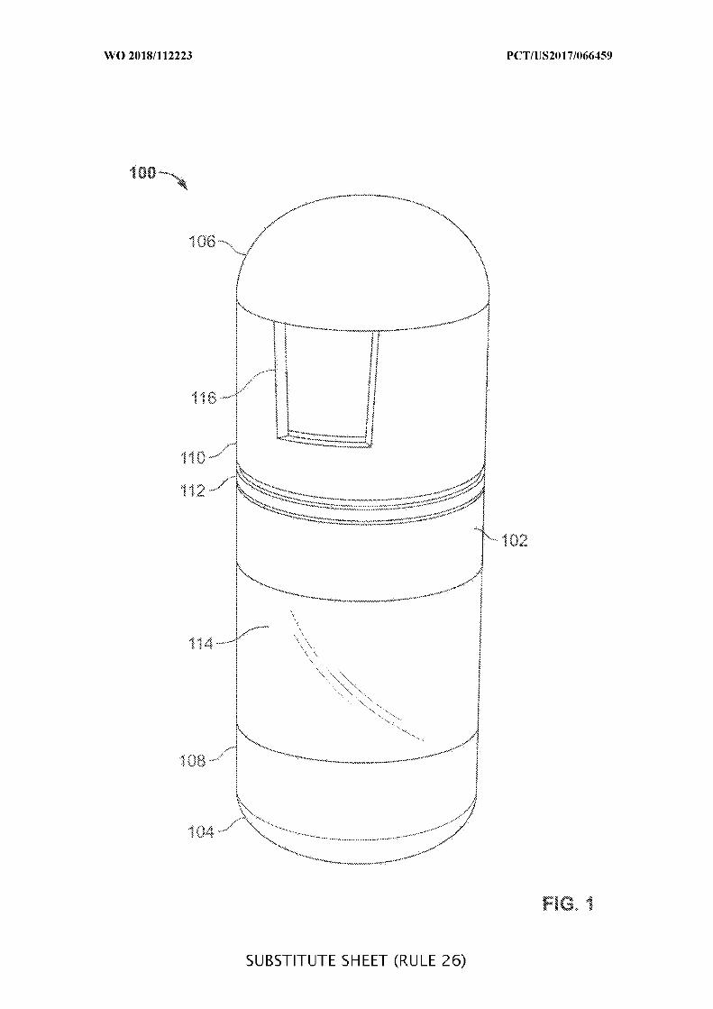
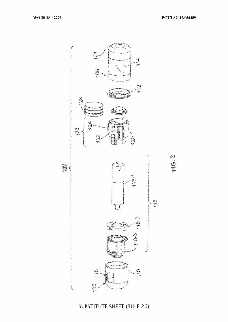
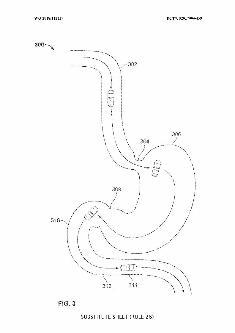
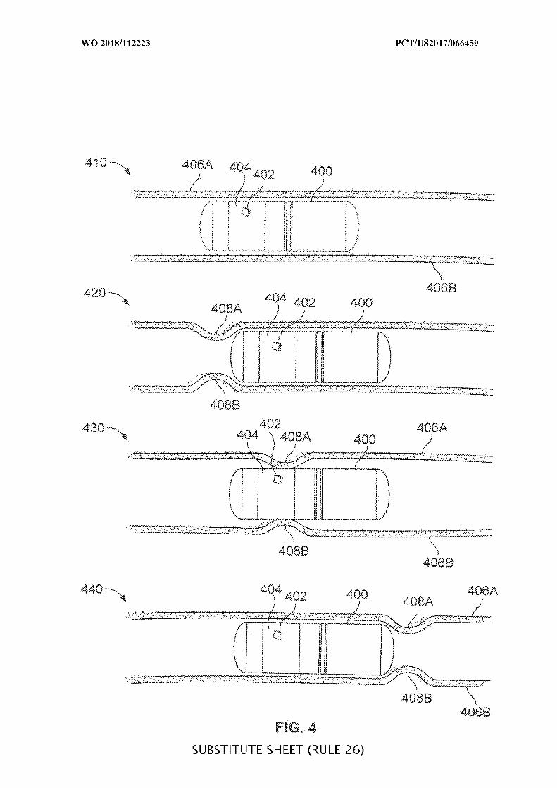
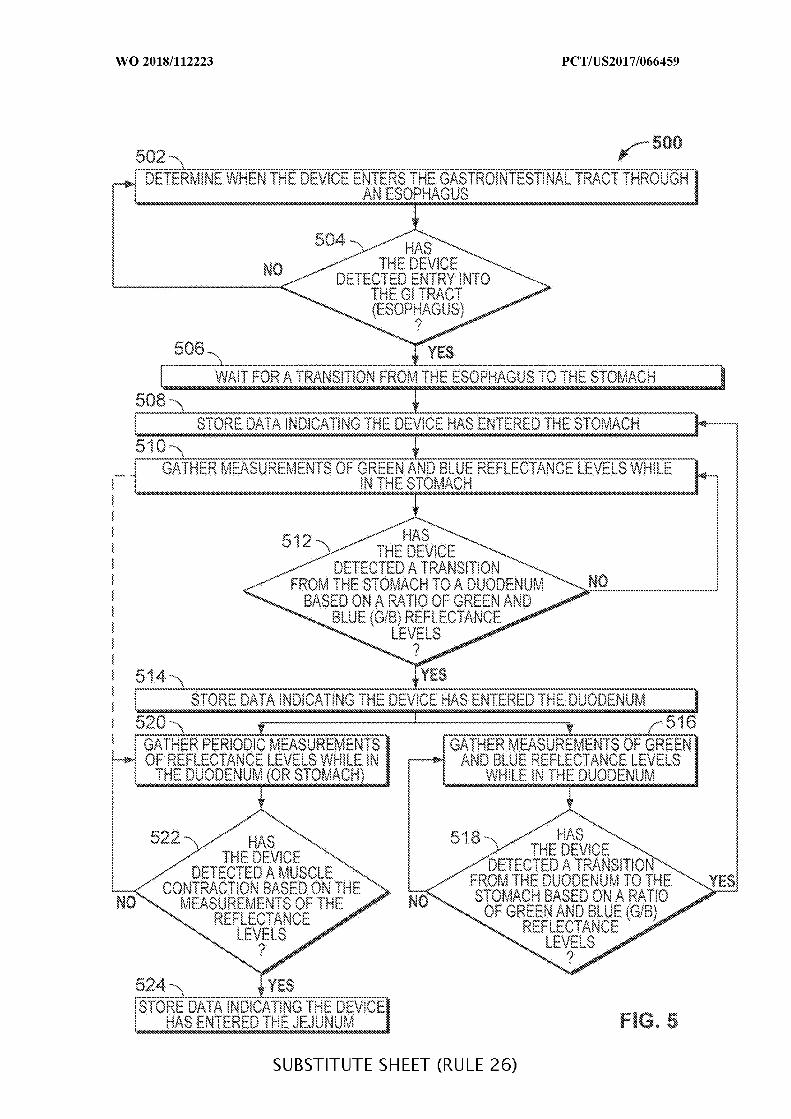
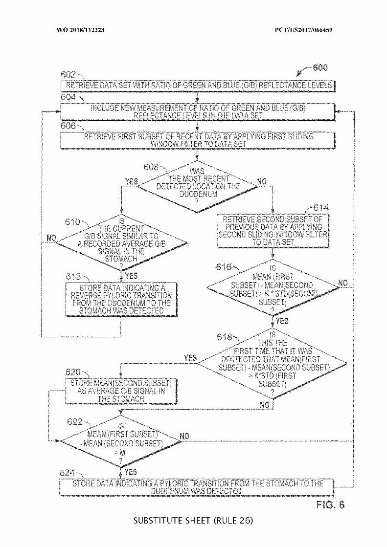
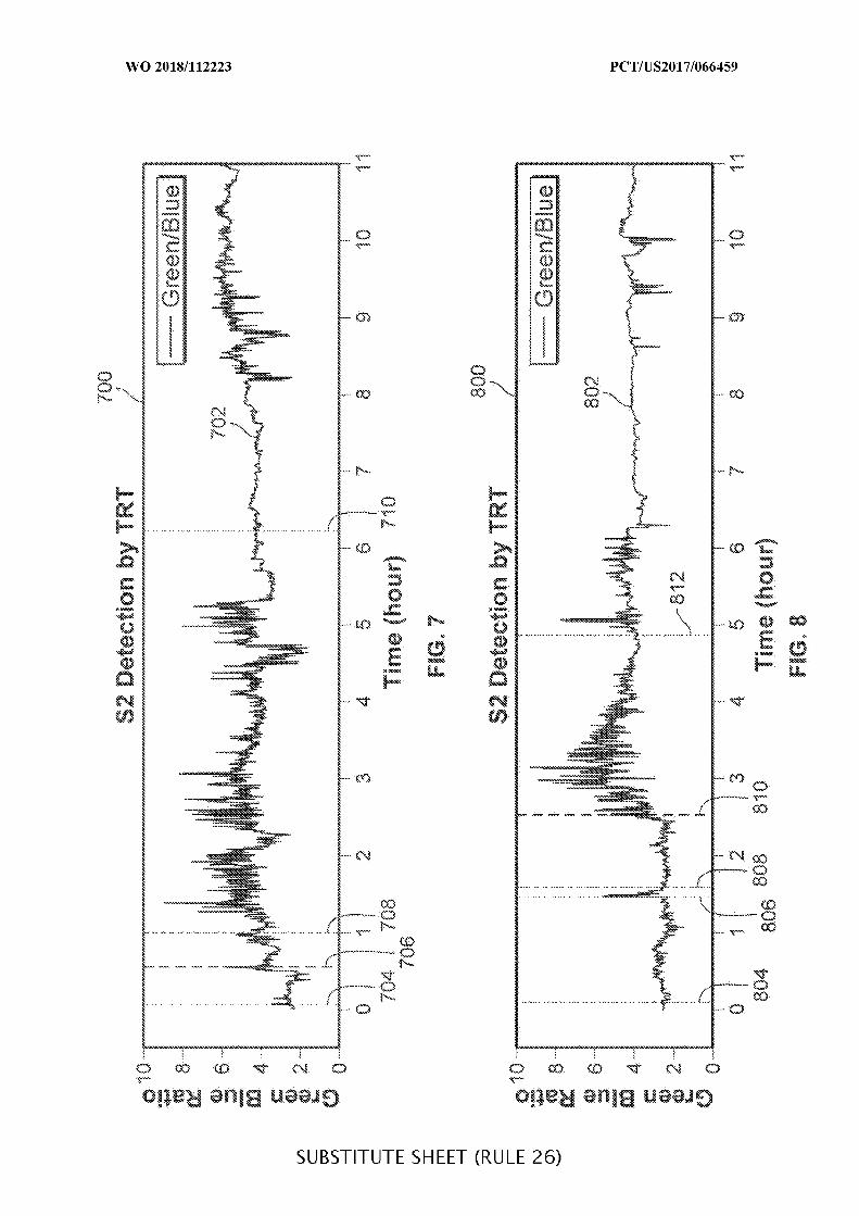

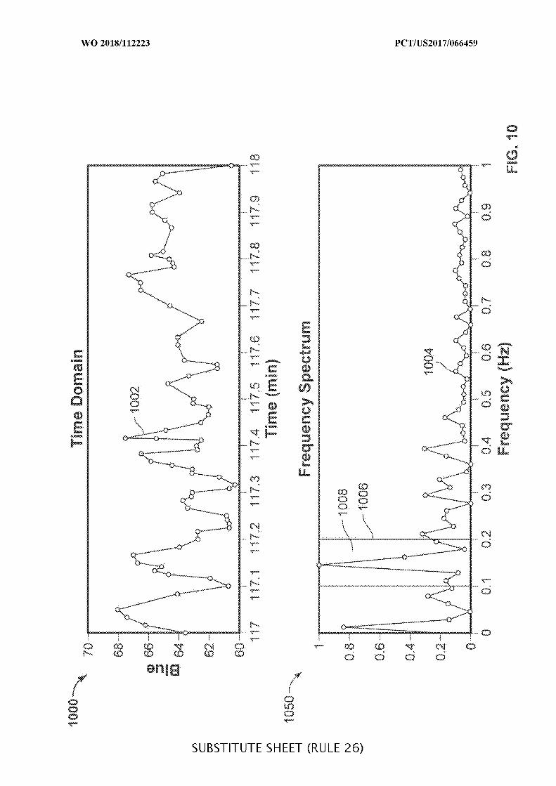
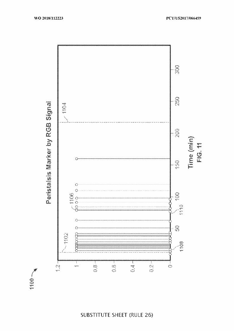
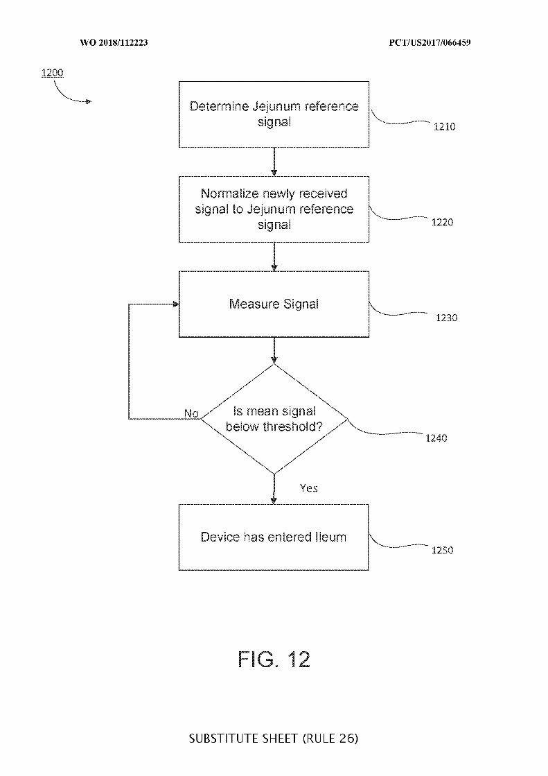
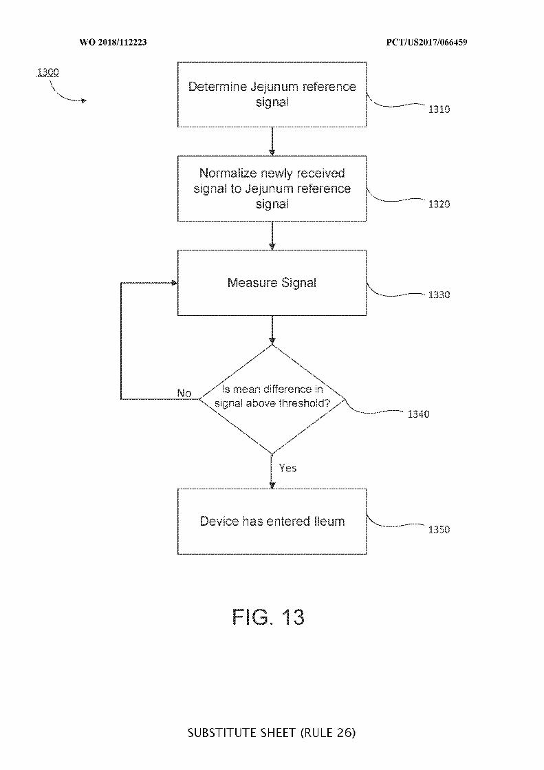
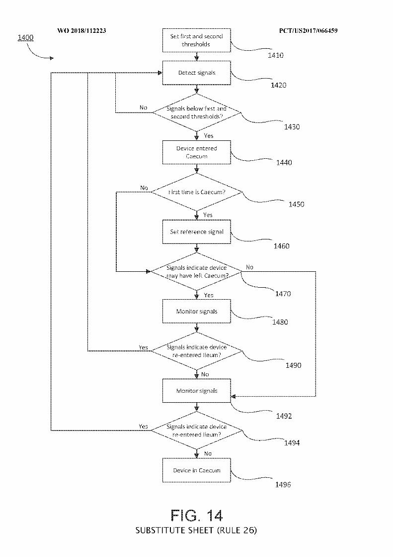
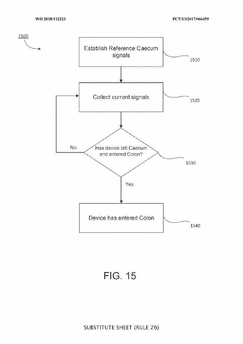
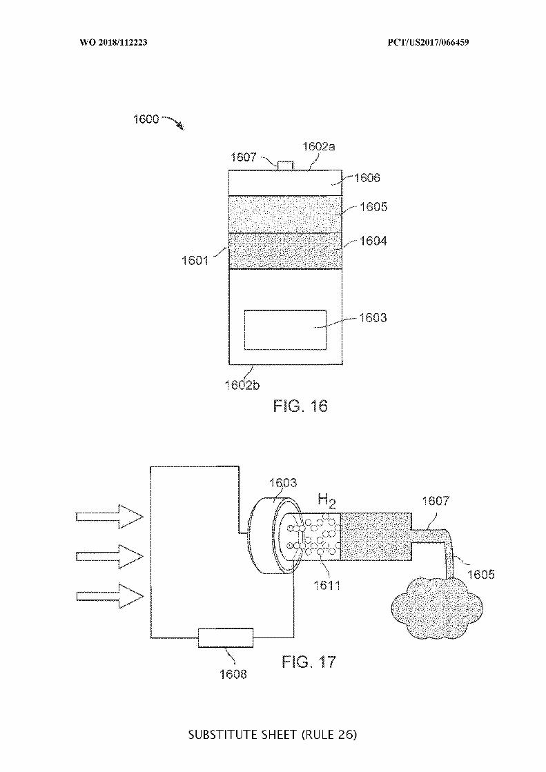
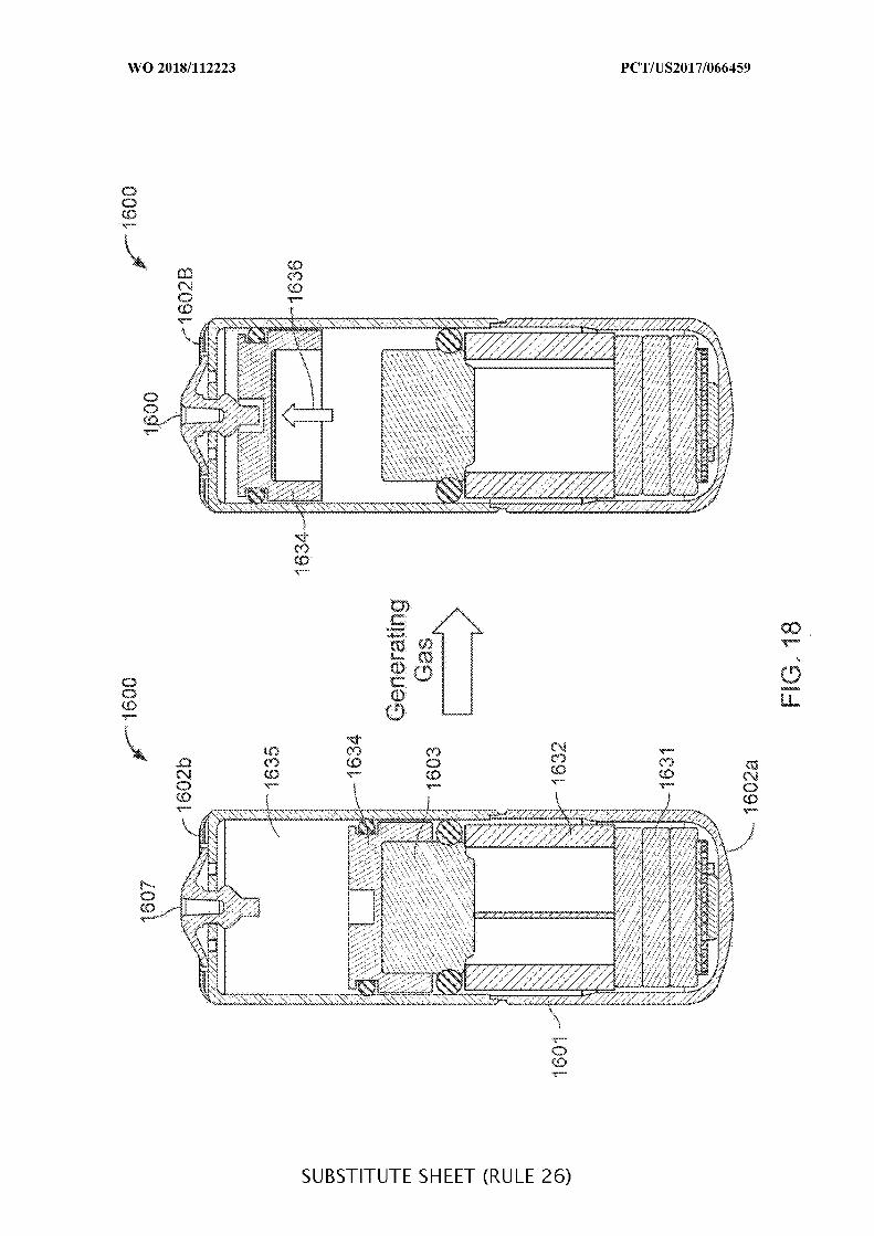
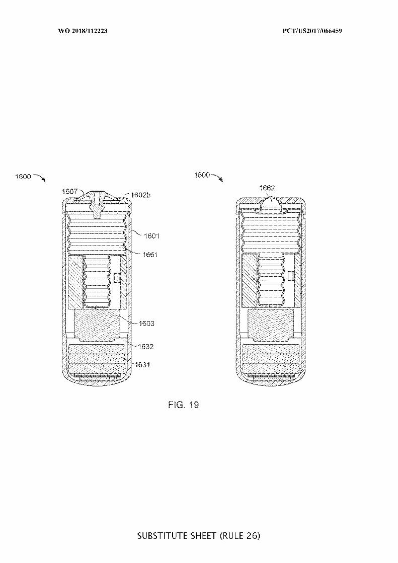
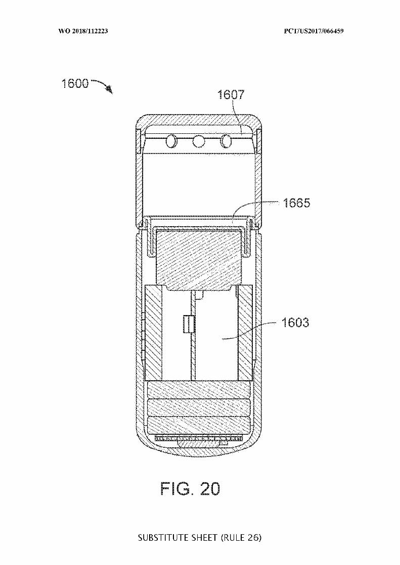
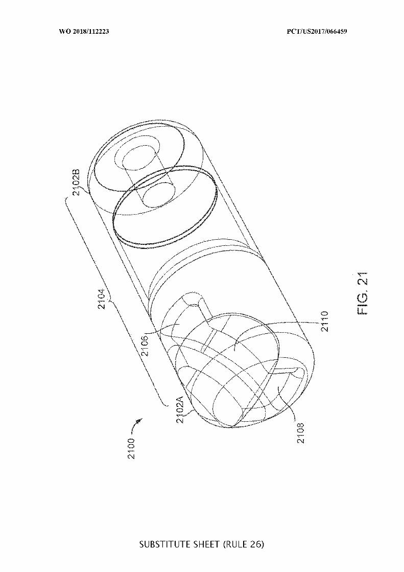
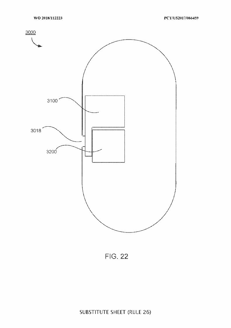
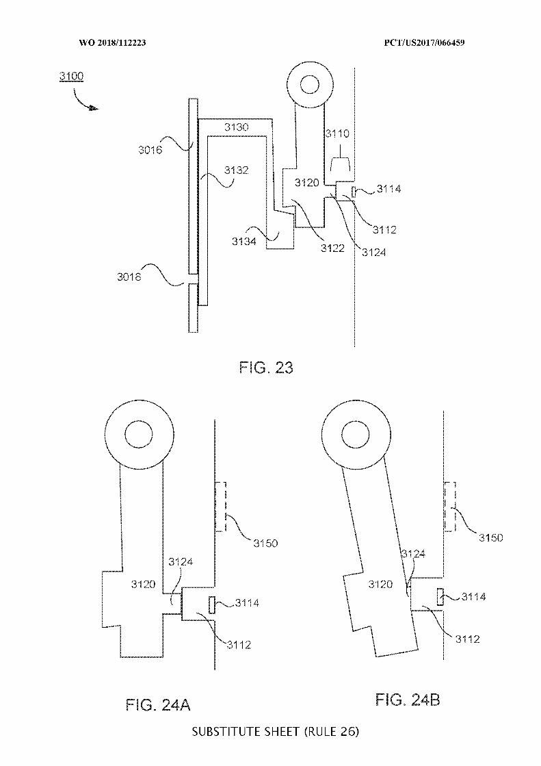
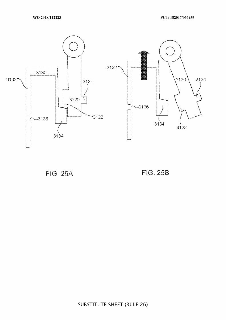
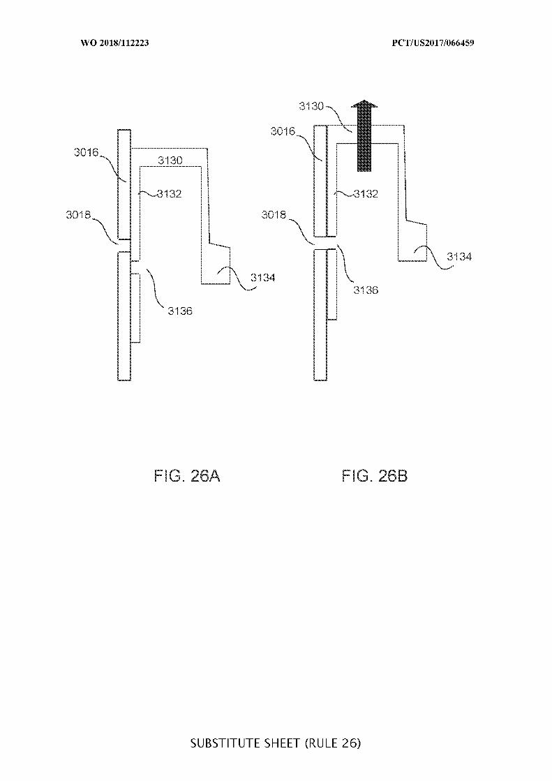
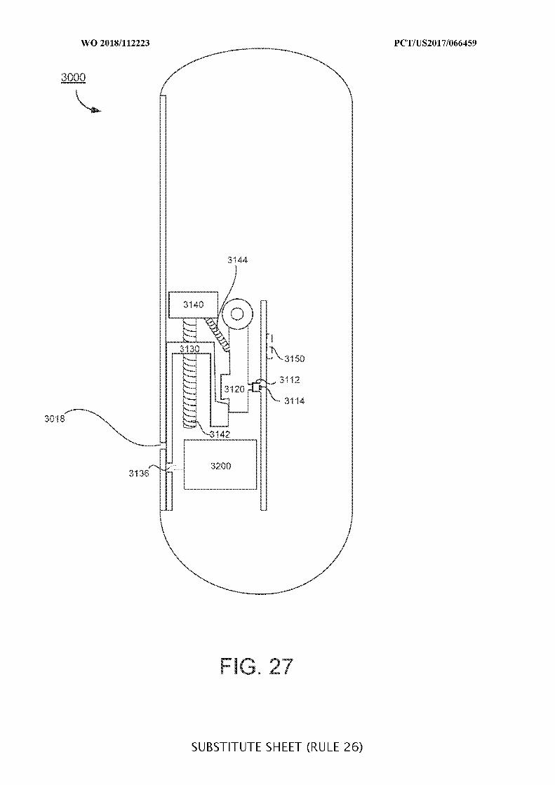

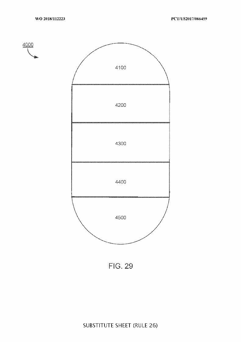
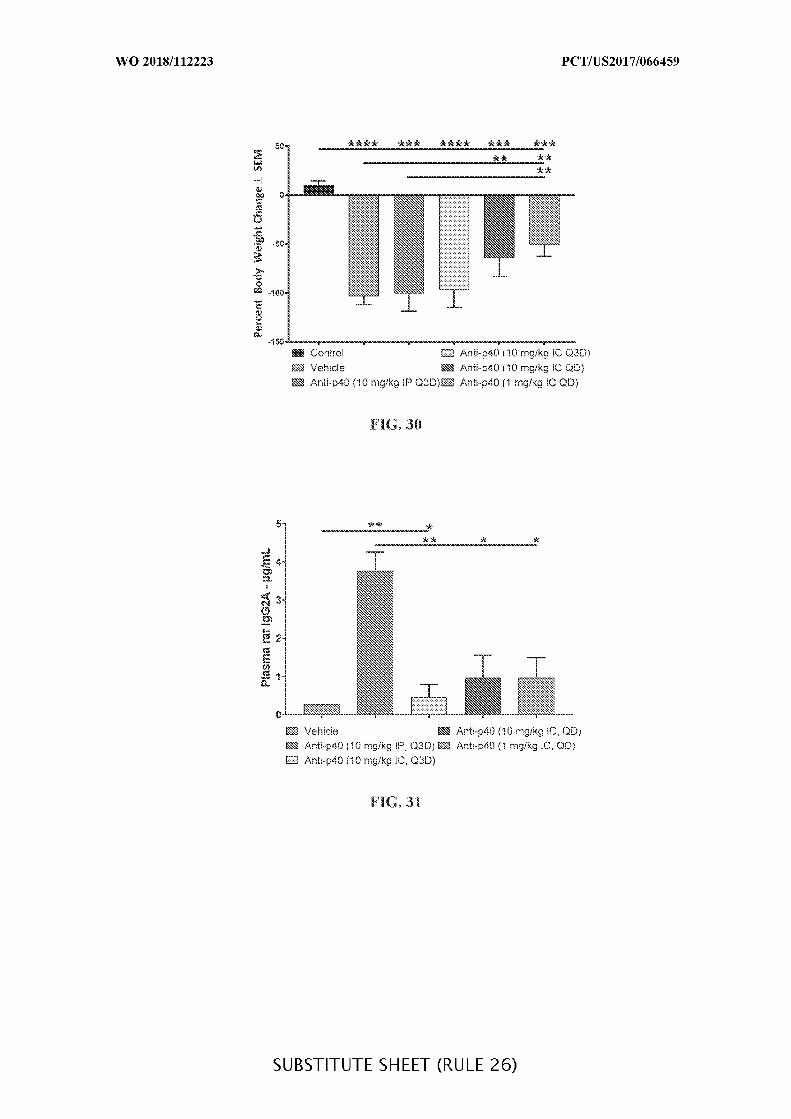
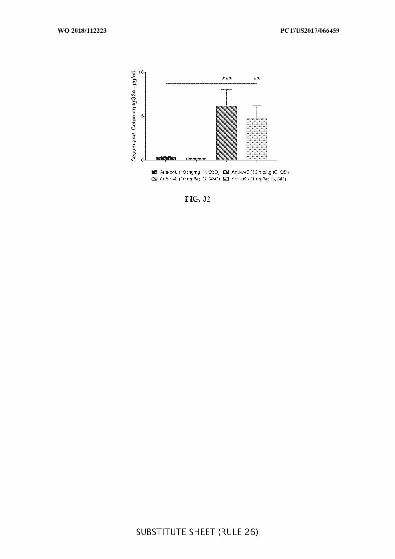
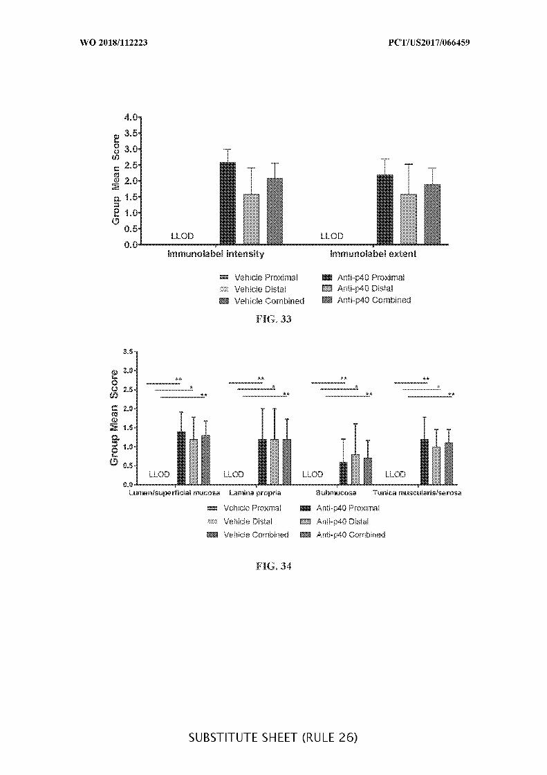
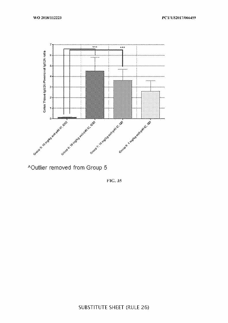
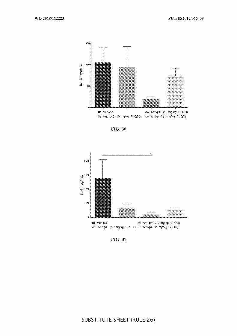
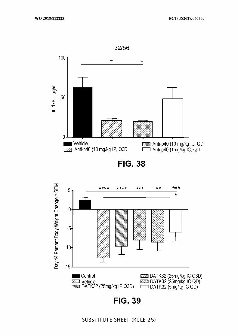
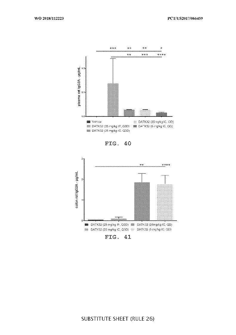
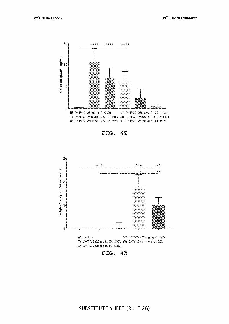
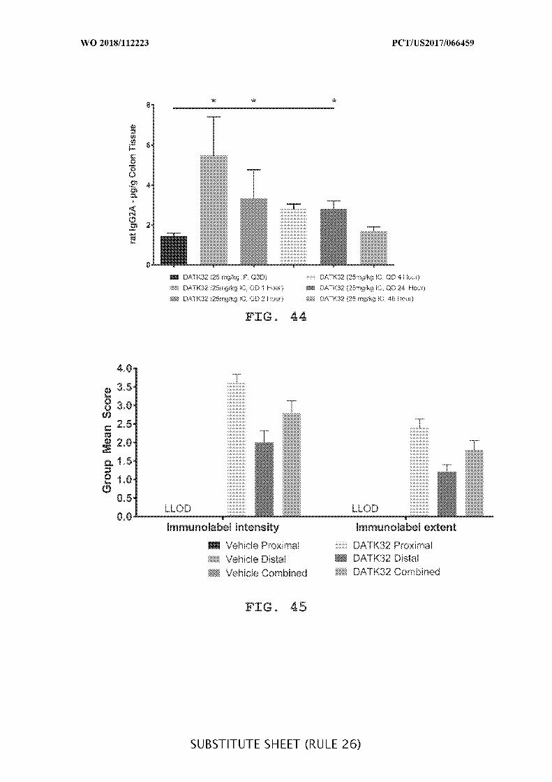
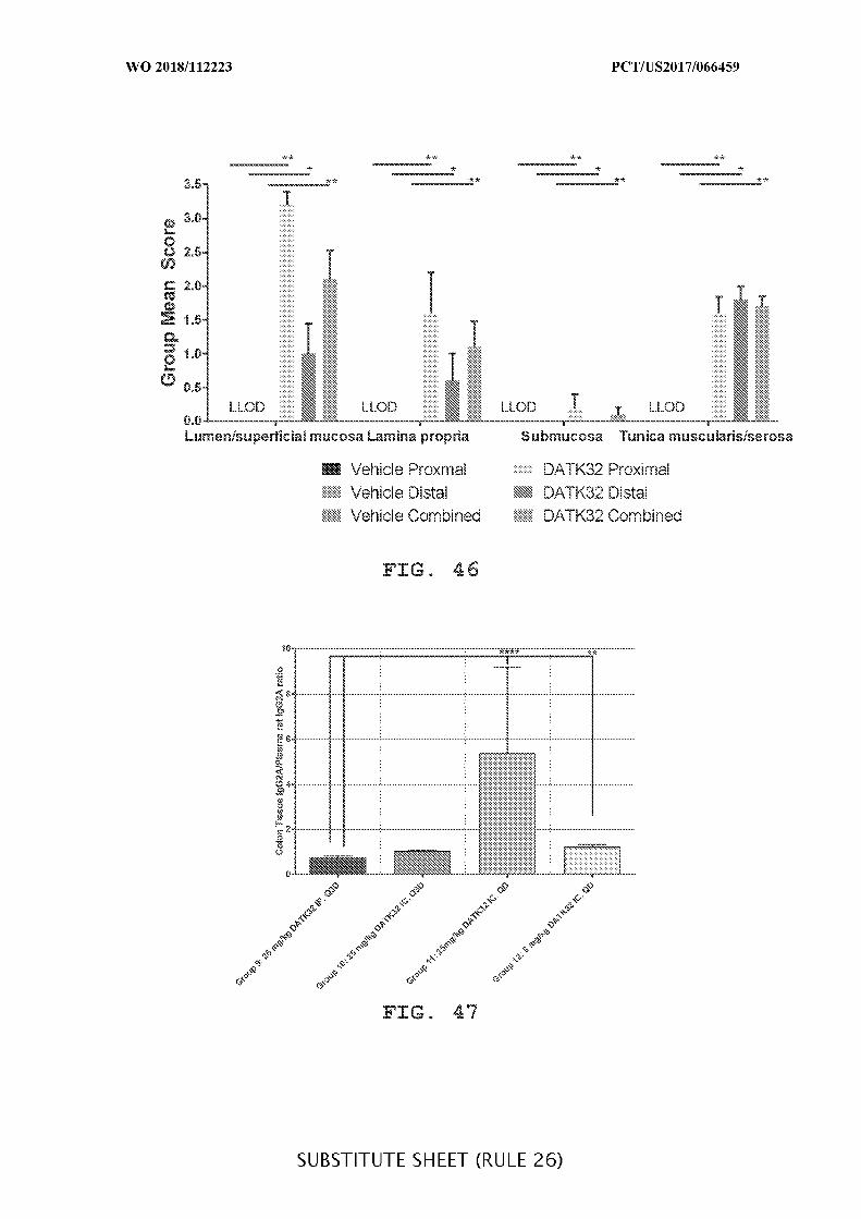
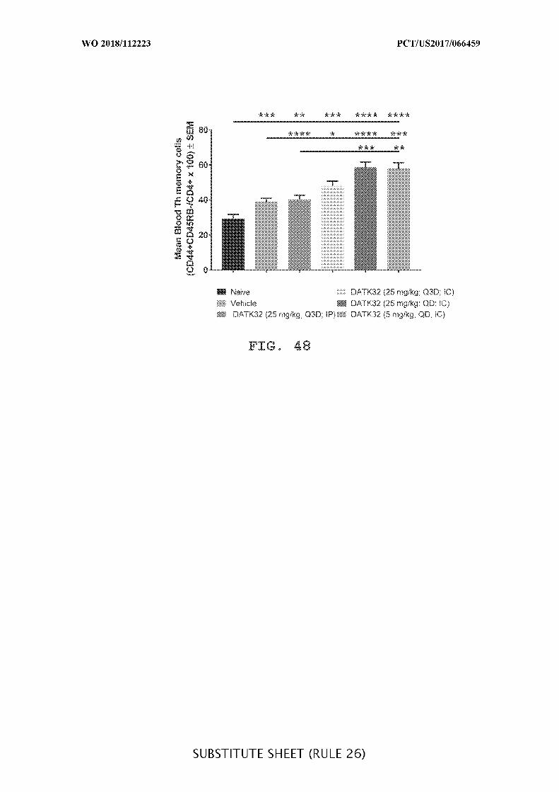
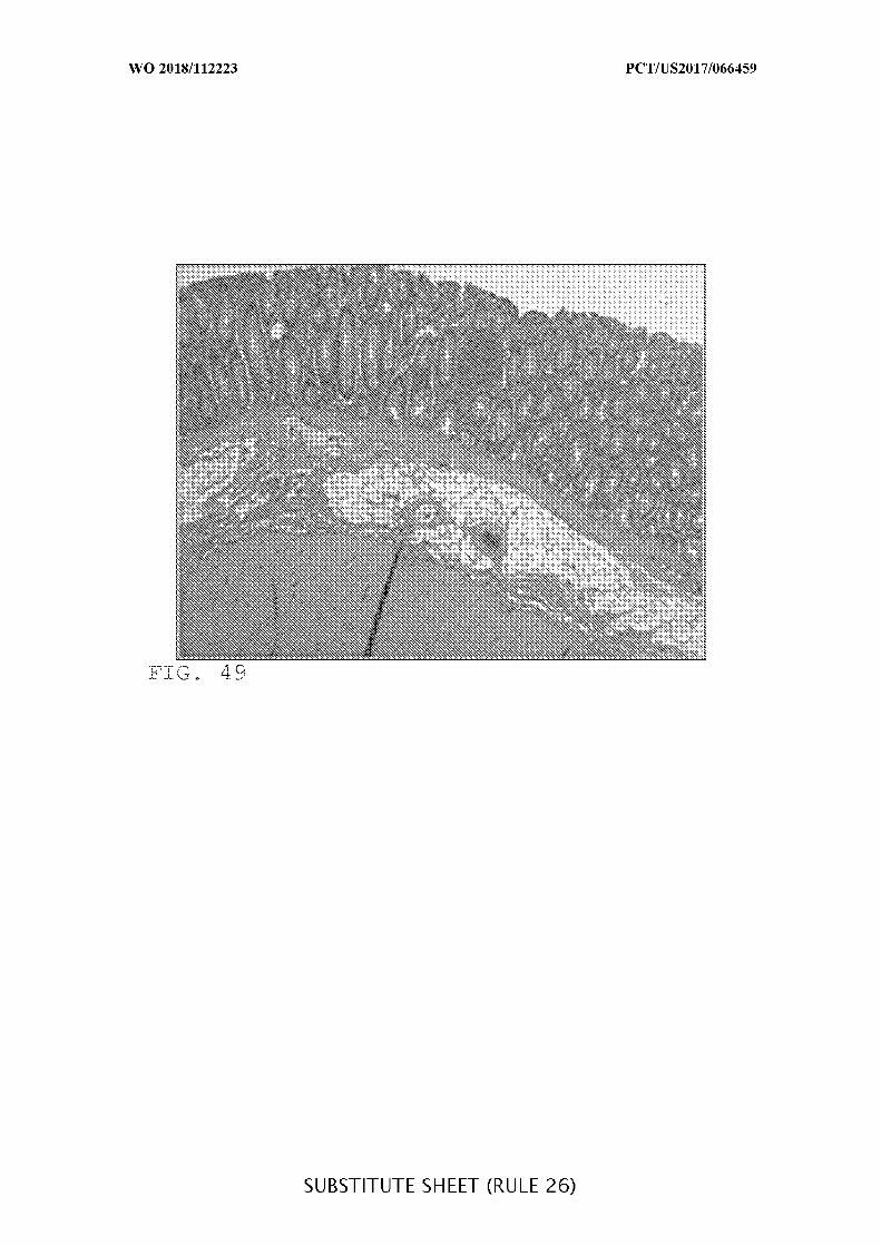
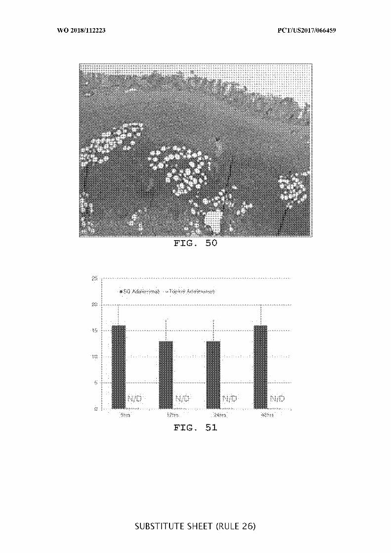
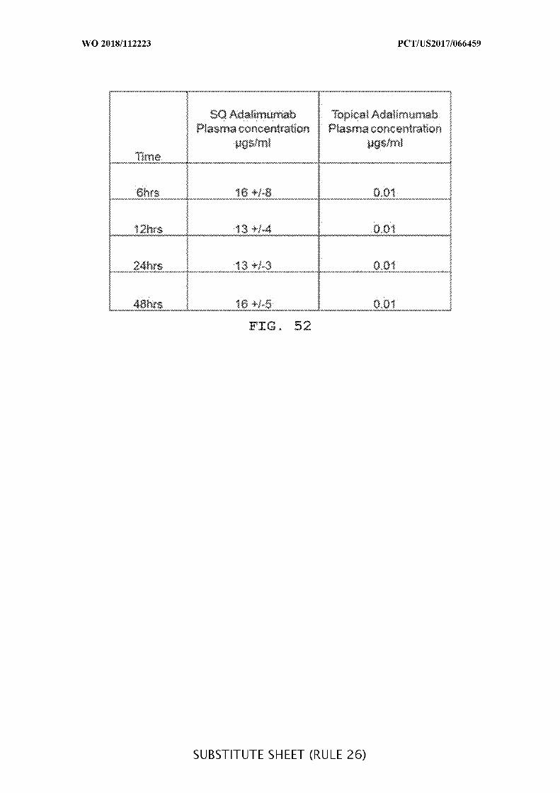
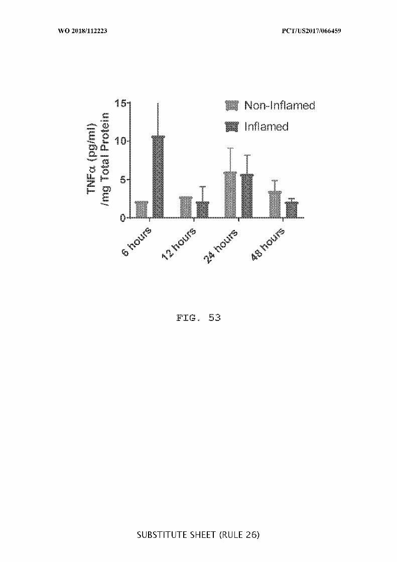
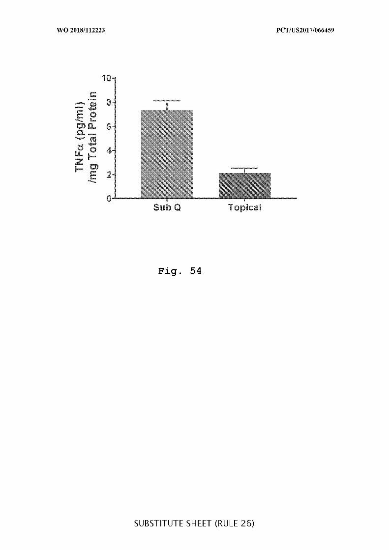
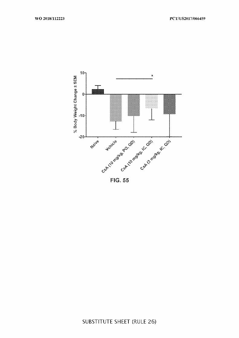
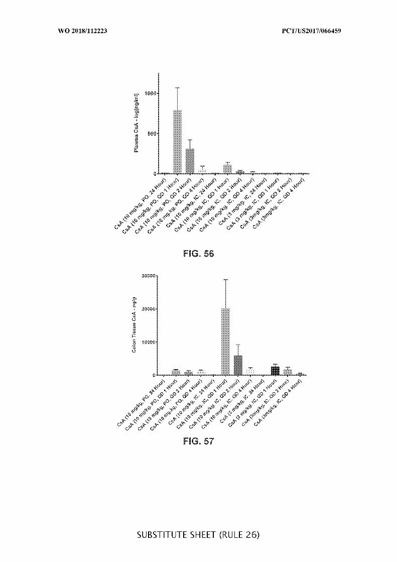
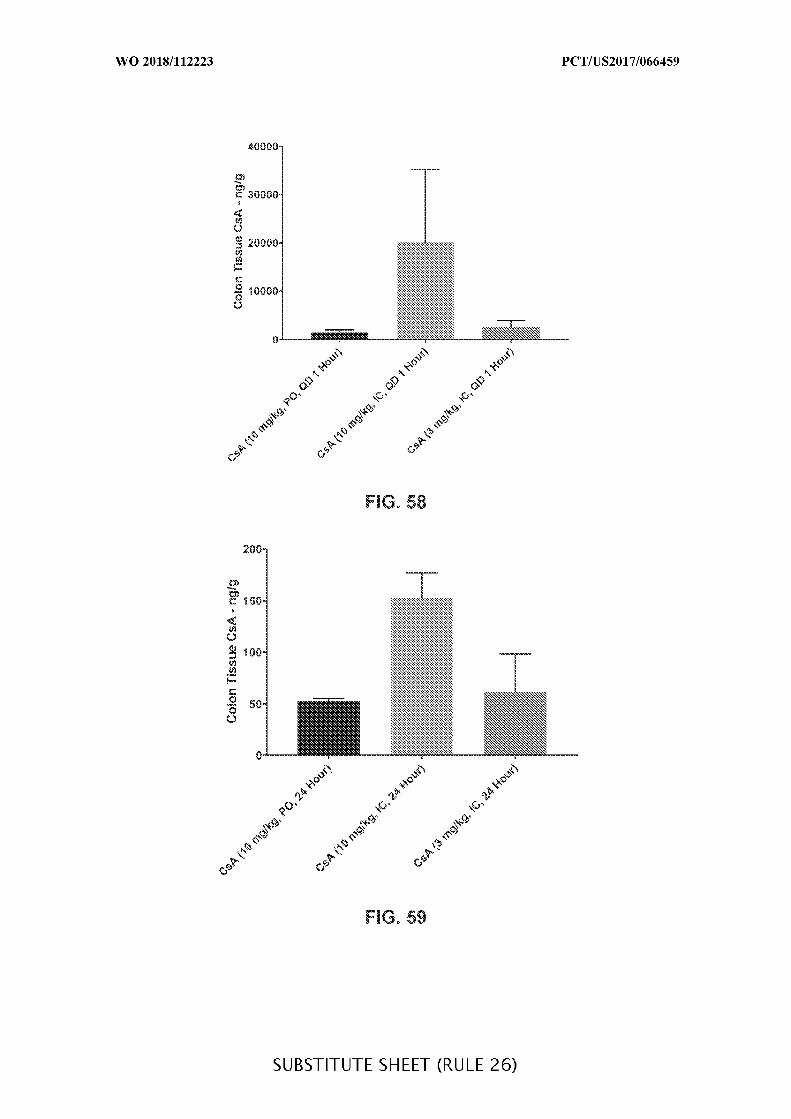
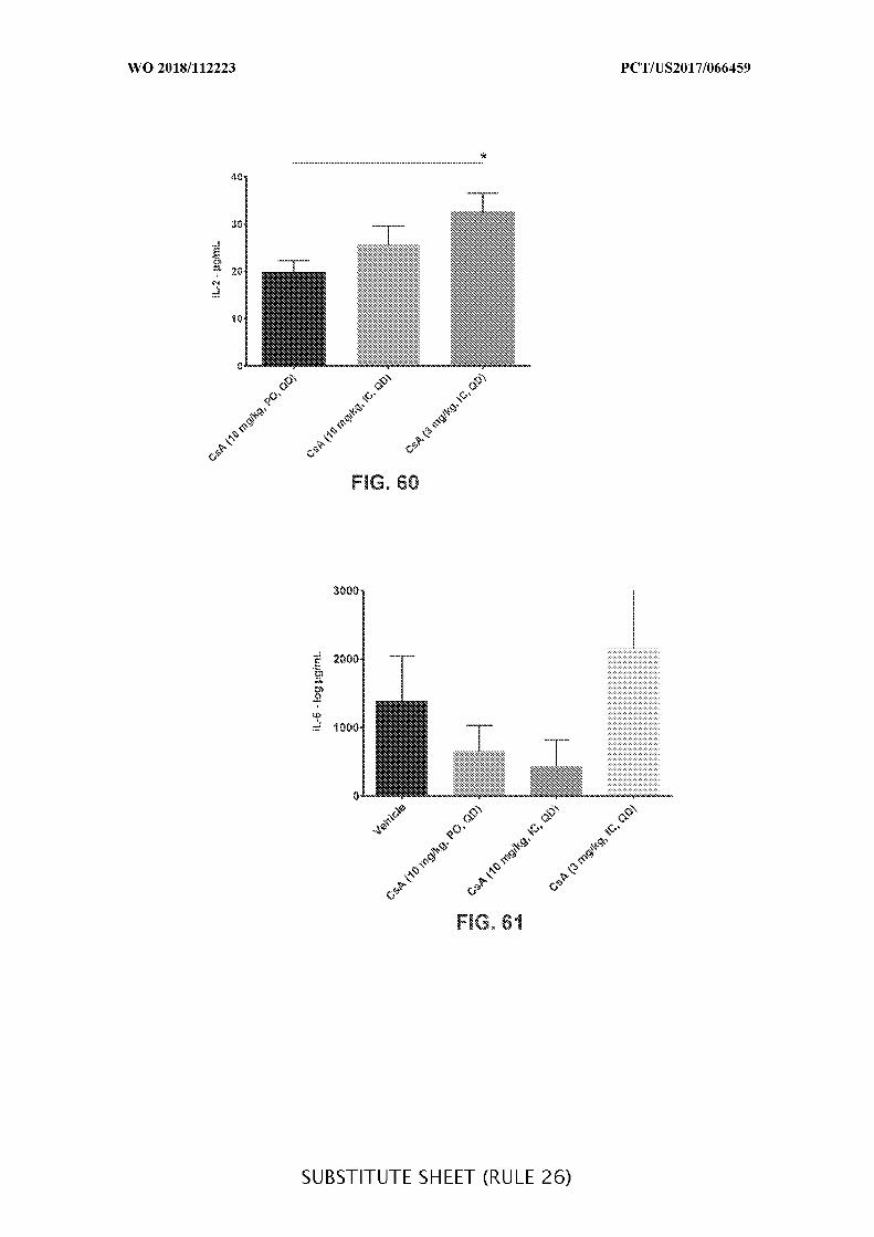
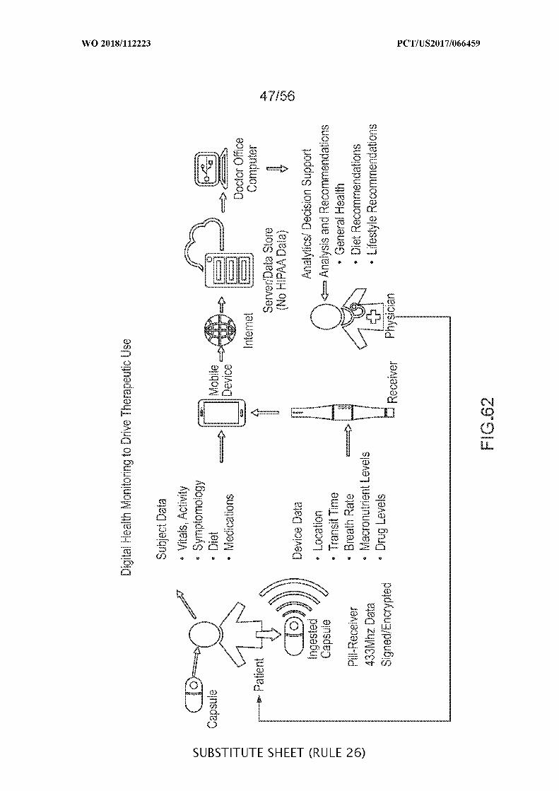
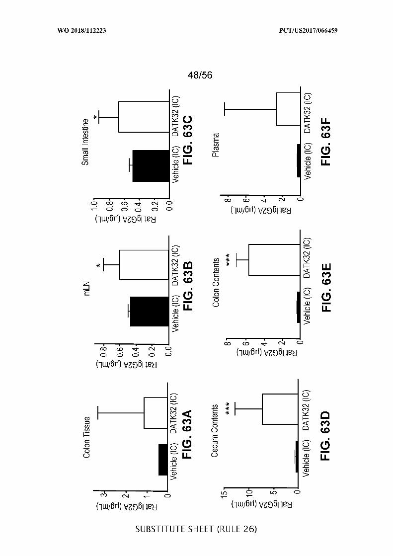
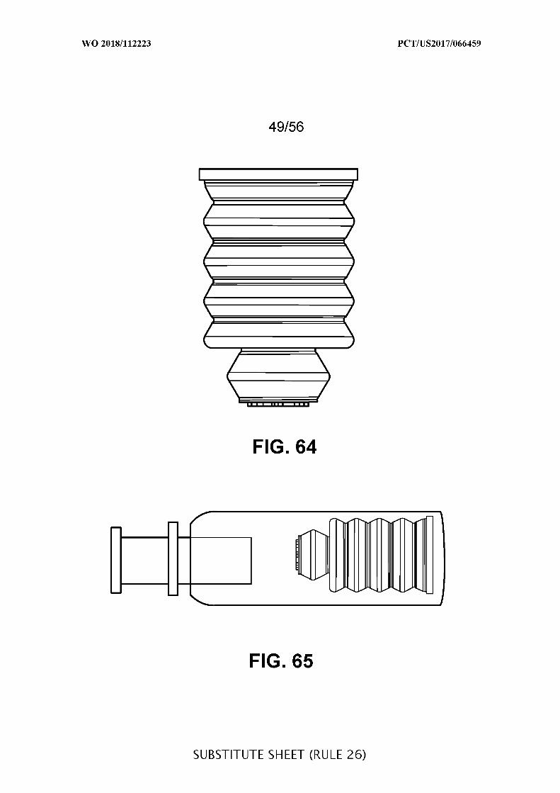
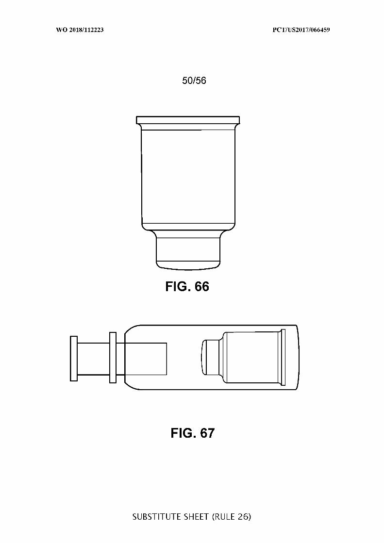
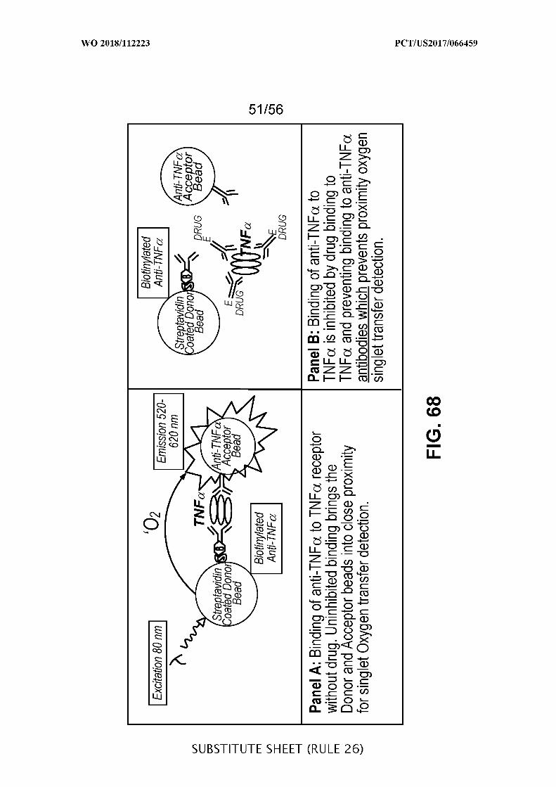
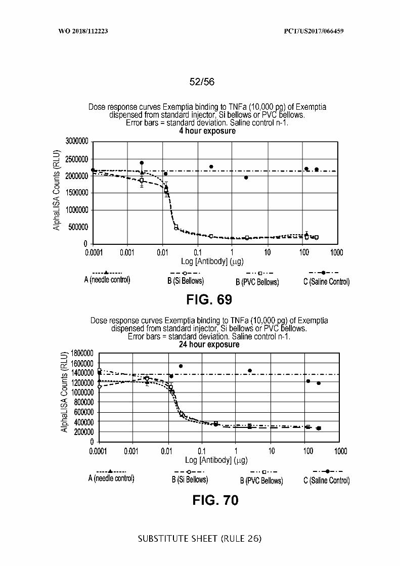
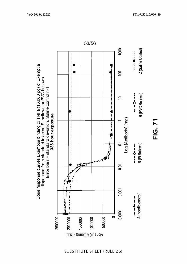
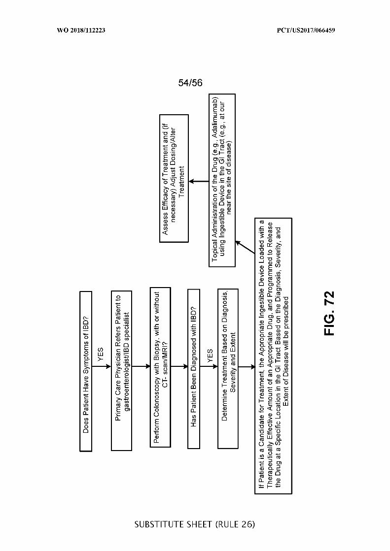
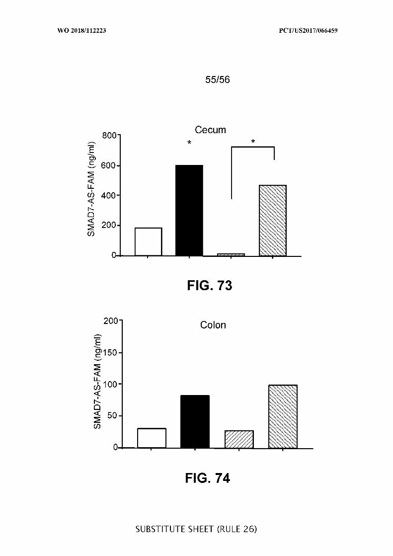
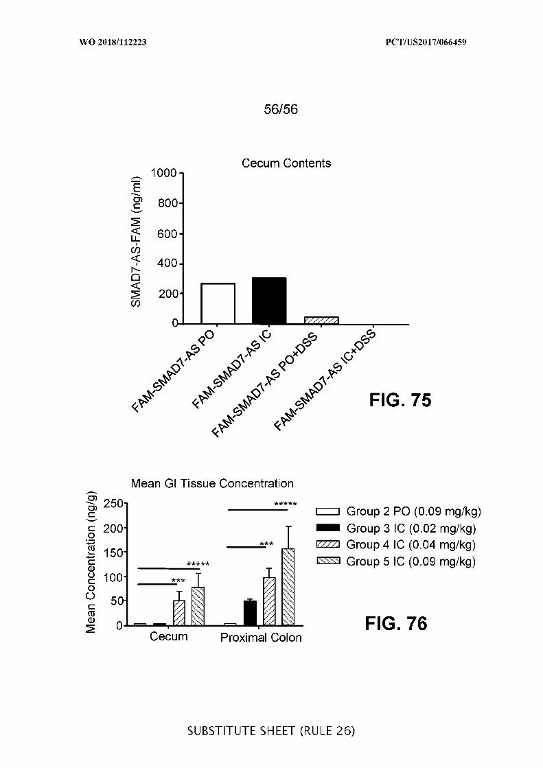
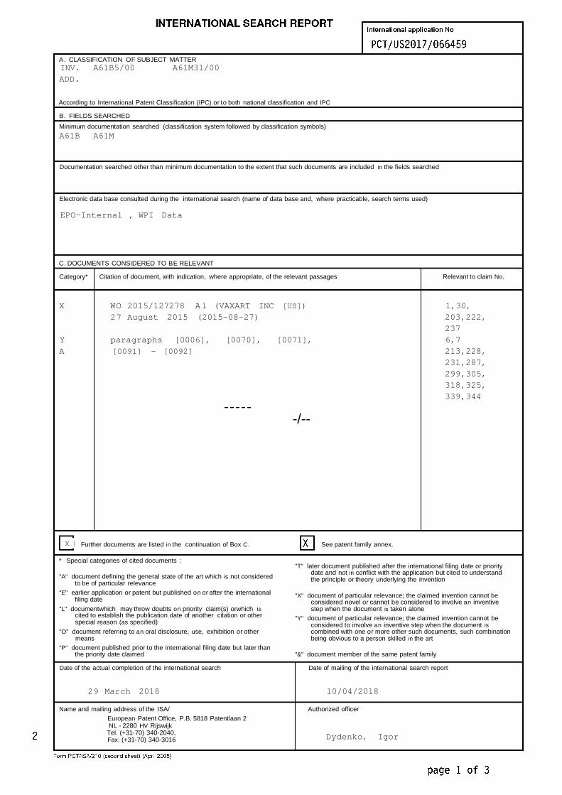
A. CLASSIFICATION OF SUBJECT MATTERINV. A61B5/00 A61M31/00ADD.
According to International Patent Classification (IPC) or to both national classification and IPC
B. FIELDS SEARCHED
Minimum documentation searched (classification system followed by classification symbols)
A61B A61M
Documentation searched other than minimum documentation to the extent that such documents are included in the fields searched
Electronic data base consulted during the international search (name of data base and, where practicable, search terms used)
EPO-Internal , WPI Data
C. DOCUMENTS CONSIDERED TO BE RELEVANT
Category* Citation of document, with indication, where appropriate, of the relevant passages Relevant to claim No.
X WO 2015/127278 Al (VAXART INC [US]) 1,30,
27 August 2015 (2015-08-27) 203,222,237
Y paragraphs [0006], [0070], [0071], 6,7A [0091] - [0092] 213,228,
231,287,299,305,318,325,339,344
-/--
X | Further documents are listed in the continuation of Box C. See patent family annex.
* Special categories of cited documents :"T" later document published after the international filing date or priority
date and not in conflict with the application but cited to understand"A" document defining the general state of the art which is not considered the principle or theory underlying the invention
to be of particular relevance"E" earlier application or patent but published on or after the international "X" document of particular relevance; the claimed invention cannot be
filing date considered novel or cannot be considered to involve an inventive"L" documentwhich may throw doubts on priority claim(s) orwhich is step when the document is taken alone
cited to establish the publication date of another citation or other "Y" document of particular relevance; the claimed invention cannot bespecial reason (as specified) considered to involve an inventive step when the document is
"O" document referring to an oral disclosure, use, exhibition or other combined with one or more other such documents, such combinationmeans being obvious to a person skilled in the art
"P" document published prior to the international filing date but later thanthe priority date claimed "&" document member of the same patent family
Date of the actual completion of the international search Date of mailing of the international search report
29 March 2018 10/04/2018
Name and mailing address of the ISA/ Authorized officerEuropean Patent Office, P.B. 5818 Patentlaan 2NL - 2280 HV Rijswijk
Tel. (+31-70) 340-2040,Fax: (+31-70) 340-3016 Dydenko, Igor
C(Continuation). DOCUMENTS CONSIDERED TO BE RELEVANT
Category* Citation of document, with indication, where appropriate, of the relevant passages Relevant to claim No.
X US 2011/136897 Al (SOREQ HERMONA [IL] ET 1,30,
AL) 9 June 2011 (2011-06-09) 203,222,237
A paragraphs [0098] - [0142] 6,7,213,228,231,287,299,305,318,325,339,344
X US 2014/086849 Al (MCKENNA ELIZABETH [US]) 1,30,
27 March 2014 (2014-03-27) 203,222,237,269,284,285
Y paragraphs [0018], [0079], [0110], 213,228,[0178] 231,287,
299,305,318,325,339,344
A 6,7,230
X 0 2014/165823 Al (NUMEDII INC [US]) 1,30,
9 October 2014 (2014-10-09) 203,222,237,269,284,285
Y paragraphs [0080] - [0086], [0127] - 159,160,[0130] , [0135] , [0183] 213,228,
230,231,287,299,305,318,325,339,344
A 6,7
X W0 2007/148238 Al (K0NINKL PHILIPS 377ELECTRONICS NV [NL] ; SHIMIZU JEFF [US];
TR0VAT0 KAREN)27 December 2007 (2007-12-27)
Y pages 8-11 6,7,213,228,231,287,299,305,318,325,339,344
X US 2013/171247 Al (IMRAN MIR [US]) 3774 July 2013 (2013-07-04)
Y paragraphs [0092] - [0112] 213,228,231,287,299,305,318,325,339,344
A 6,7
-/--
C(Continuation). DOCUMENTS CONSIDERED TO BE RELEVANT
Category* Citation of document, with indication, where appropriate, of the relevant passages Relevant to claim No.
X VAN DER SCHAAR P J ET AL: "A novel 358,377i ngesti b l e el ectroni c drug del i very andmoni tori ng devi ce" ,GASTROINTEST. ENDOSC, ,vol . 78, no. 3 ,1 January 2013 (2013-01-01) , pages520-528, XP002778209 ,
Y pages 521-522 230A 6,7
Y W0 2015/038973 Al ( FRACTYL LAB INC [US] ) 159 , 16019 March 2015 (2015-03-19)paragraphs [0105] - [0108] , [0189] -[0198]

INTERNATIONAL SEARCH REPORT
Box No. II Observations where certain claims were found unsearchable (Continuation of item 2 of first sheet)
This international search report has not been established in respect of certain claims under Article 17(2)(a) for the following reasons:
□ Claims Nos.:because they relate to subject matter not required to be searched by this Authority, namely:
Claims Nos.:because they relate to parts of the international application that do not comply with the prescribed requirements to suchan extent that no meaningful international search can be carried out, specifically:
see FURTHER INFORMATION sheet PCT/ISA/210
3 . □I I Claims Nos.:because they are dependent claims and are not drafted in accordance with the second and third sentences of Rule 6.4(a).
Box No. Ill Observations where unity of invention is lacking (Continuation of item 3 of first sheet)
This International Searching Authority found multiple inventions in this international application, as follows:
As all required additional search fees were timely paid by the applicant, this international search report covers all searchable— ciaims.
As all searchable claims could be searched without effort justifying an additional fees, this Authority did not invite payment ofadditional fees.
As only some of the required additional search fees were timely paid by the applicant, this international search report covers' ' only those claims for which fees were paid, specifically claims Nos. :
4 . I I No required additional search fees were timely paid by the applicant. Consequently, this international search report isrestricted to the invention first mentioned in the claims; it is covered by claims Nos. :
Remark on Protest The additional search fees were accompanied by the applicant's protest and, where applicable, the' ' payment of a protest fee.
The additional search fees were accompanied by the applicant's protest but the applicable protest' ' fee was not paid within the time limit specified in the invitation.
I INo protest accompanied the payment of additional search fees.
Form PCT/ISA/21 0 (continuation of first sheet (2)) (April 2005)
International Application No. PCT/ US2017/ 066459
FURTHER INFORMATION CONTINUED FROM PCT/ISA/ 210
Thi s Internati onal Searchi ng Authori t y found mul t i pl e (groups of)i nventi ons i n thi s i nternati onal appl i cati on , as fol l ows :
International Application No. PCT/ US2017/ 066459
FURTHER INFORMATION CONTINUED FROM PCT/ISA/ 210
Conti nuati on of Box I I .
Cl aims Nos . : 2-5 , 8-29 , 31-158, 161-202 , 204-212 , 214-221 , 223-227 , 229 ,232-236, 238-268, 270-283 , 286, 288-298, 300-304, 306-317 , 319-324,326-338, 340-343 , 345-357 , 359-376, 378-383Wi t h respect t o the i ngesti bl e devi ce, the di scl osure i s i nsuffi c i ent.The data show only how t o detect the macroscopi c l ocati on i n the vari oussecti ons of the gut, but not i n i t s subsecti ons l et al one the preci sel ocati on of the l esi ons . Moreover, mere detecti on of the sui tabl el ocati on of del i very does not i pso facto mean that the devi ce " knows"when t o rel ease the medi ati on . Such features are descri bed only i ngeneral terms , and not i n the l evel of detai l al l owi ng the ski l l edarti san t o construct the devi ce wi thout havi ng t o embark i nto anengi neeri ng project.
Second, i ssues of enabl ement asi de, the cl aims t othe use of the i ngesti bl e devi ce for the del i very of drugs i s at l eastcommensurate t o the contri buti on t o the art. The cl aims , however, strayfar from the fai r contri buti on t o the art. They encompass admi ni strati onof any drug that can be rel eased i n the gut by oral or endoscopi cadmi ni strati on , and further encompass vari ous modi f i ati ons of the devi ceas such , not t o menti on the desi gn of an enti rely di fferent i ngesti bl edevi ce, namely one wi t h the reservoi r attached t o the housi ng (and noti nternal t o the devi ce) , and the correspondi ng reservoi r as such .Thi rd,
there are 383 cl aims whi ch on f i l e , out of these, 25 are i ndependent ( 1,6 , 7, 30, 159 , 160, 203 , 213 , 222 , 228, 230, 231 , 237 , 269 , 284, 285 ,287 , 299 , 305 , 318, 325 , 339 , 344, 358, 377) . Cl early, the number ofcl aims i s unreasonabl e , parti cul arly gi ven the i ncremental nature of theactual i nventi on . See Rul e 6. 1(a) PCT.
Fourth , many of the i ndependentcl aims are i n the same category, wi t h overl appi ng subject-matter. Thedependent cl aims are awful l y i ntertwi ned, i n part repeti t i ve, t o theextent that i t i s nearly impossi bl e t o determi ne wi t h certai nty the truescope of protecti on . (See wri tten opi ni on for parti cul ars)
Fi fth , manycl aims so l ack di scl osure and/or techni cal features that i t i s impossi bl et o search them.
Si xth , many other cl aims are so vague and uncl ear thati t i s impossi bl e t o ascertai n the true scope or protecti on .The
Internati onal Search Authori t y f i nds that the set of cl aims on f i l e sol acks cl ari ty, support and di scl osure i n the sense of Arti c l es 5 and 6PCT, and so fai l s t o meet the requi rements of reasonabl eness i n the senseof Rul e 6. 1(a) PCT, that a compl ete search i s impossi bl e .
Pursuant t oArti c l e 17 (2) (a) ( i i ) the search i s l imi ted t o the i ndependent cl aims .
The appl i cant ' s attenti on i s drawn t o the fact that cl aims rel ati ng t oi nventi ons i n respect of whi ch no i nternati onal search report has beenestabl i shed need not be the subject of an i nternati onal prel imi naryexami nati on (Rul e 66. 1(e) PCT) . The appl i cant i s advi sed that the EP0pol i cy when acti ng as an Internati onal Prel imi nary Exami ni ng Authori t y i snormal l y not t o carry out a prel imi nary exami nati on on matter whi ch hasnot been searched. Thi s i s the case i rrespecti ve of whether or not the

International Application No. PCT/ US2017/ 066459
FURTHER INFORMATION CONTINUED FROM PCT/ISA/ 210
cl aims are amended fol l owi ng recei pt of the search report or duri ng anyChapter I I procedure. I f the appl i cati on proceeds i nto the regi onal phasebefore the EPO, the appl i cant i s remi nded that a search may be carri edout duri ng exami nati on before the EPO (see EPO Gui del i nes C-IV, 7.2) ,shoul d the probl ems whi ch l ed t o the Arti c l e 17 (2) decl arati on beovercome.
Patent document Publication Patent family Publicationcited in search report date member(s) date
O 2015127278 Al 27-08 -2015 AU 2015218769 Al 08-09-2016CA 2939581 Al 27-08-2015CN 106255508 A 21-12-2016EA 201691458 Al 28-02-2017EP 3107568 Al 28-12-2016
P 2017506265 A 02-03-2017KR 20170007726 A 20-01-2017SG 11201606814R A 29-09-2016US 2017224805 Al 10-08-2017WO 2015127278 Al 27-08-2015ZA 201605787 B 29-11-2017
us 2011136897 Al 09-06 -2011 US 2011136897 Al 09-06-2011WO 2010018583 Al 18-02-2010
us 2014086849 Al 27-03 -2014 BR 112015006264 A2 04-07-2017EP 2898073 Al 29-07-2015HK 1213017 Al 24-06-2016
P 2015535829 A 17-12-2015US 2014086849 Al 27-03-2014US 2017087247 Al 30-03-2017WO 2014047588 Al 27-03-2014
o 2014165823 Al 09-10 -2014 CN 105263482 A 20-01-2016EP 2983657 Al 17-02-2016
P 2016515631 A 30-05-2016US 2016303133 Al 20-10-2016WO 2014165823 Al 09-10-2014
wo 2007148238 Al 27-12 -2007 CN 101472639 A 01-07-2009EP 2035075 Al 18-03-2009J P 2009541298 A 26-11-2009RU 2009101475 A 27-07-2010US 2009275923 Al 05-11-2009WO 2007148238 Al 27-12-2007
us 2013171247 Al 04-07 -2013 US 2013171247 Al 04-07-2013US 2017174758 Al 22-06-2017
wo 2015038973 Al 19-03 -2015 EP 3043732 Al 20-07-2016US 2016354144 Al 08-12-2016WO 2015038973 Al 19-03-2015
Related Documents