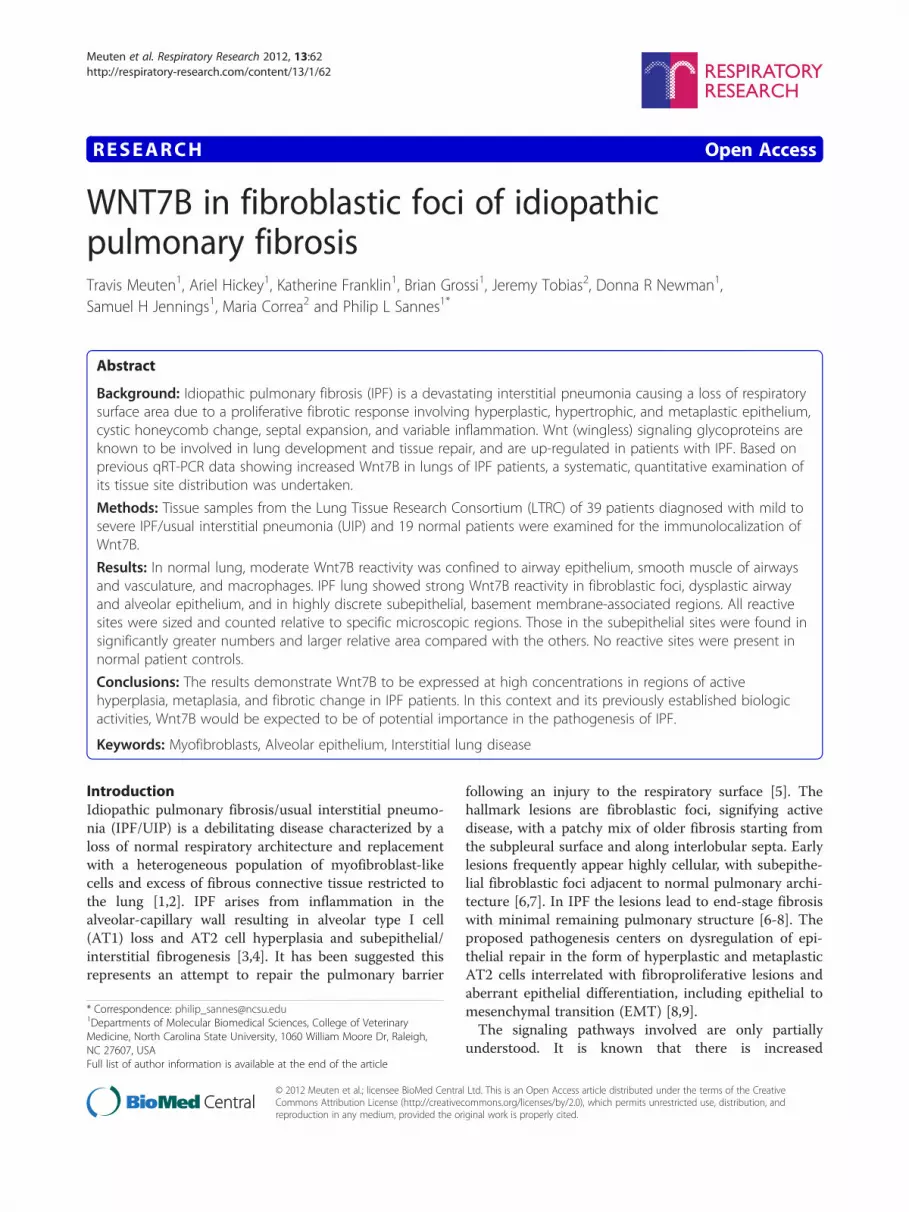
RESEARCH Open Access WNT7B in fibroblastic foci of idiopathic pulmonary fibrosis Travis Meuten 1 , Ariel Hickey 1 , Katherine Franklin 1 , Brian Grossi 1 , Jeremy Tobias 2 , Donna R Newman 1 , Samuel H Jennings 1 , Maria Correa 2 and Philip L Sannes 1* Abstract Background: Idiopathic pulmonary fibrosis (IPF) is a devastating interstitial pneumonia causing a loss of respiratory surface area due to a proliferative fibrotic response involving hyperplastic, hypertrophic, and metaplastic epithelium, cystic honeycomb change, septal expansion, and variable inflammation. Wnt (wingless) signaling glycoproteins are known to be involved in lung development and tissue repair, and are up-regulated in patients with IPF. Based on previous qRT-PCR data showing increased Wnt7B in lungs of IPF patients, a systematic, quantitative examination of its tissue site distribution was undertaken. Methods: Tissue samples from the Lung Tissue Research Consortium (LTRC) of 39 patients diagnosed with mild to severe IPF/usual interstitial pneumonia (UIP) and 19 normal patients were examined for the immunolocalization of Wnt7B. Results: In normal lung, moderate Wnt7B reactivity was confined to airway epithelium, smooth muscle of airways and vasculature, and macrophages. IPF lung showed strong Wnt7B reactivity in fibroblastic foci, dysplastic airway and alveolar epithelium, and in highly discrete subepithelial, basement membrane-associated regions. All reactive sites were sized and counted relative to specific microscopic regions. Those in the subepithelial sites were found in significantly greater numbers and larger relative area compared with the others. No reactive sites were present in normal patient controls. Conclusions: The results demonstrate Wnt7B to be expressed at high concentrations in regions of active hyperplasia, metaplasia, and fibrotic change in IPF patients. In this context and its previously established biologic activities, Wnt7B would be expected to be of potential importance in the pathogenesis of IPF. Keywords: Myofibroblasts, Alveolar epithelium, Interstitial lung disease Introduction Idiopathic pulmonary fibrosis/usual interstitial pneumo- nia (IPF/UIP) is a debilitating disease characterized by a loss of normal respiratory architecture and replacement with a heterogeneous population of myofibroblast-like cells and excess of fibrous connective tissue restricted to the lung [1,2]. IPF arises from inflammation in the alveolar-capillary wall resulting in alveolar type I cell (AT1) loss and AT2 cell hyperplasia and subepithelial/ interstitial fibrogenesis [3,4]. It has been suggested this represents an attempt to repair the pulmonary barrier following an injury to the respiratory surface [5]. The hallmark lesions are fibroblastic foci, signifying active disease, with a patchy mix of older fibrosis starting from the subpleural surface and along interlobular septa. Early lesions frequently appear highly cellular, with subepithe- lial fibroblastic foci adjacent to normal pulmonary archi- tecture [6,7]. In IPF the lesions lead to end-stage fibrosis with minimal remaining pulmonary structure [6-8]. The proposed pathogenesis centers on dysregulation of epi- thelial repair in the form of hyperplastic and metaplastic AT2 cells interrelated with fibroproliferative lesions and aberrant epithelial differentiation, including epithelial to mesenchymal transition (EMT) [8,9]. The signaling pathways involved are only partially understood. It is known that there is increased * Correspondence: [email protected] 1 Departments of Molecular Biomedical Sciences, College of Veterinary Medicine, North Carolina State University, 1060 William Moore Dr, Raleigh, NC 27607, USA Full list of author information is available at the end of the article © 2012 Meuten et al.; licensee BioMed Central Ltd. This is an Open Access article distributed under the terms of the Creative Commons Attribution License (http://creativecommons.org/licenses/by/2.0), which permits unrestricted use, distribution, and reproduction in any medium, provided the original work is properly cited. Meuten et al. Respiratory Research 2012, 13:62 http://respiratory-research.com/content/13/1/62

Welcome message from author
This document is posted to help you gain knowledge. Please leave a comment to let me know what you think about it! Share it to your friends and learn new things together.
Transcript
Meuten et al. Respiratory Research 2012, 13:62http://respiratory-research.com/content/13/1/62
RESEARCH Open Access
WNT7B in fibroblastic foci of idiopathicpulmonary fibrosisTravis Meuten1, Ariel Hickey1, Katherine Franklin1, Brian Grossi1, Jeremy Tobias2, Donna R Newman1,Samuel H Jennings1, Maria Correa2 and Philip L Sannes1*
Abstract
Background: Idiopathic pulmonary fibrosis (IPF) is a devastating interstitial pneumonia causing a loss of respiratorysurface area due to a proliferative fibrotic response involving hyperplastic, hypertrophic, and metaplastic epithelium,cystic honeycomb change, septal expansion, and variable inflammation. Wnt (wingless) signaling glycoproteins areknown to be involved in lung development and tissue repair, and are up-regulated in patients with IPF. Based onprevious qRT-PCR data showing increased Wnt7B in lungs of IPF patients, a systematic, quantitative examination ofits tissue site distribution was undertaken.
Methods: Tissue samples from the Lung Tissue Research Consortium (LTRC) of 39 patients diagnosed with mild tosevere IPF/usual interstitial pneumonia (UIP) and 19 normal patients were examined for the immunolocalization ofWnt7B.
Results: In normal lung, moderate Wnt7B reactivity was confined to airway epithelium, smooth muscle of airwaysand vasculature, and macrophages. IPF lung showed strong Wnt7B reactivity in fibroblastic foci, dysplastic airwayand alveolar epithelium, and in highly discrete subepithelial, basement membrane-associated regions. All reactivesites were sized and counted relative to specific microscopic regions. Those in the subepithelial sites were found insignificantly greater numbers and larger relative area compared with the others. No reactive sites were present innormal patient controls.
Conclusions: The results demonstrate Wnt7B to be expressed at high concentrations in regions of activehyperplasia, metaplasia, and fibrotic change in IPF patients. In this context and its previously established biologicactivities, Wnt7B would be expected to be of potential importance in the pathogenesis of IPF.
Keywords: Myofibroblasts, Alveolar epithelium, Interstitial lung disease
IntroductionIdiopathic pulmonary fibrosis/usual interstitial pneumo-nia (IPF/UIP) is a debilitating disease characterized by aloss of normal respiratory architecture and replacementwith a heterogeneous population of myofibroblast-likecells and excess of fibrous connective tissue restricted tothe lung [1,2]. IPF arises from inflammation in thealveolar-capillary wall resulting in alveolar type I cell(AT1) loss and AT2 cell hyperplasia and subepithelial/interstitial fibrogenesis [3,4]. It has been suggested thisrepresents an attempt to repair the pulmonary barrier
* Correspondence: [email protected] of Molecular Biomedical Sciences, College of VeterinaryMedicine, North Carolina State University, 1060 William Moore Dr, Raleigh,NC 27607, USAFull list of author information is available at the end of the article
© 2012 Meuten et al.; licensee BioMed CentraCommons Attribution License (http://creativecreproduction in any medium, provided the or
following an injury to the respiratory surface [5]. Thehallmark lesions are fibroblastic foci, signifying activedisease, with a patchy mix of older fibrosis starting fromthe subpleural surface and along interlobular septa. Earlylesions frequently appear highly cellular, with subepithe-lial fibroblastic foci adjacent to normal pulmonary archi-tecture [6,7]. In IPF the lesions lead to end-stage fibrosiswith minimal remaining pulmonary structure [6-8]. Theproposed pathogenesis centers on dysregulation of epi-thelial repair in the form of hyperplastic and metaplasticAT2 cells interrelated with fibroproliferative lesions andaberrant epithelial differentiation, including epithelial tomesenchymal transition (EMT) [8,9].The signaling pathways involved are only partially
understood. It is known that there is increased
l Ltd. This is an Open Access article distributed under the terms of the Creativeommons.org/licenses/by/2.0), which permits unrestricted use, distribution, andiginal work is properly cited.

Meuten et al. Respiratory Research 2012, 13:62 Page 2 of 10http://respiratory-research.com/content/13/1/62
expression of TGF-β and α-smooth muscle actin(α-SMA) in progressive lesions of IPF reflecting the tran-sition of fibroblasts to myofibroblasts with proliferationand collagen maintenance modulated by wingless (Wnt)glycoproteins [10-12]. Mouse models indicate cooper-ation of TGF-β and Wnt signaling pathways in develop-ment, differentiation, and EMT [13]. Altered expressionof Wnt ligands and one of their downstream targets, β-catenin, is evident in early IPF and bleomycin models ofpulmonary fibrosis [14-16]. This can be explained inlight of up-regulated TGF-β by its induction of LEF-1,which is a component of the canonical Wnt signalingpathway [13]. Chilosi, et al., found highly concentratedsites of β-catenin in myofibroblast-populated regions ad-jacent to airways [14]. This correlated with elevatedmRNA expression and immunohistochemical reactivityof Wnts 1 & 3a in adjacent pulmonary epithelium in IPFpatients [17]. Recent evidence shows that Wnt3a can ac-tivate β-catenin-mediated signaling and induce EMT inlung epithelial cells [18]. The collective views suggestthat canonical Wnt signaling (β-catenin mediated) is up-regulated in fibrogenic conditions and may be causallyinvolved in IPF [16-19].Wnt glycoproteins have been found to be expressed at
low levels in the normal lung and may be associatedwith epithelial turnover [17,20]. For example, Wnt7BmRNA expression in isolated normal AT2 cells was evi-dent in low levels [17,21]. Wnt7b over-expression isthought to be a contributing factor to fibrogenesis in themurine kidney [22] and to be involved with procollagenproduction by lung fibroblasts [12]. Wnt7b was of spe-cial interest because of its role in mesenchymal prolifera-tion and vascular development in the lung [23]. Basedon supporting qRT-PCR data showing increased Wnt7Bin lungs of IPF patients [17], a systematic, examinationof its localization in a cohort of 39 IPF lungs was under-taken. Wnt7B expression was found in spindle cells andextracellular matrix of virtually all fibroblastic foci andin widely distributed, large numbers of discretely definedregions of the subepithelial basement membrane zone.These findings may provide useful clues relating to thepathogenesis of IPF and present novel, potential targetsfor detection and treatment.
Materials & methodsImmunostainingTissue blocks of formalin-fixed lung tissue samples wereobtained from the Lung Tissue Research Consortium(LTRC). The samples had previously been placed intothree groups (see Table 1): group 1, with forced vitalcapacities (FVCs) >80% (normal, or no specified majoror minor diagnosis, n = 19); group 2, with FVCs between50-80% [major final clinical diagnosis as interstitial lungdisease (ILD, n = 20) and minor final clinical diagnosis as
usual interstitial pneumonia (UIP)/idiopathic pulmonaryfibrosis (IPF)]; and group 3, with FVCs <50% (majorfinal clinical diagnosis of ILD and minor final clinicaldiagnosis as UIP/IPF, n = 19). No other patient identifierswere provided, and their anonymity and confidentialitywere preserved. The study was approved by the NorthCarolina State University Institutional Review Board.Blocks were further randomized and sections stainedwith hematoxylin and eosin (H&E) and picrosirius red(PSR) or Movat’s pentachrome stain for collagen. H&Esections were examined by a Board Certified Pathologistto independently confirm/reclassify initial clinicaldiagnoses.Sections were treated with citrate buffer for antigen
retrieval and treated with a polyclonal goat anti-Wnt7Bantibody [Santa Cruz Biotechnology, Inc. (sc26363, lot#I0205, Santa Cruz, CA)] at a 1:100 dilution overnightat 4°C, followed by peroxidase-labeled secondary anti-bodies (Dako LSAB+, Dako Laboratories, Carpinteria,CA) and Nova Red (Vector Laboratories, Burlingame,CA). The Wnt7B antibody recognizes both precursorand mature forms of human origin. Control samplessubstituted normal goat serum for the primary anti-body, or were pre-treated with 5% testicular hyaluroni-dase (Sigma, Type 1-S, St. Louis, MO) for 30 minutesat 37°C to release Wnt7B bound to extracellular matri-ces [24], or treated with a competitive antibody-bindingWnt7B peptide to the primary antibody incubation.Sections were counterstained with methylene blue.Selected sequential and non-sequential serial sectionswere immunostained with anti-human smooth muscleactin.
Analysis of Reactive SitesThe entirety of each section was systematically evalu-ated at 200X magnification through a calibrated oculargrid, and the size of each Wnt7B reactive site was mea-sured. They were divided into three major size categor-ies: <50 μm2, 50–100 μm2, and >100 μm2; while theirhistologic sites were separated into one of three regionaldesignations: 1) epithelium and underlying fibroblastswith or without extracellular matrix (EF), 2) subepithe-lial fibroblasts and ECM (SE) without epithelial staining(included most fibroblastic foci), and 3) interstitialfibroblasts and ECM (I) that were not directly subjacentto an epithelial surface. The number of sites corre-sponding to the respective categories was recorded. Thetotal surface area, as defined by the external border ofeach tissue section, was determined using ImageJ (Na-tional Institutes of Health, Bethesda, MD), and the per-cent of the total area of each reactive site categoryrelative to the total section surface area was calculated(see Table 1).

Table 1 Patient group data summary and total Wnt7B area reactivity
Patient ID Area Totalμm2/mm2
Path DiagnosisFVC> 80%
Patient ID Area Totalμm2/mm2
Path DiagnosisFVC> 50%< 80%
Patient ID Area Totalμm2/mm2
Path DiagnosisFVC< 50%
34/1 0 Normal 22/29 0.22660864 Mild IF 53/13 0.308239253 Mild IF
50/8 0 Normal 52/59 0.46500093 Mild IF 40/11 0.318122907 Mild IF
60/9 0 Normal 9/55 0.702668072 IPF 51/7 0.421600843 IPF/UIP
29/25 0 Normal 46/51 0.792223807 IPF 32/28 0.543860737 IPF/UIP
27/26 0 Normal 3/50 0.853712645 Severe IF 11/12 0.609837285 IPF/UIP
43/16 0 Normal 39/43 0.898132637 IPF/UIP 2/38 0.689568308 IPF/UIP
33/23 0 Normal 30/17 0.984128952 IPF/UIP 38/35 0.870788258 IPF/UIP
13/39 0 Mild emphysema 58/15 1.02457832 Mild IF 19/47 1.07500215 Mild IF
47/44 0 Normal 54/34 1.124896765 IPF/UIP 23/31 1.246080924 Inf/Honeycombing
31/45 0 Normal 12/19 1.127274982 Severe IF 48/53 1.332033914 Mild IF
20/46 0 Normal 55/5 1.135880134 Mild IF 35/10 1.451763467 IPF/UIP
28/49 0 Mild emphysema 15/4 1.19162088 Mild IF 16/21 1.506947458 IPF/UIP
5/52 0 Septal thickening 42/32 1.257089128 IPF/UIP 49/20 1.597695503 Mild IF
1/57 0 Normal 45/18 1.470515762 IPF/UIP 4/41 1.698264266 IPF/UIP
7/60 0 Normal 36/33 1.5500031 IPF/UIP 10/22 2.0666708 Mild IF
57/36 0 Mild emphysema 24/42 1.667114445 IPF/UIP 25/40 2.214.290 IPF/UIP
14/3 0.077500155 A telectasis/thickening 37/30 2.258029207 IPF/UIP 21/37 2.188697845 Mild IF
8/27 0.116043013 Normal 18/2 2.294004588 DIFFUSEIF 56/24 2.279055942 IPF/UIP
41/14 0.41333416 Normal 44/56 2.679444431 IPF/UIP 17/48 2.605680887 IPF/UIP
59/58 3.620364384 IPF/UIP
avg 0.031940912 avg 1.366149591 avg 1.267217264
stdev 0.097448895 stdev 0.808903868 stdev 0.71592836
Legend: IF – interstitial fibrosis; IPF – idiopathic pulmonary fibrosis; UIP – usual interstitial pneumonia.
Meuten et al. Respiratory Research 2012, 13:62 Page 3 of 10http://respiratory-research.com/content/13/1/62
Statistical analysisComparisons of the descriptive statistics of reactive siteswere made using Analysis of Variance or Kruskal-Wallisnon-parametric test for histologic locations in the samereactive site groups and within lung capacity categoryusing SAS software (Cary, NC) or Minitab (Six Sigma,State College, PA). For example, for patients in FVC 50-80%, comparisons were made for reactive sites >100(μm2) for EF, SE, and I locations. Statistical significancewas set at an alpha value of ≤ 0.05.
ResultsLungs with normal pulmonary architecture (group 1)had weak to moderate Wnt7B immunoreactivity in thecytoplasm of airway epithelium (ciliated cells) andsmooth muscle of airways and arteries (Figure 1a andinset). Much weaker Wnt7B reactivity was evident in thecytoplasm of AT2 cells (Figure 1a inset), alveolar macro-phages, and endothelial cells. Lungs originally categor-ized with ILD and UIP/IPF (by the LTRC) had intenseand discrete immunoreactive sites for Wnt7B that variedin size and location. The largest sites correlated directlywith easily identifiable fibroblastic foci characteristic of
IPF, where staining was particularly intense in the extra-cellular matrix (Figures 1b, 1e, 2b, 2c, 2e). All sites, re-gardless of size and location, were unreactive withnormal goat serum controls (Figure 1c). Pretreatment ofsections with 5% hyaluronidase prior to immunostainingfor Wnt7B completely attenuated the intense extracellu-lar matrix reactive sites seen in parallel sections, whilemost cellular reactivity was retained and somewhat in-tensified (Figure 1d). This was particularly true ofsmooth muscle cells (data not shown). This increasedcellular staining is likely due to the antibody detection ofintracellular and cell surface-associated forms of theantigen, which is glycosylated [25], and made more avail-able by the digestion procedure.Simultaneous treatment of sections with Wnt7B anti-
body and its competitive binding Wnt7B peptide dur-ing the staining procedure resulted in significantreduction in all reactive sites in parallel sections, par-ticularly in sites with the most intense immunoreactiv-ity (Figure 1e vs. 1f ), confirming the specificity of thestaining.Even at lower magnifications, the Wnt7B reactive sites
were readily detectable, especially the fibroblastic foci

Figure 1 Section of normal human lung treated for the immunohistochemical localization of Wnt7B. Figure 1a at low power, airway (A)epithelial cells and smooth muscle (double arrows) surrounding a nearby vessel (V) exhibit reactivity for Wnt7B. Inset of the airway shows Wnt7Breactivity in ciliated cells (solid arrowtip), AT2 cell (single arrow), and smooth muscle (double arrows). The airway insert also shows and non-reactive non-ciliated bronchiolar cell (solid arrowhead). Methylene blue counterstain. Bar = 100 μm; inset Bar = 30 μm. Figure 1b, Section of UIP/IPF lung treated for the immunohistochemical localization of Wnt7B. Two large fibroblastic foci (FF) demonstrate strong extracellular reactivity.Bar = 100 μm. Figure 1c, Serial section of UIP/IPF lung in Figure 1b treated with non-immune serum instead of the Wnt7B-specific antibody.Fibroblastic foci (FF) lack selective staining for the antigen, but show light metachromasia with the methylene blue counterstain. Bar = 100 μm.Figure 1d, Serial section of UIP/IPF lung in Figure 1b treated for the immunohistochemical localization of Wnt7B after being exposed for 30minutes to hyaluronidase digestion. The same fibroblastic foci (FF) have lost the strong, extracellular reactivity seen in Figure 1b, while there isenhanced cellular reactivity throughout the tissue, presumptively due to unmasking of intracellular, glycosylated Wnt7B. Methylene bluecounterstain. Bar = 100 μm. Figure 1e-f, Serial sections of UIP/IPF lung treated for the immunohistochemical localization of Wnt7B; the first(e) without and second (f) with the antigenic blocking peptide simultaneously with the Wnt7B-specific antibody. The two positive fibroblasticfoci (FF) in (e) show diminished selective staining for Wnt7B in (f). Methylene blue counterstain. Bar = 100 μm.
Meuten et al. Respiratory Research 2012, 13:62 Page 4 of 10http://respiratory-research.com/content/13/1/62
(Figure 2a). Reactive sites were consistently found in oneof three regional dispositions according to the criteriadescribed above: EF, SE (which included most fibroblas-tic foci), and I (Figure 2b). Sequential and non-sequential serial sections were used to compare Wnt7Bstained slides with those stained with H&E, picrosiriusred (PSR), Movat’s pentachrome, or immunohistochem-istry for smooth muscle actin. Fibroblastic foci were eas-ily detected by their large size (typically >100 μm2) andstrong extracellular reactivity for Wnt7B, with most
found adjacent to thickened airways (Figure 2b-c) andthickened interstitium in alveolar regions (1e, 2e). Theycorrelated well with sequential/non-sequential serial sec-tions stained with H&E (data not shown) and penta-chrome (2d, 2f ). The red reactivity of collagen with PSRwas very strong throughout IPF lungs, especially inhighly thickened areas of mature collagen and thickenedbasal laminae (data not shown). Fibroblastic foci wereeasily identifiable, mainly by virtue of their lighter stain-ing, immature collagen content (as shown with the

Figure 2 Low power magnification of a section of UIP/IPF lung treated for the immunohistochemical localization of Wnt7B. Fibroblasticfoci are easy to identify (large arrows), as are even small reactive sites (small arrows). Methylene blue counterstain. Bar = 600 μm. Figure 2b,Section of UIP/IPF lung treated for the immunohistochemical localization of Wnt7B demonstrating the three categories designated for sizing andquantitation. Preliminary histopathologic evaluation of the slides determined that all reactive sites were confined to one of three characteristictypes: epithelial + fibroblast + extracellular matrix (EF), subepithelial (SE, fibroblast + extracellular matrix), or interstitial (I, not adjacent to epithelialsurfaces). Methylene blue counterstain. Bar = 100 μm. Figure 2c-f, Serial sections of UIP/IPF lung treated for the immunohistochemicallocalization of Wnt7B (c, e) or Movat’s pentachrome (d, f) in airway (c, d) and alveolar (e. f) regions. The same fibroblastic foci (FF) stain clearlywith both treatments. Methylene blue counterstain. Bar = 100 μm. Figure 2g, Section of UIP/IPF lung treated for the immunohistochemicallocalization of Wnt7B demonstrating reactivity of the mesothelial surface (arrows) and the subjacent cells and thickened interstitium. Methyleneblue counterstain. Bar = 100 μm.
Meuten et al. Respiratory Research 2012, 13:62 Page 5 of 10http://respiratory-research.com/content/13/1/62
Movat’s pentachrome), which gave it a finely layered ap-pearance (data not shown).Wnt7B immunoreactivity was found both intra- and
extracellularly. Without hyaluronidase digestion, theextracellular reactivity was discrete and intense andfound in the ECM regions of fibroblastic foci(Figures 1b, 1e, 2b-c, 2f ), subepithelial ECM (Figure 2a-b, 3b), and interstitium (Figure 2b) not adjacent to epi-thelium. The sizes of the reactive sites varied widely,with fibroblastic foci having the greatest individual
reactive areas (often >1600 μm2; Figures 1b, 2c, 2b-c,2e) and the SE regions being the most numerous andsmallest in individual reactive area (Figure 2b, 3a-d).These smaller sites were not distinguishable by any mor-phologic features or special staining characteristics (datanot shown). Intracellular reactivity was very intense insome epithelia, especially that considered hyper- ormetaplastic (Figure 3a), and in fibroblasts of fibroblasticfoci, especially the larger SE sites (Figure 3c) and smallerSE sites (Figure 3d). In airways, basal cells were often

Figure 3 Sections of UIP/IPF lung treated for the immunohistochemical localization of Wnt7B. Figure 3a demonstrates staining ofhypertrophic/metaplastic alveolar epithelial cells. Figure 3b demonstrates extensive subepithelial (SE, not an FF) site reactivity (arrows) around oneside of a small airway. Figure 3c demonstrates reactivity within a fibroblastic focus (FF) and adjacent SE sites (arrows). The inset at highermagnification of the fibroblastic foci demonstrates the extracellular matrix and cellular (arrow) nature of the reactivity. Figure 3d demonstratesextensive subepithelial (SE) site reactivity (arrows) around two small airways, the apparent reactivity of presumptive basal cells (arrows) within therespective airways. The inset at higher magnification shows presumptive, reactive fibroblasts embedded within reactive matrix (arrowheads).Methylene blue counterstain. Bars = 50 μm; inset bars = 30 μm.
Meuten et al. Respiratory Research 2012, 13:62 Page 6 of 10http://respiratory-research.com/content/13/1/62
reactive (Figure 3d), as were some ciliated cells (data notshown). Mesothelium was generally positive, but meso-thelial immunoreactivity was most intense when overly-ing areas of interstitial fibrosis (Figure 2g). Macrophageswere variably reactive for Wnt7B, while other inflamma-tory cells present in IPF/UIP lungs were uniformly nega-tive (data not shown). Many SE sites reflected staining ofportions of basal lamina (Figures 3b-d) and/or strongstaining of fibroblasts within the subepithelial fibroblastlayer (Figure 3d). The fibroblastic component of Wnt7B-positive fibroblastic foci (Figure 3d-e) was uniformlypositive for smooth muscle actin (Figure 3f ), reflective ofthe myofibroblast phenotype.Patients with FVC> 80% had few quantifiable Wnt7B
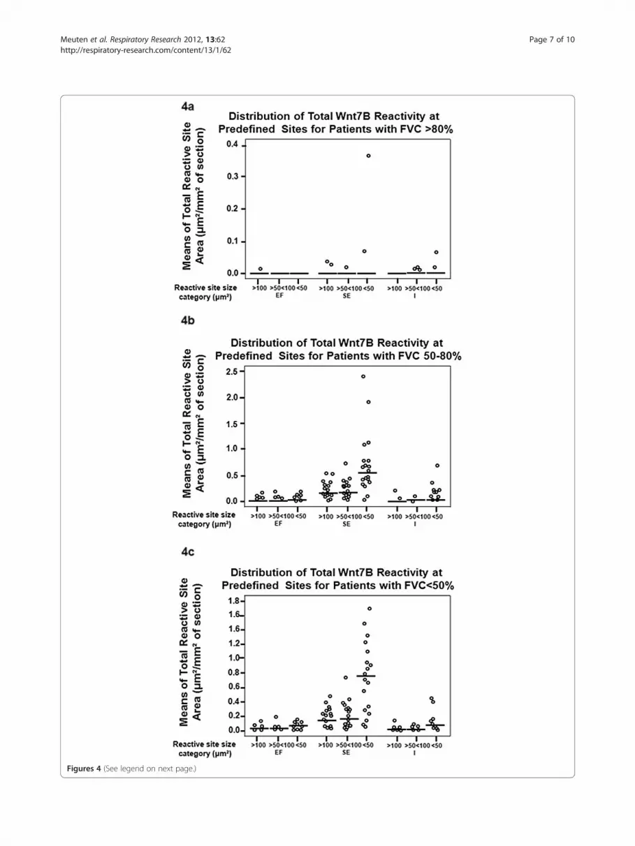
reactive sites (Figure 4a) compared to patients with func-tional diagnoses of IPF/UIP [<80% (Figures 4b-c)].There were insufficient reactive sites for the EF categoryto compare to corresponding reactive SE and I regions.Despite an outlier (defined as greater than 2 standarddeviations from the mean) for the SE region <50 μm2
category, the reactive sites for locations SE and I werenot statistically different, given that the median valuewas 0 for all reactive sites by location. For patients with
FVC 50-80% and FVC <50%, reactive site size means (inμm2), standard deviations, and minimum, median, andmaximum sizes of reactive sites (in μm2) are presentedin Tables 2 and 3. For patients with FVC 50-80% andFVC <50%, the means of the number of each of the re-active site sizes for the SE location were statisticallygreater than that of those for EF and I locations(p< 0.05).
DiscussionThe initiating cause(s) underlying the pathogenesis ofidiopathic pulmonary fibrosis (IPF) is unknown. Muchattention has focused on a failed repair of alveolar epi-thelium, and its impact on subsequent loss of pulmonaryarchitecture. This failure manifests as the inability ofepithelial surfaces to proliferate and differentiate effect-ively resulting in hyperplasia, metaplasia, and/or trans-differentiation into myofibroblast-like cells [26]. Exactlyhow these dysplastic events culminate, independently orcollectively, in the formation of one of the hallmarks ofIPF, the fibroblastic focus, is not clear. Studying the rolesthat specific signaling pathways play in the complex pro-cesses involved in the formation of fibroblastic foci has
Figures 4 (See legend on next page.)
Meuten et al. Respiratory Research 2012, 13:62 Page 7 of 10http://respiratory-research.com/content/13/1/62

(See figure on previous page.)Figures 4 Distribution of the total Wnt7B reactivity at the EF, SE, and I site categories for patients with FVCs >80% (normal), 50-80%(intermediate impairment), or <50% (severe impairment). Normal patients had minimal Wnt7B reactivity, most with a median total areareactivity of “0” (a). Patients with impaired FVCs had many more reactive sites than controls, although those with intermediate and severeimpairment were not different (see Tables 2 and 3). Notably, the median of the mean total of reactivity was greatest in the SE sites that were<50 μm² (b-c).
Meuten et al. Respiratory Research 2012, 13:62 Page 8 of 10http://respiratory-research.com/content/13/1/62
led to a greater understanding of important growth fac-tors, such as TGF-β, that not only promote the depos-ition of excessive collagen but mediate the process ofEMT as well [10]. TGF-β has been immunohistochemi-cally localized to alveolar epithelium and extracellularmatrix of IPF lungs [27] and is within fibroblastic foci[28,29]. Fibroblastic foci are also rich in its downstreamnuclear targets like phospho-Smad 2/3 [28] and relevantproteins, such as cysteine-rich protein 1 (CRP-1), whichis important in smooth muscle cell differentiation [30].Relatedly, activated β-catenin has been shown to be up-regulated in the nuclei of myofibroblasts of fibroblasticfoci of IPF patients [14], and alveolar epithelial responsesto TGF-β involve alpha3 integrin for β-catenin phos-phorylation and formation of a β-catenin/p-Smad2 com-plex resulting in initiation of EMT [31]. Morespecifically, TGF-β-stimulated Smad3 has recently beenshown to form a complex with β-catenin and CREB-binding protein in regulation of α-smooth muscle actin,a cytologic signature of EMT [32]. SPARC (secreted pro-tein acidic rich in cysteine), an extracellular matrix com-ponent abundant in fibroblastic foci [33], should also benoted, as it has been shown to activate AKT, inhibitGSK-3β, and activate β-catenin, resulting in an anti-apoptotic phenotype [34], another characteristic of theIPF myofibroblast. [10-12].The finding of strong Wnt7B immunoreactivity in the
fibroblastic focus of UIP/IPF lungs would seem to fitreadily with the above discussion on canonical Wnt sig-naling involving β-catenin. The demonstration of Wnt7Bin dysplastic airway and alveolar epithelium and in myo-fibroblasts of the fibroblastic foci closely correlates with
Table 2 Patients with FVC>50%<80%
Site/size Mean StDev Min Med Max
EF> 100 0.02098 0.03446 0 0.00906 0.14388
EF >50< 100 0.01925 0.03713 0 0.00829 0.17004
EF <50 0.0427 0.0477 0 0.0181 0.1653
SE >100 *0.198 0.1508 0 0.1613 0.5167
SE >50< 100 *0.194 0.1715 0 0.1575 0.7086
SE <50 *0.675 0.562 0 0.537 2.38
I >100 0.02319 0.04121 0 0.00829 0.186
I >50< 100 0.0231 0.02473 0 0.01027 0.08267
I <50 0.1044 0.152 0 0.0458 0.6613
the site localization of active β-catenin in IPF lungs [14].However, that study did not report active β-catenin infibroblasts/myofibroblasts in regions other than fibro-blastic foci. Wnt7B localization, demonstrated here, wasalso clearly defined in a large number of smaller sites(<50 μm2) that, like fibroblastic foci, had both cellularand extracellular matrix components. The loss of extra-cellular Wnt7B immunoreactivity after hyaluronidase di-gestion supports the matrix-associated localization,which is not surprising, as Reichsman, et al., [24] havedemonstrated that most secreted Wnts (approximately83%) are bound to the cell surface and surroundingextracellular matrix through specific, non-covalent inter-actions. The localization of Wnt7B in fibroblastic fociand a large number of smaller subepithelial sites positionit well for influencing the activity of the underlyinginterstitium. In normal lung development, murineWnt7b has been shown to be exclusively expressed inepithelial cells and regulated by TTF-1, GATA-6, andFoxA2 [35]. In the embryo, it has been demonstrated tostimulate epithelial and mesenchymal proliferation, likelythrough its activation of BMP-4 and Id2 [36]. Further,Wnt7b/β-catenin signaling regulates a program of mes-enchymal cell differentiation and proliferation that is ne-cessary for smooth muscle cell development incooperation with tenascin-C and involving PDGFR-alphaand PDGFR-beta [37]. It may not be surprising, then,that similar genetic programs are reactivated in someform in an adult disease such as IPF, in which tenascin-C is heavily expressed in the matrix of fibroblastic foci
Table 3 Patients with FVC<50%
Site/size Mean StDev Min Med Max
EF >100 0.02615 0.02849 0 0.0159 0.10568
EF >50< 100 1.02039 0.02286 0 0.01761 0.07789
EF <50 0.04478 0.03933 0 0.04559 0.124
SE >100 *0.1589 0.03933 0.00677 0.131 0.4524
SE >50< 100 *0.2121 0.167 0.0124 0.1976 0.6228
SE <50 *0.7170 0.477 0.0352 0.755 1.654
I >100 0.01564 0.02594 0 0.0124 0.11683
I >50< 100 0.02883 0.03709 0 0.02067 0.15578
I <50 0.093 0.1135 0 0.0671 0.4284
* the means of the number of each of the reactive site sizes for the SElocation were statistically greater than that of those for EF and I locations(p< 0.05).
Meuten et al. Respiratory Research 2012, 13:62 Page 9 of 10http://respiratory-research.com/content/13/1/62
[33] where myofibroblasts expressing PDGFRs arelocated [38]. Coupled with the strong expression ofWnt7B in hyperplastic/metaplastic epithelium and fibro-blastic foci reported here, these observations supportprevious conclusions that epithelium in IPF may be re-sponsible for aberrant activation of Wnt signaling, suchas that of Wnt7B, in adjacent mesenchyme, leading todamage to the lung and fibrosis [19].The notion that Wnt7B may have a potentially con-
tributive if not significant role in the development and/or progression of IPF draws on previous assumptionsthat the fibroblastic foci are central to the diseaseprocess and its prognosis [39,40]. The fibroblastic fociwere the most dramatic sites of Wnt7B localizationobserved in the IPF lung specimens. However, when sys-tematically categorized and analyzed relative to locationand size, the Wnt7B reactive sites of large fibroblasticfoci were not the most numerous nor did they consumethe greatest percentage of total tissue section area, re-gardless of disease severity according to % FVC, assum-ing the section samples correctly reflected the overallextent of fibrotic change of the whole lung (see Figure 4).This distinction belonged to the small subepithelial (SE)sites <50 μm2 (Figures 1d, 2a, 3b-d). These unique re-active sites had no other morphological features thatwere specifically distinguishing other than their discreteWnt7B immunoreactivity. In parallel sections, the smallSE sites, like the larger fibroblastic foci, consistently con-tained abundant cells expressing the myofibroblastphenotype, as indicated by α-smooth muscle actinimmunoreactivity (Figure 3f ), but by virtue of theirWnt7B reactivity, clearly represented a subpopulation ofthese cells compared with the overall total myofibroblastpopulation in IPF lungs. While it is not clear from thecurrent data that there are any connections between theWnt7B positive reactivity of the fibroblastic foci and themore numerous, smaller SE sites, Cool, et al., [41]demonstrated with three-dimensional reconstruction ofpentachrome-stained sections that fibroblastic foci ofUIP form a complex, interconnected network thatextends from the pleura into the parenchyma. Perhapsthe strong reactivity for Wnt7B within fibroblastic foci,shown here to correlate precisely with pentachrome re-activity, along with the foci’s complement of establishedpro-fibrogenic components constitute the expansionunit of IPF, and the small Wnt7B-positive, subepithelial(SE) sites are the leading edge of this process. The loca-tion of the small SE sites could portend the epithelial-fibroblast cross-talk often involving Wnt signaling andknown to be important determinants of the fibrogenicevents characteristic of IPF [26]. The regionally confinedlocalization of Wnt7B differs from that of Wnt5A, whichis strongly expressed in the majority of fibroblastsderived from UIP patients (i.e., almost all of the
remaining fibroblasts), and shown to signal throughnon-canonical pathways, promote proliferation, and pre-vent apoptosis [42]. It raises the possibility that Wnt7Band Wnt5A, along with TGF-β, SPARC, and tenascin-C,work in some coordinated or concerted fashion tomodulate fibroblast/myofibroblast activities in adjacentand/or different anatomic regions of IPF lungs. Extensiveand more detailed studies are currently underway tohelp develop a better understanding of this complexprocess.
ConclusionsThese observations draw attention to a specific Wnt sig-naling ligand, Wnt7B, which by virtue of its establishedroles in epithelial and mesenchymal proliferation anddifferentiation, procollagen production, and enhancedgene expression in IPF, would be expected to act as asignificant contributor to the pathogenesis of IPF. Thisis supported not only by the strong expression of Wnt7Bin fibroblastic foci but also by the numerous, small sube-pithelial sites that may represent early stages of develop-ing fibroblastic foci as part of a larger, expandingnetwork of fibrogenic tissue.
Competing interestsThere are no competing interests to report by any of the authors.
Authors’ contributionsTM – performed immunostaining, prepared photographic images, draftingand revision of manuscript; AH - performed immunostaining, KF -developed protocol staining and performed immunostaining; BG -performed immunostaining; JT – developed immunostainig protocol; DN -interpretation of data, drafting and revision of manuscript; SJ – analysis andinterpretation of data, manuscript revision; MC - analysis and interpretationof data, statistical analysis; PS – conception and design of study, analysis andinterpretation of data, preparation of photographic images, drafting ofmanuscript and revision; all authors read and approved of the finalmanuscript.
AknowledgmentsThis investigation was supported by PHS grants HL44497, HL95411, NorthCarolina State University College of Veterinary Medicine, and the State ofNorth Carolina. The authors wish to thank Thomas Sporn, M.D., Departmentof Pathology, Duke University Medical Center, Durham, NC, for his guidanceand diagnostic support, and Laura Shewmon, Department of PopulationHealth and Pathology, College of Veterinary Medicine, North Carolina StateUniversity for her technical contribution to this study.
Author details1Departments of Molecular Biomedical Sciences, College of VeterinaryMedicine, North Carolina State University, 1060 William Moore Dr, Raleigh,NC 27607, USA. 2Population Health and Pathology, enter for ComparativeMedicine and Translational Research, College of Veterinary Medicine, NorthCarolina State University, Raleigh, NC, USA.
Received: 25 May 2012 Accepted: 17 July 2012Published: 28 July 2012
References1. Green FH: Overview of pulmonary fibrosis. Chest 2002, 122:334S–339S.2. Schwartz DA, Helmers RA, Galvin JR, Van Fossen DS, Frees KL, Dayton CS,
Burmeister LF, Hunninghake GW: Determinants of survival in idiopathicpulmonary fibrosis. Am J Respir Crit Care Med 1994, 149:450–454.

Meuten et al. Respiratory Research 2012, 13:62 Page 10 of 10http://respiratory-research.com/content/13/1/62
3. Strieter RM: Con: Inflammatory mechanisms are not a minor componentof the pathogenesis of idiopathic pulmonary fibrosis. Am J Respir Crit CareMed 2002, 165:1206–1207. discussion 1207–8.
4. Strieter RM: Pathogenesis and natural history of usual interstitialpneumonia: the whole story or the last chapter of a long novel.Chest 2005, 128:526S–532S.
5. Konigshoff M, Eickelberg O: WNT signaling in lung disease: a failure or aregeneration signal? Am J Respir Cell Mol Biol 2010, 42:21–31.
6. Katzenstein AL, Myers JL: Idiopathic pulmonary fibrosis: clinical relevanceof pathologic classification. Am J Respir Crit Care Med 1998, 157:1301–1315.
7. Katzenstein AL, Zisman DA, Litzky LA, Nguyen BT, Kotloff RM: Usualinterstitial pneumonia: histologic study of biopsy and explantspecimens. Am J Surg Pathol 2002, 26:1567–1577.
8. Kapanci Y, Desmouliere A: Pache JC, Redard M. Gabbiani G: Cytoskeletalprotein modulation in pulmonary alveolar myofibroblasts during idiopathicpulmonary fibrosis. Possible role of transforming growth factor beta and tumornecrosis factor alpha. Am J Respir Crit Care Med 1995, 152:2163–2169.
9. Adamson IY, Hedgecock C, Bowden DH: Epithelial cell-fibroblastinteractions in lung injury and repair. Am J Pathol 1990, 137:385–392.
10. Broekelmann TJ, Limper AH, Colby TV, McDonald JA: Transforming growthfactor beta 1 is present at sites of extracellular matrix gene expression inhuman pulmonary fibrosis. Proc Natl Acad Sci USA 1991, 88:6642–6646.
11. Scotton CJ, Chambers RC: Molecular targets in pulmonary fibrosis: themyofibroblast in focus. Chest 2007, 132:1311–1321.
12. Salazar KD, Lankford SM, Brody AR: Mesenchymal stem cells produce Wntisoforms and TGF-beta1 that mediate proliferation and procollagenexpression by lung fibroblasts. Am J Physiol Lung Cell Mol Physiol 2009,297:L1002–L1011.
13. Attisano L, Labbe E: TGFbeta and Wnt pathway cross-talk. CancerMetastasis Rev 2004, 23:53–61.
14. Chilosi M, Poletti V, Zamo A, Lestani M, Montagna L, Piccoli P, Pedron S,Bertaso M, Scarpa A, Murer B, Cancellieri A, Maestro R, Semenzato G,Doglioni C: Aberrant Wnt/beta-catenin pathway activation in idiopathicpulmonary fibrosis. Am J Pathol 2003, 162:1495–1502.
15. Douglas IS: Diaz del Valle F, Winn RA, Voelkel NF: Beta-catenin in thefibroproliferative response to acute lung injury. Am J Respir Cell Mol Biol2006, 34:274–285.
16. Willert K, Nusse R: Beta-catenin: a key mediator of Wnt signaling.Curr Opin Genet Dev 1998, 8:95–102.
17. Konigshoff M, Balsara N, Pfaff EM, Kramer M, Chrobak I, Seeger W, EickelbergO: Functional Wnt signaling is increased in idiopathic pulmonary fibrosis.PLoS One 2008, 3:e2142.
18. van der Velden JL, Guala AS, Leggett SE, Sluimer J, Badura EC, Janssen-HeiningerYM: Induction of a Mesenchymal Expression Program in Lung Epithelial Cellsby Wnt/beta-catenin Requires the Presence of c-Jun N-Terminal Kinase 1.Am J Respir Cell Mol Biol 2012, doi:10.1165/rcmb.2011-029. Published ahead ofprint on March 2012.
19. Morrisey EE: Wnt signaling and pulmonary fibrosis. Am J Pathol 2003,162:1393–1397.
20. Winn RA, Marek L, Han SY, Rodriguez K, Rodriguez N, Hammond M, VanScoyk M, Acosta H, Mirus J, Barry N, Bren-Mattison Y, Van Raay TJ, NemenoffRA, Heasley LE: Restoration of Wnt-7a expression reverses non-small celllung cancer cellular transformation through frizzled-9-mediated growthinhibition and promotion of cell differentiation. J Biol Chem 2005,280:19625–19634.
21. Apparao KB, Newman DR, Zhang H, Khosla J, Randell SH, Sannes PL:Temporal changes in expression of FoxA1 and Wnt7A in isolated adulthuman alveolar epithelial cells enhanced by heparin. Anat Rec (Hoboken)2010, 293:938–946.
22. He W, Dai C, Li Y, Zeng G, Monga SP, Liu Y: Wnt/beta-catenin signalingpromotes renal interstitial fibrosis. J Am Soc Nephrol 2009, 20:765–776.
23. Shu W, Jiang YQ, Lu MM, Morrisey EE: Wnt7b regulates mesenchymalproliferation and vascular development in the lung. Development 2002,129:4831–4842.
24. Reichsman F, Smith L, Cumberledge S: Glycosaminoglycans can modulateextracellular localization of the wingless protein and promote signaltransduction. J Cell Biol 1996, 135:819–827.
25. Smolich BD, McMahon JA, McMahon AP, Papkoff J: Wnt family proteins aresecreted and associated with the cell surface. Mol Biol Cell 1993,4:1267–1275.
26. Coward WR, Saini G, Jenkins G: The pathogenesis of idiopathic pulmonaryfibrosis. Ther Adv Respir Dis 2010, 4:367–388.
27. Khalil N, O'Connor RN, Flanders KC, Unruh H: TGF-beta 1, but not TGF-beta2 or TGF-beta 3, is differentially present in epithelial cells of advancedpulmonary fibrosis: an immunohistochemical study. Am J Respir Cell MolBiol 1996, 14:131–138.
28. Jonigk D, Theophile K, Hussein K, Bock O, Lehmann U, Bockmeyer CL,Gottlieb J, Fischer S, Simon A, Welte T, Maegel L, Kreipe H, Laenger F:Obliterative airway remodelling in transplanted and non-transplantedlungs. Virchows Arch 2010, 457:369–380.
29. Lomas NJ, Watts KL, Akram KM, Forsyth NR, Spiteri MA: Idiopathicpulmonary fibrosis: immunohistochemical analysis provides freshinsights into lung tissue remodelling with implications for novelprognostic markers. Int J Clin Exp Pathol 2012, 5:58–71.
30. Jarvinen PM, Myllarniemi M, Liu H, Moore HM, Lepparanta O, Salmenkivi K,Koli K, Latonen L, Band A, Laiho M: Cysteine-rich protein 1 is regulated bytransforming growth factor-beta1 and expressed in lung fibrosis.J Cell Physiol 2011, 227(6):2605–2612.
31. Kim KK, Wei Y, Szekeres C, Kugler MC, Wolters PJ, Hill ML, Frank JA,Brumwell AN, Wheeler SE, Kreidberg JA, Chapman HA: Epithelial cellalpha3beta1 integrin links beta-catenin and Smad signaling to promotemyofibroblast formation and pulmonary fibrosis. J Clin Invest 2009,119:213–224.
32. Zhou B, Liu Y, Kahn M, Ann DK, Han A, Wang H, Nguyen C, Flodby P, ZhongQ, Krishnaveni MS, Liebler JM, Minoo P, Crandall ED, Borok Z: beta-g.J Biol Chem 2012, 2012:2012–2012.
33. Kuhn C, Mason RJ: Immunolocalization of SPARC, tenascin, andthrombospondin in pulmonary fibrosis. Am J Pathol 1995, 147:1759–1769.
34. Chang W, Wei K, Jacobs SS, Upadhyay D, Weill D, Rosen GD: SPARCsuppresses apoptosis of idiopathic pulmonary fibrosis fibroblaststhrough constitutive activation of beta-catenin. J Biol Chem 2010,285:8196–8206.
35. Weidenfeld J, Shu W, Zhang L, Millar SE, Morrisey EE: The WNT7b promoteris regulated by TTF-1, GATA6, and Foxa2 in lung epithelium. J Biol Chem2002, 277:21061–21070.
36. Rajagopal J, Carroll TJ, Guseh JS, Bores SA, Blank LJ, Anderson WJ, Yu J,Zhou Q, McMahon AP, Melton DA: Wnt7b stimulates embryonic lunggrowth by coordinately increasing the replication of epithelium andmesenchyme. Development 2008, 135:1625–1634.
37. Cohen ED, Ihida-Stansbury K, Lu MM, Panettieri RA, Jones PL, Morrisey EE:Wnt signaling regulates smooth muscle precursor development in themouse lung via a tenascin C/PDGFR pathway. J Clin Invest 2009,119:2538–2549.
38. Vuorinen K, Ohlmeier S, Lepparanta O, Salmenkivi K, Myllarniemi M, KinnulaVL: Peroxiredoxin II expression and its association with oxidative stressand cell proliferation in human idiopathic pulmonary fibrosis.J Histochem Cytochem 2008, 56:951–959.
39. Enomoto N, Suda T, Kato M, Kaida Y, Nakamura Y, Imokawa S, Ida M, ChidaK: Quantitative analysis of fibroblastic foci in usual interstitialpneumonia. Chest 2006, 130:22–29.
40. Flaherty KR, Colby TV, Travis WD, Toews GB, Mumford J, Murray S,Thannickal VJ, Kazerooni EA, Gross BH, Lynch JP 3rd, Martinez FJ:Fibroblastic foci in usual interstitial pneumonia: idiopathic versuscollagen vascular disease. Am J Respir Crit Care Med 2003, 167:1410–1415.
41. Cool CD, Groshong SD, Rai PR, Henson PM, Stewart JS, Brown KK: Fibroblastfoci are not discrete sites of lung injury or repair: the fibroblastreticulum. Am J Respir Crit Care Med 2006, 174:654–658.
42. Vuga LJ, Ben-Yehudah A, Kovkarova-Naumovski E, Oriss T, Gibson KF,Feghali-Bostwick C, Kaminski N: WNT5A is a regulator of fibroblastproliferation and resistance to apoptosis. Am J Respir Cell Mol Biol 2009,41:583–589.
doi:10.1186/1465-9921-13-62Cite this article as: Meuten et al.: WNT7B in fibroblastic foci of idiopathicpulmonary fibrosis. Respiratory Research 2012 13:62.
Related Documents