Codocedo and Inestrosa Biol Res (2016) 49:9 DOI 10.1186/s40659-016-0071-x REVIEW Wnt-5a-regulated miR-101b controls COX2 expression in hippocampal neurons Juan Francisco Codocedo 1,4 and Nibaldo C. Inestrosa 1,2,3,4* Abstract Background: Wnt-5a is a member of the WNT family of secreted lipoglycoproteins, whose expression increases dur- ing development; moreover, Wnt-5a plays a key role in synaptic structure and function in the adult nervous system. However, the mechanism underlying these effects is still elusive. MicroRNAs (miRNAs) are a family of small non-coding RNAs that control the gene expression of their targets through hybridization with complementary sequences in the 3′ UTR, thereby inhibiting the translation of the target proteins. Several evidences indicate that the miRNAs are actively involved in the regulation of neuronal function. Results: In the present study, we examined whether Wnt-5a modulates the levels of miRNAs in hippocampal neu- rons. Using PCR arrays, we identified a set of miRNAs that respond to Wnt-5a treatment. One of the most affected miRNAs was miR-101b, which targets cyclooxygenase-2 (COX2), an inducible enzyme that converts arachidonic acid to prostanoids, and has been involved in the injury/inflammatory response, and more recently in neuronal plasticity. Consistent with the Wnt-5a regulation of miR-101b, this Wnt ligand regulates COX2 expression in a time-dependent manner in cultured hippocampal neurons. Conclusion: The biological processes induced by Wnt-5a in hippocampal neurons, involve the regulation of several miRNAs including miR-101b, which has the capacity to regulate several targets, including COX-2 in the central nerv- ous system. © 2016 Codocedo and Inestrosa. This article is distributed under the terms of the Creative Commons Attribution 4.0 International License (http://creativecommons.org/licenses/by/4.0/), which permits unrestricted use, distribution, and reproduction in any medium, provided you give appropriate credit to the original author(s) and the source, provide a link to the Creative Commons license, and indicate if changes were made. The Creative Commons Public Domain Dedication waiver (http://creativecommons. org/publicdomain/zero/1.0/) applies to the data made available in this article, unless otherwise stated. Background e Wnt proteins constitute a large family of cysteine- rich secreted glycoproteins, which are present in all ani- mal species. e genome of mice and humans, has 19 independent genes which are expressed in a tissue-spe- cific form and also dependent on the development [1]. Wnts have been implicated in several cellular processes, such as cell proliferation, migration, polarity and cell fate specification [2, 3]. Moreover, the deregulation of Wnt signaling is related with several diseases, including autism [4, 5], schizophrenia [6, 7] and Alzheimer’s disease [7, 8]. Wnt ligands couple to various receptors and thereby activate different signaling pathways. On the basis of early studies, these pathways have been classified as either canonical (β-catenin-dependent) or non-canonical (β-catenin-independent) signaling pathways. However, this classification can only serve as a rough guide, as vari- ous divergent pathways has been described in different cellular contexts [9]. e role for Wnt signaling in synaptic formation and function has been clearly established [10–12]. In fact, we described that Wnt-5a, which preferentially activates non-canonical pathways [9], exerts important effects in the postsynaptic region of central synapses. Wnt-5a stimulation increases the postsynaptic density protein 95 (PSD-95) clustering [13] and increases the density of dendritic spines [14]. In hippocampal slices, Wnt- 5a enhances long-term potentiation (LTP) modulating synaptic activity and plasticity [15, 16]. ese findings strongly suggest that Wnt-5a regulates the assembly and function of the excitatory postsynaptic region of central synapses [17]. However, the mechanism underlying these effects is still elusive. MicroRNAs (miRNAs) are a class of small non-cod- ing RNAs that regulate the local translation of dendritic Open Access Biological Research *Correspondence: [email protected] 4 CARE, Biomedical Research Center, Pontificia Universidad Católica de Chile, Av. Alameda 340, 8331150 Santiago, Chile Full list of author information is available at the end of the article

Welcome message from author
This document is posted to help you gain knowledge. Please leave a comment to let me know what you think about it! Share it to your friends and learn new things together.
Transcript
Codocedo and Inestrosa Biol Res (2016) 49:9 DOI 10.1186/s40659-016-0071-x
REVIEW
Wnt-5a-regulated miR-101b controls COX2 expression in hippocampal neuronsJuan Francisco Codocedo1,4 and Nibaldo C. Inestrosa1,2,3,4*
Abstract
Background: Wnt-5a is a member of the WNT family of secreted lipoglycoproteins, whose expression increases dur-ing development; moreover, Wnt-5a plays a key role in synaptic structure and function in the adult nervous system. However, the mechanism underlying these effects is still elusive. MicroRNAs (miRNAs) are a family of small non-coding RNAs that control the gene expression of their targets through hybridization with complementary sequences in the 3′ UTR, thereby inhibiting the translation of the target proteins. Several evidences indicate that the miRNAs are actively involved in the regulation of neuronal function.
Results: In the present study, we examined whether Wnt-5a modulates the levels of miRNAs in hippocampal neu-rons. Using PCR arrays, we identified a set of miRNAs that respond to Wnt-5a treatment. One of the most affected miRNAs was miR-101b, which targets cyclooxygenase-2 (COX2), an inducible enzyme that converts arachidonic acid to prostanoids, and has been involved in the injury/inflammatory response, and more recently in neuronal plasticity. Consistent with the Wnt-5a regulation of miR-101b, this Wnt ligand regulates COX2 expression in a time-dependent manner in cultured hippocampal neurons.
Conclusion: The biological processes induced by Wnt-5a in hippocampal neurons, involve the regulation of several miRNAs including miR-101b, which has the capacity to regulate several targets, including COX-2 in the central nerv-ous system.
© 2016 Codocedo and Inestrosa. This article is distributed under the terms of the Creative Commons Attribution 4.0 International License (http://creativecommons.org/licenses/by/4.0/), which permits unrestricted use, distribution, and reproduction in any medium, provided you give appropriate credit to the original author(s) and the source, provide a link to the Creative Commons license, and indicate if changes were made. The Creative Commons Public Domain Dedication waiver (http://creativecommons.org/publicdomain/zero/1.0/) applies to the data made available in this article, unless otherwise stated.
BackgroundThe Wnt proteins constitute a large family of cysteine-rich secreted glycoproteins, which are present in all ani-mal species. The genome of mice and humans, has 19 independent genes which are expressed in a tissue-spe-cific form and also dependent on the development [1]. Wnts have been implicated in several cellular processes, such as cell proliferation, migration, polarity and cell fate specification [2, 3]. Moreover, the deregulation of Wnt signaling is related with several diseases, including autism [4, 5], schizophrenia [6, 7] and Alzheimer’s disease [7, 8]. Wnt ligands couple to various receptors and thereby activate different signaling pathways. On the basis of early studies, these pathways have been classified as either canonical (β-catenin-dependent) or non-canonical
(β-catenin-independent) signaling pathways. However, this classification can only serve as a rough guide, as vari-ous divergent pathways has been described in different cellular contexts [9].
The role for Wnt signaling in synaptic formation and function has been clearly established [10–12]. In fact, we described that Wnt-5a, which preferentially activates non-canonical pathways [9], exerts important effects in the postsynaptic region of central synapses. Wnt-5a stimulation increases the postsynaptic density protein 95 (PSD-95) clustering [13] and increases the density of dendritic spines [14]. In hippocampal slices, Wnt-5a enhances long-term potentiation (LTP) modulating synaptic activity and plasticity [15, 16]. These findings strongly suggest that Wnt-5a regulates the assembly and function of the excitatory postsynaptic region of central synapses [17]. However, the mechanism underlying these effects is still elusive.
MicroRNAs (miRNAs) are a class of small non-cod-ing RNAs that regulate the local translation of dendritic
Open Access
Biological Research
*Correspondence: [email protected] 4 CARE, Biomedical Research Center, Pontificia Universidad Católica de Chile, Av. Alameda 340, 8331150 Santiago, ChileFull list of author information is available at the end of the article
Page 2 of 10Codocedo and Inestrosa Biol Res (2016) 49:9
mRNAs, affecting the morphology and function of den-dritic spines [18]. MiRNAs control gene expression through specific base pairing between the 3′ UTR of mRNA and the miRNA “seed” region at the 5′ end [19]. We recently describe the miRNA biogenetic pathway in recent reviews [20, 21]. Briefly, canonical miRNAs are transcribed as primary miRNAs (pri-miRNAs, a long stem-loop precursor of several hundred nucleotides) which is cropped by the Microprocessor complex, com-posed by DiGeorge Syndrome Critical Region 8 (DGCR8) and Drosha, a double-stranded RNA binding protein and an RNase III enzyme, respectively [22]. The resulting pre-miRNA (~70 nt in length) is exported to the cytoplasm by Exportin-5 in a GTP-dependent fashion [23]. In the cytoplasm, pre-miRNA is cleaved into a ~22 nt mature miRNA duplex by Dicer, a second RNAse III enzyme. One strand of the mature miRNA duplex is loaded into the miRNA-induced silencing complex (miRISC) with members of the Argonaute family of proteins, producing a functional complex for targeting mRNA via direct base pairing [24]. The resulting miRNA/mRNA hybrids alter protein expression of the targeted mRNA by different mechanisms, including translational repression or mRNA degradation [25]. Some miRNAs have alternative biogen-esis process, because they can bypass the action of some processing complexes. This is mainly due to structural differences in the precursors, which allow processing by other protein complexes such as the spliceosome. These exceptions are known as non-canonical pathways [26].
Interestingly, several reports have shown that miR-NAs are downstream mediators of different extracellular stimuli, such as glutamate [27], dopamine [28], serotonin [29] and brain-derived neurotrophic factor (BDNF) [30], contributing to the induction and consolidation of plastic changes triggered by these synaptogenic factors.
In the present study, we provided evidence for a new mechanism underlying the neuronal effects of Wnt-5a, describing a number of miRNAs responsive to this ligand in hippocampal neurons. We focused on miR-101b, the most affected miRNA through Wnt-5a signaling, and their target COX2, an inducible enzyme that converts arachidonic acid to prostanoids, and has been related to the injury/inflammatory response [31, 32], and more recently to neuronal plasticity [33–36]. The downregu-lation of miR-101b could contribute to the increase in COX2 expression observed during prolonged exposure to Wnt-5a, revealing a new effector of Wnt signaling in hippocampal neurons.
MethodsEthics statementSprague-Dawley rats were housed in the University Ani-mal Facility and handled according to the guidelines
outlined and approved through the Institutional Animal Care and Use Committee at the Faculty of Biological Sci-ences of the Pontificia Universidad Católica de Chile, and following the guidelines of the American Physiological Society Rockville, MD, USA.
Primary culture of rat hippocampal neuronsRat hippocampal cultures were prepared as previously described [37, 38]. Primary hippocampal neurons were obtained from 18-day-old Sprague-Dawley rat embryos and maintained in Dulbecco’s modified Eagle’s medium (DMEM) supplemented with 10 % horse serum for 2 h. The culture medium replaced with Neurobasal medium supplemented with B27, 100 µg/ml streptomycin, and 100 units/ml penicillin. At 3 days in vitro (DIV), the cells were treated with 2 µM Cytosine Arabinoside C (araC) for 24 h to reduce the number of glial cells present in the culture. For the miRNA expression studies, 800,000 cells per well were seeded. For western blot (WB) analyses, 400,000 cells per well were seeded, and for immunofluo-rescence studies, 35,000 cells were plated per well. At 14 DIV, the neurons were stimulated with 50 µM of Foxy-5 (a mimetic formylated hexapeptide of Wnt-5a) (Gen-emed Synthesis, San Francisco, CA, USA) or 300 ng/mL of recombinant Wnt-5a (rWnt-5a) (R and D System, Min-neapolis, MN, USA) resuspended in Neurobasal medium. Control neurons incubated with a scramble peptide in Neurobasal medium (Genemed Synthesis, San Francisco, CA, USA) for experiments with either FOXY-5 or the carrier (BSA 0.1 %) resuspended in neurobasal medium for experiments with rWnt-5a. Incubations were con-ducted at 37 °C.
HT22 cell lineHT22 murine hippocampal neuronal cells were main-tained in DMEM supplemented with 10 % fetal bovine serum, 100 µg/ml streptomycin, and 100 units/ml peni-cillin, high glucose and incubated at 37 °C under 5 % CO2 as previously described [39]. Transfections were per-formed after 2 days at approximately 60 % confluency.
RNA extraction and Real‑time PCR‑based miRNA expression profilingTotal RNA extraction and subsequent enrichment of small RNAs (<200 nt) was performed using the miRVana kit (Ambion) according to the manufacturer’s instruc-tions [40]. The quantity and purity of the RNA samples were assessed using a NanoDrop 2000 Spectrophotom-eter (Thermo Scientific). The integrity of the small RNAs was assessed on denaturing 15 % polyacrylamide gels.
First-strand cDNA was synthesized from 150 ng of small RNA using the RT2 miRNA First Strand kit (SABio-sciences). RT2 miRNA PCR array was performed with an
Page 3 of 10Codocedo and Inestrosa Biol Res (2016) 49:9
Mx3000p qPCR system (Stratagene). The reactions, con-taining 2X RT2 SYBR PCR master mix and diluted cDNA in a final volume of 25 μl, were amplified at 95 °C, 15 s; 60 °C, 30 s; 72 °C, 30 s for 40 cycles. The miRNA input was normalized to endogenous controls (Rnu6, U87, 4.5S-V1 and Y1), and the data analysis was performed using the web-based software package for the miRNA PCR array system using the comparative ΔΔCt method [41]. The fold-change was calculated for each miRNA from cells treated with Foxy-5 compared with control, represented as 2(−ΔΔCt), and the results were expressed as the fold-regulation by taking the negative inverse of any number less than 1, changing the fractional number into a whole number.
To evaluate the expression of COX-2 mRNA, 20 μg/μl of total RNA was used for cDNA synthesis using the GoScript Reverse Transcription System (Promega). The determination of the mRNA abundance of specific genes was assessed through quantitative PCR (qPCR), using the StepOne 48-well Real-Time PCR System (Applied Bio-systems, Carlsbad, CA, USA) and Fast SYBR Green Mas-ter Mix (Applied Biosystems) reagent for amplification, using 4 μl cDNA, 4 μl DEPC water, 10 μl Master Mix, 1 μl Reverse Primer, and 1 μl Forward Primer in a total vol-ume of 20 μl/well. The housekeeping gene was GAPDH (forward primer: 5′-CACGGCAAGTTCAACGGC-3′; reverse primer 5′-GGTGGTGAAGACGCCAGTA-3′) at an annealing temperature of 60 °C. The following primer sequences were used for COX-2: forward primer: 5′-TGTATGCTACCATCTGGCTTCGG-3′, reverse primer 5-′GTTTGGAACAGTCGCTCGTCATC-3′.
BioinformaticsFor computational prediction of the miRNA targets, we used the TargetScan web platform [42], which predicts biological targets of miRNAs by searching for the pres-ence of conserved 8- and 7-mer sites that match the seed region of each miRNA. In addition, TargetScan exam-ines the binding sites for thermodynamic stability using RNAfold from the Vienna RNA Package [43]. For test-ing enrichment in specific KEGG pathways of miRNA targets, we used DIANA-mirPath [44]. The software performs an enrichment analysis of multiple miRNA target genes comparing each set of miRNA targets with all known KEGG pathways. The combinatorial effect of co-expressed miRNAs in the modulation of a given path-way was considered through the simultaneous analysis of multiple miRNAs.
miR‑101b gain‑of‑functionFor miR-101b gain-of-function, we used mirVana miRNA mimic (Life Technologies), which represents a
partially double-stranded RNA that mimics endogenous precursor miRNA and is processed to form an active miRNA molecule that targets specific mRNAs [45]. For miR-101b gain-of-function in HT-22 cells, Lipofectamine 2000 reagent (Invitrogen, Karlsruhe, Germany) was used according to the manufacturer’s protocol. At 48 h post-transfection, cells were used for western blotting (WB) or immunofluorescence analysis. For controls condi-tions HT22 cells was transfected with mirVana miRNA Mimic Negative Control #1 (Life Technologies, Carlsbad) which correspond to a random sequence miRNA mimic molecule that not produce identifiable effects on known miRNA function.
Western blot analysisThe extraction of total protein from cell culture of hip-pocampal neurons and immunoblot analysis were per-formed as previously described [46, 47]. The following primary antibodies were used: rabbit anti-COX-2 (1:1000; ABCAM) and anti GAPDH (1:10,000, Santa Cruz). Primary antibodies were recognized using either a horseradish peroxidase (HRP)-conjugated goat anti-rabbit antibody (1:7000, Thermo Scientific) or an HRP-conjugated rabbit anti-mouse antibody (1:7000, Thermo Scientific). The secondary antibodies were detected through enhanced chemiluminescence using the ECL Plus WB detection system (GE Healthcare). Densi-tometric analysis was performed using NIH ImageJ software.
Immunofluorescence analysisImmunofluorescence studies were performed as previ-ously described [46, 47]. Briefly, after stimulation, the cells were rinsed twice in ice- cold PBS and fixed with a freshly prepared solution of 4 % paraformaldehyde and 4 % sucrose in PBS for 20 min at 4 °C and permeabilized with 0.2 % Triton X-100 for 5 min in PBS at room tem-perature. After several rinses in ice-cold PBS, the cells were incubated in 1 % BSA in PBS (blocking solution) for 30 min at room temperature, followed by an over-night incubation at 4 °C with primary antibodies. The cells were extensively washed with PBS and subsequently incubated with Alexa-conjugated secondary antibod-ies for 60 min at 37 °C. The coverslips were mounted in mounting medium, and image stacks were collected in 0.25 µm z-step sizes using an Olympus LSM Fluoview 1000 confocal microscope. The images in the figures are maximum intensity projections to obtain high contrast images, but the quantifications were made over aver-age intensity projections. An outline was drawn around each cell and total fluorescence measurements were per-formed with NIH ImageJ software.
Page 4 of 10Codocedo and Inestrosa Biol Res (2016) 49:9
Statistical analysisAll data were analyzed statistically with Prism 5 (Prism GraphPad Software, GraphPad Software Inc., La Jolla, CA, USA) using the Mann-Whitney U test or one-way ANOVA, followed by Bonferroni-corrected pairwise comparisons. The error bars indicate SEM. A p < 0.05 was considered statistically significant.
ResultsWnt‑5a signaling regulates the expression levels of miRNAs in hippocampal neuronsVarious functions of different cell types in the mam-malian brain suggest that some miRNAs are differen-tially expressed in glia and neurons in response to the same stimuli [48, 49]. To determine whether Wnt-5a modulates neuronal miRNAs, we profiled miRNAs in cultured hippocampal neurons with a low glial content (see “Methods” section). These cultures were treated with Foxy-5, a mimetic formylated hexapeptide of Wnt-5a, extensively used for the reproduction of the biologi-cal effects of this ligand [50–53]. Foxy-5 mimics the full Wnt-5a molecule in cultures of hippocampal neurons [13, 16, 54, 55]. Among the 264 miRNAs tested after 1 h of stimulation, 8 % miRNAs were not detected, 37 % miRNAs showed low detection and 27 % miRNAs were abundantly expressed. The remaining 28 % miRNAs were abundantly detected in at least one of the experimental conditions (control or treated) (Fig. 1a). Consistent with the suggestions of the supplier, for the next analysis, we used only a fraction of miRNAs abundantly detected in at least one of the samples (cyan and red groups in Fig. 1a). In addition, to be considered as a regulated miRNAs, a minimum of fivefold regulation was used as an inclusion criteria as previously described [48]. In this fraction, we observed significant changes in 34 miRNAs (p < 0.05). Three of these miRNAs showed increased expres-sion (miR-24, -146b and -153) and 31 miRNAs showed decreased expression (Fig. 1b).
Biological function of the miRNAs modified by Wnt‑5a signalingUsing the online software TargetScan, we predicted a list of target genes for the miRNAs genes modulated through Foxy-5. To understand the functional significance of these targets, we used the online software, Diana miR-path, which identifies biological processes downstream of miRNAs altered through Wnt-5a signaling. The major biological functional categories targeted by downregu-lated miRNAs are enriched in pathways associated with cancer. Other pathways were associated with synaptic processes, such as MAP-kinase signaling, focal adhe-sion, mTOR signaling, axon guidance, regulation of actin cytoskeleton and Wnt signaling (Fig. 1c). Altogether,
these results suggest the possible roles and mechanisms of these differentially expressed miRNAs and their tar-gets in the presence of the Wnt-5a ligand.
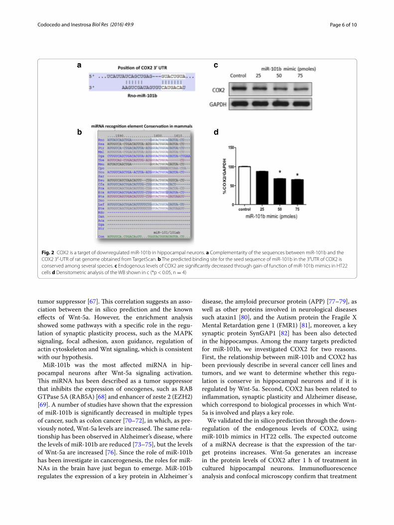
Mir‑101b gain‑of‑function downregulates COX2 expressionConsidering that Wnt-5a signaling generates a significant decrease in the levels of miR-101b (Fig. 1b), we reasoned that the levels of some of the predicted targets could be increased in the presence of Wnt-5a. Among these tar-gets, we focused on COX2, an enzyme expressed in dis-crete populations of neurons and is enriched in the cortex and hippocampus [56] and has been implicated in brain functions and in neurologic disorders, including Alz-heimer’s disease [57]. The miRNAs recognition element (MRE) in the 3′UTR of COX2 is a canonical binding site (8-mer) broadly conserved among mammals (Fig. 2a, b). To validate the in silico prediction of COX2 as a target of miR-101b, we introduced a miRNA mimic into HT22 cells and evaluated the expression of endogenous COX2 through WB (Fig. 2c). It is apparent that the increase of miR-101b decreases the level of COX2 in a dose-depend-ent manner (Fig. 2d). The same effects were observed in immunofluorescence experiments in HT22 cells using the maximal efficacy concentration of miR-101b mimics (data not show). These results validate the in silico pre-diction of COX2 as a target of miR-101b and suggest that COX2 expression could be modulated through Wnt-5a signaling via miR-101b.
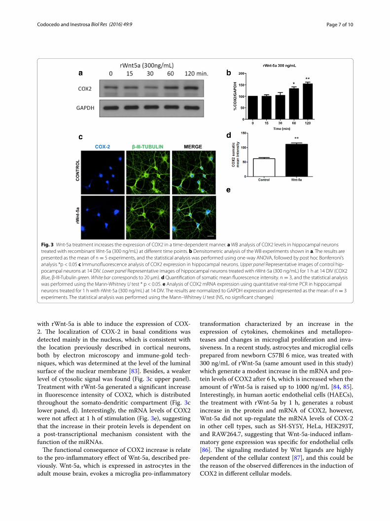
Wnt‑5a treatment increases the expression of COX2 in cultured hippocampal neuronsThe expected outcome of a decrease in miRNA levels is that the translation of the target increases, and as a con-sequence, the target protein levels also might increase. Using WB analyses, we determined that treatment with Wnt-5a increases the levels of COX2 in a time-depend-ent manner in cultured hippocampal neurons (Fig. 3a, b). Complementary immunofluorescence studies con-firm this observation, because Wnt-5a increases the somatic and dendritic signal of COX-2 at 1 h of stimu-lation (Fig. 3c, d). This increase is coincident with the previously described decrease in miR-101b triggered through Wnt-5a signaling activation (Fig. 1b). Using qPCR, we measured the expression of COX2 mRNA to determine whether this effect is dependent on a tran-scriptional mechanism. At 1 h of stimulation, we did not detect changes in the levels of COX2 mRNA expression (Fig. 3e), suggesting a post-transcriptional mechanism, consistent with the miR-101b decrease.
DiscussionPrevious studies indicated that Wnt-5a is a synaptogenic factor, whose expression increases during development;

Page 5 of 10Codocedo and Inestrosa Biol Res (2016) 49:9
indicating that Wnt-5a plays an important role in syn-aptic structure and function in the adult nervous system [17, 58]. Neural factors induce plastic changes through the activation of different signaling pathways, including the modulation of specific miRNAs [59]. In the present study, we evaluated the hypothesis that treatment with Wnt-5a modulates miRNAs in hippocampal neurons and this modulation contributes to the neuronal effects previously described for the Wnt ligand through the reg-ulation of different targets. We identified more than 30 miRNAs with differential expression after 1 h treatment with the Wnt-5a mimetic peptide. Most of the regulated miRNAs showed decreased expression, and only 3 miR-NAs showed increased expression, suggesting that these two groups of miRNAs have different Wnt responsive elements in their biogenetic pathway. Further studies are
necessary to determine the mechanism by which Wnt-5a controls the expression of miRNAs in hippocampal neurons.
To understand the role of the miRNAs regulated through Wnt-5a, we used bioinformatics tools to obtain the predicted targets and determine the potential roles for these genes in biological pathways using the KEEG database. The top KEGG pathways of biological function for the targets of downregulated miRNAs showed enrich-ment in cancer processes. This finding is important, since Wnt-5a is upregulated in several types of cancer. For example, Wnt-5a is increased in melanoma [60], colo-rectal cancer [61], pancreatic cancer [62], non-small cell lung cancer [63, 64], renal cell carcinoma [64] and glioma [65, 66]. The only exception to this, is myeloid leukemia, in which Wnt-5a is downregulated and functions as a
Fig. 1 Foxy-5, a mimetic peptide of Wnt-5a, regulates miRNA expression levels in hippocampal neurons. a The pie chart shows the percentage expression of 263 rat miRNAs in hippocampal neurons treated with Foxy-5 and control (n = 3, each). b Fold-regulation of miRNAs expression in neurons treated with Foxy-5 (p < 0.05, n = 3). Among the 34 significantly modulated miRNAs, miR-101b was the most affected. c The top KEGG pathways of biological function of the targets of downregulated miRNAs after Foxy-5 treatment
Page 6 of 10Codocedo and Inestrosa Biol Res (2016) 49:9
tumor suppressor [67]. This correlation suggests an asso-ciation between the in silico prediction and the known effects of Wnt-5a. However, the enrichment analysis showed some pathways with a specific role in the regu-lation of synaptic plasticity process, such as the MAPK signaling, focal adhesion, axon guidance, regulation of actin cytoskeleton and Wnt signaling, which is consistent with our hypothesis.
MiR-101b was the most affected miRNA in hip-pocampal neurons after Wnt-5a signaling activation. This miRNA has been described as a tumor suppressor that inhibits the expression of oncogenes, such as RAB GTPase 5A (RAB5A) [68] and enhancer of zeste 2 (EZH2) [69]. A number of studies have shown that the expression of miR-101b is significantly decreased in multiple types of cancer, such as colon cancer [70–72], in which, as pre-viously noted, Wnt-5a levels are increased. The same rela-tionship has been observed in Alzheimer’s disease, where the levels of miR-101b are reduced [73–75], but the levels of Wnt-5a are increased [76]. Since the role of miR-101b has been investigate in cancerogenesis, the roles for miR-NAs in the brain have just begun to emerge. MiR-101b regulates the expression of a key protein in Alzheimer´s
disease, the amyloid precursor protein (APP) [77–79], as well as other proteins involved in neurological diseases such ataxin1 [80], and the Autism protein the Fragile X Mental Retardation gene 1 (FMR1) [81], moreover, a key synaptic protein SynGAP1 [82] has been also detected in the hippocampus. Among the many targets predicted for miR-101b, we investigated COX2 for two reasons. First, the relationship between miR-101b and COX2 has been previously describe in several cancer cell lines and tumors, and we want to determine whether this regu-lation is conserve in hippocampal neurons and if it is regulated by Wnt-5a. Second, COX2 has been related to inflammation, synaptic plasticity and Alzheimer disease, which correspond to biological processes in which Wnt-5a is involved and plays a key role.
We validated the in silico prediction through the down-regulation of the endogenous levels of COX2, using miR-101b mimics in HT22 cells. The expected outcome of a miRNA decrease is that the expression of the tar-get proteins increases. Wnt-5a generates an increase in the protein levels of COX2 after 1 h of treatment in cultured hippocampal neurons. Immunofluorescence analysis and confocal microscopy confirm that treatment
Fig. 2 COX2 is a target of downregulated miR-101b in hippocampal neurons. a Complementarity of the sequences between miR-101b and the COX2 3′-UTR of rat genome obtained from TargetScan. b The predicted binding site for the seed sequence of miR-101b in the 3′UTR of COX2 is conserved among several species. c Endogenous levels of COX2 are significantly decreased through gain-of function of miR-101b mimics in HT22 cells d Densitometric analysis of the WB shown in c (*p < 0.05, n = 4)
Page 7 of 10Codocedo and Inestrosa Biol Res (2016) 49:9
with rWnt-5a is able to induce the expression of COX-2. The localization of COX-2 in basal conditions was detected mainly in the nucleus, which is consistent with the location previously described in cortical neurons, both by electron microscopy and immune-gold tech-niques, which was determined at the level of the luminal surface of the nuclear membrane [83]. Besides, a weaker level of cytosolic signal was found (Fig. 3c upper panel). Treatment with rWnt-5a generated a significant increase in fluorescence intensity of COX2, which is distributed throughout the somato-dendritic compartment (Fig. 3c lower panel, d). Interestingly, the mRNA levels of COX2 were not affect at 1 h of stimulation (Fig. 3e), suggesting that the increase in their protein levels is dependent on a post-transcriptional mechanism consistent with the function of the miRNAs.
The functional consequence of COX2 increase is relate to the pro-inflammatory effect of Wnt-5a, described pre-viously. Wnt-5a, which is expressed in astrocytes in the adult mouse brain, evokes a microglia pro-inflammatory
transformation characterized by an increase in the expression of cytokines, chemokines and metallopro-teases and changes in microglial proliferation and inva-siveness. In a recent study, astrocytes and microglial cells prepared from newborn C57Bl 6 mice, was treated with 300 ng/mL of rWnt-5a (same amount used in this study) which generate a modest increase in the mRNA and pro-tein levels of COX2 after 6 h, which is increased when the amount of rWnt-5a is raised up to 1000 ng/mL [84, 85]. Interestingly, in human aortic endothelial cells (HAECs), the treatment with rWnt-5a by 1 h, generates a robust increase in the protein and mRNA of COX2, however, Wnt-5a did not up-regulate the mRNA levels of COX-2 in other cell types, such as SH-SY5Y, HeLa, HEK293T, and RAW264.7, suggesting that Wnt-5a-induced inflam-matory gene expression was specific for endothelial cells [86]. The signaling mediated by Wnt ligands are highly dependent of the cellular context [87], and this could be the reason of the observed differences in the induction of COX2 in different cellular models.
Fig. 3 Wnt-5a treatment increases the expression of COX2 in a time-dependent manner. a WB analysis of COX2 levels in hippocampal neurons treated with recombinant Wnt-5a (300 ng/mL) at different time points. b Densitometric analysis of the WB experiments shown in a. The results are presented as the mean of n = 5 experiments, and the statistical analysis was performed using one-way ANOVA, followed by post hoc Bonferroni’s analysis *p < 0.05 c Immunofluorescence analysis of COX2 expression in hippocampal neurons. Upper panel Representative images of control hip-pocampal neurons at 14 DIV. Lower panel Representative images of hippocampal neurons treated with rWnt-5a (300 ng/mL) for 1 h at 14 DIV (COX2 Blue, β-III-Tubulin green. White bar corresponds to 20 μm). d Quantification of somatic mean fluorescence intensity. n = 3, and the statistical analysis was performed using the Mann-Whitney U test * p < 0.05. e Analysis of COX2 mRNA expression using quantitative real-time PCR in hippocampal neurons treated for 1 h with rWnt-5a (300 ng/mL) at 14 DIV. The results are normalized to GAPDH expression and represented as the mean of n = 3 experiments. The statistical analysis was performed using the Mann–Whitney U test (NS, no significant changes)

Page 8 of 10Codocedo and Inestrosa Biol Res (2016) 49:9
At difference of other tissues, COX-2 is also constitu-tively expressed in brain [56], suggesting that their basal activity is involved in general cellular functions. In brain, both COX-2 mRNA and protein are express at relatively high levels in neurons involved in plastic and cogni-tive functions, such as those hippocampal granule cells, pyramidal cells, and cortical neurons. In fact, COX-2 protein is express at a very high level in dentate granule cells under basal conditions [56].
Basal expression of COX-2 is regulate by the synaptic activity, and its cellular expression is upregulated by a high-frequency stimulation (HFS) associated with LTP induction [56]. Moreover, COX-2 is present in neuronal dendritic spines where excitatory synapses are located [88]. These evidences imply that COX-2 plays an impor-tant role in synaptic modifications. Based in our previous work, in which Wnt-5a induces an enhancement of the synaptic structure and function, the observed increase in the COX2 levels through miR-101b, is related to their role as synaptic modulator. Additional experiments are required to determine the mechanism by which Wnt-5a regulates the expression of miRNAs, in particular miR-101b and also the physiological conditions in which the module miR-101b/COX2 contribute to the brain func-tion regulated by the Wnt-5a ligand.
Based on the results obtained in the present study, we propose a hypothetical model, in which Wnt-5a increases the expression of COX2 through the regulation of miR-101b, which in turn could participate in biological processes well described of Wnt-5a such as a pro-inflam-matory response or synaptic plasticity (Fig. 4).
Authors’ contributionsConceived and designed the experiments JFC and NCI. Performed the experi-ments JFC. Analyzed the data JFC. Contributed reagents/materials/analysis tools NCI. Wrote the paper JFC and NCI. Both authors read and approved the final manuscript.
Author details1 Departamento de Biología Celular y Molecular, Facultad de Ciencias Biológicas, Centro de Envejecimiento y Regeneración (CARE), Pontificia Universidad Católica de Chile, Santiago, Chile. 2 Faculty of Medicine, Center for Healthy Brain Ageing, School of Psychiatry, University of New South Wales, Sydney, Australia. 3 Centro de Excelencia en Biomedicina de Magallanes (CEBIMA), Universidad de Magallanes, Punta Arenas, Chile. 4 CARE, Biomedical Research Center, Pontificia Universidad Católica de Chile, Av. Alameda 340, 8331150 Santiago, Chile.
AcknowledgementsThis work was supported through funding from Grant PFB 12/2007 from the Basal Center for Excellence in Science and Technology (to NCI) and a predoctoral fellowship from the Comisión Nacional de Investigación Científica y Tecnológica (CONICYT) to JFC. The authors would also like to thank Felipe Serrano for designing the mechanism model and Gloria Méndez for preparing hippocampal neurons primary cultures.
Competing interestsThe authors declare that they have no competing interests.
Received: 11 December 2015 Accepted: 12 February 2016
References 1. Willert K, Nusse R. Wnt proteins. Cold Spring Harb Perspect Biol.
2012;4:a007864. 2. Clevers H, Nusse R. Wnt/β-catenin signaling and disease. Cell.
2012;149:1192–205. 3. Nusse R, Varmus H. Three decades of Wnts: a personal perspective on
how a scientific field developed. EMBO J. 2012;31:2670–84. 4. Sowers LP, Loo L, Wu Y, Campbell E, Ulrich JD, Wu S, Paemka L, Wassink T,
Meyer K, Bing X, El-Shanti H, Usachev YM, Ueno N, Manak RJ, Shepherd AJ, Ferguson PJ, Darbro BW, Richerson GB, Mohapatra DP, Wemmie JA, Bassuk AG. Disruption of the non-canonical Wnt gene PRICKLE2 leads to autism-like behaviors with evidence for hippocampal synaptic dysfunc-tion. Mol Psychiatry. 2013;18:1077–89.
5. Zhang Y, Sun Y, Wang F, Wang Z, Peng Y, Li R. Downregulating the canoni-cal Wnt/β-catenin signaling pathway attenuates the susceptibility to autism-like phenotypes by decreasing oxidative stress. Neurochem Res. 2012;37:1409–19.
6. Lovestone S, Killick R, Di Forti M, Murray R. Schizophrenia as a GSK-3 dysregulation disorder. Trends Neurosci. 2007;30:142–9.
7. Inestrosa NC, Montecinos-Oliva C, Fuenzalida M. Wnt signaling: role in Alzheimer disease and schizophrenia. J Neuroimmune Pharmacol. 2012;7:788–807.
8. Purro SA, Dickins EM, Salinas PC. The secreted Wnt antagonist Dick-kopf-1 is required for amyloid β-mediated synaptic loss. J Neurosci. 2012;32:3492–8.
9. Niehrs C. The complex world of WNT receptor signalling. Nat Rev Mol Cell Biol. 2012;13:767–79.
10. Muñoz FJ, Godoy JA, Cerpa W, Poblete IM, Huidobro-Toro JP, Inestrosa NC. Wnt-5a increases NO and modulates NMDA receptor in rat hippocampal neurons. Biochem Biophys Res Commun. 2014;444:189–94.
11. Ciani L, Boyle KA, Dickins E, Sahores M, Anane D, Lopes DM, Gibb AJ, Sali-nas PC. Wnt7a signaling promotes dendritic spine growth and synaptic strength through Ca2+/Calmodulin-dependent protein kinase II. Proc Natl Acad Sci USA. 2011;108:10732–7.
12. Gogolla N, Galimberti I, Deguchi Y, Caroni P. Wnt signaling mediates experience-related regulation of synapse numbers and mossy fiber con-nectivities in the adult hippocampus. Neuron. 2009;62:510–25.
Fig. 4 Hypothetical model for the neuronal effects of Wnt-5a through miRNAs. Wnt-5a increases the expression of COX2 through the regulation of miR-101b, which in turn could participate in biologi-cal processes well described of Wnt-5a such as a pro-inflammatory response or synaptic plasticity
Page 9 of 10Codocedo and Inestrosa Biol Res (2016) 49:9
13. Farías GG, Alfaro IE, Cerpa W, Grabowski CP, Godoy JA, Bonansco C, Inestrosa NC. Wnt-5a/JNK signaling promotes the clustering of PSD-95 in hippocampal neurons. J Biol Chem. 2009;284:15857–66.
14. Varela-Nallar L, Alfaro IE, Serrano FG, Parodi J, Inestrosa NC. Wingless-type family member 5A (Wnt-5a) stimulates synaptic differentiation and function of glutamatergic synapses. Proc Natl Acad Sci USA. 2010;107:21164–9.
15. Cerpa W, Gambrill A, Inestrosa NC, Barria A. Regulation of NMDA-receptor synaptic transmission by Wnt signaling. J Neurosci. 2011;31:9466–71.
16. Vargas JY, Fuenzalida M, Inestrosa NC. In vivo activation of Wnt signaling pathway enhances cognitive function of adult mice and reverses cognitive deficits in an Alzheimer’s disease model. J Neurosci. 2014;34:2191–202.
17. Inestrosa NC, Varela-Nallar L. Wnt signaling in the nervous system and in Alzheimer’s disease. J Mol Cell Biol. 2014;6:64–74.
18. Siegel G, Saba R, Schratt G. microRNAs in neurons: manifold regulatory roles at the synapse. Curr Opin Genet Dev. 2011;21:491–7.
19. Bartel DP. MicroRNAs: target recognition and regulatory functions. Cell. 2009;136:215–33.
20. Codocedo JF, Inestrosa NC. Environmental control of microRNAs in the nervous system: implications in plasticity and behavior. Neurosci Biobe-hav Rev. 2016;60:121–38.
21. Codocedo JF, Ríos JA, Godoy JA, Inestrosa NC: Are microRNAs the molecular link between metabolic syndrome and Alzheimer’s disease? Mol Neurobiol. 2015.
22. Han J, Lee Y, Yeom K-H, Nam J-W, Heo I, Rhee J-K, Sohn SY, Cho Y, Zhang B-T, Kim VN. Molecular basis for the recognition of primary microRNAs by the Drosha-DGCR8 complex. Cell. 2006;125:887–901.
23. Okada C, Yamashita E, Lee SJ, Shibata S, Katahira J, Nakagawa A, Yoneda Y, Tsukihara T. A high-resolution structure of the pre-microRNA nuclear export machinery. Science. 2009;326:1275–9.
24. Macrae IJ, Zhou K, Li F, Repic A, Brooks AN, Cande WZ, Adams PD. Doudna J a: structural basis for double-stranded RNA processing by Dicer. Science. 2006;311:195–8.
25. Valencia-Sanchez MA. Control of translation and mRNA degradation by miRNAs and siRNAs. Genes Dev. 2006;20:515–24.
26. Havens M a, Reich A a, Duelli DM, Hastings ML: Biogenesis of mammalian microRNAs by a non-canonical processing pathway. Nucleic Acids Res 2012:1–15.
27. Krol J, Busskamp V, Markiewicz I, Stadler MB, Ribi S, Richter J, Duebel J, Bicker S, Fehling HJ, Schübeler D, Oertner TG, Schratt G, Bibel M, Roska B, Filipowicz W. Characterizing light-regulated retinal microRNAs reveals rapid turnover as a common property of neuronal microRNAs. Cell. 2010;141:618–31.
28. Saba R, Störchel PH, Aksoy-Aksel A, Kepura F, Lippi G, Plant TD, Schratt G. Dopamine-regulated microRNA MiR-181a controls GluA2 surface expres-sion in hippocampal neurons. Mol Cell Biol. 2012;32:619–32.
29. Rajasethupathy P, Fiumara F, Sheridan R, Betel D, Puthanveettil SV, Russo JJ, Sander C, Tuschl T, Kandel E. Characterization of small RNAs in Aplysia reveals a role for miR-124 in constraining synaptic plasticity through CREB. Neuron. 2009;63:803–17.
30. Schratt G, Tuebing F, Nigh EA, Kane CG, Sabatini ME, Kiebler M, Greenberg ME. A brain-specific microRNA regulates dendritic spine development. Nature. 2006;439:283–9.
31. Zhang J, Chen C. Endocannabinoid 2-arachidonoylglycerol protects neurons by limiting COX-2 elevation. J Biol Chem. 2008;283:22601–11.
32. Wang T, Zhai L, Zhang H, Zhao L, Guo Y. Picroside II Inhibits the MEK-ERK1/2-COX2 Signal Pathway to Prevent Cerebral Ischemic Injury in Rats. J Mol Neurosci. 2015;57:335–51.
33. Cowley TR, Fahey B, O’Mara SM. COX-2, but not COX-1, activity is neces-sary for the induction of perforant path long-term potentiation and spatial learning in vivo. Eur J Neurosci. 2008;27:2999–3008.
34. Sang N, Zhang J, Marcheselli V, Bazan NG, Chen C. Postsynaptically syn-thesized prostaglandin E2 (PGE2) modulates hippocampal synaptic trans-mission via a presynaptic PGE2 EP2 receptor. J Neurosci. 2005;25:9858–70.
35. Chen C, Magee JC, Bazan NG. Cyclooxygenase-2 regulates prostaglandin E2 signaling in hippocampal long-term synaptic plasticity. J Neurophys-iol. 2002;87:2851–7.
36. Chen R, Zhang J, Fan N, Teng Z-Q, Wu Y, Yang H, Tang Y-P, Sun H, Song Y, Chen C. Δ9-THC-caused synaptic and memory impairments are mediated through COX-2 signaling. Cell. 2013;155:1154–65.
37. Alvarez AR, Godoy JA, Mullendorff K, Olivares GH, Bronfman M, Inestrosa NC. Wnt-3a overcomes beta-amyloid toxicity in rat hippocampal neurons. Exp Cell Res. 2004;297:186–96.
38. Kaech S, Banker G. Culturing hippocampal neurons. Nat Protoc. 2006;1:2406–15.
39. Chhunchha B, Fatma N, Kubo E, Rai P, Singh SP, Singh DP. Curcumin abates hypoxia-induced oxidative stress based-ER stress-mediated cell death in mouse hippocampal cells (HT22) by controlling Prdx6 and NF-κB regulation. Am J Physiol Cell Physiol. 2013;304:C636–55.
40. Redshaw N, Wilkes T, Whale A, Cowen S, Huggett J, Foy CA. A comparison of miRNA isolation and RT-qPCR technologies and their effects on quanti-fication accuracy and repeatability. Biotechniques. 2013;54:155–64.
41. Livak KJ, Schmittgen TD. Analysis of relative gene expression data using real-time quantitative PCR and the 2(-Delta Delta C(T)) Method. Methods. 2001;25:402–8.
42. Lewis BP, Burge CB, Bartel DP. Conserved seed pairing, often flanked by adenosines, indicates that thousands of human genes are microRNA targets. Cell. 2005;120:15–20.
43. Hamzeiy H, Allmer J, Yousef M. Computational Methods for MicroRNA Target Prediction. Methods Mol Biol. 2014;1107:207–21.
44. Papadopoulos GL, Alexiou P, Maragkakis M, Reczko M. Hatzigeorgiou a G: dIANA-mirPath: Integrating human and mouse microRNAs in pathways. Bioinformatics. 2009;25:1991–3.
45. Nicoloso MS, Spizzo R, Shimizu M, Rossi S, Calin GA. MicroRNAs–the micro steering wheel of tumour metastases. Nat Rev Cancer. 2009;9:293–302.
46. Varela-Nallar L, Grabowski CP, Alfaro IE, Alvarez AR, Inestrosa NC. Role of the Wnt receptor Frizzled-1 in presynaptic differentiation and function. Neural Dev. 2009;4:41.
47. Codocedo JF, Allard C, Godoy JA, Varela-Nallar L, Inestrosa NC. SIRT1 Regulates Dendritic Development in Hippocampal Neurons. PLoS One. 2012;7:e47073.
48. Jovičić A, Roshan R, Moisoi N, Pradervand S, Moser R, Pillai B, Luthi-Carter R. Comprehensive expression analyses of neural cell-type-specific miR-NAs identify new determinants of the specification and maintenance of neuronal phenotypes. J Neurosci. 2013;33:5127–37.
49. Ziu M, Fletcher L, Rana S, Jimenez DF, Digicaylioglu M. Temporal differ-ences in microRNA expression patterns in astrocytes and neurons after ischemic injury. PLoS One. 2011;6:e14724.
50. Säfholm A, Tuomela J, Rosenkvist J, Dejmek J, Härkönen P, Andersson T. The Wnt-5a-derived hexapeptide Foxy-5 inhibits breast cancer metastasis in vivo by targeting cell motility. Clin Cancer Res. 2008;14:6556–63.
51. Säfholm A, Leandersson K, Dejmek J, Nielsen CK, Villoutreix BO, Andersson T. A formylated hexapeptide ligand mimics the ability of Wnt-5a to impair migration of human breast epithelial cells. J Biol Chem. 2006;281:2740–9.
52. Jenei V, Sherwood V, Howlin J, Linnskog R, Säfholm A, Axelsson L, Andersson T. A t-butyloxycarbonyl-modified Wnt5a-derived hexapeptide functions as a potent antagonist of Wnt5a-dependent melanoma cell invasion. Proc Natl Acad Sci USA. 2009;106:19473–8.
53. Romanowska M, Evans A, Kellock D, Bray SE, McLean K, Donandt S, Foerster J. Wnt5a exhibits layer-specific expression in adult skin, is upregulated in psoriasis, and synergizes with type 1 interferon. PLoS One. 2009;4:e5354.
54. Cuitino L, Godoy JA, Farías GG, Couve A, Bonansco C, Fuenzalida M, Inestrosa NC. Wnt-5a modulates recycling of functional GABAA receptors on hippocampal neurons. J Neurosci. 2010;30:8411–20.
55. Varela-Nallar L, Parodi J, Farías GG, Inestrosa NC. Wnt-5a is a synaptogenic factor with neuroprotective properties against Aβ toxicity. Neurodegener Dis. 2012;10:23–6.
56. Yamagata K, Andreasson KI, Kaufmann WE, Barnes CA, Worley PF. Expres-sion of a mitogen-inducible cyclooxygenase in brain neurons: regulation by synaptic activity and glucocorticoids. Neuron. 1993;11:371–86.
57. Wang P, Guan P-P, Wang T, Yu X, Guo J-J, Wang Z-Y. Aggravation of Alzhei-mer’s disease due to the COX-2-mediated reciprocal regulation of IL-1β and Aβ between glial and neuron cells. Aging Cell. 2014;13:605–15.
58. Inestrosa NC, Arenas E. Emerging roles of Wnts in the adult nervous system. Nat Rev Neurosci. 2010;11:77–86.
59. Chiu H, Alqadah A, Chang C. The role of microRNAs in regulating neu-ronal connectivity. Front Cell Neurosci. 2014;7:283.
60. Dissanayake SK, Wade M, Johnson CE, O’Connell MP, Leotlela PD, French AD, Shah KV, Hewitt KJ, Rosenthal DT, Indig FE, Jiang Y, Nickoloff BJ, Taub DD, Trent JM, Moon RT, Bittner M, Weeraratna AT. The Wnt5A/protein

Page 10 of 10Codocedo and Inestrosa Biol Res (2016) 49:9
kinase C pathway mediates motility in melanoma cells via the inhibition of metastasis suppressors and initiation of an epithelial to mesenchymal transition. J Biol Chem. 2007;282:17259–71.
61. Bakker ERM, Das AM, Helvensteijn W, Franken PF, Swagemakers S, van der Valk MA, Ten Hagen TLM, Kuipers EJ, van Veelen W, Smits R. Wnt5a promotes human colon cancer cell migration and invasion but does not augment intestinal tumorigenesis in Apc1638 N mice. Carcinogenesis. 2013;34:2629–38.
62. Bo H, Zhang S, Gao L, Chen Y, Zhang J, Chang X, Zhu M. Upregulation of Wnt5a promotes epithelial-to-mesenchymal transition and metastasis of pancreatic cancer cells. BMC Cancer. 2013;13:496.
63. Huang Y, Liu G, Zhang B, Xu G, Xiong W, Yang H. Wnt-5a regulates prolif-eration in lung cancer cells. Oncol Rep. 2010;23:177–81.
64. Wright TM, Brannon AR, Gordan JD, Mikels AJ, Mitchell C, Chen S, Espinosa I, van de Rijn M, Pruthi R, Wallen E, Edwards L, Nusse R, Rathmell WK. Ror2, a developmentally regulated kinase, promotes tumor growth potential in renal cell carcinoma. Oncogene. 2009;28:2513–23.
65. Kamino M, Kishida M, Kibe T, Ikoma K, Iijima M, Hirano H, Tokudome M, Chen L, Koriyama C, Yamada K, Arita K, Kishida S. Wnt-5a signaling is correlated with infiltrative activity in human glioma by inducing cellular migration and MMP-2. Cancer Sci. 2011;102:540–8.
66. Yu JM, Jun ES, Jung JS, Suh SY, Han JY, Kim JY, Kim KW, Jung JS. Role of Wnt5a in the proliferation of human glioblastoma cells. Cancer Lett. 2007;257:172–81.
67. Deng G, Li ZQ, Zhao C, Yuan Y, Niu CC, Pan J, Si WK. WNT5A expression is regulated by the status of its promoter methylation in leukaemia and can inhibit leukemic cell malignant proliferation. Oncol Rep. 2011;25:367–76.
68. Sheng Y, Li J, Zou C, Wang S, Cao Y, Zhang J, Huang A, Tang H. Down-regulation of miR-101-3p by hepatitis B virus promotes proliferation and migration of hepatocellular carcinoma cells by targeting Rab5a. Arch Virol. 2014;159:2397–410.
69. Smits M, Nilsson J, Mir SE, van der Stoop PM, Hulleman E, Niers JM, de Witt Hamer PC, Marquez VE, Cloos J, Krichevsky AM, Noske DP, Tannous BA, Würdinger T. miR-101 is down-regulated in glioblastoma resulting in EZH2-induced proliferation, migration, and angiogenesis. Oncotarget. 2010;1:710–20.
70. He X-P, Shao Y, Li X-L, Xu W, Chen G-S, Sun H-H, Xu H-C, Xu X, Tang D, Zheng X-F, Xue Y-P, Huang G-C, Sun W-H. Downregulation of miR-101 in gastric cancer correlates with cyclooxygenase-2 overexpression and tumor growth. FEBS J. 2012;279:4201–12.
71. Strillacci A, Griffoni C, Sansone P, Paterini P, Piazzi G, Lazzarini G, Spisni E, Pantaleo MA, Biasco G, Tomasi V. MiR-101 downregulation is involved in cyclooxygenase-2 overexpression in human colon cancer cells. Exp Cell Res. 2009;315:1439–47.
72. Hao Y, Gu X, Zhao Y, Greene S, Sha W, Smoot DT, Califano J, Wu T-C, Pang X. Enforced expression of miR-101 inhibits prostate cancer cell growth by modulating the COX-2 pathway in vivo. Cancer Prev Res (Phila). 2011;4:1073–83.
73. Hébert SS, Horré K, Nicolaï L, Papadopoulou AS, Mandemakers W, Silahtaroglu AN, Kauppinen S, Delacourte A, De Strooper B. Loss of microRNA cluster miR-29a/b-1 in sporadic Alzheimer’s disease correlates with increased BACE1/beta-secretase expression. Proc Natl Acad Sci USA. 2008;105:6415–20.
74. Nunez-Iglesias J, Liu C-C, Morgan TE, Finch CE, Zhou XJ. Joint genome-wide profiling of miRNA and mRNA expression in Alzheimer’s disease cortex reveals altered miRNA regulation. PLoS ONE. 2010;5:e8898.
75. Wang W-X, Huang Q, Hu Y, Stromberg AJ, Nelson PT. Patterns of microRNA expression in normal and early Alzheimer’s disease human temporal cortex: white matter versus gray matter. Acta Neuropathol. 2011;121:193–205.
76. Li B, Zhong L, Yang X, Andersson T, Huang M, Tang S-J. WNT5A signaling contributes to Aβ-induced neuroinflammation and neurotoxicity. PLoS ONE. 2011;6:e22920.
77. Long JM, Lahiri DK. MicroRNA-101 downregulates Alzheimer’s amyloid-β precursor protein levels in human cell cultures and is differentially expressed. Biochem Biophys Res Commun. 2011;404:889–95.
78. Vilardo E, Barbato C, Ciotti M, Cogoni C, Ruberti F. MicroRNA-101 regu-lates amyloid precursor protein expression in hippocampal neurons. J Biol Chem. 2010;285:18344–51.
79. Barbato C, Pezzola S, Caggiano C, Antonelli M, Frisone P, Ciotti MT, Ruberti F. A lentiviral sponge for miR-101 regulates RanBP9 expression and amy-loid precursor protein metabolism in hippocampal neurons. Front Cell Neurosci. 2014;8:37.
80. Lee Y, Samaco RC, Gatchel JR, Thaller C, Orr HT, Zoghbi HY. miR-19, miR-101 and miR-130 co-regulate ATXN1 levels to potentially modulate SCA1 pathogenesis. Nat Neurosci. 2008;11:1137–9.
81. Zongaro S, Hukema R, D’Antoni S, Davidovic L, Barbry P, Catania MV, Willemsen R, Mari B, Bardoni B. The 3′ UTR of FMR1 mRNA is a target of miR-101, miR-129-5p and miR-221: implications for the molecular pathol-ogy of FXTAS at the synapse. Hum Mol Genet. 2013;22:1971–82.
82. Codocedo JF, Montecinos-Oliva C, Inestrosa NC. Wnt-related SynGAP1 is a neuroprotective factor of glutamatergic synapses against Aβ oligomers. Front Cell Neurosci. 2015;9:227.
83. Lee J, Kosaras B, Aleyasin H, Han JA, Park DS, Ratan RR, Kowall NW, Ferrante RJ, Lee SW, Ryu H. Role of cyclooxygenase-2 induction by transcription factor Sp1 and Sp3 in neuronal oxidative and DNA damage response. FASEB J. 2006;20:2375–7.
84. Halleskog C, Dijksterhuis JP, Kilander MBC, Becerril-Ortega J, Villaescusa JC, Lindgren E, Arenas E, Schulte G. Heterotrimeric G protein-dependent WNT-5A signaling to ERK1/2 mediates distinct aspects of microglia proin-flammatory transformation. J Neuroinflammation. 2012;9:111.
85. Halleskog C, Schulte G. WNT-3A and WNT-5A counteract lipopolysaccha-ride-induced pro-inflammatory changes in mouse primary microglia. J Neurochem. 2013;125:803–8.
86. Kim J, Kim J, Kim DW, Ha Y, Ihm MH, Kim H, Song K, Lee I. Wnt5a induces endothelial inflammation via beta-catenin-independent signaling. J Immunol. 2010;185:1274–82.
87. Mikels AJ, Nusse R. Purified Wnt5a protein activates or inhibits beta-catenin-TCF signaling depending on receptor context. PLoS Biol. 2006;4:e115.
88. Kaufmann WE, Worley PF, Pegg J, Bremer M, Isakson P. COX-2, a synapti-cally induced enzyme, is expressed by excitatory neurons at postsynaptic sites in rat cerebral cortex. Proc Natl Acad Sci USA. 1996;93:2317–21.
Related Documents


