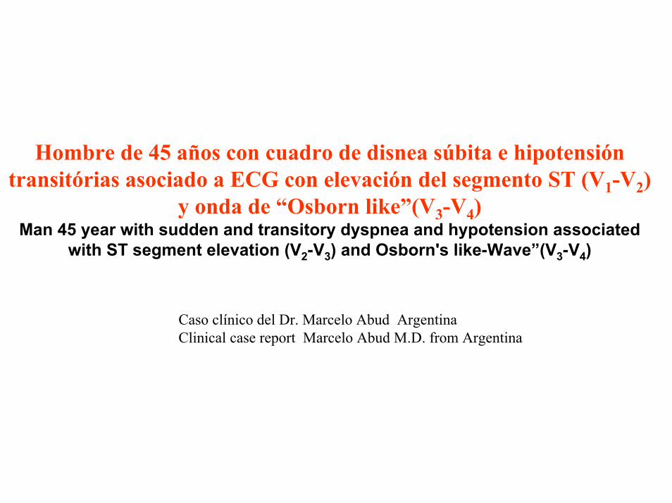
Hombre de 45 años con cuadro de disnea súbita e hipotensión transitórias asociado a ECG con elevación del segmento ST (V 1 -V 2 ) y onda de “Osborn like”(V 3 -V 4 ) Man 45 year with sudden and transitory dyspnea and hypotension associated with ST segment elevation (V 2 -V 3 ) and Osborn's like-Wave”(V 3 -V 4 ) Caso clínico del Dr. Marcelo Abud Argentina Clinical case report Marcelo Abud M.D. from Argentina

Welcome message from author
This document is posted to help you gain knowledge. Please leave a comment to let me know what you think about it! Share it to your friends and learn new things together.
Transcript
Hombre de 45 antildeos con cuadro de disnea suacutebita e hipotensioacutentransitoacuterias asociado a ECG con elevacioacuten del segmento ST (V1-V2)
y onda de ldquoOsborn likerdquo(V3-V4)Man 45 year with sudden and transitory dyspnea and hypotension associated
with ST segment elevation (V2-V3) and Osborns like-Waverdquo(V3-V4)
Caso cliacutenico del Dr Marcelo Abud Argentina Clinical case report Marcelo Abud MD from Argentina
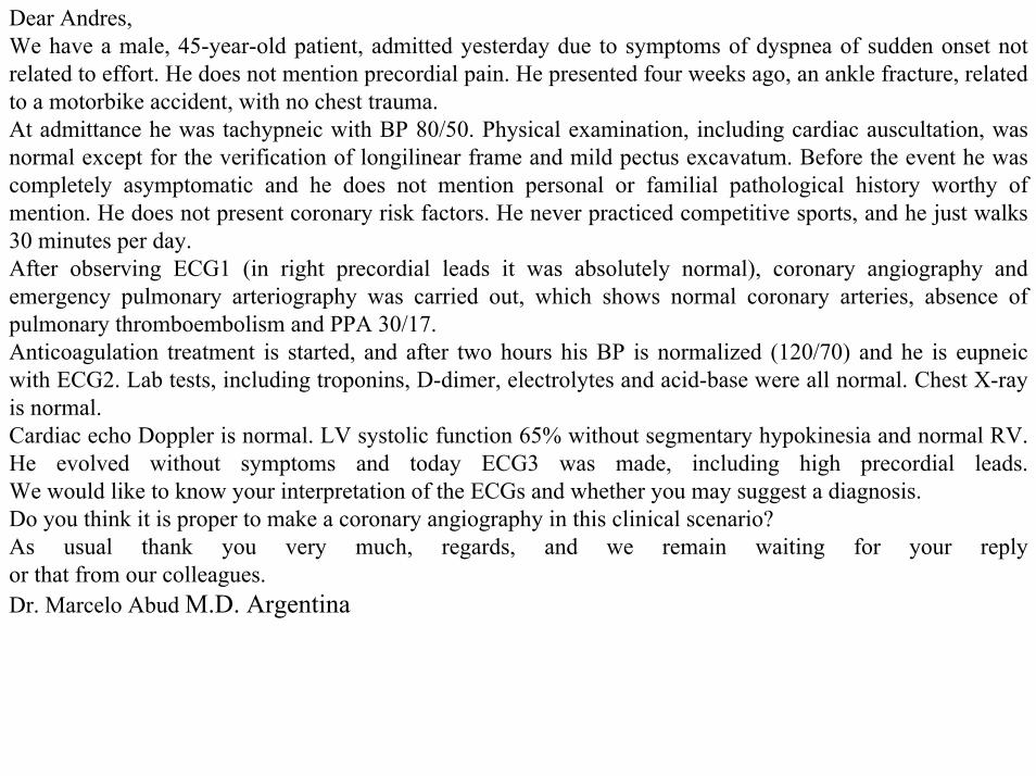
Dear AndresWe have a male 45-year-old patient admitted yesterday due to symptoms of dyspnea of sudden onset not related to effort He does not mention precordial pain He presented four weeks ago an ankle fracture related to a motorbike accident with no chest traumaAt admittance he was tachypneic with BP 8050 Physical examination including cardiac auscultation was normal except for the verification of longilinear frame and mild pectus excavatum Before the event he was completely asymptomatic and he does not mention personal or familial pathological history worthy of mention He does not present coronary risk factors He never practiced competitive sports and he just walks 30 minutes per dayAfter observing ECG1 (in right precordial leads it was absolutely normal) coronary angiography and emergency pulmonary arteriography was carried out which shows normal coronary arteries absence of pulmonary thromboembolism and PPA 3017Anticoagulation treatment is started and after two hours his BP is normalized (12070) and he is eupneicwith ECG2 Lab tests including troponins D-dimer electrolytes and acid-base were all normal Chest X-ray is normalCardiac echo Doppler is normal LV systolic function 65 without segmentary hypokinesia and normal RVHe evolved without symptoms and today ECG3 was made including high precordial leadsWe would like to know your interpretation of the ECGs and whether you may suggest a diagnosis Do you think it is proper to make a coronary angiography in this clinical scenarioAs usual thank you very much regards and we remain waiting for your reply or that from our colleaguesDr Marcelo Abud MD Argentina
Spanish
Estimado AndreacutesPaciente masculino de 45 antildeos que ingresa ayer por una cuadro de disnea suacutebita taquipneico no relacionada a esfuerzo e hipotenso (TA 8050mm de Hg) Y sin dolor precordial Habito longuilineo con discreto pectus excavatum Ascultacioacuten cardiaca normal Hace cuatro semanas tuvo una fractura de tobillo relacionada con un accidente de moto sin traumatismo toraacutecico Previamente al evento se encontraba totalmente asintomaacutetico y no refiere antecedentes patoloacutegicos personales o familiares destacablesNo presenta factores de riesgo coronario No realizoacute nunca deportes competitivos pero camina 30 minutos por diaPor el ECG de admisioacuten ECG 1 se le indica cinecoronagiografia y arteriografia pulmonar de urgencia que muestra arterias coronarias normales ausencia de TEP y presioacuten de arteria pulmonar de 3017Se comienza con tratamiento anticoagulante y a las dos horas normaliza su TA (12070)y se encuentraeupneico con un ECG2 El laboratorio incluyendo troponinasdimero D electrolitos y acido base fueronnormalesLa RX de toacuterax es normalEl ecocardiograma dopler cardiaco es normal FSVI 65 sin hipoquinesias segmentarias y VD normalEvoluciona asintomaacutetico y hoy se realiza un ECG 3 que incluye las derivaciones precordiales altasNos gustaria conocer su interpretacion de los ECG y si sugiere algun diagnosticoLe parece correcto la realizacioacuten de una cinecoronariografia en este contexto clinicoComo siempre muchas gracias un gran abrazo y esperamos su respuesta o la de nuestros colegasDr Marcelo Abud
ECG 1
ECG 2
ECG 3
Over the 3rd intercostal space
Over the 2nd intercostal space
Over the 4to intercostal space
Colleagues opinions
Estimado Dr Marcelo Abud La elevacioacuten del segmento ST en un varoacuten puede corresponder como en este caso a una repolarizacioacuten precoacutez (high take off)
iquestPorque no se trata de una isquemia aguda RespuestaPorque no se observan signos reciprocos o imagen en espejo (remodelacioacuten fisiologica)
iquestPorque no se trata de una embolia pulmonar aguda Respuesta Porque no tiene eje eleacutectrico del QRS desviado a la derecha
iquestPorque no es pericarditis aguda Respuesta porque las derivaciones no estan afectadas difusamente el intervalo PR es isoeleacutectrico no tiene precordalgia fiebre y no estaacute taquicaacuterdico (en la pericarditis aguda la FC estaacute casi siempre es alrededor de 120lpm)
Entonces iquestque tiene el paciente Respuesta Efecto androacutegeacutenico sobre el corazoacuten Los andreacutegenos aumentan la fuerza contraacutectil selectivamente en el miocito del epicaacuterdico incrementando el tenor de calcio del sarcoacutemero (ocasionando elevacioacuten del segmento ST ) y concomitantemente produciendo mayor salida del potasio durante la fase 3 por estiacutemulo del canal de salida lento de potasio ldquoslow delayed potasium receptorsrdquo responsable por el aspecto puntiagudo de la onda T Esta hipercontractibilidad epicaacuterdica observada en el sexo masculino aumenta el requerimiento del flujo coronario Este ECG muestra que la pared anterolateral baja (de V4 a V6) y la anterolateral alta (aVL I) son hipercontraacutectiles especulo que la arteacuteria circunfeja sea pequentildea con predominancia de la arteacuteria coronaacuteria derecha Esta circulacioacuten coronaria insuficiente puede ocasionar disnea o dolor precordialComo se corrige esto Respuesta con beta bloqueadores adreneacutergicosNo quiero criticar a los estudios invasivos que se le hicieron pero pienso que en este caso con 20 pesos se huviera solucionado el problemaUn fraternal abrazoSamuel Sclarovsky
Dear Dr Marcelo Abud ST segment elevation in a male may correspond such as in this case to an early repolarization pattern (high take off)Why it is not an acute ischemia Answer Because there are no reciprocal or mirror image on the ECG1 (physiological remodeling)Why he has not an acute pulmonary embolism Answer Because he has not QRS axis deviation to the rightWhy he has not an acute pericarditis Answer because the leads are not affected diffusely the PR interval is isoelectric there are not precordial pain no fever and not tachycardia (In acute pericarditis the HR is almost always around 120lpm)So why this patient has Answer androgenic effect on the heart The androgens selectively increase the contractile force in the epicardial layer myocytes with calcium overload inside of sarcomeres (causing ST segment elevation) and concomitantly producing more K+ output during phase 3 action potential by stimulating of slow delayed potasium channels (responsible for the sharp appearance of the T wave)This selective epicardial hipercontractility observed in males increases the coronary flow requirement This ECG shows both the low anterolateral (from V4 to V6) and high anterolateral (aVL I) walls hypercontractiles I speculate that the Cx artery is small with a predominance of the right coronary artery(RCA) This can result in inadequate coronary circulation dyspnea or chest painHow we approach this Answer with beta adrenergic blockersI do not want to criticize the invasive studies that were done but I think that in this case with ldquo20 pesosrdquo we solved the problemA fraternal hugSamuel Sclarovsky
Dudas y criticas al raciocinio del Profesor SamuelEl ECG1 muestra clara elevacioacuten del segmento ST confinado apenas a V2-V3(regioacuten medioseptal) En la repolarizacioacuten precoz la elevacioacuten del ST es de concavidad superior terminando en una T positiva amplia de V2 a V4 o V5 ldquoprominent J wave and ST-segment elevation concave to the top predominantly in left precordial leads ending in a positive large T wave from V2 to V4 or V5rdquo La repolarizacioacuten precoz excepcionalmente tiene elevacion del ST en las precordiales derechas En la repolarizacioacuten precoz claacutesica benigna el supradesnivel de punto J y segmento ST de concavidad superior y seguido por onda T apiculada que se observa en la gran mayoria de los casos de V4 a V6 yo en la pared inferior En este caso no se observa supradesnivel del segmento ST en esta aacuterea Ademaacutes no vemos bradIcardia sinusal que es lo que suele verse en la repolarizacion precoz Si se fijan bien existe aqui discreto infradesnivel del ST de 1mm en V5 ldquoJ point and ST segment depressionrdquoPor otra parte este paciente asociado a la dispnea tuvo una importante hipotensioacuten arterial transitoacuteria
Aqui claramente en el ECG 1 se observan muescas o empastamiento en la rama descendente de la R mas conspicuas de V3 a V5 pero tambien visibles en V2 y V6 Ese hayazgo si es compatible con repolarizacion precoz ldquonotching irregular or slurring contour of the terminal QRS complexrdquo
Por otra parte querido maestro en este caso el aspecto del ST en V2 es de convexidad superior ldquo convex to the toprdquo absolutamente diferente del supradesnivel del ST de la repolarizacion precoz y va seguida de una onda T negativa En la Repolarizacion precoz el supra de ST es de concavidad superior seguida por una T puntiaguda positiva concordante Andres
Querido amigo amigo maestro Adrian Baranchuk disculpeme por mi condicion de otista pero mas claro no puedo ser y sobre en hombre de 45 antildeos con hipotensioacuten Me supongo que alguien que vio el ECG con disnea le administroacute nitritro sublingual que ocasiona hipotensioacuten transitoacuteria y taquicardia sinusalSeria interesante si el Dr Abud envie otro ECG en reposo sin taquicardia y presion normal estoy casi seguroque el ST-T seran mas alto a pesar que a los 45 antildeos estos hallazgos en el ECG estan atenuadosPor suerte mi amigo profe Andreacutes Ricardo Peacuterez Riera se tomo la molestia en traducirme del espantildeol al espantildeolUn fraternal abrazo y a todos los amigos de la colectividad judia feliz anio nuevo
Samuel SclarovskyQuerido amigoQue le hayan dado un nitrito es SOLO una suposicion bastante facil es preguntarle al paciente no creePero estoy seguro que Abud o Perez-Riera lo hubieran clarificadoPor otro lado hipotensioacuten sostenida no es frecuente de ver en ese escenario que Ud describe Digo todo esto porque Ud en su email usando un lenguaje un poco sarcaacutestico dijo que este caso Ud lo resolvia con 20 pesos y NO con el gasto de estudios invasivos Yo estoy en desacuerdo Porque Porque Ud corre con el caballo del comisario y sabe que todo dio normal pero yo aqui en Canada le hubiera hecho los mismos (o similaresestudios)Por favor una cosa es la especulacioacuten luego de saber los resultados y otra muy distinta ser el colega de guardia que tuvo que decidir esta conductaYo no se que tiene el paciente pero le aseguro que tambien hubiera descartado TEP primero e isquemiadespueacutesEso de que NO tiene BCRD y por eso NO tiene TEP es demasiado estricto ya que el BCRD se ve en solo el 60 de los casos (hay un lindo review que escribimos hace un tiempo busquelo que ahi estan todos loscambios de TEP en el ECG)Un fuerte abrazo y disculpe mi descuerdo en este casoAB
Samuel
1 Ud dice me parece que Ud esta errado En la actualidad es mejor discutir diciendo yo pienso que No es necesario descalificar al otro por el simple hecho de no estar de acuerdo Yo ya le dije no se lo quetiene el paciente pero hubiera hecho los mismos estudios Eso NO es estar errado es simplemente estar en desacuerdo con Ud (y estoy seguro porque lo conozco que Ud no se cree el duentildeo de la verdadno cierto)
2 Ud dice yo hace muchos antildeos que me dedico a esto Es cierto Y Eso lo transforma en duentildeo de la verdad Yo me dedico con igual pasion que Ud y eso NO me hace duentildeo de la verdad
Concuerdo en el uso racional de los estudios 100 Pero ese NO es le tema del paciente en cuestion sinoque le produjo un cuadro tan severo con un ECG tan anormal A veces para aprender hay que invertir No hay otra
Le mando un abrazo sincero
AB
Professor Andreacutes e Caro Dr MarceloInteressantiacutessimo casoA histoacuteria cliacutenica sugere episoacutedio tromboemboacutelico =gt Trauma com fratura de tornozelo - Repouso ouimobilidade do membro =gt Trombo embolismo coronaacuterio - (MI Type 2 - Expert Consensus Document -European Heart Journal doi101093eurheartjehs184apub 0912) - - Resoluccedilatildeo do quadro com anticoagulante ndashQuanto ao ECG-1 - Sindrome isqueacutemica aguda c ST supra V2-V3 imaacutegem ST em espelho infra V4-V5 Discretiacutessimo supra em D1- aVL FC 88bpm Quadro cliacutenico e ECG praticamente normalizado 2 horasdepois Quanto as enzimas foram seriadas Embora o episodio todo possa ter ocorrido s alteraccedilotildeesenzimaacuteticas Observar que o J Osborn -like tambeacutem desaparece e todos os fenoacutemenos se normalizam nos ECGs do diaseguinte Natildeo teria feito cateterismo Adail - Bahia - Brasil
Interesantiacutesimo caso Elevacioacuten de segmento ST tipo Brugada o tipo infarto anterior en V2 y ondas J bastante locaizadas en V3-V4 La respuesta a la ultima pregunta de Marcelo es faacutecil Cualquier medico responsable hubiera hecho los estudios necesarios para excluir un infarto agudo y una embolia pulmonar en un paciente con disnea suacutebita y este electrocardiogramaLa otra pregunta de Marcelo cual es la interpretacioacuten del electrocardiograma una vez sabiendo que no hay evidencia de lesiones coronaria o de embolia pulmonar tambieacuten es faacutecil de contestar Mi respuesta ldquono tengo ideardquo Que puede ser esto1 Respuesta a hiperventilacioacuten Este tema ya no esta de moda pero estuvo muy de moda en el siglo pasado (hijos que feo se oye eso) Ejemplos(123)El problema con esta teoriacutea es que la hiperventilacioacuten en gente sana produce inversioacuten de onda T y depresioacuten del segmento ST Pero en pacientes con enfermedad coronaria la hiperventilacioacuten puede producir elevacioacuten de segmento ST(4) Por tanto uno podria argumentar que las coronarias de este paciente aparentemente normales en la angiografiacutea pero no son realmente normales2 Coronary spasm Faltan los cambios reciacuteprocos en otras derivaciones y en paciente no tubo dolor de pecho3 Pericarditis No claacutesico pero siempre puede ser Si usamos este diagnostico cada vez que no tenemos idea que esta pasando por que no usarlo aquiacute4 Un infarto localizado de la arteacuteria del cono ldquoconal branchrdquo de la arteria coronaria derecha(CD) Es posible Tan posible que moveriacutea esta posibilidad para arriba Recientemente tuve un paciente con gran infarto inferior por oclusioacuten de la CD Tenia elevacioacuten del segmento ST en II III y aVF
1 Savonitto S et al Different significance of hyperventilation-induced electrocardiographic changes in healthy subjects and patients with coronary artery disease Eur Heart J 199617(9)1302
2 Lary D Goldschlager N Electrocardiographic changes during hyperventilation resembling myocardial ischemia in patients with normal coronary arteriograms Am Heart J 197487(3)383-90
3 Lewis WC Siebecker KL Jr Wasserburger RH The effect of hyperventilation on the normal adult electrocardiogram Circulation 195613(6)850-5
4 Eur Heart J 199617(9)1302)
Fue sometido intervencioacuten coronaria urgente con dilatacioacuten y ldquostentingrdquo de el tramo proacuteximo de la CD y al poner el stent se obstruyoacute la rama del cono ldquoconal branchrdquo ocasionando un patroacuten ECG ldquoBrugada-likerdquo muy semejante a este caso y el paciente desarrolloacute fibrilacioacuten ventricular Mientras mas lo pienso mas me gusta esta posibilidad5 Simple ldquorepolarizacion precozrdquo Puede ser Pero el paciente presenta con disnea y tachycardia El ldquostressrdquo deberiacutea disminuir los efectos electrocardiograacuteficos de repolarizacion precoz que generalmente aumentan durante tono vagal6 Siacutendrome de Brugada Puede ser El hecho de que desaparezca o disminuya al diacutea siguiente no descarta esta posibilidad Definitivamente yo le hariacutea una prueba de ajmalina antes de mandarlo a la casa Por cierto que hay controversia en lo que se refiere al efecto del ldquostressrdquo en el ECG del Brugada En Aacutemsterdam el segmento de ST de Brugada supradesnivela durante el ejercicio(1) En Japoacuten el ST normaliza durante el ejercicio y aumenta durante la fase de recuperacioacuten (2)7 Medicamentos que ldquoproducen Brugadardquo (brugadadrugsorg) La lista es grande y para la mayoriacutea los efectos aumentan durante taquicardia8 Embolia pulmonar que resolvioacute No se hizo tomografiacutea CT del pulmoacutenhellip sin ofenderhellip la arteriografiacutea pulmonar no es faacutecil de interpretar Ademas el paciente recibioacute anticoagulantes9 Tumores o ldquomiscelaacuteneosrdquo del mediastino Esto es muy importante checarlo (34)Definitivamente solicitaria una tomografiacutea (CT) de toacuterax antes de mandar al paciente a la casaY pus total siempre queda la otra posibilidad No seSami Viskin (Tel Aviv)
1 min AS de Groot EA Ruijter JM Wilde AA Tan HL Exercise-induced ECG changes in Brugada syndrome Circ ArrhythmElectrophysiol 20092(5)531
2 Augmented ST-segment elevation during recovery from exercise predicts cardiac events in patients with Brugada syndrome JACC 20103 Nakazato Y et al Brugada-like precordial ST elevation on ECG by anterior mediastinal infective mass lesion Indian pacing and
electrophysiology journal 20033(3)184Tarin N Farre J Rubio JM Tunon J Castro-Dorticos J Brugada-like electrocardiographic pattern
in a patient with a mediastinal tumor Pacing Clin Electrophysiol 199922(8)1264-6
Descartada obviamente la hipotermia por exposicion laboral yo accidental las intoxacionesmedicamentosas o por monoxido de carbono Asi como tambien la infeccion posquirurgica con bacteriemiao sepsis Brugada-Like ECG Pattern in Severe Hypothermia Circulation2008118977-978Dr Sami muy interesante su analisis y la evaluciacion de los diagnosticos diferenciales
El paciente se presenta con hipotension taquicardico y cambios en el ECG muy similares a los descriptos en el experimento en perros Cellular Basis for the Electrocardiographic J Wave Gan-Xin Yan MD PhD Charles Antzelevitch y analizan los cambios endo epicardio como los responsables posibles del origende dicha onda y ademas en el experimento encontraban cambios ECG de v4 a v6 similar a este paciente y en precoridales derechas simulando un patron de Brugada interpretandolos por la hipotermia y a la hiperalcemia transitoria y el desalloro de acidosis (cual papel sera el de esta en la velocidad de lospotenciales de accion)Su analisis creo merece solo un comentario en el TEP los cambios caracteristicos de S1Q3T3 o aparicionde BRD con eje desviado a la derecha se ve en la mayoria de los pacientes no en todosEl dimero D puede ser negativo (es un producto de la degradacion de la fibrina) en un 5 de estos la angiografia pulmonar descarta los embolismos masivos o de arterias pulmonares pero tampoco descartatotalmente el TEPEl ecocardiograma al igual que los otros metodos solo resulta positivo en la mayoria de los casos contribuye al diagnostico pero no lo excluyeDe lo referido llama la atencion un paciente sin patologia previa conocida cirugia de tobillo reciente en casos de politraumatismo la incidencia de TVP es del 20 con probabilidad de presentarse como TEP del 05 no refirio el Dr Abud si recibia profilaxis para trombosis venosa profunda La ausencia de coagulo en MII disminye las probabilidades de TEP pero tampoco lo excluyenLa TAC helicoidal (ya se realizo una arteriografia pulmonar) no creo aporte al diagnostico presenta unasensibilidad del 90 cuando el embolismo es de las arterias de primer y segundo orden y al incluir losvasos subsegmentarios esta cae al 67 los TEP subsegmentarios son alrededor del 6 en el estudioPIOPED
La Escala de Shier para evaluar la probabilidad de embolias grasas en las primera 72 hs luego del accidentees util para predecir la probabilidad de TEP de este origen en este caso seria extremadamente raro se presentara tardiamente como han referidoLa onda J de Osborn descripta por este cuando experimentaba con perros al inducirle hipotermia(1) el primero en describirla fue Tomaszewski que encuentra cambios en el ECG de un hombre fallecido por hipotermia (1)No encuentro una explicacion concreta pero en medicinatodo tiene una explicacion y no hay casualidades sino causalidades podremos diagnosticarlas correctamenteo no pero el criterio de estudio del paciente me parece el adecuado Este paciente en particular presenta al ingreso una elevacion del punto J que disminuye pero no desaparece en los ECG posteriores lo que lleva a pensar en un transtorno agudo de una patologia cronica lo que va en contra de pensar en TEP En el Brugada la elevacion del punto J en derivaciones inferiores esa descripta pero en estudios experimetales tambien la presentan en cara lateral estos estudios experimetales en laboratorio de la diferencia endo-epicardica de potenciales (les adjunto un link de un ECG exacto al presentado para que observenhttpwwwncbinlmnihgovpmcarticlesPMC2843811figureF5 y se trata de un SBr no hipotermia peroen este presenta repolarizacion precoz lo que no se observa en el paciente presentado En la taquicardia la notched esta acentuada como ocurre en este paciente Ya lo han anticoagulado o sea que esta recibiendotratamiento para no repetir en caso de haber sido un TEPPodriamos excluir la probabilidad de que se tratara de un SBr asociado a replarizacion precoz Porque no realizarle una prueba con flecainida a fin de descartarloUn saludoMartin ibarrola
1 Tomaszewski W Changements electrocardiographiques observes chez un homme mort de froid Arch Mal Coeur 193831525) extraido de EKGWEB
2 Osborn JJ Experimental hypothermia respiratory and blood pH changes in relation to cardiac function Am J Physiol 19531753893 Genetic Molecular and Cellular Mechanisms Underlying the J Wave Syndromes Charles
Antzelevitch1) httpswwwjstagejstgojparticlecircj76576_CJ-12-0284_pdf4 J Wave SyndromesCharles Antzelevitch PhD FHRS1 and Gan-Xin Yan MD PhD234
httpwwwncbinlmnihgovpmcarticlesPMC28438115 Paradoxical effect of ajmaline in a patient with Brugada syndromeBiagio Sassone EP Europace Volume 8 Issue 4 Pp 251-254 6 Brugada syndrome with atypical ECG downsloping ST-segment elevation in inferior leadsRiera ARP Ferreira C Schapachnik E J
Electrocardiol 2004 Apr37(2)101-4
Final commentsBy Andreacutes Ricardo Peacuterez-Riera MD PhD
ECG-1 FROTAL PLANE
aVR aVL
I
IIIII
aVF
X
Y
QRS axis +30deg
Reciprocal ST-segment changes(ST-segment depression)
AB
II
TST
Early Repolarization Pattern (ERP) is defined as notching or slurring morphology of the terminal portion of the R wave (J-wave) that produces a positive hump known as a J-wave at the end of the QRS complex and beginning of the ST segment J-point elevation ge01 mV (or 1mm) above baseline in at least 2 in the mid-to-left precordial leads andor inferior leads followed by concordant tall near symmetric (T waves having similarity in shape ascending and descending ramps speeds) There is a distinct J wave and ST segment concave upward in the left precordial leads V4 through V6 The ST elevation is most frequently evident in ECG lead V4
Where is the located the end of QRS complex In A or in B If the answer is B QRSd is near 120ms If the answer is A QRSd is near 70ms
J-wav
e
ECG-1
V6
V1
V4
V5
V2
V3
X
Z
ECG-1 HORIZONTAL PLANE
V2
V5
J point
QRSd 122ms
ECG-1
QRSd 122ms
Notching
40 ms80 ms
β
80 ms
High take-off
Type 1 Brugada pattern
Type 2 Saddle-back pattern
40 ms80 ms
β
80 ms
High take-off
Type 1 Brugada pattern
Type 2 Saddle-back pattern
V2
Is this a type 2 Brugada pattern or saddle-back pattern in V2
Tryphasic QRS Pattern RSracute
High take-off of racute ge 2mm high take-off frequently does not coincide with J wave descending arm of racutecoincides with beginning of ST segment the racute wave has rounded shape wide and usually of relatively low voltage in Brugada pattern Contrarily in IRBB the racute wave is peaked the angle between the upslope of the S-wave and the downslope of the r-wave (β angle) gt 58deg duration of the base of the triangle of racute at 5mm from high take-off gt35mm the QRS duration is longer in Brugada pattern type 2 than in other cases with racute in V1 and there is a mismatch between V1 and V6 In Brugada pattern the QRS complex end is earlier in V6 than in V1 V2
βangle gt 58deg
1 Bayeacutes de Luna A et al a consensus report Journal of Electrocardiol 2012 45433-
2 Chevallier S J Am CollCardiol2011 Nov 2258 2290-2298
ECG-1
ECG-1
RIGHT SAGGITAL PLANE ECG-1
YaVF
Z V2
ECG 2
aVR aVL
I
IIIII
X
Y
aVF
QRS axis +30ordm
ECG-2 FRONTAL PLANE
aVR aVR
ECG-1 ECG-2
J-Wave
Reciprocal ST-segment changes (ST-segment depression) Without reciprocal changes or mirror image
In Early Repolarization Pattern reciprocal ST-segment changes (ST-segment depression) are eventually confined the unipolar aVR lead (Absence of reciprocal changes or mirror image in others leads)
II
II
PR interval= 160msQRSd is 121ms
ECG-1
PR interval = 140msQRSd is near 80ms
J wave
ECG-2
V6
V5
V1
V4
V2
V3
X
Z
Is this a type 2 Brugada ECG pattern Answer in next slide
ECG-2
V2βangle gt58deg
Observation β angle is related to both depolarization an repolarization A wider QRS complex (slow conduction) and abnormal repolarization both contribute to a wider β angle
1 Chevallier S J Am Coll Cardiol2011 Nov 2258 2290-2298
In patients with suspected Brugada syndrome simple ECG criteria can enable discrimination between innocent incomplete RBBB from types 2 and 3 Brugada ECG patterns The mean β angle is significantly smaller in the patients with negative results on Antiarrhythmic Drug Challenge (ADC) compared wit patients with positive results on ADC Its optimal cutoff value is 58deg which yielded a positive predictive value of 73 and a negative predictive value of 87 for conversion to type 1 pattern on ADC α is slightly less sensitive and specific compared with β When the angles are combined with QRS duration it tended to improve discrimination
Y
aVF
Z V2
Right Sagittal Plane ECG-2
Triphasic QRS patternRSracute
β angle defined as the angle between the upslope of the S-wave and the downslope of the r-wave The mean β angle was significantly smaller in the 14 patients with negative results on AAD compared to the 24 patients with positive results on AAD Itrsquos optimal cutoff value was 58deg which yielded a positive predictive value of 73 and a negative predictive value of 87 for conversion to type 1 pattern on AAD
ST segment with saddleback appearance followed by positive or biphasic T wave in V1-V2 or from V1 through V3
β
V5V5
QRSd= 122ms QRSd= 110ms
ECG-1 ECG-2
Left precordial terminal QRS notching is more prevalent in malignant early repolarization abnormalities
Left precordial terminal QRS notching
1 Merchant FM et al Am J Cardiol 2009 Nov 15 1041402-1406
V2
V2
V3
V3
ECG-1
ECG-2
ECG-2
Dynamic inconstant or dramatic changes in repolarization are suggestive of malignant early repolarization abnormalities
1 Haiumlssaguerre M et al J Cardiovasc Electrophysiol 2009 Jan2093-98
ECG-1
Conclusion this patient has high possibility of concomitant Brugada syndrome and early repolarization In this a benign ERP or malignant early repolarization abnormalities We think it is very unlikely that is a Brugada phenocopyIn this case it is necessary additionally to perform
1 IV Ajmaline test2 ECG-AR3 Family study ( first degree relatives interrogatory physical ECGs)4 Magnetic resonance image5 Genetic screening
Ventricular biopsy is not considered mandatory but is recommended especially when it may clarify the nature of dubious findings identified with others test In approximately 6--10 of survivors of cardiac arrest no cardiac abnormality can be identified despite extensive clinical evaluation Autopsy data confirm that in a similar percentage of victims of SCD no structural heart disease or or mors sine materia can be identified at post mortem evaluation Incompletely penetrant genetic defects may underlie at least some of these unexplained deaths (1)
The ERP probably represents part of a spectrum of cardiovascular anomalies related to channelopaties including BrS IVF congenital short QT syndrome and that it may also have a molecular genetic origin of variable penetrance The denomination Early Repolarization Pattern is appropriate since it indicates that it is characterized by a given ECG pattern
1 Priori SG et al Cardiovasc Res 2001 May50218-223
Case Report similar
A 20 year-old male mulatto professional soccer player from Santo Andreacute Sao Paulo Brazil He didnrsquot finishthe second year in elementary school CatholicProfession Professional soccer playerReason for consultation Presented to consultation due to two presyncopal episodes and one syncopal episode in the last 30 days The first two episodes happened during a post-defecation period Both were preceded by dizziness nausea and vomiting The syncopal episodes occurred at rest with no prodromes The third one with express and brief loss of consciousness and sphincter relaxation at dawn 48 hours agoHis prior personal medical history was unremarkable and he successfully passed two prior periodical clinical-cardiovascular evaluations (twice a year always normal) Pre-competitive examination included 12-lead ECG echocardiogram and cardiopulmonary metabolic exercise testing (CMET) All normals Intense training for the last 3 years Familial Background One uncle from his fatherrsquos side family died of sudden cardiac death when he was 35
years old He ignores the cause His brother is being seeing by a cardiologist due to ldquocardiac arrhythmiardquo in treatment with a cardiologistfrom the Institute of the Heart from Satildeo Paulo (he cannot tell which is the cause) He denies having other relevant background details Pysical cardiovascular examination revealed bradycardia at 45 bpm with physiological sinus arrhythmia blood pressure 11070 mmHg The rest of the physical examination was unremarkable He denied consuming any type of neither medications nor drugs No addictionsHis first 12-lead ECGVectorcardiogram(VCG) (Fig 1) showed sinus bradycardia at 44 bpm P-wave axis of +60deg PR interval of 186ms QRS duration of 111ms QRS axis at + 63deg QT interval 427ms and corrected QTc 367ms There is a notch on the terminal portion of the QRS complex in the left precordial leads voltage criteria for left ventricular hypertrophy (SV1 + RV5 gt 35 mm ndash Sokolow index) prominent J-wave and concave ST-segment elevation in inferior leads V4 V5 and V6 Deep and narrow Q-waves in the left precordial leads V5 and V6 All these features were considered as an Early Repolarization Pattern (ERP)
Name JVS Gender Male Age 20 yo Ethnic Group Mulatto Weight 66 Kg Height 172 m Biotype Normoline Date 11262009 Profession competitive athlete (soccer player)
ECG diagnosis Sinus bradycardia (SB) HR 44bpm P axis +60deg PR 186ms QRSd 111ms QRS axis + 63deg QT 427ms QTc 367ms Conclusion SB + Early Repolarization Pattern (ERP) Notching or slurring of the terminal QRS complex voltage criteria of left ventricular hypertrophy SV1+ RV5 gt 35 mm (Sokolow index) prominent J wave and ST-segment elevation concave to the topending in a positive T wave from V4 to V6 Precordial greater than limb leads Relatively deep but narrow q waves in the left precordial leads (V5 and V6)
V6V4 V5
Prominent J wave and ST- segment elevation concave to the top ending in a positive T wave from V4 to V6 Notching or slurring morphology of the terminal portion of the R wave (J-wave) that produces a positive hump known as a J-wave (Arrows)
aVR
Eventually reciprocal ST segment depression confined only in Lead aVR1 observed in this particular case report
1 Mehta M Jain AC Mehta A Early Repolarization Clin Cardiol 1999 Feb 22 59-65
ECGVCG CORRELATIONS HORIZONTAL PLANE
V6
V1
V2
V3
X
Z
V5
V4
T
0J T
BEGINNING AND END OF QRS LOOP IN HP
J POINT in vectorcardiography it corresponds to 3 elements end of ventricular depolarization (QRS loop) beginning of repolarization (ST segment) when it does not present depression or elevation and T wave onset In this case 0 point and J point are not coincident In situations where there is depression or elevation of ST segment the J point does not coincide with the 0 point and the greater or lesser distance between both points indicate the greater or lesser ST segment elevation or depression The phenomenon is observed in early repolarization pattern acute phase of myocardial infarction Prinzmetal variant angina pericarditis Brugada syndrome arrhythmogenic right ventricular dysplasia etc
0 POINTIt corresponds to the QRS looponset (because PR segment does notexist) Alsocorrespond to the end of biatrialchamberactivation
T
J POINT
ECGVCG CORRELATIONS FORNTAL PLANE
aVR aVL
I
IIIII
X
Y
VERTICAL AXIS ON FRONTAL PLANE The frontal plane QRS axis and ST segment axis and T wave axis are all in the same direction THE ST
SEGMENT ELEVATION
USUALLY lt 05 mm IN THE LIMB
LEADS
YVF
Z V2
T
ECGVCG CORRELATIONS RIGHT SAGITTAL PLANE
A second 12-lead ECG (Fig 2) preformed 72 hours later showing1) Sinus bradycardia at 44 bpm2) Similar p-wave axis and intervals duration3) Dramatic modification of repolarizaccedilatildeo pattern in the right precordial leads A coved-type
(type 1) ECG brugada pattern can be distinguished4) Accentuation of j-waves and st segment elevation across the precordial leads was observed5) ST segment elevation on avr lead
See next slide
Name JVS Gender Male Age 20 yo Ethnic Group MulattoWeight 66 Kg Height 172 m Biotype Normoline Date 11292009Profession Competitive Soccer Player FIGURE 2
ECG diagnosis Sinus Arrhythmia(SA) sinus bradycardia (SB) HR 44bpm P axis +60deg PR 186ms QRSd 111ms QT 427ms QTc 367 Conclusion SA + SB + Early Repolarization Pattern (ERP) Type 1 Brugada ECG pattern Dramatic accentuation of J waves across the precordial and limb leads Transient augmentation of global J waves may be indicative of a
highly arrhythmogenic substrate heralding multiple episodes of VF in patients with ERP
aVR LEAD
11262009 11292009
DEPRESSION
ELEVATION
ST SEGMENT DEPRESSION IN aVR = 1mm ST SEGMENT ELEVATION IN aVR = 2mm
V1 V2V2
1129200911262009
V1
11292009 11262009
SPONTANEOUS MODIFICATION OF REPOLARIZATION TO TYPE 1 BRUGADA ECG PATTERN
11262009
V6
11292009V6
ST segment elevation lt 2mm ST segment elevation gt 2mm
11292009
V5
11262009
V5
ST segment elevation = 1mm ST segment elevation = 25mm
EARLY REPOLARIZATION PATTERN CONCEPT
DEFINITION Early Repolarization Pattern (ERP) is a well-recognized Enigmatic idiopathic electrocardiographic phenomenon characterized by notching or slurring morphology of the terminal portion of the R wave (J-wave) that produces a positive hump known as a J-wave at the end of the QRS complex and beginning of the ST segment It considered to be present when at least two adjacent precordial leads show elevation of the ST segment with values ge01 mV (or 1mm) above baseline followed by concordant tall near symmetric (T waves having similarity in shape ascending and descending ramps speeds)
EPIDEMIOLOGY ERP occur in 2 to 5 of the population often in men young adults afro-descendents and competitive athletes and in up to 131 to 482 of patients presenting with chest pain in the emergency rooms and coronary care units There is a distinct J wave and ST segment concave upward in the left precordial leads V4 through V6 The ST elevation is most frequently evident in ECG lead V4 The ST segment elevation is usually le2mm (precordial greater than limb leads) and is lt 05 mm in the limb leadsReciprocal ST-segment changes (ST-segment depression) are eventually confined the unipolar aVR lead (Absence of reciprocal changes or mirror image) In ERP vertical QRS axis electrical is the rule The frontal plane QRS axis the ST segment axis and T wave axis are all in the same directionThis ECG pattern has been considered a benign normal ECG variant for over six decades3
1 Hasbak P Engelmann MD Early repolarization ST-segment elevation as a normal electrocardiographic variant Ugeskr Laeger 2000 162 5928-5929
2 Brady WJ Chan TC Electrocardiographic manifestations benign early repolarization J Emerg Med 1999 17473-478
3 Benito B Guasch E Rivard L Nattel S Clinical and mechanistic issues in early repolarization of normal variants and lethal arrhythmia syndromes J Am Coll Cardiol 2010 Oct 5 56 1177-1186
MAIN ELECTROCARDIOGRAPHIC FEATURES IN ERPHeart rate Predominant sinus bradycardia frequently associated with respiratory sinus arrhythmia
PR interval First degree AV block is observed in 5 to 39 among professional athletes In the non-athlete population it is observed in 065 of cases QRS axis Vertical electrical axis is the rule The frontal plane QRS axis and ST segment axis and T wave axis are all in the same directionQRS duration (90 plusmn 10 ms) subjects than in age-matched healthy controls (80 plusmn 10 ms) QRS morphology Notching or slurring of the terminal QRS complex Prominent relatively deep but narrow q waves may appear in the left precordial leadsQRS voltage Eventually voltage criteria of LVH are observed in male competitive athletes SV1+ RV5 gt 35 mm (Positive Sokolow index) High QRS voltage is more frequent in male athletes but its correlation with LVH is low Voltage decreases after deconditioning is slow The distinction between the physiological athletes heart and pathological conditions has critical implications for professional athletes An abrupt transition may occur from right-oriented complexes to left-oriented complexes in the precordial leads secondary to counterclockwise rotation on precordial leads About two-thirds of clockwise rotation and counterclockwise rotation could be attributed to the septal angle by anatomical rotation of the heart in one plane around the long axis but other factors appear to be responsible for such ECG findings in the remaining one-third of cases Relatively higher positions of the precordial ECG leads as observed in the vertical heart appeared to be responsible for clockwise rotation in some patients and left septal fascicular block is suspected to be responsible for counterclockwise rotation in others patientsJ-point elevation Notching irregular or slurring contour of the terminal QRS complex (J point) Variant of Osborn wave is seen in ERP
ST segment Widespread ST segment elevation (precordial greater than limb leads) The characteristic ST segment is elevated upward concave confined more frequently in precordial leads with reciprocal depression only in aVR The concavity is observed at the initial up-sloping portion of ST segment or upwardly concave ST segment morphology Unfortunately concave ST morphology cannot be used to rule out ST elevation from AMI with left anterior descending coronary occlusion because it is common in these circumstances The ST elevation is most frequently evident in ECG lead V4 There is a distinct J wave and ST segment in the left precordial leads V4 through V6 The ST elevation in ERP is usually lt 2 mm (but can rarely be gt 5 mm) in the precordial leads and the greatest ST elevation is usually seen in the mid-to-left precordial leads The ST segment elevation is usually lt 05 mm in the limb leads
T wave characteristics Concordant T waves of large amplitude (prominent matching T waves) typical pseudo-asymmetrical (lsquosymmetroidrdquo) or slightly asymmetrical matching T waves often of large amplitude upright tall and peaked most conspicuously from V2 to V4 or V5 sometimes seen in leads DII DIII and aVF as a rule T waves may appear as of large amplitude peaked or pointed symmetric and matching Vagotonic or high T wave voltages followed by U waves are frequent when sinus bradycardia is present Tall positive ad symmetric or symmetroid T waves are not only seen occasionally in the very early stages of MI but also in hyperkalemia and in ERP sinus bradycardia
QT intervals QT maximum The maximum Q-onset-T-end interval This parameter is higher in ERV subjects than in normal controls
QTp maximum maximum Q-onset-T-peak interval This parameter is higher in ERV subjects than in age-matched healthy controls
Rate-corrected QTc maximum This parameter is lower in ERP subjects than in age-matched healthy controls
QTpc maximum This parameter is lower in ERV subjects than in age-matched healthy controls
U wave because bradycardia U waves are frequent in ERV they are best observed in the V3 lead U waves are frequent when sinus bradycardia is present
Other ECG characteristics of ERPRelative temporal stability of the ST segment and T wave pattern is observedReciprocal changes are not seen in ERP There are no evolutionary short-term changes in the ST segment and T waves and Q waves do not appear
Discusions Early Repolarization Pattern (ERP) or Early Repolarization Variant (ERV) is an enigmati
electrocardiographic phenomenon characterized by prominent J wave and ST-segment elevation in multiplleads
Recently there has been renewed interest in ERP because of similarities to the arrhythmogenic Brugadsyndrome (BrS) Not much is known about the epidemiology of ERP and several studies have reported thathis condition is associated with a good prognosis
Both ERP and BrS exhibit some similarities including the ionic underlying mechanism the analogouresponses to changes in heart rate and autonomic tone sympathicomimetics (isoproterenol test) as well as insodium channel and beta-blockers These observations raise the hypothesis that ERP may be not as benign atraditionally believed Additionally there are documents showing that ST-segment height in the man igreatly influenced by central sympathetic nervous activity both at baseline and during physiologic andpharmacological stress Central sympathetic dysfunction regularly results in multilead ST-segment elevationor J wave that decreases or below isoelectric baseline during low dose isoproterenol infusion An earlyrepolarization pattern in the inferior leads of a standard electrocardiogram is associated with an increased riskof death from cardiac causes in middle-aged subjects1
POSSIBLE SIMILARITIES BETWEEN ERV AND BrSbullMore frequent in malesbullBoth occur more frequently in young adults and in individuals without apparent structural heart diseasebullBoth may influence just the V1-V2 leads Rarely (9) can ST elevation be observed in ERP only in the right precordial leads V1-V2 or in the inferior ones2
1 Tikkanen JT Anttonen O Junttila MJ Aro AL Kerola T Rissanen HA Reunanen A Huikuri HV Long-Term Outcome Associated with Early Repolarization on Electrocardiography N Engl J Med 2009 Nov 16 [Epubahead of print]
2 Hasbak P Engelmann MDEarly repolarization ST-segment elevation as a normal electrocardiographic variant Ugeskr Laeger 2000 162 5928-5929
When ST elevation is normal it can reach up to 3 mm in V2-V3 especially in young people In those individuals over 40 years it seldom exceeds 2 mm Both can show incomplete RBBB pattern or right bundle branch conduction disorder in BrS it can present atypical features RBBB-like and of the saddle type by exclusive elevation of the J point S wave with delay in the left leads DI aVL V5 and V6 could be absent as it is to be expected in a classic RBBB The elements considered as typical in BrS are 1) elevation of the terminal part of QRS (prominent J wave) 2) elevated and descending ST not related to lesion of ischemic (idiopathic) injury 3) negative T wave in the right precordial leads 4) normal QTc or near normal 5) Eventually absence of final delay in left leads as it would be expected in a classic RBBB1 In ERV when associated to athletersquos heart QRS can present a moderate extension (100 ms to 110 ms) in 15 of the cases which in nonathlete normal population in a 24 is called outflow tract hypertrophy In this case r does not exceed the 5 mm and is lower than S in the same lead rSrBoth may improve repolarization during the stress test with use of isoproterenolBoth respond to a shortening of AP phase 2 in a part of ventricular thickness and intensification of fast repolarization notch (phase 1) mediated by transmural dispersion of ventricular repolarization by a larger notch in the Ito channel2The alteration of the Ito and ICa2+-L channels in BrS and in ERP are the electrophysiologic substrate that explains the J point and ST segment elevation because they cause the intensified notch in phase 1 and suppression in phase 2 duration in the epicardium and in the endocardium of ventricular wall thickness
1 Hiss RG Lamb LE Electrocardiographic findings in 122043 individuals Circulation 1962 25947-9612 Antzelevitch Ch Xin Yan G Shimuzi W et al Electrical heterogeneity the ECG and Cardiac Arrhythmias In Zipes
DP Jalife J Cardiac Electrophysiology From Cell to Bedside Third Edition WB Saunders Company2000 Chapter 26 p 222-238
ELEMENTS FOR DIFFERENTIAL DIAGNOSIS BETWEEN ERP AND BrS
I) Family background ERP negativeBrS frequently positive
II) Ethnic GroupERP predominantly in African descendents(1) or equally common in all races(2) BrS predominantly in Asian group (58) and Caucasian people(3)
III) GenderERP male gender predominanceBrS great predominance in the male gender (malefemale ratio ndash 81 in non Asian and 101 in Asian
peopleIV) Response to IC group antiarrhythmic agents
BrS flecainide used in a 10 mgKg dosage in 10 minutes increases ST elevation and QRS duration in a more significant way in patients with BrS than in individuals without the entity and only in those it triggers ventricular extrasystoles(4)
ERP it can induce a pattern similar to BrS however the degree of ST elevation caused by the drug is much higher in patients with BrS than in patients without the disease
1 Grusin H Peculiarities of the Africanrsquos electrocardiogram and the changes observed in serial studies Circulation 1954 9 860-867
2 Mehta M Jain AC Mehta A Early Repolarization Clin Cardiol 1999 Feb2259-653 Nademanee K Veerakul G Nimmannit S Chaowakul V Bhuripanyo K Likittanasombat K Tunsanga K Kuasirikul S
Malasit P Tansupasawadikul S Tatsanavivat P Arrhytmogenic marker for the sudden unexplained death syndrome in Thail men Circulation 1997 962595-2600
4 Shimizu W Antzelevitch C Suyama K Kurita T Taguchi A Aihara N Takaki H Sunagawa K Kamakura S Effect of sodium channel blockers on ST segment QRS duration and corrected QT interval in patients with Brugada syndromeJ Cardiovasc Electrophysiol 2000 111320-1329
BrS patients with Inferior and lateral ECG repolarization abnormalities have longer baseline PR intervals(1)Both an S wave width ge80ms in V1 and ST elevation ge18mm in V2 are highly specific indicators of VF and criteria for high-risk BrS(2)There are several references in literature to patients with elevation of the J point and ST segment convex to the top or straight descendent in inferior leads (Brugada sign) or concomitantly ST-segment elevation in the right precordial and inferior leads II III and aVF(3) in absence of hypothermia ischemia or electrolytic disorders which we call ldquoatypical Brugada patternrdquo atypical BrS variant BrS or BrS variant or idiopathic VF (a variant of the BrS with ST-segment elevation in inferior leads(4)Pilsicainide may induce PVCs and PVT in atypical BrS and the infusion of isoproterenol suppress the arrhythmias and normalize the ST-segment elevation(5)
1 Sarkozy A Chierchia GB Paparella G Boussy T De Asmundis C Roos M Henkens S Kaufman L Buyl R Brugada R Brugada J Brugada P Inferior and lateral electrocardiographic repolarization abnormalities in Brugada syndromeCirc ArrhythmElectrophysiol 2009 Apr 2154-161
2 Atarashi H Ogawa S For The Idiopathic Ventricular Fibrillation Investigators New ECG Criteria for High-Risk Brugada Syndrome Circ J 2003 Jan 67 8-10
3 Lombardi F Potenza S Beltrami A Verzoni A Brugada P Brugada RSimultaneous ST-segment elevation in the right precordial and inferior leads in Brugada syndrome J CardiovascMed (Hagerstown) 2007 8 201-204
4 Letsas KP Efremidis M Pappas LK Gavrielatos G Markou V Sideris A Kardaras FEarlyrepolarization syndrome is it always benign Int J Cardiol 2007114 390-392
5 Chinushi M Izumi D Furushima H Watanabe H Aizawa YMultiple premature beats triggered ventricular arrhythmias during pilsicainide infusion in a patient with inferior ST-segment elevation Pacing Clin Electrophysiol 2006 29 1445-1448
Potet et al(1) identified a G752R mutation on SNC5A that produced ST segment elevation and prominent J wave in leads II III and aVF The authors provide genetic demonstration that Brugada ECG anomalies related to a unique SCN5A mutation can be observed either in the inferior or the right precordial leads The early repolarization pattern in inferolateral leads is not an uncommon finding in BrS and this pattern is not associated with a worse outcome in subjects with BrS(2) The spontaneous ERP occurred more frequently among patients with BrS than in 283 family members not having BrS (11 versus 6 P=003) Class I antiarrhythmic drug administration provoked inferior-lateral coved Brugada pattern in 13 patients with BrS These patients had longer baseline PR intervals and Class I antiarrhythmic drug induced QRS interval prolongation (108 to 178 versus 102 ms to 131 ms Plt0001) In 3 patients the Class I antiarrhythmic drug provoked coved Brugada pattern only present in the inferior leads Inferior-lateral ERP occurs spontaneously relatively frequently in BrS These patients have a more severe phenotype Class I antiarrhythmic drug administration provokes inferior-lateral coved Brugada pattern in 46 of patients The Class I antiarrhythmic drug exceptionally provoked coved Brugada pattern only observed in the inferior leads(3)
1 Potet F Mabo P Le Coq G et al Novel brugada SCN5A mutation leading to ST segment elevation in the inferior or the right precordial leads J Cardiovasc Electrophysiol 2003 142000-2003
2 Letsas KP Sacher F Probst V Weber R Knecht S Kalusche D Haiumlssaguerre M ArentzTPrevalence of early repolarization pattern in inferolateral leads in patients with Brugada syndrome Heart Rhythm 2008 Dec 5 1685-1689
3 Sarkozy A Chierchia GB Paparella G Boussy T De Asmundis C Roos M Henkens S Kaufman L Buyl R Brugada R Brugada J Brugada P Inferior and lateral electrocardiographic repolarization abnormalities in Brugada syndrome Circ Arrhythm Electrophysiol 2009 Apr2 154-161
Type 1 ECG Brugada pattern in the peripheral leads was observed in 42 of patients during ajmaline test (103 of positive tests) and was associated with longer QRS and greater QTc prolongation compared with the rest of the patients1Atypical Brugada Syndrome or IVF with J waves in inferior lateral or inferior lateral leads
and Familial Idiopathic Ventricular Fibrillation and BrSSeveral mutations in genes SCN5A KCNJ8 DPP6 SCN3B and CACNA2D1 had been identified 1 On SCN5A gene2 3 Observation We do not believe that SCN5A should be attributed to IVF because this report clearly shows conduction defects (as expected) so this is not IVF but conduction delay at different levels in the heart2 On KCNJ8 gene 4 5 Missense variant in exon 3 (NC-000012) of the KCNJ8 gene a subunit of the K(ATP)channel23 Genomic DNA sequencing of K(ATP) channel genes showed missense variant in exon 3 (NC_000012) of the KCNJ8 gene a subunit of the K(ATP) channel conferring predisposition to dramatic repolarization changes and ventricular vulnerability
1 Batchvarov VN Govindan M Camm AJ Behr ERBrugada-like changes in the peripheral leads during diagnostic ajmaline test in patients with suspected Brugada syndrome Pacing Clin Electrophysiol 2009 Jun32695-703
2 Potet F Mabo P Le Coq G Probst V Schott JJ Airaud F Guihard G Daubert JC Escande D Le MarecH Novel brugada SCN5A mutation leading to ST segment elevation in the inferior or the right precordial leads J Cardiovasc Electrophysiol 2003 14 200-203
3 Akai J Makita N Sakurada H Shirai N Ueda K Kitabatake A Nakazawa K Kimura A Hiraoka M A novel SCN5A mutation associated with idiopathic ventricular fibrillation without typical ECG findings of Brugada syndrome FEBS Lett 2000 Aug 11 479 29-34
4 Haissaguerre M Derval N Sacher F Jesel L Deisenhofer I de Roy Let al Sudden cardiac arrest associated with early repolarization N Engl J Med 2008 May 8 3582016-2023
5 Haiumlssaguerre M Sacher F Nogami A Komiya N Bernard A Probst V et al Characteristics of recurrent ventricular fibrillation associated with inferolateral early repolarization role of drug therapy J Am CollCardiol 2009 Feb 17 53 612-619
On DPP6 gene1 Dipeptidyl aminopeptidase-like protein 6 locus on chromosome7q26 Voltage-gated potassium channels OMIM126141 The ECG shows rapid PVT Monomorphic PVCs with very short coupling intervals The PVC (302 +- 52 msec) within 40 msec of the peak of the preceding Absence of T wave Pause-dependence
Valdivia et al2 reported a case with IVF and a novel mutation in the SCN3B-encoded sodium channel beta subunit Navbeta3 that causes a loss of function of Nav15 channels in vitro and provides molecular and cellular evidence implicating mutations in Navbeta3 as a cause of IVF Mutation in this SCN3B gene causes also BrS type 73
L-type calcium channel (LTCC) mutations have been associated with BrS SQTS and Timothy syndrome (LQT8) Little is known about the extent to which LTCC mutations contribute to the J-wave syndromes associated with SCD Mutations in the LTCCs are detected in a high percentage of probands with J-wave syndromes associated with inherited cardiac arrhythmias suggesting that genetic screening of Ca(v) genes may be a valuable diagnostic tool in identifying individuals at risk Burashnikov et al4 identify CACNA2D1 as a novel BrS susceptibility gene and CACNA1C CACNB2 and CACNA2D1 as possible novel ERS susceptibility genes4
1 Alders M Koopmann TT Christiaans I Postema PG Beekman L Tanck MW et al Haplotype-sharing analysis implicates chromosome 7q36 harboring DPP6 in familial idiopathic ventricular fibrillation Am J Hum Genet 2009 Apr 84 468-476
2 Valdivia CR Medeiros-Domingo A Ye B Shen WK Algiers TJ Ackerman MJ et al Loss-of-function mutation of the SCN3B-encoded sodium channel beta3 subunit associated with a case ofidiopathic ventricular fibrillation Cardiovasc Res 2010 Jun 1 86 392-400
3 Hu D Barajas-Martinez H Burashnikov E Springer M Wu Y Varro A et al A mutation in the beta-3 subunit of the cardiac sodium channel associated with Brugada ECG phenotype Circ Cardiovasc Genet 2009 2 270-278
4 Burashnikov E Pfeiffer R Barajas-Martinez H Delpoacuten E Hu D Desai M et al Mutations in the cardiac L-type calcium channel associated with inherited J-wave syndromes and sudden cardiac death Heart Rhythm 2010 Dec71872-1882
From a multicenter cohort of 122 patients (90 male age 37 +- 12 years) with IVF and ERP in the inferolateral leadsHaiumlssaguerre et al1 selected all patients with more than 3 episodes of VF (multiple) including those with electrical storms (ge3 VF in 24 h) Multiple recurrences of VF occurred in 27 of patients with ERP and may be life threatening Isoproterenol in acute cases and quinidine in chronic cases are effective antyharrhithmic drugsThe last one is necessary associated to ICDThe so-called atypical BrS is characterized by ECG abnormalities of the J wave and ST-segment elevation appeared in the inferior andor lateral leads The ERP in inferolateral leads is not an uncommon finding in BrS2 There is a high incidence of the ERP confined in inferolateral leads in patients with IVF The ECGs have an elevation of the QRS-ST junction of at least 01 mV from baseline in the inferior or lateral lead manifested as QRS slurring or notching Among patients with a history of IVF there is an increased prevalence of ERP
1 Haissaguerre M Derval N Sacher F Jesel L Deisenhofer I de Roy L Pasquieacute JL Nogami A Babuty D Yli-Mayry S De Chillou C Scanu P Mabo P Matsuo S Probst V Le Scouarnec S Defaye P Schlaepfer J Rostock T Lacroix D Lamaison D Lavergne T Aizawa Y Englund A Anselme F ONeill M Hocini M Lim KT Knecht S Veenhuyzen GD Bordachar P Chauvin M Jais P Coureau G Chene G Klein GJ Cleacutementy J Sudden cardiac arrest associated with early repolarization N Engl J Med 2008 May 8 3582016-2023
2 Letsas KP Sacher F Probst V Weber R Knecht S Kalusche D Haiumlssaguerre M Arentz T Prevalence of early repolarization pattern in inferolateral leads in patients with Brugada syndrome Heart Rhythm 2008 Dec51685-1689
Because a contingent percentage has not yet been well established it has been suggested that BrS constitutes the same entity in a large number of IVF diagnosed cases since they have common clinical features
1 BOTH ARE MORE FREQUENT IN MEN2 BOTH HAPPEN IN MIDDLE-AGE PEOPLE3 BOTH OCCUR IN APPARENT STRUCTURALLY NORMAL HEARTS14 BOTH CAN AFFECT THE SAME GENE (SCN5A)5 BOTH AFFECT EVENTUALLY THE SAME LOCUS (3p24-p21)
Wellens et al2 has pointed out that in order to be accurate about such a percentage it is important to define the diagnostic criteria used to classify BrS in IVF patients Curiously the so-called Idiopathic VT in apparently healthy hearts without structural heart disease and primary electrical disease also affects the 3p21 locus but not 3p24 and the gene is different GNA12
Survivors of SCD by VF without structural heart disease and a normal coronary angiogram are often diagnosed with IVTIVF3
A careful analysis of the ECG is essential in the differential diagnosis of this type of apparently unexplained SCD because intermittent or permanent RBBB with J point and ST-segment elevation in leads V1 to V3 is now classified as BrS
1 Goethals P Debruyne P Saffarian M Drug-induced Brugada syndrome Acta Cardiol1998 53157-60
2 Wellens HJJ Rodriquez LM Smeets JL Ventricular tachycardia in structural normal hearts In Zipes DP Jalife J Editors Cardiac Electrophysiology from cell tobedside Philadelphia 1995 WB Saunders
3 Chen Q Kirsch GE Zhang D Brugada R Brugada P Brugada J et al Genetic basis and molecular mechanisms for idiopathic ventricular fibrillation Nature 1998392 293-296
DIAGNOSTICS TRACKS TO DIFFERENTIATE BENIGN ERP FROM MALIGNANT EARLY REPOLATIZATION ABNORMALITIES
BENIGN ERP MALIGNANT EARLY REPOLARIZATION ABNORMALITIES
Family history of unexplained SCD in young relatives(lt45yo)
Absent Possible however infrequent1
Personal history Asymptomatic Asymptomatic repetitive syncope episodes or recovered of cardiac arrest
Left precordial terminal QRS notching Less prevalent More prevalent2
STT ratio in lead V6calculated by dividing the millimeters of
ST-segment elevation by the millimeters to the tallest point of the T wave
Each value is measured from the isoelectric point
lt0253 Non-tested
1 Viskin S Belhassen B Idiopathic ventricular fibrillation American Heart Journal 1990 120 661-671
2 Tikkanen JT Anttonen O Junttila MJ Aro AL Kerola T Rissanen HA Reunanen A et al Long-term outcome associated with early repolarization on electrocardiography N Engl J Med 2009 Dec 24361 2529-2537
3 Ginzton LE Laks MM The differential diagnosis of acute pericarditis from the normal variant new electrocardiographic criteria Circulation 1982 May 65 1004-1009
ST-segment elevation shape Concave upward Convex upwardJ waves are present in asymp 30 IVF patients1
Early repolarization is significantly prevalent in the subjects after cardiac arrest
(present in 31 of those resuscitated )Eventually ldquoLambda-waverdquo 23
J-point elevation and ST-segment degree Usually lt 2 mmlt05mm in limb leads
Frequent gt2mmJ-point elevation gt 2mm in inferior leads is associated with an increased risk of death
from cardiac causes in middle-aged subjects3
T wave characteristic Positive large T wave from V2 to V4 or V5 Concordant
Frequent negative discordant
Reciprocal ST-segment changes (Mirror
image)Absent With exception aVR5 Frequently Present3
1 Viskin S Idiopathic ventricular fibrillation Le Syndrome dHaiumlssaguerre and the fear of J waves J Am Coll Cardiol 2009 Feb 17 53 620-622
2 Gussak I Bjerregaard P Kostis J Electrocardiographic lambda wave and primary idiopathic cardiac asystole a new clinical syndrome J Electrocardiol 2004 Apr 37 105-107
3 Riera AR Ferreira C Schapachnik E Sanches PC Moffa PJ Brugada syndrome with atypical ECG downsloping ST-segment elevation in inferior leads J Electrocardiol 2004 Apr37101-104
4 Tikkanen JT Anttonen O Junttila MJ Aro AL Kerola T Rissanen HA Reunanen A et al Long-term outcome associated with early repolarization on electrocardiography N Engl J Med 2009 Dec 24361 2529-2537
5 Mehta M Jain AC Mehta A Early Repolarization Clin Cardiol 1999 Feb 22 59-65
Monomorphic PVCs with very short coupling
(250 to 350ms)
Absent Characteristic1 Pause-dependency absent2
QTc Interval Normal Minimally short in 35 among masculine gender
(Between 340 to 360ms3)
Late Potentials(LPs) Absent Frequent if J wave presentShowing circadian variation with night
ascendancy4
1 Alders M Koopmann TT Christiaans I Postema PG Beekman L Tanck MW et al Haplotype-sharing analysis implicates chromosome 7q36 harboring DPP6 in familial idiopathic ventricular fibrillation Am J Hum Genet 2009 Apr 84 468-476
2 Viskin S Lesh MD Eldar M Fish R Setbon I Laniado S et al Mode of onset of malignant ventricular arrhythmias in idiopathic ventricular fibrillation J Cardiovasc Electrophysiol 1997 Oct 8 1115-11 20
3 Viskin S Zeltser D Ish-Shalom M Katz A Glikson M Justo D et al Is idiopathic ventricular fibrillation a short QT syndrome Comparison of QT intervals of patients with idiopathic ventricular fibrillation and healthy controls Heart Rhythm 2004 Nov 1 587-591
4 Abe A Ikeda T Tsukada T Ishiguro H Miwa Y Miyakoshi M Circadian variation of latepotentials in idiopathic ventricular fibrillation associated with J waves insights into alternative pathophysiology and risk stratification Heart Rhythm 2010 May 7 675-682
ConclusionIn spite of both malignant early repolarization abnormalities of IVF and benign early repolarization pattern being able to cause J point and ST segment elevation the characteristics of both are clearly different to the point of a differentiation being quite simple when ECGs are analyzed by experts Any ER should raise the suspicion of IVF when it presents ST segment elevation ge 2 mm mainly in inferior wall superior convexity eventually lambda wave shape pattern of dynamic presentation inconstant with dramatic intensity of ST segment elevation discordant T wave polarity related ST segment shift presence of reciprocal ST-segment changes (mirror image) tendency to discrete short QTc interval in male gender(ge340ms) premature monomorphic ventricular contractions with very short coupling interval(250 to 350ms) not related with exercise and late potentials with circadian variation and night ascendancy on SA-ECG Contrarily in benign ERP ST elevation observed in the mid-to-left precordial leads is usually le 2 mm and lt05 mm in the limb leads and concave to the top followed by a positive concordant tall T wave
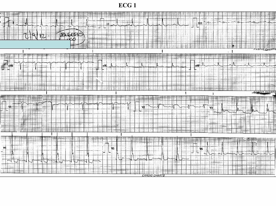

ECG 2
ECG 3
Over the 3rd intercostal space
Over the 2nd intercostal space
Over the 4to intercostal space
Colleagues opinions
Estimado Dr Marcelo Abud La elevacioacuten del segmento ST en un varoacuten puede corresponder como en este caso a una repolarizacioacuten precoacutez (high take off)
iquestPorque no se trata de una isquemia aguda RespuestaPorque no se observan signos reciprocos o imagen en espejo (remodelacioacuten fisiologica)
iquestPorque no se trata de una embolia pulmonar aguda Respuesta Porque no tiene eje eleacutectrico del QRS desviado a la derecha
iquestPorque no es pericarditis aguda Respuesta porque las derivaciones no estan afectadas difusamente el intervalo PR es isoeleacutectrico no tiene precordalgia fiebre y no estaacute taquicaacuterdico (en la pericarditis aguda la FC estaacute casi siempre es alrededor de 120lpm)
Entonces iquestque tiene el paciente Respuesta Efecto androacutegeacutenico sobre el corazoacuten Los andreacutegenos aumentan la fuerza contraacutectil selectivamente en el miocito del epicaacuterdico incrementando el tenor de calcio del sarcoacutemero (ocasionando elevacioacuten del segmento ST ) y concomitantemente produciendo mayor salida del potasio durante la fase 3 por estiacutemulo del canal de salida lento de potasio ldquoslow delayed potasium receptorsrdquo responsable por el aspecto puntiagudo de la onda T Esta hipercontractibilidad epicaacuterdica observada en el sexo masculino aumenta el requerimiento del flujo coronario Este ECG muestra que la pared anterolateral baja (de V4 a V6) y la anterolateral alta (aVL I) son hipercontraacutectiles especulo que la arteacuteria circunfeja sea pequentildea con predominancia de la arteacuteria coronaacuteria derecha Esta circulacioacuten coronaria insuficiente puede ocasionar disnea o dolor precordialComo se corrige esto Respuesta con beta bloqueadores adreneacutergicosNo quiero criticar a los estudios invasivos que se le hicieron pero pienso que en este caso con 20 pesos se huviera solucionado el problemaUn fraternal abrazoSamuel Sclarovsky
Dear Dr Marcelo Abud ST segment elevation in a male may correspond such as in this case to an early repolarization pattern (high take off)Why it is not an acute ischemia Answer Because there are no reciprocal or mirror image on the ECG1 (physiological remodeling)Why he has not an acute pulmonary embolism Answer Because he has not QRS axis deviation to the rightWhy he has not an acute pericarditis Answer because the leads are not affected diffusely the PR interval is isoelectric there are not precordial pain no fever and not tachycardia (In acute pericarditis the HR is almost always around 120lpm)So why this patient has Answer androgenic effect on the heart The androgens selectively increase the contractile force in the epicardial layer myocytes with calcium overload inside of sarcomeres (causing ST segment elevation) and concomitantly producing more K+ output during phase 3 action potential by stimulating of slow delayed potasium channels (responsible for the sharp appearance of the T wave)This selective epicardial hipercontractility observed in males increases the coronary flow requirement This ECG shows both the low anterolateral (from V4 to V6) and high anterolateral (aVL I) walls hypercontractiles I speculate that the Cx artery is small with a predominance of the right coronary artery(RCA) This can result in inadequate coronary circulation dyspnea or chest painHow we approach this Answer with beta adrenergic blockersI do not want to criticize the invasive studies that were done but I think that in this case with ldquo20 pesosrdquo we solved the problemA fraternal hugSamuel Sclarovsky
Dudas y criticas al raciocinio del Profesor SamuelEl ECG1 muestra clara elevacioacuten del segmento ST confinado apenas a V2-V3(regioacuten medioseptal) En la repolarizacioacuten precoz la elevacioacuten del ST es de concavidad superior terminando en una T positiva amplia de V2 a V4 o V5 ldquoprominent J wave and ST-segment elevation concave to the top predominantly in left precordial leads ending in a positive large T wave from V2 to V4 or V5rdquo La repolarizacioacuten precoz excepcionalmente tiene elevacion del ST en las precordiales derechas En la repolarizacioacuten precoz claacutesica benigna el supradesnivel de punto J y segmento ST de concavidad superior y seguido por onda T apiculada que se observa en la gran mayoria de los casos de V4 a V6 yo en la pared inferior En este caso no se observa supradesnivel del segmento ST en esta aacuterea Ademaacutes no vemos bradIcardia sinusal que es lo que suele verse en la repolarizacion precoz Si se fijan bien existe aqui discreto infradesnivel del ST de 1mm en V5 ldquoJ point and ST segment depressionrdquoPor otra parte este paciente asociado a la dispnea tuvo una importante hipotensioacuten arterial transitoacuteria
Aqui claramente en el ECG 1 se observan muescas o empastamiento en la rama descendente de la R mas conspicuas de V3 a V5 pero tambien visibles en V2 y V6 Ese hayazgo si es compatible con repolarizacion precoz ldquonotching irregular or slurring contour of the terminal QRS complexrdquo
Por otra parte querido maestro en este caso el aspecto del ST en V2 es de convexidad superior ldquo convex to the toprdquo absolutamente diferente del supradesnivel del ST de la repolarizacion precoz y va seguida de una onda T negativa En la Repolarizacion precoz el supra de ST es de concavidad superior seguida por una T puntiaguda positiva concordante Andres
Querido amigo amigo maestro Adrian Baranchuk disculpeme por mi condicion de otista pero mas claro no puedo ser y sobre en hombre de 45 antildeos con hipotensioacuten Me supongo que alguien que vio el ECG con disnea le administroacute nitritro sublingual que ocasiona hipotensioacuten transitoacuteria y taquicardia sinusalSeria interesante si el Dr Abud envie otro ECG en reposo sin taquicardia y presion normal estoy casi seguroque el ST-T seran mas alto a pesar que a los 45 antildeos estos hallazgos en el ECG estan atenuadosPor suerte mi amigo profe Andreacutes Ricardo Peacuterez Riera se tomo la molestia en traducirme del espantildeol al espantildeolUn fraternal abrazo y a todos los amigos de la colectividad judia feliz anio nuevo
Samuel SclarovskyQuerido amigoQue le hayan dado un nitrito es SOLO una suposicion bastante facil es preguntarle al paciente no creePero estoy seguro que Abud o Perez-Riera lo hubieran clarificadoPor otro lado hipotensioacuten sostenida no es frecuente de ver en ese escenario que Ud describe Digo todo esto porque Ud en su email usando un lenguaje un poco sarcaacutestico dijo que este caso Ud lo resolvia con 20 pesos y NO con el gasto de estudios invasivos Yo estoy en desacuerdo Porque Porque Ud corre con el caballo del comisario y sabe que todo dio normal pero yo aqui en Canada le hubiera hecho los mismos (o similaresestudios)Por favor una cosa es la especulacioacuten luego de saber los resultados y otra muy distinta ser el colega de guardia que tuvo que decidir esta conductaYo no se que tiene el paciente pero le aseguro que tambien hubiera descartado TEP primero e isquemiadespueacutesEso de que NO tiene BCRD y por eso NO tiene TEP es demasiado estricto ya que el BCRD se ve en solo el 60 de los casos (hay un lindo review que escribimos hace un tiempo busquelo que ahi estan todos loscambios de TEP en el ECG)Un fuerte abrazo y disculpe mi descuerdo en este casoAB
Samuel
1 Ud dice me parece que Ud esta errado En la actualidad es mejor discutir diciendo yo pienso que No es necesario descalificar al otro por el simple hecho de no estar de acuerdo Yo ya le dije no se lo quetiene el paciente pero hubiera hecho los mismos estudios Eso NO es estar errado es simplemente estar en desacuerdo con Ud (y estoy seguro porque lo conozco que Ud no se cree el duentildeo de la verdadno cierto)
2 Ud dice yo hace muchos antildeos que me dedico a esto Es cierto Y Eso lo transforma en duentildeo de la verdad Yo me dedico con igual pasion que Ud y eso NO me hace duentildeo de la verdad
Concuerdo en el uso racional de los estudios 100 Pero ese NO es le tema del paciente en cuestion sinoque le produjo un cuadro tan severo con un ECG tan anormal A veces para aprender hay que invertir No hay otra
Le mando un abrazo sincero
AB
Professor Andreacutes e Caro Dr MarceloInteressantiacutessimo casoA histoacuteria cliacutenica sugere episoacutedio tromboemboacutelico =gt Trauma com fratura de tornozelo - Repouso ouimobilidade do membro =gt Trombo embolismo coronaacuterio - (MI Type 2 - Expert Consensus Document -European Heart Journal doi101093eurheartjehs184apub 0912) - - Resoluccedilatildeo do quadro com anticoagulante ndashQuanto ao ECG-1 - Sindrome isqueacutemica aguda c ST supra V2-V3 imaacutegem ST em espelho infra V4-V5 Discretiacutessimo supra em D1- aVL FC 88bpm Quadro cliacutenico e ECG praticamente normalizado 2 horasdepois Quanto as enzimas foram seriadas Embora o episodio todo possa ter ocorrido s alteraccedilotildeesenzimaacuteticas Observar que o J Osborn -like tambeacutem desaparece e todos os fenoacutemenos se normalizam nos ECGs do diaseguinte Natildeo teria feito cateterismo Adail - Bahia - Brasil
Interesantiacutesimo caso Elevacioacuten de segmento ST tipo Brugada o tipo infarto anterior en V2 y ondas J bastante locaizadas en V3-V4 La respuesta a la ultima pregunta de Marcelo es faacutecil Cualquier medico responsable hubiera hecho los estudios necesarios para excluir un infarto agudo y una embolia pulmonar en un paciente con disnea suacutebita y este electrocardiogramaLa otra pregunta de Marcelo cual es la interpretacioacuten del electrocardiograma una vez sabiendo que no hay evidencia de lesiones coronaria o de embolia pulmonar tambieacuten es faacutecil de contestar Mi respuesta ldquono tengo ideardquo Que puede ser esto1 Respuesta a hiperventilacioacuten Este tema ya no esta de moda pero estuvo muy de moda en el siglo pasado (hijos que feo se oye eso) Ejemplos(123)El problema con esta teoriacutea es que la hiperventilacioacuten en gente sana produce inversioacuten de onda T y depresioacuten del segmento ST Pero en pacientes con enfermedad coronaria la hiperventilacioacuten puede producir elevacioacuten de segmento ST(4) Por tanto uno podria argumentar que las coronarias de este paciente aparentemente normales en la angiografiacutea pero no son realmente normales2 Coronary spasm Faltan los cambios reciacuteprocos en otras derivaciones y en paciente no tubo dolor de pecho3 Pericarditis No claacutesico pero siempre puede ser Si usamos este diagnostico cada vez que no tenemos idea que esta pasando por que no usarlo aquiacute4 Un infarto localizado de la arteacuteria del cono ldquoconal branchrdquo de la arteria coronaria derecha(CD) Es posible Tan posible que moveriacutea esta posibilidad para arriba Recientemente tuve un paciente con gran infarto inferior por oclusioacuten de la CD Tenia elevacioacuten del segmento ST en II III y aVF
1 Savonitto S et al Different significance of hyperventilation-induced electrocardiographic changes in healthy subjects and patients with coronary artery disease Eur Heart J 199617(9)1302
2 Lary D Goldschlager N Electrocardiographic changes during hyperventilation resembling myocardial ischemia in patients with normal coronary arteriograms Am Heart J 197487(3)383-90
3 Lewis WC Siebecker KL Jr Wasserburger RH The effect of hyperventilation on the normal adult electrocardiogram Circulation 195613(6)850-5
4 Eur Heart J 199617(9)1302)
Fue sometido intervencioacuten coronaria urgente con dilatacioacuten y ldquostentingrdquo de el tramo proacuteximo de la CD y al poner el stent se obstruyoacute la rama del cono ldquoconal branchrdquo ocasionando un patroacuten ECG ldquoBrugada-likerdquo muy semejante a este caso y el paciente desarrolloacute fibrilacioacuten ventricular Mientras mas lo pienso mas me gusta esta posibilidad5 Simple ldquorepolarizacion precozrdquo Puede ser Pero el paciente presenta con disnea y tachycardia El ldquostressrdquo deberiacutea disminuir los efectos electrocardiograacuteficos de repolarizacion precoz que generalmente aumentan durante tono vagal6 Siacutendrome de Brugada Puede ser El hecho de que desaparezca o disminuya al diacutea siguiente no descarta esta posibilidad Definitivamente yo le hariacutea una prueba de ajmalina antes de mandarlo a la casa Por cierto que hay controversia en lo que se refiere al efecto del ldquostressrdquo en el ECG del Brugada En Aacutemsterdam el segmento de ST de Brugada supradesnivela durante el ejercicio(1) En Japoacuten el ST normaliza durante el ejercicio y aumenta durante la fase de recuperacioacuten (2)7 Medicamentos que ldquoproducen Brugadardquo (brugadadrugsorg) La lista es grande y para la mayoriacutea los efectos aumentan durante taquicardia8 Embolia pulmonar que resolvioacute No se hizo tomografiacutea CT del pulmoacutenhellip sin ofenderhellip la arteriografiacutea pulmonar no es faacutecil de interpretar Ademas el paciente recibioacute anticoagulantes9 Tumores o ldquomiscelaacuteneosrdquo del mediastino Esto es muy importante checarlo (34)Definitivamente solicitaria una tomografiacutea (CT) de toacuterax antes de mandar al paciente a la casaY pus total siempre queda la otra posibilidad No seSami Viskin (Tel Aviv)
1 min AS de Groot EA Ruijter JM Wilde AA Tan HL Exercise-induced ECG changes in Brugada syndrome Circ ArrhythmElectrophysiol 20092(5)531
2 Augmented ST-segment elevation during recovery from exercise predicts cardiac events in patients with Brugada syndrome JACC 20103 Nakazato Y et al Brugada-like precordial ST elevation on ECG by anterior mediastinal infective mass lesion Indian pacing and
electrophysiology journal 20033(3)184Tarin N Farre J Rubio JM Tunon J Castro-Dorticos J Brugada-like electrocardiographic pattern
in a patient with a mediastinal tumor Pacing Clin Electrophysiol 199922(8)1264-6
Descartada obviamente la hipotermia por exposicion laboral yo accidental las intoxacionesmedicamentosas o por monoxido de carbono Asi como tambien la infeccion posquirurgica con bacteriemiao sepsis Brugada-Like ECG Pattern in Severe Hypothermia Circulation2008118977-978Dr Sami muy interesante su analisis y la evaluciacion de los diagnosticos diferenciales
El paciente se presenta con hipotension taquicardico y cambios en el ECG muy similares a los descriptos en el experimento en perros Cellular Basis for the Electrocardiographic J Wave Gan-Xin Yan MD PhD Charles Antzelevitch y analizan los cambios endo epicardio como los responsables posibles del origende dicha onda y ademas en el experimento encontraban cambios ECG de v4 a v6 similar a este paciente y en precoridales derechas simulando un patron de Brugada interpretandolos por la hipotermia y a la hiperalcemia transitoria y el desalloro de acidosis (cual papel sera el de esta en la velocidad de lospotenciales de accion)Su analisis creo merece solo un comentario en el TEP los cambios caracteristicos de S1Q3T3 o aparicionde BRD con eje desviado a la derecha se ve en la mayoria de los pacientes no en todosEl dimero D puede ser negativo (es un producto de la degradacion de la fibrina) en un 5 de estos la angiografia pulmonar descarta los embolismos masivos o de arterias pulmonares pero tampoco descartatotalmente el TEPEl ecocardiograma al igual que los otros metodos solo resulta positivo en la mayoria de los casos contribuye al diagnostico pero no lo excluyeDe lo referido llama la atencion un paciente sin patologia previa conocida cirugia de tobillo reciente en casos de politraumatismo la incidencia de TVP es del 20 con probabilidad de presentarse como TEP del 05 no refirio el Dr Abud si recibia profilaxis para trombosis venosa profunda La ausencia de coagulo en MII disminye las probabilidades de TEP pero tampoco lo excluyenLa TAC helicoidal (ya se realizo una arteriografia pulmonar) no creo aporte al diagnostico presenta unasensibilidad del 90 cuando el embolismo es de las arterias de primer y segundo orden y al incluir losvasos subsegmentarios esta cae al 67 los TEP subsegmentarios son alrededor del 6 en el estudioPIOPED
La Escala de Shier para evaluar la probabilidad de embolias grasas en las primera 72 hs luego del accidentees util para predecir la probabilidad de TEP de este origen en este caso seria extremadamente raro se presentara tardiamente como han referidoLa onda J de Osborn descripta por este cuando experimentaba con perros al inducirle hipotermia(1) el primero en describirla fue Tomaszewski que encuentra cambios en el ECG de un hombre fallecido por hipotermia (1)No encuentro una explicacion concreta pero en medicinatodo tiene una explicacion y no hay casualidades sino causalidades podremos diagnosticarlas correctamenteo no pero el criterio de estudio del paciente me parece el adecuado Este paciente en particular presenta al ingreso una elevacion del punto J que disminuye pero no desaparece en los ECG posteriores lo que lleva a pensar en un transtorno agudo de una patologia cronica lo que va en contra de pensar en TEP En el Brugada la elevacion del punto J en derivaciones inferiores esa descripta pero en estudios experimetales tambien la presentan en cara lateral estos estudios experimetales en laboratorio de la diferencia endo-epicardica de potenciales (les adjunto un link de un ECG exacto al presentado para que observenhttpwwwncbinlmnihgovpmcarticlesPMC2843811figureF5 y se trata de un SBr no hipotermia peroen este presenta repolarizacion precoz lo que no se observa en el paciente presentado En la taquicardia la notched esta acentuada como ocurre en este paciente Ya lo han anticoagulado o sea que esta recibiendotratamiento para no repetir en caso de haber sido un TEPPodriamos excluir la probabilidad de que se tratara de un SBr asociado a replarizacion precoz Porque no realizarle una prueba con flecainida a fin de descartarloUn saludoMartin ibarrola
1 Tomaszewski W Changements electrocardiographiques observes chez un homme mort de froid Arch Mal Coeur 193831525) extraido de EKGWEB
2 Osborn JJ Experimental hypothermia respiratory and blood pH changes in relation to cardiac function Am J Physiol 19531753893 Genetic Molecular and Cellular Mechanisms Underlying the J Wave Syndromes Charles
Antzelevitch1) httpswwwjstagejstgojparticlecircj76576_CJ-12-0284_pdf4 J Wave SyndromesCharles Antzelevitch PhD FHRS1 and Gan-Xin Yan MD PhD234
httpwwwncbinlmnihgovpmcarticlesPMC28438115 Paradoxical effect of ajmaline in a patient with Brugada syndromeBiagio Sassone EP Europace Volume 8 Issue 4 Pp 251-254 6 Brugada syndrome with atypical ECG downsloping ST-segment elevation in inferior leadsRiera ARP Ferreira C Schapachnik E J
Electrocardiol 2004 Apr37(2)101-4
Final commentsBy Andreacutes Ricardo Peacuterez-Riera MD PhD
ECG-1 FROTAL PLANE
aVR aVL
I
IIIII
aVF
X
Y
QRS axis +30deg
Reciprocal ST-segment changes(ST-segment depression)
AB
II
TST
Early Repolarization Pattern (ERP) is defined as notching or slurring morphology of the terminal portion of the R wave (J-wave) that produces a positive hump known as a J-wave at the end of the QRS complex and beginning of the ST segment J-point elevation ge01 mV (or 1mm) above baseline in at least 2 in the mid-to-left precordial leads andor inferior leads followed by concordant tall near symmetric (T waves having similarity in shape ascending and descending ramps speeds) There is a distinct J wave and ST segment concave upward in the left precordial leads V4 through V6 The ST elevation is most frequently evident in ECG lead V4
Where is the located the end of QRS complex In A or in B If the answer is B QRSd is near 120ms If the answer is A QRSd is near 70ms
J-wav
e
ECG-1
V6
V1
V4
V5
V2
V3
X
Z
ECG-1 HORIZONTAL PLANE
V2
V5
J point
QRSd 122ms
ECG-1
QRSd 122ms
Notching
40 ms80 ms
β
80 ms
High take-off
Type 1 Brugada pattern
Type 2 Saddle-back pattern
40 ms80 ms
β
80 ms
High take-off
Type 1 Brugada pattern
Type 2 Saddle-back pattern
V2
Is this a type 2 Brugada pattern or saddle-back pattern in V2
Tryphasic QRS Pattern RSracute
High take-off of racute ge 2mm high take-off frequently does not coincide with J wave descending arm of racutecoincides with beginning of ST segment the racute wave has rounded shape wide and usually of relatively low voltage in Brugada pattern Contrarily in IRBB the racute wave is peaked the angle between the upslope of the S-wave and the downslope of the r-wave (β angle) gt 58deg duration of the base of the triangle of racute at 5mm from high take-off gt35mm the QRS duration is longer in Brugada pattern type 2 than in other cases with racute in V1 and there is a mismatch between V1 and V6 In Brugada pattern the QRS complex end is earlier in V6 than in V1 V2
βangle gt 58deg
1 Bayeacutes de Luna A et al a consensus report Journal of Electrocardiol 2012 45433-
2 Chevallier S J Am CollCardiol2011 Nov 2258 2290-2298
ECG-1
ECG-1
RIGHT SAGGITAL PLANE ECG-1
YaVF
Z V2
ECG 2
aVR aVL
I
IIIII
X
Y
aVF
QRS axis +30ordm
ECG-2 FRONTAL PLANE
aVR aVR
ECG-1 ECG-2
J-Wave
Reciprocal ST-segment changes (ST-segment depression) Without reciprocal changes or mirror image
In Early Repolarization Pattern reciprocal ST-segment changes (ST-segment depression) are eventually confined the unipolar aVR lead (Absence of reciprocal changes or mirror image in others leads)
II
II
PR interval= 160msQRSd is 121ms
ECG-1
PR interval = 140msQRSd is near 80ms
J wave
ECG-2
V6
V5
V1
V4
V2
V3
X
Z
Is this a type 2 Brugada ECG pattern Answer in next slide
ECG-2
V2βangle gt58deg
Observation β angle is related to both depolarization an repolarization A wider QRS complex (slow conduction) and abnormal repolarization both contribute to a wider β angle
1 Chevallier S J Am Coll Cardiol2011 Nov 2258 2290-2298
In patients with suspected Brugada syndrome simple ECG criteria can enable discrimination between innocent incomplete RBBB from types 2 and 3 Brugada ECG patterns The mean β angle is significantly smaller in the patients with negative results on Antiarrhythmic Drug Challenge (ADC) compared wit patients with positive results on ADC Its optimal cutoff value is 58deg which yielded a positive predictive value of 73 and a negative predictive value of 87 for conversion to type 1 pattern on ADC α is slightly less sensitive and specific compared with β When the angles are combined with QRS duration it tended to improve discrimination
Y
aVF
Z V2
Right Sagittal Plane ECG-2
Triphasic QRS patternRSracute
β angle defined as the angle between the upslope of the S-wave and the downslope of the r-wave The mean β angle was significantly smaller in the 14 patients with negative results on AAD compared to the 24 patients with positive results on AAD Itrsquos optimal cutoff value was 58deg which yielded a positive predictive value of 73 and a negative predictive value of 87 for conversion to type 1 pattern on AAD
ST segment with saddleback appearance followed by positive or biphasic T wave in V1-V2 or from V1 through V3
β
V5V5
QRSd= 122ms QRSd= 110ms
ECG-1 ECG-2
Left precordial terminal QRS notching is more prevalent in malignant early repolarization abnormalities
Left precordial terminal QRS notching
1 Merchant FM et al Am J Cardiol 2009 Nov 15 1041402-1406
V2
V2
V3
V3
ECG-1
ECG-2
ECG-2
Dynamic inconstant or dramatic changes in repolarization are suggestive of malignant early repolarization abnormalities
1 Haiumlssaguerre M et al J Cardiovasc Electrophysiol 2009 Jan2093-98
ECG-1
Conclusion this patient has high possibility of concomitant Brugada syndrome and early repolarization In this a benign ERP or malignant early repolarization abnormalities We think it is very unlikely that is a Brugada phenocopyIn this case it is necessary additionally to perform
1 IV Ajmaline test2 ECG-AR3 Family study ( first degree relatives interrogatory physical ECGs)4 Magnetic resonance image5 Genetic screening
Ventricular biopsy is not considered mandatory but is recommended especially when it may clarify the nature of dubious findings identified with others test In approximately 6--10 of survivors of cardiac arrest no cardiac abnormality can be identified despite extensive clinical evaluation Autopsy data confirm that in a similar percentage of victims of SCD no structural heart disease or or mors sine materia can be identified at post mortem evaluation Incompletely penetrant genetic defects may underlie at least some of these unexplained deaths (1)
The ERP probably represents part of a spectrum of cardiovascular anomalies related to channelopaties including BrS IVF congenital short QT syndrome and that it may also have a molecular genetic origin of variable penetrance The denomination Early Repolarization Pattern is appropriate since it indicates that it is characterized by a given ECG pattern
1 Priori SG et al Cardiovasc Res 2001 May50218-223
Case Report similar
A 20 year-old male mulatto professional soccer player from Santo Andreacute Sao Paulo Brazil He didnrsquot finishthe second year in elementary school CatholicProfession Professional soccer playerReason for consultation Presented to consultation due to two presyncopal episodes and one syncopal episode in the last 30 days The first two episodes happened during a post-defecation period Both were preceded by dizziness nausea and vomiting The syncopal episodes occurred at rest with no prodromes The third one with express and brief loss of consciousness and sphincter relaxation at dawn 48 hours agoHis prior personal medical history was unremarkable and he successfully passed two prior periodical clinical-cardiovascular evaluations (twice a year always normal) Pre-competitive examination included 12-lead ECG echocardiogram and cardiopulmonary metabolic exercise testing (CMET) All normals Intense training for the last 3 years Familial Background One uncle from his fatherrsquos side family died of sudden cardiac death when he was 35
years old He ignores the cause His brother is being seeing by a cardiologist due to ldquocardiac arrhythmiardquo in treatment with a cardiologistfrom the Institute of the Heart from Satildeo Paulo (he cannot tell which is the cause) He denies having other relevant background details Pysical cardiovascular examination revealed bradycardia at 45 bpm with physiological sinus arrhythmia blood pressure 11070 mmHg The rest of the physical examination was unremarkable He denied consuming any type of neither medications nor drugs No addictionsHis first 12-lead ECGVectorcardiogram(VCG) (Fig 1) showed sinus bradycardia at 44 bpm P-wave axis of +60deg PR interval of 186ms QRS duration of 111ms QRS axis at + 63deg QT interval 427ms and corrected QTc 367ms There is a notch on the terminal portion of the QRS complex in the left precordial leads voltage criteria for left ventricular hypertrophy (SV1 + RV5 gt 35 mm ndash Sokolow index) prominent J-wave and concave ST-segment elevation in inferior leads V4 V5 and V6 Deep and narrow Q-waves in the left precordial leads V5 and V6 All these features were considered as an Early Repolarization Pattern (ERP)
Name JVS Gender Male Age 20 yo Ethnic Group Mulatto Weight 66 Kg Height 172 m Biotype Normoline Date 11262009 Profession competitive athlete (soccer player)
ECG diagnosis Sinus bradycardia (SB) HR 44bpm P axis +60deg PR 186ms QRSd 111ms QRS axis + 63deg QT 427ms QTc 367ms Conclusion SB + Early Repolarization Pattern (ERP) Notching or slurring of the terminal QRS complex voltage criteria of left ventricular hypertrophy SV1+ RV5 gt 35 mm (Sokolow index) prominent J wave and ST-segment elevation concave to the topending in a positive T wave from V4 to V6 Precordial greater than limb leads Relatively deep but narrow q waves in the left precordial leads (V5 and V6)
V6V4 V5
Prominent J wave and ST- segment elevation concave to the top ending in a positive T wave from V4 to V6 Notching or slurring morphology of the terminal portion of the R wave (J-wave) that produces a positive hump known as a J-wave (Arrows)
aVR
Eventually reciprocal ST segment depression confined only in Lead aVR1 observed in this particular case report
1 Mehta M Jain AC Mehta A Early Repolarization Clin Cardiol 1999 Feb 22 59-65
ECGVCG CORRELATIONS HORIZONTAL PLANE
V6
V1
V2
V3
X
Z
V5
V4
T
0J T
BEGINNING AND END OF QRS LOOP IN HP
J POINT in vectorcardiography it corresponds to 3 elements end of ventricular depolarization (QRS loop) beginning of repolarization (ST segment) when it does not present depression or elevation and T wave onset In this case 0 point and J point are not coincident In situations where there is depression or elevation of ST segment the J point does not coincide with the 0 point and the greater or lesser distance between both points indicate the greater or lesser ST segment elevation or depression The phenomenon is observed in early repolarization pattern acute phase of myocardial infarction Prinzmetal variant angina pericarditis Brugada syndrome arrhythmogenic right ventricular dysplasia etc
0 POINTIt corresponds to the QRS looponset (because PR segment does notexist) Alsocorrespond to the end of biatrialchamberactivation
T
J POINT
ECGVCG CORRELATIONS FORNTAL PLANE
aVR aVL
I
IIIII
X
Y
VERTICAL AXIS ON FRONTAL PLANE The frontal plane QRS axis and ST segment axis and T wave axis are all in the same direction THE ST
SEGMENT ELEVATION
USUALLY lt 05 mm IN THE LIMB
LEADS
YVF
Z V2
T
ECGVCG CORRELATIONS RIGHT SAGITTAL PLANE
A second 12-lead ECG (Fig 2) preformed 72 hours later showing1) Sinus bradycardia at 44 bpm2) Similar p-wave axis and intervals duration3) Dramatic modification of repolarizaccedilatildeo pattern in the right precordial leads A coved-type
(type 1) ECG brugada pattern can be distinguished4) Accentuation of j-waves and st segment elevation across the precordial leads was observed5) ST segment elevation on avr lead
See next slide
Name JVS Gender Male Age 20 yo Ethnic Group MulattoWeight 66 Kg Height 172 m Biotype Normoline Date 11292009Profession Competitive Soccer Player FIGURE 2
ECG diagnosis Sinus Arrhythmia(SA) sinus bradycardia (SB) HR 44bpm P axis +60deg PR 186ms QRSd 111ms QT 427ms QTc 367 Conclusion SA + SB + Early Repolarization Pattern (ERP) Type 1 Brugada ECG pattern Dramatic accentuation of J waves across the precordial and limb leads Transient augmentation of global J waves may be indicative of a
highly arrhythmogenic substrate heralding multiple episodes of VF in patients with ERP
aVR LEAD
11262009 11292009
DEPRESSION
ELEVATION
ST SEGMENT DEPRESSION IN aVR = 1mm ST SEGMENT ELEVATION IN aVR = 2mm
V1 V2V2
1129200911262009
V1
11292009 11262009
SPONTANEOUS MODIFICATION OF REPOLARIZATION TO TYPE 1 BRUGADA ECG PATTERN
11262009
V6
11292009V6
ST segment elevation lt 2mm ST segment elevation gt 2mm
11292009
V5
11262009
V5
ST segment elevation = 1mm ST segment elevation = 25mm
EARLY REPOLARIZATION PATTERN CONCEPT
DEFINITION Early Repolarization Pattern (ERP) is a well-recognized Enigmatic idiopathic electrocardiographic phenomenon characterized by notching or slurring morphology of the terminal portion of the R wave (J-wave) that produces a positive hump known as a J-wave at the end of the QRS complex and beginning of the ST segment It considered to be present when at least two adjacent precordial leads show elevation of the ST segment with values ge01 mV (or 1mm) above baseline followed by concordant tall near symmetric (T waves having similarity in shape ascending and descending ramps speeds)
EPIDEMIOLOGY ERP occur in 2 to 5 of the population often in men young adults afro-descendents and competitive athletes and in up to 131 to 482 of patients presenting with chest pain in the emergency rooms and coronary care units There is a distinct J wave and ST segment concave upward in the left precordial leads V4 through V6 The ST elevation is most frequently evident in ECG lead V4 The ST segment elevation is usually le2mm (precordial greater than limb leads) and is lt 05 mm in the limb leadsReciprocal ST-segment changes (ST-segment depression) are eventually confined the unipolar aVR lead (Absence of reciprocal changes or mirror image) In ERP vertical QRS axis electrical is the rule The frontal plane QRS axis the ST segment axis and T wave axis are all in the same directionThis ECG pattern has been considered a benign normal ECG variant for over six decades3
1 Hasbak P Engelmann MD Early repolarization ST-segment elevation as a normal electrocardiographic variant Ugeskr Laeger 2000 162 5928-5929
2 Brady WJ Chan TC Electrocardiographic manifestations benign early repolarization J Emerg Med 1999 17473-478
3 Benito B Guasch E Rivard L Nattel S Clinical and mechanistic issues in early repolarization of normal variants and lethal arrhythmia syndromes J Am Coll Cardiol 2010 Oct 5 56 1177-1186
MAIN ELECTROCARDIOGRAPHIC FEATURES IN ERPHeart rate Predominant sinus bradycardia frequently associated with respiratory sinus arrhythmia
PR interval First degree AV block is observed in 5 to 39 among professional athletes In the non-athlete population it is observed in 065 of cases QRS axis Vertical electrical axis is the rule The frontal plane QRS axis and ST segment axis and T wave axis are all in the same directionQRS duration (90 plusmn 10 ms) subjects than in age-matched healthy controls (80 plusmn 10 ms) QRS morphology Notching or slurring of the terminal QRS complex Prominent relatively deep but narrow q waves may appear in the left precordial leadsQRS voltage Eventually voltage criteria of LVH are observed in male competitive athletes SV1+ RV5 gt 35 mm (Positive Sokolow index) High QRS voltage is more frequent in male athletes but its correlation with LVH is low Voltage decreases after deconditioning is slow The distinction between the physiological athletes heart and pathological conditions has critical implications for professional athletes An abrupt transition may occur from right-oriented complexes to left-oriented complexes in the precordial leads secondary to counterclockwise rotation on precordial leads About two-thirds of clockwise rotation and counterclockwise rotation could be attributed to the septal angle by anatomical rotation of the heart in one plane around the long axis but other factors appear to be responsible for such ECG findings in the remaining one-third of cases Relatively higher positions of the precordial ECG leads as observed in the vertical heart appeared to be responsible for clockwise rotation in some patients and left septal fascicular block is suspected to be responsible for counterclockwise rotation in others patientsJ-point elevation Notching irregular or slurring contour of the terminal QRS complex (J point) Variant of Osborn wave is seen in ERP
ST segment Widespread ST segment elevation (precordial greater than limb leads) The characteristic ST segment is elevated upward concave confined more frequently in precordial leads with reciprocal depression only in aVR The concavity is observed at the initial up-sloping portion of ST segment or upwardly concave ST segment morphology Unfortunately concave ST morphology cannot be used to rule out ST elevation from AMI with left anterior descending coronary occlusion because it is common in these circumstances The ST elevation is most frequently evident in ECG lead V4 There is a distinct J wave and ST segment in the left precordial leads V4 through V6 The ST elevation in ERP is usually lt 2 mm (but can rarely be gt 5 mm) in the precordial leads and the greatest ST elevation is usually seen in the mid-to-left precordial leads The ST segment elevation is usually lt 05 mm in the limb leads
T wave characteristics Concordant T waves of large amplitude (prominent matching T waves) typical pseudo-asymmetrical (lsquosymmetroidrdquo) or slightly asymmetrical matching T waves often of large amplitude upright tall and peaked most conspicuously from V2 to V4 or V5 sometimes seen in leads DII DIII and aVF as a rule T waves may appear as of large amplitude peaked or pointed symmetric and matching Vagotonic or high T wave voltages followed by U waves are frequent when sinus bradycardia is present Tall positive ad symmetric or symmetroid T waves are not only seen occasionally in the very early stages of MI but also in hyperkalemia and in ERP sinus bradycardia
QT intervals QT maximum The maximum Q-onset-T-end interval This parameter is higher in ERV subjects than in normal controls
QTp maximum maximum Q-onset-T-peak interval This parameter is higher in ERV subjects than in age-matched healthy controls
Rate-corrected QTc maximum This parameter is lower in ERP subjects than in age-matched healthy controls
QTpc maximum This parameter is lower in ERV subjects than in age-matched healthy controls
U wave because bradycardia U waves are frequent in ERV they are best observed in the V3 lead U waves are frequent when sinus bradycardia is present
Other ECG characteristics of ERPRelative temporal stability of the ST segment and T wave pattern is observedReciprocal changes are not seen in ERP There are no evolutionary short-term changes in the ST segment and T waves and Q waves do not appear
Discusions Early Repolarization Pattern (ERP) or Early Repolarization Variant (ERV) is an enigmati
electrocardiographic phenomenon characterized by prominent J wave and ST-segment elevation in multiplleads
Recently there has been renewed interest in ERP because of similarities to the arrhythmogenic Brugadsyndrome (BrS) Not much is known about the epidemiology of ERP and several studies have reported thathis condition is associated with a good prognosis
Both ERP and BrS exhibit some similarities including the ionic underlying mechanism the analogouresponses to changes in heart rate and autonomic tone sympathicomimetics (isoproterenol test) as well as insodium channel and beta-blockers These observations raise the hypothesis that ERP may be not as benign atraditionally believed Additionally there are documents showing that ST-segment height in the man igreatly influenced by central sympathetic nervous activity both at baseline and during physiologic andpharmacological stress Central sympathetic dysfunction regularly results in multilead ST-segment elevationor J wave that decreases or below isoelectric baseline during low dose isoproterenol infusion An earlyrepolarization pattern in the inferior leads of a standard electrocardiogram is associated with an increased riskof death from cardiac causes in middle-aged subjects1
POSSIBLE SIMILARITIES BETWEEN ERV AND BrSbullMore frequent in malesbullBoth occur more frequently in young adults and in individuals without apparent structural heart diseasebullBoth may influence just the V1-V2 leads Rarely (9) can ST elevation be observed in ERP only in the right precordial leads V1-V2 or in the inferior ones2
1 Tikkanen JT Anttonen O Junttila MJ Aro AL Kerola T Rissanen HA Reunanen A Huikuri HV Long-Term Outcome Associated with Early Repolarization on Electrocardiography N Engl J Med 2009 Nov 16 [Epubahead of print]
2 Hasbak P Engelmann MDEarly repolarization ST-segment elevation as a normal electrocardiographic variant Ugeskr Laeger 2000 162 5928-5929
When ST elevation is normal it can reach up to 3 mm in V2-V3 especially in young people In those individuals over 40 years it seldom exceeds 2 mm Both can show incomplete RBBB pattern or right bundle branch conduction disorder in BrS it can present atypical features RBBB-like and of the saddle type by exclusive elevation of the J point S wave with delay in the left leads DI aVL V5 and V6 could be absent as it is to be expected in a classic RBBB The elements considered as typical in BrS are 1) elevation of the terminal part of QRS (prominent J wave) 2) elevated and descending ST not related to lesion of ischemic (idiopathic) injury 3) negative T wave in the right precordial leads 4) normal QTc or near normal 5) Eventually absence of final delay in left leads as it would be expected in a classic RBBB1 In ERV when associated to athletersquos heart QRS can present a moderate extension (100 ms to 110 ms) in 15 of the cases which in nonathlete normal population in a 24 is called outflow tract hypertrophy In this case r does not exceed the 5 mm and is lower than S in the same lead rSrBoth may improve repolarization during the stress test with use of isoproterenolBoth respond to a shortening of AP phase 2 in a part of ventricular thickness and intensification of fast repolarization notch (phase 1) mediated by transmural dispersion of ventricular repolarization by a larger notch in the Ito channel2The alteration of the Ito and ICa2+-L channels in BrS and in ERP are the electrophysiologic substrate that explains the J point and ST segment elevation because they cause the intensified notch in phase 1 and suppression in phase 2 duration in the epicardium and in the endocardium of ventricular wall thickness
1 Hiss RG Lamb LE Electrocardiographic findings in 122043 individuals Circulation 1962 25947-9612 Antzelevitch Ch Xin Yan G Shimuzi W et al Electrical heterogeneity the ECG and Cardiac Arrhythmias In Zipes
DP Jalife J Cardiac Electrophysiology From Cell to Bedside Third Edition WB Saunders Company2000 Chapter 26 p 222-238
ELEMENTS FOR DIFFERENTIAL DIAGNOSIS BETWEEN ERP AND BrS
I) Family background ERP negativeBrS frequently positive
II) Ethnic GroupERP predominantly in African descendents(1) or equally common in all races(2) BrS predominantly in Asian group (58) and Caucasian people(3)
III) GenderERP male gender predominanceBrS great predominance in the male gender (malefemale ratio ndash 81 in non Asian and 101 in Asian
peopleIV) Response to IC group antiarrhythmic agents
BrS flecainide used in a 10 mgKg dosage in 10 minutes increases ST elevation and QRS duration in a more significant way in patients with BrS than in individuals without the entity and only in those it triggers ventricular extrasystoles(4)
ERP it can induce a pattern similar to BrS however the degree of ST elevation caused by the drug is much higher in patients with BrS than in patients without the disease
1 Grusin H Peculiarities of the Africanrsquos electrocardiogram and the changes observed in serial studies Circulation 1954 9 860-867
2 Mehta M Jain AC Mehta A Early Repolarization Clin Cardiol 1999 Feb2259-653 Nademanee K Veerakul G Nimmannit S Chaowakul V Bhuripanyo K Likittanasombat K Tunsanga K Kuasirikul S
Malasit P Tansupasawadikul S Tatsanavivat P Arrhytmogenic marker for the sudden unexplained death syndrome in Thail men Circulation 1997 962595-2600
4 Shimizu W Antzelevitch C Suyama K Kurita T Taguchi A Aihara N Takaki H Sunagawa K Kamakura S Effect of sodium channel blockers on ST segment QRS duration and corrected QT interval in patients with Brugada syndromeJ Cardiovasc Electrophysiol 2000 111320-1329
BrS patients with Inferior and lateral ECG repolarization abnormalities have longer baseline PR intervals(1)Both an S wave width ge80ms in V1 and ST elevation ge18mm in V2 are highly specific indicators of VF and criteria for high-risk BrS(2)There are several references in literature to patients with elevation of the J point and ST segment convex to the top or straight descendent in inferior leads (Brugada sign) or concomitantly ST-segment elevation in the right precordial and inferior leads II III and aVF(3) in absence of hypothermia ischemia or electrolytic disorders which we call ldquoatypical Brugada patternrdquo atypical BrS variant BrS or BrS variant or idiopathic VF (a variant of the BrS with ST-segment elevation in inferior leads(4)Pilsicainide may induce PVCs and PVT in atypical BrS and the infusion of isoproterenol suppress the arrhythmias and normalize the ST-segment elevation(5)
1 Sarkozy A Chierchia GB Paparella G Boussy T De Asmundis C Roos M Henkens S Kaufman L Buyl R Brugada R Brugada J Brugada P Inferior and lateral electrocardiographic repolarization abnormalities in Brugada syndromeCirc ArrhythmElectrophysiol 2009 Apr 2154-161
2 Atarashi H Ogawa S For The Idiopathic Ventricular Fibrillation Investigators New ECG Criteria for High-Risk Brugada Syndrome Circ J 2003 Jan 67 8-10
3 Lombardi F Potenza S Beltrami A Verzoni A Brugada P Brugada RSimultaneous ST-segment elevation in the right precordial and inferior leads in Brugada syndrome J CardiovascMed (Hagerstown) 2007 8 201-204
4 Letsas KP Efremidis M Pappas LK Gavrielatos G Markou V Sideris A Kardaras FEarlyrepolarization syndrome is it always benign Int J Cardiol 2007114 390-392
5 Chinushi M Izumi D Furushima H Watanabe H Aizawa YMultiple premature beats triggered ventricular arrhythmias during pilsicainide infusion in a patient with inferior ST-segment elevation Pacing Clin Electrophysiol 2006 29 1445-1448
Potet et al(1) identified a G752R mutation on SNC5A that produced ST segment elevation and prominent J wave in leads II III and aVF The authors provide genetic demonstration that Brugada ECG anomalies related to a unique SCN5A mutation can be observed either in the inferior or the right precordial leads The early repolarization pattern in inferolateral leads is not an uncommon finding in BrS and this pattern is not associated with a worse outcome in subjects with BrS(2) The spontaneous ERP occurred more frequently among patients with BrS than in 283 family members not having BrS (11 versus 6 P=003) Class I antiarrhythmic drug administration provoked inferior-lateral coved Brugada pattern in 13 patients with BrS These patients had longer baseline PR intervals and Class I antiarrhythmic drug induced QRS interval prolongation (108 to 178 versus 102 ms to 131 ms Plt0001) In 3 patients the Class I antiarrhythmic drug provoked coved Brugada pattern only present in the inferior leads Inferior-lateral ERP occurs spontaneously relatively frequently in BrS These patients have a more severe phenotype Class I antiarrhythmic drug administration provokes inferior-lateral coved Brugada pattern in 46 of patients The Class I antiarrhythmic drug exceptionally provoked coved Brugada pattern only observed in the inferior leads(3)
1 Potet F Mabo P Le Coq G et al Novel brugada SCN5A mutation leading to ST segment elevation in the inferior or the right precordial leads J Cardiovasc Electrophysiol 2003 142000-2003
2 Letsas KP Sacher F Probst V Weber R Knecht S Kalusche D Haiumlssaguerre M ArentzTPrevalence of early repolarization pattern in inferolateral leads in patients with Brugada syndrome Heart Rhythm 2008 Dec 5 1685-1689
3 Sarkozy A Chierchia GB Paparella G Boussy T De Asmundis C Roos M Henkens S Kaufman L Buyl R Brugada R Brugada J Brugada P Inferior and lateral electrocardiographic repolarization abnormalities in Brugada syndrome Circ Arrhythm Electrophysiol 2009 Apr2 154-161
Type 1 ECG Brugada pattern in the peripheral leads was observed in 42 of patients during ajmaline test (103 of positive tests) and was associated with longer QRS and greater QTc prolongation compared with the rest of the patients1Atypical Brugada Syndrome or IVF with J waves in inferior lateral or inferior lateral leads
and Familial Idiopathic Ventricular Fibrillation and BrSSeveral mutations in genes SCN5A KCNJ8 DPP6 SCN3B and CACNA2D1 had been identified 1 On SCN5A gene2 3 Observation We do not believe that SCN5A should be attributed to IVF because this report clearly shows conduction defects (as expected) so this is not IVF but conduction delay at different levels in the heart2 On KCNJ8 gene 4 5 Missense variant in exon 3 (NC-000012) of the KCNJ8 gene a subunit of the K(ATP)channel23 Genomic DNA sequencing of K(ATP) channel genes showed missense variant in exon 3 (NC_000012) of the KCNJ8 gene a subunit of the K(ATP) channel conferring predisposition to dramatic repolarization changes and ventricular vulnerability
1 Batchvarov VN Govindan M Camm AJ Behr ERBrugada-like changes in the peripheral leads during diagnostic ajmaline test in patients with suspected Brugada syndrome Pacing Clin Electrophysiol 2009 Jun32695-703
2 Potet F Mabo P Le Coq G Probst V Schott JJ Airaud F Guihard G Daubert JC Escande D Le MarecH Novel brugada SCN5A mutation leading to ST segment elevation in the inferior or the right precordial leads J Cardiovasc Electrophysiol 2003 14 200-203
3 Akai J Makita N Sakurada H Shirai N Ueda K Kitabatake A Nakazawa K Kimura A Hiraoka M A novel SCN5A mutation associated with idiopathic ventricular fibrillation without typical ECG findings of Brugada syndrome FEBS Lett 2000 Aug 11 479 29-34
4 Haissaguerre M Derval N Sacher F Jesel L Deisenhofer I de Roy Let al Sudden cardiac arrest associated with early repolarization N Engl J Med 2008 May 8 3582016-2023
5 Haiumlssaguerre M Sacher F Nogami A Komiya N Bernard A Probst V et al Characteristics of recurrent ventricular fibrillation associated with inferolateral early repolarization role of drug therapy J Am CollCardiol 2009 Feb 17 53 612-619
On DPP6 gene1 Dipeptidyl aminopeptidase-like protein 6 locus on chromosome7q26 Voltage-gated potassium channels OMIM126141 The ECG shows rapid PVT Monomorphic PVCs with very short coupling intervals The PVC (302 +- 52 msec) within 40 msec of the peak of the preceding Absence of T wave Pause-dependence
Valdivia et al2 reported a case with IVF and a novel mutation in the SCN3B-encoded sodium channel beta subunit Navbeta3 that causes a loss of function of Nav15 channels in vitro and provides molecular and cellular evidence implicating mutations in Navbeta3 as a cause of IVF Mutation in this SCN3B gene causes also BrS type 73
L-type calcium channel (LTCC) mutations have been associated with BrS SQTS and Timothy syndrome (LQT8) Little is known about the extent to which LTCC mutations contribute to the J-wave syndromes associated with SCD Mutations in the LTCCs are detected in a high percentage of probands with J-wave syndromes associated with inherited cardiac arrhythmias suggesting that genetic screening of Ca(v) genes may be a valuable diagnostic tool in identifying individuals at risk Burashnikov et al4 identify CACNA2D1 as a novel BrS susceptibility gene and CACNA1C CACNB2 and CACNA2D1 as possible novel ERS susceptibility genes4
1 Alders M Koopmann TT Christiaans I Postema PG Beekman L Tanck MW et al Haplotype-sharing analysis implicates chromosome 7q36 harboring DPP6 in familial idiopathic ventricular fibrillation Am J Hum Genet 2009 Apr 84 468-476
2 Valdivia CR Medeiros-Domingo A Ye B Shen WK Algiers TJ Ackerman MJ et al Loss-of-function mutation of the SCN3B-encoded sodium channel beta3 subunit associated with a case ofidiopathic ventricular fibrillation Cardiovasc Res 2010 Jun 1 86 392-400
3 Hu D Barajas-Martinez H Burashnikov E Springer M Wu Y Varro A et al A mutation in the beta-3 subunit of the cardiac sodium channel associated with Brugada ECG phenotype Circ Cardiovasc Genet 2009 2 270-278
4 Burashnikov E Pfeiffer R Barajas-Martinez H Delpoacuten E Hu D Desai M et al Mutations in the cardiac L-type calcium channel associated with inherited J-wave syndromes and sudden cardiac death Heart Rhythm 2010 Dec71872-1882
From a multicenter cohort of 122 patients (90 male age 37 +- 12 years) with IVF and ERP in the inferolateral leadsHaiumlssaguerre et al1 selected all patients with more than 3 episodes of VF (multiple) including those with electrical storms (ge3 VF in 24 h) Multiple recurrences of VF occurred in 27 of patients with ERP and may be life threatening Isoproterenol in acute cases and quinidine in chronic cases are effective antyharrhithmic drugsThe last one is necessary associated to ICDThe so-called atypical BrS is characterized by ECG abnormalities of the J wave and ST-segment elevation appeared in the inferior andor lateral leads The ERP in inferolateral leads is not an uncommon finding in BrS2 There is a high incidence of the ERP confined in inferolateral leads in patients with IVF The ECGs have an elevation of the QRS-ST junction of at least 01 mV from baseline in the inferior or lateral lead manifested as QRS slurring or notching Among patients with a history of IVF there is an increased prevalence of ERP
1 Haissaguerre M Derval N Sacher F Jesel L Deisenhofer I de Roy L Pasquieacute JL Nogami A Babuty D Yli-Mayry S De Chillou C Scanu P Mabo P Matsuo S Probst V Le Scouarnec S Defaye P Schlaepfer J Rostock T Lacroix D Lamaison D Lavergne T Aizawa Y Englund A Anselme F ONeill M Hocini M Lim KT Knecht S Veenhuyzen GD Bordachar P Chauvin M Jais P Coureau G Chene G Klein GJ Cleacutementy J Sudden cardiac arrest associated with early repolarization N Engl J Med 2008 May 8 3582016-2023
2 Letsas KP Sacher F Probst V Weber R Knecht S Kalusche D Haiumlssaguerre M Arentz T Prevalence of early repolarization pattern in inferolateral leads in patients with Brugada syndrome Heart Rhythm 2008 Dec51685-1689
Because a contingent percentage has not yet been well established it has been suggested that BrS constitutes the same entity in a large number of IVF diagnosed cases since they have common clinical features
1 BOTH ARE MORE FREQUENT IN MEN2 BOTH HAPPEN IN MIDDLE-AGE PEOPLE3 BOTH OCCUR IN APPARENT STRUCTURALLY NORMAL HEARTS14 BOTH CAN AFFECT THE SAME GENE (SCN5A)5 BOTH AFFECT EVENTUALLY THE SAME LOCUS (3p24-p21)
Wellens et al2 has pointed out that in order to be accurate about such a percentage it is important to define the diagnostic criteria used to classify BrS in IVF patients Curiously the so-called Idiopathic VT in apparently healthy hearts without structural heart disease and primary electrical disease also affects the 3p21 locus but not 3p24 and the gene is different GNA12
Survivors of SCD by VF without structural heart disease and a normal coronary angiogram are often diagnosed with IVTIVF3
A careful analysis of the ECG is essential in the differential diagnosis of this type of apparently unexplained SCD because intermittent or permanent RBBB with J point and ST-segment elevation in leads V1 to V3 is now classified as BrS
1 Goethals P Debruyne P Saffarian M Drug-induced Brugada syndrome Acta Cardiol1998 53157-60
2 Wellens HJJ Rodriquez LM Smeets JL Ventricular tachycardia in structural normal hearts In Zipes DP Jalife J Editors Cardiac Electrophysiology from cell tobedside Philadelphia 1995 WB Saunders
3 Chen Q Kirsch GE Zhang D Brugada R Brugada P Brugada J et al Genetic basis and molecular mechanisms for idiopathic ventricular fibrillation Nature 1998392 293-296
DIAGNOSTICS TRACKS TO DIFFERENTIATE BENIGN ERP FROM MALIGNANT EARLY REPOLATIZATION ABNORMALITIES
BENIGN ERP MALIGNANT EARLY REPOLARIZATION ABNORMALITIES
Family history of unexplained SCD in young relatives(lt45yo)
Absent Possible however infrequent1
Personal history Asymptomatic Asymptomatic repetitive syncope episodes or recovered of cardiac arrest
Left precordial terminal QRS notching Less prevalent More prevalent2
STT ratio in lead V6calculated by dividing the millimeters of
ST-segment elevation by the millimeters to the tallest point of the T wave
Each value is measured from the isoelectric point
lt0253 Non-tested
1 Viskin S Belhassen B Idiopathic ventricular fibrillation American Heart Journal 1990 120 661-671
2 Tikkanen JT Anttonen O Junttila MJ Aro AL Kerola T Rissanen HA Reunanen A et al Long-term outcome associated with early repolarization on electrocardiography N Engl J Med 2009 Dec 24361 2529-2537
3 Ginzton LE Laks MM The differential diagnosis of acute pericarditis from the normal variant new electrocardiographic criteria Circulation 1982 May 65 1004-1009
ST-segment elevation shape Concave upward Convex upwardJ waves are present in asymp 30 IVF patients1
Early repolarization is significantly prevalent in the subjects after cardiac arrest
(present in 31 of those resuscitated )Eventually ldquoLambda-waverdquo 23
J-point elevation and ST-segment degree Usually lt 2 mmlt05mm in limb leads
Frequent gt2mmJ-point elevation gt 2mm in inferior leads is associated with an increased risk of death
from cardiac causes in middle-aged subjects3
T wave characteristic Positive large T wave from V2 to V4 or V5 Concordant
Frequent negative discordant
Reciprocal ST-segment changes (Mirror
image)Absent With exception aVR5 Frequently Present3
1 Viskin S Idiopathic ventricular fibrillation Le Syndrome dHaiumlssaguerre and the fear of J waves J Am Coll Cardiol 2009 Feb 17 53 620-622
2 Gussak I Bjerregaard P Kostis J Electrocardiographic lambda wave and primary idiopathic cardiac asystole a new clinical syndrome J Electrocardiol 2004 Apr 37 105-107
3 Riera AR Ferreira C Schapachnik E Sanches PC Moffa PJ Brugada syndrome with atypical ECG downsloping ST-segment elevation in inferior leads J Electrocardiol 2004 Apr37101-104
4 Tikkanen JT Anttonen O Junttila MJ Aro AL Kerola T Rissanen HA Reunanen A et al Long-term outcome associated with early repolarization on electrocardiography N Engl J Med 2009 Dec 24361 2529-2537
5 Mehta M Jain AC Mehta A Early Repolarization Clin Cardiol 1999 Feb 22 59-65
Monomorphic PVCs with very short coupling
(250 to 350ms)
Absent Characteristic1 Pause-dependency absent2
QTc Interval Normal Minimally short in 35 among masculine gender
(Between 340 to 360ms3)
Late Potentials(LPs) Absent Frequent if J wave presentShowing circadian variation with night
ascendancy4
1 Alders M Koopmann TT Christiaans I Postema PG Beekman L Tanck MW et al Haplotype-sharing analysis implicates chromosome 7q36 harboring DPP6 in familial idiopathic ventricular fibrillation Am J Hum Genet 2009 Apr 84 468-476
2 Viskin S Lesh MD Eldar M Fish R Setbon I Laniado S et al Mode of onset of malignant ventricular arrhythmias in idiopathic ventricular fibrillation J Cardiovasc Electrophysiol 1997 Oct 8 1115-11 20
3 Viskin S Zeltser D Ish-Shalom M Katz A Glikson M Justo D et al Is idiopathic ventricular fibrillation a short QT syndrome Comparison of QT intervals of patients with idiopathic ventricular fibrillation and healthy controls Heart Rhythm 2004 Nov 1 587-591
4 Abe A Ikeda T Tsukada T Ishiguro H Miwa Y Miyakoshi M Circadian variation of latepotentials in idiopathic ventricular fibrillation associated with J waves insights into alternative pathophysiology and risk stratification Heart Rhythm 2010 May 7 675-682
ConclusionIn spite of both malignant early repolarization abnormalities of IVF and benign early repolarization pattern being able to cause J point and ST segment elevation the characteristics of both are clearly different to the point of a differentiation being quite simple when ECGs are analyzed by experts Any ER should raise the suspicion of IVF when it presents ST segment elevation ge 2 mm mainly in inferior wall superior convexity eventually lambda wave shape pattern of dynamic presentation inconstant with dramatic intensity of ST segment elevation discordant T wave polarity related ST segment shift presence of reciprocal ST-segment changes (mirror image) tendency to discrete short QTc interval in male gender(ge340ms) premature monomorphic ventricular contractions with very short coupling interval(250 to 350ms) not related with exercise and late potentials with circadian variation and night ascendancy on SA-ECG Contrarily in benign ERP ST elevation observed in the mid-to-left precordial leads is usually le 2 mm and lt05 mm in the limb leads and concave to the top followed by a positive concordant tall T wave
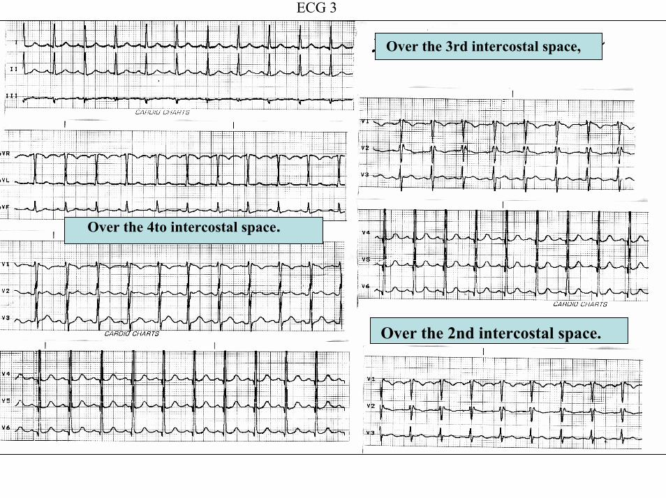
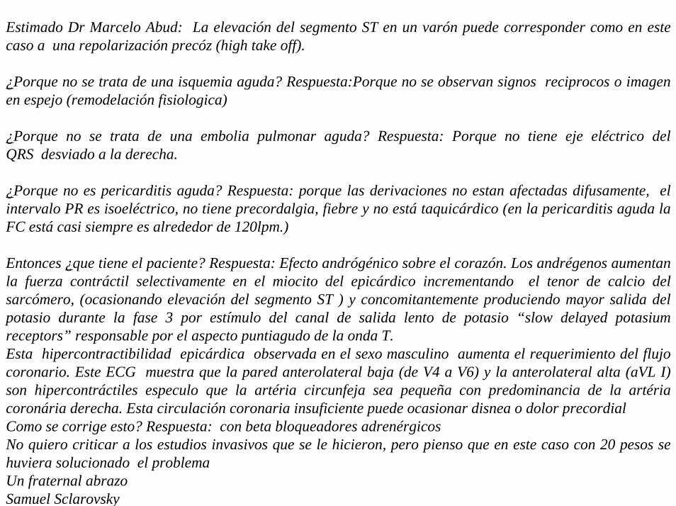
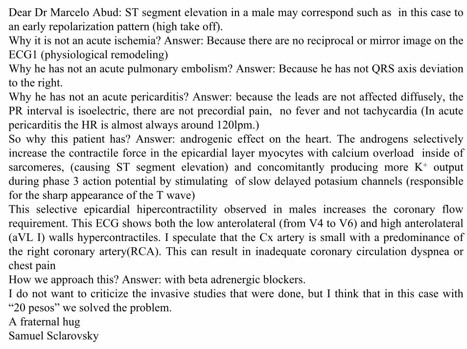
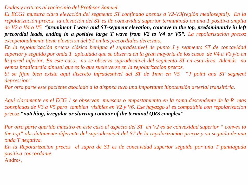
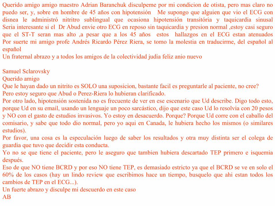
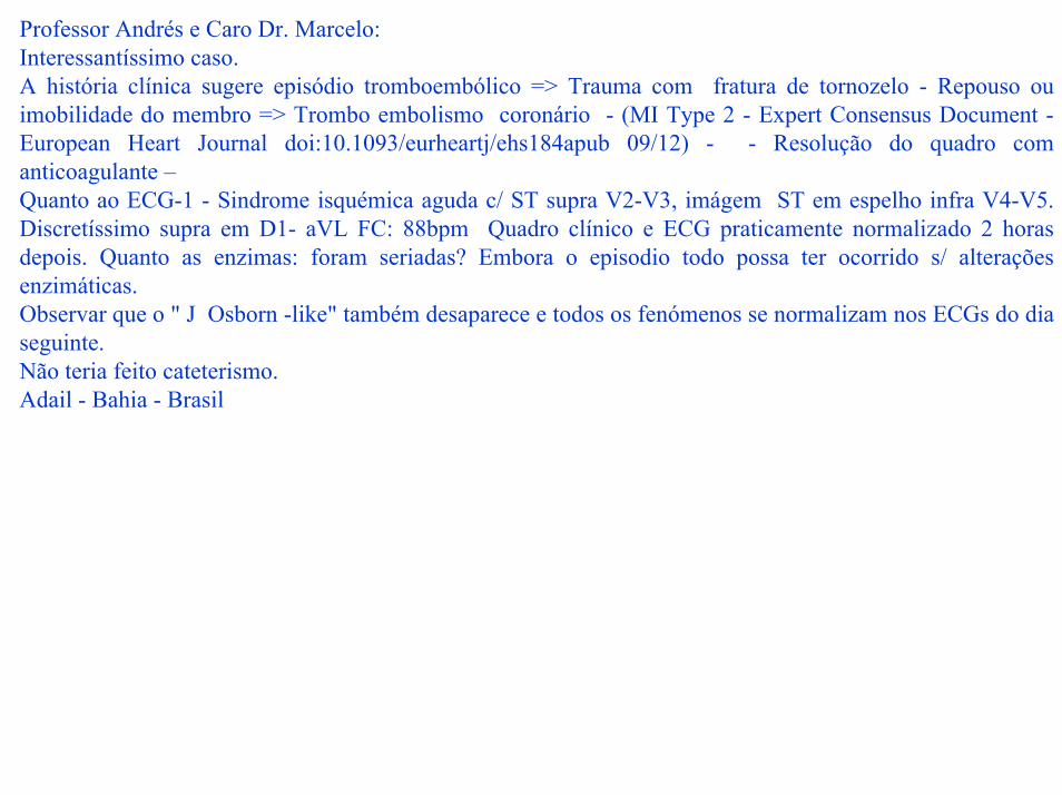
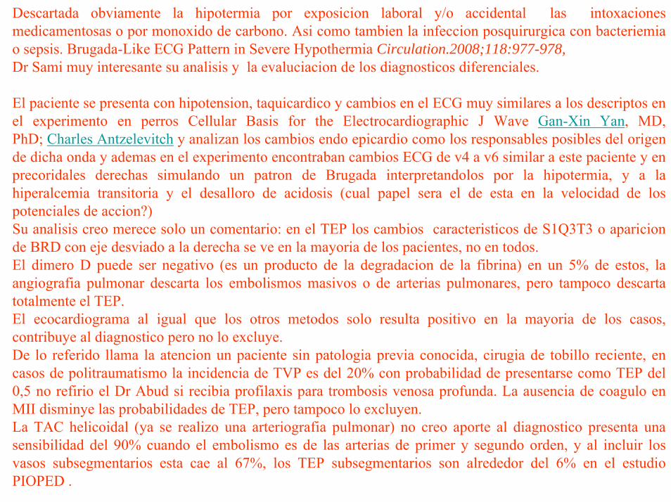
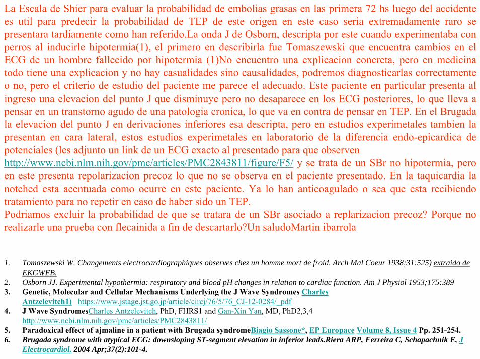
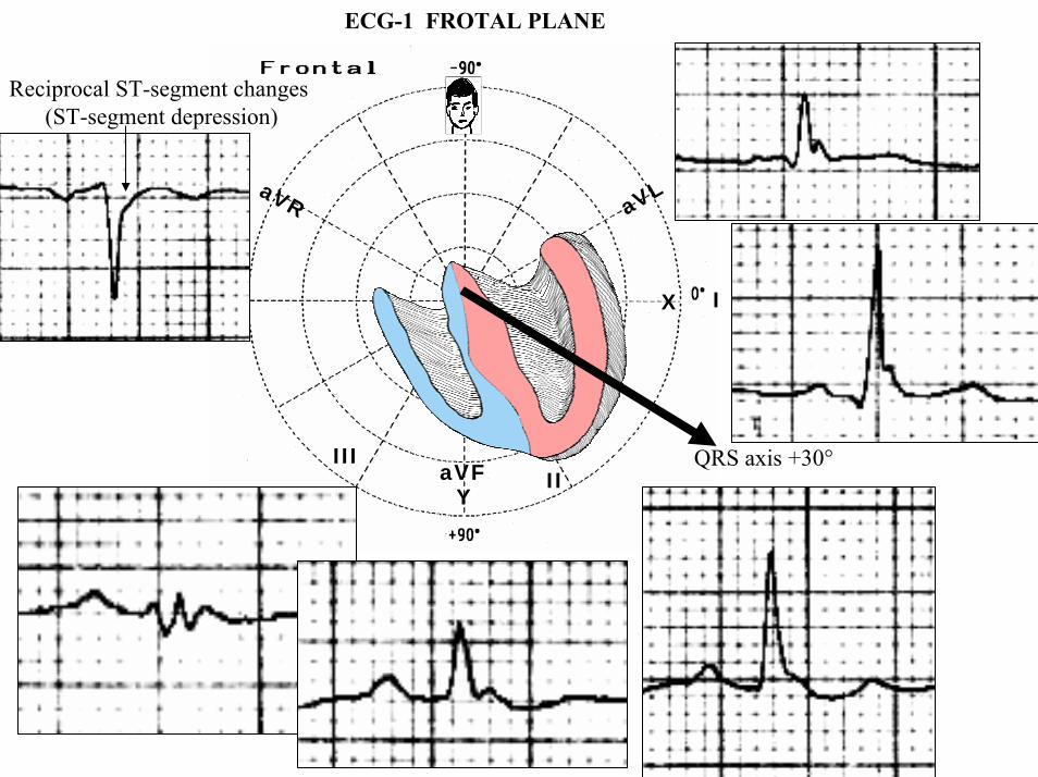
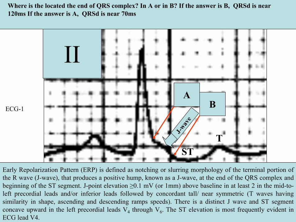
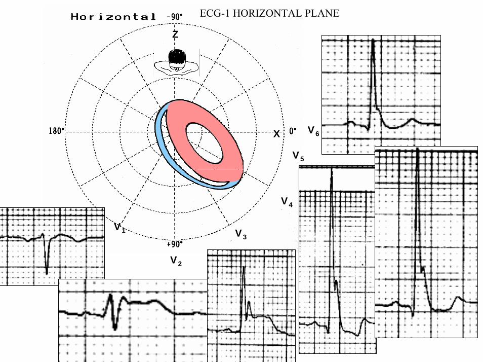
V6
V1
V4
V5
V2
V3
X
Z
ECG-1 HORIZONTAL PLANE
V2
V5
J point
QRSd 122ms
ECG-1
QRSd 122ms
Notching
40 ms80 ms
β
80 ms
High take-off
Type 1 Brugada pattern
Type 2 Saddle-back pattern
40 ms80 ms
β
80 ms
High take-off
Type 1 Brugada pattern
Type 2 Saddle-back pattern
V2
Is this a type 2 Brugada pattern or saddle-back pattern in V2
Tryphasic QRS Pattern RSracute
High take-off of racute ge 2mm high take-off frequently does not coincide with J wave descending arm of racutecoincides with beginning of ST segment the racute wave has rounded shape wide and usually of relatively low voltage in Brugada pattern Contrarily in IRBB the racute wave is peaked the angle between the upslope of the S-wave and the downslope of the r-wave (β angle) gt 58deg duration of the base of the triangle of racute at 5mm from high take-off gt35mm the QRS duration is longer in Brugada pattern type 2 than in other cases with racute in V1 and there is a mismatch between V1 and V6 In Brugada pattern the QRS complex end is earlier in V6 than in V1 V2
βangle gt 58deg
1 Bayeacutes de Luna A et al a consensus report Journal of Electrocardiol 2012 45433-
2 Chevallier S J Am CollCardiol2011 Nov 2258 2290-2298
ECG-1
ECG-1
RIGHT SAGGITAL PLANE ECG-1
YaVF
Z V2
ECG 2
aVR aVL
I
IIIII
X
Y
aVF
QRS axis +30ordm
ECG-2 FRONTAL PLANE
aVR aVR
ECG-1 ECG-2
J-Wave
Reciprocal ST-segment changes (ST-segment depression) Without reciprocal changes or mirror image
In Early Repolarization Pattern reciprocal ST-segment changes (ST-segment depression) are eventually confined the unipolar aVR lead (Absence of reciprocal changes or mirror image in others leads)
II
II
PR interval= 160msQRSd is 121ms
ECG-1
PR interval = 140msQRSd is near 80ms
J wave
ECG-2
V6
V5
V1
V4
V2
V3
X
Z
Is this a type 2 Brugada ECG pattern Answer in next slide
ECG-2
V2βangle gt58deg
Observation β angle is related to both depolarization an repolarization A wider QRS complex (slow conduction) and abnormal repolarization both contribute to a wider β angle
1 Chevallier S J Am Coll Cardiol2011 Nov 2258 2290-2298
In patients with suspected Brugada syndrome simple ECG criteria can enable discrimination between innocent incomplete RBBB from types 2 and 3 Brugada ECG patterns The mean β angle is significantly smaller in the patients with negative results on Antiarrhythmic Drug Challenge (ADC) compared wit patients with positive results on ADC Its optimal cutoff value is 58deg which yielded a positive predictive value of 73 and a negative predictive value of 87 for conversion to type 1 pattern on ADC α is slightly less sensitive and specific compared with β When the angles are combined with QRS duration it tended to improve discrimination
Y
aVF
Z V2
Right Sagittal Plane ECG-2
Triphasic QRS patternRSracute
β angle defined as the angle between the upslope of the S-wave and the downslope of the r-wave The mean β angle was significantly smaller in the 14 patients with negative results on AAD compared to the 24 patients with positive results on AAD Itrsquos optimal cutoff value was 58deg which yielded a positive predictive value of 73 and a negative predictive value of 87 for conversion to type 1 pattern on AAD
ST segment with saddleback appearance followed by positive or biphasic T wave in V1-V2 or from V1 through V3
β
V5V5
QRSd= 122ms QRSd= 110ms
ECG-1 ECG-2
Left precordial terminal QRS notching is more prevalent in malignant early repolarization abnormalities
Left precordial terminal QRS notching
1 Merchant FM et al Am J Cardiol 2009 Nov 15 1041402-1406
V2
V2
V3
V3
ECG-1
ECG-2
ECG-2
Dynamic inconstant or dramatic changes in repolarization are suggestive of malignant early repolarization abnormalities
1 Haiumlssaguerre M et al J Cardiovasc Electrophysiol 2009 Jan2093-98
ECG-1
Conclusion this patient has high possibility of concomitant Brugada syndrome and early repolarization In this a benign ERP or malignant early repolarization abnormalities We think it is very unlikely that is a Brugada phenocopyIn this case it is necessary additionally to perform
1 IV Ajmaline test2 ECG-AR3 Family study ( first degree relatives interrogatory physical ECGs)4 Magnetic resonance image5 Genetic screening
Ventricular biopsy is not considered mandatory but is recommended especially when it may clarify the nature of dubious findings identified with others test In approximately 6--10 of survivors of cardiac arrest no cardiac abnormality can be identified despite extensive clinical evaluation Autopsy data confirm that in a similar percentage of victims of SCD no structural heart disease or or mors sine materia can be identified at post mortem evaluation Incompletely penetrant genetic defects may underlie at least some of these unexplained deaths (1)
The ERP probably represents part of a spectrum of cardiovascular anomalies related to channelopaties including BrS IVF congenital short QT syndrome and that it may also have a molecular genetic origin of variable penetrance The denomination Early Repolarization Pattern is appropriate since it indicates that it is characterized by a given ECG pattern
1 Priori SG et al Cardiovasc Res 2001 May50218-223
Case Report similar
A 20 year-old male mulatto professional soccer player from Santo Andreacute Sao Paulo Brazil He didnrsquot finishthe second year in elementary school CatholicProfession Professional soccer playerReason for consultation Presented to consultation due to two presyncopal episodes and one syncopal episode in the last 30 days The first two episodes happened during a post-defecation period Both were preceded by dizziness nausea and vomiting The syncopal episodes occurred at rest with no prodromes The third one with express and brief loss of consciousness and sphincter relaxation at dawn 48 hours agoHis prior personal medical history was unremarkable and he successfully passed two prior periodical clinical-cardiovascular evaluations (twice a year always normal) Pre-competitive examination included 12-lead ECG echocardiogram and cardiopulmonary metabolic exercise testing (CMET) All normals Intense training for the last 3 years Familial Background One uncle from his fatherrsquos side family died of sudden cardiac death when he was 35
years old He ignores the cause His brother is being seeing by a cardiologist due to ldquocardiac arrhythmiardquo in treatment with a cardiologistfrom the Institute of the Heart from Satildeo Paulo (he cannot tell which is the cause) He denies having other relevant background details Pysical cardiovascular examination revealed bradycardia at 45 bpm with physiological sinus arrhythmia blood pressure 11070 mmHg The rest of the physical examination was unremarkable He denied consuming any type of neither medications nor drugs No addictionsHis first 12-lead ECGVectorcardiogram(VCG) (Fig 1) showed sinus bradycardia at 44 bpm P-wave axis of +60deg PR interval of 186ms QRS duration of 111ms QRS axis at + 63deg QT interval 427ms and corrected QTc 367ms There is a notch on the terminal portion of the QRS complex in the left precordial leads voltage criteria for left ventricular hypertrophy (SV1 + RV5 gt 35 mm ndash Sokolow index) prominent J-wave and concave ST-segment elevation in inferior leads V4 V5 and V6 Deep and narrow Q-waves in the left precordial leads V5 and V6 All these features were considered as an Early Repolarization Pattern (ERP)
Name JVS Gender Male Age 20 yo Ethnic Group Mulatto Weight 66 Kg Height 172 m Biotype Normoline Date 11262009 Profession competitive athlete (soccer player)
ECG diagnosis Sinus bradycardia (SB) HR 44bpm P axis +60deg PR 186ms QRSd 111ms QRS axis + 63deg QT 427ms QTc 367ms Conclusion SB + Early Repolarization Pattern (ERP) Notching or slurring of the terminal QRS complex voltage criteria of left ventricular hypertrophy SV1+ RV5 gt 35 mm (Sokolow index) prominent J wave and ST-segment elevation concave to the topending in a positive T wave from V4 to V6 Precordial greater than limb leads Relatively deep but narrow q waves in the left precordial leads (V5 and V6)
V6V4 V5
Prominent J wave and ST- segment elevation concave to the top ending in a positive T wave from V4 to V6 Notching or slurring morphology of the terminal portion of the R wave (J-wave) that produces a positive hump known as a J-wave (Arrows)
aVR
Eventually reciprocal ST segment depression confined only in Lead aVR1 observed in this particular case report
1 Mehta M Jain AC Mehta A Early Repolarization Clin Cardiol 1999 Feb 22 59-65
ECGVCG CORRELATIONS HORIZONTAL PLANE
V6
V1
V2
V3
X
Z
V5
V4
T
0J T
BEGINNING AND END OF QRS LOOP IN HP
J POINT in vectorcardiography it corresponds to 3 elements end of ventricular depolarization (QRS loop) beginning of repolarization (ST segment) when it does not present depression or elevation and T wave onset In this case 0 point and J point are not coincident In situations where there is depression or elevation of ST segment the J point does not coincide with the 0 point and the greater or lesser distance between both points indicate the greater or lesser ST segment elevation or depression The phenomenon is observed in early repolarization pattern acute phase of myocardial infarction Prinzmetal variant angina pericarditis Brugada syndrome arrhythmogenic right ventricular dysplasia etc
0 POINTIt corresponds to the QRS looponset (because PR segment does notexist) Alsocorrespond to the end of biatrialchamberactivation
T
J POINT
ECGVCG CORRELATIONS FORNTAL PLANE
aVR aVL
I
IIIII
X
Y
VERTICAL AXIS ON FRONTAL PLANE The frontal plane QRS axis and ST segment axis and T wave axis are all in the same direction THE ST
SEGMENT ELEVATION
USUALLY lt 05 mm IN THE LIMB
LEADS
YVF
Z V2
T
ECGVCG CORRELATIONS RIGHT SAGITTAL PLANE
A second 12-lead ECG (Fig 2) preformed 72 hours later showing1) Sinus bradycardia at 44 bpm2) Similar p-wave axis and intervals duration3) Dramatic modification of repolarizaccedilatildeo pattern in the right precordial leads A coved-type
(type 1) ECG brugada pattern can be distinguished4) Accentuation of j-waves and st segment elevation across the precordial leads was observed5) ST segment elevation on avr lead
See next slide
Name JVS Gender Male Age 20 yo Ethnic Group MulattoWeight 66 Kg Height 172 m Biotype Normoline Date 11292009Profession Competitive Soccer Player FIGURE 2
ECG diagnosis Sinus Arrhythmia(SA) sinus bradycardia (SB) HR 44bpm P axis +60deg PR 186ms QRSd 111ms QT 427ms QTc 367 Conclusion SA + SB + Early Repolarization Pattern (ERP) Type 1 Brugada ECG pattern Dramatic accentuation of J waves across the precordial and limb leads Transient augmentation of global J waves may be indicative of a
highly arrhythmogenic substrate heralding multiple episodes of VF in patients with ERP
aVR LEAD
11262009 11292009
DEPRESSION
ELEVATION
ST SEGMENT DEPRESSION IN aVR = 1mm ST SEGMENT ELEVATION IN aVR = 2mm
V1 V2V2
1129200911262009
V1
11292009 11262009
SPONTANEOUS MODIFICATION OF REPOLARIZATION TO TYPE 1 BRUGADA ECG PATTERN
11262009
V6
11292009V6
ST segment elevation lt 2mm ST segment elevation gt 2mm
11292009
V5
11262009
V5
ST segment elevation = 1mm ST segment elevation = 25mm
EARLY REPOLARIZATION PATTERN CONCEPT
DEFINITION Early Repolarization Pattern (ERP) is a well-recognized Enigmatic idiopathic electrocardiographic phenomenon characterized by notching or slurring morphology of the terminal portion of the R wave (J-wave) that produces a positive hump known as a J-wave at the end of the QRS complex and beginning of the ST segment It considered to be present when at least two adjacent precordial leads show elevation of the ST segment with values ge01 mV (or 1mm) above baseline followed by concordant tall near symmetric (T waves having similarity in shape ascending and descending ramps speeds)
EPIDEMIOLOGY ERP occur in 2 to 5 of the population often in men young adults afro-descendents and competitive athletes and in up to 131 to 482 of patients presenting with chest pain in the emergency rooms and coronary care units There is a distinct J wave and ST segment concave upward in the left precordial leads V4 through V6 The ST elevation is most frequently evident in ECG lead V4 The ST segment elevation is usually le2mm (precordial greater than limb leads) and is lt 05 mm in the limb leadsReciprocal ST-segment changes (ST-segment depression) are eventually confined the unipolar aVR lead (Absence of reciprocal changes or mirror image) In ERP vertical QRS axis electrical is the rule The frontal plane QRS axis the ST segment axis and T wave axis are all in the same directionThis ECG pattern has been considered a benign normal ECG variant for over six decades3
1 Hasbak P Engelmann MD Early repolarization ST-segment elevation as a normal electrocardiographic variant Ugeskr Laeger 2000 162 5928-5929
2 Brady WJ Chan TC Electrocardiographic manifestations benign early repolarization J Emerg Med 1999 17473-478
3 Benito B Guasch E Rivard L Nattel S Clinical and mechanistic issues in early repolarization of normal variants and lethal arrhythmia syndromes J Am Coll Cardiol 2010 Oct 5 56 1177-1186
MAIN ELECTROCARDIOGRAPHIC FEATURES IN ERPHeart rate Predominant sinus bradycardia frequently associated with respiratory sinus arrhythmia
PR interval First degree AV block is observed in 5 to 39 among professional athletes In the non-athlete population it is observed in 065 of cases QRS axis Vertical electrical axis is the rule The frontal plane QRS axis and ST segment axis and T wave axis are all in the same directionQRS duration (90 plusmn 10 ms) subjects than in age-matched healthy controls (80 plusmn 10 ms) QRS morphology Notching or slurring of the terminal QRS complex Prominent relatively deep but narrow q waves may appear in the left precordial leadsQRS voltage Eventually voltage criteria of LVH are observed in male competitive athletes SV1+ RV5 gt 35 mm (Positive Sokolow index) High QRS voltage is more frequent in male athletes but its correlation with LVH is low Voltage decreases after deconditioning is slow The distinction between the physiological athletes heart and pathological conditions has critical implications for professional athletes An abrupt transition may occur from right-oriented complexes to left-oriented complexes in the precordial leads secondary to counterclockwise rotation on precordial leads About two-thirds of clockwise rotation and counterclockwise rotation could be attributed to the septal angle by anatomical rotation of the heart in one plane around the long axis but other factors appear to be responsible for such ECG findings in the remaining one-third of cases Relatively higher positions of the precordial ECG leads as observed in the vertical heart appeared to be responsible for clockwise rotation in some patients and left septal fascicular block is suspected to be responsible for counterclockwise rotation in others patientsJ-point elevation Notching irregular or slurring contour of the terminal QRS complex (J point) Variant of Osborn wave is seen in ERP
ST segment Widespread ST segment elevation (precordial greater than limb leads) The characteristic ST segment is elevated upward concave confined more frequently in precordial leads with reciprocal depression only in aVR The concavity is observed at the initial up-sloping portion of ST segment or upwardly concave ST segment morphology Unfortunately concave ST morphology cannot be used to rule out ST elevation from AMI with left anterior descending coronary occlusion because it is common in these circumstances The ST elevation is most frequently evident in ECG lead V4 There is a distinct J wave and ST segment in the left precordial leads V4 through V6 The ST elevation in ERP is usually lt 2 mm (but can rarely be gt 5 mm) in the precordial leads and the greatest ST elevation is usually seen in the mid-to-left precordial leads The ST segment elevation is usually lt 05 mm in the limb leads
T wave characteristics Concordant T waves of large amplitude (prominent matching T waves) typical pseudo-asymmetrical (lsquosymmetroidrdquo) or slightly asymmetrical matching T waves often of large amplitude upright tall and peaked most conspicuously from V2 to V4 or V5 sometimes seen in leads DII DIII and aVF as a rule T waves may appear as of large amplitude peaked or pointed symmetric and matching Vagotonic or high T wave voltages followed by U waves are frequent when sinus bradycardia is present Tall positive ad symmetric or symmetroid T waves are not only seen occasionally in the very early stages of MI but also in hyperkalemia and in ERP sinus bradycardia
QT intervals QT maximum The maximum Q-onset-T-end interval This parameter is higher in ERV subjects than in normal controls
QTp maximum maximum Q-onset-T-peak interval This parameter is higher in ERV subjects than in age-matched healthy controls
Rate-corrected QTc maximum This parameter is lower in ERP subjects than in age-matched healthy controls
QTpc maximum This parameter is lower in ERV subjects than in age-matched healthy controls
U wave because bradycardia U waves are frequent in ERV they are best observed in the V3 lead U waves are frequent when sinus bradycardia is present
Other ECG characteristics of ERPRelative temporal stability of the ST segment and T wave pattern is observedReciprocal changes are not seen in ERP There are no evolutionary short-term changes in the ST segment and T waves and Q waves do not appear
Discusions Early Repolarization Pattern (ERP) or Early Repolarization Variant (ERV) is an enigmati
electrocardiographic phenomenon characterized by prominent J wave and ST-segment elevation in multiplleads
Recently there has been renewed interest in ERP because of similarities to the arrhythmogenic Brugadsyndrome (BrS) Not much is known about the epidemiology of ERP and several studies have reported thathis condition is associated with a good prognosis
Both ERP and BrS exhibit some similarities including the ionic underlying mechanism the analogouresponses to changes in heart rate and autonomic tone sympathicomimetics (isoproterenol test) as well as insodium channel and beta-blockers These observations raise the hypothesis that ERP may be not as benign atraditionally believed Additionally there are documents showing that ST-segment height in the man igreatly influenced by central sympathetic nervous activity both at baseline and during physiologic andpharmacological stress Central sympathetic dysfunction regularly results in multilead ST-segment elevationor J wave that decreases or below isoelectric baseline during low dose isoproterenol infusion An earlyrepolarization pattern in the inferior leads of a standard electrocardiogram is associated with an increased riskof death from cardiac causes in middle-aged subjects1
POSSIBLE SIMILARITIES BETWEEN ERV AND BrSbullMore frequent in malesbullBoth occur more frequently in young adults and in individuals without apparent structural heart diseasebullBoth may influence just the V1-V2 leads Rarely (9) can ST elevation be observed in ERP only in the right precordial leads V1-V2 or in the inferior ones2
1 Tikkanen JT Anttonen O Junttila MJ Aro AL Kerola T Rissanen HA Reunanen A Huikuri HV Long-Term Outcome Associated with Early Repolarization on Electrocardiography N Engl J Med 2009 Nov 16 [Epubahead of print]
2 Hasbak P Engelmann MDEarly repolarization ST-segment elevation as a normal electrocardiographic variant Ugeskr Laeger 2000 162 5928-5929
When ST elevation is normal it can reach up to 3 mm in V2-V3 especially in young people In those individuals over 40 years it seldom exceeds 2 mm Both can show incomplete RBBB pattern or right bundle branch conduction disorder in BrS it can present atypical features RBBB-like and of the saddle type by exclusive elevation of the J point S wave with delay in the left leads DI aVL V5 and V6 could be absent as it is to be expected in a classic RBBB The elements considered as typical in BrS are 1) elevation of the terminal part of QRS (prominent J wave) 2) elevated and descending ST not related to lesion of ischemic (idiopathic) injury 3) negative T wave in the right precordial leads 4) normal QTc or near normal 5) Eventually absence of final delay in left leads as it would be expected in a classic RBBB1 In ERV when associated to athletersquos heart QRS can present a moderate extension (100 ms to 110 ms) in 15 of the cases which in nonathlete normal population in a 24 is called outflow tract hypertrophy In this case r does not exceed the 5 mm and is lower than S in the same lead rSrBoth may improve repolarization during the stress test with use of isoproterenolBoth respond to a shortening of AP phase 2 in a part of ventricular thickness and intensification of fast repolarization notch (phase 1) mediated by transmural dispersion of ventricular repolarization by a larger notch in the Ito channel2The alteration of the Ito and ICa2+-L channels in BrS and in ERP are the electrophysiologic substrate that explains the J point and ST segment elevation because they cause the intensified notch in phase 1 and suppression in phase 2 duration in the epicardium and in the endocardium of ventricular wall thickness
1 Hiss RG Lamb LE Electrocardiographic findings in 122043 individuals Circulation 1962 25947-9612 Antzelevitch Ch Xin Yan G Shimuzi W et al Electrical heterogeneity the ECG and Cardiac Arrhythmias In Zipes
DP Jalife J Cardiac Electrophysiology From Cell to Bedside Third Edition WB Saunders Company2000 Chapter 26 p 222-238
ELEMENTS FOR DIFFERENTIAL DIAGNOSIS BETWEEN ERP AND BrS
I) Family background ERP negativeBrS frequently positive
II) Ethnic GroupERP predominantly in African descendents(1) or equally common in all races(2) BrS predominantly in Asian group (58) and Caucasian people(3)
III) GenderERP male gender predominanceBrS great predominance in the male gender (malefemale ratio ndash 81 in non Asian and 101 in Asian
peopleIV) Response to IC group antiarrhythmic agents
BrS flecainide used in a 10 mgKg dosage in 10 minutes increases ST elevation and QRS duration in a more significant way in patients with BrS than in individuals without the entity and only in those it triggers ventricular extrasystoles(4)
ERP it can induce a pattern similar to BrS however the degree of ST elevation caused by the drug is much higher in patients with BrS than in patients without the disease
1 Grusin H Peculiarities of the Africanrsquos electrocardiogram and the changes observed in serial studies Circulation 1954 9 860-867
2 Mehta M Jain AC Mehta A Early Repolarization Clin Cardiol 1999 Feb2259-653 Nademanee K Veerakul G Nimmannit S Chaowakul V Bhuripanyo K Likittanasombat K Tunsanga K Kuasirikul S
Malasit P Tansupasawadikul S Tatsanavivat P Arrhytmogenic marker for the sudden unexplained death syndrome in Thail men Circulation 1997 962595-2600
4 Shimizu W Antzelevitch C Suyama K Kurita T Taguchi A Aihara N Takaki H Sunagawa K Kamakura S Effect of sodium channel blockers on ST segment QRS duration and corrected QT interval in patients with Brugada syndromeJ Cardiovasc Electrophysiol 2000 111320-1329
BrS patients with Inferior and lateral ECG repolarization abnormalities have longer baseline PR intervals(1)Both an S wave width ge80ms in V1 and ST elevation ge18mm in V2 are highly specific indicators of VF and criteria for high-risk BrS(2)There are several references in literature to patients with elevation of the J point and ST segment convex to the top or straight descendent in inferior leads (Brugada sign) or concomitantly ST-segment elevation in the right precordial and inferior leads II III and aVF(3) in absence of hypothermia ischemia or electrolytic disorders which we call ldquoatypical Brugada patternrdquo atypical BrS variant BrS or BrS variant or idiopathic VF (a variant of the BrS with ST-segment elevation in inferior leads(4)Pilsicainide may induce PVCs and PVT in atypical BrS and the infusion of isoproterenol suppress the arrhythmias and normalize the ST-segment elevation(5)
1 Sarkozy A Chierchia GB Paparella G Boussy T De Asmundis C Roos M Henkens S Kaufman L Buyl R Brugada R Brugada J Brugada P Inferior and lateral electrocardiographic repolarization abnormalities in Brugada syndromeCirc ArrhythmElectrophysiol 2009 Apr 2154-161
2 Atarashi H Ogawa S For The Idiopathic Ventricular Fibrillation Investigators New ECG Criteria for High-Risk Brugada Syndrome Circ J 2003 Jan 67 8-10
3 Lombardi F Potenza S Beltrami A Verzoni A Brugada P Brugada RSimultaneous ST-segment elevation in the right precordial and inferior leads in Brugada syndrome J CardiovascMed (Hagerstown) 2007 8 201-204
4 Letsas KP Efremidis M Pappas LK Gavrielatos G Markou V Sideris A Kardaras FEarlyrepolarization syndrome is it always benign Int J Cardiol 2007114 390-392
5 Chinushi M Izumi D Furushima H Watanabe H Aizawa YMultiple premature beats triggered ventricular arrhythmias during pilsicainide infusion in a patient with inferior ST-segment elevation Pacing Clin Electrophysiol 2006 29 1445-1448
Potet et al(1) identified a G752R mutation on SNC5A that produced ST segment elevation and prominent J wave in leads II III and aVF The authors provide genetic demonstration that Brugada ECG anomalies related to a unique SCN5A mutation can be observed either in the inferior or the right precordial leads The early repolarization pattern in inferolateral leads is not an uncommon finding in BrS and this pattern is not associated with a worse outcome in subjects with BrS(2) The spontaneous ERP occurred more frequently among patients with BrS than in 283 family members not having BrS (11 versus 6 P=003) Class I antiarrhythmic drug administration provoked inferior-lateral coved Brugada pattern in 13 patients with BrS These patients had longer baseline PR intervals and Class I antiarrhythmic drug induced QRS interval prolongation (108 to 178 versus 102 ms to 131 ms Plt0001) In 3 patients the Class I antiarrhythmic drug provoked coved Brugada pattern only present in the inferior leads Inferior-lateral ERP occurs spontaneously relatively frequently in BrS These patients have a more severe phenotype Class I antiarrhythmic drug administration provokes inferior-lateral coved Brugada pattern in 46 of patients The Class I antiarrhythmic drug exceptionally provoked coved Brugada pattern only observed in the inferior leads(3)
1 Potet F Mabo P Le Coq G et al Novel brugada SCN5A mutation leading to ST segment elevation in the inferior or the right precordial leads J Cardiovasc Electrophysiol 2003 142000-2003
2 Letsas KP Sacher F Probst V Weber R Knecht S Kalusche D Haiumlssaguerre M ArentzTPrevalence of early repolarization pattern in inferolateral leads in patients with Brugada syndrome Heart Rhythm 2008 Dec 5 1685-1689
3 Sarkozy A Chierchia GB Paparella G Boussy T De Asmundis C Roos M Henkens S Kaufman L Buyl R Brugada R Brugada J Brugada P Inferior and lateral electrocardiographic repolarization abnormalities in Brugada syndrome Circ Arrhythm Electrophysiol 2009 Apr2 154-161
Type 1 ECG Brugada pattern in the peripheral leads was observed in 42 of patients during ajmaline test (103 of positive tests) and was associated with longer QRS and greater QTc prolongation compared with the rest of the patients1Atypical Brugada Syndrome or IVF with J waves in inferior lateral or inferior lateral leads
and Familial Idiopathic Ventricular Fibrillation and BrSSeveral mutations in genes SCN5A KCNJ8 DPP6 SCN3B and CACNA2D1 had been identified 1 On SCN5A gene2 3 Observation We do not believe that SCN5A should be attributed to IVF because this report clearly shows conduction defects (as expected) so this is not IVF but conduction delay at different levels in the heart2 On KCNJ8 gene 4 5 Missense variant in exon 3 (NC-000012) of the KCNJ8 gene a subunit of the K(ATP)channel23 Genomic DNA sequencing of K(ATP) channel genes showed missense variant in exon 3 (NC_000012) of the KCNJ8 gene a subunit of the K(ATP) channel conferring predisposition to dramatic repolarization changes and ventricular vulnerability
1 Batchvarov VN Govindan M Camm AJ Behr ERBrugada-like changes in the peripheral leads during diagnostic ajmaline test in patients with suspected Brugada syndrome Pacing Clin Electrophysiol 2009 Jun32695-703
2 Potet F Mabo P Le Coq G Probst V Schott JJ Airaud F Guihard G Daubert JC Escande D Le MarecH Novel brugada SCN5A mutation leading to ST segment elevation in the inferior or the right precordial leads J Cardiovasc Electrophysiol 2003 14 200-203
3 Akai J Makita N Sakurada H Shirai N Ueda K Kitabatake A Nakazawa K Kimura A Hiraoka M A novel SCN5A mutation associated with idiopathic ventricular fibrillation without typical ECG findings of Brugada syndrome FEBS Lett 2000 Aug 11 479 29-34
4 Haissaguerre M Derval N Sacher F Jesel L Deisenhofer I de Roy Let al Sudden cardiac arrest associated with early repolarization N Engl J Med 2008 May 8 3582016-2023
5 Haiumlssaguerre M Sacher F Nogami A Komiya N Bernard A Probst V et al Characteristics of recurrent ventricular fibrillation associated with inferolateral early repolarization role of drug therapy J Am CollCardiol 2009 Feb 17 53 612-619
On DPP6 gene1 Dipeptidyl aminopeptidase-like protein 6 locus on chromosome7q26 Voltage-gated potassium channels OMIM126141 The ECG shows rapid PVT Monomorphic PVCs with very short coupling intervals The PVC (302 +- 52 msec) within 40 msec of the peak of the preceding Absence of T wave Pause-dependence
Valdivia et al2 reported a case with IVF and a novel mutation in the SCN3B-encoded sodium channel beta subunit Navbeta3 that causes a loss of function of Nav15 channels in vitro and provides molecular and cellular evidence implicating mutations in Navbeta3 as a cause of IVF Mutation in this SCN3B gene causes also BrS type 73
L-type calcium channel (LTCC) mutations have been associated with BrS SQTS and Timothy syndrome (LQT8) Little is known about the extent to which LTCC mutations contribute to the J-wave syndromes associated with SCD Mutations in the LTCCs are detected in a high percentage of probands with J-wave syndromes associated with inherited cardiac arrhythmias suggesting that genetic screening of Ca(v) genes may be a valuable diagnostic tool in identifying individuals at risk Burashnikov et al4 identify CACNA2D1 as a novel BrS susceptibility gene and CACNA1C CACNB2 and CACNA2D1 as possible novel ERS susceptibility genes4
1 Alders M Koopmann TT Christiaans I Postema PG Beekman L Tanck MW et al Haplotype-sharing analysis implicates chromosome 7q36 harboring DPP6 in familial idiopathic ventricular fibrillation Am J Hum Genet 2009 Apr 84 468-476
2 Valdivia CR Medeiros-Domingo A Ye B Shen WK Algiers TJ Ackerman MJ et al Loss-of-function mutation of the SCN3B-encoded sodium channel beta3 subunit associated with a case ofidiopathic ventricular fibrillation Cardiovasc Res 2010 Jun 1 86 392-400
3 Hu D Barajas-Martinez H Burashnikov E Springer M Wu Y Varro A et al A mutation in the beta-3 subunit of the cardiac sodium channel associated with Brugada ECG phenotype Circ Cardiovasc Genet 2009 2 270-278
4 Burashnikov E Pfeiffer R Barajas-Martinez H Delpoacuten E Hu D Desai M et al Mutations in the cardiac L-type calcium channel associated with inherited J-wave syndromes and sudden cardiac death Heart Rhythm 2010 Dec71872-1882
From a multicenter cohort of 122 patients (90 male age 37 +- 12 years) with IVF and ERP in the inferolateral leadsHaiumlssaguerre et al1 selected all patients with more than 3 episodes of VF (multiple) including those with electrical storms (ge3 VF in 24 h) Multiple recurrences of VF occurred in 27 of patients with ERP and may be life threatening Isoproterenol in acute cases and quinidine in chronic cases are effective antyharrhithmic drugsThe last one is necessary associated to ICDThe so-called atypical BrS is characterized by ECG abnormalities of the J wave and ST-segment elevation appeared in the inferior andor lateral leads The ERP in inferolateral leads is not an uncommon finding in BrS2 There is a high incidence of the ERP confined in inferolateral leads in patients with IVF The ECGs have an elevation of the QRS-ST junction of at least 01 mV from baseline in the inferior or lateral lead manifested as QRS slurring or notching Among patients with a history of IVF there is an increased prevalence of ERP
1 Haissaguerre M Derval N Sacher F Jesel L Deisenhofer I de Roy L Pasquieacute JL Nogami A Babuty D Yli-Mayry S De Chillou C Scanu P Mabo P Matsuo S Probst V Le Scouarnec S Defaye P Schlaepfer J Rostock T Lacroix D Lamaison D Lavergne T Aizawa Y Englund A Anselme F ONeill M Hocini M Lim KT Knecht S Veenhuyzen GD Bordachar P Chauvin M Jais P Coureau G Chene G Klein GJ Cleacutementy J Sudden cardiac arrest associated with early repolarization N Engl J Med 2008 May 8 3582016-2023
2 Letsas KP Sacher F Probst V Weber R Knecht S Kalusche D Haiumlssaguerre M Arentz T Prevalence of early repolarization pattern in inferolateral leads in patients with Brugada syndrome Heart Rhythm 2008 Dec51685-1689
Because a contingent percentage has not yet been well established it has been suggested that BrS constitutes the same entity in a large number of IVF diagnosed cases since they have common clinical features
1 BOTH ARE MORE FREQUENT IN MEN2 BOTH HAPPEN IN MIDDLE-AGE PEOPLE3 BOTH OCCUR IN APPARENT STRUCTURALLY NORMAL HEARTS14 BOTH CAN AFFECT THE SAME GENE (SCN5A)5 BOTH AFFECT EVENTUALLY THE SAME LOCUS (3p24-p21)
Wellens et al2 has pointed out that in order to be accurate about such a percentage it is important to define the diagnostic criteria used to classify BrS in IVF patients Curiously the so-called Idiopathic VT in apparently healthy hearts without structural heart disease and primary electrical disease also affects the 3p21 locus but not 3p24 and the gene is different GNA12
Survivors of SCD by VF without structural heart disease and a normal coronary angiogram are often diagnosed with IVTIVF3
A careful analysis of the ECG is essential in the differential diagnosis of this type of apparently unexplained SCD because intermittent or permanent RBBB with J point and ST-segment elevation in leads V1 to V3 is now classified as BrS
1 Goethals P Debruyne P Saffarian M Drug-induced Brugada syndrome Acta Cardiol1998 53157-60
2 Wellens HJJ Rodriquez LM Smeets JL Ventricular tachycardia in structural normal hearts In Zipes DP Jalife J Editors Cardiac Electrophysiology from cell tobedside Philadelphia 1995 WB Saunders
3 Chen Q Kirsch GE Zhang D Brugada R Brugada P Brugada J et al Genetic basis and molecular mechanisms for idiopathic ventricular fibrillation Nature 1998392 293-296
DIAGNOSTICS TRACKS TO DIFFERENTIATE BENIGN ERP FROM MALIGNANT EARLY REPOLATIZATION ABNORMALITIES
BENIGN ERP MALIGNANT EARLY REPOLARIZATION ABNORMALITIES
Family history of unexplained SCD in young relatives(lt45yo)
Absent Possible however infrequent1
Personal history Asymptomatic Asymptomatic repetitive syncope episodes or recovered of cardiac arrest
Left precordial terminal QRS notching Less prevalent More prevalent2
STT ratio in lead V6calculated by dividing the millimeters of
ST-segment elevation by the millimeters to the tallest point of the T wave
Each value is measured from the isoelectric point
lt0253 Non-tested
1 Viskin S Belhassen B Idiopathic ventricular fibrillation American Heart Journal 1990 120 661-671
2 Tikkanen JT Anttonen O Junttila MJ Aro AL Kerola T Rissanen HA Reunanen A et al Long-term outcome associated with early repolarization on electrocardiography N Engl J Med 2009 Dec 24361 2529-2537
3 Ginzton LE Laks MM The differential diagnosis of acute pericarditis from the normal variant new electrocardiographic criteria Circulation 1982 May 65 1004-1009
ST-segment elevation shape Concave upward Convex upwardJ waves are present in asymp 30 IVF patients1
Early repolarization is significantly prevalent in the subjects after cardiac arrest
(present in 31 of those resuscitated )Eventually ldquoLambda-waverdquo 23
J-point elevation and ST-segment degree Usually lt 2 mmlt05mm in limb leads
Frequent gt2mmJ-point elevation gt 2mm in inferior leads is associated with an increased risk of death
from cardiac causes in middle-aged subjects3
T wave characteristic Positive large T wave from V2 to V4 or V5 Concordant
Frequent negative discordant
Reciprocal ST-segment changes (Mirror
image)Absent With exception aVR5 Frequently Present3
1 Viskin S Idiopathic ventricular fibrillation Le Syndrome dHaiumlssaguerre and the fear of J waves J Am Coll Cardiol 2009 Feb 17 53 620-622
2 Gussak I Bjerregaard P Kostis J Electrocardiographic lambda wave and primary idiopathic cardiac asystole a new clinical syndrome J Electrocardiol 2004 Apr 37 105-107
3 Riera AR Ferreira C Schapachnik E Sanches PC Moffa PJ Brugada syndrome with atypical ECG downsloping ST-segment elevation in inferior leads J Electrocardiol 2004 Apr37101-104
4 Tikkanen JT Anttonen O Junttila MJ Aro AL Kerola T Rissanen HA Reunanen A et al Long-term outcome associated with early repolarization on electrocardiography N Engl J Med 2009 Dec 24361 2529-2537
5 Mehta M Jain AC Mehta A Early Repolarization Clin Cardiol 1999 Feb 22 59-65
Monomorphic PVCs with very short coupling
(250 to 350ms)
Absent Characteristic1 Pause-dependency absent2
QTc Interval Normal Minimally short in 35 among masculine gender
(Between 340 to 360ms3)
Late Potentials(LPs) Absent Frequent if J wave presentShowing circadian variation with night
ascendancy4
1 Alders M Koopmann TT Christiaans I Postema PG Beekman L Tanck MW et al Haplotype-sharing analysis implicates chromosome 7q36 harboring DPP6 in familial idiopathic ventricular fibrillation Am J Hum Genet 2009 Apr 84 468-476
2 Viskin S Lesh MD Eldar M Fish R Setbon I Laniado S et al Mode of onset of malignant ventricular arrhythmias in idiopathic ventricular fibrillation J Cardiovasc Electrophysiol 1997 Oct 8 1115-11 20
3 Viskin S Zeltser D Ish-Shalom M Katz A Glikson M Justo D et al Is idiopathic ventricular fibrillation a short QT syndrome Comparison of QT intervals of patients with idiopathic ventricular fibrillation and healthy controls Heart Rhythm 2004 Nov 1 587-591
4 Abe A Ikeda T Tsukada T Ishiguro H Miwa Y Miyakoshi M Circadian variation of latepotentials in idiopathic ventricular fibrillation associated with J waves insights into alternative pathophysiology and risk stratification Heart Rhythm 2010 May 7 675-682
ConclusionIn spite of both malignant early repolarization abnormalities of IVF and benign early repolarization pattern being able to cause J point and ST segment elevation the characteristics of both are clearly different to the point of a differentiation being quite simple when ECGs are analyzed by experts Any ER should raise the suspicion of IVF when it presents ST segment elevation ge 2 mm mainly in inferior wall superior convexity eventually lambda wave shape pattern of dynamic presentation inconstant with dramatic intensity of ST segment elevation discordant T wave polarity related ST segment shift presence of reciprocal ST-segment changes (mirror image) tendency to discrete short QTc interval in male gender(ge340ms) premature monomorphic ventricular contractions with very short coupling interval(250 to 350ms) not related with exercise and late potentials with circadian variation and night ascendancy on SA-ECG Contrarily in benign ERP ST elevation observed in the mid-to-left precordial leads is usually le 2 mm and lt05 mm in the limb leads and concave to the top followed by a positive concordant tall T wave
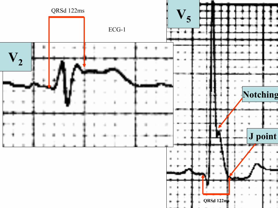
V2
V5
J point
QRSd 122ms
ECG-1
QRSd 122ms
Notching
40 ms80 ms
β
80 ms
High take-off
Type 1 Brugada pattern
Type 2 Saddle-back pattern
40 ms80 ms
β
80 ms
High take-off
Type 1 Brugada pattern
Type 2 Saddle-back pattern
V2
Is this a type 2 Brugada pattern or saddle-back pattern in V2
Tryphasic QRS Pattern RSracute
High take-off of racute ge 2mm high take-off frequently does not coincide with J wave descending arm of racutecoincides with beginning of ST segment the racute wave has rounded shape wide and usually of relatively low voltage in Brugada pattern Contrarily in IRBB the racute wave is peaked the angle between the upslope of the S-wave and the downslope of the r-wave (β angle) gt 58deg duration of the base of the triangle of racute at 5mm from high take-off gt35mm the QRS duration is longer in Brugada pattern type 2 than in other cases with racute in V1 and there is a mismatch between V1 and V6 In Brugada pattern the QRS complex end is earlier in V6 than in V1 V2
βangle gt 58deg
1 Bayeacutes de Luna A et al a consensus report Journal of Electrocardiol 2012 45433-
2 Chevallier S J Am CollCardiol2011 Nov 2258 2290-2298
ECG-1
ECG-1
RIGHT SAGGITAL PLANE ECG-1
YaVF
Z V2
ECG 2
aVR aVL
I
IIIII
X
Y
aVF
QRS axis +30ordm
ECG-2 FRONTAL PLANE
aVR aVR
ECG-1 ECG-2
J-Wave
Reciprocal ST-segment changes (ST-segment depression) Without reciprocal changes or mirror image
In Early Repolarization Pattern reciprocal ST-segment changes (ST-segment depression) are eventually confined the unipolar aVR lead (Absence of reciprocal changes or mirror image in others leads)
II
II
PR interval= 160msQRSd is 121ms
ECG-1
PR interval = 140msQRSd is near 80ms
J wave
ECG-2
V6
V5
V1
V4
V2
V3
X
Z
Is this a type 2 Brugada ECG pattern Answer in next slide
ECG-2
V2βangle gt58deg
Observation β angle is related to both depolarization an repolarization A wider QRS complex (slow conduction) and abnormal repolarization both contribute to a wider β angle
1 Chevallier S J Am Coll Cardiol2011 Nov 2258 2290-2298
In patients with suspected Brugada syndrome simple ECG criteria can enable discrimination between innocent incomplete RBBB from types 2 and 3 Brugada ECG patterns The mean β angle is significantly smaller in the patients with negative results on Antiarrhythmic Drug Challenge (ADC) compared wit patients with positive results on ADC Its optimal cutoff value is 58deg which yielded a positive predictive value of 73 and a negative predictive value of 87 for conversion to type 1 pattern on ADC α is slightly less sensitive and specific compared with β When the angles are combined with QRS duration it tended to improve discrimination
Y
aVF
Z V2
Right Sagittal Plane ECG-2
Triphasic QRS patternRSracute
β angle defined as the angle between the upslope of the S-wave and the downslope of the r-wave The mean β angle was significantly smaller in the 14 patients with negative results on AAD compared to the 24 patients with positive results on AAD Itrsquos optimal cutoff value was 58deg which yielded a positive predictive value of 73 and a negative predictive value of 87 for conversion to type 1 pattern on AAD
ST segment with saddleback appearance followed by positive or biphasic T wave in V1-V2 or from V1 through V3
β
V5V5
QRSd= 122ms QRSd= 110ms
ECG-1 ECG-2
Left precordial terminal QRS notching is more prevalent in malignant early repolarization abnormalities
Left precordial terminal QRS notching
1 Merchant FM et al Am J Cardiol 2009 Nov 15 1041402-1406
V2
V2
V3
V3
ECG-1
ECG-2
ECG-2
Dynamic inconstant or dramatic changes in repolarization are suggestive of malignant early repolarization abnormalities
1 Haiumlssaguerre M et al J Cardiovasc Electrophysiol 2009 Jan2093-98
ECG-1
Conclusion this patient has high possibility of concomitant Brugada syndrome and early repolarization In this a benign ERP or malignant early repolarization abnormalities We think it is very unlikely that is a Brugada phenocopyIn this case it is necessary additionally to perform
1 IV Ajmaline test2 ECG-AR3 Family study ( first degree relatives interrogatory physical ECGs)4 Magnetic resonance image5 Genetic screening
Ventricular biopsy is not considered mandatory but is recommended especially when it may clarify the nature of dubious findings identified with others test In approximately 6--10 of survivors of cardiac arrest no cardiac abnormality can be identified despite extensive clinical evaluation Autopsy data confirm that in a similar percentage of victims of SCD no structural heart disease or or mors sine materia can be identified at post mortem evaluation Incompletely penetrant genetic defects may underlie at least some of these unexplained deaths (1)
The ERP probably represents part of a spectrum of cardiovascular anomalies related to channelopaties including BrS IVF congenital short QT syndrome and that it may also have a molecular genetic origin of variable penetrance The denomination Early Repolarization Pattern is appropriate since it indicates that it is characterized by a given ECG pattern
1 Priori SG et al Cardiovasc Res 2001 May50218-223
Case Report similar
A 20 year-old male mulatto professional soccer player from Santo Andreacute Sao Paulo Brazil He didnrsquot finishthe second year in elementary school CatholicProfession Professional soccer playerReason for consultation Presented to consultation due to two presyncopal episodes and one syncopal episode in the last 30 days The first two episodes happened during a post-defecation period Both were preceded by dizziness nausea and vomiting The syncopal episodes occurred at rest with no prodromes The third one with express and brief loss of consciousness and sphincter relaxation at dawn 48 hours agoHis prior personal medical history was unremarkable and he successfully passed two prior periodical clinical-cardiovascular evaluations (twice a year always normal) Pre-competitive examination included 12-lead ECG echocardiogram and cardiopulmonary metabolic exercise testing (CMET) All normals Intense training for the last 3 years Familial Background One uncle from his fatherrsquos side family died of sudden cardiac death when he was 35
years old He ignores the cause His brother is being seeing by a cardiologist due to ldquocardiac arrhythmiardquo in treatment with a cardiologistfrom the Institute of the Heart from Satildeo Paulo (he cannot tell which is the cause) He denies having other relevant background details Pysical cardiovascular examination revealed bradycardia at 45 bpm with physiological sinus arrhythmia blood pressure 11070 mmHg The rest of the physical examination was unremarkable He denied consuming any type of neither medications nor drugs No addictionsHis first 12-lead ECGVectorcardiogram(VCG) (Fig 1) showed sinus bradycardia at 44 bpm P-wave axis of +60deg PR interval of 186ms QRS duration of 111ms QRS axis at + 63deg QT interval 427ms and corrected QTc 367ms There is a notch on the terminal portion of the QRS complex in the left precordial leads voltage criteria for left ventricular hypertrophy (SV1 + RV5 gt 35 mm ndash Sokolow index) prominent J-wave and concave ST-segment elevation in inferior leads V4 V5 and V6 Deep and narrow Q-waves in the left precordial leads V5 and V6 All these features were considered as an Early Repolarization Pattern (ERP)
Name JVS Gender Male Age 20 yo Ethnic Group Mulatto Weight 66 Kg Height 172 m Biotype Normoline Date 11262009 Profession competitive athlete (soccer player)
ECG diagnosis Sinus bradycardia (SB) HR 44bpm P axis +60deg PR 186ms QRSd 111ms QRS axis + 63deg QT 427ms QTc 367ms Conclusion SB + Early Repolarization Pattern (ERP) Notching or slurring of the terminal QRS complex voltage criteria of left ventricular hypertrophy SV1+ RV5 gt 35 mm (Sokolow index) prominent J wave and ST-segment elevation concave to the topending in a positive T wave from V4 to V6 Precordial greater than limb leads Relatively deep but narrow q waves in the left precordial leads (V5 and V6)
V6V4 V5
Prominent J wave and ST- segment elevation concave to the top ending in a positive T wave from V4 to V6 Notching or slurring morphology of the terminal portion of the R wave (J-wave) that produces a positive hump known as a J-wave (Arrows)
aVR
Eventually reciprocal ST segment depression confined only in Lead aVR1 observed in this particular case report
1 Mehta M Jain AC Mehta A Early Repolarization Clin Cardiol 1999 Feb 22 59-65
ECGVCG CORRELATIONS HORIZONTAL PLANE
V6
V1
V2
V3
X
Z
V5
V4
T
0J T
BEGINNING AND END OF QRS LOOP IN HP
J POINT in vectorcardiography it corresponds to 3 elements end of ventricular depolarization (QRS loop) beginning of repolarization (ST segment) when it does not present depression or elevation and T wave onset In this case 0 point and J point are not coincident In situations where there is depression or elevation of ST segment the J point does not coincide with the 0 point and the greater or lesser distance between both points indicate the greater or lesser ST segment elevation or depression The phenomenon is observed in early repolarization pattern acute phase of myocardial infarction Prinzmetal variant angina pericarditis Brugada syndrome arrhythmogenic right ventricular dysplasia etc
0 POINTIt corresponds to the QRS looponset (because PR segment does notexist) Alsocorrespond to the end of biatrialchamberactivation
T
J POINT
ECGVCG CORRELATIONS FORNTAL PLANE
aVR aVL
I
IIIII
X
Y
VERTICAL AXIS ON FRONTAL PLANE The frontal plane QRS axis and ST segment axis and T wave axis are all in the same direction THE ST
SEGMENT ELEVATION
USUALLY lt 05 mm IN THE LIMB
LEADS
YVF
Z V2
T
ECGVCG CORRELATIONS RIGHT SAGITTAL PLANE
A second 12-lead ECG (Fig 2) preformed 72 hours later showing1) Sinus bradycardia at 44 bpm2) Similar p-wave axis and intervals duration3) Dramatic modification of repolarizaccedilatildeo pattern in the right precordial leads A coved-type
(type 1) ECG brugada pattern can be distinguished4) Accentuation of j-waves and st segment elevation across the precordial leads was observed5) ST segment elevation on avr lead
See next slide
Name JVS Gender Male Age 20 yo Ethnic Group MulattoWeight 66 Kg Height 172 m Biotype Normoline Date 11292009Profession Competitive Soccer Player FIGURE 2
ECG diagnosis Sinus Arrhythmia(SA) sinus bradycardia (SB) HR 44bpm P axis +60deg PR 186ms QRSd 111ms QT 427ms QTc 367 Conclusion SA + SB + Early Repolarization Pattern (ERP) Type 1 Brugada ECG pattern Dramatic accentuation of J waves across the precordial and limb leads Transient augmentation of global J waves may be indicative of a
highly arrhythmogenic substrate heralding multiple episodes of VF in patients with ERP
aVR LEAD
11262009 11292009
DEPRESSION
ELEVATION
ST SEGMENT DEPRESSION IN aVR = 1mm ST SEGMENT ELEVATION IN aVR = 2mm
V1 V2V2
1129200911262009
V1
11292009 11262009
SPONTANEOUS MODIFICATION OF REPOLARIZATION TO TYPE 1 BRUGADA ECG PATTERN
11262009
V6
11292009V6
ST segment elevation lt 2mm ST segment elevation gt 2mm
11292009
V5
11262009
V5
ST segment elevation = 1mm ST segment elevation = 25mm
EARLY REPOLARIZATION PATTERN CONCEPT
DEFINITION Early Repolarization Pattern (ERP) is a well-recognized Enigmatic idiopathic electrocardiographic phenomenon characterized by notching or slurring morphology of the terminal portion of the R wave (J-wave) that produces a positive hump known as a J-wave at the end of the QRS complex and beginning of the ST segment It considered to be present when at least two adjacent precordial leads show elevation of the ST segment with values ge01 mV (or 1mm) above baseline followed by concordant tall near symmetric (T waves having similarity in shape ascending and descending ramps speeds)
EPIDEMIOLOGY ERP occur in 2 to 5 of the population often in men young adults afro-descendents and competitive athletes and in up to 131 to 482 of patients presenting with chest pain in the emergency rooms and coronary care units There is a distinct J wave and ST segment concave upward in the left precordial leads V4 through V6 The ST elevation is most frequently evident in ECG lead V4 The ST segment elevation is usually le2mm (precordial greater than limb leads) and is lt 05 mm in the limb leadsReciprocal ST-segment changes (ST-segment depression) are eventually confined the unipolar aVR lead (Absence of reciprocal changes or mirror image) In ERP vertical QRS axis electrical is the rule The frontal plane QRS axis the ST segment axis and T wave axis are all in the same directionThis ECG pattern has been considered a benign normal ECG variant for over six decades3
1 Hasbak P Engelmann MD Early repolarization ST-segment elevation as a normal electrocardiographic variant Ugeskr Laeger 2000 162 5928-5929
2 Brady WJ Chan TC Electrocardiographic manifestations benign early repolarization J Emerg Med 1999 17473-478
3 Benito B Guasch E Rivard L Nattel S Clinical and mechanistic issues in early repolarization of normal variants and lethal arrhythmia syndromes J Am Coll Cardiol 2010 Oct 5 56 1177-1186
MAIN ELECTROCARDIOGRAPHIC FEATURES IN ERPHeart rate Predominant sinus bradycardia frequently associated with respiratory sinus arrhythmia
PR interval First degree AV block is observed in 5 to 39 among professional athletes In the non-athlete population it is observed in 065 of cases QRS axis Vertical electrical axis is the rule The frontal plane QRS axis and ST segment axis and T wave axis are all in the same directionQRS duration (90 plusmn 10 ms) subjects than in age-matched healthy controls (80 plusmn 10 ms) QRS morphology Notching or slurring of the terminal QRS complex Prominent relatively deep but narrow q waves may appear in the left precordial leadsQRS voltage Eventually voltage criteria of LVH are observed in male competitive athletes SV1+ RV5 gt 35 mm (Positive Sokolow index) High QRS voltage is more frequent in male athletes but its correlation with LVH is low Voltage decreases after deconditioning is slow The distinction between the physiological athletes heart and pathological conditions has critical implications for professional athletes An abrupt transition may occur from right-oriented complexes to left-oriented complexes in the precordial leads secondary to counterclockwise rotation on precordial leads About two-thirds of clockwise rotation and counterclockwise rotation could be attributed to the septal angle by anatomical rotation of the heart in one plane around the long axis but other factors appear to be responsible for such ECG findings in the remaining one-third of cases Relatively higher positions of the precordial ECG leads as observed in the vertical heart appeared to be responsible for clockwise rotation in some patients and left septal fascicular block is suspected to be responsible for counterclockwise rotation in others patientsJ-point elevation Notching irregular or slurring contour of the terminal QRS complex (J point) Variant of Osborn wave is seen in ERP
ST segment Widespread ST segment elevation (precordial greater than limb leads) The characteristic ST segment is elevated upward concave confined more frequently in precordial leads with reciprocal depression only in aVR The concavity is observed at the initial up-sloping portion of ST segment or upwardly concave ST segment morphology Unfortunately concave ST morphology cannot be used to rule out ST elevation from AMI with left anterior descending coronary occlusion because it is common in these circumstances The ST elevation is most frequently evident in ECG lead V4 There is a distinct J wave and ST segment in the left precordial leads V4 through V6 The ST elevation in ERP is usually lt 2 mm (but can rarely be gt 5 mm) in the precordial leads and the greatest ST elevation is usually seen in the mid-to-left precordial leads The ST segment elevation is usually lt 05 mm in the limb leads
T wave characteristics Concordant T waves of large amplitude (prominent matching T waves) typical pseudo-asymmetrical (lsquosymmetroidrdquo) or slightly asymmetrical matching T waves often of large amplitude upright tall and peaked most conspicuously from V2 to V4 or V5 sometimes seen in leads DII DIII and aVF as a rule T waves may appear as of large amplitude peaked or pointed symmetric and matching Vagotonic or high T wave voltages followed by U waves are frequent when sinus bradycardia is present Tall positive ad symmetric or symmetroid T waves are not only seen occasionally in the very early stages of MI but also in hyperkalemia and in ERP sinus bradycardia
QT intervals QT maximum The maximum Q-onset-T-end interval This parameter is higher in ERV subjects than in normal controls
QTp maximum maximum Q-onset-T-peak interval This parameter is higher in ERV subjects than in age-matched healthy controls
Rate-corrected QTc maximum This parameter is lower in ERP subjects than in age-matched healthy controls
QTpc maximum This parameter is lower in ERV subjects than in age-matched healthy controls
U wave because bradycardia U waves are frequent in ERV they are best observed in the V3 lead U waves are frequent when sinus bradycardia is present
Other ECG characteristics of ERPRelative temporal stability of the ST segment and T wave pattern is observedReciprocal changes are not seen in ERP There are no evolutionary short-term changes in the ST segment and T waves and Q waves do not appear
Discusions Early Repolarization Pattern (ERP) or Early Repolarization Variant (ERV) is an enigmati
electrocardiographic phenomenon characterized by prominent J wave and ST-segment elevation in multiplleads
Recently there has been renewed interest in ERP because of similarities to the arrhythmogenic Brugadsyndrome (BrS) Not much is known about the epidemiology of ERP and several studies have reported thathis condition is associated with a good prognosis
Both ERP and BrS exhibit some similarities including the ionic underlying mechanism the analogouresponses to changes in heart rate and autonomic tone sympathicomimetics (isoproterenol test) as well as insodium channel and beta-blockers These observations raise the hypothesis that ERP may be not as benign atraditionally believed Additionally there are documents showing that ST-segment height in the man igreatly influenced by central sympathetic nervous activity both at baseline and during physiologic andpharmacological stress Central sympathetic dysfunction regularly results in multilead ST-segment elevationor J wave that decreases or below isoelectric baseline during low dose isoproterenol infusion An earlyrepolarization pattern in the inferior leads of a standard electrocardiogram is associated with an increased riskof death from cardiac causes in middle-aged subjects1
POSSIBLE SIMILARITIES BETWEEN ERV AND BrSbullMore frequent in malesbullBoth occur more frequently in young adults and in individuals without apparent structural heart diseasebullBoth may influence just the V1-V2 leads Rarely (9) can ST elevation be observed in ERP only in the right precordial leads V1-V2 or in the inferior ones2
1 Tikkanen JT Anttonen O Junttila MJ Aro AL Kerola T Rissanen HA Reunanen A Huikuri HV Long-Term Outcome Associated with Early Repolarization on Electrocardiography N Engl J Med 2009 Nov 16 [Epubahead of print]
2 Hasbak P Engelmann MDEarly repolarization ST-segment elevation as a normal electrocardiographic variant Ugeskr Laeger 2000 162 5928-5929
When ST elevation is normal it can reach up to 3 mm in V2-V3 especially in young people In those individuals over 40 years it seldom exceeds 2 mm Both can show incomplete RBBB pattern or right bundle branch conduction disorder in BrS it can present atypical features RBBB-like and of the saddle type by exclusive elevation of the J point S wave with delay in the left leads DI aVL V5 and V6 could be absent as it is to be expected in a classic RBBB The elements considered as typical in BrS are 1) elevation of the terminal part of QRS (prominent J wave) 2) elevated and descending ST not related to lesion of ischemic (idiopathic) injury 3) negative T wave in the right precordial leads 4) normal QTc or near normal 5) Eventually absence of final delay in left leads as it would be expected in a classic RBBB1 In ERV when associated to athletersquos heart QRS can present a moderate extension (100 ms to 110 ms) in 15 of the cases which in nonathlete normal population in a 24 is called outflow tract hypertrophy In this case r does not exceed the 5 mm and is lower than S in the same lead rSrBoth may improve repolarization during the stress test with use of isoproterenolBoth respond to a shortening of AP phase 2 in a part of ventricular thickness and intensification of fast repolarization notch (phase 1) mediated by transmural dispersion of ventricular repolarization by a larger notch in the Ito channel2The alteration of the Ito and ICa2+-L channels in BrS and in ERP are the electrophysiologic substrate that explains the J point and ST segment elevation because they cause the intensified notch in phase 1 and suppression in phase 2 duration in the epicardium and in the endocardium of ventricular wall thickness
1 Hiss RG Lamb LE Electrocardiographic findings in 122043 individuals Circulation 1962 25947-9612 Antzelevitch Ch Xin Yan G Shimuzi W et al Electrical heterogeneity the ECG and Cardiac Arrhythmias In Zipes
DP Jalife J Cardiac Electrophysiology From Cell to Bedside Third Edition WB Saunders Company2000 Chapter 26 p 222-238
ELEMENTS FOR DIFFERENTIAL DIAGNOSIS BETWEEN ERP AND BrS
I) Family background ERP negativeBrS frequently positive
II) Ethnic GroupERP predominantly in African descendents(1) or equally common in all races(2) BrS predominantly in Asian group (58) and Caucasian people(3)
III) GenderERP male gender predominanceBrS great predominance in the male gender (malefemale ratio ndash 81 in non Asian and 101 in Asian
peopleIV) Response to IC group antiarrhythmic agents
BrS flecainide used in a 10 mgKg dosage in 10 minutes increases ST elevation and QRS duration in a more significant way in patients with BrS than in individuals without the entity and only in those it triggers ventricular extrasystoles(4)
ERP it can induce a pattern similar to BrS however the degree of ST elevation caused by the drug is much higher in patients with BrS than in patients without the disease
1 Grusin H Peculiarities of the Africanrsquos electrocardiogram and the changes observed in serial studies Circulation 1954 9 860-867
2 Mehta M Jain AC Mehta A Early Repolarization Clin Cardiol 1999 Feb2259-653 Nademanee K Veerakul G Nimmannit S Chaowakul V Bhuripanyo K Likittanasombat K Tunsanga K Kuasirikul S
Malasit P Tansupasawadikul S Tatsanavivat P Arrhytmogenic marker for the sudden unexplained death syndrome in Thail men Circulation 1997 962595-2600
4 Shimizu W Antzelevitch C Suyama K Kurita T Taguchi A Aihara N Takaki H Sunagawa K Kamakura S Effect of sodium channel blockers on ST segment QRS duration and corrected QT interval in patients with Brugada syndromeJ Cardiovasc Electrophysiol 2000 111320-1329
BrS patients with Inferior and lateral ECG repolarization abnormalities have longer baseline PR intervals(1)Both an S wave width ge80ms in V1 and ST elevation ge18mm in V2 are highly specific indicators of VF and criteria for high-risk BrS(2)There are several references in literature to patients with elevation of the J point and ST segment convex to the top or straight descendent in inferior leads (Brugada sign) or concomitantly ST-segment elevation in the right precordial and inferior leads II III and aVF(3) in absence of hypothermia ischemia or electrolytic disorders which we call ldquoatypical Brugada patternrdquo atypical BrS variant BrS or BrS variant or idiopathic VF (a variant of the BrS with ST-segment elevation in inferior leads(4)Pilsicainide may induce PVCs and PVT in atypical BrS and the infusion of isoproterenol suppress the arrhythmias and normalize the ST-segment elevation(5)
1 Sarkozy A Chierchia GB Paparella G Boussy T De Asmundis C Roos M Henkens S Kaufman L Buyl R Brugada R Brugada J Brugada P Inferior and lateral electrocardiographic repolarization abnormalities in Brugada syndromeCirc ArrhythmElectrophysiol 2009 Apr 2154-161
2 Atarashi H Ogawa S For The Idiopathic Ventricular Fibrillation Investigators New ECG Criteria for High-Risk Brugada Syndrome Circ J 2003 Jan 67 8-10
3 Lombardi F Potenza S Beltrami A Verzoni A Brugada P Brugada RSimultaneous ST-segment elevation in the right precordial and inferior leads in Brugada syndrome J CardiovascMed (Hagerstown) 2007 8 201-204
4 Letsas KP Efremidis M Pappas LK Gavrielatos G Markou V Sideris A Kardaras FEarlyrepolarization syndrome is it always benign Int J Cardiol 2007114 390-392
5 Chinushi M Izumi D Furushima H Watanabe H Aizawa YMultiple premature beats triggered ventricular arrhythmias during pilsicainide infusion in a patient with inferior ST-segment elevation Pacing Clin Electrophysiol 2006 29 1445-1448
Potet et al(1) identified a G752R mutation on SNC5A that produced ST segment elevation and prominent J wave in leads II III and aVF The authors provide genetic demonstration that Brugada ECG anomalies related to a unique SCN5A mutation can be observed either in the inferior or the right precordial leads The early repolarization pattern in inferolateral leads is not an uncommon finding in BrS and this pattern is not associated with a worse outcome in subjects with BrS(2) The spontaneous ERP occurred more frequently among patients with BrS than in 283 family members not having BrS (11 versus 6 P=003) Class I antiarrhythmic drug administration provoked inferior-lateral coved Brugada pattern in 13 patients with BrS These patients had longer baseline PR intervals and Class I antiarrhythmic drug induced QRS interval prolongation (108 to 178 versus 102 ms to 131 ms Plt0001) In 3 patients the Class I antiarrhythmic drug provoked coved Brugada pattern only present in the inferior leads Inferior-lateral ERP occurs spontaneously relatively frequently in BrS These patients have a more severe phenotype Class I antiarrhythmic drug administration provokes inferior-lateral coved Brugada pattern in 46 of patients The Class I antiarrhythmic drug exceptionally provoked coved Brugada pattern only observed in the inferior leads(3)
1 Potet F Mabo P Le Coq G et al Novel brugada SCN5A mutation leading to ST segment elevation in the inferior or the right precordial leads J Cardiovasc Electrophysiol 2003 142000-2003
2 Letsas KP Sacher F Probst V Weber R Knecht S Kalusche D Haiumlssaguerre M ArentzTPrevalence of early repolarization pattern in inferolateral leads in patients with Brugada syndrome Heart Rhythm 2008 Dec 5 1685-1689
3 Sarkozy A Chierchia GB Paparella G Boussy T De Asmundis C Roos M Henkens S Kaufman L Buyl R Brugada R Brugada J Brugada P Inferior and lateral electrocardiographic repolarization abnormalities in Brugada syndrome Circ Arrhythm Electrophysiol 2009 Apr2 154-161
Type 1 ECG Brugada pattern in the peripheral leads was observed in 42 of patients during ajmaline test (103 of positive tests) and was associated with longer QRS and greater QTc prolongation compared with the rest of the patients1Atypical Brugada Syndrome or IVF with J waves in inferior lateral or inferior lateral leads
and Familial Idiopathic Ventricular Fibrillation and BrSSeveral mutations in genes SCN5A KCNJ8 DPP6 SCN3B and CACNA2D1 had been identified 1 On SCN5A gene2 3 Observation We do not believe that SCN5A should be attributed to IVF because this report clearly shows conduction defects (as expected) so this is not IVF but conduction delay at different levels in the heart2 On KCNJ8 gene 4 5 Missense variant in exon 3 (NC-000012) of the KCNJ8 gene a subunit of the K(ATP)channel23 Genomic DNA sequencing of K(ATP) channel genes showed missense variant in exon 3 (NC_000012) of the KCNJ8 gene a subunit of the K(ATP) channel conferring predisposition to dramatic repolarization changes and ventricular vulnerability
1 Batchvarov VN Govindan M Camm AJ Behr ERBrugada-like changes in the peripheral leads during diagnostic ajmaline test in patients with suspected Brugada syndrome Pacing Clin Electrophysiol 2009 Jun32695-703
2 Potet F Mabo P Le Coq G Probst V Schott JJ Airaud F Guihard G Daubert JC Escande D Le MarecH Novel brugada SCN5A mutation leading to ST segment elevation in the inferior or the right precordial leads J Cardiovasc Electrophysiol 2003 14 200-203
3 Akai J Makita N Sakurada H Shirai N Ueda K Kitabatake A Nakazawa K Kimura A Hiraoka M A novel SCN5A mutation associated with idiopathic ventricular fibrillation without typical ECG findings of Brugada syndrome FEBS Lett 2000 Aug 11 479 29-34
4 Haissaguerre M Derval N Sacher F Jesel L Deisenhofer I de Roy Let al Sudden cardiac arrest associated with early repolarization N Engl J Med 2008 May 8 3582016-2023
5 Haiumlssaguerre M Sacher F Nogami A Komiya N Bernard A Probst V et al Characteristics of recurrent ventricular fibrillation associated with inferolateral early repolarization role of drug therapy J Am CollCardiol 2009 Feb 17 53 612-619
On DPP6 gene1 Dipeptidyl aminopeptidase-like protein 6 locus on chromosome7q26 Voltage-gated potassium channels OMIM126141 The ECG shows rapid PVT Monomorphic PVCs with very short coupling intervals The PVC (302 +- 52 msec) within 40 msec of the peak of the preceding Absence of T wave Pause-dependence
Valdivia et al2 reported a case with IVF and a novel mutation in the SCN3B-encoded sodium channel beta subunit Navbeta3 that causes a loss of function of Nav15 channels in vitro and provides molecular and cellular evidence implicating mutations in Navbeta3 as a cause of IVF Mutation in this SCN3B gene causes also BrS type 73
L-type calcium channel (LTCC) mutations have been associated with BrS SQTS and Timothy syndrome (LQT8) Little is known about the extent to which LTCC mutations contribute to the J-wave syndromes associated with SCD Mutations in the LTCCs are detected in a high percentage of probands with J-wave syndromes associated with inherited cardiac arrhythmias suggesting that genetic screening of Ca(v) genes may be a valuable diagnostic tool in identifying individuals at risk Burashnikov et al4 identify CACNA2D1 as a novel BrS susceptibility gene and CACNA1C CACNB2 and CACNA2D1 as possible novel ERS susceptibility genes4
1 Alders M Koopmann TT Christiaans I Postema PG Beekman L Tanck MW et al Haplotype-sharing analysis implicates chromosome 7q36 harboring DPP6 in familial idiopathic ventricular fibrillation Am J Hum Genet 2009 Apr 84 468-476
2 Valdivia CR Medeiros-Domingo A Ye B Shen WK Algiers TJ Ackerman MJ et al Loss-of-function mutation of the SCN3B-encoded sodium channel beta3 subunit associated with a case ofidiopathic ventricular fibrillation Cardiovasc Res 2010 Jun 1 86 392-400
3 Hu D Barajas-Martinez H Burashnikov E Springer M Wu Y Varro A et al A mutation in the beta-3 subunit of the cardiac sodium channel associated with Brugada ECG phenotype Circ Cardiovasc Genet 2009 2 270-278
4 Burashnikov E Pfeiffer R Barajas-Martinez H Delpoacuten E Hu D Desai M et al Mutations in the cardiac L-type calcium channel associated with inherited J-wave syndromes and sudden cardiac death Heart Rhythm 2010 Dec71872-1882
From a multicenter cohort of 122 patients (90 male age 37 +- 12 years) with IVF and ERP in the inferolateral leadsHaiumlssaguerre et al1 selected all patients with more than 3 episodes of VF (multiple) including those with electrical storms (ge3 VF in 24 h) Multiple recurrences of VF occurred in 27 of patients with ERP and may be life threatening Isoproterenol in acute cases and quinidine in chronic cases are effective antyharrhithmic drugsThe last one is necessary associated to ICDThe so-called atypical BrS is characterized by ECG abnormalities of the J wave and ST-segment elevation appeared in the inferior andor lateral leads The ERP in inferolateral leads is not an uncommon finding in BrS2 There is a high incidence of the ERP confined in inferolateral leads in patients with IVF The ECGs have an elevation of the QRS-ST junction of at least 01 mV from baseline in the inferior or lateral lead manifested as QRS slurring or notching Among patients with a history of IVF there is an increased prevalence of ERP
1 Haissaguerre M Derval N Sacher F Jesel L Deisenhofer I de Roy L Pasquieacute JL Nogami A Babuty D Yli-Mayry S De Chillou C Scanu P Mabo P Matsuo S Probst V Le Scouarnec S Defaye P Schlaepfer J Rostock T Lacroix D Lamaison D Lavergne T Aizawa Y Englund A Anselme F ONeill M Hocini M Lim KT Knecht S Veenhuyzen GD Bordachar P Chauvin M Jais P Coureau G Chene G Klein GJ Cleacutementy J Sudden cardiac arrest associated with early repolarization N Engl J Med 2008 May 8 3582016-2023
2 Letsas KP Sacher F Probst V Weber R Knecht S Kalusche D Haiumlssaguerre M Arentz T Prevalence of early repolarization pattern in inferolateral leads in patients with Brugada syndrome Heart Rhythm 2008 Dec51685-1689
Because a contingent percentage has not yet been well established it has been suggested that BrS constitutes the same entity in a large number of IVF diagnosed cases since they have common clinical features
1 BOTH ARE MORE FREQUENT IN MEN2 BOTH HAPPEN IN MIDDLE-AGE PEOPLE3 BOTH OCCUR IN APPARENT STRUCTURALLY NORMAL HEARTS14 BOTH CAN AFFECT THE SAME GENE (SCN5A)5 BOTH AFFECT EVENTUALLY THE SAME LOCUS (3p24-p21)
Wellens et al2 has pointed out that in order to be accurate about such a percentage it is important to define the diagnostic criteria used to classify BrS in IVF patients Curiously the so-called Idiopathic VT in apparently healthy hearts without structural heart disease and primary electrical disease also affects the 3p21 locus but not 3p24 and the gene is different GNA12
Survivors of SCD by VF without structural heart disease and a normal coronary angiogram are often diagnosed with IVTIVF3
A careful analysis of the ECG is essential in the differential diagnosis of this type of apparently unexplained SCD because intermittent or permanent RBBB with J point and ST-segment elevation in leads V1 to V3 is now classified as BrS
1 Goethals P Debruyne P Saffarian M Drug-induced Brugada syndrome Acta Cardiol1998 53157-60
2 Wellens HJJ Rodriquez LM Smeets JL Ventricular tachycardia in structural normal hearts In Zipes DP Jalife J Editors Cardiac Electrophysiology from cell tobedside Philadelphia 1995 WB Saunders
3 Chen Q Kirsch GE Zhang D Brugada R Brugada P Brugada J et al Genetic basis and molecular mechanisms for idiopathic ventricular fibrillation Nature 1998392 293-296
DIAGNOSTICS TRACKS TO DIFFERENTIATE BENIGN ERP FROM MALIGNANT EARLY REPOLATIZATION ABNORMALITIES
BENIGN ERP MALIGNANT EARLY REPOLARIZATION ABNORMALITIES
Family history of unexplained SCD in young relatives(lt45yo)
Absent Possible however infrequent1
Personal history Asymptomatic Asymptomatic repetitive syncope episodes or recovered of cardiac arrest
Left precordial terminal QRS notching Less prevalent More prevalent2
STT ratio in lead V6calculated by dividing the millimeters of
ST-segment elevation by the millimeters to the tallest point of the T wave
Each value is measured from the isoelectric point
lt0253 Non-tested
1 Viskin S Belhassen B Idiopathic ventricular fibrillation American Heart Journal 1990 120 661-671
2 Tikkanen JT Anttonen O Junttila MJ Aro AL Kerola T Rissanen HA Reunanen A et al Long-term outcome associated with early repolarization on electrocardiography N Engl J Med 2009 Dec 24361 2529-2537
3 Ginzton LE Laks MM The differential diagnosis of acute pericarditis from the normal variant new electrocardiographic criteria Circulation 1982 May 65 1004-1009
ST-segment elevation shape Concave upward Convex upwardJ waves are present in asymp 30 IVF patients1
Early repolarization is significantly prevalent in the subjects after cardiac arrest
(present in 31 of those resuscitated )Eventually ldquoLambda-waverdquo 23
J-point elevation and ST-segment degree Usually lt 2 mmlt05mm in limb leads
Frequent gt2mmJ-point elevation gt 2mm in inferior leads is associated with an increased risk of death
from cardiac causes in middle-aged subjects3
T wave characteristic Positive large T wave from V2 to V4 or V5 Concordant
Frequent negative discordant
Reciprocal ST-segment changes (Mirror
image)Absent With exception aVR5 Frequently Present3
1 Viskin S Idiopathic ventricular fibrillation Le Syndrome dHaiumlssaguerre and the fear of J waves J Am Coll Cardiol 2009 Feb 17 53 620-622
2 Gussak I Bjerregaard P Kostis J Electrocardiographic lambda wave and primary idiopathic cardiac asystole a new clinical syndrome J Electrocardiol 2004 Apr 37 105-107
3 Riera AR Ferreira C Schapachnik E Sanches PC Moffa PJ Brugada syndrome with atypical ECG downsloping ST-segment elevation in inferior leads J Electrocardiol 2004 Apr37101-104
4 Tikkanen JT Anttonen O Junttila MJ Aro AL Kerola T Rissanen HA Reunanen A et al Long-term outcome associated with early repolarization on electrocardiography N Engl J Med 2009 Dec 24361 2529-2537
5 Mehta M Jain AC Mehta A Early Repolarization Clin Cardiol 1999 Feb 22 59-65
Monomorphic PVCs with very short coupling
(250 to 350ms)
Absent Characteristic1 Pause-dependency absent2
QTc Interval Normal Minimally short in 35 among masculine gender
(Between 340 to 360ms3)
Late Potentials(LPs) Absent Frequent if J wave presentShowing circadian variation with night
ascendancy4
1 Alders M Koopmann TT Christiaans I Postema PG Beekman L Tanck MW et al Haplotype-sharing analysis implicates chromosome 7q36 harboring DPP6 in familial idiopathic ventricular fibrillation Am J Hum Genet 2009 Apr 84 468-476
2 Viskin S Lesh MD Eldar M Fish R Setbon I Laniado S et al Mode of onset of malignant ventricular arrhythmias in idiopathic ventricular fibrillation J Cardiovasc Electrophysiol 1997 Oct 8 1115-11 20
3 Viskin S Zeltser D Ish-Shalom M Katz A Glikson M Justo D et al Is idiopathic ventricular fibrillation a short QT syndrome Comparison of QT intervals of patients with idiopathic ventricular fibrillation and healthy controls Heart Rhythm 2004 Nov 1 587-591
4 Abe A Ikeda T Tsukada T Ishiguro H Miwa Y Miyakoshi M Circadian variation of latepotentials in idiopathic ventricular fibrillation associated with J waves insights into alternative pathophysiology and risk stratification Heart Rhythm 2010 May 7 675-682
ConclusionIn spite of both malignant early repolarization abnormalities of IVF and benign early repolarization pattern being able to cause J point and ST segment elevation the characteristics of both are clearly different to the point of a differentiation being quite simple when ECGs are analyzed by experts Any ER should raise the suspicion of IVF when it presents ST segment elevation ge 2 mm mainly in inferior wall superior convexity eventually lambda wave shape pattern of dynamic presentation inconstant with dramatic intensity of ST segment elevation discordant T wave polarity related ST segment shift presence of reciprocal ST-segment changes (mirror image) tendency to discrete short QTc interval in male gender(ge340ms) premature monomorphic ventricular contractions with very short coupling interval(250 to 350ms) not related with exercise and late potentials with circadian variation and night ascendancy on SA-ECG Contrarily in benign ERP ST elevation observed in the mid-to-left precordial leads is usually le 2 mm and lt05 mm in the limb leads and concave to the top followed by a positive concordant tall T wave
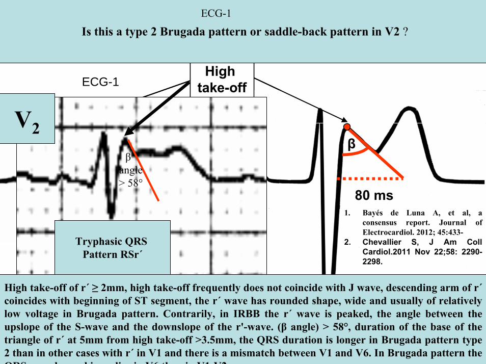
40 ms80 ms
β
80 ms
High take-off
Type 1 Brugada pattern
Type 2 Saddle-back pattern
40 ms80 ms
β
80 ms
High take-off
Type 1 Brugada pattern
Type 2 Saddle-back pattern
V2
Is this a type 2 Brugada pattern or saddle-back pattern in V2
Tryphasic QRS Pattern RSracute
High take-off of racute ge 2mm high take-off frequently does not coincide with J wave descending arm of racutecoincides with beginning of ST segment the racute wave has rounded shape wide and usually of relatively low voltage in Brugada pattern Contrarily in IRBB the racute wave is peaked the angle between the upslope of the S-wave and the downslope of the r-wave (β angle) gt 58deg duration of the base of the triangle of racute at 5mm from high take-off gt35mm the QRS duration is longer in Brugada pattern type 2 than in other cases with racute in V1 and there is a mismatch between V1 and V6 In Brugada pattern the QRS complex end is earlier in V6 than in V1 V2
βangle gt 58deg
1 Bayeacutes de Luna A et al a consensus report Journal of Electrocardiol 2012 45433-
2 Chevallier S J Am CollCardiol2011 Nov 2258 2290-2298
ECG-1
ECG-1
RIGHT SAGGITAL PLANE ECG-1
YaVF
Z V2
ECG 2
aVR aVL
I
IIIII
X
Y
aVF
QRS axis +30ordm
ECG-2 FRONTAL PLANE
aVR aVR
ECG-1 ECG-2
J-Wave
Reciprocal ST-segment changes (ST-segment depression) Without reciprocal changes or mirror image
In Early Repolarization Pattern reciprocal ST-segment changes (ST-segment depression) are eventually confined the unipolar aVR lead (Absence of reciprocal changes or mirror image in others leads)
II
II
PR interval= 160msQRSd is 121ms
ECG-1
PR interval = 140msQRSd is near 80ms
J wave
ECG-2
V6
V5
V1
V4
V2
V3
X
Z
Is this a type 2 Brugada ECG pattern Answer in next slide
ECG-2
V2βangle gt58deg
Observation β angle is related to both depolarization an repolarization A wider QRS complex (slow conduction) and abnormal repolarization both contribute to a wider β angle
1 Chevallier S J Am Coll Cardiol2011 Nov 2258 2290-2298
In patients with suspected Brugada syndrome simple ECG criteria can enable discrimination between innocent incomplete RBBB from types 2 and 3 Brugada ECG patterns The mean β angle is significantly smaller in the patients with negative results on Antiarrhythmic Drug Challenge (ADC) compared wit patients with positive results on ADC Its optimal cutoff value is 58deg which yielded a positive predictive value of 73 and a negative predictive value of 87 for conversion to type 1 pattern on ADC α is slightly less sensitive and specific compared with β When the angles are combined with QRS duration it tended to improve discrimination
Y
aVF
Z V2
Right Sagittal Plane ECG-2
Triphasic QRS patternRSracute
β angle defined as the angle between the upslope of the S-wave and the downslope of the r-wave The mean β angle was significantly smaller in the 14 patients with negative results on AAD compared to the 24 patients with positive results on AAD Itrsquos optimal cutoff value was 58deg which yielded a positive predictive value of 73 and a negative predictive value of 87 for conversion to type 1 pattern on AAD
ST segment with saddleback appearance followed by positive or biphasic T wave in V1-V2 or from V1 through V3
β
V5V5
QRSd= 122ms QRSd= 110ms
ECG-1 ECG-2
Left precordial terminal QRS notching is more prevalent in malignant early repolarization abnormalities
Left precordial terminal QRS notching
1 Merchant FM et al Am J Cardiol 2009 Nov 15 1041402-1406
V2
V2
V3
V3
ECG-1
ECG-2
ECG-2
Dynamic inconstant or dramatic changes in repolarization are suggestive of malignant early repolarization abnormalities
1 Haiumlssaguerre M et al J Cardiovasc Electrophysiol 2009 Jan2093-98
ECG-1
Conclusion this patient has high possibility of concomitant Brugada syndrome and early repolarization In this a benign ERP or malignant early repolarization abnormalities We think it is very unlikely that is a Brugada phenocopyIn this case it is necessary additionally to perform
1 IV Ajmaline test2 ECG-AR3 Family study ( first degree relatives interrogatory physical ECGs)4 Magnetic resonance image5 Genetic screening
Ventricular biopsy is not considered mandatory but is recommended especially when it may clarify the nature of dubious findings identified with others test In approximately 6--10 of survivors of cardiac arrest no cardiac abnormality can be identified despite extensive clinical evaluation Autopsy data confirm that in a similar percentage of victims of SCD no structural heart disease or or mors sine materia can be identified at post mortem evaluation Incompletely penetrant genetic defects may underlie at least some of these unexplained deaths (1)
The ERP probably represents part of a spectrum of cardiovascular anomalies related to channelopaties including BrS IVF congenital short QT syndrome and that it may also have a molecular genetic origin of variable penetrance The denomination Early Repolarization Pattern is appropriate since it indicates that it is characterized by a given ECG pattern
1 Priori SG et al Cardiovasc Res 2001 May50218-223
Case Report similar
A 20 year-old male mulatto professional soccer player from Santo Andreacute Sao Paulo Brazil He didnrsquot finishthe second year in elementary school CatholicProfession Professional soccer playerReason for consultation Presented to consultation due to two presyncopal episodes and one syncopal episode in the last 30 days The first two episodes happened during a post-defecation period Both were preceded by dizziness nausea and vomiting The syncopal episodes occurred at rest with no prodromes The third one with express and brief loss of consciousness and sphincter relaxation at dawn 48 hours agoHis prior personal medical history was unremarkable and he successfully passed two prior periodical clinical-cardiovascular evaluations (twice a year always normal) Pre-competitive examination included 12-lead ECG echocardiogram and cardiopulmonary metabolic exercise testing (CMET) All normals Intense training for the last 3 years Familial Background One uncle from his fatherrsquos side family died of sudden cardiac death when he was 35
years old He ignores the cause His brother is being seeing by a cardiologist due to ldquocardiac arrhythmiardquo in treatment with a cardiologistfrom the Institute of the Heart from Satildeo Paulo (he cannot tell which is the cause) He denies having other relevant background details Pysical cardiovascular examination revealed bradycardia at 45 bpm with physiological sinus arrhythmia blood pressure 11070 mmHg The rest of the physical examination was unremarkable He denied consuming any type of neither medications nor drugs No addictionsHis first 12-lead ECGVectorcardiogram(VCG) (Fig 1) showed sinus bradycardia at 44 bpm P-wave axis of +60deg PR interval of 186ms QRS duration of 111ms QRS axis at + 63deg QT interval 427ms and corrected QTc 367ms There is a notch on the terminal portion of the QRS complex in the left precordial leads voltage criteria for left ventricular hypertrophy (SV1 + RV5 gt 35 mm ndash Sokolow index) prominent J-wave and concave ST-segment elevation in inferior leads V4 V5 and V6 Deep and narrow Q-waves in the left precordial leads V5 and V6 All these features were considered as an Early Repolarization Pattern (ERP)
Name JVS Gender Male Age 20 yo Ethnic Group Mulatto Weight 66 Kg Height 172 m Biotype Normoline Date 11262009 Profession competitive athlete (soccer player)
ECG diagnosis Sinus bradycardia (SB) HR 44bpm P axis +60deg PR 186ms QRSd 111ms QRS axis + 63deg QT 427ms QTc 367ms Conclusion SB + Early Repolarization Pattern (ERP) Notching or slurring of the terminal QRS complex voltage criteria of left ventricular hypertrophy SV1+ RV5 gt 35 mm (Sokolow index) prominent J wave and ST-segment elevation concave to the topending in a positive T wave from V4 to V6 Precordial greater than limb leads Relatively deep but narrow q waves in the left precordial leads (V5 and V6)
V6V4 V5
Prominent J wave and ST- segment elevation concave to the top ending in a positive T wave from V4 to V6 Notching or slurring morphology of the terminal portion of the R wave (J-wave) that produces a positive hump known as a J-wave (Arrows)
aVR
Eventually reciprocal ST segment depression confined only in Lead aVR1 observed in this particular case report
1 Mehta M Jain AC Mehta A Early Repolarization Clin Cardiol 1999 Feb 22 59-65
ECGVCG CORRELATIONS HORIZONTAL PLANE
V6
V1
V2
V3
X
Z
V5
V4
T
0J T
BEGINNING AND END OF QRS LOOP IN HP
J POINT in vectorcardiography it corresponds to 3 elements end of ventricular depolarization (QRS loop) beginning of repolarization (ST segment) when it does not present depression or elevation and T wave onset In this case 0 point and J point are not coincident In situations where there is depression or elevation of ST segment the J point does not coincide with the 0 point and the greater or lesser distance between both points indicate the greater or lesser ST segment elevation or depression The phenomenon is observed in early repolarization pattern acute phase of myocardial infarction Prinzmetal variant angina pericarditis Brugada syndrome arrhythmogenic right ventricular dysplasia etc
0 POINTIt corresponds to the QRS looponset (because PR segment does notexist) Alsocorrespond to the end of biatrialchamberactivation
T
J POINT
ECGVCG CORRELATIONS FORNTAL PLANE
aVR aVL
I
IIIII
X
Y
VERTICAL AXIS ON FRONTAL PLANE The frontal plane QRS axis and ST segment axis and T wave axis are all in the same direction THE ST
SEGMENT ELEVATION
USUALLY lt 05 mm IN THE LIMB
LEADS
YVF
Z V2
T
ECGVCG CORRELATIONS RIGHT SAGITTAL PLANE
A second 12-lead ECG (Fig 2) preformed 72 hours later showing1) Sinus bradycardia at 44 bpm2) Similar p-wave axis and intervals duration3) Dramatic modification of repolarizaccedilatildeo pattern in the right precordial leads A coved-type
(type 1) ECG brugada pattern can be distinguished4) Accentuation of j-waves and st segment elevation across the precordial leads was observed5) ST segment elevation on avr lead
See next slide
Name JVS Gender Male Age 20 yo Ethnic Group MulattoWeight 66 Kg Height 172 m Biotype Normoline Date 11292009Profession Competitive Soccer Player FIGURE 2
ECG diagnosis Sinus Arrhythmia(SA) sinus bradycardia (SB) HR 44bpm P axis +60deg PR 186ms QRSd 111ms QT 427ms QTc 367 Conclusion SA + SB + Early Repolarization Pattern (ERP) Type 1 Brugada ECG pattern Dramatic accentuation of J waves across the precordial and limb leads Transient augmentation of global J waves may be indicative of a
highly arrhythmogenic substrate heralding multiple episodes of VF in patients with ERP
aVR LEAD
11262009 11292009
DEPRESSION
ELEVATION
ST SEGMENT DEPRESSION IN aVR = 1mm ST SEGMENT ELEVATION IN aVR = 2mm
V1 V2V2
1129200911262009
V1
11292009 11262009
SPONTANEOUS MODIFICATION OF REPOLARIZATION TO TYPE 1 BRUGADA ECG PATTERN
11262009
V6
11292009V6
ST segment elevation lt 2mm ST segment elevation gt 2mm
11292009
V5
11262009
V5
ST segment elevation = 1mm ST segment elevation = 25mm
EARLY REPOLARIZATION PATTERN CONCEPT
DEFINITION Early Repolarization Pattern (ERP) is a well-recognized Enigmatic idiopathic electrocardiographic phenomenon characterized by notching or slurring morphology of the terminal portion of the R wave (J-wave) that produces a positive hump known as a J-wave at the end of the QRS complex and beginning of the ST segment It considered to be present when at least two adjacent precordial leads show elevation of the ST segment with values ge01 mV (or 1mm) above baseline followed by concordant tall near symmetric (T waves having similarity in shape ascending and descending ramps speeds)
EPIDEMIOLOGY ERP occur in 2 to 5 of the population often in men young adults afro-descendents and competitive athletes and in up to 131 to 482 of patients presenting with chest pain in the emergency rooms and coronary care units There is a distinct J wave and ST segment concave upward in the left precordial leads V4 through V6 The ST elevation is most frequently evident in ECG lead V4 The ST segment elevation is usually le2mm (precordial greater than limb leads) and is lt 05 mm in the limb leadsReciprocal ST-segment changes (ST-segment depression) are eventually confined the unipolar aVR lead (Absence of reciprocal changes or mirror image) In ERP vertical QRS axis electrical is the rule The frontal plane QRS axis the ST segment axis and T wave axis are all in the same directionThis ECG pattern has been considered a benign normal ECG variant for over six decades3
1 Hasbak P Engelmann MD Early repolarization ST-segment elevation as a normal electrocardiographic variant Ugeskr Laeger 2000 162 5928-5929
2 Brady WJ Chan TC Electrocardiographic manifestations benign early repolarization J Emerg Med 1999 17473-478
3 Benito B Guasch E Rivard L Nattel S Clinical and mechanistic issues in early repolarization of normal variants and lethal arrhythmia syndromes J Am Coll Cardiol 2010 Oct 5 56 1177-1186
MAIN ELECTROCARDIOGRAPHIC FEATURES IN ERPHeart rate Predominant sinus bradycardia frequently associated with respiratory sinus arrhythmia
PR interval First degree AV block is observed in 5 to 39 among professional athletes In the non-athlete population it is observed in 065 of cases QRS axis Vertical electrical axis is the rule The frontal plane QRS axis and ST segment axis and T wave axis are all in the same directionQRS duration (90 plusmn 10 ms) subjects than in age-matched healthy controls (80 plusmn 10 ms) QRS morphology Notching or slurring of the terminal QRS complex Prominent relatively deep but narrow q waves may appear in the left precordial leadsQRS voltage Eventually voltage criteria of LVH are observed in male competitive athletes SV1+ RV5 gt 35 mm (Positive Sokolow index) High QRS voltage is more frequent in male athletes but its correlation with LVH is low Voltage decreases after deconditioning is slow The distinction between the physiological athletes heart and pathological conditions has critical implications for professional athletes An abrupt transition may occur from right-oriented complexes to left-oriented complexes in the precordial leads secondary to counterclockwise rotation on precordial leads About two-thirds of clockwise rotation and counterclockwise rotation could be attributed to the septal angle by anatomical rotation of the heart in one plane around the long axis but other factors appear to be responsible for such ECG findings in the remaining one-third of cases Relatively higher positions of the precordial ECG leads as observed in the vertical heart appeared to be responsible for clockwise rotation in some patients and left septal fascicular block is suspected to be responsible for counterclockwise rotation in others patientsJ-point elevation Notching irregular or slurring contour of the terminal QRS complex (J point) Variant of Osborn wave is seen in ERP
ST segment Widespread ST segment elevation (precordial greater than limb leads) The characteristic ST segment is elevated upward concave confined more frequently in precordial leads with reciprocal depression only in aVR The concavity is observed at the initial up-sloping portion of ST segment or upwardly concave ST segment morphology Unfortunately concave ST morphology cannot be used to rule out ST elevation from AMI with left anterior descending coronary occlusion because it is common in these circumstances The ST elevation is most frequently evident in ECG lead V4 There is a distinct J wave and ST segment in the left precordial leads V4 through V6 The ST elevation in ERP is usually lt 2 mm (but can rarely be gt 5 mm) in the precordial leads and the greatest ST elevation is usually seen in the mid-to-left precordial leads The ST segment elevation is usually lt 05 mm in the limb leads
T wave characteristics Concordant T waves of large amplitude (prominent matching T waves) typical pseudo-asymmetrical (lsquosymmetroidrdquo) or slightly asymmetrical matching T waves often of large amplitude upright tall and peaked most conspicuously from V2 to V4 or V5 sometimes seen in leads DII DIII and aVF as a rule T waves may appear as of large amplitude peaked or pointed symmetric and matching Vagotonic or high T wave voltages followed by U waves are frequent when sinus bradycardia is present Tall positive ad symmetric or symmetroid T waves are not only seen occasionally in the very early stages of MI but also in hyperkalemia and in ERP sinus bradycardia
QT intervals QT maximum The maximum Q-onset-T-end interval This parameter is higher in ERV subjects than in normal controls
QTp maximum maximum Q-onset-T-peak interval This parameter is higher in ERV subjects than in age-matched healthy controls
Rate-corrected QTc maximum This parameter is lower in ERP subjects than in age-matched healthy controls
QTpc maximum This parameter is lower in ERV subjects than in age-matched healthy controls
U wave because bradycardia U waves are frequent in ERV they are best observed in the V3 lead U waves are frequent when sinus bradycardia is present
Other ECG characteristics of ERPRelative temporal stability of the ST segment and T wave pattern is observedReciprocal changes are not seen in ERP There are no evolutionary short-term changes in the ST segment and T waves and Q waves do not appear
Discusions Early Repolarization Pattern (ERP) or Early Repolarization Variant (ERV) is an enigmati
electrocardiographic phenomenon characterized by prominent J wave and ST-segment elevation in multiplleads
Recently there has been renewed interest in ERP because of similarities to the arrhythmogenic Brugadsyndrome (BrS) Not much is known about the epidemiology of ERP and several studies have reported thathis condition is associated with a good prognosis
Both ERP and BrS exhibit some similarities including the ionic underlying mechanism the analogouresponses to changes in heart rate and autonomic tone sympathicomimetics (isoproterenol test) as well as insodium channel and beta-blockers These observations raise the hypothesis that ERP may be not as benign atraditionally believed Additionally there are documents showing that ST-segment height in the man igreatly influenced by central sympathetic nervous activity both at baseline and during physiologic andpharmacological stress Central sympathetic dysfunction regularly results in multilead ST-segment elevationor J wave that decreases or below isoelectric baseline during low dose isoproterenol infusion An earlyrepolarization pattern in the inferior leads of a standard electrocardiogram is associated with an increased riskof death from cardiac causes in middle-aged subjects1
POSSIBLE SIMILARITIES BETWEEN ERV AND BrSbullMore frequent in malesbullBoth occur more frequently in young adults and in individuals without apparent structural heart diseasebullBoth may influence just the V1-V2 leads Rarely (9) can ST elevation be observed in ERP only in the right precordial leads V1-V2 or in the inferior ones2
1 Tikkanen JT Anttonen O Junttila MJ Aro AL Kerola T Rissanen HA Reunanen A Huikuri HV Long-Term Outcome Associated with Early Repolarization on Electrocardiography N Engl J Med 2009 Nov 16 [Epubahead of print]
2 Hasbak P Engelmann MDEarly repolarization ST-segment elevation as a normal electrocardiographic variant Ugeskr Laeger 2000 162 5928-5929
When ST elevation is normal it can reach up to 3 mm in V2-V3 especially in young people In those individuals over 40 years it seldom exceeds 2 mm Both can show incomplete RBBB pattern or right bundle branch conduction disorder in BrS it can present atypical features RBBB-like and of the saddle type by exclusive elevation of the J point S wave with delay in the left leads DI aVL V5 and V6 could be absent as it is to be expected in a classic RBBB The elements considered as typical in BrS are 1) elevation of the terminal part of QRS (prominent J wave) 2) elevated and descending ST not related to lesion of ischemic (idiopathic) injury 3) negative T wave in the right precordial leads 4) normal QTc or near normal 5) Eventually absence of final delay in left leads as it would be expected in a classic RBBB1 In ERV when associated to athletersquos heart QRS can present a moderate extension (100 ms to 110 ms) in 15 of the cases which in nonathlete normal population in a 24 is called outflow tract hypertrophy In this case r does not exceed the 5 mm and is lower than S in the same lead rSrBoth may improve repolarization during the stress test with use of isoproterenolBoth respond to a shortening of AP phase 2 in a part of ventricular thickness and intensification of fast repolarization notch (phase 1) mediated by transmural dispersion of ventricular repolarization by a larger notch in the Ito channel2The alteration of the Ito and ICa2+-L channels in BrS and in ERP are the electrophysiologic substrate that explains the J point and ST segment elevation because they cause the intensified notch in phase 1 and suppression in phase 2 duration in the epicardium and in the endocardium of ventricular wall thickness
1 Hiss RG Lamb LE Electrocardiographic findings in 122043 individuals Circulation 1962 25947-9612 Antzelevitch Ch Xin Yan G Shimuzi W et al Electrical heterogeneity the ECG and Cardiac Arrhythmias In Zipes
DP Jalife J Cardiac Electrophysiology From Cell to Bedside Third Edition WB Saunders Company2000 Chapter 26 p 222-238
ELEMENTS FOR DIFFERENTIAL DIAGNOSIS BETWEEN ERP AND BrS
I) Family background ERP negativeBrS frequently positive
II) Ethnic GroupERP predominantly in African descendents(1) or equally common in all races(2) BrS predominantly in Asian group (58) and Caucasian people(3)
III) GenderERP male gender predominanceBrS great predominance in the male gender (malefemale ratio ndash 81 in non Asian and 101 in Asian
peopleIV) Response to IC group antiarrhythmic agents
BrS flecainide used in a 10 mgKg dosage in 10 minutes increases ST elevation and QRS duration in a more significant way in patients with BrS than in individuals without the entity and only in those it triggers ventricular extrasystoles(4)
ERP it can induce a pattern similar to BrS however the degree of ST elevation caused by the drug is much higher in patients with BrS than in patients without the disease
1 Grusin H Peculiarities of the Africanrsquos electrocardiogram and the changes observed in serial studies Circulation 1954 9 860-867
2 Mehta M Jain AC Mehta A Early Repolarization Clin Cardiol 1999 Feb2259-653 Nademanee K Veerakul G Nimmannit S Chaowakul V Bhuripanyo K Likittanasombat K Tunsanga K Kuasirikul S
Malasit P Tansupasawadikul S Tatsanavivat P Arrhytmogenic marker for the sudden unexplained death syndrome in Thail men Circulation 1997 962595-2600
4 Shimizu W Antzelevitch C Suyama K Kurita T Taguchi A Aihara N Takaki H Sunagawa K Kamakura S Effect of sodium channel blockers on ST segment QRS duration and corrected QT interval in patients with Brugada syndromeJ Cardiovasc Electrophysiol 2000 111320-1329
BrS patients with Inferior and lateral ECG repolarization abnormalities have longer baseline PR intervals(1)Both an S wave width ge80ms in V1 and ST elevation ge18mm in V2 are highly specific indicators of VF and criteria for high-risk BrS(2)There are several references in literature to patients with elevation of the J point and ST segment convex to the top or straight descendent in inferior leads (Brugada sign) or concomitantly ST-segment elevation in the right precordial and inferior leads II III and aVF(3) in absence of hypothermia ischemia or electrolytic disorders which we call ldquoatypical Brugada patternrdquo atypical BrS variant BrS or BrS variant or idiopathic VF (a variant of the BrS with ST-segment elevation in inferior leads(4)Pilsicainide may induce PVCs and PVT in atypical BrS and the infusion of isoproterenol suppress the arrhythmias and normalize the ST-segment elevation(5)
1 Sarkozy A Chierchia GB Paparella G Boussy T De Asmundis C Roos M Henkens S Kaufman L Buyl R Brugada R Brugada J Brugada P Inferior and lateral electrocardiographic repolarization abnormalities in Brugada syndromeCirc ArrhythmElectrophysiol 2009 Apr 2154-161
2 Atarashi H Ogawa S For The Idiopathic Ventricular Fibrillation Investigators New ECG Criteria for High-Risk Brugada Syndrome Circ J 2003 Jan 67 8-10
3 Lombardi F Potenza S Beltrami A Verzoni A Brugada P Brugada RSimultaneous ST-segment elevation in the right precordial and inferior leads in Brugada syndrome J CardiovascMed (Hagerstown) 2007 8 201-204
4 Letsas KP Efremidis M Pappas LK Gavrielatos G Markou V Sideris A Kardaras FEarlyrepolarization syndrome is it always benign Int J Cardiol 2007114 390-392
5 Chinushi M Izumi D Furushima H Watanabe H Aizawa YMultiple premature beats triggered ventricular arrhythmias during pilsicainide infusion in a patient with inferior ST-segment elevation Pacing Clin Electrophysiol 2006 29 1445-1448
Potet et al(1) identified a G752R mutation on SNC5A that produced ST segment elevation and prominent J wave in leads II III and aVF The authors provide genetic demonstration that Brugada ECG anomalies related to a unique SCN5A mutation can be observed either in the inferior or the right precordial leads The early repolarization pattern in inferolateral leads is not an uncommon finding in BrS and this pattern is not associated with a worse outcome in subjects with BrS(2) The spontaneous ERP occurred more frequently among patients with BrS than in 283 family members not having BrS (11 versus 6 P=003) Class I antiarrhythmic drug administration provoked inferior-lateral coved Brugada pattern in 13 patients with BrS These patients had longer baseline PR intervals and Class I antiarrhythmic drug induced QRS interval prolongation (108 to 178 versus 102 ms to 131 ms Plt0001) In 3 patients the Class I antiarrhythmic drug provoked coved Brugada pattern only present in the inferior leads Inferior-lateral ERP occurs spontaneously relatively frequently in BrS These patients have a more severe phenotype Class I antiarrhythmic drug administration provokes inferior-lateral coved Brugada pattern in 46 of patients The Class I antiarrhythmic drug exceptionally provoked coved Brugada pattern only observed in the inferior leads(3)
1 Potet F Mabo P Le Coq G et al Novel brugada SCN5A mutation leading to ST segment elevation in the inferior or the right precordial leads J Cardiovasc Electrophysiol 2003 142000-2003
2 Letsas KP Sacher F Probst V Weber R Knecht S Kalusche D Haiumlssaguerre M ArentzTPrevalence of early repolarization pattern in inferolateral leads in patients with Brugada syndrome Heart Rhythm 2008 Dec 5 1685-1689
3 Sarkozy A Chierchia GB Paparella G Boussy T De Asmundis C Roos M Henkens S Kaufman L Buyl R Brugada R Brugada J Brugada P Inferior and lateral electrocardiographic repolarization abnormalities in Brugada syndrome Circ Arrhythm Electrophysiol 2009 Apr2 154-161
Type 1 ECG Brugada pattern in the peripheral leads was observed in 42 of patients during ajmaline test (103 of positive tests) and was associated with longer QRS and greater QTc prolongation compared with the rest of the patients1Atypical Brugada Syndrome or IVF with J waves in inferior lateral or inferior lateral leads
and Familial Idiopathic Ventricular Fibrillation and BrSSeveral mutations in genes SCN5A KCNJ8 DPP6 SCN3B and CACNA2D1 had been identified 1 On SCN5A gene2 3 Observation We do not believe that SCN5A should be attributed to IVF because this report clearly shows conduction defects (as expected) so this is not IVF but conduction delay at different levels in the heart2 On KCNJ8 gene 4 5 Missense variant in exon 3 (NC-000012) of the KCNJ8 gene a subunit of the K(ATP)channel23 Genomic DNA sequencing of K(ATP) channel genes showed missense variant in exon 3 (NC_000012) of the KCNJ8 gene a subunit of the K(ATP) channel conferring predisposition to dramatic repolarization changes and ventricular vulnerability
1 Batchvarov VN Govindan M Camm AJ Behr ERBrugada-like changes in the peripheral leads during diagnostic ajmaline test in patients with suspected Brugada syndrome Pacing Clin Electrophysiol 2009 Jun32695-703
2 Potet F Mabo P Le Coq G Probst V Schott JJ Airaud F Guihard G Daubert JC Escande D Le MarecH Novel brugada SCN5A mutation leading to ST segment elevation in the inferior or the right precordial leads J Cardiovasc Electrophysiol 2003 14 200-203
3 Akai J Makita N Sakurada H Shirai N Ueda K Kitabatake A Nakazawa K Kimura A Hiraoka M A novel SCN5A mutation associated with idiopathic ventricular fibrillation without typical ECG findings of Brugada syndrome FEBS Lett 2000 Aug 11 479 29-34
4 Haissaguerre M Derval N Sacher F Jesel L Deisenhofer I de Roy Let al Sudden cardiac arrest associated with early repolarization N Engl J Med 2008 May 8 3582016-2023
5 Haiumlssaguerre M Sacher F Nogami A Komiya N Bernard A Probst V et al Characteristics of recurrent ventricular fibrillation associated with inferolateral early repolarization role of drug therapy J Am CollCardiol 2009 Feb 17 53 612-619
On DPP6 gene1 Dipeptidyl aminopeptidase-like protein 6 locus on chromosome7q26 Voltage-gated potassium channels OMIM126141 The ECG shows rapid PVT Monomorphic PVCs with very short coupling intervals The PVC (302 +- 52 msec) within 40 msec of the peak of the preceding Absence of T wave Pause-dependence
Valdivia et al2 reported a case with IVF and a novel mutation in the SCN3B-encoded sodium channel beta subunit Navbeta3 that causes a loss of function of Nav15 channels in vitro and provides molecular and cellular evidence implicating mutations in Navbeta3 as a cause of IVF Mutation in this SCN3B gene causes also BrS type 73
L-type calcium channel (LTCC) mutations have been associated with BrS SQTS and Timothy syndrome (LQT8) Little is known about the extent to which LTCC mutations contribute to the J-wave syndromes associated with SCD Mutations in the LTCCs are detected in a high percentage of probands with J-wave syndromes associated with inherited cardiac arrhythmias suggesting that genetic screening of Ca(v) genes may be a valuable diagnostic tool in identifying individuals at risk Burashnikov et al4 identify CACNA2D1 as a novel BrS susceptibility gene and CACNA1C CACNB2 and CACNA2D1 as possible novel ERS susceptibility genes4
1 Alders M Koopmann TT Christiaans I Postema PG Beekman L Tanck MW et al Haplotype-sharing analysis implicates chromosome 7q36 harboring DPP6 in familial idiopathic ventricular fibrillation Am J Hum Genet 2009 Apr 84 468-476
2 Valdivia CR Medeiros-Domingo A Ye B Shen WK Algiers TJ Ackerman MJ et al Loss-of-function mutation of the SCN3B-encoded sodium channel beta3 subunit associated with a case ofidiopathic ventricular fibrillation Cardiovasc Res 2010 Jun 1 86 392-400
3 Hu D Barajas-Martinez H Burashnikov E Springer M Wu Y Varro A et al A mutation in the beta-3 subunit of the cardiac sodium channel associated with Brugada ECG phenotype Circ Cardiovasc Genet 2009 2 270-278
4 Burashnikov E Pfeiffer R Barajas-Martinez H Delpoacuten E Hu D Desai M et al Mutations in the cardiac L-type calcium channel associated with inherited J-wave syndromes and sudden cardiac death Heart Rhythm 2010 Dec71872-1882
From a multicenter cohort of 122 patients (90 male age 37 +- 12 years) with IVF and ERP in the inferolateral leadsHaiumlssaguerre et al1 selected all patients with more than 3 episodes of VF (multiple) including those with electrical storms (ge3 VF in 24 h) Multiple recurrences of VF occurred in 27 of patients with ERP and may be life threatening Isoproterenol in acute cases and quinidine in chronic cases are effective antyharrhithmic drugsThe last one is necessary associated to ICDThe so-called atypical BrS is characterized by ECG abnormalities of the J wave and ST-segment elevation appeared in the inferior andor lateral leads The ERP in inferolateral leads is not an uncommon finding in BrS2 There is a high incidence of the ERP confined in inferolateral leads in patients with IVF The ECGs have an elevation of the QRS-ST junction of at least 01 mV from baseline in the inferior or lateral lead manifested as QRS slurring or notching Among patients with a history of IVF there is an increased prevalence of ERP
1 Haissaguerre M Derval N Sacher F Jesel L Deisenhofer I de Roy L Pasquieacute JL Nogami A Babuty D Yli-Mayry S De Chillou C Scanu P Mabo P Matsuo S Probst V Le Scouarnec S Defaye P Schlaepfer J Rostock T Lacroix D Lamaison D Lavergne T Aizawa Y Englund A Anselme F ONeill M Hocini M Lim KT Knecht S Veenhuyzen GD Bordachar P Chauvin M Jais P Coureau G Chene G Klein GJ Cleacutementy J Sudden cardiac arrest associated with early repolarization N Engl J Med 2008 May 8 3582016-2023
2 Letsas KP Sacher F Probst V Weber R Knecht S Kalusche D Haiumlssaguerre M Arentz T Prevalence of early repolarization pattern in inferolateral leads in patients with Brugada syndrome Heart Rhythm 2008 Dec51685-1689
Because a contingent percentage has not yet been well established it has been suggested that BrS constitutes the same entity in a large number of IVF diagnosed cases since they have common clinical features
1 BOTH ARE MORE FREQUENT IN MEN2 BOTH HAPPEN IN MIDDLE-AGE PEOPLE3 BOTH OCCUR IN APPARENT STRUCTURALLY NORMAL HEARTS14 BOTH CAN AFFECT THE SAME GENE (SCN5A)5 BOTH AFFECT EVENTUALLY THE SAME LOCUS (3p24-p21)
Wellens et al2 has pointed out that in order to be accurate about such a percentage it is important to define the diagnostic criteria used to classify BrS in IVF patients Curiously the so-called Idiopathic VT in apparently healthy hearts without structural heart disease and primary electrical disease also affects the 3p21 locus but not 3p24 and the gene is different GNA12
Survivors of SCD by VF without structural heart disease and a normal coronary angiogram are often diagnosed with IVTIVF3
A careful analysis of the ECG is essential in the differential diagnosis of this type of apparently unexplained SCD because intermittent or permanent RBBB with J point and ST-segment elevation in leads V1 to V3 is now classified as BrS
1 Goethals P Debruyne P Saffarian M Drug-induced Brugada syndrome Acta Cardiol1998 53157-60
2 Wellens HJJ Rodriquez LM Smeets JL Ventricular tachycardia in structural normal hearts In Zipes DP Jalife J Editors Cardiac Electrophysiology from cell tobedside Philadelphia 1995 WB Saunders
3 Chen Q Kirsch GE Zhang D Brugada R Brugada P Brugada J et al Genetic basis and molecular mechanisms for idiopathic ventricular fibrillation Nature 1998392 293-296
DIAGNOSTICS TRACKS TO DIFFERENTIATE BENIGN ERP FROM MALIGNANT EARLY REPOLATIZATION ABNORMALITIES
BENIGN ERP MALIGNANT EARLY REPOLARIZATION ABNORMALITIES
Family history of unexplained SCD in young relatives(lt45yo)
Absent Possible however infrequent1
Personal history Asymptomatic Asymptomatic repetitive syncope episodes or recovered of cardiac arrest
Left precordial terminal QRS notching Less prevalent More prevalent2
STT ratio in lead V6calculated by dividing the millimeters of
ST-segment elevation by the millimeters to the tallest point of the T wave
Each value is measured from the isoelectric point
lt0253 Non-tested
1 Viskin S Belhassen B Idiopathic ventricular fibrillation American Heart Journal 1990 120 661-671
2 Tikkanen JT Anttonen O Junttila MJ Aro AL Kerola T Rissanen HA Reunanen A et al Long-term outcome associated with early repolarization on electrocardiography N Engl J Med 2009 Dec 24361 2529-2537
3 Ginzton LE Laks MM The differential diagnosis of acute pericarditis from the normal variant new electrocardiographic criteria Circulation 1982 May 65 1004-1009
ST-segment elevation shape Concave upward Convex upwardJ waves are present in asymp 30 IVF patients1
Early repolarization is significantly prevalent in the subjects after cardiac arrest
(present in 31 of those resuscitated )Eventually ldquoLambda-waverdquo 23
J-point elevation and ST-segment degree Usually lt 2 mmlt05mm in limb leads
Frequent gt2mmJ-point elevation gt 2mm in inferior leads is associated with an increased risk of death
from cardiac causes in middle-aged subjects3
T wave characteristic Positive large T wave from V2 to V4 or V5 Concordant
Frequent negative discordant
Reciprocal ST-segment changes (Mirror
image)Absent With exception aVR5 Frequently Present3
1 Viskin S Idiopathic ventricular fibrillation Le Syndrome dHaiumlssaguerre and the fear of J waves J Am Coll Cardiol 2009 Feb 17 53 620-622
2 Gussak I Bjerregaard P Kostis J Electrocardiographic lambda wave and primary idiopathic cardiac asystole a new clinical syndrome J Electrocardiol 2004 Apr 37 105-107
3 Riera AR Ferreira C Schapachnik E Sanches PC Moffa PJ Brugada syndrome with atypical ECG downsloping ST-segment elevation in inferior leads J Electrocardiol 2004 Apr37101-104
4 Tikkanen JT Anttonen O Junttila MJ Aro AL Kerola T Rissanen HA Reunanen A et al Long-term outcome associated with early repolarization on electrocardiography N Engl J Med 2009 Dec 24361 2529-2537
5 Mehta M Jain AC Mehta A Early Repolarization Clin Cardiol 1999 Feb 22 59-65
Monomorphic PVCs with very short coupling
(250 to 350ms)
Absent Characteristic1 Pause-dependency absent2
QTc Interval Normal Minimally short in 35 among masculine gender
(Between 340 to 360ms3)
Late Potentials(LPs) Absent Frequent if J wave presentShowing circadian variation with night
ascendancy4
1 Alders M Koopmann TT Christiaans I Postema PG Beekman L Tanck MW et al Haplotype-sharing analysis implicates chromosome 7q36 harboring DPP6 in familial idiopathic ventricular fibrillation Am J Hum Genet 2009 Apr 84 468-476
2 Viskin S Lesh MD Eldar M Fish R Setbon I Laniado S et al Mode of onset of malignant ventricular arrhythmias in idiopathic ventricular fibrillation J Cardiovasc Electrophysiol 1997 Oct 8 1115-11 20
3 Viskin S Zeltser D Ish-Shalom M Katz A Glikson M Justo D et al Is idiopathic ventricular fibrillation a short QT syndrome Comparison of QT intervals of patients with idiopathic ventricular fibrillation and healthy controls Heart Rhythm 2004 Nov 1 587-591
4 Abe A Ikeda T Tsukada T Ishiguro H Miwa Y Miyakoshi M Circadian variation of latepotentials in idiopathic ventricular fibrillation associated with J waves insights into alternative pathophysiology and risk stratification Heart Rhythm 2010 May 7 675-682
ConclusionIn spite of both malignant early repolarization abnormalities of IVF and benign early repolarization pattern being able to cause J point and ST segment elevation the characteristics of both are clearly different to the point of a differentiation being quite simple when ECGs are analyzed by experts Any ER should raise the suspicion of IVF when it presents ST segment elevation ge 2 mm mainly in inferior wall superior convexity eventually lambda wave shape pattern of dynamic presentation inconstant with dramatic intensity of ST segment elevation discordant T wave polarity related ST segment shift presence of reciprocal ST-segment changes (mirror image) tendency to discrete short QTc interval in male gender(ge340ms) premature monomorphic ventricular contractions with very short coupling interval(250 to 350ms) not related with exercise and late potentials with circadian variation and night ascendancy on SA-ECG Contrarily in benign ERP ST elevation observed in the mid-to-left precordial leads is usually le 2 mm and lt05 mm in the limb leads and concave to the top followed by a positive concordant tall T wave
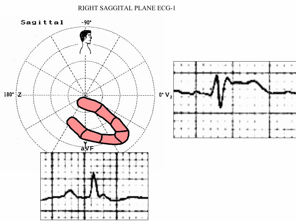
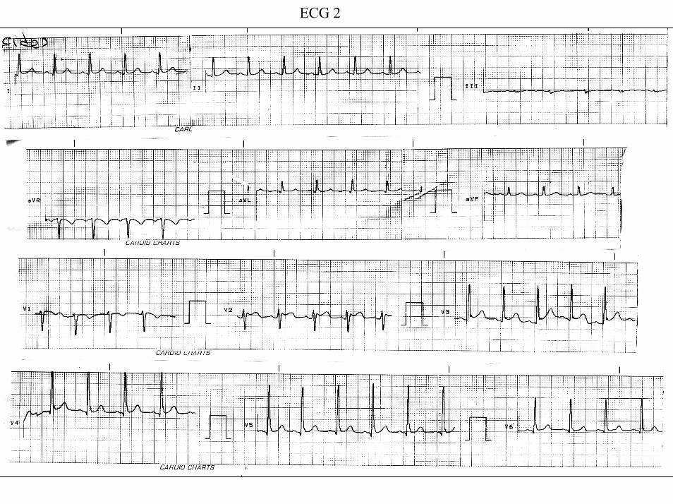
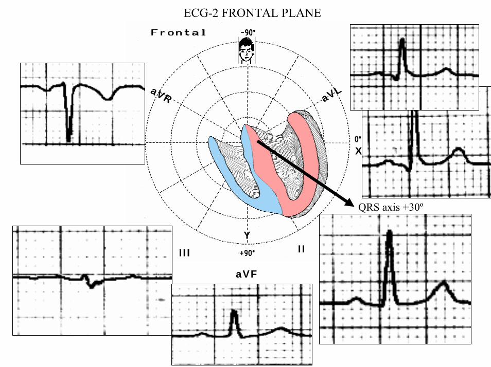
aVR aVL
I
IIIII
X
Y
aVF
QRS axis +30ordm
ECG-2 FRONTAL PLANE
aVR aVR
ECG-1 ECG-2
J-Wave
Reciprocal ST-segment changes (ST-segment depression) Without reciprocal changes or mirror image
In Early Repolarization Pattern reciprocal ST-segment changes (ST-segment depression) are eventually confined the unipolar aVR lead (Absence of reciprocal changes or mirror image in others leads)
II
II
PR interval= 160msQRSd is 121ms
ECG-1
PR interval = 140msQRSd is near 80ms
J wave
ECG-2
V6
V5
V1
V4
V2
V3
X
Z
Is this a type 2 Brugada ECG pattern Answer in next slide
ECG-2
V2βangle gt58deg
Observation β angle is related to both depolarization an repolarization A wider QRS complex (slow conduction) and abnormal repolarization both contribute to a wider β angle
1 Chevallier S J Am Coll Cardiol2011 Nov 2258 2290-2298
In patients with suspected Brugada syndrome simple ECG criteria can enable discrimination between innocent incomplete RBBB from types 2 and 3 Brugada ECG patterns The mean β angle is significantly smaller in the patients with negative results on Antiarrhythmic Drug Challenge (ADC) compared wit patients with positive results on ADC Its optimal cutoff value is 58deg which yielded a positive predictive value of 73 and a negative predictive value of 87 for conversion to type 1 pattern on ADC α is slightly less sensitive and specific compared with β When the angles are combined with QRS duration it tended to improve discrimination
Y
aVF
Z V2
Right Sagittal Plane ECG-2
Triphasic QRS patternRSracute
β angle defined as the angle between the upslope of the S-wave and the downslope of the r-wave The mean β angle was significantly smaller in the 14 patients with negative results on AAD compared to the 24 patients with positive results on AAD Itrsquos optimal cutoff value was 58deg which yielded a positive predictive value of 73 and a negative predictive value of 87 for conversion to type 1 pattern on AAD
ST segment with saddleback appearance followed by positive or biphasic T wave in V1-V2 or from V1 through V3
β
V5V5
QRSd= 122ms QRSd= 110ms
ECG-1 ECG-2
Left precordial terminal QRS notching is more prevalent in malignant early repolarization abnormalities
Left precordial terminal QRS notching
1 Merchant FM et al Am J Cardiol 2009 Nov 15 1041402-1406
V2
V2
V3
V3
ECG-1
ECG-2
ECG-2
Dynamic inconstant or dramatic changes in repolarization are suggestive of malignant early repolarization abnormalities
1 Haiumlssaguerre M et al J Cardiovasc Electrophysiol 2009 Jan2093-98
ECG-1
Conclusion this patient has high possibility of concomitant Brugada syndrome and early repolarization In this a benign ERP or malignant early repolarization abnormalities We think it is very unlikely that is a Brugada phenocopyIn this case it is necessary additionally to perform
1 IV Ajmaline test2 ECG-AR3 Family study ( first degree relatives interrogatory physical ECGs)4 Magnetic resonance image5 Genetic screening
Ventricular biopsy is not considered mandatory but is recommended especially when it may clarify the nature of dubious findings identified with others test In approximately 6--10 of survivors of cardiac arrest no cardiac abnormality can be identified despite extensive clinical evaluation Autopsy data confirm that in a similar percentage of victims of SCD no structural heart disease or or mors sine materia can be identified at post mortem evaluation Incompletely penetrant genetic defects may underlie at least some of these unexplained deaths (1)
The ERP probably represents part of a spectrum of cardiovascular anomalies related to channelopaties including BrS IVF congenital short QT syndrome and that it may also have a molecular genetic origin of variable penetrance The denomination Early Repolarization Pattern is appropriate since it indicates that it is characterized by a given ECG pattern
1 Priori SG et al Cardiovasc Res 2001 May50218-223
Case Report similar
A 20 year-old male mulatto professional soccer player from Santo Andreacute Sao Paulo Brazil He didnrsquot finishthe second year in elementary school CatholicProfession Professional soccer playerReason for consultation Presented to consultation due to two presyncopal episodes and one syncopal episode in the last 30 days The first two episodes happened during a post-defecation period Both were preceded by dizziness nausea and vomiting The syncopal episodes occurred at rest with no prodromes The third one with express and brief loss of consciousness and sphincter relaxation at dawn 48 hours agoHis prior personal medical history was unremarkable and he successfully passed two prior periodical clinical-cardiovascular evaluations (twice a year always normal) Pre-competitive examination included 12-lead ECG echocardiogram and cardiopulmonary metabolic exercise testing (CMET) All normals Intense training for the last 3 years Familial Background One uncle from his fatherrsquos side family died of sudden cardiac death when he was 35
years old He ignores the cause His brother is being seeing by a cardiologist due to ldquocardiac arrhythmiardquo in treatment with a cardiologistfrom the Institute of the Heart from Satildeo Paulo (he cannot tell which is the cause) He denies having other relevant background details Pysical cardiovascular examination revealed bradycardia at 45 bpm with physiological sinus arrhythmia blood pressure 11070 mmHg The rest of the physical examination was unremarkable He denied consuming any type of neither medications nor drugs No addictionsHis first 12-lead ECGVectorcardiogram(VCG) (Fig 1) showed sinus bradycardia at 44 bpm P-wave axis of +60deg PR interval of 186ms QRS duration of 111ms QRS axis at + 63deg QT interval 427ms and corrected QTc 367ms There is a notch on the terminal portion of the QRS complex in the left precordial leads voltage criteria for left ventricular hypertrophy (SV1 + RV5 gt 35 mm ndash Sokolow index) prominent J-wave and concave ST-segment elevation in inferior leads V4 V5 and V6 Deep and narrow Q-waves in the left precordial leads V5 and V6 All these features were considered as an Early Repolarization Pattern (ERP)
Name JVS Gender Male Age 20 yo Ethnic Group Mulatto Weight 66 Kg Height 172 m Biotype Normoline Date 11262009 Profession competitive athlete (soccer player)
ECG diagnosis Sinus bradycardia (SB) HR 44bpm P axis +60deg PR 186ms QRSd 111ms QRS axis + 63deg QT 427ms QTc 367ms Conclusion SB + Early Repolarization Pattern (ERP) Notching or slurring of the terminal QRS complex voltage criteria of left ventricular hypertrophy SV1+ RV5 gt 35 mm (Sokolow index) prominent J wave and ST-segment elevation concave to the topending in a positive T wave from V4 to V6 Precordial greater than limb leads Relatively deep but narrow q waves in the left precordial leads (V5 and V6)
V6V4 V5
Prominent J wave and ST- segment elevation concave to the top ending in a positive T wave from V4 to V6 Notching or slurring morphology of the terminal portion of the R wave (J-wave) that produces a positive hump known as a J-wave (Arrows)
aVR
Eventually reciprocal ST segment depression confined only in Lead aVR1 observed in this particular case report
1 Mehta M Jain AC Mehta A Early Repolarization Clin Cardiol 1999 Feb 22 59-65
ECGVCG CORRELATIONS HORIZONTAL PLANE
V6
V1
V2
V3
X
Z
V5
V4
T
0J T
BEGINNING AND END OF QRS LOOP IN HP
J POINT in vectorcardiography it corresponds to 3 elements end of ventricular depolarization (QRS loop) beginning of repolarization (ST segment) when it does not present depression or elevation and T wave onset In this case 0 point and J point are not coincident In situations where there is depression or elevation of ST segment the J point does not coincide with the 0 point and the greater or lesser distance between both points indicate the greater or lesser ST segment elevation or depression The phenomenon is observed in early repolarization pattern acute phase of myocardial infarction Prinzmetal variant angina pericarditis Brugada syndrome arrhythmogenic right ventricular dysplasia etc
0 POINTIt corresponds to the QRS looponset (because PR segment does notexist) Alsocorrespond to the end of biatrialchamberactivation
T
J POINT
ECGVCG CORRELATIONS FORNTAL PLANE
aVR aVL
I
IIIII
X
Y
VERTICAL AXIS ON FRONTAL PLANE The frontal plane QRS axis and ST segment axis and T wave axis are all in the same direction THE ST
SEGMENT ELEVATION
USUALLY lt 05 mm IN THE LIMB
LEADS
YVF
Z V2
T
ECGVCG CORRELATIONS RIGHT SAGITTAL PLANE
A second 12-lead ECG (Fig 2) preformed 72 hours later showing1) Sinus bradycardia at 44 bpm2) Similar p-wave axis and intervals duration3) Dramatic modification of repolarizaccedilatildeo pattern in the right precordial leads A coved-type
(type 1) ECG brugada pattern can be distinguished4) Accentuation of j-waves and st segment elevation across the precordial leads was observed5) ST segment elevation on avr lead
See next slide
Name JVS Gender Male Age 20 yo Ethnic Group MulattoWeight 66 Kg Height 172 m Biotype Normoline Date 11292009Profession Competitive Soccer Player FIGURE 2
ECG diagnosis Sinus Arrhythmia(SA) sinus bradycardia (SB) HR 44bpm P axis +60deg PR 186ms QRSd 111ms QT 427ms QTc 367 Conclusion SA + SB + Early Repolarization Pattern (ERP) Type 1 Brugada ECG pattern Dramatic accentuation of J waves across the precordial and limb leads Transient augmentation of global J waves may be indicative of a
highly arrhythmogenic substrate heralding multiple episodes of VF in patients with ERP
aVR LEAD
11262009 11292009
DEPRESSION
ELEVATION
ST SEGMENT DEPRESSION IN aVR = 1mm ST SEGMENT ELEVATION IN aVR = 2mm
V1 V2V2
1129200911262009
V1
11292009 11262009
SPONTANEOUS MODIFICATION OF REPOLARIZATION TO TYPE 1 BRUGADA ECG PATTERN
11262009
V6
11292009V6
ST segment elevation lt 2mm ST segment elevation gt 2mm
11292009
V5
11262009
V5
ST segment elevation = 1mm ST segment elevation = 25mm
EARLY REPOLARIZATION PATTERN CONCEPT
DEFINITION Early Repolarization Pattern (ERP) is a well-recognized Enigmatic idiopathic electrocardiographic phenomenon characterized by notching or slurring morphology of the terminal portion of the R wave (J-wave) that produces a positive hump known as a J-wave at the end of the QRS complex and beginning of the ST segment It considered to be present when at least two adjacent precordial leads show elevation of the ST segment with values ge01 mV (or 1mm) above baseline followed by concordant tall near symmetric (T waves having similarity in shape ascending and descending ramps speeds)
EPIDEMIOLOGY ERP occur in 2 to 5 of the population often in men young adults afro-descendents and competitive athletes and in up to 131 to 482 of patients presenting with chest pain in the emergency rooms and coronary care units There is a distinct J wave and ST segment concave upward in the left precordial leads V4 through V6 The ST elevation is most frequently evident in ECG lead V4 The ST segment elevation is usually le2mm (precordial greater than limb leads) and is lt 05 mm in the limb leadsReciprocal ST-segment changes (ST-segment depression) are eventually confined the unipolar aVR lead (Absence of reciprocal changes or mirror image) In ERP vertical QRS axis electrical is the rule The frontal plane QRS axis the ST segment axis and T wave axis are all in the same directionThis ECG pattern has been considered a benign normal ECG variant for over six decades3
1 Hasbak P Engelmann MD Early repolarization ST-segment elevation as a normal electrocardiographic variant Ugeskr Laeger 2000 162 5928-5929
2 Brady WJ Chan TC Electrocardiographic manifestations benign early repolarization J Emerg Med 1999 17473-478
3 Benito B Guasch E Rivard L Nattel S Clinical and mechanistic issues in early repolarization of normal variants and lethal arrhythmia syndromes J Am Coll Cardiol 2010 Oct 5 56 1177-1186
MAIN ELECTROCARDIOGRAPHIC FEATURES IN ERPHeart rate Predominant sinus bradycardia frequently associated with respiratory sinus arrhythmia
PR interval First degree AV block is observed in 5 to 39 among professional athletes In the non-athlete population it is observed in 065 of cases QRS axis Vertical electrical axis is the rule The frontal plane QRS axis and ST segment axis and T wave axis are all in the same directionQRS duration (90 plusmn 10 ms) subjects than in age-matched healthy controls (80 plusmn 10 ms) QRS morphology Notching or slurring of the terminal QRS complex Prominent relatively deep but narrow q waves may appear in the left precordial leadsQRS voltage Eventually voltage criteria of LVH are observed in male competitive athletes SV1+ RV5 gt 35 mm (Positive Sokolow index) High QRS voltage is more frequent in male athletes but its correlation with LVH is low Voltage decreases after deconditioning is slow The distinction between the physiological athletes heart and pathological conditions has critical implications for professional athletes An abrupt transition may occur from right-oriented complexes to left-oriented complexes in the precordial leads secondary to counterclockwise rotation on precordial leads About two-thirds of clockwise rotation and counterclockwise rotation could be attributed to the septal angle by anatomical rotation of the heart in one plane around the long axis but other factors appear to be responsible for such ECG findings in the remaining one-third of cases Relatively higher positions of the precordial ECG leads as observed in the vertical heart appeared to be responsible for clockwise rotation in some patients and left septal fascicular block is suspected to be responsible for counterclockwise rotation in others patientsJ-point elevation Notching irregular or slurring contour of the terminal QRS complex (J point) Variant of Osborn wave is seen in ERP
ST segment Widespread ST segment elevation (precordial greater than limb leads) The characteristic ST segment is elevated upward concave confined more frequently in precordial leads with reciprocal depression only in aVR The concavity is observed at the initial up-sloping portion of ST segment or upwardly concave ST segment morphology Unfortunately concave ST morphology cannot be used to rule out ST elevation from AMI with left anterior descending coronary occlusion because it is common in these circumstances The ST elevation is most frequently evident in ECG lead V4 There is a distinct J wave and ST segment in the left precordial leads V4 through V6 The ST elevation in ERP is usually lt 2 mm (but can rarely be gt 5 mm) in the precordial leads and the greatest ST elevation is usually seen in the mid-to-left precordial leads The ST segment elevation is usually lt 05 mm in the limb leads
T wave characteristics Concordant T waves of large amplitude (prominent matching T waves) typical pseudo-asymmetrical (lsquosymmetroidrdquo) or slightly asymmetrical matching T waves often of large amplitude upright tall and peaked most conspicuously from V2 to V4 or V5 sometimes seen in leads DII DIII and aVF as a rule T waves may appear as of large amplitude peaked or pointed symmetric and matching Vagotonic or high T wave voltages followed by U waves are frequent when sinus bradycardia is present Tall positive ad symmetric or symmetroid T waves are not only seen occasionally in the very early stages of MI but also in hyperkalemia and in ERP sinus bradycardia
QT intervals QT maximum The maximum Q-onset-T-end interval This parameter is higher in ERV subjects than in normal controls
QTp maximum maximum Q-onset-T-peak interval This parameter is higher in ERV subjects than in age-matched healthy controls
Rate-corrected QTc maximum This parameter is lower in ERP subjects than in age-matched healthy controls
QTpc maximum This parameter is lower in ERV subjects than in age-matched healthy controls
U wave because bradycardia U waves are frequent in ERV they are best observed in the V3 lead U waves are frequent when sinus bradycardia is present
Other ECG characteristics of ERPRelative temporal stability of the ST segment and T wave pattern is observedReciprocal changes are not seen in ERP There are no evolutionary short-term changes in the ST segment and T waves and Q waves do not appear
Discusions Early Repolarization Pattern (ERP) or Early Repolarization Variant (ERV) is an enigmati
electrocardiographic phenomenon characterized by prominent J wave and ST-segment elevation in multiplleads
Recently there has been renewed interest in ERP because of similarities to the arrhythmogenic Brugadsyndrome (BrS) Not much is known about the epidemiology of ERP and several studies have reported thathis condition is associated with a good prognosis
Both ERP and BrS exhibit some similarities including the ionic underlying mechanism the analogouresponses to changes in heart rate and autonomic tone sympathicomimetics (isoproterenol test) as well as insodium channel and beta-blockers These observations raise the hypothesis that ERP may be not as benign atraditionally believed Additionally there are documents showing that ST-segment height in the man igreatly influenced by central sympathetic nervous activity both at baseline and during physiologic andpharmacological stress Central sympathetic dysfunction regularly results in multilead ST-segment elevationor J wave that decreases or below isoelectric baseline during low dose isoproterenol infusion An earlyrepolarization pattern in the inferior leads of a standard electrocardiogram is associated with an increased riskof death from cardiac causes in middle-aged subjects1
POSSIBLE SIMILARITIES BETWEEN ERV AND BrSbullMore frequent in malesbullBoth occur more frequently in young adults and in individuals without apparent structural heart diseasebullBoth may influence just the V1-V2 leads Rarely (9) can ST elevation be observed in ERP only in the right precordial leads V1-V2 or in the inferior ones2
1 Tikkanen JT Anttonen O Junttila MJ Aro AL Kerola T Rissanen HA Reunanen A Huikuri HV Long-Term Outcome Associated with Early Repolarization on Electrocardiography N Engl J Med 2009 Nov 16 [Epubahead of print]
2 Hasbak P Engelmann MDEarly repolarization ST-segment elevation as a normal electrocardiographic variant Ugeskr Laeger 2000 162 5928-5929
When ST elevation is normal it can reach up to 3 mm in V2-V3 especially in young people In those individuals over 40 years it seldom exceeds 2 mm Both can show incomplete RBBB pattern or right bundle branch conduction disorder in BrS it can present atypical features RBBB-like and of the saddle type by exclusive elevation of the J point S wave with delay in the left leads DI aVL V5 and V6 could be absent as it is to be expected in a classic RBBB The elements considered as typical in BrS are 1) elevation of the terminal part of QRS (prominent J wave) 2) elevated and descending ST not related to lesion of ischemic (idiopathic) injury 3) negative T wave in the right precordial leads 4) normal QTc or near normal 5) Eventually absence of final delay in left leads as it would be expected in a classic RBBB1 In ERV when associated to athletersquos heart QRS can present a moderate extension (100 ms to 110 ms) in 15 of the cases which in nonathlete normal population in a 24 is called outflow tract hypertrophy In this case r does not exceed the 5 mm and is lower than S in the same lead rSrBoth may improve repolarization during the stress test with use of isoproterenolBoth respond to a shortening of AP phase 2 in a part of ventricular thickness and intensification of fast repolarization notch (phase 1) mediated by transmural dispersion of ventricular repolarization by a larger notch in the Ito channel2The alteration of the Ito and ICa2+-L channels in BrS and in ERP are the electrophysiologic substrate that explains the J point and ST segment elevation because they cause the intensified notch in phase 1 and suppression in phase 2 duration in the epicardium and in the endocardium of ventricular wall thickness
1 Hiss RG Lamb LE Electrocardiographic findings in 122043 individuals Circulation 1962 25947-9612 Antzelevitch Ch Xin Yan G Shimuzi W et al Electrical heterogeneity the ECG and Cardiac Arrhythmias In Zipes
DP Jalife J Cardiac Electrophysiology From Cell to Bedside Third Edition WB Saunders Company2000 Chapter 26 p 222-238
ELEMENTS FOR DIFFERENTIAL DIAGNOSIS BETWEEN ERP AND BrS
I) Family background ERP negativeBrS frequently positive
II) Ethnic GroupERP predominantly in African descendents(1) or equally common in all races(2) BrS predominantly in Asian group (58) and Caucasian people(3)
III) GenderERP male gender predominanceBrS great predominance in the male gender (malefemale ratio ndash 81 in non Asian and 101 in Asian
peopleIV) Response to IC group antiarrhythmic agents
BrS flecainide used in a 10 mgKg dosage in 10 minutes increases ST elevation and QRS duration in a more significant way in patients with BrS than in individuals without the entity and only in those it triggers ventricular extrasystoles(4)
ERP it can induce a pattern similar to BrS however the degree of ST elevation caused by the drug is much higher in patients with BrS than in patients without the disease
1 Grusin H Peculiarities of the Africanrsquos electrocardiogram and the changes observed in serial studies Circulation 1954 9 860-867
2 Mehta M Jain AC Mehta A Early Repolarization Clin Cardiol 1999 Feb2259-653 Nademanee K Veerakul G Nimmannit S Chaowakul V Bhuripanyo K Likittanasombat K Tunsanga K Kuasirikul S
Malasit P Tansupasawadikul S Tatsanavivat P Arrhytmogenic marker for the sudden unexplained death syndrome in Thail men Circulation 1997 962595-2600
4 Shimizu W Antzelevitch C Suyama K Kurita T Taguchi A Aihara N Takaki H Sunagawa K Kamakura S Effect of sodium channel blockers on ST segment QRS duration and corrected QT interval in patients with Brugada syndromeJ Cardiovasc Electrophysiol 2000 111320-1329
BrS patients with Inferior and lateral ECG repolarization abnormalities have longer baseline PR intervals(1)Both an S wave width ge80ms in V1 and ST elevation ge18mm in V2 are highly specific indicators of VF and criteria for high-risk BrS(2)There are several references in literature to patients with elevation of the J point and ST segment convex to the top or straight descendent in inferior leads (Brugada sign) or concomitantly ST-segment elevation in the right precordial and inferior leads II III and aVF(3) in absence of hypothermia ischemia or electrolytic disorders which we call ldquoatypical Brugada patternrdquo atypical BrS variant BrS or BrS variant or idiopathic VF (a variant of the BrS with ST-segment elevation in inferior leads(4)Pilsicainide may induce PVCs and PVT in atypical BrS and the infusion of isoproterenol suppress the arrhythmias and normalize the ST-segment elevation(5)
1 Sarkozy A Chierchia GB Paparella G Boussy T De Asmundis C Roos M Henkens S Kaufman L Buyl R Brugada R Brugada J Brugada P Inferior and lateral electrocardiographic repolarization abnormalities in Brugada syndromeCirc ArrhythmElectrophysiol 2009 Apr 2154-161
2 Atarashi H Ogawa S For The Idiopathic Ventricular Fibrillation Investigators New ECG Criteria for High-Risk Brugada Syndrome Circ J 2003 Jan 67 8-10
3 Lombardi F Potenza S Beltrami A Verzoni A Brugada P Brugada RSimultaneous ST-segment elevation in the right precordial and inferior leads in Brugada syndrome J CardiovascMed (Hagerstown) 2007 8 201-204
4 Letsas KP Efremidis M Pappas LK Gavrielatos G Markou V Sideris A Kardaras FEarlyrepolarization syndrome is it always benign Int J Cardiol 2007114 390-392
5 Chinushi M Izumi D Furushima H Watanabe H Aizawa YMultiple premature beats triggered ventricular arrhythmias during pilsicainide infusion in a patient with inferior ST-segment elevation Pacing Clin Electrophysiol 2006 29 1445-1448
Potet et al(1) identified a G752R mutation on SNC5A that produced ST segment elevation and prominent J wave in leads II III and aVF The authors provide genetic demonstration that Brugada ECG anomalies related to a unique SCN5A mutation can be observed either in the inferior or the right precordial leads The early repolarization pattern in inferolateral leads is not an uncommon finding in BrS and this pattern is not associated with a worse outcome in subjects with BrS(2) The spontaneous ERP occurred more frequently among patients with BrS than in 283 family members not having BrS (11 versus 6 P=003) Class I antiarrhythmic drug administration provoked inferior-lateral coved Brugada pattern in 13 patients with BrS These patients had longer baseline PR intervals and Class I antiarrhythmic drug induced QRS interval prolongation (108 to 178 versus 102 ms to 131 ms Plt0001) In 3 patients the Class I antiarrhythmic drug provoked coved Brugada pattern only present in the inferior leads Inferior-lateral ERP occurs spontaneously relatively frequently in BrS These patients have a more severe phenotype Class I antiarrhythmic drug administration provokes inferior-lateral coved Brugada pattern in 46 of patients The Class I antiarrhythmic drug exceptionally provoked coved Brugada pattern only observed in the inferior leads(3)
1 Potet F Mabo P Le Coq G et al Novel brugada SCN5A mutation leading to ST segment elevation in the inferior or the right precordial leads J Cardiovasc Electrophysiol 2003 142000-2003
2 Letsas KP Sacher F Probst V Weber R Knecht S Kalusche D Haiumlssaguerre M ArentzTPrevalence of early repolarization pattern in inferolateral leads in patients with Brugada syndrome Heart Rhythm 2008 Dec 5 1685-1689
3 Sarkozy A Chierchia GB Paparella G Boussy T De Asmundis C Roos M Henkens S Kaufman L Buyl R Brugada R Brugada J Brugada P Inferior and lateral electrocardiographic repolarization abnormalities in Brugada syndrome Circ Arrhythm Electrophysiol 2009 Apr2 154-161
Type 1 ECG Brugada pattern in the peripheral leads was observed in 42 of patients during ajmaline test (103 of positive tests) and was associated with longer QRS and greater QTc prolongation compared with the rest of the patients1Atypical Brugada Syndrome or IVF with J waves in inferior lateral or inferior lateral leads
and Familial Idiopathic Ventricular Fibrillation and BrSSeveral mutations in genes SCN5A KCNJ8 DPP6 SCN3B and CACNA2D1 had been identified 1 On SCN5A gene2 3 Observation We do not believe that SCN5A should be attributed to IVF because this report clearly shows conduction defects (as expected) so this is not IVF but conduction delay at different levels in the heart2 On KCNJ8 gene 4 5 Missense variant in exon 3 (NC-000012) of the KCNJ8 gene a subunit of the K(ATP)channel23 Genomic DNA sequencing of K(ATP) channel genes showed missense variant in exon 3 (NC_000012) of the KCNJ8 gene a subunit of the K(ATP) channel conferring predisposition to dramatic repolarization changes and ventricular vulnerability
1 Batchvarov VN Govindan M Camm AJ Behr ERBrugada-like changes in the peripheral leads during diagnostic ajmaline test in patients with suspected Brugada syndrome Pacing Clin Electrophysiol 2009 Jun32695-703
2 Potet F Mabo P Le Coq G Probst V Schott JJ Airaud F Guihard G Daubert JC Escande D Le MarecH Novel brugada SCN5A mutation leading to ST segment elevation in the inferior or the right precordial leads J Cardiovasc Electrophysiol 2003 14 200-203
3 Akai J Makita N Sakurada H Shirai N Ueda K Kitabatake A Nakazawa K Kimura A Hiraoka M A novel SCN5A mutation associated with idiopathic ventricular fibrillation without typical ECG findings of Brugada syndrome FEBS Lett 2000 Aug 11 479 29-34
4 Haissaguerre M Derval N Sacher F Jesel L Deisenhofer I de Roy Let al Sudden cardiac arrest associated with early repolarization N Engl J Med 2008 May 8 3582016-2023
5 Haiumlssaguerre M Sacher F Nogami A Komiya N Bernard A Probst V et al Characteristics of recurrent ventricular fibrillation associated with inferolateral early repolarization role of drug therapy J Am CollCardiol 2009 Feb 17 53 612-619
On DPP6 gene1 Dipeptidyl aminopeptidase-like protein 6 locus on chromosome7q26 Voltage-gated potassium channels OMIM126141 The ECG shows rapid PVT Monomorphic PVCs with very short coupling intervals The PVC (302 +- 52 msec) within 40 msec of the peak of the preceding Absence of T wave Pause-dependence
Valdivia et al2 reported a case with IVF and a novel mutation in the SCN3B-encoded sodium channel beta subunit Navbeta3 that causes a loss of function of Nav15 channels in vitro and provides molecular and cellular evidence implicating mutations in Navbeta3 as a cause of IVF Mutation in this SCN3B gene causes also BrS type 73
L-type calcium channel (LTCC) mutations have been associated with BrS SQTS and Timothy syndrome (LQT8) Little is known about the extent to which LTCC mutations contribute to the J-wave syndromes associated with SCD Mutations in the LTCCs are detected in a high percentage of probands with J-wave syndromes associated with inherited cardiac arrhythmias suggesting that genetic screening of Ca(v) genes may be a valuable diagnostic tool in identifying individuals at risk Burashnikov et al4 identify CACNA2D1 as a novel BrS susceptibility gene and CACNA1C CACNB2 and CACNA2D1 as possible novel ERS susceptibility genes4
1 Alders M Koopmann TT Christiaans I Postema PG Beekman L Tanck MW et al Haplotype-sharing analysis implicates chromosome 7q36 harboring DPP6 in familial idiopathic ventricular fibrillation Am J Hum Genet 2009 Apr 84 468-476
2 Valdivia CR Medeiros-Domingo A Ye B Shen WK Algiers TJ Ackerman MJ et al Loss-of-function mutation of the SCN3B-encoded sodium channel beta3 subunit associated with a case ofidiopathic ventricular fibrillation Cardiovasc Res 2010 Jun 1 86 392-400
3 Hu D Barajas-Martinez H Burashnikov E Springer M Wu Y Varro A et al A mutation in the beta-3 subunit of the cardiac sodium channel associated with Brugada ECG phenotype Circ Cardiovasc Genet 2009 2 270-278
4 Burashnikov E Pfeiffer R Barajas-Martinez H Delpoacuten E Hu D Desai M et al Mutations in the cardiac L-type calcium channel associated with inherited J-wave syndromes and sudden cardiac death Heart Rhythm 2010 Dec71872-1882
From a multicenter cohort of 122 patients (90 male age 37 +- 12 years) with IVF and ERP in the inferolateral leadsHaiumlssaguerre et al1 selected all patients with more than 3 episodes of VF (multiple) including those with electrical storms (ge3 VF in 24 h) Multiple recurrences of VF occurred in 27 of patients with ERP and may be life threatening Isoproterenol in acute cases and quinidine in chronic cases are effective antyharrhithmic drugsThe last one is necessary associated to ICDThe so-called atypical BrS is characterized by ECG abnormalities of the J wave and ST-segment elevation appeared in the inferior andor lateral leads The ERP in inferolateral leads is not an uncommon finding in BrS2 There is a high incidence of the ERP confined in inferolateral leads in patients with IVF The ECGs have an elevation of the QRS-ST junction of at least 01 mV from baseline in the inferior or lateral lead manifested as QRS slurring or notching Among patients with a history of IVF there is an increased prevalence of ERP
1 Haissaguerre M Derval N Sacher F Jesel L Deisenhofer I de Roy L Pasquieacute JL Nogami A Babuty D Yli-Mayry S De Chillou C Scanu P Mabo P Matsuo S Probst V Le Scouarnec S Defaye P Schlaepfer J Rostock T Lacroix D Lamaison D Lavergne T Aizawa Y Englund A Anselme F ONeill M Hocini M Lim KT Knecht S Veenhuyzen GD Bordachar P Chauvin M Jais P Coureau G Chene G Klein GJ Cleacutementy J Sudden cardiac arrest associated with early repolarization N Engl J Med 2008 May 8 3582016-2023
2 Letsas KP Sacher F Probst V Weber R Knecht S Kalusche D Haiumlssaguerre M Arentz T Prevalence of early repolarization pattern in inferolateral leads in patients with Brugada syndrome Heart Rhythm 2008 Dec51685-1689
Because a contingent percentage has not yet been well established it has been suggested that BrS constitutes the same entity in a large number of IVF diagnosed cases since they have common clinical features
1 BOTH ARE MORE FREQUENT IN MEN2 BOTH HAPPEN IN MIDDLE-AGE PEOPLE3 BOTH OCCUR IN APPARENT STRUCTURALLY NORMAL HEARTS14 BOTH CAN AFFECT THE SAME GENE (SCN5A)5 BOTH AFFECT EVENTUALLY THE SAME LOCUS (3p24-p21)
Wellens et al2 has pointed out that in order to be accurate about such a percentage it is important to define the diagnostic criteria used to classify BrS in IVF patients Curiously the so-called Idiopathic VT in apparently healthy hearts without structural heart disease and primary electrical disease also affects the 3p21 locus but not 3p24 and the gene is different GNA12
Survivors of SCD by VF without structural heart disease and a normal coronary angiogram are often diagnosed with IVTIVF3
A careful analysis of the ECG is essential in the differential diagnosis of this type of apparently unexplained SCD because intermittent or permanent RBBB with J point and ST-segment elevation in leads V1 to V3 is now classified as BrS
1 Goethals P Debruyne P Saffarian M Drug-induced Brugada syndrome Acta Cardiol1998 53157-60
2 Wellens HJJ Rodriquez LM Smeets JL Ventricular tachycardia in structural normal hearts In Zipes DP Jalife J Editors Cardiac Electrophysiology from cell tobedside Philadelphia 1995 WB Saunders
3 Chen Q Kirsch GE Zhang D Brugada R Brugada P Brugada J et al Genetic basis and molecular mechanisms for idiopathic ventricular fibrillation Nature 1998392 293-296
DIAGNOSTICS TRACKS TO DIFFERENTIATE BENIGN ERP FROM MALIGNANT EARLY REPOLATIZATION ABNORMALITIES
BENIGN ERP MALIGNANT EARLY REPOLARIZATION ABNORMALITIES
Family history of unexplained SCD in young relatives(lt45yo)
Absent Possible however infrequent1
Personal history Asymptomatic Asymptomatic repetitive syncope episodes or recovered of cardiac arrest
Left precordial terminal QRS notching Less prevalent More prevalent2
STT ratio in lead V6calculated by dividing the millimeters of
ST-segment elevation by the millimeters to the tallest point of the T wave
Each value is measured from the isoelectric point
lt0253 Non-tested
1 Viskin S Belhassen B Idiopathic ventricular fibrillation American Heart Journal 1990 120 661-671
2 Tikkanen JT Anttonen O Junttila MJ Aro AL Kerola T Rissanen HA Reunanen A et al Long-term outcome associated with early repolarization on electrocardiography N Engl J Med 2009 Dec 24361 2529-2537
3 Ginzton LE Laks MM The differential diagnosis of acute pericarditis from the normal variant new electrocardiographic criteria Circulation 1982 May 65 1004-1009
ST-segment elevation shape Concave upward Convex upwardJ waves are present in asymp 30 IVF patients1
Early repolarization is significantly prevalent in the subjects after cardiac arrest
(present in 31 of those resuscitated )Eventually ldquoLambda-waverdquo 23
J-point elevation and ST-segment degree Usually lt 2 mmlt05mm in limb leads
Frequent gt2mmJ-point elevation gt 2mm in inferior leads is associated with an increased risk of death
from cardiac causes in middle-aged subjects3
T wave characteristic Positive large T wave from V2 to V4 or V5 Concordant
Frequent negative discordant
Reciprocal ST-segment changes (Mirror
image)Absent With exception aVR5 Frequently Present3
1 Viskin S Idiopathic ventricular fibrillation Le Syndrome dHaiumlssaguerre and the fear of J waves J Am Coll Cardiol 2009 Feb 17 53 620-622
2 Gussak I Bjerregaard P Kostis J Electrocardiographic lambda wave and primary idiopathic cardiac asystole a new clinical syndrome J Electrocardiol 2004 Apr 37 105-107
3 Riera AR Ferreira C Schapachnik E Sanches PC Moffa PJ Brugada syndrome with atypical ECG downsloping ST-segment elevation in inferior leads J Electrocardiol 2004 Apr37101-104
4 Tikkanen JT Anttonen O Junttila MJ Aro AL Kerola T Rissanen HA Reunanen A et al Long-term outcome associated with early repolarization on electrocardiography N Engl J Med 2009 Dec 24361 2529-2537
5 Mehta M Jain AC Mehta A Early Repolarization Clin Cardiol 1999 Feb 22 59-65
Monomorphic PVCs with very short coupling
(250 to 350ms)
Absent Characteristic1 Pause-dependency absent2
QTc Interval Normal Minimally short in 35 among masculine gender
(Between 340 to 360ms3)
Late Potentials(LPs) Absent Frequent if J wave presentShowing circadian variation with night
ascendancy4
1 Alders M Koopmann TT Christiaans I Postema PG Beekman L Tanck MW et al Haplotype-sharing analysis implicates chromosome 7q36 harboring DPP6 in familial idiopathic ventricular fibrillation Am J Hum Genet 2009 Apr 84 468-476
2 Viskin S Lesh MD Eldar M Fish R Setbon I Laniado S et al Mode of onset of malignant ventricular arrhythmias in idiopathic ventricular fibrillation J Cardiovasc Electrophysiol 1997 Oct 8 1115-11 20
3 Viskin S Zeltser D Ish-Shalom M Katz A Glikson M Justo D et al Is idiopathic ventricular fibrillation a short QT syndrome Comparison of QT intervals of patients with idiopathic ventricular fibrillation and healthy controls Heart Rhythm 2004 Nov 1 587-591
4 Abe A Ikeda T Tsukada T Ishiguro H Miwa Y Miyakoshi M Circadian variation of latepotentials in idiopathic ventricular fibrillation associated with J waves insights into alternative pathophysiology and risk stratification Heart Rhythm 2010 May 7 675-682
ConclusionIn spite of both malignant early repolarization abnormalities of IVF and benign early repolarization pattern being able to cause J point and ST segment elevation the characteristics of both are clearly different to the point of a differentiation being quite simple when ECGs are analyzed by experts Any ER should raise the suspicion of IVF when it presents ST segment elevation ge 2 mm mainly in inferior wall superior convexity eventually lambda wave shape pattern of dynamic presentation inconstant with dramatic intensity of ST segment elevation discordant T wave polarity related ST segment shift presence of reciprocal ST-segment changes (mirror image) tendency to discrete short QTc interval in male gender(ge340ms) premature monomorphic ventricular contractions with very short coupling interval(250 to 350ms) not related with exercise and late potentials with circadian variation and night ascendancy on SA-ECG Contrarily in benign ERP ST elevation observed in the mid-to-left precordial leads is usually le 2 mm and lt05 mm in the limb leads and concave to the top followed by a positive concordant tall T wave
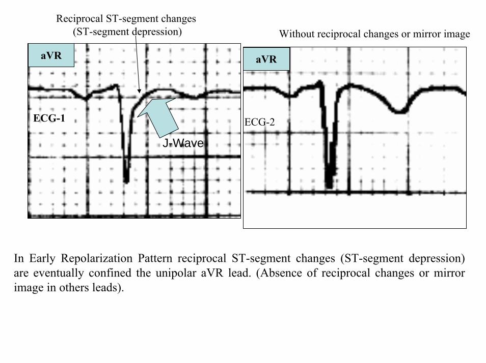
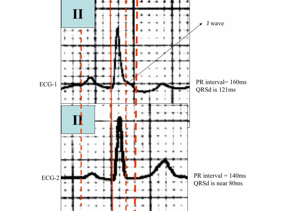
II
II
PR interval= 160msQRSd is 121ms
ECG-1
PR interval = 140msQRSd is near 80ms
J wave
ECG-2
V6
V5
V1
V4
V2
V3
X
Z
Is this a type 2 Brugada ECG pattern Answer in next slide
ECG-2
V2βangle gt58deg
Observation β angle is related to both depolarization an repolarization A wider QRS complex (slow conduction) and abnormal repolarization both contribute to a wider β angle
1 Chevallier S J Am Coll Cardiol2011 Nov 2258 2290-2298
In patients with suspected Brugada syndrome simple ECG criteria can enable discrimination between innocent incomplete RBBB from types 2 and 3 Brugada ECG patterns The mean β angle is significantly smaller in the patients with negative results on Antiarrhythmic Drug Challenge (ADC) compared wit patients with positive results on ADC Its optimal cutoff value is 58deg which yielded a positive predictive value of 73 and a negative predictive value of 87 for conversion to type 1 pattern on ADC α is slightly less sensitive and specific compared with β When the angles are combined with QRS duration it tended to improve discrimination
Y
aVF
Z V2
Right Sagittal Plane ECG-2
Triphasic QRS patternRSracute
β angle defined as the angle between the upslope of the S-wave and the downslope of the r-wave The mean β angle was significantly smaller in the 14 patients with negative results on AAD compared to the 24 patients with positive results on AAD Itrsquos optimal cutoff value was 58deg which yielded a positive predictive value of 73 and a negative predictive value of 87 for conversion to type 1 pattern on AAD
ST segment with saddleback appearance followed by positive or biphasic T wave in V1-V2 or from V1 through V3
β
V5V5
QRSd= 122ms QRSd= 110ms
ECG-1 ECG-2
Left precordial terminal QRS notching is more prevalent in malignant early repolarization abnormalities
Left precordial terminal QRS notching
1 Merchant FM et al Am J Cardiol 2009 Nov 15 1041402-1406
V2
V2
V3
V3
ECG-1
ECG-2
ECG-2
Dynamic inconstant or dramatic changes in repolarization are suggestive of malignant early repolarization abnormalities
1 Haiumlssaguerre M et al J Cardiovasc Electrophysiol 2009 Jan2093-98
ECG-1
Conclusion this patient has high possibility of concomitant Brugada syndrome and early repolarization In this a benign ERP or malignant early repolarization abnormalities We think it is very unlikely that is a Brugada phenocopyIn this case it is necessary additionally to perform
1 IV Ajmaline test2 ECG-AR3 Family study ( first degree relatives interrogatory physical ECGs)4 Magnetic resonance image5 Genetic screening
Ventricular biopsy is not considered mandatory but is recommended especially when it may clarify the nature of dubious findings identified with others test In approximately 6--10 of survivors of cardiac arrest no cardiac abnormality can be identified despite extensive clinical evaluation Autopsy data confirm that in a similar percentage of victims of SCD no structural heart disease or or mors sine materia can be identified at post mortem evaluation Incompletely penetrant genetic defects may underlie at least some of these unexplained deaths (1)
The ERP probably represents part of a spectrum of cardiovascular anomalies related to channelopaties including BrS IVF congenital short QT syndrome and that it may also have a molecular genetic origin of variable penetrance The denomination Early Repolarization Pattern is appropriate since it indicates that it is characterized by a given ECG pattern
1 Priori SG et al Cardiovasc Res 2001 May50218-223
Case Report similar
A 20 year-old male mulatto professional soccer player from Santo Andreacute Sao Paulo Brazil He didnrsquot finishthe second year in elementary school CatholicProfession Professional soccer playerReason for consultation Presented to consultation due to two presyncopal episodes and one syncopal episode in the last 30 days The first two episodes happened during a post-defecation period Both were preceded by dizziness nausea and vomiting The syncopal episodes occurred at rest with no prodromes The third one with express and brief loss of consciousness and sphincter relaxation at dawn 48 hours agoHis prior personal medical history was unremarkable and he successfully passed two prior periodical clinical-cardiovascular evaluations (twice a year always normal) Pre-competitive examination included 12-lead ECG echocardiogram and cardiopulmonary metabolic exercise testing (CMET) All normals Intense training for the last 3 years Familial Background One uncle from his fatherrsquos side family died of sudden cardiac death when he was 35
years old He ignores the cause His brother is being seeing by a cardiologist due to ldquocardiac arrhythmiardquo in treatment with a cardiologistfrom the Institute of the Heart from Satildeo Paulo (he cannot tell which is the cause) He denies having other relevant background details Pysical cardiovascular examination revealed bradycardia at 45 bpm with physiological sinus arrhythmia blood pressure 11070 mmHg The rest of the physical examination was unremarkable He denied consuming any type of neither medications nor drugs No addictionsHis first 12-lead ECGVectorcardiogram(VCG) (Fig 1) showed sinus bradycardia at 44 bpm P-wave axis of +60deg PR interval of 186ms QRS duration of 111ms QRS axis at + 63deg QT interval 427ms and corrected QTc 367ms There is a notch on the terminal portion of the QRS complex in the left precordial leads voltage criteria for left ventricular hypertrophy (SV1 + RV5 gt 35 mm ndash Sokolow index) prominent J-wave and concave ST-segment elevation in inferior leads V4 V5 and V6 Deep and narrow Q-waves in the left precordial leads V5 and V6 All these features were considered as an Early Repolarization Pattern (ERP)
Name JVS Gender Male Age 20 yo Ethnic Group Mulatto Weight 66 Kg Height 172 m Biotype Normoline Date 11262009 Profession competitive athlete (soccer player)
ECG diagnosis Sinus bradycardia (SB) HR 44bpm P axis +60deg PR 186ms QRSd 111ms QRS axis + 63deg QT 427ms QTc 367ms Conclusion SB + Early Repolarization Pattern (ERP) Notching or slurring of the terminal QRS complex voltage criteria of left ventricular hypertrophy SV1+ RV5 gt 35 mm (Sokolow index) prominent J wave and ST-segment elevation concave to the topending in a positive T wave from V4 to V6 Precordial greater than limb leads Relatively deep but narrow q waves in the left precordial leads (V5 and V6)
V6V4 V5
Prominent J wave and ST- segment elevation concave to the top ending in a positive T wave from V4 to V6 Notching or slurring morphology of the terminal portion of the R wave (J-wave) that produces a positive hump known as a J-wave (Arrows)
aVR
Eventually reciprocal ST segment depression confined only in Lead aVR1 observed in this particular case report
1 Mehta M Jain AC Mehta A Early Repolarization Clin Cardiol 1999 Feb 22 59-65
ECGVCG CORRELATIONS HORIZONTAL PLANE
V6
V1
V2
V3
X
Z
V5
V4
T
0J T
BEGINNING AND END OF QRS LOOP IN HP
J POINT in vectorcardiography it corresponds to 3 elements end of ventricular depolarization (QRS loop) beginning of repolarization (ST segment) when it does not present depression or elevation and T wave onset In this case 0 point and J point are not coincident In situations where there is depression or elevation of ST segment the J point does not coincide with the 0 point and the greater or lesser distance between both points indicate the greater or lesser ST segment elevation or depression The phenomenon is observed in early repolarization pattern acute phase of myocardial infarction Prinzmetal variant angina pericarditis Brugada syndrome arrhythmogenic right ventricular dysplasia etc
0 POINTIt corresponds to the QRS looponset (because PR segment does notexist) Alsocorrespond to the end of biatrialchamberactivation
T
J POINT
ECGVCG CORRELATIONS FORNTAL PLANE
aVR aVL
I
IIIII
X
Y
VERTICAL AXIS ON FRONTAL PLANE The frontal plane QRS axis and ST segment axis and T wave axis are all in the same direction THE ST
SEGMENT ELEVATION
USUALLY lt 05 mm IN THE LIMB
LEADS
YVF
Z V2
T
ECGVCG CORRELATIONS RIGHT SAGITTAL PLANE
A second 12-lead ECG (Fig 2) preformed 72 hours later showing1) Sinus bradycardia at 44 bpm2) Similar p-wave axis and intervals duration3) Dramatic modification of repolarizaccedilatildeo pattern in the right precordial leads A coved-type
(type 1) ECG brugada pattern can be distinguished4) Accentuation of j-waves and st segment elevation across the precordial leads was observed5) ST segment elevation on avr lead
See next slide
Name JVS Gender Male Age 20 yo Ethnic Group MulattoWeight 66 Kg Height 172 m Biotype Normoline Date 11292009Profession Competitive Soccer Player FIGURE 2
ECG diagnosis Sinus Arrhythmia(SA) sinus bradycardia (SB) HR 44bpm P axis +60deg PR 186ms QRSd 111ms QT 427ms QTc 367 Conclusion SA + SB + Early Repolarization Pattern (ERP) Type 1 Brugada ECG pattern Dramatic accentuation of J waves across the precordial and limb leads Transient augmentation of global J waves may be indicative of a
highly arrhythmogenic substrate heralding multiple episodes of VF in patients with ERP
aVR LEAD
11262009 11292009
DEPRESSION
ELEVATION
ST SEGMENT DEPRESSION IN aVR = 1mm ST SEGMENT ELEVATION IN aVR = 2mm
V1 V2V2
1129200911262009
V1
11292009 11262009
SPONTANEOUS MODIFICATION OF REPOLARIZATION TO TYPE 1 BRUGADA ECG PATTERN
11262009
V6
11292009V6
ST segment elevation lt 2mm ST segment elevation gt 2mm
11292009
V5
11262009
V5
ST segment elevation = 1mm ST segment elevation = 25mm
EARLY REPOLARIZATION PATTERN CONCEPT
DEFINITION Early Repolarization Pattern (ERP) is a well-recognized Enigmatic idiopathic electrocardiographic phenomenon characterized by notching or slurring morphology of the terminal portion of the R wave (J-wave) that produces a positive hump known as a J-wave at the end of the QRS complex and beginning of the ST segment It considered to be present when at least two adjacent precordial leads show elevation of the ST segment with values ge01 mV (or 1mm) above baseline followed by concordant tall near symmetric (T waves having similarity in shape ascending and descending ramps speeds)
EPIDEMIOLOGY ERP occur in 2 to 5 of the population often in men young adults afro-descendents and competitive athletes and in up to 131 to 482 of patients presenting with chest pain in the emergency rooms and coronary care units There is a distinct J wave and ST segment concave upward in the left precordial leads V4 through V6 The ST elevation is most frequently evident in ECG lead V4 The ST segment elevation is usually le2mm (precordial greater than limb leads) and is lt 05 mm in the limb leadsReciprocal ST-segment changes (ST-segment depression) are eventually confined the unipolar aVR lead (Absence of reciprocal changes or mirror image) In ERP vertical QRS axis electrical is the rule The frontal plane QRS axis the ST segment axis and T wave axis are all in the same directionThis ECG pattern has been considered a benign normal ECG variant for over six decades3
1 Hasbak P Engelmann MD Early repolarization ST-segment elevation as a normal electrocardiographic variant Ugeskr Laeger 2000 162 5928-5929
2 Brady WJ Chan TC Electrocardiographic manifestations benign early repolarization J Emerg Med 1999 17473-478
3 Benito B Guasch E Rivard L Nattel S Clinical and mechanistic issues in early repolarization of normal variants and lethal arrhythmia syndromes J Am Coll Cardiol 2010 Oct 5 56 1177-1186
MAIN ELECTROCARDIOGRAPHIC FEATURES IN ERPHeart rate Predominant sinus bradycardia frequently associated with respiratory sinus arrhythmia
PR interval First degree AV block is observed in 5 to 39 among professional athletes In the non-athlete population it is observed in 065 of cases QRS axis Vertical electrical axis is the rule The frontal plane QRS axis and ST segment axis and T wave axis are all in the same directionQRS duration (90 plusmn 10 ms) subjects than in age-matched healthy controls (80 plusmn 10 ms) QRS morphology Notching or slurring of the terminal QRS complex Prominent relatively deep but narrow q waves may appear in the left precordial leadsQRS voltage Eventually voltage criteria of LVH are observed in male competitive athletes SV1+ RV5 gt 35 mm (Positive Sokolow index) High QRS voltage is more frequent in male athletes but its correlation with LVH is low Voltage decreases after deconditioning is slow The distinction between the physiological athletes heart and pathological conditions has critical implications for professional athletes An abrupt transition may occur from right-oriented complexes to left-oriented complexes in the precordial leads secondary to counterclockwise rotation on precordial leads About two-thirds of clockwise rotation and counterclockwise rotation could be attributed to the septal angle by anatomical rotation of the heart in one plane around the long axis but other factors appear to be responsible for such ECG findings in the remaining one-third of cases Relatively higher positions of the precordial ECG leads as observed in the vertical heart appeared to be responsible for clockwise rotation in some patients and left septal fascicular block is suspected to be responsible for counterclockwise rotation in others patientsJ-point elevation Notching irregular or slurring contour of the terminal QRS complex (J point) Variant of Osborn wave is seen in ERP
ST segment Widespread ST segment elevation (precordial greater than limb leads) The characteristic ST segment is elevated upward concave confined more frequently in precordial leads with reciprocal depression only in aVR The concavity is observed at the initial up-sloping portion of ST segment or upwardly concave ST segment morphology Unfortunately concave ST morphology cannot be used to rule out ST elevation from AMI with left anterior descending coronary occlusion because it is common in these circumstances The ST elevation is most frequently evident in ECG lead V4 There is a distinct J wave and ST segment in the left precordial leads V4 through V6 The ST elevation in ERP is usually lt 2 mm (but can rarely be gt 5 mm) in the precordial leads and the greatest ST elevation is usually seen in the mid-to-left precordial leads The ST segment elevation is usually lt 05 mm in the limb leads
T wave characteristics Concordant T waves of large amplitude (prominent matching T waves) typical pseudo-asymmetrical (lsquosymmetroidrdquo) or slightly asymmetrical matching T waves often of large amplitude upright tall and peaked most conspicuously from V2 to V4 or V5 sometimes seen in leads DII DIII and aVF as a rule T waves may appear as of large amplitude peaked or pointed symmetric and matching Vagotonic or high T wave voltages followed by U waves are frequent when sinus bradycardia is present Tall positive ad symmetric or symmetroid T waves are not only seen occasionally in the very early stages of MI but also in hyperkalemia and in ERP sinus bradycardia
QT intervals QT maximum The maximum Q-onset-T-end interval This parameter is higher in ERV subjects than in normal controls
QTp maximum maximum Q-onset-T-peak interval This parameter is higher in ERV subjects than in age-matched healthy controls
Rate-corrected QTc maximum This parameter is lower in ERP subjects than in age-matched healthy controls
QTpc maximum This parameter is lower in ERV subjects than in age-matched healthy controls
U wave because bradycardia U waves are frequent in ERV they are best observed in the V3 lead U waves are frequent when sinus bradycardia is present
Other ECG characteristics of ERPRelative temporal stability of the ST segment and T wave pattern is observedReciprocal changes are not seen in ERP There are no evolutionary short-term changes in the ST segment and T waves and Q waves do not appear
Discusions Early Repolarization Pattern (ERP) or Early Repolarization Variant (ERV) is an enigmati
electrocardiographic phenomenon characterized by prominent J wave and ST-segment elevation in multiplleads
Recently there has been renewed interest in ERP because of similarities to the arrhythmogenic Brugadsyndrome (BrS) Not much is known about the epidemiology of ERP and several studies have reported thathis condition is associated with a good prognosis
Both ERP and BrS exhibit some similarities including the ionic underlying mechanism the analogouresponses to changes in heart rate and autonomic tone sympathicomimetics (isoproterenol test) as well as insodium channel and beta-blockers These observations raise the hypothesis that ERP may be not as benign atraditionally believed Additionally there are documents showing that ST-segment height in the man igreatly influenced by central sympathetic nervous activity both at baseline and during physiologic andpharmacological stress Central sympathetic dysfunction regularly results in multilead ST-segment elevationor J wave that decreases or below isoelectric baseline during low dose isoproterenol infusion An earlyrepolarization pattern in the inferior leads of a standard electrocardiogram is associated with an increased riskof death from cardiac causes in middle-aged subjects1
POSSIBLE SIMILARITIES BETWEEN ERV AND BrSbullMore frequent in malesbullBoth occur more frequently in young adults and in individuals without apparent structural heart diseasebullBoth may influence just the V1-V2 leads Rarely (9) can ST elevation be observed in ERP only in the right precordial leads V1-V2 or in the inferior ones2
1 Tikkanen JT Anttonen O Junttila MJ Aro AL Kerola T Rissanen HA Reunanen A Huikuri HV Long-Term Outcome Associated with Early Repolarization on Electrocardiography N Engl J Med 2009 Nov 16 [Epubahead of print]
2 Hasbak P Engelmann MDEarly repolarization ST-segment elevation as a normal electrocardiographic variant Ugeskr Laeger 2000 162 5928-5929
When ST elevation is normal it can reach up to 3 mm in V2-V3 especially in young people In those individuals over 40 years it seldom exceeds 2 mm Both can show incomplete RBBB pattern or right bundle branch conduction disorder in BrS it can present atypical features RBBB-like and of the saddle type by exclusive elevation of the J point S wave with delay in the left leads DI aVL V5 and V6 could be absent as it is to be expected in a classic RBBB The elements considered as typical in BrS are 1) elevation of the terminal part of QRS (prominent J wave) 2) elevated and descending ST not related to lesion of ischemic (idiopathic) injury 3) negative T wave in the right precordial leads 4) normal QTc or near normal 5) Eventually absence of final delay in left leads as it would be expected in a classic RBBB1 In ERV when associated to athletersquos heart QRS can present a moderate extension (100 ms to 110 ms) in 15 of the cases which in nonathlete normal population in a 24 is called outflow tract hypertrophy In this case r does not exceed the 5 mm and is lower than S in the same lead rSrBoth may improve repolarization during the stress test with use of isoproterenolBoth respond to a shortening of AP phase 2 in a part of ventricular thickness and intensification of fast repolarization notch (phase 1) mediated by transmural dispersion of ventricular repolarization by a larger notch in the Ito channel2The alteration of the Ito and ICa2+-L channels in BrS and in ERP are the electrophysiologic substrate that explains the J point and ST segment elevation because they cause the intensified notch in phase 1 and suppression in phase 2 duration in the epicardium and in the endocardium of ventricular wall thickness
1 Hiss RG Lamb LE Electrocardiographic findings in 122043 individuals Circulation 1962 25947-9612 Antzelevitch Ch Xin Yan G Shimuzi W et al Electrical heterogeneity the ECG and Cardiac Arrhythmias In Zipes
DP Jalife J Cardiac Electrophysiology From Cell to Bedside Third Edition WB Saunders Company2000 Chapter 26 p 222-238
ELEMENTS FOR DIFFERENTIAL DIAGNOSIS BETWEEN ERP AND BrS
I) Family background ERP negativeBrS frequently positive
II) Ethnic GroupERP predominantly in African descendents(1) or equally common in all races(2) BrS predominantly in Asian group (58) and Caucasian people(3)
III) GenderERP male gender predominanceBrS great predominance in the male gender (malefemale ratio ndash 81 in non Asian and 101 in Asian
peopleIV) Response to IC group antiarrhythmic agents
BrS flecainide used in a 10 mgKg dosage in 10 minutes increases ST elevation and QRS duration in a more significant way in patients with BrS than in individuals without the entity and only in those it triggers ventricular extrasystoles(4)
ERP it can induce a pattern similar to BrS however the degree of ST elevation caused by the drug is much higher in patients with BrS than in patients without the disease
1 Grusin H Peculiarities of the Africanrsquos electrocardiogram and the changes observed in serial studies Circulation 1954 9 860-867
2 Mehta M Jain AC Mehta A Early Repolarization Clin Cardiol 1999 Feb2259-653 Nademanee K Veerakul G Nimmannit S Chaowakul V Bhuripanyo K Likittanasombat K Tunsanga K Kuasirikul S
Malasit P Tansupasawadikul S Tatsanavivat P Arrhytmogenic marker for the sudden unexplained death syndrome in Thail men Circulation 1997 962595-2600
4 Shimizu W Antzelevitch C Suyama K Kurita T Taguchi A Aihara N Takaki H Sunagawa K Kamakura S Effect of sodium channel blockers on ST segment QRS duration and corrected QT interval in patients with Brugada syndromeJ Cardiovasc Electrophysiol 2000 111320-1329
BrS patients with Inferior and lateral ECG repolarization abnormalities have longer baseline PR intervals(1)Both an S wave width ge80ms in V1 and ST elevation ge18mm in V2 are highly specific indicators of VF and criteria for high-risk BrS(2)There are several references in literature to patients with elevation of the J point and ST segment convex to the top or straight descendent in inferior leads (Brugada sign) or concomitantly ST-segment elevation in the right precordial and inferior leads II III and aVF(3) in absence of hypothermia ischemia or electrolytic disorders which we call ldquoatypical Brugada patternrdquo atypical BrS variant BrS or BrS variant or idiopathic VF (a variant of the BrS with ST-segment elevation in inferior leads(4)Pilsicainide may induce PVCs and PVT in atypical BrS and the infusion of isoproterenol suppress the arrhythmias and normalize the ST-segment elevation(5)
1 Sarkozy A Chierchia GB Paparella G Boussy T De Asmundis C Roos M Henkens S Kaufman L Buyl R Brugada R Brugada J Brugada P Inferior and lateral electrocardiographic repolarization abnormalities in Brugada syndromeCirc ArrhythmElectrophysiol 2009 Apr 2154-161
2 Atarashi H Ogawa S For The Idiopathic Ventricular Fibrillation Investigators New ECG Criteria for High-Risk Brugada Syndrome Circ J 2003 Jan 67 8-10
3 Lombardi F Potenza S Beltrami A Verzoni A Brugada P Brugada RSimultaneous ST-segment elevation in the right precordial and inferior leads in Brugada syndrome J CardiovascMed (Hagerstown) 2007 8 201-204
4 Letsas KP Efremidis M Pappas LK Gavrielatos G Markou V Sideris A Kardaras FEarlyrepolarization syndrome is it always benign Int J Cardiol 2007114 390-392
5 Chinushi M Izumi D Furushima H Watanabe H Aizawa YMultiple premature beats triggered ventricular arrhythmias during pilsicainide infusion in a patient with inferior ST-segment elevation Pacing Clin Electrophysiol 2006 29 1445-1448
Potet et al(1) identified a G752R mutation on SNC5A that produced ST segment elevation and prominent J wave in leads II III and aVF The authors provide genetic demonstration that Brugada ECG anomalies related to a unique SCN5A mutation can be observed either in the inferior or the right precordial leads The early repolarization pattern in inferolateral leads is not an uncommon finding in BrS and this pattern is not associated with a worse outcome in subjects with BrS(2) The spontaneous ERP occurred more frequently among patients with BrS than in 283 family members not having BrS (11 versus 6 P=003) Class I antiarrhythmic drug administration provoked inferior-lateral coved Brugada pattern in 13 patients with BrS These patients had longer baseline PR intervals and Class I antiarrhythmic drug induced QRS interval prolongation (108 to 178 versus 102 ms to 131 ms Plt0001) In 3 patients the Class I antiarrhythmic drug provoked coved Brugada pattern only present in the inferior leads Inferior-lateral ERP occurs spontaneously relatively frequently in BrS These patients have a more severe phenotype Class I antiarrhythmic drug administration provokes inferior-lateral coved Brugada pattern in 46 of patients The Class I antiarrhythmic drug exceptionally provoked coved Brugada pattern only observed in the inferior leads(3)
1 Potet F Mabo P Le Coq G et al Novel brugada SCN5A mutation leading to ST segment elevation in the inferior or the right precordial leads J Cardiovasc Electrophysiol 2003 142000-2003
2 Letsas KP Sacher F Probst V Weber R Knecht S Kalusche D Haiumlssaguerre M ArentzTPrevalence of early repolarization pattern in inferolateral leads in patients with Brugada syndrome Heart Rhythm 2008 Dec 5 1685-1689
3 Sarkozy A Chierchia GB Paparella G Boussy T De Asmundis C Roos M Henkens S Kaufman L Buyl R Brugada R Brugada J Brugada P Inferior and lateral electrocardiographic repolarization abnormalities in Brugada syndrome Circ Arrhythm Electrophysiol 2009 Apr2 154-161
Type 1 ECG Brugada pattern in the peripheral leads was observed in 42 of patients during ajmaline test (103 of positive tests) and was associated with longer QRS and greater QTc prolongation compared with the rest of the patients1Atypical Brugada Syndrome or IVF with J waves in inferior lateral or inferior lateral leads
and Familial Idiopathic Ventricular Fibrillation and BrSSeveral mutations in genes SCN5A KCNJ8 DPP6 SCN3B and CACNA2D1 had been identified 1 On SCN5A gene2 3 Observation We do not believe that SCN5A should be attributed to IVF because this report clearly shows conduction defects (as expected) so this is not IVF but conduction delay at different levels in the heart2 On KCNJ8 gene 4 5 Missense variant in exon 3 (NC-000012) of the KCNJ8 gene a subunit of the K(ATP)channel23 Genomic DNA sequencing of K(ATP) channel genes showed missense variant in exon 3 (NC_000012) of the KCNJ8 gene a subunit of the K(ATP) channel conferring predisposition to dramatic repolarization changes and ventricular vulnerability
1 Batchvarov VN Govindan M Camm AJ Behr ERBrugada-like changes in the peripheral leads during diagnostic ajmaline test in patients with suspected Brugada syndrome Pacing Clin Electrophysiol 2009 Jun32695-703
2 Potet F Mabo P Le Coq G Probst V Schott JJ Airaud F Guihard G Daubert JC Escande D Le MarecH Novel brugada SCN5A mutation leading to ST segment elevation in the inferior or the right precordial leads J Cardiovasc Electrophysiol 2003 14 200-203
3 Akai J Makita N Sakurada H Shirai N Ueda K Kitabatake A Nakazawa K Kimura A Hiraoka M A novel SCN5A mutation associated with idiopathic ventricular fibrillation without typical ECG findings of Brugada syndrome FEBS Lett 2000 Aug 11 479 29-34
4 Haissaguerre M Derval N Sacher F Jesel L Deisenhofer I de Roy Let al Sudden cardiac arrest associated with early repolarization N Engl J Med 2008 May 8 3582016-2023
5 Haiumlssaguerre M Sacher F Nogami A Komiya N Bernard A Probst V et al Characteristics of recurrent ventricular fibrillation associated with inferolateral early repolarization role of drug therapy J Am CollCardiol 2009 Feb 17 53 612-619
On DPP6 gene1 Dipeptidyl aminopeptidase-like protein 6 locus on chromosome7q26 Voltage-gated potassium channels OMIM126141 The ECG shows rapid PVT Monomorphic PVCs with very short coupling intervals The PVC (302 +- 52 msec) within 40 msec of the peak of the preceding Absence of T wave Pause-dependence
Valdivia et al2 reported a case with IVF and a novel mutation in the SCN3B-encoded sodium channel beta subunit Navbeta3 that causes a loss of function of Nav15 channels in vitro and provides molecular and cellular evidence implicating mutations in Navbeta3 as a cause of IVF Mutation in this SCN3B gene causes also BrS type 73
L-type calcium channel (LTCC) mutations have been associated with BrS SQTS and Timothy syndrome (LQT8) Little is known about the extent to which LTCC mutations contribute to the J-wave syndromes associated with SCD Mutations in the LTCCs are detected in a high percentage of probands with J-wave syndromes associated with inherited cardiac arrhythmias suggesting that genetic screening of Ca(v) genes may be a valuable diagnostic tool in identifying individuals at risk Burashnikov et al4 identify CACNA2D1 as a novel BrS susceptibility gene and CACNA1C CACNB2 and CACNA2D1 as possible novel ERS susceptibility genes4
1 Alders M Koopmann TT Christiaans I Postema PG Beekman L Tanck MW et al Haplotype-sharing analysis implicates chromosome 7q36 harboring DPP6 in familial idiopathic ventricular fibrillation Am J Hum Genet 2009 Apr 84 468-476
2 Valdivia CR Medeiros-Domingo A Ye B Shen WK Algiers TJ Ackerman MJ et al Loss-of-function mutation of the SCN3B-encoded sodium channel beta3 subunit associated with a case ofidiopathic ventricular fibrillation Cardiovasc Res 2010 Jun 1 86 392-400
3 Hu D Barajas-Martinez H Burashnikov E Springer M Wu Y Varro A et al A mutation in the beta-3 subunit of the cardiac sodium channel associated with Brugada ECG phenotype Circ Cardiovasc Genet 2009 2 270-278
4 Burashnikov E Pfeiffer R Barajas-Martinez H Delpoacuten E Hu D Desai M et al Mutations in the cardiac L-type calcium channel associated with inherited J-wave syndromes and sudden cardiac death Heart Rhythm 2010 Dec71872-1882
From a multicenter cohort of 122 patients (90 male age 37 +- 12 years) with IVF and ERP in the inferolateral leadsHaiumlssaguerre et al1 selected all patients with more than 3 episodes of VF (multiple) including those with electrical storms (ge3 VF in 24 h) Multiple recurrences of VF occurred in 27 of patients with ERP and may be life threatening Isoproterenol in acute cases and quinidine in chronic cases are effective antyharrhithmic drugsThe last one is necessary associated to ICDThe so-called atypical BrS is characterized by ECG abnormalities of the J wave and ST-segment elevation appeared in the inferior andor lateral leads The ERP in inferolateral leads is not an uncommon finding in BrS2 There is a high incidence of the ERP confined in inferolateral leads in patients with IVF The ECGs have an elevation of the QRS-ST junction of at least 01 mV from baseline in the inferior or lateral lead manifested as QRS slurring or notching Among patients with a history of IVF there is an increased prevalence of ERP
1 Haissaguerre M Derval N Sacher F Jesel L Deisenhofer I de Roy L Pasquieacute JL Nogami A Babuty D Yli-Mayry S De Chillou C Scanu P Mabo P Matsuo S Probst V Le Scouarnec S Defaye P Schlaepfer J Rostock T Lacroix D Lamaison D Lavergne T Aizawa Y Englund A Anselme F ONeill M Hocini M Lim KT Knecht S Veenhuyzen GD Bordachar P Chauvin M Jais P Coureau G Chene G Klein GJ Cleacutementy J Sudden cardiac arrest associated with early repolarization N Engl J Med 2008 May 8 3582016-2023
2 Letsas KP Sacher F Probst V Weber R Knecht S Kalusche D Haiumlssaguerre M Arentz T Prevalence of early repolarization pattern in inferolateral leads in patients with Brugada syndrome Heart Rhythm 2008 Dec51685-1689
Because a contingent percentage has not yet been well established it has been suggested that BrS constitutes the same entity in a large number of IVF diagnosed cases since they have common clinical features
1 BOTH ARE MORE FREQUENT IN MEN2 BOTH HAPPEN IN MIDDLE-AGE PEOPLE3 BOTH OCCUR IN APPARENT STRUCTURALLY NORMAL HEARTS14 BOTH CAN AFFECT THE SAME GENE (SCN5A)5 BOTH AFFECT EVENTUALLY THE SAME LOCUS (3p24-p21)
Wellens et al2 has pointed out that in order to be accurate about such a percentage it is important to define the diagnostic criteria used to classify BrS in IVF patients Curiously the so-called Idiopathic VT in apparently healthy hearts without structural heart disease and primary electrical disease also affects the 3p21 locus but not 3p24 and the gene is different GNA12
Survivors of SCD by VF without structural heart disease and a normal coronary angiogram are often diagnosed with IVTIVF3
A careful analysis of the ECG is essential in the differential diagnosis of this type of apparently unexplained SCD because intermittent or permanent RBBB with J point and ST-segment elevation in leads V1 to V3 is now classified as BrS
1 Goethals P Debruyne P Saffarian M Drug-induced Brugada syndrome Acta Cardiol1998 53157-60
2 Wellens HJJ Rodriquez LM Smeets JL Ventricular tachycardia in structural normal hearts In Zipes DP Jalife J Editors Cardiac Electrophysiology from cell tobedside Philadelphia 1995 WB Saunders
3 Chen Q Kirsch GE Zhang D Brugada R Brugada P Brugada J et al Genetic basis and molecular mechanisms for idiopathic ventricular fibrillation Nature 1998392 293-296
DIAGNOSTICS TRACKS TO DIFFERENTIATE BENIGN ERP FROM MALIGNANT EARLY REPOLATIZATION ABNORMALITIES
BENIGN ERP MALIGNANT EARLY REPOLARIZATION ABNORMALITIES
Family history of unexplained SCD in young relatives(lt45yo)
Absent Possible however infrequent1
Personal history Asymptomatic Asymptomatic repetitive syncope episodes or recovered of cardiac arrest
Left precordial terminal QRS notching Less prevalent More prevalent2
STT ratio in lead V6calculated by dividing the millimeters of
ST-segment elevation by the millimeters to the tallest point of the T wave
Each value is measured from the isoelectric point
lt0253 Non-tested
1 Viskin S Belhassen B Idiopathic ventricular fibrillation American Heart Journal 1990 120 661-671
2 Tikkanen JT Anttonen O Junttila MJ Aro AL Kerola T Rissanen HA Reunanen A et al Long-term outcome associated with early repolarization on electrocardiography N Engl J Med 2009 Dec 24361 2529-2537
3 Ginzton LE Laks MM The differential diagnosis of acute pericarditis from the normal variant new electrocardiographic criteria Circulation 1982 May 65 1004-1009
ST-segment elevation shape Concave upward Convex upwardJ waves are present in asymp 30 IVF patients1
Early repolarization is significantly prevalent in the subjects after cardiac arrest
(present in 31 of those resuscitated )Eventually ldquoLambda-waverdquo 23
J-point elevation and ST-segment degree Usually lt 2 mmlt05mm in limb leads
Frequent gt2mmJ-point elevation gt 2mm in inferior leads is associated with an increased risk of death
from cardiac causes in middle-aged subjects3
T wave characteristic Positive large T wave from V2 to V4 or V5 Concordant
Frequent negative discordant
Reciprocal ST-segment changes (Mirror
image)Absent With exception aVR5 Frequently Present3
1 Viskin S Idiopathic ventricular fibrillation Le Syndrome dHaiumlssaguerre and the fear of J waves J Am Coll Cardiol 2009 Feb 17 53 620-622
2 Gussak I Bjerregaard P Kostis J Electrocardiographic lambda wave and primary idiopathic cardiac asystole a new clinical syndrome J Electrocardiol 2004 Apr 37 105-107
3 Riera AR Ferreira C Schapachnik E Sanches PC Moffa PJ Brugada syndrome with atypical ECG downsloping ST-segment elevation in inferior leads J Electrocardiol 2004 Apr37101-104
4 Tikkanen JT Anttonen O Junttila MJ Aro AL Kerola T Rissanen HA Reunanen A et al Long-term outcome associated with early repolarization on electrocardiography N Engl J Med 2009 Dec 24361 2529-2537
5 Mehta M Jain AC Mehta A Early Repolarization Clin Cardiol 1999 Feb 22 59-65
Monomorphic PVCs with very short coupling
(250 to 350ms)
Absent Characteristic1 Pause-dependency absent2
QTc Interval Normal Minimally short in 35 among masculine gender
(Between 340 to 360ms3)
Late Potentials(LPs) Absent Frequent if J wave presentShowing circadian variation with night
ascendancy4
1 Alders M Koopmann TT Christiaans I Postema PG Beekman L Tanck MW et al Haplotype-sharing analysis implicates chromosome 7q36 harboring DPP6 in familial idiopathic ventricular fibrillation Am J Hum Genet 2009 Apr 84 468-476
2 Viskin S Lesh MD Eldar M Fish R Setbon I Laniado S et al Mode of onset of malignant ventricular arrhythmias in idiopathic ventricular fibrillation J Cardiovasc Electrophysiol 1997 Oct 8 1115-11 20
3 Viskin S Zeltser D Ish-Shalom M Katz A Glikson M Justo D et al Is idiopathic ventricular fibrillation a short QT syndrome Comparison of QT intervals of patients with idiopathic ventricular fibrillation and healthy controls Heart Rhythm 2004 Nov 1 587-591
4 Abe A Ikeda T Tsukada T Ishiguro H Miwa Y Miyakoshi M Circadian variation of latepotentials in idiopathic ventricular fibrillation associated with J waves insights into alternative pathophysiology and risk stratification Heart Rhythm 2010 May 7 675-682
ConclusionIn spite of both malignant early repolarization abnormalities of IVF and benign early repolarization pattern being able to cause J point and ST segment elevation the characteristics of both are clearly different to the point of a differentiation being quite simple when ECGs are analyzed by experts Any ER should raise the suspicion of IVF when it presents ST segment elevation ge 2 mm mainly in inferior wall superior convexity eventually lambda wave shape pattern of dynamic presentation inconstant with dramatic intensity of ST segment elevation discordant T wave polarity related ST segment shift presence of reciprocal ST-segment changes (mirror image) tendency to discrete short QTc interval in male gender(ge340ms) premature monomorphic ventricular contractions with very short coupling interval(250 to 350ms) not related with exercise and late potentials with circadian variation and night ascendancy on SA-ECG Contrarily in benign ERP ST elevation observed in the mid-to-left precordial leads is usually le 2 mm and lt05 mm in the limb leads and concave to the top followed by a positive concordant tall T wave
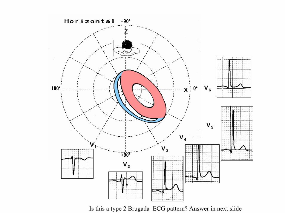
V6
V5
V1
V4
V2
V3
X
Z
Is this a type 2 Brugada ECG pattern Answer in next slide
ECG-2
V2βangle gt58deg
Observation β angle is related to both depolarization an repolarization A wider QRS complex (slow conduction) and abnormal repolarization both contribute to a wider β angle
1 Chevallier S J Am Coll Cardiol2011 Nov 2258 2290-2298
In patients with suspected Brugada syndrome simple ECG criteria can enable discrimination between innocent incomplete RBBB from types 2 and 3 Brugada ECG patterns The mean β angle is significantly smaller in the patients with negative results on Antiarrhythmic Drug Challenge (ADC) compared wit patients with positive results on ADC Its optimal cutoff value is 58deg which yielded a positive predictive value of 73 and a negative predictive value of 87 for conversion to type 1 pattern on ADC α is slightly less sensitive and specific compared with β When the angles are combined with QRS duration it tended to improve discrimination
Y
aVF
Z V2
Right Sagittal Plane ECG-2
Triphasic QRS patternRSracute
β angle defined as the angle between the upslope of the S-wave and the downslope of the r-wave The mean β angle was significantly smaller in the 14 patients with negative results on AAD compared to the 24 patients with positive results on AAD Itrsquos optimal cutoff value was 58deg which yielded a positive predictive value of 73 and a negative predictive value of 87 for conversion to type 1 pattern on AAD
ST segment with saddleback appearance followed by positive or biphasic T wave in V1-V2 or from V1 through V3
β
V5V5
QRSd= 122ms QRSd= 110ms
ECG-1 ECG-2
Left precordial terminal QRS notching is more prevalent in malignant early repolarization abnormalities
Left precordial terminal QRS notching
1 Merchant FM et al Am J Cardiol 2009 Nov 15 1041402-1406
V2
V2
V3
V3
ECG-1
ECG-2
ECG-2
Dynamic inconstant or dramatic changes in repolarization are suggestive of malignant early repolarization abnormalities
1 Haiumlssaguerre M et al J Cardiovasc Electrophysiol 2009 Jan2093-98
ECG-1
Conclusion this patient has high possibility of concomitant Brugada syndrome and early repolarization In this a benign ERP or malignant early repolarization abnormalities We think it is very unlikely that is a Brugada phenocopyIn this case it is necessary additionally to perform
1 IV Ajmaline test2 ECG-AR3 Family study ( first degree relatives interrogatory physical ECGs)4 Magnetic resonance image5 Genetic screening
Ventricular biopsy is not considered mandatory but is recommended especially when it may clarify the nature of dubious findings identified with others test In approximately 6--10 of survivors of cardiac arrest no cardiac abnormality can be identified despite extensive clinical evaluation Autopsy data confirm that in a similar percentage of victims of SCD no structural heart disease or or mors sine materia can be identified at post mortem evaluation Incompletely penetrant genetic defects may underlie at least some of these unexplained deaths (1)
The ERP probably represents part of a spectrum of cardiovascular anomalies related to channelopaties including BrS IVF congenital short QT syndrome and that it may also have a molecular genetic origin of variable penetrance The denomination Early Repolarization Pattern is appropriate since it indicates that it is characterized by a given ECG pattern
1 Priori SG et al Cardiovasc Res 2001 May50218-223
Case Report similar
A 20 year-old male mulatto professional soccer player from Santo Andreacute Sao Paulo Brazil He didnrsquot finishthe second year in elementary school CatholicProfession Professional soccer playerReason for consultation Presented to consultation due to two presyncopal episodes and one syncopal episode in the last 30 days The first two episodes happened during a post-defecation period Both were preceded by dizziness nausea and vomiting The syncopal episodes occurred at rest with no prodromes The third one with express and brief loss of consciousness and sphincter relaxation at dawn 48 hours agoHis prior personal medical history was unremarkable and he successfully passed two prior periodical clinical-cardiovascular evaluations (twice a year always normal) Pre-competitive examination included 12-lead ECG echocardiogram and cardiopulmonary metabolic exercise testing (CMET) All normals Intense training for the last 3 years Familial Background One uncle from his fatherrsquos side family died of sudden cardiac death when he was 35
years old He ignores the cause His brother is being seeing by a cardiologist due to ldquocardiac arrhythmiardquo in treatment with a cardiologistfrom the Institute of the Heart from Satildeo Paulo (he cannot tell which is the cause) He denies having other relevant background details Pysical cardiovascular examination revealed bradycardia at 45 bpm with physiological sinus arrhythmia blood pressure 11070 mmHg The rest of the physical examination was unremarkable He denied consuming any type of neither medications nor drugs No addictionsHis first 12-lead ECGVectorcardiogram(VCG) (Fig 1) showed sinus bradycardia at 44 bpm P-wave axis of +60deg PR interval of 186ms QRS duration of 111ms QRS axis at + 63deg QT interval 427ms and corrected QTc 367ms There is a notch on the terminal portion of the QRS complex in the left precordial leads voltage criteria for left ventricular hypertrophy (SV1 + RV5 gt 35 mm ndash Sokolow index) prominent J-wave and concave ST-segment elevation in inferior leads V4 V5 and V6 Deep and narrow Q-waves in the left precordial leads V5 and V6 All these features were considered as an Early Repolarization Pattern (ERP)
Name JVS Gender Male Age 20 yo Ethnic Group Mulatto Weight 66 Kg Height 172 m Biotype Normoline Date 11262009 Profession competitive athlete (soccer player)
ECG diagnosis Sinus bradycardia (SB) HR 44bpm P axis +60deg PR 186ms QRSd 111ms QRS axis + 63deg QT 427ms QTc 367ms Conclusion SB + Early Repolarization Pattern (ERP) Notching or slurring of the terminal QRS complex voltage criteria of left ventricular hypertrophy SV1+ RV5 gt 35 mm (Sokolow index) prominent J wave and ST-segment elevation concave to the topending in a positive T wave from V4 to V6 Precordial greater than limb leads Relatively deep but narrow q waves in the left precordial leads (V5 and V6)
V6V4 V5
Prominent J wave and ST- segment elevation concave to the top ending in a positive T wave from V4 to V6 Notching or slurring morphology of the terminal portion of the R wave (J-wave) that produces a positive hump known as a J-wave (Arrows)
aVR
Eventually reciprocal ST segment depression confined only in Lead aVR1 observed in this particular case report
1 Mehta M Jain AC Mehta A Early Repolarization Clin Cardiol 1999 Feb 22 59-65
ECGVCG CORRELATIONS HORIZONTAL PLANE
V6
V1
V2
V3
X
Z
V5
V4
T
0J T
BEGINNING AND END OF QRS LOOP IN HP
J POINT in vectorcardiography it corresponds to 3 elements end of ventricular depolarization (QRS loop) beginning of repolarization (ST segment) when it does not present depression or elevation and T wave onset In this case 0 point and J point are not coincident In situations where there is depression or elevation of ST segment the J point does not coincide with the 0 point and the greater or lesser distance between both points indicate the greater or lesser ST segment elevation or depression The phenomenon is observed in early repolarization pattern acute phase of myocardial infarction Prinzmetal variant angina pericarditis Brugada syndrome arrhythmogenic right ventricular dysplasia etc
0 POINTIt corresponds to the QRS looponset (because PR segment does notexist) Alsocorrespond to the end of biatrialchamberactivation
T
J POINT
ECGVCG CORRELATIONS FORNTAL PLANE
aVR aVL
I
IIIII
X
Y
VERTICAL AXIS ON FRONTAL PLANE The frontal plane QRS axis and ST segment axis and T wave axis are all in the same direction THE ST
SEGMENT ELEVATION
USUALLY lt 05 mm IN THE LIMB
LEADS
YVF
Z V2
T
ECGVCG CORRELATIONS RIGHT SAGITTAL PLANE
A second 12-lead ECG (Fig 2) preformed 72 hours later showing1) Sinus bradycardia at 44 bpm2) Similar p-wave axis and intervals duration3) Dramatic modification of repolarizaccedilatildeo pattern in the right precordial leads A coved-type
(type 1) ECG brugada pattern can be distinguished4) Accentuation of j-waves and st segment elevation across the precordial leads was observed5) ST segment elevation on avr lead
See next slide
Name JVS Gender Male Age 20 yo Ethnic Group MulattoWeight 66 Kg Height 172 m Biotype Normoline Date 11292009Profession Competitive Soccer Player FIGURE 2
ECG diagnosis Sinus Arrhythmia(SA) sinus bradycardia (SB) HR 44bpm P axis +60deg PR 186ms QRSd 111ms QT 427ms QTc 367 Conclusion SA + SB + Early Repolarization Pattern (ERP) Type 1 Brugada ECG pattern Dramatic accentuation of J waves across the precordial and limb leads Transient augmentation of global J waves may be indicative of a
highly arrhythmogenic substrate heralding multiple episodes of VF in patients with ERP
aVR LEAD
11262009 11292009
DEPRESSION
ELEVATION
ST SEGMENT DEPRESSION IN aVR = 1mm ST SEGMENT ELEVATION IN aVR = 2mm
V1 V2V2
1129200911262009
V1
11292009 11262009
SPONTANEOUS MODIFICATION OF REPOLARIZATION TO TYPE 1 BRUGADA ECG PATTERN
11262009
V6
11292009V6
ST segment elevation lt 2mm ST segment elevation gt 2mm
11292009
V5
11262009
V5
ST segment elevation = 1mm ST segment elevation = 25mm
EARLY REPOLARIZATION PATTERN CONCEPT
DEFINITION Early Repolarization Pattern (ERP) is a well-recognized Enigmatic idiopathic electrocardiographic phenomenon characterized by notching or slurring morphology of the terminal portion of the R wave (J-wave) that produces a positive hump known as a J-wave at the end of the QRS complex and beginning of the ST segment It considered to be present when at least two adjacent precordial leads show elevation of the ST segment with values ge01 mV (or 1mm) above baseline followed by concordant tall near symmetric (T waves having similarity in shape ascending and descending ramps speeds)
EPIDEMIOLOGY ERP occur in 2 to 5 of the population often in men young adults afro-descendents and competitive athletes and in up to 131 to 482 of patients presenting with chest pain in the emergency rooms and coronary care units There is a distinct J wave and ST segment concave upward in the left precordial leads V4 through V6 The ST elevation is most frequently evident in ECG lead V4 The ST segment elevation is usually le2mm (precordial greater than limb leads) and is lt 05 mm in the limb leadsReciprocal ST-segment changes (ST-segment depression) are eventually confined the unipolar aVR lead (Absence of reciprocal changes or mirror image) In ERP vertical QRS axis electrical is the rule The frontal plane QRS axis the ST segment axis and T wave axis are all in the same directionThis ECG pattern has been considered a benign normal ECG variant for over six decades3
1 Hasbak P Engelmann MD Early repolarization ST-segment elevation as a normal electrocardiographic variant Ugeskr Laeger 2000 162 5928-5929
2 Brady WJ Chan TC Electrocardiographic manifestations benign early repolarization J Emerg Med 1999 17473-478
3 Benito B Guasch E Rivard L Nattel S Clinical and mechanistic issues in early repolarization of normal variants and lethal arrhythmia syndromes J Am Coll Cardiol 2010 Oct 5 56 1177-1186
MAIN ELECTROCARDIOGRAPHIC FEATURES IN ERPHeart rate Predominant sinus bradycardia frequently associated with respiratory sinus arrhythmia
PR interval First degree AV block is observed in 5 to 39 among professional athletes In the non-athlete population it is observed in 065 of cases QRS axis Vertical electrical axis is the rule The frontal plane QRS axis and ST segment axis and T wave axis are all in the same directionQRS duration (90 plusmn 10 ms) subjects than in age-matched healthy controls (80 plusmn 10 ms) QRS morphology Notching or slurring of the terminal QRS complex Prominent relatively deep but narrow q waves may appear in the left precordial leadsQRS voltage Eventually voltage criteria of LVH are observed in male competitive athletes SV1+ RV5 gt 35 mm (Positive Sokolow index) High QRS voltage is more frequent in male athletes but its correlation with LVH is low Voltage decreases after deconditioning is slow The distinction between the physiological athletes heart and pathological conditions has critical implications for professional athletes An abrupt transition may occur from right-oriented complexes to left-oriented complexes in the precordial leads secondary to counterclockwise rotation on precordial leads About two-thirds of clockwise rotation and counterclockwise rotation could be attributed to the septal angle by anatomical rotation of the heart in one plane around the long axis but other factors appear to be responsible for such ECG findings in the remaining one-third of cases Relatively higher positions of the precordial ECG leads as observed in the vertical heart appeared to be responsible for clockwise rotation in some patients and left septal fascicular block is suspected to be responsible for counterclockwise rotation in others patientsJ-point elevation Notching irregular or slurring contour of the terminal QRS complex (J point) Variant of Osborn wave is seen in ERP
ST segment Widespread ST segment elevation (precordial greater than limb leads) The characteristic ST segment is elevated upward concave confined more frequently in precordial leads with reciprocal depression only in aVR The concavity is observed at the initial up-sloping portion of ST segment or upwardly concave ST segment morphology Unfortunately concave ST morphology cannot be used to rule out ST elevation from AMI with left anterior descending coronary occlusion because it is common in these circumstances The ST elevation is most frequently evident in ECG lead V4 There is a distinct J wave and ST segment in the left precordial leads V4 through V6 The ST elevation in ERP is usually lt 2 mm (but can rarely be gt 5 mm) in the precordial leads and the greatest ST elevation is usually seen in the mid-to-left precordial leads The ST segment elevation is usually lt 05 mm in the limb leads
T wave characteristics Concordant T waves of large amplitude (prominent matching T waves) typical pseudo-asymmetrical (lsquosymmetroidrdquo) or slightly asymmetrical matching T waves often of large amplitude upright tall and peaked most conspicuously from V2 to V4 or V5 sometimes seen in leads DII DIII and aVF as a rule T waves may appear as of large amplitude peaked or pointed symmetric and matching Vagotonic or high T wave voltages followed by U waves are frequent when sinus bradycardia is present Tall positive ad symmetric or symmetroid T waves are not only seen occasionally in the very early stages of MI but also in hyperkalemia and in ERP sinus bradycardia
QT intervals QT maximum The maximum Q-onset-T-end interval This parameter is higher in ERV subjects than in normal controls
QTp maximum maximum Q-onset-T-peak interval This parameter is higher in ERV subjects than in age-matched healthy controls
Rate-corrected QTc maximum This parameter is lower in ERP subjects than in age-matched healthy controls
QTpc maximum This parameter is lower in ERV subjects than in age-matched healthy controls
U wave because bradycardia U waves are frequent in ERV they are best observed in the V3 lead U waves are frequent when sinus bradycardia is present
Other ECG characteristics of ERPRelative temporal stability of the ST segment and T wave pattern is observedReciprocal changes are not seen in ERP There are no evolutionary short-term changes in the ST segment and T waves and Q waves do not appear
Discusions Early Repolarization Pattern (ERP) or Early Repolarization Variant (ERV) is an enigmati
electrocardiographic phenomenon characterized by prominent J wave and ST-segment elevation in multiplleads
Recently there has been renewed interest in ERP because of similarities to the arrhythmogenic Brugadsyndrome (BrS) Not much is known about the epidemiology of ERP and several studies have reported thathis condition is associated with a good prognosis
Both ERP and BrS exhibit some similarities including the ionic underlying mechanism the analogouresponses to changes in heart rate and autonomic tone sympathicomimetics (isoproterenol test) as well as insodium channel and beta-blockers These observations raise the hypothesis that ERP may be not as benign atraditionally believed Additionally there are documents showing that ST-segment height in the man igreatly influenced by central sympathetic nervous activity both at baseline and during physiologic andpharmacological stress Central sympathetic dysfunction regularly results in multilead ST-segment elevationor J wave that decreases or below isoelectric baseline during low dose isoproterenol infusion An earlyrepolarization pattern in the inferior leads of a standard electrocardiogram is associated with an increased riskof death from cardiac causes in middle-aged subjects1
POSSIBLE SIMILARITIES BETWEEN ERV AND BrSbullMore frequent in malesbullBoth occur more frequently in young adults and in individuals without apparent structural heart diseasebullBoth may influence just the V1-V2 leads Rarely (9) can ST elevation be observed in ERP only in the right precordial leads V1-V2 or in the inferior ones2
1 Tikkanen JT Anttonen O Junttila MJ Aro AL Kerola T Rissanen HA Reunanen A Huikuri HV Long-Term Outcome Associated with Early Repolarization on Electrocardiography N Engl J Med 2009 Nov 16 [Epubahead of print]
2 Hasbak P Engelmann MDEarly repolarization ST-segment elevation as a normal electrocardiographic variant Ugeskr Laeger 2000 162 5928-5929
When ST elevation is normal it can reach up to 3 mm in V2-V3 especially in young people In those individuals over 40 years it seldom exceeds 2 mm Both can show incomplete RBBB pattern or right bundle branch conduction disorder in BrS it can present atypical features RBBB-like and of the saddle type by exclusive elevation of the J point S wave with delay in the left leads DI aVL V5 and V6 could be absent as it is to be expected in a classic RBBB The elements considered as typical in BrS are 1) elevation of the terminal part of QRS (prominent J wave) 2) elevated and descending ST not related to lesion of ischemic (idiopathic) injury 3) negative T wave in the right precordial leads 4) normal QTc or near normal 5) Eventually absence of final delay in left leads as it would be expected in a classic RBBB1 In ERV when associated to athletersquos heart QRS can present a moderate extension (100 ms to 110 ms) in 15 of the cases which in nonathlete normal population in a 24 is called outflow tract hypertrophy In this case r does not exceed the 5 mm and is lower than S in the same lead rSrBoth may improve repolarization during the stress test with use of isoproterenolBoth respond to a shortening of AP phase 2 in a part of ventricular thickness and intensification of fast repolarization notch (phase 1) mediated by transmural dispersion of ventricular repolarization by a larger notch in the Ito channel2The alteration of the Ito and ICa2+-L channels in BrS and in ERP are the electrophysiologic substrate that explains the J point and ST segment elevation because they cause the intensified notch in phase 1 and suppression in phase 2 duration in the epicardium and in the endocardium of ventricular wall thickness
1 Hiss RG Lamb LE Electrocardiographic findings in 122043 individuals Circulation 1962 25947-9612 Antzelevitch Ch Xin Yan G Shimuzi W et al Electrical heterogeneity the ECG and Cardiac Arrhythmias In Zipes
DP Jalife J Cardiac Electrophysiology From Cell to Bedside Third Edition WB Saunders Company2000 Chapter 26 p 222-238
ELEMENTS FOR DIFFERENTIAL DIAGNOSIS BETWEEN ERP AND BrS
I) Family background ERP negativeBrS frequently positive
II) Ethnic GroupERP predominantly in African descendents(1) or equally common in all races(2) BrS predominantly in Asian group (58) and Caucasian people(3)
III) GenderERP male gender predominanceBrS great predominance in the male gender (malefemale ratio ndash 81 in non Asian and 101 in Asian
peopleIV) Response to IC group antiarrhythmic agents
BrS flecainide used in a 10 mgKg dosage in 10 minutes increases ST elevation and QRS duration in a more significant way in patients with BrS than in individuals without the entity and only in those it triggers ventricular extrasystoles(4)
ERP it can induce a pattern similar to BrS however the degree of ST elevation caused by the drug is much higher in patients with BrS than in patients without the disease
1 Grusin H Peculiarities of the Africanrsquos electrocardiogram and the changes observed in serial studies Circulation 1954 9 860-867
2 Mehta M Jain AC Mehta A Early Repolarization Clin Cardiol 1999 Feb2259-653 Nademanee K Veerakul G Nimmannit S Chaowakul V Bhuripanyo K Likittanasombat K Tunsanga K Kuasirikul S
Malasit P Tansupasawadikul S Tatsanavivat P Arrhytmogenic marker for the sudden unexplained death syndrome in Thail men Circulation 1997 962595-2600
4 Shimizu W Antzelevitch C Suyama K Kurita T Taguchi A Aihara N Takaki H Sunagawa K Kamakura S Effect of sodium channel blockers on ST segment QRS duration and corrected QT interval in patients with Brugada syndromeJ Cardiovasc Electrophysiol 2000 111320-1329
BrS patients with Inferior and lateral ECG repolarization abnormalities have longer baseline PR intervals(1)Both an S wave width ge80ms in V1 and ST elevation ge18mm in V2 are highly specific indicators of VF and criteria for high-risk BrS(2)There are several references in literature to patients with elevation of the J point and ST segment convex to the top or straight descendent in inferior leads (Brugada sign) or concomitantly ST-segment elevation in the right precordial and inferior leads II III and aVF(3) in absence of hypothermia ischemia or electrolytic disorders which we call ldquoatypical Brugada patternrdquo atypical BrS variant BrS or BrS variant or idiopathic VF (a variant of the BrS with ST-segment elevation in inferior leads(4)Pilsicainide may induce PVCs and PVT in atypical BrS and the infusion of isoproterenol suppress the arrhythmias and normalize the ST-segment elevation(5)
1 Sarkozy A Chierchia GB Paparella G Boussy T De Asmundis C Roos M Henkens S Kaufman L Buyl R Brugada R Brugada J Brugada P Inferior and lateral electrocardiographic repolarization abnormalities in Brugada syndromeCirc ArrhythmElectrophysiol 2009 Apr 2154-161
2 Atarashi H Ogawa S For The Idiopathic Ventricular Fibrillation Investigators New ECG Criteria for High-Risk Brugada Syndrome Circ J 2003 Jan 67 8-10
3 Lombardi F Potenza S Beltrami A Verzoni A Brugada P Brugada RSimultaneous ST-segment elevation in the right precordial and inferior leads in Brugada syndrome J CardiovascMed (Hagerstown) 2007 8 201-204
4 Letsas KP Efremidis M Pappas LK Gavrielatos G Markou V Sideris A Kardaras FEarlyrepolarization syndrome is it always benign Int J Cardiol 2007114 390-392
5 Chinushi M Izumi D Furushima H Watanabe H Aizawa YMultiple premature beats triggered ventricular arrhythmias during pilsicainide infusion in a patient with inferior ST-segment elevation Pacing Clin Electrophysiol 2006 29 1445-1448
Potet et al(1) identified a G752R mutation on SNC5A that produced ST segment elevation and prominent J wave in leads II III and aVF The authors provide genetic demonstration that Brugada ECG anomalies related to a unique SCN5A mutation can be observed either in the inferior or the right precordial leads The early repolarization pattern in inferolateral leads is not an uncommon finding in BrS and this pattern is not associated with a worse outcome in subjects with BrS(2) The spontaneous ERP occurred more frequently among patients with BrS than in 283 family members not having BrS (11 versus 6 P=003) Class I antiarrhythmic drug administration provoked inferior-lateral coved Brugada pattern in 13 patients with BrS These patients had longer baseline PR intervals and Class I antiarrhythmic drug induced QRS interval prolongation (108 to 178 versus 102 ms to 131 ms Plt0001) In 3 patients the Class I antiarrhythmic drug provoked coved Brugada pattern only present in the inferior leads Inferior-lateral ERP occurs spontaneously relatively frequently in BrS These patients have a more severe phenotype Class I antiarrhythmic drug administration provokes inferior-lateral coved Brugada pattern in 46 of patients The Class I antiarrhythmic drug exceptionally provoked coved Brugada pattern only observed in the inferior leads(3)
1 Potet F Mabo P Le Coq G et al Novel brugada SCN5A mutation leading to ST segment elevation in the inferior or the right precordial leads J Cardiovasc Electrophysiol 2003 142000-2003
2 Letsas KP Sacher F Probst V Weber R Knecht S Kalusche D Haiumlssaguerre M ArentzTPrevalence of early repolarization pattern in inferolateral leads in patients with Brugada syndrome Heart Rhythm 2008 Dec 5 1685-1689
3 Sarkozy A Chierchia GB Paparella G Boussy T De Asmundis C Roos M Henkens S Kaufman L Buyl R Brugada R Brugada J Brugada P Inferior and lateral electrocardiographic repolarization abnormalities in Brugada syndrome Circ Arrhythm Electrophysiol 2009 Apr2 154-161
Type 1 ECG Brugada pattern in the peripheral leads was observed in 42 of patients during ajmaline test (103 of positive tests) and was associated with longer QRS and greater QTc prolongation compared with the rest of the patients1Atypical Brugada Syndrome or IVF with J waves in inferior lateral or inferior lateral leads
and Familial Idiopathic Ventricular Fibrillation and BrSSeveral mutations in genes SCN5A KCNJ8 DPP6 SCN3B and CACNA2D1 had been identified 1 On SCN5A gene2 3 Observation We do not believe that SCN5A should be attributed to IVF because this report clearly shows conduction defects (as expected) so this is not IVF but conduction delay at different levels in the heart2 On KCNJ8 gene 4 5 Missense variant in exon 3 (NC-000012) of the KCNJ8 gene a subunit of the K(ATP)channel23 Genomic DNA sequencing of K(ATP) channel genes showed missense variant in exon 3 (NC_000012) of the KCNJ8 gene a subunit of the K(ATP) channel conferring predisposition to dramatic repolarization changes and ventricular vulnerability
1 Batchvarov VN Govindan M Camm AJ Behr ERBrugada-like changes in the peripheral leads during diagnostic ajmaline test in patients with suspected Brugada syndrome Pacing Clin Electrophysiol 2009 Jun32695-703
2 Potet F Mabo P Le Coq G Probst V Schott JJ Airaud F Guihard G Daubert JC Escande D Le MarecH Novel brugada SCN5A mutation leading to ST segment elevation in the inferior or the right precordial leads J Cardiovasc Electrophysiol 2003 14 200-203
3 Akai J Makita N Sakurada H Shirai N Ueda K Kitabatake A Nakazawa K Kimura A Hiraoka M A novel SCN5A mutation associated with idiopathic ventricular fibrillation without typical ECG findings of Brugada syndrome FEBS Lett 2000 Aug 11 479 29-34
4 Haissaguerre M Derval N Sacher F Jesel L Deisenhofer I de Roy Let al Sudden cardiac arrest associated with early repolarization N Engl J Med 2008 May 8 3582016-2023
5 Haiumlssaguerre M Sacher F Nogami A Komiya N Bernard A Probst V et al Characteristics of recurrent ventricular fibrillation associated with inferolateral early repolarization role of drug therapy J Am CollCardiol 2009 Feb 17 53 612-619
On DPP6 gene1 Dipeptidyl aminopeptidase-like protein 6 locus on chromosome7q26 Voltage-gated potassium channels OMIM126141 The ECG shows rapid PVT Monomorphic PVCs with very short coupling intervals The PVC (302 +- 52 msec) within 40 msec of the peak of the preceding Absence of T wave Pause-dependence
Valdivia et al2 reported a case with IVF and a novel mutation in the SCN3B-encoded sodium channel beta subunit Navbeta3 that causes a loss of function of Nav15 channels in vitro and provides molecular and cellular evidence implicating mutations in Navbeta3 as a cause of IVF Mutation in this SCN3B gene causes also BrS type 73
L-type calcium channel (LTCC) mutations have been associated with BrS SQTS and Timothy syndrome (LQT8) Little is known about the extent to which LTCC mutations contribute to the J-wave syndromes associated with SCD Mutations in the LTCCs are detected in a high percentage of probands with J-wave syndromes associated with inherited cardiac arrhythmias suggesting that genetic screening of Ca(v) genes may be a valuable diagnostic tool in identifying individuals at risk Burashnikov et al4 identify CACNA2D1 as a novel BrS susceptibility gene and CACNA1C CACNB2 and CACNA2D1 as possible novel ERS susceptibility genes4
1 Alders M Koopmann TT Christiaans I Postema PG Beekman L Tanck MW et al Haplotype-sharing analysis implicates chromosome 7q36 harboring DPP6 in familial idiopathic ventricular fibrillation Am J Hum Genet 2009 Apr 84 468-476
2 Valdivia CR Medeiros-Domingo A Ye B Shen WK Algiers TJ Ackerman MJ et al Loss-of-function mutation of the SCN3B-encoded sodium channel beta3 subunit associated with a case ofidiopathic ventricular fibrillation Cardiovasc Res 2010 Jun 1 86 392-400
3 Hu D Barajas-Martinez H Burashnikov E Springer M Wu Y Varro A et al A mutation in the beta-3 subunit of the cardiac sodium channel associated with Brugada ECG phenotype Circ Cardiovasc Genet 2009 2 270-278
4 Burashnikov E Pfeiffer R Barajas-Martinez H Delpoacuten E Hu D Desai M et al Mutations in the cardiac L-type calcium channel associated with inherited J-wave syndromes and sudden cardiac death Heart Rhythm 2010 Dec71872-1882
From a multicenter cohort of 122 patients (90 male age 37 +- 12 years) with IVF and ERP in the inferolateral leadsHaiumlssaguerre et al1 selected all patients with more than 3 episodes of VF (multiple) including those with electrical storms (ge3 VF in 24 h) Multiple recurrences of VF occurred in 27 of patients with ERP and may be life threatening Isoproterenol in acute cases and quinidine in chronic cases are effective antyharrhithmic drugsThe last one is necessary associated to ICDThe so-called atypical BrS is characterized by ECG abnormalities of the J wave and ST-segment elevation appeared in the inferior andor lateral leads The ERP in inferolateral leads is not an uncommon finding in BrS2 There is a high incidence of the ERP confined in inferolateral leads in patients with IVF The ECGs have an elevation of the QRS-ST junction of at least 01 mV from baseline in the inferior or lateral lead manifested as QRS slurring or notching Among patients with a history of IVF there is an increased prevalence of ERP
1 Haissaguerre M Derval N Sacher F Jesel L Deisenhofer I de Roy L Pasquieacute JL Nogami A Babuty D Yli-Mayry S De Chillou C Scanu P Mabo P Matsuo S Probst V Le Scouarnec S Defaye P Schlaepfer J Rostock T Lacroix D Lamaison D Lavergne T Aizawa Y Englund A Anselme F ONeill M Hocini M Lim KT Knecht S Veenhuyzen GD Bordachar P Chauvin M Jais P Coureau G Chene G Klein GJ Cleacutementy J Sudden cardiac arrest associated with early repolarization N Engl J Med 2008 May 8 3582016-2023
2 Letsas KP Sacher F Probst V Weber R Knecht S Kalusche D Haiumlssaguerre M Arentz T Prevalence of early repolarization pattern in inferolateral leads in patients with Brugada syndrome Heart Rhythm 2008 Dec51685-1689
Because a contingent percentage has not yet been well established it has been suggested that BrS constitutes the same entity in a large number of IVF diagnosed cases since they have common clinical features
1 BOTH ARE MORE FREQUENT IN MEN2 BOTH HAPPEN IN MIDDLE-AGE PEOPLE3 BOTH OCCUR IN APPARENT STRUCTURALLY NORMAL HEARTS14 BOTH CAN AFFECT THE SAME GENE (SCN5A)5 BOTH AFFECT EVENTUALLY THE SAME LOCUS (3p24-p21)
Wellens et al2 has pointed out that in order to be accurate about such a percentage it is important to define the diagnostic criteria used to classify BrS in IVF patients Curiously the so-called Idiopathic VT in apparently healthy hearts without structural heart disease and primary electrical disease also affects the 3p21 locus but not 3p24 and the gene is different GNA12
Survivors of SCD by VF without structural heart disease and a normal coronary angiogram are often diagnosed with IVTIVF3
A careful analysis of the ECG is essential in the differential diagnosis of this type of apparently unexplained SCD because intermittent or permanent RBBB with J point and ST-segment elevation in leads V1 to V3 is now classified as BrS
1 Goethals P Debruyne P Saffarian M Drug-induced Brugada syndrome Acta Cardiol1998 53157-60
2 Wellens HJJ Rodriquez LM Smeets JL Ventricular tachycardia in structural normal hearts In Zipes DP Jalife J Editors Cardiac Electrophysiology from cell tobedside Philadelphia 1995 WB Saunders
3 Chen Q Kirsch GE Zhang D Brugada R Brugada P Brugada J et al Genetic basis and molecular mechanisms for idiopathic ventricular fibrillation Nature 1998392 293-296
DIAGNOSTICS TRACKS TO DIFFERENTIATE BENIGN ERP FROM MALIGNANT EARLY REPOLATIZATION ABNORMALITIES
BENIGN ERP MALIGNANT EARLY REPOLARIZATION ABNORMALITIES
Family history of unexplained SCD in young relatives(lt45yo)
Absent Possible however infrequent1
Personal history Asymptomatic Asymptomatic repetitive syncope episodes or recovered of cardiac arrest
Left precordial terminal QRS notching Less prevalent More prevalent2
STT ratio in lead V6calculated by dividing the millimeters of
ST-segment elevation by the millimeters to the tallest point of the T wave
Each value is measured from the isoelectric point
lt0253 Non-tested
1 Viskin S Belhassen B Idiopathic ventricular fibrillation American Heart Journal 1990 120 661-671
2 Tikkanen JT Anttonen O Junttila MJ Aro AL Kerola T Rissanen HA Reunanen A et al Long-term outcome associated with early repolarization on electrocardiography N Engl J Med 2009 Dec 24361 2529-2537
3 Ginzton LE Laks MM The differential diagnosis of acute pericarditis from the normal variant new electrocardiographic criteria Circulation 1982 May 65 1004-1009
ST-segment elevation shape Concave upward Convex upwardJ waves are present in asymp 30 IVF patients1
Early repolarization is significantly prevalent in the subjects after cardiac arrest
(present in 31 of those resuscitated )Eventually ldquoLambda-waverdquo 23
J-point elevation and ST-segment degree Usually lt 2 mmlt05mm in limb leads
Frequent gt2mmJ-point elevation gt 2mm in inferior leads is associated with an increased risk of death
from cardiac causes in middle-aged subjects3
T wave characteristic Positive large T wave from V2 to V4 or V5 Concordant
Frequent negative discordant
Reciprocal ST-segment changes (Mirror
image)Absent With exception aVR5 Frequently Present3
1 Viskin S Idiopathic ventricular fibrillation Le Syndrome dHaiumlssaguerre and the fear of J waves J Am Coll Cardiol 2009 Feb 17 53 620-622
2 Gussak I Bjerregaard P Kostis J Electrocardiographic lambda wave and primary idiopathic cardiac asystole a new clinical syndrome J Electrocardiol 2004 Apr 37 105-107
3 Riera AR Ferreira C Schapachnik E Sanches PC Moffa PJ Brugada syndrome with atypical ECG downsloping ST-segment elevation in inferior leads J Electrocardiol 2004 Apr37101-104
4 Tikkanen JT Anttonen O Junttila MJ Aro AL Kerola T Rissanen HA Reunanen A et al Long-term outcome associated with early repolarization on electrocardiography N Engl J Med 2009 Dec 24361 2529-2537
5 Mehta M Jain AC Mehta A Early Repolarization Clin Cardiol 1999 Feb 22 59-65
Monomorphic PVCs with very short coupling
(250 to 350ms)
Absent Characteristic1 Pause-dependency absent2
QTc Interval Normal Minimally short in 35 among masculine gender
(Between 340 to 360ms3)
Late Potentials(LPs) Absent Frequent if J wave presentShowing circadian variation with night
ascendancy4
1 Alders M Koopmann TT Christiaans I Postema PG Beekman L Tanck MW et al Haplotype-sharing analysis implicates chromosome 7q36 harboring DPP6 in familial idiopathic ventricular fibrillation Am J Hum Genet 2009 Apr 84 468-476
2 Viskin S Lesh MD Eldar M Fish R Setbon I Laniado S et al Mode of onset of malignant ventricular arrhythmias in idiopathic ventricular fibrillation J Cardiovasc Electrophysiol 1997 Oct 8 1115-11 20
3 Viskin S Zeltser D Ish-Shalom M Katz A Glikson M Justo D et al Is idiopathic ventricular fibrillation a short QT syndrome Comparison of QT intervals of patients with idiopathic ventricular fibrillation and healthy controls Heart Rhythm 2004 Nov 1 587-591
4 Abe A Ikeda T Tsukada T Ishiguro H Miwa Y Miyakoshi M Circadian variation of latepotentials in idiopathic ventricular fibrillation associated with J waves insights into alternative pathophysiology and risk stratification Heart Rhythm 2010 May 7 675-682
ConclusionIn spite of both malignant early repolarization abnormalities of IVF and benign early repolarization pattern being able to cause J point and ST segment elevation the characteristics of both are clearly different to the point of a differentiation being quite simple when ECGs are analyzed by experts Any ER should raise the suspicion of IVF when it presents ST segment elevation ge 2 mm mainly in inferior wall superior convexity eventually lambda wave shape pattern of dynamic presentation inconstant with dramatic intensity of ST segment elevation discordant T wave polarity related ST segment shift presence of reciprocal ST-segment changes (mirror image) tendency to discrete short QTc interval in male gender(ge340ms) premature monomorphic ventricular contractions with very short coupling interval(250 to 350ms) not related with exercise and late potentials with circadian variation and night ascendancy on SA-ECG Contrarily in benign ERP ST elevation observed in the mid-to-left precordial leads is usually le 2 mm and lt05 mm in the limb leads and concave to the top followed by a positive concordant tall T wave
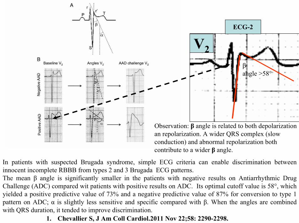
ECG-2
V2βangle gt58deg
Observation β angle is related to both depolarization an repolarization A wider QRS complex (slow conduction) and abnormal repolarization both contribute to a wider β angle
1 Chevallier S J Am Coll Cardiol2011 Nov 2258 2290-2298
In patients with suspected Brugada syndrome simple ECG criteria can enable discrimination between innocent incomplete RBBB from types 2 and 3 Brugada ECG patterns The mean β angle is significantly smaller in the patients with negative results on Antiarrhythmic Drug Challenge (ADC) compared wit patients with positive results on ADC Its optimal cutoff value is 58deg which yielded a positive predictive value of 73 and a negative predictive value of 87 for conversion to type 1 pattern on ADC α is slightly less sensitive and specific compared with β When the angles are combined with QRS duration it tended to improve discrimination
Y
aVF
Z V2
Right Sagittal Plane ECG-2
Triphasic QRS patternRSracute
β angle defined as the angle between the upslope of the S-wave and the downslope of the r-wave The mean β angle was significantly smaller in the 14 patients with negative results on AAD compared to the 24 patients with positive results on AAD Itrsquos optimal cutoff value was 58deg which yielded a positive predictive value of 73 and a negative predictive value of 87 for conversion to type 1 pattern on AAD
ST segment with saddleback appearance followed by positive or biphasic T wave in V1-V2 or from V1 through V3
β
V5V5
QRSd= 122ms QRSd= 110ms
ECG-1 ECG-2
Left precordial terminal QRS notching is more prevalent in malignant early repolarization abnormalities
Left precordial terminal QRS notching
1 Merchant FM et al Am J Cardiol 2009 Nov 15 1041402-1406
V2
V2
V3
V3
ECG-1
ECG-2
ECG-2
Dynamic inconstant or dramatic changes in repolarization are suggestive of malignant early repolarization abnormalities
1 Haiumlssaguerre M et al J Cardiovasc Electrophysiol 2009 Jan2093-98
ECG-1
Conclusion this patient has high possibility of concomitant Brugada syndrome and early repolarization In this a benign ERP or malignant early repolarization abnormalities We think it is very unlikely that is a Brugada phenocopyIn this case it is necessary additionally to perform
1 IV Ajmaline test2 ECG-AR3 Family study ( first degree relatives interrogatory physical ECGs)4 Magnetic resonance image5 Genetic screening
Ventricular biopsy is not considered mandatory but is recommended especially when it may clarify the nature of dubious findings identified with others test In approximately 6--10 of survivors of cardiac arrest no cardiac abnormality can be identified despite extensive clinical evaluation Autopsy data confirm that in a similar percentage of victims of SCD no structural heart disease or or mors sine materia can be identified at post mortem evaluation Incompletely penetrant genetic defects may underlie at least some of these unexplained deaths (1)
The ERP probably represents part of a spectrum of cardiovascular anomalies related to channelopaties including BrS IVF congenital short QT syndrome and that it may also have a molecular genetic origin of variable penetrance The denomination Early Repolarization Pattern is appropriate since it indicates that it is characterized by a given ECG pattern
1 Priori SG et al Cardiovasc Res 2001 May50218-223
Case Report similar
A 20 year-old male mulatto professional soccer player from Santo Andreacute Sao Paulo Brazil He didnrsquot finishthe second year in elementary school CatholicProfession Professional soccer playerReason for consultation Presented to consultation due to two presyncopal episodes and one syncopal episode in the last 30 days The first two episodes happened during a post-defecation period Both were preceded by dizziness nausea and vomiting The syncopal episodes occurred at rest with no prodromes The third one with express and brief loss of consciousness and sphincter relaxation at dawn 48 hours agoHis prior personal medical history was unremarkable and he successfully passed two prior periodical clinical-cardiovascular evaluations (twice a year always normal) Pre-competitive examination included 12-lead ECG echocardiogram and cardiopulmonary metabolic exercise testing (CMET) All normals Intense training for the last 3 years Familial Background One uncle from his fatherrsquos side family died of sudden cardiac death when he was 35
years old He ignores the cause His brother is being seeing by a cardiologist due to ldquocardiac arrhythmiardquo in treatment with a cardiologistfrom the Institute of the Heart from Satildeo Paulo (he cannot tell which is the cause) He denies having other relevant background details Pysical cardiovascular examination revealed bradycardia at 45 bpm with physiological sinus arrhythmia blood pressure 11070 mmHg The rest of the physical examination was unremarkable He denied consuming any type of neither medications nor drugs No addictionsHis first 12-lead ECGVectorcardiogram(VCG) (Fig 1) showed sinus bradycardia at 44 bpm P-wave axis of +60deg PR interval of 186ms QRS duration of 111ms QRS axis at + 63deg QT interval 427ms and corrected QTc 367ms There is a notch on the terminal portion of the QRS complex in the left precordial leads voltage criteria for left ventricular hypertrophy (SV1 + RV5 gt 35 mm ndash Sokolow index) prominent J-wave and concave ST-segment elevation in inferior leads V4 V5 and V6 Deep and narrow Q-waves in the left precordial leads V5 and V6 All these features were considered as an Early Repolarization Pattern (ERP)
Name JVS Gender Male Age 20 yo Ethnic Group Mulatto Weight 66 Kg Height 172 m Biotype Normoline Date 11262009 Profession competitive athlete (soccer player)
ECG diagnosis Sinus bradycardia (SB) HR 44bpm P axis +60deg PR 186ms QRSd 111ms QRS axis + 63deg QT 427ms QTc 367ms Conclusion SB + Early Repolarization Pattern (ERP) Notching or slurring of the terminal QRS complex voltage criteria of left ventricular hypertrophy SV1+ RV5 gt 35 mm (Sokolow index) prominent J wave and ST-segment elevation concave to the topending in a positive T wave from V4 to V6 Precordial greater than limb leads Relatively deep but narrow q waves in the left precordial leads (V5 and V6)
V6V4 V5
Prominent J wave and ST- segment elevation concave to the top ending in a positive T wave from V4 to V6 Notching or slurring morphology of the terminal portion of the R wave (J-wave) that produces a positive hump known as a J-wave (Arrows)
aVR
Eventually reciprocal ST segment depression confined only in Lead aVR1 observed in this particular case report
1 Mehta M Jain AC Mehta A Early Repolarization Clin Cardiol 1999 Feb 22 59-65
ECGVCG CORRELATIONS HORIZONTAL PLANE
V6
V1
V2
V3
X
Z
V5
V4
T
0J T
BEGINNING AND END OF QRS LOOP IN HP
J POINT in vectorcardiography it corresponds to 3 elements end of ventricular depolarization (QRS loop) beginning of repolarization (ST segment) when it does not present depression or elevation and T wave onset In this case 0 point and J point are not coincident In situations where there is depression or elevation of ST segment the J point does not coincide with the 0 point and the greater or lesser distance between both points indicate the greater or lesser ST segment elevation or depression The phenomenon is observed in early repolarization pattern acute phase of myocardial infarction Prinzmetal variant angina pericarditis Brugada syndrome arrhythmogenic right ventricular dysplasia etc
0 POINTIt corresponds to the QRS looponset (because PR segment does notexist) Alsocorrespond to the end of biatrialchamberactivation
T
J POINT
ECGVCG CORRELATIONS FORNTAL PLANE
aVR aVL
I
IIIII
X
Y
VERTICAL AXIS ON FRONTAL PLANE The frontal plane QRS axis and ST segment axis and T wave axis are all in the same direction THE ST
SEGMENT ELEVATION
USUALLY lt 05 mm IN THE LIMB
LEADS
YVF
Z V2
T
ECGVCG CORRELATIONS RIGHT SAGITTAL PLANE
A second 12-lead ECG (Fig 2) preformed 72 hours later showing1) Sinus bradycardia at 44 bpm2) Similar p-wave axis and intervals duration3) Dramatic modification of repolarizaccedilatildeo pattern in the right precordial leads A coved-type
(type 1) ECG brugada pattern can be distinguished4) Accentuation of j-waves and st segment elevation across the precordial leads was observed5) ST segment elevation on avr lead
See next slide
Name JVS Gender Male Age 20 yo Ethnic Group MulattoWeight 66 Kg Height 172 m Biotype Normoline Date 11292009Profession Competitive Soccer Player FIGURE 2
ECG diagnosis Sinus Arrhythmia(SA) sinus bradycardia (SB) HR 44bpm P axis +60deg PR 186ms QRSd 111ms QT 427ms QTc 367 Conclusion SA + SB + Early Repolarization Pattern (ERP) Type 1 Brugada ECG pattern Dramatic accentuation of J waves across the precordial and limb leads Transient augmentation of global J waves may be indicative of a
highly arrhythmogenic substrate heralding multiple episodes of VF in patients with ERP
aVR LEAD
11262009 11292009
DEPRESSION
ELEVATION
ST SEGMENT DEPRESSION IN aVR = 1mm ST SEGMENT ELEVATION IN aVR = 2mm
V1 V2V2
1129200911262009
V1
11292009 11262009
SPONTANEOUS MODIFICATION OF REPOLARIZATION TO TYPE 1 BRUGADA ECG PATTERN
11262009
V6
11292009V6
ST segment elevation lt 2mm ST segment elevation gt 2mm
11292009
V5
11262009
V5
ST segment elevation = 1mm ST segment elevation = 25mm
EARLY REPOLARIZATION PATTERN CONCEPT
DEFINITION Early Repolarization Pattern (ERP) is a well-recognized Enigmatic idiopathic electrocardiographic phenomenon characterized by notching or slurring morphology of the terminal portion of the R wave (J-wave) that produces a positive hump known as a J-wave at the end of the QRS complex and beginning of the ST segment It considered to be present when at least two adjacent precordial leads show elevation of the ST segment with values ge01 mV (or 1mm) above baseline followed by concordant tall near symmetric (T waves having similarity in shape ascending and descending ramps speeds)
EPIDEMIOLOGY ERP occur in 2 to 5 of the population often in men young adults afro-descendents and competitive athletes and in up to 131 to 482 of patients presenting with chest pain in the emergency rooms and coronary care units There is a distinct J wave and ST segment concave upward in the left precordial leads V4 through V6 The ST elevation is most frequently evident in ECG lead V4 The ST segment elevation is usually le2mm (precordial greater than limb leads) and is lt 05 mm in the limb leadsReciprocal ST-segment changes (ST-segment depression) are eventually confined the unipolar aVR lead (Absence of reciprocal changes or mirror image) In ERP vertical QRS axis electrical is the rule The frontal plane QRS axis the ST segment axis and T wave axis are all in the same directionThis ECG pattern has been considered a benign normal ECG variant for over six decades3
1 Hasbak P Engelmann MD Early repolarization ST-segment elevation as a normal electrocardiographic variant Ugeskr Laeger 2000 162 5928-5929
2 Brady WJ Chan TC Electrocardiographic manifestations benign early repolarization J Emerg Med 1999 17473-478
3 Benito B Guasch E Rivard L Nattel S Clinical and mechanistic issues in early repolarization of normal variants and lethal arrhythmia syndromes J Am Coll Cardiol 2010 Oct 5 56 1177-1186
MAIN ELECTROCARDIOGRAPHIC FEATURES IN ERPHeart rate Predominant sinus bradycardia frequently associated with respiratory sinus arrhythmia
PR interval First degree AV block is observed in 5 to 39 among professional athletes In the non-athlete population it is observed in 065 of cases QRS axis Vertical electrical axis is the rule The frontal plane QRS axis and ST segment axis and T wave axis are all in the same directionQRS duration (90 plusmn 10 ms) subjects than in age-matched healthy controls (80 plusmn 10 ms) QRS morphology Notching or slurring of the terminal QRS complex Prominent relatively deep but narrow q waves may appear in the left precordial leadsQRS voltage Eventually voltage criteria of LVH are observed in male competitive athletes SV1+ RV5 gt 35 mm (Positive Sokolow index) High QRS voltage is more frequent in male athletes but its correlation with LVH is low Voltage decreases after deconditioning is slow The distinction between the physiological athletes heart and pathological conditions has critical implications for professional athletes An abrupt transition may occur from right-oriented complexes to left-oriented complexes in the precordial leads secondary to counterclockwise rotation on precordial leads About two-thirds of clockwise rotation and counterclockwise rotation could be attributed to the septal angle by anatomical rotation of the heart in one plane around the long axis but other factors appear to be responsible for such ECG findings in the remaining one-third of cases Relatively higher positions of the precordial ECG leads as observed in the vertical heart appeared to be responsible for clockwise rotation in some patients and left septal fascicular block is suspected to be responsible for counterclockwise rotation in others patientsJ-point elevation Notching irregular or slurring contour of the terminal QRS complex (J point) Variant of Osborn wave is seen in ERP
ST segment Widespread ST segment elevation (precordial greater than limb leads) The characteristic ST segment is elevated upward concave confined more frequently in precordial leads with reciprocal depression only in aVR The concavity is observed at the initial up-sloping portion of ST segment or upwardly concave ST segment morphology Unfortunately concave ST morphology cannot be used to rule out ST elevation from AMI with left anterior descending coronary occlusion because it is common in these circumstances The ST elevation is most frequently evident in ECG lead V4 There is a distinct J wave and ST segment in the left precordial leads V4 through V6 The ST elevation in ERP is usually lt 2 mm (but can rarely be gt 5 mm) in the precordial leads and the greatest ST elevation is usually seen in the mid-to-left precordial leads The ST segment elevation is usually lt 05 mm in the limb leads
T wave characteristics Concordant T waves of large amplitude (prominent matching T waves) typical pseudo-asymmetrical (lsquosymmetroidrdquo) or slightly asymmetrical matching T waves often of large amplitude upright tall and peaked most conspicuously from V2 to V4 or V5 sometimes seen in leads DII DIII and aVF as a rule T waves may appear as of large amplitude peaked or pointed symmetric and matching Vagotonic or high T wave voltages followed by U waves are frequent when sinus bradycardia is present Tall positive ad symmetric or symmetroid T waves are not only seen occasionally in the very early stages of MI but also in hyperkalemia and in ERP sinus bradycardia
QT intervals QT maximum The maximum Q-onset-T-end interval This parameter is higher in ERV subjects than in normal controls
QTp maximum maximum Q-onset-T-peak interval This parameter is higher in ERV subjects than in age-matched healthy controls
Rate-corrected QTc maximum This parameter is lower in ERP subjects than in age-matched healthy controls
QTpc maximum This parameter is lower in ERV subjects than in age-matched healthy controls
U wave because bradycardia U waves are frequent in ERV they are best observed in the V3 lead U waves are frequent when sinus bradycardia is present
Other ECG characteristics of ERPRelative temporal stability of the ST segment and T wave pattern is observedReciprocal changes are not seen in ERP There are no evolutionary short-term changes in the ST segment and T waves and Q waves do not appear
Discusions Early Repolarization Pattern (ERP) or Early Repolarization Variant (ERV) is an enigmati
electrocardiographic phenomenon characterized by prominent J wave and ST-segment elevation in multiplleads
Recently there has been renewed interest in ERP because of similarities to the arrhythmogenic Brugadsyndrome (BrS) Not much is known about the epidemiology of ERP and several studies have reported thathis condition is associated with a good prognosis
Both ERP and BrS exhibit some similarities including the ionic underlying mechanism the analogouresponses to changes in heart rate and autonomic tone sympathicomimetics (isoproterenol test) as well as insodium channel and beta-blockers These observations raise the hypothesis that ERP may be not as benign atraditionally believed Additionally there are documents showing that ST-segment height in the man igreatly influenced by central sympathetic nervous activity both at baseline and during physiologic andpharmacological stress Central sympathetic dysfunction regularly results in multilead ST-segment elevationor J wave that decreases or below isoelectric baseline during low dose isoproterenol infusion An earlyrepolarization pattern in the inferior leads of a standard electrocardiogram is associated with an increased riskof death from cardiac causes in middle-aged subjects1
POSSIBLE SIMILARITIES BETWEEN ERV AND BrSbullMore frequent in malesbullBoth occur more frequently in young adults and in individuals without apparent structural heart diseasebullBoth may influence just the V1-V2 leads Rarely (9) can ST elevation be observed in ERP only in the right precordial leads V1-V2 or in the inferior ones2
1 Tikkanen JT Anttonen O Junttila MJ Aro AL Kerola T Rissanen HA Reunanen A Huikuri HV Long-Term Outcome Associated with Early Repolarization on Electrocardiography N Engl J Med 2009 Nov 16 [Epubahead of print]
2 Hasbak P Engelmann MDEarly repolarization ST-segment elevation as a normal electrocardiographic variant Ugeskr Laeger 2000 162 5928-5929
When ST elevation is normal it can reach up to 3 mm in V2-V3 especially in young people In those individuals over 40 years it seldom exceeds 2 mm Both can show incomplete RBBB pattern or right bundle branch conduction disorder in BrS it can present atypical features RBBB-like and of the saddle type by exclusive elevation of the J point S wave with delay in the left leads DI aVL V5 and V6 could be absent as it is to be expected in a classic RBBB The elements considered as typical in BrS are 1) elevation of the terminal part of QRS (prominent J wave) 2) elevated and descending ST not related to lesion of ischemic (idiopathic) injury 3) negative T wave in the right precordial leads 4) normal QTc or near normal 5) Eventually absence of final delay in left leads as it would be expected in a classic RBBB1 In ERV when associated to athletersquos heart QRS can present a moderate extension (100 ms to 110 ms) in 15 of the cases which in nonathlete normal population in a 24 is called outflow tract hypertrophy In this case r does not exceed the 5 mm and is lower than S in the same lead rSrBoth may improve repolarization during the stress test with use of isoproterenolBoth respond to a shortening of AP phase 2 in a part of ventricular thickness and intensification of fast repolarization notch (phase 1) mediated by transmural dispersion of ventricular repolarization by a larger notch in the Ito channel2The alteration of the Ito and ICa2+-L channels in BrS and in ERP are the electrophysiologic substrate that explains the J point and ST segment elevation because they cause the intensified notch in phase 1 and suppression in phase 2 duration in the epicardium and in the endocardium of ventricular wall thickness
1 Hiss RG Lamb LE Electrocardiographic findings in 122043 individuals Circulation 1962 25947-9612 Antzelevitch Ch Xin Yan G Shimuzi W et al Electrical heterogeneity the ECG and Cardiac Arrhythmias In Zipes
DP Jalife J Cardiac Electrophysiology From Cell to Bedside Third Edition WB Saunders Company2000 Chapter 26 p 222-238
ELEMENTS FOR DIFFERENTIAL DIAGNOSIS BETWEEN ERP AND BrS
I) Family background ERP negativeBrS frequently positive
II) Ethnic GroupERP predominantly in African descendents(1) or equally common in all races(2) BrS predominantly in Asian group (58) and Caucasian people(3)
III) GenderERP male gender predominanceBrS great predominance in the male gender (malefemale ratio ndash 81 in non Asian and 101 in Asian
peopleIV) Response to IC group antiarrhythmic agents
BrS flecainide used in a 10 mgKg dosage in 10 minutes increases ST elevation and QRS duration in a more significant way in patients with BrS than in individuals without the entity and only in those it triggers ventricular extrasystoles(4)
ERP it can induce a pattern similar to BrS however the degree of ST elevation caused by the drug is much higher in patients with BrS than in patients without the disease
1 Grusin H Peculiarities of the Africanrsquos electrocardiogram and the changes observed in serial studies Circulation 1954 9 860-867
2 Mehta M Jain AC Mehta A Early Repolarization Clin Cardiol 1999 Feb2259-653 Nademanee K Veerakul G Nimmannit S Chaowakul V Bhuripanyo K Likittanasombat K Tunsanga K Kuasirikul S
Malasit P Tansupasawadikul S Tatsanavivat P Arrhytmogenic marker for the sudden unexplained death syndrome in Thail men Circulation 1997 962595-2600
4 Shimizu W Antzelevitch C Suyama K Kurita T Taguchi A Aihara N Takaki H Sunagawa K Kamakura S Effect of sodium channel blockers on ST segment QRS duration and corrected QT interval in patients with Brugada syndromeJ Cardiovasc Electrophysiol 2000 111320-1329
BrS patients with Inferior and lateral ECG repolarization abnormalities have longer baseline PR intervals(1)Both an S wave width ge80ms in V1 and ST elevation ge18mm in V2 are highly specific indicators of VF and criteria for high-risk BrS(2)There are several references in literature to patients with elevation of the J point and ST segment convex to the top or straight descendent in inferior leads (Brugada sign) or concomitantly ST-segment elevation in the right precordial and inferior leads II III and aVF(3) in absence of hypothermia ischemia or electrolytic disorders which we call ldquoatypical Brugada patternrdquo atypical BrS variant BrS or BrS variant or idiopathic VF (a variant of the BrS with ST-segment elevation in inferior leads(4)Pilsicainide may induce PVCs and PVT in atypical BrS and the infusion of isoproterenol suppress the arrhythmias and normalize the ST-segment elevation(5)
1 Sarkozy A Chierchia GB Paparella G Boussy T De Asmundis C Roos M Henkens S Kaufman L Buyl R Brugada R Brugada J Brugada P Inferior and lateral electrocardiographic repolarization abnormalities in Brugada syndromeCirc ArrhythmElectrophysiol 2009 Apr 2154-161
2 Atarashi H Ogawa S For The Idiopathic Ventricular Fibrillation Investigators New ECG Criteria for High-Risk Brugada Syndrome Circ J 2003 Jan 67 8-10
3 Lombardi F Potenza S Beltrami A Verzoni A Brugada P Brugada RSimultaneous ST-segment elevation in the right precordial and inferior leads in Brugada syndrome J CardiovascMed (Hagerstown) 2007 8 201-204
4 Letsas KP Efremidis M Pappas LK Gavrielatos G Markou V Sideris A Kardaras FEarlyrepolarization syndrome is it always benign Int J Cardiol 2007114 390-392
5 Chinushi M Izumi D Furushima H Watanabe H Aizawa YMultiple premature beats triggered ventricular arrhythmias during pilsicainide infusion in a patient with inferior ST-segment elevation Pacing Clin Electrophysiol 2006 29 1445-1448
Potet et al(1) identified a G752R mutation on SNC5A that produced ST segment elevation and prominent J wave in leads II III and aVF The authors provide genetic demonstration that Brugada ECG anomalies related to a unique SCN5A mutation can be observed either in the inferior or the right precordial leads The early repolarization pattern in inferolateral leads is not an uncommon finding in BrS and this pattern is not associated with a worse outcome in subjects with BrS(2) The spontaneous ERP occurred more frequently among patients with BrS than in 283 family members not having BrS (11 versus 6 P=003) Class I antiarrhythmic drug administration provoked inferior-lateral coved Brugada pattern in 13 patients with BrS These patients had longer baseline PR intervals and Class I antiarrhythmic drug induced QRS interval prolongation (108 to 178 versus 102 ms to 131 ms Plt0001) In 3 patients the Class I antiarrhythmic drug provoked coved Brugada pattern only present in the inferior leads Inferior-lateral ERP occurs spontaneously relatively frequently in BrS These patients have a more severe phenotype Class I antiarrhythmic drug administration provokes inferior-lateral coved Brugada pattern in 46 of patients The Class I antiarrhythmic drug exceptionally provoked coved Brugada pattern only observed in the inferior leads(3)
1 Potet F Mabo P Le Coq G et al Novel brugada SCN5A mutation leading to ST segment elevation in the inferior or the right precordial leads J Cardiovasc Electrophysiol 2003 142000-2003
2 Letsas KP Sacher F Probst V Weber R Knecht S Kalusche D Haiumlssaguerre M ArentzTPrevalence of early repolarization pattern in inferolateral leads in patients with Brugada syndrome Heart Rhythm 2008 Dec 5 1685-1689
3 Sarkozy A Chierchia GB Paparella G Boussy T De Asmundis C Roos M Henkens S Kaufman L Buyl R Brugada R Brugada J Brugada P Inferior and lateral electrocardiographic repolarization abnormalities in Brugada syndrome Circ Arrhythm Electrophysiol 2009 Apr2 154-161
Type 1 ECG Brugada pattern in the peripheral leads was observed in 42 of patients during ajmaline test (103 of positive tests) and was associated with longer QRS and greater QTc prolongation compared with the rest of the patients1Atypical Brugada Syndrome or IVF with J waves in inferior lateral or inferior lateral leads
and Familial Idiopathic Ventricular Fibrillation and BrSSeveral mutations in genes SCN5A KCNJ8 DPP6 SCN3B and CACNA2D1 had been identified 1 On SCN5A gene2 3 Observation We do not believe that SCN5A should be attributed to IVF because this report clearly shows conduction defects (as expected) so this is not IVF but conduction delay at different levels in the heart2 On KCNJ8 gene 4 5 Missense variant in exon 3 (NC-000012) of the KCNJ8 gene a subunit of the K(ATP)channel23 Genomic DNA sequencing of K(ATP) channel genes showed missense variant in exon 3 (NC_000012) of the KCNJ8 gene a subunit of the K(ATP) channel conferring predisposition to dramatic repolarization changes and ventricular vulnerability
1 Batchvarov VN Govindan M Camm AJ Behr ERBrugada-like changes in the peripheral leads during diagnostic ajmaline test in patients with suspected Brugada syndrome Pacing Clin Electrophysiol 2009 Jun32695-703
2 Potet F Mabo P Le Coq G Probst V Schott JJ Airaud F Guihard G Daubert JC Escande D Le MarecH Novel brugada SCN5A mutation leading to ST segment elevation in the inferior or the right precordial leads J Cardiovasc Electrophysiol 2003 14 200-203
3 Akai J Makita N Sakurada H Shirai N Ueda K Kitabatake A Nakazawa K Kimura A Hiraoka M A novel SCN5A mutation associated with idiopathic ventricular fibrillation without typical ECG findings of Brugada syndrome FEBS Lett 2000 Aug 11 479 29-34
4 Haissaguerre M Derval N Sacher F Jesel L Deisenhofer I de Roy Let al Sudden cardiac arrest associated with early repolarization N Engl J Med 2008 May 8 3582016-2023
5 Haiumlssaguerre M Sacher F Nogami A Komiya N Bernard A Probst V et al Characteristics of recurrent ventricular fibrillation associated with inferolateral early repolarization role of drug therapy J Am CollCardiol 2009 Feb 17 53 612-619
On DPP6 gene1 Dipeptidyl aminopeptidase-like protein 6 locus on chromosome7q26 Voltage-gated potassium channels OMIM126141 The ECG shows rapid PVT Monomorphic PVCs with very short coupling intervals The PVC (302 +- 52 msec) within 40 msec of the peak of the preceding Absence of T wave Pause-dependence
Valdivia et al2 reported a case with IVF and a novel mutation in the SCN3B-encoded sodium channel beta subunit Navbeta3 that causes a loss of function of Nav15 channels in vitro and provides molecular and cellular evidence implicating mutations in Navbeta3 as a cause of IVF Mutation in this SCN3B gene causes also BrS type 73
L-type calcium channel (LTCC) mutations have been associated with BrS SQTS and Timothy syndrome (LQT8) Little is known about the extent to which LTCC mutations contribute to the J-wave syndromes associated with SCD Mutations in the LTCCs are detected in a high percentage of probands with J-wave syndromes associated with inherited cardiac arrhythmias suggesting that genetic screening of Ca(v) genes may be a valuable diagnostic tool in identifying individuals at risk Burashnikov et al4 identify CACNA2D1 as a novel BrS susceptibility gene and CACNA1C CACNB2 and CACNA2D1 as possible novel ERS susceptibility genes4
1 Alders M Koopmann TT Christiaans I Postema PG Beekman L Tanck MW et al Haplotype-sharing analysis implicates chromosome 7q36 harboring DPP6 in familial idiopathic ventricular fibrillation Am J Hum Genet 2009 Apr 84 468-476
2 Valdivia CR Medeiros-Domingo A Ye B Shen WK Algiers TJ Ackerman MJ et al Loss-of-function mutation of the SCN3B-encoded sodium channel beta3 subunit associated with a case ofidiopathic ventricular fibrillation Cardiovasc Res 2010 Jun 1 86 392-400
3 Hu D Barajas-Martinez H Burashnikov E Springer M Wu Y Varro A et al A mutation in the beta-3 subunit of the cardiac sodium channel associated with Brugada ECG phenotype Circ Cardiovasc Genet 2009 2 270-278
4 Burashnikov E Pfeiffer R Barajas-Martinez H Delpoacuten E Hu D Desai M et al Mutations in the cardiac L-type calcium channel associated with inherited J-wave syndromes and sudden cardiac death Heart Rhythm 2010 Dec71872-1882
From a multicenter cohort of 122 patients (90 male age 37 +- 12 years) with IVF and ERP in the inferolateral leadsHaiumlssaguerre et al1 selected all patients with more than 3 episodes of VF (multiple) including those with electrical storms (ge3 VF in 24 h) Multiple recurrences of VF occurred in 27 of patients with ERP and may be life threatening Isoproterenol in acute cases and quinidine in chronic cases are effective antyharrhithmic drugsThe last one is necessary associated to ICDThe so-called atypical BrS is characterized by ECG abnormalities of the J wave and ST-segment elevation appeared in the inferior andor lateral leads The ERP in inferolateral leads is not an uncommon finding in BrS2 There is a high incidence of the ERP confined in inferolateral leads in patients with IVF The ECGs have an elevation of the QRS-ST junction of at least 01 mV from baseline in the inferior or lateral lead manifested as QRS slurring or notching Among patients with a history of IVF there is an increased prevalence of ERP
1 Haissaguerre M Derval N Sacher F Jesel L Deisenhofer I de Roy L Pasquieacute JL Nogami A Babuty D Yli-Mayry S De Chillou C Scanu P Mabo P Matsuo S Probst V Le Scouarnec S Defaye P Schlaepfer J Rostock T Lacroix D Lamaison D Lavergne T Aizawa Y Englund A Anselme F ONeill M Hocini M Lim KT Knecht S Veenhuyzen GD Bordachar P Chauvin M Jais P Coureau G Chene G Klein GJ Cleacutementy J Sudden cardiac arrest associated with early repolarization N Engl J Med 2008 May 8 3582016-2023
2 Letsas KP Sacher F Probst V Weber R Knecht S Kalusche D Haiumlssaguerre M Arentz T Prevalence of early repolarization pattern in inferolateral leads in patients with Brugada syndrome Heart Rhythm 2008 Dec51685-1689
Because a contingent percentage has not yet been well established it has been suggested that BrS constitutes the same entity in a large number of IVF diagnosed cases since they have common clinical features
1 BOTH ARE MORE FREQUENT IN MEN2 BOTH HAPPEN IN MIDDLE-AGE PEOPLE3 BOTH OCCUR IN APPARENT STRUCTURALLY NORMAL HEARTS14 BOTH CAN AFFECT THE SAME GENE (SCN5A)5 BOTH AFFECT EVENTUALLY THE SAME LOCUS (3p24-p21)
Wellens et al2 has pointed out that in order to be accurate about such a percentage it is important to define the diagnostic criteria used to classify BrS in IVF patients Curiously the so-called Idiopathic VT in apparently healthy hearts without structural heart disease and primary electrical disease also affects the 3p21 locus but not 3p24 and the gene is different GNA12
Survivors of SCD by VF without structural heart disease and a normal coronary angiogram are often diagnosed with IVTIVF3
A careful analysis of the ECG is essential in the differential diagnosis of this type of apparently unexplained SCD because intermittent or permanent RBBB with J point and ST-segment elevation in leads V1 to V3 is now classified as BrS
1 Goethals P Debruyne P Saffarian M Drug-induced Brugada syndrome Acta Cardiol1998 53157-60
2 Wellens HJJ Rodriquez LM Smeets JL Ventricular tachycardia in structural normal hearts In Zipes DP Jalife J Editors Cardiac Electrophysiology from cell tobedside Philadelphia 1995 WB Saunders
3 Chen Q Kirsch GE Zhang D Brugada R Brugada P Brugada J et al Genetic basis and molecular mechanisms for idiopathic ventricular fibrillation Nature 1998392 293-296
DIAGNOSTICS TRACKS TO DIFFERENTIATE BENIGN ERP FROM MALIGNANT EARLY REPOLATIZATION ABNORMALITIES
BENIGN ERP MALIGNANT EARLY REPOLARIZATION ABNORMALITIES
Family history of unexplained SCD in young relatives(lt45yo)
Absent Possible however infrequent1
Personal history Asymptomatic Asymptomatic repetitive syncope episodes or recovered of cardiac arrest
Left precordial terminal QRS notching Less prevalent More prevalent2
STT ratio in lead V6calculated by dividing the millimeters of
ST-segment elevation by the millimeters to the tallest point of the T wave
Each value is measured from the isoelectric point
lt0253 Non-tested
1 Viskin S Belhassen B Idiopathic ventricular fibrillation American Heart Journal 1990 120 661-671
2 Tikkanen JT Anttonen O Junttila MJ Aro AL Kerola T Rissanen HA Reunanen A et al Long-term outcome associated with early repolarization on electrocardiography N Engl J Med 2009 Dec 24361 2529-2537
3 Ginzton LE Laks MM The differential diagnosis of acute pericarditis from the normal variant new electrocardiographic criteria Circulation 1982 May 65 1004-1009
ST-segment elevation shape Concave upward Convex upwardJ waves are present in asymp 30 IVF patients1
Early repolarization is significantly prevalent in the subjects after cardiac arrest
(present in 31 of those resuscitated )Eventually ldquoLambda-waverdquo 23
J-point elevation and ST-segment degree Usually lt 2 mmlt05mm in limb leads
Frequent gt2mmJ-point elevation gt 2mm in inferior leads is associated with an increased risk of death
from cardiac causes in middle-aged subjects3
T wave characteristic Positive large T wave from V2 to V4 or V5 Concordant
Frequent negative discordant
Reciprocal ST-segment changes (Mirror
image)Absent With exception aVR5 Frequently Present3
1 Viskin S Idiopathic ventricular fibrillation Le Syndrome dHaiumlssaguerre and the fear of J waves J Am Coll Cardiol 2009 Feb 17 53 620-622
2 Gussak I Bjerregaard P Kostis J Electrocardiographic lambda wave and primary idiopathic cardiac asystole a new clinical syndrome J Electrocardiol 2004 Apr 37 105-107
3 Riera AR Ferreira C Schapachnik E Sanches PC Moffa PJ Brugada syndrome with atypical ECG downsloping ST-segment elevation in inferior leads J Electrocardiol 2004 Apr37101-104
4 Tikkanen JT Anttonen O Junttila MJ Aro AL Kerola T Rissanen HA Reunanen A et al Long-term outcome associated with early repolarization on electrocardiography N Engl J Med 2009 Dec 24361 2529-2537
5 Mehta M Jain AC Mehta A Early Repolarization Clin Cardiol 1999 Feb 22 59-65
Monomorphic PVCs with very short coupling
(250 to 350ms)
Absent Characteristic1 Pause-dependency absent2
QTc Interval Normal Minimally short in 35 among masculine gender
(Between 340 to 360ms3)
Late Potentials(LPs) Absent Frequent if J wave presentShowing circadian variation with night
ascendancy4
1 Alders M Koopmann TT Christiaans I Postema PG Beekman L Tanck MW et al Haplotype-sharing analysis implicates chromosome 7q36 harboring DPP6 in familial idiopathic ventricular fibrillation Am J Hum Genet 2009 Apr 84 468-476
2 Viskin S Lesh MD Eldar M Fish R Setbon I Laniado S et al Mode of onset of malignant ventricular arrhythmias in idiopathic ventricular fibrillation J Cardiovasc Electrophysiol 1997 Oct 8 1115-11 20
3 Viskin S Zeltser D Ish-Shalom M Katz A Glikson M Justo D et al Is idiopathic ventricular fibrillation a short QT syndrome Comparison of QT intervals of patients with idiopathic ventricular fibrillation and healthy controls Heart Rhythm 2004 Nov 1 587-591
4 Abe A Ikeda T Tsukada T Ishiguro H Miwa Y Miyakoshi M Circadian variation of latepotentials in idiopathic ventricular fibrillation associated with J waves insights into alternative pathophysiology and risk stratification Heart Rhythm 2010 May 7 675-682
ConclusionIn spite of both malignant early repolarization abnormalities of IVF and benign early repolarization pattern being able to cause J point and ST segment elevation the characteristics of both are clearly different to the point of a differentiation being quite simple when ECGs are analyzed by experts Any ER should raise the suspicion of IVF when it presents ST segment elevation ge 2 mm mainly in inferior wall superior convexity eventually lambda wave shape pattern of dynamic presentation inconstant with dramatic intensity of ST segment elevation discordant T wave polarity related ST segment shift presence of reciprocal ST-segment changes (mirror image) tendency to discrete short QTc interval in male gender(ge340ms) premature monomorphic ventricular contractions with very short coupling interval(250 to 350ms) not related with exercise and late potentials with circadian variation and night ascendancy on SA-ECG Contrarily in benign ERP ST elevation observed in the mid-to-left precordial leads is usually le 2 mm and lt05 mm in the limb leads and concave to the top followed by a positive concordant tall T wave
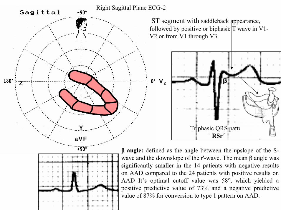
Y
aVF
Z V2
Right Sagittal Plane ECG-2
Triphasic QRS patternRSracute
β angle defined as the angle between the upslope of the S-wave and the downslope of the r-wave The mean β angle was significantly smaller in the 14 patients with negative results on AAD compared to the 24 patients with positive results on AAD Itrsquos optimal cutoff value was 58deg which yielded a positive predictive value of 73 and a negative predictive value of 87 for conversion to type 1 pattern on AAD
ST segment with saddleback appearance followed by positive or biphasic T wave in V1-V2 or from V1 through V3
β
V5V5
QRSd= 122ms QRSd= 110ms
ECG-1 ECG-2
Left precordial terminal QRS notching is more prevalent in malignant early repolarization abnormalities
Left precordial terminal QRS notching
1 Merchant FM et al Am J Cardiol 2009 Nov 15 1041402-1406
V2
V2
V3
V3
ECG-1
ECG-2
ECG-2
Dynamic inconstant or dramatic changes in repolarization are suggestive of malignant early repolarization abnormalities
1 Haiumlssaguerre M et al J Cardiovasc Electrophysiol 2009 Jan2093-98
ECG-1
Conclusion this patient has high possibility of concomitant Brugada syndrome and early repolarization In this a benign ERP or malignant early repolarization abnormalities We think it is very unlikely that is a Brugada phenocopyIn this case it is necessary additionally to perform
1 IV Ajmaline test2 ECG-AR3 Family study ( first degree relatives interrogatory physical ECGs)4 Magnetic resonance image5 Genetic screening
Ventricular biopsy is not considered mandatory but is recommended especially when it may clarify the nature of dubious findings identified with others test In approximately 6--10 of survivors of cardiac arrest no cardiac abnormality can be identified despite extensive clinical evaluation Autopsy data confirm that in a similar percentage of victims of SCD no structural heart disease or or mors sine materia can be identified at post mortem evaluation Incompletely penetrant genetic defects may underlie at least some of these unexplained deaths (1)
The ERP probably represents part of a spectrum of cardiovascular anomalies related to channelopaties including BrS IVF congenital short QT syndrome and that it may also have a molecular genetic origin of variable penetrance The denomination Early Repolarization Pattern is appropriate since it indicates that it is characterized by a given ECG pattern
1 Priori SG et al Cardiovasc Res 2001 May50218-223
Case Report similar
A 20 year-old male mulatto professional soccer player from Santo Andreacute Sao Paulo Brazil He didnrsquot finishthe second year in elementary school CatholicProfession Professional soccer playerReason for consultation Presented to consultation due to two presyncopal episodes and one syncopal episode in the last 30 days The first two episodes happened during a post-defecation period Both were preceded by dizziness nausea and vomiting The syncopal episodes occurred at rest with no prodromes The third one with express and brief loss of consciousness and sphincter relaxation at dawn 48 hours agoHis prior personal medical history was unremarkable and he successfully passed two prior periodical clinical-cardiovascular evaluations (twice a year always normal) Pre-competitive examination included 12-lead ECG echocardiogram and cardiopulmonary metabolic exercise testing (CMET) All normals Intense training for the last 3 years Familial Background One uncle from his fatherrsquos side family died of sudden cardiac death when he was 35
years old He ignores the cause His brother is being seeing by a cardiologist due to ldquocardiac arrhythmiardquo in treatment with a cardiologistfrom the Institute of the Heart from Satildeo Paulo (he cannot tell which is the cause) He denies having other relevant background details Pysical cardiovascular examination revealed bradycardia at 45 bpm with physiological sinus arrhythmia blood pressure 11070 mmHg The rest of the physical examination was unremarkable He denied consuming any type of neither medications nor drugs No addictionsHis first 12-lead ECGVectorcardiogram(VCG) (Fig 1) showed sinus bradycardia at 44 bpm P-wave axis of +60deg PR interval of 186ms QRS duration of 111ms QRS axis at + 63deg QT interval 427ms and corrected QTc 367ms There is a notch on the terminal portion of the QRS complex in the left precordial leads voltage criteria for left ventricular hypertrophy (SV1 + RV5 gt 35 mm ndash Sokolow index) prominent J-wave and concave ST-segment elevation in inferior leads V4 V5 and V6 Deep and narrow Q-waves in the left precordial leads V5 and V6 All these features were considered as an Early Repolarization Pattern (ERP)
Name JVS Gender Male Age 20 yo Ethnic Group Mulatto Weight 66 Kg Height 172 m Biotype Normoline Date 11262009 Profession competitive athlete (soccer player)
ECG diagnosis Sinus bradycardia (SB) HR 44bpm P axis +60deg PR 186ms QRSd 111ms QRS axis + 63deg QT 427ms QTc 367ms Conclusion SB + Early Repolarization Pattern (ERP) Notching or slurring of the terminal QRS complex voltage criteria of left ventricular hypertrophy SV1+ RV5 gt 35 mm (Sokolow index) prominent J wave and ST-segment elevation concave to the topending in a positive T wave from V4 to V6 Precordial greater than limb leads Relatively deep but narrow q waves in the left precordial leads (V5 and V6)
V6V4 V5
Prominent J wave and ST- segment elevation concave to the top ending in a positive T wave from V4 to V6 Notching or slurring morphology of the terminal portion of the R wave (J-wave) that produces a positive hump known as a J-wave (Arrows)
aVR
Eventually reciprocal ST segment depression confined only in Lead aVR1 observed in this particular case report
1 Mehta M Jain AC Mehta A Early Repolarization Clin Cardiol 1999 Feb 22 59-65
ECGVCG CORRELATIONS HORIZONTAL PLANE
V6
V1
V2
V3
X
Z
V5
V4
T
0J T
BEGINNING AND END OF QRS LOOP IN HP
J POINT in vectorcardiography it corresponds to 3 elements end of ventricular depolarization (QRS loop) beginning of repolarization (ST segment) when it does not present depression or elevation and T wave onset In this case 0 point and J point are not coincident In situations where there is depression or elevation of ST segment the J point does not coincide with the 0 point and the greater or lesser distance between both points indicate the greater or lesser ST segment elevation or depression The phenomenon is observed in early repolarization pattern acute phase of myocardial infarction Prinzmetal variant angina pericarditis Brugada syndrome arrhythmogenic right ventricular dysplasia etc
0 POINTIt corresponds to the QRS looponset (because PR segment does notexist) Alsocorrespond to the end of biatrialchamberactivation
T
J POINT
ECGVCG CORRELATIONS FORNTAL PLANE
aVR aVL
I
IIIII
X
Y
VERTICAL AXIS ON FRONTAL PLANE The frontal plane QRS axis and ST segment axis and T wave axis are all in the same direction THE ST
SEGMENT ELEVATION
USUALLY lt 05 mm IN THE LIMB
LEADS
YVF
Z V2
T
ECGVCG CORRELATIONS RIGHT SAGITTAL PLANE
A second 12-lead ECG (Fig 2) preformed 72 hours later showing1) Sinus bradycardia at 44 bpm2) Similar p-wave axis and intervals duration3) Dramatic modification of repolarizaccedilatildeo pattern in the right precordial leads A coved-type
(type 1) ECG brugada pattern can be distinguished4) Accentuation of j-waves and st segment elevation across the precordial leads was observed5) ST segment elevation on avr lead
See next slide
Name JVS Gender Male Age 20 yo Ethnic Group MulattoWeight 66 Kg Height 172 m Biotype Normoline Date 11292009Profession Competitive Soccer Player FIGURE 2
ECG diagnosis Sinus Arrhythmia(SA) sinus bradycardia (SB) HR 44bpm P axis +60deg PR 186ms QRSd 111ms QT 427ms QTc 367 Conclusion SA + SB + Early Repolarization Pattern (ERP) Type 1 Brugada ECG pattern Dramatic accentuation of J waves across the precordial and limb leads Transient augmentation of global J waves may be indicative of a
highly arrhythmogenic substrate heralding multiple episodes of VF in patients with ERP
aVR LEAD
11262009 11292009
DEPRESSION
ELEVATION
ST SEGMENT DEPRESSION IN aVR = 1mm ST SEGMENT ELEVATION IN aVR = 2mm
V1 V2V2
1129200911262009
V1
11292009 11262009
SPONTANEOUS MODIFICATION OF REPOLARIZATION TO TYPE 1 BRUGADA ECG PATTERN
11262009
V6
11292009V6
ST segment elevation lt 2mm ST segment elevation gt 2mm
11292009
V5
11262009
V5
ST segment elevation = 1mm ST segment elevation = 25mm
EARLY REPOLARIZATION PATTERN CONCEPT
DEFINITION Early Repolarization Pattern (ERP) is a well-recognized Enigmatic idiopathic electrocardiographic phenomenon characterized by notching or slurring morphology of the terminal portion of the R wave (J-wave) that produces a positive hump known as a J-wave at the end of the QRS complex and beginning of the ST segment It considered to be present when at least two adjacent precordial leads show elevation of the ST segment with values ge01 mV (or 1mm) above baseline followed by concordant tall near symmetric (T waves having similarity in shape ascending and descending ramps speeds)
EPIDEMIOLOGY ERP occur in 2 to 5 of the population often in men young adults afro-descendents and competitive athletes and in up to 131 to 482 of patients presenting with chest pain in the emergency rooms and coronary care units There is a distinct J wave and ST segment concave upward in the left precordial leads V4 through V6 The ST elevation is most frequently evident in ECG lead V4 The ST segment elevation is usually le2mm (precordial greater than limb leads) and is lt 05 mm in the limb leadsReciprocal ST-segment changes (ST-segment depression) are eventually confined the unipolar aVR lead (Absence of reciprocal changes or mirror image) In ERP vertical QRS axis electrical is the rule The frontal plane QRS axis the ST segment axis and T wave axis are all in the same directionThis ECG pattern has been considered a benign normal ECG variant for over six decades3
1 Hasbak P Engelmann MD Early repolarization ST-segment elevation as a normal electrocardiographic variant Ugeskr Laeger 2000 162 5928-5929
2 Brady WJ Chan TC Electrocardiographic manifestations benign early repolarization J Emerg Med 1999 17473-478
3 Benito B Guasch E Rivard L Nattel S Clinical and mechanistic issues in early repolarization of normal variants and lethal arrhythmia syndromes J Am Coll Cardiol 2010 Oct 5 56 1177-1186
MAIN ELECTROCARDIOGRAPHIC FEATURES IN ERPHeart rate Predominant sinus bradycardia frequently associated with respiratory sinus arrhythmia
PR interval First degree AV block is observed in 5 to 39 among professional athletes In the non-athlete population it is observed in 065 of cases QRS axis Vertical electrical axis is the rule The frontal plane QRS axis and ST segment axis and T wave axis are all in the same directionQRS duration (90 plusmn 10 ms) subjects than in age-matched healthy controls (80 plusmn 10 ms) QRS morphology Notching or slurring of the terminal QRS complex Prominent relatively deep but narrow q waves may appear in the left precordial leadsQRS voltage Eventually voltage criteria of LVH are observed in male competitive athletes SV1+ RV5 gt 35 mm (Positive Sokolow index) High QRS voltage is more frequent in male athletes but its correlation with LVH is low Voltage decreases after deconditioning is slow The distinction between the physiological athletes heart and pathological conditions has critical implications for professional athletes An abrupt transition may occur from right-oriented complexes to left-oriented complexes in the precordial leads secondary to counterclockwise rotation on precordial leads About two-thirds of clockwise rotation and counterclockwise rotation could be attributed to the septal angle by anatomical rotation of the heart in one plane around the long axis but other factors appear to be responsible for such ECG findings in the remaining one-third of cases Relatively higher positions of the precordial ECG leads as observed in the vertical heart appeared to be responsible for clockwise rotation in some patients and left septal fascicular block is suspected to be responsible for counterclockwise rotation in others patientsJ-point elevation Notching irregular or slurring contour of the terminal QRS complex (J point) Variant of Osborn wave is seen in ERP
ST segment Widespread ST segment elevation (precordial greater than limb leads) The characteristic ST segment is elevated upward concave confined more frequently in precordial leads with reciprocal depression only in aVR The concavity is observed at the initial up-sloping portion of ST segment or upwardly concave ST segment morphology Unfortunately concave ST morphology cannot be used to rule out ST elevation from AMI with left anterior descending coronary occlusion because it is common in these circumstances The ST elevation is most frequently evident in ECG lead V4 There is a distinct J wave and ST segment in the left precordial leads V4 through V6 The ST elevation in ERP is usually lt 2 mm (but can rarely be gt 5 mm) in the precordial leads and the greatest ST elevation is usually seen in the mid-to-left precordial leads The ST segment elevation is usually lt 05 mm in the limb leads
T wave characteristics Concordant T waves of large amplitude (prominent matching T waves) typical pseudo-asymmetrical (lsquosymmetroidrdquo) or slightly asymmetrical matching T waves often of large amplitude upright tall and peaked most conspicuously from V2 to V4 or V5 sometimes seen in leads DII DIII and aVF as a rule T waves may appear as of large amplitude peaked or pointed symmetric and matching Vagotonic or high T wave voltages followed by U waves are frequent when sinus bradycardia is present Tall positive ad symmetric or symmetroid T waves are not only seen occasionally in the very early stages of MI but also in hyperkalemia and in ERP sinus bradycardia
QT intervals QT maximum The maximum Q-onset-T-end interval This parameter is higher in ERV subjects than in normal controls
QTp maximum maximum Q-onset-T-peak interval This parameter is higher in ERV subjects than in age-matched healthy controls
Rate-corrected QTc maximum This parameter is lower in ERP subjects than in age-matched healthy controls
QTpc maximum This parameter is lower in ERV subjects than in age-matched healthy controls
U wave because bradycardia U waves are frequent in ERV they are best observed in the V3 lead U waves are frequent when sinus bradycardia is present
Other ECG characteristics of ERPRelative temporal stability of the ST segment and T wave pattern is observedReciprocal changes are not seen in ERP There are no evolutionary short-term changes in the ST segment and T waves and Q waves do not appear
Discusions Early Repolarization Pattern (ERP) or Early Repolarization Variant (ERV) is an enigmati
electrocardiographic phenomenon characterized by prominent J wave and ST-segment elevation in multiplleads
Recently there has been renewed interest in ERP because of similarities to the arrhythmogenic Brugadsyndrome (BrS) Not much is known about the epidemiology of ERP and several studies have reported thathis condition is associated with a good prognosis
Both ERP and BrS exhibit some similarities including the ionic underlying mechanism the analogouresponses to changes in heart rate and autonomic tone sympathicomimetics (isoproterenol test) as well as insodium channel and beta-blockers These observations raise the hypothesis that ERP may be not as benign atraditionally believed Additionally there are documents showing that ST-segment height in the man igreatly influenced by central sympathetic nervous activity both at baseline and during physiologic andpharmacological stress Central sympathetic dysfunction regularly results in multilead ST-segment elevationor J wave that decreases or below isoelectric baseline during low dose isoproterenol infusion An earlyrepolarization pattern in the inferior leads of a standard electrocardiogram is associated with an increased riskof death from cardiac causes in middle-aged subjects1
POSSIBLE SIMILARITIES BETWEEN ERV AND BrSbullMore frequent in malesbullBoth occur more frequently in young adults and in individuals without apparent structural heart diseasebullBoth may influence just the V1-V2 leads Rarely (9) can ST elevation be observed in ERP only in the right precordial leads V1-V2 or in the inferior ones2
1 Tikkanen JT Anttonen O Junttila MJ Aro AL Kerola T Rissanen HA Reunanen A Huikuri HV Long-Term Outcome Associated with Early Repolarization on Electrocardiography N Engl J Med 2009 Nov 16 [Epubahead of print]
2 Hasbak P Engelmann MDEarly repolarization ST-segment elevation as a normal electrocardiographic variant Ugeskr Laeger 2000 162 5928-5929
When ST elevation is normal it can reach up to 3 mm in V2-V3 especially in young people In those individuals over 40 years it seldom exceeds 2 mm Both can show incomplete RBBB pattern or right bundle branch conduction disorder in BrS it can present atypical features RBBB-like and of the saddle type by exclusive elevation of the J point S wave with delay in the left leads DI aVL V5 and V6 could be absent as it is to be expected in a classic RBBB The elements considered as typical in BrS are 1) elevation of the terminal part of QRS (prominent J wave) 2) elevated and descending ST not related to lesion of ischemic (idiopathic) injury 3) negative T wave in the right precordial leads 4) normal QTc or near normal 5) Eventually absence of final delay in left leads as it would be expected in a classic RBBB1 In ERV when associated to athletersquos heart QRS can present a moderate extension (100 ms to 110 ms) in 15 of the cases which in nonathlete normal population in a 24 is called outflow tract hypertrophy In this case r does not exceed the 5 mm and is lower than S in the same lead rSrBoth may improve repolarization during the stress test with use of isoproterenolBoth respond to a shortening of AP phase 2 in a part of ventricular thickness and intensification of fast repolarization notch (phase 1) mediated by transmural dispersion of ventricular repolarization by a larger notch in the Ito channel2The alteration of the Ito and ICa2+-L channels in BrS and in ERP are the electrophysiologic substrate that explains the J point and ST segment elevation because they cause the intensified notch in phase 1 and suppression in phase 2 duration in the epicardium and in the endocardium of ventricular wall thickness
1 Hiss RG Lamb LE Electrocardiographic findings in 122043 individuals Circulation 1962 25947-9612 Antzelevitch Ch Xin Yan G Shimuzi W et al Electrical heterogeneity the ECG and Cardiac Arrhythmias In Zipes
DP Jalife J Cardiac Electrophysiology From Cell to Bedside Third Edition WB Saunders Company2000 Chapter 26 p 222-238
ELEMENTS FOR DIFFERENTIAL DIAGNOSIS BETWEEN ERP AND BrS
I) Family background ERP negativeBrS frequently positive
II) Ethnic GroupERP predominantly in African descendents(1) or equally common in all races(2) BrS predominantly in Asian group (58) and Caucasian people(3)
III) GenderERP male gender predominanceBrS great predominance in the male gender (malefemale ratio ndash 81 in non Asian and 101 in Asian
peopleIV) Response to IC group antiarrhythmic agents
BrS flecainide used in a 10 mgKg dosage in 10 minutes increases ST elevation and QRS duration in a more significant way in patients with BrS than in individuals without the entity and only in those it triggers ventricular extrasystoles(4)
ERP it can induce a pattern similar to BrS however the degree of ST elevation caused by the drug is much higher in patients with BrS than in patients without the disease
1 Grusin H Peculiarities of the Africanrsquos electrocardiogram and the changes observed in serial studies Circulation 1954 9 860-867
2 Mehta M Jain AC Mehta A Early Repolarization Clin Cardiol 1999 Feb2259-653 Nademanee K Veerakul G Nimmannit S Chaowakul V Bhuripanyo K Likittanasombat K Tunsanga K Kuasirikul S
Malasit P Tansupasawadikul S Tatsanavivat P Arrhytmogenic marker for the sudden unexplained death syndrome in Thail men Circulation 1997 962595-2600
4 Shimizu W Antzelevitch C Suyama K Kurita T Taguchi A Aihara N Takaki H Sunagawa K Kamakura S Effect of sodium channel blockers on ST segment QRS duration and corrected QT interval in patients with Brugada syndromeJ Cardiovasc Electrophysiol 2000 111320-1329
BrS patients with Inferior and lateral ECG repolarization abnormalities have longer baseline PR intervals(1)Both an S wave width ge80ms in V1 and ST elevation ge18mm in V2 are highly specific indicators of VF and criteria for high-risk BrS(2)There are several references in literature to patients with elevation of the J point and ST segment convex to the top or straight descendent in inferior leads (Brugada sign) or concomitantly ST-segment elevation in the right precordial and inferior leads II III and aVF(3) in absence of hypothermia ischemia or electrolytic disorders which we call ldquoatypical Brugada patternrdquo atypical BrS variant BrS or BrS variant or idiopathic VF (a variant of the BrS with ST-segment elevation in inferior leads(4)Pilsicainide may induce PVCs and PVT in atypical BrS and the infusion of isoproterenol suppress the arrhythmias and normalize the ST-segment elevation(5)
1 Sarkozy A Chierchia GB Paparella G Boussy T De Asmundis C Roos M Henkens S Kaufman L Buyl R Brugada R Brugada J Brugada P Inferior and lateral electrocardiographic repolarization abnormalities in Brugada syndromeCirc ArrhythmElectrophysiol 2009 Apr 2154-161
2 Atarashi H Ogawa S For The Idiopathic Ventricular Fibrillation Investigators New ECG Criteria for High-Risk Brugada Syndrome Circ J 2003 Jan 67 8-10
3 Lombardi F Potenza S Beltrami A Verzoni A Brugada P Brugada RSimultaneous ST-segment elevation in the right precordial and inferior leads in Brugada syndrome J CardiovascMed (Hagerstown) 2007 8 201-204
4 Letsas KP Efremidis M Pappas LK Gavrielatos G Markou V Sideris A Kardaras FEarlyrepolarization syndrome is it always benign Int J Cardiol 2007114 390-392
5 Chinushi M Izumi D Furushima H Watanabe H Aizawa YMultiple premature beats triggered ventricular arrhythmias during pilsicainide infusion in a patient with inferior ST-segment elevation Pacing Clin Electrophysiol 2006 29 1445-1448
Potet et al(1) identified a G752R mutation on SNC5A that produced ST segment elevation and prominent J wave in leads II III and aVF The authors provide genetic demonstration that Brugada ECG anomalies related to a unique SCN5A mutation can be observed either in the inferior or the right precordial leads The early repolarization pattern in inferolateral leads is not an uncommon finding in BrS and this pattern is not associated with a worse outcome in subjects with BrS(2) The spontaneous ERP occurred more frequently among patients with BrS than in 283 family members not having BrS (11 versus 6 P=003) Class I antiarrhythmic drug administration provoked inferior-lateral coved Brugada pattern in 13 patients with BrS These patients had longer baseline PR intervals and Class I antiarrhythmic drug induced QRS interval prolongation (108 to 178 versus 102 ms to 131 ms Plt0001) In 3 patients the Class I antiarrhythmic drug provoked coved Brugada pattern only present in the inferior leads Inferior-lateral ERP occurs spontaneously relatively frequently in BrS These patients have a more severe phenotype Class I antiarrhythmic drug administration provokes inferior-lateral coved Brugada pattern in 46 of patients The Class I antiarrhythmic drug exceptionally provoked coved Brugada pattern only observed in the inferior leads(3)
1 Potet F Mabo P Le Coq G et al Novel brugada SCN5A mutation leading to ST segment elevation in the inferior or the right precordial leads J Cardiovasc Electrophysiol 2003 142000-2003
2 Letsas KP Sacher F Probst V Weber R Knecht S Kalusche D Haiumlssaguerre M ArentzTPrevalence of early repolarization pattern in inferolateral leads in patients with Brugada syndrome Heart Rhythm 2008 Dec 5 1685-1689
3 Sarkozy A Chierchia GB Paparella G Boussy T De Asmundis C Roos M Henkens S Kaufman L Buyl R Brugada R Brugada J Brugada P Inferior and lateral electrocardiographic repolarization abnormalities in Brugada syndrome Circ Arrhythm Electrophysiol 2009 Apr2 154-161
Type 1 ECG Brugada pattern in the peripheral leads was observed in 42 of patients during ajmaline test (103 of positive tests) and was associated with longer QRS and greater QTc prolongation compared with the rest of the patients1Atypical Brugada Syndrome or IVF with J waves in inferior lateral or inferior lateral leads
and Familial Idiopathic Ventricular Fibrillation and BrSSeveral mutations in genes SCN5A KCNJ8 DPP6 SCN3B and CACNA2D1 had been identified 1 On SCN5A gene2 3 Observation We do not believe that SCN5A should be attributed to IVF because this report clearly shows conduction defects (as expected) so this is not IVF but conduction delay at different levels in the heart2 On KCNJ8 gene 4 5 Missense variant in exon 3 (NC-000012) of the KCNJ8 gene a subunit of the K(ATP)channel23 Genomic DNA sequencing of K(ATP) channel genes showed missense variant in exon 3 (NC_000012) of the KCNJ8 gene a subunit of the K(ATP) channel conferring predisposition to dramatic repolarization changes and ventricular vulnerability
1 Batchvarov VN Govindan M Camm AJ Behr ERBrugada-like changes in the peripheral leads during diagnostic ajmaline test in patients with suspected Brugada syndrome Pacing Clin Electrophysiol 2009 Jun32695-703
2 Potet F Mabo P Le Coq G Probst V Schott JJ Airaud F Guihard G Daubert JC Escande D Le MarecH Novel brugada SCN5A mutation leading to ST segment elevation in the inferior or the right precordial leads J Cardiovasc Electrophysiol 2003 14 200-203
3 Akai J Makita N Sakurada H Shirai N Ueda K Kitabatake A Nakazawa K Kimura A Hiraoka M A novel SCN5A mutation associated with idiopathic ventricular fibrillation without typical ECG findings of Brugada syndrome FEBS Lett 2000 Aug 11 479 29-34
4 Haissaguerre M Derval N Sacher F Jesel L Deisenhofer I de Roy Let al Sudden cardiac arrest associated with early repolarization N Engl J Med 2008 May 8 3582016-2023
5 Haiumlssaguerre M Sacher F Nogami A Komiya N Bernard A Probst V et al Characteristics of recurrent ventricular fibrillation associated with inferolateral early repolarization role of drug therapy J Am CollCardiol 2009 Feb 17 53 612-619
On DPP6 gene1 Dipeptidyl aminopeptidase-like protein 6 locus on chromosome7q26 Voltage-gated potassium channels OMIM126141 The ECG shows rapid PVT Monomorphic PVCs with very short coupling intervals The PVC (302 +- 52 msec) within 40 msec of the peak of the preceding Absence of T wave Pause-dependence
Valdivia et al2 reported a case with IVF and a novel mutation in the SCN3B-encoded sodium channel beta subunit Navbeta3 that causes a loss of function of Nav15 channels in vitro and provides molecular and cellular evidence implicating mutations in Navbeta3 as a cause of IVF Mutation in this SCN3B gene causes also BrS type 73
L-type calcium channel (LTCC) mutations have been associated with BrS SQTS and Timothy syndrome (LQT8) Little is known about the extent to which LTCC mutations contribute to the J-wave syndromes associated with SCD Mutations in the LTCCs are detected in a high percentage of probands with J-wave syndromes associated with inherited cardiac arrhythmias suggesting that genetic screening of Ca(v) genes may be a valuable diagnostic tool in identifying individuals at risk Burashnikov et al4 identify CACNA2D1 as a novel BrS susceptibility gene and CACNA1C CACNB2 and CACNA2D1 as possible novel ERS susceptibility genes4
1 Alders M Koopmann TT Christiaans I Postema PG Beekman L Tanck MW et al Haplotype-sharing analysis implicates chromosome 7q36 harboring DPP6 in familial idiopathic ventricular fibrillation Am J Hum Genet 2009 Apr 84 468-476
2 Valdivia CR Medeiros-Domingo A Ye B Shen WK Algiers TJ Ackerman MJ et al Loss-of-function mutation of the SCN3B-encoded sodium channel beta3 subunit associated with a case ofidiopathic ventricular fibrillation Cardiovasc Res 2010 Jun 1 86 392-400
3 Hu D Barajas-Martinez H Burashnikov E Springer M Wu Y Varro A et al A mutation in the beta-3 subunit of the cardiac sodium channel associated with Brugada ECG phenotype Circ Cardiovasc Genet 2009 2 270-278
4 Burashnikov E Pfeiffer R Barajas-Martinez H Delpoacuten E Hu D Desai M et al Mutations in the cardiac L-type calcium channel associated with inherited J-wave syndromes and sudden cardiac death Heart Rhythm 2010 Dec71872-1882
From a multicenter cohort of 122 patients (90 male age 37 +- 12 years) with IVF and ERP in the inferolateral leadsHaiumlssaguerre et al1 selected all patients with more than 3 episodes of VF (multiple) including those with electrical storms (ge3 VF in 24 h) Multiple recurrences of VF occurred in 27 of patients with ERP and may be life threatening Isoproterenol in acute cases and quinidine in chronic cases are effective antyharrhithmic drugsThe last one is necessary associated to ICDThe so-called atypical BrS is characterized by ECG abnormalities of the J wave and ST-segment elevation appeared in the inferior andor lateral leads The ERP in inferolateral leads is not an uncommon finding in BrS2 There is a high incidence of the ERP confined in inferolateral leads in patients with IVF The ECGs have an elevation of the QRS-ST junction of at least 01 mV from baseline in the inferior or lateral lead manifested as QRS slurring or notching Among patients with a history of IVF there is an increased prevalence of ERP
1 Haissaguerre M Derval N Sacher F Jesel L Deisenhofer I de Roy L Pasquieacute JL Nogami A Babuty D Yli-Mayry S De Chillou C Scanu P Mabo P Matsuo S Probst V Le Scouarnec S Defaye P Schlaepfer J Rostock T Lacroix D Lamaison D Lavergne T Aizawa Y Englund A Anselme F ONeill M Hocini M Lim KT Knecht S Veenhuyzen GD Bordachar P Chauvin M Jais P Coureau G Chene G Klein GJ Cleacutementy J Sudden cardiac arrest associated with early repolarization N Engl J Med 2008 May 8 3582016-2023
2 Letsas KP Sacher F Probst V Weber R Knecht S Kalusche D Haiumlssaguerre M Arentz T Prevalence of early repolarization pattern in inferolateral leads in patients with Brugada syndrome Heart Rhythm 2008 Dec51685-1689
Because a contingent percentage has not yet been well established it has been suggested that BrS constitutes the same entity in a large number of IVF diagnosed cases since they have common clinical features
1 BOTH ARE MORE FREQUENT IN MEN2 BOTH HAPPEN IN MIDDLE-AGE PEOPLE3 BOTH OCCUR IN APPARENT STRUCTURALLY NORMAL HEARTS14 BOTH CAN AFFECT THE SAME GENE (SCN5A)5 BOTH AFFECT EVENTUALLY THE SAME LOCUS (3p24-p21)
Wellens et al2 has pointed out that in order to be accurate about such a percentage it is important to define the diagnostic criteria used to classify BrS in IVF patients Curiously the so-called Idiopathic VT in apparently healthy hearts without structural heart disease and primary electrical disease also affects the 3p21 locus but not 3p24 and the gene is different GNA12
Survivors of SCD by VF without structural heart disease and a normal coronary angiogram are often diagnosed with IVTIVF3
A careful analysis of the ECG is essential in the differential diagnosis of this type of apparently unexplained SCD because intermittent or permanent RBBB with J point and ST-segment elevation in leads V1 to V3 is now classified as BrS
1 Goethals P Debruyne P Saffarian M Drug-induced Brugada syndrome Acta Cardiol1998 53157-60
2 Wellens HJJ Rodriquez LM Smeets JL Ventricular tachycardia in structural normal hearts In Zipes DP Jalife J Editors Cardiac Electrophysiology from cell tobedside Philadelphia 1995 WB Saunders
3 Chen Q Kirsch GE Zhang D Brugada R Brugada P Brugada J et al Genetic basis and molecular mechanisms for idiopathic ventricular fibrillation Nature 1998392 293-296
DIAGNOSTICS TRACKS TO DIFFERENTIATE BENIGN ERP FROM MALIGNANT EARLY REPOLATIZATION ABNORMALITIES
BENIGN ERP MALIGNANT EARLY REPOLARIZATION ABNORMALITIES
Family history of unexplained SCD in young relatives(lt45yo)
Absent Possible however infrequent1
Personal history Asymptomatic Asymptomatic repetitive syncope episodes or recovered of cardiac arrest
Left precordial terminal QRS notching Less prevalent More prevalent2
STT ratio in lead V6calculated by dividing the millimeters of
ST-segment elevation by the millimeters to the tallest point of the T wave
Each value is measured from the isoelectric point
lt0253 Non-tested
1 Viskin S Belhassen B Idiopathic ventricular fibrillation American Heart Journal 1990 120 661-671
2 Tikkanen JT Anttonen O Junttila MJ Aro AL Kerola T Rissanen HA Reunanen A et al Long-term outcome associated with early repolarization on electrocardiography N Engl J Med 2009 Dec 24361 2529-2537
3 Ginzton LE Laks MM The differential diagnosis of acute pericarditis from the normal variant new electrocardiographic criteria Circulation 1982 May 65 1004-1009
ST-segment elevation shape Concave upward Convex upwardJ waves are present in asymp 30 IVF patients1
Early repolarization is significantly prevalent in the subjects after cardiac arrest
(present in 31 of those resuscitated )Eventually ldquoLambda-waverdquo 23
J-point elevation and ST-segment degree Usually lt 2 mmlt05mm in limb leads
Frequent gt2mmJ-point elevation gt 2mm in inferior leads is associated with an increased risk of death
from cardiac causes in middle-aged subjects3
T wave characteristic Positive large T wave from V2 to V4 or V5 Concordant
Frequent negative discordant
Reciprocal ST-segment changes (Mirror
image)Absent With exception aVR5 Frequently Present3
1 Viskin S Idiopathic ventricular fibrillation Le Syndrome dHaiumlssaguerre and the fear of J waves J Am Coll Cardiol 2009 Feb 17 53 620-622
2 Gussak I Bjerregaard P Kostis J Electrocardiographic lambda wave and primary idiopathic cardiac asystole a new clinical syndrome J Electrocardiol 2004 Apr 37 105-107
3 Riera AR Ferreira C Schapachnik E Sanches PC Moffa PJ Brugada syndrome with atypical ECG downsloping ST-segment elevation in inferior leads J Electrocardiol 2004 Apr37101-104
4 Tikkanen JT Anttonen O Junttila MJ Aro AL Kerola T Rissanen HA Reunanen A et al Long-term outcome associated with early repolarization on electrocardiography N Engl J Med 2009 Dec 24361 2529-2537
5 Mehta M Jain AC Mehta A Early Repolarization Clin Cardiol 1999 Feb 22 59-65
Monomorphic PVCs with very short coupling
(250 to 350ms)
Absent Characteristic1 Pause-dependency absent2
QTc Interval Normal Minimally short in 35 among masculine gender
(Between 340 to 360ms3)
Late Potentials(LPs) Absent Frequent if J wave presentShowing circadian variation with night
ascendancy4
1 Alders M Koopmann TT Christiaans I Postema PG Beekman L Tanck MW et al Haplotype-sharing analysis implicates chromosome 7q36 harboring DPP6 in familial idiopathic ventricular fibrillation Am J Hum Genet 2009 Apr 84 468-476
2 Viskin S Lesh MD Eldar M Fish R Setbon I Laniado S et al Mode of onset of malignant ventricular arrhythmias in idiopathic ventricular fibrillation J Cardiovasc Electrophysiol 1997 Oct 8 1115-11 20
3 Viskin S Zeltser D Ish-Shalom M Katz A Glikson M Justo D et al Is idiopathic ventricular fibrillation a short QT syndrome Comparison of QT intervals of patients with idiopathic ventricular fibrillation and healthy controls Heart Rhythm 2004 Nov 1 587-591
4 Abe A Ikeda T Tsukada T Ishiguro H Miwa Y Miyakoshi M Circadian variation of latepotentials in idiopathic ventricular fibrillation associated with J waves insights into alternative pathophysiology and risk stratification Heart Rhythm 2010 May 7 675-682
ConclusionIn spite of both malignant early repolarization abnormalities of IVF and benign early repolarization pattern being able to cause J point and ST segment elevation the characteristics of both are clearly different to the point of a differentiation being quite simple when ECGs are analyzed by experts Any ER should raise the suspicion of IVF when it presents ST segment elevation ge 2 mm mainly in inferior wall superior convexity eventually lambda wave shape pattern of dynamic presentation inconstant with dramatic intensity of ST segment elevation discordant T wave polarity related ST segment shift presence of reciprocal ST-segment changes (mirror image) tendency to discrete short QTc interval in male gender(ge340ms) premature monomorphic ventricular contractions with very short coupling interval(250 to 350ms) not related with exercise and late potentials with circadian variation and night ascendancy on SA-ECG Contrarily in benign ERP ST elevation observed in the mid-to-left precordial leads is usually le 2 mm and lt05 mm in the limb leads and concave to the top followed by a positive concordant tall T wave
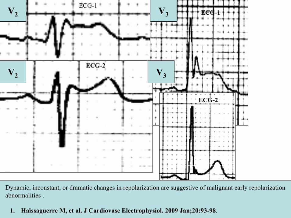
V2
V2
V3
V3
ECG-1
ECG-2
ECG-2
Dynamic inconstant or dramatic changes in repolarization are suggestive of malignant early repolarization abnormalities
1 Haiumlssaguerre M et al J Cardiovasc Electrophysiol 2009 Jan2093-98
ECG-1
Conclusion this patient has high possibility of concomitant Brugada syndrome and early repolarization In this a benign ERP or malignant early repolarization abnormalities We think it is very unlikely that is a Brugada phenocopyIn this case it is necessary additionally to perform
1 IV Ajmaline test2 ECG-AR3 Family study ( first degree relatives interrogatory physical ECGs)4 Magnetic resonance image5 Genetic screening
Ventricular biopsy is not considered mandatory but is recommended especially when it may clarify the nature of dubious findings identified with others test In approximately 6--10 of survivors of cardiac arrest no cardiac abnormality can be identified despite extensive clinical evaluation Autopsy data confirm that in a similar percentage of victims of SCD no structural heart disease or or mors sine materia can be identified at post mortem evaluation Incompletely penetrant genetic defects may underlie at least some of these unexplained deaths (1)
The ERP probably represents part of a spectrum of cardiovascular anomalies related to channelopaties including BrS IVF congenital short QT syndrome and that it may also have a molecular genetic origin of variable penetrance The denomination Early Repolarization Pattern is appropriate since it indicates that it is characterized by a given ECG pattern
1 Priori SG et al Cardiovasc Res 2001 May50218-223
Case Report similar
A 20 year-old male mulatto professional soccer player from Santo Andreacute Sao Paulo Brazil He didnrsquot finishthe second year in elementary school CatholicProfession Professional soccer playerReason for consultation Presented to consultation due to two presyncopal episodes and one syncopal episode in the last 30 days The first two episodes happened during a post-defecation period Both were preceded by dizziness nausea and vomiting The syncopal episodes occurred at rest with no prodromes The third one with express and brief loss of consciousness and sphincter relaxation at dawn 48 hours agoHis prior personal medical history was unremarkable and he successfully passed two prior periodical clinical-cardiovascular evaluations (twice a year always normal) Pre-competitive examination included 12-lead ECG echocardiogram and cardiopulmonary metabolic exercise testing (CMET) All normals Intense training for the last 3 years Familial Background One uncle from his fatherrsquos side family died of sudden cardiac death when he was 35
years old He ignores the cause His brother is being seeing by a cardiologist due to ldquocardiac arrhythmiardquo in treatment with a cardiologistfrom the Institute of the Heart from Satildeo Paulo (he cannot tell which is the cause) He denies having other relevant background details Pysical cardiovascular examination revealed bradycardia at 45 bpm with physiological sinus arrhythmia blood pressure 11070 mmHg The rest of the physical examination was unremarkable He denied consuming any type of neither medications nor drugs No addictionsHis first 12-lead ECGVectorcardiogram(VCG) (Fig 1) showed sinus bradycardia at 44 bpm P-wave axis of +60deg PR interval of 186ms QRS duration of 111ms QRS axis at + 63deg QT interval 427ms and corrected QTc 367ms There is a notch on the terminal portion of the QRS complex in the left precordial leads voltage criteria for left ventricular hypertrophy (SV1 + RV5 gt 35 mm ndash Sokolow index) prominent J-wave and concave ST-segment elevation in inferior leads V4 V5 and V6 Deep and narrow Q-waves in the left precordial leads V5 and V6 All these features were considered as an Early Repolarization Pattern (ERP)
Name JVS Gender Male Age 20 yo Ethnic Group Mulatto Weight 66 Kg Height 172 m Biotype Normoline Date 11262009 Profession competitive athlete (soccer player)
ECG diagnosis Sinus bradycardia (SB) HR 44bpm P axis +60deg PR 186ms QRSd 111ms QRS axis + 63deg QT 427ms QTc 367ms Conclusion SB + Early Repolarization Pattern (ERP) Notching or slurring of the terminal QRS complex voltage criteria of left ventricular hypertrophy SV1+ RV5 gt 35 mm (Sokolow index) prominent J wave and ST-segment elevation concave to the topending in a positive T wave from V4 to V6 Precordial greater than limb leads Relatively deep but narrow q waves in the left precordial leads (V5 and V6)
V6V4 V5
Prominent J wave and ST- segment elevation concave to the top ending in a positive T wave from V4 to V6 Notching or slurring morphology of the terminal portion of the R wave (J-wave) that produces a positive hump known as a J-wave (Arrows)
aVR
Eventually reciprocal ST segment depression confined only in Lead aVR1 observed in this particular case report
1 Mehta M Jain AC Mehta A Early Repolarization Clin Cardiol 1999 Feb 22 59-65
ECGVCG CORRELATIONS HORIZONTAL PLANE
V6
V1
V2
V3
X
Z
V5
V4
T
0J T
BEGINNING AND END OF QRS LOOP IN HP
J POINT in vectorcardiography it corresponds to 3 elements end of ventricular depolarization (QRS loop) beginning of repolarization (ST segment) when it does not present depression or elevation and T wave onset In this case 0 point and J point are not coincident In situations where there is depression or elevation of ST segment the J point does not coincide with the 0 point and the greater or lesser distance between both points indicate the greater or lesser ST segment elevation or depression The phenomenon is observed in early repolarization pattern acute phase of myocardial infarction Prinzmetal variant angina pericarditis Brugada syndrome arrhythmogenic right ventricular dysplasia etc
0 POINTIt corresponds to the QRS looponset (because PR segment does notexist) Alsocorrespond to the end of biatrialchamberactivation
T
J POINT
ECGVCG CORRELATIONS FORNTAL PLANE
aVR aVL
I
IIIII
X
Y
VERTICAL AXIS ON FRONTAL PLANE The frontal plane QRS axis and ST segment axis and T wave axis are all in the same direction THE ST
SEGMENT ELEVATION
USUALLY lt 05 mm IN THE LIMB
LEADS
YVF
Z V2
T
ECGVCG CORRELATIONS RIGHT SAGITTAL PLANE
A second 12-lead ECG (Fig 2) preformed 72 hours later showing1) Sinus bradycardia at 44 bpm2) Similar p-wave axis and intervals duration3) Dramatic modification of repolarizaccedilatildeo pattern in the right precordial leads A coved-type
(type 1) ECG brugada pattern can be distinguished4) Accentuation of j-waves and st segment elevation across the precordial leads was observed5) ST segment elevation on avr lead
See next slide
Name JVS Gender Male Age 20 yo Ethnic Group MulattoWeight 66 Kg Height 172 m Biotype Normoline Date 11292009Profession Competitive Soccer Player FIGURE 2
ECG diagnosis Sinus Arrhythmia(SA) sinus bradycardia (SB) HR 44bpm P axis +60deg PR 186ms QRSd 111ms QT 427ms QTc 367 Conclusion SA + SB + Early Repolarization Pattern (ERP) Type 1 Brugada ECG pattern Dramatic accentuation of J waves across the precordial and limb leads Transient augmentation of global J waves may be indicative of a
highly arrhythmogenic substrate heralding multiple episodes of VF in patients with ERP
aVR LEAD
11262009 11292009
DEPRESSION
ELEVATION
ST SEGMENT DEPRESSION IN aVR = 1mm ST SEGMENT ELEVATION IN aVR = 2mm
V1 V2V2
1129200911262009
V1
11292009 11262009
SPONTANEOUS MODIFICATION OF REPOLARIZATION TO TYPE 1 BRUGADA ECG PATTERN
11262009
V6
11292009V6
ST segment elevation lt 2mm ST segment elevation gt 2mm
11292009
V5
11262009
V5
ST segment elevation = 1mm ST segment elevation = 25mm
EARLY REPOLARIZATION PATTERN CONCEPT
DEFINITION Early Repolarization Pattern (ERP) is a well-recognized Enigmatic idiopathic electrocardiographic phenomenon characterized by notching or slurring morphology of the terminal portion of the R wave (J-wave) that produces a positive hump known as a J-wave at the end of the QRS complex and beginning of the ST segment It considered to be present when at least two adjacent precordial leads show elevation of the ST segment with values ge01 mV (or 1mm) above baseline followed by concordant tall near symmetric (T waves having similarity in shape ascending and descending ramps speeds)
EPIDEMIOLOGY ERP occur in 2 to 5 of the population often in men young adults afro-descendents and competitive athletes and in up to 131 to 482 of patients presenting with chest pain in the emergency rooms and coronary care units There is a distinct J wave and ST segment concave upward in the left precordial leads V4 through V6 The ST elevation is most frequently evident in ECG lead V4 The ST segment elevation is usually le2mm (precordial greater than limb leads) and is lt 05 mm in the limb leadsReciprocal ST-segment changes (ST-segment depression) are eventually confined the unipolar aVR lead (Absence of reciprocal changes or mirror image) In ERP vertical QRS axis electrical is the rule The frontal plane QRS axis the ST segment axis and T wave axis are all in the same directionThis ECG pattern has been considered a benign normal ECG variant for over six decades3
1 Hasbak P Engelmann MD Early repolarization ST-segment elevation as a normal electrocardiographic variant Ugeskr Laeger 2000 162 5928-5929
2 Brady WJ Chan TC Electrocardiographic manifestations benign early repolarization J Emerg Med 1999 17473-478
3 Benito B Guasch E Rivard L Nattel S Clinical and mechanistic issues in early repolarization of normal variants and lethal arrhythmia syndromes J Am Coll Cardiol 2010 Oct 5 56 1177-1186
MAIN ELECTROCARDIOGRAPHIC FEATURES IN ERPHeart rate Predominant sinus bradycardia frequently associated with respiratory sinus arrhythmia
PR interval First degree AV block is observed in 5 to 39 among professional athletes In the non-athlete population it is observed in 065 of cases QRS axis Vertical electrical axis is the rule The frontal plane QRS axis and ST segment axis and T wave axis are all in the same directionQRS duration (90 plusmn 10 ms) subjects than in age-matched healthy controls (80 plusmn 10 ms) QRS morphology Notching or slurring of the terminal QRS complex Prominent relatively deep but narrow q waves may appear in the left precordial leadsQRS voltage Eventually voltage criteria of LVH are observed in male competitive athletes SV1+ RV5 gt 35 mm (Positive Sokolow index) High QRS voltage is more frequent in male athletes but its correlation with LVH is low Voltage decreases after deconditioning is slow The distinction between the physiological athletes heart and pathological conditions has critical implications for professional athletes An abrupt transition may occur from right-oriented complexes to left-oriented complexes in the precordial leads secondary to counterclockwise rotation on precordial leads About two-thirds of clockwise rotation and counterclockwise rotation could be attributed to the septal angle by anatomical rotation of the heart in one plane around the long axis but other factors appear to be responsible for such ECG findings in the remaining one-third of cases Relatively higher positions of the precordial ECG leads as observed in the vertical heart appeared to be responsible for clockwise rotation in some patients and left septal fascicular block is suspected to be responsible for counterclockwise rotation in others patientsJ-point elevation Notching irregular or slurring contour of the terminal QRS complex (J point) Variant of Osborn wave is seen in ERP
ST segment Widespread ST segment elevation (precordial greater than limb leads) The characteristic ST segment is elevated upward concave confined more frequently in precordial leads with reciprocal depression only in aVR The concavity is observed at the initial up-sloping portion of ST segment or upwardly concave ST segment morphology Unfortunately concave ST morphology cannot be used to rule out ST elevation from AMI with left anterior descending coronary occlusion because it is common in these circumstances The ST elevation is most frequently evident in ECG lead V4 There is a distinct J wave and ST segment in the left precordial leads V4 through V6 The ST elevation in ERP is usually lt 2 mm (but can rarely be gt 5 mm) in the precordial leads and the greatest ST elevation is usually seen in the mid-to-left precordial leads The ST segment elevation is usually lt 05 mm in the limb leads
T wave characteristics Concordant T waves of large amplitude (prominent matching T waves) typical pseudo-asymmetrical (lsquosymmetroidrdquo) or slightly asymmetrical matching T waves often of large amplitude upright tall and peaked most conspicuously from V2 to V4 or V5 sometimes seen in leads DII DIII and aVF as a rule T waves may appear as of large amplitude peaked or pointed symmetric and matching Vagotonic or high T wave voltages followed by U waves are frequent when sinus bradycardia is present Tall positive ad symmetric or symmetroid T waves are not only seen occasionally in the very early stages of MI but also in hyperkalemia and in ERP sinus bradycardia
QT intervals QT maximum The maximum Q-onset-T-end interval This parameter is higher in ERV subjects than in normal controls
QTp maximum maximum Q-onset-T-peak interval This parameter is higher in ERV subjects than in age-matched healthy controls
Rate-corrected QTc maximum This parameter is lower in ERP subjects than in age-matched healthy controls
QTpc maximum This parameter is lower in ERV subjects than in age-matched healthy controls
U wave because bradycardia U waves are frequent in ERV they are best observed in the V3 lead U waves are frequent when sinus bradycardia is present
Other ECG characteristics of ERPRelative temporal stability of the ST segment and T wave pattern is observedReciprocal changes are not seen in ERP There are no evolutionary short-term changes in the ST segment and T waves and Q waves do not appear
Discusions Early Repolarization Pattern (ERP) or Early Repolarization Variant (ERV) is an enigmati
electrocardiographic phenomenon characterized by prominent J wave and ST-segment elevation in multiplleads
Recently there has been renewed interest in ERP because of similarities to the arrhythmogenic Brugadsyndrome (BrS) Not much is known about the epidemiology of ERP and several studies have reported thathis condition is associated with a good prognosis
Both ERP and BrS exhibit some similarities including the ionic underlying mechanism the analogouresponses to changes in heart rate and autonomic tone sympathicomimetics (isoproterenol test) as well as insodium channel and beta-blockers These observations raise the hypothesis that ERP may be not as benign atraditionally believed Additionally there are documents showing that ST-segment height in the man igreatly influenced by central sympathetic nervous activity both at baseline and during physiologic andpharmacological stress Central sympathetic dysfunction regularly results in multilead ST-segment elevationor J wave that decreases or below isoelectric baseline during low dose isoproterenol infusion An earlyrepolarization pattern in the inferior leads of a standard electrocardiogram is associated with an increased riskof death from cardiac causes in middle-aged subjects1
POSSIBLE SIMILARITIES BETWEEN ERV AND BrSbullMore frequent in malesbullBoth occur more frequently in young adults and in individuals without apparent structural heart diseasebullBoth may influence just the V1-V2 leads Rarely (9) can ST elevation be observed in ERP only in the right precordial leads V1-V2 or in the inferior ones2
1 Tikkanen JT Anttonen O Junttila MJ Aro AL Kerola T Rissanen HA Reunanen A Huikuri HV Long-Term Outcome Associated with Early Repolarization on Electrocardiography N Engl J Med 2009 Nov 16 [Epubahead of print]
2 Hasbak P Engelmann MDEarly repolarization ST-segment elevation as a normal electrocardiographic variant Ugeskr Laeger 2000 162 5928-5929
When ST elevation is normal it can reach up to 3 mm in V2-V3 especially in young people In those individuals over 40 years it seldom exceeds 2 mm Both can show incomplete RBBB pattern or right bundle branch conduction disorder in BrS it can present atypical features RBBB-like and of the saddle type by exclusive elevation of the J point S wave with delay in the left leads DI aVL V5 and V6 could be absent as it is to be expected in a classic RBBB The elements considered as typical in BrS are 1) elevation of the terminal part of QRS (prominent J wave) 2) elevated and descending ST not related to lesion of ischemic (idiopathic) injury 3) negative T wave in the right precordial leads 4) normal QTc or near normal 5) Eventually absence of final delay in left leads as it would be expected in a classic RBBB1 In ERV when associated to athletersquos heart QRS can present a moderate extension (100 ms to 110 ms) in 15 of the cases which in nonathlete normal population in a 24 is called outflow tract hypertrophy In this case r does not exceed the 5 mm and is lower than S in the same lead rSrBoth may improve repolarization during the stress test with use of isoproterenolBoth respond to a shortening of AP phase 2 in a part of ventricular thickness and intensification of fast repolarization notch (phase 1) mediated by transmural dispersion of ventricular repolarization by a larger notch in the Ito channel2The alteration of the Ito and ICa2+-L channels in BrS and in ERP are the electrophysiologic substrate that explains the J point and ST segment elevation because they cause the intensified notch in phase 1 and suppression in phase 2 duration in the epicardium and in the endocardium of ventricular wall thickness
1 Hiss RG Lamb LE Electrocardiographic findings in 122043 individuals Circulation 1962 25947-9612 Antzelevitch Ch Xin Yan G Shimuzi W et al Electrical heterogeneity the ECG and Cardiac Arrhythmias In Zipes
DP Jalife J Cardiac Electrophysiology From Cell to Bedside Third Edition WB Saunders Company2000 Chapter 26 p 222-238
ELEMENTS FOR DIFFERENTIAL DIAGNOSIS BETWEEN ERP AND BrS
I) Family background ERP negativeBrS frequently positive
II) Ethnic GroupERP predominantly in African descendents(1) or equally common in all races(2) BrS predominantly in Asian group (58) and Caucasian people(3)
III) GenderERP male gender predominanceBrS great predominance in the male gender (malefemale ratio ndash 81 in non Asian and 101 in Asian
peopleIV) Response to IC group antiarrhythmic agents
BrS flecainide used in a 10 mgKg dosage in 10 minutes increases ST elevation and QRS duration in a more significant way in patients with BrS than in individuals without the entity and only in those it triggers ventricular extrasystoles(4)
ERP it can induce a pattern similar to BrS however the degree of ST elevation caused by the drug is much higher in patients with BrS than in patients without the disease
1 Grusin H Peculiarities of the Africanrsquos electrocardiogram and the changes observed in serial studies Circulation 1954 9 860-867
2 Mehta M Jain AC Mehta A Early Repolarization Clin Cardiol 1999 Feb2259-653 Nademanee K Veerakul G Nimmannit S Chaowakul V Bhuripanyo K Likittanasombat K Tunsanga K Kuasirikul S
Malasit P Tansupasawadikul S Tatsanavivat P Arrhytmogenic marker for the sudden unexplained death syndrome in Thail men Circulation 1997 962595-2600
4 Shimizu W Antzelevitch C Suyama K Kurita T Taguchi A Aihara N Takaki H Sunagawa K Kamakura S Effect of sodium channel blockers on ST segment QRS duration and corrected QT interval in patients with Brugada syndromeJ Cardiovasc Electrophysiol 2000 111320-1329
BrS patients with Inferior and lateral ECG repolarization abnormalities have longer baseline PR intervals(1)Both an S wave width ge80ms in V1 and ST elevation ge18mm in V2 are highly specific indicators of VF and criteria for high-risk BrS(2)There are several references in literature to patients with elevation of the J point and ST segment convex to the top or straight descendent in inferior leads (Brugada sign) or concomitantly ST-segment elevation in the right precordial and inferior leads II III and aVF(3) in absence of hypothermia ischemia or electrolytic disorders which we call ldquoatypical Brugada patternrdquo atypical BrS variant BrS or BrS variant or idiopathic VF (a variant of the BrS with ST-segment elevation in inferior leads(4)Pilsicainide may induce PVCs and PVT in atypical BrS and the infusion of isoproterenol suppress the arrhythmias and normalize the ST-segment elevation(5)
1 Sarkozy A Chierchia GB Paparella G Boussy T De Asmundis C Roos M Henkens S Kaufman L Buyl R Brugada R Brugada J Brugada P Inferior and lateral electrocardiographic repolarization abnormalities in Brugada syndromeCirc ArrhythmElectrophysiol 2009 Apr 2154-161
2 Atarashi H Ogawa S For The Idiopathic Ventricular Fibrillation Investigators New ECG Criteria for High-Risk Brugada Syndrome Circ J 2003 Jan 67 8-10
3 Lombardi F Potenza S Beltrami A Verzoni A Brugada P Brugada RSimultaneous ST-segment elevation in the right precordial and inferior leads in Brugada syndrome J CardiovascMed (Hagerstown) 2007 8 201-204
4 Letsas KP Efremidis M Pappas LK Gavrielatos G Markou V Sideris A Kardaras FEarlyrepolarization syndrome is it always benign Int J Cardiol 2007114 390-392
5 Chinushi M Izumi D Furushima H Watanabe H Aizawa YMultiple premature beats triggered ventricular arrhythmias during pilsicainide infusion in a patient with inferior ST-segment elevation Pacing Clin Electrophysiol 2006 29 1445-1448
Potet et al(1) identified a G752R mutation on SNC5A that produced ST segment elevation and prominent J wave in leads II III and aVF The authors provide genetic demonstration that Brugada ECG anomalies related to a unique SCN5A mutation can be observed either in the inferior or the right precordial leads The early repolarization pattern in inferolateral leads is not an uncommon finding in BrS and this pattern is not associated with a worse outcome in subjects with BrS(2) The spontaneous ERP occurred more frequently among patients with BrS than in 283 family members not having BrS (11 versus 6 P=003) Class I antiarrhythmic drug administration provoked inferior-lateral coved Brugada pattern in 13 patients with BrS These patients had longer baseline PR intervals and Class I antiarrhythmic drug induced QRS interval prolongation (108 to 178 versus 102 ms to 131 ms Plt0001) In 3 patients the Class I antiarrhythmic drug provoked coved Brugada pattern only present in the inferior leads Inferior-lateral ERP occurs spontaneously relatively frequently in BrS These patients have a more severe phenotype Class I antiarrhythmic drug administration provokes inferior-lateral coved Brugada pattern in 46 of patients The Class I antiarrhythmic drug exceptionally provoked coved Brugada pattern only observed in the inferior leads(3)
1 Potet F Mabo P Le Coq G et al Novel brugada SCN5A mutation leading to ST segment elevation in the inferior or the right precordial leads J Cardiovasc Electrophysiol 2003 142000-2003
2 Letsas KP Sacher F Probst V Weber R Knecht S Kalusche D Haiumlssaguerre M ArentzTPrevalence of early repolarization pattern in inferolateral leads in patients with Brugada syndrome Heart Rhythm 2008 Dec 5 1685-1689
3 Sarkozy A Chierchia GB Paparella G Boussy T De Asmundis C Roos M Henkens S Kaufman L Buyl R Brugada R Brugada J Brugada P Inferior and lateral electrocardiographic repolarization abnormalities in Brugada syndrome Circ Arrhythm Electrophysiol 2009 Apr2 154-161
Type 1 ECG Brugada pattern in the peripheral leads was observed in 42 of patients during ajmaline test (103 of positive tests) and was associated with longer QRS and greater QTc prolongation compared with the rest of the patients1Atypical Brugada Syndrome or IVF with J waves in inferior lateral or inferior lateral leads
and Familial Idiopathic Ventricular Fibrillation and BrSSeveral mutations in genes SCN5A KCNJ8 DPP6 SCN3B and CACNA2D1 had been identified 1 On SCN5A gene2 3 Observation We do not believe that SCN5A should be attributed to IVF because this report clearly shows conduction defects (as expected) so this is not IVF but conduction delay at different levels in the heart2 On KCNJ8 gene 4 5 Missense variant in exon 3 (NC-000012) of the KCNJ8 gene a subunit of the K(ATP)channel23 Genomic DNA sequencing of K(ATP) channel genes showed missense variant in exon 3 (NC_000012) of the KCNJ8 gene a subunit of the K(ATP) channel conferring predisposition to dramatic repolarization changes and ventricular vulnerability
1 Batchvarov VN Govindan M Camm AJ Behr ERBrugada-like changes in the peripheral leads during diagnostic ajmaline test in patients with suspected Brugada syndrome Pacing Clin Electrophysiol 2009 Jun32695-703
2 Potet F Mabo P Le Coq G Probst V Schott JJ Airaud F Guihard G Daubert JC Escande D Le MarecH Novel brugada SCN5A mutation leading to ST segment elevation in the inferior or the right precordial leads J Cardiovasc Electrophysiol 2003 14 200-203
3 Akai J Makita N Sakurada H Shirai N Ueda K Kitabatake A Nakazawa K Kimura A Hiraoka M A novel SCN5A mutation associated with idiopathic ventricular fibrillation without typical ECG findings of Brugada syndrome FEBS Lett 2000 Aug 11 479 29-34
4 Haissaguerre M Derval N Sacher F Jesel L Deisenhofer I de Roy Let al Sudden cardiac arrest associated with early repolarization N Engl J Med 2008 May 8 3582016-2023
5 Haiumlssaguerre M Sacher F Nogami A Komiya N Bernard A Probst V et al Characteristics of recurrent ventricular fibrillation associated with inferolateral early repolarization role of drug therapy J Am CollCardiol 2009 Feb 17 53 612-619
On DPP6 gene1 Dipeptidyl aminopeptidase-like protein 6 locus on chromosome7q26 Voltage-gated potassium channels OMIM126141 The ECG shows rapid PVT Monomorphic PVCs with very short coupling intervals The PVC (302 +- 52 msec) within 40 msec of the peak of the preceding Absence of T wave Pause-dependence
Valdivia et al2 reported a case with IVF and a novel mutation in the SCN3B-encoded sodium channel beta subunit Navbeta3 that causes a loss of function of Nav15 channels in vitro and provides molecular and cellular evidence implicating mutations in Navbeta3 as a cause of IVF Mutation in this SCN3B gene causes also BrS type 73
L-type calcium channel (LTCC) mutations have been associated with BrS SQTS and Timothy syndrome (LQT8) Little is known about the extent to which LTCC mutations contribute to the J-wave syndromes associated with SCD Mutations in the LTCCs are detected in a high percentage of probands with J-wave syndromes associated with inherited cardiac arrhythmias suggesting that genetic screening of Ca(v) genes may be a valuable diagnostic tool in identifying individuals at risk Burashnikov et al4 identify CACNA2D1 as a novel BrS susceptibility gene and CACNA1C CACNB2 and CACNA2D1 as possible novel ERS susceptibility genes4
1 Alders M Koopmann TT Christiaans I Postema PG Beekman L Tanck MW et al Haplotype-sharing analysis implicates chromosome 7q36 harboring DPP6 in familial idiopathic ventricular fibrillation Am J Hum Genet 2009 Apr 84 468-476
2 Valdivia CR Medeiros-Domingo A Ye B Shen WK Algiers TJ Ackerman MJ et al Loss-of-function mutation of the SCN3B-encoded sodium channel beta3 subunit associated with a case ofidiopathic ventricular fibrillation Cardiovasc Res 2010 Jun 1 86 392-400
3 Hu D Barajas-Martinez H Burashnikov E Springer M Wu Y Varro A et al A mutation in the beta-3 subunit of the cardiac sodium channel associated with Brugada ECG phenotype Circ Cardiovasc Genet 2009 2 270-278
4 Burashnikov E Pfeiffer R Barajas-Martinez H Delpoacuten E Hu D Desai M et al Mutations in the cardiac L-type calcium channel associated with inherited J-wave syndromes and sudden cardiac death Heart Rhythm 2010 Dec71872-1882
From a multicenter cohort of 122 patients (90 male age 37 +- 12 years) with IVF and ERP in the inferolateral leadsHaiumlssaguerre et al1 selected all patients with more than 3 episodes of VF (multiple) including those with electrical storms (ge3 VF in 24 h) Multiple recurrences of VF occurred in 27 of patients with ERP and may be life threatening Isoproterenol in acute cases and quinidine in chronic cases are effective antyharrhithmic drugsThe last one is necessary associated to ICDThe so-called atypical BrS is characterized by ECG abnormalities of the J wave and ST-segment elevation appeared in the inferior andor lateral leads The ERP in inferolateral leads is not an uncommon finding in BrS2 There is a high incidence of the ERP confined in inferolateral leads in patients with IVF The ECGs have an elevation of the QRS-ST junction of at least 01 mV from baseline in the inferior or lateral lead manifested as QRS slurring or notching Among patients with a history of IVF there is an increased prevalence of ERP
1 Haissaguerre M Derval N Sacher F Jesel L Deisenhofer I de Roy L Pasquieacute JL Nogami A Babuty D Yli-Mayry S De Chillou C Scanu P Mabo P Matsuo S Probst V Le Scouarnec S Defaye P Schlaepfer J Rostock T Lacroix D Lamaison D Lavergne T Aizawa Y Englund A Anselme F ONeill M Hocini M Lim KT Knecht S Veenhuyzen GD Bordachar P Chauvin M Jais P Coureau G Chene G Klein GJ Cleacutementy J Sudden cardiac arrest associated with early repolarization N Engl J Med 2008 May 8 3582016-2023
2 Letsas KP Sacher F Probst V Weber R Knecht S Kalusche D Haiumlssaguerre M Arentz T Prevalence of early repolarization pattern in inferolateral leads in patients with Brugada syndrome Heart Rhythm 2008 Dec51685-1689
Because a contingent percentage has not yet been well established it has been suggested that BrS constitutes the same entity in a large number of IVF diagnosed cases since they have common clinical features
1 BOTH ARE MORE FREQUENT IN MEN2 BOTH HAPPEN IN MIDDLE-AGE PEOPLE3 BOTH OCCUR IN APPARENT STRUCTURALLY NORMAL HEARTS14 BOTH CAN AFFECT THE SAME GENE (SCN5A)5 BOTH AFFECT EVENTUALLY THE SAME LOCUS (3p24-p21)
Wellens et al2 has pointed out that in order to be accurate about such a percentage it is important to define the diagnostic criteria used to classify BrS in IVF patients Curiously the so-called Idiopathic VT in apparently healthy hearts without structural heart disease and primary electrical disease also affects the 3p21 locus but not 3p24 and the gene is different GNA12
Survivors of SCD by VF without structural heart disease and a normal coronary angiogram are often diagnosed with IVTIVF3
A careful analysis of the ECG is essential in the differential diagnosis of this type of apparently unexplained SCD because intermittent or permanent RBBB with J point and ST-segment elevation in leads V1 to V3 is now classified as BrS
1 Goethals P Debruyne P Saffarian M Drug-induced Brugada syndrome Acta Cardiol1998 53157-60
2 Wellens HJJ Rodriquez LM Smeets JL Ventricular tachycardia in structural normal hearts In Zipes DP Jalife J Editors Cardiac Electrophysiology from cell tobedside Philadelphia 1995 WB Saunders
3 Chen Q Kirsch GE Zhang D Brugada R Brugada P Brugada J et al Genetic basis and molecular mechanisms for idiopathic ventricular fibrillation Nature 1998392 293-296
DIAGNOSTICS TRACKS TO DIFFERENTIATE BENIGN ERP FROM MALIGNANT EARLY REPOLATIZATION ABNORMALITIES
BENIGN ERP MALIGNANT EARLY REPOLARIZATION ABNORMALITIES
Family history of unexplained SCD in young relatives(lt45yo)
Absent Possible however infrequent1
Personal history Asymptomatic Asymptomatic repetitive syncope episodes or recovered of cardiac arrest
Left precordial terminal QRS notching Less prevalent More prevalent2
STT ratio in lead V6calculated by dividing the millimeters of
ST-segment elevation by the millimeters to the tallest point of the T wave
Each value is measured from the isoelectric point
lt0253 Non-tested
1 Viskin S Belhassen B Idiopathic ventricular fibrillation American Heart Journal 1990 120 661-671
2 Tikkanen JT Anttonen O Junttila MJ Aro AL Kerola T Rissanen HA Reunanen A et al Long-term outcome associated with early repolarization on electrocardiography N Engl J Med 2009 Dec 24361 2529-2537
3 Ginzton LE Laks MM The differential diagnosis of acute pericarditis from the normal variant new electrocardiographic criteria Circulation 1982 May 65 1004-1009
ST-segment elevation shape Concave upward Convex upwardJ waves are present in asymp 30 IVF patients1
Early repolarization is significantly prevalent in the subjects after cardiac arrest
(present in 31 of those resuscitated )Eventually ldquoLambda-waverdquo 23
J-point elevation and ST-segment degree Usually lt 2 mmlt05mm in limb leads
Frequent gt2mmJ-point elevation gt 2mm in inferior leads is associated with an increased risk of death
from cardiac causes in middle-aged subjects3
T wave characteristic Positive large T wave from V2 to V4 or V5 Concordant
Frequent negative discordant
Reciprocal ST-segment changes (Mirror
image)Absent With exception aVR5 Frequently Present3
1 Viskin S Idiopathic ventricular fibrillation Le Syndrome dHaiumlssaguerre and the fear of J waves J Am Coll Cardiol 2009 Feb 17 53 620-622
2 Gussak I Bjerregaard P Kostis J Electrocardiographic lambda wave and primary idiopathic cardiac asystole a new clinical syndrome J Electrocardiol 2004 Apr 37 105-107
3 Riera AR Ferreira C Schapachnik E Sanches PC Moffa PJ Brugada syndrome with atypical ECG downsloping ST-segment elevation in inferior leads J Electrocardiol 2004 Apr37101-104
4 Tikkanen JT Anttonen O Junttila MJ Aro AL Kerola T Rissanen HA Reunanen A et al Long-term outcome associated with early repolarization on electrocardiography N Engl J Med 2009 Dec 24361 2529-2537
5 Mehta M Jain AC Mehta A Early Repolarization Clin Cardiol 1999 Feb 22 59-65
Monomorphic PVCs with very short coupling
(250 to 350ms)
Absent Characteristic1 Pause-dependency absent2
QTc Interval Normal Minimally short in 35 among masculine gender
(Between 340 to 360ms3)
Late Potentials(LPs) Absent Frequent if J wave presentShowing circadian variation with night
ascendancy4
1 Alders M Koopmann TT Christiaans I Postema PG Beekman L Tanck MW et al Haplotype-sharing analysis implicates chromosome 7q36 harboring DPP6 in familial idiopathic ventricular fibrillation Am J Hum Genet 2009 Apr 84 468-476
2 Viskin S Lesh MD Eldar M Fish R Setbon I Laniado S et al Mode of onset of malignant ventricular arrhythmias in idiopathic ventricular fibrillation J Cardiovasc Electrophysiol 1997 Oct 8 1115-11 20
3 Viskin S Zeltser D Ish-Shalom M Katz A Glikson M Justo D et al Is idiopathic ventricular fibrillation a short QT syndrome Comparison of QT intervals of patients with idiopathic ventricular fibrillation and healthy controls Heart Rhythm 2004 Nov 1 587-591
4 Abe A Ikeda T Tsukada T Ishiguro H Miwa Y Miyakoshi M Circadian variation of latepotentials in idiopathic ventricular fibrillation associated with J waves insights into alternative pathophysiology and risk stratification Heart Rhythm 2010 May 7 675-682
ConclusionIn spite of both malignant early repolarization abnormalities of IVF and benign early repolarization pattern being able to cause J point and ST segment elevation the characteristics of both are clearly different to the point of a differentiation being quite simple when ECGs are analyzed by experts Any ER should raise the suspicion of IVF when it presents ST segment elevation ge 2 mm mainly in inferior wall superior convexity eventually lambda wave shape pattern of dynamic presentation inconstant with dramatic intensity of ST segment elevation discordant T wave polarity related ST segment shift presence of reciprocal ST-segment changes (mirror image) tendency to discrete short QTc interval in male gender(ge340ms) premature monomorphic ventricular contractions with very short coupling interval(250 to 350ms) not related with exercise and late potentials with circadian variation and night ascendancy on SA-ECG Contrarily in benign ERP ST elevation observed in the mid-to-left precordial leads is usually le 2 mm and lt05 mm in the limb leads and concave to the top followed by a positive concordant tall T wave
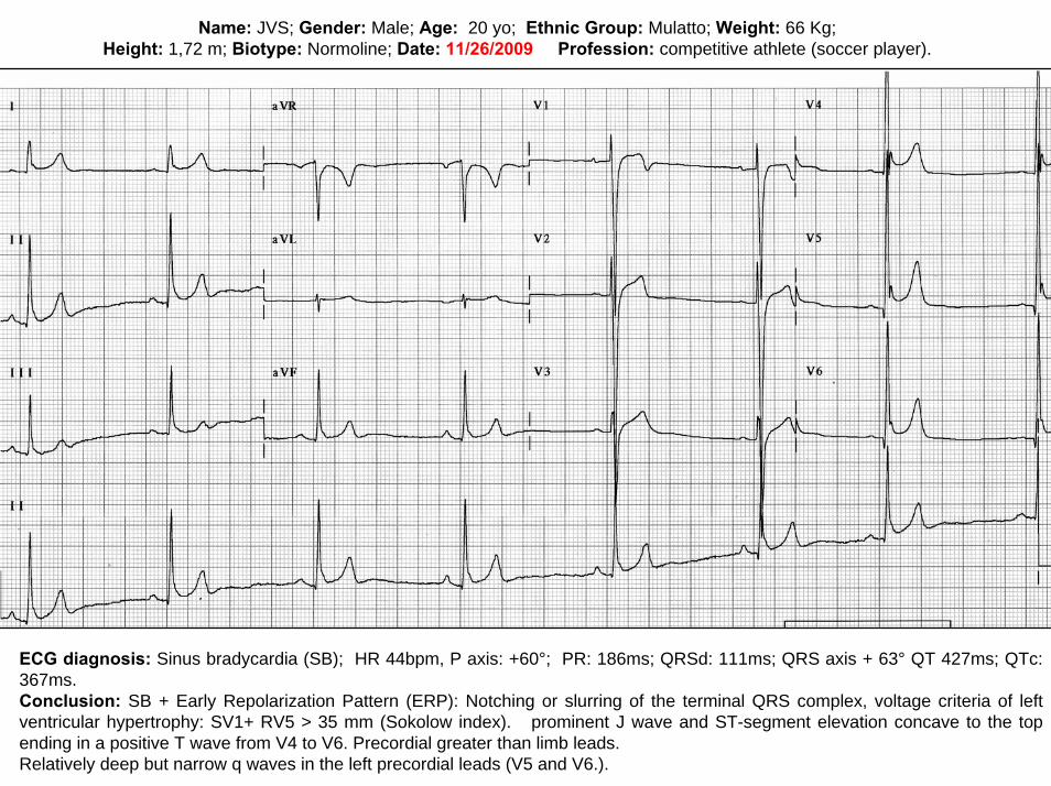
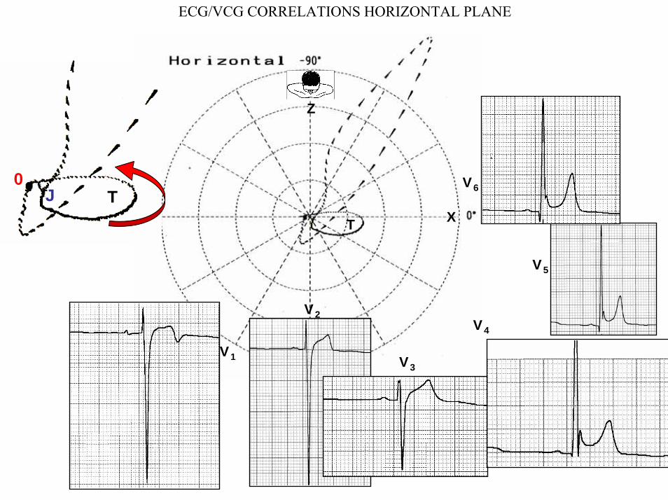
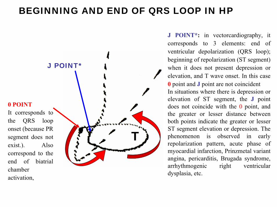
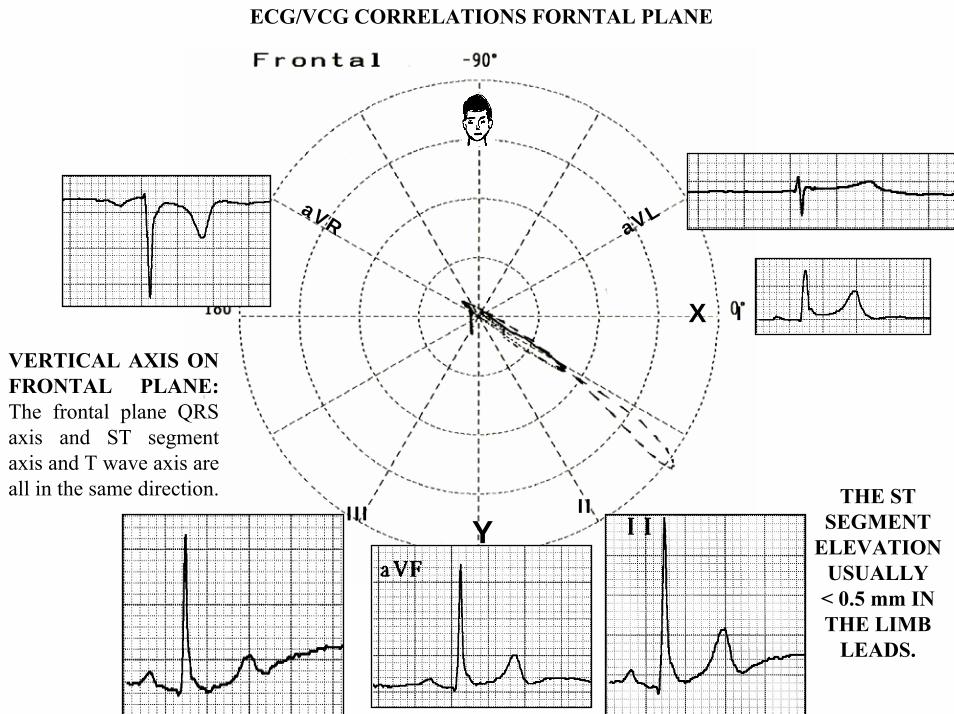
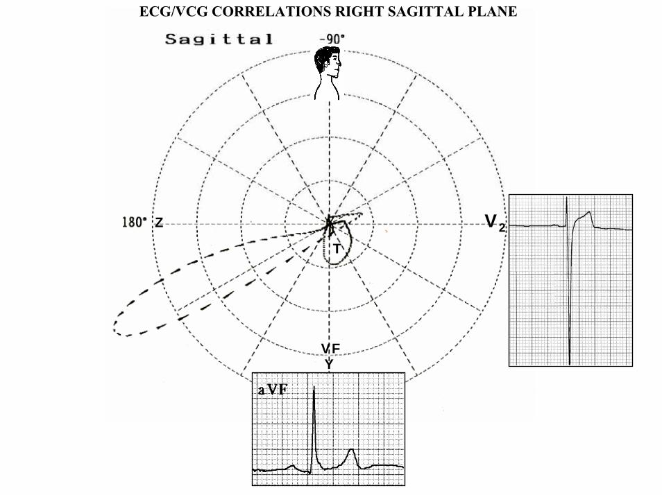
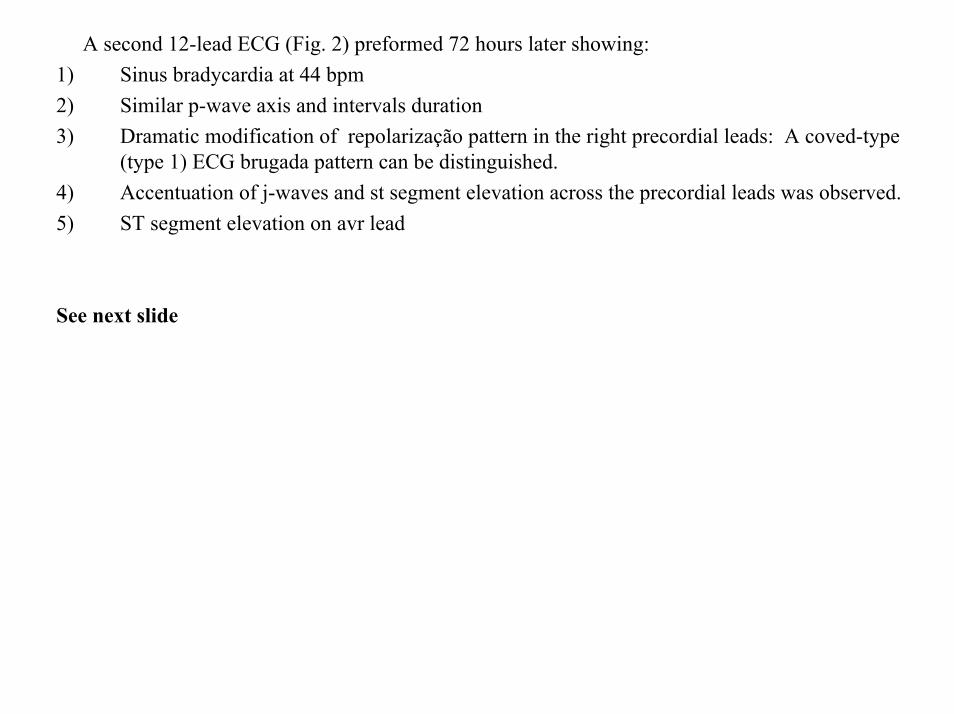
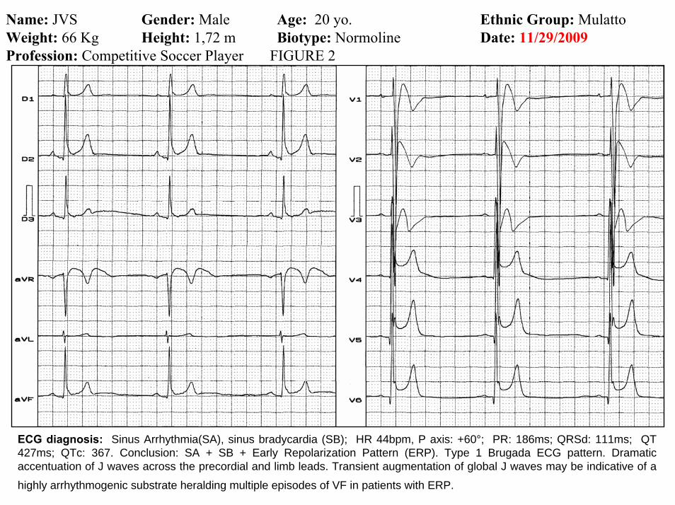
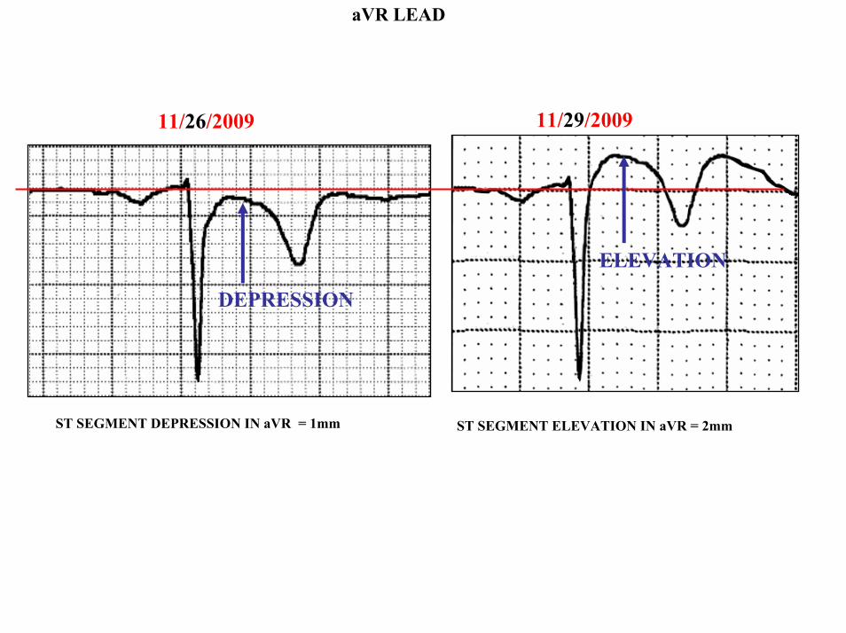
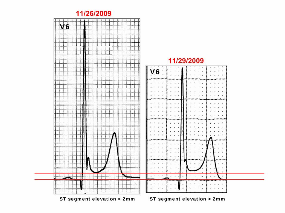
11262009
V6
11292009V6
ST segment elevation lt 2mm ST segment elevation gt 2mm
11292009
V5
11262009
V5
ST segment elevation = 1mm ST segment elevation = 25mm
EARLY REPOLARIZATION PATTERN CONCEPT
DEFINITION Early Repolarization Pattern (ERP) is a well-recognized Enigmatic idiopathic electrocardiographic phenomenon characterized by notching or slurring morphology of the terminal portion of the R wave (J-wave) that produces a positive hump known as a J-wave at the end of the QRS complex and beginning of the ST segment It considered to be present when at least two adjacent precordial leads show elevation of the ST segment with values ge01 mV (or 1mm) above baseline followed by concordant tall near symmetric (T waves having similarity in shape ascending and descending ramps speeds)
EPIDEMIOLOGY ERP occur in 2 to 5 of the population often in men young adults afro-descendents and competitive athletes and in up to 131 to 482 of patients presenting with chest pain in the emergency rooms and coronary care units There is a distinct J wave and ST segment concave upward in the left precordial leads V4 through V6 The ST elevation is most frequently evident in ECG lead V4 The ST segment elevation is usually le2mm (precordial greater than limb leads) and is lt 05 mm in the limb leadsReciprocal ST-segment changes (ST-segment depression) are eventually confined the unipolar aVR lead (Absence of reciprocal changes or mirror image) In ERP vertical QRS axis electrical is the rule The frontal plane QRS axis the ST segment axis and T wave axis are all in the same directionThis ECG pattern has been considered a benign normal ECG variant for over six decades3
1 Hasbak P Engelmann MD Early repolarization ST-segment elevation as a normal electrocardiographic variant Ugeskr Laeger 2000 162 5928-5929
2 Brady WJ Chan TC Electrocardiographic manifestations benign early repolarization J Emerg Med 1999 17473-478
3 Benito B Guasch E Rivard L Nattel S Clinical and mechanistic issues in early repolarization of normal variants and lethal arrhythmia syndromes J Am Coll Cardiol 2010 Oct 5 56 1177-1186
MAIN ELECTROCARDIOGRAPHIC FEATURES IN ERPHeart rate Predominant sinus bradycardia frequently associated with respiratory sinus arrhythmia
PR interval First degree AV block is observed in 5 to 39 among professional athletes In the non-athlete population it is observed in 065 of cases QRS axis Vertical electrical axis is the rule The frontal plane QRS axis and ST segment axis and T wave axis are all in the same directionQRS duration (90 plusmn 10 ms) subjects than in age-matched healthy controls (80 plusmn 10 ms) QRS morphology Notching or slurring of the terminal QRS complex Prominent relatively deep but narrow q waves may appear in the left precordial leadsQRS voltage Eventually voltage criteria of LVH are observed in male competitive athletes SV1+ RV5 gt 35 mm (Positive Sokolow index) High QRS voltage is more frequent in male athletes but its correlation with LVH is low Voltage decreases after deconditioning is slow The distinction between the physiological athletes heart and pathological conditions has critical implications for professional athletes An abrupt transition may occur from right-oriented complexes to left-oriented complexes in the precordial leads secondary to counterclockwise rotation on precordial leads About two-thirds of clockwise rotation and counterclockwise rotation could be attributed to the septal angle by anatomical rotation of the heart in one plane around the long axis but other factors appear to be responsible for such ECG findings in the remaining one-third of cases Relatively higher positions of the precordial ECG leads as observed in the vertical heart appeared to be responsible for clockwise rotation in some patients and left septal fascicular block is suspected to be responsible for counterclockwise rotation in others patientsJ-point elevation Notching irregular or slurring contour of the terminal QRS complex (J point) Variant of Osborn wave is seen in ERP
ST segment Widespread ST segment elevation (precordial greater than limb leads) The characteristic ST segment is elevated upward concave confined more frequently in precordial leads with reciprocal depression only in aVR The concavity is observed at the initial up-sloping portion of ST segment or upwardly concave ST segment morphology Unfortunately concave ST morphology cannot be used to rule out ST elevation from AMI with left anterior descending coronary occlusion because it is common in these circumstances The ST elevation is most frequently evident in ECG lead V4 There is a distinct J wave and ST segment in the left precordial leads V4 through V6 The ST elevation in ERP is usually lt 2 mm (but can rarely be gt 5 mm) in the precordial leads and the greatest ST elevation is usually seen in the mid-to-left precordial leads The ST segment elevation is usually lt 05 mm in the limb leads
T wave characteristics Concordant T waves of large amplitude (prominent matching T waves) typical pseudo-asymmetrical (lsquosymmetroidrdquo) or slightly asymmetrical matching T waves often of large amplitude upright tall and peaked most conspicuously from V2 to V4 or V5 sometimes seen in leads DII DIII and aVF as a rule T waves may appear as of large amplitude peaked or pointed symmetric and matching Vagotonic or high T wave voltages followed by U waves are frequent when sinus bradycardia is present Tall positive ad symmetric or symmetroid T waves are not only seen occasionally in the very early stages of MI but also in hyperkalemia and in ERP sinus bradycardia
QT intervals QT maximum The maximum Q-onset-T-end interval This parameter is higher in ERV subjects than in normal controls
QTp maximum maximum Q-onset-T-peak interval This parameter is higher in ERV subjects than in age-matched healthy controls
Rate-corrected QTc maximum This parameter is lower in ERP subjects than in age-matched healthy controls
QTpc maximum This parameter is lower in ERV subjects than in age-matched healthy controls
U wave because bradycardia U waves are frequent in ERV they are best observed in the V3 lead U waves are frequent when sinus bradycardia is present
Other ECG characteristics of ERPRelative temporal stability of the ST segment and T wave pattern is observedReciprocal changes are not seen in ERP There are no evolutionary short-term changes in the ST segment and T waves and Q waves do not appear
Discusions Early Repolarization Pattern (ERP) or Early Repolarization Variant (ERV) is an enigmati
electrocardiographic phenomenon characterized by prominent J wave and ST-segment elevation in multiplleads
Recently there has been renewed interest in ERP because of similarities to the arrhythmogenic Brugadsyndrome (BrS) Not much is known about the epidemiology of ERP and several studies have reported thathis condition is associated with a good prognosis
Both ERP and BrS exhibit some similarities including the ionic underlying mechanism the analogouresponses to changes in heart rate and autonomic tone sympathicomimetics (isoproterenol test) as well as insodium channel and beta-blockers These observations raise the hypothesis that ERP may be not as benign atraditionally believed Additionally there are documents showing that ST-segment height in the man igreatly influenced by central sympathetic nervous activity both at baseline and during physiologic andpharmacological stress Central sympathetic dysfunction regularly results in multilead ST-segment elevationor J wave that decreases or below isoelectric baseline during low dose isoproterenol infusion An earlyrepolarization pattern in the inferior leads of a standard electrocardiogram is associated with an increased riskof death from cardiac causes in middle-aged subjects1
POSSIBLE SIMILARITIES BETWEEN ERV AND BrSbullMore frequent in malesbullBoth occur more frequently in young adults and in individuals without apparent structural heart diseasebullBoth may influence just the V1-V2 leads Rarely (9) can ST elevation be observed in ERP only in the right precordial leads V1-V2 or in the inferior ones2
1 Tikkanen JT Anttonen O Junttila MJ Aro AL Kerola T Rissanen HA Reunanen A Huikuri HV Long-Term Outcome Associated with Early Repolarization on Electrocardiography N Engl J Med 2009 Nov 16 [Epubahead of print]
2 Hasbak P Engelmann MDEarly repolarization ST-segment elevation as a normal electrocardiographic variant Ugeskr Laeger 2000 162 5928-5929
When ST elevation is normal it can reach up to 3 mm in V2-V3 especially in young people In those individuals over 40 years it seldom exceeds 2 mm Both can show incomplete RBBB pattern or right bundle branch conduction disorder in BrS it can present atypical features RBBB-like and of the saddle type by exclusive elevation of the J point S wave with delay in the left leads DI aVL V5 and V6 could be absent as it is to be expected in a classic RBBB The elements considered as typical in BrS are 1) elevation of the terminal part of QRS (prominent J wave) 2) elevated and descending ST not related to lesion of ischemic (idiopathic) injury 3) negative T wave in the right precordial leads 4) normal QTc or near normal 5) Eventually absence of final delay in left leads as it would be expected in a classic RBBB1 In ERV when associated to athletersquos heart QRS can present a moderate extension (100 ms to 110 ms) in 15 of the cases which in nonathlete normal population in a 24 is called outflow tract hypertrophy In this case r does not exceed the 5 mm and is lower than S in the same lead rSrBoth may improve repolarization during the stress test with use of isoproterenolBoth respond to a shortening of AP phase 2 in a part of ventricular thickness and intensification of fast repolarization notch (phase 1) mediated by transmural dispersion of ventricular repolarization by a larger notch in the Ito channel2The alteration of the Ito and ICa2+-L channels in BrS and in ERP are the electrophysiologic substrate that explains the J point and ST segment elevation because they cause the intensified notch in phase 1 and suppression in phase 2 duration in the epicardium and in the endocardium of ventricular wall thickness
1 Hiss RG Lamb LE Electrocardiographic findings in 122043 individuals Circulation 1962 25947-9612 Antzelevitch Ch Xin Yan G Shimuzi W et al Electrical heterogeneity the ECG and Cardiac Arrhythmias In Zipes
DP Jalife J Cardiac Electrophysiology From Cell to Bedside Third Edition WB Saunders Company2000 Chapter 26 p 222-238
ELEMENTS FOR DIFFERENTIAL DIAGNOSIS BETWEEN ERP AND BrS
I) Family background ERP negativeBrS frequently positive
II) Ethnic GroupERP predominantly in African descendents(1) or equally common in all races(2) BrS predominantly in Asian group (58) and Caucasian people(3)
III) GenderERP male gender predominanceBrS great predominance in the male gender (malefemale ratio ndash 81 in non Asian and 101 in Asian
peopleIV) Response to IC group antiarrhythmic agents
BrS flecainide used in a 10 mgKg dosage in 10 minutes increases ST elevation and QRS duration in a more significant way in patients with BrS than in individuals without the entity and only in those it triggers ventricular extrasystoles(4)
ERP it can induce a pattern similar to BrS however the degree of ST elevation caused by the drug is much higher in patients with BrS than in patients without the disease
1 Grusin H Peculiarities of the Africanrsquos electrocardiogram and the changes observed in serial studies Circulation 1954 9 860-867
2 Mehta M Jain AC Mehta A Early Repolarization Clin Cardiol 1999 Feb2259-653 Nademanee K Veerakul G Nimmannit S Chaowakul V Bhuripanyo K Likittanasombat K Tunsanga K Kuasirikul S
Malasit P Tansupasawadikul S Tatsanavivat P Arrhytmogenic marker for the sudden unexplained death syndrome in Thail men Circulation 1997 962595-2600
4 Shimizu W Antzelevitch C Suyama K Kurita T Taguchi A Aihara N Takaki H Sunagawa K Kamakura S Effect of sodium channel blockers on ST segment QRS duration and corrected QT interval in patients with Brugada syndromeJ Cardiovasc Electrophysiol 2000 111320-1329
BrS patients with Inferior and lateral ECG repolarization abnormalities have longer baseline PR intervals(1)Both an S wave width ge80ms in V1 and ST elevation ge18mm in V2 are highly specific indicators of VF and criteria for high-risk BrS(2)There are several references in literature to patients with elevation of the J point and ST segment convex to the top or straight descendent in inferior leads (Brugada sign) or concomitantly ST-segment elevation in the right precordial and inferior leads II III and aVF(3) in absence of hypothermia ischemia or electrolytic disorders which we call ldquoatypical Brugada patternrdquo atypical BrS variant BrS or BrS variant or idiopathic VF (a variant of the BrS with ST-segment elevation in inferior leads(4)Pilsicainide may induce PVCs and PVT in atypical BrS and the infusion of isoproterenol suppress the arrhythmias and normalize the ST-segment elevation(5)
1 Sarkozy A Chierchia GB Paparella G Boussy T De Asmundis C Roos M Henkens S Kaufman L Buyl R Brugada R Brugada J Brugada P Inferior and lateral electrocardiographic repolarization abnormalities in Brugada syndromeCirc ArrhythmElectrophysiol 2009 Apr 2154-161
2 Atarashi H Ogawa S For The Idiopathic Ventricular Fibrillation Investigators New ECG Criteria for High-Risk Brugada Syndrome Circ J 2003 Jan 67 8-10
3 Lombardi F Potenza S Beltrami A Verzoni A Brugada P Brugada RSimultaneous ST-segment elevation in the right precordial and inferior leads in Brugada syndrome J CardiovascMed (Hagerstown) 2007 8 201-204
4 Letsas KP Efremidis M Pappas LK Gavrielatos G Markou V Sideris A Kardaras FEarlyrepolarization syndrome is it always benign Int J Cardiol 2007114 390-392
5 Chinushi M Izumi D Furushima H Watanabe H Aizawa YMultiple premature beats triggered ventricular arrhythmias during pilsicainide infusion in a patient with inferior ST-segment elevation Pacing Clin Electrophysiol 2006 29 1445-1448
Potet et al(1) identified a G752R mutation on SNC5A that produced ST segment elevation and prominent J wave in leads II III and aVF The authors provide genetic demonstration that Brugada ECG anomalies related to a unique SCN5A mutation can be observed either in the inferior or the right precordial leads The early repolarization pattern in inferolateral leads is not an uncommon finding in BrS and this pattern is not associated with a worse outcome in subjects with BrS(2) The spontaneous ERP occurred more frequently among patients with BrS than in 283 family members not having BrS (11 versus 6 P=003) Class I antiarrhythmic drug administration provoked inferior-lateral coved Brugada pattern in 13 patients with BrS These patients had longer baseline PR intervals and Class I antiarrhythmic drug induced QRS interval prolongation (108 to 178 versus 102 ms to 131 ms Plt0001) In 3 patients the Class I antiarrhythmic drug provoked coved Brugada pattern only present in the inferior leads Inferior-lateral ERP occurs spontaneously relatively frequently in BrS These patients have a more severe phenotype Class I antiarrhythmic drug administration provokes inferior-lateral coved Brugada pattern in 46 of patients The Class I antiarrhythmic drug exceptionally provoked coved Brugada pattern only observed in the inferior leads(3)
1 Potet F Mabo P Le Coq G et al Novel brugada SCN5A mutation leading to ST segment elevation in the inferior or the right precordial leads J Cardiovasc Electrophysiol 2003 142000-2003
2 Letsas KP Sacher F Probst V Weber R Knecht S Kalusche D Haiumlssaguerre M ArentzTPrevalence of early repolarization pattern in inferolateral leads in patients with Brugada syndrome Heart Rhythm 2008 Dec 5 1685-1689
3 Sarkozy A Chierchia GB Paparella G Boussy T De Asmundis C Roos M Henkens S Kaufman L Buyl R Brugada R Brugada J Brugada P Inferior and lateral electrocardiographic repolarization abnormalities in Brugada syndrome Circ Arrhythm Electrophysiol 2009 Apr2 154-161
Type 1 ECG Brugada pattern in the peripheral leads was observed in 42 of patients during ajmaline test (103 of positive tests) and was associated with longer QRS and greater QTc prolongation compared with the rest of the patients1Atypical Brugada Syndrome or IVF with J waves in inferior lateral or inferior lateral leads
and Familial Idiopathic Ventricular Fibrillation and BrSSeveral mutations in genes SCN5A KCNJ8 DPP6 SCN3B and CACNA2D1 had been identified 1 On SCN5A gene2 3 Observation We do not believe that SCN5A should be attributed to IVF because this report clearly shows conduction defects (as expected) so this is not IVF but conduction delay at different levels in the heart2 On KCNJ8 gene 4 5 Missense variant in exon 3 (NC-000012) of the KCNJ8 gene a subunit of the K(ATP)channel23 Genomic DNA sequencing of K(ATP) channel genes showed missense variant in exon 3 (NC_000012) of the KCNJ8 gene a subunit of the K(ATP) channel conferring predisposition to dramatic repolarization changes and ventricular vulnerability
1 Batchvarov VN Govindan M Camm AJ Behr ERBrugada-like changes in the peripheral leads during diagnostic ajmaline test in patients with suspected Brugada syndrome Pacing Clin Electrophysiol 2009 Jun32695-703
2 Potet F Mabo P Le Coq G Probst V Schott JJ Airaud F Guihard G Daubert JC Escande D Le MarecH Novel brugada SCN5A mutation leading to ST segment elevation in the inferior or the right precordial leads J Cardiovasc Electrophysiol 2003 14 200-203
3 Akai J Makita N Sakurada H Shirai N Ueda K Kitabatake A Nakazawa K Kimura A Hiraoka M A novel SCN5A mutation associated with idiopathic ventricular fibrillation without typical ECG findings of Brugada syndrome FEBS Lett 2000 Aug 11 479 29-34
4 Haissaguerre M Derval N Sacher F Jesel L Deisenhofer I de Roy Let al Sudden cardiac arrest associated with early repolarization N Engl J Med 2008 May 8 3582016-2023
5 Haiumlssaguerre M Sacher F Nogami A Komiya N Bernard A Probst V et al Characteristics of recurrent ventricular fibrillation associated with inferolateral early repolarization role of drug therapy J Am CollCardiol 2009 Feb 17 53 612-619
On DPP6 gene1 Dipeptidyl aminopeptidase-like protein 6 locus on chromosome7q26 Voltage-gated potassium channels OMIM126141 The ECG shows rapid PVT Monomorphic PVCs with very short coupling intervals The PVC (302 +- 52 msec) within 40 msec of the peak of the preceding Absence of T wave Pause-dependence
Valdivia et al2 reported a case with IVF and a novel mutation in the SCN3B-encoded sodium channel beta subunit Navbeta3 that causes a loss of function of Nav15 channels in vitro and provides molecular and cellular evidence implicating mutations in Navbeta3 as a cause of IVF Mutation in this SCN3B gene causes also BrS type 73
L-type calcium channel (LTCC) mutations have been associated with BrS SQTS and Timothy syndrome (LQT8) Little is known about the extent to which LTCC mutations contribute to the J-wave syndromes associated with SCD Mutations in the LTCCs are detected in a high percentage of probands with J-wave syndromes associated with inherited cardiac arrhythmias suggesting that genetic screening of Ca(v) genes may be a valuable diagnostic tool in identifying individuals at risk Burashnikov et al4 identify CACNA2D1 as a novel BrS susceptibility gene and CACNA1C CACNB2 and CACNA2D1 as possible novel ERS susceptibility genes4
1 Alders M Koopmann TT Christiaans I Postema PG Beekman L Tanck MW et al Haplotype-sharing analysis implicates chromosome 7q36 harboring DPP6 in familial idiopathic ventricular fibrillation Am J Hum Genet 2009 Apr 84 468-476
2 Valdivia CR Medeiros-Domingo A Ye B Shen WK Algiers TJ Ackerman MJ et al Loss-of-function mutation of the SCN3B-encoded sodium channel beta3 subunit associated with a case ofidiopathic ventricular fibrillation Cardiovasc Res 2010 Jun 1 86 392-400
3 Hu D Barajas-Martinez H Burashnikov E Springer M Wu Y Varro A et al A mutation in the beta-3 subunit of the cardiac sodium channel associated with Brugada ECG phenotype Circ Cardiovasc Genet 2009 2 270-278
4 Burashnikov E Pfeiffer R Barajas-Martinez H Delpoacuten E Hu D Desai M et al Mutations in the cardiac L-type calcium channel associated with inherited J-wave syndromes and sudden cardiac death Heart Rhythm 2010 Dec71872-1882
From a multicenter cohort of 122 patients (90 male age 37 +- 12 years) with IVF and ERP in the inferolateral leadsHaiumlssaguerre et al1 selected all patients with more than 3 episodes of VF (multiple) including those with electrical storms (ge3 VF in 24 h) Multiple recurrences of VF occurred in 27 of patients with ERP and may be life threatening Isoproterenol in acute cases and quinidine in chronic cases are effective antyharrhithmic drugsThe last one is necessary associated to ICDThe so-called atypical BrS is characterized by ECG abnormalities of the J wave and ST-segment elevation appeared in the inferior andor lateral leads The ERP in inferolateral leads is not an uncommon finding in BrS2 There is a high incidence of the ERP confined in inferolateral leads in patients with IVF The ECGs have an elevation of the QRS-ST junction of at least 01 mV from baseline in the inferior or lateral lead manifested as QRS slurring or notching Among patients with a history of IVF there is an increased prevalence of ERP
1 Haissaguerre M Derval N Sacher F Jesel L Deisenhofer I de Roy L Pasquieacute JL Nogami A Babuty D Yli-Mayry S De Chillou C Scanu P Mabo P Matsuo S Probst V Le Scouarnec S Defaye P Schlaepfer J Rostock T Lacroix D Lamaison D Lavergne T Aizawa Y Englund A Anselme F ONeill M Hocini M Lim KT Knecht S Veenhuyzen GD Bordachar P Chauvin M Jais P Coureau G Chene G Klein GJ Cleacutementy J Sudden cardiac arrest associated with early repolarization N Engl J Med 2008 May 8 3582016-2023
2 Letsas KP Sacher F Probst V Weber R Knecht S Kalusche D Haiumlssaguerre M Arentz T Prevalence of early repolarization pattern in inferolateral leads in patients with Brugada syndrome Heart Rhythm 2008 Dec51685-1689
Because a contingent percentage has not yet been well established it has been suggested that BrS constitutes the same entity in a large number of IVF diagnosed cases since they have common clinical features
1 BOTH ARE MORE FREQUENT IN MEN2 BOTH HAPPEN IN MIDDLE-AGE PEOPLE3 BOTH OCCUR IN APPARENT STRUCTURALLY NORMAL HEARTS14 BOTH CAN AFFECT THE SAME GENE (SCN5A)5 BOTH AFFECT EVENTUALLY THE SAME LOCUS (3p24-p21)
Wellens et al2 has pointed out that in order to be accurate about such a percentage it is important to define the diagnostic criteria used to classify BrS in IVF patients Curiously the so-called Idiopathic VT in apparently healthy hearts without structural heart disease and primary electrical disease also affects the 3p21 locus but not 3p24 and the gene is different GNA12
Survivors of SCD by VF without structural heart disease and a normal coronary angiogram are often diagnosed with IVTIVF3
A careful analysis of the ECG is essential in the differential diagnosis of this type of apparently unexplained SCD because intermittent or permanent RBBB with J point and ST-segment elevation in leads V1 to V3 is now classified as BrS
1 Goethals P Debruyne P Saffarian M Drug-induced Brugada syndrome Acta Cardiol1998 53157-60
2 Wellens HJJ Rodriquez LM Smeets JL Ventricular tachycardia in structural normal hearts In Zipes DP Jalife J Editors Cardiac Electrophysiology from cell tobedside Philadelphia 1995 WB Saunders
3 Chen Q Kirsch GE Zhang D Brugada R Brugada P Brugada J et al Genetic basis and molecular mechanisms for idiopathic ventricular fibrillation Nature 1998392 293-296
DIAGNOSTICS TRACKS TO DIFFERENTIATE BENIGN ERP FROM MALIGNANT EARLY REPOLATIZATION ABNORMALITIES
BENIGN ERP MALIGNANT EARLY REPOLARIZATION ABNORMALITIES
Family history of unexplained SCD in young relatives(lt45yo)
Absent Possible however infrequent1
Personal history Asymptomatic Asymptomatic repetitive syncope episodes or recovered of cardiac arrest
Left precordial terminal QRS notching Less prevalent More prevalent2
STT ratio in lead V6calculated by dividing the millimeters of
ST-segment elevation by the millimeters to the tallest point of the T wave
Each value is measured from the isoelectric point
lt0253 Non-tested
1 Viskin S Belhassen B Idiopathic ventricular fibrillation American Heart Journal 1990 120 661-671
2 Tikkanen JT Anttonen O Junttila MJ Aro AL Kerola T Rissanen HA Reunanen A et al Long-term outcome associated with early repolarization on electrocardiography N Engl J Med 2009 Dec 24361 2529-2537
3 Ginzton LE Laks MM The differential diagnosis of acute pericarditis from the normal variant new electrocardiographic criteria Circulation 1982 May 65 1004-1009
ST-segment elevation shape Concave upward Convex upwardJ waves are present in asymp 30 IVF patients1
Early repolarization is significantly prevalent in the subjects after cardiac arrest
(present in 31 of those resuscitated )Eventually ldquoLambda-waverdquo 23
J-point elevation and ST-segment degree Usually lt 2 mmlt05mm in limb leads
Frequent gt2mmJ-point elevation gt 2mm in inferior leads is associated with an increased risk of death
from cardiac causes in middle-aged subjects3
T wave characteristic Positive large T wave from V2 to V4 or V5 Concordant
Frequent negative discordant
Reciprocal ST-segment changes (Mirror
image)Absent With exception aVR5 Frequently Present3
1 Viskin S Idiopathic ventricular fibrillation Le Syndrome dHaiumlssaguerre and the fear of J waves J Am Coll Cardiol 2009 Feb 17 53 620-622
2 Gussak I Bjerregaard P Kostis J Electrocardiographic lambda wave and primary idiopathic cardiac asystole a new clinical syndrome J Electrocardiol 2004 Apr 37 105-107
3 Riera AR Ferreira C Schapachnik E Sanches PC Moffa PJ Brugada syndrome with atypical ECG downsloping ST-segment elevation in inferior leads J Electrocardiol 2004 Apr37101-104
4 Tikkanen JT Anttonen O Junttila MJ Aro AL Kerola T Rissanen HA Reunanen A et al Long-term outcome associated with early repolarization on electrocardiography N Engl J Med 2009 Dec 24361 2529-2537
5 Mehta M Jain AC Mehta A Early Repolarization Clin Cardiol 1999 Feb 22 59-65
Monomorphic PVCs with very short coupling
(250 to 350ms)
Absent Characteristic1 Pause-dependency absent2
QTc Interval Normal Minimally short in 35 among masculine gender
(Between 340 to 360ms3)
Late Potentials(LPs) Absent Frequent if J wave presentShowing circadian variation with night
ascendancy4
1 Alders M Koopmann TT Christiaans I Postema PG Beekman L Tanck MW et al Haplotype-sharing analysis implicates chromosome 7q36 harboring DPP6 in familial idiopathic ventricular fibrillation Am J Hum Genet 2009 Apr 84 468-476
2 Viskin S Lesh MD Eldar M Fish R Setbon I Laniado S et al Mode of onset of malignant ventricular arrhythmias in idiopathic ventricular fibrillation J Cardiovasc Electrophysiol 1997 Oct 8 1115-11 20
3 Viskin S Zeltser D Ish-Shalom M Katz A Glikson M Justo D et al Is idiopathic ventricular fibrillation a short QT syndrome Comparison of QT intervals of patients with idiopathic ventricular fibrillation and healthy controls Heart Rhythm 2004 Nov 1 587-591
4 Abe A Ikeda T Tsukada T Ishiguro H Miwa Y Miyakoshi M Circadian variation of latepotentials in idiopathic ventricular fibrillation associated with J waves insights into alternative pathophysiology and risk stratification Heart Rhythm 2010 May 7 675-682
ConclusionIn spite of both malignant early repolarization abnormalities of IVF and benign early repolarization pattern being able to cause J point and ST segment elevation the characteristics of both are clearly different to the point of a differentiation being quite simple when ECGs are analyzed by experts Any ER should raise the suspicion of IVF when it presents ST segment elevation ge 2 mm mainly in inferior wall superior convexity eventually lambda wave shape pattern of dynamic presentation inconstant with dramatic intensity of ST segment elevation discordant T wave polarity related ST segment shift presence of reciprocal ST-segment changes (mirror image) tendency to discrete short QTc interval in male gender(ge340ms) premature monomorphic ventricular contractions with very short coupling interval(250 to 350ms) not related with exercise and late potentials with circadian variation and night ascendancy on SA-ECG Contrarily in benign ERP ST elevation observed in the mid-to-left precordial leads is usually le 2 mm and lt05 mm in the limb leads and concave to the top followed by a positive concordant tall T wave
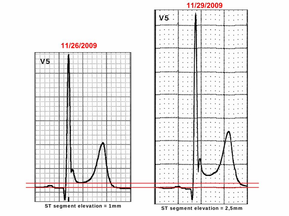
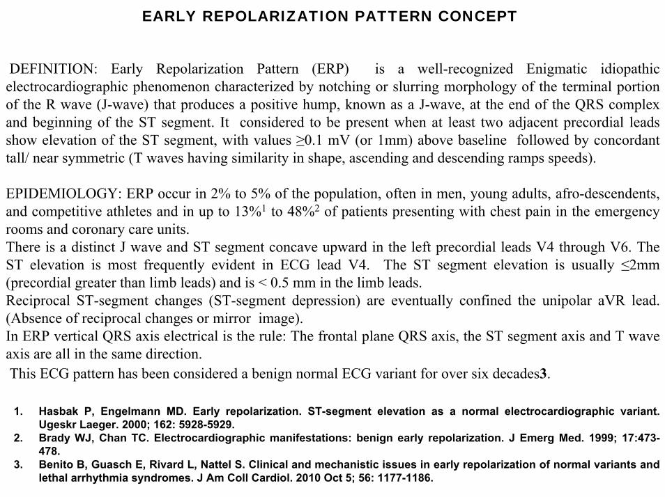
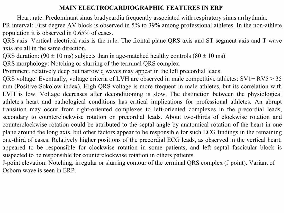
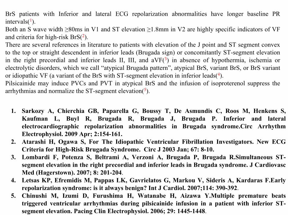
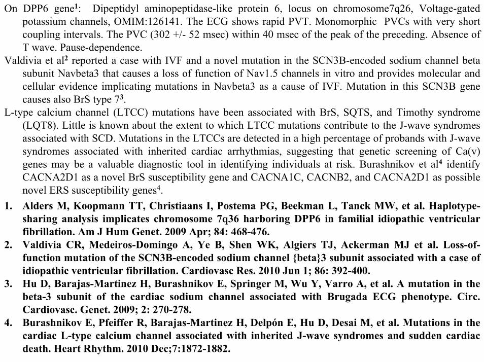
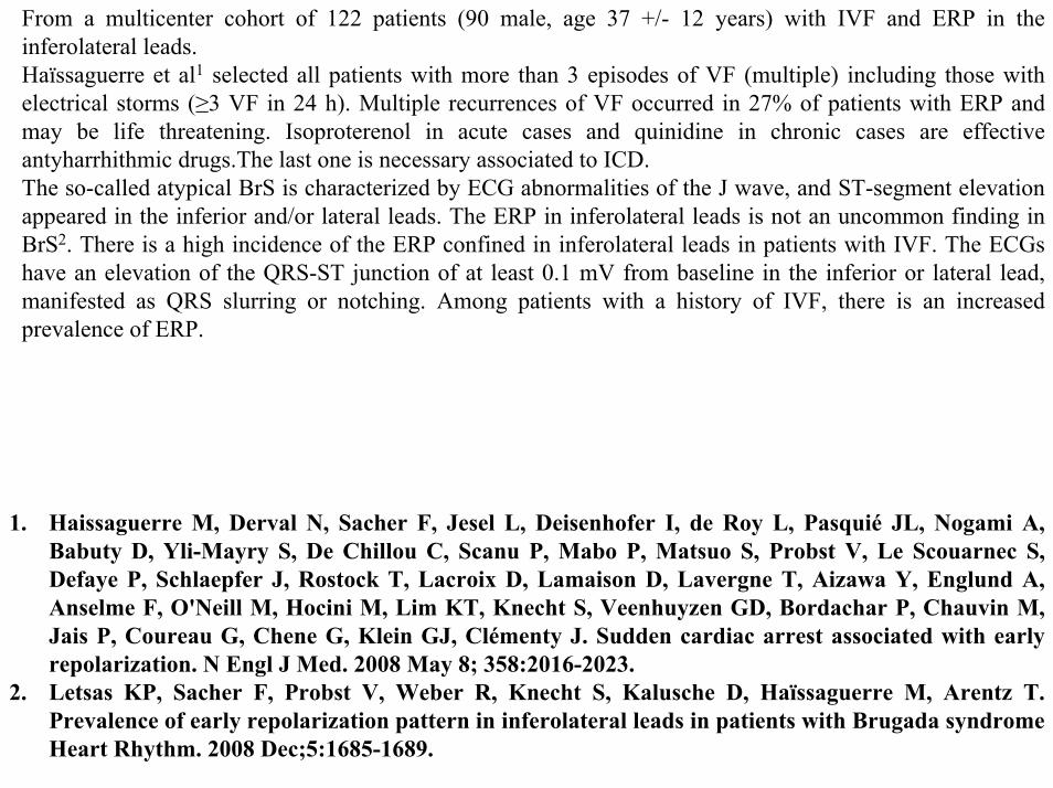
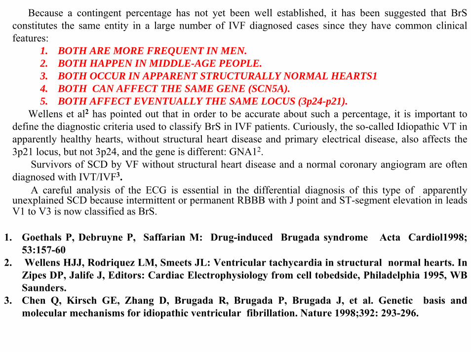
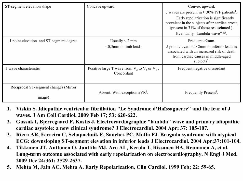
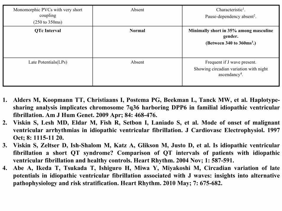
ConclusionIn spite of both malignant early repolarization abnormalities of IVF and benign early repolarization pattern being able to cause J point and ST segment elevation the characteristics of both are clearly different to the point of a differentiation being quite simple when ECGs are analyzed by experts Any ER should raise the suspicion of IVF when it presents ST segment elevation ge 2 mm mainly in inferior wall superior convexity eventually lambda wave shape pattern of dynamic presentation inconstant with dramatic intensity of ST segment elevation discordant T wave polarity related ST segment shift presence of reciprocal ST-segment changes (mirror image) tendency to discrete short QTc interval in male gender(ge340ms) premature monomorphic ventricular contractions with very short coupling interval(250 to 350ms) not related with exercise and late potentials with circadian variation and night ascendancy on SA-ECG Contrarily in benign ERP ST elevation observed in the mid-to-left precordial leads is usually le 2 mm and lt05 mm in the limb leads and concave to the top followed by a positive concordant tall T wave
Related Documents


