Review of Spinal Cord with Basics of Neuroanatomy Brain Meninges Prof. D.H. Pauža

Welcome message from author
This document is posted to help you gain knowledge. Please leave a comment to let me know what you think about it! Share it to your friends and learn new things together.
Transcript

Review of Spinal Cord
with
Basics of Neuroanatomy
Brain Meninges
Prof. D.H. Pauža


Review of Spinal Cord
with
Basics of Neuroanatomy
Brain Meninges
Prof. D.H. Pauža

Neurons and Neuroglia

Neuron Body (soma)
Perikaryon
Nissl substance or Tigroid
Dendrites
Axon
Myelin
Terminals
Synapses
Human brain contains per
1011-12 (trillions) neurons
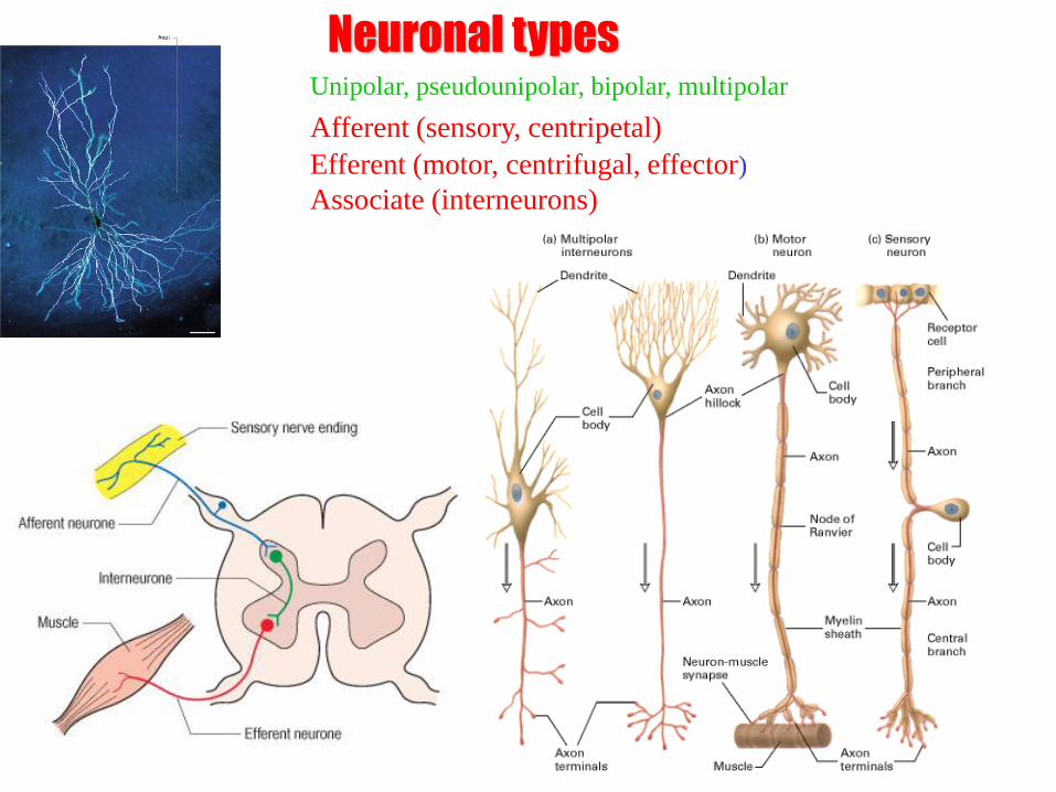
Unipolar, pseudounipolar, bipolar, multipolar
Afferent (sensory, centripetal)
Efferent (motor, centrifugal, effector)
Associate (interneurons)
Neuronal types

Synapse
In human brain – neurons 1011 (100 trillions)
Synapses – 1015 (quadrillions)
Presynaptic membrane
Postsynaptic membrane, receptors
Synaptic cleft
Synaptic vesicles, neuromediator
Mitochondria

Neuromediators •Acetylcholine
•Noradrenaline
•Serotonin
•GABA
•Endorphin
•Encephalin
•P substance
•Neuronal nitric oxide
Adrenergic nerve ending. There are many 50-nm-diameter
vesicles (arrow) with dark, electron-dense cores containing
norepinephrine. x40,000.

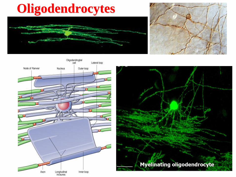
Cell Types of Neuroglia Astrocytes - Oligodendrocytes – Ependimocytes - Microglia
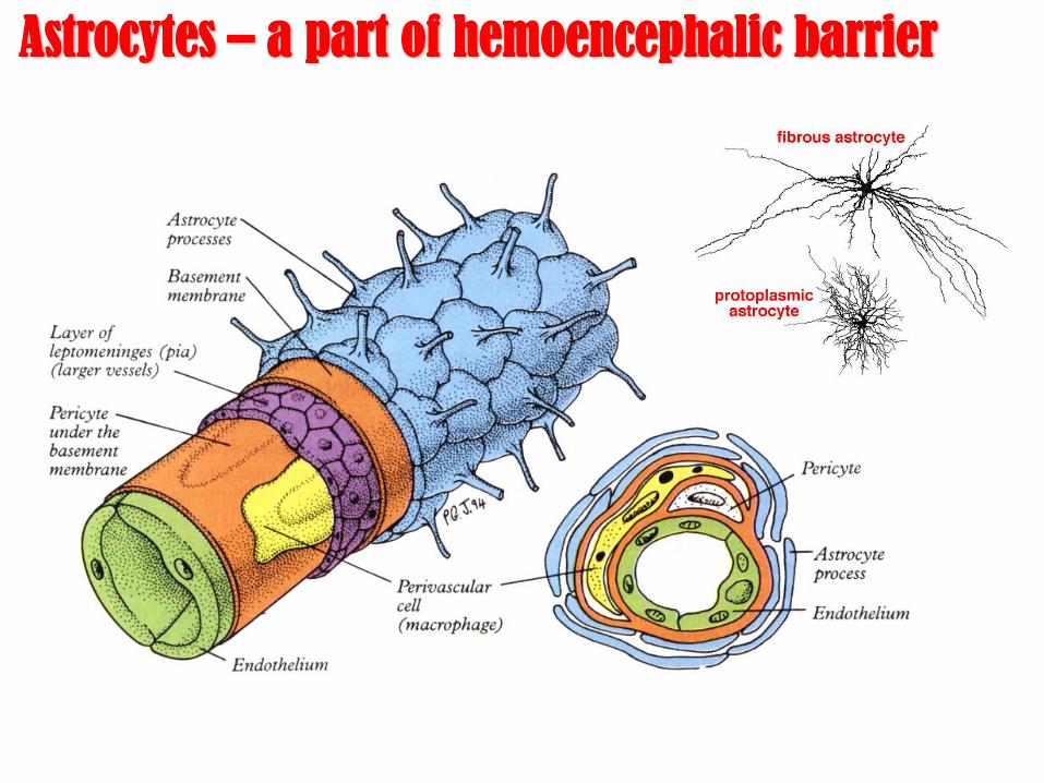
Astrocytes – a part of hemoencephalic barrier

Ependimocytes and
microglial cells
Microglia represent the endogenous brain defense and immune system,
which is responsible for CNS protection against various types of
pathogenic factors. After invading the CNS, microglial precursors
disseminate relatively homogeneously throughout the neural tissue and
acquire a specific phenotype, which clearly distinguish them from their
precursors, the blood-derived monocytes.
The ´resting´ microglia are the fastest moving cells in the brain

PNS
Neuronal lemmocytes (Schwann cells)
Nerve fiber: myelinated and unmyelinated
Impulse propagation speed: 0,5-120 m/s
A, B, C types of nerve fibers
CNS
Neuroglia in peripheral nervous system


General definitions in
neuroanatomy

Meninges on spinal cord Dura mater, arachnoid and pia mater Epidural and subdural spaces
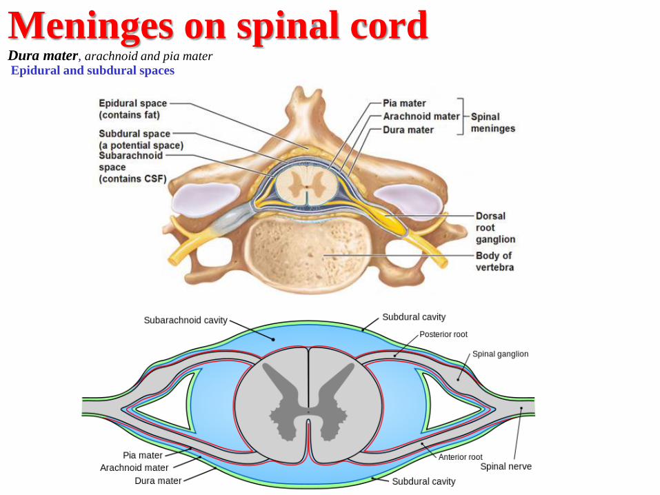
Meninges on spinal cord Dura mater, arachnoid and pia mater Epidural and subdural spaces

Meninges on brain: Dura mater, arachnoid and pia mater
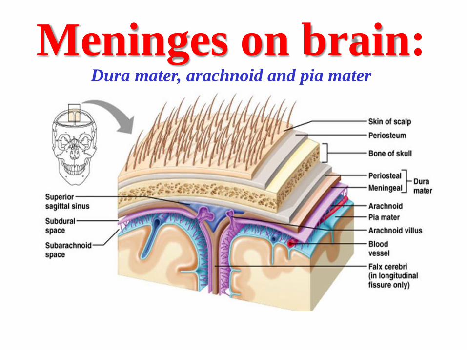
Meninges on brain: Dura mater, arachnoid and pia mater
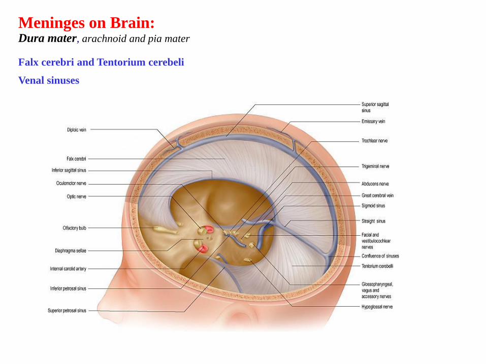
Meninges on Brain: Dura mater, arachnoid and pia mater Falx cerebri and Tentorium cerebeli
Venal sinuses

Meninges on Brain: Dura mater, arachnoid and pia mater
Arachnoid villi, granulationes arachnoideae

Meninges: Dura mater, arachnoid and pia mater

Meninges: Dura mater, arachnoid and pia mater

Cisterna magna or
cerebellomedulary cisterna
Interpedicular cisterna
Subarachnoidal space
Liquor or cerebrospinal fluid (CSF)

Central canal and Brain ventricles Lateral, Third and Fourth
Interventricular foramen
Cerebral aqueduct
Lateral and median apertures 4th ventricle

Choroid plexus and Tela charoidea
Cerebrospinal fluid

Choroid plexus and Tela charoidea
Cerebrospinal fluid

Neuronal organization
CNS:
Grey matter: nuclei and cortex, functional nuclei White matter: pathways, fascicles

Neuronal organization
PNS: Ganglia and nerves
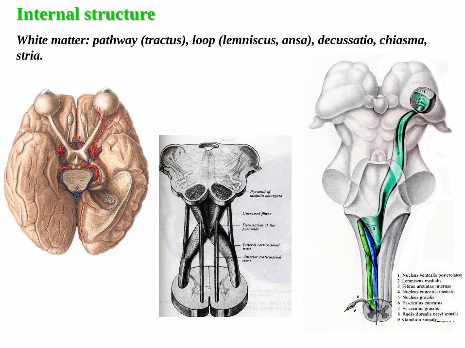
Internal structure
White matter: pathway (tractus), loop (lemniscus, ansa), decussatio, chiasma,
stria.

Anatomy of Spinal Cord

Anatomy of Spinal Cord Conus medularis up to L2
Filum terminale

Spinal cord Conus medularis, L2
Filum terminale

SPINAL CORD
External anatomy Cervical and lumbar enlargements;
Anterior median fissure,
Posterior median groove or sulcus,
Posterior and anterior lateral sulci.
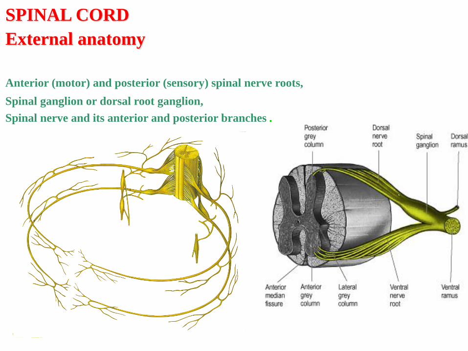
SPINAL CORD
External anatomy
Anterior (motor) and posterior (sensory) spinal nerve roots,
Spinal ganglion or dorsal root ganglion,
Spinal nerve and its anterior and posterior branches .

Cauda equina
SPINAL CORD
External anatomy

Segments of spinal cord
C1-8
Th1-12
L1-5
S1-5
Co1
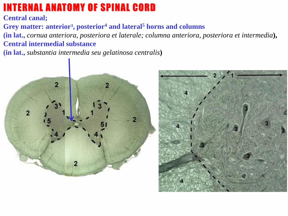
INTERNAL ANATOMY OF SPINAL CORD Central canal;
Grey matter: anterior3, posterior4 and lateral5 horns and columns
(in lat., cornua anteriora, posteriora et laterale; columna anteriora, posteriora et intermedia),
Central intermedial substance
(in lat., substantia intermedia seu gelatinosa centralis)

Pars cervicalis Pars thoracica Pars lumbalis
INTERNAL ANATOMY OF SPINAL CORD
Grey matter: anterior, posterior and lateral horns and columns (cornua anteriora,
posteriora et laterale; columna anteriora, posteriora et intermedia)
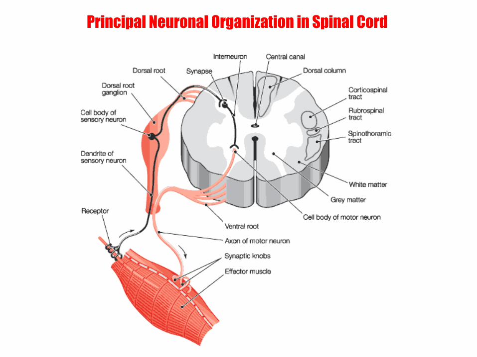
Principal Neuronal Organization in Spinal Cord

I
IV
VII
VIII
X
IX
Nucleus proprius, Sensation of skin
Sub.gelatinosa,
Pain and temperature
signal transmission nucleus
Nucleus marginalis, Pain
and temperature
signal transmission nucleus
INTERNAL ANATOMY OF SPINAL CORD
Grey matter:
nuclei and/or laminae by Bror Rexed
Nucleus intermediomedianalis Sensation of internal organs
Nucleus dorsalis
(C8-L2) basilaris. Proprioreception of the upper
and lower limbs
Nucleus
intermediolateralis
Sympathetic nucleus
(S2-4: Nucl.parasympathicus).
(Parasympathetic) V VI
Nuclei
posteromedialis
et centralis. Associate neurons and
descending pathways
interaction nucleus
Nuclei mediales et laterales. Motor nucleus of muscles of trunk and
limbs
Substantia intermedia centralis.
Nucleus of commissural neurons composing
commissura alba
Bror Rexed (June 19, 1914 - August 21, 2002) was a Swedish neuroscientist and professor
at Uppsala University. Internationally, he is best known today for his development
of the system now known as Rexed laminae.
Rexed B (1952). "The cytoarchitectonic organization of the spinal cord in the cat.".
J Comp Neurol 96 (3): 414-95.
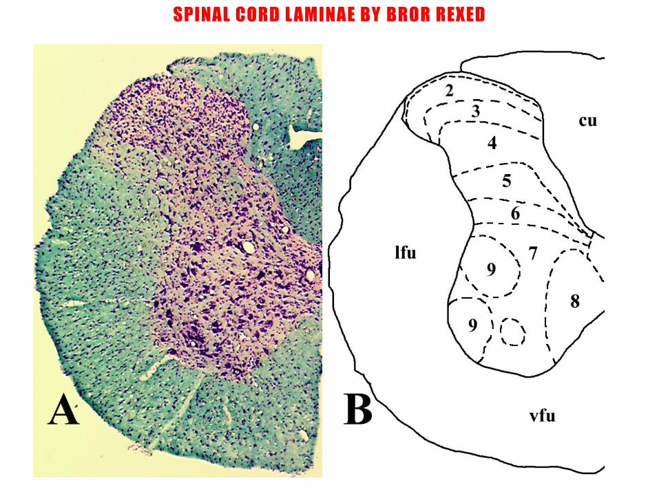
SPINAL CORD L AMINAE BY BROR REXED
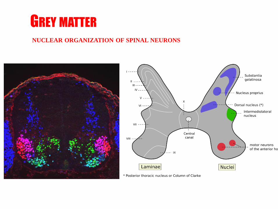
GREY MATTER NUCLEAR ORGANIZATION OF SPINAL NEURONS

GREY MATTER NUCLEAR ORGANIZATION OF SPINAL NEURONS

INTERNAL ANATOMY OF SPINAL CORD
White matter: anterior, posterior and lateral funiculae
(funiculus anterior, lateralis et posterior)

White matter Anterior, posterior and lateral funiculae; Funiculus anterior, lateralis et posterior.
Ascending and descending pathways;
Funiculus proprius

INTERNAL ANATOMY OF SPINAL CORD
White matter: anterior, posterior and lateral funiculae
(funiculus anterior, lateralis et posterior)
Ascending and descending pathways; funiculus proprius

Internal anatomy.
White matter: ascending-afferent pathways
Gracile fasciculus – tactile and proprioceptive sensations
Cuneate fasciculus - tactile and proprioceptive sensations
Spinothalamic tracts – pain and temperature sensation
Spinocerebellar tracts – proprioceptive sensation
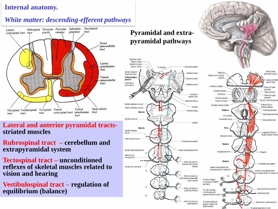
Pyramidal and extra-
pyramidal pathways
Lateral and anterior pyramidal tracts- striated muscles
Rubrospinal tract – cerebellum and extrapyramidal system
Tectospinal tract – unconditioned reflexes of skeletal muscles related to vision and hearing
Vestibulospinal tract – regulation of equilibrium (balance)
Internal anatomy.
White matter: descending-efferent pathways
Related Documents