Journal of Clinical Medicine Review Why Venous Leg Ulcers Have Difficulty Healing: Overview on Pathophysiology, Clinical Consequences, and Treatment Joseph D. Raffetto 1, * ,† , Daniela Ligi 2,† , Rosanna Maniscalco 2 , Raouf A. Khalil 1 and Ferdinando Mannello 2, * Citation: Raffetto, J.D.; Ligi, D.; Maniscalco, R.; Khalil, R.A.; Mannello, F. Why Venous Leg Ulcers Have Difficulty Healing: Overview on Pathophysiology, Clinical Consequences, and Treatment. J. Clin. Med. 2021, 10, 29. https://dx.doi.org/ 10.3390/jcm10010029 Received: 24 November 2020 Accepted: 21 December 2020 Published: 24 December 2020 Publisher’s Note: MDPI stays neu- tral with regard to jurisdictional claims in published maps and institutional affiliations. Copyright: © 2020 by the authors. Li- censee MDPI, Basel, Switzerland. This article is an open access article distributed under the terms and conditions of the Creative Commons Attribution (CC BY) license (https://creativecommons.org/ licenses/by/4.0/). 1 Vascular Surgery Research Laboratories, Division of Vascular and Endovascular Surgery, Brigham and Women’s Hospital, Harvard Medical School, Boston, MA 02115, USA; [email protected] 2 Department of Biomolecular Sciences, Section of Biochemistry and Biotechnology, Unit of Clinical Biochemistry, University Carlo Bo of Urbino, 61029 Urbino, Italy; [email protected] (D.L.); [email protected] (R.M.) * Correspondence: [email protected] (J.D.R.); [email protected] (F.M.) † Joseph D. Raffetto and Daniela Ligi contributed equally to this work. Abstract: Venous leg ulcers (VLUs) are one of the most common ulcers of the lower extremity. VLU affects many individuals worldwide, could pose a significant socioeconomic burden to the healthcare system, and has major psychological and physical impacts on the affected individual. VLU often occurs in association with post-thrombotic syndrome, advanced chronic venous disease, varicose veins, and venous hypertension. Several demographic, genetic, and environmental factors could trigger chronic venous disease with venous dilation, incompetent valves, venous reflux, and ve- nous hypertension. Endothelial cell injury and changes in the glycocalyx, venous shear-stress, and adhesion molecules could be initiating events in VLU. Increased endothelial cell permeability and leukocyte infiltration, and increases in inflammatory cytokines, matrix metalloproteinases (MMPs), reactive oxygen and nitrogen species, iron deposition, and tissue metabolites also contribute to the pathogenesis of VLU. Treatment of VLU includes compression therapy and endovenous ablation to occlude the axial reflux. Other interventional approaches such as subfascial endoscopic perforator surgery and iliac venous stent have shown mixed results. With good wound care and compres- sion therapy, VLU usually heals within 6 months. VLU healing involves orchestrated processes including hemostasis, inflammation, proliferation, and remodeling and the contribution of different cells including leukocytes, platelets, fibroblasts, vascular smooth muscle cells, endothelial cells, and keratinocytes as well as the release of various biomolecules including transforming growth factor-β, cytokines, chemokines, MMPs, tissue inhibitors of MMPs (TIMPs), elastase, urokinase plasminogen activator, fibrin, collagen, and albumin. Alterations in any of these physiological wound closure processes could delay VLU healing. Also, these histological and soluble biomarkers can be used for VLU diagnosis and assessment of its progression, responsiveness to healing, and prognosis. If not treated adequately, VLU could progress to non-healed or granulating VLU, causing physical immo- bility, reduced quality of life, cellulitis, severe infections, osteomyelitis, and neoplastic transformation. Recalcitrant VLU shows prolonged healing time with advanced age, obesity, nutritional deficiencies, colder temperature, preexisting venous disease, deep venous thrombosis, and larger wound area. VLU also has a high, 50–70% recurrence rate, likely due to noncompliance with compression therapy, failure of surgical procedures, incorrect ulcer diagnosis, progression of venous disease, and poorly understood pathophysiology. Understanding the molecular pathways underlying VLU has led to new lines of therapy with significant promise including biologics such as bilayer living skin construct, fibroblast derivatives, and extracellular matrices and non-biologic products such as poly-N-acetyl glucosamine, human placental membranes amnion/chorion allografts, ACT1 peptide inhibitor of connexin 43, sulodexide, growth factors, silver dressings, MMP inhibitors, and modulators of reactive oxygen and nitrogen species, the immune response and tissue metabolites. Preventive measures including compression therapy and venotonics could also reduce the risk of progression to chronic venous insufficiency and VLU in susceptible individuals. J. Clin. Med. 2021, 10, 29. https://dx.doi.org/10.3390/jcm10010029 https://www.mdpi.com/journal/jcm

Welcome message from author
This document is posted to help you gain knowledge. Please leave a comment to let me know what you think about it! Share it to your friends and learn new things together.
Transcript
Journal of
Clinical Medicine
Review
Why Venous Leg Ulcers Have Difficulty Healing: Overview onPathophysiology, Clinical Consequences, and Treatment
Joseph D. Raffetto 1,*,†, Daniela Ligi 2,† , Rosanna Maniscalco 2, Raouf A. Khalil 1 and Ferdinando Mannello 2,*
�����������������
Citation: Raffetto, J.D.; Ligi, D.;
Maniscalco, R.; Khalil, R.A.;
Mannello, F. Why Venous Leg Ulcers
Have Difficulty Healing: Overview
on Pathophysiology, Clinical
Consequences, and Treatment. J. Clin.
Med. 2021, 10, 29. https://dx.doi.org/
10.3390/jcm10010029
Received: 24 November 2020
Accepted: 21 December 2020
Published: 24 December 2020
Publisher’s Note: MDPI stays neu-
tral with regard to jurisdictional claims
in published maps and institutional
affiliations.
Copyright: © 2020 by the authors. Li-
censee MDPI, Basel, Switzerland. This
article is an open access article distributed
under the terms and conditions of the
Creative Commons Attribution (CC BY)
license (https://creativecommons.org/
licenses/by/4.0/).
1 Vascular Surgery Research Laboratories, Division of Vascular and Endovascular Surgery, Brigham andWomen’s Hospital, Harvard Medical School, Boston, MA 02115, USA; [email protected]
2 Department of Biomolecular Sciences, Section of Biochemistry and Biotechnology, Unit of ClinicalBiochemistry, University Carlo Bo of Urbino, 61029 Urbino, Italy; [email protected] (D.L.);[email protected] (R.M.)
* Correspondence: [email protected] (J.D.R.); [email protected] (F.M.)† Joseph D. Raffetto and Daniela Ligi contributed equally to this work.
Abstract: Venous leg ulcers (VLUs) are one of the most common ulcers of the lower extremity. VLUaffects many individuals worldwide, could pose a significant socioeconomic burden to the healthcaresystem, and has major psychological and physical impacts on the affected individual. VLU oftenoccurs in association with post-thrombotic syndrome, advanced chronic venous disease, varicoseveins, and venous hypertension. Several demographic, genetic, and environmental factors couldtrigger chronic venous disease with venous dilation, incompetent valves, venous reflux, and ve-nous hypertension. Endothelial cell injury and changes in the glycocalyx, venous shear-stress, andadhesion molecules could be initiating events in VLU. Increased endothelial cell permeability andleukocyte infiltration, and increases in inflammatory cytokines, matrix metalloproteinases (MMPs),reactive oxygen and nitrogen species, iron deposition, and tissue metabolites also contribute to thepathogenesis of VLU. Treatment of VLU includes compression therapy and endovenous ablation toocclude the axial reflux. Other interventional approaches such as subfascial endoscopic perforatorsurgery and iliac venous stent have shown mixed results. With good wound care and compres-sion therapy, VLU usually heals within 6 months. VLU healing involves orchestrated processesincluding hemostasis, inflammation, proliferation, and remodeling and the contribution of differentcells including leukocytes, platelets, fibroblasts, vascular smooth muscle cells, endothelial cells, andkeratinocytes as well as the release of various biomolecules including transforming growth factor-β,cytokines, chemokines, MMPs, tissue inhibitors of MMPs (TIMPs), elastase, urokinase plasminogenactivator, fibrin, collagen, and albumin. Alterations in any of these physiological wound closureprocesses could delay VLU healing. Also, these histological and soluble biomarkers can be used forVLU diagnosis and assessment of its progression, responsiveness to healing, and prognosis. If nottreated adequately, VLU could progress to non-healed or granulating VLU, causing physical immo-bility, reduced quality of life, cellulitis, severe infections, osteomyelitis, and neoplastic transformation.Recalcitrant VLU shows prolonged healing time with advanced age, obesity, nutritional deficiencies,colder temperature, preexisting venous disease, deep venous thrombosis, and larger wound area.VLU also has a high, 50–70% recurrence rate, likely due to noncompliance with compression therapy,failure of surgical procedures, incorrect ulcer diagnosis, progression of venous disease, and poorlyunderstood pathophysiology. Understanding the molecular pathways underlying VLU has led tonew lines of therapy with significant promise including biologics such as bilayer living skin construct,fibroblast derivatives, and extracellular matrices and non-biologic products such as poly-N-acetylglucosamine, human placental membranes amnion/chorion allografts, ACT1 peptide inhibitor ofconnexin 43, sulodexide, growth factors, silver dressings, MMP inhibitors, and modulators of reactiveoxygen and nitrogen species, the immune response and tissue metabolites. Preventive measuresincluding compression therapy and venotonics could also reduce the risk of progression to chronicvenous insufficiency and VLU in susceptible individuals.
J. Clin. Med. 2021, 10, 29. https://dx.doi.org/10.3390/jcm10010029 https://www.mdpi.com/journal/jcm
J. Clin. Med. 2021, 10, 29 2 of 34
Keywords: chronic venous disease; chronic venous insufficiency; venous leg ulcer; healing; biochem-istry; pathophysiology; clinical medicine; therapy
1. Introduction and Scope of the Problem
Venous leg ulcer (VLU) is the most common type of ulcer in the lower extremity [1].VLU accounts for 70–80% of ulcers presenting for evaluation and treatment to differentprofessions across different specialties including primary care physicians, geriatricians,wound care specialist, phlebologist, surgical specialties, cardiologist, and vascular surgeons.The prevalence of VLU is up to 2% of the population and, importantly, increases to 5% ofindividuals over the age of 65 years old [2,3]. Venous leg ulcer is a worldwide problem inmany countries and regions including the United States, the United Kingdom, Australia,India, Africa, and Europe. The number of affected individuals is staggering in Africa, withan estimated 25 to 135 million individuals having VLU and chronic wounds (with themajority of them being VLU). Europe has up to 2.2 million people affected, and over 6million individuals are affected in the United States [4]. It is important to note that VLUcan heal with good wound care and compression, which is the mainstay of treatment.Healing rates of VLU of 76% at 16 weeks can be achieved with compression [5]. However,a major issue with VLU is the high recurrence rates, which can be significant and as highas 50–70% at 6 months [1]. The morbidity of VLU has many financial and socioeconomicimpacts, especially given the high recurrence rates. The treatment of VLU is significant,involving and requiring many resources, specialties, appointments, inconveniences tothe patients, wound care products, psychosocial events, and hardships and has a majorhealthcare burden. After taking into consideration all aspects of caring for patients withVLU, including doctors visit, nursing care, wound care, and bandages applied along withcompression; surgical and endovenous treatments; and hospitalization for complicationsrelated to pain, drainage and progression, and infections, the cost becomes exponentiallyelevated. The associated costs for VLU care are just over $15,000 but increase significantlyfor patients who have delayed healing and can result in costs as high as $34,000 per patientper year, with most of the cost driven by outpatient visits, nursing care, and admissions tohospitals for related complications, usually infection [1,6].
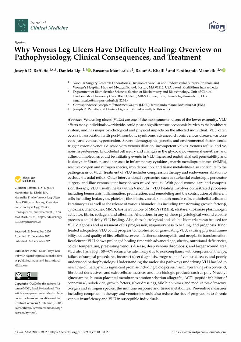
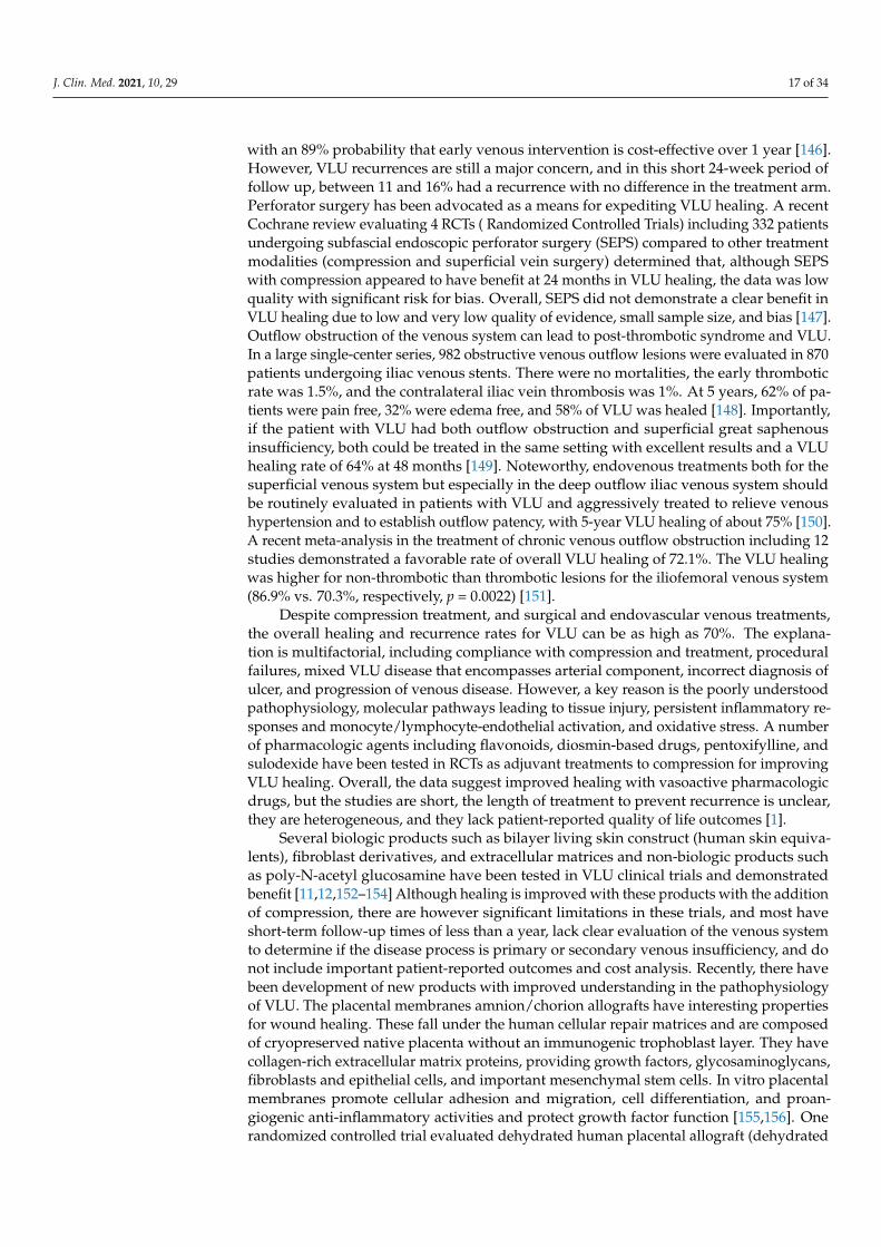
Patients with VLU have increased missed workdays, with 29% higher work-loss costs.However, a price on the burden endured by patients with VLU cannot be estimated whenone takes into account the psychosocial impact with significant isolation, embarrassment,negative emotions, anxiety, depression, loss of self-worth, dependency, and sleep distur-bance. The annual United States taxpayer burden for VLU is estimated at an astonishingcost of $14.9 billion (Figure 1) [7].
The intention of this comprehensive review is to provide practitioners caring forpatients with VLU with a foundation of information that will define causes of VLU andother ulcers that are less common that may be mistaken for VLU, the clinical manifestationsof VLU, and delayed healing of and difficulty healing VLU that is common place in clinicalpractice and to provide pathophysiological molecular insights on important regulators andinflammatory mediators that are critical factors in propagating the VLU refractory state ofcontinued inflammation, surgical treatments and innovations, and drug therapies that haveevolved given our increased scientific discovery and knowledge that lead to better targetedtherapies and finally with information on the means to prevent progression, occurrence,and recurrence of VLU.
J. Clin. Med. 2021, 10, 29 3 of 34J. Clin. Med. 2021, 10, x FOR PEER REVIEW 3 of 33
Figure 1. Facts about venous leg ulcer.
2. Pathophysiology
2.1. Definition and Etiology of Venous Leg Ulcers
VLU can be defined as a full-thickness defect of the skin frequently seen in the ankle
region that fails to heal spontaneously and is sustained by chronic venous disease (CVD,
the spectrum of venous diseases affecting the lower limbs) [8]. In more recent guidelines,
a VLU is defined by best practice and uses the standard definition of an open skin lesion
of the leg or foot that occurs in an area affected by venous hypertension [1].
VLU is a complex system involving mechanisms that affect venous macrovasculature
and microvasculature. The macrovasculature involves abnormalities with hemodynam-
ics, leading to venous hypertension that involves superficial venous insufficiency that can
overwhelm the deep system, junctions, and reentry points in compartments of the lower
extremity and cause outflow obstruction via the iliofemoral venous system, calf muscle
pump dysfunction, and perforator venous insufficiency. The majority (70–80%) of patients
with VLU have primary venous insufficiency (reflux) from varicose vein disease, and
about 20–30% have secondary venous insufficiency from post thrombotic syndrome (PTS)
[9]. Although there are many more patients with primary venous insufficiency, PTS has a
much higher risk of developing VLU and is much more aggressive in its natural history,
making treatment more challenging [10]. The microvasculature, which includes the gly-
cocalyx and endothelium, is affected by changes in shear stress and activation of leuko-
cytes and adhesion molecules occurring in both large and microscopic veins. The micro-
vascular system is composed of a network of capillaries, post-capillary venules, interstit-
ium, and lymphatics that respond to overexpressed inflammatory pathways and upregu-
lation of cytokines, chemokines, matrix metalloproteinases (MMPs), iron free radicals, and
activated oxygen and nitrogen species that all have detrimental effects to the surrounding
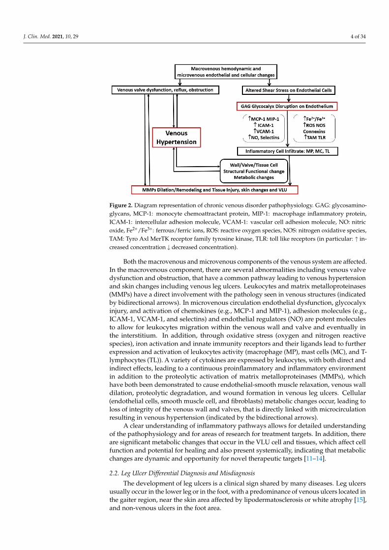
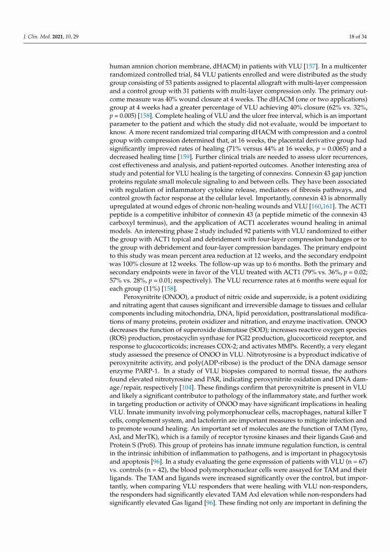
tissues and possibly systemic effects (Figure 2).
Figure 1. Facts about venous leg ulcer.
2. Pathophysiology2.1. Definition and Etiology of Venous Leg Ulcers
VLU can be defined as a full-thickness defect of the skin frequently seen in the ankleregion that fails to heal spontaneously and is sustained by chronic venous disease (CVD,the spectrum of venous diseases affecting the lower limbs) [8]. In more recent guidelines, aVLU is defined by best practice and uses the standard definition of an open skin lesion ofthe leg or foot that occurs in an area affected by venous hypertension [1].
VLU is a complex system involving mechanisms that affect venous macrovasculatureand microvasculature. The macrovasculature involves abnormalities with hemodynamics,leading to venous hypertension that involves superficial venous insufficiency that canoverwhelm the deep system, junctions, and reentry points in compartments of the lowerextremity and cause outflow obstruction via the iliofemoral venous system, calf musclepump dysfunction, and perforator venous insufficiency. The majority (70–80%) of patientswith VLU have primary venous insufficiency (reflux) from varicose vein disease, and about20–30% have secondary venous insufficiency from post thrombotic syndrome (PTS) [9].Although there are many more patients with primary venous insufficiency, PTS has a muchhigher risk of developing VLU and is much more aggressive in its natural history, makingtreatment more challenging [10]. The microvasculature, which includes the glycocalyxand endothelium, is affected by changes in shear stress and activation of leukocytes andadhesion molecules occurring in both large and microscopic veins. The microvascularsystem is composed of a network of capillaries, post-capillary venules, interstitium, andlymphatics that respond to overexpressed inflammatory pathways and upregulation ofcytokines, chemokines, matrix metalloproteinases (MMPs), iron free radicals, and activatedoxygen and nitrogen species that all have detrimental effects to the surrounding tissuesand possibly systemic effects (Figure 2).
J. Clin. Med. 2021, 10, 29 4 of 34J. Clin. Med. 2021, 10, x FOR PEER REVIEW 4 of 33
Figure 2. Diagram representation of chronic venous disorder pathophysiology. GAG: glycosaminoglycans, MCP-
1: monocyte chemoattractant protein, MIP-1: macrophage inflammatory protein, ICAM-1: intercellular adhesion molecule,
VCAM-1: vascular cell adhesion molecule, NO: nitric oxide, Fe2+/Fe3+: ferrous/ferric ions, ROS: reactive oxygen species,
NOS: nitrogen oxidative species, TAM: Tyro Axl MerTK receptor family tyrosine kinase, TLR: toll like receptors.
Both the macrovenous and microvenous components of the venous system are af-
fected. In the macrovenous component, there are several abnormalities including venous
valve dysfunction and obstruction, that have a common pathway leading to venous hy-
pertension and skin changes including venous leg ulcers. Leukocytes and matrix metallo-
proteinases (MMPs) have a direct involvement with the pathology seen in venous struc-
tures (indicated by bidirectional arrows). In microvenous circulation endothelial dysfunc-
tion, glycocalyx injury, and activation of chemokines (e.g., MCP-1 and MIP-1), adhesion
molecules (e.g., ICAM-1, VCAM-1, and selectins) and endothelial regulators (NO) are po-
tent molecules to allow for leukocytes migration within the venous wall and valve and
eventually in the interstitium. In addition, through oxidative stress (oxygen and nitrogen
reactive species), iron activation and innate immunity receptors and their ligands lead to
further expression and activation of leukocytes activity (macrophage (MP), mast cells
(MC), and T-lymphocytes (TL)). A variety of cytokines are expressed by leukocytes, with
both direct and indirect effects, leading to a continuous proinflammatory and inflamma-
tory environment in addition to the proteolytic activation of matrix metalloproteinases
(MMPs), which have both been demonstrated to cause endothelial-smooth muscle relax-
ation, venous wall dilation, proteolytic degradation, and wound formation in venous leg
ulcers. Cellular (endothelial cells, smooth muscle cell, and fibroblasts) metabolic changes
occur, leading to loss of integrity of the venous wall and valves, that is directly linked with
microcirculation resulting in venous hypertension (indicated by the bidirectional arrows).
A clear understanding of inflammatory pathways allows for detailed understanding
of the pathophysiology and for areas of research for treatment targets. In addition, there
are significant metabolic changes that occur in the VLU cell and tissues, which affect cell
function and potential for healing and also present systemically, indicating that metabolic
changes are dynamic and opportunity for novel therapeutic targets [11–14].
2.2. Leg Ulcer Differential Diagnosis and Misdiagnosis
The development of leg ulcers is a clinical sign shared by many diseases. Leg ulcers
usually occur in the lower leg or in the foot, with a predominance of venous ulcers located
in the gaiter region, near the skin area affected by lipodermatosclerosis or white atrophy
[15], and non-venous ulcers in the foot area.
Figure 2. Diagram representation of chronic venous disorder pathophysiology. GAG: glycosamino-glycans, MCP-1: monocyte chemoattractant protein, MIP-1: macrophage inflammatory protein,ICAM-1: intercellular adhesion molecule, VCAM-1: vascular cell adhesion molecule, NO: nitricoxide, Fe2+/Fe3+: ferrous/ferric ions, ROS: reactive oxygen species, NOS: nitrogen oxidative species,TAM: Tyro Axl MerTK receptor family tyrosine kinase, TLR: toll like receptors (in particular: ↑ in-creased concentration ↓ decreased concentration).
Both the macrovenous and microvenous components of the venous system are affected.In the macrovenous component, there are several abnormalities including venous valvedysfunction and obstruction, that have a common pathway leading to venous hypertensionand skin changes including venous leg ulcers. Leukocytes and matrix metalloproteinases(MMPs) have a direct involvement with the pathology seen in venous structures (indicatedby bidirectional arrows). In microvenous circulation endothelial dysfunction, glycocalyxinjury, and activation of chemokines (e.g., MCP-1 and MIP-1), adhesion molecules (e.g.,ICAM-1, VCAM-1, and selectins) and endothelial regulators (NO) are potent moleculesto allow for leukocytes migration within the venous wall and valve and eventually inthe interstitium. In addition, through oxidative stress (oxygen and nitrogen reactivespecies), iron activation and innate immunity receptors and their ligands lead to furtherexpression and activation of leukocytes activity (macrophage (MP), mast cells (MC), and T-lymphocytes (TL)). A variety of cytokines are expressed by leukocytes, with both direct andindirect effects, leading to a continuous proinflammatory and inflammatory environmentin addition to the proteolytic activation of matrix metalloproteinases (MMPs), whichhave both been demonstrated to cause endothelial-smooth muscle relaxation, venous walldilation, proteolytic degradation, and wound formation in venous leg ulcers. Cellular(endothelial cells, smooth muscle cell, and fibroblasts) metabolic changes occur, leading toloss of integrity of the venous wall and valves, that is directly linked with microcirculationresulting in venous hypertension (indicated by the bidirectional arrows).
A clear understanding of inflammatory pathways allows for detailed understandingof the pathophysiology and for areas of research for treatment targets. In addition, thereare significant metabolic changes that occur in the VLU cell and tissues, which affect cellfunction and potential for healing and also present systemically, indicating that metabolicchanges are dynamic and opportunity for novel therapeutic targets [11–14].
2.2. Leg Ulcer Differential Diagnosis and Misdiagnosis
The development of leg ulcers is a clinical sign shared by many diseases. Leg ulcersusually occur in the lower leg or in the foot, with a predominance of venous ulcers located inthe gaiter region, near the skin area affected by lipodermatosclerosis or white atrophy [15],and non-venous ulcers in the foot area.
J. Clin. Med. 2021, 10, 29 5 of 34
Chronic wounds of the lower extremities could be sustained by several local andsystemic causative factors, leading to a broad comparison among ulcers.
It has been estimated that the venous origin impacts 50–75% of chronic leg ulcers, andthis percentage heavily increases if foot ulcers are excluded. These numbers are strictlylinked to the fact that signs of CVD (i.e., varicose veins, edema, and skin changes) couldbe observed in at least 25% of the population, thus increasing the probability to diagnoseCVD (chronic venous disease )/CVI (chronic venous insufficiency) also in patients affectedby other forms of ulcer [15].
Besides the venous origin, other common etiologies are arterial (5–10%), mixed (arterio-venous), neuropathic, diabetic, and pressure ulcers, for which the prevalence reflects overallpopulation aging.
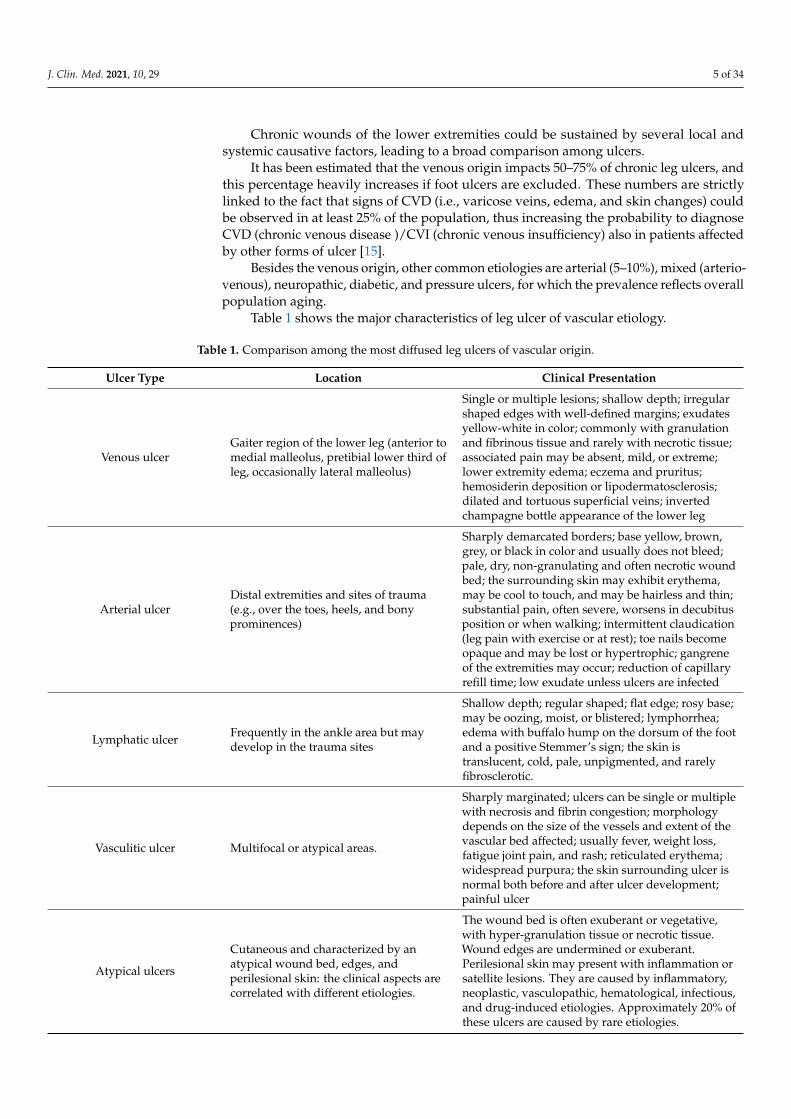
Table 1 shows the major characteristics of leg ulcer of vascular etiology.
Table 1. Comparison among the most diffused leg ulcers of vascular origin.
Ulcer Type Location Clinical Presentation
Venous ulcerGaiter region of the lower leg (anterior tomedial malleolus, pretibial lower third ofleg, occasionally lateral malleolus)
Single or multiple lesions; shallow depth; irregularshaped edges with well-defined margins; exudatesyellow-white in color; commonly with granulationand fibrinous tissue and rarely with necrotic tissue;associated pain may be absent, mild, or extreme;lower extremity edema; eczema and pruritus;hemosiderin deposition or lipodermatosclerosis;dilated and tortuous superficial veins; invertedchampagne bottle appearance of the lower leg
Arterial ulcerDistal extremities and sites of trauma(e.g., over the toes, heels, and bonyprominences)
Sharply demarcated borders; base yellow, brown,grey, or black in color and usually does not bleed;pale, dry, non-granulating and often necrotic woundbed; the surrounding skin may exhibit erythema,may be cool to touch, and may be hairless and thin;substantial pain, often severe, worsens in decubitusposition or when walking; intermittent claudication(leg pain with exercise or at rest); toe nails becomeopaque and may be lost or hypertrophic; gangreneof the extremities may occur; reduction of capillaryrefill time; low exudate unless ulcers are infected
Lymphatic ulcer Frequently in the ankle area but maydevelop in the trauma sites
Shallow depth; regular shaped; flat edge; rosy base;may be oozing, moist, or blistered; lymphorrhea;edema with buffalo hump on the dorsum of the footand a positive Stemmer’s sign; the skin istranslucent, cold, pale, unpigmented, and rarelyfibrosclerotic.
Vasculitic ulcer Multifocal or atypical areas.
Sharply marginated; ulcers can be single or multiplewith necrosis and fibrin congestion; morphologydepends on the size of the vessels and extent of thevascular bed affected; usually fever, weight loss,fatigue joint pain, and rash; reticulated erythema;widespread purpura; the skin surrounding ulcer isnormal both before and after ulcer development;painful ulcer
Atypical ulcers
Cutaneous and characterized by anatypical wound bed, edges, andperilesional skin: the clinical aspects arecorrelated with different etiologies.
The wound bed is often exuberant or vegetative,with hyper-granulation tissue or necrotic tissue.Wound edges are undermined or exuberant.Perilesional skin may present with inflammation orsatellite lesions. They are caused by inflammatory,neoplastic, vasculopathic, hematological, infectious,and drug-induced etiologies. Approximately 20% ofthese ulcers are caused by rare etiologies.
J. Clin. Med. 2021, 10, 29 6 of 34
Table 1. Cont.
Ulcer Type Location Clinical Presentation
Venous ulcerGaiter region of the lower leg (anterior tomedial malleolus, pretibial lower third ofleg, occasionally lateral malleolus)
Single or multiple lesions; shallow depth; irregularshaped edges with well-defined margins; exudatesyellow-white in color; commonly with granulationand fibrinous tissue and rarely with necrotic tissue;associated pain may be absent, mild, or extreme;lower extremity edema; eczema and pruritus;hemosiderin deposition or lipodermatosclerosis;dilated and tortuous superficial veins; invertedchampagne bottle appearance of the lower leg
Arterial ulcerDistal extremities and sites of trauma(e.g., over the toes, heels, and bonyprominences)
Sharply demarcated borders; base yellow, brown,grey, or black in color and usually does not bleed;pale, dry, non-granulating and often necrotic woundbed; the surrounding skin may exhibit erythema,may be cool to touch, and may be hairless and thin;substantial pain, often severe, worsens in decubitusposition or when walking; intermittent claudication(leg pain with exercise or at rest); toe nails becomeopaque and may be lost or hypertrophic; gangreneof the extremities may occur; reduction of capillaryrefill time; low exudate unless ulcers are infected
Lymphatic ulcer Frequently in the ankle area but maydevelop in the trauma sites
Shallow depth; regular shaped; flat edge; rosy base;may be oozing, moist, or blistered; lymphorrhea;edema with buffalo hump on the dorsum of the footand a positive Stemmer’s sign; the skin istranslucent, cold, pale, unpigmented, and rarelyfibrosclerotic.
Vasculitic ulcer Multifocal or atypical areas.
Sharply marginated; ulcers can be single or multiplewith necrosis and fibrin congestion; morphologydepends on the size of the vessels and extent of thevascular bed affected; usually fever, weight loss,fatigue joint pain, and rash; reticulated erythema;widespread purpura; the skin surrounding ulcer isnormal both before and after ulcer development;painful ulcer
Atypical ulcers
Cutaneous and characterized by anatypical wound bed, edges, andperilesional skin: the clinical aspects arecorrelated with different etiologies.
The wound bed is often exuberant or vegetative,with hyper-granulation tissue or necrotic tissue.Wound edges are undermined or exuberant.Perilesional skin may present with inflammation orsatellite lesions. They are caused by inflammatory,neoplastic, vasculopathic, hematological, infectious,and drug-induced etiologies. Approximately 20% ofthese ulcers are caused by rare etiologies.
The location of the wound may help with differential diagnosis. In fact, VLUs areusually located in the gaiter region and exhibit signs of venous CVI (e.g., edema, dermatitis,lipodermatosclerosis, hyperpigmentation, or white atrophy); arterial ulcers are mainlylocated in the distal regions of the extremities. Pain, sensation of coldness, and changes inskin color following leg elevation usually accompany arterial ulcers [16]. Diabetic ulcersare frequently observed in more distal areas of the extremities (e.g., the lateral or pretibialaspects of the leg, the dorsum of the feet, the malleoli, and the distal aspects of the forefeetand toes); neuropathic ulcers in diabetic patients occur in the plantar area [16].
A broad spectrum of wounds mimics common VLU, and unusual wounds are oftenmisdiagnosed due to concurring risk factors. Accounting for 10% of the leg ulcers, othercauses include infections, skin cancers, metabolic disorders, inflammatory processes, andother diagnoses (Table 2) [17].
J. Clin. Med. 2021, 10, 29 7 of 34
Table 2. Differential diagnosis of leg ulcers.
Ulcer Etiology Ulcer Type
Vascular Venous, arterial, lymphatic, vasculitis
Metabolic Diabetes mellitus, gout, necrobiosis lipoidica, porphiria cutanea tarda, homocysteinuria,prolidase deficiency, hyperoxaluria, ulcerative colitis, avitaminosis, cutaneous calcinosis
Connective tissue diseaseInflammatory bowel disease, pyoderma gangrenosum, rheumatoid arthritis, generalized
and localized scleroderma, systemic lupus erythematous, bullous pemphigoid,dermatomyositis, Sjogren’ syndrome, polyarteritis nodosa, leukocytoclastic vasculitis
Cutaneous microthrombociticulcers
Cryofibrinogenemia, cryoglobulinemia, antiphospholipid syndrome, coagulopathies,calciphylaxis, cholesterol embolization
Hematological diseaseSickle cell disease, leukemia, thrombocytosis, thalassemia, hereditary spherocytosis,
glucose-6-phosphate dehydrogenase deficiency, essential thrombocythemia,granulocytopenia, polycythemia, monoclonal and polyclonal, dysproteinemia
Neoplastic disease
Basal cell carcinoma, squamous cell carcinoma, malignant melanoma, primary cutaneousB cell lymphoma, Marjolin’s ulcer, pseudoepitheliomatous hyperplasia, Kaposi’s sarcoma,angiosarcoma, Bowen’s disease, intra-epidermal carcinoma, papillomatosis cutis carcinoid,neoplasms of lymphoproliferative tissue, Hodgkin disease
Panniculitis Necrobiosis lipoidica, erythema nodosum, erythema induratum
Traumatic Pressure ulcers, radiation damage, thermal burns, decubitus
Iatrogenic Drugs.
AtypicalCutaneous ulcer, caused by inflammatory, neoplastic, vasculopathic, hematological,
infectious, and drug-induced etiologies, with approximately 20% of these ulcers caused byrare etiologies
Martorell HYTILU Hypertensive ischemic leg ulcer, stenotic subcutaneous arteriolosclerosis
InfectionPyogenic, osteomyelitis, tuberculosis, syphilis, tropical disease, fungal disease,
leishmaniasis, histoplasmosis, herpes, lupus vulgaris, amoebiasis, chromoblastomycosis,coccidiomycosis, viral
Several disorders of metabolic, hematological, autoimmune, and connective tissueorigin should be taken into consideration during diagnostic workup. Furthermore, neo-plastic diseases (e.g., basal cell carcinoma and squamous cell carcinoma) could also resultin leg ulceration, and chronic wounds may undergo malignant transformation (Marjolin’sulcer) [16]. Atypical wounds require histological assessment to be properly diagnosed [17].The recommendations are that a VLU that has been present for 4–6 weeks or longer shouldundergo biopsies of the skin edges to evaluate for other possible pathologies, especially ifthe leg ulcer does not improve with wound and compression therapy and atypical ulcersappear [1].
A careful differential diagnosis is imperative to make the best therapeutic choicebecause specific treatments depend on the underlying cause. The major challenge is to findthe main cause of non-venous leg ulcers in patients where CVD symptoms exist, giventhe high prevalence of venous disorders in the population. On the other hand, leg ulcerswithout signs of CVD should be addressed as non-venous ulcers [15].
Misdiagnosis of a leg ulcer has a great impact both on patient’s suffering, due todelayed wound healing, and on economic costs. Furthermore, improper treatments leadto relevant risks including aggravation of the underlying disease, masking of symptoms,delaying appropriate diagnosis, and increasing morbidity or mortality.
2.3. Clinical Manifestations, Healing, and Consequences
CVD includes a spectrum of clinical manifestations ranging from telangiectases andreticular veins to skin changes, such as lipodermatosclerosis and VLU. Varicose veinsare among the first clinical evidences of CVD. They are enlarged superficial veins thatprogressively become twisted and dilated. Edema is the first sign of CVI. It appears as
J. Clin. Med. 2021, 10, 29 8 of 34
fluid accumulation starting from the perimalleolar ankle area to the upper side of the leg.Skin changes, due to red blood cell extravasation, hemosiderin deposition, iron overload,and inflammatory and fibrotic processes, are represented by hyperpigmentation, eczema,atrophie blanche, and lipodermatosclerosis [2].
VLU is the result of the pathological changes developed inside vessels after a pro-longed condition of CVI. As the culminating complication of CVI, VLU is accompanied byseveral clinical manifestations of the underlying disease.
VLUs commonly present as open lesions generally confined in the lower aspect ofthe leg at the gaiter region extending from midcalf to approximately 1 inch below themalleolus. Wounds can be single or multiple, mainly with an irregular shape and shallow.However, VLU have also been diagnosed and described in unexpected regions includingthe pretibial, dorsum of the foot, and rarely the toes. The wound skin is characterizedby a red granulation tissue with yellow fibrinous tissue on the basis of its healing status;black necrotic tissue rarely occurs. Variable odor and release of exudate can be observeddepending on the degree of leg edema or the presence of bacterial colonization, bothcontributing to delayed wound closure through a decreased supply of nutrients andoxygen to the tissues and a chronically sustained inflammatory response [2].
Other clinical features of CVI are generally present, including varicose veins, edema,dermatitis, telangiectasias and reticular veins, hemosiderin pigmentation, lipodermatoscle-rosis, and atrophie blanche. These clinical manifestations provoke patient’s suffering,swelling, leg pain, pruritus, pain, or nocturnal cramps [18,19].
Normal healing of acute wounds usually proceeds through orderly and time-limitedreparative processes (i.e., hemostasis, inflammation, granulation, and remodeling phases)that promote the restoration of the anatomic and functional integrity of the skin. On the con-trary, chronic wounds (e.g., VLU) are usually arrested in a prolonged inflammatory phase,thus blocking progression toward the next phases and preventing wound closure [20].
In this respect, according to CEAP (Clinical, Etiological, Anatomical, Pathophysiologi-cal) classification, VLU can be classified as healed (C5) or non-healed (C6) ulcers dependingon how long an ulcer persists without any improvement. In particular, non-healing VLU isused to define a wound that did not reduce in size within 6 months; both ulcers are blockedin the inflammatory phase of wound healing (inflammatory ulcers), and ulcers which enterthe granulation phase but did not reduce in size (granulating ulcers) could be defined asnon-healing VLU.
Chronic VLUs provide a fertile breeding ground for the onset of several complica-tions, ranging from immobility and reduced quality of life to cellulitis, severe infections,osteomyelitis, and neoplastic transformation [21].
In these cases, ulcers which persist for long periods of time require biopsy assessmentfor malignant evolution; moreover, radiography, bone scanning, and bone biopsy areneeded if osteomyelitis is suspected.
On the other hand, CVI itself is a great source of complication, including throm-bophlebitis, deep vein thrombosis (DVT), pulmonary embolism, and PTS [19].
2.4. Pathophysiological Mechanisms
VLU is the result of an intricate series of pathological events involving hemodynamic,cellular, and biomolecular alterations of macro- and microcirculation, which are eventuallytransmitted to the skin. In this complex picture, the common finding is the presence ofambulatory venous hypertension.
Several predisposing factors (e.g., advanced age, female sex, genetic predisposition,family history, pregnancy, estrogen levels, obesity, prolonged standing, sitting, and environ-mental/occupational factors) have been highlighted to promote venous hypertension [22].
From an etiological point of view, CVD can be the result of congenital, primary,or secondary disorders. Genetic predispositions (e.g., Klippel–Trenaunay, Park–Weber,and Ehlers–Danlos syndromes; CADASIL and FOXC2 gene mutations; and desmulindysregulation), despite being present at birth, manifest with clinical significance later in
J. Clin. Med. 2021, 10, 29 9 of 34
life. Other patients without congenital disorders could be affected by primary CVD, anddamages to the vein wall and valves could appear before the development of clinicallyrecognized venous hypertension [23,24]. On the other hand, the presence of other acquiredconditions (e.g., venous obstruction) could predispose to the development of a secondaryvenous insufficiency [2].
According to CEAP classification, the pathogenic mechanisms of CVD, the startingpoint for the occurrence of CVI and VLU, include venous reflux, obstruction, or both.
Retrograde blood flow or venous reflux in the superficial, perforator, and deep veinsis a common feature in patients affected by VLU. The main cause of venous reflux is thepresence of venous valve incompetence of several districts (axial deep or superficial veins,perforator veins, and venous tributaries) as well as alterations of hemodynamics and vesselwalls, and the imbalance of inflammatory and proteolytic pathways. However, whethervalvular incompetence or inflammatory changes within the venous wall and dilation arethe cause or the consequence remains a matter of discussion [11].
Valvular incompetence in the superficial venous system and associated reflux havebeen detected in about 90% of patients with CVD and 84% of patients with VLU [9,25].Prolonged venous distention, weak vessel walls or leaflets, injuries, or superficial phlebitisare the main causes of valvular incompetence in the superficial systems [2].
Valvular incompetence of deep veins usually results from previous deep vein thrombo-sis and has been associated with an increased risk of the disease toward ulceration [26,27].
Moreover, venous valve incompetence may also occur in perforator veins, thus exacer-bating the hemodynamic abnormalities of the superficial system.
Venous reflux in deep veins is a common finding in patients suffering from primaryor secondary CVD, whereas venous obstruction is characteristic of other conditions (e.g.,deep vein thrombosis, post-thrombotic syndrome, and venous stenosis) [23].
Venous hypertension is further aggravated by failure of the calf muscle pump to movedeoxygenated blood from the venous system, which often occurs with severe reflux orobstruction and represents a relevant risk factor for VLU development [28].
Venous hypertension in association with the onset of cellular, molecular, and hemo-dynamic alterations in the microcirculatory system, through the activation of a cascade ofevents involving inflammatory processes, proteolytic activity, reduction of the physiolog-ical shear stress, and loss of glycocalyx glycosaminoglycans, leads to venous structuralchanges which finally exacerbate venous hypertension resulting in clinical manifestationsof CVD, skin changes, and VLU [24,29,30].
In fact, the pooling of venous blood due to valve dysfunction together with increasedvenous pressure on the vein walls lead to an alteration of the physiological shear stress,which normally maintain blood fluidity and inhibit blood cell attachment. These mechani-cal stress forces alter the endothelium integrity both by disrupting the protective glycocalyxlayer and by promoting endothelial cell fenestration/activation. Endothelial cell activation,through the expression of adhesion molecules (e.g., ICAM-1, VCAM-1, and E-selectins) andthe release of chemoattractant molecules favor white blood cell (WBC) recruitment, attach-ment, and migration within the vein wall and interstitial tissue. Once activated, leukocytesand endothelial cells release a plethora of inflammatory and proteolytic mediators, growthfactors, and chemotactic signals which synergistically target fibroblasts, vascular smoothmuscle cells (VSMCs), and the extracellular matrix (Figure 2) [31].
As a consequence, VSMCs proliferate and lose their contractility and their ability tosynthesize collagen fibers, thus resulting in the appearance of hypertrophic areas, withreduced contractility, increased rigidity, and impaired elasticity, which altogether worsenthe ability of the vein wall to respond to increased venous pressures and to preserve thephysiological shape [32,33].
Similarly, fibroblasts are also affected by altered collagen synthesis and reducedcellular proliferation due to an abnormal response to TGF-β1 signaling and senescence [34].
J. Clin. Med. 2021, 10, 29 10 of 34
Histological and structural studies have demonstrated that the vessel wall of varicoseveins presents regions with decreased collagen content alternated with areas of increasedcollagen and reduced elastin and laminin [35,36] which contribute to the tortuosity andrigidity of VVs (Varicose Veins). Interestingly, an inverse ratio of collagen Type I to Type III,with an abundance of Type I in varicose vein wall structure, and loss of elasticity due todecreased collagen type III, events regulated by posttranslational modifications likely byMMPs (e.g., MMP-3), have been demonstrated [37,38].
The increased permeability of endothelial cells leads also to the extravasation ofred blood cells, the degradation of which within the interstitium entails the release ofhemoglobin and ferric iron, which amplify oxidative stress and inflammation of the sur-rounding tissues, further impairing wound healing [39,40].
In this complex network of hemodynamic, cellular, and molecular processes, prote-olytic enzymes and, in particular, the members of the MMP family, released by infiltratingleukocytes as well as by resident fibroblasts, VSMC, and keratinocytes, regulate both patho-logical remodeling of the extracellular matrix and the availability of signaling molecules.Besides their direct proteolytic activity against extracellular matrix (ECM) proteins, proteo-glycans, and glycocalyx glycosaminoglycans, they also modulate inflammatory pathwaysby processing chemokines, cytokines, and cell surface receptors. In fact, MMPs can activateinactive precursors of pro-inflammatory cytokines; degrade growth factors and receptors;and contribute to magnifying the proinflammatory, degradative, and prothrombotic mi-croenvironment that leads to leukocyte activation and release of other proinflammatorycytokines.
These mechanisms have been confirmed by a variety of experimental observations.Starting from the original leukocyte trapping hypothesis [41,42], even more evidences havehighlighted that blood returning from the feet of CVD patients has reduced white blood cellcount [43]. This was also confirmed by histological studies of skin biopsies where increasedlevels of T lymphocytes and macrophages have been observed [42]. Taken together, thesestudies indicate the importance of leukocytes in the pathophysiological process of CVDand VLU, and the events of WBC activation and attachment to the endothelium leading toinflammatory processes and disease progression.
Despite conflicting results that have been occasionally reported, the impact of inflam-matory and proteolytic mediators has been widely documented by a number of preclinicaland in vitro studies. In this respect, circulating biomarkers have been found both in bloodsamples and VLU exudate [12,44–47].
Several trigger mechanisms have been argued for CVI and VLU, including fibrin cuffformation, growth factor trapping, and white blood cell trapping. Recent studies proposedthat CVD could be considered primarily a blood pressure-driven inflammatory disease,although the chronological sequence of events still remains a matter of debate [22].
However, a comprehensive theory of the pathophysiological mechanism remainsspeculative and future studies are needed to deepen the knowledge on VLU development.
2.5. Biomarkers and Implications for Translational Research and Clinical Practice
The VLU microenvironment is a dynamic milieu where an intricate network of signal-ing systems exist that include different cells, growth factors, inflammatory and chemotac-tic mediators, their receptors and downstream signaling molecules, extracellular matrixmolecules, proteases, and inhibitors. However, due to the dysregulation of VLUs, VLUare not able to enlist the normal orchestrated wound healing steps that require a series oftimely and spatially controlled events involving hemostasis, inflammation, proliferation,and remodeling [48]. During each phase, cells such as leukocytes, platelets, fibroblasts,vascular smooth muscle cells, endothelial cells, and keratinocytes release extracellularlya wide variety of biomolecules (e.g., growth factors, cytokines, chemokines, proteases,proteins) that overall lead to moving wound healing toward the next step.
J. Clin. Med. 2021, 10, 29 11 of 34
Every alteration of the cellular and biochemical components driving the physiologicalprogression to wound closure could represent a factor delaying ulcer healing. Consequently,identifying both mediators of physiological and pathological processes represents a crucialpoint for research on biomarkers of disease.
In fact, taking into consideration the definition of a biomarker as “a characteristicthat is objectively measured and evaluated as an indicator of normal biological processes,pathogenic processes, or pharmacologic responses to a therapeutic intervention” [49], theresearch of clinical biomarkers for VLU should emphasize several aspects. It is importantto focus attention on diagnostic/screening biomarkers, of which recognition could help toconfirm a diagnosis or may be useful in the early diagnosis of patients predisposed to devel-oping advanced stages of CVI, such as VLU; on prognostic biomarkers, which are neededto monitor and predict the progression of the disease; and on predictive/stratificationbiomarkers for determining treatment benefit and potential for healing, which are also ableto identify patients at high risk of developing adverse events (e.g., after pharmacologicaltreatments) and to better guide clinicians to prescribe even more personalized medicine.
Biomarkers of VLU can be expressed in tissues or fluids or can originate from imagingtechniques, or chemical and physiological determinations. To date, the clinical utility ofbiomarkers has been explored solely in clinical trials and laboratory research. Notwith-standing several studies on physio-pathological mediators reflecting the biological activitiesoccurring within the venous leg wound (reviewed in [2,12,50]), up to now, no biomarkers ofclinical biochemistry has been integrated as diagnostic/prognostic/therapeutic tool/panelto the current vascular clinical practice.
Wound healing status is currently evaluated through measurement of the woundarea [1]. However, this method is time-consuming and requires several weeks of deter-minations to discriminate a healing VLU from a non-healing one. Moreover, it delays thechoice of a more appropriate and effective management strategy [51,52].
Numerous experimental studies have been performed to monitor disease progressionby studying panels of biomarkers ideally discriminating between healing and nonhealingchronic VLU through analysis of the blood, wound fluids, and tissues (reviewed in [53–58]).
Histological studies have demonstrated that chronic venous ulcers are sustained byprolonged inflammatory phase, in which macrophages, neutrophils, and T lymphocytesrepresent the predominant cell types. This is associated with an increased expression ofadhesion molecules, such as ICAM-1, VCAM, LFA-1, and VLA-4 [59,60].
A frequent histological finding is represented by fibrin cuff and deposition of actinand collagen IV and by extravasation of factor XIIIa and α2-macroglobulin [59].
An increased proteolytic activity has been also observed in non-healing ulcers, mainlysustained by high levels of neutrophil elastase, MMPs, urokinase-type plasminogen activa-tor (uPA), and extracellular MMP inducer (EMMPRIN and CD147) and decreased activityof tissue inhibitors of MMPs (TIMPs). Among growth factors, the TGF-β family has beenextensively investigated in wound healing, despite conflicting results reported.
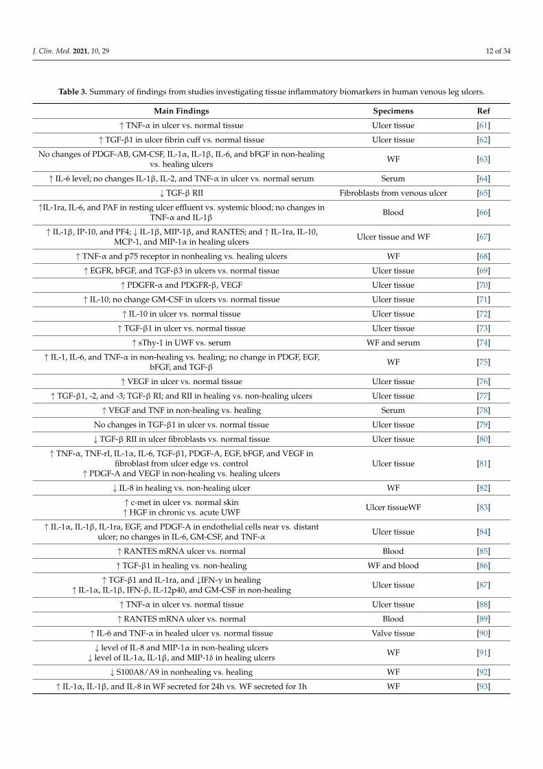
A list of tissue biomarkers found in the ulcer microenvironment is summarized inTable 3.
J. Clin. Med. 2021, 10, 29 12 of 34
Table 3. Summary of findings from studies investigating tissue inflammatory biomarkers in human venous leg ulcers.
Main Findings Specimens Ref
↑ TNF-α in ulcer vs. normal tissue Ulcer tissue [61]
↑ TGF-β1 in ulcer fibrin cuff vs. normal tissue Ulcer tissue [62]
No changes of PDGF-AB, GM-CSF, IL-1α, IL-1β, IL-6, and bFGF in non-healingvs. healing ulcers WF [63]
↑ IL-6 level; no changes IL-1β, IL-2, and TNF-α in ulcer vs. normal serum Serum [64]
↓ TGF-β RII Fibroblasts from venous ulcer [65]
↑IL-1ra, IL-6, and PAF in resting ulcer effluent vs. systemic blood; no changes inTNF-α and IL-1β Blood [66]
↑ IL-1β, IP-10, and PF4; ↓ IL-1β, MIP-1β, and RANTES; and ↑ IL-1ra, IL-10,MCP-1, and MIP-1α in healing ulcers Ulcer tissue and WF [67]
↑ TNF-α and p75 receptor in nonhealing vs. healing ulcers WF [68]
↑ EGFR, bFGF, and TGF-β3 in ulcers vs. normal tissue Ulcer tissue [69]
↑ PDGFR-α and PDGFR-β, VEGF Ulcer tissue [70]
↑ IL-10; no change GM-CSF in ulcers vs. normal tissue Ulcer tissue [71]
↑ IL-10 in ulcer vs. normal tissue Ulcer tissue [72]
↑ TGF-β1 in ulcer vs. normal tissue Ulcer tissue [73]
↑ sThy-1 in UWF vs. serum WF and serum [74]
↑ IL-1, IL-6, and TNF-α in non-healing vs. healing; no change in PDGF, EGF,bFGF, and TGF-β WF [75]
↑ VEGF in ulcer vs. normal tissue Ulcer tissue [76]
↑ TGF-β1, -2, and -3; TGF-β RI; and RII in healing vs. non-healing ulcers Ulcer tissue [77]
↑ VEGF and TNF in non-healing vs. healing Serum [78]
No changes in TGF-β1 in ulcer vs. normal tissue Ulcer tissue [79]
↓ TGF-β RII in ulcer fibroblasts vs. normal tissue Ulcer tissue [80]
↑ TNF-α, TNF-rI, IL-1α, IL-6, TGF-β1, PDGF-A, EGF, bFGF, and VEGF infibroblast from ulcer edge vs. control
↑ PDGF-A and VEGF in non-healing vs. healing ulcersUlcer tissue [81]
↓ IL-8 in healing vs. non-healing ulcer WF [82]
↑ c-met in ulcer vs. normal skin↑ HGF in chronic vs. acute UWF Ulcer tissueWF [83]
↑ IL-1α, IL-1β, IL-1ra, EGF, and PDGF-A in endothelial cells near vs. distantulcer; no changes in IL-6, GM-CSF, and TNF-α Ulcer tissue [84]
↑ RANTES mRNA ulcer vs. normal Blood [85]
↑ TGF-β1 in healing vs. non-healing WF and blood [86]
↑ TGF-β1 and IL-1ra, and ↓IFN-γ in healing↑ IL-1α, IL-1β, IFN-β, IL-12p40, and GM-CSF in non-healing Ulcer tissue [87]
↑ TNF-α in ulcer vs. normal tissue Ulcer tissue [88]
↑ RANTES mRNA ulcer vs. normal Blood [89]
↑ IL-6 and TNF-α in healed ulcer vs. normal tissue Valve tissue [90]
↓ level of IL-8 and MIP-1α in non-healing ulcers↓ level of IL-1α, IL-1β, and MIP-1δ in healing ulcers WF [91]
↓ S100A8/A9 in nonhealing vs. healing WF [92]
↑ IL-1α, IL-1β, and IL-8 in WF secreted for 24h vs. WF secreted for 1h WF [93]
J. Clin. Med. 2021, 10, 29 13 of 34
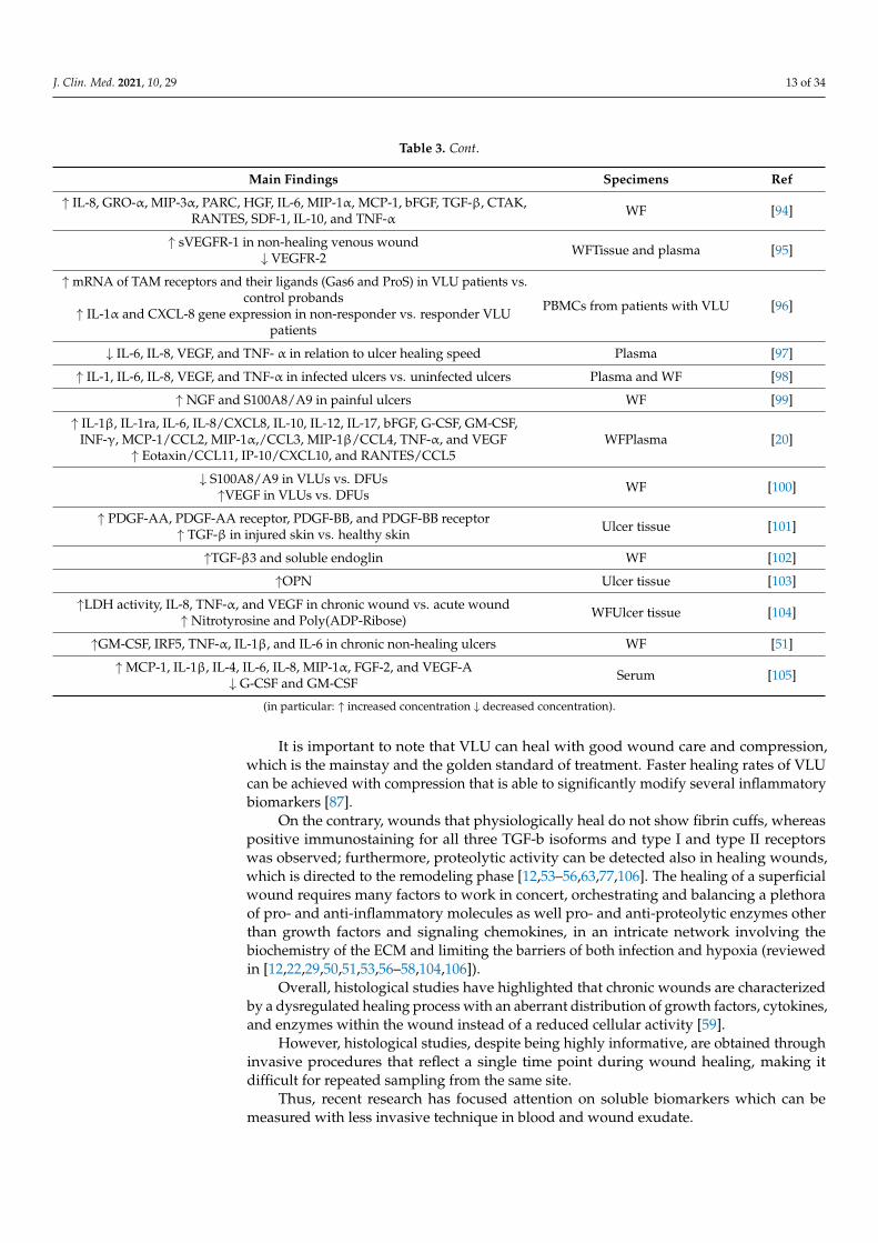
Table 3. Cont.
Main Findings Specimens Ref
↑ IL-8, GRO-α, MIP-3α, PARC, HGF, IL-6, MIP-1α, MCP-1, bFGF, TGF-β, CTAK,RANTES, SDF-1, IL-10, and TNF-α WF [94]
↑ sVEGFR-1 in non-healing venous wound↓ VEGFR-2 WFTissue and plasma [95]
↑ mRNA of TAM receptors and their ligands (Gas6 and ProS) in VLU patients vs.control probands
↑ IL-1α and CXCL-8 gene expression in non-responder vs. responder VLUpatients
PBMCs from patients with VLU [96]
↓ IL-6, IL-8, VEGF, and TNF- α in relation to ulcer healing speed Plasma [97]
↑ IL-1, IL-6, IL-8, VEGF, and TNF-α in infected ulcers vs. uninfected ulcers Plasma and WF [98]
↑ NGF and S100A8/A9 in painful ulcers WF [99]
↑ IL-1β, IL-1ra, IL-6, IL-8/CXCL8, IL-10, IL-12, IL-17, bFGF, G-CSF, GM-CSF,INF-γ, MCP-1/CCL2, MIP-1α,/CCL3, MIP-1β/CCL4, TNF-α, and VEGF
↑ Eotaxin/CCL11, IP-10/CXCL10, and RANTES/CCL5WFPlasma [20]
↓ S100A8/A9 in VLUs vs. DFUs↑VEGF in VLUs vs. DFUs WF [100]
↑ PDGF-AA, PDGF-AA receptor, PDGF-BB, and PDGF-BB receptor↑ TGF-β in injured skin vs. healthy skin Ulcer tissue [101]
↑TGF-β3 and soluble endoglin WF [102]
↑OPN Ulcer tissue [103]
↑LDH activity, IL-8, TNF-α, and VEGF in chronic wound vs. acute wound↑ Nitrotyrosine and Poly(ADP-Ribose) WFUlcer tissue [104]
↑GM-CSF, IRF5, TNF-α, IL-1β, and IL-6 in chronic non-healing ulcers WF [51]
↑MCP-1, IL-1β, IL-4, IL-6, IL-8, MIP-1α, FGF-2, and VEGF-A↓ G-CSF and GM-CSF Serum [105]
(in particular: ↑ increased concentration ↓ decreased concentration).
It is important to note that VLU can heal with good wound care and compression,which is the mainstay and the golden standard of treatment. Faster healing rates of VLUcan be achieved with compression that is able to significantly modify several inflammatorybiomarkers [87].
On the contrary, wounds that physiologically heal do not show fibrin cuffs, whereaspositive immunostaining for all three TGF-b isoforms and type I and type II receptorswas observed; furthermore, proteolytic activity can be detected also in healing wounds,which is directed to the remodeling phase [12,53–56,63,77,106]. The healing of a superficialwound requires many factors to work in concert, orchestrating and balancing a plethoraof pro- and anti-inflammatory molecules as well pro- and anti-proteolytic enzymes otherthan growth factors and signaling chemokines, in an intricate network involving thebiochemistry of the ECM and limiting the barriers of both infection and hypoxia (reviewedin [12,22,29,50,51,53,56–58,104,106]).
Overall, histological studies have highlighted that chronic wounds are characterizedby a dysregulated healing process with an aberrant distribution of growth factors, cytokines,and enzymes within the wound instead of a reduced cellular activity [59].
However, histological studies, despite being highly informative, are obtained throughinvasive procedures that reflect a single time point during wound healing, making itdifficult for repeated sampling from the same site.
Thus, recent research has focused attention on soluble biomarkers which can bemeasured with less invasive technique in blood and wound exudate.
J. Clin. Med. 2021, 10, 29 14 of 34
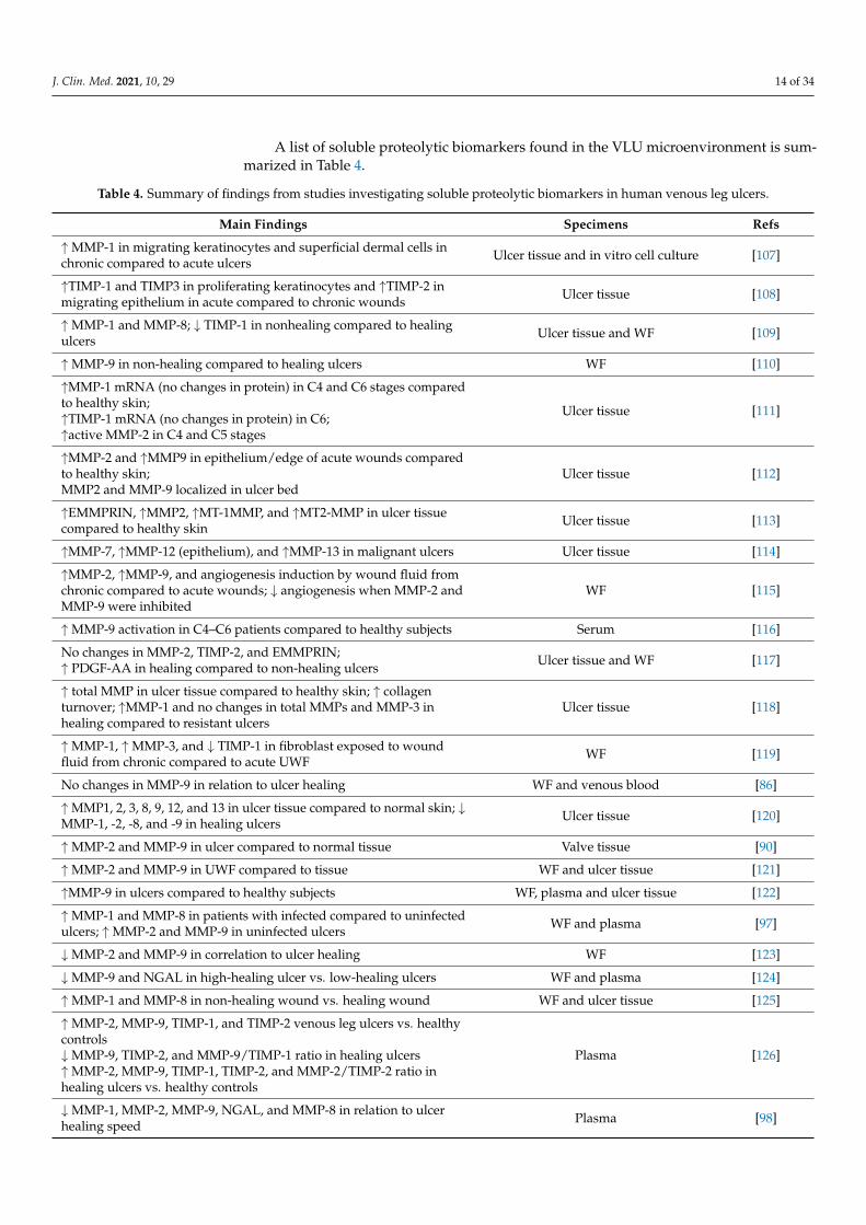
A list of soluble proteolytic biomarkers found in the VLU microenvironment is sum-marized in Table 4.
Table 4. Summary of findings from studies investigating soluble proteolytic biomarkers in human venous leg ulcers.
Main Findings Specimens Refs
↑MMP-1 in migrating keratinocytes and superficial dermal cells inchronic compared to acute ulcers Ulcer tissue and in vitro cell culture [107]
↑TIMP-1 and TIMP3 in proliferating keratinocytes and ↑TIMP-2 inmigrating epithelium in acute compared to chronic wounds Ulcer tissue [108]
↑MMP-1 and MMP-8; ↓ TIMP-1 in nonhealing compared to healingulcers Ulcer tissue and WF [109]
↑MMP-9 in non-healing compared to healing ulcers WF [110]
↑MMP-1 mRNA (no changes in protein) in C4 and C6 stages comparedto healthy skin;↑TIMP-1 mRNA (no changes in protein) in C6;↑active MMP-2 in C4 and C5 stages
Ulcer tissue [111]
↑MMP-2 and ↑MMP9 in epithelium/edge of acute wounds comparedto healthy skin;MMP2 and MMP-9 localized in ulcer bed
Ulcer tissue [112]
↑EMMPRIN, ↑MMP2, ↑MT-1MMP, and ↑MT2-MMP in ulcer tissuecompared to healthy skin Ulcer tissue [113]
↑MMP-7, ↑MMP-12 (epithelium), and ↑MMP-13 in malignant ulcers Ulcer tissue [114]
↑MMP-2, ↑MMP-9, and angiogenesis induction by wound fluid fromchronic compared to acute wounds; ↓ angiogenesis when MMP-2 andMMP-9 were inhibited
WF [115]
↑MMP-9 activation in C4–C6 patients compared to healthy subjects Serum [116]
No changes in MMP-2, TIMP-2, and EMMPRIN;↑ PDGF-AA in healing compared to non-healing ulcers Ulcer tissue and WF [117]
↑ total MMP in ulcer tissue compared to healthy skin; ↑ collagenturnover; ↑MMP-1 and no changes in total MMPs and MMP-3 inhealing compared to resistant ulcers
Ulcer tissue [118]
↑MMP-1, ↑MMP-3, and ↓ TIMP-1 in fibroblast exposed to woundfluid from chronic compared to acute UWF WF [119]
No changes in MMP-9 in relation to ulcer healing WF and venous blood [86]
↑MMP1, 2, 3, 8, 9, 12, and 13 in ulcer tissue compared to normal skin; ↓MMP-1, -2, -8, and -9 in healing ulcers Ulcer tissue [120]
↑MMP-2 and MMP-9 in ulcer compared to normal tissue Valve tissue [90]
↑MMP-2 and MMP-9 in UWF compared to tissue WF and ulcer tissue [121]
↑MMP-9 in ulcers compared to healthy subjects WF, plasma and ulcer tissue [122]
↑MMP-1 and MMP-8 in patients with infected compared to uninfectedulcers; ↑MMP-2 and MMP-9 in uninfected ulcers WF and plasma [97]
↓MMP-2 and MMP-9 in correlation to ulcer healing WF [123]
↓MMP-9 and NGAL in high-healing ulcer vs. low-healing ulcers WF and plasma [124]
↑MMP-1 and MMP-8 in non-healing wound vs. healing wound WF and ulcer tissue [125]
↑MMP-2, MMP-9, TIMP-1, and TIMP-2 venous leg ulcers vs. healthycontrols↓MMP-9, TIMP-2, and MMP-9/TIMP-1 ratio in healing ulcers↑MMP-2, MMP-9, TIMP-1, TIMP-2, and MMP-2/TIMP-2 ratio inhealing ulcers vs. healthy controls
Plasma [126]
↓MMP-1, MMP-2, MMP-9, NGAL, and MMP-8 in relation to ulcerhealing speed Plasma [98]
J. Clin. Med. 2021, 10, 29 15 of 34
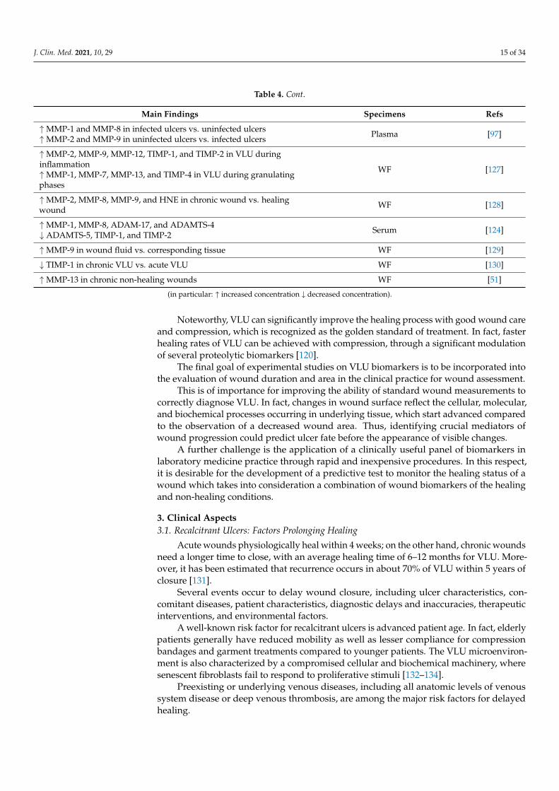
Table 4. Cont.
Main Findings Specimens Refs
↑MMP-1 and MMP-8 in infected ulcers vs. uninfected ulcers↑MMP-2 and MMP-9 in uninfected ulcers vs. infected ulcers Plasma [97]
↑MMP-2, MMP-9, MMP-12, TIMP-1, and TIMP-2 in VLU duringinflammation↑MMP-1, MMP-7, MMP-13, and TIMP-4 in VLU during granulatingphases
WF [127]
↑MMP-2, MMP-8, MMP-9, and HNE in chronic wound vs. healingwound WF [128]
↑MMP-1, MMP-8, ADAM-17, and ADAMTS-4↓ ADAMTS-5, TIMP-1, and TIMP-2 Serum [124]
↑MMP-9 in wound fluid vs. corresponding tissue WF [129]
↓ TIMP-1 in chronic VLU vs. acute VLU WF [130]
↑MMP-13 in chronic non-healing wounds WF [51]
(in particular: ↑ increased concentration ↓ decreased concentration).
Noteworthy, VLU can significantly improve the healing process with good wound careand compression, which is recognized as the golden standard of treatment. In fact, fasterhealing rates of VLU can be achieved with compression, through a significant modulationof several proteolytic biomarkers [120].
The final goal of experimental studies on VLU biomarkers is to be incorporated intothe evaluation of wound duration and area in the clinical practice for wound assessment.
This is of importance for improving the ability of standard wound measurements tocorrectly diagnose VLU. In fact, changes in wound surface reflect the cellular, molecular,and biochemical processes occurring in underlying tissue, which start advanced comparedto the observation of a decreased wound area. Thus, identifying crucial mediators ofwound progression could predict ulcer fate before the appearance of visible changes.
A further challenge is the application of a clinically useful panel of biomarkers inlaboratory medicine practice through rapid and inexpensive procedures. In this respect,it is desirable for the development of a predictive test to monitor the healing status of awound which takes into consideration a combination of wound biomarkers of the healingand non-healing conditions.
3. Clinical Aspects3.1. Recalcitrant Ulcers: Factors Prolonging Healing
Acute wounds physiologically heal within 4 weeks; on the other hand, chronic woundsneed a longer time to close, with an average healing time of 6–12 months for VLU. More-over, it has been estimated that recurrence occurs in about 70% of VLU within 5 years ofclosure [131].
Several events occur to delay wound closure, including ulcer characteristics, con-comitant diseases, patient characteristics, diagnostic delays and inaccuracies, therapeuticinterventions, and environmental factors.
A well-known risk factor for recalcitrant ulcers is advanced patient age. In fact, elderlypatients generally have reduced mobility as well as lesser compliance for compressionbandages and garment treatments compared to younger patients. The VLU microenviron-ment is also characterized by a compromised cellular and biochemical machinery, wheresenescent fibroblasts fail to respond to proliferative stimuli [132–134].
Preexisting or underlying venous diseases, including all anatomic levels of venoussystem disease or deep venous thrombosis, are among the major risk factors for delayedhealing.
J. Clin. Med. 2021, 10, 29 16 of 34
Patients with higher body mass index (BMI > 25 kg/m2) and nutritional deficienciesalso have a poor healing prognosis [17].
Larger wound area and longer duration have been reported as clinical signs of poorhealing, while data on ulcer location and shape showed contradictory results [135]. Con-flicting results regarding also the volume of exudate, the type and amount of woundinfection, and the presence of previous ulceration as potential risk factors for prolongedhealing may be important factors in delayed VLU healing [135].
Additionally, a history of venous ligation or vein stripping, a history of hip or kneereplacement surgery, ankle brachial pressure index < 0.8, and the presence of fibrin coveringgreater than 50% of the wound area have been associated with prolonged healing [136].
Among the environmental conditions predisposing for delayed wound healing, ithas been reported that colder temperature was associated with increased risk of ulcerdevelopment [137].
The diagnostic delays and a misdiagnosed VLU will result in extended time for healingdue to delays in proper diagnosis and treatment. This could be further exacerbated if animproper treatment is initiated for the misdiagnosis.
In this respect, additional factors could be examined to improve the diagnostic pro-cess, such as biochemical and molecular parameters which affect VLU progression fromdevelopment to closure or chronicity.
Biochemical and biomolecular markers of wound healing could be assessed boththrough wound tissue biopsies (e.g., wnt signaling pathway, β-catenin, c-myc, growthfactors, proteases, and miRNA [56,138]) and through soluble biomarkers circulating in theblood or released within the wound fluid (e.g., MMPs, cytokines, growth factors, levels ofalbumin, and total protein, etc.) [20,75,102,127,139–141], which generally represent crucialregulators of tissue remodeling.
In a recent retrospective cohort study involving 65 patients that underwent severaltreatments for 1 year, it has been demonstrated that, besides the known risk factors (i.e.,deep venous disease and post-thrombotic etiology), novel risk factors, such as depressionand race (nonwhite), emerged as important factors for VLU development [142].
Recently, a tool has been developed to predict the risk of failure to heal VLUs in 24weeks by taking into account several factors, including patient characteristics (age, historyof deep vein thrombosis in the affected leg, calf circumference, compression treatments,and behavioral factors) and ulcer characteristics (duration, area, presence of necrotic tissue,and ulcer area reduction in 2 weeks) [52]. This may prove useful in clinical practice to settreatment goals and patient–provider expectations.
Similar approaches, taking into consideration the main risk factors for poor VLUhealing, can be considered easy-to-use aids to discern patients with a high risk of delayedhealing and to assist clinicians during selection of the best therapeutic approach.
3.2. Latest Innovations in Surgical Treatment and Drug Therapies
A number of treatment options have been utilized for patients with VLU in order topromote healing. A key and consistent treatment for VLU is compression therapy thatcan achieve dynamic pressures of over 60 mm Hg to enable changes in hemodynamicsand promote healing [1,143,144]. There are surgical treatments consisting of open surgeryinvolving abolishing venous reflux in the superficial and perforator systems that haveimportance in healing and preventing VLU [1]. Recently, the EVRA trial involving treat-ment of the superficial system in patients without any deep venous obstruction werefound to have a significant healing benefit in patients undergoing endovenous ablationutilizing a variety of modalities including thermal energy and nonthermal endothelialinjury to occlude the axial reflux. It is important to note that the long-term outcomes ofthe EVRA trial showed reduced rates of recurrence at 3 years. Moreover, this randomizedtrial demonstrated that, compared to compression alone, compression plus ablation of thesuperficial reflux decreased time to healing with a mean of 56 days vs. 82 days with onlycompression [145]. There was a benefit of an ulcer-free interval and healed VLU at 24 weeks,
J. Clin. Med. 2021, 10, 29 17 of 34
with an 89% probability that early venous intervention is cost-effective over 1 year [146].However, VLU recurrences are still a major concern, and in this short 24-week period offollow up, between 11 and 16% had a recurrence with no difference in the treatment arm.Perforator surgery has been advocated as a means for expediting VLU healing. A recentCochrane review evaluating 4 RCTs ( Randomized Controlled Trials) including 332 patientsundergoing subfascial endoscopic perforator surgery (SEPS) compared to other treatmentmodalities (compression and superficial vein surgery) determined that, although SEPSwith compression appeared to have benefit at 24 months in VLU healing, the data was lowquality with significant risk for bias. Overall, SEPS did not demonstrate a clear benefit inVLU healing due to low and very low quality of evidence, small sample size, and bias [147].Outflow obstruction of the venous system can lead to post-thrombotic syndrome and VLU.In a large single-center series, 982 obstructive venous outflow lesions were evaluated in 870patients undergoing iliac venous stents. There were no mortalities, the early thromboticrate was 1.5%, and the contralateral iliac vein thrombosis was 1%. At 5 years, 62% of pa-tients were pain free, 32% were edema free, and 58% of VLU was healed [148]. Importantly,if the patient with VLU had both outflow obstruction and superficial great saphenousinsufficiency, both could be treated in the same setting with excellent results and a VLUhealing rate of 64% at 48 months [149]. Noteworthy, endovenous treatments both for thesuperficial venous system but especially in the deep outflow iliac venous system shouldbe routinely evaluated in patients with VLU and aggressively treated to relieve venoushypertension and to establish outflow patency, with 5-year VLU healing of about 75% [150].A recent meta-analysis in the treatment of chronic venous outflow obstruction including 12studies demonstrated a favorable rate of overall VLU healing of 72.1%. The VLU healingwas higher for non-thrombotic than thrombotic lesions for the iliofemoral venous system(86.9% vs. 70.3%, respectively, p = 0.0022) [151].
Despite compression treatment, and surgical and endovascular venous treatments,the overall healing and recurrence rates for VLU can be as high as 70%. The explana-tion is multifactorial, including compliance with compression and treatment, proceduralfailures, mixed VLU disease that encompasses arterial component, incorrect diagnosis ofulcer, and progression of venous disease. However, a key reason is the poorly understoodpathophysiology, molecular pathways leading to tissue injury, persistent inflammatory re-sponses and monocyte/lymphocyte-endothelial activation, and oxidative stress. A numberof pharmacologic agents including flavonoids, diosmin-based drugs, pentoxifylline, andsulodexide have been tested in RCTs as adjuvant treatments to compression for improvingVLU healing. Overall, the data suggest improved healing with vasoactive pharmacologicdrugs, but the studies are short, the length of treatment to prevent recurrence is unclear,they are heterogeneous, and they lack patient-reported quality of life outcomes [1].
Several biologic products such as bilayer living skin construct (human skin equiva-lents), fibroblast derivatives, and extracellular matrices and non-biologic products suchas poly-N-acetyl glucosamine have been tested in VLU clinical trials and demonstratedbenefit [11,12,152–154] Although healing is improved with these products with the additionof compression, there are however significant limitations in these trials, and most haveshort-term follow-up times of less than a year, lack clear evaluation of the venous systemto determine if the disease process is primary or secondary venous insufficiency, and donot include important patient-reported outcomes and cost analysis. Recently, there havebeen development of new products with improved understanding in the pathophysiologyof VLU. The placental membranes amnion/chorion allografts have interesting propertiesfor wound healing. These fall under the human cellular repair matrices and are composedof cryopreserved native placenta without an immunogenic trophoblast layer. They havecollagen-rich extracellular matrix proteins, providing growth factors, glycosaminoglycans,fibroblasts and epithelial cells, and important mesenchymal stem cells. In vitro placentalmembranes promote cellular adhesion and migration, cell differentiation, and proan-giogenic anti-inflammatory activities and protect growth factor function [155,156]. Onerandomized controlled trial evaluated dehydrated human placental allograft (dehydrated
J. Clin. Med. 2021, 10, 29 18 of 34
human amnion chorion membrane, dHACM) in patients with VLU [157]. In a multicenterrandomized controlled trial, 84 VLU patients enrolled and were distributed as the studygroup consisting of 53 patients assigned to placental allograft with multi-layer compressionand a control group with 31 patients with multi-layer compression only. The primary out-come measure was 40% wound closure at 4 weeks. The dHACM (one or two applications)group at 4 weeks had a greater percentage of VLU achieving 40% closure (62% vs. 32%,p = 0.005) [158]. Complete healing of VLU and the ulcer free interval, which is an importantparameter to the patient and which the study did not evaluate, would be important toknow. A more recent randomized trial comparing dHACM with compression and a controlgroup with compression determined that, at 16 weeks, the placental derivative group hadsignificantly improved rates of healing (71% versus 44% at 16 weeks, p = 0.0065) and adecreased healing time [159]. Further clinical trials are needed to assess ulcer recurrences,cost effectiveness and analysis, and patient-reported outcomes. Another interesting area ofstudy and potential for VLU healing is the targeting of connexins. Connexin 43 gap junctionproteins regulate small molecule signaling to and between cells. They have been associatedwith regulation of inflammatory cytokine release, mediators of fibrosis pathways, andcontrol growth factor response at the cellular level. Importantly, connexin 43 is abnormallyupregulated at wound edges of chronic non-healing wounds and VLU [160,161]. The ACT1peptide is a competitive inhibitor of connexin 43 (a peptide mimetic of the connexin 43carboxyl terminus), and the application of ACT1 accelerates wound healing in animalmodels. An interesting phase 2 study included 92 patients with VLU randomized to eitherthe group with ACT1 topical and debridement with four-layer compression bandages or tothe group with debridement and four-layer compression bandages. The primary endpointto this study was mean percent area reduction at 12 weeks, and the secondary endpointwas 100% closure at 12 weeks. The follow-up was up to 6 months. Both the primary andsecondary endpoints were in favor of the VLU treated with ACT1 (79% vs. 36%, p = 0.02;57% vs. 28%, p = 0.01; respectively). The VLU recurrence rates at 6 months were equal foreach group (11%) [158].
Peroxynitrite (ONOO), a product of nitric oxide and superoxide, is a potent oxidizingand nitrating agent that causes significant and irreversible damage to tissues and cellularcomponents including mitochondria, DNA, lipid peroxidation, posttranslational modifica-tions of many proteins, protein oxidizer and nitration, and enzyme inactivation. ONOOdecreases the function of superoxide dismutase (SOD); increases reactive oxygen species(ROS) production, prostacyclin synthase for PGI2 production, glucocorticoid receptor, andresponse to glucocorticoids; increases COX-2; and activates MMPs. Recently, a very elegantstudy assessed the presence of ONOO in VLU. Nitrotyrosine is a byproduct indicative ofperoxynitrite activity, and poly(ADP-ribose) is the product of the DNA damage sensorenzyme PARP-1. In a study of VLU biopsies compared to normal tissue, the authorsfound elevated nitrotyrosine and PAR, indicating peroxynitrite oxidation and DNA dam-age/repair, respectively [104]. These findings confirm that peroxynitrite is present in VLUand likely a significant contributor to pathology of the inflammatory state, and further workin targeting production or activity of ONOO may have significant implications in healingVLU. Innate immunity involving polymorphonuclear cells, macrophages, natural killer Tcells, complement system, and lactoferrin are important measures to mitigate infection andto promote wound healing. An important set of molecules are the function of TAM (Tyro,Axl, and MerTK), which is a family of receptor tyrosine kinases and their ligands Gas6 andProtein S (ProS). This group of proteins has innate immune regulation function, is centralin the intrinsic inhibition of inflammation to pathogens, and is important in phagocytosisand apoptosis [96]. In a study evaluating the gene expression of patients with VLU (n = 67)vs. controls (n = 42), the blood polymorphonuclear cells were assayed for TAM and theirligands. The TAM and ligands were increased significantly over the control, but impor-tantly, when comparing VLU responders that were healing with VLU non-responders,the responders had significantly elevated TAM Axl elevation while non-responders hadsignificantly elevated Gas ligand [96]. These finding not only are important in defining the
J. Clin. Med. 2021, 10, 29 19 of 34
role of innate immunity in VLU but has markers for healing progression and targets forpotential therapy.
The toll-like receptor family is important in innate immunity and pattern recognition.These receptors are expressed on the cell surface of innate immune cells and non-immunecells of the dermis and epidermis. They recognize discrete pathogen molecular patternsas well as endogenous damage-associated molecular patterns released after tissue andcellular damage. Importantly, toll-like receptors trigger proinflammatory responses andcytokine release, which are important mechanisms in VLU pathology. In a study assessingVLU wound fluid, toll-like receptors were assessed in healing and non-healing VLU. Bothtoll-like receptors 2 and 4 were significantly elevated in non-healing VLU and decreased inhealing VLU. In addition, the antibacterial peptide lipocalin-2 was elevated in non-healingVLU because of the increased inflammation [91]. The possibility for future research inthe areas of innate immunity, modulating pathways, and targeting certain receptors andligands has significant promise in novel treatment and pharmacology.
A new and exciting area of research is metabolomics. Metabolic phenotyping has beenemployed to explore mechanistic pathways involved in venous disease. Metabolomicsevaluates both aqueous and non-aqueous metabolites utilizing nuclear magnetic resonancespectroscopy (NMR) and mass spectrometry (MS). A recent systematic review regardingthe study of metabolites in VLU determined that upregulated metabolites in wound fluidand ulcer biopsies including lactate, branch chained amino acids, lysine, 3-hydroxybutyrate,and glutamate were identified and have importance in cell energy, amino acid and proteinbiosynthesis, and cellular functions. These data provide important clues to the diseasepathophysiology within VLU, and further research on mechanisms and targeted therapyhold significant promise [14].
Sulodexide is a glycosaminoglycan with the composition of 80% heparan sulfate (alsoknown as fast-moving heparin) and 20% dermatan sulfate. The heparan sulfate congeneris predominantly composed of glucuronic acid linked to glucosamine, while the dermatansulfate consists of iduronic acid and galactosamine. Sulodexide has important biologiceffects with antithrombotic, anti-inflammatory, and endothelial protective properties [106,162,163]. Although sulodexide was demonstrated nearly two decades ago to be effectivein increasing VLU healing [164–166], its novel molecular mechanisms and the pleiotropiceffects are just recently understood. It is important to assess the effects of the drug inpatients with VLU and healed ulcer disease, which provides insight into the mechanismsand targets of sulodexide. In a study evaluating the anti-inflammatory effects of sulodexidein healed VLU patients who were treated for 8 weeks (2 × 500 LSU/day, oral), bloodsamples before and at completion of the study were drawn to assess for IL-6 and MMP-9. At8 weeks of treatment with sulodexide, there was a significant decrease in both inflammatorymolecules. In addition, evaluation of endothelial cells treated with sulodexide-treatedserum from patients significantly decreased IL-6 and intracellular free radicals. Takentogether, these data demonstrate that sulodexide results in a reduction in intravascularinflammation and is endothelial-protective [167]. In another interesting study evaluatingthe serum from CVD-healed ulcer patients before and after treatment with sulodexide,the effect of inflammation and oxidative stress in HUVEC cells was evaluated. The keyfindings of this study were that sulodexide reduces inflammatory mediators in CVDserum (IL-6, MCP-1, and ICAM-1), reduces oxidative stress, suppresses the effect of IL-1,and reduces population doubling time and hypertrophy, indicating decreased aging andsenescence [168]. Further identifying the multiple effects of sulodexide on cellular function,an elegant study assessed sulodexide’s protective action on cell stress and autophagy (acomplex process involving lysosomal catabolic actions by which cells degrade or recycletheir contents of unnecessary or dysfunctional cellular components to maintain cellularhomeostasis, to adapt to stress, and to respond to disease (a protective mechanism). HUVECcells were stressed metabolically (methylglyoxal) and non-metabolically (ionizing radiation)with and without the treatment of sulodexide. The important and novel findings werethe effects of sulodexide mitigating apoptosis by inhibiting intrinsic and extrinsic caspase
J. Clin. Med. 2021, 10, 29 20 of 34
pathways and increased cell viability, by reducing ROS, by reducing the synthesis andrelease of inflammatory cytokines (TNF-α, IL1, IL6, and IL8), by promoting cell autophagyin maintaining cellular function, and by reducing DNA damage [169]. The implicationsare that sulodexide prevents endothelial dysfunction and injury that may have significantimplications in CVD, DVT, and PTS. Importantly, these scientific discoveries allow forfurther research in the pathophysiology of VLU and the possibility for synergistic effectswith sulodexide in treating and healing VLU. In another area of recent investigation,sulodexide physiologic properties and effects on MMPs was tested in a murine vein stretchmodel. Sulodexide caused venous contraction and restored venous contraction in a stretchmodel compared to an untreated stretched vein while inhibiting MMP-2 and MMP-9expression and activity, thereby enhancing venous contraction. Sulodexide’s effect onvenous contraction was partly due to an increase in the sensitivity of the α-adrenergicreceptor, but likely, sulodexide also enhanced downstream mechanisms (Ca2+ sensitivityand mobilization, PKC, MAPK, and Rho-kinase). These novel mechanism in SDX veincontraction may be important not only in venous leg ulcers but also in treating CVD ingeneral and improving venous function [170,171].
Several growth factors have been applied to VLU given the importance of manygrowth factors in the biology of wound healing. These growth factors include platelet-derived growth factor, transforming growth factor, epidermal growth factor, keratinocytegrowth factor, and fibroblast growth factor. A recent meta-analysis evaluating 10 studiesconsisting of 472 in the intervention group (growth factors) and 330 as control determinedthat there was moderate bias in the study design and that, although tendencies towardhealing VLU were present, none reached statistical significance [172]. Further studies withsound methodology, frequency, and duration of growth factor application and propercontrol are required before strong recommendations can be made.
Silver dressings have been applied to treat VLU. Silver ions have antibacterial andanti-inflammatory properties, have nonspecific MMP inhibition, and drive senescent cellstoward apoptosis. A recent meta-analysis of 8 studies found that, overall, there wasa benefit toward VLU healing rate; however, there were no clear benefits in completeVLU healing, and long-term follow-up and comparisons to other wound dressing waslacking, and strong recommendations for general use were not advisable until furtherresearch is generated [173]. Another systematic review evaluating the effect of MMPinhibition determined that, from 16 studies utilizing collagen-based and lipido-colloidnano oligosaccharide factor dressings, there was a clear benefit in a variety of ulcersincluding diabetic, venous, and mixed origin. The major outcomes focused on woundclosure, wound size reduction, healing time, and healing rate [174]. Further studies arerequired to determine which MMPs to target, to focus on VLU etiology and RCT trials,and to determine ulcer free intervals and cost analysis as well as specific biomarkers (e.g.,reduction in gelatinase activity in the wound bed) that demonstrate positive progress. Itis important to note that topical agents applied to VLU are abundant. However, the datais usually from small studies, with methodology flaws and bias. Many of the studies areof moderate to low certainty and quality of evidence, and further well-defined RCTs withclearly defined inclusion criteria and endpoints that evaluate ulcer free interval, patient-reported outcomes, and cost effectiveness are needed before best medical practices canbe offered to patients with VLU and can have the best healing potential and value for thepatient [175] (The different treatment strategies and evidence for success in VLU healinghave been summarized in Figure 3).
Bone Marrow derived cell (BMDC) therapy has become an area of intense research inregenerative medicine therapy. A recent pilot study evaluating the feasibility and safety ofutilizing BMDC in VLU was conducted. The study included four patients with 6 VLUs.The bone marrow was harvested, processed, and injected in the periulcer bed. At 12-monthfollow-up, the wound area had decreased and pain had improved [176]. Another pilot trialevaluating progenitor cells obtained from adipose tissue was conducted in 8 treated and 8control VLU patients. The patients treated with their own progenitor cells had significantly
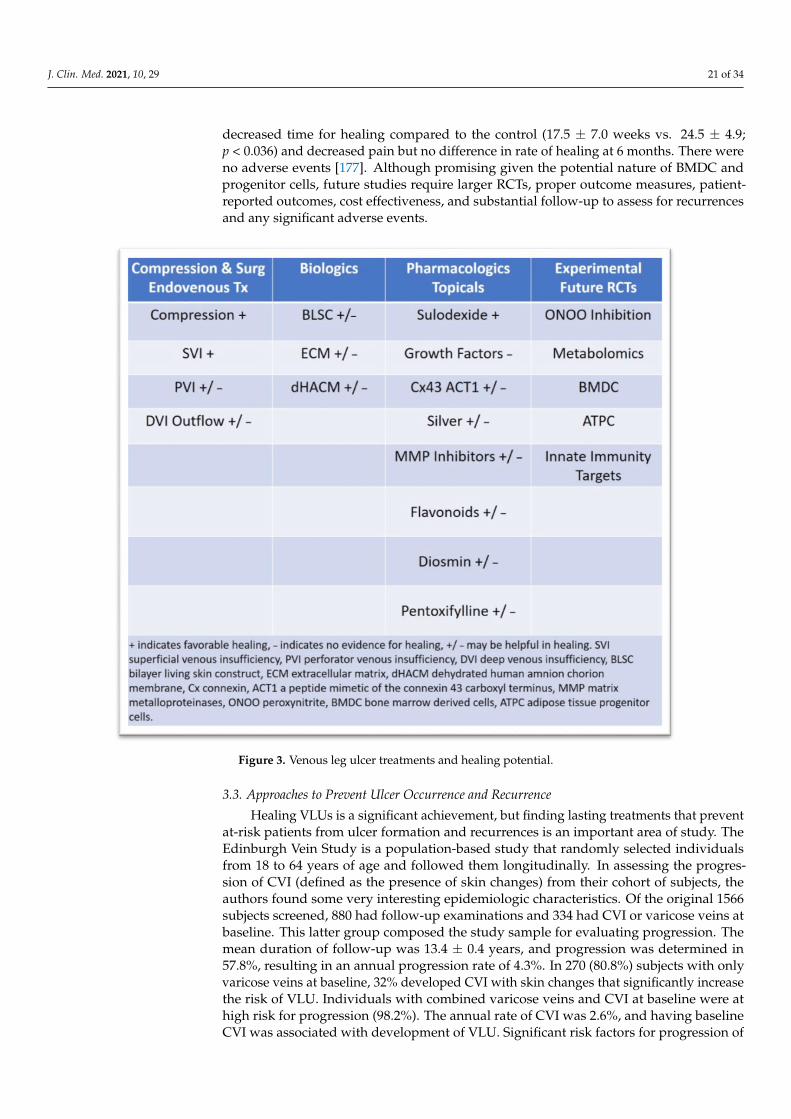
J. Clin. Med. 2021, 10, 29 21 of 34
decreased time for healing compared to the control (17.5 ± 7.0 weeks vs. 24.5 ± 4.9;p < 0.036) and decreased pain but no difference in rate of healing at 6 months. There wereno adverse events [177]. Although promising given the potential nature of BMDC andprogenitor cells, future studies require larger RCTs, proper outcome measures, patient-reported outcomes, cost effectiveness, and substantial follow-up to assess for recurrencesand any significant adverse events.
J. Clin. Med. 2021, 10, x FOR PEER REVIEW 21 of 33
Figure 3. Venous leg ulcer treatments and healing potential.
3.3. Approaches to Prevent Ulcer Occurrence and Recurrence
Healing VLUs is a significant achievement, but finding lasting treatments that pre-
vent at-risk patients from ulcer formation and recurrences is an important area of study.
The Edinburgh Vein Study is a population-based study that randomly selected individu-
als from 18 to 64 years of age and followed them longitudinally. In assessing the progres-
sion of CVI (defined as the presence of skin changes) from their cohort of subjects, the
authors found some very interesting epidemiologic characteristics. Of the original 1566
subjects screened, 880 had follow-up examinations and 334 had CVI or varicose veins at
baseline. This latter group composed the study sample for evaluating progression. The
mean duration of follow-up was 13.4 ± 0.4 years, and progression was determined in
57.8%, resulting in an annual progression rate of 4.3%. In 270 (80.8%) subjects with only
varicose veins at baseline, 32% developed CVI with skin changes that significantly in-
crease the risk of VLU. Individuals with combined varicose veins and CVI at baseline were
at high risk for progression (98.2%). The annual rate of CVI was 2.6%, and having baseline
CVI was associated with development of VLU. Significant risk factors for progression of
CVD was varicose veins at baseline and family history of DVT. Obesity was not found to
be an independent risk factor for progression of CVI, although this may be a factor and
may be implicated since obesity is associated with varicose veins. However, when evalu-
ating subjects older than 55, obesity was a significant factor for the development of CVI.
Superficial reflux in the venous system, especially in the small saphenous vein, is an in-
dependent risk for CVI progression, especially when combined with deep venous reflux
Figure 3. Venous leg ulcer treatments and healing potential.
3.3. Approaches to Prevent Ulcer Occurrence and Recurrence
Healing VLUs is a significant achievement, but finding lasting treatments that preventat-risk patients from ulcer formation and recurrences is an important area of study. TheEdinburgh Vein Study is a population-based study that randomly selected individualsfrom 18 to 64 years of age and followed them longitudinally. In assessing the progres-sion of CVI (defined as the presence of skin changes) from their cohort of subjects, theauthors found some very interesting epidemiologic characteristics. Of the original 1566subjects screened, 880 had follow-up examinations and 334 had CVI or varicose veins atbaseline. This latter group composed the study sample for evaluating progression. Themean duration of follow-up was 13.4 ± 0.4 years, and progression was determined in57.8%, resulting in an annual progression rate of 4.3%. In 270 (80.8%) subjects with onlyvaricose veins at baseline, 32% developed CVI with skin changes that significantly increasethe risk of VLU. Individuals with combined varicose veins and CVI at baseline were athigh risk for progression (98.2%). The annual rate of CVI was 2.6%, and having baselineCVI was associated with development of VLU. Significant risk factors for progression of
J. Clin. Med. 2021, 10, 29 22 of 34
CVD was varicose veins at baseline and family history of DVT. Obesity was not foundto be an independent risk factor for progression of CVI, although this may be a factorand may be implicated since obesity is associated with varicose veins. However, whenevaluating subjects older than 55, obesity was a significant factor for the development ofCVI. Superficial reflux in the venous system, especially in the small saphenous vein, isan independent risk for CVI progression, especially when combined with deep venousreflux [178]. An important question to ask would be if early intervention by treatmentof the venous system and/or compression can prevent the progression of CVD and VLUformation. In addition, utilization of ultrasound to evaluate venous reflux in both the su-perficial and deep venous systems could be an important tool for prevention and prognosis,instituting aggressive treatment with compression, venous surgical correction of reflux,and the use of pharmacologic venous drugs (e.g., diosmin, pentoxifylline, and sulodexide).Preventing ulcer formation and susceptibility is of paramount importance. Identifyingindividuals at risk for progression of CVI and VLU by history of varicose veins or DVTalong with a complete investigation of the venous system with ultrasound and identifyingreflux that is correctable may prevent the formation of de novo VLU [179]. Genetic predis-position may also be a significant factor for disease progression and VLU development.Currently, it is unclear if genetic alterations in a number of identified genes are causal orassociations. Certainly, this area of research is interesting and requires further study inlarger population-based studies and determination that early intervention in identifiedand affected individuals with specific venous gene polymorphic variants would benefitfrom interventions to correct venous hemodynamics and prevent VLU formation [180–184].Important principles to prevent and reduce recurrent VLU are following adopted princi-ples of timely referral to a vascular specialist, evaluation and ultrasonography, treatinginfection and debridement, appropriate compression, and appropriate interventions whenindicated [1]. It is important to note that, in patients with healed and active VLUs, 50%stenosis of iliac venous outflow obstruction can be present in up to 37% of individualsand 80% stenosis of iliac venous outflow obstruction is present in 23%. Significant riskfactors for venous outflow obstruction include previous DVT, deep venous insufficiency,and female gender [185]. It is important that properly selected individuals with healed oractive VLU are assessed for venous outflow obstruction and that, if obstruction is observedthat proper treatment with venography, intravascular ultrasound, angioplasty, and stentsare offered [186]. Adequate compression (multi-layer bandages, short-stretch bandages,and inelastic and elastic compression) is of primary importance in healing VLU and hasample high levels of evidence [1,187]. What is less certain is the effect of compression inpreventing VLU recurrence. The strength of the available data is less clear and inconsistentwith unequivocal benefit due to study design, strength and type of compression used,and heterogeneity. However, overall, the data appears favorable, and in clinical practice,clinicians advise and prescribe compression therapy for patients with healed VLU to main-tain hemodynamics and to prevent recurrence, which has also been demonstrated to becost-effective [1,188–191]. Currently there is insufficient data to determine if compressiontherapy prevents occurrence and progression of CVD to VLU [191]. Although severalpharmacologic vasoactive drugs have been studied and demonstrated to help VLU healingas an adjunct to compression, it is unknown if continued treatment with these agents hasan effect on reducing VLU occurrence or recurrence [192]. The EVRA trial demonstratedthat surgical intervention on the superficial system decreases the time for VLU healing(median time to healing compression + endovenous ablation 56 days vs. compression 82days). Noteworthy, the EVRA trial demonstrated that early endovenous ablation of super-ficial venous reflux was highly likely to be cost-effective over a 3-year horizon comparedwith deferred intervention, suggesting that early intervention accelerated the healing ofvenous leg ulcers and reduced the overall incidence of ulcer recurrence. Therefore, thelong-term outcomes of the Early Venous Reflux Ablation (EVRA) randomized trial showedaccelerated venous leg ulcer healing and greater ulcer-free time for participants who aretreated with early endovenous ablation of lower extremity superficial reflux. In addition,
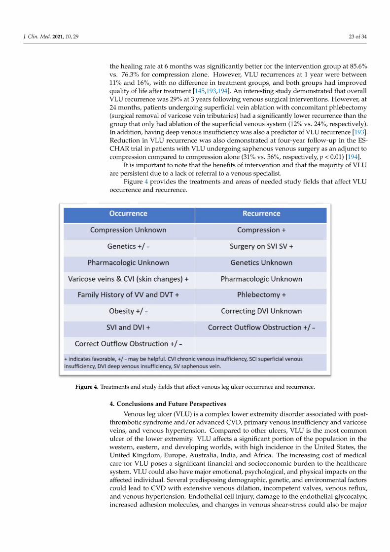
J. Clin. Med. 2021, 10, 29 23 of 34
the healing rate at 6 months was significantly better for the intervention group at 85.6%vs. 76.3% for compression alone. However, VLU recurrences at 1 year were between11% and 16%, with no difference in treatment groups, and both groups had improvedquality of life after treatment [145,193,194]. An interesting study demonstrated that overallVLU recurrence was 29% at 3 years following venous surgical interventions. However, at24 months, patients undergoing superficial vein ablation with concomitant phlebectomy(surgical removal of varicose vein tributaries) had a significantly lower recurrence than thegroup that only had ablation of the superficial venous system (12% vs. 24%, respectively).In addition, having deep venous insufficiency was also a predictor of VLU recurrence [193].Reduction in VLU recurrence was also demonstrated at four-year follow-up in the ES-CHAR trial in patients with VLU undergoing saphenous venous surgery as an adjunct tocompression compared to compression alone (31% vs. 56%, respectively, p < 0.01) [194].
It is important to note that the benefits of intervention and that the majority of VLUare persistent due to a lack of referral to a venous specialist.
Figure 4 provides the treatments and areas of needed study fields that affect VLUoccurrence and recurrence.
J. Clin. Med. 2021, 10, x FOR PEER REVIEW 23 of 33
phlebectomy (surgical removal of varicose vein tributaries) had a significantly lower re-
currence than the group that only had ablation of the superficial venous system (12% vs.
24%, respectively). In addition, having deep venous insufficiency was also a predictor of
VLU recurrence [193]. Reduction in VLU recurrence was also demonstrated at four-year
follow-up in the ESCHAR trial in patients with VLU undergoing saphenous venous sur-
gery as an adjunct to compression compared to compression alone (31% vs. 56%, respec-
tively, p ˂ 0.01) [194].
It is important to note that the benefits of intervention and that the majority of VLU
are persistent due to a lack of referral to a venous specialist.
Figure 4 provides the treatments and areas of needed study fields that affect VLU
occurrence and recurrence.
Figure 4. Treatments and study fields that affect venous leg ulcer occurrence and recurrence.
4. Conclusions and Future Perspectives
Venous leg ulcer (VLU) is a complex lower extremity disorder associated with post-
thrombotic syndrome and/or advanced CVD, primary venous insufficiency and varicose
veins, and venous hypertension. Compared to other ulcers, VLU is the most common ul-
cer of the lower extremity. VLU affects a significant portion of the population in the west-
ern, eastern, and developing worlds, with high incidence in the United States, the United
Kingdom, Europe, Australia, India, and Africa. The increasing cost of medical care for
VLU poses a significant financial and socioeconomic burden to the healthcare system.
VLU could also have major emotional, psychological, and physical impacts on the affected
individual. Several predisposing demographic, genetic, and environmental factors could
lead to CVD with extensive venous dilation, incompetent valves, venous reflux, and ve-
nous hypertension. Endothelial cell injury, damage to the endothelial glycocalyx, in-
creased adhesion molecules, and changes in venous shear-stress could also be major ini-
tiating events in VLU. Increased endothelial cell permeability, leukocyte infiltration, and
inflammation; increases in inflammatory cytokines, MMPs, and reactive oxygen and ni-
trogen species; and accumulation of iron deposits and tissue metabolites could also con-
Figure 4. Treatments and study fields that affect venous leg ulcer occurrence and recurrence.
4. Conclusions and Future Perspectives
Venous leg ulcer (VLU) is a complex lower extremity disorder associated with post-thrombotic syndrome and/or advanced CVD, primary venous insufficiency and varicoseveins, and venous hypertension. Compared to other ulcers, VLU is the most commonulcer of the lower extremity. VLU affects a significant portion of the population in thewestern, eastern, and developing worlds, with high incidence in the United States, theUnited Kingdom, Europe, Australia, India, and Africa. The increasing cost of medicalcare for VLU poses a significant financial and socioeconomic burden to the healthcaresystem. VLU could also have major emotional, psychological, and physical impacts on theaffected individual. Several predisposing demographic, genetic, and environmental factorscould lead to CVD with extensive venous dilation, incompetent valves, venous reflux,and venous hypertension. Endothelial cell injury, damage to the endothelial glycocalyx,increased adhesion molecules, and changes in venous shear-stress could also be major

J. Clin. Med. 2021, 10, 29 24 of 34
initiating events in VLU. Increased endothelial cell permeability, leukocyte infiltration,and inflammation; increases in inflammatory cytokines, MMPs, and reactive oxygen andnitrogen species; and accumulation of iron deposits and tissue metabolites could alsocontribute to the pathogenesis of VLU. VLU usually heals with good wound care andcompression therapy within 6 months. The VLU healing process involves multiple stepsincluding hemostasis, inflammation, cell proliferation, and tissue remodeling. VLU heal-ing also involves the contribution of multiple cell types including leukocytes, platelets,endothelial cells, vascular smooth muscle cells, fibroblasts, and keratinocytes. Additionally,VLU healing involves the release of a wide variety of localized and circulating biomoleculesincluding TGF-β, VEGF, PDGF, TNF-α, interleukins, chemokines, MMPs, TIMPs, elastase,urokinase plasminogen activator, fibrin, collagen, albumin, and other proteins. Alterationin any of these cellular and biochemical components that drive the physiological woundhealing process could delay VLU healing. Importantly, while histological studies are highlyinformative, specimens are obtained through invasive procedures that reflect a single timepoint during VLU healing, making it difficult for repeated sampling from the same site.On the other hand, soluble biomarkers in the blood and wound exudate can be measuredwith less invasive techniques and at different stages of VLU progression. Also, instead ofrelying solely on measurement of the VLU area to evaluate the wound healing status, theseVLU healing factors could be used as biomarkers for ulcer diagnosis and for the evaluationof its progression, responsiveness to healing, and prognosis.
Inadequate treatment of VLU could lead to progression to non-healed or granulatingVLU with major complications including physical immobility, reduced quality of life,cellulitis, severe infections, osteomyelitis, and neoplastic transformation. Recalcitrantulcers also show prolonged healing time and often occur in individuals with advancedage, higher body mass index, and nutritional deficiencies and in association with coldertemperature, preexisting or underlying venous disease, deep venous thrombosis, venousoutflow obstruction, and larger wound area.
Treatment of VLU includes compression therapy and endovenous ablation utilizinga variety of modalities including thermal energy and non-thermal endothelial injury toocclude the axial reflux. Other interventional approaches include endovenous treatmentsfor both the superficial venous system using subfascial endoscopic perforator surgery(SEPS) and the deep outflow iliac venous system using iliac venous stents, but the beneficialeffects of these approaches need to be further evaluated. Importantly, VLU has a high50–70% recurrence rate possibly due to patient noncompliance with compression therapy,surgical procedure failure, mixed VLU disease with arterial component, incorrect diagnosisof ulcer, and progression of venous disease. A key reason for VLU recurrence is alsopoorly understood pathophysiology and the molecular pathways leading to tissue injury,persistent inflammatory responses and monocyte/lymphocyte-endothelial activation, andoxidative stress (Figure 5).
New lines of therapy for VLU have been tested or are being developed. Severalbiologics such as bilayer living skin construct, fibroblast derivatives, and extracellular ma-trices and non-biologic products such as poly-N-acetyl glucosamine have been tested anddemonstrated benefits in VLU clinical trials. Human placental membrane amnion/chorionallografts have interesting properties for wound healing as they have collagen-rich ECMproteins and could provide growth factors, glycosaminoglycans, fibroblasts, epithelial cells,and mesenchymal stem cells necessary for healing.
Connexin 43 is abnormally upregulated at the wound edges of chronic non-healingVLU, and the ACT1 peptide is a competitive inhibitor of connexin 43 that accelerates woundhealing in animal models. Also, sulodexide has antithrombotic, anti-inflammatory, andendothelial protective properties that could support VLU healing. Other pharmacologicdrugs may also have benefits in VLU healing. Growth factors; bone marrow and adiposetissue-derived cell therapy; silver dressings; MMP inhibitors; and modulators of reactiveoxygen and nitrogen species, the immune response, and tissue metabolites could also havebenefits in VLU.
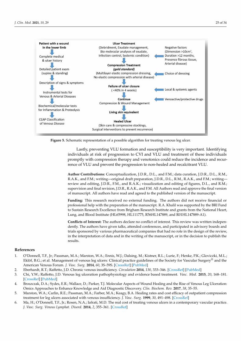
J. Clin. Med. 2021, 10, 29 25 of 34J. Clin. Med. 2021, 10, x FOR PEER REVIEW 25 of 33
Figure 5. Schematic representation of a possible algorithm for treating venous leg ulcer.
Lastly, preventing VLU formation and susceptibility is very important. Identifying
individuals at risk of progression to CVI and VLU and treatment of these individuals
promptly with compression therapy and venotonics could reduce the incidence and re-
currence of VLU and prevent the progression to non-healed and recalcitrant VLU.
Author Contributions: Conceptualization, J.D.R., D.L., and F.M.; data curation, J.D.R., D.L., R.M.,
R.A.K., and F.M.; writing—original draft preparation, J.D.R., D.L., R.M., R.A.K., and F.M.; writing—
review and editing, J.D.R., F.M., and R.A.K.; visualization and editing of figures, D.L., and R.M.;
supervision and final revision, J.D.R., R.A.K., and F.M. All Authors read and approve the final ver-
sion of manuscript. All authors have read and agreed to the published version of the manuscript.
Funding: This research received no external funding. The authors did not receive financial or pro-
fessional help with the preparation of the manuscript. R.A. Khalil was supported by the BRI Fund
to Sustain Research Excellence from Brigham Research Institute and grants from the National Heart,
Lung, and Blood Institute (HL65998, HL111775, R56HL147889, and R01HL147889-A1).
Conflicts of Interest: The authors declare no conflict of interest. This review was written inde-
pendently. The authors have given talks, attended conferences, and participated in advisory boards
and trials sponsored by various pharmaceutical companies that had no role in the design of the
review, in the interpretation of data and in the writing of the manuscript, or in the decision to pub-
lish the results.
References
1. O’Donnell, T.F., Jr.; Passman, M.A.; Marston, W.A.; Ennis, W.J.; Dalsing, M.; Kistner, R.L.; Lurie, F.; Henke, P.K.; Gloviczki,
M.L.; Eklöf, B.G.; et al. Management of venous leg ulcers: Clinical practice guidelines of the Society for Vascular Surgery® and
the American Venous Forum. J. Vasc. Surg. 2014, 60, 3S–59S, doi:10.1016/j.jvs.2014.04.049.
2. Eberhardt, R.T.; Raffetto, J.D. Chronic venous insufficiency. Circulation 2014, 130, 333–346, doi:10.1161/CIRCULA-
TIONAHA.113.006898.
3. Chi, Y.W.; Raffetto, J.D. Venous leg ulceration pathophysiology and evidence based treatment. Vasc. Med. 2015, 20, 168–181,
doi:10.1177/1358863X14568677.
4. Broszczak, D.A.; Sydes, E.R.; Wallace, D.; Parker, T.J. Molecular Aspects of Wound Healing and the Rise of Venous Leg Ulcer-
ation: Omics Approaches to Enhance Knowledge and Aid Diagnostic Discovery. Clin. Biochem. Rev. 2017, 38, 35–55.
Figure 5. Schematic representation of a possible algorithm for treating venous leg ulcer.
Lastly, preventing VLU formation and susceptibility is very important. Identifyingindividuals at risk of progression to CVI and VLU and treatment of these individualspromptly with compression therapy and venotonics could reduce the incidence and recur-rence of VLU and prevent the progression to non-healed and recalcitrant VLU.
Author Contributions: Conceptualization, J.D.R., D.L., and F.M.; data curation, J.D.R., D.L., R.M.,R.A.K., and F.M.; writing—original draft preparation, J.D.R., D.L., R.M., R.A.K., and F.M.; writing—review and editing, J.D.R., F.M., and R.A.K.; visualization and editing of figures, D.L., and R.M.;supervision and final revision, J.D.R., R.A.K., and F.M. All Authors read and approve the final versionof manuscript. All authors have read and agreed to the published version of the manuscript.
Funding: This research received no external funding. The authors did not receive financial orprofessional help with the preparation of the manuscript. R.A. Khalil was supported by the BRI Fundto Sustain Research Excellence from Brigham Research Institute and grants from the National Heart,Lung, and Blood Institute (HL65998, HL111775, R56HL147889, and R01HL147889-A1).
Conflicts of Interest: The authors declare no conflict of interest. This review was written indepen-dently. The authors have given talks, attended conferences, and participated in advisory boards andtrials sponsored by various pharmaceutical companies that had no role in the design of the review,in the interpretation of data and in the writing of the manuscript, or in the decision to publish theresults.
References1. O’Donnell, T.F., Jr.; Passman, M.A.; Marston, W.A.; Ennis, W.J.; Dalsing, M.; Kistner, R.L.; Lurie, F.; Henke, P.K.; Gloviczki, M.L.;
Eklöf, B.G.; et al. Management of venous leg ulcers: Clinical practice guidelines of the Society for Vascular Surgery® and theAmerican Venous Forum. J. Vasc. Surg. 2014, 60, 3S–59S. [CrossRef] [PubMed]
2. Eberhardt, R.T.; Raffetto, J.D. Chronic venous insufficiency. Circulation 2014, 130, 333–346. [CrossRef] [PubMed]3. Chi, Y.W.; Raffetto, J.D. Venous leg ulceration pathophysiology and evidence based treatment. Vasc. Med. 2015, 20, 168–181.
[CrossRef] [PubMed]4. Broszczak, D.A.; Sydes, E.R.; Wallace, D.; Parker, T.J. Molecular Aspects of Wound Healing and the Rise of Venous Leg Ulceration:
Omics Approaches to Enhance Knowledge and Aid Diagnostic Discovery. Clin. Biochem. Rev. 2017, 38, 35–55.5. Marston, W.A.; Carlin, R.E.; Passman, M.A.; Farber, M.A.; Keagy, B.A. Healing rates and cost efficacy of outpatient compression
treatment for leg ulcers associated with venous insufficiency. J. Vasc. Surg. 1999, 30, 491–498. [CrossRef]6. Ma, H.; O’Donnell, T.F., Jr.; Rosen, N.A.; Iafrati, M.D. The real cost of treating venous ulcers in a contemporary vascular practice.
J. Vasc. Surg. Venous Lymphat. Disord. 2014, 2, 355–361. [CrossRef]
J. Clin. Med. 2021, 10, 29 26 of 34
7. Rice, J.B.; Desai, U.; Cummings, A.K.; Birnbaum, H.G.; Skornicki, M.; Parsons, N. Burden of venous leg ulcers in the United States.J. Med. Econ. 2014, 17, 347–356. [CrossRef]
8. Eklof, B.; Rutherford, R.B.; Bergan, J.J.; Carpentier, P.H.; Gloviczki, P.; Kistner, R.L.; Meissner, M.H.; Moneta, G.L.; Myers, K.;Padberg, F.T.; et al. Revision of the CEAP classification for chronic venous disorders: Consensus statement. J. Vasc. Surg. 2004, 40,1248–1252. [CrossRef]
9. Meissner, M.H.; Moneta, G.; Burnand, K.; Gloviczki, P.; Lohr, J.M.; Lurie, F.; Mattos, M.A.; McLafferty, R.B.; Mozes, G.; Rutherford,R.B.; et al. The hemodynamics and diagnosis of venous disease. J. Vasc. Surg. 2007, 46, 4S–24S. [CrossRef]
10. Labropoulos, N.; Waggoner, T.; Sammis, W.; Samali, S.; Pappas, P.J. The effect of venous thrombus location and extent on thedevelopment of post-thrombotic signs and symptoms. J. Vasc. Surg. 2008, 48, 407–412. [CrossRef]
11. Raffetto, J.D.; Mannello, F. Pathophysiology of chronic venous disease. Int. Angiol. 2014, 33, 212–221. [PubMed]12. Mannello, F.; Ligi, D.; Canale, M.; Raffetto, J.D. Omics profiles in chronic venous ulcer wound fluid: Innovative applications for
translational medicine. Expert Rev. Mol. Diagn. 2014, 14, 737–762. [CrossRef] [PubMed]13. Raffetto, J.D. Pathophysiology of Chronic Venous Disease and Venous Ulcers. Surg. Clin. N. Am. 2018, 98, 337–347. [CrossRef]14. Onida, S.; Tan, M.K.H.; Kafeza, M.; Bergner, R.T.; Shalhoub, J.; Holmes, E.; Davies, A.H. Metabolic Phenotyping in Venous
Disease: The Need for Standardization. J. Proteome Res. 2019, 18, 3809–3820. [CrossRef]15. Pannier, F.; Rabe, E. Differential diagnosis of leg ulcers. Phlebology 2013, 28 (Suppl. 1), 55–60. [CrossRef]16. Makrantonaki, E.; Wlaschek, M.; Scharffetter-Kochanek, K. Pathogenesis of wound healing disorders in the elderly. J. Dtsch.
Dermatol. Ges. 2017, 15, 255–275. [CrossRef] [PubMed]17. Alavi, A.; Sibbald, R.G.; Phillips, T.J.; Miller, O.F.; Margolis, D.J.; Marston, W.; Woo, K.; Romanelli, M.; Kirsner, R.S. What’s new:
Management of venous leg ulcers: Approach to venous leg ulcers. J. Am. Acad. Dermatol. 2016, 74, 627–642. [CrossRef] [PubMed]18. Morton, L.M.; Phillips, T.J. Wound healing and treating wounds: Differential diagnosis and evaluation of chronic wounds. J. Am.
Acad. Dermatol. 2016, 74, 589–606. [CrossRef] [PubMed]19. Santler, B.; Goerge, T. Chronic venous insufficiency—A review of pathophysiology, diagnosis, and treatment. J. Dtsch. Dermatol.
Ges. 2017, 15, 538–556. [CrossRef] [PubMed]20. Ligi, D.; Mosti, G.; Croce, L.; Raffetto, J.D.; Mannello, F. Chronic venous disease—Part I: Inflammatory biomarkers in wound
healing. Biochim. Biophys. Acta 2016, 1862, 1964–1974. [CrossRef]21. Abbade, L.P.F.; Lastória, S. Venous ulcer: Epidemiology, physiopathology, diagnosis and treatment. Int. J. Dermatol. 2005, 44,
449–456. [CrossRef] [PubMed]22. Mansilha, A.; Sousa, J. Pathophysiological Mechanisms of Chronic Venous Disease and Implications for Venoactive Drug Therapy.
Int. J. Mol. Sci. 2018, 19, 1669. [CrossRef] [PubMed]23. Comerota, A.; Lurie, F. Pathogenesis of venous ulcer. Semin. Vasc. Surg. 2015, 28, 6–14. [CrossRef] [PubMed]24. Raffetto, J.D.; Khalil, R.A. Mechanisms of varicose vein formation: Valve dysfunction and wall dilation. Phlebology 2008, 23, 85–98.
[CrossRef]25. Labropoulos, N.; Leon, M.; Nicolaides, A.N.; Giannoukas, A.D.; Volteas, N.; Chan, P. Superficial venous insufficiency: Correlation
of anatomic extent of reflux with clinical symptoms and signs. J. Vasc. Surg. 1994, 20, 953–958. [CrossRef]26. Labropoulos, N.; Gasparis, A.P.; Tassiopoulos, A.K. Prospective evaluation of the clinical deterioration in post-thrombotic limbs.
J. Vasc. Surg. 2009, 50, 826–830. [CrossRef]27. Labropoulos, N.; Leon, M.; Nicolaides, A.N.; Sowade, O.; Volteas, N.; Ortega, F.; Chan, P. Venous reflux in patients with previous
deep venous thrombosis: Correlation with ulceration and other symptoms. J. Vasc. Surg. 1994, 20, 20–26. [CrossRef]28. Raju, S.; Knepper, J.; May, C.; Knight, A.; Pace, N.; Jayaraj, A. Ambulatory venous pressure, air plethysmography, and the role of
calf venous pump in chronic venous disease. J. Vasc. Surg. Venous Lymphat. Disord. 2019, 7, 428–440. [CrossRef]29. Raffetto, J.D. Inflammation in chronic venous ulcers. Phlebology 2013, 28, 61–67. [CrossRef]30. Bergan, J. Molecular mechanisms in chronic venous insufficiency. Ann. Vasc. Surg. 2007, 21, 260–266. [CrossRef]31. Pocock, E.S.; Alsaigh, T.; Mazor, R.; Schmid-Schönbein, G.W. Cellular and molecular basis of Venous insufficiency. Vasc. Cell 2014,
6, 24. [CrossRef] [PubMed]32. Xiao, Y.; Huang, Z.; Yin, H.; Lin, Y.; Wang, S. In vitro differences between smooth muscle cells derived from varicose veins and
normal veins. J. Vasc. Surg. 2009, 50, 1149–1154. [CrossRef]33. Badier-Commander, C.; Couvelard, A.; Henin, D.; Verbeuren, T.; Michel, J.B.; Jacob, M.P. Smooth muscle cell modulation and
cytokine overproduction in varicose veins. An in situ study. J. Pathol. 2001, 193, 398–407. [CrossRef] [PubMed]34. Crawford, J.M.; Lal, B.K.; Durán, W.N.; Pappas, P.J. Pathophysiology of venous ulceration. J. Vasc. Surg. Venous Lymphat. Disord.
2017, 5, 596–605. [CrossRef]35. Travers, J.P.; Brookes, C.E.; Evans, J.; Baker, D.M.; Kent, C.; Makin, G.S.; Mayhew, T.M. Assessment of wall structure and
composition of varicose veins with reference to collagen, elastin and smooth muscle content. Eur. J. Vasc. Endovasc. Surg. 1996, 11,230–237. [CrossRef]
36. Wali, M.A.; Eid, R.A. Changes of elastic and collagen fibers in varicose veins. Int. Angiol. 2002, 21, 337–343. [PubMed]37. Sansilvestri-Morel, P.; Rupin, A.; Badier-Commander, C.; Kern, P.; Fabiani, J.N.; Verbeuren, T.J.; Vanhoutte, P.M. Imbalance in the
synthesis of collagen type I and collagen type III in smooth muscle cells derived from human varicose veins. J. Vasc. Res. 2001, 38,560–568. [CrossRef] [PubMed]
J. Clin. Med. 2021, 10, 29 27 of 34
38. Sansilvestri-Morel, P.; Rupin, A.; Jullien, N.D.; Lembrez, N.; Mestries-Dubois, P.; Fabiani, J.N.; Verbeuren, T.J. Decreasedproduction of collagen Type III in cultured smooth muscle cells from varicose vein patients is due to a degradation by MMPs:Possible implication of MMP-3. J. Vasc. Res. 2005, 42, 388–398. [CrossRef] [PubMed]
39. Wlaschek, M.; Singh, K.; Sindrilaru, A.; Crisan, D.; Scharffetter-Kochanek, K. Iron and iron-dependent reactive oxygen speciesin the regulation of macrophages and fibroblasts in non-healing chronic wounds. Free Radic. Biol. Med. 2019, 133, 262–275.[CrossRef]
40. Caggiati, A.; Rosi, C.; Casini, A.; Cirenza, M.; Petrozza, V.; Acconcia, M.C.; Zamboni, P. Skin iron deposition characteriseslipodermatosclerosis and leg ulcer. Eur. J. Vasc. Endovasc. Surg. 2010, 40, 777–782. [CrossRef]
41. Pappas, P.J.; DeFouw, D.O.; Venezio, L.M.; Gorti, R.; Padberg, F.T., Jr.; Silva, M.B., Jr.; Goldberg, M.C.; Durán, W.N.; Hobson, R.W.,2nd. Morphometric assessment of the dermal microcirculation in patients with chronic venous insufficiency. J. Vasc. Surg. 1997,26, 784–795. [CrossRef]
42. Wilkinson, L.S.; Bunker, C.; Edwards, J.C.; Scurr, J.H.; Smith, P.D. Leukocytes: Their role in the etiopathogenesis of skin damagein venous disease. J. Vasc. Surg. 1993, 17, 669–675. [CrossRef]
43. Thomas, P.R.; Dormandy, J.A. White cell and platelet trapping in patients with chronic venous insufficiency. Phlebologie 1988, 41,771–776. [PubMed]
44. Tisato, V.; Zauli, G.; Gianesini, S.; Menegatti, E.; Brunelli, L.; Manfredini, R.; Zamboni, P.; Secchiero, P. Modulation of circulatingcytokine-chemokine profile in patients affected by chronic venous insufficiency undergoing surgical hemodynamic correction. J.Immunol. Res. 2014, 2014, 473765. [CrossRef]
45. Poredos, P.; Spirkoska, A.; Rucigaj, T.; Fareed, J.; Jezovnik, M.K. Do blood constituents in varicose veins differ from the systemicblood constituents? Eur. J. Vasc. Endovasc. Surg. 2015, 50, 250–256. [CrossRef]
46. Solá, L.d.R.; Aceves, M.; Dueñas, A.I.; González-Fajardo, J.A.; Vaquero, C.; Crespo, M.S.; García-Rodríguez, C. Varicose veinsshow enhanced chemokine expression. Eur. J. Vasc. Endovasc. Surg. 2009, 38, 635–641. [CrossRef]
47. Lattimer, C.R.; Kalodiki, E.; Geroulakos, G.; Hoppensteadt, D.; Fareed, J. Are Inflammatory Biomarkers Increased in Varicose VeinBlood? Clin. Appl. Thromb. Hemost. 2016, 22, 656–664. [CrossRef]
48. Gurtner, G.C.; Werner, S.; Barrandon, Y.; Longaker, M.T. Wound repair and regeneration. Nature 2008, 453, 314–321. [CrossRef]49. Biomarkers Definitions Working Group. Biomarkers and surrogate endpoints: Preferred definitions and conceptual framework.
Clin. Pharmacol. Ther. 2001, 69, 89–95. [CrossRef]50. Lyons, O.T.; Saha, P.; Smith, A. Redox dysregulation in the pathogenesis of chronic venous ulceration. Free Radic. Biol. Med. 2019.
[CrossRef]51. Stacey, M.C. Biomarker directed chronic wound therapy—A new treatment paradigm. J. Tissue Viability 2019. [CrossRef]
[PubMed]52. Edwards, H.E.; Parker, C.N.; Miller, C.; Gibb, M.; Kapp, S.; Ogrin, R.; Anderson, J.; Coleman, K.; Smith, D.; Finlayson, K.J.
Predicting delayed healing: The diagnostic accuracy of a venous leg ulcer risk assessment tool. Int. Wound J. 2018, 15, 258–265.[CrossRef] [PubMed]
53. Qing, C. The molecular biology in wound healing & non-healing wound. Chin. J. Traumatol. Zhonghua Chuang Shang Za Zhi 2017,20, 189–193. [CrossRef] [PubMed]
54. Diegelmann, R.F.; Evans, M.C. Wound healing: An overview of acute, fibrotic and delayed healing. Front. Biosci. J. Virtual Libr.2004, 9, 283–289. [CrossRef] [PubMed]
55. Han, G.; Ceilley, R. Chronic Wound Healing: A Review of Current Management and Treatments. Adv. Ther. 2017, 34, 599–610.[CrossRef] [PubMed]
56. Lindley, L.E.; Stojadinovic, O.; Pastar, I.; Tomic-Canic, M. Biology and Biomarkers for Wound Healing. Plast. Reconstr. Surg. 2016,138, 18S–28S. [CrossRef]
57. Pastar, I.; Wong, L.L.; Egger, A.N.; Tomic-Canic, M. Descriptive vs mechanistic scientific approach to study wound healing and itsinhibition: Is there a value of translational research involving human subjects? Exp. Dermatol. 2018, 27, 551–562. [CrossRef]
58. Parnell, L.K.S.; Volk, S.W. The Evolution of Animal Models in Wound Healing Research: 1993–2017. Adv. Wound Care 2019, 8,692–702. [CrossRef]
59. Moore, K.; Huddleston, E.; Stacey, M.C.; Harding, K.G. Venous leg ulcers—The search for a prognostic indicator. Int. Wound J.2007, 4, 163–172. [CrossRef]
60. Simka, M. Cellular and molecular mechanisms of venous leg ulcers development—The “puzzle” theory. Int. Angiol. 2010, 29,1–19.
61. Claudy, A.L.; Mirshahi, M.; Soria, C.; Soria, J. Detection of undegraded fibrin and tumor necrosis factor-alpha in venous leg ulcers.J. Am. Acad. Dermatol. 1991, 25, 623–627. [CrossRef]
62. Higley, H.R.; Ksander, G.A.; Gerhardt, C.O.; Falanga, V. Extravasation of macromolecules and possible trapping of transforminggrowth factor-beta in venous ulceration. Br. J. Dermatol. 1995, 132, 79–85. [CrossRef] [PubMed]
63. Harris, I.R.; Yee, K.C.; Walters, C.E.; Cunliffe, W.J.; Kearney, J.N.; Wood, E.J.; Ingham, E. Cytokine and protease levels in healingand non-healing chronic venous leg ulcers. Exp. Dermatol. 1995, 4, 342–349. [CrossRef] [PubMed]
64. Pappas, P.J.; Fallek, S.R.; Garcia, A.; Araki, C.T.; Back, T.L.; Durán, W.N.; Hobson, R.W., 2nd. Role of leukocyte activation inpatients with venous stasis ulcers. J. Surg. Res. 1995, 59, 553–559. [CrossRef] [PubMed]
J. Clin. Med. 2021, 10, 29 28 of 34
65. Hasan, A.; Murata, H.; Falabella, A.; Ochoa, S.; Zhou, L.; Badiavas, E.; Falanga, V. Dermal fibroblasts from venous ulcers areunresponsive to the action of transforming growth factor-beta 1. J. Dermatol. Sci. 1997, 16, 59–66. [CrossRef]
66. He, C.F.; Cherry, G.W.; Arnold, F. Postural vasoregulation and mediators of reperfusion injury in venous ulceration. J. Vasc. Surg.1997, 25, 647–653. [CrossRef]
67. Fivenson, D.P.; Faria, D.T.; Nickoloff, B.J.; Poverini, P.J.; Kunkel, S.; Burdick, M.; Strieter, R.M. Chemokine and inflammatorycytokine changes during chronic wound healing. Wound Repair Regen. 1997, 5, 310–322. [CrossRef]
68. Wallace, H.J.; Stacey, M.C. Levels of tumor necrosis factor-alpha (TNF-alpha) and soluble TNF receptors in chronic venous legulcers–correlations to healing status. J. Investig. Dermatol. 1998, 110, 292–296. [CrossRef]
69. Peschen, M.; Grenz, H.; Grothe, C.; Schöpf, E.; Vanscheidt, W. Patterns of epidermal growth factor receptor, basic fibroblastgrowth factor and transforming growth factor-beta3 expression in skin with chronic venous insufficiency. Eur. J. Dermatol. 1998, 8,334–338.
70. Peschen, M.; Grenz, H.; Brand-Saberi, B.; Bunaes, M.; Simon, J.C.; Schöpf, E.; Vanscheidt, W. Increased expression of platelet-derived growth factor receptor alpha and beta and vascular endothelial growth factor in the skin of patients with chronic venousinsufficiency. Arch. Dermatol. Res. 1998, 290, 291–297. [CrossRef]
71. Li, Y.Q.; Doyle, J.W.; Roth, T.P.; Dunn, R.M.; Lawrence, W.T. IL-10 and GM-CSF expression and the presence of antigen-presentingcells in chronic venous ulcers. J. Surg. Res. 1998, 79, 128–135. [CrossRef] [PubMed]
72. Lundberg, J.E.; Roth, T.P.; Dunn, R.M.; Doyle, J.W. Comparison of IL-10 levels in chronic venous insufficiency ulcers andautologous donor tissue. Arch. Dermatol. Res. 1998, 290, 669–673. [CrossRef]
73. Pappas, P.J.; You, R.; Rameshwar, P.; Gorti, R.; DeFouw, D.O.; Phillips, C.K.; Padberg, F.T., Jr.; Silva, M.B., Jr.; Simonian, G.T.;Hobson, R.W., 2nd; et al. Dermal tissue fibrosis in patients with chronic venous insufficiency is associated with increasedtransforming growth factor-beta1 gene expression and protein production. J. Vasc. Surg. 1999, 30, 1129–1145. [CrossRef]
74. Saalbach, A.; Wetzig, T.; Haustein, U.F.; Anderegg, U. Detection of human soluble Thy-1 in serum by ELISA. Fibroblasts andactivated endothelial cells are a possible source of soluble Thy-1 in serum. Cell Tissue Res. 1999, 298, 307–315. [CrossRef] [PubMed]
75. Trengove, N.J.; Bielefeldt-Ohmann, H.; Stacey, M.C. Mitogenic activity and cytokine levels in non-healing and healing chronic legulcers. Wound Repair Regen. 2000, 8, 13–25. [CrossRef] [PubMed]
76. Lauer, G.; Sollberg, S.; Cole, M.; Flamme, I.; Stürzebecher, J.; Mann, K.; Krieg, T.; Eming, S.A. Expression and proteolysis ofvascular endothelial growth factor is increased in chronic wounds. J. Investig. Dermatol. 2000, 115, 12–18. [CrossRef] [PubMed]
77. Cowin, A.J.; Hatzirodos, N.; Holding, C.A.; Dunaiski, V.; Harries, R.H.; Rayner, T.E.; Fitridge, R.; Cooter, R.D.; Schultz, G.S.;Belford, D.A. Effect of healing on the expression of transforming growth factor beta(s) and their receptors in chronic venous legulcers. J. Investig. Dermatol. 2001, 117, 1282–1289. [CrossRef]
78. Murphy, M.A.; Joyce, W.P.; Condron, C.; Bouchier-Hayes, D. A reduction in serum cytokine levels parallels healing of venousulcers in patients undergoing compression therapy. Eur. J. Vasc. Endovasc. Surg. 2002, 23, 349–352. [CrossRef]
79. Jude, E.B.; Blakytny, R.; Bulmer, J.; Boulton, A.J.M.; Ferguson, M.W.J. Transforming growth factor-beta 1, 2, 3 and receptor type Iand II in diabetic foot ulcers. Diabet. Med. 2002, 19, 440–447. [CrossRef]
80. Kim, B.-C.; Kim, H.T.; Park, S.H.; Cha, J.-S.; Yufit, T.; Kim, S.-J.; Falanga, V. Fibroblasts from chronic wounds show alteredTGF-beta-signaling and decreased TGF-beta Type II receptor expression. J. Cell Physiol. 2003, 195, 331–336. [CrossRef]
81. Tian, Y.-W.; Stacey, M.C. Cytokines and growth factors in keratinocytes and sweat glands in chronic venous leg ulcers. Animmunohistochemical study. Wound Repair Regen. 2003, 11, 316–325. [CrossRef] [PubMed]
82. Senet, P.; Bon, F.-X.; Benbunan, M.; Bussel, A.; Traineau, R.; Calvo, F.; Dubertret, L.; Dosquet, C. Randomized trial and localbiological effect of autologous platelets used as adjuvant therapy for chronic venous leg ulcers. J. Vasc. Surg. 2003, 38, 1342–1348.[CrossRef]
83. Nayeri, F.; Olsson, H.; Peterson, C.; Sundqvist, T. Hepatocyte growth factor; expression, concentration and biological activity inchronic leg ulcers. J. Dermatol. Sci. 2005, 37, 75–85. [CrossRef] [PubMed]
84. Galkowska, H.; Olszewski, W.L.; Wojewodzka, U. Keratinocyte and dermal vascular endothelial cell capacities remain unimpairedin the margin of chronic venous ulcer. Arch. Dermatol. Res. 2005, 296, 286–295. [CrossRef] [PubMed]
85. Chen, L.; Ye, C.; Yin, H.; Ye, J.; Wang, S. RANTES expression in venous ulceration of lower limbs. Nan Fang Yi Ke Da Xue Xue Bao2008, 28, 861–862.
86. Gohel, M.S.; Windhaber, R.A.; Tarlton, J.F.; Whyman, M.R.; Poskitt, K.R. The relationship between cytokine concentrations andwound healing in chronic venous ulceration. J. Vasc. Surg. 2008, 48, 1272–1277. [CrossRef] [PubMed]
87. Beidler, S.K.; Douillet, C.D.; Berndt, D.F.; Keagy, B.A.; Rich, P.B.; Marston, W.A. Inflammatory cytokine levels in chronic venousinsufficiency ulcer tissue before and after compression therapy. J. Vasc. Surg. 2009, 49, 1013–1020. [CrossRef]
88. Charles, C.A.; Romanelli, P.; Martinez, Z.B.; Ma, F.; Roberts, B.; Kirsner, R.S. Tumor necrosis factor-alfa in nonhealing venous legulcers. J. Am. Acad. Dermatol. 2009, 60, 951–955. [CrossRef]
89. Chen, L.; Liu, D.; Xiong, J.; Ye, C.; Yin, H.; Wang, S. The value of regulated upon activation normal T cell expressed and secretedin the pathogenesis of varicose ulcer of lower limbs. Zhonghua Yi Xue Za Zhi 2009, 89, 1460–1463.
90. Karatepe, O.; Unal, O.; Ugurlucan, M.; Kemik, A.; Karahan, S.; Aksoy, M.; Kurtoglu, M. The impact of valvular oxidative stress onthe development of venous stasis ulcer valvular oxidative stress and venous ulcers. Angiology 2010, 61, 283–288. [CrossRef]
J. Clin. Med. 2021, 10, 29 29 of 34
91. Pukstad, B.S.; Ryan, L.; Flo, T.H.; Stenvik, J.; Moseley, R.; Harding, K.; Thomas, D.W.; Espevik, T. Non-healing is associated withpersistent stimulation of the innate immune response in chronic venous leg ulcers. J. Dermatol. Sci. 2010, 59, 115–122. [CrossRef][PubMed]
92. Trøstrup, H.; Lundquist, R.; Christensen, L.H.; Jorgensen, L.N.; Karlsmark, T.; Haab, B.B.; Agren, M.S. S100A8/A9 deficiencyin nonhealing venous leg ulcers uncovered by multiplexed antibody microarray profiling. Br. J. Dermatol. 2011, 165, 292–301.[CrossRef] [PubMed]
93. Zillmer, R.; Trostrup, H.; Karlsmark, T.; Ifversen, P.; Agren, M.S. Duration of wound fluid secretion from chronic venous leg ulcersis critical for interleukin-1alpha, interleukin-1beta, interleukin-8 levels and fibroblast activation. Arch. Dermatol. Res. 2011, 303,601–606. [CrossRef] [PubMed]
94. Kroeze, K.L.; Vink, L.; de Boer, E.M.; Scheper, R.J.; van Montfrans, C.; Gibbs, S. Simple wound exudate collection method identifiesbioactive cytokines and chemokines in (arterio) venous ulcers. Wound Repair Regen. 2012, 20, 294–303. [CrossRef] [PubMed]
95. Zhou, K.; Ma, Y.; Brogan, M.S. Chronic and non-healing wounds: The story of vascular endothelial growth factor. Med. Hypotheses2015, 85, 399–404. [CrossRef] [PubMed]
96. Filkor, K.; Nemeth, T.; Nagy, I.; Kondorosi, E.; Urban, E.; Kemeny, L.; Szolnoky, G. The expression of inflammatory cytokines,TAM tyrosine kinase receptors and their ligands is upregulated in venous leg ulcer patients: A novel insight into chronic woundimmunity. Int. Wound J. 2016, 13, 554–562. [CrossRef] [PubMed]
97. Serra, R.; Grande, R.; Buffone, G.; Molinari, V.; Perri, P.; Perri, A.; Amato, B.; Colosimo, M.; de Franciscis, S. Extracellular matrixassessment of infected chronic venous leg ulcers: Role of metalloproteinases and inflammatory cytokines. Int. Wound J. 2016, 13,53–58. [CrossRef]
98. Serra, R.; Grande, R.; Butrico, L.; Buffone, G.; Calio, F.G.; Squillace, A.; Rizzo, B.A.; Massara, M.; Spinelli, F.; Ferrarese, A.G.; et al.Effects of a new nutraceutical substance on clinical and molecular parameters in patients with chronic venous ulceration. Int.Wound J. 2016, 13, 88–96. [CrossRef]
99. Goto, T.; Tamai, N.; Nakagami, G.; Kitamura, A.; Naito, A.; Hirokawa, M.; Shimokawa, C.; Takahashi, K.; Umemoto, J.; Sanada,H. Can Wound Exudate from Venous Leg Ulcers Measure Wound Pain Status? A Pilot Study. PLoS ONE 2016, 11, e0167478.[CrossRef]
100. Trostrup, H.; Holstein, P.; Christophersen, L.; Jorgensen, B.; Karlsmark, T.; Hoiby, N.; Moser, C.; Agren, M.S. S100A8/A9 is animportant host defence mediator in neuropathic foot ulcers in patients with type 2 diabetes mellitus. Arch. Dermatol. Res. 2016,308, 347–355. [CrossRef]
101. De Francesco, F.; Graziano, A.; Trovato, L.; Ceccarelli, G.; Romano, M.; Marcarelli, M.; Cusella De Angelis, G.M.; Cillo, U.; Riccio,M.; Ferraro, G.A. A Regenerative Approach with Dermal Micrografts in the Treatment of Chronic Ulcers. Stem Cell Rev. Rep. 2017,13, 139–148. [CrossRef] [PubMed]
102. Ligi, D.; Croce, L.; Mosti, G.; Raffetto, J.D.; Mannello, F. Chronic Venous Insufficiency: Transforming Growth Factor-β Isoformsand Soluble Endoglin Concentration in Different States of Wound Healing. Int. J. Mol. Sci. 2017, 18, 2206. [CrossRef] [PubMed]
103. Chimento, S.; Billero, V.; Cavallin, L.; Romanelli, M.; Nadji, M.; Romanelli, P. Evaluation of osteopontin expression in chronicwounds: A potential prognostic and therapeutic biomarker. J. Wound Care 2017, 26, S4–S8. [CrossRef] [PubMed]
104. Bodnar, E.; Bakondi, E.; Kovacs, K.; Hegedus, C.; Lakatos, P.; Robaszkiewicz, A.; Regdon, Z.; Virag, L.; Szabo, E. Redox ProfilingReveals Clear Differences between Molecular Patterns of Wound Fluids from Acute and Chronic Wounds. Oxid. Med. Cell. Longev.2018, 2018, 5286785. [CrossRef] [PubMed]
105. Krzystek-Korpacka, M.; Kedzior, K.; Maslowski, L.; Mierzchala, M.; Bednarz-Misa, I.; Bronowicka-Szydelko, A.; Kubiak, J.;Gacka, M.; Placzkowska, S.; Gamian, A. Impact of chronic wounds of various etiology on systemic profiles of key inflammatorycytokines, chemokines and growth factors, and their interplay. Adv. Clin. Exp. Med. Off. Organ Wroc. Med. Univ. 2019, 28,1301–1309. [CrossRef]
106. Ligi, D.; Croce, L.; Mannello, F. Chronic Venous Disorders: The Dangerous, the Good, and the Diverse. Int. J. Mol. Sci. 2018, 19,2544. [CrossRef]
107. Vaalamo, M.; Mattila, L.; Johansson, N.; Kariniemi, A.L.; Karjalainen-Lindsberg, M.L.; Kahari, V.M.; Saarialho-Kere, U. Distinctpopulations of stromal cells express collagenase-3 (MMP-13) and collagenase-1 (MMP-1) in chronic ulcers but not in normallyhealing wounds. J. Investig. Dermatol. 1997, 109, 96–101. [CrossRef]
108. Vaalamo, M.; Leivo, T.; Saarialho-Kere, U. Differential expression of tissue inhibitors of metalloproteinases (TIMP-1, -2, -3, and -4)in normal and aberrant wound healing. Hum. Pathol. 1999, 30, 795–802. [CrossRef]
109. Nwomeh, B.C.; Liang, H.X.; Cohen, I.K.; Yager, D.R. MMP-8 is the predominant collagenase in healing wounds and nonhealingulcers. J. Surg. Res. 1999, 81, 189–195. [CrossRef]
110. Tarlton, J.F.; Bailey, A.J.; Crawford, E.; Jones, D.; Moore, K.; Harding, K.D. Prognostic value of markers of collagen remodeling invenous ulcers. Wound Repair Regen. 1999, 7, 347–355. [CrossRef]
111. Saito, S.; Trovato, M.J.; You, R.; Lal, B.K.; Fasehun, F.; Padberg, F.T., Jr.; Hobson, R.W., 2nd; Duran, W.N.; Pappas, P.J. Role ofmatrix metalloproteinases 1, 2, and 9 and tissue inhibitor of matrix metalloproteinase-1 in chronic venous insufficiency. J. Vasc.Surg. 2001, 34, 930–938. [CrossRef] [PubMed]
112. Mirastschijski, U.; Impola, U.; Jahkola, T.; Karlsmark, T.; MS, A.G.; Saarialho-Kere, U. Ectopic localization of matrixmetalloproteinase-9 in chronic cutaneous wounds. Hum. Pathol. 2002, 33, 355–364. [CrossRef] [PubMed]
J. Clin. Med. 2021, 10, 29 30 of 34
113. Norgauer, J.; Hildenbrand, T.; Idzko, M.; Panther, E.; Bandemir, E.; Hartmann, M.; Vanscheidt, W.; Herouy, Y. Elevated expressionof extracellular matrix metalloproteinase inducer (CD147) and membrane-type matrix metalloproteinases in venous leg ulcers. Br.J. Dermatol. 2002, 147, 1180–1186. [CrossRef] [PubMed]
114. Impola, U.; Jeskanen, L.; Ravanti, L.; Syrjanen, S.; Baldursson, B.; Kahari, V.M.; Saarialho-Kere, U. Expression of matrixmetalloproteinase (MMP)-7 and MMP-13 and loss of MMP-19 and p16 are associated with malignant progression in chronicwounds. Br. J. Dermatol. 2005, 152, 720–726. [CrossRef]
115. Ulrich, D.; Lichtenegger, F.; Unglaub, F.; Smeets, R.; Pallua, N. Effect of chronic wound exudates and MMP-2/-9 inhibitor onangiogenesis in vitro. Plast. Reconstr. Surg. 2005, 116, 539–545. [CrossRef] [PubMed]
116. Zamboni, P.; Scapoli, G.; Lanzara, V.; Izzo, M.; Fortini, P.; Legnaro, R.; Palazzo, A.; Tognazzo, S.; Gemmati, D. Serum iron andmatrix metalloproteinase-9 variations in limbs affected by chronic venous disease and venous leg ulcers. Dermatol. Surg. 2005, 31,644–649, discussion 649. [CrossRef] [PubMed]
117. Mwaura, B.; Mahendran, B.; Hynes, N.; Defreitas, D.; Avalos, G.; Adegbola, T.; Adham, M.; Connolly, C.E.; Sultan, S. Theimpact of differential expression of extracellular matrix metalloproteinase inducer, matrix metalloproteinase-2, tissue inhibitor ofmatrix metalloproteinase-2 and PDGF-AA on the chronicity of venous leg ulcers. Eur. J. Vasc. Endovasc. Surg. 2006, 31, 306–310.[CrossRef]
118. Meyer, F.J.; Burnand, K.G.; Abisi, S.; Tekoppele, J.M.; van Els, B.; Smith, A. Effect of collagen turnover and matrix metalloproteinaseactivity on healing of venous leg ulcers. Br. J. Surg. 2008, 95, 319–325. [CrossRef]
119. Subramaniam, K.; Pech, C.M.; Stacey, M.C.; Wallace, H.J. Induction of MMP-1, MMP-3 and TIMP-1 in normal dermal fibroblastsby chronic venous leg ulcer wound fluid. Int. Wound J. 2008, 5, 79–86. [CrossRef]
120. Beidler, S.K.; Douillet, C.D.; Berndt, D.F.; Keagy, B.A.; Rich, P.B.; Marston, W.A. Multiplexed analysis of matrix metalloproteinasesin leg ulcer tissue of patients with chronic venous insufficiency before and after compression therapy. Wound Repair Regen. 2008,16, 642–648. [CrossRef]
121. Moor, A.N.; Vachon, D.J.; Gould, L.J. Proteolytic activity in wound fluids and tissues derived from chronic venous leg ulcers.Wound Repair Regen. 2009, 17, 832–839. [CrossRef] [PubMed]
122. Serra, R.; Buffone, G.; Falcone, D.; Molinari, V.; Scaramuzzino, M.; Gallelli, L.; de Franciscis, S. Chronic venous leg ulcers areassociated with high levels of metalloproteinases-9 and neutrophil gelatinase-associated lipocalin. Wound Repair Regen. 2013, 21,395–401. [CrossRef] [PubMed]
123. Grzela, T.; Niderla-Bielinska, J.; Litwiniuk, M.; White, R. The direct inhibition of MMP-2 and MMP-9 by an enzyme alginogel: Apossible mechanism of healing support for venous leg ulcers. J. Wound Care 2014, 23, 278–285. [CrossRef] [PubMed]
124. Serra, R.; Gallelli, L.; Butrico, L.; Buffone, G.; Calio, F.G.; De Caridi, G.; Massara, M.; Barbetta, A.; Amato, B.; Labonia, M.; et al.From varices to venous ulceration: The story of chronic venous disease described by metalloproteinases. Int. Wound J. 2017, 14,233–240. [CrossRef]
125. Amato, B.; Coretti, G.; Compagna, R.; Amato, M.; Buffone, G.; Gigliotti, D.; Grande, R.; Serra, R.; de Franciscis, S. Role of matrixmetalloproteinases in non-healing venous ulcers. Int. Wound J. 2015, 12, 641–645. [CrossRef]
126. Caimi, G.; Ferrara, F.; Montana, M.; Muratori, I.; Amato, C.; Canino, B.; Lo Presti, R.; Hopps, E. Behaviour of the plasmaconcentration of gelatinases and their tissue inhibitors in subjects with venous leg ulcers. Clin. Hemorheol. Microcirc. 2015, 60,309–316. [CrossRef]
127. Ligi, D.; Mosti, G.; Croce, L.; Raffetto, J.D.; Mannello, F. Chronic venous disease—Part II: Proteolytic biomarkers in woundhealing. Biochim. Biophys. Acta 2016, 1862, 1900–1908. [CrossRef]
128. Serena, T.E.; Cullen, B.M.; Bayliff, S.W.; Gibson, M.C.; Carter, M.J.; Chen, L.; Yaakov, R.A.; Samies, J.; Sabo, M.; DeMarco, D.; et al.Defining a new diagnostic assessment parameter for wound care: Elevated protease activity, an indicator of nonhealing, fortargeted protease-modulating treatment. Wound Repair Regen. 2016, 24, 589–595. [CrossRef]
129. Ruf, M.T.; Andreoli, A.; Vujic, G.; Itin, P.; Pluschke, G.; Schmid, P. Exudate collection using wound sponges-An easy, non-invasiveand reliable method to explore protease activities in ulcers. Wound Repair Regen. 2017, 25, 320–326. [CrossRef]
130. Trostrup, H.; Holstein, P.; Karlsmark, T.; Moser, C.; Agren, M.S. Uncontrolled gelatin degradation in non-healing chronic wounds.J. Wound Care 2018, 27, 724–734. [CrossRef]
131. Gillespie, D.L.; Writing Group III of the Pacific Vascular Symposium 6; Kistner, B.; Glass, C.; Bailey, B.; Chopra, A.; Ennis, B.;Marston, B.; Masuda, E.; Moneta, G.; et al. Venous ulcer diagnosis, treatment, and prevention of recurrences. J. Vasc. Surg. 2010,52, 8S–14S. [CrossRef] [PubMed]
132. Lal, B.K.; Saito, S.; Pappas, P.J.; Padberg, F.T., Jr.; Cerveira, J.J.; Hobson, R.W., 2nd; Durán, W.N. Altered proliferative responses ofdermal fibroblasts to TGF-beta1 may contribute to chronic venous stasis ulcer. J. Vasc. Surg. 2003, 37, 1285–1293. [CrossRef]
133. Mendez, M.V.; Raffetto, J.D.; Phillips, T.; Menzoian, J.O.; Park, H.Y. The proliferative capacity of neonatal skin fibroblasts isreduced after exposure to venous ulcer wound fluid: A potential mechanism for senescence in venous ulcers. J. Vasc. Surg. 1999,30, 734–743. [CrossRef]
134. Seidman, C.; Raffetto, J.D.; Marien, B.; Kroon, C.; Seah, C.C.; Menzoian, J.O. bFGF-induced alterations in cellular markers ofsenescence in growth-rescued fibroblasts from chronic venous ulcer and venous reflux patients. Ann. Vasc. Surg. 2003, 17, 239–244.[CrossRef]
135. Parker, C.N.; Finlayson, K.J.; Shuter, P.; Edwards, H.E. Risk factors for delayed healing in venous leg ulcers: A review of theliterature. Int. J. Clin. Pract. 2015, 69, 967–977. [CrossRef]
J. Clin. Med. 2021, 10, 29 31 of 34
136. Margolis, D.J.; Berlin, J.A.; Strom, B.L. Risk factors associated with the failure of a venous leg ulcer to heal. Arch. Dermatol. 1999,135, 920–926. [CrossRef]
137. Klode, J.; Stoffels, I.; Körber, A.; Weindorf, M.; Dissemond, J. Relationship between the seasonal onset of chronic venous leg ulcersand climatic factors. J. Eur. Acad. Dermatol. Venereol. 2011, 25, 1415–1419. [CrossRef]
138. Stojadinovic, O.; Pastar, I.; Vukelic, S.; Mahoney, M.G.; Brennan, D.; Krzyzanowska, A.; Golinko, M.; Brem, H.; Tomic-Canic, M.Deregulation of keratinocyte differentiation and activation: A hallmark of venous ulcers. J. Cell Mol. Med. 2008, 12, 2675–2690.[CrossRef]
139. Eming, S.A.; Koch, M.; Krieger, A.; Brachvogel, B.; Kreft, S.; Bruckner-Tuderman, L.; Krieg, T.; Shannon, J.D.; Fox, J.W. Differentialproteomic analysis distinguishes tissue repair biomarker signatures in wound exudates obtained from normal healing and chronicwounds. J. Proteome Res. 2010, 9, 4758–4766. [CrossRef]
140. Rayment, E.A.; Upton, Z.; Shooter, G.K. Increased matrix metalloproteinase-9 (MMP-9) activity observed in chronic wound fluidis related to the clinical severity of the ulcer. Br. J. Dermatol. 2008, 158, 951–961. [CrossRef]
141. James, T.J.; Hughes, M.A.; Cherry, G.W.; Taylor, R.P. Simple biochemical markers to assess chronic wounds. Wound Repair Regen.2000, 8, 264–269. [CrossRef] [PubMed]
142. Melikian, R.; O’Donnell, T.F., Jr.; Suarez, L.; Iafrati, M.D. Risk factors associated with the venous leg ulcer that fails to heal after 1year of treatment. J. Vasc. Surg. Venous Lymphat. Disord. 2019, 7, 98–105. [CrossRef] [PubMed]
143. Partsch, H.; Mortimer, P. Compression for leg wounds. Br. J. Dermatol. 2015, 173, 359–369. [CrossRef] [PubMed]144. Partsch, H. Compression heals leg ulcers due to abolishment of venous reflux. J. Wound Care 2019, 28, 427. [CrossRef]145. Gohel, M.S.; Heatley, F.; Liu, X.; Bradbury, A.; Bulbulia, R.; Cullum, N.; Epstein, D.M.; Nyamekye, I.; Poskitt, K.R.; Renton, S.;
et al. A Randomized Trial of Early Endovenous Ablation in Venous Ulceration. N. Engl. J. Med. 2018, 378, 2105–2114. [CrossRef]146. Epstein, D.M.; Gohel, M.S.; Heatley, F.; Liu, X.; Bradbury, A.; Bulbulia, R.; Cullum, N.; Nyamekye, I.; Poskitt, K.R.; Renton, S.;
et al. Cost-effectiveness analysis of a randomized clinical trial of early versus deferred endovenous ablation of superficial venousreflux in patients with venous ulceration. Br. J. Surg. 2019, 106, 555–562. [CrossRef]
147. Lin, Z.C.; Loveland, P.M.; Johnston, R.V.; Bruce, M.; Weller, C.D. Subfascial endoscopic perforator surgery (SEPS) for treatingvenous leg ulcers. Cochrane Database Syst. Rev. 2019, 3, CD012164. [CrossRef]
148. Neglen, P. Chronic venous obstruction: Diagnostic considerations and therapeutic role of percutaneous iliac stenting. Vascular2007, 15, 273–280. [CrossRef]
149. Neglen, P.; Hollis, K.C.; Raju, S. Combined saphenous ablation and iliac stent placement for complex severe chronic venousdisease. J. Vasc. Surg. 2006, 44, 828–833. [CrossRef]
150. Raju, S.; Kirk, O.K.; Jones, T.L. Endovenous management of venous leg ulcers. J. Vasc. Surg. Venous Lymphat. Disord. 2013, 1,165–172. [CrossRef]
151. Wang, W.; Yu, Z.; Chen, Y.-X. Stenting for chronic obstructive venous disease: A current comprehensive meta-analysis andsystematic review. Phlebology 2016, 31, 376–389. [CrossRef]
152. Raffetto, J.D. Dermal pathology, cellular biology, and inflammation in chronic venous disease. Thromb. Res. 2009, 123 (Suppl. 4),S66–S71. [CrossRef]
153. Raffetto, J.D.; Marston, W.A. Venous ulcer: What is new? Plast. Reconstr. Surg. 2011, 127 (Suppl. 1), 279S–288S. [CrossRef][PubMed]
154. Mannello, F.; Raffetto, J.D. Matrix metalloproteinase activity and glycosaminoglycans in chronic venous disease: The linkageamong cell biology, pathology and translational research. Am. J. Transl. Res. 2011, 3, 149–158.
155. Regulski, M.; Jacobstein, D.A.; Petranto, R.D.; Migliori, V.J.; Nair, G.; Pfeiffer, D. A retrospective analysis of a human cellularrepair matrix for the treatment of chronic wounds. Ostomy/Wound Manag. 2013, 59, 38–43.
156. Ananian, C.E.; Davis, R.D.; Johnson, E.L.; Regulski, M.J.; Reyzelman, A.M.; Saunders, M.C.; Danilkovitch, A. Wound ClosureOutcomes Suggest Clinical Equivalency Between Lyopreserved and Cryopreserved Placental Membranes Containing ViableCells. Adv. Wound Care 2019, 8, 546–554. [CrossRef]
157. Serena, T.E.; Carter, M.J.; Le, L.T.; Sabo, M.J.; DiMarco, D.T.; EpiFix VLU Study Group. A multicenter, randomized, controlledclinical trial evaluating the use of dehydrated human amnion/chorion membrane allografts and multilayer compression therapyvs. multilayer compression therapy alone in the treatment of venous leg ulcers. Wound Repair Regen. 2014, 22, 688–693. [CrossRef]
158. Ghatnekar, G.S.; Grek, C.L.; Armstrong, D.G.; Desai, S.C.; Gourdie, R.G. The effect of a connexin43-based Peptide on the healingof chronic venous leg ulcers: A multicenter, randomized trial. J. Investig. Dermatol. 2015, 135, 289–298. [CrossRef]
159. Bianchi, C.; Cazzell, S.; Vayser, D.; Reyzelman, A.M.; Dosluoglu, H.; Tovmassian, G.; EpiFix VLU Study Group. A multicentrerandomised controlled trial evaluating the efficacy of dehydrated human amnion/chorion membrane (EpiFix((R))) allograft forthe treatment of venous leg ulcers. Int. Wound J. 2018, 15, 114–122. [CrossRef]
160. Ghatnekar, G.S.; O’Quinn, M.P.; Jourdan, L.J.; Gurjarpadhye, A.A.; Draughn, R.L.; Gourdie, R.G. Connexin43 carboxyl-terminalpeptides reduce scar progenitor and promote regenerative healing following skin wounding. Regen. Med. 2009, 4, 205–223.[CrossRef]
161. Montgomery, J.; Ghatnekar, G.S.; Grek, C.L.; Moyer, K.E.; Gourdie, R.G. Connexin 43-Based Therapeutics for Dermal WoundHealing. Int. J. Mol. Sci. 2018, 19, 1778. [CrossRef] [PubMed]
162. Coccheri, S.; Mannello, F. Development and use of sulodexide in vascular diseases: Implications for treatment. Drug Des. Dev.Ther. 2013, 8, 49–65. [CrossRef] [PubMed]
J. Clin. Med. 2021, 10, 29 32 of 34
163. Carroll, B.J.; Piazza, G.; Goldhaber, S.Z. Sulodexide in venous disease. J. Thromb. Haemost. JTH 2019, 17, 31–38. [CrossRef][PubMed]
164. Scondotto, G.; Aloisi, D.; Ferrari, P.; Martini, L. Treatment of venous leg ulcers with sulodexide. Angiology 1999, 50, 883–889.[CrossRef]
165. Coccheri, S.; Scondotto, G.; Agnelli, G.; Palazzini, E.; Zamboni, V.; Arterial Arm of the Suavis Group. Sulodexide in the treatmentof intermittent claudication. Results of a randomized, double-blind, multicentre, placebo-controlled study. Eur. Heart J. 2002, 23,1057–1065. [CrossRef]
166. Coccheri, S.; Scondotto, G.; Agnelli, G.; Aloisi, D.; Palazzini, E.; Zamboni, V.; Venous arm of the SUAVIS Group. Randomised,double blind, multicentre, placebo controlled study of sulodexide in the treatment of venous leg ulcers. Thromb. Haemost. 2002,87, 947–952.
167. Urbanek, T.; Zbigniew, K.; Begier-Krasinska, B.; Baum, E.; Breborowicz, A. Sulodexide suppresses inflammation in patients withchronic venous insufficiency. Int. Angiol. 2015, 34, 589–596.
168. Gabryel, B.; Jarzabek, K.; Machnik, G.; Adamczyk, J.; Belowski, D.; Obuchowicz, E.; Urbanek, T. Superoxide dismutase 1 andglutathione peroxidase 1 are involved in the protective effect of sulodexide on vascular endothelial cells exposed to oxygen-glucosedeprivation. Microvasc. Res. 2016, 103, 26–35. [CrossRef]
169. De Felice, F.; Megiorni, F.; Pietrantoni, I.; Tini, P.; Lessiani, G.; Mastroiacovo, D.; Mattana, P.; Antinozzi, C.; Di Luigi, L.; DelleMonache, S.; et al. Sulodexide counteracts endothelial dysfunction induced by metabolic or non-metabolic stresses throughactivation of the autophagic program. Eur. Rev. Med. Pharmacol. Sci. 2019, 23, 2669–2680. [CrossRef]
170. Raffetto, J.D.; Yu, W.; Wang, X.; Calanni, F.; Mattana, P.; Khalil, R.A. Sulodexide Improves Contraction and Decreases MatrixMetalloproteinase-2 and -9 in Veins Under Prolonged Stretch. J. Cardiovasc. Pharmacol. 2020, 75, 211–221. [CrossRef]
171. Ligi, D.; Maniscalco, R.; Mannello, F. New Frontiers for an Old Drug: What Is New on the Pleiotropic Effect of Sulodexide inChronic Venous Disease. J. Cardiovasc. Pharmacol. 2020, 75, 208–210. [CrossRef] [PubMed]
172. Carvalho, M.R.; Silveira, I.A.; Oliveira, B. Treatment of venous ulcers with growth factors: Systematic review and meta-analysis.Rev. Bras. Enferm. 2019, 72, 200–210. [CrossRef] [PubMed]
173. Zhao, M.; Zhang, D.; Tan, L.; Huang, H. Silver dressings for the healing of venous leg ulcer: A meta-analysis and systematicreview. Medicine 2020, 99, e22164. [CrossRef] [PubMed]
174. Dissemond, J.; Augustin, M.; Dietlein, M.; Faust, U.; Keuthage, W.; Lobmann, R.; Munter, K.C.; Strohal, R.; Stucker, M.; Traber, J.;et al. Efficacy of MMP-inhibiting wound dressings in the treatment of chronic wounds: A systematic review. J. Wound Care 2020,29, 102–118. [CrossRef]
175. Norman, G.; Westby, M.J.; Rithalia, A.D.; Stubbs, N.; Soares, M.O.; Dumville, J.C. Dressings and topical agents for treating venousleg ulcers. Cochrane Database Syst. Rev. 2018, 6, CD012583. [CrossRef]
176. Otero, G.; Agorio, C.; Sujanov, A.; Echarte, L.; Tchekmedyian, A.; Montelongo, M.; Menyou, A.; Rodriguez, A.; Diaz, L.; Rodriguez,I.; et al. Autologous bone marrow-derived cells for venous leg ulcers treatment: A pilot study. Cytotherapy 2019, 21, 189–199.[CrossRef]
177. Zollino, I.; Campioni, D.; Sibilla, M.G.; Tessari, M.; Malagoni, A.M.; Zamboni, P. A phase II randomized clinical trial for thetreatment of recalcitrant chronic leg ulcers using centrifuged adipose tissue containing progenitor cells. Cytotherapy 2019, 21,200–211. [CrossRef]
178. Lee, A.J.; Robertson, L.A.; Boghossian, S.M.; Allan, P.L.; Ruckley, C.V.; Fowkes, F.G.; Evans, C.J. Progression of varicose veins andchronic venous insufficiency in the general population in the Edinburgh Vein Study. J. Vasc. Surg. Venous Lymphat. Disord. 2015, 3,18–26. [CrossRef]
179. Pannier, F.; Rabe, E. Progression in venous pathology. Phlebology 2015, 30, 95–97. [CrossRef]180. Zamboni, P.; Tognazzo, S.; Izzo, M.; Pancaldi, F.; Scapoli, G.L.; Liboni, A.; Gemmati, D. Hemochromatosis C282Y gene mutation
increases the risk of venous leg ulceration. J. Vasc. Surg. 2005, 42, 309–314. [CrossRef]181. Gemmati, D.; Tognazzo, S.; Serino, M.L.; Fogato, L.; Carandina, S.; De Palma, M.; Izzo, M.; De Mattei, M.; Ongaro, A.; Scapoli,
G.L.; et al. Factor XIII V34L polymorphism modulates the risk of chronic venous leg ulcer progression and extension. WoundRepair Regen. 2004, 12, 512–517. [CrossRef] [PubMed]
182. Gemmati, D.; Tognazzo, S.; Catozzi, L.; Federici, F.; De Palma, M.; Gianesini, S.; Scapoli, G.L.; De Mattei, M.; Liboni, A.; Zamboni,P. Influence of gene polymorphisms in ulcer healing process after superficial venous surgery. J. Vasc. Surg. 2006, 44, 554–562.[CrossRef]
183. Zamboni, P.; Lanzara, S.; Mascoli, F.; Caggiati, A.; Liboni, A. Inflammation in venous disease. Int. Angiol. 2008, 27, 361–369.184. Bosanquet, D.C.; Sanders, A.J.; Ruge, F.; Lane, J.; Morris, C.A.; Jiang, W.G.; Harding, K.G. Development and validation of a gene
expression test to identify hard-to-heal chronic venous leg ulcers. Br. J. Surg. 2019, 106, 1035–1042. [CrossRef]185. Marston, W.; Fish, D.; Unger, J.; Keagy, B. Incidence of and risk factors for iliocaval venous obstruction in patients with active or
healed venous leg ulcers. J. Vasc. Surg. 2011, 53, 1303–1308. [CrossRef] [PubMed]186. Lawrence, P.F.; Hager, E.S.; Harlander-Locke, M.P.; Pace, N.; Jayaraj, A.; Yohann, A.; Kalbaugh, C.; Marston, W.; Kabnick, L.;
Saqib, N.; et al. Treatment of superficial and perforator reflux and deep venous stenosis improves healing of chronic venous legulcers. J. Vasc. Surg. Venous Lymphat. Disord. 2020, 8, 601–609. [CrossRef] [PubMed]
J. Clin. Med. 2021, 10, 29 33 of 34
187. Gianesini, S.; Obi, A.; Onida, S.; Baccellieri, D.; Bissacco, D.; Borsuk, D.; Campisi, C.; Campisi, C.C.; Cavezzi, A.; Chi, Y.W.; et al.Global guidelines trends and controversies in lower limb venous and lymphatic disease: Narrative literature revision and experts’opinions following the vWINter international meeting in Phlebology, Lymphology & Aesthetics, 23–25 January 2019. Phlebology2019, 34, 4–66. [CrossRef]
188. Dahm, K.T.; Myrhaug, H.T.; Stromme, H.; Fure, B.; Brurberg, K.G. Effects of preventive use of compression stockings for elderlywith chronic venous insufficiency and swollen legs: A systematic review and meta-analysis. BMC Geriatr. 2019, 19, 76. [CrossRef]
189. Lurie, F.; Lal, B.K.; Antignani, P.L.; Blebea, J.; Bush, R.; Caprini, J.; Davies, A.; Forrestal, M.; Jacobowitz, G.; Kalodiki, E.; et al.Compression therapy after invasive treatment of superficial veins of the lower extremities: Clinical practice guidelines of theAmerican Venous Forum, Society for Vascular Surgery, American College of Phlebology, Society for Vascular Medicine, andInternational Union of Phlebology. J. Vasc. Surg. Venous Lymphat. Disord. 2019, 7, 17–28. [CrossRef]
190. Djalalov, S.; Sehatzadeh, S.; Keast, D.H.; Wong, W.W. Economic evaluation of compression stockings for the prevention of venousleg ulcer recurrence in Ontario. J. Wound Care 2020, 29, 141–151. [CrossRef]
191. Rabe, E.; Partsch, H.; Hafner, J.; Lattimer, C.; Mosti, G.; Neumann, M.; Urbanek, T.; Huebner, M.; Gaillard, S.; Carpentier, P.Indications for medical compression stockings in venous and lymphatic disorders: An evidence-based consensus statement.Phlebology 2018, 33, 163–184. [CrossRef] [PubMed]
192. Raffetto, J.D.; Eberhardt, R.T.; Dean, S.M.; Ligi, D.; Mannello, F. Pharmacologic treatment to improve venous leg ulcer healing.J. Vasc. Surg. Venous Lymphat. Disord. 2016, 4, 371–374. [CrossRef] [PubMed]
193. Marston, W.A.; Crowner, J.; Kouri, A.; Kalbaugh, C.A. Incidence of venous leg ulcer healing and recurrence after treatment withendovenous laser ablation. J. Vasc. Surg. Venous Lymphat. Disord. 2017, 5, 525–532. [CrossRef] [PubMed]
194. Gohel, M.S.; Barwell, J.R.; Taylor, M.; Chant, T.; Foy, C.; Earnshaw, J.J.; Heather, B.P.; Mitchell, D.C.; Whyman, M.R.; Poskitt,K.R. Long term results of compression therapy alone versus compression plus surgery in chronic venous ulceration (ESCHAR):Randomised controlled trial. BMJ 2007, 335, 83. [CrossRef] [PubMed]
Related Documents


![· Venous disease is chronic, and venous ulcers tend to be more often recurrent when com ared with the non-venous ulcers [10] and those with venous ulcers had a higher body mass](https://static.cupdf.com/doc/110x72/5f48c58329f97250766cd8db/venous-disease-is-chronic-and-venous-ulcers-tend-to-be-more-often-recurrent-when.jpg)