JOURNAL OF BACTERIOLOGY, Nov. 2006, p. 7405–7415 Vol. 188, No. 21 0021-9193/06/$08.000 doi:10.1128/JB.00758-06 Copyright © 2006, American Society for Microbiology. All Rights Reserved. Whole-Genome Sequence of Listeria welshimeri Reveals Common Steps in Genome Reduction with Listeria innocua as Compared to Listeria monocytogenes † Torsten Hain, 1 ‡ Christiane Steinweg, 1 ‡ Carsten Tobias Kuenne, 1 Andre ´ Billion, 1 Rohit Ghai, 1 Som Subhra Chatterjee, 1 Eugen Domann, 1 Uwe Ka ¨rst, 2 Alexander Goesmann, 3 Thomas Bekel, 3 Daniela Bartels, 3 Olaf Kaiser, 3 Folker Meyer, 3 Alfred Pu ¨hler, 4 Bernd Weisshaar, 5 Ju ¨rgen Wehland, 2 Chunguang Liang, 6 Thomas Dandekar, 6 Robert Lampidis, 7 Ju ¨rgen Kreft, 7 Werner Goebel, 7 and Trinad Chakraborty 1 * Institute for Medical Microbiology, Justus-Liebig-University, Frankfurter Strasse 107, D-35392 Giessen, Germany 1 ; Gesellschaft fu ¨r Biotechnologische Forschung GmbH, Department of Cell Biology, Mascheroder Weg 1, D-38124 Braunschweig, Germany 2 ; Bioinformatics Resource Facility, Centrum fu ¨r Biotechnologie, University of Bielefeld, Universita ¨tsstrasse 25, D-33615 Bielefeld, Germany 3 ; Lehrstuhl fu ¨r Genetik, University of Bielefeld, Universita ¨tsstrasse 25, D-33615 Bielefeld, Germany 4 ; Lehrstuhl fu ¨r Genomforschung, University of Bielefeld, Universita ¨tsstrasse 25, D-33615 Bielefeld, Germany 5 ; Lehrstuhl fu ¨r Bioinformatik, University of Wu ¨rzburg, Am Hubland/Biozentrum, D-97074 Wu ¨rzburg, Germany 6 ; and Lehrstuhl fu ¨r Mikrobiologie, University of Wu ¨rzburg, Am Hubland/Biozentrum, D-97074 Wu ¨rzburg, Germany 7 Received 26 May 2006/Accepted 14 August 2006 We present the complete genome sequence of Listeria welshimeri, a nonpathogenic member of the genus Listeria. Listeria welshimeri harbors a circular chromosome of 2,814,130 bp with 2,780 open reading frames. Comparative genomic analysis of chromosomal regions between L. welshimeri, Listeria innocua, and Listeria monocytogenes shows strong overall conservation of synteny, with the exception of the translocation of an F o F 1 ATP synthase. The smaller size of the L. welshimeri genome is the result of deletions in all of the genes involved in virulence and of “fitness” genes required for intracellular survival, transcription factors, and LPXTG- and LRR-containing proteins as well as 55 genes involved in carbohydrate transport and metabolism. In total, 482 genes are absent from L. welshimeri relative to L. monocytogenes. Of these, 249 deletions are commonly absent in both L. welshimeri and L. innocua, suggesting similar genome evolutionary paths from an ancestor. We also identified 311 genes specific to L. welshimeri that are absent in the other two species, indicating gene expansion in L. welshimeri, including horizontal gene transfer. The species L. welshimeri appears to have been derived from early evolutionary events and an ancestor more compact than L. monocytogenes that led to the emergence of nonpathogenic Listeria spp. Listeriae are gram-positive, motile, facultative anaerobic rod-like bacteria that are ubiquitously occurring in nature. Listeriae are members of a group of bacteria with low GC DNA content that includes species of the genera Bacillus, Clostridium, Enterococcus, Streptococcus, and Staphylococcus. The genus Listeria consists of six different species: Listeria monocytogenes, Listeria ivanovii, Listeria innocua, Listeria welshimeri, Listeria seeligeri, and Listeria grayi. L. monocyto- genes and L. ivanovii are the only pathogenic species of the genus. While L. ivanovii is predominantly an animal pathogen, L. monocytogenes can cause infections of both animals and humans. Clinical symptoms of listeriosis in humans are often manifested as meningitis, meningoencephalitis, septicemia, abortion, prenatal infection, and gastroenteritis (39). In addition to these two pathogens, there are four nonpatho- genic species of Listeria, viz., L. innocua, L. welshimeri, L. seeligeri, and L. grayi. Phylogenetic analyses, based on the 16S and 23S rRNA genes and the iap, prs, vclA, vclB, and ldh genes, indicate that L. innocua is highly related to L. monocytogenes. The second group has L. ivanovii, together with L. seeligeri, while L. welshimeri is more distant, exhibiting the deepest branching of this group. L. grayi seems to be very distant from these two groups (34). L. welshimeri (SLCC5334, CIP8149, and Welshimer V8) was first isolated from decaying plants (33, 40) and is a serovar 6b strain; other serovars (1/2a, 1/2b, 6a, 4c, and 4f) (20, 22) have also been reported for this species. Like other species of Lis- teria, L. welshimeri bacteria are small (0.5 to 2.0 m), non- spore-forming, gram-positive rods which are motile below 30°C by means of peritrichous flagella. Growth at low temperatures (4°C) proceeds within 5 days. Results from a CAMP test with Staphylococcus aureus and Rhodococcus equi were negative, and strains of the species also tested negative for oxidase but were positive for catalase activity. Acid production occurs by fermentation of D-xylose and -methyl-D-mannoside but not from L-rhamnose and D-mannitol (32). These biochemical * Corresponding author. Mailing address: Institute for Medical Mi- crobiology, Justus-Liebig-University, Frankfurter Strasse 107, D-35392 Giessen, Germany. Phone: 49-641 99 41250. Fax: 49-641 99 41259. E-mail: [email protected]. † Supplemental material for this article may be found at http://jb .asm.org/. ‡ Both authors contributed equally to this work. Published ahead of print on 25 August 2006. 7405

Welcome message from author
This document is posted to help you gain knowledge. Please leave a comment to let me know what you think about it! Share it to your friends and learn new things together.
Transcript
JOURNAL OF BACTERIOLOGY, Nov. 2006, p. 7405–7415 Vol. 188, No. 210021-9193/06/$08.00�0 doi:10.1128/JB.00758-06Copyright © 2006, American Society for Microbiology. All Rights Reserved.
Whole-Genome Sequence of Listeria welshimeri Reveals Common Stepsin Genome Reduction with Listeria innocua as Compared to
Listeria monocytogenes�†Torsten Hain,1‡ Christiane Steinweg,1‡ Carsten Tobias Kuenne,1 Andre Billion,1 Rohit Ghai,1
Som Subhra Chatterjee,1 Eugen Domann,1 Uwe Karst,2 Alexander Goesmann,3Thomas Bekel,3 Daniela Bartels,3 Olaf Kaiser,3 Folker Meyer,3 Alfred Puhler,4
Bernd Weisshaar,5 Jurgen Wehland,2 Chunguang Liang,6 Thomas Dandekar,6Robert Lampidis,7 Jurgen Kreft,7 Werner Goebel,7 and Trinad Chakraborty1*
Institute for Medical Microbiology, Justus-Liebig-University, Frankfurter Strasse 107, D-35392 Giessen, Germany1;Gesellschaft fur Biotechnologische Forschung GmbH, Department of Cell Biology, Mascheroder Weg 1,
D-38124 Braunschweig, Germany2; Bioinformatics Resource Facility, Centrum fur Biotechnologie,University of Bielefeld, Universitatsstrasse 25, D-33615 Bielefeld, Germany3; Lehrstuhl fur Genetik,
University of Bielefeld, Universitatsstrasse 25, D-33615 Bielefeld, Germany4; Lehrstuhl furGenomforschung, University of Bielefeld, Universitatsstrasse 25, D-33615 Bielefeld,
Germany5; Lehrstuhl fur Bioinformatik, University of Wurzburg, Am Hubland/Biozentrum,D-97074 Wurzburg, Germany6; and Lehrstuhl fur Mikrobiologie, University of
Wurzburg, Am Hubland/Biozentrum, D-97074 Wurzburg, Germany7
Received 26 May 2006/Accepted 14 August 2006
We present the complete genome sequence of Listeria welshimeri, a nonpathogenic member of the genusListeria. Listeria welshimeri harbors a circular chromosome of 2,814,130 bp with 2,780 open reading frames.Comparative genomic analysis of chromosomal regions between L. welshimeri, Listeria innocua, and Listeriamonocytogenes shows strong overall conservation of synteny, with the exception of the translocation of an FoF1ATP synthase. The smaller size of the L. welshimeri genome is the result of deletions in all of the genes involvedin virulence and of “fitness” genes required for intracellular survival, transcription factors, and LPXTG- andLRR-containing proteins as well as 55 genes involved in carbohydrate transport and metabolism. In total, 482genes are absent from L. welshimeri relative to L. monocytogenes. Of these, 249 deletions are commonly absentin both L. welshimeri and L. innocua, suggesting similar genome evolutionary paths from an ancestor. We alsoidentified 311 genes specific to L. welshimeri that are absent in the other two species, indicating gene expansionin L. welshimeri, including horizontal gene transfer. The species L. welshimeri appears to have been derivedfrom early evolutionary events and an ancestor more compact than L. monocytogenes that led to the emergenceof nonpathogenic Listeria spp.
Listeriae are gram-positive, motile, facultative anaerobicrod-like bacteria that are ubiquitously occurring in nature.Listeriae are members of a group of bacteria with low G�CDNA content that includes species of the genera Bacillus,Clostridium, Enterococcus, Streptococcus, and Staphylococcus.The genus Listeria consists of six different species: Listeriamonocytogenes, Listeria ivanovii, Listeria innocua, Listeriawelshimeri, Listeria seeligeri, and Listeria grayi. L. monocyto-genes and L. ivanovii are the only pathogenic species of thegenus. While L. ivanovii is predominantly an animal pathogen,L. monocytogenes can cause infections of both animals andhumans. Clinical symptoms of listeriosis in humans are oftenmanifested as meningitis, meningoencephalitis, septicemia,abortion, prenatal infection, and gastroenteritis (39).
In addition to these two pathogens, there are four nonpatho-genic species of Listeria, viz., L. innocua, L. welshimeri, L.seeligeri, and L. grayi. Phylogenetic analyses, based on the 16Sand 23S rRNA genes and the iap, prs, vclA, vclB, and ldh genes,indicate that L. innocua is highly related to L. monocytogenes.The second group has L. ivanovii, together with L. seeligeri,while L. welshimeri is more distant, exhibiting the deepestbranching of this group. L. grayi seems to be very distant fromthese two groups (34).
L. welshimeri (SLCC5334, CIP8149, and Welshimer V8) wasfirst isolated from decaying plants (33, 40) and is a serovar 6bstrain; other serovars (1/2a, 1/2b, 6a, 4c, and 4f) (20, 22) havealso been reported for this species. Like other species of Lis-teria, L. welshimeri bacteria are small (0.5 to 2.0 �m), non-spore-forming, gram-positive rods which are motile below 30°Cby means of peritrichous flagella. Growth at low temperatures(4°C) proceeds within 5 days. Results from a CAMP test withStaphylococcus aureus and Rhodococcus equi were negative,and strains of the species also tested negative for oxidase butwere positive for catalase activity. Acid production occurs byfermentation of D-xylose and �-methyl-D-mannoside but notfrom L-rhamnose and D-mannitol (32). These biochemical
* Corresponding author. Mailing address: Institute for Medical Mi-crobiology, Justus-Liebig-University, Frankfurter Strasse 107, D-35392Giessen, Germany. Phone: 49-641 99 41250. Fax: 49-641 99 41259.E-mail: [email protected].
† Supplemental material for this article may be found at http://jb.asm.org/.
‡ Both authors contributed equally to this work.� Published ahead of print on 25 August 2006.
7405

properties are used to distinguish L. welshimeri from otherListeria species.
The major virulence determinants in Listeria pathogenesisare localized on a chromosomal locus between prs and ldhdesignated the virulence gene cluster “vgc” (8) or Listeriapathogenicity island 1, “LIPI-1” (39), which is responsible forthe intracellular life cycle of the bacterium. All of these viru-lence genes are missing in L. welshimeri, which suggests thatthe genus Listeria probably evolved from the loss of the vgcregion leading to the generation of nonpathogenic speciesfrom a progenitor strain already harboring the virulence genes.
Thus, L. welshimeri strains are nonhemolytic and even a highinfecting dose of L. welshimeri (�1 � 108 CFU/ml), which is atleast 100,000-fold higher than the 50% lethal dose of L. mono-cytogenes (1 � 103 CFU/ml), does not kill mice. Primary inoc-ulation with L. welshimeri does not confer protection to sub-sequent challenge with a lethal dose of L. monocytogenes inmice (22).
Here we report the complete genome sequence and analysisof the type strain of L. welshimeri. We also compared thissequence to the complete genome sequences of three listerialstrains, L. monocytogenes 1/2a EGD-e, L. monocytogenes 4bF2365, and L. innocua. The data provide strong evidence forthe evolution of nonpathogenic Listeria spp. from an ancestralpathogenic listerial strain and suggest that this speciesemerged by additional gene loss and the acquisition of novelgenes, probably by horizontal gene transfer.
MATERIALS AND METHODS
Genome sequencing. Bacterial strains, plasmids, and primers are listed inTable S1S of the supplemental material. The type strain of Listeria welshimeri(serovar 6b, SLCC5334) was selected for genome sequencing. Two small insertlibraries (1.5 to 2 kb and 2 to 3 kb) were constructed using the plasmidpCR4Blunt-TOPO (Invitrogen) for shotgun sequencing. In addition, fosmid(pCCC1FOS; Epicenter) and bacterial artificial chromosome (BAC) (pBeloBAC11;New England Biolabs) libraries were generated for contig ordering and gapclosure with large fragments of around 40 kb and 50 kb, respectively. DNAsequencing was performed on the MegaBACE 1000 sequencing system (GEHealthcare) and ABI PRISM 3100 or 3730xl genetic analyzer (Applied Biosys-tems). Sequence data were analyzed and assembled by using Phred/Phrap/Consed software (14, 15, 18, 19). A total number of 42.146 sequences of theshotgun libraries, 692 BAC sequences, and 1,379 fosmid sequences were assem-bled by the Phrap software, resulting in �6.4-fold coverage. Gaps in the assem-bled genome sequence of L. welshimeri were closed by primer walking on shot-gun clones, fosmids, and BACs (421 sequences). To complete the remaining
gaps, we determined the order of the contigs of the L. welshimeri genome relativeto that of the L. monocytogenes EGD-e genome by using the program NUCmer(10). Specific primers were designed near the ends of neighboring contigs, andPCRs were performed with chromosomal template DNA. Ninety-two sequenceswere obtained from PCR products that spanned these gaps.
Automatic annotation of the genome sequence of L. welshimeri was performedusing GenDB 2.0 (25). Subsequently, all orthologs between the L. welshimerigenome and the genomes of L. monocytogenes EGD-e, L. innocua CLIP 11262(17), and L. monocytogenes F2365 (29) were predicted and the L. monocytogenesF2365 annotation was adapted over for the corresponding orthologs (see below)of the L. welshimeri genome. The only exception was a discrepancy between theannotation of L. monocytogenes F2365 and the automatic annotation of GenDB2.0 in case the latter one was identical to the annotations of L. monocytogenesEGD-e and L. innocua. Finally, all genes were manually inspected and annotatedusing GenDB 2.0.
For comparative genome analyses, orthologs were calculated using BLASTP.Two genes are considered an orthologous pair if they feature (i) a BLASTP Evalue of �0.001, (ii) protein identity of �50%, and (iii) both coverages (align-ment to protein 1 and alignment to protein 2) between 75 to 125%. Two geneswere considered a unidirectional orthologous pair if they were orthologs and thequery gene was the best BLASTP hit in the compared genome. Two genes wereconsidered a bidirectional ortholog pair if they were orthologs and reciprocalbest BLASTP hits when comparing two genomes. Cluster analysis was performedby merging all pairs that share one protein (pair AB plus BC results in clusterABC), which leads to a soft protein family-like grouping. The program GECO(C. T. Kuenne) for comparative genome visualization and the computationalpipeline Augur (A. Billion) for surface-associated protein prediction and proteinclassification are unpublished software.
Nucleotide sequence accession number. The genome sequence of L. welshi-meri serovar 6b (SLCC5334) reported here has been deposited in the EMBLdatabase under accession number AM263198.
RESULTS AND DISCUSSION
General and specific features of the genome. The overallfeatures of L. welshimeri and the recently sequenced genomesof L. monocytogenes and L. innocua (17) are given in Table 1.The circular genome of the type strain of L. welshimeri(SLCC5334) is 2,814,130 bp in length (Fig. 1A). In comparisonwith the genome sizes of L. monocytogenes and L. innocua, L.welshimeri has the smallest genome of the genus Listeria. Aswith the genomes of the other listerial strains, L. welshimeri hasa low G�C content, 36.4%, which is slightly lower than thoseof L. monocytogenes (38.0%) and L. innocua (37.4%). Its originof replication and terminus, as determined by G/C skew anal-ysis, are located �1,400 kb apart in positions similar to thoseobserved for L. monocytogenes and L. innocua. The genome ofL. welshimeri contains six complete copies of rRNA operons
TABLE 1. Characteristics of L. welshimeri, L. innocua, and L. monocytogenes
Characteristic L. welshimeri L. innocua L. monocytogenesEGD-e
Size of the chromosome (bp) 2,814,130 3,011,208 2,944,528G � C content (%) 36.4 37.4 38.0G � C content of protein-coding genes (%) 36.7 37.8 38.4Total no. of protein-coding genes 2,780 2,981 2,855Avg length (codons) of protein-coding genes 299 300 307No. of rRNA operons (16S-23S-5S) 6 6 6No. of tRNA genes 66 66 67Percentage coding 88.7 89.2 89.2No. of prophages 1 (55 genes) 5 (305 genes) 1 (62 genes)No. of plasmids 0 1 0No. of strain-specific genesa 311 121 218No. of orthologous genesa 2,414 2,555 2,575No. of transposons 0 0 1
a Prophage genes excepted.
7406 HAIN ET AL. J. BACTERIOL.

(16S-23S-5S); two are located on the right replichore and fourare located on the left one (Fig. 1A). We detected 66 tRNAgenes, a number similar to that for L. innocua but one less thanthat for L. monocytogenes.
The protein coding sequences (CDS) represent 88.7% of thegenome and are organized into 2,780 CDS. The average lengthof predicted CDS was 299 amino acids. The majority of CDS(79.3%) are organized on the leading strand in the direction ofDNA replication, which is similar to those of the genomes of L.monocytogenes (78.7%) and L. innocua (80%). Six pseudo-genes were detected, which is a number similar to those in L.monocytogenes (9 pseudogenes) and L. innocua (13 pseudo-genes). Following gene prediction and annotation, we assignedfunctions for 2,186 proteins (79%) in L. welshimeri and 594proteins (21%) were similar to hypothetical and conservedhypothetical proteins. A total of 1,492 out of 2,780 CDSshowed similarities to proteins from other low-G�C-contentFirmicutes species, like Bacillus subtilis 168 (1,317 orthologousgenes) and S. aureus N315 (1,100 orthologous genes), when acutoff for bidirectional ortholog pair calculation of �30% pro-tein identity and 75 to 125% coverage was used.
We observed that the synteny between L. welshimeri, L.monocytogenes, and L. innocua is highly conserved (see Fig.
S1S in the supplemental material), indicating that the overallorganization of these genomes is stable. Strong conservation ofthe genome organization has also been observed with otherclosely related members of low-G�C bacteria, such as Bacillusand Staphylococcus (5). The chromosome of L. welshimeri con-tains a single putative prophage, located at around 1.2 Mb(lwe1196-lwe1257), with strong homology to a prophage in L.innocua located 2.6 Mb from its origin of replication (see Fig.S1S in the supplemental material). Interestingly, this prophageof L. welshimeri is inserted within the region between thetRNAArg and ydeI genes compared to L. monocytogenesEGD-e and L. innocua. At the same chromosomal location inL. ivanovii, the species-specific Listeria pathogenicity island 2(12) is flanked by the tRNAArg and ydeI genes, suggesting thatthis region is an evolutionary “hot spot” of genome evolutionfor Listeria spp., which is effected by transduction processes ofbacteriophages.
Two regions that interrupt the synteny are located at 0.5 Mband 0.8 Mb of the L. welshimeri chromosome (see Fig. S1S inthe supplemental material). These specific regions harbor in-sertions of several genes for L. welshimeri. A small cluster ofgenes encoding the FoF1-ATP synthase (lwe0418-lwe0428)(see Fig. 1S in the supplemental material) is translocated with
FIG. 1. (A) Circular representation of the L. welshimeri genome. The first circle represents the scale in kilobases starting with the origin ofreplication at position 0. The second circle shows the distribution of CDS (gray) in the leading and lagging strand. rRNA operons are colored inblue, a putative prophage region in red, and tRNAs in magenta. The third circle indicates the clusters of orthologous groups (COG) classificationfor each gene and the distribution of COG classes within the genome. The color scheme and categories are according to the conventions of theCOG database. The fourth circle indicates the deviation of the GC content average, with values greater than zero in red and those less than zeroin blue. The innermost circle displays the GC skew ([G�C]/[G�C]), with values greater than zero in purple and those less than zero in orange.For the GC skew analysis, we applied a sliding window of 1,000 bp with an overlap of 500 bp. The figure was generated by using the programGenomeViz (16). (B) Identification of deleted gene regions in L. welshimeri and L. innocua corresponding to positions in the L. monocytogenesEGD-e genome. The first circle represents the scale in kilobases, starting with the origin of replication at position 0. The second circle shows thedistribution of CDS in L. monocytogenes (gray). Genes that are colored are those deleted in L. welshimeri and L. innocua compared to those inL. monocytogenes EGD-e: red, common deletion in both apathogenic strains; yellow, specific deletion in L. welshimeri; and orange, specific deletionin L. innocua. Core genes were analyzed by GECO by using bidirectional pairs, including prophages and paralogous genes. The third circleindicates the locations of genes coding for internalins (green), LPXTG-motif-harboring proteins (black), and phage-related proteins, pseudogenes,transposase genes, and integrase genes (blue). The fourth circle shows “alien genes” (light blue), which are genes of deviant codon usage identifiedby SIGI (24). The fifth circle indicates the deviation of the GC content, with values greater than zero in red and those less than zero in blue. Theinnermost circle displays the GC skew ([G�C]/[G�C]), with values greater than zero in purple and those less than zero in orange. For the GCskew analysis, we applied a sliding window of 1,000 bp with an overlap of 500 bp. The figure was drawn using GenomeViz software (16).
VOL. 188, 2006 WHOLE-GENOME SEQUENCE AND ANALYSIS OF L. WELSHIMERI 7407

respect to L. monocytogenes and L. innocua. The chromosomeis devoid of mobile genetic elements and harbors no plasmid.
Genome reduction through gene loss in L. welshimeri. Com-parative genomic analyses of L. welshimeri with L. innocua andL. monocytogenes allowed us to identify different classes ofdeleted genes in L. welshimeri and L. innocua. The first classincludes genes that are commonly absent in both L. innocuaand L. welshimeri relative to L. monocytogenes, while the sec-ond and the third classes comprise genes which are specificallyabsent in L. innocua and L. welshimeri, respectively. A total of545 genes are absent in all three classes. A total of 249 genesare absent in both L. innocua and L. welshimeri: 63 are specificdeletions for L. innocua, and 233 comprise deletions specificfor L. welshimeri (Fig. 1B). The majority of the deleted genesare organized in gene clusters, 71 in all, whereas just 49 rep-resent deletions of individual genes. To confirm that the dele-tions are specific for the species L. welshimeri, we used PCR toexamine them by using other serovar strains of L. welshimeri(Fig. 2; see Table S1S in the supplemental material). For all ofthe regions tested, we obtained PCR products of identical sizesfor all strains. This result indicates that these deletions arecommon and stable within the species and that the develop-ment of the species L. welshimeri seems to have been a clonalevent.
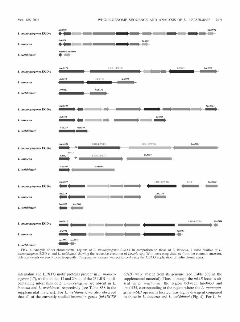
The localization of the deleted regions on the circular chro-mosomal map of L. monocytogenes (Fig. 1B) indicates that themissing genes of L. innocua and L. welshimeri originate largelyat the same loci within the chromosomes. Indeed, in manycases, the deletion that is observed in the L. innocua genomehas been extended to include neighboring genes for the samelocus in the L. welshimeri genome (Fig. 3). An inspection of thedistribution of these deletions in the chromosome of L. mono-cytogenes revealed that the majority of genes (�50%) that areabsent are located in the first third of the genome on the rightreplichore. We also detected larger deletions in the regionsaround 120 kb, 1.133 Mb, and 2.36 Mb that resulted in the lossof several genes, including those coding for monocin produc-
tion (lm0113-lmo0129), a Tn916-like transposon (lmo1097-lmo1114), and a putative prophage (lmo2271-lmo2332), thatare present in L. monocytogenes.
Many of the genes present in L. monocytogenes and absentfrom L. welshimeri not only are the result of gene loss but alsoinclude genes acquired by horizontal gene transfer (HGT) inL. monocytogenes. To examine for genes acquired by HGT inL. monocytogenes, we used the program SIGI (24), which al-lows the detection of genes of foreign origin, designated asalien genes. Alien genes are often associated with genomicislands, which are assumed to be frequently acquired viahorizontal gene transfer. The gene cluster regions of theprophage, transposon, and monocin of L. monocytogenes wereidentified by SIGI as genes derived from HGT. Results gen-erated by SIGI (Fig. 1B) also revealed 16 gene clusters, eachharboring a minimal number of one alien gene per cluster, asHGT-derived genes. Thus, �25% of the regions that are ab-sent in L. welshimeri do not represent bona fide deletionsemanating from the L. monocytogenes genome but representgenes acquired by HGT in the latter genome (see Table S2S inthe supplemental material for a list of genes derived fromHGT and designated alien genes in L. monocytogenes). Thelarge number of deletions and the occurrence of a high numberof alien genes in the first third of the chromosome suggest thatthis particular region of the genome is particularly prone torearrangements. The examination of the L. innocua and L.welshimeri genomes also revealed that the majority of aliengenes were located on the same region of the chromosome. Wespeculate that the presence of specific elements in this regionof the chromosome might account for this phenomenon.
Selective reduction of LRR and LPXTG proteins in L.welshimeri. Two families of surface-associated proteins, i.e.,those harboring LRR (leucine-rich repeat) and LPXTG mo-tifs, appear to have been selectively lost in L. welshimeri. For L.monocytogenes, members of these protein families (e.g., in-ternalins A and B) are required for the adhesion and invasionof nonphagocytic cells. In contrast to the large number of
FIG. 2. Confirmation of deleted gene regions in L. welshimeri compared to those in L. monocytogenes EGD-e using PCR. Lanes: M, 1 kb PlusDNA ladder (Gibco); 1, L. welshimeri SLCC5877 (1/2b); 2, L. welshimeri SLCC5828 (6b); 3, L. welshimeri SLCC6199 (6a); 4, L. welshimeriSLCC5334 (6b); 5, L. welshimeri SLCC5333 (6b); 6, L. welshimeri 18121; and 7, L. welshimeri 18217. Colony PCR was performed by using theExpand high-fidelity PCR system (Roche) by following the protocol supplied by manufacturer. Primer sequences for all amplicons are availablein the supplemental material. All amplified regions reveal clonality of the species L. welshimeri.
7408 HAIN ET AL. J. BACTERIOL.
internalins and LPXTG motif proteins present in L. monocy-togenes (17), we found that 17 and 20 out of the 25 LRR-motif-containing internalins of L. monocytogenes are absent in L.innocua and L. welshimeri, respectively (see Table S3S in thesupplemental material). For L. welshimeri, we also observedthat all of the currently studied internalin genes (inlABCEF
GHIJ) were absent from its genome (see Table S3S in thesupplemental material). Thus, although the inlAB locus is ab-sent in L. welshimeri, the region between lmo0430 andlmo0435, corresponding to the region where the L. monocyto-genes inlAB operon is located, was highly divergent comparedto those in L. innocua and L. welshimeri (Fig. 4). For L. in-
FIG. 3. Analysis of six chromosomal regions of L. monocytogenes EGD-e in comparison to those of L. innocua, a close relative of L.monocytogenes EGD-e, and L. welshimeri showing the reductive evolution of Listeria spp. With increasing distance from the common ancestor,deletion events occurred more frequently. Comparative analysis was performed using the GECO application of bidirectional pairs.
VOL. 188, 2006 WHOLE-GENOME SEQUENCE AND ANALYSIS OF L. WELSHIMERI 7409

nocua, upstream of the lmo0435 homolog lin0457 that codesfor a putative peptidoglycan-bound protein (2,013 amino ac-ids) harboring a LPXTG motif, is a gene (lin0454) coding foranother large protein (2,167 amino acids). This protein shareshomologies to the cell wall-associated protein precursor of B.subtilis WapA, which is involved in sucrose binding (31). In L.welshimeri, both homologs, lwe0394 and lwe0431, are presentin the same region but they are separated by a large insertionof �48 kb. The genes adjacent to the putative site of insertion,i.e., from lwe0395 to lwe0417, have lower-than-average G � Ccontents, suggesting that this region was acquired by horizontalgene transfer. Results generated by SIGI for this region sug-gest that the origin of these genes was affected by HGT (Fig.4). The latter part of the insertion harbors the FoF1-ATPsynthase genes (lwe0418-lwe0428) that are otherwise posi-tioned around 100 kb from oriC in L. monocytogenes and L.innocua. The inserted region is flanked downstream by lwe0429,a gene coding for another very large putative LPXTG-motif-containing protein (2,753 amino acids), with repeats that areprobably involved in bacterial adhesion.
An examination of the chromosomal loci harboring genesencoding LPXTG- and LRR-motif-containing proteins in L.monocytogenes revealed that many of these genes have beenlost from the apathogenic species L. innocua and L. welshimeri(Fig. 1B). We noted that the regions corresponding to theLPXTG- and LRR-motif-containing proteins are part of 16gene clusters and 6 individual genes that have been lost in theL. welshimeri genome (Fig. 3). In several cases, the deletedregions are flanked by pseudogenes (lmo0171, lmo0172, lmo0410,and lmo0473) or transposase genes (lmo0174, lmo0329, lmo0330,and lmo0464). In contrast to the genes encoding these surfaceprotein families, genes coding for other cell wall-associated
proteins, such as GW-motif-containing proteins (see Table S4Sin the supplemental material), LysM-motif-containing proteins(see Table S5S in the supplemental material), and NLPC/P60-like proteins (see Table S6S in the supplemental material)were roughly constant in the genomes of all three species. TheL. welshimeri genome contained the largest number of genesencoding lipoproteins, 69, versus 65 and 59 for L. monocyto-genes and L. innocua (Fig. 5; see Table S7S in the supplementalmaterial).
The gene locus harboring the ami (28) gene (lmo2558),which is involved in the adhesion of L. monocytogenes to theeukaryotic cell, revealed yet another class of difference forsurface-bound proteins. Unlike its counterpart in L. monocy-togenes, the Ami protein in L. welshimeri is truncated andcontains only two GW repeats (R1 and R3), whereas the L.monocytogenes protein harbors four GW-motif-containingmodules (R1 to R4). Interestingly, the Ami protein in L. in-nocua lacks only one GW-containing repeat, R3 (Fig. 6).
Apathogenic species missing several metabolic pathways ofL. monocytogenes. A major difference between L. monocyto-genes and L. welshimeri is the absence of 22 gene clusters and5 individual genes involved in the transport and metabolism ofcarbohydrates and amino acids (see Fig. S2S in the supplemen-tal material). These include several genes coding for proteinsof phosphotransferase system (PTS)-specific enzyme compo-nents involving the uptake of -glucoside (lmo0738, lmo0874-lmo0876, lmo1035, and lmo2787), fructose (lmo2733 andlmo0503), galactiol (lmo0507 and lmo0508), pentitol (lmo1971-lmo1973), rhamnose (lmo2848-lmo2850), mannitol (lmo2797 andlmo2799), and cellobiose (lmo0034 and lmo0914-lmo0916). ThebvrABC locus (lm2786-lmo2788) encoding a -glucoside-specificPTS has been previously shown to mediate virulence gene repres-
FIG. 4. Comparative representation of the chromosomal “hot spot” region between lwe0390 and lwe0431 of L. welshimeri, including cellwall-associated genes, which are marked. Putative regions of horizontal gene transfer and a rearrangement of the FoF1-ATP synthase region areboxed. Bioinformatic analysis was performed with GECO by using bidirectional pairs.
7410 HAIN ET AL. J. BACTERIOL.

sion of L. monocytogenes by -glucosides (4). A further operon(lmo0178-lmo0184) involved in sugar transport and metabo-lism is also missing in the genome of L. welshimeri. It waspreviously shown that this operon is negatively regulated byPrfA (26).
Genes involved in nicotinate and nicotinamide metabolism(nadABC, lmo2022-lmo2025) (Fig. 3) or inositol metabolism(iolRABCD, lmo0383-lmo0386) are specifically absent in L.welshimeri, as are genes involved in arginine catabolism(lmo0036-lmo0039) (Fig. 3), amino acid uptake by an ATP-binding cassette (ABC) transporter (lmo2346-lmo2349), andteichoic acid biosynthesis (lmo1077, lmo1080-lmo1085, andlmo1087) (42). Individual genes specifically absent in L. welshi-meri included three genes, ispF (lmo0236), ispG (lmo1441),and ispH (lmo1451), that are part of the deoxyxylulose-5-phos-
phate pathway for isoprenoid biosynthesis and for cell wallbiosynthesis for Escherichia coli and B. subtilis (7).
We also observed that gene clusters containing genes con-ferring bile and acid resistance are absent in L. welshimeriand L. innocua. For L. monocytogenes, the bile salt hydrolasegene (bsh, lmo2067) (13), the bile tolerance locus (btlB,lmo0752-lmo0754) (1), and the bilE system (lmo1421 andlmo1422) (36) have all been implicated in the protection ofthe bacterium within the gall bladder, a specific niche for theparasite to avoid host defense responses. However, only thebilE system is commonly present in all three species. L. welshi-meri also lacks the gene (lmo2387) encoding a member familyof CLC chloride channel proteins regulating ion efflux. Ho-mologs of these proteins in eukaryotic cells are important forcell volume regulation, transepithelial transport, intracellular
FIG. 5. Comparative analysis of six major surface protein classes for L. monocytogenes EGD-e, L. innocua, and L. welshimeri as predicted byAugur. Paralogs are not included. Orthologs were identified by unidirectional BLASTP analysis.
FIG. 6. Representative overview of the ami region in L. monocytogenes EGD-e, L. innocua, and L. welshimeri. GW modules were identified byusing the multiple sequence alignment program CLUSTALW 1.8.3. Repeated GW modules are indicated as R1 to R4 (black boxed) as previouslydescribed by Milohanic et al. (27). The flanked regions of the ami locus are highly conserved as identified with GECO by using bidirectional pairs.
VOL. 188, 2006 WHOLE-GENOME SEQUENCE AND ANALYSIS OF L. WELSHIMERI 7411

pH regulation, and membrane excitability. A comparative met-abolic reconstruction overview between L. monocytogenesEGD-e, L. innocua, and L. welshimeri is given in Fig. 7. Inaddition, two gene clusters, one coding for a twin argininesecretion apparatus, TatAC (lmo0361-lmo0367), and the sigCgene cluster (lmo0421-lmo0423) (41), are lacking in the ge-nome of L. welshimeri. It was previously reported that sigCcodes for C, which is involved in thermal resistance specificfor L. monocytogenes strains of phylogenetic lineage II (41).When cultures of L. welshimeri, L. monocytogenes EGD-e, and
L. innocua were assessed for survival following temperatureupshift at 60°C for 0, 5, 10, and 15 min, only L. welshimeri wassignificantly impaired in its ability to recover from exposure tothis nonpermissive temperature (see Fig. S3S in the supple-mental material).
Insertions confer new adaptive properties for environmentalsurvival of L. welshimeri. A well-known characteristic pheno-type of L. welshimeri is the degradation of D-xylose, a propertyused to differentiate among Listeria species. The xylose operonin L. welshimeri consists of five genes, including a glycoside
FIG. 7. An integrated view of the metabolic pathway comparison between L. monocytogenes EGD-e, L. innocua, and L. welshimeri. The mapillustrates biochemical pathways for main energy productions, such as glycolysis, starch metabolism, pentose phosphate pathway, and tricarboxylicacid cycle, which are necessary to maintain the survival of the strains. Several amino acid pathways and nucleic acid metabolism are involved aswell (pathway names are colored blue). The lipid and fatty acid pathways are omitted since they are incompletely present. Closed red arrowheadsdescribe the corresponding enzymes/reactions absent in L. welshimeri; however, they exist in both L. monocytogenes and L. innocua, whereas openred arrowheads show the enzymes/functions specifically present in L. monocytogenes. Closed blue arrowheads indicate the reactions present in onlyL. welshimeri, not L. monocytogenes or L. innocua. The gray line means the reaction is believed to occur spontaneously. Each gene product witha predicted function in ion or solute transport is illustrated in the figure. Proteins are grouped by substrate specificity, with transporters for cations(green), anions (pink), carbohydrates and organic acids (orange), and amino acids and peptides (blue). Ion-coupled permeases are represented byovals, while ABC transport systems are shown as ovals. This construction is concluded from the latest annotation data (NCBI) (C. Liang,unpublished data). The functional orthologs in silico are defined using a threshold of 50% identity and 75 to 125% alignment coverage rate. Theresulting strain-specific genes were inspected using InterPro and ExPASy protein domain analysis platforms. AICAR, 5-amino-1-(5-phospho-D-ribosyl)imidazole-4-carboxamide; E4P, erythrose-4-phosphate; DHF, dihydrofolate; G-1-P, glucose-1-phosphate; Dahp, 3-deoxy-D-arabino-hep-tulosonate-7-phosphate; F-6-P, fructose-6-phosphate; G-6-P, glucose-6-phosphate; GAP, glyceraldehyde-3-phosphate; OAA, oxaloacetate;6PGluco, 6-phosphogluconate; PRPP, 5-phosphoribosyl 1-pyrophosphate; R5P, ribose-5-phosphate; Ru5P, ribulose-5-phosphate; S7P, sedahep-tulose-7-phosphate; TCA, tricarboxylic acid; TCOylcine, 3�,7�,12�-trihydroxy-5-cholan-24-oylglycine; X5P, xylulose-5-phosphate; The enzymesencoded by the indicated genes are as follows: lwe0243, xylose isomerase; lwe0244, glycerol kinase; lmo1081, glucose-1-phosphate thymidyltransferase; lmo1082, dTDP-sugar epimerase; lmo1083, dTDP-D-glucose 4,6-dehydratase; lmo1084, DTDP-L-rhamnose synthetase; lmo2023,L-aspartate oxidase; lmo2067, conjugated bile acid hydrolase; lmo2143, mannose-6-phosphate isomerase; and lmo2848, L-rhamnose isomerase.
7412 HAIN ET AL. J. BACTERIOL.
hydrolase family protein gene (lwe0241) coding for a puta-tive alpha-xylosidase, a xylose-proton symporter gene (xylP,lwe0242), a xylose isomerase gene (xylA, lwe0243), and a xylu-lose kinase gene (xylB, lwe0244) controlled by the transcrip-tional repressor xylR protein (lwe0240) (Fig. 7). Two genesrequired for sucrose utilization (lwe1676 and lwe0226) andencoding a putative sucrose-specific IIBC PTS component anda sucrose phosphorylase were also detected. We identified twogenes coding for a IIABC (lwe0269) and IIBCA (lwe0278)-glucoside-specific PTS capable of uptake of a broad range of-glucosides, like cellulose and cellobiose, lichenin, aryl--glu-cosides, and xylans. Thus, the presence of many uptake andutilization systems for energy sources found almost exclusivelyin plants and decaying vegetation suggest that L. welshimeri hasadapted to a saprophytic strategy to survive in its natural en-vironment.
Two specific secreted proteases were detected for L. welshi-meri. A trypsin-like serine/cysteine protease is encoded bylwe1890, while lwe2295 was predicted to encode an otherwiseunassigned peptidase. This suggests that L. welshimeri can uti-lize proteins and peptides as a source of amino acids. Indeed,all of the L. welshimeri strains studied exhibit protease activityon skim milk agar plates, whereas L. innocua has no activity(see Fig. S4S in the supplemental material). The L. monocy-togenes EGD-e strain was also protease positive, as describedpreviously (11). However, an isogenic mutant lacking the mplgene and carrying a zinc-dependent metalloprotease lackedprotease activity (see Fig. S4S in the supplemental material).Thus, this novel property of L. welshimeri strains that has notbeen previously described will be useful in distinguishing itfrom L. innocua in diagnostic tests.
We also detected two genes (lwe0700 and lwe0701) withhomologies to the NatAB ABC transport system, which cata-lyzes ATP-dependent extrusion of electrogenic Na�, which iscytotoxic to the bacterial cell (9).
We identified an element encoding a complex type IC re-striction-modification (R-M) system (lwe0477-lwe0481), com-prised of a restriction endonuclease (HsdR) gene, a methylase(HsdM) gene, and two other genes, each encoding a separateHsdS subunit that imparts DNA specificity. Interestingly, theHsdS proteins are located on different strands, separated by aputative integrase gene of bacteriophage origin. The elementshares homology with similar structures present in L. innocuaand Lactococcus lactis (35) and is reminiscent of featurespresent on staphylococcal cassette chromosomes in Staphylo-coccus spp. (23). For the latter, it has been postulated that,following integration of such elements, stability is maintainedby the selfish R-M system. The function of an R-M system isoften considered to confer phage resistance, but it has alsobeen proposed that R-M systems are involved in genetic re-combination (21) and allow a low rate of horizontal genetransfer (2). In addition, we detected two genes with homolo-gies to transposase genes of the IS3 family (lwe1615 andlwe2186) and two neighboring genes (lwe1616 and lwe2187)that are involved in the transposition of the insertion sequence.
As discussed earlier, L. welshimeri harbors only 8 internalin-like genes (lwe0309, lwe0471, lwe0580, lwe0702, lwe0759,lwe0842, lwe1429 and lwe2343), while there are 25 and 19genes encoding internalins for L. monocytogenes and L. in-nocua, respectively. A characteristic feature of internalins is
the presence of multiple LRRs at the N-terminal end of themolecule combined with a LPXTG cell wall-anchoring motif(6). The LRR motif of lwe0702 shared homologies to plant-specific LRR motifs, suggesting a possible role in the sapro-phytic lifestyle concerning growth and adhesion on plant de-bris. We identified three L. welshimeri-specific genes coding forinternalins harboring LRR motifs and an LPXTG cell wallanchor motif (see Tables S3S and S8S in the supplementalmaterial). Two of these genes (lwe0309 and lwe1429) code forproteins with strong homologies to internalin A, which harborsfour and seven LRR repeats. For the remaining gene(lwe0842) we identified a single LRR repeat.
Cumulatively, these data suggest that, for L. welshimeri, thelack of genes required for intracellular replication has beencompensated for by the acquisition of genes/gene clusters foruptake systems and metabolic pathways to exploit plant-spe-cific cell wall components.
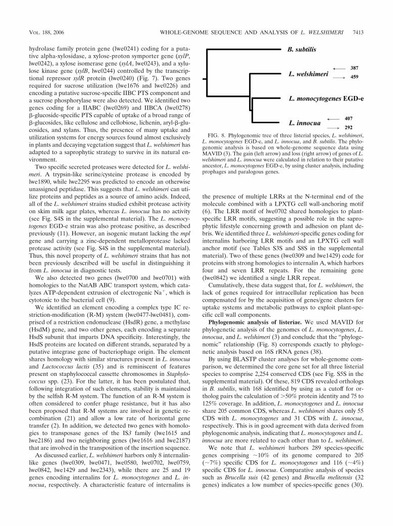
Phylogenomic analysis of listeriae. We used MAVID forphylogenetic analysis of the genomes of L. monocytogenes, L.innocua, and L. welshimeri (3) and conclude that the “phyloge-nomic” relationship (Fig. 8) corresponds exactly to phyloge-netic analysis based on 16S rRNA genes (38).
By using BLASTP cluster analyses for whole-genome com-parison, we determined the core gene set for all three listerialspecies to comprise 2,254 conserved CDS (see Fig. S5S in thesupplemental material). Of these, 819 CDS revealed orthologsin B. subtilis, with 168 identified by using as a cutoff for or-tholog pairs the calculation of �50% protein identity and 75 to125% coverage. In addition, L. monocytogenes and L. innocuashare 205 common CDS, whereas L. welshimeri shares only 55CDS with L. monocytogenes and 31 CDS with L. innocua,respectively. This is in good agreement with data derived fromphylogenomic analysis, indicating that L. monocytogenes and L.innocua are more related to each other than to L. welshimeri.
We note that L. welshimeri harbors 289 species-specificgenes comprising �10% of its genome compared to 205(�7%) specific CDS for L. monocytogenes and 116 (�4%)specific CDS for L. innocua. Comparative analysis of speciessuch as Brucella suis (42 genes) and Brucella melitensis (32genes) indicates a low number of species-specific genes (30).
FIG. 8. Phylogenomic tree of three listerial species, L. welshimeri,L. monocytogenes EGD-e, and L. innocua, and B. subtilis. The phylo-genomic analysis is based on whole-genome sequence data usingMAVID (3). The gain (left arrow) and loss (right arrow) of genes of L.welshimeri and L. innocua were calculated in relation to their putativeancestor, L. monocytogenes EGD-e, by using cluster analysis, includingprophages and paralogous genes.
VOL. 188, 2006 WHOLE-GENOME SEQUENCE AND ANALYSIS OF L. WELSHIMERI 7413

On the other hand, genome comparison among three Strepto-coccus species revealed high numbers of species-specific genes:416 genes for Streptococcus pyogenes, 683 genes for Streptococ-cus agalactiae, and 836 genes for Streptococcus pneumoniae(37). These comparisons suggest that the pangenomic generepertoire of the genus Listeria is relatively large and indicativeof active horizontal gene transfer processes in evolution.
An interesting observation is the finding of 157 genes com-monly absent in the sequenced L. monocytogenes F2365 sero-type 4b, L. innocua, and L. welshimeri strains but present in theL. monocytogenes EGD-e serotype 1/2a genome. Pairwise com-parisons indicated that a further 53 genes in L. welshimeri/L.monocytogenes F2365 serotype 4b and 23 genes in the combi-nation L. innocua/L. monocytogenes F2365 serotype 4b werecommonly absent. This suggests that these genes were deletedin a clonal ancestor preceding the diversification into the dif-ferent species. Finally, pairwise comparison of the L. monocy-togenes EGD-e serotype 1/2a and L. monocytogenes F2365 se-rotype 4b genomes indicated the absence of 38 genes in thelatter genome. Closer inspection indicated that 18 of the 38genes are truly absent. For the remaining 20 genes, singlenucleotide polymorphisms led to the classification of the re-spective orf as “absent” in the L. monocytogenes F2365 sero-type 4b genome. Thus, as has been described for other species,such single nucleotide polymorphisms mark the beginning ofgenome variation leading to the deletion and evolutionary re-duction in the genome. We expect that whole-genome se-quencing of strains comprising other species of this genus willprovide a rich resource for understanding the source of varia-tion and the evolutionary history of this genus that comprisesonly six species.
ACKNOWLEDGMENTS
We thank Alexandra Amend, Claudia Zorb, Nelli and Juri Sch-klarenko, and Prisca Viehoever for excellent technical assistance, Her-bert Hof for generously supplying the L. welshimeri serovars, andPhilippe Glaser and Carmen Buchrieser for fruitful discussions.
This work was supported by funds obtained from the BMBF throughthe Competence Network PathoGenoMik (031U213B) to T.C. andT.H.
REFERENCES
1. Begley, M., R. D. Sleator, C. G. Gahan, and C. Hill. 2005. Contribution ofthree bile-associated loci, bsh, pva, and btlB, to gastrointestinal persistenceand bile tolerance of Listeria monocytogenes. Infect. Immun. 73:894–904.
2. Bickle, T. A., and D. H. Kruger. 1993. Biology of DNA restriction. Microbiol.Rev. 57:434–450.
3. Bray, N., and L. Pachter. 2004. MAVID: constrained ancestral alignment ofmultiple sequences. Genome Res. 14:693–699.
4. Brehm, K., M. T. Ripio, J. Kreft, and J. A. Vazquez-Boland. 1999. The bvrlocus of Listeria monocytogenes mediates virulence gene repression by -glu-cosides. J. Bacteriol. 181:5024–5032.
5. Buchrieser, C., C. Rusniok, F. Kunst, P. Cossart, and P. Glaser. 2003.Comparison of the genome sequences of Listeria monocytogenes and Listeriainnocua: clues for evolution and pathogenicity. FEMS Immunol. Med. Mi-crobiol. 35:207–213.
6. Cabanes, D., P. Dehoux, O. Dussurget, L. Frangeul, and P. Cossart. 2002.Surface proteins and the pathogenic potential of Listeria monocytogenes.Trends Microbiol. 10:238–245.
7. Campbell, T. L., and E. D. Brown. 2002. Characterization of the depletion of2-C-methyl-D-erythritol-2,4-cyclodiphosphate synthase in Escherichia coliand Bacillus subtilis. J. Bacteriol. 184:5609–5618.
8. Chakraborty, T., T. Hain, and E. Domann. 2000. Genome organization andthe evolution of the virulence gene locus in Listeria species. Int. J. Med.Microbiol. 290:167–174.
9. Cheng, J., A. A. Guffanti, and T. A. Krulwich. 1997. A two-gene ABC-typetransport system that extrudes Na� in Bacillus subtilis is induced by ethanolor protonophore. Mol. Microbiol. 23:1107–1120.
10. Delcher, A. L., A. Phillippy, J. Carlton, and S. L. Salzberg. 2002. Fastalgorithms for large-scale genome alignment and comparison. Nucleic AcidsRes. 30:2478–2483.
11. Domann, E., M. Leimeister-Wachter, W. Goebel, and T. Chakraborty. 1991.Molecular cloning, sequencing, and identification of a metalloprotease genefrom Listeria monocytogenes that is species specific and physically linked tothe listeriolysin gene. Infect. Immun. 59:65–72.
12. Dominguez-Bernal, G., S. Muller-Altrock, B. Gonzalez-Zorn, M. Scortti, P.Herrmann, H. J. Monzo, L. Lacharme, J. Kreft, and J. A. Vazquez-Boland.2006. A spontaneous genomic deletion in Listeria ivanovii identifies LIPI-2,a species-specific pathogenicity island encoding sphingomyelinase and nu-merous internalins. Mol. Microbiol. 59:415–432.
13. Dussurget, O., D. Cabanes, P. Dehoux, M. Lecuit, C. Buchrieser, P. Glaser,and P. Cossart. 2002. Listeria monocytogenes bile salt hydrolase is a PrfA-regulated virulence factor involved in the intestinal and hepatic phases oflisteriosis. Mol. Microbiol. 45:1095–1106.
14. Ewing, B., and P. Green. 1998. Base-calling of automated sequencer tracesusing phred. II. Error probabilities. Genome Res. 8:186–194.
15. Ewing, B., L. Hillier, M. C. Wendl, and P. Green. 1998. Base-calling ofautomated sequencer traces using phred. I. Accuracy assessment. GenomeRes. 8:175–185.
16. Ghai, R., T. Hain, and T. Chakraborty. 2004. GenomeViz: visualizing mi-crobial genomes. BMC Bioinformatics 5:198.
17. Glaser, P., L. Frangeul, C. Buchrieser, C. Rusniok, A. Amend, F. Baquero,P. Berche, H. Bloecker, P. Brandt, T. Chakraborty, A. Charbit, F. Chetouani,E. Couve, A. de Daruvar, P. Dehoux, E. Domann, G. Dominguez-Bernal, E.Duchaud, L. Durant, O. Dussurget, K. D. Entian, H. Fsihi, F. Garcia-delPortillo, P. Garrido, L. Gautier, W. Goebel, N. Gomez-Lopez, T. Hain, J.Hauf, D. Jackson, L. M. Jones, U. Kaerst, J. Kreft, M. Kuhn, F. Kunst, G.Kurapkat, E. Madueno, A. Maitournam, J. M. Vicente, E. Ng, H. Nedjari, G.Nordsiek, S. Novella, B. de Pablos, J. C. Perez-Diaz, R. Purcell, B. Remmel,M. Rose, T. Schlueter, N. Simoes, A. Tierrez, J. A. Vazquez-Boland, H. Voss,J. Wehland, and P. Cossart. 2001. Comparative genomics of Listeria species.Science 294:849–852.
18. Gordon, D., C. Abajian, and P. Green. 1998. Consed: a graphical tool forsequence finishing. Genome Res. 8:195–202.
19. Gordon, D., C. Desmarais, and P. Green. 2001. Automated finishing withautofinish. Genome Res. 11:614–625.
20. Jones, D., and H. Seeliger. 1992. The genus Listeria, p. 1595–1616. In A.Balows, H. G. Truper, M. Dworkin, K. H. Harder, and K. H. Schleifer (ed.),The prokaryotes. Springer, New York, N.Y.
21. King, G., and N. E. Murray. 1994. Restriction enzymes in cells, not eppen-dorfs. Trends Microbiol. 2:465–469.
22. Kluge, R., and H. Hof. 1986. Virulence of Listeria welshimeri. Zentbl. Bak-teriol. Mikrobiol. Hyg. 262:403–411.
23. Kuroda, M., A. Yamashita, H. Hirakawa, M. Kumano, K. Morikawa, M.Higashide, A. Maruyama, Y. Inose, K. Matoba, H. Toh, S. Kuhara, M.Hattori, and T. Ohta. 2005. Whole genome sequence of Staphylococcussaprophyticus reveals the pathogenesis of uncomplicated urinary tract infec-tion. Proc. Natl. Acad. Sci. USA 102:13272–13277.
24. Merkl, R. 2004. SIGI: score-based identification of genomic islands. BMCBioinformatics 5:22.
25. Meyer, F., A. Goesmann, A. C. McHardy, D. Bartels, T. Bekel, J. Clausen, J.Kalinowski, B. Linke, O. Rupp, R. Giegerich, and A. Puhler. 2003.GenDB—an open source genome annotation system for prokaryote ge-nomes. Nucleic Acids Res. 31:2187–2195.
26. Milohanic, E., P. Glaser, J. Y. Coppee, L. Frangeul, Y. Vega, J. A. Vazquez-Boland, F. Kunst, P. Cossart, and C. Buchrieser. 2003. Transcriptome anal-ysis of Listeria monocytogenes identifies three groups of genes differentlyregulated by PrfA. Mol. Microbiol. 47:1613–1625.
27. Milohanic, E., R. Jonquieres, P. Cossart, P. Berche, and J. L. Gaillard. 2001.The autolysin Ami contributes to the adhesion of Listeria monocytogenes toeukaryotic cells via its cell wall anchor. Mol. Microbiol. 39:1212–1224.
28. Milohanic, E., R. Jonquieres, P. Glaser, P. Dehoux, C. Jacquet, P. Berche, P.Cossart, and J. L. Gaillard. 2004. Sequence and binding activity of theautolysin-adhesin Ami from epidemic Listeria monocytogenes 4b. Infect. Im-mun. 72:4401–4409.
29. Nelson, K. E., D. E. Fouts, E. F. Mongodin, J. Ravel, R. T. DeBoy, J. F.Kolonay, D. A. Rasko, S. V. Angiuoli, S. R. Gill, I. T. Paulsen, J. Peterson,O. White, W. C. Nelson, W. Nierman, M. J. Beanan, L. M. Brinkac, S. C.Daugherty, R. J. Dodson, A. S. Durkin, R. Madupu, D. H. Haft, J. Selengut,S. Van Aken, H. Khouri, N. Fedorova, H. Forberger, B. Tran, S. Kathariou,L. D. Wonderling, G. A. Uhlich, D. O. Bayles, J. B. Luchansky, and C. M.Fraser. 2004. Whole genome comparisons of serotype 4b and 1/2a strains ofthe food-borne pathogen Listeria monocytogenes reveal new insights into thecore genome components of this species. Nucleic Acids Res. 32:2386–2395.
30. Paulsen, I. T., R. Seshadri, K. E. Nelson, J. A. Eisen, J. F. Heidelberg, T. D.Read, R. J. Dodson, L. Umayam, L. M. Brinkac, M. J. Beanan, S. C.Daugherty, R. T. DeBoy, A. S. Durkin, J. F. Kolonay, R. Madupu, W. C.Nelson, B. Ayodeji, M. Kraul, J. Shetty, J. Malek, S. E. Van Aken, S.Riedmuller, H. Tettelin, S. R. Gill, O. White, S. L. Salzberg, D. L. Hoover,L. E. Lindler, S. M. Halling, S. M. Boyle, and C. M. Fraser. 2002. The
7414 HAIN ET AL. J. BACTERIOL.

Brucella suis genome reveals fundamental similarities between animal andplant pathogens and symbionts. Proc. Natl. Acad. Sci. USA 99:13148–13153.
31. Qian, H., and M. L. Dao. 1993. Inactivation of the Streptococcus mutanswall-associated protein A gene (wapA) results in a decrease in sucrose-dependent adherence and aggregation. Infect. Immun. 61:5021–5028.
32. Rocourt, J., A. Schrettenbrunner, and H. P. Seeliger. 1983. Biochemicaldifferentiation of the “Listeria monocytogenes” (sensu lato) genomic groups.Ann. Microbiol. 134A:65–71. (In French.)
33. Rocourt, J., and H. P. Seeliger. 1985. Distribution of species of the genusListeria. Zentbl. Bakteriol. Mikrobiol. Hyg. 259:317–330.
34. Schmid, M. W., E. Y. Ng, R. Lampidis, M. Emmerth, M. Walcher, J. Kreft,W. Goebel, M. Wagner, and K. H. Schleifer. 2005. Evolutionary history of thegenus Listeria and its virulence genes. Syst. Appl. Microbiol. 28:1–18.
35. Schouler, C., M. Gautier, S. D. Ehrlich, and M. C. Chopin. 1998. Combina-tional variation of restriction modification specificities in Lactococcus lactis.Mol. Microbiol. 28:169–178.
36. Sleator, R. D., H. H. Wemekamp-Kamphuis, C. G. Gahan, T. Abee, and C.Hill. 2005. A PrfA-regulated bile exclusion system (BilE) is a novel virulencefactor in Listeria monocytogenes. Mol. Microbiol. 55:1183–1195.
37. Tettelin, H., V. Masignani, M. J. Cieslewicz, J. A. Eisen, S. Peterson, M. R.Wessels, I. T. Paulsen, K. E. Nelson, I. Margarit, T. D. Read, L. C. Madoff,A. M. Wolf, M. J. Beanan, L. M. Brinkac, S. C. Daugherty, R. T. DeBoy, A. S.Durkin, J. F. Kolonay, R. Madupu, M. R. Lewis, D. Radune, N. B. Fedorova,D. Scanlan, H. Khouri, S. Mulligan, H. A. Carty, R. T. Cline, S. E. Van Aken,
J. Gill, M. Scarselli, M. Mora, E. T. Iacobini, C. Brettoni, G. Galli, M.Mariani, F. Vegni, D. Maione, D. Rinaudo, R. Rappuoli, J. L. Telford, D. L.Kasper, G. Grandi, and C. M. Fraser. 2002. Complete genome sequence andcomparative genomic analysis of an emerging human pathogen, serotype VStreptococcus agalactiae. Proc. Natl. Acad. Sci. USA 99:12391–12396.
38. Vaneechoutte, M., P. Boerlin, H. V. Tichy, E. Bannerman, B. Jager, and J.Bille. 1998. Comparison of PCR-based DNA fingerprinting techniques forthe identification of Listeria species and their use for atypical Listeria isolates.Int. J. Syst. Bacteriol. 48:127–139.
39. Vazquez-Boland, J. A., M. Kuhn, P. Berche, T. Chakraborty, G. Dominguez-Bernal, W. Goebel, B. Gonzalez-Zorn, J. Wehland, and J. Kreft. 2001. Lis-teria pathogenesis and molecular virulence determinants. Clin. Microbiol.Rev. 14:584–640.
40. Welshimer, H. J. 1968. Isolation of Listeria monocytogenes from vegetation.J. Bacteriol. 95:300–303.
41. Zhang, C., J. Nietfeldt, M. Zhang, and A. K. Benson. 2005. Functionalconsequences of genome evolution in Listeria monocytogenes: the lmo0423and lmo0422 genes encode C and LstR, a lineage II-specific heat shocksystem. J. Bacteriol. 187:7243–7253.
42. Zhang, C., M. Zhang, J. Ju, J. Nietfeldt, J. Wise, P. M. Terry, M. Olson, S. D.Kachman, M. Wiedmann, M. Samadpour, and A. K. Benson. 2003. Genomediversification in phylogenetic lineages I and II of Listeria monocytogenes:identification of segments unique to lineage II populations. J. Bacteriol.185:5573–5584.
VOL. 188, 2006 WHOLE-GENOME SEQUENCE AND ANALYSIS OF L. WELSHIMERI 7415
Related Documents