BRAIN A JOURNAL OF NEUROLOGY White matter damage and cognitive impairment after traumatic brain injury Kirsi Maria Kinnunen, 1 Richard Greenwood, 2 Jane Hilary Powell, 1 Robert Leech, 3 Peter Charlie Hawkins, 1 Valerie Bonnelle, 3,4 Maneesh Chandrakant Patel, 5 Serena Jane Counsell 6 and David James Sharp 3 1 Department of Psychology, Goldsmiths, University of London, London, UK 2 Institute of Neurology, Division of Clinical Neurology, University College London, London, UK 3 Computational, Cognitive, and Clinical Neuroimaging Laboratory, Clinical Neuroscience, Centre for Neuroscience, Division of Experimental Medicine, Department of Medicine, Imperial College London, London, UK 4 MRC Clinical Sciences Centre, Experimental and Clinical Neuroscience Section, Cognitive Neuroimaging Research Group, Faculty of Medicine, Imperial College London, London, UK 5 Imaging Department, Charing Cross Hospital, Imperial College Healthcare NHS Trust, London, UK 6 MRC Clinical Sciences Centre, Experimental and Clinical Neuroscience Section, Neonatal Medicine Research Group, Faculty of Medicine, Imperial College London, London, UK Correspondence to: Dr David J. Sharp, Computational, Cognitive, and Clinical Neuroimaging Laboratory, 3rd Floor, Burlington Danes Building, Imperial College London, Hammersmith Hospital Campus, Du Cane Road, London, W12 0NN, UK E-mail: [email protected] White matter disruption is an important determinant of cognitive impairment after brain injury, but conventional neuroimaging underestimates its extent. In contrast, diffusion tensor imaging provides a validated and sensitive way of identifying the impact of axonal injury. The relationship between cognitive impairment after traumatic brain injury and white matter damage is likely to be complex. We applied a flexible technique—tract-based spatial statistics—to explore whether damage to specific white matter tracts is associated with particular patterns of cognitive impairment. The commonly affected domains of memory, executive function and information processing speed were investigated in 28 patients in the post-acute/chronic phase following traumatic brain injury and in 26 age-matched controls. Analysis of fractional anisotropy and diffusivity maps revealed widespread differ- ences in white matter integrity between the groups. Patients showed large areas of reduced fractional anisotropy, as well as increased mean and axial diffusivities, compared with controls, despite the small amounts of cortical and white matter damage visible on standard imaging. A stratified analysis based on the presence or absence of microbleeds (a marker of diffuse axonal injury) revealed diffusion tensor imaging to be more sensitive than gradient-echo imaging to white matter damage. The location of white matter abnormality predicted cognitive function to some extent. The structure of the fornices was correlated with associative learning and memory across both patient and control groups, whilst the structure of frontal lobe connections showed relationships with executive function that differed in the two groups. These results highlight the complexity of the relationships between white matter structure and cognition. Although widespread and, sometimes, chronic abnormalities of white matter are identifiable following traumatic brain injury, the impact of these changes on cognitive function is likely to depend on damage to key pathways that link nodes in the distributed brain networks supporting high-level cognitive functions. doi:10.1093/brain/awq347 Brain 2011: 134; 449–463 | 449 Received July 4, 2010. Revised September 29, 2010. Accepted October 15, 2010. Advance Access publication December 29, 2010 ß The Author(s) 2010. Published by Oxford University Press on behalf of Brain. This is an Open Access article distributed under the terms of the Creative Commons Attribution Non-Commercial License (http://creativecommons.org/licenses/by-nc/2.5), which permits unrestricted non-commercial use, distribution, and reproduction in any medium, provided the original work is properly cited.

Welcome message from author
This document is posted to help you gain knowledge. Please leave a comment to let me know what you think about it! Share it to your friends and learn new things together.
Transcript
BRAINA JOURNAL OF NEUROLOGY
White matter damage and cognitive impairmentafter traumatic brain injuryKirsi Maria Kinnunen,1 Richard Greenwood,2 Jane Hilary Powell,1 Robert Leech,3
Peter Charlie Hawkins,1 Valerie Bonnelle,3,4 Maneesh Chandrakant Patel,5 Serena Jane Counsell6
and David James Sharp3
1 Department of Psychology, Goldsmiths, University of London, London, UK
2 Institute of Neurology, Division of Clinical Neurology, University College London, London, UK
3 Computational, Cognitive, and Clinical Neuroimaging Laboratory, Clinical Neuroscience, Centre for Neuroscience, Division of Experimental
Medicine, Department of Medicine, Imperial College London, London, UK
4 MRC Clinical Sciences Centre, Experimental and Clinical Neuroscience Section, Cognitive Neuroimaging Research Group, Faculty of Medicine,
Imperial College London, London, UK
5 Imaging Department, Charing Cross Hospital, Imperial College Healthcare NHS Trust, London, UK
6 MRC Clinical Sciences Centre, Experimental and Clinical Neuroscience Section, Neonatal Medicine Research Group, Faculty of Medicine,
Imperial College London, London, UK
Correspondence to: Dr David J. Sharp,
Computational, Cognitive, and Clinical Neuroimaging Laboratory,
3rd Floor, Burlington Danes Building,
Imperial College London,
Hammersmith Hospital Campus,
Du Cane Road,
London, W12 0NN, UK
E-mail: [email protected]
White matter disruption is an important determinant of cognitive impairment after brain injury, but conventional neuroimaging
underestimates its extent. In contrast, diffusion tensor imaging provides a validated and sensitive way of identifying the impact
of axonal injury. The relationship between cognitive impairment after traumatic brain injury and white matter damage is likely to
be complex. We applied a flexible technique—tract-based spatial statistics—to explore whether damage to specific white matter
tracts is associated with particular patterns of cognitive impairment. The commonly affected domains of memory, executive
function and information processing speed were investigated in 28 patients in the post-acute/chronic phase following traumatic
brain injury and in 26 age-matched controls. Analysis of fractional anisotropy and diffusivity maps revealed widespread differ-
ences in white matter integrity between the groups. Patients showed large areas of reduced fractional anisotropy, as well as
increased mean and axial diffusivities, compared with controls, despite the small amounts of cortical and white matter damage
visible on standard imaging. A stratified analysis based on the presence or absence of microbleeds (a marker of diffuse axonal
injury) revealed diffusion tensor imaging to be more sensitive than gradient-echo imaging to white matter damage. The location
of white matter abnormality predicted cognitive function to some extent. The structure of the fornices was correlated with
associative learning and memory across both patient and control groups, whilst the structure of frontal lobe connections showed
relationships with executive function that differed in the two groups. These results highlight the complexity of the relationships
between white matter structure and cognition. Although widespread and, sometimes, chronic abnormalities of white matter are
identifiable following traumatic brain injury, the impact of these changes on cognitive function is likely to depend on damage to
key pathways that link nodes in the distributed brain networks supporting high-level cognitive functions.
doi:10.1093/brain/awq347 Brain 2011: 134; 449–463 | 449
Received July 4, 2010. Revised September 29, 2010. Accepted October 15, 2010. Advance Access publication December 29, 2010
� The Author(s) 2010. Published by Oxford University Press on behalf of Brain.
This is an Open Access article distributed under the terms of the Creative Commons Attribution Non-Commercial License (http://creativecommons.org/licenses/by-nc/2.5),
which permits unrestricted non-commercial use, distribution, and reproduction in any medium, provided the original work is properly cited.
Keywords: traumatic brain injury; diffuse axonal injury; diffusion tensor; brain behaviour and relationships; cognitive impairment
Abbreviations: DTI = diffusion tensor imaging; Dax = axial diffusivity; Drad = radial diffusivity; TBSS = tract-based spatial statistics
IntroductionTraumatic brain injury often results in persistent disability, due
particularly to cognitive impairments (Whitnall et al., 2006).
Most survivors are young and have near-normal life expectancy
(Thornhill et al., 2000). Hence, the burden on public health and
social care is substantial (Thurman et al., 1999). The cognitive
domains of memory, executive function and processing speed
are commonly affected (Ponsford and Kinsella, 1992; Levin and
Kraus, 1994; Levin, 1995; Scheid et al., 2006; Draper and
Ponsford, 2008). Despite much previous work, the underlying
pathophysiology of these persistent impairments remains poorly
understood (Lowenstein, 2009).
Although focal brain injury often occurs as a result of traumatic
brain injury, in many cases, the location and extent of this injury
does not fully explain the patient’s cognitive problems (Bigler,
2001). This is likely to be because damage to brain connectivity
is a critical factor in the development of cognitive impairment after
traumatic brain injury. Functions commonly impaired, such as
memory and executive functions, depend on the coherent activity
of widely distributed brain networks (Mesulam, 1998). ‘Nodes’ in
these networks are connected by long white matter tracts that
may be damaged in traumatic brain injury as a result of diffuse
axonal injury. The pathology of diffuse axonal injury has been
investigated in some detail (Povlishock and Katz, 2005), but,
until recently, it has been difficult to study the location and
extent of this damage, or its functional consequences in vivo.
Conventional CT and standard MRI underestimate the extent of
white matter damage after traumatic brain injury (Rugg-Gunn
et al., 2001; Arfanakis et al., 2002). Standard MRI of traumatic
brain injury includes the use of gradient-echo imaging that allows
the identification of microbleeds. These are surrogate markers of
diffuse axonal injury (Scheid et al., 2003) and their presence is
associated with persistent cognitive impairment (Scheid et al.,
2006). However, pathological studies often show more extensive
axonal damage (Povlishock and Katz, 2005), which is unlikely to
be fully reflected in focal microbleed signal abnormalities.
Recently, it has become possible to study white matter damage
using diffusion tensor imaging (DTI; Arfanakis et al., 2002; Assaf
and Pasternak, 2008). In the tensor model, DTI data are used to
estimate the amount of water diffusion in a number of directions
at each point (voxel) in the image. From this, metrics such as
fractional anisotropy can be derived to quantify the degree of
white matter disruption (Basser and Pierpaoli, 1996, 1998).
Greater anisotropy, as indicated by a higher fractional anisotropy
value, is believed to reflect more coherent tissue structure, whilst
increased diffusivity suggests tissue damage (Rugg-Gunn et al.,
2001; Arfanakis et al., 2002). Experimental models of axonal
damage and demyelination have implicated axial and radial diffu-
sivities as potential biomarkers of axonal and myelin loss, respect-
ively (e.g. Song et al., 2003; Budde et al., 2008). Changes in
fractional anisotropy persist after traumatic brain injury and predict
functional outcome over and above patients’ initial clinical state or
focal lesion load (Sidaros et al., 2008).
Previous work in patients with traumatic brain injury has typic-
ally focused on a limited number of brain locations defined as
regions of interest (Kraus et al., 2007; Niogi et al., 2008a, b;
Kennedy et al., 2009). This approach is a sensitive way of iden-
tifying white matter damage, but as it is restricted to assessment
of the a priori defined regions, only a small amount of the total
white matter is usually investigated (e.g. Niogi et al., 2008a). This
is problematic for a number of reasons. Traumatic brain injury
produces a complex pattern of diffuse axonal injury at variable
locations across individuals and so it is difficult to decide a priori
where to ‘look’ for the white matter disruption. The investigation
of a small number of regions is likely to result in a failure to
identify significant white matter damage elsewhere in the brain.
As the cognitive functions commonly affected by traumatic brain
injury depend on distributed network function, such an approach
limits analysis of the structural causes of cognitive impairment.
These issues are compounded by our limited knowledge of how
tract structure relates to cognitive function in the normal brain,
making it important to assess white matter structure after trau-
matic brain injury with as comprehensive spatial coverage as
possible.
Tract-based spatial statistics (TBSS) is a new voxel-based tech-
nique for analysing white matter structure across the whole brain
(Smith et al., 2006). A voxel-based approach has previously pro-
vided important insights into long-term consequences for cogni-
tion of brain injury (Salmond et al., 2006). TBSS allows complex
patterns of white matter disruption to be identified and their re-
lationships with cognitive function to be studied in a data-driven
way. Statistical calculations are performed at each point within an
individual’s white matter ‘skeleton’, which has been registered to
standard space using a two-stage process involving non-linear
warping and subsequent alignment of individual white matter
tracts across subjects. This allows a comprehensive analysis of
tract structure to be performed in a way that is robust to effects
of brain injury, such as brain atrophy. TBSS has been used to show
a relationship between white matter structure and cognitive func-
tion in other neurological conditions (Ceccarelli et al., 2009;
Dineen et al., 2009; Roosendaal et al., 2009; Bosch et al., 2010).
Here, we used TBSS for the first time to study the relationship
between distributed white matter damage and cognitive impair-
ment following traumatic brain injury. First, we investigated
whether there were differences in white matter structure in a
group of post-acute and chronic patients with traumatic brain
injury and an age-matched control group. We then investigated
whether the pattern of white matter structure predicts cognitive
function in three domains commonly affected by traumatic brain
injury, i.e. memory, executive function and information processing
speed. Our prediction was that increased white matter disruption
following traumatic brain injury will be associated with greater
cognitive impairment, but that distinct types of impairment are
450 | Brain 2011: 134; 449–463 K. M. Kinnunen et al.
associated with particular patterns of white matter abnormalities.
Memory function is highly dependent upon hippocampal–medial
diencephalic interactions mediated through the fornices (Aggleton
and Brown, 1999; Aggleton, 2008; Tsivilis et al., 2008). Learning and
memory is impaired in chronic traumatic brain injury (Draper
and Ponsford, 2008) and white matter structure within the hippo-
campal formation has previously been shown to relate to associative
memory performance (Salmond et al., 2006). We, therefore, predict
that white matter structure of the hippocampal connections will
correlate with memory performance. Executive functions are
widely thought to depend on interactions between the frontal
lobes and more posterior brain regions (Miller and D’Esposito,
2005) and a relationship between executive dysfunction and
age-related decline in white matter integrity within tracts connecting
frontal regions has previously been demonstrated (O’Sullivan et al.,
2001; Davis et al., 2009; Madden et al., 2009; Perry et al., 2009).
Therefore, we predict that breakdown of these frontal connections
following traumatic brain injury will similarly correlate with executive
impairment. Finally, white matter organization has been shown
to predict processing speed on a range of simple tasks in healthy
individuals (Sullivan et al., 2001; Madden et al., 2004; Schulte
et al., 2005; Tuch et al., 2005; Bucur et al., 2008). Therefore, we
expect white matter structure as measured by DTI to correlate with
information processing speed.
Materials and methods
ParticipantsTwenty-eight patients with traumatic brain injury in the post-acute/
chronic phase (21 males, mean age � SD: 38.9 � 12.2 years) and
an age-matched group of 26 healthy controls (12 males,
35.4 � 11.1 years) were recruited. All patients were recruited at
least two months post-injury (average 25 months). Injury was second-
ary to assaults (36%), road traffic accidents (32%), falls (25%) and
sports-related injury (7%). Patients were referred to their local trau-
matic brain injury service because of the presence of functional
impairments following their head injury. There were 20 moderate or
severe and eight mild (probable) cases based on the Mayo
Classification System for Traumatic Brain Injury Severity, relating to
the duration of loss of consciousness, length of post-traumatic am-
nesia, lowest recorded Glasgow Coma Scale in the first 24 h, and/or
CT or MRI results (Malec et al., 2007). Exclusion criteria were as
follows: neurosurgery, except for invasive intracranial pressure moni-
toring (n = 1); a history of psychiatric or neurological illness prior to the
head injury; a history of previous traumatic brain injury; anti-epileptic
medication; current or previous drug or alcohol abuse; or contraindi-
cation to MRI. All participants gave written informed consent accord-
ing to the Declaration of Helsinki (World Medical Association, 2008).
The study was approved by the Hammersmith, Queen Charlotte’s and
Chelsea Research Ethics Committee.
Neuropsychological assessmentAll participants completed a standardized neuropsychological test bat-
tery sensitive to cognitive impairment associated with traumatic brain
injury. The cognitive functions of specific interest were indexed by:
(i) current verbal and non-verbal reasoning ability via the Wechsler
Abbreviated Scale of Intelligence Similarities and Matrix Reasoning
subtests (Wechsler, 1999); (ii) associative learning and memory via
the immediate recall score on the People Test from the Doors and
People Test (Baddeley et al., 1994); (iii) the executive functions of
set-shifting, inhibitory control, cognitive flexibility and word generation
fluency via the Trail Making Test (Reitan, 1958) alternating-switch cost
index (time to complete alternating letter and number Trails B—time
to complete numbers-only Trail A) and two indices from the Delis–
Kaplan Executive Function System (Delis et al., 2001), namely the
inhibition/switching minus baseline score from the Color–Word subtest
(high scores indicating poor performance) and the total score on Letter
Fluency; and (iv) information processing speed via the median reaction
time for accurate responses on a simple computerized choice reaction
task (see Supplementary Material for further details).
Structural imagingEach patient had standard high-resolution T1 and gradient-echo (T2*)
imaging to assess focal brain injury and evidence of microbleeds. MRI
was performed on Philips 3T Achieva scanner (Philips Medical Systems,
The Netherlands) using a body coil. The T1 and T2*-weighted images
were obtained prior to DTI. For DTI, diffusion-weighted volumes with
gradients applied in 16 non-collinear directions were collected in each
of the four DTI runs, resulting in a total of 64 directions. The following
parameters were used: 73 contiguous slices, slice thickness = 2 mm,
field of view 224 mm, matrix 128 � 128 (voxel size = 1.75 � 1.75 �
2 mm3), b value = 1000 and four images with no diffusion weighting
(b = 0 s/mm2). The images were registered to the b = 0 image by
affine transformations to minimize distortion due to motion and
eddy currents and then brain-extracted using Brain Extraction Tool
(Smith, 2002) from the FMRIB Software Library image processing
toolbox (Smith et al., 2004; Woolrich et al., 2009). Fractional anisot-
ropy and mean diffusivity maps were generated using the Diffusion
Toolbox (Behrens et al., 2003), as well as images for each of the
eigenvalues (�1, �2 and �3) representing the magnitude of diffusion
in the three principal directions. Axial (Dax) and radial (Drad) diffusivity
images were then derived from the eigenvalues (Dax = �1,
Drad = �2 + �3/2).
Diffusion tensor imaging data analysisVoxelwise analysis of the fractional anisotropy, mean diffusivity and
axial and radial diffusivity data was carried out using TBSS in the
FMRIB Software Library (Smith et al., 2004, 2006). Image analysis
using TBSS involved a number of steps: (i) non-linear alignment of
all subjects’ fractional anisotropy images into common FMRIB58 frac-
tional anisotropy template space; (ii) affine-transformation of the
aligned images into standard MNI152 1 mm space; (iii) averaging of
the aligned fractional anisotropy images to create a 4D mean fractional
anisotropy image; (iv) thinning of the mean fractional anisotropy
image to create a mean fractional anisotropy ‘skeleton’ representing
the centre of all white matter tracts, and in this way removing partial-
volume confounds; and (v) thresholding of the fractional anisotropy
skeleton at fractional anisotropy 50.2 to suppress areas of extremely
low mean fractional anisotropy and exclude those with considerable
inter-individual variability. Similar steps for processing non-fractional
anisotropy images were then carried out to obtain the mean, axial
and radial diffusivity images. Non-parametric permutation-based stat-
istics were employed using randomize with threshold-free cluster
enhancement and 5000 permutations (Nichols and Holmes, 2002;
Smith and Nichols, 2009). A threshold of P5 0.05 was then applied
on the results, corrected for multiple comparisons. Age and gender
White matter and cognition in traumatic brain injury Brain 2011: 134; 449–463 | 451
were included as covariates of no interest in all TBSS analyses.
In addition, patients with and without microbleed evidence of diffuse
axonal injury were compared against each other (microbleed versus
non-microbleed) and against the controls. Since severity of injury is
also likely to impact upon the extent of white matter disruption, we
carried out additional comparisons between patients classified as mild
and moderate/severe and controls. As DTI changes are known to
evolve after injury (Mac Donald et al., 2007; Sidaros et al., 2008),
we also investigated the effects of time since injury on the group
differences in white matter structure.
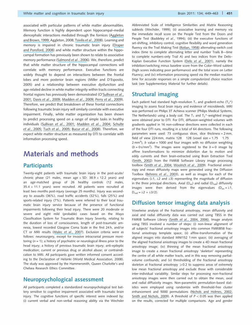
Figure 1 Lesion probability maps of (A) white matter lesions visible on gradient echo imaging and (B) contusions. The colour bar indicates
the number of patients who had lesions at each site. Green–yellow indicates where lesions were present in three (11%) of the 28 patients
with traumatic brain injury, pink indicates where they were present in two (7%) and blue where a lesion was found in one patient only.
Table 1 Neuropsychological test results by group
Cognitive domain Cognitive variable Traumaticbrain injury
Control Traumatic braininjury versusControlb
Mean � SDa Mean � SDa (t)
Intellectual ability: verbal/non-verbal WASI similarities 39.7 � 3.4 (n = 28) 35.2 � 5.7 (n = 26) 2.98**WASI matrix reasoning 29.0 � 3.3 (n = 27) 26.7 � 4.2 (n = 26) 2.84**
Memory: associative memory People Test immediate recall 24.8 � 4.9 (n = 28) 29.9 � 4.0 (n = 24) �4.03***
Processing speed: visualsearch/complex
Trail Making Test Trail A (s) 28.3 � 9.5 (n = 27) 19.8 � 4.3 (n = 25) 4.36***Trail Making Test Trails B (s) 70.2 � 40.1 (n = 28) 40.5 � 10.5 (n = 23) 3.74***
Processing speed: naming/reading Colour naming (s) 34.2 � 8.6 (n = 27) 28.2 � 5.6 (n = 26) 3.30**Word reading (s) 23.6 � 4.0 (n = 27) 22.5 � 4.5 (n = 25) 0.90c
Executive function: alternating-switchcost
Trail Making Test Trails Bminus A (s)
34.2 � 26.5 (n = 26) 22.2 � 9.9 (n = 24) 2.10*
Executive function: cognitive flexibility Inhibition/switching (s) 67.2 � 18.8 (n = 27) 54.1 � 10.6 (n = 25) 3.13**Inhibition/switching minus a
baseline of colour namingand word reading (s)
38.0 � 15.4 (n = 27) 27.9 � 10.4 (n = 25) 2.74**
Executive function: word generationfluency
Letter Fluency F + A + S total 43.1 � 9.8 (n = 28) 49.6 � 10.0 (n = 24) �2.40*
Processing speed: choice reaction time Choice reaction task medianreaction time (ms)
449 � 75 (n = 27) 393 � 52 (n = 26) 3.00**
a Following Exploratory Data Analysis using boxplots, outlier scores 51.5 � interquartile range outside the middle half of the sample were excluded variable-wise.b Patients showed significantly better performance for intellectual ability, but for all other significant group differences controls outperformed patients.c Not significant at the P4 0.05 level.
*P4 0.05; **P40.01; ***P40.001.WASI = Wechsler Abbreviated Scale of Intelligence.
452 | Brain 2011: 134; 449–463 K. M. Kinnunen et al.
Analysis of white matter structure andcognitive functionThe relationship between white matter structure and cognitive func-
tion was investigated within the framework of a general linear model
in the FMRIB Software Library. The effects of group and cognitive
variables were modelled, allowing analysis of the relationship between
white matter structure and cognitive function across voxels. Overall
correlations across both groups, correlations within each group and
group interactions were examined. Analysis was carried out using:
(i) the People Test immediate recall total score to index associative
learning and memory; (ii) the Trail Making Test Trails B�Trail A alter-
nating switch-cost, the Delis–Kaplan Executive Function System
Color-Word inhibition/switching minus a combined baseline of
naming and reading speed, and the Delis–Kaplan Executive Function
System Letter Fluency total for letters F, A and S to index the execu-
tive functions of set-shifting, cognitive flexibility and word generation
fluency; and (iii) median reaction time for accurate trials on the choice
reaction task to index information processing speed (Table 1). One
control subject and one patient were extreme outliers on either the
alternating-switch cost or the cognitive flexibility analyses and this was
modelled in the design using separate regressors. Permutation-based
significance testing was carried out as described above. For illustrative
purposes, fractional anisotropy and diffusivity values from the peak
voxels of the significant clusters of interest were then extracted for
each participant from their skeletonized images and plotted against the
cognitive scores.
Results
Standard magnetic resonance imagingT1 imaging was normal in 61% of patients and T2* normal in
25%. Definite and possible intraparenchymal microbleeds indica-
tive of diffuse axonal injury were found in 50% of the patients
(11 males, mean age 38.9 � 9.9 years, average time since injury
26 months; non-microbleed group 10 males, mean age
38.9 � 14.5 years, average time since injury 25 months). The
median number of microbleeds as identified using the
Microbleed Anatomical Rating Scale (Gregoire et al., 2009) was
seven (range 1–19). Microbleeds were mainly found in frontal and
temporal white matter bilaterally. There was little overlap in the
location of white matter damage. Cortical lesions were found in
39% of all patients and were mainly seen in frontal and temporal
regions. Again, there was a relatively small amount of lesion over-
lap (Fig. 1; see Supplementary Material for further details).
Magnetic resonance signal abnormality indicative of superficial
siderosis was found in 43% of the patients, mainly overlying bi-
lateral frontal and right temporal cortices. This is likely to be sec-
ondary to chronic haemosiderin deposition as a result of subdural
or subarachnoid haemorrhage at the time of injury.
Cognitive functionThe patient group outperformed the control group in terms of
average current intellectual ability, as indexed by the Wechsler
Abbreviated Scale of Intelligence Similarities and Matrix
Reasoning, controlled for age. However, the patients showed a
pattern of specific cognitive impairments characteristic of trau-
matic brain injury (Ponsford and Kinsella, 1992; Levin and Kraus,
1994; Levin, 1995; Scheid et al., 2006; Draper and Ponsford,
2008). Thus, controlling for intellectual ability, they showed:
(i) impaired associative learning and memory on the People Test;
(ii) impaired executive functioning, as shown by inefficiencies on
the Trail Making Test, Color–Word inhibition/switching and Letter
Fluency; and (iii) impaired information processing speed on the
choice reaction task and all other measures of processing speed,
apart from word reading.
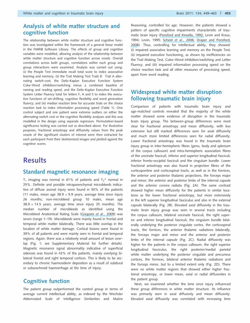
Widespread white matter disruptionfollowing traumatic brain injuryComparison of patients with traumatic brain injury and
age-matched controls revealed that the majority of the white
matter showed some evidence of disruption in the traumatic
brain injury group. The between-group differences were most
clear for fractional anisotropy and mean diffusivity, with less
extensive but still marked differences seen for axial diffusivity
and much more limited differences seen for radial diffusivity.
Lower fractional anisotropy was found in the traumatic brain
injury group in inter-hemispheric fibres (genu, body and splenium
of the corpus callosum) and intra-hemispheric association fibres
of the uncinate fasciculi, inferior and superior longitudinal fasciculi,
inferior fronto-occipital fasciculi and the cingulum bundle. Lower
fractional anisotropy was also found in projection fibres of the
corticopontine and corticospinal tracts, as well as in the fornices,
the anterior and posterior thalamic projections, the forceps major
and minor, the anterior and posterior limbs of the internal capsule
and the anterior corona radiata (Fig. 2A). The same contrast
showed higher mean diffusivity for the patients in similar loca-
tions as the lower fractional anisotropy, but more extensively
in the left superior longitudinal fasciculus and also in the external
capsule bilaterally (Fig. 2B). Elevated axial diffusivity in the trau-
matic brain injury group was seen in several tracts including
the corpus callosum, bilateral uncinate fasciculi, the right super-
ior and inferior longitudinal fasciculi, the cingulum bundle bilat-
erally underlying the posterior cingulate cortex, the corticospinal
tracts, the fornices, the anterior thalamic radiations bilaterally,
the forceps major and minor and the anterior and posterior
limbs of the internal capsule (Fig. 2C). Radial diffusivity was
higher for the patients in the corpus callosum, the right superior
longitudinal fasciculus, the right posterior/medial parietal
white matter underlying the posterior cingulate and precuneus
cortices, the fornices, bilateral anterior thalamic radiations and
the forceps minor, but to a limited extent only (Fig. 2D). There
were no white matter regions that showed either higher frac-
tional anisotropy, or lower mean, axial or radial diffusivities in
the patient group.
Next, we examined whether the time since injury influenced
these group differences in white matter structure. Its influence
was primarily seen in axial diffusivity and mean diffusivity.
Elevated axial diffusivity was correlated with increasing time
White matter and cognition in traumatic brain injury Brain 2011: 134; 449–463 | 453
since injury (R = 0.49, P5 0.01), an effect that was present when
controlling for patient age (Rpartial = 0.45, P50.05). A similar
result was found for mean diffusivity (R = 0.55, P50.01;
Rpartial = 0.46, P50.05). Time since injury was not correlated
with either fractional anisotropy or radial diffusivity once patient
age had been controlled.
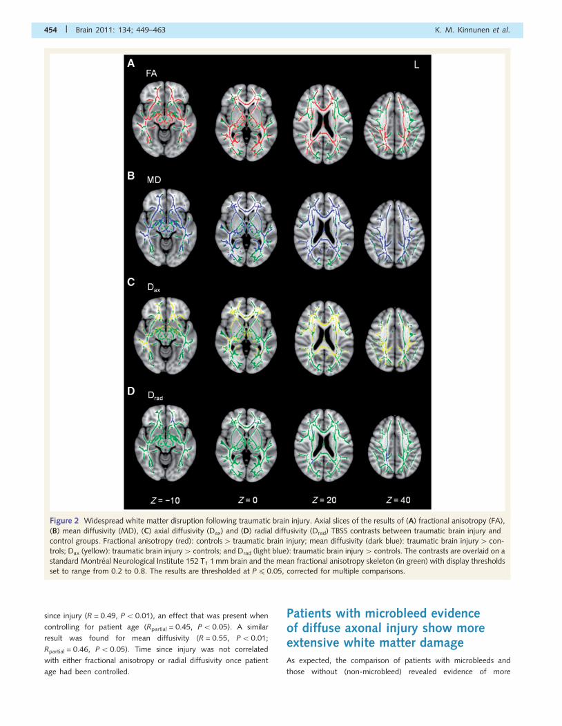
Patients with microbleed evidenceof diffuse axonal injury show moreextensive white matter damageAs expected, the comparison of patients with microbleeds and
those without (non-microbleed) revealed evidence of more
Figure 2 Widespread white matter disruption following traumatic brain injury. Axial slices of the results of (A) fractional anisotropy (FA),
(B) mean diffusivity (MD), (C) axial diffusivity (Dax) and (D) radial diffusivity (Drad) TBSS contrasts between traumatic brain injury and
control groups. Fractional anisotropy (red): controls4 traumatic brain injury; mean diffusivity (dark blue): traumatic brain injury4 con-
trols; Dax (yellow): traumatic brain injury4 controls; and Drad (light blue): traumatic brain injury4 controls. The contrasts are overlaid on a
standard Montreal Neurological Institute 152 T1 1 mm brain and the mean fractional anisotropy skeleton (in green) with display thresholds
set to range from 0.2 to 0.8. The results are thresholded at P40.05, corrected for multiple comparisons.
454 | Brain 2011: 134; 449–463 K. M. Kinnunen et al.
severe disruption in the microbleed group in large parts of the
white matter. Significantly lower fractional anisotropy for the con-
trast of microbleed versus non-microbleed patients was observed
in the body and splenium of the corpus callosum, as well as bi-
laterally within the inferior longitudinal fasciculi, the corticopon-
tine/corticospinal tracts, the fornices, the thalamic radiations, the
internal and external capsules and within white matter structures
of the midbrain (the decussation of the superior cerebellar
peduncles; Fig. 3A). There was also higher mean diffusivity for
the patients with microbleeds largely corresponding to the loca-
tions of the lower fractional anisotropy, including bilateral inferior
longitudinal fasciculi, the corticospinal tracts bilaterally, the for-
nices, bilateral anterior thalamic radiations, the posterior limbs of
the internal capsule and the external capsule. In addition, higher
mean diffusivity was observed in the superior longitudinal fasciculi,
the cingulum bundle bilaterally underlying the posterior cingulate
and retrosplenial cortices and the forceps major and minor, but
only in the posterior body and the splenium of the corpus callosum
(Fig. 3B). The patients with microbleeds also showed significantly
elevated radial diffusivity in several white matter tracts (Fig. 3C).
These corresponded to the tracts showing either lower fractional
anisotropy or higher mean diffusivity (or both), apart from the
superior longitudinal fasciculi and the forceps minor on the right
that showed higher mean diffusivity in the absence of elevated
radial diffusivity. In axial diffusivity, there were no group
differences between the patients with and without microbleeds.
Again, there were no white matter regions that showed either
elevated fractional anisotropy or lower mean, axial or radial
diffusivities in the microbleed group.
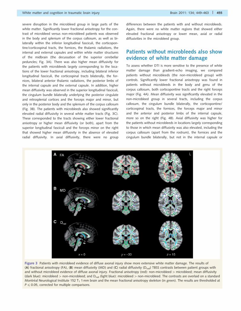
Patients without microbleeds also showevidence of white matter damageTo assess whether DTI is more sensitive to the presence of white
matter damage than gradient-echo imaging, we compared
patients without microbleeds (the non-microbleed group) with
controls. Significantly lower fractional anisotropy was found in
patients without microbleeds in the body and genu of the
corpus callosum, both corticopontine tracts and the right forceps
major (Fig. 4A). Mean diffusivity was significantly elevated in the
non-microbleed group in several tracts, including the corpus
callosum, the cingulum bundle bilaterally, the corticopontine/
corticospinal tracts, the fornices, the forceps major and minor
and the anterior and posterior limbs of the internal capsule,
more so on the right (Fig. 4B). Axial diffusivity was higher for
the patients without microbleeds in locations largely corresponding
to those in which mean diffusivity was also elevated, including the
corpus callosum (apart from the rostrum), the fornices and the
cingulum bundle bilaterally, but not in the internal capsule or
Figure 3 Patients with microbleed evidence of diffuse axonal injury show more extensive white matter damage. The results of
(A) fractional anisotropy (FA), (B) mean diffusivity (MD) and (C) radial diffusivity (Drad) TBSS contrasts between patient groups with
and without microbleed evidence of diffuse axonal injury. Fractional anisotropy (red): non-microbleed4microbleed; mean diffusivity
(dark blue): microbleed4non-microbleed; and Drad (light blue): microbleed4non-microbleed. The contrasts are overlaid on a standard
Montreal Neurological Institute 152 T1 1 mm brain and the mean fractional anisotropy skeleton (in green). The results are thresholded at
P40.05, corrected for multiple comparisons.
White matter and cognition in traumatic brain injury Brain 2011: 134; 449–463 | 455
the forceps major and minor (Fig. 4C). There were no signifi-
cant group differences in radial diffusivity. There were also no
regions where fractional anisotropy was elevated, or mean, axial
or radial diffusivities lower in the non-microbleed group as com-
pared with controls. These results demonstrate the presence of
white matter abnormalities in patients with no microbleeds on
gradient-echo imaging. As expected, the same contrasts between
patients with microbleeds and healthy controls showed exten-
sive white matter abnormalities in the patients (Supplementary
Fig. 1).
Patients classified as having sustaineda ‘mild’ traumatic brain injury showwhite matter abnormalitiesWe also examined the relationship between traumatic brain injury
severity as defined using the Mayo classification system (Malec
et al., 2007) and white matter damage. Although the mild
group consisted of only eight patients, they showed lower frac-
tional anisotropy compared with controls in a wide range of tracts
(Supplementary Fig. 2A). These included the fornices, the cingu-
lum bundle bilaterally, the corpus callosum, the anterior limb of
the right internal capsule, the left external capsule, the inferior
fronto-occipital fasciculi, the left superior longitudinal fasciculus,
the forceps major and minor bilaterally, the anterior thalamic
radiations bilaterally and the corticospinal tracts. The mild patients
also showed elevated mean diffusivity in similar, but more wide-
spread tracts, with additional differences seen in the internal and
external capsules bilaterally and the superior longitudinal fasciculi
(Supplementary Fig. 2B). Axial diffusivity was higher for the mild
patients than controls in the corpus callosum, the inferior
fronto-occipital fasciculi, the posterior cingulum bundles, the left
superior longitudinal fasciculus, the posterior limbs of the internal
capsule, the corticospinal tracts and both anterior thalamic radi-
ations (Supplementary Fig. 2C). The mild patients had higher axial
diffusivity than the moderate/severe patients in the body and
genu of the corpus callosum, a difference that was still present
after controlling for time elapsed since the injury. There were no
other differences in any of the DTI metrics between the mild and
moderate/severe patients. As expected, there were very wide-
spread differences in fractional anisotropy (controls4patients)
and mean and axial diffusivities (patients4 controls) between
moderate/severe patients and controls. These differences were
seen within the fornices, the corpus callosum, all major intra-
hemispheric association and projection fibres, the internal and ex-
ternal capsules and the superior and anterior corona radiata. Radial
diffusivity measurements were not different between any of the
three groups.
Figure 4 Patients without microbleeds also show evidence of white matter damage. The results of (A) fractional anisotropy (FA),
(B) mean diffusivity (MD) and (C) axial diffusivity (Dax) TBSS contrasts between patients without microbleed evidence of diffuse
axonal injury (non-microbleed) and controls. Fractional anisotropy (red): controls4non-microbleed, mean diffusivity (dark blue):
non-microbleed4 controls; and Dax (yellow): non-microbleed4 controls. The contrasts are overlaid on a standard Montreal Neurological
Institute 152 T1 1 mm brain and the mean fractional anisotropy skeleton (in green). The results are thresholded at P40.05, corrected for
multiple comparisons.
456 | Brain 2011: 134; 449–463 K. M. Kinnunen et al.

The relationship between white matterstructure and cognitive function
Associative memory
Across both patients and controls, we found evidence that the
structure of the fornices predicted associative memory perform-
ance. As expected, fractional anisotropy within the fornix was
positively correlated with memory, showing that individuals with
more anisotropic white matter within the fornix had better per-
formance. This relationship between fractional anisotropy and
associative memory was observed in both patient and normal
groups (Fig. 5A). Using the stringent permutation test we
employed, this relationship was of borderline significance, when
corrected for multiple comparisons across the whole brain
(P50.06 in the right fornix, peak voxel: x = 7, y = �5, z = 9
and P50.07 in the left fornix, peak voxel: x = �2, y = �16,
z = 17). In the peak voxel within the fornix, fractional anisotropy
was significantly correlated with our measure of associative learn-
ing and memory across both groups (R2 = 0.25, P5 0.001). TBSS
tests for linear relationships between white matter structure and
cognitive variables, but this relationship may not be best modelled
linearly, as suggested by Fig. 5B. For this reason, we fitted a
second-order polynomial regression slope, which models the
data more accurately (R2 = 0.33, P50.0001). This relationship
was specific to the hippocampal formation as there were no sig-
nificant regions outside it where fractional anisotropy was corre-
lated with the measure of memory function. The relationship was
also present when controlling for intellectual ability (Rpartial = 0.48,
P50.001), and an ANCOVA with severity as the between-
subjects factor showed no significant interaction between severity
and fractional anisotropy in the peak voxel from the fornix/
memory relationship. No significant correlations between associa-
tive learning and memory and white matter structure were
observed for any of the three diffusivities.
Executive function
A more complex relationship between white matter structure
and executive function was observed. There was no relationship
between our executive function measures and fractional anisot-
ropy, but a significant relationship was observed between set-
shifting, as measured by the Trail Making Test alternating
switch-cost index and both mean and radial diffusivity. Increases
in mean and radial diffusivities have previously been reported after
traumatic brain injury (Kraus et al., 2007; Sidaros et al., 2008;
Kennedy et al., 2009) and are thought to indicate axonal injury.
In the whole-brain analysis, the patient group showed an expected
correlation between elevated mean diffusivity and executive
Figure 5 The results of TBSS regression analysis of associative learning and memory (People Test immediate recall total) by fractional
anisotropy (FA) across the traumatic brain injury and control groups. (A) Areas where fractional anisotropy is positively correlated with
People Test (PT) recall score across the two groups are indicated in red (FA/PT: ALL + ). The result is overlaid on a standard Montreal
Neurological Institute 152 T1 1 mm brain and the mean fractional anisotropy skeleton (in green). For display purposes the result is
displayed with a multiple comparisons threshold of P40.1. (B) Graph showing individual data points in both groups for People Test recall
score against fractional anisotropy in the peak voxel (Montreal Neurological Institute x = 7, y = �5, z = 9). A second-order polynomial
regression slope is shown, which provides a more accurate fit than the linear regression identified by the whole-brain general linear model
analysis. CON = control; TBI = traumatic brain injury.
White matter and cognition in traumatic brain injury Brain 2011: 134; 449–463 | 457
dysfunction. Patients with high mean diffusivity in the left superior
frontal white matter showed worse performance. A subsequent
analysis within all significant voxels confirmed this relationship
(Rpartial = 0.75, P50.001), controlling for intellectual ability.
There was no such relationship in the healthy control group, in
the groups combined or in the interaction between the groups.
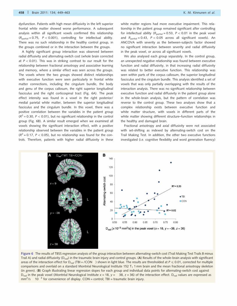
A highly significant group interaction was observed between
radial diffusivity and alternating-switch cost (whole brain corrected
at P50.01). This was in striking contrast to our result for the
relationship between fractional anisotropy and associative learning
and memory, where a similar effect was seen across the groups.
The voxels where the two groups showed distinct relationships
with executive function were seen particularly in frontal white
matter connections, including the cingulum bundle, the body
and genu of the corpus callosum, the right superior longitudinal
fasciculus and the right corticospinal tract (Fig. 6A). The peak
effect intensity was found in a voxel in the right posterior/
medial parietal white matter, between the superior longitudinal
fasciculus and the cingulum bundle. In this voxel, there was a
positive correlation between the variables in the patient group
(R2 = 0.30, P5 0.01), but no significant relationship in the control
group (Fig. 6B). A similar result emerged when we examined all
voxels showing the significant interaction effect, with a positive
relationship observed between the variables in the patient group
(R2 = 0.17, P5 0.05), but no relationship was found for the con-
trols. Therefore, patients with higher radial diffusivity in these
white matter regions had more executive impairment. This rela-
tionship in the patient group remained significant after controlling
for intellectual ability (Rpartial = 0.53, P5 0.01 in the peak voxel
and Rpartial = 0.43, P50.05 across all significant voxels). An
ANCOVA with severity as the between-subjects factor showed
no significant interaction between severity and radial diffusivity
in the peak voxel, or across all significant voxels.
We also analysed each group separately. In the control group,
an unexpected negative relationship was found between executive
function and radial diffusivity, in that increasing radial diffusivity
was related to better executive function. This relationship was
seen within parts of the corpus callosum, the superior longitudinal
fasciculus and the cingulum bundle. This analysis identified a set of
voxels that was only partially overlapping with the results of the
interaction analysis. There was no significant relationship between
executive function and radial diffusivity in the patient group alone
in the whole-brain analysis, but the pattern of correlation was
reverse to the control group. These two analyses show that a
complex relationship exists between executive function and
white matter structure, with voxels in different parts of the
white matter showing different structure–function relationships in
the healthy and damaged brain.
Fractional anisotropy and axial diffusivity were not associated
with set-shifting as indexed by alternating-switch cost on the
Trail Making Test. In addition, the other two executive functions
investigated (i.e. cognitive flexibility and word generation fluency)
Figure 6 The results of TBSS regression analysis of the group interaction between alternating-switch cost (Trail Making Test Trails B minus
Trail A) and radial diffusivity (Drad) in the traumatic brain injury and control groups. (A) Results of the whole-brain analysis with significant
areas of the interaction effect for Drad (TBI + /CON� ) shown in light blue. The results are thresholded at P4 0.01, corrected for multiple
comparisons and overlaid on a standard Montreal Neurological Institute 152 T1 1 mm brain and the mean fractional anisotropy skeleton
(in green). (B) Graph illustrating linear regression slopes for each group and individual data points for alternating-switch cost against
Drad in the peak voxel (Montreal Neurological Institute x = 18, y = �38, z = 36) of the interaction effect. Drad values are expressed as
mm2/s � 10�3 for convenience of display. CON = control; TBI = traumatic brain injury.
458 | Brain 2011: 134; 449–463 K. M. Kinnunen et al.
did not show a significant relationship with any of the DTI
measures.
Information processing speed
In the whole-brain analysis, information processing speed, as mea-
sured by median reaction time for accurate responses on the
choice reaction task, was not found significantly associated with
any index of white matter structure.
Elevated axial diffusivity after traumaticbrain injury and cognitive functionIt has been demonstrated previously that patients who show rela-
tively large increases in axial diffusivity in the first year following
their head injury have a more favourable outcome (Sidaros et al.,
2008). This raises the possibility that elevated axial diffusivity post-
traumatic brain injury is a marker of axonal recovery. Sidaros et al.
(2008) observed increases in the posterior limb of the internal
capsule, a descending motor pathway with a well-known normal
structure and architecture (Pierpaoli et al., 2001). This prompted
us to perform a focused analysis of axial diffusivity within this part
of the white matter. We tested for partial correlations between
axial diffusivity and the five cognitive variables described above,
controlling for age, time since traumatic brain injury and current
intellectual ability. Processing speed was negatively correlated with
axial diffusivity (Rpartial = �0.55, P5 0.01), such that patients with
the highest axial diffusivity in the posterior limb of the internal
capsule had the fastest reaction times. None of the other four
cognitive variables showed a significant relationship with axial
diffusivity.
DiscussionWe have demonstrated the relationship between white matter
abnormalities and cognitive function in two domains commonly
affected by traumatic brain injury, memory and executive func-
tion. The work builds on previous studies, which show that DTI is
a sensitive technique for imaging white matter damage in trau-
matic brain injury (Inglese et al., 2005; Salmond et al., 2006;
Kraus et al., 2007; Niogi et al., 2008b; Sidaros et al., 2008;
Kennedy et al., 2009). In general, these studies have used a
region of interest approach. This involves the investigation of a
relatively small amount of white matter, within regions that are
defined on the basis of a priori judgements. Here, for the first
time, we used tract-based spatial statistics (a voxel-based ap-
proach) to explore the relationship between white matter structure
and cognitive function following traumatic brain injury in a
data-driven manner. This is particularly important, as the cognitive
deficits commonly observed after traumatic brain injury, such as
executive impairment, are likely to depend upon the disruption of
distributed brain networks by diffuse axonal injury.
Our results show that widespread white matter abnormalities
persist following traumatic brain injury and that the pattern of
damage to specific white matter tracts predicts some aspects of
the profile of cognitive deficits that are present. Variability in cog-
nitive function in our patients cannot be explained by the limited,
and largely non-overlapping, pattern of focal cortical damage. In
contrast, across both patients and controls, the structure of the
fornices was related to the efficiency of associative learning and
memory. Previous work has shown the importance of the fornix
for memory function (Aggleton, 2008; Tsivilis et al., 2008). In
humans, damage to the fornices produces memory deficits
(Gaffan and Gaffan, 1991; McMackin et al., 1995; Park et al.,
2000; Kesler et al., 2001) and, in the monkey, the fornix has been
shown to be critical for the rapid learning of new spatial and
non-spatial associations (Brasted et al., 2002, 2003; Kwok and
Buckley, 2010). Following traumatic brain injury, the extent of
damage to the hippocampi is known to predict memory impair-
ment (Tate and Bigler, 2000), and mean diffusivity within the
hippocampal formation has been shown to predict associative
memory function (Salmond et al., 2006).
We extend these observations by showing that the structure of
the fornices is specifically correlated with the efficiency of certain
aspects of memory function. Previous region-of-interest studies
have not examined the effect of traumatic brain injury on the
fornix in terms of memory. Using TBSS, we were able to investi-
gate individual white matter tracts and can be confident that our
result is specific to the hippocampal formation. Although wide-
spread white matter abnormality was present, we found no
other areas that significantly correlated with memory function.
The results also suggest that the relationship between fornix struc-
ture and memory is not limited to patients with traumatic brain
injury. As we have used a cross-sectional study design, we cannot
completely exclude the possibility that there may have been
pre-morbid differences in fornix structure between the two
groups. However, this seems a highly unlikely explanation for
our results, particularly when one considers that the patients
show a specific pattern of cognitive impairment typical for trau-
matic brain injury, in association with better current intellectual
functioning than the control group. Instead, the results suggest
that fractional anisotropy within the fornix is positively correlated
with associative memory performance in the healthy brain.
Traumatic brain injury appears to modulate this existing relation-
ship by disrupting white matter structure, thereby shifting patients
along an existing continuum into a less efficient structure-function
relationship. Mechanical factors may be important in explaining
the prevalence of this type of memory impairment after traumatic
brain injury, as the fornix is likely to be particularly susceptible to
shearing and tearing forces due to its arch-like shape and long
fibre tracts (Tate and Bigler, 2000).
In contrast, the patient and control groups showed distinct
relationships between white matter structure (radial diffusivity)
and one of our three indices of executive function. Our voxel-wise
approach made it possible to explore this complex relationship.
Standard DTI analysis involves the placement of regions of inter-
est. This requires a priori knowledge of the likely location of
effects of interest, which is both difficult and restrictive, as current
understanding of structure-function relationships is limited and
white matter damage diffuse. The TBSS approach allows the
relationship between variables to be modelled in the framework
of a general linear model and does not require the placement of
specific regions of interest. Using a region of interest approach
Niogi and colleagues (2008a) previously reported a correlation
White matter and cognition in traumatic brain injury Brain 2011: 134; 449–463 | 459
between fractional anisotropy in a small part of the anterior
corona radiata and executive function following mild traumatic
brain injury. Their analysis focused on two small regions that
showed white matter/cognitive function relationships in controls.
We extend these findings by investigating a more severely
affected group and using a different DTI metric to demonstrate
that patients with more executive impairment have more white
matter damage in a number of tracts that connect the frontal
lobes to more posterior brain regions. This is consistent with the
proposal that executive dysfunction following brain injury is,
partly, the result of frontal lobe disconnection (Miller and
D’Esposito, 2005).
Also in contrast to Niogi and colleagues (2008a), we did not
observe a significant relationship between fractional anisotropy
and executive function, although it is possible that the higher
general intellectual function in the patients might obscure an over-
all correlation of fractional anisotropy and executive function
across the two groups. However, this was not the case for our
fornix/memory result and the IQ difference did not impact on the
within-group patient analysis of executive function. The presence
of widespread differences in the relationship between radial diffu-
sivity and executive function in the uninjured brain and after trau-
matic brain injury suggests that using normal structure/function
relationships to guide investigation of the effects of traumatic
brain injury may not always be appropriate. A similar relationship
between frontal connectivity and executive function has been
observed in studies of normal ageing, where, in older adults,
reduced integrity in tracts connecting frontal regions predicts ex-
ecutive dysfunction (O’Sullivan et al., 2001; Davis et al., 2009;
Perry et al., 2009). This suggests that different pathologies can
produce similar cognitive impairments through damage to the
same tracts.
DTI is extremely sensitive to white matter damage following
traumatic brain injury. Reductions in fractional anisotropy and
axial diffusivity emerge in the first few hours after a cortical con-
tusion in experimental models of traumatic brain injury (Mac
Donald et al., 2007), and these early changes reflect axonal
damage (Song et al., 2003; Budde et al., 2008, 2009). Tissue
injury evolves over time with the development of macrophage
infiltration, tissue oedema and demyelination and these patho-
logical changes are reflected in DTI measurements (Mac Donald
et al., 2007; Sidaros et al., 2008). In general, low fractional
anisotropy persists over time, accompanied by an increase in
radial diffusivity that leads to high mean diffusivity, whilst changes
in axial diffusivity are more dynamic (Sidaros et al., 2008).
We also provide direct evidence that DTI can detect white
matter damage not seen using the standard magnetic resonance
techniques. We stratified our analysis by investigating white
matter abnormalities in patients with and without microbleeds.
The presence of microbleeds on gradient-echo imaging is a
marker of diffuse axonal injury and so indicates the presence of
more severe white matter injury (Scheid et al., 2003). As
expected, patients with microbleeds showed widespread white
matter abnormalities as compared with age-matched controls,
but patients without microbleeds also showed significant white
matter abnormalities. This highlights the limitation of relying on
the presence of microbleeds as a marker of subtle white matter
damage and demonstrates that significant white matter abnormal-
ity may be present following traumatic brain injury even when
gradient-echo MRI is normal. We further stratified the patient
analysis on the basis of severity, defined based on the Mayo
system (Malec et al., 2007). This, again, demonstrated the sensi-
tivity of DTI in identifying white matter abnormalities in patients
classified as ‘mild’. Unlike the microbleed analysis, however, the
comparison of mild and moderate/severe patients failed to show a
difference in fractional anisotropy or mean diffusivity measure-
ments. Although the Mayo system includes some aspects of struc-
tural brain damage in its criteria, it does not integrate sensitive
magnetic resonance measures of white matter damage. Three of
the eight ‘mild’ patients had microbleeds on gradient echo ima-
ging. Therefore, although the ‘mild’ group was small, it is likely
that the null results reflect the inclusion in this group of patients
with significant diffuse axonal injury. This highlights the limitation
of existing severity classifications for traumatic brain injury that fail
to include specific measures of white matter damage.
Our results also show an overall increase in axial diffusivity,
which was positively correlated with time since traumatic brain
injury and greater in the ‘mild’ group of patients. Previous work
has tended to show that traumatic brain injury produces early
reductions in axial diffusivity that gradually normalize over time
(e.g. Sidaros et al., 2008; Wang et al., 2009). On average, we
scanned patients longer after their injury than Sidaros and col-
leagues (2008). Hence, our results suggest that axial diffusivity
continues to rise well after the acute phase of traumatic brain
injury. The pathological significance of these dynamic changes in
axial diffusivity remains unclear, in part due to a lack of relevant
animal studies (although see Wang et al., 2009). The normaliza-
tion of axial diffusivity could reflect reorganization within the
white matter, due to axonal recovery or even regrowth (Voss
et al., 2006; Sidaros et al., 2008). We found some support for
this proposal by specifically examining the posterior limb of the
internal capsule, where a large increase in axial diffusivity has been
previously shown to be predictive of functional outcome (Sidaros
et al., 2008). This region contains descending corticospinal fibres
and here, higher axial diffusivity in the posterior limb of the
internal capsule was associated with faster processing speed on
the choice reaction task. This result, therefore, provides support
for the proposal that increased axial diffusivity reflects adaptive
axonal recovery, but should be interpreted cautiously as our
whole-brain analysis did not reveal a significant relationship for
any region.
In our patients with traumatic brain injury, DTI changes were
generally seen in the expected directions. In contrast, higher radial
diffusivity in certain white matter tracts of the healthy controls
was associated with more efficient executive function. This was
unexpected, because one determinant of radial diffusivity is the
degree of axonal myelination (Beaulieu, 2002) and, as this
increases, one might expect reduced radial diffusivity, faster
nerve conduction times (Jack et al., 1983) and more efficient
executive function. However, the relationship between DTI meas-
ures of white matter structure and cognitive function appears not
to be this simple. Significant relationships between cognitive
function and white matter structure in an unexpected direction
have been reported previously (Tuch et al., 2005). These results
460 | Brain 2011: 134; 449–463 K. M. Kinnunen et al.
emphasize that further work is needed to determine how changes
in different aspects of white matter microstructure in specific tracts
are related to cognitive function in the uninjured brain and how
the DTI metrics are affected by brain injury. Further animal studies
will also be needed to determine in detail the complex relation-
ships between different DTI measures and the pathological effects
of traumatic brain injury.
A possible limitation of DTI analyses is the presence of partial
volume effects. This is potentially problematic for investigating
patients with traumatic brain injury in the chronic phase, as pa-
tients frequently show some degree of brain atrophy. This means
that the changes in DTI measures, such as lower fractional anisot-
ropy, may reflect partial volumes, resulting from contamination of
measurements by cerebrospinal fluid. Our approach limits the
impact of this problem as the TBSS analysis involves ‘skeletoniza-
tion’ of the white matter and focuses on the centres of the tracts
(Smith et al., 2006). This removes the white matter at the junc-
tions with cerebrospinal fluid and grey matter that is prone to
partial volume effects. Hence, our approach to investigating
group differences is more robust to brain atrophy.
To conclude, we found widespread fractional anisotropy, mean
diffusivity and axial diffusivity differences between patients with
traumatic brain injury and healthy controls using TBSS. The distri-
bution of white matter abnormality correlated with individual dif-
ferences in associative learning and memory and one of our three
indices of executive function. White matter disruption in the for-
nices predicted associative memory performance across both
groups, whereas a more distinct pattern was observed for the
relationship between frontal disconnection and executive function
in the two groups. Our approach reveals the complexity of the
relationships between indices of white matter structure and cog-
nition and shows the importance of flexibly analysing patterns of
disruption across the whole brain.
AcknowledgementsThe authors thank all participants for their contribution to this
project.
FundingThe Medical Research Council (UK) (to D.J.S.); the Hammersmith
Hospital’s Charity Trustees Research Grants Committee (to D.J.S.);
and Goldsmiths, University of London (to K.M.K.).
Supplementary materialSupplementary material is available at Brain online.
ReferencesAggleton JP. EPS Mid-Career Award 2006. Understanding anterograde
amnesia: disconnections and hidden lesions (Review). Q J Exp Psychol
2008; 61: 1441–71.
Aggleton JP, Brown MW. Episodic memory, amnesia, and the hippocam-
pal–anterior thalamic axis. Behav Brain Sci 1999; 22: 425–89.
Arfanakis K, Haughton VM, Carew JD, Rogers BP, Dempsey RJ,
Meyerand ME. Diffusion tensor MR imaging in diffuse axonal injury.
Am J Neuroradiol 2002; 23: 794–802.
Assaf Y, Pasternak O. Diffusion tensor imaging (DTI)-based white matter
mapping in brain research: A review (Review). J Mol Neurosci 2008;
34: 51–61.
Baddeley AD, Emslie H, Nimmo-Smith I. Doors and people test: a test of
visual and verbal recall and recognition. Bury-St-Edmunds: Thames
Valley Test Company; 1994.Basser PJ, Pierpaoli C. Microstructural and physiological features of tis-
sues elucidated by quantitative-diffusion-tensor MRI. J Magn Reson B
1996; 3: 209–19.
Basser PJ, Pierpaoli C. A simplified method to measure the diffusion
tensor from seven MR images. Magn Reson Med 1998; 39: 928–34.
Beaulieu C. The basis of anisotropic water diffusion in the nervous
system – a technical review (Review). NMR Biomed 2002; 15: 435–55.
Behrens TEJ, Woolrich MW, Jenkinson M, Johansen-Berg H, Nunes RG,
Clare S, et al. Characterization and propagation of uncertainty in
diffusion-weighted MR imaging. Magn Reson Med 2003; 50:
1077–88.
Bigler ED. The lesion(s) in traumatic brain injury: implications for clinical
neuropsychology. (Review). Arch Clin Neuropsychol 2001; 16:
95–131.Bosch B, Arenaza-Urquijo EM, Rami L, Sala-Llonch R, Junque C, Sole-
Padulles C, et al. Multiple DTI index analysis in normal aging, amnestic
MCI and AD: relationship with neuropsychological performance.
Neurobiol Aging [serial on the Internet]. 2010 [about 14 pages].
Available from: http://www.neurobiologyofaging.org/article/S0197-
4580(10)00082-5/pdf (10 May 2010, date last accessed).Brasted PJ, Bussey TJ, Murray EA, Wise SP. Fornix transection impairs
conditional visuomotor learning in tasks involving nonspatially differ-
entiated responses. J Neurophysiol 2002; 87: 631–3.
Brasted PJ, Bussey TJ, Murray EA, Wise SP. Role of the hippocampal
system in associative learning beyond the spatial domain. Brain
2003; 126: 1202–23.
Bucur B, Madden DJ, Spaniol J, Provenzale JM, White LE, Cabeza R,
et al. Age-related slowing of memory retrieval: contributions of per-
ceptual speed and cerebral white matter integrity. Neurobiol Aging
2008; 29: 1070–9.Budde MD, Xie M, Cross AH, Song SK. Axial diffusivity is the primary
correlate of axonal injury in the experimental autoimmune encephalo-
myelitis spinal cord: a quantitative pixelwise analysis. J Neurosci 2009;
29: 2805–13.
Budde MD, Kim JH, Liang H-F, Russell JH, Cross AH, Song SK. Axonal
injury detected by in vivo diffusion tensor imaging correlates with
neurological disability in a mouse model of multiple sclerosis. NMR
Biomed 2008; 21: 589–97.Ceccarelli A, Rocca MA, Valsasina P, Rodegher M, Pagani E, Falini A,
et al. A multiparametric evaluation of regional brain damage in
patients with primary progressive multiple sclerosis. Hum Brain Mapp
2009; 30: 3009–19.
Davis SW, Dennis NA, Buchler NG, White LE, Madden DJ, Cabeza R.
Assessing the effects of age on long white matter tracts using diffusion
tensor tractography. Neuroimage 2009; 46: 530–41.
Delis DC, Kaplan E, Kramer JH. Delis-Kaplan Executive Function System.
San Antonio, TX: Psychological Corporation; 2001.
Dineen RA, Vilisaar J, Hlinka J, Bradshaw CM, Morgan PS,
Constantinescu CS, et al. Disconnection as a mechanism for cognitive
dysfunction in multiple sclerosis. Brain 2009; 132: 239–49.Draper K, Ponsford J. Cognitive functioning ten years following traumatic
brain injury and rehabilitation. Neuropsychology 2008; 22: 618–25.Gaffan D, Gaffan EA. Amnesia in man following transection of the fornix
(Review). Brain 1991; 114: 2611–8.Gregoire SM, Chaudhary UJ, Brown MM, Yousry TA, Kallis C, Jager HR,
et al. The Microbleed Anatomical Rating Scale (MARS): reliability of a
tool to map brain microbleeds. Neurology 2009; 73: 1759–66.
White matter and cognition in traumatic brain injury Brain 2011: 134; 449–463 | 461
Inglese M, Makani S, Johnson G, Cohen BA, Silver JA, Gonen O, et al.
Diffuse axonal injury in mild traumatic brain injury: a diffusion tensor
imaging study. J Neurosurg 2005; 103: 298–303.
Jack JJB, Noble D, Tsien RW. Electric current flow in excitable cells.
Oxford: Oxford University Press; 1983.
Kennedy MRT, Wozniak JR, Muetzel RL, Mueller BA, Chiou HH,
Pantekoek K, et al. White matter and neurocognitive changes in
adults with chronic traumatic brain injury. J Int Neuropsychol Soc
2009; 15: 130–6.
Kesler SR, Hopkins RO, Weaver LK, Blatter DD, Edge-Booth H, Bigler ED.
Verbal memory deficits associated with fornix atrophy in carbon mon-
oxide poisoning. J Int Neuropsychol Soc 2001; 7: 640–6.
Kraus MF, Susmaras T, Caughlin BP, Walker CJ, Sweeney JA, Little DM.
White matter integrity and cognition in chronic traumatic brain injury:
a diffusion tensor imaging study. Brain 2007; 130: 2508–19.Kwok SC, Buckley MJ. Fornix transection selectively impairs fast learning
of conditional visuospatial discriminations. Hippocampus 2010; 20:
413–22.
Levin HS. Neurobehavioral outcome of closed head injury: implications
for clinical trials (Review). J Neurotrauma 1995; 12: 601–10.
Levin HS, Kraus MF. The frontal lobes and traumatic brain injury
(Review). J Neuropsychiatry Clin Neurosci 1994; 6: 443–54.
Lowenstein D. Traumatic brain injury: a glimpse of order among the
chaos? Ann Neurol 2009; 66: A7–8.
Mac Donald CL, Dikranian K, Bayly P, Holtzman D, Brody D. Diffusion
tensor imaging reliably detects experimental traumatic axonal injury
and indicates approximate time of injury. J Neurosci 2007; 27:
11869–76.Madden DJ, Whiting WL, Huettel SA, White LE, MacFall JR,
Provenzale JM. Diffusion tensor imaging of adult age differences in
cerebral white matter: relation to response time. Neuroimage 2004;
21: 1174–81.
Madden DJ, Spaniol J, Costello MC, Bucur B, White LE, Cabeza R, et al.
Cerebral white matter integrity mediates adult age differences in cog-
nitive performance. J Cogn Neurosci 2009; 21: 289–302.
Malec JF, Brown AW, Leibson CL, Flaada JT, Mandrekar JN, Diehl NN,
et al. The Mayo classification system for traumatic brain injury severity.
J Neurotrauma 2007; 24: 1417–24.McMackin D, Cockburn J, Anslow P, Gaffan D. Correlation of fornix
damage with memory impairment in six cases of colloid cyst removal.
Acta Neurochir 1995; 135: 12–8.
Mesulam MM. From sensation to cognition (Review). Brain 1998; 121:
1013–52.
Miller BT, D’Esposito M. Searching for “the top” in top-down control
(Review). Neuron 2005; 48: 535–8.
Nichols TE, Holmes AP. Nonparametric permutation tests for functional
neuroimaging: a primer with examples. Hum Brain Mapp 2002; 15:
1–25.
Niogi SN, Mukherjee P, Ghajar J, Johnson CE, Kolster R, Lee H, et al.
Structural dissociation of attentional control and memory in adults
with and without mild traumatic brain injury. Brain 2008a; 131:
3209–21.
Niogi SN, Mukherjee P, Ghajar J, Johnson C, Kolster RA, Sarkar R, et al.
Extent of microstructural white matter injury in postconcussive syn-
drome correlates with impaired cognitive reaction time: a 3T diffusion
tensor imaging study of mild traumatic brain injury. Am J Neuroradiol
2008b; 29: 967–73.
O’Sullivan M, Jones DK, Summers PE, Morris RG, Williams SC,
Markus HS. Evidence for cortical “disconnection” as a mechanism of
age-related cognitive decline. Neurology 2001; 57: 632–8.
Park SA, Hahn JH, Kim JI, Na DL, Huh K. Memory deficits after bilateral
anterior fornix infarction. Neurology 2000; 54: 1379–82.
Perry ME, McDonald CR, Hagler DJ, Gharapetian L, Kuperman JM,
Koyama AK, et al. White matter tracts associated with set-shifting in
healthy aging. Neuropsychologia 2009; 47: 2835–42.Pierpaoli C, Barnett A, Pajevic S, Chen R, Penix LR, Virta A, et al. Water
diffusion changes in Wallerian degeneration and their dependence on
white matter architecture. Neuroimage 2001; 13: 1174–85.
Ponsford J, Kinsella G. Attentional deficits following closed-head injury.
J Clin Exp Neuropsychol 1992; 14: 822–38.
Povlishock JT, Katz DI. Update of neuropathology and neurological re-
covery after traumatic brain injury (Review). J Head Trauma Rehabil
2005; 20: 76–94.
Reitan R. The validity of the Trail Making Test as an indicator of organic
brain damage. Percept Mot Skills 1958; 8: 271–6.
Roosendaal SD, Geurts JJG, Vrenken H, Hulst HE, Cover KS,
Castelijns JA, et al. Regional DTI differences in multiple sclerosis
patients. Neuroimage 2009; 44: 1397–403.
Rugg-Gunn FJ, Symms MR, Barker GJ, Greenwood R, Duncan JS.
Diffusion imaging shows abnormalities after blunt head trauma
when conventional magnetic resonance imaging is normal. J Neurol
Neurosurg Psychiatry 2001; 70: 530–3.Salmond CH, Menon DK, Chatfield DA, Williams GB, Pena A,
Sahakian BJ, et al. Diffusion tensor imaging in chronic head injury
survivors: Correlations with learning and memory indices.
Neuroimage 2006; 29: 117–24.
Scheid R, Preul C, Gruber O, Wiggins C, von Cramon DY. Diffuse axonal
injury associated with chronic traumatic brain injury: evidence from
T2*-weighted gradient-echo imaging at 3 T. Am J Neuroradiol
2003; 24: 1049–56.
Scheid R, Walther KR, Guthke T, Preul C, von Cramon DY. Cognitive
sequelae of diffuse axonal injury. Arch Neurol 2006; 63: 418–24.
Schulte T, Sullivan EV, Muller-Oehring EM, Adalsteinsson E,
Pfefferbaum A. Corpus callosal microstructural integrity influences
interhemispheric processing: A diffusion tensor imaging study. Cereb
Cortex 2005; 15: 1384–92.
Sidaros A, Engberg A, Sidaros K, Liptrot MG, Herning M, Petersen P,
et al. Diffusion tensor imaging during recovery from severe traumatic
brain injury and relation to clinical outcome: a longitudinal study. Brain
2008; 131: 559–72.
Smith SM. Fast robust automated brain extraction. Hum Brain Mapp
2002; 17: 143–55.
Smith SM, Nichols TE. Threshold-free cluster enhancement: addressing
problems of smoothing, threshold dependence and localisation in
cluster inference. Neuroimage 2009; 44: 83–98.
Smith SM, Jenkinson M, Johansen-Berg H, Rueckert D, Nichols TE,
Mackay CE, et al. Tract-based spatial statistics: voxelwise
analysis of multi-subject diffusion data. Neuroimage 2006; 31:
1487–505.
Smith SM, Jenkinson M, Woolrich MW, Beckmann CF, Behrens TEJ,
Johansen-Berg H, et al. Advances in functional and structural MR
image analysis and implementation as FSL. Neuroimage 2004; 23:
S208–19.
Song SK, Sun SW, Ju WK, Lin SJ, Cross AH, Neufeld AH. Diffusion tensor
imaging detects and differentiates axon and myelin degeneration in
mouse optic nerve after retinal ischemia. Neuroimage 2003; 20:
1714–22.Sullivan EV, Adalsteinsson E, Hedehus M, Ju C, Moseley M, Lim KO,
et al. Equivalent disruption of regional white matter microstruc-
ture in ageing healthy men and women. Neuroreport 2001; 12:
99–104.
Tate DF, Bigler ED. Fornix and hippocampal atrophy in traumatic brain
injury. Learn Mem 2000; 7: 442–6.
Thornhill S, Teasdale GM, Murray GD, McEwen J, Roy CW, Penny KI.
Disability in young people and adults one year after head injury:
prospective cohort study. BMJ 2000; 320: 1631–5.
Thurman DJ, Alverson C, Dunn KA, Guerrero J, Sniezek JE. Traumatic
brain injury in the United States: a public health perspective (Review).
J Head Trauma Rehabil 1999; 14: 602–15.
Tsivilis D, Vann SD, Denby C, Roberts N, Mayes AR, Montaldi D, et al. A
disproportionate role for the fornix and mammillary bodies in
recall versus recognition memory. Nat Neurosci 2008; 11: 834–42.
Tuch DS, Salat DH, Wisco JJ, Zaleta AK, Hevelone ND, Rosas HD. Choice
reaction time performance correlates with diffusion anisotropy in white
matter pathways supporting visuospatial attention. Proc Natl Acad Sci
USA 2005; 102: 12212–7.
462 | Brain 2011: 134; 449–463 K. M. Kinnunen et al.

Voss HU, Ulug AM, Dyke JP, Watts R, Kobylarz EJ, McCandliss BD, et al.Possible axonal regrowth in late recovery from the minimally conscious
state. J Clin Invest 2006; 116: 2005–11.
Wang S, Wu EX, Qiu D, Leung LH, Lau HF, Khong PL. Longitudinal
diffusion tensor magnetic resonance imaging study of radiation-induced white matter damage in a rat model. Cancer Res 2009; 69:
1190–8.
Wechsler D. WASI: Wechsler Abbreviated Scale of Intelligence. San
Antonio, TX: The Psychological Corporation; 1999.Whitnall L, McMillan TM, Murray GD, Teasdale GM. Disability in
young people and adults after head injury: 5–7 year follow up of a
prospective cohort study. J Neurol Neurosurg Psychiatry 2006; 77:
640–5.
Woolrich MW, Jbabdi S, Patenaude B, Chappell M, Makni S, Behrens T,
et al. Bayesian analysis of neuroimaging data in FSL. Neuroimage
2009; 45: S173–86.
World Medical Association. Declaration of Helsinki [document on the
Internet] Seoul: The Association; 2008. [updated 2008 Oct 22].
Available from: http://www.wma.net/en/30publications/10policies/
b3/index.html (30 June 2010, date last accessed).
White matter and cognition in traumatic brain injury Brain 2011: 134; 449–463 | 463
Related Documents


![Index [assets.cambridge.org]assets.cambridge.org/.../index/9780521871334_index.pdf · traumatic brain injury (TBI) function understanding, 571 imaging techniques, memory impairment](https://static.cupdf.com/doc/110x72/5f5b21dc35cf3004f2279bb1/index-traumatic-brain-injury-tbi-function-understanding-571-imaging-techniques.jpg)