Welcome to Human Biochemistry! m Keck, Biomolecular Chemistry - Protein biochemist ontact info: [email protected] , 263-1815 (office in a 265-4247 (?) (office after class), 2264 HS ffice hours: Each day after class; 12-1 in 2264 HSL and by appointment • This section is organized in three major parts: (1)fundamentals of protein structure and function (lect. 1-7) (2)specific examples of protein function

Welcome to Human Biochemistry! Jim Keck, Biomolecular Chemistry - Protein biochemist Contact info: [email protected], 263-1815 (office in am)[email protected].
Dec 30, 2015
Welcome message from author
This document is posted to help you gain knowledge. Please leave a comment to let me know what you think about it! Share it to your friends and learn new things together.
Transcript
Welcome to Human Biochemistry!• Jim Keck, Biomolecular Chemistry - Protein biochemist
• Contact info: [email protected], 263-1815 (office in am) 265-4247 (?) (office after class), 2264 HSLC
• Office hours: Each day after class; 12-1 in 2264 HSLC and by appointment
• This section is organized in three major parts:(1) fundamentals of protein structure and function (lect. 1-7)(2) specific examples of protein function (lect. 8-11)(3) future perspectives in protein biochemistry (lect. 12-13).

• I am a protein biochemist teaching protein biochemistry, which can be dangerous. So if something is confusing or goes by too fast PLEASE STOP ME!
Welcome to Human Biochemistry!
• PBE and literature search information will be distributed to your mailboxes -- pre-PBE homework must be turned in prior to 8:00 am on the morning of your first meeting!
• Please fill out the form on the last page of Module 0 indicating your previous experience in biochemistry courses.
• Turn this end at the end of class today.
• Lecture presentations will be available on our website prior to the day of the lecture. Modified lecture presentations will also be posted after the lecture; these will be designated with a “prime” symbol (e.g. lecture1’.ppt) and will include any announcements, review, and repairs.
Welcome to Human Biochemistry!
• Additional materials, including problem sets and sample exam questions will be made available to you. Going through these example problems is optional.
Lecture 1: Fundamentals of Protein Structure
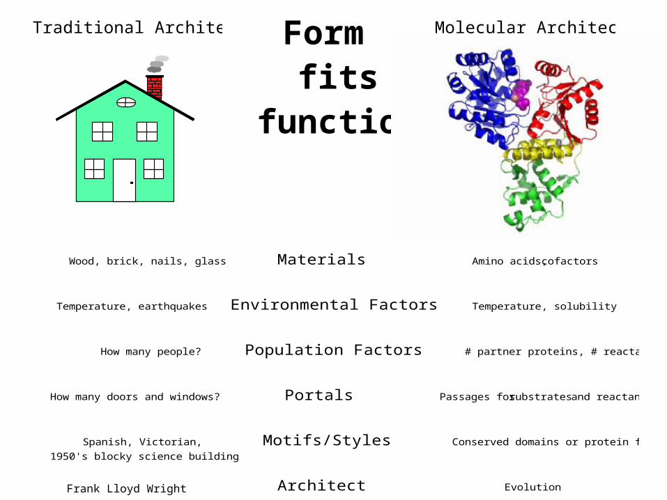
Wood, brick, nails, glass Materials Amino acids, cofactors
Temperature, earthquakes Environmental Factors Temperature, solubility
How many people? Population Factors # partner proteins, # reactants
How many doors and windows? Portals Passages for substrates and reactants
Spanish, Victorian, Motifs/Styles Conserved domains or protein folds
1950's blocky science building
Julia Morgan Architect Evolution
Traditional Architecture Molecular ArchitectureFormfits
function
Frank Lloyd Wright
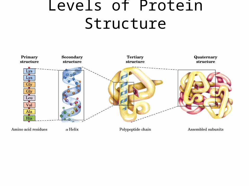
Levels of Protein Structure

Primary structure = order of amino acids in the protein chain
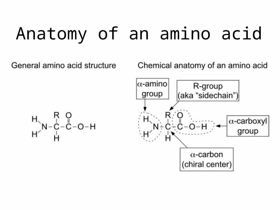
Anatomy of an amino acid
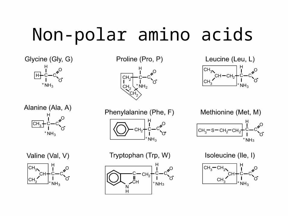
Non-polar amino acids
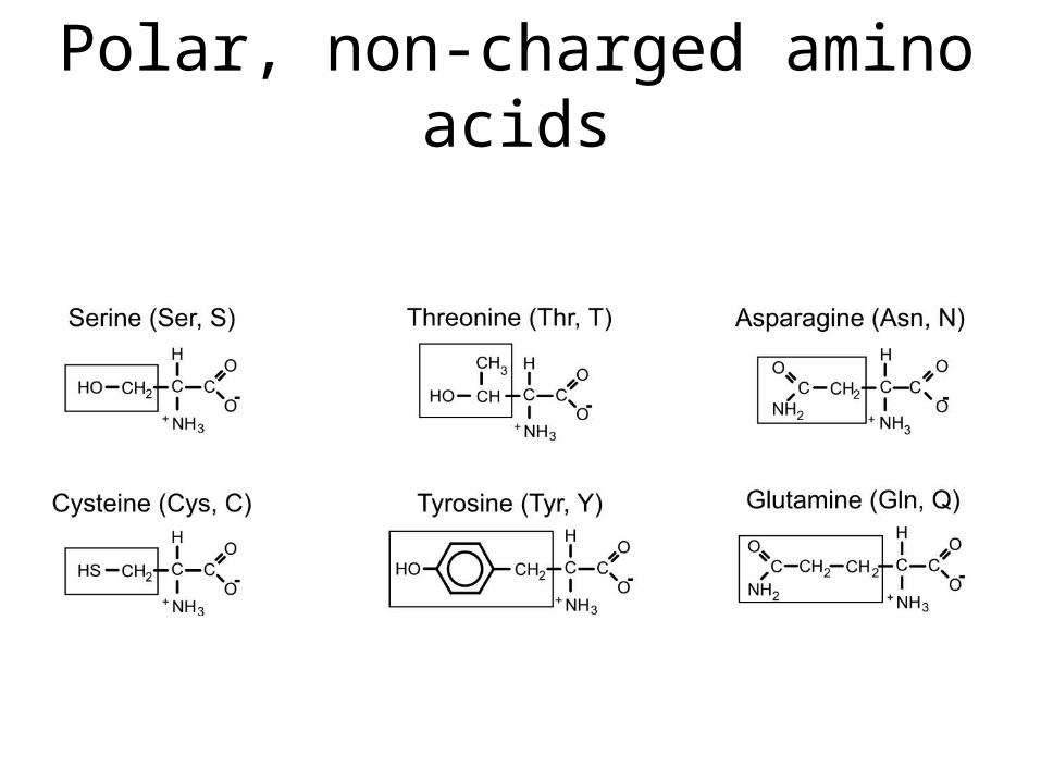
Polar, non-charged amino acids

Negatively-charged amino acids
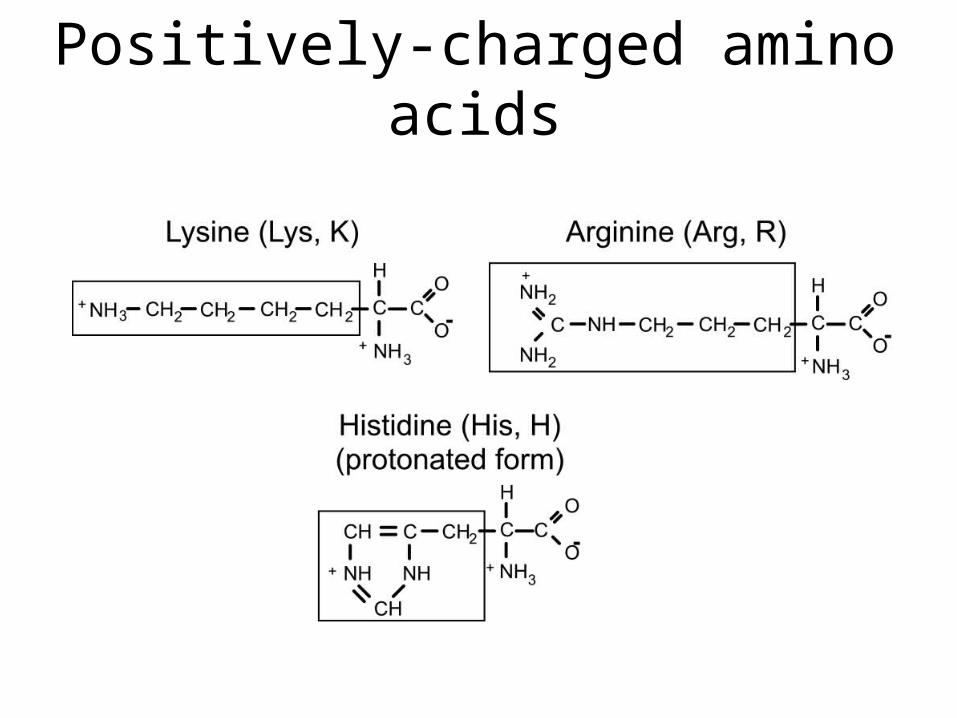
Positively-charged amino acids

Charged/polar R-groups generally map to surfaces on soluble
proteins
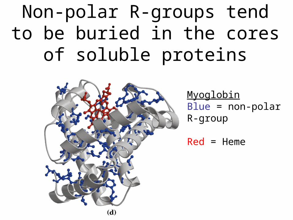
Non-polar R-groups tend to be buried in the cores of soluble
proteins
MyoglobinBlue = non-polarR-group
Red = Heme
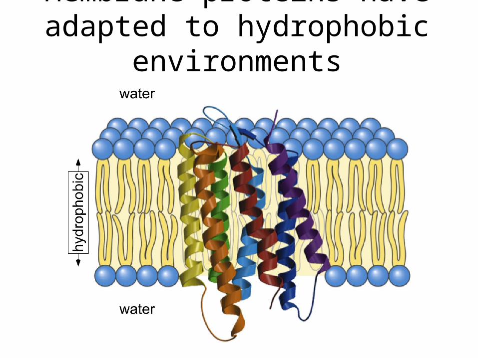
Membrane proteins have adapted to hydrophobic environments
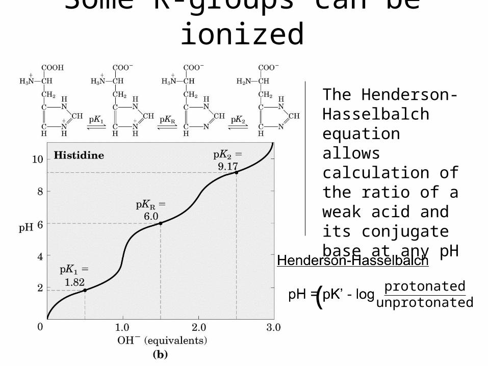
Some R-groups can be ionized
The Henderson-Hasselbalch equation allows calculation of the ratio of a weak acid and its conjugate base at any pH
protonatedunprotonated( )
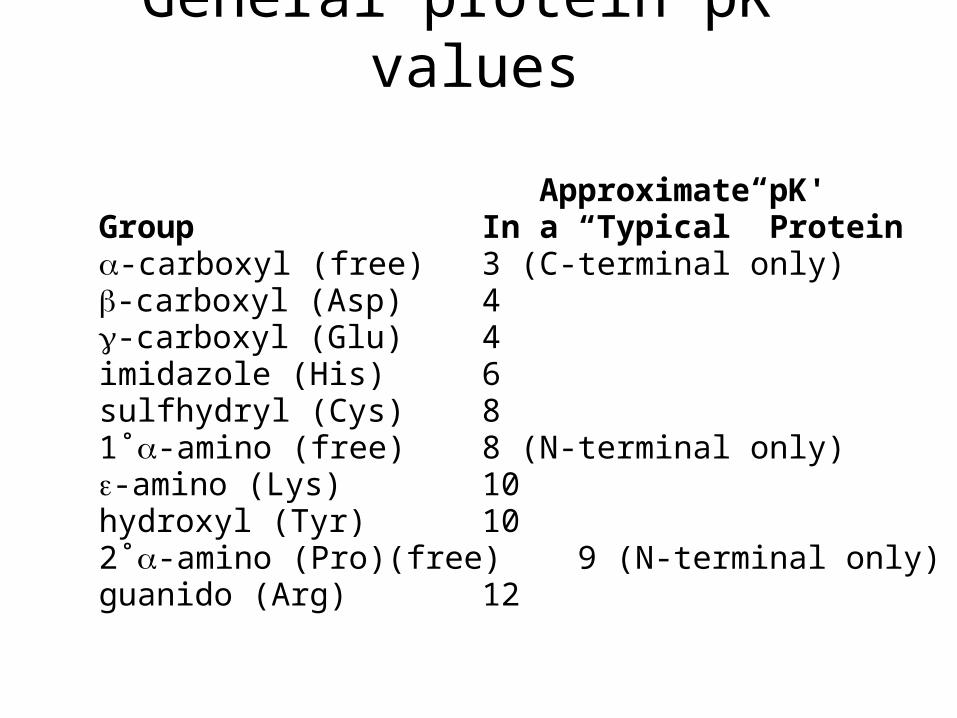
General protein pK’ values
Approximate pK'Group In a “Typical” Protein-carboxyl (free) 3 (C-terminal only)-carboxyl (Asp) 4-carboxyl (Glu) 4imidazole (His) 6sulfhydryl (Cys) 81˚-amino (free) 8 (N-terminal only)-amino (Lys) 10hydroxyl (Tyr) 102˚-amino (Pro)(free) 9 (N-terminal only)guanido (Arg) 12
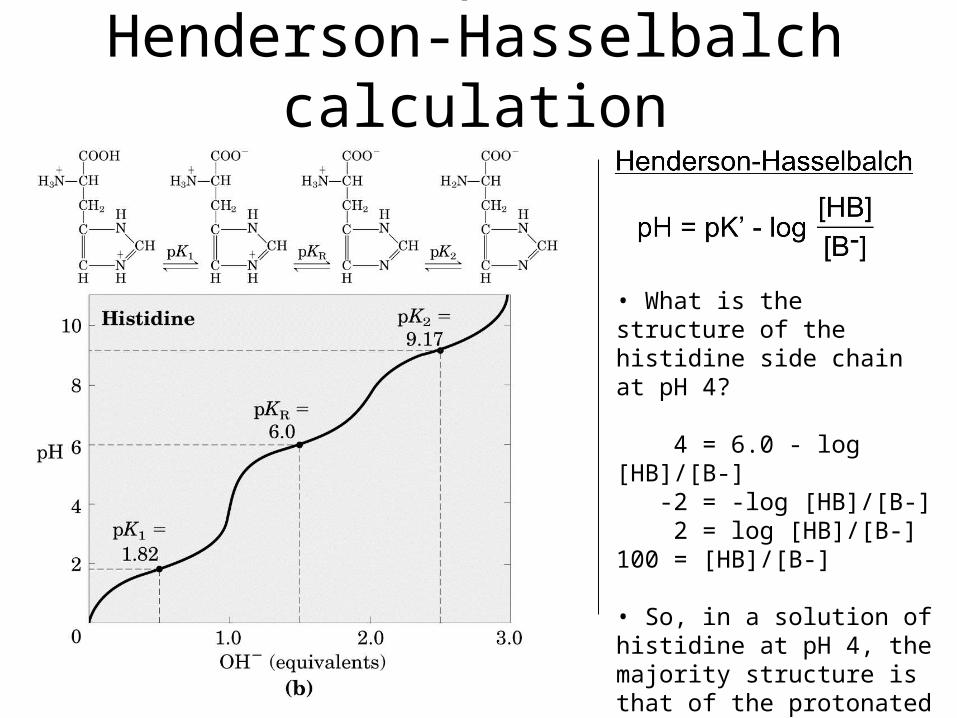
An example of a Henderson-Hasselbalch calculation
• What is the structure of the histidine side chain at pH 4?
4 = 6.0 - log [HB]/[B-] -2 = -log [HB]/[B-] 2 = log [HB]/[B-]100 = [HB]/[B-]
• So, in a solution of histidine at pH 4, the majority structure is that of the protonated form.
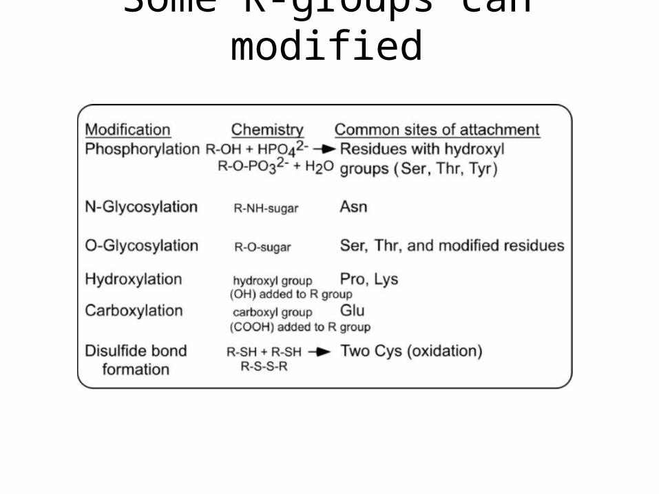
Some R-groups can modified
Amino Acids Are Joined By Peptide Bonds In Peptides
- -carboxyl of one amino acid is joined to -amino of a second amino acid (with removal of water)
- only -carboxyl and -amino groups are used, not R-group carboxyl or amino groups
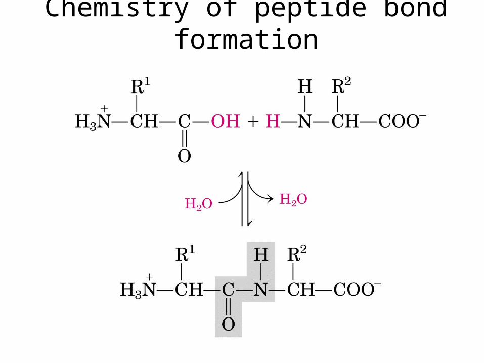
Chemistry of peptide bond formation
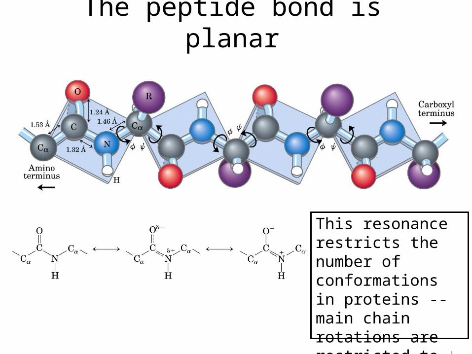
The peptide bond is planar
This resonance restricts the number of conformations in proteins -- main chain rotations are restricted to and
Primary sequence reveals important clues about a protein
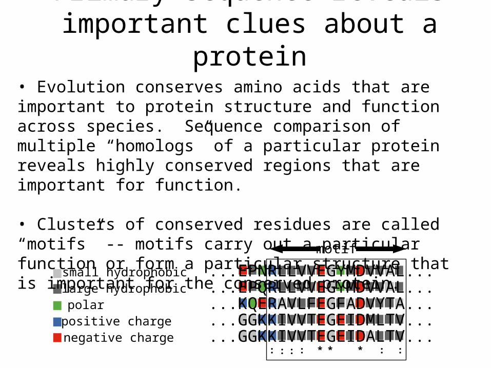
DnaG E. coli ...EPNRLLVVEGYMDVVAL...DnaG S. typ ...EPQRLLVVEGYMDVVAL...DnaG B. subt ...KQERAVLFEGFADVYTA...gp4 T3 ...GGKKIVVTEGEIDMLTV...gp4 T7 ...GGKKIVVTEGEIDALTV...
: *: :: * * : :
small hydrophobiclarge hydrophobicpolarpositive chargenegative charge
• Evolution conserves amino acids that are important to protein structure and function across species. Sequence comparison of multiple “homologs” of a particular protein reveals highly conserved regions that are important for function.
• Clusters of conserved residues are called “motifs” -- motifs carry out a particular function or form a particular structure that is important for the conserved protein.
motif

Generally only a limited amount of a protein’s surface is well conserved
Invariant (the residue is always the same, e.g. Asp)Conserved (the residue is generally similar, e.g. negatively charged)Not conserved (can be many different residues in different species)

Secondary structure = local folding of residues into regular patterns
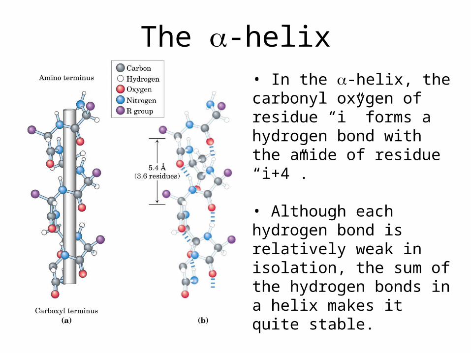
The -helix• In the -helix, the carbonyl oxygen of residue “i” forms a hydrogen bond with the amide of residue “i+4”.
• Although each hydrogen bond is relatively weak in isolation, the sum of the hydrogen bonds in a helix makes it quite stable.
• The propensity of a peptide for forming an -helix also depends on its sequence.

The -sheet • In a -sheet, carbonyl oxygens and amides form hydrogen bonds.
• These secondary structures can be either antiparallel (as shown) or parallel and need not be planar (as shown) but can be twisted.
• The propensity of a peptide for forming -sheet also depends on its sequence.
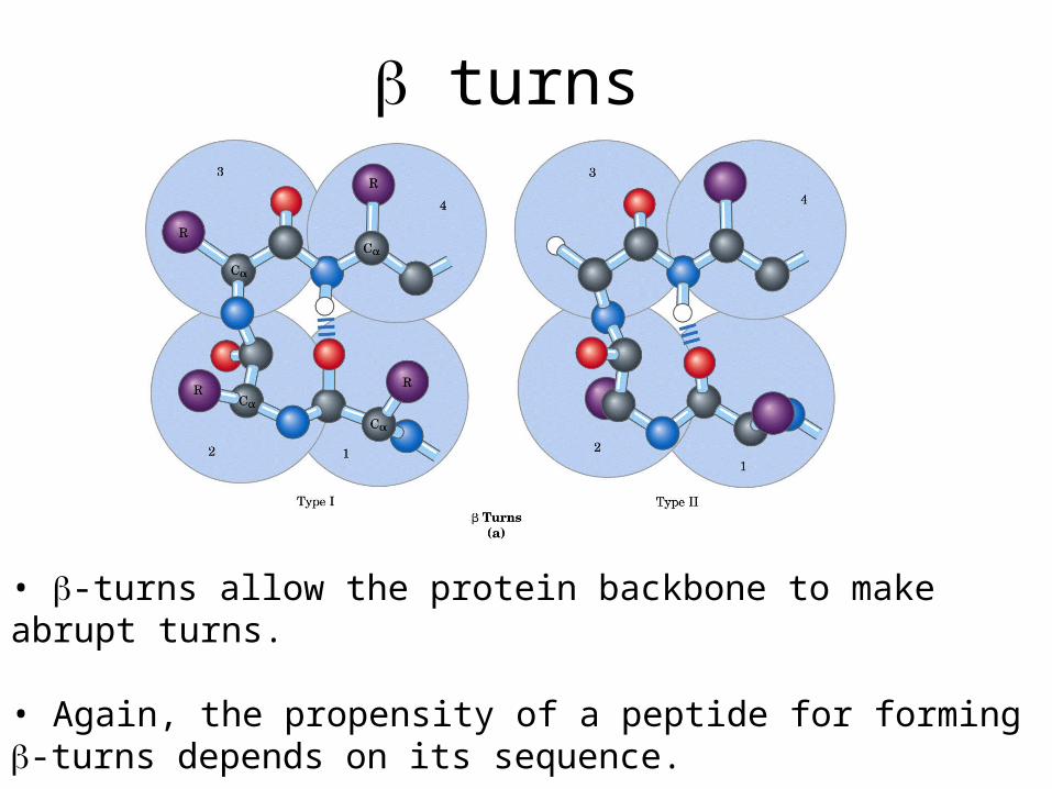
turns
• -turns allow the protein backbone to make abrupt turns.
• Again, the propensity of a peptide for forming -turns depends on its sequence.
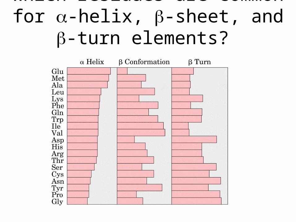
Which residues are common for -helix, -sheet, and -turn elements?
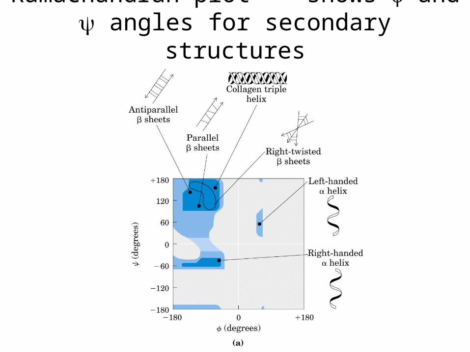
Ramachandran plot -- shows and angles for secondary structures
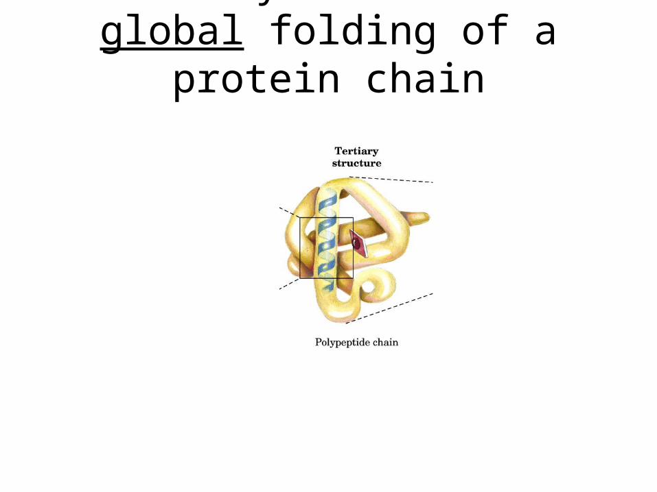
Tertiary structure = global folding of a protein chain
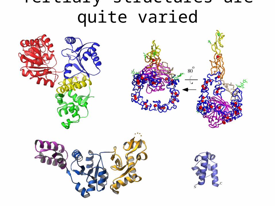
Tertiary structures are quite varied

Quaternary structure = Higher-order assembly of proteins
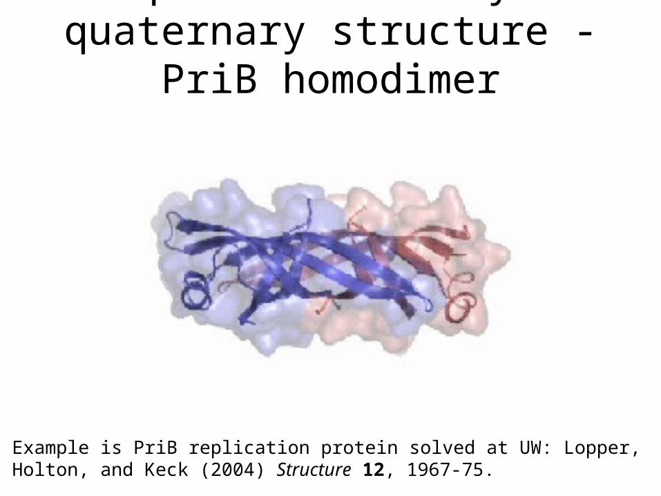
Example of tertiary and quaternary structure - PriB homodimer
Example is PriB replication protein solved at UW: Lopper, Holton, and Keck (2004) Structure 12, 1967-75.
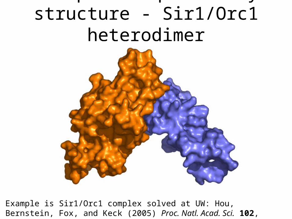
Example of quaternary structure - Sir1/Orc1 heterodimer
Example is Sir1/Orc1 complex solved at UW: Hou, Bernstein, Fox, and Keck (2005) Proc. Natl. Acad. Sci. 102, 8489-94.
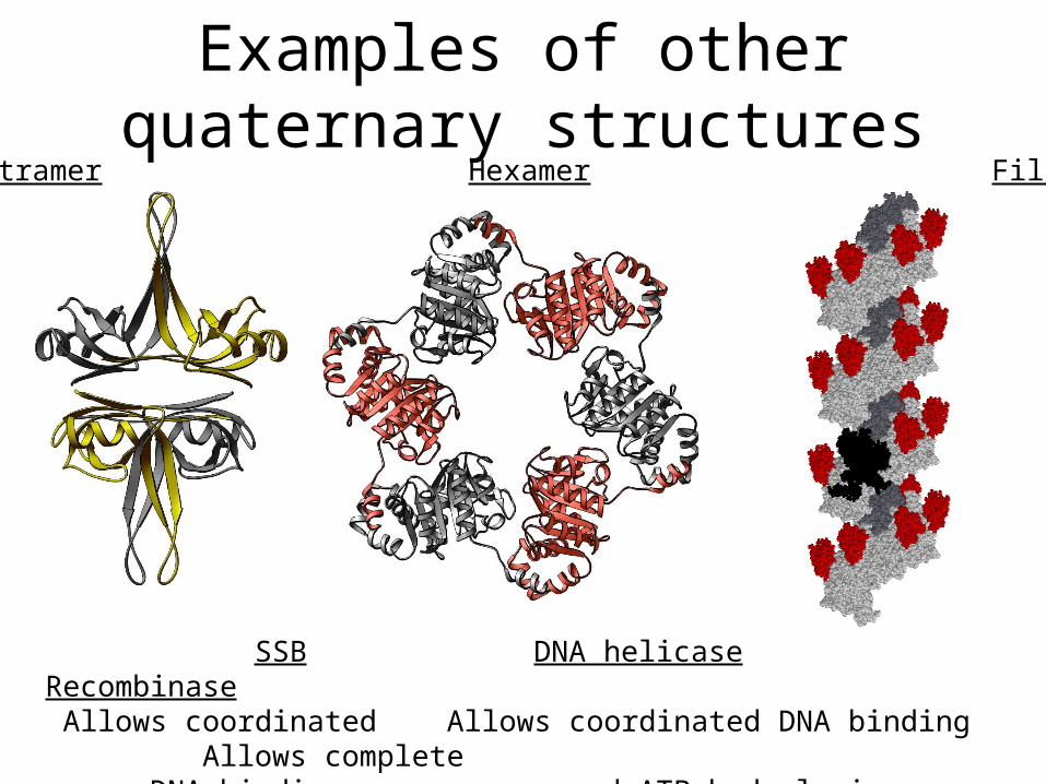
Examples of other quaternary structures
Tetramer Hexamer Filament
SSB DNA helicase Recombinase Allows coordinated Allows coordinated DNA binding Allows complete DNA binding and ATP hydrolysis coverage of an
extended molecule
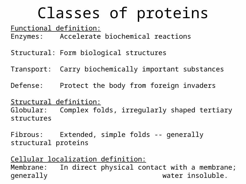
Classes of proteinsFunctional definition:Enzymes: Accelerate biochemical reactions
Structural: Form biological structures
Transport: Carry biochemically important substances
Defense: Protect the body from foreign invaders
Structural definition:Globular: Complex folds, irregularly shaped tertiary structures
Fibrous: Extended, simple folds -- generally structural proteins
Cellular localization definition:Membrane: In direct physical contact with a membrane; generally
water insoluble.
Soluble: Water soluble; can be anywhere in the cell.
Related Documents


