© 2014 College of American Pathologists. All rights reserved. 1 Welcome to CAP’s IVM Webinar Series sponsored by the In Vivo Microscopy Project Team This webinar on “How IVM Could Improve Our Practice as Pathologists” is presented by Lida P. Hariri, MD, PhD. Your host is Jill Kaufman, PhD. For comments about this webinar or suggestions for upcoming webinars, please contact Jill Kaufman at [email protected] THE WEBINAR WILL BEGIN MOMENTARILY. ENJOY!

Welcome message from author
This document is posted to help you gain knowledge. Please leave a comment to let me know what you think about it! Share it to your friends and learn new things together.
Transcript
© 2014 College of American Pathologists. All rights reserved. 1
Welcome to CAP’s IVM Webinar Series sponsored by
the In Vivo Microscopy Project Team
This webinar on “How IVM Could Improve Our Practice
as Pathologists” is presented by Lida P. Hariri, MD, PhD.
Your host is Jill Kaufman, PhD. For comments about this
webinar or suggestions for upcoming
webinars, please contact Jill Kaufman at
THE WEBINAR WILL BEGIN MOMENTARILY. ENJOY!

cap.org v. #
How IVM Could Improve Our Practice as
Pathologists Lida Hariri, MD, PhD, FCAP
February 27, 2014
In Vivo Microscopy
Webinar
3 © 2014 College of American Pathologists. All rights reserved.
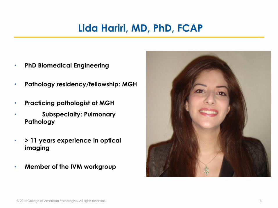
Lida Hariri, MD, PhD, FCAP
• PhD Biomedical Engineering
• Pathology residency/fellowship: MGH
• Practicing pathologist at MGH
• Subspecialty: Pulmonary
Pathology
• > 11 years experience in optical
imaging
• Member of the IVM workgroup

Disclaimer
© 2014 College of American Pathologists. All rights reserved. 4
The College does not permit reproduction of any substantial
portion of the material in this Webinar without its written
authorization. The College hereby authorizes attendees of the
CAP Webinar to use the PDF presentation solely for
educational purposes within their own institutions. The College
prohibits use of the material in the Webinar – and any
unauthorized use of the College’s name or logo – in
connection with promotional efforts by marketers of laboratory
equipment, reagents, materials, or services.
Opinions expressed by the speaker are the speaker’s own and
do not necessarily reflect an endorsement by CAP of any
organizations, equipment, reagents, materials or services used by participating laboratories.
Disclosure
• Dr. Hariri has no relevant financial relationships with
commercial interests to disclose
© 2014 College of American Pathologists. All rights reserved. 5

What is “In Vivo Microscopy (IVM)?”
Definition of IVM used by the CAP Workgroup:
A new field where microscopic images are obtained
in real time from living patients
Ex vivo applications of IVM:
• … where microscopic images are obtained in real
time from living cells or tissues
• Reagent-free, label-free or otherwise minimally
processed specimen
© 2014 College of American Pathologists. All rights reserved. 6

Bridging the Radiology/Pathology Divide
© 2014 College of American Pathologists. All rights reserved. 7

What can IVM provide us as pathologists? Our objectives
• Improve biopsy interpretation
o Guide biopsy site selection
o Additional virtual tissue volumes
• Assess tissue margins
o Ex vivo in frozen section
o In vivo to assess margins intraoperatively
• Assess tissues where it is unsafe to biopsy
o i.e. coronary arteries, eye pathology
© 2014 College of American Pathologists. All rights reserved. 8

Examples of Imaging Modalities that provide IVM
• Optical coherence tomography
• Photoacoustic tomography
• Confocal and multiphoton microscopy
• Spectroscopy
o Raman spectroscopy
o Near infrared spectroscopy
© 2014 College of American Pathologists. All rights reserved. 9

Optical Coherence Tomography
© 2014 College of American Pathologists. All rights reserved. 10
• Cross- sectional (x- z) imaging of tissue structure
• Similar to low power microscopy (4x objective)
• < 10 µm axial resolution (z)
• 10- 30 µm transverse resolution (x)
• < 3 mm penetration depth
• Non- destructive
• No transducing medium
Analagous to Ultrasound
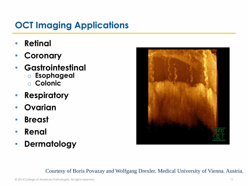
OCT Imaging Applications
• Retinal
• Coronary
• Gastrointestinal o Esophageal o Colonic
• Respiratory
• Ovarian
• Breast
• Renal
• Dermatology
Courtesy of Boris Povazay and Wolfgang Drexler. Medical University of Vienna. Austria.
© 2014 College of American Pathologists. All rights reserved. 11

Objective 1: Biopsy Guidance
IVM to improve biopsy interpretation
o Assess biopsy site selection during procedure to
reduce sampling error and increase diagnostic
yield
o Interpret volumetric imaging data sets as a form
of “virtual” tissue to accompany physical tissue
biopsies
© 2014 College of American Pathologists. All rights reserved. 12

Flexible imaging probe easily placed in standard 21-gauge TBNA needle
Tan KM et al. Biomed Opt Express. 3(8):1947-54. 2012
Needle-based OCT Probes
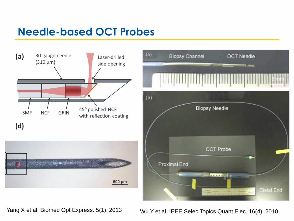
Wu Y et al. IEEE Selec Topics Quant Elec. 16(4). 2010
Needle-based OCT Probes
Yang X et al. Biomed Opt Express. 5(1). 2013

Kou WC et al. Biomed Opt Express. 3(6). 2012
Vacuum-assisted OCT Needle Biopsy Probe

http://www.spectrascience.com/
Forceps OCT biopsy probes
Song C. BMOES. 4(7). 2013

1. Volumetric OCT Acquisition 3. Target election
4. Laser Marking
5. Endoscopy with Biopsy Acquisition and
Histopathologic Analysis of Biopsy Sites
2. Image Assessment and
Interpretation
OCT Endoscopy: Guided Biopsy for Barrett’s Esophagus
Suter et al. Gastrointest Endosc. 71(2). 2010.

A work in progress that exemplifies the
need for pathologists and optical
engineers to collaborate
OCT guided biopsy of lung nodules
© 2014 College of American Pathologists. All rights reserved. 18

Transthoracic needle aspiration:
High diagnostic yield
Increased risk of pneumothorax
http://library.bjmu.edu.cn
Transbronchial needle
aspiration:
Lower risk of pneumothorax
Variable diagnostic yield
www.olympus.es
Biopsy of Pulmonary Nodules
© 2014 College of American Pathologists. All rights reserved. 19

Endobronchial
Ultrasound
Electromagnetic
Navigation:
Currently Used Guidance Techniques
Diagnostic Yield is still
low for lesions < 3.0 cm
© 2014 College of American Pathologists. All rights reserved. 20

Biopsy Triage!
Courtesy of Dr. Kevin Leslie, Mayo Clinic, Scottsdale, AZ
“To Pathology Lab”

© 2014 College of American Pathologists. All rights reserved. 22
Needle Biopsy of Lung Nodules
Target Lung Nodule
Miss Nodule
Normal Parenchyma
Normal Airway
Hit Nodule
Tumor: Diagnostic!
Necrosis: Not Diagnostic
Fibrosis: Not Diagnostic
Animations Courtesy of Dr. Alex Chee, University of Calgary

© 2014 College of American Pathologists. All rights reserved. 23
Needle Biopsy of Lung Nodules
Target Lung Nodule
Miss Nodule
Normal Parenchyma
Normal Airway
Hit Nodule
Tumor: Diagnostic!
Necrosis: Not Diagnostic
Fibrosis: Not Diagnostic
Animation Courtesy of Dr. Alex Chee, University of Calgary
We need a high resolution imaging modality to:
Complement EBUS and ENB
Assess the needle position after placement
Give immediate feedback about placement site
So what is missing in biopsy guidance?

OCT Biopsy Guidance in Lung Nodules
© 2014 College of American Pathologists. All rights reserved. 25
Hariri LP et al. J Vis Exp. 71. 2013

Flexible imaging probe easily placed in standard 21-gauge TBNA needle
Tan KM et al. Biomed Opt Express. 3(8):1947-54. 2012
Needle-based OCT Probe for Bronchoscopy

Hariri LP et al. Chest. 144(4). 2013
Needle-based OCT: Lung Parenchyma

Nodule Mixed Parenchym
a
Needle-based OCT: Lung Nodule
Hariri LP et al. Chest. 144(4). 2013

Differentiating nodules from parenchyma with OCT
© 2014 College of American Pathologists. All rights reserved. 29
Hariri LP et al. Chest. 144(4). 2013
High Sensitivity and Specificity
(> 95%) for all readers:
Pathologists, Pulmonologists, OCT Experts

Structural OCT can differentiate tumor from
airway, parenchyma, and necrosis.
Cannot differentiate solid tumor from fibrosis
Hariri LP et al. AJRCCM. 187(2):125-9. 2013
Needle-based OCT: Once you are in the nodule…
Parenchyma
Necrosis
Solid Tumor
Fibrosis
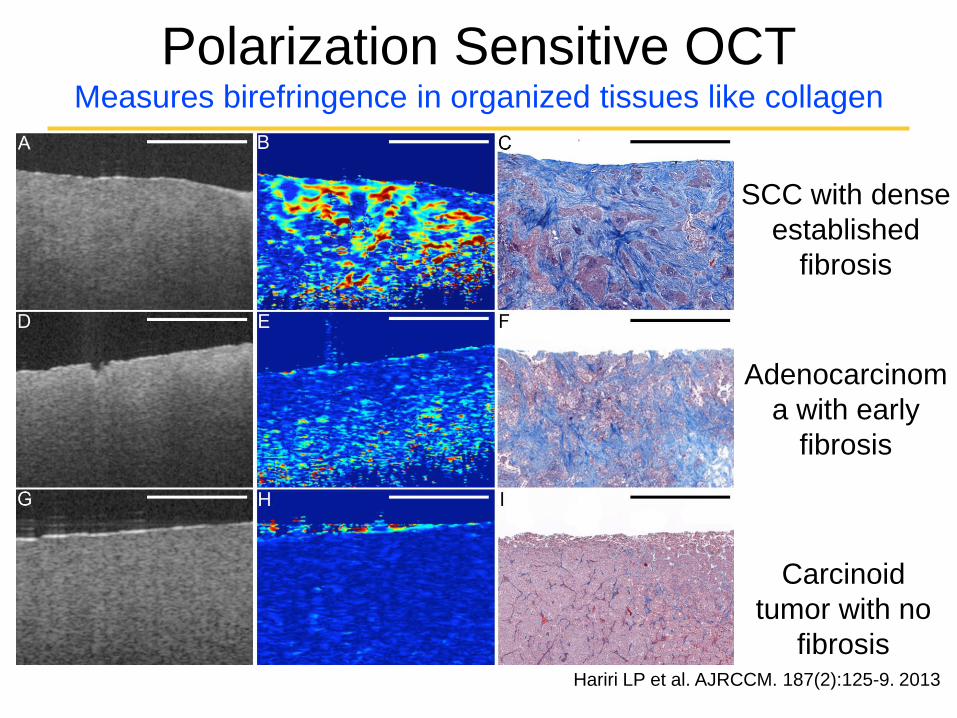
Polarization Sensitive OCT Measures birefringence in organized tissues like collagen
Hariri LP et al. AJRCCM. 187(2):125-9. 2013
SCC with dense
established
fibrosis
Adenocarcinom
a with early
fibrosis
Carcinoid
tumor with no
fibrosis
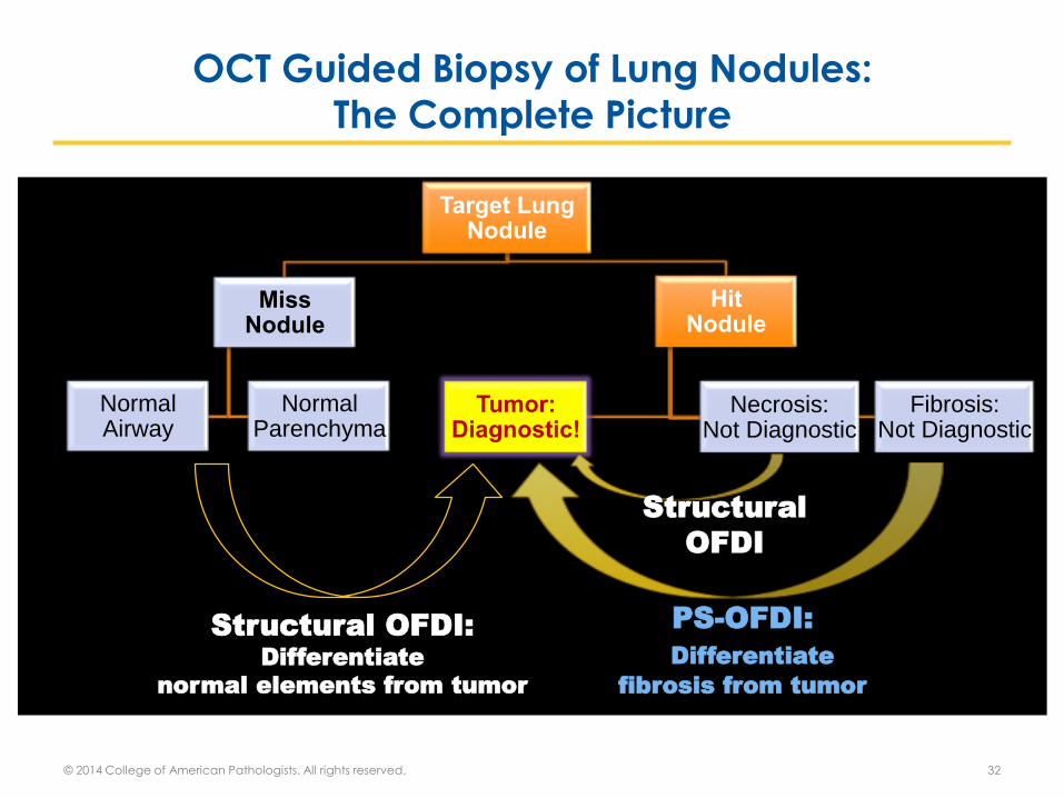
OCT Guided Biopsy of Lung Nodules: The Complete Picture
© 2014 College of American Pathologists. All rights reserved. 32
Target Lung Nodule
Miss Nodule
Normal Parenchyma
Normal Airway
Hit Nodule
Tumor: Diagnostic!
Necrosis: Not Diagnostic
Fibrosis: Not Diagnostic
Structural OFDI:
Differentiate
normal elements from tumor
Structural
OFDI
PS-OFDI:
Differentiate
fibrosis from tumor

Biopsy Guidance: What this means for pathology
Performed by pulmonologist, but will aid pathologist
• Target lung nodules with needle-based OCT in vivo
during bronchoscopic biopsy
• Use OCT to increase tumor yield for pathology
• Pathologist acts as consultant for difficult cases
Performed by pathologist
• Assess “virtual tissue volumes” as a complement to
standard biopsy to aid diagnostics
© 2014 College of American Pathologists. All rights reserved. 33

Z = 6.1 cm
1. OCT provides views of tissue microarchitecture
comparable to low power (4x) microscopy
2. Tissue volumes are orders of magnitude larger than biopsy
Large volume “virtual” tissue to accompany biopsy

3D Reconstruction: Cartilaginous Hamartoma

Objective 2: Tissue margins
IVM to assess tissue margins
o Ex vivo to assess margins in frozen section
o In vivo to assess margins intraoperatively
© 2014 College of American Pathologists. All rights reserved. 36

Full-Field OCT to assess
lung carcinoma
© 2014 College of American Pathologists. All rights reserved. 37

Full-Field OCT
© 2014 College of American Pathologists. All rights reserved. 38 Jain M. J Path Inform. 4(26). 2013

Full Field OCT: Normal Lung
© 2014 College of American Pathologists. All rights reserved. 39 Jain M. J Path Inform. 4(26). 2013

Full Field OCT: Normal Lung
© 2014 College of American Pathologists. All rights reserved. 40 Jain M. J Path Inform. 4(26). 2013

Full Field OCT: Lung Adenocarcinoma
© 2014 College of American Pathologists. All rights reserved. 41 Jain M. J Path Inform. 4(26). 2013

Full-Field OCT in the frozen section lab
• FFOCT as an adjunct to frozens for intra-operative
consultation
o Surgical margin assessment
• FFOCT to assess adequacy of biopsy material in
freshly excised tissue
• FFOCT in bio-banking to confirm tumor is present
before cryopreservation
© 2014 College of American Pathologists. All rights reserved. 42
Jain M. J Path Inform. 4(26). 2013

IVM to Assess
Breast Excision Margins
© 2014 College of American Pathologists. All rights reserved. 43

Assessing Breast Excision Margins with OCT
© 2014 College of American Pathologists. All rights reserved. 44
Nguyen FT et al. Cancer Res. 69. 2009.

© 2014 College of American Pathologists. All rights reserved. 45
Nguyen FT et al. Cancer Res. 69. 2009.
Breast Excision Margins with OCT: Negative Margin

© 2014 College of American Pathologists. All rights reserved. 46
Nguyen FT et al. Cancer Res. 69. 2009.
Breast Excision Margins with OCT: Positive Margins

Intraop margin assessment with OCT: Clinical Trial
• Multi-center, randomized blinded clinical trial
• Intraoperative imaging in partial mastectomy:
o Excised breast margins
o In vivo surgical cavity
• Compare surgical re-excision rates between
standard of care partial mastectomy and
intraoperative imaging with partial mastectomy
© 2014 College of American Pathologists. All rights reserved. 47
Jacobs LK et al.. Cancer Research. 72(24). Abstract. 2012

Objective 3: Tissue that cannot be biopsied
IVM to assess tissues where it is unsafe to biopsy
i.e. coronary arteries, eye pathology
© 2014 College of American Pathologists. All rights reserved. 48

IVM in Coronary
Artery Pathology
© 2014 College of American Pathologists. All rights reserved. 49
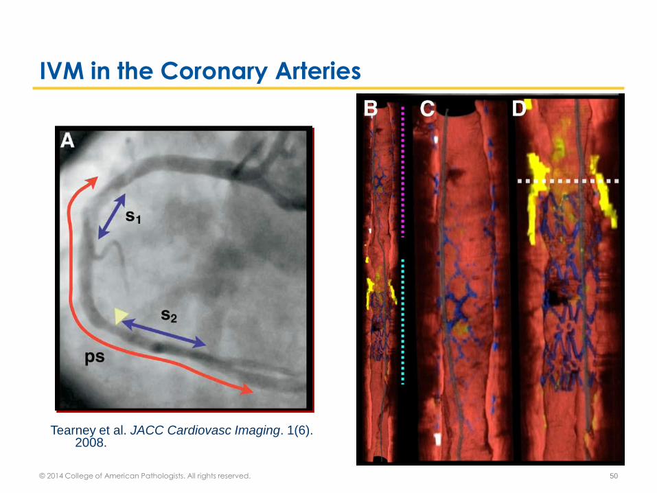
IVM in the Coronary Arteries
© 2014 College of American Pathologists. All rights reserved. 50
Tearney et al. JACC Cardiovasc Imaging. 1(6). 2008.

Witnessed Plaque Rupture with OCT
© 2014 College of American Pathologists. All rights reserved. 51
Gonzalo N et al. JACC Cardiovasc Imaging. 4(4). 2011.

IVM in Retinal Pathology
© 2014 College of American Pathologists. All rights reserved. 52

IVM in Retinal Pathology
© 2014 College of American Pathologists. All rights reserved. 53
Drexler W and Fujimoto J. Progress in Retinal and Eye Research. 27(1).
2008

IVM in Retinal Pathology: Macular Hole
© 2014 College of American Pathologists. All rights reserved. 54
Srinivasan VJ et al. Opthamology. 113(11). 2006
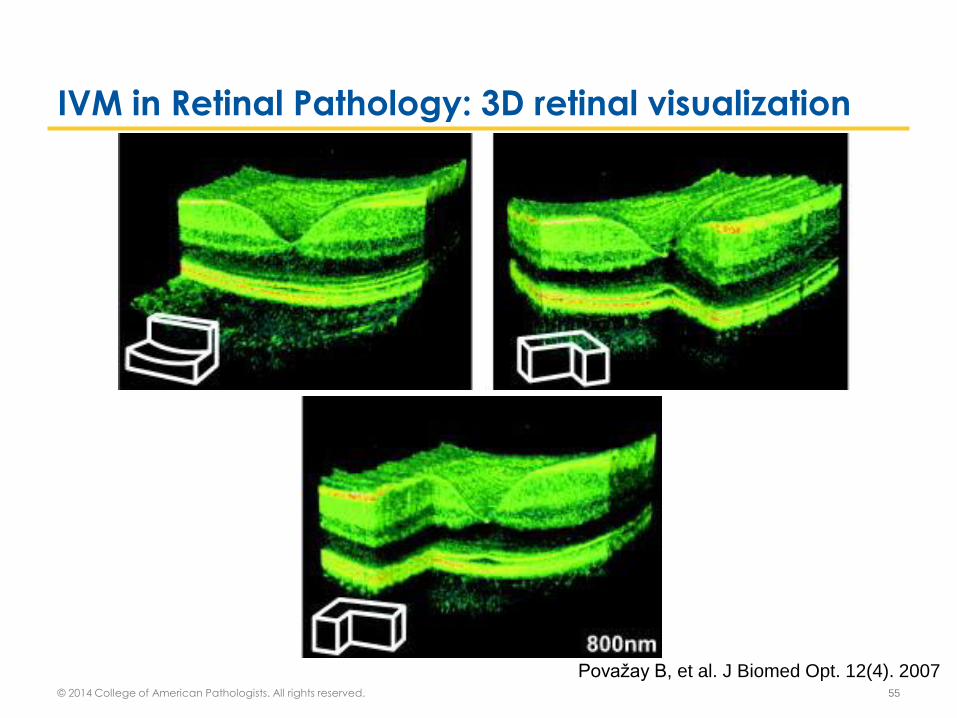
IVM in Retinal Pathology: 3D retinal visualization
© 2014 College of American Pathologists. All rights reserved. 55
Považay B, et al. J Biomed Opt. 12(4). 2007

IVM in Retinal Pathology: Commercial System
© 2014 College of American Pathologists. All rights reserved. 56
http://buea.net/services-offered/retina/

© 2014 College of American Pathologists. All rights reserved. 57
Ultra-High Resolution OCT

Micro-OCT: Atherosclerotic Plaque
© 2014 College of American Pathologists. All rights reserved. 58
Liu L, et al. Nat Med. 17(8). 2011

Optical Coherence Microcopy_ Normal Kidney
© 2014 College of American Pathologists. All rights reserved. 59
Lee HC, et al. Biomedical Optics Express. 4(8). 2013

The Role of the Pathologist
• OCT provides high resolution architectural images
similar to histopathology
• Pathologists already have strengths in
o Interpreting high magnification/resolution microscopy
o Pattern recognition
o Understanding of pathology entities
− Histological features
− Differential diagnosis
Pathologists are well suited to interpret high resolution
imaging as an adjunct to histopathology
© 2014 College of American Pathologists. All rights reserved. 60

No
• Sensitivity/specificity in tested applications not 100%
• Resolution not high enough
• You are there, take a biopsy!
o Histology is the gold standard
o Differential diagnosis in many scenarios is vast
o Molecular testing
© 2014 College of American Pathologists. All rights reserved. 61
“Will IVM replace traditional pathology?”
“IVM seems like a clinician’s tool- why should I care about it?”
1) We are the end beneficiaries of IVM:
• IVM has a lot of potential to increase the quality of
our tissue samples
o Guided biopsy sampling- Barrett’s esophagus
o Increased tumor yield- Lung nodule biopsy
• Our expertise as pathologists is needed to help
identify applications where IVM can make big
impacts
© 2014 College of American Pathologists. All rights reserved. 62

“IVM seems like a clinician’s tool- why should I care about it?”
2) IVM assessments will become more complex:
• IVM is pretty new and so far, its assessments are
straightforward
o Tumor versus non-tumor elements
o Diagnosis where there are few options
• As IVM applications develop, the complexity of
interpreting IVM images will also develop
o Will require knowledge pathologists already have:
Histologic features, differential diagnosis, etc
© 2014 College of American Pathologists. All rights reserved. 63

“IVM seems like a clinician’s tool- why should I care about it?”
3) IVM needs a defined expert:
• For example, many clinicians can assess CT scans
but that does not make them experts in radiology
• Similarly, many clinicians may use and interpret IVM
• IVM is in essence a form of microscopy, and as such
pathologists are the obvious choice as IVM experts
© 2014 College of American Pathologists. All rights reserved. 64

Ex vivo
Assess adequacy of tissue biopsy- Increase Tumor Yield
Intraoperative consult- Part of frozen section assessment
Guide tissue sampling in the grossing room
Scenarios of IVM in Pathology
© 2014 College of American Pathologists. All rights reserved. 65

In vivo
Real-time diagnosis in endoscopy or interventional suite
- Pathologist present during procedure
- At remote site using viewing workstation
As Part of Sign-out
Pathologist views images off-line after procedure
Interprets images as a complement to standard
histology (particularly in tissue biopsy)
Scenarios of IVM in Pathology
© 2014 College of American Pathologists. All rights reserved. 66

How can we get involved as pathologists?
Pathologist inherently have the skills needed to become IVM experts,
but we have to take the reigns.
• Identify clinical scenarios where IVM can make impacts
• Participate in ex vivo and in vivo validation studies
• Be key players in instituting and interpreting high resolution imaging as part of our pathology practice
© 2014 College of American Pathologists. All rights reserved. 67
Email: [email protected]

© 2014 College of American Pathologists. All rights reserved. 68

Save the Date for These Upcoming
FREE IVM Webinars or Listen to past Webinars
3 © 2014 College of American Pathologists. All rights reserved.
• Upcoming Webinars
o Ex vivo pathology applications of IVM: Cooler than Frozen
o October 23 at 11 am Central
o Richard M. Levenson, MD, FCAP
• Archived Webinars
o Ex Vivo Applications of IVM: Shedding a different light on cells and
tissue
o Chapter 7 from CAP eBook: New Paths...New Choices: Pathology in
an Era of Advancing Science and Disruptive Health Economics
Register for any upcoming or archived webinars by going to
cap.org/webinars

• created to assist pathologists who are considering
providing or developing in vivo microscopy skills
and services within the next 24 months
• free for members
• available via registration on the Member tab of
www.cap.org or through this link
CAP’s Pathology IVM Resource Guide
Printed Versions Now Available
© 2014 College of American Pathologists. All rights reserved. 4

The CAP has created the Pathology Resource Guides, a tool to assist
pathologists in understanding key emerging technologies. These
Resource Guides are a new CAP member benefit available at no
charge. Printed versions of the Resource Guides are available to
members and non-members.
Molecular Diagnostic (single gene, small panel)
Genomic Analysis (large panels, exome, genome)
Digital Pathology
Register through the CAP member tab. You will receive periodic
updates for two years.
Questions? Contact [email protected].
Other CAP’s Pathology Resource Guides
Printed Versions Now Available
5 © 2014 College of American Pathologists. All rights reserved.

• Pathology SPECs are:
o Prewritten PowerPoint presentation on emerging topics where molecular
testing plays a key role in patient management.
o Designed for pathologists to customize and use for educating other
physicians and health care leaders in their communities.
o Focused on molecular tests that are actionable to patient care today.
• Now Available:
― Emerging Concepts in the Workup of Colorectal Cancer
― Emerging Concepts in Therapeutic Guidance for Metastatic Melanoma
― Emerging Concepts in the Diagnosis and Workup of Thyroid Cancer
― Emerging Concepts in Colorectal Cancer Hereditary Non-Polyposis
Cancer (Lynch Syndrome)
― Emerging Concepts in the Workup of Polycythemia and
Thrombocythemia: JAK2
• To register, go to the CAP Member tab on cap.org. You do not need to be a
member to utilize this free tool.
A New CAP Tool- Short Presentations On Emerging
Concepts (SPECs)
© 2014 College of American Pathologists. All rights reserved. 6

© 2014 College of American Pathologists. All rights reserved. 7

THANK YOU!
Thank you for attending our webinar
“How IVM Could Improve Our Practice as Pathologists” by
Lida Hariri, MD, PhD, FCAP.
For comments about this webinar
or suggestions for upcoming
webinars, please contact
Jill Kaufman, PhD,
Director of Personalized Health Care at [email protected]
NOTE: There is no CME/CE credit available for
today’s free webinar.
8 © 2014 College of American Pathologists. All rights reserved.
Related Documents