Copyright © 2011 Wolters Kluwer Health | Lippincott Williams & Wilkins Chapter 43 Disorders of the Skeletal System: Trauma, and Infections (Through page 1112)

Welcome message from author
This document is posted to help you gain knowledge. Please leave a comment to let me know what you think about it! Share it to your friends and learn new things together.
Transcript

Copyright © 2011 Wolters Kluwer Health | Lippincott Williams & Wilkins
Chapter 43
Disorders of the Skeletal System:
Trauma, and Infections
(Through page 1112)

Copyright © 2011 Wolters Kluwer Health | Lippincott Williams & Wilkins
Common Joint Injuries
• Injuries to muscles: strains
• Injuries to ligaments: sprains, ruptures
• Injuries to tendons: rotator cuff injuries
• Injuries to bone surfaces
– Joint dislocations
– Patellar dislocation
– Loose bodies
– Meniscus injuries
– Chondromalacia

Copyright © 2011 Wolters Kluwer Health | Lippincott Williams & Wilkins
Common Joint Injuries (cont.)
• Rotator cuff injuries
– Clavicle fractures
– Dislocations
– Bursa damage
– Torn tendons
• Hip injuries
– Dislocation
– Fracture
Copyright © 2011 Wolters Kluwer Health | Lippincott Williams & Wilkins
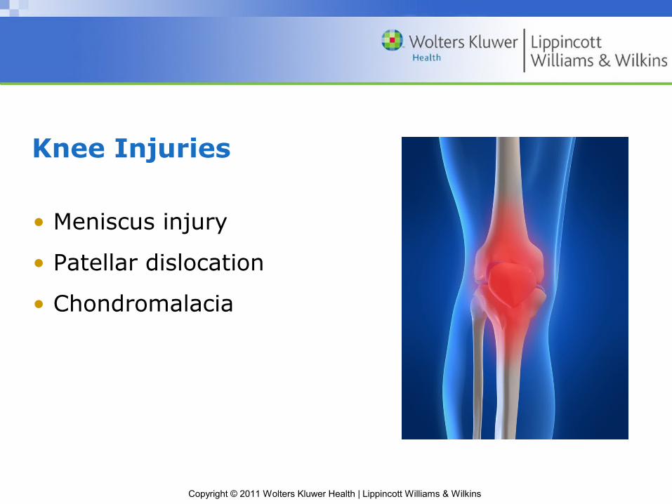
Knee Injuries
• Meniscus injury
• Patellar dislocation
• Chondromalacia

Copyright © 2011 Wolters Kluwer Health | Lippincott Williams & Wilkins
Question
True or false.
Chondromalacia is the most common type of knee injury.

Copyright © 2011 Wolters Kluwer Health | Lippincott Williams & Wilkins
Answer
False
The most common knee injuries are tear of the ACL (anterior cruciate ligament), caused by hyperextension, and damage to the meniscus.

Copyright © 2011 Wolters Kluwer Health | Lippincott Williams & Wilkins
Fractures
• Transverse
• Oblique
• Spiral
• Comminuted
• Segmental
• Butterfly
• Impacted
Copyright © 2011 Wolters Kluwer Health | Lippincott Williams & Wilkins
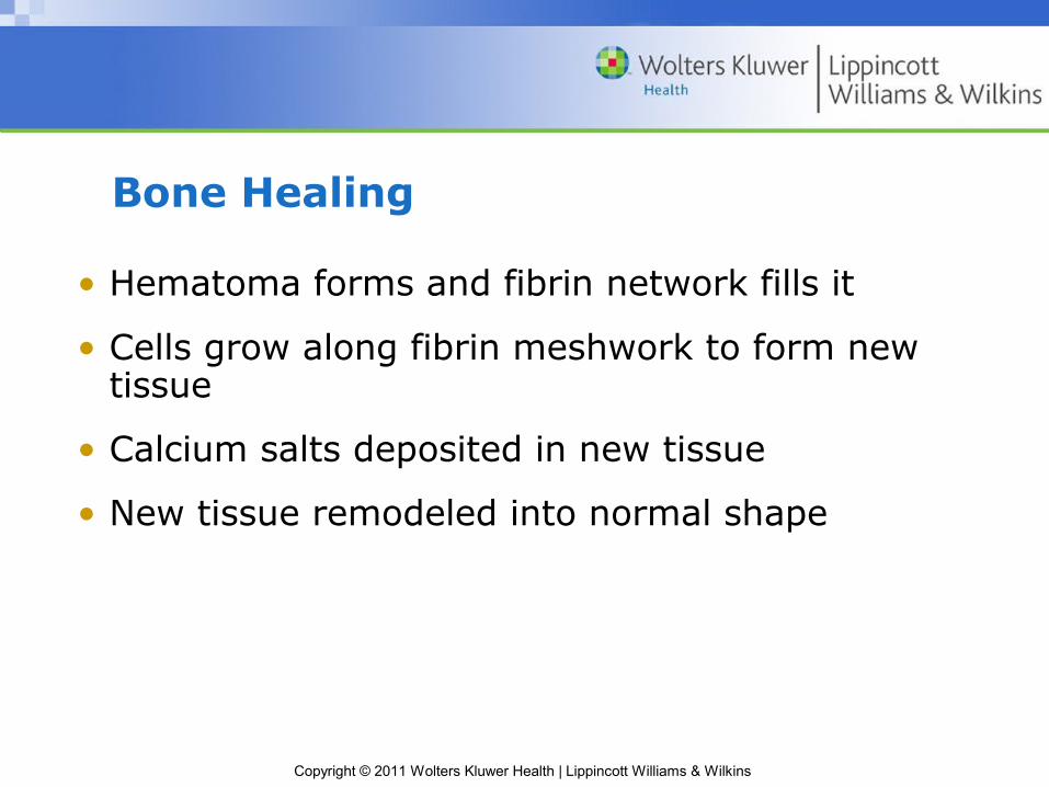
Bone Healing
• Hematoma forms and fibrin network fills it
• Cells grow along fibrin meshwork to form new tissue
• Calcium salts deposited in new tissue
• New tissue remodeled into normal shape
Copyright © 2011 Wolters Kluwer Health | Lippincott Williams & Wilkins
Question
Which of the following represents the correct sequence of bone healing?
a. Hematoma – cartilage – bone
b. Hematome – elastin – fibrocartilage
c. Cartilage – spongy bone – compact bone
d. Hemangioma – spongy bone – compact bone
Copyright © 2011 Wolters Kluwer Health | Lippincott Williams & Wilkins
Answer
a. Hematoma – cartilage – bone
Following a fracture, a hematoma forms; collagen and cartilage are deposited (soft callus); bone tissue is ossified (hard callus).

Copyright © 2011 Wolters Kluwer Health | Lippincott Williams & Wilkins
Complications Resulting from Soft Tissue Injury
• Skin injury: fracture blisters
• Muscle injury and swelling: compartment syndrome
• Nerve injury: reflex sympathetic dystrophy
• Adipose tissue or bone marrow: fat emboli

Copyright © 2011 Wolters Kluwer Health | Lippincott Williams & Wilkins
Osteomyelitis
• Infection of bone
– Direct contamination
– Contamination through blood (hematogenous)
º Miliary tuberculosis
– Contamination from skin lesions

Copyright © 2011 Wolters Kluwer Health | Lippincott Williams & Wilkins
Hematogenous Osteomyelitis
• In children:
– Affects long bones
– Purulent exudate inside bone
– Damages arteries to bone
– May penetrate skin or involve joints
• In adults:
– In vertebrae, sternoclavicular and sacroiliac joints, or pubic symphysis
– Tends to affect joint space
Copyright © 2011 Wolters Kluwer Health | Lippincott Williams & Wilkins
Chronic Osteomyelitis
• Areas of dead bone develop
– Sequestrum = infected dead bone
• Are surrounded by new bone
– Involcrum bone forms around the dead bone
• Identified by x-ray, bone scans
Copyright © 2011 Wolters Kluwer Health | Lippincott Williams & Wilkins
Osteonecrosis
• Caused by ischemia to bone due to:
– Bone injury
– Thrombosis or embolism
– Vessel injury
– Compartment syndrome inside bone (increased intraosseous pressure)
– Corticosteroid associated

Copyright © 2011 Wolters Kluwer Health | Lippincott Williams & Wilkins
Question
Which type of bone disorder is associated with thrombus formation or embolus?
a. Hematogenous osteomyelitis
b. Chronic osteomyelitis
c. Osteonecrosis
d. Osteoinfarction

Copyright © 2011 Wolters Kluwer Health | Lippincott Williams & Wilkins
Answer
c. Osteonecrosis
Bone has an extensive blood supply (remember, it can’t rely on diffusion of nutrients and gases like cartilage can). When blood flow to an area of osseous tissue is disrupted because of a clot/thrombus or an embolus (a clot that has broken off and traveled), the tissue dies (necrosis).
Related Documents