Airway Management

Welcome message from author
This document is posted to help you gain knowledge. Please leave a comment to let me know what you think about it! Share it to your friends and learn new things together.
Transcript

Airway Management
“The best preparation for managing the difficult airway is
being excellent at the management of routine airways.
Patients are not harmed by inadequate intubation but rather
inadequate ventilation”Nagelhout 4th ed. p. 441

Anatomy and Physiologyof the Airway
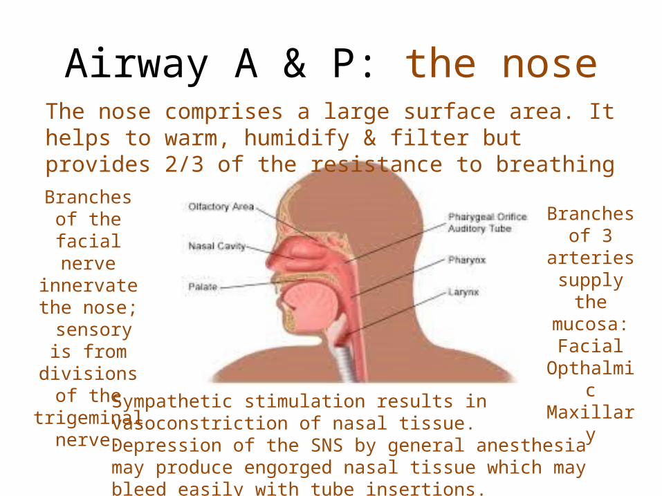
Airway A & P: the noseThe nose comprises a large surface area. It helps to warm, humidify & filter but provides 2/3 of the resistance to breathing
Branches of 3 arteries supply the mucosa:
FacialOpthalmicMaxillary
Branches of the facial nerve
innervate the nose;
sensory is from divisions of the
trigeminal nerve.
Sympathetic stimulation results in vasoconstriction of nasal tissue. Depression of the SNS by general anesthesia may produce engorged nasal tissue which may bleed easily with tube insertions.

Airway A & P: the mouthThe hard palate is
stationary.
The soft palate is able to rise, but also may become more
movable (obesity,age..) and
fall against the nasal passages during sleep producing
obstruction.
The uvula protects the oropharynx.
The large, space
occupying, muscular
tongue may obstruct the airway when
it relaxes.
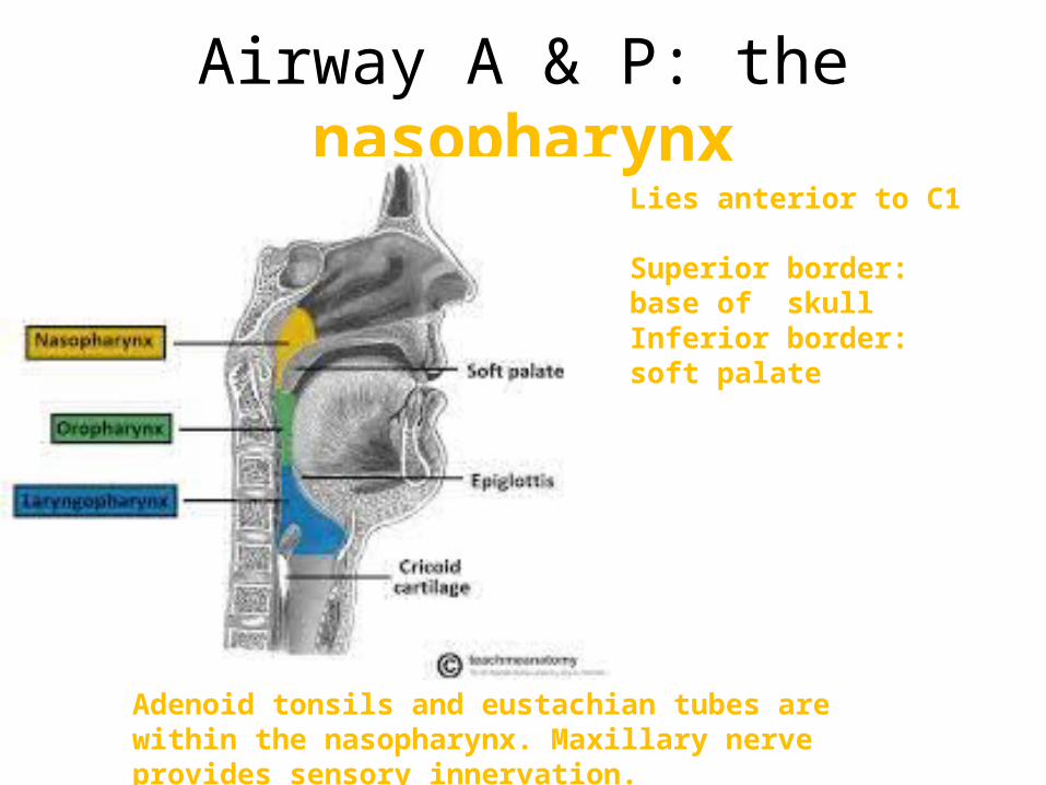
Airway A & P: the nasopharynxLies anterior to C1
Superior border: base of skullInferior border: soft palate
Adenoid tonsils and eustachian tubes are within the nasopharynx. Maxillary nerve provides sensory innervation.

Airway A & P: the oropharynx
Lies anterior to C12-C3
Superior border: soft palateInferior border: epiglottis
Opens into the mouth through the tonsillar pillars
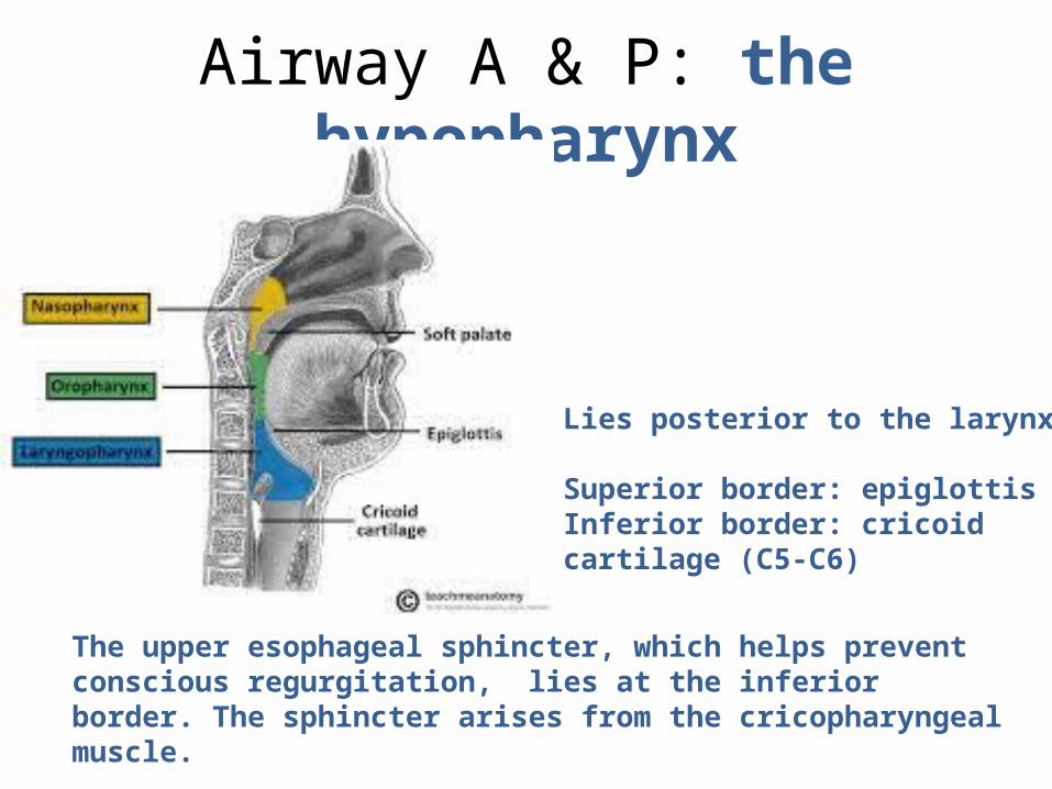
Airway A & P: the hypopharynx
Lies posterior to the larynx
Superior border: epiglottisInferior border: cricoid cartilage (C5-C6)
The upper esophageal sphincter, which helps prevent conscious regurgitation, lies at the inferior border. The sphincter arises from the cricopharyngeal muscle.

Airway A & P: the pharynxGag reflex diagram
Afferent/sensory stimuli is carried by glossopharyngeal (IX)to the medulla
Synapse occurs in the medulla with the vagus (X) & spinal accessory (XI)
Efferent /motor response returns
through the vagus (X) causing
the pharyngeal muscles to
constrict and elevate -“gag”

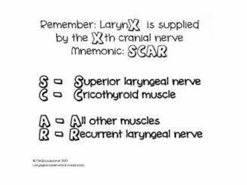
Airway A & P: the pharynxSLN & RLN
Superior Laryngeal nerve:Internal →sensory above cordsExternal→motor to cricothyroid muscle
Recurrent Laryngeal nerve:Sensory→below glottisMotor→ all other muscles of larynx
Branches off the vagus and loops around the aorta (design flaw or developmental physiology?)
Loops around the innominate
artery

Anesthesia concerns with the RLNDamage to the RLN interferes with airway control.
Acute bilateral injury → unopposed tension→ vocal cord adduction →stridorUnresolved stridor leads to respiratory distress and possibly death.

Vocal cord paralysis video

Airway A & P: the larynx
Valeculla – the space above the epiglottis. The Macintosh intubating blade is placed into this space. The blade lifts structures to view the glottis.
1 bone (hyoid) + 9 cartilagesCricoid cartilage is the only complete ringTracheal rings are incomplete posteriorly to accommodate food in the esophagus

Another view of the larnyx

Cormack & Lehane laryngoscopy grades
cords arytenoids epiglottis base of tongue

Oxygen Administration and
Mask Ventilation
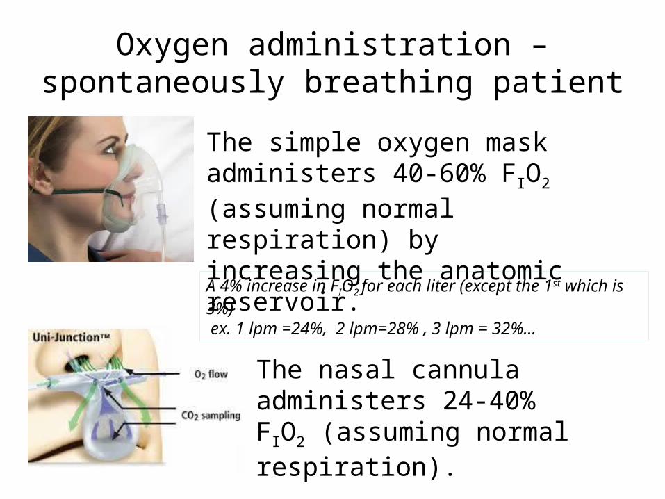
Oxygen administration –spontaneously breathing patient
The simple oxygen mask administers 40-60% FIO2 (assuming normal respiration) by increasing the anatomic reservoir.
The nasal cannula administers 24-40% FIO2 (assuming normal respiration).
A 4% increase in FIO2 for each liter (except the 1st which is 3%) ex. 1 lpm =24%, 2 lpm=28% , 3 lpm = 32%...
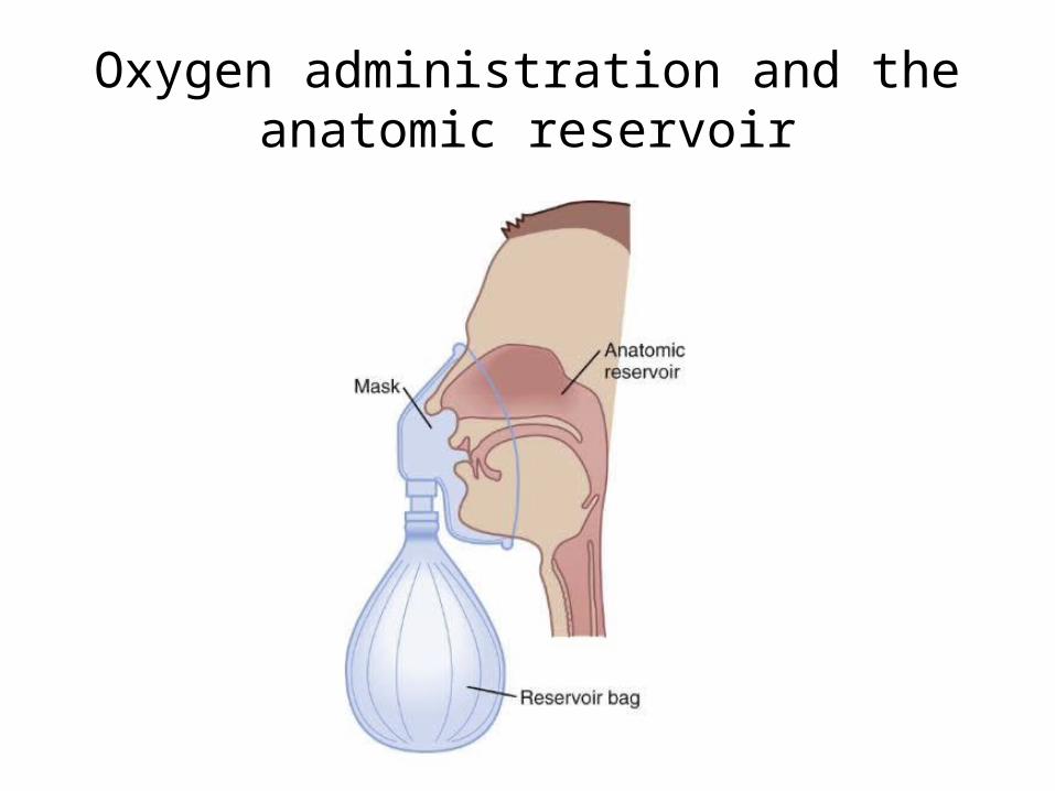
Oxygen administration and the anatomic reservoir

Difficult ventilation
The inability to maintain an oxygen saturation > 90%
while using a face mask & 100% oxygen
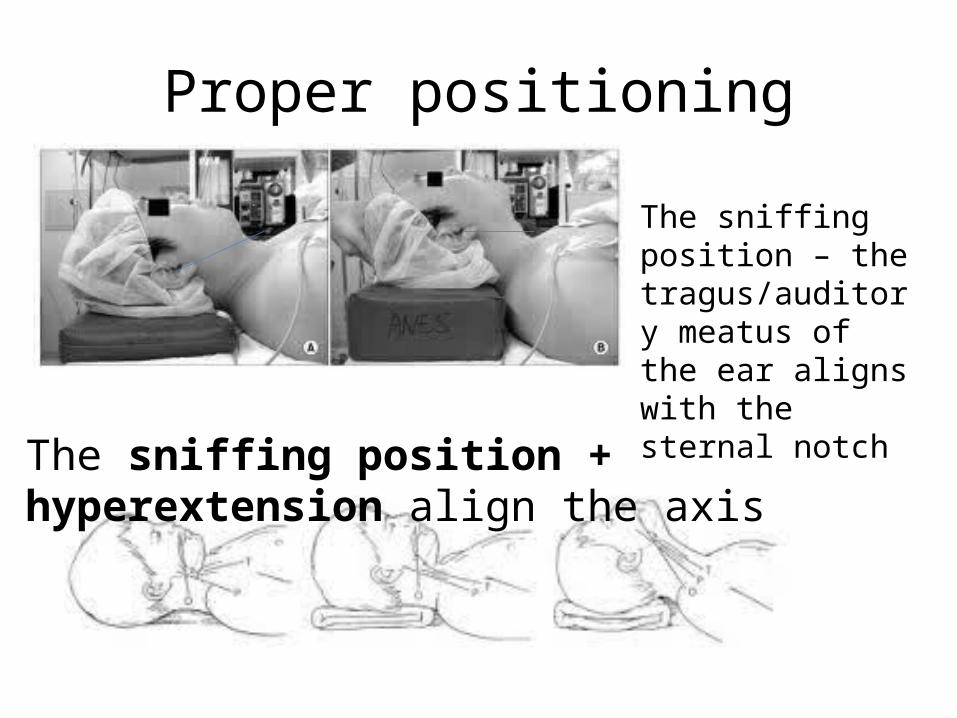
Proper positioning
The sniffing position – the tragus/auditory meatus of the ear aligns with the sternal notch
The sniffing position + hyperextension align the axis

Sniffing position video

PreoxygenationAdministration of 100% oxygen is intended to replace nitrogen (denitrogenation) in the FRC with the goal of increasing the safe apneic period.
Techniques:Normal tidal volume breaths with high flow 100 % oxygen for 3 mins.8 vital capacity breaths with 100% oxygen over 1 minute. 4 vital capacity breaths with 100% oxygen over 30 secs.(less effective)

Concept of DesaturationThe FRC is the “reservoir.”
Preoxygenation fills the “reservoir”. Oxygen consumption empties the “reservoir”.
FRC (or 35ml/kg in adult)
Oxygen consumption (or 3 ml/kg for an adult)
2500 ml 250 ml/min
(Need to also consider closing capacity)
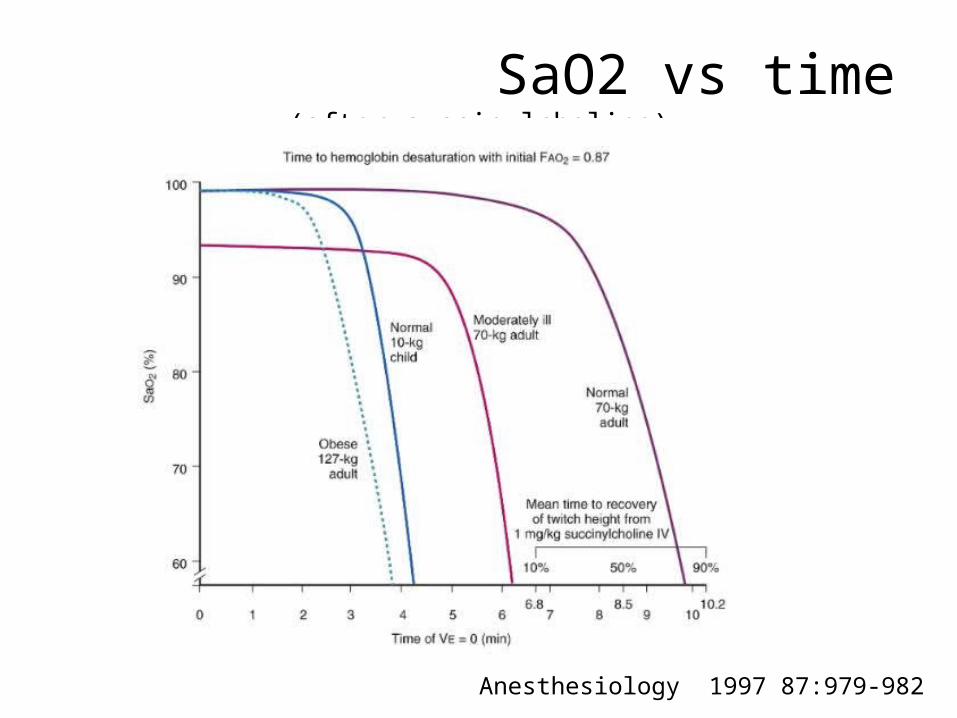
SaO2 vs time (after succinylcholine)
Anesthesiology 1997 87:979-982

Mask Ventilation Technique-a vital skill for an airway expert
1.Establish a snug fit over the bridge of nose and at the chin.2.The left thumb and forefinger create a “C” over the mask
pressing down towards the floor.3.The remaining fingers rest on the mandible. They may secure
and lift loose tissue onto the mandible.4. To improve ventilation, repositioning, hyperextension or an oral
airway may be required.
Mask ventilation video
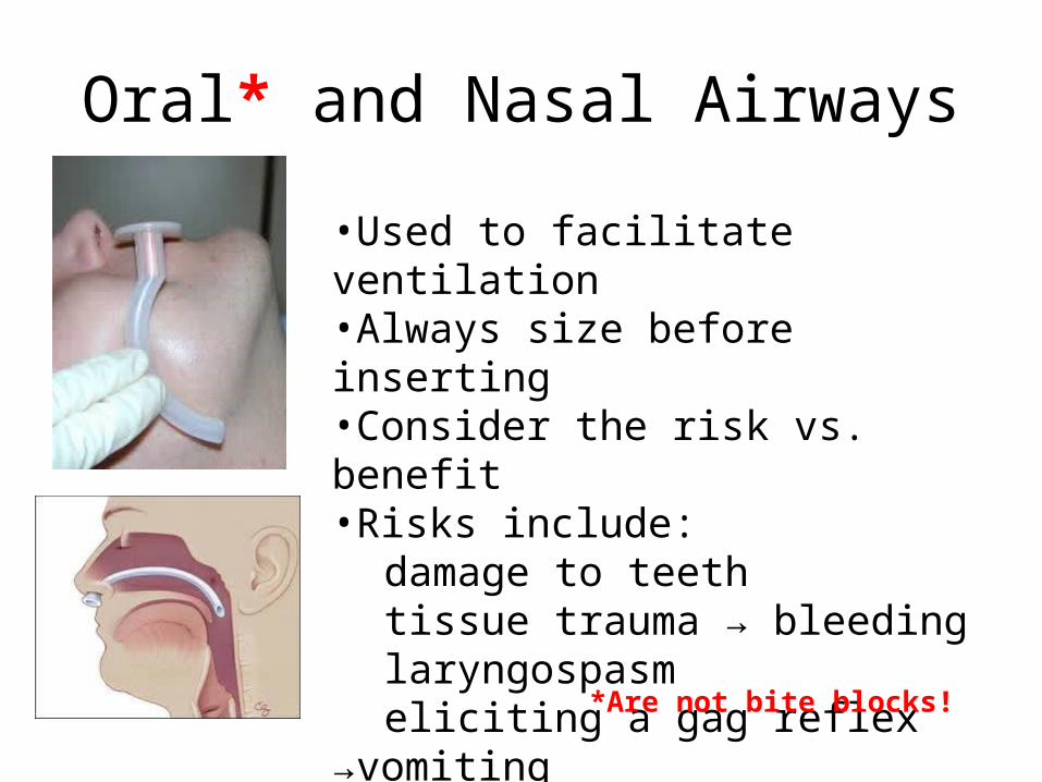
Oral* and Nasal Airways
•Used to facilitate ventilation •Always size before inserting•Consider the risk vs. benefit•Risks include:
damage to teethtissue trauma → bleedinglaryngospasmeliciting a gag reflex →vomitingfurther obstruction
*Are not bite blocks!

Bite Blocks* & Teeth protectors
*Are not airways!

Supraglottic Airways-LMA’sDO NOT prevent aspiration, or stomach inflation (keep
inflation pressure <20 cm H2O)
Are easily inserted blindly
Laryngeal Mask Airways (LMA’s) were introduced in 1989
Is properly positioned in the hypopharynx, above the epiglottis
With overinflation, may open the upper esophageal sphincter
With malposition, may produce airway obstruction
LMA insertion video
LMA Supreme
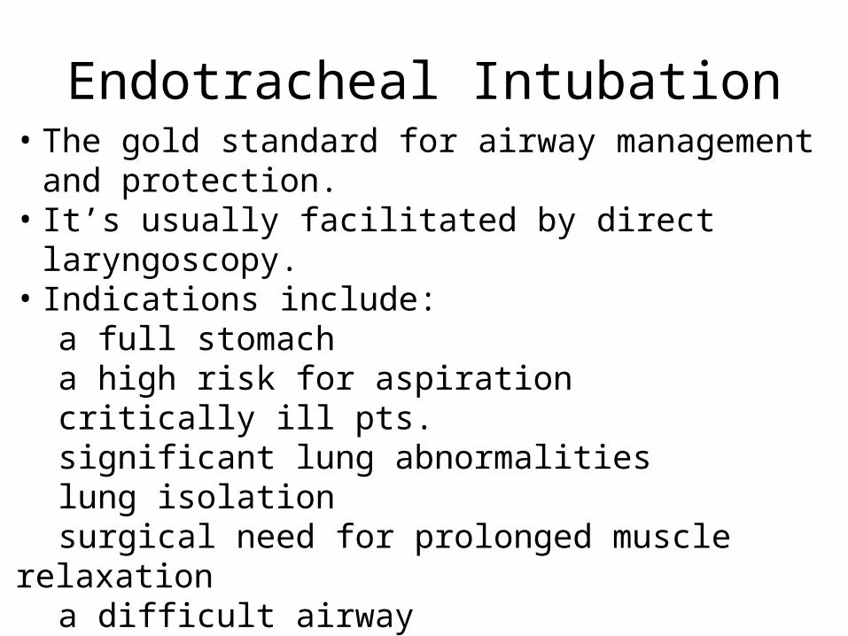
Endotracheal Intubation• The gold standard for airway management and protection.• It’s usually facilitated by direct laryngoscopy.• Indications include:
a full stomacha high risk for aspirationcritically ill pts.significant lung abnormalitieslung isolationsurgical need for prolonged muscle relaxationa difficult airwaypt. positioning
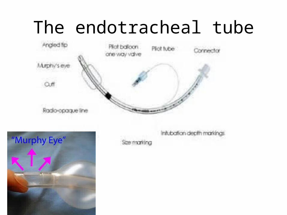
The endotracheal tube
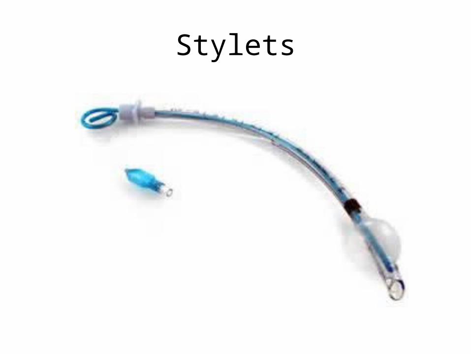
Stylets
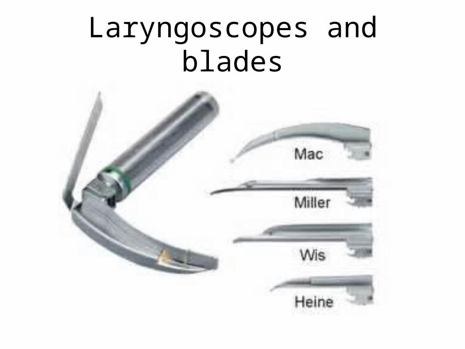
Laryngoscopes and blades
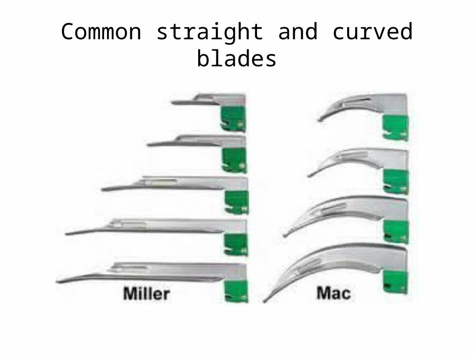
Common straight and curved blades
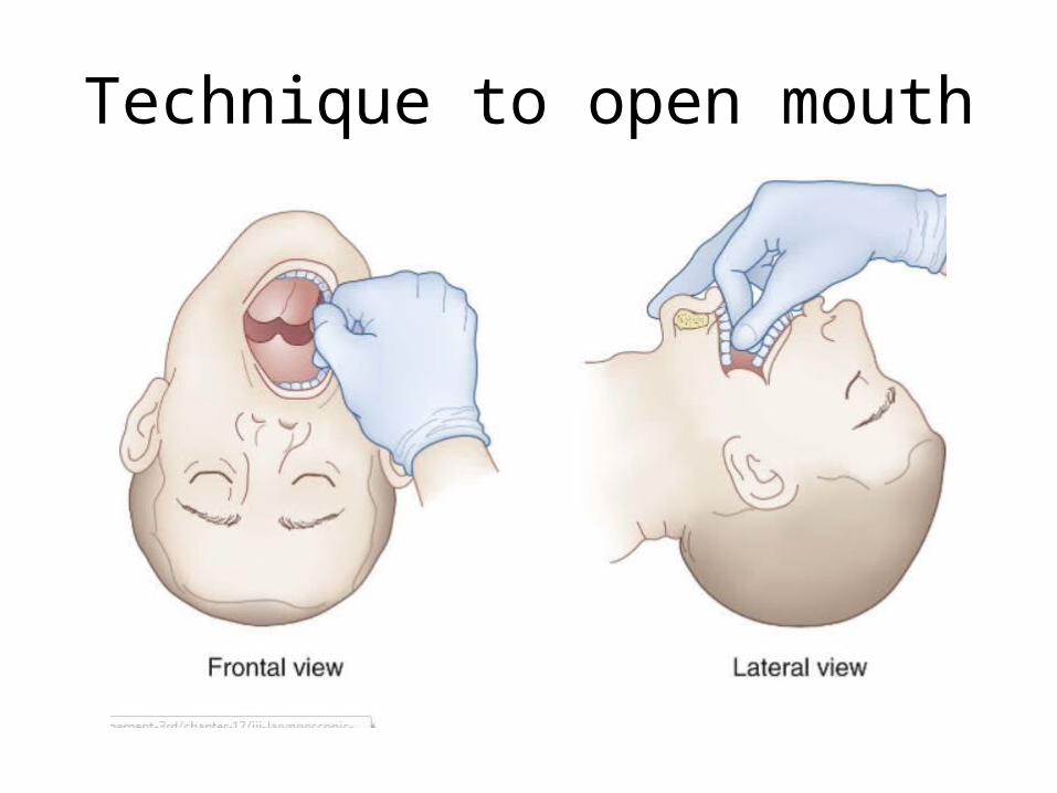
Technique to open mouth
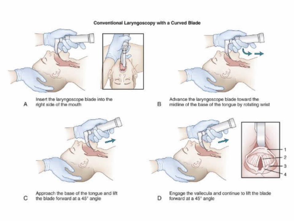

Placement of a curved blade
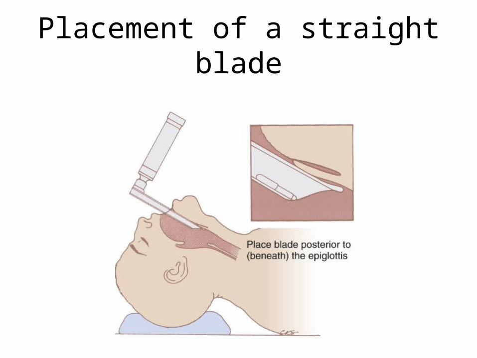
Placement of a straight blade

Tongue displacement
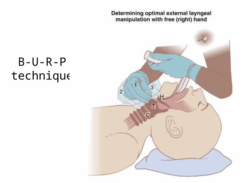
B-U-R-P technique
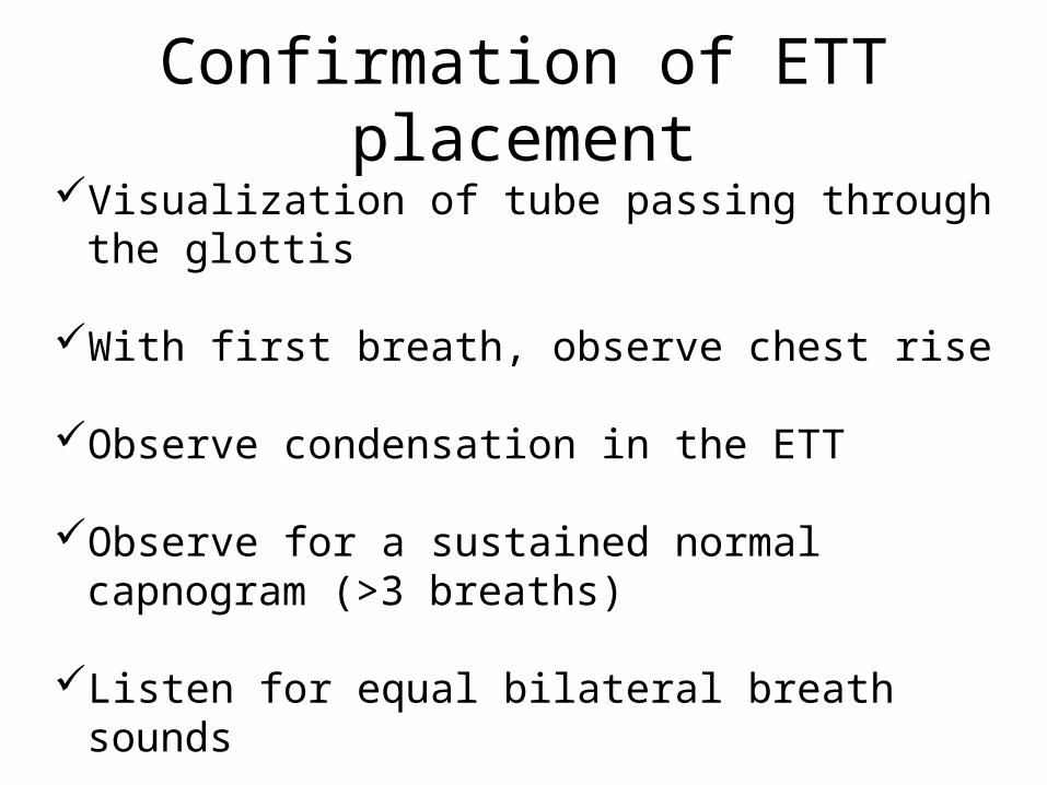
Confirmation of ETT placementVisualization of tube passing through the glottis
With first breath, observe chest rise
Observe condensation in the ETT
Observe for a sustained normal capnogram (>3 breaths)
Listen for equal bilateral breath sounds
Listen for absence of gurgling over the epigastrum
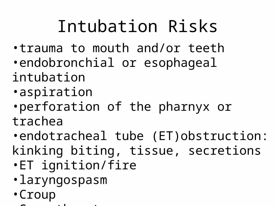
Intubation Risks•trauma to mouth and/or teeth•endobronchial or esophageal intubation•aspiration•perforation of the pharnyx or trachea•endotracheal tube (ET)obstruction: kinking biting, tissue, secretions•ET ignition/fire•laryngospasm•Croup•Sore throat
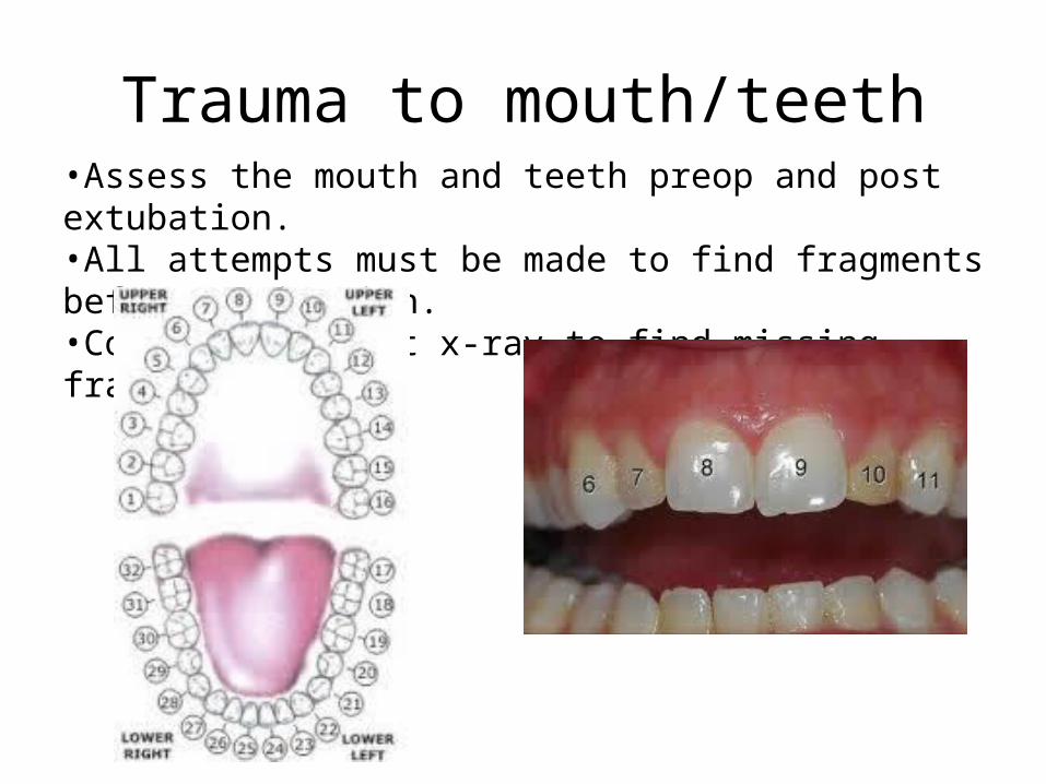
Trauma to mouth/teeth•Assess the mouth and teeth preop and post extubation.•All attempts must be made to find fragments before aspiration.•Consider a chest x-ray to find missing fragments.
Esophageal/Endobronchial intubationEnsure endotracheal intubation by confirming
placement.
Visualize the ET cuff pass through the glottis.Observe for condensation and chest rise with the first manual breath.Observe for sustained end tidal carbon dioxide capnogram.Listen for equal bilateral breath sounds and absence of gurgling over stomach.Further confirmation may include CXR or fiberoptic scope

AspirationRemains a significant cause of morbidity and mortality in obstetrics.Prophylaxis goals are to decrease the contents (<25 ml) and change the pH of the contents (<2.5)
use non-particulate antacids (Bicitra)gastrokinetics (metoclopramide)H2 antagonists or proton pump inhibitors
Use a rapid sequence induction (RSI) technique for any at risk patient full stomachobstructed boweldiabetictraumasevere, ongoing painobesityobstetrics
RSI video
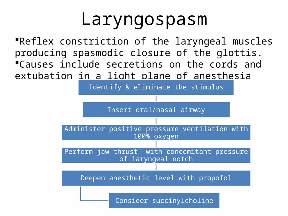
LaryngospasmReflex constriction of the laryngeal muscles producing spasmodic closure of the glottis. Causes include secretions on the cords and extubation in a light plane of anesthesia
Identify & eliminate the stimulus
Insert oral/nasal airway
Administer positive pressure ventilation with 100% oxygen
Perform jaw thrust with concomitant pressure of laryngeal notch
Deepen anesthetic level with propofol
Consider succinylcholine
Sore Throat
The most common postoperative complaint.
Factors may include:ET size irritation from instrumentationfemale gender

4 skill areas
1. Note the time in the room.2. Move the “pt/student” from the
stretcher to the table.3. Apply monitors.4. Properly position “pt” for intubation. 5. Apply mask and administer air.6. Properly position table height.7. Instruct “pt” in preoxygenation.
Attempt mask ventilation.Oral airway insertion.Nasal airway insertion.LMA insertionIntubation.
Related Documents