1 Wearable Technology to Monitor Hand Movement during Constraint-Induced Movement Therapy for Children with Cerebral Palsy Brianna M. Goodwin A thesis submitted in partial fulfilment of the requirements for the degree of MASTERS OF SCIENCE IN MECHANCIAL ENGINEERING University of Washington 2018 Committee: Katherine Steele Sawyer Fuller Kristie Bjornson Program Authorized to Offer Degree: Mechanical Engineering

Welcome message from author
This document is posted to help you gain knowledge. Please leave a comment to let me know what you think about it! Share it to your friends and learn new things together.
Transcript

1
Wearable Technology to Monitor Hand Movement during
Constraint-Induced Movement Therapy for Children with
Cerebral Palsy
Brianna M. Goodwin
A thesis
submitted in partial fulfilment of the
requirements for the degree of
MASTERS OF SCIENCE IN MECHANCIAL ENGINEERING
University of Washington
2018
Committee:
Katherine Steele
Sawyer Fuller
Kristie Bjornson
Program Authorized to Offer Degree:
Mechanical Engineering

2
©Copyright 2018
Brianna M. Goodwin

3
University of Washington
Abstract
Wearable Technology to Monitor Hand Movement during Constraint-Induced Movement
Therapy for Children with Cerebral Palsy
Brianna M. Goodwin
Chair of the Supervisory Committee:
Katherine M. Steele, PhD
Assistant Professor
Mechanical Engineering
Children with hemiplegic or unilateral cerebral palsy (uCP) have a preference to use only one
hand, and if early intervention does not occur, children often avoid or never learn to use their
paretic or impaired hand. Constraint-Induced Movement Therapy (CIMT) is an evidence-based
intervention where a child wears a cast on their dominant arm and therapists deliver intensive
therapy to the paretic hand to improve the strength and skills of that hand. The goal of CIMT is
to generate unimanual mass practice of skills in therapy which can be transferred to bimanual
skills in daily life. Few studies have investigated objective measures of bimanual tasks occurring
in daily life following therapy. This thesis set out to use accelerometry data to objectively
quantify hand use for children with uCP before, during, and after CIMT and to compare it to
typically developing (TD) children. Four children with uCP (age range: 6-8) and five TD
children (age range: 2-9) were enrolled in this study. Children in the uCP cohort wore
accelerometers on each wrist during three data collection periods; before, during, and six to eight

4
weeks after CIMT and functional tests occurred before and six to eight weeks after CIMT. The
TD cohort also wore the wrist accelerometers during three data collection periods temporally
spaced with the uCP cohort, however no interventions occurred. Results demonstrated that
before CIMT children in the uCP cohort moved their paretic hand much less than the TD cohort,
but compensated by using their non-paretic hand at higher magnitude percentages than the TD
cohort. Accelerometer data also suggested that although children improved the frequency of use
of their paretic hand compared to their non-paretic hand (use ratio) and magnitude ratio during
therapy these metrics fell back to baseline values six to eight weeks following therapy,
suggesting the benefits of the therapy were not sustained. Functionally the uCP cohort
improvement on clinical outcome measures for their paretic hand; box & blocks increased on
average 4.4 blocks moved in 60 seconds, grip strength increased by 6 lbs, 3-point pinch
increased by 3.1 lbs, lateral grasp increased by 1 lb, and children rated themselves as reaching
their goals on average 4.4 points higher per goal (measured by the Canadian Occupational
Performance Measure). The clinical results indicated that children may improve their ability to
perform unimanual tasks following CIMT, however accelerometry data demonstrated that these
gains do not transfer into increased bimanual hand use outside of the clinic. We recommend that
a Remind-To-Move protocol, which has been shown to improve bimanual skills, be implemented
following CIMT. Furthermore, through the use of surveys and focus groups this research
provided positive perceptions from both families and clinicians for the incorporation of
accelerometers into clinical practice. These results suggest that accelerometers can be used to
measure movement in TD children and children with uCP outside of the clinic and that post
CIMT follow-up interventions may be necessary to translate clinical gains into bimanual daily
activities.

5
TABLE OF CONTENTS 1 INTRODUCTION .....................................................................................................................11
Cerebral Palsy ...................................................................................................................... 12 Common Therapies to Improve Hand Function for Individuals with uCP .......................... 13 Constraint-Induced Movement Therapy .............................................................................. 17 1.3.1 History of Constraint-Induced Movement Therapy .................................................... 17 1.3.2 Effect of Age on Outcome Measures .......................................................................... 21 1.3.3 Effects of CIMT on the Developing Brain .................................................................. 22 1.3.4 Effect of the Restraint ................................................................................................. 23 1.3.5 Protocol Variation across Studies ................................................................................ 24 1.3.6 The Effect of Multiple CIMT Treatments ................................................................... 26 1.3.7 Parent Insights ............................................................................................................. 27 1.3.8 Long Term Benefits of CIMT ..................................................................................... 28 1.3.9 Which Children Benefit Most from CIMT? ................................................................ 29 1.3.10 Transfer of Processes from Research to Clinical Centers ....................................... 29 1.3.11 Limitations Associated with Current Research and Calls for Future Work ............ 30 Accelerometry Data as a Method to Quantify Movement ................................................... 31 1.4.1 Previous Pediatric Accelerometry Studies .................................................................. 32
1.4.1.1 Epoch Accelerometer Analyses ........................................................................... 33 1.4.1.2 Raw Accelerometry Analysis .............................................................................. 39
1.4.2 Analysis Methods and Visualization Metrics .............................................................. 40 1.4.3 Accelerometer Use for Children with CP ................................................................... 41 Thesis Objectives ................................................................................................................. 42 1.5.1 Specific Aim 1 ............................................................................................................. 43 1.5.2 Specific Aim 2 ............................................................................................................. 43 1.5.3 Specific Aim 3 ............................................................................................................. 43
2 Methods and Materials ............................................................................................................. 44 Data Collection .................................................................................................................... 44 2.1.1 Participants .................................................................................................................. 44 2.1.2 Constraint-Induced Movement Protocol ..................................................................... 46 2.1.3 Study Visits, Protocol, and Procedures ....................................................................... 47 2.1.4 Data Analysis .............................................................................................................. 52
2.1.4.1 Accelerometry Data ............................................................................................ 52 2.1.4.2 Qualitative Measures (Surveys) .......................................................................... 54
2.1.5 Clinician Focus Group ................................................................................................ 55 3 Results ...................................................................................................................................... 56
Accelerometry Results ......................................................................................................... 56 Functional Clinical Outcomes ............................................................................................. 66 Survey Results ..................................................................................................................... 68 Outcomes from Clinician Focus Groups ............................................................................. 68
4 Discussion ................................................................................................................................. 75 Connecting Accelerometry Data with Functional Results ................................................... 75 User perceptions of Wearing Accelerometers ...................................................................... 78 How to Effectively Implement Accelerometers in Clinical Use .......................................... 79 Limitations ........................................................................................................................... 81 Recommendations for Future Work ..................................................................................... 82

6
5 Conclusion ................................................................................................................................ 84 6 Bibliography ............................................................................................................................. 86 APPENDICES .................................................................................................................................... 93
Appendix A .................................................................................................................................... 94 Recruitment flyers and letter .................................................................................................... 94
Appendix B .................................................................................................................................. 100 CIMT Data Collection Sheets ................................................................................................. 100 TD Data Collection Sheets ......................................................................................................114
Appendix C ...................................................................................................................................118 Actigraph Wear instructions ....................................................................................................118
Appendix D ...................................................................................................................................119 Parent Survey ...........................................................................................................................119
Appendix E .................................................................................................................................. 120 Clinician Interview Guide and Outline ................................................................................... 120
Appendix F .................................................................................................................................. 123 Clinician Focus Group Transcription ..................................................................................... 123
7
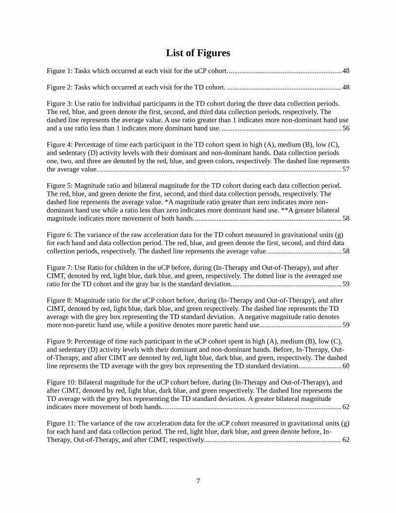
List of Figures
Figure 1: Tasks which occurred at each visit for the uCP cohort. ............................................................... 48
Figure 2: Tasks which occurred at each visit for the TD cohort. ................................................................ 48
Figure 3: Use ratio for individual participants in the TD cohort during the three data collection periods.
The red, blue, and green denote the first, second, and third data collection periods, respectively. The
dashed line represents the average value. A use ratio greater than 1 indicates more non-dominant hand use
and a use ratio less than 1 indicates more dominant hand use. ................................................................... 56
Figure 4: Percentage of time each participant in the TD cohort spent in high (A), medium (B), low (C),
and sedentary (D) activity levels with their dominant and non-dominant hands. Data collection periods
one, two, and three are denoted by the red, blue, and green colors, respectively. The dashed line represents
the average value. ........................................................................................................................................ 57
Figure 5: Magnitude ratio and bilateral magnitude for the TD cohort during each data collection period.
The red, blue, and green denote the first, second, and third data collection periods, respectively. The
dashed line represents the average value. *A magnitude ratio greater than zero indicates more non-
dominant hand use while a ratio less than zero indicates more dominant hand use. **A greater bilateral
magnitude indicates more movement of both hands. .................................................................................. 58
Figure 6: The variance of the raw acceleration data for the TD cohort measured in gravitational units (g)
for each hand and data collection period. The red, blue, and green denote the first, second, and third data
collection periods, respectively. The dashed line represents the average value. ......................................... 58
Figure 7: Use Ratio for children in the uCP before, during (In-Therapy and Out-of-Therapy), and after
CIMT, denoted by red, light blue, dark blue, and green, respectively. The dotted line is the averaged use
ratio for the TD cohort and the gray bar is the standard deviation. ............................................................. 59
Figure 8: Magnitude ratio for the uCP cohort before, during (In-Therapy and Out-of-Therapy), and after
CIMT, denoted by red, light blue, dark blue, and green respectively. The dashed line represents the TD
average with the grey box representing the TD standard deviation. A negative magnitude ratio denotes
more non-paretic hand use, while a positive denotes more paretic hand use. ............................................. 59
Figure 9: Percentage of time each participant in the uCP cohort spent in high (A), medium (B), low (C),
and sedentary (D) activity levels with their dominant and non-dominant hands. Before, In-Therapy, Out-
of-Therapy, and after CIMT are denoted by red, light blue, dark blue, and green, respectively. The dashed
line represents the TD average with the grey box representing the TD standard deviation. ....................... 60
Figure 10: Bilateral magnitude for the uCP cohort before, during (In-Therapy and Out-of-Therapy), and
after CIMT, denoted by red, light blue, dark blue, and green respectively. The dashed line represents the
TD average with the grey box representing the TD standard deviation. A greater bilateral magnitude
indicates more movement of both hands. .................................................................................................... 62
Figure 11: The variance of the raw acceleration data for the uCP cohort measured in gravitational units (g)
for each hand and data collection period. The red, light blue, dark blue, and green denote before, In-
Therapy, Out-of-Therapy, and after CIMT, respectively. ............................................................................ 62

8
Figure 12: Density plot comparing the bilateral magnitude and magnitude ratio during each of the three
data collection periods for TD01. ............................................................................................................... 63
Figure 13: Density plot comparing the bilateral magnitude and magnitude ratio during each of the three
data collection periods for TD02. ............................................................................................................... 63
Figure 14: Density plot comparing the bilateral magnitude and magnitude ratio during each of the three
data collection periods for TD03. ............................................................................................................... 64
Figure 15: Density plot comparing the bilateral magnitude and magnitude ratio during each of the three
data collection periods for TD04. ............................................................................................................... 64
Figure 16: Density plot comparing the bilateral magnitude and magnitude ratio during each of the three
data collection periods for TD05. ............................................................................................................... 64
Figure 17: Density plot comparing the bilateral magnitude and magnitude ratio before, In-Therapy, Out-
of-Therapy, and after CIMT for uCP01. ..................................................................................................... 65
Figure 18: Density plot comparing the bilateral magnitude and magnitude ratio before, In-Therapy, Out-
of-Therapy, and after CIMT for uCP02. ..................................................................................................... 65
Figure 19: Density plot comparing the bilateral magnitude and magnitude ratio before, In-Therapy, Out-
of-Therapy, and after CIMT for uCP03. ..................................................................................................... 65
Figure 20: Density plot comparing the bilateral magnitude and magnitude ratio before, In-Therapy, Out-
of-Therapy, and after CIMT for uCP04. ..................................................................................................... 66
Figure 21: Example summary sheet containing accelerometry results, used in clinician focus groups to
show metrics previously used in the literature. ........................................................................................... 72
Figure 22: Example summary sheet containing accelerometry results, used in clinician focus groups to
show metrics not previously used in the literature. ..................................................................................... 73
Figure 23: Example summary sheet containing accelerometry results, used in clinician focus groups to
show the research team’s favorite metrics. ................................................................................................. 74
Figure 24: Updated magnitude ratio plot for uCP cohort before, In-Therapy, Out-of-Therapy and after
CIMT after receiving clinician feedback. The dotted line represents the averaged typically developing
ratio. ............................................................................................................................................................ 80
Figure 25: Updated bilateral magnitude plot for uCP cohort before, In-Therapy, Out-of-Therapy and after
CIMT after receiving clinician feedback. The dotted line represents the averaged typically developing
magnitude. ................................................................................................................................................... 80

9
List of Tables
Table 1: Common therapies to improve hand function for children with uCP. ........................................... 14
Table 2: Questions about CIMT research, created by experts and ranked in order of importance. ............ 20
Table 3: CIMT protocols used in the literature. .......................................................................................... 24
Table 4: Variation in sample rate, epoch length, and location of accelerometer. ....................................... 34
Table 5: Variations in cut points used throughout the literature. ................................................................. 38
Table 6: Participant demographics for both the uCP and TD cohort. ......................................................... 46
Table 7: Background on uCP cohort. .......................................................................................................... 46
Table 8: Activities which occurred on specific days of CIMT. ................................................................... 47
Table 9: Activity count intervals used in the magnitude comparison analysis. ........................................... 54
Table 10: Averaged and range clinical outcome measures for the uCP cohort before and after CIMT. ..... 67
Table 11: Overall, unimanual, and bimanual Canadian Occupational Performance Measure (COPM) goals
rating before and after CIMT. ..................................................................................................................... 67
Table 12: Muscle Tone (MT) measured by MAS. ...................................................................................... 67
Table 13: uCP and TD cohort parent survey results. ................................................................................... 68

10
ACKNOWLEDGEMENTS
I would like to acknowledge Emily Sabelhaus, OT, for her clinical expertise, contributions to
starting this project, and mentoring through this research. Along with Emily I would also like to
acknowledge the rest of the research team Kristie Bjornson, PT, PhD, Kelly Pham, MD, William Walker,
MD, and Katherine Steele, PhD. Furthermore, this work could not have been completed without the
support and assistance of the Occupational Therapists and assistants at Seattle Children’s Hospital,
especially, Lindsay Johnson, Marla Ellingsen-Totah, and Michele Larson. I would like to thank Keshia
Peters and Heather Feldner, PhD, PT, for their assistance with data collection and facilitation of clinician
focus groups. In addition to Mrs. Peters and Dr. Feldner, I would like to acknowledge Christina Papazian
and Nick Baicoianu for all their editing and recommendations while putting this thesis together. I would
also like to thank Dr. Katherine Steele for advising this research and mentoring me through my master’s.
Finally, I would like to thank the members of the University of Washington Ability & Innovation Lab for
their knowledge, support, and encouragement.
I would also like to acknowledge our funding source, the Seattle Children’s Hospital Academic
Enrichment Fund. Without this support this research would not have been possible.

11
1 INTRODUCTION
Effective hand use is a critical part of daily activities both for children and adults. Hand use is
responsible for one’s ability to accomplish tasks, interact with their environment, and manipulate the
objects around them. Bimanual activities, or tasks which require two hands, account for over half of the
daily actions in healthy adults (Kilbreath and Heard 2005). Examples of bimanual activities include
opening a jar, washing dishes, and completing an art project. Whereas examples of unimanual, or one
handed tasks, include holding an eating utensil and brushing one’s teeth. Only 29% of daily activities
performed by adults are unimanual (Kilbreath and Heard 2005). This demonstrates the importance of
being able to use both hands and the coordination required between them.
The combined use of both hands is necessary to accomplishment many daily tasks; yet, this is not
possible for individuals with an impaired hand caused by a neurologic injury. Hemiplegia, or an injury to
the brain causing weakness or loss of movement on the opposite side of the body has many causes.
Specifically, cerebral palsy (CP) is the largest cause of hemiparesis in children (Krigger 2006, Agrawal,
Johnston et al. 2009). Although not all individuals with neurologic impairments only experience
hemiplegia, other causes of hemiparesis include traumatic brain injury, affecting 1.1 million people
annually in the United States (Langlois, Rutland-Brown et al. 2006) and stroke, affecting 795,000 people
in the United States annually (Roger, Go et al. 2011).
Constraint-Induced Movement Therapy (CIMT) is the most recommended evidence-based treatment
for children with hemiplegic or unilateral CP (uCP) (Coker-Bolt, Downey et al. 2017, DeLuca, Trucks et
al. 2017). CIMT aims to create unimanual therapeutic gains, with the goal of skill transfer to bimanual
gains outside of the clinical setting. Although many studies have shown the unimanual gains in clinic, few
studies has objectively quantified the transfer of these skills to daily life and to the best of our knowledge
this is the first study to compare the skill retention with typically developing children. Additionally,
accelerometers are the gold standard for monitoring physical activity in typically developing children

12
(Borghese, Tremblay et al. 2017) and have been used in clinical settings (Uswatte, Miltner et al. 2000,
Rand, Eng et al. 2009). To the best of our knowledge, no study has assessed family, child, and clinician
perspectives of the implementation of accelerometers in a clinical setting.
The subsequent sections describe uCP, CIMT, and the use of accelerometers in the clinical setting in
more detail. This thesis work collected accelerometry data of uCP and TD children and analyzed it to
assess the benefits and potential improvements of CIMT. Additionally, this thesis describes clinician,
family, and child insights about using accelerometry data as an objective measure of hand use outside of
the clinic. The results presented suggest methods to improve CIMT and provide recommendations for
integrating accelerometers into clinical care.
Cerebral Palsy
CP is a non-progressive neurologic disorder of movement and posture that affects approximately 2–
3.8 in every 1000 children in the United States (Reddihough and Collins 2003, Krigger 2006, Cans, De-
la-Cruz et al. 2008). CP is caused by an injury to the brain at or near the time of birth. This is the most
common physical disability in children, yet the presentation is heterogeneous across individuals and the
exact cause of the neurologic injury for a specific child is often unknown (Reddihough and Collins 2003).
CP frequently results in challenges with fine and gross motor movements. Furthermore, estimates suggest
that at least 50% of children with CP also experience speech motor impairments (Hustand, Gorton et al.
2010, Sigurdardottir and Vik 2011, Allison, Reidy et al. 2017) and some children have cognition deficits,
causing challenges with attention, learning, and perception of experiences (Rosenbaum, Paneth et al.
2007).
40% of children diagnosed with CP are more specifically diagnosed with uCP; one side of the body
is impacted by abnormal tone, decreased strength, dystonia, and other muscle impairments (Holmefur,
Aarts et al. 2009). This often presents as slow movements, muscle stiffness, coordination challenges and
decreases in grip strength up to 30-50% on the affected side (Smits-Engelsman, Rameckers et al. 2005,

13
Holmefur, Krumlinde-Sundholm et al. 2010, Brauers, Geijen et al. 2017). Further, these children have a
challenging time reaching, grasping, and manipulating objects, such as toys (Case-Smith, DeLuca et al.
2012). This presentation often restricts a child’s participation in educational activities, social play, self-
care, and eventually vocational roles (Sakzewski, Ziviani et al. 2009, Case-Smith, DeLuca et al. 2012).
Specifically in children with uCP, a trend of ‘developmental disuse’ or ‘developmental disregard’
has been described in the literature (Cope, Forst et al. 2008). When asked to perform a bilateral activity
children with hemiplegia often use their non-paretic or dominant hand to assist their paretic hand (Cope,
Forst et al. 2008). Sometimes a child may use their non-paretic hand to fully compensate for their paretic
hand (Hoare, Imms et al. 2007). Movements with the paretic hand that are challenging or require
additional cognitive planning or effort are often avoided, leading to ‘developmental disuse’ (Deluca,
Echols et al. 2006). Because CP occurs at or near birth, children with uCP often develop an alternative,
unimanual movement strategy which can result in a decreased ability to play, explore, and engage in self-
help activities. Because this process occurs throughout early development, it is known as ‘developmental
disregard’ (Deluca, Echols et al. 2006). When deciding how to accomplish a task, young adults in this
population often report feeling like they do not have any good options and regularly choose a method to
accomplish a task which decreases the negatives associated with the task, but does not eliminate them.
This results in a feeling of dissatisfaction when carrying out specific movements (Sköld, Josephsson et al.
2004).
Typical therapeutic intervention for children in this population include both occupational and
physical therapy, ranging from two to four hours per week on average (Cope, Forst et al. 2008). The most
critical issue facing children with uCP is teaching ways to accomplish bimanual tasks; traditional therapy
is aimed to do this (Holmefur, Krumlinde-Sundholm et al. 2010).
Common Therapies to Improve Hand Function for Individuals with uCP
There are multiple treatment options available to improve hand function for children with uCP

14
(Table 1). The goal of all therapies is to improve unimanual hand use in the involved limb with the hope
that bimanual hand function will also increase (Eliasson, Krumlinde-sundholm et al. 2014).
Table 1: Common therapies to improve hand function for children with uCP.
Type of Therapy Description
Constraint-Induced Movement
Therapy (CIMT)
A restraint is worn on the non-paretic hand for a specified
amount of time, dependent on the protocol. Shaping and specific
movements are performed to achieve desired outcomes (Charles
and Gordon 2005).
Remind-to-Move (RTM) A portable sensory cueing wrist-watch device is worn on the
paretic arm and vibrations occur every 15 minutes to remind the
child to move (Dong and Fong 2016, Fong, Dong et al. 2017).
Intramuscular botulinum toxin A
combined with upper-limb training
(BoNT-A)
BoNT-A is used to decrease muscle spasticity. Without additional
therapy it will not elicit change, but it is thought that in
combination with training the effects can be amplified
(Holmefur, Krumlinde-Sundholm et al. 2010, Hoare, Imms et al.
2013).
Hybrid Constraint-Induced
Movement Therapy (H-CIMT)
This therapy utilizes concepts of CIMT, but adds in intensive
bimanual therapy, typically at the end of the protocol.
Hand-arm bimanual intensive
training (HABIT or BIMT)
Intensive therapy occurs specifically targeting bimanual tasks
with the goal of enhancing bimanual hand use.
Neurodevelopmental therapy
(NDT)
This treatment is based on a theory of facilitation of ‘normal’
movements in therapy and inhibition of ‘pathological’ movement
patterns to enable functional gains. Specific handling techniques
are utilized to allow individuals to feel sensation of ‘normal’
movements, which ideally facilitates normal movement patterns
and reflexes (Deluca, Echols et al. 2006).
Forced-use A restraint is worn on the non-paretic arm for a specified amount
of time. Additional therapy is not delivered, but rather the
restraint forces the use of the paretic hand.
Relative effectiveness of these therapeutic techniques and philosophies have been contested in the
literature. For example, in their 2009 systematic review, Sakzewski et al. (2009) concluded that none of
the treatments investigated (CIMT, HABIT, NDT, BoNT-A) were more advantageous over the others.
Although, the authors suggested that the use of BoNT-A in conjunction with therapy seemed to have more
positive effects than BoNT-A alone, with improvements in the Quality of Upper Extremity Skills Test
(QUEST), Canadian Occupational Performance Measure (COPM), and the goal attainment score (GAS),

15
that were maintained six to eight months after BoNT-A and upper limb training.
Hoare, Imms et al. (2013) investigated the differences between CIMT and HABIT with BoTN-A
injections prior to the therapy. They concluded that the results were inconclusive as to which therapy
yielded the most functional long-lasting improvements. However, the CIMT therapy included in this
study had decreased frequency and length of therapy sessions compared to other studies in the literature,
(two times/week and one hour, respectively) suggesting that this trend may not be true with a more
intensive protocol. However, Gordon, Hung et al. (2011) compared CIMT with an equally intensive 90
hour HABIT program, which is more consistent with what is reported in the literature. The study found
similar results independent of the intervention, suggesting the intensity of the program creates lasting
benefits and not the restraint. However, the therapy delivered in this study was performed on a one-to-one
child to therapist ratio, which is not the case in many clinics. These results suggested that there may not
be benefits associated with children wearing a cast; however, the study also suggested that if the ratio of
children to therapists increased the restraint may be more beneficial to prohibit the child from using their
non-paretic hand while the therapist worked with another child. The results also suggested that bimanual
training might be better for kids with mild impairments and CIMT would be more suited for children who
need to work on specific skills, such as supination, as those can be specifically targeted through CIMT
(Gordon 2011). The authors noted that more positive benefits could arise from a combination of CIMT
and HABIT.
Brauers, Geijen et al. (2017) completed a similar study to identify the benefits of H-CIMT, the
combination of CIMT with BIMT. The authors used a combined protocol of six hours of CIMT and two
hours of BIMT each day for two weeks, resulting in a total dosage of 80 hours. The results demonstrated
that children increased their pinch strength. However, measurements were only taken directly after the
completion of the study and not longitudinally following therapy making it impossible to know if the
benefits of the therapy were sustained. The authors noted this was a limitation of their study and
recommended for future studies to look at the effects of H-CIMT three to eight months after the

16
completion of the therapy.
A systematic review, completed by Hoare, Imms et al. (2007) compared three studies using
forced-use, CIMT, and modified CIMT (mCIMT). The review found ‘positive trends’ supporting both a
CIMT and forced-use protocol, however a significant treatment effect was found when using mCIMT.
Section 1.3.5 discusses mCIMT in more detail. Although some studies have shown positive benefits of
only a forced-use protocol (Sung, Ryu et al. 2005), other studies have shown that physical activities and
environmental manipulation are important to reach rehabilitation goals (Glover, Mateer et al. 2002).
Furthermore, Hoare, Imms et al. (2007) strongly cautions readers when interpreting the positive results of
forced-use described in Sung, Ryu et al. (2005), due to the ‘ambiguity of its specific methodology, lack of
methodological rigor and inadequate reporting’.
There have been multiple criticisms of CIMT including challenges with clinical implementation,
type of restraint, time in the restraint, and child compliance (Dong, Fong et al. 2017). The Remind-to-
Move (RTM) technique grew out of these criticisms; Fong, Dong et al. (2017) performed a randomized
control trial comparing the effects of RTM, CIMT, and conventional rehabilitation therapy. The authors
concluded that children receiving RTM and CIMT made greater gains than the children receiving
conventional rehabilitation therapy (one hour per day, two to three days per week). They also stated the
CIMT allowed for more paretic gains, measured by the Jebsen Taylor Hand Function Test and RTM had
more bimanual gains measured by the Bruininks-Oseretsky Test of Motor Proficiency (2nd version). One
goal of CIMT is to transfer unimanual skills gained in the clinic to bimanual skills at home; understanding
the advantages of both CIMT and RTM could lead to an advantageous protocol if combined. The wrist-
watches used in RTM contained an accelerometer allowing data to be collected about the magnitude and
frequency of movement throughout the protocol (Dong and Fong 2016). The study reported increases in
duration of movement of the paretic hand compared to the non-paretic hand, known as the use ratio, for
both the RTM and CIMT groups. The authors also reported increased functional hand use measured by
the Caregiver Functional Use Survey and increases in active range of motion at the shoulder and wrist;

17
however, RTM was not shown to increase grip strength, which is one of the main positive outcome
measures used to quantify the benefits of CIMT.
NDT was historically a widely used set of therapeutic techniques in the rehabilitation of children
with uCP. However, gains associated with NDT were reported to be short-lived (Glover, Mateer et al.
2002) and little evidence exists to back its use in clinical practice. However, it took many years for this
consensus to be agreed upon (Deluca, Echols et al. 2006). One study even found there were no differences
between NDT and a context-focused intervention specifically focused on goals and activity-based training
(Sakzewski, Ziviani et al. 2013).
Although much of the literature is conflicting and no clear ‘best practice’ has been described, recent
trends in research point to CIMT and its promising effects, as the most highly recommended evidence-
based treatment for children with uCP (Coker-Bolt, Downey et al. 2017, DeLuca, Trucks et al. 2017).
However, CIMT is a program designed to improve unimanual hand use, with the goal that the benefits
will transfer to bimanual activities. Therapy programs such as RTM and HABIT have specifically been
designed to target bimanual tasks and thus it was critical to understand the different benefits associated
with each therapy when interpreting the results of this thesis.
Constraint-Induced Movement Therapy
The CIMT protocol places the individual’s non-paretic hand in a cast to encourage increased use of
their paretic hand. For the purposes of this chapter, all studies and data discussed refer to pediatric CIMT
protocols unless otherwise specified.
1.3.1 History of Constraint-Induced Movement Therapy
The theories of using experimental and clinical research to recover motor function from a damaged
central nervous system date back to 1895 (Mott and Sherrington 1985). In more recent research, Edward
Taub and colleges created the idea of CIMT by studying monkeys. Following a surgically caused brain

18
lesion, which was used to simulate stroke, the monkeys discontinued use of their affected limbs (Taub,
Perrella et al. 1975). Taub argued that not being able to effortlessly move one limb to accomplish tasks,
caused the monkeys to fall into a theory of ‘learned nonuse’. The injured monkey was able to cope
without the limb by utilizing the other three for daily tasks. Taub, Uswatte et al. (1999) suggested that
because the monkeys were able to use their three unimpaired limbs to achieve their goals, those limbs
were strengthened while the forth weakened. Furthermore, using the impaired hand became associated
with failure, incoordination, falling, and painful movements; constituting punishment and thus taught the
animal not to use their impaired hand. However, the researchers found that if one of the unimpaired
hands was rendered useless by placing it in a sling the monkey would be forced to use their impaired
limb, eventually restoring function (Taub, Perrella et al. 1975, Taub and Wolf 1997, Taub, Uswatte et al.
1999). They were able to show that if the unimpaired hand was splinted for more than three days the
monkey would continue to use the impaired hand after the splint was removed from the unimpaired hand;
there was a minimum threshold of therapeutic treatment and daily intervention hours for the results to be
sustained (DeLuca, Echols et al. 2007).
Out of this research came the idea of clinical treatment in humans where two criteria were met:
(1) the unaffected upper limb was restrained from moving and (2) intensive intervention involving mass
practice of the paretic limb occurred (Cope, Forst et al. 2008). The goal of the therapy is to improve
unimanual skills and thus increase one’s ability to perform bimanual tasks (Reidy, Naber et al. 2012).
Early studies commented on the importance of intervention time, which remains a question today (Knapp,
Taub et al. 1963). In the 1980’s CIMT was applied to adults with brain lesions from a stroke and more
recently the pediatric population has been targeted. Specifically for children with uCP the restraint serves
two main purposes: (1) shift the child’s attention to the paretic hand and (2) eliminate the sensory and
motor input gained from the non-paretic hand (DeLuca, Ramey et al. 2015).
In 1990, researchers began the tasks of creating methodologies to translate this research into
clinical care (DeLuca, Echols et al. 2007). One group developed a protocol called ACQUIREc

19
(Acquisition of new motor skills through Continuous practice and shaping to produce Quality movements
of Upper extremity through Intensive therapy and Reinforcement in Everyday patterns and places)
(DeLuca, Echols et al. 2007). When CIMT was first applied to the pediatric population, single case-
studies were often used to evaluate the efficacy of the protocol; leading to initial push-back of the therapy
and its benefits (Levin 2008). However, in the last two decades, the literature about pediatric CIMT has
grown to over 70 published articles. Unfortunately, the studies often focus on a wide array of questions
surrounding CIMT, utilize small sample sizes, and do not build on each other (Eliasson, Krumlinde-
sundholm et al. 2014). This had made the literature base challenging for clinicians to interpret and draw
specific conclusions which could be used to improve clinical therapy (Eliasson, Krumlinde-sundholm et
al. 2014).
Some of the challenges associated with the CIMT literature are seen in a case-study of a 12-
month old receiving CIMT. Researchers reported an improvement in inserting shapes into foam board,
removing and placing pellets and cubs, and scribbling with a crayon (Cope, Forst et al. 2008). Although
these are improvements in functional skills outside of the clinic, these results are hard to interpret because
they are subjective and not translatable across studies. Other studies have also reported subjective
variables as outcome measures, “(He) was able to draw lines, circular scribbles, and dots, using the full
range of the paper. These were skills he had never demonstrated before” (Glover, Mateer et al. 2002).
Although these are gains which could be related to CIMT, it is not clear if these improvements directly
occurred due to CIMT, or if they could be due to normal development. Furthermore, Glover, Mateer et al.
(2002) described that after therapy the child increasing his frequency of movement as much as three times
more than pre-therapy; however, the authors do not explain the methodology used to calculate this
increase. The reader is left believing this is a subjective assessment of the participant’s increase and
wondering what objective measures could be implemented.
In 2014 a group of expert researchers convened to discuss the current evidence base for CIMT.
They defined and ranked the importance of 11 critical research questions in understanding the benefits of

20
CIMT (Table 2). On a scale of one to five, with five being the maximum score, all 11 criteria received a
ranking above three, signifying each was an important question to answer (Eliasson, Krumlinde-
sundholm et al. 2014). Furthermore, each of these criteria had previously been discussed in the literature
and key questions which were considered in the methodology and results of this thesis are bolded in Table
2.
Table 2: Questions about CIMT research, created by experts and ranked in order of importance.
Important CIMT questions to ask Description
1. What is known about repeated CIMT? Little is known about the additive effects of
CIMT, however a potential study could be to
repeat the same study design after a 1-year
waiting period.
2. Does age influence outcome? Research has previously shown that age does not
involve the effects of CIMT, however authors still
suggest that a study with a larger sample size is
needed to fully confirm this fact.
3. Does amount of practice matter? Amount of practice takes into account duration,
frequency, and length of training program. These
vary across multiple studies and there is not a
clear consensus on an appropriate intervention. It
is suggested that future research studies this to
determine if there is a threshold effect for
structured training.
4. Does severity influence outcomes? Children of varying abilities have received
CIMT, however the effects remain unclear and
authors have proposed this for a future area
needing additional research.
5. What is known about long-term effect
of CIMT?
Most studies have reported a follow-up period
of 3-6 months, however more follow-up is
recommended for improved results.
6. Do lesion and cortical projections
influence outcomes?
Research has shown that brain lesion is a
determining characteristic for hand function,
however the extent of it is unclear. The authors
recommend future studies utilize brain imaging
and neurophysiological techniques to gain
more information.
7. Do CIMT effect transfer to bimanual
performance and activities of daily life?
The Assisting Hand Assessment, parent
questionnaires, and goal attainment measures
have been used to assess bimanual handedness
in activities of daily living, however it is
recommended that future studies investigate
these skills more.

21
Important CIMT questions to ask Description
8. Does type of restraint matter? Currently no studies have investigated this. It
is recommended that future studies investigate
the type of restraint and time the restraint is on
the non-paretic arm.
9. Does the provider of skills training
matter?
Research has suggested that parents, therapists,
and teachers can all effectively deliver CIMT.
Future research should investigate which ratio of
interventionists to children provides the most
beneficial outcomes for child improvements and
therapist time.
10. Does type of structured skills practice
matter?
A wide variety of protocols are used across the
literature however all demonstrate positive
outcomes. It is suggested that this be investigated
further; holding treatment duration, frequency,
and length of training constant while varying the
training model.
11. Does environment and context of the
training matter?
The original CIMT protocol has been adapted to
group and school based CIMT to make it more
accessible to families, however the effects have
never been assessed. Research which investigates
how treatment efficacy, compliance, and
motivation is suggested.
*Eliasson, Krumlinde-sundholm et al. (2014) provided the questions and the bolded questions were
considered in the methodology and results of this thesis.
Today, CIMT is the most recommended evidence-based treatment for the pediatric uCP
population (Coker-Bolt, Downey et al. 2017, DeLuca, Trucks et al. 2017). As previously stated, the goal
of CIMT is to increase unimanual tasks with the goal of improved bimanual hand use. Results of CIMT
show increased grip and pinch strength immediately following intervention (Stearns, Burtner et al. 2009)
and parent reported paretic hand function (Gordon, Charles et al. 2006). Additionally, researchers have
also reported improvements in coloring, ability to play with small objects, and speech function (Cope,
Forst et al. 2008, Allison, Reidy et al. 2017).
1.3.2 Effect of Age on Outcome Measures
When determining which children would have the best outcomes following CIMT, age was
thought to be a major component. Specifically, Hart (2005) suggested that young children may not be
developmentally ready for the intense nature of CIMT; concerns included increased fatigue and stress on

22
a developing child. Other reports have suggested that therapeutic activities should vary by age. For
example, older children (defined as above age 9 in this article) might be more intrinsically motivated
(Gordon, Charles et al. 2006) and benefit from increased practice, whereas younger children (defined as
4-8 in this article) might benefit more from interventions specifically targeting the enlargement of primary
neural networks through experience (Charles and Gordon 2005). However for children ages 3.5 – 10,
Gordon (2011) showed that age did not have an effect on clinical outcomes.
1.3.3 Effects of CIMT on the Developing Brain
Researchers have also suggested that CIMT could yield cortical reorganization (Hoare, Imms et
al. 2007, Cope, Forst et al. 2008). A 2007 case study reported both clinical improvements and increased
contralateral brain activity, with laterality shifting from the ipsilateral to the contralateral side after CIMT
(Sutcliffe, Gaetz et al. 2007). The authors described three potential mechanisms for this shift: (1)
decreased mirror movements, (2) increased contralateral motor cortex activity, or (3) increased
contralateral somatosensory cortex activity. They argued that the reorganization is due to the third
mechanism; CIMT provided an increase in sensory information which in turn allowed for more
recruitment and activity of the contralateral somatosensory cortex (Sutcliffe, Gaetz et al. 2007). Gillick,
Rich et al. (2018) preformed transcranial direct current stimulation (tDCS) on children while preforming
CIMT. The study reported that in accompaniment of CIMT, tDCS aims to facilitate “excitability of the
lesioned hemisphere and inhibit the exaggerated effects from the non-lesioned hemisphere” (Gillick, Rich
et al. 2018). A short CIMT protocol was used, however the outcome measures of the CIMT control group
were similar to that of the tDCS + CIMT intervention group. The authors noted that this could be due to
the low dosage of tDCS, however asserted that this study yielded a safe method for future research to take
place incorporating tDCS with CIMT.
The time since brain injury, type of lesion, and location are all important when identifying
differences between children with uCP and the type of treatment which would work the best (Nordstrand,
Holmefur et al. 2015). There is consensus in the literature that more information is desired to understand

23
if there are links between cortical reorganization and functional/speech gains following CIMT (Martin,
Burtner et al. 2008).
1.3.4 Effect of the Restraint
Concerns about the restraint have also been widely discussed in the literature. In a 24 hour per
day cast wear time protocol, Cope, Forst et al. (2008) reported that no skin breakdown or safety issues
occurred during a 2-week treatment duration. No skin irritation was also reported in Martin, Burtner et al.
(2008) and Case-Smith, DeLuca et al. (2012). Additionally, Deluca, Echols et al. (2006) reported there
was an easy transition for children to wear the cast. A few studies have reported skin irritation occurring
due to wearing the cast (Reidy, Naber et al. 2012), however they also reported quick fixes by using
common over-the-counter remedies such as antibiotic ointments and bandaging.
Even with these concerns research has suggested that the cast plays an important role in the
increase of positive outcomes of CIMT due to it ‘forcing’ the child to use their paretic hand (Cope, Forst
et al. 2008). No studies found compared different types of restraints in children (e.g. sling, cast,
puppet/mitt), although studies have reported advantages and disadvantages of each type of restraint. For
example, Gordon (2011) reported that although casts were uncomfortable, but they caused an increase in
the treatment intensity and thus outcomes. Oppositely, slings were more comfortable, but allow the user
too much choice and freedom of movement (Gordon 2011). In one study of adults receiving CIMT after a
stroke, participants who wore a glove had decreased hand use six months after the intervention, while a
group who received the same intervention while they wore a cast did not see this decline (Taub and Wolf
1997). The authors suggested that this was due to the intensity of treatment caused by the cast; only
wearing a glove allowed for the non-paretic hand to assist with tasks, whereas this did not occur when an
individual wore a cast (Taub and Wolf 1997, Charles and Gordon 2005).
Studies have also reported it to be challenging for children to wear a hard restraint for an
extended period of time, especially initially. One report described a mother and daughter both in tears at

24
the start of the therapy dealing with the initial struggle and adjustment period to the cast (Glover, Mateer
et al. 2002). Furthermore, when wearing the restraint, children cannot perform independent unimanual
and bimanual tasks that they were previously efficient at, which can be frustrating for both parents and
children. The families enrolled in therapy have used creative ways to elicit compliance, such as each
family member wearing mittens during mealtimes to show solidarity with the child wearing the restraint
(Glover, Mateer et al. 2002). During this thesis children wore a hard cast on their non-paretic hand for 24
hours per day. Additionally, this thesis investigated trends to understand if there are associated benefits of
wearing the cast outside of the clinic during CIMT.
1.3.5 Protocol Variation across Studies
Although CIMT has been shown to improve hand function, the protocols and results often vary
between research groups (Cope, Forst et al. 2008, Martin, Burtner et al. 2008, Stearns, Burtner et al. 2009,
Reidy, Naber et al. 2012). It is unclear which dosages of CIMT offer enough therapy for children to
achieve their functional goals, yet are resource conservative due to the intensity of the therapy (Kolobe,
Christy et al. 2014, Sakzewski, Provan et al. 2015). When comparing dosing it is important to look at all
components which contribute to the total dosage: frequency (sessions/week), intensity (intervention
time/day), and duration (length of therapy) (Table 3).
Table 3: CIMT protocols used in the literature.
Intensity
(intervention
time/day)
Frequency
(sessions/
week)
Hours/day
with cast
on
Duration
(weeks of
therapy)
Total
Dosage
(hours)
(Stearns, Burtner et al. 2009) 4 5 8-12 2 40
(Gordon, Charles et al. 2006,
Sakzewski, Provan et al. 2015)
6 5 6 2 60
(Nordstrand, Holmefur et al. 2015)* .5 7 Not
Reported
12 42
(Reidy, Naber et al. 2012, Allison,
Reidy et al. 2017)
Under age 5
= 3 hours/
Over age 5 =
6 hours
7 3/6 3 63/126

25
Intensity
(intervention
time/day)
Frequency
(sessions/
week)
Hours/day
with cast
on
Duration
(weeks of
therapy)
Total
Dosage
(hours)
(DeLuca, Ramey et al. 2015,
DeLuca, Trucks et al. 2017)
6 5 24 4 120
(Coker-Bolt, Downey et al. 2017) 6 5 6 1 30
(Cope, Forst et al. 2008) 2 4 24 2 16
(Martin, Burtner et al. 2008) 4 6 ~7.31 2 48
(Deluca, Echols et al. 2006) 6 7 24 3 126
(Sakzewski, Provan et al. 2015) 6 5 24 1 and 2 30 and
60
(Glover, Mateer et al. 2002) 2-4 6 24 1.5 27
(Sutcliffe, Gaetz et al. 2007) 1 5 24 3 15
(Case-Smith, DeLuca et al. 2012) 3 and 6 5 24 4 60 and
120
(Gordon 2011) 2 3 24 10 60
(DeLuca, Echols et al. 2003,
Gordon, Hung et al. 2011)
6 5 24 3 90
(Hoare, Imms et al. 2013) 1 2 3 8 16
*Therapy administered by parents and occurred in two six-week sessions with six weeks between the end
of one session and beginning of the next.
Protocols from a variety of studies suggested differing results about the total treatment dosage
which yielded the best clinical outcomes. Sakzewski, Provan et al. (2015) found that a 60 hour protocol
had more dexterity gains (measured from the Jebsen Taylor Hand Function Test) and quality of movement
(measured by the Melbourne Assessment of Unilateral Upper Limb Function) than children who only
received a 30 hour intervention. Interestingly, this same study also investigated the effects of dosage on
bimanual therapy and found no significant effects of 30 hours versus 60 hours. Coker-Bolt, Downey et al.
(2017) also supported a 60 hour intervention time as their 30 hour protocol did not create sufficient gains
in all children. Gordon (2011) went even farther to demonstrate that a 90 hour protocol is even better than
a 60 hour protocol. While this may show that more therapy is better, Cope, Forst et al. (2008)
demonstrated that a protocol of 16-hours was sufficient to create clinical changes in a case study. This
distinction is critical for delivering CIMT in clinical care, as there is a balancing act between therapy cost

26
and clinical outcomes.
Throughout the literature, there are also variations in the hours of therapy delivered each day and
their associated benefits. Traditionally, six hour per day intervention protocols have been used (Deluca,
Echols et al. 2006, Gordon, Charles et al. 2006, Sakzewski, Provan et al. 2015). However, Reidy, Naber et
al. (2012) compared children (1.6y – 4.3y) who received three hours treatment per day (with a total
dosage of 64 hours) and children (5.0 y – 20.5 y) who received six hours treatment per day (with a total
dosage of 126 hours) and found no differences in outcomes. Although they did not compare for age, they
argued that there may be a maximum dosage effect at three hours per therapy session. Case-Smith,
DeLuca et al. (2012) did control for age with a similar study comparing three hours of therapy versus six
hours of therapy per day and the authors came to the same conclusion. The authors noted that one reason
this could have occurred was because the children continued practicing after they left the clinic and the
study did not control for that; however, the authors concluded that three hours of therapy per day
accomplished a less intense and less costly treatment plan, with maximum benefits. Gordon (2011)
reported that when using a ten week protocol with three days of treatment and two hours of treatment per
day (60 hours total), similar gains were not seen. This type of CIMT is often referred to as a modified
CIMT (mCIMT), as the therapy does not occur each day of the week. Specific intervention times are
often adapted for multiple reasons including accommodation for families in rural areas and desire to make
therapy available on breaks from school (Martin, Burtner et al. 2008). Gordon, Hung et al. (2011)
demonstrated that there is a combination of frequency and length of treatment where optimum gains
occur, however future research is needed to fully understand which protocols are best to sustain these
optimal gains. This thesis used a clinical protocol, with shorter intervention times, which was previously
modified based upon the above studies and feedback from families.
1.3.6 The Effect of Multiple CIMT Treatments
The benefits of multiple CIMT treatments is also an important question to consider when looking
at this literature base. Furthermore, it is unknown if all children or only specific children would benefit

27
from this treatment or multiple rounds of the treatment (DeLuca, Ramey et al. 2015). However, in a study
designed to evaluate the benefits of multiple CIMT treatments, DeLuca, Ramey et al. (2015) argued that
because children declined after monthly follow-up visits on functional tests, there is a need for additional
CIMT bouts. Additionally, this study found that multiple CIMT treatments increased the benefits
associated with CIMT.
1.3.7 Parent Insights
Many previous studies have used parent surveys as a method to better understand activity levels
and functional arm/hand use outside the clinic (Gordon, Charles et al. 2006, Cope, Forst et al. 2008,
Stearns, Burtner et al. 2009). All reported studies noted that parents saw improvements in their child’s
ability to use their paretic hand. Only one study reported initial parent dissatisfaction, stating that the
mother felt her child’s speech abilities had decreased and he had increased clumsiness at the onset of
CIMT (Martin, Burtner et al. 2008). Eventually this mother saw improvements in her child’s hand and
speech abilities and noted that CIMT was a valuable treatment. DeLuca, Echols et al. (2007) reported a
list of concerns from parents prior to their children starting CIMT including concerns about the cast, child
frustrations with not being able to accomplish tasks, loss of function in the non-paretic hand, and how to
talk to other therapists, teachers, and doctors about the therapy. Furthermore, some parents initially
expressed concerns about their children being able to tolerate the intensity of the therapy; however, most
children demonstrated positive reactions to the therapy (Deluca, Echols et al. 2006).
In a case study of CIMT delivered to a 15-month old girl with severe hemiparesis, the parents initially
reported concerns of their daughter being able to feed herself during the therapy as she had difficulties
with weight gain. Within the first week this concern lifted, as parents saw their daughters progress and
even commented that they would like to participate in the therapy again at a later date (DeLuca, Echols et
al. 2003). A second intervention occurred when the girl was 21 months of age. At the start of the second
intervention there had been a decrease (not to baseline) in the “How well” aspect of the Pediatric Motor
Activity Log (PMAL) and the Toddler Arm Use Test (TAUT). However, following the second

28
intervention scores on both the PMAL, “How much” and TAUT increased. DeLuca, Echols et al. (2007)
reported that when this child turned 6 she was fully integrated into a first-grade classroom and was only
classified as having mild hemiparesis as compared to severe hemiparesis which was the previous
diagnosis. Another study reported that a mother was so pleased with the program and wished that private
insurance companies would fund this therapy, expressing gains are not possible through traditional
occupational and physical therapy (DeLuca, Echols et al. 2007). This concern about therapy affordability
was also brought up in Cope, Forst et al. (2008).
Although surveys give insight to perceptions of hand use in everyday life, the desire for parents to see
their children improve their hand function after CIMT causes bias in the results (Coker-Bolt, Downey et
al. 2017). A study which utilized accelerometry data described opposing trends in the accelerometry data
and the parent reported data both in length and intensity of movements (Coker-Bolt, Downey et al. 2017).
Parent surveys are the most highly used method to quantify translation of clinical gains to home life;
however, due to the lack of evidence behind parent surveys, this thesis aimed to find an evidence based
approach to objectively quantify the effects of CIMT.
1.3.8 Long Term Benefits of CIMT
A major goal of CIMT is to improve long term bimanual functions, however many studies have
reported contradictory evidence. For example, Stearns, Burtner et al. (2009) demonstrated that although
there was an increase in grip and pinch strength immediately following intervention, this trailed off within
three months. Opposing this Martin, Burtner et al. (2008) reported a small increase in grip strength and
lateral pinch immediately following CIMT, but a greater increase 3-months after the therapy. However,
they found grip strength and lateral pinch of the non-paretic hand increased a greater amount than the
paretic hand, suggesting that these improvements may relate to developmental gains associated with the
children’s increasing age, rather than gains from the therapy. Supporting this, Sutcliffe, Gaetz et al. (2007)
showed initial improvement in quantity (measured by the Quality of Upper Extremity Skills Test) and
quality (measured by the Pediatric Motor Activity Log) of movement post-therapy; however, six months

29
after therapy ended, the quantity of movements performed by the paretic hand decreased to baseline, but
the quality of movement remained improved. Due to the conflicting evidence in the literature, this thesis
aimed to objectively quantify the long term benefits of CIMT through data collection four to eight weeks
after CIMT.
1.3.9 Which Children Benefit Most from CIMT?
An important question addressed briefly in the literature is what child or characteristics of a child
make them a good candidate for CIMT. The following list from DeLuca, Echols et al. (2007) describes
characteristics which would make children good candidates for this therapy.
1. A child with severely limited upper extremity movements on only one side.
2. A child without acute or serious chronic illness.
3. A child without a seizure disorder that, despite treatment, disrupts the child’s participation in
daily activities.
4. A child without extremely severe mental retardation, autism, and/or sensory impairments that
prevent engaging in interactions with others.
5. A child without frequent, serious behavioral problems that could harm the child and/or therapist.
6. A child older than eight months.
Additionally, Gordon, Charles et al. (2006) also found that factors of age, children’s behavior, and
hand severity were not related to clinical outcomes. This differs from Brauers, Geijen et al. (2017), which
suggested that children who began therapy at MACS IIII level will not be able to reach a comparable
level to other children starting at MACS I or II. Determining which children benefit most from CIMT is
important for protocol and resource optimization.
1.3.10 Transfer of Processes from Research to Clinical Centers
DeLuca, Trucks et al. (2017) was the first multi-site randomized control trial of pediatric CIMT.
The authors emphasized the importance of individualized treatment being critical to the success of the

30
rehabilitation of each patient; specifically, the content and dosage of therapy. The authors stressed the
challenges of transitioning this therapy from research centers into clinical care, due to protocol
optimization. However, they also described the importance in doing this, so therapy is available for more
individuals. DeLuca, Trucks et al. (2017) demonstrated that CIMT could translate to a clinical setting,
with a more heterogeneous population than the literature had previously reported. In this study, all
children improved their assessment on the Emerging Behavior Scale, Pediatric Motor Activity Log, and
the Assisting Hand Assessment; age and the child’s ability did not have an effect on the outcome of their
treatment. This study also reported, that more research should be aimed at understanding the challenges
associated with clinical implementation of CIMT. The data for this thesis was collected using a clinical
protocol a local children’s hospital.
1.3.11 Limitations Associated with Current Research and Calls for Future Work
There are many limitations that are consistent across the studies presented here. First, these
studies include children whose parents have specifically sought this treatment, indicating they may be
more knowledgeable or financially well-off then the majority of the uCP population (DeLuca, Ramey et
al. 2015). Many of the studies discussed here used parent surveys to assess the benefits of CIMT outside
of the clinic. However, Coker-Bolt, Downey et al. (2017) found a difference between parent reported
outcome measures and the measured accelerometry data, suggesting a need for more objective data
collection methods to understand hand use outside of the clinic. Additionally, Taub, Uswatte et al. (1999)
described an ‘undeniable gap’ between performance in clinic and the amount of hand use at home,
suggesting that the clinically used tests are not able to fully translate to outcomes in the home
environment. Furthermore, many researchers used different protocols and assessment tools to evaluate
the therapy which yielded varying inconclusive results. CIMT has been shown to have a wide range of
benefits, yet the contradictory information in the literature make it challenging to draw specific
conclusions (Charles and Gordon 2005). This demonstrates the need for consistent methodologies and
protocols for researchers to be able to draw connections between studies. Specifically, this would allow

31
researchers to understand which aspects of treatment (intensity, restraint, duration, etc.) have the largest
positive impact on the daily life of these children following therapy.
Current literature has described a wide need for more research to understand the most beneficial
aspects of CIMT. One study suggested larger sample sizes and longer follow-up periods would yield the
effectiveness of CIMT for a variety of ages (Cope, Forst et al. 2008). Further, Deluca, Echols et al. (2006)
suggested the investigation into the specific components of CIMT which correlate with the most
functional gains would be useful. Another study suggested that to better understand hand use in daily life,
assessments including the Canadian Occupational Performance Measure (COPM) and Goal Attainment
Scaling should be used to evaluate if children are reaching their individual goals (Hoare, Imms et al.
2007). These calls for improvement were taken into account when the methods for this thesis were
created.
Accelerometry Data as a Method to Quantify Movement
There is a growing need for the use of objective measures (as opposed to self-reported surveys) to
monitor movement of individuals outside of the clinic (Uswatte, Miltner et al. 2000, Freedson, Pober et
al. 2005). Monitoring individuals by observation is neither practical nor cost beneficial (Evenson,
Catellier et al. 2008). Furthermore, although validated for children, self-report measures are not validated
for children with CP (O'Neil, Fragala-Pinkham et al. 2014).
Choosing one method to capture all aspects of body movements can be challenging, as it is a
multidimensional behavior (Peach, Van Hoomissen et al. 2014); however, with the spread of technology
has come the increase in technology used to track human movement. Specifically, since the 1980’s the
increase in battery life, memory capabilities, and decrease in cost and size has made accelerometers
acceptable for many research settings (Puyau, Adolph et al. 2002, Pulsford, Cortina-Borja et al. 2011,
Patel, Park et al. 2012). The acceleration of an object is how fast it changes speed and it is a commonly
used method to track movement. Accelerometers are more advantageous than traditional methods of

32
monitoring movement such as direct observation, questionnaires, or heart rate due to decreased intrusive
observation methods, unreliable questioners, and the complexity of heart rate monitoring (Puyau, Adolph
et al. 2002). Hildebrand, Van et al. (2014) reported accelerometers as the most commonly used objective
measure of physical activity and Borghese, Tremblay et al. (2017) called accelerometers the gold standard
for monitoring physical activity in children.
There are uniaxial, biaxial, and triaxial accelerometers available on the market. Although a uniaxial
sensor is more simple, the data recorded can miss movements which involve the upper body and load
carrying (Troiano, Berrigan et al. 2008). Testing of accelerometers has shown feasibility, reliability, and
validity when using large data sets. In particular, Choi, Liu et al. (2011) validated the use of Actigraph
accelerometers, which were used in this thesis. Furthermore, accelerometers have also been validated
(Uswatte, Miltner et al. 2000), considered reliable (Rand, Eng et al. 2009), and responsive to change
(Hsieh, Hsueh et al. 1998) when tested with the adult stroke population.
Although the use of accelerometers is expanding, there is still a need to spend more attention on the
standardization of both data collection and processing methods. Freedson, Pober et al. (2005) described
one of the main challenges of using accelerometry data today, as translating the data into meaningful
physiological outcomes. Furthermore, Borghese, Tremblay et al. (2017) described the challenges
associated with comparisons across studies due to the variation in devices, data reduction procedures, and
cut points. One of the main goals of this thesis was to understand how the accelerometry data could
translate into clinically meaningful data. The subsequent sections will describe the variations in data
collection and processing methods used in analyzing accelerometry data and give insights into previous
methods which translated this data in clinically meaningful outcomes.
1.4.1 Previous Pediatric Accelerometry Studies
Most accelerometry studies that have studied children aimed to understanding the rising rate of
obesity in the United States. Understanding the connection between children’s physical activity and how

33
it relates to later health outcomes is critical to understanding and helping to prevent childhood obesity
(Pulsford, Cortina-Borja et al. 2011). Research has shown that moderate-to-vigorous physical activity not
only reduces the rate of obesity (Mcmurray, Harrell et al. 2002), but also lowers total cholesterol
(Mcmurray, Harrell et al. 2002), circulating levels of insulin (Schmitz, Jacobs Jr et al. 2002), and slows
the age-related increase in blood pressure (Harrell, McMurray et al. 1999). Accelerometers have
correlated measured data with energy expenditure and shown its sensitivity to change with changing child
physical activity. Furthermore, Strauss, Rodzilsky et al. (2001) used acceleration values and compared
them to scores of self-efficacy and social influence rather than metrics of physical health. They found that
increased activity was associated with increased self-efficacy, self-esteem, and social influence in
children. Although these studies have investigated whole body movement (i.e. physical activity), it was
important for this thesis to understand the data analysis methods and how they could translate to upper
body accelerometer analysis.
1.4.1.1 Epoch Accelerometer Analyses
Accelerometry data is often compared using activity counts. These are proprietary to the
individual software companies which created the algorithm, but typically include filtering and compiling
the data into ‘epochs’ or time periods of one second or greater from the original sampled 30-100 Hz data.
The exact process to convert from raw data into epoch data causes a loss in the data which is too great to
overcome and understand the exact parameters used (Peach, Van Hoomissen et al. 2014). The epoch data
is then reported as an activity count, which is a non-dimensional number correlated to the intensity of the
movement; a higher number indicates greater movement while a lower number indicates less movement.
Throughout the literature there is a wide variety of sampling rates, epoch lengths, and location of
accelerometers used making it challenging to relate and draw conclusions between studies (Table 4).

34
Table 4: Variation in sample rate, epoch length, and location of accelerometer.
Article Sampling Rate
(Hz)
Epoch length Location of wear
(Troiano, Berrigan et al. 2008) Not reported 1 minute Over the right hip on an
elasticized belt
(Choi, Liu et al. 2011) 1 Summed as counts
per minute
“Anterior axillary line of the
hip on the dominant side”
(Bjornson, Song et al. 2011) Recorded data at each heal strike Ankle
(Mattocks, Leary et al. 2007) 10 1 minute On the right hip with an
elastic belt
(Pulsford, Cortina-Borja et al.
2011)
Not reported 15 seconds Flexible elastic band
mounted on the level of the
iliac crest and in the right
maxillary line
(Treuth, Schmitz et al. 2004) Not reported 30 seconds Two accelerometers used, 1
over each hip
(Puyau, Adolph et al. 2002)
(setup 1)
Not reported 1 minute Above the right iliac crest
(Computer Science and
Applications Actigraph)
(Puyau, Adolph et al. 2002)
(setup 2)
Not reported 1 minute Above the right iliac crest
(Mini-Mitter Actiwatch)
(Puyau, Adolph et al. 2002)
(setup 3)
Not reported 1 minute Lateral compartment of the
lower right leg at the head of
the fibula (Computer
Science and Applications
Actigraph)
(Puyau, Adolph et al. 2002)
(setup 4)
Not reported 1 minute Lateral compartment of the
lower right leg at the head of
the fibula (Mini-Mitter
Actigraph)
(Chandler, Brazendale et al.
2016)
Not reported 5 second Non-dominant wrist
(Hildebrand, Van et al. 2014) 60 1 minute Right hip and non-dominant
wrist
(Strauss, Rodzilsky et al. 2001) 40 30 seconds Waist
(Rowlands, Olds et al. 2017) 85.7 5 seconds Hip and wrist
(Crouter, Flynn et al. 2015) 30 1 second Dominant wrist
(Borghese, Tremblay et al. 2017) 80 1 second, 15
seconds, 30
seconds, 1 minute
Right mid-axillary line

35
Article Sampling Rate
(Hz)
Epoch length Location of wear
(Evenson, Catellier et al. 2008) Not reported 1 minute Right hip above the iliac
crest
(Trac, Dawe et al. 2018) 10 2 and 4 seconds Wrist
(O'Neil, Fragala-Pinkham et al.
2014)
Not reported 1 minute On the waist, one over each
hip
(Clanchy, Tweedy et al. 2011) Not reported 1 minute Dominant hip
There has been a strong emphasis in the literature to connect energetics to the accelerometry data
as a way to correlate and give meaning to the recorded accelerometry data. Cut points or intervals of
activity counts which correlate with activity levels (sedentary, low, moderate, and vigorous) are often
used as a way to compare movement levels. Many researchers have created cut points for different
populations (gender, age, etc.) and processing techniques (frequency of original sampled data, epoch
length, and accelerometer location). These methods are primarily geared at quantifying full body
movement, which is different than what this thesis will report; however, they give a useful way to compile
and correlate large amounts of data. Puyau, Adolph et al. (2002) reported that there are strong correlations
between energy expenditure and accelerometer counts during in-lab activities, but out of lab activities
generate much weaker correlations. Although the authors do not describe why this is the case, they do
explain how it is important to test the cut points on a wide variety of activities. Troiano, Berrigan et al.
(2008) described the importance of determining periods of non-wear and completed this by defining
intervals with greater than 60 minutes of consecutive zero intensity activity counts with one to two minute
allowance for movements between 0-100 counts. Choi, Liu et al. (2011) suggested that this method is
only applicable for youth or individuals who have an active lifestyle. The authors recommended a new
algorithm with a 90 minute window and two minute window for allowance of artificial movements.
Additionally, Pulsford, Cortina-Borja et al. (2011) argued that 15 second or smaller epochs are necessary
to give sensitive enough results for full body movement of children.
When determining cut points, it is critical to take into account the age of children, as children of

36
different ages might preform an activity differently depending on their previous experiences, therefore
affecting the data (Pulsford, Cortina-Borja et al. 2011). In the process of determining cut points Mattocks,
Leary et al. (2007) took into account age and gender. Troiano, Berrigan et al. (2008) further elaborated on
this concept and used accelerometers to track the minutes of activities levels individuals of different ages
spent in pre-determined categories of moderate and vigorous activity levels. The authors used nine age
groups, spanning from 6 to over 70 years old, and found independent of gender, there was a decline in
activity as children get older excluding the 16-19 age group. There is also a need to investigate if trends
remain constant independent of gender. Most studies reported cut points using a 60 second epoch and four
categories; sedentary, low, moderate, and vigorous. Some studies only report moderate and vigorous cut
points as these are important ranges for physical activity for children (Table 5).
The reserach studies which have used accelerometers have evolved over the years, and
subsequently so has the wear location and processing methods. Traditionally, accelerometers have been
worn on the hip to be close to the body and center of mass, and thus the activity intervals and analysis
metrics used attempted to understand full body movements. This allows researchers to understand whole
body movement as a metric of physical activity and thus predict overall energy expenditure (Westerterp
1999). Pulsford, Cortina-Borja et al. (2011) used a seven activity protocol with accelerometers mounted
on the waist to generate the activity cut points. One of the activities, basketball, resulted in lower
acceleration values, but the highest energy expenditure values. The authors argued that this demonstrated
that the use of waist worn accelerometers are not valid for defining upper body movement. More recently,
wrist worn accelerometers have shown to have a higher wear time compliance than the hip mounted ones
(Rowlands, Olds et al. 2017), however both are still used in practice. Chandler, Brazendale et al. (2016)
argued that wrist worn accelerometers can give useful information about upper body movement and the
research base is currently shifting to support that. During the protocol used by Chandler, Brazendale et al.
(2016), children wore accelerometers on their non-dominant wrists to avoid recordings of higher intensity
recordings while a child was sedentary, but preforming a task, such as drawing or playing video games

37
(Chandler, Brazendale et al. 2016). However, Crouter, Flynn et al. (2015) chose to use the dominant wrist
to detect a wider range of movements with the goal of getting a better estimate of energy expenditure. To
the best of our knowledge there has not been a comparison of accuracy between using accelerometry data
from the dominant or non-dominant arms.
Throughout the literature various accelerometers have been used to collect data, however the data
output varies and therefore the subsequent techniques used for processing also vary. This causes
challenges when trying to extrapolate processes, cut points, or results across studies. Puyau, Adolph et al.
(2002) used two different types of accelerometers (Computer Science and Applications Actigraph and
Mini-Mitter Actigraph) and mounted them on two different locations of the body, allowing for four sets of
data to be collected and analyzed. The authors argued that the counts were correlated between types of
accelerometers, but not by the location of the accelerometer. However, the raw and epoch data was widely
varying between the types of accelerometers used; consistent cut points cannot accurately be used
between the two accelerometer brands. For example, for moderate activity levels when mounted on the
leg, the Computer Science and Applications Actigraph reported a range of 5101-12000 counts per minute,
while the Mini-Mitter Actigraph ranged from 1801-4300 counts per minute (Puyau, Adolph et al. 2002).
The maximum value for the Mini-Mitter Actigraph does not even reach the minimum value for the
Computer Science and Applications Actigraph. Therefore, although the numbers were correlated the
varying results suggested that the type of accelerometer used and placement of accelerometer will dictate
which cut points are applicable for a specified data set. Evenson, Catellier et al. (2008) also investigated
the cut points for two different accelerometers made by different manufactures, Actigraph and Actical; the
authors found a similar trend; different cut points were needed for each type of accelerometer. When the
appropriate cut points were used for the specific type of accelerometer the results can be compared
between groups. For example, Borghese, Tremblay et al. (2017) found excellent reliability between two
types of accelerometers, ActiGraph GT3X+ and Actical accelerometer, for moderate to vigorous physical
activity. Although the methods to determine time in each activity level varied for the two devices, the

38
authors argued that the methods are commonly used across the literature for each type of accelerometer
and therefore their results are still valid. The data for this thesis was collected using all accelerometers
manufactured by Actigraph and therefore cut points and subsequent analysis methods used had previously
been validated for these accelerometers.
Table 5: Variations in cut points used throughout the literature.
Article Sedentary Low Cut
point
Moderate Cut
point
Vigorous Cut
point
Age range
(years)
(Mattocks, Leary
et al. 2007)
>2306 (3
METS)/>3581 (4
METS)
6130 12
(Pulsford,
Cortina-Borja et
al. 2011)
≤100 ≤2240 ≤3840 ≥3841 7-8
(Treuth, Schmitz
et al. 2004)
≤100 101 –
2999
3000-5200 ≥5200 13-14
(girls)
(Puyau, Adolph
et al. 2002)
(setup 1)
≤800 801-
3200
3201-8200 ≥8201 6-16
(Puyau, Adolph
et al. 2002)
(setup 2)
≤100 101-900 901-2200 ≥2201 6-16
(Puyau, Adolph
et al. 2002)
(setup 3)
≤800 801-
5100
5101-12000 ≥12001 6-16
(Puyau, Adolph
et al. 2002)
(setup 4)
≤200 201-
1800
1801-4300 ≥4301 6-16
(Chandler,
Brazendale et al.
2016)*
≤305 306-817 818-1968 ≥1969 8-12
(Evenson,
Catellier et al.
2008)
(ActiGraph)**
≤25 26-573 574-1002 ≥1003 5-8
(Evenson,
Catellier et al.
2008)
(Actical)**
≤11 12-507 508-718 ≥719 5-8
*Reported in counts/5 seconds
**Reported in counts/15 seconds

39
1.4.1.2 Raw Accelerometry Analysis
In 2009, at the ‘Objective Measurement of Physical Activity: Best Practices and Future
Directions’ Conference researchers agreed that there should be a shift in the way accelerometer data is
analyzed, from epochs to raw acceleration data (John and Freedson 2012). Due to the inconsistent length
of epochs across studies it is challenging to compare results across studies. Recent accelerometers have
enabled researchers to attain the raw acceleration values, allowing for comparison across studies and
increased control over data processing when these values are used (Hildebrand, Van et al. 2014).
Challenges determining which data processing techniques are best are shown throughout the literature by
the array of methods used. This is important as the processes used to summarize accelerometry data can
have a considerable increase on the accuracy of connecting acceleration data to movements (Ellingson,
Hibbing et al. 2017). The methods used in this thesis to analyze the data took these recommendations into
account; analyzing both the epoch and raw data.
Freedson, Pober et al. (2005) used raw acceleration values to analyze the data rather than utilizing
an epoch count based approach. The authors took the vector magnitude of each data point sampled at 60
Hz and subtracted by one. Next the authors rounded values under zero, stating that the negative numbers
occurred due to calibration error rather than body movements. The authors acknowledged that the
negative numbers could also be in response to downward accelerations, however they argued that taking
this into account would introduce nonlinearity into the system and therefore chose to round their negative
numbers to zero. They then found the average value over each second and minute. Fairclough, Noonan et
al. (2016) also used this metric for wrist and hip mounted accelerometers; as did Ellingson, Hibbing et al.
(2017) with a sample rate of 100 Hz.
Another approach to analyze the raw acceleration data was taken by Kinnunen, Tanskanen et al.
(2012), which aimed to correlate energy expenditure with acceleration data from the wrist. To analyze the
data, the authors bandpass filtered (0.3 - 3.0 Hz) the one dimension acceleration data. They then counted
any accelerations which exceed a predetermined threshold. The authors did not state the threshold which

40
they used for this calculation, but described that their method is a simple, practical, and reliable for
comparing acceleration data.
Additionally, another study by van Hees, Renström et al. (2011), analyzed the raw accelerometry
data in a separate way. The data was bandpass filtered (0.25 - 15.0 Hz) and then the vector magnitude of
the data was taken. This study aimed to understand the physical activity energy expenditure of pregnant
and non-pregnant women through in clinic total energy expenditure and resting energy expenditure tests,
and correlate it with accelerometry data. The authors then collected accelerometry data from women in
their home environments and concluded that this method is sufficient to find correlations for non-pregnant
women, but did not find the same for pregnant women.
Finally, Ellingson, Hibbing et al. (2017) analyzed the data using a bandpass filter (0.25 – 2.5 Hz)
and calculated the activity counts, vector magnitude, and step counts as comparison metrics. The authors
also used a low filter extension, used to include more low frequency accelerations, for comparison. They
found that there were great discrepancies between the traditional filtered data and data filtered with the
low filter extension further indicating the importance of filter choice. Additionally, the traditional filter is
the same filter parameters that Actigraph uses in their algorithm to calculate activity counts (John and
Freedson 2012) and it will be used in the analysis of raw data in this thesis.
1.4.2 Analysis Methods and Visualization Metrics
Identification of the most appropriate method to analyze acceleration data has been the subject of
multiple papers. As previously discussed, dividing the data into levels of activity (sedentary, low,
moderate, and vigorous) using activity counts has been one of the major methods to do this. However,
Troiano, Berrigan et al. (2008) states that looking at the mean activity counts per minute is a sufficient
method to analyze the collected data. The authors also looked at the minutes per day kids spend in pre-
specified activity levels and reported the numbers without any graphs or added visual guides.
A use ratio has been used to compare the time each hand is active. This ratio easily compiles a

41
large amount of data into one number, but in the process loses additional aspects of the data which are
important, such as the magnitude of movement (Coker-Bolt, Downey et al. 2017). Furthermore, a
magnitude ratio has been used to compare the magnitude of accelerations of both hands and a bilateral
magnitude compared the magnitude of both hands moving together. Lang, Waddell et al. (2017) described
the process of combining these metrics into density plots as a way to describe the differences between
hand use in adults following a stroke. The density plot creates a visual representation of how the hands
are being used individually and in coordination. Uswatte, Miltner et al. (2000) stressed the importance of
linking accelerometry values to clinical outcomes, including visualization methods, data collection
processes, and data collection equipment. This thesis aimed to understand which metrics and visualization
methods were most beneficial for clinicians to use when making clinical recommendations.
1.4.3 Accelerometer Use for Children with CP
Multiple studies have used accelerometers specifically with children with CP (Bjornson, Song et
al. 2011, Coker-Bolt, Downey et al. 2017). Capio, Sit et al. (2010) demonstrated that children with CP
could wear accelerometers on the hip to measure activity levels. Further, the authors described how the
data from the accelerometer is linearly correlated with the observed in-clinic test measures, which was not
the case for heart rate monitors. O'Neil, Fragala-Pinkham et al. (2014) proved the reliability and validity
of the use of accelerometers to measure waling in children with CP. O'Neil, Fragala-Pinkham et al. (2014)
also reported that children with CP could wear accelerometers on either hip to measure physical activity
independent of ability level and functional differences in the presentation of CP (i.e., diplegia, hemiplegia,
quadriplegia). Clanchy, Tweedy et al. (2011) investigated which cut-points were most valid for children
age 8-16 with CP wearing accelerometers on their waist, as previously no study had validated
accelerometer use for children with CP. The authors concluded the cut points determined by Evenson,
Catellier et al. (2008) gave the most correlated accelerometry and energy cost (measured by a portable
indirect calorimeter) results. The Evenson, Catellier et al. (2008) cut points were not used in this thesis
due to differences in wear locations.

42
Children with uCP and adult stroke survivors have worn accelerometers during CIMT to attempt
to understand the benefits of the program. Lang, Waddell et al. (2017) described how accelerometers are
more than just physical activity measures, but can compare hand use for individuals following a stroke.
Mitchell, Ziviani et al. (2015) reported that three-days of accelerometry data is needed to validate
measures of activity in children with CP. Coker-Bolt, Downey et al. (2017) studied children with uCP
undergoing CIMT and showed that children could wear accelerometers during a CIMT camp-based
setting and at home to collect data which was previously unavailable and more objective than parent-
based surveys. The authors used the use ratio and magnitude ratio to compare the frequency and
magnitude of hand movements for children undergoing CIMT. They did not see a significant difference
in the use ratio or magnitude ratio before versus after CIMT during a short one-week protocol. This study
has many limitations as it did not use a control group, did not collect data for both hands during therapy,
and only collected one day of data for the before and after CIMT time periods, even though three days has
been reported in the literature. Additionally, Coker-Bolt, Downey et al. (2017) asserted the need for more
research to fully understand how accelerometer data can measure motor capacity and be utilized outside
of a clinical setting. This thesis aimed to fill that gap through the accelerometry data collected and work
with clinicians to understand implementation strategies.
Thesis Objectives
Based upon the lack of objectively measured data used to quantify the benefits of CIMT outside of
the clinic, this thesis aimed to test the feasibility of using wrist-worn accelerometers to quantify upper
extremity use in children with uCP who under went CIMT at Seattle Children’s Hospital and compared it
with typically developing (TD) children. This thesis also aimed to understand the benefits and potential
draw backs of integrating accelerometers into clinical care to understand movement patterns outside of
the clinic.

43
1.5.1 Specific Aim 1
Quantify hand use with accelerometry in children with uCP: Wrist-worn accelerometers
captured the frequency and magnitude of upper extremity movement in children with uCP and TD
children. We hypothesized that the TD children would use their hands a similar amount, while the paretic
hand of individuals with uCP would be much less active than the TD baseline before CIMT.
1.5.2 Specific Aim 2
Quantify hand use before, during, and after CIMT with accelerometry data: We compared
paretic and non-paretic limb use during daily life over a three-day period before, during, and after CIMT.
We hypothesized that (1) children would increase paretic limb use during CIMT, (2) maintain the
increased use after completing the program, and (3) changes in limb use measured by accelerometry data
would correlate with changes in function measured with traditional clinical scales.
1.5.3 Specific Aim 3
Explore family, child, and clinician perceptions regarding accelerometers: We surveyed families
to gain an understanding of the parent and child’s tolerance for using accelerometers to measure hand
movement. We aimed to identify any family or child burdens that occurred from wearing the sensors on
their hands throughout the day. We hypothesized that children would tolerate the accelerometers, but there
would be user identified methods to improve the devices.

44
2 Methods and Materials
Data Collection
2.1.1 Participants
Children with uCP and TD children were included in this study. The uCP cohort was recruited
through Seattle Children’s Hospital (SCH), a tertiary pediatric care facility. When a child’s physician or
an Occupational Therapist (OT) recommended CIMT for a child, an assessment was scheduled with an
OT. During the assessment, the OT assessed the child’s upper extremity skills, social skills, and behavior
through observed playing, a general manual muscle test, and a range of motion assessment. Children
deemed clinically appropriate for CIMT, were given information about participation in this study. All
children selected for the CIMT program, proceeded with CIMT regardless of their decision to participate
in the study. TD children were recruited from the local community. Poster flyers, email flyers, and word
of mouth were used to identify children. Families were provided with a copy of a written approach letter
and the consent form to read (Appendix A).
The protocols used were approved by the Institutional Review Board at SCH and all participants
and parents provided informed consent and assent (if applicable). This thesis used general inclusion
criteria and specific criteria for both uCP and TD cohorts.
Inclusion Criteria (both cohorts):
1. English speaking child with English speaking parents
2. Non-foster children
3. Ages 2 to 18
4. Family had resources to attend all study sessions (i.e. transportation/schedule) and were not
planning to move outside the region
5. Children were willing to avoid swimming during the study (outside of therapy sessions)

45
6. Children did not experience an uncontrolled seizure disorder or have new seizure activity in the
past three months that impacted their mobility skills and function
7. Children did have any chronic or acute illness that could disrupt such therapy (such as Mercia)
8. Children were able to understand directions sufficiently to complete the therapy sessions and
study assessments
9. No planned surgery or changes to medication during the study period
Inclusion Criteria Specific to the uCP cohort included:
1. Children with hemiparesis or asymmetry of upper extremity use
2. Children who have some active finger flexion or extension in their paretic hand
3. Children were at least 1-year post injury and were referred and scheduled by a therapist or
clinician for CIMT at SCH
4. No significant developmental delay, that prevent meaningful participation in intensive therapy,
specifically two hour long CIMT sessions, five days per week
5. Able to tolerate wearing a cast seven days a week for 24 hours a day, for three weeks
Inclusion Criteria Specific to the TD cohort included:
1. Children lacking developmental disabilities (e.g., ADHD, Autism, etc.)
2. Children without upper or lower extremity disabilities or differences
3. Children without surgery or fractures in the past year
4. Children without other chronic illnesses that do not affect upper extremity function
Four children with uCP undergoing CIMT (age: 6yr 4m – 8yr 5m, 2 males/2 females) and five TD
children (age: 2yr 8m – 9yr 10m, 1 male/4 females) were enrolled in the study (Table 6). Descriptive
information about the onset of hemiplegia and affected brain area in the uCP cohort were recorded (Table
7).

46
Table 6: Participant demographics for both the uCP and TD cohort.
Participant Age Sex Height (cm) Weight (kg) Dominant Side
uCP01 7 Male 128 23.9 Right
uCP02 7 Female 113 18.4 Right
uCP03 6 Male 119 23.8 Left
uCP04 8 Female 130 32.6 Left
TD01 7 Female 128 26.1 Right
TD02 6 Female 124 28.3 Right
TD03 2 Male 93 13.8 Right
TD04 9 Female 139 29.8 Left
TD05 9 Female 141 26.4 Right
Table 7: Background on uCP cohort.
Participant Onset of hemiplegia Etiology of hemiplegia Previous CIMT treatments
uCP01 Birth Prenatal or perinatal stroke 2
uCP02 Birth Prenatal injury 0
uCP03 4 years post birth Post-natal stroke 0
uCP04 Birth Perinatal stroke 1
2.1.2 Constraint-Induced Movement Protocol
All participants in the uCP cohort received CIMT at SCH and therefore used the SCH CIMT protocol,
which included casting of the non-paretic hand for three weeks. Subsequently, unimanual hand training of
the paretic hand occurred two hours/day, four days/week and bimanual training occurred for two
hours/day, once a week. This training occurred in a group setting; one therapist to two children and often
a volunteer participated in the therapy. During therapy, therapists focused on shaping techniques to
impact the upper extremity; shaping used motivating activities of the appropriate difficulty level, to allow
the child to have successful experiences while developing new skills. The goal of shaping is to un-train
the ‘developmental disregard’ associated with the paretic hand, through accomplishing new activities and
positive verbal and visual recognition from the OT. As therapy continued, rewards were only given for
more complex tasks in a desire to facilitate the child performing more challenging movements (DeLuca,

47
Echols et al. 2007). Throughout the three-week protocol massed practice occurred to ensure skills
acquisition associated with repetitious actions. Additionally, each treatment session may have also
included the following:
OT problem solved concerns of caregiver (e.g., how does a parent help her child eat more
independently)
OT modeled interventions for parents and encouraged them to facilitate these at home (e.g.,
stretching or weight bearing positions)
OT evaluated fit and function of the restraint and modified, if necessary
During therapy, the children were engaged through age appropriate games and activities. Although
common games do occur, each day at SCH has a specific theme with associated activities and objectives
(Table 8). These themes kept the children engaged and allow excitement to build for subsequent therapy
days. Common games to promote specific gross and fine motor skills were also used during therapy and
occurred before or after the daily themes. Common games included, but were not limited to blocks, Play-
Doh™, Hungry Hungry Hippo™, moving pegs, chopstick feeding games, Whac-a-Mole™, Don’t Break
the Ice™, and yarn & bean bag games in the gym. The therapists strove to ensure all children in the
therapy were engaged and enjoyed coming to therapy each day, tailoring specific games and activities to
children’s individual interests and function levels.
Table 8: Activities which occurred on specific days of CIMT.
Day Theme
Monday Art
Tuesday Baking
Wednesday Music
Thursday Games and/or Therapy Dog
Friday Swimming
2.1.3 Study Visits, Protocol, and Procedures
All visits for the uCP cohort occurred at SCH. The first TD visit also occurred at SCH, however
because no evaluations or therapy took place during visits two and three for the TD cohort, research staff

48
coordinated with parents to give them the sensors without the children present. This increased convince
for families in the study. Visits two and three for both cohorts occurred 22±12 days post visit 1 and 45±9
post visit 2, respectively. A variety of tasks took place during each visit for the uCP and TD cohorts
(Figure 1 and Figure 2).
Figure 1: Tasks which occurred at each visit for the uCP cohort.
Figure 2: Tasks which occurred at each visit for the TD cohort.

49
Medical History: For children in the uCP cohort, research staff recorded a brief medical history
from a parent interview or chart review to verify eligibility; including diagnosis, onset of hemiplegia,
arm and hand dominance and current therapy program. Since pharmacological treatments, such as
BoNT-A or Baclofen are common in CP, dosage, frequency, and dates of use were also recorded for
each participant. No participants enrolled in this thesis had received BoNT-A or Baclofen. For the
TD cohort, research staff recorded arm/hand dominance.
Comprehensive Evaluation: The OT used data collection sheets (Appendix B) to record the
assessments measured as standard of care for children in the uCP cohort.
o Range of Motion – Passive range of motion assessments were conducted using a standard
goniometer and visual analysis during active movement of the child’s arms and hands. The
motions tested are listed below:
Shoulder flexion/extension
Shoulder abduction/adduction
Shoulder external/internal rotation
Elbow flexion/extension
Arm pronation/supination
Wrist flexion/extension
Metacarpal flexion/extension
Proximal and distal interphalangeals flexion/extension
o Grip and Pinch Testing – The grip, pinch, and lateral pinch of the child’s paretic and non-
paretic hands were measured using pinch and grip dynamometers. Children gripped or
pinched the dynamometer three times and the maximum score was recorded.
o Canadian Occupational Performance Measure (COPM) – A client-centered clinical practice
outcome assessment used to measure patient identified problems of daily function. The

50
children identified goals of self-care, productivity, or leisure and scored themselves on their
performance and satisfaction (Law, Baptiste et al. 1990).
o Modified Ashworth Scale (MAS) – A scale from 0 to 4 used to assess muscle spasticity, or
the ‘velocity dependent increase in the tonic stretch reflex (muscle tone) with exaggerated
tendon reflexes’ (Lance 1980). Therapists lead the children’s elbows and wrists through a
range of motion (ROM) and quantified the flexion and extension movements at each joint, as
listed below (Charalambous 2014):
0. No increase in muscle tone
1. Slight increase in muscle tone, manifested by a catch and release or by minimal
resistance at the end of the ROM when the affected part(s) is moved in flexion and
extension
1+. More marked increase in muscle tone, manifested by a catch, followed by minimal
resistance throughout the remainder (less than half) of the ROM
2. More marked increase in muscle tone thorugh most of the ROM, but affected part(s)
easily moved
3. Considerable increase in muscle tone, passive movement difficult
4. Affected part(s) rigid in flexion or extension
o Manual Ability Classification Scale (MACS) – A qualitative, therapist-led observational
classification used to classify how children with cerebral palsy use their hands in daily
activities. Scores range from one to five (as listed below) and are intended to indicate the
performance a child normally has, rather than their best attempt (Eliasson, Krumlinde-
Sundholm et al. 2006).
I. Handles objects easily and successfully
II. Handles most objects but with somewhat reduced quality and/or speed of
achievement
III. Handles objects with difficulty; needs help to prepare and/or modify activities

51
IV. Handles a limited selection of easily managed objects in adapted situations
V. Does not handle objects and has severely limited ability to perform even simple
actions
o Box and Blocks Test – A standardized measure of coordination in which a child was asked to
move as many wooden cubes (2.5 cm) over a 19 cm patrician within 60 seconds using one
hand (Mathiowetz, Volland et al. 1985). This test occurred for both paretic and non-paretic
hands and results were compared.
Cast Fabrication: Children in the uCP cohort had a custom long arm, univalve, fiberglass cast
fabricated, which extended from the child’s axillary region to past the fingertips. This cast was
applied for initial fitting and then removed immediately, with additional padding applied for comfort,
according to standard procedure for patients who participate in CIMT at SCH CIMT. Children
donned the cast on the first day of CIMT and it was wrapped with Coban, a self-adherent wrap. The
OT educated the family on the care of the cast and demonstrated cast removal in case of an
emergency situation.
Actigraphs Application: All children were fit with two tri-axial ActiGraph GT9X Link
accelerometers (ActiGraph Corp., Pensacola, FL) and instructed on basic wear and use instructions.
Wear instructions were also given to the families (Appendix C). The Actigraphs were placed on the
participant’s right and left wrists and strap circumference was measured. Children in the uCP cohort
had the Actigraphs placed before initial CIMT testing began. Research personnel instructed the
children (and families) to wear the Actigraphs for the next three full days while awake, with
permission to remove the actigraphs when bathing or in water.
Activity Journal: All parents and children kept an hourly activity journal during the three-day data
collection period. This paper journal and accompanying instructions were provided at each visit and
returned in person or by mail with the Actigraphs.

52
Parent Survey: All parents completed a survey developed for this study to understand parent and
child attitudes toward wearing the sensors (Appendix D).
2.1.4 Data Analysis
2.1.4.1 Accelerometry Data
Accelerometry data captured the motion of the left and right wrists on tri-axial accelerometers at
100 Hz. The integrity of the data was evaluated by comparing the daily activity logs with the raw data
through the Actigraph AciLife 6 Software (ActiGraph Corp., Pensacola, FL). Periods which reflected
times of non-wear were excluded from data analysis. For the uCP cohort data were broken into four
categories; (1) before CIMT, (2) In-Therapy (during the CIMT protocol), (3) Out-of-Therapy (during the
CIMT protocol (e.g. at home, school, etc.)), and (4) after CIMT. For the TD cohort, data were analyzed
during each of the three data collection periods.
Data were analyzed using metrics from literature (i.e., the use ratio, magnitude ratio, bilateral
magnitude, and magnitude cut points), as well as raw accelerometry data to evaluate Aims 1 and 2 of this
thesis; comparing the hand use between TD children and children with uCP (before, during, and after
CIMT).
Use Ratio: Compared the amount of active time of each hand, using epochs one second in length.
The magnitude of movement is not taken into account in this analysis; however, this metric gives a
consolidated numerical output from a large amount of data in one (clinician-friendly) number (Lang,
Waddell et al. 2017). This metric was used to compare the activity frequency of each hand.
𝑈𝑠𝑒 𝑅𝑎𝑡𝑖𝑜 = 𝐻𝑜𝑢𝑟𝑠 𝑜𝑓 𝑝𝑎𝑟𝑒𝑡𝑖𝑐 ℎ𝑎𝑛𝑑 𝑢𝑠𝑒
𝐻𝑜𝑢𝑟𝑠 𝑜𝑓 𝑛𝑜𝑛 − 𝑝𝑎𝑟𝑒𝑡𝑖𝑐 ℎ𝑎𝑛𝑑 𝑢𝑠𝑒
Magnitude Ratio: Compared the vector magnitude (vec. mag.) of acceleration for each hand at each
time point using epochs one second in length, providing a metric for quantifying bimanual and
unimanual hand use. For example, a ratio near zero indicated more bimanual hand use, while a ratio

53
near seven or negative seven, indicated more paretic or non-paretic hand use, respectively. The
average magnitude ratio was compared between the two cohorts and before, during, and after CIMT
for the uCP cohort. This metric incorporated frequency and magnitude to compare the use of each
hand.
𝑀𝑎𝑔𝑛𝑖𝑡𝑢𝑑𝑒 𝑅𝑎𝑡𝑖𝑜 = 𝑙𝑛( 𝑣𝑒𝑐. 𝑚𝑎𝑔. 𝑝𝑎𝑟𝑒𝑡𝑖𝑐
𝑣𝑒𝑐. 𝑚𝑎𝑔. 𝑛𝑜𝑛 − 𝑝𝑎𝑟𝑒𝑡𝑖𝑐 )
*All values greater than 7 and all values less than -7 were set to 7 and -7, respectively, per a predefined
protocol (Lang, Waddell et al. 2017).
Bilateral Magnitude: Compared the overall acceleration magnitude of both hands added together,
using epochs one second in length. Similar to the magnitude ratio, comparison of the mean occurred
for the bilateral magnitude to understand bilateral hand use. Furthermore, density plots of movement
are common in the literature; plotting the magnitude ratio on the x-axis and bilateral magnitude on the
y-axis. Plots utilizing this analysis method were also created. The bilateral magnitude allowed for
comparison of overall movement magnitude occurring for the two cohorts at different times.
𝐵𝑖𝑙𝑎𝑡𝑒𝑟𝑎𝑙 𝑀𝑎𝑔𝑛𝑖𝑡𝑢𝑑𝑒 = (𝑣𝑒𝑐. 𝑚𝑎𝑔. 𝑝𝑎𝑟𝑒𝑡𝑖𝑐) + (𝑣𝑒𝑐. 𝑚𝑎𝑔. 𝑛𝑜𝑛 − 𝑝𝑎𝑟𝑒𝑡𝑖𝑐)
Magnitude Cut-Point Comparison: Compared the percentage of time children spent in activity
levels of varying intensities, using epochs five seconds in length. For this analysis the cut points
proposed by Chandler, Brazendale et al. (2016) were chosen because similar age (8-12), gender (boys
and girls), and wear location (wrists) were used in both the study and this thesis (Table 9). The
percentage of time each child spent in each activity level was calculated and compared between each
hand and data collection periods.

54
Table 9: Activity count intervals used in the magnitude comparison analysis.
Activity Level Activity Counts (5 second epoch)
Sedentary ≤ 305
Low 306 – 817
Medium 818 – 1968
High ≥ 1969
Raw Data: A fourth-order Butterworth filter between 0.25 and 2.5 Hz (the range at which normal
human movement occurs) was applied to the raw data (John and Freedson 2012). The variance of the
vector magnitude was taken as a summary measure to estimate the variation of movement occurring.
A higher variance would indicate more variation in movement, while a lower variance would indicate
less variation in movement. This was compared between hands and data collection periods to yield
information about the overall movement that occurred for the two cohorts.
𝑉𝑒𝑐𝑡𝑜𝑟 𝑀𝑎𝑔𝑛𝑖𝑡𝑢𝑑𝑒 (𝑉𝑀) = √𝑎𝑥2 + 𝑎𝑦
2 + 𝑎𝑧2
𝑉𝑎𝑟𝑖𝑎𝑛𝑐𝑒 = 1
𝑁 − 1∑|𝑉𝑀𝑖 − 𝜇|2
𝑁
𝑖=1
*Where ‘a’ is the acceleration in the x, y, and z axis, N is the number of observations, and 𝜇 is the mean
of the data set.
2.1.4.2 Qualitative Measures (Surveys)
The parent surveys aimed to gain insights into parents and children’s perceptions of wearing
accelerometers to monitor movement outside of the clinic (thesis Aim 3). Specifically, the surveys
addressed child comfort, if parents were interested in the data, and if they felt the sensors should be
implemented into clinical care (Appendix D).

55
2.1.5 Clinician Focus Group
Focus group were conducted to better understand what clinicians knew about accelerometers and
what they would need to learn to effectively use accelerometers in clinical care. Additionally, the focus
group aimed to understand which metrics and visualization methods were easiest and most useful for
clinicians. Three clinicians from SCH all with CIMT experience participated in the focus groups.
Interview questions were created to answer (1) what would be advantages and disadvantages
associated with using accelerometers in clinical practice and (2) what accelerometry metrics are easiest to
understand and most important for clinicians to have access to (Appendix E). Questions asked included:
Have you ever used accelerometers in practice and if so, how?
What are the potential benefits that you see in using accelerometers with your clients?
(In terms of acceleration data) what metrics/parameters would be useful to other stakeholders
(i.e., insurance companies, families, other individuals on medical team)?
(Relating to data visualization handouts) if you got this summary before meeting with the
parent and child, what would be the key information you would draw from it?
How might you use these to assess the child’s progress and next steps?
Three data visualization handouts were created with different visuals: (1) metrics which had
previously been used in the literature, (2) metrics not used in the literature, and (3) our favorites.
Clinicians were led through the handouts and discussions about positives and negatives of each metric
occurred.

56
3 Results
Accelerometry Results
Children in the TD cohort used their dominant and non-dominant hands with similar frequency and
magnitude during activities of daily life. The averaged use ratio for the TD cohort was 0.97±0.04 (with a
use ratio of 1.0 indicating equal hand use), indicating that although the frequency of dominant hand use
was slightly greater, overall the two hands were used a similar amount (Figure 3). Furthermore, the
percentages of time each TD participant spent in periods of sedentary, low, medium, and high activity
levels were consistent for both hands (Figure 4). Although the exact percentages of activity level varied
depending on the participant and data collection period, the comparison between dominant and non-
dominant hand use remained constant through each time period, as denoted by the averaged dashed line
(Figure 4). As a metric of both frequency and magnitude, the magnitude ratio (with a value of zero
indicating equal hand use), also showed the dominant hand was slightly more active, however both hands
were used similarly, as the average ratio was -0.22±0.26 (Figure 5). Additionally the calculated variance
of movement from the raw acceleration values was similar between hands, indicating that throughout the
data collection periods there were similar changes in movement magnitudes (Figure 6).
Figure 3: Use ratio for individual participants in the TD cohort during the three data collection
periods. The red, blue, and green denote the first, second, and third data collection periods,
respectively. The dashed line represents the average value. A use ratio greater than 1 indicates
more non-dominant hand use and a use ratio less than 1 indicates more dominant hand use.

57
Figure 4: Percentage of time each participant in the TD cohort spent in high (A), medium (B),
low (C), and sedentary (D) activity levels with their dominant and non-dominant hands. Data
collection periods one, two, and three are denoted by the red, blue, and green colors,
respectively. The dashed line represents the average value.
(A)
(B)
(C)
(D)

58
Figure 5: Magnitude ratio and bilateral magnitude for the TD cohort during each data
collection period. The red, blue, and green denote the first, second, and third data collection
periods, respectively. The dashed line represents the average value. *A magnitude ratio greater
than zero indicates more non-dominant hand use while a ratio less than zero indicates more
dominant hand use. **A greater bilateral magnitude indicates more movement of both hands.
Figure 6: The variance of the raw acceleration data for the TD cohort measured in gravitational
units (g) for each hand and data collection period. The red, blue, and green denote the first,
second, and third data collection periods, respectively. The dashed line represents the average
value.
Before therapy children with uCP used their paretic hand less in both frequency and magnitude
than their non-paretic hand and in comparison to typically developing children. The averaged use ratio for
the uCP cohort before therapy was 0.77±0.04, indicating a higher frequency of non-paretic hand use
(Figure 7). Additionally, the magnitude ratio mirrored this trend with the averaged value being
-1.87±0.14, indicating greater non-paretic hand magnitude and frequency (Figure 8). The percentage of

59
time spent in each activity level during the before CIMT time period also supports the uCP cohort using
their paretic hand less (Figure 9); compared with the TD cohort the uCP cohort used their paretic hand
less in the all categories of movement (high, medium, and low) and spent more time in the sedentary
activity level.
Figure 7: Use Ratio for children in the uCP before, during (In-Therapy and Out-of-Therapy),
and after CIMT, denoted by red, light blue, dark blue, and green, respectively. The dotted line is
the averaged use ratio for the TD cohort and the gray bar is the standard deviation.
Figure 8: Magnitude ratio for the uCP cohort before, during (In-Therapy and Out-of-Therapy),
and after CIMT, denoted by red, light blue, dark blue, and green respectively. The dashed line
represents the TD average with the grey box representing the TD standard deviation. A negative
magnitude ratio denotes more non-paretic hand use, while a positive denotes more paretic hand
use.

60
Figure 9: Percentage of time each participant in the uCP cohort spent in high (A), medium (B),
low (C), and sedentary (D) activity levels with their dominant and non-dominant hands. Before,
In-Therapy, Out-of-Therapy, and after CIMT are denoted by red, light blue, dark blue, and green,
respectively. The dashed line represents the TD average with the grey box representing the TD
standard deviation.
(A)
(B)
(C)
(D)

61
The increased use of the non-paretic hand prior to therapy likely indicates a compensatory strategy
occurring. Before and after CIMT the uCP cohort showed deceased paretic hand use, but a constant
bilateral magnitude at the same magnitude as the TD cohort, indicating that the non-paretic hand was
moving at a greater magnitude (Figure 10). Furthermore, compared with the TD cohort, the non-paretic
hand of the uCP cohort spent a greater percentage of time in each of the movement activity levels (high,
medium, and low) and decreased the total percentage of time the hand was sedentary (Figure 9). The
difference hand use was not seen in the TD cohort. Additionally, the variance in movement of the non-
paretic hand was greater than the TD cohort, while the paretic hand was lower; the variance for the
dominant and non-dominant hands were 0.07 and 0.04, respectively indicating that there was more
changes in movement of the non-paretic hand and thus it was used at a greater rate (Figure 11).
During the CIMT data collection period, although there was a decrease in the Out-of-Therapy
metrics compared with In-Therapy, the increase over baseline values indicated that wearing the cast had
advantages outside of therapy. Specifically, during this time, the use ratios for In-Therapy and Out-of-
Therapy were 1.78±0.36 and 1.33±0.14, respectively compared with the pre value of 0.78±0.2 (Figure 7).
This demonstrated that there was a higher frequency of paretic hand use compared with non-paretic use
during both time periods compared with baseline, although greater increases were observed during the In-
Therapy time period. The magnitude ratio followed the same trend: In-Therapy and Out-of-Therapy
values were 2.60±0.63 and 1.58±0.33, respectively (Figure 8), while before therapy was -1.87±0.12.
Although children with uCP increased the frequency of use of their paretic hand compared with
their non-paretic hand, they decreased both their paretic and non-paretic activity levels during therapy.
The bilateral magnitude both In-Therapy and Out-of-Therapy also decreased during this period, indicating
that there was less bimanual movement (Figure 10). The decrease may be due to the non-paretic hand
being placed in a cast; however, the percentage of time the paretic hand spent on average in the high and
medium activity levels also decreased suggesting that the paretic hand also decreased in activity during

62
this time period (Figure 9). Additionally, the results were supported by the raw variance where In-Therapy
and Out-of-Therapy were both measured at 0.03, compared to 0.04 pre-therapy (Figure 11).
Figure 10: Bilateral magnitude for the uCP cohort before, during (In-Therapy and Out-of-
Therapy), and after CIMT, denoted by red, light blue, dark blue, and green respectively. The
dashed line represents the TD average with the grey box representing the TD standard deviation.
A greater bilateral magnitude indicates more movement of both hands.
Figure 11: The variance of the raw acceleration data for the uCP cohort measured in
gravitational units (g) for each hand and data collection period. The red, light blue, dark blue,
and green denote before, In-Therapy, Out-of-Therapy, and after CIMT, respectively.
Following CIMT the uCP cohort returned to baseline values in both frequency and magnitude of
upper extremity movement. The use ratio returned to an average 0.77±0.05, from the pre-therapy value of
0.77±0.04 (Figure 7), indicating that although the frequency of movement increased during therapy, it was

63
not sustained following therapy and remained less than that of the TD cohort. Additionally, this trend was
seen in the magnitude ratio as the averaged pre therapy value was -1.87±0.14 and the post therapy value
was -2.01±0.27, which indicates higher frequency and magnitude of movement of the non-paretic hand.
After therapy, the bilateral magnitude also returned to original levels (Figure 8). Furthermore, the raw
variance reverted to baseline values for the non-paretic and paretic hands, 0.07 and 0.03, respectively
(Figure 11). Density plots for both the TD (Figure 12-Figure 16) and uCP (Figure 17-Figure 20) cohorts,
which have been used in literature previously, demonstrate a visualization metric which can compare
paretic and non-paretic hand use, as well as overall movement magnitude.
Figure 12: Density plot comparing the bilateral magnitude and magnitude ratio during each of
the three data collection periods for TD01.
Figure 13: Density plot comparing the bilateral magnitude and magnitude ratio during each of
the three data collection periods for TD02.

64
Figure 14: Density plot comparing the bilateral magnitude and magnitude ratio during each of
the three data collection periods for TD03.
Figure 15: Density plot comparing the bilateral magnitude and magnitude ratio during each of
the three data collection periods for TD04.
Figure 16: Density plot comparing the bilateral magnitude and magnitude ratio during each of
the three data collection periods for TD05.

65
Figure 17: Density plot comparing the bilateral magnitude and magnitude ratio before, In-
Therapy, Out-of-Therapy, and after CIMT for uCP01.
Figure 18: Density plot comparing the bilateral magnitude and magnitude ratio before, In-
Therapy, Out-of-Therapy, and after CIMT for uCP02.
Figure 19: Density plot comparing the bilateral magnitude and magnitude ratio before, In-
Therapy, Out-of-Therapy, and after CIMT for uCP03.

66
Figure 20: Density plot comparing the bilateral magnitude and magnitude ratio before, In-
Therapy, Out-of-Therapy, and after CIMT for uCP04.
Functional Clinical Outcomes
Children with uCP scored higher on functional outcome measures with their paretic hand following
CIMT compared to pre-therapy levels. This trend was consistent for all in-clinic functional test outcomes:
grip strength, 3-point pinch, lateral grasp, and Box & Blocks (Table 10). Passive range of motion was
also measured clinically, however all participants had full passive range of motion thus specific results are
not reported here. Three of the four children in the uCP cohort started at a MACS score of II, and the
fourth child started at III (uCP03). Children in this cohort also ranked themselves as able to reach their
self-identified goals higher, following CIMT as measured by the COPM. The goals were also categorized
as bimanual or unimanual and interestingly, children rated themselves higher on reaching bimanual goals
as compared with unimanual goals (Table 11). Specific goals included being able to put on socks, open a
milk jug, and having more confidence on the monkey bars. Combined with the accelerometry data, this
may suggest that children had a greater awareness of bimanual tasks, rather than a change in
accomplishing bimanual tasks. Muscle tone of the elbow and wrist flexor and extensors did not improve
following therapy (Table 12).

67
Table 10: Averaged and range clinical outcome measures for the uCP cohort before and after
CIMT.
Dominant (before) Dominant (after) Non-Dominant
(before)
Non-Dominant
(after)
Grip (lbs.) 19.75 (25 - 12) 20 (21 - 19) 2.5 (23 - 0) 8.5 (26 - 5)
3-Point pinch 8 (10 - 8) 8 (8) 2.5 (7 - 0) 5.6 (9.2 - 2)
Lateral grasp 8.5 (8.5 - 7) 7 (7) 3 (7 - 0) 4 (4)
Box and Blocks 37.3 (43 - 32) 35.7 (38 - 36) 15.7 (25 - 11) 19.3 (23 - 19)
*Did not have after data for strength measures of the dominant hand of uCP04 and only had 3-point pinch
and lateral pinch for uCP03 and uCP04.
Table 11: Overall, unimanual, and bimanual Canadian Occupational Performance Measure
(COPM) goals rating before and after CIMT.
Before Average After Average
Overall 2.2 6.6
Unimanual 2 3.5
Bimanual 2.3 8.7
*The goals are ranked 1 out of 10 with how well the children feel they are accomplishing their goals and
how satisfied they are with meeting the goal. All children rated their performance and satisfaction as the
same number so only one score is reported here.
Table 12: Muscle Tone (MT) measured by MAS.
MT Dom
Elbow
Flexors
MT
Non-
Dom
Elbow
Flexors
MT Dom
Elbow
Extensors
MT Non-
Dom
Elbow
Extensors
MT Dom
Wrist
Flexors
MT
Non-
Dom
Wrist
Flexors
MT Dom
Wrist
Extensors
MT
Non-
Dom
Wrist
Flexors
Pre Post Pre Post Pre Post Pre Post Pre Post Pre Post Pre Post Pre Post
uCP01 0 0 2 2 0 0 2 2 0 0 2 2 0 0 0 0
uCP02 0 - 2 - 0 - 0 - 0 - 2 - 0 - 0 -
uCP03 0 0 1 1 0 0 0 0 1 0 3 1 0 0 0 0
uCP04 0 0 0 1 0 0 0 0 0 0 0 1 0 0 0 0
*uCP02 did not have post evaluation scores due to clinical appointment time restraints.
*MAS of elbow and wrist flexors and extensors on the paretic (Non-Dom) and non-paretic (Dom) sides
for the uCP cohort before (pre) and after (post) therapy. A score 0 denotes normal tone, 1 indicates an
increase in tone, 1+ indicates an increase in tone followed by minimal resistance throughout the
remainder of the ROM, 2 indicates more marked increase in tone, 3 is a considerable increase in muscle
tone, and 4 denotes that the affected.

68
Survey Results
Both families in the TD and uCP cohort felt that integration of accelerometers into clinical care
could give meaningful data. Survey responses indicated that the accelerometers were both comfortable to
wear and children enjoyed wearing them (Table 13). Parents stated their kids would describe the
accelerometers as watches or computers and were easy to wear. Parents also noted that accelerometer
improvements could include decreased size (increasing the ease of taking clothes and jackets on and off),
more interactive options (e.g., with lights or data outputs), and waterproof components. One parent noted
that her child was more imaginative and playful with the accelerometers on, while another noted that her
child was proud to be wearing them. Finally, a different parent described how it was important for his
daughter to know that her wearing the accelerometers would help other children in the future.
Table 13: uCP and TD cohort parent survey results.
uCP Parent Responses TD Parent Responses
Sensors were comfortable for my child to
wear
4.2 ± 0.7 4.0 ± 0.5
My child did not want to wear the sensors 1.7 ± 0.5 2.0± 0.9
My child enjoyed wearing the sensors 4.0 ± 0.6 3.9 ± 0.8
Sensors interfered with daily activities 2.2 ± 0.4 1.6 ± 0.5
The sensors were a conversation starter. 4.5 ± 0.5 4.3 ± 0.5
I wish we could learn more about the data
from the sensors
3.8 ± 0.4 3.9 ± 0.8
Wearable sensors should be part of clinical
care
3.5 ± 1.0 3.4 ± 1.0
* The responses were rated out of five, with five denoting ‘strongly agree’ and one denoting
‘strongly disagree’.
Outcomes from Clinician Focus Groups
The clinician focus groups gave insights about what knowledge clinicians currently have about
accelerometers, what the group would need to learn to successfully integrate accelerometers into clinical
care, and what data visualization metrics are most easily understood and useful for clinical

69
recommendation making (full transcript in Appendix F). The clinicians interviewed knew that
accelerometers could be used to track movements, however they expressed concerns with the accuracy of
the data recordings and how you could ‘cheat’ the movements. The clinicians suggested ways
accelerometers could be useful to both gain information about how a child is progressing and give that
feedback to a child in a clinical setting, such as, “look how much you’re moving your arm!” Furthermore,
they suggested how this could be a way to give older children a way to take ownership of their exercises
and health. Although the data was useful for the clinicians to see, they expressed how it might be a bit
cumbersome for the families to try to understand. They also suggested concerns with accelerometer
implementation including ensuring it is tracking correctly and fully charged. Additionally, the clinicians
agreed that in the future it would be beneficial to have a device which could track the force associated
with movements and the individual muscle contributions to a movement over extended periods of time.
When trying to determine which metrics were most important for the clinicians to make patient
recommendations, originally the clinicians stated that time of use was most important; however, after
discussion the clinicians agreed that determining a way to classify quality of use was also an important
metric. There was a concern that quality of movement would be dependent on the child and the specific
skills the child was working toward. The moderator stated that in the literature metabolic equivalents
(METs) are often used as a method to correlate accelerometry data with intensity levels; however, the
clinicians stated that this would not be helpful for this set of children. They noted that they would
interpret a METs score as how many calories a child was burning, which would not be important for
evaluation of this type of therapy. Understanding this was crucial throughout the data analysis phase of
this thesis because much of the current accelerometry literature has focused on whole body movements
rather than upper-extremity movement, yielding different priorities and therefore outcome metrics.
Eventually, three main themes immerged out of the focus group; (1) clinician understanding is critical to
the success of implementation, (2) simple graphs are preferred, and (3) different stakeholders may be
interested in different aspects of the data.

70
Theme 1: Clinician understanding is critical to the success of implementation
During the conversation one clinician expressed the need for her own understanding and
interpretation of the outcomes prior to engaging in explanations of the data and making subsequent
recommendations to families. The clinicians expressed concerns with understanding what the data
actually meant and how it would translate to clinical outcomes. Ultimately, all clinicians came back to the
importance of translating this data into functional measures of improvement and how it could be used to
improve skill acquisition and goal setting during CIMT.
Theme 2: Simpler graphs are preferred
We created example summary sheets of the accelerometer results previously discussed for the
clinicians to evaluate and provide feedback. In designing these sheets, we used metrics from the literature
(Figure 21), new metrics (Figure 22), and the research team’s favorite metrics (Figure 23). When looking
at the various graphical representations of the data, clinicians clearly expressed interest and ease of
understanding of the use ratio graph (in Figure 21), time of day graphs (in Figure 22), and percentage of
time spent in activity level graphs (in Figure 22). Although the density plots gave information that the
clinicians found useful in making clinical recommendations, the group expressed confusion with the data
presentation (in Figure 21). The clinicians suggested that a plot similar to the use ratio could depict this
data in a more easily understood format. The clinicians indicated that they liked having information about
both the paretic and non-paretic hand because it allowed for comparison of hand use and a baseline for
each child. They also expressed that the use of colors (hourly activity counts in Figure 23) and the
combination of numbers and a visual depiction made understanding the graphics easier (density plots in
Figure 21). Furthermore, the clinicians suggested that including a brief summary of the individual metrics
and a definition along with the graph axes would be beneficial. For example, instead of only stating
“magnitude ratio”, the clinicians would also benefit from a complementary statement of, “A ratio
comparing paretic hand use to non-paretic hand use; a ratio greater than zero indicates more paretic hand
use, while less than zero indicates more non-paretic hand use” (Figure 21).

71
Theme 3: Different stakeholders may be interested in different aspects of the data
Finally, the group addressed a more psychosocial concern with visual representation of the data,
noting it would not be useful to compare these metrics side-by-side with similar measures to TD children
when describing the metrics to parents. The clinicians described that parents already understand their
children are not at the same functional level as TD children; reinforcing this perception could lead to
discouragement. One of the members suggested that having this objective data, which compared to TD
children could be beneficial for insurance companies, especially if it demonstrated therapeutic gains. To
conclude the clinicians reiterated that different handouts could be beneficial for different stakeholders.

72
Figure 21: Example summary sheet containing accelerometry results, used in clinician focus
groups to show metrics previously used in the literature.

73
Figure 22: Example summary sheet containing accelerometry results, used in clinician focus
groups to show metrics not previously used in the literature.
After CIMT

74
Figure 23: Example summary sheet containing accelerometry results, used in clinician focus
groups to show the research team’s favorite metrics.

75
4 Discussion
The purpose of this thesis was to understand how children with uCP use their hands before, during,
and after CIMT and compare it to TD children. This research also aimed to understand family perceptions
of wearing accelerometers and worked with clinicians to determine analysis metrics and visualization
techniques that could be used in the clinic to understand the data.
Connecting Accelerometry Data with Functional Results
The use of accelerometry data allowed connections to be drawn between in-clinic functional tests
(which are standard of care) and bimanual participant hand use outside of the clinic. Following CIMT, in-
clinic functional test scores increased. This is consistent with many studies in the literature which report
increases in grip strength (Martin, Burtner et al. 2008, Stearns, Burtner et al. 2009, Holmefur, Krumlinde-
Sundholm et al. 2010), pinch strength (Stearns, Burtner et al. 2009, Brauers, Geijen et al. 2017) , patient
reported goals (Gordon 2011), parent reported outcomes (Taub, Ramey et al. 2004, Charles and Gordon
2005, Deluca, Echols et al. 2006, Cope, Forst et al. 2008, Stearns, Burtner et al. 2009), unimanual tests
(Gordon, Charles et al. 2006, Gordon 2011), and bimanual tests (Gordon, Charles et al. 2006, Holmefur,
Krumlinde-Sundholm et al. 2010). Similar to our study, Gordon (2011), reported that 85% of the COPM
goals chosen by the children in the uCP cohort were bimanual goals. In this thesis, children also identified
more than half of their COPM goals as bimanual; they also rated themselves as reaching their bimanual
goals higher than their unimanual goals. Interestingly, the accelerometry results do not show this trend, as
all metrics fell back to baseline values after CIMT as compared to before CIMT.
The increase in in-clinic functional tests suggested that CIMT allows for unimanual skill
acquisition and increased strength, however the return to baseline accelerometry data suggests that
children may need help transferring these skills to daily life. As previously mentioned, HABIT is an
intensive intervention which has been shown to have both unimanual and bimanual gains (Gordon,

76
Charles et al. 2006, Schertz, Shiran et al. 2016). Sabour Eghbali Mostafa Khan, Rassafiani et al. (2013)
compared CIMT alone with a protocol which combined CIMT and HABIT and found no benefits to the
combination protocol; however, the authors recommended others clinical centers use the combination
protocol so it is more attractive and easier for children. During an eight hour intervention protocol called
hybrid-CIMT (H-CIMT), children completed six hours of CIMT and two hours of HABIT each day. The
authors described positive changes in grip and pinch strength, as well as the fatigability of each set of
muscles associated with the movements (Brauers, Geijen et al. 2017). Additionally, some protocols have
completed bimanual training during the last three days of therapy (Deluca, Echols et al. 2006). However,
these studies did not directly compare a conventional CIMT protocol with the integration of bimanual
tasks. More research is needed to understand if these bimanual therapy protocols could help with the
transfer of unimanual skills to bimanual daily tasks after therapy. Additionally, to the best of our
knowledge no study has compared the effects of traditional CIMT to CIMT followed by RTM. We
hypothesize that CIMT followed by RTM would facilitate the transfer of unimanual skills into bimanual
tasks in daily living. The effects of the combined protocols may allow children with uCP to greatly
improve their hand use in daily life.
Although, the metrics used to compare hand use showed that the paretic hand was less active than
the non-paretic hand before and after therapy for the uCP cohort, the magnitude ratios during these time
periods were similar to the TD cohort. The results of this study suggested that before and after CIMT
children with uCP were able to compensate for their decreased paretic hand use with an increase in their
non-paretic hand use. Other studies have also suggested a compensation strategy which is used by
children with uCP to achieve tasks of daily living. For example, Mackey, Walt et al. (2006), used three-
dimensional kinematic analysis for TD and uCP children. The authors reported children with uCP had
increased shoulder flexion and forward trunk flexion compared to the TD children. They also suggested
that proximal compensation in the upper limb was used to help offset the lack of distal motion which was
found to occur with children that have uCP. Although the results from Mackey, Walt et al. (2006) may

77
have suggested that children only compensate for their paretic hand on their paretic side, the tasks used in
this study were predetermined unilateral tasks, indicating that other compensation strategies could exist
during bimanual tasks. Furthermore, Sköld, Josephsson et al. (2004) conducted focus groups with young
adults with uCP and found that planning and performing bimanual tasks can be a very challenging,
multifaceted process where compensation does occur. The results of this thesis also suggested that
preforming bimanual tasks are complex and eliminating the ability for a child to perform bimanual tasks
(while wearing a cast) is extremely challenging for them, as they still attempted to use their non-paretic
hand when the cast was on. Because the bilateral magnitude did not decrease by half during the cast wear
period and the percentages in each activity level (low, medium, and high) remained above zero, the results
suggested that children still attempted to use their casted non-paretic limb. Additionally, Van Thiel and
Steenbergen (2001) suggested that when reaching for an object, independent of paretic or non-paretic
side, children with uCP move their upper limbs faster in a reach-to-grasp task, than a TD control group.
Relating these results to the results of this thesis, could indicate that the high bilateral magnitude for the
uCP cohort is not only due to increased movement, but also less movement coordination; however, this
hypothesis cannot be proved or disproved with the results presented here.
The data collection which occurred for this thesis allowed children in both cohorts to complete
normal daily activities while wearing the accelerometers. Due to the countless choices of ways to
accomplish specific tasks, children could unknowingly be choosing ways to accomplish tasks which
affect the accelerometry results. For example, Rönnqvist and Rösblad (2007), suggested that children with
uCP could achieve tasks on their paretic side with altered motion sequences and magnitudes that were
different than their non-paretic side; however, the authors argued these differences did not affect duration
or smoothness of movement (on the paretic side) when reaching for small objects. Children in the uCP
cohort could have interacted with larger objects and thus created different and more difficult movement
patterns. Furthermore, Volman, Wijnroks et al. (2002), showed that with increased task-functionality
children had more symmetric (normal) velocity profiles on their paretic side. The data collected during

78
this thesis, could reflect that children in the uCP cohort partook in less functional tasks than the TD
cohort.
The use of accelerometry metrics in combination with clinical outcome measures yielded strengths
and weaknesses in the analyses employed in this thesis. For example the use ratio, used to compare
frequency of hand use, was useful as it yielded a single number which was used to compare between data
collection periods and participants. However, because this number is a ratio it is challenging to know if
the number increased due to an increase in paretic hand use or a decrease in non-paretic hand use. This is
important to understand for children with uCP when wearing a cast because the increase in this metric
during In-Therapy and Out-of-Therapy time periods could be caused by the decrease in non-paretic hand
use or an increase in the paretic hand use. This metric is also limited because it does not include the
magnitude of accelerations. Further research is needed to better understand which accelerometry metrics
are best for translating the data into clinically meaningful outcomes.
User perceptions of Wearing Accelerometers
The results of the surveys given to both parents in the TD and uCP cohorts indicated that
accelerometers could be a beneficial tool in gaining information about movement of children outside of a
clinical setting. Although if fully integrated, an iterative user-centered design process should take place to
ensure the size and look of the accelerometers are appealing to children; including color variations or an
interactive watch indicating that children are improving or meeting their goals. The results suggested
children would be more inclined to wear the devices after this process. Furthermore, the results also
indicated that parents, of both cohorts, wanted to learn more about the data suggesting that are also
interested in the objective quantification of movement; further enforcing the need for methods to
understand, interpret, and visualize accelerometry data which are both clinically and functionally relevant.

79
How to Effectively Implement Accelerometers in Clinical Use
Along with the parent surveys and feedback, the clinician focus group gave data about what would
be needed for successful accelerometer implementation in the clinic. Three main themes immerged out of
the conversations with the focus group; (1) clinician understanding is critical to the success of
implementation, (2) simple graphs are preferred, and (3) different stakeholders may be interested in
different aspects of the data.
The first main theme was the need for the clinicians to fully understand the data and visualization
technique before being able to interpret it accurately, make clinical recommendations, and describe the
outcomes to the parents. We recommend a series of workshops occur before accelerometer
implementation to address the various implementation segments: (1) what accelerometers are, how we use
them, and how to wear them, (2) what data we get and how to interpret it, and (3) how accelerometry
trends correlated to functional skills.
The second theme which arose from the clinician focus groups was difficultly interpreting more
detailed graphs. The easiest graph to interpret was the use ratio graph which provided a metric for looking
at frequency of movement, but not magnitude of movement. While this information is useful, more
detailed graphs such as the density plot provided more information including acceleration magnitude;
however, all clinicians agreed the presentation of data challenging to understand. Connecting themes one
and two, it was critical to find an easier way to interpret the density plots; therefore, the density plots were
broken into two plots and graphed similarly to the use ratio (Figure 24 and Figure 25).

80
Figure 24: Updated magnitude ratio plot for uCP cohort before, In-Therapy, Out-of-Therapy and
after CIMT after receiving clinician feedback. The dotted line represents the averaged typically
developing ratio.
Figure 25: Updated bilateral magnitude plot for uCP cohort before, In-Therapy, Out-of-Therapy
and after CIMT after receiving clinician feedback. The dotted line represents the averaged
typically developing magnitude.
The third theme indicated that different stakeholders (i.e., parents, insurance companies, or
clinicians) would benefit from clinical handouts, however the data presented and the method for
presenting data might need to be different. We recommend that, before accelerometers fully integrate into
clinical practice, more research and focus groups occur to determine the metrics of interest and best
visualization representation relevant to each group of stakeholders. Additionally, after understanding

81
which metrics give the most useful information, a system should be created to streamline the creation of
clinical handouts; through the system a clinician could choose a child with uCP and choose a stakeholder
they would like to give the data to. The system would automatically generate and print the handouts,
increasing the ease of use for the clinician.
Limitations
This is the second study which has used accelerometers to assess the benefits of CIMT in bimanual
tasks of daily living for children with uCP. Results of this thesis are limited based upon the small number
of children in each cohort. Because uCP encompasses such a heterogeneous population, there may not be
enough children in this cohort to represent trends of the entire uCP population. Furthermore, because of
the small sample size conclusions regarding which children benefit most from this therapy cannot be
drawn. Independent factors including age, onset of hemiplegia, location of brain injury, or side of
hemiplegia may influence the benefits of CIMT, but these trends cannot be drawn from the current data
set. Data is currently being collected using the protocol described in this thesis with the goal of getting a
larger cohort and answering if the independent variables correlate with functional gains.
As previously described, there are numerous variations of CIMT with different frequencies,
intensities, and total durations. The data and analysis outcomes presented in this thesis are only applicable
for the specific protocol used in this study with a total dosage time of 30 hours. Some studies have
demonstrated that benefits only occur with 60 or 90 hour therapy dosage times (Gordon, Hung et al. 2011,
Sakzewski, Provan et al. 2015), potentially indicating why long lasting therapy outcomes were not
sustained.
The accelerometry results described in this report were recorded from wrist-worn accelerometers;
giving overall gross functional movements. The results of this thesis are limited to only capturing gross
movements rather than fine movements. If children improved their finger movements, it was not captured
in this analysis. Our lab is working with collaborators to implement a system which could record these

82
movements and indicate if they were improved. For this analysis we assumed if a child improved their
finger movements they also would have improved their hand use, as they would have moved their hands
to a position where their hands could interact with the object.
Finally, the data collected during this thesis was in accompaniment of a clinical protocol; if a child
was late for an appointment data was not collected because the therapists only had a specified amount of
time with the children. However, this limitation is present in clinical practice and therefore this data
represents trends which we expect to see in clinical care, rather than in a solely research setting.
Recommendations for Future Work
As noted in the introduction, there are many CIMT protocols used across different institutions and
research groups. We recommend that a similar study design occur, while using a different protocol.
Currently, data contradicts which protocols are best; however, this could yield results which indicate that
specific protocols show increased hand use in daily life. Specifically, the DeLuca, Echols et al. (2007)
protocol has demonstrated benefits and is commonly used in the literature, therefore it would be
beneficial to perform a similar study utilizing this protocol to understand the differences in outcomes.
Furthermore, a protocol comparison should occur using the accelerometry data, similar to how Case-
Smith, DeLuca et al. (2012) and Sakzewski, Provan et al. (2015) have compared the efficacy of two
programs.
To further improve the information we have about CIMT, we recommend a fourth data collection
period one to two weeks following CIMT be implemented. The implementation of this data collection
would yield results which indicate if children initially sustained the benefits of the program and
eventually decreased four to eight weeks after therapy or if the benefits immediately declined after
removal of the cast. The collection of this data would be useful for optimizing clinical intervention time.
Additionally, we recommend that future focus groups occur to understand which accelerometry
metrics provide the most meaningful translation of data into clinical outcomes. The overall goal of using

83
accelerometers in the clinic is to relate the accelerometry data to functional outcomes. It is critical for the
success of increased clinical accelerometry use that additional studies assess the relationship between
standard of care metrics and sensor data that provides out-of-clinic data, which can be used to assess the
long-term efficacy of therapy.
As previously noted, further work should aim to understand if accelerations of the fingers correlate
with that of the accelerations measured at the wrists. The correlation between these two data collection
spots would yield differences in gross and fine motor movements and provide useful information about
the discrepancy between accelerometry and clinical data.
For clinical practice, we recommend investigating the implementation of the RTM protocol
following CIMT. When comparing CIMT with RTM, authors described that while both therapies showed
gains, CIMT had more unimanual gains and RTM had greater bimanual gains (Fong, Dong et al. 2017).
As the current results are showing challenges transferring the benefits of CIMT to daily bimanual tasks,
the addition of RTM to the end of a CIMT protocol may bridge this gap.

84
5 Conclusion
To evaluate the benefits of a specific CIMT protocol used at SCH, accelerometers where worn on
both wrists by children with uCP for three-day data collection periods before, during, and after the
therapy; TD children also wore the accelerometers for three, three-day intervals but no interventions
occurred for this cohort. Our results suggest that, although the uCP cohort improved their use ratio,
magnitude ratio and variance metrics during therapy, after therapy all metrics fell back to baseline values.
The lack of sustained improvements in these metrics suggested the unimanual skills gained in therapy
may not transfer to bimanual tasks in daily living after the removal of the cast. Although the
accelerometer metrics did not show increased paretic hand use, the children with uCP scored higher with
their paretic hand on grip, lateral pinch, 3-point pinch, and COPM goals following CIMT compared with
before CIMT. These results suggested that children have functional gains during CIMT, but the gains do
not translate to out of clinic increased paretic hand use. We recommend the implementation of an RTM
protocol following CIMT to assist with the translation of skills gained clinically to increased paretic hand
use in daily life.
Both family and clinician perspectives of using accelerometry data suggested that these devices
could be implemented in clinical care to give meaningful data about how children use their hands outside
of a clinical setting. Both groups provided suggestions to make the process of wearing the accelerometers
easier for families and ensure the data presented was clinically relevant and informative for the clinicians.
Overall, families were receptive to the idea of using accelerometers in clinical practice to gain insights of
movements outside of the clinic. Clinicians agreed that the visualization metrics used to present the data
would be different for differing stake holders (i.e., clinicians, families, or insurance companies).
Additionally, clinicians found graphs that depicted only one metric and compared results to TD children
were the most beneficial in making clinical decisions.
Further research is needed to fully understand how varying CIMT protocols effect movement in daily
life and if a subsequent RTM therapy would be beneficial in translating clinical skills to activities in daily

85
life. The results of this study indicated that accelerometers can be implemented in clinical practice to gain
knowledge about movement for both TD and uCP children. With follow-up interventions, CIMT may be
an effective therapy to improve paretic hand use for children with uCP as measured by accelerometers in
daily life.

86
6 Bibliography
Agrawal, N., et al. (2009). "Imaging data reveal a higher pediatric stroke incidence than prior US
estimates." Stroke 40(11): 3415-3421.
Allison, K. M., et al. (2017). "Speech production gains following constraint-induced movement therapy in
chidlren with hemiparesis." Journal of Pediatric Rehabilitation Medicine: An Interdisciplinary Approach
10: 3-9.
Bjornson, K. F., et al. (2011). "Walking stride rate patterns in children and youth." Pediatric physical
therapy: the official publication of the Section on Pediatrics of the American Physical Therapy
Association 23(4): 354.
Borghese, M., et al. (2017). "Comparison of ActiGraph GT3X+ and Actical accelerometer data in 9–11-
year-old Canadian children." Journal of sports sciences 35(6): 517-524.
Brauers, L., et al. (2017). "Does intensive upper limb treatment modality Hybrid Constrained Induced
Movement Therapy (H-CIMT) improve grip and pinch strength or fatigability of the affected hand?"
Journal of pediatric rehabilitation medicine 10(1): 11-17.
Cans, C., et al. (2008). "Epidemiology of cerebral palsy." Paediatrics and child health 18(9): 393-398.
Capio, C. M., et al. (2010). "Physical activity measurement using MTI (actigraph) among children with
cerebral palsy." Archives of physical medicine and rehabilitation 91(8): 1283-1290.
Case-Smith, J., et al. (2012). "Multicenter randomized controlled trial of pediatric constraint-induced
movement therapy: 6-month follow-up." American Journal of Occupational Therapy 66(1): 15-23.
Chandler, J., et al. (2016). "Classification of physical activity intensities using a wrist‐worn accelerometer
in 8–12‐year‐old children." Pediatric Obesity 11(2): 120-127.
Charalambous, C. P. (2014). Interrater reliability of a modified Ashworth scale of muscle spasticity.
Classic Papers in Orthopaedics, Springer: 415-417.
Charles, J. and A. M. Gordon (2005). "A critical review of constraint-induced movement therapy and
forced use in children with hemiplegia." Neural plasticity 12(2-3): 245-261.
Choi, L., et al. (2011). "Validation of accelerometer wear and nonwear time classification algorithm."
Medicine and science in sports and exercise 43(2): 357.
Clanchy, K. M., et al. (2011). "Validity of accelerometry in ambulatory children and adolescents with
cerebral palsy." European journal of applied physiology 111(12): 2951-2959.
Coker-Bolt, P., et al. (2017). "Exploring the feasibility and use of accelerometers before, during, and after
a camp-based CIMT program for children with cerebral palsy." Journal of Pediatric Reahabilitation
Medicine: An Interdisciplinary Approach 10: 27-36.
Cope, S. M., et al. (2008). "Modified Constraint-Induced Movement Therapy for a 12-Month-Old Child
with Hemiplegia: A Case Report." American Journal of Occupational Therapy 62(4): 430-437.
Crouter, S. E., et al. (2015). "Estimating physical activity in youth using a wrist accelerometer." Medicine

87
and science in sports and exercise 47(5): 944.
DeLuca, S., et al. (2007). ACQUIREc therapy: A training manual for effective application of pediatric
constraint-induced movement therapy, Mindnurture.
Deluca, S. C., et al. (2006). "Intensive pediatric constraint-induced therapy for children with cerebral
palsy: randomized, controlled, crossover trial." Journal of child neurology 21(11): 931-938.
DeLuca, S. C., et al. (2003). "Pediatric constraint-induced movement therapy for a young child with
cerebral palsy: two episodes of care." Physical therapy 83(11): 1003-1013.
DeLuca, S. C., et al. (2015). "Multiple Treatments of Pediatric Constraint-Induced Movement Therapy
(pCIMT): A Clinical Cohort Study." The American Journal of Occupational Therapy 69(6).
DeLuca, S. C., et al. (2017). "Practice-based evidence from a clinical cohort that received pediatric
constraint-induced movement therapy." Journal of Pediatric Rehabilation Medicine: An Interdisciplinary
Approach 10: 37-46.
Dong, A.-Q. V. and N.-K. K. Fong (2016). "Remind to move–A novel treatment on hemiplegic arm
functions in children with unilateral cerebral palsy: A randomized cross-over study." Developmental
neurorehabilitation 19(5): 275-283.
Dong, V. A., et al. (2017). "‘Remind‐to‐move’treatment versus constraint‐induced movement therapy for
children with hemiplegic cerebral palsy: a randomized controlled trial." Developmental Medicine & Child
Neurology 59(2): 160-167.
Eliasson, A.-C., et al. (2006). "The Manual Ability Classification System (MACS) for children with
cerebral palsy: scale development and evidence of validity and reliability." Developmental medicine and
child neurology 48(7): 549-554.
Eliasson, A. C., et al. (2014). "Guidelines for future research in constraint-induced movement therapy for
children with unilateral cerebral palsy: an expert consensus." Developmental Medicine & Child
Neurology 56: 125-136.
Ellingson, L. D., et al. (2017). "Lab-based validation of different data processing methods for wrist-worn
ActiGraph accelerometers in young adults." Physiological measurement 38(6): 1045.
Evenson, K. R., et al. (2008). "Calibration of two objective measures of physical activity for children."
Journal of sports sciences 26(14): 1557-1565.
Fairclough, S., et al. (2016). "Wear compliance and activity in children wearing wrist and hip mounted
accelerometers." Medicine & Science in Sports & Exercise 48(2): 245-253.
Fong, K. N. K., et al. (2017). "'Remind-to-move' treatment versus constraint-induced movement therapy
for hcildren with hemiplegic cerebral palsy: a randomized control trial." Developmental medicine & child
neurology 59(2): 160-167.
Freedson, P., et al. (2005). "Calibration of accelerometer output for children." Medicine & Science in
Sports & Exercise 37(11): S523-S530.
Gillick, B., et al. (2018). "Transcranial direct current stimulation and constraint-induced therapy in

88
cerebral palsy: A randomized, blinded, sham-controlled clinical trial." European Journal of Paediatric
Neurology.
Glover, J. E., et al. (2002). "The effectiveness of constraint induced movement therapy in two young
children with hemiplegia." Pediatric rehabilitation 5(3): 125-131.
Gordon, A. M. (2011). "To constrain or not to constrain, and other stories of intensive upper extremity
training for children with unilateral cerebral palsy." Developmental Medicine & Child Neurology 53(s4):
56-61.
Gordon, A. M., et al. (2006). "Efficacy of constraint-induced movement therapy on involved upper-
extremity use in children with hemiplegic cerebral palsy is not age-dependent." Pediatrics 117(3): e363-
e373.
Gordon, A. M., et al. (2011). "Bimanual training and constraint-induced movement therapy in children
with hemiplegic cerebral palsy: a randomized trial." Neurorehabilitation and neural repair 25(8): 692-702.
Harrell, J. S., et al. (1999). "A public health vs a risk-based intervention to improve cardiovascular health
in elementary school children: the Cardiovascular Health in Children Study." American Journal of Public
Health 89(10): 1529-1535.
Hart, H. (2005). "Can constraint therapy be developmentally appropriate and child-friendly?"
Developmental medicine and child neurology 47(6): 363-363.
Hildebrand, M., et al. (2014). "Age group comparability of raw accelerometer output from wrist-and hip-
worn monitors." Medicine and science in sports and exercise 46(9): 1816-1824.
Hoare, B., et al. (2007). "Constraint-induced movement therapy in the treatment of the upper limb in
children with hemiplegic cerebral palsy: a Cochrane systematic review." Clinical Rehabilitation 21(8):
675-685.
Hoare, B., et al. (2013). "Intensive therapy following upper limb botulinum toxin A injection in young
children with unilateral cerebral palsy: a randomized trial." Developmental Medicine & Child Neurology
55(3): 238-247.
Holmefur, M., et al. (2009). "Test-Retest and Alternate Forms Reliability of teh Assisting Hand
Assessment." Journal of Rehabilation Medicine 41(11): 886-891.
Holmefur, M., et al. (2010). "Longitudinal development of hand function in children with unilateral
cerebral palsy." Developmental Medicine & Child Neurology 52(4): 352-357.
Hsieh, C.-L., et al. (1998). "Inter-rater reliability and validity of the action research arm test in stroke
patients." Age and ageing 27(2): 107-113.
Hustand, K. C., et al. (2010). "Classifcation of speech and language profiles in 4-year-old children with
cerebral palsy: A prospective preliminary study. ." Journal of Speech Language and Hearing Reserach
53(6): 1496-1513.
John, D. and P. Freedson (2012). "ActiGraph and Actical physical activity monitors: a peek under the
hood." Medicine and science in sports and exercise 44(1 Suppl 1): S86.

89
Kilbreath, S. L. and R. C. Heard (2005). "Frequency of hand use in healthy older persons." Australian
Journal of Physiotherapy 51: 119-122.
Kinnunen, H., et al. (2012). "Wrist-worn accelerometers in assessment of energy expenditure during
intensive training." Physiological measurement 33(11): 1841.
Knapp, H., et al. (1963). "Movements in monkeys with deafferented forelimbs." Experimental neurology
7(4): 305-315.
Kolobe, T. H., et al. (2014). "Research summit III proceedings on dosing in children with an injured brain
or cerebral palsy: executive summary." Physical therapy 94(7): 907-920.
Krigger, K. W. (2006). "Cerebral palsy: an overview." American family physician 73(1).
Lance, J. W. (1980). "Pathophysiology of spasticity and clinical experience with baclofen." Spasticity:
disordered motor control. Chicago: Year Book Medical Publishers: 185-203.
Lang, C. E., et al. (2017). "A method for quantifying upper limb performance in daily life using
accelerometers." Journal of visualized experiments: JoVE(122).
Langlois, J. A., et al. (2006). "Traumatic brain injury in the United States; emergency department visits,
hospitalizations, and deaths."
Law, M., et al. (1990). "The Canadian occupational performance measure: an outcome measure for
occupational therapy." Canadian Journal of Occupational Therapy 57(2): 82-87.
Levin, M. (2008). A Review of:“Acquirec Therapy. A Training Manual for Effective Application of
Pediatric Constraint-Induced Movement Therapy by Stephanie DeLuca, Karen Echols, and Sharon
Landesman Ramey” Hillsborough, NC, MindNurture Inc., 2007, softcover, 164 pp., illus,
9781932930399, $119.95, Taylor & Francis.
Mackey, A. H., et al. (2006). "Deficits in upper-limb task performance in children with hemiplegic
cerebral palsy as defined by 3-dimensional kinematics." Archives of physical medicine and rehabilitation
87(2): 207-215.
Martin, A., et al. (2008). "Case Report: ICF-Level Changes in a Preschooler After Constraint-Induced
Movement Therapy." American Journal of Occupational Therapy 62(3): 282-288.
Mathiowetz, V., et al. (1985). "Adult norms for the Box and Block Test of manual dexterity." American
Journal of Occupational Therapy 39(6): 386-391.
Mattocks, C., et al. (2007). "Calibration of an accelerometer during free‐living activities in children."
Pediatric Obesity 2(4): 218-226.
Mcmurray, R. G., et al. (2002). "A school-based intervention can reduce body fat and blood pressure in
young adolescents." Journal of Adolescent Health 31(2): 125-132.
Mitchell, L. E., et al. (2015). "Variability in measuring physical activity in children with cerebral palsy."
Medicine and science in sports and exercise 47(1): 194-200.
Mott, F. W. and C. S. Sherrington (1985). "VIII. Experiments upon the influence of sensory nerves upon

90
movement and nutrition of the limbs. Preliminary communication." Proceedings of the Royal Society of
London 57(340-346): 481-488.
Nordstrand, L., et al. (2015). "Improvements in bimanual hand function after baby-CIMT in two-year old
children with unilateral cerebral palsy: A retrospective study." Research in developmental disabilities 41:
86-93.
O'Neil, M. E., et al. (2014). "Measuring reliability and validity of the ActiGraph GT3X accelerometer for
children with cerebral palsy: a feasibility study." Journal of pediatric rehabilitation medicine 7(3): 233-
240.
Patel, S., et al. (2012). "A review of wearable sensors and systems with application in rehabilitation."
Journal of neuroengineering and rehabilitation 9(1): 21.
Peach, D., et al. (2014). "Exploring the ActiLife® filtration algorithm: Converting raw acceleration data
to counts." Physiological measurement 35(12): 2359.
Pulsford, R. M., et al. (2011). "Actigraph accelerometer-defined boundaries for sedentary behaviour and
physical activity intensities in 7 year old children." PloS one 6(8): e21822.
Puyau, M. R., et al. (2002). "Validation and calibration of physical activity monitors in children." Obesity
10(3): 150-157.
Rand, D., et al. (2009). "How active are people with stroke?: use of accelerometers to assess physical
activity." Stroke 40(1): 163-168.
Reddihough, D. S. and K. J. Collins (2003). "The epidemiology and causes of cerebral palsy." Australian
Journal of Physiotherapy 49: 7-12.
Reidy, T. G., et al. (2012). "Outcomes of a clinic-based pediatric constraint-induced movement therapy
program." Physical & occupational therapy in pediatrics 32(4): 355-367.
Roger, V. L., et al. (2011). "Heart disease and stroke statistics—2011 update: a report from the American
Heart Association." Circulation 123(4): e18.
Rönnqvist, L. and B. Rösblad (2007). "Kinematic analysis of unimanual reaching and grasping
movements in children with hemiplegic cerebral palsy." Clinical Biomechanics 22(2): 165-175.
Rosenbaum, P., et al. (2007). "A report: the definition and classification of cerebral palsy April 2006."
Dev Med Child Neurol Suppl 109(suppl 109): 8-14.
Rowlands, A. V., et al. (2017). "Accelerometer wear-site detection: When one site does not suit all, all of
the time." Journal of science and medicine in sport 20(4): 368-372.
Sabour Eghbali Mostafa Khan, H., et al. (2013). "Comparison of Combination of CIMT and BIM training
with CIMT Alone on Fine Motor Skills of Children with Hemiplegic Cerebral Palsy." Iranian
Rehabilitation Journal 11: 46-51.
Sakzewski, L., et al. (2015). "Comparison of dosage of intensive upper limb therapy for children with
unilateral cerebral palsy: How big should the therapy pill be?" Research in developmental disabilities 37:
9-16.

91
Sakzewski, L., et al. (2009). "Systematic Review and Meta-Analysis of Therapeutic Management of
Upper-Limb Dysfunciton in Children with Congenital Hemiplegia." Journal of Pediatrics 123(6): 1111-
1122.
Sakzewski, L., et al. (2013). "Efficacy of upper limb therapies for unilateral cerebral palsy: a meta-
analysis." Pediatrics: peds. 2013-0675.
Schertz, M., et al. (2016). "Imaging Predictors of Improvement From a Motor Learning–Based
Intervention for Children With Unilateral Cerebral Palsy." Neurorehabilitation and neural repair 30(7):
647-660.
Schmitz, K., et al. (2002). "Association of physical activity with insulin sensitivity in children."
International journal of obesity 26(10): 1310.
Sigurdardottir, S. and T. Vik (2011). "Speech, expressive language, and verbal cognition of preschool
children with cerebral palsy in Iceland." Developmental Medicine & Child Neurology 53(1): 74-80.
Sköld, A., et al. (2004). "Performing bimanual activities: the experiences of young persons with
hemiplegic cerebral palsy." American Journal of Occupational Therapy 58(4): 416-425.
Smits-Engelsman, B., et al. (2005). "Muscle force generation and force control of finger movements in
children with spastic hemiplegia during isometric tasks." Developmental Medicine & Child Neurology
47: 337-342.
Stearns, G. E., et al. (2009). "Effects of constraint-induced movement therapy on hand skills and muscle
recruitment of children with spastic hemiplegia cerebral palsy." NeuroRehabilitation 24: 95-108.
Strauss, R. S., et al. (2001). "Psychosocial correlates of physical activity in healthy children." Archives of
pediatrics & adolescent medicine 155(8): 897-902.
Sung, I.-Y., et al. (2005). "Efficacy of forced-use therapy in hemiplegic cerebral palsy." Archives of
physical medicine and rehabilitation 86(11): 2195-2198.
Sutcliffe, T. L., et al. (2007). "Cortical reorganization after modified constraint-induced movement
therapy in pediatric hemiplegic cerebral palsy." Journal of child neurology 22(11): 1281-1287.
Taub, E., et al. (1975). "Diminution of early environmental control through perinatal and prenatal
somatosensory deafferentation." Biological psychiatry 10(6): 609-626.
Taub, E., et al. (2004). "Efficacy of constraint-induced movement therapy for children with cerebral palsy
with asymmetric motor impairment." Pediatrics 113(2): 305-312.
Taub, E., et al. (1999). "Constraint-Induced Movement Therapy: a new family of techniques with broad
application to physical rehabilitation--a clinical review." Journal of rehabilitation research and
development 36(3): 237.
Taub, E. and S. L. Wolf (1997). "Constraint induced movement techniques to facilitate upper extremity
use in stroke patients." Topics in Stroke Rehabilitation 3(4): 38-61.
Trac, J., et al. (2018). "Influence of upper limb movement patterns on accelerometer measurements: a

92
pediatric case series." Physiological measurement.
Treuth, M. S., et al. (2004). "Defining accelerometer thresholds for activity intensities in adolescent girls."
Medicine and science in sports and exercise 36(7): 1259.
Troiano, R. P., et al. (2008). "Physical activity in the United States measured by accelerometer." Medicine
and science in sports and exercise 40(1): 181.
Uswatte, G., et al. (2000). "Objective measurement of functional upper-extremity movement using
accelerometer recordings transformed with a threshold filter." Stroke 31(3): 662-667.
van Hees, V. T., et al. (2011). "Estimation of daily energy expenditure in pregnant and non-pregnant
women using a wrist-worn tri-axial accelerometer." PloS one 6(7): e22922.
Van Thiel, E. and B. Steenbergen (2001). "Shoulder and hand displacements during hitting, reaching, and
grasping movements in hemiparetic cerebral palsy." Motor Control 5(2): 166-182.
Volman, M. J., et al. (2002). "Effect of task context on reaching performance in children with spastic
hemiparesis." Clinical Rehabilitation 16(6): 684-692.
Westerterp, K. R. (1999). "Physical activity assessment with accelerometers." International journal of
obesity 23(S3): S45.

93
APPENDICES

94
Appendix A
Recruitment flyers and letter
CIMT Recruitment Flyer:

95
TD Recruitment Flyer:

96
CIMT Recruitment Letter:

97

98
TD Recruitment Letter:

99

100
Appendix B
CIMT Data Collection Sheets

101

102

103

104

105

106

107

108

109

110

111

112

113

114
TD Data Collection Sheets

115

116

117

118
Appendix C
Actigraph Wear instructions

119
Appendix D
Parent Survey

120
Appendix E
Clinician Interview Guide and Outline
Title: Stakeholder Perceptions of Accelerometry in Rehabilitation and visualization of CIMT
accelerometry data
TIMELINE:
10-minutes Accelerometers in Clinical Use
10-minutes Useful Data
40-minutes Clinical Reports
Focus Group Facilitator Guide
1. Welcome and Introduce self and other members of research team present.
2. Explain purpose of the focus group- Bringing together a variety of stakeholders to talk about the
uses of accelerometry in rehabilitation and visualization of CIMT accelerometry data. Show
accelerometers and define what they are (just like acceleration/deceleration of your car,
accelerometers are small devices that monitor movement – emphasize in many devices these days
– all smartphones, many watches, etc. – sensors that are part of everyday life)
3. Explain that participants were chosen because they are a healthcare practitioner who works with
children undergoing CIMT.
4. Explain that the results will be used for research, to help understand the perspectives of clinicians
using accelerometers in practice and work toward understanding the best way to visualize
accelerometry data specifically from CIMT participants.
5. Establish guidelines and group norms
a. No right or wrong answers, only different points of view
b. One person speaks at a time as the sessions are being audio recorded, first name basis,
real names will not be used in any documentation of this session.
c. Cell phones off or on silent

121
d. Moderator’s job is to guide the discussion and make sure everyone has a chance to
respond to questions or voice their opinion
e. Talk to each other, not the moderator
f. You don’t need to agree with others, but please listen respectfully until it is your turn to
share your thoughts
g. Information shared in this group should not be discussed after this session.
6. Go around table for introductions (only if they don’t know each other) - round robin opening
question
7. Move into introductory question.
Healthcare practitioner questions:
Questions:
General Accelerometry
1. Have you ever used accelerometers in practice and if so, how?
a. If no, what kind of other technology have you used to monitor movement? Or seen used?
2. What do you already know about accelerometers? Even if you think it is something small, or that
you might be wrong, please feel free to share it with the group.
3. What are the potential benefits that you see in using accelerometers with your clients?
4. What are the potential drawbacks to using accelerometers with your clients?
IF time:
5. What about using accelerometers would be easy to incorporate into your practice?
6. What about using accelerometers would be difficult to incorporate into your practice?
CIMT Specific
7. Introduce a scenario:

122
a. Imagine you are an OT, you have delivered therapy to a child for 3 weeks and they just
finished their follow up visit (6-8 weeks after CIMT). You have 3 days of accelerometer
data before, during, and after therapy. You want to quantify the effects of the therapy and
document it in the child’s medical record.
b. What metrics/parameters are you most interested in? Why?
c. How might these metric be useful in your evaluation of the therapy’s efficacy?
d. Do you think these metrics/parameters would be useful to other stakeholders (i.e.
insurance companies, families, other individuals on medical team)?
8. Data Visualization – questions for each visualization method
Explain that we are going to walk through 3 handouts with different representative data on them.
I will explain each handout and the charts on them then I’m going to ask some questions.
a. Explain hand out
b. If you got this summary before your meeting with the child and parent, what would be
the key information you’d draw from it?
c. What parts are easiest for you to interpret?
d. What parts are confusing?
e. How might you use these to assess the child’s progress and next steps?
f. What information are you lacking from this handout?
g. Would you change anything about this chart to make it meet your clinical needs?
h. Could you use this in clinical practice? Why or why not?
i. Could you use this chart when talking to families about their child’s progress?
j. Could you use a similar handout for other groups you work with?

123
Appendix F
Clinician Focus Group Transcription
Clinician Interview 3/15 at 8:30 am
Moderator 1: Thank you for being here. The purpose of the meeting is to talk about accelerometers
and how they can be used in clinic and I have some data visualization pages for you. So this is a
chart and I’ll ask what’s easy to read and what’s not easy, so that way I can present data in a way
that makes sense to you.
Clinician 1: To us?
Moderator 1: Yes, I showed Emily some graphs and she looked at me like I was crazy, so I want
to make sure that what I’m showing is easily understandable.
Manager 1: (directed at clinician 1) did you get to see the accelerometers, I know you...well I guess
you did in your treatment
Clinician 1: They were wearing it
Manager 1: Ok great, so you did, yeah I was just trying to remember because I know Clinician 2
definitely has
Clinician 1: Yes
Manager 1: So you’re comfortable with them?
Clinician 1: Yes
Moderator 1: Do you want me to go more in depth about…
Manager 1: No, that’s ok I was just trying to remember if you had just started doing evals, with
this spring. But you definitely were on board last spring.
Moderator 1: One thing to know about most accelerometers are that they are in most smart phones.
So your smart phone has one when you’re running to track your steps. That’s an accelerometer.
They are widely used now a days. I’m going to start super general on accelerometers to gage what
you guys know about them. Have you ever used accelerometers in practice before this study?
Clinician 1: Personally or professionally?
Manager 1: Professionally
Moderator 1: Professionally
Clinician 2: Well for step trackers I did, but that was for PREP but that was for step tracking not
for arm movements
*People talking over each other about it being a good example

124
Manager 1: PREP is our pain program
Clinician 2: I get so used to just saying PREP, that’s where the kids are moving
Clinician 1: I have not actually.
Moderator 1: What do you know about accelerometers? Even if it’s small there’s no wrong
answers.
Clinician 1: That it tracks your movement
Moderator 1: Yes, nice!
Manager 1: It’s nice that it gives you some immediate feedback.
Clinician 2: There is fluctuation in accuracy though, depending on the type of movement, it won’t
always register the type of movement, and sometimes you can cheat
Moderator 1: Awesome, what do you think are some of the potential benefits of using
accelerometers in the clinic with your patients?
Clinician 2: Well for the pain management program, they can actually see how much they’re doing.
And be like ohh I did that much steps or I did that..., you know what I mean? I don’t know with
this one if you are able to say “look how much you’re moving your arm.” You know what I mean.
But sometimes it’s nice to be able to actually acknowledge that you’re doing as much as you are
and that’s where they benefit from it
Moderator 1: So is that the kids acknowledging it or the clinicians?
Clinician 2: The kids. Well we acknowledge it too, but I mean they get the feedback from us of
look how much you did.
Manager 1: You might be calling it out as the clinician but having them take some owner ship,
probably depends on the age as well
Clinician 2: And with that one it’s good because they’re a little bit older
Manager 1: Yeah, which is good for taking some ownership for their exercise and health
Clinician 2: But I feel like with this one is you’re still acknowledging that they’re moving and
doing stuff, you can still reinforce it by saying “ look how good it moved” even if they don’t
understand exactly what all of that means, just acknowledging that it getting better is still positive
reinforcement no matter what, either way.
Clinician 1: And then from the clinician side of things, for objective data
Manager 1: Yeah, and that was really cool to see when you showed that data, because we kind of
know, we know they’re using it more but boy its neat to see
Clinician 1: Yeah!

125
Moderator 1: Yeah, and then opposite of that what do you think are some drawbacks of using
accelerometers with your patients?
Clinical 1: I guess just family feedback that maybe there are a bit cumbersome
Manager 1: Yeah, I think they can get a little distracted, or even obsessed with them. I mean
anything that’s a little new or technology I think can sometimes derail kids, but it also keeps them
engaged because they’re kids
Moderator 1: Yeah
Clinician 2: It’s not necessarily with the use, but my biggest issue was always batteries with stuff.
Manager 1: Making sure it was plugged in.
Clinician 2: That’s always the hardest thing is making sure it’s tracking correctly because it’s
charged correctly
Manager 1: Yeah, that’s a good point
Moderator 1: And logistically getting the data off of the device?
Manager 1: Yes
Clinician 1: Yeah
Moderator 1: Sweet, I’m going to give you a scenario and then ask you some more questions.
Imagine that you are an OT and you delivered therapy to a child for three weeks and they finished
CIMT and now you have 3 days’ worth of accelerometry data. And you have it before, during, and
after, just like how our study ran and you want to quantify the effects of the therapy and document
it in the child’s medical record. What metrics or parameters do you think are most important to get
from the data and why?
Clinician 1: Umm, wait so sorry, so they wear it 1 day during the program and 1 before…
Moderator 1: 3 days before, 3 days during, and 3 days after and they you see them for a follow up
and you have all this data and you want to communicate with the family and put it in their medical
record, but what do you want, or what makes it better…
Manager 1: What’s going to add value?
Moderator 1: Exactly
Clinician 1: Looking at how much their arm use increased during the program
*Everyone agrees
Manger 1: Increase of usage right, like times used?
Moderator 1: So can I push you on that?
Manager 1: Yeah

126
Moderator 1: Do you mean increase in time, so do we just care about they are using it more
throughout the day or do we want bigger, or better movements,
Manger 1: Quality, looking at the quality?
Moderator 1: Yes, the quality
Clinician 1: OH, can it do that?
Moderator 1: I’m working on a metric that could, I’m trying
*Mumbles
Clinician 2: Quality is good too, but also the length, the fact that they are using that arm more, I
think is the biggest thing, cause that’s one of the hardest things, is that they’ll just let it be limb, so
if we get them to use it more, that’s awesome!
Moderator 1: Ok
Clinician 2: But the way that they are using it is definitely big too, so with both of those it’s kind
of hard to pick which one would be more important.
Manager 1: Yeah, you’re right because endurance is big, but also the quality and quantity…. you
know if there was any way to do strength? Again, if we’re thinking just big. If you could measure
output, I think that would be force.
Clinician 1: Or specific muscle groups?
Manager 1: Ohh yeah
Moderator 1: Yeah that would be more like EMG
Clinician 1: The EMG you did during my (therapy)
Clinician 2: And those one’s are harder to, because you like send them home or whatever because
they have more wires
Moderator 1: Yeah, right now the technology isn’t there to send EMG home, but our lab is
collaborating with other labs, so hopefully in the next couple of years it’ll be there.
Manager 1: So as I’m thinking about it on my own, I think about “ok, did I hit my peak?” Peak
heart rate or whatever it is, so if there was some values that you were trying to hit and if you kind
of got how many times you hit that during the time, things like that would be helpful I think too.
Because like these guys would write goals,
Clinician 1: Yeah, like the amount of movement in certain periods of time, in certain intervals of
time. It’s like those peaks right? They’re using it a lot in a 5 minute time frame, or something like
that? … Repetitive
Manager 1: Right? Because that’s increased strength too.
** Meeting paused to get moderator 2 into hospital

127
(Introductions)
Moderator 1: Awesome, so we were talking about what metrics and parameters are most important,
when we’re trying to decide did the therapy “work”. We said that time is important, and quality,
also strength, and peak amounts of time or peak activities in an amount of time. Is there anything
that I missed, or that you would like to add to that?
Clinician 1: I don’t know if this would answer that question, but compare both of them
Manager 1: Ohh yeah, that’s right because it’s only measuring the one
Moderator 1: Oh, no they’re on both
Manager 1: I’m sorry, that’s right.
Moderator 1: And then when we say quality of movement, what is important from a clinical
standpoint? Is it smoothness or …?
Manager 1: Fluidity
Clinician 2: That one’s tough, because it would vary on the kid, and what they are struggling with
so which movements you would be looking at for each individual kid.
Manager 1: That’s what I was thinking, because I think supination in a huge thing for these kids,
so if you could somehow sense that, that would be huge because that’s you know huge, and even
wrist extension.
Clinician 2: Yeah I mean I don’t know, can you get to the pincher?
Moderator 1: Not with this, so one thing that we’re hoping can happen over the summer is we have
a collaborator who has made an accelerometry ring and it’s only been used on adults so far. But
basically you put it on your finger and you have a watch and then it can measure the accelerations
of finger movements, which would get at. Because right now we get these movements (moves
arms in large circle) but if they could wear a ring we could get at that pinching movement.
Manager 1: That would be great. Because again I think some of those movements are really
important to the parents too, because we talk about their goals and say that we want them to do.
And so I think you know when we do that post and they come back and we say “how are things”
and just really talk about meeting goals, I think that’s huge.
Moderator 1: Along those lines, what metrics or parameters are important for other stakeholders?
Maybe parents, or insurance companies?
Clinician 1: I don’t know for insurance, just having the objective data
Manager 1: I think just showing improvement, the more we have objectivity that definitely helps.
I mean you guys set goals with them, and so the families are obviously involved with that and it
adds a lot of value. I think that once again these guys are a lot more current than me, but when I
used to do it you expect that they’re going to have some drop in it, but it’s always really fun to

128
hear what they’re doing with both hands and seeing the things that they are able to do, that they
couldn’t do. So I think seeing that sustainability with some of the goals that they hit is huge.
Clinician 1: Yeah, even if it’s more anecdotal
Manager 1: Yeah, and even more than right after the program, right? When you see them using it
and its more of the sustainability of those skills.
Clinician 1: So you guys won’t be looking at, so you look, I mean sorry how long do you look
after the program
Moderator 1: It’s about 6-8 weeks, or whenever they come in for their follow up which can
sometimes be a lot longer than that.
Manager 1: Yeah, it’s hard to get them back sometimes.
Clinician 1: Yeah I mean I know this isn’t part of your study, but it would be interesting to look 6
months or a year later and see what they have retained
Moderator 1: Yes, I completely agree
Manager 1: Yeah, that’s great
Moderator 1: Yes and even to see if you did it one week after CIMT what changes from 1-week to
6-8 weeks you would see would be interesting
Clinician 1: Yeah
Manager: That would be. So I think the insurance question is, the more that we can… I mean we
haven’t run into that too much with this group, but we do a good job checking with everything
before they do the program to make sure they have coverage, but anything more objective is always
a benefit, just showing improvement. Because they want to see that this intense therapy is
beneficial. Because it’s still different. It’s not the traditional way to do therapy. We want to
continue to build on that model, which most people agree with I would say, but it’s changing the
culture.
Moderator 2: I’m sorry, but have you guys talked about the specific measures that you all use on
your end. Moderator 1 has obviously added the accelerometry piece to it, but in terms of the
outcome measures that you guys do pre and post, have you guys talked about what those are?
Clinician 1: For Moderator 1 to know?
Moderator 2: Or I just mean did you guys talk about it this morning?
Moderator 1: We’ve talked about them before. We document them and compare it to the outcomes.
Clinician 1: Box and blocks, AHA, grip and pinch
Moderator 1: COPM
Clinician 1: COPM,

129
Moderator 2: Perfect
Clinician 1: Sensation testing, steriognosis, manual muscle testing range of motion, muscle tone
Manager 1: You don’t do the aschworth’s do you?
Clinician 1: Yup, the MACS
Manager 1: That’s right, excellent!
Clinician 1: And I guess you were with me that one day when we set everything up, and then they
didn’t show or they were late or something so then we didn’t get to do the AHA.
Moderator 1: I saw it the day after with another therapist.
Manager 1: Ohh you did? That’s a fun one!
Moderator 1: Yeah, so now we’re going to shift into some of the data. I have some evaluations and
I will walk you through them, and this would be like the same scenario; the kid wore the
accelerometers before, during, and after therapy and then you would have this chart before you
met with the family.
Manager 1: Hmm, Ok.
Moderator 1: At least that’s the idea behind it. So this first chart is a density plot, so this is all
things that have been used in the literature before. I’m going warn you that this density plot is what
Emily gave me ‘the you’re crazy’ look at.
Manager 1: So it’s ok if we look at you like that?
(laughter)
Moderator 1: Yeah, so on the x-axis, the magnitude ratio, this is a ratio of how much do you use
one arm compared to the other arm. So if it’s more negative, you’re using your dominant hand
more, if it’s more positive you’re using your non-dominant or impaired hand more. Is that…
Clinician 1: Wait, more positive is what?
Manager 1: Non-dominant
Moderator 1: Mmhhm
Clinical 2: Wait which one is that showing
Manager 1: the very first one
Moderator 1: Yes, so this is pre CIMT, during CIMT, and post CIMT
Manager 1: And the red dot is where you’re landing right?
Moderator 1: The red dot is the centroid, so it’s like the colors indicate how often it happens, when
you get more yellow it’s more frequency basically.

130
Manager 1: Okay
Moderator 1: And then on the y-axis this bilateral magnitude is adding up both hands, so it’s like
how much do I use both hands together? So that would tell us the y-axis is bimanual hand use and
the x-axis is unilateral hand use.
Manager 1: Okay, good.
Moderator 1: Does that make sense?
Clinician 2: Yes, the one going up here is when they use both hands, right? And the one going
here is if it’s their dominant or their non-dominant and if it’s this way it’s their dominant and if
it’s this way it’s their non-dominant.
Moderator 1: You got it! So then shifting to the use ratio, the chart below. I put this up on Tuesday,
so this is the total activity time of the non-dominant hand divided by the dominant hand. So
basically, this has nothing to do with the acceleration magnitude, it’s just looking at the time piece.
So if it equaled 1 it would mean that both were being used for the same amount of time. If it’s
greater than 1 it’s more non-dominant or impaired hand use and then if its less than 1 it’s more
dominant hand use. Does that make sense?
Everyone: Mmhhhmm.
Moderator 1: Ok, can I ask you guys some questions about these then?
Everyone: Mmmhmmm
Moderator 1: So if you were given this summary and you were going to meet with a child or a
parent after you got it. What’s some of the key information that you would draw from these two
charts?
Clinician 1: To pass along to families?
Moderator 1: Mmhhhm
Clinician 1: That they used their impaired a lot more during CIMT. Fell back a little bit with time
outside of the clinic, which is what we would expect.
Moderator 1: That we know that they are going most likely going to continue to be dominant on
their dominant side. So that’s ok. We’re not trying to switch their dominant hand use, we’re just
trying to increase the awareness and using for bilateral activities. That’s why it would be nice, to
do some testing when their out of the cast and doing bilateral activities.
Clinician 1: Yes
Clinician 2: And you can note that their use of their bilateral hands still stays a little bit above after
the program. It shows that it still showing some progress in that area. Like they’re still using both
hands slightly more since the program, Right?
Moderator 1: Mmhhmm

131
Clinician 2: I mean during the program they’re not really using both hands because one is in a cast.
Manager 1: This is good, I’m glad to see that.
Moderator 1: Awesome, so what parts of these charts are easiest to interpret and I know it’s a little
challenging because I just explained them.
Manager 1: Well this one is pretty easy (talking about the use ratio)
Clinician 1: Yeah, the use ratio is pretty easy. Umm yeah I think I agree with (other therapist) I
think the density plots are a bit hard for me to grasp.
Clinician 2: Yeah it looks a little more scattered, so then you’re trying to make sure you read it
correctly, whereas this one is a little more clear. You know exactly where you are
Moderator 1: Would it be helpful if it was just a number? So bilateral magnitude or bimanual tasks
or unilateral tasks and it just gave you a number? And so you would just have the centroid instead
of the plot?
Clinician 1: Yes.
Clinician 2: Just numbers are definitely helpful because then you can visually….
Moderator 1: Is it helpful when it’s plotted like this Use ratio or just the numbers?
Clinician 1: I like the plot.
Manager 1: I think if you just give us the number it would be helpful to have a definition of what
it is again, is what we’re saying. Because that here, because you have it like this you can certainly
describe it, but if you just give us the number continuing to keep the definition I think would be
really helpful when we’re explaining it to families.
Moderator 1: Ok, do you think there is anything lacking from this? Do you want anything else to
be included?
Manager 1: Just coming from the accelerometer, right? I mean that’s…
Moderator 1: Yeah or….
Manager 1: I’m starting to jump to some of the qualitative stuff. So just from that…
Moderator 1: So this would be a supplement to some of that.
Clinician 1: Maybe for time of day?
Manager 1: Ohh, that’s interesting
Moderator 1: Hold onto that thought
Manager 1: That’s great

132
Clinician 2: Well and that could gauge their endurance too, because if they’re using it more in the
morning but then by the afternoon they’re tired and not using it as much, you could tell the
endurance.
Moderator 1: Could you use this in clinical practice, if it was the numbers instead of…
Clinician 1: Yeah definitely
Moderator 1: And do you think families would understand it, again with the numbers?
Clinician 1: Yeah I do. With us explaining it.
Clinician 2: With an explanation and with the numbers. This one (density plots) would definitely
be overwhelming, but with the numbers.
Manager 1: And then I think our challenge is then to put in functional terms to the families. So
because of that, this is something you would be able to do. Again going back to function, this
informs…
Moderator 1: So what’s clinically relevant basically?
Manager 1: Mmhhmm, right.
Moderator 2: Is there something alternative that would be more helpful to have in a visual
representation do you think?
Clinician 1: Like a type of graph?
Moderator 2: Yeah, or just an outcome, if it is more of a functional type of discussion that you are
having with the families. Would it be helpful to have something plotted visually that then you
could relate to function? Or is it more that numbers are easier to work with and understand with
families. Just thinking about types of feedback.
Clinician 1: I’m just trying to think about how you could use the visual data to relate to function.
Cause I mean still it’s just numerical values
Moderator 2: And I’m just more curious as if that would be something that would be useful to you
all or useful to parents? Or really is it just about the numbers and the expiation to the parents and
not so much about visualizing like a change.
Clinician 2: Like to visualize it they have to be able to see the function part of it. What are they
doing now that they didn’t do before? A chart is not going to be able to tell us that. We’re going
to have to be able to say you’ve seen them at home pulling their pants up now, whereas before
they weren’t able to do that because the other hand wasn’t helping and so that’s the kind of thing
that we’re looking at for the parents to be able to see. Whereas the chart is just telling us that the
numbers are also showing that they are using that hand more and then we explain functionally how
that looks at home.
*Had to switch locations – skipped beginning of recording when getting everything set up again –
Clinician 2 had a patient and needed to leave

133
Moderator 1: So now I have another hand out for you guys.
Clinician 1: Thank you
Moderator 1: So this one is broken apart similarly to the one that I showed on Tuesday. So that
one was broken apart in terms of sedentary levels of activity, low, medium, and high. This one is
just low, medium, and high, but it is broken out throughout the day. Right? So this is midnight and
then it goes through hours of the day. So you can see that pre-CIMT, during, and that’s supposed
to say after, but it doesn’t. Umm but you can see the different levels of activity.
Manager 1: Mhhmm. That’s great.
Moderator 1: And then the one below it. I took the time that they were awake and found the
averages of it, because for me it was hard to pick out the trends with what was going on above and
so that’s what this lower part is.
Manager 1: Ok.
Clinician 1: Ok, wait, sorry. So percentage of time in each… Oh
Moderator 1: So the yellow, tan one is pre and then the blue one is during in and out of therapy,
and then the purple one is
Clinician 1: After
Moderator 1: Yup
Manager 1: (Addressing moderator 1) And the dark brown, I’m trying to remember, and the lighter
color? I’m trying to remember
Moderator 1: The lighter colors are low levels of activity, slightly darker are medium, and the
darkest are high levels.
Manager 1: So when I just look at that I didn’t remember so some type of a key or something that
would say that, I guess would be helpful.
Moderator 1: Okay.
Clinician 1: So 60% of the time they are in a lower level?
Moderator 1: Mhmm.
Clinician 1: And then what 30? Is that 90? They’re in medium? And 10 in the high?
Moderator 1: Yes, for their dominant hand.
Manager 1: And then definition of low, medium, and high activity.
Clinician 1: Mhmm
Manager 1: Would be helpful.

134
Moderator 1: Okay, do you mean in terms of function, so a low activity would be like sitting down
and watching TV?
Manager 1: Yeah, well I guess I’m thinking function or time based or however you broke it down?
Like did you do it be repetitions.
Moderator 1: So actigraph, the accelerometers gives activity counts.
Manager 1: Ohh, ok.
Moderator 1: So the way they are calculated is proprietary and it’s basically…
Manager 1: We don’t know, we don’t get to know the secrets?
Moderator 1: So there are literature values out there of what is low? What is medium? What is
high?
Manager 1: Yeah, and you did explain that in your talk, so I think maybe as we’re explaining that,
that would be helpful. Because that can mean different things for every kid and all the different
ages of kids that we see. The little guys that are moving all the time.
Clinician 1: Yeah, I wonder if maybe you say it horizontally or if these were next to each other so
it would show the 60% low, 30% medium…because it’s just kind of hard to even see the
percentages.
Moderator 1: Do you think numbers would be better? Like if I put 60 here and then this would be
90? (Draw on graph each number)
Clinician 1: Yeah
Manager 1: Yeah, like going that way?
Moderator 1: Yeah, if I put the numbers actually in there?
Clinician 1: Probably
Manager 1: Yeah.
Clinician 1: But it’s really 30 right? 60, 30, 10?
Moderator 1: Yes
Clinician 1: 60 % of the time they are low
Moderator 1: Yes. Ok, so I’m going to ask you guys the same questions that we did before. So,
what is the key information that you would take from this chart to tell parents?
Clinician 1: From the top chart or??
Moderator 1: From the handout
Clinician 1: Let me see, let me try to figure it out first before I could tell the parents.

135
*Laughter
Clinician 1: So for instance, looking at the time of day, so around 10 or 11am, that’s when they
have the highest percentage of high activity level.
Manager 1: So what are they doing during that time? Why is that?
Clinician 1: So that’s actually interesting right? Because that’s not when they’re here for CIT.
Manager 1: And this is a real one?
Moderator 1: Yes
Manager 1: Hmmmm
Clinician 1: Should we have CIT at 10 or 11? Or is it what they are doing at 10 or 11?
Manager 1: Interesting. I’m interested to see what this kid was doing during that time. Was he at
school?
Moderator 1: And he might have been at recess, and a down fall of this data…
Manager 1: Rough peers. Which is interesting because as we’re interested in doing a group this
summer and they would have this peer influence when they have this group. It helps, maybe get
them moving more?
Moderator 2: Can I just go back to something you said, because I just want to make sure the
recording picked it up. When you said something to the effect of, ‘I want to make sure I understand
it before I talk about it’. Do you remember? I just want to make sure that was loud enough for the
recording to get because I think that’s just a really important part, right? Like how we all process
information and what we are going to pass on to the kids and families. So I just wanted to make
sure that the recording got it. So did I summarize that correctly?
Clinician 1: Yup
Moderator 2: Was that your intent?
Clinician 1: Yup, I think you were right on.
Manager 1: Absolutely, because if we don’t get it…and I think once you explain it we’re like ohh
yeah, yeah, but you know we’re clinicians and so we don’t know….and it’s like what class did I
have? Or is…You know what I mean? And so I think the more comfortable we are with it and
again being able to interpret it in terms of function goals for the families
Moderator 1: Yes, and that’s definitely why I wanted to have this conversation to be like…
Manager 1: And I think it’s neat to be able to look at it…I just like the pre to be able to see how
little they are using it and then it jumps so significantly I think that’s nice to be able to show. And
I’m glad you did the times of day, I think that helps, and you’d asked about that, so good.

136
Clinician 1: I know this isn’t your study, but now that we’re doing a different model, and doing it
during the summer when they’re not going to be in school to see what the day would look like.
Moderator 1: Yes, and there is research to show that kids move more during the summer, just in
general so it will be interesting to see the differences between those kids.
Manager 1: Yes the sunshine, but you won’t have that recess for those kids and you know that
they’re getting out there during that time. So we have to make sure we have some high activity,
whatever that is. That definition of high, we have to make sure we do that.
Moderator 2: So when you were saying it struck you about the brown, how the values in the brown
jumped compared to the other values. What specifically were you looking at when you said that?
Manager 1: Oh, when I just said that?
Moderator 2: Yes, when you were just talking about it.
Manager 1: Well I think, I just, I think families might think well ohh they do low activities, that’s
sort of their baseline, and they might just be picking up a little of that because they really just don’t
use it. But to really involve and use it. They are. And when you look here they’re not doing much
in the medium and high and then it’s jumping up here. I guess that what I was…
Moderator 2: Okay, so you’re kind of focused on the middle dark and dark bars of the pre vs.
during.
Manager 1: I think so because when I look at the low, they’re not that different between the two.
But it’s more are they using it and engaging it in functional tasks? Again, we’re not trying to make
them dominant with that hand, but we want it to be used and comfortable and aware. And just that
confidence in using it and I see that start to go up more in the middle to the higher activities I think
that’s really encouraging to see.
Moderator 2: Thank you, that was helpful. I just wanted to make sure we could match up your
comment with what specifically you were focusing on, because that’s really helpful in terms of
what sticks out to you guys
Manager 1: And that’s just at a quick glance.
Moderator 2: Ohh sure, sure.
Manager 1: And that’s what I hope would be able to come out. So being able to show them that
that’s happening is great.
Moderator 1: So what parts of this chart is easiest to interpret?
Manager 1: Colors, I think colors are good, I like colors. And your comment about making it a
little easier to read. The clearer numbers on here (referring to percentage graph)
Moderator 1: Right.

137
Clinician 1: The timeline is pretty easy for me to read. I’d probably add some numbers, it might
be helpfully to add some numbers in between. So like between the 12 and 6 add a 3.
Manager 1: A little more detail there?
Moderator 1: Besides the numbers that we already talked about, is there anything that’s confusing?
Clinician 1: So during in, during out. Could you define that?
Moderator 1: Yeah, so during in is during therapy so when the kids are actually here and then
during out is when the kids are at home or at school.
Clinician 1: Okay
Manager 1: Okay, so maybe just define that a little more. Maybe just a key for some of those things
that we’re like…make sure we’re interpreting it correctly. I think this one overall is maybe more
easier to understand than that first one.
Clinician 1: I think so too.
Moderator 2: Is it helpful on both of these to have the functional outcomes also on here? Like did
you find yourself comparing or you know whether that one was on the previous or this handout.
But is there a space that seems better in terms of what you would be sharing with families. Would
it be better on its own category? That’s kind of what I’m trying to get at.
Manager 1: That’s interesting. I mean what do you think (Clinician 1) Do you look at it and think
its own subset? I mean we’re looking at strength.
Clinician 1: Yeah, I mean I actually like having it side by side. Just like it’s together because they
are related to each other. I wonder though, the way you have it broken up, the pre and post, and
then the columns with the paretic and non-paretic. Because my eyes jump between to compare the
paretic pre and post and then my eyes jump to compare those, so if it could be…
Moderator 1: Ok, so if it was like paretic, non-paretic, pre and post?
Clinician 1: That would make more sense to me
Manager 1: A little easier to compare at a quick glance. And again probably for families too,
because they would… that’s what they want to see.
Clinician 1: And then would you, so the magnitude activity counts, would there be a way to define
that or describe that?
Moderator 1: Yeah, we could make that. That one is a little tricky, but yeah we definitely could.
Do you feel like information is lacking from this? Maybe something that was on the other chart
that you would also like to see on this chart?
Clinician 1: From the first chart?
Moderator 1: Yeah, or just anything in general. I guess do you feel like you get all the information
that you would want on this chart?

138
Clinician 1: I like the use ratio chart.
Manager 1: Thinking about when families ask you, how do we know? Did this work? Did it help?
This would help, when showing them and talking about it. Is there any questions that you get from
them, that you’re like hmm, I wish we knew that?
Clinician 1: Umm, let me think. I think it would be, or one thing that would be interesting is that
a lot of families has about more home program suggestions. So this could show a little about how
much they are using it during the concentrated therapy time.
Manager 1: Which you hope is high.
Clinician 1: Yes, that’s what it’s meant for, but maybe just to give them a little more information
and to encourage them to do more home program activity suggestions. That would give them….
Manager 1: That’s a good one. You now I was think just a bit about, (Clinician 1) you had
mentioned that the sensation that you test, do you get questions from families about that? Like has
that improved at all? And I guess that’s more just observable, because they’re paying attention to
stuff. And you know at the beginning you asked if we could do anything, I’m wondering if that is
something…because I know that that’s a safety issue for some of these kids. I don’t know if you
could interpret any of this, you’re strong, you’re using it more, and they should have more
awareness of the sensation. I don’t know I just can recall families wondering about that.
Clinician 1: I haven’t and it would be nice to ask (therapist 3) just because from a scheduling
perspective I have never done any of the 4-6 weeks post. So I have not gotten a lot of these
questions. And actually I haven’t really known how much they do retain their function 4-6 weeks
post because I just lose sight of them. So that might be a better questions for (therapist 3).
Manager 1: Actually that would be good, because I think she does a lot more of those.
Moderator 2: Can I go back to the density plots, the one that was not so clinician friendly from
your guy’s perspectives. One of the things that you had mentioned that it was helpful to see that
centroid. That red dot that showed the dominant hand use vs non-dominant hand use and the
change toward non-dominant hand use during that middle plot. If that was just a line, like a line
scale with that value, that central point. Would that be more useful or more easily interpretable?
Manager 1: Yes
Clinician 1: yes
Moderator 2: And do you think that is valuable information for yourselves, but also for the family?
Like if it was that line and said that this is how much your child is skewing toward their dominant
hand use pre therapy. This is how that changes during and this is what happens after. Is that helpful
for you?
Clinician 1: Yes
Manager 1: Yes, as long as we could explain it, so this is what that one means?

139
Moderator 2: Okay, but so if it was just a single line with that y-axis saying this is how the
dominant, non-dominant shifted. Do you think families would find that useful?
Clinician 1: Yes
Moderator 2: Thank you
Moderator 1: So I have one more for you guys and I apologize but part of it has the density plots
on it, so we don’t have to talk about that part.
*Laughter
Moderator 1: So one downfall of the plots that we just looked at are it is a percentage, so the bottom
one when we looked at it, it was a percentage out of 100. So it didn’t tell us how much the kids
were moving overall, it just told us how much they were moving out of a percent out of 100. And
so instead of that, this plot shows the average of their hourly activity count. So instead of being
out of 100, I took all the time that they were awake and then I averaged the activity counts that
occurred.
Manger 1: Ohh ok.
Moderator 1: So it’s basically this plot here with the hours, and then I just took the average of the
time that they were awake, and this is different for each day. So that tells us how much they were
moving overall. And so then I broke it up into that low, medium, and high percentages again.
Clinician 1: I like this.
Moderator 1: Do you like it more than the hourly plots?
Clinician 1: I like them both for different reasons. It was interesting to be able to see it along the
timeline and know the specific hours, but this graph is really easy for me to read.
Manager 1: Yeah, and I think they are both valuable.
Moderator 1: Ok, so what do you read from this? Or when you look at this what are you getting?
Clinician 1: That because, sorry can you say that again? You took the average….
Moderator 1: Of the time that they were awake or moving past a certain threshold.
Clinician 1: I think just visually it’s really easy for me to see the pre-CIT compared to during the
program and then it shows that after the program their activity level stayed a little higher than pre.
Manager 1: And that really most of the day they’re in that low.
Moderator 1: And I guess one thing I should tell you is that these cut off points are not the best.
There are some values in the literature and I did not use them when I was making this. And so
most of the time they are going to be in sedentary or low, which is just the nature of how humans
move, but it would look a little different based on the cut off points we choose.
Manager 1: Oh really? Cause I guess, I’m just like wow, really? We’re low all that.

140
Clinician 1: And you compared it to typically developing kiddos?
Moderator 1: Mmhhmm, yeah, I don’t have those numbers off the top of my head, but yeah
Manager 1: But that’s what you did?
Clinician 1: How did the graph look, just generally compared to typically developing?
Moderator 1: Yeah, so before therapy they are moving overall a lot less and then during therapy
they move even less than that, which makes sense because they’re in a cast and not as comfortable
moving, but then after therapy for the three kids that we have, overall they are moving more, but
they do fall back down to that ratio of low to high that we saw pre.
Manger 1: So they’re still not getting up to typically developing, but…
Moderator 1: So actually they are moving as much as typically developing kids are but the ratio of
dominant to non-dominant is what goes down. So it’s more movement which is good and I think
it might be more confidence, which is definitely a good thing.
Manager 1: Mhmm
Moderator 1: It’s just that ratio is still low, they are still using their non-dominant hand compared
to their dominant hang about the same amount as they were pre for at least the three kids we have
so far. Umm, so with this one would you also put the numbers on it to make it easier to read or do
you think numbers aren’t needed?
Manager 1: Like we did on this one?
Clinician 1: The percentages?
Moderator 1: Yes
Clinician 1: That helps me.
Moderator 1: Is there anything else that you would add to make this graph less confusing.
Clinician 1: The hourly… I guess the 1000, 2000, 3000 would there be a better way? Like a key
to describe it
Moderator 1: So it’s just confusing what those numbers actually mean.
Manager 1: Yes like what does activity counts actually mean? I mean I think about it like steps.
Clinician 1: Right.
Manager 1: Like a Fitbit in a way.
Moderator 1: So sometimes in the literature they’ll define these activity counts and they will relate
them to how many METS the kids are using as an energy expenditure. Would that be helpful?
Manager 1: I don’t think so. I think to tell the kids how many calories they were burning, I mean
that’s how I would interpret it.

141
Clinician 1: No, I don’t think so. I think it’s more just the direct comparison of the use of their
arms rather than to METS.
Manager 1: I think for a different population the calorie thing would be interesting, but maybe not
for this population.
Moderator 1: Ok, so out of all of the charts if you had to pick your favorites and least favorites,
what would they be?
Manager 1: How many favorites do we get? Like top 2?
Moderator 1: You can list them, whatever information you want to give me I will totally take it.
Clinician 1: So my least favorite is going to be the density plots, unless it’s with a line. Umm, I
like the use ratio that would be a favorite, I like the percentage of time, if it had the numbers.
Manager 1: Which one, by the hourly?
Clinician 1: Yeah, I like the top one that has the hourly, and I like this one, just add the percentages.
Moderator 1: Do you like this one more than this one?
Clinician 1: Yes
Manager 1: Even though that one is spaced out of 100, whereas that one is over the entire day.
Moderator 2: Just because recording wise we will want to know which one is which, you have said
you liked the percentage of time, better than the hourly activity?
Clinician 1: Yes
Moderator 2: Or is that reversed?
Clinician 1: No, I like the percentage of time
Moderator 2: Ok, (addressing manager 1) what do you think?
Manager 1: You know, I think I would agree, although I kind of like this one the hourly activity,
just because I think that just brings up interesting things to talk to families about that we maybe
don’t always get to. I think also because it’s interesting with this population we know that these
kids just don’t move as much. We know that already. And so talking about not only just their arm,
but their overall and that home program like with things getting involved, I think that would help
facilitate some change, even after the program. So I think that’s just interesting to look at.
Moderator 1: Ok, great. So I have 2 questions. With the density plots do you think you would like
them as much as the other ones if it had that linear scale, or would you still like it less?
Manager 1: (laughing) You like them. They’re very pretty.
Clinician 1: Um, I think I would like them better.

142
Manager 1: I think so too. I mean that’s a good thing to talk about right off the bat right? I mean
look at how much you moved here, and this is how much you moved it. That’s a good place to
start don’t you?
Moderator 1: Ok great. And then, I’m not sure how to actually do this, but if there was a way to
easily map out what a typically developing kid looked like on this, so I’m thinking if you, on the
hourly activity counts if you greyed out behind it what a typically developing kid looked like,
would that be helpful to have that information on the same chart?
Manager 1: Hmm
Clinician 1: Well it sounds like it might be a bit confusing to read, would be my first hesitation.
Umm, for families…
Manager 1: I think sometimes families like that and sometimes they don’t. I think that they already
know that they’re not typically developing and I think just sometimes when we do developmental
screens, we don’t talk about what age they’re coming out at, and we just talk about the skills. So
that might be a little sensitive. I think it would be nice if you could turn it off or on, because with
some families might ask for that, like tell me….like especially for the kids who come in at a higher
level. Like we have some pretty high functioning kids that go on do and are involved in a lot of
activities so I think that it would be really nice, but I think sometimes it would be like ohh another?
Clinician 1: Yeah, I guess it would depend on the audience, like if it was for us, or maybe even for
insurance, but I definitely think I wouldn’t show that to a family.
Manager 1: I agree, I think for us to know that, that’s what we’re always going to think of anyway,
but there’s always going to be things that we turn on and off you know in our notes and some of
the things we say,
Moderator 1: That’s great! Is there anything else about these charts? I know its 9:30 so I’m going
to cut you guys loose.
Manager 1: This is great.
Moderator 1: Umm, but is there anything else that you see that you want to tell me?
Manager 1: I don’t think so, I think it’s been pretty thorough. I think some of the ideas that you
mentioned about getting more details with the muscles and the exact movements, and I know that
that’s another study, but that would be great. This will totally help with what we’re doing and I
think this is great. I have to think about how we incorporate it into our notes, but yeah, that’s great.
This is what we need in our field definitely.
Clinician 1: Yeah it’s really helpful.
Manager 1: So thank you for doing that, it’s really nice.
Moderator 1: No problem, I really appreciate you guys taking the time to sit down and give me the
clinician perspective on my engineering data.
Manager 1: Oh, no it’s great!

143
Clinician 1: Haha, yeah the different brains!
Manager 1: Well when you presented it was really fun.
Moderator 2: It is really interesting sitting in lab meetings with everyone and being like, ‘I don’t
know what this is about’
**laughter
Manager 1: No, that’s great and you’ve learned a lot clinically just from observing?
Moderator 1: Oh, yeah. I’ve learned so much. I think even presenting you research because mostly
everyone was clinicians and I mean she fit right in.
Clinician 1: She’s got the lingo down.
Manager 1: And it was really cool that you were presenting that in the therapy audience.
Moderator 2: Good, good.
Manager 1: It’s just nice to see the blending of different departments and skillsets.
Clinician 1: Worlds
Manager 1: It’s really what we should be doing.
Moderator 1: Oh, absolutely. It’s where the best collaborations come from.
Manager 1: Because you’ve got great tools and we’ve got our skillsets and we’ve got the patients
and the stuff to look at…this is great.
Moderator 2: Don’t turn it off yet, because just out of curiosity I have to ask if you guys ever have
any issues with reimbursement for the program.
Clinician 1: I wouldn’t know.
Manager 1: You know, um we when we first started doing this it was a little bit of getting out there
and trying something new, and so it was selling it to some of the insurances. So we packaged it
and said this is what the cost is, and so we got that down. I had to write a few letters to some
insurances, like I think specifically group health. That’s one that’s still like ‘Oh, we could do this
at our place’. Well try it, go ahead, well yes you have therapists you can do that, but can you do
this intensive 3 week program? And this and so then they say no, and so then they approve it, but
some of that was getting out there. So we did do some of that originally, but now we’ve got it
down. Our scheduler, as soon as we identify someone we know what the cost is, we know what
we’re going to bill, we send that to IPE, our insurance processing group and then they usually
come back and let families know. Sometimes, there will be a certain amount of therapy sessions
that you can have and some then the ask is for families that maybe you want to bank some of those,
they know pretty far out when they’re going to come. So most of them are getting traditional OT,
PT in the community and so maybe they don’t do this for the month before or they just make it
work. So these families are pretty savvy about that for the most part.

144
Moderator 2: Sure
Manager 1: You know the private insurances, the ones that aren’t private, we haven’t had a
problem with that we get reimbursed.
Moderator 2: Oh, okay.
Manager 1: It’s very, very minimum on what we charge. With the DHHS and the state funding, it
has not been an issue. But with some of the private ones, it’s been interesting with this whole burst
of therapy, this whole burst of intensive therapy has really caught on how. You know when we
first started this, almost 10 years ago now, that was new. That was a whole new idea. It was sort
of getting it out there, but we actually do pretty well and I think a lot of it is that we get preapproval
before.
Moderator 2: And it’s coded as a therapeutic? For PT, I’m thinking in my head for PT like 97001,
right?
Manager 1: Yeah you pretty much do the activity, yeah the activity. Occasionally, we’ll do like a
self-care one.
Moderator 2: Sure.
Clinician 1: It’s a group CIT bill.
Manager 1: Yeah, so that’s the other thing. When we do…yeah we do a bundle because of the 2
on 1 for this one and then when we do a 1 on 1 do then we do the activity. Which we do some of
those one on one, so it’s a little less charge. So that’s nice to keep a little bit more affordable.
Moderator 2: Yeah, ok.
Manager 1: Yeah it’s expensive, we know that. And we used to do 3 hours then we cut it down to
2, but we really looked at outcomes of the centers that were doing a little bit less and we
experimented a bit too and we felt that that was actually better for a lot of them. It’s really intense
for 3 hours, for every buddy.
Moderator 2: Sure.
Manager 1: So we moved that back a while ago, so I think we’ll stay with that that.
Moderator 1: Have you always done 3 weeks?
Manager 1: Yeah.
Moderator 1: Have you ever thought about going up or down?
Manager 1: Yeah.
Moderator 1: What are the pros and cons?
Manager 1: Well not down. Everything is 21 days to change, and that mantra. And we’ve talked
about the HABIT which is bimanual, so we do that when we take it off the last two days, but

145
there’s programs out there that then that’s two weeks after that and we would love that. But five
weeks, that’s so long.
Clinician 1: Yeah
Moderator 1: Yeah
Manager 1: And we haven’t been trained, in that. Although I think we could probably do that well
too, but I think, we’ve talked about that, but I think that’s just a long time for families, and I know
years ago we got trained in Alabama by the first group that was doing constraint-induced
movement therapy and they actually did it for a full month, they went longer and they did it every
day, even on the weekends
Moderator 2: Wow.
Manger 1: They went into the home, or wherever the kids were staying and so it was a lot more
intense than this, but they have since pulled back because that’s hard to do. And that would
probably be hard to get reimbursed for, which is what they said. There was fair pay for them, and
so we can back and said what can we make work here? And so that’s where we sort of did this
modified version, and since then a lot of places have followed that model. And because that’s one
of the questions, because it’s not going to be dominant, but are they using it for two handed
activities, so if we could dive into that. But I think we have felt up until this point that cutting the
constraint piece shorter than what we’re doing now, just doesn’t feel like that’s the way we want
to go. But it’s worth looking at. Maybe we could, maybe we could do 2 weeks and 1 of…
Clinician 1: And looking at kiddos that already have their community providers and go back to get
therapy once or twice a week, vs. kids that maybe go to no therapy after. And we’ve had some kids
who come back here for maybe once or twice a week for a couple weeks after the program, if they
live locally.
Manager 1: Right? Because families love it. They’re like we want to keep the cast, can we keep
the cast? And we’re like you’re not really supposed to do this to the kid forever, and they’re gross
and stinky, but we’re like ok, if you want it. And we have some that come back, we’ve said not
outside of a year, we’re not going to do it again, we just can’t we have so many that want to come
so we want at least a year break in between. But, we’ve had some frequent flyers that have come
back and done it a couple times and again those have been paid for, and it’s therapy that’s what
they’re coming for, as long as they have the benefits they come back and so we just have to work
with their insurance.
Moderator 1: Ok, awesome. Thank you guys so much!
Manager 1: Thank you so much, we’re excited!
Related Documents