Henry Ford Hospital Medical Journal Volume 8 | Number 4 Article 8 12-1960 Warthin's Tumor Of e Parotid Gland Albert J. Macksood Roger F. Smith Richard B. Marshall Robert C. Horn Follow this and additional works at: hps://scholarlycommons.henryford.com/hmedjournal Part of the Life Sciences Commons , Medical Specialties Commons , and the Public Health Commons is Article is brought to you for free and open access by Henry Ford Health System Scholarly Commons. It has been accepted for inclusion in Henry Ford Hospital Medical Journal by an authorized editor of Henry Ford Health System Scholarly Commons. For more information, please contact [email protected]. Recommended Citation Macksood, Albert J.; Smith, Roger F.; Marshall, Richard B.; and Horn, Robert C. (1960) "Warthin's Tumor Of e Parotid Gland," Henry Ford Hospital Medical Bulletin : Vol. 8 : No. 4 , 446-454. Available at: hps://scholarlycommons.henryford.com/hmedjournal/vol8/iss4/8

Welcome message from author
This document is posted to help you gain knowledge. Please leave a comment to let me know what you think about it! Share it to your friends and learn new things together.
Transcript

Henry Ford Hospital Medical Journal
Volume 8 | Number 4 Article 8
12-1960
Warthin's Tumor Of The Parotid GlandAlbert J. Macksood
Roger F. Smith
Richard B. Marshall
Robert C. Horn
Follow this and additional works at: https://scholarlycommons.henryford.com/hfhmedjournal
Part of the Life Sciences Commons, Medical Specialties Commons, and the Public HealthCommons
This Article is brought to you for free and open access by Henry Ford Health System Scholarly Commons. It has been accepted for inclusion in HenryFord Hospital Medical Journal by an authorized editor of Henry Ford Health System Scholarly Commons. For more information, please [email protected].
Recommended CitationMacksood, Albert J.; Smith, Roger F.; Marshall, Richard B.; and Horn, Robert C. (1960) "Warthin's Tumor Of The Parotid Gland,"Henry Ford Hospital Medical Bulletin : Vol. 8 : No. 4 , 446-454.Available at: https://scholarlycommons.henryford.com/hfhmedjournal/vol8/iss4/8

WARTHIN'S TUMOR OF THE PAROTID GLAND ALBERT J. MACKSOOD, M.D.,* ROGER F . SMITH, M.D.,* RICHARD B . MARSHALL, M.D.**
AND ROBERT C. HORN, M.D.**
Warthin's tumor of the parotid gland (papillary cystadenoma lymphomatosum) is a rare, benign tumor occurring usually in or adjacent to the parotid salivary gland. Although this lesion was emphasized by Warthin in 1927 when he published a report of two cases,̂ and coined the term papidary cystadenoma lymphomatosum, he recognized the fact that this lesion had been first described as a definite pathological entity by Albrecht and Arzt in a paper published in 1910 entitled "Papidary Cystadenoma in a Typical Lymph Node".^ Martin and Ehrlich, in 1944, were the first to employ the term Warthin's Tumor.^
Our interest in this tumor was stimulated by an unusual case with bilateral parotid involvement and pathological evidence of multicentric origin. Another unique feature was the fact that there had been two recurrences of the lesion following surgical removal. The following is a report of our experience with this patient and a review of the Warthin's tumors of the parotid gland seen at the Henry Ford Hospdal from 1920 to 1960.
CASE REPORT
In August 1957, a 50 year old white male presented himself to the surgery clinic of the Henry Ford Hospital with the chief complaint of a small lump at the angle of his left jaw just below the ear. He first noticed this growth six years previously in 1951 when it began to increase in size. There was no pain, tenderness or drainage. In 1953, two years after the initial onset, he was seen by a physician and the tumor was excised. One month following this operation he noticed another asymptomatic growth at the same site. This lesion was the size of a marble and did not enlarge. In 1954, one year after the initial surgery, the mass was again excised. He was informed that both of these extirpated tumors were benign. Three weeks fodowing his second operation he noticed another small growth in the same region as the two previous operative sites. There had been no symptoms associated with this lesion other than a progressive enlargement when he was seen by us in 1957.
At that time physical examination revealed a healthy appearing white male with a freely movable nodular mass 3 x 3 centimeters in size located in the left upper cervical region just beyond the angle of the jaw. The mass was firm, non-tender and free from the overlying skin. The lesion appeared to be associated with the parotid gland and a tentative diagnosis of recurrent mixed parotid tumor was made. No abnormalities of the nasopharynx were foimd and there was no associated cervical lymphadenopathy.
A pathology report of the previously excised lesions was obtained and revealed them to be typical of papillary cystadenoma lymphomatosum. With this encouraging evidence of a benign tumor, a left total parotidectomy with preservation of the facial
* Division of General Surgery. ** Department of Laboratories.
446

Warthin's Tumor
nerve was performed by one of the authors (R. F. S.). The patient made an uneventful recovery. The pathological diagnosis was papillary cystadenoma lymphomatosum wdh evidence of multicentric origin. Typical epdhelial and lymphoid elements were seen both in the main encapsulated nodule and also in a separate focus in the surrounding parotid tissue.
One year later, in October 1958, the patient presented himself with another asymptomatic lump the size of an acorn but this time on the opposite side just below and behind the right ear. It had been present for eight months but had not concerned him unduly untd the last three months when the lesion began to increase in size more rapidly. Examination at this time revealed a non-tender mass measuring 3 x 4 centimeters behind the right ear and extending to the angle of the jaw. The mass was circumscribed and did not seem to be fixed to the overlying skin. The scar of the left parotidectomy was well healed without evidence of recurrent tumor. The patient declined surgical removal and elected to have the lesion closely observed.
In August 1959, because of progressive increase in the size of the tumor, he was admitted for definitive treatment. By this time the mass had increased to the size of a plum. A right total parotidectomy was performed (R. F. S.). The pathological examination again revealed a papillary cystadenoma lymphomatosum with multicentric origin. Grossly, there were two distinct nodules which showed a typical pattern when examined microscopically. In addition, two adjacent lymph nodes contained separate foci of the tumor. Recovery was uncomplicated. Until now there has been no evidence of recurrence of the tumors on either side.
Comment: This case held considerable interest for us because of the bilateral involvement (the only one in our series) and also the rather unusual fact that the tumor had recurred on two occasions following local surgical excision and therefore required a more extensive parotidectomy. Pathologically d was also striking in that there was bilateral evidence of multicentric origin. To have all of these unusual features combined in one case is indeed rare.
DISCUSSION
Warthin's tumors are not common. Two hundred and nine parotid tumors have been indexed at the Henry Ford Hospital from 1920 to 1960. As can be seen in
Table I PAROTID TUMORS
Henry Ford Hospital — 1920-1959
Pathological Type Number Percent
Mixed Tumor 144 69 Carcinoma 35 16.5 Warthin's Tumor 19 9 Lipoma 4 2 Reticulum Cell Sarcoma 2 1 Hemangioma 2 I Neoplastic Cyst 2 1 Myoepithelial Tumor 1
209
0.5
447
Macksood, Smith, Marshall and Horn
Table I , by far the most frequent pathological type encountered was the typical mixed parotid tumor. Fodowing parotid carcinoma were the nineteen Warthin's tumors represented nine per cent of the total.
These nineteen tumors have all been reviewed microscopically and each was found to be a typical papidary cystadenoma lymphomatosum. Their pathological characteristics will subsequently be discussed in detail.
A study of the case histories in this group revealed the patients to range in age from 25 to 69 years with the average at 54 years. There were fifteen males and only four females which is representative of the sex distribution. The time that the tumors were known to be present varied from one month to twelve years. The average duration of a noticeable parotid mass prior to surgical removal was 35 months. The size of the tumors ranged from one centimeter to six centimeters and correlated well with their duration; namely, the largest tumor was present for the longest period of time. Most of the tumors grew slowly, however, there were some that apparently remained dormant for two to three years before undergoing within a few months a period of relatively rapid enlargement. Fourteen of the tumors were located in the superficial lobe of the parotid gland, two in both the deep and superficial lobe, and three in lymph nodes outside of the parotid gland proper but adjacent to it. In only one case was there bdateral parotid involvement. These clinical features correlate well with a series of Warthin's tumors compiled by Foote and Frazell.''
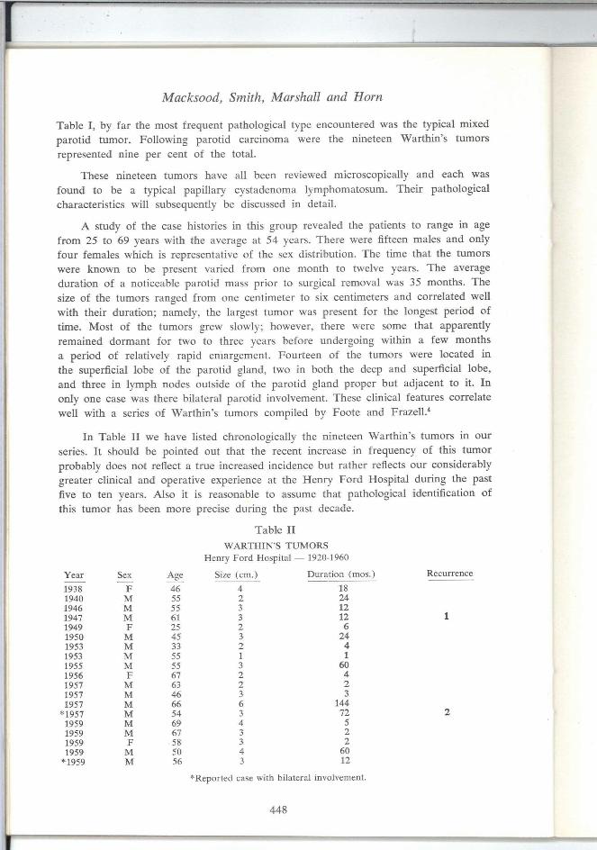
In Table I I we have listed chronologically the nineteen Warthin's tumors in our series. It should be pointed out that the recent increase in frequency of this tumor probably does not reflect a true increased incidence but rather reflects our considerably greater clinical and operative experience at the Henry Ford Hospital during the past five to ten years. Also it is reasonable to assume that pathological identification of this tumor has been more precise during the past decade.
Table I I
WARTHIN'S TUMORS Henry Ford Hospital — 1920-1960
Recurrence Year Sex Age Size (cm.) Duration (mos.
193H F 46 4 18 1940 M 55 2 24 1946 M 3 12 1947 M 61 3 12 1949 F 25 2 6 1950 M 45 3 24 1953 V! 33 2 4 1953 M 55 1 1 1955 VI
.•̂̂ 3 60
1956 F 67 2 4 1957 M 63 2 2 1957 M 46 3 3 1957 M 66 6 144 *1957 M 54 3 72 1959 M 69 4 5 1959 M 67 3 2 1959 F 58 3 2
1959 M 50 4 60
*1959 M 56 3 12
'Reported case with bilateral involvement.
448

Warthin's Tumor
The clinical characteristics of Warthin's tumors are such that they cannot be easily distinguished from other parotid tumors. Typically, they are painless, non-tender and non-exudative lesions which commonly present at the angle of the jaw and seldom become larger than five centimeters in size. They are firm to palpation and may be fixed to underlying structures but are usually not attached to the overlying skin.
Papdlary cystadenoma lymphomatosum ordinarily is diagnosed prior to surgery as a mixed parotid tumor. It also may be erroneously diagnosed as a branchial cleft cyst when the mass is fluctuant and lies below the parotid gland. A diagnosis of a tumor of lymphoid origin or a lymphadenitis may be made preoperatively when the lesion presents at a more distant location from the salivary gland."'̂ In only one of our cases was the correct diagnosis of Warthin's tumor made prior to surgery. Nine of our nineteen tumors were diagnosed preoperatively as mixed tumors of the parotid, three cases were diagnosed as branchial cleft cysts, two cases as carotid body tumor, two cases as cysts of the parotid gland, and two cases as lymphadenopathy.
The customary management of papdlary cystadenoma lymphomatosum has been local surgical excision. Irradiation is generally not indicated. As shown by our nineteen cases a limited surgical approach is usually quite satisfactory as there were only three instances of recurrence (Table 2). Local excision, however, wdl be inadequate if the tumor is multicentric. This is illustrated by the two previous recurrences in our reported case. I t would seem, therefore, that consideration should be given to performing a more extensive type of parotidectomy as the initial form of surgical therapy. Certainly if a secondary operation should become necessary it wid be technically more difficult and the hazard of facial nerve injury is considerably greater.
PATHOGENESIS
There has been considerable discussion concerning the origin of the papidary cystadenoma lymphomatosum. Originally Albrecht and Arzt thought that the tumor they described developed from heterotopic salivary gland tissue found in lymph nodes in the region of the parotid gland.' Warthin believed this tumor to be derived from an anlage of the eustachian tube because of the simdarity of the mucosal structure and the intimate relationship between the epithelium and lymphoid tissue, characteristic of pharyngeal mucous membranes in general.^ Some writers have expressed the belief that the papillary cystadenoma lymphomatosum arises primarily from salivary gland tissue and that the lymphoid constituent is only secondary.' Other investigators have maintained that the tumor arises from the ectoderm of branchial clefts in which the lymphoid and epithelial elements are likewise closely associated.'
Thompson and Bryant reported an excedent study where they made serial sections of the head and neck region of eighteen embryos in order to visualize the development of the salivary glands.^ They also compared 100 normal parotid and 50 normal sub-maxidary glands histologically with 50 branchogenic cysts and fistulas, and 23 examples of papillary cystadenoma lymphomatosum. They noted different fundamental features in the submaxillary and parotid glands of the embryos they studied. No lymphoid tissue was found by them in submaxillary tissue, while in parotid glands, lymphoid
449

Macksood, Smith, Marshall and Horn
tissue and lymph nodes were relatively common. On the basis of this study, they concluded that the papidary cystadenoma lymphomatosum arises from parotid ductal epithelial elements included in lymph nodes and is due to neoplastic proliferation of parotid ductal epithedum with concomitant proliferation of lymphoid tissue. They point out as a further basis for their contention that no examples of Warthin's tumor have been recorded in the literature except from within or near the parotid salivary gland. No unquestionable case of Warthin's tumor arising in a submaxdlary gland has been found in the literature. Bernier and Bhaskar have elaborated this study and detailed the points supporting the concept of origin of these tumors from salivary duct epithelium detained in lymphoid tissue within or adjacent to the parotid salivary gland.' This is currently the most popular theory and probably represents the correct origin of these tumors.
PATHOLOGY
The nineteen tumors studied in this laboratory were generally described as sodd, oval or lobular masses which varied from 1.5 to 4 centimeters in maximum dimensions. A capsule was described in fourteen cases. The majority of tumors were soft and contained cysts of varying size. The color varied from white to red-brown, the majority being tan and resembling lymphoid tissue. The cysts contained yellow turbid fluid or tenacious yellow-white material, frequently described as purulent or caseous. The
Figure 1 Gross photograph of papillary cystadenoma lymphomatosum showing fairly solid structure. Lobulation is striking.
450

Warthin's Tumor
gross appearance is often diagnostic and in most cases should be recognized at the operating table. There may be some variations depending largely on the number and size of the cysts (Figure 1 and 2).
Figure 2 Gross photograph (after fixation) of encapsulated papillary cystadenoma lymphomatosum lying parotid salivary gland. The intracystic papillary growth is evident.
within
The histologic appearance of papidary cystadenoma lymphomatosum is also quite characteristic. Cystic spaces are lined by a high columnar eosinophilic epithelium with prominent uniform nuclei. These nuclei have a tendency to palisade toward the lumen of the cyst. Underlying this are varying amounts of stroma containing lymphoid tissue, often with prominent reaction centers (Figure 3). The lumina of the cystic spaces generally contain an eosinophilic granular material and variable numbers of inflammatory cells and cellular debris.
Acicular clefts are noted not infrequently. Occasional goblet cells are present, which are muci-carmine and PAS positive. Epidermoid metaplasia is infrequently encountered, but does appear. Focal areas, in which the epithelial component predominates with little lymphoid tissue, are seen. A thin capsule was demonstrable in seven instances in this series. There was invagination of the parotid gland and fat into the tumor mass in one case (Figure 4) . The capsules include what appear to be
451

Macksood, Smith, Marshall and Horn
Figure 3 Papillary cystadenoma lymphomatosum showing columnar epithelium lining the cystic spaces and lymphoid tissue with reaction centers in the stroma. X115.
Figure 4 Invagination of sahvary gland and adipose tissue into papillary cystadenoma lymphomatosum. XI15.
452

Warthin's Tumor
lymphatics and in four instances an area containing a duct, blood vessel, and nerve, in close relationship, suggested a hilus. Prominent sub-epithelial fibrosis was seen in five tumors.
Two tumors in our series bore certain similarities to the cystic mucoepidermoid tumor of "favorable" prognosis, adding a further example of the overlap of certain patterns with those of other salivary gland tumors.'
Ducts of parotid salivary gland type were found in adjacent lymphoid tissue in six of the nine cases in which adequate tissue was avadable for study (Figure 5). One tumor, characterized clinically by multiple recurrences, was multicentric and in another case, the uninvolved salivary gland contained unencapsulated foci suggestive of incipient tumors of the same type. Two tumors were associated with sialadenitis and one with an adjacent, but distinct oxyphd adenoma.
Figure 5
Ducts of sahvary gland type in a lymph node adjacent to a papillary cystadenoma lymphomatosum. XI15
SUMMARY A N D CONCLUSIONS
Our experience with nineteen cases of Warthin's tumor of the parotid salivary gland is reviewed together with a report of one case with two recurrences and bdateral involvement.
Papdlary cystadenoma lymphomatosum is grossly, as well as microscopically characteristic, and should in most cases be recognized at the time of surgery. The
453

Macksood, Smith, Marshall and Horn
features which are most helpful in differentiating it from other lesions of the parotid
gland are the tan lymphoid appearance and the unusual cystic spaces containing turbid
material.
The bulk of evidence indicates the origin of these tumors to be from parotid duct
epithelial inclusions within lymphoid tissue in the region of the parotid gland and
adjacent soft tissue.
Because papidary cystadenoma lymphomatosum may be multicentric, and there
fore recurrence possible, it is suggested that consideration be given to total parotidectomy
as the primary form of surgical treatment.
BIBLIOGRAPHY
1. Warthin, A. S.: Papillary cystadenoma lymphomatosum; rare teratoid of parotid region, J. Cancer Res. 13:116, 1929.
2. Albrecht, H. and Arzt, L.: Beitrage zur Frage der Gewebsverirung. I . Papillare Cystade-nome in Lymphdriisen, Frankf. Ztschr. Path. 4:47, 1910.
3. Martin H. and Ehrlich, H. E.: Papillary cystadenoma lymphomatosum (Warthin's Tumor) of parotid salivary gland, Surg., Gynec. & Obst. 79:611, 1944.
4. Foote, F. W. and Frazell, E. L.: Tumors of the Salivary Glands. Atlas of Tumor Pathology, Sect. 4, Ease. 2, Washington, D. C, Armed Forces Institute of Pathology, 1954.
5. Bernier, J. L.: Management of Oral Disease, St. Louis, Mosby, 1955.
6. Anderson, W. A. D., ed.: Pathology, ed. 3, St, Louis, Mosby, 1957, p. 739.
7. Cunningham, W. F.: Branchial cysts of parotid gland, Ann. Surg. 90:114, 1929.
8. Thompson, A. S. and Bryant, H. C, Jr.: Histogenesis of papillary cystadenoma lympho-n-iatosum (Warthin's Tumor) of parotid salivary gland. Am. J. Path. 26:807, 1950.
9. Bernier, J. L. and Bhaskar, S. N.: Lymphoepithelial lesions of salivary glands; histogenesis and classification based on 186 cases. Cancer 11:1156, 1958.
454
Related Documents