MODULATION OF P-GL YCOPROTEIN-MEDIATED MUL TID RUG RESISTANCE IN THE CC531 RAT COLON TUMOR MODEL

Welcome message from author
This document is posted to help you gain knowledge. Please leave a comment to let me know what you think about it! Share it to your friends and learn new things together.
Transcript
MODULATION OF
P-GL YCOPROTEIN-MEDIATED MUL TID RUG RESISTANCE
IN THE CC531 RAT COLON TUMOR MODEL

W Gedrukt bij OHsetdrukkerij Ridderprint B. V., Ridderkerk
MODULATION OF
P-GL YCOPROTEIN-MEDIATED MUL TlDRUG RESISTANCE
IN THE CC531 RAT COLON TUMOR MODEL
MODULERING VAN MULTIDRUG RESISTENTIE
GEMEDIEERD DOOR P-GL YCOPROTEINE
IN HET COLON TUMOR MODEL CC531 IN DE RAT
PROEFSCHRIFT
ter verkrijging van de graad van doctor
aan de Erasmus Universiteit Rotterdam
op gezag van de rector magnificus
Prof. dr P_W.C. Akkermans, M.A.
en volgens besluit van het college voor promoties.
De openbare verdediging zal plaatsvinden op
woensdag 9 april 1997 om 15.45 uur
door
WILLEM VAN DE VRIE
geboren te Kattendijke
Promotlecommissie
Promotor
Overige leden
Copromotor
Prof. dr G. Stoter
Dr R.L. Marquet
Prof. dr J.W. Oosterhuis
Prof. dr R.J. Scheper
Dr A.M.M. Eggermont
The investigations presented in this thesis were mainly performed at the Laboratory for
Experimental Surgery of the Erasmus University Rotterdam, The Netherlands and at the
Department of Surgical Oncology, Dr. Daniel den Hoed Cancer Clinic, Rotterdam, The
Netherlands. Part of the work was done at the Laboratory of Cancer Research and
Clinical Oncology, Antwerp University, Wilrijk, Belgium and at the Laboratory of
Experimental Chemotherapy and Pharmacology, Department of Medical Oncology, Dr.
Daniel den Hoed Cancer Clinic, Rotterdam, The Netherlands.

aan Marian en ..
Modulation of P-glycoprotein-mediated multidrug resistance
Contents
1 General introduction
1.1 The problem of drug resistance in cancer therapy
1 .2 /n vivo model systems in P·glycoprotein-mediated multidrug
resistance
1.3 Aims of the thesis
2 Original studies
2.1 In vitro and in vivo chemosensitizing effect of cyclosporin
A on an intrinsic multidrug resistant rat colon tumor
2.2 Modulation of multidrug resistance with dexniguldipine in
the rat tumor model CC531
2.3 Pharmacokinetics of MDR-reversing drug dexniguldipine and
its pyridine metabolite M-l in plasma, tumor and renal tis
sue in tumor bearing WAG/RIJ rats
2.4 The chemosensitizer cyclosporin A enhances the toxic side
effects of doxorubicin in the rat
2.5 Cyclosporin A enhances locoregional metastasis of the CC531
rat colon tumor
2.6 Drug resistance in rat colon cancer cell lines is associated
with minor changes in susceptibility to cytotoxic cells
.. 11
.. 13
.. 63
.. 67
.. 83
.. 97
.111
.127
.137
Contents
3 Discussion and summary
3.1 General discussion .153
3.2 Summary .159
3.3 Samenvatting .163
4 Appendices
4.1 Abbreviations .169
4.2 Naschrift .171
4.3 Publications of the author .173
4.4 Curriculum vitae auctoris .175


1
GENERAL INTRODUCTION

Drug resistance in cancer
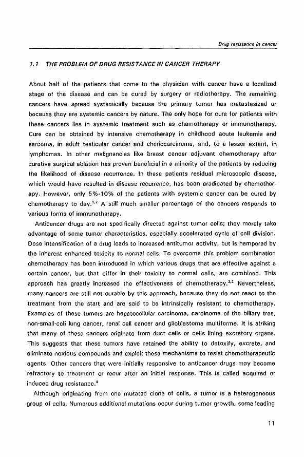
1.1 THE PROBLEM OF DRUG RESISTANCE IN CANCER THERAPY
About half of the patients that come to the physician with cancer have a localized
stage of the disease and can be cured by surgery or radiotherapy. The remaining
cancers have spread systemically because the primary tumor has metastasized or
because they are systemic cancers by nature. The only hope for cure for patients with
these cancers lies in systemic treatment such as chemotherapy or immunotherapy.
Cure can be obtained by intensive chemotherapy in childhood acute leukemia and
sarcoma, in adult testicular cancer and choriocarcinoma, and, to a lesser extent, in
lymphomas. In other malignancies like breast cancer adjuvant chemotherapy after
curative surgical ablation has proven beneficial in a minority of the patients by reducing
the likelihood of disease recurrence. In these patients residual microscopic disease,
which would have resulted in disease recurrence, has been eradicated by chemother
apy. However, only 5%-10% of the patients with systemic cancer can be cured by
chemotherapy to day.l,2 A still much smaller percentage of the cancers responds to
various forms of immunotherapy.
Anticancer drugs are not specifically directed against tumor cells; they merely take
advantage of some tumor characteristics, especially accelerated cycle of cell division.
Dose intensification of a drug leads to increased antitumor activity, but is hampered by
the inherent enhanced toxicity to normal cells. To overcome this problem combination
chemotherapy has been introduced in which various drugs that are effective against a
certain cancer, but that differ in their toxicity to normal cells, are combined. This
approach has greatly increased the effectiveness of chemotherapy.'" Nevertheless,
many cancers are still not curable by this approach, because they do not react to the
treatment from the start and are said to be intrinsically resistant to chemotherapy.
Examples of these tumors are hepatocellular carcinoma, carcinoma of the biliary tree,
non-small-cell lung cancer, renal cell cancer and glioblastoma multiforme. It is striking
that many of these cancers originate from duct cells or cells lining excretory organs.
This suggests that these tumors have retained the ability to detoxify, excrete, and
eliminate noxious compounds and exploit these mechanisms to resist chemotherapeutic
agents. Other cancers that were initially responsive to anticancer drugs may become
refractory to treatment or recur after an initial response. This is called acquired or
induced drug resistance. 4
Although originating from one mutated clone of cells, a tumor is a heterogeneous
group of cells. Numerous additional mutations occur during tumor growth, some leading
11
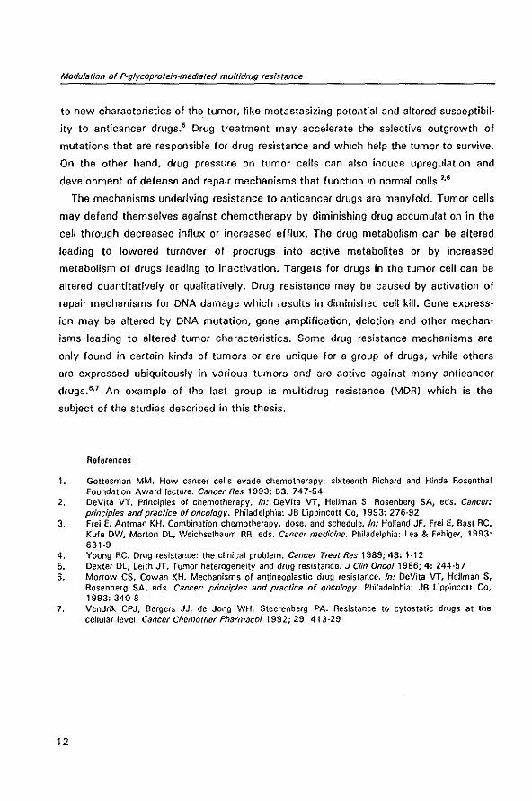
Modulation of P-glycoprotein·medialed mullidrug resistance
to new characteristics of the. tumor, like metastasizing potential and altered susceptibil
ity to anticancer drugs.5 Drug treatment may accelerate the selective outgrowth of
mutations that are responsible for drug resistance and which help the tumor to survive.
On the other hand, drug pressure on tumor cells can also induce upregulation and
development of defense and repair mechanisms that function in normal cells. 2,6
The mechanisms underlying resistance to anticancer drugs are manyfold. Tumor cells
may defend themselves against chemotherapy by diminishing drug accumulation in the
cell through decreased influx or increased efflux. The drug metabolism can be altered
leading to lowered turnover of prod rugs into active metabolites or by increased
metabolism of drugs leading to inactivation. Targets for drugs in the tumor cell can be
altered quantitatively or qualitatively. Drug resistance may be caused by activation of
repair mechanisms for DNA damage which results in diminished cell kill. Gene express
ion may be altered by DNA mutation, gene amplification, deletion and other mechan
isms leading to altered tumor characteristics. Some drug resistance mechanisms are
only found in certain kinds of tumors or are unique for a group of drugs, while others
are expressed ubiquitously in various tumors and are active against many anticancer
drugs.6•1 An example of the last group is multidrug resistance (MDR) which is the
subject of the studies described in this thesis.
References
1. Gottesman MM. How cancer cells evade chemotherapy: sixteenth Richard and Hinda Rosenthal Foundation Award lecture. Cancer Res 1993; 53: 747-54
2. DeVita VT. Principles of chemotherapy. In: DeVita VT, Hellman S, Rosenberg SA, eds. Cancer: principles and practice of oncology. Philadelphia: JB lippincott Co, 1993: 276·92
3. Frei E, Aotman KH. Combination chemotherapy, dose, and schedule, If/: Holland JF, Frei E, Bast RC, Kufe OW, Morton OL, Weichselbaum RR, eds. Cancer medicine. Philadelphia: Lea & Febiger, 1993: 631·9
4. Young RC. Drug resistance: the clinical problem. Cancer Treat Res 1989; 48: 1·12 5. Dexter DL, Leith JT. Tumor heterogeneity and drug resistance. J Clln Onco/1986; 4: 244·57 6. Morrow CS, Cowan KH. Mechanisms of antineoplastic drug resistance. In: DeVita VT, Hellman S,
Rosenberg SA, eds. Cancer: principles and practice of oncology. Philadelphia: JB lippincott Co, 1993, 340·8
7. Veodrik CPJ, Bergers JJ, de Jong WHo Steerenberg PA. Resistance to cytostatic drugs at the cellular level. Cancer Chemother Pharmacal 1992; 29: 413-29
12
1.2
IN VIVO MODEL SYSTEMS IN
P-GL YCOPROTEIN-MEDIATED
MUL TIDRUG RESISTANCE
Wim van de Vrie, Richard L. Marquet,
Gerrit Stater, Ernst A. de Bruijn
and Alexander M.M. Eggermont
(submitted for publication)
Modulation of P·glycoprotein-mediated multidrug resistance
Summary
In this article we will review the in vivo model systems that have been developed for
studying P-glycoprotein-mediated multidrug resistance (MDR) in the preclinical setting.
Rodents have two mdr genes that both confer the MDR phenotype: mdrta and mdrtb.
At gene level they show strong homology to the human MORt gene and the tissue
distribution of their gene product is very similar to P-glycoprotein expression in humans.
In vivo studies have shown the physiological roles of P'glycoprotein among which
protecting the organism from damage by xenobiotics. Tumors with intrinsic P
glycoprotein expression, induced MOR or transfected with an mdr gene can be used as
syngeneic or xenogenic tumor models. Ascites, leukemia, and solid MDR tumor models
have been developed. Molecular engineering has resulted in transgenic mice that
express the human MDR 1 gene in their bone marrow, and in knockout mice missing
murine mdr genes. The data on pharmacokinetics, efficacy and toxicity of reverters of
P-glycoprotein in vivo are described. Results from studies using monoclonal antibodies
directed against P-glycoprotein and other miscellaneous approaches for modulation of
MDR are mentioned. The importance of in vivo studies prior to clinical trials is being
stressed and potential pitfalls due to differences between species are discussed.
14
In vivo model systems in MDR
1. Introduction: the problem of multidrug resistance in human cancer
A major obstacle for successful chemotherapy of cancer is the resistance of tumors
to anticancer drugs. Resistance may be caused by an intrinsic resistance to anticancer
drugs or by the emergence of drug-resistant cells during chemotherapeutic treatment.
In recent years several mechanisms causing drug resistance have been elucidated:
decreased uptake or increased efflux of drugs by tumor cells, alterations in target pro
teins, cellular drug metabolism or drug binding, and enhancement of DNA repair
mechanisms. Some mechanisms affect only a specific drug, while others cause
resistance to a wide variety of drugs.
An important mechanism, which has been observed in many different malignancies
and which affects various groups of unrelated anticancer drugs is called multidrug
resistance (MDR)' Sometimes the prefix classical is added to distinguish classical MDR
from other forms of pleiotropic drug resistance. In classical MDR the mechanism of
drug resistance is an energy-dependent, unidirectional transmembrane efflux pump,
called P-glycoprotein or P-170, that extrudes drugs and other xenobiotics out of the
cell. Thus intracellular levels of these compounds can be kept under a non-cytotoxic
level. The efflux pump has a broad substrate specificity affecting drugs as anthracy
clines, epipodophyllotoxins, Vinca alkaloids, taxanes, colchicine, topotecan, and
actinomycin D. Therefore, tumors expressing the MDR phenotype are cross-resistant to
a wide variety of structurally unrelated drugs. Many compounds that have no
antineoplastic activity can also interact with the P-glycoprotein efflux pump and block
its function. This leads to increased intracellular levels of cytotoxins that are substrates
for P-glycoprotein and to enhanced cell death. Compounds that can block P
glycoprotein are termed MDR modulators, reverters, or chemosensitiz8rs. I-J
Studies on the expression of the mdr gene or its product P-glycoprotein in human
tumors are difficult to interpret as various methods with varying sensitivity and
specificity, often leading to conflicting results, have been employed by the investiga
tors. Molecular methods for the detection and measurement of the mdr DNA and mRNA
are generally sensitive and quantitative, the most sensitive being the reverse transcrip
tase polymerase chain reaction. However, contamination of the tumor sample with
normal cells and heterogeneous expression of the mdr gene within the tumor can not
be detected. Immunohistochemical assays with specific monoclonal antibodies against
P-glycoprotein can detect P-glycoprotein expression at the individual cell, but the
sensitivity is generally less than in molecular techniques and the measurement of p~
15
Modulation of P-glycoprotein-mediated multidrug resistance
glycoprotein level is semi·quantitative. Functional assays measure the actual activity of
the P-glycoprotein efflux pump in tumor cells. This technique is currently only available
for hematological malignancies.4.5 Recently, recommendations have been published for
standardization of methods to detect P-glycoprotein·associated MDR.6
In general, it can be stated that tumors originating from tissues with high expression
levels of P-glycoprotein have clear P'glycoprotein expression as well and are often
intrinsically refractory to chemotherapy. Among these are cotoractal cancer, renal cell
carcinoma, hepatoma, adrenocortical carcinoma and pancreatic cancer.J.9 Results of p.
glycoprotein expression in breast cancer and soft tissue sarcoma are variable,8.s while
in lung cancer, ovarian cancer, and melanoma levels of MDR expression are low to
absent.8.s For some of these solid tumors higher levels of MDR expression have been
reported after relapse or failure of chemotherapy with MDR substrates, e.g. breast
cancer,8.10 ovarian cancer,'! and neuroblastoma. '2 For more extensive information about
the expression of MDR in solid tumors, the reader is referred to some specific
reviews. '3. 14
In hematological tumors MDR overexpression is frequently observed in acute myeloid
leukemia, while its expression in acute lymphoblastic leukemia is generally IOW.8•9,15,HI
Secondary acute myeloid leukemia and disease recurrence after chemotherapy are
associated with a markedly higher frequency of MDR expression. 16,17 In lymphoma and
myeloma P·glycoprotein expression is infrequent in newly diagnosed cases, but
common in recurrent disease after chemotherapy.'S,19 In specific reviews the results of
P-glycoprotein expression in hematological malignancies are summarized. 20-21
Several studies have brought forward evidence that P·glycoprotein expression has
prognostic significance in certain malignancies_ In neuroblastoma and childhood
sarcoma P·glycoprotein expression is associated with poor response to chemotherapy,
increased chance of relapse, and decreased survival.22.23 In acute leukemia there is a
correlation with reduced frequency and duration of complete remission. '5,16,24 However,
for many tumors studies with results contradicting other reports have been published
and the exact significance of P-glycoprotein-mediated MDR remains to be defined. 25
The remainder of this review will concentrate on in vivo studies on the physiological
functions of P·glycoprotein and on in vivo model systems that have been developed for
studying modulation of P·glycoprotein-mediated MDR. The similarities and differences
in MDR·related P-glycoproteins between species will be outlined. The validity of the
animal models for studying MDR and their relevance to the clinical situation in humans
16
In vivo model systems In MDR
will be discussed.
2. Mdt gene expression and function across species
2.1. The mdr superfamily of genes
The human P-glycoprotein is a 170-kilodalton protein which consists of 1 280 amino
acids. The putative structure of P-glycoprotein consists of two homologous halves each
containing 6 transmembrane regions and a large intracytoplasmatic loop encoding an
ATP-binding site. Together they form the functional multidrug transporter. 26 The gene
encoding for P-glycoprotein is highly conserved during evolution and belongs to a
superfamily of membrane-associated transport proteins: the ATP-binding cassette
(ABC) family. This family includes, amongst others, bacterial transporters, the STE-6
transporter in yeast, the Plasmodium chloroquine resistance gene, the Leishmania
resistance gene, Drosophila genes, and the cystic fibrosis gene CTFR. Proteins of the
ABC family are transporters of various nutrients, peptides, polysaccharides, toxins and
drugs. 27. 29
In humans two mdr genes have been detected, from which only MDR1 encodes for
the MDR-related P-glycoprotein efflux pump and confers the MDR phenotype. 26.3~32 The
function of the second mdr gene product in humans, MDR3 or MOR2 (for nomenclature
see Table 1). has only recently been elucidated. but it is not a transporter of drugs used
in chemotherapy. Rodents have 3 mdr genes, from which class 1 and 2 have been
shown to confer the MDR phenotype."'" In mouse and rat these genes are called
mdrTa and mdrTb (in mice also designated as mdr3 and mdrT respectively, and in rat
as pgp1 and pgp2 respectively);35.35 in hamster they are named pgp1 and pgp2. 27•37
Table 1 gives an outline of the mdr genes in various species and the nomenclature.
Comparison of coding sequences of the various mdr genes shows high homology and
sequence identity. The homology is higher between genes of the classes 1 and 2
versus the genes of class 3, consistent with the different abilities of their products to
transport drugs. The mouse mdr1a and mdr1b genes show 83% identity to each other,
while they have 73% and 71 % identity with the mdr2 gene respectively." The human
MORT and MOR3 coding sequences are over 75% identical. The higher identity within
classes is retained when mdr genes of the same class are compared across species.
Sequence identity of human MORt and mouse mdrfa is 82% and of human MORt and
mouse mdr1b 79%. Similar homology is found with hamster and rat class 1 and 2 mdr
17
Modulation of P·glycoprotein-mediated multldrug resistance
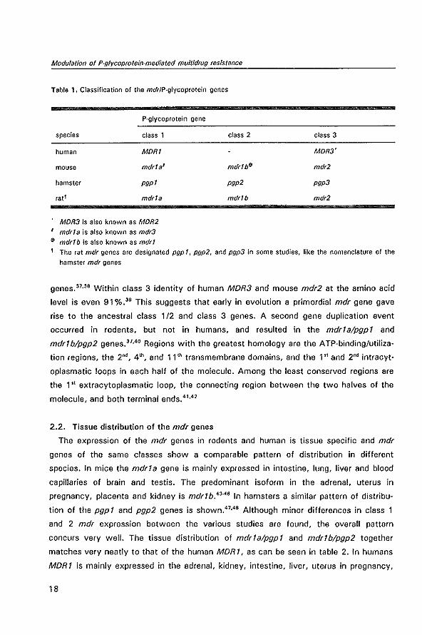
Tabla 1. Classification of the mdrlP·glycoprotein genes
P·glycoprotein gene
species class 1
human MORI
mouse mdr1a'
hamster pgp1
fat f mdr1a
MDR3 is also known as MDR2
, mdr!a is also known as mdr3
~ mdrlb is also known as mdr!
class 2
mdr1bf}
pgp2
mdrlb
class 3
MOR3'
mdr2
pgp3
mdr2
1 The rat mdr genes are designated pgp 1, pgp2, and pgp3 in some studies, like the nomenclature of the
hamster mdr genes
genes.37,38 Within class 3 identity of human MOR3 and mouse mdr2 at the amino acid
level is even 91 %.39 This suggests that early in evolution a primordial mdr gene gave
rise to the ancestral class 1/2 and class 3 genes. A second gene duplication event
occurred in rodents, but not in humans, and resulted in the mdrlalpgpl and
mdrTblpgp2 genes."·40 Regions with the greatest homology are the ATP·binding/utiliza·
tion regions, the 2nd, 4th, and 11th transmembrane domains, and the 1" and 2m! intracyt
oplasmatic loops in each half of the molecule. Among the least conserved regions are
the 1 st extracytoplasmatic loop, the connecting region between the two halves of the
molecule, and both terminal ends.41.42
2.2. Tissue distribution of the mdr genes
The expression of the mdr genes in rodents and human is tissue specific and mdr
genes of the same classes show a comparable pattern of distribution in different
species. In mice the mdrla gene is mainly expressed in intestine, lung, liver and blood
capillaries of brain and testis. The predominant isoform in the adrenal, uterus in
pregnancy, placenta and kidney is mdrlb,4J.46 In hamsters a similar pattern of distribu
tion of the pgpl and pgp2 genes is shown.47.4S Although minor differences in class 1
and 2 mdr expression between the various studies are found, the overall pattern
concurs very well. The tissue distribution of mdrTalpgpT and mdrTblpgp2 together
matches very neatly to that of the human MDRT, as can be seen in table 2. In humans
MORI is mainly expressed in the adrenal, kidney, intestine, liver, uterus in pregnancy,
18
In vlvo modal systems in MOR
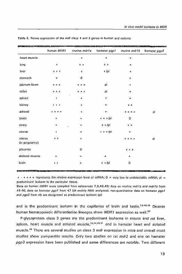
Table 2. Tissue expression of the mdr class 1 and 2 genes in human and rodents
human MORI murine mdrla hamster pgp I murine mdr!b hamster pgp2
heart muscle + + +
lung + ++ ++ +
liver +++ + +/pi +
stomach + 0 +
jejunum-ileum +++ +++ p; +
colon +++ +++ p; +
spleen + + + +
kidney +++ + + ++
adrenal ++++ + + ++++ p;
testis + + + + +/pi 0
ovary + + + +/pi ++
uterus + + + + +/pi +
uterus ++ + ++++ p; (in pregnancy)
placenta 0 +++
skeletal muscle + + + +
brain ++ + + +/pi 0
+ . + + + + represents the relative expression level of mANA; 0 = very low to undetectable mANA; pi =
predominant isoform in the particular tissue.
Data on human MOR! were compiled from references 7,9,46,49; data on murine mdrla and mdr!b from
44-46; data on hamster pgp! from 47 (all mainly ANA analyses); non-Quantitative data on hamster pgp! and pgp2 from 45 are designated as predominant isoform (pi)
and is the predominant isoform in the capillaries of brain and testis. 7•9.46,49 Diverse
human hematopoietic differentiation lineages show MORt expression as wel1.50
P-glycoprotein class 3 genes are the predominant isoforms in mouse and rat liver,
spleen, heart muscle and striated muscle,39,44,45.51 and in hamster heart and striated
muscle.46 There are several studies on class 3 mdr expression in mice and overall most
studies show comparable results, Only two studies on rat mdr2 and one on hamster
pgp3 expression have been published and some differences are notable. Two different
19
Modulation of P-glycoprotein-mediated multidrug resistance
cDNA clones have been reported for the rat mdr2, with a mismatch in nucleotide bases
between the two sequences resulting in nucleotide differences for four amino acids,51.52
Work with the first DNA sequence derived from the Fischer 344 rat strain revealed a
distribution pattern of mdr2 in the rat comparable to that in the mouseY With the
second mdr2 cDNA cloned from the Sprague-Dawley rat strain however, high express
ion levels were detected in liver, but also in gastrointestinal tract. low levels in brain,
heart and kidney, and undetectable levels in spleen and striated skeletal muscle in this
rat strain,52 If these results are confirmed with additional studies, this would indicate
that strain differences exist within species in expression level of the various P
glycoproteins, Seen the similarity in distribution of the various mdr genes across
species and the differences in function between class 1 and 2 versus class 3 P
glycoproteins (vide infra) this is not very likely. In hamsters the predominant isoform in
the liver would be pgp1,4S while in other species this is class 3 mdr, As both isoforms
are found in the liver, the differences may be based on factors as differences in
investigational techniques, These inconsistencies left aside, the distribution of rodent
and human class 3 P-glycoprotein is quite similar. See table 3. In humans high express
ion levels of MDR3 are found in the Iiver,39,49 and with specific monoclonal antibodies
only in this organ expression of the MDR3 P-glycoprotein is shown. 39,53 Low expression
levels of MDR3 mRNA are found in human adrenal, spleen, heart, and striated
muscle.39.49
Immunohistochemical and in situ hybridization studies have shown that within the
tissues the P-glycoproteins have specific subcellular localizations, with a similar pattern
of distribution in human and rodents, In epithelial cells with a polarized excretion or
absorption function P-glycoprotein is mainly expressed at the apical surface that lines
the lumen. The MOR1 product is demonstrated in the brush border of the proximal
tubules of the kidney, the biliary canalicular surface of the hepatocytes, the apical
surface of columnar epithelial cells in small and large intestine, and luminal surface of
the cells in the pancreatic ductules.54•55 In rat kidney, liver, and intestine a comparable
subcellular distribution of P·glycoprotein is detected, and in the pancreas acinar cells
were stained with a specific monoclonal antibody (C219).56 In mouse and hamster class
1 P-glycoprotein is expressed in colonic epithelial cells in a polarized manner. 48,53.57 In
the gravid uterus of the mouse mdr 1 b is expressed at high levels in the secretory
epithelial cells of the endometrium. 43•53 At blood-tissue barriers of the central nervous
system and testis, and in the papillary dermis, MDRl is expressed at high level in
endothelial cells,55.56.58 P-glycoprotein expression in endothelial cells of the brain is
20
In vivo model systems In MDR
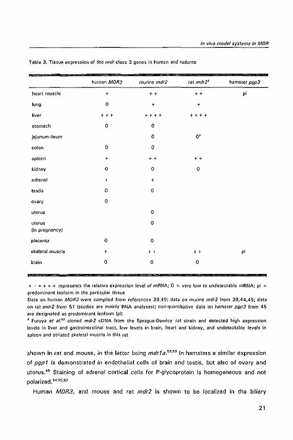
Tabla 3, Tissue expression of the mdr class 3 genes in human and rodents
human MDR3 murine mdr2 rat mdr2' hamster pgp3
heart muscle + ++ ++ pi
lung 0 + +
liver +++ ++++ ++++
stomach 0 0
jejunum-ileum 0 0'
colon 0 0
spleen + ++ ++
kidney 0 0 0
adrenal + +
testis 0 0
ovary 0
uterus 0
uterus 0 (in pregnancy)
placenta 0 0
skeletal muscle + ++ ++ pi
brain 0 0 0
+ . + + + + represents the relative expression level of mRNA; 0 :: very low to undetectable mRNA; pi =
predominant isoform in the particular tissue
Data on human MDR3 were compiled from references 39,49; data on murine mdr2 from 39,44,45; data on rat mdr2 from 51 (studies are mainly RNA analyses); non-quantitative data on hamster pgp3 from 45
are designated as predominant isoform (piJ
, Furuya et afY cloned mdr2 eDNA from the Sprague-Dawley rat strain and detected high expression
levels in liver and gastrointestinal tract, low levels in brain, heart and kidney, and undetectable levels in spleen and striated skeletal muscle in this rat
shown in rat and mouse, in the latter being mdrla.5s,59 In hamsters a similar expression
of pgp 1 is demonstrated in endothelial cells of brain and testis, but also of ovary and
uterus,48 Staining of adrenal cortical cells for P'glycoprotein is homogeneous and not
polarized. 54,55,57
Human MDR3, and mouse and rat mdr2 is shown to be localized in the biliary
21
Modulation 01 P-glycoprotein-mediated multidrug resistance
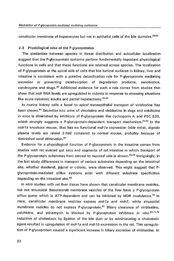
canalicular membrane of hepatocytes but not in epithelial cells of the bile ductules.39•53
2.3 Physiological roles of the P'glycoprotelns
The similarities between species in tissue distribution and subcellular localization
suggest that the P'glycoprotein isoforms perform fundamentally important physiological
functions in cells and that these functions are retained across species. The localization
of P-glycoprotein at the apical side of cells that line luminal surfaces in kidney, liver and
intestine is consistent with a putative detoxification role for P-glycoprotein mediating
excretion or preventing (re)absorption of degradation products, xenobiotics,
carcinogens and drugs. 6o Additiona! evidence for such a role comes from studies that
show that mdr RNA levels are upregulated in rodents in response to stressing situations
like acute cytotoxic insults and partial hepatectomy.61,62
In murine kidney cells a basal to apical transepithelial transport of vinblastine has
been shown.63 Secretion into urine of vincristine and vinblastine in dogs and colchicine
in mice is diminished by inhibitors of P-glycoprotein like cyclosporin A and PSC 833,
which strongly suggests a P-glycoprotein-dependent transport mechanism.e4-6S In the
mdr1a knockout mouse, that has no functional mdrfa expression (vide infra), digoxin
plasma levels are raised 2-fold compared to normal mouse, probably because of
diminished renal elimination.67
Evidence for a physiological function of P-glycoprotein in the intestine comes from
studies with rat everted gut sacs and segments of rat intestine in which transport of
the P-glycoprotein substrates from serosal to mucosal side is shown.6s,69 Intriguingly, in
the last study differences in transport of various substrates depending on the intestinal
site. whether duodenal. jejunal or colonic, were observed. This might suggest that p.
glycoprotein-mediated efflux systems exist with different substrate specificities
depending on the intestinal site,69
In vitro studies with rat liver tissue have shown that canalicular membrane vesicles,
but not sinusoidal (basolateral) membrane vesicles of the liver have a P-glycoprotein
efflux pump which is ATP·dependent and can be inhibited by MDR modulators.70 In
mice, canalicular membrane vesicles express mdrfa and mdr2, while sinusoidal
membrane vesicles do not express P·glycoprotein.53 Biliary clearance of vinblastine,
colchicine, and adriamycin is blocked by P-glycoprotein inhibitors in viVO. 65,71.72
Induction of cholestasis by ligation of the bile duct or by administering a cholestatic
agent resulted in up regulation of mdrf a and mdr1 b expression in the rat. This upregula
tion of P-glycoprotein caused a significant increase in biliary excretion of vinblastine. In
22
In vivo model systems in MDR
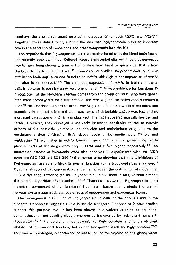
monkeys the cholestatic agent resulted in upregulation of both MOR1 and MOR3.7I
Together, these data strongly support the idea that P'glycoprotein plays an important
role in the secretion of xenobiotics and other compounds into the bile.
The hypothesis that P'glycoprotein has a protective function at the blood·brain barrier
has recently been confirmed. Cultured mouse brain endothelial cell lines that expressed
mdr1 b have been shown to transport vincristine from basal to apical side, that is from
the brain to the blood luminal side." In most rodent studies the predominant isoform of
mdr in the brain capillaries was found to be mdr1a, although minor expression of mdr1b
has also been observed.44,73 The enhanced expression of mdr1b in brain endothelial
cells in cultures is possibly an in vitro phenomenon. 74 In vivo evidence for functional P
glycoprotein at the blood-brain barrier comes from the group of Borst, who have gener
ated mice homozygous for a disruption of the mdr1a gene, so called mdr1a knockout
mice.59 No functional expression of the mdr1a gene could be shown in these mice, and
especially in gut epithelium and brain capillaries all detectable mdr1 a was lost and no
increased expression of mdr1b was observed. The mice appeared normally healthy and
fertile. However, they displayed a markedly increased sensitivity to the neurotoxic
effects of the pesticide ivermectin, an acaricide and anthelmintic drug, and to the
carcinostatic drug vinblastine. Brain tissue levels of ivermectin were 87-fold and
vinblastine 22-fold higher in mdr1 a knockout mice compared to normal mice, while
plasma levels of the drugs were only 3.3-fold and 2-fold higher respectively." The
neurotoxic effects of ivermectin were also observed in experiments with the MDR
reverters PSC 833 and SOZ 280·446 in normal mice showing that potent inhibitors of
P'glycoprotein are able to block its normal function at the blood-brain barrier in vivo. 75
Coadministration of cyclosporin A significantly increased the distribution of rhodamine-
123, a dye that is transported by P-glycoprotein, to the brain in rats, without altering
the plasma disposition of rhodamine-123. 76 These data show that P-glycoprotein is an
important component of the functional blood-brain barrier and protects the central
nervous system against deleterious effects of endogenous and exogenous toxins.
The homogenous distribution of P-glycoprotein in cells of the adrenals and in the
placental trophoblast suggests a role in steroid transport. Evidence of in vitro studies
support this putative role. It has been shown that various steroids as cortisone,
dexamethasone, and possibly aldosterone can be transported by rodent and human P
glycoprotein.17.78 Progesterone binds strongly to P-glycoprotein and is an efficient
inhibitor of its transport function, but is not transported itself by P-glycoprotein.77-79
Together with estrogen, progesterone seems to induce the expression of P·glycoprotein
23
Modulation of P-glycoproteln-mediated multidrug resistance
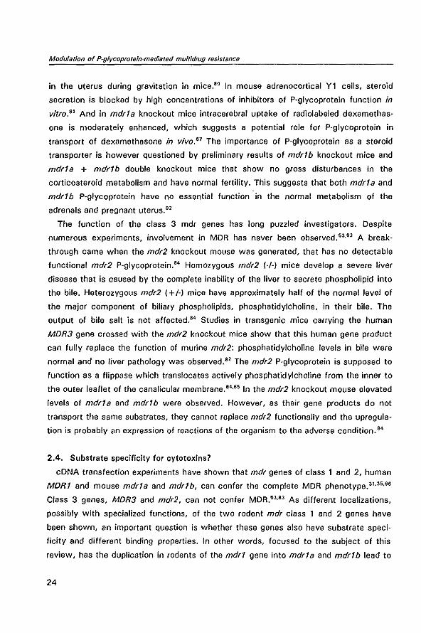
in the uterus during gravitation in mice.so In mouse adrenocortical Yl cells, steroid
secretion is blocked by high concentrations of inhibitors of P-glycoprotein function in
vitro. S! And in mdrla knockout mice intracerebral uptake of radiolabeled dexamethas
one is moderately enhanced, which suggests a potential role for P'glycoprotein in
transport of dexamethasone in vivo. s1 The importance of P-glycoprotein as a steroid
transporter is however questioned by preliminary results of mdrlb knockout mice and
mdrla + mdrlb double knockout mice that show no gross disturbances in the
corticosteroid metabolism and have normal fertility. This suggests that both mdrla and
mdrlb P'glycoprotein have no essential function in the normal metabolism of the
adrenals and pregnant uterus.82
The function of the class 3 mdr genes has long puzzled investigators. Despite
numerous experiments, involvement in MDR has never been observed. 53.83 A break
through came when the mdr2 knockout mouse was generated, that has no detectable
functional mdr2 P-glycoprotein.s4 Homozygous mdr2 (-/-) mice develop a severe liver
disease that is caused by the complete inability of the liver to secrete phospholipid into
the bile. Heterozygous mdr2 (+/.) mice have approximately half of the normal level of
the major component of biliary phospholipids, phosphatidylcholine, in their bile. The
output of bile salt is not affected.84 Studies in transgenic mice carrying the human
MOR3 gene crossed with the mdr2 knockout mice show that this human gene product
can fully replace the function of murine mdr2: phosphatidylcholine levels in bile were
normal and no liver pathology was observed. 82 The mdr2 P-glycoprotein is supposed to
function as a flippase which translocates actively phosphatidylcholine from the inner to
the outer leaflet of the canalicular membrane. 84,85 In the mdr2 knockout mouse elevated
levels of mdrfa and mdrlb were observed. However, as their gene products do not
transport the same substrates, they cannot replace mdr2 functionally and the upregula
tion is probably an expression of reactions of the organism to the adverse condition. S4
2.4. Substrate specificity for cytotoxins?
eDNA transfection experiments have shown that mdr genes of class 1 and 2, human
MORI and mouse mdrla and mdrlb, can confer the complete MDR phenotype."·35."
Class 3 genes, MDR3 and mdr2, can not confer MDR.53,83 As different localizations,
possibly with specialized functions, of the two rodent mdr class 1 and 2 genes have
been shown, an important question is whether these genes also have substrate speci
ficity and different binding properties. In other words, focused to the subject of this
review, has the duplication in rodents of the mdrl gene into mdrla and mdrlb lead to
24
In vivo model systems In MDR
different abilities to extrude cytotoxins?
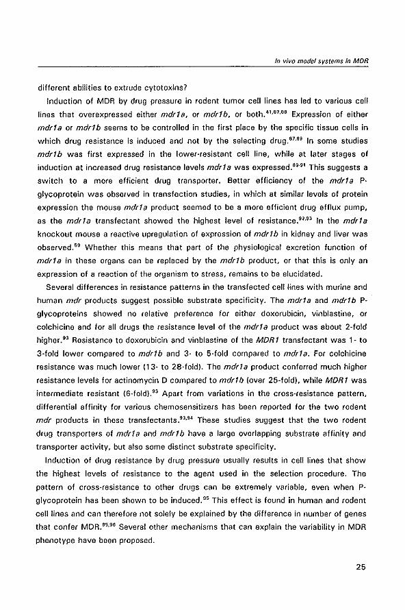
Induction of MDR by drug pressure in rodent tumor cell lines has led to various cell
lines that overexpressed either mdr1a, or mdr1b, or both.41,67.66 Expression of either
mdrf a or mdr1 b seems to be controlled in the first place by the specific tissue cells in
which drug resistance is induced and not by the selecting drug,61.S9 In some studies
mdr1b was first expressed in the lower-resistant cell line, while at later stages of
induction at increased drug resistance levels mdrfa was expressed.s9.9! This suggests a
switch to a more efficient drug transporter. Better efficiency of the mdr1a P
glycoprotein was observed in transfection studies, in which at similar levels of protein
expression the mouse mdrf a product seemed to be a more efficient drug efflux pump,
as the mdrfa transfectant showed the highest level of resistance.92.93 In the mdr1a
knockout mouse a reactive upregulation of expression of mdr1 b in kidney and liver was
observed,59 Whether this means that part of the physiological excretion function of
mdr1a in these organs can be replaced by the mdrlb product, or that this is only an
expression of a reaction of the organism to stress, remains to be elucidated.
Several differences in resistance patterns in the transfected cell lines with murine and
human mdr products suggest possible substrate specificity. The mdrfa and mdr1b P
glycoproteins showed no relative preference for either doxorubicin, vinblastine, or
colchicine and for all drugs the resistance level of the mdr1a product was about 2-fold
higher.93 Resistance to doxorubicin and vinblastine of the MOR1 transfectant was 1- to
3-fold lower compared to mdrlb and 3- to 5-fold compared to mdrla. For colchicine
resistance was much lower (13- to 28-foldi. The mdrla product conferred much higher
resistance levels for actinomycin 0 compared to mdrl b (over 25-foldl. while MDR 1 was
intermediate resistant (6-fold).93 Apart from variations in the cross-resistance pattern,
differential affinity for various chemosensitizers has been reported for the two rodent
mdr products in these transfectants,93.94 These studies suggest that the two rodent
drug transporters of mdr1 a and mdrl b have a large overlapping substrate affinity and
transporter activity, but also some distinct substrate specificity.
Induction of drug resistance by drug pressure usually results in cell lines that show
the highest levels of resistance to the agent used in the selection procedure. The
pattern of cross-resistance to other drugs can be extremely variable, even when P
glycoprotein has been shown to be induced. 95 This effect is found in human and rodent
cell lines and can therefore not solely be explained by the difference in number of genes
that confer MOR.95.9S Several other mechanisms that can explain the variability in MOR
phenotype have been proposed.
25
Modulation of P-glycoprotein·mediated muftidrug resistance
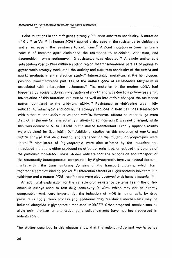
Point mutations in the mdr genes strongly influence substrate specificity. A mutation
of Gly '8s to Vallas in human MORt caused a decrease in the resistance to vinblastine
and an increase in the resistance to colchicine.97 A point mutation in transmembrane
zone 6 of hamster pgp 1 diminished the resistance to colchicine, vincristine, and
daunorubicin, while actinomycin D resistance was elevated.95 A single amino acid
substitution (Ser to Phe) within a coding region for transmembrane part 11 of murine P
glycoprotein strongly modulated the activity and substrate specificity of the mdrla and
mdrlb products in a transfection study.9E1 Interestingly, mutations at the homologous
position (transmembrane part 11) of the pfmdrl gene of Plasmodium falciparum is
associated with chloroquine resistance. 99 The mutation in the murine eDNA had
happened by accident during construction of mdr 1 b and was due to a polymerase error.
Introduction of this mutation into mdrfb as well as into mdrla changed the resistance
pattern compared to the wild-type cDNA.9E1 Resistance to vinblastine was mildly
reduced, to adriamycin and colchicine strongly reduced in both cell lines transfected
with either mutant mdrta or mutant mdrfb. However, effects on other drugs were
distinct: in the mdrta transfectant sensitivity to actinomycin 0 was not changed. while
this was decreased 5- to 10-fold in the mdrl b transfectant. Exactly opposite results
were obtained for Gramicidin 0.92 Additional studies on this mutation of mdrta and
mdrl b showed that drug binding and transport of the mutant P-glycoproteins were
altered." Modulators of P-glycoprotein were also affected by the mutation: the
introduced mutations either produced no effect. or enhanced, or reduced the potency of
the particular modulator. These studies indicate that the recognition and transport of
the structurally heterogeneous compounds by P-glycoprotein involves several determi
nants within the transmembrane domains of the transport proteins. which form
together a complex binding pocket.94 Differential effects of P-glycoprotein inhibitors in a
wild-type and a mutant MOR transfectant were also observed with human material. 'oo
An additional explanation for the variable drug resistance patterns lies in the differ
ences in assays used to test drug sensitivity in vitro. which may not be directly
comparable. And, very importantly, the induction of MDR in tumor cells by drug
pressure is not a clean process and additional drug resistance mechanisms may be
induced alongside P-glycoprotein-mediated MDR.93.95 Other proposed mechanisms as
allele polymorphism or alternative gene splice variants have not been observed in
rodents sofar.
The studies described in this chapter show that the rodent mdrla and mdrlb genes
26
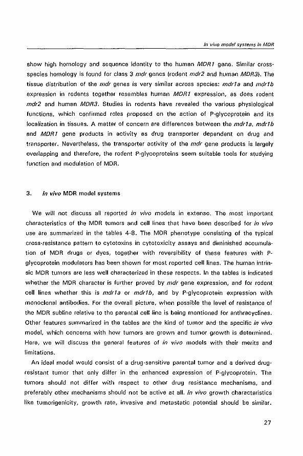
In vivo model systems in MDR
show high homology and sequence identity to the human MDR 1 gene. Similar cross*
species homology is found for class 3 mdr genes (rodent mdr2 and human MDR31. The
tissue distribution of the mdr genes is very similar across species: mdrfa and mdrlb
expression in rodents together resembles human MDR 1 expression, as does rodent
mdr2 and human MDR3. Studies in rodents have revealed the various physiological
functions, which confirmed roles proposed on the action of P*glycoprotein and its
localization in tissues. A matter of concern are differences between the mdrla, mdrlb
and MDR 1 gene products in activity as drug transporter dependent on drug and
transporter. Nevertheless, the transporter activity of the mdr gene products is largely
overlapping and therefore, the rodent P*glycoproteins seem suitable tools for studying
function and modulation of MDR.
3. In vivo MDR model systems
We will not discuss all reported in vivo models in extenso. The most important
characteristics of the MDR tumors and cell lines that have been described for in vivo
use are summarized in the tables 4-8. The MDR phenotype consisting of the typical
cross-resistance pattern to cytotoxins in cytotoxicity assays and diminished accumula*
tion of MDR drugs or dyes, together with reversibility of these features with P*
glycoprotein modulators has been shown for most reported cell lines. The human intrin
sic MDR tumors are less well characterized in these respects. In the tables is indicated
whether the MDR character is further proved by mdr gene expression, and for rodent
cell lines whether this is mdrfa or mdrlb, and by P-glycoprotein expression with
monoclonal antibodies. For the overall picture, when possible the level of resistance of
the MDR subline relative to the parental cell line is being mentioned for anthracyclines.
Other features summarized in the tables are the kind of tumor and the specific in vivo
model, which concerns with how tumors are grown and tumor growth is determined.
Here, we will discuss the general features of in vivo models with their merits and
limitations.
An ideal model would consist of a drug-sensitive parental tumor and a derived drug*
resistant tumor that only differ in the enhanced expression of P*glycoprotein. The
tumors should not differ with respect to other drug resistance mechanisms, and
preferably other mechanisms should not be active at all. In vivo growth characteristics
like tumorigenicity, growth rate, invasive and metastatic potential should be similar.
27
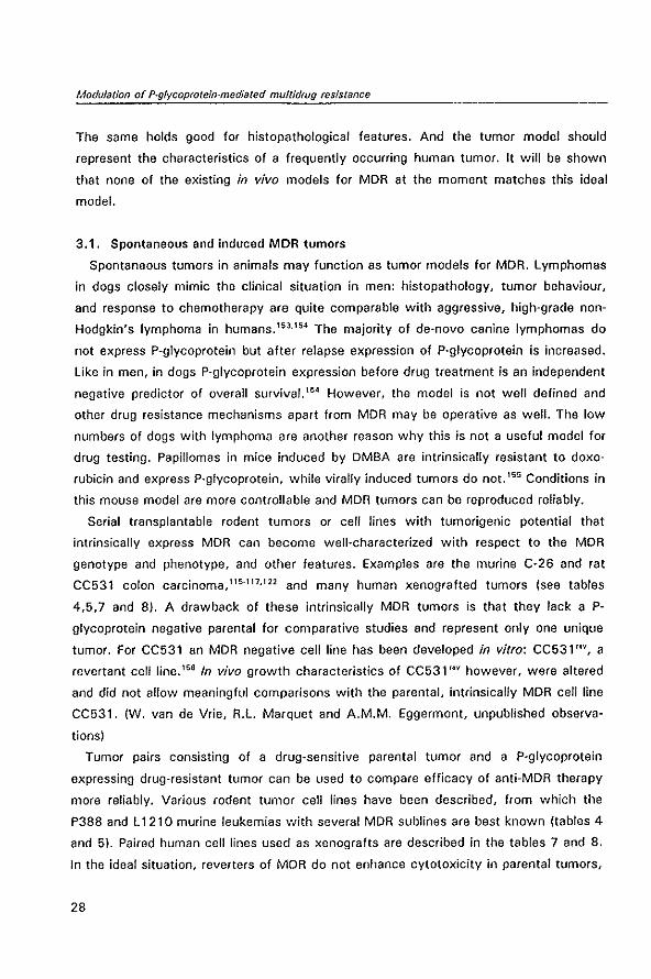
Modulation of P·gfycopfotein-mediated multidrug resistance
The same holds good for histopathological features. And the tumor model should
represent the characteristics of a frequently occurring human tumor. It will be shown
that none of the existing in vivo models for MDR at the moment matches this ideal
model.
3.1. Spontaneous and induced MDR tumors
Spontaneous tumors in animals may function as tumor models for MOR. Lymphomas
in dogs closely mimic the clinical situation in men: histopathology, tumor behaviour,
and response to chemotherapy are quite comparable with aggressive, high·grade non·
Hodgkin's lymphoma in humans.,s3.,s4 The majority of de-novo canine lymphomas do
not express P·glycoprotein but after relapse expression of P·glycoprotein is increased.
Like in men, in dogs P·glycoprotein expression before drug treatment is an independent
negative predictor of overall survival. l64 However, the model is not well defined and
other drug resistance mechanisms apart from MOR may be operative as well. The low
numbers of dogs with lymphoma are another reason why this is not a useful model for
drug testing. Papillomas in mice induced by OMBA are intrinsically resistant to doxo·
rubicin and express P·glycoprotein, while virally induced tumors do not. ISS Conditions in
this mouse model are more controllable and MOR tumors can be reproduced reliably.
Serial transplantable rodent tumors or cell lines with tumorigenic potential that
intrinsically express MDR can become well-characterized with respect to the MDR
genotype and phenotype, and other features. Examples are the murine C·26 and rat
CC531 colon carcinoma,IIS-I17.122 and many human xenografted tumors (see tables
4,5,7 and 8). A drawback of these intrinsically MDR tumors is that they lack a P
glycoprotein negative parental for comparative studies and represent only one unique
tumor. For CC531 an MDR negative cell line has been developed in vitro: CC531''', a
revertant cell line. 1s6 In vivo growth characteristics of CC531 rev however, were altered
and did not allow meaningful comparisons with the parental, intrinsically MDR cell line
CC531. (W. van de Vrie, R.l. Marquet and A.M.M. Eggermont, unpublished observa
tions)
Tumor pairs consisting of a drug·sensitive parental tumor and a P·glycoprotein
expressing drug·resistant tumor can be used to compare efficacy of anti·MDR therapy
more reliably. Various rodent tumor cell lines have been described, from which the
P388 and L121 0 murine leukemias with several MDR sublines are best known (tables 4
and 5). Paired human cell lines used as xenografts are described in the tables 7 and 8.
In the ideal situation, reverters of MDR do not enhance cytotoxicity in parental tumors,
28

Table: 4. Murine MOR tumor cel! lines in vivo
cell line MDR subline mdr/Pgp RR in vitro tissue in vivo model references expression
P388 • P388/ADR mdr1a I Pgp OOX 6a~150x Iymphoblastoid ip ~ ascites, survival; 87,101-leukemia iv - disseminated, survival; 108
P388NCR mdr1a I Pgp VCR 12·30x bone marrow purging DOX 6x
L1210 L121000x mdr1b J Pgp DOX 45x leukemia ip • ascites; 87,91, sc • sorld, tumor size 109,110
L12100NR mdr1b J Pgp DNR 30x DOX 57x
S laO s , aooox mdr1b J Pgp DOX 340x sarcoma ip • ascites '" S laOONA nr I Pgp DNR 73x
DOX 275x
EA/DS EA/DR nr / nr DNR 2.4x Ehrlich ascites ip - ascites, survival "2 carcinoma
B16V B16V/OXR mdr', nr DOX 200x melanoma sc - solid, tumor size 113,114
C26 €D mdr1b I Pgp colon carcinoma sc - sOlid. tumor size 115~117
S"
~ intrinsic MDR expression ~ c mdr gene undetermined as no specific probe is used 3
c
abbreviations: POP P-glycoprotein ADR adriamycin !l: " RR relative resistance DNR daunorubicin '<
" nr not reported DOX doxorubicin " ;P intraperitoneal VBl = vinblastine 3 " ;v intravenous VCR = vincristine S"
'" sc subcutaneous ~
'" '"

Modulation of P-glycoprotein-medialed multidruu resistance
while enhanced antitumor effects can be observed in MDR tumors. It should be noted
that even in the most often used tumor model, the P388 leukemia, this is not the case,
because the parental P388 cell line expresses low levels of P-glycoprotein and is
sensitive to reversal activity.157 Because in most of the tumors MDR is induced by
exposure to cytotoxins, other drug resistance mechanisms may be induced as well. In a
doxorubicin-induced drug-resistant rat mammary carcinoma cell line (MatS) mdr RNA is
elevated, but also glutathione S-transferase activity, while glutathione levels are
decreased. l2l Similar observations have been done in a doxorubicin-resistant human
MCF-7 breast carcinoma cell line. 136 Most in vivo used tumors are not well charac
terized with respect to other drug resistance mechanisms like altered glutathione S
transferase isoenzymes, decreased topoisomerase activity, and expression of the
multidrug resistance-associated protein (MRP).
Transfected cell lines (table 6, and various cell lines in the tables 7 and 81 have the
advantage that no other drug resistance mechanisms are introduced and in this respect
deliver a 'pure' MDR tumor model. Another advantage is that human MOR can be used.
This bypasses possible substrate specificities or differences in efflux efficiency of the
mdrla and mdrlb products of rodents. Results of studies on the efficacy of modulators
may be more comparable to the clinical situation. As human MORt is used, immuno
logical reactions to foreign protein may be induced which can have influence on results
in vivo. In both induced and transfected MDR cell lines alterations in tumorigenicity and
growth rate and growth pattern have been observed. This will be described in the next
section.
The advantage of human xenografts, whether intrinsically MDR or paired tumors, is
that they are human tumors with their own pathological characteristics and that they
express human MORt. Virtually all kinds of human tumors can be grown in immuno
compromised rodents and are available for therapeutic experiments. The results of
cytotoxin experiments in xenogratts will be more relevant to the clinical practice of
those particular tumors than standard tumor models like the murine P388 leukemia,
which has low predictive value in screening new anticancer drugs. 158 Human xenograft
can readily be grown in immunodeficient rodents like nude mice, nude rats, or SCID
(severe combined immunodeficient) mice. SCtO mice lack both functional 8 and T cells
and are more severely immunocom"promised than the nude mouse. They have a greater
propensity for transplantation of hematopoietic and lymphoid tissue and generally do
not develop graft versus host reaction.151.152
30
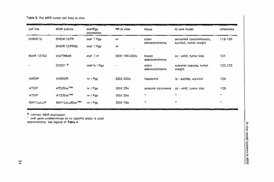
Table 5. Rat MDR tumor cell lines in vivo
cell line MDR subline mdrlPgp RR in vitro expression
DHD/K12 DHD/K12/TR mdr"1 Pgp nr
DHD/K12/PROb mdr'l Pgp nr
MatS 13762 (Adrfl)MatB mdr"1 nr DOX 166-200x
CC531 (I mdr1a! Pgp
AH66P AH66DR nr / Pgp DOX 200x
AT2/P AT2/Dox1000 nr I Pgp DOX 25x
AT3/P AT3/Dox'000 nr I Pgp DOX 25x
MAT-LyLu/P MAT-LyLu/Dox 1000 nr I Pgp DOX 76x
~ intrinsic MDR expression mdr gene undetermined as no specific probe is used
abbreviations: see legend of Table 4
w
tissue in vivo model
colon peritoneal carcinomatosis, adenocarcinoma survival, tumor weight
breast sc - solid, tumor size adenocarcinoma
colon subrenal capsule, tumor adenocarcinoma weight
hepatoma jp • ascites, survival
prostate carcinoma sc - solid, tumor size
references
118-120
121
122,123
124
125
os-
~. g !l: ~
" ~ ~
" ~
Modulation of P-glycoprotein-mediated multidrug resistance
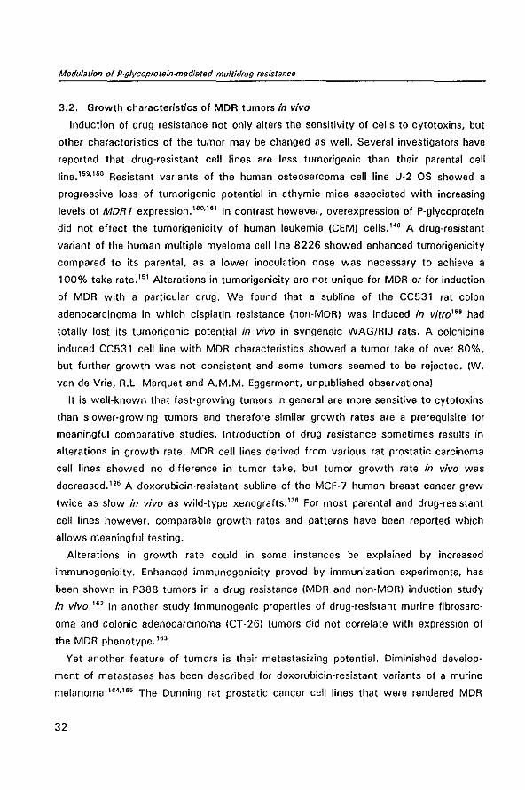
3.2. Growth characteristics of MDR tumors in vivo
Induction of drug resistance not only alters the sensitivity of cells to cytotoxins, but
other characteristics of the tumor may be changed as well. Several investigators have
reported that drug-resistant cell lines are less tumorigenic than their parental cell
line.159.160 Resistant variants of the human osteosarcoma cell line U-2 OS showed a
progressive loss of tumorigenic potential in athymic mice associated with increasing
levels of MOR1 expression.tM.161 In contrast however, overexpression of P-glycoprotein
did not effect the tumorigenicity of human leukemia (CEM) cells. 148 A drug-resistant
variant of the human multiple myeloma cell line 8226 showed enhanced tumorigenicity
compared to its parental, as a lower inoculation dose was necessary to achieve a
100% take rate. 151 Alterations in tumorigenicity are not unique for MDR or for induction
of MDR with a particular drug. We found that a subline of the CC531 rat colon
adenocarcinoma in which cisplatin resistance (non-MDR) was induced in vitro'56 had
totally lost its tumorigenic potential in vivo in syngeneic WAG/RIJ rats. A colchicine
induced CC531 cell line with MDR characteristics showed a tumor take of over 80%,
but further growth was not consistent and some tumors seemed to be rejected. (W.
van de Vrie, R.L. Marquet and A.M.M. Eggermont, unpublished observations)
It is well-known that fast-growing tumors in general are more sensitive to cytotoxins
than slower-growing tumors and therefore similar growth rates are a prerequisite for
meaningful comparative studies. Introduction of drug resistance sometimes results in
alterations in growth rate. MDR cell lines derived from various rat prostatic carcinoma
cell lines showed no difference in tumor take, but tumor growth rate in vivo was
decreased.t~5 A doxorubicin-resistant subline of the MCF-7 human breast cancer grew
twice as slow in vivo as wild-type xenografts. 136 For most parental and drug-resistant
cell lines however, comparable growth rates and patterns have been reported which
allows meaningful testing.
Alterations in growth rate could in some instances be explained by increased
immunogenicity. Enhanced immunogenicity proved by immunization experiments, has
been shown in P388 tumors in a drug resistance (MDR and non-MDR) induction study
in ViVO.16~ In another study immunogenic properties of drug-resistant murine fibrosarc
oma and colonic adenocarcinoma (CT-261 tumors did not correlate with expression of
the MDR phenotype.'"
Yet another feature of tumors is their metastasizing potential. Diminished develop
ment of metastases has been described for doxorubicin-resistant variants of a murine
melanoma.'64.165 The Dunning rat prostatic cancer cell lines that were rendered MDR
32
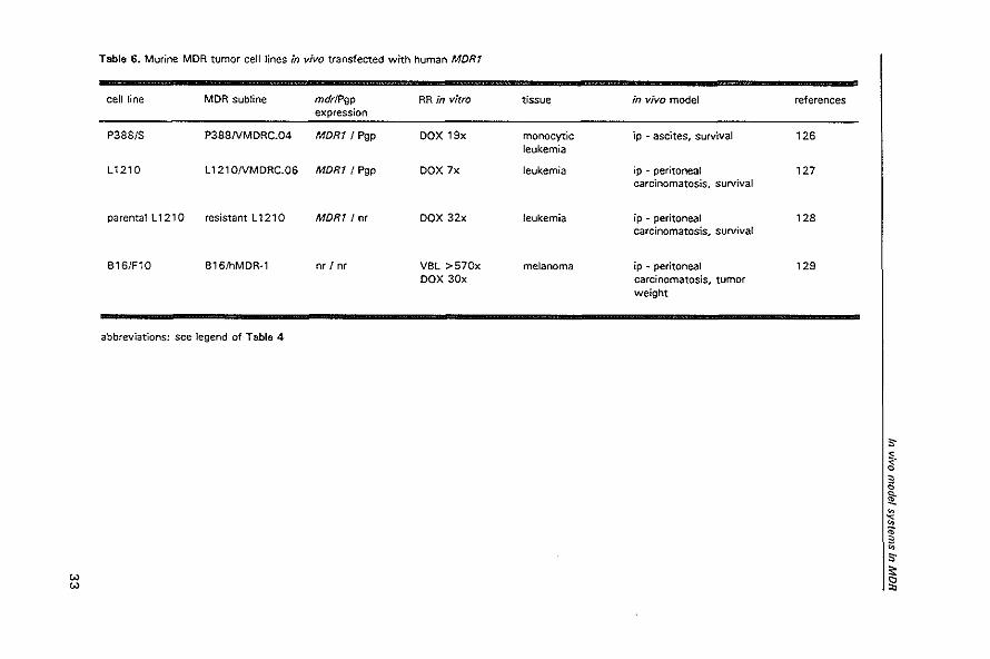
Table 6. Murine MDR tumor cell lines in vivo transfected with human MDRt
cell line MDR subline mdrlPgp RR in vitro tissue in vivo model references expression
P388/S P388NMDRC.04 MORt I Pgp DOX 19x monocyflc ip - ascites, survival 126 leukemia
L1210 l 121 ONMDRC.OS MDR1 I Pgp DOX 7x leukemia ip - peritoneal 127 carcinomatosis, survival
parental l 1210 resistant l 1210 MDR1/nr DOX 32x leukemia ip - peritoneal 128 carcinomatosis, survival
B1S/F10 B1S/hMDR-1 nr/or VBl >570x melanoma ip - peritoneal 129 DOX 30x carcinomatosis, tumor
weight
abbreviations: see legend of Table 4
;,-
~. ;) 0
!t ~ ;:;
" ;) ~
;,-
w I~ w
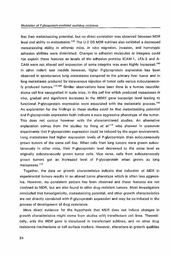
Modulation of P-glycoprolein-medlated multidrug resistance
lost their metastasizing potential, but no direct correlation was observed between MDR
level and ability to metastasize. '" The U·2 OS MDR sub lines also exhibited a decreased
metastasizing ability in athymic mice. In vitro migration, invasion, and homotypic
adhesion abilities were diminished. Changes in adhesion molecules or integrins could
not explain these features as levels of the adhesion proteins ICAM-', LFA-3 and A
CAM were not altered and expression of some integrins was even highly increased. '60
In other rodent test models however, higher P-glycoprotein expression has been
observed in spontaneous lung metastases compared to the primary liver tumor and in
lung metastases produced by intravenous injection of tumor cells versus subcutaneous
ly produced tumors." 1,166 Similar observations have been done in a human neurobla
stoma cell line xenografted in nude mice. In this cell line which produced metastases in
vivo, gradual and significant increases in the MOR1 gene transcript level leading to
functional P-glycoprotein expression were associated with the metastatic process. '46
An explanation for the findings in these studies could be that metastasizing potential
and P-glycoprotein expression both indicate a more aggressive phenotype of the tumor.
This does not concur however with the aforementioned studies. An alternative
explanation comes from the studies by Dong et al. I 11 who showed in crossover
experiments that P-glycoprotein expression could be induced by the organ environment.
Lung metastases had higher expression levels of P-glycoprotein than subcutaneously
grown tumors of the same cell line. When cells from lung tumors were grown subcu
taneously in other mice, their P-glycoprotein level decreased to the same level as
originally subcutaneously grown tumor cells. Vice versa, cells from subcutaneously
grown tumors got an increased level of P-glycoprotein when grown as lung
metastases. 111
Together, the data on growth characteristics indicate that induction of MDR in
experimental tumors results in an altered tumor phenotype which is often less aggress
ive. However, no consistent pattern has been observed and these features are not
confined to MDR, but are also found in other drug-resistant tumors. Most investigators
concluded that tumorigenicity, metastasizing potential, and other growth characteristics
are not directly correlated with P-glycoprotein expression and may be co-induced in the
process of development of drug resistance.
More direct evidence for the hypothesis that MDR does not induce changes in
growth characteristics might come from studies wit~ transfectant cell lines. Theoreti
cally, only the MDR gene is introduced in transfectant sublines, and no other drug
resistance mechanisms or cell surface markers. However, alterations in growth qualities
34
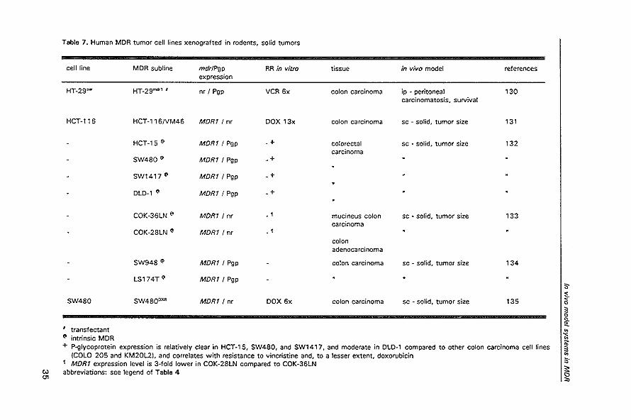
Table 7. Human MOR tumor cell lines xenografted in rodents, solid tumors
cell line MDR subline mdrlPgp RR in vitro tissue in vivo model references expression
HT~29P'" HT~29mdrl I nr , Pgp VCR 6x colon carcinoma ip ~ peritoneal 130 carcinomatosis, survival
HCT~1'6 HCT~' 16NM46 MORT! nr DOX 13x colon carcinoma sc . solid, tumor size 131
HCT~'5 (\I MORT / Pgp .+ colorectal sc - solid, tumor size 132 carcinoma
SW480 fI.t MORT/ Pgp .+
SW1417 (\I MORT! Pgp .+
OLD·' ~ MORT! Pgp .+
COK·36LN • MDRT/nr • 1 mucineus colon sc - solid, tumor size 133 carcinoma
COK·28LN • MORT! nr • 1
colon adenocarcinoma
SW948 fI.t MORT / Pgp colon carcinoma sc - solid, tumor size 134
LS174T fI.t MDRT / Pgp S-
SW480 SW4800XR MORT/nr DOX 6x colon carcinoma sc - solid, tumor size 135 < I ~. • 0
!l: I transfectant ~
~ intrinsic MDR " " + P-glycoprotein expression is relatively clear in HCT-15, SW480, and SW1 417. and moderate in DLD-' compared to other colon carcinoma cell lines • (COLO 205 and KM20L2), and correlates with resistance to vincristine and. to a lesser extent. doxorubicin ~
1 MORT expression level is 3-fold lower in COK-28LN compared to COK-36LN S-;:
'" abbreviations: see legend of Table 4
'" '" '"
Modulation of P-glycoprotein-medlated multidrug resistance
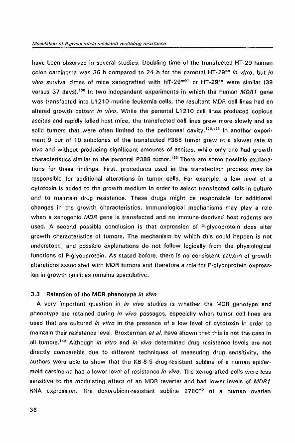
have been observed in several studies. Doubling time of the transfected HT -29 human
colon carcinoma was 36 h compared to 24 h for the parental HT ·29p&l in vitro, but in
vivo survival times of mice xenografted with HT _29m<1r' or HT -29p&l were similar (39
versus 37 days}.13o In two independent experiments in which the human MDRT gene
was transfected into L121 0 murine leukemia cells, the resultant MDR cell lines had an
altered growth pattern in vivo. While the parental L 1210 cell lines produced copious
ascites and rapidly killed host mice, the transfected cell lines grew more slowly and as
solid tumors that were often limited to the peritoneal cavity.'26.128 In another experi
ment 9 out of 10 subclones of the transfected P388 tumor grew at a slower rate in
vivo and without producing significant amounts of ascites, while only one had growth
characteristics similar to the parental P388 tumor. 126 There are some possible explana
tions for these findings. First, procedures used in the transfection process may be
responsible for additional alterations in tumor cells. For example, a low level of a
cytotoxin is added to the growth medium in order to select transfected cells in culture
and to maintain drug resistance. These drugs might be responsible for additional
changes in the growth characteristics. Immunological mechanisms may play a role
when a xenogenic MDR gene is transfected and no immune-deprived host rodents are
used. A second possible conclusion is that expression of P-glycoprotein does alter
growth characteristics of tumors. The mechanism by which this could happen is not
understood, and possible explanations do not follow logically from the physiological
functions of P-glycoprotein. As stated before, there is no consistent pattern of growth
alterations associated with MOR tumors and therefore a role for P-glycoprotein express
ion in growth qualities remains speculative.
3.3 Retention of the MDR phenotype in vivo
A very important question in In vivo studies is whether the MDR genotype and
phenotype are retained during in vivo passages, especially when tumor cell lines are
used that are cultured in vitro in the presence of a low level of cytotoxin in order to
maintain their resistance level. Broxterman et al. have shown that this is not the case in
all tumors. 142 Although in vitro and in vivo determined drug resistance levels are not
directly comparable due to different techniques of measuring drug sensitivity, the
authors were able to show that the KS-8-5 drug-resistant subline of a human epider
moid carcinoma had a lower level of resistance in vivo. The xenografted cells were less
sensitive to the modulating effect of an MDR reverter and had lower levels of MDR 1
RNA expression. The doxorubicin-resistant subline 2780 .... 0 of a human ovarian
36
'" "
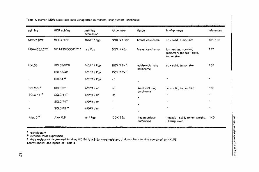
Table 7. Human MDR tumor cell lines xenografted in rodents. solid tumors (continued)
cell line MDR subline mdrJPgp RR in vitro tissue in vivo model expression
MCF-7IWTI MCF-7/ADR MORt! Pgp DOX >150x breast carcinoma sc - solid. tumor size
MDA435IlCC6 MDA435!LCC6MOA1 I nr I Pgp DOX ±45x breast carcinoma ip - ascites. survival; mammary fat pad - sOlid. tumor size
HXL55 HXL55NCR MORt I Pgp DOX 3.8x' epidermoid lung sc - solid, tumor size carcinoma
HXL55/AD MORt I Pgp DOX 3.2x '
HXL54 ~ MORt! Pgp - ,
SCLC-6 ~ SCLC-6T MORt I nr m small cell lung sc • solid, tumor size carcinoma
SCLC-41 (I> SCLC-41T MDRt I nr nr
SCLC-74T MORt! nr
SCLC-75 ~ MORt! nr
Alex 0 (I Alex 0.5 nr ! Pgp DOX 25x hepatocellular hepatic - solid, tumor weight, carcinoma HBsAg level
transfectant (I> intrinsic MDR expression 1 drug resistance determined in vivo; HXL54 is .±.9.5x more resistant to doxorubicin in vivo compared to HXL55 abbreviations: see legend of Table 4
references
131.136
137
138
139
140 ,. ~. ~ c 1l: ~
-:; ~ ~ ,. ~
Modulation of P-glycoprotein-mediated multidrug resistance
carcinoma generally showed reduced P-glycoprotein activity, but a minority of the cells
seemed to have retained the high resistance level. Regrowth of these cell lines in vitro
confirmed these observations. 142 In another study a 17-fold loss of resistance level was
observed after in vivo passage of 2780AO cells. '43 In contrast, xenografts of the
transfected MORI BRO melanoma cell line had a comparable expression level of the
MOR I gene and similar functional activity of P'glycoprotein as the original cell
line.'41.142 Retention of the MDR genotype and phenotype has also been found after in
vivo passage of resistant CEM leukemia cells. '49 For other MDR tumors these features
have not been studied as extensively as for the tumors described above, but the
differences between parental and drug-resistant tumors in drug sensitivity in vivo and
the efficacy of MDR reverters show indirectly that at least part of the MDR mechanism
is retained during the process of in vivo passage. The drug resistance level of an
induced or transfected MDR tumor tends to decrease when tumor cells are grown for a
long time in the absence of their selecting drug, giving rise to so-called reverted cell
lines with lowered expression levels of P-glycoprotein. This phenomenon is observed in
vitro as well as in ViVO.126.165 Therefore, in most studies selecting drugs are only
withheld from the culture medium a short time before and during the testing period.
3.4. In vivo tumor models
In ascites models the tumor is grown in the peritoneal cavity and the drugs are
administered intra peritoneally (ip), the so-called ip-ip model. Efficacy of antitumor
agents is determined by scoring prolongation of survival. The well-known P388 cell
lines are grown this way and this has become a sort of standard in vivo model for anti
neoplastic drug screening. 167 Survival time is approximately 10 days for parental and
MDR cell lines. Advantages of the ascites tumor models like the murine P388 and
L 1210 leukemia are the ease of in vitro and in vivo maintenance, and the ability to
perform reproducible and rapid testing of drugs.
The ascites model can be criticized for being artificial and it is said that relatively high
therapeutic effects are observed. In the first place, this is partly inherent to the
standard procedure in which treatment is started on the same day as tumor inoculation,
which means that the tumor is not yet established. Secondly, drugs are most often
administered intra peritoneally. This may result in a chemical peritonitis that contributes
non·specifically to the antitumor effect. Thirdly, direct administration of drugs at the
site of the tumor bypasses the vascular route. In the clinical situation most of the drugs
are administered intravenously because most human tumors do not grow as ascites
38
"" to
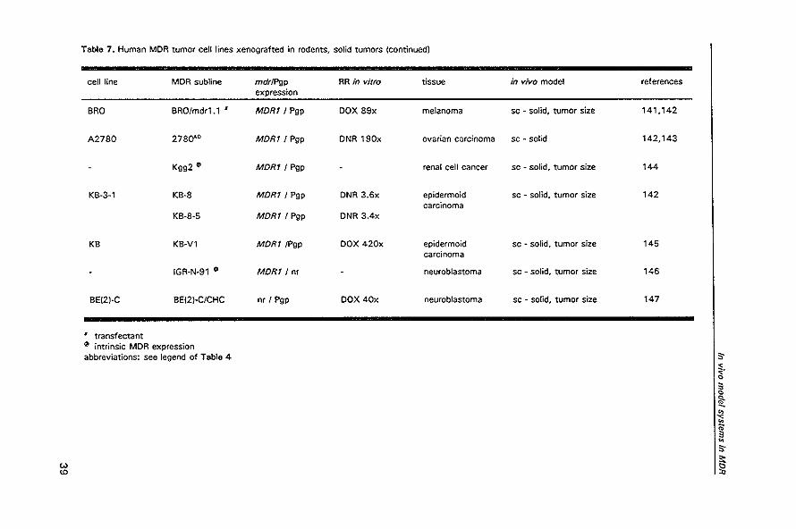
Table 7. Human MDR tumor cell lines xenografted in rodents, solid tumors (continued)
cell line MDR subline
BRO BRO/mdr1.1 I
A2780 2780"-D
Kgg2 C!'
KB-3-1 KB-8
KB-8-5
KB KB~Vl
IGR.N.91 (I
BEI2J-C BEI2J-GICHC
I transfectant C!' intrinsic MDR expression abbreviations: see legend of Table 4
mdrlPgp expression
MOR11 Pgp
MORt! Pgp
MORt! Pgp
MORt / Pgp
MORt! Pgp
MORt/Pgp
MORt/ nr
nr I Pgp
RR in vitro tissue
DOX 89x melanoma
DNR 190x ovarian carcinoma
renal cell cancer
DNR 3.6x epidermoid carcinoma
DNR 3.4x
DOX 420x epidermoid carcinoma
neuroblastoma
DOX 40x neuroblastoma
in vivo model references
sc ~ solid, tumor size 141,142
sc ~ solid 142,143
sc • solid, tumor size 144
sc - solid. tumor size 142
sc ~ sOlid. tumor size 145
sc - solid, tumor size 146
sc - solid, tumor size 147
s-~. 3 o
it " -;;
~ S-
~
Modulation 01 P-glycoprotein-mediated mullidrug resistance
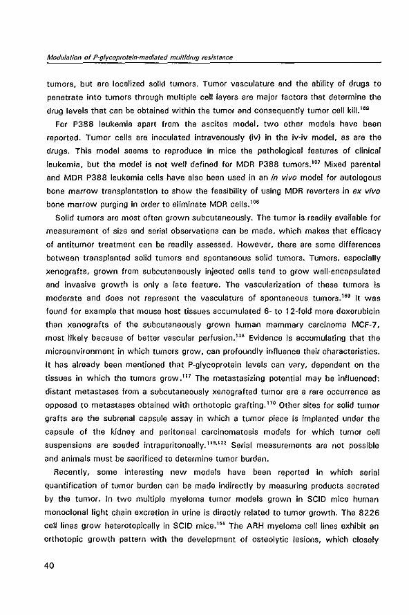
tumors, but are localized solid tumors. Tumor vasculature and the ability of drugs to
penetrate into tumors through multiple cell layers are major factors that determine the
drug levels that can be obtained within the tumor and consequently tumor cell kill. 16a
For P388 leukemia apart from the ascites model, two other models have been
reported. Tumor cells are inoculated intravenously (iv) in the iv·iv model, as are the
drugs. This model seems to reproduce in mice the pathological features of clinical
leukemia, but the model is not well defined for MDR P388 tumors. ,., Mixed parental
and MDR P388 leukemia cells have also been used in an in vivo model for autologous
bone marrow transplantation to show the feasibility of using MDR reverters in ex vivo
bone marrow purging in order to eliminate MDR cells. 106
Solid tumors are most often grown subcutaneously. The tumor is readily available for
measurement of size and serial observations can be made, which makes that efficacy
of antitumor treatment can be readily assessed. However, there are some differences
between transplanted solid tumors and spontaneous solid tumors. Tumors, especially
xenografts, grown from subcutaneously injected cells tend to grow well-encapsulated
and invasive growth is only a late feature. The vascularization of these tumors is
moderate and does not represent the vasculature of spontaneous tumors. '69 It was
found for example that mouse host tissues accumulated 6- to 12-fold more doxorubicin
than xenografts of the subcutaneously grown human mammary carcinoma MCF-7,
most likely because of better vascular perfusion. '36 Evidence is accumulating that the
microenvironment in which tumors grow, can profoundly influence their characteristics.
It has already been mentioned that P'glycoprotein levels can vary, dependent on the
tissues in which the tumors grow. 117 The metastasizing potential may be influenced:
distant metastases from a subcutaneously xenografted tumor are a rare occurrence as
opposed to metastases obtained with orthotopic grafting. 170 Other sites for solid tumor
grafts are the subrenal capsule assay in which a tumor piece is implanted under the
capsule of the kidney and peritoneal carcinomatosis models for which tumor cell
suspensions are seeded intraperitoneally.119,122 Serial measurements are not possible
and animals must be sacrificed to determine tumor burden.
Recently, some interesting new models have been reported in which serial
quantification of tumor burden can be made indirectly by measuring products secreted
by the tumor. In two multiple myeloma tumor models grown in SCID mice human
monoclonal light chain excretion in urine is directly related to tumor growth. The 8226
cell lines grow heterotopically in SCID mice. '" The ARH myeloma cell lines exhibit an
orthotopic growth pattern with the development of osteolytic lesions, which closely
40
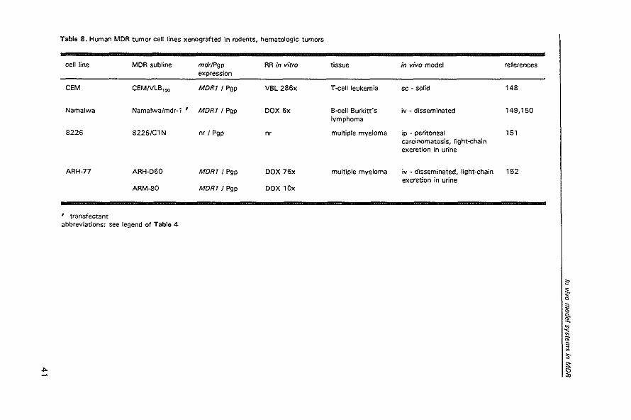
Table 8. Human MDR tumor cell lines xenografted 'In rodents. hematologic tumors
cell line MDR subline mdrJPgp RR in vitro tissue in vivo model references expression
CEM CEMNlB,oo MDRI / Pgp VBl286x T-cell leukemia sc ~ solid 148
Namalwa Namalwa/mdr~ 1 ' MDR1 I Pgp DOX 6x B-cell Burkitt's iv ~ disseminated 149,150 lymphoma
8226 8226/C1 N nr I Pgp n, multiple myeloma ip - peritoneal 151 carcinomatosis. light-chain excretion in urine
ARH-77 ARH-060 MDR1 I Pgp DOX 76x multiple myeloma iv ~ disseminated. light~chain 152 excretion in urine
ARM-SO MDR1 I Pgp DOX 10x
, transfectant abbreviations: see legend of Table 4
s-~. Q
3 Q
1!: " ~ " ;;: ;]
" s· ... 15
"
Modulation of P-glycoprotein-mediated muftidrug resIstance
mimics the pathophysiology of human myeloma.'" A human hepatoma cell line (Alex 0)
and its MDR subline grow as an intrahepatic xenograft after intra splenic injection. The
tumor cells produce HBsAg and serum levels correlate with tumor burden.140 The
measurement of secreted tumor products allows starting of treatment at a determined
tumor burden and permits a direct comparison of the effectiveness of drugs used at the
same extent of disease in each animal.
Novel endpoints to measure functional MDR in vivo come from radio-imaging tech
niques. Drug-resistant and sensitive tumor xenografts have been shown to be distin
guishable by differences in uptake of radiolabeled colchicine. '71 In vivo quantification of
P'glycoprotein has been performed with the radiolabeled monoclonal antibody MRK16
that specifically recognizes P-glycoprotein. 141 And imaging studies in rats bearing wild
type and drug-resistant tumors showed that the imaging agent 99Tcm-sestamibi, which
is transported by P-glycoprotein, was washed out of resistant tumors three times the
rate of wild-type tumors.112 These studies suggest potential use of radio-imaging
techniques to evaluate MDR in vivo.
Two other models should be mentioned here: the mdr knockout mice and MDRI
transgenic mice. The mdrta (-I-) knockout mouse has no functional mdrta P
glycoprotein," and the mdrla + mdrlb (-j.) double knockout mouse totally lacks p.
glycoproteins that are involved in MDR.82 These mice have been engineered by
disrupting the mdrla andlor mdrlb genes in the germ lines of mice, which resulted in
mice heterozygous for the disrupted gene. Mice homozygous for the disrupted gene
were obtained by inbreeding techniques. 69.82 The features of these mice have already
been described in chapter 2.3. Mdr knockout mice are excellent tools for studying the
physiological role of P-glycoprotein and toxic effects of drugs due to the absence of
functional P·glycoprotein.
Transgenic mice that express the human MORt gene in their bone marrow have been
engineered by the group of Gottesman and Pastan. cDNA constructs encoding full
length human MORt in a plasmid carrier were injected into fertilized mouse embryos. A
homozygous line was obtained of mice in which the expression of the MDR 1 transgene
was limited to the bone marrow and spleen. 173 MORt heterozygous animals were
obtained by backcrossing with MDR 1-negative mice and these mice were used in MDR
modulation studies. The mice are resistant to the myelosuppressive effects of drugs
that are influenced by the MDR mechanism like anthracyclines, Vinca alkaloids, etoposi
de, and taxol. The level of MDR 1 expression in the bone marrow is comparable to that
found in many human cancers. The effect of drugs and combination therapy with
42
In vivo model systems in MDR
chemosensitizers on the bone marrow can easily be measured by peripheral white blood
cell count. This makes it an efficient model for testing efficacy of MDR reverters in
viVO. ' 73-1715 A problem with the model is that after many generations of breeding the
MDR 1 expression is not kept at its initial level. '" It should be mentioned that the
transgenic mouse model is not a tumor model as the MDRI gene is expressed in normal
bone marrow.
4 Modulation of MDR with reverters in vivo
Most attempts to circumvent MDR have used the possibility to inhibit the P
glycoprotein efflux pump, which results in increased intracellular drug concentrations
and enhanced cell death. In this chapter we will review studies on modulation of MDR
in vivo with so-called chemosensitizers or reverters. In the next chapter other
approaches to circumvent MDR will be described.
4.1 Pharmacokinetics
Combination treatment of cytotoxins with reverters altered the pharmacokinetics of
MDR related drugs in phase Ill! clinical trials in humans. Changes induced by reverters
are a decrease in drug clearance and an increase of the area under the curve (AU C) of
the cytotoxin. These pharmacokinetic interactions have been obtained with the
reverters verapamil, dexverapamil, nifedipine, cyclosporin A, and PSC 833 and the
drugs doxorubicin, epirubicin, vincristine, etoposide, and paclitaxel in various combina
tions, showing that this phenomenon is not limited to certain drugs. '76,177 There are few
studies on plasma levels and AUC of drugs in animals. Plasma levels of drugs in animals
are often not much increased by combination treatment with reverters,'43.178.,91 The
AUC of drugs can be elevated,'39,'57,'82 which is due to prolonged elimination. '39,162 But
in other studies elimination of doxorubicin was not found to be altered.'so"s,
Animal studies have shown the various mechanisms by which MDR modulators can
change the pharmacokinetics of drugs. The decreased elimination of drugs by reverters
is caused by alterations in intestinal, biliary and renal absorption and excretion. The
absorption from the gut of orally administrated etoposide is higher in quinidine pretre
ated rats and the intestinal secretion (exsorption) of intravenously administered
etoposide is diminished by quinidine, compatible with inhibition of the P-glycoprotein
transporter in intestinal cells. 1Sl Reverters partly inhibit the active biliary excretion of
43
Modulation of P·glycoprotein-mediated multidrug resistance
colchicine, doxorubicin and etoposide by the Iiver. 65,72,184,185 In the kidneys a net
secretion of MDR-related drugs is observed by the luminal membrane of renal cells.
Cyclosporin A inhibits the renal secretion of vincristine and vinblastine in a dose
dependent manner in dogs.56 Similar observations have been reported for colchicine by
cyclosporin A and PSC 833 in rats""" These studies show that the physiological
function of P-glycoprotein which is prevention of (re)absorption and elimination of
xenobiotics also affects drug transport and can be blocked by reverters in vivo.
The extent by which drug pharmacokinetics are altered by a reverter will depend on
the fraction of drug that is normally eliminated by the P-glycoprotein efflux mechanism.
This has not been studied extensively in vivo. The conflicting results of pharmacokin
etic studies with doxorubicin have been mentioned above.'20.'39.180.1S1 Of note is the
influence of route of administration of drugs. In combination with PSC 833 the AUC of
etoposide is much more elevated in case of oral administration of etoposide compared
to intravenous dosing. Apparently, etoposide absorption from the gut is normally largely
inhibited by intestinal P-glycoprotein, while the relative role of P-glycoprotein in elimin
ation of etoposide seems smaller. '82
A consistent observation in animal studies is the altered distribution of drugs over the
various tissues in mice and rats. In Sprague-Dawley rats cyclosporin A and PSC 833
significantly increased tissue levels of doxorubicin in liver, kidney, small intestine, and
adrenals. A smaller increase was also observed in the heart, while cyclosporin A had no
effect on doxorubicin concentration in the brain. In these studies the increases in drug
tissue levels did not appear to be the result of changes in drug metabolism or elimin
ation, as plasma levels and elimination of drugs were not significantly altered. '80,ISI
Other investigators reported elevation of doxorubicin levels in liver and kidney caused
by amiodarone and cinchonine,12o,186 and elevation of vincristine levels in liver, kidney
and small intestine by verapamil. 178 The alterations in drug levels are compatible with
relatively high expression levels of P-glycoprotein in these tissues. Increase of drug
levels by reverters in tissues with lower expression levels of P-glycoprotein like lung
and spleen have been noted in some studies. '20,lS7 The pattern of distribution of drugs
in the mdrla knockout mouse is different from the pattern described above. In the
mdrl a knockout mice a marked elevation of drugs like vinblastine, cyclosporin A,
digoxin and ivermectin is observed in the brain and, to a lesser extent, in the testis.
Elevation of these drugs in other tissues was less marked. 59,a7 At the blood-tissue
barriers of brain and testis predominantly mdrla is expressed and disruption of the
mdrla gene in the mdrla (-/-) knockout mouse leads to total absence of functional P-
44
In vivo model systems in MDR
glycoprotein at these sites. In other tissues mdrlb is also expressed and probably the
mdrlb product can, at least partly, replace the function of the mdrl. product. This is
also suggested by the increased expression of mdrlb in kidney and liver of the mdrla
knockout mouse.59 It is apparently not easy to break the blood-brain barrier by MDR
reverters as cyclosporin A. More potent reverters of MOR like PSG 833 and SOZ 280-
446 have the capacity to enhance the neurotoxic effects of drugs, suggesting that
these reverters are able to block P'glycoprotein at the blood-brain barrier, but drug
levels in the brain have not been measured in this study.75
4.2. Efficacy of MDR reverters in vivo
The first observations that verapamil could reverse drug resistance were done by
Tsuruo et al. who showed that verapamil was able to enhance drug accumulation of
vincristine and vinblastine in the P388NCR drug-resistant cell line in vitro and in
ViVO. '87 Since then, numerous compounds have been described which efficiently inhibit
the P-glycoprotein efflux pump: calcium channel blockers (e.g. verapamil, dexniguldip
ine, PAK-200, AHG-52), cyclic peptides (e.g. cyclosporin A, PSG 833, SOZ 280·446),
calmodulin antagonists (e.g. trifluoperazine), protein kinase C inhibitors (e.g. staurospo
rine). steroidal agents (e.g. progesterone, tamoxifen, megestrol acetate), Vinca alkaloid
analogues, and miscellaneous compounds (e.g. amiodarone, quinidine).'88.'89 The first
generation MOR reverters were existing drugs which appeared to have MDR reversal
activity, but had other pharmacological effects as well. Levels necessary in vivo for
MOR reversal could often not be obtained because of prohibitive toxicity. For example,
the target concentration of verapamil in mice could not be reached by bolus injection
because this dose was acutely lethal.178 In a human study cardiovascular effects as
hypotension and cardiac arrhythmias prevented adequate dosing of verapamil. '90
Cyclosporin A is a potent blocker of P-glycoprotein but its immunosuppressive potential
and nephrotoxic side-effects are matters of concern. A new generation of compounds,
often analogues of known reverters, but devoid of their primary pharmacological effects
and especially selected for MOR reversal activity, have been developed. '89
Almost all chemosensitizers have first been tested in the drug-resistant P388 ascites
tumors. With verapamil an increase in life span of 25%-45% was obtained in
P388/ADR and in P388NCR tumors. HI7,191 For newer reverters survival increases of
±40%·100% have been reported in the P388NCR ascites tumor. In the more drug·
resistant P388/ADR reverters were less effective. 102,105.l08,179,192 Impressive survival
increases (up to 300% increased life span) have been reported for treatment with the
45
Modulation of P·glycoprotein-mediated multidrug resistance
reverters PSC 833 and SDZ 280-446 in the P388/ADR tumor cell line. IM•I".I93 In all
studies mentioned above drugs were administered intra peritoneally, while chemosensi
tizers were given intraperitoneally, orally or intravenously. Intravenous administration of
drugs in combination with PSC 833 in the P388 model was not effective against the
P388/ADR tumors, but highly effective against the less resistant P388NCR. I57•194 In
the intravenously disseminated P388 leukemia model doxorubicin and etoposide, but
not vincristine, in combination with a reverter (AHC·52) increased survival of
P388NCR bearing mice. In intravenously inoculated P388/ADR bearing mice reversal of
MDR was not obtained. '07
Chemosensitizers also reverse MDR in solid tumor models. In most studies estab·
lished tumors were used: tumors were first allowed to grow to a certain volume before
drug treatment was started. In intrinsic MDR tumors the addition of a reverter to
ineffective treatment schedules with cytotoxins resulted in significant tumor growth
delays."6,'22.'7~ln a study with subcutaneously grown C26 murine colon tumors the /"
revertef...-pst 833 even induced some cures,194 Reversal studies in solid tumor pairs
with a parental and an MDR tumor show specific enhancement of antitumor activity
against MDR tumors. '31 ,133,141.143
These studies demonstrate that reversal of MDR in vivo is feasible and can be
obtained in ascites models, as well as in more difficult solid tumor models and the
intravenously disseminated leukemia model. Possibilities for reversal are dependent on
the level of drug resistance and reversal is not always obtained in highly drug-resistant
tumors. Comparison of the in vivo efficacy of reverters is not possible with the current
data because of differences between studies in tumors, dosing schedules of drugs and
reverters, and experimental designs in the different studies. Comparative studies with
several reverters within the same model and study design are scarce and only available
for new potent reverters versus first generation chemosensitizers like verapamil and
cyclosporin A, and show a clear higher potential of the novel reverters in vivo. 104,1 16
4.3. Toxicity
The reverse of enhanced efficacy of cytotoxins by chemosensitizers is the possible
increase in toxicity. As described above, many reversal studies have shown that
reverters can enhance survival. However, several studies in animals that specifically
investigated adverse effects of MDR reversal have shown that the combination of a
chemosensitizer with a high dose cytotoxin results in increased toxicity leading to
accelerated death. These observations have been reported for verapamil, cyclosporin A,
46
In vivo model systems in MDR
and PSC 833 in combination with doxorubicin and etoposide.'51.ISo-1S2,195,196 The nature
of the increased toxicity has been investigated in few studies. Myelotoxicity is often
the dose limiting factor in chemotherapy. Combination treatment of PSC 833 and
etoposide caused increased leucopenia in mice,'S2 and cyclosporin A with doxorubicin
induced a transient doxorubicin-dose-dependent leucopenia and thrombopenia in rats. '97
Mice treated with PSC 833 and doxorubicin showed transient spleen hypoplasia, with a
general decrease in all leucocyte lineages (8 cell, T cell, and myeloid lineagesl. Changes
were dependent on dose of doxorubicin and increased by addition of PSC 833. In the
bone marrow only a persistent fall in the number of B cells was observed. '9s In two
studies all deviations in blood parameters, and abnormal pathological findings at
autopsy and at light-microscopic examination, could be attributed to known toxicities of
the cytotoxins (doxorubicin, daunomycin and vinblastine). The reverter only accentu
ated these abnormalities, while the pattern of organ toxicity was not altered and no
signs of new toxicity were found, especially not in organs with known high expression
levels of P_glycoprotein. 175,197 In another histopathological study, cardiotoxicity of
doxorubicin was enhanced by verapamil in mice. '9s
Possibly, studies with novel, potent reverters of P-glycoprotein will be different, as
their capacity to block P-glycoprotein is more effective. In a recent study potent chemo
sensitizers as PSC 833 and SOZ 280·446, but not cyclosporin A, were able to break
the P·glycoprotein·dependent component of the blood·brain barrier. High doses of these
reverters in combination with the neurotoxic agent ivermectin caused acute dysfunction
of the central nervous system {convulsion, paralysis, coma and death).15 Interestingly,
within one day after administration of the reverter the increased sensitivity of mice for
the neurotoxic effects of ivermectin disappeared. This can be explained by dissociation
of the reverter from the P-glycoprotein, or new expression of P-glycoprotein by the
blood-brain barrier, or the emergence of alternative mechanisms causing ivermectin
resistance. 75
4.4. Specific modulation of MDR at the tumor level?
An important question to be answered in in vivo studies is whether combination
treatment of cytotoxins with chemosensitizers merely alters pharmacokinetics and must
be considered as a method of dose-intensification, or if specific interaction with p.
glycoprotein at the tumor level results in increase in intratumoral drug levels and
antitumor activity. Possibly, both mechanisms do contribute. Alterations of drug levels
in the tumor caused by reverters have seldom been determined. In a study with a drug-
47
Modulation of P-glycoprotein-mediated multidrug resistance
resistant sarcoma xenograft intra tumoral vincristine levels were not significantly altered
by verapamil. In this study the combination showed no antitumor activity too and no
proof of MOR expression is given.178 Different results were obtained in a study with
effective reversal in MDR tumors. The reverter PAK·200 induced a 4.6·fold increase in
intratumoral doxorubicin concentration in the P·glycoprotein expressing KB-8-5 xeno·
grafts, while PAK·200 did not significantly alter doxorubicin levels in the parental KB-3-
1 drug-sensitive tumors. In COK36LN, a P'glycoprotein expressing colon carcinoma,
PAK-200 elevated intratumoral levels 1.5-fold, and in xenografts of another colon
carcinoma, COK28LN, which expressed little P-glycoprotein the reverter had no influ
ence. The doxorubicin levels in the MDR tumors with the modulator exceeded the
doxorubicin levels in the drug·sensitive tumors. III This study suggests that specific
modulation of P-glycoprotein in the tumor has resulted in enhanced antitumor activity.
Reverters of P-glycoprotein are no magic bullet since P-glycoprotein in other tissues
than the tumor is also inhibited resulting in increased cytotoxicity in these tissues. The
possibilities for MOR reversal will depend on the sensitivities of tissues and tumors for
the cytotoxic effects of certain drugs and on their relative content of P-glycoprotein.
The question is whether the therapeutic index can be increased. In some in vivo
reversal studies the dose of the cytotoxin had to be lowered because of increased
toxicity caused by addition of a reverter. Nevertheless, effective antitumor activity was
at least retained. I04,128 In the MORI transgenic mouse model, that has bone marrow
with a 10-fold increased resistance to MOR-associated drugs, the maximal tolerable
dose of cytotoxins in combination with a chemosensitizer was 20%·45% lower. In
experiments with a chemosensitizer most drugs caused a 44%-78% decrease in white
blood cell count suggesting that there is a possibility to increase the therapeutic index
of P-glycoprotein sensitive agents by concomitant administration of MDR reverters. 175
5. Alternative approaches for modulation MOR in vivo
The most direct way to circumvent MOR is to utilize drugs that are not susceptible to
the P-glycoprotein efflux pump mechanism. This is often not possible, because tumors
do not tend to be sensitive to many different anticancer agents. Dose-intensification as
a means of overcoming MDR is prevented by toxicity of the drugs. An alternative
approach is to modify known active MDR drugs at the biochemical level in such a way
that they are less sensitive to the P·glycoprotein drug extrusion mechanism but retain
48
In vivo model systems In MDR
their cytotoxic potential. This can be done by chemical reactions, by conjugation to
other structures or by encapsulation of a drug in liposomes. An example of a chemically
altered cytotoxin is ME2303, a fluorine-containing doxorubicin derivative, which has
prominent antitumor activity against a wide variety of tumors in vitro and in vivo, also
against MDR tumors. ME2303 caused a 57%-96% increased life span of P388NCR
bearing mice, while doxorubicin or vincristine had only a marginal therapeutic effect
(maximum increased life span of 24% and 8%, respectively). The mechanism of the
enhanced effectiveness has not been investigated in the study.199 Partial lack of cross
resistance in MDR tumors in vitro and in vivo was found for the anthracycline annamy
cin, mediated by increased drug accumulation and retention. 145 Conjugation of doxo
rubicin to albumin resulted in a prolonged intracellular accumulation of doxorubicin, and
increased its cytotoxic efficacy against MOR tumors in vivo. 124 Doxorubicin encapsu
lated in liposomes effectively lowered the white blood cell count in the MDRI transg
enic mouse model, whereas free doxorubicin alone or in combination with free
liposomes was not or only marginally effective respectively.20o Annamycin entrapped in
small liposomes showed markedly increased activity against the KB-Vl human
xenograft compared to free annamycin and doxorubicin. 145
Other attempts on MDR modulation have used the drug transporter P-glycoprotein as
a target for immunotherapy by monoclonal antibodies. Antibodies directed against P
glycoprotein can be used for modulation of MDR in various ways. The monoclonal
antibody MRK16 specifically binds human P-glycoprotein and has direct cytotoxic
activity in xenografted MDR tumor models. The antitumor activity is probably mediated
by immune mechanisms like complement-dependent cytotoxicity and antibody-depend
ent cell-mediated cytolysis.'''·'''·20' Monoclonal antibodies like MRK16 and HYB-241
are also able to act as a chemosensitizer by their binding to P-glycoprotein and thus
inhibiting its function.130.132.202.203 In later studies it has been shown that the efficacy of
the reverter MRK16 could be augmented by interferon-a treatment. 204 Enhanced killing
can be achieved by targeted toxin therapy: conjugation of Pseudomonas exotoxin to
MRK16 resulted in dose-dependent specific killing of bone marrow cells in MDRI
transgenic mice. 203 In vitro studies showed that the monoclonal antibody UIC2 is a
more efficient blocker of P-glycoprotein than other externally binding antibodies and
reverses resistance to a wide variety of drugs, where MRK16 and HYB-241 could
reverse vincristine and actinomycin 0 resistance, but not doxorubicin resis
tance.202.205.206 MRK16 and UIC2 only recognize the human MORt product and do not
cross-react with rodent P_glycoprotein.205.206 This may lead to favourable outcome in
49
Modulation of P-glycoprotein-mediated multidrug resistance
murine studies with xenografted human tumors and disguise possible side-effects as
the monoclonal antibodies do not react with normal tissues in these models. HYB-241
is not species specific and reacts with murine P-glycoprotein. 201
Transfer of the MOR1 gene into bone marrow cells of mice has resulted in a test
model for the activity of MOR reverters. 113,114 Bone marrow trans fee ted with MOR 1 can
also be utilized in a different way. In the treatment of non-hematological tumors the
bone marrow toxicity of several drugs is dose·limiting and prevents adequate dosing.
Insertion of the MOR1 gene into bone marrow may provide resistance to the myelosup
pressive effects of drugs and allow higher dosing of these cytotoxins. The feasibility of
this approach has recently been shown in murine transplantation models.208.210
6. Discussion
In vitro studies playa prominent role in the development of new anticancer drugs in
the clinical situation. 211 ,212 This allows massive screening of numerous compounds. The
predictive value of in vitro cytotoxicity tests for in vivo effectiveness is still under
debate. Massive screening in animals is considered less ethical for reasons of animal
well fare. The standard screening method in vivo in the P388 ascites leukemia model
has been criticized for being a quite artificial model in which favourable results are
readily obtained. As a leukemia model, it may be a poor predictor of effectiveness of
drugs in other malignancies, especially solid tumors.211 Various ways to test com
pounds in more relevant model systems in vivo are being carried out, like multicenter
collaborative screening in human tumor xenografts. 158 Testing of drugs in animals is an
indispensable step to be taken before drugs can be tried in the clinical situation. 212,213
Important issues to be tested in in vivo model systems are for example uptake,
metabolism, excretion routes and excretion rate of the drug, volume of distribution,
protein binding, availability of the drug at the tumor site, interactions with other drugs,
side-effects including carcinogeneity and teratogeneity. Not all pharmacokinetic findings
in animal studies can directly be translated to the clinical situation, as cross-species
differences in drug metabolism and elimination do exist, but many important problems
associated with in vivo use of new drugs can be investigated in animal models.
Sofar, there is no ideal in vivo model for studying MDR. The various models have
their value at certain stages in the research of MDR, Syngeneic intrinsic MDR and
paired rodent tumor models can be used to explore in vivo-related factors in MDR
50
In vivo model systems in MDR
modulation in relatively cheap and readily available models. Xenografted human tumors
represent more clinically relevant tumors. MOR1 trans fee ted tumors are valuable
because they are more 'clean', as other resistance mechanisms are not introduced in
the drug·resistant tumors. The transgenic MDR mice model is a valuable model for
screening new chemosensitizers in modulation studies. 167 Development of MDR tumor
models should be directed towards clinically relevant models: orthotopically growing
tumors that represent frequently occurring human malignancies. express relevant low
levels of P·glycoprotein, and allow serial measurement of tumor burden. Some interest·
ing models have been described recently.140,162 Wild·type tumors exploit various
defense mechanisms against cytotoxic insults. The other drug resistance mechanisms.
apart from MDR, should be investigated as well. Modulation of several drug resistance
mechanisms will be necessary to overcome clinical drug resistance. Clear model
systems are indispensable for investigation of these complicated matters. A future
modulator may be represented by BIBW 22 which can block the P-glycoprotein efflux
pump, and inhibits nucleoside transport resulting in enhancement of 5·fluorouracil
cytotoxicity.la9 Another example is 5'·deoxy-5-fluorouridine, a prodrug of 5-fluorouracil,
which has antitumor activity in its own, and is a P-glycoprotein reverter. 214
The mdr genes of rodents and humans are not identical. Substrate specificity and
differences in transport efficacy have been reported for the various mdr gene products
and for gene mutations. Although these differences remain a matter of concern, and
warrant further investigations, they have not disqualified the rodent MDR tumors as
models for studying MDR. We have shown in this review that the expression of the
mdr1a and mdr1b gene products in normal tissues in rodents have a very similar
distribution as the MOR 1 gene product in humans. The physiological functions of p.
glycoprotein in rodents correspond to the putative roles that were proposed on their
localizations and are especially related to defense against xenobiotics and transport of
valuable compounds. Modulation studies of MDR have shown very similar effectiveness
of the P-glycoprotein reverters in human and in rodent tumor cell lines in vitro and in
rodent MOR tumors, MOR1 transfected tumors, and human xenografts in vivo.
The studies in rodents in various ascites and solid tumor models have shown the
feasibility of reversal of MDR by chemosensitizers in vivo. Modulation of MDR by
reverters does alter pharmacokinetics and must be considered as a means of dose·
intensification. But additional P·glycoprotein modulation at tumor level does also seems
to take place, as has been shown by increased intratumoral levels of a cytotoxin by a
reverter in MDR tumors and increase in therapeutic index. 133,175 The evidence for p.
51
Modulation of P-glycoprotein-mediated multidrug resistance
glycoprotein reversal at tumor level is scant however, due to few studies on this
subject. This is partly caused by a lack of suitable in vivo models. Studies in the mdr1 a
knockout mice have shown the deleterious effects that total elimination of this P
glycoprotein may have. These observations warn of the possible side-effects that very
potent reverters of P-glycoprotein may have. On the other hand, this may lead to novel
strategies for treating tumors and other diseases in sanctuaries like the central nervous
system.
Modulation of MOR by chemosensitizers has entered the clinic and has yielded
promising results in clinical trials in multiple myeloma and acute leukemia.215,216
However, in clinical studies with solid tumours like colon carcinoma and renal cell
cancer results with chemosensitization were disappointing.217.216 Many other phase 1111
trials have been conducted with various chemosensitizers largely without remarkable
response rates.219.221 The question whether this is the result of inadequate levels of
reverter or cytotoxin, has to be answered by ongoing trials with adequate doses of
potent P-glycoprotein reverters like PSC 833.
P-glycoprotein-mediated MOR is a powerful resistance mechanism, which may play
an important role in clinical drug resistance. Most tumors however, exploit various
mechanisms to resist antitumor treatment, and research on the contribution of addi
tional drug resistance mechanisms should be continued.
References
1. Chin K·V, Pastan I, Gottesman MM. Function and regulation of the human multidrug resistance gene. Adv Cancer Res 1993; 60: 157·80
2. Gottesman MM, Pastan I. Biochemistry of multidrug resistance mediated by the multidrug transporter. Annu Rev Biochem 1993; 62: 385·427
3. Bellamy Wf, Dalton WS. Muhidrug resistance in the laboratory and clinic. Adv Clin Chem 1994; 31: 1-61
4. Van der Heyden S, Gheuens E, de Bruijn E, van Oosterom A, Maes R. P·glycoprotein: clinical signifi· cance and methods of analysis. Crit Rev Clin Lab Sci 1995; 32: 221·64
5. Broxterman HJ, Lankelma J. Pinedo HM. How to probe clinical tumour samples for P-glycoprotein and multidrug resistance·associated protein. Eur J Cancer 1996; 32A: 1024·33
6. Beck Wf, Grogan TM, Willman CL. Cordon·Cardo C, Parham OM. Kuttesch JF, Andreeff M, Bates SE, Berard CW, Boyett JM, Brophy NA, Broxterman HJ, Chan HSL, Dalton WS, Dietel M, Fojo AT, Gascoyne AD, Head P, Houghton PJ, Kumar Srivastava 0, Lehnert M, Leith CPo Paietta E, Pavelic ZP, Aimsza L, Aoninson IB, Sikic BI, Twentyman PR, Warnke A, Weinstein R. Methods to detect p. glycoprotein·associated multidrug resistance in patients' tumors: consensus recommendations. Cancer Res 1996; 56: 3010·20
7. Foio AT, Ueda K, Siamon OJ, Poplack DG, Gottesman MM, Pastan I. Expression of a multidrug· resistance gene in human tumors and tissues. Proc Nat! Acad Sci USA 1987; 84: 265·9
8. Goldstein LJ, Galski H, Fojo A, Willingham M, Lai S·L, Gazdar A, Pirker R, Green A, Crist W, Brodeur GM, Lieber M, Cossman J, Gottesman MM, Pastan I. Expression of a multidrug resistance gene in human cancers. J Nat! Cancer Inst 1989; 81: 116·24
52
In vivo model systems In MOR
9. Noonan KE, Beck C, Holzmayer T A, Chin JE, Wunder JS, Andrulis Il, Gazdar AF, Willman Cl, Griffith 8, Von Hoff DO, Roninson lB. Quantitative analysis of MOR1 (multidrug resistance) gene expression in human tumors by polymerase chain reaction. Proc Nat! Acad Sd USA 1990; 87: 7160-4
10. Ro J, Sahin A, Ro JY, Fritsche H, Hortobagyi G, Blick M. Immunohistochemical analysis of Pglycoprotein expression correlated with chemotherapy resistance in locally advanced breast cancer. Human Path 1990; 21: 787-91
11. Bourhis J, Goldstein LJ, Riou G, Pastan I, Gottesman MM, B~nard J. Expression of a human multi· drug resistance gene in ovarian carcinomas. Cancer Res 1989: 49: 5062-5
12. Goldstein LJ. Fojo AT, Ueda K, Crist W, Green A, Brodeur G, Pastan I, Gottesman MM. Expression of the multidrug resistance, MOR1, gene in neuroblastomas. J Clin Oneal 1990; 8: 128-36
13. Chan HSl, DeBoer G, Haddad G, Gallie Bl, Ling V. Multidrug resistance in pediatric malignancies, Hematol Oneal Clin North Am 1995; 9: 275-318
14. Goldstein LJ, MORl gene expression in solid tumours. Eur J Cancer 1996; 32A: 1039-50 16. Pirker R, Wallner J. Geissler K, Linkesch W, Haas OA, Bettelheim P, Hopfner M, Scherrer R. Valent
P. Havelec L. MOR I gene expression and treatment outcome in acute myeloid leukemia. J Nat! Cancer Inst 1991; B3: 708-12
16. Campos l, Guyotat 0, Archimbaud E, Calmard-Oriol P. Tsuruo T, Troncy J, Treifle 0, Fiere D. Clinical significance of multi drug resistance P-glycoprotein expression on acute nonlymphoblastic leukemia cells at diagnosis. Blood 1992; 79: 473-6
17. Sa to H, Gottesman MM, Goldstein LJ, Pastan I, Block AM, Sandberg AA, Preisler HD. Expression of the multidrug resistance gene in myeloid leukemias. leukemia Res 1990; 14: 11-22
18. Miller TP, Grogan TM, Dalton WS, Spier CM, Scheper RJ, Salmon SE. P-glycoprotein expression in malignant lymphoma and reversal of clinical drug resistance with chemotherapy plus high·dose verapami1. J Clin Oncol 1991; 9: 17-24
19. Grogan TM, Spier CM, Salmon SE, Matzner M. Rybski J, Weinstein RS, Scheper RJ, Dalton WS. Pglycoprotein expression in human plasma cell myeloma: correlation with prior chemotherapy. Blood 1993; 81: 490-5
20. Licht T, Pastan I, Gottesman M. Herman F. P-Glycoprotein-mediated multidrug resistance in normal and neoplastic hematopoietic cells. Ann Hematol 1994; 69: 169-71
21. Nooter K, Sonneveld P. Clinical relevance of P·Glycoprotein expression in haematological malignancies, leukemia Res 1994; 18: 233-43
22. Chan HSl, Thorner PS, Haddad G, ling V. Immunohistochemical detection of P-glycoprotein: prognostic correlation in soft tissue sarcoma of childhood. J Clin Oncol 1990; 8: 689-704
23. Chan HSl, Haddad G, Thorner PS, De80er G, Lin YP, Ondrusek N, Yeger H, Ling V. P-glycoprotein expression as a predictor of the outcome of therapy for neuroblastoma. N Engl J Med 1991; 325: 1608-14
24. Zhou DC, Marie JP. Subervi1le AM, Zittaun R. Relevance of mdrl gene expression in acute myeloid leukemia and comparison of different diagnostic methods. leukemia 1992; 6: 879-85
25. Holmes JA, West RR. The effect af MDR-' gene expression on outcome in acute myeloblastic leukaemia. Br J Cancer 1994; 69: 382-4
26. Chen CJ, Chin JE, Ueda K. Clark DP, Pastan I, Gottesman MM, Roninson lB. Internal duplication and homology with bacterial transport proteins in the mdrl lP-glycoproteinj gene from multidrugresistant human cells. Cel/ 1986; 47: 381-9
27. Juranka PF, Zastawny Rl, Ling V. P-glycoprotein: multidrug-resistance and a superfamily of membrane-associated transport proteins. FASEB J 1989; 3: 2683-92
28. Croop JM. P·glycoprotein structure and evolutionary homologies. Cytotechnology 1993; 12: 1-32 29. Buschman E, Lepage P, Gros P. P-glycoprotein homologues. Cancer Treat Res 1994; 73: 17-39 30. Shen D-w, Fojo A, Roninson IB, Chin JE, Soffir A, Pastan I, Gottesman MM. Muhidrug resistance in
DNA·mediated transformants is linked to transfer of the human mdr1 gene. Mol Cell BioI 1986; 6: 4039-45
31. Ueda K, Cardarelli C, Gottesman MM, Pastan I. Expression of a full-length eDNA for the human -MDRT· gene confers resistance to colchicine, doxorubicin, and vinblastine. Proc Nat! Acad Sci USA 1987; 84: 3004-8
32. Van der Bliek AM, Kooiman PM. Schneider C, Borst P. Sequence of mdr3 eDNA encoding a human P-glycoprotein. Gene 1988; 71: 401-11
33. Gros P, Croop J, Housman D. Mammalian multidrug resistance gene: complete eDNA sequence indicates strong homology to bacterial transport proteins. Ce1/1986; 47: 371-80
53
Modulation of P-glycoproteln-medlated multldrug resistance
34. Gras P, Raymond M, Bell J, Housman O. Cloning and characterization of a second member of the mouse mdr gene family. Mol Cell 8io/1988; 8: 2770-8
35. Devault A, Gras P. Two members of the mouse mdr gene family confer multidrug resistance with overlapping but distinct drug specificities. Moll Cell 8101 1990; 10: 1652-63
36_ Oeuchars Kl, Duthie M, ling V_ Identification of distinct P-glycoprotein gene sequences in rat. 8iochlm Blophys Acta 1992; 1130: 157-65
37 _ Endicott JA, Sarangi F, ling V. Complete cDNA sequences encoding the Chinese hamster Pglycoprotein gene family. DNA Seq 1991; 2: 89·101
38_ Silverman JA, Aaunio H, Gant TW, Thorgeirsson SS. Cloning and characterization of a member of the rat multidrug resistance (mdrJ gene family. Gene 1991; 106: 229·36
39. Smit JJM, Schinkel AH, Mol CAAM, Majoor D, Mooi WJ, Jongsma APM, lincke CA, Borst P. Tissue Distribution of the human MDA3 P-glycoprotein_ lab Invest 1994; 71: 638·49
40. Ng WF, Sarangi F, Zastawny Al, Veinot-Drebot l, ling V. Identification of members of the Pglycoprotein multigene family. Mol Cell BioI 1989; 9: 1224-32
41. Hsu SI-H, lothstein l, Horwitz SB. Differential overexpression of three mdr gene famHy members in multidrug-resistant J774.2 mouse cells. J BioI Chem 1989; 264: 12053-62
42. Devine SE, Hussain A, Davide JP, Melera PW. Full length and alternatively spliced pgpi transcripts in multidrug-resistant Chinese hamster lung cells. J BioI Chem 1991; 266: 4545·55
43. Arceci AJ, Croop JM, Horwitz SB, Housman D. The gene encoding multidrug resistance is induced and expressed at high levels during pregnancy in the secretory epithelium of the uterus. Proc Natl Acad Sci USA 1988; 85: 4350-4
44. Croop JM, Aaymond M, Haber 0, Devault A, Arced RJ, Gros P, Housman DE. The three mouse multidrug resistance (mdr) genes are expressed in a tissue-specific manner in normal mouse tissues. Mol Cell 8;0/1989; 9: 1346·50
45. Teeter lD, Becker FF, Chisari FV, li 0, Kuo MT. Overexpression of the multidrug resistance gene mdr3 in spontaneous and chemically induced mouse hepatocellular carcinomas. Mol Cell BioI 1990; 10: 5728-35
46. Schinkel AH, Mol CAAM, Wagenaar E, van Deemter l, Smit JJM, Borst P. Multidrug resistance and the role of P-glycoprotein knockout mice. Eur J Cancer 1995; 31 A: 1295-8
47. Baas F, Borst P. The tissue dependent expression of hamster P-glycoprotein genes. FEBS lett 1988; 229: 329-32
48. Bradley G, Georges A, ling V. Sex·dependent and independent expression of the P-glycoprotein isoforms in Chinese hamster. J Cell Physlo/1990; 145: 398-408
49. Chin JE, Soffit R, Nooman KE, Choi K, Roninson lB. Structure and expression of the human MDR If-glycoprotein) gene family. Mol Cel! 810/1989; 9: 3808-20
50. Drach 0, Zhao S, Drach J. Mahadevia A, Gattringer C, Huber H, Andreeff M. Sub populations of normal peripheral blood and bone marrow cells express a functional multidrug resistance phenotype. B/ood 1992; 80: 2729-34
51. Brown PC, Thorgeirsson SS, Silverman JA. Cloning and regulation of the rat mdr2 gene. Nucleic Acids Res 1993; 21: 3885-91
52. Furuya KN, Gebhart R, Schuetz EG, Schuetz JD. Isolation of rat pgp3 cON A: evidence fOf gender and zonal regulation of expression in the liver. Blochim Biophys Acta 1994; 1219: 636-44
53. Buschman E, Arceci RJ, Croop JM, Che M, Arias 1M, Housman DE, Gras P. mdr2 Encodes p. glycoprotein expressed in the bile canalicular membrane as determined by isoform-specific antibodies. J BioI Chem 1992; 267: 18093·9
54. Thiebaut F, Tsuruo T, Hamada H, Gottesman MM, Pastan I, Willingham MC. Cellular localization of the multidrug-resistance gene product P-glycoprotein in normal human tissues. Proc Natl Acad Sci USA 1987; 84: 7735-8
55. Cordon-Cardo C, O'Brien JP, Boccia J, Casals 0, Bertino JR, Melamed MA. Expression of the multi· drug resistance gene product IP·glycoprotein) in human normal and tumor tissues. J Histochem Cytochem 1990; 38: 1277·87
56. Thiebaut F, Tsuruo T, Hamada H, Gottesman MM, Pastan I, Wittingham MC. Immunohistochemical localization in normal tissues of different epitopes in the multidrug transport protein P170: evidence for localization in brain capillaries and crossreactivity of one antibody with a muscle protein. J Hlstochem Cytochem 1989; 37: 159-64
57. Georges E, Bradley G, Gariepy J, ling V. Detection of P-glycoprotein isoforffiS by gene-specific monoclonal antibodies. Proc Nat! Acad Scl USA 1990; 87: 152·6
58. Cordon-Cardo C, O'Brien JP, Casals 0, Rittman-Grauer l, Bledler Jl, Melamed MR, Bertion JA.
54
In vivo model systems In MDR
Multidrug-resistance gene (P-glycoproteinl is expressed by endothelial cells at blood-brain barrier sites. Proc Nat! Acad Sci USA 1989; 86: 695-8
59. Schinkel AH, Smit JJM, van Tellingen 0, Seijnen JH, Wagenaar E, van Deemter L, Mol CAAM. van der Valk MA, Robanus-Maandag EC. te Riele HPJ, Berns AJM. Borst P. Disruption of the mouse rndrfa P-glycoprotein gene leads to a deficiency in the blood-brain barrier and to increased sensitivity to drugs. Cell 1994; 77: 491-502
60. Borst p. Schinkel AH. Smit JJM, Wagenaar E. van Deemter L, Smith AJ. Eijdems EWHM. Baas F. Zaman GJR. Classical and novel forms of multidrug resistance and the physiological functions of pglycoproteins in mammals. Pharmac Ther 1993; 60: 289-99
61. Chin KV, Chauhan SS, Pastan I. Gottesman MM. Regulation of mdr RNA levels in response to cytotoxic drugs in rodent cells. Cell Growth Differ 1990; 1: 361·5
62. Teeter LD, Estes M, Chan JV, Atassi H. Sell S, Becker FF, Kuo MT. Activation of distinct multidrugresistance IP-glycoprotein) genes during rat liver regeneration and hepatocarcinogenesis. Mol Carcinog 1993; 8: 67-73
63. Dutt A. Heath LA, Nelson JA. P-Glycoprotein and organic cation secretion by the mammalian kidney. J Pharmacol Exp Ther 1994; 269: 1254·60
64. Speeg KV, Maldonado AL, liaci J, Muirhead D. Effect of cycfosporine on colchicine secretion by the kidney multidrug transporter studied in vivo. J Pharm Exp Ther 1992; 261: 50-5
65. Speeg KV, Maldonado Al. Effect of the nonimmunosuppressive cycfosporin analog SDZ PSC-833 on colchinine and doxorubincin biliary secretion by the rat in vivo. Cancer Chemother Pharmacol 1994; 34: 133-6
66. De Lannoy lAM. Mandin RS, Sifverman M. Renal secretion of vinblastine, vincristine and colchicine In vivo. J Pharmacal fxp Ther 1994; 268: 388-95
67. Schinkel AH. Wagenaar E, van Deemter L. Mol CAAM. Borst P. Absence of the mdr1 a Pglycoprotein in mice affects tissue distribution and pharmacokinetics of dexamethasone, digoxin, and cyc!osporine A. J Clin Invest 1995; 96: 1698-705
68. Hsing S, Gatmaitan Z, Arias 1M. The function of Gp 170, the multidrug-resistance gene product, in the brush border of rat intestinal mucosa. Gastroenterology 1992; 102: 879·85
69. Saitoh H, Aungst BJ. Possible involvement of multiple P-glycoprotein·mediated efflux systems in the transport of verapamil and other organic cations across rat intestine. Pharm Res 1995; 12: 1304-10
70. Kamimoto Y, Gatmaitan Z, Hsu J, Arias 1M. The function of Gp 170, the multidrug resistance gene product, in rat liver canalicular membrane vesicles. J BioI Chem 1989; 264: 11693·8
71. Schrenk 0, Gant TW, Preisegger K-H, Silverman JA, Marino PA, Thorgeirsson SS. Induction of muhidrug resistance gene expression during cholestasis in rats and nonhuman primates. Hepatology 1993; 17: 854-60
72. Speeg KV, Maldonado Al. Effect of cyc!osporine on colchincine partitioning in the rat liver. Cancer Chernother Pharmacal 1993; 32: 434·6
73. Tatsuta T, Naito M, Oh·hara T, Sugawafa I, Tsuruo T. Functional involvement of P·glycoprotein in blood-brain barrier. J Bioi Chern 1992; 267: 20383·91
74. Sarrand MA, Robertson KJ, von Weikersthal SF. Comparisons of P-glycoprotein expression in isolated rat brain microvessels and in primary cultures of endothelial cells derived from microvasculature of rat brain, epididymal fat pad and from aorta. FEBS Lett 1995; 374: 179·83
75. Didier AD, Loor F. Decreased biotolerability for ivermectin and cyc!osporine A in mice exposed to potent P-glycoprotein inhibitors. Int J Cancer 1995; 63: 263-7
76. Wang 0, Yang H, Miller OW, Elmquist WF. Effect of the P-glycoprotein inhibitor, cyclosporine A, on the distribution of rhodamine-123 to the brain: an in vivo microdialysis study in freely moving rats. Blochem Biophys Res Commun 1995; 211: 719-26
77. Ueda K, Okamura N, Hirai M, Tanigawara y, Saeki T, Kioka N, Komano T. Hort R. Human Pglycoprotein transports cortisol, aldosterone, and dexamethasone. but not progesterone. J BioI Chern 1992; 267: 24248-52
78. Van Kalken CK. Broxterman HJ, Pinedo HM. Fe11er N, Dekker H, Lankelma J. Giaccone G. Cortisol is transported by the multidrug resistance gene product P-glycoprotein. Br J Cancer 1993; 67: 284·9
79. Yang C·PH, Cohen 0, Greenberger LM, Hsu SI·H, Horwitz 58. Differential transport properties of two mdr gene products are distinguished by progesterone. J B;ol Chem 1990; 265: 10282-8
80. Arceci RJ, Baas F, Raponi R, Horwitz SB, Housman 0, Croop JM. Multidrug resistance gene expression is controlled by steroid hormones in the secretory epithelium of the uterus. Mol Reprod Dev 1990; 25: 101-9
55
Modulation of P-glycoprotein-mediated multidrug resistance
81. Chin KV, Chauhan SSt Abraham I. Sampson KE. Krolczyk AJ. Wong M, Schimmer B, Pastan I, Gottesman MM. Reduced mRNA levels for the multidrug-resistance genes in cAMP-dependent protein kinase mutant cell tines_ J Cell Physlo/1992; 152: 87-94
82. Borst P, Schinkel AH_ What have we learnt thus far from mice with disrupted P-glycoprotein genes? Eur J Cancer 1996; 32A: 985-90
83_ Schinkel AH, Roelofs MEM. Borst P. Characterization of the human MDR3 P-glycoprotein and its recognition by P-glycoprotein-specific monoclonal antibodies. Cancer Res 1991: 51: 2628-35
84_ Smit JJM. Schinkel AH. Oude Elferink RPJ. Groen AK. Wagenaar E. van Oeemter l. Mol CAAM. Ottenhoff R, van der Lugt NMT. van Roon MA. van der Valk MA, Offerhaus GJA, Berns AJM, Borst P. Homozygous disruption of the murine mdr2 P-glycoprotein gene leads to a complete absence of phospholipid from bile and to liver disease. Cell 1993; 75: 451-62
85. Oude Elferink RPJ, Groen AK. The role of mdr2 P-glycoprotein in biliary lipid secretion_ Cross-talk between cancer research and biliary physiology. J Hepatol 1995; 23: 617-25
86. Gras P. Ben Neriah YB, Croop JM, Housman DE. Isolation and expression of a complementary DNA that confers multidrug resistance_ Nature 1986; 323: 728-31
87. Aaymond M, Aose E, Housman DE, Gras P. Physical mapping. amplification, and overexpression of the mouse mdr gene family in multi drug-resistant cells. Mol Cell BioI 1990; 10: 1642-51
88. Schott B. Bennis S, Pourquier P, Ales C, londos-Gagliardi 0, Robert J. Differential over-expression of mdr1 genes in multi drug-resistant rat glioblastoma cell lines selected with doxorubicin or vincristine. Int J Cancer 1993; 55: 115-21
89_ Barrand MA, Twentyman PA. Differential recognition of mdrl a and mdr1 b gene products in muhidrug resistant mouse tumour cell lines by different monoclonal antibodies. Br J Cancer 1992; 65: 239-45
90. lothstein l. Hsu SI-H, Horwitz S8, Greenberger lM_ Alternate over-expression of two P-phosphoglycoprotein genes is associated with changes in muftidrug resistance in a J774-2 cell line. J BioI Chern 1989; 264: 16054-8
91. Ganapathi A. Kuo T. Teeter l. Grabowski 0, Ford J_ Relationship between expression of Pglycoprotein and efficacy of trifluoperazine in multi drug-resistant cells_ Mol Pharmacol 1991; 39: 1-8
92. Kajiji S, Talbot F. Grizzuti K, Van Dyke-Phillips V, Agresti M, Safa AR, Gros P. Functional analysis of P-glycoprotein mutants identifies predicted transmembrane domain 11 as a putative drug binding site. Biochemistry 1993; 32: 4185·94
93_ Tang-Wai OF. Kajiji S, DiCapua F, de Graaf 0, Roninson IB, Gras P. Human (MOR!) and mouse Imdrl,mdr3J P-glycoproteins can be distinguished by their respective drug resistance profiles and sensitivity to modulators. Biochemistry 1995; 34: 32-9
94_ Kajiji S. Oreslin JA, GrizlOti K, Gros P_ Structurally distinct MDA modulators show specific patterns of reversal against P-gfycoproteins bearing unique mutations at Serine9J91941. Biochemistry 1994; 33: 5041-8
95. Devine SE, Melera PW_ Diversity of multidrug resistance in mammalian cells. J BioI Chern 1994; 269: 6133-9
96_ Shen D-w. Cardarelli C, Hwang J. Cornwell M, Richert N, Ishii S, Pastan I, Gottesman MM. Multiple drug-resistant human KB carcinoma cells independently selected for high-level resistance to colchicine. adriamycin, or vinblastine show changes in expression of specific proteins. J BiD/ Chem 1986; 261: 7762-70
97. Choi K, Chen C-j, Kriegler M, Aoninson IB_ An altered pattern of cross-resistance in multidrugresistant human cells results from spontaneous mutations in the mdr1 IP-glycoprotein) gene. Cell 1988; 53: 619-29
98_ Gros P, Ohir A, Croop J, Talbot F. A single amino acid substitution strongly modulates the activity and substrate specificity of the mouse mdrl and mdr3 drug efflux pumps_ Proc Nat! Acad Sci USA 1991; 88: 7289-93
99. Foote SJ, Kyle DE, Martin AK. Oduola AM, Forsyth K, Kemp OJ, Cowman AF_ Several alleles of the multidrug-resistance gene are closely linked to chloroquine resistance in Plasmodium falciparum. Nature 1990; 345: 255-8
100. Cardarelli CO. Aksentijevich I, Pastan I, Gottesman MM_ Differential effects of P-glycoprotein inhibitors on NIH3T3 cells transfected with wild-type IG 1851 or mutant IV185) multidrug transporters_ Cancer Res 1995; 55: 1086-91
101. Johnson RK, Chitnis MP, Embrey WM, Gregory EB. In vivo characteristics of resistance and crossresistance of an Adriamycin-resistant subline of P388 leukemia. Cancer Treat Rep 1978; 62: 1535·
56
In vivo model systems In MDR
47 102. Shinoda H, Inaba M, Tsuruo T. In vivo circumvention of vincristine resistance in mice with P388
leukemia using a novel compound, AHC·52. Cancer Res 1989; 49: 1722-6 103. Radel S, Fredericks W, Mayhew E, Baker R. P-glycoprotein expression and modulation of cell
membrane morphology in adriamycin-resistant P388 leukemia cells. Cancer Chemother Pharmacal 1990; 25: 241-6
104. Boesch 0, Gav~rjaux C, Jachez 8, Pourtier-Manzanedo A, Bollinger P, loor F.ln vivo circumvention of P-glycoprotein-mediated multidrug resistance of tumor cells with SOZ PSC 833. Cancer Res 1991; 51: 4226-33
105. Sato W, Fukazawa N, Suzuki T, Vusa K, Tsuruo T. Circumvention of multidtug resistance by a newly synthesized quinoline derivative, MS-073. Cancer Res 1991; 51: 2420-4
106_ KOhl JS, Sikic BI, Blume KG, Chao NJ_ Use of etoposide in combination with cyc!osporin for purging multidrug-resistant leukemic cells from bone marrow in a mouse model. Exp Hematol 1992; 20: 1048-54
107_ Shinoda H, Ebisu H, Mitsuhashi J, Inaba M, Tsuruo T. Therapeutic efficacy of combination of antitumor agent with AHC-52 against multidtug-resistant cells in the intravenously inoculated P388 leukemia model. Cancer Chemother Pharmacol 1992; 30: 355-40
108. Sato W, Fukazawa N, Nakanishi 0, Baba M, Suzuki T, Yana 0, Taito M, Tsu(uo T. Reversal of multidrug resistance by a novel quinoline derivative, MS-209. Cancer Chemother Pharmacal 1995; 35: 271-7
109. Volm M, Bak M, Efferth T, Mattern J. Induced multi drug resistance in murine leukemia l1210 and associated changes in a surface-membrane glycoprotein. J Cancer Res CUn Oncol 1989; 115: 17-24
110. Efferth T, Klett T, Mattern J, Osswald H. Pommerenke EW, Stohr M. Volm M. Reversing muftidrug resistance in l1210 tumor cells by hycanthone or chlorophenoxamine in vitro and in vivo. Anticancer Res 1991; 11: 1275-80
111. Volm M. Bak M, Efferth T, Mattern J. Induced multidrug-resistance in murine sarcoma 180 cells grown in vitro and in vivo and associated changes in expression of multidrug·resistance DNAsequences and membrane glycoproteins_ Anticancer Res 1988; 8: 1169-78
112. Stater lM, Sweet P, Stupecky M, Wetzel MW, Gupta S. Cyclosporin A corrects daunorubicin resistance in Ehrlich ascites carcinoma. 8r J Cancer 1986; 54: 235-8
113. Formelli F, Cleris l, Carsana R. Effect of verapamil on doxorubicin activity and pharmacokinetics in mice bearing resistant and sensitive solid tumors. Cancer Chemorher Pharmacol 1988; 21: 329-36
114. Capranico G, De Isabella P, Castelli C, Supino R, Parmiani G, Zunino F. P-glycoprotein gene amplification and expression in multidrug-resistant murine P388 and B 16 cell lines. Sr J Cancer 1989; 59: 682-5
115. Spoelstra EC, Dekker H, Schuurhuis GJ, Broxterman HJ, lankelma J. P-glycoprotein drug efflux pump involved in the mechanisms of intrinsic drug resistance in various colon cancer cell tines. Evidence for a saturation of active daunorubicin transport. Biochem Pharmacol 1991; 41: 349-59
116. Watanabe T, Tsuge H, Oh-Hara T, Naito M, Tsuruo T. Comparative study on reversal efficacy of SDZ PSC 833, cyclosporin A and verapamil on multidrug resistance in vitro and in vivo. Acta Oneal 1995; 34: 235-41
117. Dong Z, Radinsky R, Fan 0, Tsan R, Bucana CD, Wilmanns C, Fidler IJ. Organ-specific modulation of steady-state mdr gene expression and drug resistance in murine colon cancer cells. J Nat! Cancer Inst 1994; 86: 913-20
118. Chauffert B, Martin M, Hammann A, Michel MF, Martin F. Amiodarone-induced enhancement of doxorubicin and 4'-deoxydoxorubicin cytotoxicity to rat colon cancer celts in vitro and in vivo. Cancer Res 1986; 46: 825-30
119. Genne p, Dimanche-Boitrel MT, Mauvernay AY, Gutierrez G, Duchamp 0, Petit JM. Martin F, Chauffert B. Cinchonine, a potent efflux inhibitor to circumvent anthracycline resistance in vivo. Cancer Res 1992; 52: 2797-801
120. Genne P, Duchamp 0, Solary E, Magnette J, Belon JP, Chauffert B. Cinchonine per as: efficient circumvention of P-glycoprotein·medlated multi drug resistance. Anti-Cancer Drug Deslgn 1995; 10: 103-18
121. Schecter Rl, Woo A, Duong M, Batist G. In vivo and in vitro mechanisms of drug resistance in a rat mammary carcinoma model. Cancer Res 1991; 51: 1434-42
122. Van de Vrie W. Gheuens EEO, Durante NMC, de Bruijn EA, Marquet Rl, van Oosterom AT, Eggermont AMM. In vitro and in vivo chemosensitizing effect of cyclosporin A on an intrinsic
57
Modulation of P-glycoprolein-mediated multidrug resistance
muhidrug-resistant rat colon tumour. J Cancer Res Clin Onco/1993; 119: 609-14 123. De Greef C, van der Heyden S, Viana F, Eggermont J, de Bruijn E, Raeymaekers l, Oroogmans G,
Nilius B. lack of correlation between mdr-1 expression and volume·activation of chloride-currents in rat colon cancer cells. Pffugers Arch 1995; 430: 296-8
124. Ohkawa K, Hatano T, Yamada K, Joh K, Takada K, Tsukada Y, Matsuda M. Bovine serum albumindoxorubicin conjugate overcomes multidrug resistance in a rat hepatoma. Cancer Res 1993; 53: 4238-42
125. Bash!r I, Sikora K, Abel P, Foster CS. Establishment and In vivo characterization of multidrugresistant Dunning R3327 rat prostate·carcinoma cell·lines. Int J Cancer 1994; 57: 719-26
126. Yang J·M, Goldenberg S, Gottesmann MM, Hait WN. Characteristics of P388NMORC.04, a simple, sensitive model for studying P-glycoprotein antagonists. Cancer Res 1994; 54: 730·7
127. Yang J-M, Hait WN. Use of multidrug resistant 11210 cell line for In vitro and in vivo drug screening. Proc Am Assoc Cancer Res 1991; 32: 364
128. Keller RP, Altermatt HJ, Nooter K. Poschmann G, Laissue JA, Bollinger P, Hiestand PC. SDZ PSC 833, a non-immunosuppressive cyclosporine: its potency in overcoming P-glycoprotein·mediated multidrug resistance of murine leukemia. Int J Cancer 1992; 50: 593-7
129. Christensen JG, LeBlanc GA. Reversal of multidrug resistance in vivo by dietary administration of the phytochemical indole-3-carbinol. Cancer Res 1996; 56: 574·81
130. Pearson JW, Fogler WE, Volker K, Usui N, Goldenberg SK, Gruys E, Riggs CW, Komschlies K. Wiltrout RH, Tsuruo T, Pastan I, Gottesman MM, Longo OL. Reversal of drug resistance in a human colon cancer xenograft expressing MORl complementary DNA by in vivo administration of MRK-16 monoclonal antibody. J Nall Cancer Inst 1991; 83: 1386-91
131. Wang l, Yang C-PH, Horwitz S8, Trail PA, Casazza AM. Reversal of the human and murine multidrug-resistance phenotype with megestrol acetate. Cancer Chemather Pharmacol 1994; 34: 96-102
132. Iwahashi T. Okochi E. Ariyoshi K, Watabe H, Amann E, Mori S, Tsuruo T, Ono K. Specific targeting and killing activities of anti-P-glycoprotein monoclonal antibody MRK 16 directed against intrinsically multidrug-resistant human colorecta[ carcinoma cell lines in the nude mouse model. Cancer Res 1993; 53: 5475-82
133. Niwa K, Yamada K, Furukawa T, Shudo N, Seta K, Matsumoto T. Takao S, Akiyama S-i, Shimazu H. Effect of a dihydropyridine analogue, 2-!benzyl(phenvl)amino}ethyl 1,4-dihydro-2.6-dimethyj-5-(5, 5·dimethyl- 2 -oxo-1, 3, 2 -dioxaphosp ho rian- 2 -yl)- 1 -( 2 -morpho !ino ethyl)·4 -(3·nitro phenyl). 3-pyridinecarboxylate on reversing in vivo resistance of tumor cells to Adriamycin. Cancer Res 1992; 52: 3655-60
134. Hitselberger Kanitz MH, Mastro JM. Moore RE, Starling JJ. In vivo expression of P-glycoprotein in a human colon carcinoma xenograft is modulated by therapy with free and monoclonal antibodyconjugated vinca alkaloids. Anticancer Res 1994; 14: 857·68
135. Satta T, Isobe K·i, Yamauchi M, Nakashima I, Akiyama S, Itou K, Watanabe T, Takagi H. Establish· ment of drug resistance in human gastric and colon carcinoma xenograft lines. Jpn J Cancer Res 1991; 82: 593-8
136. Mimnaugh EG, Fairchild CR, Fruehauf JP, Sinha BK. Biochemical and pharmacological characten'z, ation of MCF-7 drug-sensitive and AdrA multidrug-resistant human breast tumor xenografts in athymic nude mice. Biochem Pharmacol 1991; 42: 391·402
137. leonessa F, Green D, Licht T. Wright A. Wingate-Legette K, Lippman J, Gottesman MM, Clarke A. MDA435/lCC6 and MDA435/LCC6 MOA
': ascites models of human breast cancer. Brit J Cancer 1996; 73: 154-61
138. Mattern J, Bak M, Volm M. Intrinsic and acquired multidrug resistance in human lung carcinomas grown in nude mice. Anticancer Res 1990; 10: 177-80
139. Arvelo F, Poupon MF, Bichat F, Grossin F, Bourgeois Y, Jacrot M, Bastian G, Le Chevalier T. Adding a reverser (verapamjl) to combined chemotherapy overrides resistance in small cell lung cancer xenografts. Eur J Cancer 1995; 31A: 1862-8
140. leveille·Webster CA, Arias IA. Establishment and serial quantification of intrahepatic xenografts of human hepatocellular carcinoma in severe combined immunodeficiency mice, and development of therapeutic strategies to overcome multidrug resistance. Clin Cancer Res 1996; 2: 695-706
141. Jansen WJM, Pinedo HM, KUiper eM, Lincke C, Bamberger U, Heckel A, Boven E. Biochemical modulation of 'classical' multidrug resistance by BIBW22BS, a potent derivative of dipyridamole. Ann Onco11994; 5: 733-9
142. Broxterman HJ, Feller N, Kuiper CM. Boven E, Versantvoort CHM, Teerlink T. Pinedo HM.
58
In vivo model systems In MOR
Correlation between functional and molecular analysis of mdr1 P-glycoprotein in human solid-tumor xenografts. Int J Cancer 1995; 61: 880·6
143. Plumb JA, Wishart GC, Setanoians A, Morrison JG, Hamilton T, Bricknell SR, Kaye SB. Identification of a multi drug resistance modulator with clinical potential by analysis of synergistic activity In vitro, toxicity In vivo and growth delay in a solid human tUmour xenograft. Biochem Pharmacal 1994: 47: 257-66
144. Berlion M, Arvelo F, Leonce S, Bourgeois Y. Rigaudy P. Bizzari JP. Poupon MF. Antitumor activity of the new vinca-alkaloid S 12363 alone or in combination with verapamil on a human multidrug resistant renal carcinoma xenograft. In Vivo 1993; 7: 399-405
145. Zou Y. Ling YH, Van NT, Priebe W. Perez-Soler R. Antitumor activity of free and liposomeentrapped annamycin. a lipophilic anthracycline antibiotic with non·cross·resistance properties. Cancer Res 1994; 54: 1479·84
146. Ferrandis E, Da Silva J, Riou G, B~nard I. Coactivation of the MORt and MYCN genes in human neuroblastoma cells during the metastatic process in the nude mouse. Cancer Res 1994; 54: 2256-61
147. Scott AM, Rosa E. Mehta BM, Oivgi CR, Finn RD. Siedler JL, Tsuruo T. Kalaigian H. Larson SM. In vivo imaging and specific targeting of P·glycoprotein expression in multidrug resistant nude mice xenografts with [1251\MRK_16 monoclonal antibody. Nucl Med BioI 1995; 22: 497-504
148. Hill AS, Berk WT, Trent JM. Cytogenetic and molecular characterization of tumors in nude mice derived from a multidrug·resistant human leukemia cell tine. Cancer Res 1988; 48: 393·8
149. O'Conner R, Liu C, Feffis CA. Guild BC, Teicher BA, Corvi C, Liu Y, Arced RJ. Goldmacher VS, Lambert JM. Blatter WA. Anti·B4·blocked ricin synergi2es with doxorubicin and etoposide on multidrug·resistant and drug·sensitive tumors. Blood 1995; 86: 4286-94
150. Liu C, lambert JM, Teicher BA, Blatter WA, O'Conner R. Cure of multidrug·resistant human B·cell lymphoma xenografts by combinations of anti·B4·blocked ricin and chemotherapeutic drugs. Blood 1996: 87: 3892-8
151. Bellamy wr. Odeleye A, Finley P. Huizinga B. Dalton WS, Weinstein RS, Hersch EM, Grogan TM. An in vivo model of human multidrug·resistant multiple myeloma in SCIO mice. Am J Pathol 1993; 142: 691-8
152. Bellamy WT, Mendibles P, Bontje P, Thompson F, Richter l. Weinstein RS. Grogan TM. Develop· ment of an orthotopic SCID mouse·human tumor xenograft model displaying the multidrug-resistant phenotype. Cancer Chemother Pharmacol 1996; 37: 305-16
153. Moore AS. Leveille CR, Reimann KA, Shu H. Arias 1M. The expression of P·glycoprotein in canine lymphoma and its association with multidrug resistance. Cancer Invest 1995; 13: 475·9
154. lee JJ, Hughes CS, Fine Rl. Page Rt. P·glycoprotein expression in canine lymphoma. A relevant intermediate model of multidrug resistance. Cancer 1996; 77: 1892·8
155. Keith WN, Mee PJ, Brown R. Response of mouse skin tumors to doxorubicin is dependent on carcinogen exposure. Cancer Res 1990; 50: 6841-7
156. Gheuens E. van der Heyden S, Elst H, Eggermont A. van Oosterom A, de Bruljn E, Multidrug resistance in fat colon carcinoma cell lines CC531, CC531"""· and CC531"~. Jpn J Cancer Res 1993: 84: 1201-8
157. Colombo T. Gonzalez Paz 0, O'incalci M. Distribution and activity of doxorubicin combined with SDZ PSC 833 in mice with P388 and P388/DOX leukaemia. BrJ Cancer 1996; 73: 866-71
158. Boven E, Winograd B, Berger DP. Dumont MP, Braakhuis BJM. Fodstad 0, Langdon S, Fiebig HH. Phase II preclinical drug screening in human tumor xenografts: a first European multicenter collaborative study. Cancer Res 1992; 52: 5940-7
159. Biedler JL. Riehm H, Peterson RHF. Spengler BA. Membrane·mediated drug resistance and phenotypic reversion to normal growth behavior of Chillese hamster cells. J Nat! Cancer Inst 1975; 55: 671-80
160. Scotlandi K, Serra M. Nicoletti G, Vaccari M, Manara MC, Nin; G, Landuzzi l, Colacci A, Bacci G, Bertoni F. Piccl P, Campanacci M, Baldini N. Multidrug resistance and malignancy in human osteosarcoma. Cancer Res 1996; 56: 2434·9
161. Serra M, Scotlandi K. Manara MC, Maurici 0, lollini P·L, De Giovanni C, Toffo!i G. Baldini N. Establishment and characterization of multidrug·resistant human osteosarcoma cell lines. Anticancer Res 1993: 13: 323-30
162. Fitchner I, Stein U. Hoffmann J, Winterfeld G, Pfeil 0, Hentschel M. Characterization of four drugresistant P388 sublines: resistancefsensivity In vivo, resistance- and proliferation·markers, immunogenicity. Anticancer Res 1994; 14: 1995-2004
59
Modulatlon of P-glycoprotein-mediated multidrug resistance
163_ Killion JJ, Radinsky R, Dong 2, Fishbeck A, Whitworth P, Fidler IJ. The immunogenic properties of drug-resistant murine tumor cells do not correlate with expression of the MDR phenotype. Cancer Immuno/lmmuno/her 1993; 36: 381·6
164. Formefli F, Rossi C, Supino R, Parmiani G. In vivo characterization of a doxorubicin resistant B16 melanoma cell line. Br J Cancer 1986; 54: 223-33
165. Ganapathi R, Grabowski 0, Schmidt H, Bell 0, Melia M. Characterization in vitro and ;n vivo of progressively Adriamycin-resistant Bl6-BL6 mouse melanoma cells. Cancer Res 1987; 47: 3464-8
166. Bradley G, Sharma A, Rajafakshmi S, ling V. P-Glycoprotein expression during tumor progression in the rat liver. Cancer Res 1992; 52: 5154-61
167. Gottesman MM, Mickisch GH, Pastan I. In vivo models of P-glycoprotein-mediated multidrug resistance. Cancer Treat Res 1994; 73: 107-28
168. Jain RK. Barriers to drug delivery in solid tumors. Scientific Am 1994; 271: 42-9 169. Mattern J, Bak M, Hahn EW, Volm M. Human tumor xenografts as model for drug testing. Cancer
Metas Rev 1988; 7: 263-84 170. Fidler IJ. Role of the organ environment in the pathogenesis of cancer metastasis. Proc Am Assoc
Cancer Res 1993; 34: 570 171. Mehta 8M, Rosa E, Biedler Jl, larson SM. In vivo uptake of carbon·14-colchicine for identification
of tumor multidrug resistance. J Nucl Med 1994; 35: 1179·84 172. Ballinger JR, Hua HA, Berry BW, Firby P, Boxen I. 99Tcm·sestamibi as an agent for imaging P
glycoprotein-mediated multi-drug resistance: in vitro and in vivo studies in a rat breast tumour cell line and its doxorubicin-resistant variant. Nucl Med Commun 1995; 16: 253·7
173. Galski H, Sullivan M, Willingham Me, Chin KV, Gottesman MM, Pastan I, Merlino GT. Expression of a human multidrug resistance eDNA (MDR 1) in the bone marrow of transgenic mice: resistance to daunomycin-induced leukopenia. Mol Cell BioI 1989; 9: 4357-63
174. Mickisch GH, Merlino GT. Galski H, Gottesman MM, Pastan I. Transgenetic mice that express the human multidrug-resistance gene in bone marrow enable a rapid identification of agents that reverse drug resistance. Proc Naif Acad Sci USA 1991; 88: 547-51
175. Mickisch GH. Ucht T, Merlino GT, Gottesman MM. Pastan I. Chemotherapy and chemosensitization of transgenic mice which express the human multidrug resistance gene in bone marrow: efficacy, potency, and toxicity. Cancer Res 1991; 51: 5417-24
176. Levt:que D, Jehl F. P-Glycoprotein and pharmacokinetics. Anticancer Res 1995; 15: 331·6 177. Fischer GA, lum Bl, Hausdorff J, Sikic BI. Pharmacological considerations in the modulation of
multidtug resistance. Eur J Cancer ~ 996; 32A: 1082-8 178. Horton JK. Thimmaiah KN, Houghton JA, Horowitz ME. Houghton PJ. Modulation by verapamil of
vincristine pharmacokinetics and toxicity in mice bearing human tumor xenografts. Biochem Pharmaco/1989; 38: 1727-36
179. Hyafil F, Vergely C, Du Vigoaud P, Grand·Perret T. In vitro and in vivo reversal of multidrug resistance by GF 120918, an acridonecarboxamide derivative. Cancer Res 1993; 53: 4595·602
180. Colombo T, 2ucchetti M, D'incalci M. Cyclosporin A markedly changes the distribution of doxorubincin in mice and rats. J Pharmacol Exp Ther 1994; 269: 22-7
181. Gonzalez 0, Colombo T, De Fusco M, Imperatori L, 2ucchetti M, D'incalci M. Changes in doxorubicin distribution and toxocity in mice pretreated with the cyclosporin analogue SD2 PSC 833. Cancer Chemother Pharmacol 1995; 36: 335-40
182. Keller RP, Altermatt HJ, Dontasch P, 2ihlmann H. laissue JA, Hiestand PC. Pharmacologic interactions between the resistance-modifying cyclosporine SD2 PSC 833 and etoposide (VP 16-213) enhance in vivo cytostatic activity and toxicity. Int J Cancer 1992; 51: 433-8
183. leu B-l, Huang J-d. Inhibition of intestinal P-glycoprotein and effects on etoposide absorption. Cancer Chemother Pharmacol 1995; 35: 432-6
184. Speeg KV, Maldonado Al, Liaci J, Muirhead D. Effect of cyclosporine on colchicine secretion by a liver canalicular transporter studied in vivo. Hepatology 1992; 15: 899-903
185. Ems AG, Crinis NA, Webster LK. Inhibition of etoposide elimination in the isolated perfused rat liver by Cremophor EL and Tween 80. Cancer Chemother Pharmaco/1996; 38: 81-7
186. Genne P, Coudert B, Pelletier H, Girardot C. Martin F, Chauffert 8. Serum concentrations of amiodarone required for an in vivo modulation of anthracycline resistance. Anticancer Res 1989; 9: 1655-60
187. Tsuruo T, lida H. Tsukagoshi S, Sakurai Y. Overcoming of vincristine resistance in P388 leukemia in vivo and in vitro through enhanced cytotoxicity of vincristine and vinblastine by verapamil. Cancer Res 1981; 41: 1967-72
60
In vivo model systems in MDR
188. Ford JM, Hait WN. Pharmacology of drugs that alter muhidruQ resistance in cancer. Pharmacal Rev 1990; 42: 155-99
189. Ford JM. Experimental reversal of P-glycoprotein-mediated multidruQ resistance by pharmacological chemosensitizers. Eur J Cancer 1996; 32A: 991-1001
190. Pennock GO, Dalton WS, Roeske WR, Appleton CP, Mosley K, Plezia P, Miller TP, Salmon SE. Systemic toxic effects associated with high-dose vera pam it infusion and chemotherapy administration_ J Nalf Cancer Inst 1991; 83: 105-10
191. Tsuruo T. Reversal of acquired resistance to vinca alkaloids and anthracycline antibiotics. Cancer Treat Rep 1983; 67: 889-94
192. Cros S, Guilbaud N, Berlion M, Dunn T, Regnier G, Dhainaut A, Atassi G, Bizzari J-P. In vivo evidence of complete circumvention of vincristine resistance by a new triazinoaminopiperidine derivative S 9788 in P388NCR leukemia model. Cancer Chemother Pharmaco/1992; 30: 491·4
193. Loar F, Boesch 0, Gav~riaux C, Jachez B, Pourtier-Manzanedo A, Emmer G. SOZ 280·446, a novel semi-synthetic cyclopeptolide: In vitro and In vivo circumvention of the P-glycoprotein·mediated tumour cell multidrug resistance. Sr J Cancer 1992; 65: 11·8
194. Watanabe T, Naito M, Oh-hara T, Itoh Y, Cohen 0, Tsuruo T. Modulation of multidrug resistance by SOZ PSC 833 in leukemic and solid-tumor-bearing mouse models. Jpn J Cancer Res 1996; 87: 184-93
195. Bright JM, Buss DO. Effects of verapamit on chronic doxorubicin-induced cardiotoxicity in dogs. J Natl Cancer Inst 1990; 82: 963-4
196. Sridhar R, Dwivedi C, Anderson J, Baker PS, Sharma HM, Desai P, Engineer FN. Effects of verapamilan the acute toxicity of doxorubicin in vivo. J Natl Cancer Inst 1992; 84: 1653-60
197. Van de Vrie W, Jonker AM, Marquet RL, Eggermont AMM. The chemosensitizer cyclosporin A enhances the toxic side-effects of doxorubicin in the (at. J Cancer Res Clin Onco/1994; 120: 533· 8
198. Froidevaux S, Loor F. Myeloid and lymphoid cen alterations in normal mice exposed to chemother· apy with doxorubicin and/or the multidrug·resistance reversing agent SOZ PSC 833. Int J Cancer 1994; 59: 133-40
199. Tsuruo T, Yusa K, Sudo Y, Takamori R, Sugimoto Y. A fluorine-containing anthracycline (ME2303) as a new antitumor agent against murine and human tumors and their multidrug-resistant sublines. Cancer Res 1989; 49: 5537-42
200. Mickisch GH, Rahman A, Pastan I, Gottesman MM. Increased effectiveness of tiposome-encapsulated doxorubicin in multidrug-resistanHransgenic mice compared with free doxorubicin. J Nat! Cancer Inst 1992; 84: 804-5
201. Tsuruo T, Hamada H, Sa to S, Heike Y. Inhibition of multidrug-resistant human tumor growth in athymic mice by anti-P-glycoprotein monoclonal antibodies. Jpn J Cancer Res 1989; 80: 627·31
202. Rittmann-Grauer LS, Yong MA, Sanders V, Mackensen DG. Reversal of Vinca alkaloid resistance by anti-P·glycoprotein monoclonal antibody HYB-241 in a human tumor xenograft. Cancer Res 1992; 52: 1810-6
203. Mickisch GH, Pai LH, Gottesman MM, Pastan I. Monoclonal antibody MRK 16 reverses the multi drug resistance of multidrug·resistant transgenic mice. Cancer Res 1992; 52: 4427-32
204. Fogler WE, Pearson JW, Volker K, Ariyoshi K, Watabe H, Riggs CW, Wiltrout RH, Longo Dl. Enhancement by recombinant human interferon aUa of the reversal of multidrug resistance by MRK· 16 monoclonal antibody. J Nat! Cancer Inst 1995; 87: 94-104
205. Hamada H, Tsuruo T. Functional role for the 170- to 180-kDa glycoprotein specific to drug-resistant tumor cells as revealed by monoclonal antibodies. Proc Nat! Acad Scd USA 1986; 83: 7785·9
206. Mechetner EB, Roninson lB. Efficient inhibition of P·glycoprotein·mediated multidrug resistance with a monoclonal antibody. Proc Nat! Acad Sci USA. 1992; B9: 5824-8
207. Meyers MB, Rittmann-Grauer L, O'Brien JP, Sa fa AA. Characterization of monoclonal antibodies recognizing a M, 180,000 P-glycoprotein: differential expression of the M, 180,000 and M, 170,000 P-glycoproteins in multi drug-resistant human tumor cells. Cancer Res 1989; 49: 3209-14
208. Sorrentino BP, Brandt SJ, Bodine 0, Gottesman MM, Pastan I, Cline A, Nienhuis AW. Selection of drug-resistant bone marrow cells in vivo after retroviral transfer of human MOR1. Science 1992; 257: 99-103
209. Podda S, Ward M, Himelstein A, Richardson C, de la Flor-Weiss E, Smith l, Gottesman MM, Pastan I, Bank A. Transfer and expression of the human multiple drug resistance gene into live mice. Proc Nat! Acad Sci USA 1992; 89: 9676-80
210. Baudard M, Pastan I, Gottesman MM. Transfer of the MORt gene into haematopoietic cells. EurJ
61
Modulatlon of P·glycoprotein-mediated multidrug resistance
Cancer 1996; 32A: 1019-23 211. Bellamy wr. Prediction of response to drug therapy of cancer. Drugs 1992; 44: 690-708 212. Weisenthal LM. Antineoplastic drug screening belongs in the laboratory, not in the clinic. J Nat!
Cancer Inst 1992; 84: 466·9 213. Martin OS, Balis ME, Fisher a, Frei A. Freirich EJ, Heppner GH, Holland JF, Houghton JA, Houghton
PJ, Johnson RK, Mittelman A, Rustum V, Sawyer RC, Schmid FA, Stolfi RI, Young CWo Role of murine tumor models in cancer treatment research. Cancer Res 1986; 46: 2189-92
214. Van der Heyden S, Gheuens E, van de Vrie W. van Bockstaele 0, van Oosterom A, Eggermont A. de Bruijn EA. 5'-Deoxy-5-fluorouridine increases daunorubicin uptake in multidrug-resistant cells and its activity is related with P-gp 170 expression. Jpn J Cancer Res 1994; 85: 13-6
215. Sonneveld P, Durie BGM, Lokhorst HM, Marie JP, Solbu G, Suciu S, Zittoun R. LOwenberg B, Nooter K. Modulation of multidrug-resistant multiple myeloma by cyclosporin. lancel 1992; 340: 255-9
216. list AF, Spier C, Greer J, Wolff S, Hutter J, Oaf{ A, Salmon S, Futscher a, Baier M, Dalton W. Phase 1111 trial of cyclosporine as a chemotherapy-resistance modifier in acute leukemia. J Clin Oneal 1993; 11: 1652·60
217. Verweij J, Herweijer H, Oosterom A, van der Burg MEL, Planting ASTh, Seynaeve C, Stoter G, Nooter K. A phase II study of epidoxorubicin in colorectal cancer and the use of cyclosporin-A in an attempt to (everse multidrug resistance. Sr J Cancer 1991; 64: 361-4
218. Rodenburg CJ. Nooter K, Herweijer H. Seynaeve C, Oosterom A, Stoter G, Verweij J. Phase II study of combining vinblastine and cyclosporin-A to circumvent multidrug resistance in renal cell cancer. Ann Oneol 1991; 2: 305·6
219. Fisher GA, Sikic BI. Clinical studies with modulators of multidrug resistance. Hemalol Oncol Clin North Am 1995; 9: 363-82
220. Ferry DR, Traunecker H, Kerr OJ. Clinical trials of P-glycoprotein reversal in solid tumours. Eur J Cancer 1996; 32A: 1070·81
221. Sonneveld P. Reversal of multidrug resistance in acute myeloid leukaemia and other haematological malignancies. Eur J Cancer 1996; 32A: 1062-9
62
Aims of the thesis
1.3. AIMS OF THE THESIS
When we started our investigations back in 1990 few in vivo models for studying
reversal of MDR were available. Most studies were done in ascites models like the
P388 leukemia model. We felt that these models might not be relevant for the situation
in solid tumors and could be inadequate for predicting efficacy of reversal in solidly
growing cancers. Further, little was known about the possibilities to overcome MDR in
solid tumors in vivo by reverters. Therefore, it was decided to investigate the possibil
ities of developing a relevant solid MDR in vivo model in which reversibility of MDR
could be tested. The objectives of the investigations can be summarized as follows:
1. development of an in vivo model of a solid MDR tumor with relevance for the
clinical situation;
2. investigation of the feasibility of reversal of MDR in a solid tumor in vivo by
reverters of MDR;
3. study of pharmacokinetics of MDR reverters;
4. exploration of the side-effects of MDR reversal by concomitant treatment of
anticancer drugs and reverters;
5. investigation of the influence of drug resistance on sensitivity for immunotherapy.
63


2
ORIGINAL STUDIES

2.1
IN VITRO AND IN VIVO
CHEMOSENSITIZING EFFECT OF
CYCLOSPORIN A ON AN
INTRINSIC MUL TlDRUG-RESISTANT
RA T COL ON TUMOR
Wim van de Vrie, Eric E.O. Gheuens,
Nico M. C. Durante, Ernst A. de Bruijn,
Richard L. Marquet, Allan T. van Oosterom
and Alexander M.M. Eggermont
J Cancer Res elin Oneal
1993; 119: 609·14
Modulation of P-glycoprotein-mediated mullidrug resistance
Summary
Colon tumors are intrinsically resistant to chemotherapy and most of them express the
multidrug transporter P-glycoprotein. Whether this P-glycoprotein expression determines
their resistance to anticancer agents in patients is not known. We report here on the
reversibility of intrinsic MDR in a syngeneic, solid tumor model. CC531 is a rat colon
carcinoma that expresses P-glycoprotein, as was shown with the monoclonal antibody
C219. In vitro the sensitivity to doxorubicin, daunorubicin and colchicine was enhanced
by the addition of the chemosensitizers verapamil and cyclosporin A, while the
sensitivity to cisplatin was not enhanced. In a daunorubicin accumulation assay
verapamil and cyclosporin A enhanced the daunorubicin content of CC531 cells. In vivo
cyclosporin A was injected intramuscularly for 3 consecutive days at a dose of 20 mg
kg·' day·', This resulted in whole blood cyclosporin A levels above 2 pmolll, while
intratumoral cyclosporin A levels amounted to 3.6 pmollkg. In a subrenal capsule assay
the maximal tolerable dose of doxorubicin (4 mg/kg) significantly reduced tumor
growth. Doxorubicin at 3 mg/kg was not effective, but in combination with cyclosporin
A this dose was as effective as 4 mg/kg doxorubicin. These experiments show that
adequate doses of the chemosensitizing drug cyclosporin A can be obtained in vivo,
resulting in increased antitumoral activity of doxorubicin in vivo. The in vitro and in vivo
data together suggest that the chemosensitization by cyclosporin A is mediated by p
glycoprotein. This finding may have implications for the application of cyclosporin A
and cyclosporin A-like chemosensitizers in the clinical setting.
68
MDR reversal with cycfosporin A
Introduction
The phenomenon of multidrug resistance to anticancer agents can be an intrinsic
characteristic of tumors, or can be acquired by tumors during the course of chemother
apy. Among the tumors that intrinsically have a very low response rate to chemother
apy are colon cancer, renal cell cancer, hepatocellular cancer and adrenocortical cancer.
These tumors have high expression levels of the gene for MDR, MORI, at a high
frequency. I The tissues from which they arise also have a high level of MOR t express
ion. 2 In these tissues the gene product of MORt, P-glycoprotein, may function as an
efflux pump for xenobiotics. It is striking that organs with the highest expression of
MORt all have excretory functions and that within these organs P-glycoprotein is
principally found in cells lining excretory lumina.3 Whether MOR expression in intrinsic
MDR tumors is the most important factor determining their resistance to chemother
apy, and whether blocking of P-glycoprotein or suppression of MOR expression can
result in enhancement of cytotoxicity of anticancer drugs in the clinical situation, are
still under study.
Several drugs have been reported to reverse MDR in vitro. One of the most effective
reverters is the immunosuppressive drug cyclosporin A.4 Many in vitro studies have
shown an increase in cytotoxicity to MOR cell lines when cyclosporin A is added to
drugs that are affected by the MDR cross-resistance pattern, like doxorubicin,
vincristine and colchicine.B<7 Cyclosporin A, like other reverters, acts as a chemosensit
izer almost only against MDR cell lines; the cytotoxicity to parental cell lines that do
not express P-glycoprotein is not influenced. On some cell lines cyciosporin A alone has
anti proliferative andlor cytotoxic effects, especially at higher doses of cyclosporin A. e
Compared to the abundance of in vitro data on the role of P-glycoprotein and the
reversal of MDR by chemosensitizers, very few data on their value in vivo have been
published, especially concerning their role in solid tumors. Most in vivo studies have
been carried out with ascites tumors. In these models intraperitoneally floating tumor
cells are treated with intraperitoneal injections of drugs. In solid tumors a prerequisite is
achieving effective drug concentrations at the tumor site by a vascular route.
We investigated the question of intrinsic MDR in a syngeneic, solid tumor model. We
report here on the chemosensitizing effects of cyclosporin A in vitro and in vivo on an
intrinsic MDR rat colon carcinoma.
69
Modulation of P-glycoprotein-mediated mu/tidrug resistance
Materials and methods
Animals
Male rats of the inbred WAG/RIJ (RTl") strain were obtained from Harlan-CPB
(Austerlitz. The Netherlands). Animals were bred under specific-pathogen-free condi
tions and fed standard rat chow (Hope Farms. Woerden. The Netherlands) and water ad
libitum. In the experiments rats 12-18 weeks old. weighing 220-280 g. were used.
Tumors
CC531 is a colon carcinoma, which was induced chemically in the WAG rat with
1,2-dimethylhydrazine. The tumor, a moderately differentiated adenocarcinoma, is
weakly immunogenic and transplantable in syngeneic rats.9 In vitro the cell line grows
as a monolayer in Dulbecco's modified Eagle's medium supplemented with 5% fetal
calf serum, aspartic acid (0.1 mM), and glutamic acid (0.3 mM), all obtained from
Gibco (Paisley, UK), in a humidified atmosphere of 5% CD,195% air at 37°C. Regular
screening for Mycoplasma infection was performed. Cells were isolated by trypsiniza
tion; viability, determined by trypan blue exclusion, was over 90% in all experiments.
The human ovarian carcinoma cell lines A2780 and 2780"'° were grown in complete
medium. 2780AO, an MDR cell line with a high level of P-glycoprotein expression. was
grown in the presence of 1 pM doxorubicin.'o This cell line was used as a positive
control in immunofluorescence studies, while the parental line, A2780, was used as a
negative control.
Chemicals
Cyclosporin A was obtained from Sandoz. Basel, Switzerland; doxorubicin (Adriab
lastina) from Farmitalia, Nivelles, Belgium; daunorubicin, colchicine, cis-diaminedichloro
platinum (cisplatin), verapamil and 3-(4.5-dimethylthiazol-2-yi)-2,5-diphenyltetrazolium
bromide (MTT) from Sigma Chemical. St Louis, Mo., USA; and dimethylsulphoxide from
Merck, Darmstadt, Germany.
In vitro cytotoxicity assay
We determined chemosensitivity in vitro by the MTT assay I essentially carried out as
described by Carmichael et al." In brief, in 48-well culture plates (Costar, Cambridge,
Mass., USA) 1500 cells were plated in 500 pi complete medium. Drugs were dissolved
in 0.9% NaCI. To each well 250 pi drug solution was added, using a fixed concentra-
70
MDR reversal with cyclosporln A
tion of the chemosensitizer and graded concentrations of the drugs. Cells were grown
at 37°C in 5% CO, humidified air. After 4 days 150 pi MTT, dissolved in phosphate·
buffered saline (PBSI at a concentration of 2 mg/ml was added to each well. After an
incubation period of 4 h the supernatant was carefully removed and 250 pi dimethylsul·
phoxide was pipetted to each well. Plates were placed in a microplate shaker for 5 min.
The content was pipetted into 96 wells plates in order to read the absorbance at 570
nm in an automatic microtiter reader (EAR·400). Survival was calculated using the
formula: survival = (test well/control) x 100%. The drug concentration reducing the
absorbance to 50% of control (lC,o) was determined from the graphs. Sensitization
ratios were determined by dividing the IC,o in the absence of the reverter by the IC,o in
the presence of the chemosensitizer.
Drug accumulation
In order to determine the accumulation of daunorubicin, cells were incubated with
pg/ml daunorubicin for 1 h at 37°C. The content of the fluorescent drug in individual
cells was measured on the FACStar Plus flow cytometer (Becton Dickinson, Mountain
View, Calif., USA), equipped with a 4-W argon-ion laser tuned to 488 nm with 300
mW power. Orange fluorescence pulses were collected through a 575/26 nm bandpass
filter. Results were calculated using the FACStar Plus research software." Enhance
ment of daunorubicin accumulation was tested by adding cyclosporin A (5 pM! and/or
verapamil (6.6 pM) to the incubation medium. Results are presented as mean fluor
escence intensity.
Immunofluorescence
P-glycoprotein expression was determined with the specific anti-P·glycoprotein
monoclonal antibody C219, which recognizes an internal epitope of P-glycoprotein."
Single cell suspensions of A2780, 2780AO, and CC531 were fixed with methanol 70%
for 10 min at -20°C. Cells were washed three times in PBS and resuspended in PBS
with 1 % bovine serum albumin (Centocor, Leiden, The Netherlands). Next, cells were
incubated with the monoclonal antibody C219-FITC (fluorescein isothiocyanate
conjugate; P·glyco·CHEK, Centocor) diluted 1: 1 00, for 60 min on ice. IgG2a-FITC was
used as a control antibody, to determine the aspecific andlor autofluorescence of the
cells. After three washings in PBS, cells were analysed on the FACStar Plus flow
cytometer using green fluorescence pulses, collected through a 530/30 nm bandpass
filter.
71
Modulation of P-glycoprotein-medialed mullidrug resistance
In vivo assay Solid tumors of the CC531 cell line were obtained by intraperitoneal inoculation of
5xl0' tumor cells. After 30·40 days a rat carrying a large tumor mass was sacrificed
and a viable tumor part was excised and divided into small pieces. In a sub renal capsule
assay tumor pieces weighing 6-8 mg were implanted under the capsule of the kidneys.
Rats were matched for implanted tumor weight in the different treatment groups. On
the same day cyclosporin A treatment was started. Cyclosporin A, dissolved in olive
oil, was injected intramuscularly into the hind leg daily, for 3 consecutive days at a
dose of 20 mglkg, in order to generate sustained high levels of cyelosporin A. On day
3, rats were injected with 3 mglkg or 4 mglkg doxorubicin, or PBS in control rats. After
10 days animals were sacrificed and tumors were enucleated and weighed. All
experimental groups consisted of six rats and all animals were evaluable.
Cyclosporin A levels
Cyclosporin A levels in vivo were determined with the Emit cycJosporin assay (Syva,
Palo Alto, Calif.1 on the ELAN analyser IEppendorf, Hamburg, Germany). This homo
geneous enzyme immunoassay is designed for measuring cycJosporin A levels in whole
blood. Blood samples were taken 24 h after the third injection of cyclosporin A. Whole
blood samples 1100 pi) were mixed with 200 pi 100% methanol, which solubilizes
cycJosporin A_ The samples were centrifuged and aliquots of the supernatant containing
the cyciosporin A were diluted with Emit cyciosporin diluent before assaying. In order
to measure intratumoral cyciosporin A levels, tumors were grown under the renal
capsule for 10 days. On days 7, 8 and 9 cyelosporin A 120 mglkg) was administered
intramuscularly. On day 10 rats were sacrificed and tumors were enucleated without
renal tissue. Tumors were crushed in a small tube in 300/11 methanol with a pestle for
3 min. Then 200 pi solution was mixed with 100 pi normal rat whole blood and
assayed as a blood sample. The results were corrected for dilution steps and calculated
per kilogram tumor tissue, while the plasma and whole blood levels are presented per
liter. As the weight of 1 I whole blood is 1.06 kg, the results of the tissue levels and
the blood levels are comparable. Because the cyelosporin A levels appeared to be very
high, an additional dilution step was necessary to reach the measurable range of the
assay. Blood samples and tumor samples were taken from three rats and are repre
sented individually in the graph.
72
MDR reversal with cyclosporin A
Statistics
Statistical significance was determined with SPSS/PC +, using the Mann-Whitney
UIWilcoxon rank·sum W test. P < 0.05 was considered significant. Results are
presented as means with standard deviations.
Ethical approval
The experimental protocols adhered to the rules laid down in "The Dutch Animal
Experimentation Act" (1977) and the "Guidelines on the Protection of Experimental
Animals" published by the Council of the E.C. (1986), Specific protocols were approved
by the Committee on Animal Research of the Erasmus University, Rotterdam.
Table 1. Chemosensitizing effect of verapamil on growth inhibition by drugs in CC531 cells
drug
colchicine
daunorubicin
cisplatin
IC50 - verapamil"
0.081 1 ± 0.021)
0.200 I± 0.113}
0.600 1 ± 0.120}
IC50 + verapamil"
0.028 {± 0.001}
0.030 1 ± 0.002}
0.865 I± 0.087}
sensitization ratiob
2.9
6.7
0.7
The drug concentration that results in a 50% reduction of the absorbance in the MIT assay; the values
in parentheses are standard deviations
b The mean sensitization ratio is shown
All experiments were carried out at least three times
Results
In vitro chemosensitizing effect
In the first tests we used the best-known chemosensitizer, verapamil. At a concen·
tration of 6.6 JIM growth inhibition of the cell line CC531 by drugs like daunorubicin
and colchicine was enhanced. Verapamil was not able to amplify the growth-inhibiting
effect of cisplatin. In contrast some growth enhancement was observed (Table 1). At
lower concentrations the efficacy of verapamil diminished rapidly: at a concentration of
1 pM only a small chemosensitizing effect was seen. Next we tested the chemosensit
izin9 potential of cyclosporin A; in Fig. 1 a representative experiment is shown.
Cyclosporin A had a concentration-dependent chemosensitizing effect on growth
inhibition by doxorubicin and was active at concentrations as low as 0.1 pM. The mean
sensitization ratio of 0.5 pM cyciospotin A to doxorubicin cytotoxicity was 6.6.
73
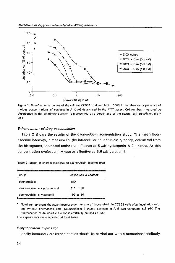
Modufatlon of P-gfycoprotein·mediated muftidrug resistance
120 Z
X
100 "-g • c 0 80 u
a ~ 60 • u c • -e 40 0 ~ .0 • 20 -
*' OOX control
-i-- OOX + GsA (0.1 ,liM)
*" OOX + GsA (0.5 ,liM)
--<>- DOX + GsA (1.0 ,liM)
o+-------~------~------~------~ 0.01 0.1 10 100
[doxorubicinJ in,uM
Figure 1. Dose/response curves of the cell line CC531 to doxorubicin (DOX) in the absence or presence of
various concentrations of cyclosporin A (CsA) determined in the MIT assay. Cell number, measured as absorbance in the colorimetric assay, is represented as a percentage of the control cell growth on the y axis
Enhancement of drug accumulation
Table 2 shows the results of the daunorubicin accumulation study. The mean fluor
escence intensity, a measure for the intracellular daunorubicin quantity, calculated from
the histograms, increased under the influence of 5 pM cyclosporin A 2.1 times. At this
concentration cyclosporin A was as effective as 6.6 pM verapamil.
Table 2. Effect of chemosensitizers on daunorubicin accumulation
drugs daunorubicin content"
daunOfubicin 100
daunorubicin + cyclosporin A 211 ± 38
daunorubicin + verapamit 193 ± 20
Numbers represent the mean fluorescence intensity of daunorubicin in CC531 cells after incubation with
and without chemosensitizers. Daunorubicin: 1 pg/ml; cyclosporin A 5 pM; verapamil 6.6 pM. The
fluorescence of daunorubicin alone is arbitrarily defined as 100
The experiments were repeated at least twice
P-glycoprotein expression
Ideally immunofluorescence studies should be carried out with a monoclonal antibody
74
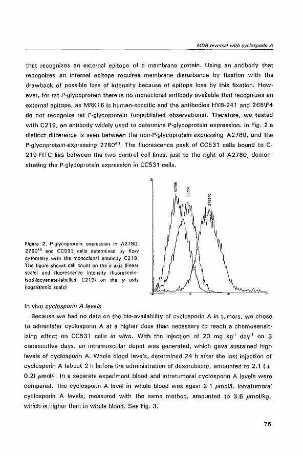
MDR reversal with cyclospor/n A
that recognizes an external epitope of a membrane protein. Using an antibody that
recognizes an internal epitope requires membrane disturbance by fixation with the
drawback of possible loss of intensity because of epitope loss by this fixation. How
ever, for rat P-glycoprotein there is no monoclonal antibody available that recognizes an
external epitope, as MRK16 is human-specific and the antibodies HYB-241 and 265\F4
do not recognize rat P-glycoprotein (unpublished observations), Therefore, we tested
with C219, an antibody widely used to determine P-glycoprotein expression. In Fig. 2 a
distinct difference is seen between the non-P-glycoprotein-expressing A2780, and the
P-glycoprotein-expressing 2780'°. The fluorescence peak of CC531 cells bound to C-
219-FITC lies between the two control cell lines, just to the right of A2780, demon
strating the P-glycoprotein expression in CC531 cells.
Figure 2. P-glycoprotein expression in A2780, 2780AO and CC531 cells determined by flow
cytometry with the monoclonal antibody C2l9. The fjgure shows cell count on the x axis (linear
scale) and fluorescence intensity (fluoresceinisothiocyanate-Iabelled C219) on the y axis
(logarithmic scale)
In vivo cyclosporin A levels
Because we had no data on the bio-availability of cyclosporin A in tumors, we chose
to administer cyclosporin A at a higher dose than necessary to reach a chemosensit
izing effect on CC531 cells in vitro. With the injection of 20 mg kg" day'! on 3
consecutive days, an intramuscular depot was generated, which gave sustained high
levels of cyelosporin A. Whole blood levels, determined 24 h after the last injection of
cyclosporin A (about 2 h before the administration of doxorubicinL amounted to 2.1 (±
0.2) pmolli. In a separate experiment blood and intratumoral cyclosporin A levels were
compared. The cyelosporin A level in whole blood was again 2.1 IImol/l. Intratumoral
cyelosporin A levels, measured with the same method, amounted to 3.6 IImol/kg,
which is higher than in whole blood. See Fig. 3.
75
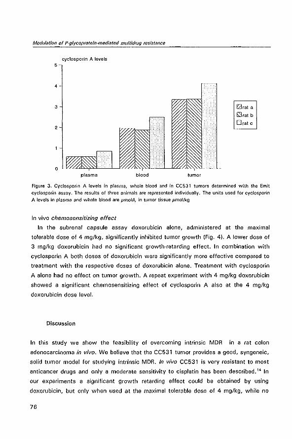
Modulation of P-glycoprotein-mediated muftidrug resistance
cyclosporin A levels
plasma blood tumor
Earat a
~(at b
Drat c
Figure 3. Cyclosporin A levels in plasma, whole blood and in CC531 tumors determined with the Emit
cyclosporin assay. The results of three animals are represented individually. The units used for cyclosporin
A levels in plasma and whole blood are pmolil, in tumor tissue pmollkg
In vivo chemosensitizing effect
In the subrenal capsule assay doxorubicin alone, administered at the maximal
tolerable dose of 4 mg/kg, significantly inhibited tumor growth (Fig. 4). A lower dose of
3 mg/kg doxorubicin had no significant growth-retarding effect. In combination with
cyclosporin A both doses of doxorubicin were significantly more effective compared to
treatment with the respective doses of doxorubicin alone. Treatment with cyclosporin
A alone had no effect on tumor growth. A repeat experiment with 4 mg/kg doxorubicin
showed a significant chemosensitizing effect of cyclosporin A also at the 4 mg/kg
doxorubicin dose level.
Discussion
In this study we show the feasibility of overcoming intrinsic MDR in a rat colon
adenocarcinoma in vivo. We believe that the CC531 tumor provides a good, syngeneic,
solid tumor model for studying intrinsic MDR. In vivo CC531 is very resistant to most
anticancer drugs and only a moderate sensitivity to cisplatin has been described. 14 In
our experiments a significant growth retarding effect could be obtained by using
doxorubicin, but only when used at the maximal tolerable dose of 4 mg/kg, while no
76
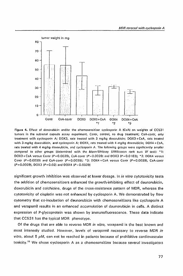
MDR reversal with cycfosporin A
tumor weight in mg
Contr GsA-contr DOX3 DOX3+CsA DOX4 OOX4+CsA 'I '2 '3
Figure 4. Effect of doxorubicin and/or the chemosensitizer cyclosporin A (CsA) on weights of CC531
tumors in the sub renal capsule assay experiment. Contr, control, no drug treatment; CsA-contr, only
treatment with cyclosporin A; DOX3, rats treated with 3 mglkg doxorubicin; DOX3 + CsA, rats treated
with 3 mg/kg doxorubicin, and cyc!osporin A; DOX4, rats treated with 4 mg/kg doxorubicin; DOX4 +CsA,
rats treated with 4 mg/kg doxorubicin, and cyclosporin A_ The following groups were significantly smaller
compared to other groups (determined with the Mann-Whitney UlWilcoxon rank sum W test): - 1:
DOX3+CsA versus ConU (P=O_00391, CsA-contr (P=O.0039) and DOX3 (P=O.0163); -2: DOX4 versus
Contr (P=O.0039) and CsA-contr (P""O.0039); -3: DOX4 +CsA versus Contr IP=O.0039), CsA-contr
(P=O.0039), DOX3 (P=O.02) and DOX4 (P=O.0039)
significant growth inhibition was observed at lower dosage. In in vitro cytotoxicity tests
the addition of chemosensitizers enhanced the growth-inhibiting effect of daunorubicin,
doxorubicin and colchicine, drugs of the cross-resistance pattern of MOR, whereas the
cytotoxicity of cisplatin was not enhanced by cyclosporin A. We demonstrated by flow
cytometry that co-incubation of daunorubicin with chemosensitizers like cyclosporin A
and verapamil results in an enhanced accumulation of daunorubicin in cells. A distinct
expression of P-glycoprotein was shown by immunofluorescence. These data indicate
that CC531 has the typical MDR phenotype.
Of the drugs that are able to reverse MDR in vitro, verapamil is the best known and
most intensely studied. However, levels of verapamil necessary to reverse MDR in
vitro, about 5 JIM, can not be reached in patients because of prohibitive cardiovascular
toxicity.15 We chose cyclosporin A as a chemosensitizer because several investigators
77
Modulation of P-glycoprotein-mediated multidrug resistance
reported a higher effectivity on an equimolar basis of cyclosporin A over verapamil,5,7,16
a result we also found with the CC531 cell line. The second reason we chose
cyclosporin A was that the concentration of cyclosporin A necessary in vitro to
overcome drug resistance is achievable without intolerable side-effects in vivo in
humans.\7,le However, one can not translate in vitro concentrations directly into doses
required in vivo, as pharmacological aspects may play an important role in the bio
availability of the drug,19 e.g. in vivo over 95% of cyclosporin A is bound to proteins or
cells. Therefore we decided to administer cyclosporin A at a higher dose than required
to achieve a chemosensitizing effect in our in vitro experiments. After 3 days of
intramuscular administration, cyclosporin A levels in whole blood obtained in the in vivo
experiments were above 2 pmol/!. Interestingly, measured with the same cyclosporin
assay, intratumoral levels of cyelosporin A amounted to 3.6 pmol/kg, which suggests
that in solid tumors cyclosporin A levels may be even higher than in whole blood. This
means that adequate levels of the chemosensitizer cyclosporin A can be reached at the
tumor site by a vascular route. In vivo this dose had neither a growth-retarding, nor a
growth-stimulating effect on the tumor: in the subrenal capsule assay, tumors in the
control group or tumors treated with cyclosporin A alone had similar weights.
In this intrinsic MDR model we show that cyclosporin A can have an effective
chemosensitizing effect on doxorubicin in vivo. In the subrenal capsule assay experi
ment rats treated with the combination of cyciosporin A and doxorubicin had signifi
cantly smaller tumors compared to all other groups and also compared to the rats
treated with doxorubicin alone. Adding cyciosporin A to doxorubicin rendered a
suboptimal dose of 3 mg/kg doxorubicin effective, and the activity of an effective dose
of 4 mg/kg was enhanced by combination with cyelosporin A. The differences in
standard errors between the experimental groups, with larger errors in the control
groups and less variation in results in the treated groups, is a phenomenon that is often
seen in experimental pharmacology. Probably this is due to the logarithmic growth of
tumors, which is attenuated for some time in effectively treated groups, resulting in
smaller tumors with inherently smaller standard errors.
Together with the in vitro data, demonstrating P-glycoprotein expression, enhance
ment of drug uptake and increased growth inhibition in CC531 cells by the addition of
cyclosporin A, these findings furnish evidence for direct MDR reversal at the cellular
tumor level. However, an alternative explanation, cyclosporin A altering drug pharmaco
kinetics,2° can not be ruled out. As P-glycoprotein expression is found in the kidneys,
especially in the renal tubules,' blocking of this efflux pump may diminish excretion of
78
MDR reversal with cyc/osporin A
anticancer drugs and, via higher and more prolonged blood levels, cause indirect
enhanced exposure of cells to drugs. 20
In vivo reversibility of drug resistance by cyclosporin A has been reported in other,
mostly non-solid tumor models. In ascites tumor models, Slater et al. 21 found a
correction of daunorubicin resistance by cyclosporin A on a daunorubicin-resistant
subline of the Ehrlich ascites tumor in vivo, and they also described enhancement of
the cytotoxicity of daunorubicin by cyciosporin A to the parental Ehrlich ascites and
hepatoma 129." Boesch et al." however, did not find any effect of cyciosporin A on
vincristine cytotoxicity to an MOR variant of the murine monocytic leukemia P388.
Only Osieka et al. 24 published a study about a solid tumor and showed enhancement of
etoposide cytotoxicity by cyclosporin A to a human embryonal carcinoma in nude mice.
We feel that our tumor model is, therefore, a valuable one, as it is both a syngeneic,
and a solid, MOR positive tumor.
Although our experiments suggest potential use for cyclosporin A in the clinical
setting, results in the few trials reported so far have not yet substantiated its role, as
they are not unequivocal. Sonneveld and Nooter25 showed the possibility of eliminating
MOR 1 positive acute myeloid leukemia cells by adding cyclosporin A to an ineffective
treatment schedule. In a very recent report they described reversal of clinical drug
resistance in patients with multiple myeloma after addition of cyclosporin A to the
combination chemotherapy. IS Response was correlated with P-glycoprotein and MORt
expression. Steady-state plasma levels of cyclosporin A were about 1000/19/1. Verweij
et al. '1 tested cyclosporin A as a reverter in combination with epidoxorubicin in
colore eta I cancer. In four out of four tumor samples they showed the ability of
cyclosporin A to enhance daunorubicin uptake by flow cytometry. Despite this observa
tion, only 1 patient out of 24 had a partial response, while 2 of the patients who
showed enhanced daunorubicin uptake in vitro had progressive disease. The
cyciosporin A levels they reported seem quite high and adequate: peak levels of about
6000 nglml and levels around 1000 ng/ml 18 h later. Similar cyclosporin A levels were
achieved in a clinical trial in renal celt cancer patients with the combination cyclosporin
A plus vinblastine. No response was found in 15 patients. 26 Although MOR levels or P
glycoprotein expression were not determined in these studies, it is likely that a substan
tial number of these 39 patients expressed P-glycoprotein in their tumors, as in other
studies colon carcinoma as well as renal cell cancer was found to be MOR- or P
glycoprotein-positive in the majority of the patients.27-31 Moreover, Kanamaru et al. 29
and Mickisch et al. 30 showed that P-glycoprotein expression in renal cell carcinomas
79
Modulation of P-glycoprotein-mediated mu/tidrug resistance
correlated with resistance in primary cell cultures in vitro to doxorubicin and vinblastine,
which could be reversed by chemosensitizers. The clinical trials by Rodenburg et al. 26
and Verweij et al_ 17 are the only studies that dealt exclusively with tumors from organs
that inherently have a high expression level of P-glycoprotein. Other studies were
carried out in heavily pretreated patients or patients with advanced disease. In these
studies, a clear response was found in a trial with patients with myeloma or lymphoma
resistant to vincristine/doxorubicin/dexamethasone: 3 out of 8 patients responded to
the addition of verapamil to the drug regimen. while 6 of these 8 patients exhibited P
glycoprotein expression on their tumor cells. 32 So this last study and the study by
Sonneveld et a/. 18 clearly suggest a beneficial role of MDR reverters.
So far only clinical trials with hematological disorders have been successful in revers
ing MDR. In solid tumors no responses were found. We cannot compare our experimen
tal data with clinical trials, but we show in our model the feasibility of reversing
intrinsic MDR in a solid tumor in vivo. Adequate levels of the chemosensitizer
cyclosporin A could be obtained at the tumor site by a vascular route and this rendered
a suboptimal dose of doxorubicin effective. Our results, obtained in a syngeneic, solid
tumor model therefore suggest that there may still be a place for chemosensitizers in
the chemotherapy of MDR solid tumors.
References
1. Go!dstein LJ, Galski H, Fojo A, Willingham M, Lai S·L, Gazdar A, Pirker A. Green A, Crist W, Brodeur GM, Lieber M, Cossman J, Gottesman MM, Pastan I. Expression of a multi drug resistance gene in human cancers. J Natf Cancer !nst 1989; 81: 116-24
2. Fojo AT, Ueda K, Siamon OJ, Popfack OG, Gottesman MM, Pastan I. Expression of a multidrugresistance gene in human tumors and tissues. Proc Natl Acad Sci USA 1987; 84: 265-9
3. Thiehaut F, Tsuruo T. Hamada H, Gottesman MM, Pastan I, Willingham MC. Cellular localization of the multidrug·resistance gene product P-glycoprotein in normal human tissues. Proc Nat! Acad Sci USA 1987; 84: 7735·8
4. Ford JM, Hait WN. Pharmacology of drugs that alter multidrug resistance in cancer. Pharmacol Rev 1990: 42: 155-99
5. Boesch 0, Muller K, Pourtier-Manzanedo A, Loor F. Restoration of daunomycin retention in multidrug-resistant P388 cells by submicromolar concentrations of SDZ PSC 833, a nonimmunosuppressive cyclosporin derivative. Exp Cell Res 1991; 196: 26-32
6. Gav~riaux C, Boesch D, Boelsterli JJ, Bollinger P, Eberle MK. Hiestand P. Payne T, Traber A, Wenger A, Loor F. Overcoming multidrug resistance in Chinese hamster ovary cells in vitro by cyclosporin A (Sandimmune) and non-immunosuppressive derivatives. Sr J Cancer 1989; 60: 867-71
7. Twentyman PR, Aeeve JG. Koch G, Wright KA. Chemosensitisation by verapamil and cyclosporin A in mouse tumor cells expressing different levels of P·glycoprotein and CP22 Isorcin). Sr J Cancer 1990; 62: 89·95
8. Twentyman PA, Wright KA. Wallage HM. Effects of cyclosporin A and a non·immunosuppressive analogue. O-acetyl cyclosporin A, UpOll growth of parent and multidrug resistant human lung cancer cells in vitro. Sr J Cancer 1992; 65: 335·40
80
MDR reversal with cyclosporin A
9. Marquet AL, Westbroek DL, Jeekel J. Interferon treatment of a transplantable rat colon adenocarcinoma: importance of tumor site. Int J Cancer 1984; 33: 689·92
10. Aogan AM, Hamilton Te, Young Re, Klecker AW, Ozols AF. Aeversal of Adriamycin resistance by verapamil in human ovarian cancer. Science 1984; 224: 994·6
11. Carmichael J, DeGraff WG, Gazdar AF, Minna JD, Mitchell JB. Evaluation of a tetrazollum·based semiautomated colorimetric assay: assessment of chemosensitivity testing. Cancer Res 1987; 47: 936-42
12. Gheuens EEO, van Bockstaele DA, van der Keur M, Tanke HJ, van Oosterom AT, de Bruijn EA. Flow cytometric double labeling technique for screening of multidrug resistance. Cytometry 1991; 12: 636-44
13. Kanner N, Evernden·Porelle DE, Bradley G, Ling V. Detection of P-glycoprotein in mu[tidrugresistant cell lines by monoclonal antibodies. Nature 1985; 316: 820-3
14. Los G, Nagel JD, McVie JG. Anti·tumor effect of cisplatin, carboplatin, mitoxantrone, and doxorubkin on peritoneal tumor growth after intraperitoneal and intravenous chemotherapy: a comparative study. Sel Cancer Ther 1990; 6: 73·82
15. Pennock GD, Dalton WS, Aoeske WA, Appleton CP, Mosley K, Plezia P, Miller TM, Salmon SE. Systemic toxic effects associated with high-dose verapamil infusion and chemotherapy administration. J Nall Cancer Inst 1991; 83: 105-'0
16. Silbermann MH, Boersma AWM, Janssen ALW, Scheper RJ, Herweijer H, Nooter K. Effects of cyclosporin A and verapamil on the intracellular daunorubicin accumulation in Chinese hamster ovary cells with increasing levels of drug-resistance. Int J Cancer 1989; 44: 722-6
17. VelVleij J, Herweijer H, Oosterom A, van der Burg MEL, Planting ASTh, Seynaeve C, Stoter G, Nooter K. A phase II study of epidoxorubicin in colorectal cancer and the use of cyclosporin-A in an attempt to reverse multidrug resistance. Dr J Cancer 1991; 64: 361·4
18. Sonneveld P, Durie BGM, Lokhorst HM, Marie JP, Sorbu B, Suciu S, Zittoun A, LOwenberg B, Nooter K. Modulation of multidrug·resistant multiple myeloma by cyclosporin. lancet 1992; 340: 255-9
19. Kaye SB. Reversal of multidrug resistance. Cancer Treat Rev 1990; 17 (Suppl AJ: 37-43 20. Sikic BI, Yahanda AM, Adler KM, Fisher B, Brophy NA, Halsey J, Gosland MP, Lum BL. Use of
cyclosporin to reverse drug resistance (abstract). Ann Onco/1992; 3 ISuppl 11: 63 21. Slater LM, Sweet P, Stupecky M, Wetzel MW, Gupta S. Cyclosporin A corrects daunorubicin
resistance in Ehrlich ascites carcinoma. Br J Cancer 1986; 54: 236-8 22. Meador J, Sweet P, Stupecky M, Wetzel M, Murray S, Gupta S, Slater L. Enhancement by
cyclosporin A of daunorubicin efficacy in Ehrlich ascites carcinoma and murine hepatoma 129. Cancer Res 1987; 47: 6216-9
23. Boesch D, Gav~riaux C, Jachez B, Pourtier·Manzanedo A, Bo1Jinger P, Loor F. In vivo circumvention of P·glycoprotein-mediated multidrug resistance of tumor cells with SDZ PSC 833. Cancer Res 1991; 51: 4226-33
24. Osieka A, Seeber S, Pannenb~cke( A, Soli 0, Glatte P, Schmidt eG. Enhancement of etoposideinduced cytotoxicity by cyclosporin A. Cancer Chemother Pharmacol 1986; 18: 198-202
25. Sonneveld P, Nooter K. Reversal of drug·resistance by cyclosporin·A in a patient with acute myelocytic leukaemia. Dr J Haemalol 1990; 75: 208-11
26. Aodenburg CJ, Nooter K, Herweijer H, Seynaeve C, Oosterom A, Stoter G, Verweij J. Phase II study of combining vinblastine and cycJosporin·A to circumvent multidrug resistance in renal cell cancer. Ann Oncol 1991; 2: 305-6
27. Mizoguchi T, Yamada K, Furukawa T, Hidaka K, Hisatsugu T, Shimazu H, Tsuruo T, Sumizawa T, Akiyama S-i. Expression of the MDRI gene in human gastric and colorectal carcinomas. J Natl Cancer Insl 1990; 82: 1679·83
28. Weinstein AS, Jakate SM, Dominguez JM, Lebovitz MD, Koukoulis GK, Kuszak JA, Klusens LF, Grogan TM, Saclarides T J, Aoninson IB, Coon JS. Aelationship of the expression of the multidrug resistance gene product jP·glycoprotein) in human colon carcinoma to local tumor aggressiveness and lymph node metastasis. Cancer Res 1991; 51: 2720·6
29. Kanamaru H, Kakehi Y, Yoshida 0, Nakanishi S. Pastan I, Gottesman MM. MORt ANA levels in human renal cell carcinomas: correlation with grade and prediction of reversal of doxorubicin resistance by Quinidine in tumor explants. J Nat! Cancer Inst 1989; 81: 844-9
30. Mickisch GH, Aoehrich K, Koessig J, Forster S, Tschada RK, AIken PM. Mechanisms and modula· tion of multldrug resistance in primary human renal cell carcinoma. J Uro/1990; 144: 755-9
31. Van Kalken CK, van der Valk P, Hadisaputro MMN, Pieters A, Broxterman HJ, Kuiper eM, Scheffer GL, Veerman AJP, Meyer CJLM, Scheper AJ, Pinedo HM. Differentiation dependent expression of
81
Modulation of P·olycoprotein·mediated multidruo resistance
P'glycoprotein in the normal and neoplastic human kidney. Ann Onco/1991; 2: 55-62 32. Dalton WS, Grogan TM, Meltzer PS, Scheper RJ, Durie BGM, Taylor CW, Miller TP, Salmon SE.
82
Drug·resistance in multiple myeloma and non·Hodgkin's lymphoma: detection of P-glycoprotein and potential circumvention by addition of verapamil to chemotherapy. J C/in Oneal 1989; 7: 415-24
2.2
MODULA TlON OF MUL TIDRUG RESISTANCE WITH
DEXNIGULDIPINE HYDROCHLORIDE (88509-035)
IN THE CC531 RAT COLON CARCINOMA MODEL
Wim van de Vrie, Jan H.M. Schellens,
Walter J. Laos, Herman J. Kolker,
Jaap Verwey, Gerrit Stater,
Nico M.e. Durante and Alexander M.M. Eggermont
J Cancer Res Clin Oneal
1996; 122: 403·8
Modulation of P-glycoprotein-mediated multidrug resistance
Summary
The chemosensitizing potency of dexniguldipine hydrochloride (88509-035) on
epidoxorubicin was assessed in an MDR tumor model, the intrinsically MDR rat colon
carcinoma CC531. In vitro in the sulphorhodamine 8 cell-viability assay the cytotoxicity
of epidoxorubicin was increased approximately 15-fold by co-incubation with 50 ng/ml
dexniguldipine. In vivo concentrations of dexniguldipine 5 h after a single oral dose of
30 mg/kg were 72 (± 19 sd) ng/ml in plasma and 925 (± 495 sd) ng/g in tumor tissue.
Levels of the metabolite of dexniguldipine M-l, which has the same chemosensitizing
potential, were 26 (± 6 sd) ng/ml and 289 (± 127 sd) ng/mg respectively. The efficacy
of treatment with 6 mg/kg epidoxorubicin applied intravenously combined with 30 mg
kg'! day·1 dexniguldipine administered orally for 3 days prior to epidoxorubicin injection
was evaluated on tumors grown under the renal capsule. Dexniguldipine alone did not
show antitumor effects in vivo. Dexniguldipine modestly, but consistently, potentiated
the tumor-growth-inhibiting effect of epidoxorubicin reaching statistical significance in
two out of four experiments. In conclusion, these experiments show that dexniguldipine
has potency as an MDR reverter in vitro and in vivo in this solid MDR tumor model.
84
MDR reversal with dexnlguldipine
Introduction
Multidrug resistance is an important mechanism in clinical drug resistance and express
ion of the MDR gene is found in a variety of tumors.1,2 In MDR a transmembrane efflux
pump, P-glycoprotein, confers drug resistance on a group of chemically unrelated
anticancer drugs by increasing the efflux. The P-glycoprotein pump can be blocked
reversibly by so-called chemosensitizers, which are substrates for the protein them
selves. As a result, higher intracellular levels of anticancer drugs are achieved and
enhanced cell death occurs. Among the various compounds that can function as
chemosensitizers are verapamil, cyclosporin A and its non-immunosuppressive analogue
PSC 833. quinidine, tamoxifen, and many others.1 Previous studies have been carried
out with verapamil, but levels necessary for modulation of MDR in vivo appeared too
high. resulting in severe cardiovascular side-effects. In a clinical trial with verapamil.
dose-limiting side-effects were hypotension and cardiac arrhythmias at levels of
exposure anticipated to be inadequate for MDR reversal. 3 This has led to a search for
related compounds that are devoid of the cardiovascular side-effects.
Stereoisomers of verapamil and related drugs vary in calcium-channel-blocking
activity. For example, the (+ )stereoisomer of verapamil is a 10-fold less potent calcium
antagonist than the (-)isomer,4 but has approximately the same chemosensitizing effec
tiveness. 5 Of the other calcium antagonists. the dihydropyridine drug niguldipine. was
found to be a very effective chemosensitizer in MDR.6 The (-)stereoisomer, dexniguldi
pine hydrochloride (88509-035). displays a 45-fold lower affinity for calcium-channel
binding sites compared to the (+ )isomer, while both have the same MDR-modulating
potency.6 In various preclinical models in vitro the chemosensitizing potency of
dexniguldipine was either equal to or up to 50 times more effective than verapamiI.6."
We tested the activity of dexniguldipine in the CC531 MDR tumor model. CC531 is a
colon carcinoma, derived from and transplantable in the WAG/RIJ rat, that intrinsically
expresses the multidrug-resistant phenotype. In a previous report the potency of
cyclosporin A as a modulator of resistance to doxorubicin was shown in this model. 12
Here we report on the chemosensitizing effect of dexniguldipine in vitro and in vivo and
on levels of dexniguldipine and its active metabolite M-l in plasma and tumors.
85
Modulation of P-glycoprotein-mediated multidrug resistance
Materials and Methods
Animals
Male rats of the inbred WAG/RIJ (RT1') strain were obtained from Harlan·CPB
(Austerlitz, The Netherlands). Animals were bred under specific·pathogen·free condi·
tions and fed standard rat chow (Hope Farms, Woerden, The Netherlands) and water ad
libitum. In the experiments rats 12·18 weeks old, weighing 220·280 g, were used.
Tumor and cell line
CC531 is a colon carcinoma, which was induced chemically in the WAG/RIJ rat with
1,2-dimethylhydrazine. The tumor, a moderately differentiated adenocarcinoma, is
weakly immunogenic and transplantable in WAG/RIJ rats. 13 In vitro the cell line grows
as a monolayer. CC531 is an intrinsically multidrug-resistant tumor as it expresses the
MDR phenotype. At the mRNA level expression of mdrla, and not mdrlb, has been
detected by the polymerase chain reaction. '4 A low level of P-glycoprotein expression
has been shown with the monoclonal antibody C219 by Western blotting and by
immunofluorescence techniques. 12,14 Intracellular accumulation of daunorubicin can be
enhanced by chemosensitizers like verapamil and cyclosporin A. 12,15 Cytotoxicity assays
have shown the typical drug resistance pattern of MDR and enhancement of cytotox·
icity by chemosensitizers.' 2,'5
The cell line was grown in Oulbecco's modified Eagle's medium supplemented with
5% fetal calf serum, aspartic acid '(0.1 mM), glutamic acid (0.3 mM), penicillin (111
IUlml) and streptomycin (111 pglmll. all obtained from Gibco (Paisley, UK), in a
humidified atmosphere of 5% CO,l95% air at 37°C. Regular screening for Mycoplasma
infection was performed. Cells were isolated by trypsinization; viability, determined by
trypan blue exclusion, was over 90% in all experiments.
Chemicals
Dexniguldipine hydrochloride (B8509·035), the metabolite M·l (B8909·008) and
B9003·001 (internal standard for dexniguldipine in the HPLC) were kindly provided by
Byk Gulden, Konstanz, Germany; epidoxorubicin (Farmorubicin) was obtained from
Farmitalia, Carlo Erba, Italy; sulphorhodamine B was purchased from Sigma Chemicals,
St. Louis, Mo., USA; deionized MiIli·O water was from Millipore, Etten Leur, The
Netherlands; trichloroacetic acid from J.T. Baker, Deventer, The Netherlands; and
dichloromethane/hexane/isobutyl alcohol (40:60:0.5) from Rathburn, Walkerburn,
86
MDR reversal with dexnlguldipine
Scotland.
In vitro cytotoxicity assay
Chemosensitivity in vitro was determined with the sulphorhodamine B cell-viability
assay, essentially carried out as described by Skehan et al. 16 In brief, 2 x 10³ trypsin
ized tumor cells/well in 2001'1 complete medium were plated into 96-well flat-bottomed
microtitre plates (Costar, Cambridge, Mass., USA). Tests were carried out in quadrupli
cate. The plates were incubated for 24 h at 37°C, 5% CO,/95% air to allow the cells
to adhere. Then the old medium was replaced by medium containing the test drug in
graded concentrations; in the interaction studies epidoxorubicin together with a fixed
concentration of dexniguldipine was added. On day 7 the incubation was terminated by
washing the plates twice with PBS. Subsequently the cells were fixed with 10%
trichloroacetic acid in deionized Milli-Q water and placed for 1 h at 4°C. After five
washings with tap water, the cells were stained for 15 min with 0.4% sulphorhodami
ne 8 dissolved in 1 % acetic acid. and subsequently washed with 1 % acetic acid to
remove the unbound stain. The plates were air-dried and bound protein stain was
dissolved in 1501'1 10 mM TRIS base. The absorbance was read at 540 nm using an
automated microplate reader (Titertek, Flow Laboratories Ltd .. Irvine, Scotland).
In vivo assay
Subcutaneously grown solid tumors of the CC531 cell line were used 20-30 days
after implantation. In a subrenal capsule assay tumor pieces weighing 6-8 mg were
implanted under the capsule of the kidneys. In the pharmacokinetic experiment,
treatment with dexniguldipine was given 10 days after implantation. Rats were
restrained from food 12 h prior to administration of the drug. A single dose of 30
mg/kg dexniguldipine was administered orally through a thin metal cannula. Five hours
later rats were sacrificed, a blood sample was taken and the tumors were collected for
analysis of dexniguldipine and M-l levels.
In the pharmacodynamic experiment dexniguldipine treatment was started 24 h after
implantation of the tumor. In contrast to the single dosing in the pharmacokinetic
experiment. dexniguldipine was administered for 3 consecutive days orally at a dose of
30 mg kg" day". On day 4, 5 h after the last dexniguldipine dose, rats were injected
intravenously with 6 mg/kg epidoxorubicin, or PBS in control rats. On day 10 the
animals were sacrificed. tumors were enucleated and weighed.
All experimental groups consisted of 6-8 rats.
87
Modulation of P-glycoprotein-mediated multidrug resistance
Apparatus for dexniguldipine and M-l measurement
Dexniguldipine was determined in plasma and tumor tissue with an automated
reverse·phase isocratic high·performance liquid chromatography (HPLC) assay with UV
detection at 230 nm. A model 7108 WISP autosampler and a model M51 0 pump were
used (all Waters Assoc., Milford, Mass., USA). The detector was a UV2000 (Spectra
Physics, San Jose, Calif., USA). The data were processed with a Shimadzu CR3A
integrator (Shimadzu Corp., Kyoto, Japan). The column was a Shandon Hypersyl CPS,
3 pm 150 x 4.6 mm (LC Services, Emmen, The Netherlands!. The eluent consisted of a
5 mM phosphate buffer (pH 7.5) with 60% acetonitrile. The flow rate was 1.5 ml/min
and the column temperature 40°C. Sample size was 100/11 for each analysis.
Sample preparation for dexniguldipine and M-l measurement
A volume of 150 pi plasma was collected, to which 50 pi 2000 ng/ml solution of
internal standard (89003·001) in methanol was added. Next, 800 pi deionized MiIIi·Q
water was added and the sample was mixed on a whirl mixer for 15 s. For extraction
of the test chemicals 7 ml dichloromethane/hexane/isobutyl alcohol (40:60:0.5) was
added. The mixture was mixed for 30 min on a whirl mixer and subsequently centri
fuged for 10 min at 4000 g. The organic layer was collected and evaporated to dryness
at 50°C under vacuum. The residue was reconstituted in 150 pi eluent.
Tumor tissue was homogenized with a Turrax homogenizer (Boom, Meppel, The
Netherlands) in 1 ml of MiIli·Q water. The homogenizer was flushed twice with 250 pi
Milli·Q water. A 50 pi volume of a 2000·ng/ml internal standard solution in methanol
and 7 ml dichloromethane/hexane/isobutyl alcohol (40:60:0.5) were added. Further
handling of the tissue sample was as described for the plasma sample.
The recovery of dexniguldipine, M-l, and the internal standard was determined
relative to direct injection of the individual dissolved compounds.
Statistics
Statistical analysis was carried out with SPSS/PC +, using the Mann·Whitney
UlWilcoxon rank-sum W-test. P< 0.05 was considered significant.
Ethical approval
The experimental protocols adhered to the rules laid down in The Dutch Animal
Experimentation Act (1977) and the Guidelines on the Protection of Experimental
Animals published by the Council of the E.C. (1986). Specific protocols were approved
88
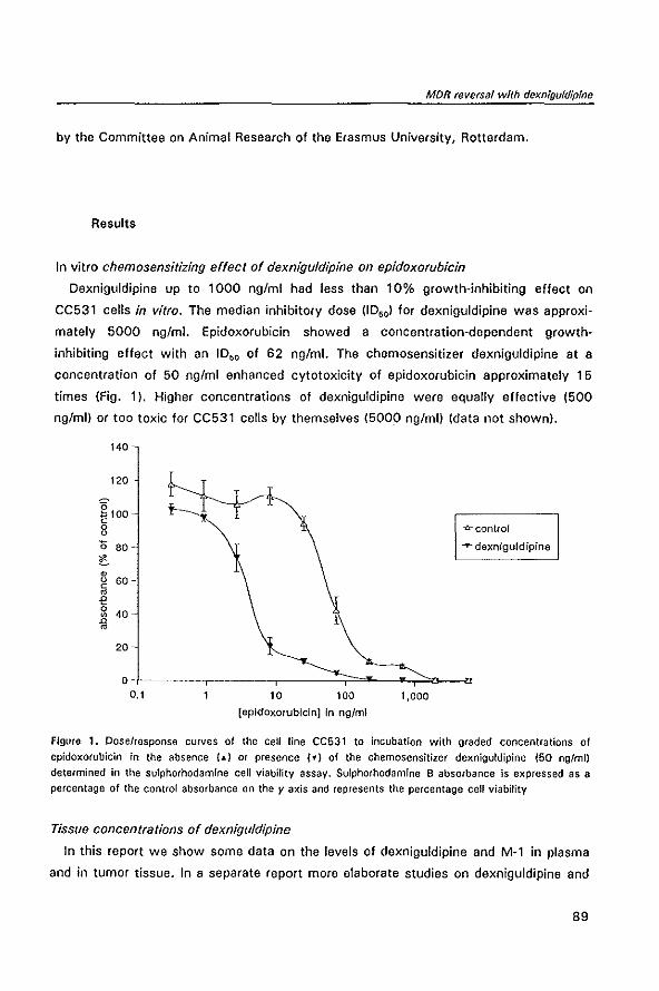
MDR reversal with dexniguldipfne
by the Committee on Animal Research of the Erasmus University, Rotterdam.
Results
In vitro chemosensitizing effect of dexniguldipine on epidoxorubicin
Oexniguldipine up to 1000 ng/ml had less than 10% growth-inhibiting effect on
CC531 cells in vitro. The median inhibitory dose (10,,1 for dexniguldipine was approxi
mately 5000 ng/ml. Epidoxorubicin showed a concentration-dependent growth·
inhibiting effect with an 1050 of 62 ng/ml. The chemosensitizer dexniguldipine at a
concentration of 50 ng/ml enhanced cytotoxicity of epidoxorubicin approximately 15
times (Fig. 11. Higher concentrations of dexniguldipine were equally effective (500
ng/mll or too toxic for CC531 cells by themselves (5000 ng/mll (data not shownl.
140
120
g100 c 0 0 -fr- control
'0 80 ...,.. dexniguldipine
C • 0 60 -c • € 0 40 • n •
20
0 0.1 10 100 1,000
[epldoxorubicinJ In ng/ml
Figure 1. Dose/response curves of the cell line CC531 to incubation with graded concentrations of
epidoxorubicin in the absence (J.) or presence (yj of the chemosensitizer dexniguldipine (50 ng/ml)
determined in the sulphorhodamine cell viability assay. Sulphorhodamine B absorbance is expressed as a
percentage of the control absorbance on the y axis and represents the percentage cell viability
Tissue concentrations of dexniguldipine
In this report we show some data on the levels of dexniguldipine and M-1 in plasma
and in tumor tissue. In a separate report more elaborate studies on dexniguldipine and
89
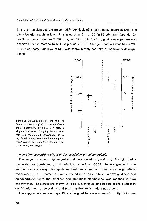
Modulation of P-gfycoproteln-mediated mullidrug resistance
M-l pharmacokinetics are presented. 17 Dexniguldipine was readily absorbed after oral
administration reaching levels in plasma after 5 h of 72 (± 19 sd) ng/ml (see Fig. 2).
Levels in tumor tissue were much higher: 925 (±495 sd) ng/g. A similar pattern was
observed for the metabolite M·l: in plasma 26 (± 6 sd) ng/ml and in tumor tissue 289
(± 127 sd) ng/gr. The level of M·l was approximately one-third of the level of dexnigul
dipine.
10,000 10,000
~ 1,000 ~ 1,000
~ • I C>
s S ~ ---r " E E ~ il 0.
100 .~ 100 ..L .~ c c : 0
i -+-- ~ m
m • u u • c c 0 0 10 10 u u
Figure 2. Dexniguldipine I·) and M-l ,.) levels in plasma lng/mil and tumor tissue
Ingfg) determined by HPlC 5 h after a • ~
m single oral dose of 30 mgikg. Results from c c
" '0. '0. rats are represented individually on a U U logarithmic scale, with lines indicating the S S
C> C>
mean values. left data from plasma; right 'c '0 x x • • data from tumor tissue 'C 'C
In vivo chemosensitizing effect of dexniguldipine on epidoxorubicin
Pilot experiments with epidoxorubicin alone showed that a dose of 6 mg/kg had a
moderate but consistent growth-inhibiting effect on CC531 tumors grown in the
subrenal capsule assay. Dexniguldipine treatment alone had no influence on growth of
the tumor. In all experiments tumors treated with the combination dexniguldipine and
epidoxorubicin were the smallest and statistical significance was reached in two
experiments. The results are shown in Table 1. Dexniguldipine had no additive effect in
combination with a lower dose of 4 mg/kg epidoxorubicin (data not shown).
The experiments were not specifically designed for assessment of toxicity, but some
90
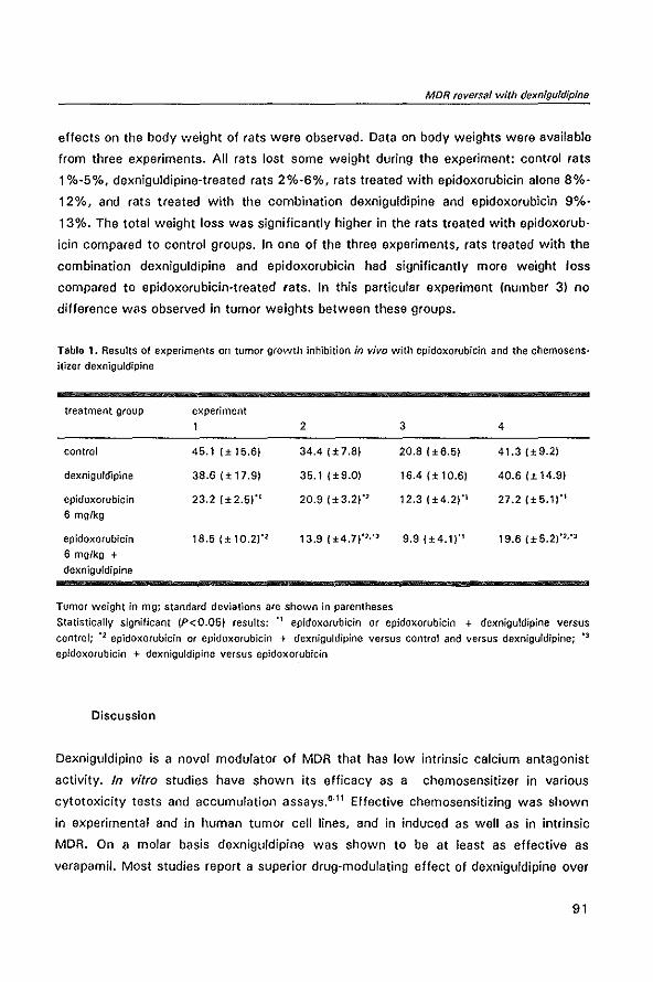
MDR reversal with dexni{}uldlpine
effects on the body weight of rats were observed. Data on body weights were available
from three experiments. All rats lost some weight during the experiment: control rats
1 %-5%, dexniguldipine-treated rats 2%-6%, rats treated with epidoxorubicin alone 8%-
12%, and rats treated with the combination dexniguldipine and epidoxorubicln 9%-
13%. The total weight loss was significantly higher in the rats treated with epidoxorub·
icin compared to control groups. In one of the three experiments, rats treated with the
combination dexniguldipine and epidoxorubicin had significantly more weight loss
compared to epidoxorubicin-treated rats. In this particular experiment (number 3) no
difference was observed in tumor weights between these groups.
Table 1. Results of experiments on tumor growth inhibition in vivo with epidoxorubicin and the chemos ensitizer dexniguldipine
treatment group
control
dexniguldipine
epidoxorubicin
6 mg/kg
epidoxorubicin
6 mg/kg + dexniguldipine
experiment 2
45.1 I± 15.6) 34.41±7.8)
38.6 I± 17.9) 35.1 1±9.0)
23.21±2.5)"' 20.9 (± 3.2)'2
18.5 (± 10.2)'2 13.9 (±4,7)'2,'3
Tumor weight in mg; standard deviations are shown in parentheses
3 4
20.8 I ±6.5) 41.31±9.2)
16.41± 10.6) 40.6 I ± 14.9)
12.3 (±4.2)'\ 27.2 (±5,1)'\
9,9 {±4.1)'1 19.6 (±5,2)'2,'J
Statistically significant (P<O.05) results: '\ epidoxorubicin or epidoxorubicin + dexniguldipine versus
control; '2 epidoxorubicin or epidoxorubicin + dexniguldipine versus control and versus dexniguldipine; '3
epidoxorubicin + dexniguldipine versus epidoxofubicin
Discussion
Dexniguldipine is a novel modulator of MDR that has low intrinsic calcium antagonist
activity. In vitro studies have shown its efficacy as a chemosensitizer in various
cytotoxicity tests and accumulation assays,6.11 Effective chemosensitizing was shown
in experimental and in human tumor cell lines, and in induced as well as in intrinsic
MDR. On a molar basis dexniguldipine was shown to be at least as effective as
verapamil. Most studies report a superior drug-modulating effect of dexniguldipine over
91
Modulation of P-glycoproleln-mediated multidrug resistance
verapamil of 2.5- to 50-fold. 7.,."
This study expands these in vitro studies with in vivo data. After oral administration,
dexniguldipine is readily absorbed and distributed into various tissues. Dexniguldipine
has a very lipophilic nature and its volume of distribution is high (in animals 20-40
IIkg)." In the present study intratumoral levels of dexniguldipine were 925 nglg tissue,
which is more than ten times the plasma levels. Compared to the in vitro level of 50
nglml, which was effective in MDR modulation, these in vivo levels are high. The data
on dexniguldipine levels were obtained after a single oral dosing. In the antitumor
experiment dosing was tripled by administration on 3 consecutive days. Because of the
lipophilic nature of the drug this will probably have resulted in even higher intratumoral
levels. The wide variation in the results of the levels of the chemosensitizer is not
readily explained. Wide interindividual variation in pharmacokinetics has been reported
by others for dexniguldipine after oral administration, as well as for other dihydropyri
dine compounds. '9 Part of the activity of dexniguldipine in vivo is mediated by the active metabolite M-
1, which is shown to have the same MDR-modulating potency as dexniguldipine. 6,9 The
pharmacokinetics of M-l followed the results of dexniguldipine closely. The M-l level
was approximately one-third of the level of dexniguldipine in plasma and in tumor tissue
5 h after administration. Recently, comparable levels of dexniguldipine and M-1 have
been published from a phase I trial in patients. '9
Dexniguldipine showed no direct antitumor activity against CC531 cells. Anti
proliferative effects of dexniguldipine have been reported for some tumors, possibly
tumors with a neuroendocrine differentiation depending on autocrine stimulating
factors. 20•
21 The chemosensitizing potency of dexniguldipine on the MDR CC531 tumor
was observed in all in vivo experiments. Oexniguldipine had a significant potentiating
effect on growth inhibition of CC531 tumors by epidoxorubicin in two out of four
experiments, while in the other experiments the observed differences did not reach
statistical significance. The results with dexniguldipine are comparable to those with
earlier published experiments in the CC531 tumor model, which revealed the chemo
sensitizing effect of cyclosporin A in combination with doxorubicin. '2
Levels of epidoxorubicin in plasma and tumor have not been measured in these
experiments. Therefore, we can not rule out the possibility that altered pharmacokinet
ics of epidoxorubicin contribute to the chemosensitizing effect apart from direct
modulation of MDR at the cellular level. The fact that combination treatment resulted in
a small enhancement of toxicity in one experiment, as measured by body-weight loss,
92
MDR reversal with dexniguldipine
suggests that at least some systemic enhancement of epidoxorubicin activity may have
occurred. This is in agreement with previous studies that showed enhancement of
doxorubicin toxicity caused by combined treatment with cyclosporin A.n Other
investigators, however, have also furnished evidence for a direct effect of chemosensit
izers on the tumor. Niwa et al.23 showed that PAK-200, like dexniguldipine a dihydro
pyridine analogue, enhanced the accumulation of doxorubicin in solid tumors in vivo.
The effect of PAK-200 on doxorubicin accumulation in the tumors was dependent on
'the level of P-glycoprotein expression: only the tumors with a clear expression of P
glycoprotein had a higher doxorubicin content in the presence of the chemosensitizer.
Furthermore, in patients with refractory multiple myeloma, addition of cyclosporin A to
the chemotherapeutic regimen vincristine, doxorubicin and dexamethasone resulted in
enhancement of the response rate. 24 Additional studies showed that the effect was
probably achieved by specific killing of the plasma cells expressing P-glycoprotein. 25
The studies clearly suggest drug modulation directly at the cellular level by a P
glycoprotein-dependent mechanism.
The results in our study are comparable to results obtained with dexniguldipine and
doxorubicin in a nude mouse xenograft model. Here partial reversal of resistance to
doxorubicin was observed in solid tumors of the MDR l-overexpressing KB-8-5 cell line
grown subcutaneously. 26 Dexniguldipine has entered clinical studies now and promising
results have been obtained in trials in acute myeloid leukemia and multiple
myeloma. 21•28
The present study confirms the chemosensitizing potency of dexniguldipine on MDR
cells in vitro. It shows that in vivo relatively high levels of dexniguldipine in plasma and
tumor tissue can easily be achieved by oral administration. In vivo this resulted in a
strong trend towards a significant enhancement of the antitumor effect of epidoxorub
icin in the solid MDR tumor CC531.
References
1. Bellamy WT, Dalton WS. Multidrug resistance in the laboratory and clinic. Adv Clin Chern 1994; 31: 1-61
2. Goldstein LJ, Galski H, Fojo A. Willingham M. Lai Sol, Ga2dar A, Pirker R, Green A, Crist W, Brodeur GM, lieber M, Cossman J, Gottesman MM, Pastan I. Expression of a multidrug resistance gene in human cancers. JNatlCancer Insl1989; 81: 116-24
3. Pennock GO, Dalton WS, Roeske WR, Appleton CP, Mosley K, Plezia P, Miller TP, Salmon SE. Systemic toxic effects associated with high-dose verapamil infusion and chemotherapy administration. J Nat! Cancer Insf 1991; 83: 10&-10
4. Echi2en H, Brecht T, Niedergesilss S, Vogelgesang B, Eichelbaum M. The effect of dextro-, levo-,
93
Modulation of P-glycoprotein-mediated multidrug lesistance
and racemic verapamil on atrioventricular conduction 1n humans. Am Heart J 1985; 109: 210-7 5. Plumb JA, Milroy R, Kaye SB. The activity of verapamil as a resistance modifier in vitro in drug
resistant human tumor cell lines is not stereospecific_ Biochem Pharmacal 1990; 39: 787-92 6. HOllt V, Kouba M. Dietel M, Vogt G_ Stereo isomers of calcium antagonists which differ markedly in
their potencies as calcium blockers are equally effective in modulating drug transport by Pglycoprotein. Biochem Pharmacal 1992; 43: 2601-8
7. Hill BT, Hosking LK_ Differential effectiveness of a range of novel drug-resistance modulators, relative to verapamil. in influencing vinblastine or teniposide cytotoxicity in human Iymphoblastoid CCRF-CEM sublines expressing classic or atypical multidrug resistance. Cancel Chemother Pharmacal 1994; 33: 317-24
8. Hofmann J, Ueberall F, Egle A, Grunicke H. 8-859-35, a new drug with anti-tumor activity reverses multi-drug resistance. Int J Cancer 1991; 47: 870-4
9_ Hofmann J, Wolf A, Spitaler M, Back G, Drach J, Ludescher C, Grunicke H. Reversal of multidrug resistance by B859-35, a metabolite of 8859-35, niguldipine, verapamil and nitrendipine. J Cancer Res Clin Oncol 1992; 118: 361-6
10. Reymann A, Looft G, Woermann C, Dietel M, Erttmann A. Aeversal of multidrug resistance in Friend leukemia cells by dexniguldipine·HCI. Cancer Chemother Pharmacal 1993; 32: 25-30
11. Aoller E, Klumpp a, Krause J, Eichelbaum M, Schumacher K. Influence of sequential exposure to Averapamit or 88509-035 on rhodamine 123 accumulation in human Iymphoblastoid cell lines. Cancer Chemolher Pharmacal 1993; 32: 151-5
12. Van de Vrie W, Gheuens EEO, Durante NMC, de 8ruijn EA, Marquet RL, van Oosterom AT, Eggermont AMM. In vitro and in vivo chemosensitizing effect of cyclosporin A on an intrinsic multidrug-resistant rat colon tumour. J Cancer Res Clin Oncol 1993; 119: 609-14
13. Marquet AL, Westbroek DL, Jeekel J. Interferon treatment of a transplantable rat colon adenocarcinoma: importance of tumor site.lnt J Cancer 1984; 33: 689-92
14. De Greef C, van der Heyden S, Viana F, Eggermont J, de Bruljn EA, Aaeymaekers L, Droogmans G, Nilius a. Lack of correlation between mdr-I expression and volume activation of chloride-currents in rat colon cancer cells. PI/ugers Arch 1995; 430: 296-8
15. Gheuens E, van der Heyden S, Elst H, Eggermont A. van Oosterom A, de Bruijn E. Multidrug resistance in rat colon carcinoma cell tines CC531, CC531 md
r+ and CC531m. Jpn J Cancer Res 1993; 84: 1201·8
16. Skehan P, Storeng R, Scudiero 0, Monks A. McMahon J, Vistica D, Warren JT, Bokesch H, Kenney S. Boyd MA. New colorimetric Cytotoxicity assay for anticancer-drug screening. J Natl Cancer Inst 1990; 82: 1107·12
17. ScheUens JHM, van de Vrie W, Laos WJ. Kolker HJ, Verweij J, Stoter G, Durante NMC, Eggermont AMM. Pharmacokinetics of the MDR-reversing drug dexniguldipine and its pyridine metabolite M-l, in plasma, tumor and renal tissue in tumor bearing WAG/AIJ rats. (submitted)
18. Zech K, Herzog A. Two-dimensional high-performance liquid chromatography at low ngfml levels of the anti-proliferative agent B859-35 in serum with automated sample clean-up, solid·phase trapping and ultraviolet detection. J Chromatogr 1991; 553: 55-63
19. Ukena D. Boewer C, Oldenkott B, Aathgeb F, Wurst W, Zech K, Sybrecht GW. Tolerance, safety, and kinetics of the new antineoplastic compound dexniguldipine-HC[ after oral administration: a phase I dose·escalation trial. Cancer Chemother Pharmacol 1995; 36: 160-4
20. SchOller HM, Correa E, Orloff M, ReUlik GK. Successful chemotherapy of experimental neuroendocrine lung tumors in hamsters with an antagonist of Cah Icalmodulin. Cancer Res 1990; 50: 1645·9
21. SchOller HM, Orloff M, Reznik GK. Antiproliferative effects of the Ca2 +/cafmodulin antagonist B859-3!1 and the Ca2 +-channel blocker verapamil on human lung cancer cell lines. Carcinogenesis 1991; 12: 2301·3
22. Van de Vrie W, Jonker AM, Marquet AL, Eggermont AMM. The chemosensitizer cyc/osporin A enhances the toxic side-effects of doxorubicin in the rat. J Cancer Res Clin Oneal 1994; 120: 533-8
23. Niwa K. Yamada K. Furukawa T, Shuda N, Seta K, Matsumoto T. Takao S, Akiyama S-i, Shimazu H. Effect of a dihydropyridine analogue, 2-[benzyl!phenyllamino!ethyt 1,4-dihydro·2,6·dimethyl-5-(5, 5-dimethyl-2-oxo -1,3,2 -dioxaphosp haria n-2 -y 1) - 1 -(2 -mo rp holino ethyl)- 4· (3-nl trophenyl )-3· pyridinecarboxylate on reversing in vivo resistance of tumor cells to Adriamycin. Cancer Res 1992; 52: 3655·60
24. Sonneveld P, Durie BGM, Lokhorst HM. Marie JP, Solbu G, Suciu S, Zinoun R, Lowenberg a, Nooter K. Modulation of multidrug-resistant multiple myeloma by cyc!osporin. Lancet 1992; 340:
94
MDR reversal with dexniguldipine
255·9 25. Sonneveld P, Schoester M, de Leeuw K. Clinical modulation of multi drug resistance in multiple
myeloma: effect of cyclosporine on resistant tumor cells. J Clin Oncol 1994; 12: 1584·91 26. Boss H, Eisenhauer S, Ise W, Gekeler V, Sanders K·H. Oexniguldipine·HCI modulates the MDRl
mediated drug resistance in a nude mouse xenograft model (abstract). Anti-Cancer Drugs 1994; 5 ISuppl 11: 29
27. Scheulen ME, Meusers P, Schr6der J, Uppenkamp M, MOHer M, Reiter WW, Weimar Ch, Rathgeb F, Brittinger G, Seeber S. Phase 1/11 trial of additive dexniguldipine {hADMJ in acute myeloid leukemia (AML) refractory to previous daunorubicin and high·dose cytarabine (hAD) (abstract). Proc Am Assoc Cancer Res 1995; 36: 203
28. Thaler J, Reiter WW, Ludescher C, WClrmann 8, Ramsauer 8, NOaler V, Reiber C, Weimar C, Nowrousian MR. Modulation of multidrug resistance (MDR 1 J by dexniguldipine in combination with VAD or VECD in patients with refractory myeloma. Onkologie 1994; 17 (Suppl 2J: abstract 603
95

2.3
PHARMACOKINETICS OF THE
MDR-REVERSING DRUG DEXNIGULDIPINE
AND ITS PYRIDINE METABOLITE M-1
IN PLASMA, TUMOR AND RENAL TISSUE
IN TUMOR BEARING WAG/RIJ RATS
Jan H.M. Schellens, Wim van de Vrie,
Walter J. Loos, Herman J. Kolker,
Jaap Verweij, Gerrit Stoter,
Nico M.e. Durante and Alexander M.M. Eggermont
Cancer Chernother Pharmacal
(in press)
Modulation of P·glycoprotein+mediated multidrug resistance
Summary
The pharmacokinetics of oral dexniguldipine, a new MDR reverter under clinical evalu
ation, and its pyridine metabolite M-1 were determined in plasma, tumor and renal
tissue in WAG/RIJ rats bearing an MDR CC531 colon adenocarcinoma under the renal
capsule. The pharmacokinetics were studied in 4 experiments. After a single adminis
tration of dexniguldipine (30 mg/kg) tumors and kidneys were coliected after 5 h, 24 h
and 48 h in separate experiments. In the fourth experiment dexniguldipine was
administered once daily for 3 consecutive days. The dose was 30 mg/kg. In ali
experiments plasma samples were collected at regular intervals.
The concentrations of dexniguldfpine and M·1 could be determined in plasma in most
of the rats up to 32 hatter drug administration. The area under the curve (AUC) of
dexniguldipine and M·1 varied 2- to 6-fold in the four experiments. High tumor tissue
concentrations of dexniguldipine were observed. The concentrations were highest in
the multiple dose experiment (2014 ± 1005 ng/g tissue). High correlations (> 0.8)
were established between the concentrations of dexniguldipine in plasma and tumor
and renal tissue. Tumor tissue concentrations of M·1 were overall one third of the
dexniguldipine concentrations.
98
Pharmacokinetics of dexniguldipine
Introduction
Oexniguldipine is the (0) enantiomer of niguldipine, a dihydropyridine derivative. In in
vitro studies dexniguldipine was found to bind to P-glycoprotein and to enhance the
cytotoxicity of chemotherapeutic agents such as doxorubicin and etoposide in several
cell lines resistant to these agents.'·3 The synergistic effects may well be associated
with reversal of MDR related to the activity of P·glycoprotein. Also, the pyridine
metabolite M-1 demonstrated pharmacological activity.'" Dexniguldipine is extensively
metabolised by the cytochrome P450 system and most likely by CYP 3A.
In addition, other in vitro studies revealed that dexniguldipine itself has potent and
selective cytotoxic activity against several tumor cell lines. The mechanism of cytotoxic
action has not been fully elucidated, but interaction with protein kinase C and other
parts of the intracellular signal transduction pathway have been proposed.5.6
Many MDR modifying agents have been applied in the clinic, such as verapamil,
cyclosporin A, quinidine, tamoxifen and others. 7.g The results obtained with verapamil
revealed serious cardiovascular side-effects at levels of exposure which are presumably
insufficient to achieve MDR reversal.'o In addition, lack of information about tumor
tissue concentrations of the MDR modi fier limited the optimal design of clinical studies
with an MOR modifier and a P-glycoprotein-dependent anticancer agent.,,·12 The affinity
of dexniguldipine for the calcium channel receptor site is relatively low. This enables
clinical administration of high doses of the drug. However, the resulting concentration
range of the drug in tumor tissues has not been established. At present, dexniguldipine
in combination with anticancer agents is in phase 1111 of clinical testing, e.g. in small cell
lung cancer.
The aim of the present studies was to explore the pharmacokinetics of dexniguldipine
and pyridine metabolite M·l in plasma, tumor and renal tissue of WAG/RIJ rats bearing
an intrinsic MOR CC531 colon adenocarcinoma, grown as a solid tumor under the renal
capsule. 13.14
Materials and Methods
Experiments were approved by the Animal Ethics Board of the University of Rotterdam.
99
Modulation of P-glycoprotein-mediated multidrug resistance
Assay of dexniguldipine and M-t in plasma, tumor and renal tissue
Apparatus
Dexniguldipine and M-1 were determined in plasma, tumor and renal tissue with an
automated reverse-phase isocratic high-performance liquid chromatography (HPLC)
assay with UV detection at 230 nm. A model 7108 WISP autosampler and a model
M510 pump were used (all Waters Assoc .. Milford, Mass., USA), The detector was a
UV2000 (Spectra Physics, San Jose, Calif .. USA). The data were processed with a
Shimadzu CR3A integrator (Shimadzu Corp., Kyoto, Japan). The column was a
Shandon Hypersyl CPS, 3 pm 150 x 4.6 mm (LC Services, Emmen, The Netherlands).
The eluent consisted of a 5 mM phosphate buffer (pH 7.5) with 60% acetonitrile. The
flow rate was 1.5 ml/min and the column temperature 40°C. Sample size was 100 pi
for each analysis.
Chemicals
Dexniguldipine hydrochloride (88509-035, batch 292-349), the metabolite M-l
(88909-008, batch Ul 29/071) (chemical name 3-acetyl-2,6-dimethyl-4-nitrophenyl-5-
((5-(4,4-diphenyl-l-piperidinyl)-pentanoyl)pyridine fumarate) and the internal standard
for the assay (89003-001. batch Zi 04/106) were obtained from 8yk Gulden, Konstanz,
Germany; deionized Milli-Q water was from Millipore, Etten Leur, The Netherlands;
trichloroacetic acid from J.T. 8aker, Deventer, The Netherlands; and dichloromethane/
hexane/isobutyl alcohol (40:60:0.5) from Rathburn, Walkerburn, Scotland. All chemi
cals were of analytical grade.
Plasma sample preparation
A volume of 150 pi plasma was collected, to which 50 pi 2000 ng/ml solution of
internal standard (89003-001) in methanol was added. Next, 800 pi deionized Milli-Q
water was added and the sample was mixed on a whirl mixer for 15 s. For extraction
of the test chemicals 7 ml dichloromethane/hexane/isobutyl alcohol (40:60:0.5) was
added. The mixture was mixed for 30 min on a whirl mixer and subsequently centri
fuged for 10 min at 4000 g. The organic layer was collected and evaporated to dryness
at 50°C under vacuum. The residue was reconstituted in 150 pi eluent. Calibration
curves were constructed up to 2000 ng/ml. The recovery of dexniguldipine, M-' and
internal standard was determined relative to direct injection of the individual dissolved
compounds.
100
PharmacokInetics of dexnigufdipine
Tumor tissue extraction
Tumor tissue was homogenized with a Turrax homogenizer (Boom, Meppel, The
Netherlands) in 1 ml of Milli·Q water. The homogenizer was flushed twice with 250 pi
Milli·Q water. A 50 pi volume of a 2000'ng/ml internal standard solution in methanol
and 7 ml dichloromethane/hexaneflsobutyl alcohol (40:60:0.5) were added. The mixture
was vortexed for 30 min. Subsequently, the mixture was centrifuged for 10 min at
4000 g. The organic layer was collected and evaporated to dryness at 50°C under
vacuum. The residue was reconstituted in 150 pi eluent. The recovery of dexniguldip·
ine, M·1 and internal standard was determined relative to direct injection of the
individual dissolved compounds.
Construction of calibration curves up to 500 ng of dexnlguldlplne in tumor tissue
Dexniguldipine hydrochloride (88509·035) and M·1 (88909·008) were added to
clean test tubes and the samples were evaporated under vacuum. A volume of
approximately 100 mg tumor tissue was added. Subsequently, the procedure was
carried out as outlined above.
Tumor model and in vivo experiments
Solid tumors of the CC531 colon adenocarcinoma tumor model were used according
to previously described method."'" Tumors of the intrinsically MDR cell line (CC531)
were grown in donor WAG/RIJ rats. Tumor particles of 6·7 mg were prepared and
implanted under the renal capsule of both kidneys of the rats (subrenal capsule model).
In all pharmacokinetic experiments 2 particles were implanted per kidney. For each
experiment (i.e. the 5 h, 24 h, 48 h and repeated administration experiment) a new
tumor batch was grown in donor rats. In all rats the right jugular vein was cannulated in
the pharmacokinetic experiments to obtain blood samples at regular intervals. The
experiments were started on day 1 with tumor implantation. The rats were canulated,
after full recovery, on day 3 or 4. During the experiment di·ethyl ether anaesthesia was
applied. After termination of the experiments rats were sacrificed by cervical disloca
tion.
Pharmacokinetic experiments
An oral solution of 30 mg/kg dexniguldipine (1.5 ml/kg) was administered, through a
thin metal oral cannula of 23 gauge, on day 8 (single administration) or day 8,9 and 10
(repeated administration) after tumor implantation. Eight rats per treatment group were
101
Modulation of P-glycoprotein-mediated multidrug resistance
used. The solution consisted of undiluted 2% dexniguldipine micro·emulsion (batch LSc
1974). The rats were restrained form food the night prior to the experiment. They had
free access to drinking water. Blood samples were collected up to 5 h (5 h experiment),
24 h (24 h experiment) or 48 h (48 h experiment). The time points were: 0, 10, 20,
30, 60, 120 min and 4, 6, 10, 24, 32, 48 h. In the 5 hand 24 h experiments the
sampling time ended at 5 hand 24 h respectively. Tumors and kidneys were collected
immediately at the end of the sampling period. In the multiple dose experiment peak
and trough whole blood samples were collected. Tumors and kidneys were collected 5
h after the final dose on day 10. Plasma samples were collected after centrifugation of
whole blood (5 min at 5000 rounds per minute).
The AUC was calculated with the Iin·log trapezoidal method in all experiments. The
Pearson correlation coefficient was calculated where appropriate.
Results
Assay of dexniguldipine and M·1 in plasma, tumor and renal tissue
Results of the analysis of dexniguldipine and M-I in plasma
Calibration curves were linear up to the studied concentration of 2000 ng/ml.
Correlation coefficients were better than 0.999. The lower limit of Quantitation of
dexniguldipine and M·l was 25 ng/ml. The between·run coefficient of variation at the
lower limit of quantitation for dexniguldipine was 15.1% and for M-l 24.7%. At almost
all concentrations higher than the lower limit of quantitation the coefficient of variation
was around 5% or lower.
Results of the tumor tissue extraction
Calibration curves are linear up to the studied concentration of 500 ng. The correia·
tion coefficients are >0.999. The lower limit of quantitation of dexniguldipine and M-l
in tumor and renal tissue was 25 ng/g.
Pharmacokinetic experiments
In the 5 h experiment 7 of the 8 rats were evaluable for plasma kinetics of dexniguld
ipine and M-' and all rats were evaluable for tumor and renal tissue uptake. The plasma
concentration-time curves of the 48 h experiment are given in Fig. 1. The AUC data are
summarized in Table 1. The AUC of M-1 is always lower than of the parent drug (see
102
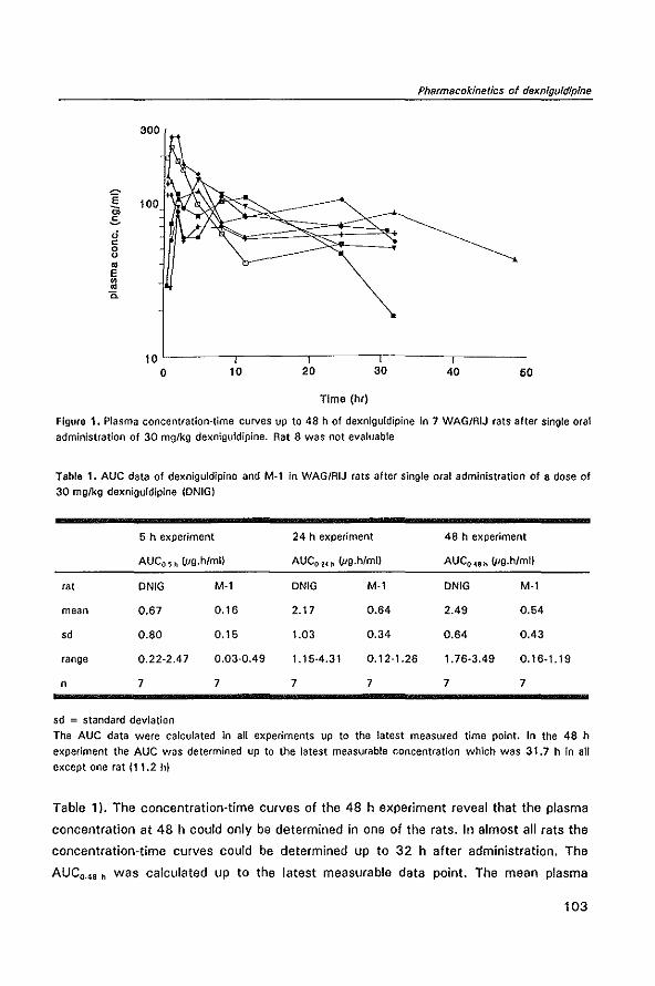
~ 10 oS o c 8 = E
Pharmacokinetics of dexnigufdipfne
~ <i
001'----,.-----.-, -------,,~---,,--o 10 20 30 40 50
Time (hr)
Figure 1. Plasma concentration-time curves up to 48 h of dexniguldipine in 7 WAG/RIJ rats after single oral
administration of 30 mglkg dexniguldipine_ Rat 8 was not evaluable
Table 1. AUe data of dexniguldipine and M-l in WAG/RIJ rats after single oral administration of a dose of
30 mglkg dexniguldipine (ONIG)
5 h experiment 24 h experiment 48 h experiment
AUCO-s h tug.h/m!) AUC0-24 h tug·h/mll AUC0-48 h tug·h/m!)
rat DNIG M-l DNIG M-l DNIG M-l
mean 0.67 0.16 2.17 0.64 2.49 0.54
sd 0.80 0.15 1.03 0.34 0.64 0.43
range 0.22-2.47 0.03-0.49 1.15-4.31 0.12-1.26 1.76-3.49 0.16-1.19
n 7 7 7 7 7 7
sd = standard deviation
The AUC data were calculated in all experiments up to the latest measured time point. In the 48 h
experiment the AUC was determined up to the latest measurable concentration which was 31.7 h in all
except one rat (11.2 hi
Table 1). The concentration-time curves of the 48 h experiment reveal that the plasma
concentration at 48 h could only be determined in one of the rats. In almost all rats the
concentration-time curves could be determined up to 32 h after administration. The
AUC~48 h was calculated up to the latest measurable data point. The mean plasma
103
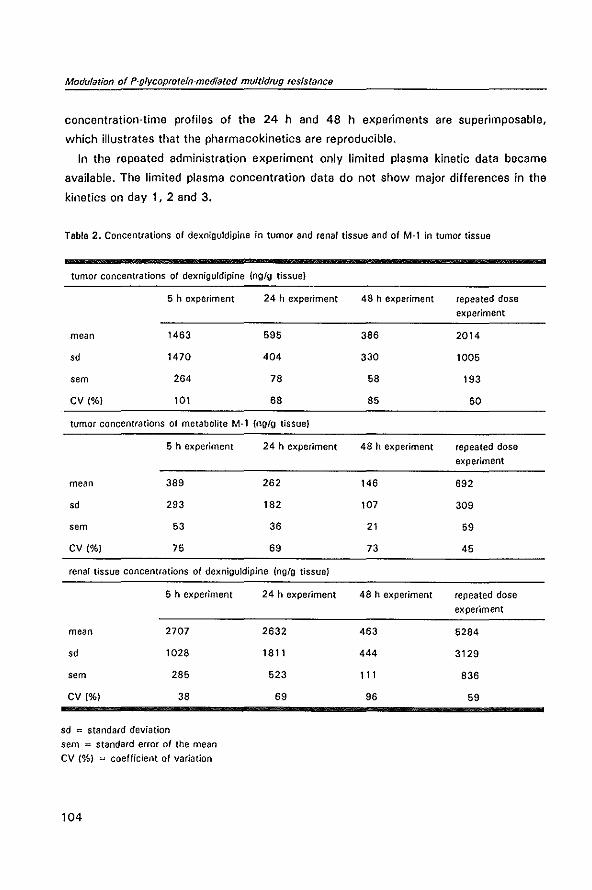
Modulation of P-glycoprotein-mediated multidrug resistance
concentration-time profiles of the 24 hand 48 h experiments are superimposable,
which illustrates that the pharmacokinetics are reproducible.
In the repeated administration experiment only limited plasma kinetic data became
available. The limited plasma concentration data do not show major differences in the
kinetics on day 1, 2 and 3.
Table 2. Concentrations of dexniguldipine in tumor and renal tissue and of M-l in tumor tissue
tumor concentrations of dexniguldipine (ng/g tissue)
5 h experiment 24 h experiment 48 h experiment repeated dose
experiment
mean 1463 595 386 2014
sd 1470 404 330 1005
sem 264 78 58 193
CV (%) 101 68 85 50
tumor concentrations of metabolite M-l (ng/g tissue)
5 h experiment 24 h experiment 48 h experiment repeated dose
experiment
mean 389 262 146 692
sd 293 182 107 309
sem 53 36 21 59
CV (%) 75 69 73 45
renal tissue concentrations of dexniguldipine (nglg tissue)
5 h experiment 24 h experiment 48 h experiment repeated dose
experiment
mean 2707 2632 463 5284
sd 1028 1811 444 3129
sem 285 523 111 836
CV (%) 38 69 96 59
sd = standard deviation
sem = standard error of the mean
CV (%) "" coefficient of variation
104
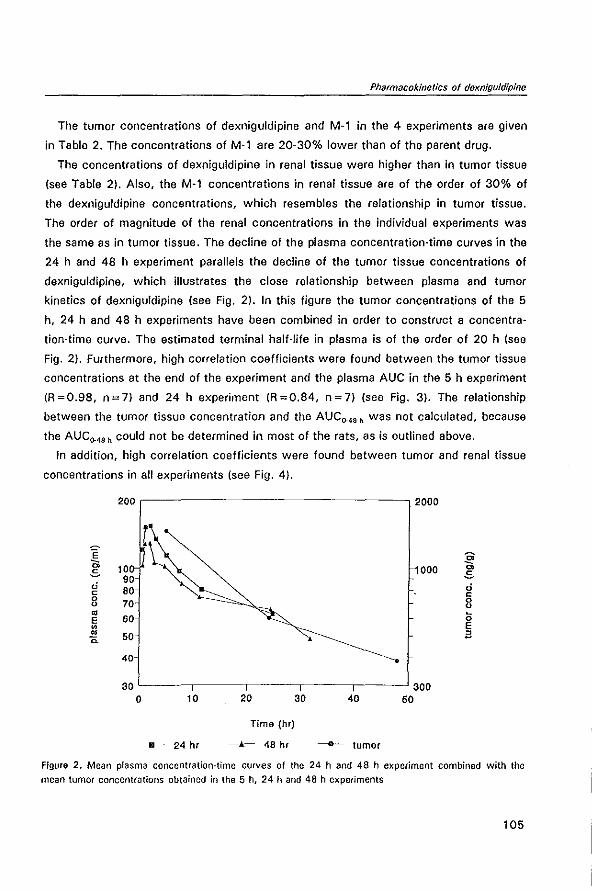
Pharmacokinetlcs of dexniguldipine
The tumor concentrations of dexniguldipine and M-1 in the 4 experiments are given
in Table 2. The concentrations of M·l are 20·30% lower than of the parent drug.
The concentrations of dexniguldipine in renal tissue were higher than in tumor tissue
(see Table 2). Also, the M·l concentrations in renal tissue are of the order of 30% of
the dexniguldipine concentrations, which resembles the relationship in tumor tissue.
The order of magnitude of the renal concentrations in the individual experiments was
the same as in tumor tissue. The decline of the plasma concentration-time curves in the
24 hand 48 h experiment para lie Is the decline of the tumor tissue concentrations of
dexniguldipine, which illustrates the close relationship between plasma and tumor
kinetics of dexniguldipine (see Fig. 2). In this figure the tumor concentrations of the 5
h, 24 hand 48 h experiments have been combined in order to construct a concentra
tion-time curve. The estimated terminal half-life in plasma is of the order of 20 h (see
Fig. 2). Furthermore, high correlation coefficients were found between the tumor tissue
concentrations at the end of the experiment and the plasma AUC in the 5 h experiment
(R=0.98, n=7) and 24 h experiment (R=0.84, n=7) (see Fig. 3). The relationship
between the tumor tissue concentration and the AUCo.48 h was not calculated, because
the AUCo.48 h could not be determined in most of the rats, as is outlined above.
In addition, high correlation coefficients were found between tumor and renal tissue
concentrations in all experiments (see Fig. 4).
E 0; oS d c 0 0 ~ E ~ ~ D.
200 r------------------, 2000
100 90 80 70 60
50
40
30
000
L---~--,_--_,--_,--~300
0 10
--------- 24 hr
20
Time (hr)
-10.- 48 hr
30 40 50
------- tumor
~ oS d c 0 0 ~
0 E a
Figure 2. Mean plasma concentration-time curves of the 24 hand 48 h experiment combined with the mean tumor concentrations obtained in the 5 h, 24 hand 48 h experiments
105
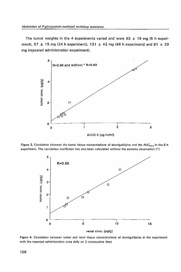
Modulation of P·glycoprotein·mediated multidrug resistance
The tumor weights in the 4 experiments varied and were 63 ± 19 mg (5 h experi
ment), 57 ± 15 mg (24 h experiment). 131 ± 42 mg (48 h experiment) and 81 ± 29
mg (repeated administration experiment).
6 A--O.98 and without· A"O.89
•
m 4 c; .;; ,; c 0 0 ~
0 E 2 0 il
o o 2 3
AUCO-5 ("o.hr/ml)
Figure 3. Correlation between the tumor tissue concentrations of dexniguldipine and the AUCO-~h in the 5 h
experiment. The correlation coefficient has also been calculated without the extreme observation (-)
5
R-O.96
4 0
.!!? m .;; 3 ,; 0 c 0 0 ~ 2 0 E 0 0
il
o o 5 10 15
ronal cone. (~g/g)
Figura 4. Correlation between tumor and renal tissue concentrations of dexniguldipine. in the experiment
with the repeated administration once daily on 3 consecutive days
106
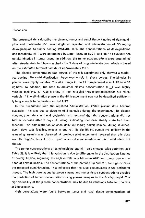
Pharmacokinetics of dexniguldipine
Discussion
The presented data describe the plasma, tumor and renal tissue kinetics of dexniguldi
pine and metabolite M·l after single or repeated oral administration of 30 mg/kg
dexniguldipine to tumor bearing WAG/RIJ rats. The concentrations of dexniguldipine
and metabolite M·l were determined in tumor tissue at 5, 24, and 48 h to evaluate the
uptake kinetics in tumor tissue. In addition, the tumor concentrations were determined
after steady state had been reached after 3 days of drug administration, which is based
on the estimated terminal half·life of approximately 20 h.
The plasma concentration-time curves of the 5 h experiment only showed a moder
ate decline. No rapid distribution phase was visible in these curves. The kinetics in
plasma were highly variable. The AUC range in the 24 h experiment was 1.15 to 4.31
pg.h/ml. In addition, the time to maximal plasma concentration (T m~) was highly
variable (see Fig. 1). Also a study in man revealed that pharmacokinetics are highly
variable. I6 The elimination phase in the 48 h experiment can not be described sufficient
ly long enough to calculate the total AUC.
In the experiment with the repeated administration limited plasma data became
available. This was due to plugging of 3 cannulas during the experiment. The plasma
concentration data in the 4 evaluable rats revealed that the concentrations did not
further increase after 2 days of dosing, indicating that near steady state had been
reached. The administration of once daily 30 mg/kg dexniguldipine, during 3 subse
quent days was feasible, except in one rat. No significant cumulative toxicity in the
remaining animals was observed. A previous pilot experiment revealed that this dose
was the highest feasible dose upon repeated administration in this model (data not
shown).
The tumor concentrations of dexniguldipine and M-' also showed wide variation (see
Table 2). It is unlikely that this variation is due to differences in the distribution kinetics
of dexniguldipine, regarding the high correlations between AUC and tumor concentra
tions of dexniguldipine. The concentrations of the parent drug and M·l are highest after
the repeated administration. This indicates that the drug accumulates in the peripheral
tissues. The high correlations between plasma and tumor tissue concentrations enables
the prediction of tumor concentrations using plasma samples in this in vivo model. The
high variability of the plasma concentrations may be due to variations between the rats
in bioavailability.
High correlations were found between tumor and renal tissue concentrations of
107
Modulation of P-glycoprotein-mediated muft/drug resIstance
dexniguldipine. The renal tissue concentrations were always a factor 1.5 to 4 higher
than the tumor tissue concentrations, dependent on the sampling time (see Table 2).
Dexniguldipine is a dihydropyridine derivative. This class of drugs is known to have a
high tissue distribution. Differences in lipophilicity between tissues, such as renal and
tumor tissue, may therefore contribute to the differences in tissue concentration after
exposure to dexniguldipine, in particular at steady state. The ratio dexniguldipine and
M-1 was constant in all experiments.
The tumor weights between the experiments showed variation. The mean tumor
weight in the 48 h experiment is clearly higher than in the other experiments, which
may have been due to tumor batch differences. As a consequence, tumor growth
inhibition experiments should be carried out with the same tumor batch.
The in vitro results of the MDR-modifying effect of dexniguldipine revealed that
dexniguldipine was highly active at a concentration as low as 50 ng/ml. 17 In that
experiment the same CC531 cell line was used. It is hazardous to extrapolate results of
in vitro studies to in vivo tumor models. However, regarding the high tumor tissue
concentrations of dexniguldipine in vivo, it may be anticipated that these concentra
tions are high enough to reverse MDR in vivo. Results of the pharmacodynamic study
with dexniguldipine and epidoxorubicin in this model reveal a moderate synergistic
antitumor effect, which was statistically significant in 2 out of 4 experiments. 17
In the present study high concentrations were achieved in intrinsically MDR solid
tumor tissue implanted under the renal capsule after single oral dosing of the MDR
reverter dexniguldipine. In addition, high correlations between plasma and tumor tissue
concentrations enables prediction of tumor concentrations in this model by simply
measuring plasma concentrations.
References
1. Hofmann J, Ueberall F, Egle A, Grunicke H. 8·859·35, a new drug with anti-tumor activity reverses multi-drug resistance. Int J Cancer 1991; 47: 870·4
2. Hallt V, Kouba M, Dietel M, Vogt G. Stereoisomers of calcium antagonists which differ markedly in their potencies as calcium blockers are equally effective in modulating drug transport by Pglycoprotein. Blochem Pharmacol 1992; 43: 2601·8
3. Hofmann J, Wolf A, Spitaler M, 80ck G. Drach J. ludescher C, Grunicke H. Reversal of multidrug resistance by 8859-35, a metabolite of 8859-35, niguldipine, verapamil and nitrendipine. J Cancer Res Clln Oncol 1992; 118: 361·6
4. NoUer A, Wilisch A, Hiiussermann K. Gekeler V. MDR modulating and antineoplastic effects of 8859-35, and its metabolite. Ann Oneal 1992; 3 (Supp1. 11: 71
5. Gietzen K, Abdallah F, Bai G. Inhibition of tumour cell growth by a novel dihydropyridine derivative. Eur J Cancer 1990; 26: 922·3
6. SchOller HM, Correa E, Orloff M, Reznik GK. Successful chemotherapy of experimental neuroendoc·
108
Pharmacokinetics of dexnigufdipine
fine lung tumors in hamsters with an antagonist of Ca2 + fcalmodulin. Cancer Res 1990; 50: 1645-9 7. Pastan I, Gottesman M. Multiple·drug resistance in human cancer. New Engl J Med 1987; 316:
1388-93 8. Raderer M, Scheithauer W. Clinical trials of agents that reverse multidrug resistance. Cancer 1993;
72: 3553-63 9. Sikic 81. Modulation of multidrug resistance at the threshold. J Clin Oneal 1993; 11: 1629-35 10. Pennock GO, Dalton WS, Roeske WA, Appleton CP, Mosley K, Plezia P, Miller TP, Salmon SE.
Systemic toxic effects associated with high-dose verapamil infusion and chemotherapy administration. J Nat! Cancer Inst 1991; 83: 105-10
11. lum Bl, Fisher GA. Brophy NA, Yahanda AM, Adler KM, Kaubisch S, Halsy J, Sikic BI. Clinical trials of modulation of multidrug resistance. Cancer 1993; 72: 3502-14
12. Wishart GC, Bisset 0, Paul J, Jodrell 0, Harnett A, Habeshaw T, Kerr OJ, Macham MA, Soukop M, leonard RCF, Knepil J, Kaye SB. Quinidine as a resistance modulator of epirubicin in advanced breast cancer: mature results of a placebo-controlled randomized trial. J Clin Oneal 1994; 12: 1771-7
13. Gheuens E, van der Heyden S, Elst H. Eggermont A, van Oosterom A, de Bruijn E. Mu!tidrug resistance in rat colon carcinoma cell lines CC531, CC531 mdH and CC531'"". Jpn J Cancer Res 1993; 84: 1201-8
14. Van de Vrie W, Gheuens HO, Durante NMC, de Bruijn EA, Marquet Al, van Oosterom AT, Eggermont AMM. In vitro and In vivo chemosensitizing effect of cyclosporin A on an intrinsic multidrug-resistant rat colon tumour. J Cancer Res Clin Onco/1993; 119: 609-14
15. Marquet Al, Westbroek Ol, Jeekel J. Interferon treatment of a transplantable rat colon adenocarcinoma: importance of tumor site. In! J Cancer 1984; 33: 689-92
16. Ukena 0, Boewer C, Oldenkott B, Rathgeb F, Wurst W, Zech K, Sybrecht GW. Tolerance, safety, and kinetics of the new antineoplastic compound dexniguldipine-HCI after oral administration: a phase I dose-escalation trial. Cancer Chemother Pharmacol 1995; 36: 160-4
17. Van de Vrie W. Schell ens JHM, loos WJ, Kolker HJ, Verwey J. Stoter G. Durante NMC, Eggermont AMM. Modulation of multidrug resistance with dexniguldipine hydrochloride (B8509-035) in the CC531 rat colon carcinoma model. J Cancer Res Clin Onco/1996; 122: 403-8
109

2.4
THE CHEMOSENSITIZER CYCLOSPORIN A
ENHANCES THE TOXIC SIDE-EFFECTS OF
DOXORUBICIN IN THE RA T
Wim van de Vrie, A. Mieke Jonker,
Richard L. Marquet and
Alexander M.M. Eggermont
J Cancer Res Clin Oneol
1994; 120: 533·8
Modulation of P-glycoprotein-mediated mull/drug resistance
Summary
The feasibility of using chemosensitizers in the circumvention of P-glycoprotein
mediated MDR has been shown in many studies. We recently reported on the chemo·
sensitizing effect of cyclosporin A on doxorubicin in a rat solid tumor model. Using the
same experimental design we investigated the side-effects of the combination treat
ment. During the 35-day experiment doxorubicin treatment caused dose-dependent
weight loss, which was enhanced by combination treatment with cyclosporin A. The
main doxorubicin-related side-effects were myelosuppression (transient leucopenia and
thrombopenia) and nephrotoxicity. Damage to the kidney was severe, leading to a
nephrotic syndrome and resulting in ascites, pleural effusion, hypercholesterolemia and
hypertriglyceridemia. These toxicities were enhanced by the addition of the chemosens
itizer cyciosporin A. Mild doxorubicin-related cardiomyopathy and minimal
hepatotoxicity were seen on histological examination. There were no signs of enhanced
toxicity of the combination treatment in tissues with known high expression levels of P
glycoprotein. like the liver. adrenal gland and large intestine. Cyclosporin A had a low
toxicity profile, as it only caused a transient rise in bilirubin. In conclusion, the chemos
ensitizer cyclosporin A enhanced the side-effects of the anticancer drug doxorubicin,
without altering the toxicity pattern. There was no evidence of a therapeutic gain by
adding cyclosporin A to doxorubicin, compared to single agent treatment with doxorub
lcin in 25%-33% higher doses, because of the enhanced toxicity of the combination
treatment.
112
ToxIcity of MDR reversal
Introduction
Multidrug resistance is an important mechanism of resistance of tumors to anticancer
drugs. In MDR an efflux pump, P-glycoprotein, expels drugs from the cell by active
transport. t P-glycoprotein expression has been found in many tumors. High expression
levels of P-glycoprotein were demonstrated in colon cancer, renal cell cancer, hepato
cellular carcinoma, and adrenocortical cancer, while intermediate levels were found in
sarcomas and breast cancer. 2,3 In hematological malignancies, like several leukemias,
lymphomas and in multiple myeloma, expression of P-glycoprotein was found in
untreated, and, to a greater extent, in treated tumors,3.5 However, this protein is also
expressed in normal tissues. Organs with a high expression level of P-glycoprotein are
the adrenal gland, liver, kidney, colon and pancreas/·6 and the protein is mainly
localized in cells lining excretory lumina, which suggests a detoxification function. 6
One way of disturbing the P-glycoprotein-mediated resistance mechanism is by
blocking the efflux pump with so-called chemosensitizers. Numerous in vitro studies
have shown the efficacy of drugs like verapamil, cyclosporin A, quinine/quinidine,
tamoxifen, and others in enhancing the sensitivity of MDR tumor cell lines to anticancer
drugs.7 In vivo studies have confirmed the feasibility of reversal of MDR by chemosens
itizers in ascites tumor models8,9 and in solid tumor models. tO•11 In clinical trials
promising results have been observed in patients with multiple myeloma, lymphoma,
and leukemia.4.12.14 In studies with solid tumors chemosensitizers showed less efficacy
with responses in a minority of the patients only.IOolS Besides, some authors have
reported on enhancement of toxic side-effects, like myelosuppression by the addition
of chemosensitizers to the therapeutic regimen.17.19.20 Therefore, the question is raised
whether the use of chemosensitizers in combination with anticancer drugs enhances
the toxic side-effects of these drugs, apart from enhancing the efficacy of the
anticancer treatment. A second question is whether other toxic effects will appear,
especially in P-glycoprotein-expressing tissues. Third, chemosensitizers themselves may
have adverse effects.
We recently published our results on chemosensitizing in a rat MDR tumor mode!.11
The chemosensitizer cyclosporin A was shown to enhance the cytotoxic efficacy of
doxorubicin in vitro and in vivo. A suboptimal dose of doxorubicin (3 mg/kg) was
rendered effective against the solid growing CC531 rat colon carcinoma in vivo by the
addition of cyclosporin A. Drugs were administered intramuscularly and intravenously,
which means that, unlike in ascites tumor models, drugs were transported to the tumor
113
Modulation of P·glycoprotein·mediated multidrug resistance
and other tissues by a vascular route. Because this is close to the clinical situation, the
same model was used to study the toxic effects of the combination treatment on
normal tissues in rats. In this study we show that the chemosensitizer cyclosporin A
enhances the specific toxic effects of doxorubicin on normal tissues, resulting in
myelosuppression, severe nephrotoxicity, and mild cardiotoxicity. There were no signs
of additive toxic damage in tissues with a high expression level of P'glycoprotein, nor
of severe cyclosporin A·induced toxicity.
Materials and methods
Animals
Male rats of the inbred WAG/RIJ (RTl') strain were obtained from Harlan-CPS
(Austerlitz, The Netherlands). Animals were bred under specific·pathogen·free condi
tions and fed standard rat chow (Hope Farms, Woerden, The Netherlands) and water ad
libitum. In the experiments rats were 12-18 weeks old and had a body weight of 220-
280 g.
Chemicals
Cyclosporin A was obtained from Sandoz, 8asel, Switzerland; doxorubicin (Adria bias·
tina) from Farmitalia, Nivelles, 8elgium.
Experimental design
Animals were randomly allocated to the experimental groups. The two control groups
consisted of eight animals, while the five experimental groups contained four animals
each. The experiment was repeated once. Intravenous injection and blood sampling
were done under anaesthetic conditions using ether. Rats were weighed weekly. On
day 3, 7, 14, 21, and 28 a blood sample of 0.75 ml was taken by bleeding from the
tail vein. On the 35th day the experiment was terminated and all rats were sacrificed. If
an animal was critically ill such that it was not supposed to survive 48 h, or if it had
lost approximately 20% body weight, the animal was sacrificed earlier than day 35.
Drug treatment
The chemosensitizer cyclosporin At dissolved in olive oil, was injected intramuscular·
Iy into the hind leg daily for 3 consecutive days at a dose of 20 mg/kg body weight.
114
Toxicity of MDR reversal
Animals in groups not to be treated with cyclosporin A were injected with the vehicle
of cyelosporin A: olive oil and 6.25% alcohol. Treatment was given on days -2, -1 and
O. Doxorubicin was administered intravenously on day 0 as a single dose at a concen
tration of 3 mg/kg, 4 mg/kg or 6 mg/kg body weight. Control rats were injected with
PBS. This resulted in the following groups: Control (treatment with PBS and vehicle),
CsA-con (PBS + cyclosporin AI. DOX3 (3 mg/kg doxorubicin + vehiclel. DOX3 + CsA
(3 mg/kg doxorubicin + cyclosporin Al. DOX4 (4 mg/kg doxorubicin + vehicle),
DOX4 + CsA (4 mg/kg doxorubicin + cyelosporin A), and DOX6 (6 mg/kg doxorubicin
+ vehicle).
Hematological and biochemical studies
Blood was collected in lithium/heparin microtubes (Sarstedt, Germany). The hemoglo
bin content was determined on the TOA hemoglobin counter HB·100, leucocytes on
the Sysmex microcell counter CC-1 08 and platelets on the TOA platelet counter PL-1 00
(all Sysmex, TOA Medical Electronics, Hamburg, Germany). The remaining blood
sample was centrifuged and serum was collected. Biochemical values of creatinine,
urea, aspartate aminotransferase, T-glutamyltransferase, total bilirubin, cholesterol and
triglyceride were determined on the ELAN·Analyzer (Eppendorf, Hamburg, Germany)
with reagents from Merck (Merck Oiagnostica, Darmstadt, Germany).
Histology
On day 35 all animals were sacrificed and an autopsy was performed. Ascites and
pleural effusion, if present, were aspirated in a syringe and measured. Specimens of the
following organs were taken for histological examination: heart, lung, liver, spleen,
kidney, large intestine, pancreas and adrenal gland. The organs were removed immedia
tely, fixed in 10% buffered formalin and embedded in paraffin. Sections were cut at 5
Jim, stained with hematoxylin and eosin and periodic-acid/Schiff. Microscopic sections
were coded and scored blindly. The following histological parameters were evaluated:
edema, necrosis, inflammation, accumulation of fat, fibrosis, glycogen storage (liver),
and degenerative changes. The extent of damage in kidney and liver was graded
semi quantitatively on a 0 to 2 + scale (0 = absent, 1 + = slightly damaged, 2 + =
severely damaged). The histopathological changes in the heart were assessed according
to the scoring system of Bristow et al., 21 which scale runs from 0 to 3.0 + .
115
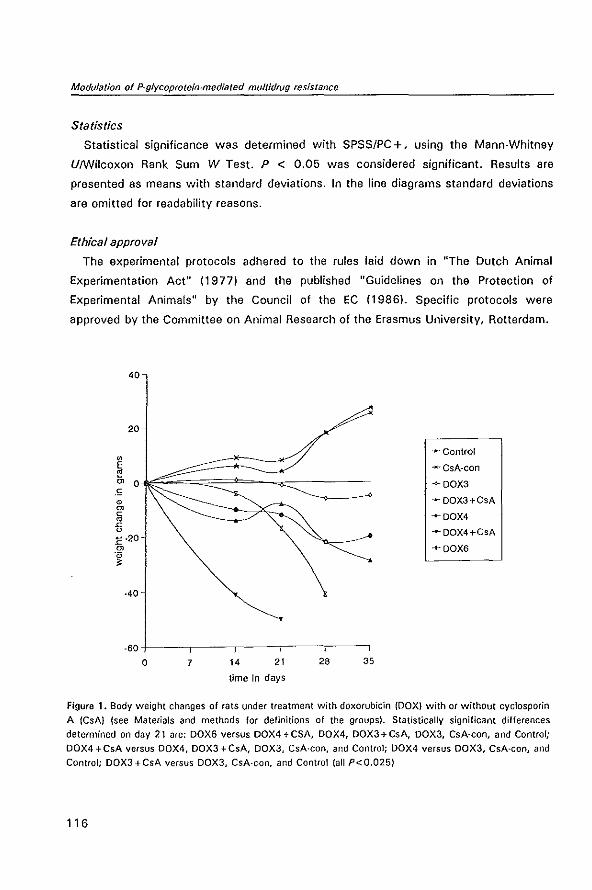
Modulation 01 P-glycoprolein-medialed multidrug resistance
Statistics
Statistical significance was determined with SPSSIPC +, using the Mann-Whitney
U/wilcoxon Rank Sum W Test. P < 0.05 was considered significant. Results are
presented as means with standard deviations. In the line diagrams standard deviations
are omitted for readability reasons.
Ethical approval
The experimental protocols adhered to the rules laid down in "The Dutch Animal
Experimentation Act" (1977) and the published "Guidelines on the Protection of
Experimental Animals" by the Council of the EC (1986). Specific protocols were
approved by the Committee on Animal Research of the Erasmus University, Rotterdam.
20
-Control " E ...... CsA-con e '" 0 ->- OOX3 .e 0 -- OOX3+CsA
'" c -+-OOX4 • ~ u -..... OOX4+CsA 1: ·20
'" +OOX6 .~
~
·40
o 7 14 21 28 35
time in days
Figure 1. Body weight changes of rats under treatment with doxorubicin (DOX) with Of without cyclosporin
A (CsA) (see Materials and methods for definitions of the groups). Statistically significant differences
determined on day 21 are: DOX6 versus DOX4+CSA, DOX4, DOX3+CsA, DOX3, CsA-con, and Control;
DOX4 + CsA versus DOX4, DOX3 + CsA, DOX3, CsA-con, and Control; DOX4 versus DOX3, CsA-con, and
Control; DOX3+CsA versus DOX3, CsA-con, and Control (all P<O.025)
116
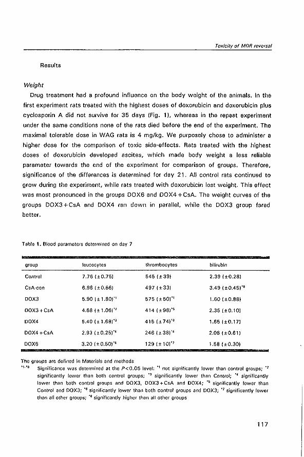
Toxicity of MDR reversal
Results
Weight
Drug treatment had a profound influence on the body weight of the animals. In the
first experiment rats treated with the highest doses of doxorubicin and doxorubicin plus
cyclosporin A did not survive for 35 days (Fig. 1)' whereas in the repeat experiment
under the same conditions none of the rats died before the end of the experiment. The
maximal tolerable dose in WAG rats is 4 mg/kg. We purposely chose to administer a
higher dose for the comparison of toxic side-effects. Rats treated with the highest
doses of doxorubicin developed ascites, which made body weight a less reliable
parameter towards the end of the experiment for comparison of groups. Therefore.
significance of the differences is determined for day 21. All control rats continued to
grow during the experiment, while rats treated with doxorubicin lost weight. This effect
was most pronounced in the groups DOX6 and DOX4 + GsA. The weight curves of the
groups DOX3 + GsA and DOX4 ran down in parallel, while the DOX3 group fared
better.
Table 1. Blood parameters determined on day 7
group leucocytes thrombocytes bilirubin
Control 7.76 (>0.75) 545(>39) 2.39 (> 0.28)
CsA-con 6.96 (> 0.66) 497 (±33) 3.49 (±0.45)·'
DOX3 5.90 {± 1.80)'1 575 {±50,'1 1.60 (±0.88)
OQX3+CsA 4.68 (± 1.06)" 414(±98"S 2.35 (±0.10)
DOX4 5.40 (> 1.68)" 415 {±74J'6 1.65 (±0.17)
OOX4 +CsA 2.93 (±0.25)'4 246 (±38)'4 2.08 (± 0.61)
DOX6 3.20 (>0.50)" 129 (± 10)" 1.58 (> 0.30)
The groups are defined in Materials and methods
'1·'S Significance was determined at the P<0.05 level: '\ not significantly lower than control groups; '2
significantly lower than both control groups; 'J significantly lower than Control; '4 significantly
lower than both control groups and DOX3, DOX3 +CsA and DOX4; '5 significantly lower than
Control and DOX3; '6 significantly lower than both control groups and DOX3; '1 significantly lower
than all other groups; '8 significantly higher than all other groups
117
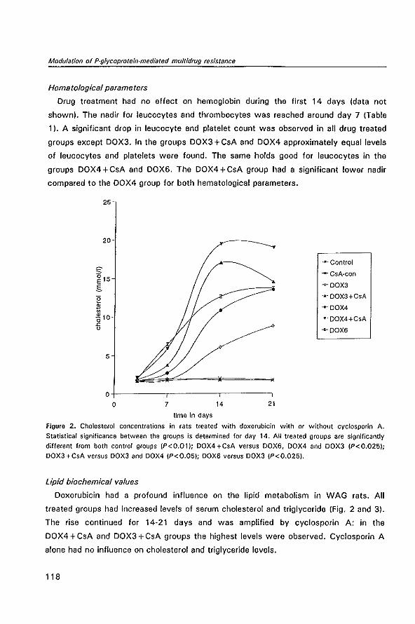
Modulation of P-{}Iycoprolein-medialed multidrug resistance
Hematological parameters
Drug treatment had no effect on hemoglobin during the first 14 days (data not
shownl. The nadir for leucocytes and thrombocytes was reached around day 7 (Table
1). A significant drop in leucocyte and platelet count was observed in all drug treated
groups except DOX3. In the groups DOX3 + GsA and DOX4 approximately equal levels
of leucocytes and platelets were found. The same holds good for leucocytes in the
groups DOX4+GsA and DOX6. The DOX4+GsA group had a significant lower nadir
compared to the DOX4 group for both hematological parameters.
25
20
..... Contro!
'" "*" CsA·con ~15
-v- OOX3 S E ..... OOX3+CsA
• ....OOX4 ;; -6 10 ..... OOX4+CsA
'" u ..... OOX6
5
o+--------,~-------.--------,
o 7 14 21
time in days
Figura 2. Cholesterol concentrations in rats treated with doxorubicin with or without cyclosporin A.
Statistical significance between the groups is determined for day 14. AU treated groups are significantly
different from both control groups (P<O.01); DOX4+CsA versus DOX6, DOX4 and DOX3 (P<O.025J;
DOX3+CsA versus DOX3 and DOX4 (P<O.05); DOX6 versus DOX3 (P<O.025).
Lipid biochemical values
Doxorubicin had a profound influence on the lipid metabolism in WAG rats. All
treated groups had increased levels of serum cholesterol and triglyceride (Fig. 2 and 31.
The rise continued for 14·21 days and was amplified by cyclosporin A: in the
DOX4 + GsA and DOX3 + GsA groups the highest levels were observed. Gyclosporin A
alone had no influence on cholesterol and triglyceride levels.
118
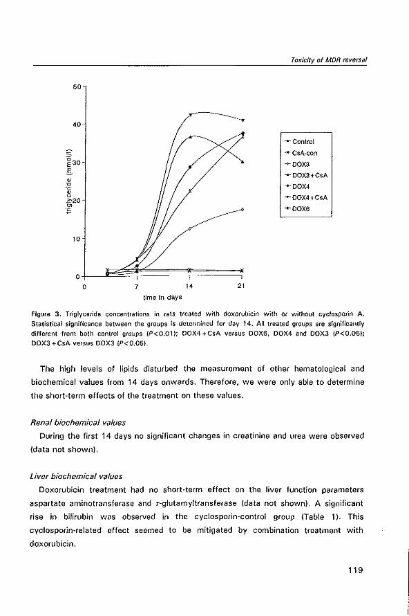
Toxt'clty of MDR reversal
50
40
--Control
"- -Il-CsA·con ~30 ~OOX3
.s "'OOX3+CsA • u +OOX4 .&j 0 ..... OOX4+CsA .?:-20 0> ..... OOX6 E
10
o 7 14 21
time in days
Figure 3. Triglyceride concentrations in rats treated with doxorubicin with or without cyclosporin A. Statistical significance between the groups is determined for day 14. All treated groups are significantly different from both control groups (P<O.01); DOX4+CsA versus DOX6, DOX4 and DOX3 (P<O.05);
DOX3+CsA versus DOX3 (P<O.05J.
The high levels of lipids disturbed the measurement of other hematological and
biochemical values from 14 days onwards. Therefore. we were only able to determine
the short-term effects of the treatment on these values.
Renal biochem;cal values
During the first 14 days no significant changes in creatinine and urea were observed
(data not shown).
Liver biochemical values
Doxorubicin treatment had no short-term effect on the liver function parameters
aspartate aminotransferase and T-glutamyltransferase (data not shown). A significant
rise in bilirubin was observed in the cyclosporin-control group (Table 1). This
cyclosporin-related effect seemed to be mitigated by combination treatment with
doxorubicin.
119
Modulation of P-glvcoprotein-mediated multidrug resistance
Autopsy
Pathology data presented here are from the second experiment, in which all animals
survived. On day 35 rats in the groups DOX4 + CsA and DOX6 were critically ill. They
had lost body weight and subcutaneous lat. During the experiment rats had not had
diarrhoea. At autopsy a large amount of ascites and hemorrhagic pleural effusion was
lound in rats treated with DOX4 + CsA and DDX6. Rats in the DOX4 + CsA group had
12.4 (± 12.4) ml ascites. rats in the DOX6 group 15.3 (± 6.9) mi. while only a small
amount of 2.1 (± 2.4) ml was lound in the DOX3 + CsA group and no ascites in the
other rats. Pleural effusion was 6.5 (± 3.1) ml and 6.3 (± 4.4) ml in the DOX4 +CsA
and DOX6 groups respectively. In the DOX3 + CsA group 0.8 (± 1.5) ml pleural effusion
was found and none in the other rats. The differences in ascites an pleural effusion
were statistically significant lor the DOX4 + CsA and DOX6 groups compared to all
other groups except for pleural effusion in DOX6 versus DOX3 + esA. In addition,
edema of the pancreas and paleness of the liver, kidneys and adrenal glands were
observed in rats of the DOX4 + CsA and DOX6 groups. In the other groups all these
macroscopic findings were minimal or absent.
Microscopic study
Light microscopic examination of the kidney showed severe damage (2 +) in all rats
treated with doxorubicin or the combination doxorubicin and cyclosporin A, while rats
injected with PBS or cyclosporin A had normal kidneys. Injured kidneys showed increa
sed glomerular mesangial cellularity, lipid accumulation in macrophages, thickening of
basement membranes of glomerular capillaries and Bowman's capsule with in some
glomeruli focal adhesions (Fig. 4). The tubules epithelium showed degenerative
changes, focal regenerative activity (mitotic figures) and some showed protein casts. In
the interstitial space of injured kidneys focal lymphocytic infiltrates were seen. Blood
vessels had normal morphology.
The myocardium of rats treated with doxorubicin and the combination doxorubicin
plus cyclosporin A showed minimal morphological changes with edema in the interstitial
space, slight vacuolization of myocytes, and sporadic focal inflammation. Necrosis or
fibrosis was not observed. The maximal score according to Bristow et al. 21 was 1.5.
Increased doses of doxorubicin revealed the same degree of damage, however, a
greater percentage of rats in each group was affected with higher doses (OOX3 25%;
DOX3+CsA 50%; DOX4 75%; DOX4+CsA and DOX6 100%).
Minimal hepatotoxic changes were demonstrated. In rats treated with DOX4 and
120
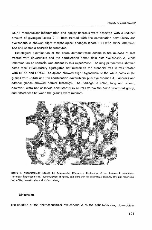
Toxicity of MaR reversal
DOX6 mononuclear inflammation and spotty necrosis were observed with a reduced
amount of glycogen (score 2 +). Rats treated with the combination doxorubicin and
cyclosporin A showed slight morphological changes (score 1 +) with minor inflamma·
tion and sporadic necrotic hepatocytes.
Histological examination of the colon demonstrated edema in the mucosa of rats
treated with doxorubicin and the combination doxorubicin plus cyclosporin A, while
inflammation or necrosis was absent in this experiment. The lung parenchyma showed
some focal inflammatory aggregates not related to the bronchial tree in rats treated
with DOX4 and DOX6. The spleen showed slight hypoplasia of the white pulpa in the
groups with DOX6 and the combination doxorubicin plus cyclosporine A. Pancreas and
adrenal glands showed normal histology. The findings in colon, lung and spleen,
however, were not observed consistently in all rats within the same treatment group,
and differences between the groups were minimal.
Figure 4. Nephrotoxicity caused by doxorubicin treatment: thickening of the basement membrane,
mesangial hyperceHularity, accumulation of lipids, and adhesion to Bowman's capsule. Original magnifica
tion 400x; hematoxylin and eosin staining
Discussion
The addition of the chemosensitizer cyclosporin A to the anticancer drug doxorubicin
1 21
ModulatiOt1 of P-gfycoprotein-mediated multidrug resistance
clearly enhances its toxicity. In a previous study we demonstrated that the addition of
cyclosporin A made a suboptimal dose of 3 mg/kg doxorubicin as effective as 4 mg/kg
doxorubicin." In the present study we show data (body weight change curves,
hematological parameters, and autopsy findings) indicating that the combination of
doxorubicin with cyclosporin A is about as toxic as a 25%-33% higher doxorubicin
dose alone. We found therefore no therapeutic window, in contrast to Mickisch et al. 22
in their transgenic mouse model. They had to reduce the dose of most anticancer drugs
by 20%, while these doses in combination with D·verapamil reduced MDR cell
populations by 44%-78%. The results of the combination treatment were favourable
compared to results with full doses of the drugs alone. Boesch et al.23 reported similar
favourable results of a combination treatment of vinblastine and doxorubicin with the
cyclosporin A analogue PSC 833 in a survival model of mice with MDR tumors.
The toxicity pattern of doxorubicin is not altered by the addition of cyclosporin A.
Doxorubicin, like most other cytotoxic agents, causes severe damage to cell-renewal
systems, which are highly proliferative in post fetal Iife.24 In our experiments the main
acute side-effect was myelosuppression with significant leucopenia and thrombopenia.
This effect was reversible. No signs of enterocolitis were observed. Unique toxic
actions of anthracyclines, especially doxorubicin, are cardiovascular toxic effects,
nephrotoxicity and toxic effects on the skeletal system. The last two effects can be
observed in several experimental models, while cardiotoxicity is also found in humans. 25
In man cardiomyopathy leading to congestive heart failure is dependent on the total
cumulative dose administered. 26 Our study was not designed specifically for studying
the toxic effects of doxorubicin on cardiac tissue. The study was short-term, lasting
only 35 days, and involved a single dose treatment schedule for doxorubicin, which
made it more apt for studying acute toxic effects than chronic damage. Nevertheless,
on microscopic examination mild damage to the cardiac tissue was observed in the
most intensely treated rats. Other investigators have found severe cardiomyopathy in
rats from 35 days onwards after administering multiple low doses of doxorubicin
instead of a single high dose, and reaching higher cumulative values for doxorubicin
than we did. 21•28 Results in studies with dogs and mice suggest that the addition of the
chemosensitizer verapamil potentiates the cardiotoxic effects of doxorubicin. 29.30
WAG rats proved to be very sensitive to the nephrotoxic effects of doxorubicin,
eventually developing a full-blown nephrotic syndrome. 25•28 We were unable to measure
proteins and renal parameters after 14 days because of disturbance of the assays
caused by turbidity of the hyperlipidemic serum, but the ascites and pleural effusion
122
Toxicity of MDR reversal
indicate hypoproteinemia. Levels of cholesterol and triglyceride were significantly raised
by doxorubicin. Addition of cyclosporin A raised the levels of cholesterol and triglyceri·
de even higher, in such a way that combination treatment produced the highest levels.
Meanwhile, significant amounts of ascites and pleural effusion were only found in the
two most intensely treated groups. This indicates that the nephrotoxic effects of
doxorubicin were amplified by the chemosensitizer cyclosporin A.
Tissues with known high expression levels of P-glycoprotein. like liver. large intestine
and adrenal gland, were monitored for toxic effects. No signs of major toxicity were
observed. This suggests that these tissues are not susceptible to the cytotoxic effects
of doxorubicin and that raising its intracellular concentration either by administering a
higher dose or by adding a chemosensitizer, does not make these tissues sensitive to
doxorubicin, despite the presence of P-glycoprotein. Other researchers have come to
the same conclusion in a pathological study using the chemosensitizer D-verapamil and
the drugs vinblastine, doxorubicin, and daunomycin. 22 In contrast, Horton et al. 31 found
enhanced concentrations of vincristine in P-glycoprotein-expressing normal tissues, like
small intestine. kidney and liver. caused by the addition of high doses of the chemosen
sitizer verapamil. Toxicity was enhanced eight-fold and symptomatic of vincristine
related neurotoxicity. They did not describe the functional and morphological effects of
the raised concentration of intracellular vincristine in these normal tissues. Genne et al. 32 also reported enhanced doxorubicin accumulation in kidney and liver in combina
tion treatment with the chemosensitizer amiodarone. Combination treatment acceler
ated doxorubicin-induced death. In clinical studies with chemosensitizers, however, no
toxicities, apart from those attributable to the drug or the chemosensitizer, have been
observed so far.
Cyclosporin A seems to produce few toxic effects in the concentrations used for
chemosensitizing. In clinical trials steady state levels from 1000 /1gll up to 5000 /1gll
were reported.13.14.17,2o The cyclosporin A concentration of 1000 Jig" suffices in vitro
for MDR reversal. Side-effects of cyclosporin A observed were an early and transient
rise in serum bilirubin, without increases in liver enzymes, and hypomagnes
emia. 13,14,20.33 In our rat study the transient hyperbilirubinemia appeared to be a purely
cyciosporin A-dependent feature, which was not enhanced by the addition of doxorubi
cin. We found no evidence in our rat model for the hypothesis that bilirubin is raised as
a consequence of competition between doxorubicin, cyclosporin A, and bilirubin at the
excretion level, and thus might be used as a marker for P-glycoprotein modulation in
vivo. '4.2o
123
Modulation of P-glycoproteln-mediated multidrug resistance
From our studies with doxorubicin and cyclosporin it can be concluded that the
addition of a chemosensitizer seriously enhances the toxic side-effects of the anticancer
drug without altering the pattern of toxicity. As the toxicity patterns of anticancer
drugs are known, side-effects can be anticipated in the planning of clinical trials.
However, it remains unclear from this study whether therapeutic gains can be made by
the application of a chemosensitizer.
References
1. Chin K-V, Pastan I, Gottesman MM. Function and regulation of the human mu!tidrug resistance gene. Adv Cancer Res 1993; 60: 157--80
2. Fojo AT, Ueda K, S!amon OJ, Poplack DG, Gottesman MM, Pastan I. Expression of a mu!tidrugresistance gene in human tumors and tissues. Proc Nat! Acad Sci USA 1987; 84: 265-9
3. Go!dstein LJ, Galski H, Fojo A, Willingham M, lai S·l, Gazdar A, Pirker R, Green A, Crist W, Brodeur GM, Lieber M, Cossman J, Gottesman MM. Pastan I. Expression of a multidrug resistance gene in human cancers. J Nat! Cancer Inst 1989; 81: 116-24
4. Dalton WS, Grogan TM, Me!tzer PS, Scheper AJ, Durie BGM, Taylor CW, Miller TP, Salmon SE. Drug·resistance in multiple myeloma and non-Hodgkin's lymphoma: detection of P-glycoprotein and potential circumvention by addition of verapamil to chemotherapy. J Clln Oncol 1989; 7: 415-24
5. Herweijer H, Sonneveld P, Baas F, Nooter K. Expression of mdrl and mdr3 multidrug-resistance genes in human acute and chronic leukemias and association with stimulation of drug accumulation by cyclosporine. J Nat! Cancer Inst 1990; 82: 1133-40
6. Thiebaut F, Tsuruo T, Hamada H, Gottesman MM, Pastan I, Willingham MC. Cellular localization of the multidrug-resistance gene product P-glycoprotein in normal human tissues. Proc Nall Acad Sci USA 1987; 84: 7735-8
7. Ford JM, Hait WN. Pharmacology of drugs that alter multidrug resistance in cancer. Pharmacol Rev 1990; 42: 155-99
8. Tsuruo T, lida H, Tsukagoshi S, Sakurai Y. Overcoming of vincristine resistance in P388 leukemia in vivo and in vitro through enhanced -cytotoxicity of vincristine and vinblastine by verapamil. Cancer Res 1981;41: 1967·72
9. Slater lM, Sweet P, Stupecky M, Wetzel MW, Gupta S. Cyclosporin A corrects daunorubicin resistance in Ehrlich ascites carcinoma. Br J Cancer 1986; 54: 235-8
10. Osieka R, Seeber S, Pannenbacker R, Soli 0, Glatte P, Schmidt CG. Enhancement of etoposideinduced cytotoxicity by cyclosporin A. Cancer Chemother Pharmacal 1986; 18: 198·202
11. Van de Vrie W, Gheuens EEO, Durante NMC. de Bruijn EA, Marquet Al, van Oosterom AT, Eggermont AMM. In vitro and in vivo chemosensitizing effect of cyclosporin A on an intrinsic multidrug-resistant rat colon tumour. J Cancer Res Clin Oneal 1993; 119: 609-14
12. Solary E, CaiHot 0, Chauffert B, Casasnovas R-O, Dumas M, Maynadie M, Guy H. Feasibitity of using quinine, a potential multidrug lesistance-reversing agent, in combination with mitoxantrone and cytarabine for the treatment of acute leukemia. J Clin Oncol 1992; 10: 1730-6
13. Sonneveld P, Durie BGM, lokhorst HM, Marie J-P, Solbu G, Suciu 5, Zittoun R, l6wenberg B, Nooter K. Modulation of multidrug-resistant multiple myeloma by cyclosporin. Lancet 1992; 340: 255-9
14. List AF, Spier C, Greer J, Wolff S, Hutter J, Dorr A, Salmon 5, Futscher B, Baier M, Dalton W. Phase 1111 trial of cyclosporine as a chemotherapy-resistance modifier in acute leukemia. J Clln Oncol 1993; 11: 1652-60
15. Miller RL, Bukowski RM, Budd GT, Purvis J, Weick JK, Shepard K, Midha KK, Ganapathi A. Clinical modulation of doxorubicin resistance by the calmodulin·inhibitor, trifluoperazine: a phase "" trial. J Clin Oneal 1988; 6: 880·8
16. Bissett 0, Kerr OJ, Cassidy J, Meredith P, Traugott U, Kaye SB. Phase I and pharmacokinetic study of O-verapamil and doxorubicin. Br J Cancer 1991; 64: 1168-71
124
Toxicity of MDR reversal
17. Verweij J, Herweijer H, Oosterom R, van der Burg MEL, Planting ASTh, Seynaeve C, Stoter G, Nooter K. A phase 1/ study of epidoxorubicin in colorectal cancer and the use of cyc!osporin A in an attempt to reverse multidrug resistance. 8r J Cancer 1991; 64: 361-4
18. Philip PA, Joel S, Monkman SC, Oolega-Ossowski E, Tonkin K, Carmichael J, Idle JR, Harris AL. A phase I study on the reversal of multidrug resistance (MDR) in vivo: nifedipine plus etoposide. 8r J Cancer 1992; 65: 267-70
19. Figueredo A, Arnold A, Goodyear M, Findlay B, Neville A, Normandeau R, Jones A. Addition of ve· (apamil and tamoxifen to the initial chemotherapy of small cell lung cancer. A phase 1/1/ study. Can· cer 1990; 65: 1895-902
20. Yahanda AM, Adler KM, Fisher GA, Brophy NA, Halsey J, Hardy RI, Gosland MP, Lum BL, Sikic SI. Phase I trial of etoposide with cyclosporine as a modulator of multidrug resistance. J C/in Oncol 1992; 10: 1624-34
21. Bristow MR, Lopez MB, Mason JW, Billingham ME, Winchester MA. Efficacy and cost of cardiac monitoring in patients receiving doxorubicin. Cancer 1982; 60: 32-41
22. Mickisch GH, Licht T, Merlino GT, Gottesman MM, Pastan I. Chemotherapy and chemosensitization of transgenic mice which express the human muhidrug resistance gene in bone marrow: efficacy, potency, and toxicity. Cancer Res 1991; 51: 5417-24
23. Boesch 0, Gaveriaux C, Jachez B, PourHer·Manzanedo A, Bollinger P, Loor F. In vivo circumvention of P·glycoprotein-mediated multidrug resistance of tumor cells with SOZ PSC 833. Cancer Res 1991; 51: 4226-33
24. Philips FS, Gilladoga A. Marquardt H, Sternberg SS, Vidal PM. Some observations on the toxicity of Adriamycin (NSC·123127). Cancer Chemolher Rep 1975; 6: 177-81
25. Young OM. Pathologic effects of Adriamycin (NSC·123127) in experimental systems. Cancer Chemolher Rep 1975; 6: 159·75
26. Lenaz L, Page JA. Cardiotoxicity of Adriamycin and related anthracyclines. Cancer Treat Rev 1976; 3: 111-20
27. Mettler FP, Young OM, Ward JM. Adriamycin-induced cardiotoxicity (cardiomyopathy and congestive heart failure) in rats. Cancer Res 1977; 37: 2705-13
28. Van Haesel QGCM, Steerenberg PA. Dormans JAMA, de Jong WHo de Wildt OJ, Vas JG. Time· course study on doxorubicin-induced nephropathy and cardiomyopathy in male and female LOU/M/Wsl rats: lack of evidence for a causal relationship. J Nat! Cancer Ins! 1986; 76: 299-307
29. Bright JM, Buss OD. Effects of verapam'll on chronic doxorubicin-induced cardiotoxicity in dogs. J Natl Cancer Inst 1990; 82: 963·4
30. Sridhar R, Dwivedi C, Anderson J, Baker PB, Sharma HM, Desai P, Engineer FN. Effects of verapamil on the acute toxicity of doxorlJbicin in vivo. J Nat! Cancer Inst 1992; 84: 1653-60
31. Horton JK, Thimmaiah KN. Houghton JA, Horowitz ME, Houghton PJ. Modulation by verapamil of vincristine pharmacokinetics and toxicity in mice bearing human tumor xenografts. Biochem Pharmacal 1989; 38: 1727-36
32. Genne P, Coudert B, Pelletier H, Girardot C, Martin F, Chauffert B. Serum concentrations of amiodarone required for an in vivo modulation of anthracycline resistance. Anticancer Res 1989; 9: 1655-60
33. Lum SL, Kaubisch S. Yahanda AM, Adler KM. Jew l. Ehsan MN, Brophy NA, Halsey J, Gosland MP, Sikic Sf. Alteration of etoposide pharmacokinetics and pharmacodynamics by cyclosporine in a phase I trial to modulate multidrug resistance. J Clin Onca/1992; 10: 1635-42
125

2.5
CYCLOSPORIN A ENHANCES
LOCOREGIONAL METASTASIS OF THE
CC531 RAT COLON TUMOR
Wim van de Vrie,
Richard L. Marquet and
Alexander M.M. Eggermont
J Cancer Res Clin Oneol
1997; 123: 21·4
Modulation of P-olycoprolein-mediated multidrug resistance
Summary
The immunosuppressive drug cyclosporin A has been evaluated recently in phase II
trials in cancer therapy as a reverter of P-glycoprotein-mediated MDR. As an
immunosuppressive agent, cyclosporin A potentially can enhance tumor growth. We
investigated this potency of cyclosporin A in the weakly immunogenic CC531 colon
adenocarcinoma model, using the same dose that had previously been shown to
intensify the antitumor activity of doxorubicin in vivo. In vitro cyclosporin A caused no
growth acceleration and only at high doses was growth inhibition of CC531 cells
observed. In vivo no evidence of growth enhancement was found in short-term assays,
but, after 4 weeks, rats treated with cyclosporin A had a significantly higher tumor
load, mainly consisting of locoregional metastases. These experiments in the CC531
tumor model show that cyclosporin A, used as a reverter of MOR, may produce short
term improvement of antitumor activity but may also induce enhancement of tumor
metastasis.
128
Enhancement of metastasis by cyc/osporin A
Introduction
Cyclosporin A is an immunosuppressive drug that is widely used in transplantation
programmes. Since the introduction of cyclosporin A, graft survival and patient survival
have increased considerably. I A new application in anticancer therapy is its use as a
reverter of P-glycoprotein-mediated MDR. Cyclosporin A is an efficient blocker of the P
glycoprotein efflux pump, resulting in higher intracellular levels of drugs and enhanced
cell death. 2 The efficacy has been shown in numerous in vitro and in vivo studies.H
Compared to other modulators of MDR the potency of cyclosporin A is high.' In a
clinical trial with refractory mUltiple myeloma, cyclosporin A, in combination with
standard chemotherapy, resulted in improvement of the response rate. Additional
studies showed that the effect was probably obtained by specific killing of the plasma
cells expressing P-glycoprotein. 7•8
By its immunosuppressive properties, however, cyclosporin A might also enhance
growth of tumors that are susceptible to immunocompetent cells. It is known that any
form of severe and sustained immunosuppression can lead to the development of
certain cancers. This is a complication of the intensity of the immunosuppression and
not a side-effect of certain agents_ 9•10 In animals, rats and non-human primates,
development of Iymphoproliferative lesions, especially lymphomas, was seen after
immunosuppressive doses of cyclosporin A.11·12 In humans the incidence of lymphomas
(all non-Hodgkin's type) under severe immunosuppression may be raised 28- to 49-
fo/d. 13 An aet/ological role for the Epstein-Bart virus is strongly suspected in these
cases. Other tumors that are reported to have a raised incidence under
immunosuppression are skin cancer of the squamous cell type, Kaposi's sarcoma,
primary liver cell cancer, and, probably, carcinoma of the kidney and melanoma. 13,14
Little is known about the effect of immunosuppression on the growth rate of already
existing tumors in humans. A study in animals showed an increase of metastasis but
not of growth of the primary tumor following treatment with cyclosporin A in
immunogenic tumors. 15
We investigated the effect of immunosuppression by cyclosporin A on tumor growth
in the CC531 model. CC531 is a weakly immunogenic rat adenocarcinoma that
expresses low levels of P-glycoprotein. '6.17 We have recently shown the efficacy of
cyclosporin A as a modulator of drug resistance to doxorubicin in vitro and in vivo in
this intrinsic MDR model. 6 In these short-term experiments no evidence of growth
enhancement was observed. In the present study longer-lasting experiments are
129
Modulation of P-glycoprotein-mediated multidrug resistance
presented that show tumor growth enhancement by cyclosporin A in the same dosage
as used in the experiments with MDR modulation. These observations caution that the
use of cyclosporin A in the clinical setting might induce similar tumor growth enhance·
ment.
Materials and Methods
Animals
Male rats of the inbred WAG/RIJ (RTl') strain were obtained from Harlan·CPB
(Austerlitz, The Netherlands). Animals were bred under specific·pathogen·free condi·
tions and fed standard rat chow (Hope Farms, Woerden, The Netherlands) and water ad
libitum. In the experiments rats 10·18 weeks old, weighing 200·280 g, were used.
Tumor and cell line
CC531 is a colon carcinoma, which was induced chemically in the WAG rat with
1,2·dimethylhydrazine. The tumor, a moderately differentiated adenocarcinoma, is
weakly immunogenic and transplantable in WAG/RIJ rats." In vitro the cell line grows
as a monolayer. CC531 is intrinsically MDR: at the mRNA level, expression of mdrl a
has been detected by the polymerase chain reaction; Western blotting with the
monoclonal antibody C219 shows P-glycoprotein expression;17 MDR reverters can
enhance intracellular drug accumulation and reduce drug resistance in cytotoxicity
assays.6,18 The cell line was grown in Dulbecco's modified Eagle's medium supple
mented with 5% fetal calf serum, aspartic acid (0.1 mM), glutamic acid (0.3 mM),
penicillin 111 IUlml and streptomycin 111 Jlglml, all obtained from Gibco (Paisley, UK),
in a humidified atmosphere of 5% CO,/95% air at 37°C. Regular screening for
Mycoplasma infection was performed. Cells were isolated by trypsinization; viability,
determined by trypan blue exclusion, was over 90% in all experiments.
Chemicals
Cyclosporin A was obtained from Sandoz, Basel, Switzerland; MTT 3·(4,5·dimethyl·
thiazol·2·yl)·2,5·diphenyltetrazolium bromide (MTT) from Sigma Chemical, St Louis,
Mo., USA; and dimethylsulphoxide from Merck, Darmstadt, Germany.
130
Enhancement of metastasis by cyclosporin A
In vitro cytotoxicity assay
Chemosensitivity in vitro was determined by the MTT assay, essentially carried out
as described by Carmichael at al." In brief, 5 x 10³ trypsinized tumor cellslwell in 100
/II complete medium were plated into 96·well flat·bottomed microtitre plates (Costar,
Cambridge, Mass., USA). The plates were incubated for 24 h at 37°C, 5% CO,/95%
air to allow the cells to adhere. Then 100 /II medium, containing the test drug
cyclosporin A in graded concentrations, was added. Cyclosporin A was dissolved in
pure ethanol and diluted in complete medium. The concentration of ethanol in the test
wells did not exceed 0.2%. After 4 days 30/11 MTT, dissolved in PBS at a concentra·
tion of 5 mglml, was added to each well. After an incubation period of 3.5 h the
supernatant was carefully removed and 200 /II dimethylsulphoxide was pipetted into
each well. Plates were placed in a microplate shaker for 5 min. The absorbance was
read at 540 nm on an automated microplate reader (Titertek, Flow Laboratories Ltd.,
Irvine, Scotland). Cell survival was calculated using the formula: survival (%) = (test
well/control) x 100. The drug concentration reducing the absorbance to 50% of the
control (lC,,) was determined from the graph.
In vivo assays
For the intraperitoneal in vivo experiments viable pieces of a solid CC531 tumor,
weighing 12·15 mg, were implanted in the fat flap of the testis. Four days after
implantation treatment was started. Cyclosporin A was injected intramuscularly into the
hind leg daily, on 3 consecutive days, at a dose of 20 mg/kg. Control rats received
injections of the vehicle of cyclosporin A (olive oil and 6.25% ethanol). After 4 weeks,
animals were killed and primary tumors and locoregional metastases were counted,
enucleated and weighed. Groups consisted of 8 rats each. The experiment was
repeated once.
Statistics
Statistical significance was determined with SPSS/PC +, using the Mann·Whitney
UIWi!coxon rank-sum W test. P < 0.05 was considered significant. Results are
presented as means with standard deviations.
131
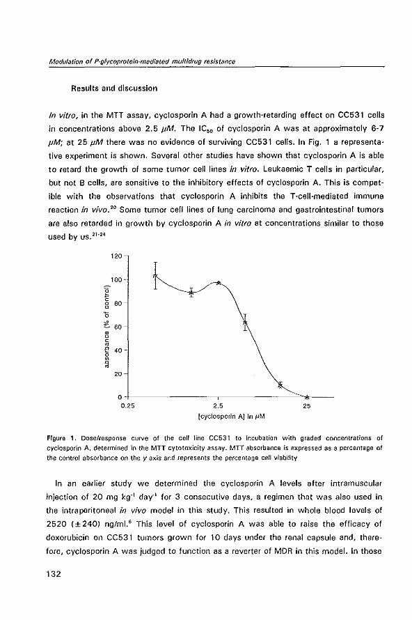
Modulation of P-glycoprotein-mediated muftidrug resistance
Results and discussion
In vitro. in the MTT assay. cyclosporin A had a growth-retarding effect on CC531 cells
in concentrations above 2.5 JiM. The IC50 of cyclosporin A was at approximately 6-7
JiM; at 25 JiM there was no evidence of surviving CC531 cells. In Fig. 1 a representa
tive experiment is shown. Several other studies have shown that cyclosporin A is able
to retard the growth of some tumor cell lines in vitro. Leukaemic T cells in particular,
but not B cells, are sensitive to the inhibitory effects of cyclosporin A. This is compat
ible with the observations that cyclosporin A inhibits the T-cell-mediated immune
reaction in vivo. 20 Some tumor ceff lines of lung carcinoma and gastrointestinal tumors
are also retarded in growth by cyclosporin A in vitro at concentrations similar to those
used by US.21
-24
120
100 -
g c 0 u
80 -
15
.c 60 m u c oj
£ 40 -0 ~
f,j 20
o-~-------------,----------~~~---0.25 2.5 25
(cyclosporin AJ In JIM
Figure 1. Dose/response curve of the cell line CC53 t to incubation with graded concentrations of
cyclosporin A, determined in the MTT cytotoxicity assay. MIT absorbance is expressed as a percentage of
the control absorbance on the y axis and represents the percentage cell viability
In an earlier study we determined the cyclosporin A levels after intramuscular
injection of 20 mg kgol day-l for 3 consecutive days, a regimen that was also used in
the intraperitoneal in vivo model in this study. This resulted in whole blood levels of
2520 (±240) ng/ml.' This level of cyclosporin A was able to raise the efficacy of
doxorubicin on CC531 tumors grown for 10 days under the renal capsule and, there
fore. cyclosporin A was judged to function as a reverter of MDR in this model. In those
132
Enhancement of metastasis by cyclospodn A
studies, no signs of growth acceleration or retardation by cyclosporin A were observed.
In the present study, tumors were implanted in the fat flap of the testes lying intraperit
oneally, which gave the tumors more space to grow and enabled longer·lasting
experiments_ In this model, cyclosporin A enhanced the growth of CC531 (see Table
1). Tumors treated with cyclosporin A alone were larger than control tumors, a
difference that was nearly statistically significant (P=O.06). The total tumor load was
significantly increased. This was caused by increase of the number and size of the
intraperitoneal metastases. These data concur with those of Eccles et 81.15 for
immunogenic tumors. They showed that cyclosporin A exerts its effects via
immunosuppression. The immunosuppressive treatment was only given for 3 days in
our experiments.
Tabla 1. Tumor growth enhancement in vivo under cyc!osporin A treatment
parameter control cyclosporin A statistical
significance, P
total tumor load (mg) 864 I± 2491 14861±5931 0.02
primary tumor load (mg) 840 I± 2361 11971H041 0.06
metastases load (mg) 241HOI 2891±3761 0,02
metastases number 1.0 (± 1.4) 7.91±7.41 om
Results are means with standard deviations in parentheses
In some in vivo studies, cyclosporin A retarded the growth of tumors, like the murine
colon cancer MC26.23 In human studies cyclosporin A has been used with variable
success in some hematological malignancies. Promising results have been described in
cutaneous T cell disease (the Sezary syndrome) and in Hodgkin's lymphoma. 25•28 No
objective responses were observed in a clinical trial involving 1 7 patients with
colorectal cancer. 27
Tumor growth in phase 1111 ollcological studies is considered as primary resistance to
the experimental drug. Therefore, tumor growth enhancement by an experimental drug
can remain unnoticed in these studies. As an immunosuppressive agent, cyclosporin A
potentially can accelerate growth of tumors that are restrained in their growth by the
immune system. Although experimental tumors in animals are more often immunogenic
than are human tumors, the immune system may play a beneficial role in various
133
Modulation of P-glycoprotein-mediated multidrug resistance
human malignancies, like renal cell cancer and malignant melanoma. In clinical trials
these tumors, and to a lesser extent colore eta I cancer, responded to treatment with
interleukin-2 and Iymphokine-activated killer cells in up to 30% of the cases_" In these
trials parts of the immune system are activated that are suppressed by cyclosporin A.
There are no studies on a potential tumor·growth-enhancing effect of
immunosuppression on existing tumors in humans. Only one study has measured the
effect of cyclosporin A on T cell levels in the setting of MDR reversal. Significant
decreases in total lymphocyte counts and in CD19, DC3, DC4 and CD8 subpopulations
were observed, which were totally reversible. 29
Our experiments in an animal model show that a short course of cyclosporin A
administration that was able to modulate MDR was also able to enhance the locoregio
nal metastatic growth of the weakly immunogenic CC531 colon carcinoma. It is
important to realise that, with the novel utilisation of cyclosporin A as a reverter of
MDR in cancer therapy, growth enhancement might be induced in some tumors. Non
immunosuppressive analogues of cyclosporin A, such as PSC 833, or other compounds
that are at least equally potent reverters of MDR should, therefore, be preferred in
anticancer chemotherapy.
References
1. Kahan BD. Cyclosporine_ N Engl J Med 1989; 321: 1725-38 2. Silbermann MH, Boersma AWM, Janssen ALW, Scheper AJ, Herweijer H, Nooter K_ Effects of
cyclosporin A and verapamil on the intracellular daunorubicin accumulation in Chinese hamster ovary cetts with increasing levels of drug-resistance. Int J Cancer 1989; 44: 722-6
3. Boesch 0, Muller K, Pourtier-Manzanedo A, loor F. Restoration of daunomycin retention in multidrug·resistant P3B8 cetts by submicromolar concentrations of SDZ PSC 833, a nonimmunosuppressive cyclosporin derivative. Exp Cell Res 1991; 196: 26-32
4. Osieka A, Seeber S, Pannenbacker A, Soli 0, Glatte P, Schmidt CG. Enhancement of etoposideinduced cytotoxicity by cyclosporin A. Cancer Chemolher Pharmacol 1986; 18: 198-202
5. Twentyman PA, Aeeve JG, Koch G, Wright KA. Chemosensitisation by verapamil and cyclosporin A in mouse tumour cells expressing different levels of P-glycoprotein and CP22 (sorcin). Or J Cancer 1990; 62: 89-95
6. Van de Vrie W, Gheuens EEO, Durante NMC, de Bruijn EA, Marquet AL, van Oosterom AT, Eggermont AMM. In vitro and in vivo chemosensitizing effect of cyclosporin A on an intrinsic multidrug-resistant rat co [on tumour. J Cancer Res Clln Onco/1993; 119: 609-14
7. Sonneveld P, Durie BGM, Lokhorst HM, Marie JP, Solbu B, Suciu S, Zittoun R, LOwenberg B, Nooter K. Modulation of multidrug·resistant multiple myeloma by cyclosporin. Lancel 1992; 340: 255-9
B. Sonneveld P, Schoester M, de Leeuw K. Clinical modulation of multidrug resistance in multiple myeloma: effect of cyclosporine on resistant tumor cells. J Clin Oncol 1994; 12: 1584-91
9. Beveridge T, Krupp P, McKibbin C. Lymphomas and Iymphoproliferative lesions developing under cyclosporin therapy !letter to the editorJ. Lancet 1984; I: 7B8
10. Penn I. Cancer is a complication of severe immunosuppression. Surg Gynecol Obstet 1986; 162: 603-10
11. Reitz BA, Bieber CPA Cancer after the use of cyclosporin A in animals, Cancer SUN 1982; 1: 613·9
134
Enhancement of metastasis by cyclosporln A
12. Demetris AJ, Nalesnik MA, Kunz HW, Gill T J III, Shinozuka H. Sequential analyses of the development of Iymphoproliferative disorders in rats receiving cyclosporine. Transplantation 1984; 38: 239-46
13. Kinlen LJ. Immunosuppressive therapy and cancer. Cancer Surveys 1982; 1: 565-83 14. Penn I. Cancers following cyclosporine therapy. Transplantation 1987; 43: 32·5 15. Eccles SA, Heckford SE, Alexander P. Effect of cyclosporin A on the growth and spontaneous
metastasis of syngeneic animal tumours. Br J Cancer 1980; 42: 252-9 16. Marquet Rl, Westbroek Dl, Jeekel J. Interferon treatment of a transplantable rat colon
adenocarcinoma: importance of tumor site. Int J Cancer 1984; 33: 689-92 17. De Greef C, van der Heyden S, Viana F. Eggermont J, de Broijn E, Raeymaekers L, Droogmans G,
Nilius B. lack of correlation between mdr-I expression and volume activation of chloride·currents in rat colon cancer cells. PI/ugers Arch 1995; 430: 296·8
18. Gheuens E, van der Heyden S, Erst H, Eggermont A, van Oosterom A, de Bruijn E. Multidrug resistance in rat colon carcinoma cell lines CC531, CC531 mdr + and CC531· ..... Jpn J Cancer Res 1993; 84: 1201-8
19. Carmichael J, DeGraff WG, Gazdar AF, Minna JD, Mitchell JB. Evaluation of a tetrazotium·based semiautomated colorimetric assay: assessment of chemosensitivity testing. Cancer Res 1987; 47: 936-42
20. TlStterman TH, Danersund A, Nilsson K, Killander A. Cyclosporin-A is selectively cytotoxic to human leukemic T cells in vitro. Blood 1982; 59: 1103·7
21. Piontek M, Porschen A. Growth inhibition of human gastrointestinal cancer cells by cyclosporin A. J Cancer Res Clin Oncol 1994; 120: 695-9
22. Saydjari R, Townsend CM, Barranco SC. James E. Thompson JC. Effects of cyclosporin A and q. difluoromethylornithine on the growth of hamster pancreatic cancer in vitro. J Natl Cancer Inst 1986; 77: 1087-92
23. Saydjari A, Townsend CM, Barranco SC, Thompson JC. Effects of cyclosporin A and q·difluoromet· hylornithine on the growth of mouse colon cancer in vitro. life Sci 1987; 40: 359-66
24. Twentyman PA. A possible role for cyc[osporins in cancer chemotherapy. Anticancer Res 1988; 8: 985-94
25. Puttick l, Pollock A. Fairburn E. Treatment of the S~zary syndrome with cyctosporin A. J R Soc Med 1983; 76: 1063-4
26. Zwitter M. On the potential role of cyclosporin in the treatment of Iymphoproliferative diseases. Leukemia Res 1988; 12: 243·8
27. Murren JR. Ganpule S, Sarris A, Durivage H, Davis C, Makuch R, Handschumacher RE, Marsh JC. A phase II trial of cyclosporin A in the treatment of refractory metastatic colorectal cancer. Am J Clin Onco/1991; 14: 208-10
28. Rosenberg SA. Principles and application of biologic therapy. In: DeVita VT. ir, Helfman 5, Rosenberg SA (eds) Cancer: Principles and practice of oncology, 1993, 4th edn. Lippincott, Philadelphia, pp 293·324
29. Gonz.1lez-Manzano R, Cid J, Brugarolas A, Piasecki CC. Cyc[osporin A and doxorubicin·ifosfamide in resistant solid tumours, a phase I and an immunological study. Br J Cancer 1995; 72: 1294-9
135

2.6
DRUG RESISTANCE IN
RA T COLON CANCER CELL LINES IS
ASSOCIA TED WITH MINOR CHANGES IN
SUSCEPTIBILITY TO CYTOTOXIC CELLS
Wim van de Vrie, Sylke A.M. van der Heyden,
Eric E. O. Gheuens, Amelie M. Bijma,
Ernst A. de Bruijn, Richard L. Marquet,
Allan T. van Oosterom and Alexander M.M. Eggermont
Cancer Immunollmmunother
1993; 37: 337-42
Modulation of P-glycoprotein-mediated muftidrug resIstance
Summary
The development of resistance to anticancer drugs urges the search for different
treatment modalities. Several investigators have reported the concomitant development
of drug resistance and resistance to natural killer (NK), Iymphokine-activated killer (LAK)
or monocytefmacrophage cell lysis, while others described unchanged or even
increased susceptibility. We investigated this subject in the rat colon carcinoma cell
line, CC531-PAR, which is intrinsically MDR, and in three sublines derived from this
parental cell line: a cell line with an increased MDR phenotype (CC531-CDL), a
revertant line from CC531-CDL (CC531-REV) which demonstrates enhanced sensitivity
to anticancer drugs of the MDR phenotype, and an independently developed cisplatin
resistant line (CC531-CISl. In a 4 h "Cr-release assay we found no differences in
susceptibility to NK cell lysis. No significant differences in Iysability by adherent LAK
(aLAK) cells were observed in a 4 h assay. In a prolonged 20 h "Cr-release assay an
enhanced sensitivity to aLAK cell-mediated lysis was observed in the revertant, p.
glycoprotein-negative cell line and in the cisplatin-resistant cell line (CC531-CIS). None
of the cell lines was completely resistant to lysis by aLAK cells. Therefore, a role for
immunotherapy in the treatment of drug-resistant tumors remains a realistic option.
138
Drug resistance and cytotoxic cell lysis
Introduction
One of the major problems in cancer chemotherapy is the development of resistance to
drugs. Several mechanisms of drug resistance have been elucidated; some operate
against a particular drug, while others affect a group of structurally unrelated
anticancer agents, as in MDR. Doxorubicin, vincristine and etoposide are examples of
drugs subjected to the MDR mechanism. MDR cells express an efflux pump, P
glycoprotein, which expels anticancer drugs from the cells. Expression of the multidrug
transporter can be demonstrated by anti-P-glycoprotein monoclonal antibodies, or at the
DNA and RNA level by blotting techniques.' Resistance to a specific drug may be
caused by several mechanisms. For instance, resistance to cisplatin is related to
reduced drug accumulation, increased detoxification, and increased DNA repair. 2 The
development of resistance to anticancer drugs urges the search for alternative treat
ment modalities, for example immunotherapy.
Adoptive immunotherapy using interleukin-2 (lL-2) and cytotoxic cells (lymphokine
activated killer (LAK) cellsl has proven to be effective in renal cell cancer. Response
rates up to 35% have been reported. In melanoma, responses up to 21 % were found.
Response rates in colon carcinoma were lower (13%).3 All these tumors are very
resistant to currently available drugs. Of these tumors, renal cell cancer and colon
carcinoma intrinsically express the MOR phenotype at a high frequency. 4
Before immunotherapy can be used as an alternative treatment after failure of
chemotherapy, it is important to know whether there might be a correlation between
drug resistance and sensitivity or resistance to immunotherapy. Reports in the literature
have yielded conflicting data about a possible correlation between drug resistance,
especially MDR, and resistance to NK and LAK cell lysis.'" We investigated this subject
in four cell lines of a rat colon carcinoma with different mechanisms and levels of drug
resistance. In our model drug resistance was not associated with changes in sensitivity
to NK-cell-mediated lysis. Only minor alterations in sensitivity to IL-2/adherent LAK
(aLAK) cell lysis were observed.
Materials and methods
Animals
Male rats of the inbred WAG/RIJ (RT1 ') strain were obtained from Harlan-CPB
139
Modulation of P-glycoprotein-mediated muftidrug resistance
(Austerlitz, The Netherlands). Animals were bred under specific-pathogen-free condi
tions and fed standard rat chow (Hope Farms, Woerden, The Netherlands) and water ad
libitum. In the experiments, rats of 12·18 weeks old, weighing 220-280 g, were used.
Cell lines
CC531 is a rat colon adenocarcinoma, which was induced chemically in the WAG rat
with 1,2-dimethylhydrazine. The moderately differentiated tumor is weakly
immunogenic, as determined by the method described by Prehn and Main,' and
transplantable in syngeneic rats.a Following subcutaneous implantation and subsequent
resection, the tumor metastasizes to the lungs. In vivo the tumor is resistant to most
anticancer drugs and only at the maximal tolerable dose were significant growth
retarding effects observed.9 (and unpublished observations) Cisplatin was reported to
be one of the most effective drugs against CC531.9 Moderate sensitivity to immunothe
rapeutic agents like interferon-T and tumor necrosis factor-o in vitro and in vivo has
been shown in previous studies.a,10-12 In vitro CC53l, the parental cell line (CC531-
PAR), grows as a monolayer in Dulbecco's modified Eagle's medium supplemented with
5% heat-inactivated fetal calf serum, L-aspargine (50 mg/l), glutamic acid (2 mM), 100
IUlml penicillin and 100 pg/ml streptomycin, all obtained from Gibco (Paisley, UK), in a
humidified atmosphere of 5% CO,/95% air at 37"C. CC531-PAR intrinsically expresses
the MDR phenotype. We recently reported the reversibility of MDR in vitro and in vivo
in this model. 13 In vitro drug-resistant sub lines were induced by continuous incubation
with colchicine (CC531-COl) and cisplatin (CC531-CIS). Established cell lines were
maintained in the presence of 0.2 pM colchicine and 0.75 pM cisplatin (cis-diaminedi
chloroplatinum) respectively." Compared to the parental line, CC531-COL shows
enhanced resistance to drugs of the MDR phenotype (colchicine 33-fold, daunorubicin
10-fold) and to cisplatin (5.5-fold). CC531-CIS is resistant to cisplatin (9.8-fold), while
it has approximately the same sensitivity to other drugs as the parental line. From
CC531-COL a revertant line (CC531-REV) was isolated that is more sensitive to drugs
of the MDR phenotype than the parental line (resistance to colchicine 0.6-fold, to
daunorubicin 0.25-foldl. but the resistance to cisplatin is maintained (4-fold) as in the
CC531-COL line. 14•1S See Table 1 for characteristics.
YAC-l, a mouse T eel/lymphoma, sensitive to NK cell lysis, was used as a positive
control in the NK cell experiments. pa15, a mouse mastocytoma, NK cell lysis resistant
but sensitive to LAK cell lysis, was used as a negative control in NK cell experiments
and as a positive control in aLAK cell cytotoxicity tests. Both cell lines were grown in
140
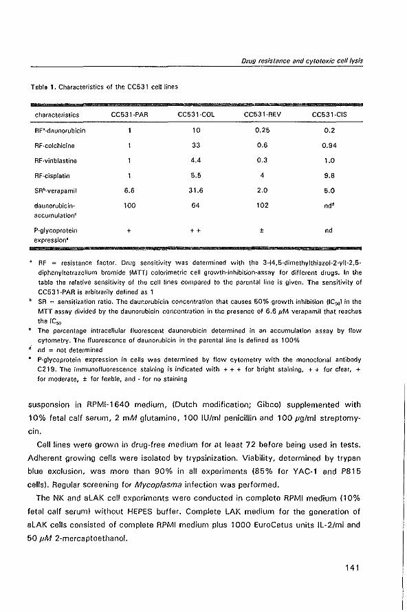
Table 1. Characteristics of the CC531 cell Jines
characteristics
AF"-daunorubicin
AF-colchicine
AF-vinblastine
AF-cisplatin
daunorubicin
accumulation~
P-glycoprotein
expression"
CC531·PAR
6.6
100
+
CC531·COL
10
33
4.4
5.5
31.6
64
++
Drug resistance and cytotoxic cell lysis
CC531·REV CC531·C1S
0.25 0.2
0.6 0.94
0.3 1.0
4 9.8
2.0 5.0
102 nd'
± nd
AF = resistance factor. Drug sensitivity was determined with the 3-(4,5-dimethylthiazol-2-yll-2,5-
diphenyltetrazolium bromide (MIT) colorimetric celt growth-inhibition-assay for different drugs. In the
table the relative sensitivity of the celt lines compared to the parental line is given. The sensitivity of
CC531·PAA is arbitrarily defined as 1
b SA "" sensitization ratio. The daunorubicin concentration that causes 50% growth inhibition (lC50 ) in the
MIT assay divided by the daunorubicin concentration in the presence of 6.6 JIM verapamil that reaches
the IC50
The percentage intracellular fluorescent daunorubicin determined in an accumulation assay by flow
cytometry. The fluorescence of daunorubicin in the parental line is defined as 100%
d nd = not determined
P-glycoprotein expression in cells was determined by flow cytometry with the monoclonal antibody
C219. The immunofluorescence staining is indicated with + + + for bright staining, + + for clear, + for moderate, ± for feeble, and - for no staining
suspension in RPMI-1640 medium, (Dutch modification; Gibco) supplemented with
10% fetal calf serum, 2 mM glutamine, 100 IU/ml penicillin and 100 Jlglml streptomy
cin.
Cell lines were grown in drug-free medium for at least 72 before being used in tests.
Adherent growing cells were isolated by trypsinization. Viability, determined by trypan
blue exclusion, was more than 90% in all experiments (85% for YAC-l and P815
cells). Regular screening for Mycoplasma infection was performed.
The NK and aLAK cell experiments were conducted in complete RPMI medium (10%
fetal calf serum) without HEPES buffer. Complete LAK medium for the generation of
aLAK cells consisted of complete RPMI medium plus 1000 EuroCetus units IL-2/ml and
50 JIM 2-mercaptoethanol.
141
Modulation of P-glycoprotein-mediated multidrug resistance
Chemicals
2·Mercaptoethanol was obtained from J. T. Baker, Deventer, The Netherlands;
interleukin·2 (lL·2) from EuroCetus, Amsterdam, The Netherlands; 1 % sodium dodecyl
sulphate from Merck, Darmstadt, Germany; sodium [51 Cr[chromate from Amersham,
Aylesbury, UK.
Preparation of effector cells
Spleens were removed aseptically from the rats and crushed with the hub of a
syringe in complete medium. Spleen cells were incubated for 5 min at 37°C in a
buffered ammonium chloride solution to lyse the erythrocytes. For the NK cytotoxicity
tests cells were rested for 1 h in complete medium at 37°C in 25 em' culture flask
(Costar, Cambridge, Mass., USA) to remove the macrophages by adherence to the
plastic. The remaining free·floating cells were aspirated, counted, and added to 96·well
round·bottomed microtiter plates (Costar). In the NK cell lysis tests effector:target (E:T)
cell ratios were 200: 1, 100: 1, 50: 1 and 25: 1.
For the aLAK cell cytotoxicity tests cells were passed over nylon-wool columns to
remove monocytes/macrophages and 8 cells. 16 Samples containing 2x10⁸ spleen cells
were added to a syringe containing 0.6 g sterile nylon wool (Cellular Products, Buffalo,
N.Y., USA) and incubated for 1 h at 37°C. The nonadherent cells were carefully
washed out with 50 ml medium. These cells were cultured at a concentration of 2x10⁶
cells in 75 em' culture flasks in LAK medium for 24 h. Then only the cells adherent to
the plastic of the flasks were cultured further in conditioned medium to make the aLAK
cell bulk culture. Conditioned medium was prepared by decanting the medium from the
flasks, removing the nonadherent cell by centrifugation and passing the supernatant
through a 0.45·/1m Millipore filter.17 After 72 h all cultured cells were collected; the
adherent cells by adding EDTA and scraping the flask with a rubber policeman. In the
aLAK cell lysis experiments E:T cell ratios were 50:1. 25:1,12.5:1 and 6.25:1.
Cytotoxicity assay
Sensitivity to NK and aLAK cells was tested in the 51Cr-release cytotoxicity assay.
Samples containing 1xl05 target cells were incubated for 1 h with 200 pCi 61Cr in 200
pi medium. Cells were washed three times with complete medium and counted, and
lxl0' cells in 100/11 complete medium were added to the effector cells (100/11) in the
plates. Spontaneous release was tested in wells containing target cells and medium
(100 /11) only; maximal release was obtained by adding 100 pi 1 % sodium dodecyl
142
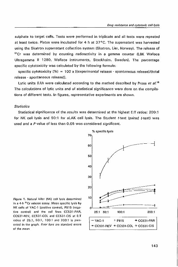
Drug resIstance and cytotoxic eel/lysis
sulphate to target cells. Tests were performed in triplicate and all tests were repeated
at least twice. Plates were incubated for 4 h at 37°C. The supernatant was harvested
using the Skatron supernatant collection system (Skatron, Lier, Norway). The release of
51Cr was determined by counting radioactivity in a gamma counter (LBK Wallace
Ultragamma II 1280, Wallace Instruments, Stockholm, Sweden). The percentage
specific cytotoxicity was calculated by the following formula:
specific cytotoxicity (%) = 100 x [(experimental release - spontaneous release)/(total
release - spontaneous release)].
Lytic units (LU) were calculated according to the method described by Pross a( al."
The calculations of lytic units and of statistical significance were done on the compila
tions of different tests. In figures, representative experiments are shown.
Statistics
Statistical significance of the results was determined at the highest E:T ratios: 200: 1
for NK cell lysis and 50: 1 for aLAK cell lysis. The Student (·test (paired (-test) was
used and a P value of less than 0.05 was considered significant.
Figure 1. Natural killer (NK) cell lysis determined in a 4-h 51C( release assay. Mean specific lysis by
NK cells of YAC·l (positive control), P815 (nega·
tive control) and the cell lines CC531-PAA, CC531·REV, CC531-COl and CC531-CIS at E:T ratios of 25:1,50:1, 100:1 and 200:1 is pres
ented in the graph, Error bars are standard errors
of the mean
% speclffc lysis 70
60
50
40
30
20
10
oL==:t::::::========2... 25:1 50:1 100:1 200:1
- VAC·l + pa15 ·CC531·PAR
~·CC531·REV *CC531·COL +CC531·CIS
143
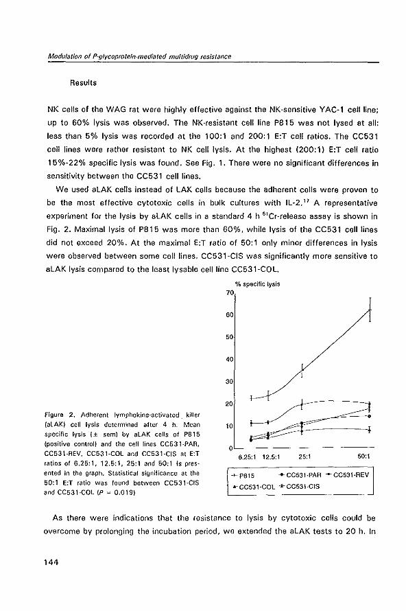
Modufation of P-gfycoprotefn-medlated muflidrug resistance
Results
NK cells of the WAG rat were highly effective against the NK-sensitive YAC-l cell line;
up to 60% lysis was observed. The NK·resistant cell line P815 was not lysed at all:
less than 5% lysis was recorded at the 100:1 and 200:1 E:T cell ratios. The CC531
cell lines were rather resistant to NK cell lysis. At the highest (200: 1) E:T cell ratio
15%~22% specific lysis was found. See Fig. 1. There were no significant differences in
sensitivity between the CC531 cell lines.
We used aLAK cells instead of LAK cells because the adherent cells were proven to
be the most effective cytotoxic cells in bulk cultures with IL·2.17 A representative
experiment for the lysis by aLAK cells in a standard 4 h 51Cr4release assay is shown in
Fig. 2. Maximal lysis of P815 was more than 60%, while lysis of the CC531 cell lines
did not exceed 20%. At the maximal E:T ratio of 50:1 only minor differences in lysis
were observed between some cell lines. CC531 ~CIS was significantly more sensitive to
aLAK lysis compared to the least Iysable cell line CC531-COl.
Figure 2. Adherent lymphokine·activated. killer
(alAKI cell lysis determined after 4 h. Mean specific lysis (± semI by alAK cells of P8l5
(positive control) and the cell tines CC53l·PAR,
CC531·REV. CC531-COl and CC531·CIS at E:T ratios of 6.25: 1, 12.5: 1, 25: 1 and 50: 1 is pres·
ented in the graph. Statistical significance at the 50: 1 E:T ratio was found between CC531-CIS
and CC531·CQl (P '" 0.019)
% specific lysis 70
60
50
40
30
20
10
oL----------------------6.25:1 12.5:1 25:1 50:1
+P815 .... CC531·PAA ..... CC531·AEV
... ·CC531·COL +CC531·CIS
As there were indications that the resistance to lysis by cytotoxic cells could be
overcome by prolonging the incubation period, we extended the aLAK tests to 20 h. In
144
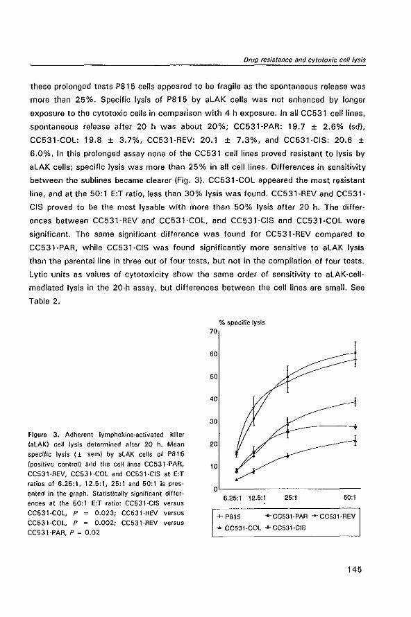
Drug resistance and cytotoxic eel/lysis
these prolonged tests P8l5 cells appeared to be fragile as the spontaneous release was
more than 25%. Specific lysis of P815 by aLAK cells was not enhanced by longer
exposure to the cytotoxic cells in comparison with 4 h exposure. In all CC531 cell lines,
spontaneous release after 20 h was about 20%; CC531-PAR: 19.7 ± 2.6% (sd),
CC531-COL: 19.8 ± 3.7%, CC531-REV: 20.1 ± 7.3%, and CC531·CIS: 20.6 ±
6.0%. In this prolonged assay none of the CC531 cell lines proved resistant to lysis by
aLAK cells; specific lysis was more than 25% in all cell lines. Differences in sensitivity
between the sublines became clearer (Fig. 3). CC531-COL appeared the most resistant
line, and at the 50: 1 E:T ratio, less than 30% lysis was found. CC531-REV and CC531-
CIS proved to be the most Iysable with inore than 50% lysis after 20 h. The differ·
ences between CC531-REV and CC531·COL, and CC531·CIS and CC531-COL were
significant. The same significant difference was found for CC531-REV compared to
CC531-PAR, while CC531·CIS was found significantly more sensitive to aLAK lysis
than the parental line in three out of four tests, but not in the compilation of four tests.
Lytic units as values of cytotoxicity show the same order of sensitivity to aLAK·cell·
mediated lysis in the 20·h assay, but differences between the cell lines are small. See
Table 2.
Figure 3. Adherent Iymphokine-activated kilter
% specific lysis 70
60
50
40
30
(alAK) cell lysis determined alter 20 h. Mean 20 specific lysis (± semI by aLAK cells of P815
(positive control) and the cell Jines CC531-PAR,
CC531-REV, CC531-COL and CC531·CIS at E:T ratios of 6.25:1, 12.5:1, 25:1 and 50:1 is pres
ented in the graph. Statistically significant differ· ences at the 50: 1 E:T ratio: CC531-CIS versus
CC531-COL, P = 0.023; CC531·AEV versus CC531-COL, P = 0.002; CC531-AEV versus
CC531·PAR, P " 0.02
10
OL--------------------6.25:1 12.5:1 25:1 50:1
-+- P81S "'CCS31·PAA .... CC53l·AEV
.... CC53l·COL ""CC531·CIS
145
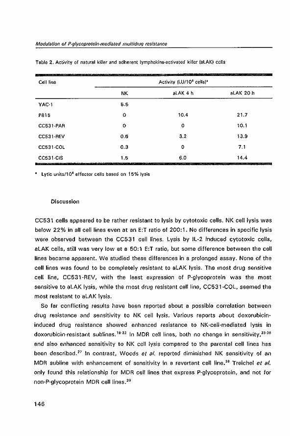
Modulation of P-glycoprotein-mediated mu/tidrug resistance
Table 2. Activity of natural killer and adherent Iymphokine-activated killer (aLAK) cells
Celt line Activity (LU/106 cells)-
NK aLAK 4 h aLAK 20 h
VAC-l 5.5
P815 0 10.4 21.7
CC531-PAR 0 0 10.1
CC531-REV 0.6 3.2 13_9
CC531-COL 0.3 0 7.1
CC531-CIS 1.5 6.0 14.4
- Lytic units/1 0 6 effector cells based on 15% lysis
Discussion
CC531 cells appeared to be rather resistant to lysis by cytotoxic cells. NK cell lysis was
below 22% in all cell lines even at an E:T ratio of 200: 1. No differences in specific lysis
were observed between the CC531 cell lines. Lysis by IL-2 induced cytotoxic cells,
aLAK cells, still was very low at a 50: 1 E:T ratio, but some difference between the cell
lines became apparent. We studied these differences in a prolonged assay. None of the
cell lines was found to be completely resistant to aLAK lysis. The most drug sensitive
cell line, CC531-REV, with the least expression of P-glycoprotein was the most
sensitive to aLAK lysis, while the most drug resistant cell line, CC531-COL, seemed the
most resistant to aLAK lysis.
So far conflicting results have been reported about a possible correlation between
drug resistance and sensitivity to NK cell lysis. Various reports about doxorubicin
induced drug resistance showed enhanced resistance to NK-cell-mediated lysis in
doxorubicin-resistant subHnes. 19-22 In MDR cell lines, both no change in sensitivity, 23-26
and also enhanced sensitivity to NK cell lysis compared to the parental cell lines has
been described. 27 In contrast, Woods et al. reported diminished NK sensitivity of an
MDR subline with enhancement of sensitivity in a revertant cell line. 28 Treichel et al.
only found this relationship for MDR cell lines that express P-glycoprotein, and not for
non-P-glycoprotein MDR cell lines.29
146
Drug resistance and cytotoxic eel/lysis
With regard to LAK cell sensitivity, various studies show no difference in Iysability
between drug-resistant cell lines and their parental lines. 22,24'26,30-32 Enhanced sensitivity
as well as enhanced resistance of MDR cell lines to LAK cell lysis has been reported by
others.27.29.32.35 An inverse relationship has also been found: the induction of LAK
resistance in melanoma cell lines rendered these more sensitive to doxorubicin.36
Kimmig et al. found that LAK resistance correlated with the level of P'glycoprotein
expression. A revertant cell line that was as drug sensitive as the parental line was
found to be as Iysable by LAK cells as the parental cell line." We did a comparable
observation with the CC531 cell lines in the prolonged aLAK lysis assay.
Explanations for the phenomenon of differences in sensitivity to cytotoxic cells
between drug-sensitive and drug-resistant cells have been sought in differences in the
expression of cell membrane molecules. In most studies no correlation between MDR
expression, NK or LAK cell resistance and expression of MHC class I and" antigens
was observed. 26,33,37 With regard to adhesion molecules a correlation between enhanced
LAK cell lysis and ICAM-1 and LFA-3 expression in MDR cell lines was described." but
other authors could not confirm these results using other cell lines. 26
To our knowledge only few reports about the association between cisplatin resis
tance and NK/LAK cell resistance have been published. In most experiments no
influence of cisplatin resistance on sensitivity to LAK-cell-mediated lysis was
observed. 25•30,31.36 In one of their cisplatin-resistant cell lines Ohtsu et al. found
enhanced sensitivity to LAK cell lysis. 31 Allevana et al. reported significant lysis by LAK
cells of freshly isolated tumor cells from ovarian cancer patients that were refractory to
chemotherapy with cispfatin. 30 In a recent article short-term pretreatment of cancer
cells with cisplatin was reported to render these cells more sensitive to cytotoxic
cells." We observed enhanced lysis by aLAK cells of the cisplatin-resistant cell line
CC531-CIS. In CC531-COL and CC531-REV cells a moderate resistance to cisplatin
was found. about half the resistance of CC531-CIS. In these two cell lines an associ
ation between their MDR phenotype and LAK cell resistance can be supposed. while
the resistance to cisplatin seems of no importance. It is very possible that the colchicin
induced MDR cell line CC531-COL and its revertant CC531-REV have a different
mechanism of cisplatin resistance from CC531-CIS, as this resistance was induced
differently.
It is clear from our results and those of others that basic research has not yet
provided coherent data on the relationship between drug resistance and sensitivity to
immunotherapy. The lack of coherence in the results may be inherent to the different
147
Modulation of P-glycoprolein-mediated multidrug resIstance
cell lines and the diversity of methods used to induce resistance. Possibly this reflects
the heterogeneity of sensitivity and resistance to drugs and immunotherapy in the
clinical situation. Our results seem to indicate that drug resistance does not preclude
the use of immunotherapy with IL-2 and LAK cells, but enhancement of the efficacy of
immunotherapy is necessary. With regard to P-glycoprotein-expressing MOR tumors, an
attractive approach might be to turn the strength of MDR cells into their weakness.
This can be done by using antibodies against the multidrug transporter P-glycoprotein.
Immunotoxin therapy using the anti-p-glycoprotein antibody MRK16 coupled to
Pseudomonas exotoxin has been shown to be effective against MOR cells in vitro and
in ViVO. 39.41 Another possible method of targeted immunotherapy is the use of bispecific
monoclonal antibodies directed against P-glycoprotein on one hand and against an
antigen on cytotoxic cells on the other.42.43 Further research in this direction is war
ranted.
References
1. Pastan I, Gottesman M. Multiple-drug resistance in human cancer. N Engl J Med 1987; 316: 1388-93
2. Andrews PA, Howell S8. Cellular pharmacology of cisplatin: perspectives on mechanisms of acquired resistance. Cancer Cells 1990; 2: 35·43
3. Rosenberg SA. Immunotherapy and gene therapy of cancer. Cancer Res 1991; 51: 5074s-9s 4. Goldstein LJ, Galski H, Fojo A, Willingham M, lai Sol, Ga2dar A, Pirker R, Green A, Crist W,
Brodeur GM, Lieber M, Cossman J, Gottesman MM, Pastan t. Expression of a multidrug resistance gene in human cancers. J Nat! Cancer Inst 1989; 81: 116-24
5. Safrit JT, Bonavida B. Hierarchy of in vitro sensitivity and resistance of tumor celfs to cytotoxic effector cells, cytokines, drugs and toxins. Cancer Immunollmmunorher 1992; 34: 321·8
6. Savas B, Cole SPC, Akoglu TF. Pross HF. P-glycoprotein-mediated multidrug resistance and cytotoxic effector cells. Nat Immun 1992; 11: 177-92
7. Prehn RT, Main JW. Immunity to methylcholanthrene induced sarcomas. J Natl Cancer Inst 1957: 18: 769-78
8. Marquet Rl, Westbroek Dl, Jeekel J. Interferon treatment of a transplantable rat colon adenocarcinoma: importance of tumor site. Int J Cancer 1984; 33: 689-992
9. los G, Nagel JD, McVie JG. Anti-tumor effect of cisplatin, carboplatin, mitoxantrone, and doxorubicin on peritoneal tumor growth after intraperitoneal and intravenous chemotherapy: a comparative study. Sel Cancer Ther 1990; 6: 73-82
10. Eggermont AMM, Marquet RL, de Bruin RWF, Jeekel J. Effects of the interferon-inducer ABPP on colon cancer in rats: importance of tumor load and tumor site. Cancer Immunollmmunother 1986; 22: 217-20
11. Marquet Rl, 1J2ermans JNM, de Bruin RWF, FiefS W, Jeekel J. Antitumor activity of recombinant mouse tumor necrosis factor (TNF) on colon cancer in rats is promoted by recombinant rat interferon gamma; toxicity is reduced by indomethacin. Int J Cancer 1987; 40: 550-3
12. Scheringa M. IJ2ermans JNM, Jeekel J, Marquet Al. The anti tumour activity of the interferon inducer bropirimine is partially mediated by endogenous tumour necrosis factor-a. Cancer Immunol Immunother 1990; 32: 251-5
13. Van de Vrie W, Gheuens EEO, Durante NMC, de Bruijn EA, Marquet Rl, van Oosterom AT, Eggermont AMM. In vitro and in vivo chemosensitizing effect of cyclosporin-A on an intrinsic multidrug resistant rat colon tumour. J Cancer Res Clin Oncol 1993; 119: 609-14
148
Drug resistance and cytotoxic cell lysis
14. Gheuens EEO, Elst HJ, van der Heyden SAM, de Bruijn EA, van Oosterom AT (1993) Multidrug resistance in rat coton carcinoma cell lines CC531, CC531"'*-+ and CC531'··. Jpn J Cancer Res 1993;84: 1201-8
15. Gheuens EEO, van Bockstaele DR, van der Keur M, Tanke HJ, van Oosterom AT, de Bruljn EA. Flow cytometric double labelling technique for screening of multidrug resistance. Cytometry 1991; 12: 636-44
16. Julius MH, Simpson E, Herzenberg LA. A rapid method for the Isolation of functional thymus derived murine lymphocytes. Eur J Immunol 1973; 3: 645-9
17. Vujanovic NL, Herberman RB, Maghazachi AA, Hiserodt JC. lymphokine·activated killer celfs in rats til. A simple method for the purification of large granular lymphocytes and their rapid expansion and conversion into Iymphokine-activated killer celfs. J Exp Med 1988; 167: 15-29
18. Pross HF, Baines MG, Rubin P, Shragge P, Patterson MS. Spontaneous human lymphocyte· mediated cytotoxicity against tumor target cells. IX. The quantitation of natural killer cell activity. J Clin Immunol 1981; 1: 51·63
19. Benoist H, Madoulet C, Jardillier J·C, oesplaces A. Adriamycin induced resistance of sensitive K 562 ceffs to natural killer lymphocyte attack. Cancer Immunollmmunother 1985; 20: 122-8
20. Vanovlch S, Halt RE, Weinert C. Resistance to natural killer cell·mediated cytolysis by a pleiotropic drug-resistant human erythroleukemia (K562·A) cell line. Cancer Res 1986; 46: 4511-5
21. Wood WJ, Lotzov<1 E. Adtiamycin·induced resistance to natural kiffer {NK)·mediated cytotoxicity. Cancer 1989; 64: 396-403
22. Alfavena P, Grandi M, o'lncalci M, Geri 0, Giuliani FC, Mantovani A. Human tumor cell lines with pleiotropic drug resistance are efficiently killed by interleukin·2 activated killer cells and by activated monocytes. Inc J Cancer 1987; 40: 104·7
23. Allavena P, Peccatori F, Maggioni 0, Pirovano P, Mantovani A. Killing of tumor cells with pleiotropic drug resistance by OK432-activated effector cells. Immunopharmacol Immunotoxicol 1989; 11: 257-68
24. Ades EW, Bosse 0, Pruckler J. Potentiation of human natural killer celt activity by recombinant interleukin-2 towards muhidrug·resistant human epidermoid carcinoma. Pathobiology 1990; 58: 84-7
25. Harker WG, Tom C, McGregor JR, Slade L, Samlowski WE. Human tumor cell line resistance to chemotherapeutic agents does not predict resistance to natural killer or Iymphokine·activated killer celt·mediated cytolysis. Cancer Res 1990; 50: 5931-6
26. Scheper RJ, Dalton WS, Grogan TM, Schlosser A, Bellamy WT, Taylor CW, Scuderi P, Spier, C. Altered expression of P·glycoprotein and cellular adhesion molecules on human multi·drug·resistant tumor celts does not affect their susceptibility to NK- and LAK·mediated cytotoxicity. Int J Cancer 1991; 48: 562-7
27. Aivoltin; L, Colombo MP, Supino A, Balliflari 0, Tsuruo T, Parmiani G. Modulation of multidrug resistance by verapamif or mdrl anti·sense oligodeoxynuc!eotide does not change the high susceptibility to Iymphokine·activated killers in mdr-resistant human carcinoma (LoVo) line. Int J Cancer 1990; 46: 727-32
28. Woods G, lund lA, Naik M, Ling V, Ochi A. Aesistance of multidrug·resistant lines to natural killerlike cell·mediated cytotoxicity. FASEB J 1988; 2: 2791-6
29. Treichel AS, Olken S. The relationship between multi· drug resistance and resistance to natural·killercell and Iymphokine-activated killer·cell lysis in human leukemic cell lines. Int J Cancer 1992; 50: 305-10
30. Allavena P, Damia G, Colombo T, Maggioni 0, o'incalci M, Mantovani A. Lymphokine·activated killer (LAK) and monocyte·mediated cytotoxicity on tumor cell lines resistant to antitumor agents. Cell Immunol 1989; 120: 250-8
31. Ohtsu A, Sasaki V, Tamura T, Fujiwara Y, Ohe V, Minato K, Nakagawa K, Bungo M, Saljo N. Inhibition of colony formation of drug·resistant human tumor cell lines by combinations of interfeukin·2-activated killer cells and antitumor drugs. Jpn J Cancer Res 1989; 80: 265-70
32. Gautam SC, Chikkala NF, Lewis I, Grabowski DR, Finke JH, Ganapathi R. Therapeutic efficacy of interleukin-2 activated killer cells against Adriamycin resistant mouse B 16·BL6 melanoma. Anticancer Res 1992; 12: 921·6
33. Gambacorti·Passerinl C, Rivoltini l, Supino A, Mariani M, Parmlani G. Differential lysis of melanoma clones by autologous recombinant interleukin 2-activated lymphocytes. Relationship with sponta· neous resistance to doxorubicln (Ox). Int J Cancer 1988; 42: 544·8
34. Gambacorti·Passerini C, Aivoltini L, Supino A, Rodolfo M, Radriz.zani M, Fossati G, Patmiani G.
149
Modulation of P-gIVcoprotein-mediated multidrug resistance
Susceptibility of chemoresistant murine and human tumor cells to lysis by interleukin 2-activated lymphocytes. Cancer Res 1988; 4'8: 2372-6
35. Kimmig A, Gekeler V, Neumann M, Frese G, Handgretinger A, Kardos G, Oiddens H, Niethammer O. Susceptibility of multidrug-resistant human leukemia cell lines to human interleukin 2-activated killer cells. Cancer Res 1990; 50: 6793~9
36. Aivoltini l, Gambacorti-Passerini C, Supin~ A, Parmiani G. Generation and partial characterization of melanoma sublines resistant to Iymphokine activated killer (lAK) cells_ Relevance to doxorubicin resistance. Int J Cancer 1989; 43: 880-5
37. Rivoltini l, Cattoretti G, Arienti F, Mastroianni A, Melani C, Colombo MP, Parmiani G_ The high Iysability by LAK cells of colon-carcinoma cells resistant to doxorubicin is associated with a high expression of ICAM-l, lFA-3, NCA and a less-differentiated phenotype. Int J Cancer 1991; 47: 746-54
38. Mizutani Y, Banovida B, Nio Y, Yoshida 0_ Enhanced susceptibility of cis-diamminedichloroplatinumtreated K562 cells to lysis by peripheral blood lymphocytes and Iymphokine activated killer cells. Cancer 1993; 71: 1313-21
39. FitzGerald OJ, Willingham MC, Cardarelli CO, Hamada H, Tsuruo T, Gottesman MM, Pastan I. A monoclonal antibody-Pseudomonas toxin conjugate that specifically kills multidrug-resistant cells. Proc Nat! Acad Sci USA 1987; 84: 4288-92
40. Mickisch GH, Pai lH, Gottesman MM, Paslan I. Monoclonal antibody MAK 16 reverses the multidrug resistance of multidrug-resistant transgenic mice. Cancer Res 1992; 52: 4427-32
41. Mickisch GH, Pai lH, Siegmund M, Campain J, Gottesman MM, Pastan I. Pseudomonas exotoxin conjugated to monoclonal antibody MAK 16 specifically kiffs multidrug resistant cells in cultured renal carcinomas and in MDR-transgenic mice. J Uro/1993; 149: 174-8
42. Van Dijk J, Tsuruo T, Segal OM, Bolhuis RlH, Colognola A, van de Griend RJ, Fleuren GJ, Warnaar SO. Bispecific antibodies reactive with the multidrug·resistance-related glycoprotein and CD3 induce lysis of multidrug·resistant tumor cells. Int J Cancer 1989; 44: 738-43 •
43. Bolhuis Al, Sturm E, Braakman E. T cell targeting in cancer therapy. Cancer Immunollmmunother 1991;34: 1-8
150

3
DISCUSSION AND SUMMARY

General discussion
3.1 GENERAL DISCUSSION
Our studies go back to 1990 when the mechanism of P·glycoprotein-mediated MDR
had only recently been elucidated. The feasibility of blocking the P-glycoprotein efflux
pump, thereby increasing intracellular drug levels and enhancing cell death had been
shown in numerous in vitro studies, but few studies had shown the possibilities of
MDR reversal in vivo. At this point we started collaborative investigations with the
group of Prof. Dr. A.T. Van Oosterom and Dr. E.A. De Bruijn of the Laboratory of
Cancer Research and Clinical Oncology of the University of Antwerp in Belgium. They
had developed several drug-resistant cell lines from the CC531 rat colon
adenocarcinoma, among which two MDR cell lines.' Their work was the basis on which
further investigations in vivo could be done. Major contributions from their part were
the phenotypic characterization of the CC531 cell lines by in vitro cytotoxicity tests
and drug accumulation studies, and genotyping of the cell lines for drug resistance
markers.'-3
Our part of the investigations dealt with the development of in vivo models to test
the feasibility of MDR reversal. It appeared not possible to obtain consistent in vivo
growth of the sub lines of CC531 that had been manipulated by cytotoxins in vitro. The
cell line CC531-COL which has an increased MDR phenotype, had a tumor take rate of
over 80%, but its growth pattern was not consistent: some tumors regressed, while
others continued to grow. A revertant cell line of CC531-COL, CC531-REV, which
showed a diminished level of MDR, even compared to the parental CC531 line, had a
more consistent tumor take and growth pattern. However, the differences in sensitivity
to drugs like doxorubicin between CC531 and CC531-COL were too small to obtain
discriminating results. The cell line manipulated with cisplatin, CC531-CI8, non-MDR,
was not tumorigenic at all. IW. Van de Vrie, R.L. Marquet and A.M.M. Eggermont,
unpublished observations)
We have used the parental CC531 cell line and tumor as a model for an intrinsic
MDR expressing solid cancer. The CC531 tumor appeared to express P-glycoprotein,
shown at protein and gene level, and to exhibit the MDR phenotype Isee chapter 2.1
and 2.2).,,3 The level of P-glycoprotetn expression is low compared to that· in the
induced MDR cell line 2780'°, but closely resembles the situation in humans, which
contributes to the relevance of the model. A tumor cell line without a control cell line is
not an ideal model as only effects between different treatments can be compared and
results can not be validated by comparison with another, non-MDR cell line. A model
153
Modulation of P-glycoprotein-mediated multidrug resIstance
like the CC531 intrinsically MOR tumor remains however useful to study various
aspects of drug resistance. 4 ln chapter 1.2, we argued that an ideal model should be an
orthotopically growing tumor that represents a frequently occurring human malignancy,
expresses low levels of P-glycoprotein and allows serial measurement of tumor burden.
Further, the model should consist of a parental and a drug· resistant subline, only
differing in the expression of P-glycoprotein and not with respect to other resistance
mechanisms and growth characteristics. No such model exists at the moment.
Gottesman et al. have described the advantages and disadvantages of various in vivo
models, and shown their value at various stages of research of MOR.4
We have used the CC531 tumor as a model to test the feasibility of reversal of
intrinsic MOR in a solid tumor in vivo and study the side-effects of combined treatment.
We observed a clear enhancement of doxorubicin efficacy in vivo against the CC531
tumor by addition of the reverter cyclosporin A (chapter 2.1 L while with dexniguldipine
a nearly significant result was obtained (chapter 2.2). We have no direct proof that the
modulation of MDR in the CC531 tumors was obtained by blocking of P'glycoprotein at
the tumor level. It can not be excluded that pharmacokinetic interactions at the level of
drug metabolism and elimination have contributed to the increase in doxorubicin
activity, as we did not measure levels of doxorubicin in blood and tumors. The fact that
in toxicity experiments (chapter 2.4) the addition of the reverter cyclosporin A clearly
increased the known doxorubicin-related toxicity points in this direction. Others have
found evidence for specific modulation of MDR at the tumor level.s The efficacy of
MDR reversal in our e~periments was moderate. No disappearance of the tumors was
observed. We have not searched for an optimal dosing schedule and possibly more
efficient MDR reversal could have been obtained by repeated dosing. Increased efficacy
might also be obtained by more potent reverters of MDR.
The plasma levels of the reverters in our experiments were assessed to be adequate
for reversal of MOR, as they were much higher than effective levels in vitro. It has
recently been reported that the efficacy of reverters in vitro is considerably lower in the
presence of serum proteins, to which these compounds are bound in vivo.6 Our in vitro
experiments had not been carried out in serum-free conditions, and are therefore not
subject to this bias. Moreover, we measured tumoral levels of the reverters, which
were about G·fold higher for cyclosporin A and over 10·times higher for dexniguldipine
compared to plasma levels (chapter 2.1 and 2.21. It should also be mentioned that
levels of dexniguldipine in normal kidney tissue were even higher, possibly due to
differences in lipophilicity between the tissues (chapter 2.31. Differences in levels of
154
General discussion
reverters in various tissues may contribute to efficacy and toxicity of MDR reversal.
Using a first generation chemosensitizer, cyclosporin A, in combination with doxorub~
iein we observed that the known side-effects of doxorubicin were enhanced (chapter
2.4). In tissues that were not affected by the toxic effects of doxorubicin, no evidence
of enhanced damage caused by coadministration of the reverter was observed,
irrespective of P-glycoprotein expression in the tissues. Recently, new data have been
published on this subject showing that P-glycoprotein-dependent toxicity can be
enhanced by stronger reverters than cyclosporin A. With new potent chemosensitizers
like PSC 833 and SDZ 280-446, but not with cyciosporin A, it was shown that P
glycoprotein at the blood-brain barrier could be blocked, leading to neurotoxicity of
coadministered ivermectin. 7 PSC 833 also enhanced the brain penetration of
vincristine. a On one hand this shows the limits for reversal in the clinical situation, as
unwanted neurotoxicity and other new side-effects are inherent to the use of potent
reverters. On the other hand this may open novel ways to treat tumors and metastases
in sanctuaries by opening blood-tissue barriers.
In later experiments we observed an important side-effect of cyclosporin A on the
CC531 tumor, which was enhancement of tumor growth, especially locoregional
metastasis (chapter 2.5). This is likely mediated by the weakly immunogenic character
of the tumor. Human tumors are less immunogenic than experimental tumors in
animals, and, therefore, this observation has possibly not much relevance for the
situation in humans. But, in tumors like malignant melanoma, renal cell cancer, and,
possibly, colorectal cancer a beneficial role of the immune system has not been ruled
out.
Clinical trials with chemosensitizers have shown the feasibility of modulation of MDR
in hematological malignancies, like multiple myeloma and acute myeloid leukemia.g·ll
Attempts to modulate MDR in solid tumors have yielded disappointing results. 12 E.g. in
clinical trials in colorectal cancer with the combinations epidoxo(ubicin plus cyclosporin
A and vinblastine plus bepridil only one partial response was obtained in 39 treated
patients. 13,14 The levels of the chemosensitizer reached in patients were at least as high
as effective levels in in vitro studies, and comparable to those used in the clinical
studies in hematological malignancies. The only favourable MDR reversal study in solid
tumors is a recently published study on intraocular retinoblastoma. Neuroblastoma is
normally responsive to chemotherapy and P-glycoprotein expression is a negative
prognosticator for response to drug treatment." In this phase 1111 trial the addition of
155
Modulation of P-glycoprotein-mediated multidrug resistance
cyclosporin A to the therapeutic regimen of vincristine, tenipaside, and carbaplatin
enhanced its efficacy compared to historical controls. 16 The mere fact that a benefit
was shown as compared to results reported in historical controls must caution against
too much optimism.
There are some possible explanations for the resistance of solid tumors, especially
those with intrinsic P·glycoprotein expression, to MDR modulation. At first, the solid
nature of the tumor makes it more difficult for drugs to reach all cells, especially those
in the centre of the tumor that are less well perfused and here conditions may exist
that are unfavourable for the activity of cytotoxic drugs. Therefore, despite adequate
levels in blood, this may still be inadequate to modulate drug resistance effectively.
Secondly, it is possible that P-glycoprotein-mediated MDR is not a major drug resis
tance mechanism in solid tumors and that other mechanisms playa more important
role. The significance of MOR expression in colorectal cancer is also questioned by the
fact that MORI in tumors is not upregulated as MORI levels do not differ significantly
between normal tissue and carcinomas in colorectal tissues. 17 Thirdly, it is striking that
trials with reverters have only proven effective in tumors that are usually sensitive to
anticancer drugs, while results in trials with intrinsic chemoresistant tumors were
negative. There is no evidence from basic research that intrinsically expressed MDR is
qualitatively different from induced P-glycoprotein, provided no mutations have
occurred. Probably, in induced MDR tumors the P-glycoprotein mechanism is upregu!
ated as a last defense, that may help the tumor cells to survive. This mechanism is for
example active in lymphomas where increase in MDR expression levels has been shown
by sequential biopsies during chemotherapy with epidoxorubicin. 18 In intrinsically MDR
tumors like colorectal cancer current chemotherapy is far from effective and the
blocking of P-glycoprotein alone is not sufficient to render these cells drug-sensitive.
Therefore, it is likely that MDR reversal will only be effective in tumors that are almost
sensitive to chemotherapy.
Some other items deserve discussion. In tumors for which an effective chemotherapy
is available even a small drug-resistant subpopulation may already determine treatment
failure. It has been shown that P-glycoprotein expressing cells in numbers as low as
1 %-5% are a risk factor for refractory disease in acute myeloid leukemia.19 Possibly,
early intervention by adding a reverter to the cytotoxic regimen from the start of
treatment may prevent the selective outgrowth of MDR subclones. Reverters of MDR
may also prevent the activation or upregulation of MDR. In an in vitro study in which
156
General discussion
drug resistance was induced by doxorubicin, the addition of the reverter PSG 833
suppressed the emergence of MDR 1 mutants. 10
P-glycoprotein-mediated MDR is only one of the mechanisms tumor cells exploit to
defend themselves against toxic insults. Various other types of drug resistance active
against more than one group of cytotoxins have been detected: another multidrug
transporter named the MDR-associated protein (MRP); the major vault protein LRP
which probably works by vesicular sequestration of drugs; alterations in drug targets
such as DNA topoisomerase " expression and activity; increased detoxification of
compounds, e.g. by the glutathione system; and dysfunction of the genes involved in
apoptosis: apoptosis-MDR. 11 In most tumors various mechanisms contribute to clinical
drug resistance. Will chemotherapy be successful in these tumors, all mechanisms that
contribute to the resistance to cytotoxins will have to be identified and circumvented. It
is to be expected, that in the future apart from the histopathological typing of a tumor,
a typing of drug resistance mechanisms will be performed on each tumor before and
during chemotherapeutic treatment and at relapse. Reverters of the drug resistance
mechanisms that are active, or drugs that are not susceptible to those resistance
mechanisms can than be added to the therapeutic regimen. Future research has to
show whether this ideal will ultimately be attainable without intolerable side-effects.
For this goal new in vitro and in vivo models have to be developed in which several
drug resistance mechanisms are active and the feasibility of reversal of more than one
drug resistance mechanism can be investigated.
P-glycoprotein-mediated MDR is an important drug resistance mechanism which has
been elucidated by intensive basic research over the last 10-15 years. Although current
data on the significance of P-glycoprotein expression in various tumors and on MDR
reversal in patients indicate that few patients will benefit from the addition of an MDR
reverter to the cytotoxic regimen, its discovery has had an enormous impact in
oncology. The detection of the MDR mechanism and of the possibilities to overcome p.
glycoprotein-mediated MDR has greatly stimulated the research on other drug resis
tance mechanisms. Ultimately, modulation of several drug resistance mechanisms will
be necessary to overcome clinical drug resistance.
lIteraturo
1. Gheuens E, van der Heyden S, Efst H, Eggermont A, van Oosterom A, de Bruijn E. Multidrug
157
Modulation of P-glycoprotein-mediated multidrug resistance
resistance in rat colon carcinoma cell lines CC531, CC531"«+ and CC531"'. Jpn J Cancer Res 1993; 84: 1201-8
2. Van der Heyden S, Gheuens E, van de Vrie W, van Bockstaele D, van Oosterom A, Eggermont A, de Bruijn E. 5'·Deoxy·5·(luorouridine increases daunorubicin uptake in multidrug·tesistant cells and its activity is related with P-gp 170 exptession. Jpn J Cancer Res 1994; 85: 13-6
3. De Greef C, van der Heyden S, Viana F, Eggermont J, de Bruijn EA, Aaeymaekers L, Droogmans G, Nilius B. Lack of correlation between mdr·' expression and volume-activation of chloride·currents in rat colon cancer cells. Pllugers Arch 1995; 430: 296-8
4. Gottesman MM, Mickisch GH, Pastan I. In vivo models of P-glycoprotein-mediated multidrug resistance. Cancer Treat Res 1994; 73: 107-28
5. Niwa K, Vamada K, Furukawa T, Shuda N, Seto K, Matsumoto T, Takao S, Akiyama S'i, Shimazu H. Effect of a dihydropyridine analogue, 2·lbenzyl{phenyllamino]ethyl 1,4·dihydro-2,6·dimethyl-5-(5, 5-dimethyl· 2 ,oxo-1, 3, 2-dioxaphosp ho ria n -2 -yl)· 1 • (2 -mo rpholinoethyl) ·4· (3-nitrophenyll·3-pyridinecarboxy[ate on reversing In vjvo resistance of tumor cells to Adriamycin. Cancer Res 1992; 52: 3655-60
6. Ludescher C, Eisterer W, Hilbe W, Hofmann J, Thaler J. Decreased potency of MDR-modulators under serum conditions determined by a functional assay. Or J Haematol 1995; 91: 652-7
7. Didier AD, Loor F. Decreased biotolerability for ivermectin and cyclosporine A in mice exposed to potent P·glycoprotein inhibitors. In! J Cancer 1995; 63: 263-7
8. Lemaire M, Bruelisauer A, Guntz P, Sa to H. Dose-dependent brain penetration of SDZ PSC 833, an novel multidrug resistance-reversing cyclosporin, in tats. Cancer Chemather Pharmacal 1996; 38: 481-6
9. Sonneveld P, Durie BGM, Lokhorst HM, Marie J.p, Solbu G, Suciu S, Zittoun A, L6wenberg B, Nooter K. Modulation of multidrug-resistant multiple myeloma by cyclosporin. lancet 1992; 340: 255-9
10. List AF, Spier C, Greer J, Wolff S, Hutter J, DOf( R, Salmon S, Futscher B, Baier M, Da[ton W. Phase 1111 trial of cyclosporine as a chemotherapy-resistance modifier in acute leukemia. J Clin Onca/1993; 11: 1652-60
11. Sonneveld P. Reversal of multidrug resistance in acute myeloid leukaemia and other haematological malignancies. fur J Cancer 1996; 32A: 1062-9
12. Ferry DR, Traunecker H, Kerr OJ. Clinical trials of P-g[ycoprotein reversal in solid tumours. fur J Cancer 1996; 32A: 1070-81
13. Verweij J, Herweijer H, Oosterom R, van der Burg MEL, Planting ASTh, Seynaeve C, Stoter G, Nooter K. A phase II study of epidoxofUbicin in colorectal cancer and the use of cyclosporin·A in an attempt to reverse multidrug resistance. Or J Cancer 1991; 64: 361·4
14. Unn SC, van Kalken CK, van Tellingen 0, van der Valk P, van Groeningen CJ, Kuiper CM, Pinedo HM, Giaccone G. Clinical and pharmacologic study of multidrug resistance reversal with vinblastine and bepridil. J Clln Oneal 1994; 12: 812-9
15. Chan HSL, Haddad G, Thorner PS, DeBoer G, Un VP, Ondrusek N, Veger H and Ling V. p. glycoprotein expression as a predictor of the outcome of therapy for neuroblastoma. N Engl J Med 1991; 325: 1608-14
16. Chan HSL, DeBoer G, Thiessen JJ, Budning A, Kingston JE, O'Brien JM, Koren G, Giesbrecht E, Haddad G, Verjee Z, Hungerford JL, Ling V, Gallie BL Combining cyclosporin with chemotherapy controls intraocular retinoblastoma without requiring radiation. Clin Cancer Res 1996; 2: 1499·508
17. Redmond SMS, Joncourt F, Buser K, Ziemiecki A, Altermatt H·J, Fey M, Margison G, Cerny T. Assessment of P-glycoprotein, glutathione-based detoxifying enzymes and Oa·alkylguanine-DNA alkyltransferase as potential indicators of constitutive drug resistance in human colorectal tumors. Cancer Res 1991; 51: 2092-7
18. Wilson WH, Bates SE, Fojo A, Chabner BA. Modu[ation of multidrug resistance by dexverapamil in EPOCH·refractory lymphomas. J Cancer Res Clin Oncal 1995; 121 (Suppl 31: A25-9
19. Te Baekhorst PAW, Lowenberg B, van Kapel J, Nooter K, Sonneveld P. Multidrug resistant cells with high proliferative capacity determine response to therapy in acute myeloid leukemia. Leukemia 1995; 9: 1025-31
20. Beketic-Oreskovic l, Duran GE, Chen G, Dumontet C, Sikic BI. Decreased mutation rate for cellular resistance to doxorubicin and suppression of mdrl gene activation by the cyclosporin PSC 833. J Nat! Cancer Inst 1995; 87: 1593-602
21. Lehnert M. Clinical multidrug resistance in cancer: a multifactorial problem. fur J Cancer 1996; 32A: 912-20
158
Summary
3.2 SUMMARY
In this thesis studies on P-glycoprotein-mediated multi drug resistance (MDR) in an
experimental rat tumor model are described. MDR is a mechanism in tumors that makes
them resistant to a wide variety of cytotoxic drugs. In a general introduction the magnitude
of the problem of drug resistance in chemotherapy of cancer, hindering effective drug
treatment in many cancers, is outlined.
Chapter 1.2 is a review of in vivo model systems that have been developed for studying
P-glycoprotein-mediated MDR. First, the mechanism of MDR and its clinical relevance are
delineated. In short. in MDR a membrane efflux pump called P'glycoprotein expels a wide
variety of anticancer drugs out of the cell and thus prevents their cytotoxic actions.
Overexpression of P-glycoprotein has been observed in many human cancers,
hematological as well as solid tumors. Rodents have two mdr genes that both confer the
MDR phenotype: mdrla and mdrlb. At the gene level they show strong homology to the
human MORt gene and the tissue distribution of their gene product is very similar to P
glycoprotein expression in humans. It is argued that, albeit human and rodent P
glycoprotein show some differences in substrate affinity and specificity, rodent MDR
tumors are relevant models for studying MDR. In vivo studies have shown the physiologi
cal roles of P-glycoprotein among which protecting the organism against damage by
xenobiotics. An extensive overview of experimental in vivo tumors is given. Tumors with
intrinsic P-glycoprotein expression, induced MDR or transfected with an mdr gene can be
used as syngeneic or xenogenic tumor models. Ascites, leukemia, and solid MDR tumor
models have been developed. Molecular engineering has resulted in transgenic mice that
express the human MOR 1 gene in their bone marrow, and in knockout mice missing murine
mdr genes. The function of P·glycoprotein can be blocked by so called reverters. The data
on pharmacokinetics, efficacy and toxicity of reverters of P-glycoprotein in vivo are
described. Results from studies using monoclonal antibodies directed against P
glycoprotein and other miscellaneous approaches for modulation of MDR are mentioned.
The importance of in vivo studies prior to clinical trials is being stressed and potential
pitfalls due to differences between species are discussed.
In chapter 1.3 the aims of the thesis are outlined.
Part 2 of the thesis contains the original studies. Chapter 2.1 introduces the CC531 rat
colon adenocarcinoma as a model for studying intrinsic MDR expression. In CC531 cells
P'glycoprotein expression was shown with the monoclonal antibody C219. In cytotoxicity
159
Modulation of P-glycoproteln-mediated multidrug resistance
studies in vitro the sensitivity to doxorubicin, daunorubicin and colchicine was enhanced
by the addition of the chemosensitizers verapamil and cyclosporin A while the sensitivity
to cisplatin was not enhanced. In a daunorubicin accumulation assay verapamil and
cyclosporin A increased the daunorubicin content of CC531 celis. These data show that
CC531 intrinsically expresses the MDR phenotype. In vivo intramuscular administration of
cyclosporin A (20 mg/kg for 3 days) resulted in whole-blood levels superior to 2 pmol/L,
while intratumoral levels amounted to 3.6 pmol/kg. This dose of cyclosporin A rendered
an ineffective dose of 3 mglkg doxorubicin into an effective antitumor treatment. The
experiments show that adequate levels of the chemosensitizing drug cyclosporin A can be
obtained in vivo, resulting in increased antitumoral activity of a cytotoxin in vivo. The in
vitro and in vivo data together suggest that the chemosensitization by cyclosporin A is
mediated by P-glycoprotein.
In chapter 2.2 comparable experiments to those in chapter 2.1 are described, using
another reverter of MDR. dexniguldipine. Dexniguldipine isa dihydropyridine derivative with
low calcium-channel-blocking activity, which makes it more suitable for MDR reversal than
other calcium antagonists because adequate dosing is probably not limited by toxic cardiac
effects. In vitro coincubation with 50 ng/ml dexniguldipine increased the cytotoxicity of
epidoxorubicin approximately 15-fold. In vivo concentrations of dexniguldipine 5 h after
a single oral dose of 30 mg/kg were 72 (± 19 sd) ng/ml in plasma and 925 (± 495 sd) ng/g
in tumor tissue. Dexniguldipine alone did not show antitumor effects in vivo against
CC531. Pretreatment for 3 days with dexniguldipine modestly. but consistently.
potentiated the tumor-growth-inhibiting effect of epidoxorubicin (6 mg/kg) reaching
statistical significance in 2 out of 4 experiments. Although the results in vivo in this tumor
with dexniguldipine are less clear than those with cyclosporin A in separate experiments,
it is concluded that dexniguldipine has potency as an MOR reverter in vitro and in vivo.
In chapter 2.3 pharmacokinetic data of dexniguldipine and its pyridine metabolite M-l
that has the same MDR-reverting activity are reported. After single oral dosing.
concentrations of dexniguldipine and M-l could be determined in plasma in most of the
rats up to 32 hours after administration. High tumor tissue concentrations of
dexniguldipine were observed, but levels in normal renal tissue were even higher. The
concentrations of dexniguldipine were highest in the multiple dose experiment: 2 jlg/g
tumor tissue. High correlations (>0.8) were established between the concentrations of
dexniguldipine in plasma and tumor and renal tissue. Tumor tissue concentrations of M-l
were overall one third of the dexniguldipine concentrations.
In chapter 2.4 side-effects of the addition of a reverter to a cytotoxin are explored.
160
Summary
Using the same experimental design as in chapter 2.1, we investigated the side-effects of
combination treatment of cyclosporin A and doxorubicin in an experiment lasting 35 days.
The main doxorubicin-related side-effects were dose-dependent weight loss, myelosup
pression (transient leucopenia and thrombopenia) and nephrotoxicity. Damage to the
kidney was severe, leading to a nephrotic syndrome and resulting in ascites, pleural
effusion, hypercholesterolemia and hypertriglyceridemia. These toxicities were enhanced
by the addition of the chemosensitizer cyclosporin A. Mild doxorubicin·related
cardiomyopathy and minimal hepatotoxicity were seen on histological examination. There
were no signs of enhanced toxicity of the combination treatment in tissues with known
high expression levels of P-glycoprotein, like liver, adrenal gland and large intestine.
Cyclosporin A had a low toxicity profile. It was concluded that the chemosensitizer
cyclosporin A enhanced the side-effects of the anticancer drug doxorubicin without altering
the toxicity pattern. The increased toxicity observed in these experiments casted doubt
whether a therapeutic gain could be obtained in this intrinsically MDR tumor model by
adding a reverter to a cytotoxin.
Chapter 2.5 describes a 'side-effect' of the reverter cyclosporin A caused by the
immunosuppressive action of the compound. In vitro cyclosporin A caused no growth
acceleration and only at high doses growth inhibition of CC531 cells was observed. In vivo
no evidence of growth enhancement was found in short term assays, but after 4 weeks
rats treated with cyclosporin A had a significantly higher tumor load, mainly consisting of
locore9ional metastases. These experiments in the CC531 tumor model show that
cyclosporin A used as a reverter of MDR may produce short-term improvement of
antitumor activity, but also induce enhancement of tumor metastasis, at least in this
weakly immunogenic tumor.
In chapter 2.6 the feasibility of immunotherapy in drug-resistant cell lines is investigated
in in vitro studies. Several investigators have reported on concomitant development of drug
resistance and resistance to natural killer (NKI, Iymphokine-activated killer (LAK) or
monocyte/macrophage cell lysis, while others described unchanged or even increased
susceptibility. We investigated this subject in the rat colon carcinoma cell line, CC531-
PAR, which is intrinsically MDR, and in three sublines derived from this parental cell line:
a cell line with an increased MDR phenotype (CC531-COL), a revertant line from CC531-
CDL (CC531-REV) which demonstrates enhanced sensitivity to anticancer drugs of the
MDR phenotype, and an independently developed cisplatin-resistant line (CC531-CIS). In
a 4 h 51 Chromium-release (51 Cr) assay we found no differences in susceptibility to NK cell
lysis. No significant differences in Iysability by adherent-LAK (aLAK) cells were observed
161
Modulation of P-glycoprotein-mediated multidrug resIstance
in a 4 h assay. In a prolonged 20 h "Cr-release assay an enhanced sensitivity to aLAK-cell
mediated lysis was observed in the revertant. P-glycoprotein negative cell line and in the
cisplatin-resistant cell line (CC531-CIS). None of the cell lines was completely resistant to
lysis by aLAK cells.
In the general discussion, chapter 3.1 the results of the experiments are discussed
together with new data from later reports in the literature. Possible explanations for the
low activity of MDR reverters in solid tumors are brought forward. Future directions for
research on additional drug resistance mechanisms are suggested.
162
Samenvatting
3.3 SAMENVA TTiNG
In dit proefschrift worden onderzoeken beschreven op het gebied van multi drug resistentie
(MDR) veroorzaakt door P-glycoproteine. MDR staat voor een mechanisme in tumoren dat
resistentie veroorzaakt tegen meerdere cytostatische middelen. In een algemene introductie
wordt de grootte van het probleem van resistentie voor cytostatica in de behandeling van
kanker uiteengezet.
Hooldstuk 1.2 geeft een overzicht van de in vivo modellen die zijn ontwikkeld om MDR
te bestuderen. Eerst wordt het mechanisme van MDR en de kHnische betekenis geschetst.
Kart samengevat is MDR een resistentie mechanisme waarbij een pomp in de celmembraan
die P-glycoproteine wordt genoemd, de capaciteit heeft een groat aantal verschillende
cytostatic a de eel uit werken zodat deze onvoldoende tijd krijgen voor hun celdodende
aetiviteit. In meerdere hematologische en solide tumoren in de mens is verhoogde expressie
van dit mechanisme aangetoond. Knaagdieren (de meest gebruikte proefdierenl hebben
twee mdrgenen die allebei het MDR fenotype overdragen: mdrl a en mdrl b. Dp gen niveau
blijken deze genen sterke homologie te vertonen met het humane MDR t gen; de verdeling
van de expressie van P-glycoproteine over de normale weefsels toont eveneens een sterke
overeenkomst. Er wordt beargumenteerd dat, hoe wei er meerdere verschillen zijn tussen
het humane P-glycoproteine en de dierlijke P-glycoproteines, onder andere in affiniteit en
specificiteit voor diverse substraten, de overeenkomsten zo groot zijn dat dierlijke tumoren
die P-glycoproteine tot expressie brengen relevante modellen zijn om MDR te bestuderen.
Onderzoeken in vivo hebben de fysiologische functies van P-glycoproteine in normale
weefsels aangetoond, waaronder met name de bescherming van het organisme tegen
schadelijke stoffen. In het review wordt een uitgebreid overzicht gegeven van de
verschillende experimentele MDR tumorsn die voor in vivo gebruik zijn beschreven. Deze
behelzen tumoren met een intrinsieke expressie van P-glycoproteine, tumoren waarin de
expressie van P-glycoproteine is opgewekt en tumoren waarin dit door transfectie met een
mdr gen is verkregen. Er zijn zowel syngene als xenogene modellen en MDR tumoren
kunnen als ascites, leukemie of als solide tumoren groeien. Met behulp van moleculair
biologische technieken zijn transgene muizen ontwikkeld die het humane MORt gen in h.un
beenmerg tot expressie brengen, en 'knockout' muizen die mdr genen missen. De
resultaten van studies in deze modellen met 'reverters' (stoffen die de functie van P
glycoproteine blokkeren) op het gebied van farmacokinetiek, effectiviteit en toxiciteit
worden beschreven. Tevens worden andere vormen van modulering van het MDR
mechanisme genoemd, waaronder het gebruik van monoclonale antilichamen gericht tegen
163
Modulation of P-olycoprotein-mediated multidrug resistance
P-glycoproteine. In de discussie wordt het belang van proefdier studies benadrukt als
noodzakelijke stap alvorens klinische trials kunnen worden verricht. De mogelijke valkuilen
door de verse hill en tussen species worden bediscussieerd.
In hoofdstuk 1.3 worden de doelstellingen van het in dit proefschrift beschreven
onderzoek uiteengezet.
Deel 2 van het proefschrift bevat de originele studies. Hoofdstuk 2 introduceert het CC531
colon adenocareinoom in de rat als een model voor de bestudering van de betekenis van
intrinsieke MDR expressie. De expressie van P-glycoproteine in CC531 werd aangetoond
met behulp van een specifiek monoclonaal antilichaam (C219). In cytotoxiciteits studies
in vitro werd de gevoeligheid van CC531 cellen v~~r doxorubicine, daunorubicine en
colchicine versterkt door toevoeging van de 'reverters' verapamil en cyelosporine A, terwijl
de sensitiviteit voor cisplatinum niet veranderde. Verapamil en cyclosporine A verhoogden
eveneens de accumulatie van daunorubicine in tumorcellen. Deze gegevens tonen aan dat
CC531 een funktioneel MDR mechanisme tot expressie brengt. Intramusculaire
cyclosporine A toediening (20 mg/kg gedurende 3 dagen) aan ratten resulteerde in
cyclosporine A spiegels in vol bloed boven de 2 pmol/l, terwijl in tumoren hogere spiegels
van 3,6 pmol/kg werden gemeten. Cyclosporine A in deze dosering versterkte het effect
van een ineffectieve dosering van doxorubicine (3 mg/kg) dusdanig dat een significant
antitumor effect werd verkregen. Deze experimenten toonden aan dat het mogelijk was am
in ratten voldoende hoge spiegels van een 'reverter' zoafs cyclosporine A te verkrijgen die
zorgden voor versterking van het antitumor effect van een cytostaticum. De resultaten van
de in vitro en in vivo studies samen suggereren dat dit effect werd behaald door blokkering
van de P-glycoproteine effluxpomp.
In hoofdstuk 2.2 worden vergelijkbare experimenten beschreven met een andere stat die
P-glycoproteine kan blokkeren, namelijk dexniguldipine. Dexniguldipine is een
dihydropyridine darivaat met een zwakke calcium antagonerende activiteit. Dit maakt de
stof meergesehikt als 'reverter' dan andere calcium antagonisten omdat adequate dosering
waarschijnlijk niet wordt verhinderd door toxiciteit op cardiovasculair gebied.
Oexniguldipine in een dosering van 50 ng/ml versterkte het eytotoxische effect van
epidoxorubicine in vitro met een factor 15. De spiegels van dexniguldipine in ratten 5 uur
na een eenmalige orale toediening van 30 mg/kg waren 72 (± 19 sd) ng/ml in plasma en
925 (± 495 sd) ng/g in tumoren. Dexniguldipine zelf vertoonde geen cytotoxische activiteit
in vivo tegen de CC531 tumor. Voorbehandeling gedurende 3 dagen van ratten met
dexniguldipine potentieerde het tumorgroeiremmende effect van epidoxorubicine (dose ring
164
Samenvattlng
6 mg/kg). Potentiaring werd gevonden in aile 4 de uitgevoerde experimenten, en was
statistisch significant in 2 van de 4. Hoewel de resultaten in vivo in dit tumor model met
dexniguldipine minder sterk zijn dan die met cyclosporine A die zijn verricht in afzonderlijke
experimenten, wordt geconcludeerd dat dexniguldipine potentieel heeft als een 'reverter'
van MDR in vitro en in vivo.
In hoofdstuk 2.3 worden gegevens gerapporteerd Over de farmacokinetiek van
dexniguldipine en de belangrijkste pyridine metabaliet M·l die even aetiel is als MDR
'reverter'. Na eenmalige orale dose ring konden in de meeste ratten plasmaspiegels van
dexniguldipine en M·l warden gedeteeteerd gedurende de eerste 32 uur. In tumarweelsel
werden hoge spiegels gemeten, maar de spiegels in weefsel van de nier waren nog hoger.
De cancentraties van dexniguldipine waren het haagst na herhaalde dasering: 2 pg/g
tumorweefsel. De correlaties tussen de concentraties van dexniguldipine in plasma, tumor·
en nierweefsel waren hoog (> 0,8). De weefsel concentratie van M·1 was in het algemeen
een derde van de dexniguldipine concentratie.
In hoofdstuk 2.4 worden de bijwerkingen van toevoeging van een 'reverter' aan een
cytostaticum onderzocht. In een experiment dat langer duurde (35 dagen), maar verder
dezellde opzet had als de experimenten waarmee in haoldstuk 2.1 effectiviteit was
aangetoond, werden nu de bijwerkingen van de combinatiebehandeling van cyclosporine
A en doxorubicine bestudeerd. De belangrijkste bijwerkingen die te wijten waren aan
doxorubicine waren dosisafhankelijk en betroffen gewichtsverlies, beenmergsuppressie
(voorbijgaande leucopenie en trombopenie) en nefrotoxiciteit. De schade aan de nier was
ernstig en veroorzaakte een nefrotisch syndroom resulterend in ascites, pleuravocht,
hypercholesterolemie en hypertriglyceridemie. Toevoeging van cyclosporine A versterkte
deze toxiciteit. Een milde, aan doxorubicine gerelateerde cardiomyopatie en minimale
hepatotoxiciteit werden gezien bij histologisch onderzoek. Er waren geen aanwijzingen voor
toegenomen toxiciteit door combinatiebehandeling in weefsels met een hoog niveau van
P·glycoprateine expressie zaals de lever, bijnieren en dikke darm. Cyclasparine A zell
veroorzaakte weinig toxiciteit. Er wordt geconcludeerd dat toevoeging van de 'reverter'
cyclosporine A aan het cytostaticum doxorubicine de bijwerkingen van doxorubicine
versterkte maar het toxiciteitsprofiel niet veranderde. De toegenomen toxiciteit van de
combinatiebehandaling was dusdanig dat deze de therapautische winst te niet leek te doen
van de toevoeging van de 'reverter' aan hat cytostaticum .
Hoofdstuk 2.5 beschrijft een 'bijwerking' van de 'reverter' cyc!osporine A veroorzaakt
door de immunosuppressive werking van de stot. In vitro veroorzaakte cyclosporine A in
een celgroei·assay geen groeiversnelling en groeivertraging werd earst gazien bij zeer hoge
165
Modulation of P-glycoprotein-medlated multidrug resistance
concentraties van cyclosporine A. In vivo werden bij kort durende experimenten geen
aanwijzingen gevonden voor een effect op groeisnelheid van tumoren door cyclosporine
A aileen. In experimenten die 4 we ken duurden hadden ratten die behandeld werden met
cyclosporine A echter een significant hogere tumormassa, voornamelijk bestaande uit
toename van locoregionale metastasen. Deze experimenten met de CC531 tumor toonden
aan dat cyclosporine A als een 'reverter' van MOR kart durende versterking van de
antitumor activiteit van een cytostaticum kan geven, maar eveneens metastasering van
deze zwak immunogene tumor kan bevorderen.
In hoofdstuk 2.6 worden de mogelijkheden van immunotherapie in verschillende cellijnen
die resistent zijn gemaakt voor cytostatica onderzocht in in vitro experimenten.
Verscheidene onderzoekers hebben gerapporteerd dat bij het induceren van resistentie voor
cytostatica in tumorcellijnen, eveneens resistentie va or immunocompetente cellen zoals
natural killer (NK) cellen, Iymphokine-activated killer (LAK) cellen en monocyten en
makrofagen zou ontstaan. Andere onderzoekers meldden een onveranderde of juist
toegenomen gevoeligheid. Wij hebben dit onderwerp bestudeerd in de ratte coloncarcinoom
cellijn CC531-PAR, die intrinsiek MDR tot expressie brengt en in drie sublijnen die van de
parentale CC531 cellijn zijn afgeleid: CC531-COL, een cellijn met een toegenomen MDR
expressieniveau; CC531-REV, een revertante lijn van CC531-COL met een toegenomen
gevoeligheid v~~r cytostatica van het MDR mechanisme; en CC531-CIS, een onafhankelijk
gekweekte suhlijn van CC531 die resistent is gemaakt voor cisplatinum. Met behulp van
de 4 uurs 51 Chromium-release assay vonden wij geen verschil tussen de cellijnen in
gevoeligheid voor NK gemedieerde celdood. Eveneens werd geen verschil gevonden tussen
de cellijnen in gevoeligheid voor adherente LAK (aLAK) cellen gemedieerde celdood na 4
uur. In een langer durende assay met alAK cellen van 20 uur kwamen kleine verschillen
naar voren met toegenomen gevoeligheid van CC531-REV, die P-glycoproteine negatief is,
en de cisplatinum resistente CC531-CIS. Geen van de cellijnen was totaal resistent tegen
lAK gemedieerde cytotoxiciteit.
In de algemene discussie, hoofdstuk 3.1 worden de resultaten opnieuw bediscussieerd
samen met nieuwe data van latere verslagen uit de literatuur. Mogelijke verklaringen voor
de zeer matige actlviteit van 'reverters' van MOR in solide tumoren worden geopperd. Er
worden suggesties gedaan va or verder onderzoek naar additionele resistentie mechanismen
tegen cytostatica.
166

4
APPENDICES

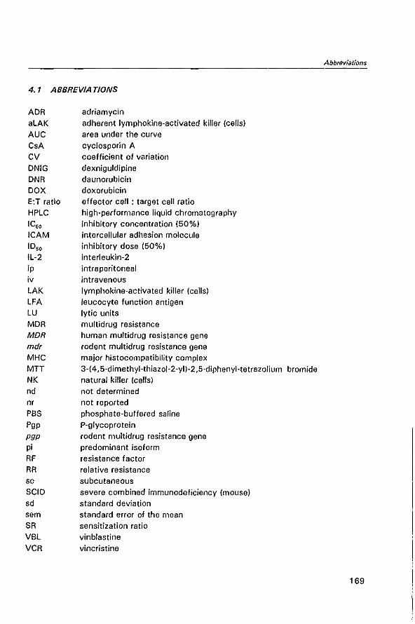
4.1 ABBREVIA TIONS
ADR alAK AUC CsA CV DNIG DNR DOX E:T ratio HPlC ICso ICAM 1050
Il·2 ip iv lAK lFA LU MDR MDR
adriamycin adherent Iymphokine'activated killer (cells) area under the curve cyclosporin A coefficient of variation dexniguldipine daunorubicin doxorubicin effector cell: target cell ratio high·performance liquid chromatography inhibitory concentration (50%) intercellular adhesion molecule inhibitory dose (50%) interleukin·2 intraperitoneal intravenous Iymphokine'activated killer (cells) leucocyte function antigen lytic units multidrug resistance human multidrug resistance gene
mdr rodent multidrug resistance gene MHC major histocompatibility complex MTT 3·(4,5·dimethyJ.thiazol·2·yl)·2,5·diphenyl·tetrazolium bromide NK natural killer (cells) nd not determined nr not reported PBS phosphate·buffered saline Pgp P'glycoprotein PUP rodent multidrug resistance gene pi predominant isoform RF resistance factor RR relative resistance sc subcutaneous SCIO severe combined immunodeficiency (mouse) sd standard deviation sem standard error of the mean SR sensitization ratio VBl vinblastine VCR vincristine
Abbreviations
169
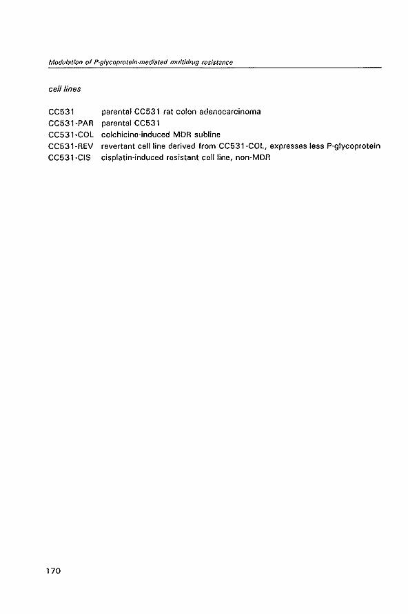
Modulatlon of P-gfycoprotein-medlated muftidrug resistance
eel/lines
CC531 parental CC531 rat colon adenocarcinoma
CC531-PAR parental CC531
CC531-COL colchicine-induced MDR subline CC531-REV revertant cell line derived from CC531-COL, expresses less P-glycoprotein
CC531-CIS cisplatin-induced resistant cell line, non-MDR
170
Naschrilt
4.2 NASCHRIFT
Wetenschappelijk onderzoek doe je niet aileen en dat maakt het me de zo boeiend en
leerzaam. Graag wi! ik iedereen bedanken die op een of andere wijze heeft bijgedragen
aan het tot standkomen van dit boekje. dat ik als bijnamen ook wei 'mijn levenswerk' of
'mijn molensteen' heb gegeven.
Allereerst Lex Eggermont, de initiator van het onderzoek en mijn copromotor. Hoewel
het onderzoek een andere richting heaft genomen dan in eerste instantie de bedoeling
was en geen immunotherapeutisch onderzoek is geworden en het met chirurgie al
helemaal weinig te maken heeft, ben je het tach blijven steunen; een teken van je brede
belangstelling. Je optimistische visie heeft me door meerdere dalen geholpen.
Ten tweede Richard Marquet, hoofd van het Laberatorium veer Experimentele
Chirurgie. Steeds aanwezig, altijd bereid een probleem aan te horen en te helpen
oplossen, en om manuscripten snel van commentaar te voorzien. Ik heb veel geleerd
van je nuchtere en relativerende kijk op wetenschappelijk onderzoek.
Prof. Gerrit Stater, mijn promotor, bedankt voer de kritische beoordelingen en de
mogelijkheid om in de inwendige geneeskunde, mijn huidige vak, te promoveren.
De mensen op het Laboraterium voor Experimentele Chirurgie: Nice Durante, voer de
vele experimenten die je samen met me en veer me hebt uitgevoerd en voor je
inleidingen in computerkunde; Amelie Sljma, voor de vele celkweken en in vitro testen
die je hebt gedaan en de technieken die ik van je heb mogen leren; Marcel Scheringa en
Fred Bonthuis, voor de inwijding in de proefdiertechnieken en het immunohistochemisch
onderzoek; de dierenverzorgers; de andere onderzoekers in het lab, voor de boeiende
wetenschappelijke discussies en het slappe geouwehoer. Zou het tot stand brengen van
een promotie ook op serendipiteit berusten?
, Antwerpen' bedankt voor de resistente cellijnen en de verschillende in vitro testen
die met name Eric Gheuens en Sylke van der Heyden hebben verricht. En zeker ook
Ernst de Bruijn, die gedurende de gehele periode van het onderzoek kritisch is blljven
meedenken; en Prof. Allan van Oosterorn.
Mensen van de Dr. Daniel den Hoed kliniek: Mieke Jonker, voor de histologische
beoordelingen; Walter Loos en Herman Kolker voer de bepalingen van dexniguldipine en
Jan Schellens voor zijn organisatie van dit onderdeel van het onderzoek.
Ik wil mijn paranymfen Bea Tanis en Wim Verwijs bedanken voor het werk dat ze bij
het schrijven van dit dankwoord nog grotendeels moeten gaan verrichten. Waar is onze
mooie studietijd, eh, ik bedoel studententijd geblevenl
171
Modulation of P-glycoproteln-medlated multidrug resistance
Bij deze past ook verontschuldinging aan aile vrienden die ik vaak verwaarloosd heb
omdat ik zonodig aan mijn onderzoek moest werken of lastig viel met verhalen over
mijn promotia dia nu toch echt heel dicht bij was, of er juist helemaal niat van zou
komen.
Mijn ouders bedankt voor hat fait dat za me habben gastimulaard om ta studaren.
Hoewel ik regelmatig hab gedacht dat ik liaver fruit had willen plukken dan op ean
studie of onderzoek ta zwoegen. Wat nu gebaur!. is ook oogsten.
En Marian, voor alie geduld an steun en liefde an nog heel veel maar.
172
Publlcations
4.3 PUBLICATIONS OF THE AUTHOR
full papers
Van de Vrie W, Gheuens EEO, Durante NMC, de Bruijn EA, Marquet Rl, van Oosterom A, Eggermont AMM. In vitro and in vivo chemosensitizing effect of cyclosporin A on an intrinsic multidrug-resistant rat colon tumour. J Cancer Res Clin Onco11993; 119: 609-14
Van de Vrie W, van der Heyden SAM, Gheuens EEO, Bijma AM, de Bruijn EA, Marquet Rl, van Oosterom AT, Eggermont AMM. Drug resistance in rat colon cancer cell lines is associated with minor changes in susceptibility to cytotoxic cells. Cancer Immunollmmunother 1993; 37: 337·42
Van de Vrie W, Eggermont AMM, van Putten Wl, Wiggers Th. Therapeutic lymphadenectomy in melanomas of the head and neck. Head Neck 1993; 15: 377·81
Van de Vrie W, Jonker AM, Marquet Rl, Eggermont AMM. The chemosensitizer cyclosporin A enhances the toxic side-effects of doxorubicin in the rat. J Cancer Res Clin Oncol 1994; 120: 533·8
Van de Vrie W, van Geel AN, Tjong Joe Wai R, Wijthoff SJM, Borel Rinkes IHM, Wiggers Th. Directe reconstructie van de borst na mastectomie. Een nieuwe strategie in de behandeling van het mammacarcinoom? Ned Tijdschr Geneeskd 1994; 138: 1949-53
Van de Vrie W, Schellens JHM, laos WJ, Kolker HJ, Verwey J, Stoter G, Durante NMC, Eggermont AMM. Modulation of multi drug resistance with dexniguldipine hydrochloride (88509-035) in the CC531 rat colon carcinoma model. J Cancer Res Clin Oncol 1996; 122: 403·8
Van de Vrje W, Baggen MGA, Janssen fMC, Ouwendijk RJTh. Acute pancreatitis door het chylomicronemie-syndroom. Ned TJjdschr Geneeskd 1996; 140: 34-6
Van de Vrie W, Marquet Rl, Eggermont AMM. Cyclosporin A enhances locoregional metastasis of the CC531 rat colon tumor. J Cancer Res Clin Onco/1997; 123: 21·4
Van der Heyden S, Gheuens E, van de Vrie W, van Bockstaele D, van Oosterom A, Egger· mont A, de 8ruijn E. 5' -Deoxy-5-fluorouridine increases daunorubicin uptake in multidrug-resistant cells and its activity is related with P-gp 170 expression. Jpn J Cancer Res 1994; 85: 13·6
Tjong Joe Wai R, van Geel AN, Wiggers Th, van de Vrie W, van Wersch A. Directe reconstructie na mastectomie. IKR-bulletin 1994; 19: 60-1
Schellens JHM, van de Vrie W, Loos WJ, Kolker HJ, Verweij J, Stater G, Durante NMC, Eggermont AMM. Pharmacokinetics of the MDR-reversing drug dexniguldipine and its pyridine metabolite M-1 in plasma, tumor and renal tissue in tumor bearing WAG-RIJ rats. Cancer Chernother Pharmacal (in press)
173
Modulation of P-gfycoprotein-mediated multidrug resistance
abstracts
Van de Vrie W, Gheuens EEO, Durante NMC, de Bruijn EA, Marquet RL, van Oosterom AT, Eggermont AMM. A syngeneic in vivo model of a multidrug resistant colon carcinoma, CC531, in the WAG rat. Proc Am Assoc Cancer Res 1991; 32: 365, abstr 2170
Van de Vrie W, Gheuens EEO, Durante NMC, de Bruijn EA, Marquet RL, van Oosterom AT, Eggermont AMM. In vitro and in vivo chemosensitizing effect of cyclosporin-A on an intrinsic multidrug resistant rat colon tumor. J Cancer Res Clin Oneol 1991; 117, Suppl.lII: S111, abstr E18
Van de Vrie W, Gheuens EEO, Bijma A, de Bruijn EA, Marquet RL, van Oosterom AT, Eggermont AMM. Sensitivity of different drug resistant cell lines to ALAK cell lysis. Ann Onco/1992; 3, Suppl.1: 81, abstr 091
Van de Vrie W, Gheuens EEO, Durante NMC, de Bruijn EA, Marquet RL, van Oosterom AT, Eggermont AMM. Cyc/osporin·A and its analog PSC 833 can reverse intrinsic multidrug resistance in a rat colon carcinoma. Proc Am Assoc Cancer Res 1992; 33: 484, abstr 2893
Van de Vrie W, van Geel AN, Tjong Joe Wai R, Eggermont AMM, Wiggers Th. Primaira reconstructia van de borst na amputatie voor mammacarcinoom. Chirurgendagen '93 Samenvattingen 1993; 215, abstr 49
Van de Vrie W, Gheuens EEO, Marquet RL, de Bruljn EA, van Oosterom AT, Eggermont AMM. In vitro and in vivo reversal of MDR of colon cancer CC531 by cyc/osporin. Proe VI International Conference on Regional Cancer Treatment Wiesbaden, July 1993
Van de Vrie W, Middelkoop MPC, Dees A. Acute renal failure on the ICU of a general hospital. Feasibility and results of CVVH treatment. ge Nederlands Intensive Care Kongres. Utrecht, mel 1995
Van de Vrie W, Baggen MGA, Visser W, Derkx FHM, Ouwendijk RJTh. High renin and aldosterone but normal prostaglandins in a patient with the ovarian hyperstimulation syndrome. Neth J Med 1996; 48: A78, abstr 77
Elst HJ, Gheuens EEO, van de Vrie W, van Oosterom AT, Eggermont AMM, de Bruijn EA. Isolation of a multidrug resistant rat colon carcinoma cell line CC531 m4,+ + • J Cancer Res Clin Onco/1991; 117, Suppl.lIl: 594, abstr 813
Van Geel AN, Tjong Joe Wai R, Wiggers Th, van de Vrie W. Directe reconstructie van de mamma na mastectomie met een subpectorale siliconenprothese. Chirurgendagen '94 Samenvattingen 1994: 209, abstr H2.1
Tjong Joe Wai R, van Geel AN, Wijthoff SJM, Wiggers Th, van Wersch A, Borel Rinkes IHM, van de Vrie W. Voorkeur voor verticaal litteken bij directe reconstructie met subpectorale siliconprothesen: ervaringen met 50 patienten vanaf 1991. Ned Tijdschr Geneeskd 1995; 139: 253
174
Curriculum vitae
4.4 CURRICULUM VITAE AUCTORIS
Wim van de Vrie werd op 8 oktober 1960 geboren in Kattendijke. In 1979 legde hij het
eindexamen Gymnasium-B at aan het Christelijk Lyceum voor Zeeland te Goes. Hierna
studeerde hij gedurende een jaar Psychologie aan de Vrije Universiteit in Amsterdam en
werd het propedeutisch examen algelegd.
Van 1980 tot 1989 volgde hij de studie Geneeskunde aan dezellde universiteit. In
deze periode was hij gedurende enige jaren studentlid van de taculteitsraad en de
facultaire onderwijscommissie en bestuurslid van de medische faculteitsvereniging. Hij
werkte in 1986 gedurende drie maanden mee aan medisch wetenschappelijk onderzoek
in Ndoungue, Kameroen. Tevens werd een cursus Medische Antropologie gevolgd aan
de Universiteit van Amsterdam. Op 8 september 1989 werd de artsenbul behaald.
In 1990 werd hij voor twee jaar wetenschappelijk onderzoeker aan de Dr. Daniel den
Hoed Kliniek te Rotterdam, gedetacheerd in het Laboratorium voor Experimentele
Chirurgie aan de Erasmus Universiteit Rotterdam, begeleiders Dr. A.M.M. Eggermont en
Dr. R.L. Marquet. In deze periode werd een belangrijk deel van de experimenten verricht
die beschreven zijn in dit proefschrift.
In 1992 werd hij assistent·geneeskundige niet·in-opleiding op de aldeling chirurgische
oncologie in Dr. Daniel den Hoed Kliniek te Rotterdam. In 1993 was hij gedurende drie
maanden gedetacheerd op de aldeling algemene heelkunde in het Zuiderziekenhuis te
Rotterdam. In september 1993 volgde een assistentschap inwendige geneeskunde in
het IKAZIA ziekenhuis te Rotterdam.
Op 1 januali 1994 werd hij assistent-geneeskundige in opleiding tot internist in het
IKAZIA ziekenhuis. opleider Dr. R.J.Th. Ouwendijk. De opleiding is vanal mei 1995
voortgezet in hat Academisch Ziekenhuis Dijkzigt te Rotterdam bij de opleider Prof.
J.H.P. Wilson.
175

Related Documents


