VSV-GP: a Potent Viral Vaccine Vector That Boosts the Immune Response upon Repeated Applications Reinhard Tober, a Zoltan Banki, a Lisa Egerer, a Alexander Muik, b * Sandra Behmüller, c Florian Kreppel, c Ute Greczmiel, d Annette Oxenius, d Dorothee von Laer, a Janine Kimpel a Division of Virology, Innsbruck Medical University, Innsbruck, Austria a ; Applied Virology and Gene Therapy Unit, Georg-Speyer-Haus, Frankfurt am Main, Germany b ; Division of Gene Therapy, University of Ulm, Ulm, Germany c ; Institute of Microbiology, ETH Zurich, Zurich, Switzerland d ABSTRACT Antivector immunity limits the response to homologous boosting for viral vector vaccines. Here, we describe a new, potent vac- cine vector based on replication-competent vesicular stomatitis virus pseudotyped with the glycoprotein of the lymphocytic cho- riomeningitis virus (VSV-GP), which we previously showed to be safe in mice. In mice, VSV and VSV-GP encoding ovalbumin (OVA) as a model antigen (VSV-OVA and VSV-GP-OVA) induced equal levels of OVA-specific humoral and cellular immune responses upon a single immunization. However, boosting with the same vector was possible only for VSV-GP-OVA as neutral- izing antibodies to VSV limited the immunogenicity of the VSV-OVA boost. OVA-specific cytotoxic T-lymphocyte (CTL) re- sponses induced by VSV-GP-OVA were at least as potent as those induced by an adenoviral state-of-the-art vaccine vector and completely protected mice in a Listeria monocytogenes challenge model. VSV-GP is so far the only replication-competent vaccine vector that does not lose efficacy upon repeated application. IMPORTANCE Although there has been great progress in treatment and prevention of infectious diseases in the past several years, effective vac- cines against some of the most serious infections, e.g., AIDS, malaria, hepatitis C, or tuberculosis, are urgently needed. Here, several approaches based on viral vector vaccines are under development. However, for all viral vaccine vectors currently in clin- ical testing, repeated application is limited by neutralizing antibodies to the vector itself. Here, we have exploited the potential of vesicular stomatitis virus pseudotyped with the glycoprotein of the lymphocytic choriomeningitis virus (VSV-GP) as a vaccine platform. VSV-GP is the first replication-competent viral vector vaccine that does not induce vector-specific humoral immunity, i.e., neutralizing antibodies, and therefore can boost immune responses against a foreign antigen by repeated applications. The vector allows introduction of various antigens and therefore can serve as a platform technology for the development of novel vaccines against a broad spectrum of diseases. D espite the progress in treatment and prevention of many in- fectious diseases, there is still an urgent need for effective vaccines against diseases like AIDS, malaria, hepatitis C, or tuber- culosis. Many successful vaccines are based on live-attenuated pathogens (e.g., those for polio, measles, mumps, and rubella) and usually confer strong and long-lasting immunity (1, 2). For those infectious agents that cannot be attenuated or where, despite at- tenuation, safety concerns remain (e.g., HIV), replication-compe- tent viral vector vaccines can be used. These viral vectors express the antigen of choice with the expectation that the induced immu- nity will be as strong and lasting as the response to live-attenuated vaccines. An ideal viral vector vaccine must fulfill several criteria: it must be safe, it must induce strong and durable cellular and humoral immune responses, there should be no preexisting im- munity in the human population, and ideally the vaccine should not lose its potency upon repeated application. All current repli- cation-competent viral vector vaccines lose efficacy upon repeated application but are very effective in a heterologous prime-boost regimen. Preexisting or vector-induced neutralizing antibodies can limit replication of the vector vaccine as shown, e.g., for ad- enoviral and poxvirus-based vector vaccines (3–5). Vesicular stomatitis virus (VSV), a negative-strand RNA virus of the rhabdovirus family, is a potent candidate vaccine vector. VSV-based vaccines can mount both strong humoral (6) and po- tent cellular immune responses against pathogens (7). Accord- ingly, VSV vectors have been shown to induce protective immu- nity against a large number of different pathogens in animal models, e.g., HIV, influenza virus, Marburg virus, and Ebola virus (8–10). VSV has several characteristics required for an ideal vac- cine vector: the general population lacks antibodies against VSV, which can interfere with vaccination efficacy (11); infections in humans are rare and mostly asymptomatic (12); there is no risk of genotoxicity as the virus does not integrate into the host genome and replicates in the cytoplasm (13); due to the helical structure of the capsid, the viral genome can accommodate large antigenic transgenes (14); the packaging capacity of VSV can even be in- creased further by using a semi-replication-competent vector sys- tem, consisting of a pair of VSV variants with trans-complement- Received 7 November 2013 Accepted 3 February 2014 Published ahead of print 19 February 2014 Editor: D. S. Lyles Address correspondence to Dorothee von Laer, [email protected], or Janine Kimpel, [email protected]. * Present address: Alexander Muik, Paul-Ehrlich-Institut, Langen, Germany. R.T., Z.B., D.V.L., and J.K. contributed equally to this work. Copyright © 2014, American Society for Microbiology. All Rights Reserved. doi:10.1128/JVI.03276-13 May 2014 Volume 88 Number 9 Journal of Virology p. 4897– 4907 jvi.asm.org 4897 on March 4, 2016 by PENN STATE UNIV http://jvi.asm.org/ Downloaded from

Welcome message from author
This document is posted to help you gain knowledge. Please leave a comment to let me know what you think about it! Share it to your friends and learn new things together.
Transcript
VSV-GP: a Potent Viral Vaccine Vector That Boosts the ImmuneResponse upon Repeated Applications
Reinhard Tober,a Zoltan Banki,a Lisa Egerer,a Alexander Muik,b* Sandra Behmüller,c Florian Kreppel,c Ute Greczmiel,d
Annette Oxenius,d Dorothee von Laer,a Janine Kimpela
Division of Virology, Innsbruck Medical University, Innsbruck, Austriaa; Applied Virology and Gene Therapy Unit, Georg-Speyer-Haus, Frankfurt am Main, Germanyb;Division of Gene Therapy, University of Ulm, Ulm, Germanyc; Institute of Microbiology, ETH Zurich, Zurich, Switzerlandd
ABSTRACT
Antivector immunity limits the response to homologous boosting for viral vector vaccines. Here, we describe a new, potent vac-cine vector based on replication-competent vesicular stomatitis virus pseudotyped with the glycoprotein of the lymphocytic cho-riomeningitis virus (VSV-GP), which we previously showed to be safe in mice. In mice, VSV and VSV-GP encoding ovalbumin(OVA) as a model antigen (VSV-OVA and VSV-GP-OVA) induced equal levels of OVA-specific humoral and cellular immuneresponses upon a single immunization. However, boosting with the same vector was possible only for VSV-GP-OVA as neutral-izing antibodies to VSV limited the immunogenicity of the VSV-OVA boost. OVA-specific cytotoxic T-lymphocyte (CTL) re-sponses induced by VSV-GP-OVA were at least as potent as those induced by an adenoviral state-of-the-art vaccine vector andcompletely protected mice in a Listeria monocytogenes challenge model. VSV-GP is so far the only replication-competent vaccinevector that does not lose efficacy upon repeated application.
IMPORTANCE
Although there has been great progress in treatment and prevention of infectious diseases in the past several years, effective vac-cines against some of the most serious infections, e.g., AIDS, malaria, hepatitis C, or tuberculosis, are urgently needed. Here,several approaches based on viral vector vaccines are under development. However, for all viral vaccine vectors currently in clin-ical testing, repeated application is limited by neutralizing antibodies to the vector itself. Here, we have exploited the potential ofvesicular stomatitis virus pseudotyped with the glycoprotein of the lymphocytic choriomeningitis virus (VSV-GP) as a vaccineplatform. VSV-GP is the first replication-competent viral vector vaccine that does not induce vector-specific humoral immunity,i.e., neutralizing antibodies, and therefore can boost immune responses against a foreign antigen by repeated applications. Thevector allows introduction of various antigens and therefore can serve as a platform technology for the development of novelvaccines against a broad spectrum of diseases.
Despite the progress in treatment and prevention of many in-fectious diseases, there is still an urgent need for effective
vaccines against diseases like AIDS, malaria, hepatitis C, or tuber-culosis. Many successful vaccines are based on live-attenuatedpathogens (e.g., those for polio, measles, mumps, and rubella) andusually confer strong and long-lasting immunity (1, 2). For thoseinfectious agents that cannot be attenuated or where, despite at-tenuation, safety concerns remain (e.g., HIV), replication-compe-tent viral vector vaccines can be used. These viral vectors expressthe antigen of choice with the expectation that the induced immu-nity will be as strong and lasting as the response to live-attenuatedvaccines. An ideal viral vector vaccine must fulfill several criteria:it must be safe, it must induce strong and durable cellular andhumoral immune responses, there should be no preexisting im-munity in the human population, and ideally the vaccine shouldnot lose its potency upon repeated application. All current repli-cation-competent viral vector vaccines lose efficacy upon repeatedapplication but are very effective in a heterologous prime-boostregimen. Preexisting or vector-induced neutralizing antibodiescan limit replication of the vector vaccine as shown, e.g., for ad-enoviral and poxvirus-based vector vaccines (3–5).
Vesicular stomatitis virus (VSV), a negative-strand RNA virusof the rhabdovirus family, is a potent candidate vaccine vector.VSV-based vaccines can mount both strong humoral (6) and po-tent cellular immune responses against pathogens (7). Accord-
ingly, VSV vectors have been shown to induce protective immu-nity against a large number of different pathogens in animalmodels, e.g., HIV, influenza virus, Marburg virus, and Ebola virus(8–10). VSV has several characteristics required for an ideal vac-cine vector: the general population lacks antibodies against VSV,which can interfere with vaccination efficacy (11); infections inhumans are rare and mostly asymptomatic (12); there is no risk ofgenotoxicity as the virus does not integrate into the host genomeand replicates in the cytoplasm (13); due to the helical structure ofthe capsid, the viral genome can accommodate large antigenictransgenes (14); the packaging capacity of VSV can even be in-creased further by using a semi-replication-competent vector sys-tem, consisting of a pair of VSV variants with trans-complement-
Received 7 November 2013 Accepted 3 February 2014
Published ahead of print 19 February 2014
Editor: D. S. Lyles
Address correspondence to Dorothee von Laer, [email protected], orJanine Kimpel, [email protected].
* Present address: Alexander Muik, Paul-Ehrlich-Institut, Langen, Germany.
R.T., Z.B., D.V.L., and J.K. contributed equally to this work.
Copyright © 2014, American Society for Microbiology. All Rights Reserved.
doi:10.1128/JVI.03276-13
May 2014 Volume 88 Number 9 Journal of Virology p. 4897– 4907 jvi.asm.org 4897
on March 4, 2016 by P
EN
N S
TA
TE
UN
IVhttp://jvi.asm
.org/D
ownloaded from
ing mutations (15); and finally, as VSV is an enveloped virus, viralglycoproteins are potentially presented in their natural, lipid en-velope-associated conformation on the viral surface, thereby ex-posing conformational epitopes not always found in the recombi-nant protein. Such conformational epitopes on the surface of aviral pathogen can be potent inducers of neutralizing antibodies.
However, VSV has two major limitations. Neutralizing anti-bodies against the VSV glycoprotein G are already induced afterthe first application, so that homologous boosting with VSV-based vector vaccines is ineffective (6). Therefore, for VSV, boost-ing can be achieved only by using multiple serotypes (16). Addi-tionally, VSV replicates in neurons and thereby can cause viralencephalitis (17, 18). Therefore, only attenuated VSV variants canbe used in humans, but these, however, have been found to be lesspotent (6, 19).
Our group has recently shown that neurotropism (studied inmice and human neurons) and neurotoxicity (mice) are com-pletely abolished by pseudotyping viruses with the glycoprotein(GP) of the lymphocytic choriomeningitis virus (LCMV) WE-HPI strain (20–22). In contrast to the LCMV Armstrong strain,the WE strain of LCMV is not neurotropic and is not significantlypathogenic (23). The LCMV-GP-WE pseudotype VSV-GP is notneurotoxic even at high intracranial doses in mice. Further exten-sive safety studies in mice have not shown any pathogenicity ofVSV-GP even at extremely high systemic doses (A. Muik, L. J.Stubbert, R. Z. Jahedi, Y. Geiss, C. Dold, R. Tober, A. Volk, S.Klein, U. Dietrich, B. Yadollahi, T. Falls, H. Miletic, D. Stojdl, J. C.Bell, and D. von Laer, submitted for publication).
Here, we evaluated the immunogenicity of a VSV-GP-basedvector vaccine using the model antigen ovalbumin (OVA) in mice.We found that VSV-GP was as immunogenic as VSV and adeno-viral vector vaccines upon prime, but VSV-GP was the only vectorthat did not induce neutralizing antibodies to the vector itself andtherefore did not lose immunogenicity upon repeated application.
MATERIALS AND METHODSEthics statement. Animal experiments were performed in compliancewith the Austrian national animal experimentation law (“Tierversuchs-gesetz”), and animal trial permission was granted by Austrian nationalauthorities (Bundesministerium für Wissenschaft und Forschung, no.66.011/154-II/3b/2011).
Cell lines and bacteria. BHK-21 cells (American Type Culture Collec-tion, Manassas, VA) were cultured in Glasgow minimum essential me-dium (GMEM) (Gibco, Carlsbad, CA) supplemented with 10% fetal calfserum (FCS; PAA Laboratories), 5% tryptose phosphate broth (Gibco,Carlsbad, CA), 100 units/ml penicillin (Gibco), and 0.1 mg/ml strepto-mycin (Gibco). Murine splenocytes were isolated from spleens of immu-nized C57BL/6 mice, treated with ACK buffer (Lonza, Basel, Switzerland)to lyse erythrocytes, washed with phosphate-buffered saline (PBS), andsubsequently cultured in RPMI 1620 medium (Lonza, Basel, Switzerland)supplemented with 10% FCS.
A Listeria monocytogenes strain expressing OVA134 –387 (Lm_OVA)(24) was kindly provided by Hao Shen, University of Pennsylvania, USA.Bacteria were grown in LB medium supplemented with 5 �g/ml erythro-mycin and 5 mg/liter glucose.
Viruses. VSV, VSV-GP, VSV-gfp, and VSV-GP-gfp were describedpreviously (25, 26) (Muik et al., submitted). VSV-OVA and VSV-GP-OVA were generated de novo. The OVA-enhanced green fluorescent pro-tein (eGFP) fusion cassette was inserted on position 5 in the viral genomesbetween G/GP and L genes. For construction of the OVA-eGFP fusioncassette, the full-length OVA sequence was amplified (GenBank sequenceaccession number NM_205152) using primers 5=-GCA TGG ACG AGC
TGT ACA AGA TGG GCT CCA TCG GCG CA-3= and 5=-CAA ACA TGAAGA ATC TGG CTA GAT CAT CAA GGG GAA ACA CAT CTG CC-3=.The eGFP sequence starting with a unique NheI restriction site and theintergenic region between the VSV G and L genes was obtained frompVSV-gfp using primers 5=-AAA GTA ACT CAA ATC CTG CTA GG-3=and 5=-TGC GCC GAT GGA GCC CAT CTT GTA CAG CTC GTC CATGC-3=. The sequence spanning the L gene up to a unique HpaI restrictionsite was amplified with primers 5=-GGC AGA TGT GTT TCC CCT TGATGA TCT AGC CAG ATT CTT CAT GTT TG-3= and 5=-GTA AAA AACTAT ACC CTT GAC TGG-3= from pVSV-gfp. To obtain the full-lengtheGFP_OVA sequence, a fusion PCR with these three PCR products andthe primers 5=-AAA GTA ACT CAA ATC CTG CTA GG-3= and 5=-GTAAAA AAC TAT ACC CTT GAC TGG-3= was performed. The resultingcassette was ligated via unique NheI/HpaI sites into pVSV-GP to obtainpVSV-GP-OVA. To create pVSV-OVA, LCMV-GP was replaced inpVSV-GP-OVA by VSV-G from pVSV-XN2 using the unique restrictionsites MluI and XhoI.
�M51-GP-OVA was created by exchange of wild-type M in VSV-GP-OVA with an M gene containing the �M51 mutation (kindly provided byOliver Ebert, Munich, Germany).
Recombinant VSVs were rescued and plaque purified as describedelsewhere (27). Titers were determined on confluent BHK-21 monolayersvia plaque assay.
The adenovirus vectors described here are replication-defective E1-deleted vectors based on human adenovirus type 5 (Ad5) and bear a hu-man cytomegalovirus (hCMV) promoter-driven expression cassette forsecreted, full-length ovalbumin or N-terminally truncated intracellularovalbumin. Cloning details can be obtained upon request. Vectors wereproduced on N52.E6 cells (28) and purified by double CsCl gradients.Vector titration was performed by a DNA-based slot blot procedure (29).
Preparation of cell lysates and Western blot analysis. BHK-21 cellswere infected with VSV-OVA or VSV-GP-OVA at a multiplicity of infec-tion (MOI) of 0.1, and cell lysates were prepared 24 h later. UninfectedBHK-21 cells were used as a control. Cells were lysed in ice-cold cell lysisbuffer (50 mmol/liter HEPES, pH 7.5; 150 mmol/liter NaCl; 1% TritonX-100; 2% aprotinin; 2 mmol/liter EDTA, pH 8.0; 50 mmol/liter sodiumfluoride; 10 mmol/liter sodium pyrophosphate; 10% glycerol; 1 mmol/liter sodium vanadate; and 2 mmol/liter Pefabloc SC) for 30 min. Subse-quently, cell lysates were centrifuged (13,000 rpm) for 10 min to removecell debris, and lysates were stored at �80°C until use.
SDS-PAGE of protein lysates was performed under standard reducingconditions on a 10% polyacrylamide gel. Proteins were electrophoreti-cally transferred to 0.45-�m nitrocellulose membranes (Whatman, Das-sel, Germany). Membranes were blocked with PBSTM (PBS containing5% skim milk and 0.1% Tween 20) and stained overnight at 4°C with aGFP-specific mouse monoclonal antibody (B-2, sc-9996; Santa Cruz Bio-technology, Santa Cruz, CA) diluted 1:1,000 in PBSTM. Detection wasperformed with a peroxidase-conjugated mouse IgG-specific antibodyfrom goat (Invitrogen, Carlsbad, CA), diluted 1:10,000 in PBSTM. Blotswere developed with enhanced chemiluminescence (ECL) (30). For beta-actin staining, blots were stripped with NaOH (0.1%) for 10 min at roomtemperature and blocked with PBSTM. Actin was stained with a beta-actin-specific monoclonal antibody from mouse (A2228; Sigma, Munich,Germany) diluted 1:5,000 in PBSTM and a secondary horseradish perox-idase-conjugated mouse IgG-specific antibody from goat and develop-ment with ECL.
TCID50 assay. For in vitro growth curves, virus titers were determinedusing a 50% tissue culture infective dose (TCID50) assay using the methodof Spearman-Kärber as described previously (31). Briefly, 10-fold serialdilutions of virus were prepared. One hundred microliters of each dilu-tion was added in quadruplicate to confluent BHK-21 cells in 96-wellplates and incubated for 24 to 48 h at 37°C until a cytopathic effect wasvisible. Numbers of infected wells were counted, and TCID50 values werecalculated.
Tober et al.
4898 jvi.asm.org Journal of Virology
on March 4, 2016 by P
EN
N S
TA
TE
UN
IVhttp://jvi.asm
.org/D
ownloaded from
Mouse experiments. C57BL/6 wild-type mice were purchased fromHarlan Laboratories (Rossdorf, Germany) and bred and maintained inthe animal facilities of the Innsbruck Medical University. All experimentswere performed in compliance with local animal experimentation guide-lines and approved by local authorities (Bundesministerium für Wissen-schaft und Forschung no. 66.011/154-II/3b/2011). Mice were immunizedintramuscularly with 1 � 106 PFU of VSV vectors or 2 � 109 vectorparticles (vp) of recombinant adenovirus vectors diluted in 50 �l PBS,respectively. Control animals received immunizations with 50 �l PBSwithout virus.
Serum transfer experiment. C57BL/6 mice were immunized on days0 and 26 with VSV-OVA or VSV-GP-OVA. For generation of nonim-mune sera, naive C57BL/6 mice were used. On day 7 post-boost immu-nization, mice were bled. Blood was allowed to coagulate for 1 h at roomtemperature, and subsequently, serum was obtained by centrifugation for5 min at 8,000 rpm. Serum was stored at �80°C till use. Three hundredfifty to 500 �l of serum was injected intravenously into naive C57BL/6mice. Mice were immunized 24 h later with the respective virus or PBS ascontrol. On day 7 postimmunization, OVA-specific cytotoxic T-lympho-cyte (CTL) responses were determined via tetramer and intracellular cy-tokine staining.
IFN-� enzyme-linked immunosorbent spot (ELISpot) assay. Themouse gamma interferon (IFN-�) ELISpot Plus kit (Mabtech, NackaStrand, Sweden) was used to quantify ovalbumin-specific CD8� T cells.The assay was performed according to the manufacturer’s protocol.Briefly, 2 � 105 splenocytes per well were incubated under standard cellculture conditions in 96-well plates precoated with a mouse IFN-�-spe-cific antibody. For stimulation of OVA-specific CTLs, 2.5 �g/ml of a pep-tide containing the ovalbumin-specific CTL epitope (SIINFEKL) wasadded. Anti-CD3/anti-CD28-coupled magnetic beads (32) were added tostimulate CTLs in positive-control wells, while negative-control wells re-mained unstimulated. After 20 h, the cells were removed. The plates weresubsequently incubated with a biotinylated IFN-�-specific antibody and astreptavidin-horseradish peroxidase (HRP)-conjugated secondary anti-body. The plates were developed with tetramethylbenzidine (TMB) sub-strate, and spots were counted in a CTL-Immunospot plate reader (CTL,Cleveland, OH). The assay was performed in duplicate, and counts ofunstimulated samples were subtracted from those of peptide-stimulatedsamples.
Intracellular cytokine staining. Intracellular production of the cyto-kines IFN-�, tumor necrosis factor alpha (TNF-�), and interleukin-2(IL-2) was examined by flow cytometric analysis. Briefly, 106 splenocyteswere incubated in the presence or absence of 2.5 �g/ml peptide (SIINFEKL) in standard medium supplemented with 1 �g/ml GolgiPlug (BD,Franklin Lakes, NJ, USA) for 6 h at 37°C in a humidified 6% CO2 incuba-tor. Cells were washed once with fluorescence-activated cell sorting(FACS) buffer (PBS supplemented with 1% fetal calf serum and 0.05%sodium azide) and stained with a peridinin chlorophyll protein (PerCp)-Cy5-conjugated CD8�-specific antibody (BD, Franklin Lakes, NJ, USA).Intracellular staining with fluorescein isothiocyanate (FITC)-conjugatedIFN-�-specific, phycoerythrin (PE)-conjugated IL-2-specific, and allo-phycocyanin (APC)-conjugated TNF-�-specific antibodies (BD), respec-tively, was performed using the Cytofix/Cytoperm fixation/permeabiliza-tion solution kit (BD) according to the manufacturer’s instructions.Samples were measured and analyzed using a FACSCanto II cytometer(BD) and DIVA software.
Tetramer staining. SIINFEKL-specific CD8� T cell counts weredetermined using APC-conjugated iTAg tetramers (iTAg tetramer/APC–H–2-kb OVA [SIINFEKL]; Beckman Coulter, catalog no. T03002).Splenocytes (106) were stained with PE-conjugated CD8�-specific, FITC-conjugated CD3�-specific antibodies (BD) and 2.5 �l of tetramer. Afterwashing with FACS buffer, cells were fixed with 3.7% formaldehyde. Theproportion of tetramer-positive CD8� T cells was determined by flowcytometric analysis using a FACSCanto II cytometer and DIVA software.
Anti-OVA IgG enzyme-linked immunosorbent assay (ELISA).Ninety-six-well microtiter plates were coated overnight at 4°C with 100 �lof ovalbumin protein (ICN Biomedicals Inc., Costa Mesa, CA) at a con-centration of 100 �g/ml in PBS. Nonspecific binding sites were blockedwith 100 �l PBSTM for 1 h at 37°C. All washing steps were performed withPBS containing 0.1% Tween 20 (PBST). Mouse plasma was diluted inPBST in 1:4 serial dilutions, starting with a 1:40 dilution. One hundredmicroliters of plasma dilution was added to the coated wells in duplicateand incubated for 1 h at 37°C. Detection was performed with a horserad-ish peroxidase-conjugated mouse IgG-specific antibody from goat (di-luted 1:10,000; Invitrogen, Carlsbad, CA) and Sure Blue TMB detectionreagent and TMB stop solution (KPL, Gaithersburg, MD). Plates wereanalyzed at 450 nm (signal) and 650 nm (background) on a model 680microplate reader (Bio-Rad, Hercules, CA) using Microplate Manager 5.2.1software (Bio-Rad, Hercules, CA). Endpoint titers were determined as thereciprocal maximum dilution at which the mean optical density at 450 nm(OD450) � OD650 of duplicates was greater than the mean OD450 � OD650
plus 2 standard deviations of naive sera.For absolute quantification, a standard curve was generated using
2-fold serial dilutions (128 ng to 0.125 ng per well) of an ovalbumin-specific monoclonal antibody from mouse (A6075; Sigma).
LCMV-GP1 IgG ELISA. ELISA plates (PS-Microplate, 96 wells;Greiner-Bio One) were coated overnight at 4°C with F(ab)2 fragments ofanti-human IgG (Jackson ImmunoResearch) diluted 1:800 in 0.1 M so-dium carbonate buffer (pH 9.6). After a 2-h blocking step at room tem-perature with 2% bovine serum albumin (BSA)-PBS, the F(ab)2 frag-ments were loaded with 100 �l/well of purified Fc-LCMV-GP1 (33) andincubated for 1 h at room temperature. On a parallel plate, plasma sam-ples were prediluted 1:8 in 0.1% BSA-PBS and a 3-fold dilution series wasperformed. A total of 50 �l/well was applied to the Fc-LCMV-GP1-satu-rated plate and incubated for 1 h at room temperature. LCMV-GP1-specific IgG antibodies were detected with anti-mouse IgG-�-HRP(Sigma) diluted 1:100 in 0.1% BSA-PBS. Plates were developed with 100�l/well of a solution containing 0.2 mg/ml 2,2=-azino-di-(3-ethylbenz-thiazoline sulfonic acid), 0.1 M NaH2PO4, 0.04% H2O2, pH 4. Plates wereread at 405 nm in a Victor3 reader (Wallac 1420; PerkinElmer). Betweeneach step, plates were washed five times with PBST. Titers were deter-mined as the reciprocal maximum dilution at which the OD405 wasgreater than the 2-fold OD405 signal of the naive plasma.
Antiadenovirus ELISA. Of a purified E1-deleted Ad5 vector without atransgene expression cassette, 6 � 108 vp were coated in 0.2 M Na2CO3-NaHCO3, pH 9.5, overnight in 96-well Maxisorp plates (Nunc). Blockingwas performed for 1 h at room temperature with 3% (wt/vol) BSA in PBS.After blocking, serial dilutions of the mouse sera in blocking buffer weretransferred into the wells and incubated for 2 h at room temperature.Detection was performed using a horseradish peroxidase-conjugatedmouse IgG-specific antibody from goat (BD 554002) (incubation of a1:2,000 dilution for 1 h at 37°C). Plates were analyzed at 491 nm (signal)and 620 nm (background).
VSV neutralization assay. The VSV neutralization assay was per-formed as described elsewhere (34). Briefly, mouse plasma samples werediluted in 2-fold serial dilutions in PBS, starting with a 1:10 dilution. Onehundred PFU of VSV-GFP or VSV-GP-GFP diluted in 50 �l serum-freeGMEM was mixed with 50 �l of plasma dilution in duplicate samples, andmixtures were incubated at 37°C for 1 h. Then, samples were transferredto 96-well plates containing a monolayer of confluent BHK-21 cells in 100�l complete GMEM. Plates were incubated at 37°C, 6% CO2, for 2 to 3days and analyzed for cytopathic effect. Neutralizing titers are given as thehighest plasma dilution which completely inhibited a VSV-GFP- or VSV-GP-GFP-induced cytopathic effect.
Lm_OVA challenge. Immunized mice were intravenously challengedon day 7 postboost with 1 � 105 CFU of Listeria monocytogenes expressingOVA (Lm_OVA). On day 3 postchallenge, mice were sacrificed andsplenocytes were isolated. Tenfold serial dilutions of splenocytes wereperformed in PBS. Dilutions were plated in duplicate on LB plates sup-
VSV-GP as Vaccine Vector
May 2014 Volume 88 Number 9 jvi.asm.org 4899
on March 4, 2016 by P
EN
N S
TA
TE
UN
IVhttp://jvi.asm
.org/D
ownloaded from
plemented with 5 �g/ml erythromycin and 5 mg/liter glucose. After 2days, colonies were counted and the number of CFU per spleen was cal-culated.
Statistical analysis. Statistical analysis was performed using Graph-Pad Prism software (GraphPad Software, Inc., La Jolla, CA).
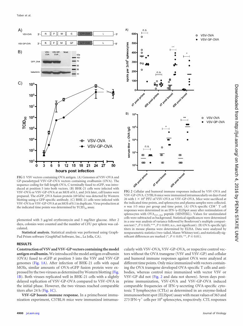
RESULTSConstruction of VSV and VSV-GP vectors containing the modelantigen ovalbumin. We introduced the model antigen ovalbumin(OVA) fused to eGFP at position 5 into the VSV and VSV-GPgenomes (Fig. 1A). After infection of BHK-21 cells with equalMOIs, similar amounts of OVA-eGFP fusion protein were ex-pressed by the two viruses as determined by Western blotting (Fig.1B). Both viruses replicated well in BHK-21 cells with a slightlydelayed replication of VSV-GP-OVA compared to VSV-OVA inthe initial phase. However, the two viruses reached comparabletiters after 24 h (Fig. 1C).
VSV-GP boosts immune response. In a prime/boost immu-nization experiment, C57BL/6 mice were immunized intramus-
cularly with VSV-OVA, VSV-GP-OVA, or respective control vec-tors without the OVA transgene (VSV and VSV-GP) and cellularand humoral immune responses against OVA were analyzed atdifferent time points. Only mice immunized with vectors contain-ing the OVA transgene developed OVA-specific T cells and anti-bodies, whereas control mice immunized with vector VSV orVSV-GP did not (Fig. 2 and data not shown). Seven days post-prime immunization, VSV-OVA and VSV-GP-OVA inducedcomparable frequencies of IFN-�-secreting OVA-specific cyto-toxic T lymphocytes (CTLs) as determined in an enzyme-linkedimmunosorbent spot (ELISpot) assay with mean values of 363 and273 IFN-�� cells per 106 splenocytes, respectively. CTL responses
FIG 1 VSV vectors containing OVA antigen. (A) Genomes of VSV-OVA andGP-pseudotyped VSV-GP-OVA vectors containing ovalbumin (OVA). Thesequence coding for full-length OVA, C terminally fused to eGFP, was intro-duced at position 5 into both vectors. (B) BHK-21 cells were infected withVSV-OVA or VSV-GP-OVA at an MOI of 0.1, and 24 h later, cell lysates wereprepared. The eGFP_OVA fusion protein (69 kDa) was detected by Westernblotting using a GFP-specific antibody. (C) BHK-21 cells were infected withVSV-OVA or VSV-GP-OVA at an MOI of 0.1 in duplicate. Virus production atthe indicated time points was determined by TCID50 assay.
FIG 2 Cellular and humoral immune responses induced by VSV-OVA andVSV-GP-OVA. C57BL/6 mice were immunized intramuscularly on days 0 and26 with 1 � 106 PFU of VSV-OVA or VSV-GP-OVA. Mice were sacrificed atthe indicated time points, and splenocytes and plasma samples were collected.n was �5 mice per group and time point. (A) OVA-specific CD8� T cellresponses were determined in an IFN-�–ELISpot assay after restimulation ofsplenocytes with OVA257–264 peptide (SIINFEKL). Values for unstimulatedcells were subtracted as background. Statistical significances were determinedin a one-way analysis of variance followed by Bonferroni’s multiple compari-son test (*, P � 0.05; ***, P � 0.001; n.s., not significant). (B) OVA-specific IgGtiters in mouse plasma were determined by ELISA. Data were analyzed bynonparametric statistics (two-tailed, Mann-Whitney test), and statistically sig-nificant differences are marked (*, P � 0.05; **, P � 0.01).
Tober et al.
4900 jvi.asm.org Journal of Virology
on March 4, 2016 by P
EN
N S
TA
TE
UN
IVhttp://jvi.asm
.org/D
ownloaded from
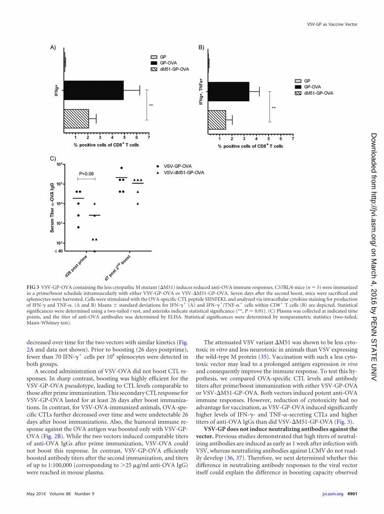
decreased over time for the two vectors with similar kinetics (Fig.2A and data not shown). Prior to boosting (26 days postprime),fewer than 70 IFN-�� cells per 106 splenocytes were detected inboth groups.
A second administration of VSV-OVA did not boost CTL re-sponses. In sharp contrast, boosting was highly efficient for theVSV-GP-OVA pseudotype, leading to CTL levels comparable tothose after prime immunization. This secondary CTL response forVSV-GP-OVA lasted for at least 26 days after boost immuniza-tions. In contrast, for VSV-OVA-immunized animals, OVA-spe-cific CTLs further decreased over time and were undetectable 26days after boost immunizations. Also, the humoral immune re-sponse against the OVA antigen was boosted only with VSV-GP-OVA (Fig. 2B). While the two vectors induced comparable titersof anti-OVA IgGs after prime immunization, VSV-OVA couldnot boost this response. In contrast, VSV-GP-OVA efficientlyboosted antibody titers after the second immunization, and titersof up to 1:100,000 (corresponding to 25 �g/ml anti-OVA IgG)were reached in mouse plasma.
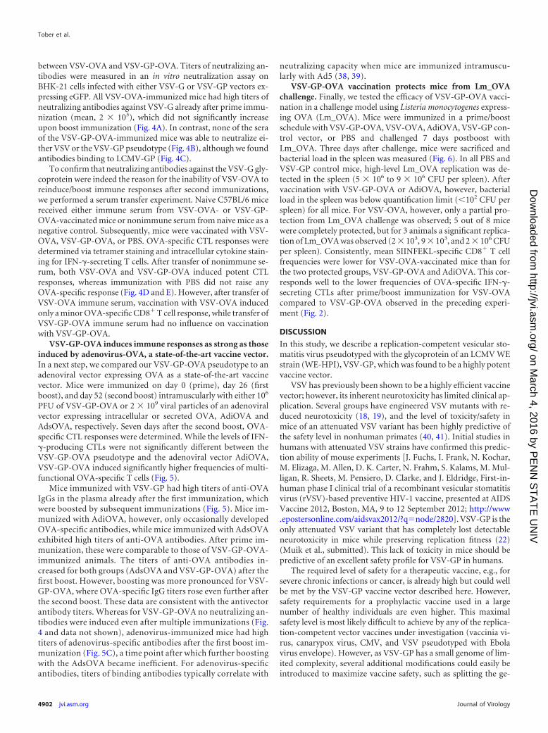
The attenuated VSV variant �M51 was shown to be less cyto-toxic in vitro and less neurotoxic in animals than VSV expressingthe wild-type M protein (35). Vaccination with such a less cyto-toxic vector may lead to a prolonged antigen expression in vivoand consequently improve the immune response. To test this hy-pothesis, we compared OVA-specific CTL levels and antibodytiters after prime/boost immunization with either VSV-GP-OVAor VSV-�M51-GP-OVA. Both vectors induced potent anti-OVAimmune responses. However, reduction of cytotoxicity had noadvantage for vaccination, as VSV-GP-OVA induced significantlyhigher levels of IFN-�- and TNF-�-secreting CTLs and highertiters of anti-OVA IgGs than did VSV-�M51-GP-OVA (Fig. 3).
VSV-GP does not induce neutralizing antibodies against thevector. Previous studies demonstrated that high titers of neutral-izing antibodies are induced as early as 1 week after infection withVSV, whereas neutralizing antibodies against LCMV do not read-ily develop (36, 37). Therefore, we next determined whether thisdifference in neutralizing antibody responses to the viral vectoritself could explain the difference in boosting capacity observed
FIG 3 VSV-GP-OVA containing the less cytopathic M mutant (�M51) induces reduced anti-OVA immune responses. C57BL/6 mice (n 5) were immunizedin a prime/boost schedule intramuscularly with either VSV-GP-OVA or VSV-�M51-GP-OVA. Seven days after the second boost, mice were sacrificed andsplenocytes were harvested. Cells were stimulated with the OVA-specific CTL peptide SIINFEKL and analyzed via intracellular cytokine staining for productionof IFN-� and TNF-�. (A and B) Means � standard deviations for IFN-�� (A) and IFN-��/TNF-�� cells within CD8� T cells (B) are depicted. Statisticalsignificances were determined using a two-tailed t test, and asterisks indicate statistical significance (**, P 0.01). (C) Plasma was collected at indicated timepoints, and the titer of anti-OVA antibodies was determined by ELISA. Statistical significances were determined by nonparametric statistics (two-tailed,Mann-Whitney test).
VSV-GP as Vaccine Vector
May 2014 Volume 88 Number 9 jvi.asm.org 4901
on March 4, 2016 by P
EN
N S
TA
TE
UN
IVhttp://jvi.asm
.org/D
ownloaded from
between VSV-OVA and VSV-GP-OVA. Titers of neutralizing an-tibodies were measured in an in vitro neutralization assay onBHK-21 cells infected with either VSV-G or VSV-GP vectors ex-pressing eGFP. All VSV-OVA-immunized mice had high titers ofneutralizing antibodies against VSV-G already after prime immu-nization (mean, 2 � 103), which did not significantly increaseupon boost immunization (Fig. 4A). In contrast, none of the seraof the VSV-GP-OVA-immunized mice was able to neutralize ei-ther VSV or the VSV-GP pseudotype (Fig. 4B), although we foundantibodies binding to LCMV-GP (Fig. 4C).
To confirm that neutralizing antibodies against the VSV-G gly-coprotein were indeed the reason for the inability of VSV-OVA toreinduce/boost immune responses after second immunizations,we performed a serum transfer experiment. Naive C57BL/6 micereceived either immune serum from VSV-OVA- or VSV-GP-OVA-vaccinated mice or nonimmune serum from naive mice as anegative control. Subsequently, mice were vaccinated with VSV-OVA, VSV-GP-OVA, or PBS. OVA-specific CTL responses weredetermined via tetramer staining and intracellular cytokine stain-ing for IFN-�-secreting T cells. After transfer of nonimmune se-rum, both VSV-OVA and VSV-GP-OVA induced potent CTLresponses, whereas immunization with PBS did not raise anyOVA-specific response (Fig. 4D and E). However, after transfer ofVSV-OVA immune serum, vaccination with VSV-OVA inducedonly a minor OVA-specific CD8� T cell response, while transfer ofVSV-GP-OVA immune serum had no influence on vaccinationwith VSV-GP-OVA.
VSV-GP-OVA induces immune responses as strong as thoseinduced by adenovirus-OVA, a state-of-the-art vaccine vector.In a next step, we compared our VSV-GP-OVA pseudotype to anadenoviral vector expressing OVA as a state-of-the-art vaccinevector. Mice were immunized on day 0 (prime), day 26 (firstboost), and day 52 (second boost) intramuscularly with either 106
PFU of VSV-GP-OVA or 2 � 109 viral particles of an adenoviralvector expressing intracellular or secreted OVA, AdiOVA andAdsOVA, respectively. Seven days after the second boost, OVA-specific CTL responses were determined. While the levels of IFN-�-producing CTLs were not significantly different between theVSV-GP-OVA pseudotype and the adenoviral vector AdiOVA,VSV-GP-OVA induced significantly higher frequencies of multi-functional OVA-specific T cells (Fig. 5).
Mice immunized with VSV-GP had high titers of anti-OVAIgGs in the plasma already after the first immunization, whichwere boosted by subsequent immunizations (Fig. 5). Mice im-munized with AdiOVA, however, only occasionally developedOVA-specific antibodies, while mice immunized with AdsOVAexhibited high titers of anti-OVA antibodies. After prime im-munization, these were comparable to those of VSV-GP-OVA-immunized animals. The titers of anti-OVA antibodies in-creased for both groups (AdsOVA and VSV-GP-OVA) after thefirst boost. However, boosting was more pronounced for VSV-GP-OVA, where OVA-specific IgG titers rose even further afterthe second boost. These data are consistent with the antivectorantibody titers. Whereas for VSV-GP-OVA no neutralizing an-tibodies were induced even after multiple immunizations (Fig.4 and data not shown), adenovirus-immunized mice had hightiters of adenovirus-specific antibodies after the first boost im-munization (Fig. 5C), a time point after which further boostingwith the AdsOVA became inefficient. For adenovirus-specificantibodies, titers of binding antibodies typically correlate with
neutralizing capacity when mice are immunized intramuscu-larly with Ad5 (38, 39).
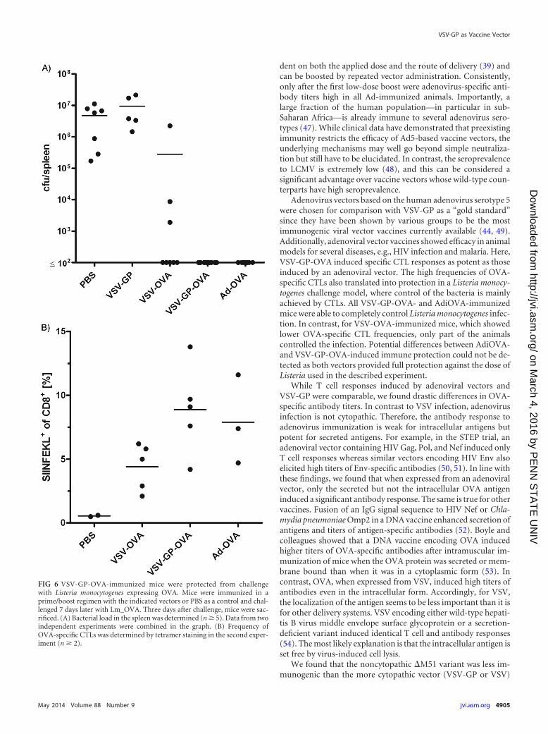
VSV-GP-OVA vaccination protects mice from Lm_OVAchallenge. Finally, we tested the efficacy of VSV-GP-OVA vacci-nation in a challenge model using Listeria monocytogenes express-ing OVA (Lm_OVA). Mice were immunized in a prime/boostschedule with VSV-GP-OVA, VSV-OVA, AdiOVA, VSV-GP con-trol vector, or PBS and challenged 7 days postboost withLm_OVA. Three days after challenge, mice were sacrificed andbacterial load in the spleen was measured (Fig. 6). In all PBS andVSV-GP control mice, high-level Lm_OVA replication was de-tected in the spleen (5 � 106 to 9 � 106 CFU per spleen). Aftervaccination with VSV-GP-OVA or AdiOVA, however, bacterialload in the spleen was below quantification limit (�102 CFU perspleen) for all mice. For VSV-OVA, however, only a partial pro-tection from Lm_OVA challenge was observed; 5 out of 8 micewere completely protected, but for 3 animals a significant replica-tion of Lm_OVA was observed (2 � 103, 9 � 103, and 2 � 106 CFUper spleen). Consistently, mean SIINFEKL-specific CD8� T cellfrequencies were lower for VSV-OVA-vaccinated mice than forthe two protected groups, VSV-GP-OVA and AdiOVA. This cor-responds well to the lower frequencies of OVA-specific IFN-�-secreting CTLs after prime/boost immunization for VSV-OVAcompared to VSV-GP-OVA observed in the preceding experi-ment (Fig. 2).
DISCUSSION
In this study, we describe a replication-competent vesicular sto-matitis virus pseudotyped with the glycoprotein of an LCMV WEstrain (WE-HPI), VSV-GP, which was found to be a highly potentvaccine vector.
VSV has previously been shown to be a highly efficient vaccinevector; however, its inherent neurotoxicity has limited clinical ap-plication. Several groups have engineered VSV mutants with re-duced neurotoxicity (18, 19), and the level of toxicity/safety inmice of an attenuated VSV variant has been highly predictive ofthe safety level in nonhuman primates (40, 41). Initial studies inhumans with attenuated VSV strains have confirmed this predic-tion ability of mouse experiments [J. Fuchs, I. Frank, N. Kochar,M. Elizaga, M. Allen, D. K. Carter, N. Frahm, S. Kalams, M. Mul-ligan, R. Sheets, M. Pensiero, D. Clarke, and J. Eldridge, First-in-human phase I clinical trial of a recombinant vesicular stomatitisvirus (rVSV)-based preventive HIV-1 vaccine, presented at AIDSVaccine 2012, Boston, MA, 9 to 12 September 2012; http://www.epostersonline.com/aidsvax2012/?qnode/2820]. VSV-GP is theonly attenuated VSV variant that has completely lost detectableneurotoxicity in mice while preserving replication fitness (22)(Muik et al., submitted). This lack of toxicity in mice should bepredictive of an excellent safety profile for VSV-GP in humans.
The required level of safety for a therapeutic vaccine, e.g., forsevere chronic infections or cancer, is already high but could wellbe met by the VSV-GP vaccine vector described here. However,safety requirements for a prophylactic vaccine used in a largenumber of healthy individuals are even higher. This maximalsafety level is most likely difficult to achieve by any of the replica-tion-competent vector vaccines under investigation (vaccinia vi-rus, canarypox virus, CMV, and VSV pseudotyped with Ebolavirus envelope). However, as VSV-GP has a small genome of lim-ited complexity, several additional modifications could easily beintroduced to maximize vaccine safety, such as splitting the ge-
Tober et al.
4902 jvi.asm.org Journal of Virology
on March 4, 2016 by P
EN
N S
TA
TE
UN
IVhttp://jvi.asm
.org/D
ownloaded from
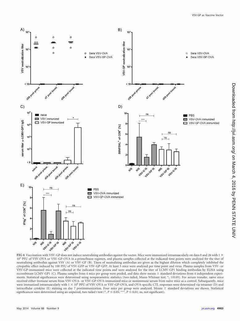
FIG 4 Vaccination with VSV-GP does not induce neutralizing antibodies against the vector. Mice were immunized intramuscularly on days 0 and 26 with 1 �106 PFU of VSV-OVA or VSV-GP-OVA in a prime/boost regimen, and plasma samples collected at the indicated time points were analyzed for the titer ofneutralizing antibodies against VSV (A) or VSV-GP (B). Titers of neutralizing antibodies are given as the highest dilution which completely inhibited thecytopathic effect induced by 100 PFU of VSV-GFP or VSV-GP-GFP. At least 5 mice were analyzed per time point and virus. Plasma samples from VSV- orVSV-GP-immunized mice were collected at the indicated time points and were analyzed for the titer of LCMV-GP1 binding antibodies by ELISA usingrecombinant LCMV-GP1 (C). Plasma samples from 4 mice per group were pooled, and data show means � standard deviations from 4 independent experi-ments. Statistical significances were determined using nonparametric statistics (two-tailed, Mann-Whitney test; *, �0.05). For serum transfer, naive micereceived either immune serum from VSV-OVA- or VSV-GP-OVA-immunized mice or nonimmune serum from naive mice as a control. Subsequently, micewere immunized intramuscularly with 1 � 106 PFU of VSV-OVA or VSV-GP-OVA, and OVA-specific CTL responses were determined via tetramer (D) andintracellular cytokine (E) staining on day 7 postimmunization. Four mice per group were analyzed. Means � standard deviations are shown. Statisticalsignificances were determined using an unpaired, two-tailed t test (*, P � 0.05; ***, P � 0.01; ns, not significant).
VSV-GP as Vaccine Vector
May 2014 Volume 88 Number 9 jvi.asm.org 4903
on March 4, 2016 by P
EN
N S
TA
TE
UN
IVhttp://jvi.asm
.org/D
ownloaded from

nome to generate a semireplicative virus system as described by uspreviously for VSV (15).
Among all replication-competent vector vaccines, VSV-GPwas found to be unique in the ability to boost the vaccine responseupon repeated applications. Accordingly, VSV-GP-OVA rein-duced OVA-specific CTL responses and boosted the titers ofOVA-specific antibodies. The lack of vector-neutralizing antibod-ies in VSV-GP-OVA-immunized mice was found to be the reasonfor VSV-GP-OVA’s ability to boost immune responses upon re-peated applications. Accordingly, serum transfer from VSV-im-munized mice reduced the subsequent T cell response to OVAafter VSV-OVA vaccination, while serum transfer from VSV-GP-immunized mice had no effect on the response to VSV-GP-OVAvaccination. Indeed, VSV is known to rapidly induce neutralizingantibodies (11), while neutralizing antibodies to LCMV are, if atall, induced only after chronic infection of 50 to 150 days (42). Byexchanging the glycoproteins between VSV and LCMV, Pin-
schewer and colleagues showed that the ability to induce neutral-izing antibodies is determined solely by the viral glycoprotein andnot the backbone (43).
Antivector immunity is an important issue for viral vector vac-cines. For those, currently in clinical testing (poxviruses, adeno-viruses, VSV, etc.), repeated application is limited by antivectorimmunity (44). There are several other candidates in preclinicaldevelopment. But among those, only a replication-deficientLCMV vector allows homologous boosting (45, 46). To circum-vent this problem, heterologous prime-boost regimes or differentserotypes of the same vector may be used.
Interestingly, we were able to boost anti-OVA antibody titersonce in mice immunized with AdsOVA (the OVA protein is se-creted). These data correlated with titers of adenovirus-specificantibodies in the plasma of the mice, which were low after prime.It is known that the induction of adenovirus-specific antibodies inmice by immunization with human Ad5 vectors is strongly depen-
FIG 5 VSV-GP-OVA is at least as potent as a state-of-the-art adenoviral vaccine vector. Mice (n 5) were immunized with VSV-GP-OVA, AdiOVA (expressingintracellular OVA), or AdsOva (expressing secreted OVA) in a prime/boost schedule with two boost immunizations. (A) Seven days after the second boostimmunization, mice were sacrificed and splenocytes were harvested. Cells were stimulated with the OVA-specific CTL epitope SIINFEKL and analyzed byintracellular cytokine staining for production of IFN-�, TNF-�, and IL-2. Means � standard deviations for IFN-��, IFN-��/TNF-��, and IFN-��/TNF-��/IL-2� cells within CD8� T cell population are shown. Statistical significances were determined using an unpaired, two-tailed t test (**, P � 0.01; *, P � 0.05; n.s.,not significant). (B) Plasma was collected at the indicated time points, and the titer of OVA-specific antibodies was determined by ELISA. Statistical significanceswere determined using nonparametric statistics (two-tailed, Mann-Whitney test; *, P � 0.05). (C) Mice (n � 4) were immunized with AdiOVA in a prime/boostschedule with two boost immunizations. Plasma was collected at indicated time points, and the titer of antiadenovirus antibodies was determined via ELISA.
Tober et al.
4904 jvi.asm.org Journal of Virology
on March 4, 2016 by P
EN
N S
TA
TE
UN
IVhttp://jvi.asm
.org/D
ownloaded from
dent on both the applied dose and the route of delivery (39) andcan be boosted by repeated vector administration. Consistently,only after the first low-dose boost were adenovirus-specific anti-body titers high in all Ad-immunized animals. Importantly, alarge fraction of the human population—in particular in sub-Saharan Africa—is already immune to several adenovirus sero-types (47). While clinical data have demonstrated that preexistingimmunity restricts the efficacy of Ad5-based vaccine vectors, theunderlying mechanisms may well go beyond simple neutraliza-tion but still have to be elucidated. In contrast, the seroprevalenceto LCMV is extremely low (48), and this can be considered asignificant advantage over vaccine vectors whose wild-type coun-terparts have high seroprevalence.
Adenovirus vectors based on the human adenovirus serotype 5were chosen for comparison with VSV-GP as a “gold standard”since they have been shown by various groups to be the mostimmunogenic viral vector vaccines currently available (44, 49).Additionally, adenoviral vector vaccines showed efficacy in animalmodels for several diseases, e.g., HIV infection and malaria. Here,VSV-GP-OVA induced specific CTL responses as potent as thoseinduced by an adenoviral vector. The high frequencies of OVA-specific CTLs also translated into protection in a Listeria monocy-togenes challenge model, where control of the bacteria is mainlyachieved by CTLs. All VSV-GP-OVA- and AdiOVA-immunizedmice were able to completely control Listeria monocytogenes infec-tion. In contrast, for VSV-OVA-immunized mice, which showedlower OVA-specific CTL frequencies, only part of the animalscontrolled the infection. Potential differences between AdiOVA-and VSV-GP-OVA-induced immune protection could not be de-tected as both vectors provided full protection against the dose ofListeria used in the described experiment.
While T cell responses induced by adenoviral vectors andVSV-GP were comparable, we found drastic differences in OVA-specific antibody titers. In contrast to VSV infection, adenovirusinfection is not cytopathic. Therefore, the antibody response toadenovirus immunization is weak for intracellular antigens butpotent for secreted antigens. For example, in the STEP trial, anadenoviral vector containing HIV Gag, Pol, and Nef induced onlyT cell responses whereas similar vectors encoding HIV Env alsoelicited high titers of Env-specific antibodies (50, 51). In line withthese findings, we found that when expressed from an adenoviralvector, only the secreted but not the intracellular OVA antigeninduced a significant antibody response. The same is true for othervaccines. Fusion of an IgG signal sequence to HIV Nef or Chla-mydia pneumoniae Omp2 in a DNA vaccine enhanced secretion ofantigens and titers of antigen-specific antibodies (52). Boyle andcolleagues showed that a DNA vaccine encoding OVA inducedhigher titers of OVA-specific antibodies after intramuscular im-munization of mice when the OVA protein was secreted or mem-brane bound than when it was in a cytoplasmic form (53). Incontrast, OVA, when expressed from VSV, induced high titers ofantibodies even in the intracellular form. Accordingly, for VSV,the localization of the antigen seems to be less important than it isfor other delivery systems. VSV encoding either wild-type hepati-tis B virus middle envelope surface glycoprotein or a secretion-deficient variant induced identical T cell and antibody responses(54). The most likely explanation is that the intracellular antigen isset free by virus-induced cell lysis.
We found that the noncytopathic �M51 variant was less im-munogenic than the more cytopathic vector (VSV-GP or VSV)
FIG 6 VSV-GP-OVA-immunized mice were protected from challengewith Listeria monocytogenes expressing OVA. Mice were immunized in aprime/boost regimen with the indicated vectors or PBS as a control and chal-lenged 7 days later with Lm_OVA. Three days after challenge, mice were sac-rificed. (A) Bacterial load in the spleen was determined (n � 5). Data from twoindependent experiments were combined in the graph. (B) Frequency ofOVA-specific CTLs was determined by tetramer staining in the second exper-iment (n � 2).
VSV-GP as Vaccine Vector
May 2014 Volume 88 Number 9 jvi.asm.org 4905
on March 4, 2016 by P
EN
N S
TA
TE
UN
IVhttp://jvi.asm
.org/D
ownloaded from
containing the wild-type M protein. Consistently, after intranasalor intramuscular immunization the HIV Env-specific CTL re-sponse was lower for a VSV-�M51 variant than for VSV withwild-type M protein (55). Likewise, a recent study showed that aVSV variant containing the �M51 protein induced lower titers ofantibodies against hepatitis B virus middle surface protein thanthe corresponding virus with wild-type M protein (54). The re-lease of antigen from cells killed by virus infection again seems tobe important for the induction of a potent humoral immune re-sponse.
VSV-GP is the first replicating viral vector vaccine that doesnot readily induce neutralizing antibodies to the vector itself andthus can repeatedly boost immune responses against a vaccineantigen. In future, this unique feature could potentially be con-ferred also on other enveloped viral vaccine vectors simply bypseudotyping these with LCMV GP protein.
ACKNOWLEDGMENTS
We kindly thank Hao Shen and Dirk Busch for providing Listeria mono-cytogenes expressing OVA and protocols. We are grateful to Katja Kotschand Cornelia Fabritius for their practical support. We thank ManuelaLunardon and Bettina Grosslercher for excellent technical support.
This work has been supported by the intramural funding program ofInnsbruck Medical University for young scientists, MUI-Start, project2012032006.
D.V.L. is an inventor of VSV-GP and a participator in the biotechcompany ViraTherapeutics GmbH, which holds intellectual propertyrights for VSV-GP. D.V.L. is listed as inventor on a patent applicationrelated to VSV-GP. For the other authors, no competing financial inter-ests exist.
REFERENCES1. Robert-Guroff M. 2007. Replicating and non-replicating viral vectors for
vaccine development. Curr. Opin. Biotechnol. 18:546 –556. http://dx.doi.org/10.1016/j.copbio.2007.10.010.
2. Manrique J, Piatak M, Lauer W, Johnson W, Mansfield K, Lifson J,Desrosiers R. 2013. Influence of mismatch of Env sequences on vaccineprotection by live attenuated simian immunodeficiency virus. J. Virol.87:7246 –7254. http://dx.doi.org/10.1128/JVI.00798-13.
3. Barouch DH, Pau MG, Custers JH, Koudstaal W, Kostense S, HavengaMJ, Truitt DM, Sumida SM, Kishko MG, Arthur JC, Korioth-SchmitzB, Newberg MH, Gorgone DA, Lifton MA, Panicali DL, Nabel GJ,Letvin NL, Goudsmit J. 2004. Immunogenicity of recombinant adeno-virus serotype 35 vaccine in the presence of pre-existing anti-Ad5 immu-nity. J. Immunol. 172:6290 – 6297.
4. Casimiro DR, Chen L, Fu TM, Evans RK, Caulfield MJ, Davies ME,Tang A, Chen M, Huang L, Harris V, Freed DC, Wilson KA, Dubey S,Zhu DM, Nawrocki D, Mach H, Troutman R, Isopi L, Williams D,Hurni W, Xu Z, Smith JG, Wang S, Liu X, Guan L, Long R, Trigona W,Heidecker GJ, Perry HC, Persaud N, Toner TJ, Su Q, Liang X, Youil R,Chastain M, Bett AJ, Volkin DB, Emini EA, Shiver JW. 2003. Compar-ative immunogenicity in rhesus monkeys of DNA plasmid, recombinantvaccinia virus, and replication-defective adenovirus vectors expressing ahuman immunodeficiency virus type 1 gag gene. J. Virol. 77:6305– 6313.http://dx.doi.org/10.1128/JVI.77.11.6305-6313.2003.
5. Santra S, Sun Y, Parvani JG, Philippon V, Wyand MS, Manson K,Gomez-Yafal A, Mazzara G, Panicali D, Markham PD, Montefiori DC,Letvin NL. 2007. Heterologous prime/boost immunization of rhesusmonkeys by using diverse poxvirus vectors. J. Virol. 81:8563– 8570. http://dx.doi.org/10.1128/JVI.00744-07.
6. Roberts A, Buonocore L, Price R, Forman J, Rose JK. 1999. Attenuatedvesicular stomatitis viruses as vaccine vectors. J. Virol. 73:3723–3732.
7. Haglund K, Leiner I, Kerksiek K, Buonocore L, Pamer E, Rose JK. 2002.High-level primary CD8(�) T-cell response to human immunodeficiencyvirus type 1 gag and env generated by vaccination with recombinant ve-sicular stomatitis viruses. J. Virol. 76:2730 –2738. http://dx.doi.org/10.1128/JVI.76.6.2730-2738.2002.
8. Geisbert TW, Feldmann H. 2011. Recombinant vesicular stomatitisvirus-based vaccines against Ebola and Marburg virus infections. J. Infect.Dis. 204(Suppl 3):S1075–S1081. http://dx.doi.org/10.1093/infdis/jir349.
9. Rose NF, Marx PA, Luckay A, Nixon DF, Moretto WJ, Donahoe SM,Montefiori D, Roberts A, Buonocore L, Rose JK. 2001. An effective AIDSvaccine based on live attenuated vesicular stomatitis virus recombinants.Cell 106:539 –549. http://dx.doi.org/10.1016/S0092-8674(01)00482-2.
10. Schwartz JA, Buonocore L, Suguitan AL, Jr, Silaghi A, Kobasa D,Kobinger G, Feldmann H, Subbarao K, Rose JK. 2010. Potent vesicularstomatitis virus-based avian influenza vaccines provide long-term steril-izing immunity against heterologous challenge. J. Virol. 84:4611– 4618.http://dx.doi.org/10.1128/JVI.02637-09.
11. Wagner RR, Rose JK. 1996. Rhabdoviridae: the viruses and their replica-tion, p 1121–1136. In Fields BN, Knipe DM, Howley PM (ed), Fieldsvirology. Lippincott-Raven, Philadelphia, PA.
12. Stallknecht DE. 2000. VSV-NJ on Ossabaw Island, Georgia. The truth isout there. Ann. N. Y. Acad. Sci. 916:431– 436. http://dx.doi.org/10.1111/j.1749-6632.2000.tb05322.x.
13. Lichty BD, Power AT, Stojdl DF, Bell JC. 2004. Vesicular stomatitisvirus: re-inventing the bullet. Trends Mol. Med. 10:210 –216. http://dx.doi.org/10.1016/j.molmed.2004.03.003.
14. Haglund K, Forman J, Krausslich HG, Rose JK. 2000. Expression ofhuman immunodeficiency virus type 1 Gag protein precursor and enve-lope proteins from a vesicular stomatitis virus recombinant: high-levelproduction of virus-like particles containing HIV envelope. Virology 268:112–121. http://dx.doi.org/10.1006/viro.1999.0120.
15. Muik A, Dold C, Geiss Y, Volk A, Werbizki M, Dietrich U, von Laer D.2012. Semireplication-competent vesicular stomatitis virus as a novelplatform for oncolytic virotherapy. J. Mol. Med. 90:959 –970. http://dx.doi.org/10.1007/s00109-012-0863-6.
16. Rose NF, Roberts A, Buonocore L, Rose JK. 2000. Glycoprotein ex-change vectors based on vesicular stomatitis virus allow effective boostingand generation of neutralizing antibodies to a primary isolate of humanimmunodeficiency virus type 1. J. Virol. 74:10903–10910. http://dx.doi.org/10.1128/JVI.74.23.10903-10910.2000.
17. Johnson JE, Nasar F, Coleman JW, Price RE, Javadian A, Draper K, LeeM, Reilly PA, Clarke DK, Hendry RM, Udem SA. 2007. Neurovirulenceproperties of recombinant vesicular stomatitis virus vectors in non-human primates. Virology 360:36 – 49. http://dx.doi.org/10.1016/j.virol.2006.10.026.
18. Cooper D, Wright KJ, Calderon PC, Guo M, Nasar F, Johnson JE,Coleman JW, Lee M, Kotash C, Yurgelonis I, Natuk RJ, Hendry RM,Udem SA, Clarke DK. 2008. Attenuation of recombinant vesicular sto-matitis virus-human immunodeficiency virus type 1 vaccine vectors bygene translocations and g gene truncation reduces neurovirulence andenhances immunogenicity in mice. J. Virol. 82:207–219. http://dx.doi.org/10.1128/JVI.01515-07.
19. Clarke DK, Cooper D, Egan MA, Hendry RM, Parks CL, Udem SA.2006. Recombinant vesicular stomatitis virus as an HIV-1 vaccine vector.Springer Semin. Immunopathol. 28:239 –253. http://dx.doi.org/10.1007/s00281-006-0042-3.
20. Miletic H, Fischer YH, Neumann H, Hans V, Stenzel W, Giroglou T,Hermann M, Deckert M, Von Laer D. 2004. Selective transduction ofmalignant glioma by lentiviral vectors pseudotyped with lymphocyticchoriomeningitis virus glycoproteins. Hum. Gene Ther. 15:1091–1100.http://dx.doi.org/10.1089/hum.2004.15.1091.
21. Beyer WR, Miletic H, Ostertag W, von Laer D. 2001. Recombinantexpression of lymphocytic choriomeningitis virus strain WE glycopro-teins: a single amino acid makes the difference. J. Virol. 75:1061–1064.http://dx.doi.org/10.1128/JVI.75.2.1061-1064.2001.
22. Muik A, Kneiske I, Werbizki M, Wilflingseder D, Giroglou T, Ebert O,Kraft A, Dietrich U, Zimmer G, Momma S, von Laer D. 2011. Pseudotyp-ing vesicular stomatitis virus with lymphocytic choriomeningitis virus glyco-proteins enhances infectivity for glioma cells and minimizes neurotropism. J.Virol. 85:5679–5684. http://dx.doi.org/10.1128/JVI.02511-10.
23. Moskophidis D, Battegay M, van den Broek M, Laine E, Hoffmann-Rohrer U, Zinkernagel RM. 1995. Role of virus and host variables in viruspersistence or immunopathological disease caused by a non-cytolytic vi-rus. J. Gen. Virol. 76:381–391. http://dx.doi.org/10.1099/0022-1317-76-2-381.
24. Foulds KE, Zenewicz LA, Shedlock DJ, Jiang J, Troy AE, Shen H. 2002.Cutting edge: CD4 and CD8 T cells are intrinsically different in theirproliferative responses. J. Immunol. 168:1528 –1532.
Tober et al.
4906 jvi.asm.org Journal of Virology
on March 4, 2016 by P
EN
N S
TA
TE
UN
IVhttp://jvi.asm
.org/D
ownloaded from
25. Boritz E, Gerlach J, Johnson JE, Rose JK. 1999. Replication-competentrhabdoviruses with human immunodeficiency virus type 1 coats andgreen fluorescent protein: entry by a pH-independent pathway. J. Virol.73:6937– 6945.
26. Schnell MJ, Buonocore L, Whitt MA, Rose JK. 1996. The minimalconserved transcription stop-start signal promotes stable expression of aforeign gene in vesicular stomatitis virus. J. Virol. 70:2318 –2323.
27. Ebert O, Shinozaki K, Kournioti C, Park MS, Garcia-Sastre A, Woo SL.2004. Syncytia induction enhances the oncolytic potential of vesicularstomatitis virus in virotherapy for cancer. Cancer Res. 64:3265–3270. http://dx.doi.org/10.1158/0008-5472.CAN-03-3753.
28. Schiedner G, Hertel S, Kochanek S. 2000. Efficient transformation ofprimary human amniocytes by E1 functions of Ad5: generation of new celllines for adenoviral vector production. Hum. Gene Ther. 11:2105–2116.http://dx.doi.org/10.1089/104303400750001417.
29. Kreppel F, Biermann V, Kochanek S, Schiedner G. 2002. A DNA-basedmethod to assay total and infectious particle contents and helper viruscontamination in high-capacity adenoviral vector preparations. Hum.Gene Ther. 13:1151–1156. http://dx.doi.org/10.1089/104303402320138934.
30. Mruk DD, Cheng CY. 2011. Enhanced chemiluminescence (ECL) forroutine immunoblotting: an inexpensive alternative to commerciallyavailable kits. Spermatogenesis 1:121–122. http://dx.doi.org/10.4161/spmg.1.2.16606.
31. Kaerber G. 1931. 50% end-point calculation. Arch. Exp. Pathol. Pharma-kol. 162:480 – 483. http://dx.doi.org/10.1007/BF01863914.
32. Newrzela S, Cornils K, Li Z, Baum C, Brugman MH, Hartmann M,Meyer J, Hartmann S, Hansmann ML, Fehse B, von Laer D. 2008.Resistance of mature T cells to oncogene transformation. Blood 112:2278 –2286. http://dx.doi.org/10.1182/blood-2007-12-128751.
33. Eschli B, Zellweger RM, Wepf A, Lang KS, Quirin K, Weber J, Zinker-nagel RM, Hengartner H. 2007. Early antibodies specific for the neutral-izing epitope on the receptor binding subunit of the lymphocytic chorio-meningitis virus glycoprotein fail to neutralize the virus. J. Virol. 81:11650 –11657. http://dx.doi.org/10.1128/JVI.00955-07.
34. Altfeld M, Rosenberg ES, Shankarappa R, Mukherjee JS, Hecht FM,Eldridge RL, Addo MM, Poon SH, Phillips MN, Robbins GK, Sax PE,Boswell S, Kahn JO, Brander C, Goulder PJ, Levy JA, Mullins JI, WalkerBD. 2001. Cellular immune responses and viral diversity in individualstreated during acute and early HIV-1 infection. J. Exp. Med. 193:169 –180.http://dx.doi.org/10.1084/jem.193.2.169.
35. Jayakar HR, Whitt MA. 2002. Identification of two additional translationproducts from the matrix (M) gene that contribute to vesicular stomatitisvirus cytopathology. J. Virol. 76:8011– 8018. http://dx.doi.org/10.1128/JVI.76.16.8011-8018.2002.
36. Diallo JS, Vaha-Koskela M, Le Boeuf F, Bell J. 2012. Propagation,purification, and in vivo testing of oncolytic vesicular stomatitis virusstrains. Methods Mol. Biol. 797:127–140. http://dx.doi.org/10.1007/978-1-61779-340-0_10.
37. Hangartner L, Zinkernagel RM, Hengartner H. 2006. Antiviral antibodyresponses: the two extremes of a wide spectrum. Nat. Rev. Immunol.6:231–243. http://dx.doi.org/10.1038/nri1783.
38. Wortmann A, Vohringer S, Engler T, Corjon S, Schirmbeck R, ReimannJ, Kochanek S, Kreppel F. 2008. Fully detargeted polyethylene glycol-coated adenovirus vectors are potent genetic vaccines and escape frompre-existing anti-adenovirus antibodies. Mol. Ther. 16:154 –162. http://dx.doi.org/10.1038/sj.mt.6300306.
39. Croyle MA, Patel A, Tran KN, Gray M, Zhang Y, Strong JE, FeldmannH, Kobinger GP. 2008. Nasal delivery of an adenovirus-based vaccinebypasses pre-existing immunity to the vaccine carrier and improves theimmune response in mice. PLoS One 3:e3548. http://dx.doi.org/10.1371/journal.pone.0003548.
40. Mire CE, Miller AD, Carville A, Westmoreland SV, Geisbert JB, Mans-field KG, Feldmann H, Hensley LE, Geisbert TW. 2012. Recombinantvesicular stomatitis virus vaccine vectors expressing filovirus glycopro-teins lack neurovirulence in nonhuman primates. PLoS Negl. Trop. Dis.6:e1567. http://dx.doi.org/10.1371/journal.pntd.0001567.
41. Jenks N, Myers R, Greiner SM, Thompson J, Mader EK, Greenslade A,Griesmann GE, Federspiel MJ, Rakela J, Borad MJ, Vile RG, Barber GN,
Meier TR, Blanco MC, Carlson SK, Russell SJ, Peng KW. 2010. Safetystudies on intrahepatic or intratumoral injection of oncolytic vesicularstomatitis virus expressing interferon-beta in rodents and nonhuman pri-mates. Hum. Gene Ther. 21:451– 462. http://dx.doi.org/10.1089/hum.2009.111.
42. Planz O, Seiler P, Hengartner H, Zinkernagel RM. 1996. Specific cyto-toxic T cells eliminate B cells producing virus-neutralizing antibodies.Nature 382:726 –729. http://dx.doi.org/10.1038/382726a0.
43. Pinschewer DD, Perez M, Jeetendra E, Bachi T, Horvath E, HengartnerH, Whitt MA, de la Torre JC, Zinkernagel RM. 2004. Kinetics ofprotective antibodies are determined by the viral surface antigen. J. Clin.Invest. 114:988 –993. http://dx.doi.org/10.1172/JCI22374.
44. Small JC, Ertl HC. 2011. Viruses—from pathogens to vaccine carriers.Curr. Opin. Virol. 1:241–245. http://dx.doi.org/10.1016/j.coviro.2011.07.009.
45. Flatz L, Hegazy AN, Bergthaler A, Verschoor A, Claus C, Fernandez M,Gattinoni L, Johnson S, Kreppel F, Kochanek S, Broek M, Radbruch A,Levy F, Lambert PH, Siegrist CA, Restifo NP, Lohning M, OchsenbeinAF, Nabel GJ, Pinschewer DD. 2010. Development of replication-defective lymphocytic choriomeningitis virus vectors for the induction ofpotent CD8� T cell immunity. Nat. Med. 16:339 –345. http://dx.doi.org/10.1038/nm.2104.
46. Halbherr SJ, Brostoff T, Tippenhauer M, Locher S, Berger Rentsch M,Zimmer G. 2013. Vaccination with recombinant RNA replicon particlesprotects chickens from H5N1 highly pathogenic avian influenza virus.PLoS One 8:e66059. http://dx.doi.org/10.1371/journal.pone.0066059.
47. Xiang Z, Li Y, Cun A, Yang W, Ellenberg S, Switzer WM, Kalish ML,Ertl HC. 2006. Chimpanzee adenovirus antibodies in humans, sub-Saharan Africa. Emerg. Infect. Dis. 12:1596 –1599. http://dx.doi.org/10.3201/eid1210.060078.
48. Lledo L, Gegundez MI, Saz JV, Bahamontes N, Beltran M. 2003.Lymphocytic choriomeningitis virus infection in a province of Spain:analysis of sera from the general population and wild rodents. J. Med.Virol. 70:273–275. http://dx.doi.org/10.1002/jmv.10389.
49. Chen H, Xiang ZQ, Li Y, Kurupati RK, Jia B, Bian A, Zhou DM,Hutnick N, Yuan S, Gray C, Serwanga J, Auma B, Kaleebu P, Zhou X,Betts MR, Ertl HC. 2010. Adenovirus-based vaccines: comparison ofvectors from three species of adenoviridae. J. Virol. 84:10522–10532. http://dx.doi.org/10.1128/JVI.00450-10.
50. Buchbinder SP, Mehrotra DV, Duerr A, Fitzgerald DW, Mogg R, Li D,Gilbert PB, Lama JR, Marmor M, Del Rio C, McElrath MJ, CasimiroDR, Gottesdiener KM, Chodakewitz JA, Corey L, Robertson MN. 2008.Efficacy assessment of a cell-mediated immunity HIV-1 vaccine (the StepStudy): a double-blind, randomised, placebo-controlled, test-of-concepttrial. Lancet 372:1881–1893. http://dx.doi.org/10.1016/S0140-6736(08)61591-3.
51. McElrath MJ, De Rosa SC, Moodie Z, Dubey S, Kierstead L, Janes H,Defawe OD, Carter DK, Hural J, Akondy R, Buchbinder SP, RobertsonMN, Mehrotra DV, Self SG, Corey L, Shiver JW, Casimiro DR. 2008.HIV-1 vaccine-induced immunity in the test-of-concept Step Study: acase-cohort analysis. Lancet 372:1894 –1905. http://dx.doi.org/10.1016/S0140-6736(08)61592-5.
52. Svanholm C, Bandholtz L, Lobell A, Wigzell H. 1999. Enhancement ofantibody responses by DNA immunization using expression vectors me-diating efficient antigen secretion. J. Immunol. Methods 228:121–130.http://dx.doi.org/10.1016/S0022-1759(99)00086-1.
53. Boyle JS, Koniaras C, Lew AM. 1997. Influence of cellular location ofexpressed antigen on the efficacy of DNA vaccination: cytotoxic T lym-phocyte and antibody responses are suboptimal when antigen is cytoplas-mic after intramuscular DNA immunization. Int. Immunol. 9:1897–1906.http://dx.doi.org/10.1093/intimm/9.12.1897.
54. Cobleigh MA, Bradfield C, Liu Y, Mehta A, Robek MD. 2012. Theimmune response to a vesicular stomatitis virus vaccine vector is indepen-dent of particulate antigen secretion and protein turnover rate. J. Virol.86:4253– 4261. http://dx.doi.org/10.1128/JVI.05991-11.
55. Publicover J, Ramsburg E, Robek M, Rose JK. 2006. Rapid pathogenesisinduced by a vesicular stomatitis virus matrix protein mutant: viral patho-genesis is linked to induction of tumor necrosis factor alpha. J. Virol.80:7028 –7036. http://dx.doi.org/10.1128/JVI.00478-06.
VSV-GP as Vaccine Vector
May 2014 Volume 88 Number 9 jvi.asm.org 4907
on March 4, 2016 by P
EN
N S
TA
TE
UN
IVhttp://jvi.asm
.org/D
ownloaded from
Related Documents