Volume 13 - 2021 www.surgicalcosmetic.org.br SCIENTIFIC SUPPORT: Official publication of the Brazilian Society of Dermatology

Welcome message from author
This document is posted to help you gain knowledge. Please leave a comment to let me know what you think about it! Share it to your friends and learn new things together.
Transcript
��������������
Volume 13 - 2021
www.surgicalcosmetic.org.br
SCIENTIFIC SUPPORT:
Official publication of the Brazilian Society of Dermatology
1
APOIO CIENTÍFICO:
www.surgicalcosmetic.org.br/
ISSN-e 1984-8773
Original ArticleAuthors: Isabella Parente Almeida¹ Maria Isabel Ramos Saraiva¹,² Maria Cristina de Lorenzo Messina²,³ João Pereira Duprat4
Luiz Guilherme Martins Castro¹,²
¹ Oncoderma Clínica de Oncologia Cutânea, São Paulo (SP), Brazil.
² Hospital Alemão Oswaldo Cruz, Department of Cutaneous Oncology, São Paulo (SP), Brazil.
³ Hospital Ipiranga, Department of Dermatology, São Paulo (SP), Brazil.
4 AC Camargo Cancer Center, São Paulo (SP), Brazil.
Correspondence: Isabella Parente Almeida¹ Email: isabellaparente@hotmail.
com / [email protected]
Financial support: None Conflict of interest: None
Submitted on: 31/03/2021Accepted on: 16/09/2021
How to cite this article: Saraiva MIR, Almeida IP, Messina MCL, Duprat JP, Castro LGM. Sentinel Lymph Node Biopsy for Cutaneous Melanoma in a real life setting: analysis of 47 cases treated at a private clinic in Brazil. Surg Cosm Dermatol. 2021;13:e20210021.
Sentinel lymph node biopsy for cutaneous melanoma in a real life setting: analysis of 47 cases treated at a private clinic in Brazil Biópsia de linfonodo sentinela para melanoma cutâneo na vida real: análise de 47 casos tratados em clínica privada no Brasil
ABSTRACTBackground: Sentinel Lymph Node (SLN) status has been shown to be the strongest independent prognostic factor of cutaneous melanoma (CM) stage I-II patients. Few papers on CM at private clinics (PC) are available. Objective: To present clinical and histologic data, complications and frequency of SLN involvement in CM patients diagnosed and followed at a dermatology/cutaneous oncology PC in São Paulo/Brazil, who were submitted to SLNB. Methods: Retrospective, single-center cohort of patients who attended PC from June 1998 to Jan 2020. Electronic files were selected and analyzed. Minimum period for considering the patient eligible was 1 year. Results: 215 CM lesions were identified in 184 patients(1.2 melanoma/patient). Forty-seven patients (25.5%) were submitted to SLNB and 59 SLN for histologic examination (1.2 SLN/patient). 10,9% tested positive. SLN identification happened in 95.7%. In 38/47 (80,8%) patients single LBD was found, while multiple-LBD was found in 9/47(19.1%). Eighteen(72,0%) out of 25 trunk lesions drained to single basin, while in 7 patients multiple LBD was found. Complication rate was 6,0%. Conclusion: Percentage of CM patients that undergo SLNB, node positivity for metastasis, draining basins and complications in this study were similar to studies in northern hemisphere patients. Clinical and epidemiologic characteristics of CM patients differ markedly between PC and PHS patients.Keywords: Biopsy; Melanoma; Sentinel lymph node
RESUMOIntrodução: O status do linfonodo sentinela (LNS) tem se mostrado o mais importante fator prognóstico independen-te no melanoma cutâneo (MC) em estágio I-II. Poucos artigos sobre MC em clínicas privadas (CP) estão disponíveis. Objetivo: Apresentar dados clínicos e histológicos, complicações e frequência de envolvimento do LS em pacientes com MC acompanhados em CP de dermatologia/oncologia cutânea em São Paulo/Brasil, submetidos a biópsia de LS (BLNS). Métodos: Coorte retrospectiva e unicêntrica de pacientes atendidos em CP de junho/1998 a janeiro/2020. Prontuá-rios eletrônicos foram analisados. O período mínimo para considerar paciente elegível foi de um ano. Resultados: Identificamos 215 MC em 184 pacientes (1,2 melanoma/paciente). No total, 47 pacientes (25,5%) foram submetidos à BLNS e 59 LN à exame histológico (1,2 LNS/paciente), sendo que 10,9% foram positivo. A identificação do LNS ocorreu em 95,7%. Dezoito (72,0%) das 25 lesões do tronco drenavam para cadeia única, enquanto em 7 pacientes drenavam para cadeias múltiplas. A taxa de complicação foi de 6,0%. Conclusão: O percentual de pacientes com MC submetidos a BLNS, positividade de LS, cadeias de drenagem e complicações neste estudo foram semelhantes aos estudos em pacientes do hemisfério norte. As características clínicas e epidemiológicas dos pacientes com MC diferem acentuadamente entre os pacientes de CP e do serviço público de saúde.Palavras-chave: Biópsia; Linfonodo sentinela; Melanoma.
DOI: http://www.dx.doi.org/10.5935/scd1984-8773.2021130021
ISSN-e 1984-8773
Saraiva MIR, Almeida IP, Messina MCL, Duprat JP, Castro LGM
2
BACKGROUNDSentinel Lymph Node (SLN) status is the most decisive
independent prognostic factor of cutaneous melanoma (CM) stage I-II patients.1 Accurate assessment of the regional LN sta-tus by SLN biopsy (SLNB) is becoming even more critical in the era of novel effective adjuvant therapies for the microscopic nodal disease.2 Some points on SLNB’s role and benefit in this setting are still controversial.3
Most published papers on SLNB for CM analyze patients from the northern hemisphere and Australia, large hospitals, or public health system (PHS). Very few articles on CM patients diagnosed and followed at private clinics (PC) are available in the literature. 4-8 We have not been able to find papers specifically addressing CM patients from PC who underwent SLNB, which leaves information gaps about what happens in this context.
OBJECTIVEThis study aims to present clinical and histologic data, des-
cribing complications and frequency of SLN involvement in CM patients diagnosed and followed at a dermatology/cutaneous on-cology PC in São Paulo/Brazil. They were submitted to SLNB, and their data were compared with data from the literature.
METHODSA retrospective, single-center study selected and analyzed
the electronic files of a cohort of patients diagnosed with CM attending a PC from June 1998 to January 2020. Data collected consisted of gender, primary tumor’s anatomic location, melano-ma clinical type, Breslow thickness, and history of SLNB. Among those submitted to SLNB, we also assessed SLN status, lymph node basins drainage, number of excised SLN, surgical complica-tions associated with SLNB, and eventual local or distant relapses.
The same surgical team, consisting of dermatologic and oncologic surgeons, operated on all but seven patients at diffe-rent hospitals in São Paulo. Pathologists from the different hospi-tals where surgeries were performed determined SLN histologic status. After removal, SLN were submitted to serial sectioning and permanent preparations for histological and immunohisto-chemical examination, according to current recommendations at the time. SLN was identified using Tc-labeled radiopharma-ceutical preoperative lymphoscintigraphy and subsequent in-
traoperative detection with gamma probe associated or not with blue-dye.
Follow-up was also based on information contained in the electronic charts. The minimum period for considering the patient eligible was one year.
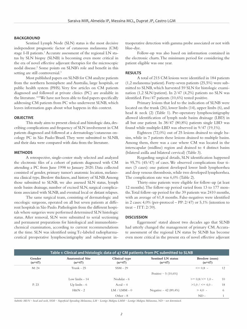
RESULTSA total of 215 CM lesions were identified in 184 patients
(1,2 melanoma/patient). Forty-seven patients (25,5%) were sub-mitted to SLNB, which harvested 59 SLN for histologic exami-nation (1,2 SLN/patient). In 2/47 (4,2%) patients no SLN was identified. Five/47 patients (10,6%) tested positive.
Primary lesions that led to the indication of SLNB were located on the trunk (26), lower limbs (14), upper limbs (6), and head & neck (2) (Table 1). Pre-operatory lymphoscintigraphy allowed identification of lymph node basins drainage (LBD) in all but one patient. In 38/47 (80,8%) patients single LBD was found while multiple-LBD was observed in 9/47 (19,1%).
Eighteen (72,0%) out of 25 lesions drained to single ba-sins, while in 7 patients these lesions drained to multiple basins. Among them, there was a case where CM was located in the interscapular (midline) region and drained to 4 distinct basins (bilateral axilla and bilateral cervical) (Table 3).
Regarding surgical details, SLN identification happened in 95,7% (45/47) of cases. We observed complications four ti-mes (3 cases): one patient developed lower limb lymphorrhea and deep venous thrombosis, while two developed lymphorrhea. The complication rate was 6,0% (Table 2).
Thirty-nine patients were eligible for follow-up (at least 12 months). The follow-up period varied from 13 to 177 mon-ths. Total follow-up period for the 39 patients was 2410 months, with an average of 61,8 months. False-negatives were identified in 2 cases: 4,0% (per-protocol – PP: 2/47) or 5,1% (intention to treat – ITT: 2/39).
DISCUSSIONEggermont1 stated almost two decades ago that SLNB
had utterly changed the management of primary CM. Accura-te assessment of the regional LN status by SLNB has become even more critical in the present era of novel effective adjuvant
Table 1: Clinical and histologic data of 47 CM patients from PC submitted to SLNBGender(n=47)
Anatomical Site(n=47)
Clinical type(n=47)
Sentinel LN status(n=47)
Breslow (mm)(n=47)
M: 24 Trunk - 25 SSM - 29 <= 0,8 - 12
Positive – 5 (10,6%)
Low limbs - 14 Nodular - 6 >= 0,8/<= 1,0 - 10
F: 23 Up limbs - 6 Acral – 4 >1,0 / <= 4,0 - 18
H&N - 2 LM / LMM - 0 Negative – 42 (89,4%) > 4,0 - 6
Other - 8 ND - 1
Subtitle: H&N – head and neck, SSM – Superficial Spreading Melanoma, LM – Lentigo Maligna LMM – Lentigo Maligna Melanoma, ND – not determined.
Surg Cosmet Dermatol. 2021;13:e20210021.
3
immuno and targeted therapies for the microscopic nodal disease.2
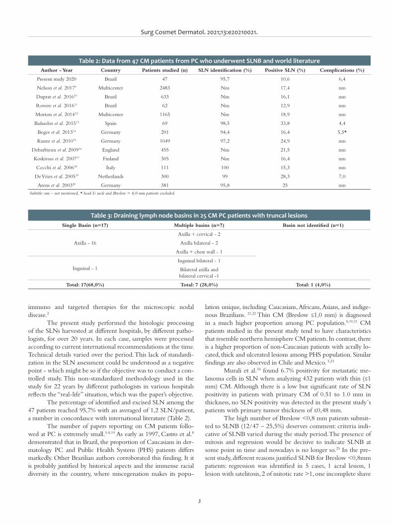
The present study performed the histologic processing of the SLNs harvested at different hospitals, by different patho-logists, for over 20 years. In each case, samples were processed according to current international recommendations at the time. Technical details varied over the period. This lack of standardi-zation in the SLN assessment could be understood as a negative point - which might be so if the objective was to conduct a con-trolled study. This non-standardized methodology used in the study for 22 years by different pathologists in various hospitals reflects the “real-life” situation, which was the paper’s objective.
The percentage of identified and excised SLN among the 47 patients reached 95,7% with an averaged of 1,2 SLN/patient, a number in concordance with international literature (Table 2).
The number of papers reporting on CM patients follo-wed at PC is extremely small.5-8,19 As early as 1997, Castro et al.8 demonstrated that in Brazil, the proportion of Caucasians in der-matology PC and Public Health System (PHS) patients differs markedly. Other Brazilian authors corroborated this finding. It it is probably justified by historical aspects and the immense racial diversity in the country, where miscegenation makes its popu-
lation unique, including Caucasians, Africans, Asians, and indige-nous Brazilians. 21,22 Thin CM (Breslow ≤1,0 mm) is diagnosed in a much higher proportion among PC population.8,19,21 CM patients studied in the present study tend to have characteristics that resemble northern hemisphere CM patients. In contrast, there is a higher proportion of non-Caucasian patients with acrally lo-cated, thick and ulcerated lesions among PHS population. Similar findings are also observed in Chile and Mexico. 5,23
Murali et al.24 found 6.7% positivity for metastatic me-lanoma cells in SLN when analyzing 432 patients with thin (≤1 mm) CM. Although there is a low but significant rate of SLN positivity in patients with primary CM of 0.51 to 1.0 mm in thickness, no SLN positivity was detected in the present study´s patients with primary tumor thickness of ≤0,48 mm.
The high number of Breslow <0,8 mm patients submit-ted to SLNB (12/47 – 25,5%) deserves comment: criteria indi-cative of SLNB varied during the study period. The presence of mitosis and regression would be decisive to indicate SLNB at some point in time and nowadays is no longer so.25 In the pre-sent study, different reasons justified SLNB for Breslow <0,8mm patients: regression was identified in 5 cases, 1 acral lesion, 1 lesion with satelitosis, 2 of mitotic rate >1, one incomplete shave
Table 2: Data from 47 CM patients from PC who underwent SLNB and world literatureAuthor - Year Country Patients studied (n) SLN identification (%) Positive SLN (%) Complications (%)
Present study 2020 Brazil 47 95,7 10,6 6,4
Nelson et al. 20179 Multicenter 2483 Nm 17,4 nm
Duprat et al. 201610 Brazil 633 Nm 16,1 nm
Rovere et al. 201611 Brazil 62 Nm 12,9 nm
Morton et al. 201412 Multicenter 1165 Nm 18,9 nm
Bañuelos et al. 201513 Spain 69 98,5 33,8 4,4
Beger et al. 201314 Germany 201 94,4 16,4 5,5*
Kunte et al. 201015 Germany 1049 97,2 24,9 nm
Debarbieux et al. 200916 England 455 Nm 21,5 nm
Koskivuo et al. 200717 Finland 305 Nm 16,4 nm
Cecchi et al. 200618 Italy 111 100 15,3 nm
De Vries et al. 200519 Netherlands 300 99 28,3 7,0
Arens et al. 200320 Germany 381 95,8 25 nm
Subtitle: nm – not mentioned, * head & neck and Breslow > 4,0 mm patients excluded.
Table 3: Draining lymph node basins in 25 CM PC patients with truncal lesionsSingle Basin (n=17) Multiple basins (n=7) Basin not identified (n=1)
Axilla - 16
Axilla + cervical - 2
Axilla bilateral - 2
Axilla + chest wall - 1
Inguinal - 1
Inguinal bilateral - 1
Bilateral axilla andbilateral cervical -1
Total: 17(68,0%) Total: 7 (28,0%) Total: 1 (4,0%)
Saraiva MIR, Almeida IP, Messina MCL, Duprat JP, Castro LGM
4
biopsy, where Breslow thickness was determined as “at least”, 2 cases where patients demanded to have the surgery for their own will.
Skip metastasesSkip metastases are one of the most significant drawbacks
of the method and can be found at different rates. The present study identified two cases. The first was a truncal CM with Bres-low thickness 4,5mm and Clark level IV draining to the left axilla. The SLN tested negative for metastases. Ten years later pulmonary and intestinal metastases were identified and quickly led the patient to death. The second was also a truncal CM with Breslow thickness 5,85 mm and vascular invasion draining to both inguinal basins. The three SLN tested negative for metasta-ses. Two years later, cerebral metastases were identified.
Draining basinsPreoperative lymphoscintigraphy has proved to be a vi-
tal planning instrument to guide complete removal of all SLNs, mainly when the primary lesion is located on the trunk, as fou-nd in the present paper, where 7 out of 25 (28,0%) patients had more than one drainage basin identified (Table 3). Truncal CM presents multiple-LBD from 17% to 34,6% of cases, especially when the lesion is mid-line. The present study associated double axillary/cervical and bilateral axillary drainage with upper back lesions.
The significance of multiple-LBD in truncal melanoma patients undergoing SLNB has long been debated. Currently, it it is widely accepted that multiple-LBD is not an independent risk factor for SLN metastasis and has no independent prognos-tic significance. Among matched pairs, multiple-LBD did not affect rates of LN metastasis overall survival, overall recurrence, locoregional recurrence, or distant recurrence.26-28
ComplicationsSLNB is an invasive procedure and is not free of risks.
Complications and sequelae are far less common when com-pared to complete LN dissection. Wrightson et al. reported on a total of 2120 patients submitted to SLNB. Overall, 96 (4.6%) of them developed major or minor complications. In contrast, 103 (23.2%) of 444 patients experienced complications when SLNB was followed by complete LN dissection, a number five times higher. 29
CM complication rates reported after SLNB are highly variable in the literature, ranging between 1.8% and 29.9%.30-32 In a systematic literature review, Moody et al.25 found an overall complication rate of 11.3% among SLNB patients, most tempo-rary. Incidence of infection was 2.9%; seroma, 5.1%; hematoma, 0.5%; lymphedema, 1.3%; and nerve injury, 0.3%.
The frequency of complications observed in the present study (6,0%) fits the interval described in Moody et al. review paper.25 One patient developed lower limb lymphorrhea and deep venous thrombosis, while two developed lymphorrhea.
CONCLUSIONDespite the relatively small number of patients studied,
we could observe that data obtained from CM patients at PC submitted to SLNB closely resembled those described in nor-thern hemisphere patients regarding the percentage of indivi-duals that undergo SLNB, node positivity for metastasis, age, draining basins, and complications.
Clinical and epidemiologic characteristics of CM pa-tients in Brazil differ markedly between PC and PHS popula-tion. The present study’s findings are restricted to CM Brazilian patients from PC and should not be extrapolated to Brazilian patients from the PHS. l
REFERENCES:1. Eggermont AM. Adjuvant therapy of malignant melanoma and the role
of sentinel node mapping. Recent Results Cancer Res. 2000;157:178-89.2. Ulmer A, Kofler L. Sentinel-node-Biopsie und Lymphknotendissektion
im Zeitalter neuer Systemtherapien des malignen Melanoms [Sentinel node biopsy and lymph node dissection in the era of new systemic the-rapies for malignant melanoma]. Hautarzt. 2019;70(11):864-9.
3. Bigby M, Zagarella S, Sladden M, Popescu CM. Time to reconsider the role of sentinel lymph node biopsy in melanoma. J Am Acad Dermatol. 2019;80(4):1168-71.
4. Stricklin SM, Stoecker WV, Malters JM, Drugge R, Oliviero M, Rabinovitz HS, et al. Melanoma in situ in a private practice setting 2005 through 2009: location, lesion size, lack of concern. J Am Acad Dermatol. 2012;67(3):e105-9.
5. Zemelman VB, Valenzuela CY, Sazunic I, Araya I. Malignant melanoma in Chile: different site distribution between private and state patients. Biol Res. 2014;47(1):34.
6. Cherian P, Tait CP. Melanoma in private practice: do dermatologists make a difference? Australas J Dermatol. 2009;50(4):257-60.
7. Barton MB, Gabriel GS, Frommer MS, Holt PE, Thompson JF. Surgical procedures for melanoma in public and private New South Wales hos-pitals, 2001-2002. ANZ J Surg. 2006;76(5):318-24.
8. Castro LGM, Toyama CL, Gomes AP, Freire MA, Brito T. Câncer de pele em clínica particular em São Paulo. An Bras Dermatol. 1996;6:471-6.
9. Nelson DW, Stern S, Elashoff DE, Elashoff R, Thompson JF, Mozzillo N, et al. Impact of time between diagnosis and SLNB on outcomes in cuta-neous melanoma. J Am Coll Surg. 2017;225(2):302-11.
10. Duprat JP, Brechtbülh ER, Costa de Sá B, Enokihara M, Fregnani JH, Landman G, et al. Absence of tumor-infiltrating lymphocyte is a repro-ducible predictive factor for sentinel lymph node metastasis: a mul-ticenter database study by the brazilian melanoma group. PLoS One. 2016;11(2):e0148160.
11. Rovere RK, A Sde L, Demarchi V, Stein CE. Sentinel lymph node in melanoma - a study conducted in the South of Brazil. Klin Onkol. 2016;29(4):274-8.
12. Morton DL, Thompson JF, Cochran AJ, Mozzillo N, Nieweg OE, Roses DF, et al. Final trial report of sentinel-node biopsy versus nodal observation in melanoma. N Engl J Med. 2014;370(7):599-609.
13. Bañuelos-Andrío L, Rodríguez-Caravaca G, López-Estebaranz JL, Rue-da-Orgaz JA, Pinedo-Moraleda F. Biopsia selectiva del ganglio centinela en melanoma: experiencia durante 8 años en un hospital universitario [Sentinel lymph node biopsy in melanoma: our experience over 8 years in a universitary hospital]. Cir Cir. 2015t;83(5):378-85.
Surg Cosmet Dermatol. 2021;13:e20210021.
5
14. Beger J, Hansel G, Krönert C, Fuchs M, Tanner C, Schönlebe J, et al. A 10-year analysis of primary cutaneous malignant melanoma with sentinel lymph node biopsy and long-term follow-up. Int J Dermatol. 2013;52(2):220-30.
15. Kunte C, Geimer T, Baumert J, Konz B, Volkenandt M, Flaig M, et al. Prog-nostic factors associated with sentinel lymph node positivity and effect of sentinel status on survival: an analysis of 1049 patients with cuta-neous melanoma. Melanoma Res. 2010;20(4):330-7.
16. Debarbieux S, Duru G, Dalle S, Béatrix O, Balme B, Thomas L. Sentinel lymph node biopsy in melanoma: a micromorphometric study relating to prognosis and completion lymph node dissection. Br J Dermatol. 2007;157(1):58-67.
17. Koskivuo I, Talve L, Vihinen P, Mäki M, Vahlberg T, Suominen E. Sentinel lymph node biopsy in cutaneous melanoma: a case-control study. Ann Surg Oncol. 2007;14(12):3566-74.
18. Cecchi R, De Gaudio C, Buralli L, Innocenti S. Lymphatic mapping and sentinel lymph node biopsy in the management of primary cutaneous melanoma: report of a single-centre experience. Tumori. 2006;92(2):113-7.
19. de Vries M, Jager PL, Suurmeijer AJ, Plukker JT, van Ginkel RJ, Hoeks-tra HJ. Schildwachtklierbiopsie bij het melanoom: prognostische be-tekenis en nadelen bij 300 patiënten [Sentinel lymph node biopsy for melanoma: prognostic value and disadvantages in 300 patients]. Ned Tijdschr Geneeskd. 2005;149(33):1845-51.
20. Arens A, Osinga J, Schwipper V, Schober O, Tilkorn H, Liebau J. Senti-nel-Lymphknoten-Dissektion beim malignen Melanom. Ein diagnostis-cher und therapeutischer Standard [Sentinel lymph node dissection in patients with malignant melanoma. Diagnostic and therapeutic stan-dards]. Chirurg. 2003;74(7):665-70.
21. Maia M, Russo C, Ferrari N, Ribeiro MCSA, Santos ABO. Reflexões em relação à epidemiologia do melanoma cutâneo no Brasil / Reflections regarding the epidemiology of cutaneous melanoma in Brazil An Bras Dermatol. 202;77(2):163-70.
22. 22 - Pena SD, Di Pietro G, Fuchshuber-Moraes M, Genro JP, Hutz MH, Kehdy Fde S, et al. The genomic ancestry of individuals from different geographical regions of Brazil is more uniform than expected. PLoS One. 2011;6(2):e17063.
AUTHORS' CONTRIBUTION:
Isabella Parente Almeida 0000-0002-6283-4065 Statistical analysis; study conception and planning; preparation and writing of the manuscript; data collection, analysis and interpretation; intel-lectual participation in propaedeutic and/or therapeutic management of studied cases; critical literature review.
Maria Isabel Ramos Saraiva 0000-0002-5043-489XStatistical analysis; study conception and planning; preparation and writing of the manuscript; data collection; analysis and interpretation; intel-lectual participation in propaedeutic and/or therapeutic management of studied cases; critical literature review.
Maria Cristina de Lorenzo Messina 0000-0002-8401-7349 Data collection, analysis and interpretation; active participation in research orientation; critical literature review; manuscript critical review.
João Pereira Duprat 0000-0001-8968-4506Data collection, analysis and interpretation; active participation in research orientation; intellectual participation in propaedeutic and/or thera-peutic conduct of studied cases; critical literature review; critical revision of the manuscript.
Luiz Guilherme Martins Castro 0000-0002-6269-1957Statistical analysis; approval of the final version of the manuscript; study design and planning; preparation and writing of the manuscript; data collection, analysis, and interpretation; active participation in research orientation; intellectual participation in propaedeutic and/or therapeutic conduct of studied cases; critical literature review; critical revision of the manuscript.
23. Saez-de-Ocariz M, Sosa-de-Martínez C, Duran-McKinster C, Orozco--Covarrubias L, Palacios-López C, Ruiz-Maldonado R. Cutaneous mela-noma in private vs. public practices of Mexican dermatologists. Int J Dermatol. 2008;47(6):637-9.
24. Murali R, Haydu LE, Quinn MJ, Saw RP, Shannon K, Spillane AJ, et al. Sentinel lymph node biopsy in patients with thin primary cutaneous melanoma. Ann Surg. 2012;255(1):128-33.
25. Castro LG, Bakos RM, Duprat Neto JP, Bittencourt FV, Di Giacomo TH, Serpa SS, et al. Brazilian guidelines for diagnosis, treatment and fol-low-up of primary cutaneous melanoma - Part II. An Bras Dermatol. 2016;91(1):49-58.
26. Howard JH, Ozao-Choy JJ, Hiles JM, Sim MS, Faries MB. Prognostic value of multiple draining lymph node basins in melanoma: a matched-pair analysis based on the John Wayne Cancer Institute Experience. Front Oncol. 2017;7:172.
27. Ribero S, Osella Abate S, Pasquali S, Rossi CR, Borgognoni L, Piazzalun-ga D, et al. Multiple lymph node basin drainage in trunk melanoma is not associated with survival of sentinel lymph node-positive patients. Dermatology. 2017;233(2-3):205-11.
28. Castro LGM, Duprat JP, Landman G. Dupla drenagem para cadeias lin-fonodais distintas, detectada por técnica de biópsia de linfonodo sen-tinela em pacientes com melanoma cutâneo: relato de dois casos. An Bras Dermatol. 2005;80(5):499-502.
29. Thomas JM. Lymphoedema in the observation and biopsy arms of MSLT-1. Ann Surg Oncol. 2011;18(Suppl 3):S311.
30. Wrightson WR, Wong SL, Edwards MJ, Chao C, Reintgen DS, Ross MI, et al. Complications associated with sentinel lymph node biopsy for me-lanoma. Ann Surg Oncol. 2003;10(6):676-80.
31. Moody JA, Ali RF, Carbone AC, Singh S, Hardwicke JT. Complications of sentinel lymph node biopsy for melanoma - A systematic review of the literature. Eur J Surg Oncol. 2017;43(2):270-7.
32. Wollina U, Langner D, Schönlebe J, Tanner C, Fuchs M, Nowak A. Senti-nel lymph node biopsy in early melanoma-comparison of two techni-ques for sentinel removal. Wien Med Wochenschr. 2017;167(5-6):100-3.
1
APOIO CIENTÍFICO:
www.surgicalcosmetic.org.br/
ISSN-e 1984-8773
Original articleAuthors:
César Augusto Zago Ferreira1
Vinícius de Souza1
Hélio Amante Miot2
Juliano Vilaverde Schmitt2
1 Hospital de Clínicas, Dermatology Service, Medical School, São Paulo State University, Botucatu (SP), Brazil.
2 Department of Infectology, Medical School, São Paulo State University, Botucatu (SP), Brazil.
2 Department of Infectology, Medical School, São Paulo State University, Botucatu (SP), Brazil.
Correspondence:Juliano Vilaverde SchmittEmail: [email protected]
Financial support: NoneConflict of interest: None
Submitted on: 12/01/2021 Approved on: 14/02/2021
How to cite this article: Ferreira CAZ, Souza V, Miot HA, Schmitt JA. Development and validation of an artificial neural network to support the diagnosis of melanoma from dermoscopic images. Surg Cosmet Dermatol. 2021;13:e20210015.
RESUMOIntrodução: Com o avanço da análise digital de imagens, análises preditivas e métodos de aprendizagem de máquina, surgiram estudos referentes ao uso da inteligência artificial nos exames de imagem como a derma-toscopia. Objetivo: Construção, teste e implementação de uma rede neural artificial baseada em características de imagens dermatoscópicas. Métodos: Foram incluídas 1949 imagens de nevos melanocíticos e melanomas, tanto de arqui-vos dos autores, quanto de bancos de imagens dermatoscópicas disponíveis na internet, e desenvolvidas rotinas e plugins para a extração de 58 características aplicadas a um algoritmo de construção de rede neural multicamadas. Quarenta imagens aleatórias foram também avaliadas por 52 dermatologistas e os acertos comparados. Resultados: O treinamento e o teste da rede neural obtiveram uma porcentagem correta de classificação de 78,5 e 79,1%, respectivamente, com uma curva ROC abrangendo 86,5% da área. A sensibilidade e especificidade dos dermatologistas foi de 71,8 e 52%. Para as mesmas imagens e um ponto de corte de 0,4 (40%) do valor de saída, o aplicativo obteve valores de 62 e 56%, respectivamente. Conclusões: Modelos de rede neural multicamada podem auxiliar na avaliação dermatoscópica de nevos melanocíticos e melanomas, quanto ao diagnóstico diferencial entre eles.Palavras-chave: Diagnóstico clínico; Inteligência artificial; Melanoma; Nevos e melanomas
Development and validation of an artificial neural network to support the diagnosis of melanoma from dermoscopic images Desenvolvimento e validação de rede neural artificial para suporte ao diagnóstico de melanoma em imagens dermatoscópicas
DOI: http://www.dx.doi.org/10.5935/scd1984-8773.2021130015
ABSTRACTIntroduction: With the advancement of digital image analysis, predictive analysis, and machine learning methods, studies have emerged regarding the use of artificial intelligence in imaging tests such as dermoscopy. Objective: Construction, testing, and implementation of an artificial neural network based on characteristics of dermoscopic images. Methods: 1949 images of melanocytic nevi and melanomas were included, both from the au-thors’ files and from dermoscopic image banks available on the internet, and routines and plugins were developed to extract 58 features applied to a multilayered neural network construction al-gorithm. Also, 52 dermatologists assessed 40 random images and compared the results compared. Results: The training and testing of the neural network obtained a correct percentage of classifi-cation of 78.5% and 79.1%, respectively, with a ROC curve covering 86.5% of the area. The sen-sitivity and specificity of dermatologists were 71.8% and 52%. For the same images and a cutoff point of 0.4 (40%) of the output value, the application obtained 62% and 56% values, respectively. Conclusions: Multilayer neural network models can assist in the dermoscopic evaluation of melanocytic nevi and melanomas regarding the differential diagnosis between them. Keywords: Artificial intelligence; Diagnosis; Melanoma; Nevus
ISSN-e 1984-8773

Ferreira CAZ, Souza V, Miot HA, Schmitt JA
2
INTRODUCTIONMelanoma, like most cancers, has a better prognosis and
availability of less morbid treatments if diagnosed early. There are several tools for the early diagnosis of melanoma. Dermoscopy is the most prominent one given the accessibility of the skin to visual assessment and the practicality of the exam. Also, it can be performed on an outpatient basis at the time of dermatological consultation.1
Despite being widely used, its increased diagnostic ac-curacy for melanoma compared to examination with the naked eye has been more effectively evidenced in the last two decades. A meta-analysis published in 2008 showed a significant increase in sensitivity, from 71% to 90%, but no significant difference in specificity. Likewise, Hoorens et al. identified a 3.5% reduction in specificity despite a substantial increase in the sensitivity for diagnosing malignant skin neoplasms.2-4
With the advancement of digital image analysis, predicti-ve analysis, and machine learning methods, studies regarding the use of artificial intelligence in imaging exams such as dermos-copy have emerged. Thus, the results obtained by convolutional neural networks with thousands of neurons stand out, and recent studies indicate diagnostic accuracy for melanoma superior to the examination by specialists. On the other hand, such mathe-matical models and algorithms usually require high computatio-nal power to obtain the results.5,6
Less complex models of predictive analysis or artificial intelligence through machine learning have a lower computa-tional cost. They can be applied as collaborative tools in der-matological assessment, although they may present less accurate results.7,8
In the present study, we constructed, tested, and imple-mented an artificial neural network based on global dermosco-pic imaging of melanocytic nevi and melanomas to predict the type of image analyzed.
METHODSImages of melanocytic nevi and melanomas were inclu-
ded, both from the authors’ files and from dermoscopic image banks available on the internet (The International Skin Ima-ging Collaboration - ISIC - https://www.isic-archive.com/). We included only images of lesions with a histopathologically confirmed diagnosis, and non-pigmented lesions. We excluded non-pigmented lesions, with coarse hair, of the mucosa, in the nail or palmoplantar region, or those that extrapolated the image field of the dermoscopic photo for better performance of the model. Images that showed peripheral objects, such as derma-toscope edges, were cut in a rectangular shape to exclude them (Figure 2-A, D).9
The study was conducted between April and July 2018. The images were pre-processed using the imageJ 1.48 software, and routines and plugins were developed to extract 58 image characteristics. After segmentation between lesion and back-ground, image features included the distribution, variability and
color entropy (25 items), histogram (16 items), shape (five items), borders (four items) and size (two items), and the distribution of shape filters applied to the image (six items). Each image was assessed in two different dimensions to reduce the effects of cro-pping objects. However, overall, the lesions covered more than one-fifth of the pixels in the images submitted to feature ex-traction.
The 58 characteristics obtained from each of the 1,949 images (50.3% melanoma) were tabulated and analyzed using the IBM SPSS 20v software (Multilayer Perceptron Network) with the standardization of input data. The sample was divided into 80% training and 20% testing, with hyperbolic tangent acti-vation function, softmax function output, and optimization con-jugate gradient method, obtaining a network with a hidden layer of seven perceptrons.
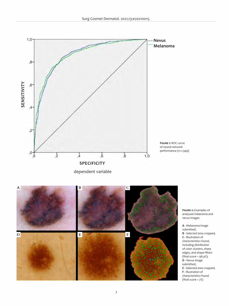
A percentage value (pseudoprobability) resulting from the softmax function in the output layer, ranging from 0-100%, characterizes the result of the neural network. So values higher than 50 were predicted as melanoma and values lower than 50, as nevus (Figure 1).
With an average of 9.3 years of dermatological practice and 7.6 years of dermoscopy use, 52 dermatologists randomly selected 40 images from the image bank excluded from the neu-ral network training. The evaluators were informed that these were melanocytic lesions and answered whether each lesion was benign or malignant (including in situ).
RESULTSThe training and testing obtained a correct rating per-
centage of 78.5% and 79.1%, respectively, with a ROC curve covering 86.5% of the area (Figure 1). The weights and parame-ters obtained from the neural network were used to develop an application (Figure 2) hosted on a public web server, allowing the experimental online evaluation of dermoscopic images (http://200.145.131.197/mmview/index.php/).
Difficulty or uncertainty in analyzing lesions by derma-tologists had a mean value of 3.3 on a scale of 0 to 5. The overall sensitivity and specificity of the 2,080 assessments by dermatolo-gists were 71.8% and 52%, respectively. For the same images and a cutoff point of 0.4 (40%) of the output value, the application obtained 62% and 56% values, respectively.
DISCUSSIONThe study results demonstrate that less complex predic-
tive methods such as artificial neural networks can bring sig-nificant results despite their limitations. The online and open availability of the studied algorithm can add information in de-cision-making about melanocytic lesions, mainly when more extreme values are obtained. Nevertheless, it should be recogni-zed that the tool has performance limitations. It was trained only with selected images of nevi and melanoma, not with coarse hair or pigmented lesions.
Surg Cosmet Dermatol. 2021;13:e20210015.
3
SEN
SITI
VITY
SPECIFICITY
dependent variable
NevusMelanoma
Figure 1: ROC curve of neural network performance (n=1.949)
Figure 2: Examples of analyzed melanoma and nevus images
A - Melanoma image submitted; B - Selected area cropped; C - Illustration of characteristics found, including distribution of color clusters, sharp edges, and shape filters (final score = 98,4%); D - Nevus image submitted; E - Selected area cropped; F - Illustration of characteristics found (final score = 7%)
A
D
B
E
C
F
Ferreira CAZ, Souza V, Miot HA, Schmitt JA
4
REFERENCES1. Souza RJ, Mattedi AP, Rezende ML, Corrêa Mde P, Duarte EM. An esti-
mate of the cost of treating melanoma disease in the state of Sao Paulo - Brazil. An Bras Dermatol. 2009;84(3):237-43.
2. Vestergaard ME, Macaskill P, Holt PE, Menzies SW. Dermoscopy com-pared with naked eye examination for the diagnosis of primary me-lanoma: a meta-analysis of studies performed in a clinical setting. Br J Dermatol. 2008;159(3):669-76.
3. van der Rhee JI, Bergman W, Kukutsch NA. The impact of dermoscopy on the management of pigmented lesions in everyday clinical prac-tice of general dermatologists: a prospective study. Br J Dermatol. 2010;162(3):563-7.
4. Hoorens I, Vossaert K, Lanssens S, Dierckxsens L, Argenziano G, Brochez L. Value of dermoscopy in a population-based screening sample by dermatologists. Dermatol Pract Concept. 2019;9(3):200-6.
5. Cui X, Wei R, Gong L, Qi R, Zhao Z, Chen H, et al. Assessing the effecti-veness of artificial intelligence methods for melanoma: a retrospective review. J Am Acad Dermatol. 2019;81(5):1176-80.
6. Brinker TJ, Hekler A, Enk AH, Berking C, Haferkamp S, Hauschild A, et al. Deep neural networks are superior to dermatologists in melanoma image classification. Eur J Cancer. 2019;119:11-7.
7. Basheer IA, Hajmeer M. Artificial neural networks: fundamentals, com-puting, design, and application. J Microbiol Methods. 2000;43(1):3-31.
8. Cullell-Dalmau M, Otero-Viñas M, Manzo C. Research techniques made simple: deep learning for the classification of dermatological images. J Invest Dermatol. 2020;140(3):507-14
9. ISIC. The International Skin Imaging Collaboration. Available from: https://www.isic- archive.com/#!/topWithHeader/wideContentTop/main. 2020. Acessed 21 Oct 2020.
10. Islam MM, Iqbal H, Haque MR, Hasan MK. Prediction of breast cancer using support vector machine and K-Nearest neighbors. IEEE Region 10 Humanitarian Technology Conference (R10-HTC), Dhaka. 2017;226-9.
AUTHORS' CONTRIBUTION:
César Augusto Zago Ferreira 0000-0001-7299-1710Approval of the final version of the manuscript; study design and planning; preparation and writing of the manuscript; data collec-tion, analysis, and interpretation; critical revision of the manuscript.
Vinícius de Souza 0000-0001-8819-6906Approval of the final version of the manuscript; data collection, analysis, and interpretation; critical revision of the manuscript.
Hélio Amante Miot 0000-0002-2596-9294Statistical analysis; approval of the final version of the manuscript; study design and planning; preparation and writing of the ma-nuscript; critical revision of the manuscript.
Juliano Vilaverde Schmitt 0000-0002-7975-2429Statistical analysis; approval of the final version of the manuscript; study design and planning; preparation and writing of the ma-nuscript; data collection, analysis, and interpretation; active participation in research orientation; intellectual participation in pro-paedeutic and/or therapeutic conduct of studied cases; critical literature review; critical revision of the manuscript.
Other classification algorithms such as k-nearest nei-ghbors algorithm (k-NN) and Support Vector Machine (SVM) may have different performances than the artificial neural ne-twork, and the group will explore them later. Also, extracting new variables from image analysis can lead to system perfor-mance gain.10
Computer vision methods have evolved significantly with cloud computing-based systems, spreading the use of con-volutional neural networks with up to billions of neurons. Still, they depend on a high number of images for learning and sig-
nificant maintenance costs. Nevertheless, machine learning will probably become more frequent in medical activities, especially in image analysis, as it is in other human activities.
CONCLUSIONSWe developed and implemented a neural network ba-
sed on dermoscopic images, which can collaboratively assist in the differential diagnosis between melanocytic nevus and melanoma. l
1
APOIO CIENTÍFICO:
www.surgicalcosmetic.org.br/
ISSN-e 1984-8773
Original ArticleAuthors:
Flávia Trevisan1
Nataly Portilla Maya2
Guilherme Canho Bittner3
Bruno de Carvalho Fantini4
Felipe Bochnia Cerci5,6
1 Dermatology Service, Universidade Federal do Paraná, Curitiba (PR), Brazil.
2 Dermatology Service, Clínica Erasmo, Valledupar, Colombia.
3 Dermatology Service, Universi- dade Federal do Mato Grosso do Sul, Campo Grande (MS), Brazil.
4 Dermatology Service, Faculdade de Medicina de Ribeirão Preto - USP. RIbeirão Preto (SP), Brazil.
5 Mohs Curitiba, Clínica Cepelle, Curitiba (PR), Brazil.
6 Postgraduate Program in Internal Medicine and Health Sciences, Universidade Federal do Paraná, Curitiba (PR), Brazil.
Correspondence:Flávia TrevisanEmail: [email protected]
Financial support: None.Conflict of interest: None.
Submitted on: 14/02/2021Approved on: 04/04/2021
How to cite this article: Trevisan F, Maya NP, Bittner GC, Fantini BC, Cerci FB. Perioral reconstruction after Mohs micrographic surgery: analysis of 108 cases. Surg Cosmet Dermatol. 2021;13:e20210022.
RESUMOIntrodução: a região perioral é comumente acometida por câncer de pele não melanoma. A cirurgia micrográ-fica de Mohs é o tratamento de escolha nessa área, com as maiores taxas de cura e preservação de tecido sadio. Há inúmeros métodos de reconstrução da região perioral, sendo sua escolha influenciada por características da ferida operatória e preferência do cirurgião. Objetivos: descrever a experiência dos autores na reconstrução perioral após cirurgia micrográfica de Mohs e analisar os métodos de reconstrução mais utilizados. Métodos: estudo retrospectivo de casos de reconstrução perioral submetidos à cirurgia de Mohs. Resultados: foram incluídos 103 pacientes, totalizando 108 casos. O número médio de estágios da cirurgia micrográfica de Mohs foi de 1,4, e o tamanho médio dos defeitos, de 16mm. O fechamento primário foi a técnica mais empregada para reconstrução, seguido por retalhos, principalmente VY, avanço simples e rotação. A associação entre métodos de reparo foi utilizada em 28,7%. Quatro pacientes tiveram complicações (necrose e infecção do enxerto, trapdoor e deiscência parcial de sutura). Conclusões: fechamento primário foi o método mais frequente de reparo, seguido pelos retalhos. Conhecer as estratégias de reconstrução e possibilidades de associações é fundamental para a adequada restauração da região perioral, mantendo-se funcionalidade, sensibilidade e estética do local.Palavras-chave: Cirurgia de Mohs; Lábio; Neoplasias cutâneas; Neoplasias labiais
Perioral reconstruction after Mohs micrographic surgery: analysis of 108 cases Reconstrução perioral após cirurgia micrográfica de Mohs: análise de 108 casos
DOI: http://www.dx.doi.org/10.5935/scd1984-8773.2021130022
ABSTRACTIntroduction: The perioral region is commonly affected by non-melanoma skin cancer. Mohs micrographic surgery is the treatment of choice in this area because it has the highest cure rate and preserves healthy tissue. Several methods are available for restoring the perioral region, and their selection is influenced by the surgical wound characteristics and the surgeon's preference. Objective: Describe the authors’ experience in perioral reconstruction after Mohs micrograph-ic surgery and analyze the repair methods most frequently performed. Methods: Retrospective study of consecutive cases submitted to Mohs surgery and perioral reconstruction. Results: The study included 108 cases from 103 patients. The mean number of Mohs surgery stages was 1.4, and the mean defect size was 16 mm. Primary closure was the most used technique for reconstruction, followed by flaps (mainly V-Y, single advancement, and rotation). The associ-ation of repair methods was used in 28.7% of cases, mostly combined with flaps. Four patients had complications (necrosis and graft infection, trapdoor effect, and partial wound dehiscence). Conclusion: Primary closure was the most frequent repair method, followed by flaps. Knowing reconstruction strategies and possibilities of associations is essential for proper restoration of the perioral region, maintaining its function, sensation and aesthetics.Keywords: Lip; Lip neoplasms; Mohs surgery; Skin neoplasms
ISSN-e 1984-8773
Trevisan F, Maya NP, Bittner GC, Fantini BC, Cerci FB
2
INTRODUCTIONThe perioral region is commonly affected by non-me-
lanoma skin cancer. While basal cell carcinoma (BCC) often affects the cutaneous portion, squamous cell carcinoma (SCC) is more prevalent in the vermilion (mucosa).1 Surgical remo-val, either by wide local excision or micrographic surgery is the treatment of choice for malignant skin tumors in the perioral region.2
Mohs micrographic surgery (MMS) is preferable in the perioral region, as it has the advantages of a higher cure rate and preservation of healthy tissue.3 The highest cure rate comes from the complete examination of the surgical margins during the procedure.4 Wide local excision on the other hand assesses approximately 1% of the margins.5 The preservation of healthy tissue in the MMS can save the patient from more complex reconstructions. However, challenging reconstructions may be necessary even with MMS, and a thorough margins examination is essential to perform them safely.2
When choosing the repair method for this area the size of the surgical wound, its location (subunit affected), and its depth should be considered, among other factors. A satisfactory surgical outcome is achieved when the site’s functionality, mobi-lity, sensitivity, and esthetics are maintained.2
This study aims to describe the authors’ experience in perioral reconstruction after MMS and to analyze the most used reconstruction methods.
METHODSThis is a retrospective study of consecutive cases sub-
mitted to MMS and perioral reconstruction performed by the authors between January 2017 and August 2020. The cases are from the authors’ private clinics and from a university hospital where one of the authors works. The ethics committee approved the study, protocol 30743520.2.0000.0103.
Except for one surgery performed under local anesthesia and sedation, all procedures were performed under local anes-thesia. Postoperatively, antibiotics (cephalexin 500 mg 6/6 hours for seven days, cefadroxil 500 mg 12/12 hours for four days, or amoxicillin 500 mg 8/8 hours for seven days) were used in more complex, long duration surgeries or when a significant portion of mucosa was removed.
For data analysis, we reviewed photographic documen-tation and data such as age, gender, Fitzpatrick skin phototype, tumor characteristics, size of the wound and anatomical su-bunits involved, number of MMS stages, reconstruction per-formed, antiplatelet or anticoagulants use, and postoperative complications.
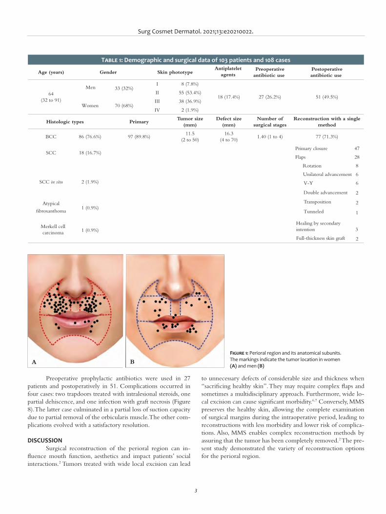
The perioral subunits were divided into upper cutaneous lip (UCL), lower cutaneous lip (LCL), philtrum, apical trian-gle, superior vermilion, and inferior vermilion (Figure 1).6 The reconstruction methods were divided into secondary intention healing, primary closure, flaps, or graft. When more than one method was used, it was called combined reconstruction. For
analysis of repair methods, only those that repaired perioral su-bunits were considered.
Complications were divided into two groups, short or long-term. Bleeding that required re-intervention, hematoma, infection, dehiscence, and necrosis of the flap/graft (partial or total) were considered short-term. Considerable anatomical dis-tortion and functional impairment (difficulty speaking and im-paired mobility) were defined as long-term.
RESULTSThe study included 108 cases from 103 patients.
Table 1 describes the demographic and surgical data. BCC was the most prevalent tumor in all perioral subunits, except in the lower vermilion, where only SCCs were found (ten invasive and one in situ).
Figure 1 shows the perioral subunits. The upper cuta-neous lip (n=83) was the subunit mostly affected, followed by the lower vermilion (12), apical triangle (5), philtrum (4), lower cutaneous lip (3), and upper vermilion (1). In 35 cases, the tumor extended over more than one perioral subunit, and in 12, the ex-tension reached another facial unit (mainly malar, in eight cases). Only three cases had full-thickness lip defects.
Seventy-seven cases underwent reconstruction with a single method, and 31 cases underwent combined methods. The most used procedure was flaps combined with other methods. Flaps were used in 50 cases, half as a single technique and half associated with other methods. The following flaps were used: V-Y (n=15), single advancement (n=14), rotation (n=11), trans-position (n=4), double advancement (n=3), hinge (n=2), and tunneled island flap (n=1).
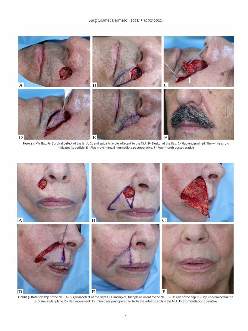
For tumors primarily involving the UCL, 47 cases were restored primarily (Figure 2). In nine cases, primary closure was combined with a full-thickness skin graft (Figure 3), and one with graft and advancement flap. Flaps were the second most common technique in this cosmetic subunit (Figure 4), 33 cases, and rotation was the most used type (n=10) (Figure 5). Two patients were referred by plastic surgery to perform MMS and, after its completion, returned for reconstruction with the plastic surgeon. Both were restored with primary closure.
In the lower vermilion, ten cases were repaired with pri-mary closure. Among them three were combined with an advan-cement flap and two with secondary intention healing. An exten-sive but superficial case was left to heal by secondary intention. The most used method was also the primary closure in the apical triangle, philtrum, superior vermilion, and lower cutaneous lip.
In wounds involving more than one perioral subunit (n=35), the method most used for reconstruction was flaps (n=17) (Figure 6), followed by primary closure (n=15), and grafts (n= 2). Among these 35 cases, four required the association of more than two methods (Figure 7).
Surg Cosmet Dermatol. 2021;13:e20210022.
3
Preoperative prophylactic antibiotics were used in 27 patients and postoperatively in 51. Complications occurred in four cases: two trapdoors treated with intralesional steroids, one partial dehiscence, and one infection with graft necrosis (Figure 8). The latter case culminated in a partial loss of suction capacity due to partial removal of the orbicularis muscle. The other com-plications evolved with a satisfactory resolution.
DISCUSSIONSurgical reconstruction of the perioral region can in-
fluence mouth function, aesthetics and impact patients’ social interactions.2 Tumors treated with wide local excision can lead
to unnecessary defects of considerable size and thickness when “sacrificing healthy skin”. They may require complex flaps and sometimes a multidisciplinary approach. Furthermore, wide lo-cal excision can cause significant morbidity.6,7 Conversely, MMS preserves the healthy skin, allowing the complete examination of surgical margins during the intraoperative period, leading to reconstructions with less morbidity and lower risk of complica-tions. Also, MMS enables complex reconstruction methods by assuring that the tumor has been completely removed.2 The pre-sent study demonstrated the variety of reconstruction options for the perioral region.
Figure 1: Perioral region and its anatomical subunits. The markings indicate the tumor location in women (A) and men (B)
A B
Surg Cosmet Dermatol. 2021;13:e20210022.
3
roids, one partial dehiscence, and one infection with graft necrosis (Figure 8). The latter case culminated in a partial loss of suction capacity due to a large part of the orbicularis muscle resection. The other complications evolved with a satisfactory resolution.
DISCUSSIONSurgical reconstruction of the perioral region can in-
fluence mouth functions, aesthetics and even impact patients’ social interactions.2 Tumors treated with conventional surgical excision can lead to unnecessary defects of considerable size
and thickness when “sacrificing healthy skin”. They may require complex flaps and sometimes a multidisciplinary team to per-form the procedure, in addition to more than one surgical stage. Also, conventional surgery can cause significant morbidities.6,7 In turn, MMS preserves the healthy skin, allowing the complete examination of surgical margins during the intraoperative pe-riod, leading to reconstructions with less morbidity and lower risk of complications. Also, MMS enables complex reconstruc-tion methods, thus assuring that the tumor has been comple-tely removed.2 The present study demonstrated the variety of
Figure 1: Perioral region and its anatomical subunits. The markings indicate the locations of tumors in women (A) and men (B)
A B
Table 1: Demographic and surgical data of 103 patients and 108 cases
Age (years) Gender Skin phototypeAntiplatelet
agentsPreoperative
antibiotic usePostoperative antibiotic use
64(32 to 91)
Men 33 (32%)I 8 (7.8%)
18 (17.4%) 27 (26.2%) 51 (49.5%)II 55 (53.4%)
Women 70 (68%)III 38 (36.9%)
IV 2 (1.9%)
Histologic types PrimaryTumor size
(mm)Defect size
(mm)Number of
surgical stagesReconstruction with a single
method
BCC 86 (76.6%) 97 (89.8%)11.5
(2 to 50)16.3
(4 to 70)1.40 (1 to 4) 77 (71.3%)
SCC 18 (16.7%)47
28
SCC in situ 2 (1.9%)
8
Primary closure
Flaps
Rotation
Unilateral advancement 6
V-Y 6
3
Atypical
fibroxanthoma1 (0.9%)
2
Merkell cell carcinoma 1 (0.9%)
2
1
Full-thickness skin graft 2
Double advancement
Transposition
Tunneled
Healing by secondary intention
Trevisan F, Maya NP, Bittner GC, Fantini BC, Cerci FB
4
Like previous publications, BCC was the most prevalent malignant tumor in the cutaneous portion of the lip, while SCC was the most common (and the only one) in the vermilion.1 In 32% of cases (n=35), the tumor extended through more than one perioral subunit, and in 11%, the extension reached another facial unit (mainly the malar). This demonstrates the complexity of repairs in the perioral region. The involvement of sites beyond the nasola-bial fold (NLF) is important because, in some cases, it is preferable to combine reconstruction methods for better results.
After tumor removal from the perioral region, surgical wounds can be divided according to thickness, size, and location. These factors help to guide the choice of the reconstruction method.8 According to a 2009 literature review, the most com-mon method for reconstructing the perioral region was primary
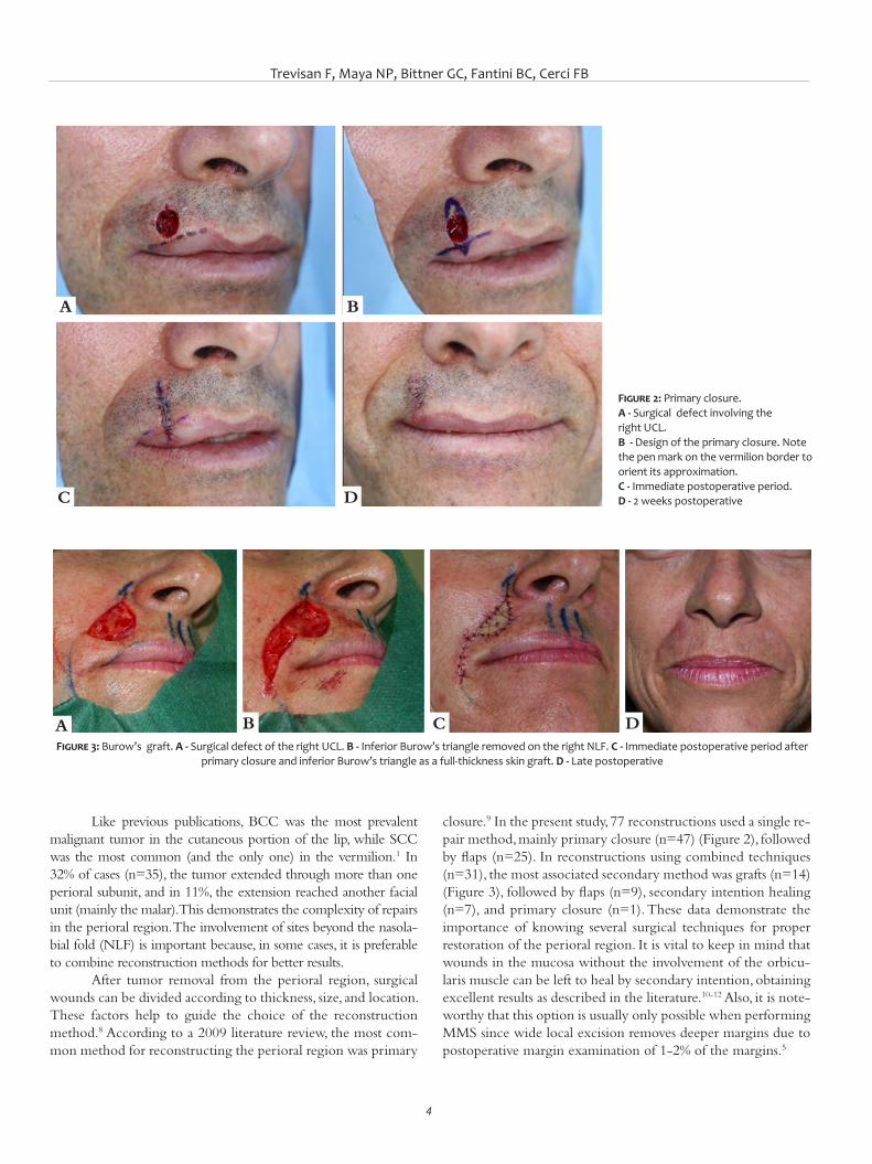
closure.9 In the present study, 77 reconstructions used a single re-pair method, mainly primary closure (n=47) (Figure 2), followed by flaps (n=25). In reconstructions using combined techniques (n=31), the most associated secondary method was grafts (n=14) (Figure 3), followed by flaps (n=9), secondary intention healing (n=7), and primary closure (n=1). These data demonstrate the importance of knowing several surgical techniques for proper restoration of the perioral region. It is vital to keep in mind that wounds in the mucosa without the involvement of the orbicu-laris muscle can be left to heal by secondary intention, obtaining excellent results as described in the literature.10-12 Also, it is note-worthy that this option is usually only possible when performing MMS since wide local excision removes deeper margins due to postoperative margin examination of 1-2% of the margins.5
Figure 3: Burow’s graft. A - Surgical defect of the right UCL. B - Inferior Burow’s triangle removed on the right NLF. C - Immediate postoperative period after primary closure and inferior Burow’s triangle as a full-thickness skin graft. D - Late postoperative
A B C D
Figure 2: Primary closure. A - Surgical defect involving the right UCL. B - Design of the primary closure. Note the pen mark on the vermilion border to orient its approximation. C - Immediate postoperative period. D - 2 weeks postoperative
A
C
B
D
Surg Cosmet Dermatol. 2021;13:e20210022.
5
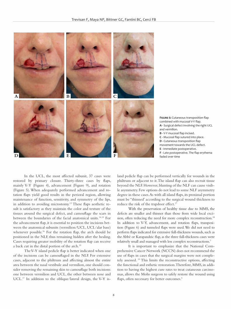
Figure 4: V-Y flap. A - Surgical defect of the left UCL and apical triangle adjacent to the NLF. B - Design of the flap. C - Flap undermined. The white arrow indicates its pedicle. D - Flap movement. E - Immediate postoperative. F - Four-month postoperative
A
D
B
E
C
F
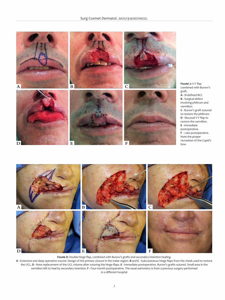
Figure 5: Rotation flap of the NLF. A - Surgical defect of the right UCL and apical triangle adjacent to the NLF. B - Design of the flap. C - Flap undermined in the supramuscular plane. D - Flap movement. E - Immediate postoperative. Note the rotation arch in the NLF. F - Six-month postoperative
D E F
A B C
Trevisan F, Maya NP, Bittner GC, Fantini BC, Cerci FB
6
In the UCL, the most affected subunit, 37 cases were restored by primary closure. Thirty-three cases by flaps, mainly V-Y (Figure 4), advancement (Figure 9), and rotation (Figure 5). When adequately performed advancement and ro-tation flaps yield good results in the perioral region, allowing maintenance of function, sensitivity, and symmetry of the lips, in addition to avoiding microstomy.13 These flaps aesthetic re-sult is satisfactory as they maintain the color and texture of the tissues around the surgical defect, and camouflage the scars in between the boundaries of the facial anatomical units.14,15 For the advancement flap, it is essential to position the incisions bet-ween the anatomical subunits (vermilion/UCL, UCL/alar base) whenever possible.16 For the rotation flap, the arch should be positioned in the NLF, thus remaining hidden after the healing. Cases requiring greater mobility of the rotation flap can receive a back cut in the distal portion of the arch.15
The V-Y island pedicle flap is better indicated when one of the incisions can be camouflaged in the NLF. For extensive cases, adjacent to the philtrum and affecting almost the entire area between the nasal vestibule and vermilion, one should con-sider removing the remaining skin to camouflage both incisions: one between vermilion and UCL, the other between nose and UCL.17 In addition to the oblique/lateral design, the V-Y is-
land pedicle flap can be performed vertically for wounds in the philtrum or adjacent to it. The island flap can also recruit tissue beyond the NLF. However, blunting of the NLF can cause visib-le asymmetry. Few options do not lead to some NLF asymmetry degree in these cases. As with all island flaps, its proximal portion must be “thinned’ according to the surgical wound thickness to reduce the risk of the trapdoor effect.17
With the preservation of healthy tissue due to MMS, the defects are smaller and thinner than those from wide local exci-sion, often reducing the need for more complex reconstruction.18 In addition to V-Y, advancement, and rotation flaps, transposi-tion (Figure 6) and tunneled flaps were used. We did not need to perform flaps indicated for extensive full-thickness wounds, such as the Abbé or Karapandzic flap, as the three full-thickness cases were relatively small and managed with less complex reconstructions.7
It is important to emphasize that the National Com-prehensive Cancer Network (NCCN) does not recommend the use of flaps in cases that the surgical margins were not comple-tely assessed. 19 This limits the reconstructive options, affecting the functional and esthetic restoration. Therefore, MMS, in addi-tion to having the highest cure rates to treat cutaneous carcino-mas, allows the Mohs surgeon to safely restore the wound using flaps, often necessary for better outcomes.5
Figure 6: Cutaneous transposition flap combined with mucosal V-Y flap. A - Surgical defect involving the right UCL and vermilion. B - V-Y mucosal flap incised. C - Mucosal flap sutured into place.D - Cutaneous transposition flap movement towards the UCL defect. E - Immediate postoperative. F - Late postoperative. The flap erythema faded over time
A
D
B
E
C
F
Surg Cosmet Dermatol. 2021;13:e20210022.
7
Figure 8: Double hinge flap, combined with Burow’s grafts and secondary intention healing. A - Extensive and deep operative wound. Design of the primary closure in the malar region. B and C - Subcutaneous hinge flaps from the cheek used to restore
the UCL. D - Note replacement of the UCL volume after suturing the hinge flaps. E - Immediate postoperative. Burow's grafts sutured. Small area in the vermilion left to heal by secondary intention. F - Four-month postoperative. The nasal asimmetry is from a previous surgery performed
in a different hospital
A
D
B
E
C
F
Figure 7: V-Y flap combined with Burow’s graft. A - Ill-defined BCCB - Surgical defect involving philtrum and vermilion. C - Burow’s graft sutured to restore the philtrum. D - Mucosal V-Y flap to restore the vermilion. E - Immediate postoperative. F - Late postoperative. Note the proper recreation of the Cupid's bow
A
D
B
E
C
F
Trevisan F, Maya NP, Bittner GC, Fantini BC, Cerci FB
8
The use of pre and postoperative antibiotics is a con-troversial topic in dermatologic surgery.20-23 Cutaneous surge-ries are considered clean, but the proximity to the oral cavity makes the perioral region a site with a significant chance of potentially contaminated surgeries.24 This study used preope-rative prophylactic antibiotic therapy in 26.2% of cases and postoperative in 49.5%, reflecting the difficulty in anticipating the wound size and the reconstruction method to be perfor-med. With a low mean of surgical stages (which reduces the surgical time) and appropriate indication for antibiotic the-rapy, there was only one case of postoperative infection of a recurrent morpheaform BCC (treated by wide local excision eight years earlier). The tumor extended from the UCL to the upper vermilion and philtrum, leading to a 48 mm surgical defect repaired with a hinge flap combined with full-thickness skin graft and secondary intention. In this case, despite the use of pre and postoperative antibiotics, there was infection and graft necrosis. However, post-operative care and additional antibiotics led to a satisfactory outcome, except for the slight functional impairment (suction) secondary to a significant loss of orbicularis muscle due to tumor’s aggressiveness (Figure 8).
Other complications were two cases of trapdoor, one after a V-Y flap and the other secondary to a tunneled flap both in the UCL. Both had a good response with intralesional ste-roids (triamcinolone acetonide 20 mg/ml). There was one case of partial dehiscence in the apical triangle following primary closure.
Preserving the function and volume of the perioral re-gion is challenging because even minor defects can impair the lips’ movement, competence, and symmetry. The surgeons must invest all their efforts in resolving the disease with a satisfactory aesthetic-functional surgical outcome. As recommended by the NCCN, reconstructions, especially in cosmetic sensitive areas, should ideally be performed after complete tumor resection confirmed by histological analysis of 100% of the surgical mar-gins perioperatively as in MMS.19 The Mohs surgeon should be able to perform complex and straightforward reconstructions in the perioral region. More challenging cases may require a mul-tidisciplinary approach.
CONCLUSIONSPrimary closure was the most used technique for recons-
truction, followed by flaps (mainly V-Y, unilateral advancement, and rotation). Combined methods were performed in 28.7% of cases. A combined approach is better indicated for wounds in-volving cutaneous and mucosal subunit, such as UCL and ver-milion.
Knowing the reconstruction strategies and possibilities of associations is essential for the proper reconstruction of the perioral region, maintaining its functionality, sensation and aes-thetics. l
A
C
B
D
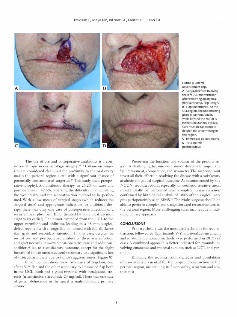
Figure 9: Lateral advancement flap. A - Surgical defect involving the left UCL and vermilion after removing an atypical fibroxanthoma. Flap design. B - Flap undermined. At the UCL region, the undermining plane is supramuscular, while beyond the NLF, it is in the subcutaneous tissue. Care must be taken not to deepen the undermining in this region. C - Immediate postoperative. D - Four-month postoperative
Surg Cosmet Dermatol. 2021;13:e20210022.
9
REFERENCES1. Queen D, Knackstedt T, Polacco MA, Collins LK, Lee K, Samie FH. Charac-
teristics of Nonmelanoma SkinCancers of Cutaneous Perioral and Ver-milion Lip Treated by Mohs Micrographic Surgery. J Eur Acad Dermatol Venereol. 2019;33(2):305-11.
2. Hafiji J, Hussain W, Salmon P. Reconstruction of perioral defects post--Mohs micrographic surgery: a dermatological surgeon's approach. Br J Dermatol. 2015;172(1):145-50.
3. Hafiji J, Hussain W, Salmon P. Mohs surgery spares the orbicularis oris muscle, optimizing cosmetic and functional outcomes for tumours in the perioral region: a series of 407 cases and reconstructions by derma-tological surgeons. Br J Dermatol. 2015;172(1):294-6.
4. Tolkachiov SN, Brodland DG, Coldiron BM, Fazio MJ, Hruza GJ, Roe-nigk RK, et al. Understanding Mohs Micrographic Surgery: a review and practical guide for the nondermatologist. Mayo Clin Proc. 2017; 92(8):1261-71.
5. Kimyai-Asadi A, Goldberg LH, Jih MH. Accuracy of serial transverse cross-sections in detecting residual basal cell carcinoma at the surgi-cal margins of an elliptical excision specimen. J Am Acad Dermatol. 2005;53(3):469-74.
6. Gaylon S W, Frodel JL. Lip and perioral defects. Otolaryngol Clin North Am. 2001;34(3):647-66.
7. Wollina U. Reconstructive surgery in advanced perioral non-mela-noma skin cancer. Results in elderly patientes. J Dermatol Case Rep. 2014;8(4):103-7.
8. Johnson AR, Egeler SA, Wu WW, Bucknor A, Ibrhim AMS, Lin SJ. Facial re-construction after Mohs Surgery: a critical review os defects involving cheek, forehead and perioral region. J Craniofac Surg. 2019;30(2):400-7.
9. Faulhaber J, Geraud C, Goerdt S, Koenen W. Functional and aesthetic reconstruction of full-thickness defects of the lower lip after tumor resection: analysis of 59 cases and discussion of a surgical approach. Dermatol Surg. 2010;36(6):859-67.
10. Gloster J, Hugh M. The use of second-intention healing for partial--thickness Mohs defects involving the vermilion and/or mucosal surfa-ces of the lip. J Am Acad Dermatol. 2002;47(6):893-7.
11. Aimee LL, Hanke CW. Second intention healing for intermediate and lar-ge postsurgical defects of the lip. J Am Acad Dermatol. 2007;57(5):832-5.
12. Donigan JM, Millican EA. Cosmetic and Functional Outcomes of Se-cond Intention Healing for Mohs Defects of the Lips. Dermatol Surg. 2019;45(1):26-35.
13. Ergün SS. Reconstruction of the labiomental region with local flaps. Dermatol Surg. 2002;28(9):863-5.
14. Joo-Hak K, Ahn CH, Kim S, Lee WS, Oh S. Effective method for recons-truction of remaining lower lip vermilion defect after a mental V-Y ad-vancement flap. Arch Craniofac Surg. 2019; 20(2):76-83.
15. Cerci, FB. Rotation flap for the reconstruction of the cutaneous upper lip after Mohs micrographic surgery. Surg Cosmet Dermatol. 2017;9(2):83-6.
16. Tolkachjov SN. Reconstruction for a large central upper mucosal lip de-fect. Dermatol Surg. Epub March 2020.
17. Griffin GR, Weber S, Baker, Shan R. Outcomes following V-Y advance-ment flap reconstruction of large upper lip defects. Arch Facial Plast Surg. 2012;14(3):193-7.
18. Touma DJ. Mohs' surgery to reduce the size of facial defects and neces-sity for complex repairs. Plast Reconstr Surg. 2002;110(6):1601.
19. Bichakjian CK, Olencki T, Aasi SZ, Alam M, Andersen JS, Berg D, et al. Basal cell skin cancer, version 1.2016, NCCN Clinical Practice Guidelines in Oncology. J Natl Compr Canc Netw. 2016;14(5):574-97.
20. Levin EC, Chow C, Makhzoumi Z, Jin C, Shiboski SC, Arron ST. Associa-tion of postoperative antibiotics with surgical site infection in Mohs Micrographic Surgery. Dermatol Surg. 2019;45(1):52-7.
21. Ahmed M, Gniadecki R, Taher, M. Oral and intraincisional antibiotic prophylaxis in Mohs Surgery: a systematic review and meta-analysis. Dermatol Surg. 2020;46(4):558-60.
22. Taylor O, Li JN, Carr C, Garcia A, Tran S, Srivastava D, et al. The effect of antibiotic prophylaxis on infection rates in mohs micrographic surgery: a single institution retrospective study. 2021 Oct;313(8):663-667.
23. Lin MJ, Dubin DP, Giordano CN, Kriegel DA, Hooman K. Antibiotic practi-ces in Mohs Micrographic Surgery. J Drugs Dermatol. 2020;19(5):493-7.
24. Center for Disease Control and Prevention (CDC). "Patient Safety Proto-col." Em The National Healthcare Safety Network (NHSN) Manual. 2008.
AUTHORS' CONTRIBUTION:
Flávia Trevisan 0000-0001-5855-3685Statistical analysis; approval of the final version of the manuscript; study design and planning; preparation and writing of the manuscript; data collection, analysis, and interpretation; intellectual participation in propaedeutic and/or therapeutic conduct of studied cases; critical literature review; critical revision of the manuscript.
Nataly Portilla Maya 0000-0002-0325-7640Approval of the final version of the manuscript; preparation and writing of the manuscript; data collection, analysis, and interpretation; critical revision of the manuscript.
Guilherme Canho Bittner 0000-0002-5892-4391Statistical analysis; approval of the final version of the manuscript; study design and planning; data collection, analysis, and interpretation.
Bruno de Carvalho Fantini 0000-0003-1192-8376Statistical analysis; approval of the final version of the manuscript; study design and planning; intellectual participation in propaedeutic and/or therapeutic conduct of studied cases.
Felipe Bochnia Cerci 0000-0001-9605-0798Statistical analysis; approval of the final version of the manuscript; study design and planning; preparation and writing of the manuscript; data collection, analysis, and interpretation; active participation in research orientation; intellectual participation in propaedeutic and/or therapeutic conduct of studied cases; critical literature review; critical revision of the manuscript.
1
APOIO CIENTÍFICO:
www.surgicalcosmetic.org.br/
ISSN-e 1984-8773
Original ArticleAuthors: Thessaly Puel de Oliveira¹ Hillani da Silva Andrade¹ José Roberto Pegas¹ Cristina Santos Ribeiro Bechara¹
¹ Dermatology Service, Complexo Hospitalar Padre Bento de Guarulhos, Guarulhos (SP), Brazil.
Correspondence: Cristina Santos Ribeiro Bechara Email: [email protected]
Financial support: None. Conflict of interest: None.
Submitted on: 15/04/2021Approved on: 12/07/2021
How to cite this article: Oliveira TP, Andrade HS, Pegas JR, Bechara CSR. Prevalence of patholo-gical diagnoses and epidemiological profile of patients with non-me-lanoma skin cancer suspicious lesions. Surg Cosmet Dermatol. 2021;13:e20210031.
Prevalence of pathological diagnoses and epidemiological profile of patients with non-melanoma skin cancer suspicious lesions Prevalência dos diagnósticos anatomopatológicos e perfil epidemiológico dos pacientes com lesões suspeitas de câncer de pele não melanoma
ABSTRACTIntroduction: Cutaneous cancer has a high incidence, can be screened with dermatological clinical examination and confirmed by cutaneous biopsy. Objective: To verify the prevalence of pathological diagnoses for suspected non-melanoma skin cancer (NMSC) lesions in a Reference Service. Methods: Observational and cross-sectional study. Patients with indication of skin biopsy for suspected NMSC were included. Variables analyzed: age, gender, personal history of skin cancer, number of biopsies, biopsy site, and pathological outcome, divided into Group 1 (neoplastic lesions); Group 2 (premalignant lesions), and Group 3 (benign lesions). Results: A total of 287 patients, with an average of 1.33 biopsies per patient. The median age in the sample was 71 years, and 56.1% were women. Personal history of skin cancer: 44.95%. General sample: group 1: 62%; group 2: 21% and group 3: 17%. Neoplasms found: 68% were BCC, 30% were SCC, and other neoplasms: 2%. In the group of premalignant lesions: mainly actinic keratosis; in the group of benign lesions: diagnostic variety. The cephalic segment was the most frequently biopsied topography (58%). Conclusions: In this study, we showed a higher incidence of skin cancer in women, with the majority of elderly patients being the most frequent diagnosis of BCC.Keywords: Basal Cell; Biopsy; Carcinoma; Neoplasms; Skin neoplasms; Squamous cell.
RESUMOIntrodução: o câncer cutâneo apresenta alta incidência, pode ser rastreado com exame clínico dermatológico e confirmado por biópsia cutânea. Objetivo: verificar a prevalência dos diagnósticos anatomopatológicos por lesões suspeitas de câncer de pele não melanoma (CPNM) em um Serviço de Referência. Métodos: estudo observacional e transversal. Inclusos pacientes com indicação de biópsia cutânea por suspeita de CPNM. Variáveis analisadas: idade, gênero, história pessoal de câncer de pele, número de biópsias, local da biópsia e resultado anatomopatológico, este dividido em grupo 1 (lesões neoplásicas); grupo 2 (lesões pré-malignas) e grupo 3 (lesões benignas). Resultados: um total de 287 pacientes, com média de 1,33 biópsia por paciente. A idade mediana na amostra foi 71 anos, sendo que 56,1% eram mulheres. História pessoal de câncer de pele: 44,95%. Amostra geral: gru-po 1: 62%; grupo 2: 21% e grupo 3: 17%. Neoplasias encontradas: 68% eram CBC, 30% eram CEC; e 2%, outras neoplasias. No grupo de lesões pré-malignas: principalmente queratoses actínicas; no grupo de lesões benignas: variedade diagnóstica. O segmento cefálico foi a topografia mais frequentemente biopsiada (58%). Conclusões: neste estudo, evidenciamos uma maior incidência de câncer de pele em mulheres, sendo a maioria dos pacientes idosos, e o diagnóstico mais frequente o CBC.Palavras-chave: Biópsia; Carcinoma basocelular; Carcinoma de células escamosas; Neoplasias cutâneas
DOI: http://www.dx.doi.org/10.5935/scd1984-8773.2021130031
ISSN-e 1984-8773
Oliveira TP, Andrade HS, Pegas JR, Bechara CSR
2
INTRODUCTIONSkin cancer has gained special significance in recent
decades due to its increasing incidence. It is considered a pu-blic health problem in geographic areas of intense exposure to ultraviolet radiation.1,2,3 Non-melanoma skin cancer (NMSC), represented by basal cell carcinoma (BCC) and squamous cell carcinoma (SCC), corresponds to 30% of all malignant tumors registered in the country. BCC accounts for 70% of skin cancer diagnoses, followed by SCC, diagnosed in 25% of cases.1
It is believed that 90% of NMSC can be attributed to sun exposure,4 and BCC is associated with cumulative exposure. This fact reinforces the importance of photoprotection, whi-ch should be introduced from childhood and is considered the measure of choice worldwide to prevent NMSC.5 Other causes related to the development of skin cancer are family history, ex-posure to chemicals, radiotherapies, phenotypic factors, immu-nosuppression, and some hereditary genetic syndromes.6
Diagnosis of skin cancer mainly involves clinical exami-nation, conducted through visual inspection of the patient’s skin, and histopathological analysis after the lesion’s biopsy, which is indicated when a clinical study shows suspicion of NMSC or melanoma.7 A specialized professional must perform a thorough physical examination, identifying suspected malignant lesions through their clinical characteristics. Advances in dermoscopic studies of these lesions allowed higher precision in indicating skin biopsies for diagnostic confirmation.8,9,10
When detected early, BCC presents high cure rates.2,6 According to the National Comprehensive Cancer Network (NCCN), NMSC treatment should prioritize the complete cure of the tumor. The NCCN recommends the biopsy of all NMSCs before any procedure, allowing the most appropriate treatment choice.¹¹ Despite the low mortality rate, this type of tumor has high morbidity because it causes disfigurement, re-sulting in physical and psychological disability.¹² However, in some cases, there is tumor recurrence after excision. Among the reasons for occurrence, some studies highlight the relationship with the location, lesion extension, compromised surgical mar-gins, and others. Considering this, it is of paramount importance to screen patients who have already had skin tumors to monitor the recurrence or persistence of tumors and detect new lesions.¹4
Assuming that there is considerable underreporting due to underdiagnosis, and also because it is a neoplasm with good prognosis if treated in an appropriate and timely manner, this study aims to verify the prevalence of anatomopathological re-sults and epidemiological/clinical profile of patients who under-went biopsy for NMSC suspicious lesions in the Dermatology Service of Complexo Hospitalar Padre Bento de Guarulhos-SP (CHPBG), between June and December 2019, promoting fur-ther discussion and analysis on this subject.
METHODSWe conducted a cross-sectional and retrospective obser-
vational study by analyzing medical records of patients who un-
derwent skin biopsies for NMSC suspicion lesions in the Der-matology Service of CHPBG, from June to December 2019. This study was submitted to the CHPBG Research Ethics Committee (CEP 245/2025) through Plataforma Brasil and it was approved under the number 3,929,819.
The variables analyzed in the study were: age, gender, personal history of skin cancer, number of biopsies, biopsy site, and anatomopathological results. After completing data collec-tion, the general epidemiological profile of the sample was de-signed (total number of patients in the study) by observing the mean, maximum, and minimum age and gender, in addition to following the frequency of the personal history of skin cancer.
Then, the patients were grouped according to the ana-tomopathological result, forming three distinct groups: Group 1, comprising patients with confirmed neoplastic lesions in the anatomopathological analysis; Group 2, including individuals with premalignant lesions in the anatomopathological analysis; and Group 3, with patients with benign lesions in the anatomo-pathological examination. The epidemiological profile of each group was analyzed later. We subdivided Group 1 into BCC, SCC, and other neoplasms to enable the individual assessment of each histological type of neoplasm found, allowing the analysis of the topographic location in each of these subgroups.
The study included the medical records of patients with skin lesions suggestive of NMSC, whose biopsies already had a histopathological result. The biopsies were performed at the CHPBG Dermatology Service from June to December 2019. We excluded the medical records of patients with requests for biopsy of cutaneous lesions not suggestive of malignancy, in ad-dition to patients with cutaneous lesions suggestive of malignan-cy that had already been surgically addressed, even those with an indication for a new biopsy. Likewise, patients who did not attend or did not have the anatomopathological report during the research period and individuals with a previous diagnosis of NMSC known as Gorlin Syndrome, xeroderma pigmentosum, or mycosis fungoid, or those treated outside the established pe-riod, were excluded.
RESULTS This study included all skin biopsy samples indicated by
clinical suspicion of NMSC, totaling 382 points of skin biopsies in a total of 287 patients, corresponding to a mean of 1.33 biop-sies per patient, with a maximum number of seven biopsies in the same patient.
The overall sample (287 patients) comprised 126 (43.9%) men and 161 (56.1%) women, and the patients’ age ranged bet-ween 20 and 97 years, with a median of 71 years. Among the assessed patients, 129 (44.95%) had a personal history of skin cancer and 158 (55.05%) did not.
We formed three large groups according to the anato-mopathological results obtained from the samples to know the diagnoses of suspicious lesions in the general sample:
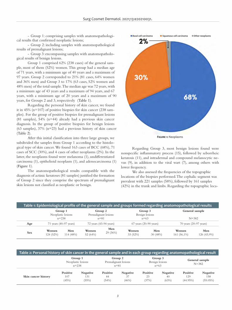
Surg Cosmet Dermatol. 2021;13:e20210031.
3
- Group 1: comprising samples with anatomopathologi-cal results that confirmed neoplastic lesions;
- Group 2: including samples with anatomopathological results of premalignant lesions;
- Group 3: encompassing samples with anatomopatholo-gical results of benign lesions.
Group 1 comprised 62% (238 cases) of the general sam-ple, most of them (52%) women. This group had a median age of 71 years, with a minimum age of 40 years and a maximum of 97 years. Group 2 corresponded to 21% (81 cases, 64% women and 36% men) and Group 3 to 17% (63 cases, 52% women and 48% men) of the total sample. The median age was 72 years, with a minimum age of 43 years and a maximum of 94 years, and 67 years, with a minimum age of 20 years and a maximum of 90 years, for Groups 2 and 3, respectively (Table 1).
Regarding the personal history of skin cancer, we found it in 45% (n=107) of positive biopsies for skin cancer (238 sam-ples). For the group of positive biopsies for premalignant lesions (81 samples), 54% (n=44) already had a previous skin cancer diagnosis. In the group of positive biopsies for benign lesions (63 samples), 37% (n=23) had a previous history of skin cancer (Table 2).
After this initial classification into three large groups, we subdivided the samples from Group 1 according to the histolo-gical type of skin cancer. We found 163 cases of BCC (68%), 71 cases of SCC (30%), and 4 cases of other neoplasms (2%). In the latter, the neoplasms found were melanoma (1), undifferentiated carcinoma (1), epithelioid neoplasm (1), and adenocarcinoma (1) (Figure 1).
The anatomopathological results compatible with the diagnosis of actinic keratoses (81 samples) justified the formation of Group 2 since they comprise the spectrum of premalignant skin lesions not classified as neoplastic or benign.
Regarding Group 3, most benign lesions found were nonspecific inflammatory process (15), followed by seborrheic keratosis (11), and intradermal and compound melanocytic ne-vus (9), in addition to the viral wart (7), among others with lower frequency.
We also assessed the frequencies of the topographic locations of the biopsies performed. The cephalic segment was prevalent with 221 samples (58%), followed by 161 samples (42%) in the trunk and limbs. Regarding the topographic loca-
Table 1: Epidemiological profile of the general sample and groups formed regarding anatomopathological resultsGroup 1
Neoplastic lesionsn=238
Group 2Premalignant lesions
n=81
Group 3Benign lesions
n=63
General sample
N=382
Age 71 years (40-97 years) 72 years (43-94 years) 67 years (20-90 years) 70 years (20-97 years)
SexWomen
124 (52%)Men
114 (48%)Women52 (64%)
Men29 (36%)
Women33 (52%)
Men30 (48%)
Women161 (56,1%)
Men126 (43,9%)
Table 2: Personal history of skin cancer in the general sample and in each group regarding anatomopathological resultGroup 1
Neoplastic lesionsn=238
Group 2Premalignant lesions
n=81
Group 3Benign lesions
n=63
General sampleN=382
Skin cancer historyPositive
107 (45%)
Negative131
(55%)
Positive44
(54%)
Negative37
(46%)
Positive23
(37%)
Negative40
(63%)
Positive129
(44.95%)
Negative158
(55.05%)
Figure 1: Neoplasms
Basal cell carcinoma Squamous cell carcinoma Other neoplasms
Oliveira TP, Andrade HS, Pegas JR, Bechara CSR
4
tion of biopsied lesions, in the individual analysis of each group, we noticed that Group 1 presented 61% of the samples in the cephalic segment, as well as Group 2 (53%) and Group 3 (54%) (Table 3).
Concerning the topographic locations of the neoplastic lesions (Group 1), in the different histological types, we observed that most of the biopsies for BCC (71%) were in the cephalic segment. On the other hand, SCC was more biopsied in the trunk and limbs (63%) (Table 4).
Finally, due to the prognostic importance that the ce-phalic segment’s risk zones (zone H and zone M) can pose, this region was subdivided into nose, periauricular/temporal, pe-riorbital, forehead, cheeks, jaw, chin, lips, and scalp. Thus, it was possible to identify the frequencies of biopsies in each of these anatomical subunits.
This study showed that the prevalent anatomical subu-nits of the cephalic segment in Group 1 were the periauricular/temporal (42% - 29.1%) and nose (40% - 27.7%). Regarding Group 2, the most frequent anatomical subunits of the cephalic segment were cheek (25.5%), periauricular/temporal (23.2%), nose (18.6%), and periorbital (13, 9%). There were no biopsies in the chin region resulting in pre-malignant lesions. In Group 3, the cheek (29.4%), nose (23.5%), and periorbital (14.7%) areas were the most biopsied anatomical subunits. It is noteworthy that there were no biopsies in the chin and lips regions represen-ting benign lesions (Table 5).
When we assessed only the frequency of biopsies loca-tion in the anatomical subunits of the cephalic segment that ob-
tained BCC results, we observed that the most frequent were the nose (32%) and periauricular/temporal regions (29%). Likewi-se, regarding SCC in the cephalic segment, when we analyzed this frequency, we noticed that the most frequent locations were the periauricular/temporal and cheek areas, both with the same incidence (30.7%) followed by the frontal region (4%-15.3%). It should be noted that, in the entire sample, the jaw and lips regions did not present reports compatible with SCC (Table 6).
DISCUSSIONThis study evaluated the anatomopathological results of all skin lesions indicated for biopsy due to suspicious malignancy over seven months (June to December 2019) in the Dermatology Service of the CHPBG. We obtained 382 biopsies from a total of 271 patients, of which 62% had a diagnosis of skin cancer, 21% indicated premalignant lesions, and only 17% excluded neo-plasia. A study conducted in a Reference Service in Southern Brazil found a similarity in the order of frequency of diagno-ses. It evaluated 531 anatomopathological results of skin cancer and actinic keratoses, in which 39.74% were BCC, 18.27% were SCC, 4.89% were melanoma, and 31.1% were actinic keratosis.15
According to the data from this study, the mean age of the general sample of patients with suspected skin cancer was 70 years and, for the group with confirmed skin cancer, it was 71 years – similar to those found in other studies. It indicates a higher prevalence of skin cancer in patients over 40 years of age due to greater sun exposure throughout life, the most important risk factor.1,8,9,16,17
Table 3: Topographic location of biopsies performed on the general sample and on the groups formed regarding the pathological results
Group 1Neoplastic lesions
N=238
Group 2Premalignant lesions
N=81
Group 3Benign lesions
N=63
General sampleN=382
Cephalic segment144
(61%)43
(53%)34
(54%)221
(58%)
Trunk/Members94
(39%)38 (47%)
29 (46%)
161 (42%)
Table 4. Frequency of topographic locations of biopsies from Group 1 and subgroups with different histological diagnoses
Group 1n=238
Basal cell carcinoman=163
Squamous cell carcinoman=71
Other neoplasmsn=4
Cephalic segment144
(61%)116
(71%)26
(37%)2
(50%)
Trunk/Members94
(39%)47
(29%)45
(63%)2
(50%)
Surg Cosmet Dermatol. 2021;13:e20210031.
5
In Brazil, approximately 176,930 new cases of skin can-cer were estimated in 2020. The state of São Paulo estimated an incidence rate of non-melanoma skin cancer in 2020 higher for women (101.84 per 100,000 women) and lower for men (72.31 per 100,000 men).1 According to this study’s findings, the preva-lence was also increased for women, as they had more indication for biopsy (56.1%). The increased prevalence among women was also verified when we evaluated only patients with confirmed skin cancer (52% were women).
The patient with skin cancer becomes a matter of con-cern due to the risk of developing other skin cancers such as BCC, SCC, and melanoma. Evidence from cohort studies and case registries shows that the patient with skin cancer has a re-lative risk of 1.12-1.49 times higher for developing a new skin tumor.18 Thus, it is worth noting that, in our study, 45% of pa-tients with skin cancer confirmed by the anatomopathological examination (Group 1) had a second diagnosis of skin cancer in their lifetime.
Among the neoplasms confirmed by the anatomopatho-logical examination, BCC presented the highest prevalence, with 68% of the total diagnosed neoplasms. This percentage agrees with the literature data that shows that this histological type is the most frequent, with rates reaching up to 70% of all skin neo-plasms.8,16,17 According to the order of prevalence of neoplasms, we also emphasize that SCC ranked second, representing 30% of diagnoses. These values are very close to those of the 2015 European Consensus, where SCC represents approximately 20% of skin neoplasms.18
The anatomopathological results compatible with the diagnosis of actinic keratoses represented 21% of the total ca-ses in our study. These lesions comprise a classic spectrum of premalignant cutaneous lesions not categorized as malignant or benign. However, some authors classify them as “in situ” neo-plasms since they derive from clonal modifications of keratino-cyte DNA.9 Although the diagnosis of actinic keratosis is clinical, some criteria may justify the need for biopsy due to the possi-bility of neoplastic transformation, such as lesions larger than 1 cm, bleeding, ulceration, induration, rapid growth, and intense erythema.9
NMSC is typically found in the cephalic and cervical segment, upper trunk, and arms.8,9,18,19 In our study, more than half (58%) of the biopsies were performed in the cephalic seg-ment, in accordance with what was found in another Reference Hospital, where the authors showed that most biopsies were per-formed in the cephalic segment (50.47% of cases).20
A study that epidemiologically assessed patients who ob-tained skin biopsies compatible with BCC observed that 67% of them were identified in the cephalic segment. The three pre-valent anatomical subunits were the nose (47.4%), ear (16.3%), and forehead (14.9%).18 Our study showed that, for patients diagnosed with BCC, the three anatomical subunits most fre-quently biopsied were the nose (32%), periauricular/temporal region (29%), as reported in the study mentioned above, and the cheek(12%), differing from the previous research.
Most conclusive SCC biopsies occurred in the trunk and limbs (63%), and face (37%). It differs from the literature, which
Table 5: Frequency of biopsies location in the anatomical subunits of the cephalic segment in general sample and in groups formed regarding the anatomopathological results
Group 1Neoplastic lesions
n=144
Group 2Premalignant lesions
n=43
Group 3Benign lesions
n=34
General sampleN= 221
Nose40
(27.7%)8
(18.6%)8
(23.5%)56
(25.3%)
Periauricular/Temporal42
(29.1%)10
(23.2%)4
(11.7%)56
(25.3%)
Periorbital9
(6.2%)6
(13.9%)5
(14.7%)20
(9%)
Forehead13
(9%)2
(4.6%)4
(11.7%)19
(8.6%)
Malar23
(16%)11 (
25.5%)10
(29,4%)44
(19.9%)
Mandibular3
(2.1%)2
(4.6%)1
(2.9%)6
(2.7%)
Mentonian3
(2.1%)- -
3 (1.3%)
Lips5
(3.4%)2
(4.6%)-
7 (3.1%)
Scalp6
(4.1%)2
(4.6%)2
(5.8%)10
(4.5%)
Oliveira TP, Andrade HS, Pegas JR, Bechara CSR
6
indicates a higher frequency of these tumors also in the cephalic segment. According to the European Consensus (2015), 90% of SCCs are in photoexposed areas, such as the head, neck, back of the hands, and forearms.19 An epidemiological assessment con-ducted in 2012 identified that 25.9% of SCC biopsies were per-formed in trunk and limbs and 32.6% on the face, but 41.5% of the reports did not disclose this information.12
According to a literature review about actinic keratoses, these lesions are predominantly located in sun-exposed areas, such as the face, scalp (mainly of bald heads), cervical region, shoulders, forearms, and back of the hands.9 Although the diag-nosis of actinic keratosis is clinical, some criteria may justify the need for biopsy due to the possibility of neoplastic transfor-mation, such as lesions larger than 1 cm, bleeding, ulceration, induration, rapid growth, and intense erythema.9 In our study, the biopsied lesions were mainly distributed in the cephalic seg-ment, representing 53% of the sample, and the malar subunit was the most biopsied. Also, 47% of the biopsies were perfor-med in the trunk and limbs. Another relevant factor concerns the potential for malignant actinic keratosis. Though it presents a relatively low percentage (about 10% in immunocompetent patients and 20% in immunocompromised patients, considering only one lesion in 10 years), patients with this type of lesion have a history of intense and prolonged sun exposure. Thus, they almost always have a diagnosis of more than one lesion – which
increases the chance of having some malignant transformation.9
When we face a possible clinical diagnosis, together, a range of possible differential diagnoses opens up. With non-me-lanoma skin cancer it is no different. Thus, other neoplastic di-seases of varying cell lineages, such as melanocytes, spindle cells, muscle fibers, gland cells, and non-neoplastic lesions of different inflammatory and even infectious origins, are part of these diag-nostic options.21 It justifies the finding of other neoplasms (me-lanoma, undifferentiated carcinoma, epithelioid neoplasm, and adenocarcinoma) and the different benign lesions as anatomo-pathological results of some patients in our study.
CONCLUSIONS Due to the epidemiological importance of NMSC and
its growing incidence in the world population, its consideration as a public health problem is imperative. In this study, we evi-denced a higher incidence of skin cancer among women and elderly patients, and the most frequent diagnosis was basal cell carcinoma. Therefore, better understanding the epidemiological profile of skin cancer in patients treated at a Reference Hospital and knowledge of the accuracy of the biopsies performed allo-wed a better assessment of the prevalence and possible risk fac-tors associated with this type of neoplasm in this sample of the population. Thus, we emphasize the need for conducting more studies to establish better prevention strategies, with emphasis on early diagnosis and treatment. l
Table 6. Frequency of biopsies localization in the anatomical subunits of the cephalic segment for the histological results of basal cell carcinoma and squamous cell carcinoma
Basal cell carcinoman=116
Squamous cell carcinoman=26
Nose38
(32%)2
(7.6%)
Periauricular/Temporal34
(29%)8
(30.7%)
Periorbital8 (
6.8%)1
(3.8%)
Forehead9
(7.7%)4
(15.3%)
Malar14
(12%)8
(30.7%)
Mandibular2
(1.7%)1
(3.8%)
Mentonian 3
(2.5%)-
Lips5
(4.3%)-
Scalp3
(2.5%)2
(7.6%)
Surg Cosmet Dermatol. 2021;13:e20210031.
7
AUTHORS' CONTRIBUTION:
Thessaly Puel de Oliveira 0000-0002-8269-4266Statistical analysis; approval of the final version of the manuscript; study design and planning; preparation and writing of the ma-nuscript; data collection, analysis, and interpretation; active participation in research orientation; intellectual participation in pro-paedeutic and/or therapeutic conduct of studied cases; critical literature review; critical revision of the manuscript.
Hillani da Silva Andrade 0000-0001-7466-1731Statistical analysis; approval of the final version of the manuscript; study design and planning; preparation and writing of the ma-nuscript; data collection, analysis, and interpretation; active participation in research orientation; intellectual participation in pro-paedeutic and/or therapeutic conduct of studied cases; critical literature review; critical revision of the manuscript.
José Roberto Pegas 0000-0002-2541-6008Statistical analysis; approval of the final version of the manuscript; study design and planning; preparation and writing of the ma-nuscript; data collection, analysis, and interpretation; active participation in research orientation; intellectual participation in pro-paedeutic and/or therapeutic conduct of studied cases; critical literature review; critical revision of the manuscript.
Cristina Santos Ribeiro Bechara 0000-0002-7723-2980Statistical analysis; approval of the final version of the manuscript; study design and planning; preparation and writing of the ma-nuscript; data collection, analysis, and interpretation; active participation in research orientation; intellectual participation in pro-paedeutic and/or therapeutic conduct of studied cases; critical literature review; critical revision of the manuscript.
REFERENCES:1. Instituto Nacional do Câncer (INCA). [Cited 2019 Jun 13]. Available
from: http://www.inca.gov.br
2. Graffunder CM, Wyatt SW, Bewerse B, Hall I, Reilley B, Lee-Pethel R.
Skin cancer prevention: the problem, responses, and lessons learned.
Health Educ Behav. 1999;26(3):308-16.
3. Geller AC, Cantor M, Miller DR, et al. The environmental protection
agency's national sun wise school program: sun protection education
in US school (1999-2000). J Am Acad Dermatol. 2002;6(5):683-9.
4. Miller SJ. Biology of basal cell carcinoma (Part I). J Am Acad Dermatol.
1991;24(1):1-13.
5. Imanichi D, Gasparello Filho JL, Moraes CF, Soterol RC, Gomes LO. Fa-
tores de risco do câncer de pele não melanoma em idosos no Brasil.
Diagn Tratamento. 2017;22(1):3-7.
6. Acosta AE. Carcinoma basocelular. En: Guias de práctica clínica en en-
fermedades neoplásicas. Instituto Nacional de Cancerologia. 2001;15-31.
7. Costa CS. Epidemiologia do câncer de pele no Brasil e evidências sobre
a sua prevenção. Diagn Tratamento. 2012;17(4):206-8.
8. Bichakjian CK, Olencki T, Aasi SZ, Alam M, Andersen JS, Berg D, et al.
Basal cell skin cancer, version 1.2016, NCCN Clinical Practice Guidelines
in Oncology. J Natl Compr Canc Netw. 2016;14(5):574-97.
9. Reinehr CPH, Bakos RM. Ceratoses actínicas: revisão dos aspectos clínicos,
dermatoscópicos e terapêuticos. An Bras Dermatol. 2019;94(6):637-57.
10. Stratigos A, Garbe C, Lebbe C, Malvehy J, del Marmol V, Pehamberger
H, et al. Diagnosis and treatment of invasive squamous cell carcinoma
of the skin: European consensus-based interdisciplinary guideline. Eur
J Cancer. 2015;51(14):1989-2007.
11. Zink BS. Câncer de pele: a importância do seu diagnóstico, tratamento
e prevenção. Revista HUPE. 2014;13(supl.1):76-83.
12. Corrêa LHL, Popoaski CP, Custódio G, Gonçalves CO, Trevisol FS. Epide-
miologia dos carcinomas espinocelulares na população atendida em
Tubarão (SC), entre 1999 e 2009. An Bras Dermatol. 2012;87(4):572-7.
13. Karagas MR. Occurrence of cutaneous basal cell and aquamous cell
malignancies among those with a prior history of skin cancer. The Skin
Cancer Prevention Study Group. J Invest Dermatol. 1994;102(6):10S13S.
14. Beralla CS, Blanco LFO, Yamane A. Análise dos dados epidemiológicos
dos laudos de carcinoma espinocelular. Rev Bras Clin Med São Paulo.
2013;11(1):43-7.
15. Dergham AP, Muraro CC, Ramos EA, Mesquita LAF, Collaço LM. Distri-
buição dos diagnósticos de lesões pré-neoplásicas e neoplásicas de
pele no Hospital Universitário Evangélico de Curitiba. An Bras Derma-
tol. 2004;79(5):555-9.
16. Custódio G, Locks LH, Coan MF, Gonçalves CO, Trevisol DJ, Trevisol FS.
Epidemiologia dos carcinomas basocelulares em Tubarão, Santa Catari-
na (SC), Brasil, entre 1999 e 2008. An Bras Dermatol. 2010;85(6):815-26.
17. Nasser N. Epidemiologia dos cânceres espinocelulares - Blumenau (SC)
- Brasil, de 1980 a 1999. An Bras Dermatol. 2004;79(6):669-677.
18. 18 - Work Group, Invited Reviewers, Kim JYS, Kozlow JH, Mittal B, Moyer
J, Olenecki T, Rodgers P. Guidelines of care for the management of cuta-
neous squamous cell carcinoma. J Am Acad Dermatol. 2018;78(3):560-78.
19. Peris K, Fargnoli MC, Garbe C, Kaufmann R, Bastholt L, Seguin NB, et al.
Diagnosis and treatment of basal cell carcinoma: European consensus-
-based interdisciplinary guidelines. Eur J Cancer. 2019;118:10-34.
20. Simões JC, Campos ECR, Kamei DJ, et al. Análise do perfil epidemiológico,
clínico e patológico de pacientes portadores de câncer de pele não mela-
noma no hospital evangélico de Curitiba. Rev Med Res. 2011;13(4):251-60.
21. Chinem VP, Miot HA. Epidemiologia do carcinoma basocelular. An Bras
Dermatol. 2011;86(2):292-305.
1
APOIO CIENTÍFICO:
www.surgicalcosmetic.org.br/
ISSN-e 1984-8773
Original ArticleAuthors: Francisco Milton da Silva Junior¹ Marcelo Tavares Oliveira¹ Luiz Carlos Angelini¹ Wu Tu Chung²
¹ Hospital do Servidor Público Municipal, Hand Surgery Outpatient Clinic, São Paulo (SP), Brazil.
² Hospital do Servidor Público Municipal, Department of Orthopedics and Traumatology, São Paulo (SP) Brazil.
Correspondence: Francisco Milton da Silva Junior Email: [email protected] / E-mail alternativo: fmiltonjr@ hotmail.com
Financial support: None. Conflict of interest: None.
Submitted on: 29/04/2021Approved on: 04/06/2021
How to cite this article: Silva Junior FM, Oliveira MT, Angelini LC, Chung WT. Treatment of hook nails using the Bakhach’s technique: a retrospective study. Surg Cosmet Dermatol. 2021;13:20210039.
Treatment of hook nails using the Bakhach’s technique: a retrospective study Tratamento da unha em gancho com a técnica de Bakhach: estudo retrospectivo
ABSTRACTIntroduction: The cause of the hook nail is the trauma of the hyponychium. The nail plate presents longitudinal hypercurvature of volar concavity, causing functional loss, aesthetics concerns, and pain. Objective: To evaluate the research results of 20 medical records of patients submitted to hook nail correction surgeries of traumatic etiologies. Methods: Epidemiological, cross-sectional, retrospective study of patients’ medical records who were submitted to Bakhach’s surgical technique, from 2010 to 2018, in the Hand Surgery Outpatient Clinic of the Hospital do Servidor Público Municipal de São Paulo. Results: We analyzed 20 affected fingers: 7 were victims of accidents with sharp objects, 5 with press machines, 5 with doors, 1 with window, 1 with motorcycle, and 1 due to a dog bite. The finger with the highest incidence was the middle finger (n=12), followed by the index finger (n=5), and ring finger (n=3). The main complaint was aesthetics (n=11); pain (n=6), and functional (n=3). All cases had type II distal digital transverse amputations and underwent surgi-cal treatment. The reconstruction surgery occurred between 4 and 25 months after the trauma. Conclusion: 15 patients were satisfied and two felt pain: one in the distal interphalangeal joint and the other in the hyponychium. The follow-up ranged from 6 months to 2 years.Keywords: Finger injuries; Hand injuries; Nail diseases; Nails malformed
RESUMOIntrodução: a causa da unha em gancho é o trauma do hiponíquio. A lâmina ungueal se apresenta com hi-percurvatura longitudinal de concavidade volar, causando perda funcional, comprometimento estético e dor. Objetivo: avaliar os resultados na pesquisa de 20 prontuários de pacientes submetidos a cirurgias de correções de unhas em gancho de etiologias traumáticas. Métodos: estudo retrospectivo transversal epidemiológico, de 2010 a 2018, de prontuários de pacientes submetidos à técnica cirúrgica de Bakhach, no ambulatório de Cirurgia da Mão do Hospital do Servidor Público Municipal de São Paulo. Resultados: 20 dedos acometidos. Sete vítimas de acidente com objetos cortantes, cinco com máquinas tipo prensa, cinco com portas, um com janela, um com motocicleta e um por mordida de cachorro. O dedo com maior incidência foi o médio (12 casos); seguido do indicador (cinco casos) e do anelar (três casos). A queixa principal foi estética (11); dor (seis) e funcional (três). Todos tiveram amputações digitais distais transversas do tipo II e foram submetidos a tratamento cirúrgico. A cirurgia de reconstrução ocorreu entre quatro e 25 meses pós-trauma. Conclusão: 15 ficaram satisfeitos, embora dois destes tenham relatado dor: um na articulação interfalân-gica distal e o outro no hiponíquio. O acompanhamento variou de seis meses a dois anos.Palavras-chave: Doenças da unha; Traumatismos dos dedos; Traumatismos da mão; Unhas malformadas
DOI: http://www.dx.doi.org/10.5935/scd1984-8773.2021130039
ISSN-e 1984-8773
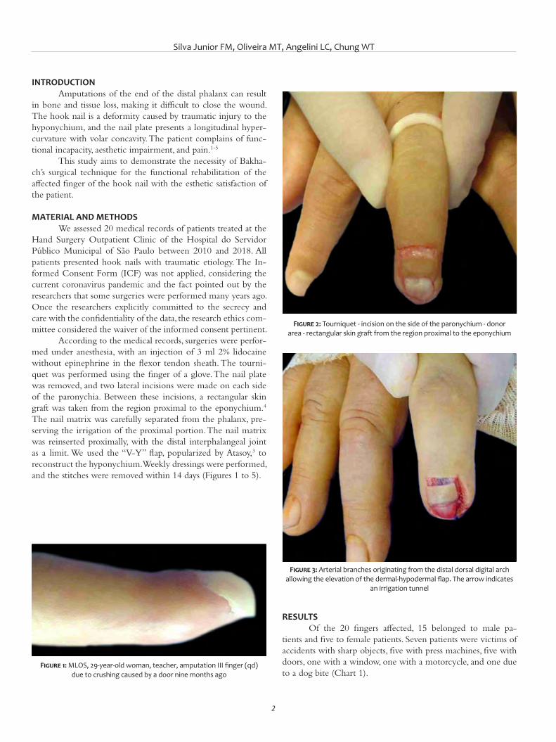
Silva Junior FM, Oliveira MT, Angelini LC, Chung WT
2
INTRODUCTIONAmputations of the end of the distal phalanx can result
in bone and tissue loss, making it difficult to close the wound. The hook nail is a deformity caused by traumatic injury to the hyponychium, and the nail plate presents a longitudinal hyper-curvature with volar concavity. The patient complains of func-tional incapacity, aesthetic impairment, and pain.1-5
This study aims to demonstrate the necessity of Bakha-ch’s surgical technique for the functional rehabilitation of the affected finger of the hook nail with the esthetic satisfaction of the patient.
MATERIAL AND METHODS We assessed 20 medical records of patients treated at the
Hand Surgery Outpatient Clinic of the Hospital do Servidor Público Municipal of São Paulo between 2010 and 2018. All patients presented hook nails with traumatic etiology. The In-formed Consent Form (ICF) was not applied, considering the current coronavirus pandemic and the fact pointed out by the researchers that some surgeries were performed many years ago. Once the researchers explicitly committed to the secrecy and care with the confidentiality of the data, the research ethics com-mittee considered the waiver of the informed consent pertinent.
According to the medical records, surgeries were perfor-med under anesthesia, with an injection of 3 ml 2% lidocaine without epinephrine in the flexor tendon sheath. The tourni-quet was performed using the finger of a glove. The nail plate was removed, and two lateral incisions were made on each side of the paronychia. Between these incisions, a rectangular skin graft was taken from the region proximal to the eponychium.4 The nail matrix was carefully separated from the phalanx, pre-serving the irrigation of the proximal portion. The nail matrix was reinserted proximally, with the distal interphalangeal joint as a limit. We used the “V-Y” flap, popularized by Atasoy,3 to reconstruct the hyponychium. Weekly dressings were performed, and the stitches were removed within 14 days (Figures 1 to 5).
RESULTS Of the 20 fingers affected, 15 belonged to male pa-
tients and five to female patients. Seven patients were victims of accidents with sharp objects, five with press machines, five with doors, one with a window, one with a motorcycle, and one due to a dog bite (Chart 1).
Figure 1: MLOS, 29-year-old woman, teacher, amputation III finger (qd) due to crushing caused by a door nine months ago
Figure 2: Tourniquet - incision on the side of the paronychium - donor area - rectangular skin graft from the region proximal to the eponychium
Figure 3: Arterial branches originating from the distal dorsal digital arch allowing the elevation of the dermal-hypodermal flap. The arrow indicates
an irrigation tunnel
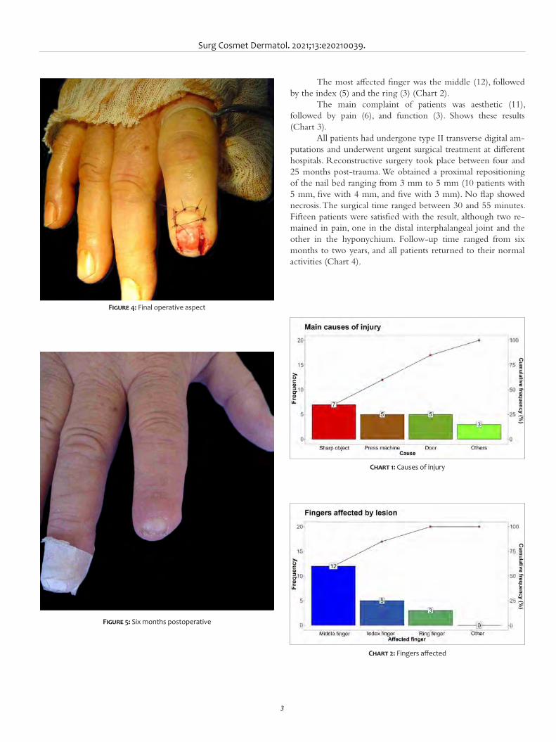
Surg Cosmet Dermatol. 2021;13:e20210039.
3
The most affected finger was the middle (12), followed by the index (5) and the ring (3) (Chart 2).
The main complaint of patients was aesthetic (11), followed by pain (6), and function (3). Shows these results (Chart 3).
All patients had undergone type II transverse digital am-putations and underwent urgent surgical treatment at different hospitals. Reconstructive surgery took place between four and 25 months post-trauma. We obtained a proximal repositioning of the nail bed ranging from 3 mm to 5 mm (10 patients with 5 mm, five with 4 mm, and five with 3 mm). No flap showed necrosis. The surgical time ranged between 30 and 55 minutes. Fifteen patients were satisfied with the result, although two re-mained in pain, one in the distal interphalangeal joint and the other in the hyponychium. Follow-up time ranged from six months to two years, and all patients returned to their normal activities (Chart 4).
Figure 4: Final operative aspect
Figure 5: Six months postoperative
Chart 1: Causes of injury
Chart 2: Fingers affected
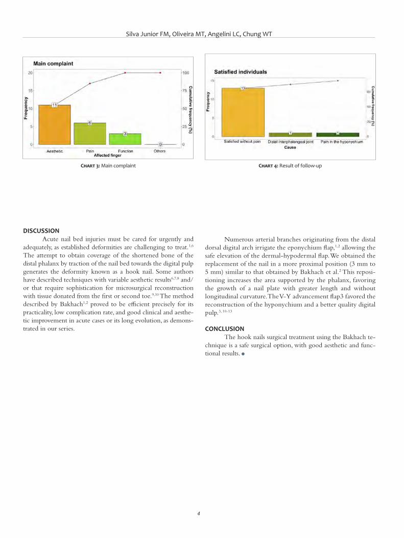
Silva Junior FM, Oliveira MT, Angelini LC, Chung WT
4
DISCUSSION Acute nail bed injuries must be cared for urgently and
adequately, as established deformities are challenging to treat.1,6 The attempt to obtain coverage of the shortened bone of the distal phalanx by traction of the nail bed towards the digital pulp generates the deformity known as a hook nail. Some authors have described techniques with variable aesthetic results6,7,8 and/or that require sophistication for microsurgical reconstruction with tissue donated from the first or second toe.9,10 The method described by Bakhach1,2 proved to be efficient precisely for its practicality, low complication rate, and good clinical and aesthe-tic improvement in acute cases or its long evolution, as demons-trated in our series.
Numerous arterial branches originating from the distal dorsal digital arch irrigate the eponychium flap,1,2 allowing the safe elevation of the dermal-hypodermal flap. We obtained the replacement of the nail in a more proximal position (3 mm to 5 mm) similar to that obtained by Bakhach et al.2 This reposi-tioning increases the area supported by the phalanx, favoring the growth of a nail plate with greater length and without longitudinal curvature. The V-Y advancement flap3 favored the reconstruction of the hyponychium and a better quality digital pulp.3, 10-13
CONCLUSION The hook nails surgical treatment using the Bakhach te-
chnique is a safe surgical option, with good aesthetic and func-tional results. l
Chart 3: Main complaint Chart 4: Result of follow-up
Surg Cosmet Dermatol. 2021;13:e20210039.
5
AUTHORS' CONTRIBUTION:
Francisco Milton da Silva Junior 0000-0001-7914-2914Study design and planning; preparation and writing of the manuscript; data collection, analysis, and interpretation; active participation in research orientation; intellectual participation in propaedeutic and/or therapeutic conduct of studied cases.
Marcelo Tavares Oliveira 0000-0002-7090-861Approval of the final version of the manuscript; study design and planning; active participation in research orientation; intellectual participation in propaedeutic and/or therapeutic conduct of studied cases; critical revision of the manuscript.
Luiz Carlos Angelini 0000-0001-6762-4270Active participation in research orientation; critical revision of the manuscript.
Wu Tu Chung 0000-0002-9709-1177 Statistical analysis; critical literature review; critical revision of the manuscript.
REFERENCES:1. Bakhach J. Le lambeau d'éponychium [Eponychial flap]. Ann Chir Plast
Esthet. 1998;43(3):259-63.2. Bakhach J, Demiri E, Guimberteau JC. Use of the eponychial flap to res-
tore the length of a short nail: a review of 30 cases. Plast Reconstr Surg. 2005;116(2):478-83.
3. Atasoy E, Ioakimidis E, Kasdan ML, Kutz JE, Kleinert HE. Reconstruction of the amputated finger tip with a triangular volar flap. A new surgical procedure. J Bone Joint Surg Am. 1970;52(5):921-6.
4. Chiu DT. Transthecal digital block: flexor tendon sheath used for anes-thetic infusion. J Hand Surg Am. 1990;15(3):471-7.
5. Foucher G, Merle M, Michon J. Les amputations digitales distales: de la cicatrisation dirigée au transfert microchirurgical de pulpe d'orteil. Indications et résultats [Distal digital amputations: from delayed hea-ling to microsurgical transfer of the toe pulp. Indications and results]. Chirurgie. 1986;112(10):727-35.
6. Atasoy E, Godfrey A, Kalisman M. The "antenna" procedure for the "hook-nail" deformity. J Hand Surg Am. 1983;8(1):55-8.
7. Dufourmentel C. Problèmes esthétiques dans la reconstruction des moignons digitaux [Esthetic problems in the reconstruction of digital stumps]. Ann Chir. 1971;25(19):995-9.
8. Foucher G, Lenoble E, Goffin D, Sammut D. Le lambeau "escalator" dans le traitement de l'ongle en griffe [Escalator flap in the treatment of claw nail]. Ann Chir Plast Esthet. 1991;36(1):51-3.
9. Foucher G, Merle M, Maneaud M, Michon J. Microsurgical free partial toe transfer in hand reconstruction: a report of 12 cases. Plast Reconstr Surg. 1980;65(5):616-27.
10. Koshima I, Moriguchi T, Umeda N, Yamada A. Trimmed second toetip transfer for reconstruction of claw nail deformity of the fingers. Br J Plast Surg. 1992;45(8):591-4.
11. Foucher G, Merle M, Maneaud M, Michon J. Microsurgical free partial toe transfer in hand reconstruction: a report of 12 cases. Plast Reconstr Surg. 1980;65(5):616-27.
12. Cunha AL, Tania LT, Oliveira LO, Craviotto M. Retalho do eponíquio para alongamento ungueal: série de casos. Rev Bras Cir Plást. 2019;34 (Suppl. 1):67-9.
13. Cambon-Binder A, Le Hanneur M, Doursounian L, Masquelet AC, Sautet A. Eponychial flap refinement for the treatment of "hook-nail" defor-mity. J Plast Reconstr Aesthet Surg. 2017;70(7):979-81.

1
APOIO CIENTÍFICO:
www.surgicalcosmetic.org.br/
ISSN-e 1984-8773
Original articleAuthors:
Elcilane Gomes Silva1
Juliana Joyce Chaves de Lima2
Natalia Pantoja Costa1
1 School of Medicine, Universidade do Estado do Pará, Belém (PA), Brazil.
2 School of Medicine, Centro Universitário Metropolitano da Amazônia, Belém (PA), Brazil.
Correspondence:Elcilane Gomes SilvaEmail: [email protected]
Financial support: None. Conflict of interest: None.
Submitted on: 13/10/2020 Approved on: 04/03/2021
How to cite this article: Silva EG, Lima JJC, Costa NP. Use of botuli-num toxin in hidradenitis suppu-rativa. Surg Cosmet Dermatol. 2021;13:e20210013.
RESUMOIntrodução: hidradenite supurativa (HS) é uma condição inflamatória, crônica, recorrente e debilitante. Há uma falta de consenso sobre o seu tratamento. Objetivo: foi realizada revisão da literatura nacional e internacional, buscando-se artigos que abordas-sem o uso da toxina botulínica na HS, além de descrever as doses e técnicas utilizadas. Métodos: foi realizada busca bibliográfica nas bases LILACS, Medline e SciELO. Resultados: considera-se que a toxina botulínica pode ser eficaz em pacientes com HS, principalmente se houver hiperidrose concomitante e quando as terapias tradicionais falham. Oferece a possibilidade de realizar um tratamento com efeitos colaterais mínimos, repetíveis ao longo do tempo sem perda de eficácia, além de diminuir a dor. Conclusões: é provável que os pacientes precisem de mais de um tratamento com o tempo, pois as lesões tendem a se repetir após uma média de seis a 10 meses. Utiliza-se a técnica padrão para hiperidrose. Mais pesquisas são necessárias para entender o seu papel no gerenciamento da HS, incluindo dosagem e frequência ideais de administração.Palavras-chave: Hidradenite supurativa; Hidradenite; Toxinas botulínicas; Toxinas botulínicas tipo ARotação
Use of botulinum toxin in hidradenitis suppurativaUso da toxina botulínica na hidradenite supurativa
DOI: http://www.dx.doi.org/10.5935/scd1984-8773.2021130013
ABSTRACTIntroduction: Hidradenitis suppurativa (HS) is an inflamatory, chronic, recurrent, and debilitat-ing condition. There is a lack of consensus about its treatment. Objective: We conducted a review of the national and international literature, searching for articles that addressed the use of botulinum toxin in HS, in addition to describing the doses and techniques used. Methods: A bibliographic search was performed in LILACS, Medline, and SciELO. Results: Botulinum toxin can be an effective therapy in patients with HS, especially in cases of concomitant hyperhidrosis and traditional therapies fail. It offers the possibility of conducting treatment with minimal adverse events, reproducible over time without loss of effectiveness, in addition to reducing pain. Conclusions: Patients will likely need more than one treatment over time, as the lesions tend to recur after 6 to 10 months. The technique used has been the standard for hyperhidrosis. Further research is needed to understand its role in the management of HS, including the ideal dosage and frequency of administration.Keywords: Hidradenitis suppurativa; Hidradenitis; Botulinum toxins; Botulinum toxins, type A
ISSN-e 1984-8773
Silva EG, Lima JJC, Costa NP
2
INTRODUCTIONHidradenitis suppurativa (HS) is also known as acne in-
versa or Verneuil’s disease. It is an inflammatory condition of a chronicle character, recurrent and debilitant. Its pathogenesis results from chronic obstruction of the follicular portions of the pilosebaceous units.1,2 Its prevalence ranges from 1% to 4% of the world’s population, but it is believed to be underestimated.²
The proposed pathogenic HS sequence starts with a follicular occlusion, causing dilation of the pilosebaceous unit, rupture, and exit of the follicular content (keratin, corneocytes, bacteria, sebaceous matter) in the dermis. The secondary inflam-matory process involves deregulation in both innate and adap-tive immunity.3,4 Extensive fibrosis is often seen as a late result of this inflammation. We still don’t know whether this mechanism comes from environmental and/or genetic factors.5 Among the risk factors, we can identify family history, obesity, smok-ing, and drug use (oral and injectable contraceptives containing medroxyprogesterone acetate or levonorgestrel).6
HS may occur sporadically, but genetic studies have iden-tified susceptibility genes involved in the gamma-secretase ex-pression. However, mutations were found only in a minority of HS patients.7 Currently, 23 pathogenic sequences are identified in HS. Still, these findings are probably significant only in the familial HS.8 Hessam et al. showed that the expression of IL36α, β, γ, and IL36Ra was significantly higher in HS lesional skin compared to healthy controls.9
The most common sites of HS involvement are inter-triginous skin areas, including armpits, groins, perianal, perineal, and inframammary regions.10 The symptoms begin after puberty and at age 40, being more common in women, with a propor-tion of 3,6:1 reported in a retrospective French observational study.6,11 Lesions start as inflamed nodules and may evolve into fistulized and interconnected abscesses, forming scars, with pain as their main characteristic.12
It is noteworthy that the pain, associated with foul odor due to the secretion and scar formation, significantly impacts the patients’ quality of life. Even though it is a prevalent disease with limited therapeutic options, it is necessary to correctly treat HS patients due to the higher presence of depression and sexual dys-function among them.13 Thus, due to its chronic character and its impact on the patients’ quality of life, its correct management, although complex, is essential.14
The initial approach of these patients depends on their classification using Hurley’s clinical staging:15 stage I - abscess(es) formation, no fistulization, and no scarring; stage II - recurrent abscesses, with sinus tracts formation and scarring; stage III - diffuse abscesses or interconnected sinus tracts and multiple ab-scesses.
In all patients, conservative management with weight loss and smoking cessation is recommended, and psychosocial sup-port and analgesia if necessary. Antibiotics are effective in mild to moderate cases. In women, antiandrogenic therapy reduces the severity of the disease in some cases. Systemic immunosup-
pression, including cyclosporine and infliximab, led to a signif-icant improvement in moderate to severe disease. In advanced disease, or where medical therapy has failed, surgical treatment may involve radical excision of affected apocrine glands. LASER (light amplification by stimulated emission of radiation) therapy and external beam radiation have also demonstrated efficacy and may be helpful in selected patients.16
Recently, the multiple uses of botulinum toxin (BT) in dermatological conditions have been reviewed, especially those called “Fold (intertriginous) dermatoses”.17 Among these is HS, which involves precisely these areas of the body.5
The exact mechanism by which BT affects the disease process in HS is unclear. The moist environment resulting from the sweat retained in the armpit and groin provides ideal condi-tions for bacteria to flourish. The presence of apocrine glandular secretions can aggravate it, forming a rich substrate for bacterial growth. The antiperspirant effect of BT reduces the skin flora population, consequently decreasing the inflammatory stimulus for HS. A second possibility is that BT shuts down the function of the whole pilosebaceous unit and apocrine secretion, prevent-ing the rupture and dissemination of follicular material through the dermis, which usually results in more inflammation.18
Currently, there is a lack of consensus on the ideal treat-ment for HS. Despite the advent of biological drugs, the disease remains a therapeutic challenge. Many therapies are available, but the benefits of these treatments should be weighed against the adverse events. BT represents a new and promising treatment option for this complex disease. However, a shared treatment regimen is necessary to define the potential of this treatment in HS, and mainly, a change in the quality of life of these patients. Given this context, it is relevant to conduct studies seeking to relate such information and subsidize new and more efficient approaches. The present study aimed at reviewing the use of botulinum toxin in hidradenitis suppurativa in the national and international literature, besides describing the doses and tech-niques used to treat the disease.
METHODThis study is a narrative literature review. We conducted
a bibliographic search on the electronic databases Latin Ameri-can & Caribbean Health Sciences Literature (LILACS), Medical Literature Analysis and Retrieval System Online (Medline), and Scientific Electronic Library Online (SciELO), published from 2009 to 2019.
The descriptors used were: Hidradenitis Suppurativa, Hidradenitis, Botulinum Toxins and Botulinum Toxins, Type A. All descriptors are found in the Health Sciences Descriptors (DeCS). We identified nine articles in the databases mentioned above and selected six of them for this study after reading their title and abstract.
Surg Cosmet Dermatol. 2021;13:e20210013.
3
RESULTSThe first case of HS treated with botulinum toxin type
A (BTXA) dates back to 2005. The 38-year-old patient (patient A) had the disease for more than ten years and was submitted to therapy with lymecycline and flucloxacillin without clinical improvement. She received then 250 U of Dysport® in both armpits. The toxin was delivered with intradermal papules, the standard technique for hyperhidrosis. After 15 days of the ad-ministration, there was no evidence of active inflammation. The patient had complete remission of symptoms until approximate-ly ten months later when the first symptoms of mild inflamma-tion reappeared.19
Feito-Rodriguez et al. described the first case of appli-cation of botulinum toxin A in a prepubertal child to treat HS (patient B). At six years of age, the child was diagnosed with HS, presenting erythematous papules and painful nodules only in the groin, symmetrically. Topical and oral antibacterials were prescribed, in addition to isotretinoin, which was discontinued due to adverse events. At the age of 7 years and 5 months, a total dose of 40 U of Botox® was applied under sedation, distributed in 10 to 12 points on an elliptical area on each side of the groin. She had complete remission until six months later when the first lesions reappeared. A new, similar application was made with a good therapeutic response.20
Khoo & Burova reported the third case in the literature, using BTXA to treat HS in three patients. The authors reported the most successful case in a 46-year-old patient (patient C) who had the disease for 11 years (Hurley II). She had already been submitted to systemic antibiotics, without improvement, and surgical drainage of the abscesses. The patient received 50 U of BTXA in each armpit. There was a good clinical response with-in three months after the first treatment, and after the second treatment, there was clinical remission, which lasted for more than a year. The other two patients presented a disease-free peri-od from 5 to 6 months.18
Reilly et al. described another case of success in a 23-year-old woman (patient D), diagnosed with HS for four years. She had performed eight surgical incisions in the groins, buttocks, and inner part of the thighs, with slight improvement. The patient received 100 U of BTXA (Botox®) in the groin and inner thighs and 130 U in the buttocks. There was a complete resolution of the symptoms for three months. In the following 18 months, the patient received five more BTXA applications, each consisting of 200 U (100 U in the anterior region and 100 in the posterior). There was a four-month interval between treatments, and in the last month of this interval, there was an exacerbation of the lesions, requiring surgical intervention.16
Shi et al. also reported a case of successful treatment of HS in stage III with BTXA. The 40-year-old patient (patient E) presented HS in the groin and armpits with chronic pain and had failed therapy with oral and topical antibiotics and surgical treatment. She then received a total of 400 U of BTXA – 100 U for each site (bilateral and inframammary axillary region). The
study used intradermal injection in parallel lines with 1 cm of distance between the points, with approximately 20 injections per area. There was an excellent response with a significant re-duction in drainage and inflammation, as well as pain relief of 50%. In subsequent administration, the dose was reduced to 50 U per area, in addition to injection at other areas such as groins and perineal regions. However, the symptoms returned earlier with the dose reduction. Thus, the subsequent applications re-sumed the dose of 100 U for each site. The patient received no other treatment during BTXA therapy.10
In 2019, Campanati et al. described two cases of HS treated with BTXA. Case 1 was a 23-year-old young woman (patient F) who presented HS in the armpits (Hurley II) re-fractory to topical and oral therapies. The area was treated with BTXA (Vistabex®), with a technique similar to axillary hyper-hidrosis’ procedure, using 50 U per armpit diluted in 2.5 ml of 0.9% saline solution. She received an intradermal injection, with the administration of 4 U in each square of 1.5 cm². Case 2 was a 50-year-old man (patient G) with HS in the groin and inner thigh region (Hurley II) who had failed other topical and systemic therapies. He received BTXA (Botox®) in the same dilution and technique described above, but 100 U on each side. Both patients were reassessed after one, three, and six months. They experienced real improvement with the disappearance of inflammatory lesions in case 1 and reduction of fistulas in the groin in case 2. The patient of case 1 decided to undergo therapy again after ten months with an optimal response5 (Table 1).
DISCUSSIONBotulinum toxin type A can be an effective therapy in
patients with HS, especially if there is concomitant hyperhidrosis and when traditional therapies fail.
The BTXA blocks the release of acetylcholine and other pre-synaptic vesicle neurotransmitters by deactivating SNARE proteins. It has been used for hyperhidrosis blocking the cholin-ergic nerve fibers of the sweat glands. The US Food and Drug Administration (FDA) approved it in 2004 to treat severe prima-ry axillary hyperhidrosis refractory to the use of antiperspirants.10
It is not clear how BTXA can improve HS. The first hy-pothesis is that local factors such as humidity can create an ideal environment for bacterial growth, a known pro-inflammatory element. In fact, bacteria are not promoters of the disease, but they among the main contributors to a vicious inflammatory circle.21 Thus, reducing sweat production could reduce the skin flora population and its potential pro-inflammatory effect.
The other hypothesis about the BTXA therapeutic effect is that it shuts down the function of the entire pilosebaceous unit and apocrine secretions, preventing the rupture and spread of follicular material through the dermis, which usually results in more inflammation and formation of the sinus tract.17
Patient A, treated with 250 U of Dysport® in both arm-pits, presented condition improvement attributed to reducing
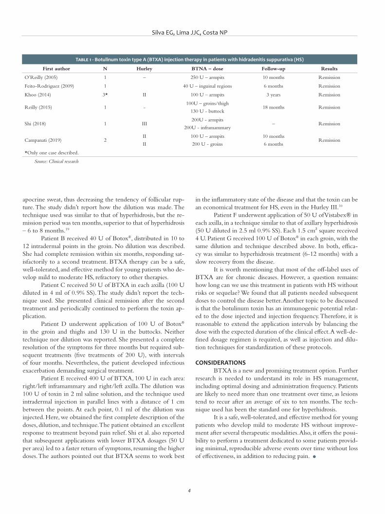
Silva EG, Lima JJC, Costa NP
4
apocrine sweat, thus decreasing the tendency of follicular rup-ture. The study didn’t report how the dilution was made. The technique used was similar to that of hyperhidrosis, but the re-mission period was ten months, superior to that of hyperhidrosis – 6 to 8 months.19
Patient B received 40 U of Botox®, distributed in 10 to 12 intradermal points in the groin. No dilution was described. She had complete remission within six months, responding sat-isfactorily to a second treatment. BTXA therapy can be a safe, well-tolerated, and effective method for young patients who de-velop mild to moderate HS, refractory to other therapies.
Patient C received 50 U of BTXA in each axilla (100 U diluted in 4 ml of 0.9% SS). The study didn’t report the tech-nique used. She presented clinical remission after the second treatment and periodically continued to perform the toxin ap-plication.
Patient D underwent application of 100 U of Botox® in the groin and thighs and 130 U in the buttocks. Neither technique nor dilution was reported. She presented a complete resolution of the symptoms for three months but required sub-sequent treatments (five treatments of 200 U), with intervals of four months. Nevertheless, the patient developed infectious exacerbation demanding surgical treatment.
Patient E received 400 U of BTXA, 100 U in each area: right/left inframammary and right/left axilla. The dilution was 100 U of toxin in 2 ml saline solution, and the technique used intradermal injection in parallel lines with a distance of 1 cm between the points. At each point, 0.1 ml of the dilution was injected. Here, we obtained the first complete description of the doses, dilution, and technique. The patient obtained an excellent response to treatment beyond pain relief. Shi et al. also reported that subsequent applications with lower BTXA dosages (50 U per area) led to a faster return of symptoms, resuming the higher doses. The authors pointed out that BTXA seems to work best
in the inflammatory state of the disease and that the toxin can be an economical treatment for HS, even in the Hurley III.10
Patient F underwent application of 50 U of Vistabex® in each axilla, in a technique similar to that of axillary hyperhidrosis (50 U diluted in 2.5 ml 0.9% SS). Each 1.5 cm² square received 4 U. Patient G received 100 U of Botox® in each groin, with the same dilution and technique described above. In both, effica-cy was similar to hyperhidrosis treatment (6-12 months) with a slow recovery from the disease.
It is worth mentioning that most of the off-label uses of BTXA are for chronic diseases. However, a question remains: how long can we use this treatment in patients with HS without risks or sequelae? We found that all patients needed subsequent doses to control the disease better. Another topic to be discussed is that the botulinum toxin has an immunogenic potential relat-ed to the dose injected and injection frequency. Therefore, it is reasonable to extend the application intervals by balancing the dose with the expected duration of the clinical effect. A well-de-fined dosage regimen is required, as well as injection and dilu-tion techniques for standardization of these protocols.
CONSIDERATIONSBTXA is a new and promising treatment option. Further
research is needed to understand its role in HS management, including optimal dosing and administration frequency. Patients are likely to need more than one treatment over time, as lesions tend to recur after an average of six to ten months. The tech-nique used has been the standard one for hyperhidrosis.
It is a safe, well-tolerated, and effective method for young patients who develop mild to moderate HS without improve-ment after several therapeutic modalities. Also, it offers the possi-bility to perform a treatment dedicated to some patients provid-ing minimal, reproducible adverse events over time without loss of effectiveness, in addition to reducing pain. l
Table 1 - Botulinum toxin type A (BTXA) injection therapy in patients with hidradenitis suppurativa (HS)
First author N Hurley BTNA – dose Follow-up Results
O’Reilly (2005) 1 – 250 U – armpits 10 months Remission
Feito-Rodriguez (2009) 1 40 U – inguinal regions 6 months Remission
Khoo (2014) 3* II 100 U – armpits 3 years Remission
Reilly (2015) 1 -100U – groins/thigh
130 U - buttock18 months Remission
Shi (2018) 1 III200U - armpits
200U - inframammary– Remission
Campanati (2019) 2II
II
100 U – armpits
200 U - groins
10 months
6 monthsRemission
*Only one case described.
Source: Clinical research
Surg Cosmet Dermatol. 2021;13:e20210013.
5
REFERENCES1. Alikhan A, Lynch PJ, Eisen DB. Hidradenitis suppurativa: a comprehen-
sive review. J Am Acad Dermatol. 2009;60(4):539-61.2. Jemec GBE. Clinical practice. Hidradenitis suppurativa. N Engl J Med.
2012;366(2):158-64.3. Napolitano M, Megna M, Timoshchuk EA, Patruno C, Balato N, Fabbro-
cini G, et al. Hidradenitis suppurativa: froma pathogenesis to diagnosis and treatment. Clin Cosmet Investig Dermatol. 2017;10:105-15.
4. Van der Zee HH, Laman JD, Boer J, Prens EP. Hidradenitis suppurativa: viewpoint on clinical phenotyping, pathogenesis and novel treat-ments. Exp Dermatol. 2012;21(10):735-39.
5. Campanati A, Martina E, Giuliodori K, Bobyr I, Consales V, Offidani A. Two cases of Hidradenitis suppurativa and botulinum toxin type a the-rapy: A novel approach for a pathology that is still difficult to manage. Dermatol Ther. 2019;32(3):e12841.
6. Vazquez BG, Alikhan A, Weaver AL, Wetter DA, Davis MD. Incidence of hidradenitis suppurativa and associated factors: a population-based study of Olmsted County, Minnesota. J Invest Dermatol. 2013;133(1):97-103.
7. Pink AE, Simpson MA, Desai N, Trembath RC, Barker JNW. Gamma-se-cretase mutations in hidradenitis suppurativa: new insights into disea-se pathogenesis. J Invest Dermatol. 2013;133(3):601-7.
8. Frew JW, Vekic DA, Woods J, Cains GD. A systematic review and critical evaluation of reported pathogenic sequence variants in hidradenitis suppurativa. Br J Dermatol. 2017;177(4):987-98.
9. Hessam S, Sand M, Gambichler T, Skrygan M, Ruddel I, Bechara FG. Interleukin-36 in hidradenitis suppurativa: evidence for a distinctive proinflammatory role and a key factor in the development of anin-flammatory loop. Br J Dermatol. 2018;178(3):761-7.
10. Shi W, Schultz S, Strouse A, Gater DR. Successful treatment of stage III hidradenitis suppurativawith botulinum toxin A. Br J Dermatol Case Rep. 2019;12(1):e226064.
11. Canoui-Poitrine F, Le Thuaut A, Revuz JE, Viallette C, Gabison G, Poli F, et al. Identification of three hidradenitis suppurativa phenotypes: latent class analysis of a cross- sectional study. J Invest Dermatol. 2013;133(6):1506-11.
12. Kurek A, Peters EM, Chanwangpong A, Sabat R, Sterry W, Schneider- Burrus S. Profound disturbances of sexual health in patients with acne inversa. J Am Acad Dermatol. 2012;67(3):422-8.
13. Onderdijk AJ, van der Zee HH, Esmann S, Lophaven S, Dufour DN, Je-mec GB, et al. Depression in patients with hidradenitis suppurativa. J Eur Acad Dermatol Venereol. 2013;27(4):473-8.
14. Muzy G, Crocco EI, Alves RO. Hidradenite supurativa: atualização e revisão de suas modalidades terapêuticas. Surg Cosmet Dermatol. 2014;6(3):206-12.
15. Sartorius K, Emtestam L, Jemec GB, Lapins J. Objective scoring of hidra-denitis suppurativareflecting the role of tobacco smoking and obesity. Br J Dermatol. 2009;161(4):831-9.
16. Reilly DJ, Munasinghe CP, Nizzero DE. Botulinum toxin A for the ma-nagement of hidradenitis suppurativa. Plast Surg Case Studies. 2015;1(1):3-4.
17. Campanati A, Martina E, Giuliodori K, Consales V, Bobyr I, Offidani A. Bo-tulinum toxin Off-Label use in dermatology: a review. Skin Appendage Disord. 2017;3(1):39-56.
18. Khoo ABS, Burova EP. Hidradenitis suppurativa treated with clostridium botulinum toxin A. Clin Exp Dermatol. 2014;39(6):749-50.
19. O'Reilly DJ, Pleat JM, Richards AM. Treatment of hidradenitis suppura-tiva with botulinum toxin A. Plast Reconstr Surg. 2005;116(5):1575-6.
20. Feito-Rodriguez M, Sendagorta-Cudos E, Herranz-Pinto P, de Lucas-La-guna R. Prepubertal hidradenitis suppurativa successfully treated with botulinum toxin A. Am Soc Dermatol Surg. 2009; 35(8):1300-2.
21. Matusiak L, Bieniek A, Szepietowski JC. Bacteriology of hidradenitis su-ppurativa - Which antibiotics are the treatment of choice? Acta Derm Venereol. 2014;94(6):699-702.
AUTHORS' CONTRIBUTION:
Elcilane Gomes Silva 0000-0001-8841-2285Preparation and writing of the manuscript; data collection, analysis, and interpretation.
Juliana Joyce Chaves de Lima 0000-0002-9971-5908Preparation and writing of the manuscript; data collection, analysis, and interpretation.
Natalia Pantoja Costa 0000-0002-3454-6760Author’s contribution: Preparation and writing of the manuscript; data collection, analysis, and interpretation.
1
APOIO CIENTÍFICO:
www.surgicalcosmetic.org.br/
ISSN-e 1984-8773
Original ArticleAuthors: Jaqueline Barbeito de Vasconcellos¹ Isabele Oliveira Santos¹ Daniela Alves Pereira Antelo¹
¹ Universidade do Estado do Rio de Janeiro, Dermatology Service,
Rio de Janeiro (RJ), Brazil.
Correspondence: Jaqueline Barbeito de Vasconcellos Email: jaqueline_vasconcellos@ hotmail.com
Financial support: None.Conflict of interest: None.
Submitted on: 29/03/2021 Approved on: 03/05/2021
How to cite this article: Vasconcellos JB, Santos IO, Antelo DAP. Use of botulinum toxin for rosacea: a pilot study. Surg Cosmet Dermatol. 2021;13:e20210019.
Use of botulinum toxin for rosacea: a pilot study Uso da toxina botulínica para rosácea: estudo-piloto
ABSTRACTIntroduction: Rosacea is a chronic inflammatory skin disease. The intradermal application of botulinum toxin (BT) has been studied as a therapeutic option for patients who struggle to manage flushing and/or persistent facial erythema. There is no standard protocol for BT application in rosacea. Objective: To evaluate the effectiveness of botulinum toxin application on erythematotelan-giectatic rosacea. Methods: Pilot study with case series. We applied intradermal BT in 10 patients with a diagnosis of rosacea and symptoms of persistent erythema and/or facial flushing. Patients received 10 to 15 injections per hemiface (1 unit of onabotulinum BT per injection) and 0 to 5 injections in the nasal region, totaling 25 to 35 units per patient. Results: Seventy-five percent of the patients presented a reduction in flush and erythema inten-sity. The follow-up time was three months, and no serious adverse events were observed. Conclusions: The therapeutic arsenal to control erythema and facial flushing of rosacea, espe-cially refractory to the usual treatment, should consider the intradermal application of BT type A.Keywords: Erythema; Rosacea; Flushing; Botulinum toxins
RESUMOIntrodução: rosácea é uma doença inflamatória crônica da pele, e a aplicação intradérmica de toxina botulínica (TB) tem sido estudada como uma opção terapêutica aos pacientes de difícil manejo do flushing e/ou eritema facial persistente. Ainda não há protocolo-padrão para aplicação da TB na rosácea. Objetivo: avaliar o efeito da aplicação de toxina botulínica na rosácea eritêmato-telangiectásica. Métodos: estudo-piloto com série de casos. Foi realizada a aplicação intradérmica da TB em 10 pacientes com diagnóstico de rosácea e sintomas de eritema persistente e/ou flushing facial. Os pacientes foram submetidos a 10 a 15 injeções por hemiface (1 unidade de TB onabotulínica por injeção) e 0 a 5 injeções na região nasal, totalizando 25 a 35 unidades por paciente. Resultados: apresentaram redução na intensidade do flush e do eritema 75% dos pacientes. O tempo de acompanhamento foi de três meses e nenhum evento adverso grave foi observado. Conclusões: a aplicação intradérmica de TB tipo A deve ser considerada no arsenal terapêutico para controle do eritema e flushing facial da rosácea, especialmente em casos refratários ao tratamento habitual.Palavras-chave: Eritema; Rosácea; Rubor; Toxinas botulínicas
DOI: http://www.dx.doi.org/10.5935/scd1984-8773.2021130019
ISSN-e 1984-8773
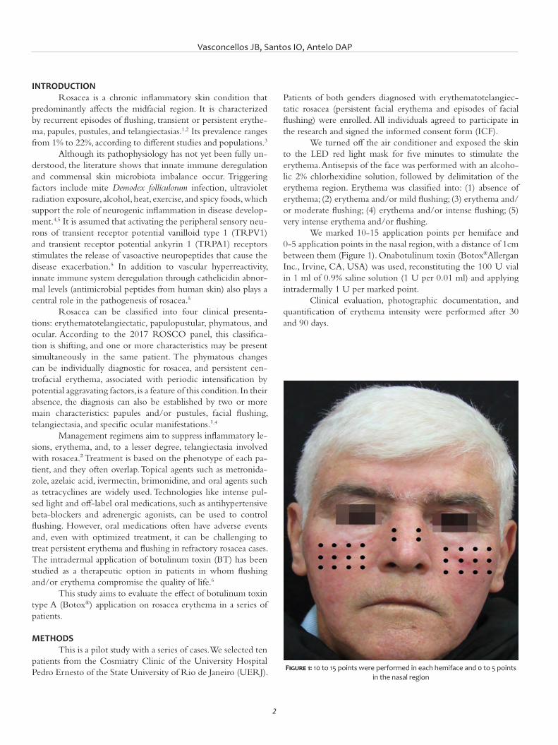
Vasconcellos JB, Santos IO, Antelo DAP
2
INTRODUCTIONRosacea is a chronic inflammatory skin condition that
predominantly affects the midfacial region. It is characterized by recurrent episodes of flushing, transient or persistent erythe-ma, papules, pustules, and telangiectasias.1,2 Its prevalence ranges from 1% to 22%, according to different studies and populations.3
Although its pathophysiology has not yet been fully un-derstood, the literature shows that innate immune deregulation and commensal skin microbiota imbalance occur. Triggering factors include mite Demodex folliculorum infection, ultraviolet radiation exposure, alcohol, heat, exercise, and spicy foods, which support the role of neurogenic inflammation in disease develop-ment.4,5 It is assumed that activating the peripheral sensory neu-rons of transient receptor potential vanilloid type 1 (TRPV1) and transient receptor potential ankyrin 1 (TRPA1) receptors stimulates the release of vasoactive neuropeptides that cause the disease exacerbation.5 In addition to vascular hyperreactivity, innate immune system deregulation through cathelicidin abnor-mal levels (antimicrobial peptides from human skin) also plays a central role in the pathogenesis of rosacea.5
Rosacea can be classified into four clinical presenta-tions: erythematotelangiectatic, papulopustular, phymatous, and ocular. According to the 2017 ROSCO panel, this classifica-tion is shifting, and one or more characteristics may be present simultaneously in the same patient. The phymatous changes can be individually diagnostic for rosacea, and persistent cen-trofacial erythema, associated with periodic intensification by potential aggravating factors, is a feature of this condition. In their absence, the diagnosis can also be established by two or more main characteristics: papules and/or pustules, facial flushing, telangiectasia, and specific ocular manifestations.¹,4
Management regimens aim to suppress inflammatory le-sions, erythema, and, to a lesser degree, telangiectasia involved with rosacea.² Treatment is based on the phenotype of each pa-tient, and they often overlap. Topical agents such as metronida-zole, azelaic acid, ivermectin, brimonidine, and oral agents such as tetracyclines are widely used. Technologies like intense pul-sed light and off-label oral medications, such as antihypertensive beta-blockers and adrenergic agonists, can be used to control flushing. However, oral medications often have adverse events and, even with optimized treatment, it can be challenging to treat persistent erythema and flushing in refractory rosacea cases. The intradermal application of botulinum toxin (BT) has been studied as a therapeutic option in patients in whom flushing and/or erythema compromise the quality of life.6
This study aims to evaluate the effect of botulinum toxin type A (Botox®) application on rosacea erythema in a series of patients.
METHODSThis is a pilot study with a series of cases. We selected ten
patients from the Cosmiatry Clinic of the University Hospital Pedro Ernesto of the State University of Rio de Janeiro (UERJ).
Patients of both genders diagnosed with erythematotelangiec-tatic rosacea (persistent facial erythema and episodes of facial flushing) were enrolled. All individuals agreed to participate in the research and signed the informed consent form (ICF).
We turned off the air conditioner and exposed the skin to the LED red light mask for five minutes to stimulate the erythema. Antisepsis of the face was performed with an alcoho-lic 2% chlorhexidine solution, followed by delimitation of the erythema region. Erythema was classified into: (1) absence of erythema; (2) erythema and/or mild flushing; (3) erythema and/or moderate flushing; (4) erythema and/or intense flushing; (5) very intense erythema and/or flushing.
We marked 10-15 application points per hemiface and 0-5 application points in the nasal region, with a distance of 1cm between them (Figure 1). Onabotulinum toxin (Botox®Allergan Inc., Irvine, CA, USA) was used, reconstituting the 100 U vial in 1 ml of 0.9% saline solution (1 U per 0.01 ml) and applying intradermally 1 U per marked point.
Clinical evaluation, photographic documentation, and quantification of erythema intensity were performed after 30 and 90 days.
Figure 1: 10 to 15 points were performed in each hemiface and 0 to 5 points in the nasal region
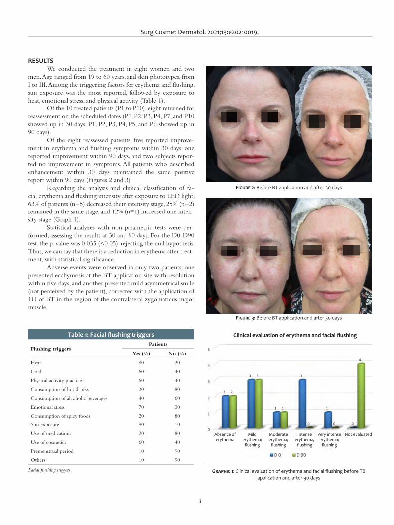
Surg Cosmet Dermatol. 2021;13:e20210019.
3
RESULTSWe conducted the treatment in eight women and two
men. Age ranged from 19 to 60 years, and skin phototypes, from I to III. Among the triggering factors for erythema and flushing, sun exposure was the most reported, followed by exposure to heat, emotional stress, and physical activity (Table 1).
Of the 10 treated patients (P1 to P10), eight returned for reassessment on the scheduled dates (P1, P2, P3, P4, P7, and P10 showed up in 30 days; P1, P2, P3, P4, P5, and P6 showed up in 90 days).
Of the eight reassessed patients, five reported improve-ment in erythema and flushing symptoms within 30 days, one reported improvement within 90 days, and two subjects repor-ted no improvement in symptoms. All patients who described enhancement within 30 days maintained the same positive report within 90 days (Figures 2 and 3).
Regarding the analysis and clinical classification of fa-cial erythema and flushing intensity after exposure to LED light, 63% of patients (n=5) decreased their intensity stage, 25% (n=2) remained in the same stage, and 12% (n=1) increased one inten-sity stage (Graph 1).
Statistical analyzes with non-parametric tests were per-formed, assessing the results at 30 and 90 days. For the D0-D90 test, the p-value was 0.035 (<0.05), rejecting the null hypothesis. Thus, we can say that there is a reduction in erythema after treat-ment, with statistical significance.
Adverse events were observed in only two patients: one presented ecchymosis at the BT application site with resolution within five days, and another presented mild asymmetrical smile (not perceived by the patient), corrected with the application of 1U of BT in the region of the contralateral zygomaticus major muscle.
Figure 2: Before BT application and after 30 days
Figure 3: Before BT application and after 30 days
Table 1: Facial flushing triggers
Flushing triggersPatients
Yes (%) No (%)
Heat 80 20
Cold 60 40
Physical activity practice 60 40
Consumption of hot drinks 20 80
Consumption of alcoholic beverages 40 60
Emotional stress 70 30
Consumption of spicy foods 20 80
Sun exposure 90 10
Use of medications 20 80
Use of cosmetics 60 40
Premenstrual period 10 90
Others 10 90
Facial flushing triggers Graphic 1: Clinical evaluation of erythema and facial flushing before TB application and after 90 days
Clinical evaluation of erythema and facial flushing
Absence of erythema
Mild erythema/
flushing
Moderate erythema/
flushing
Intense erythema/
flushing
Very intense erythema/
flushing
Not evaluated
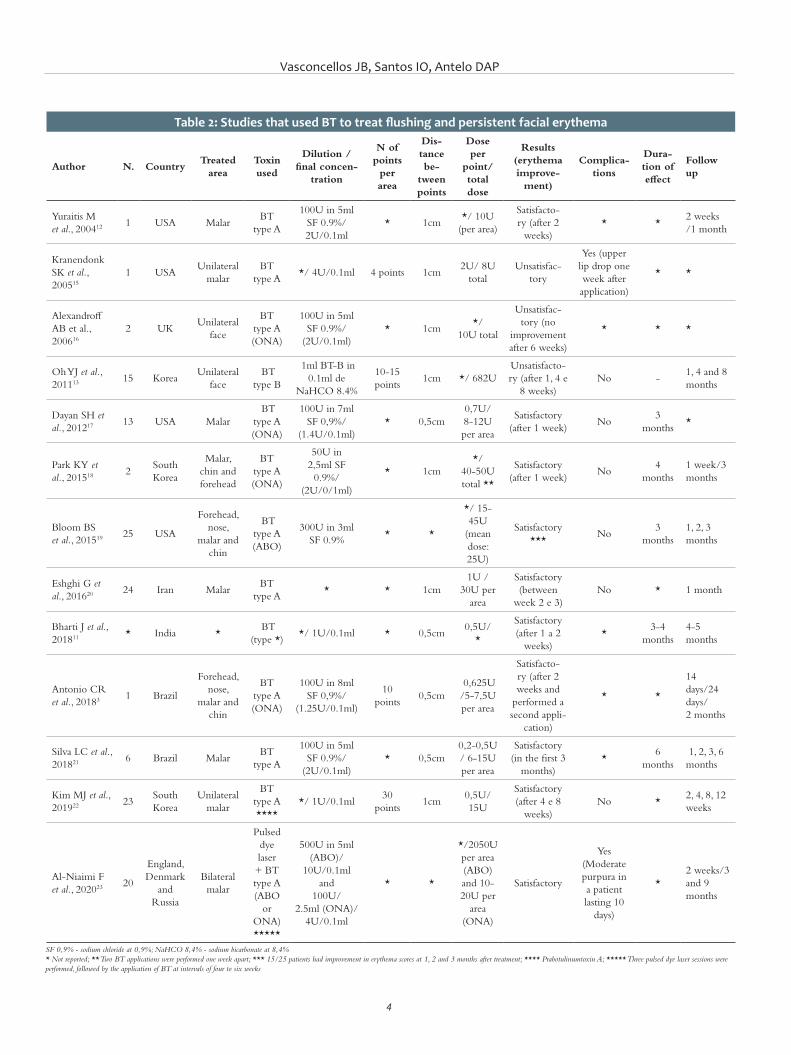
Vasconcellos JB, Santos IO, Antelo DAP
4
Table 2: Studies that used BT to treat flushing and per sistent facial erythema
Author N. Country Treated
area Toxin used
Dilution / final concen-
tration
N of points per area
Dis-tance be-
tween points
Dose per
point/total dose
Results (erythema improve-
ment)
Complica-tions
Dura-tion of effect
Follow up
Yuraitis M et al., 200412
1 USA Malar BT
type A
100U in 5ml SF 0.9%/ 2U/0.1ml
* 1cm */ 10U
(per area)
Satisfacto-ry (after 2
weeks) * *
2 weeks /1 month
KranendonkSK et al., 200515
1 USAUnilateral
malarBT
type A*/ 4U/0.1ml 4 points 1cm
2U/ 8U total
Unsatisfac-tory
Yes (upper lip drop one week after application)
* *
AlexandroffAB et al., 200616
2 UK Unilateral
face
BT type A (ONA)
100U in 5mlSF 0.9%/
(2U/0.1ml)* 1cm
*/ 10U total
Unsatisfac-tory (no
improvement after 6 weeks)
* * *
Oh YJ et al., 201113
15 Korea Unilateral
face BT
type B
1ml BT-B in0.1ml de
NaHCO 8.4%
10-15 points
1cm */ 682U Unsatisfacto-ry (after 1, 4 e
8 weeks) No -
1, 4 and 8 months
Dayan SH et al., 201217
13 USA Malar BT
type A (ONA)
100U in 7mlSF 0,9%/
(1.4U/0.1ml)* 0,5cm
0,7U/ 8-12U per area
Satisfactory (after 1 week)
No 3
months *
Park KY et al., 201518
2 South Korea
Malar, chin and forehead
BT type A (ONA)
50U in2,5ml SF
0.9%/ (2U/0/1ml)
* 1cm */
40-50Utotal **
Satisfactory (after 1 week)
No 4
months 1 week/3 months
Bloom BS et al., 201519
25 USA
Forehead, nose,
malar and chin
BT type A (ABO)
300U in 3mlSF 0.9%
* *
*/ 15-45U
(mean dose: 25U)
Satisfactory ***
No 3
months 1, 2, 3 months
Eshghi G et al., 201620
24 Iran Malar BT
type A* * 1cm
1U / 30U per
area
Satisfactory (between
week 2 e 3) No * 1 month
Bharti J et al., 201811
* India * BT
(type *) */ 1U/0.1ml * 0,5cm
0,5U/ *
Satisfactory (after 1 a 2
weeks) *
3-4 months
4-5 months
Antonio CR et al., 20183 1 Brazil
Forehead, nose,
malar and chin
BT type A (ONA)
100U in 8ml SF 0,9%/
(1.25U/0.1ml)
10 points
0,5cm 0,625U /5-7,5U per area
Satisfacto-ry (after 2 weeks and
performed a second appli-
cation)
* *
14 days/24 days/2 months
Silva LC et al., 201821
6 Brazil Malar BT
type A
100U in 5mlSF 0.9%/
(2U/0.1ml)* 0,5cm
0,2-0,5U / 6-15U per area
Satisfactory (in the first 3
months) *
6 months
1, 2, 3, 6 months
Kim MJ et al., 201922
23 South Korea
Unilateral malar
BT type A ****
*/ 1U/0.1ml 30
points 1cm
0,5U/ 15U
Satisfactory (after 4 e 8
weeks) No *
2, 4, 8, 12 weeks
Al-Niaimi F et al., 202023 20
England, Denmark
and Russia
Bilateral malar
Pulsed dye laser+ BT type A (ABO
or ONA) *****
500U in 5ml(ABO)/
10U/0.1ml and
100U/2.5ml (ONA)/
4U/0.1ml
* *
*/2050U per area (ABO) and 10-20U per
area(ONA)
Satisfactory
Yes (Moderate purpura in a patient lasting 10
days)
* 2 weeks/3 and 9 months
SF 0,9% - sodium chloride at 0,9%; NaHCO 8,4% - sodium bicarbonate at 8,4% * Not reported; ** Two BT applications were performed one week apart; *** 15/25 patients had improvement in erythema scores at 1, 2 and 3 months after treatment; **** Prabotulinumtoxin A; ***** Three pulsed dye laser sessions were performed, followed by the application of BT at intervals of four to six weeks
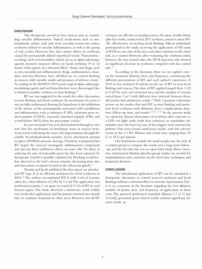
Surg Cosmet Dermatol. 2021;13:e20210019.
5
DISCUSSIONThe therapeutic arsenal to treat rosacea aims at control-
ling vascular inflammation. Topical medications such as me-tronidazole, azelaic acid, and, more recently, ivermectin reduce erythema related to vascular inflammation, as well as the group of oral cyclins. However, they have minor effects on erythema caused by permanently dilated superficial vessels.7 Vasoconstric-tor drugs, such as brimonidine, which acts as an alpha-adrenergic agonist, promote transient effects on facial erythema (9 to 12 hours) with reports of a rebound effect.8 Some oral drugs, such as non-steroidal anti-inflammatory drugs, antihistamines, cloni-dine, and beta-blockers, have off-label use to control flushing in rosacea, with variable results and presence of adverse events.9 According to the ROSCO 2019 panel, topical alpha-adrenergic modulating agents and oral beta-blockers were discouraged due to limited scientific evidence to treat flushing.10
BT use was suggested in the search for other alternatives to treat flushing and facial erythema. Its mechanism of action is not yet fully understood. Among the hypotheses is the inhibition of the release of the neuropeptides associated with vasodilation and inflammation, such as substance P (SP), calcitonin gene-re-lated peptide (CGRP), vasoactive intestinal peptide (VIP), and acetylcholine (ACh) from the presynaptic vesicle.5
In a recent study, Choi et al. demonstrated through in vivo tests that the mechanism of botulinum toxin in rosacea treat-ment involves blocking the mast cells degranulation through the soluble N-ethylmaleimide-sensitive factor attachment protein receptor (SNARE) proteins cleavage. Therefore, it proposed that BT targets the rosacea’s neurogenic inflammatory component and also has direct inhibitory effects on mast cells.5 Its effect in reducing the size of noticeable pores has also been reported. Its therapeutic benefit is possibly explained by blocking acetylcho-line directed to the hair’s erector muscles, decreasing pore size, and muscarinic receptors located in the sebaceous glands.11
Yuraitis and Jacob published the first report on intrader-mal BT type A as an effective treatment for facial erythema in 2004.12 The authors reconstituted BT-A with 5 ml of isotonic saline for a final dilution of 2 IU by 0.1 ml. The application was performed at points 1 cm apart, in a total of 10 UI of BT in each treated region. The study observed a satisfactory result within two weeks after application, and the patient returned one month later to continue treatment in other areas. However, not all BT
serotypes are effective in treating rosacea. An open, double-blind, split-face study, conducted in 2011 in Korea, aimed to assess BT--B’s effectiveness in treating facial flushing. Fifteen individuals participated in the study, receiving the application of 682 units of BT-B on one side of the face, and saline solution on the other side, as a control. However, after evaluating the erythema index between the two treated sides, the BT-B injection side showed no significant decrease in erythema compared with the control side.13
According to the literature, there are no explicit crite-ria for treatment dilution, dose, and frequency, considering the different presentations of BT and each author’s experience. A 2019 review analyzed 30 articles on the use of BT to treat facial flushing and rosacea. The dose of BT applied ranged from 1 UI to 6 UI for each cm2 of treated area, and the number of sessions varied from 1 to 3 with different time intervals between them. All articles had satisfactory results.14 Table 2 presents a literature review on the studies that used BT to treat flushing and persis-tent facial erythema with dilutions, doses, complications, dura-tion, follow-up time, and results.3,11-13, 15-23 In the present study, we opted for clinical observation of erythema after exposure to a LED red light mask (with heat emission) to standardize the stimulus since the heat was one of the triggers most reported by patients. Our series found satisfactory results, with few adverse events in the 1:1 BT dilution and a total dose ranging from 25 U to 35 U per patient.
Our limitations include the small sample size, the lack of a control group to compare the results and a long-term follow--up, and the fact that this was an open-label study. More exten-sive, randomized, blinded, placebo-group studies are needed for standardization and consensus on the ideal dose, technique, and treatment duration.
CONCLUSION The intradermal application of BT can be considered a
therapeutic alternative to control rosacea’s erythema and facial flushing without a rebound effect or systemic repercussion. The-re is no consensus in the literature regarding the best dilution, number of points, dose, and frequency of application in these cases. The protocol performed (standard dilution 1:1 [1 U per 0.01ml]) generated good clinical results without significant ad-verse events. l
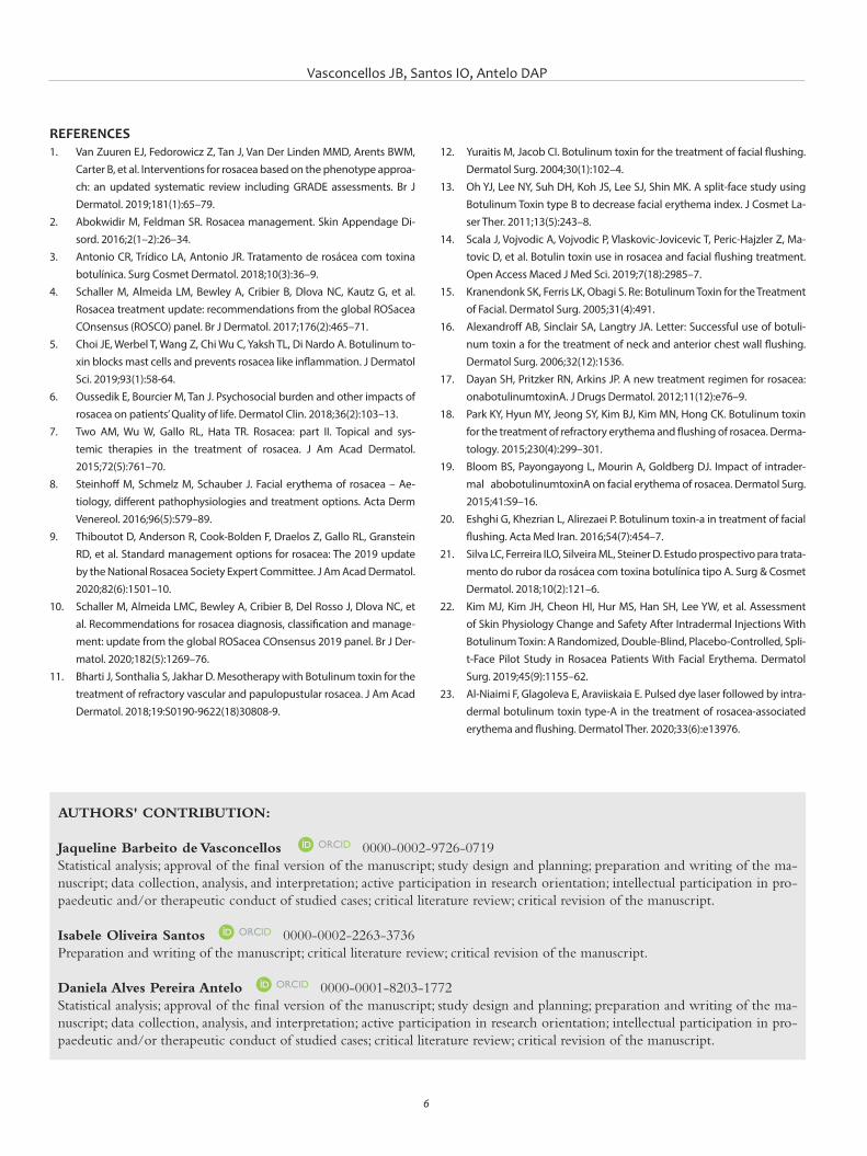
Vasconcellos JB, Santos IO, Antelo DAP
6
REFERENCES1. Van Zuuren EJ, Fedorowicz Z, Tan J, Van Der Linden MMD, Arents BWM,
Carter B, et al. Interventions for rosacea based on the phenotype approa-ch: an updated systematic review including GRADE assessments. Br J Dermatol. 2019;181(1):65–79.
2. Abokwidir M, Feldman SR. Rosacea management. Skin Appendage Di-sord. 2016;2(1–2):26–34.
3. Antonio CR, Trídico LA, Antonio JR. Tratamento de rosácea com toxina botulínica. Surg Cosmet Dermatol. 2018;10(3):36–9.
4. Schaller M, Almeida LM, Bewley A, Cribier B, Dlova NC, Kautz G, et al. Rosacea treatment update: recommendations from the global ROSacea COnsensus (ROSCO) panel. Br J Dermatol. 2017;176(2):465–71.
5. Choi JE, Werbel T, Wang Z, Chi Wu C, Yaksh TL, Di Nardo A. Botulinum to-xin blocks mast cells and prevents rosacea like inflammation. J Dermatol Sci. 2019;93(1):58-64.
6. Oussedik E, Bourcier M, Tan J. Psychosocial burden and other impacts of rosacea on patients’ Quality of life. Dermatol Clin. 2018;36(2):103–13.
7. Two AM, Wu W, Gallo RL, Hata TR. Rosacea: part II. Topical and sys-temic therapies in the treatment of rosacea. J Am Acad Dermatol. 2015;72(5):761–70.
8. Steinhoff M, Schmelz M, Schauber J. Facial erythema of rosacea – Ae-tiology, different pathophysiologies and treatment options. Acta Derm Venereol. 2016;96(5):579–89.
9. Thiboutot D, Anderson R, Cook-Bolden F, Draelos Z, Gallo RL, Granstein RD, et al. Standard management options for rosacea: The 2019 update by the National Rosacea Society Expert Committee. J Am Acad Dermatol. 2020;82(6):1501–10.
10. Schaller M, Almeida LMC, Bewley A, Cribier B, Del Rosso J, Dlova NC, et al. Recommendations for rosacea diagnosis, classification and manage-ment: update from the global ROSacea COnsensus 2019 panel. Br J Der-matol. 2020;182(5):1269–76.
11. Bharti J, Sonthalia S, Jakhar D. Mesotherapy with Botulinum toxin for the treatment of refractory vascular and papulopustular rosacea. J Am Acad Dermatol. 2018;19:S0190-9622(18)30808-9.
12. Yuraitis M, Jacob CI. Botulinum toxin for the treatment of facial flushing. Dermatol Surg. 2004;30(1):102–4.
13. Oh YJ, Lee NY, Suh DH, Koh JS, Lee SJ, Shin MK. A split-face study using Botulinum Toxin type B to decrease facial erythema index. J Cosmet La-ser Ther. 2011;13(5):243–8.
14. Scala J, Vojvodic A, Vojvodic P, Vlaskovic-Jovicevic T, Peric-Hajzler Z, Ma-tovic D, et al. Botulin toxin use in rosacea and facial flushing treatment. Open Access Maced J Med Sci. 2019;7(18):2985–7.
15. Kranendonk SK, Ferris LK, Obagi S. Re: Botulinum Toxin for the Treatment of Facial. Dermatol Surg. 2005;31(4):491.
16. Alexandroff AB, Sinclair SA, Langtry JA. Letter: Successful use of botuli-num toxin a for the treatment of neck and anterior chest wall flushing. Dermatol Surg. 2006;32(12):1536.
17. Dayan SH, Pritzker RN, Arkins JP. A new treatment regimen for rosacea: onabotulinumtoxinA. J Drugs Dermatol. 2012;11(12):e76–9.
18. Park KY, Hyun MY, Jeong SY, Kim BJ, Kim MN, Hong CK. Botulinum toxin for the treatment of refractory erythema and flushing of rosacea. Derma-tology. 2015;230(4):299–301.
19. Bloom BS, Payongayong L, Mourin A, Goldberg DJ. Impact of intrader-mal abobotulinumtoxinA on facial erythema of rosacea. Dermatol Surg. 2015;41:S9–16.
20. Eshghi G, Khezrian L, Alirezaei P. Botulinum toxin-a in treatment of facial flushing. Acta Med Iran. 2016;54(7):454–7.
21. Silva LC, Ferreira ILO, Silveira ML, Steiner D. Estudo prospectivo para trata-mento do rubor da rosácea com toxina botulínica tipo A. Surg & Cosmet Dermatol. 2018;10(2):121–6.
22. Kim MJ, Kim JH, Cheon HI, Hur MS, Han SH, Lee YW, et al. Assessment of Skin Physiology Change and Safety After Intradermal Injections With Botulinum Toxin: A Randomized, Double-Blind, Placebo-Controlled, Spli-t-Face Pilot Study in Rosacea Patients With Facial Erythema. Dermatol Surg. 2019;45(9):1155–62.
23. Al-Niaimi F, Glagoleva E, Araviiskaia E. Pulsed dye laser followed by intra-dermal botulinum toxin type-A in the treatment of rosacea-associated erythema and flushing. Dermatol Ther. 2020;33(6):e13976.
AUTHORS' CONTRIBUTION:
Jaqueline Barbeito de Vasconcellos 0000-0002-9726-0719Statistical analysis; approval of the final version of the manuscript; study design and planning; preparation and writing of the ma-nuscript; data collection, analysis, and interpretation; active participation in research orientation; intellectual participation in pro-paedeutic and/or therapeutic conduct of studied cases; critical literature review; critical revision of the manuscript.
Isabele Oliveira Santos 0000-0002-2263-3736Preparation and writing of the manuscript; critical literature review; critical revision of the manuscript.
Daniela Alves Pereira Antelo 0000-0001-8203-1772Statistical analysis; approval of the final version of the manuscript; study design and planning; preparation and writing of the ma-nuscript; data collection, analysis, and interpretation; active participation in research orientation; intellectual participation in pro-paedeutic and/or therapeutic conduct of studied cases; critical literature review; critical revision of the manuscript.

1
APOIO CIENTÍFICO:
www.surgicalcosmetic.org.br/
ISSN-e 1984-8773
Original Article Authors: Shishira R Jartarkar¹ Swayamsidda Mishra² Manjunath KG¹ Spoorthy B¹
¹ Department of Dermatology, Vydehi Institute of Medical Sciences and Research Centre, Bengaluru, Karnataka, India
² Department of Dermatology, Vaccicare Clinic, Cuttack, Odisha, India
Correspondence: Shishira R. Jartarkar Email: [email protected]
Financial support: None Conflict of interest: None
Submitted on: 17/05/2021Approved on: 04/06/2021
How to cite this article: Jartarkar SR, Manjunath KG, Mishra S, Spoorthy B. Intralesional vitamin D in multiple recurrent plantar warts - A single blind, prospective, placebo controlled study. Surg Cosmet Dermatol. 2021;13:e20210050.
Intralesional vitamin D in multiple recurrent plantar warts - A single, blind, prospective, placebo-controlled study Vitamina D intralesional em múltiplas verrugas plantares recorrentes - Um estudo cego, prospectivo e controlado por placebo
ABSTRACTIntroduction: Warts or verrucae, caused by the human papillomavirus (HPV), are a benign epidermal proliferation of the skin. Most patients seek medical advice as warts are cosmetically unacceptable and can be painful. Plantar warts, in particular, are typically refractory to treatment requiring multiple treatment sessions. High recurrence rates, pain, and scarring limit the available therapeutic modalities. In contrast, immunotherapeutic approaches stimulate the host immune system by enhancing cellular immunity to eliminate the virus.Objective: To assess the safety and efficacy of intralesional vitamin D3 injection to treat multiple recur-rent plantar warts. Methods: 60 patients with multiple recurrent warts were divided into two groups of 30 each. Group 1 received 0.5 ml intralesional vitamin D in the base of the largest wart, and Group 2 received 0.5 ml of normal saline. The sessions were repeated every two weeks for a maximum of four sessions, and patients were followed up for 12 months to detect any recurrences. Results: The study group showed complete clearance in 73.3% (22) individuals, while most controls (70%) showed no response. Conclusion: Intralesional vitamin D3 is a safe and effective treatment option for multiple recurrent plantar warts.Keywords: Cellular immunity; Injections, intralesional; Warts
RESUMOIntrodução: As verrugas, são proliferações epidérmicas benignas da pele. A maioria dos pacientes procura orientação médica, pois as verrugas são cosmeticamente inaceitáveis e podem ser dolorosas. As verrugas plantares, em particular, são tipicamente refratárias ao tratamento que requer várias sessões. As modalidades terapêuticas disponíveis são limitadas pela alta taxa de recorrência, dor e cicatrizes. Em contraste, as abordagens imunoterapêuticas estimulam o sistema imunológico do hospedeiro, aumentando a imunidade celular para eliminar o vírus. Objetivo: Avaliar a segurança e eficácia da injeção intralesional de vitamina D3 no tratamento de múltiplas verrugas plantares recorrentes. Métodos: Um total de 60 pacientes com verrugas plantares múltiplas recorrentes, foram divididos em dois grupos de 30. No grupo 1, 0,5ml de vitamina D intralesional foi injetado na base da maior verruga e no grupo 2, injetou-se 0,5ml de solução salina normal. As sessões foram repetidas a cada 2 semanas por no máximo 4 sessões e os pacientes foram acompanhados por um período de 12 meses. Resultados: No grupo de estudo, a eliminação completa foi observada em 73,3% (22) e nos controles, 70% dos pacientes não apresentaram resposta. Conclusão: A vitamina D3 intralesional é uma opção de tratamento segura e eficaz em verrugas plantares.Palavras-chave: Imunidade celular; Injeções; Verrugas
DOI: http://www.dx.doi.org/10.5935/scd1984-8773.2021130050
ISSN-e 1984-8773
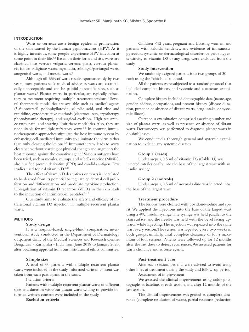
Jartarkar SR, Manjunath KG, Mishra S, Spoorthy B
2
INTRODUCTION Warts or verrucae are a benign epidermal proliferation
of the skin caused by the human papillomavirus (HPV). As it is highly infectious, some people experience HPV infection at some point in their life.1,2 Based on their form and site, warts are classified into verruca vulgaris, verruca plana, verruca planta-ris, filiform/digitate warts, myrmecia, subungal/periungal warts, anogenital warts, and mosaic warts.3
Although 60-65% of warts resolve spontaneously by two years, most patients seek medical advice as warts are cosmeti-cally unacceptable and can be painful at specific sites, such as plantar warts.1 Plantar warts, in particular, are typically refrac-tory to treatment requiring multiple treatment sessions.4 Seve-ral therapeutic modalities are available such as medical agents (5-fluorouracil, podophyllotoxin, salicylic acid, oral zinc and ranitidine, cytodestructive methods (electrocautery, cryotherapy, photodynamic therapy), and surgical excision. High recurren-ce rates, pain, and scarring limit these modalities. Also, they are not suitable for multiple refractory warts.5,6 In contrast, immu-notherapeutic approaches stimulate the host immune system by enhancing cell-mediated immunity to eliminate the virus rather than only clearing the lesions.6,7 Immunotherapy leads to warts clearance without scarring or physical changes and augments the host response against the causative agent.8 Various antigens have been tried, such as measles, mumps, and rubella vaccine (MMR), also purified protein derivative (PPD) and candida antigen. Few studies used topical vitamin D.9-12
The effect of vitamin D derivatives on warts is speculated to be derived from its potential to regulate epidermal cell proli-feration and differentiation and modulate cytokine production. Upregulation of vitamin D receptors (VDR) in the skin leads to the induction of antimicrobial peptides.9-12
Our study aims to evaluate the safety and efficacy of in-tralesional vitamin D3 injection in multiple recurrent plantar warts.
METHODSStudy design It is a hospital-based, single-blind, comparative, inter-
ventional study conducted in the Department of Dermatology outpatient clinic of the Medical Sciences and Research Centre, Bengaluru - Karnataka - India from June 2018 to January 2020, after obtaining approval from our institutional ethics committee.
Sample sizeA total of 60 patients with multiple recurrent plantar
warts were included in the study. Informed written consent was taken from each participant in the study.
Inclusion criteriaPatients with multiple recurrent plantar warts of different
sizes and duration with/out distant warts willing to provide in-formed written consent were included in the study.
Exclusion criteria
Children <12 years, pregnant and lactating women, and patients with keloidal tendency, any evidence of immunosu-ppression, systemic or dermatological disorder, or prior hyper-sensitivity to vitamin D3 or any drug, were excluded from the study.
Study interventionWe randomly assigned patients into two groups of 30
each using the “chit box” method.All the patients were subjected to a standard protocol that
included complete history and systemic and cutaneous exami-nation.
Complete history included demographic data (name, age, gender, address, occupation), and present history (disease dura-tion, presence or absence of distant warts, drug intake, or syste-mic illness).
Cutaneous examination comprised assessing number and size of plantar warts, as well as presence or absence of distant warts. Dermoscopy was performed to diagnose plantar warts in doubtful cases.
We conducted a thorough general and systemic exami-nation to exclude any systemic diseases.
Group 1 (cases)Under asepsis, 0.5 ml of vitamin D3 (6lakh IU) was
injected intralesionally into the base of the largest wart with an insulin syringe.
Group 2 (controls)Under asepsis, 0.5 ml of normal saline was injected into
the base of the largest wart.
Treatment procedureThe lesions were cleaned with povidone-iodine and spi-
rit. We applied the injections into the base of the largest wart using a 40U insulin syringe. The syringe was held parallel to the skin surface, and the needle was held with the bevel facing up-wards while injecting. The injection was repeated into the same wart every session. The session was repeated every two weeks in both groups, similarly, until complete clearance or for a maxi-mum of four sessions. Patients were followed up for 12 months after the last dose to detect recurrences. We assessed patients for warts clearance and adverse events.
Post-treatment careAfter each session, patients were advised to avoid using
other lines of treatment during the study and follow-up period.Assessment of improvementWe assessed the clinical improvement using color pho-
tographs at baseline, at each session, and after 12 months of the last session.
The clinical improvement was graded as complete clea-rance (complete resolution of warts), partial response (reduction
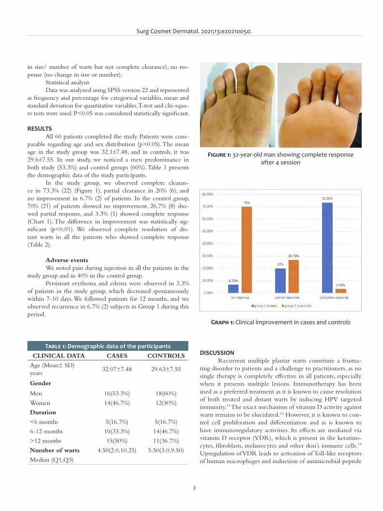
Surg Cosmet Dermatol. 2021;13:e20210050.
3
DISCUSSION Recurrent multiple plantar warts constitute a frustra-
ting disorder to patients and a challenge to practitioners, as no single therapy is completely effective in all patients, especially when it presents multiple lesions. Immunotherapy has been used as a preferred treatment as it is known to cause resolution of both treated and distant warts by inducing HPV targeted immunity.13 The exact mechanism of vitamin D activity against warts remains to be elucidated.14 However, it is known to con-trol cell proliferation and differentiation and as is known to have immunoregulatory activities. Its effects are mediated via vitamin D receptor (VDR), which is present in the keratino-cytes, fibroblasts, melanocytes and other skin’s immune cells.15 Upregulation of VDR leads to activation of Toll-like receptors of human macrophages and induction of antimicrobial peptide
in size/ number of warts but not complete clearance), no res-ponse (no change in size or number).
Statistical analysisData was analyzed using SPSS version 22 and represented
as frequency and percentage for categorical variables, mean and standard deviation for quantitative variables. T-test and chi-squa-re tests were used. P<0.05 was considered statistically significant.
RESULTSAll 60 patients completed the study. Patients were com-
parable regarding age and sex distribution (p>0.05). The mean age in the study group was 32.1±7.48, and in controls, it was 29.6±7.55. In our study, we noticed a men predominance in both study (53.3%) and control groups (60%). Table 1 presents the demographic data of the study participants.
In the study group, we observed complete clearan-ce in 73.3% (22) (Figure 1), partial clearance in 20% (6), and no improvement in 6.7% (2) of patients. In the control group, 70% (21) of patients showed no improvement, 26.7% (8) sho-wed partial response, and 3.3% (1) showed complete response (Chart 1). The difference in improvement was statistically sig-nificant (p<0.01). We observed complete resolution of dis-tant warts in all the patients who showed complete response (Table 2).
Adverse eventsWe noted pain during injection in all the patients in the
study group and in 40% in the control group. Persistent erythema and edema were observed in 3.3%
of patients in the study group, which decreased spontaneously within 7-10 days. We followed patients for 12 months, and we observed recurrence in 6.7% (2) subjects in Group 1 during this period.
Table 1: Demographic data of the participantsCLINICAL DATA CASES CONTROLS
Age (Mean± SD) years
32.07±7.48 29.63±7.55
Gender
Men 16(53.3%) 18(60%)
Women 14(46.7%) 12(40%)
Duration
<6 months 5(16.7%) 5(16.7%)
6-12 months 10(33.3%) 14(46.7%)
>12 months 15(50%) 11(36.7%)
Number of warts 4.50(2.0,10.25) 5.50(3.0,9.50)
Median (Q1,Q3)
Figure 1: 32-year-old man showing complete response after 4 session
Graph 1: Clinical improvement in cases and controls
Jartarkar SR, Manjunath KG, Mishra S, Spoorthy B
4
expressions as thymic stromal, lymphopoietin, and cathelicidin. It also acts as an anti-inflammatory by reducing the synthesis of IL1alpha and IL6.16
Our study revealed that intralesional vitamin D3 is an effective therapy for multiple recurrent plantar warts with a suc-cess rate of 73.3%, as 22 out of 30 patients had complete clea-rance of plantar warts. This was in concordance with a study by Aktas et al.,4 who reported total clearance in 80% (16 out of 20) of plantar warts patients. Another study, by Raghukumar et al.,13 evaluated the effect of intralesional injection of vitamin D3 in 60 patients with various extragenital warts, showing complete response in 90% of cases.
During the follow-up period of 12 months, we observed recurrence in 6.7% of our study group. It agreed with Raghu-kumar et al.,13 who reported a recurrence rate of 3.33% during the six-month follow-up.
Table 2: Clinical improvement in cases and controls
Clinical Improvement
Group 1(cases)% (n)
Group 2 (controls)
No response 6.7% (2) 70% (21)
Partial response 20% (6) 26.7% (8)
Complete response
73.3% (22) 3.3% (1)
A randomized controlled trial by El-Sayed et al.17 com-pared the effectiveness of 0.2 ml intralesional vitamin D3 and 0.2 ml intralesional zinc sulfate every two weeks for a maxi-mum of four sessions. It showed complete response of 62.9% with vitamin D and 71.4% with zinc sulfate.
A placebo-controlled study by Abdel Azim et al.14 sho-wed total clearance of warts in 56.25% (18 out of 32) of patients with intralesional vitamin D3 in cutaneous warts. But our study showed a better response in plantar warts.
Another placebo-controlled study by Kareem et al.16 used 0.2 ml intralesional vitamin D once monthly for two ses-sions for common warts. It showed complete clearance in 45% of the patients. Our study presented better results, probably due to the increased number of sessions (four sessions) and the use of a higher dose (0.5 ml) of vitamin D3.
There were no significant adverse events observed in our study. The limitation of our study is the relatively small sample size.
CONCLUSIONOur study shows a beneficial role of intralesional vita-
min D3 in multiple recurrent plantar warts. It is a simple, safe, cost-effective treatment modality with a low recurrence rate. However, larger case-control studies as well as in-vitro/in-vivo studies are needed to elucidate the exact mechanism of action of vitamin D3 in warts. l

Surg Cosmet Dermatol. 2021;13:e20210050.
5
AUTHORS' CONTRIBUTION:
Shishira R Jartarkar 0000-0002-7016-6087Approval of the final veårsion of the manuscript; study design and planning; preparation and writing of the manuscript; data collection, analysis, and interpretation; active participation in research orientation; intellectual participation in propaedeutic and/or therapeutic conduct of studied cases; critical literature review; critical revision of the manuscript.
Manjunath KG 0000-0002-4956-0880Statistical analysis, Approval of the final version of the manuscript; study design and planning; preparation and writing of the manuscript; data collection, analysis, and interpretation; active participation in research orientation; intellectual participation in propaedeutic and/or therapeutic conduct of studied cases.
Swayamsidda Mishra 0000-0002-3645-523XStatistical analysis; study design and planning; data collection, analysis, and interpretation; active participation in research orientation.
Spoorthy B 0000-0003-2283-8050Statistical analysis; preparation and writing of the manuscript; data collection, analysis, and interpretation; active participation in research orien-tation.
REFERENCES:1. Sterling JC, Handfield-Jones S, Hudson PM; British Association of Der-
matologists. Guidelines for the management of cutaneous warts. Br J Dermatol. 2001;144(1):4-11.
2. Tyring SK. Human papillomavirus infections: epidemiology, pathoge-nesis, and host immune response. J Am Acad Dermatol. 2000;43(1 Pt 2):S18-26.
3. Salman S, Ahmed MS, Ibrahim AM, Mattar OM, El-Shirbiny H, Sarsik S, et al. Intralesional immunotherapy for the treatment of warts: a network meta-analysis. J Am Acad Dermatol. 2019;80(4):922-30.
4. Aktaş H, Ergin C, Demir B, Ekiz Ö. Intralesional vitamin D injection may be an effective treatment option for warts. J Cutan Med Surg. 2016;20(2):118-22.
5. Bacelieri R, Johnson SM. Cutaneous warts: an evidence-based approa-ch to therapy. Am Fam Physician. 2005 15;72(4):647-52.
6. Rivera A, Tyring SK. Therapy of cutaneous human papillomavirus infec-tions. Dermatol Ther. 2004;17(6):441-8.
7. Thappa DM, Chiramel MJ. Evolving role of immunotherapy in the treat-ment of refractory warts. Indian Dermatol Online J. 2016;7(5):364-70.
8. Sinha S, Relhan V, Garg VK. Immunomodulators in warts: Unexplored or ineffective? Indian J Dermatol. 2015;60(2):118-29.
9. Moscarelli L, Annunziata F, Mjeshtri A, Paudice N, Tsalouchos A, Zana-zzi M, et al. Successful treatment of refractory wart with a topical ac-tivated vitamin d in a renal transplant recipient. Case Rep Transplant. 2011;2011:368623.
10. Rind T, Oiso N, Kawada A. Successful treatment of anogenital wart with a topical vitamin D(3) derivative in an infant. Case Rep Dermatol. 2010;2(1):46-9.
11. AlGhamdi K, Kumar A, Moussa N. The role of vitamin D in melanoge-nesis with an emphasis on vitiligo. Indian J Dermatol Venereol Leprol. 2013;79(6):750-8.
12. Imagawa I, Suzuki H. Successful treatment of refractory warts with topi-cal vitamin D3 derivative (maxacalcitol, 1alpha, 25-dihydroxy-22-oxa-calcitriol) in 17 patients. J Dermatol. 2007;34(4):264-6.
13. Raghukumar S, Ravikumar BC, Vinay KN, Suresh MR, Aggarwal A, Yasho-vardhana DP. Intralesional vitamin D3 injection in the treatment of recal-citrant warts: a novel proposition. J Cutan Med Surg. 2017;21(4):320-4.
14. Abdel-Azim ES, Abdel-Aziz R, Ragale MH, Mohamed EA. Clinical and dermoscopic evaluation of intralesional vitamin D3 in treatment of cutaneous warts: A placebocontrolled study. J Egypt Women Dermatol Soc. 2020;17:6-12.
15. Fathy G, Sharara MA, Khafagy AH. Intralesional vitamin D3 versus Candida antigen immunotherapy in the treatment of multiple recal-citrant plantar warts: a comparative case-control study. Dermatol Ther. 2019;32(5):e12997.
16. Kareem IMA, Ibrahim IM, Mohammed SFF, Ahmed AA. Effective-ness of intralesional vitamin D3 injection in the treatment of com-mon warts: single-blinded placebo-controlled study. Dermatol Ther. 2019;32(3):e12882.

1
APOIO CIENTÍFICO:
www.surgicalcosmetic.org.br/
ISSN-e 1984-8773
ReviewAuthors: Carlos Roberto Antonio1
Lívia Arroyo Trídico1
¹ Faculdade de Medicina de São José do Rio Preto, Departmant of Dermatology, São José do Rio Preto (SP), Brazil.
Correspondence: Lívia Arroyo Trídico Email: [email protected] / Alternative email: latridico@gmail. com
Financial support: None.Conflict of interest: None.
Submitted on: 25/03/2021 Approved on: 13/10/2021
How to cite this article: Antonio CR, Trídico LA. The importance of interaction between hyaluronic acid and CD44 receptor. Surg Cosmet Dermatol. 2021;13:20210006.
The importance of interaction between hyaluronic acid and CD44 receptor A importância da interação entre o ácido hialurônico e o receptor CD44
ABSTRACTHyaluronic acid is one of the most used substances in dermatology. It presents structural roles in the extracellular matrix, binding to cells and biological components through specific and nonspecific interactions. The native ligand for hyaluronic acid is the transmembrane CD44 receptor, which interacts not only with hyaluronic acid but also with different growth factors, cytokines, and extracellular matrix proteins. We seek to review the interaction of the CD44 receptor with the various forms of hyaluronic acid in the skin to better understand its action and fully explore its use in dermatology.Keywords: Rejuvenation; Hyaluronic acid; Skin
RESUMOÁcido hialurônico é uma das substâncias mais utilizadas na Dermatologia. Apresenta tarefas estruturais na matriz extracelular, ligando-se às células e a componentes biológicos por interações específicas e inespe-cíficas. O ligante nativo para o ácido hialurônico é o receptor transmembrânico CD44, que interage não apenas com o ácido hialurônico, mas também com diferentes fatores de crescimento, citocinas e proteínas da matriz extracelular. Buscamos revisar a interação entre o receptor CD44 e as diversas formas de ácido hialurônico na pele, a fim de compreender melhor sua ação e explorar seu uso de forma mais completa na Dermatologia.Palavras-chave: Rejuvenescimento; Ácido hialurônico; Pele
DOI: http://www.dx.doi.org/10.5935/scd1984-8773.2021130006
ISSN-e 1984-8773
Antonio CR, Trídico LA
2
INTRODUCTION Hyaluronic acid (HA) is a natural biodegradable polymer.
It is a non-sulfated, non-branched glycosaminoglycan composed of repeating disaccharide units (1.4 β-D-glucuronic acid and 1.3 N-acetyl-β-D-glucosamine). HA is a polyanion that can bind to each other and water molecules, forming a rigid and viscous structure similar to gelatin. HA is one of the main extracellular matrix (ECM) elements of vertebrate tissues, available in almost all body fluids and tissues. This biopolymer has a structural func-tion, binding extracellular matrix molecules. Also, it is involved in several essential biological processes, such as regulation of cell adhesion and motility, in addition to acting in cell differentiation and proliferation and the mechanical properties of tissues.1
The HA characteristics, such as consistency, biocom-patibility, and hydrophilicity, make it ideal for use in Aesthetic Dermatology. Also, its unique viscoelasticity and limited immu-nogenicity led to its use in various medical applications, such as osteoarthritis treatment (OA), eye surgery aid, and wound rege-neration.2 We will discuss the use of injectable hyaluronic acid as a dermal filler and its interactions with its primary receptor, CD44, to understand how it can promote rejuvenation through its biological interactions.
Hyaluronic acid metabolism in the skinIn the epidermis, HA content is especially high in the
proliferating basal regions. It is in line with maintaining the undifferentiated and proliferative state of basal cells and cor-responding observations during embryogenic development.3 Histological findings suggest that basal layer keratinocytes con-tain intracellular HA, whereas extracellular HA prevails in the upper epidermal layers.4 Extracellular HA is believed to main-tain diffusion and open spaces to facilitate cell migration.5
HA’s primary source in the dermis is fibroblasts, with hi-gher HA synthesis activity in the papillary dermis. HA structures’ high flexibility and hydrophilicity allow these molecules to fill any gaps in the ECM.6 The multiple hydrogen bonds between adjacent disaccharides especially explain HA’s large hydrodyna-mic volume, but these may also depend on the close interaction of HA with highly glycosylated proteoglycans. The viscoelas-tic properties resulting from HA in the dermis are responsible for supporting the dermal tissue architecture. Dermal HA has access to the lymphatic system, probably regulating the water content in the dermis. Both free HA crosslinked by proteins or proteoglycans and HA associated with the cell membrane faci-litate cell migration, proliferation, and communication by inte-racting with cell receptors, grouping receptors, and subsequent signaling cascades.7
Specific enzymes called HA-synthase (HAS) synthesize the HA. They are membrane-binding enzymes that synthesi-ze HA on the inner surface of the plasma membrane and then eliminate it through pore-like structures into the extracellular space. Three enzymes are responsible for HA synthesis: HAS-1, 2, and 3, which exhibit distinct enzymatic properties and synthe-size HA chains of various lengths.7,8
HA degradation is a gradual process that can occur
through enzymatic or non-enzymatic reactions. Three types of enzymes (hyaluronidase, β-D-glucuronides, and β-N-acetylhe-xosaminidase) are involved in the enzymatic degradation of HA. These enzymes are found in several forms, in the intercellular space and serum. Hyaluronidase breaks high molecular weight HA into smaller fragments, while the other two enzymes degra-de the fragments by removing non-reducing terminal sugars. In addition to the enzymatic mechanisms of HA degradation, it can be degraded by shear stress, heat, and chemical reactions, such as acid/alkaline hydrolysis and degradation by oxidants. These types of degradation occur at random, often resulting in disac-charide fragments.1,9
Skin aging and hyaluronic acidThe most dramatic histochemical change observed with
aging skin is the marked disappearance of hyaluronic acid in the epidermis, while HA is still present in the dermis.11 The reasons for the changes in HA homeostasis with aging are unknown, but it is known that the underlying dermis influences epider-mal HA synthesis. A progressive reduction in the size of HA polymers in the skin due to aging has also been reported. Thus, the epidermis loses the main molecule responsible for binding and retaining water molecules, resulting in the loss of moisture from the skin. In the dermis, the main age-related change is the increasing avidity of HA for tissue structures and loss of HA ex-traction capacity. Also, there is progressive collagen crosslinking and loss of collagen extraction capacity with age. All of the above age-related phenomena contribute to the apparent dehydration, atrophy, and loss of elasticity that characterizes aging skin.10
The premature skin aging due to repeated and prolonged exposure to radiation results in an abnormal content of glyco-saminoglycans and distribution compared to that found in scars, with decreased HA and increased chondroitin sulfate proteo-glycans levels.12 In dermal fibroblasts, this reduction in HA syn-thesis was attributed to collagen fragments, which activate αvβ3 integrins, resulting in reduced HA synthase expression (enzymes responsible for HA production).12
Injectable Hyaluronic Acid in DermatologyHyaluronic acid-based dermal fillers are currently one of
the most commonly performed aesthetic procedures. HA injec-tions into the skin promote a volume-filling effect and induce collagen synthesis, reversing the signs of aging skin.13
Compositional properties and rheological properties characterize the compositions of HA formulations used for tis-sue filling. The design of fillers includes concentration, size, and crosslinking particles (substances that generate intermolecular bonds that increase the stability and clinical durability of the filler). In contrast, rheological properties include elasticity (G’) and viscosity (N’). Crosslinked HA (crosslinking particles) can be made with several chemicals (butanediol diglycidyl ether, divinyl sulfone, etc.). The increase in crosslinking particles and
Surg Cosmet Dermatol. 2021;13:e20210006.
3
concentration strengthens the fillers’ resistance to enzymatic degradation. The polymerization of glycosaminoglycan chains and tension determine the particle size, which optimizes tissue lifting capacity. Elasticity and viscosity guarantee fillers the abi-lity to resist compression and shear force, respectively. Another critical feature is hydrophilicity, that is, the filler’s ability to attract water and expand.14,15
Each product consists of a unique combination of diffe-rent characteristics, and understanding these differences allows treating appropriately other areas of the face. Softness and less viscosity are characteristics that make the filler ideal for surface wrinkles, lips, and eyelids. In contrast, denser and heavier fillers are better for deeper plane injection to increase volume.16 Du-ration of corrective effect of HA fillers ranges from three to 24 months, predominantly depending on HA concentration, cross-linking (degree and type), treated area, and individual.17
In addition to replenishing volume, injectable HA acts as a skin remodeler because the filling effect persists for a time much longer than the filler’s bioavailability. Studies have shown that HA can induce an increase in the production of collagen and elastic fibers, restoring the extracellular matrix by direct sti-mulation and/or mechanical stretching of fibroblasts.18
HA performs several structural tasks of ECM, as it binds to cells and other biological components through specific and non-specific interactions. Binding to HA stabilizes several ECM proteins. Specific molecules and receptors that interact with HA are involved in cellular signal transduction, such as the aggre-can, versican, and neurocan molecules, and the cellular recep-tors CD44, RHAMM, TSG6, GHAP, ICAM-1, and LYVE-1.19 Among these receptors, CD44 (cell surface glycoprotein) deser-ves more attention as, due to its wide distribution and based on current knowledge, it is considered the primary HA receptor on most cell types.1,20 We will address the importance of the CD44 receptor and its interactions with HA to promote rejuvenation.
CD44 receptor and hyaluronic acidThe native ligand for HA is the transmembrane receptor
CD44.21 HA binds to the N-terminal of CD44, which functions as a coupling site and is coated by a mixture of predominantly essential and hydrophobic amino acids.22 The CD44 gene con-tains 20 exons, 10 of which can be regulated by alternative bin-ding, leading to the generation of other variants (variant exons or ‘v’), which are translated to a polypeptide of molecular wei-ght 80-90kDa, depending on the binding. Biological functions, such as cell migration, adhesion, and structural integrity during anti-inflammatory processes, depend on the HA-CD44 interac-tion. The smallest CD44 isoform, standard CD44 (CD44s), is ubiquitous, whereas variant isoforms are expressed only in some epithelial tissues and cancers.23,24
The different forms of hyaluronic acid synthesized for medical use have common and distinct interactions with the CD44 receptor. Generally, HA-CD44 interactions can be alte-red according to HA extent modification, chemical group type
used for that modification, and HA location where the alteration was made. Regardless of the peculiarities involving the different types of synthetic hyaluronic acid and CD44 receptor, we will globally address this receptor role in allowing HA action to go beyond simply filling tissues, mainly acting in tissue biomodu-lation.25
CD44 can also interact with different growth factors, cy-tokines, and extracellular matrix proteins, such as fibronectin.26 The intracellular CD44 domain interacts with the cytoskeleton. Consequently, when its extracellular domain binds to the ECM’s HA, it creates a link between the cytoskeleton structures and the polymer.27 Several intracellular signaling pathways are involved in the HA-CD44 interaction, and they act by controlling cellu-lar biological processes: HA degradation and internalization, an-giogenesis, cell migration, proliferation, aggregation, and adhe-sion to ECM components.27,28,29
HA concentration manipulations or HA-CD44 inte-ractions can alter signaling pathways of many regulatory and adapter molecules, such as SRC kinases, Rho-GTPases, VAV2, and GAB1.30 The binding of CD44 to hyaluronic acid can al-ter cell survival or proliferation by altering intracellular protein binding.31 Also, HA can activate several receptor tyrosine kinases, and HA binding to CD44 can group and cooperate with growth factors.32 It has also been shown that the CD44 receptor is invol-ved in cellular uptake of extracellular HA.33
Kaya et al. (1992) demonstrated that CD44 is associa-ted with the regulation of HA homeostasis in keratinocytes. The authors developed transgenic mice expressing an antisense CD44 cDNA triggered by the keratin-5 promoter. These mice do not show detectable CD44 expression on skin keratinocytes and corneal epithelium and exhibit abnormal HA accumulation in the superficial dermis and stroma of the cornea, morpholo-gical changes distinct from basal keratinocytes and cornea, and defective proliferation of keratinocytes in response to mitogen and growth factors. A decrease in skin elasticity, poor local in-flammatory response and tissue repair, delayed capillary growth, and epidermal failure to undergo hyperplasia in response to the carcinogen reflect these changes. Therefore, they observed two main functions of CD44 in the skin: the regulation of kerati-nocyte proliferation in response to extracellular stimuli and the maintenance of local HA homeostasis.34
Vistejinova et al. (2014) conducted a study to compare the ability of high molecular weight (HMW) with low molecu-lar weight (LMW) HA to stimulate the production of cytokines and chemokines by human dermal fibroblasts, associated with the importance of the CD44 receptor in this process. The study showed that dermal fibroblasts and their primary function of producing the extracellular matrix could respond to low mole-cular weight HA fragments via interaction with CD44 through the production of cytokines, suggesting that the LMW HA is implicated in an inflammatory signal that stimulates stromal fi-broblasts.35
Studies have demonstrated that HA and CD44 on the outer surface of dermal fibroblasts act by regulating the physio-
Antonio CR, Trídico LA
4
logy of fibroblasts and stimulating the production of extracellular matrix.36 Thus, it may be possible to alter the skin’s collagen production by increasing or reducing the amount of HA.37 In Wang et al. study (2007), 11 volunteers received injections of HA filler or vehicle and underwent biopsy at four and 13 weeks after the procedure. The results demonstrated that, compared to controls, skin treated with crosslinked hyaluronic acid injections revealed increased collagen deposition around the filler material, gene expression for types I and III procollagens, and various growth factors profibrotic, was also regulated between four and 13 weeks compared to controls. The authors concluded that the injection of crosslinked hyaluronic acid stimulates collagen syn-thesis, partially restoring the dermal matrix components lost in photoaged skin.13
Bhattacharya et al. (2017) demonstrated that alterations in HA structures modify its interaction with the CD44 recep-tor. They observed that both sulfation and deacetylation of HA in individuals are associated with lower interaction with CD44; thus, both modifications are necessary to reduce the HA-CD44 interaction. Therefore, the study suggests that it would be possi-ble in future studies to regulate cell activation pathways through different forms of HA.38
Wang et al. (2019) assessed how different types of HA influence the binding of CD44 to HA hydrogels. HA-CD44 in-teractions can be altered when HA is modified to synthesize HA macromers, depending on the modification extent, chemical group type used for transformation, and the site used with HA for alteration. These effects are observable when HA macromers are presented to CD44 in soluble form and after crosslinking in hydrogels. Gene expression and long-term biochemical and histological analyzes of mesenchymal stromal cells encapsulated in HA hydrogels strongly suggest that levels of HA macromer modification influence cell-hydrogel interactions and chondro-genic differentiation. Notably, low and moderately modified HA hydrogels promote significantly higher binding to CD44 com-pared with inert molecules. Also, chondrogenesis and cartilage formation are regulated with HA hydrogels compared to inert polyethylene glycol hydrogel controls.39
Gruber, Holtz, and Riemer (2021) performed an in vi-tro evaluation on the influence of different molecular weights of HA on its binding to CD44. They showed that low molecular weight HA and a commercial complex with HA of three mo-lecular weights (high, medium, and low) increased the CD44 protein expression in human epidermal keratinocytes. In con-trast, medium and high molecular weight HA fractions didn’t. Therefore, they concluded that HA can influence the expression of the CD44 protein and that this influence seems to be depen-dent on the molecular weight.40
CD44 receptor and carcinogenesisIt is known that the abnormal activation of the CD44
signaling cascade by HA and the CD44 overexpression and upregulation can result in the development of pathological le-
sions and malignant transformation since the HA-CD44 inte-raction is involved in cell processes such as cell proliferation and angiogenesis.29,41 Therefore, CD44 is overexpressed in several so-lid tumors, such as pancreas, breast, and lung tumors.42
There is a complex communication between cancer cells and their microenvironment. Evidence indicates that the tumor microenvironment may regulate the capacity for tumor growth and metastasis.43 HA provides cell support and hydrophilic ma-trix and also regulates cell-cell adhesion, cell migration, growth, and differentiation.44 Thus, these properties make it a suitable candidate for involvement in pathological processes such as can-cer. Furthermore, by forming pericellular layers, HA can protect tumor cells from immune attack.45 Several tumor cells produ-ce increased amounts of HA or induce the production of HA by releasing growth factors and cytokines. Likewise, fragmented HA induced by reactive oxygen species (ROS) also contributes to HA overproduction.46 Tumor and stromal cells express HA isoforms and produce HA in the ECM, which accumulates in the tumor parenchyma and peritumoral stromal tissues, contri-buting to metastatic spread.47 Also, HA overproduction in tumor cells can induce cancer cell-like epithelial changes toward a mi-gratory fibroblastic phenotype.48 HA-rich ECM can also media-te mesenchymal stem cells recruitment, which are progenitors of tumor-associated fibroblasts.49
HA interaction with CD44 could explain many HA tu-mor-promoting activities. There are three ways in which CD44 can interact with HA: binding with soluble extracellular HA molecules and ECM, interacting with receptor tyrosine kinases for anti-apoptosis and drug resistance, and binding of CD44 to actin cytoskeleton.50
Activated CD44 is overexpressed in solid tumors, but much less or almost none, in its non-tumorigenic counterparts. Adhesion of CD44 to HA induces upregulation of integrins that strengthen stem cell adhesion.51 Tumor-derived cells express CD44 in a high-affinity state that can bind to and internalize HA. The binding affinity of CD44 to HA is essential for cell migration allowing CD44 to be incorporated into the leading edge of cells. CD44 can also react with other molecules, inclu-ding collagen, fibronectin, osteopontin, growth factors, and me-talloproteinases on tumor cells, but the functional roles of such interactions are less known.50
Receptor tyrosine kinases (RTKs) are a subclass of cell surface growth factor (GFR) receptors with an intrinsic tyrosine kinase activity controlled by ligands. The HA-CD44 interaction has a general effect on activating anti-apoptotic cell survival proteins, initiated by association with the activation of tyrosine kinase receptors. Also, CD44 binds to cytoskeletal proteins, and this interface is modulated by the HA-CD44 interaction.50
Thus, given that HA levels and their interactions with CD44 can regulate cell differentiation (such as epidermal kera-tinocyte cornification and fibroblast differentiation), the modu-latory capacity of regulated cell differentiation through the HA route could be used therapeutically, especially in Oncology. The fact that HA binding to CD44 can interact with several recep-
Surg Cosmet Dermatol. 2021;13:e20210006.
5
tor systems is very intriguing. If HA and CD44 interactions are necessary to lead to carcinogenesis and metastasis, then it is be-lieved that manipulation of these interactions can be performed therapeutically.37
Since the first reports of HA liposomes targeting CD44 in 2001, researchers have devoted a great effort to using HA-me-diated CD44 targets for disease and drug-delivery applications. Then, studies of HA nanomaterials have emerged as an effective way to improve drug delivery. In the future, it is believed that the straightforward structure and easy manufacturing process for HA nanomaterials may increase the possibility of success in cli-nical practice.51
CONCLUSIONThere are numerous interactions between HA and its
primary receptor, CD44. We know that they depend on several factors, including the type of HA involved. Studies show that HA-CD44 interactions occur not only with endogenous HA but also with topical and injectable HA. Therefore, we can con-clude that exogenous HA acts with CD44 to cause cellular and molecular modulation in the site where it is applied, thus brin-ging results beyond the simple fact of filling the treated place. It mainly alters the environment through local interactions and improves skin quality. Also, several studies report the involve-ment of the CD44 receptor in carcinogenesis. Although further studies on this subject are needed, a better understanding of the role of the CD44 receptor can also positively influence the tre-atment of cancer in the future. l
REFERENCES:1. Fakhari A, Berkland C. Applications and emerging trends of hyaluronic
acid in tissue engineering, as a dermal filler and in osteoarthritis treat-ment. Acta Biomater. 2013;9(7):7081-92.
2. Necas J, Bartosikova L, Brauner P, Kolar J. Hyaluronic acid (hyaluronan): a review. Veterinarni Medicina. 2008(8):397–411.
3. Pasonen-Seppänen S, Karvinen S, Törrönen K, Hyttinen JM, Jokela T, Lammi MJ, et al. EGF upregulates, whereas TGF-beta downregulates, the hyaluronan synthases Has2 and Has3 in organotypic keratinocyte cultures: correlations with epidermal proliferation and differentiation. J Invest Dermatol. 2003;120(6):1038-44.
4. Stern R, Maibach HI. Hyaluronan in skin: aspects of aging and its phar-macologic modulation. Clin Dermatol. 2008;26(2):106-22.
5. Kaya G, Rodriguez I, Jorcano JL, Vassalli P, Stamenkovic I. Selective su-ppression of CD44 in keratinocytes of mice bearing an antisense CD44 transgene driven by a tissue-specific promoter disrupts hyaluronate metabolism in the skin and impairs keratinocyte proliferation. Genes Dev. 1997;11(8):996-1007.
6. Evanko SP, Tammi MI, Tammi RH, Wight TN. Hyaluronan-dependent pe-ricellular matrix. Adv Drug Deliv Rev. 2007;59(13):1351-65.
7. Prehm P. Hyaluronate is synthesized at plasma membranes. Biochem J. 1984;220:597-600.
8. Watanabe K, Yamaguchi Y. Molecular identification of a putative hu-man hyaluronan synthase. J Biol Chem. 1996;271(38):22945-8.
9. Stern R, Kogan G, Jedrzejas MJ, Soltés L. The many ways to cleave hya-luronan. Biotechnol Adv. 2007;25(6):537-57.
10. Stern R, Maibach HI. Hyaluronan in skin: aspects of aging and its phar-macologic modulation. Clin Dermatol. 2008;26(2):106-22.
11. Röck K, Grandoch M, Majora M, Krutmann J, Fischer JW. Collagen frag-ments inhibit hyaluronan synthesis in skin fibroblasts in response to ul-traviolet B (UVB): new insights into mechanisms of matrix remodeling. J Biol Chem. 2011 20;286(20):18268-76.
12. Bernstein EF, Underhill CB, Hahn PJ, Brown DB, Uitto J. Chronic sun ex-posure alters both the content and distribution of dermal glycosami-noglycans. Br J Dermatol. 1996;135(2):255-62.
13. Wang F, Garza LA, Kang S, et al. In vivo stimulation of de novo collagen production caused by cross-linked hyaluronic acid dermal filler injec-tions in photodamaged human skin. Arch Dermatol. 2007;143(2):155-63.
14. Oliveira JD, Carvalho LS, Gomes AM, Queiroz LR, Magalhães BS, Para-chin NS. Genetic basis for hyper production of hyaluronic acid in natu-ral and engineered microorganisms. Microb Cell Fact. 2016;15(1):119.
15. Edsman K, Nord LI, Ohrlund A, Lärkner H, Kenne AH. Gel properties of hyaluronic acid dermal fillers. Dermatol Surg. 2012;38(7 Pt 2):1170-9.
16. Alam M, Tung R. Injection technique in neurotoxins and fillers: indica-tions, products, and outcomes. J Am Acad Dermatol. 2018;79(3):423-35.
17. Muhn C, Rosen N, Solish N, Bertucci V, Lupin M, Dansereau A, et al. The evolving role of hyaluronic acid fillers for facial volume restoration and contouring: a Canadian overview. Clin Cosmet Investig Dermatol. 2012;5:147-58.
18. Trindade de Almeida AR, Sampaio GAA. Hyaluronic acid in the rejuve-nation of the upper third of the face: review and update - Part 1. Surg Cosmet Dermatol. 2015;8(2):148-53.
19. Necas J, Bartosikova L, Brauner P, Kolar J. Hyaluronic acid (hyaluronan): a review. Vet Med. 2008;53:397-411.
20. Toole BP. Hyaluronan in morphogenesis. J Intern Med. 1997;242(1):35-40.21. Gallatin WM, Weissman IL, Butcher EC. A cell-surface molecule involved
in organ-specific homing of lymphocytes. Nature. 1983;304(5921):30-4. 22. Banerji S, Wright AJ, Noble M, Mahoney DJ, Campbell ID, Day AJ, et
al. Structures of the Cd44-hyaluronan complex provide insight into a fundamental carbohydrate-protein interaction. Nat Struct Mol Biol. 2007;14(3):234-9.
23. Ponta H, Sherman L, Herrlich Pa. CD44: from adhesion molecules to sig-nalling regulators. Nat Rev Mol Cell Biol. 2003;4(1):33-45.
Antonio CR, Trídico LA
6
AUTHORS' CONTRIBUTION:
Carlos Roberto Antonio 0000-0001-9243-8293 Approval of the final version of the manuscript; study design and planning; active participation in research orientation; critical literature review; critical revision of the manuscript.
Lívia Arroyo Trídico 0000-0002-7743-4195Study design and planning; preparation and writing of the manuscript; critical literature review; critical revision of the manuscript.
24. Toole BP, Wight TN, Tammi MI. Hyaluronan-cell interactions in cancer and vascular disease. J Biol Chem. 2002;277(7):4593-6.
25. Kwon MY, Wang C, Galarraga JH, Puré E, Han L, Burdick JA. Influence of hyaluronic acid modification on CD44 binding towards the design of hydrogel biomaterials. Biomaterials. 2019;222:119451.
26. Vigetti D, Karousou E, Viola M, Deleonibus S, De Luca G, Passi A. Hyaluronan: biosynthesis and signaling. Biochim Biophys Acta. 2014;1840(8):2452-9.
27. Knopf-Marques H, Pravda M, Wolfova L, Velebny V, Schaaf P, Vrana NE, et al. Hyaluronic acid and its derivatives in coating and delivery systems: applications in tissue engineering, regenerative Medicine and Immu-nomodulation. Adv Healthc Mater. 2016;5(22):2841-55.
28. Girish KS, Kemparaju K. The magic glue hyaluronan and its eraser hya-luronidase: a biological overview. Life Sci. 2007;80(21):1921-43.
29. Toole BP. Hyaluronan: from extracellular glue to pericellular cue. Nat Rev Cancer. 2004;4(7):528-39.
30. Toole BP, Wight TN, Tammi MI. Hyaluronan-cell interactions in cancer and vascular disease. J Biol Chem. 2002;277(7):4593-6.
31. Morrison H, Sherman LS, Legg J, Banine F, Isacke C, Haipek CA, et al. The NF2 tumor suppressor gene product, merlin, mediates contact inhibition of growth through interactions with CD44. Genes Dev. 2001;15(8):968-80.
32. Misra S, Toole BP, Ghatak S. Hyaluronan constitutively regulates activa-tion of multiple receptor tyrosine kinases in epithelial and carcinoma cells. J Biol Chem. 2006;281(46):34936-41.
33. Culty M, Nguyen HA, Underhill CB. The hyaluronan receptor (CD44) participates in the uptake and degradation of hyaluronan. J Cell Biol. 1992;116(4):1055-62.
34. Kaya G, Rodriguez I, Jorcano JL, Vassalli P, Stamenkovic I. Selective su-ppression of CD44 in keratinocytes of mice bearing an antisense CD44 transgene driven by a tissue-specific promoter disrupts hyaluronate metabolism in the skin and impairs keratinocyte proliferation. Genes Dev. 1997;11(8):996-1007.
35. Vistejnova L, Safrankova B, Nesporova K, Slavkovsky R, Hermannova M, Hosek P, et al. Low molecular weight hyaluronan mediated CD44 depen-dent induction of IL-6 and chemokines in human dermal fibroblasts po-tentiates innate immune response. Cytokine. 2014;70(2):97-103.
36. Webber J, Jenkins RH, Meran S, Phillips A, Steadman R. Modulation of TGFbeta1-dependent myofibroblast differentiation by hyaluronan. Am J Pathol. 2009;175(1):148-60.
37. Maytin EV. Hyaluronan: More than just a wrinkle filler. Glycobiology. 2016;26(6):553-9.
38. Bhattacharya D, Svechkarev D, Souchek JJ, Hill TK, Taylor MA, Natarajan A, et al. Impact of structurally modifying hyaluronic acid on CD44 inte-raction. J Mater Chem B. 2017;5(41):8183-92.
39. Gruber JV, Holtz R, Riemer J. Hyaluronic acid (HA) stimulates the in vitro expression of CD44 proteins but not HAS1 proteins in normal human epidermal keratinocytes (NHEKs) and is HA molecular weight depen-dent. J Cosmet Dermatol. Epub 2021 Apr 28.
40. Kwon MY, Wang C, Galarraga JH, Puré E, Han L, Burdick JA. Influence of hyaluronic acid modification on CD44 binding towards the design of hydrogel biomaterials. Biomaterials. 2019;222:119451.
41. Girish KS, Kemparaju K. The magic glue hyaluronan and its eraser hya-luronidase: a biological overview. Life Sci. 2007;80(21):1921-43.
42. Mattheolabakis G, Milane L, Singh A, Amiji MM. Hyaluronic acid targe-ting of CD44 for cancer therapy: from receptor biology to nanomedici-ne. J Drug Target. 2015;23(7-8):605-18.
43. Bissell MJ, Radisky DC, Rizki A, Weaver VM, Petersen OW. The organizing principle: microenvironmental influences in the normal and malignant breast. Differentiation. 2002;70(9-10):537-46.
44. Laurent TC, Fraser JR. Hyaluronan. FASEB J. 1992;6(7):2397-404. 45. McBride WH, Bard JB. Hyaluronidase-sensitive halos around adherent
cells. Their role in blocking lymphocyte-mediated cytolysis. J Exp Med. 1979;149(2):507-15.
46. Stern R, Jedrzejas MJ. Hyaluronidases: their genomics, structures, and mechanisms of action. Chem Rev. 2006;106(3):818-39.
47. Knudson W, Biswas C, Toole BP. Interactions between human tumor cells and fibroblasts stimulate hyaluronate synthesis. Proc Natl Acad Sci USA. 1984;81(21):6767-71.
48. Itano N, Zhuo L, Kimata K. Impact of the hyaluronan-rich tumor mi-croenvironment on cancer initiation and progression. Cancer Sci. 2008;99(9):1720-5.
49. Zhu H, Mitsuhashi N, Klein A, Barsky LW, Weinberg K, Barr ML, D, et al. The role of the hyaluronan receptor CD44 in mesenchymal stem cell migration in the extracellular matrix. Stem Cells. 2006;24(4):928-35.
50. Misra S, Hascall VC, Markwald RR, Ghatak S. Interactions between hya-luronan and its receptors (CD44, RHAMM) regulate the activities of in-flammation and cancer. Front Immunol. 2015;6:201.
51. Choi KY, Han HS, Lee ES, Shin JM, Almquist BD, Lee DS, et al. Hyaluronic acid-based activatable nanomaterials for stimuli-responsive imaging and therapeutics: beyond CD44-mediated drug delivery. Adv Mater. 2019;31(34):e1803549.
1
APOIO CIENTÍFICO:
www.surgicalcosmetic.org.br/
ISSN-e 1984-8773
ReviewAuthors:
Magda Blessmann Weber1
Iago Gonçalves Ferreira1
Laura Oliveira Ferreira1
Anna Bittarello Silva1
Selma Schuartz Cernea2
1 Universidade Federal de Ciências da Saúde de Porto Alegre, Medical School, Porto Alegre (RS), Brazil.
2 Hospital do Servidor Público Municipal de São Paulo, Medical School, São Paulo (SP), Brazil.
Correspondence:Iago Gonçalves FerreiraE-mail: [email protected]
Financial support: None.Conflict of interest: None.
Submitted on: 10/02/2021Approved on: 04/04/2021
How to cite this article:Weber MB, Ferreira IG, Ferreira LO, Silva AB, Cernea SS. Advanced squamous cell carcinoma and immunotherapy: new therapeutic perspectives. Surg Cosmet Dermatol. 2021;13:e20210023.
RESUMOO carcinoma espinocelular (CEC) apresenta a segunda maior taxa de incidência entre os cânceres de pele não melanoma. Cerca de 5% desses casos evoluem para lesões localmente avançadas e/ou metastáticas, tornando a abordagem cirúrgica muitas vezes inviável. Com base nisso, foi realizada uma revisão na literatura sobre o uso de imunoterápicos no tratamento do CEC avançado. Observou-se, então, que a imunoterapia é uma potencial estratégia terapêutica devido à promoção da atividade antitumoral por meio da própria resposta imunológica individual, o que contribui para a redução dos efeitos colaterais de cirurgias, quimioterapias e radioterapias.Palavras-chave: Carcinoma de células escamosas; Evasão tumoral; Imunoterapia; Neoplasias cutâneas
Advanced squamous cell carcinoma and immunotherapy: new therapeutic perspectives Carcinoma espinocelular avançado e imunoterápicos: novas perspectivas terapêuticas
DOI: http://www.dx.doi.org/10.5935/scd1984-8773.2021130023
ABSTRACTSquamous cell carcinoma (SCC) has the second highest incidence rate among non-melanoma skin cancers. About 5% of cases progress to locally advanced and/or metastatic lesions, making the surgical approach often unfeasible. Based on this, we performed a literature review on the use of immunotherapy drugs to treat advanced SCC. The results showed that immunotherapy is a potential therapeutic strategy due to the antitumor activity promotion through the individual immune response, reducing the adverse events of surgeries, chemotherapy, and radiotherapy. Keywords: Squamous cell carcinoma; Tumor Evasion; Immunotherapy; Skin neoplasms
ISSN-e 1984-8773
Weber MB, Ferreira IG, Ferreira LO, Silva AB, Cernea SS
2
INTRODUCTIONNon-melanoma skin cancers (NMSCs) represent one of
the most prevalent groups of malignancies in the world. World-wide, an estimated 18 million new cancer cases occurred in 2018, causing around 100,000 deaths. Of these, approximately 1 million were NMSCs.1-3 In Brazil, the National Cancer Institute estimates near 625,000 cancer cases in the country, of which 177,000 are NMSCs. Thus, this is the group of malignant neo-plasms with the highest incidence.3
NMSCs include several malignant neoplasms, including basal cell carcinoma (BCC), the most common skin cancer, and squamous cell carcinoma (SCC), the second most frequent skin malignancy.3 BCCs and SCCs originate from the neoplastic pro-liferation of epidermal keratinocytes exposed to carcinogenic factors, including exposure to ultraviolet (UV) radiation, chron-ic immunosuppression, burn scars, contact to ionizing radiation, among others.1,2
UV radiation stands out among the carcinogenic fac-tors. It is recognized for its high mutagenic potential, providing BCCs and SCCs with the highest mutation load among all types of cancer. However, SCCs are a matter of greater concern giv-en their more aggressive behavior: about 5% of cases evolve to locally advanced or metastatic conditions, with uncontrollable growth and substantial disfigurement.4–6
Regarding treatment, early surgical excision is considered the therapeutic option of choice, allowing the tumor type con-firmation, histological differentiation degree, and free margins analysis. Nevertheless, some SCC cases are diagnosed in elderly patients with comorbidities that limit the adoption of more in-vasive therapeutic alternatives.5,6 The tumors’ location and size can also restrict surgical therapies, such as lesions with a diameter greater than 20 mm in periocular, auricular, labial, and temporal regions, as well as in cases of metastatic diseases.7,8
Locally advanced cutaneous SCC represents a signifi-cant therapeutic challenge. For unresectable, unsuitable for ra-diotherapy SCC, standard systemic treatment options include chemotherapy (usually platinum or fluoropyrimidine-based) or targeted therapy with epidermal growth factor receptor in-hibitors.6 The responses often have a short life and may be associated with significant adverse events in an elderly and frail population.
Despite the relevance of the surgical approach in ad-vanced SCCs, cases of greater clinical complexity can adopt therapeutic alternatives. These alternatives include radiotherapy and chemotherapy with cisplatin, 5-fluorouracil, paclitaxel, and methotrexate (often used in inoperable and advanced lesions).5,6
Improved understanding of the immunological con-trol mechanisms involved in skin cancer pathogenesis led to the development of specific immunotherapeutic treatments to promote antitumor activity. In this sense, immunotherapy pro-vides individualized treatment to patients, with minimal adverse events, as it acts in the tumor microenvironment through molec-ular and cellular mechanisms.9,10
Given the new therapeutic approach proposed by im-
munotherapy for malignant skin neoplasms, this study aimed to conduct an integrative review of immunotherapy drugs to treat advanced squamous cell carcinomas and/or in patients with co-morbidities that limit other therapies.
METHODSThe study aimed to overview the current scientific pro-
duction on the use of immunomodulators to treat advanced cutaneous squamous cell carcinoma and/or in patients with co-morbidities that limit conventional therapies. It adopted the in-tegrative review as a research method. Integrative reviews consist of research methods that aim to provide a synthesis of knowl-edge about a particular subject or field to integrate concepts, ideas, and results from original and/or secondary studies.11,12
We conducted the literature review in three databases: Medline (Medical Literature Analysis and Retrieval System On-line), Lilacs (Latin American and Caribbean Center on Health Sciences Information), and Scopus (SciVerse Scopus - Elsevier) adopting as search strategy the keywords: “cutaneous squamous cell carcinoma” AND “immunotherapy”. The search used the fil-ters “10 years” - to select studies published from 2010 to 2020 – and “full text” – to retrieve articles with the full version available.
From the results obtained with the search strategy, we started the article selection process, using as inclusion criteria studies on the use of immunomodulators to treat cutaneous squamous cell carcinoma (SCC), with text in Portuguese or English available, and primary focus related to cutaneous im-munology in SCCs and/or the use of immunotherapy drugs in SCC treatment. The exclusion criteria adopted were: studies addressing SCC systemic treatments generically or broadly, with full texts not available, and relating immunotherapeutic treat-ment with other therapeutic alternatives. Such standards aimed to allow the data synthesis to be more targeted and specific to the study objectives.
We assessed the selected studies separately, dividing them into two thematic areas: “Immune system and immunotherapy in cutaneous squamous cell carcinoma” and “Immunotherapy drugs in cutaneous squamous cell carcinoma”. Then, the articles’ contents were analyzed and summarized through a conceptual synthesis.
RESULTSThe search in the databases identified a total of 123 pub-
lications. The titles and abstracts preliminary assessment through eligibility criteria allowed the selection of 59 studies, from which 24 were excluded due to duplicity. Thus, 35 publications comprised the sample analyzed in the review and conceptual synthesis (Figure 1).
We distributed the assessed studies in two thematic areas: 17 studies in “Immune system and immunotherapy in cutaneous squamous cell carcinoma” and 18 studies in “Immunotherapy drugs in cutaneous squamous cell carcinomas”. Most of these
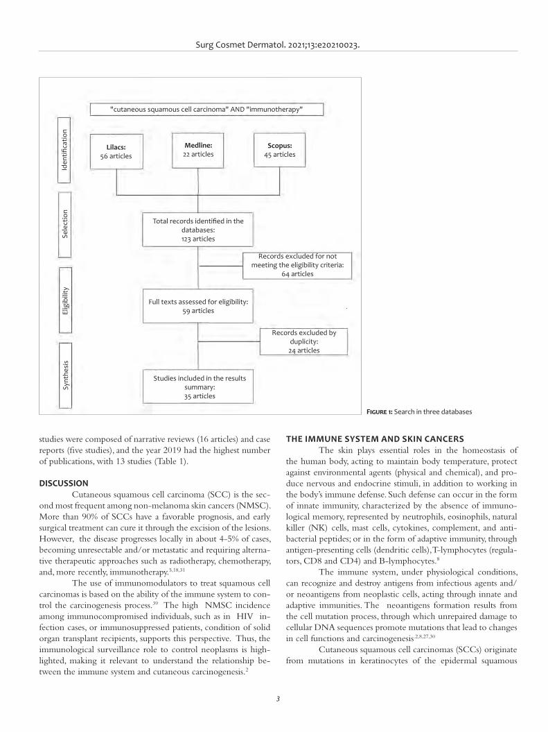
Surg Cosmet Dermatol. 2021;13:e20210023.
3
studies were composed of narrative reviews (16 articles) and case reports (five studies), and the year 2019 had the highest number of publications, with 13 studies (Table 1).
DISCUSSIONCutaneous squamous cell carcinoma (SCC) is the sec-
ond most frequent among non-melanoma skin cancers (NMSC). More than 90% of SCCs have a favorable prognosis, and early surgical treatment can cure it through the excision of the lesions. However, the disease progresses locally in about 4-5% of cases, becoming unresectable and/or metastatic and requiring alterna-tive therapeutic approaches such as radiotherapy, chemotherapy, and, more recently, immunotherapy.5,18,31
The use of immunomodulators to treat squamous cell carcinomas is based on the ability of the immune system to con-trol the carcinogenesis process.39 The high NMSC incidence among immunocompromised individuals, such as in HIV in-fection cases, or immunosuppressed patients, condition of solid organ transplant recipients, supports this perspective. Thus, the immunological surveillance role to control neoplasms is high-lighted, making it relevant to understand the relationship be-tween the immune system and cutaneous carcinogenesis.2
THE IMMUNE SYSTEM AND SKIN CANCERSThe skin plays essential roles in the homeostasis of
the human body, acting to maintain body temperature, protect against environmental agents (physical and chemical), and pro-duce nervous and endocrine stimuli, in addition to working in the body’s immune defense. Such defense can occur in the form of innate immunity, characterized by the absence of immuno-logical memory, represented by neutrophils, eosinophils, natural killer (NK) cells, mast cells, cytokines, complement, and anti-bacterial peptides; or in the form of adaptive immunity, through antigen-presenting cells (dendritic cells), T-lymphocytes (regula-tors, CD8 and CD4) and B-lymphocytes.8
The immune system, under physiological conditions, can recognize and destroy antigens from infectious agents and/or neoantigens from neoplastic cells, acting through innate and adaptive immunities. The neoantigens formation results from the cell mutation process, through which unrepaired damage to cellular DNA sequences promote mutations that lead to changes in cell functions and carcinogenesis.2,8,27,30
Cutaneous squamous cell carcinomas (SCCs) originate from mutations in keratinocytes of the epidermal squamous
Figure 1: Search in three databases
Iden
tifica
tion
Sele
ctio
nEl
igib
ility
Synt
hesi
s
Lilacs: 56 articles
Medline: 22 articles
Scopus: 45 articles
"cutaneous squamous cell carcinoma" AND "immunotherapy"
Total records identified in the databases: 123 articles
Records excluded for not meeting the eligibility criteria:
64 articles
Full texts assessed for eligibility: 59 articles
Records excluded by duplicity: 24 articles
Studies included in the results summary: 35 articles
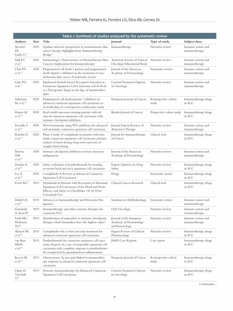
Weber MB, Ferreira IG, Ferreira LO, Silva AB, Cernea SS
4
Table 1: Synthesis of studies analyzed by the systematic reviewAuthors Year Title Journal Type of study Subject Area
Ascierto PA, Garbe C.5
2020 Updates and new perspectives in nonmelanoma skin cancer therapy: highlights from 'Immunotherapy Bridge'
Immunotherapy Narrative review Immune system and immunotherapy
Hall ET et al.2
2020 Immunologic Characteristics of Nonmelanoma Skin Cancers: Implications for Immunotherapy
American Society of Clinical Oncology Educational Book
Narrative review Immune system and immunotherapy
Choi FD et al.13
2020 Programmed cell death 1 protein and programmed death-ligand 1 inhibitors in the treatment of non-melanoma skin cancer: A systematic review
Journal of the American Academy of Dermatology
Systematic review Immune system and immunotherapy
Lima PO et al.4
2020 Epidermal Growth Factor Receptor's Function in Cutaneous Squamous Cell Carcinoma and Its Role as a Therapeutic Target in the Age of Immunother-apies
Current Treatment Options in Oncology
Narrative review Immune system and immunotherapy
Salzmann M et al.14
2020 Programmed cell death protein 1 inhibitors in advanced cutaneous squamous cell carcinoma: re-al-world data of a retrospective, multicenter study
European Journal of Cancer, Retrospective cohort study
Immunotherapy drugs in SCC
Hanna GJ et al.15
2020 Real-world outcomes treating patients with ad-vanced cutaneous squamous cell carcinoma with immune checkpoint inhibitors
British Journal of Cancer Prospective cohort study Immunotherapy drugs in SCC
Pezeshki S et al.16
2020 Novel treatments using PD1 inhibitors for advanced and metastatic cutaneous squamous cell carcinoma
Journal Expert Review of Anticancer Therapy
Narrative review Immune system and immunotherapy
Rischin D et al.17
2020 Phase 2 study of cemiplimab in patients with met-astatic cutaneous squamous cell carcinoma: primary analysis of fixed-dosing, long-term outcome of weight-based dosing
Journal for Immunotherapy of Cancer
Clinical trial Immunotherapy drugs in SCC
Barrios DM et al.18
2020 Immune checkpoint inhibitors to treat cutaneous malignancies
Journal of the American Academy of Dermatology
Narrative review Immune system and immunotherapy
Desilets A et al.19
2020 Safety evaluation of pembrolizumab for treating recurrent head and neck squamous cell carcinoma
Expert Opinion on Drug Safety
Narrative review Immunotherapy drugs in SCC
Lee A et al.20
2020 Cemiplimab: A Review in Advanced Cutaneous Squamous Cell Carcinoma
Drugs Systematic review Immunotherapy drugs in SCC
Ferris RL21 2019 Nivolumab in Patients with Recurrent or Metastatic Squamous Cell Carcinoma of the Head and Neck: Efficacy and Safety in CheckMate 141 by Prior Cetuximab Use
Clinical Cancer Research Clinical trial Immunotherapy drugs in SCC
Habib LA et al.8
2019 Advances in Immunotherapy and Periocular Ma-lignancy
Seminars in Ophthalmology Systematic review Immune system and immunotherapy
Guminski A, Stein B9
2019 Immunotherapy and other systemic therapies for cutaneous SCC
Oral Oncology Narrative review Immune system and immunotherapy
Liebl MC, Hofmann TG22
2019 Identification of responders to immune checkpoint therapy: which biomarkers have the highest value?
Journal of the European Academy of Dermatology and Venereology
Narrative review Immune system and immunotherapy
Ahmed SR et al.23
2019 Cemiplimab-rwlc as first and only treatment for advanced cutaneous squamous cell carcinoma
Expert Review of Clinical Pharmacology
Narrative review Immunotherapy drugs in SCC
van Baar MLM et al.24
2019 Pembrolizumab for cutaneous squamous cell carci-noma: Report of a case of inoperable squamous cell carcinoma with complete response to pembrolizum-ab complicated by granulomatous inflammation
JAAD Case Reports Case report Immunotherapy drugs in SCC
Kacew AJ et al.25
2019 Chromosome 3q arm gain linked to immunother-apy response in advanced cutaneous squamous cell carcinoma
European Journal of Cancer Retrospective cohort study
Immunotherapy drugs in SCC
Ogata D, Tsuchida T26
2019 Systemic Immunotherapy for Advanced Cutaneous Squamous Cell Carcinoma
Current Treatment Options in Oncology
Narrative review Immunotherapy drugs in SCC
Continuation...
Surg Cosmet Dermatol. 2021;13:e20210023.
5
cell layer, which expand into tissues through neoplastic clones. Tumor promoters stimulate clonal tumor expansion. They can be exogenous, such as ultraviolet radiation (UVR), chemical agents, medications, and infections, or endogenous, such as diet and immune suppression. SCCs carry one of the highest tumor mutation loads among all types of cancer, which increases their
immunogenicity due to the expression of tumor neoantigens, mutations, and/or viral gene expression.2,27,30
Natural killer (NK) cells represent one of the main cell lines of innate immunity, and they’re found mainly in the dermis.31 NK cells are responsible for the neoantigens immunosurveillance, controlling tumor progression through cytolytic response.36 Tissue
Table 1: Synthesis of studies analyzed by the systematic reviewAuthors Year Title Journal Type of study Subject Area
Paulson KG et al.27
2019 Immunotherapy for skin cancer International Immunology Narrative review Immune system and immunotherapy
Liu Y et al.28
2019 Prolonged Response to Pembrolizumab in Spindle Cell Squamous Cell Carcinoma Metastatic to the Central Nervous System
Journal of Investigative Medicine High Impact Case Reports
Case report Immunotherapy drugs in SCC
Di Nardo L et al.29
2019 Molecular genetics of cutaneous squamous cell carcinoma: perspective for treatment strategies
Journal of the European Academy of Dermatology and Venereology
Narrative review Immune system and immunotherapy
Barber BR30
2019 Immune Status and Immunotherapy in Advanced Cutaneous Squamous Cell Carcinoma-What Are Our Next Steps?
JAMA Otolaryngolo-gy-Head & Neck Surgery
Letter to the editor Immune system and immunotherapy
Bottomley et al.31
2019 The Role of the Immune System in Cutaneous Squamous Cell Carcinoma
International Journal of Molecular Sciences
Narrative review Immune system and immunotherapy
Chen A et al.7
2018 Clinical Remission of Cutaneous Squamous Cell Carcinoma of the Auricle with Cetuximab and Nivolumab.
Journal of Clinical Medicine Case report Immunotherapy drugs in SCC
Amoils M et al.32
2018 PD-L1 Expression and Tumor-Infiltrating Lym-phocytes in High-Risk and Metastatic Cutaneous Squamous Cell Carcinoma
Head and Neck Surgery Cross-sectional study Immunotherapy drugs in SCC
Migden MR et al.33
2018 PD-1 blockade with cemiplimab in advanced cuta-neous squamous-cell carcinoma
New England Journal of Medicine
Clinical trial Immunotherapy drugs in SCC
Degache E et al.34
2018 Major response to pembrolizumab in two patients with locally advanced cutaneous squamous cell carcinoma
Journal of the European Academy of Dermatology and Venereology
Letter to the editor Immunotherapy drugs in SCC
Yanagi T, Kitamura S, Hata H35
2018 Novel therapeutic targets in cutaneous squamous cell carcinoma
Frontiers in Oncology Narrative review Immune system and immunotherapy
Ilyas M, Costello CM, Sharma A36
2017 Exploring the relationship between natural killer cells and cutaneous squamous cell carcinoma devel-opment
JAAD Case Reports Case report Immunotherapy drugs in SCC
Falchook GS et al.37
2016 Responses of metastatic basal cell and cutaneous squamous cell carcinomas to anti-PD1 monoclonal antibody REGN2810
Journal for Immunotherapy of Cancer
Case report Immunotherapy drugs in SCC
Chang ALS et al.38
2016 A case report of unresectable cutaneous squamous cell carcinoma responsive to pembrolizumab, a programmed cell death protein 1 inhibitor
JAMA Dermatology Letter to the editor Immunotherapy drugs in SCC
Macdonald JB et al.39
2015 Cutaneous adverse effects of targeted therapies: Part II: Inhibitors of intracellular molecular signaling pathways
Journal of the American Academy of Dermatology
Narrative review Immune system and immunotherapy
Yanofsky VR et al.40
2013 Understanding dendritic cells and their role in cuta-neous carcinoma and cancer immunotherapy
Clinical and Developmental Immunology
Narrative review Immune system and immunotherapy
Fujita H et al.41
2012 Langerhans cells from human cutaneous squamous cell carcinoma induce strong type 1 immunity
Journal of Investigative Dermatology
Experimental study Immune system and immunotherapy
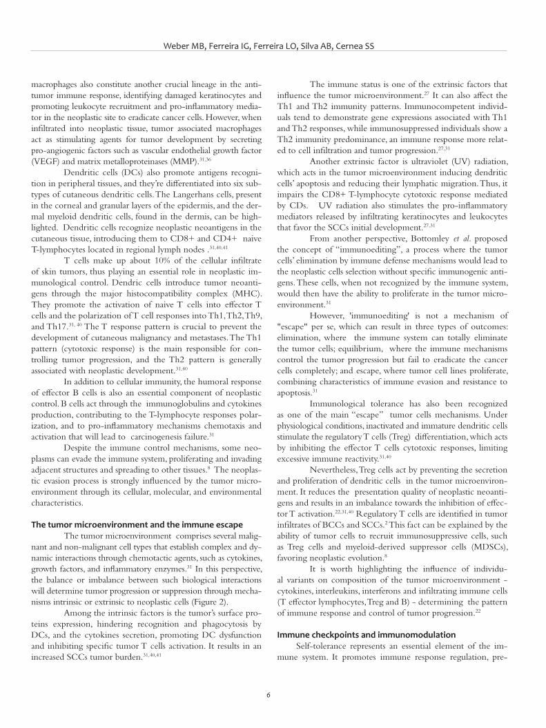
Weber MB, Ferreira IG, Ferreira LO, Silva AB, Cernea SS
6
macrophages also constitute another crucial lineage in the anti-tumor immune response, identifying damaged keratinocytes and promoting leukocyte recruitment and pro-inflammatory media-tor in the neoplastic site to eradicate cancer cells. However, when infiltrated into neoplastic tissue, tumor associated macrophages act as stimulating agents for tumor development by secreting pro-angiogenic factors such as vascular endothelial growth factor (VEGF) and matrix metalloproteinases (MMP).31,36
Dendritic cells (DCs) also promote antigens recogni-tion in peripheral tissues, and they’re differentiated into six sub-types of cutaneous dendritic cells. The Langerhans cells, present in the corneal and granular layers of the epidermis, and the der-mal myeloid dendritic cells, found in the dermis, can be high-lighted. Dendritic cells recognize neoplastic neoantigens in the cutaneous tissue, introducing them to CD8+ and CD4+ naive T-lymphocytes located in regional lymph nodes .31,40,41
T cells make up about 10% of the cellular infiltrate of skin tumors, thus playing an essential role in neoplastic im-munological control. Dendric cells introduce tumor neoanti-gens through the major histocompatibility complex (MHC). They promote the activation of naive T cells into effector T cells and the polarization of T cell responses into Th1, Th2, Th9, and Th17.31, 40 The T response pattern is crucial to prevent the development of cutaneous malignancy and metastases. The Th1 pattern (cytotoxic response) is the main responsible for con-trolling tumor progression, and the Th2 pattern is generally associated with neoplastic development.31,40
In addition to cellular immunity, the humoral response of effector B cells is also an essential component of neoplastic control. B cells act through the immunoglobulins and cytokines production, contributing to the T-lymphocyte responses polar-ization, and to pro-inflammatory mechanisms chemotaxis and activation that will lead to carcinogenesis failure.31
Despite the immune control mechanisms, some neo-plasms can evade the immune system, proliferating and invading adjacent structures and spreading to other tissues.8 The neoplas-tic evasion process is strongly influenced by the tumor micro-environment through its cellular, molecular, and environmental characteristics.
The tumor microenvironment and the immune escapeThe tumor microenvironment comprises several malig-
nant and non-malignant cell types that establish complex and dy-namic interactions through chemotactic agents, such as cytokines, growth factors, and inflammatory enzymes.31 In this perspective, the balance or imbalance between such biological interactions will determine tumor progression or suppression through mecha-nisms intrinsic or extrinsic to neoplastic cells (Figure 2).
Among the intrinsic factors is the tumor’s surface pro-teins expression, hindering recognition and phagocytosis by DCs, and the cytokines secretion, promoting DC dysfunction and inhibiting specific tumor T cells activation. It results in an increased SCCs tumor burden.31,40,41
The immune status is one of the extrinsic factors that influence the tumor microenvironment.27 It can also affect the Th1 and Th2 immunity patterns. Immunocompetent individ-uals tend to demonstrate gene expressions associated with Th1 and Th2 responses, while immunosuppressed individuals show a Th2 immunity predominance, an immune response more relat-ed to cell infiltration and tumor progression.27,31
Another extrinsic factor is ultraviolet (UV) radiation, which acts in the tumor microenvironment inducing dendritic cells’ apoptosis and reducing their lymphatic migration. Thus, it impairs the CD8+ T-lymphocyte cytotoxic response mediated by CDs. UV radiation also stimulates the pro-inflammatory mediators released by infiltrating keratinocytes and leukocytes that favor the SCCs initial development.27,31
From another perspective, Bottomley et al. proposed the concept of “immunoediting”, a process where the tumor cells’ elimination by immune defense mechanisms would lead to the neoplastic cells selection without specific immunogenic anti-gens. These cells, when not recognized by the immune system, would then have the ability to proliferate in the tumor micro-environment.31
However, 'immunoediting' is not a mechanism of "escape" per se, which can result in three types of outcomes: elimination, where the immune system can totally eliminate the tumor cells; equilibrium, where the immune mechanisms control the tumor progression but fail to eradicate the cancer cells completely; and escape, where tumor cell lines proliferate, combining characteristics of immune evasion and resistance to apoptosis.31
Immunological tolerance has also been recognized as one of the main “escape” tumor cells mechanisms. Under physiological conditions, inactivated and immature dendritic cells stimulate the regulatory T cells (Treg) differentiation, which acts by inhibiting the effector T cells cytotoxic responses, limiting excessive immune reactivity.31,40
Nevertheless, Treg cells act by preventing the secretion and proliferation of dendritic cells in the tumor microenviron-ment. It reduces the presentation quality of neoplastic neoanti-gens and results in an imbalance towards the inhibition of effec-tor T activation.22,31,40 Regulatory T cells are identified in tumor infiltrates of BCCs and SCCs.2 This fact can be explained by the ability of tumor cells to recruit immunosuppressive cells, such as Treg cells and myeloid-derived suppressor cells (MDSCs), favoring neoplastic evolution.8
It is worth highlighting the influence of individu-al variants on composition of the tumor microenvironment - cytokines, interleukins, interferons and infiltrating immune cells (T effector lymphocytes, Treg and B) - determining the pattern of immune response and control of tumor progression.22
Immune checkpoints and immunomodulation Self-tolerance represents an essential element of the im-
mune system. It promotes immune response regulation, pre-
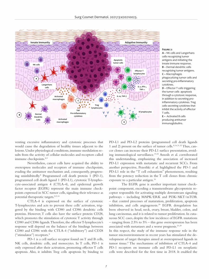
Surg Cosmet Dermatol. 2021;13:e20210023.
7
venting excessive inflammatory and cytotoxic processes that would cause the degradation of healthy tissues adjacent to the lesions. Under physiological conditions, immune modulation re-sults from the activity of cellular molecules and receptors called immune checkpoints.8,9
Nevertheless, cancer cells have acquired the ability to overexpress molecules and receptors of immune checkpoints, evading the antitumor mechanism and, consequently, progress-ing uninhibitedly.8 Programmed cell death protein 1 (PD-1), programmed cell death ligand 1 (PD-L1), cytotoxic T-lympho-cyte-associated antigen 4 (CTLA-4), and epidermal growth factor receptor (EGFR) represent the main immune check-points expressed in SCC tumor cells, signaling their relevance as potential therapeutic targets.8,9,14
CTLA-4 is expressed on the surface of cytotoxic T-lymphocytes and acts to prevent these cells’ activation, trig-gered by the binding with CD80 and CD86 dendritic cells proteins. However, T cells also have the surface protein CD28, which promotes the stimulation of cytotoxic T activity through CD80 and CD86 ligands. Therefore, the effector T-lymphocyte’s response will depend on the balance of the bindings between CD80 and CD86 with the CTLA-4 (“inhibitory”) and CD28 (“stimulator”) receptors.8
PD-1 is a cell surface receptor found on T and B cells, NK cells, dendritic cells, and monocytes. In T cells, PD-1 is only expressed after their activation, promoting effector T cells apoptosis. Also, it inhibits Treg cells apoptosis by binding to
PD-L1 and PD-L2 proteins (programmed cell death ligands 1 and 2) present on the surface of tumor cells.8,14,31,32 Thus, can-cer clones can increase their PD-L1 surface presentation, avoid-ing immunological surveillance.19,32 Amoils et al. corroborated this understanding, emphasizing the association of increased PD-L1 expression with metastatic and recurrent SCCs. From another perspective, Pezeshki et al. highlighted the PD-1 and PD-L1 role in the “T cell exhaustion” phenomenon, resulting from the potency reduction in the T cell clones from chronic exposure to a particular antigen.16
The EGFR gene is another important tumor check-point component, encoding a transmembrane glycoprotein re-ceptor responsible for activating multiple downstream signaling pathways – including MAPK/ERK and PI3K/AKT/mTOR – that control processes of maturation, proliferation, apoptosis inhibition, and cells angiogenesis.29 EGFR deregulation has been observed in head, neck, ovary, breast, bladder, colon, and lung carcinomas, and it is related to tumor proliferation. In cuta-neous SCC cases, despite the low incidence of EGFR mutations – ranging from 2.5% to 5% – this gene overexpression has been associated with metastases and a worse prognosis.21,29
In this respect, the study of the immune response role in the tumor microenvironment in recent years has stimulated the de-velopment of target therapies aimed at overexpressed ligands in tumor tissue.8 The mechanisms of inhibition of CTLA-4 and PD-1 receptors on immune cells and PD-L1 on neoplastic cells were described for the first time in 2018. It enabled the
Figure 2: A – NK cells and Langerhans cells recognizing tumor antigens and initiating the innate immune response. B – Dermal dendritic cells recognizing tumor antigens. C – Macrophages phagocytizing tumor cells and secreting pro-inflammatory cytokines. D – Effector T cells triggering the tumor cells apoptosis through a cytotoxic response, in addition to secreting pro-inflammatory cytokines. Treg cells secreting cytokines that inhibit the activity of effector T cells. E – Activated B cells producing antitumor immunoglobulins.
Innate immunity Adaptive immunity
NK cells
Langerhans cells
Activated macrophages
Activated macrophages
Dermal Dendritic
Cells
TNFPro-inflammatory
cytokines
Treg lymphocytes
Apoptotic tumor cells
Effector T lymphocytes
IL-2, IFN-g,TNF-a
IL-10, TNF-b
B lymphocytes
Weber MB, Ferreira IG, Ferreira LO, Silva AB, Cernea SS
8
tions has been reported in specific cases of melanoma, renal cell carcinoma, and recurrent and metastatic SCCs of head and neck, demonstrating better therapeutic responses.8,25 However, such combinations have higher toxicity, with the risk of triggering colitis and hypophysis.24
The advent of checkpoint inhibitor therapy has raised promising expectations for the treatment of locally ad-vanced, recurrent, and metastatic SCCs, with improved patient overall survival as well as progression-free survival.8,25 Ipilimum-ab, CTLA-4 inhibitor, nivolumab, pembrolizumab, cemiplimab, PD-1 inhibitors, cetuximab, and anti-EGFR are the immu-nological checkpoint inhibitors currently approved for skin neoplasms (Table 2).8,18,35
IMMUNOTHERAPY DRUGS AND TREATMENT OF SQUAMOUS CELL CARCINOMA:
Ipilimumab Ipilimumab can cause adverse events such as autoim-
mune dermatitis, colitis, and diarrhea, in addition to skin reac-tions such as pruritus, morbilliform eruption, nodular pruritus, lichenoid rash, and photosensitivity. In general, these adverse events present after three to six weeks from the start of the med-ication, being dose-dependent and reversible with the end of the treatment.39
NivolumabNivolumab is a PD-1 receptor inhibitor that pre-
vents the T-lymphocyte’s deactivation and preserves the cellular
interruption of the mechanism of the tumor evasion, allowing the effector T cells action to control the cancers’ progression (Figure 3).8,9,18,22,32
In this perspective, CTLA-4 blocking would allow the effector T-lymphocytes activation, while blocking PD-1 or PD-L1 would inhibit the effector T-lymphocytes apoptosis, as well as the phenomenon of “T cell exhaustion”. Thus, both therapeutic pathways would promote increased cellular immunity activity mediated by T cells.8 On the other hand, blocking the EGFR would inhibit one of the mechanisms of tumor cells “evasion”, allowing the immune system to act on them.21,29
Still, it is noteworthy that the immune activity stimula-tion by immunotherapy drugs can trigger nonspecific immune responses, in the form of autoimmune diseases such as vitiligo. Furthermore, these medications also demonstrate the potential to cause adverse events such as pruritus, lichenoid rash, papulo-pustular eruptions, among other skin manifestations, as well as diarrhea and hypothyroidism.19,24,39
Understanding the immune surveillance of cancers and the mechanisms of tumor “evasion” allowed the development of drugs that block the receptors involved in these pathways, with greater emphasis on the CTLA-4, PD-1, and EGFR receptors.8
Due to the high mutational load of SCCs, they become very susceptible to blocking immune checkpoints.8
The main indications for immunotherapy are locally advanced, unresectable, incurable, metastatic SCCs and cases of good tolerability to medications with potential increased surviv-al.15 The combination of anti-CTLA-4 and anti-PD-1 medica-
Figure 3: T-lymphocytes recruited to the tumor site. PD-L1 ligands of tumor cells activating PD-1 receptors on T-lymphocytes, triggering apoptosis of these cells
T lymphocytes
T lymphocytesTumor cell
Squamous cell carcinoma
T lymphocyte apoptosis
PD-L1 and PD-1
correlation
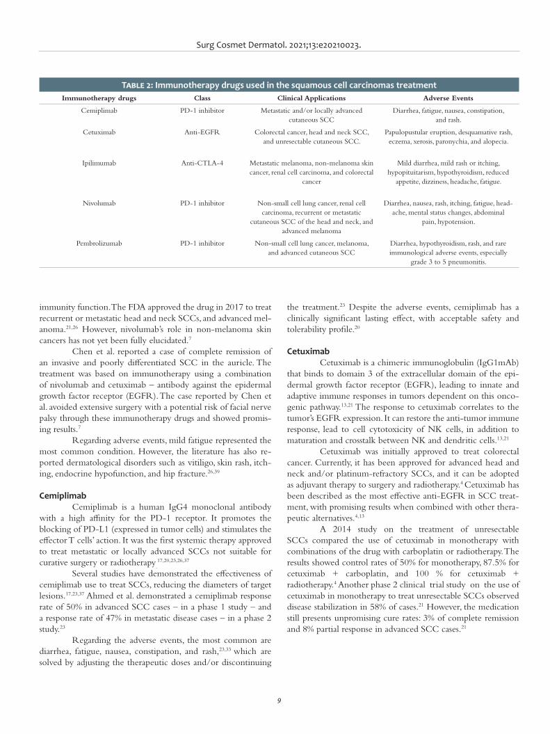
Surg Cosmet Dermatol. 2021;13:e20210023.
9
immunity function. The FDA approved the drug in 2017 to treat recurrent or metastatic head and neck SCCs, and advanced mel-anoma.21,26 However, nivolumab’s role in non-melanoma skin cancers has not yet been fully elucidated.7
Chen et al. reported a case of complete remission of an invasive and poorly differentiated SCC in the auricle. The treatment was based on immunotherapy using a combination of nivolumab and cetuximab – antibody against the epidermal growth factor receptor (EGFR). The case reported by Chen et al. avoided extensive surgery with a potential risk of facial nerve palsy through these immunotherapy drugs and showed promis-ing results.7
Regarding adverse events, mild fatigue represented the most common condition. However, the literature has also re-ported dermatological disorders such as vitiligo, skin rash, itch-ing, endocrine hypofunction, and hip fracture.26,39
CemiplimabCemiplimab is a human IgG4 monoclonal antibody
with a high affinity for the PD-1 receptor. It promotes the blocking of PD-L1 (expressed in tumor cells) and stimulates the effector T cells’ action. It was the first systemic therapy approved to treat metastatic or locally advanced SCCs not suitable for curative surgery or radiotherapy
.17,20,23,26,37
Several studies have demonstrated the effectiveness of cemiplimab use to treat SCCs, reducing the diameters of target lesions.17,23,37 Ahmed et al. demonstrated a cemiplimab response rate of 50% in advanced SCC cases – in a phase 1 study – and a response rate of 47% in metastatic disease cases – in a phase 2 study.23
Regarding the adverse events, the most common are diarrhea, fatigue, nausea, constipation, and rash,23,33 which are solved by adjusting the therapeutic doses and/or discontinuing
the treatment.23 Despite the adverse events, cemiplimab has a clinically significant lasting effect, with acceptable safety and tolerability profile.20
CetuximabCetuximab is a chimeric immunoglobulin (IgG1mAb)
that binds to domain 3 of the extracellular domain of the epi-dermal growth factor receptor (EGFR), leading to innate and adaptive immune responses in tumors dependent on this onco-genic pathway.13,21 The response to cetuximab correlates to the tumor’s EGFR expression. It can restore the anti-tumor immune response, lead to cell cytotoxicity of NK cells, in addition to maturation and crosstalk between NK and dendritic cells.13,21
Cetuximab was initially approved to treat colorectal cancer. Currently, it has been approved for advanced head and neck and/or platinum-refractory SCCs, and it can be adopted as adjuvant therapy to surgery and radiotherapy.4 Cetuximab has been described as the most effective anti-EGFR in SCC treat-ment, with promising results when combined with other thera-peutic alternatives.4,13
A 2014 study on the treatment of unresectable SCCs compared the use of cetuximab in monotherapy with combinations of the drug with carboplatin or radiotherapy. The results showed control rates of 50% for monotherapy, 87.5% for cetuximab + carboplatin, and 100 % for cetuximab + radiotherapy.4 Another phase 2 clinical trial study on the use of cetuximab in monotherapy to treat unresectable SCCs observed disease stabilization in 58% of cases.21 However, the medication still presents unpromising cure rates: 3% of complete remission and 8% partial response in advanced SCC cases.21
Table 2: Immunotherapy drugs used in the squamous cell carcinomas treatmentImmunotherapy drugs Class Clinical Applications Adverse Events
Cemiplimab PD-1 inhibitor Metastatic and/or locally advanced cutaneous SCC
Diarrhea, fatigue, nausea, constipation, and rash.
Cetuximab Anti-EGFR Colorectal cancer, head and neck SCC, and unresectable cutaneous SCC.
Papulopustular eruption, desquamative rash, eczema, xerosis, paronychia, and alopecia.
Ipilimumab Anti-CTLA-4 Metastatic melanoma, non-melanoma skin cancer, renal cell carcinoma, and colorectal
cancer
Mild diarrhea, mild rash or itching, hypopituitarism, hypothyroidism, reduced
appetite, dizziness, headache, fatigue.
Nivolumab PD-1 inhibitor Non-small cell lung cancer, renal cell carcinoma, recurrent or metastatic
cutaneous SCC of the head and neck, and advanced melanoma
Diarrhea, nausea, rash, itching, fatigue, head-ache, mental status changes, abdominal
pain, hypotension.
Pembrolizumab PD-1 inhibitor Non-small cell lung cancer, melanoma, and advanced cutaneous SCC
Diarrhea, hypothyroidism, rash, and rare immunological adverse events, especially
grade 3 to 5 pneumonitis.
Weber MB, Ferreira IG, Ferreira LO, Silva AB, Cernea SS
10
REFERENCES1. Parekh V, Seykora JT. Cutaneous squamous cell carcinoma. Clin Lab
Med [Internet]. 2017;37(3):503-25. 2. Hall ET, Fernandez-Lopez E, Silk AW, Dummer R, Bhatia S. Immunologic
characteristics of nonmelanoma skin cancers: implications for immu-notherapy. Am Soc Clin Oncol Educ Book. 2020;40:1-10.
3. Ministério da Saúde. Estimativa 2020 - Incidência de Câncer no Bra-sil [Internet]. Ministério da Saúde - Instituto Nacional de Câncer José Alencar Gomes da Silva. Rio de Janeiro; 2019. Available at: https://ejournal.poltektegal.ac.id/index.php/siklus/article/view/298%0Aht-tp://repositorio.unan.edu.ni/2986/1/5624.pdf%0Ahttp://dx.doi.or-g/10.1016/j.jana.2015.10.005%0Aht tp://www.biomedcentral.com/1471-2458/12/58%0Ahttp://ovidsp.ovid.com/ovidweb.cgi?T=JS&P
4. 4Lima PO, Joseph S, Panizza B, Simpson F. Epidermal growth factor re-ceptor's function in cutaneous squamous cell carcinoma and its role as a therapeutic target in the age of immunotherapies. Curr Treat Options Oncol. 2020;21(1):9.
5. Ascierto PA, Garbe C. Updates and new perspectives in nonmelanoma skin cancer therapy: highlights from "Immunotherapy Bridge". Immu-notherapy. 2020;12(3):167-74.
6. Misiakos EP, Damaskou V, Koumarianou A, Gouloumi AR, Patapis P, Za-vras N, et al. A giant squamous cell carcinoma of the skin of the tho-racic wall: a case report and review of the literature. J Med Case Rep. 2017;11:136.
7. Chen A, Ali N, Boasberg P, Ho A. Clinical remission of cutaneous squa-mous cell carcinoma of the auricle with cetuximab and nivolumab. J Clin Med. 2018;7(1):10.
8. Habib LA, Wolkow N, Freitag SK, Yoon MK. Advances in immunothera-py and periocular malignancy. Semin Ophthalmol. 2019;34(4):327-33.
9. Guminski A, Stein B. Immunotherapy and other systemic therapies for cutaneous SCC. Oral Oncol. 2019;99:104459. Epub 2019 Nov 9.
10. Taefehshokr N, Baradaran B, Baghbanzadeh A, Taefehshokr S. Pro-mising approaches in cancer immunotherapy. Immunobiology. 2020;225(2):151875.
11. Cunha PLP. Manual revisão bibliográfica sistemática integrativa: a pes-quisa baseada em evidências. Belo Horizonte: Grupo Anima Educação; 2014. 63 p.
12. Souza MT, Dias M, Carvalho R. Revisão integrativa: o que é e como fazer. Einstein. 2010;8(1):102-6.
13. Choi FD, Kraus CN, Elsensohn AN, Carley SK, Lehmer LM, Nguyen RT, et al. Programmed cell death 1 protein and programmed death-ligand 1 inhibitors in the treatment of nonmelanoma skin cancer: a systematic review. J Am Acad Dermatol. 2020;82(2):440-59.
14. Salzmann M, Leiter U, Loquai C, Zimmer L, Ugurel S, Gutzmer R, et al. Programmed cell death protein 1 inhibitors in advanced cutaneous squamous cell carcinoma: real-world data of a retrospective, multicen-ter study. Eur J Cancer. 2020;138:125-32.
15. Hanna GJ, Ruiz ES, LeBoeuf NR, Thakuria M, Schmults CD, Decaprio JA, et al. Real-world outcomes treating patients with advanced cutaneous squamous cell carcinoma with immune checkpoint inhibitors (CPI). Br J Cancer. 2020;123(10):1535-42.
16. Pezeshki S, Hemmati S, Rezaei N. Novel treatments using PD1 inhibi-tors for advanced and metastatic cutaneous squamous cell carcinoma. Expert Rev Anticancer Ther. 2020;20(10):819-22.
17. Rischin D, Migden MR, Lim AM, Schmults CD, Khushalani NI, Hughes BGM, et al. Phase 2 study of cemiplimab in patients with metastatic cutaneous squamous cell carcinoma: primary analysis of fixed-dosing, long-term outcome of weight-based dosing. J Immunother Cancer. 2020;8(1):e000775.
18. Barrios DM, Do MH, Phillips GS, Postow MA, Akaike T, Nghiem P, et al. Immune checkpoint inhibitors to treat cutaneous malignancies. J Am Acad Dermatol. 2020;83(5):1239-53.
19. Desilets A, Soulières D. Safety evaluation of pembrolizumab for trea-ting recurrent head and neck squamous cell carcinoma. Expert Opin Drug Saf. 2020;19(8):927-34.
20. Lee A, Duggan S, Deeks ED. Cemiplimab: a review in advanced cuta-neous squamous cell carcinoma. Drugs. 2020;80(8):813-9.
CONCLUSIONSThe treatment of cutaneous neoplasms is at an ad-
vanced stage, benefiting patients affected with tumors that are challenging to access surgically and reconstruct anatomically. The use of many of these therapies is still under investigation, clinical trial, or approval. Still, the literature already presents evi-dence to support the consideration of the great importance and benefit of these new therapeutic strategies.
It’s essential to understanding the physiopathogenesis of SCCs to promote the development of new therapeutic ap-proaches that may soon benefit a more significant number of patients. l

Surg Cosmet Dermatol. 2021;13:e20210023.
11
21. Ferris RL, Licitra L, Fayette J, Even C, Blumenschein G, Harrington KJ, et al. Nivolumab in patients with recurrent or metastatic squamous cell carcinoma of the head and neck: Efficacy and safety in checkmate 141 by prior cetuximab use. Clin Cancer Res. 2019;25(17):5221-30.
22. Liebl MC, Hofmann TG. Identification of responders to immune checkpoint therapy: which biomarkers have the highest value? J Eur Acad Dermatol Venereol. 2019;33(Suppl 8):52-6.
23. Ahmed SR, Petersen E, Patel R, Migden MR. Cemiplimab-rwlc as first and only treatment for advanced cutaneous squamous cell carcinoma. Expert Rev Clin Pharmacol. 2019;12(10):947-51.
24. van Baar MLM, Guminski AD, Ferguson PM, Martin LK. Pembrolizumab for cutaneous squamous cell carcinoma: report of a case of inoperable squamous cell carcinoma with complete response to pembrolizumab complicated by granulomatous inflammation. JAAD Case Reports. 2019;5(6):491-4.
25. Kacew AJ, Harris EJ, Lorch JH, Haddad RI, Chau NG, Rabinowits G, et al. Chromosome 3q arm gain linked to immunotherapy response in advan-ced cutaneous squamous cell carcinoma. Eur J Cancer. 2019;113:1-9.
26. Ogata D, Tsuchida T. Systemic immunotherapy for advanced cutaneous squamous cell carcinoma. Curr Treat Options Oncol. 2019;20(4):30.
27. Paulson KG, Lahman MC, Chapuis AG, Brownell I. Immunotherapy for skin cancer. Int Immunol. 2019;31(7):465-75.
28. Liu Y, Fitzgerald B, Perry E, Pathak A, Chao HH. Prolonged response to pembrolizumab in spindle cell squamous cell carcinoma metastatic to the central nervous system. J Investig Med High Impact Case Reports. 2019;7:1-5.
29. Di Nardo L, Pellegrini C, Di Stefani A, Del Regno L, Sollena P, Picceril-lo A, et al. Molecular genetics of cutaneous squamous cell carcinoma: perspective for treatment strategies. J Eur Acad Dermatology Venereol. 2020;34(5):932-41.
30. Barber BR. Immune status and immunotherapy in advanced cutaneous squamous cell carcinoma -What are our next steps? JAMA Otolaryngol Neck Surg. 2019;145(4):361.
31. Bottomley MJ, Thomson J, Harwood C, Leigh I. The role of the immu-ne system in cutaneous squamous cell carcinoma. Int J Mol Sci. 2019;20(8):2009.
32. Amoils M, Kim J, Lee C, Sunwoo JB, Colevas AD, Aasi SZ, et al. PD-L1 Ex-pression and Tumor-Infiltrating Lymphocytes in High-Risk and Metas-tatic Cutaneous Squamous Cell Carcinoma. Otolaryngol - Head Neck Surg. 2018;160(1):93-9.
33. Migden MR, Rischin D, Schmults CD, Guminski A, Hauschild A, Lewis KD, et al. PD-1 blockade with cemiplimab in advanced cutaneous squa-mous-cell carcinoma. N Engl J Med. 2018;379:341-51.
34. Degache E, Crochet J, Simon N, Tardieu M, Trabelsi S, Moncourier M, et al. Major response to pembrolizumab in two patients with locally ad-vanced cutaneous squamous cell carcinoma. J Eur Acad Dermatology Venereol. 2018;32(7):e257-8.
35. Yanagi T, Kitamura S, Hata H. Novel therapeutic targets in cutaneous squamous cell carcinoma. Front Oncol. 2018;8:79.
36. Ilyas M, Costello CM, Sharma A. Exploring the relationship between na-tural killer cells and cutaneous squamous cell carcinoma development. JAAD Case Reports. 2017;3(4):364-6.
37. Falchook GS, Leidner R, Stankevich E, Piening B, Bifulco C, Lowy I, et al. Responses of metastatic basal cell and cutaneous squamous cell car-cinomas to anti PD1 monoclonal antibody REGN2810. J Immunother Cancer. 2016;4(1):1-5.
38. Chang ALS, Kim J, Luciano R, Sullivan-Chang L, Colevas AD. A case re-port of unresectable cutaneous squamous cell carcinoma responsive to pembrolizumab, a programmed cell death protein 1 inhibitor. JAMA Dermatology. 2016;152(1):106-8.
39. Macdonald JB, Macdonald B, Golitz LE, LoRusso P, Sekulic A. Cuta-neous adverse effects of targeted therapies. J Am Acad Dermatol. 2015;72(2):203-18.
40. Yanofsky VR, Mitsui H, Felsen D, Carucci JA. Understanding dendritic cells and their role in cutaneous carcinoma and cancer immunothera-py. Clin Dev Immunol. 2013;2013:624123.
41. Fujita H, Suárez-Farĩas M, Mitsui H, Gonzalez J, Bluth MJ, Zhang S, et al. Langerhans cells from human cutaneous squamous cell carcinoma in-duce strong type 1 immunity. J Invest Dermatol. 2012;132(6):1645-55.
AUTHORS' CONTRIBUTION:
Magda Blessmann Weber 0000-0001-5885-5851Approval of the final version of the manuscript; study design and planning; preparation and writing of the manuscript; data collection, analysis, and interpretation; intellectual participation in propaedeutic and/or therapeutic conduct of studied cases; critical literature review; critical revi-sion of the manuscript.
Iago Gonçalves Ferreira 0000-0002-4695-1982Approval of the final version of the manuscript; study design and planning; preparation and writing of the manuscript; data collection, analysis, and interpretation; critical literature review; critical revision of the manuscript.
Laura Oliveira Ferreira 0000-0003-2767-7479Author's contribution: Approval of the final version of the manuscript; study design and planning; preparation and writing of the manuscript; data collection, analysis, and interpretation; critical literature review; critical revision of the manuscript.
Anna Bittarello Silva 0000-0003-4277-1439Approval of the final version of the manuscript; study design and planning; preparation and writing of the manuscript; data collection, analysis, and interpretation; critical literature review; critical revision of the manuscript.
Selma Schuartz Cernea 0000-0002-0710-5935Approval of the final version of the manuscript; study design and planning;active participation in research orientation; critical revision of the manuscript.
1
APOIO CIENTÍFICO:
www.surgicalcosmetic.org.br/
ISSN-e 1984-8773
ReviewAuthors: Guilherme Aron Teixeira Silva1
Daysiane Rocha Souza1
Karina Emburana Costa Parreiras1
Janaíne Cunha Polese1
Fernanda Souza da Silva1
1 School of Medical Sciences of Minas Gerais, Belo Horizonte (MG), Brazil.
Correspondence: Fernanda Souza da Silva E-mail: fernanda.silva@ cienciasmedicasmg.edu.br
Financial support: noneConflit of interest: none
Submitted on: 05/08/2020 Approved on: 07/02/2021
How to cite this article: Silva GAT, Souza DR, Parreiras KEC, Polese JC, Silva FS. Effectiveness of cryolipolysis for subcutaneous fat reduction: a systematic review and meta-analysis. Surg Cosmet Dermatol. 2021;13: e 20210004.
Effectiveness of cryolipolysis for subcutaneous fat reduction: a systematic review and meta-analysisEficácia da criolipólise para redução de gordura subcutânea: uma revisão sistemática e metanálise
ABSTRACTIntroduction: Cryolipolysis produces selective, controlled cooling, and it’s based on the concept that lipid-rich tissue is more susceptible to cold injury, reducing subcutaneous fat. Objective: To review the literature to assess the effectiveness of cryolipolysis in reducing subcutaneous fat. Methods: Systematic review with meta-analysis of studies published in the EBSCOhost, LILACS, and PUBMED databases. Results: Only one study did not present significant reduction in subcutaneous fat compared to the control group. There was a difference among the parameters in the studies. Conclusion: Cryolipolysis is an effective tool for localized fat reduction.Keywords: Apoptosis. Freezing. Subcutaneous Fat
RESU MOIntrodução: a criolipólise produz um resfriamento seletivo e controlado e baseia-se no conceito de que tecidos ricos em lipídios são mais suscetíveis a lesões por frio, reduzindo gordura subcutânea. Objetivo: revisar a literatura para avaliar a eficácia da criolipólise na redução de gordura subcutânea. Métodos: revisão sistemática com metanálise de estudos publicados nas bases de dados EBSCOhost, LILACS e PUBMED. Resultados: apenas um estudo não apresentou redução significativa na gordura subcutânea quando comparado o grupo de intervenção ao grupo controle. Houve diferença entre os estudos em relação aos parâmetros. Conclusão: a criolipólise é uma ferramenta eficaz para redução de gordura localizada.Palavras-chave: Apoptose. Congelamento. Gordura Subcutânea
DOI: http://www.dx.doi.org/10.5935/scd1984-8773.2021130005
ISSN-e 1984-8773
Silva GAT, Souza DR, Parreiras KEC, Polese JC, Silva FS
2
INTRODUCTIONThe fact that more than 56.3% of Brazilian adults are
overweight or obese,1 in addition to the desire to quickly lose fat without undergoing surgery,2 drive the growing demand for fat reduction methods. In this scenario, cryolipolysis emerges as one of the most recently developed modalities for the noninvasive reduction of localized fat.3
The US Food and Drug Administration (FDA) autho-rized the first cryolipolysis device (CoolSculpting system, Zeltiq Aesthetics, Pleasanton, CA, USA) to reduce flank fat (K080521) in 2010; abdominal fat (K120023) in 2012; and inner thigh fat (K133212), in April 2014.4 According to Suh et al.,5 the use of cryolipolysis was also approved for inner thigh, submental fat, arms, back, and lower buttocks.
This technique produces selective and controlled cool-ing. It is based on the concept that lipid-rich tissues are more susceptible to cold injuries than the water-rich tissues around them.6 Adipocytes undergo apoptosis more quickly because they are sensitive to cooling than the dermis, epidermis, muscles, ves-sels, and nerves. These dead cells cause an inflammatory process and are metabolically eliminated as part of normal digestion.7 According to Avram and Harry,8 the changes are not noticeable immediately after the treatment. Therefore, adipocytes and cell membranes are not affected. However, three days after the inter-vention, there is evidence that an inflammatory process occurs only in adipocytes. Between 14 and 30 days, lipid phagocytosis is apparent, resulting in decreased tissue volume in 60 days.
According to the literature, this procedure is safe, with report of patient satisfaction, especially when compared to other methods for localized fat reduction.9 However, with all its bene-fits, there is still a lack of substantial literature demonstrating this tool’s effectiveness. Therefore, this study has the general objec-tive of systematically review the available literature to assess the effectiveness of cryolipolysis in reducing subcutaneous fat. Its specific objectives are to determine the temperature values and the application time commonly used to reduce subcutaneous fat and search the literature if the temperature and cryolipolysis application time specified in the cryolipolysis equipment influ-ences the subcutaneous fat reduction.
MATERIALS AND METHODSThis study is a systematic review conducted according to
the PRISMA methodology (Preferred Reporting Items for Sys-tematic Reviews and Meta-Analysis).10 The guiding question of the present study was elaborated using the PICOS strategy: (P) individuals with subcutaneous fat, (I) cryolipolysis, (C) control group or placebo group, that is, who did not receive the cooling intervention, (O) subcutaneous fat reduction, and (S) random-ized clinical trials.
We performed a systematic review search using the EB-SCOhost, LILACS, and PUBMED databases. The search was conducted using the following terms: (tw:(cryolipolysis)) OR (tw:(lipocryolysis)) OR (tw:(fat freezing)) OR (tw:(coolsculpt-
ing)) OR (tw:(adipocytolysis)) OR (mh:(cryotherapy)) AND (mh:(adiposity)) OR (mh:(subcutaneous fat)) OR (tw:(fat re-duction)) OR (tw:(fat)) OR (tw:(localized fat)) OR (mh:(body contouring)) OR (tw:(nonsurgical reduction of fat)) OR (tw:(noninvasive fat removal)) OR (tw:(noninvasive body con-touring)). In addition to the electronic search, we performed a manual search in the bibliographic references of the previously selected studies.
Randomized clinical trials published between 2010 and 2019 in English, Portuguese, and Spanish, which had full text, were included. We selected the articles conducted in humans, both sexes, aged between 18 and 59 years old, analyzing cryoli-polysis to reduce localized fat in the thigh, flank, abdomen, arm, or submental regions.
Review articles, animal research, studies that used anoth-er therapy combined with cryolipolysis aiming at localized fat reduction other than local massage were excluded. We also ex-cluded studies with an intervention objective other than reduc-ing localized fat and those that performed the treatment with cooling application three or more times in the same area.
Two researchers independently and blindly reviewed the titles and abstracts of potential articles and extracted the data. The selected titles and abstracts were submitted for a complete review. A third researcher was consulted when there were dif-ferences in the inclusion and exclusion of articles. The meth-odological quality of the studies was assessed using the PEDro scale, developed by the Physiotherapy Evidence Database to be used in experimental studies. According to Morton,11 the PE-Dro scale is a valid measure to assess the methodological quality of clinical trials. The scale has a total score of up to 10 points, including internal validity assessment criteria and presentation of the statistical analysis employed. For each criterion defined in the scale, one point (1) is attributed to indicators of the quality of the evidence, and zero (0) in the case of absence of these in-dicators. Two researchers applied the PEDro scale independently and blinded.
Statistical analysis was performed using the Review Manager (RevMan) program developed with the Cochrane col-laboration. The characteristics of the samples between the stud-ies were different, resulting in greater heterogeneity. In the case of significant statistical heterogeneity (I2>50%), a random-effect model n was used for the meta-analysis.
RESULTSSearches in databases and other additional 30 studies
identified through manual search resulted in 1,147 articles. Af-ter eliminating duplicates, 972 articles remained, of which 862 were excluded by title and 51 by abstract. Among the 59 articles assessed for eligibility, 55 were excluded because 22 were cohort studies, systematic or literature review, case report, or pilot study; 17 had participants’ ages incompatible with the inclusion criteria (over 59 years old); five were quasi-experimental studies; three
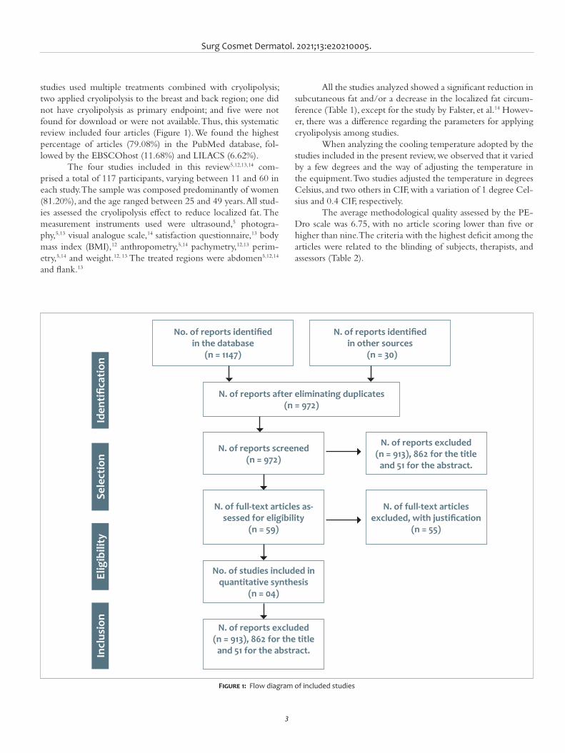
Surg Cosmet Dermatol. 2021;13:e20210005.
3
studies used multiple treatments combined with cryolipolysis; two applied cryolipolysis to the breast and back region; one did not have cryolipolysis as primary endpoint; and five were not found for download or were not available. Thus, this systematic review included four articles (Figure 1). We found the highest percentage of articles (79.08%) in the PubMed database, fol-lowed by the EBSCOhost (11.68%) and LILACS (6.62%).
The four studies included in this review5,12,13,14 com-prised a total of 117 participants, varying between 11 and 60 in each study. The sample was composed predominantly of women (81.20%), and the age ranged between 25 and 49 years. All stud-ies assessed the cryolipolysis effect to reduce localized fat. The measurement instruments used were ultrasound,5 photogra-phy,5,13 visual analogue scale,14 satisfaction questionnaire,13 body mass index (BMI),12 anthropometry,5,14 pachymetry,12,13 perim-etry,5,14 and weight.12, 13 The treated regions were abdomen5,12,14 and flank.13
All the studies analyzed showed a significant reduction in subcutaneous fat and/or a decrease in the localized fat circum-ference (Table 1), except for the study by Falster, et al.14 Howev-er, there was a difference regarding the parameters for applying cryolipolysis among studies.
When analyzing the cooling temperature adopted by the studies included in the present review, we observed that it varied by a few degrees and the way of adjusting the temperature in the equipment. Two studies adjusted the temperature in degrees Celsius, and two others in CIF, with a variation of 1 degree Cel-sius and 0.4 CIF, respectively.
The average methodological quality assessed by the PE-Dro scale was 6.75, with no article scoring lower than five or higher than nine. The criteria with the highest deficit among the articles were related to the blinding of subjects, therapists, and assessors (Table 2).
Figure 1: Flow diagram of included studies
No. of reports identified in the database
(n = 1147)
N. of reports identified in other sources
(n = 30)
N. of reports after eliminating duplicates(n = 972)
N. of reports screened(n = 972)
N. of reports excluded(n = 913), 862 for the title
and 51 for the abstract.
N. of full-text articles as-sessed for eligibility
(n = 59)
N. of full-text articles excluded, with justification
(n = 55)
No. of studies included in quantitative synthesis
(n = 04)
N. of reports excluded(n = 913), 862 for the title
and 51 for the abstract.Incl
usio
nEl
igib
ility
Sele
ctio
nId
entifi
catio
n
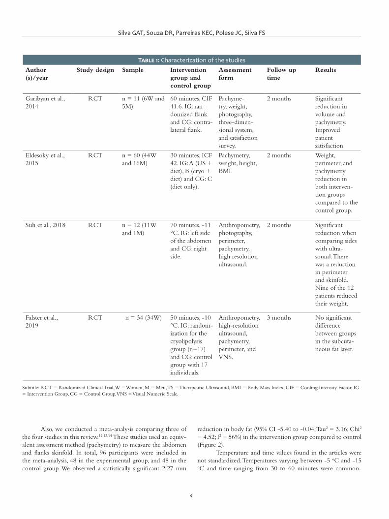
Silva GAT, Souza DR, Parreiras KEC, Polese JC, Silva FS
4
reduction in body fat (95% CI -5.40 to -0.04; Tau2 = 3.16; Chi2 = 4.52; I2 = 56%) in the intervention group compared to control (Figure 2).
Temperature and time values found in the articles were not standardized. Temperatures varying between -5 oC and -15 oC and time ranging from 30 to 60 minutes were common-
Also, we conducted a meta-analysis comparing three of the four studies in this review.12,13,14 These studies used an equiv-alent assessment method (pachymetry) to measure the abdomen and flanks skinfold. In total, 96 participants were included in the meta-analysis, 48 in the experimental group, and 48 in the control group. We observed a statistically significant 2.27 mm
Table 1: Characterization of the studiesAuthor(s)/year
Study design Sample Intervention group and control group
Assessment form
Follow up time
Results
Garibyan et al., 2014
RCT n = 11 (6W and 5M)
60 minutes, CIF 41.6. IG: ran-domized flank and CG: contra-lateral flank.
Pachyme-try, weight, photography, three-dimen-sional system, and satisfaction survey.
2 months Significant reduction in volume and pachymetry. Improved patient satisfaction.
Eldesoky et al., 2015
RCT n = 60 (44W and 16M)
30 minutes, ICF 42. IG: A (US + diet), B (cryo + diet) and CG: C (diet only).
Pachymetry, weight, height, BMI.
2 months Weight, perimeter, and pachymetry reduction in both interven-tion groups compared to the control group.
Suh et al., 2018 RCT n = 12 (11W and 1M)
70 minutes, -11 °C. IG: left side of the abdomen and CG: right side.
Anthropometry, photography, perimeter, pachymetry, high resolution ultrasound.
2 months Significant reduction when comparing sides with ultra-sound. There was a reduction in perimeter and skinfold. Nine of the 12 patients reduced their weight.
Falster et al., 2019
RCT n = 34 (34W) 50 minutes, -10 °C. IG: random-ization for the cryolipolysis group (n=17) and CG: control group with 17 individuals.
Anthropometry, high-resolution ultrasound, pachymetry, perimeter, and VNS.
3 months No significant difference between groups in the subcuta-neous fat layer.
Subtitle: RCT = Randomized Clinical Trial, W = Women, M = Men, TS = Therapeutic Ultrasound, BMI = Body Mass Index, CIF = Cooling Intensity Factor, IG = Intervention Group, CG = Control Group, VNS = Visual Numeric Scale.
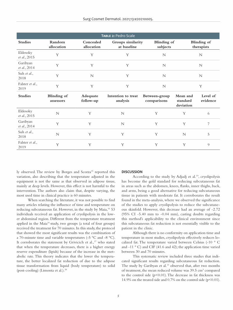
Surg Cosmet Dermatol. 2021;13:e20210005.
5
ly observed. The review by Borges and Scorza15 reported this variation, also describing that the temperature adjusted in the equipment is not the same as that observed in adipose tissue, mainly at deep levels. However, this effect is not harmful to the intervention. The authors also claim that, despite varying, the most used time in clinical practice is 60 minutes.
When searching the literature, it was not possible to find many articles relating the influence of time and temperature on reducing subcutaneous fat. However, in the study by Maia,16 53 individuals received an application of cryolipolysis in the low-er abdominal region. Different from the temperature treatment applied in the Maia16 study, two groups (a total of four groups) received the treatment for 70 minutes. In this study, the protocol that showed the most significant results was the combination of a 70-minute time and variable temperatures (-5 °C and -8 °C). It corroborates the statement by Grivicich et al.,17 who stated that when the temperature decreases, there is a higher energy reserve expenditure (lipids) because of the increase in the met-abolic rate. This theory indicates that the lower the tempera-ture, the better localized fat reduction of due to the adipose tissue transformation from liquid (body temperature) to solid (post-cooling) (Limonta et al.).18
DISCUSSIONAccording to the study by Adjadj et al.19, cryolipolysis
has become the gold standard for reducing subcutaneous fat in areas such as the abdomen, knees, flanks, inner thighs, back, and arms, being a good alternative for reducing subcutaneous tissue in patients with moderate fat. It corroborates the result found in the meta-analysis, where we observed the significance of the studies to apply cryolipolysis to reduce the subcutane-ous skinfold. However, this decrease had an average of -2.72 (95% CI -5.40 mm to -0.04 mm), casting doubts regarding this method’s applicability to the clinical environment since this subcutaneous fat reduction is not essentially visible to the patient in the clinic.
Although there is no conformity on application time and temperature in most studies, cryolipolysis effectively reduces lo-calized fat. The temperature varied between Celsius (-10 ° C and -11 ° C) and CIF (41.6 and 42); the application time varied between 30 and 70 minutes.
This systematic review included three studies that indi-cated significant results regarding subcutaneous fat reduction. The study by Garibyan et al.13 observed that, after two months of treatment, the mean reduced volume was 39.5 cm3 compared to the control side (p<0.01). The decrease in fat thickness was 14.9% on the treated side and 0.7% on the control side (p<0.01).
Table 2: Pedro Scale
Studies Random allocation
Concealed allocation
Groups similarity at baseline
Blinding of subjects
Blinding of therapists
Eldesoky et al., 2015
Y Y Y N N
Garibyan et al., 2014
Y Y Y N N
Suh et al., 2018
Y N Y N N
Falster et al., 2019
Y Y Y N Y
Studies Blinding of assessors
Adequate follow-up
Intention to treat analysis
Between-group comparisons
Mean and standard deviation
Level of evidence
Eldesoky et al., 2015
N Y N Y Y 6
Garibyan et al., 2014
Y Y N Y Y 7
Suh et al., 2018
N Y Y Y N 5
Falster et al., 2019
Y Y Y Y Y 9
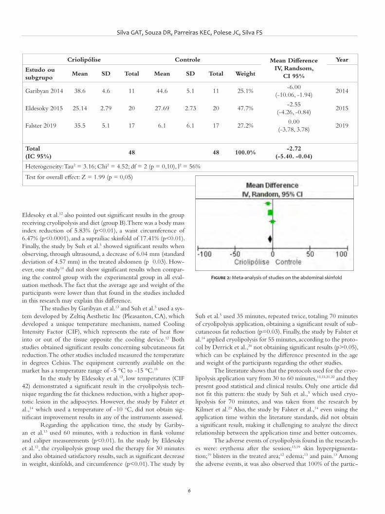
Silva GAT, Souza DR, Parreiras KEC, Polese JC, Silva FS
6
Eldesoky et al.12 also pointed out significant results in the group receiving cryolipolysis and diet (group B). There was a body mass index reduction of 5.83% (p<0.01), a waist circumference of 6.47% (p<0.0001), and a suprailiac skinfold of 17.41% (p<0.01). Finally, the study by Suh et al.5 showed significant results when observing, through ultrasound, a decrease of 6.04 mm (standard deviation of 4.57 mm) in the treated abdomen (p 0.03). How-ever, one study14 did not show significant results when compar-ing the control group with the experimental group in all eval-uation methods. The fact that the average age and weight of the participants were lower than that found in the studies included in this research may explain this difference.
The studies by Garibyan et al.13 and Suh et al.5 used a sys-tem developed by Zeltiq Aesthetic Inc (Pleasanton, CA), which developed a unique temperature mechanism, named Cooling Intensity Factor (CIF), which represents the rate of heat flow into or out of the tissue opposite the cooling device.12 Both studies obtained significant results concerning subcutaneous fat reduction. The other studies included measured the temperature in degrees Celsius. The equipment currently available on the market has a temperature range of -5 °C to -15 °C.15
In the study by Eldesoky et al.12, low temperatures (CIF 42) demonstrated a significant result in the cryolipolysis tech-nique regarding the fat thickness reduction, with a higher apop-totic lesion in the adipocytes. However, the study by Falster et al.,14 which used a temperature of -10 oC, did not obtain sig-nificant improvement results in any of the instruments assessed.
Regarding the application time, the study by Gariby-an et al.13 used 60 minutes, with a reduction in flank volume and caliper measurements (p<0.01). In the study by Eldesoky et al.12, the cryolipolysis group used the therapy for 30 minutes and also obtained satisfactory results, such as significant decrease in weight, skinfolds, and circumference (p<0.01). The study by
Suh et al.5 used 35 minutes, repeated twice, totaling 70 minutes of cryolipolysis application, obtaining a significant result of sub-cutaneous fat reduction (p=0.03). Finally, the study by Falster et al.14 applied cryolipolysis for 55 minutes, according to the proto-col by Derrick et al.,20 not obtaining significant results (p>0.05), which can be explained by the difference presented in the age and weight of the participants regarding the other studies.
The literature shows that the protocols used for the cryo-lipolysis application vary from 30 to 60 minutes,13,15,21,22 and they present good statistical and clinical results. Only one article did not fit this pattern: the study by Suh et al.,5 which used cryo-lipolysis for 70 minutes, and was taken from the research by Kilmer et al.23 Also, the study by Falster et al.,14 even using the application time within the literature standards, did not obtain a significant result, making it challenging to analyze the direct relationship between the application time and better outcomes.
The adverse events of cryolipolysis found in the research-es were: erythema after the session;13,19 skin hyperpigmenta-tion;19 blisters in the treated area;12 edema;13 and pain.13 Among the adverse events, it was also observed that 100% of the partic-
Figure 2: Meta-analysis of studies on the abdominal skinfold
Criolipólise Controle Mean DifferenceIV, Randsom,
CI 95%
Year
Estudo ou subgrupo
Mean SD Total Mean SD Total Weight
Garibyan 2014 38.6 4.6 11 44.6 5.1 11 25.1%-6.00
(-10.06, -1.94)2014
Eldesoky 2015 25.14 2.79 20 27.69 2.73 20 47.7%-2.55
(-4.26, -0.84)2015
Falster 2019 35.5 5.1 17 6.1 6.1 17 27.2%0.00
(-3.78, 3.78)2019
Total (IC 95%)
48 48 100.0%-2.72
(-5.40. -0.04)
Heterogeneity: Tau2 = 3.16; Chi2 = 4.52; df = 2 (p = 0,10), l2 = 56%
Test for overall effect: Z = 1.99 (p = 0,05)
Surg Cosmet Dermatol. 2021;13:e20210005.
7
REFERENCES1. Sa NNB, Moura EC. Excesso de peso: determinantes sociodemográfi-
cos e comportamentais em adultos, Brasil, 2008. Cad Saúde Pública. 2011;27(7):1380-92.
2. Mulholland RS, Paul MD, Chalfoun C. Noninvasive body contouring with radiofrequency, ultrasound, cryolipolysis, and low-level laser the-rapy. Clin Plast Surg. 2011;38(3):503-20.
3. Ingargiola MJ, Motakef S, Chung MT, Vasconez HC, Sasaki GH. Cryolipoly-sis for fat reduction and body contouring: safety and efficacy of current treatment paradigms. Plast Reconstr Surg. 2015;135(6):1581-90.
4. Lee SJ, Jang HW, Kim MH, Suh DH, Ryu HJ. Non-invasive cryolipoly-sis to reduce subcutaneous fat in the arms. J Cosmet Laser Ther. 2016;18(3):126-9.
5. Suh DH, Park JH, Kim BY, Lee SJ, Moon JH, Ryu HJ. Double stacking cryo-lipolysis treatment of the abdominal fat with use of a novel contoured applicator. J Cosmet Laser Ther. 2019;21(4):238-42.
6. Savacini MB, Bueno DT, Molina ACS, Lopes ACA, Silva CN, Moreira RG, et al. Effectiveness and safety of contrast cryolipolysis for subcutaneous--fat reduction. Dermatol Res Pract. 2018;(2):1-9.
7. Ferraro GA, Francesco F, Cataldo C, Rossano F, Nicoletti G, D'Andea F. Sy-nergistic effects of cryolipolysis and shock waves for noninvasive body contouring. Aesthetic Plast Surg. 2012;36(3):666-79.
8. Avram MM, Harry RS. Cryolipolysis for subcutaneous fat layer reduc-tion. Lasers Surg Med. 2009;41(10):703-8.
9. Dierickx CC, Mazer JM, Sand M, Koenig S, Arigon V. Safety, tolerance, and patient satisfaction with noninvasive cryolipolysis. Dermatol Surg. 2013;39(8):1209-16.
10. Galvão TF, Pansani TSA. Principais itens para relatar revisões sistemá-ticas e meta- análises: a recomendação PRISMA. 2015;24(2):335-342.
11. De Morton NA. The PEDro scale is a valid measure of the methodolo-gical quality of clinical trials: a demographic study. Aust J Physiother. 2009;55(2):129-33.
12. Eldesoky MTM, Abutaleb EEM, Mousa GSM. Ultrasound cavitation ver-sus cryolipolysis for non-invasive body contouring. Australas J Derma-tol. 2016;57(4):288-93.
13. Garibyan L, Sipprell WH, Jalian RH, Sakamoto FH, Avram M, Anderson RR. Three-dimensional volumetric quantification of fat loss following cryolipolysis. Lasers Surg Med. 2014;46(2):75-80.
14. Falster M, Schardong J, Santos DP, Machado BC, Peres A, Rosa PV, et al. Efeitos da criolipólise na espessura da gordura abdominal inferior de mulheres saudáveis e satisfação do paciente: um estudo controlado randomizado. Rev Bras Fisio. 2019.
ipants had reduced sensitivity for 10 minutes after application, 73% after three weeks, and 18% in the second month after treat-ment.13 Several studies report these events after the cryolipolysis use. However, numerous researchers state that they occur in a short period and do not have significant repercussions.24,25
This research’s findings evidence the need for further studies in the area – studies with good methodological quality and standardized assessment instruments that allow conducting new meta-analyses, ensuring the safe application of cryolipoly-sis as a method to reduce subcutaneous fat. However, the study presented as limitations the failure to research all scientific data-bases and the inclusion of only articles published between 2010 and 2019, in English, Portuguese, Spanish, which had full text available.
CONCLUSIONThe assessment method of areas submitted to cryolipoly-
sis did not present conformity: the articles used parameters re-peated in different authors or cited in a single study. However, as the meta-analysis demonstrated, cryolipolysis is an effective tool in reducing subcutaneous fat. Nevertheless, other studies with better methodological quality, investigating temperature and ap-plication time for subcutaneous fat reduction and presenting a standardization of assessment methods, should be proposed, giv-en the scarcity in the literature. l
Silva GAT, Souza DR, Parreiras KEC, Polese JC, Silva FS
8
AUTHORS' CONTRIBUTION:
Guilherme Aron Teixeira Silva | 0000-0002-2499-0147Statistical analysis; approval of the final version of the manuscript; study design and planning; preparation and writing of the manuscript; data collection, analysis; critical literature review.
Daysiane Rocha Souza | 0000-0003-4528-701XStatistical analysis; approval of the final version of the manuscript; study design and planning; preparation and writing of the ma-nuscript; data collection, analysis; critical literature review.
Karina Emburana Costa Parreiras | 0000-0003-4966-6252Statistical analysis; approval of the final version of the manuscript; study design and planning; preparation and writing of the ma-nuscript; data collection, analysis; critical literature review.
Janaíne Cunha Polese | 0000-0003-3366-1545Statistical analysis; approval of the final version of the manuscript; study design and planning; preparation and writing of the ma-nuscript; data collection, analysis; active participation in research orientation; critical literature review.
Fernanda Souza da Silva | 0000-0002-3161-2531Statistical analysis; approval of the final version of the manuscript; study design and planning; preparation and writing of the ma-nuscript; data collection, analysis; active participation in research orientation; critical literature review.
15. Borges FS, Scorza FA. Fundamentos da criolipólise. Fisio Ser. 2014;9(4):219-24.
16. Maia VRC. Desenvolvimento de um protocolo biomédico para a redu-ção da adiposidade abdominal feminina utilizando a criolipólise. Curi-tiba. Dissertação [Programa de Pós- Graduação em Engenharia Bio-médica] - Universidade Tecnológica Federal do Paraná; 2018. Available from: http://repositorio.utfpr.edu.br/jspui/handle/1/3402.
17. Grivicich I, Regner A, Rocha AB. Morte celular por apoptose. Revista Brasileira de Cancerologia. 2007;53(3):335-43.
18. Limonta AN, Ribeiro V da S, Gomes JPC, Moraes CAP. Criolipólise: a im-portância da membrana anticongelante na prevenção de queimadu-ras. InterfacEHS. 2017;12(1):128-43.
19. Adjadj L, SidAhmed-Mezi M, Mondoloni M, Meningaud JP, Hersant B. Assessment of the efficacy of cryolipolysis on saddlebags: a prospec-tive study of 53 patients. Plast Reconstr Surg. 2017;140(1):50-7.
20. Derrick CD, Shridharani SM, Broyles JM. The safety and efficacy of cryo-lipolysis: a systematic review of available literature. Aesthetic Surg J. 2015;35(7):830-6.
21. Manstein D, Laubach H, Watanabe K, Farinelli W, Zurakowski D, Ander-son RR. Selective cryolysis: a novel method of noninvasive fat removal. Lasers Surg Med. 2008 ;40(9):595-604.
22. Coleman SR, Sachdeva K, Egbert BM, Preciado J, Allison J. Clinical effi-cacy of noninvasive cryolipolysis and its effects on peripheral nerves. Aesthetic Plastic Surgery. 2009;33(4):482-8.
23. Kilmer SL. Prototype coolcup cryolipolysis applicator with over 40% re-duced treatment time demonstrates equivalent safety and efficacy with greater patient preference. Lasers Surg Med. 2017;49(1):63-8.
24. Klein KB, Zelicson B, Riopelle JG, Okamoto E, Bachelor EP, Harry RS, Preciado JA. Non-invasive cryolipolysis for subcutaneous fat reduction does not affect serum liipid levels or liver function tests. Lasers Surg Med. 2009;41(10):785-90.
25. Stevens WG, Pietrzak LK, Spring MA. Broad overview of a clinical and commercial experience with cool sculpting. Aesthet Surg J. 2013;33(6):835-46.
1
APOIO CIENTÍFICO:
www.surgicalcosmetic.org.br/
ISSN-e 1984-8773
ReviewAuthors: Pedro de Freitas Silva Torraca1
Estela Mari Sandini1
Tania Christina Marchesi de Freitas1
1 Dermatology Service, Universida-de Federal do Mato Grosso do Sul, Campo Grande (MS), Brazil.
Correspondence:Pedro de Freitas Silva TorracaEmail: [email protected]
Financial support: noneConflit of interest: none
Submitted on: 24/03/2021Approved on: 22/06/2021
How to cite this article: Torraca PFS, Sandini EM, Freitas TCM. Uso de tec-nologias a laser e luz intensa pulsada no tratamento da ocronose exógena: uma revisão da literatura. Surg Cosmet Dermatol. 2021;13:e20210002.
Use of laser technologies and intense pulsed light in the treatment of exogenous ochronosis: a literature reviewUso de tecnologias a laser e luz intensa pulsada no tratamento da ocronose exógena: uma revisão da literatura
ABSTRACTExogenous ochronosis is a cutaneous hyperpigmentation condition caused by the accu-mulation of substances derived from phenol on the skin or mucous membranes without affecting other tissues. It occurs mainly due to the use of bleaching agents, most frequently hydroquinone. The lesions are difficult to treat, being resistant to several approaches. Some-times it’s necessary to use laser technologies or intense pulsed light to achieve some degree of improvement. The present work consists of a literature review of publications on these technologies in exogenous ochronosis from January 1990 to July 2020. Keywords: Hyperpigmentation; Intense pulsed light therapy; Lasers; Ochronosis
RESU MOA ocronose exógena é um quadro de hiperpigmentação cutânea por acúmulo de substâncias derivadas de fenol em pele ou mucosas, sem acometimento de outros tecidos. Ocorre, principalmente, por uso de clareadores, sendo mais frequente na hidroquinona como despigmentante. As lesões apresentam difícil tratamento, sendo resistentes a diversas abordagens. Por vezes, é necessário utilizar tecnologias com laser ou luz intensa pulsada para atingir algum grau de melhora. O presente trabalho realizou pesquisa bibliográfica na forma de revisão de literatura entre janeiro de 1990 e julho de 2020, organizando publicações acerca do uso destas tecnologias na ocronose exógena.Palavras-chave: Hiperpigmentação; Lasers; Ocronose; Terapia de luz pulsada intensa
DOI: http://www.dx.doi.org/10.5935/scd1984-8773.2021130002
ISSN-e 1984-8773
Torraca PFS, Sandini EM, Freitas TCM
2
INTRODUCTIONExogenous ochronosis (EO) is described as skin and mu-
cous membranes hyperpigmentation caused by phenol-derived substances deposits, most commonly after the use of whiten-ing creams containing hydroquinone, topical resorcinol, or in-tramuscular or oral antimalarials.1,2,3,4 Contrary to endogenous ochronosis, which arises from homogentisic acid deposition in soft areas and internal tissues, exogenous ochronosis does not af-fect these sites. EO is considered uncommon:5 it presents a mod-erate incidence in South Africa,6 with isolated reports in Asia, Africa, and Latin America in patients with Hispanic ancestry and high skin phototypes.7,8,9
Rudolph Virchow named EO in 1865.10,11 Findlay6 relat-ed the disease to the use of bleaching cream with hydroquinone in 1975, and Beddard and Plumtre12 associated it to with use of phenol to treat leg ulcers in 1912.12
A recent review of cases in the American literature showed a total of 39 reports of EO in the United States, from January 1983 to June 2020. Of these cases, 18 described the disease onset from the use of whitening creams, and, among these, 14 had hydroquinone in the composition. The concentra-tions ranged from 2% to 7.5%, with application time from two months to 30 years until the onset of the lesions.2,3,8
Clinically, EO presents as asymptomatic bluish-black or yellow-brown macules, or areas of hyperpigmentation in sun-exposed regions, such as the face, neck, back, extensor zones of the upper limbs, distal portions of the forearms, legs, and dor-sum of hands or feet.13 At the histological level, the clinical pic-ture is similar to that of endogenous ochronosis, but with no pigment accumulation in joints, bones, urine, other secretions, or tissues.14,15
For some authors, the condition occurs due to resistance of melanocytes to the effect of whitening agents, with conse-quent pigment leakage in the papillary dermis and accumulation of this pigment in fibroblasts, resulting in phenols presence in elastotic fibers and their hyperchromia.6 Other authors argue that hydroquinone is oxidized to quinone forming hydroxylated indoles similar to melanin precursors.16 A third group believes that high hydroquinone concentrations stimulate melanocytes to produce more melanin.17 However, the most widely accept-ed theory is that hyperpigmentation results from homogentisic acid oxidase enzyme inhibition by hydroquinone, causing local homogentisic acid accumulation. The homogentisic acid then polymerizes, forming an ocher pigment in the papillary dermis, as occurs in other tissues in cases of endogenous ochronosis due to a primary structural defect of this enzyme.18
Exogenous ochronosis lesions observed in dermoscopy were initially described in 200819,20 as sites of blue-gray or brown to black, amorphous globules, with follicular obliteration areas. It contrasts with melasma cases where dermoscopy demonstrates a pattern of reticular pigmentation, pseudonet accentuation, and brownish granules and globules, sparing the follicles.21
At the histological level, the lesions show collagen fibers
with a yellow-brownish color in a “banana shape”, degradation of these fibers, and formation of colloid milium amid an inflam-matory infiltrate with plasma cells, histiocytes, and multinucleat-ed giant cells in the development of the lesions.16,22
In 1979, Dogliotti and Leibowitz classified the clinical stages of exogenous ochronosis into stage I (lesions with er-ythema and some pigmentation); stage II (injuries presenting hyperpigmentation, hyperpigmented colloid milium, atrophy); and stage III (presence of papulonodular eruptive elements in a lesion with stage II features plus inflammatory characteristics in more recent wounds that are less pronounced in older inju-ries).23 In 1986, Phillips et al. classified ochronosis as mild, mod-erate, and severe in a series of 395 cases in patients assessed by the Dermatology Service of a hospital in Johannesburg, South Africa, during one year. Only lesions with altered skin hyperpig-mentation and hypertrophy were considered as mild; presence of hyperchromic papules was the standard for moderate degree; and lesions with hyperchromic caviar-like papules, coalescent in plaques, were deemed as severe.8 In a third classification, in 1989, Hardwick et al. considered five grades of presentation: grade 1 comprised lesions with hyperchromic macules; grade 2, with macules and micropapules; grade 3 included injuries with dark-ened deposits and larger papules; grade 4, with colloid milium of 1 mm or more; and grade 5 encompassed lesions with keloid nodules and hyperchromic cysts.24
According to the European Society of Laser Dermatology (ESLD), the exogenous ochronosis treatment is challenging, with unpredictable results, often below expectations. The use of photo-protection becomes a relevant element in the initial approach by slowing the progression of the lesions and preventing the emer-gence of new hyperpigmented areas.15 Some cases achieved a par-tial response with topical retinoic and glycolic acid in low con-centrations and oral use of tetracycline in papular presentations or with sarcoidosis-like lesions.25 In a recent review on the use of these technologies, the ESLD recommends such therapies asso-ciated with multiple laser technologies sessions, combining frac-tional ablative modalities such as CO
2 or Erbium 2940nm with
Q-Switched 1064nm for better and faster results.26,15
The present study reviews publications containing a therapeutic approach to exogenous ochronosis conditions using laser technologies or intense pulsed light, demonstrating the de-scribed protocols and results obtained from 1990 to July 2020.
METHODS
Four databases were searched from June 15 to August 5, 2020: Embase, MEDLINE/Pubmed, LILACS, and Cochrane Library. The selected languages were English, Spanish, and Por-tuguese. In the first stage, the keywords used were chronosis, exogenous ochronosis, ocronose, and ocronosis. They generat-ed a total of 1,377 results in the Embase platform, 978 in the MEDLINE/Pubmed, 32 in the LILACS, and 16 results in the Cochrane Library platform.
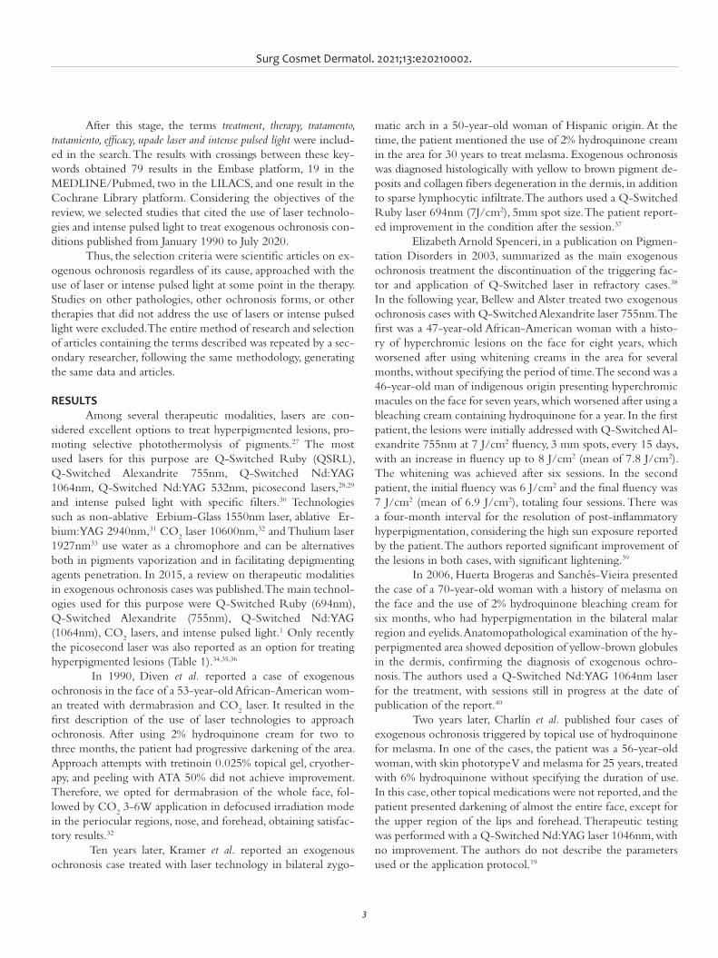
Surg Cosmet Dermatol. 2021;13:e20210002.
3
After this stage, the terms treatment, therapy, tratamento, tratamiento, efficacy, upade laser and intense pulsed light were includ-ed in the search. The results with crossings between these key-words obtained 79 results in the Embase platform, 19 in the MEDLINE/Pubmed, two in the LILACS, and one result in the Cochrane Library platform. Considering the objectives of the review, we selected studies that cited the use of laser technolo-gies and intense pulsed light to treat exogenous ochronosis con-ditions published from January 1990 to July 2020.
Thus, the selection criteria were scientific articles on ex-ogenous ochronosis regardless of its cause, approached with the use of laser or intense pulsed light at some point in the therapy. Studies on other pathologies, other ochronosis forms, or other therapies that did not address the use of lasers or intense pulsed light were excluded. The entire method of research and selection of articles containing the terms described was repeated by a sec-ondary researcher, following the same methodology, generating the same data and articles.
RESULTS
Among several therapeutic modalities, lasers are con-sidered excellent options to treat hyperpigmented lesions, pro-moting selective photothermolysis of pigments.27 The most used lasers for this purpose are Q-Switched Ruby (QSRL), Q-Switched Alexandrite 755nm, Q-Switched Nd:YAG 1064nm, Q-Switched Nd:YAG 532nm, picosecond lasers,28,29 and intense pulsed light with specific filters.30 Technologies such as non-ablative Erbium-Glass 1550nm laser, ablative Er-bium:YAG 2940nm,31 CO
2 laser 10600nm,32 and Thulium laser
1927nm33 use water as a chromophore and can be alternatives both in pigments vaporization and in facilitating depigmenting agents penetration. In 2015, a review on therapeutic modalities in exogenous ochronosis cases was published. The main technol-ogies used for this purpose were Q-Switched Ruby (694nm), Q-Switched Alexandrite (755nm), Q-Switched Nd:YAG (1064nm), CO
2 lasers, and intense pulsed light.1 Only recently
the picosecond laser was also reported as an option for treating hyperpigmented lesions (Table 1).34,35,36
In 1990, Diven et al. reported a case of exogenous ochronosis in the face of a 53-year-old African-American wom-an treated with dermabrasion and CO
2 laser. It resulted in the
first description of the use of laser technologies to approach ochronosis. After using 2% hydroquinone cream for two to three months, the patient had progressive darkening of the area. Approach attempts with tretinoin 0.025% topical gel, cryother-apy, and peeling with ATA 50% did not achieve improvement. Therefore, we opted for dermabrasion of the whole face, fol-lowed by CO
2 3-6W application in defocused irradiation mode
in the periocular regions, nose, and forehead, obtaining satisfac-tory results.32
Ten years later, Kramer et al. reported an exogenous ochronosis case treated with laser technology in bilateral zygo-
matic arch in a 50-year-old woman of Hispanic origin. At the time, the patient mentioned the use of 2% hydroquinone cream in the area for 30 years to treat melasma. Exogenous ochronosis was diagnosed histologically with yellow to brown pigment de-posits and collagen fibers degeneration in the dermis, in addition to sparse lymphocytic infiltrate. The authors used a Q-Switched Ruby laser 694nm (7J/cm2), 5mm spot size. The patient report-ed improvement in the condition after the session.37
Elizabeth Arnold Spenceri, in a publication on Pigmen-tation Disorders in 2003, summarized as the main exogenous ochronosis treatment the discontinuation of the triggering fac-tor and application of Q-Switched laser in refractory cases.38 In the following year, Bellew and Alster treated two exogenous ochronosis cases with Q-Switched Alexandrite laser 755nm. The first was a 47-year-old African-American woman with a histo-ry of hyperchromic lesions on the face for eight years, which worsened after using whitening creams in the area for several months, without specifying the period of time. The second was a 46-year-old man of indigenous origin presenting hyperchromic macules on the face for seven years, which worsened after using a bleaching cream containing hydroquinone for a year. In the first patient, the lesions were initially addressed with Q-Switched Al-exandrite 755nm at 7 J/cm2 fluency, 3 mm spots, every 15 days, with an increase in fluency up to 8 J/cm2 (mean of 7.8 J/cm2). The whitening was achieved after six sessions. In the second patient, the initial fluency was 6 J/cm2 and the final fluency was 7 J/cm2 (mean of 6.9 J/cm2), totaling four sessions. There was a four-month interval for the resolution of post-inflammatory hyperpigmentation, considering the high sun exposure reported by the patient. The authors reported significant improvement of the lesions in both cases, with significant lightening.39
In 2006, Huerta Brogeras and Sanchés-Vieira presented the case of a 70-year-old woman with a history of melasma on the face and the use of 2% hydroquinone bleaching cream for six months, who had hyperpigmentation in the bilateral malar region and eyelids. Anatomopathological examination of the hy-perpigmented area showed deposition of yellow-brown globules in the dermis, confirming the diagnosis of exogenous ochro-nosis. The authors used a Q-Switched Nd:YAG 1064nm laser for the treatment, with sessions still in progress at the date of publication of the report.40
Two years later, Charlín et al. published four cases of exogenous ochronosis triggered by topical use of hydroquinone for melasma. In one of the cases, the patient was a 56-year-old woman, with skin phototype V and melasma for 25 years, treated with 6% hydroquinone without specifying the duration of use. In this case, other topical medications were not reported, and the patient presented darkening of almost the entire face, except for the upper region of the lips and forehead. Therapeutic testing was performed with a Q-Switched Nd:YAG laser 1046nm, with no improvement. The authors do not describe the parameters used or the application protocol.19
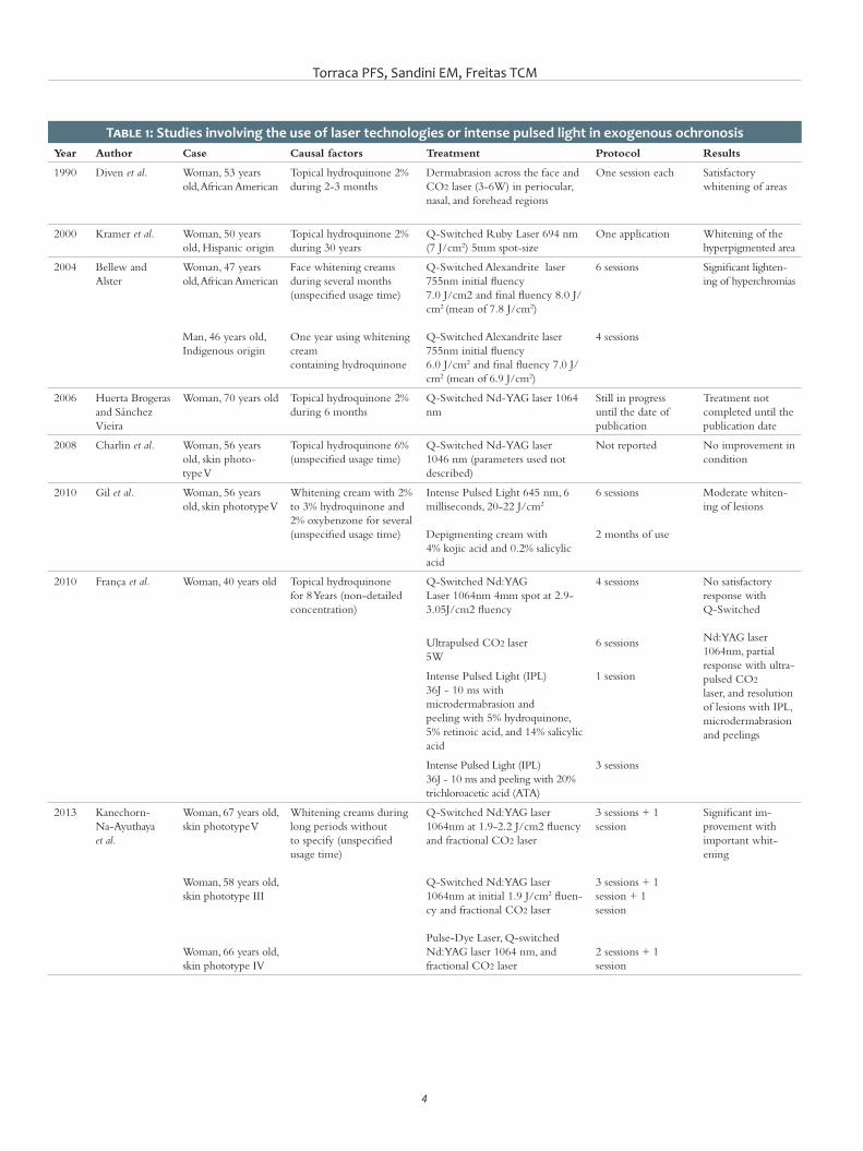
Torraca PFS, Sandini EM, Freitas TCM
4
Table 1: Studies involving the use of laser technologies or intense pulsed light in exogenous ochronosisYear Author Case Causal factors Treatment Protocol Results
1990 Diven et al. Woman, 53 years old, African American
Topical hydroquinone 2% during 2-3 months
Dermabrasion across the face and CO2 laser (3-6W) in periocular, nasal, and forehead regions
One session each Satisfactory whitening of areas
2000 Kramer et al. Woman, 50 years old, Hispanic origin
Topical hydroquinone 2% during 30 years
Q-Switched Ruby Laser 694 nm (7 J/cm2) 5mm spot-size
One application Whitening of thehyperpigmented area
2004 Bellew and Alster
Woman, 47 years old, African American
Man, 46 years old,Indigenous origin
Face whitening creamsduring several months (unspecified usage time)
One year using whitening creamcontaining hydroquinone
Q-Switched Alexandrite laser 755nm initial fluency7.0 J/cm2 and final fluency 8.0 J/cm2 (mean of 7.8 J/cm2)
Q-Switched Alexandrite laser 755nm initial fluency6.0 J/cm2 and final fluency 7.0 J/cm2 (mean of 6.9 J/cm2)
6 sessions
4 sessions
Significant lighten-ing of hyperchromias
2006 Huerta Brogeras and Sánchez Vieira
Woman, 70 years old Topical hydroquinone 2% during 6 months
Q-Switched Nd-YAG laser 1064 nm
Still in progress until the date of publication
Treatment not completed until thepublication date
2008 Charlin et al. Woman, 56 years old, skin photo-type V
Topical hydroquinone 6% (unspecified usage time)
Q-Switched Nd-YAG laser 1046 nm (parameters used not described)
Not reported No improvement in condition
2010 Gil et al. Woman, 56 years old, skin phototype V
Whitening cream with 2% to 3% hydroquinone and 2% oxybenzone for several (unspecified usage time)
Intense Pulsed Light 645 nm, 6 milliseconds, 20-22 J/cm2
Depigmenting cream with4% kojic acid and 0.2% salicylic acid
6 sessions
2 months of use
Moderate whiten-ing of lesions
2010 França et al. Woman, 40 years old Topical hydroquinone for 8 Years (non-detailed concentration)
Q-Switched Nd:YAGLaser 1064nm 4mm spot at 2.9-3.05J/cm2 fluency
4 sessions No satisfactory response withQ-Switched
Nd:YAG laser1064nm, partial response with ultra-pulsed CO2
laser, and resolution of lesions with IPL,microdermabrasion and peelings
Ultrapulsed CO2 laser5W
6 sessions
Intense Pulsed Light (IPL)36J - 10 ms withmicrodermabrasion and peeling with 5% hydroquinone, 5% retinoic acid, and 14% salicylic acid
1 session
Intense Pulsed Light (IPL)36J - 10 ms and peeling with 20% trichloroacetic acid (ATA)
3 sessions
2013 Kanechorn-Na-Ayuthaya et al.
Woman, 67 years old, skin phototype V
Woman, 58 years old, skin phototype III
Woman, 66 years old, skin phototype IV
Whitening creams duringlong periods withoutto specify (unspecified usage time)
Q-Switched Nd:YAG laser 1064nm at 1.9-2.2 J/cm2 fluency and fractional CO2 laser
Q-Switched Nd:YAG laser 1064nm at initial 1.9 J/cm2 fluen-cy and fractional CO2 laser
Pulse-Dye Laser, Q-switched Nd:YAG laser 1064 nm, and fractional CO2 laser
3 sessions + 1 session
3 sessions + 1 session + 1session
2 sessions + 1 session
Significant im-provement with important whit-ening
Surg Cosmet Dermatol. 2021;13:e20210002.
5
Year Author Case Causal factors Treatment Protocol Results
2013 Tan Six women, from 37 to 69 years old
Whitening creams Q-Switched Nd:YAG laser 1064nm at 1.2 J/cm2 fluency in hyperpigmented areas ofexogenous ochronosis and melasma
Q-Switched Nd:YAG laser 1064nm at 4-6 J/cm2 fluency in exogenous ochronosis areas
8 sessions + 16 sessions
Two showed rele-vant improvement, four showed mild improvement.
2014 Liu et al. Woman, 50 years old, skin phototype IV
Whitening creams forlong periods oftenwith hydroquinone (unspecified usage time)
Q-Switched Nd-YAG laser 1064nm at 6-9 J/cm2 fluency
6 sessions No improvement
2014 Lee and Weiss Woman, 48 years old Whitening cream (unspec-ified usage time)
Intense Pulsed Light in570 nm waves at12J/cm2 fluency and 15 millisec-onds pulse
Total of sessions not reported
Whitening of thehyperpigmented area
2015 Ko et al. Woman, 50 years old Whitening cream with4% hydroquinone
Q-Switched Nd-YAG laser 1064nm at 5.3 J/cm2 fluency in left zygomatic region
Q-Switched Alexandrite laser 755nm at 8.5 J/cm2 fluency in right zygomatic region
2 sessions Therapeutic failure with worsening of the hyperchromic areas
2016 Carvalho et al. Woman, 46 years old, skin photo-type V
Topical hydroquinone 4% during 5 years
Fractional CO2 laser with 120mm tip, 120mJ, and 150 points stitches per cm2
12 sessions Significant improvement
2018 Méndez Baca et al.
Woman, 55 years old, skin phototype IV
Whitening cream con-taining hydroquinone for 5 years (the concentration was not specified)
Non-Ablative Fractionalpicosecond laser1064nm and 532nm atinitial fluency of 1.30 and 0.18 J/cm2 with an increase of 0.20/0.02 J/cm2 at each application up to a maximum fluency of 2.9/0.30 J/cm2
9 sessions Improvement in skin color and appearance
2019 Lee at al. Woman, 66 years old Not described CO2 and Q-Switched Nd-YAGLasers in the same session with no description of the parameters used
3 sessions Lost to follow-up due to no response to therapy
Gil et al., in 2010, reported a case of exogenous ochro-nosis in a 63-year-old woman, with skin phototype V, developed after using a whitening cream with 2% to 3% hydroquinone and 2% oxybenzone for several years, without specifying the duration of use. The diagnosis was confirmed histologically by the pres-ence of yellow-brown material deposits in the papillary and me-dial dermis. She was treated with intense pulsed light 645nm, six milliseconds, 20-22J/cm2, associated with depigmenting cream with 4% kojic acid and 0.2% salicylic acid. Moderate lightening of the lesions was achieved after two months of topical use and six sessions of intense pulsed light.41
Also in 2010, França et al. described an exogenous ochronosis case in a 40-year-old woman with hyperchromic macules and papules in the malar region and a history of topical hydroquinone use for eight years (the study did not describe
the concentration). A histopathological examination revealed yellow-brown filaments in the papillary dermis, and investiga-tion of deposits in other tissues was negative, ruling out an en-dogenous picture. In this report, the approach started with four sessions of Q-Switched Nd:YAG 1064nm laser, 4 mm spot, and 2.9-3.05 J/cm2 fluency, without satisfactory response. Then, the authors opted for six sessions of ultrapulsed CO
2 laser, with one-
month interval between each session, at 5W fluency, reaching some response. Finally, intense pulsed light (IPL) 36 J-10 ms was associated with the therapy, applied to the malar area, followed by microdermabrasion and chemical peeling with 5% hydro-quinone, 5% retinoic acid, and 14% salicylic acid. The lesions resolved only after three more sessions, with intervals of 30 days of intense pulsed light (IPL) 36 J-10 ms and trichloroacetic acid (ATA) 20% peeling.13
Torraca PFS, Sandini EM, Freitas TCM
6
Kanechorn-Na-Ayuthaya et al., in 2013, assessed the use of the combination of Q-Switched Nd:YAG and fractional CO2 laser to treat exogenous ochronosis. They applied these modali-ties in three cases. The first was a 67-year-old woman, skin pho-totype V, with a history of face melasma for 28 years and use of whitening cream for long periods (the time has not been deter-mined), presenting darkening of the malar and zygomatic areas. She received three sessions of Q-Switched Nd:YAG 1064nm at 1.9-2.2 J/cm2 fluency and one session of fractional CO
2 laser,
totaling four months of applications. The second patient was a 58-year-old woman, skin phototype III, with a history of recal-citrant melasma for 28 years and long-term use of hydroquinone bleaching cream, presenting hyperchromia in the temporal, ma-lar, eyelid, and perilabial regions. For the treatment, she also re-ceived three applications of Q-Switched Nd:YAG 1064nm laser every 30 days, with an initial fluency of 1.9 J/cm2 progressively increasing in multiple passes until the appearance of petechiae or purpura. After the third application, a CO
2 laser was performed
only once at the end of four months of treatment. Three months later, pulse-dye laser was applied for telangiectasias in the areas. The third case described was a 66-year-old woman, skin photo-type IV, with a history of melasma for 20 years. She used whit-ening creams and complaint of bilateral darkening of the malar region. In this case, a Q-Switched Nd:YAG laser 1064nm was used in two sessions with a 30-day interval, resulting in purpura after each application. Fractioned CO
2 laser was performed after
the second Q-Switched session. All the cases showed significant skin improvement, with lightening of hyperchromic lesions and skin rejuvenation with enhancement of telangiectasias.26
In the same year, Tan described six cases of exogenous ochronosis successfully treated after sessions of Q-Switched Nd:YAG 1064nm. Six women aged between 37 and 69 years, with a history of melasma and the use of whitening creams, two of them containing hydroquinone, presented hyperchromia in the application areas. The diagnosis was confirmed with histo-pathological examination, and the conditions were classified ac-cording to the ochronosis staging method described by Dogliotti and Leibowitz: stage I, lesions with erythema and some pigmen-tation; stage II, lesions with hyperpigmentation, hyperpigmented colloid milium, and atrophy; and stage III, papulonodular erup-tive elements in a lesion with stage II characteristics.23 Four of the patients had stage I EO; one, stage II; and one, stage III. The cases were treated with a Q-Switched Nd:YAG 1064nm laser at 1.2 J/cm2 fluency, 8 mm spot size, in four passes in the hyper-pigmented areas, which included lesions of exogenous ochrono-sis and melasma. Subsequently, only areas considered as having exogenous ochronosis received new applications at 4-6 J/cm2 fluency, 4 mm spot size, with two or three applications in stacks in each macula until the appearance of erythema or petechiae. Stage II and III patients showed significant improvement in areas of exogenous ochronosis after eight and 16 sessions, respectively. Nevertheless, stage I patients had no resolution of the lesions, but slight improvement in color after treatment.42
In 2014, Liu et al. reported an exogenous ochronosis case in a 50-year-old woman, skin phototype IV, with a history of hyperchromic macules in malar areas. She was treated with whitening creams for long periods, often with hydroquinone, in different concentrations, which had been darkening for one year of the description, even with adequate photoprotection. The dermoscopy showed areas with sparse blue-gray dots and glob-ules, and homogeneous follicular ostia obliteration. Histological examination revealed dilated and basophilic collagen fibers, frag-mented and with ocher pigmentation. Exogenous ochronosis was classified as Dogliotti stage II, and the patient received six sessions of Q-Switched Nd:YAG 1064nm laser with 6-9 J/cm2 fluency, with no improvement.43
Also in 2014, Lee and Weiss presented an exogenous ochronosis case in a 48-year-old woman, after years of using a bleaching cream for facial dyschromia. Intense pulsed light in waves of 570nm was used for the treatment, at 12 J/cm2 fluency, 15 milliseconds pulse, with sessions every six weeks, without de-scription of the total number of sessions. The authors observed whitening of the maculae since the first application.44 The fol-lowing year, Ko and Wang reported exogenous ochronosis in a 50-year-old woman after using a whitening cream containing 4% hydroquinone. Therapeutic response test was performed us-ing Q-Switched Nd:YAG laser 1064nm at 5.3 J/cm2 fluency, 3 mm spot size, in the left zygomatic region, and Q-Switched Alexandrite laser 755nm at 8.5 J/cm2 fluency, 3 mm spot size, in the right zygomatic region. After two applications with a 5-week interval, both areas showed darkening, indicating ther-apeutic failure.45
In 2016, Carvalho et al. described ochronosis lesions like hyperchromic papules and macules on the forehead, nasal dor-sum, and malar regions of a 46-year-old woman, skin phototype V, using topical 4% hydroquinone for five years to treat melasma on the face. After ruling out endogenous ochronosis due to the absence of pigmentary deposits in other tissues, joint pain, and urinary alterations, the application of fractional CO
2 laser was
started on the entire face, using a 120 mm tip, 120 mJ energy, and 150 points per cm2 density, in monthly sessions for one year, totaling 12 applications and reaching significant improvement of the condition.46
Méndez Baca et al., in 2018, depicted the case of a 55-year-old woman, skin phototype IV, with exogenous ochronosis lesions in the bilateral malar region, reporting the appearance of blue-gray macules in the area after applying bleaching cream containing hydroquinone for five years to treat hyperpigmented lesions. The condition had been previously approached with intense pulsed light associated with depigmenting agents with 4% hydroquinone, kojic acid, phytic acid, ferulic acid, citric acid, as well as topical pimecrolimus and sunscreens, without improvement. It was then decided to use a fractionated non-ablative picosecond laser 1064nm and 532nm, at an initial 1.30-0.18 J/cm2 fluency, with an increase of 0.20/0.02 J/cm2 each session, up to a maximum 2.9/0.30 J/cm2 fluency. The sessions
Surg Cosmet Dermatol. 2021;13:e20210002.
7
REFERENCES1. Simmons BJ, Griffith RD, Bray FN, Falto-Aizpurua LA, Nouri K. Exoge-
nous ochronosis: a comprehensive review of the diagnosis, epidemio-logy, causes, and treatments. Am J Clin Dermatol. 2015;16(3):205-12.
2. Qorbani A, Mubasher A, Sarantopoulos GP, Nelson S, Fung MA. Exo-genous Ochronosis (EO): skin lightening cream causing rare caviar--like lesion with banana-like pigments; review of literature and histo-logical comparison with endogenous counterpart. Autops Case Rep. 2020;10(4):e2020197.
3. Martins VMR, Sousa ARD, Portela NC, Tigre CAF, Gonçalves LMS, Castro Filho RJL. Exogenous ochronosis: case report and literature review. An Bras Dermatol. 2012;87(4):633-6.
4. Zawar VP, Mhaskar ST. Exogenous ochronosis following hydroquinone for melasma. J Cosmet Dermatol. 2004;3(4):234-6.
5. Levitt J. The safety of hydroquinone: a dermatologist’s response to the 2006 Federal Register. J Am Acad Dematol. 2007;57(5):854–872.
6. Findlay GH, Morrison JG, Simson IW. Exogenous ochronosis and pig-mented colloid milium from hydroquinone bleaching creams. Br J Der-matol. 1975;93(6):613–22.
7. Bhattar PA, Zawar VP, Godse KV, Patil SP, Nadkarni NJ, Gautam MM, et al. Exogenous ochronosis. Indian J Dermatol. 2015;60(6):537-43.
8. Phillips JI, Isaacson C, Carman H. Ochronosis in black South Africans who used skin lighteners. Am J Dermatopathol. 1986;8(1):14-21.
9. Findlay GH, Beer HA. Chronic hydroquinone poisoning of the skin from skin-lightening cosmetics. A South African epidemic of ochronosis of the face in dark-skinned individuals. S Afr Med J. 1980;57(6):187–90.
10. Findlay GH. Ochronosis. Clin Dermatol. 1989;7(2):28-35. 11. Laymon CW. Ochronosis. AMA Arch Derm Syphilol. 1953;67(6):553-60. 12. Beddard AP, Plumtre CM. A further note on ochronosis associated with
carboluria. QJM. 1912;5(4):35-7.
13. França ER, Paiva V, Toscano LPN, Nunes GJB, Rodrigues TFA. Exogenous ochronosis: a case report. Surg Cosmet Dermatol. 2010;2(4):319-21.
14. Zawar V, Tan SK. Exogenous ochronosis: a review for clinicians. Expert Rev Dermatol. 2012;7(2):171–80.
15. Passeron T, Genedy R, Salah L, Fusade T, Kositratna G, Laubach HJ, et al. Laser treatment of hyperpigmented lesions: position statement of the European Society of Laser in Dermatology. J Eur Acad Dermatol Vene-reol. 2019;33(6):987-1005.
16. Cullison D, Abele DC, Quinn JL. Localized exogenous ochronosis: report of a case and review of the literature. J Am Acad Dermatol. 1983;8(6):882–9.
17. Engasser PG. Ochronosis caused by bleaching creams. J Am Acad Der-matol. 1984;10(6):1072–3.
18. Penneys NS. Ochronosislike pigmentation from hydroquinone blea-ching creams. Arch Dermatol. 1985;121(10):1239–40.
19. Charlin R, Barcaui CB, Kac BK, Soares DB, Rabello-Fonseca R, Azulay-Abu-lafia L. Hydroquinone-induced exogenous ochronosis: a report of four cases and usefulness of dermoscopy. Int J Dermatol. 2008;47(1):19–23.
20. Romero SA, Pereira PM, Mariano AV, Francesconi F, Francesconi VA. Use of dermoscopy for diagnosis of exogenous ochronosis. An Bras Derma-tol. 2011;86(Suppl 1):S31–4.
21. Khunger N, Kandhari R. Dermoscopic criteria for differentiating exo-genous ochronosis from melasma. Indian J Dermatol Venereol Leprol. 2013;79(6):819–21.
22. Levin CY, Maibach H. Exogenous ochronosis. An update on clinical features, causative agents and treatment options. Am J Clin Dermatol. 2001;2(4):213–7.
23. Dogliotti M, Leibowitz M. Granulomatous ochronosis – cosmetic indu-ced skin disorder in blacks. S Afr Med J. 1979;56(19):757–60.
occurred every two months, with applications until obtaining uniform facial erythema. After nine sessions, there was an im-provement in skin color and texture.47
In 2019, Lee et al. reported an exogenous ochronosis case in a 66-year-old woman with hyperpigmented perilabial and scleral lesions for one year. Skin lesions were treated with CO
2 laser and Q-Switched Nd:YAG in the same session, with-
out description of the parameters used. After three applications, the patient was lost to follow-up due to lack of response.48
CONCLUSION
Despite the extensive use of hydroquinone bleaching agents in Dermatology, reports of exogenous ochronosis are
infrequent. The difficulty in treating hyperchromic lesions re-inforces the need for multiple therapeutic approaches to reach satisfactory results. In this context, the use of lasers or light ther-apies can be a promising alternative. However, there are several types of technologies used without a specific protocol.
The vast majority of studies on the topic refer to iso-lated cases of the use of lasers or different light technologies, without respecting standardization both in the clinical evalu-ation of the response and in the selected modalities. The need for population studies considering a larger number of cases, assessment standardization, exogenous ochronosis lesions and treatment classification, becomes, therefore, crucial for further clarification. l
Torraca PFS, Sandini EM, Freitas TCM
8
AUTHORS' CONTRIBUTION:
Pedro de Freitas Silva Torraca 0000-0002-8417-0685Study design and planning; preparation and writing of the manuscript; data collection, analysis, and interpretation; critical literature review.
Estela Mari Sandini 0000-0002-2362-396XStudy design and planning; preparation and writing of the manuscript; data collection, analysis, and interpretation; intellectual participation in propaedeutic and/or therapeutic conduct of studied cases; critical literature review.
Tania Christina Marchesi de Freitas 0000-0002-5609-5884Study design and planning; preparation and writing of the manuscript; data collection, analysis, and interpretation; critical literature review; critical revision of the manuscript.
35. Lipp MB, Angra K, Wu DC. Safety and efficacy of a novel 730 nm pico-second titanium sapphire laser for the treatment of benign pigmented lesions. Lasers Surg Med. 2021;53(4):429-34.
36. Vanaman Wilson MJ, Alkhonizi S, Wu DC. Successful treatment of un-der‐eye pigmentation in skin type IV with a picosecond alexandrite laser with diffractive lens array. Dermatol Surg. 2017;43(8):1095–7.
37. Kramer KE, Lopez A, Stefanato CM, Phillips TJ. Exogenous ochronosis. J Am Acad Dermatol. 2000;42(5 Pt 2):869–71.
38. Spenceri EA. Disorders of pigmentation. Facial Plast Surg Clin North Am. 2003;11(2):209–17.
39. Bellew SG, Alster TS. Treatment of exogenous ochronosis with a Q-swit-ched alexandrite (755 nm) laser. Dermatol Surg. 2004;30(4 Pt 1):555–8.
40. Huerta Brogeras M, Sánchez-Viera M. Exogenous ochronosis. J Drugs Dermatol. 2006;5(1):80-1.
41. Gil I, Segura S, Martinez-Escala E, Lloreta J, Puig S, Velez M, et al. Der-moscopic and reflectance confocal microscopic features of exogenous ochronosis. Arch Dermatol. 2010;146(9):1021–5.
42. Tan SK. Exogenous ochronosis: successful outcome after treatment with Q-switched Nd:YAG laser. J Cosmet Laser Ther. 2013;15(5):274–8.
43. Liu WC, Tey HL, Lee JS, Goh BK. Exogenous ochronosis in a Chinese pa-tient: use of dermoscopy aids early diagnosis and selection of biopsy site. Singapore Med J. 2014;55(1):e1–3.
44. Lee MD, Weiss E. Treatment of exogenous ochronosis with advanced fluorescence technology. Dermatol Surg. 2014;40(9):1046–8.
45. Ko WL, Wang KH. Exogenous ochronosis. Dermatol Sin. 2015;33(1):29-30. 46. Carvalho CGS, Vilela VN, Rocha AEA, Carvalho GSM, França ER, Rodri-
gues AGA. Exogenous ochronosis treated with CO2 Laser. Surg Cosmet Dermatol. 2016;8(4):370-2.
47. Méndez Baca I, Al‐Niaimi F, Colina C, Anuzita A. A case of ochronosis successfully treated with the picosecond laser. J Cosmet Dermatol. Epub 2018 Dec 16.
48. Lee MY, Yun SJ, Lee SC, Won YH, Lee JB. A case of ochronosis with atypi-cal manifestations involving the perioral area and sclera. Ann Derma-tol. 2019;31(1):106-7.
24. Hardwick N, Van Gelder LW, Van der Merwe CA et al. Exogenous ochro-nosis: an epidemiological study. Br J Dermatol. 1989;120(2):229–38.
25. Fisher AA. Tetracycline treatment for sarcoid-like ochronosis due to hydroquinone. Cutis. 1988;42(1):19–20.
26. Kanechorn-Na-Ayuthaya P, Niumphradit N, Aunhachoke K, Nakakes A, Sittiwangkul R, Srisuttiyakorn C. Effect of combination of 1064 nm Q-s-witched Nd:YAG and fractional carbon dioxide lasers for treating exo-genous ochronosis. J Cosmet Laser Ther. 2013;15(1):42–5.
27. Anderson RR, Parrish JA. Selective photothermolysis: precise mi-crosurgery by selective absorption of pulsed radiation. Science. 1983;220(4596):524–7.
28. Chan JC, Shek SY, Kono T, Yeung CK, Chan HH. A retrospective analysis on the management of pigmented lesions using a picosecond 755-nm alexandrite laser in Asians. Lasers Surg Med. 2016;48(1):23–9.
29. Levin MK, Ng E, Bae YS, Brauer JA, Geronemus RG. Treatment of pigmen-tary disorders in patients with skin of color with a novel 755 nm pico-second, Q-switched ruby, and Q-switched Nd:YAG nanosecond lasers: a retrospective photographic review. Lasers Surg Med. 2016;48(2):181–7.
30. Pimentel CL, Rodriguez-Salido MJ. Pigmentation due to stasis dermati-tis treated successfully with a noncoherent intense pulsed light source. Dermatol Surg. 2008;34(7):950–1.
31. Badawi AM, Osman MA. Fractional erbium-doped yttrium aluminum garnet laser-assisted drug delivery of hydroquinone in the treatment of melasma. Clin Cosmet Investig Dermatol. 2018;11:13–20.
32. Diven DG, Smith EB, Pupo RA, Lee M. Hydroquinone-induced localized exogenous ochronosis treated with dermabrasion and CO2 laser. J Der-matol Surg Oncol. 1990;16(11):1018–22.
33. Niwa Massaki AB, Eimpunth S, Fabi SG, Guiha I, Groff W, Fitzpatrick R. Treatment of melasma with the 1,927-nm fractional thulium fiber laser: a retrospective analysis of 20 cases with long-term follow-up. Lasers Surg Med. 2013;45(2):95–101.
34. Levin MK, Ng E, Bae Y-SC, Brauer JA, Geronemus RG. Treatment of pigmentary disorders in patients with skin of color with a novel 755 nm picosecond, Q-switched ruby, and Q-switched Nd:YAG nano-second lasers: a retrospective photographic review. Lasers Surg Med. 2016;48(2):181–7.
1
APOIO CIENTÍFICO:
www.surgicalcosmetic.org.br/
ISSN-e 1984-8773
Case reportAuthors:
Renato Roberto Liberato Rostey1,2
Ivana Nascimento Garcia de Santana2,3
Cristiane Ferreira Rallo de Almeida2
1 Medical School, Clinic - Cáceres (MT) - Brazil
2 Dermatology Residency Program, Julio Muller University Hospital - Cuiabá (MT) - Brazil
3 Medical School, Dr. Ivana Garcia Clinic - Cuiabá (MT) - Brazil
Correspondence:Renato Roberto Liberato RosteyEmail: [email protected]
Financial support: NoneConflict of interest: None
Submitted on: 01/12/2020Approved on: 24/01/2021
How to cite this article: Rostey RRL, Santana ING, Almeida CFR. Alopecia Areata after COVID-19: causal or casual relationship? Surg Cosmet Dermatol. 2021;13:e20210014.
RESUMOCOVID-19, a disease that causes flu-like syndrome and acute respiratory failure, has been shown to cause damage to several other organs and systems. Several dermatological manifestations have been regular. We report a case of Alopecia Areata possibly triggered by COVID-19 in a patient who, despite his negative SARS-COV 2 RT- PCR, presented IgM reactor, in addition to classic symptoms related to the disease. We believe that a COVID-19 can trigger the autoimmune immune response with the consequent production of interferons that led to Alopecia areata.Keywords: Alopecia areata; Autoimmunity; Coronavirus
Alopecia Areata after COVID-19: causal or casual relationship?Alopecia Areata pós-Covid-19: relação causal ou casual?
DOI: http://www.dx.doi.org/10.5935/scd1984-8773.2021130014
ABSTRACTA Covid-19, doença causadora de síndrome gripal e insuficiência respiratória aguda, vem demon-strando provocar danos a diversos outros órgãos e sistemas. Várias manifestações dermatológicas já foram descritas. Relatamos um quadro de alopecia areata (AA) desencadeada possivelmente pela Covid-19 em paciente que, apesar de ter seu RT-PCR para SARS-CoV-2 negativo, apresentou IgM reagente e sintomatologia clássica relacionada à doença. Acreditamos que a Covid-19 possa ter desencadeado resposta imunológica autoimune, com a consequente produção de interferons, que levou ao quadro de AA.Palavras-chave: Alopecia em áreas; Autoimunidade; Coronavirus
ISSN-e 1984-8773
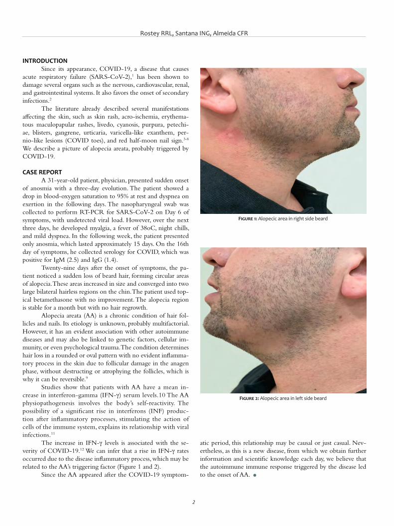
Rostey RRL, Santana ING, Almeida CFR
2
INTRODUCTIONSince its appearance, COVID-19, a disease that causes
acute respiratory failure (SARS-CoV-2),1 has been shown to damage several organs such as the nervous, cardiovascular, renal, and gastrointestinal systems. It also favors the onset of secondary infections.2
The literature already described several manifestations affecting the skin, such as skin rash, acro-ischemia, erythema-tous maculopapular rashes, livedo, cyanosis, purpura, petechi-ae, blisters, gangrene, urticaria, varicella-like exanthem, per-nio-like lesions (COVID toes), and red half-moon nail sign.3-8
We describe a picture of alopecia areata, probably triggered by COVID-19.
CASE REPORTA 31-year-old patient, physician, presented sudden onset
of anosmia with a three-day evolution. The patient showed a drop in blood-oxygen saturation to 95% at rest and dyspnea on exertion in the following days. The nasopharyngeal swab was collected to perform RT-PCR for SARS-CoV-2 on Day 6 of symptoms, with undetected viral load. However, over the next three days, he developed myalgia, a fever of 38oC, night chills, and mild dyspnea. In the following week, the patient presented only anosmia, which lasted approximately 15 days. On the 16th day of symptoms, he collected serology for COVID, which was positive for IgM (2.5) and IgG (1.4).
Twenty-nine days after the onset of symptoms, the pa-tient noticed a sudden loss of beard hair, forming circular areas of alopecia. These areas increased in size and converged into two large bilateral hairless regions on the chin. The patient used top-ical betamethasone with no improvement. The alopecia region is stable for a month but with no hair regrowth.
Alopecia areata (AA) is a chronic condition of hair fol-licles and nails. Its etiology is unknown, probably multifactorial. However, it has an evident association with other autoimmune diseases and may also be linked to genetic factors, cellular im-munity, or even psychological trauma. The condition determines hair loss in a rounded or oval pattern with no evident inflamma-tory process in the skin due to follicular damage in the anagen phase, without destructing or atrophying the follicles, which is why it can be reversible.9
Studies show that patients with AA have a mean in-crease in interferon-gamma (IFN-g) serum levels.10 The AA physiopathogenesis involves the body’s self-reactivity. The possibility of a significant rise in interferons (INF) produc-tion after inflammatory processes, stimulating the action of cells of the immune system, explains its relationship with viral infections.11
The increase in IFN-g levels is associated with the se-verity of COVID-19.12 We can infer that a rise in IFN-g rates occurred due to the disease inflammatory process, which may be related to the AA’s triggering factor (Figure 1 and 2).
Since the AA appeared after the COVID-19 symptom-
atic period, this relationship may be causal or just casual. Nev-ertheless, as this is a new disease, from which we obtain further information and scientific knowledge each day, we believe that the autoimmune immune response triggered by the disease led to the onset of AA. l
Figure 1: Alopecic area in right side beard
Figure 2: Alopecic area in left side beard
Surg Cosmet Dermatol. 2021;13:e20210014.
3
REFERENCES1. Centers for Disease Control and Prevention. Severe outcomes among
patients with Coronavirus Disease 2019 (COVID-19): United States, Fe-bruary 12- March 16, 2020. Morbid Mortal Weekly Rep. 69(12):343-6.
2. Guan W-J, Ni Z-Y, Hu Y, Liang W-H, Ou C-Q, He J-X, et al. Clinical cha-racteristics of coronavirus disease 2019 in China. N Engl J Med. 2020;382:1708-20.
3. Recalcati S. Cutaneous manifestations in COVID-19: a first perspective. J Eur Acad Dermatol Venereol. 2020;34(5):e212-e213.
4. Estébanez A, Pérez-Santiago L, Silva E, Guillen-Climent S, García- Vá-zquez S, García- Vázquez A, Ramón MD. Cutaneous manifestations in COVID-19: a new contribution. J Eur Acad Dermatol Venereol. 2020;34(6):e250-1.
5. Andina D, Noguera-Morel L, Bascuas-Arribas M, Gaitero-Tristán J, Alon-so Cadenas JA, Escalada Pellitero S, et al. Chilblains in children in the setting of COVID-19 pandemic. Pediatr Dermatol. 2020; 37(3):406-11.
6. Galván Casas C, Català A, Carretero Hernández G, Rodríguez-Jiménez P, Fernández-Nieto D, Rodríguez-Villa Lario A, et al. Classification of the cutaneous manifestations of COVID-19: a rapid prospective nationwide consensus. Br J Dermatol. 2020;183(1):71-7.
7. Neri I, Guglielmo A, Virdi A, Gaspari V, Starace M, Piraccini BMl. The red half-moon nail sign: a novel manifestation of coronavirus infection. J Eur Acad Dermatol Venereol. 2020;34(11):e-663-5.
8. Mendez-Flores S, Zaladonis A, Valdes-Rodriguez R. COVID‐19 and nail manifestation: be on the look out for the red half‐moon nail sign. Int J Dermatol. 2020;59(11):1414.
9. Rivitti EA. Alopecia areata: revisão e atualização. An Bras Dermatol. 2005;80(1):57-68.
10. Ribeiro LMG. Fisiopatologia da Alopécia Areata. Dissertação (Mestrado em Medicina) - Instituto de Ciências Biomédicas Abel Salazar. Universi-dade do Porto . Porto; 2016. p. 23.
11. Arca E, Muşabak U, Akar A, Erbil AH, Taştan HB. Interferon-gamma in alopecia areata. Eur J Dermatol. 2004;14(1):33-6.
12. Gadotti AC, Deus MC. Telles JP, Wind R, Goes M, Ossoski RGC, et al. IFN-γ is an independent risk factor associated with mortality in patients with moderate and severe COVID-19 infection. Virus Res. 2020;289:198171.
AUTHORS' CONTRIBUTION:
Renato Roberto Liberato Rostey 0000-0001-8656-4111Approval of the final version of the manuscript; study design and planning; preparation and writing of the manuscript; active par-ticipation in research orientation; critical literature review; critical revision of the manuscript.
Ivana Nascimento Garcia de Santana 0000-0001-7029-4882Study design and planning; intellectual participation in propaedeutic and/or therapeutic conduct of studied cases.
Cristiane Ferreira Rallo de Almeida 0000-0002-8525-344xData collection, analysis, and interpretation; critical literature review.

1
APOIO CIENTÍFICO:
www.surgicalcosmetic.org.br/
ISSN-e 1984-8773
Case reportAuthors: Bruna Cristina Mendes dos Santos¹ Marina Riedi Guilherme¹ Mayara Teixeira Cruz¹ Marcelo de Souza Machado² Lúcia Emiko Imazu³
¹ Department of Dermatology, Autarquia Municipal de Saúde de Apucarana, Apucarana (PR), Brazil.
² Department of Dermatology, Hospital do Câncer de Londrina, Londrina (PR), Brazil.
³ Department of Dermatology, Associação Filantrópica Humanitas, São Jerônimo da Serra (PR), Brazil.
Correspondence: Bruna Cristina Mendes dos Santos Email: bcmsantos2008@hotmail. com
Financial support: None. Conflict of interest: None.
Submitted on: 15/04/2021Approved on: 15/07/2021
How to cite this article: Santos BCM, Guilherme MR, Cruz MT, Machado MS, Imazu LE. Basal cell carcinoma of the vulva: a case report of cutaneous neoplasia in a special area. Surg Cosmet Dermatol. 2021;13:e20210033.
Basal cell carcinoma of the vulva: a case report of cutaneous neoplasia in a special area Carcinoma basocelular na vulva: um relato de caso de neoplasia cutânea em área especial
ABSTRACTBasal cell carcinoma (BCC) is the most common skin cancer¹. Among the risk factors for its development are exposure to ionizing and non-ionizing radiation, some chemicals, and previous scars.² However, the most important factor is exposure to ultraviolet radiation, which explains the higher incidence of this neoplasm in photo-exposed areas.¹ BCC in unexposed areas is uncommon. The purpose of this report is to describe a case of recurrent BCC in the vulva, to demonstrate the importance of dermatological examination in unusual areas, and to report the application of Mohs micrographic surgery.Keywords: Basal cell carcinoma; Vulvar diseases; Vulvar neoplasms
RESUMOO carcinoma basocelular (CBC) é o câncer de pele mais comum.¹ Entre os fatores de risco para seu desenvolvimento estão a exposição a radiações ionizantes e não ionizantes, alguns produtos químicos e cicatrizes prévias.² Porém, o fator mais importante é a exposição à radiação ultravioleta, o que explica a maior incidência dessa neoplasia em áreas fotoexpostas. O CBC em áreas não expostas é incomum. O objetivo deste relato é descrever um caso de CBC recorrente na vulva, demonstrar a importância do exame dermatológico em áreas incomuns e relatar a aplicação da cirurgia micrográfica de Mohs.Palavras-chave: Carcinoma basocelular; Doenças da vulva; Neoplasias vulvares
DOI: http://www.dx.doi.org/10.5935/scd1984-8773.2021130033
ISSN-e 1984-8773
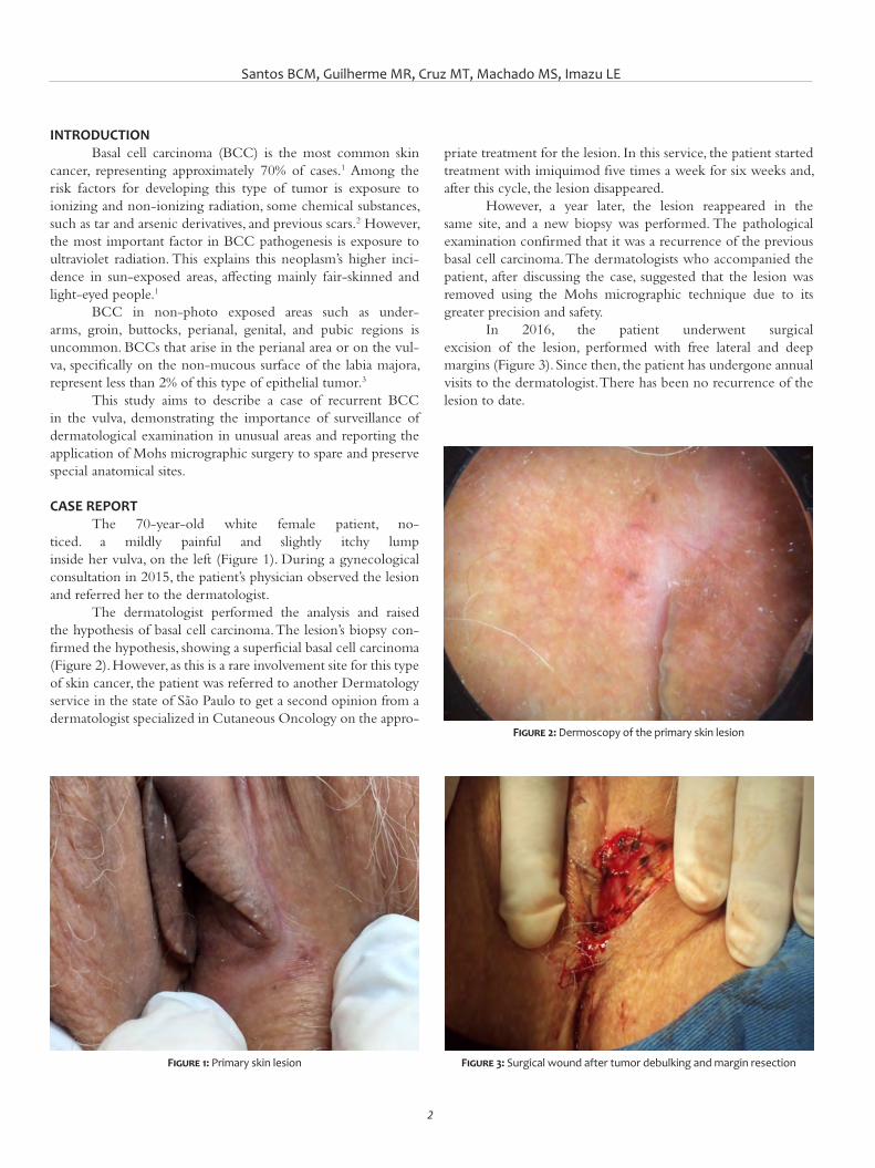
Santos BCM, Guilherme MR, Cruz MT, Machado MS, Imazu LE
2
INTRODUCTIONBasal cell carcinoma (BCC) is the most common skin
cancer, representing approximately 70% of cases.1 Among the risk factors for developing this type of tumor is exposure to ionizing and non-ionizing radiation, some chemical substances, such as tar and arsenic derivatives, and previous scars.2 However, the most important factor in BCC pathogenesis is exposure to ultraviolet radiation. This explains this neoplasm’s higher inci-dence in sun-exposed areas, affecting mainly fair-skinned and light-eyed people.1
BCC in non-photo exposed areas such as under-arms, groin, buttocks, perianal, genital, and pubic regions is uncommon. BCCs that arise in the perianal area or on the vul-va, specifically on the non-mucous surface of the labia majora, represent less than 2% of this type of epithelial tumor.3
This study aims to describe a case of recurrent BCC in the vulva, demonstrating the importance of surveillance of dermatological examination in unusual areas and reporting the application of Mohs micrographic surgery to spare and preserve special anatomical sites.
CASE REPORTThe 70-year-old white female patient, no-
ticed. a mildly painful and slightly itchy lump inside her vulva, on the left (Figure 1). During a gynecological consultation in 2015, the patient’s physician observed the lesion and referred her to the dermatologist.
The dermatologist performed the analysis and raised the hypothesis of basal cell carcinoma. The lesion’s biopsy con-firmed the hypothesis, showing a superficial basal cell carcinoma (Figure 2). However, as this is a rare involvement site for this type of skin cancer, the patient was referred to another Dermatology service in the state of São Paulo to get a second opinion from a dermatologist specialized in Cutaneous Oncology on the appro-
priate treatment for the lesion. In this service, the patient started treatment with imiquimod five times a week for six weeks and, after this cycle, the lesion disappeared.
However, a year later, the lesion reappeared in the same site, and a new biopsy was performed. The pathological examination confirmed that it was a recurrence of the previous basal cell carcinoma. The dermatologists who accompanied the patient, after discussing the case, suggested that the lesion was removed using the Mohs micrographic technique due to its greater precision and safety.
In 2016, the patient underwent surgical excision of the lesion, performed with free lateral and deep margins (Figure 3). Since then, the patient has undergone annual visits to the dermatologist. There has been no recurrence of the lesion to date.
Figure 1: Primary skin lesion
Figure 2: Dermoscopy of the primary skin lesion
Figure 3: Surgical wound after tumor debulking and margin resection
Surg Cosmet Dermatol. 2021;13:e20210033.
3
DISCUSSION BCC arises from follicular stem cells. Thus, its
development in hairless areas is uncommon.4 The site of greatest involvement is the cephalic region (90%), mainly in the orbitopalpebral and nasal area, followed by the trunk and limbs.2 The most important factor in the pathogenesis of BCC is expo-sure to ultraviolet light. However, up to a third of cases occur in covered areas, such as armpits, groin, buttocks, perianal, genital, and pubic region.3,5,6
To date, the literature has described less than 300 cases of vulvar BCC. Tumors in this location represent approxi-mately 2% of all BCCs and less than 3% of vulvar carcinomas.4 The exact cause of BCC development in non-sun exposed areas is still uncertain, but the relationship with increasing age has been reported in two large case series, in which the mean age of vulvar BCC development was between 70 and 73 years.7,8
Vulvar BCC shares the same risk factors as the sun-exposed BCCs. What sets them apart is that exposure to ultraviolet (UV) radiation does not play an important factor in the pathogenesis of vulvar BCC. Nevertheless, some authors suggest that exposure to UV radiation causes systemic immunosuppression, and it could be related to the BCC appearance in areas not exposed to the sun.9 Previous radiation is also one of the factors that predispose to vulvar BCC. Usually, the onset of lesions occurs years after exposure to irradiation.10,11 Another risk factor is genetic defects that can predispose to the development of BCC, such as the PTCH mutation, which leads to basal cell nevus syndrome and substantially increases the sus-ceptibility to this neoplasm.12
The clinical manifestations of vulvar BCCs are nonspecific and don’t have classic BCC features. Lesions can appear as papules, nodules, or lesions that can be exophytic, pediculated, infiltrative, pigmented, and ulcerated in more advanced cases, or even in the combined form of these items.13 The signs and symptoms are not characteristic. The most reported ones are irritation, pain, and local itching. The non- specificity of the symptoms often causes the lesions to be treated as inflammatory and infectious dermatoses, leading to late diagno-sis and, consequently, to an increase in the size of the neoplasm and local invasion.8
The vulvar BCCs are more aggressive when compared to the sun-exposed BCCs, and they are associated with deep local infiltration and occasional perineural extension. This type of neoplasm has a greater recurrence, as well as higher levels of local and distant metastases. It varies according to the histologi-cal pattern, and morphea-like, metatypic, adenocystic, and infil-trative tumors tend to be more recurrent and aggressive. ¹4, 15, 16
Surgical excision is the best form of treatment for vulvar BCC. Surgical margins should be wide, as a retrospective study showed that up to 25% of excisional biopsy margins were com-promised. It is mainly due to the difficult demarcation, as this site is naturally erythematous, and the edges of the lesion are easily confused with healthy skin.8 The involvement of the margins results in a higher recurrence rate, which may reach 10% to 20% of cases.14
In large, histologically aggressive BCCs and in those with recurrence and poorly delimited borders, such as the tumor in this case report, Mohs micrographic surgery is the treatment of choice. It provides histological control of 100% of the mar-gins, preserving normal skin, in addition to reducing changes in anatomical structures, making the reconstruction safer and with better aesthetic and functional results. In cases where the surgical approach is contraindicated or where there is edge involvement, radiotherapy is a therapeutic option, but it can lead to complica-tions in the irradiated site.3,17,18
Early diagnosis is crucial to ensure less invasive surgery. However, vulvar BCC often has a delayed diagnosis because it occurs in a location that is less monitored during the physical examination.8 We concluded that there must be awareness that BCCs can appear in non-photo exposed areas and, many times, they can mimic other dermatoses. Thus, dermatologists, general practitioners, and gynecologists should perform a complete skin examination, especially in patients with a history of skin cancer, and biopsy is advisable in any suspicious skin lesion in these regions. l
Santos BCM, Guilherme MR, Cruz MT, Machado MS, Imazu LE
4
AUTHORS' CONTRIBUTION:
Bruna Cristina Mendes dos Santos 0000-0002-3213-3962Approval of the final version of the manuscript; intellectual participation in propaedeutic and/or therapeutic conduct of studied cases; critical literature review; critical revision of the manuscript.
Marina Riedi Guilherme 0000-0003-4765-2180Approval of the final version of the manuscript; intellectual participation in propaedeutic and/or therapeutic conduct of studied cases; critical literature review; critical revision of the manuscript.
Mayara Teixeira Cruz 0000-0002-5069-0519Approval of the final version of the manuscript; intellectual participation in propaedeutic and/or therapeutic conduct of studied cases; critical literature review; critical revision of the manuscript.
Marcelo de Souza Machado 0000-0001-8817-0183Approval of the final version of the manuscript; intellectual participation in propaedeutic and/or therapeutic conduct of studied cases; critical literature review; critical revision of the manuscript.
Lúcia Emiko Imazu 0000-0002-6634-5509Active participation in research orientation; intellectual participation in propaedeutic and/or therapeutic conduct of studied cases; critical revision of the manuscript.
REFERENCES:1. Belda Junior B, Di Chiacchio N, Criado PR. Tratado de Dermatologia. 3th
ed. Rio de Janeiro: Atheneu; 2018.2. Azulay RD, Azulay DR, Azulay-Abulafia L. Dermatologia. 6th ed. Rio de
Janeiro: Gen; 2013.3. Pisani C, Poggiali S, De Padova L, Andreassi A, Bilenchi R. Basal cell car-
cinoma of the vulva. J Eur Acad Dermatol Venereol. 2006;20(4):446-8. 4. Sakai T, Goto M, Kai Y, Kato A, Shimizu F, Okamoto O, et al. Vulvar basal
cell carcinoma with bone metastasis. J Dermatol. 2011;38(1):97-100.5. Gloster HM Jr, Brodland DG. The epidemiology of skin cancer. Dermatol
Surg. 1996;22(3):217-26.6. Bhagchandani L, Sanadi RE, Sattar S, Abbott RR. Basal cell carci-
noma presenting as finger mass. A case report. Am J Clin Oncol. 1995;18(2):176-9.
7. Gibson GE, Ahmed I. Perianal and genital basal cell carcinoma: A clini-copathologic review of 51 cases. J Am Acad Dermatol. 2001;45(1):68-71.
8. de Giorgi V, Salvini C, Massi D, Raspollini MR, Carli P. Vulvar basal cell carcinoma: retrospective study and review of literature. Gynecol Oncol. 2005;97(1):192-4.
9. Strickland PT, Creasia D, Kripke ML. Enhancement of two-stage skin car-cinogenesis by exposure of distant skin to UV radiation. J Natl Cancer Inst. 1985;74(5):1129-34.
10. Benedet JL, Miller DM, Ehlen TG, Bertrand MA. Basal cell carcinoma of the vulva: clinical features and treatment results in 28 patients. Obstet Gynecol. 1997;90(5):765-8.
11. Mateus C, Fortier-Beaulieu M, Lhomme C, Rochard F, Castaigne D, Du-villard P, et al. Carcinome basocellulaire de la vulve: 21 cases. Ann Der-matol Venereol. 2001;128(1):11-5.
12. Susong CR, Ratz JL. Basal-cell carcinoma occurring in an axilla: a case presentation and a review of factors related to tumor development. J Dermatol Surg Oncol. 1985;11(5):526-30.
13. Mulvany NJ, Rayoo M, Allen DG. Basal cell carcinoma of the vulva: a case series. Pathology. 2012;44(6):528-33.
14. Mulayim N, Foster Silver D, Tolgay Ocal I, Babalola E. Vulvar basal cell carcinoma: two unusual presentations and review of the literature. Gy-necol Oncol. 2002;85(3):532-7.
15. Watson GA, Kelly D, Prior L, Stanley E, MacEneaney O, Walsh T, et al. An unusual case of basal cell carcinoma of the vulva with lung metastases. Gynecol Oncol Rep. 2016;18:32-5.
16. Jacobs GH, Rippey JJ, Altini M. Prediction of aggressive behavior in ba-sal cell carcinoma. Cancer. 1982;49(3):533-7.
17. Suzuki HS, Sato MS. A experiência da implantação da cirurgia micro-gráfica de Mohs em um serviço universitário. Surg Cosmet Dermatol. 2012;4(2):206-7.
18. Miller SJ, Alam M, Andersen J, Berg D, Bichakjian CK, Bowen G, et.al. Basal cell and squamous cell skin cancers. JNCCN. 2010(8):836-64.

1
APOIO CIENTÍFICO:
www.surgicalcosmetic.org.br/
ISSN-e 1984-8773
Case reportAuthors: Khairuddin Djawad1
Idrianti Idrus1
Airin Nurdin Mappewali1
1 Dermatology and Venereology, Hasanuddin University, Makassar, South Sulawesi, Indonesia.
Correspondence: Khairuddin Djawad Email: [email protected]
Financial support: NoneConflict of interest: None
Submitted on: 12/01/2021 Approved on: 14/02/2021
How to cite this article: Djawad K, Idrus I, Mappewali NA. Closure of a large nasal dorsum defect using a bilateral crescent advancement flap from the malar region to the nose. Surg Cosmet Dermatol. 2021;13:e20210017.
Closure of a large nasal dorsum defect using a bilateral crescent advancement flap from the malar region to the nose Fechamento de um grande defeito do dorso nasal por retalho de avanço crescente bilateral da região malar ao nariz
ABSTRACTThe closure of large defects on the nasal dorsum is a challenge for dermatologic surgeons. The alternatives to repair the defect are a skin graft and some forms of skin flaps. One particular defect closure technique is the crescentic advancement flap, which uses crescent incisions at the nasola-bial fold to accommodate the excess tissue. This flap is best used if the defect is in the lateral nose, alar, and nasolabial fold. We report a large defect of the nasal dorsum in a 65-year-old woman that was successfully reconstructed using bilateral cheek-to-nose crescentic advancement flap. The patient showed excellent cosmetic and outcome.Keywords: Dermatology; Nasal Bone; Surgical Flaps
RESUMOO fechamento de grandes defeitos no dorso nasal é um desafio para o dermatocirurgião. As alternativas para reparar o defeito são enxertos e algumas formas de retalho de pele retalhos de pele, como o retalho de avanço crescente, que utiliza incisões em crescente no sulco nasolabial para acomodar o excesso de tecido. Esse retalho apresenta melhores resultados em defeito nas regiões lateral do nariz, alar e dobra nasolabial. Relatamos um grande defeito do dorso nasal em uma mulher de 65 anos, reconstruído com sucesso com o retalho de avanço crescente bilateral da bochecha ao nariz. A paciente apresentou excelente resultado cosmético.Palavras-chave: Dermatologia; Osso nasal; Retalhos Cirúrgicos
DOI: http://www.dx.doi.org/10.5935/scd1984-8773.2021130017
ISSN-e 1984-8773
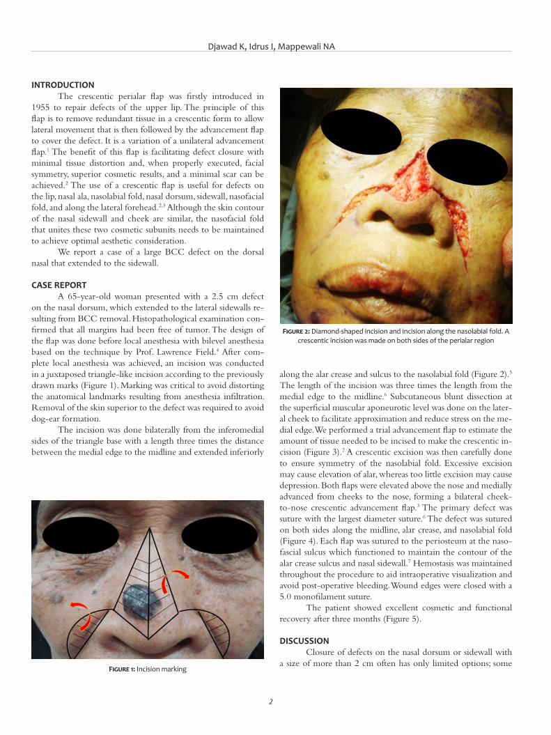
Djawad K, Idrus I, Mappewali NA
2
INTRODUCTION The crescentic perialar flap was firstly introduced in
1955 to repair defects of the upper lip. The principle of this flap is to remove redundant tissue in a crescentic form to allow lateral movement that is then followed by the advancement flap to cover the defect. It is a variation of a unilateral advancement flap.1 The benefit of this flap is facilitating defect closure with minimal tissue distortion and, when properly executed, facial symmetry, superior cosmetic results, and a minimal scar can be achieved.2 The use of a crescentic flap is useful for defects on the lip, nasal ala, nasolabial fold, nasal dorsum, sidewall, nasofacial fold, and along the lateral forehead.2,3 Although the skin contour of the nasal sidewall and cheek are similar, the nasofacial fold that unites these two cosmetic subunits needs to be maintained to achieve optimal aesthetic consideration.
We report a case of a large BCC defect on the dorsal nasal that extended to the sidewall.
CASE REPORT A 65-year-old woman presented with a 2.5 cm defect
on the nasal dorsum, which extended to the lateral sidewalls re-sulting from BCC removal. Histopathological examination con-firmed that all margins had been free of tumor. The design of the flap was done before local anesthesia with bilevel anesthesia based on the technique by Prof. Lawrence Field.4 After com-plete local anesthesia was achieved, an incision was conducted in a juxtaposed triangle-like incision according to the previously drawn marks (Figure 1). Marking was critical to avoid distorting the anatomical landmarks resulting from anesthesia infiltration. Removal of the skin superior to the defect was required to avoid dog-ear formation.
The incision was done bilaterally from the inferomedial sides of the triangle base with a length three times the distance between the medial edge to the midline and extended inferiorly
along the alar crease and sulcus to the nasolabial fold (Figure 2).5 The length of the incision was three times the length from the medial edge to the midline.6 Subcutaneous blunt dissection at the superficial muscular aponeurotic level was done on the later-al cheek to facilitate approximation and reduce stress on the me-dial edge. We performed a trial advancement flap to estimate the amount of tissue needed to be incised to make the crescentic in-cision (Figure 3).2 A crescentic excision was then carefully done to ensure symmetry of the nasolabial fold. Excessive excision may cause elevation of alar, whereas too little excision may cause depression. Both flaps were elevated above the nose and medially advanced from cheeks to the nose, forming a bilateral cheek-to-nose crescentic advancement flap.3 The primary defect was suture with the largest diameter suture.6 The defect was sutured on both sides along the midline, alar crease, and nasolabial fold (Figure 4). Each flap was sutured to the periosteum at the naso-fascial sulcus which functioned to maintain the contour of the alar crease sulcus and nasal sidewall.7 Hemostasis was maintained throughout the procedure to aid intraoperative visualization and avoid post-operative bleeding. Wound edges were closed with a 5.0 monofilament suture.
The patient showed excellent cosmetic and functional recovery after three months (Figure 5).
DISCUSSION Closure of defects on the nasal dorsum or sidewall with
a size of more than 2 cm often has only limited options; some Figure 1: Incision marking
Figure 2: Diamond-shaped incision and incision along the nasolabial fold. A crescentic incision was made on both sides of the perialar region
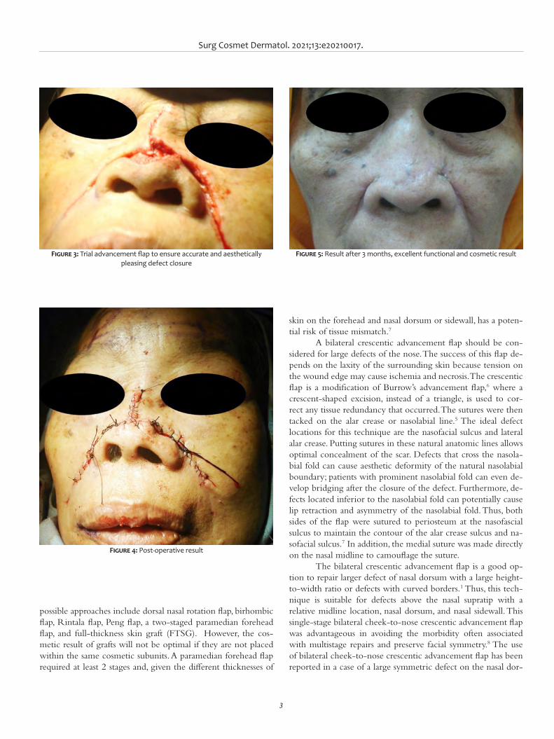
Surg Cosmet Dermatol. 2021;13:e20210017.
3
possible approaches include dorsal nasal rotation flap, birhombic flap, Rintala flap, Peng flap, a two-staged paramedian forehead flap, and full-thickness skin graft (FTSG). However, the cos-metic result of grafts will not be optimal if they are not placed within the same cosmetic subunits. A paramedian forehead flap required at least 2 stages and, given the different thicknesses of
skin on the forehead and nasal dorsum or sidewall, has a poten-tial risk of tissue mismatch.7
A bilateral crescentic advancement flap should be con-sidered for large defects of the nose. The success of this flap de-pends on the laxity of the surrounding skin because tension on the wound edge may cause ischemia and necrosis. The crescentic flap is a modification of Burrow’s advancement flap,6 where a crescent-shaped excision, instead of a triangle, is used to cor-rect any tissue redundancy that occurred. The sutures were then tacked on the alar crease or nasolabial line.5 The ideal defect locations for this technique are the nasofacial sulcus and lateral alar crease. Putting sutures in these natural anatomic lines allows optimal concealment of the scar. Defects that cross the nasola-bial fold can cause aesthetic deformity of the natural nasolabial boundary; patients with prominent nasolabial fold can even de-velop bridging after the closure of the defect. Furthermore, de-fects located inferior to the nasolabial fold can potentially cause lip retraction and asymmetry of the nasolabial fold. Thus, both sides of the flap were sutured to periosteum at the nasofascial sulcus to maintain the contour of the alar crease sulcus and na-sofacial sulcus.7 In addition, the medial suture was made directly on the nasal midline to camouflage the suture.
The bilateral crescentic advancement flap is a good op-tion to repair larger defect of nasal dorsum with a large height-to-width ratio or defects with curved borders.1 Thus, this tech-nique is suitable for defects above the nasal supratip with a relative midline location, nasal dorsum, and nasal sidewall. This single-stage bilateral cheek-to-nose crescentic advancement flap was advantageous in avoiding the morbidity often associated with multistage repairs and preserve facial symmetry.8 The use of bilateral cheek-to-nose crescentic advancement flap has been reported in a case of a large symmetric defect on the nasal dor-
Figure 3: Trial advancement flap to ensure accurate and aesthetically pleasing defect closure
Figure 4: Post-operative result
Figure 5: Result after 3 months, excellent functional and cosmetic result
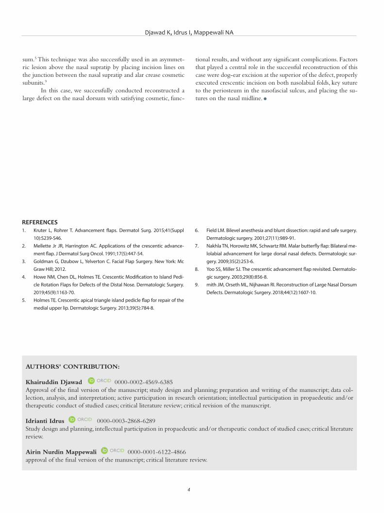
Djawad K, Idrus I, Mappewali NA
4
REFERENCES1. Kruter L, Rohrer T. Advancement flaps. Dermatol Surg. 2015;41(Suppl
10):S239-S46. 2. Mellette Jr JR, Harrington AC. Applications of the crescentic advance-
ment flap. J Dermatol Surg Oncol. 1991;17(5):447-54. 3. Goldman G, Dzubow L, Yelverton C. Facial Flap Surgery. New York: Mc
Graw Hill; 2012. 4. Howe NM, Chen DL, Holmes TE. Crescentic Modification to Island Pedi-
cle Rotation Flaps for Defects of the Distal Nose. Dermatologic Surgery. 2019;45(9):1163-70.
5. Holmes TE. Crescentic apical triangle island pedicle flap for repair of the medial upper lip. Dermatologic Surgery. 2013;39(5):784-8.
6. Field LM. Bilevel anesthesia and blunt dissection: rapid and safe surgery. Dermatologic surgery. 2001;27(11):989-91.
7. Nakhla TN, Horowitz MK, Schwartz RM. Malar butterfly flap: Bilateral me-lolabial advancement for large dorsal nasal defects. Dermatologic sur-gery. 2009;35(2):253-6.
8. Yoo SS, Miller SJ. The crescentic advancement flap revisited. Dermatolo-gic surgery. 2003;29(8):856-8.
9. mith JM, Orseth ML, Nijhawan RI. Reconstruction of Large Nasal Dorsum Defects. Dermatologic Surgery. 2018;44(12):1607-10.
AUTHORS' CONTRIBUTION:
Khairuddin Djawad 0000-0002-4569-6385Approval of the final version of the manuscript; study design and planning; preparation and writing of the manuscript; data col-lection, analysis, and interpretation; active participation in research orientation; intellectual participation in propaedeutic and/or therapeutic conduct of studied cases; critical literature review; critical revision of the manuscript.
Idrianti Idrus 0000-0003-2868-6289Study design and planning, intellectual participation in propaedeutic and/or therapeutic conduct of studied cases; critical literature review.
Airin Nurdin Mappewali 0000-0001-6122-4866approval of the final version of the manuscript; critical literature review.
sum.5 This technique was also successfully used in an asymmet-ric lesion above the nasal supratip by placing incision lines on the junction between the nasal supratip and alar crease cosmetic subunits.9
In this case, we successfully conducted reconstructed a large defect on the nasal dorsum with satisfying cosmetic, func-
tional results, and without any significant complications. Factors that played a central role in the successful reconstruction of this case were dog-ear excision at the superior of the defect, properly executed crescentic incision on both nasolabial folds, key suture to the periosteum in the nasofascial sulcus, and placing the su-tures on the nasal midline. l
1
APOIO CIENTÍFICO:
www.surgicalcosmetic.org.br/
ISSN-e 1984-8773
Case Report Authors: Giovana Serrão Fensterseifer¹ Ana Letícia Boff² Fernando Eibs Cafrune²
¹ Department of Dermatology, Pontifícia Universidade Católica do Rio Grande do Sul, Porto Alegre (RS), Brazil
² Department of Dermatology, Santa Casa de Misericórdia de Porto Alegre, Porto Alegre (RS), Brazil
Correspondence: Giovana Serrão Fensterseifer Email: [email protected] / Alternative E-mail: fcafrune@gmail. com
Financial support: None Conflict of interest: None
Submitted on: 15/05/2021Approved on: 03/09/2021
How to cite this article: Fensterseifer GS, Cafrune FE, Boff AL. Pigmented columns as a dermosco-py feature in a seborrheic keratosis associated with a squamous cell carcinoma. Surg Cosmet Dermatol. 2021;13:e20210049.
Pigmented columns as a dermoscopy feature in a seborrheic keratosis associated with a squamous cell carcinoma Colunas pigmentadas como um achado dermatoscópico de queratose seborreica associada a carcinoma espinocelular
ABSTRACTIntroduction: Seborrheic keratosis (SK) has the typical pigmented dermoscopy features such as fissures and ridges, fat fingers, and cerebriform patterns. Here we describe a case where pigmented colum-ns (PC) were visualized on SK’s dermoscopy associated with a squamous cell carcinoma (SCC). Case presentation: We report a case of a lesion whose dermoscopy showed PC. Histopathological report showed well-differentiated SCC and associated SK. Discussion: The PC was visualized in an area of dendritic melanocytes in the epidermis, correlating with the pigmented SK component of the lesion. To our knowledge, this is the first time PC is described in SK´s dermoscopy.Keywords: Carcinoma squamous cell; Dermoscopy; Skin neoplasms.
RESUMOIntrodução: As queratoses seborreicas (QS) têm achados dermatoscópicos pigmentados clássicos, como: fissuras e sulcos, "fat-fingers" e padrão cerebriforme. Relatamos um caso onde colunas pigmentadas (CP) foram visualizadas na derma-toscopia de uma QS associada a um carcinoma epidermoide (CEC). Relato do caso: Relatamos o caso de uma lesão onde a dermatoscopia demonstrou CP. O anatomopatológico foi compatível com CEC associado a QS. Discussão: As CP foram visualizadas em área de melanócitos dendríticos na epiderme, correlacionando-se ao componente de QS da lesão. Ao que se sabe, esta é a primeira vez que CP são descritas como achado dermatoscópico de QS.Palavras-chave: Carcinoma de células escamosas; Dermoscopia; Neoplasias cutâneas
DOI: http://www.dx.doi.org/10.5935/scd1984-8773.2021130049
ISSN-e 1984-8773
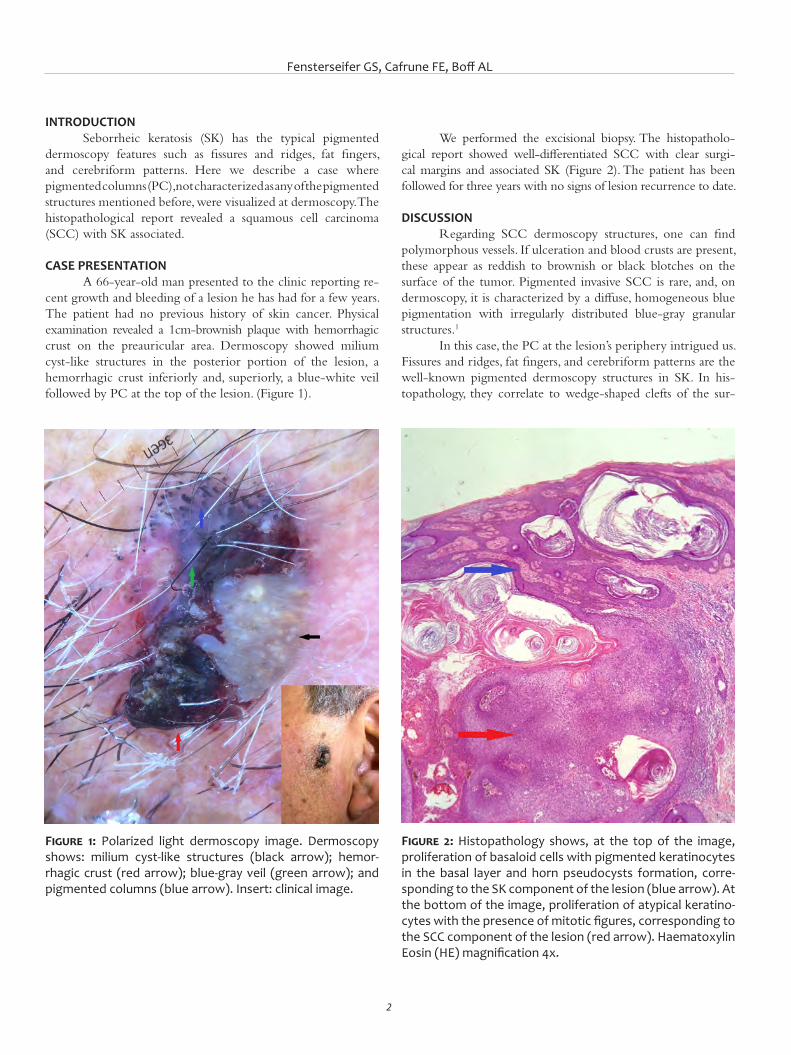
Fensterseifer GS, Cafrune FE, Boff AL
2
INTRODUCTIONSeborrheic keratosis (SK) has the typical pigmented
dermoscopy features such as fissures and ridges, fat fingers, and cerebriform patterns. Here we describe a case where pigmented columns (PC), not characterized as any of the pigmented structures mentioned before, were visualized at dermoscopy. The histopathological report revealed a squamous cell carcinoma (SCC) with SK associated.
CASE PRESENTATIONA 66-year-old man presented to the clinic reporting re-
cent growth and bleeding of a lesion he has had for a few years. The patient had no previous history of skin cancer. Physical examination revealed a 1cm-brownish plaque with hemorrhagic crust on the preauricular area. Dermoscopy showed milium cyst-like structures in the posterior portion of the lesion, a hemorrhagic crust inferiorly and, superiorly, a blue-white veil followed by PC at the top of the lesion. (Figure 1).
We performed the excisional biopsy. The histopatholo-gical report showed well-differentiated SCC with clear surgi-cal margins and associated SK (Figure 2). The patient has been followed for three years with no signs of lesion recurrence to date.
DISCUSSION Regarding SCC dermoscopy structures, one can find
polymorphous vessels. If ulceration and blood crusts are present, these appear as reddish to brownish or black blotches on the surface of the tumor. Pigmented invasive SCC is rare, and, on dermoscopy, it is characterized by a diffuse, homogeneous blue pigmentation with irregularly distributed blue-gray granular structures.1
In this case, the PC at the lesion’s periphery intrigued us. Fissures and ridges, fat fingers, and cerebriform patterns are the well-known pigmented dermoscopy structures in SK. In his-topathology, they correlate to wedge-shaped clefts of the sur-
Figure 1: Polarized light dermoscopy image. Dermoscopy shows: milium cyst-like structures (black arrow); hemor-rhagic crust (red arrow); blue-gray veil (green arrow); and pigmented columns (blue arrow). Insert: clinical image.
Figure 2: Histopathology shows, at the top of the image, proliferation of basaloid cells with pigmented keratinocytes in the basal layer and horn pseudocysts formation, corre-sponding to the SK component of the lesion (blue arrow). At the bottom of the image, proliferation of atypical keratino-cytes with the presence of mitotic figures, corresponding to the SCC component of the lesion (red arrow). Haematoxylin Eosin (HE) magnification 4x.
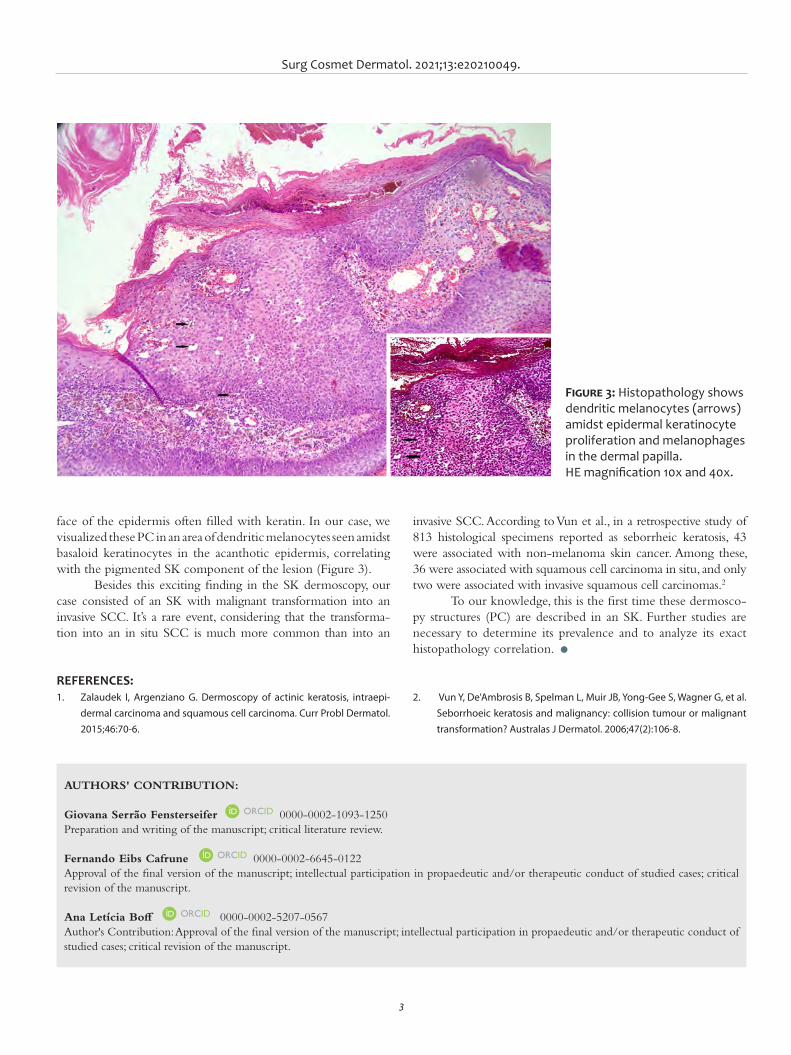
Surg Cosmet Dermatol. 2021;13:e20210049.
3
invasive SCC. According to Vun et al., in a retrospective study of 813 histological specimens reported as seborrheic keratosis, 43 were associated with non-melanoma skin cancer. Among these, 36 were associated with squamous cell carcinoma in situ, and only two were associated with invasive squamous cell carcinomas.2
To our knowledge, this is the first time these dermosco-py structures (PC) are described in an SK. Further studies are necessary to determine its prevalence and to analyze its exact histopathology correlation. l
face of the epidermis often filled with keratin. In our case, we visualized these PC in an area of dendritic melanocytes seen amidst basaloid keratinocytes in the acanthotic epidermis, correlating with the pigmented SK component of the lesion (Figure 3).
Besides this exciting finding in the SK dermoscopy, our case consisted of an SK with malignant transformation into an invasive SCC. It’s a rare event, considering that the transforma-tion into an in situ SCC is much more common than into an
AUTHORS' CONTRIBUTION:
Giovana Serrão Fensterseifer 0000-0002-1093-1250Preparation and writing of the manuscript; critical literature review.
Fernando Eibs Cafrune 0000-0002-6645-0122Approval of the final version of the manuscript; intellectual participation in propaedeutic and/or therapeutic conduct of studied cases; critical revision of the manuscript.
Ana Letícia Boff 0000-0002-5207-0567Author's Contribution: Approval of the final version of the manuscript; intellectual participation in propaedeutic and/or therapeutic conduct of studied cases; critical revision of the manuscript.
REFERENCES:1. Zalaudek I, Argenziano G. Dermoscopy of actinic keratosis, intraepi-
dermal carcinoma and squamous cell carcinoma. Curr Probl Dermatol. 2015;46:70-6.
2. Vun Y, De'Ambrosis B, Spelman L, Muir JB, Yong-Gee S, Wagner G, et al. Seborrhoeic keratosis and malignancy: collision tumour or malignant transformation? Australas J Dermatol. 2006;47(2):106-8.
Figure 3: Histopathology shows dendritic melanocytes (arrows) amidst epidermal keratinocyte proliferation and melanophages in the dermal papilla. HE magnification 10x and 40x.

1
APOIO CIENTÍFICO:
www.surgicalcosmetic.org.br/
ISSN-e 1984-8773
Case reportAuthors: Mariana Abdo de Almeida¹ Michele Maria Reis-Feroldi² Marcia Lanzoni Alvarenga Lira³
¹ Universidade de Taubaté, Medical School, Taubaté (SP), Brazil
² Private Clinic, Department of Dermatology, Campos do Jordão (SP), Brazil
³ Laboratório do Vale, Department of Pathology, Taubaté (SP), Brazil
Correspondence: Email: [email protected]
Financial support: NoneConflict of interest: None
Submitted on: 15/04/2021 Approved on: 13/10/2021
How to cite this article: Almeida MA, Reis-Feroldi MM, Lira MLA. Frontal fibrosing alope-cia development in two patients after botulinum toxin applications: relationship or coincidence? Surg Cosmet Dermatol. 2021;13:20210036.
Frontal fibrosing alopecia development in two patients after botulinum toxin applications: relationship or coincidence? Desenvolvimento de alopecia fibrosante frontal em duas pacientes usuárias de toxina botulínica: relação ou coincidência?
ABSTRACTFrontal fibrous alopecia is a primary lymphocytic scarring alopecia that is difficult to control, with a perifollicular lichenoid infiltrate marking its histology. Since its description in 1994 by Kossard, there has been an increase in cases reported worldwide. This article reports two cases that underwent incisional biopsies with longitudinal and transverse sections in areas presenting higher disease activity and reviews some aspects of this dermatosis. It also addresses the sus-picion of a possible relationship with the application of botulinum toxin – a question already raised by other authors but with no established confirmation.Keywords: Alopecia; Botulinum toxins; Botulinum toxins type A
RESUMOA alopecia frontal fibrosante é uma alopecia cicatricial linfocítica primária de difícil controle, com infiltrado liquenoide perifolicular marcando sua histologia. Desde a sua descrição em 1994 por Kossard houve um aumento de casos descritos pelo mundo. Este artigo relata dois casos em que foram realizadas biópsias incisionais com cortes longitudinais e transversais nas áreas que apresentaram maior atividade da doença e revisa alguns aspectos dessa dermatose, além de abordar a suspeita de uma possível relação com a aplicação de toxina botulínica - questão já aventada por outros autores, porém sem estabelecimento de confirmação.Palavras-chave: Alopecia; Toxinas botulínicas; Toxinas botulínicas tipo A
DOI: http://www.dx.doi.org/10.5935/scd1984-8773.2021130036
ISSN-e 1984-8773
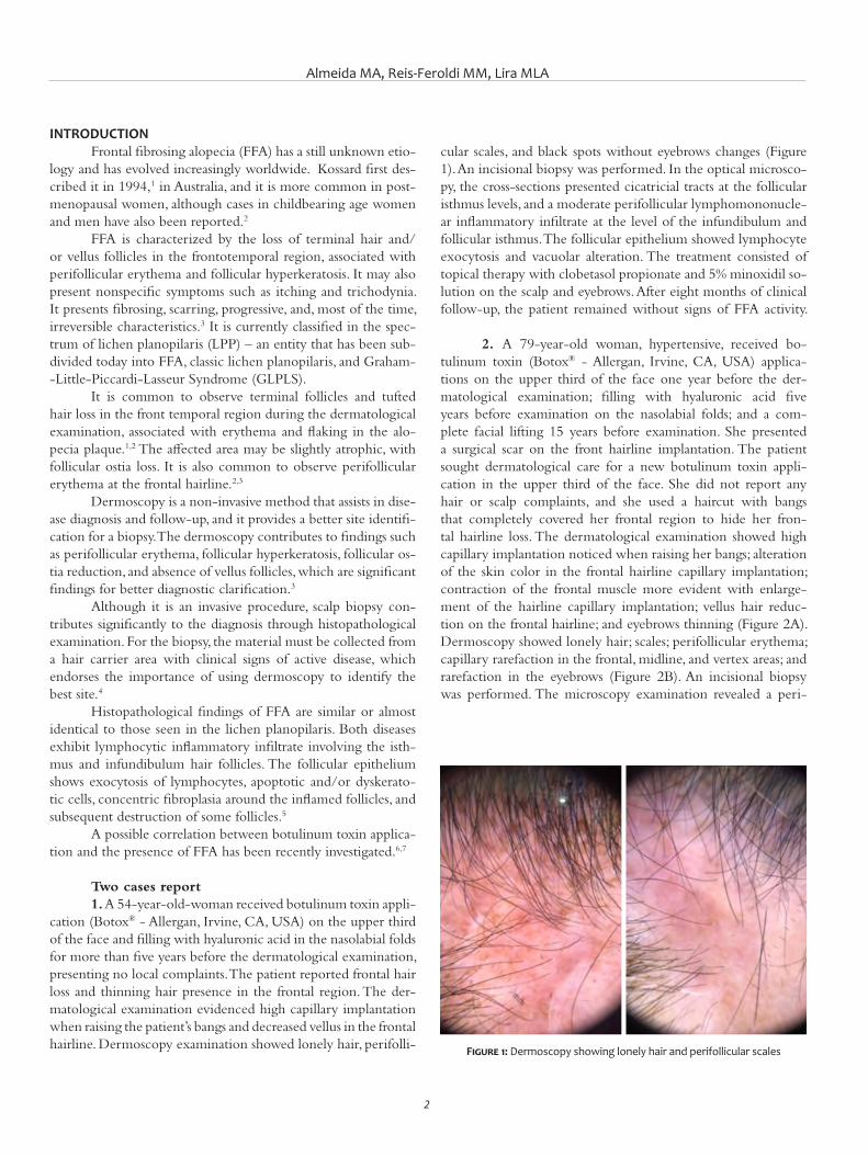
Almeida MA, Reis-Feroldi MM, Lira MLA
2
INTRODUCTIONFrontal fibrosing alopecia (FFA) has a still unknown etio-
logy and has evolved increasingly worldwide. Kossard first des-cribed it in 1994,1 in Australia, and it is more common in post-menopausal women, although cases in childbearing age women and men have also been reported.2
FFA is characterized by the loss of terminal hair and/or vellus follicles in the frontotemporal region, associated with perifollicular erythema and follicular hyperkeratosis. It may also present nonspecific symptoms such as itching and trichodynia. It presents fibrosing, scarring, progressive, and, most of the time, irreversible characteristics.3 It is currently classified in the spec-trum of lichen planopilaris (LPP) – an entity that has been sub-divided today into FFA, classic lichen planopilaris, and Graham--Little-Piccardi-Lasseur Syndrome (GLPLS).
It is common to observe terminal follicles and tufted hair loss in the front temporal region during the dermatological examination, associated with erythema and flaking in the alo-pecia plaque.1,2 The affected area may be slightly atrophic, with follicular ostia loss. It is also common to observe perifollicular erythema at the frontal hairline.2,3
Dermoscopy is a non-invasive method that assists in dise-ase diagnosis and follow-up, and it provides a better site identifi-cation for a biopsy. The dermoscopy contributes to findings such as perifollicular erythema, follicular hyperkeratosis, follicular os-tia reduction, and absence of vellus follicles, which are significant findings for better diagnostic clarification.3
Although it is an invasive procedure, scalp biopsy con-tributes significantly to the diagnosis through histopathological examination. For the biopsy, the material must be collected from a hair carrier area with clinical signs of active disease, which endorses the importance of using dermoscopy to identify the best site.4
Histopathological findings of FFA are similar or almost identical to those seen in the lichen planopilaris. Both diseases exhibit lymphocytic inflammatory infiltrate involving the isth-mus and infundibulum hair follicles. The follicular epithelium shows exocytosis of lymphocytes, apoptotic and/or dyskerato-tic cells, concentric fibroplasia around the inflamed follicles, and subsequent destruction of some follicles.5
A possible correlation between botulinum toxin applica-tion and the presence of FFA has been recently investigated.6,7
Two cases report1. A 54-year-old-woman received botulinum toxin appli-
cation (Botox® - Allergan, Irvine, CA, USA) on the upper third of the face and filling with hyaluronic acid in the nasolabial folds for more than five years before the dermatological examination, presenting no local complaints. The patient reported frontal hair loss and thinning hair presence in the frontal region. The der-matological examination evidenced high capillary implantation when raising the patient’s bangs and decreased vellus in the frontal hairline. Dermoscopy examination showed lonely hair, perifolli-
cular scales, and black spots without eyebrows changes (Figure 1). An incisional biopsy was performed. In the optical microsco-py, the cross-sections presented cicatricial tracts at the follicular isthmus levels, and a moderate perifollicular lymphomononucle-ar inflammatory infiltrate at the level of the infundibulum and follicular isthmus. The follicular epithelium showed lymphocyte exocytosis and vacuolar alteration. The treatment consisted of topical therapy with clobetasol propionate and 5% minoxidil so-lution on the scalp and eyebrows. After eight months of clinical follow-up, the patient remained without signs of FFA activity.
2. A 79-year-old woman, hypertensive, received bo-tulinum toxin (Botox® - Allergan, Irvine, CA, USA) applica-tions on the upper third of the face one year before the der-matological examination; filling with hyaluronic acid five years before examination on the nasolabial folds; and a com-plete facial lifting 15 years before examination. She presented a surgical scar on the front hairline implantation. The patient sought dermatological care for a new botulinum toxin appli-cation in the upper third of the face. She did not report any hair or scalp complaints, and she used a haircut with bangs that completely covered her frontal region to hide her fron-tal hairline loss. The dermatological examination showed high capillary implantation noticed when raising her bangs; alteration of the skin color in the frontal hairline capillary implantation; contraction of the frontal muscle more evident with enlarge-ment of the hairline capillary implantation; vellus hair reduc-tion on the frontal hairline; and eyebrows thinning (Figure 2A). Dermoscopy showed lonely hair; scales; perifollicular erythema; capillary rarefaction in the frontal, midline, and vertex areas; and rarefaction in the eyebrows (Figure 2B). An incisional biopsy was performed. The microscopy examination revealed a peri-
Figure 1: Dermoscopy showing lonely hair and perifollicular scales
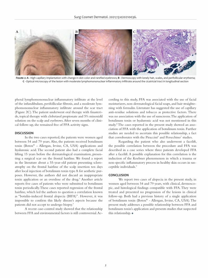
Surg Cosmet Dermatol. 2021;13:e20210036.
3
pheral lymphomononuclear inflammatory infiltrate at the level of the infundibulum, perifollicular fibrosis, and a moderate lym-phomononuclear inflammatory infiltrate around the scar tract (Figure 2C). The patient underwent oral therapy with finasteri-de, topical therapy with clobetasol propionate and 5% minoxidil solution on the scalp and eyebrows. After seven months of clini-cal follow-up, she remained free of FFA activity signs.
DISCUSSIONIn the two cases reported, the patients were women aged
between 54 and 79 years. Also, the patients received botulinum toxin (Botox® - Allergan, Irvine, CA, USA) applications and hyaluronic acid. The second patient also had a complete facial lifting 15 years before the dermatological examination, presen-ting a surgical scar on the frontal hairline. We found a report in the literature about a 55-year-old patient presenting sclero-atrophy on the frontal hairline of the scalp insertion ten days after local injection of botulinum toxin type A for aesthetic pur-poses. However, the authors did not discard an inappropriate toxin application or an overdose of the drug.6 Another article reports five cases of patients who were submitted to botulinum toxin periodically. These cases reported regression of the frontal hairline, which led the authors to question a correlation known as “botulin-induced frontal alopecia (BIFA)”. However, it was impossible to confirm this likely disease’s aspects because the patients did not accept to undergo biopsy.7
A recent case-control study showed that the relationship between FFA and environmental factors is still controversial. Ac-
cording to this study, FFA was associated with the use of facial moisturizers, non-dermatological facial soaps, and hair straighte-ning with formalin. Literature has suggested the use of capillary anti-residue solutions and tobacco as protective factors. There was no association with the use of sunscreens. The application of botulinum toxin or hyaluronic acid was not mentioned in this study.8 The cases reported in the present study showed an asso-ciation of FFA with the application of botulinum toxin. Further studies are needed to ascertain this possible relationship, a fact that corroborates with the Piraccini6 and Persechino7 studies.
Regarding the patient who also underwent a facelift, the possible correlation between the procedure and FFA was described in a case series where three patients developed FFA after a facelift. A possible explanation for this correlation is the induction of the Koebner phenomenon in which a trauma or non-specific inflammatory process in healthy skin occurs in sus-ceptible individuals.9
CONCLUSIONWe report two cases of alopecia in the present study, in
women aged between 54 and 79 years, with clinical, dermosco-pic, and histological findings compatible with FFA. They were treated and presented no progression of the lesions in clinical follow-up. Both had a previous history of a single application of botulinum toxin (Botox® - Allergan, Irvine, CA, USA). The present study addresses a possible relationship between FFA and botulinum toxin’s application and presents studies that suspected this relationship. l
Figure 2: A - High capillary implantation with change in skin color and rarefied eyebrows; B - Dermoscopy with lonely hair, scales, and perifollicular erythema; C - Optical microscopy of the lesion with moderate lymphomononuclear inflammatory infiltrate around the cicatricial tract in longitudinal section
A B C
Almeida MA, Reis-Feroldi MM, Lira MLA
4
AUTHORS' CONTRIBUTION:
Mariana Abdo de Almeida 0000-0002-7080-689XStatistical analysis; approval of the final version of the manuscript; study design and planning; preparation and writing of the ma-nuscript; data collection, analysis and interpretation; active participation in research orientation; intellectual participation in pro-paedeutic and/or therapeutic conduct of studied cases; critical literature review; critical revision of the manuscript.
Michele Maria Reis-Feroldi 0000-0003-4900-3598
Approval of the final version of the manuscript; study design and planning; preparation and writing of the manuscript; data col-lection, analysis and interpretation; active participation in research orientation; intellectual participation in propaedeutic and/or therapeutic conduct of studied cases; critical literature review.
Marcia Lanzoni Alvarenga Lira 0000-0002-1208-7911Approval of the final version of the manuscript; study design and planning; preparation and writing of the manuscript; data col-lection, analysis and interpretation; active participation in research orientation; intellectual participation in propaedeutic and/or therapeutic conduct of studied cases; critical literature review; critical revision of the manuscript.
REFERENCES:1. Kossard S. Postmenopausal frontal fibrosing alopecia. Scarring alope-
cia in a pattern distribution. Arch Dermatol. 1994;130(6):770-4. 2. Dawn G, Holmes SC, Moffat D, Munro CS. Post-menopausal frontal fi-
brosing alopecia. Clin Exp Dermatol. 2003;28(1):43-5.3. Crisóstomo MR, Crisóstomo MCC, Crisóstomo MGR, Gondim VJT, Cri-
sóstomoMR, Benevides NA. Perda pilosa por líquen plano pilar após transplante capilar: relato de dois casos e revisão da literatura. Na Bras Dermatol. 2011;86(2):359-62.
4. Donati A, Molina L, Doche I, Valente NS, Romiti R. Facial papules in fron-tal fibrosing alopecia: evidence of vellus follicle involvement. Arch Der-matol. 2011;147(12):1424-7.
5. Sperling LC, Cowper SE, Knopp EA. An Atlas of Hair Pathology with Cli-nical Correlations. 2nd ed. Florida: CRC Press; 2012.
6. Di Pietro A, Piraccini BM. Frontal alopecia after repeated botulinum to-xin type A injections for forehead wrinkles: an underestimated entity? Skin Appendage Disord. 2016;2(1-2):67-9.
7. Persechino S, Lupi F, Di Vito E, Romano I, Persechino F, et al. Sclero- Atrophic Reaction after Botulinum Toxin Injection. Clin Res Dermatol Open Access. 2016;3(2):1-2.
8. Ramos PM, Anzai A, Duque-Estrada B, Farias DC, Melo DF, Mulinari-Bren-ner F, et al. Risk factors for frontal fibrosing alopecia: a case-control stu-dy in a multiracial population. J Am Acad Dermatol. 2021;84(3):712-8.
9. Chiang YZ, Tosti A, Chaudhry IH, Lyne L, Farjo B, Farjo N, et al. Lichen planopilaris following hair transplantation and face-lift surgery. Br J Dermatol. 2012;166(3):666-370.
1
APOIO CIENTÍFICO:
www.surgicalcosmetic.org.br/
ISSN-e 1984-8773
Case ReportAuthors:
Helio Amante Miot¹Cesar Augusto Zago Ferreira¹Ana Claudia Athanasio Shwetz¹Luciane Donida Miot¹Gabriela Roncada Haddad¹
¹ Universidade Estadual Paulista Júlio de Mesquita Filho, Department of Infectious Diseases, Dermatology, Botucatu, São Paulo (SP), Brazil
Correspondence:Helio Amante MiotEmail: [email protected] / [email protected]
Financial support: None.Conflict of interest: None.
Submitted on: 09/04/2021 Approved on: 04/06/2021
How to cite this article:Miot HA, Ferreira CAZ, Shwetz ACA, Miot LD, Haddad GR. Malignant fibrous histiocytoma in ankle: case report. Surg Cosmet Dermatol. 2021;13:e20210028.
RESUMOHistiocitoma fibroso maligno (MFH) ou sarcoma pleomórfico indiferenciado (UPS) é um sarcoma moderada-mente agressivo, capaz de invadir estruturas adjacentes. Trata-se de neoplasia mesenquimal que predomina em homens entre a sexta e sétima décadas de vida. Localiza-se, principalmente, nos membros inferiores, podendo acometer cabeça e pescoço, tronco e retroperitônio, com tendência à recorrência e à metástase local. O presente relato tem como objetivo apresentar um caso de MFH no tornozelo de uma mulher de 49 anos, com invasão óssea adjacente, que evoluiu com amputação transtibial. São abordados aspectos clínicos, radiológicos, histopa-tológicos e terapêuticos, salientando-se a importância do diagnóstico precoce.Palavras-chave: Amputação; Histiocitoma fibroso maligno; Sarcoma.
Malignant fibrous histiocytoma in ankle: case report Histiocitoma fibroso maligno no tornozelo: relato de caso
DOI: http://www.dx.doi.org/10.5935/scd1984-8773.2021130028
ABSTRACTMalignant fibrous histiocytoma (MFH) or undifferentiated p leomorphic s arcoma (UPS) i s a sarcoma capable of invading adjacent structures. It is a mesenchymal neoplasia that predominates in men between the sixth and seventh decades of life. It is located mainly in the lower limbs and may affect the head and neck, trunk, and retroperitoneum, presenting a tendency to recurrence and local metastasis. This report aims to present a case of MFH in the ankle of a 49-year-old woman with an adjacent bone invasion, which evolved with transtibial amputation. Clinical, ra-diological, histopathological, and therapeutic aspects were addressed, highlighting the importance of early diagnosis.Keywords: Amputation; Malignant fibrous histiocytoma; Sarcoma
ISSN-e 1984-8773
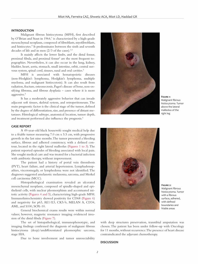
Miot HA, Ferreira CAZ, Shwetz ACA, Miot LD, Haddad GR
2
INTRODUCTIONMalignant fibrous histiocytoma (MFH), first described
by O’Brian and Staut in 1964,¹ is characterized by a high-grade mesenchymal neoplasm, composed of fibroblasts, myofibroblasts, and histiocytes.² It predominates between the sixth and seventh decades of life and in men (2/3 of the cases).3,4
It mainly affects the lower limbs, and the distal femur, proximal fibula, and proximal femur2 are the most frequent to-pographies. Nevertheless, it can also occur in the lung, kidney, bladder, heart, aorta, stomach, small intestine, orbit, central ner-vous system, spinal cord, sinuses, nasal and oral cavities.5
MFH is associated with hematopoietic diseases (non-Hodgkin’s lymphoma, Hodgkin’s lymphoma, multiple myeloma, and malignant histiocytosis). It can also result from radiation, fracture, osteonecrosis, Paget’s disease of bone, non-os-sifying fibroma, and fibrous dysplasia – cases where it is more aggressive.5
It has a moderately aggressive behavior that can invade adjacent soft tissues, skeletal system, and retroperitoneum. The main prognostic factor is the clinical stage of the tumor, defined by the degree of differentiation, size, and presence of distant me-tastases. Histological subtype, anatomical location, tumor depth, and treatment performed also influence the prognosis.6
CASE REPORTA 49-year-old black housewife sought medical help due
to a friable tumor measuring 7.0 cm x 5.5 cm, with progressive growth in the last nine months. The tumor presented a bleeding surface, fibrous and adhered consistency, with a defined con-tour, located in the right lateral malleolus (Figures 1 to 3). The patient reported episodes of bleeding associated with local pain. She sought medical care and was treated for a bacterial infection with antibiotic therapy, without improvement.
The patient had a history of portal vein thrombosis (PVT), heart failure, and arterial hypertension. Lymphadenop-athies, visceromegaly, or lymphedema were not identified. The diagnoses suggested amelanotic melanoma, sarcoma, and Merkel cell carcinoma (MCC).
Histopathological examination revealed an ulcerated mesenchymal neoplasm, composed of spindle-shaped and epi-thelioid cells, with nuclear pleomorphism and accentuated mi-totic activity (Figures 4 and 5), characterizing high-grade MFH. Immunohistochemistry showed positivity for CD68 (Figure 6) and negativity for p63, AE1/E3, CK5/6, MELAN A, CD34, AML, and S100, SOX-10.
General biochemical exams results were within normal values; however, magnetic resonance imaging evidenced inva-sion of the distal fibula (Figure 7).
The set of histopathological, immunophenotypic, and imaging findings confirmed the diagnosis of malignant fibrous histiocytoma (deep)/undifferentiated pleomorphic sarcoma, stage IIIA.
Due to bone involvement and tumor unresectability
with deep structures preservation, transtibial amputation was chosen. The patient has been under follow-up with Oncology for 11 months, without recurrence. The presence of heart disease contraindicated the adjuvant chemotherapy.
DISCUSSION
Figure 1: Malignant fibrous histiocytoma. Tumor above the lateral malleolus of the right leg
Figure 2: Malignant fibrous histiocytoma. Tumor with a fibrous surface, adhered, with defined boundaries and friable areas
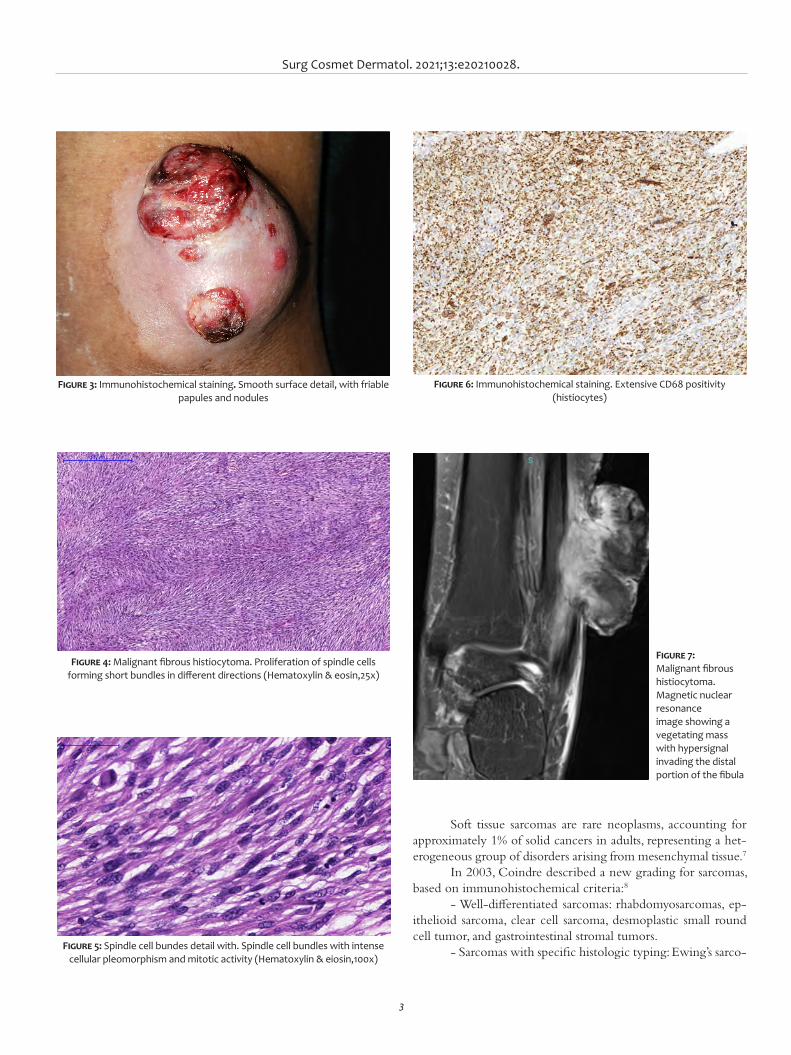
Surg Cosmet Dermatol. 2021;13:e20210028.
3
Soft tissue sarcomas are rare neoplasms, accounting for approximately 1% of solid cancers in adults, representing a het-erogeneous group of disorders arising from mesenchymal tissue.7
In 2003, Coindre described a new grading for sarcomas, based on immunohistochemical criteria:8
- Well-differentiated sarcomas: rhabdomyosarcomas, ep-ithelioid sarcoma, clear cell sarcoma, desmoplastic small round cell tumor, and gastrointestinal stromal tumors.
- Sarcomas with specific histologic typing: Ewing’s sarco-
Figure 3: Immunohistochemical staining. Smooth surface detail, with friable papules and nodules
Figure 4: Malignant fibrous histiocytoma. Proliferation of spindle cells forming short bundles in different directions (Hematoxylin & eosin,25x)
Figure 5: Spindle cell bundes detail with. Spindle cell bundles with intense cellular pleomorphism and mitotic activity (Hematoxylin & eiosin,100x)
Figure 6: Immunohistochemical staining. Extensive CD68 positivity (histiocytes)
Figure 7: Malignant fibrous histiocytoma. Magnetic nuclear resonance image showing a vegetating mass with hypersignal invading the distal portion of the fibula
Miot HA, Ferreira CAZ, Shwetz ACA, Miot LD, Haddad GR
4
ma, leiomyosarcoma, malignant peripheral nerve sheath tumor, dermatofibrosarcoma protuberans, giant cell fibroblastoma, ex-traskeletal myxoid chondrosarcoma, liposarcomas, and alveolar soft part sarcoma.
- Undifferentiated sarcomas, or sarcomas of doubtful type (not showing specific markers): fibrosarcoma, myxofibrosarcoma, and malignant fibrous histiocytoma. In these cases, immunohis-tochemistry can help exclude other non-mesenchymal tumors.
Undifferentiated pleomorphic sarcoma (UPS) can be divided into superficial and deep subtypes, also called atypi-cal fibroxanthoma (AFX) and malignant fibrous histiocytoma (MFH), respectively. The distinction between the entities is es-sential to predict the locoregional aggressiveness of the tumor and its prognosis.9,10
In this report, MFH occurred in the ankle of an adult woman. However, these tumors predominate in men older than 50 years.4 Typically, it presents as a painless, fast-growing tumor, with reports of lesions exclusively subcutaneous.11
UPS diagnosis requires the histopathological differenti-ation of tumors such as melanoma, squamous cell carcinoma, angiosarcoma, leiomyosarcoma, and other undifferentiated neo-plasms.12,13
MFH is recognized as a heterogeneous group of tumors that share a common phenotype, requiring immunohistochem-istry, electron microscopy, or molecular studies for better char-acterization.14
Histopathology revealed pleomorphic spindle cells ar-ranged in bundles with a storiform pattern, and multinucleated histiocytes infiltrated in the deep dermis and subcutaneous cel-lular tissue.15,16
MFH has immunoreactivity for vimentin and CD68 (histiocytic marker).17,18 S100, desmin, S-100, and HMB-45 are antibodies found in liposarcoma or malignant peripheral nerve tumors, nerve sheath tumor, rhabdomyosarcoma, and malignant melanoma, respectively. Moreover, CD34 shows reactivity in an-giosarcomass.15,16
Magnetic resonance imaging is the imaging modality of
choice to assess soft tissue sarcomas, particularly to determine the local extent of the lesion. On examination, the MFH shows a heterogeneous pattern that is hyperdense on T2-weighted im-ages and isodense to muscles on T1-weighted images.19
Wide excision (margin ≥2cm) is the recommended ap-proach. Nevertheless, there is a limitation due to the proximity to noble structures depending on the topography,14 indicating micrographic surgery.6,20
Local recurrence high rates are reported in patients un-dergoing surgical excision (19% to 66%).20-22 In this case, we completely removed the lesion and, due to comorbidities, decid-ed not to perform adjuvant chemotherapy.
A study analyzing 167 individuals found that 66% of patients treated with marginal resection had local recurrence compared with 27% of patients who underwent wide resection (margin ≥3cm).23
Radiotherapy can be beneficial as an adjuvant treatment, using a radiation field covering the tumor site and 5 cm around it, with doses ranging between 50 and 65 Gray. However, the overall impact on recurrence and survival is not fully defined.24
Chemotherapy is typically used for generalized diseas-es but with poor prognoses.25,26 Currently, studies assessing im-munobiologicals in patients with advanced diseases are in the clinical phase. This is the case for ipilimumab (anti-CTLA-4), comprising 33 children and young adults, and sunitinib (oral multi-kinase inhibitor) including 48 young adults, which ana-lyzes unresectable or metastatic sarcoma.27
CONCLUSIONMFH presents clinically indistinguishable from other en-
tities, being mainly characterized by aggressive growth behavior. This case report aimed to highlight its main characteristics and the importance of diagnostic suspicion and early treatment to avoid unfavorable outcomes. l
Surg Cosmet Dermatol. 2021;13:e20210028.
5
REFERENCES1. O'Brien JE, Stout A P. Malignant fibrous xanthomas. Cancer.
1964;17:1445-55.2. Pobirci DD, Bogdan F, Pobirci O, Petcu CA, Roşca E. Study of malignant
fibrous histiocytoma: clinical, statistic and histopatological interrela-tion. Rom J Morphol Embryol. 2011;52(1 Suppl):385-8.
3. Marcet S. Atypical fibroxanthoma/malignant fibrous histiocytoma. Dermatol Ther. 2008;21(6):424-7.
4. Clark DW, Moore BA, Patel SR, Guadagnolo BA, Roberts DB, Sturgis EM. Malignant fibrous histiocytoma of the head andneck region. Head Neck. 2011;33(3):303-8.
5. Amjad M, Bari A. Malignant fibrous histiocytoma: an uncommon soft tissue sarcoma. J Pakistan Assoc Dermatol. 2009;19(4):243-6
6. Huether MJ, Zitelli JA, Brodland DG. Mohs micrographic surgery for the treatment of spindle cell tumors of the skin. J Am Acad Dermatol. 2001;44(4):656-9.
7. Clark MA, Fisher C, Judson I, Thomas JM. Soft-tissue sarcomas in adults. N Engl J Med. 2005;353(7):701-11.
8. Coindre JM. Immumo histochemistry in the diagnosis of soft tissue tu-mours. Histopathology. 2003;43(1):1-16.
9. Fletcher CD. The evolving classification of soft tissue tumours an up-date based on the new 2013 WHO classification. Histopathology. 2014;64(1):2-11.
10. Henderson MT, Hollmig ST. Malignant fibrous histiocytoma: chan-ging perceptions and management challenges. J Am Acad Dermatol. 2012;67(6):1335-41.
11. Luzar B, Calonje E. Morphological and immunohistochemical characte-ristics of atypical fibroxanthoma with a special emphasis on potential diagnostic pitfalls: a review. J Cutan Pathol. 2010;37(3):301-9.
12. Pujani M, Hassan MJ, Jetley S. Atypical fibroxanthoma in a young fema-le misdiagnosed clinically as a malignant melanomadan - An unusual presentation. J Cancer Res Ther. 2015;11(4):1027.
13. Deyrup AT, Haydon RC, Huo D, Ishikawa A, Peabody TD, He TC, et al. Myoid differentiation and prognosis in adult pleomorphic sarcomas of the 203 extremity: an analysis of 92 cases. Cancer. 2003;98(4):805-13.
14. Vassiliou A, Vlastarakos PV, Manolopoulos L, Yiotakis I, Voros D, Carvou-ni E, et al. Metastatic sarcoma of the tongue: pleomorphic malignant fi-brous histiocytoma and literature review. J Rhinol Otol. 2014;2(1):10-3.
15. Winchester D, Lehman J, Tello T, Chimato N, Hocker T, Kim S, et al. Un-differentiated pleomorphic sarcoma: factors predictive of adverse out-comes. J Am Acad Dermatol. 2018;79(5):853-9.
16. Al-Agha OM, Igbokwe AA. Malignant fibrous histiocytoma: between the past and the present. Arch Pathol Lab Med. 2008;132(6):1030-5.
17. Dei Tos AP. Classification of pleomorphic sarcomas: where are we now? Histopathology. 2006;48(1):51-62.
18. Park SW, Kim HJ, Lee JH, Ko YH. Malignant fibrous histiocytoma of the head and neck: CT and MR imaging findings. AJNR Am J Neuroradiol. 2009;30(1):71-6.
19. Brown MD, Swanson NA. Treatment of malignant fibrous histiocytoma and atypical fibroxanthomas with micrographic surgery. J Dermatol Surg Oncol. 1989;15(12):1287-92.
20. Iorizzo LJ, Brown MD. Atypical fibroxanthoma: a review of the literature. Dermatol Surg. 2011;37(2):146-57.
21. Peiper M, Zurakowski D, Knoefel WT, Izbicki JR. Malignant fibrous his-tiocytoma of the extremities and trunk: an institutional review. Surgery. 2004;135(1):59-66.
22. Kearney MM, Soule EH, Ivins JC. Malignant fibrous histiocytoma: a re-trospective study of 167 cases. Cancer. 1980;45(1):167-78.
23. Mundt AJ, Awan A, Sibley GS, Simon M, Rubin SJ, Samuels B, et al. Con-servative surgery and adjuvant radiation therapy in the management of adult soft tissue sarcoma of the extremities: clinical and radiobiolo-gical results. Int J Radiat Oncol Biol Phys. 1995;32(4):977-85.
24. Marchese R, Bufo P, Carrieri G, Bove G. Malignant fibrous histiocytoma of the kidney treated with nephrectomy and adjuvant radiotherapy: a case report. Case Report Med. Epub 2010.
25. Tarjan M, Cserni G, Szabo Z. Malignant fibrous histiocytoma of the kid-ney. Scand J Urol Nephrol. 2001;35(6):518-20.
26. ClinicalTrials.gov [Internet]. Bethesda (MD): National Library of Medi-cine (US). 2011 Oct 3 - . Identifier NCT01445379, Phase I Study of Ipi-limumab (Anti-CTLA-4) in Children and Adolescents With Treatment--Resistant Cancer. Available from: https://clinicaltrials.gov/ct2/show/NCT00556881
27. ClinicalTrials.gov [Internet]. Bethesda (MD): National Library of Medi-cine (US). 2006 Nov 17 - . Identifier NCT00400569, Phase II Study of Sunitinib Malate for Metastatic and/or Surgically Unresectable Soft Tis-sue Sarcoma. Available from: 240 https://clinicaltrials.gov/ct2/show/NCT00400569
AUTHORS' CONTRIBUTION:
Helio Amante Miot 0000-0002-2596-9294Approval of the final version of the manuscript; study design and planning; preparation and writing of the manuscript; active participation in research orientation; intellectual participation in propaedeutic and/or therapeutic conduct of studied cases; critical literature review; critical revision of the manuscript.
Cesar Augusto Zago Ferreira 0000-0001-7299-1710Approval of the final version of the manuscript; study design and planning; preparation and writing of the manuscript; critical literature review; critical revision of the manuscript.
Ana Claudia Athanasio Shwetz 0000-0002-9373-5019Approval of the final version of the manuscript; critical literature review; critical revision of the manuscript.
Luciane Donida Miot 0000-0002-2388-7842Approval of the final version of the manuscript; active participation in research orientation; intellectual participation in propaedeutic and/or therapeutic conduct of studied cases; critical literature review; critical revision of the manuscript.
Gabriela Roncada Haddad 0000-0002-7516-9586Approval of the final version of the manuscript; active participation in research orientation; intellectual participation in propaedeutic and/or therapeutic conduct of studied cases; critical literature review; critical revision of the manuscript.

1
APOIO CIENTÍFICO:
www.surgicalcosmetic.org.br/
ISSN-e 1984-8773
Case report
Authors: Katherine Santacoloma1
Gabriella Mazzarone de Sá Barreto1
Guillermo Loda1
Marcela Duarte Benez Miller1
1 Santa Casa de Misericórdia do Rio de Janeiro, Prof. Rubem David Azulay Institute of Dermatology, Rio de Janeiro (RJ), Brazil.
Correspondence: Katherine Santacoloma E-mail: [email protected]
Financial support: Nenhuma. Conflit of interest: Nenhum.
Submitted on: 16/11/2020Approved on: 04/03/2021
How to cite this article: Almeida Santacoloma K, Barreto GMS, Loda G, Miller MDB. Giant hand lipoma: a surgical challenge. Surg Cosmet Dermatol. 2021;13:e20210009.
Giant hand lipoma: a surgical challenge Lipoma gigante na mão: um desafio cirúrgico
ABSTRACTLipomas are frequent soft tissue tumors and can be found anywhere in the body; however, their location in hand is rare. They are called giants when they exceed 5 cm. In these cases, the differ-ential diagnosis must be made with liposarcoma. These benign tumors must be characterized preoperatively with an imaging study, and their treatment is surgical. We present the case of a patient with a giant hand lipoma successfully treated with a modified Bruner incision approach.Keywords: Lipoma; Liposarcoma; Neoplasms, adipose tissue; Hand
RESUMOO lipoma é um tumor frequente dos tecidos moles e pode localizar-se em qualquer parte do corpo; no entanto, sua apresentação na mão é rara. São chamados gigantes quando excedem 5 cm; nestes casos, o diagnóstico diferencial deve ser feito com lipossarcoma. Os lipomas devem ser caracterizados no pré-operatório com um exame de imagem, e seu tratamento é cirúrgico. Apresentamos o caso de uma paciente com um lipoma gigante da mão dominante, tratada com sucesso com uma abordagem cirúrgica modificada de Bruner.Palavras-chave: Lipoma; Lipossarcoma; Neoplasias lipomatosas; Mãos
DOI: http://www.dx.doi.org/10.5935/scd1984-8773.2021130009
ISSN-e 1984-8773
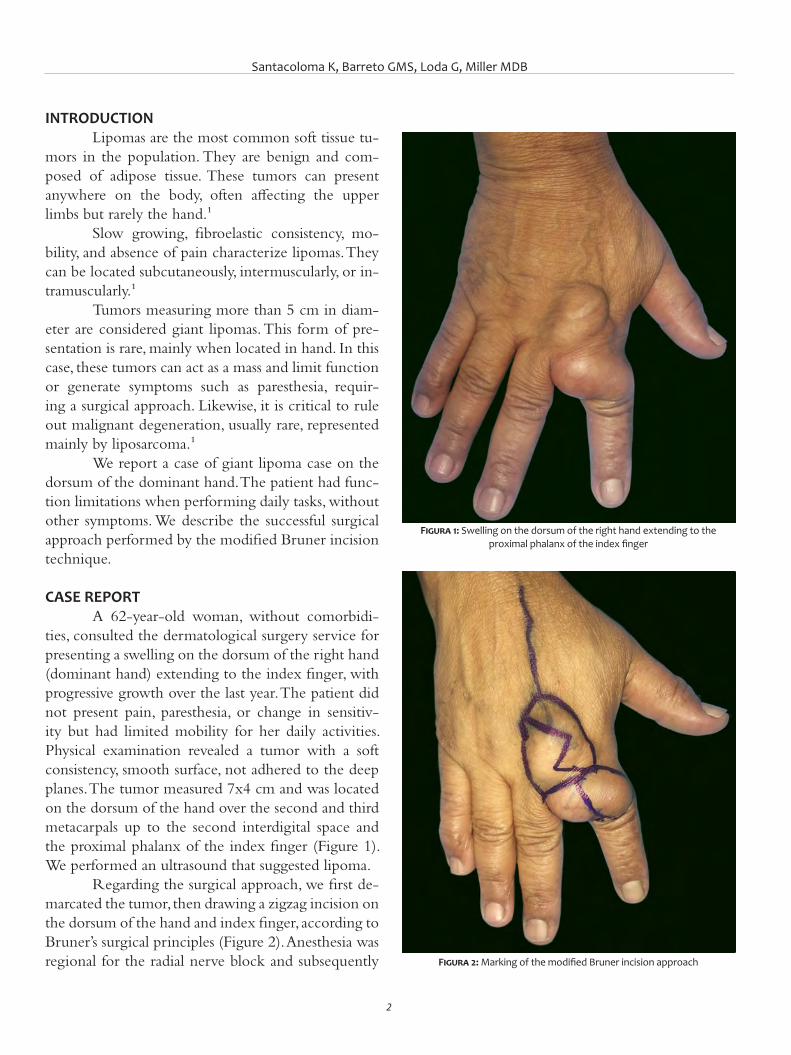
Santacoloma K, Barreto GMS, Loda G, Miller MDB
2
INTRODUCTIONLipomas are the most common soft tissue tu-
mors in the population. They are benign and com-posed of adipose tissue. These tumors can present anywhere on the body, often affecting the upper limbs but rarely the hand.¹
Slow growing, fibroelastic consistency, mo-bility, and absence of pain characterize lipomas. They can be located subcutaneously, intermuscularly, or in-tramuscularly.¹
Tumors measuring more than 5 cm in diam-eter are considered giant lipomas. This form of pre-sentation is rare, mainly when located in hand. In this case, these tumors can act as a mass and limit function or generate symptoms such as paresthesia, requir-ing a surgical approach. Likewise, it is critical to rule out malignant degeneration, usually rare, represented mainly by liposarcoma.¹
We report a case of giant lipoma case on the dorsum of the dominant hand. The patient had func-tion limitations when performing daily tasks, without other symptoms. We describe the successful surgical approach performed by the modified Bruner incision technique.
CASE REPORTA 62-year-old woman, without comorbidi-
ties, consulted the dermatological surgery service for presenting a swelling on the dorsum of the right hand (dominant hand) extending to the index finger, with progressive growth over the last year. The patient did not present pain, paresthesia, or change in sensitiv-ity but had limited mobility for her daily activities. Physical examination revealed a tumor with a soft consistency, smooth surface, not adhered to the deep planes. The tumor measured 7x4 cm and was located on the dorsum of the hand over the second and third metacarpals up to the second interdigital space and the proximal phalanx of the index finger (Figure 1). We performed an ultrasound that suggested lipoma.
Regarding the surgical approach, we first de-marcated the tumor, then drawing a zigzag incision on the dorsum of the hand and index finger, according to Bruner’s surgical principles (Figure 2). Anesthesia was regional for the radial nerve block and subsequently
Figura 1: Swelling on the dorsum of the right hand extending to the proximal phalanx of the index finger
Figura 2: Marking of the modified Bruner incision approach
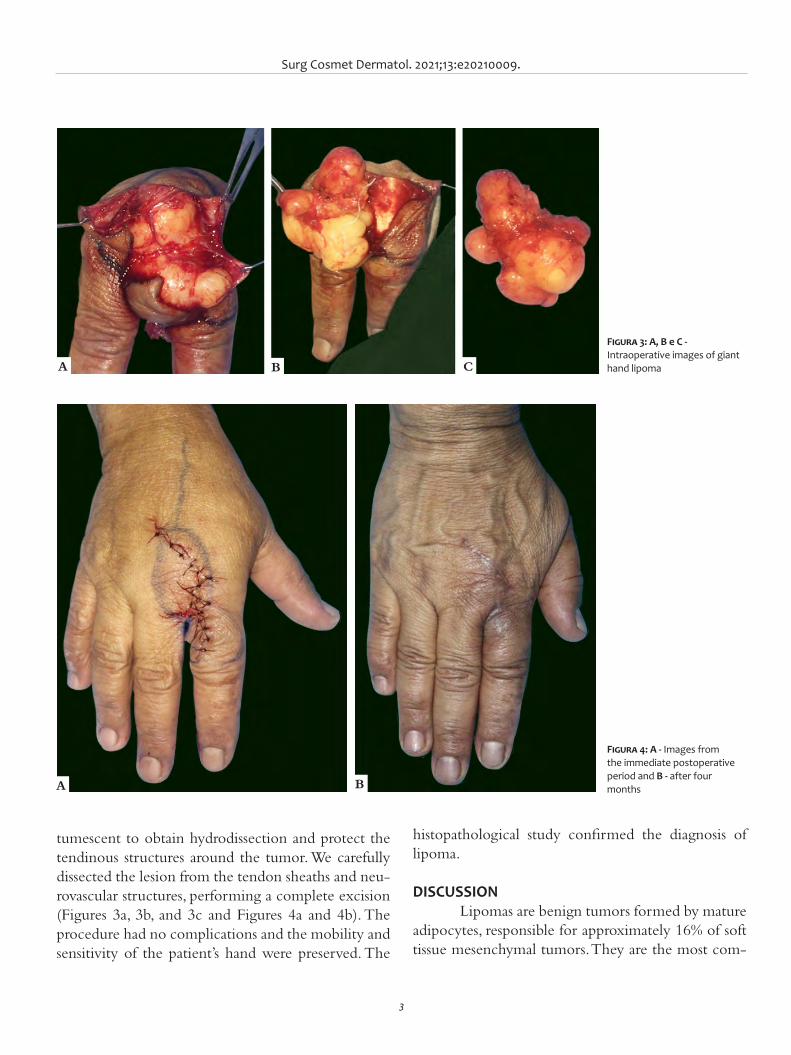
Surg Cosmet Dermatol. 2021;13:e20210009.
3
tumescent to obtain hydrodissection and protect the tendinous structures around the tumor. We carefully dissected the lesion from the tendon sheaths and neu-rovascular structures, performing a complete excision (Figures 3a, 3b, and 3c and Figures 4a and 4b). The procedure had no complications and the mobility and sensitivity of the patient’s hand were preserved. The
Figura 3: A, B e C - Intraoperative images of giant hand lipoma
Figura 4: A - Images from the immediate postoperative period and B - after four months
histopathological study confirmed the diagnosis of lipoma.
DISCUSSIONLipomas are benign tumors formed by mature
adipocytes, responsible for approximately 16% of soft tissue mesenchymal tumors. They are the most com-
CBA
A B
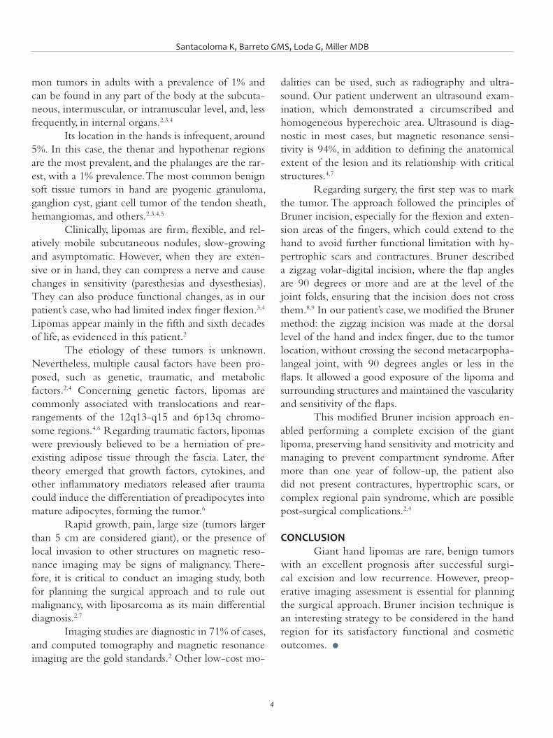
Santacoloma K, Barreto GMS, Loda G, Miller MDB
4
dalities can be used, such as radiography and ultra-sound. Our patient underwent an ultrasound exam-ination, which demonstrated a circumscribed and homogeneous hyperechoic area. Ultrasound is diag-nostic in most cases, but magnetic resonance sensi-tivity is 94%, in addition to defining the anatomical extent of the lesion and its relationship with critical structures.4,7
Regarding surgery, the first step was to mark the tumor. The approach followed the principles of Bruner incision, especially for the flexion and exten-sion areas of the fingers, which could extend to the hand to avoid further functional limitation with hy-pertrophic scars and contractures. Bruner described a zigzag volar-digital incision, where the flap angles are 90 degrees or more and are at the level of the joint folds, ensuring that the incision does not cross them.8,9 In our patient’s case, we modified the Bruner method: the zigzag incision was made at the dorsal level of the hand and index finger, due to the tumor location, without crossing the second metacarpopha-langeal joint, with 90 degrees angles or less in the flaps. It allowed a good exposure of the lipoma and surrounding structures and maintained the vascularity and sensitivity of the flaps.
This modified Bruner incision approach en-abled performing a complete excision of the giant lipoma, preserving hand sensitivity and motricity and managing to prevent compartment syndrome. After more than one year of follow-up, the patient also did not present contractures, hypertrophic scars, or complex regional pain syndrome, which are possible post-surgical complications.2,4
CONCLUSIONGiant hand lipomas are rare, benign tumors
with an excellent prognosis after successful surgi-cal excision and low recurrence. However, preop-erative imaging assessment is essential for planning the surgical approach. Bruner incision technique is an interesting strategy to be considered in the hand region for its satisfactory functional and cosmetic outcomes. l
mon tumors in adults with a prevalence of 1% and can be found in any part of the body at the subcuta-neous, intermuscular, or intramuscular level, and, less frequently, in internal organs.2,3,4
Its location in the hands is infrequent, around 5%. In this case, the thenar and hypothenar regions are the most prevalent, and the phalanges are the rar-est, with a 1% prevalence. The most common benign soft tissue tumors in hand are pyogenic granuloma, ganglion cyst, giant cell tumor of the tendon sheath, hemangiomas, and others.2,3,4,5
Clinically, lipomas are firm, flexible, and rel-atively mobile subcutaneous nodules, slow-growing and asymptomatic. However, when they are exten-sive or in hand, they can compress a nerve and cause changes in sensitivity (paresthesias and dysesthesias). They can also produce functional changes, as in our patient’s case, who had limited index finger flexion.3,4 Lipomas appear mainly in the fifth and sixth decades of life, as evidenced in this patient.2
The etiology of these tumors is unknown. Nevertheless, multiple causal factors have been pro-posed, such as genetic, traumatic, and metabolic factors.2,4 Concerning genetic factors, lipomas are commonly associated with translocations and rear-rangements of the 12q13-q15 and 6p13q chromo-some regions.4,6 Regarding traumatic factors, lipomas were previously believed to be a herniation of pre-existing adipose tissue through the fascia. Later, the theory emerged that growth factors, cytokines, and other inflammatory mediators released after trauma could induce the differentiation of preadipocytes into mature adipocytes, forming the tumor.6
Rapid growth, pain, large size (tumors larger than 5 cm are considered giant), or the presence of local invasion to other structures on magnetic reso-nance imaging may be signs of malignancy. There-fore, it is critical to conduct an imaging study, both for planning the surgical approach and to rule out malignancy, with liposarcoma as its main differential diagnosis.2,7
Imaging studies are diagnostic in 71% of cases, and computed tomography and magnetic resonance imaging are the gold standards.2 Other low-cost mo-
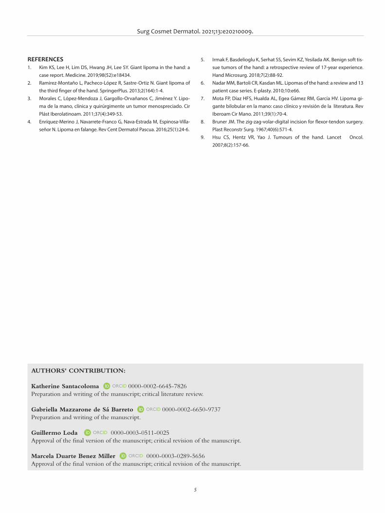
Surg Cosmet Dermatol. 2021;13:e20210009.
5
REFERENCES1. Kim KS, Lee H, Lim DS, Hwang JH, Lee SY. Giant lipoma in the hand: a
case report. Medicine. 2019;98(52):e18434.2. Ramirez-Montaño L, Pacheco-López R, Sastre-Ortiz N. Giant lipoma of
the third finger of the hand. SpringerPlus. 2013;2(164):1-4.3. Morales C, López-Mendoza J, Gargollo-Orvañanos C, Jiménez Y. Lipo-
ma de la mano, clínica y quirúrgimente un tumor menospreciado. Cir Plást Iberolatinoam. 2011;37(4):349-53.
4. Enríquez-Merino J, Navarrete-Franco G, Nava-Estrada M, Espinosa-Villa-señor N. Lipoma en falange. Rev Cent Dermatol Pascua. 2016;25(1):24-6.
AUTHORS' CONTRIBUTION:
Katherine Santacoloma 0000-0002-6645-7826Preparation and writing of the manuscript; critical literature review.
Gabriella Mazzarone de Sá Barreto 0000-0002-6650-9737Preparation and writing of the manuscript.
Guillermo Loda 0000-0003-0511-0025Approval of the final version of the manuscript; critical revision of the manuscript.
Marcela Duarte Benez Miller 0000-0003-0289-5656Approval of the final version of the manuscript; critical revision of the manuscript.
5. Irmak F, Basdelioglu K, Serhat SS, Sevim KZ, Yesilada AK. Benign soft tis-sue tumors of the hand: a retrospective review of 17-year experience. Hand Microsurg. 2018;7(2):88-92.
6. Nadar MM, Bartoli CR, Kasdan ML. Lipomas of the hand: a review and 13 patient case series. E-plasty. 2010;10:e66.
7. Mota FP, Díaz HFS, Hualda AL, Egea Gámez RM, García HV. Lipoma gi-gante bilobular en la mano: caso clínico y revisión de la literatura. Rev Iberoam Cir Mano. 2011;39(1):70-4.
8. Bruner JM. The zig-zag-volar-digital incision for flexor-tendon surgery. Plast Reconstr Surg. 1967;40(6):571-4.
9. Hsu CS, Hentz VR, Yao J. Tumours of the hand. Lancet Oncol. 2007;8(2):157-66.

1
APOIO CIENTÍFICO:
www.surgicalcosmetic.org.br/
ISSN-e 1984-8773
Case reportAuthors: Flávia Fenólio Nigro Marcelino1
Jayme de Oliveira-Filho2
Gabriela Machado Dias Junqueira3
Márcia Ferraz Nogueira3
Alexandre Ozores Michalany3
1 Universidade Santo Amaro, Residência em Dermatologia, São Paulo (SP), Brasil.
2 Universidade Santo Amaro, Professor titular de Dermatologia, São Paulo (SP), Brasil.
3 Clínica privada, Dermatologia, São Paulo (SP), Brasil.
Correspondence: Flávia Fenólio Nigro Marcelino E-mail: [email protected]
Financial support: noneConflit of interest: none
Submitted on: 09/08/2020Approved on: 09/02/2021
Acknowledgments: We thank everyone who contributed directly or indirectly to this work.
How to cite this article: Marcelino FFN, Oliveira-Filho J, Junqueira GMD, Nogueira MF, Michalany AO. Molluscum contagiosum as a tattoo complication: a case report and lite-rature review. Surg Cosmet Dermatol. 2021;13:e20210004.
Molluscum contagiosum as a tattoo complication: a case report and literature review Molusco contagioso como complicação de tatuagem: um relato de caso e revisão da literatura
ABSTRACTTattooing is an ancient practice and very popular nowadays. The pigments used have changed over time but still present varied and poorly regulated compositions. There are many described cases of adverse effects after tattooing, mainly infectious and hypersensitivity reactions. We re-port the case of a 64-year-old woman, healthy, with papules on her eyebrows one month after performing micropigmentation. The excisional biopsy diagnosed molluscum contagiosum, and the lesions were curetted. There are few reports in the literature of the spread of molluscum contagiosum caused by tattooing.Keywords: Molluscum contagiosum. Tattooing. Poxviridae infections.
RESU MOA tatuagem é uma prática antiga e muito popular atualmente. Os pigmentos utilizados mudaram com o tem-po, mas continuam apresentando composições variadas e pouco regulamentadas. Há inúmeros casos descritos de efeitos adversos pós-tatuagem, em sua maioria infecciosos, e reações de hipersensibilidade. Relatamos o caso de uma mulher de 64 anos, hígida, com pápulas nas sobrancelhas um mês após realizar micropigmentação. A biópsia excisional fez o diagnóstico de molusco contagioso, e o tratamento foi realizado com curetagem das lesões. Na literatura, existem poucos relatos de disseminação de molusco contagioso causada por tatuagem.Palavras-chave: Molusco contagioso. Tatuagem. Infecções por poxviridae
DOI: http://www.dx.doi.org/10.5935/scd1984-8773.2021130008
ISSN-e 1984-8773
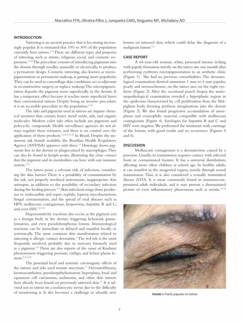
Marcelino FFN, Oliveira-Filho J, Junqueira GMD, Nogueira MF, Michalany AO
2
INTRODUCTIONTattooing is an ancient practice that is becoming increas-
ingly popular. It is estimated that 10% to 30% of the population currently have tattoos.1-4 There are different types and purposes of tattooing, such as artistic, religious, social, and cosmetic ex-pression.1,4,5 The procedure consists of introducing pigments into the dermis through needles, manually or electrically, to produce a permanent design. Cosmetic tattooing, also known as micro-pigmentation or permanent makeup, is gaining more popularity. They can be used to camouflage skin conditions, act as adjuvants in reconstructive surgery, or replace makeup. The micropigmen-tation deposits the pigment more superficially in the dermis. It has a temporary effect because it reaches more superficial layers than conventional tattoos. Despite being an invasive procedure, it is an accessible procedure to the population.6-10
The inks and pigments used in tattoos are impure chem-ical mixtures that contain heavy metal oxide, salts, and organic molecules. Modern color inks often include azo pigments and polycyclic compounds. Health surveillance agencies do not al-ways regulate these mixtures, and there is no control over the application of these products.1,2,4,6,11,12 In Brazil, Despite the nu-merous ink brands available, the Brazilian Health Regulatory Agency (ANVISA) approves only three.13 Histology shows pig-ments free in the dermis or phagocytized by macrophages. They can also be found in lymph nodes, illustrating the close contact that the pigment and its metabolites can have with our immune system.1,2,1
The tattoo poses a relevant risk of infection, consider-ing the skin barrier. There is a possibility of contamination by the ink, not properly sterilized instruments, inappropriate skin antisepsis, in addition to the possibility of secondary infection during the healing process.1,2 Skin infections range from pyoder-ma to endocarditis and sepsis, syphilis, leprosy, mycobacteriosis, fungal contamination, and the spread of viral diseases such as HPV, molluscum contagiosum, herpesvirus, hepatitis B and C, and even HIV.1,2,3,12
Hypersensitivity reactions also occur, as the pigment acts as a foreign body in the dermis, triggering lichenoid, granu-lomatous, and even pseudolymphoma lesions. Immunological reactions can be immediate or delayed and manifest locally or systemically. The most common skin manifestation related to tattooing is allergic contact dermatitis.3 The red ink is the most frequently involved, probably due to mercury formerly used as a pigment.2,3 There are also reports of the onset of Koebner phenomenon triggering psoriasis, vitiligo, and lichen planus le-sions.2,3,14,15
The potential local and systemic carcinogenic effects of the tattoos and inks used remain uncertain.11 Dermatofibroma, keratoacanthoma, pseudoepitheliomatous hyperplasia, basal and squamous cell carcinomas, melanoma, and other skin tumors have already been found on previously tattooed skin.11 It is ad-vised not to tattoo on a melanocytic nevus due to the difficulty of monitoring it. It also becomes a challenge to identify new
lesions on tattooed skin, which could delay the diagnosis of a malignant tumor.2,3
CASE REPORTA 64-year-old woman, white, presented intense itching
with papule formation strictly on the tattoo site one month after performing eyebrows micropigmentation in an aesthetic clinic (Figure 1). She had no previous comorbidities. The dermato-logical examination showed numerous 1 mm to 4 mm papules, pearly and normochromic, on the tattoo area on the right eye-brow (Figure 2). After the excisional punch biopsy, the anato-mopathological examination revealed a hyperplastic region in the epidermis characterized by cell proliferation from the Mal-pighian body, forming piriform invaginations into the dermis (Figure 3). We also found progressive accumulation of amor-phous and eosinophilic material, compatible with molluscum contagiosum (Figure 4). Serologies for hepatitis B and C and HIV were negative. We performed the treatment with curettage of the lesions, with good results and no recurrence (Figures 5 and 6).
DISCUSSIONMolluscum contagiosum is a dermatovirus caused by a
poxvirus. Usually, its transmission requires contact with infected hosts or contaminated fomites. It has a universal distribution, affecting more often children at school age. In healthy adults, it can manifest in the anogenital region, mainly through sexual transmission. Thus, it is also considered a sexually transmitted disease (STD). It is more commonly found in immunocom-promised adult individuals, and it may present a disseminated picture or even inflammatory phenomena such as uveitis.16-19
Figure 1: Pearly papules on tattoo
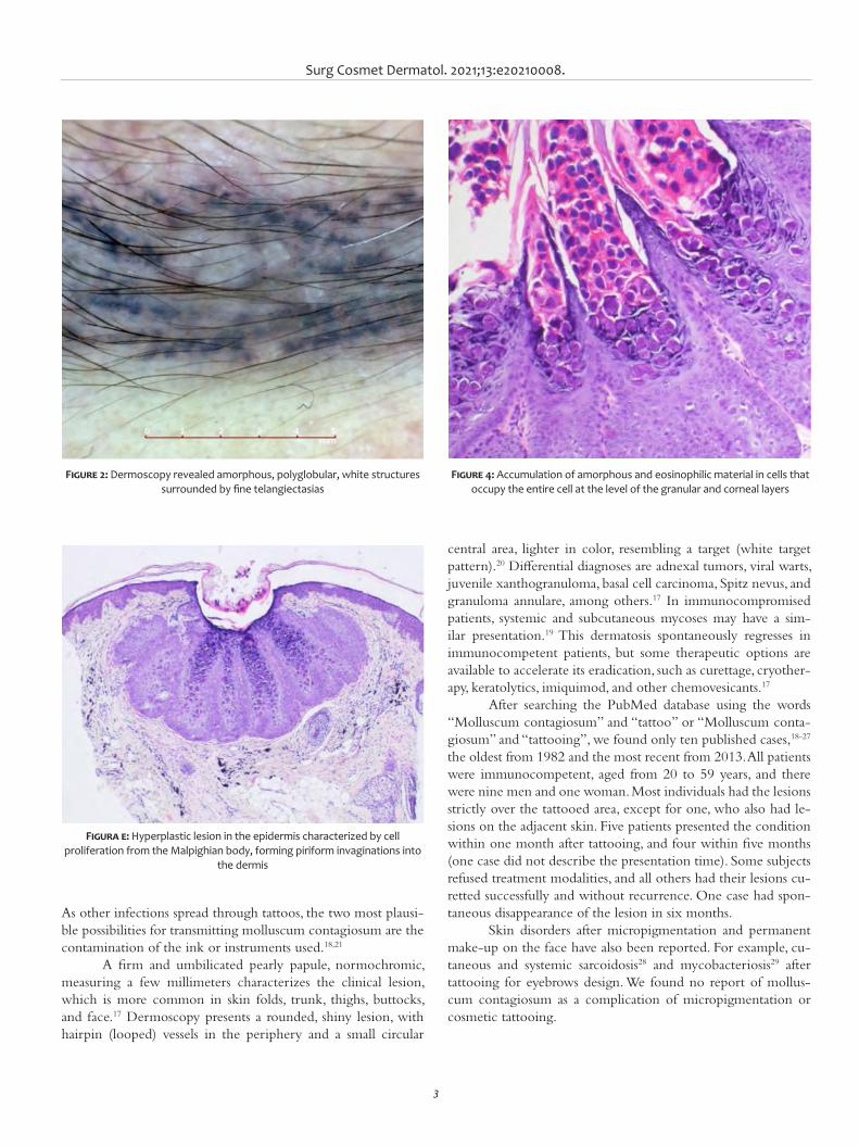
Surg Cosmet Dermatol. 2021;13:e20210008.
3
As other infections spread through tattoos, the two most plausi-ble possibilities for transmitting molluscum contagiosum are the contamination of the ink or instruments used.18,21
A firm and umbilicated pearly papule, normochromic, measuring a few millimeters characterizes the clinical lesion, which is more common in skin folds, trunk, thighs, buttocks, and face.17 Dermoscopy presents a rounded, shiny lesion, with hairpin (looped) vessels in the periphery and a small circular
Figure 2: Dermoscopy revealed amorphous, polyglobular, white structures surrounded by fine telangiectasias
Figura e: Hyperplastic lesion in the epidermis characterized by cell proliferation from the Malpighian body, forming piriform invaginations into
the dermis
Figure 4: Accumulation of amorphous and eosinophilic material in cells that occupy the entire cell at the level of the granular and corneal layers
central area, lighter in color, resembling a target (white target pattern).20 Differential diagnoses are adnexal tumors, viral warts, juvenile xanthogranuloma, basal cell carcinoma, Spitz nevus, and granuloma annulare, among others.17 In immunocompromised patients, systemic and subcutaneous mycoses may have a sim-ilar presentation.19 This dermatosis spontaneously regresses in immunocompetent patients, but some therapeutic options are available to accelerate its eradication, such as curettage, cryother-apy, keratolytics, imiquimod, and other chemovesicants.17
After searching the PubMed database using the words “Molluscum contagiosum” and “tattoo” or “Molluscum conta-giosum” and “tattooing”, we found only ten published cases,18-27 the oldest from 1982 and the most recent from 2013. All patients were immunocompetent, aged from 20 to 59 years, and there were nine men and one woman. Most individuals had the lesions strictly over the tattooed area, except for one, who also had le-sions on the adjacent skin. Five patients presented the condition within one month after tattooing, and four within five months (one case did not describe the presentation time). Some subjects refused treatment modalities, and all others had their lesions cu-retted successfully and without recurrence. One case had spon-taneous disappearance of the lesion in six months.
Skin disorders after micropigmentation and permanent make-up on the face have also been reported. For example, cu-taneous and systemic sarcoidosis28 and mycobacteriosis29 after tattooing for eyebrows design. We found no report of mollus-cum contagiosum as a complication of micropigmentation or cosmetic tattooing.
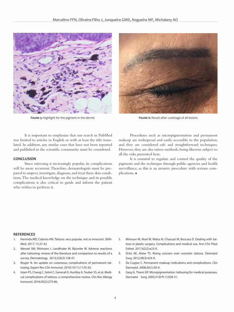
Marcelino FFN, Oliveira-Filho J, Junqueira GMD, Nogueira MF, Michalany AO
4
Procedures such as micropigmentation and permanent makeup are widespread and easily accessible to the population, and they are considered safe and straightforward techniques. However, they are also tattoo methods, being likewise subject to all the risks presented here.
It is essential to regulate and control the quality of the pigments and the technique through public agencies and health surveillance, as this is an invasive procedure with serious com-plications. l
It is important to emphasize that our search in PubMed was limited to articles in English or with at least the title trans-lated. In addition, any similar cases that have not been reported and published in the scientific community must be considered.
CONCLUSIONSince tattooing is increasingly popular, its complications
will be more recurrent. Therefore, dermatologists must be pre-pared to suspect, investigate, diagnose, and treat these skin condi-tions. The medical knowledge on the technique and its possible complications is also critical to guide and inform the patient who wishes to perform it.
REFERENCES1. Hermida MD, Cabrera HN. Tattoos: very popular, not so innocent. SKIN-
Med. 2017; 15:37-42.2. Wenzel SM, Rittmann I, Landthaler M, Bäumler W. Adverse reactions
after tattooing: review of the literature and comparison to results of a survey. Dermatology. 2013;226(2):138-47.
3. Kluger N. An update on cutaneous complications of permanent tat-tooing. Expert Rev Clin Immunol. 2019;15(11):1135-43.
4. Islam PS, Chang C, Selmi C, Generali E, Huntley A, Teuber SS, et al. Medi-cal complications of tattoos: a comprehensive review. Clin Rev Allergy Immunol. 2016;50(2):273-86.
Figure 5: Highlight for the pigment in the dermis Figure 6: Result after curettage of all lesions
5. Mimoun M, Noel W, Malca N, Chaouat M, Boccara D. Dealing with tat-toos in plastic surgery. Complications and medical use. Ann Chir Plast Esthet. 2017;62(2):e23-9.
6. Ortiz AE, Alster TS. Rising concern over cosmetic tattoos. Dermatol Surg. 2012;38(3):424-9.
7. De Cuyper C. Permanent makeup: indications and complications. Clin Dermatol. 2008;26(1):30-4.
8. Garg G, Thami GP. Micropigmentation: tattooing for medical purposes. Dermatol Surg. 2005;31(8 Pt 1):928-31.

Surg Cosmet Dermatol. 2021;13:e20210008.
5
9. Ju HJ, Eun SH, Lee HN, Lee JH, Kim GM, Bae JM. Micropigmentation for vitiligo on light to moderately colored skin: updated evidence from a clinical and animal study. J Dermatol. 2020. Epub 2020 Mar 2.
10. Vassileva S, Hristakieva E. Medical applications of tattooing. Clin Der-matol. 2007;25(4):367-74.
11. Kluger N, Koljonen V. Tattoos, inks, and cancer. Lancet Oncol. 2012;13(4):e161-8.
12. LeBlanc PM, Hollinger KA, Klontz KC. Tattoo ink-related infections awareness, diagnosis, reporting, and prevention. N Engl J Med. 2012;367(11):985-7.
13. Laboissière P. Agência Brasil [Internet]. Brasília, Empresa Brasil de Co-municação [cited 2014 May 6]. Available from: https://agenciabrasil.ebc.com.br/
14. Kluger N. Tattoo side effects worldwide: a google trends-based time series analysis. Acta Dermato Venerol Alp Pannonica Adriat. 2019;28(3):125-7.
15. Jacob CI. Tattoo-associated dermatoses: a case report and review of the literature. Dermatol Surg. 2002;28(10):962-965.
16. Ostheimer TA, Burkholder BM, Leung TG, Butler NJ, Dunn JP, Thorne JE. Tattoo-associated uveitis. Am J Ophthalmol. 2014;158(3):637-43.
17. Bologna JL, Jorizzo JL, Rapini RP. Dermatology. 3th ed. Elsevier; 2012.18. Molina L, Romiti R. Molluscum contagiosum on tattoo. An Bras Derma-
tol. 2011;86(2):352-4. 19. De Giorgi V, Grazzini M, Lotti T. A three-dimensional tattoo: molluscum
contagiosum. CMAJ. 2010;182(9):E382. 20. Panasiti V, Devirgiliis V, Roberti V, Curzio M, Calvieri S. Molluscum
contagiosum on a tattoo: usefulness of dermoscopy. Int J Dermatol. 2008;47(12):1318-9.
AUTHORS' CONTRIBUTION:
Flávia Fenólio Nigro Marcelino 0000-0003-4057-5143Concepção e planejamento do estudo; elaboração e redação do manuscrito; obtenção, análise e interpretação dos dados; parti-cipação intelectual em conduta propedêutica e/ou terapêutica de casos estudados; revisão crítica da literatura; revisão crítica do manuscrito.
Jayme de Oliveira-Filho 0000-0003-0239-0981Aprovação da versão final do manuscrito; participação efetiva na orientação da pesquisa; participação intelectual em conduta pro-pedêutica e/ou terapêutica de casos estudados.
Gabriela Machado Dias Junqueira 0000-0003-0899-9341Aprovação da versão final do manuscrito; concepção e planejamento do estudo; participação efetiva na orientação da pesquisa; participação intelectual em conduta propedêutica e/ou terapêutica de casos estudados.
Márcia Ferraz Nogueira 0000-0001-7872-7304Aprovação da versão final do manuscrito; participação intelectual em conduta propedêutica e/ou terapêutica de casos estudados.
Alexandre Ozores Michalany 0000-0002-8814-4513Aprovação da versão final do manuscrito; obtenção, análise e interpretação dos dados; participação intelectual em conduta prope-dêutica e/ou terapêutica de casos estudados.
21. Salmaso F, Gnecchi L, Gianotti R, Veraldi S. Molluscum contagiosum on a tattoo. Acta Derm Venereol. 2001;81(2):146-7.
22. Grillo E, Urech M, Vano-Galvan S, Jaén P. Lesions on tattooed skin a case study. Aust Fam Physician. 2012;41(5):308-9.
23. Ruiz-Villaverde R. Molluscum contagiosum: an unusual complication of tattooing. Br Med J (Clin Res Ed). 1982;285(6342):607.
24. Kluger N, Comte C, Guillot B. Molluscum contagiosum sur tatoua-ge [Molluscum contagiosum on a tattoo]. Ann Dermatol Venereol. 2007;134(5 Pt 1):506-7.
25. Sánchez-Cano D. Pearled papules over tattoo: molluscum cotagiosum. Pan Afr Med J. 2013;16:49.
26. Pérez-Barrio S, González-Hermosa MR, Ratón JA, Díaz-Pérez JL. Molus-co contagioso sobre tatuaje [Moluscum contagiosum over a tattoo]. Actas Dermosifiliogr. 2009;100(2):152-4.
27. Pérez Gala S, Alonso Pérez A, Ríos Buceta L, Aragüés Montañés M, Gar-cia Díez
28. A. Molluscum contagiosum on a multicoloured tattoo. J Eur Acad Der-matol Venereol. 2006;20(2):221-2.
29. Huisman S, Van der Bent SAS, Wolkerstorfer A, Rustemeyer T. Granu-lomatous tattoo reactions in permanent makeup of the eyebrows. J Cosmet Dermatol. 2019;18(1):212-4.
30. Wollina U. Nodular skin reactions in eyebrow permanent makeup: two case reports and an infection by mycobacterium haemophilum. J Cos-met Dermatol. 2011;10(3):235-9.
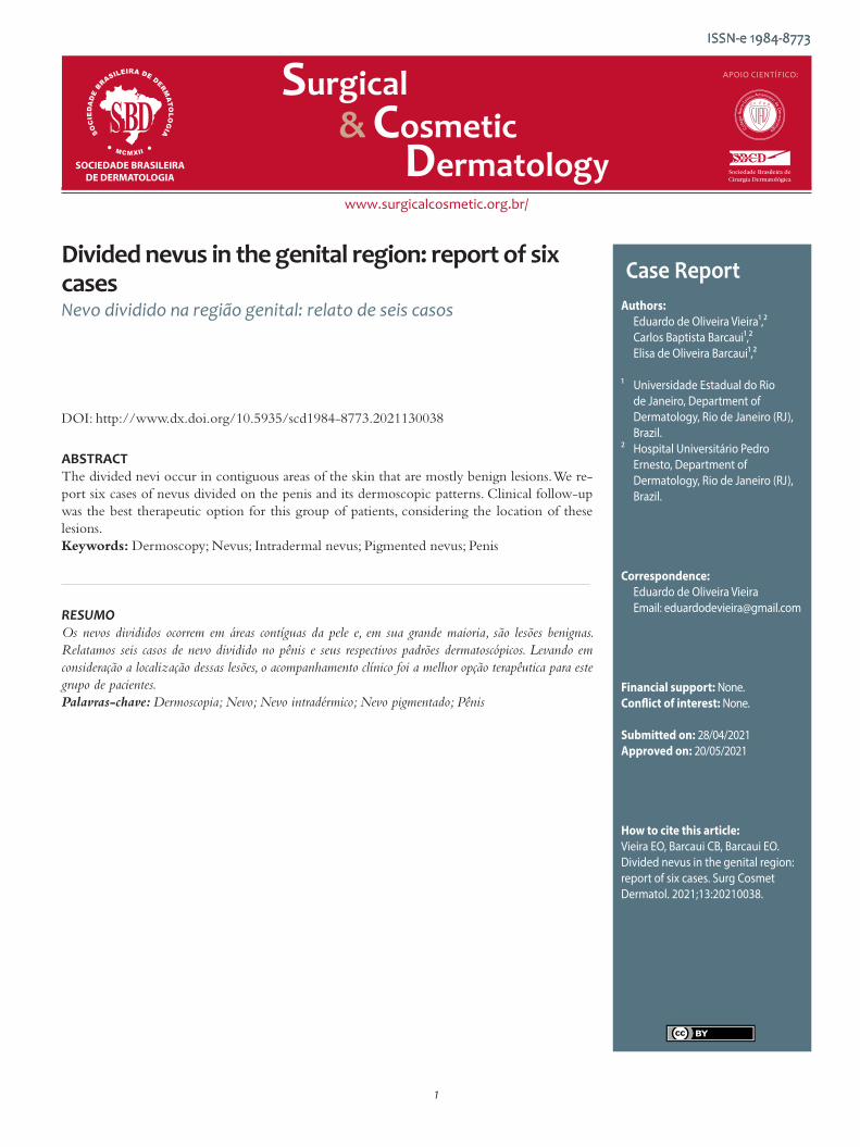
1
APOIO CIENTÍFICO:
www.surgicalcosmetic.org.br/
ISSN-e 1984-8773
Case ReportAuthors: Eduardo de Oliveira Vieira¹,² Carlos Baptista Barcaui¹,² Elisa de Oliveira Barcaui¹,²
¹ Universidade Estadual do Rio de Janeiro, Department of Dermatology, Rio de Janeiro (RJ), Brazil.
² Hospital Universitário Pedro Ernesto, Department of Dermatology, Rio de Janeiro (RJ), Brazil.
Correspondence: Eduardo de Oliveira Vieira Email: [email protected]
Financial support: None. Conflict of interest: None.
Submitted on: 28/04/2021Approved on: 20/05/2021
How to cite this article: Vieira EO, Barcaui CB, Barcaui EO. Divided nevus in the genital region: report of six cases. Surg Cosmet Dermatol. 2021;13:20210038.
Divided nevus in the genital region: report of six cases Nevo dividido na região genital: relato de seis casos
ABSTRACTThe divided nevi occur in contiguous areas of the skin that are mostly benign lesions. We re-port six cases of nevus divided on the penis and its dermoscopic patterns. Clinical follow-up was the best therapeutic option for this group of patients, considering the location of these lesions.Keywords: Dermoscopy; Nevus; Intradermal nevus; Pigmented nevus; Penis
RESUMOOs nevos divididos ocorrem em áreas contíguas da pele e, em sua grande maioria, são lesões benignas. Relatamos seis casos de nevo dividido no pênis e seus respectivos padrões dermatoscópicos. Levando em consideração a localização dessas lesões, o acompanhamento clínico foi a melhor opção terapêutica para este grupo de pacientes.Palavras-chave: Dermoscopia; Nevo; Nevo intradérmico; Nevo pigmentado; Pênis
DOI: http://www.dx.doi.org/10.5935/scd1984-8773.2021130038
ISSN-e 1984-8773
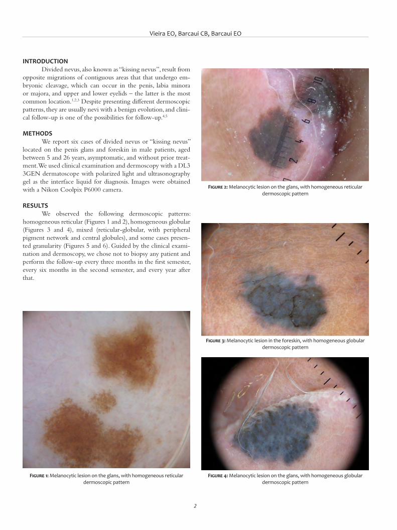
Vieira EO, Barcaui CB, Barcaui EO
2
INTRODUCTIONDivided nevus, also known as “kissing nevus”, result from
opposite migrations of contiguous areas that that undergo em-bryonic cleavage, which can occur in the penis, labia minora or majora, and upper and lower eyelids – the latter is the most common location.1,2,3 Despite presenting different dermoscopic patterns, they are usually nevi with a benign evolution, and clini-cal follow-up is one of the possibilities for follow-up.4,5
METHODS We report six cases of divided nevus or “kissing nevus”
located on the penis glans and foreskin in male patients, aged between 5 and 26 years, asymptomatic, and without prior treat-ment. We used clinical examination and dermoscopy with a DL3 3GEN dermatoscope with polarized light and ultrasonography gel as the interface liquid for diagnosis. Images were obtained with a Nikon Coolpix P6000 camera.
RESULTS We observed the following dermoscopic patterns:
homogeneous reticular (Figures 1 and 2), homogeneous globular (Figures 3 and 4), mixed (reticular-globular, with peripheral pigment network and central globules), and some cases presen-ted granularity (Figures 5 and 6). Guided by the clinical exami-nation and dermoscopy, we chose not to biopsy any patient and perform the follow-up every three months in the first semester, every six months in the second semester, and every year after that.
Figure 1: Melanocytic lesion on the glans, with homogeneous reticular dermoscopic pattern
Figure 2: Melanocytic lesion on the glans, with homogeneous reticular dermoscopic pattern
Figure 3: Melanocytic lesion in the foreskin, with homogeneous globular dermoscopic pattern
Figure 4: Melanocytic lesion on the glans, with homogeneous globular dermoscopic pattern
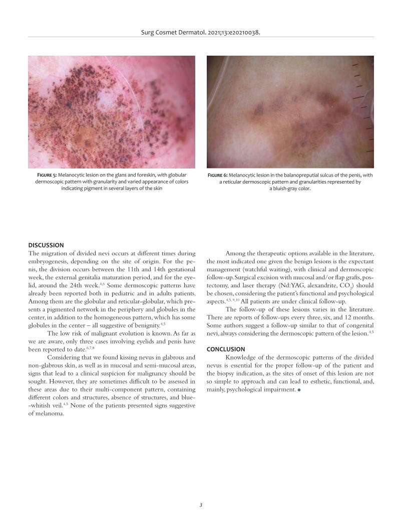
Surg Cosmet Dermatol. 2021;13:e20210038.
3
DISCUSSIONThe migration of divided nevi occurs at different times during embryogenesis, depending on the site of origin. For the pe-nis, the division occurs between the 11th and 14th gestational week, the external genitalia maturation period, and for the eye-lid, around the 24th week.4,6 Some dermoscopic patterns have already been reported both in pediatric and in adults patients. Among them are the globular and reticular-globular, which pre-sents a pigmented network in the periphery and globules in the center, in addition to the homogeneous pattern, which has some globules in the center – all suggestive of benignity.4,5
The low risk of malignant evolution is known. As far as we are aware, only three cases involving eyelids and penis have been reported to date.6,7,8
Considering that we found kissing nevus in glabrous and non-glabrous skin, as well as in mucosal and semi-mucosal areas, signs that lead to a clinical suspicion for malignancy should be sought. However, they are sometimes difficult to be assessed in these areas due to their multi-component pattern, containing different colors and structures, absence of structures, and blue--whitish veil.4,5 None of the patients presented signs suggestive of melanoma.
Among the therapeutic options available in the literature, the most indicated one given the benign lesions is the expectant management (watchful waiting), with clinical and dermoscopic follow-up. Surgical excision with mucosal and/or flap grafts, pos-tectomy, and laser therapy (Nd:YAG, alexandrite, CO
2) should
be chosen, considering the patient’s functional and psychological aspects.4,5, 9,10 All patients are under clinical follow-up.
The follow-up of these lesions varies in the literature. There are reports of follow-ups every three, six, and 12 months. Some authors suggest a follow-up similar to that of congenital nevi, always considering the dermoscopic pattern of the lesion.4,5
CONCLUSION Knowledge of the dermoscopic patterns of the divided
nevus is essential for the proper follow-up of the patient and the biopsy indication, as the sites of onset of this lesion are not so simple to approach and can lead to esthetic, functional, and, mainly, psychological impairment. l
Figure 5: Melanocytic lesion on the glans and foreskin, with globular dermoscopic pattern with granularity and varied appearance of colors
indicating pigment in several layers of the skin
Figure 6: Melanocytic lesion in the balanopreputial sulcus of the penis, with a reticular dermoscopic pattern and granularities represented by
a bluish-gray color.
Vieira EO, Barcaui CB, Barcaui EO
4
AUTHORS' CONTRIBUTION:
Eduardo de Oliveira Vieira 0000-0001-6765-2474Approval of the final version of the manuscript; study design and planning; preparation and writing of the manuscript; data collection, analysis, and interpretation; active participation in research orientation; intellectual participation in propaedeutic and/or therapeutic conduct of studied cases; critical literature review; critical revision of the manuscript.
Carlos Baptista Barcaui 0000-0002-3303-3656Approval of the final version of the manuscript; study design and planning; preparation and writing of the manuscript; data collection, analysis, and interpretation; active participation in research orientation; intellectual participation in propaedeutic and/or therapeutic conduct of studied cases; critical literature review; critical revision of the manuscript.
Elisa de Oliveira Barcaui 0000-0002-9487-7860Approval of the final version of the manuscript; study design and planning; preparation and writing of the manuscript; data collection, analysis, and interpretation; active participation in research orientation; intellectual participation in propaedeutic and/or therapeutic conduct of studied cases; critical literature review; critical revision of the manuscript.
REFERENCES:1. Jiang S, Chen Y, Hinchliffe TE, Wu T, Stephen T. Kissing nevus of the pe-
nis. J Coll Physicians Surg Pak. 2018;28(3):S19-S20.2. Vora RV, Shah AJ, Patel TB. Congenital melanocytic kissing ne-
vus on right eyelid: a rare phenomenon. Indian Dermatol Online J. 2020;11(4):627-8.
3. Lysebo DE, Krohn J. Kissing naevus. Tidsskr Nor Laegeforen. 2021;7(1):141.
4. Armengot-Carbó M, Rodrigo-Nicolás B, Botella-Estrada R. Divided or kissing nevus of the penis: a new case with dermoscopic findings. Pe-diatr Dermatol. 2018;35(5):e321-e4.
5. Godinho N, Nai GA, Schaefer ALF, Schaefer LV. Kissing nevus of the penis: a case report and dermatoscopic findings. An Bras Dermatol. 2017;92(5 Suppl 1):95-7.
6. Kharel Sitaula R, Batta S, Shrestha GB, Shrestha JK. Malignant trans-formation of kissing nevus- a rare entity. Nepal J Ophthalmol. 2012;4(2):329-32.
7. Fuchs A. Divided nevi of the eyelids. Urol Cutaneous Rev. 1950;54(2):88-90.8. Lemaître S, Gardrat S, Vincent-Salomon A, Galatoire O, Lévy-Gabriel C,
Desjardins L. Malignant transformation of a multi-operated divided ne-vus of the eyelids. Ocul Oncol Pathol. 2018;4(2):112-5.
9. Correia B, Duarte AF, Haneke E, Correia O. CO2 laser treatment of a kis-sing nevus of the penis: an alternative solution for a rare condition. J Dermatolog Treat. 2021;32(1):120-3.
10. Bray FN, Shah V, Nouri K. Laser treatment of congenital melanocytic nevi: a review of the literature. Lasers Med Sci. 2016;31(1):197-204.

1
APOIO CIENTÍFICO:
www.surgicalcosmetic.org.br/
ISSN-e 1984-8773
Case report Authors: Carla Aguiar Andrade¹ Cláudia Cardoso de Macedo Oliveira¹ Eduardo Vinícius Mendes Roncada¹ Diogo Gonçalves Ribeiro²
¹ Hospital Regional de Presidente Prudente, Department of Derma-tology, Presidente Prudente (SP), Brazil.
² Hospital Regional de Presidente Prudente, Head and Neck Surgery Service, Presidente Prudente (SP), Brazil.
Correspondence: Carla Aguiar Andrade¹ Email: carlaaguiarandrade@ hotmail.com
Financial support: None.Conflict of interest: None.
Submitted on: 13/04/2021Approved on: 15/07/2021
How to cite this article: Andrade CA, Oliveira CCM, Roncada EVM, Ribeiro DG. Primary oral muco-sal melanoma: case report. Surg Cos-met Dermatol. 2021;13:e20210029.
Primary oral mucosal melanoma: case reportMelanoma primário de mucosa oral: relato de caso
ABSTRACTOral mucosa melanoma (OMM) represents 0.2% to 8% of melanoma cases. It mainly affects the palate and gums of patients between 40 and 70 years old. It is initially asymptomatic, contribut-ing to late diagnosis and poor prognosis. The treatment of choice consists of surgical resection, and adjuvant radiotherapy and chemotherapy may be used. We report a case in an 80-year-old male patient with primary gingival and hard palate melanoma who underwent tumor resection followed by palate reconstruction. We emphasize the importance of a complete physical exam-ination and active search for lesions in the oral mucosa for early diagnosis.Keywords: Melanoma; Mouth mucosa; Palate
RESUMOO melanoma de mucosa oral (MMO) representa de 0,2 a 8% dos casos de melanoma. Acomete, principal-mente, palato e gengiva de pacientes entre 40 e 70 anos. Inicialmente é assintomático, contribuindo para o diagnóstico tardio e prognóstico reservado. O tratamento de escolha consiste na ressecção cirúrgica, podendo ser utilizadas radioterapia e quimioterapia adjuvantes. Relatamos um caso em paciente de 80 anos do sexo masculino, com melanoma primário na gengiva e palato duro, que foi submetido à ressecção tumoral seguida de reconstrução de palato. Ressaltamos a importância do exame físico completo e da busca ativa por lesões na mucosa oral para diagnóstico precoce.Palavras-chave: Melanoma; Mucosa bucal; Palato
DOI: http://www.dx.doi.org/10.5935/scd1984-8773.2021130029
ISSN-e 1984-8773
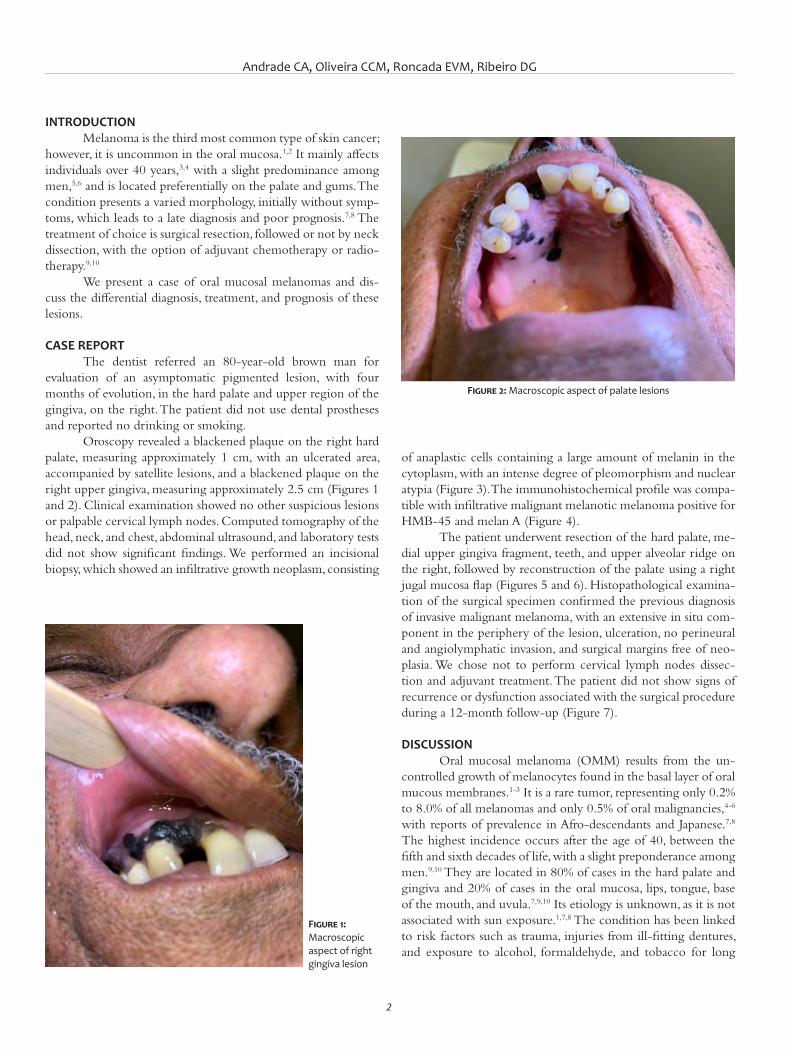
Andrade CA, Oliveira CCM, Roncada EVM, Ribeiro DG
2
INTRODUCTIONMelanoma is the third most common type of skin cancer;
however, it is uncommon in the oral mucosa.1,2 It mainly affects individuals over 40 years,3,4 with a slight predominance among men,5,6 and is located preferentially on the palate and gums. The condition presents a varied morphology, initially without symp-toms, which leads to a late diagnosis and poor prognosis.7,8 The treatment of choice is surgical resection, followed or not by neck dissection, with the option of adjuvant chemotherapy or radio-therapy.9,10
We present a case of oral mucosal melanomas and dis-cuss the differential diagnosis, treatment, and prognosis of these lesions.
CASE REPORTThe dentist referred an 80-year-old brown man for
evaluation of an asymptomatic pigmented lesion, with four months of evolution, in the hard palate and upper region of the gingiva, on the right. The patient did not use dental prostheses and reported no drinking or smoking.
Oroscopy revealed a blackened plaque on the right hard palate, measuring approximately 1 cm, with an ulcerated area, accompanied by satellite lesions, and a blackened plaque on the right upper gingiva, measuring approximately 2.5 cm (Figures 1 and 2). Clinical examination showed no other suspicious lesions or palpable cervical lymph nodes. Computed tomography of the head, neck, and chest, abdominal ultrasound, and laboratory tests did not show significant findings. We performed an incisional biopsy, which showed an infiltrative growth neoplasm, consisting
of anaplastic cells containing a large amount of melanin in the cytoplasm, with an intense degree of pleomorphism and nuclear atypia (Figure 3). The immunohistochemical profile was compa-tible with infiltrative malignant melanotic melanoma positive for HMB-45 and melan A (Figure 4).
The patient underwent resection of the hard palate, me-dial upper gingiva fragment, teeth, and upper alveolar ridge on the right, followed by reconstruction of the palate using a right jugal mucosa flap (Figures 5 and 6). Histopathological examina-tion of the surgical specimen confirmed the previous diagnosis of invasive malignant melanoma, with an extensive in situ com-ponent in the periphery of the lesion, ulceration, no perineural and angiolymphatic invasion, and surgical margins free of neo-plasia. We chose not to perform cervical lymph nodes dissec-tion and adjuvant treatment. The patient did not show signs of recurrence or dysfunction associated with the surgical procedure during a 12-month follow-up (Figure 7).
DISCUSSION Oral mucosal melanoma (OMM) results from the un-
controlled growth of melanocytes found in the basal layer of oral mucous membranes.1-3 It is a rare tumor, representing only 0.2% to 8.0% of all melanomas and only 0.5% of oral malignancies,4-6 with reports of prevalence in Afro-descendants and Japanese.7,8 The highest incidence occurs after the age of 40, between the fifth and sixth decades of life, with a slight preponderance among men.9,10 They are located in 80% of cases in the hard palate and gingiva and 20% of cases in the oral mucosa, lips, tongue, base of the mouth, and uvula.7,9,10 Its etiology is unknown, as it is not associated with sun exposure.1,7,8 The condition has been linked to risk factors such as trauma, injuries from ill-fitting dentures, and exposure to alcohol, formaldehyde, and tobacco for long
Figure 1: Macroscopic aspect of right gingiva lesion
Figure 2: Macroscopic aspect of palate lesions
Surg Cosmet Dermatol. 2021;13:e20210029.
3
periods.3,4,6,10 Although most OMMs arise again, more than a third develop from benign melanocytic lesions.1,5,8
Initially, it presents as an asymptomatic macula or plaque that evolves with swelling, ulceration, bleeding, pain, tooth mo-bility, and the appearance of satellite lesions.7-10 Its color varies
in shades of black, brown, gray, purple, and red. However, a third of the lesions present no pigment.3 It tends to recur locally and develop metastasis to lymph nodes (33%). In contrast, involve-ment of the lungs, brain, bones, and liver affects up to 85% of patients.4,5 Differential diagnosis includes amalgam tattoo, me-lanoacanthoma, smoking-associated melanosis, post-inflamma-tory pigmentation, melanocytic nevus, drug-induced melanosis (such as minocycline and antimalarials), Peutz-Jeghers syndro-me, Cushing’s syndrome, Addison’s disease, Kaposi sarcoma, and other various conditions that cause pigmentation in the oral mucosa.1,2,8,9
Histologically, tumor cells are characterized by the proliferation of atypical melanocytes. The evaluated parameters include the
Figure 3: Neoplastic cells with evident red nucleoli. Note neoplastic cells infiltrating the epidermis (arrows)
(Hematoxylin & eosin, 400x magnification)
Figure 4: Neoplastic cells with cytoplasm marked with melan-A (100x magnification)
Figure 5: Intraoperative phase after tumor resection
Figure 6: Surgical piece
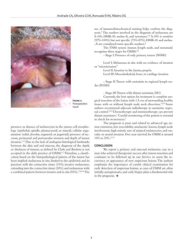
Andrade CA, Oliveira CCM, Roncada EVM, Ribeiro DG
4
presence or absence of melanocytes in the tumor; cell morpho-logy (epithelial, spindle, plasmacytoid, or mixed); cellular orga-nization (solid, alveolar, organoid, or pagetoid); presence of ne-crosis, perineural and perivascular invasion and depth of tumor invasion.7,10 Due to the lack of analogous histological landmarks between the skin and oral mucosa, the diagnosis of the depth or thickness of tumors, as defined by Clark and Breslow, is not accepted in the daily practice of OMM.4,7 Therefore, a classifi-cation based on the histopathological pattern of the tumor has been implied: melanoma in situ, limited to the epidermis and its junction with the connective tissue (15%); invasive melanomas, extending into the connective tissue (30%); and melanomas with a combined pattern between invasive and in situ (55%).1,5,8,10 The
use of immunohistochemical staining helps confirm the diag-nosis.9 The markers involved in the diagnosis of melanoma are S-100, HMB-45, melan-A, and tyrosinase.7,8 S-100 is sensitive (97%-100%) but not specific (75%-87%); HMB-45 and melan--A are considered more specific markers.8
The TNM system (tumor, lymph node, and metastasis) recognizes three stages for OMM:1,8
- Stage I: Presence of only primary tumor (N0M0)
Level I: Melanoma in situ with no evidence of invasion or “microinvasion”
Level II: Invasion to the lamina propriaLevel III: Musculoskeletal, bone, or cartilage invasion
- Stage II: Tumor with metastasis to regional lymph no-des (N1M0)
- Stage III: Tumor with distant metastasis (M1)Currently, the best option for treatment is complete sur-
gical resection of the lesion with 1.5 cm of surrounding healthy tissue, with or without lymph node neck dissection.7,8,10 Some authors recommend adjuvant radiotherapy to maximize regio-nal control.3,4,6 Chemotherapy and immunotherapy can prevent distant metastases.1 Careful monitoring of the patient is essential to check for recurrences.2
The prognosis is poor and related to advanced age, tu-mor extension, low resectability, amelanotic lesions, lymph node involvement, high mitotic rate of atypical melanocytes, and vas-cular or neural invasion. Five-year survival for OMM is around 15% to 30%.1,6,10
CONCLUSIONWe report a primary oral mucosal melanoma case in a
man who achieved therapeutic success after tumor resection and continues to be followed up in our Service to assess the re-currence or appearance of new suspicious lesions. The authors emphasize the importance of careful clinical examination for early detection of suspicious lesions, as cases of OMM are often initially asymptomatic, and early biopsy plays a fundamental role in the prognosis. l
Figure 7: Postoperative result
Surg Cosmet Dermatol. 2021;13:e20210029.
5
REFERENCES1. Munde A, Juvekar MV, Karle RR, Wankhede P. Malignant melanoma of the
oral cavity: Report of two cases. Contemp Clin Dent. 2014;5(2):227-30.2. Manigandan T, Sagar GV, Amudhan A, Hemalatha VT, Babu NA. Oral ma-
lignant melanoma: A case report with review of literature. Contemp Clin Dent. 2014;5(3):415-8.
3. Oranges CM, Sisti G, Nasioudis D, Tremp M, DI Summa PG, Kalbermatten DF, et al. Hard Palate Melanoma: A Population-based Analysis of Epide-miology and Survival Outcomes. Anticancer Res. 2018;38(10):5811-7.
4. Malinoski H, Reddy R, Cohen DM, Bhattacharyya I, Islam MN, Bowers TL 4th. Oral Melanomas: A Case Series of a Deadly Neoplasm. J Oral Maxillo-fac Surg. 2019;77(9):1832-6.
5. Tchernev G, Lotti T, Wollina U. Palatal Melanoma: The Silent Killer. Open Access Maced J Med Sci. 2018;6(2):364-6.
6. Kumar V, Vishnoi JR, Kori CG, Gupta S, Misra S, Akhtar N. Primary malig-nant melanoma of oral cavity: A tertiary care center experience. Natl J Maxillofac Surg. 2015;6(2):167-71.
7. Topić B, Mašić T, Radović S, Lincender I, Muhić E. Primary Oral Mucosal Melanomas - Two Case Reports and Comprehensive Literature Review. Acta Clin Croat. 2017;56(2):323- 30.
8. Deyhimi P, Razavi SM, Shahnaseri S, Khalesi S, Homayoni S, Tavakoli P. Rare and Extensive Malignant Melanoma of the Oral Cavity: Report of Two Cases. J Dent (Shiraz). 2017;18(3):227-33.
9. Martinelli-Kläy CP, Laporte ML, Martinelli CR, Martinelli C, Lombardi T. Oral Malignant Melanoma Initially Misdiagnosed as a Racial Pigmenta-tion: A Case Report. Dermatopathology (Basel). 2016;3(1):1-7.
10. Guevara-Canales JO, Gutiérrez-Morales MM, Sacsaquispe-Contreras SJ, Sánchez-Lihón J, Morales-Vadillo R. Malignant melanoma of the oral ca-vity. Review of the literature and experience in a Peruvian Population. Med Oral Patol Oral Cir Bucal. 2012;17(2):206-11.
AUTHORS' CONTRIBUTION:
Carla Aguiar Andrade | 0000-0002-9348-5304Preparation and writing of the manuscript; critical literature review.
Cláudia Cardoso de Macedo Oliveira | 0000-0002-2367-1662Approval of the final version of the manuscript; study design and planning; active participation in research orientation; intellectual participation in propaedeutic and/or therapeutic conduct of studied cases; critical revision of the manuscript.
Eduardo Vinícius Mendes Roncada | 0000-0002-2149-2388Approval of the final version of the manuscript; study design and planning; active participation in research orientation; critical revision of the manuscript.
Diogo Gonçalves Ribeiro | 0000-0003-2024-1162Approval of the final version of the manuscript; study design and planning; active participation in research orientation; intellectual participation in propaedeutic and/or therapeutic conduct of studied cases; critical revision of the manuscript.
1
APOIO CIENTÍFICO:
www.surgicalcosmetic.org.br/
ISSN-e 1984-8773
Case reportAuthors: Julia Silva Marra¹ Kioshe Rodrigues Siracava¹ Leonardo Teodoro Duarte Alves¹ Talissa Gomes Silva de Souza¹ Mabel Duarte Alves Gomides²
¹ Universidade Federal de Uberlândia, Department of Medicine, Uberlândia (MG), Brazil.² Hospital de Clínicas de Uberlândia,
Intensive Care Service, Uberlândia (MG), Brazil.
Correspondence: Mabel Duarte Alves Gomides Email: mabel@dermaclinicagoias. com.br
Financial support: None.Conflict of interest: None.
Submitted on: 15/04/2021Approved on: 12/07/2021
How to cite this article: Marra JS, Siracava KR, Alves LTD, Souza TGS, Gomides MDA. Hot asphalt burns: an epidemiological and therapeutic review. Surg Cosmet Dermatol. 2021;13:e20210030.
Hot asphalt burns: case report and therapeutic review Queimadura por asfalto: relato de caso e revisão terapêutica
ABSTRACTHot asphalt burns are a health threat due to the risk of death and adhesion to tissues. A 40-year-old man suffered first and second-degree burns by hot asphalt on 20% of the body surface area. The asphalt adhered to the skin was removed on the 4th day of the ICU stay with liquid petroleum jelly, and the burns were treated with healing gel. Burns caused by hot asphalt are serious and represent 1.4% of hospitalized cases. They affect young people in the skin and airways by inhaling the vapors. Removing asphalt from the skin is a major therapeutic challenge.Keywords: Burns. Burns, Chemical. Petroleum. Accidents. Accidents, Occupational
RESUMOAs queimaduras por asfalto quente representam uma ameaça para a saúde devido ao risco de morte e adesão aos tecidos. Paciente do sexo masculino, 40 anos, vítima de queimadura de 1° e 2° graus por asfalto quente em 20% da superfície corporal. O asfalto aderido na pele foi removido no 4º dia de UTI com vaselina líquida, e as queimaduras, tratadas com cicatrizante tópico. As queimaduras por asfalto quente são graves e representam 1,4% dos casos hospitalizados. Acometem jovens nas regiões da pele e vias aéreas por inalação dos vapores. Remover o asfalto da pele caracteriza-se em grande desafio terapêutico.Palavras-chave: Queimaduras. Queimaduras Químicas. Petróleo. Acidentes. Acidentes de Trabalho
DOI: http://www.dx.doi.org/10.5935/scd1984-8773.2021130030
ISSN-e 1984-8773
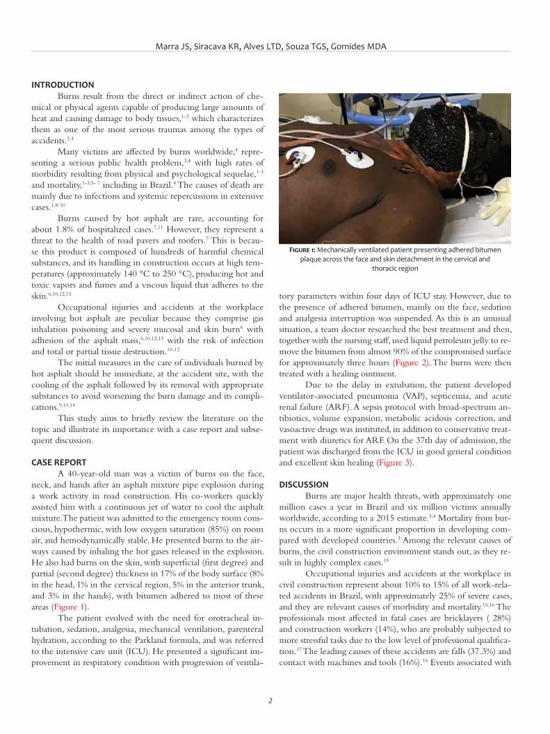
Marra JS, Siracava KR, Alves LTD, Souza TGS, Gomides MDA
2
INTRODUCTION Burns result from the direct or indirect action of che-
mical or physical agents capable of producing large amounts of heat and causing damage to body tissues,1-3 which characterizes them as one of the most serious traumas among the types of accidents.2,4
Many victims are affected by burns worldwide,4 repre-senting a serious public health problem,3,4 with high rates of morbidity resulting from physical and psychological sequelae,1-3 and mortality,1-3,5- 7 including in Brazil.4 The causes of death are mainly due to infections and systemic repercussions in extensive cases.1,8-10
Burns caused by hot asphalt are rare, accounting for about 1.8% of hospitalized cases.7,11 However, they represent a threat to the health of road pavers and roofers.7 This is becau-se this product is composed of hundreds of harmful chemical substances, and its handling in construction occurs at high tem-peratures (approximately 140 °C to 250 °C), producing hot and toxic vapors and fumes and a viscous liquid that adheres to the skin.6,10,12,13
Occupational injuries and accidents at the workplace involving hot asphalt are peculiar because they comprise gas inhalation poisoning and severe mucosal and skin burn6 with adhesion of the asphalt mass,6,10,12,13 with the risk of infection and total or partial tissue destruction.10-12
The initial measures in the care of individuals burned by hot asphalt should be immediate, at the accident site, with the cooling of the asphalt followed by its removal with appropriate substances to avoid worsening the burn damage and its compli-cations.9,10,14
This study aims to briefly review the literature on the topic and illustrate its importance with a case report and subse-quent discussion.
CASE REPORT A 40-year-old man was a victim of burns on the face,
neck, and hands after an asphalt mixture pipe explosion during a work activity in road construction. His co-workers quickly assisted him with a continuous jet of water to cool the asphalt mixture. The patient was admitted to the emergency room cons-cious, hypothermic, with low oxygen saturation (85%) on room air, and hemodynamically stable. He presented burns to the air-ways caused by inhaling the hot gases released in the explosion. He also had burns on the skin, with superficial (first degree) and partial (second degree) thickness in 17% of the body surface (8% in the head, 1% in the cervical region, 5% in the anterior trunk, and 3% in the hands), with bitumen adhered to most of these areas (Figure 1).
The patient evolved with the need for orotracheal in-tubation, sedation, analgesia, mechanical ventilation, parenteral hydration, according to the Parkland formula, and was referred to the intensive care unit (ICU). He presented a significant im-provement in respiratory condition with progression of ventila-
tory parameters within four days of ICU stay. However, due to the presence of adhered bitumen, mainly on the face, sedation and analgesia interruption was suspended. As this is an unusual situation, a team doctor researched the best treatment and then, together with the nursing staff, used liquid petroleum jelly to re-move the bitumen from almost 90% of the compromised surface for approximately three hours (Figure 2). The burns were then treated with a healing ointment.
Due to the delay in extubation, the patient developed ventilator-associated pneumonia (VAP), septicemia, and acute renal failure (ARF). A sepsis protocol with broad-spectrum an-tibiotics, volume expansion, metabolic acidosis correction, and vasoactive drugs was instituted, in addition to conservative treat-ment with diuretics for ARF. On the 37th day of admission, the patient was discharged from the ICU in good general condition and excellent skin healing (Figure 3).
DISCUSSION Burns are major health threats, with approximately one
million cases a year in Brazil and six million victims annually worldwide, according to a 2015 estimate.3,4 Mortality from bur-ns occurs in a more significant proportion in developing com-pared with developed countries.3 Among the relevant causes of burns, the civil construction environment stands out, as they re-sult in highly complex cases.15
Occupational injuries and accidents at the workplace in civil construction represent about 10% to 15% of all work-rela-ted accidents in Brazil, with approximately 25% of severe cases, and they are relevant causes of morbidity and mortality.15,16 The professionals most affected in fatal cases are bricklayers ( 28%) and construction workers (14%), who are probably subjected to more stressful tasks due to the low level of professional qualifica-tion.17 The leading causes of these accidents are falls (37.3%) and contact with machines and tools (16%).16 Events associated with
Figure 1: Mechanically ventilated patient presenting adhered bitumen plaque across the face and skin detachment in the cervical and
thoracic region
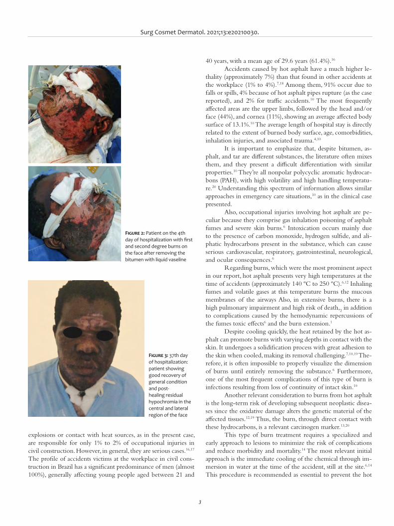
Surg Cosmet Dermatol. 2021;13:e20210030.
3
explosions or contact with heat sources, as in the present case, are responsible for only 1% to 2% of occupational injuries in civil construction. However, in general, they are serious cases.16,17 The profile of accidents victims at the workplace in civil cons-truction in Brazil has a significant predominance of men (almost 100%), generally affecting young people aged between 21 and
40 years, with a mean age of 29.6 years (61.4%).16
Accidents caused by hot asphalt have a much higher le-thality (approximately 7%) than that found in other accidents at the workplace (1% to 4%).7,18 Among them, 91% occur due to falls or spills, 4% because of hot asphalt pipes rupture (as the case reported), and 2% for traffic accidents.10 The most frequently affected areas are the upper limbs, followed by the head and/or face (44%), and cornea (11%), showing an average affected body surface of 13.1%.10 The average length of hospital stay is directly related to the extent of burned body surface, age, comorbidities, inhalation injuries, and associated trauma.4,10
It is important to emphasize that, despite bitumen, as-phalt, and tar are different substances, the literature often mixes them, and they present a difficult differentiation with similar properties.10 They’re all nonpolar polycyclic aromatic hydrocar-bons (PAH), with high volatility and high handling temperatu-re.20 Understanding this spectrum of information allows similar approaches in emergency care situations,10 as in the clinical case presented.
Also, occupational injuries involving hot asphalt are pe-culiar because they comprise gas inhalation poisoning of asphalt fumes and severe skin burns.6 Intoxication occurs mainly due to the presence of carbon monoxide, hydrogen sulfide, and ali-phatic hydrocarbons present in the substance, which can cause serious cardiovascular, respiratory, gastrointestinal, neurological, and ocular consequences.6
Regarding burns, which were the most prominent aspect in our report, hot asphalt presents very high temperatures at the time of accidents (approximately 140 ºC to 250 ºC).6,12 Inhaling fumes and volatile gases at this temperature burns the mucous membranes of the airways Also, in extensive burns, there is a high pulmonary impairment and high risk of death,
2 in addition
to complications caused by the hemodynamic repercussions of the fumes toxic effects6 and the burn extension.1
Despite cooling quickly, the heat retained by the hot as-phalt can promote burns with varying depths in contact with the skin. It undergoes a solidification process with great adhesion to the skin when cooled, making its removal challenging.7,10,19 The-refore, it is often impossible to properly visualize the dimension of burns until entirely removing the substance.6 Furthermore, one of the most frequent complications of this type of burn is infections resulting from loss of continuity of intact skin.10
Another relevant consideration to burns from hot asphalt is the long-term risk of developing subsequent neoplastic disea-ses since the oxidative damage alters the genetic material of the affected tissues.12,19 Thus, the burn, through direct contact with these hydrocarbons, is a relevant carcinogen marker.13,20
This type of burn treatment requires a specialized and early approach to lesions to minimize the risk of complications and reduce morbidity and mortality.14 The most relevant initial approach is the immediate cooling of the chemical through im-mersion in water at the time of the accident, still at the site.6,14 This procedure is recommended as essential to prevent the hot
Figure 2: Patient on the 4th day of hospitalization with first and second degree burns on the face after removing the bitumen with liquid vaseline
Figure 3: 37th day of hospitalization: patient showing good recovery of general condition and post-healing residual hypochromia in the central and lateral region of the face
Marra JS, Siracava KR, Alves LTD, Souza TGS, Gomides MDA
4
asphalt heat from causing tissue destruction in greater depth and adjacent regions.6,14
Sequentially, the patient must be taken to the hospital emergency room with advanced life support for fluid resusci-tation and airway management according to the extent and lo-cation of the burn.7 The clinical case addressed in this article required this care because it presented a burn in 17% of body surface and involvement of the face and airways. In the past, bitumen removal was performed through mechanical debride-ment, with undesirable results due to the healthy tissue remo-val and an increase in tissue exposure.¹² Currently, the chemi-cal principle of “like dissolves like”19 is applied, therefore, using nonpolar solvents to remove through micelle formation effecti-vely.7,19 Despite the few comparative studies in the literature, the most indicated solvents are vaseline, vegetable oils, butter, and antibiotic ointments.6,10,19 Other types of chemical solvents, such as alcohol, acetone, kerosene, and gasoline, have been contrain-dicated in these cases because they are ineffective, irritating, and toxic.12 The surgical approaches have been necessary in about 42% of cases.6
The average time of emulsification to remove the subs-tance adhered to the skin ranged from 20 minutes to a few hours.6,10,11 Some authors report experiences of clinical cases in which, one hour after the accident, they emulsified the asphalt mixture with an appropriate solvent for 20 minutes. Then, they could remove it without damaging the skin, followed by extu-bation and treatment completion.11 Thus, it is essential to em-phasize the long period during which our patient was exposed to the substance until its removal, which probably contributed to the infectious disease’s complications and consequently pro-longed hospitalization. It reflects the challenge of health services
in dealing with this type of cases, especially due to the lack of scientific information in the literature.
The prognosis of these patients depends on age, the burn extension, and inhalation injuries. It’s worse in individuals over 60 years of age, with severe burns (more than 40% of body sur-face) and the presence of burns in the airways.3 To obtain a good quality of life after the accident, optimized and multidisciplinary care is necessary for the acute and late phases of the burn, thus avoiding emotional sequelae and unsightly scars.1-4,21 The impro-vement of healing results after removal of asphalt depends a lot on the care with the wound, such as debridement, grafts, and healing therapies.10
CONCLUSION Even with a vast road network in the country, burns cau-
sed by hot asphalt mixture are still little discussed in Brazilian and world literature, and its treatment remains undefined. This situation promotes therapeutic challenges in the healthcare sys-tem and leads in harm to patients resulting from late manage-ment, which prolongs the length of hospital stay and increases treatment costs.
Given the current scenario of increasing road paving and technology evolution, we concluded that there is an emerging need for more discussions on the subject to be fostered and pu-blished in the literature. Hot asphalt burns are a condition that is easy to diagnose, simple and inexpensive to treat, but that re-quires prior and specific learning from the assistant physician. Therefore, the elucidation of cases with clinical presentation, management, and evolution becomes essential for disseminating knowledge on the subject. l
REFERENCES:1. Vale ECS. Primeiro atendimento em queimaduras: a abordagem do der-
matologista. An Bras Dermatol. 2005;80:9-19.2. Silva KP, Caparróz MR, Torquato JA. Prevalência de complicações respira-
tórias em pacientes com queimaduras internados num hospital público estadual de São Paulo. Rev Bras Queimaduras. 2010;9(4):130-5.
3. Hettiaratchy S, Dziewulski P. ABC of burns: Introduction. BMJ. 2004;328(7452):1366-8.
4. Dias LDF, Oliveira AF, Juliano Yara, Ferreira LM. Unidade de Tratamento de Queimaduras da Universidade Federal de São Paulo: estudo epidemioló-gico. Rev Bras Cir Plást. 2015;30(1):86-92.
5. Kilshaw AD, Chang C, Jivan S. Consistency an issue? – A review of UK bur-ns service online information on burns first aid. Burns. 2019;45(1):258-60.
6. Yamazaki E, Shido K, Yamazaki K, Aiba S. Edible oil methods to remove asphalt on burns. J Dermatol. 2018;45(11):1331-36.
7. Burchardt T, Assmann T, Schulte KW, Ruzicka T, Eumann NJ. Management of dermatologic industrial accidents. Skin burns by bitumen as an exam-ple. Hautarzt. 2003;54(4):376–7.
8. Hettiaratchy S, Dziewulski P. ABC of burns: pathophysiology and types of burns. BMJ. 2004;329(7458):148.
Surg Cosmet Dermatol. 2021;13:e20210030.
5
9. D’Avignon LC, Hogan BK, Murray CK, Loo FL, Hospenthal DR, Cancio LC, et al. Contribution of bacterial and viral infections to attributable mortality in patients with severe burns: an autopsy series. Burns. 2010;36(6):773-9.
10. Bosse GM, Wadia SA, Padmanabhan P. Hot asphalt burns: a review of in-juries and management options. Am J Emerg Med. 2014;32(7):820.e1-3.
11. Bozkurt A, O’Dey D, Pallua N. Treatment of hot bitumen-contact-burn injuries. Burns. 2008;34(7):1053–4.
12. Iuchi M, Sugiyama M, Oyatsu Y, Fukai T. The comparative study of sol-vents to expedite removal of bitumen. Burns. 2009;35(2):288-93.
13. Fustinoni S, Campo L, Cirla PE, Martinotti I, Buratti M, Longhi O, Foà V, et al. Dermal exposure to polycyclic aromatic hydrocarbons in asphalt workers. Occup Environ Med. 2010;67(7):456–63.
14. Pruitt BA, Edlich RF. Treatment of bitumen burns. Ann Emerg Med. 1982;11(12):697.
15. Scussiato LA, Sarquis LMM, Kirchhof ALC, Kalinke LP. Epidemiological profile of serious accidents at work in the State of Paraná, Brazil, 2007a 2010. Epidemiol Serv Saúde. 2013;22(4):621-30.
AUTHORS' CONTRIBUTION:
Julia Silva Marra | 0000-0003-0422-1997Study design and planning; preparation and writing of the manuscript; critical literature review; critical revision of the manuscript.
Kioshe Rodrigues Siracava | 0000-0002-2859-9903Study design and planning; preparation and writing of the manuscript; critical literature review; critical revision of the manuscript.
Leonardo Teodoro Duarte Alves | 0000-0003-2475-4002Study design and planning; preparation and writing of the manuscript; critical literature review; critical revision of the manuscript.
Talissa Gomes Silva de Souza | 0000-0002-0053-3590Study design and planning; preparation and writing of the manuscript; critical literature review; critical revision of the manuscript.
Mabel Duarte Alves Gomides | 0000-0003-1253-9428 Statistical analysis; approval of the final version of the manuscript; study design and planning; preparation and writing of the ma-nuscript; data collection, analysis, and interpretation; active participation in research orientation; intellectual participation in pro-paedeutic and/or therapeutic conduct of studied cases; critical literature review; critical revision of the manuscript.
16. Silveira CA, Robazzi MLCC; Walter EV, Marziale MHP. Acidentes de traba-lho na construção civil identificados através de prontuários hospitalares. Rev Esc Minas. 2005;58:39-44.
17. Mangas RMN, Gómez CM, Thedim-Costa SMF. Acidentes de trabalho fa-tais e desproteção social na indústria da construção civil do Rio de Janei-ro. Rev Bras Saude Ocup. 2008;33:48-55.
18. Pafiadache C, Zanini RR, Souza AM. Estudo das taxas de letalidade por acidente de trabalho no Rio Grande do Sul. Brasil. Espacios. 2014;35(3):4.
19. Nawrocki PS, Thompson H. Using baby oil to remove asphalt from burn. CJEM. 2016;18(3):239-40.
20. Serdar B, Lee D, Dou Z. Biomarkers of exposure to polycyclic aromatic hydrocarbons (PAHs) and DNA damage: a cross-sectional pilot study among roofers in South Florida. BMJ Open. 2012 19;2(4):e001318.
21. Sheridan RL, Hinson MI, Liang MH, Nackel AF, Schoenfeld Da, Ryan CM, et al. Long-term outcome of children surviving massive burns. JAMA. 2000; 283(1):69-73.
1
APOIO CIENTÍFICO:
www.surgicalcosmetic.org.br/
ISSN-e 1984-8773
Case Report Authors: Priscila Neri Lacerda¹ Maria Estela Bellini Ribeiro¹ Izabelle Ferreira da Silva Mazeto¹ Vinícius de Souza¹ Hélio Amante Miot1
¹ Universidade Estadual Paulista, Department of Dermatology and Radiotherapy, Botucatu (SP), Brazil.
Correspondence: Priscila Neri Lacerda Email: priscilanlacerda@hotmail. com ou priscilanlacerda@hotmail. com
Financial support: None.Conflict of interest: None.
Submitted on: 30/04/2021Approved on: 18/08/2021
How to cite this article: Lacerda PN, Ribeiro MEB, Mazeto IFS, Souza V, Miot HA. Infantile melanoma: early diagnosis by total body mapping in dysplastic nevus syndrome. Surg Cosmet Dermatol. 2021;13:20210040.
Infantile melanoma: early diagnosis by total body mapping in dysplastic nevus syndrome Melanoma infantil: diagnóstico precoce pelo mapeamento corporal total em síndrome do nevo displásico
ABSTRACTMelanoma is a rare neoplasm in the pediatric population, and it is even rarer in children under 10 years of age. Total body mapping constitutes a low-cost and non-invasive method that increases diagnostic accuracy in evaluating pigmented lesions, especially in high-risk patients. We reported the case of a 9-year boy with dysplastic nevus syndrome, in which one lesion presented a subtle change (asymmetry of structures) within a 6-months follo-w-up. Its excision resulted in melanoma with a Breslow thickness of 1 mm and a negative sentinel lymph node. Total body mapping reduces the number of unnecessary excisions. It allows diagnosis of melanomas in early and potentially curable stages, especially in children and patients with risk factors such as dysplastic nevus syndrome. We report this case due to the rarity of the neoplasia in this age group and also to demonstrate the importance of sequential digital dermoscopy in early diagnosis of melanoma in this population.Keywords: Dermoscopy; Melanoma; Dysplastic nevus syndrome
RESUMOO melanoma é uma neoplasia rara na população pediátrica, sendo ainda mais rara em crianças menores de 10 anos. O mapeamento corporal total constitui método não invasivo e de baixo custo, capaz de aumentar a acurácia diagnóstica na avaliação de lesões pigmentadas, principalmente em pacientes de alto risco. Rela-tamos um paciente de nove anos de idade com síndrome do nevo displásico, no qual uma lesão apresentou mudança sutil (assimetria de estruturas) no seguimento de seis meses. A exérese da lesão resultou em mela-noma com Breslow 1mm e linfonodo-sentinela negativo. O mapeamento corporal total reduz o número de exéreses desnecessárias e permite o diagnóstico de melanomas em estágios iniciais e potencialmente curáveis, especialmente em crianças e pacientes com fatores de risco como síndrome do nevo displásico. O caso foi reportado devido à raridade da neoplasia na faixa etária e para demonstrar a importância da dermatoscopia digital seriada no diagnóstico precoce de melanoma nessa população.Palavras-chave: Dermoscopia; Melanoma; Síndrome do nevo displásico
DOI: http://www.dx.doi.org/10.5935/scd1984-8773.2021130040
ISSN-e 1984-8773
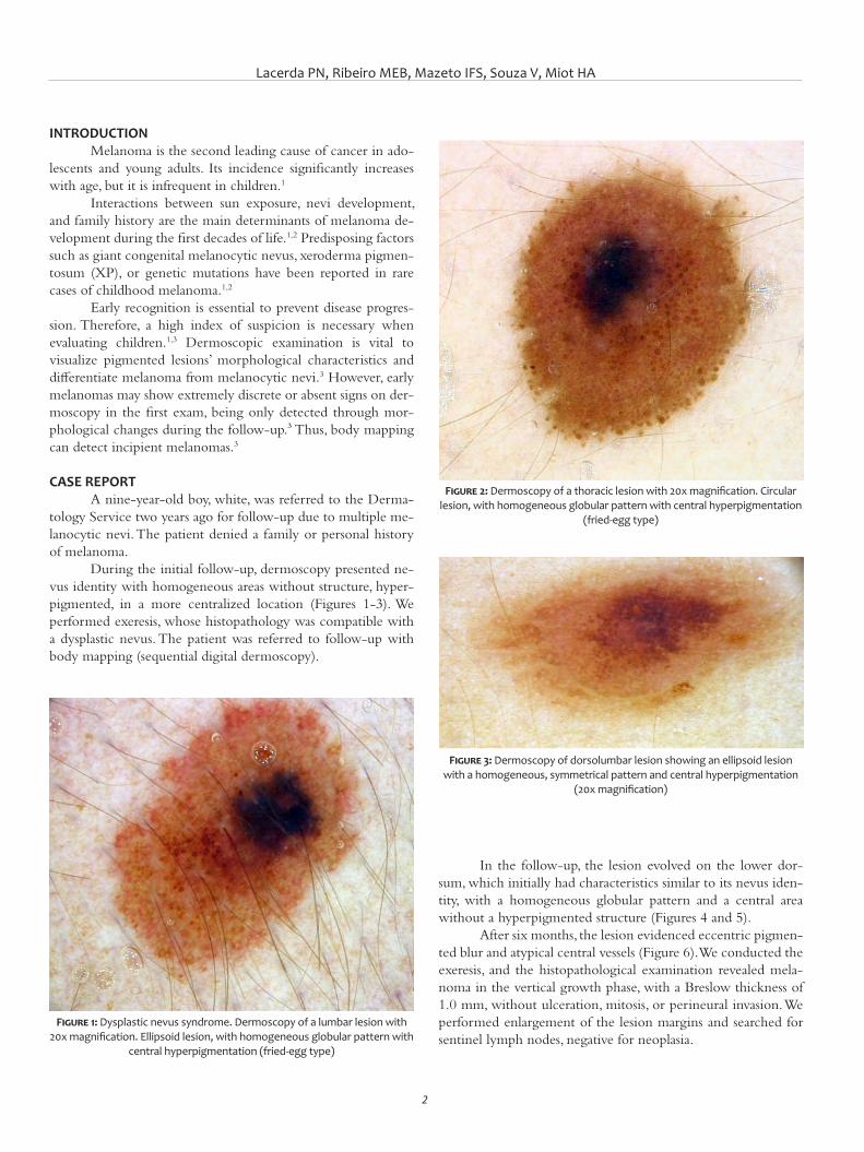
Lacerda PN, Ribeiro MEB, Mazeto IFS, Souza V, Miot HA
2
INTRODUCTIONMelanoma is the second leading cause of cancer in ado-
lescents and young adults. Its incidence significantly increases with age, but it is infrequent in children.1
Interactions between sun exposure, nevi development, and family history are the main determinants of melanoma de-velopment during the first decades of life.1,2 Predisposing factors such as giant congenital melanocytic nevus, xeroderma pigmen-tosum (XP), or genetic mutations have been reported in rare cases of childhood melanoma.1,2
Early recognition is essential to prevent disease progres-sion. Therefore, a high index of suspicion is necessary when evaluating children.1,3 Dermoscopic examination is vital to visualize pigmented lesions’ morphological characteristics and differentiate melanoma from melanocytic nevi.3 However, early melanomas may show extremely discrete or absent signs on der-moscopy in the first exam, being only detected through mor-phological changes during the follow-up.³ Thus, body mapping can detect incipient melanomas.3
CASE REPORT A nine-year-old boy, white, was referred to the Derma-
tology Service two years ago for follow-up due to multiple me-lanocytic nevi. The patient denied a family or personal history of melanoma.
During the initial follow-up, dermoscopy presented ne-vus identity with homogeneous areas without structure, hyper-pigmented, in a more centralized location (Figures 1-3). We performed exeresis, whose histopathology was compatible with a dysplastic nevus. The patient was referred to follow-up with body mapping (sequential digital dermoscopy).
In the follow-up, the lesion evolved on the lower dor-sum, which initially had characteristics similar to its nevus iden-tity, with a homogeneous globular pattern and a central area without a hyperpigmented structure (Figures 4 and 5).
After six months, the lesion evidenced eccentric pigmen-ted blur and atypical central vessels (Figure 6). We conducted the exeresis, and the histopathological examination revealed mela-noma in the vertical growth phase, with a Breslow thickness of 1.0 mm, without ulceration, mitosis, or perineural invasion. We performed enlargement of the lesion margins and searched for sentinel lymph nodes, negative for neoplasia.
Figure 1: Dysplastic nevus syndrome. Dermoscopy of a lumbar lesion with 20x magnification. Ellipsoid lesion, with homogeneous globular pattern with
central hyperpigmentation (fried-egg type)
Figure 2: Dermoscopy of a thoracic lesion with 20x magnification. Circular lesion, with homogeneous globular pattern with central hyperpigmentation
(fried-egg type)
Figure 3: Dermoscopy of dorsolumbar lesion showing an ellipsoid lesion with a homogeneous, symmetrical pattern and central hyperpigmentation
(20x magnification)
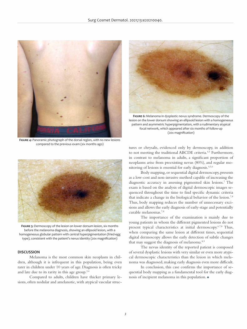
Surg Cosmet Dermatol. 2021;13:e20210040.
3
tures or chrysalis, evidenced only by dermoscopy, in addition to not meeting the traditional ABCDE criteria.4,5 Furthermore, in contrast to melanoma in adults, a significant proportion of neoplasms arise from preexisting nevus (80%), and regular mo-nitoring of lesions is essential for early diagnosis.4,5,6
Body mapping, or sequential digital dermoscopy, presents as a low-cost and non-invasive method capable of increasing the diagnostic accuracy in assessing pigmented skin lesions.7 The exam is based on the analysis of digital dermoscopic images se-quenced throughout the time to find specific dynamic criteria that indicate a change in the biological behavior of the lesion.7,8 Thus, body mapping reduces the number of unnecessary exci-sions and allows the early diagnosis of early-stage and potentially curable melanomas.7,8
The importance of the examination is mainly due to young patients in whom the different pigmented lesions do not present typical characteristics at initial dermoscopy.6,7,8 Thus, when comparing the same lesion at different times, sequential digital dermoscopy allows the early detection of subtle changes that may suggest the diagnosis of melanoma.8,9
The nevus identity of the reported patient is composed of several dysplastic lesions with very similar or even more atypi-cal dermoscopic characteristics than the lesion in which mela-noma was diagnosed, making early diagnosis even more difficult.
In conclusion, this case confirms the importance of se-quential body mapping as a fundamental tool for the early diag-nosis of incipient melanoma in this population. l
DISCUSSION Melanoma is the most common skin neoplasm in chil-
dren, although it is infrequent in this population, being even rarer in children under 10 years of age. Diagnosis is often tricky and late due to its rarity in this age group.3,4
Compared to adults, children have thicker primary le-sions, often nodular and amelanotic, with atypical vascular struc-
Figure 4: Panoramic photograph of the dorsal region, with no new lesions compared to the previous exam (six months ago)
Figure 6: Melanoma in dysplastic nevus syndrome. Dermoscopy of the lesion on the lower dorsum showing an ellipsoid lesion with a homogeneous
pattern and asymmetric hyperpigmentation, with a rudimentary atypical focal network, which appeared after six months of follow-up
(20x magnification)
Figure 5: Dermoscopy of the lesion on lower dorsum lesion, six months before the melanoma diagnosis, showing an ellipsoid lesion, with a
homogeneous globular pattern with central hyperpigmentation (fried-egg type), consistent with the patient’s nevus identity (20x magnification)
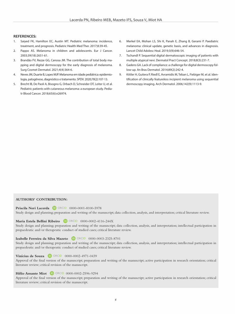
Lacerda PN, Ribeiro MEB, Mazeto IFS, Souza V, Miot HA
4
AUTHORS' CONTRIBUTION:
Priscila Neri Lacerda 0000-0001-8100-5978Study design and planning; preparation and writing of the manuscript; data collection, analysis, and interpretation; critical literature review.
Maria Estela Bellini Ribeiro 0000-0002-4116-244XStudy design and planning; preparation and writing of the manuscript; data collection, analysis, and interpretation; intellectual participation in propaedeutic and/or therapeutic conduct of studied cases; critical literature review.
Izabelle Ferreira da Silva Mazeto 0000-0003-2325-8701Study design and planning; preparation and writing of the manuscript; data collection, analysis, and interpretation; intellectual participation in propaedeutic and/or therapeutic conduct of studied cases; critical literature review.
Vinícius de Souza 0000-0002-4971-0439Approval of the final version of the manuscript; preparation and writing of the manuscript; active participation in research orientation; critical literature review; critical revision of the manuscript.
Hélio Amante Miot 0000-0002-2596-9294Approval of the final version of the manuscript; preparation and writing of the manuscript; active participation in research orientation; critical literature review; critical revision of the manuscript.
REFERENCES:1. Saiyed FK, Hamilton EC, Austin MT. Pediatric melanoma: incidence,
treatment, and prognosis. Pediatric Health Med Ther. 2017;8:39-45.2. Pappo AS. Melanoma in children and adolescents. Eur J Cancer.
2003;39(18):2651-61.3. Brandão FV, Rezze GG, Canosa JM. The contribution of total body ma-
pping and digital dermoscopy for the early diagnosis of melanoma. Surg Cosmet Dermatol. 2021;4(4):364-6.
4. Neves JM, Duarte B, Lopes MJP. Melanoma em idade pediátrica: epidemio-logia, patogênese, diagnóstico e tratamento. SPDV. 2020;78(2):107-13.
5. Brecht IB, De Paoli A, Bisogno G, Orbach D, Schneider DT, Leiter U, et al. Pediatric patients with cutaneous melanoma: a european study. Pedia-tr Blood Cancer. 2018;65(6):e26974.
6. Merkel EA, Mohan LS, Shi K, Panah E, Zhang B, Gerami P. Paediatric melanoma: clinical update, genetic basis, and advances in diagnosis. Lancet Child Adolesc Heal. 2019;3(9):646-54.
7. Tschandl P. Sequential digital dermatoscopic imaging of patients with multiple atypical nevi. Dermatol Pract Concept. 2018;8(3):231-7.
8. Gadens GA. Lack of compliance: a challenge for digital dermoscopy fol-low-up. An Bras Dermatol. 2014;89(2):242-4.
9. Kittler H, Guitera P, Riedl E, Avramidis M, Teban L, Fiebiger M, et al. Iden-tification of clinically featureless incipient melanoma using sequential dermoscopy imaging. Arch Dermatol. 2006;142(9):1113-9.
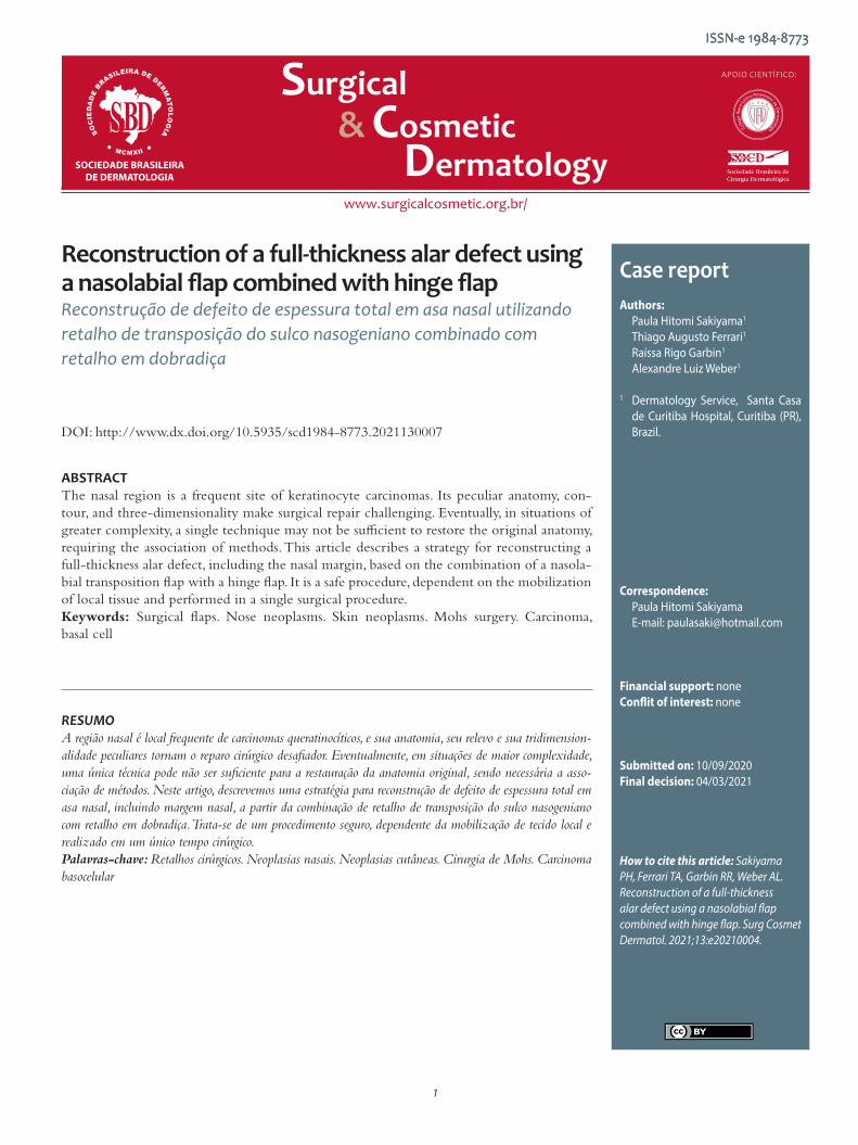
1
APOIO CIENTÍFICO:
www.surgicalcosmetic.org.br/
ISSN-e 1984-8773
Case reportAuthors: Paula Hitomi Sakiyama1
Thiago Augusto Ferrari1
Raíssa Rigo Garbin1
Alexandre Luiz Weber1
1 Dermatology Service, Santa Casa de Curitiba Hospital, Curitiba (PR), Brazil.
Correspondence: Paula Hitomi Sakiyama E-mail: [email protected]
Financial support: noneConflit of interest: none
Submitted on: 10/09/2020Final decision: 04/03/2021
How to cite this article: Sakiyama PH, Ferrari TA, Garbin RR, Weber AL. Reconstruction of a full-thickness alar defect using a nasolabial flap combined with hinge flap. Surg Cosmet Dermatol. 2021;13:e20210004.
Reconstruction of a full-thickness alar defect using a nasolabial flap combined with hinge flap Reconstrução de defeito de espessura total em asa nasal utilizando retalho de transposição do sulco nasogeniano combinado com retalho em dobradiça
ABSTRACTThe nasal region is a frequent site of keratinocyte carcinomas. Its peculiar anatomy, con-tour, and three-dimensionality make surgical repair challenging. Eventually, in situations of greater complexity, a single technique may not be sufficient to restore the original anatomy, requiring the association of methods. This article describes a strategy for reconstructing a full-thickness alar defect, including the nasal margin, based on the combination of a nasola-bial transposition flap with a hinge flap. It is a safe procedure, dependent on the mobilization of local tissue and performed in a single surgical procedure.Keywords: Surgical flaps. Nose neoplasms. Skin neoplasms. Mohs surgery. Carcinoma, basal cell
RESU MOA região nasal é local frequente de carcinomas queratinocíticos, e sua anatomia, seu relevo e sua tridimension-alidade peculiares tornam o reparo cirúrgico desafiador. Eventualmente, em situações de maior complexidade, uma única técnica pode não ser suficiente para a restauração da anatomia original, sendo necessária a asso-ciação de métodos. Neste artigo, descrevemos uma estratégia para reconstrução de defeito de espessura total em asa nasal, incluindo margem nasal, a partir da combinação de retalho de transposição do sulco nasogeniano com retalho em dobradiça. Trata-se de um procedimento seguro, dependente da mobilização de tecido local e realizado em um único tempo cirúrgico.Palavras-chave: Retalhos cirúrgicos. Neoplasias nasais. Neoplasias cutâneas. Cirurgia de Mohs. Carcinoma basocelular
DOI: http://www.dx.doi.org/10.5935/scd1984-8773.2021130007
ISSN-e 1984-8773
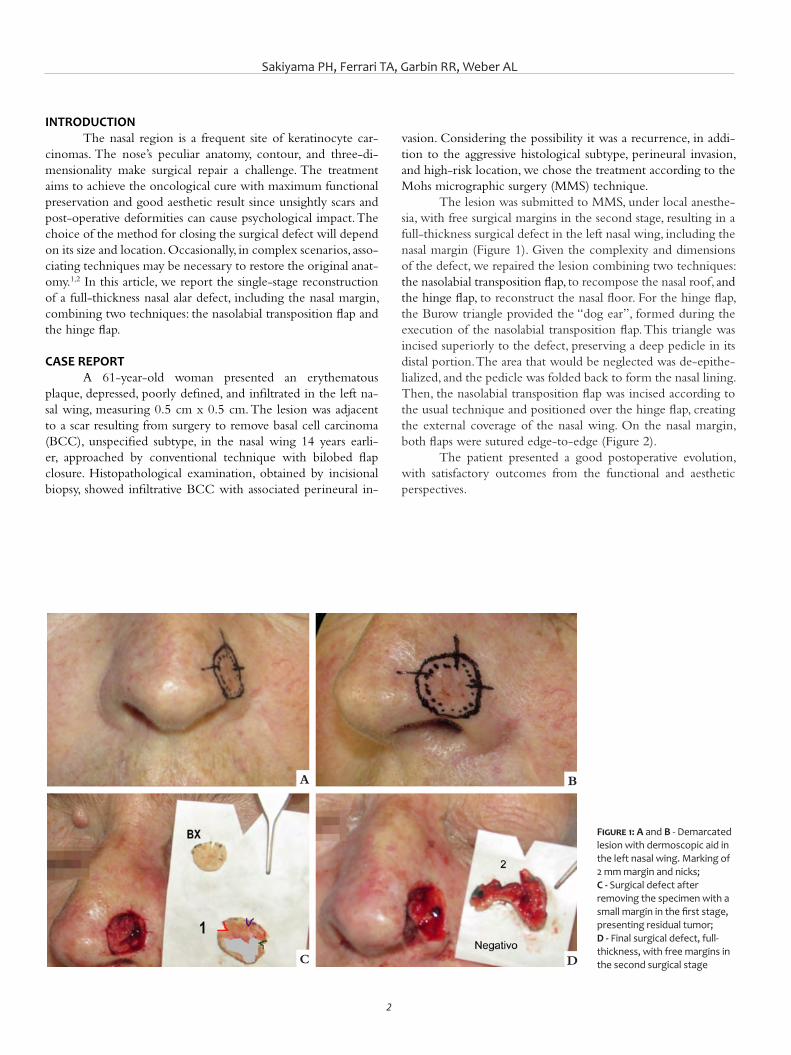
Sakiyama PH, Ferrari TA, Garbin RR, Weber AL
2
INTRODUCTIONThe nasal region is a frequent site of keratinocyte car-
cinomas. The nose’s peculiar anatomy, contour, and three-di-mensionality make surgical repair a challenge. The treatment aims to achieve the oncological cure with maximum functional preservation and good aesthetic result since unsightly scars and post-operative deformities can cause psychological impact. The choice of the method for closing the surgical defect will depend on its size and location. Occasionally, in complex scenarios, asso-ciating techniques may be necessary to restore the original anat-omy.1,2 In this article, we report the single-stage reconstruction of a full-thickness nasal alar defect, including the nasal margin, combining two techniques: the nasolabial transposition flap and the hinge flap.
CASE REPORTA 61-year-old woman presented an erythematous
plaque, depressed, poorly defined, and infiltrated in the left na-sal wing, measuring 0.5 cm x 0.5 cm. The lesion was adjacent to a scar resulting from surgery to remove basal cell carcinoma (BCC), unspecified subtype, in the nasal wing 14 years earli-er, approached by conventional technique with bilobed flap closure. Histopathological examination, obtained by incisional biopsy, showed infiltrative BCC with associated perineural in-
vasion. Considering the possibility it was a recurrence, in addi-tion to the aggressive histological subtype, perineural invasion, and high-risk location, we chose the treatment according to the Mohs micrographic surgery (MMS) technique.
The lesion was submitted to MMS, under local anesthe-sia, with free surgical margins in the second stage, resulting in a full-thickness surgical defect in the left nasal wing, including the nasal margin (Figure 1). Given the complexity and dimensions of the defect, we repaired the lesion combining two techniques: the nasolabial transposition flap, to recompose the nasal roof, and the hinge flap, to reconstruct the nasal floor. For the hinge flap, the Burow triangle provided the “dog ear”, formed during the execution of the nasolabial transposition flap. This triangle was incised superiorly to the defect, preserving a deep pedicle in its distal portion. The area that would be neglected was de-epithe-lialized, and the pedicle was folded back to form the nasal lining. Then, the nasolabial transposition flap was incised according to the usual technique and positioned over the hinge flap, creating the external coverage of the nasal wing. On the nasal margin, both flaps were sutured edge-to-edge (Figure 2).
The patient presented a good postoperative evolution, with satisfactory outcomes from the functional and aesthetic perspectives.
Figure 1: A and B - Demarcated lesion with dermoscopic aid in the left nasal wing. Marking of 2 mm margin and nicks; C - Surgical defect after removing the specimen with a small margin in the first stage, presenting residual tumor; D - Final surgical defect, full-thickness, with free margins in the second surgical stage
A
C
B
D
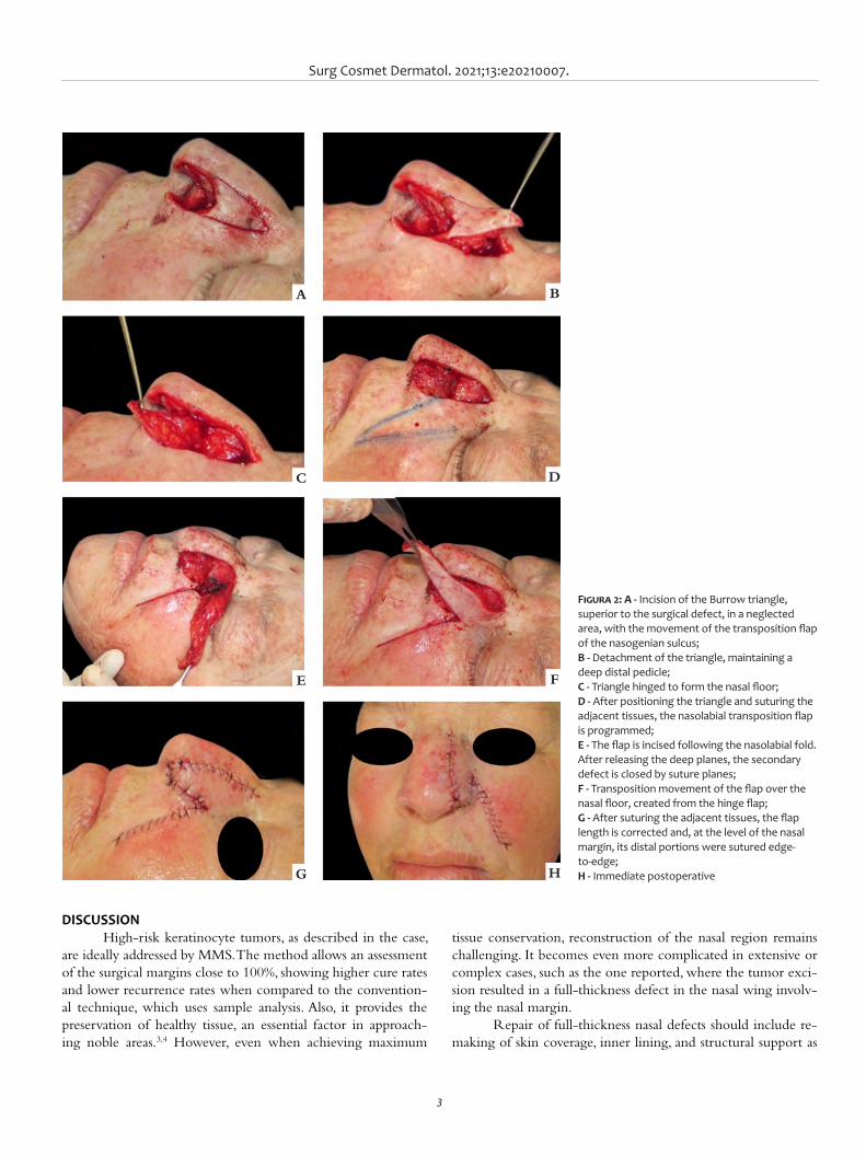
Surg Cosmet Dermatol. 2021;13:e20210007.
3
DISCUSSIONHigh-risk keratinocyte tumors, as described in the case,
are ideally addressed by MMS. The method allows an assessment of the surgical margins close to 100%, showing higher cure rates and lower recurrence rates when compared to the convention-al technique, which uses sample analysis. Also, it provides the preservation of healthy tissue, an essential factor in approach-ing noble areas.3,4 However, even when achieving maximum
tissue conservation, reconstruction of the nasal region remains challenging. It becomes even more complicated in extensive or complex cases, such as the one reported, where the tumor exci-sion resulted in a full-thickness defect in the nasal wing involv-ing the nasal margin.
Repair of full-thickness nasal defects should include re-making of skin coverage, inner lining, and structural support as
Figura 2: A - Incision of the Burrow triangle, superior to the surgical defect, in a neglected area, with the movement of the transposition flap of the nasogenian sulcus; B - Detachment of the triangle, maintaining a deep distal pedicle; C - Triangle hinged to form the nasal floor; D - After positioning the triangle and suturing the adjacent tissues, the nasolabial transposition flap is programmed; E - The flap is incised following the nasolabial fold. After releasing the deep planes, the secondary defect is closed by suture planes;F - Transposition movement of the flap over the nasal floor, created from the hinge flap; G - After suturing the adjacent tissues, the flap length is corrected and, at the level of the nasal margin, its distal portions were sutured edge-to-edge; H - Immediate postoperative
A
C
E
G
B
D
F
H
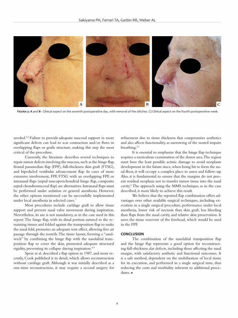
Sakiyama PH, Ferrari TA, Garbin RR, Weber AL
4
refinement due to tissue thickness that compromises aesthetics and also affects functionality, as narrowing of the nostril impairs breathing.8,1
It is essential to emphasize that the hinge flap technique requires a meticulous examination of the donor area. The region must have the least possible actinic damage to avoid neoplasm development in the future since, when being hit to form the na-sal floor, it will occupy a complex place to assess and follow-up. Also, it is fundamental to ensure that the margins do not pres-ent residual neoplasia not to transfer tumor tissue into the nasal cavity.5 The approach using the MMS technique, as in the case described, is more likely to achieve this result.
We believe that the reported flap combination offers ad-vantages over other available surgical techniques, including ex-ecution in a single surgical procedure, performance under local anesthesia, lower risk of necrosis than skin graft, less bleeding than flaps from the nasal cavity, and relative skin preservation. It saves the tissue reservoir of the forehead, which would be used in the FPF.
CONCLUSIONThe combination of the nasolabial transposition flap
and the hinge flap represents a good option for reconstruct-ing full-thickness alar defects, including those affecting the nasal margin, with satisfactory aesthetic and functional outcomes. It is a safe method, dependent on the mobilization of local tissue for its execution, and performed in a single surgical time, thus reducing the costs and morbidity inherent to additional proce-dures. l
needed.5,6 Failure to provide adequate mucosal support in more significant defects can lead to scar contraction and/or flaws in overlapping flaps or grafts structure, making this step the most critical of the procedure.
Currently, the literature describes several techniques to repair minor defects involving the mucosa, such as the hinge flap, frontal paramedian flap (FPF), full-thickness skin graft (FTSG), and bipedicled vestibular advancement flap. In cases of more extensive involvement, FPF, FTSG with an overlapping FPF, or intranasal flaps (septal mucoperichondrial hinge flap, composite septal chondromucosal flap) are alternatives. Intranasal flaps must be performed under sedation or general anesthesia. However, the other options mentioned can be successfully implemented under local anesthesia in selected cases.7
Most procedures include cartilage graft to allow tissue support and prevent nasal valve movement during inspiration. Nevertheless, its use is not mandatory, as in the case used in this report. The hinge flap, with its distal portion sutured to the re-maining tissues and folded against the transposition flap to make the nasal fold, promotes an adequate tent effect, allowing free air passage through the nostrils. The tissue layout, forming a “sand-wich” by combining the hinge flap with the nasolabial trans-position flap to cover the skin, promoted adequate structural rigidity, preventing its collapse during inspiration.8,9
Spear et al. described a flap option in 1987, and more re-cently, Cook published it in detail, which allows reconstruction without cartilage graft. Although it was initially described as a one-time reconstruction, it may require a second surgery for
Figura 3: A and B - Clinical aspect on the seventh postoperative day, with removal of the stitches. C) Clinical aspect on the fourth postoperative week
A B C
Surg Cosmet Dermatol. 2021;13:e20210007.
5
AUTHORS' CONTRIBUTION:
Paula Hitomi Sakiyama | 0000-0001-7813-8294Approval of the final version of the manuscript; study design and planning; preparation and writing of the manuscript; data collec-tion, analysis, and interpretation; critical literature review; critical revision of the manuscript.
Thiago Augusto Ferrari | 0000-0003-4874-4837Approval of the final version of the manuscript; study design and planning; preparation and writing of the manuscript; data collec-tion, analysis, and interpretation; critical literature review; critical revision of the manuscript.
Raíssa Rigo Garbin | 0000-0002-9771-1209Approval of the final version of the manuscript; study design and planning; data collection, analysis, and interpretation; active par-ticipation in research orientation; intellectual participation in propaedeutic and/or therapeutic conduct of studied cases; critical literature review; critical revision of the manuscript.
Alexandre Luiz Weber | 0000-0002-4862-5777Approval of the final version of the manuscript; study design and planning; data collection, analysis, and interpretation; active par-ticipation in research orientation; intellectual participation in propaedeutic and/or therapeutic conduct of studied cases; critical literature review; critical revision of the manuscript.
REFERENCES1. Joseph AW, Truesdale C, Baker SR. Reconstruction of the Nose. Clin
North Am. 2020;27(1):43-54.2. Cerci FB, Kubo E. Reconstrução nasal após cirurgia micrográfica de
Mohs: análise de 208 casos. Surg Cosmet Dermatol. 2020;12(1):42-50.3. Cernea SS, Gontijo G, Pimentel ERA, Tarlé RG, Tassara G, Ferreira JASLB,
et al. Diretrizes de indicações de cirurgia micrográfica de Mohs nos tu-mores da pele. An Bras Dermatol. 2016;91(5):621-7.
4. Tolkachjov SN, Brodland DG, Coldiron BM, Fazio MJ, Hruza GJ, Roe-nigk RK, et al. Understanding Mohs micrographic surgery: a re-view and practical guide for the nondermatologist. Mayo Clin Proc. 2017;92(8):1261-71.
5. Taeda A, Akimoto M, Park K, Kounoike N, Shimakura Y, Nemoto M, et al. Single- stage reconstruction of a full-thickness alar defect using a folded nasolabial flap combined with a redundant skin turnover flap. J Craniofac Surg. 2014;25(6):2144-6.
6. Bedwani NH, Rizkalla M. Single-stage reconstruction of full-thickness nasal alar defect using bilobed and turnover flaps. J Craniofac Sugr. 2020;31(2):e169-71.
7. Cerci FB, Dellatorre G. Retalho paramediano frontal associado a retalho em dobradiça para reparo de defeito em ponta nasal. An Bras Derma-tol. 2016;91(5 Supl 1):S140-3.
8. Sanchez FH, Estefan JL, Fernandez ID. Reconstrução completa de asa nasal com retalho de Spear após cirurgia micrográfica de Mohs no tratamento de carcinoma basocelular. Surg Cosmet Dermatol 2013;5(4):3559.
9. Sanchez FH, Estefan JL, Fernandez ID. Reconstrução nasal complexa com combinação de retalhos bilaterais em dobradiça e retalho pa-ramediano frontal, após cirurgia de Mohs. Surg Cosmet Dermatol 2013;5(4):355-9.
10. Lane JE, Hsia LB, Merritt BG. Reconstruction of large transmural nasal defects with a nasolabial turnover interpolation flap. Dermatol Surg. 2020;46(7):899-903.
1
APOIO CIENTÍFICO:
www.surgicalcosmetic.org.br/
ISSN-e 1984-8773
Case reportAuthors: Katherine Santacoloma1
Barbara Ciraudo1
Marcela Duarte Benez Miller1
Guillermo Loda1
1 Santa Casa de Misericórdia do Rio de Janeiro, Prof. Rubem David Azulay Institute of Dermatology, Rio de Janeiro (RJ), Brazil.
Correspondence: Katherine Santacoloma E-mail: [email protected]
Financial support: Nenhuma. Conflit of interest: Nenhum.
Submitted on: 04/09/2020Approved on: 04/03/2021
How to cite this article: Santacolo-ma K, Ciraudo B, Miller MDB, Loda G. East-west advancement flap: a useful tool to reconstruct nasal tip defects. Surg Cosmet Dermatol. 2021;13:e20210010.
East-west advancement flap: a useful tool to reconstruct nasal tip defects Retalho leste-oeste: uma ferramenta útil para reconstruir defeitos na ponta nasal
ABSTRACTThe reconstruction of nasal defects secondary to non-melanoma skin cancer represents a surgical challenge, especially in the nasal tip, due to its limited local laxity and possible asymmetries. There are multiple techniques to perform this closure, but most are flaps from distant locations that can lead to less aesthetic results. We present two cases of basal cell carcinoma on the nasal tip, where reconstruction with the east-west advancement flap was performed after tumor excision, with a modification of the flap in one of the patients, obtaining a good structural and aesthetic result.Keywords: Surgical flaps; Carcinoma, basal cell; Nose; Dermatologic surgical procedures
RESUMOA reconstrução de defeitos nasais secundários ao câncer de pele não melanoma representa um desafio cirúrgico, especialmente na ponta nasal, pela sua limitada frouxidão local e possíveis assimetrias. Ex-istem múltiplas técnicas para realizar esse fechamento, mas a maioria são retalhos de locais distantes que podem levar a resultados menos estéticos. Apresentamos dois casos de pacientes masculinos com carcinoma basocelular na ponta nasal, onde foi feita uma reconstrução com o retalho leste-oeste após exérese do tumor, com uma modificação no retalho em um dos pacientes, obtendo-se um bom resultado estrutural e estético.Palavras-chave: Retalhos cirúrgicos; Carcinoma basocelular; Nariz; Procedimentos cirúrgicos dermatológicos
DOI: http://www.dx.doi.org/10.5935/scd1984-8773.2021130010
ISSN-e 1984-8773
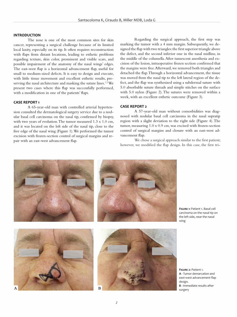
Santacoloma K, Ciraudo B, Miller MDB, Loda G
2
INTRODUCTIONThe nose is one of the most common sites for skin
cancer, representing a surgical challenge because of its limited local laxity, especially on its tip. It often requires reconstruction with flaps from distant locations, leading to esthetic problems regarding texture, skin color, prominent and visible scars, and possible impairment of the anatomy of the nasal wings’ edges. The east-west flap is a horizontal advancement flap, useful for small to medium-sized defects. It is easy to design and execute, with little tissue movement and excellent esthetic results, pre-serving the nasal architecture and masking the suture lines.1,2 We present two cases where this flap was successfully performed, with a modification in one of the patients’ flaps.
CASE REPORT 1A 65-year-old man with controlled arterial hyperten-
sion consulted the dermatological surgery service due to a nod-ular basal cell carcinoma on the nasal tip, confirmed by biopsy, with two years of evolution. The tumor measured 1.3 x 1.0 cm, and it was located on the left side of the nasal tip, close to the free edge of the nasal wing (Figure 1). We performed the tumor excision with frozen-section control of surgical margins and re-pair with an east-west advancement flap.
Regarding the surgical approach, the first step was marking the tumor with a 4 mm margin. Subsequently, we de-signed the flap with two triangles: the first superior triangle above the defect, and the second inferior one in the nasal midline, in the middle of the columella. After tumescent anesthesia and ex-cision of the lesion, intraoperative frozen section confirmed that the margins were free. Afterward, we removed both triangles and detached the flap. Through a horizontal advancement, the tissue was moved from the nasal tip to the left lateral region of the de-fect, and the flap was synthesized using a subdermal suture with 5.0 absorbable suture threads and simple stitches on the surface with 5.0 nylon (Figure 2). The sutures were removed within a week, with an excellent esthetic outcome (Figure 3).
CASE REPORT 2A 57-year-old man without comorbidities was diag-
nosed with nodular basal cell carcinoma in the nasal supratip region with a slight deviation to the right side (Figure 4). The tumor, measuring 1.0 x 0.9 cm, was excised with frozen-section control of surgical margins and closure with an east-west ad-vancement flap.
We chose a surgical approach similar to the first patient; however, we modified the flap design. In this case, the first tri-
Figure 1: Patient 1. Basal cell carcinoma on the nasal tip on the left side, near the nasal wing
Figure 2: Patient 1. A - Tumor demarcation and east-west advancement flap design. B - Immediate results after surgeryA B
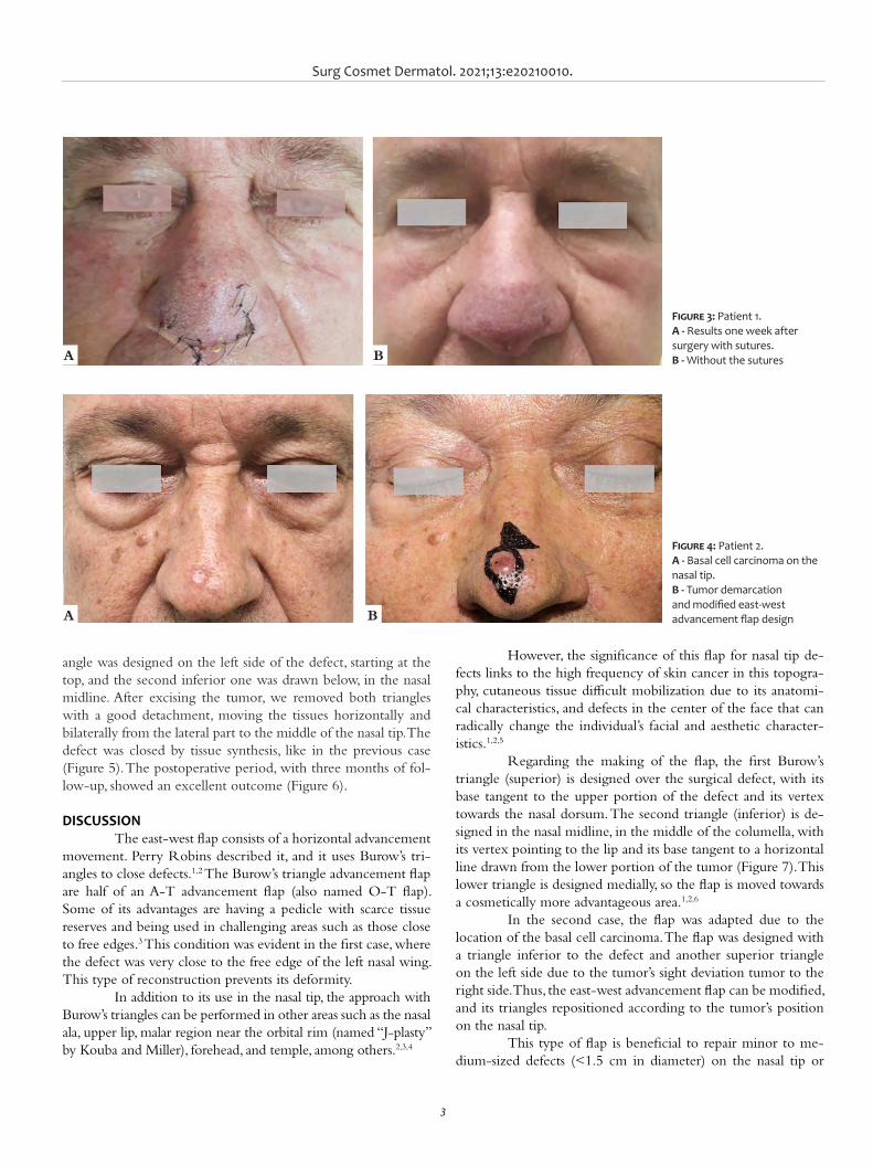
Surg Cosmet Dermatol. 2021;13:e20210010.
3
Figure 3: Patient 1. A - Results one week after surgery with sutures. B - Without the sutures
Figure 4: Patient 2. A - Basal cell carcinoma on the nasal tip. B - Tumor demarcation and modified east-west advancement flap design
angle was designed on the left side of the defect, starting at the top, and the second inferior one was drawn below, in the nasal midline. After excising the tumor, we removed both triangles with a good detachment, moving the tissues horizontally and bilaterally from the lateral part to the middle of the nasal tip. The defect was closed by tissue synthesis, like in the previous case (Figure 5). The postoperative period, with three months of fol-low-up, showed an excellent outcome (Figure 6).
DISCUSSIONThe east-west flap consists of a horizontal advancement
movement. Perry Robins described it, and it uses Burow’s tri-angles to close defects.1,2 The Burow’s triangle advancement flap are half of an A-T advancement flap (also named O-T flap). Some of its advantages are having a pedicle with scarce tissue reserves and being used in challenging areas such as those close to free edges.3 This condition was evident in the first case, where the defect was very close to the free edge of the left nasal wing. This type of reconstruction prevents its deformity.
In addition to its use in the nasal tip, the approach with Burow’s triangles can be performed in other areas such as the nasal ala, upper lip, malar region near the orbital rim (named “J-plasty” by Kouba and Miller), forehead, and temple, among others.2,3,4
However, the significance of this flap for nasal tip de-fects links to the high frequency of skin cancer in this topogra-phy, cutaneous tissue difficult mobilization due to its anatomi-cal characteristics, and defects in the center of the face that can radically change the individual’s facial and aesthetic character-istics.1,2,5
Regarding the making of the flap, the first Burow’s triangle (superior) is designed over the surgical defect, with its base tangent to the upper portion of the defect and its vertex towards the nasal dorsum. The second triangle (inferior) is de-signed in the nasal midline, in the middle of the columella, with its vertex pointing to the lip and its base tangent to a horizontal line drawn from the lower portion of the tumor (Figure 7). This lower triangle is designed medially, so the flap is moved towards a cosmetically more advantageous area.1,2,6
In the second case, the flap was adapted due to the location of the basal cell carcinoma. The flap was designed with a triangle inferior to the defect and another superior triangle on the left side due to the tumor’s sight deviation tumor to the right side. Thus, the east-west advancement flap can be modified, and its triangles repositioned according to the tumor’s position on the nasal tip.
This type of flap is beneficial to repair minor to me-dium-sized defects (<1.5 cm in diameter) on the nasal tip or
A
A
B
B
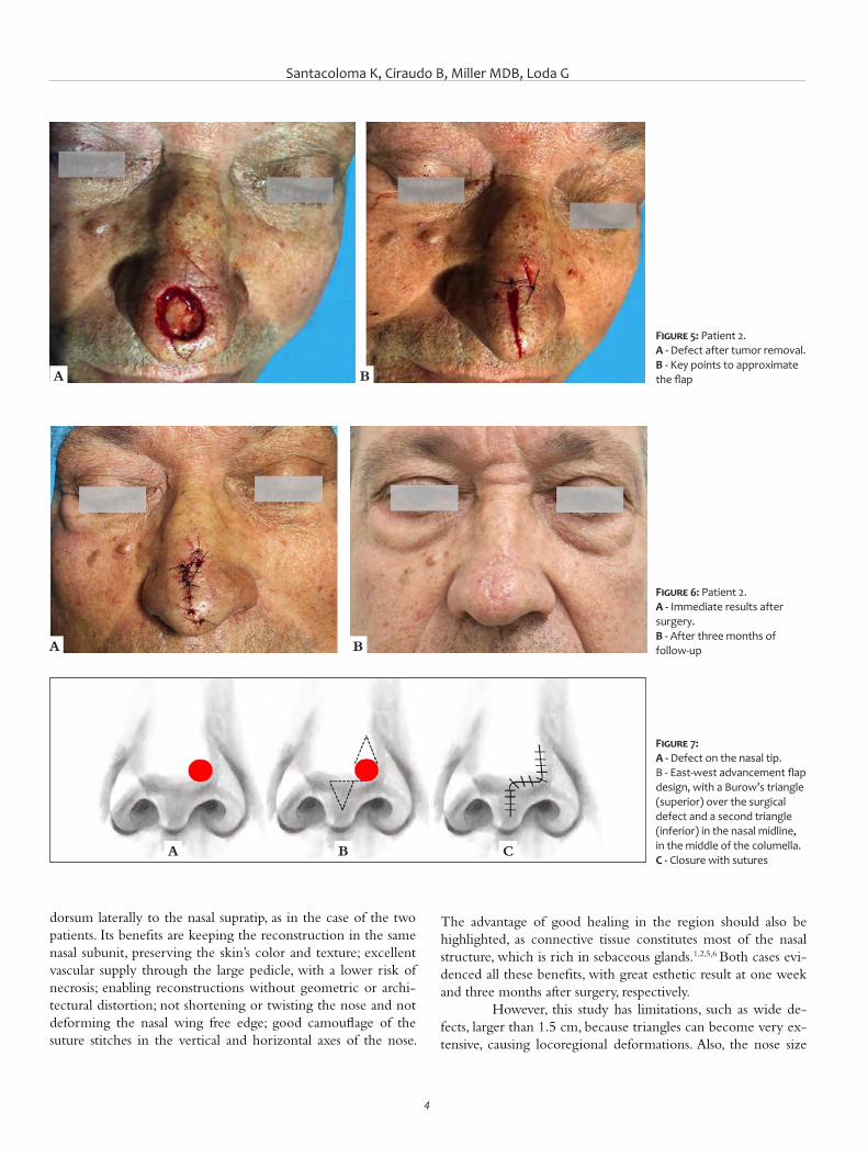
Santacoloma K, Ciraudo B, Miller MDB, Loda G
4
dorsum laterally to the nasal supratip, as in the case of the two patients. Its benefits are keeping the reconstruction in the same nasal subunit, preserving the skin’s color and texture; excellent vascular supply through the large pedicle, with a lower risk of necrosis; enabling reconstructions without geometric or archi-tectural distortion; not shortening or twisting the nose and not deforming the nasal wing free edge; good camouflage of the suture stitches in the vertical and horizontal axes of the nose.
The advantage of good healing in the region should also be highlighted, as connective tissue constitutes most of the nasal structure, which is rich in sebaceous glands.1,2,5,6 Both cases evi-denced all these benefits, with great esthetic result at one week and three months after surgery, respectively.
However, this study has limitations, such as wide de-fects, larger than 1.5 cm, because triangles can become very ex-tensive, causing locoregional deformations. Also, the nose size
Figure 5: Patient 2. A - Defect after tumor removal. B - Key points to approximate the flap
Figure 6: Patient 2. A - Immediate results after surgery. B - After three months of follow-up
Figure 7: A - Defect on the nasal tip. B - East-west advancement flap design, with a Burow’s triangle (superior) over the surgical defect and a second triangle (inferior) in the nasal midline, in the middle of the columella. C - Closure with sutures
A
A
A
B
B
B C
Surg Cosmet Dermatol. 2021;13:e20210010.
5
REFERENCES1. Goldberg LH, Alam M. Horizontal advancement flap for symmetric re-
construction of small to medium-sized cutaneous defects of the lateral nasal supratip. J Am Acad Dermatol. 2003;49(4):685-9.
2. Magliano J, Abelenda MP, Navarrete J, Bazzano C. East-west flap after Mohs micrographic surgery. Actas Dermosifiliogr. 2019;110(9):759-62.
3. Krishnan R, Garmn M, Nunez-Gussman J, Orengo I. Advancement flaps: a basic theme with many variations. Dermatol Surg. 2005;31(8):986-94.
AUTHORS' CONTRIBUTION:
Katherine Santacoloma 0000-0002-6645-7826Preparation and writing of the manuscript; data collection, analysis, and interpretation; critical literature review.
Barbara Ciraudo 0000-0003-0805-0160Study design and planning.
Marcela Duarte Benez Miller 0000-0003-0289-5656Approval of the final version of the manuscript; critical revision of the manuscript.
Guillermo Loda 0000-0003-0511-0025Approval of the final version of the manuscript.
4. Ascari-Raccagni A, Dondas A, Righini MG, Trevisan G. The east-west ad-vancement flap (horizontal advancement flap) to repair a defect on the nose ala. JEADV. 2010;24(8):926-29.
5. Summers BK, Siegle RJ. Facial cutaneous reconstructive surgery: facial flaps. J Am Acad Dermatol. 1993;29(6):917-41.
6. Geist DE, Malony ME. The "East-West" advancement flap for nasal de-fects: re examined and extended. Dermatol Surg. 2012;38(9):1529-34.
influences the technique choice, as wider noses carry larger de-fects, while smaller noses may require movement of distant lat-eral tissue, resulting in a tight nose appearance. 1,2,6
CONCLUSIONThe east-west advancement flap is an aesthetically ex-
cellent repair for small to medium-sized skin cancer defects on the nasal tip. In addition to being easy to design and execute, it
requires little tissue movement, hides the suture lines well, main-tains the color and texture of the nasal subunit skin, has a large pedicle reducing the risk of necrosis, and does not cause nose distortion. All of these benefits are critical because any defect in this area can seriously affect individual facial features. In well-se-lected cases, the result of this type of horizontal advancement flap may be superior to the rotation flap, which has long inci-sions that are difficult to disguise, or the transposition flap, which can cause tissue distortion. l
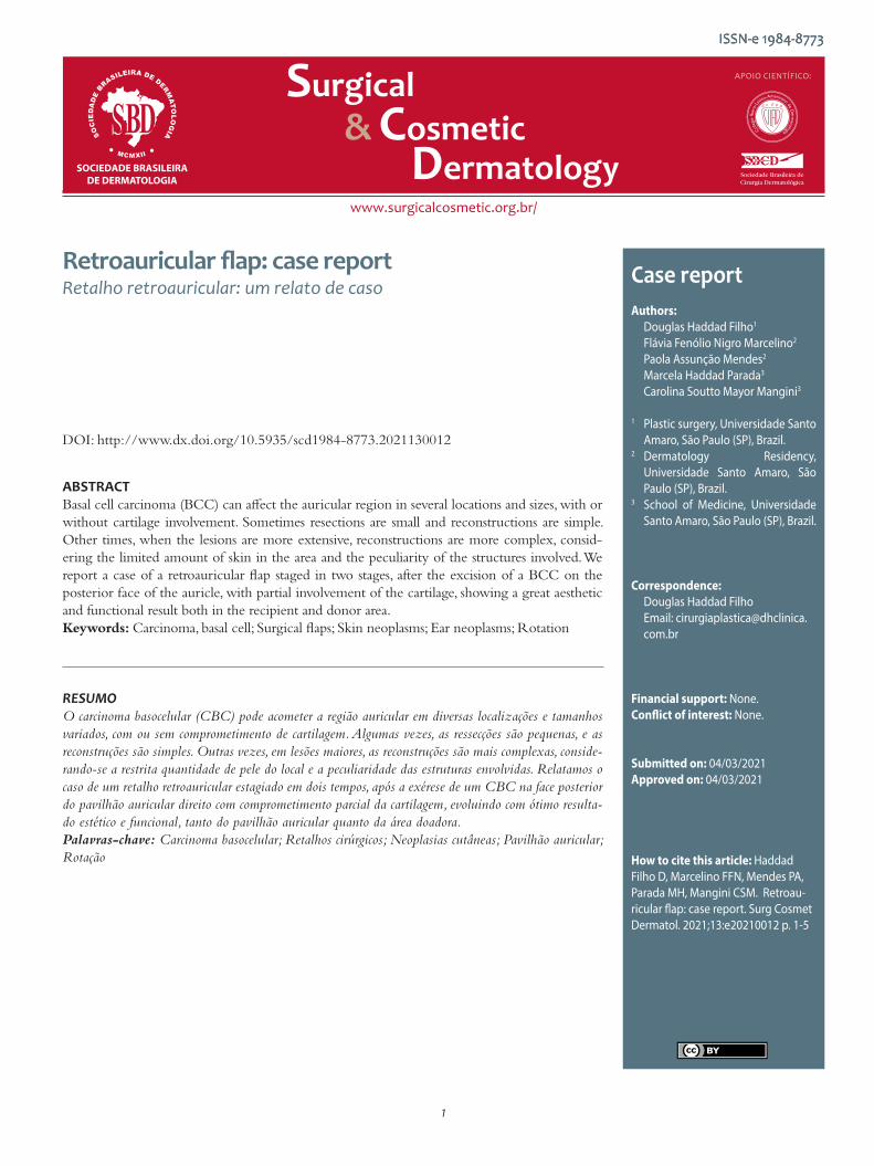
1
APOIO CIENTÍFICO:
www.surgicalcosmetic.org.br/
ISSN-e 1984-8773
Case reportAuthors: Douglas Haddad Filho1
Flávia Fenólio Nigro Marcelino2
Paola Assunção Mendes2
Marcela Haddad Parada3
Carolina Soutto Mayor Mangini3
1 Plastic surgery, Universidade Santo Amaro, São Paulo (SP), Brazil.
2 Dermatology Residency, Universidade Santo Amaro, São Paulo (SP), Brazil.
3 School of Medicine, Universidade Santo Amaro, São Paulo (SP), Brazil.
Correspondence: Douglas Haddad Filho Email: cirurgiaplastica@dhclinica. com.br
Financial support: None. Conflict of interest: None.
Submitted on: 04/03/2021 Approved on: 04/03/2021
How to cite this article: Haddad Filho D, Marcelino FFN, Mendes PA, Parada MH, Mangini CSM. Retroau-ricular flap: case report. Surg Cosmet Dermatol. 2021;13:e20210012 p. 1-5
Retroauricular flap: case reportRetalho retroauricular: um relato de caso
ABSTRACTBasal cell carcinoma (BCC) can affect the auricular region in several locations and sizes, with or without cartilage involvement. Sometimes resections are small and reconstructions are simple. Other times, when the lesions are more extensive, reconstructions are more complex, consid-ering the limited amount of skin in the area and the peculiarity of the structures involved. We report a case of a retroauricular flap staged in two stages, after the excision of a BCC on the posterior face of the auricle, with partial involvement of the cartilage, showing a great aesthetic and functional result both in the recipient and donor area.Keywords: Carcinoma, basal cell; Surgical flaps; Skin neoplasms; Ear neoplasms; Rotation
RESUMOO carcinoma basocelular (CBC) pode acometer a região auricular em diversas localizações e tamanhos variados, com ou sem comprometimento de cartilagem. Algumas vezes, as ressecções são pequenas, e as reconstruções são simples. Outras vezes, em lesões maiores, as reconstruções são mais complexas, conside-rando-se a restrita quantidade de pele do local e a peculiaridade das estruturas envolvidas. Relatamos o caso de um retalho retroauricular estagiado em dois tempos, após a exérese de um CBC na face posterior do pavilhão auricular direito com comprometimento parcial da cartilagem, evoluindo com ótimo resulta-do estético e funcional, tanto do pavilhão auricular quanto da área doadora.Palavras-chave: Carcinoma basocelular; Retalhos cirúrgicos; Neoplasias cutâneas; Pavilhão auricular; Rotação
DOI: http://www.dx.doi.org/10.5935/scd1984-8773.2021130012
ISSN-e 1984-8773
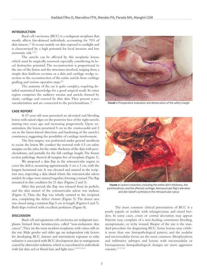
Haddad Filho D, Marcelino FFN, Mendes PA, Parada MH, Mangini CSM
2
INTRODUCTIONBasal cell carcinoma (BCC) is a malignant neoplasm that
mostly affects fair-skinned individuals, accounting for 70% of skin tumors.1,2 It occurs mainly on skin exposed to sunlight and is characterized by a high potential for local invasion and low metastatic risk.3,4,5
The auricle can be affected by this neoplastic lesion, which must be surgically removed, especially considering its lo-cal destructive potential. The reconstruction is proportional to the size of the lesion and the structures involved, ranging from a simple skin fusiform excision or a skin and cartilage wedge re-section to the reconstruction of the entire auricle from cartilage grafting and various operative steps.5,6
The anatomy of the ear is quite complex, requiring de-tailed anatomical knowledge for a good surgical result. Its outer region comprises the auditory meatus and auricle, formed by elastic cartilage and covered by thin skin. They present scarce vascularization and are connected to the perichondrium.1,6
CASE REPORTA 67-year-old man presented an ulcerated and bleeding
lesion with raised edges on the posterior face of the right auricle, starting two years ago and increasing progressively. Upon ex-amination, the lesion presented 5 cm in the craniocaudal and 2 cm in the latero-lateral direction, and hardening of the auricle’s consistency, suggesting the possibility of cartilage involvement.
The first surgery was performed under general anesthesia to excise the lesion. We conduct the removal with 0.5 cm safety margins on the sides, for the entire thickness of the skin with peri-chondrium, and partially for the full cartilage length. The frozen section pathology showed all margins free of neoplasia (Figure 1).
We proposed a skin flap in the retroauricular region to cover the defect, measuring approximately 4 cm x 2 cm, with the longest horizontal axis. It was elevated and sutured in the recip-ient area, respecting a skin island where the retroauricular sulcus resided. Its edges were sutured together, forming a tunnel. The flap remained in this condition for 21 days (Figures 2 and 3).
After this period, the flap was released from its pedicle, and the skin tunnel of the retroauricular sulcus was undone (Figure 4). Thus, the flap was wholly sutured in the recipient area, completing the defect closure (Figure 5). The donor area was closed using a rotation flap, 5 cm in length (Figures 6 and 7). Both flaps evolved with excellent perfusion (Figure 8).
DISCUSSIONBasal cell and squamous cell carcinoma are malignant neo-
plasms formed from keratinocytes, called “non-melanoma skin cancer”. They are the most incident neoplasms, with values still on the rise. Male gender and older age are independent risk factors for developing BCC. Intense and intermittent exposure to solar radiation is associated with BCC development due to mutagenesis caused by ultraviolet radiation, which is exacerbated in individuals with fair skin, red or blond hair, and light eyes.3,7,8,17,18,19
The most common clinical presentation of BCC is a pearly papule or nodule with telangiectasias and raised bor-ders. In some cases, crusts or central ulceration may appear. Patients may complain of a non-healing, sometimes bleeding, asymptomatic, or itchy wound. Biopsy of the site is the stan-dard procedure for diagnosing BCC. Some lesions may exhib-it more than one histopathological pattern, and the nodular and micronodular forms are the most common. Morpheaform and infiltrative subtypes and lesions with micronodular or basosquamous histopathological changes are more aggressive variants.3,7,17,18
Figure 1: Preoperative evaluation and demarcation of the safety margin
Figure 2: Lesion’s resection, including the entire skin’s thickness, the perichondrium, and the affected cartilage. Retroauricular flap’s elevation
and skin island’s synthesis in the retroauricular sulcus
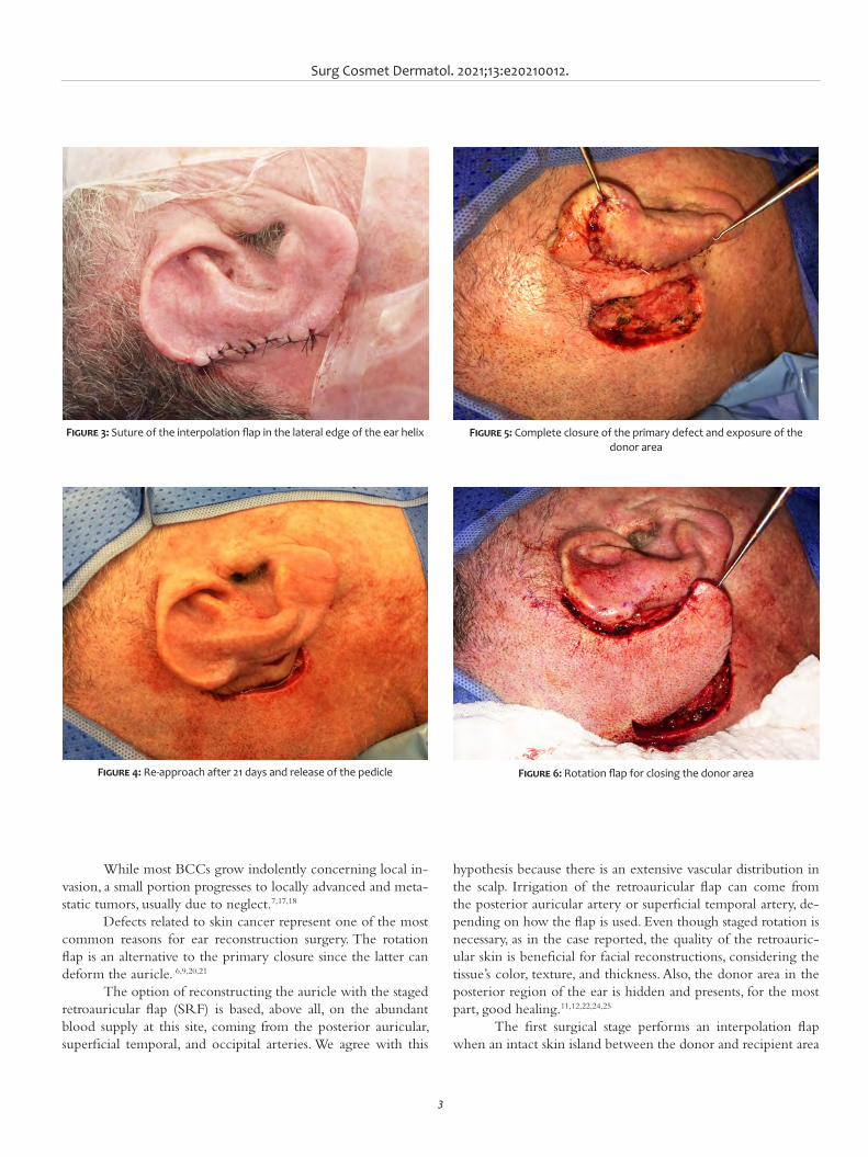
Surg Cosmet Dermatol. 2021;13:e20210012.
3
Figure 3: Suture of the interpolation flap in the lateral edge of the ear helix
Figure 4: Re-approach after 21 days and release of the pedicle
Figure 5: Complete closure of the primary defect and exposure of the donor area
Figure 6: Rotation flap for closing the donor area
While most BCCs grow indolently concerning local in-vasion, a small portion progresses to locally advanced and meta-static tumors, usually due to neglect.7,17,18
Defects related to skin cancer represent one of the most common reasons for ear reconstruction surgery. The rotation flap is an alternative to the primary closure since the latter can deform the auricle. 6,9,20,21
The option of reconstructing the auricle with the staged retroauricular flap (SRF) is based, above all, on the abundant blood supply at this site, coming from the posterior auricular, superficial temporal, and occipital arteries. We agree with this
hypothesis because there is an extensive vascular distribution in the scalp. Irrigation of the retroauricular flap can come from the posterior auricular artery or superficial temporal artery, de-pending on how the flap is used. Even though staged rotation is necessary, as in the case reported, the quality of the retroauric-ular skin is beneficial for facial reconstructions, considering the tissue’s color, texture, and thickness. Also, the donor area in the posterior region of the ear is hidden and presents, for the most part, good healing.11,12,22,24,25
The first surgical stage performs an interpolation flap when an intact skin island between the donor and recipient area
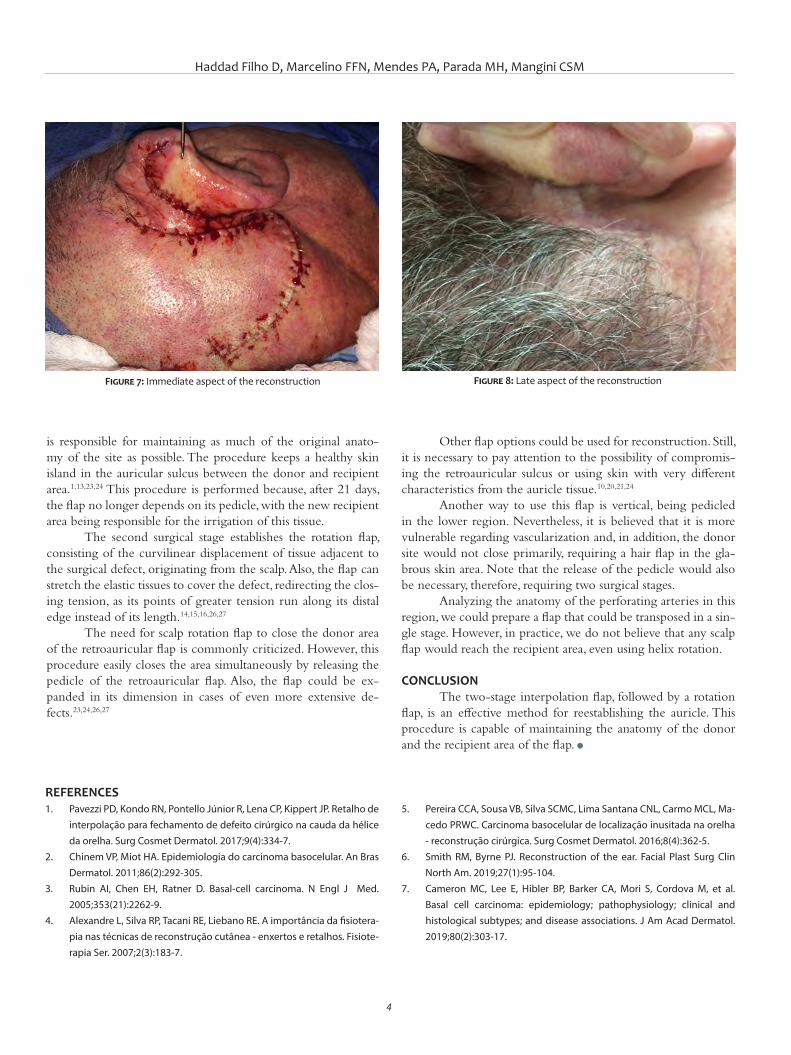
Haddad Filho D, Marcelino FFN, Mendes PA, Parada MH, Mangini CSM
4
is responsible for maintaining as much of the original anato-my of the site as possible. The procedure keeps a healthy skin island in the auricular sulcus between the donor and recipient area.1,13,23,24 This procedure is performed because, after 21 days, the flap no longer depends on its pedicle, with the new recipient area being responsible for the irrigation of this tissue.
The second surgical stage establishes the rotation flap, consisting of the curvilinear displacement of tissue adjacent to the surgical defect, originating from the scalp. Also, the flap can stretch the elastic tissues to cover the defect, redirecting the clos-ing tension, as its points of greater tension run along its distal edge instead of its length.14,15,16,26,27
The need for scalp rotation flap to close the donor area of the retroauricular flap is commonly criticized. However, this procedure easily closes the area simultaneously by releasing the pedicle of the retroauricular flap. Also, the flap could be ex-panded in its dimension in cases of even more extensive de-fects.23,24,26,27
Other flap options could be used for reconstruction. Still, it is necessary to pay attention to the possibility of compromis-ing the retroauricular sulcus or using skin with very different characteristics from the auricle tissue.10,20,21,24
Another way to use this flap is vertical, being pedicled in the lower region. Nevertheless, it is believed that it is more vulnerable regarding vascularization and, in addition, the donor site would not close primarily, requiring a hair flap in the gla-brous skin area. Note that the release of the pedicle would also be necessary, therefore, requiring two surgical stages.
Analyzing the anatomy of the perforating arteries in this region, we could prepare a flap that could be transposed in a sin-gle stage. However, in practice, we do not believe that any scalp flap would reach the recipient area, even using helix rotation.
CONCLUSIONThe two-stage interpolation flap, followed by a rotation
flap, is an effective method for reestablishing the auricle. This procedure is capable of maintaining the anatomy of the donor and the recipient area of the flap. l
Figure 7: Immediate aspect of the reconstruction Figure 8: Late aspect of the reconstruction
REFERENCES1. Pavezzi PD, Kondo RN, Pontello Júnior R, Lena CP, Kippert JP. Retalho de
interpolação para fechamento de defeito cirúrgico na cauda da hélice da orelha. Surg Cosmet Dermatol. 2017;9(4):334-7.
2. Chinem VP, Miot HA. Epidemiologia do carcinoma basocelular. An Bras Dermatol. 2011;86(2):292-305.
3. Rubin AI, Chen EH, Ratner D. Basal-cell carcinoma. N Engl J Med. 2005;353(21):2262-9.
4. Alexandre L, Silva RP, Tacani RE, Liebano RE. A importância da fisiotera-pia nas técnicas de reconstrução cutânea - enxertos e retalhos. Fisiote-rapia Ser. 2007;2(3):183-7.
5. Pereira CCA, Sousa VB, Silva SCMC, Lima Santana CNL, Carmo MCL, Ma-cedo PRWC. Carcinoma basocelular de localização inusitada na orelha - reconstrução cirúrgica. Surg Cosmet Dermatol. 2016;8(4):362-5.
6. Smith RM, Byrne PJ. Reconstruction of the ear. Facial Plast Surg Clin North Am. 2019;27(1):95-104.
7. Cameron MC, Lee E, Hibler BP, Barker CA, Mori S, Cordova M, et al. Basal cell carcinoma: epidemiology; pathophysiology; clinical and histological subtypes; and disease associations. J Am Acad Dermatol. 2019;80(2):303-17.
Surg Cosmet Dermatol. 2021;13:e20210012.
5
8. Waldman A, Schmults C. Cutaneous Squamous Cell Carcinoma. Hema-tol Oncol Clin North Am. 2019;33(1):1-12.
9. Tinklepaugh A, Husain Z, Libby TJ, Ciocon D. Reconstruction of a full--thickness auricular defect after Mohs micrographic surgery. Dermatol Surg. 2018;44(12):1595-8.
10. Hsueh Y, Shieh S. Two-stage retroauricular flap to reconstruct a helical rim defect. Dermatol Surg. 2009;35(11):1827-30.
11. Hénoux M, Espitalier F, Hamel A, Dréno B, Michel G, Malard O. Vascular supply of the auricle: anatomical study and applications to external ear reconstruction. Dermatol Surg. 2017;43(1):87-97.
12. Cerci FB. Retalho retroauricular estagiado para reconstrução de hélice após cirurgia micrográfica de Mohs. An Bras Dermatol. 2016;91(1):144-7.
13. Ramsey ML, Ellison CA, Al Aboud AM. Interpolated flaps. StatPearls. 2020[Internet]. Available from: https://www.ncbi.nlm.nih.gov/books/NBK470521/.
14. Pontes LT, Kimyai-Asadi A, Jih MH, Moraes AM, Stolf HO. Retalho de ro-tação para defeitos da asa. Surg Cosmet Dermatol. 2009;1(1):47-8.
15. Gadelha ADR, Costa IMC. Cirurgia dermatológica avançada: retalho de rotação. 3th ed. Rio de Janeiro: Atheneu; 2016.
16. Starkman SJ, Williams CT, Sherris DA. Flap basics I: rotation and trans-position flaps. Facial Plast Surg Clin North Am. 2017;25(3):313-21.
17. Kim DP, Kus K, Ruiz E. Basal cell carcinoma review. Hematol Oncol Clin North Am. 2019;33(1):13-24.
18. Tanese K. Diagnosis and management of basal cell carcinoma. Current Treat Opt Oncol. 2019;20(2):13.
19. Bariani RL, Nahas FX, Barbosa MVJ, Farah AB, Ferreira LM. Basal cell car-cinoma: an updated epidemiological and therapeutically profile of an urban population. Acta Cir Bras. 2006;21(2):66-73.
20. 20-Aguilar EF. Ear reconstruction. Clin Plastic Surgery. 2004;31(1):87-91.21. Iljin A, Antoszewski B, Durko M, Zieliński T, Pietruszewska W. External
auditory meatus and/or conchal bowl reconstruction with postauricu-lar island flap in patients with basal cell carcinoma or squamous cell carcinoma. Otolaryngol Pol. 2018;72 (3):4-10.
22. Li Y, Cui C, Zhang R, Zhang Q, Xu Z, Xu F, et al. Anatomical and histo-logical evaluation of the retroauricular fascia flap for staged auricular reconstruction. Aesthet Plast Surg. 2018;42(3):625-32.
23. Johnson TM, Fader DJ. The staged retroauricular to auricular direct pe-dicle (interpolation) flap for helical ear reconstruction. J Am Acad Der-matol. 1997;37(6): 975-8.
24. Faleiros HRP. Retalho retroauricular em ilha a pedículo superior, estudo anatomico e novos conceitos. Botucatu: Faculdade de Medicina de Botucatu (UNESP). Dissertação de mestrado. 2003. Available from: ht-tps://repositorio.unesp.br/handle/11449/88965.
25. Cordova A, D'Arpa S, Pirrello R, Giambona C, Moschella F. Retroauricular skin: a flaps bank for ear reconstruction. J Plast Reconstr Aesthet Surg. 2008;61(1):S44-51.
26. Lo Piccolo MC. Rotation flaps-principles and locations. Dermatol Surg. 2015;41(10): S247-54.
27. Lo CH, Kimble FW. The ideal rotation flap: an experimental study. J Plast Reconstr Aesthet Surg. 2008;61(7):754-9.
AUTHORS' CONTRIBUTION:
Douglas Haddad Filho 0000-0001-9304-4739Approval of the final version of the manuscript; study design and planning; preparation and writing of the manuscript; data col-lection, analysis, and interpretation; active participation in research orientation; intellectual participation in propaedeutic and/or therapeutic conduct of studied cases; critical literature review; critical revision of the manuscript.
Flávia Fenólio Nigro Marcelino 0000-0003-4057-5143Approval of the final version of the manuscript; study design and planning; preparation and writing of the manuscript; data col-lection, analysis, and interpretation; intellectual participation in propaedeutic and/or therapeutic conduct of studied cases; critical literature review; critical revision of the manuscript.
Paola Assunção Mendes 0000-0002-1116-9819Approval of the final version of the manuscript; study design and planning; preparation and writing of the manuscript; data col-lection, analysis, and interpretation; active participation in research orientation; intellectual participation in propaedeutic and/or therapeutic conduct of studied cases; critical literature review; critical revision of the manuscript.
Marcela Haddad Parada 0000-0001-5616-829XStudy design and planning; preparation and writing of the manuscript critical; literature review; critical revision of the manuscript.
Carolina Soutto Mayor Mangini 0000-0002-4354-1347Study design and planning; preparation and writing of the manuscript critical; literature review; critical revision of the manuscript.
1
APOIO CIENTÍFICO:
www.surgicalcosmetic.org.br/
ISSN-e 1984-8773
Case reportAuthors: Renan Tironi Giglio de Oliveira1
Beatrice Martinez Zugaib Abdalla1
Daniela Suzuki Locatelli1
Amanda Voltarelli Cesar de Oliveira1
Simao Cohen1
1 Dermatology Service, Centro Universitário Saúde ABC, São Paulo (SP), Brazil.
Correspondence: Renan Tironi Giglio de Oliveira Email: [email protected]
Financial support: None. Conflict of interest: None.
Submitted on: 18/08/2020Approved on: 24/02/2021
Acknowledgments: We thank the opportunity to work with the Harmony® platforms from LBT Lasers and SmartXide Dot® from DEKA laser, and the companies’ disposition to elucidate operational issues.
How to cite this article: Oliveira RTG, Abdalla BMZ, Locatelli DS, Oliveira AVC, Cohen S. Combined CO2 laser and intense pulsed light therapy in the treatment of vascular lesions. Surg Cosmet Dermatol. 2021;13:e20210011.
Combined CO2 laser and intense pulsed light therapy in the treatment of vascular lesions Terapia combinada de laser de CO2 e luz intensa pulsada no tratamento de lesões vasculares
ABSTRACTCongenital vascular lesions can be subdivided into tumors and malformations. Hemangiomas are benign vascular tumors resulting from the abnormal proliferation of endothelial cells, whereas port-wine stains are vascular malformations of a possible autosomal dominant inheritance. To date, there are no studies associating the use of intense pulsed light and CO2 laser as a therapeutic form for these pathologies. We present a series of 10 cases of vascular injuries treated with the combination of these forms of irradiation. This combined therapy can be effective in reducing the thickness of the lesions or in attenuating the color.Keywords: Hemangioma; Lasers, gas; Laser therapy; Intense pulsed light therapy; Vascular mal-formations
RESUMOAs lesões vasculares congênitas podem ser divididas em: tumores e malformações. Os hemangiomas são tumores vasculares benignos decorrentes da proliferação anormal de células endoteliais; já as manchas em vinho do Porto são malformações vasculares de provável herança autossômica dominante. Até o presente momento, não há estudos sobre a associação entre luz intensa pulsada e laser de CO2 como forma terapêutica destas patologias. A presente série de casos apresenta 10 casos de lesões vasculares tratadas com a combinação destas formas de irradiação. Conclui-se que esta terapia combinada pode ser efetiva na redução da espessura das lesões ou na atenuação da coloração.Palavras-chave: Hemangioma; Lasers de gás; Terapia a laser; Terapia de luz intensa pulsada; Mal-formações vasculares
DOI: http://www.dx.doi.org/10.5935/scd1984-8773.2021130011
ISSN-e 1984-8773
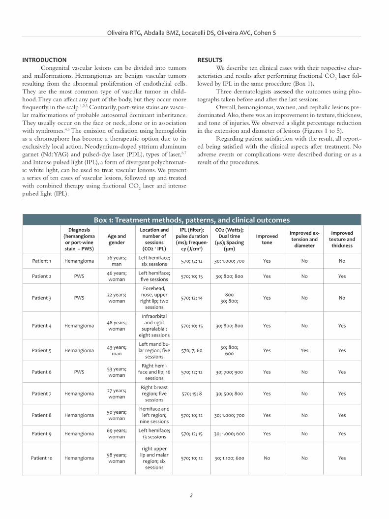
Oliveira RTG, Abdalla BMZ, Locatelli DS, Oliveira AVC, Cohen S
2
INTRODUCTIONCongenital vascular lesions can be divided into tumors
and malformations. Hemangiomas are benign vascular tumors resulting from the abnormal proliferation of endothelial cells. They are the most common type of vascular tumor in child-hood. They can affect any part of the body, but they occur more frequently in the scalp.1,2,3 Contrarily, port-wine stains are vascu-lar malformations of probable autosomal dominant inheritance. They usually occur on the face or neck, alone or in association with syndromes.4,5 The emission of radiation using hemoglobin as a chromophore has become a therapeutic option due to its exclusively local action. Neodymium-doped yttrium aluminum garnet (Nd: YAG) and pulsed-dye laser (PDL), types of laser,6,7 and Intense pulsed light (IPL), a form of divergent polychromat-ic white light, can be used to treat vascular lesions. We present a series of ten cases of vascular lesions, followed up and treated with combined therapy using fractional CO
2 laser and intense
pulsed light (IPL).
RESULTSWe describe ten clinical cases with their respective char-
acteristics and results after performing fractional CO2 laser fol-
lowed by IPL in the same procedure (Box 1). Three dermatologists assessed the outcomes using pho-
tographs taken before and after the last sessions. Overall, hemangiomas, women, and cephalic lesions pre-
dominated. Also, there was an improvement in texture, thickness, and tone of injuries. We observed a slight percentage reduction in the extension and diameter of lesions (Figures 1 to 5).
Regarding patient satisfaction with the result, all report-ed being satisfied with the clinical aspects after treatment. No adverse events or complications were described during or as a result of the procedures.
Box 1: Treatment methods, patterns, and clinical outcomesDiagnosis
(hemangioma or port-wine stain – PWS)
Age and gender
Location and number of
sessions (CO2 + IPL)
IPL (filter); pulse duration (ms); frequen-
cy (J/cm2)
CO2 (Watts); Dual time
(μs); Spacing (μm)
Improved tone
Improved ex-tension and
diameter
Improved texture and
thickness
Patient 1 Hemangioma 26 years;man
Left hemiface; six sessions 570; 12; 12 30; 1.000; 700 Yes No No
Patient 2 PWS 46 years;woman
Left hemiface; five sessions 570; 10; 15 30; 800; 800 Yes No Yes
Patient 3 PWS 22 years;woman
Forehead, nose, upper right lip; two
sessions
570; 12; 14 80030; 800; Yes No No
Patient 4 Hemangioma 48 years;woman
Infraorbital and right
supralabial; eight sessions
570; 10; 15 30; 800; 800 Yes No Yes
Patient 5 Hemangioma 43 years;man
Left mandibu-lar region; five
sessions570; 7; 60 30; 800;
600 Yes Yes Yes
Patient 6 PWS 53 years;woman
Right hemi-face and lip; 16
sessions570; 12; 12 30; 700; 900 Yes No Yes
Patient 7 Hemangioma 27 years;woman
Right breast region; five
sessions570; 15; 8 30; 500; 800 Yes No Yes
Patient 8 Hemangioma 50 years;woman
Hemiface and left region;
nine sessions570; 10; 12 30; 1.000; 700 Yes No Yes
Patient 9 Hemangioma 69 years;woman
Left hemiface; 13 sessions 570; 12; 15 30; 1.000; 600 Yes No Yes
Patient 10 Hemangioma 58 years;woman
right upper lip and malar
region; six sessions
570; 10; 12 30; 1.100; 600 No No Yes
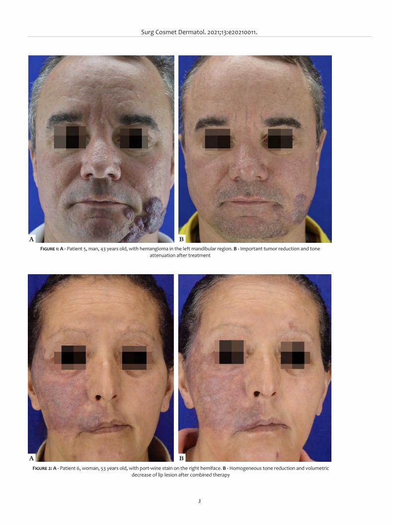
Surg Cosmet Dermatol. 2021;13:e20210011.
3
A
A
B
B
Figure 1: A - Patient 5, man, 43 years old, with hemangioma in the left mandibular region. B - Important tumor reduction and tone attenuation after treatment
Figure 2: A - Patient 6, woman, 53 years old, with port-wine stain on the right hemiface. B - Homogeneous tone reduction and volumetric decrease of lip lesion after combined therapy
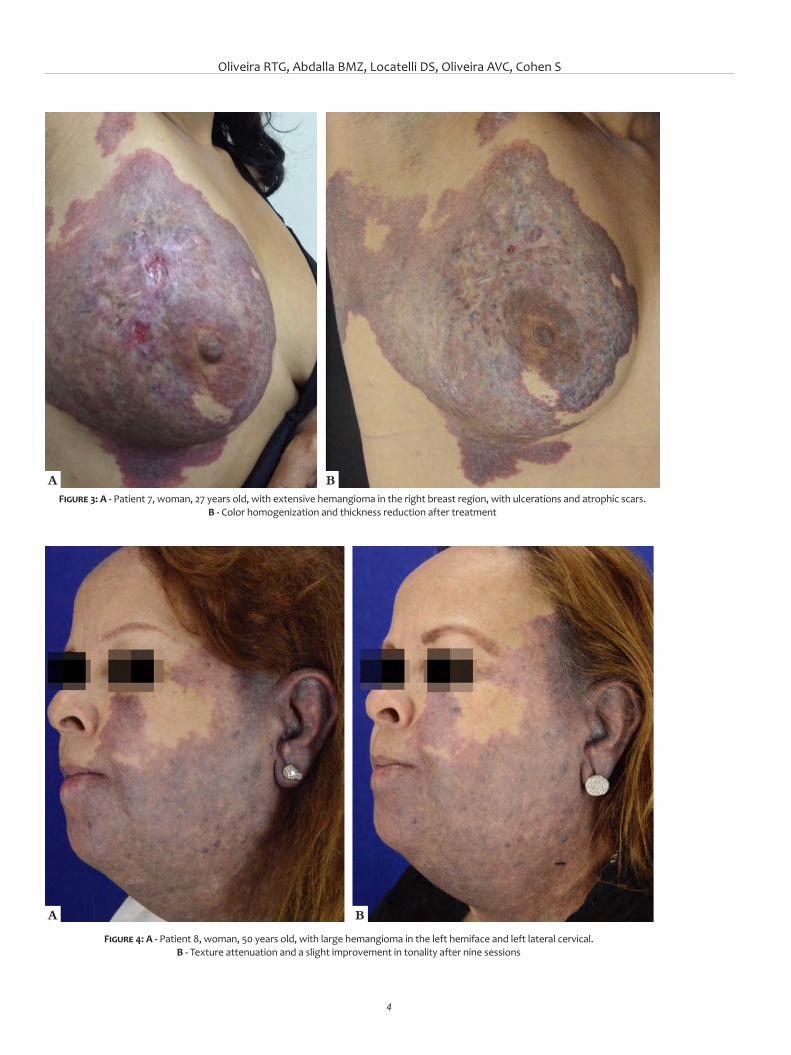
Oliveira RTG, Abdalla BMZ, Locatelli DS, Oliveira AVC, Cohen S
4
A
A
B
B
Figure 3: A - Patient 7, woman, 27 years old, with extensive hemangioma in the right breast region, with ulcerations and atrophic scars. B - Color homogenization and thickness reduction after treatment
Figure 4: A - Patient 8, woman, 50 years old, with large hemangioma in the left hemiface and left lateral cervical. B - Texture attenuation and a slight improvement in tonality after nine sessions
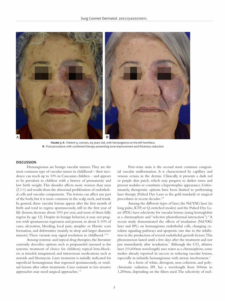
Surg Cosmet Dermatol. 2021;13:e20210011.
5
DISCUSSIONHemangiomas are benign vascular tumors. They are the
most common type of vascular tumor in childhood – their inci-dence can reach up to 10% in Caucasian children – and appears to be prevalent in children with a history of prematurity and low birth weight. This disorder affects more women than men (2.1:1) and results from the abnormal proliferation of endotheli-al cells and vascular components. The lesions can affect any part of the body, but it is more common in the scalp, neck, and trunk. In general, these vascular lesions appear after the first month of birth and tend to regress spontaneously, still in the first year of life (lesions decrease about 10% per year, and most of them fully regress by age 12). Despite its benign behavior, it may not prog-ress with spontaneous regression and present, in about 5-10% of cases, ulceration, bleeding, local pain, atrophic or fibrotic scars formation, and deformities (mainly in deep and larger diameter tumors). These variants may signal resolution in childhood.1,2,3
Among systemic and topical drug therapies, the literature currently describes options such as propranolol (assessed as the systemic treatment of choice for children); topical beta-block-ers as timolol; imiquimod; and intravenous medications such as steroids and bleomycin. Laser treatment is initially indicated for superficial hemangiomas that regressed spontaneously, or resid-ual lesions after other treatments. Cases resistant to less invasive approaches may need surgical approaches.1,3
Port-wine stain is the second most common congeni-tal vascular malformation. It is characterized by capillary and venous ectasia in the dermis. Clinically, it presents a dark red or purple skin patch, which may progress to darker tones and present nodules or constitute a hypertrophic appearance. Unfor-tunately, therapeutic options have been limited to performing laser therapy (Pulsed Dye Laser as the gold standard) or surgical procedures in recent decades.4,5
Among the different types of laser, the Nd:YAG laser (in long pulse, KTP, or Q-switched modes) and the Pulsed Dye La-ser (PDL) have selectivity for vascular lesions (using hemoglobin as a chromophore and “selective photothermal interaction”).6 A recent study demonstrated the effects of irradiation (Nd:YAG laser and IPL) on hemangioma endothelial cells, changing cy-tokine signaling pathways and apoptotic rate due to the inhibi-tion in the production of several endothelial growth factors. This phenomenon lasted until a few days after the treatment and not just immediately after irradiation.7 Although the CO
2 ablative
laser (10,600nm wavelength) uses water as a chromophore, some studies already reported its success in reducing vascular lesions, especially in infantile hemangiomas with airway involvement.3
As a form of white, divergent, non-coherent, and poly-chromatic radiation, IPL has a wavelength from 500nm to 1,200nm, depending on the filters used. The selectivity of mel-
A B
Figure 5: A - Patient 9, woman, 69 years old, with hemangioma on the left hemiface. B - Post-procedure with combined therapy presenting tone improvement and thickness reduction
Oliveira RTG, Abdalla BMZ, Locatelli DS, Oliveira AVC, Cohen S
6
REFERENCES1. Chen ZY, Wang QN, Zhu YH, Zhou LY, Xu T, He ZY, et al. Progress in the
treatment of infantile hemangioma. Ann Transl Med. 2019;7(22):692. doi:10.21037/atm.2019.10.47
2. Brauer JA, Geronemus RG. Laser treatment in the management of in-fantile hemangiomas and capillary vascular malformations. Tech Vasc Interv Radiol. 2013;16(1):51-4.
3. Chinnadurai S, Sathe NA, Surawicz T. Laser treatment of infantile he-mangioma: a systematic review. Lasers Surg Med. 2016;48(3):221- 33.
4. Katelyn Mariko Updyke BS,a Amor Khachemoune. Port-Wine stains: a focused review on their management. J Drugs Dermatol. 2017;16(11):1145-51.
5. van Raath MI, Chohan S, Wolkerstorfer A, van der Horst CMAM, Storm G, Heger M. Port wine stain treatment outcomes have not impro-ved over the past three decades. J Eur Acad Dermatol Venereol. 2019;33(7):1369-77.
AUTHORS' CONTRIBUTION:
Renan Tironi Giglio de Oliveira 0000-0001-5013-7660Statistical analysis; approval of the final version of the manuscript; study design and planning; preparation and writing of the ma-nuscript; data collection, analysis, and interpretation; active participation in research orientation; intellectual participation in pro-paedeutic and/or therapeutic conduct of studied cases; critical literature review; critical revision of the manuscript.
Beatrice Martinez Zugaib Abdalla 0000-0003-4586-1915Statistical analysis; approval of the final version of the manuscript; study design and planning; preparation and writing of the ma-nuscript; data collection, analysis, and interpretation; active participation in research orientation; intellectual participation in pro-paedeutic and/or therapeutic conduct of studied cases; critical literature review; critical revision of the manuscript.
Daniela Suzuki Locatelli 0000-0001-6210-3230Statistical analysis; approval of the final version of the manuscript; study design and planning; preparation and writing of the ma-nuscript; data collection, analysis, and interpretation; active participation in research orientation; intellectual participation in pro-paedeutic and/or therapeutic conduct of studied cases; critical literature review; critical revision of the manuscript.
Amanda Voltarelli Cesar de Oliveira 0000-0001-5594-8120Statistical analysis; approval of the final version of the manuscript; study design and planning; preparation and writing of the ma-nuscript; data collection, analysis, and interpretation; active participation in research orientation; intellectual participation in pro-paedeutic and/or therapeutic conduct of studied cases; critical literature review; critical revision of the manuscript.
Simao Cohen 0000-0003-4532-0465Statistical analysis; approval of the final version of the manuscript; study design and planning; preparation and writing of the ma-nuscript; data collection, analysis, and interpretation; active participation in research orientation; intellectual participation in pro-paedeutic and/or therapeutic conduct of studied cases; critical literature review; critical revision of the manuscript.
6. Lin M, Lin C, Hu S, Chang J, Chung W, Zhang Z, et al. The application of 595-nm pulsed dye laser for vascular anomalies in a Chinese popula-tion: a 10-year experience. J Cosmet Laser Ther. 2018;21(3):171-8.
7. Zhu Y, Wan X, Abliz P. Effects of laser irradiation on growth factors and cell apoptosis of in vitro cultured infant hemangioma endothelial cells. Life Sciences. 2019;233:116685.
8. Wat H, Wu DC, Rao J, Goldman MP. Application of intense pulsed light in the treatment of dermatologic disease: a systematic review. Derma-tol Surg. 2014;40(4):359-77.
9. Grillo E, Travassos AR, Boixeda P, Cuevas A, Pérez B, Paoli J, et al. Histo-chemical evaluation of the vessel wall destruction and selectivity after treatment with intense pulsed light in capillary malformations. Actas Dermosifiliogr. 2016;107(3):215-23.
anin and hemoglobin chromophores and the ability to damage the vascular walls according to established parameters justify its application in vascular lesions of the face.8,9
CONCLUSIONThe worldwide dermatological literature has not yet de-
scribed the association between IPL and CO2 laser for treating
vascular lesions. Therefore, we concluded that combining two different types of irradiation can effectively manage vascular le-sions, with possible secondary indications when it’s not possible to perform Nd:YAG or Pulsed Dye Laser. Device parameters must be further defined to explore the best aesthetic and func-tional result. l
1
APOIO CIENTÍFICO:
www.surgicalcosmetic.org.br/
ISSN-e 1984-8773
How I do it?Authors: Hudson Dutra Rezende¹ Bruna Orquiza dos Santos¹ Bruna Margatho Elias¹ Sandra Lopes Mattos Dinato¹ Maria Fernanda Reis Gavazzoni Dias² Ralph Michel Trüeb³
¹ Centro Universitário Lusíada (UNILUS), Department of Dermatology, São Paulo (SP), Brazil.
² Universidade Federal Fluminense, Department of Dermatology, Niterói (RJ), Brazil.
³ Centro de Dermatologia e Doenças Capilares Professor Trüeb, Department of Dermatology, Wallisellen, Zurique, Switzerland.
Correspondence: Hudson Dutra Rezende Email: [email protected] ou [email protected]
Financial support: None.Conflict of interest: None.
Submitted on: 18/04/2021Approved on: 20/06/2021
How to cite this article: Rezende HD, Santos BO, Elias BM, Dinato SLM, Dias MFRG, Trüeb RM. How to prepare a trichogram with transparent enamel base coat? Surg Cosmet Dermatol. 2021;13:20210037.
How to prepare a trichogram with transparent enamel base coat? Abordagem dos aspectos técnicos no preparo da lâmina de tricograma com base de esmalte incolor
ABSTRACTThe trichogram is an easy and semi-invasive method, useful for evaluating different types of hair loss in daily practice. To date, there is no standardized methodology on how to collect and perform the exam. The use of a liquid interface between the slide and the coverslip to read the trichogram under optical microscopy varies widely in the literature. Among the alternatives, transparent enamel base coat is an inexpensive, accessible, and practical option, providing the examiner with proper visualization of the hair shafts with minimal air bubbles formation.Keywords: Alopecia; Hair diseases; Scalp dermatoses
RESUMOO tricograma configura-se em método semi-invasivo de fácil aplicabilidade e baixo custo, útil na avaliação dos diversos tipos de queda capilar no consultório dermatológico. Até o momento, não há padronização da técnica para coleta e realização do exame. A utilização de meios de interface entre lâmina e lamínula para a leitura do tricograma à microscopia óptica varia amplamente na literatura. Dentre as alternativas, a utilização de base de esmalte incolor configura-se em opção de baixo custo acessível e prática, além de permitir a visualização das hastes capilares com mínima formação de artefato.Palavras-chave: Alopecia; Dermatoses do couro cabeludo; Doenças do cabelo
DOI: http://www.dx.doi.org/10.5935/scd1984-8773.2021130037
ISSN-e 1984-8773
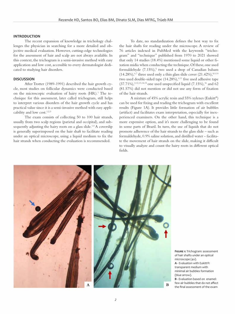
Rezende HD, Santos BO, Elias BM, Dinato SLM, Dias MFRG, Trüeb RM
2
INTRODUCTIONThe recent expansion of knowledge in trichology chal-
lenges the physician in searching for a more detailed and ob-jective medical evaluation. However, cutting-edge technologies for the assessment of hair and scalp are not always available. In this context, the trichogram is a semi-invasive method with easy application and low cost, accessible to every dermatologist dedi-cated to studying hair disorders.
DISCUSSIONAfter Trotter (1989-1991) described the hair growth cy-
cle, most studies on follicular dynamics were conducted based on the microscopic evaluation of hairy roots (HR).1 The te-chnique for this assessment, later called trichogram, still helps to interpret various disorders of the hair growth cycle and has practical value since it is a semi-invasive method with easy appli-cability and low cost.1,2,3
The exam consists of collecting 50 to 100 hair strands, usually from two scalp regions (parietal and occipital), and sub-sequently adjusting the hairy roots on a glass slide.1,4 A coverslip is generally superimposed on the hair shaft to facilitate reading under an optical microscope, using a liquid medium to fix the hair strands when conducting the evaluation is recommended.
To date, no standardization defines the best way to fix the hair shafts for reading under the microscope. A review of 76 articles indexed in PubMed with the keywords “tricho-gram” and “technique” published from 1970 to 2021 showed that only 14 studies (18.4%) mentioned some liquid or other fi-xation midia when conducting the technique. Of these, one used formaldehyde (7.15%),5 two used a drop of Canadian balsam (14.28%),6,7 three used only a thin glass slide cover (21.42%),8,9,10 two used double-sided tape (14.28%),4,11 five used adhesive tape (37.71%),2,12,13,14,15 one used unspecified liquid (7.15%),16 and 62 (81.57%) did not mention or did not use any form of fixation of the hair strands.
A mixture of 45% acrylic resin and 55% xylenes (Eukitt®) can be used for fixing and reading the trichogram with excellent results (Figure 1A). It provides little formation of air bubbles (artifact) and facilitates exam interpretation, especially for inex-perienced examiners. On the other hand, this technique is a more expensive option, and it’s more challenging to be found in some parts of Brazil. In turn, the use of liquids that do not promote adherence of the hair strands to the glass slide – such as formaldehyde, 0.9% saline solution, and distilled water - facilita-te the movement of hair strands on the slide, making it difficult to visually analyze and count the hairy roots in different optical fields.
Figure 1: Trichogram: assessment of hair shafts under an optical microscope (4x). A - Evaluation with Eukitt®: transparent medium with minimal air bubbles formation (blue arrow). B - Evaluation based on enamel: few air bubbles that do not affect the final assessment of the examA B
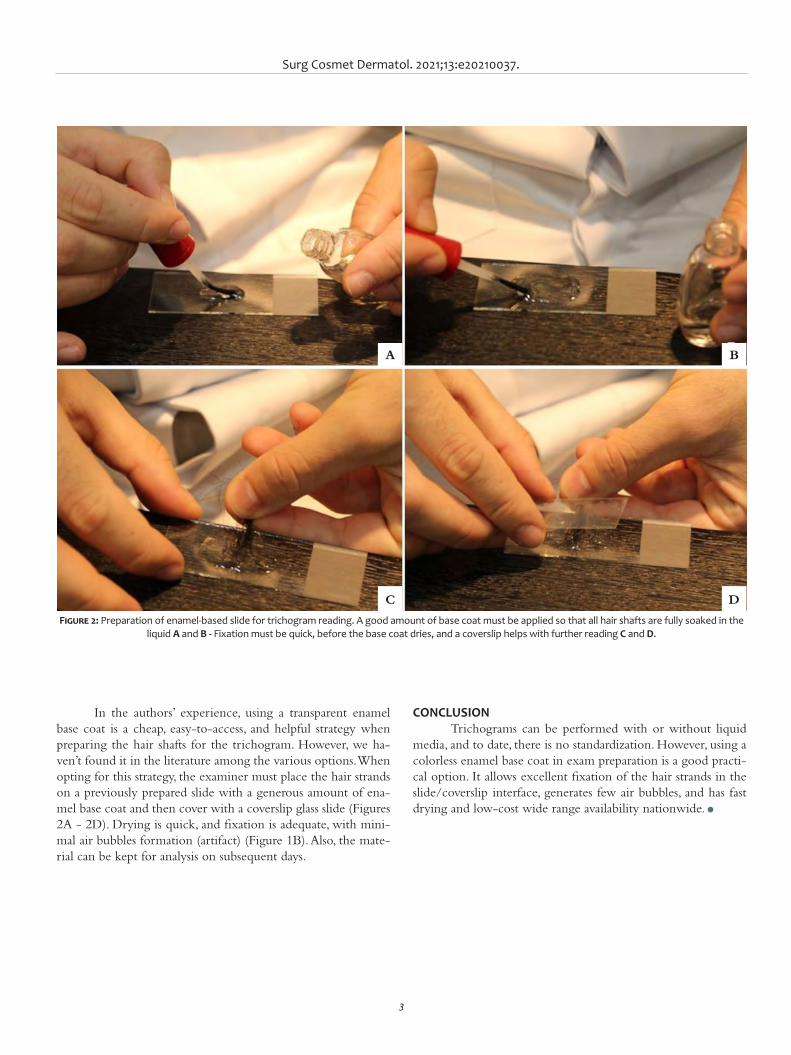
Surg Cosmet Dermatol. 2021;13:e20210037.
3
In the authors’ experience, using a transparent enamel base coat is a cheap, easy-to-access, and helpful strategy when preparing the hair shafts for the trichogram. However, we ha-ven’t found it in the literature among the various options. When opting for this strategy, the examiner must place the hair strands on a previously prepared slide with a generous amount of ena-mel base coat and then cover with a coverslip glass slide (Figures 2A - 2D). Drying is quick, and fixation is adequate, with mini-mal air bubbles formation (artifact) (Figure 1B). Also, the mate-rial can be kept for analysis on subsequent days.
CONCLUSION Trichograms can be performed with or without liquid
media, and to date, there is no standardization. However, using a colorless enamel base coat in exam preparation is a good practi-cal option. It allows excellent fixation of the hair strands in the slide/coverslip interface, generates few air bubbles, and has fast drying and low-cost wide range availability nationwide. l
Figure 2: Preparation of enamel-based slide for trichogram reading. A good amount of base coat must be applied so that all hair shafts are fully soaked in the liquid A and B - Fixation must be quick, before the base coat dries, and a coverslip helps with further reading C and D.
A
C
B
D
Rezende HD, Santos BO, Elias BM, Dinato SLM, Dias MFRG, Trüeb RM
4
AUTHORS' CONTRIBUTION:
Hudson Dutra Rezende 0000-0002-7039-790XStatistical analysis; approval of the final version of the manuscript; study design and planning; preparation and writing of the manuscript; data collection, analysis, and interpretation; active participation in research orientation; intellectual participation in propaedeutic and/or therapeutic conduct of studied cases; critical literature review; critical revision of the manuscript.
Bruna Orquiza dos Santos 0000-0002-1983-3868Approval of the final version of the manuscript; study design and planning; data collection, analysis, and interpretation; active participation in research orientation; intellectual participation in propaedeutic and/or therapeutic conduct of studied cases; critical literature review.
Bruna Margatho Elias 0000-0003-2615-5775Statistical analysis; approval of the final version of the manuscript; preparation and writing of the manuscript; data collection, analysis, and in-terpretation; active participation in research orientation; intellectual participation in propaedeutic and/or therapeutic conduct of studied cases; critical literature review; critical revision of the manuscript.
Sandra Lopes Mattos Dinato 0000-0002-4547-0474Statistical analysis; approval of the final version of the manuscript; study design and planning; preparation and writing of the manuscript; data collection, analysis, and interpretation; active participation in research orientation; intellectual participation in propaedeutic and/or therapeutic conduct of studied cases; critical literature review; critical revision of the manuscript.
Maria Fernanda Reis Gavazzoni Dias 0000-0001-7397-7478Statistical analysis; approval of the final version of the manuscript; study design and planning; preparation and writing of the manuscript; data collection, analysis, and interpretation; active participation in research orientation; intellectual participation in propaedeutic and/or therapeutic conduct of studied cases; critical literature review; critical revision of the manuscript.
Ralph Michel Trüeb 0000-0003-4970-0350Statistical analysis; approval of the final version of the manuscript; study design and planning; preparation and writing of the manuscript; data collection, analysis, and interpretation; active participation in research orientation; intellectual participation in propaedeutic and/or therapeutic conduct of studied cases; critical literature review; critical revision of the manuscript.
REFERENCES:1. Ralph M, Trüeb. The difficult hair loss patient: guide to successful mana-
gement of alopecia and related conditions. New York: Springer; 2015. 2. Dhurat R, Saraogi P. Hair evaluation methods: merits and demerits. Int J
Trichology. 2009;1(2):108-19.3. Serrano-Falcón C, Fernández-Pugnaire MA, Serrano-Ortega S. Evalua-
ción del pelo y cuero cabelludo: tricograma. Actas Dermosifiliogr. 2013;104(10):867–76.
4. Piérard GE, Piérard-Franchimont C, Marks R, Elsner P; EEMCO group (Eu-ropean Expert Group on Efficacy Measurement of Cosmetics and other Topical Products). EEMCO guidance for the assessment of hair shed-ding and alopecia. Skin Pharmacol Physiol. 2004;17(2):98-110.
5. Chapman DM, Miller RA. An objective measurement of the anchoring strength of anagen hair in an adult with the loose anagen hair syndro-me. J Cutan Pathol. 1996;23(3):288-92.
6. Astore IP, Pecoraro V, Pecoraro EG. The normal trichogram of pubic hair. Br J Dermatol. 1979;101(4):441-5.
7. Barman JM, Astore I, Pecoraro V. The normal trichogram of the adult. J Invest Dermatol. 1965;44:233-6.
8. Sánchez P, Serrano-Falcón C, Torres JM, Serrano S, Ortega E. 5α-Re-ductase isozymes and aromatase mRNA levels in plucked hair from young women with female pattern hair loss. Arch Dermatol Res. 2018;310(1):77-83.
9. Kunz M, Seifert B, Trüeb RM. Seasonality of hair shedding in healthy women complaining of hair loss. Dermatology. 2009;219(2):105-10.
10. Galliker NA, Trüeb RM. Value of trichoscopy versus trichogram for diag-nosis of female androgenetic alopecia. Int J Trichology. 2012;4(1):19-22.
11. Rushton H, James KC, Mortimer CH. The unit area trichogram in the assessment of androgen-dependent alopecia. Br J Dermatol. 1983;109(4):429-37.
12. Rasheed H, Mahgoub D, Hegazy R, El-Komy M, Abdel Hay R, Hamid MA, Hamdy E. Serum ferritin and vitamin d in female hair loss: do they play a role? Skin Pharmacol Physiol. 2013;26(2):101-7.
13. Dhurat R, Chitallia J, May TW, Jayaraaman AM, Madhukara J, Anandan S, et al. An Open-Label Randomized Multicenter Study Assessing the Noninferiority of a Caffeine-Based Topical Liquid 0.2% versus Minoxidil 5% Solution in Male Androgenetic Alopecia. Skin Pharmacol Physiol. 2017;30(6):298-305.
14. Yavuz IH, Yavuz GO, Bilgili SG, Demir H, Demir C. Assessment of heavy metal and trace element levels in patients with telogen effluvium. In-dian J Dermatol. 2018;63(3):246-50.
15. Serrano-Falcón C, Fernández-Pugnaire MA, Serrano-Ortega S. Hair and scalp evaluation: the trichogram. Actas Dermosifiliogr. 2013;104(10):867-76.
16. Hillmann K, Blume-Peytavi U. Diagnosis of hair disorders. Semin Cutan Med Surg. 2009;28(1):33-8.
1
APOIO CIENTÍFICO:
www.surgicalcosmetic.org.br/
ISSN-e 1984-8773
How do I do?Authors:Ada Regina Trindade de Almeida 1 Renata Sitonio T. D. Monteiro2
1 Assistant Professor of cosmiatry at Dermatologic Clinic of Hospital do Servidor Público Municipal de São Paulo - São Paulo SP, Brazil.
2 Assistant volunteer of cosmiatry at Dermatologic Clinic of Hospital do Servidor Público Municipal de São Paulo - São Paulo SP, Brazil.
Correspondence:Ada Regina Trindade de Almeida E-mail: [email protected]
Financial support: noneConflit of interest: none
Submitted on: 10/11/2020Approved on: 15/01/2021
How to cite this article: Almeida ART, Monteiro RSTD. Hematoma in aesthe-tic surgery: tips to avoid unaesthetic results Hyaluronidase and hematoma drainage. Surg Cosmet Dermatol. 2021;13:e20210004.
Hematoma in aesthetic surgery: tips to avoid unaesthetic results Hyaluronidase and hematoma drainageHematoma em cirurgia cosmética: dicas para evitar resultados inestéticos a partir da hialuronidase e drenagem de hematomas
ABSTRACTHematomas are common following cosmetic surgery. When minor, they are treated with observation only as they are most often reabsorbed. However, even with small collections of blood, if no early intervention is adopted, poor aesthetic outcomes may occur. Early drain-age has been especially described in otorhinolaryngology and radiology journals. The au-thors present an approach to early treating hematomas. Special emphasis is given to the use of hyaluronidase, which is well known by dermatologists and plastic surgeons for its ability to dissolve hyaluronic acid, but its utility in the treatment of hematomas is not so commonly known by these experts. Keywords: Hyaluronoglucosaminidase; Hyaluronic Acid; Hematoma; Fibrosis
RESU MOHematomas são comuns após cirurgias cosméticas. Quando pequenos, são conduzidos de maneira con-servadora, pois, na maioria das vezes, são reabsorvidos. No entanto, mesmo pequenas coleções, quando não ativamente abordadas, podem resultar em maus resultados estéticos. A drenagem precoce tem sido especialmente descrita em revistas de Otorrinolaringologia e Radiologia. As autoras apresentam uma abordagem para o tratamento precoce de hematomas. Ênfase especial é dada ao uso da hialuronidase, bem conhecida pelos dermatologistas e cirurgiões plásticos por sua capacidade de dissolver o ácido hi-alurônico, mas sua utilidade no tratamento de hematomas não é amplamente difundida entre esses especialistas.Palavras-chave: Hialuronoglucosaminidase; Ácido Hialurônico. Hematoma; Fibrose
DOI: http://www.dx.doi.org/10.5935/scd1984-8773.2021130004
ISSN-e 1984-8773
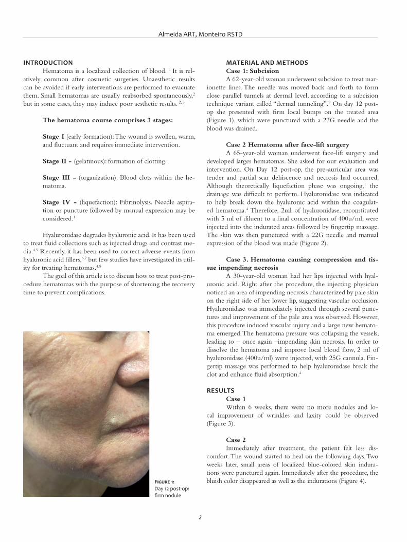
Almeida ART, Monteiro RSTD
2
INTRODUCTIONHematoma is a localized collection of blood. 1 It is rel-
atively common after cosmetic surgeries. Unaesthetic results can be avoided if early interventions are performed to evacuate them. Small hematomas are usually reabsorbed spontaneously,2 but in some cases, they may induce poor aesthetic results. 2, 3
The hematoma course comprises 3 stages:
Stage I (early formation): The wound is swollen, warm, and fluctuant and requires immediate intervention.
Stage II - (gelatinous): formation of clotting.
Stage III - (organization): Blood clots within the he-matoma.
Stage IV - (liquefaction): Fibrinolysis. Needle aspira-tion or puncture followed by manual expression may be considered.1
Hyaluronidase degrades hyaluronic acid. It has been used to treat fluid collections such as injected drugs and contrast me-dia.4,5 Recently, it has been used to correct adverse events from hyaluronic acid fillers,6,7 but few studies have investigated its util-ity for treating hematomas.4,8
The goal of this article is to discuss how to treat post-pro-cedure hematomas with the purpose of shortening the recovery time to prevent complications.
MATERIAL AND METHODSCase 1: SubcisionA 62-year-old woman underwent subcision to treat mar-
ionette lines. The needle was moved back and forth to form close parallel tunnels at dermal level, according to a subcision technique variant called “dermal tunneling”.9 On day 12 post-op she presented with firm local bumps on the treated area (Figure 1), which were punctured with a 22G needle and the blood was drained.
Case 2 Hematoma after face-lift surgeryA 65-year-old woman underwent face-lift surgery and
developed larges hematomas. She asked for our evaluation and intervention. On Day 12 post-op, the pre-auricular area was tender and partial scar dehiscence and necrosis had occurred. Although theoretically liquefaction phase was ongoing,1 the drainage was difficult to perform. Hyaluronidase was indicated to help break down the hyaluronic acid within the coagulat-ed hematoma.4 Therefore, 2ml of hyaluronidase, reconstituted with 5 ml of diluent to a final concentration of 400u/ml, were injected into the indurated areas followed by fingertip massage. The skin was then punctured with a 22G needle and manual expression of the blood was made (Figure 2).
Case 3. Hematoma causing compression and tis-sue impending necrosis
A 30-year-old woman had her lips injected with hyal-uronic acid. Right after the procedure, the injecting physician noticed an area of impending necrosis characterized by pale skin on the right side of her lower lip, suggesting vascular occlusion. Hyaluronidase was immediately injected through several punc-tures and improvement of the pale area was observed. However, this procedure induced vascular injury and a large new hemato-ma emerged. The hematoma pressure was collapsing the vessels, leading to – once again –impending skin necrosis. In order to dissolve the hematoma and improve local blood flow, 2 ml of hyaluronidase (400u/ml) were injected, with 25G cannula. Fin-gertip massage was performed to help hyaluronidase break the clot and enhance fluid absorption.4
RESULTSCase 1Within 6 weeks, there were no more nodules and lo-
cal improvement of wrinkles and laxity could be observed (Figure 3).
Case 2Immediately after treatment, the patient felt less dis-
comfort. The wound started to heal on the following days. Two weeks later, small areas of localized blue-colored skin indura-tions were punctured again. Immediately after the procedure, the bluish color disappeared as well as the indurations (Figure 4).Figure 1:
Day 12 post-op: firm nodule
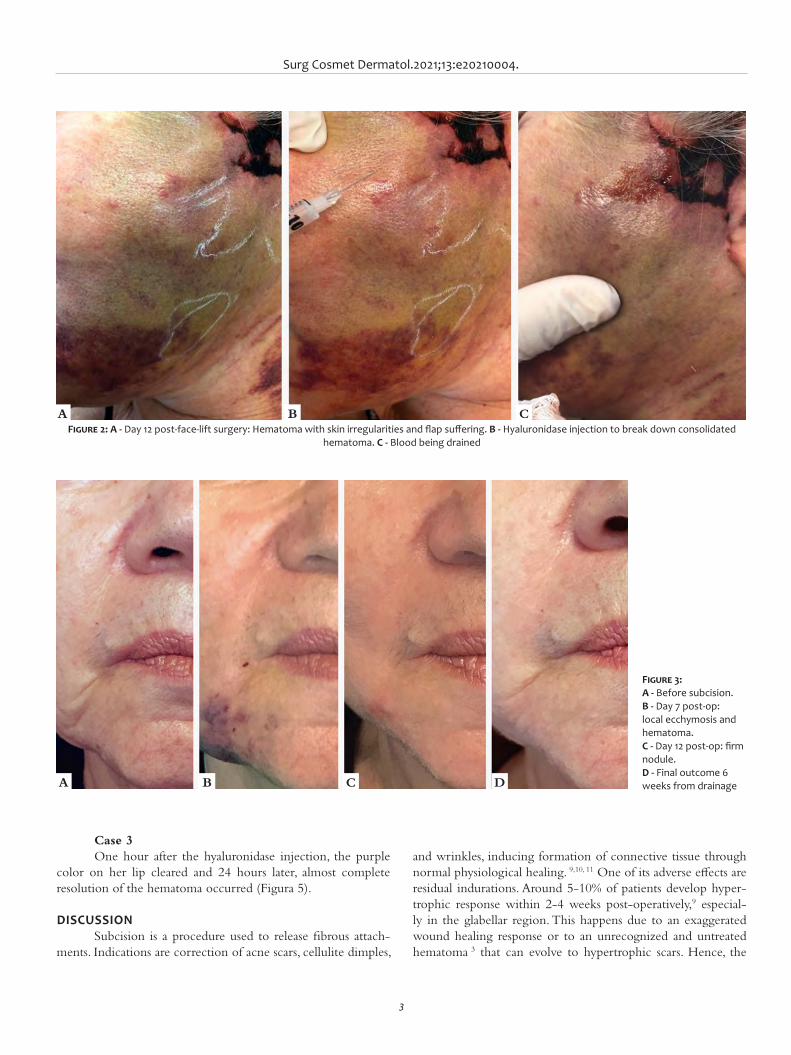
Surg Cosmet Dermatol.2021;13:e20210004.
3
Case 3One hour after the hyaluronidase injection, the purple
color on her lip cleared and 24 hours later, almost complete resolution of the hematoma occurred (Figura 5).
DISCUSSIONSubcision is a procedure used to release fibrous attach-
ments. Indications are correction of acne scars, cellulite dimples,
and wrinkles, inducing formation of connective tissue through normal physiological healing. 9,10, 11 One of its adverse effects are residual indurations. Around 5-10% of patients develop hyper-trophic response within 2-4 weeks post-operatively,9 especial-ly in the glabellar region. This happens due to an exaggerated wound healing response or to an unrecognized and untreated hematoma 3 that can evolve to hypertrophic scars. Hence, the
Figure 3: A - Before subcision. B - Day 7 post-op: local ecchymosis and hematoma. C - Day 12 post-op: firm nodule. D - Final outcome 6 weeks from drainage
Figure 2: A - Day 12 post-face-lift surgery: Hematoma with skin irregularities and flap suffering. B - Hyaluronidase injection to break down consolidated hematoma. C - Blood being drained
A B C
A B C D
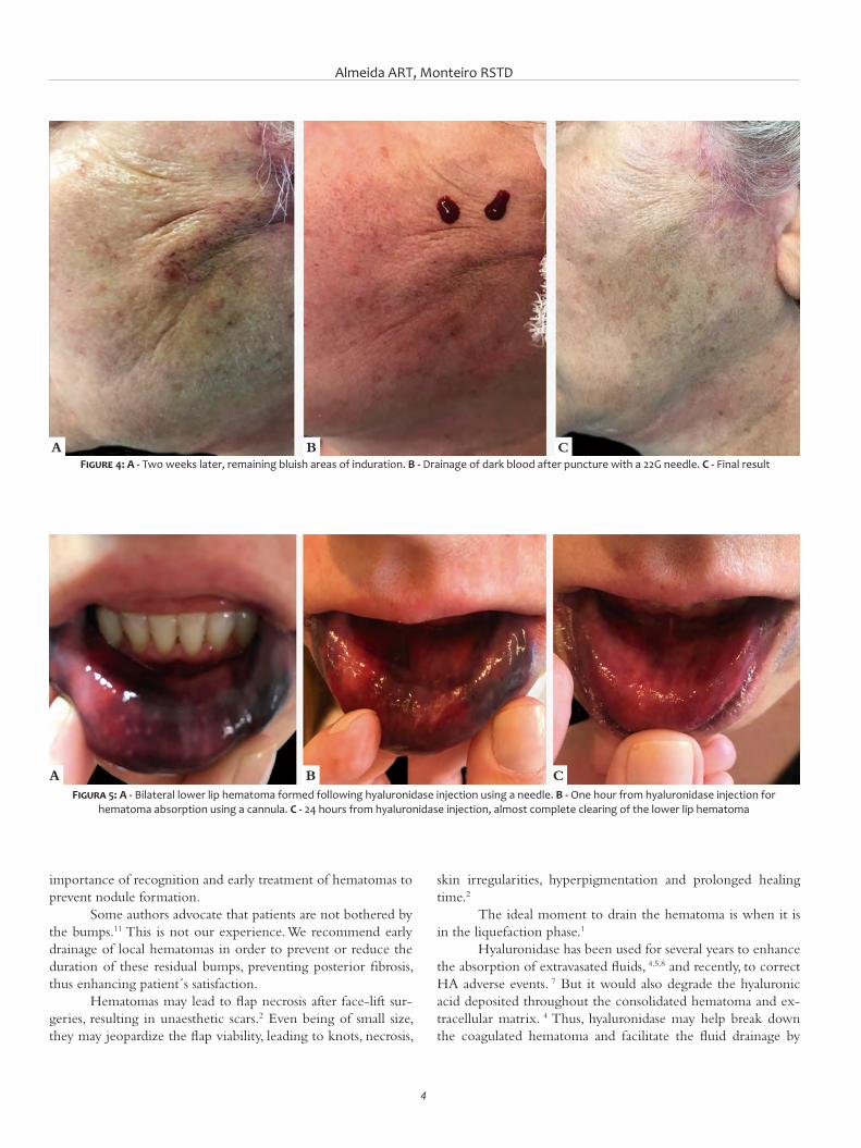
Almeida ART, Monteiro RSTD
4
importance of recognition and early treatment of hematomas to prevent nodule formation.
Some authors advocate that patients are not bothered by the bumps.11 This is not our experience. We recommend early drainage of local hematomas in order to prevent or reduce the duration of these residual bumps, preventing posterior fibrosis, thus enhancing patient´s satisfaction.
Hematomas may lead to flap necrosis after face-lift sur-geries, resulting in unaesthetic scars.2 Even being of small size, they may jeopardize the flap viability, leading to knots, necrosis,
skin irregularities, hyperpigmentation and prolonged healing time.2
The ideal moment to drain the hematoma is when it is in the liquefaction phase.1
Hyaluronidase has been used for several years to enhance the absorption of extravasated fluids, 4,5,8 and recently, to correct HA adverse events. 7 But it would also degrade the hyaluronic acid deposited throughout the consolidated hematoma and ex-tracellular matrix. 4 Thus, hyaluronidase may help break down the coagulated hematoma and facilitate the fluid drainage by
Figure 4: A - Two weeks later, remaining bluish areas of induration. B - Drainage of dark blood after puncture with a 22G needle. C - Final result
Figura 5: A - Bilateral lower lip hematoma formed following hyaluronidase injection using a needle. B - One hour from hyaluronidase injection for hematoma absorption using a cannula. C - 24 hours from hyaluronidase injection, almost complete clearing of the lower lip hematoma
A
A B C
B C
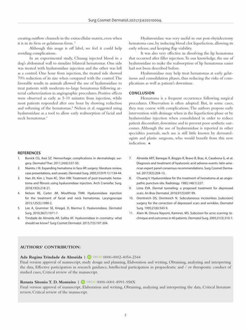
Surg Cosmet Dermatol.2021;13:e20210004.
5
creating outflow channels in the extracellular matrix, even when it is in its firm or gelatinous form. 4
Although this usage is off label, we feel it could help avoiding complications.
In an experimental study, Chuang injected blood in a dog’s abdominal wall to simulate bilateral hematomas. One side was treated with hyaluronidase injection and the other was left as a control. One hour from injection, the treated side showed 70% reduction of its size when compared with the control. The favorable results in animals allowed the use of hyaluronidase to treat patients with moderate-to-large hematomas following ar-terial catheterization in angiographic procedures. Positive effects were observed as early as 5-10 minutes from injection, while most patients responded after one hour by showing reduction and softening of the hematomas.8 Nelson et al. suggested using hyaluronidase as a tool to allow early reabsorption of facial and neck hematomas.4
Hyaluronidase was very useful in our post-rhytidectomy hematoma case, by inducing blood clot liquefaction, allowing its early release, and keeping flap viability.
It was also very effective in dissolving the lip hematoma that occurred after filler injection. To our knowledge, the use of hyaluronidase to make the reabsorption of lip hematomas easier had not been described before.
Hyaluronidase may help treat hematomas at early gelat-inous and consolidation phases, thus reducing the risks of com-plications as well as patient’s downtime.
CONCLUSIONHematoma is a frequent occurrence following surgical
procedures. Observation is often adopted. But, in some cases, they may course with complications. The authors propose early intervention with drainage when in the liquefaction phase or by hyaluronidase injection when consolidated in order to reduce patient discomfort, downtime and to prevent poor aesthetic out-comes. Although the use of hyaluronidase is reported in other specialties journals, such use is still little known by dermatol-ogists and plastic surgeons, who would benefit from this new indication. l
AUTHORS' CONTRIBUTION:
Ada Regina Trindade de Almeida | 0000-0002-4054-2344Final version approval of manuscript, study design and planning, Elaboration and writing, Obtaining, analyzing and interpreting the data, Effective participation in research guidance, Intellectual participation in propaedeutic and / or therapeutic conduct of studied cases, Critical review of the manuscript.
Renata Sitonio T. D. Monteiro | 0000-0001-8991-958XFinal version approval of manuscript, Elaboration and writing, Obtaining, analyzing and interpreting the data, Critical literature review, Critical review of the manuscript.
REFERENCES1. Bunick CG, Aasi SZ. Hemorrhagic complications in dermatologic sur-
gery. Dermatol Ther. 2011;24(6):537-50. 2. Niamtu J III. Expanding hematoma in face-lift surgery: literature review,
case presentations, and caveats. Dermatol Surg. 2005;31(9 Pt 1):1134-44. 3. Han JH, Kim J, Yoon KC, Shin HW. Treatment of post-traumatic hema-
toma and fibrosis using hyaluronidase injection. Arch Craniofac Surg. 2018;19(3):218-21.
4. Nelson RE, Carter JM, Moulthrop THM. Hyaluronidase injection for the treatment of facial and neck hematomas. Laryngoscope 2015;125(5):1090-2.
5. Lee A, Grummer SE, KriegeL D, Marmur E. Hyaluronidase. Dermatol Surg. 2010;36(7):1071-7.
6. Trindade de Almeida AR, Saliba AF. Hyaluronidase in cosmiatry: what should we know? Surg Cosmet Dermatol. 2015;7(3):197-204.
7. Almeida ART, Banegas R, Boggio R, Bravo B, Braz, A, Casabona G, et al. Diagnosis and treatment of hyaluronic acid adverse events: latin ame-rican expert panel consensus recommendations. Surg Cosmet Derma-tol. 2017;9(3):204-13.
8. Chuang V. Hyaluronidase for the treatment of hematoma at an angio-pathic puncture site. Radiology. 1983;146(1):227.
9. Lima EVA. Dermal tunneling: a proposed treatment for depressed scars. An Bras Dermatol. 2016;91(5):697-99.
10. Orentreich DS, Orentreich N. Subcutaneous incisionless [subcision] surgery for the correction of depressed scars and wrinkles. Dermatol Surg. 1995;21(6):543-9.
11. Alam M, Omura Nayomi, Kaminer, MS. Subcision for acne scarring: te-chnique and outcomes in 40 patients. Dermatol Surg. 2005;31(3):310-7.
1
APOIO CIENTÍFICO:
www.surgicalcosmetic.org.br/
ISSN-e 1984-8773
Communication Authors: Mariana M. Morita¹ Thomas S. P. Marcondes¹ Vidal Haddad-Jr¹ Hélio Amante Miot¹
¹ Universidade Estadual de São Paulo, Department of Dermatology, Botucatu (SP), Brazil.
Correspondence: Hélio Amante Miot Email: [email protected] / E-mail alternativo: heliomiot@ gmail.com
Financial support: None. Conflict of interest: None.
Submitted on: 05/05/2021Approved on: 04/06/2021
How to cite this article: Morita MM, Marcondes TSP, Haddad-Jr V, Miot HA. Cryosurgery with liquid nitrogen versus trichloroacetic acid in the treatment of human papillomavirus (HPV) penile wart: a randomized controlled trial. Surg Cosmet Dermatol. 2021;13:20210041.
Cryosurgery with liquid nitrogen versus trichloroacetic acid in the treatment of human papillomavirus (HPV) penile wart: a randomized controlled trial Criocirurgia com nitrogênio líquido versus ácido tricloroacético no tratamento de verruga peniana por papilomavírus humano (HPV): um ensaio clínico randomizado
ABSTRACTBackground: Anogenital warts are the most prevalent sexually transmitted infection (0.5%) among der-matological consultations. Cryotherapy with liquid nitrogen (LN) and therapy with trichloroacetic acid (TCA) are remarkable among the treatment strategies. Objective: To evaluate the effectiveness of LN versus TCA in to treat penile warts in patients from a Brazilian public institution. Methods: Open, parallel, randomized clinical trial. Following randomization, the warts were counted and submitted to cryotherapy (10s) or case therapy (TCA 80%) in each visit. The primary dependent va-riable was the lesion count before and after four weeks of treatment. We assessed the following variables: complete clearance, age, immunosuppression, smoking, topography, and education. Results: 142 treatments were evaluated in 52 participants. There was a predominance of young adults, and the main topography affected was the penis shaft. The mean reduction rate per session was 48% for LN and 26% for TCA (p=0.11). 42 (81%) participants achieved complete clearance, with 39 (75%; 95% CI: 64-85%) reaching clearance in up to three sessions. Age was associated with a worse therapeutic response rate (ß =-0.09; p<0.01). Conclusions: LN and TCA proved to be effective to treat penile warts, without difference between treatments. Age was associated with a worse therapeutic response.Keywords: Trichloroacetic acid; Cryosurgery; Sexually transmitted diseases; Papillomavirus infections; Warts
RESUMOIntrodução: verrugas anogenitais são a infecção sexualmente transmissível prevalente (0,5%) entre atendimentos dermatológicos. Dentre as estratégias de tratamento, destacam-se a crioterapia com nitrogênio líquido (NL) e causoterapia com ácido tricloroacético (ATA). Objetivo: avaliar a eficácia do NL versus ATA no tratamento de verrugas penianas em pacientes de instituição pública brasileira. Métodos: ensaio clínico aberto, paralelo e randomizado. Em cada visita, as verrugas foram contadas e submetidas à crioterapia (10s) ou à causoterapia (ATA 80%), após randomização. A principal variável dependente foi a contagem de lesões antes e após quatro semanas de tratamento. Foram analisados: eliminação completa, idade, imunossupressão, tabagismo, topografia e escolaridade. Resultados: foram avaliados 142 tratamentos em 52 participantes. Houve predominância de adultos jovens, menor escolaridade, e a principal topografia afetada foi a haste. A redução percentual média por sessão foi 48% para o NL e 26% para o ATA (p=0,11). Clearance completo foi atingido por 42 (81%) participantes, sendo que 39 (75%; IC95%: 64-85%) atingiram o clearance em até três sessões. A idade associou-se à pior taxa de resposta terapêutica (ß=-0,09; p<0,01). Conclusões: NL e ATA mostraram-se eficazes na terapêutica das verrugas penianas, sem diferença entre os tratamen-tos. A idade foi associada à pior resposta terapêutica.Palavras-chave: Ácido tricloroacético; Criocirurgia; Doenças sexualmente transmissíveis; Infecções por papillomavi-rus; Verrugas
DOI: http://www.dx.doi.org/10.5935/scd1984-8773.2021130041
ISSN-e 1984-8773
Morita MM, Marcondes TSP, Haddad-Jr V, Miot HA
2
INTRODUCTION Despite the vaccination campaign, anogenital warts
(AGWs) remain a prevalent sexually transmitted infection (0.5%) among dermatological care.1 The human alpha papillomavirus (HPV) causes AGWs, usually genotypes 6 and 11. It has a low--oncogenic risk but may present co-infection with high-risk ge-notypes. In addition to the critical link between contagion and impact on quality of life, HPV can induce epithelial neoplasms, especially in immunocompromised individuals.2,3
Among the therapeutic strategies used in AGW treat-ment, clinical therapy (e.g., imiquimod, podophyllotoxin) and surgical options, especially electrocoagulation, cryotherapy with liquid nitrogen (LN), and treatment with trichloroacetic acid (TCA) 80-90% stand out due to their accessibility and no need for anesthesia. There is no hierarchy of treatments, and few ran-domized studies compare the effectiveness of different strategies in AGW treatment. LN is associated with a 44-87% cure rate and TCA with 56-94%.4
This study aims to assess the effectiveness of LN ver-sus TCA to treat penile warts in patients from a Brazilian public institution.
METHODS It is an open, parallel, randomized clinical trial invol-
ving patients with penile AGW. At each clinical visit, the AGW were counted and submitted to treatment with LN (10s) or TCA 80%, according to computerized randomization. Patients were re-assessed after four weeks, AGWs were recounted, and patients were re-enrolled in the study under further randomi-zation if new treatments were needed. The primary dependent variable was AGW lesion count, assessed after acetoscopy (with acetic acid 5%), before treatment, and after four weeks. We also analyzed complete clearance, age, immunosuppression, smo-king, topography (penis glans, shaft, foreskin), and education. The numerical variation of the lesions was evaluated using a generalized linear mixed-effect model (negative binomial). Outcomes were assessed by intention to treat (ITT). Partici-pants who did not attend the re-evaluation visit were con-sidered a therapeutic failure, and the mixed model imputed their results. Significance was defined as p values <0.05. The project was approved by the Research Ethics Committee (n. 4418.2012).
RESULTS We assessed 142 treatments in 52 participants: 13 (9%)
of the cases did not return for re-evaluation (drop out). Table 1 shows the primary clinical and demographic data of the sample. There was a predominance of young adults, low education, and the main affected topography was the penis shaft. Cauterization with TCA was indicated in 63 (44%) of the treatments, and 59 was completed; NL was indicated in 79 (56%) of the treatments, with 70 completed, with no difference between the groups re-garding the drop out rate (p=0.82).
Figure 1 represents the AGW counts for each group and treatment time. There was a significant reduction in the median (p25-p75) of AGWs for both groups: TCA (3 [2-9] to 1 [0-4]; p<0.01) and LN (2 [1-4] to 0 [0-2]; p<0.01), with no differences between treatments (p=0.11). The mean percentage reduction in each session was 48% for LN and 26% for TCA.
Complete clearance (absence of acetowhite areas) was achieved by 42 (81%) study participants during follow-up, with 39 (75%; 95% CI: 64-85%) of all included participants achie-ving clearance within up to three monthly sessions. As the par-ticipants were re-included at each return in different treatment groups, assessing the complete clearance linked to each treat-ment was impossible.
When we assessed clinical-demographic elements as predictors of a general response to treatments, age (β= -0.09; p<0.01) was associated with the worst response rate. However, HIV carriers (p=0.99), education (p=0.51), smoking (p=0.17), and the topography of the lesions (p=0.24) did not interfere in the therapeutic response.
Table 1: Main clinical-demographic characteristics of the studied sample.
Variable Results
Age (years)* 28 (10)
School – n (%)
Elementary School 20 (43)
High School 21 (45)
University 6 (13)
HIV positive – n (%) 4 (8)
Smoking – n (%) 10 (19)
Topography of the lesions – n(%)
Penis shaft 91 (64)
Penis foreskin 32 (23)
Penis glans 19 (13)
Number of pre-treatment lesions **
3 (1-5)
Number of post-treatment lesions **
0 (0-3)
* means (standard deviation); ** median (p25-p75)
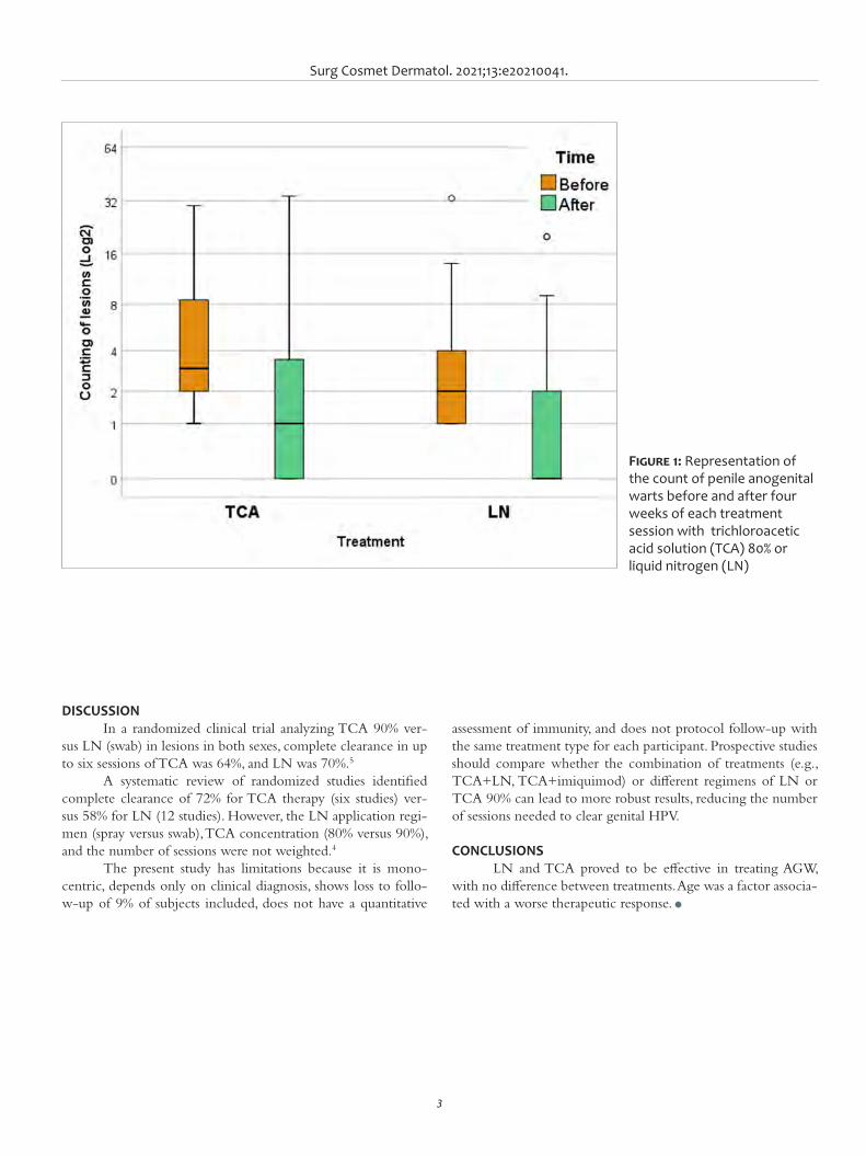
Surg Cosmet Dermatol. 2021;13:e20210041.
3
assessment of immunity, and does not protocol follow-up with the same treatment type for each participant. Prospective studies should compare whether the combination of treatments (e.g., TCA+LN, TCA+imiquimod) or different regimens of LN or TCA 90% can lead to more robust results, reducing the number of sessions needed to clear genital HPV.
CONCLUSIONS LN and TCA proved to be effective in treating AGW,
with no difference between treatments. Age was a factor associa-ted with a worse therapeutic response. l
DISCUSSION In a randomized clinical trial analyzing TCA 90% ver-
sus LN (swab) in lesions in both sexes, complete clearance in up to six sessions of TCA was 64%, and LN was 70%.5
A systematic review of randomized studies identified complete clearance of 72% for TCA therapy (six studies) ver-sus 58% for LN (12 studies). However, the LN application regi-men (spray versus swab), TCA concentration (80% versus 90%), and the number of sessions were not weighted.4
The present study has limitations because it is mono-centric, depends only on clinical diagnosis, shows loss to follo-w-up of 9% of subjects included, does not have a quantitative
Figure 1: Representation of the count of penile anogenital warts before and after four weeks of each treatment session with trichloroacetic acid solution (TCA) 80% or liquid nitrogen (LN)
Morita MM, Marcondes TSP, Haddad-Jr V, Miot HA
4
AUTHORS' CONTRIBUTION:
Mariana M. Morita 0000-0002-6396-7388Approval of the final version of the manuscript; preparation and writing of the manuscript; data collection, analysis, and interpretation; critical literature review; critical revision of the manuscript.
Thomas S. P. Marcondes 0000-0002-4448-9595 Approval of the final version of the manuscript; preparation and writing of the manuscript; data collection, analysis, and interpretation; critical literature review; critical revision of the manuscript.
Vidal Haddad-Jr 0000-0001-7214-0422Approval of the final version of the manuscript; study design and planning; preparation and writing of the manuscript; data collection, analysis, and interpretation; active participation in research orientation; intellectual participation in propaedeutic and/or therapeutic conduct of studied cases; critical literature review; critical revision of the manuscript.
Hélio Amante Miot 0000-0002-2596-9294Statistical analysis; approval of the final version of the manuscript; study design and planning; preparation and writing of the manuscript; data collection, analysis, and interpretation; active participation in research orientation; intellectual participation in propaedeutic and/or therapeutic conduct of studied cases; critical literature review; critical revision of the manuscript.
REFERENCES:1. Miot HA, Penna GO, Ramos AMC, Penna MLF, Schmidt SM, Luz FB, et
al. Profile of dermatological consultations in Brazil. An Bras Dermatol. 2018;93(6):916-28.
2. Leto Md, Santos Júnior GF, Porro AM, Tomimori J. Human papillomavi-rus infection: etiopathogenesis, molecular biology and clinical mani-festations. An Bras Dermatol. 2011;86(2):306-17.
3. Camargo CC, D'Elia MPB, Miot HA. Quality of life in men diagnosed with anogenital warts. An Bras Dermatol. 2017;92(3):427-9.
4. Bertolotti A, Milpied B, Fouéré S, Cabié A, Dupin N, Derancourt C. Local management of anogenital warts in immunocompetent adults: syste-matic review and pooled analysis of randomized-controlled trial data. J Am Acad Dermatol. 2019;81(5):1203-4.
5. 5 Abdullah AN, Walzman M, Wade A. Treatment of external genital warts comparing cryotherapy (liquid nitrogen) and trichloroacetic acid. Sex Transm Dis. 1993;20(6):344-5.

1
APOIO CIENTÍFICO:
www.surgicalcosmetic.org.br/
ISSN-e 1984-8773
Letter Authors: Gregory Cavanagh¹ Casey Abrahams¹ Andy Goren² Carlos Gustavo Wambier¹
¹ Department of Dermatology, The Warren Alpert Medical School of
Brown University, Providence (RI), United States
² Applied Biology, Inc., Irvine (CA), United States
Correspondence: Carlos Gustavo Wambie Email: carlos_wambier@brown. edu / Alternative E-mail: gregory_ [email protected]
Financial support: None Conflict of interest: None
Submitted on: 09/05/2021Approved on: 04/06/2021
How to cite this article: Cavanagh G, Abrahams C, Goren A, Wambier CG. Seasonal and daylight savings time fluctuations of Google searches for scalp seborrheic dermatitis. Surg Cosmet Dermatol. 2021;13:20210044.
Seasonal and daylight saving time fluctuations in Google searches for scalp seborrheic dermatitis Flutuações sazonais e do horário de verão nas pesquisas do Google para dermatite seborreica do couro cabeludo
ABSTRACTScalp seborrheic dermatitis, or dandruff, is thought to worsen during the winter when there is later sunrise and less daylight. This study investigates trends in search engine interest for the term "dandruff" as they relate to changes in daylight, sunrise, and seasonality. We investigated the search interest in several countries of varying latitudes over a five-year period, and we explore the effect of daylight saving time on disease interest within two cities in the United States. We discuss our findings in the context of hormonal changes and skincare/behavior.Keywords: Hair; Dandruff; Dermatitis; Hair diseases; Light; Sunlight
RESUMOAcredita-se que a dermatite seborreica do couro cabeludo, ou caspa, piore em gravidade durante o inverno, quando ocorre o nascer do sol tardio e menos luz do dia. Neste estudo, investigamos as tendências no interesse do mecanismo de pesquisa pelo termo "caspa", visto que se relacionam com as mudanças na luz do dia, nascer do sol e sazonalidade. Analisamos o interesse de pesquisa em vários países de latitudes variáveis em um período de cinco anos e exploramos o efeito do horário de verão sobre o interesse por doenças em duas cidades dos Estados Unidos. Discutimos nossas descobertas no contexto de mudanças hormonais e cuidados com a pele/comportamento.Palavras-chave: Cabelo; Caspa; Dermatite; Doenças do cabelo; Luz; Luz solar
DOI: http://www.dx.doi.org/10.5935/scd1984-8773.2021130044
ISSN-e 1984-8773

Cavanagh G, Abrahams C, Goren A, Wambier CG
2
INTRODUCTIONContributing factors to scalp seborrheic dermatitis
(dandruff) development are increased sebum, by-products of microorganisms like Malassezia yeasts, and allergic sensitivity. Dandruff is thought to worsen in severity during the winter,1 when there is later sunrise and less sunlight. We aimed to evalua-te potential seasonal fluctuations of search engine interest for “dandruff” and the possible correlation with daylight, sunrise, and DST shifts.
METHODS We assessed the frequency of search-engine queries for
scalp seborrheic dermatitis over five years (2015-2019) to inves-tigate a possible relationship between sunlight and search interest in the United States, Brazil, South Africa, and Colombia. The United States experiences more intense sunlight in June, July, and August; while South Africa and Brazil experience more in-tense sunlight in December, January, and February. In Colombia, the sunlight intensity is distributed more evenly throughout the year. We selected English-speaking (United States, South Africa) and non-English-speaking countries (Colombia and Brazil). Lay terminology matching the condition and language of each cou-ntry was used. Google Trends2 was used to determine the search frequencies (SF) each week relative to the maximum weekly searches each year. Average monthly frequency was taken over
five years, and it was used to plot the average relative search inte-rest in a year cycle (Figure 1). To investigate the specific effects of sunrise in the United States, we compared search trends for New York City (NYC, observes DST) and Phoenix (ST) for the year of 2018 (the most recent year without the effect of COVID-19 pandemic) (Figure 2). In the United States, searches for dandruff were estimated to be one hundred thousand to one million per month.
RESULTSWhen evaluating interest as compared to daylight,
it spiked in the late winter (low sunlight) of both northern (United States) and southern (Brazil, South Africa) locations (Figure 1). Dandruff interest occurred throughout the year along the equator (Colombia), with a reduced interest in the two Equinoxes (March and September). In the context of sun-rise time, searches increased in the winter when sunrise was later in both NYC and Phoenix. However, searches decreased in March in NYC but peaked again in April, after “springing forward” (unexpected increase). In contrast, the peak followed a typical reduction as the winter faded in Phoenix, without a spring peak. The peaks of interest followed the sunrise time pattern (Figure 2), suggesting that luminosity in the morning might be protective for dandruff.
Figure 1: Average monthly Google search frequency (2015-2019) of terms related to scalp seborrheic dermatitis in the Unit-ed States (dandruff), Brazil (caspa), Colombia (caspa), and South Africa (dandruff) over a representative one-year period. Circles are sized in quartiles to show monthly frequency relative to maximum interest.
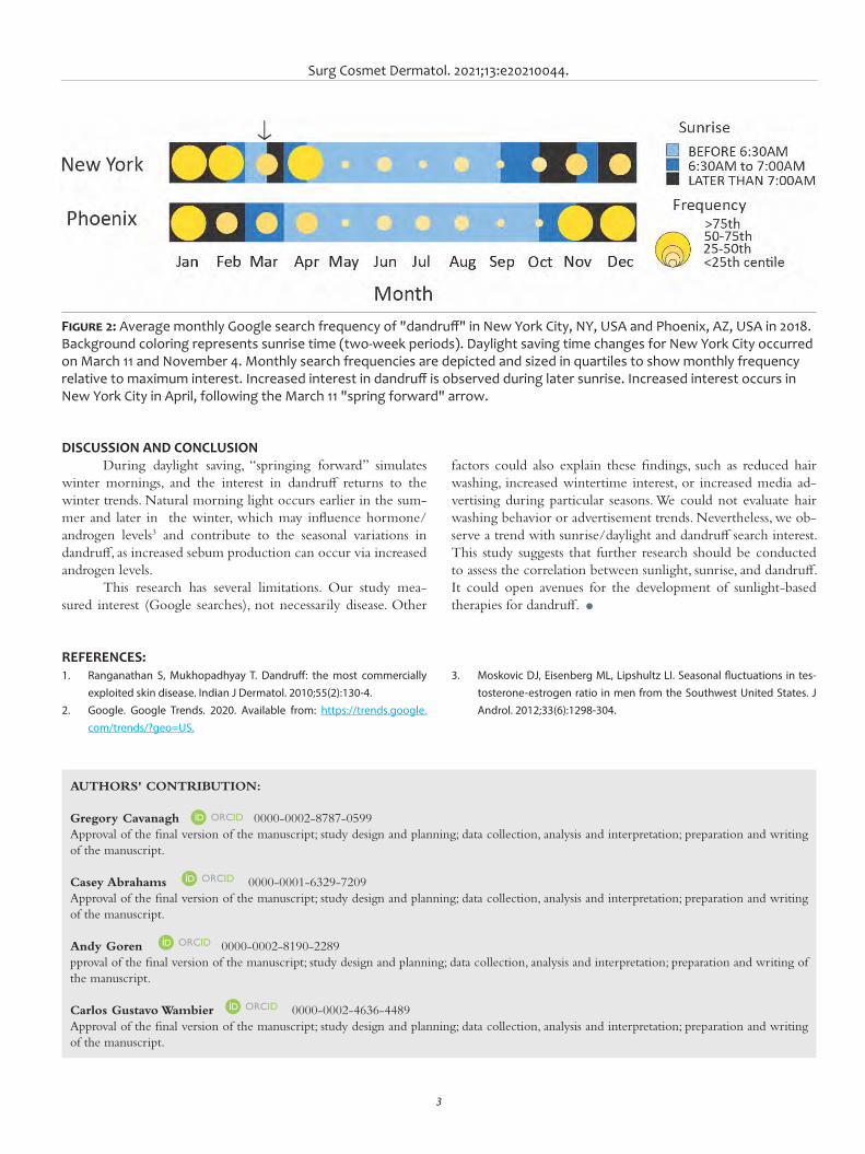
Surg Cosmet Dermatol. 2021;13:e20210044.
3
factors could also explain these findings, such as reduced hair washing, increased wintertime interest, or increased media ad-vertising during particular seasons. We could not evaluate hair washing behavior or advertisement trends. Nevertheless, we ob-serve a trend with sunrise/daylight and dandruff search interest. This study suggests that further research should be conducted to assess the correlation between sunlight, sunrise, and dandruff. It could open avenues for the development of sunlight-based therapies for dandruff. l
DISCUSSION AND CONCLUSION During daylight saving, “springing forward” simulates
winter mornings, and the interest in dandruff returns to the winter trends. Natural morning light occurs earlier in the sum-mer and later in the winter, which may influence hormone/androgen levels3 and contribute to the seasonal variations in dandruff, as increased sebum production can occur via increased androgen levels.
This research has several limitations. Our study mea-sured interest (Google searches), not necessarily disease. Other
Figure 2: Average monthly Google search frequency of "dandruff" in New York City, NY, USA and Phoenix, AZ, USA in 2018. Background coloring represents sunrise time (two-week periods). Daylight saving time changes for New York City occurred on March 11 and November 4. Monthly search frequencies are depicted and sized in quartiles to show monthly frequency relative to maximum interest. Increased interest in dandruff is observed during later sunrise. Increased interest occurs in New York City in April, following the March 11 "spring forward" arrow.
AUTHORS' CONTRIBUTION:
Gregory Cavanagh 0000-0002-8787-0599Approval of the final version of the manuscript; study design and planning; data collection, analysis and interpretation; preparation and writing of the manuscript.
Casey Abrahams 0000-0001-6329-7209Approval of the final version of the manuscript; study design and planning; data collection, analysis and interpretation; preparation and writing of the manuscript.
Andy Goren 0000-0002-8190-2289pproval of the final version of the manuscript; study design and planning; data collection, analysis and interpretation; preparation and writing of the manuscript.
Carlos Gustavo Wambier 0000-0002-4636-4489Approval of the final version of the manuscript; study design and planning; data collection, analysis and interpretation; preparation and writing of the manuscript.
REFERENCES:1. Ranganathan S, Mukhopadhyay T. Dandruff: the most commercially
exploited skin disease. Indian J Dermatol. 2010;55(2):130-4.2. Google. Google Trends. 2020. Available from: https://trends.google.
com/trends/?geo=US.
3. Moskovic DJ, Eisenberg ML, Lipshultz LI. Seasonal fluctuations in tes-tosterone-estrogen ratio in men from the Southwest United States. J Androl. 2012;33(6):1298-304.
1
APOIO CIENTÍFICO:
www.surgicalcosmetic.org.br/
ISSN-e 1984-8773
LettersAuthors: Leticia Dupont¹ Daniele Damares Rodrigues de
Souza¹ Ana Paula Dornelles Manzoni¹
¹ Santa Casa de Misericórdia de Porto Alegre, Dermatology Department, Porto Alegre (RS), Brazil.
Correspondence: Leticia Dupont Email: [email protected]
Financial support: None Conflict of interest: None
Submitted on: 15/04/2021Approved on: 14/07/2021
How to cite this article: Dupont L, Souza DDR, Manzoni APD. Myomodulation with hyalu-ronic acid for correction of gummy smile. Surg Cosmetic Dermatol. 2021;13:e20210032.
Myomodulation with hyaluronic acid for correction of gummy smile Miomodulação com ácido hialurônico para o tratamento do sorriso gengival
ABSTRACTThe "gummy smile" occurs when more than 3-4 mm of gingiva appears during the act of smiling. It is considered unattractive and causes aesthetic disharmony. It has a multifactorial etiology, with several techniques described for its correction. Myomodulation with hyaluronic acid is a non-surgical alternative with immediate and lasting results. In this article, we present a case of myomodulation with hyaluronic acid to correct "gummy smile" in a 31-year-old patient.Keywords: Hyaluronic acid; Gingiva; Smile
RESUMOO sorriso gengival ocorre quando há exposição de mais de 3-4 mm de tecido gengival durante o ato de sorrir. É considerado pouco atraente e causa de desarmonia estética. Possui etiologia multifatorial com diversas técnicas descritas para sua correção. A miomodulação com ácido hialurônico é uma alternativa não cirúrgica, com resultados imediatos e duradouros. Neste artigo nós apresentamos um caso de miomodulação com ácido hialurônico para correção do sorriso gengival em uma paciente de 31 anos.Palavras-chave: Ácido hialurônico; Gengiva; Sorriso
DOI: http://www.dx.doi.org/10.5935/scd1984-8773.2021130032
ISSN-e 1984-8773
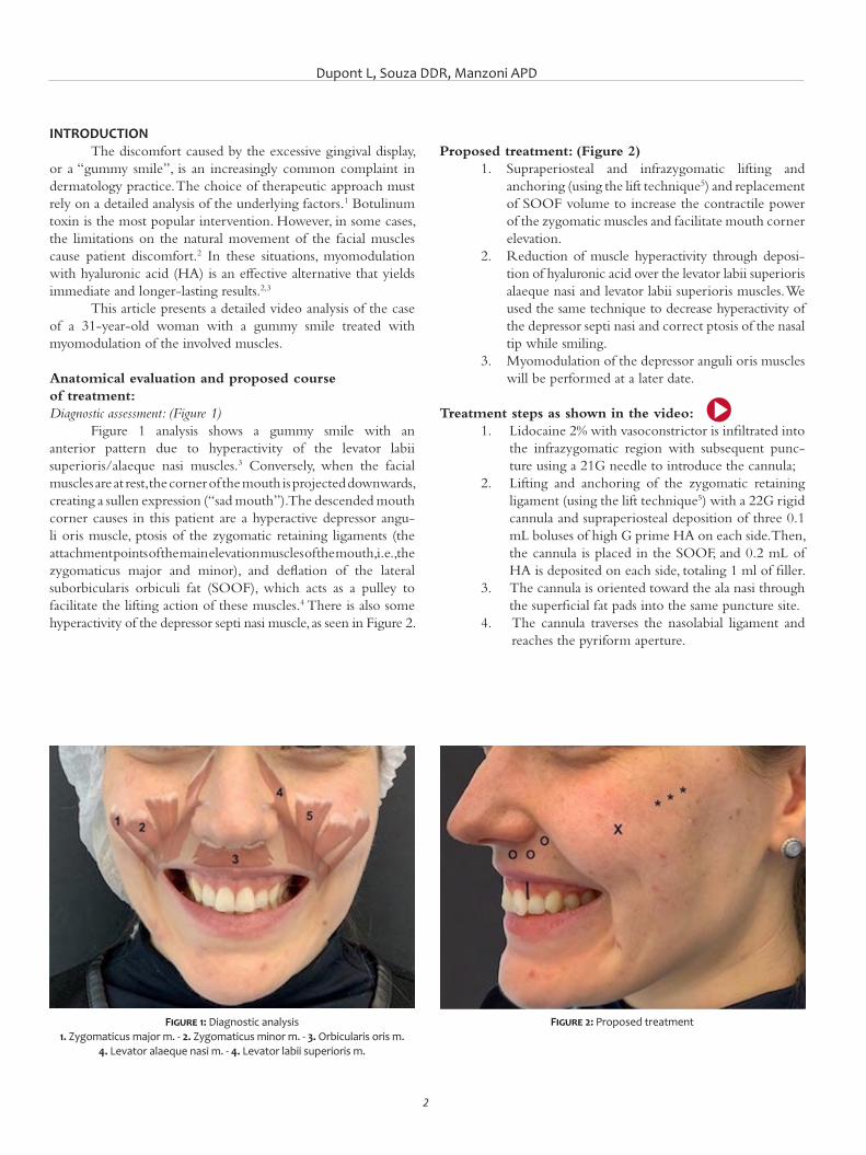
Dupont L, Souza DDR, Manzoni APD
2
INTRODUCTION The discomfort caused by the excessive gingival display,
or a “gummy smile”, is an increasingly common complaint in dermatology practice. The choice of therapeutic approach must rely on a detailed analysis of the underlying factors.1 Botulinum toxin is the most popular intervention. However, in some cases, the limitations on the natural movement of the facial muscles cause patient discomfort.2 In these situations, myomodulation with hyaluronic acid (HA) is an effective alternative that yields immediate and longer-lasting results.2,3
This article presents a detailed video analysis of the case of a 31-year-old woman with a gummy smile treated with myomodulation of the involved muscles.
Anatomical evaluation and proposed course of treatment:Diagnostic assessment: (Figure 1)
Figure 1 analysis shows a gummy smile with an anterior pattern due to hyperactivity of the levator labii superioris/alaeque nasi muscles.3 Conversely, when the facial muscles are at rest, the corner of the mouth is projected downwards, creating a sullen expression (“sad mouth”). The descended mouth corner causes in this patient are a hyperactive depressor angu-li oris muscle, ptosis of the zygomatic retaining ligaments (the attachment points of the main elevation muscles of the mouth, i.e., the zygomaticus major and minor), and deflation of the lateral suborbicularis orbiculi fat (SOOF), which acts as a pulley to facilitate the lifting action of these muscles.4 There is also some hyperactivity of the depressor septi nasi muscle, as seen in Figure 2.
Proposed treatment: (Figure 2)1. Supraperiosteal and infrazygomatic lifting and
anchoring (using the lift technique5) and replacement of SOOF volume to increase the contractile power of the zygomatic muscles and facilitate mouth corner elevation.
2. Reduction of muscle hyperactivity through deposi-tion of hyaluronic acid over the levator labii superioris alaeque nasi and levator labii superioris muscles. We used the same technique to decrease hyperactivity of the depressor septi nasi and correct ptosis of the nasal tip while smiling.
3. Myomodulation of the depressor anguli oris muscles will be performed at a later date.
Treatment steps as shown in the video: 1. Lidocaine 2% with vasoconstrictor is infiltrated into
the infrazygomatic region with subsequent punc-ture using a 21G needle to introduce the cannula;
2. Lifting and anchoring of the zygomatic retaining ligament (using the lift technique5) with a 22G rigid cannula and supraperiosteal deposition of three 0.1 mL boluses of high G prime HA on each side. Then, the cannula is placed in the SOOF, and 0.2 mL of HA is deposited on each side, totaling 1 ml of filler.
3. The cannula is oriented toward the ala nasi through the superficial fat pads into the same puncture site.
4. The cannula traverses the nasolabial ligament and reaches the pyriform aperture.
Figure 1: Diagnostic analysis1. Zygomaticus major m. - 2. Zygomaticus minor m. - 3. Orbicularis oris m.
4. Levator alaeque nasi m. - 4. Levator labii superioris m.
Figure 2: Proposed treatment
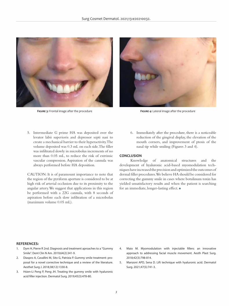
Surg Cosmet Dermatol. 2021;13:e20210032.
3
5. Intermediate G prime HA was deposited over the levator labii superioris and depressor septi nasi to create a mechanical barrier to their hyperactivity. The volume deposited was 0.3 mL on each side. The filler was infiltrated slowly in microbolus increments of no more than 0.05 mL, to reduce the risk of extrinsic vascular compression. Aspiration of the cannula was always performed before HA deposition.
CAUTION: It is of paramount importance to note that the region of the pyriform aperture is considered to be at high risk of arterial occlusion due to its proximity to the angular artery. We suggest that applications in this region be performed with a 22G cannula, with 8 seconds of aspiration before each slow infiltration of a microbolus (maximum volume 0.05 mL).
6. Immediately after the procedure, there is a noticeable reduction of the gingival display, the elevation of the mouth corners, and improvement of ptosis of the nasal tip while smiling (Figures 3 and 4).
CONCLUSION Knowledge of anatomical structures and the
development of hyaluronic acid-based myomodulation tech-niques have increased the precision and optimized the outcomes of dermal filler procedures. We believe HA should be considered for correcting the gummy smile in cases where botulinum toxin has yielded unsatisfactory results and when the patient is searching for an immediate, longer-lasting effect. l
Figure 3: Frontal image after the procedure Figure 4: Lateral image after the procedure
REFERENCES:1. Dym H, Pierre R 2nd. Diagnosis and treatment aproaches to a "Gummy
Smile". Dent Clin N Am. 2019;64(2):341-9. 2. Diaspro A, Cavallini M, Sito G, Patrizia P. Gummy smile treatment: pro-
posal for a novel corrective technique and a review of the literature. Aesthet Surg J. 2018;38(12):1330-8.
3. Hsien-Li Peng P, Peng JH. Treating the gummy smile with hyaluronic acid filler injection. Dermatol Surg. 2019;45(3):478-80.
4. Maio M. Myomodulation with injectable fillers: an innovative approach to addressing facial muscle movement. Aesth Plast Surg. 2018;42(3):798-814.
5. Manzoni APD, Sena D. Lift technique with hyaluronic acid. Dermatol Surg. 2021;47(5):741-3.

Dupont L, Souza DDR, Manzoni APD
4
AUTHORS' CONTRIBUTION:
Leticia Dupont 0000-0002-8150-1055Study design and planning; preparation and writing of the manuscript; critical literature review.
Daniele Damares Rodrigues de Souza 0000-0003-0610-4807Data collection, analysis and interpretation; preparation and writing of the manuscript; critical literature review.
Ana Paula Dornelles Manzoni 0000-0001-6184-4440Approval of the final version of the manuscript; study design and planning; preparation and writing of the manuscript; data collection, analysis, and interpretation; active participation in research orientation; intellectual participation in propaedeutic and/or therapeutic conduct of studied cases; critical literature review; critical revision of the manuscript.
www.surgicalcosmetic.org.br
SCIENTIFIC SUPPORT:
Official publication of the Brazilian Society of Dermatology
Related Documents