EDITOR II CHIEF EDITOR ASSOCIATE EDITORS F.U.Baqai Abdul Aziz Asadullah Khan Abdul Moeed Kazi Jamshed Akhtar Azhar Husain Irshad Waheed Mohammad Shamim Masood Javaid Saghir Ahmed S.A.Jilani Rashid Ahmad Choudhry Zahida Baqai EDITORIAL BOARD EDITORIALCONSULTAITS AUSTRALIA BAIGLADESH Earl R.Owen Ahsanullah Chowdhury M.Kabiruddin Ahmed HumayunKabirChoudhury Reffat Kamel Tehemton E.Udwadia Ahmed Abdul Hai I.K.Dhawan Ibrahim Bani Bani LB. Thappa AKSharma Dharuba Mudavari EGYPT liDIA' JORDAI IEPAL REPUBLIC OF MALDIVES SRI LAlKA U.K. U.A.E. PAKISTAI Imteyaz Mohsin A.P.R. Aluwihare John Hadfield J.S.P. Lumley Essa Kazim Abdullah Jan Jaffar Adib ul Hasan Rizvi Changez Hakim Khan Faiz Mohammad Khan Ghulam Ali Memon Ijaz H. Aqeel Inamul Haq Shami Jan MohammaedMemon Muhammad Iqbal Moizuddin M.Younis Khatri M. Naeem Khan Nazir M.Kazi Shah Nawaz Shah Sikander Shaikh Syed Azhar Ahmed Tariq Saeed Mufti Tipu Sultan Z.K.Kazi Zafarullah Chaudhry Published by: Prof.Abdul Aziz for Prof.F.U.Baqai, Baqai Postgraduate Medical Institute, IIIC, 1/12, Nazimabad, Karachi / Address for correspondence: Col. (Retd.) Masudul Haque, Executive Secretary, Surgical Publications, clo College of Physicians & Surgeons Pakistan, 7th Centeral Street Defence HOUSingAuthority Karachi,Tel. 5890029 & Fax:5887333 Subscribeption rates: Per copy-in Pakistan RS.1 00/= in SAARC countries U.S. $20, in other countries U.S.$30, Annual- in Pakistan Rs.300/= in SARRC countries US $ 60, in other countries U.S.$90. Layout by : Aleemuddin Siddqiui. VOL 2 NO 1 JANUARY - MARCH 1997 QUARTERLY JOURNAL OF SURGERY PAKISTAN INTERNATIONAL EDITOR'S NOTE Pancreatic Carcinoma Irshad Waheed ORIGINAL ARTICLES Experience with severe forms of Amniotic Bands of the limbs and review of literature. Faizah Naheed Bhatti, Amna Nasir and M. Naeem-uz-zafar Khan 2 Incidence of carcinoma in nodular qoitre. Salim Soomro, Shah Zaman, Nusrat Anis, Anis Subhan and Majeed Balooch 6 Early experience of Laparoscopic Cholecystectomy at JPMC. Mir Zafar Ali, Rizwan Nasim, S. Sagheer Hussain Shah, Shahid Rasul, Rasheed A. Ch., and Irshad Waheed 8 The role of antioxidant-Emoxipine on Corneal conservation. Road Traffic Accident Injuries in Children R. Goundorova, I. Choroshilova, L.llatovskaia & S. Siddique (Russia) 10 Shabbir Hussain, Ghulam Ali, Mohammad Aslam, Irfan Qureshi, Rafiq Khanani and Sanaullah 11 Surgical Approach for Hepatic Hydatid Disease Primary Varicose Veins_ Experience at Military Hospital G. Asghar Channa and Ahmar Ali Shah 13 Dilawar Mehmood and Irfan Ahmad 15 CASE REPORTS Late onset oesophageal perforation due to gunshot injury of chest: successful non operative management in a young child. Asad I. Mian, Saima H. Khan and M. Naeem-uz-Zafar Khan 17 Phytobezoar of small intestine. Abdul Sattar Memon, Jan Mohammad Memon and Abdul Ghani Soomro 21 Jamshed Akhtar, Zakaullah Waqasi and Abdul Aziz 23 "rrfF{ ---------------------------------------------------------------- "- / Amana Nasir, Faizah N. Bhatti, Farah Khan, Amjad Chaudhry and M. Naeem uz Zafar Khan fIllt Congenital lobar emphysema. Bilateral renal agenesis in parasitic twins with features of Potter's syndrome: A case report and review of literature. 25 Acute Intermittent Porphyria Simulating Acute Surgical Abdomen Faisal Ahmed, Jamal Ara, Rukhsana Sattar and Shabeer Hussain 29 Laparoscopic surgery in developing countnes by Dr. T.E. Udwadia (India) BOOK REVIEW ,

Welcome message from author
This document is posted to help you gain knowledge. Please leave a comment to let me know what you think about it! Share it to your friends and learn new things together.
Transcript
EDITOR II CHIEFEDITORASSOCIATE EDITORS
F.U.BaqaiAbdul AzizAsadullah KhanAbdul Moeed KaziJamshed AkhtarAzhar HusainIrshadWaheedMohammad ShamimMasood JavaidSaghir AhmedS.A.JilaniRashid Ahmad ChoudhryZahida Baqai
EDITORIALBOARD
EDITORIALCONSULTAITS
AUSTRALIABAIGLADESH
Earl R.OwenAhsanullah ChowdhuryM.Kabiruddin AhmedHumayunKabirChoudhuryReffat KamelTehemton E.UdwadiaAhmed Abdul HaiI.K.DhawanIbrahim Bani BaniLB. ThappaAKSharmaDharuba Mudavari
EGYPTliDIA'
JORDAIIEPAL
REPUBLIC OFMALDIVESSRI LAlKAU.K.
U.A.E.PAKISTAI
Imteyaz MohsinA.P.R. AluwihareJohn HadfieldJ.S.P. LumleyEssa KazimAbdullah Jan JaffarAdib ul Hasan RizviChangez Hakim KhanFaiz Mohammad KhanGhulam Ali MemonIjaz H. AqeelInamul Haq ShamiJan MohammaedMemonMuhammad IqbalMoizuddinM.Younis KhatriM. Naeem KhanNazir M.KaziShah Nawaz ShahSikander ShaikhSyed Azhar AhmedTariq Saeed MuftiTipu SultanZ.K.KaziZafarullah Chaudhry
Published by: Prof.Abdul Aziz forProf.F.U.Baqai, Baqai PostgraduateMedical Institute, IIIC, 1/12, Nazimabad,Karachi /Address for correspondence: Col.(Retd.) Masudul Haque, ExecutiveSecretary, Surgical Publications, cloCollege of Physicians & SurgeonsPakistan, 7th Centeral Street DefenceHOUSingAuthority Karachi, Tel. 5890029 &Fax:5887333Subscribeption rates: Per copy-inPakistan RS.100/= in SAARC countriesU.S. $20, in other countries U.S.$30,Annual- in Pakistan Rs.300/= in SARRCcountries US $ 60, in other countriesU.S.$90.Layout by : Aleemuddin Siddqiui.
VOL 2 NO 1 JANUARY - MARCH 1997 QUARTERLY
JOURNAL OFSURGERYPAKISTANINTERNATIONAL
EDITOR'S NOTE
Pancreatic Carcinoma Irshad Waheed
ORIGINAL ARTICLES
Experience with severe forms of AmnioticBands of the limbs and review ofliterature.
Faizah Naheed Bhatti, AmnaNasir and M. Naeem-uz-zafar Khan 2
Incidence of carcinoma in nodular qoitre.Salim Soomro, Shah Zaman,Nusrat Anis, Anis Subhanand Majeed Balooch 6
Early experience of LaparoscopicCholecystectomy at JPMC.
Mir Zafar Ali, Rizwan Nasim,S.Sagheer Hussain Shah,Shahid Rasul, Rasheed A. Ch.,and Irshad Waheed 8
The role of antioxidant-Emoxipine onCorneal conservation.
Road Traffic Accident Injuries inChildren
R. Goundorova, I. Choroshilova,L.llatovskaia & S. Siddique (Russia) 10
Shabbir Hussain, Ghulam Ali,Mohammad Aslam, Irfan Qureshi,Rafiq Khanani and Sanaullah 11
Surgical Approach for Hepatic HydatidDisease
Primary Varicose Veins_Experience at Military Hospital
G. Asghar Channa and AhmarAli Shah 13
Dilawar Mehmood andIrfan Ahmad 15
CASE REPORTS
Late onset oesophageal perforation dueto gunshot injury of chest: successful nonoperative management in a young child.
Asad I. Mian, Saima H. Khanand M. Naeem-uz-Zafar Khan 17
Phytobezoar of small intestine. Abdul Sattar Memon, JanMohammad Memon andAbdul Ghani Soomro 21
Jamshed Akhtar, ZakaullahWaqasi and Abdul Aziz 23 "rrfF{
---------------------------------------------------------------- "- /
Amana Nasir, Faizah N. Bhatti,Farah Khan, Amjad Chaudhryand M. Naeem uz Zafar Khan
fIllt Congenital lobar emphysema.
Bilateral renal agenesis in parasitic twinswith features of Potter's syndrome: Acase report and review of literature. 25
Acute Intermittent Porphyria SimulatingAcute Surgical Abdomen
Faisal Ahmed, Jamal Ara, RukhsanaSattar and Shabeer Hussain 29
Laparoscopic surgery in developingcountnes by Dr. T.E. Udwadia (India)
BOOK REVIEW ,
OBITUARY
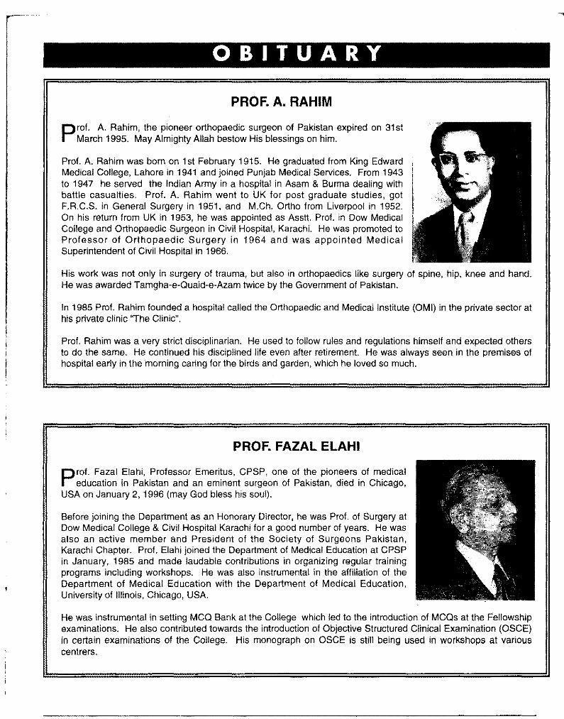
His work was not only in surgery of trauma, but also in orthopaedics like surgery of spine, hip, knee and hand.He was awarded Tamgha-e-Quaid-e-Azam twice by the Government of Pakistan.
In 1985 Prof. Rahim founded a hospital called the Orthopaedic and Medical Institute (OMI) in the private sector athis private clinic "The Clinic".
Prof. Rahim was a very strict disciplinarian. He used to follow rules and regulations himself and expected othersto do the same. He continued his disciplined life even after retirement. He was always seen in the premises ofhospital early in the morning caring for the birds and garden, which he loved so much.
PROF. A. RAHIM
Prof. A. Rahim, the pioneer orthopaedic surgeon of Pakistan expired on 31stMarch 1995. May Almighty Allah bestow His blessings on him.
Prof. A. Rahim was born on 1st February 1915. He graduated from King EdwardMedical College, Lahore in 1941 and joined Punjab Medical Services. From 1943to 1947 he served the Indian Army in a hospital in Asam & Burma dealing withbattle casualties. Prof. A. Rahim went to UK for post graduate studies, gotF.R.C.S. in General Surgery in 1951, and M.Ch. Ortho from Liverpool in 1952.On his return from UK in 1953, he was appointee as Asstt. Prof. in Dow MedicalCollege and Orthopaedic Surgeon in Civil Hospital, Karachi. He was promoted toProfessor of Orthopaedic Surgery in 1964 and was appointed MedicalSuperintendent of Civil Hospital in 1966.
He was instrumental in setting MCQ Bank at the College which led to the introduction of MCQs at the Fellowshipexaminations. He also contributed towards the introduction of Objective Structured Clinical Examination (OSCE)in certain examinations of the College. His monograph on OSCE is still being used in workshops at variouscentrers.
PROF. FAZAL ELAHI
Prof. Fazal Elahi, Professor Emeritus, CPSP, one of the pioneers of medicaleducation in Pakistan and an eminent surgeon of Pakistan, died in Chicago,
USA on January 2, 1996 (may God bless his soul).
Before joining the Department as an Honorary Director, he was Prof. of Surgery atDow Medical College & Civil Hospital Karachi for a good number of years. He wasalso an active member and President of the Society of Surgeons Pakistan,Karachi Chapter. Prof. Elahi joined the Department of Medical Education at CPSPin January, 1985 and made laudable contributions in organizing regular trainingprograms including workshops. He was also instrumental in the affiliation of theDepartment of Medical Education with the Department of Medical Education,University of Illinois, Chicago, USA.
EDITORIAL
Pancreatic pathology in general, and carcinoma in particular has traditionally been shrouded in mystery.Placed as it is in a relatively inaccessible and obscure position in the retroperitoneal region, it arrived late inmedical diagnostic literature. Diagnostic methods helpful in the diagnosis of pancreatic pathology have
arrived on the scene only comparatively recently. As a result, treatment methods too have lagged behind indevelopment and general acceptance, as compared to other parts of the digestive system.
The first resection for Pancreatic carcinoma was performed in the early forties by Whipple and others. It wasfollowed by other cases, with a variable mortality and morbidity after surgery. The biggest problem at that timewas in knowing which tumours were malignant. needing radical procedures and which were benign, requiringlimited procedures only. This problem has been solved to a large extent today by better pre-operative assessmentand the availability of per-operative frozen section facilities.
Although Pancreatic resections were being done from the 1920s onwards, these procedures were confined tomajor centres in the more advanced countries. In a large majority of cases around the world, for a long time,pancreatic carcinoma was being managed palliatively with various biliary-enteric bypass procedures. These hadgood short term effects, but as expected, poor long term survival rates. The situation has now improvedenormously, at least in cases of pancreatic head carcinomas.
This improvement has been achieved by excellent diagnostic imaging techniques including ultrasound, CT Scans,MRls andrecentfydevelopeo Magnetic Resonance Cholangio-Pancreatogreaphy (M.R.C.P.). All these techniquesare non-invasive, rendering invasive methods like P.T.C. or E.R.C.P. unnecessary in most cases. This has helpedin greatly improved understanding of the importance of pre-operative preparation, including physiotherapy andnutrition etc. and better pre-operative management by anesthesiologists. The greatly improved post-operative carein surgical I.C.Us, easy availability of frozen section facilities and introduction of newer surgical techniques, like thepylorus preserving resection etc. has brought down considerably the mortality of Whipple's procedure forpancreatic head carcinoma. The figures for periampullary carcinoma, which should be classed together withpancreatic head tumours as a clinical entity, are even better.
However, the prospects for malignancies of body and tail of the pancreas remain depressingly low. This isbasically due to delay in presentation of these cases. Symptoms do not appear early on and problems that ariseare symptomatically treated by the attending doctors after labelling the problem as dyspepsia, indigestion orirritable bowel. Many of the patients present with maturity - onset diabetes mellitus. Occasionally, the firstindication that something is wrong is the occurrence of secondaries; Troisier's Lymph nodes in the supracalvicularregion are often seen. A patient presented to me with a subcutaneous lump in the scalp which turned out to be apancreatic secondary.
What is therefore required, in the light of our knowledge is that a high index of suspicion should be kept forpancreatic tumours. Full use of modern day investigative techniques should be made in cases where there is astrong likelihood of existence of these tumours, early surgery is mandatory, with a standby frozen section facility ifpossible.
Irshad Waheed
1

EXPERIENCE WITH SEVERE FORMS OF AMNIOTICBANDS OF THE LIMBS AND REVIEW OF
LITERATURE
FAIZAH NAHEED BHATTI*, AMNA NASIR*, M. NAEEM-UZ-ZAFAR KHAN**
ABSTRACT
Amniotic bands are curious congenital constriction bands which may involve any organ or tis-sue in the body. The most plausible theory is considered to be an early rupture oJ the amnionwhich results in the formaiion. oj fibrous bands. These bands may entangle any organ, butwhen they occur in limbs, they produce dllferent degree oj uyury distal to the site oj constric-tion. In this article. amniotic bands afflicting the limbs have been studied. The results oj sur-gical management are assessed and review oj literature is presented.
KEY WORDS: Amniotic bands. autoamputation. Congenital constriction rings.
INTRODUCTIONHistorically many authors have referred to amniotic bandswith various names. These include "Annular Bands","Constriction Band Disruption" and "Ring Constriction", aswell as the more obscure terminologies such as"Streeter's Bands" and "Simonart's Bands", Amnioticband formation is thought to occur due to disruptionprocess and results in destruction of previously formednormal element of the fetus2,3.
MATERIALS AND METHODSFive cases of amniotic bands in children, who presentedwith severe sequelae of different nature from January1987 to April 1996 were selected. For the present studyless severe cases with isolated abnormalities are exclud-ed. Age, sex, anatomical regions involved and the pre-senting abnormalities distal to the constricting bands werenoted pre-operatively. The results after reconstructivesurgery i.e. excision of the constricting band and multipleZ-plasties, were assessed and the resolution of patholog-ical changes that were present before surgery, were stud-ied.
CASE REPORTS:Case No.1A 9 month old baby boy was admitted with multiple arnni-
'Aga Khan University Medical College. Karachi."Department of Paediatric Surgery, Children Hospital, Pakistan Institute ofMedical Sciences, Islamabad, Paksitan.
All correspondence to:Prof. M. Naeem-uz-Zafar Khan,Depatment of Paediatric Surgery, ChildrenHospital, Pakistan Institute of Medical Sciences. Islamabad,
otic bands of toes of both feet resulting in autoamputa-tions. There were distal syndactyly and partial amputa-tions of the fingers of the right hand and circular amnioticbands on the fingers of the right hand and a circular amni-otic band on the proximal left forearm causing massivelymphedema with elephant like skin (Fig.1).
Figure 1 1 he rig!l! arm has jumbled fingers and autoamputationswhereas the tigtht amoniotic band of the left arm has produced massivelymphedema of the forearm and hand, so much so that only the nailswith the tips of the fingers are showing.
Joumal of Surgery Pakistan (International) Vol. 2 (1); January 1997 2
Faizah Naheed Bhatti, Amna Nasir, M. Naeem-uz-Zafar Khan
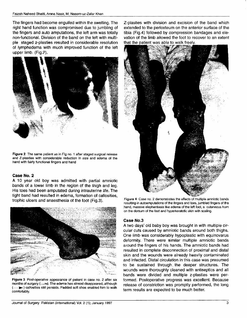
The fingers had become engulfed within the swelling. Theright hand function was compromised due to jumbling ofthe fingers and auto amputations, the left arm was totallynon-functional. Division of the band on the left with multi-ple staged z-plasties resulted in considerable resolutionof lymphedema with much improved function of the leftupper limb. (Fig.2).
Figure 2 The same patient as in Fig no. 1 after staged surgical releaseand Z-plasties with considerable reduction in size and edema of thehand with fairly functional fingers and hand
Case No.2A 10 year old boy was admitted with partial amnioticbands of a lower limb in the region of the thigh and leg.His toes had been amputated during intrauterine life. Thetight band had resulted in edema, formation of callosities,trophic ulcers and anaesthesia of the foot (Fig.3).
Figure 3 Post-operative appearance of patient in case no. 2 after sixmonths of surgery (--"'4. The edema has almost disappeared, although(----. ) callosities still persists. Padded soft shoe enabled him to walkcomfortably.
Z-plasties with division and excision of the band whichextended to the periosteum on the anterior surface of thetibia (Fig.4) followed by compression bandages and ele-vation of the limb allowed the foot to recover to an extentthat the
Figure 4 Case no. 2 demonstrates the effects of multiple amniotic bandsresulting in autoamputations of the fingers and toes, jumbled fingers of thehand, massive elephantiasis like edema of the left foot, a cutaneous hornon the dorsum of the foot and hyperkeratotic skin with scaling.
Case No.3A two days' old baby boy was brought in with multiple cir-cular cuts caused by amniotic bands around both thighs.One limb was considerably hypoplastic with equinovarusdeformity. There were similar multiple amniotic bandsaround the fingers of his hands. The amniotic bands hadresulted in complete disconnection of proximal and distalskin and the wounds were already heavily contaminatedand infected. Distal circulation in this case was presumed-to be sustained through, the deeper structures. Thewounds were thoroughly cleaned with antiseptics and allbands were divided and multiple z-plasties were per-formed. Postoperative progress was excellent. Becauserelease of constriction was promptly performed, the longterm results are expected to be much better.
Journal of Surgery Pakistan (International) Vol. 2 (1); January 1997 3

Experience with sever forms of aminotic bands of the limbs and review of literature
Case No.4A boy of 13 presented with jumbled fingers with peripher-al amputations. His right leg was grossly swollen due tochronic lymphedema with hyperkeratotic skin of the foot,scattered warty lesions, a large cutaneous horn on thedorsum of the foot and anaesthetic toes and feet, with afoul smelling contaminated fissured ulcer on the foot.Following z-plasties, the patient was kept recumbent withelevation of the affected limb. There was rapid reductionin lymphedema, considerable softening of the hyperkera-totic skin and healing of the fissured ulcer (Fig.5). Secondstage z-plasties on the posterior side of the leg are nowplanned.
Figure 5 After ten days of release of the amniotic bands with Z-plasties,incorporting the proximal healthy and distal edematous skin which resultedin rapid reduction in edema and healing of the fissured uncler. Further Z-plasties on the posterior aspect of the leg are planned.
Case No.5A baby girl of 9 months presented with bilateral amnioticbands of the distal one third of legs which had resulted inenlargement due to lymphedema of the right leg and tal-ipes equinovarus of the left leg. She was treated with exci-sion of the bands, multiple z-plasties and elongation of thetendo Achilles on the left side to correct the equinus defor-
Figure 6 Two months after surgery edema of the limb has subsidedand the equinous deformity (-~) on the right foot has also been cor-rected by elongation of the tendo-Achilles and post capsulotomy of theankle joint.
mity. Because surgery has been performed at an earlyage, we expect future development of her limbs should begood (Fig. 6).
DISCUSSIONEstimates of the incidence of this condition ranges from 1in 5000 to 1 in 10,000 and over 600 cases have beenreported. No sex predilection has been observed',Although many attempts have been made to discover agenetiC alteration, it has not yielded any conclusive evi-dence. Most likely theory is that of obscure environmental(exogenous) influences that predispose to disruption ofthe placental layers leading to spontaneous amniotic rup-ture'". There has been one case report where amniocen-tesis done with a large needle predisposed to amnioticdamage and subsequent rupture'.ln one case series,9500 patients were observed out of which amnion dys-morphism was observed in 30 cases by ultrasound.Pregnancy outcome was favorable in all cases, thus ultra-sound visualization in early pregnancy has also been
shown to have no adverse pregnancy outcome'. Familialclustering has been observed in one case of the Amnioticdeformity, Adhesions, Mutilations (ADAM) cornplex'".Alternatively some form of germ cell defect or a defect indevelopment of the tissues is also suspected".
The ruptured amnion forms fibrous strands which entwinethe fetus. Most characteristic is the presence of multiplefibrous strands of amnion extending from the placentalinsertion of the umbilical cord to the surface of the amniondenuded chorion or floating freely within the chorionicsac. Most often distal limbs of the fetus get strangulated,
4 Journal of Surgery Pakistan Journal of Surgery Pakistan (International) Va\,2 (1); January 1997
Faizah Naheed Bhatti, Amna Nasir, M. Naeem-uz-Zafar Khan
as we have observed in our cases. Inadvertently thebands become permanently attached to the underlyingsoft tissue, which then must be removed by surgery.
Injury to the fetal parts may range from mere compressionand ring strictures to intrauterine amputation. Extent ofdamage is determined by the duration and severity of thecompression. Defects may be circular or transverse, anddefects of varying tightness and depth may affect any orall layers of the skin and soft tissues beneath". If the lym-phatiC as well as venous drainage is blocked, lymphede-ma and swelling of the dependent portion may be emi-nent. This edema should be differentiated from congenitallymphedema'. Amniotic bands also cause other patholo-gies like cleft lip, encephalocele etc".
Mild to moderate vascular insufficiency of the developinglimb bud causes nutritional depletion, leading to underde-velopment and a hypoplastic limb, But this may be initial-ly masked by the gross lymphatic swelling. Severe arteri-al obstruction will eventually cause ischemia and necrosisof the dependent limb. The ultimate outcome is intrauter-ine amputation. Nerve compression may give rise to neu-ropathy, as well as distal anaesthesia. Ischemic insuffi-ciency can lead to trophic ulcers, Callosities and horns.
Constriction bands often bind fingers together during theprocess of their separation. This leads to pseudosyn-dactyly'. Pseudosyndactyly, also known as jumbled fin-gers, is distal in nature, whereas primary syndactyly isproximal. Sometimes talipes equinovarus can also occurdue to these bands as seen in case No.5.
Severe cases are usually associated with raised levels ofalpha-fetoportein and acetylcholinesterase in the amniot-ic fluid. The anomalies usually result from exogenousintluences=, but an observation in two members of a fam-ily suggests that genetiC factors might operate in somecases', As a rule the absent parts due to autoamputationare usually multiple, but rarely bilaterally symmetrical.Symmetrical cases are usually genetiC in origin'.
The primary goal would be to relieve lymphedema, andrestore normal circulation as soon as possible. The bandis removed by plastiC procedures, which then exposes theunderlying constricted defect. This must be repaired bymultiple Z-plasties'2. Conjoined fingers must be detachedand skin grafts may be necessary to provide proper shape.Criteria for management should include degree of com-pression, and loss of function. By and large general rule isearly correction by plastic and orthopaedic procedures inorder to allow the child maximal use of his hands and feet.Generally the prognosis of such patients is good.
REFERENCES1. Welch; Randolph; Ravitch; O'Niel; Rowe. Paediatric
Surgery Vol 2; 4th ed. Year book Med.publishes 1986.
2. Behrman, R.E.; Klegman,R.M.; Nelson,W.E.;Vaughanlll,V.C. Nelson Textbook of Paediatrics, 14thed. W.B. Saunders Comp, 1992.
3. Pers,M. Congenital absence of skin: Pathogenesisand relation to ring constriction. Acta.Chir.Scand.1963; 126:388.
4. Goodman,R.M; Gorlin,R.J. Amniotic Band DisruptionComplex. The Malformed Infant and Child-AnIllustrated Guide. Oxford University Press, 1983.
5. Neuhauser,G; Sitzman,F. ADAM Complex- maxillofa-cial abnormalities and abnormalities of the extremitiescaused by amniotic strangulations. Dtsch- Zahnrztl-z.1979 Jul;34(7) : 546-50.
6. Weinzweig,N. Constriction band induced vascularcompromise of the foot: Classification and manage-ment of the "intermediate stage of constriction ringsyndrome. Plas Reconstr Surg 1995; 96(4) :972-7.
7. Zimmer,E.Z; Bronshtein,M. Ultrasound observation ofamnion dysmorphism at 14.5-16 weeks. prenat.Diagn 1995 May;15(5) : 447-9
8. Keller,H; Neuhauser,G; Durkin-Stamm-MV;Kavaggia,E.g; Schaff,A; Sitzman,F ADAM Complex-a pattern of craniofacial and limb defects.Am.J.Med.Gen. 1978;2(1) : 81-98.
9. Streeter,G,L. Focal Deficiencies in fetal tissues andtheir relation to intrauterine amputation.Contrib.Embryol(Carnegie Institute Washington)1930; 22:1.
10. Higginbottom,M,C; Jones,K,L; Hall,B,D; Smith,D,W.The amniotic band disruption complex: timing ofamniotic rupture and variable spectra of consequentdefects. J.Peadiatr. 1979 Oct;95(4) :544-9.
11. Miyajima,K; Natsume,N; Kawai,T; Tizuka,T. Obliquefacial cleft, cleft palate, and supernumerary teeth sec-ondary to amniotic bands. Cleft.palate.CraniofaciaI.J.1994 Nov; 31 (6) : 483-6.
12. Weinzweig,N; Barr,A. Radial, ulnar admedian nervepalsies caused by a congenital constriction band ofthe arm: single stage correction. Plast.Reconst.Surg,1994 Nov;94(6) : 872-6.
Journal of Surgery Pakistan (international) Vol. 2 (1); January 1997 5
INCIDENCE OF CARCINOMA IN NODULAR GOITRESALIM SOOMRO, SHAH ZAMAN, NUSRAT ANIS, ANIS SUBHANI AND MAJEED BALOCH
ABSTRACT
In six yearsjrom Jan 1990 to Dee 1995, 369 (90 males and 279 females) euthyroid patientswith goitre were operated in Surgical Unit-II, JPMC Karachi. On technitium 99 Isotope Scan175 were solitary nodulesand 194 were multiple nodular goitres. All these patients were clin-ically and biochemically euthyroid. On Biopsy 15.52% solitary nodules and 11.94% multin-odular goitres were malignant; over all incidence of malignancy was 13.6%
KEY WORDS;- Carcinoma in Nodular Goitre
INTRODUCTIONPalpable Multinodular or Solitary nodule of thyroidgland remains a common reason for referral of a largenumber of patients to both Surgeons & Physicians. Onclinical examination, Technitium 99 Isotope Scan andultra sonography diagnosis of solitary or multinodular-ity is made with final confirmation at surgery and onhistopathology.
Much controversy exists over appropriate diagnosticand therapeutic management of nodular goitre. This iscompounded by the facts that thyroid nodules are rel-atively common, malignancy is also not very rare anda well differentiated thyroid cancer generally runs avery indolent course.
PURPOSE OF STUDYTo determine the incidence of malignancy of thyroid insolitary and multi nodular goitres operated in SurgicalUnit-II, JPMC Karachi, a major tertiary hospital of thecountry.
MATERIAL AND METHODSAll euthyroid patients admitted to Surgical Unit-II fromJan 1990 to Dec 1995 were included in the study. Allthe patients had complete clinical examination, hor-monal assay (Serum T3, T4 & TSH) and radioisotope(Tc99) Scan. FNAC was done on all the solitary nod-
All correspondence to:Prof. Majeed BalochSurgical Ward II, J.P.M.C, Karachi
ules before surgery. Partial thyroidectomy was done inmulti nodular goitres and lobectomy was done in soli-tary nodular gOitres. All the operated specimens weresubjected to histopathology.
RESULTS:In the 6 years study of the 369 euthyroid patients,75.7% were females and 24.3% were males.Maximum number of patients were of the age group of30-40 years. (Table I) Solitary nodular goitre were 175and 194 were multi nodular.
Table-I
Sex Distribution
Age of Patient (years) Number of patient %
10 - 20 29 7.821 - 30 60 16.231 - 40 143 33.741 - 50 106 23.951 - 60 20 5.4
61 - 70 11 3Incidence of carcinoma was 15.52% in solitary nodularand 11.94% in multi nodular gOitres. Overall incidenceof carcinoma in euthyroid nodular was 13.6%. Table II
Journal of Surgery Pakistan Vol. 2 (1); Jnauary 1997 6
Salim Soomro, Shah Zaman, Nusrat Anis, Anis Subhan and Majeed 8alooch
Table-II
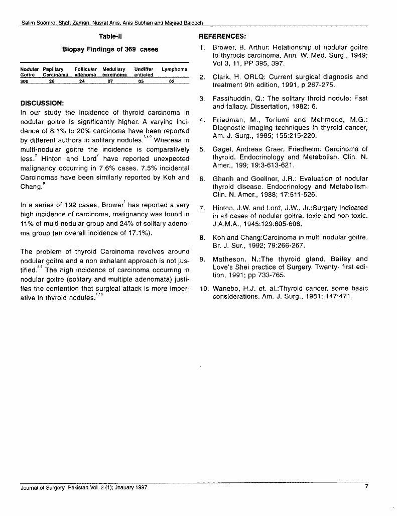
Biopsy Findings of 369 cases
Nodular PapillaryGoitre Carcinoma
Follicular Medullaryadenoma carcinoma
Undifferentiated
Lymphoma
300 26 24 07 0205
DISCUSSION:In our study the incidence of thyroid carcinoma innodular gOitre is significantly higher. A varying inci-dence of 8.1% to 20% carcinoma have been reportedby different authors in solitary nodules.
3AgWhereas in
multi-nodular gOitre the incidence is comparativelyless.' Hinton and Lord? have reported unexpectedmalignancy occurring in 7.6% cases. 7.5% incidentalCarcinomas have been similarly reported by Koh and
B
Chang.
In a series of 192 cases, Brower1
has reported a veryhigh incidence of carcinoma, malignancy was found in11% of multi nodular group and 24% of solitary adeno-ma group (an overall incidence of 17.1%).
The problem of thyroid Carcinoma revolves aroundnodular goitre and a non exhalant approach is not jus-tified.
56The high incidence of carcinoma occurring in
nodular gOitre (solitary and multiple adenomata) justi-fies the contention that surgical attack is more imper-ative in thyroid nodules.
1.1o
REFERENCES:
1. Brower, B. Arthur: Relationship of nodular goitreto thyrocis carcinoma, Ann. W. Med. Surg., 1949;Vol 3, 11, PP 395, 397.
Clark, H. ORLO: Current surgical diagnosis andtreatment 9th edition, 1991, p 267-275.
3. Fassihuddin, 0.: The solitary throid nodule: Fastand fallacy. Dissertation, 1982; 6.
2.
4. Friedman, M., Toriumi and Mehmood, M.G.:Diagnostic imaging techniques in thyroid cancer,Am. J. Surg., 1985; 155:215-220.
5. Gagel, Andreas Graer, Friedheim: Carcinoma ofthyroid. Endocrinology and Metabolish. Clin. N.Amer., 199; 19:3-613-621.
6. Gharih and Goellner, J.R.: Evaluation of nodularthyroid disease. Endocrinology and Metabolism.Clin. N. Amer., 1988; 17:511-526.
7. Hinton, J.W. and Lord, J.W., Jr.:Surgery indicatedin all cases of nodular goitre, toxic and non toxic.J.A.M.A., 1945:129:605-606.
8. Koh and Chang:Carcinoma in multi nodular gOitre.Br. J. Sur., 1992; 79:266-267.
9. Matheson, N.:The thyroid gland. Bailey andLove's Shei practice of Surgery. Twenty- first edi-tion, 1991; pp 733-765.
10. Wanebo, H.J. et. al.:Thyroid cancer, some basicconsiderations. Am. J. Surg., 1981; 147:471.
Journal of Surgery Pakistan Vol. 2 (1); Jnauary 1997 7

EARLY EXPERIENCE OF LAPAROSCOPICCHOLECYSTECTOMY AT J.P.M.C
MIR ZAFAR All, RIZWAN NASIM, S. SAGHEER HUSSAIN SHAH,SHAHID RASUL, RASHEED A. CH., IRSHAD WAHEED
ABSTRACTThe application oj minimal invasive surgical technique for the removal oj gallbladder hasemerged as the preJerred method oj treating symptomatic gallstone disease. The objective ojthis study is tofind out the complications, conversion, reasonJor conversion and hospital stayoj 127 laparoscopic cholecystectomies performed over a period oj 16 months at JPMC fromJune 1994 to October 1995. As an early experience one hundred and eleven were success-Jully operated laparoscopically and 16 converted to open laparotomy. Average duration ojstay after successful laparoscopic cholecystectomy was 4 days.
KEY WORD:Laparoscopic Cholecystectomy, Early Experience
RESULTS Transient JaundiceDuring 16 months period from June 1994 to October ---------------------1995, 127 cases were approached laparoscopically Biliary Fistulaand cholecystectomy was performed. This study is ---------------------divided into two equal halves, each of eight months Retained Stone in CBDduration. ---------------------All correspondence to: Surgical EmphysemaDr. Mir Zafar AIi, Department of Surgery, ---------------------Jinnah Postgraduate Medical Centre,Karachi. Intra Abdominal Collection
INTRODUCTIONWith the availability of facilities of operativelaparoscopy at JPMC, this study was designed to eval-uate the reasons for conversions, complications, hos-pital stay during the early period. Nowadays most ofthe patients having symptomatic gall stone are treatedwith laparoscopy. Laparoscopic gallbladder operationsare associated with less morbidity than conventionalopen Cholecystectomy.
PATIENTS AND METHODSThis is a sixteen month retrospective study of an earlyexperience of laparoscopic cholecystectomy carriedout in two surgical units of JPMC. This study is divid-ed into two equal halves - each of 8 months duration.Six consultant surgeons operated upon the cases.Patients were admitted through the out-patient clinics,after confirmation of diagnosis of cholelithiasis. All thecases were operated under general anaesthesia.Prophylactic first generation cephalosporin (Ceporex)was given intravenously at the time of induction ofanaesthesia.
In the first half 49 (38.5%) and in the second 78(61.5%) cholecystectomies were performed laparo-scopically. The mean operating time for laparoscopiccholecystectomies was 100 minutes in the first halfand 72 minutes in the second half. Overall complica-tion rate was 10.2% (13 cases) (Table I). Six cases(4.72%) were converted to open laparotomy in the firsthalf, and 10 cases (7.87%) in the second half. Overallrate of conversion was 12.59% (16 cases). The rea-sons for conversion were adhesions, anatomical vari-ations, hemorrhage, failure of equipment, carcinomaof gallbladder, common bile duct damage and empye-ma gallbladder (Table II ,III). Mean hospital stay aftersuccessful laparoscopic cholecystectomies was 4.2days and 6.3 days in converted cases (Table III).
Table-I
Complications
Laparoscopic Surgery
Wound Infection 3
3
2
2
2
Journal of Surgery Pakistan (International) Vol. 2 (1); Jnauary 1997 8

Table-II
Mir Zafar AIi, Rizwan Nasim, S. Sagheer Hussain Shah, Shahid Rasul, Rasheed A. Ch., Irshad Waheed
Ratio of conversion in both halves of laparoscopiccholecstectomy
1st Half 2nd Half
Laparoscopic 49 (38.5%) 78 (61.5%)Cholecystectomies
No. of Successful 43 (87.8%) 68 (87.2%)Lapcholecystectomy
No. of Conversion 06 (12.2%) 10 (12.8%)
Table-III
Reasons for conversion
Reasons forConversion
Cases in 1st Half Cases in 2nd Half
Adhesions 3 3
Hemorrhage 2
Anatomical Variation a 2
Failure of Equipment a 2
Carcinoma Gall Bladder a
CBD Transection
Empyema Gall Bladder a
Table-IV
Hospital stay after surgery
No. of Days Successful Laparoscopic
1 Day
2 Day 17
3 Day 36
4 Day 19
5 Day 16
6 Day 9
7 Day 5
Above 7 Days 8
Total 111
Converted
a
a
a
6
4
a
5
16
9Journal of Surgery Pakistan (International) Vol. 2 (1); Jnauary 1997
DISCUSSIONLaparoscopic cholecystectomy is rapidly becoming thetreatment of choice for symptomatic gallstone dis-
ease.'2 because a successful laparoscopic operationis associated with less morbidity than conventionalone but it might be associated with high rate of seriouscomplications, especially bile duct injury and damage
to gut during trocars entry". Besides all this it is anexpensive procedure and lot of expertise and experi-ence is required to perform this operation. The maindrawback with this technique is difficulty in hand-eyecoordination, and the inability to feel the structures. Inaddition, laparoscopic cholecystectomies might beassociated with high rate of serious complications,especially bile duct injury & damage of gut with trocas
4.5
entry .
Our experience of 127 laparoscopic Cholecystec-tomies have convinced us that successful outcome oflaparoscopic cholecystectomies is dependent uponproper patients selection, meticulous techniques andattitude to convert to open if needed. In our study, thereason for conversion was highest in cases with adhe-sions and due to hemorrhage followed by anatomicalvariations in two cases. Preoperative failure of equip-ment was a reason for conversion in two cases. A casewas opened because of bile duct damage bydiathermy and another due to empyema gallbladder(Table III). Hospitalization and recovery time was sig-nificantly shorter in successful laparoscopic operationwith the benefit of minimal trauma but comparing withconverted cases, there was marked difference (TableIV). We found that this technique should be performedwith tuuy equipped theatre with stand-by for open con-version and surgeon should have an adequate experi-ence in open biliary surgery as well as expertise in thetechnique of laparoscopy.
In this study the results do not compare well with thosein published literature perhaps because it was an earlylearning experience.
CONCLUSIONIn the light of the present study we believe that sur-geons should rationalize the provision of laparoscopicsurgery among themselves and should be careful inthe selection of the patients. Considering obvious ben-efit of laparoscopic cholecystectomies, we have tomeasure its drawbacks and it seems appropriate toperform careful studies in all centres where laparo-scopic surgery is being performed. Our results are notcomparable with the international study of early expe-rience because we are in the "learning curve".

Early experience of laparoscopic cholecystectomy at J.P.M.C
REFERENCES1. Macntyne IMC Wilson RG Laparoscopic
Cholecystectomy Sr. J. Surg. 1993 8:552-9
2. Grace Pa, Qureshi A, Coleman J. et alReduced post operative hospitalization afterlaparoscopic cholecystectomy Sr. J. 1991,78:160-2
3. Moosa AR, Easter DW,Casola G, DAgostino Hopen Cholecystectomy;leave, analgesia andSr. J. 1992, 215:203-8
Van Sonnerberg E,Lapraroscopic versushospitalization sicktrauma responses
4. R. J. C. Steel, K. Marshall, M. Lang and J. Doran
Introduction of laparoscopic cholecystectomy in alarge hospital independent audit of the first 3years Sr. J. 1995, 82:968-971
5. A. J. Mc Mohan, G. Fullarton, J. N. Saxyer and P.J. O. Dwyer Sile duct injury and bile leakage inlaparoscopic surgery Sr. J. 1995, 82:307-313
6. G. Crosthwaite, T. Chung, P. Dunkely, S. Shineand A Cuschiexi comparison of direct vision andelectronic two and three dimensional display sys-tem on surgical task efficiency in endoscopicsurgery Sr. J. 1995, 82:847-851
7. Stoker ME, Vose J, 0' Mara P, Maini Ss.Laparoscopic cholecystectomy A clinical andfinancial analysis of 280 operationsArch Surg 1992, 127:589-95
ROLE OF ANTIOXIDANT-EMPOXIPINE ON CORNEALCONSERVATION
(AN ELECTRON MICROSCOPY INVESTIGATION)
R. GOUNDOROVA, I. CHOROSHILOVA, G. BORDIOGOVA, , L. ILATOVSKAIA, S. SIDDIQUE,
We investigated the role of emoxipine in the structuralpreservation of two groups of corneas: 1. Controlgroup where the preservation was completed on clas-sical method of filatovs, where eye ball was refrigerat-
ed at +2°C to +4°C. 2. The experimental group, 0.5 mlto 0.7 ml emoxipine where injected into interior cham-ber.
All Chemical reactions in corneal tissue were slowed,by decreasing temperature, including free radicalprocess, which play an important role in destruction ofcell structure. That is why for blocking free radicalprocess, in cornea we used a new antioxidant emoxip-ine.
After 3-8 days corneas fixed in 2.5% glucotar aldehydesolution and dehydrate in alcohol.
With increasing degree of' concentration ultra thin
All correspondence to:Department of Tramatology,Reconstructive Surgery,Holmolthz's Institute of Eye Diseases,Moscow, Russia.
slides were investigated under electron microscope(Tesla - 500). We used the method of total detachmentof endothelium and impregnation of it by silver(Smolins method) and investigated, under light micro-scope. We studied 25 human and rabbitts' corneas.
Our morphological investigation showed that injectingemoxipine into the interior chamber of the eye, pro-motes preservation of the cornea especially forendothelial cells. These cells were intact as comparedto control group, where many endothelial cells weredeleterious, increasing the period of conservation to 6-7 days.
Experimental investigation showed that moist chamberrefrigerated at' +2°C to +4°C, slows down autolysisprocess. Injecting emoxipine into the interior chamberdoubles the structural preservation as compared to thecontrol group.
10 Journal of Surgery Pakistan (International) Vol. 2 (1); Jnauary 1997
ROAD TRAFFIC ACCIDENT INJURIES INCHILDREN
SHABBIR HUSSAIN, GHULAM All, MOHAMMAD ASLAM, IRFAN QURESHI,RAFIQ KHANANI, SANAULLAH
ABSTRACT:
During one year (30th Nov. 92 - 1st Dec. 93), 104 cases below 15 years oj age oj Trafficlniuries reported to JPMC emergency room including 112 abrasions, 6 bruises, 82 lacerationand 17fractures. Nine cases were fatal, most due to head & neck injuries. Maximum no ojJatal cases occured in the early morning hours orlate night.
Key Words:- Road Traffic Accidents (RTA), Children Cause oj death in RTA.
INTRODUCTIONRoad traffic accidents are public health problem andtheir magnitude has been recognized by the 27th WorldHealth Assembly by concluding that a workable solutionto this problem requires coordinated efforts of interna-tional agencies and organization, national, regional and
local authorities and those of world citizenry."
Road traffic accidents (RTA) are a leading cause ofdeath among those under 30 years and account for agreater proportion of years of potential life loss thancancer and heart disease comblned":". They involvebicycle, motor vehicles and animal carts in Pakistan.
MATERIAL AND METHODSThis study was conducted at Jinnah PostgraduateMedical Centre (JPMC) from 30th November 1992 to1st December 1993. In a period of one year, 104 chil-dren under 15 years of age reported at JPMC emer-gency room and accident department after road trafficaccidents. After complete physical and necessary radi-ological examination, they were referred to specializedunits for management. The data was collected on aproforma which included, name, age, sex, site ofinjury, type of injury (abrasion, bruise, fracture, ampu-tation), out patient care and admission or death. Anattempt was made to determine the injury responsiblefor death infatal injuries.
All correspondence to:Dr. Ghulam Ali, Department of Forensic Medicine,Sindh Medical College, ~arachi.
RESULTSDuring one year study 104 children under 15 years ofage reported in E.R. There were 67 male(64%) and 37female (36%), patients, with male to female ratio of1:8:1. Nine (8.7%) cases, (5 male and 4 female) werefatal.
Abrasions were the most common type of injuries onmore than one anatomical region of the body. (Table-I). The timing of the number of reported cases exhibitsan interesting pattern. Majority of the cases reportedbetween 12:00 Noon to 6:00 P.M. Only 5 cases wereadmitted for further observations, rest were dis-charged after administration of necessary treatment.
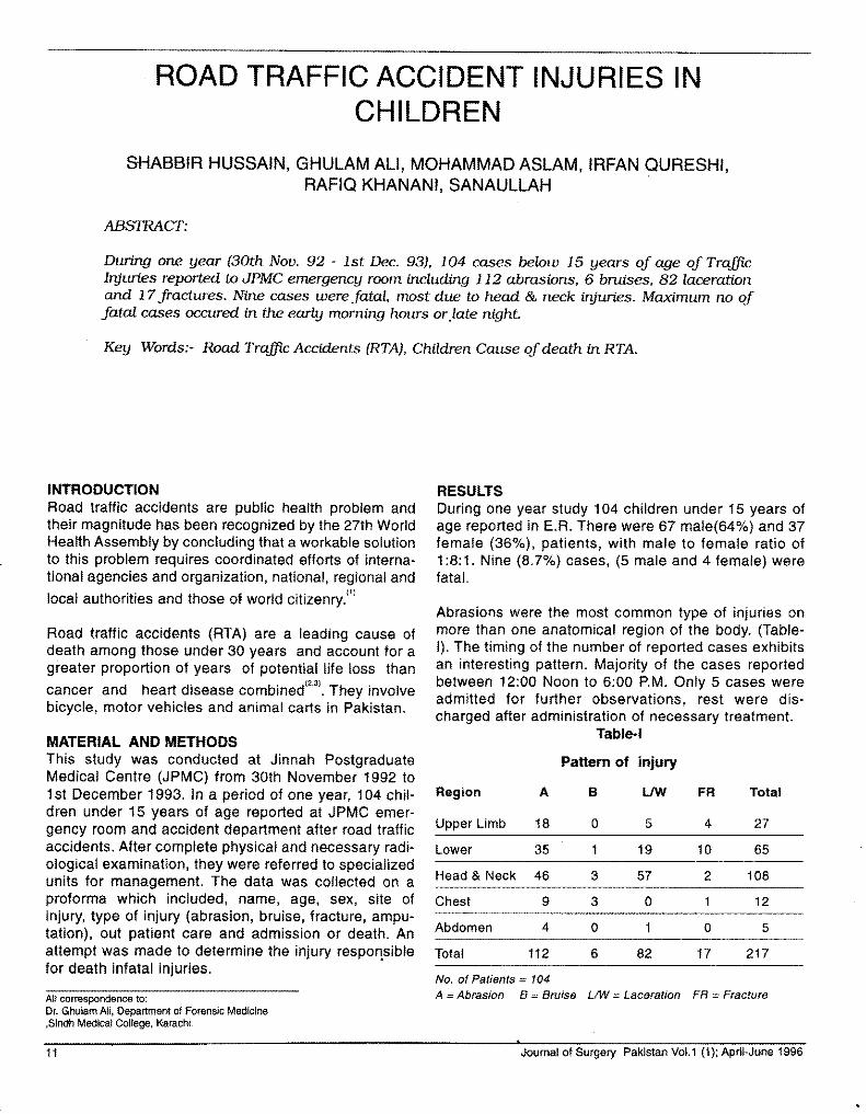
Table-I
Pattern of injury
Region A B UW FR Total
Upper Limb 18 0 5 4 27Lower 35 19 10 65Head & Neck 46 3 57 2 108Chest 9 3 0 12Abdomen 4 0 0 5Total 112 6 82 17 217No. of Patients = 104A = Abrasion B = Bruise LIW = Laceration FR = Fracture
11 Journal of Surgery Pakistan Vol.1 (1); April-June 1996
"

Road Traffic Accident Injuries In Children
DISCUSSIONRoad traffic accidents occur in every part of the worldwhere automobiles are used. They are the leading
f (4·6)causes 0 unnatural deaths around the globe. Malepredominace is reported in most of the series. (7,6)
In our series injuries of the lower limb (70.4% are morethan those on the upper limb (29.6%).The lower limb ismore prone to fracture being weight bearing and hiteasily. Tibia is the most commonly broken bone in thelower limb. Elsewhere the pattern of injuries is almostthe same. (9)
Maximum number of accidents occured between 12:00Noon and 6:00 P.M.(Table-II) It coincided with the peakschooling hours as the morning shifts come off and theafterrtoon shifts of most schools begin. It is thereforeemphasized that reckless driving should be checkednear school premises. Quite interestingly, the maxi-mum fatalities occurred between 12:00 midnight &6:00 A.M. They were mostly recorded near the airport,railway station and in those areas where marriagehalls (public places of gatherings) are plentiful. Thefatalities recorded near the airport or railway stationcan supposedly be due to the tendency of elders totake children to receive relatives arriving from out-sta-tion areas. While the fatalities reported from marriagehall areas can be explained by the presence of chil-dren at late night in these ceremonies and the locationof marriage halls along main roads where heavy trafficmoves with relatively high speed.
This study highlights the urgency of reducing acciden-tal injury to children. Legislation and increasing publicawareness by utilizing mass media can help in improv-, 10mg the present status .
Table-II
Time of arrival of Pateints at the Hospital
Time Period No. Of Cases
6:00 A.M. TO 12:00 NOON 26
12:00 NOON TO 6:00 P.M. 43
6:00 P.M. TO 12:00 NIGHT 28
12:00 NIGHT TO 6:00 A.M. 7
Total 104
REFERENCES
1. Anos. avoidable epidemic. World Health (1975)
P. 4 W.H.O. Geneva.
2. Anderson B. OW Finding from a major U.S. sur-
vey of patients hospitalized
with head injuries. Public health Rep. (1983)98(5) 475-8.
3. Aganga A.O: Epidemiology of RTA's in Zaria.
Nigeria. J. soc. Health. Aug.
1983; 103(4): 123-6.
4. Jamieson E.G. The toil of road, Clinical aspects.B.M.J. (1966) 2: 157.
5. Mc Farland RA: The epidemiology of motor
vehicle accident. J.A.M.A.
(1962), 180:189.
6. K.H. Neurological aspect of traffic accidents.
Report of sub-committee of
Canadian Neurosurgical Society. Canada Med.
Association. J(167), 97,
1364.
7. Jinadu M.K. Epidemiology of motor cycle acci-
dents in a developing country.
J.R.S.H. (1984) 104(4):153-6.
8. Pless I.B. Accident
BMJ(1991 ):303:462-4.prevention.
9. The National committee for injury prevention
and control. Injury prevention:meeting the challenge. Am. J. Pre.
Med.(1989):5:5-6.
10. Akhtar J., Aziz A. Trauma in children Pak J.
Surg. 1994; 10:108-109.
Journal of Surgery Pakistan Vol.1 (1); April-June 1996 12
SURGICAL APPROACH FOR HEPATIC HYDATIDDISEASE
G. ASGHAR CHANNA, AHMAR All SHAH
ABSTRACT:We reviewed thirty two patients oj hepatic hydatid cysts treated surgically by us in thedepartment oj surgery J.P.M ..C. Karachi, over a period oj five years. The study was in rela-tion to surgical procedure and complications resulting from the treatment and possible Jollowup oj the patients. Ultra Sound scanning, being non invasive and sensitive investigation, was
used regularly inJollow up to assess the size oJresidual cavity and recurrence of the cYS{45.Patients with biliary fistula and abscess formation. with discharging tract were treated con-servatively.
KEY WORDS: Liver hydatid disease, Surgery for hydatid disease, Complicationsoj hydatid disease.
INTRODUCTIONMany surgical techniques have been employed in themanagement of hydatid disease. Because of the fear ofrecurrence due to rupture and damage to the human tis-sue due to Hle scolicidal agents, resection of the involvedportion of liver has been advised, despite considerablemorbidity and mortality 13.
MATERIAL & METHODSThirtytwo patients, operated during five years, wereincluded in the study. All of these patients were operatedby surgeons of a surgical unit. It was noticed that patientshad come from all over the country, the majority beingfrom Sindh and Baluchistan. Prospective investigationdone included ultrasound, computerized tomographicscanning and Haemagglutination test. All patients wereoperated after assessment. Liver was approachedthrough the subcostal or right para-median incisions.Nineteen out of 32 cysts (59%) were on the right side ofthe falciform ligament. Five (15%) cysts were seen in theregion crossed by the falciform ligament. Cysts were iden-tified by palpation and isolated with 10% saline soakedswabs. After aspirating, saline was injected and cyst evac-uation was done. Difficulty was encountered while operat-ing cysts on the summit of lobes and posteriorly seatedcysts. If the cavity was larger it was filled with omentumAll correspondence to:Dr. G. Asghar ChannaDeptt. of Surgery ,J.P.M.C.,Karachi.
and stitched with chromic catgut. The cavities on the infe-rior aspect of the liver were drained by vacuum or depen-dant drains.
RESULTSIn 10 (31.2%) patients mild to moderate spillage of fluidoccurred. However no patient developed anaphylaxis.Seven patients developed fever after surgery. Radiologyand ultrasound scanning revealed that three patients hadpleural effusion, which responded to conservative mea-sures. Four patients developed abcesses; they weretreated by aspiration under ultrasound guidance.
Two patients developed biliary fistula remained in the hos-pital for 34 days and were followed-up in outpatient. Theleakage progressively decreased and stopped over aperiod of two months. The longest follow-up in a fewpatients has been upto 5 years. Ultrasound scanning wasrepeated at six monthly intervals. Follow-up ultrasoundscanning showed residual cavity or Hydrops.
Progressively decreasing size of the cavity was noticedand no hydatid sand was detected on subsequentUltrasound examination.
COMPLICATIONSSpillage of hydatid fluidPleural EffusionAbcessBiliary leak
10 patients (31.2%)3 patients (9 %)4 patients (1.2 %)2 patients
13 Journal of Surgery Pakistan Vol. 2 (1); Jnauary 1997

Surgical approach for hepatic hydatid disease
DISCUSSIONLiver, being the first filter for the ecchinococcus infesta-tion, is a common site for the hydatid cysts formation.Many treatment modalities are being used to treat thiscondition.
•
Use of anti-helmenth drugs like albendazole and meben-dazole has achieved partial success. These drugs arespecially advocated in wide spread inoperable hydatido-SiS,15.11.9.12Ultrasound or computerized tomographic guideddouble percutaneous aspiration and injection technique (OPAl) is also used. The success of treatment has beenclaimed specially in uncomplicated hepatic hydatid cysts.The scolicidal agent is 95% alcohol or hypertonic saline 7.8.
Surgery is the most common form of treatment for hepat-ic hydatid cysts. Omentoplasty, marsuplization, introflex-ion, pericystectomy and hepatic resections are commontechniques 5.
In our series, surgical technique did not involve any majorhepatic resection. There was no mortality. Two patientsout of 32 had long hospital stay (34 days) who had biliaryfistula. The fistula closed with conservative treatment. Theabscesses were aspirated under ultrasound guidance.
CONCLUSIONWithout major liver resection hepatic hydatid cysts can betreated. Sclerosing cholangitis and anaphylaxis was notseen in this series of patients. Ultrasound scanning is areliable diagnostic and follow up investigation. Biliary fis-tula heals spontaneously if no distal obstruction is detected.
REFERENCES1. AI-Habib-AA, etal. Hepatic Hydatid cysts: presenta-
tion and surgical management Yemen.J-R-ColI-Edin. 1992 Aug; 37 (4) 229-31.
2. Acumas-B; Rozanes-I; Celik-L; Minareci-O; Alper-A;Ariogul-O; Gokmen-E; Purely cystic Hydatid diseaseof the liver: treatment with percutaneous aspirationand injection of hypertonic saline. Radiology, 1992Feb; 182 (2) : 541-3.
Akhan-O; Dincer-A; Gokoz-A; Sayed-I; Harlioglu-S; Abbasoglu-O; Eryilma-M; Basim-A; Baris-I.Percutaneous treatment of abdominal hydatidcysts with hypertonic saline and alcohol. An exper-imental study in sheep. Invet-Radiol. 1993 Feb;28 (2) : 121-7.
3.
4. Behus-A; Van-Heerden-J A. Surgical managementof hepatic hydatid disease. HPB-Surg. 1992; 6(1) :1193-7
5. Bilge-A; Sozuer-EM. Diagnostic and surgical treat-
14Journal of Surgery Pakistan Vol. 2 (1); Jnauary 1997
ment of hepatic hydatic di ease. HPB-Surg. 1992;6(1) : 57-64.
Demiraci-S; Eraslan-S; Andol-E; Bozatil-L.Comparison of the result of different surgical tech-niques in the management of hydatid cysts of theliver. World-J-Surg. 1989 Jan-Feb; 13 (1) : 88-90.
Giorgio-A; Tarantino-L; Francica-G; Mariniello-N;Aloisio-T; Sosia-E; Pierri-G. Unilocular hydatid cysts: treatment with U/S quided, double percutaneousaspiration and alcohol injection. Radiology, 1992Seo; 184 (3) : 705-10.
Gori-S; Campatelli-A; Luchi-S; Palasdini-A; Savalli-E; Scasso-A. Cytology in the percutaneous treat-ment of hydatid cysts. A report of four casses. Acta-Cytol. 1993 May-Jun ; 37 (3) : 423-6.
Gocmen-A; Toppare-MF; Kiper-N. Treatment ofhydatid disease in childhood with mebandazola.Eur-Respir-J. 1993 Feb; 6 (2) : 253-7.
10. Hai-AA; etal. Surgical treatment of hypatic hydatidcysts of liver. Indian-Med-Associ. 1991 Nov; 89 (11)-313.
6.
7.
8.
9.
11. Lin-YH; Wang-XJ; Chen- YT. Preliminary observationof continuos albendozola therapy in alveolarechinococcosis. Chin-Med-J-Engl. 1991 Nov; 104(11) : 930-3.
12. Meneghelli-UG; Martinelli-AL; Bellucdi-AD;Villanova-MG; Vellado-MA; Magro-JE. Polycystichydatid disease (Echinococcus vogeJi). Treatmentwith albendazole.}Ann-Trop-Med-Parasitol. 1992April; 86 (2) : 151-6.
13. Ozmen-V; Igci-A; etal. Surgical treatment of hypatichydatid disease. Can-J-Surg. 1992 Aug; 35 (4) : 423-7.
14. Todorov-T; Vutova-K; Mechkov-G; Georgiev-P;Petkov-D; Tonchev-Z; Nedelkov-G. Chemotherapyof human cystic echinococcus: comparative effica-cy of mebendazole and albendazole. Ann- Trop-Med-Parasitol. 1992 Feb; 86 (1) : 59-66.
15. Todorov-T; Vutova-k; Mechkov-G; Lazarova-1.Experience in the chemotherapy of severe, inopera-ble echinococosis in man. Infection. 1992 Jan -Feb ;20 (1) : 19-24.
16. Vagianos-C; Pohydorou; etal. Successful treatmentof post operative Ext. Biliary fistula. HPB-Surg.1992; 6 (2) : 105-20 : discussion 120-4.
17. Xynos-E; etal. Hydatid disease of liver, diagnosisand surgical treatment. HPB-Surg. 1991 May; 4 (1) :59-66: discussion 66-7.

PRIMARY VARICOSE VEINS: EXPERIENCE ATMILITARY HOSPITALSDILAWAR MEHMOOD, IRFAN AHMED
ABSTRACT:Eighty six patients were managed at two Military Hospitals in a two years period. Seventyeight patients were foUowed regularly. Mqjority of our patients were soldiers with a mean ageof 32 years, long hours of standing being their occupational requirement. Saphenofemoralflush ligation was performed in all cases. Groin to upper calf long saphenous vein strippingwas done in those with above knee varicosities and multiple stab avulsions were done forbelow knee varicosities. Recurrence occurred in 12 cases, the cause being inadequate initialoperation.
KEY WORDS: Varicose veins, Saphenofemoralligation, Surgical technique
INTRODUCTIONIncidence of primary varicose veins in our country isnot known. Regarding pathophysiology of primary vari-cose veins, the concept is changing and now the fun-damental defect is supposed to be primary defect invenous wall. A recent study has shown that reflux canoccur in the presence of competent saphenofemoralvalve.2,3 Many surgical procedures have been pro-posed to deal with this disease. In military hospitalsmost of the patients are serving soldiers. This is thereason we are getting more patients with varicoseveins than in civilian practice. The purpose of study isto review the present day surgical protocol for thetreatment of varicose veins.
MATERIAL AND METHODSThe study was conducted over a period of two years atCombined Military Hospitals Rawalpindi and Lahore.Eighty six patients were entered into this study ofwhich eight patients were not available for regular tot-lOW-Up.There were 6 female patients. The patientswere mostly soldiers; the civilians who were treatedwith this disease belonged to the lower socio-econom-ic group. Clinical assessment was the mainstay of thediagnosis. Ascending phlebography was done in 10cases in which there was a clinical suspicion of deepvein thrombosis. For the purpose of study, the primary
All correspondence to:Dr. Dilawar Mehmood NaeemDepartment of General Surgery, Combined Military Hospital,Lahore.
varicose veins were described as dilated, lengthenedand tortuous superficial veins with no obstruction todeep venous flow or any arterio-venous fistula.Saphenofemoral flush ligation was done in all cases.Groin to upper calf long saphenous vein stripping wasdone in those with above knee varicosities and multi-ple stab avulsions were done for below knee varicosi-ties.
Post-operatively compression dressing crepe bandagewas applied. Lower limbs were elevated for 12 hoursto reduce hematoma formation and ensure hemosta-sis. Early ambulation was encouraged. Crepe ban-dage was continued for 2 weeks. The patients weretold to report in outdoor after 2 weeks for initial evalu-ation and then after 6 weeks. Results were gradedaccording to cosmetic appearance, relief of symptomsand recurrence of varicosities. The patients were thenfollowed up for 6 months to a year.
RESULTSOf the 78 patients who were followed regularly therewere 72 male and 6 female. Mean age was 32 years.Recurrence occurred in 12 cases. Primary varicoseveins were bilateral in 76% of the cases. Long saphe-nous system was involved in all cases, while shortsephenous was involved in 20% of the cases.
DISCUSSIONPrimary varicose veins is a disease of young adults
15 Journal of Surgery Pakistan Vol. 2 (1); Jnauary 1997

Primary varicos veins: Experience at military hospitals
whose professional requirement forces them to standfor long hours. The concept of etiology is shiftingtowards primary venous wall defect and is attributed todefect in connective tissues. Incompetent valve is themanifestation of this defect which allows high pressuretransmission to superficial veins. The cause of recur-rence is usually inadequate initial operation with fail-ure to perform flush ligation of saphenofemoral junc-tion. Limited stripping of long saphenous veins uptoupper calf in all cases should be done to avoid recur-rence due to persisting mid thigh perforators. In thepopliteal fossa sphenopopliteal junction should becarefully identified and ligated when there is shortsaphenous incompetence.
Surgery for primary varicose veins has quite low inci-dence of complications where patient selection isappropriate and surgical principles are followed.
REFERENCES1. Barnabas A P, macfarlane R, 1985. The use of
preoperative venography in the management ofdifficult primary and recurrent varicose veins.
In : Greenhalgh RM (ed) Diagnostic techniquesand assessment procedures in vascularsurgery. Grune & Stratton, London, pp 447-458.
2. Campbell WB. Varicose veins. BMJ1990;300:763-4.
3. Dodd, H., and F.B. Cockett: The pathology andsurgery of the veins of the Lower Limb,Churchill Livingstone, Edinburgh, 1976.
4. Rivlin S. 1975. The surgical cure of primaryvaricose veins. Br. J. Surg 62: 913-917.
5. Elbaz C. Recurrence of varicose veins follow-ing surgery. J. Vasc Surg. 1989; 23:90-4.
6. Lofgren EP, Lofgren KA. Recurrence of vari-cose veins after the stripping operation. ArchSurg. 191; 102:111-14.
Journal of Surgery Pakistan Vol. 2 (1); Jnauary 1997 16
LATE ONSET ESOPHAGEAL PERFORATION DUE TOGUNSHOT INJURY OF CHEST: SUCCESSFUL NONOP-
ERATIVE MANAGEMENT IN A YOUNG CHILD.
A Case Report
ASAD I. MIAN*, SAIMA H. KHAN*, M. NAEEM-UZ-ZAFAR KHAN**
ABSTRACT:Penetrating firearm iniuries oj the chest have become commonplace in childern living in vio-lent urban areas. Esophageal petforoiioti may present as a late manifestation oj peadiatricthoracic trauma due to gunshot injuru. Opinion varies as to the surgical or nonsurgical man-agement oj such a patient. The authors report a case oj a 12-year-old girl who presented witha similar dilemma and her successJul conservative management, that they opted [or; is dis-cussed.
KEYWORDS: Esophageal perforation, firearm injuries, penetrating thoracic trauma.
INTRODUCTIONPenetrating thoracic trauma by high-velocity projectile
accounts for 10% to 15%'2 of childhood firearm
injuries, and is on the rise." It is the commonest cause
of thoracic injury in childern 12-years of age or older,"The lesions are diverse and may involve any intratho-racic structure." Perforation of the esophagus, due tofirearm, is the most severe complication of esophagealinjury and a life-threatening condition that requires
prompt diagnosis and treatment." Unfortunately, dam-age to the esophagus due to intrathoracic trauma isoften overlooked during the initial examination, or isnot immediately apparent because of delay in presen-
tation." Management of cases of delayed onset
esophageal perforation remains controversial.i'"
Herein we describe the case of a child who developedlate onset esophageal perforation due to firearminjury of the chest. Her successful nonoperative man-agement is reported and discussed as described in lit-erature.
CASE REPORT:A twelve-year-old girl presented 24 hours after sus-
All correspondence to:M. Naeem-uz-Zatar Khan,Department of Paediatric Surgery. The Childern'sHospital, P.I.M.S.,lslarnabad,Pakistan.
taining a bullet injury of the chest. The injury had ledto profuse bleeding from her right shoulder. The girldeveloped progressive difficulty in breathing andcyanosis. Bleeding was curtailed by pressure bandageuntil her arrival to the ER, where she was found to bein severe pain and obvious respiratory distress. Shehad heart rate of 120/min., respiratory rate of 30/min.,and blood -pressure of 90/60 mm of Hg. She had a1cm. x 1cm. wound of entry along the posterior axillaryfold on the left axilla, and the wound of exit was 5cm x3cm along the right suprascapular region. Left side ofthe chest had decreased expansion and absent breathsounds. A chest radiograph showed an air-fluid levelon the left and a needle thoracentesis was diagnosticfor a hemopneumothorax. Immediate chest intubationwas done and hemorrhagic fluid drained.
In the intensive care unit broad-spectrum intravenousantibiotics (a third generation cephalosporin, anaminoglycoside and metronidazole) were prescribed,and oral intake was allowed. The, chest tube wasremoved after four days but subsequently the childdeveloped a continuous high grade fever. Bloodcounts showed a persistent leucocytosis with neu-trophilia. On the fifth day of admission, thick pus andsaliva started oozing from the wound of exit, followedtwo days later by frank discharge of foul smelling foodmaterial from the same site. This was the first clinicalindication of a delayed presentation of esophageal
17 Journal of Surgery Pakistan (International) Vol. 2 (1); January 1997
Late onset Esophageal Perforation due to Gunshot Injury of chest: successful Nonoperative Management in a young child.
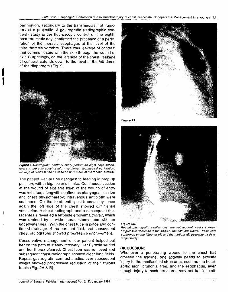
perforation, secondary to the transmediastinal trajec-tory of a projectile. A gastrografin (radiographic con-trast) study under fluoroscopic control on the eighthpost-traumatic day, confirmed the presence of a perfo-ration of the thoracic esophagus at the level of thethird thoracic vertebra. There was leakage of contrastthat communicated with the skin through the wound ofexit. Surprisingly, on the left side of the chest, leakageof contrast extends down to the level of the left domeof the diaphragm (Fig.1).
Figure 1.Gastrografin contrast study performed eight days subse-quent to thoracic gunshot injury confirmed esophageal perforation;leakage of contrast can be seen on both sides of the thorax (arrows).
The patient was put on nasogastric feeding in prop-upposition, with a high caloric intake. Continuous suctionat the wound of exit and toilet of the wound of entrywas initiated, alongwith continuous pharyngeal suctionand chest physiotherapy; intravenous antibiotic werecontinued. On the fourteenth post-trauma day, onceagain the left side of the chest showed diminishedventilation. A chest radiograph and a subsequent tho-racentesis revealed a left-side empyema thorax, whichwas drained by a wide thoracostomy tube with anunderwater seal. With the chest tube in place and con-tinued drainage of the purulent fluid, and subsequentchest radiographs showed progressive improvement.
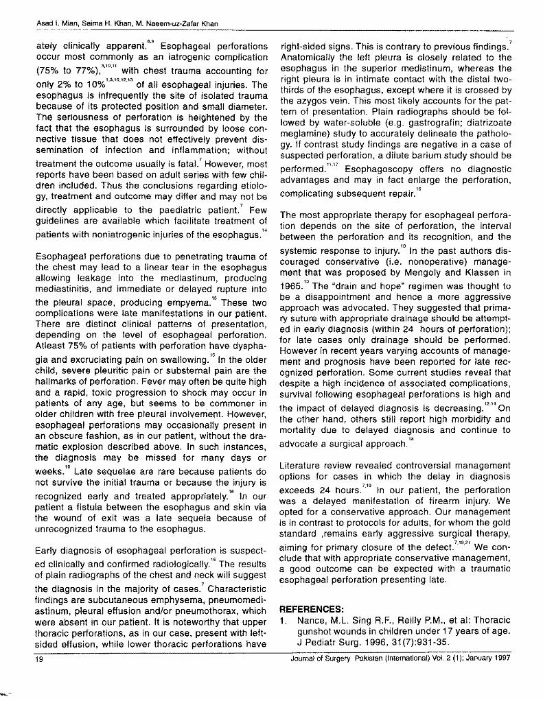
Conservative management of our patient helped puther on the path of steady recovery. Her Pyrexia settledand her thorax cleared. Chest tube was removed andsubsequent chest radiograph showed clear lung fields.Repeat gastrografin contrast studies over subsequentweeks showed progressive reduction of the fistuloustracts (Fig. 2A & 8).
Figure 2A
Figure 28.Repeat gastrografin studies over the subsequent weeks showingprogressive decrease in the sizes of the fistulous tracts. These wereperformed on the fifteenth (A) and the thirtieth (B) post-trauma days,respectively.
DISCUSSION:Whenever a penetrating wound to the chest hascrossed the midline, one actively needs to excludeinjury to the mediastinal structures, such as the heart,aortic arch, bronchial tree, and the esophagus, eventhough injury to such structures may not be immedi-
Journal of Surgery Pakistan (International) Vol. 2 (1); January 1997 18
Asad I. Mian, Saima H. Khan, M. Naeem-uz-Zafar Khan
ately clinically apparent. 8.9 Esophageal perforationsoccur most commonly as an iatrogenic complication(75% to 77%),3.10.11with chest trauma accounting foronly 2% to 10%'·3.'0.'2.'3of all esophageal injuries. Theesophagus is infrequently the site of isolated traumabecause of its protected position and small diameter.The seriousness of perforation is heightened by thefact that the esophagus is surrounded by loose con-nective tissue that does not effectively prevent dis-semination of infection and inflammation; without
treatment the outcome usually is fatal.' However, mostreports have been based on adult series with few chil-dren included. Thus the conclusions regarding etiolo-gy, treatment and outcome may differ and may not be
7directly applicable to the paediatric patient. Fewguidelines are available which facilitate treatment of
14patients with non iatrogenic injuries of the esophagus.
Esophageal perforations due to penetrating trauma ofthe chest may lead to a linear tear in the esophagusallowing leakage into the mediastinum, producingmediastinitis, and immediate or delayed rupture into
1Sthe pleural space, producing empyema. These twocomplications were late manifestations in our patient.There are distinct clinical patterns of presentation,depending on the level of esophageal perforation.Atleast 75% of patients with perforation have dyspha-
gia and excruciating pain on swallowlnq." In the olderchild, severe pleuritic pain or substernal pain are thehallmarks of perforation. Fever may often be quite highand a rapid, toxic progression to shock may occur inpatients of any age, but seems to be commoner inolder children with free pleural involvement. However,esophageal perforations may occasionally present inan obscure fashion, as in our patient, without the dra-matic explosion described above. In such instances,the diagnosis may be missed for many days or
weeks.'° Late sequelae are rare because patients donot survive the initial trauma or because the injury is
recognized early and treated appropriately." In ourpatient a fistula between the esophagus and skin viathe wound of exit was a late sequela because ofunrecognized trauma to the esophagus.
Early diagnosis of esophageal perforation is suspect-ed clinically and confirmed radioloqically." The resultsof plain radiographs of the chest and neck will suggest
the diagnosis in the majority of cases.' Characteristicfindings are subcutaneous emphysema, pneumome.di-astinum, pleural effusion and/or pneumothorax, whichwere absent in our patient. It is noteworthy that upperthoracic perforations, as in our case, present with left-sided effusion, while lower thoracic perforations have
7
right-sided signs. This is contrary to previous findings.Anatomically the left pleura is closely related to theesophagus in the superior medistinum, whereas theright pleura is in intimate contact with the distal two-thirds of the esophagus, except where it is crossed bythe azygos vein. This most likely accounts for the pat-tern of presentation. Plain radiographs should be fol-lowed by water-soluble (e.g. gastrografin; diatrizoatemeglamine) study to accurately delineate the patholo-gy. If contrast study findings are negative in a case ofsuspected perforation, a dilute barium study should bepertormed.":" Esophagoscopy offers no diagnosticadvantages and may in fact enlarge the perforation,
• 1Scomplicating subsequent repair.
The most appropriate therapy for esophageal perfora-tion depends on the site of perforation, the intervalbetween the perforation and its recognition, and the
10 d.systemic response to injury. In the past authors IS-
couraged conservative (i.e. nonoperative) manage-ment that was proposed by Mengoly and Klassen in1965.'0 The "drain and hope" regimen was thought tobe a disappointment and hence a more aggressiveapproach was advocated. They suggested that prima-ry suture with appropriate drainage should be atte~pt-ed in early diagnosis (within 24 hours of perforation);for late cases only drainage should be performed.However in recent years varying accounts of manage-ment and prognosis have been reported for late rec-ognized perforation. Some current studies reveal thatdespite a high incidence of associated complications,survival following esophageal perforations is high and
• 12,14
the impact of delayed diagnosis is decreasing. Onthe other hand, others still report high morbidity andmortality due to delayed diagnosis and continue to
18
advocate a surgical approach.
Literature review revealed controversial managementoptions for cases in which the delay in diagnosisexceeds 24 hours.
7.19
In our patient, the perforationwas a delayed manifestation of firearm injury. Weopted for a conservative approach. Our managementis in contrast to protocols for adults, for whom the goldstandard ,remains early aggressive surgical therapy,aiming for primary closure of the defect.7.19·21We con-clude that with appropriate conservative management,a good outcome can be expected with a traumaticesophageal perforation presenting late.
REFERENCES:1. Nance, M.L. Sing R.F., Reilly P.M., et al: Thoracic
gunshot wounds in children under 17 years of age.J Pediatr Surg. 1996, 31 (7):931-35.
19 Journal of Surgery Pakistan (International) Vol. 2 (1); January 1997
--
Late onset Esophageal Perforation due to Gunshot Injury of chest: successful Nonoperative Management in a young child.
2. Nance M.L, Templeton J.M., 0 Neill J.A.:Socioeconomic impact of gunshot wounds in anurban pediatric population. J Pediatr Surg.1994,29:39-43.
3. Postlethwait R.w.: Surgery of the esophagus.New York, Appleton-Century-Crofts, 1979.
4. Zavoski R.W., Lapidus G.D., et al: A population-based study of severe firearm injury among chil-dren and youth. Pediatrics. 1995, 96(2 Pt 1):278-82.
5. Meller J.L., Little A.G., Shermata D.W. :Thoracictrauma in children. Pediatrics. 1984, 74:813.
6. Reinberg 0., Genton N., Lacheretz M.:Particularities of thoracic trauma in children. In:Webb W.R., Besson A. (eds.): Thoracic surgery:Surgical management of chest injuries (chapter34). St. Louis, Mosby-Year Book, 1991,7:340.
7. Panieri E., Millar A.J.W., et al: Iatrogenicesophageal perforations in children: Patterns ofinjury, presentation, management, and outcome. JPediatr Surg. 1996, 31(7):890-95.
8. Haller J.A. Thoracic injuries. In: Welch K.J.,Randolph J.G., Ravitch MM, et al: Pediatricsurgery. Chicago, Year Book Medical Publishers,Inc. (chap 17); 1986, 143-53.
9. Falcon O. Knott C., Elkins R.C. Gunshot woundstraversing the mediastinum: Guidelines for evalu-ation and management. J Okla State Med Assoc.87(4): 1994, 174-7.
10. Popovici, Z.: Esophageal injuries. In: Webb, W.R.;Besson, A. (eds.): Thoracic surgery: Surgicalmanagement of chest injuries (chap 45). St. Louis,Mosby-Year Book. 1991, 7:443-66.
11. Sawyer R., Philips C., Vakil N. Short and long-term outcome of esophageal perforations.Gastrointest endosc. 1995, 41(2):130-4.
12. Reefder l.B., DeFilippi V.J., Ferguson MK.:Current results of therapy for esophageal perfora-tions. Am J Surg. 1996, 169(6):615-7.
13. Besson A., Savary M.: Trauma of the esophagus.In: BessonA., Saegesser R.(eds.): SA colour atlasof chest trauma and associated injuries(chap. 11)London, Wolfe Medical Publications, Ud.(2):1983, 251-316.
14. Weimam D.S., Walker W.A., Brosnan K.M.:Noniatrogenic esophageal trauma. Ann ThoracSurg. 1995, 59(4):845-9.
15. Weber T.R.: Esophageal rupture and perforation.In: Welch K.J., Randolph J.G., Ravitch M.M.; et al:Pediatric Surgery.(chap 73) Chicago, Year BookMedical Publishers, Inc.: 1986, 725-27.
16. Deslauriers J., Desaulniers D., Pomerleau S.:Late sequelae of thoracic injury. In:Webb W.R.,Besson A. (eds.): Thoracic surgery:Surgical man-agement of chest injuries.(chap. 48) St. Louis,Mosby-Year Book, Inc.; vol 7: 1991, 513-35.
17. JamesA.E., Montaly R.J., Chafee V., et al: Bariumor gastrografin: which contrast media for diagno-sis of esophageal tears? Gastroenterology, 1975,68:1103.
18. Andrade A.R. T-tube intubation in the manage-ment of late traumatic esophageal perforations:Case report. J Trauma. 1994, 37(1):131-2.
19. White R.K., Morris D.M. Diagnosis and manage-ment of esophageal perforations. Am Surg. 1992,8:112-19.
20. Santos G.H., Frater R.W.M. Transesophageal irri-gation for the treatment of mediastinitis producedby esophageal rupture. J Thorac Cardiovasc Surg.1986, 91:57-62.
21. FlynnA.E.,VerrierE.D.,Way i.v«. et al: Esophagealperforations.Arch Surg. 1989, 124-1211-14.
.Journal of Surgery Pakistan (International) Vol. 2 (1); January 1997 20
PHYTOBEZOAR OF SMALL INTESTINE
A Case Report
ABDUL SAnAR MEMON, JAN MOHAMMAD MEMON, ABDUL GHANI SOOMRO
ABSTRACT
Phytobezoar of small intestine in middle age group is rare. We report one such case in a 50years old female patient suffering from abdominal tuberculosis with phytobezoar. Followingantituberculous treatment and surgery, patient recovered completely.
KEY WORDS: Small intstine : Phytobezoar
INTRODUCTIONBezoar is derived from Arabic word "BEDZEHR" meaningantidote. Debakey and Ochsner' has classified bezoarinto 3 categories.
1. Trichobezoar or hair ball composed of masses of hairand decaying food materials.
2. Phytobezoar consists of entangled masses of veg-etable, fibrous material, such as skin, seeds, veg-etable and fruit fibers.
3. Hard concretions which consist of organic masses orcalculi containing calcium.
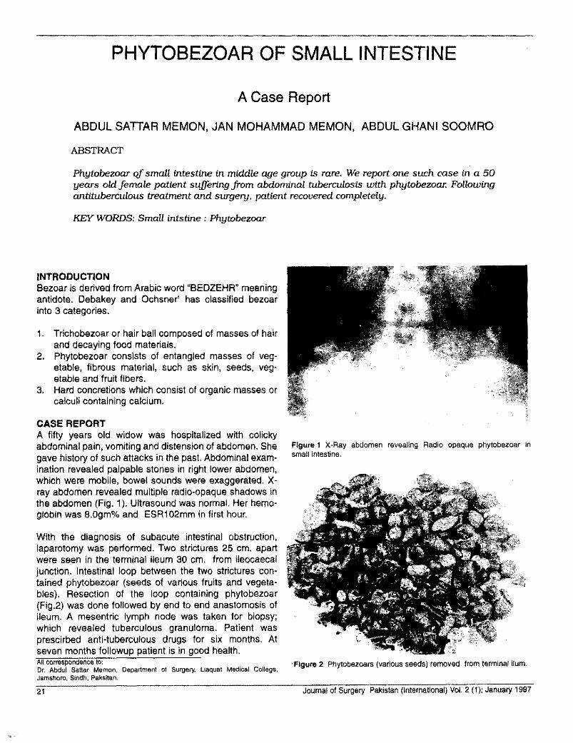
CASE REPORTA fifty years old widow was hospitalized with colickyabdominal pain, vomiting and distension of abdomen. Shegave history of such attacks in the past. Abdominal exam-ination revealed palpable stones in right lower abdomen,which were mobile, bowel sounds were exaggerated. X-ray abdomen revealed multiple radio-opaque shadows inthe abdomen (Fig. 1). Ultrasound was normal. Her hemo-globin was 8.0gm% and ESR102mm in first hour.
Figure 1 X-Ray abdomen revealing Radio opaque phytobezoar insmall intestine.
With the diagnosis of subacute intestinal obstruction,laparotomy was performed. Two strictures 25 cm. apartwere seen in the terminal ileum 30 em. from ileocaeealjunction. Intestinal loop between the two strictures con-tained phytobezoar (seeds of various fruits and vegeta-bles). Resection of the loop containing phytobezoar(Fig.2) was done followed by end to end anastomosis ofileum. A mesentric lymph node was taken for biopsy;which revealed tuberculous granuloma. Patient wasprescirbed anti-tuberculous drugs for six months. Atseven months followup patient is in good health.All correspondence to: .Figure 2 Phytobezoars (various seeds) removed from terminal ilum.Dr. Abdul Sattar Memon, Department of Surgery, Uaquat Medical College,Jamshoro, Sindh, Paksitan.
21 Journal of Surgery Pakistan (International) Vol. 2 (1); January 1997
Phytobezoar of small intestine
DISCUSSIONBezoars are commonly seen in young ladies with psychi-atric disorders. Bezoars after gastric surgery are reportedquite frequently because of reduction of acid and pepsinand also disturbance of antral function. Citrus fruits withlarge amounts of cellulose are also a common cause.Recently many iatrogenic bezoars secondary to medica-tions have been reported", Formation of intestinal bezoarsafter gastric surgery may be due to delayed emptying.First such case after vagotomy was reported by Oliver".
It is possible to dissolve phytobezoar by papain(Proteolytic enzyme) if administered with sodium bicar-bonate, as later is mucolytic and also facilitates the actionof papain. Combination of enzymes e.g. cellulose anddehydroxycholic acid are also effective. Conservative
treatment, however, is effective in small percentage ofpatients only. Surgery is required in cases where conser-vative treatment fails.
REFERENCES1. Oebakey M., Ochsner A. Bezoars and concretion,
Comprehensive review of literature with analysis of303 collected cases and presentation of 8 additionalcases. Surgery 1983, 4:934-63.
2. Forrucci JR, Eaton AB. Complications of drug thera-py. In Margnilus AR, Burnhenve HJ (eds.) Alimentarytract roentgenology, 2nd ed. St. Louis C. Y. Mosby Co.1973, 281-93.
3. Oliver, Prevest 1966 Prease Medical 74-387.
SURGICAL PROGRAMME FOR 1997
Launching of Journal of Surgery Pakistan
J.P.M.C. Symposium
Congress 97 at Dhaka - Joint Conference of Bangladesh College ofPhysicians & Surgeons with CPSP
Symposium on 'Training of Surgeons - structured programme' by ICS
Symposium on Disaster Plan for the city of Karachi by ICS
Asia Pacific Conference of ICS in Singapore
Guest Lecture by Prof. Earnest Owen, President International Collegeof Surgeons
Hands on workshop on Animal Transplantation by ICS
Symposium on ' Undergraduate Training Programme by ICS
Symposium on 'Surgical Problems in the SARRAC country' by ICS
Symposium on Oncology - 'Lung Malignancies' by ICS
Workshop on 'Vascular Surgery' by ICS
Journal of Surgery Pakistan (International) Vol. 2 (1); January 1997 22
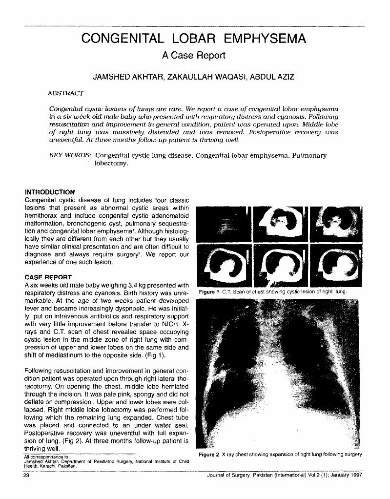
CONGENITAL LOBAR EMPHYSEMAA Case Report
JAMSHED AKHTAR, ZAKAULLAH WAQASI, ABDUL AZIZ
ABSTRACT
Congenital cystic lesions oj lungs are rare. We report a case oj congenital lobar emphysemain a six week old male baby who presented with respiratory distress and cyanosis. Followingresuscitation and improvement in general condition. patient was operated upon. Middle lobeoj right lung was massively distended and was removed. Postoperative recovery wasuneventful. At three months folious-up patient is thriving well.
KEY WORDS: Congenital cystic lung disease, Congenital lobar emphysema, Pulmonarylobectomy.
INTRODUCTIONCongenital cystic disease of lung includes four classiclesions that present as abnormal cystic areas withinhemithorax and include congenital cystic adenomatoidmalformation, bronchogenic cyst. pulmonary sequestra-tion and congenital lobar emphysema'. Although histolog-ically they are different from each other but they usuallyhave similar clinical presentation and are often difficult todiagnose and always require surqery", We report ourexperience of one such lesion.
CASE REPORTA six weeks old male baby weighing 3.4 kg presented withrespiratory distress and cyanosis. Birth history was unre-markable. At the age of two weeks patient developedfever and became increaSingly dyspnoeic. He was initial-ly put on intravenous antibiotics and respiratory supportwith very little improvement before transfer to NICH. X-rays and C.T. scan of chest revealed space occupyingcystic lesion in the middle zone of right lung with com-pression of upper and lower lobes on the same side andshift of mediastinum to the opposite side. (Fig 1).
Following resuscitation and improvement in general con-dition patient was operated upon through right lateral tho-racotomy. On opening the chest. middle lobe herniatedthrough the incision. It was pale pink, spongy and did notdeflate on compression. Upper and lower lobes were col-lapsed. Right middle lobe lobectomy was performed fol-lowing which the remaining lung expanded. Chest tubewas placed and connected to an under water seal.Postoperative recovery was uneventful with full expan-sion of lung. (Fig 2). At three months follow-up patient isthriving well.All correspondence to:Jamshed Akhtar, Department of Paediatric Surgery, National Institute of ChildHealth, Karachi, Paksitan.
Figure 1 C.T Scan of chest showing cystic lesion of right lung.
Figure 2 X ray chest showing expansion of right lung following surgery
Journal of Surgery Pakistan (International) Vol.2 (1); January 199723
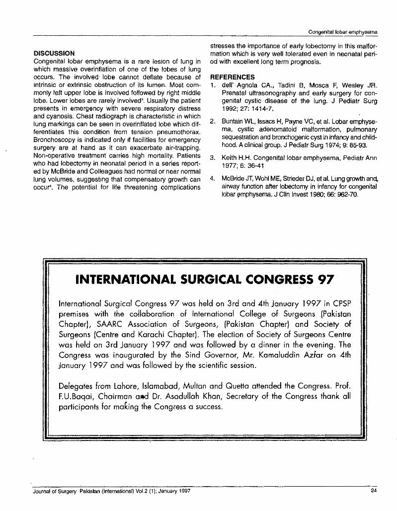
Congenital lobar emphysema
DISCUSSIONCongenital lobar emphysema is a rare lesion of lung inwhich massive overinflation of one of the lobes of lungoccurs. The involved lobe cannot deflate because ofintrinsic or extrinsic obstruction of its lumen. Most com-monly left upper lobe is involved followed by right middlelobe. Lower lobes are rarely involved". Usually the patientpresents in emergency with severe respiratory distressand cyanosis. Chest radiograph is characteristic in whichlung markings can be seen in overinflated lobe which dif-ferentiates this condition from tension pneumothorax.Bronchoscopy is indicated only if facilities for emergencysurgery are at hand as it can exacerbate air-trapping.Non-operative treatment carries high mortality. Patientswho had lobectomy in neonatal period in a series report-ed by McBride and Colleagues had normal or near normallung volumes, suggesting that compensatory growth canoccur. The potential for life threatening complications
stresses the importance of early lobectomy in this malfor-mation which is very well tolerated even in neonatal peri-od with excellent long term prognosis.
REFERENCES1. dell' Agnola CA., Tadini B, Mosca F, Wesley JR.
Prenatal ultrasonography and early surgery for con-genital cystic disease of the lung. J Pediatr Surg1992; 27: 1414-7.
2. Buntain WL, Issacs H, Payne VC, et al. Lobar emphyse-ma, cystic adenomatoid malformation, pulmonarysequestration and bronchogeniC cyst in infancy and child-hood. A clinical group. J Pediatr Surg 1974; 9: 85-93.
3. Keith H.H. Congenital lobar emphysema, Pediatr Ann1977; 6: 36-41
4. McBride JT,Wohl ME, Strieder DJ, et al, Lung growth and.airway function after lobectomy in infancy for congenitallobar emphysema. J Clin Invest 1980; 66: 962-70.
INTERNATIONAL SURGICAL CONGRESS 97
International Surgical Congress 97 was held on 3rd and 4th January 1997 in CPSPpremises with the collaboration of International College of Surgeons (PakistanChapter), SAARC Association of Surgeons, (Pakistan Chapter) and Society ofSurgeons (Centre and Karachi Chapter). The election of Society of Surgeons Centrewas held on 3rd January 1997 and was followed by a dinner in the evening. TheCongress was inaugurated by the Sind Governor, Mr. Korncluddin Azfar on 4thJanuary 1997 and was followed by the scientific session.
Delegates from Lahore, Islamabad, Multan and Quetta attended the Congress. Prof.F.U.Baqai, Chairman a!IKJDr. Asadullah Khan, Secretary of the Congress thank allparticipants for making the Congress a success.
Journal of Surgery Pakistan (International) Vol.2 (1); January 1997 24
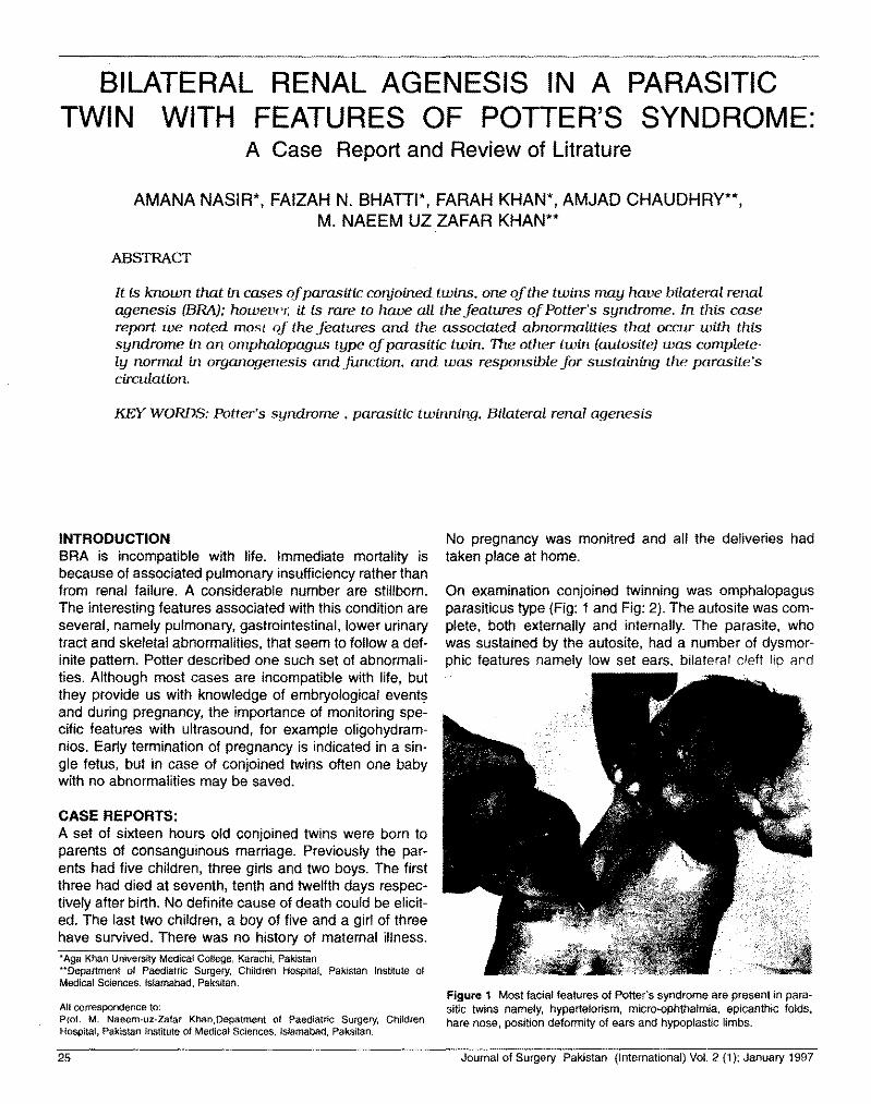
BILATERAL RENAL AGENESIS IN A PARASITICTWIN WITH FEATURES OF POTTER'S SYNDROME:
A Case Report and Review of Litrature
AMANA NASIR*, FAIZAH N. BHATTI*, FARAH KHAN*, AMJAD CHAUDHRY**,M. NAEEM UZ ZAFAR KHAN**
ABSTRACT
It is known that in cases oj parasitic conjoined: twins, one oj the twins may have bilateral renalagenesis (BRA); however. it is rare to have all the features oj Potter's syndrome. In this casereport we noted most oj the features and the associated abnormalities that occur with thissyndrome in an omphalopagus type oj parasitic twin. The other twin (autosite) was complete-ly normal in organogenesis and function, and was responsible for sustaining the parasite'scirculation.
KEY WORDS:Potter's syndrome, parasitic twinning, Bilateral renal agenesis
INTRODUCTIONBRA is incompatible with life. Immediate mortality isbecause of associated pulmonary insufficiency rather thanfrom renal failure. A considerable number are stillborn.The interesting features associated with this condition areseveral, namely pulmonary, gastrointestinal, lower urinarytract and skeletal abnormalities, that seem to follow a def-inite pattern. Potter described one such set of abnormali-ties. Although most cases are incompatible with life, butthey provide us with knowledge of embryological eventsand during pregnancy, the importance of monitoring spe-cific features with ultrasound, for example oligohydram-nios. Early termination of pregnancy is indicated in a sin-gle fetus, but in case of conjoined twins often one babywith no abnormalities may be saved.
CASE REPORTS:A set of sixteen hours old conjoined twins were born toparents of consanguinous marriage. Previously the par-ents had five children, three girls and two boys. The firstthree had died at seventh, tenth and twelfth days respec-tively after birth. No definite cause of death could be elicit-ed. The last two children, a boy of five and a girl of threehave survived. There was no history of maternal illness.'Aga Khan University Medical College, Karachi, Pakistan"Department of Paediatric Surgery, Children Hospital, Pakistan Institute ofMedical SCiences, Islamabad, Paksitan.
All correspondence to:Prof. M. Naeem-uz-Zafar Khan,Depatment of Paediatric Surgery, ChildrenHospital, Pakistan Institute of Medical SCiences,Islamabad, Paksitan.
No pregnancy was monitred and all the deliveries hadtaken place at home.
On examination conjoined twinning was omphalopagusparasiticus type (Fig: 1 and Fig: 2). The autosite was com-plete, both externally and internally. The parasite, whowas sustained by the autosite, had a number of dysmor-phic features namely low set ears, bilateral cleft lip and
Figure 1 Most facial features of Potter's syndrome are present in para-sitic twins namely, hypertelorism, micro-ophthalmia, epicanthic folds,hare nose, position deformity of ears and hypoplastic limbs.
25 Journal of Surgery Pakistan (International) Vol. 2 (1); January 1997

Bilateral renal agenesis in parasitic twin with features of potter's syndrome: a case reprot and review of literature
Figure 2 Hydrocephaly, encephalocele and tibiofibular deficiency inright, and femoral dficiency on the left lower hyoplastic limbs.
Figure 3 Viscera of paraslte with hypoplastic lungs and heart, oesoph-agus and intestinal atresia. Absent kidney and urinary bladder withBockdalek hernia.
palate, microophthalmia, excessively flattened nose withhypertelorism (Fig:2 ). There were limb deformities withvestigial right upper limb bud, the left arm was large andhad radial clubbing. The lower limbs were hypoplastic withproximal femoral deficiency on one and vestigial tibia andfibula on the other lower limb (Fig:2). The penis was ves-tigial and the patient had high anorectal agenesis. Therewas no demonstrable motor or sensory activity in the par-
Figure 4 Hydrocephaly with encephalocoele, large amount of C.S.F. isbeing drained and compressed hypoplastic brain is brought out.
Figure 5 Skeleton of parasite demonstrates deficiency of bones inhypoplastic limbs and defects in skull due to encephalocoeles, gastroin-testinal contrast study shows intestine of autosite overlapping into para-site's abdominal cavity.
asite. There were only conducted movements with themovements of autosite.
On investigations skeletal survey revealed normal skeletonof the autosite except for the sternum which was fused withthe sternum of the parasite. The parasite had 9 pairs of vis-ible ribs, a complete but abnormal vertebral column withpelvis and complete sacral agenesis. Both scapulae were
Journal of Surgery Pakistan (International) Vol. 2 (1); January 1997 26

Amana Nasir, Faizah N. Bhatti, Farah Khan, Amjad Chaudhry,M. Naeem uz Zafar Khan
vestigial. One limb had complete lower radius and ulna andskeleton of the hand had four metacarpals and digits. Theskeleton of the thumb was absent with radial clubbing. Theskull had an occipital defect from where the encephalocelewas bulging (Fig:4). There were hypoplastic mandible andmaxilla. Esophagus could not be cannulated and becauseof anorectal agenesis, gastrointestinal tract could not bestudied. Lungs were not visible (being not ventilated).Contrast study of the autosite showed normal esophagus,stomach and rest of the intestine, much of the small bowelhad prolapsed through the omphalopagus connection intothe abdominal cavity of the parasite. The autosite and par-asite shared an umbilicus at the base of their fusion. (Fig:5)
On ultrasound no abnormality was noted in the autosite.Whereas, in the parasite there were hypoplastic liver andkidneys; heart and lungs could not be visualized. Cranialultrasound revealed marked hydrocephaly with distortedbrain tissue. EEG of autosite was normal whereas para-site showed no activity.CT scan could not be performed.
At operation, besides the general physical findingsalready described, the fusion was along the umbilicusupto the xiphisternum, where the sternum of the autositehad fused with sternum of the parasite. There was a largearterial and venous connection arising from the autositeadjacent to the liver which was perfusing the parasite. Theparasite had loops of small intestine and vestigial colonwhich ended blindly in what appeared to be prostatic tis-sue in the pelvis. The penis was vestigial and testis andscrotum were absent. There was no organised bladder,ureter or kidney. Liver was fairly well formed with a fibrousconnection with the liver of the autosite. There was a ves-tigial gall bladder, a small spleen and a Meckel's divertic-ulum. Some loops of intestine and spleen of parasite hadprolapsed through a posterolateral defect in the leftdiaphragm (Bochdalek hernia). The lungs were like budswith poorly formed tracheobronchial tree and there was ahypoplastic heart within the pericardial cavity (Fig:3) andpoorly developed aortic and pulmonary vessels.The facehad the appearance of premature senility which is suffi-ciently characteristic to warrant a diagnosis of co-existingrenal aplasia.
This detailed scrutiny of the parasite revealed that mostfeatures were similar to abnormalities found in Potter'ssyndrome.
DISCUSSIONDegrees of defective renal development range from com-plete failure of formation of the kidneys to occurrence ofvarying quantity of renal tissue. Renal agenesis is a con-dition in which no nephrogeniC tissue is present, and isreported to occur in 1 in 4000 births, with a male predom-inance of 3 to 1'. Excluding parasite and formless mon-
sters, the earliest report of bilateral renal agenesis [BRA]was in 16716• Although most cases of BRA are sporadic, afamilial pattern has also been reported. Cain et aldescribed a family with two consecutive born male infants,the first with BRA and the other with renal agenesis on oneside and total renal dysplasia on the other'. With renalagenesis, pulmonary hypoplasia is a feature and this willproduce signs of respiratory insufficiency.
There does not seem to be any significant relationshipbetween BRA and maternal age or parity. In some set oftwins like our case, only one of the pair has been affected.66 percent or more of these infants are reported as pre-mature but it is suggested that this reflects a failure to gainweight after 34 weeks, rather than a shortened gestationtime. Breech presentation of infant with BRA is usual. The
diagnosis is usually confirmed only at autopsy", A detailedautopsy is vital in assessement of an infant with oligohy-dramnios to establish congenital abnormalities of the uri-nary tract. This applies equally to the second trimesterfetuses following miscarriages, theraputic abortion, still-born infants and cases of neonatal deaths".
Of all the children born with bilateral renal agenesis about33 percent will be stillborn, most of the others will die with-in 24 hours. A rare 6 percent of patients who survive therespiratory insufficiency live long enough to die of uremia.The longest survival has been 39 days. The first three sib-lings of our conjoined twins had died within the first weekof birth. About 25% of infants with BRA have no otheranomalies. Various associations have been reported inthese children. The interrelationship is not yet clear but itis possible that all are different manifestations of a broad-er axial mesodermal "dysplasia" syndrome.
BRA may be detected prenatally by fetal ultrasound.Normal kidneys can be detected sonographically by 20thweek of gestation. If the kidneys are not in place by thattime, failure to demonstrate the bladder within 1-2 hoursafter IN administration of lasix to the mother is consideredconfirmatory. The dignostic criteria for renal agenesis in theearly fetus differs from those in the second half of gestation.
In conjoined twins where the aim is to save both individu-als a detailed study of organogenesis and function in bothis required before surgery. But in a case of parasitic twin-ning like in our case there is no difficulty to arrive at a deci-sion, as only the autosite has to be saved.
REFERENCES1. CHESTER M. EDELMANN Jr.Renal Abnormalities
And Malformation Syndromes. Paediatric KidneyDisease. Second Edition Little, Brown and Co [Inc].1992.
2. BEHRMAN AND VAUGHAN. Kidney Problems In
27 Journal of Surgery Pakistan (International) Vol. 2 (1); January 1997
.~....:::::-.

Bilateral renal agenesis in parasitic twin with features of potter's syndrome: a case reprot and review of literature
Newborn Infants. Nelson Textbook Of Paediatrics.Fourteenth Edition. W.B. Saunders and Co. 1992.
3. RICHARD S.SNELL The Genitourinary System.Clinical Embryology. Third Edition. Little, Brown andCo [Inc]. 1983
4. ELLIN LIBERMAN Congenital Malformation of UrinaryTract With or Without Infection. Clinical PaediatricNephrology. J.B. Lippincott and Co. 1976.
5. KEITH L. MOORE The Genitourinary Tract. TheDeveloping Human. Third Edition.
6. SKANDALAKIS AND GRAY Embryology ForSurgeon. Second Edition Abnormalities Of The
Amount Of Renal Tissue.
7. MARC I. ROWE Essential Of Paediatric Surgery.Renal Disorders.
8. LOWE P. DEHNER Paediatric Surgical Pathology.Second Edition. Congenital And DevelopmentalAnomalies.
9. BRONSTEIN M.; AMITT A. Prenatal diagnosis. April1994. The Early Prenatal Sonographic Diagnosis ofRenal Agenesis: Techniques and possible pitfalls.
10. British Journal (Obs. & Gynae.) July 1994 Title:Oligohydromnios sequence; The Spectrumof RenalMalformation.
journal of Surgery Pakistan (International) Vol. 2 (1); January 1997 28
ACUTE INTERMITTENT PORPHYRIA SIMULATINGACUTE SURGICAL ABDOMEN
A Case Report
FAISAL AHMED, JAMAL ARA, RUKHSANA SATTAR, SHABEER HUSSAIN.
ABSTRACT
A case of porphyria, an autosomal dominant, metabolic disorder, running in a whole family,is reported. The patient is the only survivor of a family; who presented as an Acute Abdomen.
plaints the patient developed disordered orientation;spectrum of which fluctuated between anxiety andfrank psychotic behavior. He gave no history of skinblistering or photosensitivity. Two of his sisters haddied at the ages of 13 and 14 respectively, of diseasewith similar signs and symptoms. His father had diedat the age of 28 of a disease whose nature could notbe ascertained. There is a history of consanguinity inthe family dating back to the last three generations.
KEY WORDS: Rare Metabolic Disorder, Severe Abdominal Pain, Inherited disease, ProperDiagnosis, Porphyria, Acute Intermittent Porphyria.
INTRODUCTIONPorphyrias are a relatively uncommon group of meta-bolic disorders, usually inherttad", in which there areabnormalities at various levels of Haem biosyntheticpathway" .•. Depending on the type of porphyria, excessproduction and concomitant accumulation of the inter-mediate products called porphyrins occur". The Mostimportant part in the management is family screaming
to prevent other members from acute attacks 68. A casereport of Acute Intermittent Porphyria presenting as
acute abdomen is presented?
CASE REPORTA 14 years old school boy presented in the casualtydepartment of JPMC, with the complaints of abdominalpain, constipation and disordered orientation for 5days and fever for 8 days. About 7 hours prior toadmission, he also developed urinary retention.
The patients was alright 8 days ago when he devel-oped fever, for which he was treated by a local doctor.Three days later he developed abdominal pain whichprevailed throughout but fluctuated in severity andresponded to certain injections prescribed by the localdoctor. He was constipated which became absolute afew hours prior to admission. Alongwith these com-Dr. Faisal Ahmed.Department of Medicine,Jinnah Postgraduate Medical CenterKarachi.
The patient had visited the hospital on four differentoccassions, but was either discharged because hewas found to have no surgical pathology, or referred toneuropsychiatric department, in view of his obviousbehavioral disorder.
Clinical examination showed that he had a very rapidpulse and a blood pressure of 150/1 OOmm of Hg. Hewas disoriented and paranoid complaining of excruci-ating abdominal pain; but there was no abdominal ten-derness. His deep tendon reflexes were normal. hewas catheterized because of urinary retention, and onexamination of the urinary bag a short while later, theurine had turned darker in the urine bag, Ehlrich's testwas performed and was affirmative. Other investiga-tions showed reactive neutrophilia, Na- concentration120 meq/L and urea 43mg/L. A diagnosis of AcuteIntermittent Porphyria was thus confirmed.DISCUSSIONAs the mortality of an acute attack of this disease is
29 Journal of Surgery Pakistan (International) Vol. 2 (1); January 1997
Acute intermittent porphyria simulating acute surgical abdomen
very high, and keeping in view its relative, thoughanecdotal, low incidence in our setup, and also con-sidering the fact that this is an autosomal dominant
disease", which should have a relatively equal inci- 5.dence in various communities. Patients are often shuf-fled between surgical and psychiatric Departments
because of various behaviour changes in this lllness".It is tentatively concluded that this condition is notdiagnosed correctly, a number of times, to facilitateprompt and life saving treatment. Therefore, in allcases of acute severe abdominal pain, not within the 6.surgical parameters, metabolic disorders like porpho-rias should be kept in mind.
REFERENCES1. Patience DH, Blackwood DH, McColl-KG, Moore
MR, Acute Intermittent Porphyria and MentalIllness - A Family Study Acta-Psychiatr-Scand1994 Apr, 89(4) : 262-7.
of specific alleles. 1994 Jan; 93(1) : Anvert-M.Hum-Genetics.
Hernandez-A; Sepulveda-P; Fernandez-Cuartero-B, De-Salamanca-RE. Urinary por-phyrinogens in normal subjects and in patientswith porphyria cutanea tarda and acute intermit-tent porphyria. Horm-Metab-Res. 1993 Aug;25(8) : 454-4.
Abid-S; Jafri-SW; Abbas-Z; Khan-AH; Khan-MA.Acute intermittent porphyria-a diagnosis to con-sider. JPMA-J-Pak-Med-Assoc. 1995 Feb; 45(2): 45
7. Shively-BD; Clochesy-JM; Briones-JP; Spositio-DL; Kloos-JA. Caring for patients with acute inter-mittent porphyria. AACN-Clin-Issues. 1994 Feb;5(1) : 36-41
8. el-Shazly-M. Acute intermittent porphyria: a fami-ly study. Br-J-Hosp-Med. 1995 Feb; 53(4) : 169-70
9. Santosh-PJ; Malhotra-S. Varied psychiatric man-ifestation of acute intermittent porphyria. Biol-Psychiatry. 1994 Dec 1; 36(11): 744-7
10. Whatley-SD; Roberts-AG; Elder-GH. De-novomutation and sporadic presentation of acuteintermittent porphyria. Lancet. 1995 Oct 14;346(8981) : 1007-8.
2. Sagene Laegreid, A etal Genetic CarrierDetection in Norwegian Families with acute inter-mittent Porphyria, Scand -J-Clin-Lab-Invest.1993 Nov, 53(7): 687-91.
3. Mgone-CS; Lanyon-WG; Moor-MR; Louie-GV;Connor-JM, Identification of five novel mutationsin the porphobilinogen deaminase, Hum-Mol-Genet. 1994 May; 3(5) : 809-11
4. Lundin-G; Wedell-A; Thunel-S; Anvert-M, Twonew mutations in the porphobilinogen deaminaseand a screening method using PCR amplification
Journal of Surgery Pakistan (Intemational) Vol. 2 (1); January 1997 30
Related Documents