www.eurosurveillance.org Vol. 17 | Weekly issue 50 | 13 December 2012 Europe’s journal on infectious disease epidemiology, prevention and control Rapid communications Detection of Usutu virus infection in a healthy blood donor from south-west Germany, 2012 2 by L Allering, H Jöst, P Emmerich, S Günther, E Lattwein, M Schmidt, E Seifried, V Sambri, K Hourfar, J Schmidt-Chanasit First detection of livestock-associated meticillin-resistant Staphylococcus aureus CC398 in bulk tank milk in the United Kingdom, January to July 2012 5 by GK Paterson, J Larsen, EM Harrison, AR Larsen, FJ Morgan, SJ Peacock, J Parkhill, RN Zadoks, MA Holmes Cluster of invasive Neisseria meningitidis infections on a cruise ship, Italy, October 2012 8 by P Stefanelli, C Fazio, A Neri, P Isola, S Sani, P Marelli, C Martinelli, P Mastrantonio, MG Pompa Surveillance and outbreak reports Measles virus genotyping an important tool in measles outbreak investigation in Norway, 2011 10 by K Vainio, TW Steen, TM Arnesen, K Rønning, G Ånestad, S Dudman Letters Probable imported rather than autochthonous Plasmodium vivax cases in Italy 20 by E Nicastri Reply: Probable imported rather than autochthonous Plasmodium vivax cases in Italy 21 by G Rezza, D Boccolini, M Menegon, R Romi

Welcome message from author
This document is posted to help you gain knowledge. Please leave a comment to let me know what you think about it! Share it to your friends and learn new things together.
Transcript
www.eurosurveillance.org
Vol. 17 | Weekly issue 50 | 13 December 2012
E u r o p e ’ s j o u r n a l o n i n f e c t i o u s d i s e a s e e p i d e m i o l o g y, p r e v e n t i o n a n d c o n t r o l
Rapid communications
Detection of Usutu virus infection in a healthy blood donor from south-west Germany, 2012 2by L Allering, H Jöst, P Emmerich, S Günther, E Lattwein, M Schmidt, E Seifried, V Sambri, K Hourfar, J Schmidt-Chanasit
First detection of livestock-associated meticillin-resistant Staphylococcus aureus CC398 in bulk tank milk in the United Kingdom, January to July 2012 5by GK Paterson, J Larsen, EM Harrison, AR Larsen, FJ Morgan, SJ Peacock, J Parkhill, RN Zadoks, MA Holmes
Cluster of invasive Neisseria meningitidis infections on a cruise ship, Italy, October 2012 8by P Stefanelli, C Fazio, A Neri, P Isola, S Sani, P Marelli, C Martinelli, P Mastrantonio, MG Pompa
Surveillance and outbreak reports
Measles virus genotyping an important tool in measles outbreak investigation in Norway, 2011 10by K Vainio, TW Steen, TM Arnesen, K Rønning, G Ånestad, S Dudman
Letters
Probable imported rather than autochthonous Plasmodium vivax cases in Italy 20by E Nicastri
Reply: Probable imported rather than autochthonous Plasmodium vivax cases in Italy 21by G Rezza, D Boccolini, M Menegon, R Romi
2 www.eurosurveillance.org
Rapid communications
Detection of Usutu virus infection in a healthy blood donor from south-west Germany, 2012
L Allering1, H Jöst1, P Emmerich1, S Günther1, E Lattwein2, M Schmidt3, E Seifried3, V Sambri4, K Hourfar3,5, J Schmidt-Chanasit ([email protected])1,5
1. Bernhard Nocht Institute for Tropical Medicine, WHO Collaborating Centre for Arbovirus and Haemorrhagic Fever Reference and Research, Hamburg, Germany
2. EUROIMMUN Medizinische Labordiagnostika AG, Lübeck, Germany3. German Red Cross Blood Service Baden-Württemberg-Hesse, Frankfurt, Germany 4. Microbiology Unit, Regional Reference Centre for Microbiological Emergencies, Bologna, Italy5. The authors contributed equally to this study
Citation style for this article: Allering L, Jöst H, Emmerich P, Günther S, Lattwein E, Schmidt M, Seifried E, Sambri V, Hourfar K, Schmidt-Chanasit J. Detection of Usutu virus infection in a healthy blood donor from south-west Germany, 2012. Euro Surveill. 2012;17(50):pii=20341. Available online: http://www.eurosurveillance.org/ViewArticle.aspx?ArticleId=20341
Article submitted on 3 December 2012 / published on 13 December 2012
From September 2011 until November 2012, 31 serum samples from German patients with clinically sus-pected acute Usutu virus (USUV) infections were tested for USUV-specific antibodies. All samples tested nega-tive. In addition, 4,200 serum samples from healthy blood donors from south-west Germany were collected in January 2012 and also analysed for the presence of specific antibodies. One sample tested positive for USUV-IgG and -IgM. Thus, the seroprevalence of USUV antibodies in healthy blood donors from south-west Germany was low in January 2012.
Following the first detection of Usutu virus (USUV) in the city of Weinheim, Germany, in 2010 and sub-sequent spread of USUV in mosquitoes and birds in south-west Germany 2011 [1], a study was initiated in 2011 to investigate potential human infections. From September 2011 to November 2012, 31 serum samples from patients in Germany with clinically suspected acute Usutu virus (USUV) infections were collected and tested for USUV-specific antibodies. In addition, in January 2012, 4,200 serum samples from healthy blood donors from south-west Germany were collected and also analysed for USUV-specific antibodies. Although all serum samples from the patients tested negative, one IgG- and IgM-positive blood donor was confirmed to have been infected by USUV by virus neutralisation tests, after eliminating the possibility of serological cross-reactivity to other flaviviruses.
BackgroundUSUV is a mosquito-borne, single-stranded RNA virus that belongs to the Japanese encephalitis virus group within the family Flaviviridae. USUV can cause Usutu fever (USUF) in humans, a mild arboviral disease char-acterised by fever, rash, jaundice and headache. In contrast, in the immunocompromised patient, USUF can present as acute meningoencephalitis. USUV was originally isolated from Culex neavei mosquitoes in
South Africa in 1959 [2]. In 2001, USUV emerged out-side of Africa and caused an epizootic among birds in Austria [3]. In the following years, USUV was found to circulate in several other European countries including Hungary, Switzerland, Spain and Italy [4]. Reports on clinically apparent human USUV infections are scarce and only four cases are described so far in the litera-ture. The first case occurred in 1981 in the Central African Republic and was a patient with fever and rash, and the second case was a 10-year-old patient with fever and jaundice identified in Burkina Faso in 2004 [5]. In 2009, two human cases with an USUV-related neuroinvasive illness were reported from Italy [6,7]. Consequently, 359 Italian blood donors were tested for the presence of USUV-specific IgG antibodies [8]. Four healthy blood donors tested positive [8].
In August 2010, USUV strain 1477 was isolated from a pool of Culex pipiens pipiens mosquitoes that were trapped in the city of Weinheim, south-west Germany [9]. In contrast, all mosquitoes trapped during 2009 in the city of Weinheim had tested negative for USUV [9]. After the initial detection in 2010, the virus spread in 2011 and caused epizootics among wild and captive birds in south-west Germany [1]. We therefore, initi-ated this study to investigate potential cases of human USUV infection in Germany.
Testing of blood samples for Usutu virusFrom September 2011 until November 2012, 31 blood samples from patients in Germany with clinically sus-pected acute USUV infections (fever, rash or headache) were sent to the World Health Organization (WHO) Collaborating Centre for Arbovirus and Haemorrhagic Fever Reference and Research at the Bernhard Nocht Institute for Tropical Medicine (BNI) in Hamburg and tested for USUV-specific-IgG and -IgM antibodies with an in-house indirect immunofluorescence assay (IFA).

3www.eurosurveillance.org
The IFA was validated with USUV positive and negative samples from Italy [6,8]. All samples tested negative.
In addition 4,200 serum samples from healthy blood donors from south-west Germany were collected in January 2012 and analysed for the presence of USUV-specific-IgG antibodies with the in-house IFA. Serum samples that tested positive were further investigated with a commercial USUV IgG enzyme-linked immuno-sorbent assay (ELISA) (EUROIMMUN, Lübeck, Germany) and for serological IgG-cross-reactivity to other fla-viviruses such as West Nile virus (WNV), tick-borne encephalitis virus (TBEV), Japanese encephalitis virus (JEV) and yellow fever virus (YFV) by endpoint titration in IFA as described recently [10-13]. In addition, sam-ples were further investigated with virus neutralisation tests (VNTs) as previously described [10,11].
Seventy-nine serum samples originating from different healthy blood donors tested USUV-IgG positive in IFA and this result was confirmed by the commercial USUV-IgG ELISA. Serological cross-reactivity with TBEV, JEV, WNV and YFV was observed and highest endpoint titres against USUV were only demonstrated for nine samples. For 53 samples, the highest endpoint titres were demonstrated against TBEV, whereas 17 samples showed no titre differences between the analysed fla-viviruses. As VNT is considered the gold standard for flavivirus serology, the 79 IFA-reactive samples were tested against USUV, WNV, JEV, TBEV and YFV by VNT. One sample showed neutralising antibodies against WNV and for 77 samples highest neutralising antibody titres were demonstrated against TBEV. Only one sam-ple showed a neutralising antibody titre against USUV.
Further investigation of the positive blood sampleInterestingly, the sample with a neutralising antibody titre against USUV also tested positive for USUV- and WNV-IgM in the in-house IFA (Table), demonstrating recent USUV infection. Consequently, four blood sam-ples of the USUV-IgG and IgM positive tested blood donor, taken in the years 2007, 2008, 2010 and 2011 were provided to the BNI for further analysis. An USUV-IgG- and -IgM seroconversion was demonstrated in the blood sample from the year 2011 when compared to the sample from the year 2010 (Table). Moreover, a low anti-TBEV-IgG titre of 1:80 and 1:160 was detected in samples from the years 2008 and 2010, respectively (Table). This titre is most probably related to a previous TBEV vaccination. In addition, one sample showed the highest neutralising antibody titre against WNV (data not shown) and tested negative for WNV-IgM in the in-house IFA, demonstrating a past WNV infection.
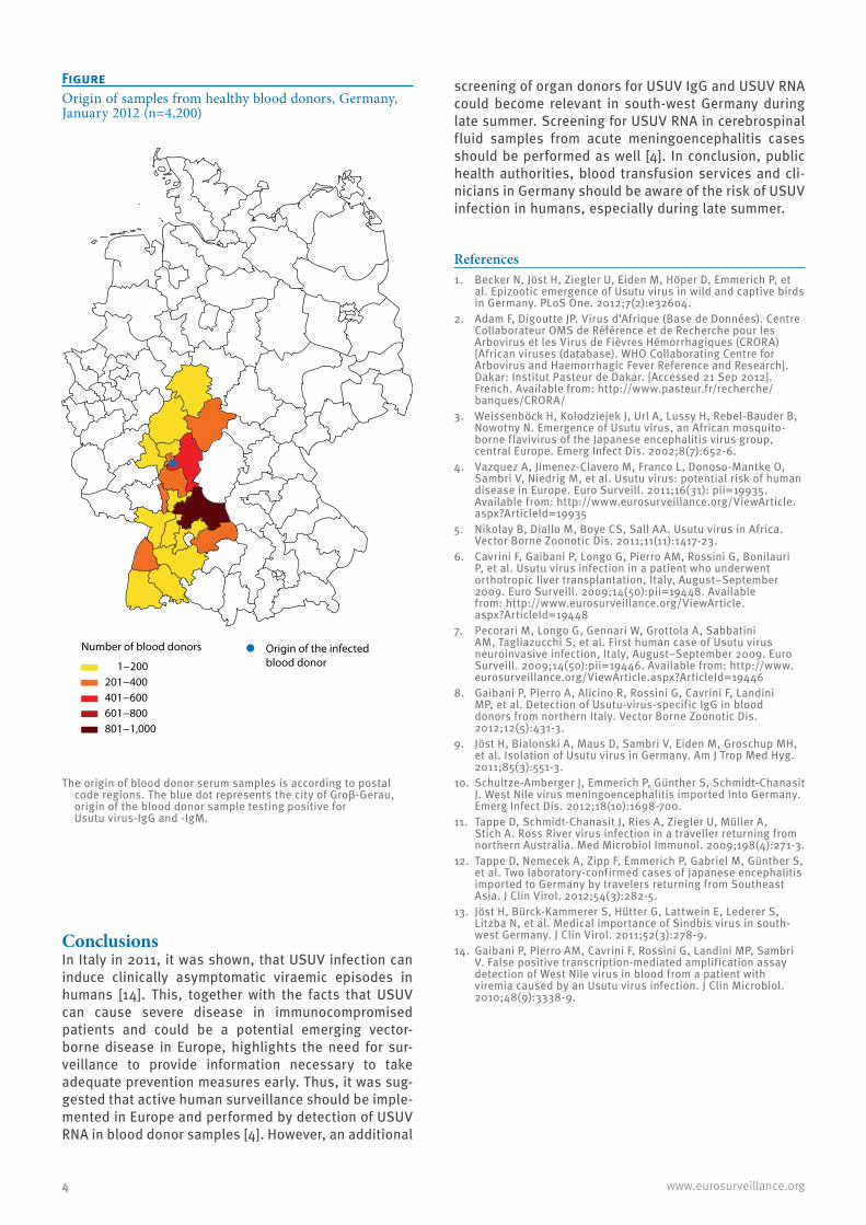
The blood donor testing positive for USUV-IgG and -IgM, who was in his forties, reported no history of vaccination against YFV and JEV, and did not have a fever during a period of three months before the blood donation. In addition, he had not been abroad during this period. The blood donor lives in the city of Gross-Gerau (Figure) and dead black birds testing positive for USUV were reported from the district of Gross-Gerau in 2011 [1]. These findings, taken together with the sero-logical results corroborate the hypothesis of a pauci- or asymptomatic and autochthonous USUV infection of the blood donor during late summer 2011.
TableResults of serological analysis of a German blood donor at different time points, Germany, 2007–2012
VirusImmunofluorescence assay (IFA)
Virus neutralisation
test (VNT)April 2007 October 2008 October 2010 October 2011 January 2012 April 2012 January
2012April 2012IgG IgM IgG IgM IgG IgM IgG IgM IgG IgM IgG IgM
Usutu Neg Neg Neg Neg Neg Neg 1:1,280 1:160 1:2,560 1:160 1:2,560 1:40 1:40 1:80West Nile Neg Neg Neg Neg Neg Neg 1:640 1:20 1:640 1:20 1:160 1:20 Neg NegJapanese encephalitis Neg Neg Neg Neg Neg Neg 1:2,560 Neg 1:1,280 Neg 1:640 Neg Neg NegTick-borne encephalitis Neg Neg 1:80 Neg 1:160 Neg 1:1,280 Neg 1:1,280 Neg 1:1,280 Neg Neg 1:20Yellow fever Neg Neg Neg Neg 1:160 Neg 1:1,280 Neg 1:1,280 Neg 1:160 Neg Neg Neg
Neg: negative.
Immunofluorescence assay and virus neutralisation test results of < 1:20 were considered negative.
4 www.eurosurveillance.org
ConclusionsIn Italy in 2011, it was shown, that USUV infection can induce clinically asymptomatic viraemic episodes in humans [14]. This, together with the facts that USUV can cause severe disease in immunocompromised patients and could be a potential emerging vector-borne disease in Europe, highlights the need for sur-veillance to provide information necessary to take adequate prevention measures early. Thus, it was sug-gested that active human surveillance should be imple-mented in Europe and performed by detection of USUV RNA in blood donor samples [4]. However, an additional
screening of organ donors for USUV IgG and USUV RNA could become relevant in south-west Germany during late summer. Screening for USUV RNA in cerebrospinal fluid samples from acute meningoencephalitis cases should be performed as well [4]. In conclusion, public health authorities, blood transfusion services and cli-nicians in Germany should be aware of the risk of USUV infection in humans, especially during late summer.
References1. Becker N, Jöst H, Ziegler U, Eiden M, Höper D, Emmerich P, et
al. Epizootic emergence of Usutu virus in wild and captive birds in Germany. PLoS One. 2012;7(2):e32604.
2. Adam F, Digoutte JP. Virus d’Afrique (Base de Données). Centre Collaborateur OMS de Référence et de Recherche pour les Arbovirus et les Virus de Fièvres Hémorrhagiques (CRORA) [African viruses (database). WHO Collaborating Centre for Arbovirus and Haemorrhagic Fever Reference and Research]. Dakar: Institut Pasteur de Dakar. [Accessed 21 Sep 2012]. French. Available from: http://www.pasteur.fr/recherche/banques/CRORA/
3. Weissenböck H, Kolodziejek J, Url A, Lussy H, Rebel-Bauder B, Nowotny N. Emergence of Usutu virus, an African mosquito-borne flavivirus of the Japanese encephalitis virus group, central Europe. Emerg Infect Dis. 2002;8(7):652-6.
4. Vazquez A, Jimenez-Clavero M, Franco L, Donoso-Mantke O, Sambri V, Niedrig M, et al. Usutu virus: potential risk of human disease in Europe. Euro Surveill. 2011;16(31): pii=19935. Available from: http://www.eurosurveillance.org/ViewArticle.aspx?ArticleId=19935
5. Nikolay B, Diallo M, Boye CS, Sall AA. Usutu virus in Africa. Vector Borne Zoonotic Dis. 2011;11(11):1417-23.
6. Cavrini F, Gaibani P, Longo G, Pierro AM, Rossini G, Bonilauri P, et al. Usutu virus infection in a patient who underwent orthotropic liver transplantation, Italy, August–September 2009. Euro Surveill. 2009;14(50):pii=19448. Available from: http://www.eurosurveillance.org/ViewArticle.aspx?ArticleId=19448
7. Pecorari M, Longo G, Gennari W, Grottola A, Sabbatini AM, Tagliazucchi S, et al. First human case of Usutu virus neuroinvasive infection, Italy, August–September 2009. Euro Surveill. 2009;14(50):pii=19446. Available from: http://www.eurosurveillance.org/ViewArticle.aspx?ArticleId=19446
8. Gaibani P, Pierro A, Alicino R, Rossini G, Cavrini F, Landini MP, et al. Detection of Usutu-virus-specific IgG in blood donors from northern Italy. Vector Borne Zoonotic Dis. 2012;12(5):431-3.
9. Jöst H, Bialonski A, Maus D, Sambri V, Eiden M, Groschup MH, et al. Isolation of Usutu virus in Germany. Am J Trop Med Hyg. 2011;85(3):551-3.
10. Schultze-Amberger J, Emmerich P, Günther S, Schmidt-Chanasit J. West Nile virus meningoencephalitis imported into Germany. Emerg Infect Dis. 2012;18(10):1698-700.
11. Tappe D, Schmidt-Chanasit J, Ries A, Ziegler U, Müller A, Stich A. Ross River virus infection in a traveller returning from northern Australia. Med Microbiol Immunol. 2009;198(4):271-3.
12. Tappe D, Nemecek A, Zipp F, Emmerich P, Gabriel M, Günther S, et al. Two laboratory-confirmed cases of Japanese encephalitis imported to Germany by travelers returning from Southeast Asia. J Clin Virol. 2012;54(3):282-5.
13. Jöst H, Bürck-Kammerer S, Hütter G, Lattwein E, Lederer S, Litzba N, et al. Medical importance of Sindbis virus in south-west Germany. J Clin Virol. 2011;52(3):278-9.
14. Gaibani P, Pierro AM, Cavrini F, Rossini G, Landini MP, Sambri V. False positive transcription-mediated amplification assay detection of West Nile virus in blood from a patient with viremia caused by an Usutu virus infection. J Clin Microbiol. 2010;48(9):3338-9.
FigureOrigin of samples from healthy blood donors, Germany, January 2012 (n=4,200)
The origin of blood donor serum samples is according to postal code regions. The blue dot represents the city of Groβ-Gerau, origin of the blood donor sample testing positive for Usutu virus-IgG and -IgM.
1−200201−400401−600601−800801−1,000
Origin of the infected blood donor
Number of blood donors

5www.eurosurveillance.org
Rapid communications
First detection of livestock-associated meticillin-resistant Staphylococcus aureus CC398 in bulk tank milk in the United Kingdom, January to July 2012
G K Paterson1, J Larsen2, E M Harrison1, A R Larsen2, F J Morgan1, S J Peacock3,4, J Parkhill4, R N Zadoks5, M A Holmes ([email protected])1
1. Department of Veterinary Medicine, University of Cambridge, Cambridge, United Kingdom2. Microbiology and Infection Control, Statens Serum Institut, Copenhagen, Denmark3. School of Clinical Medicine, University of Cambridge, Cambridge, United Kingdom 4. The Wellcome Trust Sanger Institute, Wellcome Trust, Cambridge, United Kingdom5. Moredun Research Institute, Penicuik, United Kingdom
Citation style for this article: Paterson GK, Larsen J, Harrison EM, Larsen AR, Morgan FJ, Peacock SJ, Parkhill J, Zadoks RN, Holmes MA. First detection of livestock-associated meticillin-resistant Staphylococcus aureus CC398 in bulk tank milk in the United Kingdom, January to July 2012. Euro Surveill. 2012;17(50):pii=20337. Available online: http://www.eurosurveillance.org/ViewArticle.aspx?ArticleId=20337
Article submitted on 27 November 2012 / published on 13 December 2012
Livestock-associated meticillin-resistant Staphylo-coccus aureus belonging to clonal complex 398 (LA-MRSA CC398) is an important cause of zoonotic infections in several countries, but there is only a sin-gle published report of this lineage from the United Kingdom (UK). Here, we describe the isolation of LA-MRSA CC398 from bulk tank milk from five geo-graphically dispersed farms in the UK. Our findings suggest that LA-MRSA CC398 is established in live-stock in the UK. Awareness of the potential occupa-tional risks and surveillance in other food-producing animal species should be promoted.
Isolation of meticillin-resistant Staphylococcus aureus from dairy cattleDuring a study, performed from January to July 2012, to detect mecC meticillin-resistant Staphylococcus aureus (MRSA) in dairy cattle in the United Kingdom (UK), ca. 1,500 bulk tank milk samples were supplied by National Milk Laboratories Ltd., (Chippenham, UK). These were collected aseptically by trained technicians for quality assurance purposes and stored at 4 °C for up to five days prior to testing. Enrichment for S. aureus was per-formed using a modification of a published technique [1] omitting the incubation in phenol red mannitol broth supplemented with 4 mg/L oxacillin (24 h at 37°C). Identification of potential MRSA colonies (blue colour) was confirmed by subculture on Staph Brilliance 24 plates (Oxoid, Baskingstoke, UK) and these were sub-sequently screened for mecA, mecC and femB by mul-tiplex PCR as described previously [2]. Approximately 300 potential MRSA colonies were identified and sub-jected to PCR testing, yielding a total of seven mecA MRSA isolates from five farms, including three isolates from the same farm. These isolates were found to be mecA, femB-positive by PCR (Table). All seven isolates were resistant to penicillin, meticillin and cefoxitin by disk diffusion according to the European Committee on
Antimicrobial Susceptibility Testing (EUCAST) guide-lines [3].
Molecular and phenotypic characterisation of LA-MRSA CC398 from dairy cattle in the United KingdomMulti-locus sequence typing found all seven isolates belonged to sequence type ST398, and CC398-specific PCR based on the restriction–modification system sau1–hsdS1 confirmed that all the isolates belonged to clonal complex CC398 [4]. Isolates from three farms exhibited spa type t011 and carried a compos-ite staphylococcal cassette chromosome mec (SCCmec) V(5C2&5)c element, whereas isolates from the remain-ing two farms had spa types t011 and t2546 and har-boured SCCmec IVa. All isolates lacked the lukS-PV and lukF-PV genes encoding Panton-Valentine leukocidin and the scn gene (Table). Antimicrobial susceptibility testing using disk diffusion according to the EUCAST guidelines revealed that all isolates were resistant to tetracycline, and PCR [10] demonstrated the presence of the tetracycline resistance gene tet(M) in all seven, and of tet(K) in three isolates (Table).
DiscussionHere we describe the first isolation of LA-MRSA CC398 from dairy cattle in the UK. This is only the second published instance of LA-MRSA CC398 in this coun-try following the report of isolates (t011 and SCCmec IVa) from two horses in south-eastern England [11]. In many countries in continental Europe and elsewhere, LA-MRSA CC398 poses an occupational risk for those in close contact with livestock, particularly pigs and veal calves. For instance, significantly higher rates of MRSA nasal carriage by humans in contact with pigs (farm workers, abattoir workers, veterinarians) have been noted in several epidemiological studies, with the isolates typically belonging to CC398 [12-16]. Further
6 www.eurosurveillance.org
studies have shown an association between clinical disease resulting from LA-MRSA CC398 infection and contact with pigs or pig farms [16-20]. The impact of this can be significant locally, and this lineage can be imported into healthcare settings. For example, in a German hospital in an area with a large number of pigs, 22% of patients colonised with MRSA at admission carried ST398 [21]. Nosocomial transmission has also been reported [22]. LA-MRSA CC398, like other MRSA, may be responsible for life-threatening infections dur-ing long or frequent hospitalisations, or following wound or surgery site infections, and also increases healthcare costs resulting from screening, isolation of carriers, and decolonisation. Although pasteurisa-tion of milk should ensure that CC398 MRSA will not enter the food chain, our finding of LA-MRSA CC398 in dairy cattle has clear public health implications for the UK. Workers on dairy farms, or individuals with regular contact with dairy cows, are likely to have a higher risk of colonisation or infection with LA-MRSA CC398 com-pared to the general population in the UK. LA-MRSA CC398 isolates from three of the farms where isolated were found carried SCCmec type IVa. The isolates from the other two farms carried SCCmec type V(5C2&5)c. Both of these SCCmec types have previously been found in LA-MRSA CC398 isolates [23].
Heterogeneity is seen in S. aureus CC398, with human and livestock-associated lineages being differentiated by the presence or absence of specific resistance and virulence-related genes [23-24]. In all of our isolates the absence of the scn gene, encoding the human-specific staphylococcal complement inhibitor, and the presence of tet(M) suggested that they were all livestock-associated, as opposed to S. aureus CC398 strains which circulate in the human population inde-pendent of a livestock reservoir [23-24]. Likewise, all seven isolates lacked the lukS-PV and lukF-PV genes encoding Panton-Valentine leukocidin which is absent in LA-MRSA CC398, but is present in some, but not all, human-associated CC398 isolates [23]. Three consecu-tive samples from the same farm over a seven-month period were positive for LA-MRSA CC398 isolates with identical spa (t011) and SCCmec types (IVa), suggest-ing that this strain is able to persist in dairy herds over prolonged periods. While there are relatively few reports of LA-MRSA CC398 from dairy cattle compared to pig farms, it has been found to cause bovine masti-tis [25-27]. Our findings therefore have significance to veterinary medicine, in addition to public health. The relative absence of CC398 MRSA from the UK prior to this study, when it is widespread in the rest of Europe suggests that the geographical separation of the UK
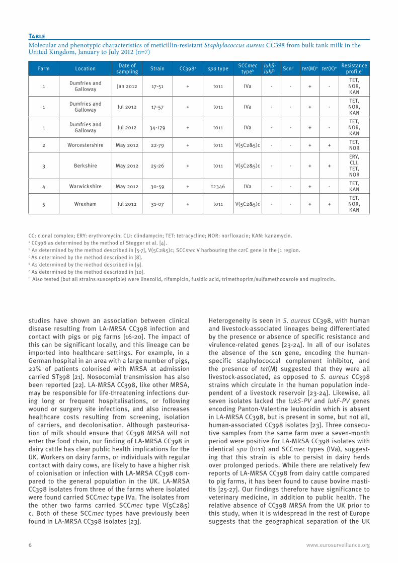
TableMolecular and phenotypic characteristics of meticillin-resistant Staphylococcus aureus CC398 from bulk tank milk in the United Kingdom, January to July 2012 (n=7)
Farm Location Date of sampling Strain CC398a spa type SCCmec
typeblukS-lukFc Scnd tet(M)e tet(K)e Resistance
profilef
1 Dumfries and Galloway Jan 2012 17-51 + t011 IVa - - + -
TET, NOR, KAN
1 Dumfries and Galloway Jul 2012 17-57 + t011 IVa - - + -
TET, NOR, KAN
1 Dumfries and Galloway Jul 2012 34-179 + t011 IVa - - + -
TET, NOR, KAN
2 Worcestershire May 2012 22-79 + t011 V(5C2&5)c - - + + TET, NOR
3 Berkshire May 2012 25-26 + t011 V(5C2&5)c - - + +
ERY, CLI, TET, NOR
4 Warwickshire May 2012 30-59 + t2346 IVa - - + - TET, KAN
5 Wrexham Jul 2012 31-07 + t011 V(5C2&5)c - - + +TET, NOR, KAN
CC: clonal complex; ERY: erythromycin; CLI: clindamycin; TET: tetracycline; NOR: norfloxacin; KAN: kanamycin. a CC398 as determined by the method of Stegger et al. [4].b As determined by the method described in [5-7], V(5C2&5)c; SCCmec V harbouring the czrC gene in the J1 region. c As determined by the method described in [8]. d As determined by the method described in [9].e As determined by the method described in [10]. f Also tested (but all strains susceptible) were linezolid, rifampicin, fusidic acid, trimethoprim/sulfamethoxazole and mupirocin.
7www.eurosurveillance.org
from continental Europe may have delayed the spread of this lineage to the UK rather than there being any fundamental difference in husbandry or biosecurity in the UK. The authors are aware of unpublished sur-veys looking for potential LA-MRSA in UK dairy and pig herds that have been negative before now. These CC398-positive samples were not part of a formal prev-alence study, and it is therefore unclear how common LA-MRSA CC398 isolates are in UK dairy farms or if they are present in other livestock. However, the five farms with positive samples were identified from a sample of ca. 1,500 farms, indicating a low prevalence currently.
ConclusionsThis is the first description of LA-MRSA CC398 in food-producing animals in the UK. The ability of this lineage to colonise a wide range of host species, coupled with its zoonotic potential, make this finding of significance to both veterinary and human health. Future surveil-lance for this LA-MRSA CC398 strain in all food-pro-ducing animal species in the UK and the evaluation of occupational risk factors for MRSA carriage and infec-tion should be considered.
Acknowledgments This work was supported by a Medical Research Council Partnership Grant (G1001787/1) held between the Department of Veterinary Medicine, University of Cambridge (M.A.H), the School of Clinical Medicine, University of Cambridge (S.J.P), the Moredun Research Institute (R.N.Z), and the Wellcome Trust Sanger Institute (J.P and S.J.P). We thank National Milk Laboratories Ltd. for their invaluable assistance.
References1. Haran KP, Godden SM, Boxrud D, Jawahir S, Bender JB,
and Sreevatsan S. Prevalence and characterization of Staphylococcus aureus, including methicillin-resistant Staphylococcus aureus, isolated from bulk tank milk from Minnesota dairy farms. J Clin Microbiol. 2012; 50(3):688-95.
2. Paterson GK, Larsen AR, Robb A, Edwards GE, Pennycott TW, Foster G, et al. The newly described mecA homologue, mecALGA251, is present in methicillin-resistant Staphylococcus aureus isolates from a diverse range of host species. J Antimicrob Chemother. 2012;67(12):2809-13.
3. European Committee on Antimicrobial Susceptibility Testing (EUCAST). Breakpoint tables for interpretation of MICs and zone diameters Version 2.0. Basel: European Society of Clinical Microbiology and Infectious Diseases; 1 Jan 2012. Available from: http://www.eucast.org/fileadmin/src/media/PDFs/EUCAST_files/Breakpoint_tables/Breakpoint_table_v_2.0_120221.pdf
4. Stegger M, Lindsay JA, Moodley A, Skov R, Broens EM, Guardabassi L. Rapid PCR Detection of Staphylococcus aureus Clonal Complex 398 by Targeting the Restriction-Modification System Carrying sau1-hsdS1. J Clin Microbiol. 2011;49(2):732-4.
5. Kondo Y, Ito T, Ma XX, Watanabe S, Kreiswirth BN, Etienne J, et al. Combination of multiplex PCRs for staphylococcal cassette chromosome mec type assignment: rapid identification system for mec, ccr, and major differences in junkyard regions. Antimicrob Agents Chemother. 2007;51(1):264-74.
6. Cavaco LM, Hasman H, Stegger M, Andersen PS, Skov R, Fluit AC, et al. Cloning and Occurrence of czrC, a Gene Conferring Cadmium and Zinc Resistance in Methicillin-Resistant Staphylococcus aureus CC398 Isolates. Antimicrob Agents and Chemother. 2010;54(9):3605-8.
7. Higuchi W, Takano T, Teng L-J, Yamamoto T. Structure and specific detection of staphylococcal cassette chromosome mec type VII. Biochem Biophys Res Commun. 2008 19;377(3):752-6.
8. Lina G, Piemont Y, Godail-Gamot F, Bes M, Peter MO, Gauduchon V, et al. Involvement of Panton-Valentine
leukocidin-producing Staphylococcus aureus in primary skin infections and pneumonia. Clin Infect Dis. 1999;29(5):1128-32.
9. van Wamel WJ, Rooijakkers SH, Ruyken M, van Kessel KP, van Strijp JA. The innate immune modulators staphylococcal complement inhibitor and chemotaxis inhibitory protein of Staphylococcus aureus are located on beta-hemolysin-converting bacteriophages. J Bacteriol. 2006;188(4):1310-5.
10. Warsa UC, Nonoyama M, Ida T, Okamoto R, Okubo T, Shimauchi C, et al. Detection of tet(K) and tet(M) in Staphylococcus aureus of Asian countries by the polymerase chain reaction. J Antibiot. 1996;49(11):1127-32.
11. Loeffler A, Kearns AM, Ellington MJ, Smith LJ, Unt VE, Lindsay JA, et al. First isolation of MRSA ST398 from UK animals: a new challenge for infection control teams? J Hosp Infect. 2009;72(3):269-71.
12. Van Cleef BA, Broens EM, Voss A, Huijsdens XW, Zuchner L, Van Benthem BH, et al. High prevalence of nasal MRSA carriage in slaughterhouse workers in contact with live pigs in The Netherlands. Epidemiol Infect. 2010;138(5):756-63.
13. Huber H, Koller S, Giezendanner N, Stephan R, Zweifel C. Prevalence and characteristics of meticillin-resistant Staphylococcus aureus in humans in contact with farm animals, in livestock, and in food of animal origin, Switzerland, 2009. Euro Surveill. 2010;15(16):pii=19542. Available from: http://www.eurosurveillance.org/ViewArticle.aspx?ArticleId=19542
14. Garcia-Graells C, Antoine J, Larsen J, Catry B, Skov R, Denis O. Livestock veterinarians at high risk of acquiring methicillin-resistant Staphylococcus aureus ST398. Epidemiol Infect. 2012;140(3):383-9.
15. van Cleef BA, Verkade EJM, Wulf MW, Buiting AG, Voss A, Huijsdens XW, et al. Prevalence of Livestock-Associated MRSA in Communities with High Pig-Densities in The Netherlands. PLoS One. 2010;5(2):e9385.
16. Krziwanek K, Metz-Gercek S, Mittermayer H. Methicillin-Resistant Staphylococcus aureus ST398 from Human Patients, Upper Austria. Emerg Infect Dis. 2009;15(5):766-9.
17. Pan A, Battisti A, Zoncada A, Bernieri F, Boldini M, Franco A, et al. Community-acquired Methicillin-Resistant Staphylococcus aureus ST398 Infection, Italy. Emerg Infect Dis. 2009;15(5):845-7.
18. Witte W, Strommenger B, Stanek C, Cuny C. Methicillin-resistant Staphylococcus aureus ST398 in humans and animals, Central Europe. Emerg Infect Dis. 2007;13(2):255-8.
19. Denis O, Suetens C, Hallin M, Catry B, Ramboer I, Dispas M, et al. Methicillin-Resistant Staphylococcus aureus ST398 in Swine Farm Personnel, Belgium. Emerg Infect Dis. 2009;15(7):1098-101.
20. Aspiroz C, Lozano C, Vindel A, Lasarte JJ, Zarazaga M, Torres C. Skin Lesion Caused by ST398 and ST1 MRSA, Spain. Emerg Infect Dis. 2010;16(1):157-9.
21. Köck R, Harlizius J, Bressan N, Laerberg R, Wieler LH, Witte W, et al. Prevalence and molecular characteristics of methicillin-resistant Staphylococcus aureus (MRSA) among pigs on German farms and import of livestock-related MRSA into hospitals. Eur J Clin Microbiol Infect Dis. 2009;28(11):1375-82.
22. Wulf MW, Markestein A, van der Linden FT, Voss A, Klaassen C, Verduin CM. First outbreak of methicillin-resistant Staphylococcus aureus ST398 in a Dutch hospital, June 2007. Euro Surveill. 2008;13(9):pii=8051. Available from: http://www.eurosurveillance.org/ViewArticle.aspx?ArticleId=8051
23. Price LB, Stegger M, Hasman H, Aziz M, Larsen J, Andersen PS, et al. Staphylococcus aureus CC398: host adaptation and emergence of methicillin resistance in livestock. MBio. 2012;3(1):pii=00305-11.
24. Uhlemann AC, Porcella SF, Trivedi S, Sullivan SB, Hafer C, Kennedy AD, et al. Identification of a Highly Transmissible Animal-Independent Staphylococcus aureus ST398 Clone with Distinct Genomic and Cell Adhesion Properties. MBio. 2012;3(2): pii=e00027-12.
25. Fessler A, Scott C, Kadlec K, Ehricht R, Monecke S, Schwarz S. Characterization of methicillin-resistant Staphylococcus aureus ST398 from cases of bovine mastitis. J Antimicrob Chemother. 2010;65(4):619-25.
26. Vanderhaeghen W, Cerpentier T, Adriaensen C, Vicca J, Hermans K, Butaye P. Methicillin-resistant Staphylococcus aureus (MRSA) ST398 associated with clinical and subclinical mastitis in Belgian cows. Vet Microbiol. 2010;144(1-2):166-71.
27. Tavakol M, Riekerink RG, Sampimon OC, van Wamel WJ, van Belkum A, Lam TJ. Bovine-associated MRSA ST398 in The Netherlands. Acta Vet Scand. 2012;54:28.
8 www.eurosurveillance.org
Rapid communications
Cluster of invasive Neisseria meningitidis infections on a cruise ship, Italy, October 2012
P Stefanelli ([email protected])1, C Fazio1,2, A Neri1,2, P Isola3, S Sani4, P Marelli3, C Martinelli3, P Mastrantonio1, M G Pompa5
1. Department of Infectious, Parasitic and Immune-mediated Diseases, Istituto Superiore di Sanità (ISS), Rome, Italy2. These authors contributed equally to this work3. Clinical Pathology Department, Azienda USL 6, Livorno, Italy4. Infectious Diseases Unit, Azienda USL 6, Livorno, Italy5. Communicable Disease and International Prophylaxis Unit, Directorate General of Prevention, Ministry of Health, Rome, Italy
Citation style for this article: Stefanelli P, Fazio C, Neri A, Isola P, Sani S, Marelli P, Martinelli C, Mastrantonio P, Pompa MG. Cluster of invasive Neisseria meningitidis infections on a cruise ship, Italy, October 2012. Euro Surveill. 2012;17(50):pii=20336. Available online: http://www.eurosurveillance.org/ViewArticle.aspx?ArticleId=20336
Article submitted on 21 November 2012 / published on 13 December 2012
We describe a cluster of four cases of invasive menin-gococcal disease that occurred on a cruise ship sailing along the Italian coast in October 2012. All four cases were hospitalised with severe illness and one of them died. This report illustrates the importance of rapid implementation of emergency control measures such as administration of prophylaxis to all crew members and passengers to prevent the spread of the disease in such a close environment.
We report a cluster of four cases of meningitis due to infection with serogroup C Neisseria meningitidis ST-11 clonal complex (cc) that occurred on a cruise ship sailing along the Italian coast in October 2012. Meningococcal serogroup C strains, (ST-11 cc) are known to cause inva-sive disease burden worldwide [1,2]. It is also known that these hyperinvasive strains are responsible for a high mortality rate among cases [3,4]. These strains caused several outbreaks in France between 2001 and 2003 that led to targeted vaccination campaigns [5,6].
In Italy, serogroup C is the second most common sero-group (16% of the 118 cases with a known serogroup in 2011 were caused by serogroup C infection) after the serogroup B (64% of the 118 cases with a known sero-group in 2011 were caused by serogroup B infection) [7]. Meningococci C ST-11 cc have been identified in Italy during the last five years and those characterised as C:P1.5-1,10-8:F3-6:ST-11, were responsible for both sporadic and outbreak-associated cases [8].
Cluster descriptionIn early October 2012, four staff members of a cruise ship sailing on the Italian coast were hospitalised on the same day in a local hospital in Tuscany, with clini-cally suspected meningitis. The four cases were aged between 26 and 47 years and originated from three different countries and three continents. They were all crew members working in the kitchen. One of these four hospitalised patients died; no secondary cases have been reported among other persons present on the ship or among other contacts of the four cases,
during the follow-up. Health authorities conducted interviews to determine the travel history of the cases and Naples was established as the last stop before the onset of symptoms. No common source of contamina-tion could be identified during the investigations.
Laboratory investigationOne day after hospitalisation, the cerebrospinal fluid (CSF) of the four patients was examined by direct microscopy observation at the microbiology laboratory of the Livorno Hospital and the diagnosis of bacterial meningitis was established. Furthermore, on the same day, latex test and culture were performed in the same hospital, whereas serogroup C meningococci were confirmed by rapid molecular test at the Laboratory of the Paediatric Department, Meyer Hospital, University of Florence. The four N. meningitidis isolates identi-fied, were sent to the National Reference Laboratory for Invasive Meningococcal Diseases of the Istituto Superiore di Sanità for serogroup confirmation, antimicrobials susceptibility and molecular typing characterisation.
In particular, multilocus sequence typing (MLST) [9] and sequencing of outer membrane proteins PorA and FetA were performed to define the clonal complex and the finetype, respectively; porB gene was also ana-lysed. Variable number tandem repeats (VNTR) typing, using standardised procedures [10], was also carried out to further discriminate among strains. Sequence analysis of penA gene was used to determine the cor-responding allele defining isolates with a decreased susceptibility to penicillin [11].
The molecular investigations showed that all strains were C:P1.5-1,10-8:F3-6:ST-11. The discriminatory power of MLST and antigen sequence typing high-lighted that the four isolates clearly belonged to ST-11 cc, finetype P1.5-1, 10-8:F3-6. The porB allele was 2-2 for all of them. The VNTR analysis (data not shown) con-firmed the presence of a unique profile among the four
9www.eurosurveillance.org
strains which was different from all the others found in ST-11 cc isolates circulating in Italy [12].
The antibiotic susceptibility assay showed a full sus-ceptibility to rifampicin, ciprofloxacin, ceftriaxone and a decreased susceptibility to penicillin and ampicillin. The analysis of penA gene showed the presence of the same allele (248), often associated with a decreased susceptibility to penicillin (0.06<MIC≤1 mg/L) [13].
Control measuresOn the hospital admission day of the four cases described above an alert was launched from the Maritime Port Health Offices of Livorno to the Ministry of Health which ensured rapid communication of this cluster to national and international health authorities for achieving a coordinated response and proper man-agement of this public health event.
On the same day, as an emergency control measure, it was decided to administer chemoprophylaxis with cip-rofloxacin to about 2,000 persons present on the ship (international passengers and crew members) before some people left.
In general, the management of meningococcal disease cases focuses on early recognition and antibiotic treat-ment to reduce case fatality rate and to control the spread of the disease. In the cluster described above, the prompt prophylaxis to all crew members and pas-sengers, as an emergency control measure in such a close environment as a ship, ensured the meningococ-cal spread control. The rapid strain typing was essen-tial to assess the link among isolates and to highlight the circulation of hyper-invasive ST-11 meningococci.
Acknowledgments The authors thank everyone who took part in this inves-tigation: the staff of the Maritime Port Health Offices of Livorno; the staff of the Livorno Hospital; Dr Chiara Azzari, Paediatrician Department, Anna Meyer Hospital, University of Florence, for the rapid identification of serogroup C menin-gococcal strains on CSFs; Local Health Authorities.This publication made use of the Neisseria Multi Locus Sequence Typing website [13] developed by Keith Jolley and sited at the University of Oxford (Jolley & Maiden 2010, BMC Bioinformatics, 11:595). The development of this site has been funded by the Wellcome Trust and European Union.
This work was partially funded by the Ministry of Health-CCM Project fasc.1M12 “Sorveglianza delle Malattie Batteriche Invasive da Neisseria meningitidis, Streptococcus pneumo-niae ed Hemophilus influenzae” 2010-2012.
References1. Watkins ER, Maiden MC. Persistence of Hyperinvasive
Meningococcal Strain Types during Global Spread as Recorded in the PubMLST Database. PLoS One. 2012;7(9):e45349.
2. Deghmane AE, Parent du Chatelet, Szatanik M, Hong E, Ruckly C, Giorgini D, et al. Emergence of new virulent Neisseria meningitidis serogroup C Sequence Type 11 isolates in France. J Infect Dis. 2010;202(2): 247-50.
3. Zarantonelli ML, Lancellotti M, Deghmane AE, Giorgini D, Hong E, Ruckly C, et al. Hyperinvasive genotypes of Neisseria meningitidis in France. Clin Microbiol Infect. 2008;14(5):467-72.
4. Smith I, Caugant DA, Høiby EA, Wentzel-Larsen T, Halstensen A. High case-fatality rates of meningococcal disease in Western Norway caused by serogroup C strains belonging to both sequence type (ST)-32 and ST-11 complexes, 1985-2002. Epidemiol Infect. 2006;134(6):1195-202.
5. Perrocheau A, Taha MK, Lévy-Bruhl D. Epidemiology of invasive meningococcal disease in France in 2003. Euro Surveill. 2005;10(12):pii=587. Available from: http://www.eurosurveillance.org/ViewArticle.aspx?ArticleId=587
6. Lévy-Bruhl D, Perrocheau A, Mora M, Taha MK, Dromell-Chabrier S, Quatresous I. Vaccination campaign following an increase in incidence of serogroup C meningococcal diseases in the department of Puy-de-Dôme (France). Euro Surveill. 2002;7(5):pii=368. Available from: http://www.eurosurveillance.org/ViewArticle.aspx?ArticleId=368
7. Istituto Superiore di Sanità (ISS). Dati di sorveglianza delle malattie batteriche invasive aggiornati al 20 agosto 2012. [Surveillance data on invasive bacterial diseases updated 20 August 2012]. Rome: ISS. 25 Sep 2012. Italian. Available from: http://www.simi.iss.it/files/Report_MBI.pdf
8. Fazio C, Neri A, Sofia T, Carannante A, Caporali MG, Salmaso S, et al. Characterisation of Neisseria meningitidis C strains causing two clusters in the north of Italy in 2007 and 2008. Euro Surveill. 2009;14(16):pii=19179. Available from: http://www.eurosurveillance.org/ViewArticle.aspx?ArticleId=19179
9. Maiden MC, Bygraves JA, Feil E, Morelli G, Russell JE, Urwin R, et al. Multilocus sequence typing: a portable approach to the identification of clones within populations of pathogenic microorganisms. Proc Natl Acad Sci USA. 1998;95(6):3140-5.
10. Yazdankhah SP, Lindstedt BA, Caugant DA. Use of variable-number tandem repeats to examine genetic diversity of Neisseria meningitidis. J Clin Microbiol. 2005;43(4):1699-705.
11. Taha MK, Vàsquez JA, Hong E, Bennett DE, Bertrand S, Bukovski S, et al. Target gene sequencing to characterize the penicillin G susceptibility of Neisseria meningitidis. Antimicrob Agents Chemother. 2007;51(8):2784-92.
12. Stefanelli P, Fazio C, Sofia T, Neri A, Mastrantonio P. Serogroup C meningococci in Italy in the era of conjugate menC vaccination. BMC Infect Dis. 2009;9:135.
13. PubMLST [Internet]. Neisseria Sequence Typing Home Page. PubMLST. [Accessed 21 Nov 2012]. Available from: http://pubmlst.org/neisseria

10 www.eurosurveillance.org
Surveillance and outbreak reports
Measles virus genotyping an important tool in measles outbreak investigation in Norway, 2011
K Vainio ([email protected])1, T W Steen2, T M Arnesen3, K Rønning4, G Ånestad1, S Dudman1
1. Norwegian Institute of Public Health, Department of Virology, Oslo, Norway2. Health and Welfare Authority, City of Oslo, Norway3. District of old Oslo, City of Oslo, Norway4. Norwegian Institute of Public Health, Department of Infectious Disease Epidemiology, Oslo, Norway
Citation style for this article: Vainio K, Steen TW, Arnesen TM, Rønning K, Ånestad G, Dudman S. Measles virus genotyping an important tool in measles outbreak investigation in Norway, 2011. Euro Surveill. 2012;17(50):pii=20340. Available online: http://www.eurosurveillance.org/ViewArticle.aspx?ArticleId=20340
Article submitted on 25 April 2012 / published on 13 December 2012
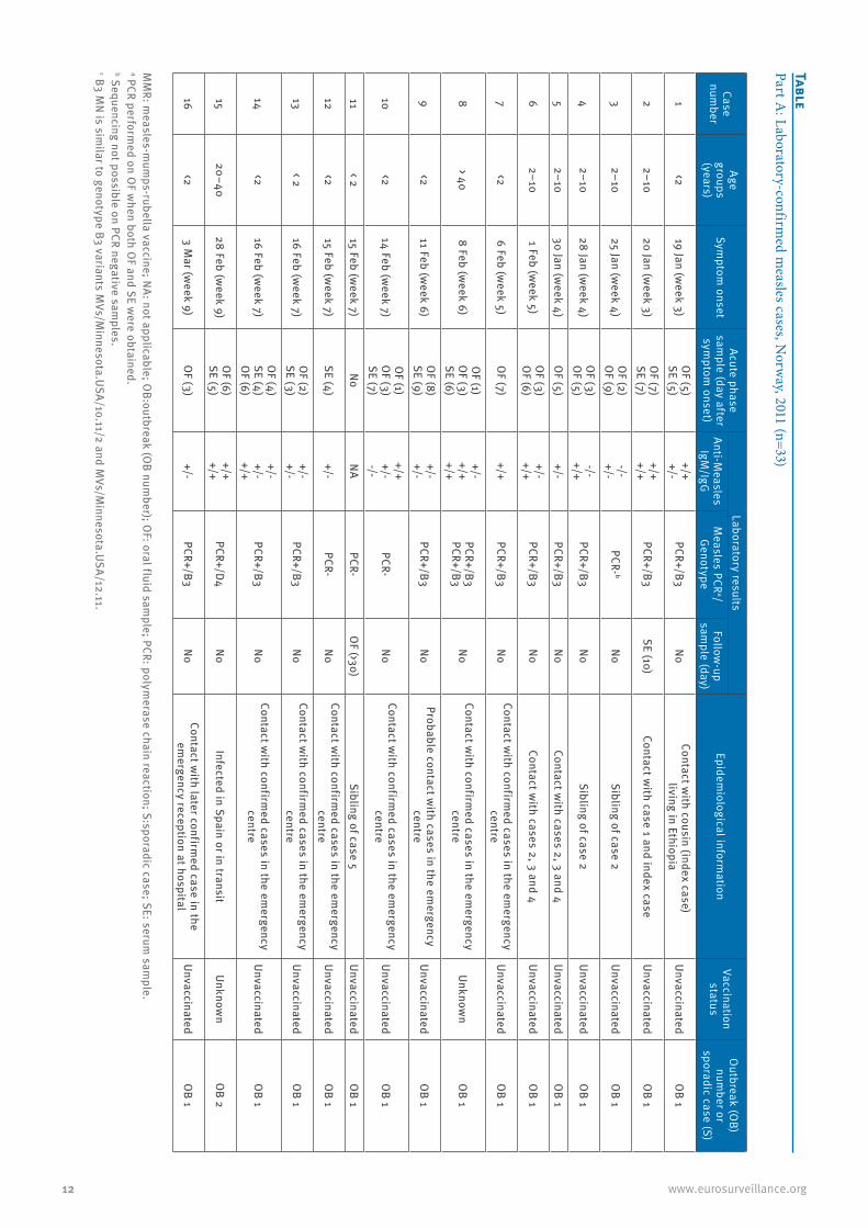
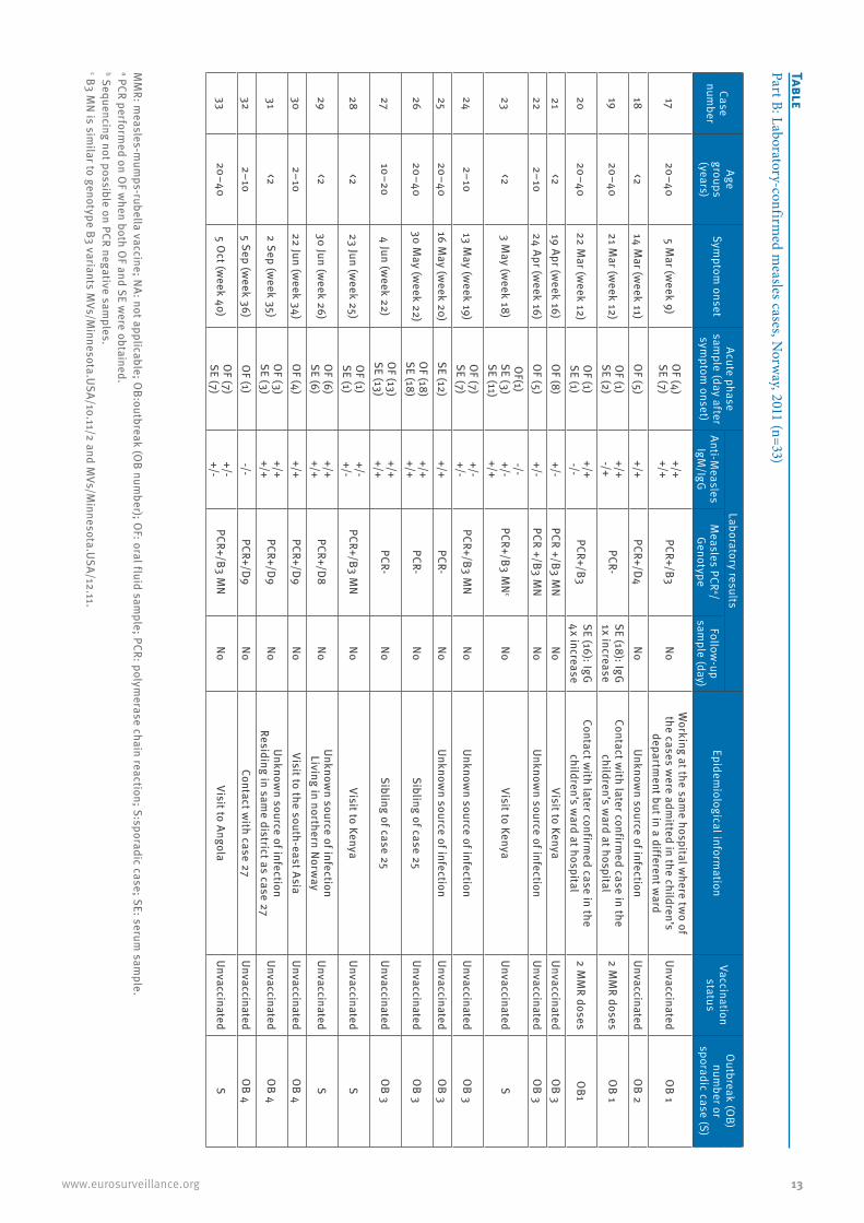
This study describes 33 laboratory-confirmed cases of measles that occurred in Norway in 2011, mainly among unvaccinated children between seven months and 10 years of age. Laboratory testing included detection of anti-measles IgM- and IgG antibodies by enzyme-linked immunosorbent assay (ELISA) and molecular detection and characterisation of measles virus by polymerase chain reaction (PCR) and sequenc-ing. Epidemiological data and genotyping revealed that the measles cases originated from eight sepa-rate importations, resulting in four outbreaks and four sporadic cases. Except for the first outbreak which affected 18 cases, limited secondary spread occurred in each of the three other outbreaks. The outbreaks were caused by measles virus genotypes B3, D4 and D9, whereas genotypes D8 and B3 were detected in the sporadic cases. This study highlights that genetic characterisation of measles virus is an essential tool in the laboratory surveillance of measles, especially in countries like Norway which are approaching the measles elimination goal. The investigation revealed that importation of measles resulted in subsequent transmission within Norway to non-vaccinated indi-viduals, and twelve cases occurred in healthcare set-tings, involving both staff and children. The four cases detected among healthcare workers (HCWs) empha-sised that the coverage of measles-mumps-rubella (MMR) vaccination among healthcare personnel needs to be improved and both primary and secondary vac-cine failure was demonstrated in two fully immunised HCWs.
IntroductionMeasles, a highly contagious respiratory viral disease characterised by the appearance of fever and a rash, is the leading cause of vaccine-preventable childhood mortality worldwide [1]. The incubation time is between 10 and 14 days, and a measles infected person is conta-gious from four days before to four days after the rash appears. Although a safe and cost-effective vaccine has been available for decades, measles is still an ongoing public health problem in several European countries.
Between January and October 2011, 26,074 measles cases were reported in the World Health Organization (WHO) European region [2]. The target date for elimina-tion of measles in Europe has been changed a number of times, and due to widespread outbreaks occurring in both eastern and western Europe (Austria, Bulgaria, France, Germany, Romania, Switzerland, and the United Kingdom (UK)) [3] the WHO Regional Office for Europe most recently (2010) changed the target date from 2010 to 2015 [3,4].
Measles elimination is defined as the interruption of indigenous transmission of measles virus for a 12-month period [5]. In order to prevent outbreaks, a measles vaccine coverage of 95% for two doses of vaccine is needed [6]. In addition, strong national sur-veillance systems are necessary to detect all clinical cases of measles and to investigate thoroughly all sin-gle cases and outbreaks. In Europe, personal attitudes toward vaccination are factors that influence the vac-cination coverage, which is variable [3].
Laboratory diagnosis is required for confirmation of measles, especially in times of low incidence, when most cases of fever illness with rash are caused by other agents. The WHO currently recognises eight clades of measles virus (A–H) with a total of 23 geno-types recognised within the clades, and viruses with related sequences within a genotype (e.g. B3) are referred to as clusters [7,8]. Molecular characterisation of measles virus isolates is vital in outbreak investiga-tions and the only tool that demonstrates the interrup-tion of circulating endemic virus [5,9]. Consequently, it is one of the key components of the verification of measles elimination.
In Norway, measles is a mandatory notifiable disease. Between 1975 and 1988 only measles encephalitis cases were notified, but since 1988 all cases of mea-sles are notifiable. All children residing in Norway are offered the measles vaccine free of charge as part of the childhood vaccination programme. One dose of
11www.eurosurveillance.org
a monovalent measles vaccine was introduced in the national vaccination programme in Norway in 1969. This was replaced by the combined measles-mumps-rubella MMR vaccine in 1983, applied in a two-dose schedule (at 15 months and at 11–12 years of age). Due to a high coverage (>90%) of two doses of the MMR vaccine in the last decades, according to the National vaccine register (SYSVAK) [10], measles incidence has declined in Norway since first half of the 1980s [11,12]. In 2011, the vaccination coverage in two year-olds (birth cohort 2009) with the first dose was 93% in the whole of Norway, 92% in Oslo and 88% in the district of Old Oslo in Oslo [10]. The MMR vaccine coverage data for the second dose is available for 16 year-olds (birth cohort 1993) and the coverage was 90% in the district of Old Oslo and 94% in Oslo as well as in rest of the country [10]. All measles cases identified during the last decade in Norway have been linked to importa-tion from endemic areas or linked to other outbreaks in Europe [12,13]. The last outbreak in Norway before 2011 occurred in 2008 in an anthroposophical community, where the index case fell ill after returning from Austria [14]. In 2007, there was an outbreak among members of the Irish travelling community from England who were in Norway at the time [13]. Measles spread among unvaccinated children within the community, but no cases occurred in the local population.
The present study describes epidemiological and molecular data from measles outbreaks and sporadic cases detected in Norway during 2011. Preliminary data from the first outbreak has been published previ-ously [15].
Methods
Samples and epidemiological dataCase-based surveillance of measles is conducted con-tinuously in Norway. The case definition used in the present study was based on the WHO classification of measles cases [16]. In Norway, the WHO National Reference Laboratory for Measles and Rubella is located at the Norwegian Institute of Public Health (NIPH) where samples obtained from suspected and notified measles cases are routinely sent for confirma-tion. All laboratory-confirmed cases are reported to the surveillance system. Healthcare personnel who sus-pect a measles case are required to notify the NIPH via the institute’s 24-hour call centre, and then send the samples directly to the NIPH reference laboratory to be analysed immediately to expedite the public health response. Thirty-three notified cases in Norway in 2011 were investigated at the NIPH. The information dur-ing the case investigation was collected by telephone interview and included demographic characteristics, ethnic background, clinical symptoms, hospitalisation, vaccination status, travel history and laboratory data. Contact tracing is also routinely undertaken, especially for unvaccinated and exposed individuals.
Laboratory analysisAll samples (serum and oral fluid) were initially tested for the presence of anti-measles IgM- and IgG antibod-ies with commercially available IgG- and IgM enzyme-linked immunosorbent assays (ELISA) (Enzygnost ELISA, Siemens Healthcare Diagnostics Products, Marburg, Germany) and/or measles IgM capture enzyme immu-noassay (EIA) (Microimmune Ltd, Middlesex, UK). The assays were performed as recommended by the manu-facturer and assay results on the samples were inter-preted qualitatively as positive, negative or equivocal. Measles infection was confirmed when anti-measles IgM antibodies were present. In the event of an equivo-cal result, a second serum or oral fluid was requested to ascertain seroconversion. Measles IgG avidity test-ing was performed at the WHO Regional Reference Laboratory (RRL) for measles, at the Robert Koch-Institute in Berlin, using anti-measles virus ELISA (IgG) assay (Euroimmun AG, Luebeck, Germany). Avidity ratios < 40% were considered to be low, >40% and < 60% to be borderline and > 65% to be high.
Viral RNA was extracted from the clinical samples using the QIAamp viral RNA mini kit (Qiagen, Germany). RNA was converted to complementary DNA (cDNA), and nested polymerase chain reaction (PCR) was per-formed using primers amplifying a 450-nucleotide (nt) fragment encoding the C-terminal end of the nucleopro-tein (N) [17]. All PCR positive samples were sequenced according to the WHO recommendation. Sequences were aligned by Clustal W [18], and phylogenetic and molecular analyses were performed using Molecular Evolutionary Genetics Analysis (MEGA) version 5.0 software [19]. A phylogenetic tree was constructed by using the maximum likelihood method. Genotype assignment was performed by sequence comparison with the measles virus reference strains as designated by WHO [20,21].
Sequences from the Norwegian isolates have been deposited in the GenBank database or the measles nucleotide surveillance (MeaNS) database [22] and the GenBank accession numbers are: JN599049–JN599064 and JX680814–JX680820. Measles virus sequences included in the phylogenetic tree are WHO reference strains, genotype B3 variant strains (MVs/Minnesota.USA/10.11/2, MVs/Minnesota.USA/12.11/ and Pretoria.ZAF/13.09/1 (personal communication, Sheilagh Smit, 12 May 2011) and the sequences from the Norwegian isolates.
Outbreak definitionIn countries with an elimination goal (e.g. Norway), a measles outbreak is defined as two or more confirmed cases that are temporally related and linked epidemio-logically and by detection of the same virus variant. Cases with disease onset within 18 days and who could be epidemiologically linked (e.g. same emergency unit, household, community, kindergarten) were grouped into the same outbreak. Molecular typing of measles
12 www.eurosurveillance.org
TablePart A
: Laboratory-confirmed m
easles cases, Norw
ay, 2011 (n=33)
Casenum
ber
Agegroups(years)
Symptom
onsetAcute phase
sample (day after
symptom
onset)
Laboratory resultsEpidem
iological information
Vaccination status
Outbreak (O
B) num
ber or sporadic case (S)
Anti-Measles
IgM/IgG
Measles PCR
a/ Genotype
Follow-up
sample (day)
1<2
19 Jan (week 3)
OF (5)
SE (5)+/++/-
PCR+/B3No
Contact with cousin (index case)
living in EthiopiaUnvaccinated
OB 1
22–10
20 Jan (week 3)
OF (7)
SE (7)+/++/+
PCR+/B3SE (10)
Contact with case 1 and index case
UnvaccinatedO
B 1
32–10
25 Jan (week 4)
OF (2)
OF (9)
-/-+/-
PCR- bNo
Sibling of case 2Unvaccinated
OB 1
42–10
28 Jan (week 4)
OF (3)
OF (5)
-/-+/+
PCR+/B3No
Sibling of case 2Unvaccinated
OB 1
52–10
30 Jan (week 4)
OF (5)
+/-PCR+/B3
NoContact w
ith cases 2, 3 and 4Unvaccinated
OB 1
62–10
1 Feb (week 5)
OF (3)
OF (6)
+/-+/+
PCR+/B3No
Contact with cases 2, 3 and 4
UnvaccinatedO
B 1
7<2
6 Feb (week 5)
OF (7)
+/+PCR+/B3
NoContact w
ith confirmed cases in the em
ergency centre
UnvaccinatedO
B 1
8> 40
8 Feb (week 6)
OF (1)
OF (3)
SE (6)
+/-+/++/+
PCR+/B3PCR+/B3
NoContact w
ith confirmed cases in the em
ergency centre
Unknown
OB 1
9<2
11 Feb (week 6)
OF (8)
SE (9)+/-+/-
PCR+/B3No
Probable contact with cases in the em
ergency centre
UnvaccinatedO
B 1
10<2
14 Feb (week 7)
OF (1)
OF (3)
SE (7)
+/++/--/-
PCR-No
Contact with confirm
ed cases in the emergency
centreUnvaccinated
OB 1
11< 2
15 Feb (week 7)
NoN
APCR-
OF (>30)
Sibling of case 5Unvaccinated
OB 1
12<2
15 Feb (week 7)
SE (4)+/-
PCR-No
Contact with confirm
ed cases in the emergency
centreUnvaccinated
OB 1
13< 2
16 Feb (week 7)
OF (2)
SE (3)+/-+/-
PCR+/B3No
Contact with confirm
ed cases in the emergency
centreUnvaccinated
OB 1
14<2
16 Feb (week 7)
OF (4)
SE (4)O
F (6)
+/-+/-+/+
PCR+/B3No
Contact with confirm
ed cases in the emergency
centreUnvaccinated
OB 1
1520–40
28 Feb (week 9)
OF (6)
SE (5)+/++/+
PCR+/D4No
Infected in Spain or in transitUnknow
nO
B 2
16<2
3 Mar (w
eek 9) O
F (3)+/-
PCR+/B3No
Contact with later confirm
ed case in the em
ergency reception at hospitalUnvaccinated
OB 1
MM
R: measles-m
umps-rubella vaccine; N
A: not applicable; OB:outbreak (O
B number); O
F: oral fluid sample; PCR: polym
erase chain reaction; S:sporadic case; SE: serum sam
ple.a PCR perform
ed on OF w
hen both OF and SE w
ere obtained.b Sequencing not possible on PCR negative sam
ples.c B3 M
N is similar to genotype B3 variants M
Vs/Minnesota.USA/10.11/2 and M
Vs/Minnesota.USA/12.11.
13www.eurosurveillance.org
TablePart B: Laboratory-confirm
ed measles cases, N
orway, 2011 (n=33)
Casenum
ber
Agegroups(years)
Symptom
onsetAcute phase
sample (day after
symptom
onset)
Laboratory resultsEpidem
iological information
Vaccination status
Outbreak (O
B) num
ber or sporadic case (S)
Anti-Measles
IgM/IgG
Measles PCR
a/ Genotype
Follow-up
sample (day)
1720–40
5 Mar (w
eek 9)O
F (4)SE (7)
+/++/+
PCR+/B3No
Working at the sam
e hospital where tw
o of the cases w
ere admitted in the children’s
department but in a different w
ardUnvaccinated
OB 1
18<2
14 Mar (w
eek 11)O
F (5)+/+
PCR+/D4No
Unknown source of infection
UnvaccinatedO
B 2
1920–40
21 Mar (w
eek 12)O
F (1)SE (2)
+/+-/+
PCR-SE (18): IgG1x increase
Contact with later confirm
ed case in the children’s w
ard at hospital2 M
MR doses
OB 1
2020–40
22 Mar (w
eek 12)O
F (1)SE (1)
+/+-/-
PCR+/B3SE (16): IgG4x increase
Contact with later confirm
ed case in the children’s w
ard at hospital2 M
MR doses
OB1
21<2
19 Apr (week 16)
OF (8)
+/-PCR +/B3 M
NNo
Visit to KenyaUnvaccinated
OB 3
222–10
24 Apr (week 16)
OF (5)
+/-PCR +/B3 M
NNo
Unknown source of infection
UnvaccinatedO
B 3
23<2
3 May (w
eek 18)O
F(1)SE (3)
SE (11)
-/-+/-+/+
PCR+/B3 MN
cNo
Visit to KenyaUnvaccinated
S
242–10
13 May (w
eek 19)O
F (7)SE (7)
+/-+/-
PCR+/B3 MN
NoUnknow
n source of infectionUnvaccinated
OB 3
2520–40
16 May (w
eek 20)SE (12)
+/+PCR-
NoUnknow
n source of infectionUnvaccinated
OB 3
2620–40
30 May (w
eek 22)O
F (18)SE (18)
+/++/+
PCR-No
Sibling of case 25Unvaccinated
OB 3
2710–20
4 Jun (week 22)
OF (13)
SE (13)+/++/+
PCR-No
Sibling of case 25Unvaccinated
OB 3
28<2
23 Jun (week 25)
OF (1)
SE (1)+/-+/-
PCR+/B3 MN
NoVisit to Kenya
UnvaccinatedS
29<2
30 Jun (week 26)
OF (6)
SE (6)+/++/+
PCR+/D8No
Unknown source of infection
Living in northern Norway
UnvaccinatedS
302–10
22 Jun (week 34)
OF (4)
+/+PCR+/D9
NoVisit to the south-east Asia
UnvaccinatedO
B 4
31<2
2 Sep (week 35)
OF (3)
SE (3)+/++/+
PCR+/D9No
Unknown source of infection
Residing in same district as case 27
UnvaccinatedO
B 4
322–10
5 Sep (week 36)
OF (1)
-/-PCR+/D9
NoContact w
ith case 27Unvaccinated
OB 4
3320–40
5 Oct (w
eek 40)O
F (7)SE (7)
+/-+/-
PCR+/B3 MN
NoVisit to Angola
UnvaccinatedS
MM
R: measles-m
umps-rubella vaccine; N
A: not applicable; OB:outbreak (O
B number); O
F: oral fluid sample; PCR: polym
erase chain reaction; S:sporadic case; SE: serum sam
ple.a PCR perform
ed on OF w
hen both OF and SE w
ere obtained.b Sequencing not possible on PCR negative sam
ples.c B3 M
N is similar to genotype B3 variants M
Vs/Minnesota.USA/10.11/2 and M
Vs/Minnesota.USA/12.11.
14 www.eurosurveillance.org
virus isolates was used to differentiate simultaneously occurring outbreaks.
ResultsA total of 39 measles cases were reported to the Norwegian Surveillance System for Communicable Diseases [12] during 2011, and 33 of these were investi-gated at NIPH (Table).
Clinical, epidemiological and genotyping results revealed that 29 of the cases belonged to four differ-ent outbreaks, whereas the remaining four cases were separate importations. The outbreaks were caused by two distinct variants of genotype B3, genotype D4 and D9, whereas genotype B3 (two variants) and D8 were detected in the four sporadic cases.
Measles introduction with secondary spread: outbreak 1The highest number of cases (n=18) was identified in the first outbreak that started on 19 January 2011 (week 3) in Oslo (Table), and ended week 12 (Figure 1). The first laboratory-confirmed case was an unvaccinated child < 2 years of age from the Somali immigrant popu-lation. The child, who had relatives living in Ethiopia, developed classical measles symptoms 12 days after the arrival of family members from Ethiopia. The index case (not included in the study) was probably one of the visiting relatives, according to the symptoms described by the parents. During the first weeks of the outbreak, measles spread to unvaccinated children within the
Somali immigrant population living in the same area in Oslo (Old Oslo).
Steps to avoid secondary transmission were taken. The day after the first case was laboratory confirmed, healthcare workers (HCWs) visited families who had been in contact with the sick child and MMR vaccina-tion was offered. Oral fluids were collected from poten-tially exposed children and tested on the following day. One new case was detected, and vaccination status of the contacts was obtained. All unvaccinated contacts were offered MMR. To prevent the spread, oral and written information was given to the Somali commu-nity immediately after the first two cases were labora-tory confirmed. The information was also given in their native language.
Measles were confirmed in eight Somali children < 10 years of age. The remaining confirmed cases were Norwegian children (n=6) and adults (n=4) (Table) who were exposed in a healthcare setting. During the beginning of the outbreak, it became clear that rapid communication of information was important to increase awareness amongst HCWs. Updated bulletins were therefore sent to healthcare personnel by e-mail as well as posted on the NIPH’s website.
Five of the six unvaccinated Norwegian children were exposed to measles in a waiting room at the emergency centre in Oslo. All, except one, were under the age when the first MMR dose is recommended. Only four of the 14 children were female. The four adult cases were
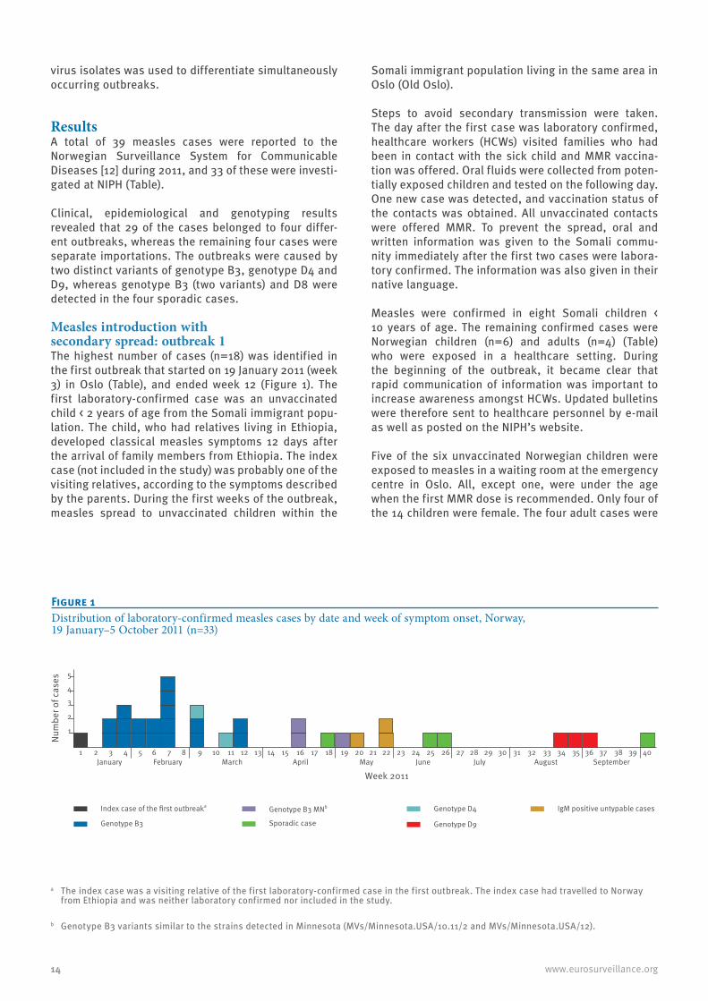
Figure 1Distribution of laboratory-confirmed measles cases by date and week of symptom onset, Norway, 19 January–5 October 2011 (n=33)
a The index case was a visiting relative of the first laboratory-confirmed case in the first outbreak. The index case had travelled to Norway from Ethiopia and was neither laboratory confirmed nor included in the study.
b Genotype B3 variants similar to the strains detected in Minnesota (MVs/Minnesota.USA/10.11/2 and MVs/Minnesota.USA/12).
Index case of the first outbreaka
Genotype B3
Genotype D4Genotype B3 MNb
Sporadic case
IgM positive untypable cases
1 2 3 4 5 6 7 8 9 10 11 12 13 14 15 16 17 18 19 20 21 22 23 24 25 26 27 28 29 30 31 32 33 34 35 36 37 38 39 40January February March April May June July August September
Week 2011
Num
ber o
f cas
es
Genotype D9
1
2
3
4
5
15www.eurosurveillance.org
HCWs employed at a healthcare centre (n=1) and chil-dren’s ward at a hospital (n=3). According to SYSVAK, two of the HCWs had been vaccinated twice with the MMR vaccine in line with the national children vaccina-tion programme [10], whereas one was not previously vaccinated against measles. No information was avail-able regarding the last HCW. All HCWs at the children’s ward of the hospital were offered a booster dose of MMR.
Epidemiological investigation demonstrated a link between all 18 cases involved in this outbreak (Table). Acute phase serum and/or oral fluid were obtained from 17 of the 18 cases, and specific anti-measles IgM antibodies were detected in all 18 cases with at least one of the ELISA assays used in the laboratory (Table).
Primary (PVF) and secondary vaccine failure (SVF) was seen in the two fully immunised HCWs, case 20 and 19 respectively. A weak IgM reaction was obtained in the acute phase oral fluid obtained from case 19, whereas the corresponding serum sample was IgM negative. High avidity IgG-antibodies (97%) were detected in both acute phase- and convalescence serum samples in case 19 consistent with SVF. In case 20, measles infection was confirmed by detection of measles virus by PCR, IgM seroconversion and four-fold IgG increase between acute phase- and convalescence samples. An increase in avidity ratio was seen between acute phase serum (69%) and convalescence serum (94%) in case 20, consistent with PVF.
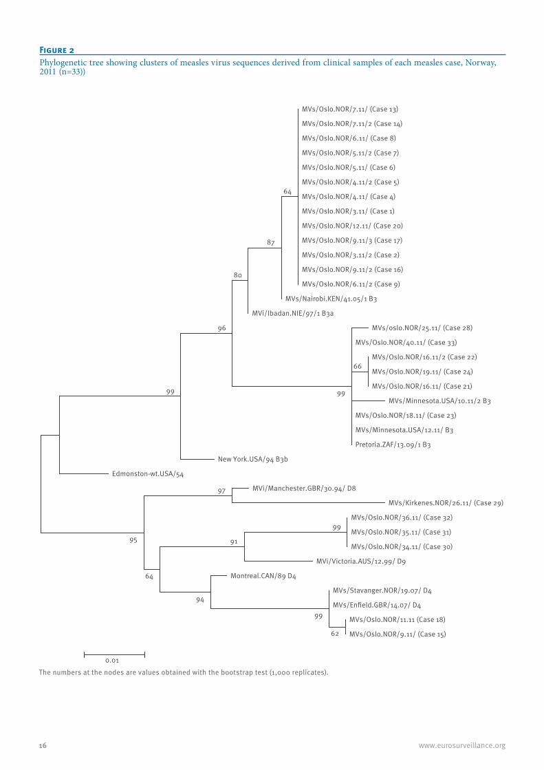
Measles infection was confirmed by PCR in 13 cases, including case 20. All the PCR positive samples were oral fluids collected between day one and day eight after the onset of symptoms, however, three of the acute phase oral fluids turned out to be PCR negative. All PCR positive samples were sequenced, and identi-cal sequences revealing genotype B3 were obtained (Figure 2). The similarity between the B3 strain detected in this outbreak and the WHO reference strain MVi/Ibadan NIE/97/1 (MeaNS) was 98.7%, but differed only by one nt from another B3 variant (MVs/Nairobi.KEN/41.05/1) detected in an outbreak in Kenya in 2005 (personal communication, Sheilagh Smit, 12 May 2011).
All cases had typical symptoms of measles including a generalised maculopapular erythematous rash, fever, cough, runny nose and red eyes. The two vaccinated HCWs although showing milder symptoms, clearly had measles infection. They had both been exposed to measles virus for a prolonged period during their duty. Only one of the children who were admitted to hospital developed severe measles pneumonia, the others were admitted due to dehydration and impaired general con-dition. In Norway, the threshold to hospitalise measles cases is low for isolation purposes.
Measles introduction with limited spread: outbreaks 2, 3 and 4During outbreak 1, genotyping of samples from two persons affected by measles yielded an identical geno-type D4 (Table), revealing a second outbreak occurring in parallel. The index case (case 15) in this outbreak 2 was a tourist from Spain who arrived in Oslo 16 February and fell ill 28 February (week 9) and the sec-ond case (case 18) occurred in Oslo two weeks later in an unvaccinated child < 2 years of age (Table). The D4 variant detected in these two cases differed only by one nt from the D4 strain causing outbreaks among Irish travellers in the UK and Norway in 2007 (Figure 2) [23,24]. Epidemiological investigation did not identify a link between these two cases.
Outbreak 3 started on 19 April 2011 in Oslo (week 16). The index case (case 21) was an unvaccinated Somali child < 2 years of age who got measles after a visit to Kenya. Measles was confirmed also between week 16 and 19 in three additional children; one child from east-ern Europe (case 22), one unvaccinated Somali child < 2 years of age who contracted measles in Kenya (case 23) and one Norwegian child living in Oslo (case 24). Measles IgM antibodies were detected in acute phase serum and/or oral fluid in all four cases. Epidemiological investigation did not identify a link between these four cases, whereas genotyping revealed identical B3 strains (MVs/Oslo.NOR/16.11, MVs/Oslo.NOR/16.11/2 and MVs/Oslo.NOR/19.11) in three of the cases (case 21, 22 and 24) representing outbreak 3.
As shown in Figure 2, different variants of genotype B3 caused outbreak 1 and 3. The similarity between the B3 variant detected in outbreak 3 and the WHO reference strain MVi/Ibadan NIE/97/1 was 97.6%, but differed by only one to three nt from the B3 variant detected in the Somali community in Minneapolis (Minnesota) in late March 2011 (MVs/Minnesota.USA/10.11/2).
The B3 variant (MVs/Oslo.NOR/18.11) detected in case 23 was one nt different from the B3 variant causing out-break 3 and was therefore not included in the outbreak (Figure 2). In addition, three unvaccinated siblings developed measles in week 20 and 22. Measles was confirmed serologically by detection of IgM antibod-ies in serum and/or oral fluid. Although epidemiologi-cal investigation did not demonstrate a link between the siblings and the cases previously described in outbreak 3, they were most probably part of outbreak 3. One of them was from Oslo, whilst the two others resided just outside Oslo (Table).
Outbreak 4 started on 22 August (week 34) and the index case was an unvaccinated Norwegian child between two and 10 years-old. The child developed classical symptoms of measles shortly after a visit to south-east Asia. Measles was later confirmed by detec-tion of measles IgM antibodies in serum and/or oral fluid as well as in two additional other unvaccinated children; a child < 2 years of age living in the same area
16 www.eurosurveillance.org
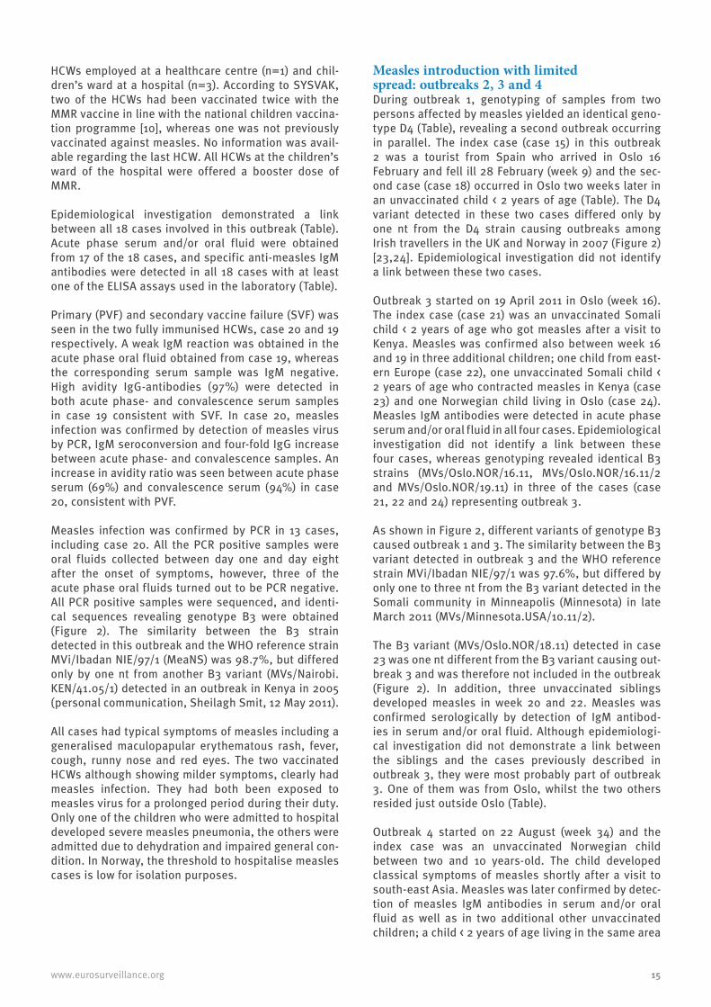
Figure 2Phylogenetic tree showing clusters of measles virus sequences derived from clinical samples of each measles case, Norway, 2011 (n=33))
MVs/Oslo.NOR/7.11/ (Case 13)
MVs/Oslo.NOR/7.11/2 (Case 14)
MVs/Oslo.NOR/6.11/ (Case 8)
MVs/Oslo.NOR/5.11/2 (Case 7)
MVs/Oslo.NOR/5.11/ (Case 6)
MVs/Oslo.NOR/4.11/2 (Case 5)
MVs/Oslo.NOR/4.11/ (Case 4)
MVs/Oslo.NOR/3.11/ (Case 1)
MVs/Oslo.NOR/12.11/ (Case 20)
MVs/Oslo.NOR/9.11/3 (Case 17)
MVs/Oslo.NOR/3.11/2 (Case 2)
MVs/Oslo.NOR/9.11/2 (Case 16)
MVs/Oslo.NOR/6.11/2 (Case 9)
MVs/Nairobi.KEN/41.05/1 B3
MVi/Ibadan.NIE/97/1 B3a
MVs/oslo.NOR/25.11/ (Case 28)
MVs/Oslo.NOR/40.11/ (Case 33)
MVs/Oslo.NOR/16.11/2 (Case 22)
MVs/Oslo.NOR/19.11/ (Case 24)
MVs/Oslo.NOR/16.11/ (Case 21)
MVs/Minnesota.USA/10.11/2 B3
MVs/Oslo.NOR/18.11/ (Case 23)
MVs/Minnesota.USA/12.11/ B3
Pretoria.ZAF/13.09/1 B3
New York.USA/94 B3b
Edmonston-wt.USA/54
MVi/Manchester.GBR/30.94/ D8
MVs/Kirkenes.NOR/26.11/ (Case 29)
MVs/Oslo.NOR/36.11/ (Case 32)
MVs/Oslo.NOR/35.11/ (Case 31)
MVs/Oslo.NOR/34.11/ (Case 30)
MVi/Victoria.AUS/12.99/ D9
Montreal.CAN/89 D4
MVs/Stavanger.NOR/19.07/ D4
MVs/Enfield.GBR/14.07/ D4
MVs/Oslo.NOR/11.11 (Case 18)
MVs/Oslo.NOR/9.11/ (Case 15)62
99
99
91
94
97
64
95
66
9999
96
80
87
64
0.01
The numbers at the nodes are values obtained with the bootstrap test (1,000 replicates).
17www.eurosurveillance.org
(week 35) and a schoolmate of the index case (week 36). Genotyping revealed an identical genotype D9 in all three cases (Figure 2), whereas epidemiological link was only identified between two of the cases.
Measles introductions without secondary spreadBased on the epidemiological and molecular investiga-tions, four of the measles cases in 2011 were assigned as sporadic importations (case 23, 28, 29 and 33) (Table). Genotyping revealed an identical B3 variant in case 23 (described in outbreak 3) and case 33 (Figure 2), also identical to a B3 variant detected in Minnesota in 2011 (MVs/Minnesota.USA/12.11). Case 33 was an unvaccinated adult who contracted measles in Angola, Africa (week 40). A slightly different B3 variant (1 nt) was detected in an unvaccinated child (case 28) who contracted measles in Kenya and fell ill before arrival to Oslo (week 25). Measles genotype D8 was detected in an unvaccinated child (case 29) living in northern Norway, however, no epidemiological data or travel history was available. Measles was initially confirmed in all of these cases by detection of IgM antibodies in acute phase sample.
DiscussionThis study describes the introduction of at least eight different measles viruses into Norway during the year 2011. We investigated a total of 33 measles cases, which could be grouped into four outbreaks and four sporadic cases. The measles virus genotypes detected during 2011 were D4, D9, D8 and four dif-ferent sequence variants of genotype B3 as shown in Figure 2. This is the largest number of measles cases reported within a single year since the large outbreak at Nesodden near Oslo in an anthroposophic com-munity in 1997. Since then, only imported cases have been identified in Norway [12]. The situation seen in Norway in 2011, may thus reflect the increased number of measles cases and multistate outbreaks of measles that have been observed in European countries [2].
The largest outbreak in Norway started January 2011 with importation from Ethiopia and 18 people were infected during the following nine weeks. Genotyping showed that the outbreak was sustained by an iden-tical B3 variant (Figure 2). Genotype B3 was the most frequently reported genotype from Africa to the WHO Global Sequence database during the period from 2007 to 2009, and was also found circulating in Malawi, Liberia and Mauritania in 2010 [8]. Genotype B3 has been associated with outbreaks in Kenya in 2005 [25], and a nearly identical B3 variant was detected in outbreak 1. Genotype B3 is currently regarded as an endemic genotype in most of the African continent, including Kenya and Ethiopia, and has been associated with importations from African countries to other parts of the world [25]. The index case in outbreak 1 was a family member living in Ethiopia, and the B3 variant detected in this outbreak differed by only one nt from
a B3 variant (MVs/Nairobi.KEN/41.05/1) causing out-breaks in Kenya in 2005.
Additionally three other genotype B3 variants were imported to Norway from Kenya during 2011, how-ever only one of the variants resulted in a small out-break 3. These different B3 variants were either identical or one to three nt different from the B3 vari-ants (MVs/Minnesota.USA/12.11 and MVs/Minnesota.USA/10.11/2) that caused outbreaks in the Somali com-munity in Minnesota in 2011 (Figure 2). Epidemiological data did not identify a link between all cases infected with the slightly different B3 variants, indicating that the strains were independent importations and show-ing how easily measles virus spreads between coun-tries among susceptible individuals. However, one must bear in mind that only minor sequence differ-ences were obtained.
Population immunity in Norway is generally suf-ficiently high to prevent sustained transmission of measles virus, and was probably the reason why fur-ther transmission of some of the imported strains did not develop. In addition, there was a lot of awareness among HCWs and the public due to the ongoing out-breaks, especially the first three, and local health authorities organised vaccination campaigns and pro-vided information to the affected community. In spite of this, this study nevertheless shows that even in a country with a generally high MMR coverage, the mea-sles virus can affect a group with low vaccine coverage and cause an outbreak affecting unvaccinated individ-uals both in and outside the group. The measles out-breaks gave an opportunity to investigate the reasons why parents choose not to vaccinate their children, and at the same time to disperse misbeliefs concerning MMR vaccination. Reasons for non-vaccination were mainly fear of side effects of the vaccine.
The results highlight the importance of genotyping in order to trace the source of infections, to distinguish sporadic cases from outbreak cases and to verify the end of an outbreak. For example, measles cases with the outbreak 1 genotype were not detected after week 12, suggesting the end of this outbreak, while two measles cases subsequently occurring in week 16 were shown to be part of a new different outbreak (outbreak 3) involving a different genotype from that of outbreak 1.
The outbreaks also demonstrated that the measles virus was easily transmitted to unvaccinated individu-als in healthcare facilities and emergency centres, which are separate centres located outside the hos-pitals. As seen in other studies [26,27], the outbreaks affected mainly unvaccinated children < 2 years of age (Table). Many of the affected children were under the age when the first MMR dose normally is given (15 month). This raised the question as to whether the age for administration of the first dose should be brought down to 12 months, however as eight of the cases were

18 www.eurosurveillance.org
under 12 months of age, these would not have been avoided by lowering the age.
Measles infection was also demonstrated in four HCWs, two fully immunised, one unvaccinated and one with no information on vaccination status. Genotyping revealed an identical B3 variant in two of the HCWs (case 17 and case 20), and demonstrated that these two also became a source of transmission. The two fully immu-nised HCWs had, according to the national vaccination programme, received two doses of MMR vaccines dur-ing childhood and were therefore classified as primary and secondary vaccine failure (PVF and SVF). Diagnosis of SVF may be challenging, however, anti-measles IgM in oral fluid and high-avidity anti-measles IgG in serum samples were documented in the case with SVF (case 19). An assay for detection of measles plaque reduction neutralisation antibodies was not available. SVF cases with mild clinical course similar to the case reported in this study have been observed in several studies [28]. Such cases are regarded as less contagious than unvaccinated measles cases, as measles virus in these circumstances can be difficult to detect and transmis-sion is limited. Waning of the measles antibody has been observed in cohorts of vaccinees 10 years after the second dose [29], and in the case of extended dura-tion of measles virus exposure, previously immune individuals can develop infection [30].
The genotype D4 detected in this study differed only by one nt from the genotype D4 detected in Irish travellers in UK and Norway in 2007 (Figure 2) [24,2], indicating that a nearly identical D4 variant is still circulating in Europe. Genotype D4 has been associated with large numbers of cases and outbreaks in Europe the last years [5,31] and was detected in 24 countries in Europe during 2011 [2]. The index case in outbreak 2 fell ill 12 days after arrival in Oslo and was probably infected in Spain or during transit, however endemic circulation of closely related D4 variants makes it difficult to trace the transmission pathways.
The genotype D9 causing outbreak 4 was imported by the index case who had visited south-east Asia. Genotyping revealed identical sequences in two addi-tional measles cases, although epidemiological link was only confirmed between the index case and a schoolmate. However, all three cases lived in the same area of Oslo. Measles has a high basic reproduction number (R0: 12–18) [32], and transmission is possi-ble by infectious measles virus-containing respiratory droplets for up to two hours after the infectious indi-vidual has departed an area [33]. Genotype D9 has been associated with endemic transmission in several countries in Asia [8].
This study demonstrates the importance of obtaining an adequate sample for virological surveillance. Oral fluid is the preferred sample type for PCR examina-tion and genotyping, and the chance of detecting mea-sles virus is highest in the first week of the disease.
Acute phase oral fluid was obtained from the majority of cases (Table), and PCR was able to detect measles virus in oral fluid taken between day one and day eight from the onset of symptoms. In addition, as most of the cases in this study were small children, oral fluid is also easier to obtain. Although some cases with milder symptoms could have been missed, we do not believe that there were many missed opportunities in identi-fying the exposed individuals as samples are taken from patients showing any symptoms of measles. Nevertheless, there might been a few missed cases, especially ones with mild symptoms.
In conclusion, this study has shown that molecular analyses of measles virus detected in Norway in 2011 provided important information in order to track the transmission path, the origin of the infection, to dis-tinguish sporadic cases from outbreak cases and con-firm vaccine failure. Especially in countries nearing the measles elimination goal, molecular analysis is essential in verification of elimination and to monitor the impact of importations, which can cause limited outbreaks. For preventing such outbreaks, organising information and vaccination campaigns in the affected communities is important. In Norway, hospitalised chil-dren with rash illness are isolated but this measure is not in place for outpatient settings. Recommendations concerning the handling of suspected measles cases in outpatient settings are warranted in order to avoid the spread of measles within healthcare centres. The four cases among the HCWs emphasised that the coverage of MMR vaccination among healthcare personnel needs to be improved as, due to their type of work, HCWs can be particularly exposed to measles and, if infected, can potentially transmit the disease to patients. More studies are also needed to investigate the cause of the SVF, and the impact with regards to the measles elimi-nation plan.
Acknowledgments This study was financially supported by the Norwegian Institute of Public Health. We thank the laboratory personnel at the virology laboratory, especially Hege Fremstad, for excellent technical assistance.
19www.eurosurveillance.org
References1. World Health Organization (WHO). Measles. Geneva: WHO.
[Accessed 26 Sep 2012]. Available from: http://www.who.int/mediacentre/factsheets/fs286/en/
2. Centers for Disease Control and Prevention (CDC). Increased transmission and outbreaks of measles -- European Region, 2011. MMWR Morb Mortal Wkly Rep. 2011;60(47):1605-10.
3. Steffens I, Martin R, Lopalco P. Spotlight on measles 2010: Measles elimination in Europe -- a new commitment to meet the goal by 2015. Euro Surveill. 2010;15(50):pii=19749. Available from: http://www.eurosurveillance.org/ViewArticle.aspx?ArticleId=19749
4. World Health Organization Regional Office for Europe (WHO Europe). Renewed commitment to elimination of measles and rubella and prevention of congenital rubella syndrome by 2015 and Sustained support for Polio-free status in the WHO European Region. WHO publication EUR/RC60/R12. Copenhagen: WHO Europe; 13–16 Sep 2010. Available from: http://www.euro.who.int/__data/assets/pdf_file/0016/122236/RC60_eRes12.pdf
5. Mankertz A, Mulders MN, Shulga S, Kremer JR, Brown KE, Santibanez S, et al. Molecular genotyping and epidemiology of measles virus transmission in the World Health Organization European Region, 2007-2009. J Infect Dis. 2011;204 Suppl 1:S335-42.
6. World Health Organization Regional Office for Europe (WHO Europe). Surveillance guidelines for measles, rubella and congenital rubella syndrome in the WHO European Region. WHO publication EUR/08/5082738. Copenhagen: WHO Europe; 2009. Available from: http://www.euro.who.int/__data/assets/pdf_file/0018/79020/E93035.pdf
7. Brown KE, Mulders MN, Freymuth F, Santibanez S, Mosquera MM, Cordey S, et al. Appearance of a novel measles G3 strain in multiple European countries within a two month period, 2010. Euro Surveill. 2011;16(17):pii=19852. Available from: http://www.eurosurveillance.org/ViewArticle.aspx?ArticleId=19852
8. Rota PA, Brown K, Mankertz A, Santibanez S, Shulga S, Muller CP, et al. Global distribution of measles genotypes and measles molecular epidemiology. J Infect Dis. 2011;204 Suppl 1:S514-23.
9. World Health Organization Regional Office for Europe (WHO Europe). Eliminating measles and rubella and preventing congenital rubella infection: WHO European region strategic Plan 2005-2010. Copenhagen: WHO Europe; 2005. Available from: http://www.euro.who.int/__data/assets/pdf_file/0008/79028/E87772.pdf
10. Norwegian Institute of Public Health (NIPH). Nasjonalt vaksinasjonsregister (SYSVAK).[National vaccine register-SYSVAK].Oslo: NIPH. [Accessed 20 Apr 2012]. Norwegian. Available from: http://www.fhi.no/eway/default.aspx?pid=233&trg=MainArea_5661&MainArea_5661=6068:0:17,4297:1:0:0:::0:0
11. Norwegian Institute of Public Health (NIPH). Smittevernboka; Meslinger (morbilli) [Infection Control Manual; Measles (morbilli)]. [Accessed 06 Dec 2012]. Available from: http://www.fhi.no/eway/default.aspx?pid=233&trg=MainLeft_6039&MainArea_5661=6039:0:15,5078:1:0:0:::0:0&MainLeft_6039=6041:82822::1:6043:80:::0:0
12. Norwegian Surveillance System for Communicable Diseases (MSIS). [Accessed 20 Apr 2012]. Available from: www.msis.no
13. Løvoll Ø, Vonen L, Vevatne T, Sagvik E, Vainio K, Sandbu S, et al. An outbreak of measles among a travelling community from England in Norway: a preliminary report. Euro Surveill. 2007;12(21):pii=3198. Available from: http://www.eurosurveillance.org/ViewArticle.aspx?ArticleId=3198
14. Schmid D, Holzmann H, Abele S, Kasper S, König S, Meusburger S, et al. An ongoing multi-state outbreak of measles linked to non-immune anthroposophic communities in Austria, Germany, and Norway, March-April 2008. Euro Surveill. 2008;13(16):pii=18838. Available from: http://www.eurosurveillance.org/ViewArticle.aspx?ArticleId=18838
15. Vainio K, Rønning K, Steen TW, Arnesen TM, Anestad G, Dudman S. Ongoing outbreak of measles in Oslo, Norway, January-February 2011. Euro Surveill. 2011;16(8):pii=19804. Available from: http://www.eurosurveillance.org/ViewArticle.aspx?ArticleId=19804
16. World Health Organization (WHO). WHO recommended surveillance standard of measles. Geneva: WHO. [Accessed 20 Apr 2012]. Available from: http://www.who.int/immunization_monitoring/diseases/measles_surveillance/en/
17. World Health Organization (WHO). New genotype of measles virus and update on global distribution of measles genotypes. Wkly Epidemiol Rec. 2005;80(40):347-51.
18. 18.Thompson JD, Gibson TJ, Plewniak F, Jeanmougin F, Higgins DG. The CLUSTAL_X windows interface: flexible strategies for multiple sequence alignment aided by quality analysis tools. Nucleic Acids Res. 1997;25(24):4876-82.
19. Tamura K, Peterson D, Peterson N, Stecher G, Nei M, Kumar S. MEGA5: molecular evolutionary genetics analysis using maximum likelihood, evolutionary distance, and maximum parsimony methods. Mol Biol Evol. 2011;28(10):2731-9.
20. Update of the nomenclature for describing the genetic characteristics of wild-type measles viruses: new genotypes and reference strains. Wkly Epidemiol Rec. 2003;78(27):229-32.
21. Measles virus nomenclature update: 2012. Wkly Epidemiol Rec. 2012;87(9):73-81.
22. Gnaneshan S, Brown KE, Green J, Brown DW. On-line global/WHO-European regional measles nucleotide surveillance. Euro Surveill. 2008;13(19):pii=18861. Available from: http://www.eurosurveillance.org/ViewArticle.aspx?ArticleId=18861
23. Cohuet S, Morgan O, Bukasa A, Heathcock R, White J, Brown K, et al. Outbreak of measles among Irish Travellers in England, March to May 2007. Euro Surveill. 2007;12(24):pii=3216. Available from: http://www.eurosurveillance.org/ViewArticle.aspx?ArticleId=3216
24. Løvoll Ø, Vonen L, Nordbø SA, Vevatne T, Sagvik E, Vainio K, et al. Outbreak of measles among Irish Travellers in Norway: an update. Euro Surveill. 2007;12(24):pii=3217. Available from: http://www.eurosurveillance.org/ViewArticle.aspx?ArticleId=3217
25. Rota J, Lowe L, Rota P, Bellini W, Redd S, Dayan G, et al. Identical genotype B3 sequences from measles patients in 4 countries, 2005. Emerg Infect Dis. 2006;12(11):1779-81.
26. Stanescu A, Janta D, Lupulescu E, Necula G, Lazar M, Molnar G, et al. Ongoing measles outbreak in Romania, 2011. Euro Surveill. 2011;16(31):pii=19932. Available from: http://www.eurosurveillance.org/ViewArticle.aspx?ArticleId=19932
27. Filia A, Tavilla A, Bella A, Magurano F, Ansaldi F, Chironna M, et al. Measles in Italy, July 2009 to September 2010. Euro Surveill. 2011;16(29):pii=19925. Available from: http://www.eurosurveillance.org/ViewArticle.aspx?ArticleId=19925
28. Hickman CJ, Hyde TB, Sowers SB, Mercader S, McGrew M, Williams NJ, et al. Laboratory characterization of measles virus infection in previously vaccinated and unvaccinated individuals. J Infect Dis. 2011;204 Suppl 1:S549-58.
29. LeBaron CW, Beeler J, Sullivan BJ, Forghani B, Bi D, Beck C, et al.Persistence of measles antibodies after 2 doses of measles vaccine in a postelimination environment. Arch Pediatr Adolesc Med. 2007;161(3):294-301.
30. Krugman S, Giles JP, Friedman H, Stone S. Studies on immunity to measles. J Pediatr. 1965;66:471-88.
31. Mankertz A, Mihneva Z, Gold H, Baumgarte S, Baillot A, Helble R, et al. Spread of measles virus D4-Hamburg, Europe, 2008-2011. Emerg Infect Dis. 2011;17(8):1396-401.
32. Wadl M, Siedler A, Krämer W, Haindl ME, Gebrande S, Krenn-Lanzl I, et al. Measles transmission from an anthroposophic community to the general population, Germany 2008. BMC Public Health. 2011;11:474.
33. Biellik RJ, Clements CJ. [Strategies for minimizing nosocomial transmission of measles]. Rev Panam Salud Publica. 1998;4(5):350-7. Spanish.
20 www.eurosurveillance.org
Letters
Probable imported rather than autochthonous Plasmodium vivax cases in Italy
E Nicastri ([email protected])1
1. National Institute for Infectious Diseases (Istituto Nazionale per le Malattie Infettive, INMI) “L. Spallanzani”, Rome, Italy
Citation style for this article: Nicastri E. Probable imported rather than autochthonous Plasmodium vivax cases in Italy. Euro Surveill. 2012;17(50):pii=20338. Available online: http://www.eurosurveillance.org/ViewArticle.aspx?ArticleId=20338
Article submiited on 7 December 2012 / published on 13 December 2012
To the editor: Italian colleagues describe two recent cases of probable autochthonous-introduced Plasmodium vivax malaria, suggesting that sporadic cases may be considered possible in areas with vec-tor abundance and favourable environmental condi-tions [1]. Nevertheless, we have some concerns about this report: firstly, no other concurrent P. vivax malaria cases have been reported in the Pontine marshes or in the Calabria Region; secondly, no secondary cases have been reported to arise from the index cases; thirdly, the first patient visited Santo Domingo in the Dominican Republic in 2003, an area where malaria cases still occur; fourthly, as previously affirmed in a publication by the same group about an unusual case of late relapse of P. vivax four years after the acute infection [2], at least the first case could be explained as a late relapse following an asymptomatic or pau-cisymptomatic primary infection. In conclusion, the described cases could in our opinion be viewed as probable imported, and not autochthonous, P. vivax cases.
References1. Romi R, Boccolini D, Menegon M, Rezza G. Probable
autochthonous introduced malaria cases in Italy in 2009–2011 and the risk of local vector-borne transmission. Euro Surveill. 2012;17(48):pii=20325. Available from: http://www.eurosurveillance.org/ViewArticle.aspx?ArticleId=20325
2. Durante Mangoni E, Severini C, Menegon M, Romi R, Ruggiero G, Majori G. Case report: An unusual late relapse of Plasmodium vivax malaria. Am J Trop Med Hyg. 2003;68(2):159-60.

21www.eurosurveillance.org
Letters
Reply: Probable imported rather than autochthonous Plasmodium vivax cases in Italy
G Rezza1, D Boccolini1, M Menegon1, R Romi ([email protected])1
1. Istituto Superiore di Sanità (ISS), Department of Infectious, Parasitic and Immune-Mediated Diseases (MIPI), Rome, Italy
Citation style for this article: Rezza G, Boccolini D, Menegon M, Romi R. Reply: Probable imported rather than autochthonous Plasmodium vivax cases in Italy . Euro Surveill. 2012;17(50):pii=20339. Available online: http://www.eurosurveillance.org/ViewArticle.aspx?ArticleId=20339
Article submiited on 11 December 2012 / published on 13 December 2012
To the editor: Dr Nicastri argues that cryptic sporadic cases may not be considered as autochthonous unless other cases have been identified in a specific geo-graphical area. This is a rigid and not scientifically cor-roborated assumption that, in our opinion, cannot be generalised. In fact, there is no reason to assume that secondary cases should necessarily occur [1,2], since the generation of a chain of transmission depends strictly on the basic reproductive number R0, which is influenced by a series of factors [3] ranging from prevalence of infection among mosquitoes (likely to be very low when autochthonous introduced cases occur, as demonstrated by the lack of detection of plasmo-dia among mosquitoes in recent outbreaks in Greece) and probability of exposure of humans to mosquito bites. Dr Nicastri’s latter two points pertain to only one of the two cases and have been critically discussed in our article. However, it should be mentioned that the patient who visited Santo Domingo did not report febrile episodes after their return and that the likeli-hood of very late relapse is very low; thus, we think this patient should be considered as a cryptic case. In conclusion, we do not feel Dr Nicastri’s criticism is suf-ficiently supported by the arguments raised.
References1. Baldari M, Tamburro A, Sabatinelli G, Romi R, Severini C,
Cuccagna P, Fiorilli G, Allegri MP, Buriani C, Toti M. Malaria in Maremma, Italy. Lancet. 1998;351(9111):1246-7.
2. Gould EA, Gallian P, De Lamballerie X, Charrel RN. First cases of autochthonous dengue fever and chikungunya fever in France: from bad dream to reality! Clin Microbiol Infect. 2010;16(12):1702-4.
3. Romi R, Boccolini D, Vallorani R, Severini F, Toma L, Cocchi M, Tamburro A, Messeri G, Crisci A, Angeli A, Costantini R, Raffaelli I, Pontuale G, Thiéry I, Landier A, Le Goff G, Fausto AM, Di Luca M. Assessment of the risk of malaria re-introduction in the Maremma Plain (Central Italy) using a multi factorial approach. Malaria Journal. 2012;11:98.
Related Documents