AGING NEUROSCIENCE ORIGINAL RESEARCH ARTICLE published: 08 October 2014 doi: 10.3389/fnagi.2014.00272 Improved cerebral oxygenation response and executive performance as a function of cardiorespiratory fitness in older women: a fNIRS study Cédric T. Albinet 1 *, Kevin Mandrick 2 , Pierre Louis Bernard 2 , Stéphane Perrey 2† and Hubert Blain 2,3† 1 CeRCA (CNRS–UMR 7295), Faculty of Sport Sciences, University of Poitiers, Poitiers, France 2 Movement to Health (M2H), Montpellier-1 University, Euromov, Montpellier, France 3 University hospital of Montpellier - MacVia-LR, France Edited by: Philip P. Foster, The University of Texas Health Science Center at Houston, USA Reviewed by: Evgeniya Kirilina, Free University of Berlin, Germany Anouk Vermeij, Radboud University Nijmegen, Netherlands *Correspondence: Cédric T. Albinet, CeRCA (CNRS–UMR 7295), Faculty of Sport Sciences, University of Poitiers, 5 rue Théodore Lefebvre, Bâtiment A5, TSA 21103, 86073 Poitiers cedex 9, France e-mail: cedric.albinet@ univ-poitiers.fr † These authors have contributed equally to this work. Cardiorespiratory fitness has been shown to protect and enhance cognitive and brain functions, but little is known about the cortical mechanisms that underlie these changes in older adults. In this study, functional near infrared spectroscopy (fNIRS) was used to investigate variations in oxyhemoglobin [HbO 2 ] and in deoxyhemoglobin [HHb] in the dorsolateral prefrontal cortex (DLPFC) during the performance of an executive control task in older women with different levels of cardiorespiratory fitness (VO 2 max). Thirty-four women aged 60–77 years were classified as high-fit and low-fit based on VO 2 max measures. They all performed a control counting (CNT) task and the Random Number Generation (RNG) task at two different paces (1 number/1 s and 1 number/1.5 s), allowing to manipulate task difficulty, while hemodynamic responses in the bilateral DLPFCs were recorded using continuous-wave NIRS. The behavioral data revealed that the high-fit women showed significantly better performance on the RNG tasks compared with the low-fit women. The high-fit women showed significant increases in [HbO 2 ] responses in both left and right DLPFCs during the RNG task, while the low-fit women showed significantly less activation in the right DLPFC compared with the right DLPFC of the high-fit women and compared with their own left DLPFC. At the level of the whole sample, increases in the [HbO 2 ] responses in the right DLPFC were found to mediate in part the relationship between VO 2 max level and executive performance during the RNG task at 1.5 s but not at 1 s. These results provide support for the cardiorespiratory fitness hypothesis and suggest that higher levels of aerobic fitness in older women are related to increased cerebral oxygen supply to the DLPFC, sustaining better cognitive performance. Keywords: prefrontal cortex, functional near-infrared spectroscopy (fNIRS), executive functions, aerobic fitness, aging INTRODUCTION Executive functions refer to a set of higher-order cognitive processes whose principal function is to facilitate behavioral adap- tation to new or complex situations, specifically when routines are inappropriate. They encompass the formulation of a goal and the implementation of a strategy, planning, action sequencing and monitoring, mental flexibility, inhibition, and updating of working memory (see Anderson et al., 2008). Executive functions are critical for the activities of daily living and autonomy, but are among the most altered cognitive functions due to normal aging (West, 1996; Albinet et al., 2012). Neuroimaging and neu- ropsychological studies have shown that important substrates for executive functions are the frontal and parietal lobes, which are the cerebral structures that are the most vulnerable to the effects of aging (see West, 1996; Cabeza et al., 2005; Raz and Rodrigue, 2006). Cross-sectional and longitudinal studies have reported important linear age-related declines in the prefrontal volume (Raz et al., 2004, 2005) and decreased regional cerebral blood flow (rCBF) in the prefrontal cortex (PFC; Kwee and Nakada, 2003; Kalpouzos et al., 2009). These declines, however, may be modulated by lifestyle and health-related factors, such as regular physical exercise. In the past decade, a growing body of evidence has documented the beneficial effects of physical exercise and/or car- diorespiratory fitness level on the cognitive and brain functions in older adults (for reviews see Etnier et al., 2006; Hillman et al., 2008; Voelcker-Rehage and Niemann, 2013). Aerobic exercise and cardiorespiratory fitness level, indexed by maximal oxygen uptake (VO 2 max), have been shown to be positively related to cognitive performance in older adults, particularly when executive control processes that involve the PFC are critical for task success (Kramer et al., 1999; Colcombe and Kramer, 2003; Prakash et al., 2011). The process of inhibition, a core executive function defined as the capacity to suppress irrelevant information or prepotent Frontiers in Aging Neuroscience www.frontiersin.org October 2014 | Volume 6 | Article 272 | 1

Welcome message from author
This document is posted to help you gain knowledge. Please leave a comment to let me know what you think about it! Share it to your friends and learn new things together.
Transcript

AGING NEUROSCIENCEORIGINAL RESEARCH ARTICLE
published: 08 October 2014doi: 10.3389/fnagi.2014.00272
Improved cerebral oxygenation response and executiveperformance as a function of cardiorespiratory fitness inolder women: a fNIRS studyCédric T. Albinet1*, Kevin Mandrick2, Pierre Louis Bernard2, Stéphane Perrey2† and Hubert Blain2,3†
1 CeRCA (CNRS–UMR 7295), Faculty of Sport Sciences, University of Poitiers, Poitiers, France2 Movement to Health (M2H), Montpellier-1 University, Euromov, Montpellier, France3 University hospital of Montpellier - MacVia-LR, France
Edited by:Philip P. Foster, The University ofTexas Health Science Center atHouston, USA
Reviewed by:Evgeniya Kirilina, Free University ofBerlin, GermanyAnouk Vermeij, Radboud UniversityNijmegen, Netherlands
*Correspondence:Cédric T. Albinet, CeRCA(CNRS–UMR 7295), Faculty ofSport Sciences, University ofPoitiers, 5 rue Théodore Lefebvre,Bâtiment A5, TSA 21103, 86073Poitiers cedex 9, Francee-mail: [email protected]†These authors have contributedequally to this work.
Cardiorespiratory fitness has been shown to protect and enhance cognitive and brainfunctions, but little is known about the cortical mechanisms that underlie these changesin older adults. In this study, functional near infrared spectroscopy (fNIRS) was usedto investigate variations in oxyhemoglobin [HbO2] and in deoxyhemoglobin [HHb] in thedorsolateral prefrontal cortex (DLPFC) during the performance of an executive control taskin older women with different levels of cardiorespiratory fitness (VO2max). Thirty-fourwomen aged 60–77 years were classified as high-fit and low-fit based on VO2maxmeasures. They all performed a control counting (CNT) task and the Random NumberGeneration (RNG) task at two different paces (1 number/1 s and 1 number/1.5 s), allowingto manipulate task difficulty, while hemodynamic responses in the bilateral DLPFCs wererecorded using continuous-wave NIRS. The behavioral data revealed that the high-fitwomen showed significantly better performance on the RNG tasks compared with thelow-fit women. The high-fit women showed significant increases in [HbO2] responsesin both left and right DLPFCs during the RNG task, while the low-fit women showedsignificantly less activation in the right DLPFC compared with the right DLPFC of thehigh-fit women and compared with their own left DLPFC. At the level of the whole sample,increases in the [HbO2] responses in the right DLPFC were found to mediate in partthe relationship between VO2max level and executive performance during the RNG taskat 1.5 s but not at 1 s. These results provide support for the cardiorespiratory fitnesshypothesis and suggest that higher levels of aerobic fitness in older women are related toincreased cerebral oxygen supply to the DLPFC, sustaining better cognitive performance.
Keywords: prefrontal cortex, functional near-infrared spectroscopy (fNIRS), executive functions, aerobic fitness,aging
INTRODUCTIONExecutive functions refer to a set of higher-order cognitiveprocesses whose principal function is to facilitate behavioral adap-tation to new or complex situations, specifically when routinesare inappropriate. They encompass the formulation of a goal andthe implementation of a strategy, planning, action sequencingand monitoring, mental flexibility, inhibition, and updating ofworking memory (see Anderson et al., 2008). Executive functionsare critical for the activities of daily living and autonomy, butare among the most altered cognitive functions due to normalaging (West, 1996; Albinet et al., 2012). Neuroimaging and neu-ropsychological studies have shown that important substrates forexecutive functions are the frontal and parietal lobes, which arethe cerebral structures that are the most vulnerable to the effectsof aging (see West, 1996; Cabeza et al., 2005; Raz and Rodrigue,2006). Cross-sectional and longitudinal studies have reportedimportant linear age-related declines in the prefrontal volume
(Raz et al., 2004, 2005) and decreased regional cerebral bloodflow (rCBF) in the prefrontal cortex (PFC; Kwee and Nakada,2003; Kalpouzos et al., 2009). These declines, however, may bemodulated by lifestyle and health-related factors, such as regularphysical exercise.
In the past decade, a growing body of evidence hasdocumented the beneficial effects of physical exercise and/or car-diorespiratory fitness level on the cognitive and brain functionsin older adults (for reviews see Etnier et al., 2006; Hillman et al.,2008; Voelcker-Rehage and Niemann, 2013). Aerobic exercise andcardiorespiratory fitness level, indexed by maximal oxygen uptake(VO2max), have been shown to be positively related to cognitiveperformance in older adults, particularly when executive controlprocesses that involve the PFC are critical for task success (Krameret al., 1999; Colcombe and Kramer, 2003; Prakash et al., 2011).The process of inhibition, a core executive function definedas the capacity to suppress irrelevant information or prepotent
Frontiers in Aging Neuroscience www.frontiersin.org October 2014 | Volume 6 | Article 272 | 1

Albinet et al. Fitness, cortical oxygenation and executive performance
responses, appears particularly sensitive to the aerobic fitness level(see Smiley-Oyen et al., 2008; Colcombe et al., 2004; Boucardet al., 2012). Moreover, brain imaging studies have recently shownthat greater aerobic fitness is associated with an increased volumein the frontal and parietal lobes, as well as in the hippocampus(Colcombe et al., 2003, 2006; Erickson et al., 2011), and withgreater or more efficient neural activity in the prefrontal andparietal regions involved in executive functions (Colcombe et al.,2004; Prakash et al., 2011; see Voelcker-Rehage and Niemann,2013 for a review). Indeed, using functional Magnetic ResonanceImaging (fMRI) during the performance of an executive task thatinvolves behavioral conflict, Colcombe et al. (2004) showed thatincreased cardiorespiratory fitness level after a 6-month aerobictraining program was associated with higher brain activationsin the prefrontal and parietal cortices and significantly loweractivity in the anterior cingulate cortex. Similarly, Prakash et al.(2011) reported that cardiorespiratory fitness was associated withhigher activation of the prefrontal and parietal cortices, but notthe posterior regions, in an inhibition task with high cognitiveloads. Although the neurobiological mechanisms responsible forthese benefits are not yet fully understood, converging evidencefrom animal and human research suggests that the release ofneurotrophic factors that facilitate neurogenesis, angiogenesis,and neurovasculature may play an important role (see Cotmanet al., 2007; Voss et al., 2013).
Some authors have argued that aerobic exercise and associatedgains in cardiorespiratory fitness are important for cognitive andcerebral benefits to occur (see Dustman et al., 1984; Colcombeand Kramer, 2003; Colcombe et al., 2004). In this framework, theso-called cardiorespiratory fitness hypothesis states that increasedlevel of aerobic fitness (high VO2max level) would increase rCBF(see Ainslie et al., 2008; Brown et al., 2010), thus allowing bettercerebral oxygen supply to the brain, particularly in the prefrontalregions where the age-related deficit in rCBF is the most signifi-cant (Cabeza et al., 2005), and would translate into an improvedexecutive performance. Although, as stated above, evidence sug-gests that increased cardiorespiratory fitness may affect brainplasticity, at a behavioral level, the direct link between cardiorespi-ratory fitness level and cognitive performance in older adults hasnot been always consistently demonstrated (see Etnier et al., 2006;Smiley-Oyen et al., 2008), deserving additional investigation.
At a neurophysiological level, the cardiorespiratory fitnesshypothesis has received very little evidence. Previous studieshave indirectly investigated this question by measuring rCBF orlocal concentration changes in paramagnetic deoxyhemoglobin[HHb] measured by the BOLD response using fMRI, butnot cerebral oxygenation per se. Functional near infraredspectroscopy (fNIRS), which measures the hemodynamiccorrelates of neural activity, may help to further investigatethe relationships between prefrontal oxygenation, executiveperformance and cardiorespiratory fitness. Functional NIRSmeasures changes in oxygenated and deoxygenated hemoglobin([HbO2] and [HHb], respectively) from the cortical surface.The principles of fNIRS have been extensively described andappear suitable to assess the relationship between corticalactivation and hemodynamic response with a high samplingrate (for reviews see Obrig and Villringer, 2003; Perrey, 2008;
Ferrari and Quaresima, 2012). Recent studies have examinedthe advantages of fNIRS compared with fMRI in cognitive tasks(Cui et al., 2011) and have validated this technique for the studyof neurovascular coupling or cognitive performance in normalaging (Vermeij et al., 2012; Fabiani et al., 2014). For example,Fabiani et al. (2014) used different brain imaging techniquesduring passive visual stimulation and revealed significantcorrelations between the [HbO2] response and the VO2max level,but not between the [HHb] or BOLD responses and VO2max.Moreover, these authors concluded that evaluating the functionalimpact of reduced neurovascular coupling in older low-fit adultsrequires the manipulation of the cognitive load, for example, byincreasing the task difficulty in a behavioral paradigm. To thebest of our knowledge, no study to date has directly tested thecardiorespiratory fitness hypothesis by measuring the changes inthe blood volume and oxygenation in the brain with fNIRS duringcognitive tasks in older humans. As a contribution towards abetter understanding of the effects of cortical oxygenation changesin the relationship between cardiorespiratory fitness and executiveperformance, the present study investigated the behavioral andbrain responses during cognitive tasks that challenge executivefunctions with different levels of difficulty in older adults.
In the present study, we evaluated the differences in executivecontrol performance between aerobically fit older women (highVO2max) and aerobically unfit older women (low VO2max) usingthe Random Number Generation (RNG) task. This experimentaltask consists of producing random sequences of numbers atspecified rates and involves executive functioning, particularly theinhibition of overlearned schemas (i.e., counting, Miyake et al.,2000; Albinet et al., 2012). Previous studies have shown thatnormal aging significantly impairs the behavioral performance onthe RNG task (Albinet et al., 2006, 2012), while aerobic exerciseor cardiorespiratory fitness improves the behavioral performancein older adults (Abou-Dest et al., 2012; Boucard et al., 2012).Different neuroimaging studies have shown that good perfor-mance on this task requires a distributed neural network in theprefrontal and parietal cortices, with a strong involvement in par-ticular areas. The dorsolateral prefrontal cortex (DLPFC) showsa significant and systematic left-sided activation (Jahanshahi andDirnberger, 1998; Jahanshahi et al., 1998, 2000; Joppich et al.,2004) and sometimes bilateral activation (Daniels et al., 2003;Hoshi et al., 2003; Koike et al., 2011) in normal adults. Thesefindings oriented us to particularly examine this cortical area andto measure the changes in [HbO2] and [HHb] as a function of thetask demands in the bilateral DLPFC (BAs 9/46). In the presentstudy, we hypothesized that the older aerobically fit women woulddemonstrate better executive performance on the RNG task com-pared with their unfit counterparts. According to the cardiores-piratory fitness hypothesis, we expected that the older fit womenwould exhibit greater bilateral prefrontal activity as a function ofthe task being made more difficult to sustain their better executiveperformance compared with the unfit older women.
MATERIALS AND METHODSPARTICIPANTSForty community-dwelling women aged 60–77 years wererecruited from public meetings aimed at promoting physical
Frontiers in Aging Neuroscience www.frontiersin.org October 2014 | Volume 6 | Article 272 | 2
Albinet et al. Fitness, cortical oxygenation and executive performance
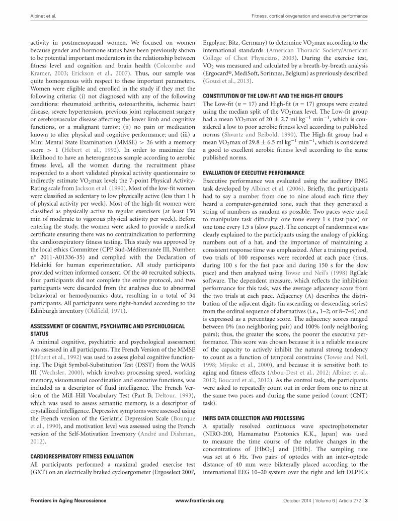
activity in postmenopausal women. We focused on womenbecause gender and hormone status have been previously shownto be potential important moderators in the relationship betweenfitness level and cognition and brain health (Colcombe andKramer, 2003; Erickson et al., 2007). Thus, our sample wasquite homogenous with respect to these important parameters.Women were eligible and enrolled in the study if they met thefollowing criteria: (i) not diagnosed with any of the followingconditions: rheumatoid arthritis, osteoarthritis, ischemic heartdisease, severe hypertension, previous joint replacement surgeryor cerebrovascular disease affecting the lower limb and cognitivefunctions, or a malignant tumor; (ii) no pain or medicationknown to alter physical and cognitive performance; and (iii) aMini Mental State Examination (MMSE) > 26 with a memoryscore > 1 (Hébert et al., 1992). In order to maximize thelikelihood to have an heterogeneous sample according to aerobicfitness level, all the women during the recruitment phaseresponded to a short validated physical activity questionnaire toindirectly estimate VO2max level; the 7-point Physical Activity-Rating scale from Jackson et al. (1990). Most of the low-fit womenwere classified as sedentary to low physically active (less than 1 hof physical activity per week). Most of the high-fit women wereclassified as physically active to regular exercisers (at least 150min of moderate to vigorous physical activity per week). Beforeentering the study, the women were asked to provide a medicalcertificate ensuring there was no contraindication to performingthe cardiorespiratory fitness testing. This study was approved bythe local ethics Committee (CPP Sud-Méditerranée III, Number:n◦ 2011-A01336-35) and complied with the Declaration ofHelsinki for human experimentation. All study participantsprovided written informed consent. Of the 40 recruited subjects,four participants did not complete the entire protocol, and twoparticipants were discarded from the analyses due to abnormalbehavioral or hemodynamics data, resulting in a total of 34participants. All participants were right-handed according to theEdinburgh inventory (Oldfield, 1971).
ASSESSMENT OF COGNITIVE, PSYCHIATRIC AND PSYCHOLOGICALSTATUSA minimal cognitive, psychiatric and psychological assessmentwas assessed in all participants. The French Version of the MMSE(Hébert et al., 1992) was used to assess global cognitive function-ing. The Digit Symbol-Substitution Test (DSST) from the WAISIII (Wechsler, 2000), which involves processing speed, workingmemory, visuomanual coordination and executive functions, wasincluded as a descriptor of fluid intelligence. The French Ver-sion of the Mill–Hill Vocabulary Test (Part B; Deltour, 1993),which was used to assess semantic memory, is a descriptor ofcrystallized intelligence. Depressive symptoms were assessed usingthe French version of the Geriatric Depression Scale (Bourqueet al., 1990), and motivation level was assessed using the Frenchversion of the Self-Motivation Inventory (André and Dishman,2012).
CARDIORESPIRATORY FITNESS EVALUATIONAll participants performed a maximal graded exercise test(GXT) on an electrically braked cycloergometer (Ergoselect 200P,
Ergolyne, Bitz, Germany) to determine VO2max according to theinternational standards (American Thoracic Society/AmericanCollege of Chest Physicians, 2003). During the exercise test,VO2 was measured and calculated by a breath-by-breath analysis(Ergocard®, MediSoft, Sorinnes, Belgium) as previously described(Gouzi et al., 2013).
CONSTITUTION OF THE LOW-FIT AND THE HIGH-FIT GROUPSThe Low-fit (n = 17) and High-fit (n = 17) groups were createdusing the median split of the VO2max level. The Low-fit grouphad a mean VO2max of 20 ± 2.7 ml kg−1 min−1, which is con-sidered a low to poor aerobic fitness level according to publishednorms (Shvartz and Reibold, 1990). The High-fit group had amean VO2max of 29.8 ± 6.5 ml kg−1 min−1, which is considereda good to excellent aerobic fitness level according to the samepublished norms.
EVALUATION OF EXECUTIVE PERFORMANCEExecutive performance was evaluated using the auditory RNGtask developed by Albinet et al. (2006). Briefly, the participantshad to say a number from one to nine aloud each time theyheard a computer-generated tone, such that they generated astring of numbers as random as possible. Two paces were usedto manipulate task difficulty: one tone every 1 s (fast pace) orone tone every 1.5 s (slow pace). The concept of randomness wasclearly explained to the participants using the analogy of pickingnumbers out of a hat, and the importance of maintaining aconsistent response time was emphasized. After a training period,two trials of 100 responses were recorded at each pace (thus,during 100 s for the fast pace and during 150 s for the slowpace) and then analyzed using Towse and Neil’s (1998) RgCalcsoftware. The dependent measure, which reflects the inhibitionperformance for this task, was the average adjacency score fromthe two trials at each pace. Adjacency (A) describes the distri-bution of the adjacent digits (in ascending or descending series)from the ordinal sequence of alternatives (i.e., 1–2; or 8–7–6) andis expressed as a percentage score. The adjacency scores rangedbetween 0% (no neighboring pair) and 100% (only neighboringpairs); thus, the greater the score, the poorer the executive per-formance. This score was chosen because it is a reliable measureof the capacity to actively inhibit the natural strong tendencyto count as a function of temporal constrains (Towse and Neil,1998; Miyake et al., 2000), and because it is sensitive both toaging and fitness effects (Abou-Dest et al., 2012; Albinet et al.,2012; Boucard et al., 2012). As the control task, the participantswere asked to repeatedly count out in order from one to nine atthe same two paces and during the same period (count (CNT)task).
fNIRS DATA COLLECTION AND PROCESSINGA spatially resolved continuous wave spectrophotometer(NIRO-200, Hamamatsu Photonics K.K., Japan) was usedto measure the time course of the relative changes in theconcentrations of [HbO2] and [HHb]. The sampling ratewas set at 6 Hz. Two pairs of optodes with an inter-optodedistance of 40 mm were bilaterally placed according to theinternational EEG 10–20 system over the right and left DLPFCs
Frontiers in Aging Neuroscience www.frontiersin.org October 2014 | Volume 6 | Article 272 | 3

Albinet et al. Fitness, cortical oxygenation and executive performance
(BAs 9/46). To reduce artifacts (motion and physiological), theparticipants were asked to minimize head and body movements,were given instructions to breathe gently and regularly, andwere instructed to rest quietly. Pre-processing and processingof NIRS signals were performed off-line using a customizedcode implemented in Matlab 7.0 software (The MathworksInc., MA, USA). The typical hemodynamic response shows anincrease for [HbO2] with neural activity while [HHb] signaltypically behaves opposite. This hemodynamic response is oftenused in the feature extraction. The raw NIRS data were filteredusing a first-order Butterworth low-pass filter zero-lag (cut-offfrequency of 0.7 Hz) to remove the heart rate signal (Huppertet al., 2009). No detrending method was applied on the rawNIRS signals based on no important low frequency signal driftfor each individual time series. From the resulting individualsignals, changes (∆) in [HbO2] and [HHb] were calculated foreach condition with two processing methods. First, we usedthe slope method (see more details in Mandrick et al., 2013a)in 100-s stimulation windows as a simple, but effective featureto detect both the global hemodynamic response amplitudeand the time to reach plateau level during sustained activation.The relationship between neural activity and the hemodynamicresponses is often approximated to that of a linear system (Fristonet al., 1994), meaning that integrated neural firing rate wasassumed to be a rectangular function corresponding to theconstant stimulus over time. For each experimental condition,the two trials were averaged; the dependent measures includedthe slope coefficients for ∆[HbO2] and ∆[HHb] in µM.cm/s.As suggested by Mandrick et al. (2013a) the slope coefficient ofa straight line fitted to the data in a windows as feature, indicatesthe magnitude and the direction of the oxygenation responsesover the stimulation period for each condition. Even if the issueof modeling and determining the shape of the hemodynamicresponses is beyond the scope of this study, our results showthat evoked fNIRS-derived cerebral hemodynamic response tosustained cognitive tasks was translated by the slope coefficientwith enough sensitivity (see Mandrick et al., 2013a; presentstudy). Second, the oxygenation response amplitude was alsoanalyzed as it is regularly done in the field by subtracting thelevel obtained at the resting state (last 10 s of the rest period)from the activation period (last 10 s of the task) after reachinga plateau for each trial (Holper et al., 2009). In case of a longstimulus period (≥1 min), the [HbO2] signal reaches a plateauthat is maintained until the cessation of the stimulus (Heekerenet al., 1997), as we observed in the present study (see Figure 3).Overall, the results were concordant between the two methods(correlated at r = 0.9), but the slope method appeared to be moresensitive to discriminating the fNIRS signal changes as a functionof the cognitive load and the VO2max level. Accordingly, weonly report in the present study the data concerning the slopemethod.
PROCEDUREAfter careful screening for the inclusion and exclusion criteria, theparticipants were first evaluated for global cognitive, psychiatricand psychological status. On the second day, their cardiorespira-tory fitness was measured with the GXT, which was performed at
the Department of Clinical Physiology of the University Hospital.On the third assessment day, each participant was tested indi-vidually in a quiet partially obscured experimental room forthe RNG performance with fNIRS recording. This session lastedapproximately 50 min. After the NIRS apparatus was installedover the two DLPFCs and its functions were broadly explained,the participant was comfortably seated in an armchair. The par-ticipant performed orally the RNG task, while an experimentervisually controlled the online evolution of the hemodynamicparameters on a computer screen. After a training period toensure familiarization with the material and the tasks, each partic-ipant performed two RNG trials at each pace alternating with restperiods and count trials in an ABBA vs. BAAB counterbalancedorder across the participants (see bottom of Figure 1). The orderof the paces was also counterbalanced across the participants;half of the participants started with the slow pace condition, andthe other half started with the fast pace condition (see upperof Figure 1). Rest periods, which lasted the same time as theactivation periods, were visually controlled for each participant toensure that the hemodynamics variables returned to their baselinelevel.
STATISTICAL ANALYSESThe statistical analyses were performed using STATISTICA soft-ware version 7.1 (StatSoft, France). The assumption of datanormality and homogeneity was assessed using Kolmogorov-Smirnov and Levene tests, respectively. Behavioral performanceon the RNG task (Adjacency score) was analyzed with a 2 (group:high-fit vs. low-fit) × 2 (pace: fast vs. slow) ANOVA with groupas a between-subject factor and pace as a within-subject factorwith repeated measures. For each of the hemodynamic measures,∆[HbO2] and ∆[HHb], separate 2 (group: high-fit vs. low-fit)× 2 (task: RNG task vs. CNT task) × 2 (hemisphere: right vs.left) × 2 (pace: fast vs. slow) ANOVAs were performed, withgroup as a between-subject factor and task, hemisphere, and paceas within-subject factors with repeated measures. Mean compar-isons were performed using Tukey’s HSD corrections for mul-tiple comparisons. Finally, the relationships between executiveperformance, VO2max level and hemodynamic measures wereexamined using partial correlation analyses. All data are expressedas the mean ± SD. The level of significance was set at p < 0.05.Partial estimated effect sizes (η2
p) were reported for significantresults.
RESULTSTable 1 displays the characteristics of the 34 participants of thestudy, that were not significantly different between the High-fitand Low-fit groups.
BEHAVIORAL DATAThe ANOVA revealed a significant main effect of pace(F(1,32) = 48.3; p < 0.0001; η2
p = 0.60), with higher Adjacencyscores for the fast pace (31.5 ± 9.1%) compared with the slowpace (24.1 ± 7.8%). The ANOVA also revealed a significantmain effect of group (F(1,32) = 5.5; p < 0.05; η2
p = 0.15). Asshown in Figure 2, whatever the pace, the high-fit group showed
Frontiers in Aging Neuroscience www.frontiersin.org October 2014 | Volume 6 | Article 272 | 4
Albinet et al. Fitness, cortical oxygenation and executive performance
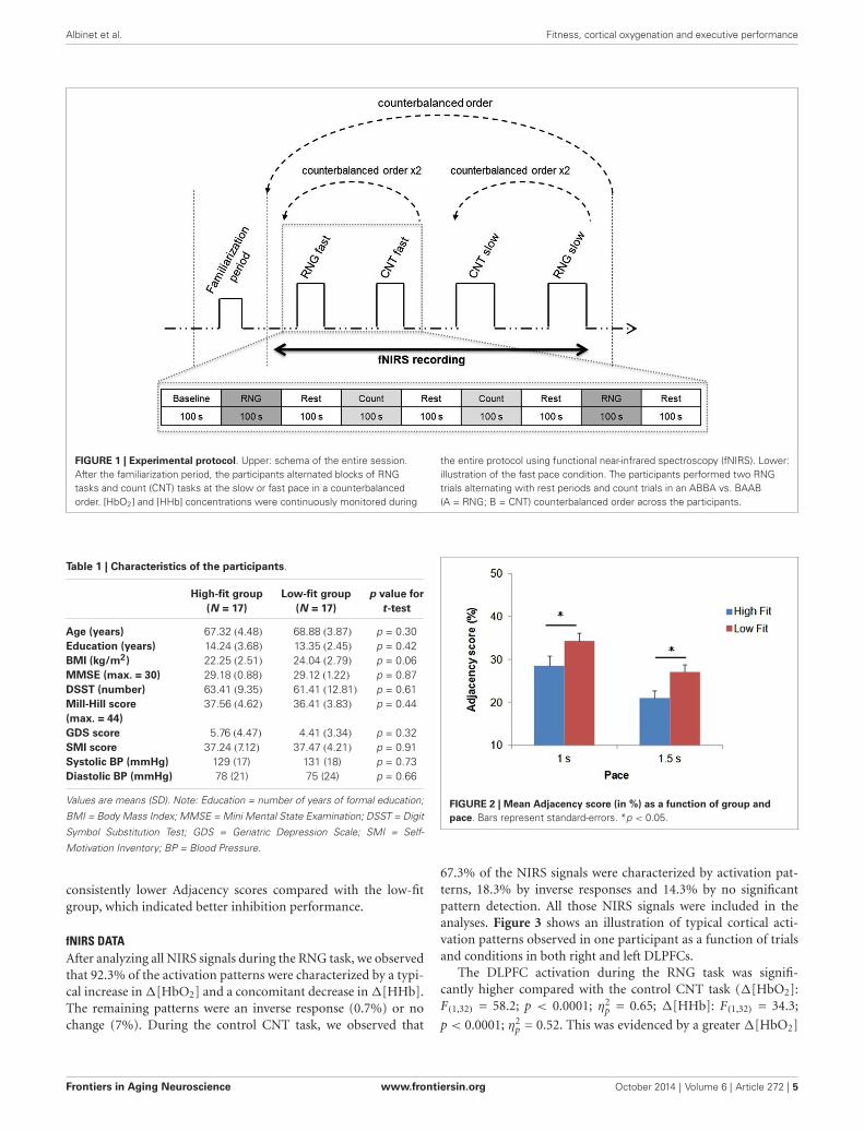
FIGURE 1 | Experimental protocol. Upper: schema of the entire session.After the familiarization period, the participants alternated blocks of RNGtasks and count (CNT) tasks at the slow or fast pace in a counterbalancedorder. [HbO2] and [HHb] concentrations were continuously monitored during
the entire protocol using functional near-infrared spectroscopy (fNIRS). Lower:illustration of the fast pace condition. The participants performed two RNGtrials alternating with rest periods and count trials in an ABBA vs. BAAB(A = RNG; B = CNT) counterbalanced order across the participants.
Table 1 | Characteristics of the participants.
High-fit group Low-fit group p value for(N = 17) (N = 17) t-test
Age (years) 67.32 (4.48) 68.88 (3.87) p = 0.30Education (years) 14.24 (3.68) 13.35 (2.45) p = 0.42BMI (kg/m2) 22.25 (2.51) 24.04 (2.79) p = 0.06MMSE (max. = 30) 29.18 (0.88) 29.12 (1.22) p = 0.87DSST (number) 63.41 (9.35) 61.41 (12.81) p = 0.61Mill-Hill score 37.56 (4.62) 36.41 (3.83) p = 0.44(max. = 44)GDS score 5.76 (4.47) 4.41 (3.34) p = 0.32SMI score 37.24 (7.12) 37.47 (4.21) p = 0.91Systolic BP (mmHg) 129 (17) 131 (18) p = 0.73Diastolic BP (mmHg) 78 (21) 75 (24) p = 0.66
Values are means (SD). Note: Education = number of years of formal education;
BMI = Body Mass Index; MMSE = Mini Mental State Examination; DSST = Digit
Symbol Substitution Test; GDS = Geriatric Depression Scale; SMI = Self-
Motivation Inventory; BP = Blood Pressure.
consistently lower Adjacency scores compared with the low-fitgroup, which indicated better inhibition performance.
fNIRS DATAAfter analyzing all NIRS signals during the RNG task, we observedthat 92.3% of the activation patterns were characterized by a typi-cal increase in ∆[HbO2] and a concomitant decrease in ∆[HHb].The remaining patterns were an inverse response (0.7%) or nochange (7%). During the control CNT task, we observed that
FIGURE 2 | Mean Adjacency score (in %) as a function of group andpace. Bars represent standard-errors. *p < 0.05.
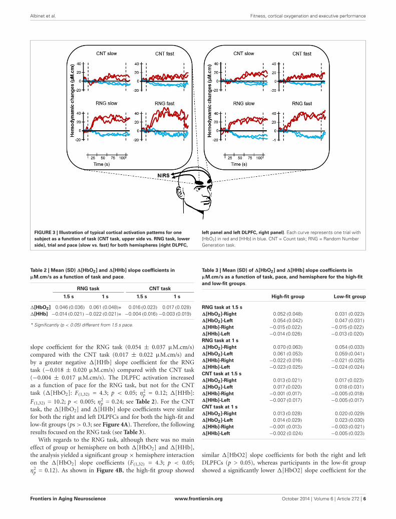
67.3% of the NIRS signals were characterized by activation pat-terns, 18.3% by inverse responses and 14.3% by no significantpattern detection. All those NIRS signals were included in theanalyses. Figure 3 shows an illustration of typical cortical acti-vation patterns observed in one participant as a function of trialsand conditions in both right and left DLPFCs.
The DLPFC activation during the RNG task was signifi-cantly higher compared with the control CNT task (∆[HbO2]:F(1,32) = 58.2; p < 0.0001; η2
p = 0.65; ∆[HHb]: F(1,32) = 34.3;
p < 0.0001; η2p = 0.52. This was evidenced by a greater ∆[HbO2]
Frontiers in Aging Neuroscience www.frontiersin.org October 2014 | Volume 6 | Article 272 | 5
Albinet et al. Fitness, cortical oxygenation and executive performance
FIGURE 3 | Illustration of typical cortical activation patterns for onesubject as a function of task (CNT task, upper side vs. RNG task, lowerside), trial and pace (slow vs. fast) for both hemispheres (right DLPFC,
left panel and left DLPFC, right panel). Each curve represents one trial with[HbO2] in red and [HHb] in blue. CNT = Count task; RNG = Random NumberGeneration task.
Table 2 | Mean (SD) ∆[HbO2] and ∆[HHb] slope coefficients inµM.cm/s as a function of task and pace.
RNG task CNT task
1.5 s 1 s 1.5 s 1 s
∆[HbO2] 0.046 (0.036) 0.061 (0.048)∗ 0.016 (0.023) 0.017 (0.028)∆[HHb] −0.014 (0.021)−0.022 (0.021)∗ −0.004 (0.016)−0.003 (0.019)
* Significantly (p < 0.05) different from 1.5 s pace.
slope coefficient for the RNG task (0.054 ± 0.037 µM.cm/s)compared with the CNT task (0.017 ± 0.022 µM.cm/s) andby a greater negative ∆[HHb] slope coefficient for the RNGtask (−0.018 ± 0.020 µM.cm/s) compared with the CNT task(−0.004 ± 0.017 µM.cm/s). The DLPFC activation increasedas a function of pace for the RNG task, but not for the CNTtask (∆[HbO2]: F(1,32) = 4.3; p < 0.05; η2
p = 0.12; ∆[HHb]:
F(1,32) = 10.2; p < 0.005; η2p = 0.24; see Table 2). For the CNT
task, the ∆[HbO2] and ∆[HHb] slope coefficients were similarfor both the right and left DLPFCs and for both the high-fit andlow-fit groups (ps > 0.3; see Figure 4A). Therefore, the followingresults focused on the RNG task (see Table 3).
With regards to the RNG task, although there was no maineffect of group or hemisphere on both ∆[HbO2] and ∆[HHb],the analysis yielded a significant group × hemisphere interactionon the ∆[HbO2] slope coefficients (F(1,32) = 4.3; p < 0.05;η2
p = 0.12). As shown in Figure 4B, the high-fit group showed
Table 3 | Mean (SD) of ∆[HbO2] and ∆[HHb] slope coefficients inµM.cm/s as a function of task, pace, and hemisphere for the high-fitand low-fit groups.
High-fit group Low-fit group
RNG task at 1.5 s∆[HbO2]-Right 0.052 (0.048) 0.031 (0.023)∆[HbO2]-Left 0.054 (0.042) 0.047 (0.031)∆[HHb]-Right −0.015 (0.022) −0.015 (0.022)∆[HHb]-Left −0.014 (0.026) −0.013 (0.020)RNG task at 1 s∆[HbO2]-Right 0.070 (0.063) 0.054 (0.033)∆[HbO2]-Left 0.061 (0.053) 0.059 (0.041)∆[HHb]-Right −0.022 (0.016) −0.021 (0.025)∆[HHb]-Left −0.023 (0.025) −0.024 (0.024)CNT task at 1.5 s∆[HbO2]-Right 0.013 (0.021) 0.017 (0.023)∆[HbO2]-Left 0.017 (0.020) 0.018 (0.031)∆[HHb]-Right −0.001 (0.017) −0.005 (0.018)∆[HHb]-Left −0.007 (0.017) −0.005 (0.017)CNT task at 1 s∆[HbO2]-Right 0.013 (0.028) 0.020 (0.029)∆[HbO2]-Left 0.014 (0.029) 0.023 (0.030)∆[HHb]-Right −0.001 (0.013) −0.003 (0.021)∆[HHb]-Left −0.002 (0.024) −0.005 (0.023)
similar ∆[HbO2] slope coefficients for both the right and leftDLPFCs (p > 0.05), whereas participants in the low-fit groupshowed a significantly lower ∆[HbO2] slope coefficient for the
Frontiers in Aging Neuroscience www.frontiersin.org October 2014 | Volume 6 | Article 272 | 6

Albinet et al. Fitness, cortical oxygenation and executive performance
FIGURE 4 | Mean ∆[HbO2] slope coefficients (in red) and mean ∆[HHb]slope coefficients (in blue) in µM.cm/s during the CNT task (A) and theRNG task (B) for the Low-fit group and the High-fit group in the LeftDLPFC (left panel) and in the Right DLPFC (right panel). Bars representstandard-errors. *p < 0.05.
right DLPFC compared with the right DLPFC of the high-fitparticipants and compared with their own left DLPFC (p <
0.05). This interaction was not significant for ∆[HHb] (p >0.8). As shown in Figure 4B, the ∆[HHb] slope coefficientswere similar for both the right and left DLPFCs and for boththe high-fit and low-fit groups. Finally, the analysis yielded asignificant hemisphere × speed interaction (F(1,32) = 11.3; p <0.005; η2
p = 0.26). Post hoc analyses showed that the ∆[HbO2]slope coefficient was significantly greater in the left DLPFC (0.051± 0.037 µM.cm/s) compared with the right DLPFC (0.042 ±
0.037 µM.cm/s; p < 0.05) at the slow pace (1.5 s). At the fastpace (1 s), the ∆[HbO2] slope coefficients were greater thanthe slow pace, but they did not significantly differ between theleft (0.060 ± 0.047 µM.cm/s) and the right (0.062 ± 0.050µM.cm/s) DLPFCs (p > 0.05). However, this last pattern ofresults was similar for both the high-fit and low-fit groups, asthe group × hemisphere × pace interaction was not significant(F < 1).
RELATIONSHIPS BETWEEN EXECUTIVE PERFORMANCE, VO2MAXLEVEL AND CEREBRAL OXYGENATIONTable 4 presents the matrix of correlations between the Adjacencyscores, the ∆[HbO2] parameters, the VO2max level, and potentialconfounders. The VO2max level was significantly correlated withthe executive performance at the pace of 1.5 s (r = 0.38; p< 0.05)and at the pace of 1 s (r = 0.42; p < 0.05). These correlationsremained significant even after controlling for age and BMI(rp = 0.37; p < 0.05 and rp =0.40; p < 0.05, respectively). OnlyVO2max, 1.5HbO2-Right and Adjacency-1.5 were significantlycorrelated each other, highlighting their functional significance.Figure 5 depicts the relationships between these three variables.The significant relationship between the VO2max level and theAdjacency score at the 1.5 s pace condition (r = −0.38; p < 0.05)no longer remained significant after controlling for ∆[HbO2](rp = 0.24; p> 0.1). Finally, as stated above, there was a significantcorrelation between the VO2max level and the Adjacency score at1 s, but these variables were not significantly correlated with the∆[HbO2] parameters at pace 1 s in the right or left DLPFC (allps> 0.4; see Table 4).
DISCUSSIONThe aim of the present study was to examine whether cardiores-piratory fitness is related to better executive performance inolder women and whether these cognitive benefits are relatedto increased prefrontal oxygenation response. In this study, wecompared the functional brain activation patterns in the rightand left DLPFCs in high-fit and low-fit older women duringthe performance of an executive controlled task, namely, theRNG task, using fNIRS. The main findings showed consistentincreasing cortical activations as a function of task difficulty (i.e.,the CNT vs. the RNG at a slow pace vs. the RNG at a fast pace),with significant increases in ∆[HbO2] and significant decreasesin ∆[HHb]. Moreover, during the RNG task, the high-fit groupperformed better and showed greater increases in ∆[HbO2] bilat-erally in the right and left DLPFCs, whereas the low-fit womenshowed significantly less activation in the right DLPFC comparedwith the right DLPFC of the high-fit women and compared withtheir own left DLPFC, which may explain, in part, their lowerexecutive performance.
The behavioral results show that the older high-fit womendemonstrated a consistently better executive performance duringthe RNG task at both difficulty levels (one response per 1 sand one response per 1.5 s) compared with the unfit olderwomen. This finding confirms previous results that showed olderadults who participated in regular physical activity (Abou-Destet al., 2012) or with higher levels of aerobic fitness (Boucardet al., 2012) outperformed their sedentary or less fit counter-parts on the RNG task at one response per 1 s. The presentstudy extends these results for a less demanding condition (oneresponse per 1.5 s) and adds to the literature on the benefi-cial effects of cardiorespiratory fitness on executive performancein older adults, particularly concerning inhibition (Colcombeand Kramer, 2003; Colcombe et al., 2004; Smiley-Oyen et al.,2008). Furthermore, when comparing the behavioral results ofthe present study with other studies using the same task, onemust note that our participants’ performances were high (overall
Frontiers in Aging Neuroscience www.frontiersin.org October 2014 | Volume 6 | Article 272 | 7

Albinet et al. Fitness, cortical oxygenation and executive performance
Table 4 | Correlation matrix between the measures of executive performance, the hemodynamic data and the characteristics of theparticipants.
Age VO2max Education BMI Adjacency-1.5 Adjacency-1 1.5HbO2 1HbO2 1.5HbO2 1HbO2-Right -Right -Left -Left
Age –VO2max –0.37* –Education −0.32 0.17 –BMI 0.12 −0.59*** −0.28 –Adjacency-1.5 0.02 −0.38* −0.02 0.18 –Adjacency-1 0.16 −0.42* −0.25 0.15 0.75*** –1.5HbO2-Right −0.26 0.41* −0.17 −0.10 −0.42* −0.38* –1HbO2-Right 0.01 0.13 −0.28 0.12 −0.18 0.05 0.60*** –1.5HbO2-Left −0.19 0.19 −0.26 −0.01 −0.44* −0.31 0.82*** 0.49* –1HbO2-Left 0.22 −0.06 −0.35* 0.24 −0.10 0.09 0.47* 0.89*** 0.51* –
Note: BMI = Body Mass Index; Right = right dorsolateral prefrontal cortex; Left = left dorsolateral prefrontal cortex; 1.5 = RNG task at the pace of 1 number per
1.5 s; 1 = RNG task at the pace of 1 number per 1 s. *p < 0.05; **p < 0.001; ***p < 0.0001.
FIGURE 5 | Relationships between the VO2max level, the ∆[HbO2] slope coefficient in the right DLPFC during the 1.5 s RNG task and the Adjacencyscore at 1.5 s. rp : partial correlation after controlling for the 1.5 ∆[HbO2] slope coefficient in the right DLPFC.
low Adjacency score); this finding suggests that our sample,regardless of the fitness level, was composed of older womenwith high cognitive functioning. This suggests that the posi-tive relationship between cardiorespiratory fitness and executivefunctioning can be expected even in this cognitively and physicallyhealthy population.
Consistent with previous studies, we found that the changesin [HbO2] were more important compared with the changes in[HHb] and that the [HbO2] response was more sensitive to theexperimental conditions, thus most likely reflecting more directcortical activation (Hoshi et al., 2003; Koike et al., 2011; Vermeijet al., 2012). Similar to Fabiani et al. (2014) during simple visualstimulation, we found that the VO2max level only correlated withthe amplitude of the [HbO2] response, but not with the amplitudeof the [HHb] response, thereby highlighting the importance ofdissociating these two components of the neurovascular response.Functional NIRS results also revealed that the DLPFC activationincreased with rising cognitive load for both the high-fit and low-fit participants, with minimal activation for the control CNT task(at both paces) and maximal activation for the RNG task at one
response per 1 s (see Table 3). This result is in agreement withother neuroimaging studies that manipulated the cognitive loadin young (Hoshi et al., 2003; Mandrick et al., 2013b) and olderadults (Vermeij et al., 2012) using fNIRS.
The high-fit older women in this study showed more over-all activation in the right DLPFC compared with their unfitcounterparts during the RNG task. Importantly, there was nogroup difference in the hemodynamic response during the controlCNT task, regardless of the pace, which suggests that the groupdifference in the hemodynamic response during the RNG task islikely not attributable to baseline differences or to difficulty inthe speed elocution. Our results tend to support the hypothesisthat the high-fit older women were able to recruit additionalcontralateral prefrontal areas to cope with task demands andthat this overactivation may have contributed, in part, to theirbetter executive performance. This explanation is in agreementwith recent neuroimaging models of cognitive aging that show apossible overactivation of the prefrontal cortices and the evidenceof reduced hemispheric asymmetry (HAROLD model) in someolder adults (see Cabeza, 2002; Reuter-Lorenz and Lustig, 2005;
Frontiers in Aging Neuroscience www.frontiersin.org October 2014 | Volume 6 | Article 272 | 8
Albinet et al. Fitness, cortical oxygenation and executive performance
Davis et al., 2008; Reuter-Lorenz and Park, 2010). According tothe compensation hypothesis, this contralateral recruitment insome older adults might correspond to a form of compensatorymechanism that counteracts age-related neurocognitive deficitsbecause it is generally associated with better cognitive perfor-mance (see Cabeza et al., 2002; Reuter-Lorenz and Cappell, 2008).Indeed, many studies have shown strongly lateralized activity inyoung adults and low-performing older adults, but bilateral acti-vations in high-performing older individuals for a wide range ofprocesses, such as executive functions (Langenecker and Nielson,2003; Langenecker et al., 2004; Tsujii et al., 2010) or episodicmemory (Cabeza et al., 2002; Gutchess et al., 2005; Angel et al.,2011). Recently, Angel et al. (2011) found that the involvement ofboth hemispheres in an episodic memory task increased memoryperformance in older adults and that older adults’ individuallevels of executive functioning mediated age-related differences inthe degree of lateralization of brain activity. The same mechanismmay have operated in the present study for the high-fit women,who were found to respond to the cognitive load of the RNGtask by recruiting the bilateral DLPFC regions. This reduced lat-eralization was beneficial to their executive performance, therebysupporting the compensation hypothesis. The depressed neu-rovascular response observed in our older low-fit women, in turn,may reflect a diminished ability of their vascular system to adaptto a task that challenges their executive functions or a reduction oftheir brain capillary bed (see Fabiani et al., 2014). On the whole,this pattern of results suggests that higher levels of aerobic fitnessin older adults may be related to increased cerebral perfusionand better cerebral oxygen supply to the PFC (Swain et al., 2003;Ainslie et al., 2008; Brown et al., 2010). The fact that aerobicfitness was positively correlated with only the [HbO2] responsein the present study is particularly consistent with this view.
Based on these results, we explored whether the increases inthe [HbO2] response observed during the RNG task had a func-tional role in participant’s executive performance. The increase in[HbO2] response in both the right and left DLPFCs was positivelyrelated to the cognitive performance during the RNG task forthe low pace condition (1.5 s). In contrast, the link between thecognitive performance and the DLPFC activation disappearedfor the most challenging condition (RNG at 1 s; see Table 4).Taken together, these results may suggest a relationship betweenDLPFC activation and cognitive performance until a certain levelof task difficulty and a ceiling effect for more challenging tasks.The use of a more parametric experimental design with at leastthree levels of difficulty would help to resolve this issue. Anotherpossibility is that the activation of other brain regions during themost difficult condition of the RNG task may have modulatedtask performance. This possibility cannot be completely ruled outbecause the present study focused on the hemodynamic activityof the DLPFC. Nonetheless, in a PET study that used the sameRNG task with six different rates (from 0.5 to 3 s per response) insix normal young adults, Jahanshahi et al. (2000) found that theleft DLPFC (BAs 9, 46) was the only brain region that showeda significant increase in rCBF associated with better cognitiveperformance. However, in disagreement with our results, theseauthors found that rCBF in the DLPFC decreased with the fasterrates of the RNG with a decline in cognitive performance. In
our study, the increased DLPFC activation in the most difficultcondition may reflect that our participants remained engaged inthe task, as confirmed by their overall good performance. Clearly,these discrepancies deserve future research in this area.
One important finding of the present study was that thesignificant relationship between the VO2max level and the Adja-cency score during the 1.5 s RNG task no longer remainedsignificant after controlling for the right DLPFC [HbO2] changes(see Figure 5). This result indicates that the increase in theright DLPFC [HbO2] response may mediate, at least in part, thepositive relationship between the VO2max level and the executiveperformance. Although caution is needed in the interpretationbecause association does not mean causality, this result providesstrong support for the cardiorespiratory fitness hypothesis andhas theoretical and practical implications. Indeed, it highlightsthe functional significance of the prefrontal increased oxygena-tion level in the high-fit women during a task that involves theperformance of executive functions, at least for the easy RNG taskcondition.
The present study has some limitations that deserve furtherinvestigation. First, the study was limited to older women aged60–77 years old, for whom executive functions are essential toprevent the decline of autonomy. As the hormone status and gen-der may influence the relationship between fitness and executiveperformance (Colcombe and Kramer, 2003; Erickson et al., 2007),our results may not be generalized to older men. Second, becausewe did not include a control group of younger adults, futureresearch is warranted with a more comprehensive manipulationof the cognitive load to examine brain activation and cognitiveperformance as a function of age and task difficulty. In particular,it would be useful to validate in different age groups whetherour results are in line with the neuroimaging models of cognitiveaging we referred to in the discussion (see Cabeza, 2002; Reuter-Lorenz and Cappell, 2008). Third, the cross-sectional design ofour study precludes inferences about the causality in the rela-tionship between the VO2max level, the DLPFC activation andthe cognitive performance. Although converging evidence fromlongitudinal and animal studies provides support to the modelwe tested, randomized-controlled trials are needed to determinewhether improving aerobic fitness through exercise training maysimultaneously improve prefrontal oxygenation and cognitiveperformance. The present study is a necessary first step, which ismore cost-effective, before engaging in clinical trials. Finally, asdiscussed above, we only recorded cerebral hemodynamics usingfNIRS in the bilateral DLPFCs, and thus, we have no informationon the activation patterns in other cortical and subcortical regionsinvolved in executive function performance. Another potentialissue in the current study was the lack of a control for the skin flowcontributions in our NIRS signals. Recent studies have indeedraised the question of superficial—extra-cortical—contributionsin NIRS signals, specifically in the [HbO2] signal (Kirilina et al.,2012). In our study, the differences across the cortical areasinvestigated do not support the idea of a global systemic responsethat biased the findings. Future investigations using methods todefinitively separate the cortical and extracortical signals in theNIRS signals would help to identify the precise nature of the con-tribution from the cortical layers in the optical signals obtained.
Frontiers in Aging Neuroscience www.frontiersin.org October 2014 | Volume 6 | Article 272 | 9
Albinet et al. Fitness, cortical oxygenation and executive performance
These methods notably include the use of additional short source-detector separation optodes as regressors (Gagnon et al., 2012)and the analysis of the photon time-of-flight distribution in time-domain NIRS (Aletti et al., 2012).
In conclusion, to our knowledge, the results of the presentstudy show for the first time that the relationship between aerobicfitness and cognitive performance for a task that involves execu-tive functions is, at least in part, mediated by the PFC oxygenationmeasured by fNIRS. At both levels of task difficulty, the high-fitwomen showed higher patterns of [HbO2] response in the rightDLPFC, which may explain, at least in part, their better cogni-tive performance; however, there was a ceiling effect concerningthe relationship between the DLPFC activation and the behav-ioral performance. Future interventional studies are needed toexamine whether physical training programs may simultaneouslyimprove aerobic fitness, PFC oxygenation and executive functionperformance.
ACKNOWLEDGMENTSThis work was supported by the General Council of Vienne anda grant from the University of Poitiers to Cédric T. Albinet.The authors wish to thank Dr. M. Hayot and the Departmentof Clinical Physiology for their help in the determination ofparticipants’ VO2max.
REFERENCESAbou-Dest, A., Albinet, C. T., Boucard, G., and Audiffren, M. (2012). Swimming as
a positive moderator of cognitive aging: a cross-sectional study with a multitaskapproach. J. Aging Res. 2012:273185. doi: 10.1155/2012/273185
Ainslie, P. N., Cotter, J. D., George, K. P., Lucas, S., Murrell, C., Shave, R., et al.(2008). Elevation in cerebral blood flow velocity with aerobic fitness throughouthealthy human ageing. J. Physiol. 586, 4005–4010. doi: 10.1113/jphysiol.2008.158279
Albinet, C. T., Boucard, G., Bouquet, C. A., and Audiffren, M. (2012). Pro-cessing speed and executive functions in cognitive aging: how to disentangletheir mutual relationship? Brain Cogn. 79, 1–11. doi: 10.1016/j.bandc.2012.02.001
Albinet, C., Tomporowski, P. D., and Beasman, K. (2006). Aging and concurrenttask performance: cognitive demand and motor control. Educ. Gerontol. 32,689–706. doi: 10.1080/03601270600835421
Aletti, F., Re, R., Pace, V., Contini, D., Molteni, E., Cerutti, S., et al. (2012). Deepand surface hemodynamic signal from functional time resolved transcranialnear infrared spectroscopy compared to skin flowmotion. Comput. Biol. Med.42, 282–289. doi: 10.1016/j.compbiomed.2011.06.001
American Thoracic Society/American College of Chest Physicians. (2003).ATS/ACCP statement on cardiopulmonary exercise testing. Am. J. Respir. Crit.Care Med. 167, 211–277. doi: 10.1164/rccm.167.2.211
Anderson, V., Jacobs, R., and Anderson, P. J. (2008). Executive Functions and theFrontal Lobes: A Lifespan Perspective. New York: Taylor and Francis.
André, N., and Dishman, R. K. (2012). Evidence for the construct validity of self-motivation as a correlate of exercise adherence in French older adults. J. AgingPhys. Act. 20, 231–245.
Angel, L., Fay, S., Bouazzaoui, B., and Isingrini, M. (2011). Two hemispheres forbetter memory in old age: role of executive functioning. J. Cogn. Neurosci. 23,3767–3777. doi: 10.1162/jocn_a_00104
Boucard, G. K., Albinet, C. T., Bugaiska, A., Bouquet, C. A., Clarys, D., andAudiffren, M. (2012). Impact of physical activity on executive functions in aging:a selective effect on inhibition among old adults. J. Sport Exerc. Psychol. 34, 808–827.
Bourque, P., Blanchard, L., and Vézina, J. (1990). Étude psychométrique del’Échelle de dépression gériatrique. Can. J. Aging 9, 348–355. doi: 10.1017/s0714980800007467
Brown, A. D., McMorris, C. A., Longman, R. S., Leigh, R., Hill, M. D., Friedenreich,C. M., et al. (2010). Effects of cardiorespiratory fitness and cerebral blood flowon cognitive outcomes in older women. Neurobiol. Aging 31, 2047–2057. doi: 10.1016/j.neurobiolaging.2008.11.002
Cabeza, R. (2002). Hemispheric asymmetry reduction in older adults: theHAROLD model. Psychol. Aging 17, 85–100. doi: 10.1037//0882-7974.17.1.85
Cabeza, R., Anderson, N. D., Locantore, J. K., and McIntosh, A. R. (2002).Aging gracefully: compensatory brain activity in high-performing older adults.Neuroimage 17, 1394–1402. doi: 10.1006/nimg.2002.1280
Cabeza, R., Nyberg, L., and Park, D. (2005). Cognitive Neuroscience of Aging: LinkingCognitive and Cerebral Aging. New York: Oxford University Press.
Colcombe, S. J., Erickson, K. I., Raz, N., Webb, A. G., Cohen, N. J., McAuley,E., et al. (2003). Aerobic fitness reduces brain tissue loss in aging humans. J.Gerontol. A Biol. Sci. Med. Sci. 58, 176–180. doi: 10.1093/gerona/58.2.m176
Colcombe, S. J., Erickson, K. I., Scalf, P. E., Kim, J. S., Prakash, R., McAuley, E.,et al. (2006). Aerobic exercise training increases brain volume in aging humans.J. Gerontol. A Biol. Sci. Med. Sci. 61, 1166–1170. doi: 10.1093/gerona/61.11.1166
Colcombe, S., and Kramer, A. F. (2003). Fitness effects on the cognitive function ofolder adults: a meta-analytic study. Psychol. Sci. 14, 125–130. doi: 10.1111/1467-9280.t01-1-01430
Colcombe, S. J., Kramer, A. F., Erickson, K. I., Scalf, P., McAuley, E., Cohen, N. J.,et al. (2004). Cardiovascular fitness, cortical plasticity and aging. Proc. Natl.Acad. Sci. U S A 101, 3316–3321. doi: 10.1073/pnas.0400266101
Cotman, C. W., Berchtold, N. C., and Christie, L. A. (2007). Exercise builds brainhealth: key roles of growth factor cascades and inflammation. Trends Neurosci.30, 464–472. doi: 10.1016/j.tins.2007.06.011
Cui, X., Bray, S., Bryant, D. M., Glover, G. H., and Reiss, A. L. (2011). A quantitativecomparison of NIRS and fMRI across multiple cognitive tasks. Neuroimage 54,2808–2821. doi: 10.1016/j.neuroimage.2010.10.069
Daniels, C., Witt, K., Wolff, S., Jansen, O., and Deuschl, G. (2003). Rate dependencyof the human cortical network subserving executive functions during generationof random number series-a functional magnetic resonance imaging study.Neurosci. Lett. 345, 25–28. doi: 10.1016/s0304-3940(03)00496-8
Davis, S. W., Dennis, N. A., Daselaar, S. M., Fleck, M. S., and Cabeza, R. (2008).Que PASA? The posterior-anterior shift in aging. Cereb. Cortex 18, 1201–1209.doi: 10.1093/cercor/bhm155
Deltour, J. (1993). Echelle de Vocabulaire de Mill Hill de JC Raven. AdaptationFrançaise et Normes Européennes du Mill Hill et du Standard Progressive Matricesde Raven (PM38). Braine-le-Château: Editions l’application des techniquesmodernes.
Dustman, R. E., Ruhling, R. O., Russell, E. M., Shearer, D. E., Bonekat, H. W.,Shigeoka, J. W., et al. (1984). Aerobic exercise training and improved neuropsy-chological function of older individuals. Neurobiol. Aging 5, 35–42. doi: 10.1016/0197-4580(84)90083-6
Erickson, K. I., Colcombe, S. J., Elavsky, S., McAuley, E., Korol, D. L., Scalf, P. E.,et al. (2007). Interactive effects of fitness and hormone treatment on brainhealth in postmenopausal women. Neurobiol. Aging 28, 179–185. doi: 10.1016/j.neurobiolaging.2005.11.016
Erickson, K. I., Voss, M. W., Prakash, R. S., Basak, C., Szabo, A., Chaddock, L., et al.(2011). Exercise training increases size of hippocampus and improves memory.Proc. Natl. Acad. Sci. U S A 108, 3017–3022. doi: 10.1073/pnas.1015950108
Etnier, J. L., Nowell, P. M., Landers, D. M., and Sibley, B. A. (2006). A meta-regression to examine the relationship between aerobic fitness and cognitiveperformance. Brain Res. Rev. 52, 119–130. doi: 10.1016/j.brainresrev.2006.01.002
Fabiani, M., Gordon, B. A., Maclin, E. L., Pearson, M. A., Brumback-Peltz, C. R.,Low, K. A., et al. (2014). Neurovascular coupling in normal aging: a combinedoptical, ERP and fMRI study. Neuroimage 85(Pt. 1), 592–607. doi: 10.1016/j.neuroimage.2013.04.113
Ferrari, M., and Quaresima, V. (2012). A brief review on the history of humanfunctional near-infrared spectroscopy (fNIRS) development and fields of appli-cation. Neuroimage 63, 921–935. doi: 10.1016/j.neuroimage.2012.03.049
Friston, K. J., Holmes, A. P., Worsley, K. J., Poline, J.-P., Frith, C. D., and Frackowiak,R. S. J. (1994). Statistical parametric maps in functional imaging: a general linearapproach. Hum. Brain Mapp. 2, 189–210. doi: 10.1002/hbm.460020402
Gagnon, L., Cooper, R. J., Yücel, M. A., Perdue, K. L., Greve, D. N., and Boas,D. A. (2012). Short separation channel location impacts the performance ofshort channel regression in NIRS. Neuroimage 59, 2518–2528. doi: 10.1016/j.neuroimage.2011.08.095
Frontiers in Aging Neuroscience www.frontiersin.org October 2014 | Volume 6 | Article 272 | 10
Albinet et al. Fitness, cortical oxygenation and executive performance
Gouzi, F., Préfaut, C., Abdellaoui, A., Roudier, E., de Rigal, P., Molinari, N.,et al. (2013). Blunted muscle angiogenic training-response in COPD patientsversus sedentary controls. Eur. Respir. J. 41, 806–814. doi: 10.1183/09031936.00053512
Gutchess, A. H., Welsh, R. C., Hedden, T., Bangert, A., Minear, M., Liu, L. L.,et al. (2005). Aging and the neural correlates of successful picture encoding:frontal activations compensate for decreased medial-temporal activity. J. Cogn.Neurosci. 17, 84–96. doi: 10.1162/0898929052880048
Hébert, R., Bravo, G., and Girouard, D. (1992). Validation de l’adaptation françaisedu modified mini-mental state (3MS). Rev. Geriatr. 17, 443–450.
Heekeren, H. R., Obrig, H., Wenzel, R., Eberle, K., Ruben, J., Villringer, K., et al.(1997). Cerebral haemoglobin oxygenation during sustained visual stimulation:a near-infrared spectroscopy study. Philos. Trans. R. Soc. Lond. B Biol. Sci. 352,743–750. doi: 10.1098/rstb.1997.0057
Hillman, C. H., Erickson, K. I., and Kramer, A. F. (2008). Be smart, exercise yourheart: exercise effects on brain and cognition. Nat. Rev. Neurosci. 9, 58–65.doi: 10.1038/nrn2298
Holper, L., Biallas, M., and Wolf, M. (2009). Task complexity relates to activationof cortical motor areas during uni-and bimanual performance: a func-tional NIRS study. Neuroimage 46, 1105–1113. doi: 10.1016/j.neuroimage.2009.03.027
Hoshi, Y., Tsou, B. H., Billock, V. A., Tanosaki, M., Iguchi, Y., Shimada, M., et al.(2003). Spatiotemporal characteristics of hemodynamic changes in the humanlateral prefrontal cortex during working memory tasks. Neuroimage 20, 1493–1504. doi: 10.1016/s1053-8119(03)00412-9
Huppert, T. J., Diamond, S. G., Franceschini, M. A., and Boas, D. A. (2009).HomER: a review of time-series analysis methods for near-infrared spectroscopyof the brain. Appl. Opt. 48, D280–D298. doi: 10.1364/ao.48.00d280
Jackson, A. S., Blair, S. N., Mahar, M. T., Wier, L. T., Ross, R. M., and Stuteville, J. E.(1990). Prediction of functional aerobic capacity without exercise testing. Med.Sci. Sports Exerc. 22, 863–870. doi: 10.1249/00005768-199012000-00021
Jahanshahi, M., and Dirnberger, G. (1998). The left dorsolateral prefrontalcortex and random generation of responses: studies with transcranial mag-netic stimulation. Neuropsychologia 37, 181–190. doi: 10.1016/s0028-3932(98)00092-x
Jahanshahi, M., Dirnberger, G., Fuller, R., and Frith, C. D. (2000). The role ofthe dorsolateral prefrontal cortex in random number generation: a study withpositron emission tomography. Neuroimage 12, 713–725. doi: 10.1006/nimg.2000.0647
Jahanshahi, M., Profice, P., Brown, R. G., Ridding, M. C., Dirnberger, G., andRothwell, J. C. (1998). The effects of transcranial magnetic stimulation over thedorsolateral prefrontal cortex on suppression of habitual counting during ran-dom number generation. Brain 121(Pt. 8), 1533–1544. doi: 10.1093/brain/121.8.1533
Joppich, G., Däuper, J., Dengler, R., Johannes, S., Rodriguez-Fornells, A., andMünte, T. F. (2004). Brain potentials index executive functions during randomnumber generation. Neurosci. Res. 49, 157–164. doi: 10.1016/j.neures.2004.02.003
Kalpouzos, G., Chételat, G., Baron, J.-C., Landeau, B., Mevel, K., Godeau, C.,et al. (2009). Voxel-based mapping of brain gray matter volume and glucosemetabolism profiles in normal aging. Neurobiol. Aging 30, 112–124. doi: 10.1016/j.neurobiolaging.2007.05.019
Kirilina, E., Jelzow, A., Heine, A., Niessing, M., Wabnitz, H., Brühl, R., et al. (2012).The physiological origin of task-evoked systemic artefacts in functional nearinfrared spectroscopy. Neuroimage 61, 70–81. doi: 10.1016/j.neuroimage.2012.02.074
Koike, S., Takizawa, R., Nishimura, Y., Marumo, K., Kinou, M., Kawakubo, Y.,et al. (2011). Association between severe dorsolateral prefrontal dysfunctionduring random number generation and earlier onset in schizophrenia. Clin.Neurophysiol. 122, 1533–1540. doi: 10.1016/j.clinph.2010.12.056
Kramer, A. F., Hahn, S., Cohen, N. J., Banich, M. T., McAuley, E., Harrison, C. R.,et al. (1999). Ageing, fitness and neurocognitive function. Nature 400, 418–419.doi: 10.1038/22682
Kwee, I. L., and Nakada, T. (2003). Dorsolateral prefrontal lobe activation declinessignificantly with age—functional NIRS study. J. Neurol. 250, 525–529. doi: 10.1007/s00415-003-1028-x
Langenecker, S. A., and Nielson, K. A. (2003). Frontal recruitment during responseinhibition in older adults replicated with fMRI. Neuroimage 20, 1384–1392.doi: 10.1016/s1053-8119(03)00372-0
Langenecker, S. A., Nielson, K. A., and Rao, S. M. (2004). fMRI of healthy olderadults during Stroop interference. Neuroimage 21, 192–200. doi: 10.1016/j.neuroimage.2003.08.027
Mandrick, K., Derosiere, G., Dray, G., Coulon, D., Micallef, J.-P., and Perrey, S.(2013a). Utilizing slope method as an alternative data analysis for functionalnear-infrared spectroscopy-derived cerebral hemodynamic responses. Int. J. Ind.Ergonom. 43, 335–341. doi: 10.1016/j.ergon.2013.05.003
Mandrick, K., Derosiere, G., Dray, G., Coulon, D., Micallef, J. P., and Perrey, S.(2013b). Prefrontal cortex activity during motor tasks with additional mentalload requiring attentional demand: a near-infrared spectroscopy study. Neu-rosci. Res. 76, 156–162. doi: 10.1016/j.neures.2013.04.006
Miyake, A., Friedman, N. P., Emerson, M. J., Witzki, A. H., Howerter, A., andWager, T. D. (2000). The unity and diversity of executive functions and theircontributions to complex “Frontal Lobe” tasks: a latent variable analysis. Cogn.Psychol. 41, 49–100. doi: 10.1006/cogp.1999.0734
Obrig, H., and Villringer, A. (2003). Beyond the visible—imaging the humanbrain with light. J. Cereb. Blood Flow Metab. 23, 1–18. doi: 10.1097/00004647-200301000-00001
Oldfield, R. C. (1971). The assessment and analysis of handedness: the Edinburghinventory. Neuropsychologia 9, 97–113. doi: 10.1016/0028-3932(71)90067-4
Perrey, S. (2008). Non-invasive NIR spectroscopy of human brain function duringexercise. Methods 45, 289–299. doi: 10.1016/j.ymeth.2008.04.005
Prakash, R. S., Voss, M. W., Erickson, K. I., Lewis, J. M., Chaddock, L., Malkowski,E., et al. (2011). Cardiorespiratory fitness and attentional control in the agingbrain. Front. Hum. Neurosci. 4:229. doi: 10.3389/fnhum.2010.00229
Raz, N., Gunning-Dixon, F., Head, D., Rodrigue, K. M., Williamson, A., and Acker,J. D. (2004). Aging, sexual dimorphism and hemispheric asymmetry of thecerebral cortex: replicability of regional differences in volume. Neurobiol. Aging25, 377–396. doi: 10.1016/s0197-4580(03)00118-0
Raz, N., Lindenberger, U., Rodrigue, K. M., Kennedy, K. M., Head, D., Williamson,A., et al. (2005). Regional brain changes in aging healthy adults: general trends,individual differences and modifiers. Cereb. Cortex 15, 1676–1689. doi: 10.1093/cercor/bhi044
Raz, N., and Rodrigue, K. M. (2006). Differential aging of the brain: patterns,cognitive correlates and modifiers. Neurosci. Biobehav. Rev. 30, 730–748. doi: 10.1016/j.neubiorev.2006.07.001
Reuter-Lorenz, P. A., and Cappell, K. A. (2008). Neurocognitive aging and thecompensation hypothesis. Curr. Dir. Psychol. Sci. 17, 177–182. doi: 10.1111/j.1467-8721.2008.00570.x
Reuter-Lorenz, P. A., and Lustig, C. (2005). Brain aging: reorganizing discoveriesabout the aging mind. Curr. Opin. Neurobiol. 15, 245–251. doi: 10.1016/j.conb.2005.03.016
Reuter-Lorenz, P. A., and Park, D. C. (2010). Human neuroscience and the agingmind: a new look at old problems. J. Gerontol. B Psychol. Sci. Soc. Sci. 65, 405–415. doi: 10.1093/geronb/gbq035
Shvartz, E., and Reibold, R. C. (1990). Aerobic fitness norms for males and femalesaged 6 to 75 years: a review. Aviat. Space Environ. Med. 61, 3–11.
Smiley-Oyen, A. L., Lowry, K. A., Francois, S. J., Kohut, M. L., and Ekkekakis,P. (2008). Exercise, fitness and neurocognitive function in older adults: the“selective improvement” and “cardiovascular fitness” hypotheses. Ann. Behav.Med. 36, 280–291. doi: 10.1007/s12160-008-9064-5
Swain, R. A., Harris, A. B., Wiener, E. C., Dutka, M. V., Morris, H. D., Theien, B. E.,et al. (2003). Prolonged exercise induces angiogenesis and increases cerebralblood volume in primary motor cortex of the rat. Neuroscience 117, 1037–1046.doi: 10.1016/s0306-4522(02)00664-4
Towse, J. N., and Neil, D. (1998). Analyzing human random generation behavior:a review of methods used and a computer program for describing perfor-mance. Behav. Res. Methods Instrum. Comput. 30, 583–591. doi: 10.3758/bf03209475
Tsujii, T., Okada, M., and Watanabe, S. (2010). Effects of aging on hemisphericasymmetry in inferior frontal cortex activity during belief-bias syllogistic rea-soning: a near-infrared spectroscopy study. Behav. Brain Res. 210, 178–183.doi: 10.1016/j.bbr.2010.02.027
Vermeij, A., van Beek, A. H., Olde Rikkert, M. G., Claassen, J. A., and Kessels,R. P. (2012). Effects of aging on cerebral oxygenation during working-memoryperformance: a functional near-infrared spectroscopy study. PLoS One 7:e46210.doi: 10.1371/journal.pone.0046210
Voelcker-Rehage, C., and Niemann, C. (2013). Structural and functionalbrain changes related to different types of physical activity across the life
Frontiers in Aging Neuroscience www.frontiersin.org October 2014 | Volume 6 | Article 272 | 11

Albinet et al. Fitness, cortical oxygenation and executive performance
span. Neurosci. Biobehav. Rev. 37, 2268–2295. doi: 10.1016/j.neubiorev.2013.01.028
Voss, M. W., Vivar, C., Kramer, A. F., and Van Praag, H. (2013). Bridging animaland human models of exercise-induced brain plasticity. Trends Cogn. Sci. 17,525–544. doi: 10.1016/j.tics.2013.08.001
Wechsler, D. (2000). Manuel de L’echelle D’intelligence de Wechsler pour Adultes.Paris: Editions du Centre de Psychologie Appliquée.
West, R. L. (1996). An application of prefrontal cortex function theory to cognitiveaging. Psychol. Bull. 120, 272–292. doi: 10.1037//0033-2909.120.2.272
Conflict of Interest Statement: The authors declare that the research was conductedin the absence of any commercial or financial relationships that could be construedas a potential conflict of interest.
Received: 09 May 2014; accepted: 19 September 2014; published online: 08 October2014.Citation: Albinet CT, Mandrick K, Bernard PL, Perrey S and Blain H (2014)Improved cerebral oxygenation response and executive performance as a function ofcardiorespiratory fitness in older women: a fNIRS study. Front. Aging Neurosci. 6:272.doi: 10.3389/fnagi.2014.00272This article was submitted to the journal Frontiers in Aging Neuroscience.Copyright © 2014 Albinet, Mandrick, Bernard, Perrey and Blain. This is an open-access article distributed under the terms of the Creative Commons Attribution License(CC BY). The use, distribution and reproduction in other forums is permitted, providedthe original author(s) or licensor are credited and that the original publication in thisjournal is cited, in accordance with accepted academic practice. No use, distribution orreproduction is permitted which does not comply with these terms.
Frontiers in Aging Neuroscience www.frontiersin.org October 2014 | Volume 6 | Article 272 | 12
Related Documents