Vitamins G. B. Shinde (M.V.Sc., IV Sem) Dept. of Animal Nutrition. CoVAS, Udgir, (MS)

Welcome message from author
This document is posted to help you gain knowledge. Please leave a comment to let me know what you think about it! Share it to your friends and learn new things together.
Transcript

VitaminsG. B. Shinde
(M.V.Sc., IV Sem)Dept. of Animal
Nutrition.CoVAS, Udgir,
(MS)
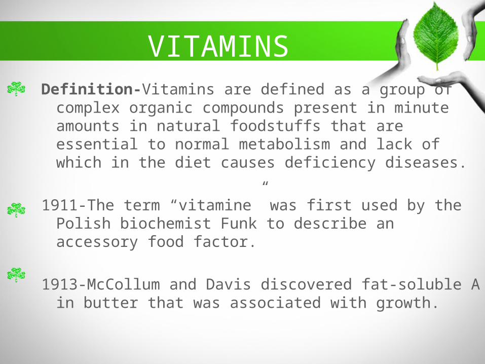
VITAMINSDefinition-Vitamins are defined as a group of
complex organic compounds present in minute amounts in natural foodstuffs that are essential to normal metabolism and lack of which in the diet causes deficiency diseases.
1911-The term “vitamine” was first used by the Polish biochemist Funk to describe an accessory food factor.
1913-McCollum and Davis discovered fat-soluble A in butter that was associated with growth.

3
Classification of Vitamins
Water Soluble Vitamins Fat Soluble Vitamins
1) Thiamin2) Pyrodoxin3) Biotin4) Riboflavin5) Pantothanic
acid6) Nicotinic acid7) Vit. B12 8) Folic acid.9) Vitamin C
(Ascorbic acid)
1) Vitamin A2) Vitamin D3) Vitamin E4) Vitamin K
Vitamin like compound
1) Lipoic acid2) Chollin3) Meso-inositel4) Ubiquinone
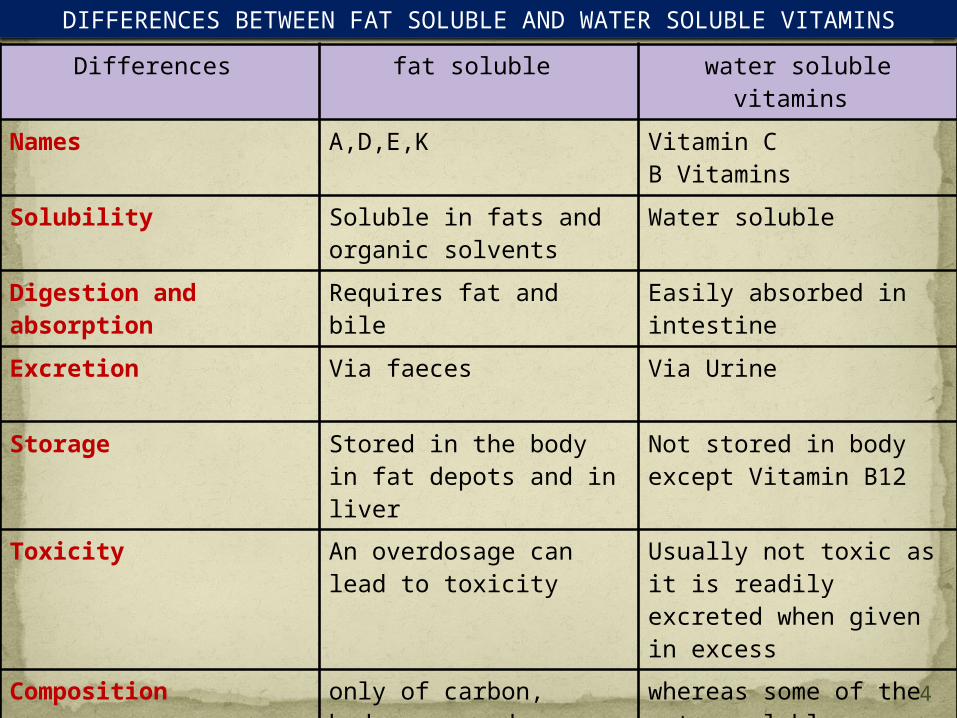
Differences fat soluble water soluble vitamins
Names A,D,E,K Vitamin C B Vitamins
Solubility Soluble in fats and organic solvents
Water soluble
Digestion and absorption
Requires fat and bile Easily absorbed in intestine
Excretion Via faeces Via Urine
Storage Stored in the body in fat depots and in liver
Not stored in body except Vitamin B12
Toxicity An overdosage can lead to toxicity
Usually not toxic as it is readily excreted when given in excess
Composition only of carbon, hydrogen, and oxygen
whereas some of the water-soluble vitamins also contain nitrogen, sulfur, or cobalt.
DIFFERENCES BETWEEN FAT SOLUBLE AND WATER SOLUBLE VITAMINS
4
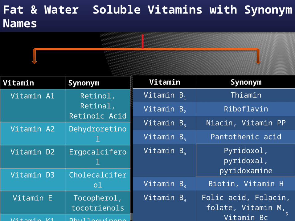
Fat & Water Soluble Vitamins with Synonym Names
Vitamin Synonym
Vitamin A1 Retinol, Retinal, Retinoic Acid
Vitamin A2 Dehydroretinol
Vitamin D2 Ergocalciferol
Vitamin D3 Cholecalciferol
Vitamin E Tocopherol, tocotrienols
Vitamin K1 Phylloquinone
Vitamin K2 Menaquinone
Vitamin K3 Menadionea
Vitamin Synonym
Vitamin B1 Thiamin
Vitamin B2 Riboflavin
Vitamin B3 Niacin, Vitamin PP
Vitamin B5 Pantothenic acid
Vitamin B6 Pyridoxol, pyridoxal, pyridoxamine
Vitamin B8 Biotin, Vitamin H
Vitamin B9 Folic acid, Folacin, folate, Vitamin M,
Vitamin Bc
Vitamin B12 Cobalamin, Cyanocobalamin
Vitamin C Ascorbic acid
5

Vitamin A(C20 H30 O)
6

Fat Soluble Vitamins
History
1900s, McCollum and Davis described “fat-soluble A,” a factor isolated from animal fats1919-Steenbock- suggested that carotene was the source of the vitamin A.1939-Wagner and coworkers suggested in that the conversion of β-carotene into vitamin A occurs within the intestinal mucosa.1930 to 1931-Karrer and coworkers proposed the exact structural formulas for vitamin A and β-carotene.1947-Isler and coworkers synthesized the first pure vitamin A.
7
Chemical Structure & Properties
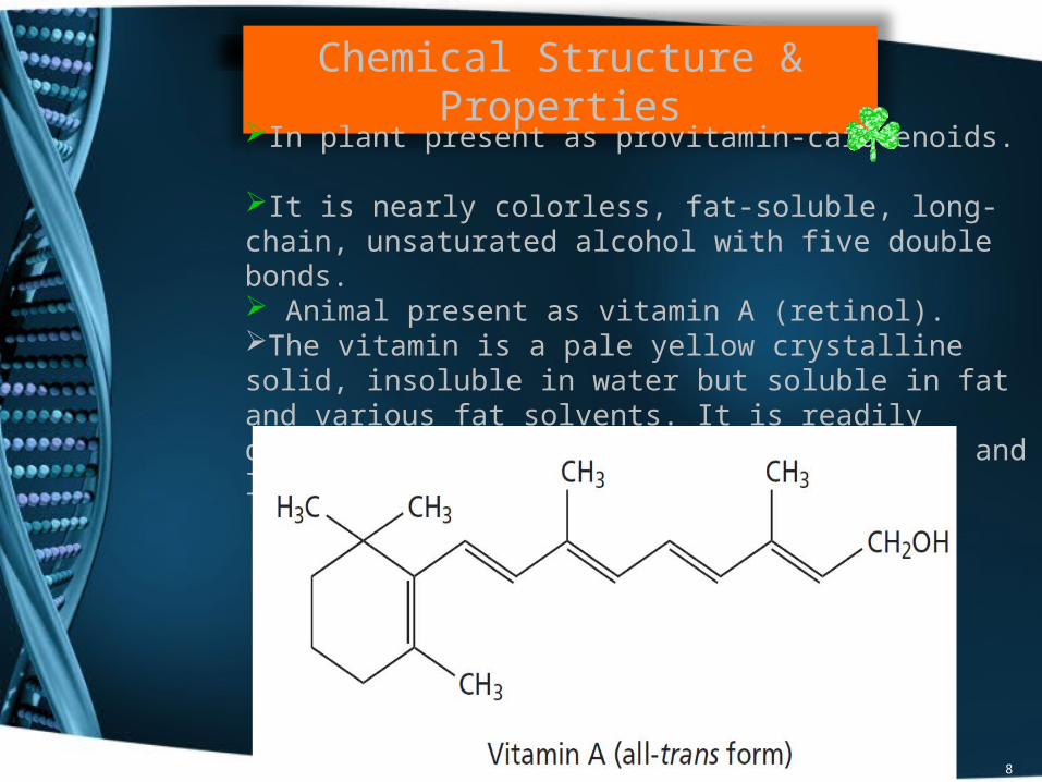
In plant present as provitamin-carotenoids.
It is nearly colorless, fat-soluble, long-chain, unsaturated alcohol with five double bonds. Animal present as vitamin A (retinol).The vitamin is a pale yellow crystalline solid, insoluble in water but soluble in fat and various fat solvents. It is readily destroyed by oxidation on exposure to air and light.
8
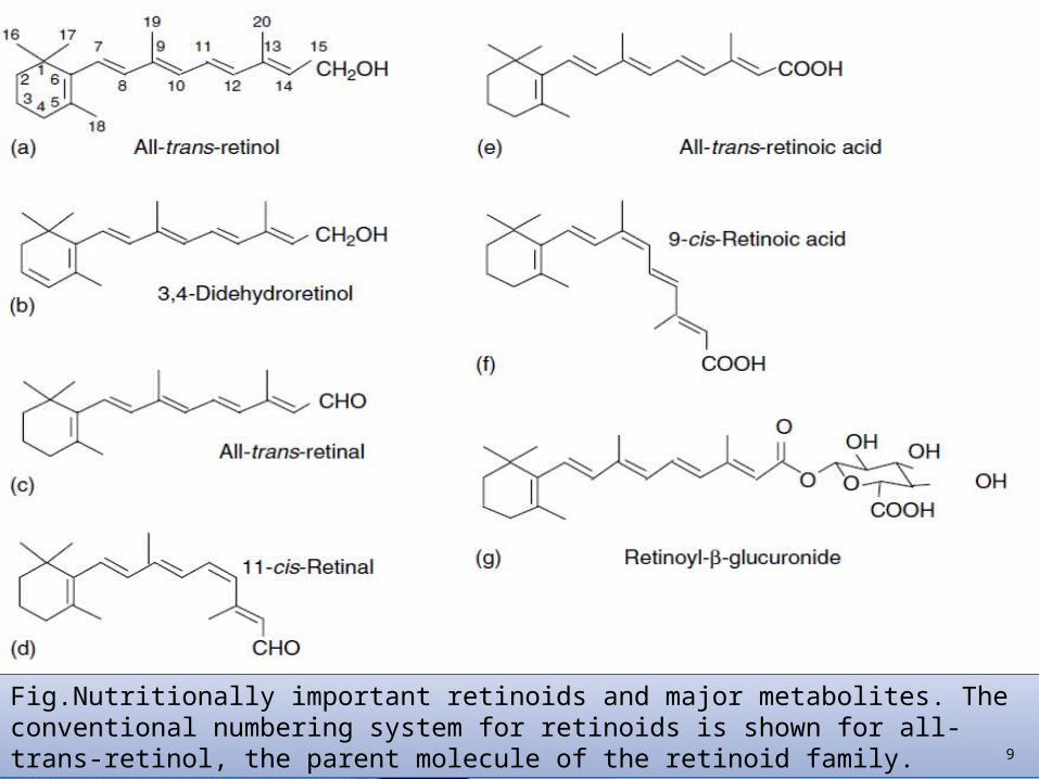
Fig.Nutritionally important retinoids and major metabolites. The conventional numbering system for retinoids is shown for all-trans-retinol, the parent molecule of the retinoid family. 9

Fig. Nutritionally important carotenoids. (a) Lycopene, a nonprovitamin A carotene; (b) alltrans-bb0-carotene; arrows indicate sites of cleavage by b-carotene monooxygenase, BCO, and BCO-2;(c) all-trans (a,b0) carotene; (d) lutein, a nonprovitamin A xanthophyll; (e) b-cryptoxanthin.
10

All-trans-retinal
is the immediate product of the central cleavage of b-carotene as well as an intermediate in the oxidative metabolism of retinol to all transretinoic acid.
All-trans-retinoic acid
is the most bioactive form of vitamin A.
When fed to vitamin A-deficient animals, retinoic acid restores growth and tissue differentiation and prevents mortality,
indicating that this form alone, or metabolites made from it, is able to support nearly all of
the functions attributed to vitamin A.
A notable exception is vision, which is not restored by retinoic acid because retinoic acid
cannot be reduced to retinal in vivo.
11

Carotenoids- At least 600 naturally occurring carotenoids are known, but only a few of these are precursors of the vitamin.One mol of carotene yields two molecules of Vit A.
α-carotene, β−carotene, γ-carotene, cryptoxanthine.
β−carotene:-Highest Vitamin A activityMost biologically active form.Main source of vitamin A in diet.Twice as potent as the other isomers
Vitamin A1- -Retinol (Alcohol),-Retinal (Aldehyde), -Retinoic acid(Acid).
Vitamin A2- Dehydroretinol-It contains an additional double bond in the β−ionone ring.-Liver oils of marine fish-less than 10% of the total vitamin A content.-Biological activity-40 to50% that of Vit A1.
12

METABOLISMDigestion
Vitamin A in animal products and carotenoids are released from proteins by the action of pepsin in the stomach and proteolytic enzymes in the small intestine. (Ong, 1993; Ross, 1993).In the duodenum, bile salts break up fatty globules of carotenoids and retinyl esters to smaller lipid congregates, which can be more easily digested by pancreatic lipase, retinyl ester hydrolase, and cholesteryl ester hydrolase.
β−carotene
In the intestinal
mucosa
two molecules of retinal
retinol
However,extensive evidence exists also for random (eccentric) cleavage, resulting in retinoic acid and retinal, with a preponderance of apocarotenals formed as intermediates (Wolf, 1995).
15,15 dioxygenase
Retinaldehyde reductase
Absorption
13

The cleavage enzyme has been found in many vertebrates but is not present in the cat or mink.
Therefore, these species cannot utilize carotene as a source of vitamin A.
In some species, such as the rat, pig, goat, sheep,rabbit, buffalo, and dog, almost all of the carotene is cleaved in the intestine.
In humans, cattle, horses, and carp, significant amounts of carotene can be absorbed.
Absorbed carotene can be stored in the liver and fatty tissues.
Hence, these latter animals have yellow body and milk fat, whereas animals that do not absorb carotene have white fat.No absorption occours in Stomach
Main site of lipid absorption is Mucosa of Proximal jejunum.
14

15

Transport In mucosal cells of Int., retinol is re-esterified
Palmitate ester
incorporated into the chylomicra of the mucosa
secreted into the lymph
transported through the lymphatic system with a LPL
deposited Liver (hepatocytes and stellate and parenchymal cells)
Intestine
Retinyl ester hydrolase by pancreas
16

Liver
Target Tissue
Retinol is readily transferred to the egg in birds, but the transfer of retinol across the placenta is marginal, and mammals are born with very low liver stores of vitamin A. Uterine RBP has been identified in the pig uterus, with the function of delivering retinol to the fetus (Clawitter et al., 1990). 17

Excretion
Derivatives of vitamin A with an intact carbon chain are generally excreted in feces, whereas acidic chain-shortened products tend to be excreted in urine
(Olson, 1991).Storage
Liver normally contains about 90% of total-body vitamin A.
The remainder is stored in the kidneys, lungs, adrenals, and blood, with small amounts also found in other organs and tissues.
A large quantity of vitamin A is stored in the kidney as well as the liver in cats and dogs.
This high level of vitamin A in the kidney is unique to cats and dogs.Grass-fed cattle have large stores of carotene in their body fat, which is evidenced by a deep yellow color
18

FUNCTIONS
1. Vision2. Maintenance of Normal Epithelium3. Bone Development
Vision
19
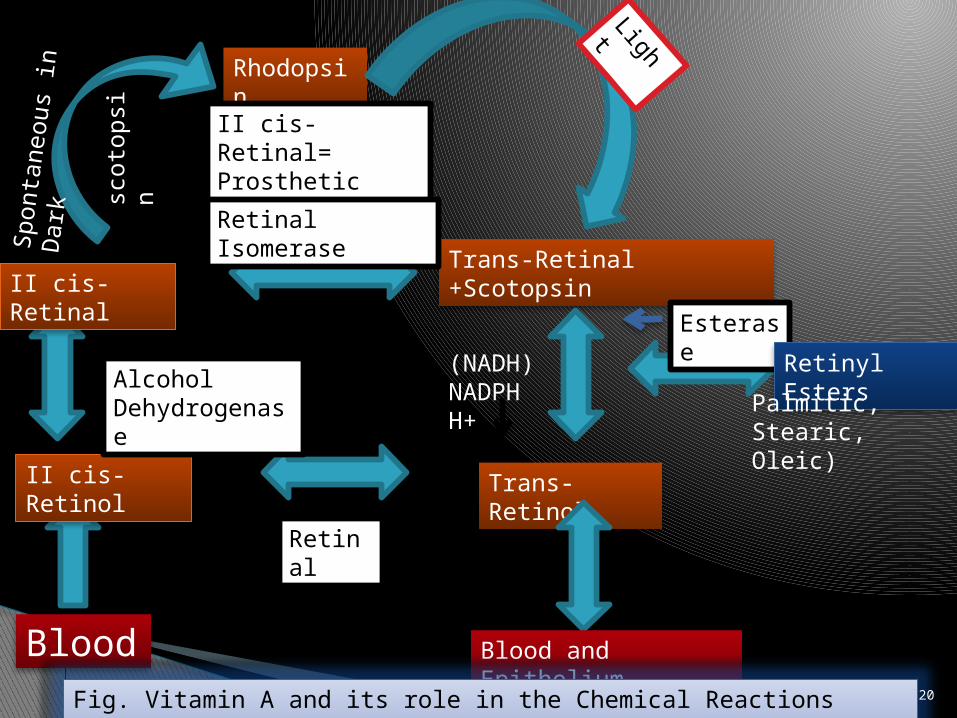
Blood
II cis-Retinol
Rhodopsin
Trans-Retinal +ScotopsinII cis-Retinal
II cis-Retinal= Prosthetic group
Trans-Retinol
Blood and Epithelium
Esterase Retinyl Esters
Retinal
Alcohol Dehydrogenase
(NADH)NADPHH+
Retinal Isomerase
Palmitic, Stearic, Oleic)
Fig. Vitamin A and its role in the Chemical Reactions involved in vision.
Light
Sponta
neous
in D
ark
scoto
psi
n
20

Retina of eye
Rods Cones
Dim light Bright light
Rhodopsin Iodopsin
Cis isomers of retinol
Vision
21

II-Cis isomer of Retinol
oxidation
Retinal(aldehyde)
In absence of light
II cis isomer of retinol
ε amino group of Lysin in OPSIN
Rhodopsin {VISUAL PURPLE}22

In presence of light
All trans form
Cis retnaldehyde
Released from Opsin
Transmission of impulse up the Optic nerve23

Maintenance of Normal Epithelium
Synthesis of glycoprotein to maintain integrity of epithelial cellRequired for maintenance of epithelial cells, which form protective linings on many of the body’s organsVitamin A penetrates lipoprotein membranes and, at optimum levels, may act as a cross-linkage agent between the lipid and protein, thus stabilizing the membrane (Scott et al., 1982).Vitamin A is necessary for normal vision in animals and humans,maintenance of healthy epithelial or surface tissues, and normal bonedevelopment.
Bone DevelopmentThrough control exercised over the activity of osteoclasts of the epithelial cartilage.Release of protease, cathepsin from the lysosomes, which act on the muco-protein of the bone cartilage releasing protein and mucoployschride derivatives. 24
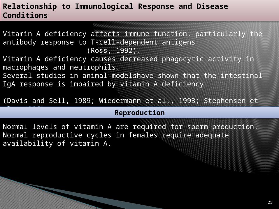
Relationship to Immunological Response and Disease Conditions
Vitamin A deficiency affects immune function, particularly the antibody response to T-cell–dependent antigens
(Ross, 1992).Vitamin A deficiency causes decreased phagocytic activity in macrophages and neutrophils.Several studies in animal modelshave shown that the intestinal IgA response is impaired by vitamin A deficiency
(Davis and Sell, 1989; Wiedermann et al., 1993; Stephensen et al., 1996).
Reproduction
Normal levels of vitamin A are required for sperm production.Normal reproductive cycles in females require adequate availability of vitamin A.
25
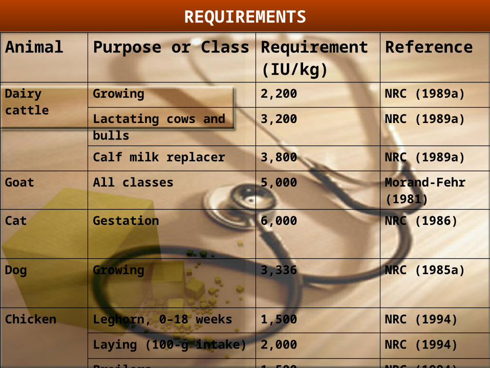
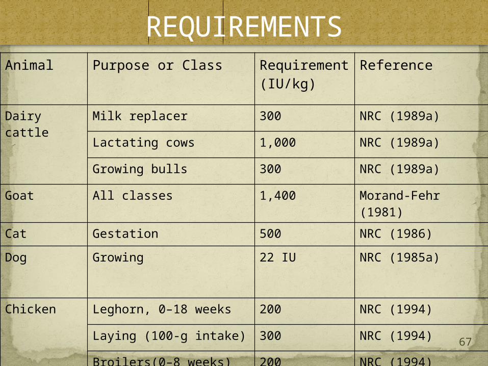
REQUIREMENTS
Animal Purpose or Class Requirement(IU/kg)
Reference
Dairy cattle
Growing 2,200 NRC (1989a)
Lactating cows and bulls
3,200 NRC (1989a)
Calf milk replacer 3,800 NRC (1989a)
Goat All classes 5,000 Morand-Fehr (1981)
Cat Gestation 6,000 NRC (1986)
Dog Growing 3,336 NRC (1985a)
Chicken Leghorn, 0–18 weeks 1,500 NRC (1994)
Laying (100-g intake) 2,000 NRC (1994)
Broilers 1,500 NRC (1994)

Deficiency symptomsBitot's spots- Mild vitamin A deficiency may result in changes in the conjunctiva (corner of the eye) EFFECT ON REPRODUCTION:-•Deficiency of vitamin A can lead to infertility or sterility in male •Deficiency of vitamin A can lead to vaginitis, abnormal oestrous cycle, early embryonic mortality, abortion and defective formation of foetus in females. •EFFECT ON CEREBROSPINAL FLUID PRESSURE:One of the initial effects of vitamin A deficiency is elevated cerebrospinal fluid (CSF) pressure. The mechanism whereby the increase in CSF pressure is brought by thickened duramater leading to under absorption of CSF. EFFECT ON BONE FORMATION- vitamin A can lead to developmental bone deformities. EFFECT ON IMMUNE SYSTEM-Vitamin A is commonly known as the anti-infective vitamin, because it is required for normal functioning of the immune system. The skin and mucosal cells (cells that line the airways, digestive tract, and urinary tract) function as a barrier and form the body's first line of defense against infection.
27

ANTI-INFECTIVE VITAMIN –Vitamin A is involved in the formation and protection of epithelial cells. Damage to epithelial cells can cause easy entry of pathogenic microbes leading to infection. So infection of gastrointestinal tract, respiratory tract, urogenital tract and skin is common in Vitamin A deficiency. As vitamin A helps to prevent these infections it is called anti infective vitamin. CONGENITAL BLINDNESS-Vitamin A is needed for bone formation. If vitamin A is deficient optic foramen is not formed properly. Small size optic foramen leads to the constriction of optic nerve. Permanent damage to the nerve can lead to permanent blindness. XEROPHTHALMIA (DRY EYE)-Xerophthalmia is characterized by changes in the cells of the cornea that ultimately result in corneal opacity, keratinization of the cornea, corneal ulcers, scarring, and blindness.
28

Deficiency Symptoms Night blindness Xeropthalmia Keratinization of epithelium Reproductive performance Nervous lesionsRetinoic acid :unable to fullfillThe function of Vit a in vision And reproduction although itMaintain normal growth.
29

Fig. Vitamin A-deficient calf. Note the emaciated appearance and evidence of diarrhea. The calf also shows excessive lacrimation and nasal discharge characteristic of vitamin A deficiency. 30

Dry condition of cornea and conjuctiva,cloudiness and ulceration
Softning of cornea Thickned cornea Bitot spot (human) White foamy patches on
white portion of eye
XEROPTHALMIA
31

Occulonasal discharge Conjuctivitis Sticking of eyelids Presence of cheesy partical in eye and nasal sinus
Swelling of face
Nutritional Roup
Egg production and hatchability
decreased
•Poor hatchabilitylili•Poor feather development•Fall in egg production•Retarded growth
32

Fig. Advanced stage of anasarca in hindquarters of vitamin A-deficient steer.33

Fig. Vitamin A-deficient calf shows incoordination and weakness. 34
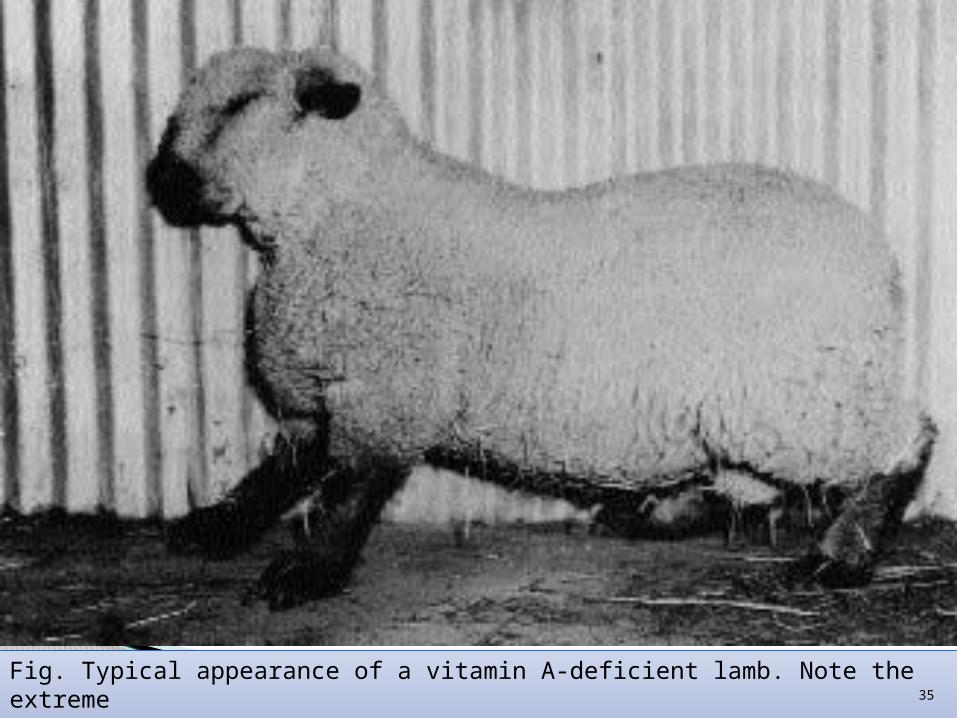
Fig. Typical appearance of a vitamin A-deficient lamb. Note the extremeweakness and swayed back. This was followed by the inability to stand. 35

Fig. Vitamin A deficiency in growing pigs: (A) partial paralysis and seborrhea, (B) initial stage of spasm, (C) lordosis and weakness of hind legs.
A
B
C
36

Keratinization of epithelium
Respiratory trouble-cold and sinus inf
GIT disorder-Diarrhoea
Genitourinary-kidney and bladder stone.
Deposition of urate –on heart,pericardium,liver and spleen
Nervous lesionSkeletal growth retarded but nervous tissue and brain grows-there is pressure on nervous tissue-increased CSF
SEVERE ATAXIA 37
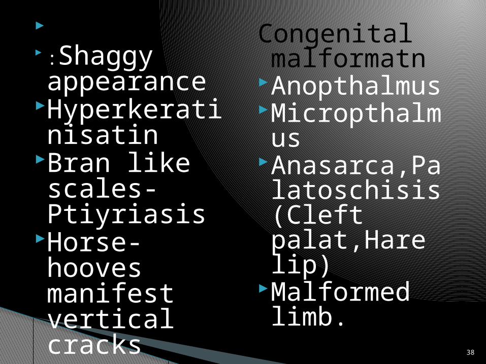
Skin :Shaggy appearance
Hyperkeratinisatin
Bran like scales-Ptiyriasis
Horse-hooves manifest vertical cracks
Congenital malformatn
AnopthalmusMicropthalmusAnasarca,Palatoschisis(Cleft palat,Hare lip)
Malformed limb.
38

Decline in sexual maturity.Decreased no.of
spermatozoaDecrease in motilityFailure of spermatogenesisDegeneration of germinal
epithelium and seminiferous tubule.
Oestrus is disturbed Abortion or birth of dead ,weak or
abnormal offspringThickning of vaginal epitheliumRetension of placenta.
Reproductive Performance
39
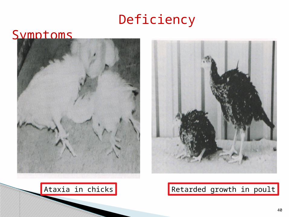
Deficiency Symptoms
Ataxia in chicks Retarded growth in poult
40

Ricket and OsteomalaciaPain in bone due to calcificationSoreness of corners of mouth and coarseness of hair
Exostoses in various placesIn human-Persistant chronic headache
Distorted vision.
HYPERVITAMINOSIS A
41
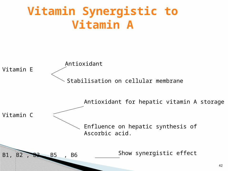
Vitamin Synergistic to Vitamin A
Antioxidant
Stabilisation on cellular membrane
Vitamin C
Antioxidant for hepatic vitamin A storage
Enfluence on hepatic synthesis of Ascorbic acid.
Vitamin E
B1, B2 , B3 , B5 , B6 Show synergistic effect
42
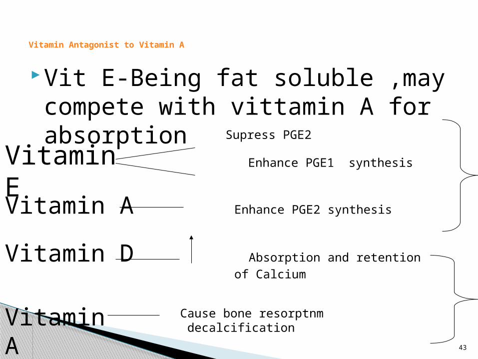
Vit E-Being fat soluble ,may compete with vittamin A for absorption
Vitamin Antagonist to Vitamin A
Supress PGE2
Enhance PGE1 synthesisVitamin E
Vitamin A Enhance PGE2 synthesis
Vitamin D Absorption and retention of Calcium
Vitamin A Cause bone resorptnm decalcification
43

Mineral synergistic to vitamin A
Involved in regulation of vit AAdequate amt are required for the mobilization Of Vitamin A from liverInvolved in maintaining plasma RBPSpecific transport
Zinc
SeleniumSynergistic(antioxidant)
Antagonistic
Iodine In the form of T4
Synergistic
Antagonistic
44

ADH-Increase mobilization of vitamin A from liver
Thyroxin-……………..Estogen……………
HARMONE
45

46
NATURAL SOURCESVitamin A (Retinol) (IU/g)Content of Feeds
Whale liver oil 400,000 Barracuda liver oil
12,000
Swordfish liver oil 250,000 Dogfish liver oil 12,000
Halibut liver oil 240,000 Seal liver oil 10,000
Herring liver oil 211,000 Cod liver oil 4,000
Tuna liver oil 150,000 Sardine body oil
750
Shark liver oil 150,000 Pilchard body oil
500
Bonito liver oil 120,000 Menhaden body oil
340
White sea bass liver oil
50,000 Butter 35
Eggs 10 Cheese 14
Milk 1.5Sources: Adapted from Scott et al. (1982) and Maynard et al. (1979).

47
Carotene Source (mg/kg))Content of FeedsFresh green legumes and grasses, immature (wet basis) 33–88
Dehydrated alfalfa meal, fresh, dehydrated without field curing, very bright green
242–297
Dehydrated alfalfa meal after considerable time in storage, bright green
110–154
Alfalfa leaf meal, bright green 120–176
Legume hays, including alfalfa, very quickly cured with minimum sun exposure, bright green, leafy
77–88
Legume hays, including alfalfa, good green color, leafy 40–59
Legume hays, including alfalfa, partly bleached, moderate amount of green color
20–31
Legume hays, including alfalfa, badly bleached, or discolored, traces of green color
9–18
Non-legume hays, including timothy, cereal, and prairie hays, well cured, good green color
20–31
Non-legume hays, average quality, bleached, some green color
9–18
Legume silage (wet basis) 11–44
Corn and sorghum silages, medium to good green color (wet basis
4–22
Grains, mill feeds, protein concentrates, and by-product concentrates, except yellow corn and its by-products
0.02–0.44

Vitamin D
48

History
Chemical Structure & Properties
1919-Sir Edward Mellanby was able to experimentally produce rickets in puppies by feeding synthetic diets
-He further showed that rickets could be prevented by the addition of cod-liver oil or butterfat to the feed.1922- McCollum showed that the antirachitic factor in cod-liver oil could survive both aeration and heating to 1000C for 14 h whereas the activity of vitamin A was destroyed by this treatment. McCollum named the new substance vitamin D
Dietary source of vitamin D-steroid, ergosterol
Occurs as colourless crystals.
Insoluble in water.
Readily soluble in alcohol and other organic solvents.
Less soluble in vegetable oils.
Destroyed by overtreatment wit uv light and by peroxidation in the presence of rancidifying polyunsaturated fatty acids.
Sunshine Vitamin
49

Ergocalciferol (C₂₈H₄₄O) In plant present as Vitamin D2 (Ergocalciferol). Ergocalciferol is derived from a common plant steroid, ergosterol,
and is the usual dietary source of vitamin D.Three double bondsmelting point-121⁰CMolecular weight-384.65 Insoluble in H₂OSoluble in benzene, chloroform, ethanol, and acetone.Unstable in lightWill undergo oxidation if exposed to air at 24⁰C for 72 h.Stored at 0⁰c.
Cholecalciferol (C₂₇H₄₄O) In animal present as Vitamin D3 (Cholecalciferol). Precursor is 7-
ehydrocholesterolFour double bondsmelting point-121⁰C Insoluble in H₂OSoluble in benzene, chloroform, ethanol, and acetone.Unstable in lightWill undergo oxidation if exposed to air at 24⁰C for 72 h.Stored at 0⁰c.
Chemical Properties
50

51

Side Chains of Provitamin D; It Includes Structures of the Side Chains of Vitamins D2 D7Provitamin Trivial Name
Vitamin D Produced upon Irradiation
Empirical Formula (Complete Steroid)
Side ChainStructure
Ergosterol D2 C28H44O7-dehydrocholesterol D3 C27H44O
22,23-dihydroergosterol
D4 C28H46O
7-dehydrositosterol D5 C29H48O
7-dehydrostigmasterol D6 C29H46O
7-dehydrocampesterol D7 C28H46O52

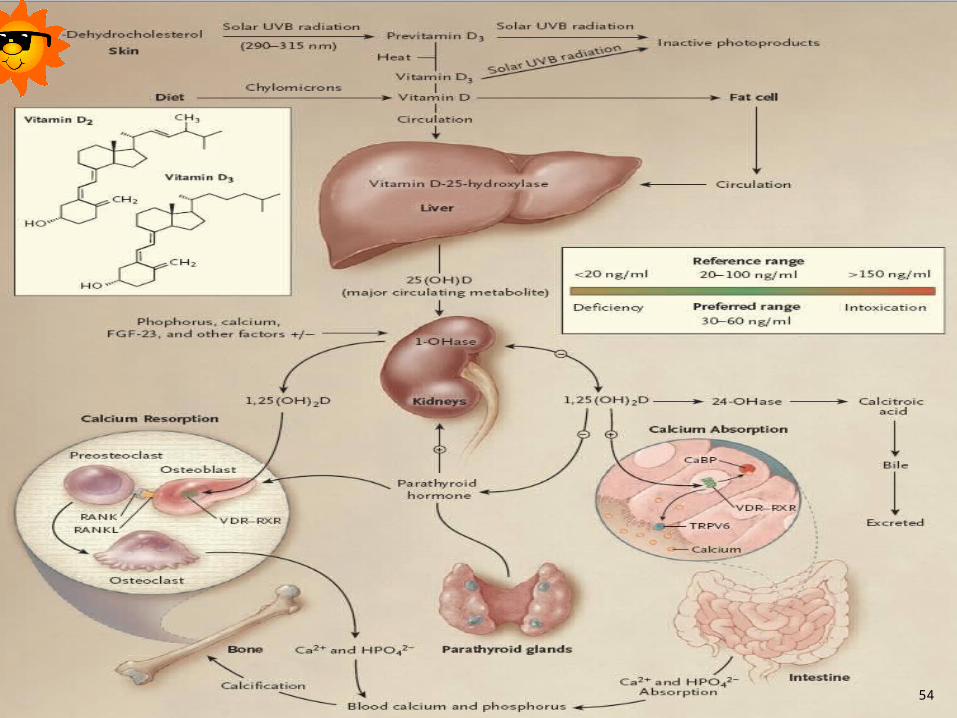
METABOLISMAbsorption
Cholecalciferol is absorbed from the intestinal tract primarily in the duodenum.
It is poorly absorbed in the absence of bile, the secretion of which is limited in young chicks.
Highest concentration found in the- intestinal wall, liver, kidneys, spleen, gall bladder and serum.
While concentration in muscle, bone, pancreases and skin are low, these tissue account for large proportion of the stored vitamin D.Vitamin D₃ absorption is enhanced by certain organic acids, especially lactic acid.
However, the effect of organic acid in improving calcium absoption may be independent of Vitamin D.
Half life of Vitamin D₃-25days in birds while that of 25(OH)D₃ is closer to 20 days and the half life of 24,25(OH) ₂D₃ is only around 2days 53
54
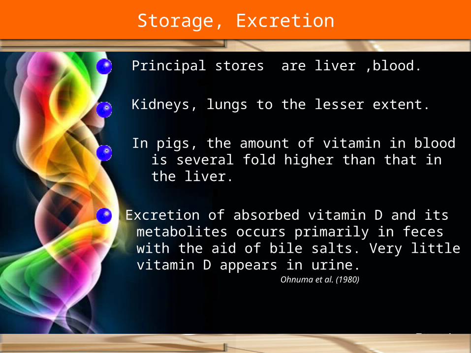
Principal stores are liver ,blood.
Kidneys, lungs to the lesser extent.
In pigs, the amount of vitamin in blood is several fold higher than that in the liver.
Excretion of absorbed vitamin D and its metabolites occurs primarily in feces with the aid of bile salts. Very little vitamin D appears in urine.
Ohnuma et al. (1980)
Storage, Excretion

Endocrine, paracrine and intracrine functions of Vitamin D
56
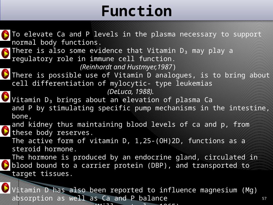
FunctionTo elevate Ca and P levels in the plasma necessary to support normal body functions.There is also some evidence that Vitamin D₃ may play a regulatory role in immune cell function.
(Reinhardt and Hustmyer,1987)There is possible use of Vitamin D analogues, is to bring about cell differentiation of mylocytic- type leukemias
(DeLuca, 1988).Vitamin D₃ brings about an elevation of plasma Caand P by stimulating specific pump mechanisms in the intestine, bone,and kidney thus maintaining blood levels of ca and p, from these body reserves.The active form of vitamin D, 1,25-(OH)2D, functions as a steroid hormone.The hormone is produced by an endocrine gland, circulated in blood bound to a carrier protein (DBP), and transported to target tissues.
Vitamin D has also been reported to influence magnesium (Mg) absorption as well as Ca and P balance
(Miller et al., 1965).Intestinal Effects- It is well known that vitamin D stimulates active transport of Ca and P across intestinal epithelium. 57

Bone Effects-Vitamin D plays roles both in the mineralization of bone as well as demineralization or mobilization of bone mineral. 1,25-(OH)2D is one of the factors controlling the balance between bone formation and resorption.
In young animals during bone formation, minerals are deposited on the matrix. This is accompanied by an invasion of blood vessels that gives rise to trabecular bone. This process causes bones to elongate. During vitamin D deficiency, this organic matrix fails to mineralize, causing rickets in the young and osteomalacia in adults.
Another role of vitamin D has been proposed in addition to its involvement in bone; namely, in the biosynthesis of collagen in preparation for mineralization. (Gonnerman et al., 1976).
Kidney Effects-There is evidence that vitamin D functions in the distal renal tubules to improve Ca reabsorption and is mediated by calbindin.
(Bronner and Stein, 1995).
1,25-(OH)2D3 functions in improving renal reabsorption of Ca.(Sutton and Dirks, 1978).
58

Other Vitamin D Functions-Vitamin D has also been shown to be required for chick embryonic development.
1,25-(OH)2D is also essential for the transport of eggshell Ca to the embryo across the chorioallantoic membrane
(Elaroussi et al., 1994).
In the pancreas, 1,25-(OH)2D is essential for normal insulin secretion.More than 50 genes have been reported to be transcriptionally regulated by 1,25-(OH)2D.
(Hannah and Norman, 1994).
The actions of 1,25-(OH)2D3 are recognized as being involved in regulation of the growth and differentiation of a variety of cell types, including those of hematopoietic and immune systems.
(Lemire, 1992).
A deficiency of vitamin D may promote prostate cancer. (Skowronski et al., 1995).
59

Deficiency
Rickets- generally characterized by a decreased concentration of Ca and P in the organic matrices of cartilage and bone.
Osteomalacia- is characterized by a decreased concentration of Ca and P in the bone matrix
Osteoporosis-is defined as a decrease in the amount of bone, leading to fractures after minimal trauma.
vitamin D deficiency occurs in animals includes the following characteristics
1. Failure of Ca salt deposition in the cartilage matrix.2. Failure of cartilage cells to mature, leading to their
accumulation rather than destruction.3. Compression of the proliferating cartilage cells.4. Elongation, swelling, and degeneration of proliferative
cartilage.5. Abnormal pattern of invasion of cartilage by
capillaries. (Kramer and Gribetz, 1971)
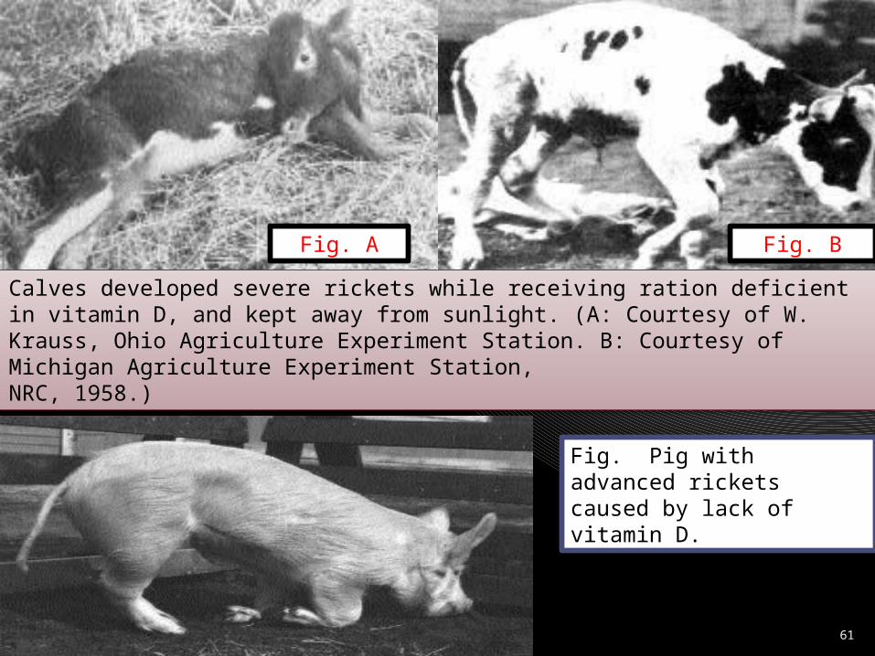
Calves developed severe rickets while receiving ration deficient in vitamin D, and kept away from sunlight. (A: Courtesy of W. Krauss, Ohio Agriculture Experiment Station. B: Courtesy of Michigan Agriculture Experiment Station,NRC, 1958.)
Fig. A Fig. B
Fig. Pig with advanced rickets caused by lack of vitamin D.
61
62
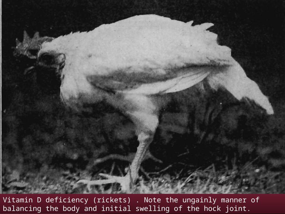
Vitamin D deficiency (rickets) . Note the ungainly manner of balancing the body and initial swelling of the hock joint.
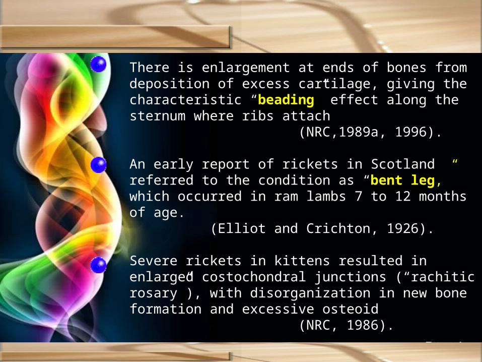
There is enlargement at ends of bones from deposition of excess cartilage, giving the characteristic “beading” effect along the sternum where ribs attach
(NRC,1989a, 1996).
An early report of rickets in Scotland referred to the condition as “bent leg,” which occurred in ram lambs 7 to 12 months of age.
(Elliot and Crichton, 1926).
Severe rickets in kittens resulted in enlarged costochondral junctions (“rachitic rosary”), with disorganization in new bone formation and excessive osteoid
(NRC, 1986).

64
ToxicityDogs-receiving toxic concentrations of vitamin D exhibited anorexia, polyuria, bloody diarrhea, polydipsia, prostration, and excessive calcification of the lungs
(Morgan, 1947)
In poultry, excess vitamin D elevates 1,25-(OH)2D with hypercalcemia and soft tissue mineralization.
(NRC, 1994).
Harrington and Page (1983) compared toxicity of D2 to D3 in horses. Signs of toxicity included weight loss, hypercalcemia, hyperphosphatemia, and cardiovascular calcinosis.
A condition known as “humpy-back,” in which clinical symptoms reminiscent of calcinosis occur, may be caused by sheep grazing the fruits of S. esuriale in Australia. In Jamaica, “Manchester wasting disease” and in Hawaii, “Naalehu disease” are conditions seen in cattle that are virtually identical to enteque seco in relation to clinical and pathological signs.
(Wasserman, 1975; Arnold and Fincham, 1997).
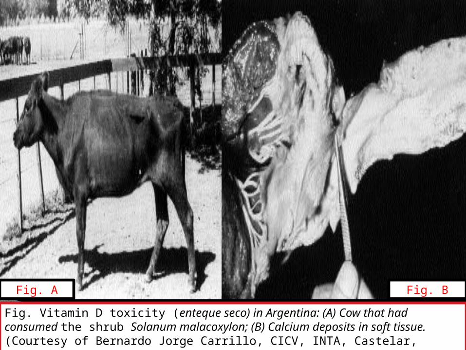
Fig. Vitamin D toxicity (enteque seco) in Argentina: (A) Cow that had consumed the shrub Solanum malacoxylon; (B) Calcium deposits in soft tissue. (Courtesy of Bernardo Jorge Carrillo, CICV, INTA, Castelar, Argentina.)
Fig. A Fig. B
65

Estimation of Safe Upper Dietary Levels of Vitamin D3 (IU/kg in Diet) for Animals
Animal DietaryRequirement
Exposure Time
< 60 days > 60 days
Chicken 200 40,000 2,800
Horse 400 2,200
Sheep 275 25,000 2,200
Swine 220 33,000 2,200
Source: Modified from NRC (1987). 66
67
REQUIREMENTSAnimal Purpose or Class Requirement
(IU/kg)Reference
Dairy cattle Milk replacer 300 NRC (1989a)
Lactating cows 1,000 NRC (1989a)
Growing bulls 300 NRC (1989a)
Goat All classes 1,400 Morand-Fehr (1981)
Cat Gestation 500 NRC (1986)
Dog Growing 22 IU NRC (1985a)
Chicken Leghorn, 0–18 weeks 200 NRC (1994)
Laying (100-g intake) 300 NRC (1994)
Broilers(0–8 weeks) 200 NRC (1994)

68
NATURAL SOURCESVitamin D (Ergocalciferol )(D2) IU/100 g Concentrations in Various
Foods and Feedstuffs
Food or Feedstuff Red clover, fresh
4.7
Alfalfa pasture 4.6 Red clover, sun cured
192
Alfalfa hay, sun cured 142 Sorghum grain 2.6
Alfalfa silage 12 Sorghum silage 66
Alfalfa wilted silage 60 Corn silage 13
Birdsfoot trefoil hay, sun cured
142 Molasses, sugar beet
58
Barley straw 60
Cocoa shell meal, sun dried
3,500
Sources: Adapted from Scott et al. (1982) and Maynard et al. (1979).

69
Vitamin D (Cholecalciferol D3) IU/100 g Concentrations in Various Foods and Feedstuffs
Blue fin tuna liver oil 4,000,000 Milk, cow’s whole (winter)
1
Cod liver oil meal 4,000 Sardine, entire body oil
8,000
Cod liver oil 10,000 Swordfish liver oil
1,000,000
Eggs 100
Halibut liver oil 120,000
Herring, entire body oil
10,000
Menhaden, entire body oil
5,000
Milk, cow’s whole (summer)
4Sources: Adapted from NRC (1982b) and Scott et al. (1982).

Vitamin E
70

History
Chemical Structure & Properties
1922-Discovered by Evans and Bishop1936: Evans et al, Isolated α-tocopherol1960s: Vitamin E deficiency was described in children with fat malabsorption syndromes.1980s: Major symptom of vitamin E deficiency in Human was a peripheral neuropathy.Greek -‘‘tokos’’ (offspring) and ‘‘pherein’’ (to bear) with an ‘‘ol’’ to indicate that it was an alcohol
Tocopherols and Tocotrienols is closely related to Vitamin EEight forms of vitamin E
Tocopherols(Saturated )- α, β, γ, and δ.Tocotrienols(Unsaturated )- α, β, γ, and δ.
α- tocopherols -most biologically active-yellow oil i.e. insoluble in water -soluble in organic solvents.
a-tocopherol > potent than b > g > dTocotrienols (trienols)
unsaturated side chainsonly a has significant biological activity
Synthetic-α tocopheryl acetate.

Fig. Vitamin E structures are shown. The methyl groups on the chromanol head determine whether the molecule is a-, b- or g-, or d-, while the tail determines whether the molecule is a tocopherol or a tocotrienol. 72

Tocopherols-extremely resistant to heat but are easily oxidized, destroyed by peroxides, ozone
Resistant to acids (anaerobic) and basesVitamin E is a potent peroxyl radical scavenger and especially protects PUFA within phospholipids of biological membranes and in plasma lipoproteins.
73

METABOLISM
Vitamin E absorption is related to fat digestion and is facilitated bybile and pancreatic lipase .
(Sitrin et al., 1987).Most vitamin E is absorbed as the alcohol
Absorption and Transport
Storage and ExcretionVitamin E is stored throughout all body tissues; major deposits are in adipose tissue, liver, and muscle, with highest storage in the liver..
The major route of excretion of ingested vitamin E is fecal elimination.
Usually less than 1% of orally ingested vitamin E is excreted in the urine. 74
Placental and Mammary Transfer
Vitamin E does not cross the placenta in any appreciable amounts; however, it is concentrated in colostrum (Van Saun et al., 1989). With respect to neonatal ruminants (Hidiroglou et al., 1969; Van Saun et al.,1989) and baby pigs (Mahan, 1991).
The importance of providing colostrum rich in vitamin E is quite apparent, as both calves and lambs are born with low levels of the vitamin
(Nockels, 1991; Njeru et al., 1994a).
Low blood vitamin E may lead to diminished disease resistance and immune response in the neonate
(Nockels, 1991).

FUNCTIONSa-Tocopherol decreased the release of proinflammatory cytokines and chemokines (IL-8 and plasminogen activator inhibitor-1 [PAI-1]).
(Singh et al.,2005).
Vitamin E has been shown to be essential for integrity and optimumfunction of the reproductive, muscular, circulatory, nervous, andimmune systems.
(Hoekstra, 1975; Sheffy and Schultz, 1979; Bendich,1987; McDowell et al., 1996).
Vitamin E as a Biological Antioxidant
One of the most important functions is its role as an intercellular and intracellular antioxidant.
Vitamin E is part of the body’s intracellular defense against the adverse effects of reactive oxygen and free radicals that initiate oxidation of unsaturated phospholipids (Chow, 1979) and critical sulfhydryl groups (Brownlee et al., 1977). 76
Semen quality of boars was improved with Se and vitamin E supplementation, with vitamin E playing a role in maintaining sperm integrity in combination with Se
(Marin-Guzman et al., 1989).
Membrane Structure and Prostaglandin Synthesis
α-Tocopherol may be involved in the formation of structural components of biological membranes, thus exerting a unique influence on architecture of membrane phospholipids
(Ullrey, 1981).
It is reported that α-tocopherol stimulated the incorporation of 14C from linoleic acid into arachidonic acid in fibroblast phospholipids. Also, it was found that α-tocopherol exerted a pronounced stimulatory influence on formation of prostaglandin E from arachidonic acid, while a chemical antioxidant had no effect.
77

Blood Clotting
Vitamin E is an inhibitor of platelet aggregation in pigs.
(McIntosh et al., 1985),
May play a role by inhibiting peroxidation of arachidonic acid, which is required for formation of prostaglandins involved in platelet aggregation
(Panganamala and Cornwell, 1982; Machlin,1991).
Disease Resistance
Both in vitro and in vivo studies showed that the antioxidant vitamins generally enhance different aspects of cellular and noncellular (humoral) immunity.
One function of vitamin C is that this vitamin can regenerate the reduced form of α-tocopherol, perhaps accounting for observed sparing effects of these vitamins .
(Jacob, 1995; Tanaka et al., 1997).
78

Both vitamin E and Se may help these cells to survive the toxic productsthat are produced in order to effectively kill ingested bacteria.
(Badwey and Karnovsky, 1980).
Vitamin E has been implicated in stimulation of serum antibody synthesis, particularly IgG antibodies.
(Tengerdy, 1980).
Vitamin E deficiency allows a normally benign virus to cause disease .(Beck et al., 1994).
Selenium or vitamin E deficiency leads to a change in viral phenotype, such that an avirulent strain of a virus becomes virulent and a virulent strain becomes more virulent.
(Beck, 1997).
79

Relationship to Toxic Elements or Substances
Both vitamin E and Se provide protection against toxicity with three classes of heavy metals
(Whanger, 1981).
Vitamin E can be effective against other toxic substances. For example, treatment with vitamin E gave protection to weanling pigs against monensin-induced skeletal muscle damage .
(Van Vleet et al.,1987).
Relationship with Selenium in Tissue Protection
Vitamin E prevents fatty acid hydroperoxide formation, sulfur amino acids are precursors of glutathione peroxidase, and Se is a component of glutathione peroxidase.
(Smith et al., 1974).In diets severely deficient in Se, vitamin E does not prevent or cure exudative diathesis, whereas addition of as little as 0.05 ppm Se completely prevents this disease.
(Scott, 1980).80
Other Functions1. Normal phosphorylation reactions, especially of high-energy2. phosphate compounds, such as creatine phosphate and adenosine
triphosphate3. A role in synthesis of vitamin C and ubiquinone 4. Role in sulfur amino acid metabolism.
Scott et al. (1982)
A deficiency of vitamin E interferes with conversion of vitamin B12 to itscoenzyme 5′-deoxyadenosylcobalamin and concomitantly metabolismof methylmalonyl-CoA to succinyl-CoA.
Pappu et al. (1978)
In rats, vitamin E deficiency has been reported to inhibit vitamin D metabolism in the liver and kidneys with the formation of active metabolites and decreases in the concentration of the hormone-receptor complexes in the target tissue. Liver vitamin D hydroxylase activity decreased by 39%, 25-OHD3 1-hydroxylase activity in the kidneys by 22%, and 24-hydroxylase activity by 52%
(Sergeev et al., 1990).
81

Animal Purpose or Class Requirement(IU/kg)
Reference
Dairy cattle Milk replacer 40 NRC (1989a)
Lactating cows 25 NRC (1989a)
Growing bulls 100 NRC (1989a)
Goat All classes 100 Morand-Fehr (1981)
Cat Gestation 30 NRC (1986)
Dog Growing 22 NRC (1985a)
Chicken Leghorn, 0–6weeks 10 NRC (1994)
Leghorn, 6-18weeks 5
Laying (100-g intake) 5 NRC (1994)
Broilers(0–8 weeks) 10 NRC (1994)
REQUIREMENTS

DEFICIENCY
Nutritional myopathy / white muscle disease / stiff lamb disease / mulberry heart disease / exudative diathesis / crazy chick disease
The most frequent and the most important manifestation of Selenium deficiency in farm animals is muscle degeneration (myopathy).
Nutritional myopathy , also known as muscular dystrophy, frequently occurs in cattle, particularly calves.
The myopathy primarily affects the skeletal muscles and the affected animals have weak leg muscles, a condition manifested by difficulty in standing and, after standing, a trembling and staggering gait.

Eventually, the animals are unable to rise and weakness of the neck muscles prevents them from raising their heads.
A popular descriptive name for this condition is white muscle disease. The heart muscle may also be affected and death may result.
Nutritional myopathy also occurs in lambs, with similar symptoms to those of calves. The condition is frequently referred to as stiff lamb disease.
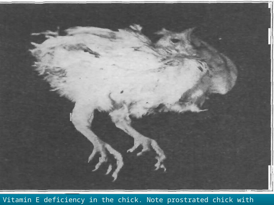
Vitamin E deficiency in the chick. Note prostrated chick with retracted head.

Fig. Vitamin E-selenium deficiency in cattle is manifested as white muscle disease or necrosis of the gastrocnemius muscle; chalky white streaks are evident in the belly of the muscle 86

Chicks:- In nutritional myopathy the main muscles affected are the
pectorals although the leg muscles also may be involved.
Nutritional encephalomalacia or crazy chick disease is a condition in which the chick is unable to walk or stand, and is accompanied by hemorrhages and necrosis of brain cells.
Exudative diathesis is a vascular disease of chicks characterized by a generalized oedema of the subcutaneous fatty tissues, associated with an abnormal permeability of the capillary walls.
Both selenium and vitamin E appear to be involved in nutrition myopathy and in exudative diathesis but selenium does not seem to be important in nutritional encephalomacia.

In pigs, the two main diseases associated with vitamin E and selenium deficiency are myopathy and cardiac disease.
The pigs demonstrate an uncoordinated staggering gait, or are unable to rise.
The pigs heart muscle is more commonly affected.
Sudden cardiac failure occurs and on post-mortem examination the lesions of the cardiac muscles are seen as pale patches or white streaks. This condition is commonly known as mulberry heart disease.

Fig. Vitamin E-selenium deficiency is seen as flexion of the hock and fetlockjoints as a result of decreased support by the gastrocnemius muscle, which is severely affected by myodegeneration 89

TOXICITYHypervitaminosis E studies in rats, chicks, and humans indicate maximum tolerable levels in the range of 1,000 to 2,000 IU/kg of diet
(NRC, 1987).
In chickens, the effects of vitamin E toxicity are depressed growth rate, reduced hematocrit, reticulocytosis, increased prothrombin time (corrected by injecting vitamin K), and reduced calcium and phosphorus in dry, fat-free bone ash
(NRC, 1987).
90

Tocopherols in Selected Feedstuffs (ppm)
Source: Modified from Ullrey (1981).
Feedstuff α β γ δ
Barley 4 3 0.5 0.1
Corn 6 ---- 38 Trace
Oats 7 2 3 ---
Rye 8 9 -- 0.8
Wheat 10 9 ---- 0.8
Corn oil 112 50 602 18
Cottonseed oil 389 --- 387 ---
Wheat germ oil 1,330 710 260 271
Palm oil 256 ---- 316 70
Safflower oil 387 --- 174 40
Soybean oil 101 ---- 593 264
NATURAL SOURCES
Vitamin K
92

History
Chemical Structure & Properties
1935- Discovered by Henrik Dam
Vitamin K extracted from plant- Phylloquinone (Vitamin K1) .Synthesised by intestinal bacteria- Menaquinones (Vitamin K2).Synthetic-Menadione (Vitamin K3).Vitamin K is a golden yellow viscous oil.Vitamin K1 is slowly degraded by atmospheric oxygen but fairly rapidly destroyed by light.stable to heat, destroyed by sunlight & alkali.melting points 35ºC to 60ºC
Phylloquinone.Synonym
93

Fig. Structures of some compounds with vitamin K activity. 94

AntagonistsDicumarol.
By feeding sulfonamides (in monogastric species) at levels sufficient to inhibit intestinal synthesis of vitamin k.
Mycotoxins, toxic substances produced by molds, are also antagonists causing vitamin k deficiency.
In cattle, vitamin k1 is much more potent than vitamin k3 as an antidote to dicumarol.
(Goplen and Bell 1967)
95
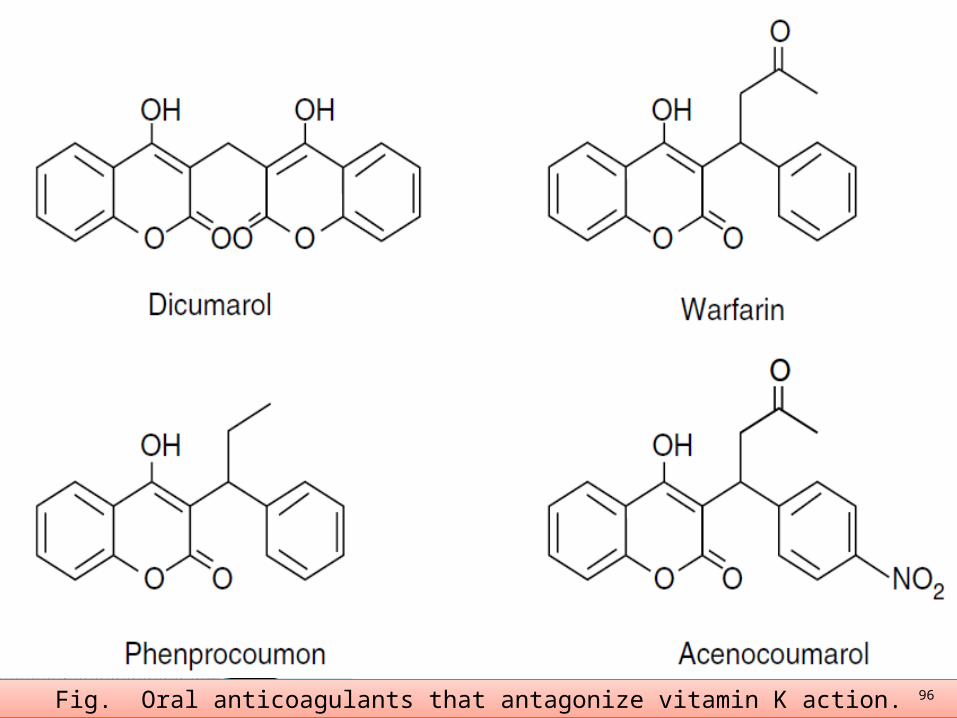
Fig. Oral anticoagulants that antagonize vitamin K action. 96

Fig. Other vitamin K antagonists 97

METABOLISMAbsorption and Transfer
Absorption of vitamin K depends on its incorporation into mixed micelles, and optimal formation of these micellar structures requires the presence of both bile and pancreatic juice.
Storage and Excretion
Vitamin K -stored liver.Excreted in the urine.
The lymphatic system is the major route of transport of absorbed phylloquinone from the intestine in mammals but by portal circulation in birds, fishes, and reptiles.
(Shearer et al. 1970)
98
FUNCTIONS
The vitamin is required for the synthesis of the active form of prothrombin (factor II) and other plasma clotting factors (VII, IX, and X).These four blood-clotting proteins are synthesized in the liver in inactive precursor forms (zymogens) and then converted to biologically active proteins by the action of vitamin K
(Suttie and Jackson, 1977).
Blood Coagulation
99

Fig. Scheme involving blood clotting. The vitamin K-dependent factors(synthesis of each is inhibited by dicumarol) include factor IX, Christmas factor;factor X, Stuart-Prower factor; factor VII, proconvertin; and factor II, prothrombin.
100

The metabolic function of vitamin K is as the coenzyme in the carboxylation of protein-incorporated glutamate residues to yield γ-carboxyglutamate thus converting inactive precursor proteins to biological activity.
Carboxylation allows prothrombin and the other procoagulant proteins to participate in a specific protein-calcium-phospholipid interaction that is necessary for their biological role
(Suttie andJackson, 1977).
Calcium binding proteins

DEFICIENCY
The major clinical sign of vitamin K deficiency in all species is impairment of blood coagulation.
Sweet clover poisoning / hemorrhagic sweet clover disease
Dicumarols are produced by molds, particularly those that attack sweet clover hay, thus giving rise to the term sweet clover disease.
During the process of spoiling, harmless natural coumarins in sweet clover are converted to dicumarol (bis-hydroxycoumarin), and when toxic hay or silage is consumed by animals, hypoprothrombinemia results, presumably because dicumarol combines with the proenzyme to prevent formation of the active enzyme required for the synthesis of prothrombin. It probably also interferes with synthesis of factor VII and other coagulation factors.
In an experiment with calves, dicumarol poisoning was produced by feeding naturally spoiled, sweet clover hay that contained a minimum of 90 mg dicumarol per kilogram of hay.
(Alstad et al., 1985).102

Poultry
Generalized hemorrhage due to severe vitamin K deficiency in a young chick. 103
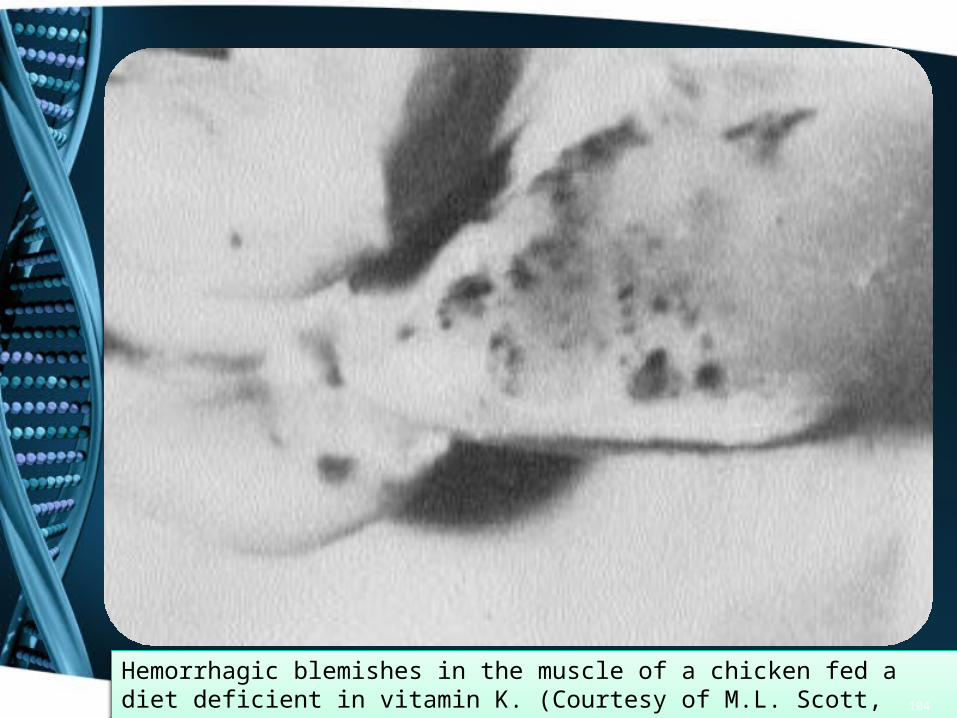
Hemorrhagic blemishes in the muscle of a chicken fed a diet deficient in vitamin K. (Courtesy of M.L. Scott, Cornell University.) 104

Rabbits
When a vitamin K-deficient diet was fed to pregnant rabbits, the result was placental hemorrhage and abortion of young
(NRC, 1977).
Dogs
Vitamin K deficiency has been demonstrated in adult dogs following diversion of bile from the intestine by means of a cholecystonephrostomy .
(NRC, 1985a).
One study found that although blood clotting was impaired and there was a reduction in bone γ-carboxyglutamic acid concentration, vitamin K deficiency did not functionally impair skeletal metabolism of laying hens and their progeny
(Lavelle et al., 1994).
105
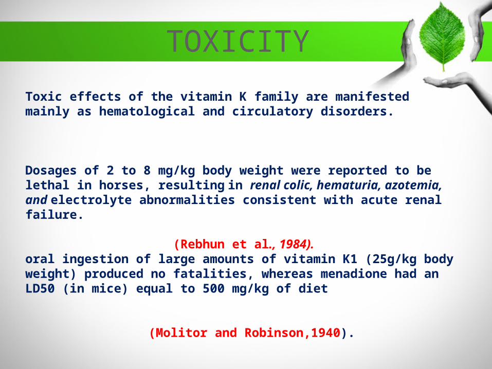
Toxic effects of the vitamin K family are manifested mainly as hematological and circulatory disorders.
Dosages of 2 to 8 mg/kg body weight were reported to be lethal in horses, resulting in renal colic, hematuria, azotemia, and electrolyte abnormalities consistent with acute renal failure.
(Rebhun et al., 1984).oral ingestion of large amounts of vitamin K1 (25g/kg body weight) produced no fatalities, whereas menadione had an LD50 (in mice) equal to 500 mg/kg of diet
(Molitor and Robinson,1940).
TOXICITY

107
REQUIREMENTSAnimal Purpose or Class Requirement
(mg/kg)Reference
Dairy cattle Adult Microbial synthesis NRC (1989a)
Beef cattle Adult Microbial synthesis NRC (1996)
Goat All classes Microbial synthesis NRC (1981)
Cat Gestation 0.1 NRC (1986)
Dog Growing 1 NRC (1985a)
Chicken Leghorn, 0–6weeks 0.5 NRC (1994)
Laying (100-g intake) 0.5 NRC (1994)
Broilers(0–8 weeks) 0.5 NRC (1994)
108
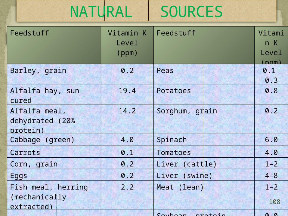
NATURAL SOURCES
(as fed basis)
Sources: From NRC (1982b) and Marks (1975).
Feedstuff Vitamin K Level (ppm)
Feedstuff Vitamin K
Level (ppm)
Barley, grain 0.2 Peas 0.1–0.3
Alfalfa hay, sun cured 19.4 Potatoes 0.8
Alfalfa meal, dehydrated (20% protein)
14.2 Sorghum, grain 0.2
Cabbage (green) 4.0 Spinach 6.0
Carrots 0.1 Tomatoes 4.0
Corn, grain 0.2 Liver (cattle) 1–2
Eggs 0.2 Liver (swine) 4–8
Fish meal, herring (mechanically extracted)
2.2 Meat (lean) 1–2
Soybean, protein concentrate (70.0% protein)
0.0

Vitamin B 1
Water Soluble Vitamins
(C12H17N4OSCl)
109
History
Chemical Structure & Properties
Synonym
Thiamine, Aneurin ,Thiamin
Thiamin is considered to be the oldest vitamin with the deficiency disease beri beri .the early history of thiamin can be found in sebrell and harris (1973) and loosely (1988). Beriberi was recognised in china as early as 2600 BC.In the 1890 eijkman, a dutch investigator, produced a paralysis in chickens fed boiled polished rice he called the condition “ polyneuritis” and observed the clinical sign were similar to Beriberi symptom in humans.
It consists of a molecule of--Pyrimidine-Thiazole linked by a methylene bridge & it contains both
nitrogen and sulfur atoms.It is isolated in pure form as the white thiamin hydrochloride.It soluble in water.Insoluble in fat solvents.It is very sensitive to alkali. Thiamin hydrochloride is more hygroscopic (takes up moisture).

• Thiamine chloride hydrochloride (the name is often shortened to thiamine hydrochloride) is a
• Colorless• Crystalline• Hygroscopic• Highly water-soluble substance• They have a characteristic pungent odour.
Antagonists
Thiamin-degrading enzymes (thiaminases).
The synthetic compounds pyrithiamine, oxythiamine, and amprolium (coccidiostat) are structurally similar antagonists, and their mode of action is competitive inhibition with iologically inactive compounds, thus interfering with thiamin at different points in metabolism.

• Heat-stable thiamin antagonists occur in a number of plants (e.g.,ferns and tea); these include polyphenols (e.g., caffeic acid and tannicacid), which oxidize the thiazole ring to yield the nonabsorbable thiamin disulfide.

METABOLISMDigestion
Adenosine triphosphate (ATP) provides the diphosphate moiety for the synthesis of thiamine diphosphate from free thiamine by the action of thiamine pyrophosphokinase.
Thiamine diphosphate can be metabolized either by dephosphorylation to form thiamine monophosphate, catalyzed by thiamine pyrophosphatase, or by further phosphorylation to give thiamine triphosphate, catalyzed by thiamine diphosphate–ATP phosphoryltransferase.
To a limited extent, free thiamine can be converted to thiamine monophosphate by an intestinal membrane alkaline phosphatase, in the presence of phosphate donors
Absorption
Absorption occurs in the small intestine, particularly in the jejunum Horse-cecum.Ruminants- Rumen
113
Transport
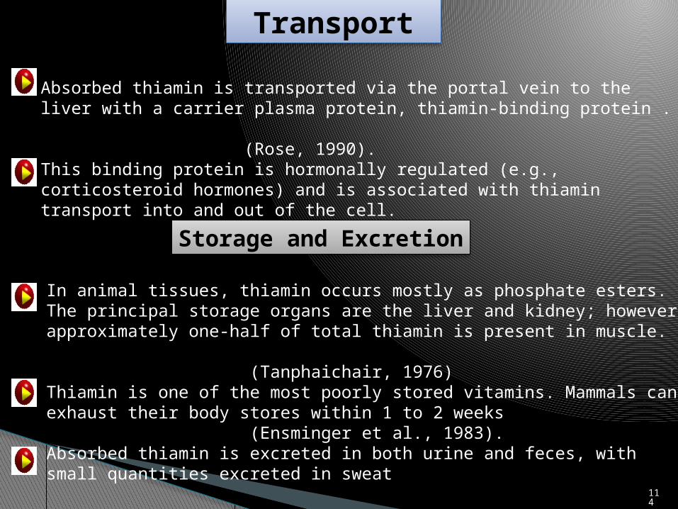
Absorbed thiamin is transported via the portal vein to the liver with a carrier plasma protein, thiamin-binding protein .
(Rose, 1990).This binding protein is hormonally regulated (e.g., corticosteroid hormones) and is associated with thiamin transport into and out of the cell.
Storage and Excretion
In animal tissues, thiamin occurs mostly as phosphate esters.The principal storage organs are the liver and kidney; however, approximately one-half of total thiamin is present in muscle.
(Tanphaichair, 1976)Thiamin is one of the most poorly stored vitamins. Mammals can exhaust their body stores within 1 to 2 weeks
(Ensminger et al., 1983).Absorbed thiamin is excreted in both urine and feces, with small quantities excreted in sweat
114

FUNCTIONS
Decarboxylation of α-Keto Acids and Transketolase Reactions
Thiamine diphosphate is a coenzyme involved in oxidative ecarboxylation of pyruvate to acetyl coenzyme A. and of alpha ketoglutarate to succinyl COA in TCA cycle.
Other Functions
Thiamin has a vital role in nerve function
Possible mechanisms of action of thiamin in nervous tissue include the following
115

1. Thiamin is involved in the synthesis of acetylcholine, which transmits neural Impulses
2. Thiamin participates in the passive transport of sodium of excitable membranes, which is important for the transmission of impulses at the membrane of ganglionic cells
3. The reduction in the activity of transketolase in the pentose phosphate pathway that follows thiamin deficiency reduces the synthesis of fatty acids and the metabolism of energy in the nervous system.
(Muralt, 1962; Cooper et al., 1963)
Thiamin has been shown to have a role in insulin biosynthesis. Isolated pancreatic islets from thiamin-deficient rats secreted less insulin than those from controls
(Rathanaswami and Sundaresan, 1991).
116

Fig. Sheep with thiamin deficiency. Characteristics of the condition are head bent backward (opisthotonos), cramp-like muscle contractions, disturbance of balance, and aggressiveness.
DEFICIENCY
117
Occurs sporadically in cattle, sheep, and goats.
The term PEM refers to a laminar softening or degeneration of brain gray matter (Brent and Bartley, 1984).
The condition affects mainly calves and young cattle between 4 months to2 years old, and lambs and kid between 2 and 7 months old.
The condition is characterized by circling, head pressing, and convulsions, and in severe cases, the animal collapses within 12 to 72 hours after onset of the disease.
High-sulfur diets are associated with thiamin deficiency and PEM(Gould, 1998). Gould et al.
(1991)
Polioencephalomalacia (PEM)

A B
Fig. (A and B). An animal with polioencephalomalacia, a disease of thiamin deficiency. Feedlot cattle suffering from this condition show dullness and sometimes blindness, with a series of nervous disorders such as circling, head pressing, and convulsions. Six to eight hours after thiamin injection, the same animal was able to stand 11
9

Fig. With continued thiamin treatment, in 3 to 5 days, the animal returned to almost normal, with slight brain damage. 12
0

Wasting disease (secadera)
Fig. Wasting disease (secadera) of cattle in the llanos of Colombia. The disease is characterized by emaciation in spite of the availability of good-quality forage. Secadera has been reported as thiamin deficiency because it has been alleviated with thiamin injections. 12
1

Fig. Polyneuritis in a thiamin-deficient chick. Muscle paralysis causes extendedlegs and retraction of the head
Polyneuritis(star gazing posture)
122

Fig. Enlarged heart on right is due to thiamin deficiency. Heart on left isfrom a similar pig fed the same diet plus thiamin.
123

Chastek Paralysis
The disease occurs in mink and foxes and is induced by feeding these animals certain types of raw fish.
In foxes, clinical signs include anorexia and abnormal gait, followed by severe ataxia, inability to stand, hyperesthesia, constant moaning, and convulsions
(Long and Shaw, 1943)RABBITS
Rabbits fed a thiamin-free diet, along with a thiamin antagonist (neopyrithiamin), developed ataxia, flaccid paralysis, convulsions, and coma, followed by death
(Reid et al., 1963; NRC, 1977).Humans
Swelling of the legs, with pitting in ankle region, marks beginning of so-called wet beriberi.
124

CROCODILES
Disease conditions were noted in 4 of 11 clutches of crocodile hatchlings. Sudden loss of righting reflex was the outstanding feature of the disease.
(Jubb, 1992)
Fig. A 2-month-old saltwater crocodile hatchling with suspected thiamin deficiency, floating on its side in shallow water. The listless appearance and open jaws are also characteristic of the disease 125

TOXICITY
Thiamin in large amounts is not toxic, and usually the same is true of parenteral doses.
Dietary intakes of thiamin up to 1,000 times the requirement are apparently safe for most animal species
(NRC, 1987).
Lethal doses with intravenous injection were 125, 250, 300, and 350 mg/kg body weight for mice, rats, rabbits, and dogs, respectively
(Gubler, 1991).

127
REQUIREMENTSAnimal Purpose or Class Requirement
(mg/kg)Reference
Dairy cattle Adult Microbial synthesis NRC (1989a)
Calf 6.5 ppm milk replacer
NRC (1989a)
Beef cattle Adult Microbial synthesis NRC (1996)
Goat All classes Microbial synthesis NRC (1981)
Cat Gestation 5.0 NRC (1986)
Dog Growing 0.75 NRC (1985a)
Chicken Leghorn, 0–6weeks 1.0 NRC (1994)
Leghorn 12-18weeks 0.8 NRC (1994)
Laying (100-g intake) 0.7 NRC (1994)
Broilers(0–8 weeks) 1.8 NRC (1994)

Sources: Modified from Bräunlich and Zintzen (1976), Marks (1975), and Scott et al. (1982).
Feedstuff mg/kg Feedstuff mg/kg
Alfalfa meal 3.9 Linseed meal, expeller extracted
5.1
Barley grain, dried 5.7 Rice, bran 23.0
Beans 6.0 Sorghum grain 3.9
Brewer’s grains, dried 0.8 Sugarcane molasses 1.2
Brewer’s yeast, dried 95.2 Wheat bran 8.0
Coconut meal, dried 0.8 Wheat grain 5.5
Corn (maize), yellow grain
3.5 Blood meal, dried 0.2
Corn (maize), germ meal 10.9 Eggs, whole 3.4
Corn (maize), dried gluten meal
2.1 Milk, cow’s 0.4
Cottonseed meal, solvent extracted
6.4 Fish meal, with solubles 2.0
Distiller’s dried solubles 6.8 Sesame meal 10.0
NATURAL SOURCES

Vitamin B 2
C17
H20
N4O
6
129

History
Chemical Structure & Properties
1915-it was known that a water soluble factor or factor promoted growthand prevented beriberi in rats.
1933-Kuhn (Germany) suggested that this growth factor for rats be given the name flavin
Pure crystalline flavin compounds were found to contain ribose, and thus, the name riboflavin became popular.
It consists of a a dimethylisoalloxazine nucleus combined with the alcohol of ribose as a side chain.It exists in three forms:-free riboflavin
-coenzyme derivatives FMN (Riboflavin 5 phosphate)-FAD.
Riboflavin is an odorless, bitter orange-yellow compound that melts at 2800C.Its empirical formula is - C17
H20
N4O
6 ,with an elemental analysis of carbon 54.25%, hydrogen 5.36%, and nitrogen 14.89%.
Synonym Riboflavin.
130
Riboflavin is only slightly soluble in water but readily soluble in dilute basic or strong acidic solutions.
Very little is lost in cooking.
Loss in milk during pasteurization
131

132
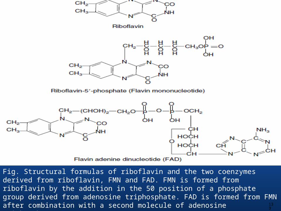
Fig. Structural formulas of riboflavin and the two coenzymes derived from riboflavin, FMN and FAD. FMN is formed from riboflavin by the addition in the 50 position of a phosphate group derived from adenosine triphosphate. FAD is formed from FMN after combination with a second molecule of adenosine triphosphate. 13
3

Metabolic pathway of conversion of riboflavin into FMN, FAD, and covalently bound flavin, together with its control by thyroid hormones.
(Rivlin, R.S., 1970.) 134

METABOLISMDigestion, Absorption and Transport
hydrolyzed by phosphatases
Phosphorylated flavokinase
FAD-pyrophosphorylase
135

Riboflavin is found in feeds as FAD, FMN, and free riboflavin.
Storage and Excretion
Liver-the major site of storage, contains about one-third of the total body flavins.
The liver, kidney, and heart have the richest concentrations.
136

FUNCTIONS
Riboflavin is required as part of many enzymes essential to utilization of carbohydrates, fat, and protein.
More than 100 enzymes are known to bind FAD or FMN in animal and microbial systems.
It is a constituent of flavoproteins, Flavin mononucleotide and Flavin adenine dinucleotide.
They are involved in amino acid and carbohydrate metabolism.
In sows riboflavin is necessary to maintain normal oestrous activity and prevent premature parturition.
DEFICIENCY
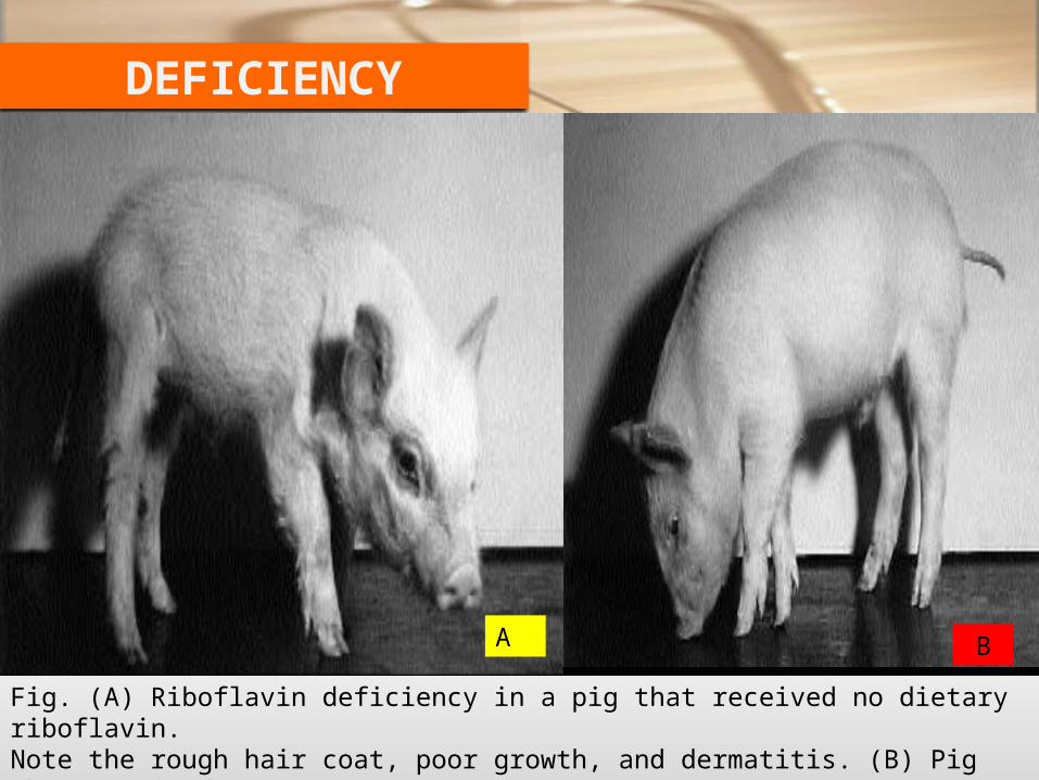
Fig. (A) Riboflavin deficiency in a pig that received no dietary riboflavin.Note the rough hair coat, poor growth, and dermatitis. (B) Pig that receivedadequate riboflavin.
A B

Fig. Riboflavin deficiency. (A) All of the pigs in this litter were born dead; some were in the process of resorption. A few had edema and enlargement of front legs as a result of gelatinous edema. (B) Pigs from a litter in which gelatinous edema was more pronounced.
A B
139

Seven of the ten pigs farrowed were born dead, and the other three were dead within 48 hours. The sow received a riboflavin-deficient diet for a shorter period than the sows farrowing the other two litters. 14
0

Fig. Curled-toe paralysis in a riboflavin- deficient chick 141

142
Riboflavin deficiency in a young chick. Note the position of the hocks, with the toes curled inward.

Fig. Riboflavin deficiency in chicks. (A) The chick at left was fed a corn-soybeanmeal diet without supplemental riboflavin; it exhibited the predominanttype of paralysis observed at the zero level of riboflavin supplementation.Both chicks are female. (B) Same as in (A), but the chicks are male.
143

Fig. Riboflavin deficiency in turkeys at 21 days of age. (A) The turkey at left was fed a corn-soybean basal diet without supplemental riboflavin. (B) Severeleg paralysis and poor feathering in a turkey poultry fed the riboflavin deficient diet.
144

145
DOGS AND CATS
Riboflavin-deficient dogs exhibit low growth rates, anemia, and corneal lesions
(NRC, 1985a).
Cats deficient in the vitamin develop cataracts, fatty livers, testicular hypoplasia, and alopecia with epidermal atrophy
(NRC, 1986).
Corneal vascularization and ulceration, cataract formation, anemia, myelin degeneration of sciatic nerves and spinal cord, fatty liver, congenital abnormalities, and metabolic abnormalities of hepatocytes may occur.
(NRC, 1995).
LABORATORY ANIMALS

Fig. Riboflavin deficiency in the rat, exhibited by (A) generalized dermatitis, growth failure, and marked keratitis of the cornea. (B) After 1 month of treatment with riboflavin, growth resumed, and ocular and skin lesions practically disappeared. 14
6

After 2 months of treatment, the rat showed no signs of deficiency. 147

Fig. Riboflavin deficiency in foxes. After 7 weeks on a riboflavin deficientdiet, the 12-week-old blue fox at right showed depigmentation, shedding of fur, and dermatitis. The littermate at left was fed a diet supplemented with riboflavin. 14
8

Animal Purpose or Class Requirement(mg/kg)
Reference
Dairy cattle Adult Microbial synthesis NRC (1989a)
Calf 6.5 ppm milk replacer NRC (1989a)
Beef cattle Adult Microbial synthesis NRC (1996)
Goat All classes Microbial synthesis NRC (1981)
Cat Gestation 1.0 NRC (1986)
Dog Growing 2-4 NRC (1985a)
Chicken Leghorn, 0–6weeks 3.6 NRC (1994)
Leghorn, 6–18 weeks 1.8 NRC (1994)
Laying (100-g intake) 2.5 NRC (1994)
Broilers(0–8 weeks) 3.6 NRC (1994)
REQUIREMENTS
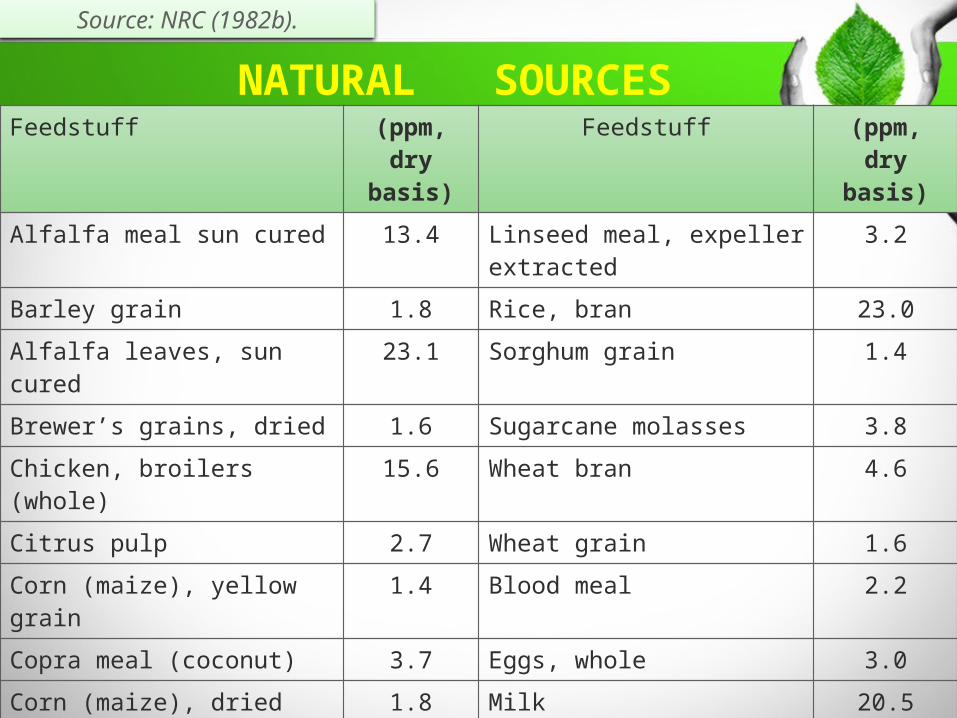
Feedstuff (ppm, dry
basis)
Feedstuff (ppm, dry
basis)
Alfalfa meal sun cured 13.4 Linseed meal, expeller extracted
3.2
Barley grain 1.8 Rice, bran 23.0
Alfalfa leaves, sun cured 23.1 Sorghum grain 1.4
Brewer’s grains, dried 1.6 Sugarcane molasses 3.8
Chicken, broilers (whole) 15.6 Wheat bran 4.6
Citrus pulp 2.7 Wheat grain 1.6
Corn (maize), yellow grain 1.4 Blood meal 2.2
Copra meal (coconut) 3.7 Eggs, whole 3.0
Corn (maize), dried gluten meal
1.8 Milk 20.5
Cottonseed meal, solvent extracted
5.3 Soybean meal, solvent extracted
3.2
Clover hay, ladino (sun cured)
17.2 Sesame meal 10.0
Bean, navy (seed) 2.0
NATURAL SOURCES
Source: NRC (1982b).

Vitamin B 3
C6H5O2N
151

History
Chemical Structure & Properties
Synonym Vitamin PP, Niacin, Nicotinic acid, Nicotinamide.
1914-Funk isolated nicotinic acid from rice polishing.Older term replace Nicotinic Acid & nicotinamide to niacin & niacinamide.
Its empirical formula is - C6H5O2N.Niacin is pyridine-3-caboxylic acid.Two forms-nicotinic acid and nicotinamide (niacinamide).Both are white, odorless, crystalline solids soluble in water and alcohol.They are very resistant to heat, air, light, and alkaline conditions and thus are stable in foods.Nicotinic acid is a white crystalline solid, stable in air at normal room temperature.
152

Nicotinic acid readily forms salts with metals such as aluminum, calcium, copper, and sodium.When in acid solution, niacin readily forms quaternary ammonium compounds, such as nicotinic acid hydrochloride, which is soluble in water.When in a basic solution, nicotinic acid readily forms carboxylic acid salts.It is moderately soluble in water and alcohol, but insoluble in ether.In contrast to nicotinic acid, nicotinamide is highly soluble in water, and is soluble in ether, characteristics that allow separation of the two vitamers.
Niacin Coenzymes
The biologically active forms of niacin compounds are the NAD and NADP coenzymes.The oxidized and reduced forms of the coenzymes are designated NAD+ or NADP+ and NADH or NADPH, respectively.

Fig. Chemical structures of niacin compounds. (a) Nicotinic acid, (b) nicotinamide, (c) NAD+, (d) NADP+, and (e) site of reduction. 15
4

155
Pathways of Synthesis
Although plants and most microorganisms can synthesize the pyridine ring of NAD de novo from aspartic acid and dihydroxyacetone phosphate, animals do not have this ability.
Nicotinic acid, nicotinamide, pyridine nucleotides, and tryptophan represent the dietary sources for the pyridine ring structure in mammals.
Animals may also practice coprophagy to take advantage of colonic synthesis of niacin by icroflora. Ruminants receive an ample supply of niacin from foregut bacteria

156

157
Fig.Pathways of NADþ synthesis in mammals. Reactions 5, 6, 8, and 9 comprise the Preiss– Handler pathway whereas reactions 10 and 11 form the Dietrich pathway. The following enzymes correspond to the numbered reactions: 1, tryptophan 2,3-dioxygenase (hepatic) or indoleamine 2,3-dioxygenase (extrahepatic), which start the five-step conversion to ACMS and nine-step catabolism of tryptophan to acetyl CoA; 2, ACMS decarboxylase (ACMSD); 3, spontaneous chemical reaction; 4, quinolinic acid phosphoribosyltransferase; 5, NAMN adenylyltransferase (enzymes 5 and 11 may be identical proteins); 6, NAD synthetase; 7, NAD glycohydrolases, various ADP-ribosylation reactions; 8, nicotinamide deamidase; 9, nicotinic acid hosphoribosyltransferase; 10, nicotinamide phosphoribosyltransferase; 11, NMN adenylyltransferase.
1. 3-acetyl pyridine .2. pyridine sulfonic acid.
Antagonists

METABOLISMNiacin in foods occurs mostly in its coenzyme forms, which are hydrolyzed during digestion, yielding nicotinamide, which seems to be absorbed without further hydrolysis in the gastrointestinal tract.
In the gut, mucosa nicotinic acid is converted to nicotinamide
(Stein et al., 1994).Nicotinamide is the primary circulating form of the vitamin and is converted into its coenzyme forms in the tissues.
Excretion
Urine is the primary pathway of excretion of absorbed niacin and its metabolites.The principal excretory product in humans, dogs, rats, and pigs is the methylated metabolite N′-methylnicotinamide or one of two oxidation products of this compound, 4-pyridone or 6-pyridone of N′-methylnicotinamide.
On the other hand, in herbivores niacin does not seem to be metabolized by methylation, but large amounts are excreted unchanged.In the chicken, however, nicotinic acid is conjugated with ornithine as either α- or δ-nicotinyl ornithine or dinicotinyl ornithine. 158

FUNCTIONS
The major function of niacin is in the coenzyme forms of nicotinamide, NAD and NADP.They are especially important in the metabolic reactions that furnish energy to the animal.
Important metabolic reactions catalyzed by NAD and NADP are summarized below:
1. Carbohydrate metabolism—(a) glycolysis (anaerobic oxidation of glucose) and (b) the Krebs cycle.
2. Lipid metabolism—(a) glycerol synthesis and breakdown, (b) fatty3. acid oxidation and synthesis, and (c) steroid synthesis.4. Protein metabolism—(a) degradation and synthesis of amino acids and (b)
oxidation of carbon chains via the Krebs cycle.5. Photosynthesis.6. Rhodopsin synthesis

Summary of Reaction Types That Are Dependent on Nicotinamide Nucleotides
Category of Reaction
Enzymes Main Products
Metabolic Roles
NADþ=NADH redox exchanges
Numerous NAD-dependent enzymesthroughout oxidative metabolism
NADH and oxidized metabolites, e.g., TCA cycle intermediates
1. Transfer of electrons from macronutrient substrates to the ETC, ATP production
2. Numerous oxidative reactions are enabled by the high ratio of NADþ:NADH
160

NADPþ=NADPH redox exchanges
Numerous NADP-dependent enzymes involved in reductive metabolism
NADPþ and reduced metabolites, e.g., fatty acid
Biosynthetic metabolism, oxidant defense
Numerous reductive reactions are enabled by the high ratio of NADPH:NADPþ, which is maintained by the pentose phosphate pathway
Poly(ADP-ribosyl)ation reactions
Up to 18 different PARP enzymes, mainly nuclear and DNA associated
Poly(ADP-ribose) covalently bound to proteins, free polymer resulting from catabolism
Diverse functions, but many related to DNA metabolism and genomic stability
Polyanionic nature controls protein function
High-affinity polymer binding by other proteins16
1

Mono(ADP-ribosyl)ationreactions
Numerous poorly characterized transferases
Mono(ADP-ribose) covalently bound to proteins, many of which are G-proteins
Diverse and poorly characterized
Cyclic ADP-ribose and NAADP formation
ADP-ribosyl cyclases, which also have thepotential to form NAADP
Cyclic ADP-riboseNAADP
Control of intracellular calcium levels, and thereby control of almost all cellular signaling events
SIR2=SIRT1 deacetylationreactions
SIR2 (rats)SIRT1 (humans)
Deacetylated proteins, includinghistones, p53
O-Acetyl-ADP-ribose
Control of p53 function and chromatin structure, central to life extension through caloric restriction
162

DEFICIENCY
Niacin deficiency is characterized by severe metabolic disorders in the skin and digestive organs.

Fig. Leg disorders in niacin-deficient broiler chicks. The bird on the left, with bowed legs, was fed a corn-soybean meal diet without Supplemental niacin.
Fig. Intestine from niacin-deficient pig shows thickened and hemorrhagic mucous membrane and denuded areas. 16
4

Dogs & Cats
Blacktongue (Canine Pellagra).
There is severe cheilosis, glossitis, and gingivitis. Necrotic patches and ulcers may be seen on the oral mucosa, and there is a foul odor.
There is bloody diarrhea, inflammation, and hemorrhagic necrosis of the duodenum and jejunum, with shortening and clubbing of villi and inflammationand degeneration of the mucosa of the large intestine.
Foxes & Mink
Foxes fed a niacin-deficient diet exhibited anorexia, weight loss, and typical blacktongue, characterized by severe inflammation of the gums and fiery redness of the lips, tongue, and gums
(NRC, 1982a).
165

Fig. 8.7 Niacin-deficient dog with blacktongue exhibits drooling of thick, ropy saliva. 16
6

Fig. Niacin deficiency in turkey poults. (A and B) The birds on the left side,which were fed a corn-soybean meal without supplemental niacin, showed Perosis like signs. © Comparison of the legs of the poultry in B. 16
7

168
REQUIREMENTSAnimal Purpose or Class Requirement
(mg/kg)Reference
Dairy cattle Adult Microbial synthesis NRC (1989a)
Calf 2.6 ppm milk replacer
NRC (1989a)
Beef cattle Adult Microbial synthesis NRC (1996)
Goat All classes Microbial synthesis NRC (1981)
Cat Gestation 40 NRC (1986)
Dog Growing 450µg/kg BW NRC (1985a)
Chicken Leghorn, 0–6weeks 27.0 NRC (1994)
Leghorn 12-18weeks 11.0 NRC (1994)
Laying (100-g intake) 10.0 NRC (1994)
Broilers(0–8 weeks) 25-30 NRC (1994)

Feedstuff (mg/kg, dry
basis)
Feedstuff (mg/kg, dry
basis)
Alfalfa meal sun cured 42 Linseed meal, expeller extracted
37
Barley grain 94 Rice, bran 23.0
Alfalfa leaves, sun cured 53 Sorghum grain 1.4
Brewer’s grains 47 Sugarcane molasses 3.8
Chicken, broilers (whole) 230 Wheat bran 268
Citrus pulp 23 Wheat grain 64
Corn , yellow grain 55 Blood meal 34
Copra meal (coconut) 28 Eggs, whole 3.0
Corn , gluten meal 55 Milk, cattle 269
Cottonseed meal, solvent extracted
48 Soybean meal, solvent extracted
31
Clover hay, ladino (sun cured)
11 Soybean seed 24
NATURAL SOURCES
Source: NRC (1982b).

TOXICITY
Limited research indicates that nicotinic acid and nicotinamide are toxic at dietary intakes greater than about 350 mg/kg of body weight per day
(NRC, 1987).
In dogs, oral administration of 2 g of nicotinic acid per day (133 to 145 mg/kg of body weight) produced bloody feces in a few dogs, followed by convulsions and death
(NRC, 1987).
170

Vitamin B 5
C9H17NO5
171
History
Chemical Structure & Properties
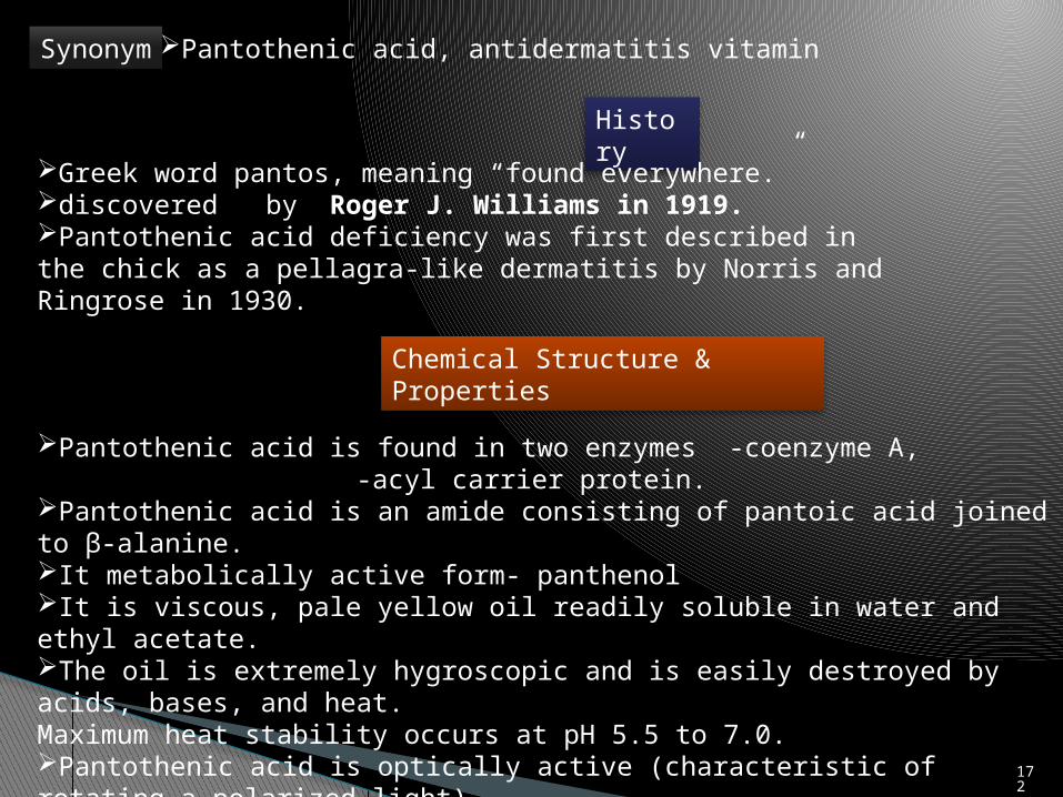
Synonym Pantothenic acid, antidermatitis vitamin
Greek word pantos, meaning “found everywhere.”discovered by Roger J. Williams in 1919.Pantothenic acid deficiency was first described in the chick as a pellagra-like dermatitis by Norris and Ringrose in 1930.
Pantothenic acid is found in two enzymes -coenzyme A,-acyl carrier protein.
Pantothenic acid is an amide consisting of pantoic acid joined to β-alanine.It metabolically active form- panthenolIt is viscous, pale yellow oil readily soluble in water and ethyl acetate.The oil is extremely hygroscopic and is easily destroyed by acids, bases, and heat.Maximum heat stability occurs at pH 5.5 to 7.0.Pantothenic acid is optically active (characteristic of rotating a polarized light). 17
2

• Calcium pantothenate, the form used in commerce, crystallizes as white needles from methanol and is reasonably stable to light and air.
• Feeding chickens on high intakes of copper results in reduced formation of coenzyme A, by increasing the oxidation of cysteine to cystine, and also by the formation of copper-cysteine and copper-glutathione complexes, which render the amino acid unavailable for coenzyme A synthesis
(Latymer and Coates, 1981).

Fig. Structural components of coenzyme A. 174

Fig. Pathway for the biosynthesis of pantothenic acid found in plants, bacteria (including archaea), and eubacteria. 17
5

176
Antagonist
• The most common antagonist of pantothenic acid is ω-methyl-pantothenic acid, which has been used to produce a deficiency of the vitamin in humans
(Hodges et al., 1958).
• Other antivitamins include pantoyltaurine, phenylpantothenate hydroxycobalamine (c-lactam) (analog of vitamin B12), and antimetabolites of the vitamin containing alkyl or aryl ureido and carbamate components in the amide part of the molecule.
(Fox, 1991; Brass, 1993).

METABOLISM
Pantetheinase
177

Coenzyme A synthesis
Fig. Coenzyme A metabolism and importance of pantothenic acid kinase.178

Absorption
Pantothenic acid, its salt, and the alcohol are absorbed primarily in the jejunum by a specific transport system that is saturable and sodium ion dependent
(Fenstermacher and Rose, 1986).
After absorption, pantothenic acid is transported to various tissues in the plasma, from which it is taken up by most cells via another active-transportProcess.
Within all tissues pantothenic acid is converted to coenzyme A and other compounds in which the vitamin is a functional group
(Sauberlich,1985).
179
Excretion
Urinary excretion represents the major route of body loss of absorbed pantothenic acid, with prompt excretion when taken in excess.
Most pantothenic acid is excreted as the free vitamin, but some species (e.g., dog) excrete it as β-glucuronide
(Taylor et al., 1972).
An appreciable quantity of pantothenic acid (approximately 15% of daily intake) is oxidized completely and is excreted across the lungs as CO2
(Combs, 1992).
180

Storage
Animals and humans do not appear to have the ability to store appreciable amounts of pantothenic acid, with organs such as the liver and kidney having the highest concentrations.
Most pantothenic acid in blood exists in red blood cells as coenzyme A; serum contains no coenzyme A but does contain free pantothenic acid.
181
FUNCTIONS
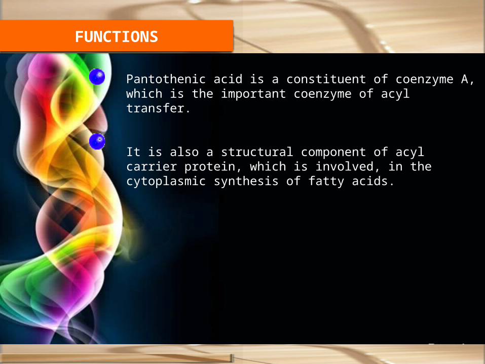
Pantothenic acid is a constituent of coenzyme A, which is the important coenzyme of acyl transfer.
It is also a structural component of acyl carrier protein, which is involved, in the cytoplasmic synthesis of fatty acids.
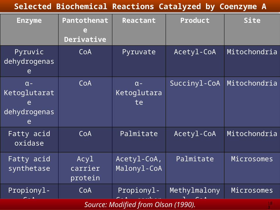
Enzyme Pantothenate
Derivative
Reactant Product Site
Pyruvic dehydrogenas
e
CoA Pyruvate Acetyl-CoA Mitochondria
α-Ketoglutarate dehydrogenas
e
CoA α-Ketoglutarate
Succinyl-CoA Mitochondria
Fatty acid oxidase
CoA Palmitate Acetyl-CoA Mitochondria
Fatty acid synthetase
Acyl carrier protein
Acetyl-CoA, Malonyl-CoA
Palmitate Microsomes
Propionyl-CoA carboxylase
CoA Propionyl-CoA, carbon
dioxide
Methylmalonyl- CoA
Microsomes
Acyl-CoA synthetase
Phosphopanthetheine
Succinyl-CoA, GDP + P1
Succinate, GTP + CoA
Mitochondria
Selected Biochemical Reactions Catalyzed by Coenzyme A
Source: Modified from Olson (1990). 183

Functions of CoA and Acyl Carrier Protein
Function Importance
Carbohydrate-related citric acid cycle transfer reactions
Oxidative metabolism
Acetylation of sugars (e.g., N-acetylglucosamine)
Production of carbohydrates important to cell structure
Lipid-related
Phospholipid biosynthesis Cell membrane formation and structure
Isoprenoid biosynthesis Cholesterol and bile salt production
Steroid biosynthesis Steroid hormone production
Fatty acid elongation Ability to modify cell membrane fluidity
Acyl (fatty acid) and triacyl glyceride synthesis
Energy storage
Protein-related
Protein acetylation Altered protein conformation; activation of certain hormones and enzymes, e.g., adrenocorticotropin transcriptional regulation, e.g., acetylation of histone
Protein acylation (e.g., myristic and palmitic acid, and prenyl moiety additions)
Compartmentalization and activation of hormones and transcription factors
184

DEFICIENCY
Species Symptoms
Chicken Dermatitis around beak, feet, and eyes; poor feathering; spinal cord myelin degeneration; involution of the thymus; fatty degeneration of the liver
Fish Anorectic behavior; listlessness; fused gill lamellae; reproductive failure
Rat Dermatitis; loss of hair color (achromotrichia). with alopecia; hemorrhagic necrosis of the adrenals; duodenal ulcer; spastic gait; anemia; leukopenia; impaired antibody production; gonadal atrophy with infertility
Dog Anorexia; diarrhea; acute encephalopathy; coma; hypoglycemia; leukocytosis; hyperammonemia; hyperlactemia; hepatic steatosis; mitochondrial enlargement
Pig Dermatitis; hair loss; diarrhea with impaired sodium, potassium, and glucose absorption; lachrymation; ulcerative colitis; spinal cord and peripheral nerve lesions with spastic gait

Fig. Goose-stepping pig with pantothenic acid deficiency. 186

Fig. Pantothenic acid deficiency. (A) Locomotor incoordination (goosestepping).(B) An affected pig often falls sideways or, with its back legs spreadapart, assumes a posture that resembles that of a sitting dog.
A B
187
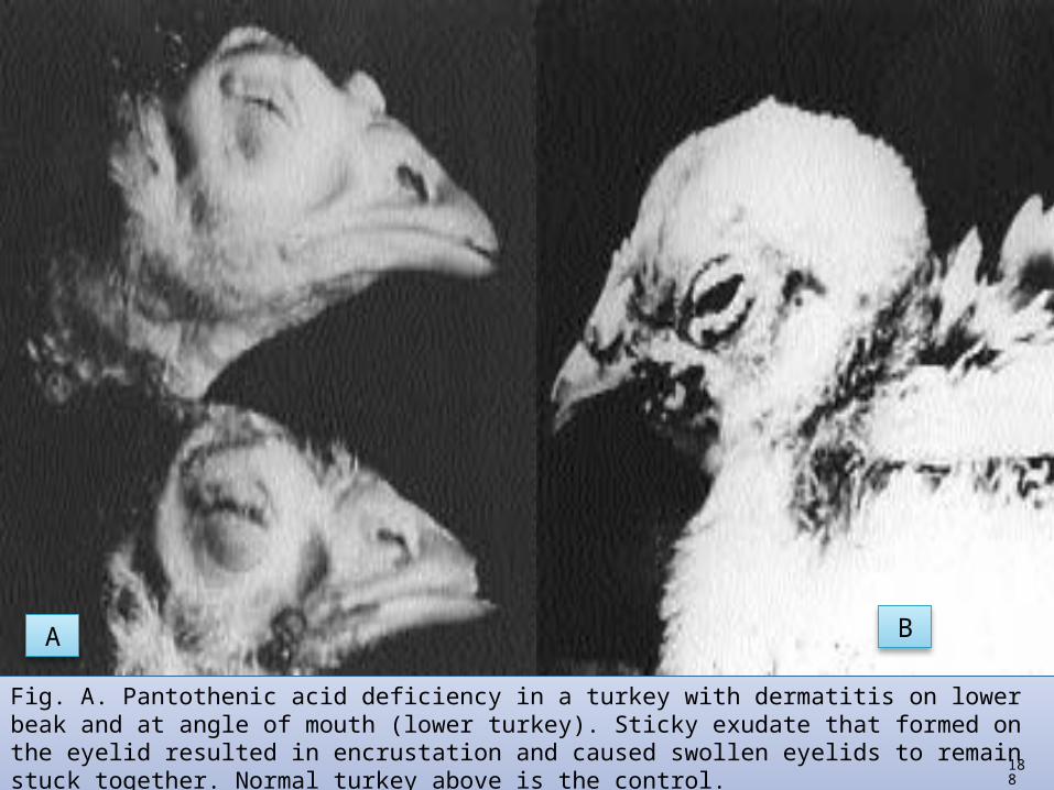
Fig. A. Pantothenic acid deficiency in a turkey with dermatitis on lower beak and at angle of mouth (lower turkey). Sticky exudate that formed on the eyelid resulted in encrustation and caused swollen eyelids to remain stuck together. Normal turkey above is the control.Fig. B. Pantothenic acid deficiency in a chick, with dermatitis around beak
A B
188

REQUIREMENTSAnimal Purpose or Class Requirement
(mg/kg)Reference
Dairy cattle Adult Microbial synthesis NRC (1989a)
Calf 2.6 ppm milk replacer
NRC (1989a)
Beef cattle Adult Microbial synthesis NRC (1996)
Goat All classes Microbial synthesis NRC (1981b)
Cat Growing 8 NRC (1986)
Dog Growing 400µg/kg BW NRC (1985a)
Chicken Leghorn, 0–6weeks 10.0 NRC (1994)
Laying (100-g intake) 1.7 NRC (1994)
Broilers(0–8 weeks) 10.00 NRC (1994)

Feedstuff (mg/kg, dry
basis)
Feedstuff (mg/kg, dry
basis)
Alfalfa meal sun cured 28.6 Linseed meal, expeller extracted
16.3
Barley grain 9.1 Rice, bran 25.0
Alfalfa leaves, sun cured 32.4 Sorghum grain 12.5
Brewer’s grains 8.9 Sugarcane molasses 50.3
Rice, grain 9.1 Wheat bran 33.5
Citrus pulp 14.3 Wheat grain 11.4
Corn , yellow grain 6.6 Blood meal 2.6
Copra meal (coconut) 6.9 Eggs, whole 27.0
Corn , gluten meal 11.2 Milk, cattle 38.6
Cottonseed meal, solvent extracted
15.4 Soybean meal, solvent extracted
18.2
Clover hay, ladino (sun cured)
1.1 Soybean seed 17.3
NATURAL SOURCES
Source: NRC (1982b).

Vitamin B 6
191

History
Chemical Structure & Properties
Synonym Pyridoxol, Pyridoxal, Pyridoxamine
1934-Gyorgy first recognized Vitamin B6 as a distinct Vitamin.Kuhn and coworkers-the structure of the vitamin was first explained.
It refers to a group of three compounds:- 1.Pyridoxol/Pyridoxine(Alcohol),2.Pyridoxal (Aldehyde).3.Pyridoxamine.(Amine).
Two additional forms coenzyme-1.Pyridoxal phosphate (PLP) 2.Pyridoxamine phosphate.
In plant-pyridoxineIn animal-pyridoxal and pyridoxamine.It can be destroyed by heat, alkali and exposure to light, especially in neutral or alkaline media is highly destructive.Vitamin B6 are colorless crystals soluble in water.Synthetic- Pyridoxine hydrochloride
192

193
Antagonist1. The anDeoxypyridoxine is a powerful
antagonist to vitamin B6
2. Isoniazid is a strong inhibitor of pyridoxal kinase and results in anemia in humans, probably by inhibiting the synthesis of δ-aminolevulinic acid and thus of heme.
3. Isonicotinic acid hydrazide (isoniazid)4. Cycloserine5. Penicillamine6. tihypertensive drugs thiosemicarbizide and
hydralazine have also been shown to interfere with vitamin B6 usage.
Digestion, Absorption, and Transport
Vitamin B6 is absorbed mainly in the jejunum, but also in theileum, by passive diffusion.
Both niacin (as the NADP-dependent enzyme) and riboflavin (as the flavoprotein pyridoxamine phosphate oxidase) are important for conversion of vitamin B6 forms and phosphorylation reactions
(Wada andSnell, 1961; Kodentsova et al., 1993).
Vitamin B6 is found in the blood largely as PLP, most of which is derived from the liver after metabolism by hepatic flavoenzymes.
Only small quantities of vitamin B6 are stored in the body
Reports of PLP content of glycogen phosphorylase suggest that 90% or more of the vitamin B6 present in muscle might be present in this single enzyme
(Merrill and Burnhan, 1990).Excreted through urine

Function
More than 60 enzymes are already known to depend on vitamin B6 coenzymes.Pyridoxal phosphate functions in practically all reactions involved in amino acid metabolism, including transamination aminotransferase), decarboxylation,deamination, and desulfhydration, and in the cleavage or synthesis of amino acids.

Vitamin B6 is involved in many additional reactions, particularly those involving proteins. The vitamin participates in the following functions
(Bräunlich, 1974; Marks, 1975; LeKlem, 1991)
1. Deaminases—for serine, threonine, and cystathionine.2. Desulfhydrases and transulfhyurases—interconversion and
metabolism of sulfur-containing amino acids.3. Synthesis of niacin from tryptophan—hydroxykynurenine is not
converted to hydroxyanthranilic acid but rather to xanthurenic acid because of lack of the B6-dependent enzyme kynureninase
4. Formation of δ-aminolevulinic acid from succinyl-CoA and glycine, the first step in porphyrin synthesis.
5. Conversion of linoleic to arachidonic acid in the metabolism of essential fatty acids (this function is controversial).
6. Glycogen phosphorylase catalyzes glycogen breakdown to glucose l-phosphate. Pyridoxal phosphate does not appear to be a coenzyme for this enzyme but rather affects the enzyme’s conformation.
7. Synthesis of epinephrine and norepinephrine from either phenylalanine or tyrosine—both norepinephrine and epinephrine are involved in carbohydrate metabolism as well as in other body reactions.
196

8. Racemases—PLP-dependent racemases enable certain microorganisms to utilize D-amino acids. Racemases have not yet been detected in mammalian tissues.
9. Transmethylation by methionine.10. Incorporation of iron in hemoglobin synthesis.11. Amino acid transport—all three known amino acid transport systems—(a)
neutral amino acids and histidine, (b) basic amino acids, and (c) proline and hydroxyproline—appear to require PLP.
12. Formation of antibodies—B6 deficiency results in inhibition of the synthesis of globulins, which carry antibodies.
197

DEFICIENCY Affects the animal's growth rate. Convulsions may also occur, possibly because a reduction in the activity
of glutamic acid decarboxylase results in an accumulation of glutamic acid.
In addition, pigs exhibit a reduced appetite and may develop anemia. Chicks on a deficient diet show jerky movements, while in adult birds
hatchability and egg production are adversely affected.
Fig. A 6-week-old B6-deficient pig weighing only 3.6 kg. 198

Fig. This pig is having an epileptic-like seizure while receiving a diet low in vitamin B6.
199
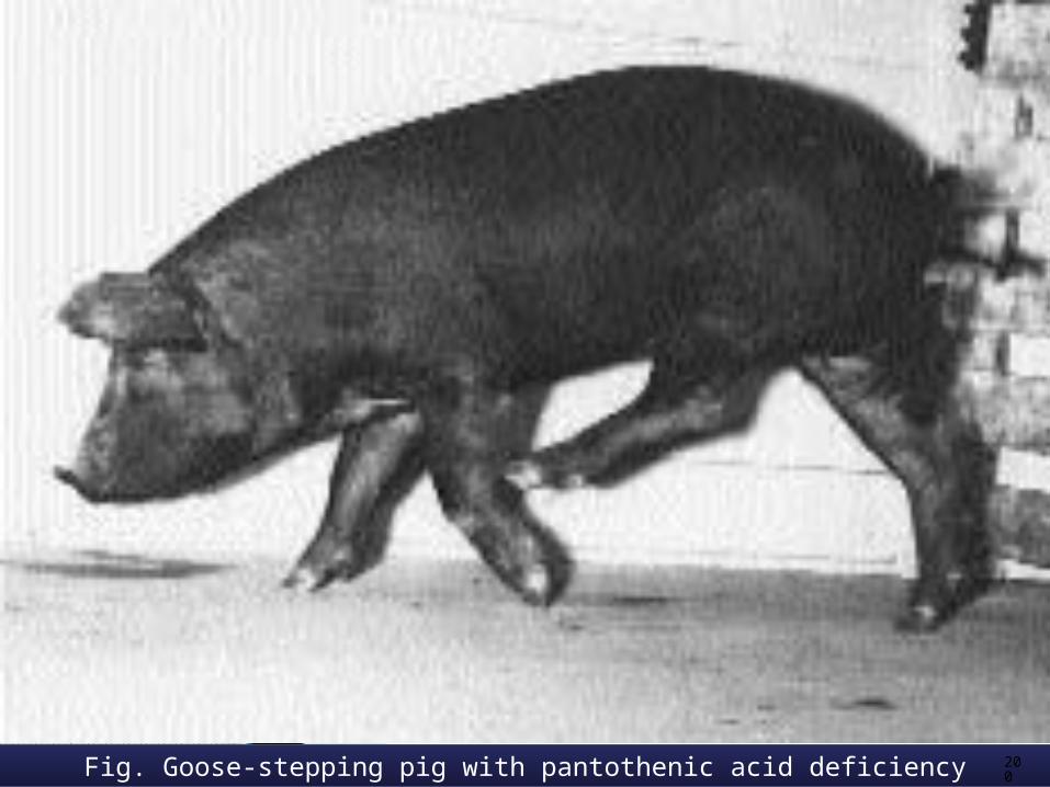
Fig. Goose-stepping pig with pantothenic acid deficiency 200

201Pyridoxine deficiency. Note loss of control of the legs and the head retraction

Fig. Vitamin B6-deficient poult (about 4 weeks old) on left and a normalpoult on right. 20
2

203
REQUIREMENTSAnimal Purpose or Class Requirement
(mg/kg)Reference
Dairy cattle Adult Microbial synthesis NRC (1989a)
Calf 6.5 ppm milk replacer
NRC (1989a)
Beef cattle Adult Microbial synthesis NRC (1996)
Goat All classes Microbial synthesis NRC (1981b)
Cat Growing 4 NRC (1986)
Dog Growing 60µg/kg BW NRC (1985a)
Chicken Leghorn, 0–6weeks 3.0 NRC (1994)
Laying (100-g intake) 2.5 NRC (1994)
Broilers(0–8 weeks) 3-3.5 NRC (1994)

Feedstuff (mg/kg, dry
basis)
Feedstuff (mg/kg, dry
basis)
Alfalfa meal sun cured 4.4 Linseed meal, expeller extracted
6.1
Barley grain 7.3 Rice, polished 0.4
Bean, navy (seed) 0.3 Sorghum grain 5.0
Brewer’s grains 0.8 Sugarcane molasses 5.7
Rice, grain 5 Wheat bran 9.6
Crab meal 7.2 Wheat grain 5.6
Corn , yellow grain 5.3 Blood meal 4.8
Copra meal (coconut) 4.8 Eggs, whole 27.0
Corn , gluten meal 8.8 Milk, cattle 4.5
Cottonseed meal, solvent extracted
6.8 Soybean meal, solvent extracted
6.7
Peanut meal, solvent extracted
6.9 Oat, grain 2.8
NATURAL SOURCES
Source: NRC (1982b).
Vitamin B 8
C11H18O3S
205

History
Chemical Structure & Properties
Synonym Biotin, Vitamin H
Biotin was the name given to a substance isolated from egg yolk by Kogl and Tonnis in 1936 that was necessary for yeast growthThe term vitamin H was chosen by György because the factor protectedthe haut, the German word for skin..
The structure of biotin includes- sulfur atom in its ring (like thiamin) and a transverse bond across the ring.It is a monocarboxylic acid with sulfur as a thioether linkage.The empirical formula for biotin is C11H18O3S
It contains three asymmetric carbonations and therefore eight different isomers are possible.Of these isomers only one contains vitamin activity, d-biotin.The stereoisomer l-biotin is inactive.Melting point- 232-2330C.It soluble in dilute alkali and hot water and practically insoluble in fats and organic solventsIt is destroyed by nitrous acid, other strong acids, strong bases, and formaldehyde and is inactivated by rancid fats and choline
206

Mild oxidation converts biotin to sulfoxide, and
Strong agents result in sulfur replacement by oxygen, resulting in oxybiotin and desthiobiotin. strong oxidation converts it to sulfone.
METABOLISMBiotin exists in natural materials in both bound and free forms, with much of the bound biotin apparently not available to animal species.
Naturally occurring biotin is found partly in the free state (fruit, milk, vegetables) and partly in a form bound to protein in animal tissues, plant seeds, and yeast.

The few studies conducted in animals on biotin metabolism revealed that biotin is absorbed as the intact molecule in the first third to half of the small intestine
(Bonjour, 1991).Biotin appears to circulate in the bloodstream both free and bound to a serum glycoprotein, which also has biotinidase activity, catalyzing the hydrolysis of biocytin.
All cells contain some biotin, with larger quantities in the liver and kidneys.
Absorption

FUNCTIONS
Biotin is an essential coenzyme in carbohydrate, fat, and protein metabolism. It is involved in conversion of carbohydrate to protein and vice- versa as well as conversion of protein and carbohydrate to fat.
Biotin also plays an important role in maintaining normal blood glucose levels from metabolism of protein and fat when dietary intake of carbohydrate is low.
Biotin functions as a carboxyl carrier in four carboxylase enzymes: pyruvate carboxylase, acetyl CoA carboxylase, propionyl CoA carboxylase, and 3-methylcrotonyl CoA carboxylase.
Specific biotin-dependent reactions in carbohydrate metabolism are the following:
• Carboxylation of pyruvic acid to oxaloacetic acid.• Conversion of malic acid to pyruvic acid.• Interconversion of succinic acid and propionic acid.• Conversion of oxalosuccinic acid to α-ketoglutaric acid.

In protein metabolism, biotin enzymes are important in protein synthesis, amino acid deamination, purine synthesis, and nucleic acid metabolism.
Biotin is required for transcarboxylation in degradation ofvarious amino acids.

DEFICIENCY
Fig. The two middle pigs are biotin deficient. Note the hair loss and dermatitis.

Fig. Biotin-deficient pigs. Note transverse cracking of the soles and the tops of the hooves.

Fig. Perosis (bone deformities) as a result of biotin deficiency. Chicks showed perosis as early as 17 days of age, with rigid limb joints that resulted in a stilted walk.

Fig. Normal (left) and biotin-deficient (right) Broad-Breasted Bronze male turkeys at 3 weeks of age.

Fig. A Severe foot-pad lesions in the growing turkey as a result of biotin deficiency. Less severe lesions are more common. Fig. B Biotin deficiency in a poultry. Note lesion at apex of mouth
A B

Fig. 11.8 Hoof condition of a 7-year-old 18-hands heavyweight show hunter resulting from biotin supplementation. The horse had a history of tender feet.(A) The walls of the hooves were very weak crumbling at the lower edges, with large areas breaking out and detaching when shoes were nailed. (B) Five months after supplementation with 15 mg of biotin per day, the walls of the hooves were thicker and harder,so nailing was achieved.
A B
216
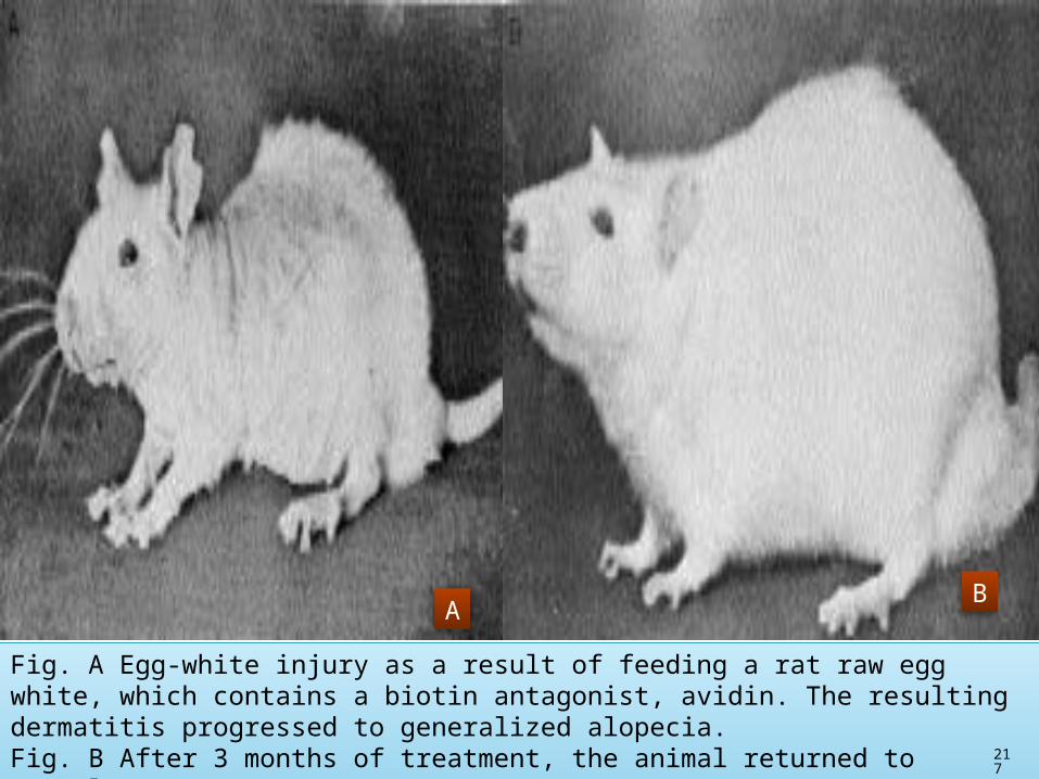
Fig. A Egg-white injury as a result of feeding a rat raw egg white, which contains a biotin antagonist, avidin. The resulting dermatitis progressed to generalized alopecia. Fig. B After 3 months of treatment, the animal returned to normal.
AB
217

Fig. The newborn fox pup on the left is from a biotin-deficient dam that received a diet containing raw egg white. Thin, gray pelt and deformed legs areapparent. A control diet containing cooked egg white was fed to the dam of the new born pup on the right.
218

219
REQUIREMENTSAnimal Purpose or Class Requirement
(mg/kg)Reference
Dairy cattle Adult Microbial synthesis NRC (1989a)
Calf 0.1 ppm milk replacer
NRC (1989a)
Beef cattle Adult Microbial synthesis NRC (1996)
Goat All classes Microbial synthesis NRC (1981)
Cat All classes 0.07 NRC (1986)
Chicken Leghorn, 0–6weeks 0.15 NRC (1994)
Leghorn 12-18weeks 0.10 NRC (1994)
Laying (100-g intake) 0.10 NRC (1994)
Broilers(0–8 weeks) 0.15 NRC (1994)

Feedstuff (μg/g) Feedstuff (μg/g)
Alfalfa meal, dehydrated 0.33 Fish meal 0.14
Barley grain 0.14 Milk, cow’s 0.05
Beef, steak 0.04 Molasses, blackstrap 0.7
Cabbage 0.02 Oats 0.25
Carrots 0.03 Rice bran 0.42
Chicken 0.10 Rice polishings 0.37
Corn , yellow grain 0.08 Skim milk, dried 0.25
Corn , gluten meal 0.19 Sorghum 0.29
Cottonseed meal, solvent extracted
0.08-0.47 Soybean meal 0.27
Distiller’s solubles, dried 0.44-1.1 Wheat 0.10
Eggs, whole 0.25 Wheat bran 0.36
NATURAL SOURCES
Sources: Modified from NRC (1982b) and Frigg and Volker (1994).

Vitamin B 9
221

History
Chemical Structure & Properties
Synonym Folic acid, Folacin, Folate, Vitamin M, Vitamin Bc.
1935-An anemia preventive factor for monkeys was found in yeast or liver extracts and designated vitamin M in by Day.1939-Hogan and Parrot prevented anemia in chicks with a factor in liver called Bc.I1940- Snell and coworkers found a growth factor for L. casei, and in the same year, a growth factor for S. lacti was found in spinach. The growth factor had been isolated from 4 tons of spinach leaves. The isolated factor was called folic acid from folium, the Latin word for leaf.
Its structure contains three parts- glutamic acid, ρ-aminobenzoic acid (PABA), and a pteridine nucleus.
Pteroic acid consist- ρ-aminobenzoic acid (PABA) and a Pteridine nucleus. Much of the folacin in natural feedstuffs is conjugated with a varying number of
extra glutamic acid molecules. Folacin as pteroyloligo-γ-L-glutamates (PteGlun) & it is one to nine glutamates
long, linked by peptide bonds. n-indicating the number of glutamyl residues.
222
Folacin are the natural coenzymes that are most abundant in every tissue.
The conjugated forms with two or more glutamic acid residues are joined by γ-glutamyl linkages to the single glutamic acid moiety of the vitamin.
Synthetic folacin, however, is in the monoglutamate form.
Folacin is a yellowish-orange crystalline powder, tasteless and odorless,
and insoluble in alcohol, ether, and other organic solvents.
Molecular weight - 441.4,
223
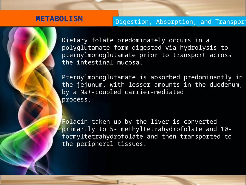
METABOLISM Digestion, Absorption, and Transport
Dietary folate predominately occurs in a polyglutamate form digested via hydrolysis to pteroylmonoglutamate prior to transport across the intestinal mucosa.
Pteroylmonoglutamate is absorbed predominantly in the jejunum, with lesser amounts in the duodenum, by a Na+-coupled carrier-mediatedprocess.
Folacin taken up by the liver is converted primarily to 5- methyltetrahydrofolate and 10-formyltetrahydrofolate and then transported to the peripheral tissues.

Storage
Folacin is widely distributed in tissues largely in the conjugated polyglutamate forms.
Excretion
Urinary excretion of folacin represents a small fraction of total excretion (e.g., < 1% of total body stores).
Fecal folacin concentrations are quite high, often higher than intake, representing not only undigested folacin but, more important, the considerable bacterial synthesis of the vitamin in the intestine.

FUNCTIONS
Folic acid is converted into tetrahydrofolic acid which functions as a coenzyme in the mobilization and utilisation of single-carbon groups (e.g.) formyl, methyl that are added to, or removed from, such metabolites as histidine, serine, glycine, methionine and purines.

Fig.A Folacin-deficient bird on left with depigmentation and reduction in growth (compare with normal bird on the right).Fig. B Folacin-deficient chick, with cervical paralysis, at 5 weeks of age. Note the weakened condition of legs and the way the bird holds the left wing. Folacin-deficient bird will shake the end of the wing, and the whole bird will quiver at times.
Deficiency
227

Fig. A Folacin-deficient poult was hatched from a hen fed a diet low in folacin.Fig. 12.6 Folic acid deficiency. Abnormal embryo from an egg laid by a hen on a low-folacindiet. 22
8

229
REQUIREMENTSAnimal Purpose or Class Requirement
(mg/kg)Reference
Dairy cattle Adult Microbial synthesis NRC (1989a)
Calf 0.1 ppm milk replacer
NRC (1989a)
Beef cattle Adult Microbial synthesis NRC (1996)
Goat All classes Microbial synthesis NRC (1981b)
Cat Growing 0.8 NRC (1986)
Dog Growing 0.2 NRC (1985a)
Chicken Leghorn, 0–6weeks 0.55 NRC (1994)
Laying (100-g intake) 0.25 NRC (1994)
0.25
Broilers(0–8 weeks) 0.55 NRC (1994)

Feedstuff (μg/g) Feedstuff (μg/g)
NATURAL SOURCES

Vitamin B 12
C63H88O14N14PCo.
231

232
HistorySynonym Cobalamin, Cyanocobalamin.
Vitamin B12 was the last vitamin to be discovered (1948) and the most potent of the vitamins, with the lowest concentrations required to meet daily requirements.In 1920 Whipple provided liver in diets to dogs and showed regenerated blood and a specific liver protein that was needed for the formation of hemoglobin.
Minot, Murphy, and Whipple received the Nobel Prize in 1934 for liver therapy of pernicious anemia.
Karl Folkers and his group from Merck, and their transatlantic competitors at Glaxo led by E. Lester Smith, almost simultaneously announced successful purification and crystallization of reddish needle-like crystals of a new vitamin.
This vitamin showed clinical and biological activity by the gold standard assay of demonstrating efficacy in inducing and maintaining remission in patients with pernicious anemia.
In 1961 Lenhert and Hodgkin reported the structure of the enzyme form of vitamin B12. In 1964 another Nobel Prize was awarded to Hodgkin for her part in the elucidation of the chemical structure of vitamin B12 by x-ray crystallography.

Chemical Structure & Properties
Vitamin B12 is unique i.e.synthesized by microorganisms.The empirical formula of B12 is C63H88O14N14PCo.features is the content of 4.5% cobalt.Structure-four pyrrole nuclei coupled directly to each other, with the inner nitrogen atom of each pyrrole coordinated with a single atom of cobalt.The basic tetrapyrrole structure is the corrin nucleus.The large ring formed by the four reduced rings is called “corrin” because it is the core of the vitamin.Cobalt atom is in the center of the corrin nucleus, therefore called “cobalamin”.Cyanide, which lies above the planar ring, is attached to the cobalt atom thus the name cyanocobalamin. Cyanide can be replaced by other groups- OH (Hydroxycobalamin),
-H2O (Aquacobalamin), -NO2 (Nitrocobalamin), -CH3 (Methylcobalamin).
It is a dark red, crystalline, hygroscopic substance, freely soluble in water and alcohol but insoluble in acetone, chloroform, or ether.Molecular weight - 1354the most complex structure and heaviest compound of all the vitamins.
233

Fig. Structure of vitamin B12 (Cyanocobalamin). 234

235
Analogs of B12
collectively called corrinoids.These include two major subclassifications: 1. cobamides, which contain substitutions in the place of ribose,
for example, adenoside2. cobinamides, which lack a nucleotide.

236
Transferring path

METABOLISM
B12 bound to protein in food
B12 released from food protein by gastric acid and pepsinB12 bound to salivary R binder (haptocorrin)
IF produced by parietal cells
B12 released from R binderB12 bound to IF
R binder degraded
IF–B12 complex taken up by receptor mediated endocytosis involving cubulin, RAP, and megalin
B12 released from IF in lysosomeB12 bound to TC and carried into blood (TC-B12)
Diet
Stomachlumen
Intestinal lumen
Enterocyte(Ileum)
237

In normal human subjects, vitamin B12 is found principally in the liver; the average amount is 1.5 mg. Kidneys, heart, spleen, and brain each contain about 20 to 30 μg.
(Ellenbogen and Cooper, 1991).
In humans, methylcobalamin constitutes 60 to 80% of total plasma cobalamin, while adenosylcobalamin is the major cobalamin in all cellular tissues, constituting about 60 to 70% in the liver and about 50% in other organs
(Ellenbogen and Cooper, 1991).
Vitamin B12 is stored in the liver in the largest quantities for most animals thathave been studied, but it is stored in the kidney of the bat.
.
Storage
238

Excretion
The main excretion of absorbed vitamin B12 is via urinary, biliary, and fecal routes. Total body loss ranges from 2 to 5 μg daily in humans
(Shinton, 1972).
239

240
FUNCTIONSOf special interest in ruminant nutrition is the role of vitamin B12 in
the metabolism of propionic acid into succinic acid ,which then enters the tricarboxylic acid (Krebs) cycle.
Blood Cells – it is essential for production of RBCsNervous – It improves concentration, memory,& balance.It is important for metabolism of fat,carbohydrate ,proteins, folic acid.It promotes growth and increases apatite.
BIOCHEMICAL FUNCTIONS OF B12. Methylmalonyl-CoA-isomerase: Itcatalyzes the reaction using B12 as acoenzyme
Methylmalonyl-CoA → succinyl-CoA2.
Methionine synthase or homocysteinemethyl transferase requires B12 ascoenzyme:
Conversion of Ribonucleotide to deoxyribonucleotide also needs B12. It is important in the synthesis of DNA hence deficiency of B12 leads to the defective synthesis of DNA. Role as Hemopoietic Factor: Like folic acid,vitamin B12 is also concerned withhemopoiesis and is needed for maturationof RBCs.

241
Abnormal Homocysteine Level: Invitamin B12 deficiency, Homocysteine Conversion to methionine a block so that homocysteine is accumulated, leading tohomocystienuria. Homocysteine level in blood is related with myocardialin farction. So1, B12 is protective against cardiac disease.
Demyelination and Neurological Deficits: InB12 deficiency, methylation of phosphatidyl ethanolamine to phosphatidylcholine is not adequate. This leads to deficient formation of myelin sheaths of nerves, demyelination and neurological lesions.

Fig. (A) A cobalt-deficient heifer that had access to an iron-copper salt supplement. Note severe emaciation, which resulted from failure to synthesizeB12. Her blood contained 6.6 g of hemoglobin per 100 ml (B) The same heifer fully recovered with an iron-copper-cobalt salt supplement while on the same pasture.
A B
242

Fig. (A) Vitamin B12-deficient pig. Note rough hair coat and dermatitis.(B) Control pig.
AB
243
TOXICITY
the maximum tolerable amount of dietary cobalt for ruminants is estimated at 5 ppm.
Cobalt toxicosis in cattle is characterized by mild polycythemia; excessive urination, defecation, and salivation; shortness of breath; and increased hemoglobin, red cell count, and packed cell volume.
244

245
NATURAL SOURCESVitamin B12 Concentrations of Various Foods and Feedstuffs (ppb, dry basis)
Blood meal 49 Horse meat 142
Corn, grain 0 Liver meal 542
Crab meal 475 Meat meal 72
Distiller’s solubles 3 Milk, skim, cow’s 54
Fish solubles 1007 Poultry by-product meal
322
Fish meal, anchovy 233 Soybean meal 0
Fish meal, herring 467 Spleen, cow 247
Fish meal, menhaden 133 Wheat, grain 1
Fish meal, tuna 324 Whey, cow 20
Yeast 1
Source: NRC (1982b).
246
REQUIREMENTSAnimal Purpose or Class Requirement
(μg/kg)Reference
Dairy cattle Adult Microbial synthesis NRC (1989a)
Calf 0.07 ppm milk replacer
NRC (1989a)
Beef cattle Adult Microbial synthesis NRC (1996)
Goat All classes Microbial synthesis NRC (1981)
Cat Growing 20 NRC (1986)
Dog Growing 26 NRC (1985a)
Chicken Leghorn, 0–6weeks 9 NRC (1994)
Leghorn, 6–18 weeks 3 NRC (1994)
Laying (100-g intake) 4
Broilers(0–8 weeks) 7 NRC (1994)

Vitamin C
C6H8o6
247

248
History
Chemical Structure & Properties
Synonym Ascorbic acid
Vitamin C is synthesized in almost all species, the exceptions including humans, guinea pigs, fish, fruiteating bats, insects, and some birds.Two forms- 1.Reduced ascorbic acid
2.Oxidized dehydroascorbic acid. There are four stereoisomers of ascorbic acid.Ascorbic acid is so readily oxidized to dehydroascorbic acid.Vitamin C is the least stable and therefore most easily destroyed, of all vitamins.Ascorbic acid is a white to yellow-tinged crystalline powder.Not found in dry foods.Destroyed by cooking, particularly when the ph is alkaline.Glycoascorbic acid acts as an antimetabolite for vitamin cinsoluble in diethyl ether,chloroform, benzene,petroleum ether, oils, fatsMelting point-190 °C
1928- Szent-Györgyi isolated hexuronic acid from orange juice, cabbage juice, and cattle adrenal glands.1937-both Szent-Gyorgyi and Haworth received Nobel Prizes in medicine and chemistry, respectively, for work related to vitamin C1932- Waugh and King isolated hexuronic acid from lemons and identified it as vitamin C.1933 -Haworth determined structure of vitamin C.

Fig. Basic steps in the commercial synthesis of ascorbic acid from D-glucose via theReichstein–Grussner synthesis pathway or fermentation starting with D-glucose or L-sorbitol. If ample quantities of sorbosone are produced, ascorbic acid can be generated by the action of sorbosone dehydrogenase 24
9

Cellular pathways for the synthesis of ascorbic acid. The direct oxidative pathway for glucose is utilized in animals that make ascorbic acid. Gulonolactone oxidase is compromised or absent in animals that cannot make ascorbic acid. In plants and bacteria that make L-ascorbic acid (pathway to the left), galactose and mannose, in addition to D-glucose can contribute to ascorbic acid production.
250

251
METABOLISM
In its metabolism, ascorbic acid is first converted to dehydroascorbate by a number of enzyme or nonenzymatic processes and is then reduced in cells
(Rose et al., 1986).
The site of absorption in the guinea pig is in the duodenal and proximal small intestine, whereas the rat showed highest absorption in the ileum
(Hornig et al.,1984).
In humans, ascorbic acid is absorbed predominantly in the distal portion of the small intestine and, to a lesser extent, in the mouth, stomach, and proximal intestine
(Moser and Bendich, 1991).
Vitamin C is transported in the plasma in association with the protein albumin.
In experimental animals, highest concentrations of vitamin C are found in the pituitary and adrenal glands, with high levels also found in the liver, spleen, brain, and pancreas. The vitamin tends to localize around healing wounds.
Absorbed ascorbic acid is excreted in urine, sweat, and feces.
FUNCTIONS
Collagen Synthesis:-
Syntheses of collagens involve enzymatic hydroxylations of proline to form a stable extracellular matrix and of lysine for glycosylation and formation of cross-links in the fibers
(Barnes and Kodicek, 1972).
Antioxidant and Immunity RoleVitamin C is the most important antioxidant in extracellular fluids
(Stocker and Frei, 1991).
Antioxidants serve to stabilize these highly reactive free radicals, thereby maintaining the structural and functional integrity of cells
(Chew, 1995).
252

enhance immunity by maintaining the functional and structural integrity of important immune cells.
Vitamin C can stimulate the production of interferons, the proteins that protect cells against viral attack (Siegel,1974).
Ascorbic acid has a role in metal ion metabolism because of its reducingand chelating properties.
Ascorbic acid promotes non-heme iron absorption from food
(Olivares et al., 1997)
253

Fig. Vitamin C deficiency in catfish. (A) Fingerling channel catfish fed adiet devoid of vitamin C for 8 weeks. Note scoliosis and lordosis. (B) Channelcatfish from commercial cage culture where the regular diet was devoid of vitamin C. Fish at left shows lateral curvature of the spine (scoliosis); fish atright shows vertical curvature (lordosis) and a vertical depigmented band atthe point of spinal injury, which is characteristic.
254

Vitamin C Concentrations in Various Foods (mg/100 g, as-fed basis)
Vegetables Bananas 6-12
Cabbage, red 55 Lemons 80
Carrots 2-6 Limes 250
Cauliflower, raw 50-90 Animal Products
Corn 12 Fish 5-30
Oats, whole 0 Kidney, lamb 9
Rice 0 Kidney, pig 11
Wheat, whole 0 Liver, calf 13
Potatoes, new 18 Liver, pig 15
Fruit Milk, cow 1-2
Apples, unpeeled
10-30 Milk, human 3-6
NATURAL SOURCES

256
Summary
Vitamins originate primarily in plant tissues and are present in animal tissue only as a consequence of consumption of plants, or because the animal harbors microorganisms that synthesize them.
Vitamin B12 is unique in that it occurs in plant tissues as a result of microbial synthesis.
Two of the four fat-soluble vitamins, A and D, differ from the water soluble B vitamins in that they occur in plant tissue in the form of a provitamin (a precursor of the vitamin), which can be converted into a vitamin in the animal body.
However, the amino acid tryptophan can be converted to niacin for most species.

Vitamin B7 257
Carnitine, vitamin BT
Carnitine was isolated from meat extracts and identified in 1905. In1948, Fraenkel’s research on dietary requirements of the mealworm(Tenebrio molitor) led to recognition of a new B vitamin, which in 1932was identified as carnitine (Friedman and Fraenkel, 1972).Since it was a small water-soluble compound required in the diet of T. molitor
258

two types of carnitine, L- and D-carnitine, only L-carnitine is biologically active.
Carnitine is a quaternary amine, β-hydroxy-γ-trimethylaminobutyrate.It is a very hygroscopic compound, easily soluble in watermolecular weight- 161.2.
259

METABOLISM
Under normal conditions in omnivores, about 70 to 80% of dietary carnitine is absorbed
(Rebouche and Chenard, 1991).
Carnitine is synthesized in liver and kidney and stored in skeletal muscle; free carnitine is excreted mainly in the urine
(Tanphaichitr and Leelahagul, 1993).
The product is trimethylamine oxide
(Mitchell,1978).Carnitine synthesis depends on two precursors, L-lysine and methionine, as well as ascorbic acid, nicotinamide, vitamin B6, and iron
(Borum, 1991).
260

Steps in carnitine biosynthesis. In order to form L-carnitine from lysine, three consecutive methylation reactions are required utilizing S-adenosylmethionine (SAM) as the methyl donor. This step occurs as a posttranslational protein modification. Next, trimethyllysine is obtained (after protein hydrolysis). Trimethyllysine is enzymatically transformed into 3-hydroxy-trimethyllysine in a reaction requiring a-ketoglutarate, O2, and ascorbic acid. Following the loss of glycine and oxidation to trimethylammoniobutyrate, a second hydroxylation involving an ascorbic acid-assistedstep results in carnitine. 261

FUNCTIONS
Carnitine is an important cofactor for normal cellular metabolism.
Optimal utilization of fuel substrates for adenosine triphosphate (ATP) generation by skeletal muscle during exercise is dependent on adequate carnitine stores
Carnitine is required for transport of long-chain fatty acids into the matrix compartment of mitochondria from cytoplasm for subsequent oxidation by the fatty acid oxidase complex for energy production.
262

Vitamin-like Substances(Pseudovitamins)
263
264
MYO-INOSITOL (INOSITOL)myo-Inositol, also referred to as inositol, is a water-soluble growth factor for which no coenzyme function is known.It was first isolated from muscle in 1850 and was identified as a growth factor for yeast and molds, though not for bacteria.
Chemical Structure and Properties
It is a cyclohexane compoundInositol exists in nine forms.myo-Inositol is an alcohol, similar to a hexose sugar.It is a white, crystalline, water-soluble compound with a sweet taste, and is stable in acids, alkalines, and heat up to about 250⁰C.Because of hydroxyl groups, it forms various ester, ethers, and acetals.The hexaphosphoric acid ester (combined with six phosphate molecules) of myo-inositol is phytic acid, a compound that complexes with phosphorus and other minerals, making them less available for absorption

265
Metabolismmyo-Inositol is absorbed by active transport from dietary sources, or it may be synthesized de novo from glucose. Based on animal studies, myo-inositol may also be converted to glucose.
three metabolic fates:1. Oxidation to CO2, 2. use in gluconeogenesis,3. synthesis of phospholipids.
266
PYRROLOQUINOLINE QUINONE (PQQ
Inter- relationship of minerals with other nutrients
Vitamin D stimulates active transport of Ca & P across intesinal epihtlium.Na & Cl help control the passage of nutrients into cells & waste products out.Insufficient Na lower the utilization of digested protein & energy.Cl is essential for activation of intestinal amylase.K activates or functions as a cofactor in several enzyme systems & the include energy transfer & utilization, protein synthesis & carbohydrate metabolism.Mg also activates pyruvic acid carboxylase, pyruvic acid oxidase & the condensing enzyme for the reaction in krebs cycle.Mg is involved in protein synthesis through it’s action on ribosomal aggregation, its role in binding messenger RNA to 70S ribosome's.Iron exist in the animal body mainly in complex forms to protein (hemoprotein).Iron plays a significant role in TCA , as all of the 24 enzymes in this cycle contain Fe at their active in this cycle contain Fe either at their active centers or as essential cofactor .Copper deficeiency results in elevated level of serum triglyceride, phospholipid & cholesterol in the rat.Copper helps to propionate of deficiency or metabolic origin converted into succinate, then enters into TCA.Magneese deficiency increase fat deposition & back fat thickness in pig.
267

Year of discovery
Vitamin
1909 McCollum and Davis described “fat-soluble A
Vitamin A (Retinol)
1912 Vitamin B1 (Thiamin)
1912 Vitamin C (Ascorbic Acid)
1918 Sir Edward Mellanby Vitamin D (Calciferol)
1920 Vitamin B2 (Riboflavin)
1922 Evans and Bishop Vitamin E (Tocopherol)
1926 Vitamin B12 (Siano Cobalamin)
1929 Henrik Dam Vitamin K (Phylloquinone)
1931 Vitamin B5 (Pantothenic acid)
1931 Vitamin B7 (Biotin)
1934 Vitamin B6 (Pyridoxine)
1936 Vitamin B3 (Niacin)
1941 Vitamin B9 (Folic acid)268

Animal Nutrition
Chapter 41
269

270

271
Related Documents