Water 2022, 14, 1545. https://doi.org/10.3390/w14101545 www.mdpi.com/journal/water Article Visible Light Photocatalyst and Antibacterial Activity of BFO (Bismuth Ferrite) Nanoparticles from Honey M Sharmila 1, *, R Jothi Mani 2 , C Parvathiraja 3 , S M Abdul Kader 4 , Masoom Raza Siddiqui 5 , Saikh Mohammad Wabaidur 5 , Md Ataul Islam 6 and Wen‐Cheng Lai 7,8, * 1 Research Scholar (18211192132009), Department of Physics, Sadakathullah Appa College, Affiliated to Manonmaniam Sundarnar University, Tirunelveli 627012, Tamilnadu, India 2 Assistant Professor, Department of Physics, Fatima College, Madurai 625018, Tamilnadu, India; [email protected] 3 Department of Physics, Manonmaniam Sundaranar University, Tirunelveli 627012, Tamilnadu, India; [email protected] 4 Department of Physics, Sadakathullah Appa College, Tirunelveli 627011, Tamilnadu, India; [email protected] 5 Chemistry Department, College of Science, King Saud University, Riyadh 11451, Saudi Arabia; [email protected] (M.R.S.); [email protected] (S.M.W.) 6 Division of Pharmacy and Optometry, School of Health Sciences, Faculty of Biology, Medicine and Health, University of Manchester, Manchester M13 9PL, UK; [email protected] 7 Bachelor Program in Industrial Projects, National Yunlin University of Science and Technology, Douliu 640301, Taiwan 8 Department of Electronic Engineering, National Yunlin University of Science and Technology, Douliu 640301, Taiwan * Correspondence: [email protected] (M.S.); [email protected] or [email protected] (W.‐C.L.) Abstract: Visible light‐driven photocatalyst BiFeO3 (BFO) nanoparticles were synthesised by the auto‐combustion method. The honey was used to fuel the auto combustion method to synthesise the BFO nanoparticles. The structural, optical and morphological activities of the bismuth loaded BFO nanoparticles were characterised by X‐ray diffraction (XRD), FTIR, UV, photoluminescence (PL) and SEM analysis, respectively. The bismuth content modifies the lattice parameters of XRD and reduces the bandgap energy. The observed crystallite size varies from 19 to 27 nm and the bandgap region is 2.07 to 2.21 eV. The photo‐charge carriers increased upon the BFO nanoparticles and their emission at 587 nm in the visible region of the PL spectrum. The 2% bismuth loaded BFO nanoparticles showed better morphology than 0% and 5% bismuth loaded BFO nanoparticles. The oxidation state of BFO nanoparticles and their binding energies were characterised by X‐ray Photo‐ electron Spectroscopy (XPS) analysis. The methylene blue dye (MB) degradation against 2% BFO nanoparticles showed enhanced catalytic activity (81%) than the remaining samples of BFO nano‐ particles. The bacterial activity of BFO nanoparticles was assessed against Gram‐positive and Gram‐ negative bacteria, including S. aureus and E. coli. 2% Excess bismuth BFO nanoparticles exhibit better antibacterial activity. Comparatively, 2% Excess bismuth BFO nanoparticles derived an outstanding crystallinity, charge separation, and reduced bandgap activities. Based on these findings, BFO na‐ noparticles may be applicable in drug delivery and water remediation applications. Keywords: BFO; visible light; honey; green synthesis and photocatalysis 1. Introduction Magnetic nanoparticles possess multiple applications due to their large number of oxides with reasonable magnetic properties. Their applications are sensors, solar energy devices and data storage. These applications are very basic applications of ferrites [1]. Various numbers of metal ferrite oxides are available; they are NFO (Nickel Ferrite), ZFO Citation: Sharmila, M.; Mani, R.J.; Parvathiraja, C.; Kader, S.M.A.; Siddiqui, M.R.; Wabaidur, S.M.; Islam, M.A.; Lai, W.‐C. Visible Light Photocatalyst and Antibacterial Activity of BFO (Bismuth Ferrite) Nanoparticles from Honey. Water 2022, 14, 1545. https://doi.org/ 10.3390/w14101545 Academic Editors: Mika Sillanpää and Peyman Gholami Received: 12 April 2022 Accepted: 10 May 2022 Published: 11 May 2022 Publisher’s Note: MDPI stays neu‐ tral with regard to jurisdictional claims in published maps and institu‐ tional affiliations. Copyright: © 2022 by the authors. Li‐ censee MDPI, Basel, Switzerland. This article is an open access article distributed under the terms and con‐ ditions of the Creative Commons At‐ tribution (CC BY) license (https://cre‐ ativecommons.org/licenses/by/4.0/).

Welcome message from author
This document is posted to help you gain knowledge. Please leave a comment to let me know what you think about it! Share it to your friends and learn new things together.
Transcript

Water 2022, 14, 1545. https://doi.org/10.3390/w14101545 www.mdpi.com/journal/water
Article
Visible Light Photocatalyst and Antibacterial Activity of BFO
(Bismuth Ferrite) Nanoparticles from Honey
M Sharmila 1,*, R Jothi Mani 2, C Parvathiraja 3, S M Abdul Kader 4, Masoom Raza Siddiqui 5,
Saikh Mohammad Wabaidur 5, Md Ataul Islam 6 and Wen‐Cheng Lai 7,8,*
1 Research Scholar (18211192132009), Department of Physics, Sadakathullah Appa College,
Affiliated to Manonmaniam Sundarnar University, Tirunelveli 627012, Tamilnadu, India 2 Assistant Professor, Department of Physics, Fatima College, Madurai 625018, Tamilnadu, India;
[email protected] 3 Department of Physics, Manonmaniam Sundaranar University, Tirunelveli 627012, Tamilnadu, India;
[email protected] 4 Department of Physics, Sadakathullah Appa College, Tirunelveli 627011, Tamilnadu, India;
[email protected] 5 Chemistry Department, College of Science, King Saud University, Riyadh 11451, Saudi Arabia;
[email protected] (M.R.S.); [email protected] (S.M.W.) 6 Division of Pharmacy and Optometry, School of Health Sciences, Faculty of Biology, Medicine and Health,
University of Manchester, Manchester M13 9PL, UK; [email protected] 7 Bachelor Program in Industrial Projects, National Yunlin University of Science and Technology,
Douliu 640301, Taiwan 8 Department of Electronic Engineering, National Yunlin University of Science and Technology,
Douliu 640301, Taiwan
* Correspondence: [email protected] (M.S.); [email protected] or
[email protected] (W.‐C.L.)
Abstract: Visible light‐driven photocatalyst BiFeO3 (BFO) nanoparticles were synthesised by the
auto‐combustion method. The honey was used to fuel the auto combustion method to synthesise
the BFO nanoparticles. The structural, optical and morphological activities of the bismuth loaded
BFO nanoparticles were characterised by X‐ray diffraction (XRD), FTIR, UV, photoluminescence
(PL) and SEM analysis, respectively. The bismuth content modifies the lattice parameters of XRD
and reduces the bandgap energy. The observed crystallite size varies from 19 to 27 nm and the
bandgap region is 2.07 to 2.21 eV. The photo‐charge carriers increased upon the BFO nanoparticles
and their emission at 587 nm in the visible region of the PL spectrum. The 2% bismuth loaded BFO
nanoparticles showed better morphology than 0% and 5% bismuth loaded BFO nanoparticles. The
oxidation state of BFO nanoparticles and their binding energies were characterised by X‐ray Photo‐
electron Spectroscopy (XPS) analysis. The methylene blue dye (MB) degradation against 2% BFO
nanoparticles showed enhanced catalytic activity (81%) than the remaining samples of BFO nano‐
particles. The bacterial activity of BFO nanoparticles was assessed against Gram‐positive and Gram‐
negative bacteria, including S. aureus and E. coli. 2% Excess bismuth BFO nanoparticles exhibit better
antibacterial activity. Comparatively, 2% Excess bismuth BFO nanoparticles derived an outstanding
crystallinity, charge separation, and reduced bandgap activities. Based on these findings, BFO na‐
noparticles may be applicable in drug delivery and water remediation applications.
Keywords: BFO; visible light; honey; green synthesis and photocatalysis
1. Introduction
Magnetic nanoparticles possess multiple applications due to their large number of
oxides with reasonable magnetic properties. Their applications are sensors, solar energy
devices and data storage. These applications are very basic applications of ferrites [1].
Various numbers of metal ferrite oxides are available; they are NFO (Nickel Ferrite), ZFO
Citation: Sharmila, M.; Mani, R.J.;
Parvathiraja, C.; Kader, S.M.A.;
Siddiqui, M.R.; Wabaidur, S.M.;
Islam, M.A.; Lai, W.‐C. Visible Light
Photocatalyst and Antibacterial
Activity of BFO (Bismuth Ferrite)
Nanoparticles from Honey. Water
2022, 14, 1545. https://doi.org/
10.3390/w14101545
Academic Editors: Mika Sillanpää
and Peyman Gholami
Received: 12 April 2022
Accepted: 10 May 2022
Published: 11 May 2022
Publisher’s Note: MDPI stays neu‐
tral with regard to jurisdictional
claims in published maps and institu‐
tional affiliations.
Copyright: © 2022 by the authors. Li‐
censee MDPI, Basel, Switzerland.
This article is an open access article
distributed under the terms and con‐
ditions of the Creative Commons At‐
tribution (CC BY) license (https://cre‐
ativecommons.org/licenses/by/4.0/).

Water 2022, 14, 1545 2 of 16
(Zinc Ferrite) and BFO (Bismuth Ferrite). Among these metal ferrite oxides, BFO possesses
unique electrical and magnetic properties, reasonable visible‐light responsive photo‐cata‐
lyst properties, and also having good binding energy, lithium‐ion batteries, enriched
chemical stability and field emission properties [2]. Bismuth ferrite has an orthorhombic
structure and also it is essential among the other type of iron oxides. Bismuth ferrite pos‐
sess a high curie temperature (Tc = 1103 K) and Neel temperature (TN = 643 K) [3]. Various
type of synthesis methods of BFO are available including solution combustion [4], com‐
bustion reaction [5], co‐precipitation [6], molten salt [7], sol‐gel [8], microwave synthesis
[9], electrospinning [10], solvothermal [11], pechini [12], hydrothermal [13], sonochemical
[14] and solution evaporation [15]. Among all these types of synthesises, green synthesis
gives the best result. The perovskite Bismuth Ferrite, BiFeO3, is one of the most promising
materials in the field of multiferroics. The contribution of both ferroelectric and ferromag‐
netic properties in its switchable domain tends to bring out a revolution in memory stor‐
age applications. Many more potential applications, such as in solar cells as an effect of
the smaller bandgap, photocatalytic activity due to its absorption in the visible region, gas
sensors, water splitting, etc., make the researchers show a lot of interest in this BiFeO3
material. However, it suffers from the following problems: (i) presence of impurity
phases, (ii) low remnant polarisation and remnant magnetisation, (iii) high leakage cur‐
rent, (iv) wide gap between the transition temperatures and (v) weak magnetoelectric cou‐
pling. All these problems are related to the challenge of forming phase pure BiFeO3. Even
after using various techniques to overcome this issue in the last decade, it is still a chal‐
lenging one. From the literature survey, we have found that the Phase fuel plays a vital
role in forming up BiFeO3 in its pure form. One of the common problems all over the
world is water contamination. These water contaminations have four types: radiological
contaminants, organic contaminants, inorganic contaminants, and biological contami‐
nants. Now we focus on the organic contaminants; the major sources of organic contami‐
nation are industrial waste, pesticides and domestic waste [16]. Many industries use or‐
ganic dyes, but they are heavy pollutants to our environment and they cause more health
issues to human beings [17–21]. Nowadays, many researchers provide more importance
to producing nanoparticles from the green synthesis method. The green synthesised na‐
noparticles effectively eliminate the organic dyes from wastewater [22]. Honey has many
practical applications in the medical field due to its derivatives of carbohydrates, vitamins
and amino acids. Their derivatives cure many diseases directly and indirectly and their
products can control nanoparticle growth and sizes. BFO have a narrow band gap energy,
multiferroic behaviour and their photocatalytic activity successfully degrade the toxic and
biological pollutants [23]. Therefore, in our present work, we have synthesised BiFeO3 us‐
ing a novel combustion technique without using any acid or harmful products. The green
way nano‐production is a non‐toxic synthesis protocol and their existing bio‐compounds
induce the antimicrobial activity in the biological system. The results were analysed using
XRD, UV‐vis spectra, FTIR, and PL. Bandgap values derived from the UV‐Vis spectral
data fall in the visible region and it demonstrates the possibility of practical applications.
The synthesised BFO nanoparticles were examined in MB dye solution and bacterial ac‐
tivity studies.
2. Materials and Methods
2.1. Materials
The synthesising materials purchased from Himedia AR grade and used to prepare
BiFeO3 were bismuth nitrate Bi (NO3)3 5H2O, iron nitrate Fe (NO3)3 9H2O and honey so‐
lution were procured from siddha medical store, Tirunelvei, Tamilnadu, India.

Water 2022, 14, 1545 3 of 16
2.2. Experimental Section
Bi (NO3)3 5H2O, Fe (NO3)3 9H2O were used as precursors. Bismuth nitrate (0.2 M) and
Iron nitrate (0.2 M) were taken in stoichiometric ratio and dissolved in water containing 3
mL of Honey. To this homogenous mixture, an excess amount of bismuth was added in
the following percentage 0%, 2% and 5%. Then the solution was stirred for about 2‐h at
room temperature. After 2‐h of stirring, the solution was dried at 180 °C in the oven over‐
night. Finally, the dried powder was preheated at 450 °C for 10 min and annealed at 600
°C for 2‐h to obtain the required BiFeO3 nanoparticles. The detailed synthesis protocol of
BFO nanoparticles is displayed in Scheme 1.
Scheme 1. Synthesis protocol of BFO (0%, 2% and 5% Excess Bi) nanoparticles.
2.3. Characterisation
The crystalline nature of the synthesised nanoparticles was evaluated from XPERT
PRO PANalytical X‐ray diffractometer functioning at 40 KV, 30 mA with Cu Kα radiation.
The functional group were observed from the FTIR Spectrometer (Perkin‐Elmer 1725X)
(Waltham, MA, USA). The optical and electronic details were attained from the UV‐DRS
spectrum (UV/Vis DRS Spectrometer Shimadzu‐2700) (Kyoto, Japan) and photolumines‐
cence spectra (spectrofluorometer PC1 (ISS, Austin, TX, USA)). The surface morphology
of the nanoparticles was carried out by (FESEM) ZEISS, SIGMA. The chemical composi‐
tions and their binding between the energised particles were observed from XPS (XPS‐
PHI 5000, Austin, TX, USA)
2.4. Antibacterial Activity
The bacterial strain Staphylococcus aureus and Escherichia coli were used to evaluate
the antibacterial activity of the title compound by using the disc diffusion method. The
Muller‐Hinton broth was used for bacteria cultured overnight. Then the growing bacteria
was spread to the sterilised Petri plates (100 μL 10−7 cm CFU). The 6 mm diameter disc

Water 2022, 14, 1545 4 of 16
was filled with BFO nanoparticles (50 μg). Finally, the loaded Petri plates were incubated
at 37 °C (overnight). The zone of inhibitions gives the bacterial susceptibility of the BFO
nanoparticles and their zone was calculated on an mm scale. The obtained inhibitions
were compared with amoxicillin (US Antibiotics) and honey solution.
2.5. Photocatalytic Activity
The MB dye is used as a model effluent to degrade the organic compounds. The 100
mL organic compound (MB = 30 mg/L) and BFO nanomaterials (10 mg) were placed in a
dark condition with continuous stirring. The dark condition induces the adsorption‐de‐
sorption equilibrium position of the combined solution. The visible light (above 400 nm)
irradiated photocatalyst samples were withdrawn at regular time intervals of 30 min. The
taken samples were then centrifuged for 10,000 rpm (3 times) to remove the catalyst and
filtered in Whatman No.1 filter paper. The supernatant solution emanates dye degrada‐
tion activity of BFO nanoparticles. The dissociation percentage of the dye molecules was
calculated by the following equations.
Dye degradation (%) = 100
where C0 is the initial dye concentration and Ct is the light irradiated dye concentration.
3. Results and Discussions
3.1. XRD Analysis
The X‐ray diffraction pattern of BFO nanoparticles is shown in Figure 1. The figure
denotes the various amount of bismuth content in the BFO nanoparticles. Figure 1 also
shows the XRD results of 0%, 2% and 5% of excess bismuth added for the compound for‐
mation and compared with the standard JCPDS No: 71‐2494. The excessive addition of 2%
tends to have an impurity lower than the 5%. All the (h k l) planes (012), (104), (110), (202),
(024), (116), (122), (018), (214), (300), (125), (208), (220) were indexed and have a good
agreement with the JCPDS: 71‐2494 [24]. The 0% of excess bismuth in the BFO nanoparti‐
cles showed many unwanted peaks and their phase instability had been modified by the
addition of 2% and 5% of excess bismuth into the BFO nanoparticles. The 0% excess of Bi
content demonstrates the Fe‐rich peaks, which does not match with standard BFO pattern
and it is not BFO nanoparticles. 5% excess of Bi content provokes the Bi content in BFO
nanoparticles. Fe and Bi‐enriched BFO nanoparticles are not convinced of the std JCPDS
peaks. So, 0% and 5% excess of Bi content BFO nanoparticles presented many unwanted
peaks. The excessive amount of bismuth can regulate the phase changes and lattice orien‐
tations. Their lattice parameters were found using unit cell software and tabulated in Ta‐
ble 1.

Water 2022, 14, 1545 5 of 16
Figure 1. X‐ray diffraction pattern of BFO (0%, 2% and 5% Excess Bi) nanoparticles.
Table 1. Cell parameters of BiFeO3 Nanoparticles.
Parameters
Different precursors of BiFeO3 Nanoparticles
Samples
Standard
JCPDS File No.:
71‐2494 2%Excess 5%Excess
a = b (Å) 5.5651 5.5696 5.587
c (Å) 13.8760 13.8714 13.867
V (Å)3 372.1664 372.6507 374.94
The crystalline size, microstrain and dislocation density of the BFO nanoparticles
were calculated and their values are listed in Table 2.
Table 2. XRD parameters of BiFeO3 nanoparticles.
Sample Name Crystallite
Size (nm)
Microstrain
(m−4)
Dislocation
Density (m−2)
0% Excess 19.2 7.04 × 10⁻⁵ 2.71 × 10¹⁷
2% Excess 10.51 1.66 × 10⁻³ 9.05 × 10¹⁷
5% Excess 26.84 7.04 × 10⁻⁴ 1.39 × 10¹⁷
The obtained values of the BFO nanoparticles unveiled the BFO on nanoscale and
their values revealed the increase in bismuth content decreased the crystallite size values
up to 2% bismuth content. After that, the bismuth content increases the crystallite size.
The decreased crystallite size samples initiate the electron mobility and inhibit the e‐h
pair, which produce the enhanced catalytic reaction [25].
3.2. FTIR Analysis
Figure 2 shows the FTIR spectra of BFO nanoparticles with 0%, 2% and 5% of excess
bismuth. The spectrum revealed a noticeable change in their bond vibration and bond
stretching. The addition of 2% bismuth gives a better result than 5% or 0% of excessive
bismuth. Their transmittance is high and their strong vibration band at 520 cm−1 contrib‐
utes to the stretching and bending of the Fe‐O‐Fe octahedra structure of BFO [26,27]. The
remaining peaks of the 2% bismuth spectrum came from the honey solution. The honey
attributes the reduction/capping of BFO nanoparticles. Moreover, 2% excess of bismuth

Water 2022, 14, 1545 6 of 16
derived an enhanced activity and their surface was encapsulated from honey molecules
(insert figure). The prominent peaks at 1117 cm−1 are attributed to the vibration of C‐N
and C‐O, which are responsible for the capping of nanoparticles [28–30]. The dotted
round‐shaped peaks at 855 cm−1 and 813 cm−1 indicate the existence of bismuth presence
in BFO nanoparticles, which increased with respect to Bi [26,27]. The remaining peaks are
responsible for the compounds of vitamins, glucose, proteins, fructose, sucrose and min‐
erals. These biomolecules serve multiple actions during the nano production of BFO na‐
noparticles. The honey molecules encouraged BFO nanoparticle formations and their
honey compounds interface with the metal sourced to provide the better catalytic and
bacterial activity.
Figure 2. FT‐IR spectrum of BFO (0%, 2% and 5% Excess Bi) nanoparticles.
3.3. UV‐DRS Analysis
The absorbance spectrum of synthesised BFO nanoparticles is shown in Figure 3a.
The raw BFO nanoparticles exhibited the visible region absorption and their value was
matched with previous work [31,32]. The visible light absorbance approves the visible
light photocatalytic activity and their absorption responds to visible light activity. The
excess bismuth BFO nanoparticlesʹ absorbance edge values are enhanced than raw BFO
nanoparticles. During the addition of bismuth into the BFO nanoparticles, the absorbance
peaks are shifted towards the higher visible region, increasing the intensity with bismuth
content. The spectra are exhibited the strongest absorption peaks in the visible light re‐
gion, which could be responsible for the enhanced activity of e‐h pair recombination [33].
The bandgap calculations were carried out by Kubelka–Munk function whereas, (αhν)2
vs. hν (photon energy) for direct bandgap semiconductor as depicted in Figure 3b. The
bandgap values are 2.26 eV, 2.11 eV and 2.06 eV for the 0 %, 2 % and 5 % excess bismuth
BFO samples, respectively. The bandgap decrement denotes the electrons and holes trans‐
portation responsible for the effective and enhanced photocatalytic activity of the materi‐
als [34].

Water 2022, 14, 1545 7 of 16
Figure 3. UV‐Vis diffuse reflectance spectrum of BFO nanoparticles (a) and [F(R)αh υ]2 versus pho‐
ton energy spectrum (b).
3.4. PL Analysis
The photocatalytic efficiency is affected by the recombination rate of photoinduced
charges, which was measured from photoluminescence analysis. The photoluminescence
emission spectrum of green synthesised BFO nanoparticles has been provided in Figure
4. The emission spectra of excess bismuth BFO nanoparticle emissions were responsible
for finding the rate of recombination of induced charges. The emission appeared at 587
nm in all the BFO nanoparticles with varying intensities depending on the bismuth con‐
tents, Figure 4 (inserted) [35,36]. The excitation wavelength was set at 333 nm during the
experiment. The lowering intensity and separated charge carriers might be the responsible
candidate for enhanced photocatalytic activity. The lower intensity values are well ac‐
knowledging the quantum size effect. The oxygen presence was shared by bismuth con‐
tent that can produce enhanced activation on the surface. Without bismuth, their intensi‐
ties were high, but the addition of bismuth to the Fe and O lattice decreased their intensi‐
ties. The bismuth introduction to the BFO decreases the intensity and induces the charge
carriers, which was reassured by previously reported work [37–40].
Figure 4. PL spectrum of BiFeO3 (0%, 2% and 5% Excess Bi) nanoparticles.

Water 2022, 14, 1545 8 of 16
3.5. FESEM Analysis
The surface properties of green synthesised BFO nanoparticles were observed from
a field emission scanning electron microscope. The microscopic images of BFO nanopar‐
ticles with various percentages of bismuth context are shown in Figure 5. The 0% bismuth
into the BFO nanoparticles revealed the cluster morphology with very smaller particles.
Most of the particles are agglomerated and attached together. The 2% excess of bismuth
into the BFO nanoparticles exhibited the spherical shape with polydisperse nanoparticles
and their agglomeration level was found to be very minimum. The formed nanophase
particles display a high surface area as well. The 5% excess of bismuth into the BFO nano‐
particles emerged conjoint spherical shapes with larger grains, which were re‐ensured by
larger crystallite size and high bandgap energy. Altogether, the 2% excess of bismuth
loaded BFO nanoparticles derived a better morphology and lowest crystallite and lower
bandgap, which accelerated the photocatalytic activity.
Figure 5. FESEM images of BFO (0% (a,b), 2% (c,d) and 5% (e,f) of excess bismuth BFO) samples.
3.6. XPS Analysis
The chemical composition and the valency of the green synthesised BFO nanoparti‐
cles were determined from x‐ray photoelectron spectroscopy measurement. The XPS spec‐
tra of BFO nanoparticles wide (a), bismuth (b), iron (c), oxygen (d) and carbon (e) are dis‐
played in Figure 6. The wide spectrum revealed that in the synthesised materials, bismuth,
iron, oxygen and carbon are present in different valence states. The deep scan of the bis‐
muth spectrum denotes the binding energy at 2 states; they are 4f7/2 and 4f5/2. The obtained
binding energies were 158.51 eV at 4f7/2 and 163.86 eV at 4f5/2. In a BFO, +3 is a standard
oxidation state of Fe, which is investigated in the Fe spectra of BFO. The binding energy
of 710.3 eV and 724.7 eV were in the 2p3/2 and 2p1/2 states of the Fe spectrum [41–43]. The
existing amount of iron is located at 718.3 eV, which is higher binding energy than the Fe
2p3/2 peak denoting the oxidation state of Fe not in Fe2+ but Fe3+. The oxygen (O‐1s) spec‐
trum denotes the oxygen vacancy at 529.5 eV, which describes the bonding between Bi
and Fe [44]. The carbon peaks displayed at 284.8 eV came from the honey solution [45].
The honey molecules consist of sucrose and fructose, which are responsible for the encap‐
sulation/reduction in the title compound and reassured by FITR and UV analysis as well.

Water 2022, 14, 1545 9 of 16
Figure 6. XPS survey (a), Bi‐4f (b), Fe‐2p (c), O‐1s(d) and C‐1s (e) spectrum of BFO (2% excess bis‐
muth BFO) sample.
3.7. Antibacterial Activity
Microbes have been one of the deadliest things to humanity as they provoke a lot of
death‐spreading diseases. Therefore, researchers focused on a microbial resistance area,
which is important worldwide. The present work describes the bacterial resistance of BFO
nanoparticles, which was compared with a honey solution and amoxicillin drug. The op‐
timised 2% excess of Bi in BFO nanoparticles emits the highest bacterial resistance than
the rest of the nanoparticles (Figure 7). The 2% excess of Bi in the BFO nanoparticles zone
of inhibitions is well‐matched with standard drugs. Honey is a natural drug for microbial
resistance, which inhibits bacterial migration. Based on the values, E. coli shows more sen‐
sitivity than S. aureus in both strains. The bacterial strain and nanoparticle interactions kill
the bacteria via contact killing. The present work of BFO nanoparticles caused the Bi, Fe
and dissolved oxygen ions, which interact with the cell wall of the bacteria [46,47]. These
interactions may provoke free radical formations and cause cell wall damage, allowing
hydrogen peroxide entry into the bacterial setup. Based on the obtained and previously
reported work, BFO nanoparticles are highly appreciable in medical fields developing
new drugs against contagious diseases.

Water 2022, 14, 1545 10 of 16
Figure 7. Antibacterial activity BiFeO3 nanoparticles and honey solution.
3.8. Photocatalytic Dye Degradation
Photocatalytic dye degradation is one of the effective degradation methods to min‐
eralise organic molecules [48–51]. The degradation depends on the initial dye concentra‐
tion, catalyst dosage, dye solution pH value and different light sources. The BFO nano‐
particles have a wide bandgap, which could act as a catalyst. The different nature of BFO
nanoparticles (0%, 2% and 5% Bi content) were examined by photocatalyst activity to the
MB dye (Figure 8). Among the tested nanoparticles, 2% BFO nanoparticles exhibit the
highest activity compared to 0% and 5% Bi content BFO nanoparticles. The MB dye deg‐
radation is 5% while checking without any catalyst. The light interaction on the dye mol‐
ecule surface increases the dissociation of dye compounds, but their output is very low
compared to catalysts using photocatalytic activity. The degradation percentages of BFO
nanoparticles are 66%, 81% and 68% of 0%, 2% and 5% Bi content BFO, respectively. The
high content of Bi decreased the surface area and increased the crystallite size of the na‐
nomaterials. The decreased surface area influences the catalytic activity. The 2% Bi content
displays better catalytic activity than the 0% and 5% Bi content of BFO nanoparticles. The
kinetics of the BFO nanoparticles were demonstrated in Figure 8b,c. The dark and light
sources of photocatalytic activity have visually explained the nature of the catalyst. The
0% excess of Bi exhibits the highest degradation percentage than 2% and 5% of excess
bismuth into the BFO nanoparticles in dark conditions. The light penetration on the cata‐
lyst surface increased the degradation. The photocatalytic dye degradation kinetics was
evaluated by pseudo‐first‐order kinetics as follows [17]; −ln(Ct/C0) = −kt, where Ct is the
vigorous concentration of MB at time t, C0 is the primary concentration of MB, t is the
degradation time, and k is the deceptive photocatalytic degradation constant. The kinetic
rate of BFO nanoparticles was observed from pseudo‐first‐order kinetic, which was dis‐
played in Figure 8c. The rate constant values are displayed in Figure 8c. The photocataly‐
sis mechanism of BFO nanoparticles shows in Figure 8d. During the visible light source
(xenon) interaction with the BFO nanoparticles, the electrons and holes are reoriented

Water 2022, 14, 1545 11 of 16
with the valence band (holes) and conduction band (electrons). The Bi content increases
the surface area of the BFO nanoparticles, which provokes catalytic activity. Hence 2% Bi
excess BFO nanoparticles exhibited an enhanced catalytic activity than 0% Bi excess BFO
nanoparticles. The single‐phase and multi‐phase BFO nanoparticles promote dye degra‐
dation towards the organic compounds. The visible light source illuminated on the BFO
surface produces h+ and e−. These charges are separated by BFO nanoparticles and pro‐
mote the higher‐order catalytic activity in the MB dye solution. The Bi and BFO interface
increased the charge separations and enhanced the water remediation capability over the
catalyst surface. The excessive metal orientation on the BFO surface‐induced the charge
carriers more than pure BFO nanoparticles [52,53]. The charge carriers extend the lifetime
of the recombination process and produce superoxides and hydroxides. These scavengers
produce hydrogen peroxide (H2O2). The photogenerated holes, superoxides and hydrox‐
ides promote the degradation activity of MB dye solution. The above‐mentioned mecha‐
nism equation is as follows.
BiFeO3 + hν→V.Bh+ + C.B(e−)
e− + O2→O2− + H2O2
h+ + H2O→OH
O ⋅−2 + Dye→degradation products
.OH + Dye→nontoxic dye molecules
h+ + Dye→nontoxic dye molecules
The calculated values and their kinetics have embodied the improved photocatalytic
activity. BFO nanoparticles produce higher photocatalytic activity due to the doping treat‐
ment, which depends on the crystallite size, morphology, particle size and bandgap of the
materials.

Water 2022, 14, 1545 12 of 16
Figure 8. Photocatalytic dye degradation (a) efficiency, (b) C/C0, (c) kinetics and (d) mechanism of
the BFO nanoparticles.
The present work and previous reported BFO nanoparticles and their catalytic activ‐
ity against various dyes are listed in Table 3. The light source, doping and crystallite size
determine the catalytic activity. The large crystallite size reduced the surface area, which
restricted hydroxyl formation. These activities decreased the dye degradation capacity.
The UV light irradiation suppressed the electron‐hole pair recombination, which pro‐
vokes the high production of free radicals. Thus, the high production of free radicals pa‐
rades the better catalytic activity. The BFO nanoparticles presented enhanced catalytic ac‐
tivity due to the metal trapping and increased the oxygen vacancies. Based on the previ‐
ously reported work, the current BFO nanoparticles exhibit better catalytic activity in a
limited time under visible light irradiation. The obtained results of BFO nanoparticles are
a better replacement for wastewater treatment and catalytic activity on a larger scale.
Table 3. Photocatalytic dye degradation of BFO nanoparticles in different dyes.
S. No. Nanoparticles Dye Source Degradation
%
Time
(Min)
XRD
(nm) Reference
1. BFO RB Visible 94 180 14.38 [54]
2. BFO RhB Visible 62.3 60 50.63 [55]
3. BFO AR‐85 Visible 60 60 57 [56]
4. Ag‐BFO SY Visible 84.4 240 ‐ [57]
5. Ag‐BFO MB Visible 91.2 240 ‐ [58]
6. Se‐BFO CR Visible 95 120 ‐ [58]
7. BFO DOX UV 79 160 17.6 [59]
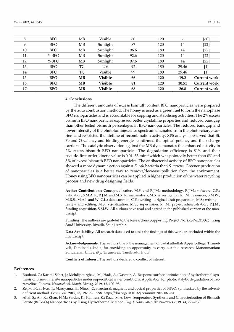
Water 2022, 14, 1545 13 of 16
8. BFO MB Visible 60 120 ‐ [60]
9. BFO MB Sunlight 87 120 14 [22]
10. BFO MB Sunlight 96.6 180 14 [22]
11. Y‐BFO MB Sunlight 92.6 120 14 [22]
12. Y‐BFO MB Sunlight 97.6 180 14 [22]
13. BFO TC UV 92 180 29.46 [1]
14. BFO TC Visible 99 180 29.46 [1]
15. BFO MB Visible 66 120 19.2 Current work
16. BFO MB Visible 81 120 10.51 Current work
17. BFO MB Visible 68 120 26.8 Current work
4. Conclusions
The different amounts of excess bismuth content BFO nanoparticles were prepared
by the auto combustion method. The honey is used as a green fuel to form the nanophase
BFO nanoparticles and is accountable for capping and stabilising activities. The 2% excess
bismuth BFO nanoparticles expressed better crystalline properties and reduced bandgap
than other tested bismuth percentages in BFO nanoparticles. The reduced bandgap and
lower intensity of the photoluminescence spectrum emanated from the photo‐charge car‐
riers and restricted the lifetime of recombination activity. XPS analysis observed that Bi,
Fe and O valency and binding energies confirmed the optical potency and their charge
carriers. The catalytic observation against the MB dye emanates the enhanced activity in
2% excess bismuth BFO nanoparticles. The degradation efficiency is 81% and their
pseudo‐first‐order kinetic value is 0.01453 min−1 which was pointedly better than 0% and
5% of excess bismuth BFO nanoparticles. The antibacterial activity of BFO nanoparticles
showed a more dynamic action against E. coli bacteria than S. aureus. Greener production
of nanoparticles is a better way to remove/decrease pollution from the environment.
Honey using BFO nanoparticles can be applied in higher production of the water recycling
process and new drug designing fields.
Author Contributions: Conceptualization, M.S. and R.J.M.; methodology, R.J.M.; software, C.P.;
validation, S.M.A.K., R.J.M. and M.S.; formal analysis, M.S.; investigation, R.J.M.; resources, S.M.W.,
M.R.S., M.A.I. and W.‐C.L.; data curation, C.P.; writing—original draft preparation, M.S.; writing—
review and editing, M.S.; visualization, M.S.; supervision, R.J.M.; project administration, R.J.M.;
funding acquisition, S.M.W. All authors have read and agreed to the published version of the man‐
uscript.
Funding: The authors are grateful to the Researchers Supporting Project No. (RSP‐2021/326), King
Saud University, Riyadh, Saudi Arabia.
Data Availability: All research data used to assist the findings of this work are included within the
manuscript.
Acknowledgements: The authors thank the management of Sadakathullah Appa College, Tirunel‐
veli, Tamilnadu, India, for providing an opportunity to carry out this research. Manonmaniam
Sundaranar University, Tirunelveli, Tamilnadu, India.
Conflicts of Interest: The authors declare no conflict of interest.
References
1. Rouhani, Z.; Karimi‐Sabet, J.; Mehdipourghazi, M.; Hadi, A.; Dastbaz, A. Response surface optimization of hydrothermal syn‐
thesis of Bismuth ferrite nanoparticles under supercritical water conditions: Application for photocatalytic degradation of Tet‐
racycline. Environ. Nanotechnol. Monit. Manag. 2019, 11, 100198.
2. Zeljković, S.; Ivas, T.; Maruyama, H.; Nino, J.C. Structural, magnetic and optical properties of BiFeO3 synthesized by the solvent‐
deficient method. Ceram. Int. 2019, 45, 19793–19798. https://doi.org/10.1016/j.ceramint.2019.06.234.
3. Altaf, S.; Ali, K.; Khan, H.M.; Sardar, K.; Kamran, K.; Raza, M.A. Low Temperature Synthesis and Characterization of Bismuth
Ferrite (Bi2Fe4O9) Nanoparticles by Using Hydrothermal Method. Dig. J. Nanomater. Biostructures 2019, 14, 727–733.

Water 2022, 14, 1545 14 of 16
4. Gautam, A.; Singh, K.; Sen, K.; Kotnala, R.; Singh, M. Crystal structure and magnetic property of Nd doped BiFeO3
nanocrytallites. Mater. Lett. 2011, 65, 591–594. https://doi.org/10.1016/j.matlet.2010.11.002.
5. Priyadharsini, P.; Pradeep, A.; Sathyamoorthy, B.; Chandrasekaran, G. Enhanced multiferroic properties in La and Ce co‐doped
BiFeO3 nanoparticles. J. Phys. Chem. Solids 2014, 75, 797–802. https://doi.org/10.1016/j.jpcs.2014.03.001.
6. Muneeswaran, M.; Giridharan, N.V. Effect of Dy‐substitution on the structural, vibrational, and multiferroic properties of
BiFeO3 nanoparticles. J. Appl. Phys. 2014, 115, 214109. https://doi.org/10.1063/1.4881529.
7. Ishikawa, K.L. Nonlinear optical response of graphene in time domain. Phys. Rev. B 2010, 82, 201402.
https://doi.org/10.1103/physrevb.82.201402.
8. Arora, M.; Sati, P.C.; Chauhan, S.; Singh, H.; Yadav, K.L.; Chhoker, S.; Kumar, M. Structural, magnetic and optical properties of
Bi1–xDyxFeO3 nanoparticles synthesized by sol–gel method. Mater. Lett. 2013, 96, 71–73.
9. Wang, L.; Han, Y.; Jia, G.; Zhang, C.; Liu, Y.; Liu, L.; Yin, K. Facile preparation, characterization and photocatalytic properties
of multifunctional BiFeO3 nanocrystals. J. Nanosci. Nanotechnol. 2011, 11, 5207–5209.
10. Feng, Y.‐N.; Wang, H.‐C.; Luo, Y.‐D.; Shen, Y.; Lin, Y.‐H. Ferromagnetic and photocatalytic behaviors observed in Cadoped
BiFeO3 nanofibres. J. Appl. Phys. 2013, 113, 146101. https://doi.org/10.1063/1.4801796.
11. Chakrabarti, K.; Sarkar, B.; Ashok, V.D.; Das, K.; Chaudhuri, S.S.; Mitra, A.; De, S.K. Exchange bias effect in BiFeO3‐NiO nano‐
composite. J. Appl. Phys. 2014, 115, 013906. https://doi.org/10.1063/1.4861140.
12. Hasan, M.; Islam, M.F.; Mahbub, R.; Hossain, M.S.; Hakim, M.A. A soft chemical route to the synthesis of BiFeO3 nanoparticles
with enhanced magnetization. Mater. Res. Bull. 2016, 73, 179–186.
13. Li, S.; Nechache, R.; Davalos, I.A.V.; Goupil, G.; Nikolova, L.; Nicklaus, M.; Laverdiere, J.; Ruediger, A.; Rosei, F. Ultrafast
Microwave Hydrothermal Synthesis of BiFeO3 Nanoplates. J. Am. Ceram. Soc. 2013, 96, 3155–3162.
https://doi.org/10.1111/jace.12473.
14. Fang, L.; Liu, J.; Ju, S.; Zheng, F.; Dong, W.; Shen, M. Experimental and theoretical evidence of enhanced ferromagnetism in
sonochemical synthesized BiFeO3 nanoparticles. Appl. Phys. Lett. 2010, 97, 242501. https://doi.org/10.1063/1.3525573.
15. Khan, U.S.; Adeela, N.; Javed, K.; Riaz, S.; Ali, H.H.; Iqbal, M.; Han, X.F.; Naseem, S. Influence of cobalt doping on structural
and magnetic properties of BiFeO3 nanoparticles. J. Nanoparticle Res. 2015, 17, 429. https://doi.org/10.1007/s11051‐015‐3233‐9.
16. Sharma, S.; Bhattacharya, A. Drinking water contamination and treatment techniques. Appl. Water Sci. 2017, 7, 1043–1067.
https://doi.org/10.1007/s13201‐016‐0455‐7.
17. Naushad, M.; Sharma, G.; Alothman, Z.A. Photodegradation of toxic dye using Gum Arabic‐crosslinked‐poly (acrylamide)/Ni
(OH) 2/FeOOH nanocomposites hydrogel. J. Clean. Prod. 2019, 241, 118263.
18. Ali, I.; Alharbi, O.M.L.; Alothman, Z.A.; Al‐Mohaimeed, A.M.; Alwarthan, A. Modeling of fenuron pesticide adsorption on
CNTs for mechanistic insight and removal in water. Environ. Res. 2019, 170, 389–397. https://doi.org/10.1016/j.envres.2018.12.066.
19. Wabaidur, S.M.; Khan, M.A.; Siddiqui, M.R.; Otero, M.; Jeon, B.H.; Alothman, Z.A.; Hakami, A.A.H. Oxygenated functionalities
enriched MWCNTs decorated with silica coated spinel ferrite—A nanocomposite for potentially rapid and efficient de‐colori‐
zation of aquatic environment. J. Mol. Liq. 2020, 317, 113916.
20. Alothman, Z.A. A Review: Fundamental Aspects of Silicate Mesoporous Materials. Materials 2012, 5, 2874–2902.
https://doi.org/10.3390/ma5122874.
21. Khan, M.A.; Alqadami, A.A.; Wabaidur, S.M.; Siddiqui, M.R.; Jeon, B.H.; Alshareef, S.A.; Alothman, Z.A.; Hamedelniel, A.E.
Oil industry waste based non‐magnetic and magnetic hydrochar to sequester potentially toxic post‐transition metal ions from
water. J. Hazard. Mater. 2020, 400, 123247.
22. Satar, N.S.A.; Adnan, R.; Lee, H.L.; Hall, S.R.; Kobayashi, T.; Kassim, M.H.M.; Kaus, N.H.M. Facile green synthesis of ytrium‐
doped BiFeO3 with highly efficient photocatalytic degradation towards methylene blue. Ceram. Int. 2019, 45, 15964–15973.
https://doi.org/10.1016/j.ceramint.2019.05.105.
23. Abushad, M.; Khan, W.; Naseem, S.; Husain, S.; Nadeem, M.; Ansari, A. Influence of Mn doping on microstructure, optical,
dielectric and magnetic properties of BiFeO3 nanoceramics synthesized via sol–gel method. Ceram. Int. 2019, 45, 7437–7445.
24. Wang, Z.B.; Aldalbahi, A.; Ahamad, T.; Alshehri, S.M.; Feng, P.X. Preparation of BiFeO3 and its photoelectric performance as
photoanode of DSSC. Ceram. Int. 2021, 47, 27565–27570.
25. Aliyu, A.; Srivastava, C. Correlation between growth texture, crystallite size, lattice strain and corrosion behavior of cop‐per‐
carbon nanotube composite coatings. Surf. Coat. Technol. 2021, 405, 126596.
26. Ranjithkumar, B.; Ramalingam, H.; Srinivas, C.; Magesh, G.; Rahale, C.S.; El‐Metwaly, N.M.; Shekar, B.C. Natural fuels (Honey
and Cow urine) assisted combustion synthesis of zinc oxide nanoparticles for antimicrobial activities. Ceram. Int. 2021, 47, 14475–
14481. https://doi.org/10.1016/j.ceramint.2021.02.026.
27. Ichangi, A.; Lê, K.; Queraltó, A.; Grosch, M.; Weissing, R.; Ünlü, F.; Chijioke, A.K.; Verma, A.; Fischer, T.; Surmenev, R.; et al.
Electrospun BiFeO3 Nanofibers for Vibrational Energy Harvesting Application. Adv. Eng. Mater. 2021.
https://doi.org/10.1002/adem.202101394.
28. Kossar, S.; Amiruddin, R.; Rasool, A. Study on thickness‐dependence characteristics of bismuth ferrite (BFO) for ultraviolet
(UV) photodetector application. Micro Nano Syst. Lett. 2021, 9, 1–10. https://doi.org/10.1186/s40486‐020‐00128‐7.
29. Sharmila, M.; Jothi Mani, R.; Kader, A.; Ahmad, A.; Eldesoky, G.E.; Yahya, A.E.; Bahajjaj, A.A.A. Photocatalytic and Biological
Activity of ZnO Nanoparticles Using Honey. Coatings 2021, 11, 1046.
30. Chen, X.; Liu, B.; Li, X.; An, T.T.; Zhou, Y.; Li, G.; Wu‐Smart, J.; Alvarez, S.; Naldrett, M.J., Eudy, J.; et al. Identification of anti‐
inflammatory vesicle‐like nanoparticles in honey. J. Extracell. Vesicles 2021, 10, e12069.
Water 2022, 14, 1545 15 of 16
31. Vishwakarma, A.K.; Tripathi, P.; Srivastava, A.; Sinha, A.; Srivastava, O. Band gap engineering of Gd and Co doped BiFeO3 and
their application in hydrogen production through photoelectrochemical route. Int. J. Hydrogen Energy 2017, 42, 22677–22686.
https://doi.org/10.1016/j.ijhydene.2017.07.153.
32. Bharathkumar, S.; Sakar, M.; Vinod, K.R.; Balakumar, S. Versatility of electrospinning in the fabrication of fibrous mat and mesh
nanostructures of bismuth ferrite (BiFeO3) and their magnetic and photocatalytic activities. Phys. Chem. Chem. Phys. 2015, 17,
17745–17754. https://doi.org/10.1039/c5cp01640a.
33. Aghdam, T.R.; Mehrizadeh, H.; Salari, D.; Tseng, H.H.; Niaei, A.; Amini, A. Photocatalytic removal of NOx over immobilized
BiFeO3 nanoparticles and effect of operational parameters. Korean J. Chem. Eng. 2018, 35, 994–999.
34. Pattnaik, S.P.; Behera, A.; Martha, S.; Acharya, R.; Parida, K. Synthesis, photoelectrochemical properties and solar light‐induced
photocatalytic activity of bismuth ferrite nanoparticles. J. Nanoparticle Res. 2018, 20, 10. https://doi.org/10.1007/s11051‐017‐4110‐
5.
35. Prashanthi, K.; Thakur, G.; Thundat, T. Surface enhanced strong visible photoluminescence from one‐dimensional multiferroic
BiFeO3 nanostructures. Surf. Sci. 2012, 606, L83–L86. https://doi.org/10.1016/j.susc.2012.06.003.
36. Mishra, D.K.; Qi, X. Energy levels and photoluminescence properties of nickel‐doped bismuth ferrite. J. Alloys Compd. 2010, 504,
27–31. https://doi.org/10.1016/j.jallcom.2010.05.107.
37. Prashanthi, K.; Gupta, M.; Tsui, Y.Y.; Thundat, T. Effect of annealing atmosphere on microstructural and photoluminescence
characteristics of multiferroic BiFeO3 thin films prepared by pulsed laser deposition technique. Appl. Phys. A 2013, 110, 903–907.
https://doi.org/10.1007/s00339‐012‐7194‐x.
38. Miriyala, N.; Prashanthi, K.; Thundat, T. Oxygen vacancy dominant strong visible photoluminescence from BiFeO3 nanotubes.
Phys. Status Solidi (RRL)—Rapid Res. Lett. 2013, 7, 668–671.
39. Li, J.; Huang, X.; Zhao, X.; Chen, L.J.; Yan, X.P. pH‐Responsive Torpedo‐Like Persistent Luminescence Nanoparticles for Auto‐
fluorescence‐Free Biosensing and High‐Level Information Encryption. Angew. Chem. 2021, 133, 2428–2435.
40. Ramazanov, S.; Sobola, D.; Ţălu, Ş.; Orudzev, F.; Arman, A.; Kaspar, P.; Dallaev, R.; Ramazanov, G. Multiferroic behavior of the
func‐tionalized surface of a flexible substrate by deposition of Bi2O3 and Fe2O3. Microsc. Res. Tech. 2022, 85, 1300–1310.
41. Tian, C.; Yao, Q.; Tong, Z.; Zhou, H.; Rao, G.; Deng, J.; Wang, Z.; Wang, J. Effects of Sm‐doping on microstructure, magnetic
and microwave absorption properties of BiFeO3. J. Rare Earths 2021, 39, 835–843. https://doi.org/10.1016/j.jre.2020.05.003.
42. Ahamad, T.; Aldalbahi, A.; Alshehri, S.M.; Alotaibi, S.; Alzahly, S.; Wang, Z.B.; Feng, P.X. Enhanced photovoltaic performance
of dye‐sensitized solar cells based Ag2O doped BiFeO3 hetrostructures. Sol. Energy 2021, 220, 758–765.
43. Ramadan, W.; Feldhoff, A.; Bahnemann, D. Assessing the photocatalytic oxygen evolution reaction of BiFeO3 loaded with IrO2
nanoparticles as cocatalyst. Sol. Energy Mater. Sol. Cells 2021, 232, 111349. https://doi.org/10.1016/j.solmat.2021.111349.
44. Amrillah, T.; Hermawan, A.; Yin, S.; Juang, J.Y. Formation and physical properties of the self‐assembled BFO–CFO vertically
aligned nanocomposite on a CFO‐buffered two‐dimensional flexible mica substrate. RSC Adv. 2021, 11, 15539–15545.
45. Gajendiran, J.; Raj, S.G.; Kumar, G.R.; Gnanam, S.; Ramya, J.R.; Senthil, V.P. Photoluminescence Properties and Antibacterial
Activity of BiFeO3 and BiFeO3‐CoFe2O4 Composite. J. Electron. Mater. 2022, 51, 8–16.
46. Rameshkumar, C.; Gayathri, R.; Subalakshmi, R. Synthesis and characterization of undopped bismuth ferrite oxide nanoparti‐
cles for the application of cancer treatment. Mater. Today Proc. 2021, 43, 3662–3665.
47. Chouker, M.A.; Abdallah, H.; Zeiz, A.; El‐Dakdouki, M.H. Host‐quest inclusion complex of quinoxaline‐1, 4‐dioxide derivative
with 2‐hydroxypropyl‐β‐cyclodextrin: Preparation, characterization, and antibacterial activity. J. Mol. Struct. 2021, 1235, 130273.
48. Ahmed, H.M.; Roy, A.; Wahab, M.; Ahmed, M.; Othman‐Qadir, G.; Elesawy, B.H.; Khandaker, M.U.; Islam, M.N.; Emran, T.B.
Applications of nanomaterials in agri food and pharmaceutical industry. J. Nanomater. 2021, 2021, 1472096.
49. Nabavi, S.; Behzad, S. Herbal Drugs and Natural Products in the light of Nanotechnology and Nanomedicine for Developing
Drug Formulations. Mini‐Rev. Med. Chem. 2021, 21, 302–313. https://doi.org/10.2174/1389557520666200916143240.
50. Roy, A.; Ananda Murthy, H.C.; Ahmed, H.M.; Islam, M.N.; Prasad, R. Phytogenic Synthesis of Metal/Metal Oxide Nanoparticles
for Degradation of Dyes. J. Renew. Mater. 2022, 10, 1911.
51. Shimi, A.K.; Ahmed, H.M.; Wahab, M.; Katheria, S.; Wabaidur, S.M.; Eldesoky, G.E.; Islam, M.A.; Rane, K.P. Synthesis and Ap‐
plications of Green Synthesized TiO2 Nanoparticles for Photocatalytic Dye Degradation and Antibacterial Activity. J. Nanomater.
2022, 2022, 7060388.
52. Pan, Z.; Liu, M.; Zhang, G.; Zhuzhang, H.; Wang, X. Molecular Triazine–Heptazine Junctions Promoting Exciton Dissociation
for Overall Water Splitting with Visible Light. J. Phys. Chem. C 2021, 125, 9818–9826. https://doi.org/10.1021/acs.jpcc.1c02274.
53. Pan, Z.; Zhao, M.; Zhuzhang, H.; Zhang, G.; Anpo, M.; Wang, X. Gradient Zn‐Doped Poly Heptazine Imides Integrated with a
van der Waals Homojunction Boosting Visible Light‐Driven Water Oxidation Activities. ACS Catal. 2021, 11, 13463–13471.
https://doi.org/10.1021/acscatal.1c03687.
54. Siddique, M.; Khan, N.M.; Saeed, M. Photocatalytic Activity of Bismuth Ferrite Nanoparticles Synthesized via Sol‐Gel Route.
Z. Für Phys. Chem. 2019, 233, 595–607. https://doi.org/10.1515/zpch‐2018‐1225.
55. Puhan, A.; Bhushan, B.; Satpathy, S.; Meena, S.S.; Nayak, A.; Rout, D. Facile single phase synthesis of Sr, Co co‐doped BiFeO3
nanoparticles for boosting photocatalytic and magnetic properties. Appl. Surf. Sci. 2019, 493, 593–604.
https://doi.org/10.1016/j.apsusc.2019.07.002.
56. Ponraj, C.; Vinitha, G.; Daniel, J. Photocatalytic degradation of acid red‐85 dye by nickel substituted bismuth ferrite nanoparti‐
cles. Mater. Res. Express 2019, 6, 084006.

Water 2022, 14, 1545 16 of 16
57. Jaffari, Z.; Lam, S.‐M.; Sin, J.‐C.; Zeng, H. Boosting visible light photocatalytic and antibacterial performance by decoration of
silver on magnetic spindle‐like bismuth ferrite. Mater. Sci. Semicond. Process. 2019, 101, 103–115.
https://doi.org/10.1016/j.mssp.2019.05.036.
58. Irfan, S.; Zhuanghao, Z.; Li, F.; Chen, Y.‐X.; Liang, G.‐X.; Luo, J.‐T.; Ping, F. Critical review: Bismuth ferrite as an emerging
visible light active nanostructured photocatalyst. J. Mater. Res. Technol. 2019, 8, 6375–6389.
https://doi.org/10.1016/j.jmrt.2019.10.004.
59. Dumitru, R.; Ianculescu, A.; Păcurariu, C.; Lupa, L.; Pop, A.; Vasile, B.S.; Surdu, V.‐A.; Manea, F. BiFeO3‐synthesis, characteri‐
zation and its photocatalytic activity towards doxorubicin degradation from water. Ceram. Int. 2019, 45, 2789–2802.
https://doi.org/10.1016/j.ceramint.2018.07.298.
60. Duan, Q.; Kong, F.; Han, X.; Jiang, Y.; Liu, T.; Chang, Y.; Zhou, L.; Qin, G.; Zhang, X. Synthesis and characterization of mor‐
phology‐controllable BiFeO3 particles with efficient photocatalytic activity. Mater. Res. Bull. 2019, 112, 104–108.
Related Documents


