Review Visceral Leishmaniasis and HIV Coinfection in East Africa Ermias Diro 1,2 *, Lutgarde Lynen 1 , Koert Ritmeijer 3 , Marleen Boelaert 4 , Asrat Hailu 5 , Johan van Griensven 1 1 Department of Clinical Sciences, Institute of Tropical Medicine, Antwerp, Belgium, 2 Department of Internal Medicine, University of Gondar, Gondar, Ethiopia, 3 Public Health Department, Me ´ decins Sans Frontie ` res, Amsterdam, the Netherlands, 4 Department of Public Health, Institute of Tropical Medicine, Antwerp, Belgium, 5 School of Medicine, Addis Ababa University, Addis Ababa, Ethiopia Abstract: Visceral Leishmaniasis (VL) is an important protozoan opportunistic disease in HIV patients in endemic areas. East Africa is second to the Indian subcontinent in the global VL caseload and first in VL- HIV coinfection rate. Because of the alteration in the disease course, the diagnostic challenges, and the poor treatment responses, VL with HIV coinfection has become a very serious challenge in East Africa today. Field experience with the use of liposomal amphotericin B in combination with miltefosine, followed by secondary prophylaxis and antiretroviral drugs, looks promising. However, this needs to be confirmed through clinical trials. Better diagnostic and follow-up methods for relapse and prediction of relapse should also be looked for. Basic research to understand the immunological interaction of the two infections may ultimately help to improve the management of the coinfection. Introduction Visceral leishmaniasis (VL) is a vector-borne protozoan infection targeting the reticuloendothelial system [1]. Its occur- rence is widespread, being prevalent in approximately 70 countries worldwide. East Africa is one of the most affected regions, second only to the Indian subcontinent, with an estimated annual incidence rate of 29,400 to 56,700 cases [2]. The countries most affected in this region are Sudan, South Sudan, and then Ethiopia. Although with much lower VL burden, endemic foci of the disease are also found in Eritrea, Somalia, Kenya, and Uganda [2]. Figure 1 shows the VL-endemic regions in East Africa. The disease typically affects poor communities residing in remote places with poorly functioning health-care systems. Historically, East African VL has claimed the lives of many people, with the most infamous epidemic reported from South Sudan by Seaman et al. [3]. Between 1984 and 1994, a devastating epidemic in the western Upper Nile region in South Sudan claimed the lives of an estimated 100,000 people [3]. To date, treatment and care for VL in these resource-poor countries is mainly provided or supported by international organizations such as Me ´decins Sans Frontie `res (MSF), Drugs for Neglected Diseases initiative (DNDi), and the World Health Organization (WHO). The simultaneous infection of humans by HIV and Leishmania almost always leads to a ‘‘deadly gridlock,’’ as they both have the same deleterious effect on the immune response [4]. The majority of VL-HIV coinfections were previously reported from the Mediterranean countries during the pandemic years of HIV/ AIDS in the 1990s, with the prevalence of HIV among VL patients reaching up to 60% in intravenous drug users in Spain [5,6]. As a consequence of HIV coinfection, atypical presentations of VL, a high rate of treatment failure, and frequent relapses were reported. Only after the introduction of antiretroviral therapy (ART) was a decline in incidence of VL-HIV coinfection observed [5]. With the spread of HIV to other VL-endemic regions of the world, the coinfection is now reported from 35 countries [5,7]. Because of the alteration of the disease course, the diagnostic challenges, and the poor treatment response, VL with HIV coinfection has become a very serious challenge in East Africa today. HIV Epidemiology in East Africa The prevalence of HIV increased alarmingly from the mid- 1980s to the 1990s in most African countries. Since 2000, however, a decline has been seen in the number of new HIV infections, which can be explained by a variety of factors, most notably preventive measures and access to ART [8,9]. In Ethiopia, HIV prevalence has declined from 5.6% in 2005 to 2.6% in 2011 (antenatal care sentinel surveillance [10]), and the estimated prevalence among the adult population is 1.5% (Demographic and Health Survey [DHS] 2011) [11]. However, despite the decreasing prevalence of HIV in the general population, the prevalence of HIV among VL patients has remained proportionally very high. The northwest districts of Ethiopia along the Sudanese border report the highest burden of HIV and VL coinfection rates, with HIV prevalence rates of 20%–40% among VL patients [5,7,12]. The 2012 annual report from the Leishmaniasis Research and Treatment Centre of the University of Gondar showed that 81/ 332 (24.4%) of all admitted VL cases were HIV coinfected (unpublished data). The rates of coinfection from different studies in Ethiopia are summarized in Table 1. The particularly high HIV coinfection rate in northwest Ethiopia could be due to the massive population movement in the region [13]. In this area of cash-crop farming, there is a high labour demand, and 300,000 to 500,000 highlanders from urban and semiurban areas seasonally move in and out of the region. When these Leishmania-nonimmune highlanders go to the VL- endemic regions, they become exposed and infected. Internal migration is also a risk factor for HIV, and those infected with Citation: Diro E, Lynen L, Ritmeijer K, Boelaert M, Hailu A, et al. (2014) Visceral Leishmaniasis and HIV Coinfection in East Africa. PLOS Negl Trop Dis 8(6): e2869. doi:10.1371/journal.pntd.0002869 Editor: Jesus G. Valenzuela, National Institute of Allergy and Infectious Diseases, United States of America Published June 26, 2014 Copyright: ß 2014 Diro et al. This is an open-access article distributed under the terms of the Creative Commons Attribution License, which permits unrestricted use, distribution, and reproduction in any medium, provided the original author and source are credited. Funding: ED has received individual PhD scholarship from Belgian development cooperation. The funders had no role in study design, data collection and analysis, decision to publish, or preparation of the manuscript. Competing Interests: The authors have declared that no competing interests exist. * Email: [email protected] PLOS Neglected Tropical Diseases | www.plosntds.org 1 June 2014 | Volume 8 | Issue 6 | e2869

Welcome message from author
This document is posted to help you gain knowledge. Please leave a comment to let me know what you think about it! Share it to your friends and learn new things together.
Transcript
Review
Visceral Leishmaniasis and HIV Coinfection in East AfricaErmias Diro1,2*, Lutgarde Lynen1, Koert Ritmeijer3, Marleen Boelaert4, Asrat Hailu5,
Johan van Griensven1
1 Department of Clinical Sciences, Institute of Tropical Medicine, Antwerp, Belgium, 2 Department of Internal Medicine, University of Gondar, Gondar, Ethiopia, 3 Public
Health Department, Medecins Sans Frontieres, Amsterdam, the Netherlands, 4 Department of Public Health, Institute of Tropical Medicine, Antwerp, Belgium, 5 School of
Medicine, Addis Ababa University, Addis Ababa, Ethiopia
Abstract: Visceral Leishmaniasis (VL) is an importantprotozoan opportunistic disease in HIV patients inendemic areas. East Africa is second to the Indiansubcontinent in the global VL caseload and first in VL-HIV coinfection rate. Because of the alteration in thedisease course, the diagnostic challenges, and the poortreatment responses, VL with HIV coinfection has becomea very serious challenge in East Africa today. Fieldexperience with the use of liposomal amphotericin B incombination with miltefosine, followed by secondaryprophylaxis and antiretroviral drugs, looks promising.However, this needs to be confirmed through clinicaltrials. Better diagnostic and follow-up methods for relapseand prediction of relapse should also be looked for. Basicresearch to understand the immunological interaction ofthe two infections may ultimately help to improve themanagement of the coinfection.
Introduction
Visceral leishmaniasis (VL) is a vector-borne protozoan
infection targeting the reticuloendothelial system [1]. Its occur-
rence is widespread, being prevalent in approximately 70 countries
worldwide. East Africa is one of the most affected regions, second
only to the Indian subcontinent, with an estimated annual
incidence rate of 29,400 to 56,700 cases [2]. The countries most
affected in this region are Sudan, South Sudan, and then Ethiopia.
Although with much lower VL burden, endemic foci of the disease
are also found in Eritrea, Somalia, Kenya, and Uganda [2].
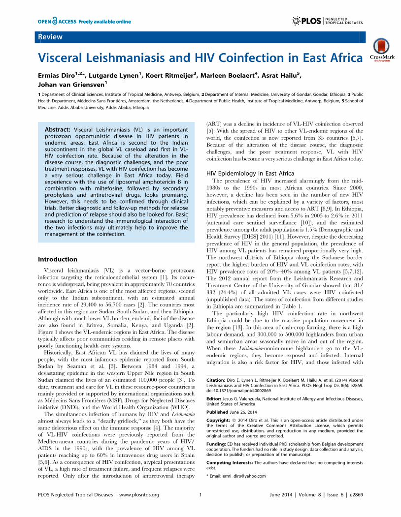
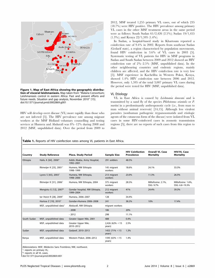
Figure 1 shows the VL-endemic regions in East Africa. The disease
typically affects poor communities residing in remote places with
poorly functioning health-care systems.
Historically, East African VL has claimed the lives of many
people, with the most infamous epidemic reported from South
Sudan by Seaman et al. [3]. Between 1984 and 1994, a
devastating epidemic in the western Upper Nile region in South
Sudan claimed the lives of an estimated 100,000 people [3]. To
date, treatment and care for VL in these resource-poor countries is
mainly provided or supported by international organizations such
as Medecins Sans Frontieres (MSF), Drugs for Neglected Diseases
initiative (DNDi), and the World Health Organization (WHO).
The simultaneous infection of humans by HIV and Leishmania
almost always leads to a ‘‘deadly gridlock,’’ as they both have the
same deleterious effect on the immune response [4]. The majority
of VL-HIV coinfections were previously reported from the
Mediterranean countries during the pandemic years of HIV/
AIDS in the 1990s, with the prevalence of HIV among VL
patients reaching up to 60% in intravenous drug users in Spain
[5,6]. As a consequence of HIV coinfection, atypical presentations
of VL, a high rate of treatment failure, and frequent relapses were
reported. Only after the introduction of antiretroviral therapy
(ART) was a decline in incidence of VL-HIV coinfection observed
[5]. With the spread of HIV to other VL-endemic regions of the
world, the coinfection is now reported from 35 countries [5,7].
Because of the alteration of the disease course, the diagnostic
challenges, and the poor treatment response, VL with HIV
coinfection has become a very serious challenge in East Africa today.
HIV Epidemiology in East AfricaThe prevalence of HIV increased alarmingly from the mid-
1980s to the 1990s in most African countries. Since 2000,
however, a decline has been seen in the number of new HIV
infections, which can be explained by a variety of factors, most
notably preventive measures and access to ART [8,9]. In Ethiopia,
HIV prevalence has declined from 5.6% in 2005 to 2.6% in 2011
(antenatal care sentinel surveillance [10]), and the estimated
prevalence among the adult population is 1.5% (Demographic and
Health Survey [DHS] 2011) [11]. However, despite the decreasing
prevalence of HIV in the general population, the prevalence of
HIV among VL patients has remained proportionally very high.
The northwest districts of Ethiopia along the Sudanese border
report the highest burden of HIV and VL coinfection rates, with
HIV prevalence rates of 20%–40% among VL patients [5,7,12].
The 2012 annual report from the Leishmaniasis Research and
Treatment Centre of the University of Gondar showed that 81/
332 (24.4%) of all admitted VL cases were HIV coinfected
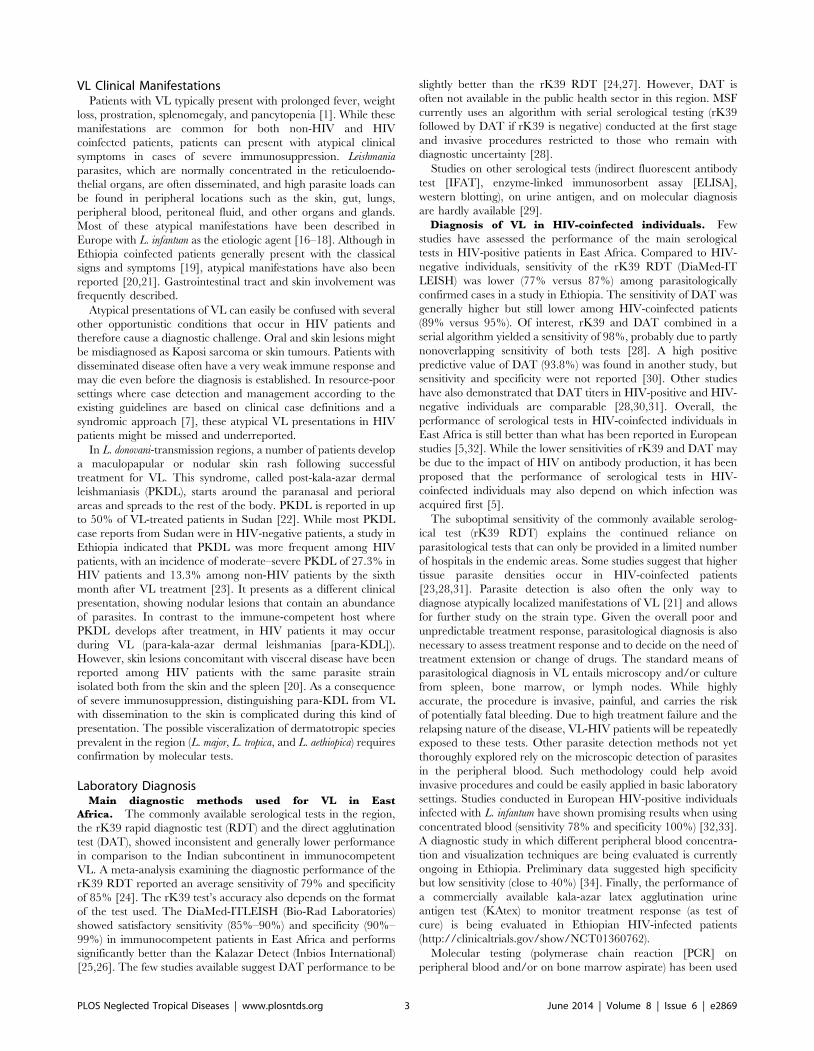
(unpublished data). The rates of coinfection from different studies
in Ethiopia are summarized in Table 1.
The particularly high HIV coinfection rate in northwest
Ethiopia could be due to the massive population movement in
the region [13]. In this area of cash-crop farming, there is a high
labour demand, and 300,000 to 500,000 highlanders from urban
and semiurban areas seasonally move in and out of the region.
When these Leishmania-nonimmune highlanders go to the VL-
endemic regions, they become exposed and infected. Internal
migration is also a risk factor for HIV, and those infected with
Citation: Diro E, Lynen L, Ritmeijer K, Boelaert M, Hailu A, et al. (2014) VisceralLeishmaniasis and HIV Coinfection in East Africa. PLOS Negl Trop Dis 8(6): e2869.doi:10.1371/journal.pntd.0002869
Editor: Jesus G. Valenzuela, National Institute of Allergy and Infectious Diseases,United States of America
Published June 26, 2014
Copyright: � 2014 Diro et al. This is an open-access article distributed underthe terms of the Creative Commons Attribution License, which permitsunrestricted use, distribution, and reproduction in any medium, provided theoriginal author and source are credited.
Funding: ED has received individual PhD scholarship from Belgian developmentcooperation. The funders had no role in study design, data collection and analysis,decision to publish, or preparation of the manuscript.
Competing Interests: The authors have declared that no competing interestsexist.
* Email: [email protected]
PLOS Neglected Tropical Diseases | www.plosntds.org 1 June 2014 | Volume 8 | Issue 6 | e2869
HIV will develop overt disease (VL) more rapidly than those who
are not infected [5]. The HIV prevalence rate among migrant
workers at the MSF Holland voluntary counselling and testing
services at Humera and Abdurafi was 8%–12% during 2008 and
2012 (MSF, unpublished data). Over the period from 2009 to
2012, MSF treated 1,255 primary VL cases, out of which 235
(18.7%) were HIV positive. The HIV prevalence among primary
VL cases in the other MSF treatment areas in the same period
were as follows: South Sudan 61/2,426 (2.5%); Sudan 19/1,455
(1.3%); and Kenya 22/1,595 (1.4%).
In Sudan, a hospital-based study in Khartoum reported a
coinfection rate of 9.4% in 2002. Reports from southeast Sudan
(Gedarif state), a region characterised by population movements,
found HIV coinfection in 3.6% of VL cases in 2003 [5].
Systematic testing of VL patients for HIV in MSF programs in
Sudan and South Sudan between 2009 and 2012 showed an HIV
coinfection rate of 2%–2.5% (MSF, unpublished data). In the
other neighbouring countries and endemic regions, mainly
children are affected, and the HIV coinfection rate is very low
[5]. MSF experience in Kacheliba in Western Pokot, Kenya,
showed 1.4% HIV coinfection rate between 2006 and 2012.
However, only 1,595 of the total 3,007 primary VL cases during
the period were tested for HIV (MSF, unpublished data).
VL EtiologyVL in East Africa is caused by Leishmania donovani and is
transmitted by a sand fly of the species Phlebotomus orientalis or P.
martini in a predominantly anthroponotic cycle (i.e., from man to
man without animal reservoir) [14,15]. Although less virulent
parasites (nonhuman pathogenic trypanosomatids and etiologic
agents of the cutaneous form of the disease) were isolated from VL
cases in some HIV-coinfected cases in zoonotic transmission
regions [5], there are no reports of such cases from this region to
date.
Table 1. Reports of HIV coinfection rates among VL patients in East Africa.
Country Study Reference Place, Study Period Sample SizeHIV CoinfectionPrevalence
Overall VL CaseMortality
HIV/VL CaseMortality
Ethiopia Hailu A [64], 2006* Addis Ababa, Army Hospital,1992–2001
291 soldiers 48.5%
Ritmeijer K [23], 2001c Humera, NW Ethiopia1998–1999
145 migrantworkers
18.6% 24.1% 33.3%
Lyons S [65], 2003c Humera, NW Ethiopia,1998–2000
213 migrantworkers
23.0% 11.3% 26.5%
Ritmeijer K [31], 2006c Humera, NW Ethiopia, 2004 375 migrantworkers
28.5% Miltefosine: 2.1%,SSG: 9.7%
Miltefosine: 1.6%,SSG: 6.8–19.3%
Mengistu G [12], 2007* Gondar hospital, NW Ethiopia,1999–2004
212 migrantworkers
41% 24.4% 39.3%
ter Horst R [28], 2009c Humera, 2006–2007 128 34.4%
Hurissa Z [19], 2010c Gondar+Humera 2006–2008 241 38.2% 10% 17.4%
MSF, unpublished datac Abdurafi, NW Ethiopia migrant workers
- 2008 186 20%
- 2012 298 11.1%
South Sudan MSF, unpublished data Greater Upper Nile, 2001 488 0.4%
MSF, unpublished data Greater Upper Nile,2010–2012
2,426 (62% ,15years)
2.5%
Sudan MSF, unpublished data Gedaref, 2010–2013 1455 (71% ,15years)
1.3%
Kenya MSF, unpublished data Western Pokot, 2006–2012 1595 (63% ,15years)
1.4%
Abbreviations: MSF, Medecins Sans Frontieres; NW, northwest.c: reports on primary VL;*: reports in all VL cases.doi:10.1371/journal.pntd.0002869.t001
Figure 1. Map of East Africa showing the geographic distribu-tion of visceral leishmaniasis. Map taken from ‘‘Malaria Consortium;Leishmaniasis control in eastern Africa: Past and present efforts andfuture needs. Situation and gap analysis, November 2010’’ [15].doi:10.1371/journal.pntd.0002869.g001
PLOS Neglected Tropical Diseases | www.plosntds.org 2 June 2014 | Volume 8 | Issue 6 | e2869
VL Clinical ManifestationsPatients with VL typically present with prolonged fever, weight
loss, prostration, splenomegaly, and pancytopenia [1]. While these
manifestations are common for both non-HIV and HIV
coinfected patients, patients can present with atypical clinical
symptoms in cases of severe immunosuppression. Leishmania
parasites, which are normally concentrated in the reticuloendo-
thelial organs, are often disseminated, and high parasite loads can
be found in peripheral locations such as the skin, gut, lungs,
peripheral blood, peritoneal fluid, and other organs and glands.
Most of these atypical manifestations have been described in
Europe with L. infantum as the etiologic agent [16–18]. Although in
Ethiopia coinfected patients generally present with the classical
signs and symptoms [19], atypical manifestations have also been
reported [20,21]. Gastrointestinal tract and skin involvement was
frequently described.
Atypical presentations of VL can easily be confused with several
other opportunistic conditions that occur in HIV patients and
therefore cause a diagnostic challenge. Oral and skin lesions might
be misdiagnosed as Kaposi sarcoma or skin tumours. Patients with
disseminated disease often have a very weak immune response and
may die even before the diagnosis is established. In resource-poor
settings where case detection and management according to the
existing guidelines are based on clinical case definitions and a
syndromic approach [7], these atypical VL presentations in HIV
patients might be missed and underreported.
In L. donovani-transmission regions, a number of patients develop
a maculopapular or nodular skin rash following successful
treatment for VL. This syndrome, called post-kala-azar dermal
leishmaniasis (PKDL), starts around the paranasal and perioral
areas and spreads to the rest of the body. PKDL is reported in up
to 50% of VL-treated patients in Sudan [22]. While most PKDL
case reports from Sudan were in HIV-negative patients, a study in
Ethiopia indicated that PKDL was more frequent among HIV
patients, with an incidence of moderate–severe PKDL of 27.3% in
HIV patients and 13.3% among non-HIV patients by the sixth
month after VL treatment [23]. It presents as a different clinical
presentation, showing nodular lesions that contain an abundance
of parasites. In contrast to the immune-competent host where
PKDL develops after treatment, in HIV patients it may occur
during VL (para-kala-azar dermal leishmanias [para-KDL]).
However, skin lesions concomitant with visceral disease have been
reported among HIV patients with the same parasite strain
isolated both from the skin and the spleen [20]. As a consequence
of severe immunosuppression, distinguishing para-KDL from VL
with dissemination to the skin is complicated during this kind of
presentation. The possible visceralization of dermatotropic species
prevalent in the region (L. major, L. tropica, and L. aethiopica) requires
confirmation by molecular tests.
Laboratory DiagnosisMain diagnostic methods used for VL in East
Africa. The commonly available serological tests in the region,
the rK39 rapid diagnostic test (RDT) and the direct agglutination
test (DAT), showed inconsistent and generally lower performance
in comparison to the Indian subcontinent in immunocompetent
VL. A meta-analysis examining the diagnostic performance of the
rK39 RDT reported an average sensitivity of 79% and specificity
of 85% [24]. The rK39 test’s accuracy also depends on the format
of the test used. The DiaMed-ITLEISH (Bio-Rad Laboratories)
showed satisfactory sensitivity (85%–90%) and specificity (90%–
99%) in immunocompetent patients in East Africa and performs
significantly better than the Kalazar Detect (Inbios International)
[25,26]. The few studies available suggest DAT performance to be
slightly better than the rK39 RDT [24,27]. However, DAT is
often not available in the public health sector in this region. MSF
currently uses an algorithm with serial serological testing (rK39
followed by DAT if rK39 is negative) conducted at the first stage
and invasive procedures restricted to those who remain with
diagnostic uncertainty [28].
Studies on other serological tests (indirect fluorescent antibody
test [IFAT], enzyme-linked immunosorbent assay [ELISA],
western blotting), on urine antigen, and on molecular diagnosis
are hardly available [29].
Diagnosis of VL in HIV-coinfected individuals. Few
studies have assessed the performance of the main serological
tests in HIV-positive patients in East Africa. Compared to HIV-
negative individuals, sensitivity of the rK39 RDT (DiaMed-IT
LEISH) was lower (77% versus 87%) among parasitologically
confirmed cases in a study in Ethiopia. The sensitivity of DAT was
generally higher but still lower among HIV-coinfected patients
(89% versus 95%). Of interest, rK39 and DAT combined in a
serial algorithm yielded a sensitivity of 98%, probably due to partly
nonoverlapping sensitivity of both tests [28]. A high positive
predictive value of DAT (93.8%) was found in another study, but
sensitivity and specificity were not reported [30]. Other studies
have also demonstrated that DAT titers in HIV-positive and HIV-
negative individuals are comparable [28,30,31]. Overall, the
performance of serological tests in HIV-coinfected individuals in
East Africa is still better than what has been reported in European
studies [5,32]. While the lower sensitivities of rK39 and DAT may
be due to the impact of HIV on antibody production, it has been
proposed that the performance of serological tests in HIV-
coinfected individuals may also depend on which infection was
acquired first [5].
The suboptimal sensitivity of the commonly available serolog-
ical test (rK39 RDT) explains the continued reliance on
parasitological tests that can only be provided in a limited number
of hospitals in the endemic areas. Some studies suggest that higher
tissue parasite densities occur in HIV-coinfected patients
[23,28,31]. Parasite detection is also often the only way to
diagnose atypically localized manifestations of VL [21] and allows
for further study on the strain type. Given the overall poor and
unpredictable treatment response, parasitological diagnosis is also
necessary to assess treatment response and to decide on the need of
treatment extension or change of drugs. The standard means of
parasitological diagnosis in VL entails microscopy and/or culture
from spleen, bone marrow, or lymph nodes. While highly
accurate, the procedure is invasive, painful, and carries the risk
of potentially fatal bleeding. Due to high treatment failure and the
relapsing nature of the disease, VL-HIV patients will be repeatedly
exposed to these tests. Other parasite detection methods not yet
thoroughly explored rely on the microscopic detection of parasites
in the peripheral blood. Such methodology could help avoid
invasive procedures and could be easily applied in basic laboratory
settings. Studies conducted in European HIV-positive individuals
infected with L. infantum have shown promising results when using
concentrated blood (sensitivity 78% and specificity 100%) [32,33].
A diagnostic study in which different peripheral blood concentra-
tion and visualization techniques are being evaluated is currently
ongoing in Ethiopia. Preliminary data suggested high specificity
but low sensitivity (close to 40%) [34]. Finally, the performance of
a commercially available kala-azar latex agglutination urine
antigen test (KAtex) to monitor treatment response (as test of
cure) is being evaluated in Ethiopian HIV-infected patients
(http://clinicaltrials.gov/show/NCT01360762).
Molecular testing (polymerase chain reaction [PCR] on
peripheral blood and/or on bone marrow aspirate) has been used
PLOS Neglected Tropical Diseases | www.plosntds.org 3 June 2014 | Volume 8 | Issue 6 | e2869
in first-line diagnosis for VL in HIV-coinfected individuals in
European countries and Brazil and merits further exploration in
East Africa [35]. Ongoing or planned diagnostic studies focusing
on HIV-coinfected individuals in East Africa are summarized in
Box 1.
Treatment OutcomesVL treatment in HIV coinfection in East Africa. Striking
differences exist in VL treatment response in the general VL
patient population across and within regions [36–38]. Higher
doses of paromomycin and liposomal amphotericin B appear
necessary for treatment of L. donovani in East Africa than in the
Indian subcontinent. Of interest, within East Africa, clear
differences in efficacy were seen with these drugs in between
and within different countries, with the lowest cure rates noted in
northern Ethiopia and Sudan. Possibly, these observations could
extend to VL-HIV coinfection as well.
For decades, antimonials have been the cornerstone of VL
treatment in Africa. Although these drugs still maintain good
efficacy in East Africa, their use is associated with unacceptably
high and potentially fatal toxicity in VL-HIV coinfection [31].
Reported death rates during antimonial treatment typically have
been 4- to 10-fold higher compared to HIV-negative individuals
[12,23,38] and have varied between 6.5% and 24.5% in a more
recent study [19]. In a recent Ethiopian study, high parasitolog-
ically confirmed treatment-failure rates (30%) were observed in
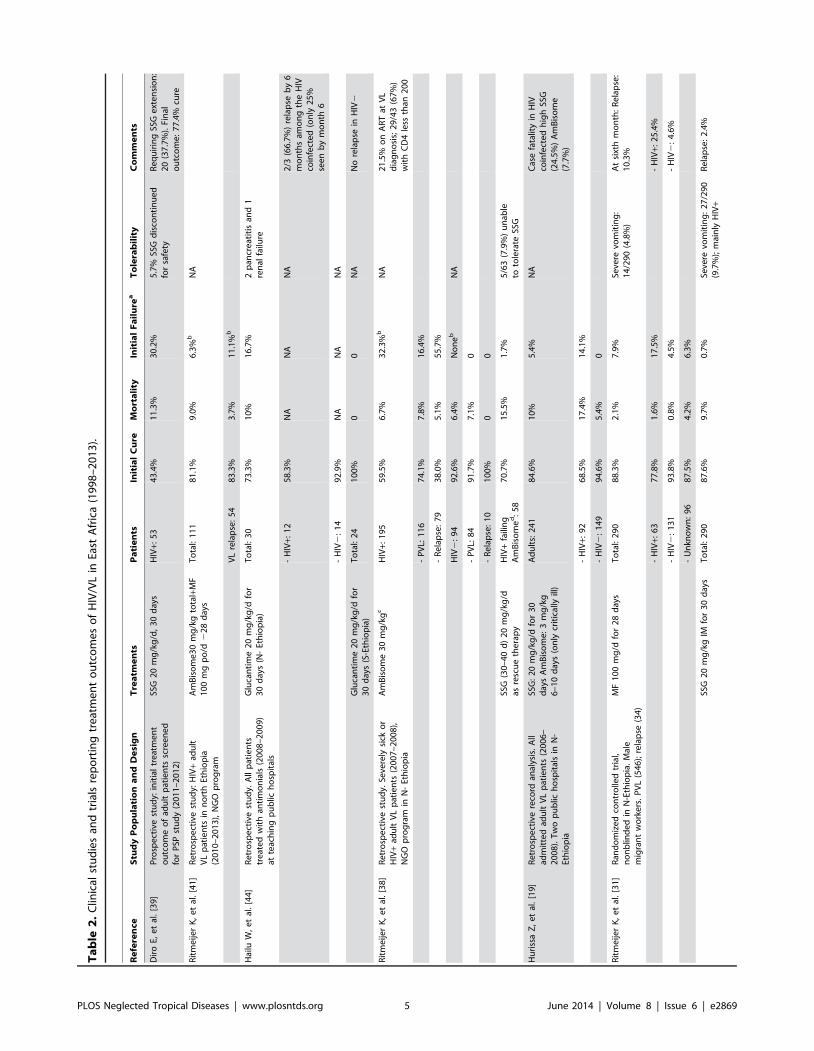
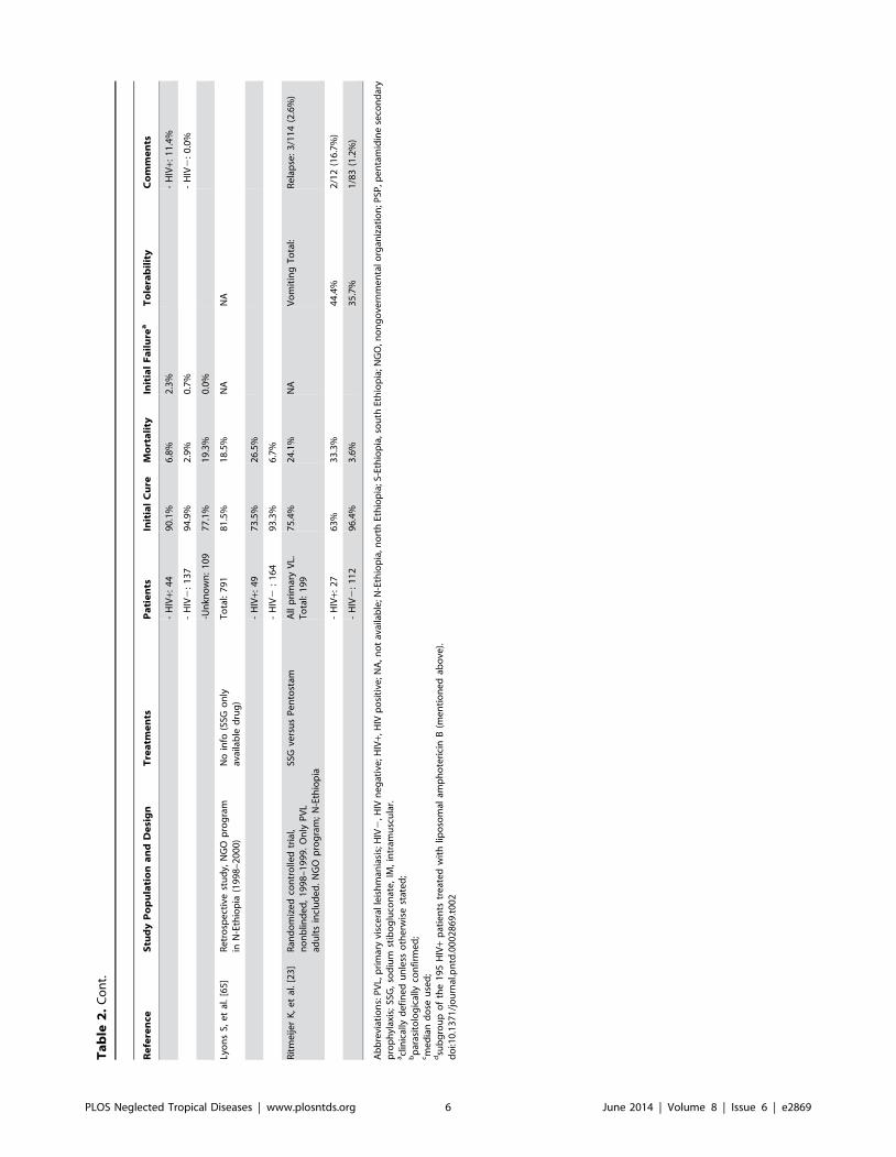
HIV-infected patients treated with antimonials [39]. Table 2
summarizes the studies showing the treatment outcomes of VL
and VL-HIV for different antileishmanial drugs from 1998 on.
In the search for a safer alternative, liposomal amphotericin B
has increasingly been explored in East Africa. While studies have
consistently reported an excellent tolerability, cure rates in HIV-
infected individuals have been rather disappointing in this
continent. At a total dose of 30 mg/kg, around 16% of primary
VL and 56% of VL relapse cases demonstrate parasitological
failure in northern Ethiopia [38]. This is in clear contrast with a
report from India in which high (100%) cure rates were achieved
at a total dose of 20 mg/kg [40]. Current WHO guidelines
recommend a cumulative dose of 40 mg/kg, with 8 to 10 doses of
3–5 mg/kg taken daily or intermittently, for VL-HIV coinfection
in East Africa [7], although this has not yet been evaluated in the
region.
Miltefosine has been evaluated in only one clinical trial in
Ethiopia [31]. In comparison with antimonials, it was found safer
but less effective, with 17.5% parasitological treatment failure.
Interestingly, a compassionate use of miltefosine in combination
with liposomal amphotericin B (at 30 mg/kg total dose) in 111
HIV-coinfected VL patients seems to suggest substantially higher
cure rates and lower failure rates both in primary VL and VL
relapse [41]. Based on this emerging evidence, a clinical trial is
planned to start in northwest Ethiopia by the end of 2013,
evaluating in parallel two treatment options: (1) combination
therapy: miltefosine (2.5 mg/kg per day) for 28 days combined
with liposomal amphotericin B (6 doses of 5 mg/kg; total dose
30 mg/kg) and (2) a high dose of liposomal amphotericin B (8
doses of 5 mg/kg; total dose 40 mg/kg).
Whereas the combination of antimonials and paromomycin (for
17 days) is now recommended by WHO as first-line treatment in
immunocompetent individuals in East Africa, experience with it as
a first-line treatment for HIV-coinfected individuals is limited [42].
This regimen is now used in some programs for VL-HIV
coinfected patients as a second-line treatment (in case of
intolerance or failure of liposomal amphotericin B) [43] or as a
first-line treatment if access to and availability of liposomal
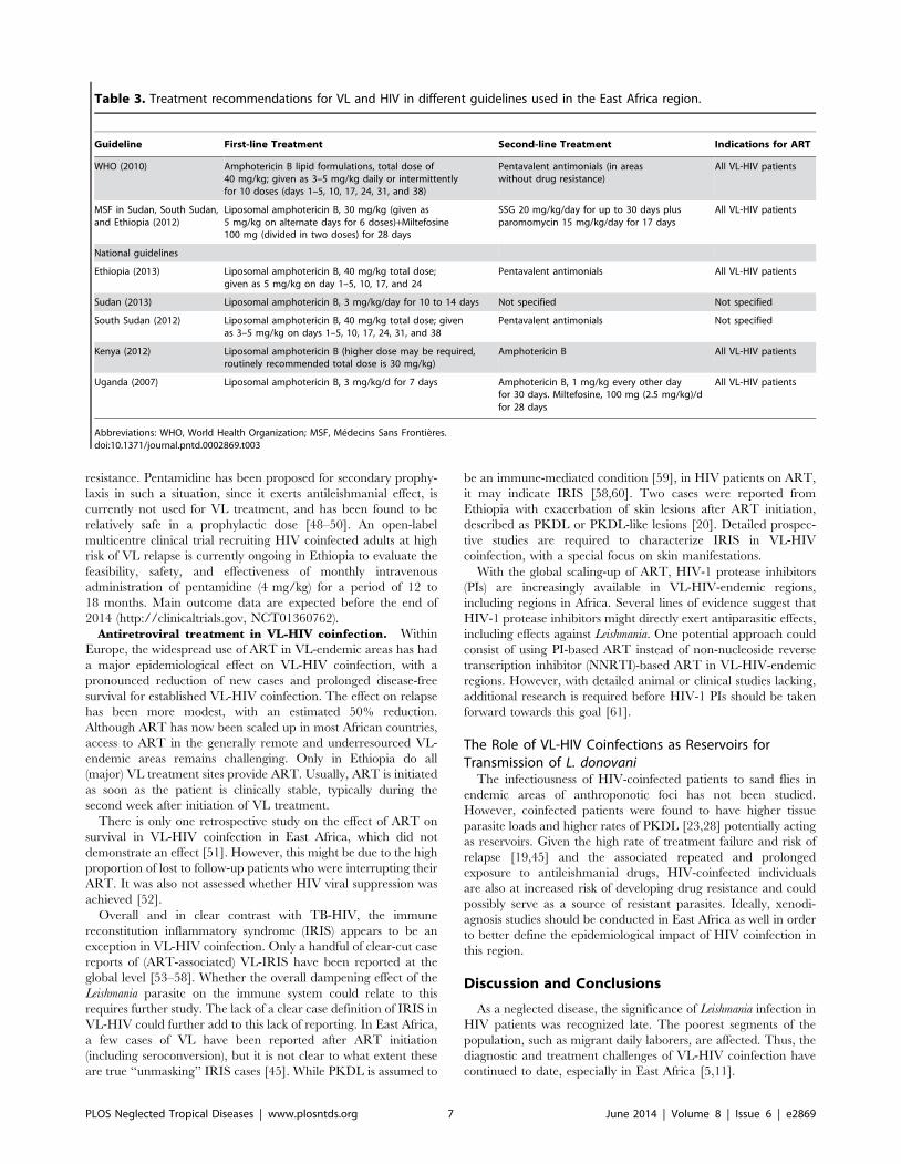
amphotericin B is limited. Current national and international
treatment recommendations for VL-HIV coinfection are summa-
rized in Table 3.
Risk of relapse and secondary prophylaxis in VL-HIV
coinfection. Given the high rates of lost to follow-up in most
reported studies (often above 50%) [19,23,44], reliable data on the
risk of relapse in VL-HIV-coinfected individuals are very scarce.
In the most complete study, the reported risk of relapse at six
months varied between 25.4% for the miltefosine group and
11.4% for the sodium stibogluconate (SSG) group [31]. Another
study estimated a one-year relapse risk of close to 20% for
individuals with primary VL and CD4 cell counts of around 200
cells/mL and around 60% for those with multiple previous VL
episodes and CD4 cell counts below 100 cells/mL [45]. However,
the potential bias caused by the high proportion of patients not
receiving ART who were lost to follow-up in this study
compromises the generalizability of these estimates. In line with
European data, use of ART was associated with an estimated 50%
reduced risk of relapse in this study. VL relapse was also associated
with persistently low CD4 counts while on ART.
A recent systematic review, mainly containing data on L.
infantum in Europe, suggested that secondary prophylaxis could
reduce the risk of relapse in VL-HIV coinfection by at least 50%
[46,47]. Whereas secondary prophylaxis against VL is indeed
recommended by WHO for VL-HIV coinfection in areas with
zoonotic transmission, this is less clear when transmission is human
to human (antroponotic transmission) [5]. In such a situation, use
of any of the few available VL treatment drugs for secondary
prophylaxis carries the risk of emergence and spread of drug
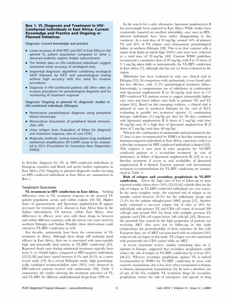
Box 1. VL Diagnosis and Treatment in HIV-Coinfected Individuals in East Africa: CurrentKnowledge and Practice and Ongoing orPlanned Initiatives
Diagnosis: Current knowledge and practice
N Lower accuracy of rK39 RDT and DAT in East Africa in thegeneral VL patient population compared to other L.donovani-endemic regions (Indian subcontinent)
N The limited data on HIV-coinfected individuals suggestsomewhat lower accuracy of serological tests
N Sequential diagnostic algorithms combining serological(rK39 followed by DAT) and parasitological testingachieve high accuracy with less need for invasiveprocedures
N Diagnosis in HIV-coinfected patients still often relies oninvasive procedures for parasitological diagnosis and formonitoring of treatment response
Diagnosis: Ongoing or planned VL diagnostic studies inHIV-coinfected individuals (Ethiopia)
N Noninvasive parasitological diagnosis using peripheralblood microscopy
N Microculture inoculation of peripheral blood mononu-clear cells
N Urine antigen tests: Evaluation of KAtex for diagnosisand treatment response, test of cure (TOC)
N Molecular methods: reverse transcription loop mediatedisothermal amplification (RT-LAMP) assay to be evaluat-ed in 2013 (Foundation for Innovative New Diagnostics[FIND])
PLOS Neglected Tropical Diseases | www.plosntds.org 4 June 2014 | Volume 8 | Issue 6 | e2869
Ta
ble
2.
Clin
ical
stu
die
san
dtr
ials
rep
ort
ing
tre
atm
en
to
utc
om
es
of
HIV
/VL
inEa
stA
fric
a(1
99
8–
20
13
).
Re
fere
nce
Stu
dy
Po
pu
lati
on
an
dD
esi
gn
Tre
atm
en
tsP
ati
en
tsIn
itia
lC
ure
Mo
rta
lity
Init
ial
Fa
ilu
rea
To
lera
bil
ity
Co
mm
en
ts
Dir
oE,
et
al.
[39
]P
rosp
ect
ive
stu
dy:
init
ial
tre
atm
en
to
utc
om
eo
fad
ult
pat
ien
tssc
ree
ne
dfo
rP
SPst
ud
y(2
01
1–
20
12
)
SSG
20
mg
/kg
/d,
30
day
sH
IV+:
53
43
.4%
11
.3%
30
.2%
5.7
%SS
Gd
isco
nti
nu
ed
for
safe
tyR
eq
uir
ing
SSG
ext
en
sio
n:
20
(37
.7%
).Fi
nal
ou
tco
me
:7
7.4
%cu
re
Rit
me
ijer
K,
et
al.
[41
]R
etr
osp
ect
ive
stu
dy:
HIV
+ad
ult
VL
pat
ien
tsin
no
rth
Eth
iop
ia(2
01
0–
20
13
),N
GO
pro
gra
m
Am
Bis
om
e3
0m
g/k
gto
tal+
MF
10
0m
gp
o/d
22
8d
ays
To
tal:
11
18
1.1
%9
.0%
6.3
%b
NA
VL
rela
pse
:5
48
3.3
%3
.7%
11
.1%
b
Hai
luW
,e
tal
.[4
4]
Re
tro
spe
ctiv
est
ud
y.A
llp
atie
nts
tre
ate
dw
ith
anti
mo
nia
ls(2
00
8–
20
09
)at
teac
hin
gp
ub
lich
osp
ital
s
Glu
can
tim
e2
0m
g/k
g/d
for
30
day
s(N
-Et
hio
pia
)T
ota
l:3
07
3.3
%1
0%
16
.7%
2p
ancr
eat
itis
and
1re
nal
failu
re
-H
IV+:
12
58
.3%
NA
NA
NA
2/3
(66
.7%
)re
lap
seb
y6
mo
nth
sam
on
gth
eH
IVco
infe
cte
d(o
nly
25
%se
en
by
mo
nth
6
-H
IV2
:1
49
2.9
%N
AN
AN
A
Glu
can
tim
e2
0m
g/k
g/d
for
30
day
s(S
-Eth
iop
ia)
To
tal:
24
10
0%
00
NA
No
rela
pse
inH
IV2
Rit
me
ijer
K,
et
al.
[38
]R
etr
osp
ect
ive
stu
dy.
Seve
rely
sick
or
HIV
+ad
ult
VL
pat
ien
ts(2
00
7–
20
08
),N
GO
pro
gra
min
N-
Eth
iop
ia
Am
Bis
om
e3
0m
g/k
gc
HIV
+:1
95
59
.5%
6.7
%3
2.3
%b
NA
21
.5%
on
AR
Tat
VL
dia
gn
osi
s;2
9/4
3(6
7%
)w
ith
CD
4le
ssth
an2
00
-P
VL:
11
67
4.1
%7
.8%
16
.4%
-R
ela
pse
:7
93
8.0
%5
.1%
55
.7%
HIV
2:
94
92
.6%
6.4
%N
on
eb
NA
-P
VL:
84
91
.7%
7.1
%0
-R
ela
pse
:1
01
00
%0
0
SSG
(30
–4
0d
)2
0m
g/k
g/d
asre
scu
eth
era
py
HIV
+fa
ilin
gA
mB
iso
me
d:
58
70
.7%
15
.5%
1.7
%5
/63
(7.9
%)
un
able
toto
lera
teSS
G
Hu
riss
aZ
,e
tal
.[1
9]
Re
tro
spe
ctiv
ere
cord
anal
ysis
.A
llad
mit
ted
adu
ltV
Lp
atie
nts
(20
06
–2
00
8).
Tw
op
ub
lich
osp
ital
sin
N-
Eth
iop
ia
SSG
:2
0m
g/k
g/d
for
30
day
sA
mB
iso
me
:3
mg
/kg
6–
10
day
s(o
nly
crit
ical
lyill
)
Ad
ult
s:2
41
84
.6%
10
%5
.4%
NA
Cas
efa
talit
yin
HIV
coin
fect
ed
hig
hSS
G(2
4.5
%)
Am
Bis
om
e(7
.7%
)
-H
IV+:
92
68
.5%
17
.4%
14
.1%
-H
IV2
:1
49
94
.6%
5.4
%0
Rit
me
ijer
K,
et
al.
[31
]R
and
om
ize
dco
ntr
olle
dtr
ial,
no
nb
lind
ed
inN
-Eth
iop
ia.
Mal
em
igra
nt
wo
rke
rs.
PV
L(5
46
);re
lap
se(3
4)
MF
10
0m
g/d
for
28
day
sT
ota
l:2
90
88
.3%
2.1
%7
.9%
Seve
revo
mit
ing
:1
4/2
90
(4.8
%)
At
sixt
hm
on
th:
Re
lap
se:
10
.3%
-H
IV+:
63
77
.8%
1.6
%1
7.5
%-
HIV
+:2
5.4
%
-H
IV2
:1
31
93
.8%
0.8
%4
.5%
-H
IV2
:4
.6%
-U
nkn
ow
n:
96
87
.5%
4.2
%6
.3%
SSG
20
mg
/kg
IMfo
r3
0d
ays
To
tal:
29
08
7.6
%9
.7%
0.7
%Se
vere
vom
itin
g:
27
/29
0(9
.7%
);m
ain
lyH
IV+
Re
lap
se:
2.4
%
PLOS Neglected Tropical Diseases | www.plosntds.org 5 June 2014 | Volume 8 | Issue 6 | e2869
Ta
ble
2.
Co
nt.
Re
fere
nce
Stu
dy
Po
pu
lati
on
an
dD
esi
gn
Tre
atm
en
tsP
ati
en
tsIn
itia
lC
ure
Mo
rta
lity
Init
ial
Fa
ilu
rea
To
lera
bil
ity
Co
mm
en
ts
-H
IV+:
44
90
.1%
6.8
%2
.3%
-H
IV+:
11
.4%
-H
IV2
:1
37
94
.9%
2.9
%0
.7%
-H
IV2
:0
.0%
-Un
kno
wn
:1
09
77
.1%
19
.3%
0.0
%
Lyo
ns
S,e
tal
.[6
5]
Re
tro
spe
ctiv
est
ud
y,N
GO
pro
gra
min
N-E
thio
pia
(19
98
–2
00
0)
No
info
(SSG
on
lyav
aila
ble
dru
g)
To
tal:
79
18
1.5
%1
8.5
%N
AN
A
-H
IV+:
49
73
.5%
26
.5%
-H
IV2
:1
64
93
.3%
6.7
%
Rit
me
ijer
K,
et
al.
[23
]R
and
om
ize
dco
ntr
olle
dtr
ial,
no
nb
lind
ed
,1
99
8–
19
99
.O
nly
PV
Lad
ult
sin
clu
de
d.
NG
Op
rog
ram
;N
-Eth
iop
ia
SSG
vers
us
Pe
nto
stam
All
pri
mar
yV
L.T
ota
l:1
99
75
.4%
24
.1%
NA
Vo
mit
ing
To
tal:
Re
lap
se:
3/1
14
(2.6
%)
-H
IV+:
27
63
%3
3.3
%4
4.4
%2
/12
(16
.7%
)
-H
IV2
:1
12
96
.4%
3.6
%3
5.7
%1
/83
(1.2
%)
Ab
bre
viat
ion
s:P
VL,
pri
mar
yvi
sce
ral
leis
hm
ania
sis;
HIV
2,H
IVn
eg
ativ
e;H
IV+,
HIV
po
siti
ve;N
A,n
ot
avai
lab
le;N
-Eth
iop
ia,n
ort
hEt
hio
pia
;S-E
thio
pia
,so
uth
Eth
iop
ia;N
GO
,no
ng
ove
rnm
en
tal
org
aniz
atio
n;P
SP,p
en
tam
idin
ese
con
dar
yp
rop
hyl
axis
;SS
G,
sod
ium
stib
og
luco
nat
e,
IM,
intr
amu
scu
lar.
acl
inic
ally
de
fin
ed
un
less
oth
erw
ise
stat
ed
;b
par
asit
olo
gic
ally
con
firm
ed
;cm
ed
ian
do
seu
sed
;d
sub
gro
up
of
the
19
5H
IV+
pat
ien
tstr
eat
ed
wit
hlip
oso
mal
amp
ho
teri
cin
B(m
en
tio
ne
dab
ove
).d
oi:1
0.1
37
1/j
ou
rnal
.pn
td.0
00
28
69
.t0
02
PLOS Neglected Tropical Diseases | www.plosntds.org 6 June 2014 | Volume 8 | Issue 6 | e2869
resistance. Pentamidine has been proposed for secondary prophy-
laxis in such a situation, since it exerts antileishmanial effect, is
currently not used for VL treatment, and has been found to be
relatively safe in a prophylactic dose [48–50]. An open-label
multicentre clinical trial recruiting HIV coinfected adults at high
risk of VL relapse is currently ongoing in Ethiopia to evaluate the
feasibility, safety, and effectiveness of monthly intravenous
administration of pentamidine (4 mg/kg) for a period of 12 to
18 months. Main outcome data are expected before the end of
2014 (http://clinicaltrials.gov, NCT01360762).
Antiretroviral treatment in VL-HIV coinfection. Within
Europe, the widespread use of ART in VL-endemic areas has had
a major epidemiological effect on VL-HIV coinfection, with a
pronounced reduction of new cases and prolonged disease-free
survival for established VL-HIV coinfection. The effect on relapse
has been more modest, with an estimated 50% reduction.
Although ART has now been scaled up in most African countries,
access to ART in the generally remote and underresourced VL-
endemic areas remains challenging. Only in Ethiopia do all
(major) VL treatment sites provide ART. Usually, ART is initiated
as soon as the patient is clinically stable, typically during the
second week after initiation of VL treatment.
There is only one retrospective study on the effect of ART on
survival in VL-HIV coinfection in East Africa, which did not
demonstrate an effect [51]. However, this might be due to the high
proportion of lost to follow-up patients who were interrupting their
ART. It was also not assessed whether HIV viral suppression was
achieved [52].
Overall and in clear contrast with TB-HIV, the immune
reconstitution inflammatory syndrome (IRIS) appears to be an
exception in VL-HIV coinfection. Only a handful of clear-cut case
reports of (ART-associated) VL-IRIS have been reported at the
global level [53–58]. Whether the overall dampening effect of the
Leishmania parasite on the immune system could relate to this
requires further study. The lack of a clear case definition of IRIS in
VL-HIV could further add to this lack of reporting. In East Africa,
a few cases of VL have been reported after ART initiation
(including seroconversion), but it is not clear to what extent these
are true ‘‘unmasking’’ IRIS cases [45]. While PKDL is assumed to
be an immune-mediated condition [59], in HIV patients on ART,
it may indicate IRIS [58,60]. Two cases were reported from
Ethiopia with exacerbation of skin lesions after ART initiation,
described as PKDL or PKDL-like lesions [20]. Detailed prospec-
tive studies are required to characterize IRIS in VL-HIV
coinfection, with a special focus on skin manifestations.
With the global scaling-up of ART, HIV-1 protease inhibitors
(PIs) are increasingly available in VL-HIV-endemic regions,
including regions in Africa. Several lines of evidence suggest that
HIV-1 protease inhibitors might directly exert antiparasitic effects,
including effects against Leishmania. One potential approach could
consist of using PI-based ART instead of non-nucleoside reverse
transcription inhibitor (NNRTI)-based ART in VL-HIV-endemic
regions. However, with detailed animal or clinical studies lacking,
additional research is required before HIV-1 PIs should be taken
forward towards this goal [61].
The Role of VL-HIV Coinfections as Reservoirs forTransmission of L. donovani
The infectiousness of HIV-coinfected patients to sand flies in
endemic areas of anthroponotic foci has not been studied.
However, coinfected patients were found to have higher tissue
parasite loads and higher rates of PKDL [23,28] potentially acting
as reservoirs. Given the high rate of treatment failure and risk of
relapse [19,45] and the associated repeated and prolonged
exposure to antileishmanial drugs, HIV-coinfected individuals
are also at increased risk of developing drug resistance and could
possibly serve as a source of resistant parasites. Ideally, xenodi-
agnosis studies should be conducted in East Africa as well in order
to better define the epidemiological impact of HIV coinfection in
this region.
Discussion and Conclusions
As a neglected disease, the significance of Leishmania infection in
HIV patients was recognized late. The poorest segments of the
population, such as migrant daily laborers, are affected. Thus, the
diagnostic and treatment challenges of VL-HIV coinfection have
continued to date, especially in East Africa [5,11].
Table 3. Treatment recommendations for VL and HIV in different guidelines used in the East Africa region.
Guideline First-line Treatment Second-line Treatment Indications for ART
WHO (2010) Amphotericin B lipid formulations, total dose of40 mg/kg; given as 3–5 mg/kg daily or intermittentlyfor 10 doses (days 1–5, 10, 17, 24, 31, and 38)
Pentavalent antimonials (in areaswithout drug resistance)
All VL-HIV patients
MSF in Sudan, South Sudan,and Ethiopia (2012)
Liposomal amphotericin B, 30 mg/kg (given as5 mg/kg on alternate days for 6 doses)+Miltefosine100 mg (divided in two doses) for 28 days
SSG 20 mg/kg/day for up to 30 days plusparomomycin 15 mg/kg/day for 17 days
All VL-HIV patients
National guidelines
Ethiopia (2013) Liposomal amphotericin B, 40 mg/kg total dose;given as 5 mg/kg on day 1–5, 10, 17, and 24
Pentavalent antimonials All VL-HIV patients
Sudan (2013) Liposomal amphotericin B, 3 mg/kg/day for 10 to 14 days Not specified Not specified
South Sudan (2012) Liposomal amphotericin B, 40 mg/kg total dose; givenas 3–5 mg/kg on days 1–5, 10, 17, 24, 31, and 38
Pentavalent antimonials Not specified
Kenya (2012) Liposomal amphotericin B (higher dose may be required,routinely recommended total dose is 30 mg/kg)
Amphotericin B All VL-HIV patients
Uganda (2007) Liposomal amphotericin B, 3 mg/kg/d for 7 days Amphotericin B, 1 mg/kg every other dayfor 30 days. Miltefosine, 100 mg (2.5 mg/kg)/dfor 28 days
All VL-HIV patients
Abbreviations: WHO, World Health Organization; MSF, Medecins Sans Frontieres.doi:10.1371/journal.pntd.0002869.t003
PLOS Neglected Tropical Diseases | www.plosntds.org 7 June 2014 | Volume 8 | Issue 6 | e2869
The control of VL requires a combined effort at vector control,
improved living standards and case detection, and early treatment
[13]. The feasibility of transmission prevention methods like
impregnated bed nets, blankets, and protective clothing should be
evaluated. In the case of VL-HIV, targeting the most at-risk
population groups (e.g., the migrant population) and improving
their awareness through health education and counseling for both
diseases should be pursued. Health professions should also be
regularly updated about VL.
Case detection and management is especially challenging for
HIV-infected persons. To have a larger impact on VL-HIV
coinfection, a comprehensive and multipronged approach will be
required. Better diagnostic and curative options would help to
improve case detection and patient management. As with other
HIV-associated coinfections (for instance, tuberculosis and crypto-
coccal meningitis), preventive strategies should be explored. As the
experience in Europe testifies, the preventive effect of large-scale
introduction of ART merits exploration. Although the current
WHO guidelines now recommend early ART initiation [62], the
most challenging part will be early HIV diagnosis and retention in
care in a typically highly mobile and difficult to reach population.
Diagnosing VL in HIV patients relies mainly on parasite
detection from tissue aspiration. This is due to the insufficiently
sensitive serological tests that also do not help to diagnose relapsed
cases, which are very common in HIV patients. However, tissue
aspiration is associated with potential risk of fatal bleeding that
requires experience and health facilities equipped to handle this
potential complication. Thus, a simple-to-use but reliable antigen-
detection diagnostic procedure or tool that can be applicable for
treatment monitoring and diagnosing relapses is urgently required.
For VL-HIV case management, field experience favors the use
of the following combination treatment: liposomal amphotericin B
and miltefosine [41] followed by secondary prophylaxis [7,47,63]
and ART. However, studies are urgently needed to strengthen the
evidence for this treatment and improve its outcome in patients in
field conditions in East Africa. To date, there is no clear evidence
regarding the interaction of ARV drugs and antileishmanial
medications. Basic research to understand the immunological
interaction of the two infections, as well as the immune
modulatory effects of drugs, may ultimately help to improve the
management of the coinfection.
Tackling VL-HIV coinfection in the long run, along with
combating VL in general, will strongly depend on the strength and
commitment of the national programs. However, to date, the
national programs still rely on substantial external support in most
East African countries. Moreover, a strong link with the national
HIV program will be needed for efficient and integrated program
management. Though the HIV prevalence is declining in
Ethiopia, the unstable socioeconomic and political situations in
South Sudan and the high population migrations in the region
warrant continuous effort for the control of both diseases.
References
1. van Griensven J, Diro E (2012) Visceral leishmaniasis. Infect Dis Clin North Am
26: 309–322. doi:10.1016/j.idc.2012.03.005.
2. Alvar J, Velez ID, Bern C, Herrero M, Desjeux P, et al. (2012) Leishmaniasis
worldwide and global estimates of its incidence. PLoS One 7: e35671. doi:
10.1371/journal.pone.0035671.
3. Seaman J, Mercer AJ, Sondorp E (1996) The epidemic of visceral leishmaniasis
in western upper Nile, Southern Sudan: Course and impact from 1984 to 1994.
Int J Epidemiol 25: 862–871.
4. Olivier M, Badaro R, Medrano FJ, Moreno J (2003) The pathogenesis of
Leishmania/HIV co-infection: cellular and immunological mechanisms. Ann
Trop Med Parasitol 97 Suppl 1: 79–98. doi: 10.1179/000349803225002561.
5. Alvar J, Aparicio P, Aseffa A, den Boer M, Canavate C, et al. (2008) The
relationship between leishmaniasis and AIDS: the second 10 years. Clin
Microbiol Rev 21: 334–359, table. doi:10.1128/CMR.00061-07.
6. Jarvis JN, Lockwood DN (2013) Clinical aspects of visceral leishmaniasis in
HIV infection. Curr Opin Infect Dis 26: 1–9. doi: 10.1097/QCO.0-
b013e32835c2198.
7. World Health Organization (2010 March 22) Control of the Leishmaniases. In:
WHO Technical Report Series 949: Report of a meeting of the WHO Expert
Committee on the Control of Leishmaniases, Geneva, 22–26 March 2010.
Geneva, Swetzerland: World Health Organization.
8. Joint United Nations Programme on HIV/AIDS [UNAIDS] (2013) Global
Report: UNAIDS report on the global AIDS epidemic. Available: http://www.
unaids.org/en/media/unaids/contentassets/documents/epidemiology/2013/
gr2013/UNAIDS_Global_Report_2013_en.pdf. Accessed 22 May 2014.
9. Merson MH, O’Malley J, Serwadda D, Apisuk C (2008) The history and
challenge of HIV prevention. Lancet 372: 475–488. doi: 10.1016/S0140-
6736(08)60884-3.
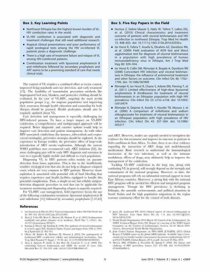
Box 2. Key Learning Points
N Northwest Ethiopia has the highest known burden of VL-HIV coinfection rates in the world.
N VL-HIV coinfection is associated with diagnostic andtreatment challenges that still need additional research.
N Atypical clinical presentations and poor performance ofrapid serological tests among the HIV co-infected VLpatients poses a diagnostic challenge.
N There is a high rate of treatment failure and relapse of VLamong HIV-coinfected patients.
N Combination treatment with liposomal amphotericin Band miltefosine followed by secondary prophylaxis andART seems to be a promising standard of care that needsclinical trials.
Box 3. Five Key Papers in the Field
N Hurissa Z, Gebre-Silassie S, Hailu W, Tefera T, Lalloo DG,et al. (2010) Clinical characteristics and treatmentoutcome of patients with visceral leishmaniasis and HIVco-infection in northwest Ethiopia. Trop Med Int Health15: 848–855. doi: 10.1111/j.1365-3156.2010.02550.x
N ter Horst R, Tefera T, Assefa G, Ebrahim AZ, Davidson RN,et al. (2009) Field evaluation of rK39 test and directagglutination test for diagnosis of visceral leishmaniasisin a population with high prevalence of humanimmunodeficiency virus in Ethiopia. Am J Trop MedHyg 80: 929–934.
N ter Horst R, Collin SM, Ritmeijer K, Bogale A, Davidson RN(2008) Concordant HIV infection and visceral leishman-iasis in Ethiopia: the influence of antiretroviral treatmentand other factors on outcome. Clin Infect Dis 46: 1702–1709. doi: 10.1086/587899
N Ritmeijer K, ter Horst R, Chane S, Aderie EM, Piening T, etal. (2011) Limited effectiveness of high-dose liposomalamphotericin B (AmBisome) for treatment of visceralleishmaniasis in an Ethiopian population with high HIVprevalence. Clin Infect Dis 53: e152–e158. doi: 10.1093/cid/cir674
N Ritmeijer K, Dejenie A, Assefa Y, Hundie TB, Mesure J, etal. (2006) A comparison of miltefosine and sodiumstibogluconate for treatment of visceral leishmaniasis inan Ethiopian population with high prevalence of HIVinfection. Clin Infect Dis 43: 357–364. doi: 10.1086/505217
PLOS Neglected Tropical Diseases | www.plosntds.org 8 June 2014 | Volume 8 | Issue 6 | e2869
10. Ethiopian Health and Nutrition Research Institute (EHNRI) (2011) Report on
the 2009 Round Antenatal Care Sentinel HIV Surveillance in Ethiopia.Available: http://www.ehnri.gov.et/newsletter/ANC%202009_Final_Report.
pdf. Accessed 22 May 2014.
11. Federal Democratic Republic of Ethiopia (2012) Country progress report on
HIV/AIDS response. Available: http://www.unaids.org/en/dataanalysis/
knowyourresponse/countryprogressreports/2012countries/GAP%20Report%202012.pdf. Accessed 22 May 2014.
12. Mengistu G, Ayele B (2007) Visceral leishmaniasis and HIV co-infection inpatients admitted to Gondar University Hopsital, North West Ethiopia.
Ethiop J Health Dev 21: 53–60.
13. Argaw D, Mulugeta A, Herrero M, Nombela N, Teklu T, et al. (2013) Risk
factors for visceral Leishmaniasis among residents and migrants in Kafta-
Humera, Ethiopia. PLoS Negl Trop Dis 7: e2543. doi: 10.1371/jour-nal.pntd.0002543.
14. Gelanew T, Kuhls K, Hurissa Z, Weldegebreal T, Hailu W, et al. (2010)Inference of population structure of Leishmania donovani strains isolated from
different Ethiopian visceral leishmaniasis endemic areas. PLoS Negl Trop Dis 4:e889. doi: 10.1371/journal.pntd.0000889.
15. Malaria Consortium (2010) Leishmaniasis control in eastern Africa: Pastand present efforts and future needs. Situation and gap analysis. Available:
http://www.malariaconsortium.org/userfiles/file/NTD%20Resources/
VL%20EA%20Situation%20Analysis%20Fina_Janl.pdf. Accessed 22 May2014.
16. Catorce G (2010) Leishmania infantum/HIV co-infection: cuteneous lesionsfollowing treatment of visceral leishmaniasis. Ann Dermatol Venereol 133: 39–
42.
17. Fenske S, Stellbrink HJ, Albrecht H, Greten H (1991) Visceral leishmaniasis in
an HIV-infected patient: clinical features and response to treatment. KlinWochenschr 69: 793–796.
18. Garcia-Samaniego J, Laguna F (1997) [Intestinal leishmaniasis in AIDS
patients]. Rev Esp Enferm Dig 89: 145.
19. Hurissa Z, Gebre-Silassie S, Hailu W, Tefera T, Lalloo DG, et al. (2010) Clinical
characteristics and treatment outcome of patients with visceral leishmaniasis andHIV co-infection in northwest Ethiopia. Trop Med Int Health 15: 848–855. doi:
10.1111/j.1365-3156.2010.02550.x.
20. Gelanew T, Hurissa Z, Diro E, Kassahun A, Kuhls K, et al. (2011) Disseminated
cutaneous leishmaniasis resembling post-kala-azar dermal leishmaniasis causedby Leishmania donovani in three patients co-infected with visceral leishmaniasis
and human immunodeficiency virus/acquired immunodeficiency syndrome in
Ethiopia. Am J Trop Med Hyg 84: 906–912. doi:10.4269/ajtmh.2011.11-0055.
21. Diro E, Hurissa Z, van Griensven J, Hailu A (2011) Unusual presentations of
visceral leishmania in the era of HIV. In: Proceedings of the 16th InternationalConference on AIDS and STIs in Africa (ICASA); 4–8 December 2011; Addis
Ababa, Ethiopia.
22. Zijlstra EE, Musa AM, Khalil EA, el-Hassan IM, el-Hassan AM (2003) Post-
kala-azar dermal leishmaniasis. Lancet Infect Dis 3: 87–98.
23. Ritmeijer K, Veeken H, Melaku Y, Leal G, Amsalu R, et al. (2001) Ethiopian
visceral leishmaniasis: generic and proprietary sodium stibogluconate are
equivalent; HIV co-infected patients have a poor outcome. Trans R Soc TropMed Hyg 95: 668–672.
24. Chappuis F, Rijal S, Soto A, Menten J, Boelaert M (2006) A meta-analysis of thediagnostic performance of the direct agglutination test and rK39 dipstick for
visceral leishmaniasis. BMJ 333: 723. doi: 10.1136/bmj.38917.503056.7C.
25. Chappuis F, Mueller Y, Nguimfack A, Rwakimari JB, Couffignal S, et al. (2005)
Diagnostic accuracy of two rK39 antigen-based dipsticks and the formol gel testfor rapid diagnosis of visceral leishmaniasis in northeastern Uganda. J Clin
Microbiol 43: 5973–5977. doi: 10.1128/JCM.43.12.5973-5977.2005 [doi].
26. Cunningham J, Hasker E, Das P, El SS, Goto H, et al. (2012) A globalcomparative evaluation of commercial immunochromatographic rapid diagnos-
tic tests for visceral leishmaniasis. Clin Infect Dis 55: 1312–1319.doi:10.1093/cid/cis716.
27. Diro E, Techane Y, Tefera T, Assefa Y, Kebede T, et al. (2007) Field evaluationof FD-DAT, rK39 dipstick and KATEX (urine latex agglutination) for diagnosis
of visceral leishmaniasis in northwest Ethiopia. Trans R Soc Trop Med Hyg
908–914. doi: 10.1016/j.trstmh.2007.05.002.
28. ter Horst R, Tefera T, Assefa G, Ebrahim AZ, Davidson RN, et al. (2009) Field
evaluation of rK39 test and direct agglutination test for diagnosis of visceralleishmaniasis in a population with high prevalence of human immunodeficiency
virus in Ethiopia. Am J Trop Med Hyg 80: 929–934.
29. Adams ER, Schoone GJ, Ageed AF, Safi SE, Schallig HD (2010) Development
of a reverse transcriptase loop-mediated isothermal amplification (LAMP) assayfor the sensitive detection of Leishmania parasites in clinical samples. Am J Trop
Med Hyg 82: 591–596. doi: 10.4269/ajtmh.2010.09-0369.
30. Hailu A, Berhe N (2002) The performance of direct agglutination tests (DAT) in
the diagnosis of visceral leishmaniasis among Ethiopian patients with HIV co-
infection. Ann Trop Med Parasitol 96: 25–30.
31. Ritmeijer K, Dejenie A, Assefa Y, Hundie TB, Mesure J, et al. (2006) A
comparison of miltefosine and sodium stibogluconate for treatment of visceralleishmaniasis in an Ethiopian population with high prevalence of HIV infection.
Clin Infect Dis 43: 357–364. doi: 10.1086/505217.
32. Deniau M, Canavate C, Faraut-Gambarelli F, Marty P (2003) The biological
diagnosis of leishmaniasis in HIV-infected patients. Ann Trop Med Parasitol 97
Suppl 1: 115–133. doi: 10.1179/000349803225002598.
33. Izri MA, Deniau M, Briere C, Rivollet D, Petithory JC, et al. (1996)
Leishmaniasis in AIDS patients: results of leukocytoconcentration, a fast
biological method of diagnosis. Bull World Health Organ 74: 91–93.
34. Yansouni CP, Diro E, Lynen L, van Griensven J, Takele Y, et al. (2012)
Diagnosis of visceral leishmaniasis using peripheral blood microscopy in
Ethiopia: a phase-III diagnostic study evaluating 3 parasite concentration
techniques compared to tissue aspiration. In: Proceedings of the 61st Annual
Meeting of the American Society of Tropical Medicine and Hygiene, 11–15
November 2012; Atlanta, Georgia, United States.
35. Cota GF, de Sousa MR, Demarqui FN, Rabello A (2012) The diagnostic
accuracy of serologic and molecular methods for detecting visceral leishmaniasis
in HIV infected patients: meta-analysis. PLoS Negl Trop Dis 6: e1665. doi:
10.1371/journal.pntd.0001665.
36. Hailu A, Musa A, Wasunna M, Balasegaram M, Yifru S, et al. (2010)
Geographical variation in the response of visceral leishmaniasis to paromomycin
in East Africa: a multicentre, open-label, randomized trial. PLoS Negl Trop Dis
4: e709. doi: 10.1371/journal.pntd.0000709.
37. Musa AM, Younis B, Fadlalla A, Royce C, Balasegaram M, et al. (2010)
Paromomycin for the treatment of visceral leishmaniasis in Sudan: a
randomized, open-label, dose-finding study. PLoS Negl Trop Dis 4: e855. doi:
10.1371/journal.pntd.0000855.
38. Ritmeijer K, ter Horst R, Chane S, Aderie EM, Piening T, et al. (2011) Limited
effectiveness of high-dose liposomal amphotericin B (AmBisome) for treatment of
visceral leishmaniasis in an Ethiopian population with high HIV prevalence.
Clin Infect Dis 53: e152–e158. doi: 10.1093/cid/cir674.
39. Diro E, Lynen L, Mohammed R, Boelaert M, Hailu A, et al. (2013) Increasing
parasitological failure rate of visceral leishmaniasis to sodium stibogluconate
among HIV co-infected patients in East Africa. In: Proceedings of the Fifth
World Leishmaniasis Congress; 13–17 May 2013; Porto de Galhinas, Brazil.
40. Sinha PK, van Griensven J, Pandey K, Kumar N, Verma N, et al. (2011)
Liposomal amphotericin B for visceral leishmaniasis in human immunodefi-
ciency virus-coinfected patients: 2-year treatment outcomes in Bihar, India. Clin
Infect Dis 53: e91–e98. doi: 10.1093/cid/cir521.
41. Ritmeijer K (2013) Old and new treatments for HIV/VL co-infection. In:
Proceedings of the Fifth World Leishmaniasis Congress, 13–17 May 2013; Porto
de Galhinas, Brazil.
42. Musa A, Khalil E, Hailu A, Olobo J, Balasegaram M, et al. (2012) Sodium
stibogluconate (SSG) & paromomycin combination compared to SSG for
visceral leishmaniasis in East Africa: a randomised controlled trial. PLoS Negl
Trop Dis 6: e1674. doi: 10.1371/journal.pntd.0001674.
43. Federal Ministry of Health, Ethiopia (2013) Guidelines for diagnosis, treatment
and prevention of leishmaniasis in Ethiopia, 2nd edition. Addis Adaba, Ethiopia:
Federal Ministry of Health, Ethiopia.
44. Hailu W, Weldegebreal T, Hurissa Z, Tafes H, Omollo R, et al. (2010) Safety
and effectiveness of meglumine antimoniate in the treatment of Ethiopian
visceral leishmaniasis patients with and without HIV co-infection. Trans R Soc
Trop Med Hyg 104: 706–712. doi: 10.1016/j.trstmh.2010.07.007.
45. ter HR, Collin SM, Ritmeijer K, Bogale A, Davidson RN (2008) Concordant
HIV infection and visceral leishmaniasis in Ethiopia: the influence of
antiretroviral treatment and other factors on outcome. Clin Infect Dis 46:
1702–1709. doi: 10.1086/587899.
46. Cota GF, de Sousa MR, Rabello A (2011) Predictors of visceral leishmaniasis
relapse in HIV-infected patients: a systematic review. PLoS Negl Trop Dis 5:
e1153. doi: 10.1371/journal.pntd.0001153.
47. Lopez-Velez R, Videla S, Marquez M, Boix V, Jimenez-Mejias ME, et al. (2004)
Amphotericin B lipid complex versus no treatment in the secondary prophylaxis
of visceral leishmaniasis in HIV-infected patients. J Antimicrob Chemother 53:
540–543. doi: 10.1093/jac/dkh084.
48. Calza L, Marinacci G, Manfredi R, Colangeli V, Fortunato L, et al. (2001)
Pentamidine isethionate as treatment and secondary prophylaxis for dissemi-
nated cutaneous leishmaniasis during HIV infection: case report. J Chemother
13: 653–657.
49. Ena J, Amador C, Pasquau F, Carbonell C, Benito C, et al. (1994) Once-a-
month administration of intravenous pentamidine to patients infected with
human immunodeficiency virus as prophylaxis for Pneumocystis carinii
pneumonia. Clin Infect Dis 18: 901–904.
50. Yeung KT, Chan M, Chan CK (1996) The safety of i.v. pentamidine
administered in an ambulatory setting. Chest 110: 136–140.
51. ter Horst R, Collin SM, Ritmeijer K, Bogale A, Davidson RN (2008)
Concordant HIV infection and visceral leishmaniasis in Ethiopia: the influence
of antiretroviral treatment and other factors on outcome. Clin Infect Dis 46:
1702–1709. doi: 10.1086/587899.
52. Petter A, Shetty-Lee A, Kofler G, Huemer HP, Larcher C (2001) Visceral
leishmaniasis in an AIDS patient on successful antiretroviral therapy: failure of
parasite eradication despite increase in CD4+ T-cell count but low CD8+ T-cell
count. Scand J Infect Dis 33: 236–238.
53. Ramos A, Cruz I, Munez E, Salas C, Fernandez A, et al. (2008) Post-kala-azar
dermal Leishmaniasis and uveitis in an HIV-positive patient. Infection 36: 184–
186. doi: 10.1007/s15010-007-6279-5.
54. Antinori S, Longhi E, Bestetti G, Piolini R, Acquaviva V, et al. (2007) Post-kala-
azar dermal leishmaniasis as an immune reconstitution inflammatory syndrome
in a patient with acquired immune deficiency syndrome. Br J Dermatol 157:
1032–1036. doi: 10.1111/j.1365-2133.2007.08157.x.
PLOS Neglected Tropical Diseases | www.plosntds.org 9 June 2014 | Volume 8 | Issue 6 | e2869
55. Bittencourt A, Silva N, Straatmann A, Nunes VL, Follador I, et al. (2003) Post-
kala-azar dermal leishmaniasis associated with AIDS. Braz J Infect Dis 7: 229–233.
56. Stark D, Pett S, Marriott D, Harkness J (2006) Post-kala-azar dermal
leishmaniasis due to Leishmania infantum in a human immunodeficiency virustype 1-infected patient. J Clin Microbiol 44: 1178–1180. doi: 10.1128/
JCM.44.3.1178-1180.2006.57. Ridolfo AL, Gervasoni C, Antinori S, Pizzuto M, Santambrogio S, et al. (2000)
Post-kala-azar dermal leishmaniasis during highly active antiretroviral therapy
in an AIDS patient infected with Leishmania infantum. J Infect 40: 199–202.
58. Gilad J, Borer A, Hallel-Halevy D, Riesenberg K, Alkan M, et al. (2001) Post-kala-azar dermal leishmaniasis manifesting after initiation of highly active anti-
retroviral therapy in a patient with human immunodeficiency virus infection. IsrMed Assoc J 3: 451–452.
59. Khalil EA, Khidir SA, Musa AM, Musa BY, Elfaki ME, et al. (2013) Post-Kala-
Azar Dermal Leishmaniasis: A Paradigm of Paradoxical Immune ReconstitutionSyndrome in Non-HIV/AIDS Patients. J Trop Med 2013: 275253. doi:
10.1155/2013/275253.
60. Tadesse A, Hurissa Z (2009) Leishmaniasis (PKDL) as a case of immune
reconstitution inflammatory syndrome (IRIS) in HIV-positive patient afterinitiation of anti-retroviral therapy (ART). Ethiop Med J 47: 77–79.
61. van GJ, Diro E, Lopez-Velez R, Boelaert M, Lynen L, et al. (2013) HIV-1
protease inhibitors for treatment of visceral leishmaniasis in HIV-co-infectedindividuals. Lancet Infect Dis 13: 251–259. doi: 10.1016/S1473-3099(12)70348-
1.62. Word Health Organization (2013) Consolidated Guidelines on Use of
Antiretroviral Drugs for Treating and Preventing HIV Infection Recommen-
dations for a Public Health Approach. Geneva, Switzerland: World HealthOrganization.
63. CDC (2009) Guidelines for Prevention and Treatment of OpportunisticInfections in HIV-Infected Adults and Adolescents. MMWR Morb Mortal
Wkly Rep 58: RR-4.64. Hailu A, Gebre-Michael T, Berhe N, Balkew M (2006) Leishmaniasis in
Ethiopia. In: The Ecology and Epidemiology of Health and Disease in Ethiopia.
Addis Ababa, Ethiopia: Shama Books. pp. 615–634.65. Lyons S, Veeken H, Long J (2003) Visceral leishmaniasis and HIV in Tigray,
Ethiopia. Trop Med Int Health 8: 733–739.
PLOS Neglected Tropical Diseases | www.plosntds.org 10 June 2014 | Volume 8 | Issue 6 | e2869
Related Documents