10.1128/JVI.75.24.11943-11947.2001. 2001, 75(24):11943. DOI: J. Virol. Johnson E. V. Damodaran, Ryan T. Morton, Charles L. Brooks III and John Vijay S. Reddy, Padmaja Natarajan, Brian Okerberg, Kevin Li, K. Computational Analyses Virus Capsid Structures and Their Virus Particle Explorer (VIPER), a Website for http://jvi.asm.org/content/75/24/11943 Updated information and services can be found at: These include: REFERENCES http://jvi.asm.org/content/75/24/11943#ref-list-1 This article cites 9 articles, 2 of which can be accessed free at: CONTENT ALERTS more» cite this article), Receive: RSS Feeds, eTOCs, free email alerts (when new articles http://journals.asm.org/site/misc/reprints.xhtml Information about commercial reprint orders: http://journals.asm.org/site/subscriptions/ To subscribe to to another ASM Journal go to: on October 20, 2014 by guest http://jvi.asm.org/ Downloaded from on October 20, 2014 by guest http://jvi.asm.org/ Downloaded from

Welcome message from author
This document is posted to help you gain knowledge. Please leave a comment to let me know what you think about it! Share it to your friends and learn new things together.
Transcript
10.1128/JVI.75.24.11943-11947.2001.
2001, 75(24):11943. DOI:J. Virol. Johnson
E.V. Damodaran, Ryan T. Morton, Charles L. Brooks III and John Vijay S. Reddy, Padmaja Natarajan, Brian Okerberg, Kevin Li, K. Computational AnalysesVirus Capsid Structures and Their Virus Particle Explorer (VIPER), a Website for
http://jvi.asm.org/content/75/24/11943Updated information and services can be found at:
These include:
REFERENCEShttp://jvi.asm.org/content/75/24/11943#ref-list-1This article cites 9 articles, 2 of which can be accessed free at:
CONTENT ALERTS more»cite this article),
Receive: RSS Feeds, eTOCs, free email alerts (when new articles
http://journals.asm.org/site/misc/reprints.xhtmlInformation about commercial reprint orders: http://journals.asm.org/site/subscriptions/To subscribe to to another ASM Journal go to:
on October 20, 2014 by guest
http://jvi.asm.org/
Dow
nloaded from
on October 20, 2014 by guest
http://jvi.asm.org/
Dow
nloaded from
JOURNAL OF VIROLOGY,0022-538X/01/$04.00�0 DOI: 10.1128/JVI.75.24.11943–11947.2001
Dec. 2001, p. 11943–11947 Vol. 75, No. 24
Copyright © 2001, American Society for Microbiology. All Rights Reserved.
GUEST COMMENTARYVirus Particle Explorer (VIPER), a Website for Virus Capsid
Structures and Their Computational Analyses†VIJAY S. REDDY,* PADMAJA NATARAJAN, BRIAN OKERBERG, KEVIN LI, K. V. DAMODARAN,
RYAN T. MORTON, CHARLES L. BROOKS III,* AND JOHN E. JOHNSON*
Department of Molecular Biology, The Scripps Research Institute, La Jolla, California 92037
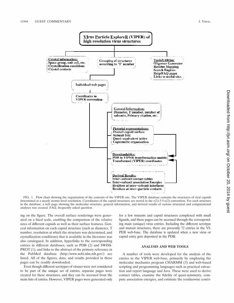
The number of icosahedral-capsid structures determined ata near-atomic level of resolution is growing rapidly as advancesin synchrotron radiation sources, fast-readout detectors, andcomputer hardware and software are made. Hence, there is anincreasing need to organize these mega-assemblies into a uni-form and easy-to-use database. The coordinates of the icosa-hedral-capsid structures deposited in the Protein Data Bank(PDB) (2) follow a variety of conventions in which the icosa-hedral symmetry axes are oriented differently in the orthogonalcoordinate system. While trying to analyze the various capsidstructures en masse, we became aware of the need for a data-base in which all capsid structures (coordinates) are stored ina standard icosahedral orientation. Such a structural databaseof viral capsids would indeed facilitate the development oftools for high-throughput analyses of the virus structures. Wereport here the creation of a web-base (website and database)of virus structures, the Virus Particle Explorer (VIPER), whichcan be accessed through the World Wide Web (WWW) atthe uniform resource locator (URL) http://mmtsb.scripps.edu/viper/. The organization of the VIPER database is shown inFig. 1.
TRANSFORMING THE PDB COORDINATES INTOVIPER CONVENTION
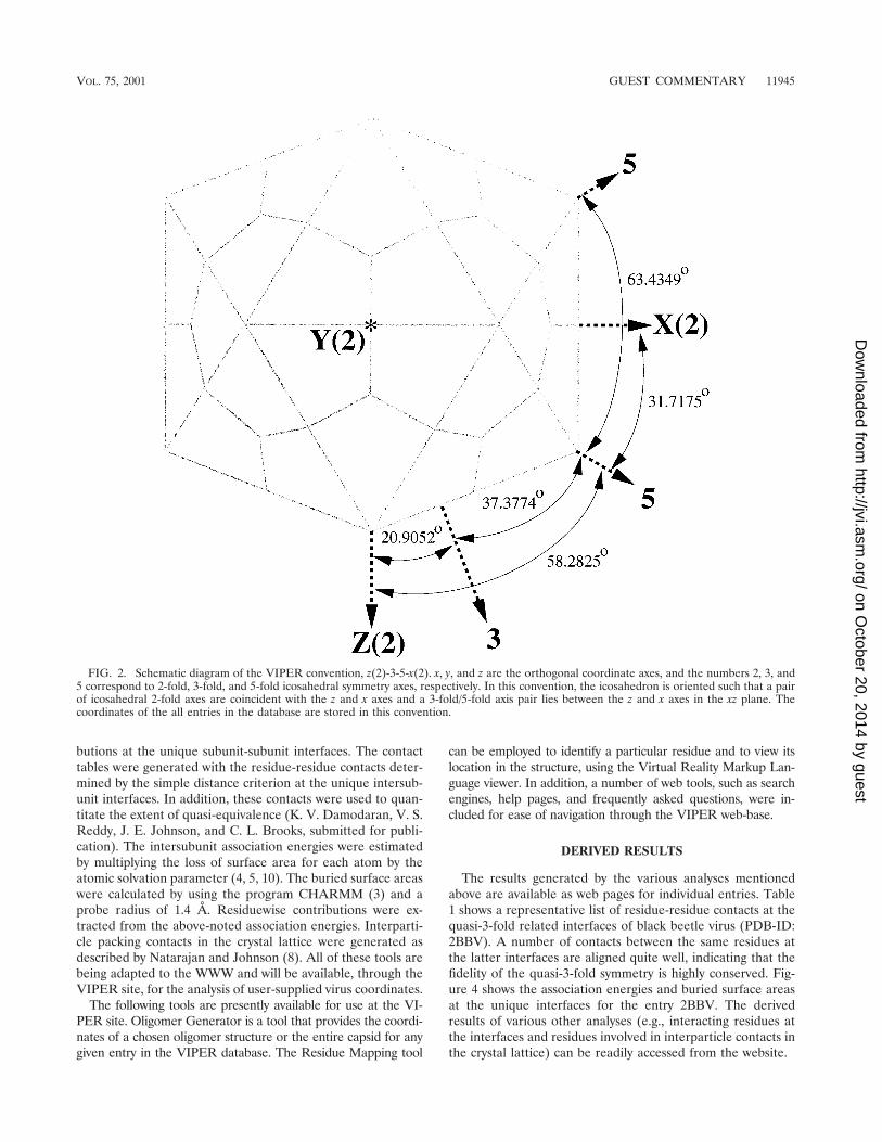
To describe icosahedral symmetry axes with respect to theorthogonal coordinate axes, two conventions, z(2)-3-5-x(2) andz(2)-3-5-y(2), are commonly used, where 2, 3, and 5 correspondto the icosahedral 2-fold, 3-fold, and 5-fold symmetry axes,respectively, and x, y, and z are the orthogonal coordinate axes.In the z(2)-3-5-x(2) convention, two icosahedral 2-fold axescoincide with the z and x coordinate axes while a set of icosa-hedral 3-fold and 5-fold axes lie, in that order, between the zand x axes in the xz plane (Fig. 2). Similarly, in the z(2)-3-5-y(2)orientation, a pair of 2-fold axes coincide with the z and y axesand a 3-fold and a 5-fold axis lie in between in the zy plane.These two orientations are related to each other by a 90°rotation around the z axis. All of the entries in the VIPERweb-base are stored uniformly in the z(2)-3-5-x(2) convention.
Initially, the coordinates of each entry in the PDB weretransformed to one of the two standard icosahedral orienta-tions depending on the depositor’s reference convention. Theywere then converted into the VIPER (z-3-5-x) convention. Inaddition, a reference icosahedral asymmetric unit was chosenfor each type of triangulation (T) lattice (e.g., T�1, T�3, orT�4). Accordingly, the coordinates were transformed to thereference asymmetric unit and stored in the VIPER database.The product of these matrices is stored as a single transformationmatrix, the PDB TO VIPER matrix, making the conversionbetween the VIPER and PDB coordinates straightforward.
The transformed coordinates were further processed so thatthe amino acid residues were placed at the top of the file, afterwhich followed the nucleic acid residues, metal ions, and watermolecules, if present. Atoms with missing chain identifiers(IDs) were given chain IDs, and these changes were docu-mented in the REMARK lines at the beginning of the coor-dinate file. This rearrangement of the coordinates was requiredto facilitate the analysis of the capsid structures in a rapid andautomatic fashion by using the molecular mechanics programCHARMM (3).
ORGANIZATION AND DESCRIPTION OF THE ENTRIES
Presently there are 53 unique capsid structures available at theVIPER site. The coordinates of the capsids whose structures havebeen published but not yet deposited in the PDB were acquiredthrough personal communications. The entries are listed in al-phabetical order, as well as classified in terms of T number, whichusually refers to the number of copies of the capsid protein sub-units that occupy the icosahedral asymmetric unit. For each struc-ture in the database, a web page was created with a pictorialdescription of the capsid and subunit structures as well as struc-tural characteristics and related information available on the virus(e.g., diameter, T number, PDB ID, etc.).
Individual pages show the overall molecular surface render-ing of the complete capsid, color coded according to the zcoordinate, created to resemble the cryoelectron microscopyreconstruction. The tertiary fold of the capsid protein sub-unit(s), the capsomeric organization of the subunits, and ageometrical representation of the quasi-equivalent lattice arealso provided. The rendered surface drawings were createdwith the program GRASP (9). The remaining figures were gen-erated using the programs MOLSCRIPT (6) and RASTER3D(7). A typical web page at the VIPER site is shown in Fig. 3.While the individual VIPER pages show the figures in reducedsizes, each figure can be magnified to its original size by click-
* Corresponding author. Mailing address: Department of MolecularBiology, The Scripps Research Institute, 10550 N. Torrey Pines Rd.,La Jolla, CA 92037. Phone for Vijay S. Reddy: (858) 784-8191. Fax:(858) 784-8660. E-mail: [email protected]. Phone for Charles L.Brooks III: (858) 784-8035. Fax: (858) 784-8688. E-mail: [email protected]. Phone for John E. Johnson: (858) 784-9705. Fax: (858)784-8660. E-mail: [email protected].
† Manuscript number 14160-MB of The Scripps Research Institute.
11943
on October 20, 2014 by guest
http://jvi.asm.org/
Dow
nloaded from
ing on the figure. The overall surface renderings were gener-ated on a fixed scale, enabling the comparison of the relativesizes of different capsids as well as their surface features. Gen-eral information on each capsid structure (such as diameter, Tnumber, resolution at which the structure was determined, andcrystallization conditions) that is available in the literature wasalso catalogued. In addition, hyperlinks to the correspondingentries in different databases, such as PDB (2) and SWISS-PROT (1), and links to the abstract of the primary reference inthe PubMed database (http://www.ncbi.nlm.nih.gov/) arelisted. All of the figures, data, and results provided in thesepages can be readily downloaded.
Even though different serotypes of viruses were not consideredto be part of the unique set of entries, separate pages werecreated for these structures, and they can be accessed from themain lists of entries. However, VIPER pages were generated only
for a few mutants and capsid structures complexed with smallligands, and these pages can be accessed through the correspond-ing main (unique) virus entries. Including the different serotypeand mutant structures, there are presently 72 entries in the VI-PER web-base. The database is updated when a new virus orcapsid entry gets deposited in the PDB.
ANALYSIS AND WEB TOOLS
A number of tools were developed for the analysis of theentries in the VIPER web-base, primarily by employing themolecular mechanics program CHARMM (3) and web-basedscripting and programming languages such as practical extrac-tion and report language and Java. These were used to derivecontact tables, examine the fidelity of quasi-symmetry, com-pute association energies, and estimate the residuewise contri-
FIG. 1. Flow chart showing the organization of the contents of the VIPER site. The VIPER database contains the structures of viral capsidsdetermined at a nearly atomic-level resolution. Coordinates of the capsid structures are stored in the z(2)-3-5-x(2) convention. For each structurein the database, a web page showing the molecular structure, general information, and derived results of various structural and computationalanalyses was created. FAQ, frequently asked question.
11944 GUEST COMMENTARY J. VIROL.
on October 20, 2014 by guest
http://jvi.asm.org/
Dow
nloaded from
butions at the unique subunit-subunit interfaces. The contacttables were generated with the residue-residue contacts deter-mined by the simple distance criterion at the unique intersub-unit interfaces. In addition, these contacts were used to quan-titate the extent of quasi-equivalence (K. V. Damodaran, V. S.Reddy, J. E. Johnson, and C. L. Brooks, submitted for publi-cation). The intersubunit association energies were estimatedby multiplying the loss of surface area for each atom by theatomic solvation parameter (4, 5, 10). The buried surface areaswere calculated by using the program CHARMM (3) and aprobe radius of 1.4 Å. Residuewise contributions were ex-tracted from the above-noted association energies. Interparti-cle packing contacts in the crystal lattice were generated asdescribed by Natarajan and Johnson (8). All of these tools arebeing adapted to the WWW and will be available, through theVIPER site, for the analysis of user-supplied virus coordinates.
The following tools are presently available for use at the VI-PER site. Oligomer Generator is a tool that provides the coordi-nates of a chosen oligomer structure or the entire capsid for anygiven entry in the VIPER database. The Residue Mapping tool
can be employed to identify a particular residue and to view itslocation in the structure, using the Virtual Reality Markup Lan-guage viewer. In addition, a number of web tools, such as searchengines, help pages, and frequently asked questions, were in-cluded for ease of navigation through the VIPER web-base.
DERIVED RESULTS
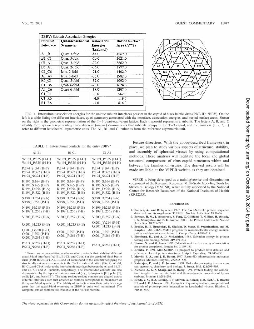
The results generated by the various analyses mentionedabove are available as web pages for individual entries. Table1 shows a representative list of residue-residue contacts at thequasi-3-fold related interfaces of black beetle virus (PDB-ID:2BBV). A number of contacts between the same residues atthe latter interfaces are aligned quite well, indicating that thefidelity of the quasi-3-fold symmetry is highly conserved. Fig-ure 4 shows the association energies and buried surface areasat the unique interfaces for the entry 2BBV. The derivedresults of various other analyses (e.g., interacting residues atthe interfaces and residues involved in interparticle contacts inthe crystal lattice) can be readily accessed from the website.
FIG. 2. Schematic diagram of the VIPER convention, z(2)-3-5-x(2). x, y, and z are the orthogonal coordinate axes, and the numbers 2, 3, and5 correspond to 2-fold, 3-fold, and 5-fold icosahedral symmetry axes, respectively. In this convention, the icosahedron is oriented such that a pairof icosahedral 2-fold axes are coincident with the z and x axes and a 3-fold/5-fold axis pair lies between the z and x axes in the xz plane. Thecoordinates of the all entries in the database are stored in this convention.
VOL. 75, 2001 GUEST COMMENTARY 11945
on October 20, 2014 by guest
http://jvi.asm.org/
Dow
nloaded from
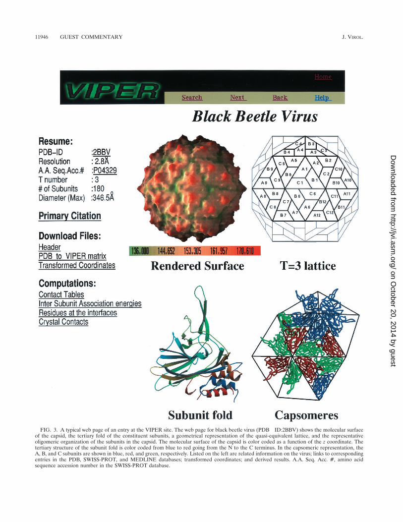
FIG. 3. A typical web page of an entry at the VIPER site. The web page for black beetle virus (PDB ID:2BBV) shows the molecular surfaceof the capsid, the tertiary fold of the constituent subunits, a geometrical representation of the quasi-equivalent lattice, and the representativeoligomeric organization of the subunits in the capsid. The molecular surface of the capsid is color coded as a function of the z coordinate. Thetertiary structure of the subunit fold is color coded from blue to red going from the N to the C terminus. In the capsomeric representation, theA, B, and C subunits are shown in blue, red, and green, respectively. Listed on the left are related information on the virus; links to correspondingentries in the PDB, SWISS-PROT, and MEDLINE databases; transformed coordinates; and derived results. A.A. Seq. Acc. #, amino acidsequence accession number in the SWISS-PROT database.
11946 GUEST COMMENTARY J. VIROL.
on October 20, 2014 by guest
http://jvi.asm.org/
Dow
nloaded from
Future directions. With the above-described framework inplace, we plan to study various aspects of structure, stability,and assembly of spherical viruses by using computationalmethods. These analyses will facilitate the local and globalstructural comparisons of virus capsid structures within andbetween the families of viruses. The derived results will bemade available at the VIPER website as they are obtained.
VIPER is being developed as a training/service and disseminationcomponent of the Research Resource: Multi-Scale Modeling Tools forStructure Biology (MMTSB), which is fully supported by the NationalCenter for Research Resources of the National Institutes of Health(RR12255).
REFERENCES
1. Bairoch, A., and R. Apweiler. 1997. The SWISS-PROT protein sequencedata bank and its supplement TrEMBL. Nucleic Acids Res. 25:31–36.
2. Berman, H. M., J. Westbrook, Z. Feng, G. Gilliland, T. N. Bhat, H. Weissig,I. N. Shindyalov, and P. E. Bourne. 2000. The Protein Data Bank. NucleicAcids Res. 28:235–242.
3. Brooks, B., B. Bruccoleri, D. Olafson, D. States, S. Swaminathan, and M.Karplus. 1983. CHARMM: a program for macromolecular energy, minimi-zation and dynamics calculation. J. Comp. Chem. 4:187–217.
4. Eisenberg, D., and A. D. McLachlan. 1986. Solvation energy in proteinfolding and binding. Nature 319:199–203.
5. Horton, N., and M. Lewis. 1992. Calculation of the free energy of associationfor protein complexes. Protein Sci. 1:169–181.
6. Kraulis, P. 1991. MOLSCRIPT: a program to produce both detailed andschematic plots of protein structures. J. Appl. Crystallogr. 24:946–950.
7. Merritt, E. A., and J. D. Bacon. 1997. Raster3D: photorealistic moleculargraphics. Methods Enzymol. 277:505–524.
8. Natarajan, P., and J. E. Johnson. 1998. Molecular packaging in virus crys-tals: geometry, chemistry, and biology. J. Struct. Biol. 121:295–305.
9. Nicholls, A., K. A. Sharp, and B. Honig. 1991. Protein folding and associa-tion: insights from the interfacial and thermodynamic properties of hydro-carbons. Proteins 11:281–296.
10. Reddy, V. S., H. A. Giesing, R. T. Morton, A. Kumar, C. B. Post, C. L. BrooksIII, and J. E. Johnson. 1998. Energetics of quasiequivalence: computationalanalysis of protein-protein interactions in icosahedral viruses. Biophys. J.74:546–558.
FIG. 4. Intersubunit association energies for the unique subunit interfaces present in the capsid of black beetle virus (PDB-ID: 2BBV). On theleft is a table listing the different interfaces, quasi-symmetry associated with the interface, association energies, and buried surface areas. Shownon the right is the geometric representation of the T�3 quasi-equivalent lattice. Each trapezoid represents a subunit. The letters A, B, and Cidentify the trapezoids representing three different (unique) environments that subunits occupy in the T�3 capsid, and the numbers (1, 2, 3,. . .)refer to different icosahedral asymmetric units. The A1, B1, and C1 subunits form the reference asymmetric unit.
TABLE 1. Intersubunit contacts for the entry 2BBVa
A1-B1 B1-C1 C1-A1
W:191_P:325 (H-H) W:191_P:325 (H-H) W:191_P:325 (H-H)W:191_P:325 (H-H) W:191_P:325 (H-H) W:191_P:325 (H-H)
P:194_S:164 (H-P) P:194_S:164 (H-P) P:194_S:164 (H-P)P:194_R:322 (H-B) P:194_R:322 (H-B) P:194_R:322 (H-B)P:194_N:324 (H-P) P:194_N:324 (H-P) P:194_N:324 (H-P)
K:196_S:164 (B-P) K:196_S:164 (B-P) K:196_S:164 (B-P)K:196_S:165 (B-P) K:196_S:165 (B-P) K:196_S:165 (B-P)K:196_D:254 (B-A) K:196_D:254 (B-A) K:196_D:254 (B-A)K:196_R:322 (B-B) K:196_R:322 (B-B) K:196_R:322 (B-B)
S:198_D:254 (P-A) S:198_D:254 (P-A) S:198_D:254 (P-A)S:198_L:256 (P-H) S:198_L:256 (P-H) S:198_L:256 (P-H)
N:199_H:215 (P-B) N:199_H:215 (P-B) N:199_H:215 (P-B)N:199_L:256 (P-H) N:199_L:256 (P-H) N:199_L:256 (P-H)
V:200_E:257 (H-A) V:200_E:257 (H-A) V:200_E:257 (H-A)
Q:201_H:215 (P-B) Q:201_H:215 (P-B) Q:201_V:214 (P-H)Q:201_H:215 (P-B)
Q:201_G:258 (P-H) Q:201_I:259 (P-H) Q:201_I:259 (P-H)Q:201_I:259 (P-H) Q:201_P:264 (P-H) Q:201_P:264 (P-H)Q:201_P:264 (P-H)
P:203_A:265 (H-H) P:203_A:265 (H-H) P:203_A:265 (H-H)P:203_N:266 (H-P) P:203_N:266 (H-P)
a Shown are representative residue-residue contacts that stabilize differentquasi-3-fold interfaces (A1-B1, B1-C1, and C1-A1) in the capsid of black beetlevirus (PDB-ID:2BBV). A1, B1, and C1 correspond to the subunits occupying thestructurally unique environments in the T�3 icosahedral lattice (Fig. 4). A1-B1,B1-C1, and C1-A1 refer to the intersubunit interfaces between the A1 and B1, B1and C1, C1 and A1 subunits, respectively. The interresidue contacts are alsodistinguished by the types of residues involved (e.g., hydrophobic [H], polar [P],acidic [A], and basic [B]). The same residue-residue contacts are aligned acrossdifferent interfaces such that absence of contacts corresponds to breakdown ofthe quasi-3-fold symmetry. The fidelity of contacts across these interfaces sug-gests that the quasi-3-fold symmetry in 2BBV is quite well maintained. Thecomplete lists of contacts are available at the VIPER website.
The views expressed in this Commentary do not necessarily reflect the views of the journal or of ASM.
VOL. 75, 2001 GUEST COMMENTARY 11947
on October 20, 2014 by guest
http://jvi.asm.org/
Dow
nloaded from
Related Documents