Virucidal Effect of Cold Atmospheric Gaseous Plasma on Feline Calicivirus, a Surrogate for Human Norovirus Hamada A. Aboubakr, a,c Paul Williams, b Urvashi Gangal, b Mohammed M. Youssef, c Sobhy A. A. El-Sohaimy, d Peter J. Bruggeman, b Sagar M. Goyal a Department of Veterinary Population Medicine, Veterinary Diagnostic Laboratory, College of Veterinary Medicine, University of Minnesota, St. Paul, Minnesota, USA a ; Department of Mechanical Engineering, College of Science and Engineering, University of Minnesota, Minneapolis, Minnesota, USA b ; Food Science and Technology Department, Faculty of Agriculture, Alexandria University, El-Shatby, Alexandria, Egypt c ; Department of Food Biotechnology, Arid Land Cultivation and Development Institute, City of Scientific Research and Technology Applications, New Borg El-Arab, Alexandria, Egypt d Minimal food-processing methods are not effective against foodborne viruses, such as human norovirus (NV). It is impor- tant, therefore, to explore novel nonthermal technologies for decontamination of foods eaten fresh, minimally processed and ready-to-eat foods, and food contact surfaces. We studied the in vitro virucidal activity of cold atmospheric gaseous plasma (CGP) against feline calicivirus (FCV), a surrogate of NV. Factors affecting the virucidal activity of CGP (a so- called radio frequency atmospheric pressure plasma jet) were the plasma generation power, the exposure time and dis- tance, the plasma feed gas mixture, and the virus suspension medium. Exposure to 2.5-W argon (Ar) plasma caused a 5.55 log 10 unit reduction in the FCV titer within 120 s. The reduction in the virus titer increased with increasing exposure time and decreasing exposure distance. Of the four plasma gas mixtures studied (Ar, Ar plus 1% O 2 , Ar plus 1% dry air, and Ar plus 0.27% water), Ar plus 1% O 2 plasma treatment had the highest virucidal effect: more than 6.0 log 10 units of the virus after 15 s of exposure. The lowest virus reduction was observed with Ar plus 0.27% water plasma treatment (5 log 10 unit reduction after 120 s). The highest reduction in titer was observed when the virus was suspended in distilled water. Changes in temperature and pH and formation of H 2 O 2 were not responsible for the virucidal effect of plasma. The oxida- tion of viral capsid proteins by plasma-produced reactive oxygen and nitrogen species in the solution was thought to be responsible for the virucidal effect. In conclusion, CGP exhibits virucidal activity in vitro and has the potential to combat viral contamination in foods and on food preparation surfaces. F oodborne illnesses continue to plague public health, as well as world economies, costing approximately $152 billion in the United States alone (1). Enteric viruses, particularly human noro- virus (NV) and hepatitis A virus (HAV), are the leading causes of viral foodborne illnesses (2). Human NV, one of the top five pathogens with respect to the total cost of foodborne illnesses in the United States, belongs to the family Caliciviridae and is a well- known cause of “winter vomiting disease” or “stomach flu” (3). NV causes 19 to 21 million cases of acute gastroenteritis annually in the United States and leads to 1.7 to 1.9 million outpatient visits, 400,000 emergency room visits, 56,000 to 71,000 hospitalizations, and 570 to 800 deaths, mostly among young children (4). More than half of all foodborne disease outbreaks due to a known cause reported to the CDC from 2006 to 2010 were attributed to NV. In the European Union in 2007, caliciviruses (primarily NV) were responsible for 507 of 675 foodborne viral disease outbreaks (5). Multiple issues related to the quality of thermally processed foods, e.g., nutritional losses and adverse effects on organoleptic quality, have led to the emergence of so-called nonthermal tech- nologies, which consist of preservation treatments that are effec- tive at ambient or sublethal temperatures, thereby minimizing the negative effects of thermal processing on nutritional and quality attributes of food (6). In addition, consumers have increasing in- terest in non-thermally processed and ready-to-eat food com- modities with fresh-like and healthy attributes, leading to popu- larization of minimally processed foods. However, such foods carry a high risk of surface cross-contamination through cutting boards, knives, working surfaces, equipment, or the processing environment. In addition, the low inactivation efficiency of most minimal food-processing methods against foodborne viruses is also a concern (7). Recent experiments with NV in a variety of foods revealed that minimal food processing (freezing, cooling, and mild heat treatment) was not significantly effective in reduc- ing titers of NV or its surrogates (8). Consequently, the develop- ment of novel efficient and safe nonthermal technology for viral decontamination of foods is of great interest to food scientists, food producers, and ultimately customers. A few recent investiga- tions have been aimed at studying the virucidal effects of some novel nonthermal technologies, such as ozone treatment (9), gamma irradiation treatment (10), and lactic acid bacteria as an antiviral biopreservative (11), in combating foodborne viral con- tamination in food. Cold atmospheric gaseous plasma (CGP) refers to a partially ionized gas composed essentially of photons, ions, free electrons, and reactive species, including atoms (O and N), molecules (e.g., Received 7 January 2015 Accepted 11 March 2015 Accepted manuscript posted online 20 March 2015 Citation Aboubakr HA, Williams P, Gangal U, Youssef MM, El-Sohaimy SAA, Bruggeman PJ, Goyal SM. 2015. Virucidal effect of cold atmospheric gaseous plasma on feline calicivirus, a surrogate for human norovirus. Appl Environ Microbiol 81:3612–3622. doi:10.1128/AEM.00054-15. Editor: C. A. Elkins Address correspondence to Sagar M. Goyal, [email protected]. Copyright © 2015, American Society for Microbiology. All Rights Reserved. doi:10.1128/AEM.00054-15 3612 aem.asm.org June 2015 Volume 81 Number 11 Applied and Environmental Microbiology on May 10, 2015 by guest http://aem.asm.org/ Downloaded from

Welcome message from author
This document is posted to help you gain knowledge. Please leave a comment to let me know what you think about it! Share it to your friends and learn new things together.
Transcript
Virucidal Effect of Cold Atmospheric Gaseous Plasma on FelineCalicivirus, a Surrogate for Human Norovirus
Hamada A. Aboubakr,a,c Paul Williams,b Urvashi Gangal,b Mohammed M. Youssef,c Sobhy A. A. El-Sohaimy,d Peter J. Bruggeman,b
Sagar M. Goyala
Department of Veterinary Population Medicine, Veterinary Diagnostic Laboratory, College of Veterinary Medicine, University of Minnesota, St. Paul, Minnesota, USAa;Department of Mechanical Engineering, College of Science and Engineering, University of Minnesota, Minneapolis, Minnesota, USAb; Food Science and TechnologyDepartment, Faculty of Agriculture, Alexandria University, El-Shatby, Alexandria, Egyptc; Department of Food Biotechnology, Arid Land Cultivation and DevelopmentInstitute, City of Scientific Research and Technology Applications, New Borg El-Arab, Alexandria, Egyptd
Minimal food-processing methods are not effective against foodborne viruses, such as human norovirus (NV). It is impor-tant, therefore, to explore novel nonthermal technologies for decontamination of foods eaten fresh, minimally processedand ready-to-eat foods, and food contact surfaces. We studied the in vitro virucidal activity of cold atmospheric gaseousplasma (CGP) against feline calicivirus (FCV), a surrogate of NV. Factors affecting the virucidal activity of CGP (a so-called radio frequency atmospheric pressure plasma jet) were the plasma generation power, the exposure time and dis-tance, the plasma feed gas mixture, and the virus suspension medium. Exposure to 2.5-W argon (Ar) plasma caused a 5.55log10 unit reduction in the FCV titer within 120 s. The reduction in the virus titer increased with increasing exposure timeand decreasing exposure distance. Of the four plasma gas mixtures studied (Ar, Ar plus 1% O2, Ar plus 1% dry air, and Arplus 0.27% water), Ar plus 1% O2 plasma treatment had the highest virucidal effect: more than 6.0 log10 units of the virusafter 15 s of exposure. The lowest virus reduction was observed with Ar plus 0.27% water plasma treatment (5 log10 unitreduction after 120 s). The highest reduction in titer was observed when the virus was suspended in distilled water.Changes in temperature and pH and formation of H2O2 were not responsible for the virucidal effect of plasma. The oxida-tion of viral capsid proteins by plasma-produced reactive oxygen and nitrogen species in the solution was thought to beresponsible for the virucidal effect. In conclusion, CGP exhibits virucidal activity in vitro and has the potential to combatviral contamination in foods and on food preparation surfaces.
Foodborne illnesses continue to plague public health, as well asworld economies, costing approximately $152 billion in the
United States alone (1). Enteric viruses, particularly human noro-virus (NV) and hepatitis A virus (HAV), are the leading causes ofviral foodborne illnesses (2). Human NV, one of the top fivepathogens with respect to the total cost of foodborne illnesses inthe United States, belongs to the family Caliciviridae and is a well-known cause of “winter vomiting disease” or “stomach flu” (3).NV causes 19 to 21 million cases of acute gastroenteritis annuallyin the United States and leads to 1.7 to 1.9 million outpatient visits,400,000 emergency room visits, 56,000 to 71,000 hospitalizations,and 570 to 800 deaths, mostly among young children (4). Morethan half of all foodborne disease outbreaks due to a known causereported to the CDC from 2006 to 2010 were attributed to NV. Inthe European Union in 2007, caliciviruses (primarily NV) wereresponsible for 507 of 675 foodborne viral disease outbreaks (5).
Multiple issues related to the quality of thermally processedfoods, e.g., nutritional losses and adverse effects on organolepticquality, have led to the emergence of so-called nonthermal tech-nologies, which consist of preservation treatments that are effec-tive at ambient or sublethal temperatures, thereby minimizing thenegative effects of thermal processing on nutritional and qualityattributes of food (6). In addition, consumers have increasing in-terest in non-thermally processed and ready-to-eat food com-modities with fresh-like and healthy attributes, leading to popu-larization of minimally processed foods. However, such foodscarry a high risk of surface cross-contamination through cuttingboards, knives, working surfaces, equipment, or the processingenvironment. In addition, the low inactivation efficiency of most
minimal food-processing methods against foodborne viruses isalso a concern (7). Recent experiments with NV in a variety offoods revealed that minimal food processing (freezing, cooling,and mild heat treatment) was not significantly effective in reduc-ing titers of NV or its surrogates (8). Consequently, the develop-ment of novel efficient and safe nonthermal technology for viraldecontamination of foods is of great interest to food scientists,food producers, and ultimately customers. A few recent investiga-tions have been aimed at studying the virucidal effects of somenovel nonthermal technologies, such as ozone treatment (9),gamma irradiation treatment (10), and lactic acid bacteria as anantiviral biopreservative (11), in combating foodborne viral con-tamination in food.
Cold atmospheric gaseous plasma (CGP) refers to a partiallyionized gas composed essentially of photons, ions, free electrons,and reactive species, including atoms (O and N), molecules (e.g.,
Received 7 January 2015 Accepted 11 March 2015
Accepted manuscript posted online 20 March 2015
Citation Aboubakr HA, Williams P, Gangal U, Youssef MM, El-Sohaimy SAA,Bruggeman PJ, Goyal SM. 2015. Virucidal effect of cold atmospheric gaseousplasma on feline calicivirus, a surrogate for human norovirus. Appl EnvironMicrobiol 81:3612–3622. doi:10.1128/AEM.00054-15.
Editor: C. A. Elkins
Address correspondence to Sagar M. Goyal, [email protected].
Copyright © 2015, American Society for Microbiology. All Rights Reserved.
doi:10.1128/AEM.00054-15
3612 aem.asm.org June 2015 Volume 81 Number 11Applied and Environmental Microbiology
on May 10, 2015 by guest
http://aem.asm
.org/D
ownloaded from
O3, H2O2, and HNO2), and radicals {OH�, NO�, and singlet oxygen[O2(a1�g)]}, all of which are important in biological reactions(12). Due to its unique properties, plasma is often referred to asthe fourth state of matter and is normally generated in nature orartificially by subjecting a gas to a strong electric field, which leadsto ionization; dissociation of molecules; and production of pho-tons, radicals, and reactive species (13). As the electric field trans-fers energy to the mobile electrons and the significant mass differ-ence between electrons and neutral gas molecules (heavyparticles) leads to inefficient collisional energy transfer betweenelectrons and heavy particles, the energy of the electrons can besignificantly larger than the energy of the neutral gas molecules.Various sources of plasma exploiting this physical property allowthe production of highly reactive cold atmospheric pressure plas-mas for which the reactivity is induced by energetic electrons whilethe gas temperature remains close to room temperature (14).These conditions are easily obtained at low pressure and requirespecific plasma generation strategies at atmospheric pressure.Typical approaches for cold atmospheric pressure plasma gener-ation at atmospheric pressure include corona discharge, dielectricbarrier discharges (DBD), radio frequency (RF) plasma, and glid-ing-arc discharge (15).
CGP is attracting the attention of food researchers due to itsbiocidal effects against various spoilage microorganisms and foodpathogens, because it can be applied at ambient air temperaturewithout the use of heat. The use of CGP appears to be especiallyadvantageous for decontaminating the surfaces of fresh foods,such as vegetables, fruits, meats, nuts, poultry, and eggs, and alsofor disinfection of food preparation surfaces (16–19).
Several studies on the antibacterial effects of cold plasmaagainst bacteria—Escherichia coli (20), Salmonella spp. (16),Staphylococcus aureus (18), and others (6)—are available. How-ever, the antiviral activity of CGP has not been properly explored.Only three studies have reported antiviral activity of CGP. Terrieret al. (21) recorded reductions in virus titers of 6.5, 3.8, and 4.0log10 units for influenza virus type A, human parainfluenza virustype 3, and respiratory syncytial virus (RSV), respectively, using anewly designed system based on physical decontamination of sub-strates and air by cold atmospheric plasma. Zimmermann et al.(22) achieved up to 6.0 log10 unit inactivation of adenovirus usinga CGP system. Alekseev et al. (23) suppressed corneal herpes sim-plex virus 1 infection in vitro using a nonthermal DBD plasmasystem. To date, no published study is available on the efficacy ofCGP against foodborne viruses, such as NV and HAV. The presentstudy was undertaken to determine the in vitro virucidal activity ofcold plasma using four different gas mixtures against feline calici-virus (FCV), a surrogate for human NV, and to study factors thatmight determine the virucidal effects of CGP.
MATERIALS AND METHODSDescription of the plasma generation system. The plasma jet used in thisstudy is identical to the one described by van Gils et al. (24). The plasma isignited in Ar, which is blown through a 1.9-mm quartz tube at 1.5 stan-dard liters per minute (slm) by RF power (13.56 MHz). The RF power ismodulated at a frequency of 20 kHz with a duty cycle of 20%. Unlessotherwise stated, the work was performed at a time-averaged plasma dis-sipated power of 2.5 W. The power level was measured as explained byHofmann et al. (25). In this study, 4 different gases were used: Ar, Ar plus1% O2, Ar plus 1% dry air, and Ar plus 0.27% water. The amounts ofadmixtures were controlled by mass flow controllers (MKS GE50; MKSInstruments Inc., USA). In the case of Ar plus 0.27% water, part of the Ar
flow was sent through a bubbler containing sterile distilled water (DW) tohumidify part of the Ar gas feed.
The plasma jet is known to produce significant amounts of NO� (26),O3 (27), OH�, HNO2, H2O2, and O2(a1�g) (28). These species are allbiologically active and are produced in large quantities. Different gas ad-mixtures favor the production of certain species over others. For example,mixing O2, air, and water favors the production of O3, NO�, and OH�/H2O2, respectively (29), although even in the cases of Ar, Ar plus water,and Ar plus O2, diffusion of the surrounding air in the jet effluent leads tothe production of NO�. The reactive species produced by the plasma in thegas phase are transferred to the solution through complex gas-liquid in-teractions (30).
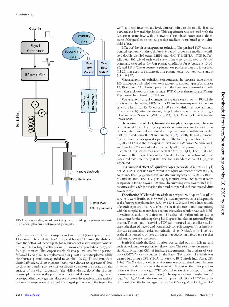
For the virucidal experiments in this study, we placed a sterile 96-wellmicrotiter plate (containing 100 �l of virus suspension) below the plasmajet. The tip of the plasma plume was allowed to approach the top of thewell. A diagram of the plasma setup, including a schematic of the 96-wellplate, can be seen in Fig. 1. After the desired treatment times, the sampleswere removed and kept at room temperature for 2 h to allow the plasma-produced reactive species in solution to react with the virus. Then, FCVtitration of treated and nontreated (control) samples was carried out (asdescribed below). The antiviral effects were identified by comparing thetiters of treated and nontreated (control) viruses.
Virus propagation. Strain 255 of FCV was used as a surrogate for NVin this investigation. The virus was grown and titrated in Crandell-Reesefeline kidney (CRFK) cells, which were grown in minimum essential me-dium (MEM) with Earle’s salts and L-glutamine (Mediatech Inc., Manas-sas, VA, USA) supplemented with 8% fetal bovine serum (FBS) and anti-biotics (neomycin, 90 U/ml; gentamicin, 50 �g/ml; penicillin, 455 IU/ml;streptomycin, 455 �g/ml; and amphotericin B [Fungizone], 1.5 �g/ml).The cells were incubated at 37°C under 5% CO2. The incubated cells wereexamined daily under an inverted microscope for appearance of cyto-pathic effects (CPE), which usually appeared 2 to 3 days after infection.The infected cells were frozen and thawed three times, followed by cen-trifugation at 2,000 � g for 30 min at 4°C. The virus-containing superna-tant was decanted and distributed in 1- by 3.25-in. ultracentrifuge tubes(Beckman Instruments Inc., USA). One milliliter of sterile 30% sucrose indistilled water was underlaid in each tube. The balanced, capped tubeswere then ultracentrifuged at 111,857 � g for 2.5 h at 4°C (Optima L-90Kultracentrifuge; Beckman Instruments) using an SW32 Ti swinging-bucket rotor (Beckman Instruments Inc., USA). The supernatant wascarefully removed and discarded, and the virus pellet was resuspended in1 ml of sterile DW. The purified virus was aliquoted and stored at �80°Cuntil it was used.
Virus titration. Serial 10-fold dilutions of samples were prepared inMEM containing 4% FBS. The dilutions were inoculated into CRFK cellmonolayers prepared in 96-well microtiter plates using 3 wells per dilu-tion. The plates were incubated at 37°C under 5% CO2 and examineddaily for the development of CPE for up to 5 days. The endpoint was takento be the highest dilution of the virus that produced CPE in 50% of theinoculated cells. Viral titers were calculated by the Kärber method (31)and are expressed as 50% tissue culture infective doses (TCID50)/0.1 ml.
Effect of plasma generation power. Aliquots (100 �l) of FCV sus-pended in sterile distilled water (DW-FCV) were exposed to Ar atmo-spheric plasma generated at five different power levels (1 � 0.1, 1.5 � 0.1,2 � 0.1, 2.5 � 0.1, and 3 � 0.1 W). The exposure distance was 18.13 mmfrom the plasma nozzle to the surface of the treated viral suspension. Theexposure times were 0 (control), 15, 30, 60, 120, and 180 s.
Effect of plasma exposure distance and type of plasma feeding gasmixture. In a factorial experiment, four plasma feeding gas mixtures (Ar,Ar plus 1% O2, Ar plus 1% dry air, and Ar plus 0.27% water) were usedseparately for the generation of CGP. The plasma dissipated power waskept constant at 2.5 � 0.1 W. Aliquots (100 �l) of DW-FCV were exposedto the four types of plasma for 0 (control), 15, 30, 60, and 120 s. To studythe effect of exposure distance on the virucidal effects of each plasma type,three different exposure distances (the distance from the plasma jet nozzle
Virucidal Effect of Cold Atmospheric Gaseous Plasma
June 2015 Volume 81 Number 11 aem.asm.org 3613Applied and Environmental Microbiology
on May 10, 2015 by guest
http://aem.asm
.org/D
ownloaded from
to the surface of the virus suspension) were used (low exposure level,11.25 mm; intermediate, 14.69 mm; and high, 18.13 mm [the distancefrom the bottom of the well plate to the surface of the virus suspension was6.49 mm]). The length of the plasma plumes used depended on the type offeed gas mixture. The longest visible plasma plume was for Ar plasma,followed by Ar plus 1% air plasma and Ar plus 0.27% water plasma, whilethe shortest plume corresponded to Ar plus 1% O2. To accommodatethese differences, three exposure levels were chosen to represent (i) lowlevel, corresponding to the shortest distance between the nozzle and thesurface of the viral suspension (the visible plasma tip of the shortestplasma plume was at the position of the top of the well); (ii) high level,corresponding to the greatest distance between the nozzle and the surfaceof the viral suspension (the tip of the longest plume was at the top of the
well); and (iii) intermediate level, corresponding to the middle distancebetween the low and high levels. This experiment was repeated with thefeed gas mixture flows with the power off (gas-phase treatment) to deter-mine if the gas flow on the suspension medium contributed to the viru-cidal effect.
Effect of the virus suspension solution. The purified FCV was sus-pended separately in three different types of suspension medium (steril-ized double-distilled water, MEM, and NaCl-Tris-EDTA [NTE] buffer).Aliquots (100 �l) of each viral suspension were distributed in 96-wellplates and exposed to the four plasma conditions for 0 (control), 15, 30,60, and 120 s. The exposure to plasma was performed at the lower level(11.25-mm exposure distance). The plasma power was kept constant at2.5 � 0.1 W.
Measurement of solution temperature. In separate experiments,100-�l aliquots of distilled water were exposed to the four types of plasma for15, 30, 60, and 120 s. The temperature of the liquid was measured immedi-ately after each exposure time, using an RTD Omega thermocouple (OmegaEngineering Inc., Stamford, CT, USA).
Measurement of pH changes. In separate experiments, 100-�l ali-quots of distilled water, MEM, and NTE buffer were exposed to the fourtypes of plasma for 15, 30, 60, and 120 s at two distances (low and highexposure levels). After treatment, the pH values were measured using aThermo Fisher Scientific (Waltham, MA, USA) Orion pH probe (model8220BNWP).
Determination of H2O2 formed during plasma exposure. The con-centration of formed hydrogen peroxide in plasma-exposed distilled wa-ter was determined colorimetrically using the titanium sulfate method ofSatterfield and Bonnell (32) and Eisenberg (33). Briefly, 100-�l aliquots ofdistilled water were exposed separately to the four types of plasma for 15,30, 60, and 120 s at the low exposure level and 2.5 W power. Sodium azidesolution (4 mM) was added immediately after the plasma treatment toquench nitrites, which may react with the formed H2O2. Then, 100 �l oftitanium sulfate reagent was added. The development of yellow color wasmeasured colorimetrically at 407 nm, and a standard curve of H2O2 wasgenerated.
FCV virucidal effect of liquid hydrogen peroxide. Aliquots (100 �l)of DW-FCV suspension were mixed with equal volumes of different H2O2
solutions. The H2O2 concentrations after mixing were 5, 10, 20, 30, 40, 50,80, and 100 mM. The FCV-plus-H2O2 mixtures were incubated at roomtemperature for 30, 60, and 120 min. The surviving virus was titrated in allmixtures after each incubation time and compared with nontreated virusas a control.
The effective FCV lethal time of plasma exposure. Aliquots (100�l) ofDW-FCV were distributed in 96-well plates. Samples were exposed separatelyto the four types of plasma for 15, 30, 60, 120, 180, 240, and 300 s. Immediatelyafter each exposure time, 10 �l of 0.1 M (the final concentration after mixingwith the sample) filter-sterilized sodium thiosulfate solution was added, fol-lowed immediately by FCV titration. The sodium thiosulfate solution acts asa scavenger for the oxidizing (long-lived) species in solution generated by theplasma. The amount of surviving FCV was measured as the difference be-tween the titers of treated and nontreated (control) samples. Virus inactiva-tion was calculated as the decimal reduction time (D value), which is definedas the time needed to achieve a 1-log-unit reduction in infectious virus titerwith a given plasma treatment.
Statistical analysis. Each titration was carried out in triplicate, andeach experiment was performed three times. The results are the means �standard deviations (SD) of triplicate experiments. The analysis of vari-ance (ANOVA) was generated by the F test. The statistical analysis wascarried out using STATISTICA software, v. 10 (Statsoft Inc., Tulsa, OK,USA). The D value of each type of plasma was determined from the neg-ative reciprocal of the slope of the regression line, using the linear portionsof the survival curves (log10 TCID50/0.1 ml versus time of exposure to theplasma under constant conditions). The exposure times needed for a 4log10 TCID50/0.1 ml reduction and complete reduction of FCV were de-termined from the following equation: t � D � (log No � log Nf) � D �
FIG 1 Schematic diagram of the CGP system, including the plasma jet, treat-ment of samples, and electrical and gas inputs.
Aboubakr et al.
3614 aem.asm.org June 2015 Volume 81 Number 11Applied and Environmental Microbiology
on May 10, 2015 by guest
http://aem.asm
.org/D
ownloaded from
n, where D is the D value (in minutes) under the specified conditions; No
is the initial titer of the virus; Nf is the surviving titer after an exposuretime, t (in minutes), to the selected plasma type; and n is (log No � log Nf),which is equal to the log10 reduction of the initial titer of the virus.
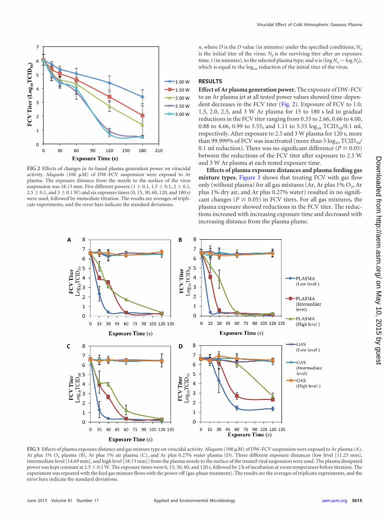
RESULTSEffect of Ar plasma generation power. The exposure of DW-FCVto an Ar plasma jet at all tested power values showed time-depen-dent decreases in the FCV titer (Fig. 2). Exposure of FCV to 1.0,1.5, 2.0, 2.5, and 3 W Ar plasma for 15 to 180 s led to gradualreductions in the FCV titer ranging from 0.33 to 2.66, 0.66 to 4.00,0.88 to 4.66, 0.99 to 5.55, and 1.11 to 5.55 log10 TCID50/0.1 ml,respectively. After exposure to 2.5 and 3 W plasma for 120 s, morethan 99.999% of FCV was inactivated (more than 5 log10 TCID50/0.1 ml reduction). There was no significant difference (P � 0.05)between the reductions of the FCV titer after exposure to 2.5 Wand 3 W Ar plasma at each tested exposure time.
Effects of plasma exposure distances and plasma feeding gasmixture types. Figure 3 shows that treating FCV with gas flowonly (without plasma) for all gas mixtures (Ar, Ar plus 1% O2, Arplus 1% dry air, and Ar plus 0.27% water) resulted in no signifi-cant changes (P � 0.05) in FCV titers. For all gas mixtures, theplasma exposure showed reductions in the FCV titer. The reduc-tions increased with increasing exposure time and decreased withincreasing distance from the plasma plume.
FIG 2 Effects of changes in Ar-based plasma generation power on virucidalactivity. Aliquots (100 �M) of DW-FCV suspension were exposed to Arplasma. The exposure distance from the nozzle to the surface of the virussuspension was 18.13 mm. Five different powers (1 � 0.1, 1.5 � 0.1, 2 � 0.1,2.5 � 0.1, and 3 � 0.1 W) and six exposure times (0, 15, 30, 60, 120, and 180 s)were used, followed by immediate titration. The results are averages of tripli-cate experiments, and the error bars indicate the standard deviations.
FIG 3 Effects of plasma exposure distance and gas mixture type on virucidal activity. Aliquots (100 �M) of DW-FCV suspension were exposed to Ar plasma (A),Ar plus 1% O2 plasma (B), Ar plus 1% air plasma (C), and Ar plus 0.27% water plasma (D). Three different exposure distances (low level [11.25 mm],intermediate level [14.69 mm], and high level [18.13 mm]) from the plasma nozzle to the surface of the treated viral suspension were used. The plasma dissipatedpower was kept constant at 2.5 � 0.1 W. The exposure times were 0, 15, 30, 60, and 120 s, followed by 2 h of incubation at room temperature before titration. Theexperiment was repeated with the feed gas mixture flows with the power off (gas-phase treatment). The results are the averages of triplicate experiments, and theerror bars indicate the standard deviations.
Virucidal Effect of Cold Atmospheric Gaseous Plasma
June 2015 Volume 81 Number 11 aem.asm.org 3615Applied and Environmental Microbiology
on May 10, 2015 by guest
http://aem.asm
.org/D
ownloaded from

For Ar plasma, more than 99.9999% of FCV (6.18, 6.22, and6.23 log10 TCID50/0.1 ml) was inactivated after exposure times of30 s, 60 s, and 120 s at low, intermediate, and high exposure levels,respectively (Fig. 3A). As shown in Fig. 3B, more than 99.9999%(6.22, 6.00, and 6.25 log10 TCID50/0.1 ml) of FCV was inactivatedafter exposure to Ar plus 1% O2 plasma for 15 s, 30 s, and 60 s atlow, intermediate, and high exposure levels, respectively. For Arplus 1% air plasma, more than 99.9999% of FCV (6.21, 6.22, and6.29 log10 TCID50/0.1 ml) was inactivated after exposure times of30 s, 60 s, and 120 s at low, intermediate and high exposure levels,respectively (Fig. 3C). As shown in Fig. 3D, the FCV titer wasdecreased only 99.9993% (more than 5.00 log10 TCID50/0.1 ml)after exposure to Ar plus 0.27% water plasma for 60 s at the lowexposure level, whereas the intermediate and high exposure levels
showed only 99.995% (4.29 log10 TCID50/0.1 ml) and 99.988%(3.91 log10 TCID50/0.1 ml) reductions, respectively, after the 120-sexposure.
Among the four tested gas mixtures, the greatest anti-FCV ef-fect was achieved with exposure to Ar plus 1% O2 plasma, sinceapproximately 100% (more than 6 log10 TCID50/0.1 ml) of thevirus was inactivated after only 15 s at the low level, while a smalleranti-FCV effect occurred with Ar plus 0.27% water plasma treat-ment, resulting in a reduction of only 5 log10 TCID50/0.1 ml(99.9993) after 120-s plasma exposure.
Effect of virus suspension medium. The results presented inFig. 4 show that the type of virus suspension solution significantly(P � 0.05) affected the virucidal activities of the four tested gasmixture plasmas. For Ar plasma exposure (Fig. 4A), when DW was
FIG 4 Effects of virus suspension media on virucidal activity of CGP. Aliquots (100 �M) of DW-FCV, MEM-FCV, and NTE-FCV suspensions were exposed to Arplasma (A), Ar plus 1% O2 plasma (B), Ar plus 1% air plasma (C), and Ar plus 0.27% water plasma (D). The plasma dissipated power was kept constant at 2.5 � 0.1 W.The exposure distance from the nozzle to the surface of the virus suspension was 11.25 mm. The exposure times were 0, 15, 30, 60, and 120 s, followed by 2 h of incubationat room temperature before titration. The results are the averages of triplicate experiments, and the error bars indicate the standard deviations.
TABLE 1 Changes in the temperature of distilled water after exposure to various types of atmospheric gaseous plasma jets for various exposuretimes
Exposure time (s)
Tempa (°C)
Ar Ar 1% O2 Ar 1% air Ar 0.27% water
0 24.20 � 0.21A/A 24.20 � 0.21A/A 24.20 � 0.21A/A 24.20 � 0.21A/A15 24.55 � 0.07AB/A 25.25 � 0.21AB/AB 25.60 � 0.71B/B 25.95 � 0.07B/B30 25.60 � 0.71ABC/A 25.85 � 1.34AB/A 25.90 � 0.14B/A 25.90 � 0.42B/A60 25.90 � 0.57BC/A 26.30 � 0.28AB/A 25.95 � 0.21B/A 25.95 � 0.21B/A120 26.70 � 0.85C/A 26.91 � 0.14B/A 26.30 � 0.42B/A 26.78 � 0.14B/Aa The values shown are averages of triplicate measurements � standard deviations. Values followed by the same letters indicate no significant difference in the temperatures (P �
0.05) across the treatment times within the same plasma treatment group (letters before slashes) or across the plasma treatment group within the same treatment time group (lettersafter slashes). The distance from the plasma jet nozzle to the surface of the virus suspension (low level) was 11.25 mm.
Aboubakr et al.
3616 aem.asm.org June 2015 Volume 81 Number 11Applied and Environmental Microbiology
on May 10, 2015 by guest
http://aem.asm
.org/D
ownloaded from
used to suspend FCV, 99.9997% (5.56 log10 TCID50/0.1 ml) of theFCV was inactivated after 30 s of exposure. However, 99.992%(4.11 log10 TCID50/0.1 ml) and 95.43% (1.34 log10 TCID50/0.1 ml)reductions were attained after the longest exposure time (120 s) inthe cases of NTE- and MEM-suspended FCV, respectively. FCVexposed to Ar plus 1% O2 plasma using DW and NTE buffer asthe suspending solutions led to almost complete inactivation(99.9998% [5.67 log10 TCID50/0.1 ml] and 99.9996% [5.45 log10
TCID50/0.1 ml] inactivation) of FCV after only 15 s, while the useof MEM led to a very small reduction (1.78 log10 TCID50/0.1 ml)in the FCV titer even after the longest exposure time (120 s). Whenthe virus was suspended in distilled water and exposed to Ar plus1% air plasma (Fig. 4C), 99.9996% (5.45 log10 TCID50/0.1 ml) ofthe FCV was inactivated after 30 s of exposure, while 99.94% (3.23log10 TCID50/0.1 ml) and only 78.62% (0.67 log10 TCID50/0.1 ml)reductions were attained after the longest exposure time (120 s) inthe cases of NTE- and MEM-suspended FCV, respectively. Even-tually, as presented in Fig. 4D, FCV exposed to Ar plus 0.27%water plasma for the longest exposure time (120 s) using DW,NTE buffer, and MEM as the suspension solutions led to only99.990% (4.00 log10 TCID50/0.1 ml), 99.950% (3.34 log10 TCID50/0.1 ml), and 97.860% (1.67 log10 TCID50/0.1 ml) reductions inFCV, respectively.
Temperature and pH value changes. As shown in Table 1, thetemperature of the DW exposed to all tested plasma types in-creased slightly with an increase in treatment time; the maximumincrease was 2.5°C after the longest treatment time (120 s). Therewere minor differences (P � 0.05) in the temperature changesamong the four types of plasma; the highest solution temperaturenever exceeded 27°C.
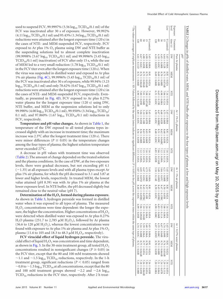
A decrease in pH values with treatment time was observed(Table 2).The amount of change depended on the treated solutionand the plasma conditions. In the case of DW, at the two exposurelevels, there were gradual decreases, but not exceeding pH 4.1(�1.35) at all exposure levels and with all plasma types except Arplus 1% air plasma, for which the pH decreased to 3.1 and 3.87 atlower and higher levels, respectively. In treated MEM, the lowestvalue attained (pH 8.39) was with Ar plus 1% air plasma at thelower exposure level. In NTE buffer, the pH decreased slightly butremained close to the neutral value (pH 7).
Determination of the H2O2 formed during plasma exposure.As shown in Table 3, hydrogen peroxide was formed in distilledwater when it was exposed to all types of plasma. The measuredH2O2 concentrations were time dependent: the longer the expo-sure, the higher the concentration. Higher concentrations of H2O2
were detected when distilled water was exposed to Ar plus 0.27%H2O plasma (251.7 to 2,785 �M H2O2), followed by Ar plasma(36.6 to 120 �M H2O2), whereas the lowest concentrations werefound with exposure to Ar plus 1% air plasma and Ar plus 1% O2
plasma (11.6 to 105 and 18.3 to 48.3 �M H2O2, respectively).FCV virucidal effect of liquid hydrogen peroxide. The viru-
cidal effect of liquid H2O2 was concentration and time dependent,as shown in Fig. 5. In the 30-min treatment group, all tested H2O2
concentrations resulted in nonsignificant changes (P � 0.05) inthe FCV titer, except that the 80 and 100 mM treatments showed1.1 and 1.5 log10 TCID50 reductions, respectively. In the 1-htreatment group, significant reductions (P � 0.05) ranged from0.8 to 1.5 log10 TCID50 at all concentrations, except that the 80and 100 mM treatment groups showed 2.2 and 2.6 log10
TCID50 reductions in the FCV titer, respectively. After 2 h treat-
TA
BLE
2C
han
gesin
pHs
ofdistilledw
ater,ME
M,an
dN
TE
buffer
afterexposu
reto
the
four
typesofplasm
aat
lowan
dh
ighexposu
relevels
Exposu
relevel
Treatm
ent
time
(s)
pHa
Pu
reA
rplasm
aA
r
1%O
2plasm
aA
r
1%air
plasma
Ar
0.27%
water
plasma
DW
ME
MN
TE
DW
ME
MN
TE
DW
ME
MN
TE
DW
ME
MN
TE
Low0
(control)
5.45�
0.00A8.81
�0.02A
7.16�
0.05A5.45
�0.00A
8.81�
0.02A7.16
�0.05A
5.45�
0.00A8.81
�0.02A
7.16�
0.05A5.45
�0.00A
8.81�
0.02A7.16
�0.05A
154.64
�0.12B
8.73�
0.07AB
7.06�
0.12A4.75
�0.06B
8.68�
0.03B7.14
�0.11A
4.21�
0.08B8.50
�0.05B
7.08�
0.08A4.74
�0.09B
8.74�
0.05AB
7.29�
0.11A30
4.45�
0.08B8.70
�0.08B
6.98�
0.08A4.68
�0.02B
8.62�
0.04BC
7.12�
0.09A3.84
�0.07C
8.47�
0.13BC
7.03�
0.11A4.67
�0.04B
8.73�
0.06AB
7.24�
0.07B60
4.18�
0.05C8.69
�0.10B
6.65�
0.15B4.67
�0.08B
C8.51
�0.09C
7.04�
0.13AB
3.49�
0.09D8.45
�0.03B
C6.83
�0.06B
4.35�
0.08C8.70
�0.08B
7.21�
0.06A120
4.10�
0.02D8.69
�0.06B
6.31�
0.03C4.59
�0.05C
8.48�
0.05C6.85
�0.05B
3.10�
0.14E8.39
�0.03C
4.90�
0.08C4.07
�0.05D
8.70�
0.05B7.20
�0.08A
High
0(con
trol)5.45
�0.00A
8.81�
0.02A7.16
�0.05A
5.45�
0.00A8.81
�0.02A
7.16�
0.05A5.45
�0.00A
8.81�
0.02A7.16
�0.05A
5.45�
0.00A8.81
�0.02A
7.16�
0.05B15
4.99�
0.06B8.75
�0.05A
B7.08
�0.07A
B5.08
�0.07B
8.65�
0.06B7.16
�0.05A
4.73�
0.08B8.57
�0.10B
7.20�
0.06A5.40
�0.06A
8.78�
0.12A7.30
�0.05A
304.88
�0.05B
8.75�
0.09AB
7.01�
0.04B5.10
�0.10B
8.63�
0.03B7.15
�0.07A
4.47�
0.05C8.43
�0.07B
C7.10
�0.10A
B5.39
�0.06A
8.77�
0.05A7.26
�0.06A
B60
4.67�
0.02C8.74
�0.06A
B7.00
�0.05B
5.04�
0.05B8.56
�0.08B
7.12�
0.09A4.05
�0.07D
8.42�
0.05BC
7.06�
0.09AB
5.16�
0.08B8.75
�0.04A
B7.18
�0.05B
1204.41
�0.11D
8.72�
0.06B6.90
�0.21A
B4.85
�0.03C
8.56�
0.05B7.13
�0.05A
3.87�
0.06E8.40
�0.03C
6.99�
0.05B4.74
�0.03C
8.72�
0.02B7.15
�0.08B
aT
he
values
areaverages
oftriplicatem
easurem
ents
�SD
.Valu
esfollow
edby
the
same
lettersin
dicaten
osign
ifican
tdifferen
ce(P
�0.05)
inth
epH
acrossth
eexposu
retim
esw
ithin
the
same
typeofplasm
a.
Virucidal Effect of Cold Atmospheric Gaseous Plasma
June 2015 Volume 81 Number 11 aem.asm.org 3617Applied and Environmental Microbiology
on May 10, 2015 by guest
http://aem.asm
.org/D
ownloaded from
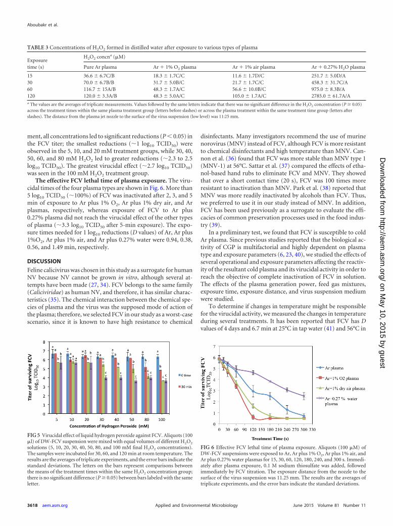
ment, all concentrations led to significant reductions (P � 0.05) inthe FCV titer; the smallest reductions (1 log10 TCID50) wereobserved in the 5, 10, and 20 mM treatment groups, while 30, 40,50, 60, and 80 mM H2O2 led to greater reductions (2.3 to 2.5log10 TCID50). The greatest virucidal effect (2.7 log10 TCID50)was seen in the 100 mM H2O2 treatment group.
The effective FCV lethal time of plasma exposure. The viru-cidal times of the four plasma types are shown in Fig. 6. More than5 log10 TCID50 (100%) of FCV was inactivated after 2, 3, and 5min of exposure to Ar plus 1% O2, Ar plus 1% dry air, and Arplasmas, respectively, whereas exposure of FCV to Ar plus0.27% plasma did not reach the virucidal effect of the other typesof plasma (3.3 log10 TCID50 after 5-min exposure). The expo-sure times needed for 1 log10 reductions (D values) of Ar, Ar plus1%O2, Ar plus 1% air, and Ar plus 0.27% water were 0.94, 0.38,0.56, and 1.49 min, respectively.
DISCUSSION
Feline calicivirus was chosen in this study as a surrogate for humanNV because NV cannot be grown in vitro, although several at-tempts have been made (27, 34). FCV belongs to the same family(Caliciviridae) as human NV, and therefore, it has similar charac-teristics (35). The chemical interaction between the chemical spe-cies of plasma and the virus was the supposed mode of action ofthe plasma; therefore, we selected FCV in our study as a worst-casescenario, since it is known to have high resistance to chemical
disinfectants. Many investigators recommend the use of murinenorovirus (MNV) instead of FCV, although FCV is more resistantto chemical disinfectants and high temperature than MNV. Can-non et al. (36) found that FCV was more stable than MNV type 1(MNV-1) at 56°C. Sattar et al. (37) compared the effects of etha-nol-based hand rubs to eliminate FCV and MNV. They showedthat over a short contact time (20 s), FCV was 100 times moreresistant to inactivation than MNV. Park et al. (38) reported thatMNV was more readily inactivated by alcohols than FCV. Thus,we preferred to use it in our study instead of MNV. In addition,FCV has been used previously as a surrogate to evaluate the effi-cacies of common preservation processes used in the food indus-try (39).
In a preliminary test, we found that FCV is susceptible to coldAr plasma. Since previous studies reported that the biological ac-tivity of CGP is multifactorial and highly dependent on plasmatype and exposure parameters (6, 23, 40), we studied the effects ofseveral operational and exposure parameters affecting the reactiv-ity of the resultant cold plasma and its virucidal activity in order toreach the objective of complete inactivation of FCV in solution.The effects of the plasma generation power, feed gas mixtures,exposure time, exposure distance, and virus suspension mediumwere studied.
To determine if changes in temperature might be responsiblefor the virucidal activity, we measured the changes in temperatureduring several treatments. It has been reported that FCV has Dvalues of 4 days and 6.7 min at 25°C in tap water (41) and 56°C in
TABLE 3 Concentrations of H2O2 formed in distilled water after exposure to various types of plasma
Exposuretime (s)
H2O2 concna (�M)
Pure Ar plasma Ar 1% O2 plasma Ar 1% air plasma Ar 0.27% H2O plasma
15 36.6 � 6.7C/B 18.3 � 1.7C/C 11.6 � 1.7D/C 251.7 � 5.0D/A30 70.0 � 6.7B/B 31.7 � 5.0B/C 21.7 � 1.7C/C 458.3 � 31.7C/A60 116.7 � 15A/B 48.3 � 1.7A/C 56.6 � 10.0B/C 975.0 � 8.3B/A120 120.0 � 3.3A/B 48.3 � 5.0A/C 105.0 � 1.7A/C 2785.0 � 61.7A/Aa The values are the averages of triplicate measurements. Values followed by the same letters indicate that there was no significant difference in the H2O2 concentration (P � 0.05)across the treatment times within the same plasma treatment group (letters before slashes) or across the plasma treatment within the same treatment time group (letters afterslashes). The distance from the plasma jet nozzle to the surface of the virus suspension (low level) was 11:25 mm.
FIG 5 Virucidal effect of liquid hydrogen peroxide against FCV. Aliquots (100�l) of DW-FCV suspension were mixed with equal volumes of different H2O2
solutions (5, 10, 20, 30, 40, 50, 80, and 100 mM final H2O2 concentrations).The samples were incubated for 30, 60, and 120 min at room temperature. Theresults are the averages of triplicate experiments, and the error bars indicate thestandard deviations. The letters on the bars represent comparisons betweenthe means of the treatment times within the same H2O2 concentration group;there is no significant difference (P � 0.05) between bars labeled with the sameletter.
FIG 6 Effective FCV lethal time of plasma exposure. Aliquots (100 �M) ofDW-FCV suspensions were exposed to Ar, Ar plus 1% O2, Ar plus 1% air, andAr plus 0.27% water plasmas for 15, 30, 60, 120, 180, 240, and 300 s. Immedi-ately after plasma exposure, 0.1 M sodium thiosulfate was added, followedimmediately by FCV titration. The exposure distance from the nozzle to thesurface of the virus suspension was 11.25 mm. The results are the averages oftriplicate experiments, and the error bars indicate the standard deviations.
Aboubakr et al.
3618 aem.asm.org June 2015 Volume 81 Number 11Applied and Environmental Microbiology
on May 10, 2015 by guest
http://aem.asm
.org/D
ownloaded from
MEM (36), respectively. None of our treatments produced tem-peratures higher than 27°C (Table 1), indicating that the observedvirucidal effects (�5 log10 TCID50/0.1 ml after 15 s of plasmaexposure) were not due to an increase in temperature.
Also, the observed reductions in FCV titers induced by plasmacannot be attributed to changes in pH, either, since only 2.5log10 and 2 log10 TCID50/0.1 ml decreases in FCV titers wereseen when wet virus was exposed for 30 min to pH 4 and pH 9,respectively, at 37°C (36). We believe that the effect of the plasmais caused by the production of active chemical species in solution.
To study the effect of the plasma generation power (Fig. 2), weused Ar plasma, which was generated at various power values, as areference plasma type. The results showed an increase in the viru-cidal effect with increasing plasma generation power up to 2.5 W.This is consistent with the observed increase in reactive species,such as O3 and NO�, with increasing power (26, 29). In the presentstudy, a plasma generation power of 2.5 W was selected to be usedin the subsequent tests, as it had the greatest virucidal effect andthere was no significant difference (P � 0.05) between its effectand the effect of 3 W of power at all exposure times.
The absence of virucidal effect of any feeding gas mixture whenthe power was off (Fig. 3A to D) confirms that the virucidal effectsof CGP are not due to disturbance of the solution by the flow ofgases. The increase in virucidal effect with increasing plasma ex-posure time is probably due to an increase in the production ofreactive species in solution, as is illustrated by the reduction ofthe pH with increasing treatment time. This is in agreement withthe results of Alekseev et al. (23), who found that reduction in theinfectivity of herpes simplex virus 1 was proportional to the expo-sure time when the infection medium was treated with nonther-mal DBD air-based plasma. A similar effect was found with thisplasma source in treating Pseudomonas aeruginosa (24).
The FCV virucidal activities of all tested plasma types wereinversely proportional to the exposure distance. This is consistentwith a smaller reduction in pH at the high level of plasma than atthe low level (Table 2). Also, experiments with plasma plumes
showed that the densities of reactive species decreased with in-creasing distance from the nozzle (26, 29). In contrast, Niemira(42) found that E. coli O157:H7 on the surfaces of almonds wasreduced more effectively with greater distance from the dry-air-based cold-plasma emitter. These results clearly illustrate the dif-ference between plasma treatments of pathogens in solution ver-sus those that are directly treated on surfaces.
One of the major factors that determine the reactive speciesproduced by plasma is the gas composition, which leads to signif-icant differences in their biological impacts (42). All four gas mix-tures investigated in this study had significant virucidal effectsagainst FCV (Fig. 3A to D). The addition of 1% air or O2 led to anincrease in the virucidal effect compared to Ar alone, indicatingthat the addition of O2 and air enhances the FCV virucidal effect ofAr plasma. This is also consistent with the increase of O3 and NO�
densities found for these plasma conditions (29). Obviously, theAr plus 1% O2 plasma was the best among all the tested feed gasmixtures, since FCV was completely inactivated at the low expo-sure level with only 15 s of exposure followed by 2 h posttreatmentincubation at room temperature. We believe that this phenome-non may be attributed to an increase in the reactive oxygen species(ROS), particularly ozone, released from plasma discharge (29) tothe treated solution, which may have the main virucidal effectagainst FCV.
The beneficial effect of adding O2 in the inactivation of bacteriahas been reported by many researchers, although these studieswere performed to study the effects against bacteria on surfacesrather than in solution. In a study conducted on S. aureus, theauthors found that the antibacterial activity of plasma was moresignificant when 97%:3% He-O2 was used than using 97%:3%He-N2 (43). Sureshkumar et al. (44) reported that addition of 2%O2 to the gas mixture (N2) enhanced the anti-S. aureus effect of RFplasma, leading to a 6.0-log-unit reduction. Noriega et al. (19)observed that the presence of O2 in helium-based atmosphericcold plasma enhanced reductions of Listeria innocua on chickensamples. Chen et al. (45) studied the antibacterial effect of anatmospheric low-temperature helium-based plasma and foundthat the He-O2 plasma killed Enterococcus faecalis more effectivelythan the pure He plasma.
Our results show that the addition of 0.27% water to the Arflow decreased the virucidal activity of the Ar plasma. This is notsurprising, because it is known that the augmentation of feed gas
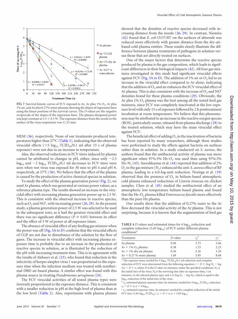
FIG 7 Survival kinetic curves of FCV exposed to Ar, Ar plus 1% O2, Ar plus1% air, and Ar plus 0.27% water plasmas showing the slopes of regression linesusing the linear portions of the survival curves. The D values are the negativereciprocals of the slopes of the regression lines. The plasma dissipated powerwas kept constant at 2.5 � 0.1 W. The exposure distance from the nozzle to thesurface of the virus suspension was 11.25 mm.
TABLE 4 D values and estimated times for 4 log10 reduction andcomplete reduction (5.83 log10) of FCV under different plasmaconditionsa
Treatment D value t4b tT
c
Ar plasma 0.94 3.75 5.46Ar 1% O2 plasma 0.38 1.53 2.23Ar 1% dry air plasma 0.56 2.26 3.29Ar 0.27 % water plasma 1.49 5.95 8.68a The exposure times needed for 4 log10 TCID50/0.1 ml reduction and completereduction of FCV were determined from the following equation: t � D � (log No � logNf) � D � n, where D is the D value (in minutes) under the specified conditions; No isthe initial titer of the virus; Nf is the surviving titer after an exposure time, t (inminutes), to the selected plasma type; and n is (log No � log Nf), which is equal to thelog10 reduction of the initial titer of the virus.b t4, estimated plasma exposure time (in minutes) needed for 4 log10 TCID50 reduction.t4 � D � n; n � 4 log10.c tT, estimated exposure time (in minutes) needed for complete reduction of the initialFCV titer (5.83 log10 TCID50). tT � D � n; n � 5.83 log10.
Virucidal Effect of Cold Atmospheric Gaseous Plasma
June 2015 Volume 81 Number 11 aem.asm.org 3619Applied and Environmental Microbiology
on May 10, 2015 by guest
http://aem.asm
.org/D
ownloaded from
humidity increases H2O2 production (46) but decreases certainROS species, such as ozone (47). Indeed, we found that the H2O2
concentration in distilled water increased from �120 �M for Ar,Ar plus 1% O2, and Ar plus 1% air to 2,785 �M for Ar plus 0.27%water after a 2-min treatment time.
The results for virus suspension solutions (Fig. 4A to D) indi-cate that the FCV virucidal activity of plasma is greatly affected bythe type of virus suspension medium. Generally, the greatest re-duction in FCV was observed when the virus was suspended indistilled water. In MEM, all types of plasma showed minimumvirucidal activity. When FCV was suspended in NTE buffer, FCVvirucidal activity was found to be variable; virus suspension inNTE buffer did not affect the virucidal activity of Ar plus 1% O2
plasma but did decrease the virucidal activities of all other plasmatypes. It is clear that pH has an important effect on the plasma-induced liquid-phase chemistry, as has been shown for the sameplasma source using Ar for bacterial inactivation (48). However,the effect of Ar plus 1% O2 seems to be pH independent. Thereduced virucidal effect in MEM may be due to the presence ofbovine serum and proteins, which may act as scavengers of theROS produced by the plasma through an oxidation process withthe molecules. We hypothesize, based on this observation, that thevirucidal efficiency of this new technology might be positively ornegatively affected by the chemical composition of the treatedfoods, and it indicates that plasma might not be applicable withhigh-protein foods. However, further investigations of variousfood models are needed to study the effect of food composition onthe virucidal efficacy of plasma.
We believe that the virucidal effect of plasma is not due to theproduction of H2O2 in solution, as the maximum virucidal effectwas not more than 2.7 log10 TCID50/0.1 ml of FCV when treatedwith 100 mM liquid H2O2 for 2 h (Fig. 5). However, there wasmore than a 4.5 log10 TCID50/0.1 ml decrease with Ar plus 0.27%water plasma, which did not yield more than 2.8 mM H2O2 (Table3). The key factor for the FCV virucidal activity is the chemicalinteraction of ROS and reactive nitrogen species (RNS), such assinglet oxygen (O2*), ozone (O3), and superoxide (O2
�) or per-oxynitrous acid. These species potentially react with the capsidprotein of FCV, leading to protein peroxidation and destructionof the capsid. In addition, they can damage the viral RNA, leading
to reduced gene expression and elimination of viral RNA, or both.This hypothetical mode of action has been suggested in severalreports. Yasuda et al. (49) suggested that CGP mainly affects thebacteriophage lambda coat protein and, to a lesser extent, causesdamage to the bacteriophage DNA. They also showed experimen-tal evidence that degradation of proteins and DNA was detectableonly at long plasma exposures. However, lambda coat protein canbe inactivated quickly, suggesting chemical modification, such asoxidation or reduction. A similar mode of action was suggested byZimmermann et al. (22), explaining the inactivation of a recom-binant strain of human adenovirus using CGP. Since FCV consistsof RNA, packaged in the capsid protein coat, the mechanism ofinactivation by our CGP system may be similar.
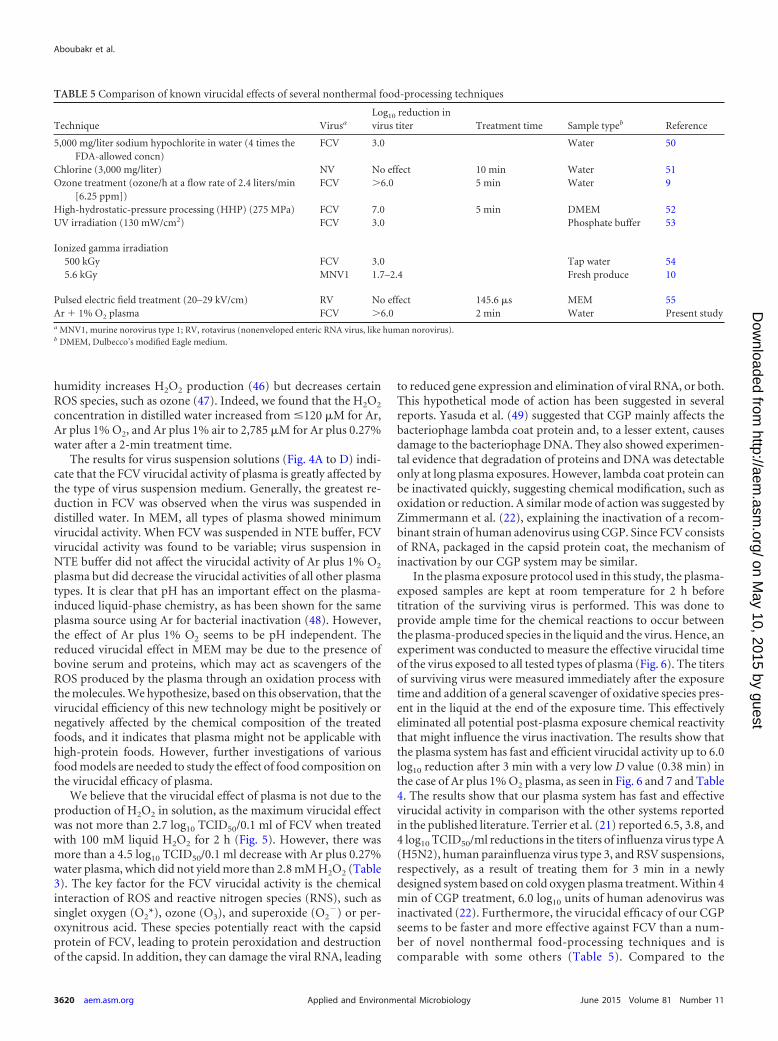
In the plasma exposure protocol used in this study, the plasma-exposed samples are kept at room temperature for 2 h beforetitration of the surviving virus is performed. This was done toprovide ample time for the chemical reactions to occur betweenthe plasma-produced species in the liquid and the virus. Hence, anexperiment was conducted to measure the effective virucidal timeof the virus exposed to all tested types of plasma (Fig. 6). The titersof surviving virus were measured immediately after the exposuretime and addition of a general scavenger of oxidative species pres-ent in the liquid at the end of the exposure time. This effectivelyeliminated all potential post-plasma exposure chemical reactivitythat might influence the virus inactivation. The results show thatthe plasma system has fast and efficient virucidal activity up to 6.0log10 reduction after 3 min with a very low D value (0.38 min) inthe case of Ar plus 1% O2 plasma, as seen in Fig. 6 and 7 and Table4. The results show that our plasma system has fast and effectivevirucidal activity in comparison with the other systems reportedin the published literature. Terrier et al. (21) reported 6.5, 3.8, and4 log10 TCID50/ml reductions in the titers of influenza virus type A(H5N2), human parainfluenza virus type 3, and RSV suspensions,respectively, as a result of treating them for 3 min in a newlydesigned system based on cold oxygen plasma treatment. Within 4min of CGP treatment, 6.0 log10 units of human adenovirus wasinactivated (22). Furthermore, the virucidal efficacy of our CGPseems to be faster and more effective against FCV than a num-ber of novel nonthermal food-processing techniques and iscomparable with some others (Table 5). Compared to the
TABLE 5 Comparison of known virucidal effects of several nonthermal food-processing techniques
Technique Virusa
Log10 reduction invirus titer Treatment time Sample typeb Reference
5,000 mg/liter sodium hypochlorite in water (4 times theFDA-allowed concn)
FCV 3.0 Water 50
Chlorine (3,000 mg/liter) NV No effect 10 min Water 51Ozone treatment (ozone/h at a flow rate of 2.4 liters/min
[6.25 ppm])FCV �6.0 5 min Water 9
High-hydrostatic-pressure processing (HHP) (275 MPa) FCV 7.0 5 min DMEM 52UV irradiation (130 mW/cm2) FCV 3.0 Phosphate buffer 53
Ionized gamma irradiation500 kGy FCV 3.0 Tap water 545.6 kGy MNV1 1.7–2.4 Fresh produce 10
Pulsed electric field treatment (20–29 kV/cm) RV No effect 145.6 �s MEM 55Ar 1% O2 plasma FCV �6.0 2 min Water Present studya MNV1, murine norovirus type 1; RV, rotavirus (nonenveloped enteric RNA virus, like human norovirus).b DMEM, Dulbecco’s modified Eagle medium.
Aboubakr et al.
3620 aem.asm.org June 2015 Volume 81 Number 11Applied and Environmental Microbiology
on May 10, 2015 by guest
http://aem.asm
.org/D
ownloaded from
strong virucidal activity of short exposure followed by 2 h ofincubation, longer exposure times were needed for virus inac-tivation when the scavenger (sodium thiosulfate) was addedimmediately after the plasma treatment. This indicates thatvirus inactivation is partially caused by long-lived ROS or RNS.When plasma-treated solution was added to the virus afterdifferent intervals, we found that in most cases, the virucidaleffect was no longer present 5 min after the plasma treatment(data not shown).
In conclusion, oxygen-based CGP is a novel, versatile nonther-mal tool that has great virucidal potential against FCV, a surrogatefor human norovirus, which is the most important foodbornevirus. This novel technology has strong potential in food-process-ing and food safety applications for controlling viral contamina-tion of food-processing surfaces and fresh and minimally pro-cessed foods. Further studies are recommended to determine themechanism of action of CGP used in this study. Experimentsshould also be conducted to determine if this CGP system caninactivate FCV, HAV, and NV on ready-to-eat foods and foodseaten fresh and on food contact surfaces.
ACKNOWLEDGMENTS
Partial funding provided by the Cultural Affairs and Mission Sector, Min-istry of Higher Education and Scientific Research, Egypt, is gratefully ac-knowledged. We also acknowledge funding from the Department of En-ergy Plasma Science Center through the U.S. Department of Energy,Office of Fusion Energy Sciences, contract DE-SC0001939, and the Uni-versity of Minnesota.
We thank Nhungoc Ti Luong for technical help.
REFERENCES1. Scharff RL. 3 March 2010. Health-related costs from foodborne illness in
the United States. http://www.publichealth.lacounty.gov/eh/docs/ReportPublication/HlthRelatedCostsFromFoodborneIllinessUS.pdf.
2. Anonymous. 2012. The European Union summary report on trends andsources of zoonoses, zoonotic agents and food-borne outbreaks in theEuropean Union in 2010. EFSA J 10:2597. http://dx.doi.org/10.2903/j.efsa.2012.2597.
3. European Center for Disease Prevention and Control. 2013. Fact-sheet for health professionals. http://www.ecdc.europa.eu/en/healthtopics/norovirus_infection/factsheet-health-professionals/Pages/factsheet_health_professionals.aspx.
4. CDC. 30 December 2014. U.S. trends and outbreaks. http://www.cdc.gov/norovirus/trends-outbreaks.html.
5. European Food Safety Authority. 30 April 2009. Community sum-mary report. Food-borne outbreaks in the European Union in 2007.European Food Safety Authority, Parma, Italy. http://www.efsa.europa.eu/de/efsajournal/doc/271r.pdf.
6. Misra NN, Tiwari BK, Raghavarao KSMS, Cullen PJ. 2011. Nonthermalplasma inactivation of food-borne pathogens. Food Eng Rev 3:159 –170.http://dx.doi.org/10.1007/s12393-011-9041-9.
7. Hirneisen KA, Black EP, Cascarino JL, Fino VR, Hoover DG, Kniel KE.2010. Viral inactivation in foods: a review of traditional and novel food-processing technologies. Compr Rev Food Sci Food Saf 9:3–20. http://dx.doi.org/10.1111/j.1541-4337.2009.00092.x.
8. Mormann S, Dabisch-Ruthe M, Becker B. 2010. Inactivation of norovi-rus in foods: inoculation study using human norovirus. Fleischwirtschaft90:116 –121.
9. Hirneisen KA, Markland SM, Kniel KE. 2011. Ozone inactivation ofnorovirus surrogates on fresh produce. J Food Prot 74:836 – 839. http://dx.doi.org/10.4315/0362-028X.JFP-10-438.
10. Feng K, Divers E, Ma Y, Li J. 2011. Inactivation of a human norovirussurrogate, human norovirus virus-like particles, and vesicular stomatitisvirus by gamma irradiation. Appl Environ Microbiol 77:3507–3517. http://dx.doi.org/10.1128/AEM.00081-11.
11. Aboubakr HA, El-Banna AA, Youssef MM, Al-Sohaimy SAA, GoyalSM. 2014. Antiviral effects of Lactococcus lactis on feline calicivirus, a
human norovirus surrogate. Food EnvironVirol 6:282–289. http://dx.doi.org/10.1007/s12560-014-9164-2.
12. Graves DB. 2012. The emerging role of reactive oxygen and nitrogenspecies in redox biology and some implications for plasma applications tomedicine and biology. J Phys D Appl Phys 45:263001. http://dx.doi.org/10.1088/0022-3727/45/26/263001.
13. Fridman A. 2008. Plasma chemistry. Cambridge University Press, NewYork, NY.
14. Bruggeman P, Locke BR. 2013. Assessment of potential applications ofplasma with liquid water, p 13– 40. In Chu PK, Lu XP (ed), Low temper-ature plasma technology: methods and applications. CRC Press, Boca Ra-ton, FL.
15. Cecchi JL. 1990. Introduction to plasma concept and discharge, p 14 – 69.In Rossnagel SM, Cuomo JJ, Westwood WD (ed), Handbook of plasmaprocessing technology, fundamentals, etching, deposition and surface in-teractions. Noyes Publications, Saddle River, NJ.
16. Azharonok V, Krat’ko L, Nekrashevich YI, Filatova I, Mel’nikova L,Dudchik N, Yanetskaya S, Bologa M. 2009. Bactericidal action of theplasma of high-frequency capacitive and barrier discharges on microor-ganisms. J Eng Phys Thermophys 82:419 – 426. http://dx.doi.org/10.1007/s10891-009-0210-0.
17. Fernández A, Noriega E, Thompson A. 2013. Inactivation of Salmonellaenterica serovar Typhimurium on fresh produce by cold atmospheric gasplasma technology. Food Microbiol 33:24 –29. http://dx.doi.org/10.1016/j.fm.2012.08.007.
18. Liu F, Sun P, Bai N, Tian Y, Zhou H, Wei S, Zhou Y, Zhang J, Zhu W,Becker K. 2010. Inactivation of bacteria in an aqueous environment by adirect current, cold atmospheric pressure air plasma micro jet. PlasmaProcess Polym 7:231–236. http://dx.doi.org/10.1002/ppap.200900070.
19. Noriega E, Sharma G, Laca A, Diaz M, Kong MG. 2011. Cold atmo-spheric gas plasma disinfection of chicken muscle and chicken skin con-taminated with Listeria monocytogenes. Food Microbiol 28:1293–1300.http://dx.doi.org/10.1016/j.fm.2011.05.007.
20. Alkawareek MY, Algwari QT, Gorman SP, Graham WG, O’Connell D,Gilmore BF. 2012. Application of atmospheric pressure nonthermalplasma for the in vitro eradication of bacterial biofilms. FEMS ImmunolMed Microbiol 65:381–384. http://dx.doi.org/10.1111/j.1574-695X.2012.00942.x.
21. Terrier O, Essere B, Yver M, Barthélémy M, Bouscambert-Duchamp M,Kurtz P, van Mechelen D, Morfin F, Billaud G, Ferraris O, Lina B,Rosa-Calatrava M, Moules V. 2009. Cold oxygen plasma technologyefficiency against different airborne respiratory viruses. J Clin Virol 45:119 –124. http://dx.doi.org/10.1016/j.jcv.2009.03.017.
22. Zimmermann JL, Dumler K, Shimizu T, Morfill GE, Wolf A, Boxham-mer Schlegel VJ, Gansbacher B, Anton M. 2011. Effects of cold atmo-spheric plasmas on adenoviruses in solution. J Phys D Appl Phys 44:505201. http://dx.doi.org/10.1088/0022-3727/44/50/505201.
23. Alekseev O, Donovan K, Limonnik V, Azizkhan-Clifford J. 2014. Non-thermal dielectric barrier discharge (DBD) plasma suppresses herpes sim-plex virus type 1 (HSV-1) replication in corneal epithelium. Trans Vis SciTechnol 3:2. http://dx.doi.org/10.1167/tvst.3.2.2.
24. van Gils CAJ, Hofmann S, Boekema BKHL, Brandenburg R, BruggemanPJ. 2013. Mechanisms of bacterial inactivation in the liquid phase induced bya remote RF cold atmospheric pressure plasma jet. J Phys D Appl Phys 46:175203. http://dx.doi.org/10.1088/0022-3727/46/17/175203.
25. Hofmann S, Gessel A, Bruggeman P. 2011. Power dissipation, gas tem-peratures and electron densities of cold atmospheric pressure helium andargon RF plasma jets. Plasma Sources Sci Technol 20:065010. http://dx.doi.org/10.1088/0963-0252/20/6/065010.
26. van Gessel AFH, Alards KMJ, Bruggeman PJ. 2013. NO production in anRF plasma jet at atmospheric pressure. J Phys D Appl Phys 46:265202.http://dx.doi.org/10.1088/0022-3727/46/26/265202.
27. Malik YS, Maherchandani S, Allwood PB, Goyal SM. 2005. Evaluationof animal origin cell cultures for in vitro cultivation of noroviruses. J ApplRes Clin Exp Ther 5:312–317.
28. van Gaens W, Bogaerts A. 2014. Reaction pathways of biomedicallyactive species in an Ar plasma jet. Plasma Sources Sci Technol 23:035015.http://dx.doi.org/10.1088/0963-0252/23/3/035015.
29. van Ham BTJ, Hofmann S, Brandenburg R, Bruggeman PJ. 2014. In situabsolute air, O3 and NO densities in the effluent of a cold RF argon atmo-spheric pressure plasma jet obtained by molecular beam mass spectrom-etry. J Phys D Appl Phys 47:224013. http://dx.doi.org/10.1088/0022-3727/47/22/224013.
Virucidal Effect of Cold Atmospheric Gaseous Plasma
June 2015 Volume 81 Number 11 aem.asm.org 3621Applied and Environmental Microbiology
on May 10, 2015 by guest
http://aem.asm
.org/D
ownloaded from
30. Bruggeman P, Leys C. 2009. Non-thermal plasmas in and in contact withliquids. J Phys D Appl Phys 42:053001. http://dx.doi.org/10.1088/0022-3727/42/5/053001.
31. Kärber G. 1931. 50% end point calculation. Arch Exp Pathol Pharmacol162:480 – 483. http://dx.doi.org/10.1007/BF01863914.
32. Satterfield CH, Bonnell AH. 1995. Interferences in the titanium sulfatemethod for hydrogen peroxide. Anal Chem 27:1174 –1175. http://dx.doi.org/10.1021/ac60103a042.
33. Eisenberg GM. 1943. Colorimetric determination of hydrogen peroxide.Ind Eng Chem Anal 15:327–328. http://dx.doi.org/10.1021/i560117a011.
34. Straub TM, Honerzu BK, Orosz-Coghlan P, Dohnalkova A, Mayer BK,Bartholomew RA, Valdez CO, Bruckner-Lea CJ, Gerba CP, Abbasza-degan MA, Nickerson CA. 2007. In vitro cell culture infectivity assay forhuman noroviruses. Emerg Infect Dis 13:396 – 403. http://dx.doi.org/10.3201/eid1303.060549.
35. D’Souza DH, Sair A, Williams K, Papafragkou E, Jean J, Moore C,Jaykus L. 2006. Persistence of caliciviruses on environmental surfaces andtheir transfer to food. Int J Food Microbiol 108:84 –91. http://dx.doi.org/10.1016/j.ijfoodmicro.2005.10.024.
36. Cannon JL, Papafragkou E, Park GW, Osborne J, Jaykus L, Vinjé J.2006. Surrogates for the study of norovirus stability and inactivation in theenvironment: a comparison of murine norovirus and feline calicivirus. JFood Prot 69:2761–2765.
37. Sattar SA, Ali M, Tetro JA. 2011. In vivo comparison of two norovirussurrogates for testing ethanol-based handrubs: the mouse chasing the cat!PLoS One 6:e17340. http://dx.doi.org/10.1371/journal.pone.0017340.
38. Park GW, Barclay L, Macinga D, Charbonneau D, Pettigrew CA, VinjéJ. 2010. Comparative efficacy of seven hand sanitizers against murinenorovirus, feline calicivirus, and GII.4 norovirus. J Food Prot 73:2232–2238.
39. Butot S, Putallaz T, Sanchez G. 2008. Effects of sanitation, freezing andfrozen storage on enteric viruses in berries and herbs. Int J Food Microbiol126:30 –35. http://dx.doi.org/10.1016/j.ijfoodmicro.2008.04.033.
40. Hury S, Vidal DR, Desor F, Pelletier J, Lagarde T. 1998. A parametricstudy of the destruction efficiency of Bacillus spores in low pressure oxy-gen-based plasmas. Lett Appl Microbiol 26:417– 421. http://dx.doi.org/10.1046/j.1472-765X.1998.00365.x.
41. Allwood PB, Malik YS, Maherchandani S, Hedberg CW, Goyal SM. 2005.Effect of temperature on the survival of F-specific RNA coliphage, feline cali-civirus, and Escherichia coli in chlorinated water. Int J Environ Res PublicHealth 2:442– 446. http://dx.doi.org/10.3390/ijerph2005030008.
42. Niemira BA. 2012. Cold plasma reduction of Salmonella and Escherichiacoli O157:H7 on almonds using ambient pressure gases. J Food Sci 77:M171. http://dx.doi.org/10.1111/j.1750-3841.2011.02594.x.
43. Lu XP, Ye T, Cao YG, Sun ZY, Xiong Q, Tang Z, Xiong Z, Hu J, Jiang
Z, Pan Y. 2008. The roles of the various plasma agents in the inactivationof bacteria. J Appl Phys 104:053309. http://dx.doi.org/10.1063/1.2977674.
44. Sureshkumar S, Sankar R, Mandal M, Neogi S. 2010. Effective bacterialinactivation using low temperature radio frequency plasma. Int J Pharm396:17–22. http://dx.doi.org/10.1016/j.ijpharm.2010.05.045.
45. Chen W, Huang J, Du N, Liu X, Wang X, Lv G-H, Zhang G, Guo L,Yang S. 2012. Treatment of Enterococcus faecalis bacteria by a heliumatmospheric cold plasma brush with oxygen addition. J Appl Phys 112:013304. http://dx.doi.org/10.1063/1.4732135.
46. Winter J, Wende K, Masur K, Iseni S, Dünnbier M, Hammer MU,Tresp H, Weltmann K-D, Reuter S. 2013. Feed gas humidity: a vitalparameter affecting a cold atmospheric-pressure plasma jet and plasma-treated human skin cells. J Phys D Appl Phys 46:295401. http://dx.doi.org/10.1088/0022-3727/46/29/295401.
47. Liu DX, Iza F, Wang XH, Kong MG, Rong MZ. 2011. HeO2H2Oplasmas as a source of reactive oxygen species. Appl Phys Lett 98:221501.http://dx.doi.org/10.1063/1.3592775.
48. Boekema BKHL, Hofmann S, van Ham BJT, Bruggeman PJ, Middelk-oop E. 2013. Antibacterial plasma at safe levels for skin cells. J Phys D ApplPhys 46:422001. http://dx.doi.org/10.1088/0022-3727/46/42/422001.
49. Yasuda H, Miura T, Kurita H, Takashima K, Mizuno A. 2010. Biologicalevaluation of DNA damage in bacteriophages inactivated by atmosphericpressure cold plasma. Plasma Process Polym 7:301–308. http://dx.doi.org/10.1002/ppap.200900088.
50. Gulati BR, Allwood PB, Hedberg CW, Goyal SM. 2001. Efficacy ofcommonly used disinfectants for the inactivation of calicivirus on straw-berry, lettuce and a food-contact surface. J Food Prot 64:1430 –1434.
51. Duizer E, Bijkerk P, Rockx B, de Groot A, Twisk F, Koopmans M. 2004.Inactivation of caliciviruses. Appl Environ Microbiol 70:4538 – 4543. http://dx.doi.org/10.1128/AEM.70.8.4538-4543.2004.
52. Kingsley DH, Hoover DG, Papafragkou E, Richards GP. 2002. Inacti-vation of hepatitis A virus and a calicivirus by high hydrostatic pressure. JFood Prot 65:1605–1609.
53. Nuanualsuwan S, Mariam T, Himathongkham S, Cliver D. 2002. Ul-traviolet Inactivation of feline calicivirus, human enteric viruses and co-liphages. Photochem Photobiol 76:406 – 410. http://dx.doi.org/10.1562/0031-8655(2002)0760406UIOFCH2.0.CO2.
54. De Roda Husman AM, Bijkerk P, Lodder W, Van Den Berg H, PribilW, Cabaj A, Gehringer P, Sommer R, Duizer E. 2004. Calicivirusinactivation by nonionizing (253.7-nanometer-wavelength [UV]) andionizing (gamma) radiation. Appl Environ Microbiol 70:5089 –5093. http://dx.doi.org/10.1128/AEM.70.9.5089-5093.2004.
55. Khadre MA, Yousef AE. 2002. Susceptibility of human rotavirus toozone, high pressure and pulsed electric field. J Food Prot 65:1441–1446.
Aboubakr et al.
3622 aem.asm.org June 2015 Volume 81 Number 11Applied and Environmental Microbiology
on May 10, 2015 by guest
http://aem.asm
.org/D
ownloaded from
Related Documents


