Virological Tests Laboratory Diagnosis of Viral Diseases ◼ Description of virus-induced cytopathologic effects (CPEs) on cells ◼ Detection of viral particles ◼ Isolation and growth of the virus ◼ Detection and analysis of viral components (e.g., proteins (antigens), enzymes, genomes) ◼ Evaluation of the patient's immune response to the virus (serology) There have been many new developments in laboratory viral diagnosis that provide more rapid and sensitive viral identification from clinical samples. These include better antibody reagents for direct analysis of samples, molecular genetics techniques and genomic sequencing for direct identification of the virus, and assays that can identify multiple viruses (multiplex) and be automated. Often, isolation of the organism is unnecessary and avoided to minimize the risk to laboratory and other personnel. The quicker turnaround allows a more rapid choice of the appropriate antiviral therapy. Viral laboratory studies are performed to (1) confirm the diagnosis by identifying the viral agent of infection, (2) determine appropriate antiviral therapy, (3) check on the compliance of the patient taking antiviral drugs, (4) define the course of the disease, (5) monitor the disease epidemiologically, and (6) educate physicians and patients. The laboratory methods accomplish the following results: Description of virus-induced cytopathologic effects (CPEs) on cells Detection of viral particles Isolation and growth of the virus Detection and analysis of viral components (e.g., proteins (antigens), enzymes, genomes) Evaluation of the patient's immune response to the virus (serology) Collection of specimens for virologic examination ◼ Swabs ◼ Washes ◼ CSF, urine, stool, anticoaggulated blood, tissue samples (biopsy or necropsy) ◼ Transport of specimens ◼ Storage temperature ◼ Specimen inspection Collection of specimens for virologic examination ◼ Bone marrow ◼ Cervix specimen ◼ Eyes, conjunctival swab ◼ Fluid specimen ◼ Lesion swab ◼ Nasal wash (optimal for RSV) ◼ Nasopharyngeal swab ◼ Rectal swab ◼ Throat swab ◼ Urethral specimen The patient's symptoms and history, including recent travel, the season of the year, and a presumptive diagnosis, help determine the appropriate procedures to be used to identify a

Welcome message from author
This document is posted to help you gain knowledge. Please leave a comment to let me know what you think about it! Share it to your friends and learn new things together.
Transcript
Virological Tests
Laboratory Diagnosis of Viral Diseases
◼ Description of virus-induced cytopathologic effects (CPEs) on cells
◼ Detection of viral particles
◼ Isolation and growth of the virus
◼ Detection and analysis of viral components (e.g., proteins (antigens), enzymes, genomes)
◼ Evaluation of the patient's immune response to the virus (serology)
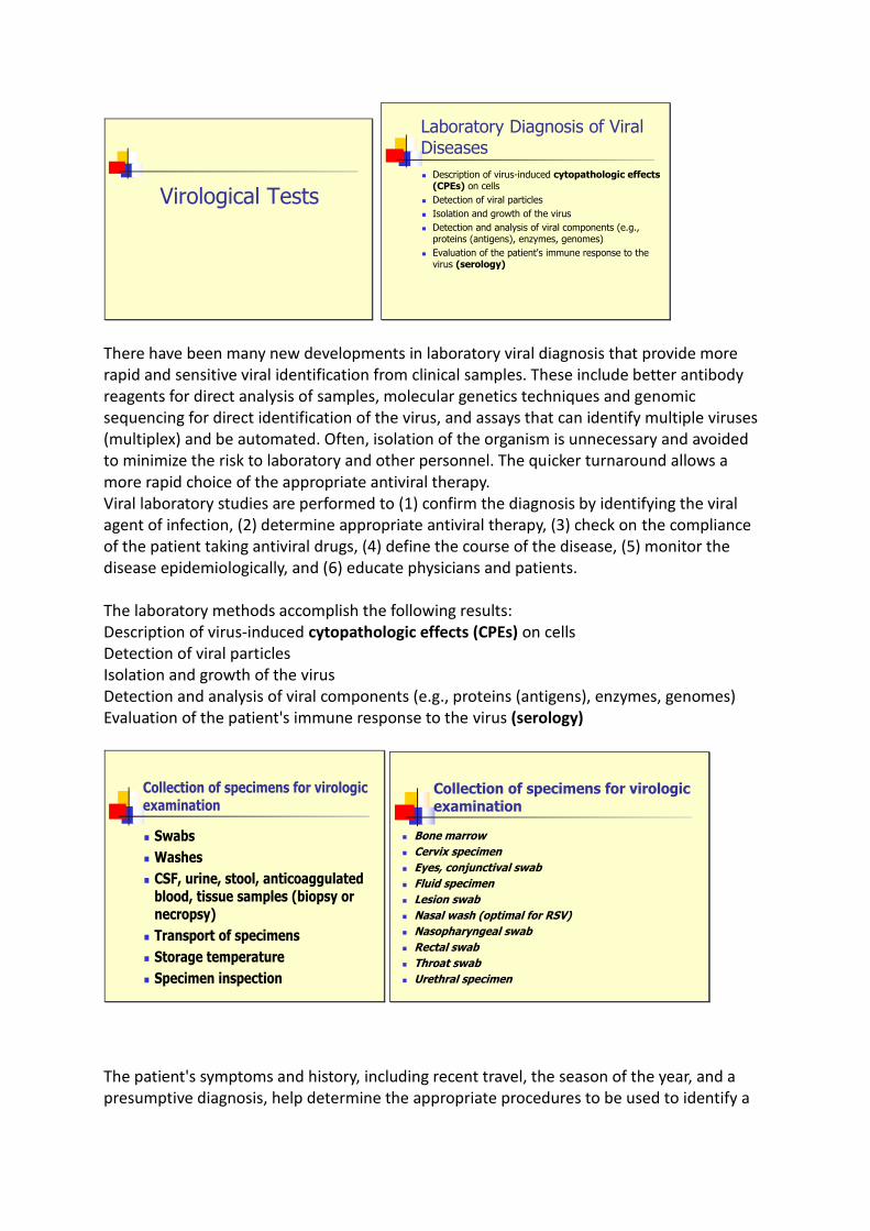
There have been many new developments in laboratory viral diagnosis that provide more rapid and sensitive viral identification from clinical samples. These include better antibody reagents for direct analysis of samples, molecular genetics techniques and genomic sequencing for direct identification of the virus, and assays that can identify multiple viruses (multiplex) and be automated. Often, isolation of the organism is unnecessary and avoided to minimize the risk to laboratory and other personnel. The quicker turnaround allows a more rapid choice of the appropriate antiviral therapy. Viral laboratory studies are performed to (1) confirm the diagnosis by identifying the viral agent of infection, (2) determine appropriate antiviral therapy, (3) check on the compliance of the patient taking antiviral drugs, (4) define the course of the disease, (5) monitor the disease epidemiologically, and (6) educate physicians and patients. The laboratory methods accomplish the following results: Description of virus-induced cytopathologic effects (CPEs) on cells Detection of viral particles Isolation and growth of the virus Detection and analysis of viral components (e.g., proteins (antigens), enzymes, genomes) Evaluation of the patient's immune response to the virus (serology)
Collection of specimens for virologic examination
◼ Swabs
◼ Washes
◼ CSF, urine, stool, anticoaggulated blood, tissue samples (biopsy or necropsy)
◼ Transport of specimens
◼ Storage temperature
◼ Specimen inspection
Collection of specimens for virologic examination
◼ Bone marrow
◼ Cervix specimen
◼ Eyes, conjunctival swab
◼ Fluid specimen
◼ Lesion swab
◼ Nasal wash (optimal for RSV)
◼ Nasopharyngeal swab
◼ Rectal swab
◼ Throat swab
◼ Urethral specimen
The patient's symptoms and history, including recent travel, the season of the year, and a presumptive diagnosis, help determine the appropriate procedures to be used to identify a

viral agent. The selection of the appropriate specimen for analysis is often complicated because several viruses may cause the same clinical disease. For example, the development of meningitis symptoms during the summer suggests an arbovirus, in which case cerebrospinal fluid (CSF) and blood should be collected, or an enterovirus, in which case CSF, a throat swab, and stool specimens should be collected for genome analysis and possible virus isolation. A focal encephalitis with a temporal lobe localization preceded by headaches and disorientation suggests herpes simplex virus (HSV) infection, for which CSF can be relatively quickly analyzed for viral deoxyribonucleic acid (DNA) sequences by polymerase chain reaction (PCR) amplification. Specimens should be collected early in the acute phase of infection, before the virus ceases to be shed. For example, respiratory viruses may be shed for only 3 to 7 days, and shedding may lapse before the symptoms cease. HSV and varicella-zoster virus (VZV) may not be recoverable from lesions more than 5 days after the onset of symptoms. It may be possible to isolate an enterovirus from the CSF for only 2 to 3 days after the onset of the central nervous system manifestations. In addition, antibody produced in response to the infection may block the detection of virus. Laboratory diagnosis of viral infections requires an understanding of the pathogenesis of the suspected agent, stage of infection, as well as age and immunocompetence of the infected individual. It is important to select the appropriate specimens, collect the specimen carefully to optimize recovery of the infectious agent, and transport the specimens as directed so as to maintain viability and minimize overgrowth with contaminating organisms. The specimen should be collected from the target organ most closely associated with clinical symptoms to identify the etiologic agent responsible for the patient's disease.
Viral transport medium◼ bovine albumin fraction
◼ gentamicin sulfate solution
◼ amphotericin
The shorter the interval between the collection of a specimen and its delivery to the laboratory, the greater the potential for isolating a virus. The reasons are that many viruses

are labile, and the samples are susceptible to bacterial and fungal overgrowth. Viruses are best transported and stored on ice and in special media that contain antibiotics and proteins, such as serum albumin or gelatin. Significant losses in infectious titers occur when enveloped viruses (e.g., HSV, VZV, influenza virus) are kept at room temperature or frozen at -20° C. This is not a risk for nonenveloped viruses (e.g., adenoviruses, enteroviruses).
Diagnostic Methods in Virology
1. Direct Examination
2. Isolation of virus
3. Indirect Examination - Serology
In general, diagnostic tests can be grouped into 3 categories.: (1) direct detection, (2) virus isolation, and (3) serology. In direct examination, the clinical specimen is examined directly for the presence of virus particles, virus antigen or viral nucleic acids. Isolation of virus - the specimen into cell culture, eggs or animals in an attempt to grow the virus. A serological diagnosis can be made by the detection of rising titres of antibody between acute and convalescent stages of infection, or the detection of IgM. In general, the majority of common viral infections can be diagnosed by serology. The specimen used for direction detection and virus isolation is very important. A positive result from the site of disease would be of much greater diagnostic significance than those from other sites. For example, in the case of herpes simplex encephalitis, a positive result from the CSF or the brain would be much greater significance than a positive result from an oral ulcer, since reactivation of oral herpes is common during times of stress. Serology Detection of rising titres of antibody between acute and convalescent stages of infection, or the detection of IgM in primary infection. Classical Techniques Newer Techniques 1. Complement fixation tests (CFT) 1. Radioimmunoassay (RIA) 2. Haemagglutination inhibition tests 2. Enzyme linked immunosorbent assay (EIA) 3. Immunofluorescence techniques (IF) 3. Particle agglutination 4. Neutralization tests 4. Western Blot (WB) 5. Single Radial Haemolysis 5. Recombinant immunoblot assay (RIBA), line immunoassay (Liatek) etc.
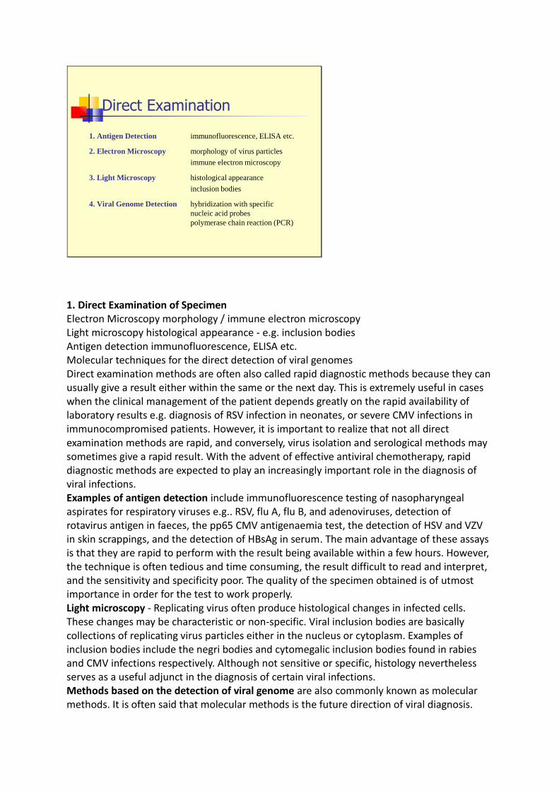
Direct Examination
1. Antigen Detection immunofluorescence, ELISA etc.
2. Electron Microscopy morphology of virus particles
immune electron microscopy
3. Light Microscopy histological appearance
inclusion bodies
4. Viral Genome Detection hybridization with specific
nucleic acid probes
polymerase chain reaction (PCR)
1. Direct Examination of Specimen Electron Microscopy morphology / immune electron microscopy Light microscopy histological appearance - e.g. inclusion bodies Antigen detection immunofluorescence, ELISA etc. Molecular techniques for the direct detection of viral genomes Direct examination methods are often also called rapid diagnostic methods because they can usually give a result either within the same or the next day. This is extremely useful in cases when the clinical management of the patient depends greatly on the rapid availability of laboratory results e.g. diagnosis of RSV infection in neonates, or severe CMV infections in immunocompromised patients. However, it is important to realize that not all direct examination methods are rapid, and conversely, virus isolation and serological methods may sometimes give a rapid result. With the advent of effective antiviral chemotherapy, rapid diagnostic methods are expected to play an increasingly important role in the diagnosis of viral infections. Examples of antigen detection include immunofluorescence testing of nasopharyngeal aspirates for respiratory viruses e.g.. RSV, flu A, flu B, and adenoviruses, detection of rotavirus antigen in faeces, the pp65 CMV antigenaemia test, the detection of HSV and VZV in skin scrappings, and the detection of HBsAg in serum. The main advantage of these assays is that they are rapid to perform with the result being available within a few hours. However, the technique is often tedious and time consuming, the result difficult to read and interpret, and the sensitivity and specificity poor. The quality of the specimen obtained is of utmost importance in order for the test to work properly. Light microscopy - Replicating virus often produce histological changes in infected cells. These changes may be characteristic or non-specific. Viral inclusion bodies are basically collections of replicating virus particles either in the nucleus or cytoplasm. Examples of inclusion bodies include the negri bodies and cytomegalic inclusion bodies found in rabies and CMV infections respectively. Although not sensitive or specific, histology nevertheless serves as a useful adjunct in the diagnosis of certain viral infections. Methods based on the detection of viral genome are also commonly known as molecular methods. It is often said that molecular methods is the future direction of viral diagnosis.
However in practice, although the use of these methods is indeed increasing, the role played by molecular methods in a routine diagnostic virus laboratory is still small compared to conventional methods. It is certain though that the role of molecular methods will increase rapidly in the near future. Classical molecular techniques such as dot-blot and Southern-blot depend on the use of specific DNA/RNA probes for hybridization. The specificity of the reaction depends on the conditions used for hybridization. These techniques may allow for the quantification of DNA/RNA present in the specimen. However, it is often found that the sensitivity of these techniques is not better than conventional viral diagnostic methods. Newer molecular techniques such as the polymerase chain reaction (PCR), ligase chain reaction (LCR), nucleic acid based amplification (NASBA), and branched DNA (bDNA) depend on some form of amplification, either the target nucleic acid, or the signal itself. PCR is the only amplification technique which is in common use. PCR is an extremely sensitive technique: it is possible to achieve a sensitivity of down to 1 DNA molecule in a clinical specimen. However, PCR has many problems, the chief among which is contamination, since only a minute amount of contamination is needed to give a false positive result. In addition, because PCR is so sensitive compared to other techniques, a positive PCR result is often very difficult to interpret as it does not necessarily indicate the presence of disease. This problem is particular great in the case of latent viruses such as CMV, since latent CMV genomes may be amplified from the blood of healthy individuals. Despite all this, PCR is being increasingly used for the rapid diagnosis of rapid. This is especially as the cost of the assay come down and the availability of closed automated systems that could also perform quantification (Quantitative PCR) e.g. real-time PCR and Cobas Amplicor.systems. Other amplification techniques such as LCR and NASBA are just as susceptible to contamination as PCR but that is ameliorated to a great extent by the use of propriatory closed systems. It is unlikely though that other amplification techniques will challenge the dominance of PCR since it is much easier to set up an house PCR assay than other assays. One advantage of PCR assays is that the PCR product can be sequenced and thus used for epidemiological investigation and drug susceptibility testing.
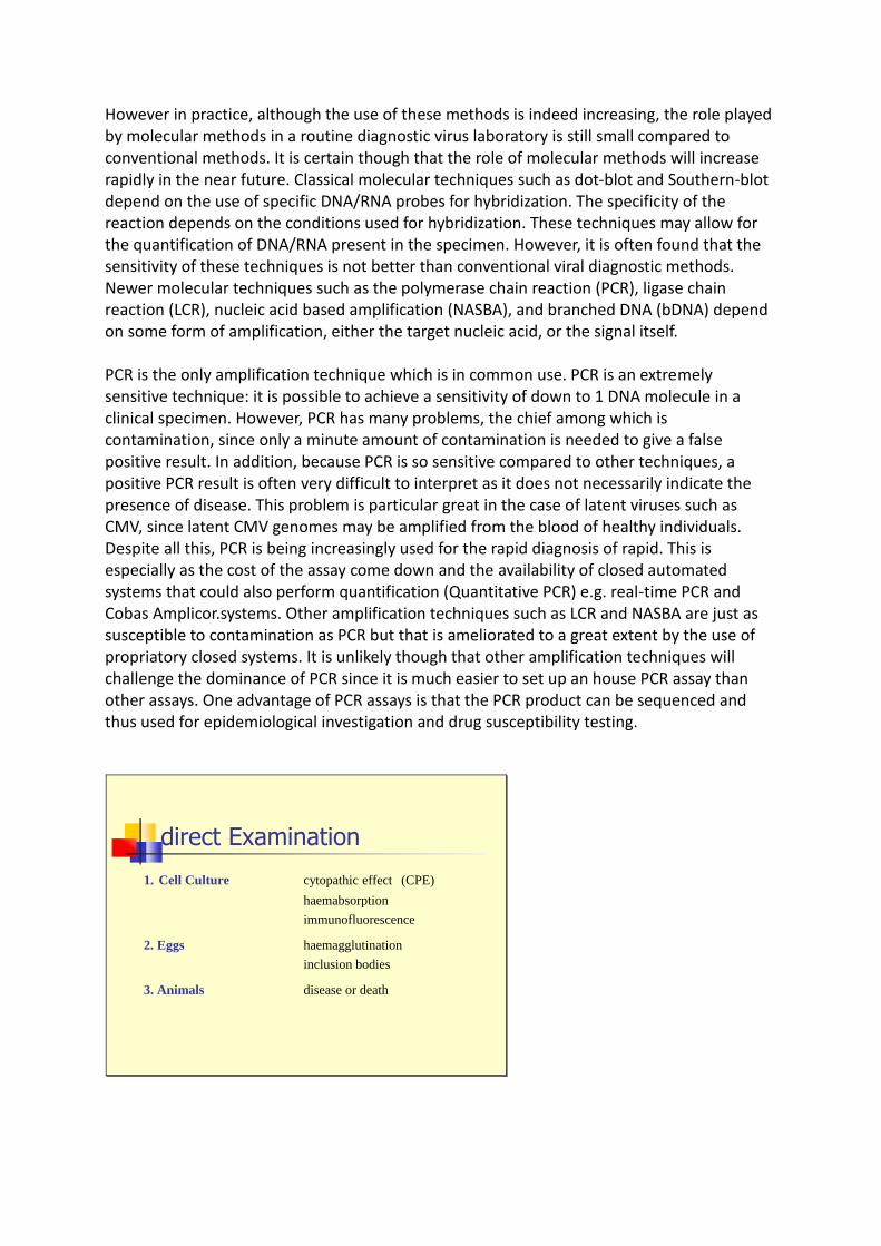
direct Examination
1. Cell Culture cytopathic effect (CPE)
haemabsorption
immunofluorescence
2. Eggs haemagglutination
inclusion bodies
3. Animals disease or death
2. Isolation of virus Cell Culture - cytopathic effect, haemadsorption, confirmation by neutralization, interference, immunofluorescence etc. Eggs pocks - haemagglutination, inclusion bodies Animals disease or death confirmation by neutralization
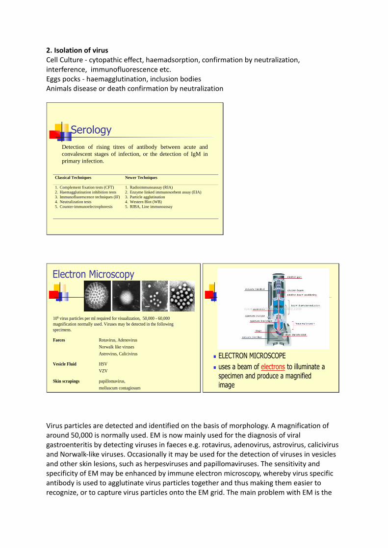
Serology
Detection of rising titres of antibody between acute and
convalescent stages of infection, or the detection of IgM in
primary infection.
Classical Techniques Newer Techniques
1. Complement fixation tests (CFT) 1. Radioimmunoassay (RIA)
2. Haemagglutination inhibition tests 2. Enzyme linked immunosorbent assay (EIA)
3. Immunofluorescence techniques (IF) 3. Particle agglutination
4. Neutralization tests 4. Western Blot (WB)
5. Counter-immunoelectrophoresis 5. RIBA, Line immunoassay
Electron Microscopy
106 virus particles per ml required for visualization, 50,000 - 60,000
magnification normally used. Viruses may be detected in the following
specimens.
Faeces Rotavirus, Adenovirus
Norwalk like viruses
Astrovirus, Calicivirus
Vesicle Fluid HSV
VZV
Skin scrapings papillomavirus,
molluscum contagiosum
◼ ELECTRON MICROSCOPE
◼ uses a beam of electrons to illuminate a specimen and produce a magnified image
Virus particles are detected and identified on the basis of morphology. A magnification of around 50,000 is normally used. EM is now mainly used for the diagnosis of viral gastroenteritis by detecting viruses in faeces e.g. rotavirus, adenovirus, astrovirus, calicivirus and Norwalk-like viruses. Occasionally it may be used for the detection of viruses in vesicles and other skin lesions, such as herpesviruses and papillomaviruses. The sensitivity and specificity of EM may be enhanced by immune electron microscopy, whereby virus specific antibody is used to agglutinate virus particles together and thus making them easier to recognize, or to capture virus particles onto the EM grid. The main problem with EM is the
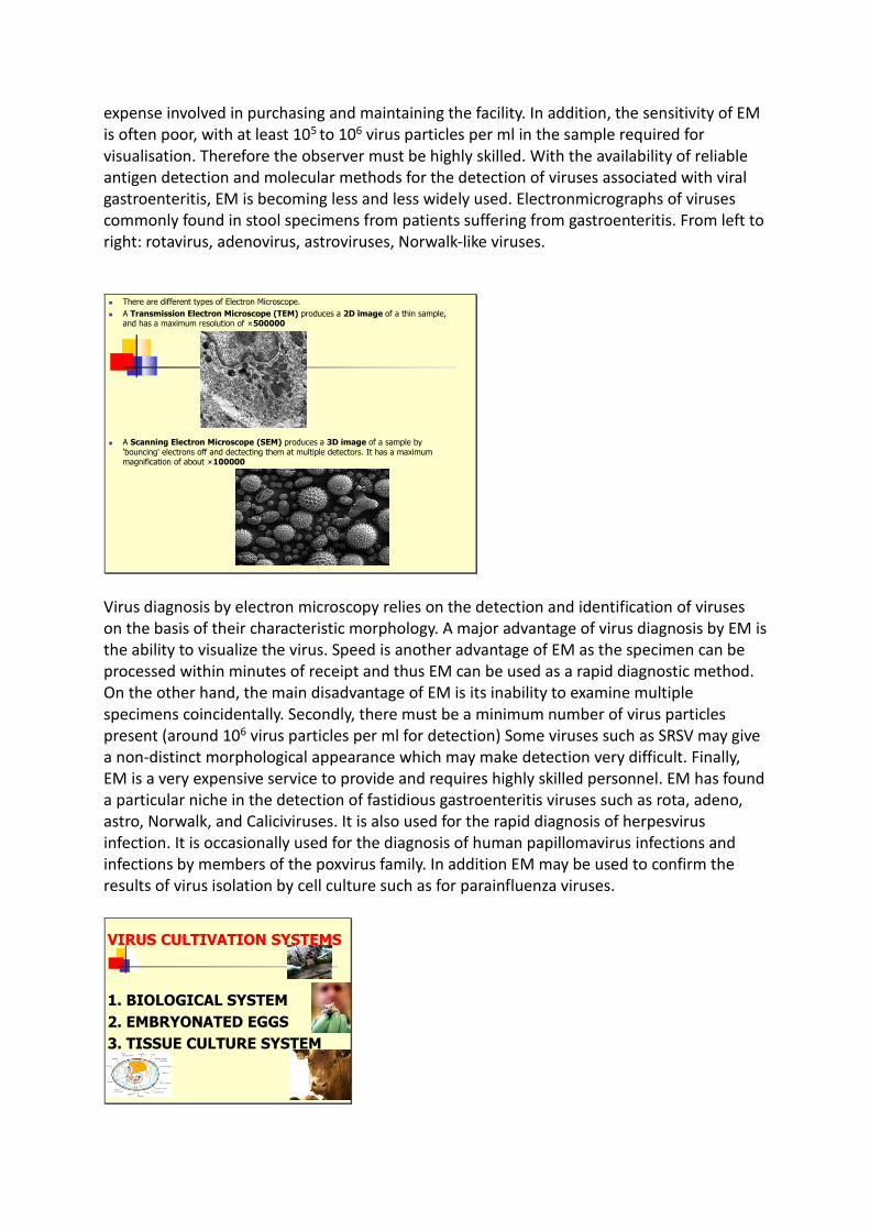
expense involved in purchasing and maintaining the facility. In addition, the sensitivity of EM is often poor, with at least 105 to 106 virus particles per ml in the sample required for visualisation. Therefore the observer must be highly skilled. With the availability of reliable antigen detection and molecular methods for the detection of viruses associated with viral gastroenteritis, EM is becoming less and less widely used. Electronmicrographs of viruses commonly found in stool specimens from patients suffering from gastroenteritis. From left to right: rotavirus, adenovirus, astroviruses, Norwalk-like viruses.
◼ There are different types of Electron Microscope.
◼ A Transmission Electron Microscope (TEM) produces a 2D image of a thin sample, and has a maximum resolution of ×500000
◼ A Scanning Electron Microscope (SEM) produces a 3D image of a sample by 'bouncing' electrons off and dectecting them at multiple detectors. It has a maximum magnification of about ×100000
Virus diagnosis by electron microscopy relies on the detection and identification of viruses on the basis of their characteristic morphology. A major advantage of virus diagnosis by EM is the ability to visualize the virus. Speed is another advantage of EM as the specimen can be processed within minutes of receipt and thus EM can be used as a rapid diagnostic method. On the other hand, the main disadvantage of EM is its inability to examine multiple specimens coincidentally. Secondly, there must be a minimum number of virus particles present (around 106 virus particles per ml for detection) Some viruses such as SRSV may give a non-distinct morphological appearance which may make detection very difficult. Finally, EM is a very expensive service to provide and requires highly skilled personnel. EM has found a particular niche in the detection of fastidious gastroenteritis viruses such as rota, adeno, astro, Norwalk, and Caliciviruses. It is also used for the rapid diagnosis of herpesvirus infection. It is occasionally used for the diagnosis of human papillomavirus infections and infections by members of the poxvirus family. In addition EM may be used to confirm the results of virus isolation by cell culture such as for parainfluenza viruses.
VIRUS CULTIVATION SYSTEMS
1. BIOLOGICAL SYSTEM
2. EMBRYONATED EGGS
3. TISSUE CULTURE SYSTEM
Cell cultures, eggs, and animals may be used for isolation. However eggs and animals are difficult to handle and most viral diagnostic laboratories depend on cell culture only.
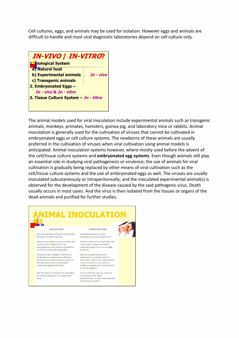
IN-VIVO / IN-VITRO?1. Biological System
a) Natural host
b) Experimental animals In - vivo
c) Transgenic animals
2. Embryonated Eggs –
In - vivo & In - vitro
3. Tissue Culture System – In - Vitro
The animal models used for viral inoculation include experimental animals such as transgenic animals, monkeys, primates, hamsters, guinea pig, and laboratory mice or rabbits. Animal inoculation is generally used for the cultivation of viruses that cannot be cultivated in embryonated eggs or cell culture systems. The newborns of these animals are usually preferred in the cultivation of viruses when viral cultivation using animal models is anticipated. Animal inoculation systems however, where mostly used before the advent of the cell/tissue culture systems and embryonated egg systems. Even though animals still play an essential role in studying viral pathogenesis or virulence; the use of animals for viral cultivation is gradually being replaced by other means of viral cultivation such as the cell/tissue culture systems and the use of embryonated eggs as well. The viruses are usually inoculated subcutaneously or intraperitoneally; and the inoculated experimental animal(s) is observed for the development of the disease caused by the said pathogenic virus. Death usually occurs in most cases. And the virus is then isolated from the tissues or organs of the dead animals and purified for further studies.
ANIMAL INOCULATION
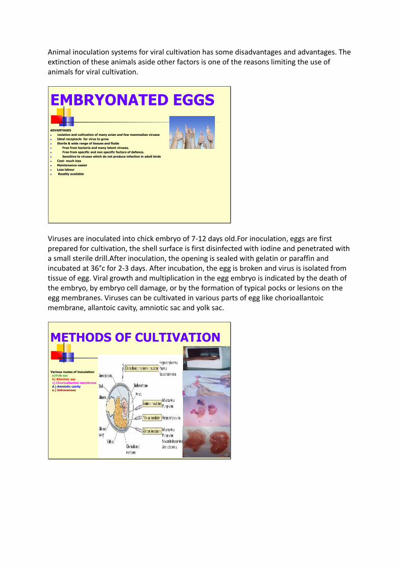
Animal inoculation systems for viral cultivation has some disadvantages and advantages. The extinction of these animals aside other factors is one of the reasons limiting the use of animals for viral cultivation.
EMBRYONATED EGGS
ADVANTAGES
◼ Isolation and cultivation of many avian and few mammalian viruses
◼ Ideal receptacle for virus to grow
◼ Sterile & wide range of tissues and fluids
◼ Free from bacteria and many latent viruses.
◼ Free from specific and non specific factors of defence.
◼ Sensitive to viruses which do not produce infection in adult birds
◼ Cost- much less
◼ Maintenance-easier
◼ Less labour
◼ Readily available
Viruses are inoculated into chick embryo of 7-12 days old.For inoculation, eggs are first prepared for cultivation, the shell surface is first disinfected with iodine and penetrated with a small sterile drill.After inoculation, the opening is sealed with gelatin or paraffin and incubated at 36°c for 2-3 days. After incubation, the egg is broken and virus is isolated from tissue of egg. Viral growth and multiplication in the egg embryo is indicated by the death of the embryo, by embryo cell damage, or by the formation of typical pocks or lesions on the egg membranes. Viruses can be cultivated in various parts of egg like chorioallantoic membrane, allantoic cavity, amniotic sac and yolk sac.
METHODS OF CULTIVATION
Various routes of inoculation a)Yolk sacb) Allantoic sacc) Chorioallantoic membraned.) Amniotic cavitye.) Intravenous
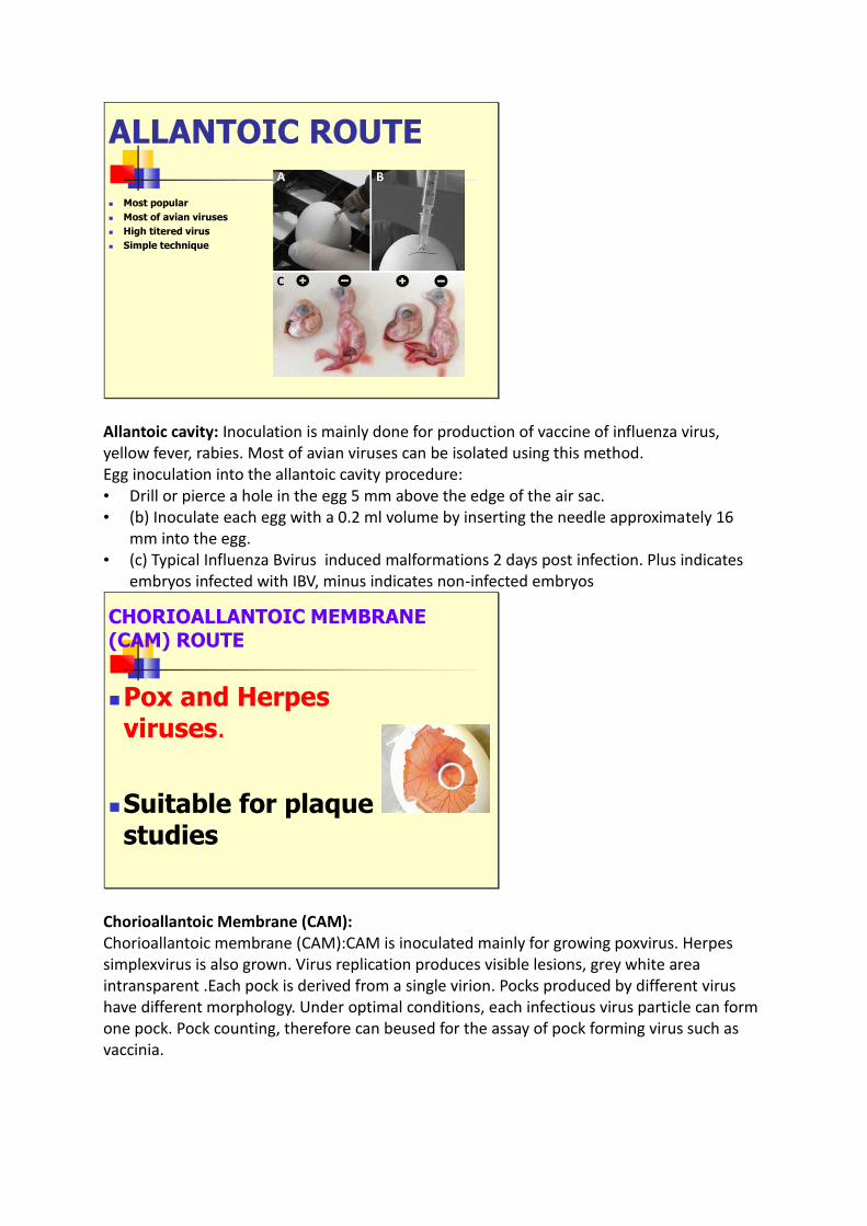
ALLANTOIC ROUTE
◼ Most popular
◼ Most of avian viruses
◼ High titered virus
◼ Simple technique
Allantoic cavity: Inoculation is mainly done for production of vaccine of influenza virus, yellow fever, rabies. Most of avian viruses can be isolated using this method. Egg inoculation into the allantoic cavity procedure: • Drill or pierce a hole in the egg 5 mm above the edge of the air sac. • (b) Inoculate each egg with a 0.2 ml volume by inserting the needle approximately 16
mm into the egg. • (c) Typical Influenza Bvirus induced malformations 2 days post infection. Plus indicates
embryos infected with IBV, minus indicates non-infected embryos
CHORIOALLANTOIC MEMBRANE(CAM) ROUTE
◼Pox and Herpes viruses.
◼Suitable for plaque studies
Chorioallantoic Membrane (CAM): Chorioallantoic membrane (CAM):CAM is inoculated mainly for growing poxvirus. Herpes simplexvirus is also grown. Virus replication produces visible lesions, grey white area intransparent .Each pock is derived from a single virion. Pocks produced by different virus have different morphology. Under optimal conditions, each infectious virus particle can form one pock. Pock counting, therefore can beused for the assay of pock forming virus such as vaccinia.
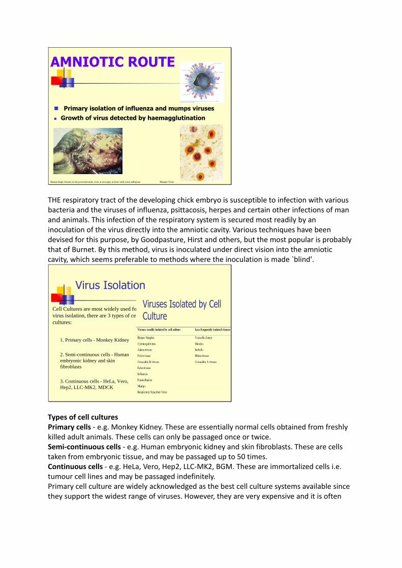
AMNIOTIC ROUTE
◼ Primary isolation of influenza and mumps viruses
◼ Growth of virus detected by haemagglutination
Hemorrhagic lesions in the proventriculus, seen at necropsy in fowl with avian influenza
Influenza Virus
Mumps Virus
THE respiratory tract of the developing chick embryo is susceptible to infection with various bacteria and the viruses of influenza, psittacosis, herpes and certain other infections of man and animals. This infection of the respiratory system is secured most readily by an inoculation of the virus directly into the amniotic cavity. Various techniques have been devised for this purpose, by Goodpasture, Hirst and others, but the most popular is probably that of Burnet. By this method, virus is inoculated under direct vision into the amniotic cavity, which seems preferable to methods where the inoculation is made `blind'.
Virus Isolation
Cell Cultures are most widely used for
virus isolation, there are 3 types of cell
cultures:
1. Primary cells - Monkey Kidney
2. Semi-continuous cells - Human
embryonic kidney and skin
fibroblasts
3. Continuous cells - HeLa, Vero,
Hep2, LLC-MK2, MDCK
Types of cell cultures Primary cells - e.g. Monkey Kidney. These are essentially normal cells obtained from freshly killed adult animals. These cells can only be passaged once or twice. Semi-continuous cells - e.g. Human embryonic kidney and skin fibroblasts. These are cells taken from embryonic tissue, and may be passaged up to 50 times. Continuous cells - e.g. HeLa, Vero, Hep2, LLC-MK2, BGM. These are immortalized cells i.e. tumour cell lines and may be passaged indefinitely. Primary cell culture are widely acknowledged as the best cell culture systems available since they support the widest range of viruses. However, they are very expensive and it is often
difficult to obtain a reliable supply. Continuous cells are the most easy to handle but the range of viruses supported is often limited.
Cell Cultures
Growing virus may produce
1. Cytopathic Effect (CPE) - such as the ballooning of cells or
syncytia formation, may be specific or non-specific.
2. Haemadsorption - cells acquire the ability to stick to
mammalian red blood cells.
Confirmation of the identity of the virus may be carried out using
neutralization, haemadsorption-inhibition or immunofluorescence
tests.
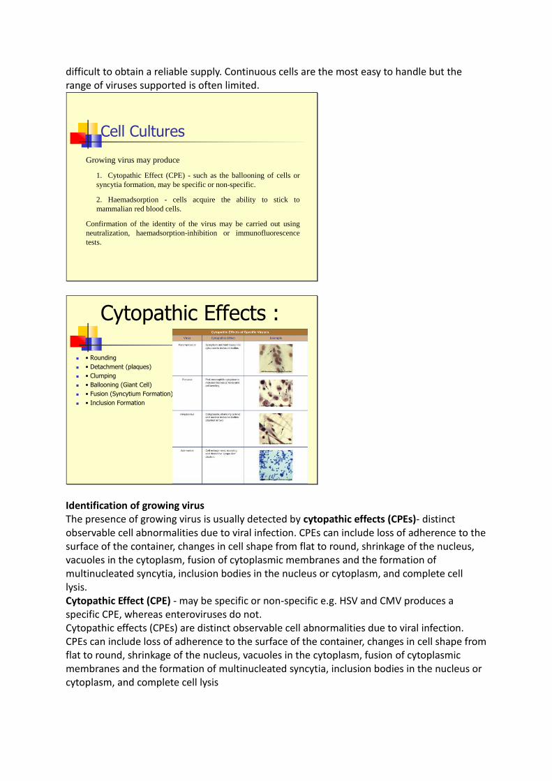
Cytopathic Effects :
◼ • Rounding
◼ • Detachment (plaques)
◼ • Clumping
◼ • Ballooning (Giant Cell)
◼ • Fusion (Syncytium Formation)
◼ • Inclusion Formation
Identification of growing virus The presence of growing virus is usually detected by cytopathic effects (CPEs)- distinct observable cell abnormalities due to viral infection. CPEs can include loss of adherence to the surface of the container, changes in cell shape from flat to round, shrinkage of the nucleus, vacuoles in the cytoplasm, fusion of cytoplasmic membranes and the formation of multinucleated syncytia, inclusion bodies in the nucleus or cytoplasm, and complete cell lysis. Cytopathic Effect (CPE) - may be specific or non-specific e.g. HSV and CMV produces a specific CPE, whereas enteroviruses do not. Cytopathic effects (CPEs) are distinct observable cell abnormalities due to viral infection. CPEs can include loss of adherence to the surface of the container, changes in cell shape from flat to round, shrinkage of the nucleus, vacuoles in the cytoplasm, fusion of cytoplasmic membranes and the formation of multinucleated syncytia, inclusion bodies in the nucleus or cytoplasm, and complete cell lysis
Haemadsorption - cells acquire the ability to stick to mammalian red blood cells. Haemadsorption is mainly used for the detection of influenza and parainfluenzaviruses. Confirmation of the identity of the virus may be carried out using neutralization, haemadsorption- inhibition, immunofluorescence, or molecular tests.
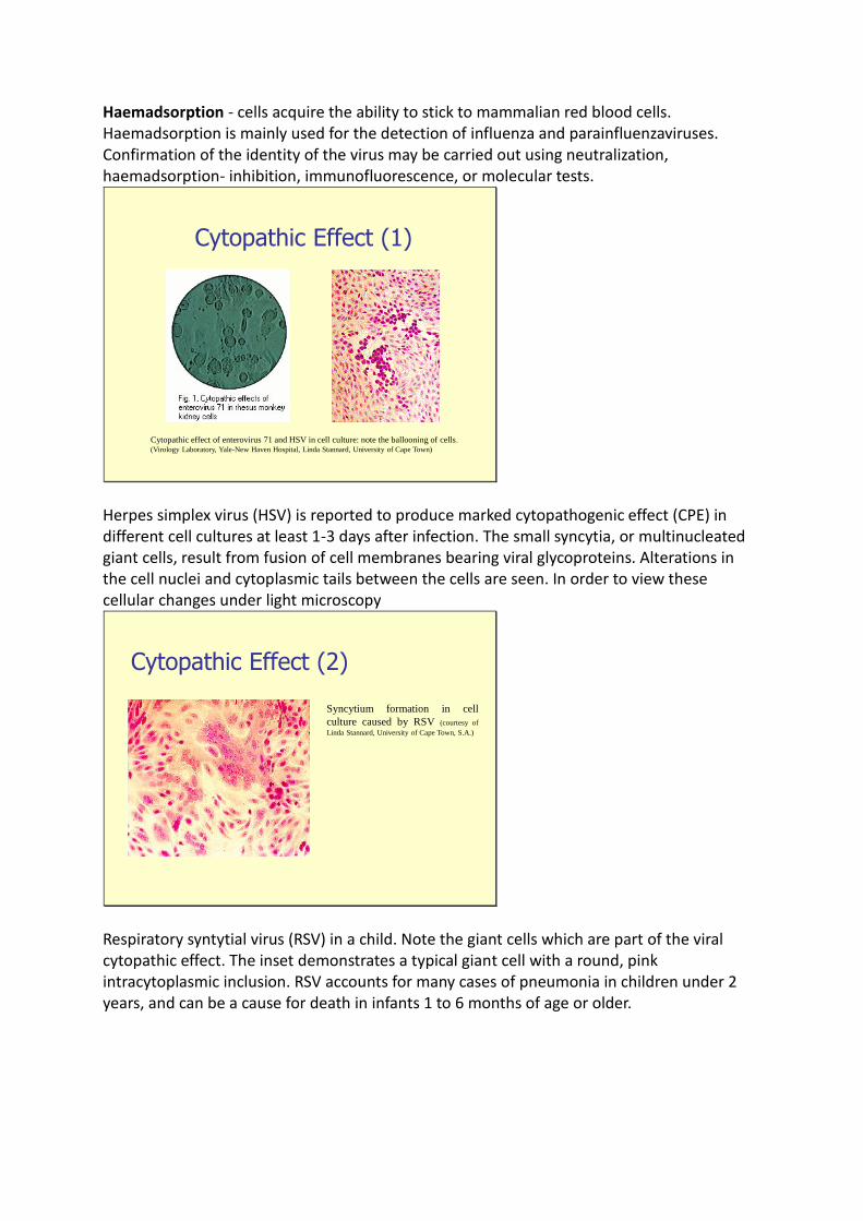
Cytopathic Effect (1)
Cytopathic effect of enterovirus 71 and HSV in cell culture: note the ballooning of cells.
(Virology Laboratory, Yale-New Haven Hospital, Linda Stannard, University of Cape Town)
Herpes simplex virus (HSV) is reported to produce marked cytopathogenic effect (CPE) in different cell cultures at least 1-3 days after infection. The small syncytia, or multinucleated giant cells, result from fusion of cell membranes bearing viral glycoproteins. Alterations in the cell nuclei and cytoplasmic tails between the cells are seen. In order to view these cellular changes under light microscopy
Cytopathic Effect (2)
Syncytium formation in cell
culture caused by RSV (courtesy of
Linda Stannard, University of Cape Town, S.A.)
Respiratory syntytial virus (RSV) in a child. Note the giant cells which are part of the viral cytopathic effect. The inset demonstrates a typical giant cell with a round, pink intracytoplasmic inclusion. RSV accounts for many cases of pneumonia in children under 2 years, and can be a cause for death in infants 1 to 6 months of age or older.
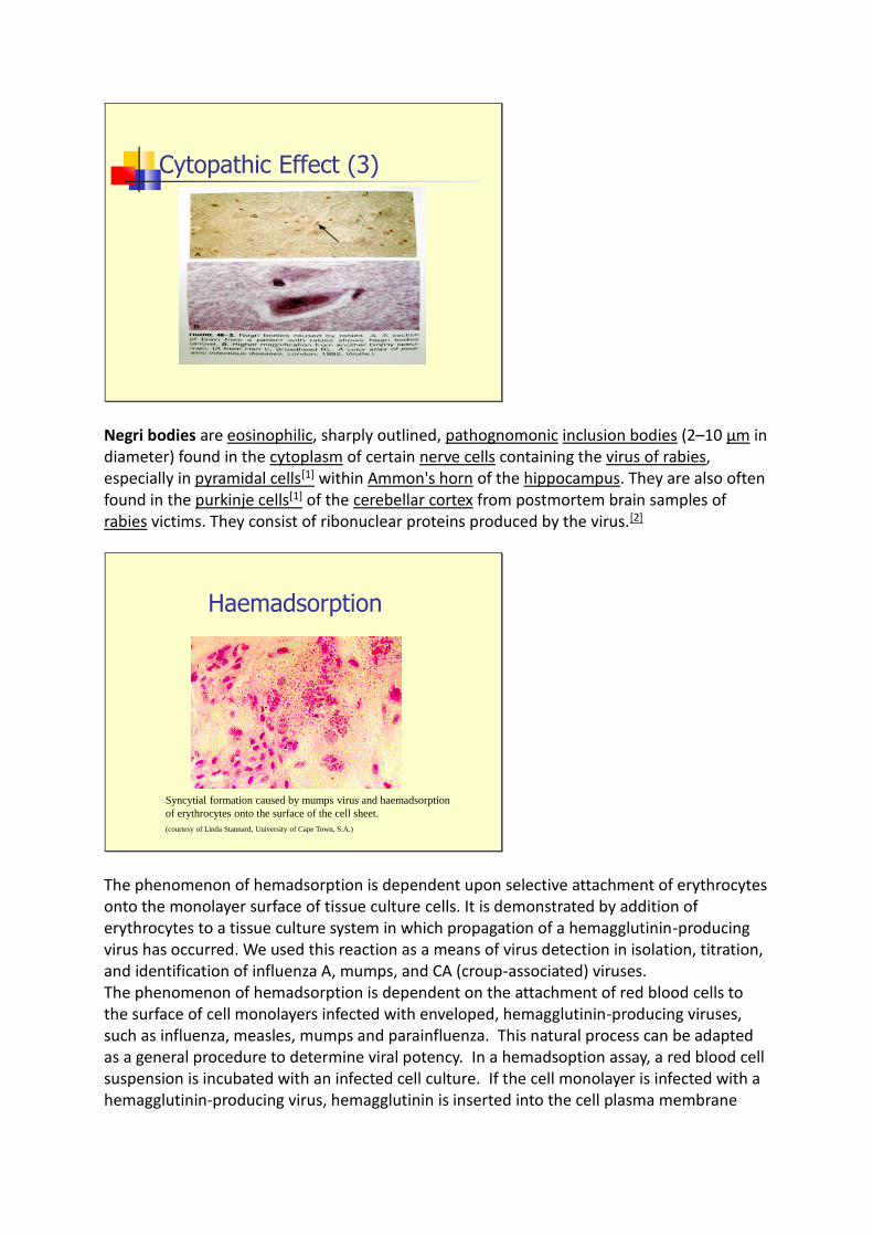
Cytopathic Effect (3)
Negri bodies are eosinophilic, sharply outlined, pathognomonic inclusion bodies (2–10 μm in diameter) found in the cytoplasm of certain nerve cells containing the virus of rabies, especially in pyramidal cells[1] within Ammon's horn of the hippocampus. They are also often found in the purkinje cells[1] of the cerebellar cortex from postmortem brain samples of rabies victims. They consist of ribonuclear proteins produced by the virus.[2]
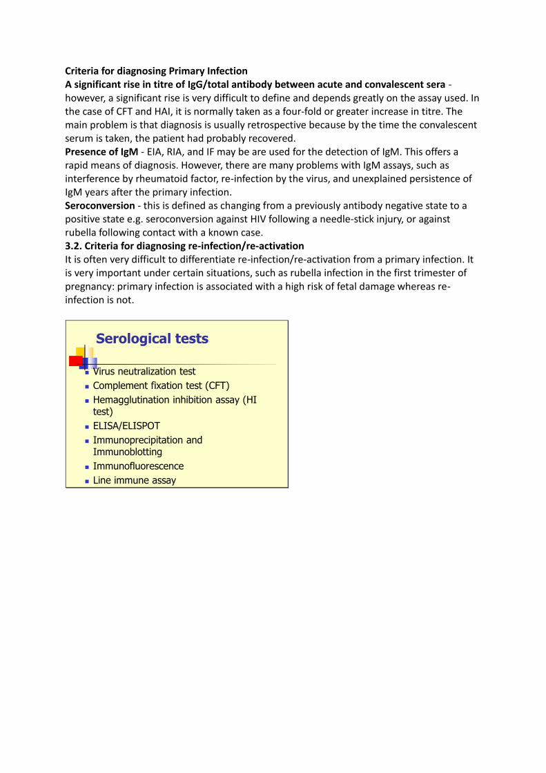
Haemadsorption
Syncytial formation caused by mumps virus and haemadsorption
of erythrocytes onto the surface of the cell sheet.
(courtesy of Linda Stannard, University of Cape Town, S.A.)
The phenomenon of hemadsorption is dependent upon selective attachment of erythrocytes onto the monolayer surface of tissue culture cells. It is demonstrated by addition of erythrocytes to a tissue culture system in which propagation of a hemagglutinin-producing virus has occurred. We used this reaction as a means of virus detection in isolation, titration, and identification of influenza A, mumps, and CA (croup-associated) viruses. The phenomenon of hemadsorption is dependent on the attachment of red blood cells to the surface of cell monolayers infected with enveloped, hemagglutinin-producing viruses, such as influenza, measles, mumps and parainfluenza. This natural process can be adapted as a general procedure to determine viral potency. In a hemadsoption assay, a red blood cell suspension is incubated with an infected cell culture. If the cell monolayer is infected with a hemagglutinin-producing virus, hemagglutinin is inserted into the cell plasma membrane

during viral reproduction inpreparation for viral maturation. It is at these modified areas of the cell surface that red blood cells will specifically bind. Thus hemadsorption is indicative of the presence of viruses that produce hemagglutinin.
Problems with cell culture
◼ Long period (up to 4 weeks) required for result.
◼ Often very poor sensitivity
◼ Susceptible to bacterial contamination.
◼ Susceptible to toxic substances which may be present in
the specimen.
◼ Many viruses will not grow in cell culture e.g. Hepatitis
B, Diarrhoeal viruses, parvovirus, papillomavirus.
Cell culture is the process of isolating cells from an organism and growing them in a scientifically controlled environment. The main problem with cell culture is the long period (up to 4 weeks) required for a result to be available. Also, the sensitivity is often poor and depends on many factors, such as the condition of the specimen, and the condition of the cell sheet. Cell cultures are also very susceptible to bacterial contamination and toxic substances in the specimen. Lastly, many viruses will not grow in cell culture at all e.g. Hepatitis B and C, Diarrhoeal viruses, parvovirus etc.
Serology
Criteria for diagnosing Primary Infection
◼ 4 fold or more increase in titre of IgG or total antibody
between acute and convalescent sera
◼ Presence of IgM
◼ Seroconversion
◼ A single high titre of IgG (or total antibody)
Criteria for diagnosing Reinfection
◼ fold or more increase in titre of IgG or total antibody
between acute and convalescent sera
◼ Absence or slight increase in IgM
◼ Note that during reinfection, IgM may be absent or
present at a low level transiently
Serology forms the mainstay of viral diagnosis. This is what happens in a primary humoral immune response to antigen. Following exposure, the first antibody to appear is IgM, which is followed by a much higher titre of IgG. In cases of reinfection, the level of specific IgM either remain the same or rises slightly. But IgG shoots up rapidly and far more earlier than in a primary infection. Many different types of serological tests are available
Criteria for diagnosing Primary Infection A significant rise in titre of IgG/total antibody between acute and convalescent sera - however, a significant rise is very difficult to define and depends greatly on the assay used. In the case of CFT and HAI, it is normally taken as a four-fold or greater increase in titre. The main problem is that diagnosis is usually retrospective because by the time the convalescent serum is taken, the patient had probably recovered. Presence of IgM - EIA, RIA, and IF may be are used for the detection of IgM. This offers a rapid means of diagnosis. However, there are many problems with IgM assays, such as interference by rheumatoid factor, re-infection by the virus, and unexplained persistence of IgM years after the primary infection. Seroconversion - this is defined as changing from a previously antibody negative state to a positive state e.g. seroconversion against HIV following a needle-stick injury, or against rubella following contact with a known case. 3.2. Criteria for diagnosing re-infection/re-activation It is often very difficult to differentiate re-infection/re-activation from a primary infection. It is very important under certain situations, such as rubella infection in the first trimester of pregnancy: primary infection is associated with a high risk of fetal damage whereas re-infection is not.
Serological tests
◼ Virus neutralization test
◼ Complement fixation test (CFT)
◼ Hemagglutination inhibition assay (HI test)
◼ ELISA/ELISPOT
◼ Immunoprecipitation and Immunoblotting
◼ Immunofluorescence
◼ Line immune assay
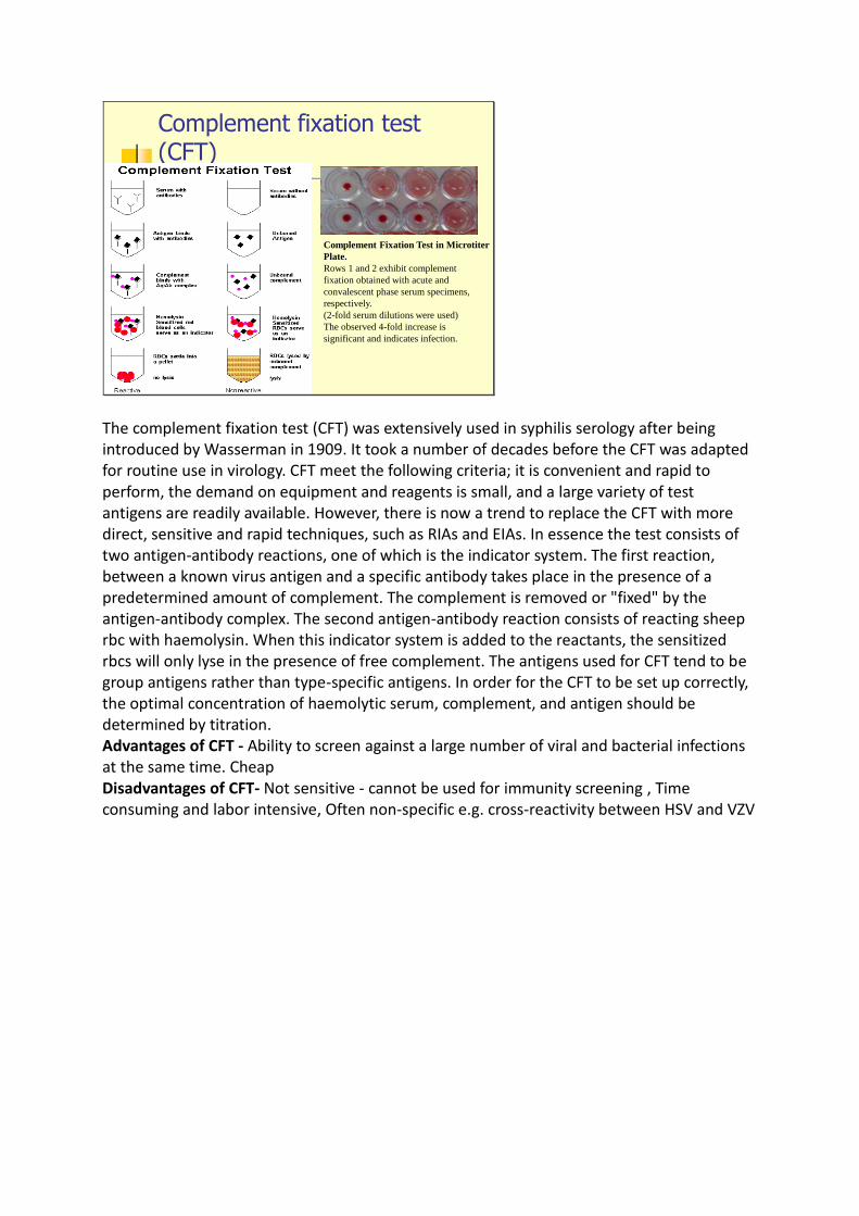
Complement fixation test (CFT)
Complement Fixation Test in Microtiter
Plate.
Rows 1 and 2 exhibit complement
fixation obtained with acute and
convalescent phase serum specimens,
respectively.
(2-fold serum dilutions were used)
The observed 4-fold increase is
significant and indicates infection.
The complement fixation test (CFT) was extensively used in syphilis serology after being introduced by Wasserman in 1909. It took a number of decades before the CFT was adapted for routine use in virology. CFT meet the following criteria; it is convenient and rapid to perform, the demand on equipment and reagents is small, and a large variety of test antigens are readily available. However, there is now a trend to replace the CFT with more direct, sensitive and rapid techniques, such as RIAs and EIAs. In essence the test consists of two antigen-antibody reactions, one of which is the indicator system. The first reaction, between a known virus antigen and a specific antibody takes place in the presence of a predetermined amount of complement. The complement is removed or "fixed" by the antigen-antibody complex. The second antigen-antibody reaction consists of reacting sheep rbc with haemolysin. When this indicator system is added to the reactants, the sensitized rbcs will only lyse in the presence of free complement. The antigens used for CFT tend to be group antigens rather than type-specific antigens. In order for the CFT to be set up correctly, the optimal concentration of haemolytic serum, complement, and antigen should be determined by titration. Advantages of CFT - Ability to screen against a large number of viral and bacterial infections at the same time. Cheap Disadvantages of CFT- Not sensitive - cannot be used for immunity screening , Time consuming and labor intensive, Often non-specific e.g. cross-reactivity between HSV and VZV
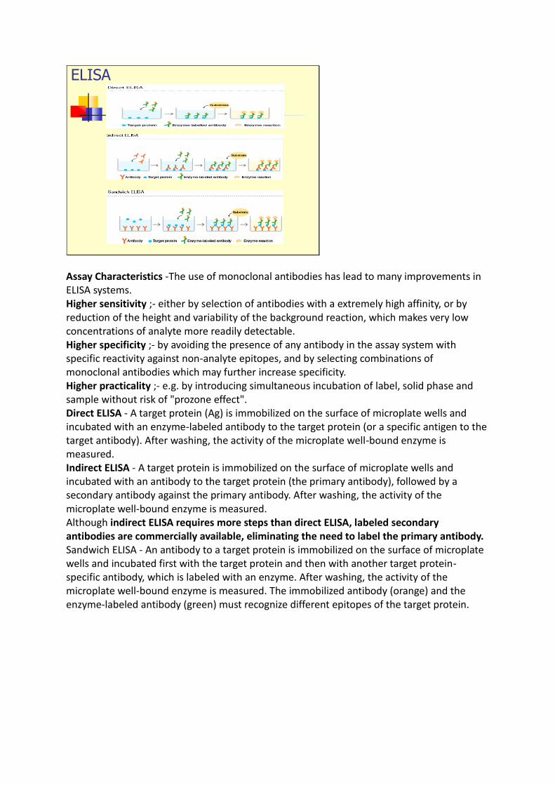
ELISA
Assay Characteristics -The use of monoclonal antibodies has lead to many improvements in ELISA systems. Higher sensitivity ;- either by selection of antibodies with a extremely high affinity, or by reduction of the height and variability of the background reaction, which makes very low concentrations of analyte more readily detectable. Higher specificity ;- by avoiding the presence of any antibody in the assay system with specific reactivity against non-analyte epitopes, and by selecting combinations of monoclonal antibodies which may further increase specificity. Higher practicality ;- e.g. by introducing simultaneous incubation of label, solid phase and sample without risk of "prozone effect". Direct ELISA - A target protein (Ag) is immobilized on the surface of microplate wells and incubated with an enzyme-labeled antibody to the target protein (or a specific antigen to the target antibody). After washing, the activity of the microplate well-bound enzyme is measured. Indirect ELISA - A target protein is immobilized on the surface of microplate wells and incubated with an antibody to the target protein (the primary antibody), followed by a secondary antibody against the primary antibody. After washing, the activity of the microplate well-bound enzyme is measured. Although indirect ELISA requires more steps than direct ELISA, labeled secondary antibodies are commercially available, eliminating the need to label the primary antibody. Sandwich ELISA - An antibody to a target protein is immobilized on the surface of microplate wells and incubated first with the target protein and then with another target protein-specific antibody, which is labeled with an enzyme. After washing, the activity of the microplate well-bound enzyme is measured. The immobilized antibody (orange) and the enzyme-labeled antibody (green) must recognize different epitopes of the target protein.
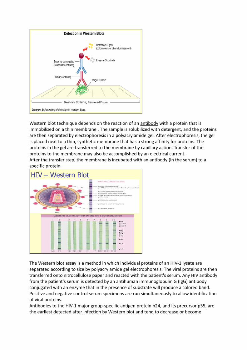
Western blot technique depends on the reaction of an antibody with a protein that is immobilized on a thin membrane . The sample is solubilized with detergent, and the proteins are then separated by electrophoresis in a polyacrylamide gel. After electrophoresis, the gel is placed next to a thin, synthetic membrane that has a strong affinity for proteins. The proteins in the gel are transferred to the membrane by capillary action. Transfer of the proteins to the membrane may also be accomplished by an electrical current. After the transfer step, the membrane is incubated with an antibody (in the serum) to a specific protein.
HIV – Western Blot
The Western blot assay is a method in which individual proteins of an HIV-1 lysate are separated according to size by polyacrylamide gel electrophoresis. The viral proteins are then transferred onto nitrocellulose paper and reacted with the patient's serum. Any HIV antibody from the patient's serum is detected by an antihuman immunoglobulin G (IgG) antibody conjugated with an enzyme that in the presence of substrate will produce a colored band. Positive and negative control serum specimens are run simultaneously to allow identification of viral proteins. Antibodies to the HIV-1 major group-specific antigen protein p24, and its precursor p55, are the earliest detected after infection by Western blot and tend to decrease or become
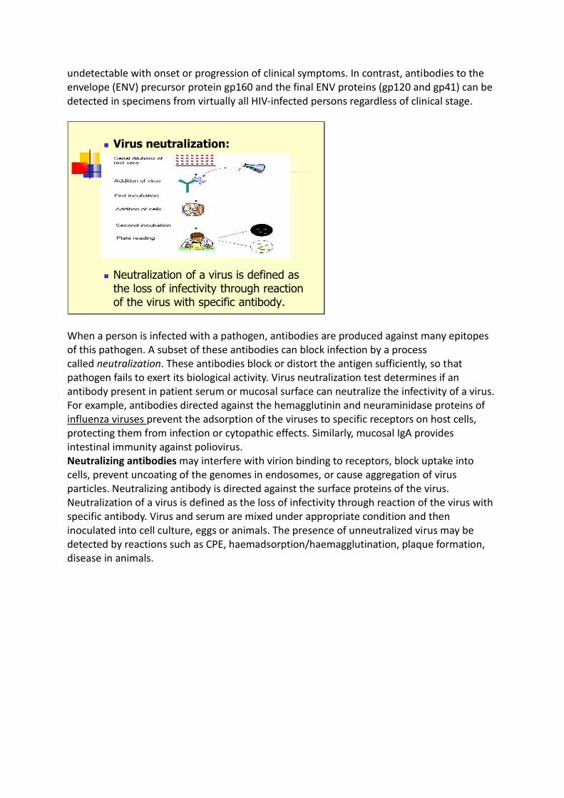
undetectable with onset or progression of clinical symptoms. In contrast, antibodies to the envelope (ENV) precursor protein gp160 and the final ENV proteins (gp120 and gp41) can be detected in specimens from virtually all HIV-infected persons regardless of clinical stage.
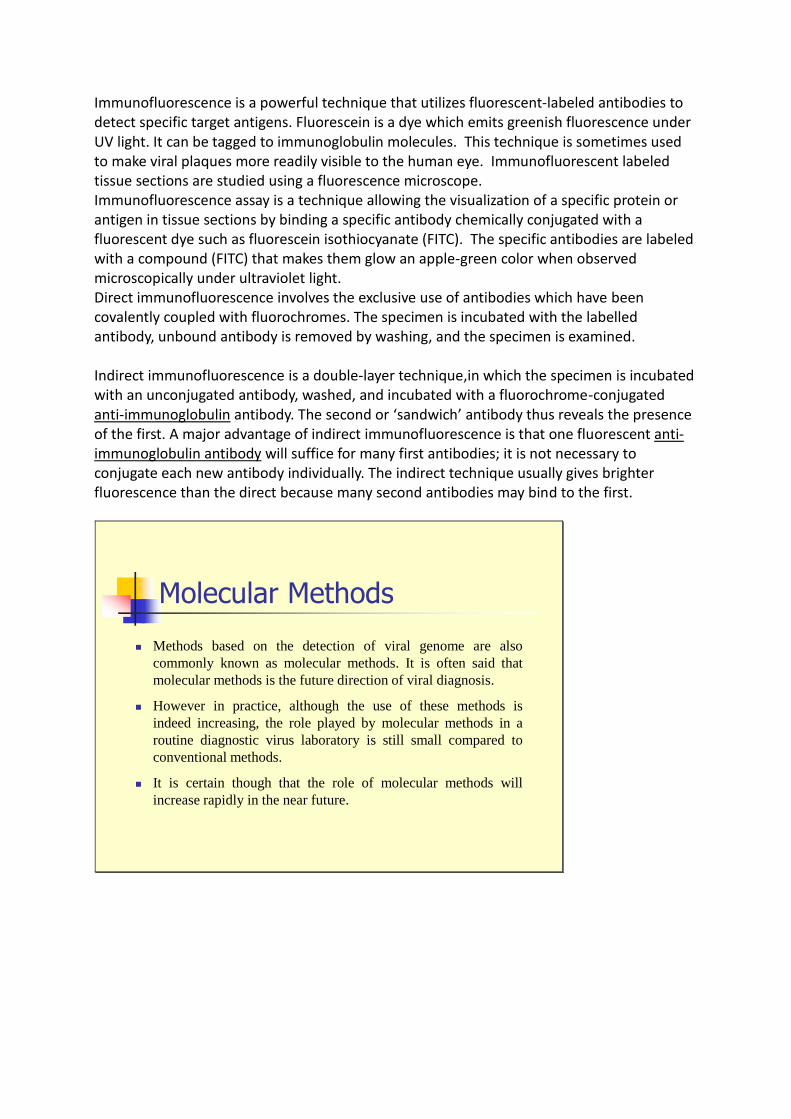
◼ Virus neutralization:
◼ Neutralization of a virus is defined as the loss of infectivity through reaction of the virus with specific antibody.
When a person is infected with a pathogen, antibodies are produced against many epitopes of this pathogen. A subset of these antibodies can block infection by a process called neutralization. These antibodies block or distort the antigen sufficiently, so that pathogen fails to exert its biological activity. Virus neutralization test determines if an antibody present in patient serum or mucosal surface can neutralize the infectivity of a virus. For example, antibodies directed against the hemagglutinin and neuraminidase proteins of influenza viruses prevent the adsorption of the viruses to specific receptors on host cells, protecting them from infection or cytopathic effects. Similarly, mucosal IgA provides intestinal immunity against poliovirus. Neutralizing antibodies may interfere with virion binding to receptors, block uptake into cells, prevent uncoating of the genomes in endosomes, or cause aggregation of virus particles. Neutralizing antibody is directed against the surface proteins of the virus. Neutralization of a virus is defined as the loss of infectivity through reaction of the virus with specific antibody. Virus and serum are mixed under appropriate condition and then inoculated into cell culture, eggs or animals. The presence of unneutralized virus may be detected by reactions such as CPE, haemadsorption/haemagglutination, plaque formation, disease in animals.

Hemagglutination InhibitionTest
The nucleic acids of various viruses encode surface proteins (e.g.hemaaglutinin (HA) of influenza virus) that agglutinate red blood cells (RBC) of a variety of species. The reaction of viral hemagglutinins with red blood cells results in a lattice of agglutinated cells that settle irregularly in a tube or microtiter well. Unagglutinated cells settle in a compact button. This process is known as hemagglutination. The basis of the HAI assay is that antibodies to that particular virus (for example; measles virus- see image) will prevent attachment of the virus to RBC. Therefore hemagglutination is inhibited when antibodies are present. Hemagglutination occurs when measles viruses and red blood cells are mixed (see image a). But, if the serum of a person infected with measles virus is mixed with RBC and measles virus, there won’t be any agglutination of RBC. This phenomenon is known as hemagglutination inhibition. This arises because antibodies present in the serum of that infected person reacts with the measles viruses and neutralize them (positive result). If the patient’s serum do not contain antibodies against surface proteins of test virus, there will be presence of hemagglutination as surface molecules are free to hemaaglutinate RBCs (negative result).
Immunofluorescence
Direct Indirect
Immunofluorescence is a powerful technique that utilizes fluorescent-labeled antibodies to detect specific target antigens. Fluorescein is a dye which emits greenish fluorescence under UV light. It can be tagged to immunoglobulin molecules. This technique is sometimes used to make viral plaques more readily visible to the human eye. Immunofluorescent labeled tissue sections are studied using a fluorescence microscope. Immunofluorescence assay is a technique allowing the visualization of a specific protein or antigen in tissue sections by binding a specific antibody chemically conjugated with a fluorescent dye such as fluorescein isothiocyanate (FITC). The specific antibodies are labeled with a compound (FITC) that makes them glow an apple-green color when observed microscopically under ultraviolet light. Direct immunofluorescence involves the exclusive use of antibodies which have been covalently coupled with fluorochromes. The specimen is incubated with the labelled antibody, unbound antibody is removed by washing, and the specimen is examined. Indirect immunofluorescence is a double-layer technique,in which the specimen is incubated with an unconjugated antibody, washed, and incubated with a fluorochrome-conjugated anti-immunoglobulin antibody. The second or ‘sandwich’ antibody thus reveals the presence of the first. A major advantage of indirect immunofluorescence is that one fluorescent anti-immunoglobulin antibody will suffice for many first antibodies; it is not necessary to conjugate each new antibody individually. The indirect technique usually gives brighter fluorescence than the direct because many second antibodies may bind to the first.
Molecular Methods
◼ Methods based on the detection of viral genome are also
commonly known as molecular methods. It is often said that
molecular methods is the future direction of viral diagnosis.
◼ However in practice, although the use of these methods is
indeed increasing, the role played by molecular methods in a
routine diagnostic virus laboratory is still small compared to
conventional methods.
◼ It is certain though that the role of molecular methods will
increase rapidly in the near future.
Polymerase Chain Reaction
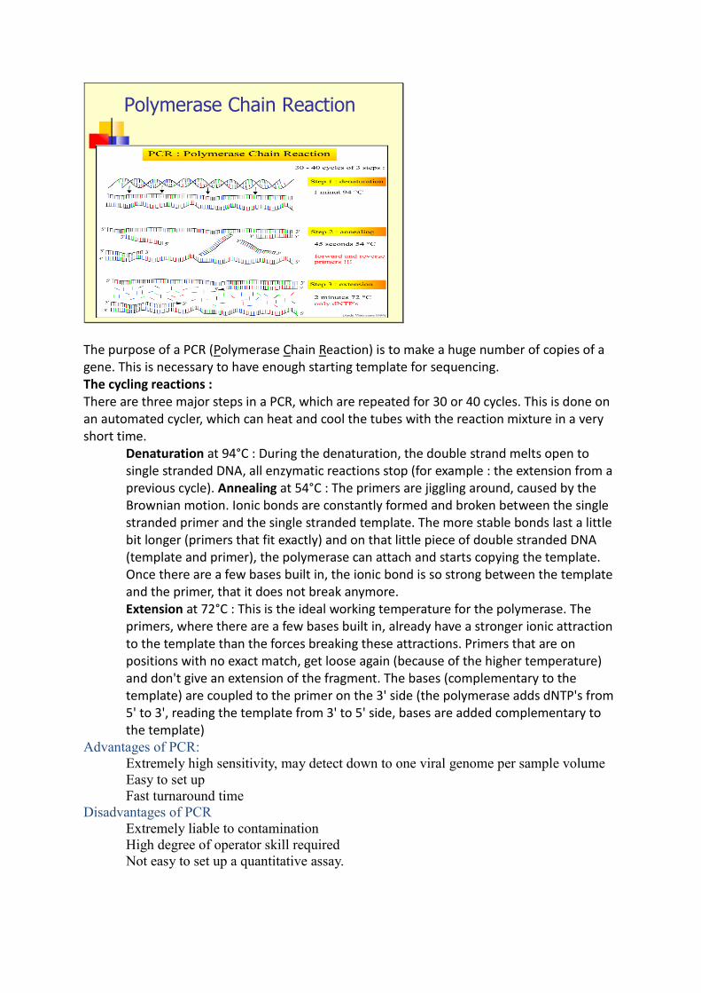
The purpose of a PCR (Polymerase Chain Reaction) is to make a huge number of copies of a gene. This is necessary to have enough starting template for sequencing. The cycling reactions : There are three major steps in a PCR, which are repeated for 30 or 40 cycles. This is done on an automated cycler, which can heat and cool the tubes with the reaction mixture in a very short time.
Denaturation at 94°C : During the denaturation, the double strand melts open to single stranded DNA, all enzymatic reactions stop (for example : the extension from a previous cycle). Annealing at 54°C : The primers are jiggling around, caused by the Brownian motion. Ionic bonds are constantly formed and broken between the single stranded primer and the single stranded template. The more stable bonds last a little bit longer (primers that fit exactly) and on that little piece of double stranded DNA (template and primer), the polymerase can attach and starts copying the template. Once there are a few bases built in, the ionic bond is so strong between the template and the primer, that it does not break anymore. Extension at 72°C : This is the ideal working temperature for the polymerase. The primers, where there are a few bases built in, already have a stronger ionic attraction to the template than the forces breaking these attractions. Primers that are on positions with no exact match, get loose again (because of the higher temperature) and don't give an extension of the fragment. The bases (complementary to the template) are coupled to the primer on the 3' side (the polymerase adds dNTP's from 5' to 3', reading the template from 3' to 5' side, bases are added complementary to the template)
Advantages of PCR:
Extremely high sensitivity, may detect down to one viral genome per sample volume
Easy to set up
Fast turnaround time
Disadvantages of PCR
Extremely liable to contamination
High degree of operator skill required
Not easy to set up a quantitative assay.
A positive result may be difficult to interpret, especially with latent viruses such as
CMV, where any seropositive person will have virus present in their blood irrespective
whether they have disease or not.
SOURCES:
◼ https://virology-online.com/general/Tests.htm
◼ https://www.jobilize.com/microbiology/test/detection-of-a-virus-isolation-culture-and-identification-by-openstax
◼ https://www.asmscience.org/content/education/imagegallery/image.2616
◼ https://www.lgcstandards-atcc.org/support/faqs/d1f5b/General+protocol+for+the+Hemadsorption+Assay-234.aspx?geo_country=sk
◼ https://www.researchgate.net/figure/Egg-inoculation-into-the-allantoic-cavity-a-Drill-or-pierce-a-hole-in-the-egg-5-mm_fig1_272836131
◼ https://microbeonline.com/hemagglutination-inhibition-test-hai-principle-procedure-result-interpretations/
◼ https://www.slideshare.net/TapeshwarYadav1/immunofluorescence-55934842
◼ https://theconversation.com/five-techniques-were-using-to-uncover-the-secrets-of-viruses-144363
Related Documents