Title : Periodontal Abscess : A review Shende, Sumit 1 ; Swargiya Dadasaheb Kalmegh Smruti Dental College and Hospital, Department of Periodontology Ansari , Salman 2 ; Swargiya Dadasaheb Kalmegh Smruti Dental College and Hospital, Department of Periodontology. Gattani, Deepti 3 ; Swargiya Dadasaheb Kalmegh Smruti Dental College and Hospital, Department of Periodontology. Bhutada, Girish 4; Swargiya Dadasaheb Kalmegh Smruti Dental College and Hospital, Department of Periodontology. Meshram, Sneha 5 ; Swargiya Dadasaheb Kalmegh Smruti Dental College and Hospital, Department of Periodontology. Jirafe, Sanjana 6 ; Swargiya Dadasaheb Kalmegh Smruti Dental College and Hospital, Department of Periodontology. 1, 5,6- Pg student 2- Reader and guide 3- Hod and professor 4- Professor and Guide “Address For correspondence” Sumit Shende, II year MDS student, Department of Periodontology, Swargiya Dadasaheb Kalmegh Smruti Dental College and Hospital Email id- [email protected] Phone no. – 9665292327 / 7588788444

Welcome message from author
This document is posted to help you gain knowledge. Please leave a comment to let me know what you think about it! Share it to your friends and learn new things together.
Transcript
Title : Periodontal Abscess : A review
Shende, Sumit1; Swargiya Dadasaheb Kalmegh Smruti Dental College and Hospital, Department of Periodontology
Ansari , Salman2; Swargiya Dadasaheb Kalmegh Smruti Dental College and Hospital, Department of Periodontology.
Gattani, Deepti3; Swargiya Dadasaheb Kalmegh Smruti Dental College and Hospital, Department of Periodontology.
Bhutada, Girish4; Swargiya Dadasaheb Kalmegh Smruti Dental College and Hospital, Department of Periodontology.
Meshram, Sneha5 ; Swargiya Dadasaheb Kalmegh Smruti Dental College and Hospital, Department of Periodontology.
Jirafe, Sanjana6 ; Swargiya Dadasaheb Kalmegh Smruti Dental College and Hospital, Department of Periodontology.
1, 5,6- Pg student
2- Reader and guide 3- Hod and professor 4- Professor and Guide
“Address For correspondence”
Sumit Shende, II year MDS student, Department of Periodontology, Swargiya Dadasaheb Kalmegh Smruti Dental College and Hospital
Email id- [email protected]
Phone no. – 9665292327 / 7588788444
Periodontal abscess : A review -Introduction:
Periodontium of tooth confines to the periodontal ligament, alveolar bone, gingiva and
cementum. Odontogenic infections involving periodontium may lead to emergencies such as dental
abscesses in dental practice.
Abscesses in periodontium are caused by improper oral hygiene following surgical therapy,
pulpal infections leading to periodontal involvement, pericoronitis and trauma to underlying
periosteum. Periodontal infections including periodontitis, pericoronitis and perio-endo lesion may
give rise to abscesses in the periodontium if left untreated. Abscesses in the periodontium includes
gingival, pericoronal and periodontal abscess. Abscesses in the periodontium can be defined as ‘a
lesion with an expressed periodontal breakdown occurring during a limited period of time, and with
easily detectable clinical symptoms, including a localized accumulation of pus located within the
gingival wall of the periodontal pocket’. Among all the dental emergencies, abscesses were seen
41% associated with first molar, 24% with second molar, 17% with upper premolars, 7% with lower
premolars, 7% with incisors and 3.5% with upper third molars.
Among all the abscesses of periodontium, the periodontal abscess is the most important one. The
periodontal abscess is the third most common dental emergency (6-14 %), first is the Dentoalveolar
abscess / pulpal infection (14-25 %) followed by pericoronitis (10-11 %).1 There was one study
conducted in US in and he found that among all total emergencies in dentistry, periodontal abscess
comprised of almost 14%.2
Periodontal abscess is defined as ‘A periodontal abscess is a localized, purulent infection
involving a greater dimension of the gingiva, extending apically and adjacent to a periodontal pocket.
A periodontal abscess can also be termed as lateral abscess or parietal abscess; however, when
marginal soft tissues such as marginal gingiva are affected in isolation, it is called a gingival abscess.
Periodontal abscess and gingival abscess are identical histologically and differ only in location.
According to international workshop for a classification of periodontal diseases and conditions
(1999):
They are classified as - 1. Gingival
2. Periodontal
3. Periapical
4. Pericoronal. 3
Periodontal abscess is classified according to its etiology, course of the lesion, number of
abscess and location of abscess.
According to etiology of the lesion, periodontal abscess can be classified as –
1. Periodontitis related periodontal abscess – periodontal abscess occurs in previously present
periodontitis or previously present biofilm in deepened periodontal pocket.4
2. Non periodontitis periodontal abscess - periodontal abscess can develop in healthy sites or from
another local source for e.g. impacted food particles or foreign body (pieces of dental floss,
toothbrush bristles) and alteration in root morphology.5,6
According to course of the lesion, periodontal abscess can be classified as-
1. Acute periodontal abscess – an acute periodontal abscess can manifests as painful swelling with
red inflamed area. Swelling is usually tender on palpation and suppuration can occur upon gentle
pressure. Pain is usually throbbing in nature and lymphadenopathy can be present.
2. Chronic periodontal abscess – an acute periodontal abscess can become chronic periodontal
abscess when drainage is established through the deepened periodontal pocket, gingival sulcus
and sinus tract. Abscess can be asymptomatic and develops as an acute exacerbation of acute
periodontal abscess. There may be bleeding and pain is usually of low intensity. Tooth may have
mobility and it may be tender on percussion.6
According to location, periodontal abscess can be classified as –
1. Gingival abscess – when abscess is confined to marginal gingiva or interdental papilla and
swelling is localized and with purulent infection. Impacted subgingival calculus or any othe
foreign body particles can cause purulent suppuration.4
2. Periodontal abscess – when localized purulent infection involves the periodontium and swelling is
confined to periodontal tissues.4
3. Pericoronal abscess- it is mainly associated with partially impacted tooth (lower third molar
usually) or the tooth covered with pericoronal flap i.e. pericoronitis. A localized purulent
infection develops within the pericoronal flap which is tender on palpation and causes pus
discharge on gentle light pressure. Sometimes, condition is associated with trismus. (Inability to
open the mouth). Pain in case of pericoronal abscess ranges from mild to severe throbbing pain.

According to number of abscesses, periodontal abscess can be classified as-
1. Single abscess- when abscess is confined to single tooth.
2. Multiple abscess- when abscess is confined to more than one tooth.7
Etiology of periodontal abscess -
Periodontal abscess can develop in periodontitis affected sites and in non-periodontitis affected
sites. In periodontitis periodontal abscess may develop with an acute exacerbation of previously
present periodontitis with deep periodontal torturous pockets, teeth with furcation involvement and
vertical defect which may lead to spread of infection into the surrounding periodontal tissues.
Decreased host tissue response and alteration of composition of subgingival bacteria results in
diminished capacity to drain the suppuration.8,9
Periodontal abscess in previously present periodontitis can occur by following ways-
1. After nonsurgical periodontal therapy- Following scaling and root planing, dislodged calculus and
food particles may gain entry into the deep periodontal pockets which may cause inflammation
and suppuration of periodontal tissues.8,9,10,11.
2. After surgical periodontal therapy- It is associated with the foreign bodies such as resorbable or
nonresorbable membranes and bone grafts used for periodontal regenerative procedures.10,11
3. Acute exacerbation of untreated periodontitis.8
4. Acute exacerbation of refractory periodontitis.12,13
5. Acute exacerbation of supportive periodontal therapy.12,13
6. Systemic antimicrobials without mechanical debridement of subgingival calculus may lead to
abscess formation.7
7. Treatment with nifedipine therapy.14
8. Resorbable and nonresorbable membranes in guided bone regeneration.15
Periodontal abscess can occur in previously healthy sites i.e. non periodontitis patients-
1. Foreign bodies-.
Orthodontic brackets/elastics/wires, a piece of dental floss, piece of toothbrush bristles or
toothpicks, dislodged cemental tear and pieces of nails in nail biting patients. Oral hygiene
abscess is the term given for abscess caused by the use of oral hygiene aids.6
2.Alteration in the morphology of root surfaces-
Perforation by an endodontic instrument, cervical cemental tears, external root resorption,
invaginated tooth and cracked tooth syndrome.16,17

Course of periodontal abscess
The rate of destruction of periodontal abscess is depending upon following factors-
1. PH of local tissues (an acidic environment favors the activity of lysozomal enzymes)
2. Growth of bacteria inside the focus
3. Their virulence
Histopathology of periodontal abscess:
Dewitt GV et al , 1985 studied 12 biopsy samples of periodontal abscess patients and
observed the following clinical findings –
1. A normal oral epithelium and lamina propria
2. An acute inflammatory infiltrate
3. An infiltration of neutrophils and lymphocytes present in area of destroyed and necrotic
connective tissue.
Electron microscopy of periodontal abscess revealed that there are more gram negative
bacteria, invasion of pocket epithelium and the affected connective tissues shows amorphous,
granular and acidophilic debris.4
Invasion of bacteria into periodontal
Inflammatory response
Release of chemotactic factors causing destruction of connective tissue
Encapsulation of bacterial infection and production of
pus
periodontal abscess

Microbiology of periodontal abscess :
Purulent oral infections are usually multibacterial and are caused by commensal bacteria. In
microbiological reports on periodontal abscesses, gram negative bacteria and rods predominated
over gram positive bacteria and cocci. The most prevalent bacterial species identified in
periodontal abscesses, using culture-based or molecular-based diagnostic techniques, is
Porphyromonas gingivalis, with a range in prevalence of 50–100%.18
Other strict anaerobes which are incorporated are Prevotella intermedia, Prevotella
melaninogenica, Fusobacterium nucleatum, Tannerella forsythia, Treponema spp. ,Parvimonas
micra, Actinomyces spp. and Bifidobacterium spp. Among the facultative anaerobic gram-
negative bacteria, Campylobacter spp., Capnocytophaga spp. and Aggregatibacter
actinomycetemcomitans. It is polymicrobial and dominated by nonmotile, anaerobic, gram
negative and rod shaped species.19
Diagnosis of periodontal abscess:
Periodontal abscess is multifactorial in origin, so all the local, systemic and aggravating
factors should be taken into consideration while diagnosing periodontal abscess. Diagnosis is
based on overall evaluation, past medical and dental history, patient symptoms, together with
clinical and radiographic signs.20
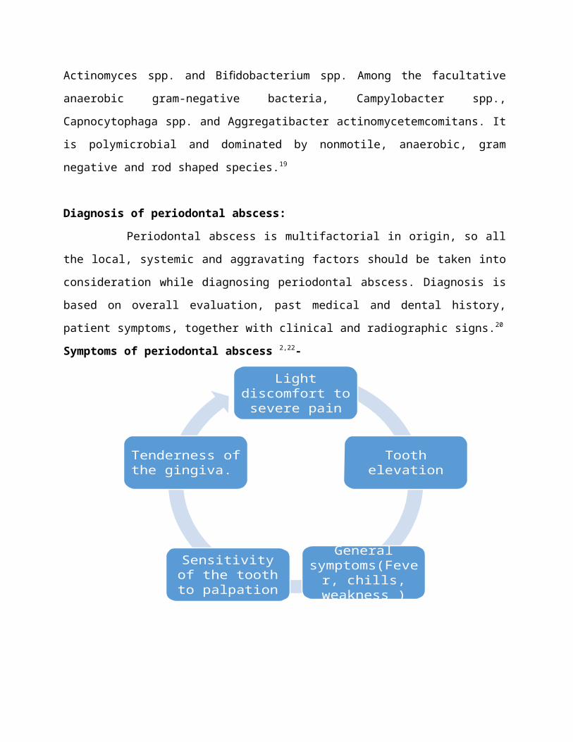
Symptoms of periodontal abscess 2,22-
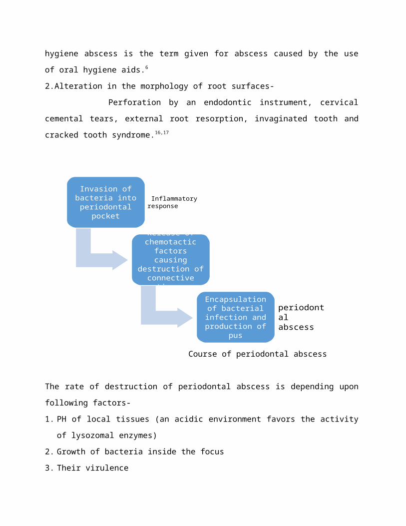
Fig. no. 1 : Palatal periodontal abscess associated with maxillary left second and third molar
Fig. no. 2 : Periodontal abscess present palatally with maxillary second and third molar
Light discomfort to severe pain
Tooth elevation
General symptoms(Fever, chills, weakness )
Sensitivity of the tooth to palpation
Tenderness of the gingiva.

Fig. no. 3 : Gingival abscess associated with maxillary premolar and first molar
Signs in periodontal abscess patients –
Presence of an ovoid elevation in the gingiva, along the lateral part of the root, although
abscesses located deep in the periodontium may be more difficult to identify and it may found as
a diffuse swelling as a red area.2,22
Investigations :
Radiographs–
Intraoral Periapical radiograph, digital orthopantamogram and bite wing radiographs (in case
of perio-endo lesion suspecting interdental bone loss) are used to assess the amount of bone loss
and periapical condition of involved teeth. Gutta-percha points placed through the sinus may
reveal a source of infection. Following features can be seen –
-may reveal a normal appearance or evident bone loss
- widening of periodontal ligament space
- may reveal about the size, shape, location and source of infection.22
Bleeding on probing , inctreased tooth mobility Deep periodontal pockets Suppuration through fistula or pocket
lining

Fig. no. 4: Periodontal abscess radio graphically showing discontinuation of lamina dura and
marginal bone loss associated with maxillary premolar and first molar
Pulp vitality test:
Pulp vitality tests like thermal and electric tests are generally used to assess the vitality of pulp.22
Microbial tests:
Samples of pus from sinus/abscess or collected through gingival sulcus sent for microbiological
culture and antibiotic sensitivity testing.22
Laboratory investigations:
Complete blood count and other hematologic investigations reveals the percentage of increase in
neutrophils or monocytes count which shows the proinflammatory response of bacterial toxins in
the periodontal abscess.22
Others-
Multiple periodontal abscesses are usually associated with systemic factors like increased blood
sugar and with an altered immune response in diabetic patients. Therefore, the assessment of the
diabetic status through the testing of random blood glucose, fasting blood glucose or glycosylated
hemoglobin levels is necessary to rule out the etiology of the periodontal abscess.
Anamnesis:
It gives the basic information about the etiology and pathogenesis of periodontal abscess,
Especially, associated with previous treatments or non-oral therapies such as systemic
antimicrobials or periodontal treatment. It is helpful in case of abscess which are related to
impaction of foreign bodies or particles. Sometimes the interview with patient depicting the
clinical history and other variables may be great help for further treatment planning.
Differential diagnosis :
Periodontal abscess may be misundertaken with the following infections or lesions –
1. Periapical or Dentoalveolar abscess.2
2. Lateral Periapical cyst.2
3. Endo-perio lesions.
4. Oral diseases like squamous cell carcinoma, pyogenic granuloma and metastatic
carcinoma.23,24
5. Self-inflicted gingival injuries.25

Treatment plan:
Treatment of periodontal abscess depends upon it etiology, local and aggravating factors, host
immune response and prognosis.
It includes two phases –
1. Control of the acute condition to arrest tissue destruction and control the symptoms.1,26
2. Management of pre-existing and / or residual lesion, especially in patients with periodontitis.
1. Control of the acute condition –
There are four therapeutic alternatives
A. Drainage of abscess – Drainage of abscess with light and digital pressure through gingival
sulcus or periodontal pocket should be carried out under local anesthesia.1,2
B. Debridement- Mechanical debridement of subgingival calculus and removal of any foreign
body present in gingival sulcus or deep periodontal pocket should be done in order to rule
out the etiology of periodontal abscess.1,2
C. Systemic or local antimicrobials- Broad spectrum antibiotics should be given to arrest the
growth of predominant gram negative cocci present in periodontal abscess.1,2
D. Surgery- Subgingival calculus if left after scaling and root planing or any impacted
foreign body particle present in deepened periodontal pocket should be debrided
(especially furcation defects) through periodontal flap sugery.
E. Tooth extraction – Teeth with hopeless prognosis or teeth with grade IV furcation or
mobility shoul be indicated for extraction.
Studies:
1. Smith and Davies – He examined 22 abscesses in a 3 year study, and he concluded that
together with adjunctive role of systemic antimicrobials like metronidazole (200mg, 3
times in a day, for 5 days).27

2. Hafstrom et al – He stated that drainage through the periodontal pocket, irrigation with
sterile saline solution, supragingival scaling and use of tertacyclines for two
weeks(1g/day) provides better results as well as better patient compliance.21
3. Herrera D et al- He compared azithromycin versus amoxicillin plus clavulanate solution
and concluded that both are having similar effects.22
4. Eguchi et al- He evaluated the comparision of 2% minocycline HCL ointment and
irrigation with sterile saline versus irrigation with sterile solution without local
antibiotics.28
5. Taani DS et al – A case series evaluating a combination of an access flap with deep
scaling and irrigation with doxycycline is also available and have reported good results.29
Various surgical procedures have also been proposed mainly for abscess associated with deep
vertical defects or in cases occurring after periodontal treatment or debridement in which
residual calculus is left after scaling and root planing.
2. Management of a pre-existing / residual lesion –
- proper evaluation of periodontal therapy after resolution of the acute phase.
- cases which have not been treated previously, the appropriate treatment should
be provided.
-Those patients receiving supportive periodontal therapy, careful evaluation
of recurrence of abscess should be made as well as assessment of tissue
damage and prognosis should be done.
-once the acute lesion has been treated, the periodontal therapy should be completed.
References :
1. Lewis MA, Meechan C, MacFarlane TW, Lamey PJ, Kay E. Presentation and antimicrobial
treatment of acute orofacial infections in general dental practice. Br Dent J 1989: 166: 41 –45.
178.
2. Ahl DR, Hilgeman JL, Snyder JD. Periodontal emergencies. Dent Clin North Am 1986: 30:
459–472.

3. Meng HX. Periodontal abscess. Ann Periodontol 1999: 4: 79–83.
4. DeWitt GV, Cobb CM, Killoy WJ. The acute periodontal abscess: microbial penetration of
the soft tissue wall. Int J Periodontics Restorative Dent 1985: 5: 38-51.
5. Gillette WB, Van House RL. Ill effects of improper oral hygeine procedure. J Am Dent Assoc
1980: 101: 476–480.
6. Pini Prato GP, Cortellini P, Clauser C. Fibrin and fibronectin sealing system in a guided tissue
regeneration procedure. A case report. J Periodontol 1988: 59: 679–683.
7. H.H. Topollo, D.E. Lange, and R.F. Muller, Multiple periodontal abscesses after systemic
antibiotic therapy,J Clin Periodontol17, 1990, 268-272
8. M.M. Dello Russso, The post-prophylaxis periodontal abscess: etiology and management,Int
J Periodont Rest Dent 1, 1985, 29-3.
9. F.A. Carranza, P.M. Camargo, The periodontal pocket, in: M.G. Newman MG, H.H. Takei,
F.A. Carranza (Ed.),Carranza’s clinical periodontology, 9(Philadelphia: Saunder’s Elsevier,
2003) 349.
10. M.M. Dello Russso, The post-prophylaxis periodontal abscess: etiology and management,Int
J Periodont Rest Dent 1, 1985, 29-37.
11. G.V. DeWitt, C.M. Cobb, and W.J. Killoy, The acute periodontalabscess: Microbial
penetration of the soft tissue wall,Int JPeriodont Rest Dent 5, 1985, 39.
12. R. Chace, and S. Low, Survival characteristics of periodontally involved teeth: a 40-year
study,J Periodontol 64, 1993, 701-705.
13. D.E. McLeod, P.A. Lainson, and J.D. Spivey, Tooth loss due toperiodontal abscess: a
retrospective study,J Periodontol 68, 1997, 963-966.
14. G. Koller-Benz, A. Fritzsche, and R. Krapf, Nifedipine induced gingival Abscesses,Br Med J
304, 1992, 1225.
15. S. Garrett, A.M. Polson, N.H. Stoller,C.L. Drisco, J.G. Caton, C.Q. Harold, G. Bogle, H.
Greenwell, R.A. Lowengath, S.P. Duke, and T.A. DeRouen, Comparison of a
bioresorbableGTR barrier to a non-absorbable barrier in treating human class II furcation
defects: A multicenter, parallel design, randomized, single-blind trial,J Periodontol 68, 1997,
667-675.
16. Greenberg MS. Herpesvirus infections. Dent Clin North Am 1996: 40: 359–368.

17. Chen RJ, Yang JF, Chao TC. Invaginated tooth associated with periodontal abscess. Oral Surg
Oral Med Oral Pathol 1990: 69: 659.
18. Topoll HH, Lange DE, Muller RF. Multiple periodontal abscesses after systemic antibiotic
therapy. J Clin Periodontol 1990: 17: 268–272.
19. Van Winkelhoff AJ, Carlee AW, de Graaff J. Bacteroides endodontalis and other black-
pigmented Bacteroides species in odontogenic abscesses. Infect Immun 1985: 49: 494–497.
20. Corbet EF. Diagnosis of acute periodontal lesions. Periodontol 2000 2004: 34: 204–216.
21. Hafstrom CA, Wikstrom MB, Renvert SN, Dahlen GG. Effect of treatment on some
periodontopathogens and their antibody levels in periodontal abscesses. J Periodontol 1994:
65: 1022–1028.
22. Herrera D, Roldan S, Gonzalez I, Sanz M. The periodontal abscess (I). Clinical and
microbiological findings. J Clin Periodontol 2000: 27: 387–394.
23. Kerr DA, McClatchey KD, Regezi JA. Allergic gingivostomatitis (dueto gum chewing). J
Periodontol1971: 42: 709 –712.
24. Kim OS, Uhm SW, Kim SC, Lee BA, Kim OJ, Kim YJ, Chung HJ. A case of squamous cell
carcinoma presenting as localized severe periodontitis in the upper gingiva. J Periodontol
2012: 83: 753–756.
25. Rodd HD. Self-inflicted gingival injury in a young girl. Br Dent J 1995: 178: 28 –30.
26. Abrams H, Kopczyk RA. Gingival sequela from a retained piece of dental floss. J Am Dent
Assoc 1983: 106: 57 –58.
27. Smith RG, Davies RM. Acute lateral periodontal abscesses. Br Dent J 1986: 161: 176–178.
28. Eguchi T, Koshy G, Umeda M, Iwanami T, Suga J, Nomura Y, Kawanami M, Ishikawa I.
Microbial changes in patients with acute periodontal abscess after treatment detected by
PadoTest. Oral Dis 2008: 14: 180–184.
29. Taani DS. An effective treatment for chronic periodontal abscesses. Quintessence Int 1996:
27: 697–699.

Related Documents


![TNPSC PRELIMINARY - PBworksappolo.pbworks.com/w/file/fetch/126497441/SPOT TEST 1 to 7 [CA & MATHS].pdf9. Who has been conferred with the 2017 Dadasaheb Phalke Award? a. Shatrughan](https://static.cupdf.com/doc/110x72/5e89dcb7a228041a72045a06/tnpsc-preliminary-test-1-to-7-ca-mathspdf-9-who-has-been-conferred-with.jpg)