“Veterinary emergencies and critical care” The veterinary emergency service is equipped to deal with any pet emergency from trauma to severe infection By: Dr. Mitin chanana M.V.Sc. 2 nd yr Vety. Surgery and radiology

Vety.emergency and critical care
Jul 16, 2015
Welcome message from author
This document is posted to help you gain knowledge. Please leave a comment to let me know what you think about it! Share it to your friends and learn new things together.
Transcript

“Veterinary emergencies and critical care”
The veterinary emergency service is equipped to deal with any pet emergency from trauma to severe infection
By: Dr. Mitin chanana
M.V.Sc. 2nd yr
Vety. Surgery and radiology
Typical conditions that are seen by the emergency service include:
Trauma patients, including those hit by cars, bite, bullet, knife or burn injuries
Animal having trouble breathing
Animals that need a blood transfusion
Shock (signs of shock can include weakness, pale mucous membranes in their mouth, cold extremities, and an abnormal heart rate)
Animals having trouble urinating, or are not producing urine
Animals in which an abnormal heart rhythm is causing problems
Animals with life-threatening neurologic disease such as coma or severe seizures
Patients that have had surgery and are not recovering well from anesthesia

So a veterinarian should discuss and understand the:
Aetiologies,
Typical history,
Physical examination findings,
Diagnostic algorithm and
Treatment options for the patient which has acute dysfunction in the
• Cardiovascular,
• Respiratory,
• Haemo-lymphatic,
• Musculoskeletal, or
• Nervous systems

Cardiovascular: mitral valve insufficiency, congestive heart failure, pericardial effusion, hypertrophic cardiomyopathy, bacterial endocarditis, heart base tumours,,etc.
Respiratory: pulmonary oedema, neoplasias,, pyothorax, pneumothorax, haemothorax, tracheal collapse, pharyngeal and tracheal injuries, diaphragmatic hernia, broken ribs, bite wounds to the chest, pulmonary thromboembolism,etc.

Haemo-Lymphatic: anaemia of any cause, leukaemia, paracetamol toxicity, methaemoglobinaemia, haemangiosarcoma, lymphosarcoma, haemorrhage,systemic inflammatory response syndrome, sepsis, Vitamin K-antagonist toxicity, anaphylactic reactions, vaccine-associated reactions, etc.
Musculoskeletal: lameness of any cause, repair of abdominal / inguinal/ umbilical hernia, cellulitis, tendon and pad injuries, acute myositis, recognition and prognosis of fractures
Neurologic: degenerative myelopathy, intervertebral disc disease, epilepsy, meningitis, ataxia, tremors, seizures, neurotoxins including but not limited to organophosphate, carbamate, metaldehyde, pyrethrin, chocolate, lead, mushroom, illicit drugs.

Other problems that also require immediate medical attention:
Exposure to toxins (ingested or topical)
Open fractures
Snake bite
Burns
Prolapsed organs
Wound dehiscence
Dystocia

A technique for performing common emergency procedures is listed below:
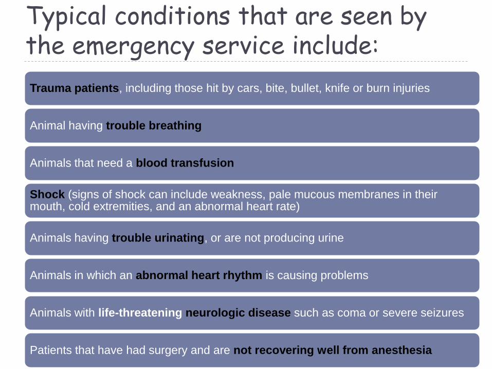
1.Airway and breathing
• if the animal has increased breathing rate or effort.
• animals may assume a posture with the head and neck extended and the elbows abducted (held away from the body)
• absent chest wall motion,
• exaggerated breathing effort,
• flaring of the nares,
• open mouth breathing and paradoxical breathing(opposing movements of the chest and abdominal walls during inspiration and expiration
• Cyanosis, a blue or purplish tint to the mucous membranes
• Crepitus about the body may indicate subcutaneous emphysema, which can be caused by tracheal tears or chest wall defects
First, patency of airway and
breathing effort should be
assessed. This is done by
visualization, auscultation, and
palpation.

2.Circulation:
• Mucous membrane colour
• Capillary refill time
• Heart rate
• Pulse quality
• Extremity temperature
• Mentation
The focus of the
cardiovascular assessment is
the six perfusion
parameters
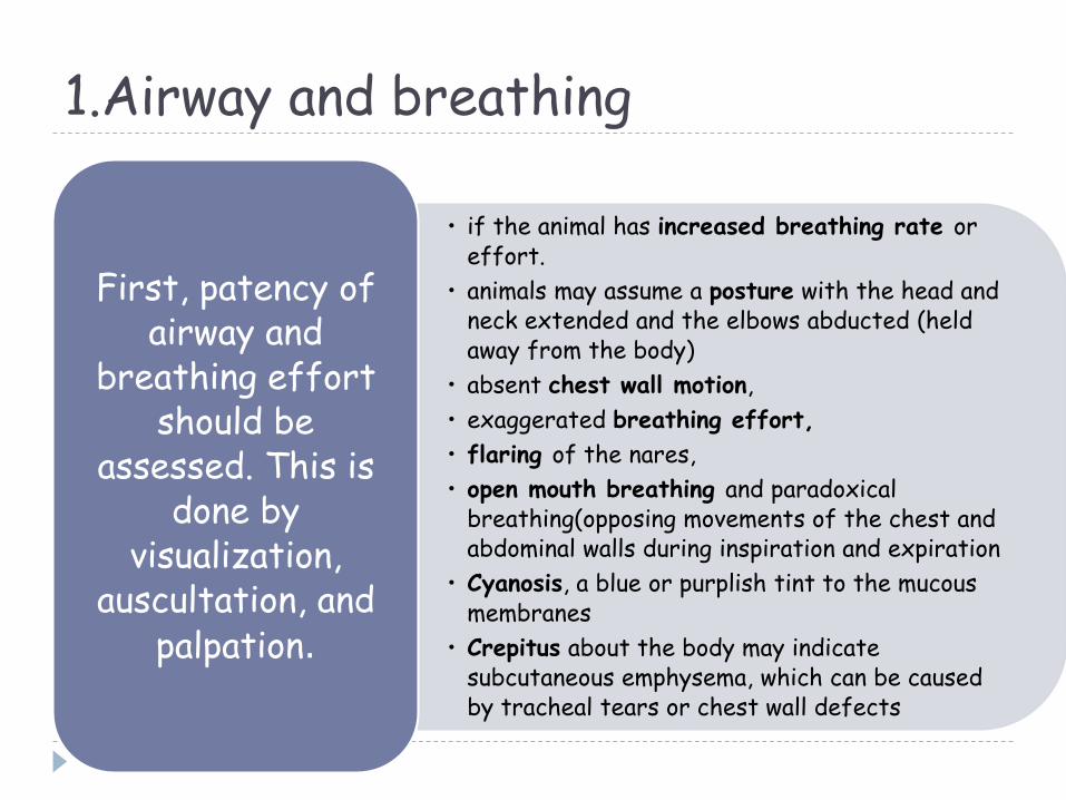
Mucous membrane colour
Evaluate the mucous membranes by examining the colour of the gums.
As an alternative in the fractious animal or patients with pigmented gums examine the conjunctiva, penis or the vulva.
• The normal colour pink is a result of oxygenated haemoglobin in red blood cells in the capillary bed.
• Mucous membrane colour may vary with circulatory related problems.
• Mucous membrane colour may be pale or white due to blood loss anaemia or vasoconstriction.
• Brick red or injected mucous membranes are a result of vasodilation and can be seen with hyperthermia or sepsis.
• Cyanotic or blue mucous membranes are an indicator of severe hypoxemia.
• Icteric or yellow mucous membranes are due to the breakdown of red cells (haemolysis) or liver disease.
• Methemoglobinemia results in brown or chocolate - coloured mucous membranes.
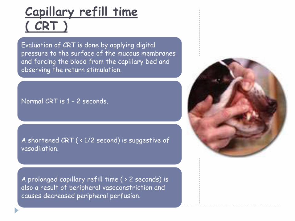
Capillary refill time ( CRT )
Evaluation of CRT is done by applying digital pressure to the surface of the mucous membranes and forcing the blood from the capillary bed and observing the return stimulation.
Normal CRT is 1 – 2 seconds.
A shortened CRT ( < 1/2 second) is suggestive of vasodilation.
A prolonged capillary refill time ( > 2 seconds) is also a result of peripheral vasoconstriction and causes decreased peripheral perfusion.

Heart rateIt is usually measured by auscultation of the heart, palpation of the apex beat, or palpation of an artery.
Tachycardia:-
• may be caused by hypovolemia , hypoxemia, hypotension, drugs, fever, excitement, exercise, and pain.
• It is generally defined as a heart rate > 160 beats per minutes (bpm) in the dog or 200 bpm in the cat.
Bradycardia:-
• may be caused by increased vagal tone, severe electrolyte disturbances and hypothermia, drugs, or disturbances of the cardiac conduction system.
• It is generally defined as a heart rate < 60 bpm in the dog and 140 bpm in the cat.
Auscultation of the heart and palpation of an artery should occur simultaneously, so that pulse deficits (the difference between heart and pulse rate; they should be the same) can be determined.
Pulse deficits are suggestive of arrhythmias.

Pulse quality
Palpation of the artery provides information about the animal’s heart rate and rhythm.
The femoral or dorsal pedal arteries are the commonly palpated arteries.
Pulse quality is an indicator of stroke volume, the amount of blood pumped out of the heart with each beat.
Ideally, the pulse should be full, regular, and strong, indicating a normal stroke volume.
A thready pulse is defined as a narrow waveform and a weak pulse refers to a small amplitude pulse difference, both of which are indicative of a decreased stroke volume.
Bounding pulses have a large pulse pressure difference and wide waveforms usually associated with increased stroke volume and vasodilation.

Extremity temperature
• The paws, limbs, or ears should normally feel warm to the touch. Cool extremities are a result of vasoconstriction.
Mentation
• As previously mentioned, evaluation of mentationstarts from a far. Observe the attitude of the patient.
• Are there any fractures?
• Is the abdomen painful or distended?
• Is there evidence of debilitation or other signs of disease?

Common emergency procedures:
Perform pericardiocentesis to relieve pericardial tamponade
Measure blood pressure indirectly using a Doppler probe and sphygmomanometer with cuff
Obtain Lead II ECG trace and assess it for life-threatening arrhythmias
Manage a cardiopulmonary arrest and resuscitation
Use ultrasouund to access a possible pericardial effusion
Cardiovascular:

Respiratory:
Place a nasal catheter for intranasal oxygen administration
Place an indwelling chest tube
Perform thoracocentesis
Interpret the spO2 from blood gas measurements
Interpret pH, HCO3 and pCO2 on blood gases
Perform the anaesthesia to repair a diaphragmatic hernia
Perform the surgical repair of a diaphragmatic hernia

Some of the Oxygen Delivery Methods used:

Nasal catheters
• Place 3-5 drops of Proparacaine into nostril
• Insert suture at caudoventral aspect of alar groove max. 1 cm from naris
• Use 3.5 to 8 Fr tube
Nasotracheal tubes
• Same as for nasal catheter but head is hyperextended when catheter reaches level of pharynx
• Insert tube rapidly as patient takes breath
• Radiographs may be required to confirm location
• Deliver O2 at 50 ml/kg/min (always humidify)
Transtracheal catheters
• Use 14-16 G IV Catheter
• Insert catheter between rings in the mid cervical region
• Aspirate to ensure location in trachea; Attach Oxygen at 50 ml/kg/min
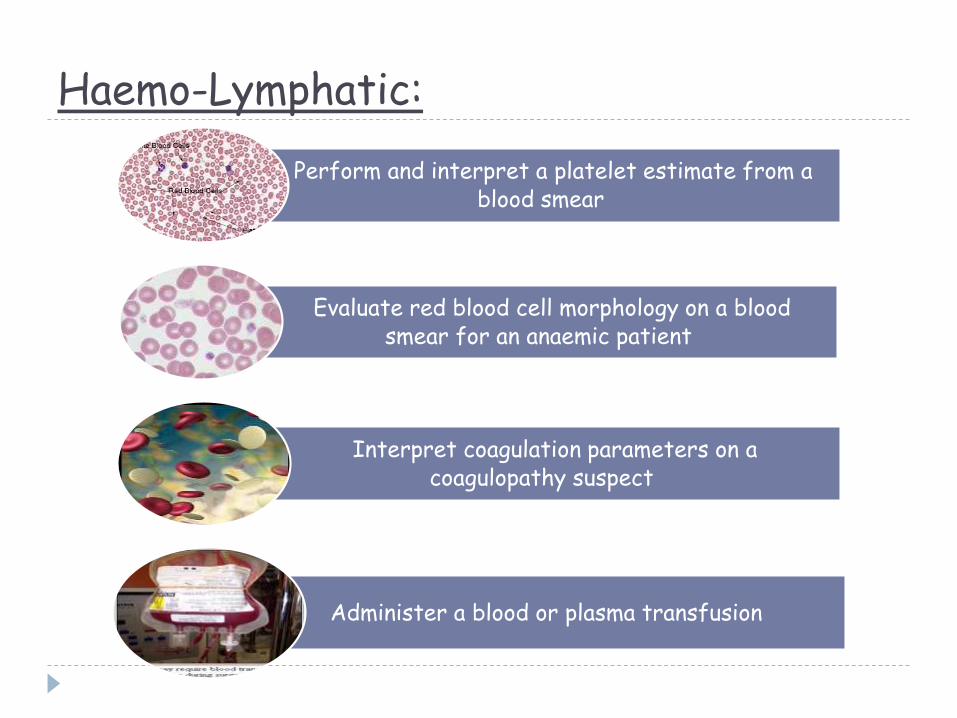
Haemo-Lymphatic:
Perform and interpret a platelet estimate from a blood smear
Evaluate red blood cell morphology on a blood smear for an anaemic patient
Interpret coagulation parameters on a coagulopathy suspect
Administer a blood or plasma transfusion

• Musculo-Skeletal •Neurologic:
Replace a dislocated hip under anaesthesia
Place an Ehmer sling or perform any other kind of external or internal coaptationtechnique as per the type of fracture or injury
Localise an acute intervertebral disk lesion and assess the prognosis

Related Documents