Vestibular Role of KCNQ4 and KCNQ5 K Channels Revealed by Mouse Models * □ S Received for publication, November 2, 2012, and in revised form, February 1, 2013 Published, JBC Papers in Press, February 13, 2013, DOI 10.1074/jbc.M112.433383 Guillermo Spitzmaul ‡1 , Leonardo Tolosa §2 , Beerend H. J. Winkelman §2 , Matthias Heidenreich ‡3 , Maarten A. Frens ¶ , Christian Chabbert , Chris I. de Zeeuw §¶4 , and Thomas J. Jentsch ‡ ** 5 From the ‡ Leibniz-Institut für Molekulare Pharmakologie (FMP) and Max-Delbrück-Centrum für Molekulare Medizin (MDC), 13125 Berlin, Germany, the § Netherlands Institute for Neuroscience, 1105 BA Amsterdam, The Netherlands, the ¶ Department of Neuroscience, Erasmus MC, 3000 CA Rotterdam, The Netherlands, INSERM U1051 Institut des Neurosciences de Montpellier, 30491 Montpellier cedex 5, France, and the **Cluster of Excellence NeuroCure, Charité-Universitätsmedizin, 10117 Berlin, Germany Background: KCNQ K channels regulate neuronal excitability, and KCNQ4 mutations cause deafness. Results: KCNQ4 and KCNQ5 expression in vestibular organ and the impact on vestibular function are investigated. Conclusion: Both channels reside in postsynaptic calyx membranes of hair cells, and loss of KCNQ4 impairs vestibular function. Significance: KCNQ4 may affect vestibular function because of a novel role in synaptic transmission. The function of sensory hair cells of the cochlea and vestibular organs depends on an influx of K through apical mechanosen- sitive ion channels and its subsequent removal over their baso- lateral membrane. The KCNQ4 (K v 7.4) K channel, which is mutated in DFNA2 human hearing loss, is expressed in the basal membrane of cochlear outer hair cells where it may mediate K efflux. Like the related K channel KCNQ5 (K v 7.5), KCNQ4 is also found at calyx terminals ensheathing type I vestibular hair cells where it may be localized pre- or postsynaptically. Making use of Kcnq4 / mice lacking KCNQ4, as well as Kcnq4 dn/dn and Kcnq5 dn/dn mice expressing dominant negative channel mutants, we now show unambiguously that in adult mice both channels reside in postsynaptic calyx-forming neurons, but can- not be detected in the innervated hair cells. Accordingly, whole cell currents of vestibular hair cells did not differ between genotypes. Neither Kcnq4 / , Kcnq5 dn/dn nor Kcnq4 / / Kcnq5 dn/dn double mutant mice displayed circling behavior found with severe vestibular impairment. However, a milder form of vestibular dysfunction was apparent from altered ves- tibulo-ocular reflexes in Kcnq4 / /Kcnq5 dn/dn and Kcnq4 / mice. The larger impact of KCNQ4 may result from its prefer- ential expression in central zones of maculae and cristae, which are innervated by phasic neurons that are more sensitive than the tonic neurons present predominantly in the surrounding peripheral zones where KCNQ5 is found. The impact of post- synaptic KCNQ4 on vestibular function may be related to K removal and modulation of synaptic transmission. The vestibular organ senses gravitational forces and acceler- ation and provides essential sensory input for the control of body equilibrium, head orientation, and eye movements. Sen- sory hair cells (HCs) 6 in otholithic organs (utricle and sacculus) respond to gravitational forces and linear acceleration, whereas HCs in the cristae ampullares detect rotational acceleration. Both types of sensory end organ contain different classes of HCs named type I and type II. These classes are not only distin- guished by their pattern of innervation, with calyx terminals ensheathing type I cells, but also by other characteristics like the expression of distinctive plasma membrane currents (1). Vestibular sensory output depends not only on mechanosen- sitive ion channels located in the stereocilia of HCs, but also on ion channels present in both the basolateral membrane of HCs and in their cognate afferents. These channels may modulate receptor potentials, synaptic transmission, and neuronal excit- ability. At least two members of the KCNQ (K v 7) K channel family (2, 3), KCNQ4 and KCNQ5, are found in the vestibular organ (4, 5). KCNQ4 is important for hearing (5, 6), is mutated in patients with dominant DFNA2 deafness (6), and modulates touch sensation (7). KCNQ4 displays a highly restricted expres- sion pattern. In the cochlea, it is localized at the basal pole of outer hair cells (OHCs) that depolarize and eventually degen- erate upon Kcnq4 disruption (5). KCNQ4 is also found at basal poles of vestibular type I hair cells that are ensheathed by calyx synapses (5). However, its localization to pre- or postsynaptic membranes remains controversial (4, 8 –12). Unlike cochlear OHCs, vestibular HCs do not degenerate in Kcnq4 / mice (5), * This work was supported by a grant from the Deutsche Forschungsgemein- schaft (Exc 257) and the Prix Louis-Jeantet de Médecine (to T. J. J.), by a stipend from the Alexander von Humboldt Foundation (to G. S.), by the Dutch Organization for Medical Sciences (ZonMw; to C. I. d. Z.), Life Sci- ences (ALW; to C. I. d. Z.), Senter (Neuro-Basic; to C. I. d. Z.), CEREBNET (to C. I. d. Z.), C7 programs of the European Community (to C. I. d. Z.), and ERC (European Research Council)-advanced grant (to C. I. d. Z.). □ S This article contains supplemental Figs. S1–S3. 1 Present address: Instituto de Investigaciones Bioquímicas de Bahía Blanca (INIBIBB) and Universidad Nacional del Sur (UNS), Bahía Blanca, Argentina. 2 Both authors contributed equally to this work. 3 Present address: Broad Institute, Massachusetts Institute of Technology, Cambridge, MA. 4 To whom correspondence may be addressed concerning vestibulo-ocular assays: Dept. of Neuroscience, Erasmus MC, P.O. Box 2040, 3000 CA Rotter- dam, The Netherlands. Tel.: 0031-10-7043-299; Fax: 0031-10-704-4734; E-mail: [email protected]. 5 To whom correspondence may be addressed: Leibniz-Institut für Moleku- lare Pharmakologie (FMP) and Max-Delbrück-Centrum für Molekulare Medizin (MDC), Robert-Rössle-Str. 10, 13125 Berlin, Germany. Tel.: 0049- 30-9406-2961 or 2975; Fax: 0049-30-9406-2960; E-mail: jentsch@fmp- berlin.de. 6 The abbreviations used are: HC, hair cell; dn, dominant negative; EPSP, excit- atory postsynaptic potential; OHC, outer hair cell; VOR, vestibulo-ocular reflex. THE JOURNAL OF BIOLOGICAL CHEMISTRY VOL. 288, NO. 13, pp. 9334 –9344, March 29, 2013 © 2013 by The American Society for Biochemistry and Molecular Biology, Inc. Published in the U.S.A. 9334 JOURNAL OF BIOLOGICAL CHEMISTRY VOLUME 288 • NUMBER 13 • MARCH 29, 2013 by guest on March 21, 2019 http://www.jbc.org/ Downloaded from

Welcome message from author
This document is posted to help you gain knowledge. Please leave a comment to let me know what you think about it! Share it to your friends and learn new things together.
Transcript

Vestibular Role of KCNQ4 and KCNQ5 K� Channels Revealedby Mouse Models*□S
Received for publication, November 2, 2012, and in revised form, February 1, 2013 Published, JBC Papers in Press, February 13, 2013, DOI 10.1074/jbc.M112.433383
Guillermo Spitzmaul‡1, Leonardo Tolosa§2, Beerend H. J. Winkelman§2, Matthias Heidenreich‡3, Maarten A. Frens¶,Christian Chabbert�, Chris I. de Zeeuw§¶4, and Thomas J. Jentsch‡**5
From the ‡Leibniz-Institut für Molekulare Pharmakologie (FMP) and Max-Delbrück-Centrum für Molekulare Medizin (MDC),13125 Berlin, Germany, the §Netherlands Institute for Neuroscience, 1105 BA Amsterdam, The Netherlands, the ¶Department ofNeuroscience, Erasmus MC, 3000 CA Rotterdam, The Netherlands, �INSERM U1051 Institut des Neurosciences de Montpellier,30491 Montpellier cedex 5, France, and the **Cluster of Excellence NeuroCure, Charité-Universitätsmedizin, 10117 Berlin, Germany
Background: KCNQ K� channels regulate neuronal excitability, and KCNQ4 mutations cause deafness.Results: KCNQ4 and KCNQ5 expression in vestibular organ and the impact on vestibular function are investigated.Conclusion:Both channels reside in postsynaptic calyxmembranes of hair cells, and loss of KCNQ4 impairs vestibular function.Significance: KCNQ4 may affect vestibular function because of a novel role in synaptic transmission.
The functionof sensoryhair cells of the cochlea and vestibularorgans depends on an influx of K� through apical mechanosen-sitive ion channels and its subsequent removal over their baso-lateral membrane. The KCNQ4 (Kv7.4) K� channel, which ismutated inDFNA2humanhearing loss, is expressed in the basalmembrane of cochlear outer hair cells where it maymediate K�
efflux. Like the related K� channel KCNQ5 (Kv7.5), KCNQ4 isalso found at calyx terminals ensheathing type I vestibular haircells where it may be localized pre- or postsynaptically. Makinguse of Kcnq4�/� mice lacking KCNQ4, as well as Kcnq4dn/dn
and Kcnq5dn/dn mice expressing dominant negative channelmutants, we now show unambiguously that in adult mice bothchannels reside in postsynaptic calyx-forming neurons, but can-not be detected in the innervated hair cells. Accordingly, wholecell currents of vestibular hair cells did not differ betweengenotypes. Neither Kcnq4�/�, Kcnq5dn/dn nor Kcnq4�/�/Kcnq5dn/dn double mutant mice displayed circling behaviorfound with severe vestibular impairment. However, a milderform of vestibular dysfunction was apparent from altered ves-tibulo-ocular reflexes in Kcnq4�/�/Kcnq5dn/dn and Kcnq4�/�
mice. The larger impact of KCNQ4 may result from its prefer-
ential expression in central zones of maculae and cristae, whichare innervated by phasic neurons that are more sensitive thanthe tonic neurons present predominantly in the surroundingperipheral zones where KCNQ5 is found. The impact of post-synaptic KCNQ4 on vestibular function may be related to K�
removal and modulation of synaptic transmission.
The vestibular organ senses gravitational forces and acceler-ation and provides essential sensory input for the control ofbody equilibrium, head orientation, and eye movements. Sen-sory hair cells (HCs)6 in otholithic organs (utricle and sacculus)respond to gravitational forces and linear acceleration, whereasHCs in the cristae ampullares detect rotational acceleration.Both types of sensory endorgan contain different classes ofHCsnamed type I and type II. These classes are not only distin-guished by their pattern of innervation, with calyx terminalsensheathing type I cells, but also by other characteristics like theexpression of distinctive plasma membrane currents (1).Vestibular sensory output depends not only onmechanosen-
sitive ion channels located in the stereocilia of HCs, but also onion channels present in both the basolateral membrane of HCsand in their cognate afferents. These channels may modulatereceptor potentials, synaptic transmission, and neuronal excit-ability. At least two members of the KCNQ (Kv7) K� channelfamily (2, 3), KCNQ4 and KCNQ5, are found in the vestibularorgan (4, 5). KCNQ4 is important for hearing (5, 6), is mutatedin patients with dominant DFNA2 deafness (6), and modulatestouch sensation (7). KCNQ4displays a highly restricted expres-sion pattern. In the cochlea, it is localized at the basal pole ofouter hair cells (OHCs) that depolarize and eventually degen-erate upon Kcnq4 disruption (5). KCNQ4 is also found at basalpoles of vestibular type I hair cells that are ensheathed by calyxsynapses (5). However, its localization to pre- or postsynapticmembranes remains controversial (4, 8–12). Unlike cochlearOHCs, vestibular HCs do not degenerate inKcnq4�/� mice (5),
* This work was supported by a grant from the Deutsche Forschungsgemein-schaft (Exc 257) and the Prix Louis-Jeantet de Médecine (to T. J. J.), by astipend from the Alexander von Humboldt Foundation (to G. S.), by theDutch Organization for Medical Sciences (ZonMw; to C. I. d. Z.), Life Sci-ences (ALW; to C. I. d. Z.), Senter (Neuro-Basic; to C. I. d. Z.), CEREBNET (toC. I. d. Z.), C7 programs of the European Community (to C. I. d. Z.), and ERC(European Research Council)-advanced grant (to C. I. d. Z.).
□S This article contains supplemental Figs. S1–S3.1 Present address: Instituto de Investigaciones Bioquímicas de Bahía Blanca
(INIBIBB) and Universidad Nacional del Sur (UNS), Bahía Blanca, Argentina.2 Both authors contributed equally to this work.3 Present address: Broad Institute, Massachusetts Institute of Technology,
Cambridge, MA.4 To whom correspondence may be addressed concerning vestibulo-ocular
assays: Dept. of Neuroscience, Erasmus MC, P.O. Box 2040, 3000 CA Rotter-dam, The Netherlands. Tel.: 0031-10-7043-299; Fax: 0031-10-704-4734;E-mail: [email protected].
5 To whom correspondence may be addressed: Leibniz-Institut für Moleku-lare Pharmakologie (FMP) and Max-Delbrück-Centrum für MolekulareMedizin (MDC), Robert-Rössle-Str. 10, 13125 Berlin, Germany. Tel.: 0049-30-9406-2961 or 2975; Fax: 0049-30-9406-2960; E-mail: [email protected].
6 The abbreviations used are: HC, hair cell; dn, dominant negative; EPSP, excit-atory postsynaptic potential; OHC, outer hair cell; VOR, vestibulo-ocularreflex.
THE JOURNAL OF BIOLOGICAL CHEMISTRY VOL. 288, NO. 13, pp. 9334 –9344, March 29, 2013© 2013 by The American Society for Biochemistry and Molecular Biology, Inc. Published in the U.S.A.
9334 JOURNAL OF BIOLOGICAL CHEMISTRY VOLUME 288 • NUMBER 13 • MARCH 29, 2013
by guest on March 21, 2019
http://ww
w.jbc.org/
Dow
nloaded from

but �30% of patients with KCNQ4mutations display mild ves-tibular symptoms (13).KCNQ5 is more broadly expressed than KCNQ4 (14, 15),
yields similar currents, but lacks cochlear expression. In vestib-ular sensory epithelia (5) it partially overlaps with KCNQ4.Apart from the disappearance of an afterhyperpolarization cur-rent in hippocampal neurons (16), no phenotype has beendescribed in Kcnq5dn/dn mice carrying a dominant negativemutation.Using KCNQmutantmousemodels we now resolve the con-
troversy surrounding the contribution of KCNQ4 and KCNQ5to vestibular HC currents and determine their physiologicalrelevance for the vestibular system. We found that neitherchannel is significantly expressed in adult vestibular HCs, butrather postsynaptically in calyx terminals where their expres-sion partially overlaps. Accordingly, currents of vestibular HCswere unchanged upon gene disruption. Vestibulo-ocularreflexes were reduced both in Kcnq4�/� and Kcnq4�/�/Kcnq5dn/dn double mutant mice, whereas only marginal effectswere observed in Kcnq5dn/dn mice. The larger impact of Kcnq4disruption on vestibular function may be related to its pref-erential expression in the central zones of vestibular sensoryepithelia.
EXPERIMENTAL PROCEDURES
Mouse Models and Genotyping—The generation ofKcnq4�/�, Kcnq4dn/dn, and Kcnq5dn/dn mice and their geno-typing has been described previously (5, 16). KCNQ4 mousemodels were initially kept in C3H/HeJ and Kcnq5dn/dn mice inC57BL/6 background. Double mutant mice were generated bycrossingKcnq4�/� andKcnq5dn/dn mice. They were viable, fer-tile, and had no immediately apparent phenotype. They werekept and investigated in the mixed background. Subse-quently, all mutant genotypes were obtained by breedingmice heterozygous for both genes. Mice of either sex wereused for experiments.Immunofluorescence and in Situ Hybridization—Mice (2–52
weeks old)were anesthetized and perfusedwith 4%paraformal-dehyde in PBS. Inner ears were dissected from the temporalbones in PBS and processed for (i) whole mount, (ii) sliceimmunohistochemistry, or (iii) in situ hybridization. For (i) wecarefully excised the utricle and semicircular canals, opened themembranous labyrinth, and removed the otoconial membranefrom utricles. Tissues were postfixed in 4% paraformaldehyde,washed with PBS, and incubated in blocking solution (17) (3%normal goat serum, 2% BSA, and 0.5% Nonidet P-40 in PBS)overnight. Primary antibodieswere incubated for 48 h in carriersolution (PBS containing 1.5% normal goat serum, 1% BSA, and0.25% Nonidet P-40). Secondary antibodies diluted in carriersolution were incubated overnight. Tissues were mountedunflattened in Fluoromount-G (Southern Biotech). For (ii) and(iii), inner ears were postfixed and decalcified as described (18).Tissues were cut in 8–12-�m sections and kept at �20 °C (forii) or �80 °C (for iii). For (ii), slices were blocked with BSA for2 h and incubated with primary antibodies in carrier solution(12 h). Secondary antibodies were incubated for 2 h. For wholemount (i) and immunohistochemistry (ii) the following primaryantibodies were used: rabbit anti-KCNQ4 (4), 1:150 for (i) and
1:200 for (ii); guinea pig anti-KCNQ4 (7), 1:100 for (i) and 1:200for (ii); rabbit anti-KCNQ5 (16), 1:150 for (i); rabbit and guineapig anti-KCNQ5-C1b (raised against residues 793–808), 1:100for (i) and 1:200 for (ii); mouse anti-calretinin (Swant), 1:200 for(i) and 1:400 for (ii); mouse and rabbit anti-�-III-tubulin (Cova-nce), 1:500 for (i) and (ii); and mouse anti-calbindin (Swant),1:400 for (ii). Nuclei were stained with DAPI. Fluorescence-labeled secondary antibodies were obtained from MolecularProbes and used diluted 1:500 for (i) and 1:1000 for (ii). For insitu hybridization (iii), sense and antisense digoxigenin-UTP-labeled riboprobes (DIG RNA labelingMix; Roche Applied Sci-ence) were generated with T7 or SP6 RNA polymerase (RocheApplied Science), respectively, from linearized mouse cDNAclones (bp 1114–1560 forKcnq4 and 1744–2352 forKcnq5). Insitu hybridization on inner ear cryosections was performed asdescribed (19). Pictures were taken for (ii) by a confocal laser-scanning microscope (LSM510; Zeiss), analyzed off-line withZEN 2009 light edition software (Zeiss), and assembled usingAdobePhotoshop (Adobe Systems). Pictures for (iii) were takenwith a Zeiss Axiophot or Zeiss Stemi-2000-c microscope.Electrophysiology—Whole cell recordings were done on HCs
from acutely dissected utricles that were perfused at 0.5ml/minwith oxygenated solution containing 137mMNaCl, 5mMKCl, 2mMCaCl2, 1 mMMgCl2, 10mMHEPES, 11mM glucose, pH 7.4.The epithelium was cut transversally and anchored to thechamber floor with two pieces of glass, exposing the basolateralmembrane of hair cells. Central and peripheral hair cells couldnot be easily distinguished in this semi-intact preparation.However, patches weremostly obtained in the central and adja-cent areas of the exposed tissue which may span the wholestriola and juxtastriola and a minor portion of extrastriola.Recordings were done with 3–4-megohm electrodes filled with140 mM KCl, 5 mM NaCl, 10 mM HEPES, 10 mM glucose, 5 mM
EGTA, 3 mMMgATP, 1 mMNaGTP, pH 7.3. Type I and type IIHCs were characterized (20) by the presence or absence,respectively, of the gK,L conductance (1). Microscopic inspec-tion further helped to identify HC classes.Whole cell recordings were done using a Multiclamp 700B
amplifier (Molecular Devices). After seal formation (�10 G�)onto the basolateral membrane of HCs and membrane disrup-tion, we estimated membrane capacitance (Cm) and seriesresistance (Rs) from the decay of capacitive transients inducedby a �10-mV pulse from a holding potential of �80 mV. Aftercancellation of capacitive transients, Rs was compensated up to85%. The resting potential was measured as the zero currentvoltage in the current clamp mode. Data analysis used pClamp10 software (Molecular Devices) and Origin 7.5 (OriginLab).Data were sampled at 10 kHz and filtered at 2 kHz. The voltageclamp protocol started from a holding potential of �70 (fortype II HCs) or �90 mV (for type I HCs), followed by a 50-mspulse to �110 mV for type I HCs and �100 mV for type II HCsfollowed by test pulses of 200 ms between �110 and �40 mVfor type I HC and �100 to �50 mV in type II HC in steps of 10mV, and a constant step to �30 mV for tail currents. In statis-tical analysis, the number of cells is given by n, and number ofanimals by N.
KCNQ K� Channels in Vestibular Organ
MARCH 29, 2013 • VOLUME 288 • NUMBER 13 JOURNAL OF BIOLOGICAL CHEMISTRY 9335
by guest on March 21, 2019
http://ww
w.jbc.org/
Dow
nloaded from

Expression in Xenopus Oocytes and Two-electrode VoltageClamping—Xenopus oocytes were obtained and injected asdescribed (15). A final amount of 20 ng of cRNA/oocyte wasinjected for each condition. Oocytes were incubated 3–4 daysat 17 °C and then examined by two-electrode voltage clampingusing a Turbo Tec03 (NPI Electronics) amplifier and pClamp8.0 software (Molecular Devices). The protocol consisted of 5-ssteps from�110 to�50mV in 10-mV increments from a hold-ing potential of �80 mV.Vestibulo-ocular Reflexes—The vestibulo-ocular reflex
(VOR) was tested in four groups of adult mice including wild-type (WT,n� 15),Kcnq4�/� (n� 13),Kcnq5dn/dn (n� 17), andKcnq4�/�/Kcnq5dn/dn (n � 13). In each experimental groupabout half of the mice were male. Mice were prepared forchronic head-restrained experiments, as described (21). Theexperimental protocol was approved by the Animal Experi-mentation Committee (DEC) of the Royal Dutch Academy ofSciences (KNAW).During the experiment themouse was placed head-fixed in a
holder tube on a vestibular motion platform (R2000 Rotopod;Parallel Robotic SystemsCorporation). Left eye orientationwasmeasured using video pupil tracking with a table fixed CCDcamera (Pulnix TM-6710CL, 120 frames/s) and IR illumination(850-nm LED, 6.5-cm distance from the eye). Pilocarpine (2%)eye drops were applied before the experiment to limit pupildilatation in darkness. Online image analysis was performed toextract the location of pupil edges and corneal light reflectionsusing custombuilt software for Labview (National Instruments,Austin, TX). Angular eye velocity was computed offline usingcustom software written for Matlab (The Mathworks Inc.,Natick, MA) using the algorithm outlined elsewhere (22, 23).Saccadic eye movements and nystagmus fast phases wereremoved using a 50°/s velocity threshold and 200-msmargins ateach threshold crossing. Each mouse was accustomed to thesetup and experimental paradigm in a period of up to 3 trainingdays before the experimental data were collected.ThehorizontalVORwas characterized in darkness and in the
light using sinusoidal rotation about the yaw axis, using fre-quencies ranging between 1/16 and 4 Hz, presented in asequence of increasing order. Peak velocitywas held constant at25°/s. The number of cycles ranged between 5 (at 1/16 Hz) and60 at 4 Hz. Eye velocity amplitude and phase shift relative tohead movement were calculated using multiple linear regres-sion of eye velocity to in-phase and quadrature components ofthe stimulus velocity. Gain of the eye movement response wasdefined as the ratio between the eye velocity amplitude and thestimulus velocity amplitude. Phase shift is expressed in degrees;positive phase shifts indicate phase lead. Error bars representthe S.E. Circular statistics were used to compare phase values.Transfer function profiles were statistically compared usingMANOVA for repeated measures (SPSS Statistics 17.0.).
RESULTS
Differential Distribution of KCNQ4 and KCNQ5 in Vestibu-lar EndOrgans—Previous studies examining the distribution ofKCNQ4 and KCNQ5 in vestibular end organs were performedonly with WT animals (4, 8, 11). We now used three geneticmousemodels to control immunohistochemical labeling and to
explore potential cross-talk between the expression of bothproteins. Kcnq4�/� mice lack the KCNQ4 protein, whereasKcnq4dn/dn mice express a dominant negative KCNQ4 variantcarrying a point mutation (G286S) in the pore-forming P-loop(5). It corresponds to amutation of aDFNA2 patient with dom-inantly inherited hearing loss (6). Kcnq5dn/dn mice carry anequivalent mutation (G278S) in KCNQ5 (16). KCNQ5(G278S)does not yield currents. It exerts strong dominant negativeeffects on co-expressed KCNQ3 and KCNQ5 WT subunitswith which it can form heterotetramers (6, 15, 16).Labeling vestibular end organswith our antibodies revealed a
differential distribution of KCNQ4 and KCNQ5 in both theotholithic organs (Fig. 1) and cristae (supplemental Fig. S1). Asdisplayed for utricles in Fig. 1A, KCNQ4 was most highlyexpressed in the central striolar and the adjacent juxtastriolarregions. It extended at weaker expression levels to the adjacentextrastriola (Fig. 1, A and J). By contrast, KCNQ5 was foundmainly in the extrastriola and extended at lower expressionlevels into the juxtastriola (Fig. 1, B and K). As described previ-ously (4, 5), both KCNQ4 and KCNQ5were found in calyx-likestructures that ensheathe type I vestibular hair cells (Fig. 1, JandK). Calyces were co-labeled for KCNQ4 and KCNQ5 in thejuxtastriolar and extrastriolar regions (Fig. 1L). Likewise,KCNQ4 was found in central, intermediate, and peripheralzones of the cristae ampullares, whereas KCNQ5was expressedin intermediate and peripheral zones but was almost undetect-able in central zones (supplemental Fig. S1, A–C, J–L).
Importantly, labeling for KCNQ4 was abolished in tissuefrom Kcnq4�/� mice (Fig. 1D and supplemental Fig. S1D), val-idating the specificity of our staining. Surprisingly, labeling forKCNQ5 was virtually abolished in utricles from Kcnq5dn/dnmice that express a full-lengthKCNQ5protein carrying a singlepoint mutation (Fig. 1H). A similar pattern was observed incristae ampullares (supplemental Fig. S1, H and N). Besidesvalidating ourKCNQ5 antibody staining, this finding suggestedthat the mutant protein was unstable or incorrectly targeted.The partially overlapping expression patterns of KCNQ4 and
KCNQ5 (Fig. 1, C and L, and supplemental Fig. S1, C and L)suggested that they may form heteromeric channels in vivo, ashas been reported for co-transfected Chinese hamster ovarycells (24). Using dominant negative mutants, including the oneexpressed in Kcnq5dn/dn mice, we confirmed these findings intheXenopus oocyte system that allows amore quantitative con-trol of co-expression levels (supplemental Fig. S2, A and B).Importantly, KCNQ4/5 heterotetramers did not yield highercurrents than the respective homotetramers, contrasting withfindings for KCNQ3-KCNQ5 (15, 16) and KCNQ2-KCNQ3channels (25, 26). Hence, the disruption of Kcnq4 should notdecrease K� currents more than expected from a loss ofKCNQ4 currents alone. On the other hand, instead of a com-pensatory up-regulation, we observed a mild, but consistentdecrease of KCNQ5 labeling in Kcnq4�/� vestibular organs(Fig. 1E). This finding hints at a role of KCNQ4-KCNQ5 com-plexes in trafficking, anchoring, or stabilizing KCNQ5 at itstarget membrane. The loss of KCNQ5 current in Kcnq4�/�
mice, however, is expected to be mild. This contrasts with thestrong dominant negative effect of theKCNQ5dnmutantwhichwill suppress currents from all KCNQ4 subunits present in
KCNQ K� Channels in Vestibular Organ
9336 JOURNAL OF BIOLOGICAL CHEMISTRY VOLUME 288 • NUMBER 13 • MARCH 29, 2013
by guest on March 21, 2019
http://ww
w.jbc.org/
Dow
nloaded from
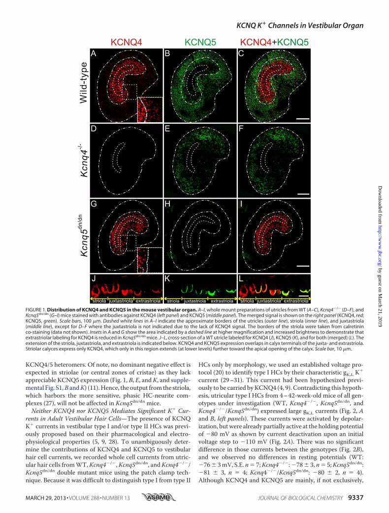
KCNQ4/5 heteromers. Of note, no dominant negative effect isexpected in striolae (or central zones of cristae) as they lackappreciable KCNQ5 expression (Fig. 1, B, E, andK, and supple-mental Fig. S1,B andK) (11).Hence, the output from the striola,which harbors the more sensitive, phasic HC-neurite com-plexes (27), will not be affected in Kcnq5dn/dn mice.Neither KCNQ4 nor KCNQ5 Mediates Significant K� Cur-
rents in Adult Vestibular Hair Cells—The presence of KCNQK� currents in vestibular type I and/or type II HCs was previ-ously proposed based on their pharmacological and electro-physiological properties (5, 9, 28). To unambiguously deter-mine the contributions of KCNQ4 and KCNQ5 to vestibularhair cell currents, we recorded whole cell currents from utric-ular hair cells fromWT,Kcnq4�/�,Kcnq5dn/dn, andKcnq4�/�/Kcnq5dn/dn double mutant mice using the patch clamp tech-nique. Because it was difficult to distinguish type I from type II
HCs only by morphology, we used an established voltage pro-tocol (20) to identify type I HCs by their characteristic gK,L K�
current (29–31). This current had been hypothesized previ-ously to be carried byKCNQ4 (4, 9). Contradicting this hypoth-esis, utricular type I HCs from 4–42-week-old mice of all gen-otypes under investigation (WT, Kcnq4�/�, Kcnq5dn/dn, andKcnq4�/�/Kcnq5dn/dn) expressed large gK,L currents (Fig. 2, Aand B, left panels). These currents were activated by depolar-ization, butwere already partially active at the holding potentialof �80 mV as shown by current deactivation upon an initialvoltage step to �110 mV (Fig. 2A). There was no significantdifference in those currents between the genotypes (Fig. 2B),and we observed no differences in resting potentials (WT:�76� 3mV, S.E. n� 7;Kcnq4�/�:�78� 3, n� 5;Kcnq5dn/dn:�81 � 3, n � 4; Kcnq4�/�/Kcnq5dn/dn: �80 � 2, n � 4).Although KCNQ4 and KCNQ5 are mainly, if not exclusively,
FIGURE 1. Distribution of KCNQ4 and KCNQ5 in the mouse vestibular organ. A–I, whole mount preparations of utricles from WT (A–C), Kcnq4�/� (D–F), andKcnq5dn/dn (G–I) mice stained with antibodies against KCNQ4 (left panel) and KCNQ5 (middle panel). The merged signal is shown on the right panel (KCNQ4, red;KCNQ5, green). Scale bars, 100 �m. Dashed white lines in A–I indicate the approximate borders of the utricles (outer line), striola (inner line), and juxtastriola(middle line), except for D–F where the juxtastriola is not indicated due to the lack of KCNQ4 signal. The borders of the striola were taken from calretininco-staining (data not shown). Insets in A and G show the area indicated by a dashed line at higher magnification and increased brightness to demonstrate thatextrastriolar labeling for KCNQ4 is reduced in Kcnq5dn/dn mice. J–L, cross-section of a WT utricle labeled for KCNQ4 (J), KCNQ5 (K), and for both (merged) (L). Theextension of the striola, juxtastriola, and extrastriola is indicated below. KCNQ4 and KCNQ5 expression overlaps in calyx terminals of the juxta- and extrastriola.Striolar calyces express only KCNQ4, which only in this region extends (at lower levels) further toward the apical opening of the calyx. Scale bar, 10 �m.
KCNQ K� Channels in Vestibular Organ
MARCH 29, 2013 • VOLUME 288 • NUMBER 13 JOURNAL OF BIOLOGICAL CHEMISTRY 9337
by guest on March 21, 2019
http://ww
w.jbc.org/
Dow
nloaded from

expressed at calyx terminals of type I cells (4, 5), we also inves-tigated type II cells in the same age range. They displayed thetypical GDRII current (1) and had a less negative resting voltage(�58 � 3 mV, n � 26). As expected, we detected significantdifferences between genotypes neither in type II HC currents,nor in resting potentials (Kcnq4�/�: �59 � 2 mV, n � 13;Kcnq5dn/dn:�57� 7mV, n� 4;Kcnq4�/�/Kcnq5dn/dn:�59�3 mV, n � 5). The half-maximal voltages of current activation(V1/2) as determined from tail currents did not differ betweenthe genotypes (supplemental Fig. S3, A–C). Moreover, 10 �M
XE991, a potent blocker of KCNQ2, -3, -4, and -5 currents (15,
26, 32), failed to block K� currents of utricular type I or type IIHCs fromWT or Kcnq4�/�/Kcnq5dn/dn mice (Fig. 2C). Hence,neither KCNQ4 nor KCNQ5 mediates significant currents investibular hair cells of mice older than 3 weeks.KCNQ4 and KCNQ5 Are Not Expressed in Adult Vestibular
Hair Cells, but in Afferent Neurons—The lack of KCNQ-medi-ated currents in �3-week-old type I HCs suggested thatKCNQ4 and -5 are not expressed in those sensory cells, butrather reside in postsynaptic calyx membranes that ensheathetype I HCs. Confocal light microscopy cannot resolve the post-synaptic inner calyx membrane from the closely apposed basal,presynaptic HC membrane. A postsynaptic localization ofKCNQ4, however, is strongly suggested by the extension ofweaker KCNQ4 immunoreactivity along the outer calyx mem-brane into neurites (4, 11), a finding we now confirmed byKcnq4�/�-controlled immunostaining of striolar calyces (Fig.3A). By contrast, KCNQ5 labeling (controlled by Kcnq5dn/dn
HCs) was detected neither in outer calyceal membranes, nor inneurites (Fig. 3B). We correlated these findings with immuno-detection of either channel protein in neuronal cell bodies ofthe vestibular ganglion (Fig. 4). Whereas somata of a neuronalsubpopulation showed plasma membrane and intracellularlabeling for KCNQ4 (Fig. 4A), KCNQ5 was not, or only barely,detectable (Fig. 4D). Hence, KCNQ5 may be more stringentlytargeted to the inner calyx membrane than KCNQ4. InKcnq5dn/dn mice, however, KCNQ5 was detected in a punctatepattern in neuronal cell bodies (Fig. 4F), while disappearing fromvestibular endorgans (Fig. 1H and supplemental Fig. S1,H andN).Such a trafficking defect might also be expected for KCNQ4-KCNQ5 heteromers containing KCNQ5(G78S) subunits.Indeed, juxtastriolar and extrastriolar KCNQ4 labeling wasmildly reduced in maculae and cristae of Kcnq5dn/dn mice (Fig.1, A and G, and supplementary Fig. S1M). This reduction wasmore evident in the extrastriola where the KCNQ5/KCNQ4ratio is larger (insets in Fig. 1, A and G).
FIGURE 2. Potassium currents from vestibular hair cell obtained from4 – 42-week-old mice are not affected in mouse models for KCNQ4 andKCNQ5 channels. A, potassium currents from type I (left) and type II HCs(right) obtained from semi-intact preparations of mouse utricle from wild-type and double-mutant mice Kcnq4�/�/Kcnq5dn/dn. Insets show voltage pro-tocol used to obtain currents in type I and type II HCs, respectively. Cells wereclamped before the voltage step to a �110 mV (type I HC) or �100 mV (typeII HC) for 50 ms and after it to �30 mV. B, current-voltage relationship ofpotassium currents from type I (left) and type II (right) HCs for wild-type (blackdots), Kcnq4�/� (blue triangle), Kcnq5dn/dn (green triangle), Kcnq4�/�/Kcnq5dn/dn (red star) mice. Currents were normalized to cell capacitance.Number of measured cells from WT, Kcnq4�/�, Kcnq5dn/dn, and Kcnq4�/�/Kcnq5dn/dn mice were 7, 5, 4, and 4 for type I HCs, and 26, 13, 4, and 5 for typeII HCs, respectively. C, utricular type I (upper panels) and type II (lower panels)HC currents in the absence (black traces) and presence (red traces) of 10 �M
XE991 perfused for 5 min after control trace. Currents were activated at �20mV and obtained from wild-type (left panel) and Kcnq4�/�/Kcnq5dn/dn (middlepanel) mice. Right diagrams, averaged currents (at � 20 mV, and normalizedto capacitance) for both genotypes. Numbers of animals were: type I HCs: n �5 and 4 for wild-type and Kcnq4�/�/Kcnq5dn/dn mice, respectively; and type IIHCs: n � 6 for either wild-type or Kcnq4�/�/Kcnq5dn/dn mice.
FIGURE 3. Expression of KCNQ4 and KCNQ5 in type I hair cell calyces.A, immunohistochemistry of striolar type I HC for KCNQ4 (green), counter-stained for the nuclear marker DAPI (blue), and in right panels for �-III-tubulin(red), a cytosolic marker for calyces. Top, results from wild-type mice; bottompanels, from Kcnq4�/� mice. Note that weak KCNQ4 staining extends beyondthe basal pole of the HC toward the neck of the calyx, where inner and outermembranes are labeled (arrows), and to the neurite. This is better visible ininsets which show the areas enclosed by dotted lines with increased bright-ness. Kcnq4�/� sections are not labeled by the KCNQ4 antibody, demonstrat-ing the specificity of staining. B, similar labeling for KCNQ5 of extrastriolarHCs. Note that KCNQ5 immunoreactivity does not extend beyond the basalpart of the calyx and that labeling is abolished in Kcnq5dn/dn section. Scalebars, 5 �m.
KCNQ K� Channels in Vestibular Organ
9338 JOURNAL OF BIOLOGICAL CHEMISTRY VOLUME 288 • NUMBER 13 • MARCH 29, 2013
by guest on March 21, 2019
http://ww
w.jbc.org/
Dow
nloaded from

Similar mutations in KCNQ4 are known to interfere withplasma membrane expression (7, 33, 34). Indeed, neuronalsomata in the vestibular ganglion ofKcnq4dn/dn mice (5), whichexpress the trafficking-deficient G286S mutant (7), showedintense cytoplasmic KCNQ4 labeling (Fig. 4B) compared withWT somata (Fig. 4A). KCNQ4 labeling of vestibular end organsdisappeared in parallel, as shown for Kcnq4dn/dn crista ampul-laris in Fig. 5,A andB. It is instructive to compare these findingswith those for the cochlea where KCNQ4 is expressed in outerhair cells (4–6).Unlike vestibularHCs,OHCsdisplayed detect-able levels of the mutant KCNQ4 protein (Fig. 5D). Again con-sistent with impaired trafficking and ER retention, the mutantprotein was no longer detected at the basal OHC plasmamem-brane (Fig. 5C), but in a punctate intracellular pattern (Fig. 5D).Our immunohistochemical analysis of mutant mice strongly
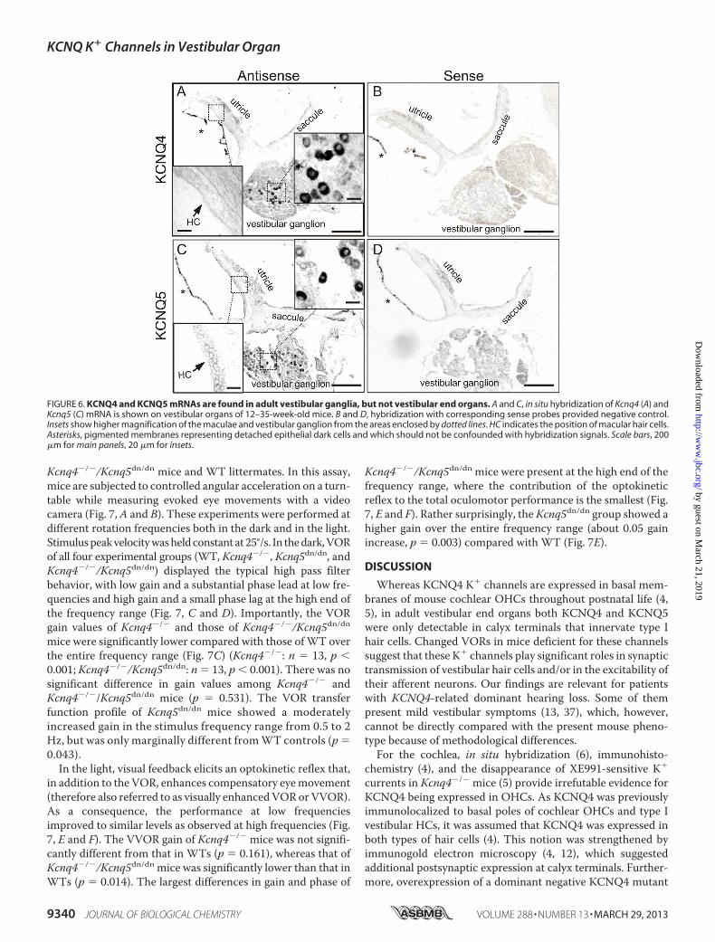
suggests thatKCNQ4and-5areexpressed inpostsynaptic, butnotpresynaptic membranes of calyx terminals. In situ hybridizationfurther strengthened this conclusion. BothKcnq4 (Fig. 6,A andB)and Kcnq5 (Fig. 6, C and D) mRNAs were detected in ganglionarcell bodies, but not in vestibular sensory epithelia as would havebeen expected with an expression in hair cells. Hence, all of ourexperiments point to apostsynaptic rather thanvestibular hair cellexpression of KCNQ4 inmice older than 3 weeks.Consequences of the Loss of KCNQ4 and KCNQ5 for Vestibu-
lar Function—Kcnq4�/� mice,Kcnq5dn/dnmice, and evenmicehomozygous for both mutations (Kcnq4�/�/Kcnq5dn/dn)lacked shaker/waltzer behavior that is indicative of a strongvestibular deficit phenotype as found, e.g. in mice lacking theNaK2Cl co-transporter Nkcc1 (35) or the K� �-subunit geneKcne1 (36). Bothgeneproducts are involved ingenerating thehighendocochlear K� concentration that is required for mechano-transduction currents in cochlear and vestibular hair cells.
To find out whethermore subtle vestibular deficits can resultfrom a loss of KCNQ4 and/or KCNQ5 function we measuredthe VOR in awake, head-restrained Kcnq4�/�, Kcnq5dn/dn, and
FIGURE 4. Expression of KCNQ4 and KCNQ5 in vestibular ganglia is increased by trafficking-impaired dominant negative mutations. Shown is vestib-ular ganglia staining for KCNQ4 (A–C; red), or for KCNQ5 (D–F; red), from wild-type (A and D), Kcnq4dn/dn (B and E), and Kcnq5dn/dn (C and F). Nuclei were stainedwith DAPI (blue). Note increased cytoplasmic staining for KCNQ4 and KCNQ5 in Kcnq4dn/dn and Kcnq5dn/dn mice, respectively, without significant effects of theKCNQ4 mutation on KCNQ5 expression and vice versa. Scale bars, 20 �m
FIGURE 5. Altered subcellular localization of KCNQ4 in the inner ear ofKcnq4dn/dn mice. A and B, KCNQ4 (red) labeling of the crista ampullaris ofwild-type mice (A) is abolished in Kcnq4dn/dn mice (B). C and D, KCNQ4 (red)labeling of the basolateral plasma membrane of prestin-positive (green)OHCs of wild-type mice (C) is changed to a weak punctate cytoplasmic label-ing in Kcnq4dn/dn mice (D). Inset, area enclosed by the dotted line is at highermagnification and increased laser intensity. Note the morphological abnor-malities of prestin-positive OHCs of Kcnq4dn/dn mice due to their incipientdegeneration at this age (3 months). Nuclei were labeled with DAPI. Scalebars, 20 �m.
KCNQ K� Channels in Vestibular Organ
MARCH 29, 2013 • VOLUME 288 • NUMBER 13 JOURNAL OF BIOLOGICAL CHEMISTRY 9339
by guest on March 21, 2019
http://ww
w.jbc.org/
Dow
nloaded from
Kcnq4�/�/Kcnq5dn/dn mice and WT littermates. In this assay,mice are subjected to controlled angular acceleration on a turn-table while measuring evoked eye movements with a videocamera (Fig. 7,A and B). These experiments were performed atdifferent rotation frequencies both in the dark and in the light.Stimuluspeakvelocitywasheldconstant at25°/s. In thedark,VORof all four experimental groups (WT, Kcnq4�/�, Kcnq5dn/dn, andKcnq4�/�/Kcnq5dn/dn) displayed the typical high pass filterbehavior, with low gain and a substantial phase lead at low fre-quencies and high gain and a small phase lag at the high end ofthe frequency range (Fig. 7, C and D). Importantly, the VORgain values of Kcnq4�/� and those of Kcnq4�/�/Kcnq5dn/dnmice were significantly lower compared with those ofWT overthe entire frequency range (Fig. 7C) (Kcnq4�/�: n � 13, p �0.001; Kcnq4�/�/Kcnq5dn/dn: n � 13, p � 0.001). There was nosignificant difference in gain values among Kcnq4�/� andKcnq4�/�/Kcnq5dn/dn mice (p � 0.531). The VOR transferfunction profile of Kcnq5dn/dn mice showed a moderatelyincreased gain in the stimulus frequency range from 0.5 to 2Hz, but was only marginally different fromWT controls (p �0.043).In the light, visual feedback elicits an optokinetic reflex that,
in addition to the VOR, enhances compensatory eyemovement(therefore also referred to as visually enhancedVORorVVOR).As a consequence, the performance at low frequenciesimproved to similar levels as observed at high frequencies (Fig.7, E and F). The VVOR gain of Kcnq4�/� mice was not signifi-cantly different from that in WTs (p � 0.161), whereas that ofKcnq4�/�/Kcnq5dn/dn mice was significantly lower than that inWTs (p � 0.014). The largest differences in gain and phase of
Kcnq4�/�/Kcnq5dn/dn mice were present at the high end of thefrequency range, where the contribution of the optokineticreflex to the total oculomotor performance is the smallest (Fig.7, E and F). Rather surprisingly, theKcnq5dn/dn group showed ahigher gain over the entire frequency range (about 0.05 gainincrease, p � 0.003) compared with WT (Fig. 7E).
DISCUSSION
Whereas KCNQ4 K� channels are expressed in basal mem-branes of mouse cochlear OHCs throughout postnatal life (4,5), in adult vestibular end organs both KCNQ4 and KCNQ5were only detectable in calyx terminals that innervate type Ihair cells. Changed VORs in mice deficient for these channelssuggest that these K� channels play significant roles in synaptictransmission of vestibular hair cells and/or in the excitability oftheir afferent neurons. Our findings are relevant for patientswith KCNQ4-related dominant hearing loss. Some of thempresent mild vestibular symptoms (13, 37), which, however,cannot be directly compared with the present mouse pheno-type because of methodological differences.For the cochlea, in situ hybridization (6), immunohisto-
chemistry (4), and the disappearance of XE991-sensitive K�
currents in Kcnq4�/� mice (5) provide irrefutable evidence forKCNQ4 being expressed in OHCs. As KCNQ4 was previouslyimmunolocalized to basal poles of cochlear OHCs and type Ivestibular HCs, it was assumed that KCNQ4 was expressed inboth types of hair cells (4). This notion was strengthened byimmunogold electron microscopy (4, 12), which suggestedadditional postsynaptic expression at calyx terminals. Further-more, overexpression of a dominant negative KCNQ4 mutant
FIGURE 6. KCNQ4 and KCNQ5 mRNAs are found in adult vestibular ganglia, but not vestibular end organs. A and C, in situ hybridization of Kcnq4 (A) andKcnq5 (C) mRNA is shown on vestibular organs of 12–35-week-old mice. B and D, hybridization with corresponding sense probes provided negative control.Insets show higher magnification of the maculae and vestibular ganglion from the areas enclosed by dotted lines. HC indicates the position of macular hair cells.Asterisks, pigmented membranes representing detached epithelial dark cells and which should not be confounded with hybridization signals. Scale bars, 200�m for main panels, 20 �m for insets.
KCNQ K� Channels in Vestibular Organ
9340 JOURNAL OF BIOLOGICAL CHEMISTRY VOLUME 288 • NUMBER 13 • MARCH 29, 2013
by guest on March 21, 2019
http://ww
w.jbc.org/
Dow
nloaded from

suppressed gK,L K� currents in type I HCs at P6-P10 (9). Thiseffect likely resulted from a suppression of currents carried byKCNQ4 or other KCNQ subunits with which KCNQ4 canassociate.Later work suggested that KCNQ4 (and possibly KCNQ5)
are expressed in type I vestibularHCs only during an early post-natal period (8). Single-cell PCR detectedKcnq4 and -5mRNAsin type I HCs at P1, but their levels declined in parallel toXE991-sensitive HC currents at P14 (8). Membranes of calyxterminals concomitantly started to express XE991-sensitivecurrents and KCNQ4 protein. The detection by RT-PCR ofKcnq4 mRNA in P35 maculae (10), however, seems to contra-
dict these findings, but might be caused by small amounts ofKcnq4mRNA in dendrites of vestibular neurons.Our data now show convincingly that in mice older than P28
KCNQ4 andKCNQ5 are expressed in postsynaptic calyxmem-branes but are not detectable inHCs. Contrastingwith the largeeffects ofKcnq4 disruption onOHC currents (4), neither loss ofKcnq4 nor expression of the dominant negative KCNQ5mutant changed vestibular HC currents ofmice older than P28.KCNQ4 and KCNQ5 pore mutant proteins were retained inneuronal somata rather than being trafficked to calyces. In situhybridization performed at that age found mRNAs for bothKcnq4 and -5 in vestibular ganglion neurons, but not in vestib-
FIGURE 7. Role of KCNQ4 and KCNQ5 in VORs. A, schematic of experimental setup for VOR measurement (view from above). B, example data, showing theVOR eye movement response, including saccades, of a WT mouse (thin line) to table movement (thick line) at a frequency of 1/2 Hz. C–F, transfer function profilesshowing gain (C and E) and phase shift (D and F) of the VOR performance relative to head velocity for the four experimental groups: WT (black ●), Kcnq4�/� (blueŒ), Kcnq5dn/dn (red �), Kcnq4�/�/Kcnq5dn/dn (purple star). C and D, eye movement performance in the dark (VOR). E and F, performance of the eye movementresponse in the light (VOR�OKR). Data represent the mean � S.E. obtained from N �13 animals per group.
KCNQ K� Channels in Vestibular Organ
MARCH 29, 2013 • VOLUME 288 • NUMBER 13 JOURNAL OF BIOLOGICAL CHEMISTRY 9341
by guest on March 21, 2019
http://ww
w.jbc.org/
Dow
nloaded from
ular HCs. The apparent discrepancy with earlier work thatshowed KCNQ4 expression in type I vestibular HCs might beexplained by a difference in age and species, with HC expres-sion ofKCNQ4being lost during the 1stweek of life inmice, butabout 1 week later in rats (1, 8, 38).Thus the changes of VORs in Kcnq4�/� mice are not caused
by a direct, cell-intrinsic effect on sensory HCs. VORmeasure-ments depend on cristae ampullares that detect rotationalacceleration, whereas our patch clamp analysis was performedin otholithic organs. However, the very similar expression pat-terns of KCNQ4 and KCNQ5 in both organs strongly suggestsimilar channel functions in either end organ. In view of thesparseKcnq4 expression in brain (4) and its robust postsynapticexpression in calyx synapses, VOR impairment likely originatesfrom altered synaptic transmission and spike initiation in affer-ent neurons. The presence of a large postsynaptic K� conduct-ance that is already active at resting potentials may shunt post-synaptic currents and thereby reduce excitatory postsynapticpotentials (EPSPs). In this scenario, the loss of postsynapticKCNQ should increase, rather than decrease, the efficiency ofsynaptic transmission. A decreased electric shunt in the spikeinitiation zone of afferent neurons will further contribute to amore efficient coupling of HC depolarization to action poten-tial output. Both KCNQ4 and KCNQ5 are partially open atresting voltages and can be further slowly activated by depolar-ization (5, 6, 14, 15). Theymay thus contribute to an adaptationof afferent neurons that results in phasic responses (2). Reducedpostsynaptic shunting may also decrease the time constant ofEPSP decay. The resulting EPSP broadening may affect circuitsthat compare exactly timed inputs as in the central auditorypathway (39), but is less likely to interfere with the less time-critical signals of vestibular organs.Synaptic clefts of calyx synapses have extraordinary large
areas. Ion concentrations in those clefts must be regulated bytransmembrane transport rather than by passive diffusionthrough the open end of the calyx at the neck of HCs. Bothvestibular and cochlear HCs have to cope with apical K� influxthrough mechanosensitive cation channels that occurs at restand ismodulated bymechanical forces. Apical K� influx, whichdepends on the high endolymphatic K� concentration,must bebalanced by basolateral K� efflux. In cochlear OHCs, KCNQ4provides amajor pathway for the basal efflux ofK� (4–6)whichis then removed by the KCC4 K-Cl co-transporter of closelyapposed Deiters’ cells (40). KCC4 apparently has no role inremoving K� from the calyx cleft as our unpublished immuno-histochemistry did not detect KCC4 in calyx synapses whilerobustly labeling Deiters’ cells.Whereas ion channel(s) mediat-ing K� efflux from adult type I vestibular HCs remain to beidentified (with a likely contribution of erg channels (8)), wepropose thatK� is removed from the cleft throughpostsynapticKCNQ4, KCNQ5, and possibly Kir4.1 K� channels, whichwerealso found in type IHCcalyces (41). CellularK�uptake throughion channels requires unusual electrochemical K� gradients.For example, glial Kir4.1 K� channels can take up extracellularK� when its concentration rises during neuronal activity (42,43). Model calculations (44) and experiments (45, 46) suggest asignificant activity-dependent rise in the K� concentration inclefts of calyx synapses ([K�]cleft). If the postsynaptic mem-
brane is held at sufficiently negative voltages by K� channels atthe outer calyceal face, postsynaptic K� uptake through KCNQchannels appears feasible. Assuming a calyx membrane poten-tial similar to that of cultured vestibular neurons (�70 to �60mV; (47)) and [K�]i � 140 mM, [K�]cleft would have to rise to�10–15 mM for entering the calyx through K� channels. Thisseems realistic as recent measurements suggested an activity-dependent increase of [K�]cleft to �50 mM (45).Hence, loss of KCNQ4 or KCNQ5may increase [K�]cleft and
thereby depolarize type I hair cells, which may affect theirtransduction currents (46) or synaptic vesicle exocytosis. How-ever, unlike cochlear OHCs of Kcnq4�/� (5) or Kcc4�/� (40)mice, vestibular HCs showed no significant degeneration atleast up to 10 months of age. Constitutively open K� channelsin pre- and postsynapticmembranesmay also lead to nonquan-tal neurotransmission between type I hair cells and their cog-nate afferents (5, 44, 46, 48). Disruption of Kcnq4 and Kcnq5might interfere with this nonconventional type of synapticcommunication.Vestibulo-ocular reflexes were impaired in Kcnq4�/� and
Kcnq4�/�/Kcnq5dn/dn mice, but not in Kcnq5dn/dn mice. Themost conspicuous effect was an overall lower VOR gain thatwas not significantly increased by the additional loss of KCNQ5function. Interestingly, Kcnq5dn/dn mice showed a slightincrease in VOR gain in the dark and light. It remains to beshown whether this effect results from the expression ofKCNQ5 in the retina (49).As both KCNQ4 and KCNQ5 channels have roughly similar
biophysical properties, the marked difference in their impactonVOR responses in the darkmight be caused by differences inglobal expression levels (that are difficult to determine) or bytheir differential distribution in vestibular organs. Irregularlyfiring, phasic afferents innervate type I HCs in the central andstriolar regions of cristae and maculae, respectively (27).Exactly these neurons rely predominantly on KCNQ4. By con-trast, regularly firing, tonic afferents rather contact peripheraland extrastriolar hair cells (27, 50) which relymore on KCNQ5.Hence, the dependence on KCNQ4 rather than KCNQ5 sug-gests that vestibular-ocular reflexes depend more on hair cellseliciting phasic rather than tonic responses, which may berelated to the fact that these adapting, irregularly firing neu-rons, together with their upstream hair cells, are more sensitiveto mechanical stimulation than the tonic ones.
Acknowledgments—We thank R. Pareja, R. Leben, and P. Seidler fortechnical assistance.
REFERENCES1. Rüsch, A., Lysakowski, A., and Eatock, R. A. (1998) Postnatal development
of type I and type II hair cells in the mouse utricle: acquisition of voltage-gated conductances and differentiated morphology. J. Neurosci. 18,7487–7501
2. Jentsch, T. J. (2000) Neuronal KCNQ potassium channels: physiology androle in disease. Nat. Rev. Neurosci. 1, 21–30
3. Soldovieri, M. V., Miceli, F., and Taglialatela, M. (2011) Driving with nobrakes: molecular pathophysiology of Kv7 potassium channels. Physiology26, 365–376
4. Kharkovets, T., Hardelin, J. P., Safieddine, S., Schweizer, M., El-Amraoui,A., Petit, C., and Jentsch, T. J. (2000) KCNQ4, a K� channel mutated in a
KCNQ K� Channels in Vestibular Organ
9342 JOURNAL OF BIOLOGICAL CHEMISTRY VOLUME 288 • NUMBER 13 • MARCH 29, 2013
by guest on March 21, 2019
http://ww
w.jbc.org/
Dow
nloaded from
form of dominant deafness, is expressed in the inner ear and the centralauditory pathway. Proc. Natl. Acad. Sci. U.S.A. 97, 4333–4338
5. Kharkovets, T., Dedek, K., Maier, H., Schweizer, M., Khimich, D., Nou-vian, R., Vardanyan, V., Leuwer, R., Moser, T., and Jentsch, T. J. (2006)Mice with altered KCNQ4 K� channels implicate sensory outer hair cellsin human progressive deafness. EMBO J. 25, 642–652
6. Kubisch, C., Schroeder, B. C., Friedrich, T., Lütjohann, B., El-Amraoui, A.,Marlin, S., Petit, C., and Jentsch, T. J. (1999) KCNQ4, a novel potassiumchannel expressed in sensory outer hair cells, is mutated in dominantdeafness. Cell 96, 437–446
7. Heidenreich, M., Lechner, S. G., Vardanyan, V., Wetzel, C., Cremers,C. W., De Leenheer, E. M., Aránguez, G., Moreno-Pelayo, M. Á., Jentsch,T. J., and Lewin, G. R. (2012) KCNQ4 K� channels tune mechanorecep-tors for normal touch sensation in mouse and man. Nat. Neurosci. 15,138–145
8. Hurley, K. M., Gaboyard, S., Zhong, M., Price, S. D., Wooltorton, J. R.,Lysakowski, A., and Eatock, R. A. (2006) M-like K� currents in type I haircells and calyx afferent endings of the developing rat utricle. J. Neurosci.26, 10253–10269
9. Holt, J. R., Stauffer, E. A., Abraham, D., and Géléoc, G. S. (2007) Domi-nant-negative inhibition ofM-like potassium conductances in hair cells ofthe mouse inner ear. J. Neurosci. 27, 8940–8951
10. Rocha-Sanchez, S. M., Morris, K. A., Kachar, B., Nichols, D., Fritzsch, B.,and Beisel, K. W. (2007) Developmental expression of Kcnq4 in vestibularneurons and neurosensory epithelia. Brain Res. 1139, 117–125
11. Lysakowski, A., Gaboyard-Niay, S., Calin-Jageman, I., Chatlani, S., Price,S. D., and Eatock, R. A. (2011) Molecular microdomains in a sensoryterminal, the vestibular calyx ending. J. Neurosci. 31, 10101–10114
12. Sousa, A. D., Andrade, L. R., Salles, F. T., Pillai, A. M., Buttermore, E. D.,Bhat, M. A., and Kachar, B. (2009) The septate junction protein caspr isrequired for structural support and retention of KCNQ4 at calyceal syn-apses of vestibular hair cells. J. Neurosci. 29, 3103–3108
13. Marres,H., van Ewijk,M.,Huygen, P., Kunst,H., vanCamp,G., Coucke, P.,Willems, P., and Cremers, C. (1997) Inherited nonsyndromic hearing loss:an audiovestibular study in a large family with autosomal dominant pro-gressive hearing loss related to DFNA2. Arch. Otolaryngol. Head NeckSurg. 123, 573–577
14. Lerche, C., Scherer, C. R., Seebohm, G., Derst, C., Wei, A. D., Busch, A. E.,and Steinmeyer, K. (2000)Molecular cloning and functional expression ofKCNQ5, a potassium channel subunit that may contribute to neuronal Mcurrent diversity. J. Biol. Chem. 275, 22395–22400
15. Schroeder, B. C., Hechenberger, M., Weinreich, F., Kubisch, C., and Jen-tsch, T. J. (2000) KCNQ5, a novel potassium channel broadly expressed inbrain, mediates M-type currents. J. Biol. Chem. 275, 24089–24095
16. Tzingounis, A. V., Heidenreich, M., Kharkovets, T., Spitzmaul, G., Jensen,H. S., Nicoll, R. A., and Jentsch, T. J. (2010) The KCNQ5 potassium chan-nel mediates a component of the afterhyperpolarization current in mousehippocampus. Proc. Natl. Acad. Sci. U.S.A. 107, 10232–10237
17. Lysakowski, A., Alonto, A., and Jacobson, L. (1999) Peripherin immuno-reactivity labels small diameter vestibular “bouton” afferents in rodents.Hear. Res. 133, 149–154
18. Rickheit, G., Maier, H., Strenzke, N., Andreescu, C. E., De Zeeuw, C. I.,Muenscher, A., Zdebik, A. A., and Jentsch, T. J. (2008) Endocochlear po-tential depends on Cl� channels: mechanism underlying deafness in Bart-ter syndrome IV. EMBO J. 27, 2907–2917
19. Braissant, O., andWahli, W. (1998) Differential expression of peroxisomeproliferator-activated receptor-�, -�, and -� during rat embryonic devel-opment. Endocrinology 139, 2748–2754
20. Gaboyard, S., Chabbert, C., Travo, C., Bancel, F., Lehouelleur, J., Yamau-chi, D., Marcus, D. C., and Sans, A. (2005) Three-dimensional culture ofnewborn rat utricle using an extracellular matrix promotes formation of acyst. Neuroscience 133, 253–265
21. Wulff, P., Schonewille, M., Renzi, M., Viltono, L., Sassoè-Pognetto, M.,Badura, A., Gao, Z., Hoebeek, F. E., van Dorp, S., Wisden,W., Farrant, M.,and De Zeeuw, C. I. (2009) Synaptic inhibition of Purkinje cells mediatesconsolidation of vestibulo-cerebellar motor learning. Nat. Neurosci. 12,1042–1049
22. Stahl, J. S., van Alphen, A.M., and De Zeeuw, C. I. (2000) A comparison of
video and magnetic search coil recordings of mouse eye movements.J. Neurosci. Methods 99, 101–110
23. Stahl, J. S. (2002) Calcium channelopathy mutants and their role in ocularmotor research. Ann. N.Y. Acad. Sci. 956, 64–74
24. Bal, M., Zhang, J., Zaika, O., Hernandez, C. C., and Shapiro, M. S. (2008)Homomeric and heteromeric assembly of KCNQ (Kv7) K� channels as-sayed by total internal reflection fluorescence/fluorescence resonance en-ergy transfer and patch clamp analysis. J. Biol. Chem. 283, 30668–30676
25. Schroeder, B. C., Kubisch, C., Stein, V., and Jentsch, T. J. (1998) Moderateloss of function of cyclic AMP-modulated KCNQ2/KCNQ3 K� channelscauses epilepsy. Nature 396, 687–690
26. Wang, H. S., Pan, Z., Shi, W., Brown, B. S., Wymore, R. S., Cohen, I. S.,Dixon, J. E., and McKinnon, D. (1998) KCNQ2 and KCNQ3 potassiumchannel subunits: molecular correlates of the M channel. Science 282,1890–1893
27. Goldberg, J. M., Lysakowski, A., and Fernández, C. (1992) Structure andfunction of vestibular nerve fibers in the chinchilla and squirrel monkey.Ann. N.Y. Acad. Sci. 656, 92–107
28. Rennie, K. J.,Weng, T., and Correia,M. J. (2001) Effects of KCNQ channelblockers onK� currents in vestibular hair cells.Am. J. Physiol. Cell Physiol.280, C473–480
29. Correia, M. J., and Lang, D. G. (1990) An electrophysiological comparisonof solitary type I and type II vestibular hair cells. Neurosci. Lett. 116,106–111
30. Rüsch, A., and Eatock, R. A. (1996) A delayed rectifier conductance in typeI hair cells of the mouse utricle. J. Neurophysiol. 76, 995–1004
31. Chen, J. W., and Eatock, R. A. (2000) Major potassium conductance intype I hair cells from rat semicircular canals: characterization and modu-lation by nitric oxide. J. Neurophysiol. 84, 139–151
32. Søgaard, R., Ljungstrøm, T., Pedersen, K. A., Olesen, S. P., and Jensen, B. S.(2001) KCNQ4 channels expressed in mammalian cells: functional char-acteristics and pharmacology.Am. J. Physiol. Cell Physiol. 280,C859–866
33. Mencía, A., González-Nieto, D., Modamio-Høybjør, S., Etxeberría, A.,Aránguez, G., Salvador, N., Del Castillo, I., Villarroel, A., Moreno, F., Bar-rio, L., and Moreno-Pelayo, M. A. (2008) A novel KCNQ4 pore-regionmutation (p.G296S) causes deafness by impairing cell-surface channelexpression. Hum. Genet. 123, 41–53
34. Kim, H. J., Lv, P., Sihn, C. R., and Yamoah, E. N. (2011) Cellular andmolecular mechanisms of autosomal dominant form of progressive hear-ing loss, DFNA2. J. Biol. Chem. 286, 1517–1527
35. Delpire, E., Lu, J., England, R., Dull, C., andThorne, T. (1999)Deafness andimbalance associated with inactivation of the secretory Na-K-2Cl co-transporter. Nat. Genet. 22, 192–195
36. Vetter, D. E., Mann, J. R., Wangemann, P., Liu, J., McLaughlin, K. J., Les-age, F., Marcus, D. C., Lazdunski, M., Heinemann, S. F., and Barhanin, J.(1996) Inner ear defects induced by null mutation of the isk gene. Neuron17, 1251–1264
37. De Leenheer, E.M., Huygen, P. L., Coucke, P. J., Admiraal, R. J., van Camp,G., and Cremers, C. W. (2002) Longitudinal and cross-sectional pheno-type analysis in a new, large Dutch DFNA2/KCNQ4 family. Ann. Otol.Rhinol. Laryngol. 111, 267–274
38. Géléoc, G. S., Risner, J. R., andHolt, J. R. (2004)Developmental acquisitionof voltage-dependent conductances and sensory signaling in hair cells ofthe embryonic mouse inner ear. J. Neurosci. 24, 11148–11159
39. Trussell, L. O. (1999) Synaptic mechanisms for coding timing in auditoryneurons. Annu. Rev. Physiol. 61, 477–496
40. Boettger, T., Hübner, C. A.,Maier, H., Rust,M. B., Beck, F. X., and Jentsch,T. J. (2002) Deafness and renal tubular acidosis in mice lacking the K-Clco-transporter Kcc4. Nature 416, 874–878
41. Udagawa, T., Tatsumi, N., Tachibana, T., Negishi, Y., Saijo, H., Kobayashi,T., Yaguchi, Y., Kojima, H., Moriyama, H., andOkabe,M. (2012) Inwardlyrectifying potassium channel Kir4.1 is localized at the calyx endings ofvestibular afferents. Neuroscience 215, 209–216
42. Neusch, C., Papadopoulos, N., Müller, M., Maletzki, I., Winter, S. M.,Hirrlinger, J., Handschuh, M., Bähr, M., Richter, D. W., Kirchhoff, F., andHülsmann, S. (2006) Lack of the Kir4.1 channel subunit abolishes K�
buffering properties of astrocytes in the ventral respiratory group: impacton extracellular K� regulation. J. Neurophysiol. 95, 1843–1852
KCNQ K� Channels in Vestibular Organ
MARCH 29, 2013 • VOLUME 288 • NUMBER 13 JOURNAL OF BIOLOGICAL CHEMISTRY 9343
by guest on March 21, 2019
http://ww
w.jbc.org/
Dow
nloaded from

43. Bay, V., and Butt, A. M. (2012) Relationship between glial potassium regula-tion and axon excitability: a role for glial Kir4.1 channels.Glia 60, 651–660
44. Goldberg, J. M. (1996) Theoretical analysis of intercellular communica-tion between the vestibular type I hair cell and its calyx ending. J. Neuro-physiol. 76, 1942–1957
45. Lim, R., Kindig, A. E., Donne, S. W., Callister, R. J., and Brichta, A. M.(2011) Potassium accumulation between type I hair cells and calyx termi-nals in mouse crista. Exp. Brain Res. 210, 607–621
46. Contini, D., Zampini, V., Tavazzani, E., Magistretti, J., Russo, G., Prigioni,I., andMasetto, S. (2012) Intercellular K� accumulation depolarizes type Ivestibular hair cells and their associated afferent nerve calyx.Neuroscience227, 232–246
47. Kalluri, R., Xue, J., and Eatock, R. A. (2010) Ion channels set spike timingregularity ofmammalian vestibular afferent neurons. J. Neurophysiol. 104,2034–2051
48. Goldberg, J. M. (1996) Transmission between the type I hair cell and itscalyx ending. Ann. N.Y. Acad. Sci. 781, 474–488
49. Zhang, X., Yang, D., and Hughes, B. A. (2011) KCNQ5/Kv7.5 potassiumchannel expression and subcellular localization in primate retinal pigmentepithelium and neural retina. Am. J. Physiol. Cell Physiol. 301,C1017–1026
50. Eatock, R. A., Xue, J., and Kalluri, R. (2008) Ion channels in mammalianvestibular afferents may set regularity of firing. J. Exp. Biol. 211,1764–1774
KCNQ K� Channels in Vestibular Organ
9344 JOURNAL OF BIOLOGICAL CHEMISTRY VOLUME 288 • NUMBER 13 • MARCH 29, 2013
by guest on March 21, 2019
http://ww
w.jbc.org/
Dow
nloaded from
JentschHeidenreich, Maarten A. Frens, Christian Chabbert, Chris I. de Zeeuw and Thomas J.
Guillermo Spitzmaul, Leonardo Tolosa, Beerend H. J. Winkelman, Matthias Channels Revealed by Mouse Models+Vestibular Role of KCNQ4 and KCNQ5 K
doi: 10.1074/jbc.M112.433383 originally published online February 13, 20132013, 288:9334-9344.J. Biol. Chem.
10.1074/jbc.M112.433383Access the most updated version of this article at doi:
Alerts:
When a correction for this article is posted•
When this article is cited•
to choose from all of JBC's e-mail alertsClick here
Supplemental material:
http://www.jbc.org/content/suppl/2013/02/13/M112.433383.DC1
http://www.jbc.org/content/288/13/9334.full.html#ref-list-1
This article cites 50 references, 14 of which can be accessed free at
by guest on March 21, 2019
http://ww
w.jbc.org/
Dow
nloaded from
Related Documents