Nano Res. Versatile multiplexed super-resolution imaging of nanostructures by Quencher-Exchange-PAINT Tobias Lutz 1 , Alexander H. Clowsley 1 , Ruisheng Lin 1 , Stefano Pagliara 1 , Lorenzo Di Michele 2 , and Christian Soeller 1 ( ) 1 Living Systems Institute & Biomedical Physics, University of Exeter, Exeter EX4 4QD, UK 2 Cavendish Laboratory, University of Cambridge, Cambridge CB3 0HE, UK Received: 12 October 2017 Revised: 20 December 2017 Accepted: 21 December 2017 © The author(s) 2018. This article is published with open access at link.Springer.com KEYWORDS super-resolution microscopy, fluorescence imaging, DNA nanotechnology, DNA-PAINT, fluorescence quencher ABSTRACT The optical super-resolution technique DNA-PAINT (Point Accumulation Imaging in Nanoscale Topography) provides a flexible way to achieve imaging of nanoscale structures at ~ 10-nanometer resolution. In DNA-PAINT, fluorescently labeled DNA “imager” strands bind transiently and with high specificity to complementary target “docking” strands anchored to the structure of interest. The localization of single binding events enables the assembly of a super- resolution image, and this approach effectively circumvents photobleaching. The solution exchange of imager strands is the basis of Exchange-PAINT, which enables multiplexed imaging that avoids chromatic aberrations. Fluid exchange during imaging typically requires specialized chambers or washes, which can disturb the sample. Additionally, diffusional washout of imager strands is slow in thick samples such as biological tissue slices. Here, we introduce Quencher- Exchange-PAINT—a new approach to Exchange-PAINT in regular open-top imaging chambers—which overcomes the comparatively slow imager strand switching via diffusional imager washout. Quencher-Exchange-PAINT uses “quencher” strands, i.e., oligonucleotides that prevent the imager from binding to the targets, to rapidly reduce unwanted single-stranded imager concentrations to negligible levels, decoupled from the absolute imager concentration. The quencher strands contain an effective dye quencher that reduces the fluorescence of quenched imager strands to negligible levels. We characterized Quencher- Exchange-PAINT when applied to synthetic, cellular, and thick tissue samples. Quencher-Exchange-PAINT opens the way for efficient multiplexed imaging of complex nanostructures, e.g., in thick tissues, without the need for washing steps. 1 Introduction With a wide range of requirements for optical imaging in molecular biology and medicine, a broad spectrum of different techniques have emerged, with many recent ones allowing for the imaging of structures Address correspondence to [email protected] Nano Research 2018, 11(12): 6141–6154 https://doi.org/10.1007/s12274-018-1971-6

Welcome message from author
This document is posted to help you gain knowledge. Please leave a comment to let me know what you think about it! Share it to your friends and learn new things together.
Transcript

Nano Res.
Versatile multiplexed super-resolution imaging of nanostructures by Quencher-Exchange-PAINT
Tobias Lutz1, Alexander H. Clowsley1, Ruisheng Lin1, Stefano Pagliara1, Lorenzo Di Michele2, and Christian
Soeller1 ()
1 Living Systems Institute & Biomedical Physics, University of Exeter, Exeter EX4 4QD, UK 2 Cavendish Laboratory, University of Cambridge, Cambridge CB3 0HE, UK
Received: 12 October 2017
Revised: 20 December 2017
Accepted: 21 December 2017
© The author(s) 2018. This
article is published with open
access at link.Springer.com
KEYWORDS
super-resolution
microscopy,
fluorescence imaging,
DNA nanotechnology,
DNA-PAINT,
fluorescence quencher
ABSTRACT
The optical super-resolution technique DNA-PAINT (Point Accumulation
Imaging in Nanoscale Topography) provides a flexible way to achieve imaging
of nanoscale structures at ~ 10-nanometer resolution. In DNA-PAINT, fluorescently
labeled DNA “imager” strands bind transiently and with high specificity to
complementary target “docking” strands anchored to the structure of interest.
The localization of single binding events enables the assembly of a super-
resolution image, and this approach effectively circumvents photobleaching.
The solution exchange of imager strands is the basis of Exchange-PAINT, which
enables multiplexed imaging that avoids chromatic aberrations. Fluid exchange
during imaging typically requires specialized chambers or washes, which can
disturb the sample. Additionally, diffusional washout of imager strands is slow
in thick samples such as biological tissue slices. Here, we introduce Quencher-
Exchange-PAINT—a new approach to Exchange-PAINT in regular open-top
imaging chambers—which overcomes the comparatively slow imager strand
switching via diffusional imager washout. Quencher-Exchange-PAINT uses
“quencher” strands, i.e., oligonucleotides that prevent the imager from binding
to the targets, to rapidly reduce unwanted single-stranded imager concentrations
to negligible levels, decoupled from the absolute imager concentration. The
quencher strands contain an effective dye quencher that reduces the fluorescence
of quenched imager strands to negligible levels. We characterized Quencher-
Exchange-PAINT when applied to synthetic, cellular, and thick tissue samples.
Quencher-Exchange-PAINT opens the way for efficient multiplexed imaging of
complex nanostructures, e.g., in thick tissues, without the need for washing steps.
1 Introduction
With a wide range of requirements for optical imaging
in molecular biology and medicine, a broad spectrum
of different techniques have emerged, with many
recent ones allowing for the imaging of structures
Address correspondence to [email protected]
Nano Research 2018, 11(12): 6141–6154 https://doi.org/10.1007/s12274-018-1971-6

| www.editorialmanager.com/nare/default.asp
6142 Nano Res. 2018, 11(12): 6141–6154
smaller than the diffraction limit of light [1, 2]. Examples
include structured illumination microscopy (SIM) [3],
4Pi [4], stimulated emission depletion (STED) microscopy
[5, 6], (fluorescence) photoactivated localization micro-
scopy [(F)PALM] [7, 8], (direct) stochastic optical
reconstruction microscopy [(d)STORM] [9, 10], and
combinations of different methods [11, 12].
The relatively straightforward-to-implement method
DNA-PAINT (Point Accumulation Imaging in Nanoscale
Topography [13]) is based on transient binding of
fluorescently labeled oligonucleotides [14, 15]. Although
labeled strands (“imager”) in solution are detected
only as a diffuse fluorescence background signal,
they appear as diffraction-limited spots once they
bind to a complementary target strand (“docking
strand”) as a result of the transient immobilization
and can be localized with single-nanometer precision.
The diffuse background can be minimized by imaging
in total internal reflection fluorescence (TIRF) or highly
inclined and laminated optical sheet (HILO) modes
and binding times can be adjusted by modifying
buffer conditions and strand lengths. This approach
enables imaging with high specificity and contrast
and, unlike other super-resolution techniques, dye
photobleaching is negligible.
Because the fluorescent marker is not fixed on the
target structure, multiplexed imaging can be achieved
by exchanging imager solutions with different sequences,
a method known as Exchange-PAINT [14, 16, 17]. This
imaging of multiple targets with Exchange-PAINT by
means of the same fluorescent dye gives an image
free of chromatic aberrations. Nonetheless, current
Exchange-PAINT protocols require lengthy washing
steps and potentially complex fluidics systems. Especially
in samples with limited diffusion, e.g., tissue slices,
the switching time between different imagers can be
substantial due to slow diffusional removal of imagers.
The washing steps are critical because full removal of
imagers between exchange rounds is crucial for
crosstalk-free imaging.
Here, we demonstrate imager switching by a revised
and simplified procedure, called Quencher-Exchange-
PAINT. Instead of washing off and replacing imager
strands, we add so-called “quencher strands,” which
hybridize to, and thus passivate, the imager. This action
rapidly reduces the effective concentration of free
single-stranded imagers available for binding with
docking strands. To maintain a low fluorescent
background, a fluorescence quencher is conjugated to
the quencher strand, with minimum intramolecular
distance to the imager dye. We show that the use of
quencher strands allows for easier sequential target
imaging without the need for washing steps or
specialized chambers. Quencher-Exchange-PAINT
imaging can be performed in a conventional open-top
imaging chamber, and imager binding to the docking
strand is rapidly stopped by adding a small volume
of quencher strands at a sufficiently high concentration
into the imaging chamber.
We show that a suitably designed quencher–imager
pair with high affinity enables short switching times,
up to an order of magnitude shorter than conventional
Exchange-PAINT does, while yielding the same imaging
quality. Furthermore, imaging of nanostructures in
tissue slices with rapid imager switching is demonstrated.
Switching is decoupled from the slow, diffusion-limited
imager removal from the sample during imaging of a
tissue slice because the concentration of quencher
strands rises to a level required for inhibition more
rapidly in comparison with the diffusional removal
of imagers at a washing step with a buffer solution.
2 Results and discussion
2.1 Tuning of the DNA-PAINT event rate with
competitive strands
Binding-event rate optimization is crucial for efficient
DNA-PAINT imaging [15, 16]. If too many binding
events per frame are observed, the risk of overlapping
events increases, reducing the localization precision.
If the rate is too low, then imaging takes an unnecessarily
long time. Exchange-PAINT represents an extreme
case, in which the event rate has to be reduced to
background levels before switching to a new round
of imagers to ensure crosstalk-free imaging. The most
obvious way to tune the binding-event rate during
image acquisition is by changing the concentration of
free imager strands. Usually, this task is accomplished
by diluting or concentrating the imager strands in a
microscope chamber (Fig. 1, top) during a sequence
of washing steps; this approach directly changes the
www.theNanoResearch.com∣www.Springer.com/journal/12274 | Nano Research
6143 Nano Res. 2018, 11(12): 6141–6154
Figure 1 A sketch demonstrating conventional tuning of DNA-PAINT imager/docking binding-event rate vs. proposed tuning via quencher strands, which are complementary to the imager and thus compete with docking strands for binding to the imager. In DNA- PAINT, the event rate is proportional to the concentration of free imager strands. The concentration of free imager strands can either be tuned by the absolute concentration of the imager (“Conventional”, top) or by adding a competitive complementary strand (“Quencher”, bottom). The fluorescent quencher, conjugated to the competitive strand, reduces background fluorescence levels thus enhancing the signal-to-background ratio. In the schematic, the color of DNA strands identifies corresponding complementary strands, docking and imager strands 1 (red), docking and imager strands 2 (green).
event rate. Here, we propose Quencher-Exchange-PAINT,
a scheme in which the free imager concentration can
be reduced by simply adding a DNA strand compl-
ementary to the imager. The added complementary
strand competes with the docking strand for binding
to the imager. Fluorescence quenchers are conjugated
to the competitive strand (which we therefore call a
“quencher strand”, see Fig. 1, bottom) to reduce
background fluorescence and maintain a high signal-
to-background ratio.
2.2 Design of an effective imager-and-quenching
strand pair
It is desirable to minimize the concentration of com-
peting binding strands required to significantly reduce
free imager concentrations. The reason is two-fold: (1)
This approach makes it practical to add only small
amounts of a quencher strand solution to achieve fast
and complete termination of docking-imager binding
events, and (2) it reduces the concentration of the
quencher strand in solution required to achieve
essentially complete removal of free imagers. With
respect to the latter consideration, tuning of the
binding-event rate may be possible with a competitive
complementary strand lacking a conjugated quencher.
Nevertheless, this approach may come at the cost of
significant background fluorescence from imagers that
do not contribute to the super-resolution image, and
this background in turn negatively affects localization
precision [18]. This problem can be effectively avoided
by addition of a quencher dye that quenches the
fluorescence of competitively bound imagers, thereby
maintaining a high signal-to-background ratio and
high localization precision. On the other hand, extremely
high quencher concentrations (> 10 μM) give rise to
their own backgrounds as we show below, thus
necessitating the design of a quencher–imager pair
with high mutual affinity.
Efficient extinction of the imager dye fluorescence
by the quencher is highly desirable. This notion was
tested with long complementary strands that bind
permanently and are labeled with a dye–quencher pair

| www.editorialmanager.com/nare/default.asp
6144 Nano Res. 2018, 11(12): 6141–6154
Figure 2 Quenching efficacy of a quencher coupled to complimentary oligonucleotides. (a) Dye-labeled, biotinylated oligonucleotides are linked to a streptavidin-coated polystyrene bead (bottom). Complementary quencher-modified strands (17 bp) will permanently bind, and the unbound quencher in solution is removed by washing. The residual fluorescence intensity per bead after saturated, permanent binding of quencher strands is 2.1% ± 0.6%. (b) Imager–quencher pair P1+ and complementary P1 docking strand used in (c) and (d). The sketch shows P1+ pairs bound and unbound in solution. (c) Modeled free imager concentration [I] for an imager–quencher pair with high binding affinity (P1+). (d) Experimental data on bulk fluorescence intensity for the imager–quencher pair, at an imager concentration of 50 nM. Line: modeled bulk fluorescence intensity. A rise of intensity for higher concentrations of the quencher owing to fluorescence of the quencher. Simulated fluorescence intensities with parameters = 0.02, = 5 103, = 0.07, Kd = 3.8 105 nM at equilibrium. Kd was calculated with estimated Δ 18.0 G kcalmol1 (DINAmelt webserver [19, 20]).
(Atto 655 and Iowa Black RQ, Fig. 2(a)). Streptavidin-
coated polystyrene beads were attached to a coverslip
to act as anchors for biotinylated single-stranded
DNA with a conjugated dye molecule. Complementary
quencher strands with an overlap of 17 bp were added
to the solution surrounding the beads, hybridized to
the dye-labeled strands attached to the beads, and
remaining free quencher and imager strands were
washed out with plain buffer (see Experimental). The bulk
fluorescence measurements indicated that fluorescence
of the Atto 655 dye was reduced by approximately
98% upon hybridization with a quencher strand.
Criteria (1) and (2) above can be optimally fulfilled
when quencher and imager strands have high binding
affinity for each other, but the design must also ensure
comparatively low affinity for transient binding between
docking and imager strands; this condition is the
basis of DNA-PAINT super-resolution.
On the basis of these considerations, we designed
an imager–quencher pair using a DNA sequence
termed P1+ (Fig. 2(b)), which is based on a previously
published P1 design [14] but with a higher binding
affinity as compared to the P1+ imager and P1
docking binding owing to an increased number (13)
of complimentary bases between the P1+ imager and
P1+ quencher. For this design, G = 18.0 kcalmol–1
(under typical DNA-PAINT imaging buffer conditions,
calculated with DINAmelt [19, 20] for 500 mM NaCl,

www.theNanoResearch.com∣www.Springer.com/journal/12274 | Nano Research
6145 Nano Res. 2018, 11(12): 6141–6154
T = 293.15 K), so that dissociation constant Kd,q becomes
small enough (38.1 fM) to ensure near-permanent
binding within the imager–quencher complex. The
modeled curve based on equilibrium binding in
Fig. 2(c) (for details see Electronic Supplementary
Material (ESM), supplementary theory) indicates that
the free imager concentration can be reduced to
negligible levels once quencher strand concentration
exceeds the imager concentration. Imaging quality is
not expected to change relative to conventional DNA-
PAINT because the transient low-affinity binding
between the P1+ imager and the P1 docking strand
involves only 9 complementary base pairs.
To estimate the background fluorescence intensity
F, the residual fluorescence of a hybridized imager–
quencher complex (IQ) as well as the fluorescence
from the free quencher strand itself (Q) have to be
taken into account. The background fluorescence
should be proportional to the concentration of these
species
[ ] [ ][ ] + IQ + +F I α β Q γ (1)
where α and β are parameters that denote the ratio of
fluorescence from quencher–imager complexes and
quencher strands, respectively, versus a free imager
strand; γ quantifies a nonspecific background that
tends to be present in experiments; for details see
Supplementary theory Eqs. (S1)–(S3) in the ESM. A
curve calculated from this model is shown in Fig. 2(d)
as a function of quencher strand concentration. Once
the quencher concentration is much higher than the
total imager concentration [I0], the very small fluorescence
of the quencher itself becomes non-negligible, and
the total measured fluorescence increases.
The predicted dependence of fluorescence intensity
based on model Eq. (1) was confirmed experimentally
(Fig. 2(d), squares). Increasing concentrations of P1+
quencher strands were added to an imager present
at a fixed concentration of I0 = 50 nM, and bulk
fluorescence F was recorded. The data showed that
efficient quenching is possible with the quasi-perm-
anently binding quencher strands and overcomes the
limitations of a standard DNA-PAINT experiment.
Notably, the measured fluorescence remains low
from a quencher concentration of 50 nM up to several
hundred nM, i.e., the fluorescence of the quencher is
still negligible even at a 10 higher concentration of
quencher strands compared to the imager concentration.
2.3 Tuning of the binding-event rate and back-
ground fluorescence by quencher strands
The anticipated reduction in background fluorescence
by an imager–quencher pair with high binding affinity
compared to the imager-docking binding affinity was
tested in a Quencher-DNA-PAINT experiment as
shown in Fig. 3. The extended imager sequence P1+
shows—just as the conventional imager P1—compa-
ratively low affinity for transient binding between
docking and imager strands because it contains the
9-base sequence of P1 to allow for transient binding
to a P1 docking strand. Adding the quasi-permanently
binding P1+ quencher strand to a solution containing
the P1+ imager in DNA-PAINT tuned the effective
concentration of the free imager. Here, we imaged
500-nm streptavidin-coated polystyrene beads that
were labeled with biotinylated P1 docking strands
and compared both the binding-event rate and the
fluorescence background as a function of the effective
free imager concentration [I]. If no quencher was
added, then the free imager concentration equals the
total imager concentration [I] = [I0], and the binding-
event rate increases proportionally to an increase in [I]
(Fig. 3, black filled squares). Similar proportionality
of the event rate with the effective free imager
concentration is observed with added high-affinity
P1+ quencher strands (Fig. 3, black empty squares),
where [I] can be approximated as follows (see
Supplementary theory Eq. (S4) in the ESM)
[I] [I0] – [Q0] (2)
As expected, the measured fluorescence background
shows an approximately linear increase with the
increasing total imager concentration in the absence
of quencher strands (Fig. 3, red filled squares). If a
quencher is added, and the measured background
fluorescence is plotted against the remaining free imager
concentration (calculated as [I0] – [Q0]), then a similar
dependence is observed although the background is
slightly higher (Fig. 3, red empty squares). This result
is consistent with residual fluorescence of the
imager–quencher complex and free quencher itself
(i.e., α, β > 0 in Eq. (1)). Overall, Fig. 3 shows that the

| www.editorialmanager.com/nare/default.asp
6146 Nano Res. 2018, 11(12): 6141–6154
Figure 3 The effect of increasing imager and quencher concentrations on the DNA-PAINT event rate and fluorescence background. (a) The binding event rate of imager strands to docking strands attached to polystyrene beads is proportional to the concentration of unbound free imager strands. (b) The same effective free imager concentration, i.e., event rate, as in (a) can be achieved at a higher concentration of the imager, if the additional imager strands are bound to complimentary quencher strands. (c) Black: The event rate is proportional to the effective free imager concentration both without the quencher (filled symbols) and as the quencher concentration is varied with a fixed total imager concentration of 1 nM (empty symbols). The free imager concentration is estimated from equilibrium binding and dK of 143.8 10 M. Inset: raw data and a rendered image of DNA-PAINT with polystyrene beads. Scale bar: 1 µm. Red: fluorescent background intensity after subtraction of the imager-unrelated offset increases linearly with the free imager concentration. With the added quencher (empty symbols), the background is generally higher than with an equivalent pure imager concentration at the same effective free imager concentration.
effective free imager concentration can be reduced
both by adding the high-affinity P1+ quencher strand
or by reducing the absolute imager concentration,
resulting in a similar behavior of both the fluorescence
background and the binding-event rate.
The experiments above showed that the use of the
high-affinity quencher strands works as desired,
namely, that the addition of the quencher strands in
solution has an effect almost exactly equivalent to
physical removal of imagers from the solution. This
pattern holds both for the reduced pool of free
imagers (that can bind to docking strands and thus
reduce the binding-event rate by competitive binding)
and for the reduction of bulk fluorescence by adding
a fluorescence quencher modification to the quencher
strand.
These findings also indicate that the use of quenchers
is not suitable for increasing the signal-to-background
ratio in DNA-PAINT, at least with simple competi-
tive-binding strategies. The concomitant reduction in
the event rate at best matches the reduction in the
fluorescence background. In other words, one cannot
do better in terms of the signal-to-background ratio
for DNA-PAINT than adjusting imager concentrations
to achieve the desired event rate, at least not via
simple competitive quencher binding schemes. This
list includes the quencher strand designs shown in
this manuscript and extends to the potential use of
molecular beacon imagers [21]. Nevertheless, the use
of quenchers shown here is a practical alternative to
actually removing imagers from the solution as we
further demonstrate below in experiments with biol-
ogical samples.
2.4 Quencher-Exchange-PAINT without the need
for solution exchange
The presented high-affinity quencher/imager tuning
scheme (as illustrated with the P1+ design) can be
employed to implement Exchange-PAINT, that is,
imaging serially with different imagers, without solution
exchange. DNA-PAINT imaging of polystyrene beads
(Fig. S1 in the ESM) and microtubules in fixed COS-7
cells (Figs. 4(a) and 4(b)) confirmed that the additional
three bases of the P1+ imager sequence beyond those
complementary to the P1 imager did not interfere
with the imaging performance, because the docking-
imager binding site was left unchanged. In rendered
images, the localization error and the photon number
per binding event yielded similar results with P1+
and P1 imagers.

www.theNanoResearch.com∣www.Springer.com/journal/12274 | Nano Research
6147 Nano Res. 2018, 11(12): 6141–6154
Figure 4 Efficient quenching of a modified imager strand with a quasi-permanently binding quencher shown on tubulin in fixed COS-7 cells. (a) DNA-PAINT imaging with a 10-base P1 imager (green) gives imaging quality similar to that of an extended 13-base P1+ imager (red). Grey: a fluorescent widefield image. (b) Similar localization errors for the P1 and P1+ imager confirm a similar binding behavior. The image shown in (a) rendering only localization events with an error < 8 nm. (c) Tubulin imaged for data shown in (d); grey: a fluorescent widefield image. (d) The localization event rate of tubulin imaged with the P1+ imager at 4 nM in an open chamber. At t = 710 s, 7.5 µL of the P1+ quencher is added to the 500-µL chamber to achieve a total quencher concentration of 15 nM. Efficient suppression of the binding-event rate can be achieved without washing or fluid exchange steps and without a high concentration of a quencher.
To demonstrate Quencher-Exchange-PAINT without
the need for exchanging solutions, microtubules were
imaged in an open-top chamber with a P1+ imager.
A small amount of concentrated complementary
quencher strands was then added into the imaging
chamber: Here, 7.5 μL of 1 μM P1+ quencher strand
into a 500-μL open-top imaging chamber containing
imager at 4 nM. This action yielded a total quencher
strand concentration of ~ 15 nM in the chamber and
ensured saturated quenching of imager strands. The
diffusional distribution of quencher strands in a sample
chamber containing fixed cells is fairly rapid and
achieved efficient quenching after approximately 5 min
(Figs. 4(c) and 4(d)). As shown before, the nonspecific
adsorption of imagers is very low in biological samples
[14], and as a result, the super-resolution images (e.g.,
Fig. 4(c)) have very high contrast.
Conventional Exchange-PAINT requires full fluid
exchanges from one imager to washing buffer and
next imagers. This arrangement is typically achieved
either with specially designed chambers [16], which
can require complex preparation, or with multiple
washing steps in an open-top chamber. A drawback
when working with an open chamber is that accidental

| www.editorialmanager.com/nare/default.asp
6148 Nano Res. 2018, 11(12): 6141–6154
full draining can deteriorate sample quality or dislodge
the sample.
We demonstrated a full Quencher-Exchange-PAINT
cycle in fixed cells by means of an open-top imaging
chamber by imaging microtubules and the mitochondrial
import receptor subunit TOM20 (Figs. 5(a) and 5(b)).
With 1 excess of quencher strands over the imager
concentration, efficient suppression of P1+ binding was
achieved after 3 min: comparable to the suppression
speed shown at the 4 excess above (Figs. 4(c) and
4(d)). The benefits of conventional Exchange-PAINT
are preserved, such as negligible crosstalk and inde-
pendence of chromatic aberrations.
A generalized Quencher-Exchange-PAINT protocol
(Fig. 5(c)) implements multiple rounds of Exchange-
PAINT without the need for fluid exchange. Here,
the sample was imaged in an open-top microscopy
chamber, and full suppression of imager binding
events could be achieved with a small amount of a
concentrated complementary quencher added by
pipetting, for example, 1 μL of 500 nM quencher into
a 500-μL chamber. This situation should result in a
final quencher strand concentration of ~ 1 nM, sufficient
to reduce the binding-event rate as well as the
background fluorescence to negligible levels. Adding
excess quencher strand concentration should speed
up the suppression and thus allows for faster switching.
Additionally, it guarantees full suppression even in
the case of local concentration variations. Note that
adding the quencher strand complementary to the
previous imager (here P1+) and the subsequent imager
(here P2+) at different time points is proposed for
quality control, i.e., to check that event rates drop to
negligible levels before adding imager complementary
to a different docking strand. P1+, P2+, … are or-
thogonal imagers that follow a scheme similar to the
Figure 5 The Quencher-Exchange-PAINT concept involving a simple open-top microscopy chamber. (a) A full Exchange-PAINT cycle using the P1+ imager and quencher for β-tubulin and the P5 imager for TOM20 in fixed COS-7 cells. Low crosstalk is achieved without any washing steps by adding a small amount of the quencher and subsequently the P5 imager into an open chamber. (b) Widefield and rendered Exchange-PAINT image corresponding to data shown in (a), red: TOM20, green: β-tubulin. (c) Left to right: transient binding of imager P1+ to a partially complementary (9 bp) docking strand allows for imaging of the first target. A small volume of highly concentrated complementary (13 bp) quencher strands is added. The concentration is chosen so that the resulting concentration in the chamber is at least equal to the imager concentration. Depending on diffusion, but typically after several minutes, the binding-event rate of the P1+ imager drops to negligible levels, and the imager matching the next target can be added to the sample. In principle, these steps can be repeated with an arbitrary number of orthogonal imager–quencher pairs.

Nano Res.
P1+ design presented in Fig. 2, i.e., high affinity
between imager and quencher strands, but relatively
low affinity between imager and docking strands.
This arrangement can be achieved by generalizing
the scheme underlying the P1+/P1 strands and adapting
it to orthogonal DNA-PAINT strands, such as those
evaluated by Jungmann et al. [14]. Even faster and
less invasive Quencher-Exchange-PAINT could be
achieved by adding the P1+ quencher strand and
P2+ imager simultaneously as a mixture at a single
pipetting step, and the localization events of suitably
chosen transition time are discarded to avoid crosstalk
between the P1+ and P2+ channels.
In principle, Quencher-Exchange-PAINT with or-
thogonal quencher–imager pairs enables multiplexed
imaging of an arbitrary number of targets labeled
with orthogonal docking strand sequences. Subsequent
imager binding (P2+, P3+, …) can be suppressed with
respective complementary quencher strands (P2+
quencher, P3+ quencher, …). Repeated imaging and
quenching of the same target is possible as well. The
free imager concentration [I] [I0] – [Q0], which
determines the binding-event rate, has to be adjusted
by adding sufficient imager, to compensate for an
excess quencher strand concentration.
2.5 Rapid imager exchange in Quencher-Excha-
nge-PAINT of tissue samples
In addition to the application of Quencher-Exchange-
PAINT to simplified multiplexed super-resolution
imaging, we investigated its ability to accelerate
imager switching in multiplexed tissue imaging. If
the imager solution surrounding the sample is fully
replaced by a buffer during a conventional washout,
the drop of the event rate depends on the diffusion of
imager strands out of the sample. Although these
time scales are negligible with DNA origami samples
in a free solution and with thin fixed cells, diffusion
of imager strands in tissue slices is much more varied
and can result in an imager washing step requiring
regularly more than 15 min. In our experiments, time
scales of 50% removal were as large as 10 min although
in some tissue locations in our experiments with
murine, rat, and porcine cardiac tissue samples, removal
was considerably faster. Notably, there were no obvious
criteria to predict imager removal time, and this
drawback precluded selecting tissue portions for fast
exchange.
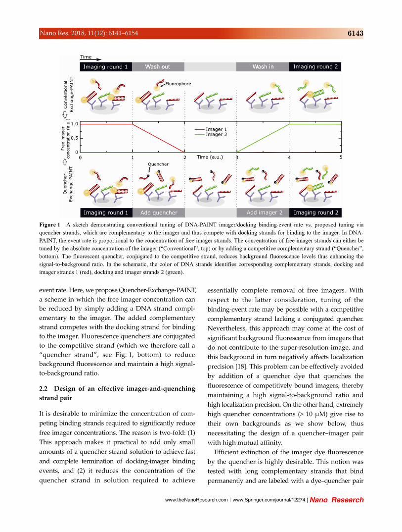
Quencher-Exchange-PAINT offers a way to decouple
the binding-event rate from the absolute imager
concentration and thus from imager diffusion itself
(Fig. 6(a)). To reduce the event rate, quencher strands
at a concentration much higher (10–50) than the
imager concentration were added to the solution
surrounding the tissue. This concentration gradient
led to an increase in the quencher strand concentration
to a sufficient level throughout the tissue much faster
than the diffusion of the imager out of the tissue,
resulting in a rapid reduction of the binding-event
rate. Figure 6(b) reveals an increasingly rapid event
rate suppression with the increasing quencher strand
concentrations. Ryanodine receptors in a murine
cardiac tissue slice were imaged, and the event rate
was modulated sequentially by washing with plain
buffer and different concentrations of complementary
quencher strands (1, 10, or 50 nM at an initial imager
concentration of 1 nM), while the field of view and
imaging sequence remained the same for comparability.
In the shown case, washing with plain buffer did not
decrease the event rate to levels necessary for
Exchange-PAINT for over 10 min. Washing with
quencher strands 10–50 more concentrated than the
imager concentration within the tissue reduced the
event rate to background levels within 5 min.
Due to the washing steps involved in the presented
tissue Quencher-Exchange-PAINT, the high quencher–
imager binding affinity is no longer crucial, because a
high proportion of the imager–quencher pairs will be
washed out in the process. Therefore, a shorter quencher
strand, binding to a conventional P1 imager with a 9
bp overlap, could be used here as well. Nevertheless,
the P1+ type approach without explicit solution exchange
should also work for tissues, with the following
alteration: A larger excess of the P1+ quencher should
be added because the acceleration of the suppression
of imager-docking binding relies on the saturation of
binding between quencher and imager strands. This
rapid saturation steepens the time course of reduction
in the free imager amount relative to the diffusional
time course of the quencher concentration increase.
To demonstrate a full Quencher-Exchange-PAINT
cycle in tissue, we imaged porcine and rat cardiac
| www.editorialmanager.com/nare/default.asp
6150 Nano Res. 2018, 11(12): 6141–6154
Figure 6 The influence of a fluorescence quencher on washing steps during Exchange-PAINT imaging of cardiac tissue samples. (a) After imaging one target in Exchange-PAINT (left), the imager has to be removed efficiently for subsequent imaging steps. This removal is achieved by replacing the imager with plain buffer (top), but can take several minutes in samples with limited diffusion. Quenching the remaining imager with complementary quencher-conjugated strands will reduce the event rate much more rapidly (bottom). (b) A cardiac tissue sample is washed with plain buffer (black) and increasing quencher concentrations (blue, red, and green), and this approach reduces the event rate more effectively. The inset shows the absolute event rate of the washes; the free imager concentration was readjusted by adding more imager after each wash.
tissue slices, targeting ryanodine receptors (Imager
P1) and TOM20 (Imager P3; Fig. 7). The binding-
event rate dropped to negligible levels in less than a
minute (Fig. 7(a)). Here, the washing step was maintained
longer than strictly necessary to demonstrate a constantly
low event rate, similar to the background event rate
in tissue, after the quencher was added. Subsequent
addition of an orthogonal P3 imager allowed for
crosstalk-free multiplexed imaging of the next target,
here TOM20 (Fig. 7(b)).
3 Conclusions
Here, we introduce Quencher-Exchange-PAINT, which
enables rapid, low-crosstalk Exchange-PAINT imaging
of protein clusters and membrane structures in cell
and tissue samples. The addition of fluorescence
quenchers conjugated to oligonucleotides and comple-
mentary to imager strands is equivalent to a decrease of
imager concentration, reducing both the fluorescence
background and the binding-event rate. Thus, exchanging
imager solutions in Quencher-Exchange-PAINT can
be decoupled from the slow diffusional washout of
the residual imager, thereby accelerating the process
considerably. The same approach based on quencher-
coupled strands can be implemented for straightforward
Quencher-Exchange-PAINT imaging without the need
for washes and full fluid exchange chambers. The
free-imager concentration and therefore the Quencher-
DNA-PAINT binding-event rate can be easily tuned by
adding a small volume of complementary quencher
strands at high concentration into an open-top imaging
chamber.
Moreover, we show the flexibility of the synthetic
DNA design, by means of an imager and quencher
strand pair with slightly extended length that
achieves the desired high affinity while not affecting
the super-resolution imaging quality. The throughput
of Quencher-Exchange-PAINT can in principle be
increased by combination with spectrally multiplexed
imaging (see Fig. S2 in the ESM). Furthermore, the
concept of Quencher-Exchange-PAINT could be used
to facilitate other related techniques such as the
generalization of Exchange-PAINT to confocal imaging
and other super-resolution techniques such as STORM
or STED [22–24]. In these methods, a tuning or
reduction of the imager-binding rate is also essential
and can be facilitated by adding complementary
quencher strands.

www.theNanoResearch.com∣www.Springer.com/journal/12274 | Nano Research
6151 Nano Res. 2018, 11(12): 6141–6154
Figure 7 Exchange-PAINT facilitated by a wash with an additional quencher. (a) The DNA PAINT event rate for the P1 imager drops to background levels after a wash with a complementary quencher strand. Subsequently, imager P3 is added. (b) A superimposed image of channels P1 and P3. Green: ryanodine receptors imaged at 1 nM P1, washed with 10 nM P1 quencher. Red: TOM20 imaged with P3 at 1 nM. The tissue slice was imaged 3 µm above the coverslip in HILO mode.
4 Experimental
4.1 Materials and sample preparation
High-performance liquid chromatography (HPLC)-
purified modified oligonucleotides were purchased
from Eurofins Genomics (Imager strands and strep-
tavidin-modified docking strands for bead experiments,
Eurofins Scientific, Luxemburg) and IDT (Amino
modified docking strands and quencher modified
strands, Integrated DNA Technologies, Coralville).
P1 docking strands were labeled with the Cy3 dye to
enable quality control via widefield fluorescence imaging.
Cy3 excitation at 642 nm is negligible and thus did
not interfere with our DNA-PAINT experiments. The
sequences used are given in Table 1.
Lyophilized DNA was resuspended and stored in
Tris-EDTA (TE, pH 8.0, Sigma-Aldrich) buffer at
100 μM. Dilution in DNA-PAINT buffer (1 PBS,
500 mM NaCl, pH 8.0, see buffer C in Ref. [14]) was
carried out for imaging.
Coverslips for imaging of polystyrene (PS) beads
were coated with PLL-g-PEG (SuSoS, Duebendorf) to
prevent nonspecific binding. PLL-g-PEG at a con-
centration of 0.1 mgmL–1 in PBS was washed off the
coverslip after 30 min. The docking strands were
attached to the streptavidin-coated PS beads (diameter:
500 nm, Microparticles GmbH, Berlin) by dispersing
them in TE buffer containing 300 mM NaCl and
biotinylated docking strands. Docking strands were
Table 1 Docking, imager, and quencher strand sequences
Name Sequence (5 3)
Permanently binding imager
Atto 647N – TATACATCTATCTTCATTATT –Biotin
Permanently binding quencher
TAATGAAGATAGATGTATT – Iowa BlackRQ
P1 imager [14] CTAGATGTAT – Atto 655
P1 docking Biotin/antibody – TTATACATCTA – Cy3
P1 quencher Iowa Black RQ – ATACATCTAC
P1+ imager GCGCTAGATGTAT – Atto 655
P1+ quencher Iowa Black RQ – ATACATCTAGCGC
P3 imager [14] GTAATGAAGA – Atto 655
P3 docking Biotin/antibody – TTTCTTCATTA
P3 quencher Iowa Black RQ – TCTTCATTAC
P5 imager [14] CTTTACCTAA – Atto 655
P5 docking Antibody – TTTTAGGTAAA
added in 4 excess concentration as compared to the
binding capacity of the beads to ensure a saturated
coating. Unbound oligos were removed by repeated
centrifugation and redispersion steps.
The experimentation with rat and murine tissues
was approved by the University of Exeter ethics
committee and the use of porcine tissue by the
University of Bristol ethics committee. The preparation
and immunostaining procedure is described elsewhere
in detail [25]. Cryosections were cut at a thickness of
15 μm and deposited onto poly-L-lysine–coated No.
1.5 coverslips. The tissue slices were hydrated, blocked

| www.editorialmanager.com/nare/default.asp
6152 Nano Res. 2018, 11(12): 6141–6154
at room temperature, and then incubated with a
primary antibody in an incubation solution (1% BSA;
0.05% Triton X-100; 0.05% NaN3) at 4 °C overnight.
All tissue slices were labeled with a ryanodine
receptor (RyR) 2–specific antibody, and the rat and
pig tissues shown in Fig. 7 were additionally labeled
with a primary antibody against the mitochondrial
import receptor subunit TOM20. Respective secondary
antibodies (Jackson ImmunoResearch, West Grove)
conjugated to the DNA-PAINT docking strands were
added after multiple washing steps with PBS.
COS-7 cells were seeded on coverslips and grown
overnight in DMEM at 37 °C and 5% CO2. After removal
of the medium, the cells were fixed in ice-cold methanol
for 15 min at 20 °C and washed three times in PBS.
After that, the cells were permeabilized with 0.2%
Triton X-100 in PBS and blocked with 1% BSA in PBS
for 10 min each. β-Tubulin and TOM20 were immunostained
with respective primary antibodies (1:200 in the incubation
solution) at room temperature for 1 h. After three
washes with PBS for 5 min each, the respective secondary
antibodies conjugated to DNA-PAINT docking strands
P1 for β-tubulin and P5 for TOM20 were added at 1%
in the incubation solution. The sample was washed
three times with PBS for 5 min each before addition
of the imager strands.
4.2 Imaging setup
All data were acquired using a modified Nikon
Eclipse Ti-E inverted microscope (Nikon, Tokyo) and
an Andor Zyla 4.2 sCMOS (scientific complementary
metal-oxide-semiconductor) camera (Andor, Belfast).
For quality checks of the labeling, a widefield fluorescence
imaging LED light source was employed (CoolLED,
Andover). Atto 655 was excited with a CW diode
laser (Omikron LuxX, Rodgau) at 642 nm with a power
of 140 mW, attenuated to approximately 15 mW with
an illumination spot with approximately 30 μm diameter
in the sample. A 60× 1.49NA APO oil immersion TIRF
objective (Nikon, Tokyo) was used. PS beads were
imaged in TIRF mode, whereas tissue slices in HILO
mode. Thermal drift was reduced with a custom
objective holder, and the focus was controlled by a
piezo objective scanner (P-725, Physik Instrumente,
Karlsruhe). Lateral drift correction during post-analysis
was achieved via tracking the data acquired with an
auxiliary camera in transmission mode at a non-
interfering wavelength. This tracking setup was also
implemented for continuous focus stabilization during
image acquisition.
4.3 Image acquisition and analysis
Oligo concentrations were estimated by measuring
the absorbance at the DNA absorbance peak (260 nm)
and at the absorption maximum of the respective dye
or quencher oligo modification on a NanoDrop
spectrophotometer (Thermo Fisher Scientific, Waltham).
Bulk fluorescence was measured in a 200 μL chamber
and 50 μm above the coverslip, to minimize the detected
fluorescence of imagers adsorbed on the coverslip
surface.
DNA-PAINT images were captured with integration
time 100 ms. Fluid exchange in Exchange-PAINT
experiments on tissue and streptavidin-coated poly-
styrene beads was facilitated by means of a 3D-printed
chamber (printed with Form2, Formlabs, Somerville).
It included a straight channel with a volume of
approx. 140 μL, an inlet that could be attached to a
syringe or a pump (via 0.1” FEP tubing), and a waste
reservoir which was decoupled from the imaging
channel. Tissue slices were imaged in HILO mode 5
μm (Fig. 6) and 3 μm (Fig. 7) away from the surface
of the coverslip. Fixed cells imaged by Quencher-
Exchange-PAINT without the need for washing were
imaged in an open-top microscopy chamber. A cleaned
coverslip was attached to a reusable Perspex slide
with a circular chamber cut in its middle (construction
time approx. 1 min, for details see Ref. [26]).
Control of the optical components including the
microscope, image acquisition, and analysis was based
on a custom-written software package, Python Micro-
scopy Environment (PyME), which is available freely
via: https://bitbucket.org/christian_soeller/python-
microscopy-exeter/. Single binding events were detected
and fitted to a 2D Gaussian for localization. The data
were next filtered with respect to parameters of a Gaussian
fitting, e.g., the peak intensity, and localization errors.
Thus, binding events that were not in focus could be
effectively suppressed. Drift correction uses data acquired
by an auxiliary camera via the transmission light

www.theNanoResearch.com∣www.Springer.com/journal/12274 | Nano Research
6153 Nano Res. 2018, 11(12): 6141–6154
path, while deviating aberrations between the two
imaging paths were corrected for. Super resolution
images were rendered by jittered triangulation [27].
Acknowledgements
We thank Rikke Morrish for help with the fixation of
COS-7 cells and Anna Meletiou, Cecilia Afonso
Rodrigues, Carl Harrison for their help with antibody
conjugations and labelling of tissue sections and fixed
cells. The authors also acknowledge useful discussions
with B.M. Mognetti. The work was supported by
funding from the Human Frontier Science Program
(No. 0027/2013) and the Engineering and Physical Sciences
Research Council of the UK (No. EP/N008235/1).
Electronic Supplementary Material: Supplementary
material (theoretical model for the simulation of
background fluorescence, further comparison of P1
and P1+ imager strands and a proof-of-principle
experiment demonstrating the combination of Quen-
cher-Exchange-PAINT with spectral multiplexing) is
available in the online version of this article at
https://doi.org/10.1007/s12274-018-1971-6.
Open Access: This article is distributed under the terms
of the Creative Commons Attribution 4.0 International
License (http://creativecommons.org/licenses/by/4.0/),
which permits unrestricted use, distribution, and
reproduction in any medium, provided you give
appropriate credit to the original author(s) and the
source, provide a link to the Creative Commons license,
and indicate if changes were made.
References
[1] Huang, B.; Bates, M.; Zhuang, X. W. Super resolution fluorescence
microscopy. Annu. Rev. Biochem. 2009, 78, 993–1016.
[2] Hell, S. W. Microscopy and its focal switch. Nat. Methods 2009,
6, 24–32.
[3] Bailey, B.; Farkas, D. L.; Taylor, D. L.; Lanni, F. Enhancement
of axial resolution in fluorescence microscopy by standing-
wave excitation. Nature 1993, 366, 44–48.
[4] Hell, S.; Stelzer, E. H. K. Properties of a 4Pi confocal
fluorescence microscope. J. Opt. Soc. Am. A 1992, 9, 2159–
2166.
[5] Hell, S. W.; Wichmann, J. Breaking the diffraction resolution
limit by stimulated emission: Stimulated-emission-depletion
fluorescence microscopy. Opt. lett. 1994, 19, 780–782.
[6] Klar, T. A.; Jakobs, S.; Dyba, M.; Egner, A.; Hell, S. W.
Fluorescence microscopy with diffraction resolution barrier
broken by stimulated emission. Proc. Natl. Acad. Sci. USA 2000,
97, 8206–8210.
[7] Betzig, E.; Patterson, G. H.; Sougrat, R.; Lindwasser, O. W.;
Olenych, S.; Bonifacino, J. S.; Davidson, M. W.; Lippincott-
Schwartz, J.; Hess, H. F. Imaging intracellular fluorescent
proteins at nanometer resolution. Science 2006, 313, 1642–1645.
[8] Hess, S. T.; Girirajan, T. P. K.; Mason, M. D. Ultra-high
resolution imaging by fluorescence photoactivation localization
microscopy. Biophys. J. 2006, 91, 4258–4272.
[9] Rust, M. J.; Bates, M.; Zhuang, X. W. Sub-diffraction-limit
imaging by stochastic optical reconstruction microscopy
(STORM). Nat. Methods 2006, 3, 793–796.
[10] Heilemann, M.; van de Linde, S.; Schüttpelz, M.; Kasper, R.;
Seefeldt, B.; Mukherjee, A.; Tinnefeld, P.; Sauer, M.
Subdiffraction-resolution fluorescence imaging with conventional
fluorescent probes. Angew. Chem., Int. Ed. 2008, 47, 6172–6176.
[11] Legant, W. R.; Shao, L.; Grimm, J. B.; Brown, T. A.; Milkie, D.
E.; Avants, B. B.; Lavis, L. D.; Betzig, E. High-density
three-dimensional localization microscopy across large volumes.
Nat. Methods 2016, 13, 359–365.
[12] Curdt, F.; Herr, S. J.; Lutz, T.; Schmidt, R.; Engelhardt, J.; Sahl,
S. J.; Hell, S. W. isoSTED nanoscopy with intrinsic beam
alignment. Opt. Express 2015, 23, 30891–30903.
[13] Sharonov, A.; Hochstrasser, R. M. Wide-field subdiffraction
imaging by accumulated binding of diffusing probes. Proc. Natl.
Acad. Sci. USA 2006, 103, 18911–18916.
[14] Jungmann, R.; Avendaño, M. S.; Woehrstein, J. B.; Dai, M. J.;
Shih, W. M.; Yin, P. Multiplexed 3D cellular super-resolution
imaging with DNA-PAINT and Exchange-PAINT. Nat. Methods
2014, 11, 313–318.
[15] Jungmann, R.; Steinhauer, C.; Scheible, M.; Kuzyk, A.;
Tinnefeld, P.; Simmel, F. C. Single-molecule kinetics and
super-resolution microscopy by fluorescence imaging of transient
binding on DNA origami. Nano Lett. 2010, 10, 4756–4761.
[16] Schnitzbauer, J.; Strauss, M. T.; Schlichthaerle, T.; Schueder, F.;
Jungmann, R. Super-resolution microscopy with DNA-PAINT.
Nat. Protoc. 2017, 12, 1198–1228.
[17] Agasti, S. S.; Wang, Y.; Schueder, F.; Sukumar, A.; Jungmann,
R.; Yin, P. DNA-barcoded labeling probes for highly multiplexed
exchange-PAINT imaging. Chem. Sci. 2017, 8, 3080–3091.
[18] Thompson, R. E.; Larson, D. R.; Webb, W. W. Precise
nanometer localization analysis for individual fluorescent
probes. Biophys. J. 2002, 82, 2775–2783.
[19] Markham, N. R.; Zuker, M. DINAMelt web server for nucleic
acid melting prediction. Nucleic Acids Res. 2005, 33, W577–W581.
| www.editorialmanager.com/nare/default.asp
6154 Nano Res. 2018, 11(12): 6141–6154
[20] Markham, N. R.; Zuker, M. UNAFold: Software for nucleic
acid folding and hybridization. In Bioinformatics, Volume II.
Structure, Function and Applications. Keith, J. M., Ed.;
Humana Press: Totowa, NJ, 2008; pp 3–31.
[21] Molle, J.; Raab, M.; Holzmeister, S.; Schmitt-Monreal, D.;
Grohmann, D.; He, Z. K.; Tinnefeld, P. Superresolution
microscopy with transient binding. Curr. Opin. Biotechnol.
2016, 39, 8–16.
[22] Schueder, F.; Strauss, M. T.; Hoerl, D.; Schnitzbauer, J.;
Schlichthaerle, T.; Strauss, S.; Yin, P.; Harz, H.; Leonhardt, H.;
Jungmann, R. Universal super-resolution multiplexing by DNA
exchange. Angew. Chem., Int. Ed. 2017, 56, 4052–4055.
[23] Wang, Y.; Woehrstein, J. B.; Donoghue, N.; Dai, M. J.;
Avendaño, M. S.; Schackmann, R. C. J.; Zoeller, J. J.; Wang, S.
S. H.; Tillberg, P. W.; Park, D. et al. Rapid sequential in situ
multiplexing with DNA exchange imaging in neuronal cells and
tissues. Nano Lett. 2017, 17, 6131–6139.
[24] Beater, S.; Holzmeister, P.; Lalkens, B.; Tinnefeld, P. Simple
and aberration-free 4color-STED-multiplexing by transient
binding. Opt. Express 2015, 23, 8630–8638.
[25] Hou, Y. F.; Crossman, D. J.; Rajagopal, V.; Baddeley, D.;
Jayasinghe, I.; Soeller, C. Super-resolution fluorescence imaging
to study cardiac biophysics: α-actinin distribution and Z-disk
topologies in optically thick cardiac tissue slices. Prog. Biophys.
Mol. Biol. 2014, 115, 328–339.
[26] Crossman, D. J.; Hou, Y. F.; Jayasinghe, I.; Baddeley, D.;
Soeller, C. Combining confocal and single molecule localisation
microscopy: A correlative approach to multi-scale tissue imaging.
Methods 2015, 88, 98–108.
[27] Baddeley, D.; Cannell, M. B.; Soeller, C. Visualization of
localization microscopy data. Microsc. Microanal. 2010, 16,
64–72.
Related Documents