Varicose veins and treatment DR SADIA SHABBIR House surgeon

Welcome message from author
This document is posted to help you gain knowledge. Please leave a comment to let me know what you think about it! Share it to your friends and learn new things together.
Transcript
Varicose veins and treatment
DR SADIA SHABBIR House surgeon
Leg Vein Anatomy
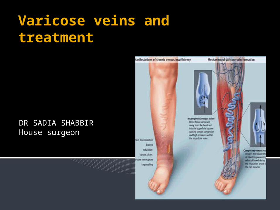
The venous drainage system of the lower extremity consists of three sets of veins:
Deep veins, Superficial veins Perforating veins. All veins contain delicate one-way valves

Superficial Veins Great saphenous vein originates from the medial side of the dorsal venous arch, and then ascends up the medial side of the leg, knee, and thigh to connect with the femoral vein just inferior to the inguinal ligamentSmall saphenous vein originates from the lateral side of the dorsal venous arch, ascends up the posterior surface of the leg, and then penetrates deep fascia to join the popliteal vein posterior to the knee; proximal to the knee, the popliteal vein becomes the femoral vein.
Perforating veins
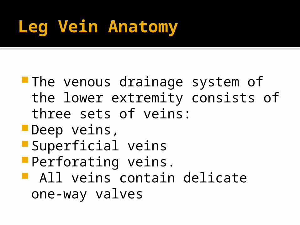
They connect the deep system with the superficial system
They pass through the deep fascia
Guarded by valves-unidirectional flow from superficial to deep veins

Types of perforators
1. Ankle perforators-may or kuster
2. Lower leg perforators of cockett-I,II,III
a)Posteroinferior to med malleolus
b)10cm above med.malleolus
c)15cm above med.malleolus
3. Gastrocnemius perforators of Boyd
4. Mid thigh perforators of Dodd

Physiology of venous blood flow
Venous return from leg is governed by Arterial pressure Calf musculovenous pump Gravity Thoracic pump Valves in veins
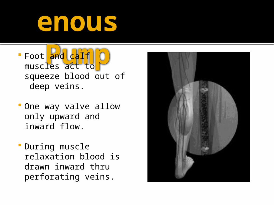
Musculovenous Pump Foot and calf muscles
act to squeeze blood out of deep veins.
One way valve allow only upward and inward flow.
During muscle relaxation blood is drawn inward thru perforating veins.

Venous valvular function
Valve leaflets allow unidirectional flow upward or inward.

Pathophysiology
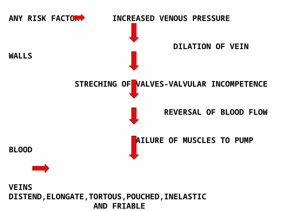
ANY RISK FACTOR INCREASED VENOUS PRESSURE
DILATION OF VEIN WALLS
STRECHING OF VALVES-VALVULAR INCOMPETENCE
REVERSAL OF BLOOD FLOW
FAILURE OF MUSCLES TO PUMP BLOOD
VEINS DISTEND,ELONGATE,TORTOUS,POUCHED,INELASTIC AND FRIABLE

Varicose veins
Dilated,tortuous and elongated veins with reversal of blood flow mainly due to valvular incompetence
Examples Varicose veins in legs Hemorrhoids Varicocele Oesophageal varices
Risk factors
Age Gender Height Heredity Pregnancy Obesity and overweight Posture

Aetiology
Primary varicosities
Congenital incompetence/absence of valves
Weakness or wasting of muscles
Stretching of deep fascia
Inheritance with FOXC2 gene
Klippel-trenaunay syndrome

Secondary Varicositiesecond varicosities
Recurrent thrombophlebitis
Occupational
Oral contraceptive pills
Pregnancy and pelvic tumors
Iatrogenic-in AV fistula
Deep vein thrombosis

Dilated tortuous veins Dragging pain worsening on prolonged
standing/sitting Night cramps Aching pain is relieved at night on taking rest
or elevation of limbs Ithcing,oedema,thickening and eczema of feet Appearance of spider veins in affected leg. Discoloration/ulceration Skin above ankle may shrink
(lipodermatosclerosis) b/c fat underneath skin becomes hard.
Symptoms
Examination
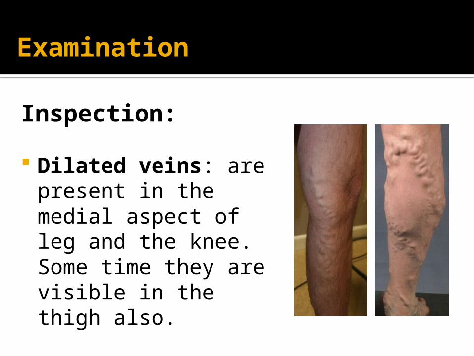
Inspection:
Dilated veins: are present in the medial aspect of leg and the knee. Some time they are visible in the thigh also.

Saphena varix
A saphena varix is a dilatation at the top of the long saphenous vein due to valvular incompetence. It may reach the size of a golf ball or larger.
The varix is: soft and compressible disappears immediately on lying
down exhibits an expansile cough
impulse demonstrates a fluid thrill

Ankle flare
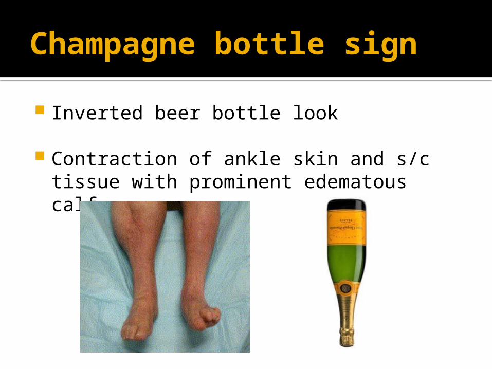
Champagne bottle sign
Inverted beer bottle look
Contraction of ankle skin and s/c tissue with prominent edematous calf

Complications
lipodermatosclerosis
Eczema in varicose vein

Thrombophlebitisid for treatment
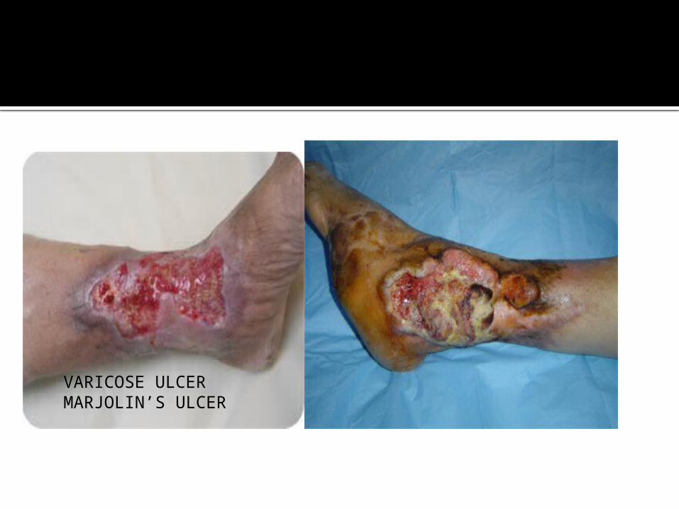
VARICOSE ULCER MARJOLIN’S ULCER

Special Tests
1. Cough impulse test:
This test should be done in standing position. The examiner keeps the finger at SF junction and ask the patient to cough. Fluid thrill, an impulse felt by fingers, is indicative of “saphenofemoral incompetence”
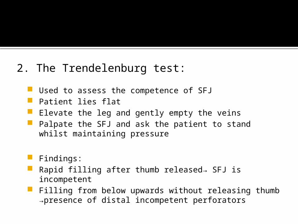
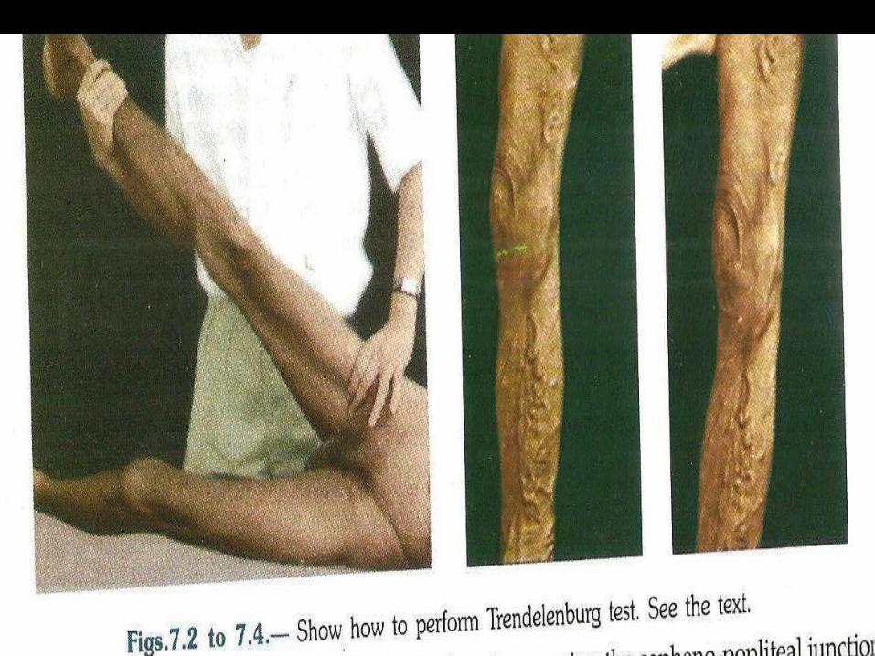
2. The Trendelenburg test:
Used to assess the competence of SFJ Patient lies flat Elevate the leg and gently empty the veins Palpate the SFJ and ask the patient to stand whilst
maintaining pressure
Findings: Rapid filling after thumb released→ SFJ is incompetent Filling from below upwards without releasing thumb
→presence of distal incompetent perforators
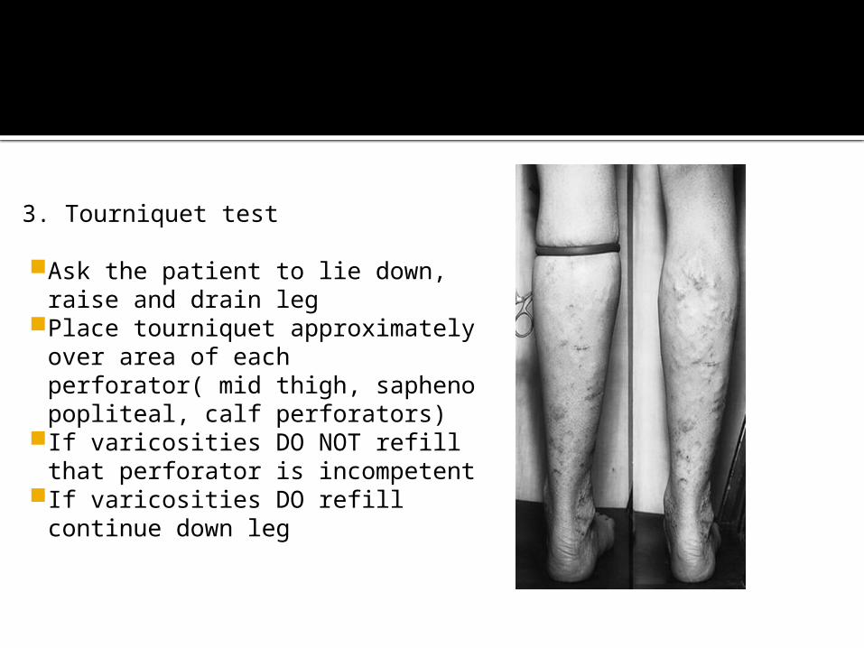
3. Tourniquet test
Ask the patient to lie down, raise and drain leg
Place tourniquet approximately over area of each perforator( mid thigh, sapheno popliteal, calf perforators)
If varicosities DO NOT refill that perforator is incompetent
If varicosities DO refill continue down leg

4. Schwartz test
In standing position,tap the lower part of vein
Impulse felt on saphenofemoral junction
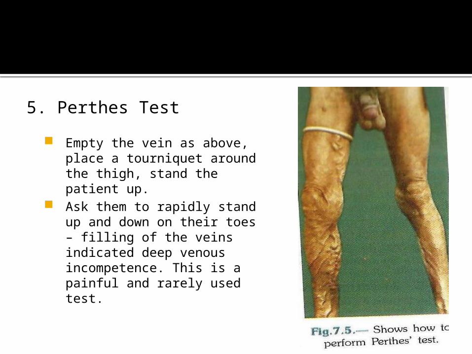
5. Perthes Test
Empty the vein as above, place a tourniquet around the thigh, stand the patient up.
Ask them to rapidly stand up and down on their toes – filling of the veins indicated deep venous incompetence. This is a painful and rarely used test.
6.Fegan’s test
Line of varicosities marked
Site where perforators pierce deep fascia-bulges on standing circular depressions on lying
Investigations
Doppler ultrsound Duplex ultrsound
imaging Venography Varicography

Management
Conservative treatment
Elevation of limb Support elastic
crepe bandage

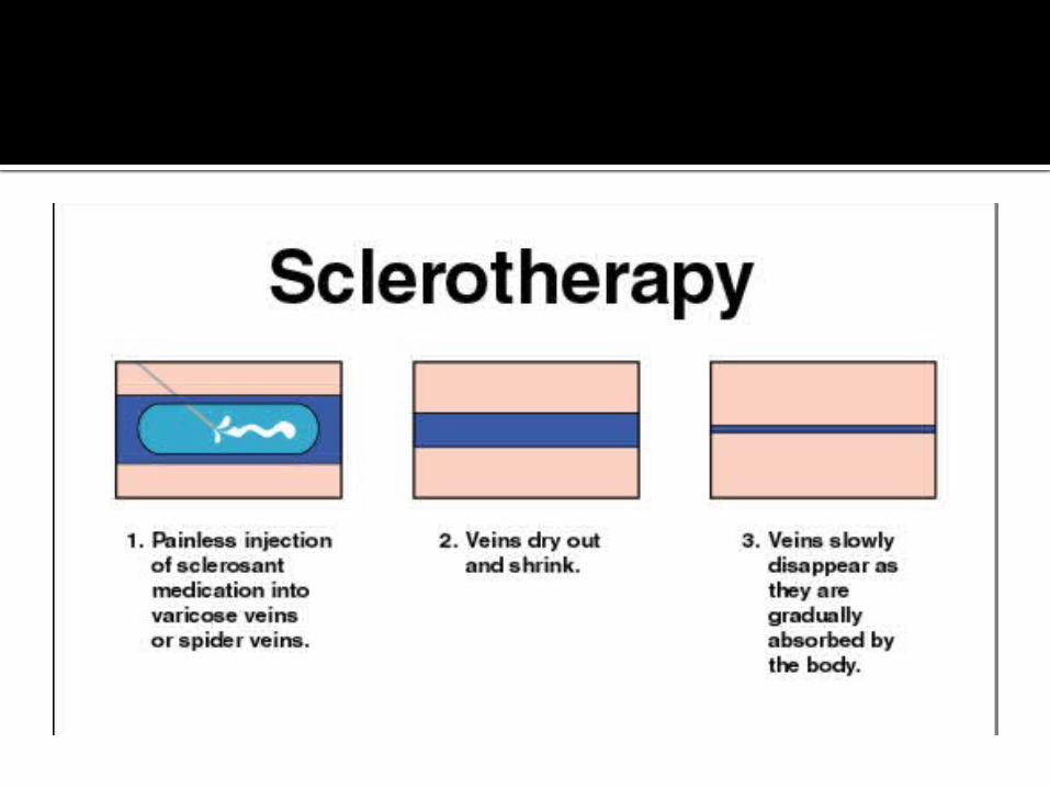
Injection-sclerotherapy
Injecting sclerosants into vein –sodium tetradecyl sulphate destruction of lipid membranes of
endothelial cells shedding of endothelial cells thrombosis,fibrosis,obliteration of
veins

Saphenofemoral junction ligation and greater saphenous stripping
Avulsion of varicosities-multiple ligation
Saphenopopliteal junction ligation and lesser
saphenous stripping
Surgical treatment- Trendelenburg procedure
Picture clipping
Related Documents