Ventricles of brain ▪ Ventricles are cavities or expansions within the brain that are derived from the lumen of the embryonic neural tube. ▪ They are continuous with one another as well as with the central canal of the spinal cord ▪ Ventricles are filled with CSF ▪ There are four ventricles in the brain: ❖ Two lateral ventricles ❖ The third ventricle ❖ The fourth ventricle

Welcome message from author
This document is posted to help you gain knowledge. Please leave a comment to let me know what you think about it! Share it to your friends and learn new things together.
Transcript
Ventricles of brain▪ Ventricles are cavities or expansions within the brain
that are derived from the lumen of the embryonicneural tube.
▪ They are continuous with one another as well as withthe central canal of the spinal cord
▪ Ventricles are filled with CSF
▪ There are four ventricles in the brain:
❖ Two lateral ventricles
❖ The third ventricle
❖ The fourth ventricle
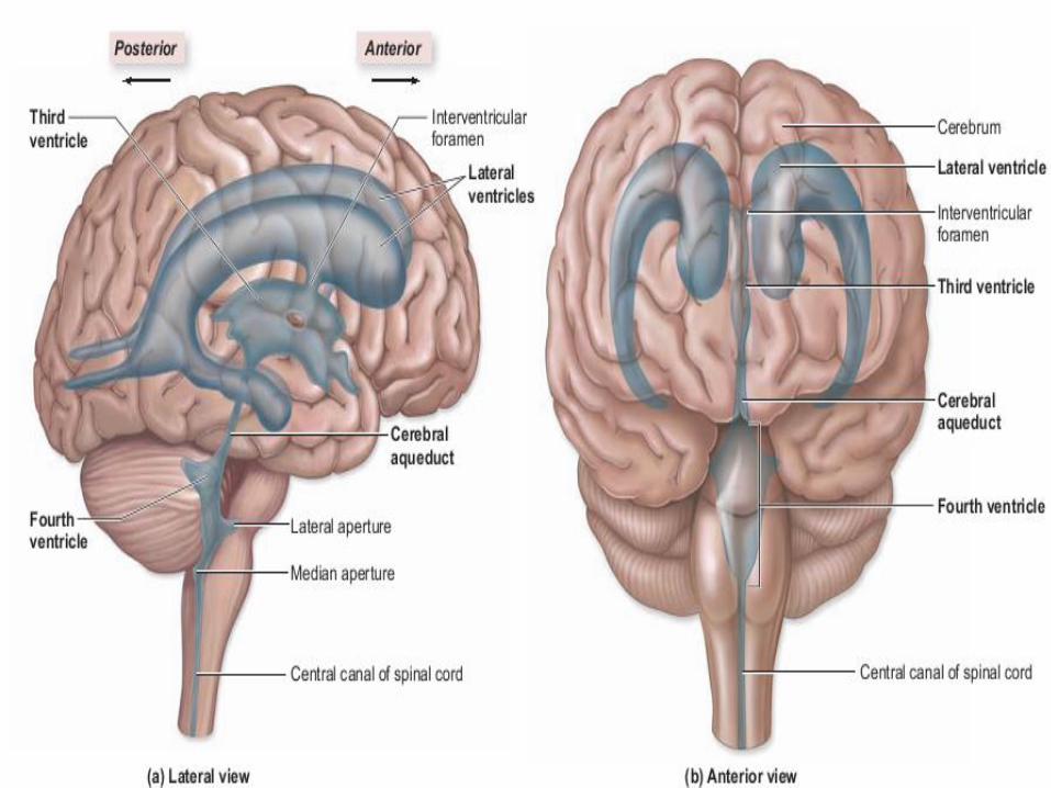
Two lateral ventricles
❑ are large fluid filled cavities contained in the two lobes of cerebral hemispheres.
❑ There is one lateral ventricle in each hemisphere of the cerebrum.
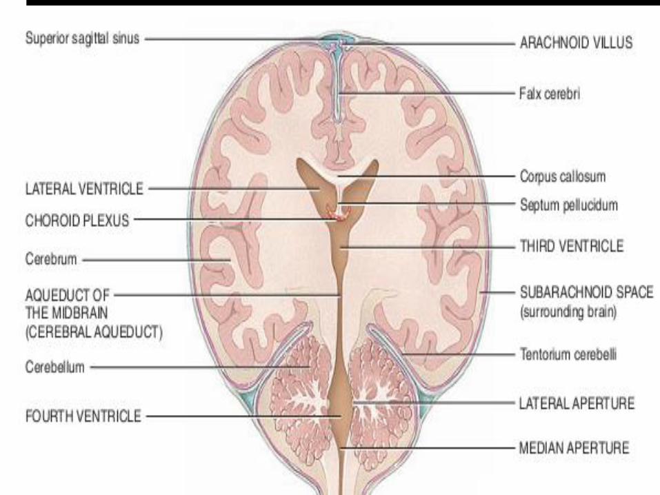
❑ The lateral ventricles meet at the midline just inferior to the corpus callosum wherethey are separated by a thin membrane, the septum pellucidum
The third ventricle
❑ is a smaller slit-like cavity located in the midline in center of the diencephalonbetween the two halves of the thalamus.
❑ Each lateral ventricle communicates with the third ventricle through an openingcalled the interventricular foramen
The fourth ventricle
❑ lies between the brain stem and the cerebellum.
❑ The third ventricle connects with the fourth ventricle through a narrow canal, thecerebral aqueduct, which passes through the midbrain.
❑ The fourth ventricle is continuous with the central canal of the spinal cord, whichextends nearly the full length of the cord.
❑ The fourth ventricle connects with the subarachnoid space through threeopenings—a median aperture in the roof of the fourth ventricle and two lateralapertures, one in each lateral wall of the fourth ventricle.
Cerebrospinal fluid
▪ Cerebrospinal fluid (CSF)is a clear, colorless liquid composed primarily of water that protects the brain and spinal cord from chemical and physical injuries.
▪ CSF continuously circulates through cavities in the brain and spinal cord and around them in the subarachnoid space
▪ The total volume of CSF is 80 to 150 mL. The brain produces about 500 mL of CSF per day, but the fluid is constantly reabsorbed at the same rate
Formation of CSF▪ The majority of CSF production is from the choroid
plexuses
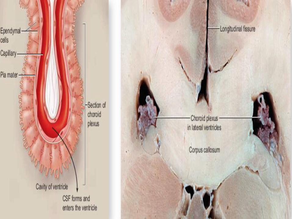
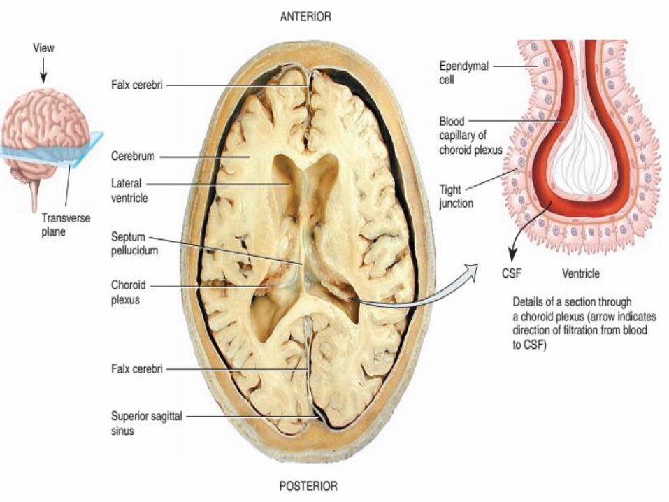
▪ Choroid plexus is tuft of capillaries in the ventricles
▪ Ependymal cells cover the capillaries of the choroidplexuses such that contents that diffuse from capillariesmust first pass through ependymal cells before enteringthe ventricles to become CSF
▪ CSF forms partly by the filtration of blood plasmathrough the choroid plexuses and then modification ofthis filtrate by ependymal cells so that CSF has moresodium and chloride than the blood plasma, but lesspotassium, calcium, and glucose and very little protein
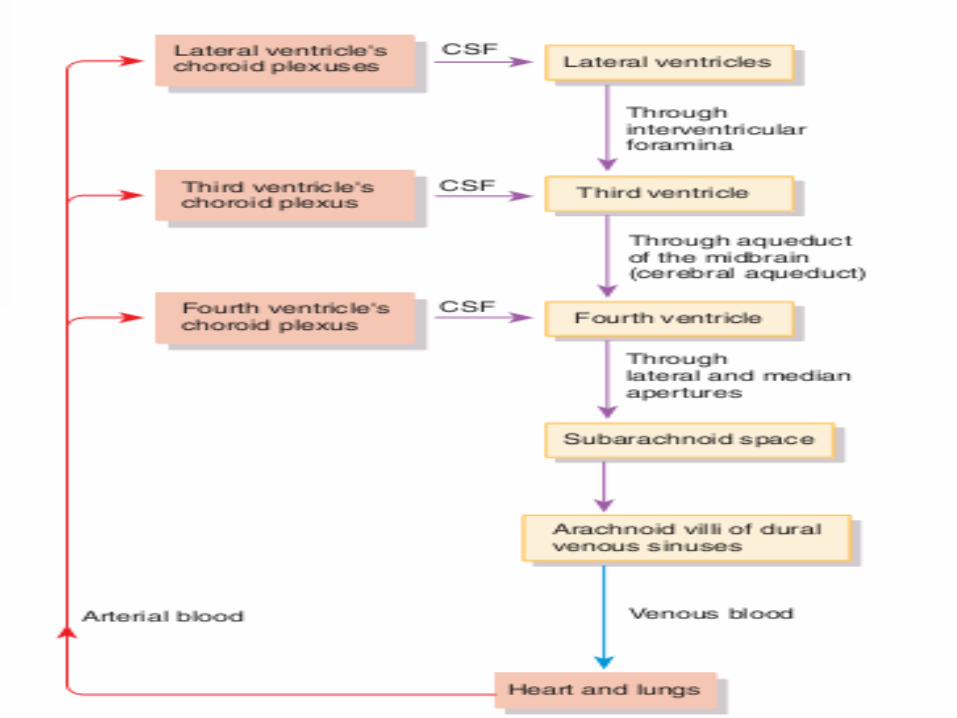
Circulation of CSF▪ The CSF is not a stationary fluid but continually flows through and around
the CNS, driven partly by its own pressure and partly by rhythmicpulsations of the brain produced by each heartbeat
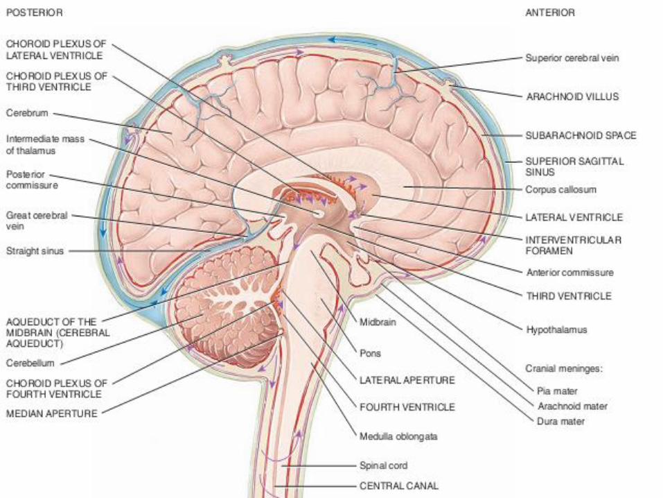
▪ The CSF secreted in the lateral ventricles flows through the interventricularforamina into the third ventricle and then down the cerebral aqueduct tothe fourth ventricle.
▪ The third and fourth ventricles and their choroid plexuses add more CSFalong the way.
▪ A small amount of CSF fills the central canal of the spinal cord, butultimately, all of it escapes through three pores in the walls of the fourthventricle—a median aperture and two lateral apertures.
▪ These lead into the subarachnoid space on the brain surface. From thisspace, the CSF is absorbed by arachnoid villi.
▪ CSF penetrates the walls of the arachnoid villi and mixes with the blood inthe dural venous sinus.
Cerebrum
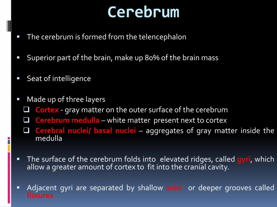
▪ The cerebrum is formed from the telencephalon
▪ Superior part of the brain, make up 80% of the brain mass
▪ Seat of intelligence
▪ Made up of three layers
❑ Cortex - gray matter on the outer surface of the cerebrum
❑ Cerebrum medulla – white matter present next to cortex
❑ Cerebral nuclei/ basal nuclei – aggregates of gray matter inside themedulla
▪ The surface of the cerebrum folds into elevated ridges, called gyri, whichallow a greater amount of cortex to fit into the cranial cavity.
▪ Adjacent gyri are separated by shallow sulci or deeper grooves calledfissures

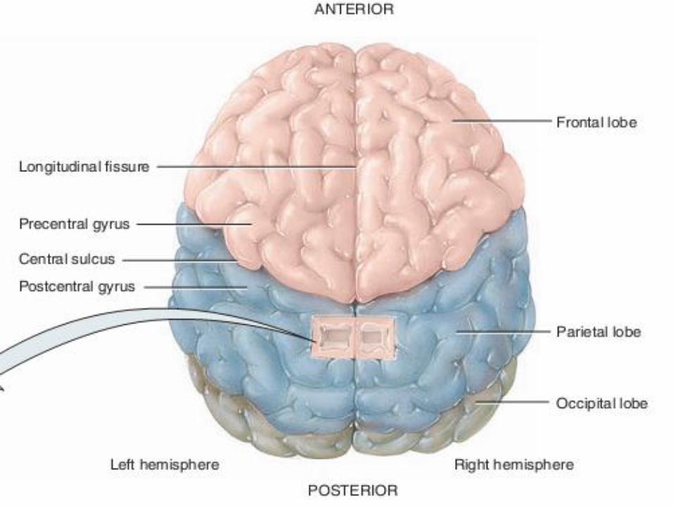
▪ The most prominent fissure in the cerebrum in longitudinal fissure whichextend along midsagittal plane and divides the cerebrum into right andleft hemisphere
▪ The falx cerebri extend along the longitudinal fissure
▪ The separation between cerebral hemispheres is not complete instead abroad band of white matter containing axons that extend between thehemispheres allow for communication between them. That tract of whitematter is called corpus callosum
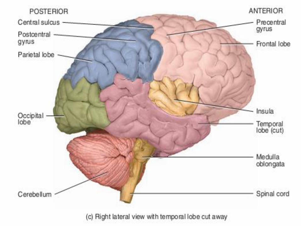
▪ Each cerebral hemisphere is divided into five anatomically andfunctionally distinct lobes by sulci or fissures. The lobes are named for theskull bones overlying each one:
❑ Frontal,
❑ Parietal,
❑ Temporal, and
❑ Occipital lobes
❑ Insula - the fifth lobe is not visible at the surface of the hemispheres.
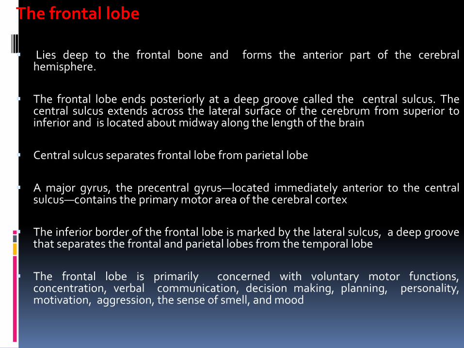
The frontal lobe
▪ Lies deep to the frontal bone and forms the anterior part of the cerebralhemisphere.
▪ The frontal lobe ends posteriorly at a deep groove called the central sulcus. Thecentral sulcus extends across the lateral surface of the cerebrum from superior toinferior and is located about midway along the length of the brain
▪ Central sulcus separates frontal lobe from parietal lobe
▪ A major gyrus, the precentral gyrus—located immediately anterior to the centralsulcus—contains the primary motor area of the cerebral cortex
▪ The inferior border of the frontal lobe is marked by the lateral sulcus, a deep groovethat separates the frontal and parietal lobes from the temporal lobe
▪ The frontal lobe is primarily concerned with voluntary motor functions,concentration, verbal communication, decision making, planning, personality,motivation, aggression, the sense of smell, and mood
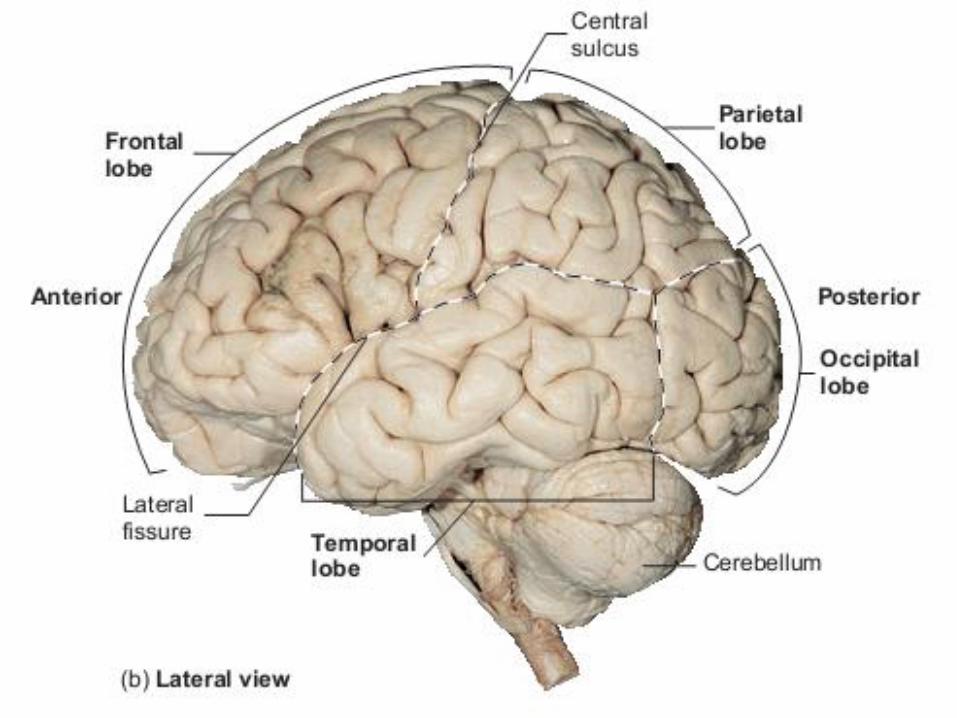
▪ The Parietal lobe▪ lies internal to the parietal bone and forms the superoposterior part of each cerebral
hemisphere.
▪ It terminates anteriorly at the central sulcus, posteriorly at a relatively indistinctparieto-occipital sulcus, and laterally at the lateral sulcus.
▪ A major gyrus, the post-central gyrus, which is located immediately posterior to thecentral sulcus, contains the primary somatosensory area of the cerebral cortex
▪ The parietal lobe is involved with general sensory functions, such as evaluating theshape and texture of objects being touched . It is a major center for the receptionand evaluation of most sensory information, such as touch, pain, temperature,balance, and taste.
▪ The temporal lobe▪ underlies the temporal bone.
▪ The temporal lobe is located below the parietal lobe and the posterior portion of thefrontal lobe. It is separated from both by the lateral sulcus.
▪ The temporal lobe contains auditory centers that receive sensory fibers from thecochlea of the ear. This lobe also interprets some sensory experiences and storesmemories of both auditory and visual experiences.
▪ Its anterior and inferior portions are referred to as the “psychic cortex,” and theyare associated with such brain functions as abstract thought and judgment
The occipital lobe
▪ forms the posterior region of each hemisphere and immediately underlies theoccipital bone.
▪ It is not distinctly separated from the temporal and parietal lobes
▪ It lies superior to the cerebellum and is separated from it by an infolding of themeningeal layer called the tentorium cerebelli
▪ The principal functions of the occipital lobe concern vision. It integrates eyemovements by directing and focusing the eye. It is also responsible for visualassociation—correlating visual images with previous visual experiences and othersensory stimuli
The insula
▪ The insula is a deep lobe of the cerebrum that cannot be viewed on the surface .
▪ It lies deep to the lateral sulcus and is covered by portions of the frontal, parietal,and temporal lobes
▪ It is apparently involved in memory and the interpretation of taste.
The central white matter▪ lies deep to the gray matter of the cerebral cortex and is composed
primarily of myelinated axons.
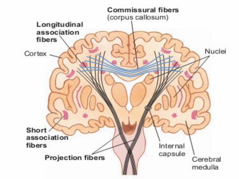
▪ Most of these axons are grouped into bundles called tracts. There are threetype of tracts:
❑ Association tracts – has axons that connect different regions of thecerebral cortex within the same hemisphere.
❑ Commissural tracts - has axons that connect the cerebral hemispheres.The prominent commissural tracts that link the left and right cerebralhemispheres include the large, C -shaped corpus callosum anteriorcommissure, and posterior commissure.
❑ Projection tracts- link the cerebral cortex to the inferior brain regionsand the spinal cord. Examples of projection tracts are the corticospinaltracts that carry motor signals from the cerebrum to the brainstem andspinal cord.
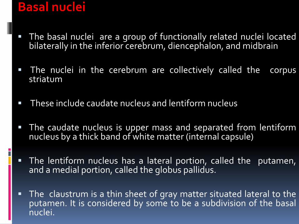
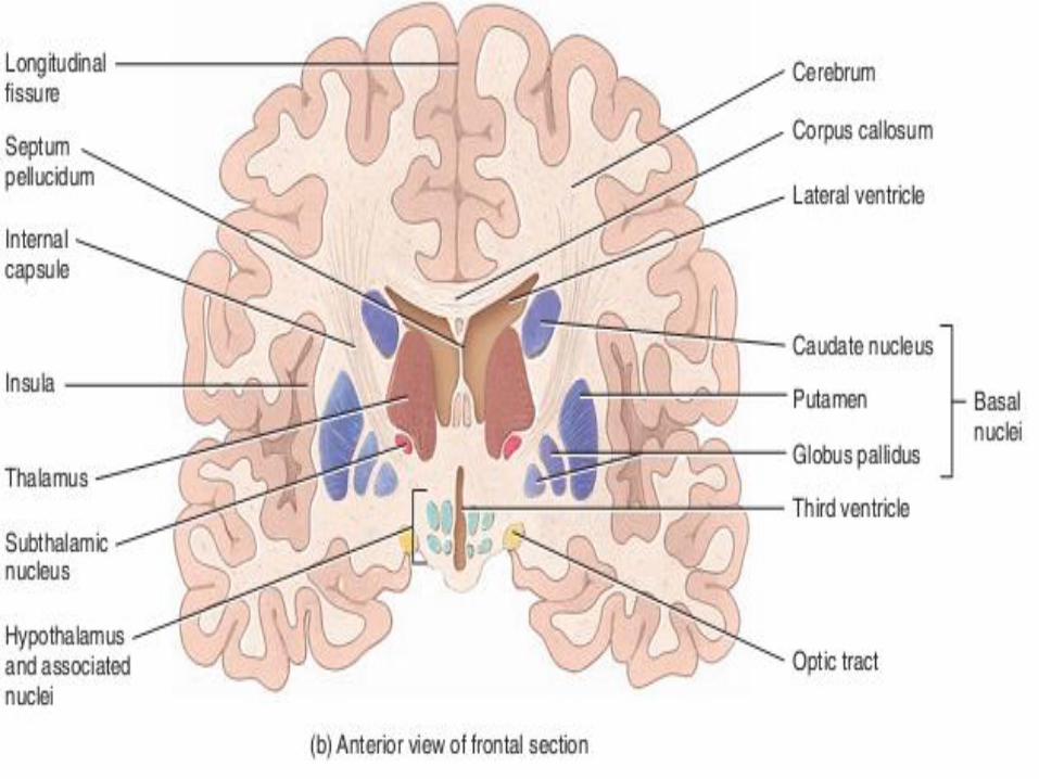
Basal nuclei
▪ The basal nuclei are a group of functionally related nuclei locatedbilaterally in the inferior cerebrum, diencephalon, and midbrain
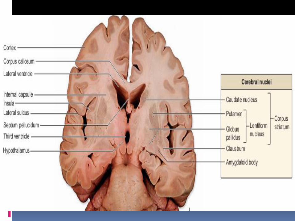
▪ The nuclei in the cerebrum are collectively called the corpusstriatum
▪ These include caudate nucleus and lentiform nucleus
▪ The caudate nucleus is upper mass and separated from lentiformnucleus by a thick band of white matter (internal capsule)
▪ The lentiform nucleus has a lateral portion, called the putamen,and a medial portion, called the globus pallidus.
▪ The claustrum is a thin sheet of gray matter situated lateral to theputamen. It is considered by some to be a subdivision of the basalnuclei.
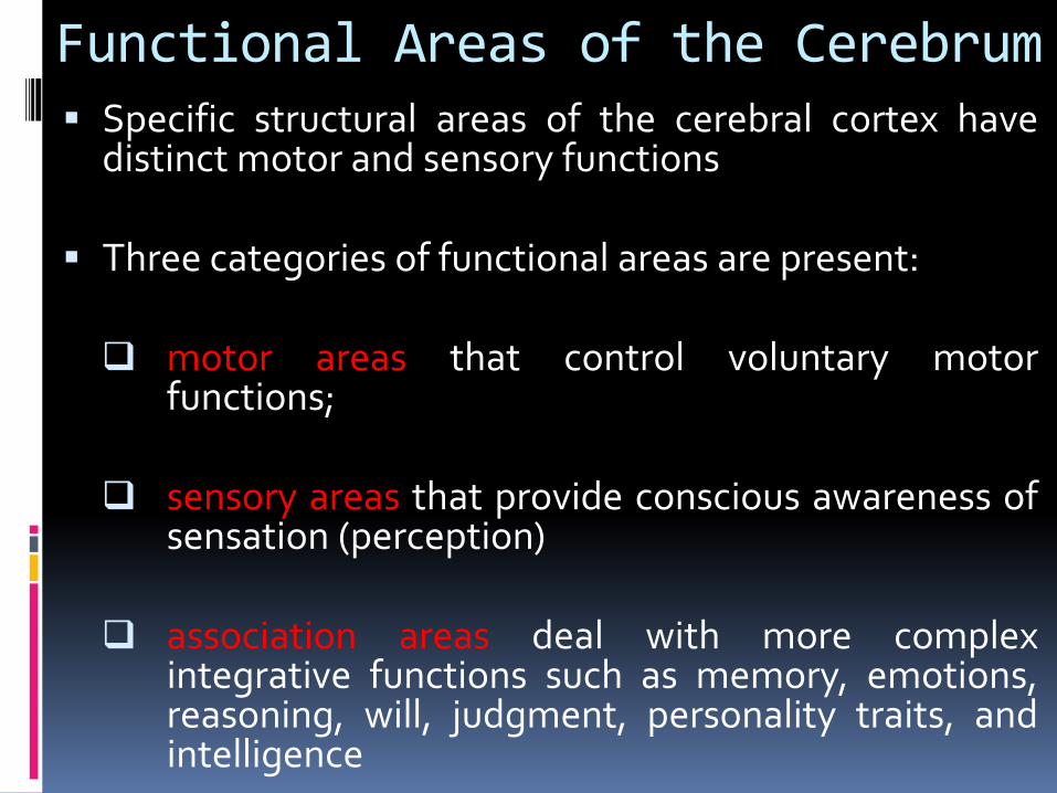
Functional Areas of the Cerebrum▪ Specific structural areas of the cerebral cortex have
distinct motor and sensory functions
▪ Three categories of functional areas are present:
❑ motor areas that control voluntary motorfunctions;
❑ sensory areas that provide conscious awareness ofsensation (perception)
❑ association areas deal with more complexintegrative functions such as memory, emotions,reasoning, will, judgment, personality traits, andintelligence
Motor areas▪ The cortical areas that control motor functions are housed within the frontal
lobes.
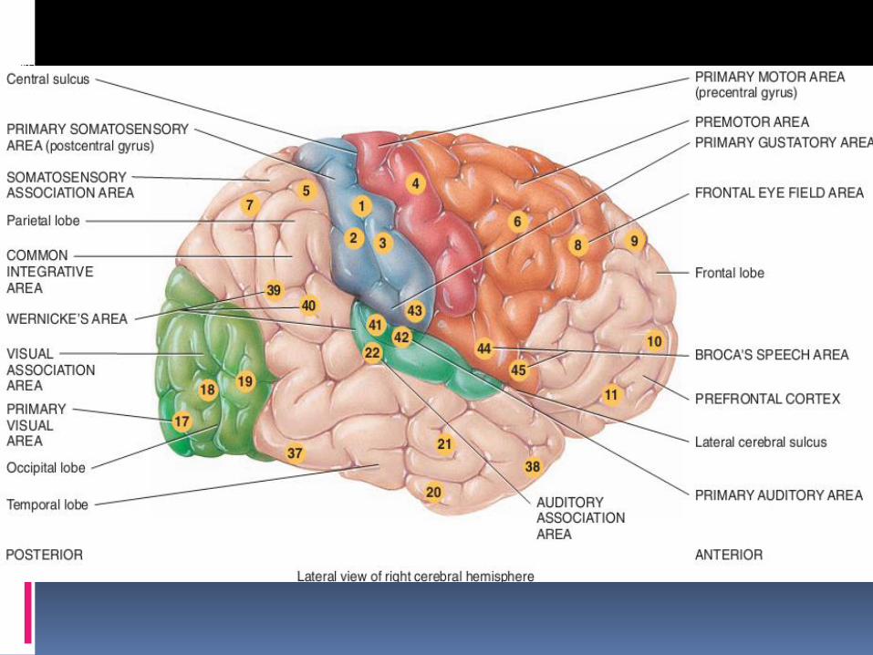
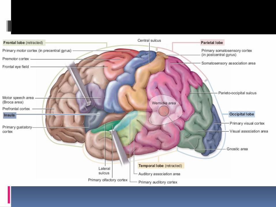
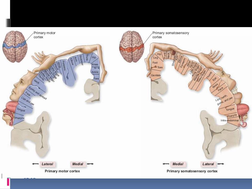
▪ The primary motor cortex, (area 4) also called the somatic motor area, islocated within the precentral gyrus of the frontal lobe which controlvoluntary skeletal muscle activity.
▪ A “map” of the entire body is present in the primary motor area. Each regionwithin the area controls voluntary contractions of specific muscles or groupsof muscles.
▪ Different muscles are represented unequally in the primary motor area.More cortical area is devoted to those muscles involved in skilled, complex,or delicate movement
▪ The axons of these neurons project contralaterally (to the opposite side) tothe brainstem and spinal cord. Thus, the left primary motor cortex controlsthe right-side voluntary muscles, and vice versa.
▪ The motor speech area/ the Broca area, (areas 44 and 45) is located in mostindividuals within the inferolateral portion of the left frontal lobe. This regionis responsible for controlling the muscular movements necessary forvocalization.
▪ The frontal eye field control and regulate the eye movements needed forreading and coordinating binocular vision
Sensory areas
▪ The cortical areas involved conscious awareness of sensation are present in parietal,temporal, and occipital lobes
▪ The primary somatosensory cortex is housed within the postcentral gyrus of theparietal lobes.( area 1, 2 ,3) Neurons in this cortex receive general somatic sensoryinformation from touch, pressure, pain, proprioception and temperature receptor. A“map” of the entire body is present in the primary somatosensory area. Each pointwithin the area receives impulses from a specific part of the body. The size of thecortical area receiving impulses from a particular part of the body depends on thenumber of receptors present there rather than on the size of the body part
▪ The primary gustatory cortex (area 43), located in the parietal cortex, receivesimpulses for taste and is involved in gustatory perception and taste discrimination
▪ The primary visual cortex, (area 17) located in the occipital lobe, receives andprocesses incoming visual information.
▪ The primary auditory cortex, (area 41 and 42) located in the temporal lobe, receivesand processes auditory information.
▪ Primary olfactory cortex,(area 28) located in the temporal lobe, provides consciousawareness of smells.
Association areas▪ The association areas of the cerebrum consist of large areas of the occipital, parietal,
and temporal lobes and of the frontal lobes anterior to the motor areas.
▪ Association areas are connected with one another by association tracts
▪ The primary motor and sensory cortical regions are connected to adjacent association areas that either process and interpret incoming data or coordinate a motor response.
▪ Association areas integrate new sensory inputs with memories of past experiences
▪ The premotor area (area 6)
❑ is a motor association area that is immediately anterior to the primary motor area in frontal lobe.
❑ Neurons in this area communicate with the primary motor cortex, the sensoryassociation areas in the parietal lobe, the basal nuclei, and the thalamus.
❑ The premotor area deals with learned motor activities of a complex and sequentialnature. It generates nerve impulses that cause specific groups of muscles tocontract in a specific sequence
❑ The premotor area also serves as a memory bank for such various skilledmovements.

▪ The somatosensory association area (area 5 and 7)
❑ is located in the parietal lobe and lies immediately posterior to the primary somatosensory cortex. It receives input from the primary somatosensory area, as well as from the thalamus and other parts of the brain
❑ It interprets sensory information and is responsible for integrating and interpreting sensations to determine the texture, temperature, pressure, and shape of objects.
❑Another role of this area is storage of memories of past somatic sensory experiences, enabling one to compare current sensations with previous experiences
▪ The visual association area (areas 18 and 19)
❑ located in the occipital lobe, receives sensory impulses from the primary visual area and the thalamus.
❑ relates present and past visual experiences and is essential for recognizing and evaluating what is seen
▪ The facial recognition area, (corresponding roughly to areas 20, 21, and 37)
❑in the inferior temporal lobe, receives nerve impulses from the visual association area.
❑This area stores information about faces, and it allows to recognize people by their faces.
▪ The auditory association area (area 22)
❑ located within the temporal lobe posterior to primary auditory area. Helpto recognize a particular sound as speech, music, or noise.
▪ Wernicke’s (posterior language) area (area 22, 39,40)
❑ a broad region in the left temporal and parietal lobes, interprets themeaning of speech by recognizing spoken words.
❑ It is active in translating words into thoughts.
❑ The regions in the right hemisphere that correspond to Broca’s andWernicke’s areas in the left hemisphere also contribute to verbalcommunication by adding emotional content, such as anger or joy, tospoken words.
▪ The common integrative area (areas 5, 7, 39, and 40)
❑ composed of regions of the parietal, occipital, and temporal lobes
❑ This region integrates all sensory, visual, and auditory informationbeing processed by the association areas within these lobes. Thus itprovides comprehensive understanding of a current activity
❑ It then transmits signals to other parts of the brain for the appropriateresponse to the sensory signals it has interpreted.
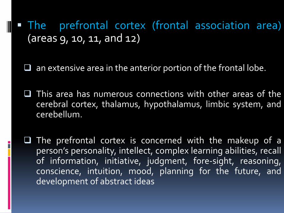
▪ The prefrontal cortex (frontal association area)(areas 9, 10, 11, and 12)
❑ an extensive area in the anterior portion of the frontal lobe.
❑ This area has numerous connections with other areas of thecerebral cortex, thalamus, hypothalamus, limbic system, andcerebellum.
❑ The prefrontal cortex is concerned with the makeup of aperson’s personality, intellect, complex learning abilities, recallof information, initiative, judgment, fore-sight, reasoning,conscience, intuition, mood, planning for the future, anddevelopment of abstract ideas
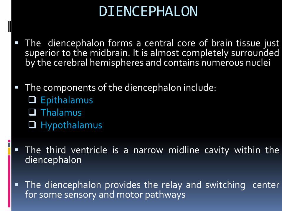
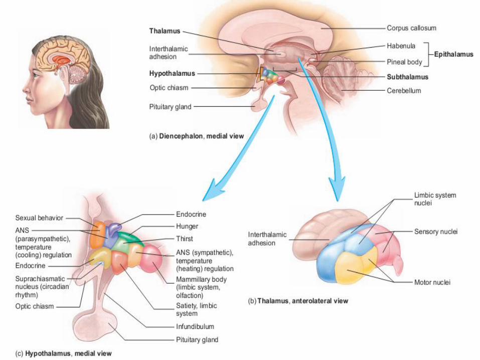
DIENCEPHALON
▪ The diencephalon forms a central core of brain tissue justsuperior to the midbrain. It is almost completely surroundedby the cerebral hemispheres and contains numerous nuclei
▪ The components of the diencephalon include:❑ Epithalamus❑ Thalamus❑ Hypothalamus
▪ The third ventricle is a narrow midline cavity within thediencephalon
▪ The diencephalon provides the relay and switching centerfor some sensory and motor pathways
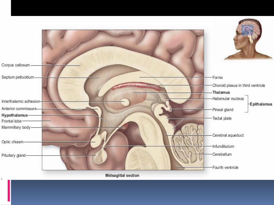
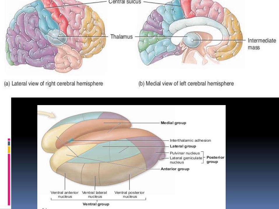
Thalamus❑ The thalamus refers to paired oval masses of gray matter that lie on each
side of the third ventricle. The lateral portions of thalamus are connectedin the center by a small stalk called the interthalamic adhesion
❑ Anterior to thalamus is present anterior commissure, posterior to it ispresent pineal gland, superior to it is present lateral ventricles, inferior to itis present mid brain
❑ Each part of the thalamus is a gray matter mass composed of group ofnuclei from which axons project to particular regions of the cerebralcortex.
❑ Sensory impulses from all the conscious senses except olfaction convergeon the thalamus and synapse in at least one of its nuclei
❑ The thalamus is the principal and final relay point for sensory informationthat will be processed and projected to the primary somatosensory cortex.Thalamus acts as an information filter for those sensory stimuli
❑ Some of the thalamic nuclei are involved with controlling skeletal muscles.They connect to, and interact with, other parts of the brain that controlskeletal muscle contraction, especially the motor areas of the cerebralcortex, the cerebellum, and the basal nuclei
❑ Some of the thalamic nuclei are involved with the limbic system andemotions. They connect different parts of the limbic system and influencemood and actions associated with strong emotions, such as fear and rage
Epithalamus
▪ The epithalamus is a small area superior and posterior to the thalamus consists of the pineal gland and habenularnuclei
▪ The pineal gland is a small pea sized, pine shaped gland that protrudes from the posterior midline of the third ventricle. The pineal gland is part of the endocrine system because it secretes the hormone melatonin which has role in maintaining circadian rhythm
▪ The posterio-superior portion of the epithalamus houses the habenular nuclei. The habenular nuclei, are involved in olfaction, especially emotional responses to odors

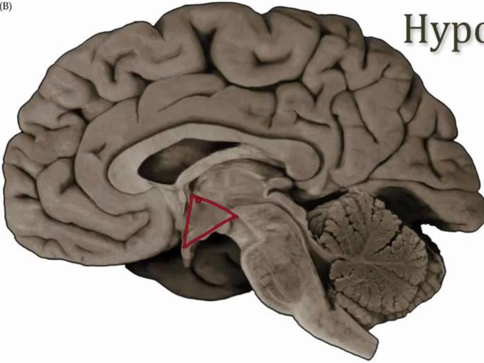
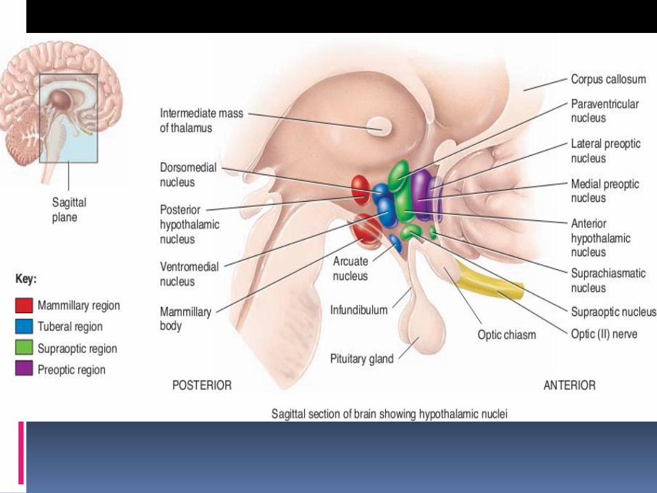
Hypothalamus▪ The hypothalamus is a collection of nuclei that is present in theanteroinferior region of the diencephalon.
▪ It is connected to other parts of brain and spinal cord and controlautonomic, emotional and basic body functions
▪ A thin, stalklike infundibulum extends inferiorly from thehypothalamus to attach to the pituitary gland
▪ Hypothalamus performs following functions
❑ Body-temperature regulation.❑ Regulation of water and electrolyte balance.(ADH)❑ Regulation of hunger and control of gastrointestinal activity.❑ Emotions. (including anger, fear, pain, and pleasure).❑ Control of endocrine functions. The hypothalamus produces
neurosecretory chemicals that stimulate the anterior and posterior pituitary to release various hormones.

Mesencephalon / Midbrain
▪ The mesencephalon (or midbrain) is the superiorportion of the brainstem
▪ It extends from diencephalon to pons and is about2.5 cm long.
▪ Cerebral aqueduct passes through the midbrain,connecting the third ventricle above with the fourthventricle below
▪ The midbrain contain both nuclei and tracts.

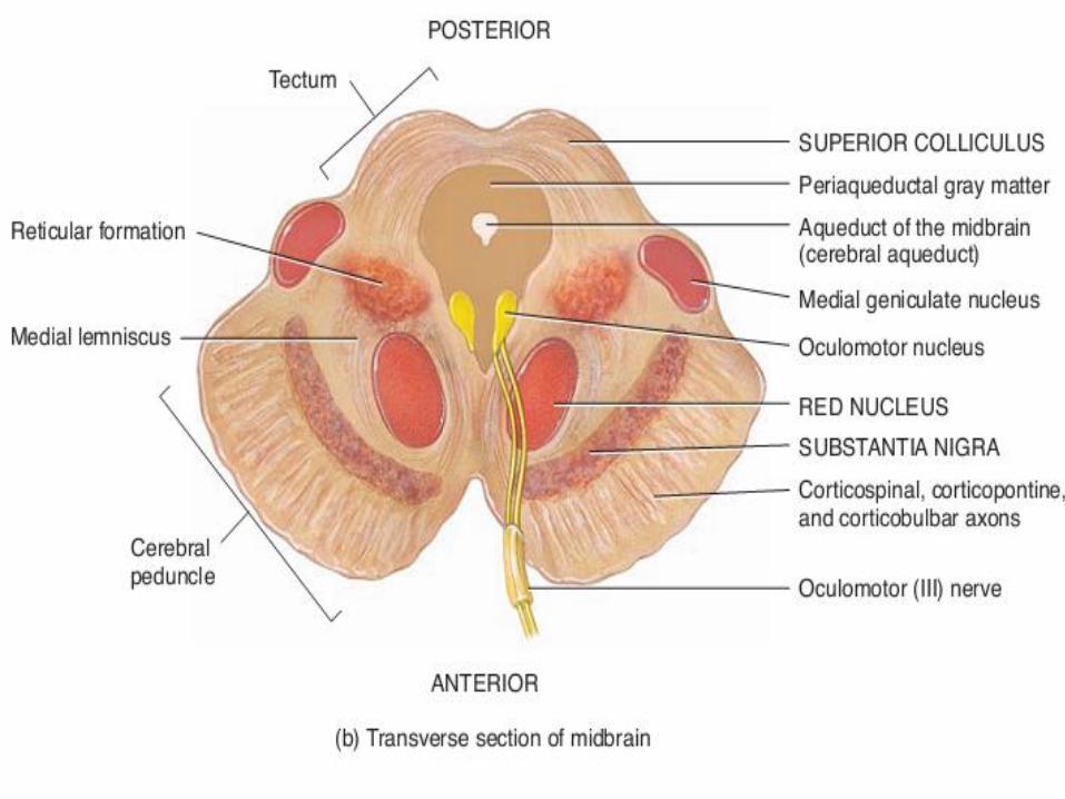
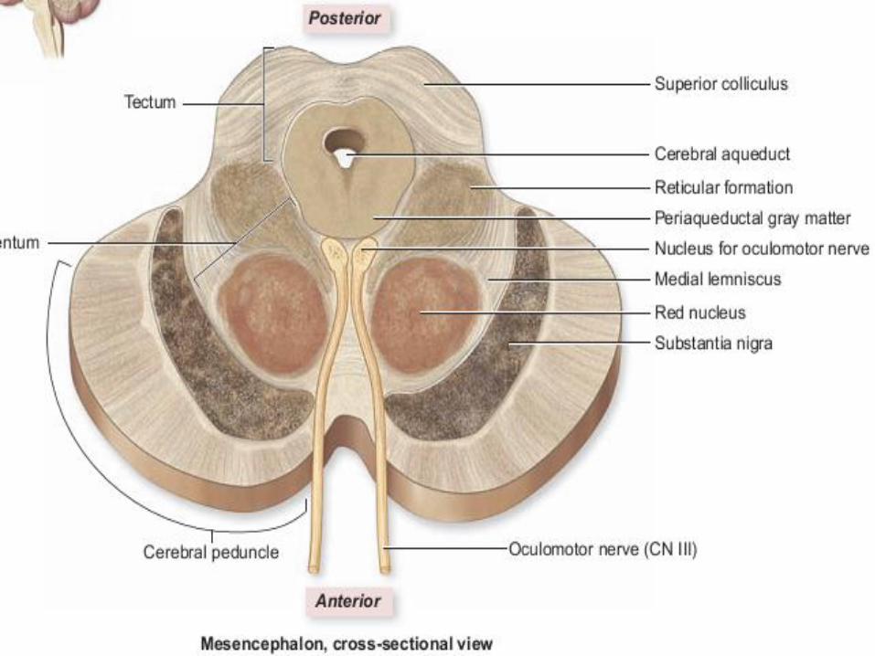
▪ Anterior of midbrain contain bundles of axon called cerebral peduncles,posterior of mid brain contain four (two pairs) of elevation which are sensorynuclei collectively called tectum, in middle is present cerebral aqueductaround which are present masses of gray matter named Periaqueductalgray, substantia nigra, red bodies and reticular formation
▪ Cerebral peduncles are motor tracts located on the anterolateral surfaces ofthe mesencephalon conduct nerve impulses from motor areas in thecerebral cortex to the spinal cord, medulla, and pons.
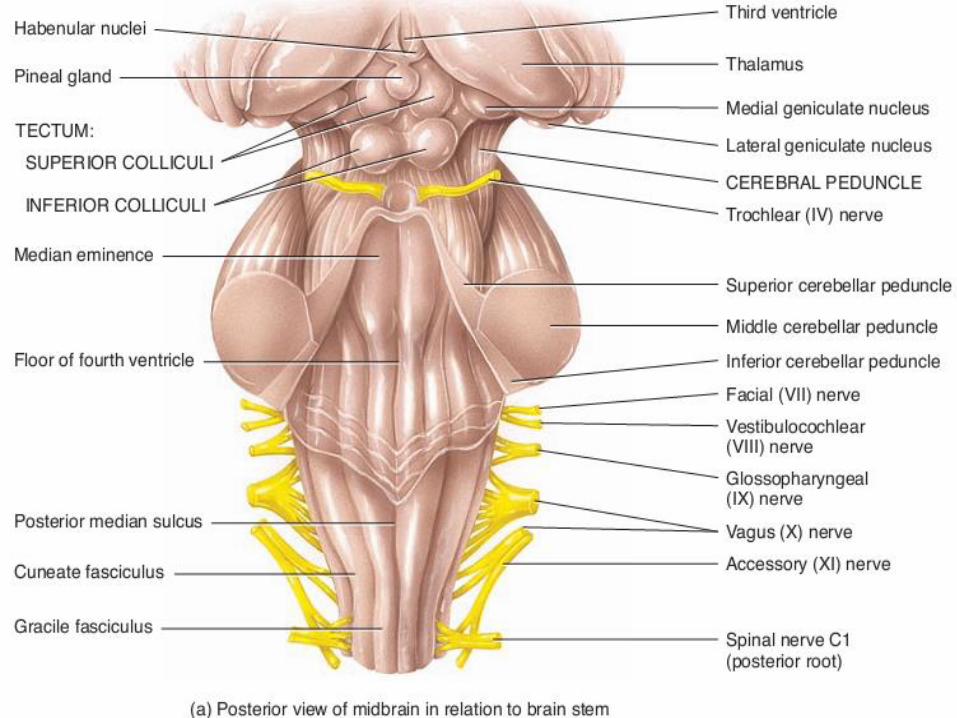
▪ Tectum
❑ is present is the posterior region of the mesencephalon dorsal to thecerebral aqueduct made up of two pairs of sensory nuclei, the superior andinferior colliculi, which are collectively called the tectal plate or corporaquadrigemina.
❑ These nuclei are relay stations in the processing pathway of visual andauditory sensations.
❑ The superior colliculi (superior nuclei) act as “visual reflex centers” becausethey help visually track moving objects and control reflexes such as turningthe eyes and head in response to a visual stimulus.
❑ The paired inferior colliculi are the “auditory reflex centers,” meaning thatthey control reflexive turning of the head and eyes in the direction of asound.
Substantia nigra
▪ consists of bilaterally symmetrical nuclei within the mesencephalon.
▪ Its name derives from its black appearance, which is due to melanin pigmentation.
▪ Neurons in substantia nigra, release dopamine, extend from the substantia nigra tothe basal nuclei, help control subconscious muscle activities emotional response,and ability to experience pleasure and pain
Red nuclei
▪ The mid brain contain left and right red nuclei, which look reddish due to their richblood supply and an iron-containing pigment in their neuronal cell bodies.
▪ Axons from the cerebellum and cerebral cortex form synapses in the red nuclei,which help control muscular movements
Rerticular formation
▪ the brain stem consists of small clusters of neuronal cell bodies (gray matter)interspersed among small bundles of myelinated axons (white matter). The broadregion where white matter and gray matter exhibit a netlike arrangement is knownas the reticular formation.
▪ It extends from the superior part of the spinal cord, throughout the brain stem, andinto the inferior part of the diencephalon.
▪ Neurons within the reticular formation have both ascending (sensory) anddescending (motor) functions.
▪ The ascending portion of the reticular formation is called the reticular activatingsystem (RAS), which consists of sensory axons that project to the cerebral cortex,both directly and through the thalamus. They have role in arousal andconsciousness
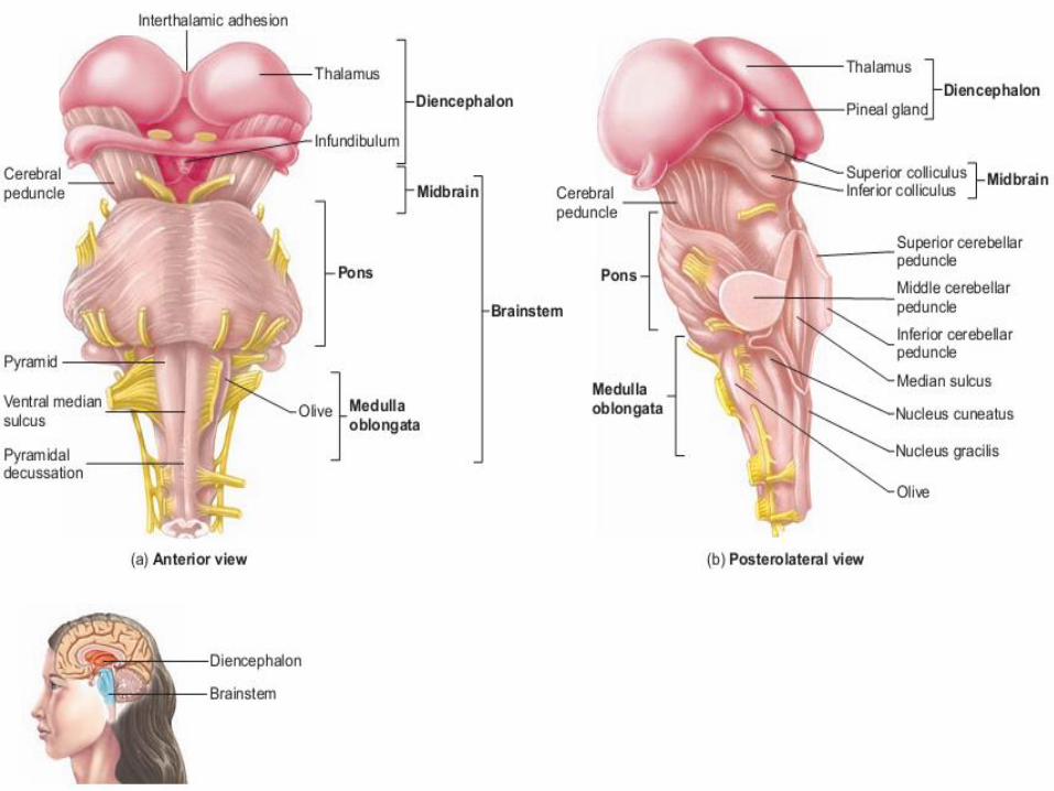
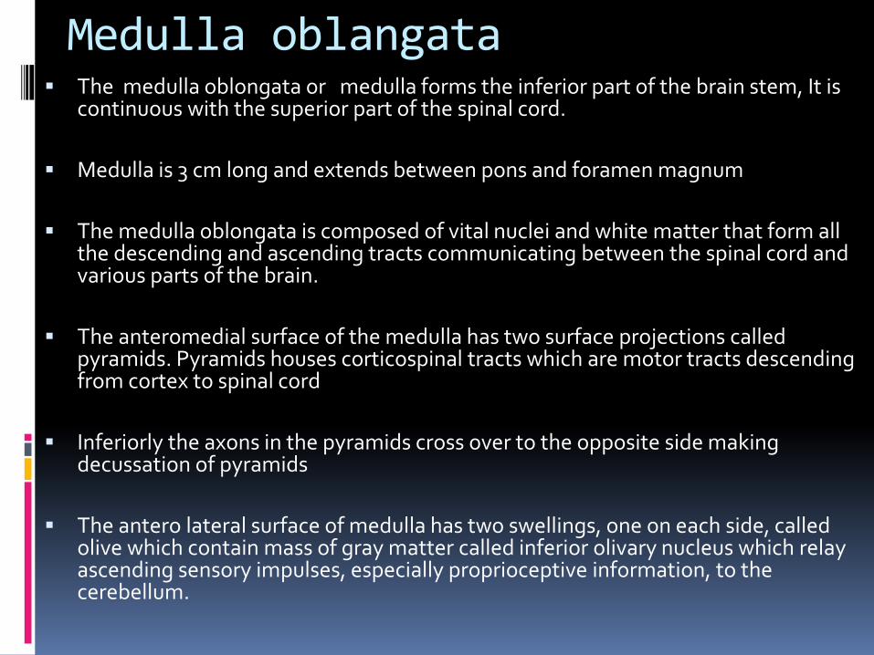
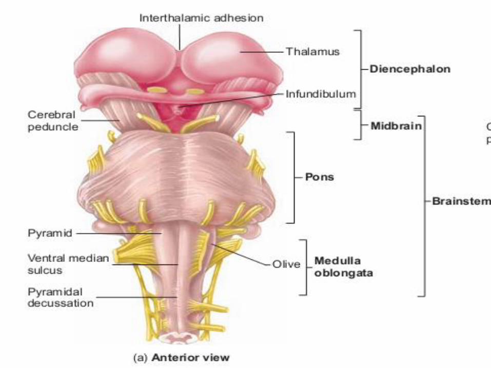
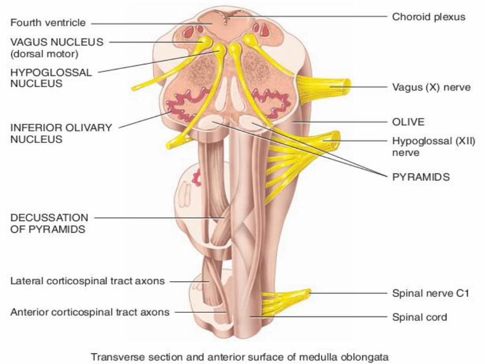
Medulla oblangata▪ The medulla oblongata or medulla forms the inferior part of the brain stem, It is
continuous with the superior part of the spinal cord.
▪ Medulla is 3 cm long and extends between pons and foramen magnum
▪ The medulla oblongata is composed of vital nuclei and white matter that form all the descending and ascending tracts communicating between the spinal cord and various parts of the brain.
▪ The anteromedial surface of the medulla has two surface projections called pyramids. Pyramids houses corticospinal tracts which are motor tracts descending from cortex to spinal cord
▪ Inferiorly the axons in the pyramids cross over to the opposite side making decussation of pyramids
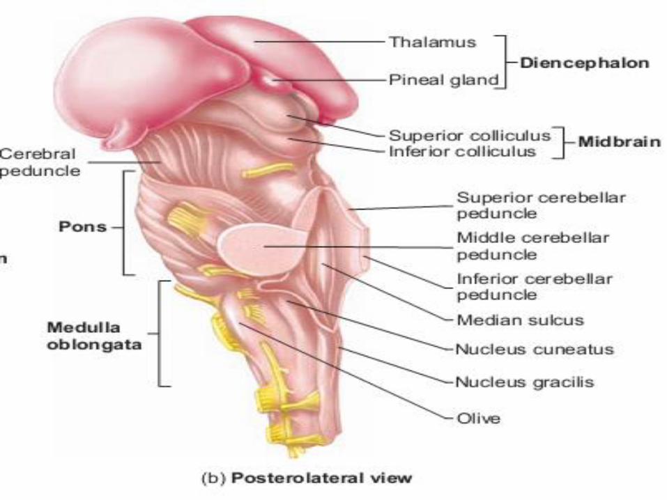
▪ The antero lateral surface of medulla has two swellings, one on each side, called olive which contain mass of gray matter called inferior olivary nucleus which relay ascending sensory impulses, especially proprioceptive information, to the cerebellum.
▪ Other than these medulla oblongata have additional nuclei that havevarious functions:
❑ The cranial nerve nuclei are associated with the vestibulocochlear (CNVIII), glossopharyngeal (CN IX), vagus (CN X), accessory (CN XI), andhypoglossal (CN XII) cranial nerves.
❑ In addition there are paired nucleus cuneatus and the nucleus gracilis ,which relay somatic sensory information to the thalamus. The nucleuscuneatus receives sensory innervation from the arm and hand of thesame side. The nucleus gracilis receives sensory information from the legand lower limbs of the same side
❑ Medulla contains several autonomic nuclei, which regulate vitalfunctions. Autonomic nuclei group together to form centers in themedulla oblongata. The important autonomic centers in medulla arecardiac center ( regulate heart rate and contraction), vasomotor center(control blood pressure) and respiratory center ( regulate rate ofrespiration)
❑ Medulla also house nuclei for various reflexes like coughing, sneezing,salivating, swallowing, gagging, and vomiting
Pons▪ The pons is a bulging region on the anterior part of the brainstem that
forms from part of the metencephalon.
▪ It contains ascending and descending nerve tracts, as well as several nuclei
▪ Within the pons are sensory and motor tracts that connect to the brain and spinal cord
▪ In addition, the middle cerebellar peduncles are transverse groups of fibers that connect the pons to the cerebellum
▪ The pons also houses two autonomic respiratory centers: the pneumotaxiccenter and the apneustic center
▪ The pons houses sensory and motor cranial nerve nuclei for the trigeminal (CN V), abducens (CN VI), and facial (CN VII) cranial nerves.
▪ It also houses superior olivary complex which receives auditory input and is involved in the pathway for sound localization

Cerebellum ▪ The cerebellum is the second largest structure of the brain.
It is located in the metencephalon and occupies the inferior and posterior aspect of the cranial cavity.
▪ The cerebellum is separated from the overlying cerebrum by a transverse fissure.
▪ A portion of the meninges called the tentorium cerebelliextends into the transverse fissure.
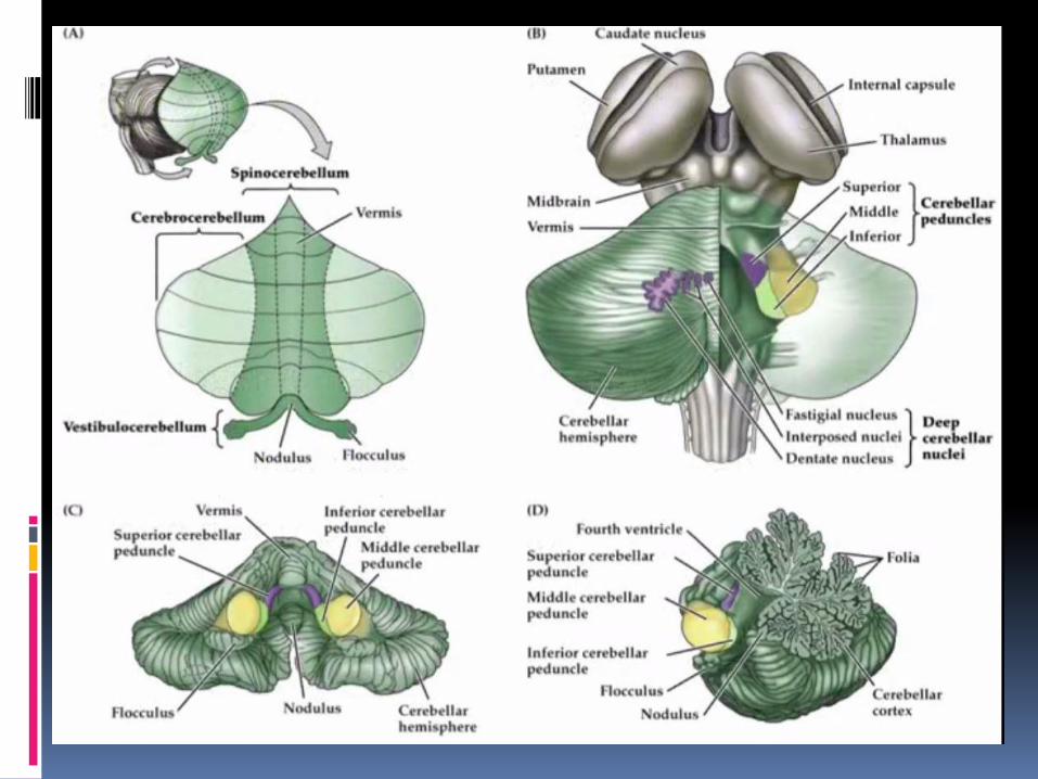
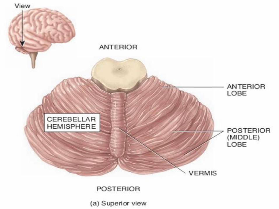
▪ The cerebellum consists of two hemispheres and a central constricted area called the vermis
▪ The falx cerebelli is the portion of the meninges that partially extends between the hemispheres
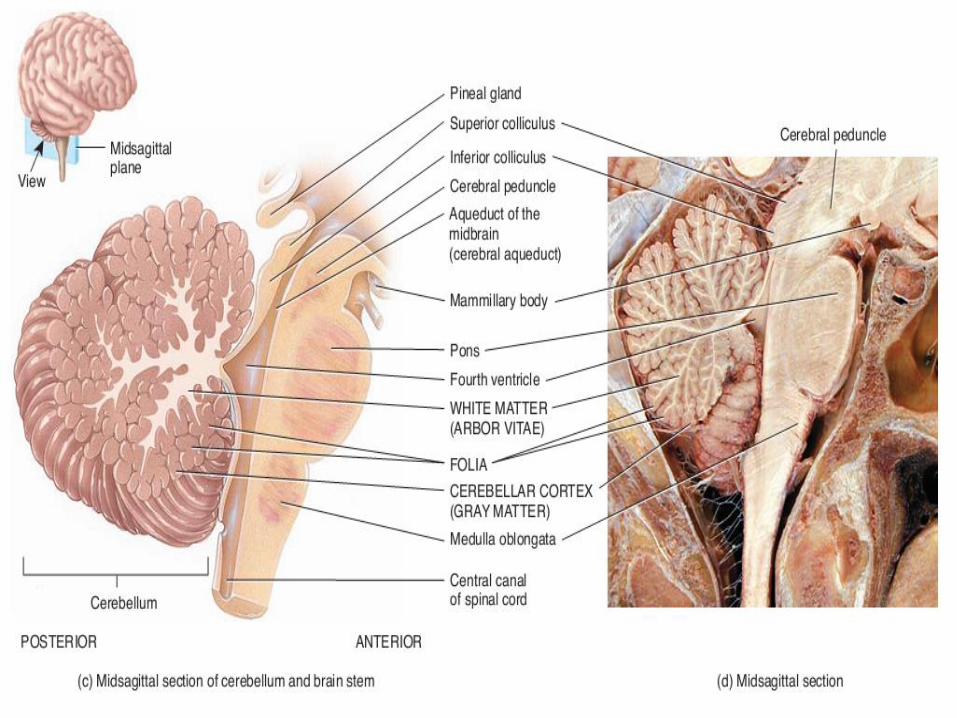
▪ Cerebellum is made up of three layers:
❑ The outer layer of gray matter called cerebellar cortex
❑ The middle layer of tracts of white matter arranged in a branching pattern liketree called arbor vitae
❑ Innermost regions of gray matter which are cerebellar nuclei from where axonsarise and connect various regions of brain
▪ Three paired cerebellar peduncles which are bundles of axons attach thecerebellum to the brain stem. These are:
❑ The superior cerebellar peduncles contain axons that extend from thecerebellum to the red nuclei of the midbrain and to several nuclei of thethalamus
❑ The middle cerebellar peduncles axons carry impulses for voluntary movementsfrom the pontine nuclei (which receive input from motor areas of the cerebralcortex) into the cerebellum
❑ The inferior cerebellar peduncles have diverse connectivity that includesconnectivity with spinal cord for proprioception from limbs, connectivity withvestibular apparatus and nulcei, connectivity with olivary nuclei of medulla andconnectivity with reticular formation
▪ The primary function of the cerebellum is toevaluate how well movements initiated by motorareas in the cerebrum are actually being carriedout.
▪ Cerebellum detects the discrepancies in thecomplex movements of muscles and sendfeedback to the motor cortex for correction.
▪ It is also involved in maintaining posture andbalance while performing motor activities
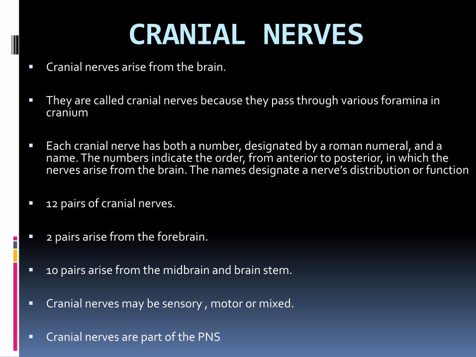
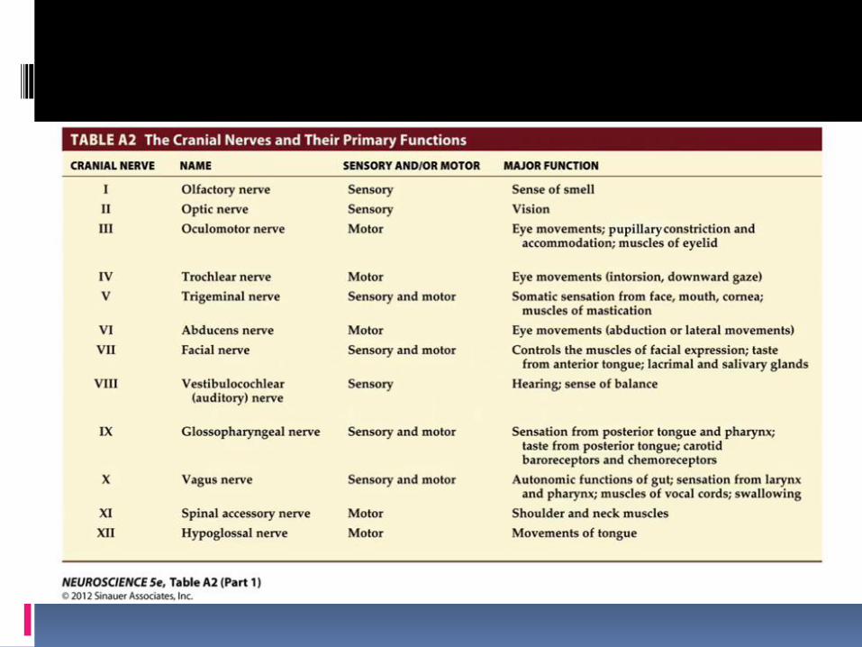
CRANIAL NERVES▪ Cranial nerves arise from the brain.
▪ They are called cranial nerves because they pass through various foramina in cranium
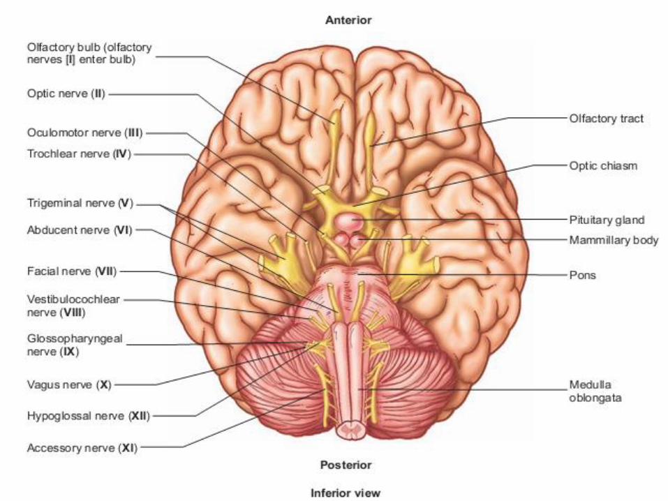
▪ Each cranial nerve has both a number, designated by a roman numeral, and a name. The numbers indicate the order, from anterior to posterior, in which the nerves arise from the brain. The names designate a nerve’s distribution or function
▪ 12 pairs of cranial nerves.
▪ 2 pairs arise from the forebrain.
▪ 10 pairs arise from the midbrain and brain stem.
▪ Cranial nerves may be sensory , motor or mixed.
▪ Cranial nerves are part of the PNS
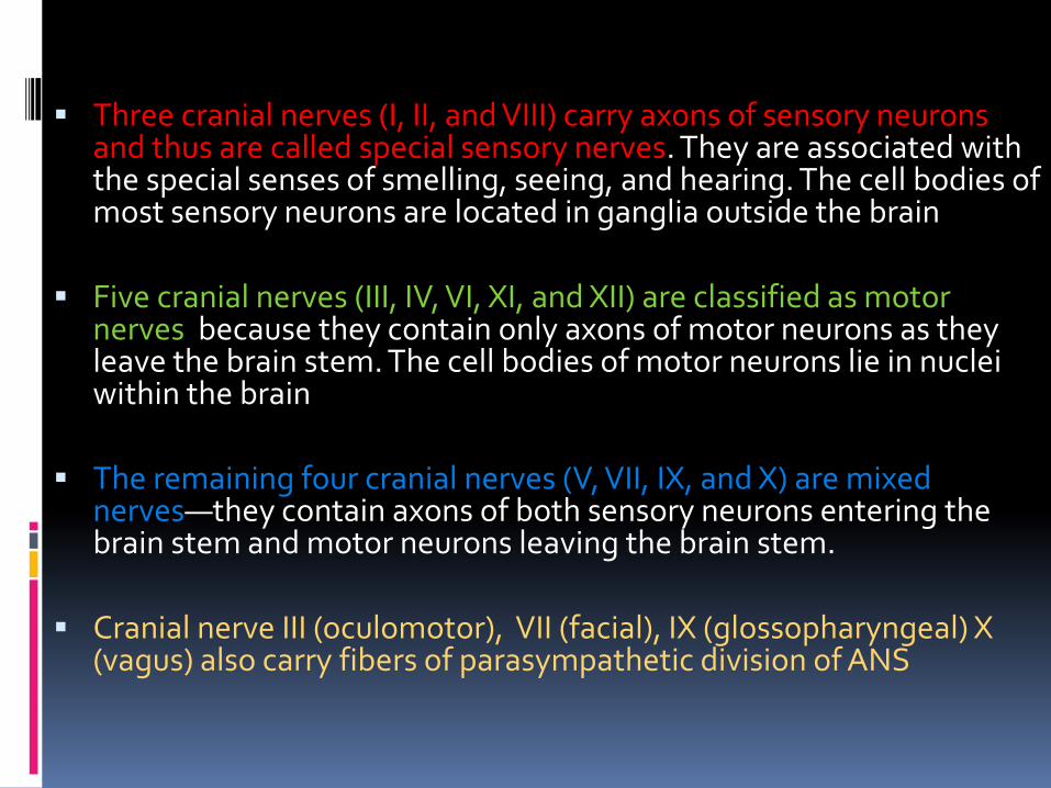
▪ Three cranial nerves (I, II, and VIII) carry axons of sensory neurons and thus are called special sensory nerves. They are associated with the special senses of smelling, seeing, and hearing. The cell bodies of most sensory neurons are located in ganglia outside the brain
▪ Five cranial nerves (III, IV, VI, XI, and XII) are classified as motor nerves because they contain only axons of motor neurons as they leave the brain stem. The cell bodies of motor neurons lie in nuclei within the brain
▪ The remaining four cranial nerves (V, VII, IX, and X) are mixed nerves—they contain axons of both sensory neurons entering the brain stem and motor neurons leaving the brain stem.
▪ Cranial nerve III (oculomotor), VII (facial), IX (glossopharyngeal) X (vagus) also carry fibers of parasympathetic division of ANS
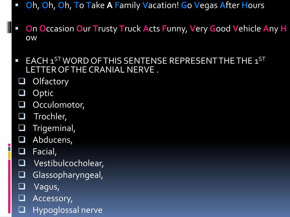
▪ Oh, Oh, Oh, To Take A Family Vacation! Go Vegas After Hours
▪ On Occasion Our Trusty Truck Acts Funny, Very Good Vehicle Any How
▪ EACH 1ST WORD OF THIS SENTENSE REPRESENT THE THE 1ST
LETTER OF THE CRANIAL NERVE .
❑ Olfactory
❑ Optic
❑ Occulomotor,
❑ Trochler,
❑ Trigeminal,
❑ Abducens,
❑ Facial,
❑ Vestibulcocholear,
❑ Glassopharyngeal,
❑ Vagus,
❑ Accessory,
❑ Hypoglossal nerve

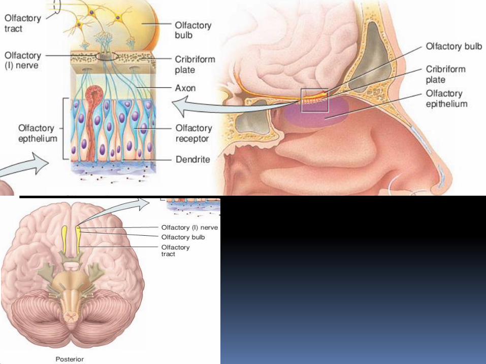
Olfactory nerve▪ pure sensory nerve
▪ Very short nerve
▪ contains axons that conduct nerve impulses for olfaction, the sense of smell
▪ The olfactory epithelium occupies the superior part of the nasal cavity
▪ The olfactory receptors within the olfactory epithelium are bipolar neurons from where this nerve originate. Each has a single odor-sensitive dendrite projecting from one side of the cell body and an unmyelinated axon extending from the other side
▪ Bundles of axons extend through olfactory foramina of ethmoid bone on each side of nose making the right and left olfactory nerves
▪ Olfactory nerves end in the brain in paired masses of gray matter called the olfactory bulbs from which axons arise making the olfactory tracts which end in primary olfactory area in temporal lobe of cerebral cortex
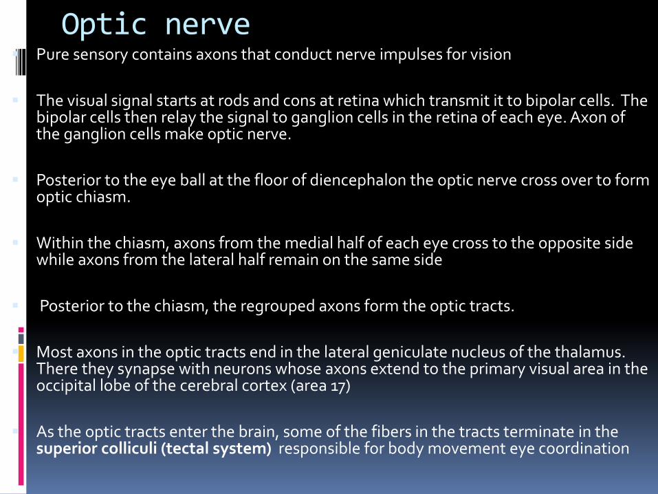
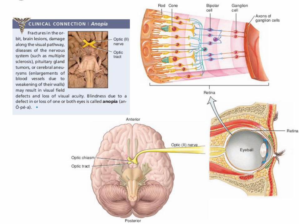
Optic nerve▪ Pure sensory contains axons that conduct nerve impulses for vision
▪ The visual signal starts at rods and cons at retina which transmit it to bipolar cells. The bipolar cells then relay the signal to ganglion cells in the retina of each eye. Axon of the ganglion cells make optic nerve.
▪ Posterior to the eye ball at the floor of diencephalon the optic nerve cross over to form optic chiasm.
▪ Within the chiasm, axons from the medial half of each eye cross to the opposite side while axons from the lateral half remain on the same side
▪ Posterior to the chiasm, the regrouped axons form the optic tracts.
▪ Most axons in the optic tracts end in the lateral geniculate nucleus of the thalamus. There they synapse with neurons whose axons extend to the primary visual area in the occipital lobe of the cerebral cortex (area 17)
▪ As the optic tracts enter the brain, some of the fibers in the tracts terminate in the superior colliculi (tectal system) responsible for body movement eye coordination
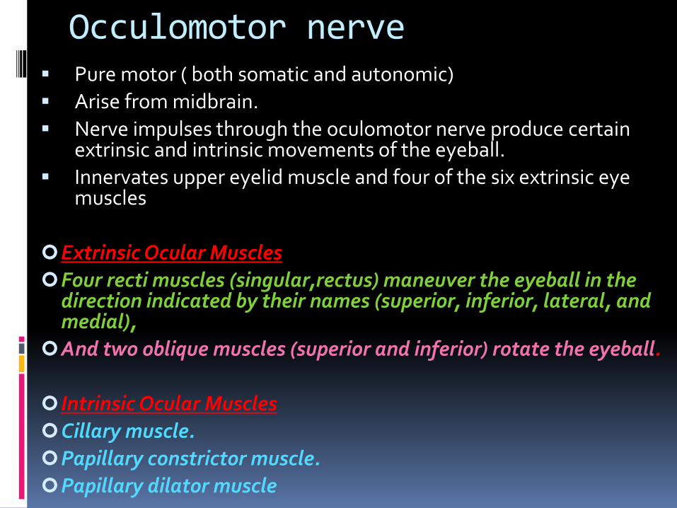
Occulomotor nerve▪ Pure motor ( both somatic and autonomic)
▪ Arise from midbrain.
▪ Nerve impulses through the oculomotor nerve produce certain extrinsic and intrinsic movements of the eyeball.
▪ Innervates upper eyelid muscle and four of the six extrinsic eye muscles
Extrinsic Ocular Muscles
Four recti muscles (singular,rectus) maneuver the eyeball in the direction indicated by their names (superior, inferior, lateral, and medial),
And two oblique muscles (superior and inferior) rotate the eyeball.
Intrinsic Ocular Muscles
Cillary muscle.
Papillary constrictor muscle.
Papillary dilator muscle
Somatic motor function
▪ It divides into superior and inferior branches as it passes through thesuperior orbital fissure in the orbit.
▪ The superior branch innervates the superior rectus muscle, whichmoves the eyeball superiorly, and the levator palpebrae superiorismuscle, which raises the upper eyelid.
▪ The inferior branch innervates the medial rectus, inferior rectus,and inferior oblique eye muscles for medial, inferior, and superiorlateral movement of the eyeball, respectively
Autonomic motor function
▪ In addition, fibers from the inferior branch of the oculomotor nerveenter the eyeball to supply parasympathetic autonomic motorinnervation to the intrinsic smooth muscles of the iris for pupilconstriction and to the muscles within the ciliary body for lensaccommodation
Trochlear nerve
▪ Motor
▪ Arise from nuclei in midbrain
▪ Innervate the superior oblique muscle of the eyeball,another extrinsic eyeball muscle that controlsmovement of the eyeball
▪ Motor impulses to the superior oblique cause theeyeball to rotate downward and away from the midline.
Trigeminal▪ Originate in pons▪ Mixed cranial nerve (both sensory and motor)▪ Sensory component is more strong and extensive than motor▪ The larger sensory root immediately enlarges into a
swelling called the trigeminal ganglion.▪ Three large nerves arise from the trigeminal ganglion❑ The ophthalmic nerve conducts sensory impulses from
cornea, nose, forehead, anterior scalp❑ The maxillary nerve conducts sensory impulses from
nasal mucosa, palate, gums, cheek❑ And the mandibular nerve conducts sensory impulses
from anterior two-thirds of tongue, skin of chin, lower jaw, lower teeth; one-third from sensory axons of auricle of ear
▪ The motor neurons are part of mandibular nerve and supply muscles of mastication
Abducens
▪ Motor
▪ originate from a nucleus in the pons
▪ Somatic motor axons extend from the nucleus to the lateral rectus muscle of the eyeball, move the eyeball away from the midline laterally
Facial nerve▪ Mixed nerve
▪ Has autonomic fibers as well
▪ Arise from pons
▪ The motor neurons arise from a nucleus in the pons and innervate middle ear, facial, scalp, and neck muscles. Nerve impulses propagating along these axons cause contraction of the muscles of facial expression and other movements of muscles in face and neck
▪ Its sensory axons extend from the taste buds of the anterior two-thirds of the tongue. The taste buds act as chemo receptors. From here the sensory axons pass to the geniculate ganglion which is the enlargement of the facial nerve just before the entrance of the sensory portion into the pons. From the pons, axons extend to the thalamus, and then to the gustatory areas of the cerebral cortex
▪ The sensory portion of the facial nerve also contains axons from skin in the ear canal that relay touch, pain, and thermal sensations.
▪ Additionally, proprioceptors from muscles of the face and scalp relay information through their cell bodies in a nucleus in the midbrain
▪ Autonomic component Increases secretions of the lacrimal gland of the eye as well as the submandibular and sublingual salivary glands through parasympathetic fibers
Vestibulocochlear▪ Sensory nerve▪ Two branches – vestibular branch which carries impulses for
equilibrium and cochlear branch which carries impulses for hearing
▪ Sensory axons in the vestibular branch extend from the semicircular canals, the saccule, and the utricle of the inner ear to the vestibular ganglion, where the cell bodies of the neurons are located and end in vestibular nuclei in the pons and cerebellum
▪ Sensory axons in the cochlear branch arise in the spiral organ (organ of Corti) in the cochlea of the internal ear. The cell bodies of cochlear branch sensory neurons are located in the spiral ganglion of the cochlea. From there, axons extend to nuclei in the medulla oblongata and end in the thalamus.
Glossopharyngeal nerve▪ Mixed nerve
▪ Has autonomic fibers as well
▪ nuclei present in medulla
▪ Sensory axons of the glossopharyngeal nerve arise from
❑ taste buds on the posterior one-third of the tongue reaching the thalamus, where they synapse with fibers that convey the impulses to the gustatory area of the cerebral cortex.
❑ proprioceptors from some swallowing muscles supplied by the motor portion
❑ baroreceptors (pressure-monitoring receptors) in the carotid sinus that monitor blood pressure,
❑ chemoreceptors (receptors that monitor blood levels of oxygen and carbon dioxide) in the carotid bodies near the carotid arteries
▪ The motor fibers innervates the stylopharyngeus (pharynx muscle)
▪ The parasympathetic fibers of ANS are responsible for increase in secretion of parotid salivary gland
Vagus nerve▪ Mixed nerve▪ Also has autonomic function ▪ Nuclei in medulla▪ Somatic motor supply muscles of the pharynx, larynx, and
soft palate that are used in swallowing, vocalization, and coughing
▪ Autonomic motor has parasympathetic fibers that Innervates visceral smooth muscle, cardiac muscle, lungs, pharynx, larynx, trachea, and most abdominal organs
▪ Sensory portion collect visceral sensory information from pharynx, larynx, heart, lungs, and most abdominal organs.
▪ Sensory fibers helps in the sensation of hunger pangs, distension, intestinal discomfort, or laryngeal movements. Sensory fibers also arise from proprioceptors in the muscles innervated by the motor fibers of this nerve.
▪ Damage to Vagus nerve results in death
Accessary nerve▪ Motor nerve
▪ Has two parts: one is cranial accessory nerve other is spinal accessory nerve
▪ The cranial accessory nerve arise from medulla innervates the skeletal muscles of the soft palate, pharynx, and larynx, which contract reflexively during swallowing.
▪ The spinal accessory nerve arise in the anterior gray horn of the first five segments of the cervical portion of the spinal cord. The axons from the segments exit the spinal cord laterally and come together, ascend through the foramen magnum and then exit through the jugular foramen along with the vagus and glossopharyngeal nerves.
▪ The accessory nerve conveys motor impulses to the sternocleidomastoid and trapezius muscles to coordinate head movements.
Hypoglossal Nerve
▪ Motor nerve
▪ The motor fibers arise from the hypoglossal nucleus within the medulla oblongata and pass through the hypoglossal canal of the skull to innervate both the extrinsic and intrinsic muscles of the tongue.
▪ Motor impulses along these fibers account for the coordinated contraction of the tongue muscles that is needed for such activities as food manipulation, swallowing, and speech
Related Documents