-Page 1 of 42- .::VOLUME 17, LESSON 4::. Ventilation/perfusion lung scintigraphy: What is still needed? A review considering technetium-99m-labeled macro-aggregates of albumin Continuing Education for Nuclear Pharmacists And Nuclear Medicine Professionals By Klaus Zöphel Department of Nuclear Medicine, Carl Gustav Carus Medical School, University of Technology Dresden, Fetscherstraße 74, 01307 Dresden, Germany Claudia Bacher-Stier Bayer Schering Pharma AG, Berlin, Germany Jörg Pinkert Bayer Vital GmbH, Leverkusen, Germany Joachim Kropp Department of Nuclear Medicine, Carl-Thiem-Klinikum Cottbus gGmbH, Cottbus, Germany The University of New Mexico Health Sciences Center, College of Pharmacy is accredited by the Accreditation Council for Pharmacy Education as a provider of continuing pharmacy education. Program No. 0039-0000-13- 174-H04-P 3.0 Contact Hours or 0.3 CEUs. Initial release date: 10/23/2013

Welcome message from author
This document is posted to help you gain knowledge. Please leave a comment to let me know what you think about it! Share it to your friends and learn new things together.
Transcript

-Page 1 of 42-
.::VOLUME 17, LESSON 4::.
Ventilation/perfusion lung scintigraphy: What is still needed?
A review considering technetium-99m-labeled macro-aggregates of albumin
Continuing Education for Nuclear Pharmacists And
Nuclear Medicine Professionals
By
Klaus Zöphel
Department of Nuclear Medicine, Carl Gustav Carus Medical School, University of Technology Dresden, Fetscherstraße 74, 01307 Dresden, Germany
Claudia Bacher-Stier Bayer Schering Pharma AG, Berlin, Germany
Jörg Pinkert Bayer Vital GmbH, Leverkusen, Germany
Joachim Kropp Department of Nuclear Medicine, Carl-Thiem-Klinikum Cottbus gGmbH, Cottbus, Germany
The University of New Mexico Health Sciences Center, College of Pharmacy is accredited by the Accreditation Council for Pharmacy Education as a provider of continuing pharmacy education. Program No. 0039-0000-13-174-H04-P 3.0 Contact Hours or 0.3 CEUs. Initial release date: 10/23/2013

-Page 2 of 42-
-- Intentionally left blank --

-Page 3 of 42-
Ventilation/perfusion lung scintigraphy: What is still needed?
A review considering technetium-99m-labeled macro-aggregates of albumin
By Klaus Zöphel, Claudia Bacher-Stier, Jörg Pinkert and Joachim Kropp
Editor, CENP
Jeffrey Norenberg, MS, PharmD, BCNP, FASHP, FAPhA UNM College of Pharmacy
Editorial Board Stephen Dragotakes, RPh, BCNP, FAPhA
Michael Mosley, RPh, BCNP, FAPhA Neil Petry, RPh, MS, BCNP, FAPhA
James Ponto, MS, RPh, BCNP, FAPhA Tim Quinton, PharmD, BCNP, FAPhA
Sally Schwarz, BCNP, FAPhA Duann Vanderslice Thistlethwaite, RPh, BCNP, FAPhA
John Yuen, PharmD, BCNP
Advisory Board Dave Engstrom, PharmD, BCNP
Christine Brown, RPh, BCNP Leana DiBenedetto, BCNP
Walter Holst, PharmD, BCNP Susan Lardner, BCNP
Vivian Loveless, PharmD, BCNP, FAPhA Brigette Nelson, MS, PharmD, BCNP
Brantley Strickland, BCNP
Director, CENP Kristina Wittstrom, PhD, RPh, BCNP, FAPhA
UNM College of Pharmacy
Administrator, CE & Web Publisher Christina Muñoz, M.A.
UNM College of Pharmacy
While the advice and information in this publication are believed to be true and accurate at the time of press, the author(s), editors, or the
publisher cannot accept any legal responsibility for any errors or omissions that may be made. The publisher makes no warranty, Copyright 2013
University of New Mexico Health Sciences Center Continuing Pharmacy Education

-Page 4 of 42-
Instructions: Upon purchase of this Lesson, you will have gained access to this lesson and the corresponding assessment via the following link < https://pharmacyce.health.unm.edu > To receive a Statement of Credit you must:
1. Review the lesson content 2. Complete the assessment, submit answers online with 70% correct (you will have 2 chances to
pass) 3. Complete the lesson evaluation
Once all requirements are met, a Statement of Credit will be available in your workspace. At any time you may "View the Certificate" and use the print command of your web browser to print the completion certificate for your records. NOTE: Please be aware that we cannot provide you with the correct answers to questions you received wrong. This would violate the rules and regulations for accreditation by ACPE. The system will identify those items marked as incorrect. Disclosure: The Author(s) does not hold a vested interest in or affiliation with any corporate organization offering financial support or grant monies for this continuing education activity, or any affiliation with an organization whose philosophy could potentially bias the presentation.
-Page 5 of 42-
Copyright Permission and Acknowledgement This review article originally published in Annals of Nuclear Medicine, a publication of the Japanese Society of Nuclear Medicine, is reproduced with permission for purposes of continuing education. Annals of Nuclear Medicine (2009) 23:1-16 DOI 10.1007/s12149-008-0187-3 NOTE: This article was written for a world-side audience as an update on the place of lung perfusion imaging using Tc-99m MAA in detection of pulmonary emboli. While practice standards may differ from a local site, the intent of the article is a universal comparison of lung imaging modalities.
ABSTRACT
Lung perfusion scintigraphy (LPS) with technetium-99m-labeled macro-aggregates of albumin (Tc-99m-MAA) is well established in the diagnostic of pulmonary embolism (PE). In the last decade, it was shown that single-photon emission computer tomography (SPECT) acquisition of LPS overcame static scintigraphy. Furthermore, there are rare indications for LPS, such as preoperative quantification of regional lung function prior to lung resection or transplantation, optimization of lung cancer radiation therapy, quantification of right–left shunt, planning of intra-arterial chemotherapy, and several rare indications in pediatrics. Moreover, LPS with Tc-99m-MAA is a safe method with low radiation exposure. PE can also be diagnosed by spiral computer tomography (CT), ultrasound, magnetic resonance angiography, or pulmonary angiography (PA, former gold standard). The present review considers all these methods, especially spiral CT, and compares them with LPS with respect to sensitivity and specificity and gives an overview of established and newer publications. It shows that LPS with Tc-99m- MAA represents a diagnostic method of continuing value for PE. In comparison with spiral CT and/or PA, LPS is still diagnostically useful as mentioned in the Prospective Investigation of Pulmonary Embolism Diagnosis (PIOPED) II reports. This applies in particular to chronic or recurring embolisms, whereas currently spiral CT may be of greater value for major or life-threatening embolisms. At present, LPS cannot be replaced by other methods in some applications, such as pediatrics or in the quantification of regional pulmonary function in a preoperative context or prior to radiation therapy. LPS still has a place in the diagnostics of PE and is irreplaceable in several rare indications as described earlier.

-Page 6 of 42-
Ventilation/perfusion lung scintigraphy: What is still needed?
A review considering technetium-99m-labeled macro-aggregates of albumin
STATEMENT OF LEARNING OBJECTIVES: Upon successful completion of this lesson, the reader should be able to:
1. Discuss the pharmacokinetic basis of Tc-99m labeled macroaggregated albumin in lung perfusion imaging.
2. Explain the role on planar (static) Tc-99m MAA lung perfusion in the diagnosis of pulmonary emboli
3. Compare lung perfusion scintigraphy with other diagnostic methods for pulmonary emboli.
4. Discuss the safety aspects of Tc-99 MAA lung perfusion imaging.
5. List other indications for Tc-99m MAA lung perfusion imaging. Acronyms:
CT Computer tomography NPV Negative Predictive Value HP High probability PA Pulmonary angiography ICRP International Council on Radiation Protection PE Pulmonary embolism IP Intermediate probability PIOPED Prospective Investigation of
Pulmonary Embolism Diagnosis LP Low probability PPV Positive predictive value LPS Lung Perfusion Scintigraphy SPECT Single photon computer tomography LS Lung scintigraphy TEE Tranesophageal echocardioagraphy LVS Lung Ventilation Scintigraphy TTE Transthoracic echocardiography MAA Macroaggregated albumin MR Magnetic Resonance V/Q Ventilation and perfusion imaging MRPA Magnetic Resonance Pulmonary Angiography VTE Venous thromoembolic event

-Page 7 of 42-
COURSE OUTLINE
ABSTRACT ............................................................................................................................................................................ 5
PHARMACOKINETIC BASIS FOR LUNG PERFUSION SCINTIGRAPHY WITH TC-99M-LABELED MACRO-ALBUMIN AGGREGATES . 9 STATIC TC-99M-MAA LUNG PERFUSION SCINTIGRAPHY IN THE DIAGNOSIS OF PULMONARY EMBOLISM ........................... 10
Pulmonary embolism .................................................................................................................................................. 10 Initial diagnostic measures for suspected PE............................................................................................................ 10 Lung scintigraphy in the diagnostic decision tree ...................................................................................................... 11 Diagnostic accuracy of static Tc-99m-MAA lung perfusion scintigraphy ................................................................ 12
Normal .........................................................................................................................................................................................13 High probability ...........................................................................................................................................................................13 Low probability ...........................................................................................................................................................................14 Intermediate probability (indeterminate) ...................................................................................................................................14 Agreement with static LPS .........................................................................................................................................................15
TC-99M-MAA LUNG PERFUSION SCINTIGRAPHY WITH SPECT .......................................................................................... 15 V/Q scintigraphy in patients with concomitant chronic obstructive lung disease ........................................................ 17
LUNG PERFUSION SCINTIGRAPHY IN COMPARISON WITH OTHER DIAGNOSTIC METHODS FOR PE ........................................ 17 Spiral CT in comparison with LPS ............................................................................................................................... 17 LPS and spiral CT for the diagnosis of acute and chronic recurrent PE ..................................................................... 20 LPS or spiral CT? ......................................................................................................................................................... 20 Other imaging procedures for the diagnosis of PE ...................................................................................................... 21
Magnetic resonance pulmonary angiography (MRPA) ................................................................................................................21 Vein ultrasound of the lower leg ................................................................................................................................................22 Pulmonary angiography ...............................................................................................................................................................22 Transthoracic and transesophageal echocardiography (TTE and TEE) ......................................................................................22
SAFETY ASPECTS OF TC-99M-MAA LUNG PERFUSION SCINTIGRAPHY ............................................................................... 23 OTHER INDICATIONS FOR TC-99M-MAA LUNG PERFUSION SCINTIGRAPHY ....................................................................... 24
Preoperative evaluation of lung function prior to carcinoma resection .................................................................... 24 Preoperative evaluation of lung function in lung transplantation .............................................................................. 25 Optimization of radiation therapy for lung cancer ..................................................................................................... 25 Right–left shunt quantification .................................................................................................................................... 25 Pediatric use of Tc-99m-MAA LPS .............................................................................................................................. 26
CONCLUSIONS ................................................................................................................................................................... 26
REFERENCES ..................................................................................................................................................................... 27
ASSESSMENT QUESTIONS ............................................................................................................................................. 40

-Page 8 of 42-
-- Intentionally left blank --

-Page 9 of 42-
VENTILATION/PERFUSION LUNG SCINTIGRAPHY: WHAT IS STILL NEEDED?
A REVIEW CONSIDERING TECHNETIUM-99M-LABELED MACRO-AGGREGATES OF ALBUMIN
Klaus Zöphel, Claudia Bacher-Stier, Jörg Pinkert and Joachim Kropp
Pharmacokinetic basis for lung perfusion scintigraphy with Tc-99m-labeled macro-albumin aggregates
Lung perfusion scintigraphy (LPS) with macroaggregates of albumin (MAAs) depends on the
principle that particles >10 μm in diameter in the bloodstream are trapped in the lung at first
passage [1, 2], causing temporary “micro-embolisms” [3, 4] whose number is in direct proportion
to the local rate of blood flow [5]. Following their rapid accumulation in the lung (98% within 1–2
min [6, 7]), MAAs are mechanically and enzymically degraded, and phagocytosed in the liver [8].
For scintigraphic imaging, the MAAs are today labeled with the nuclide technetium-99m (Tc-99m).
Following elimination from the lung (half-time for residence 1–24 h [3, 6, 7, 9–11]) the
radioactivity is excreted, via the liver and kidneys, in the urine [7, 8, 11].
The perialveolar capillaries have an average diameter of ∼8 μm (range 7–10 μm [12]) and the
arterioles ∼25 μm (15–35 μm [2, 12]). The ideal particle size for good image quality has been stated
to be 20–50 μm [2] or 15–40 μm [13]. Today, a range of 25–60 μm is preferred; smaller particles are
degraded very rapidly, whereas those in excess of ∼60 μm can block the capillary tree at higher
levels and potentially cause hemodynamic disturbance. Typical Tc-99m-MAA preparations have a
size range of 5–100 μm [3] with 80–90% of the particles within the range 10–75 μm [6–9, 14]. Tc-
99m-labeled microspheres of human serum albumin are rarely used today because of a considerably
slower metabolic removal and the consequent safety disadvantage for patients with pulmonary
hypertension and children.
The number of microembolisms caused by MAAs is determined by the ratio between the number of
particles injected and the numbers of lung arterioles and capillaries. An adult has some 2.8 × 10⁹

-Page 10 of 42-
capillaries and 3.0 × 10⁸ arterioles [12]. Therefore, a typical injection with 3 × 10⁵ Tc-99m-MAA
particles will—depending on the exact distribution of particle sizes and varying according to local
blood flow rate—temporarily block every millionth capillary and every thousandth arteriole [1]; the
latter will not be blocked by particles less than 20 μm in size. Good imaging has been estimated to
require at least 60, 000 [15] or 100, 000 [16, 17] particles in adults, whereas for safety reasons not
more than 700, 000 particles should be injected [3, 16, 17]. Thus, even if the maximum number of
particles is injected, all measuring >20 μm, not more than 0.23% of the arterioles in humans with
normal lung function will be blocked (for abnormal cases see “Safety aspects of Tc-99M-MAA lung
perfusion scintigraphy” section).
Static Tc-99m-MAA lung perfusion scintigraphy in the diagnosis of pulmonary embolism
Pulmonary embolism
Pulmonary embolism (PE) is a major health problem, with an annual incidence of 0.5–1.0 per
thousand in the industrialized world [18]; it is more common still among hospital patients [19, 20],
where it is the third most common cause of death [13]. Massive embolisms are associated with
blockage of ≥70% of the terminal pulmonary vessels and are usually fatal [21]. Some 50–60% of
PE cases are caused by deep-vein thrombosis [21].
Pulmonary embolism is frequently clinically asymptomatic in the early stages, until 30% or more of
the arterial tree is occluded [22]. It is thus frequently over-looked [13, 18]; a meta-analysis o f 12
autopsy studies revealed a high percentage (>50%) of unrecognized, clinically relevant PEs [19]. The
mortality rate of unrecognized PE is ∼30% [19], but this can be reduced to 2–8% by timely
discovery and anticoagulant therapy [23, 24]. The prevalence of PE in patients clinically suspected
of having PE was found to be 27% [25]. Although it is recognized that several small PEs often
herald a large one, such a diagnosis must be reliable, as superfluous anticoagulation therapy can also
be dangerous, owing to the suppression of normal coagulation [26].
Initial diagnostic measures for suspected PE
All methods for the diagnosis of PE, both imaging and non-imaging, have characteristic strengths
and weaknesses [27]. A “decision tree” of complementary methods is therefore recommended [27,
28]. Clinical symptoms such as acute chest pain, coughing, anxiety, hemoptysis, and tachypnea are
-Page 11 of 42-
unspecific and variable [3, 21, 29], as are instrumental methods such as electrocardiography, chest X-
ray [30, 31], and arterial blood gas analysis [32]. A recently developed, relatively reliable marker for
PE is the presence of D-dimer, a degradation product of fibrin [33]. Although other factors can lead to
a positive D-dimer finding, a negative finding allows PE to be excluded with high reliability [30, 32–
37]. Many authors have therefore proposed that a positive D-dimer result be a requirement for
subsequent investigation by an imaging method [18, 27, 30] such as lung scintigraphy [17, 38], spiral
computer tomography (CT), ultrasound of the lower leg veins (positive for 70% of PE patients [39]),
ultrasound of the thorax (diagnostic accuracy 84% [40]), multidetector row CT [41]
echocardiography, or invasive pulmonary angiography (PA). In particular, the accuracy of thoracic
ultrasound is heavily contested. Thoracic ultrasound is highly unspecific and is mainly used in
countries with a lack of imaging departments. A recent study concluded that a combination of
negative spiral CT and normal ultrasonography images suffices to exclude PE in emergency contexts
[42]. The recently published PIOPED II study [43] found the sensitivity of CT angiography to be 83%
with a specificity of 96%, whereas the sensitivity of CT angiography combined with venography was
90% with specificity of 95%.
Lung scintigraphy in the diagnostic decision tree
The position of a V/Q lung scintigraphy (LS) within the diagnostic decision tree is currently as
follows:
LS is generally regarded as a method of first choice, if there are clinical signs of PE, as an
exclusion criterion [44, 45].
LS is conducted as a method of first choice if D-dimer elevation has been found [46]; this
accords with the recommendations of the European Cardiological Society [18].
For patients who have already been examined by V/Q scintigraphy, this should be repeated to
ensure the reproducibility of the results [21, 30].
Certain factors in the patient’s earlier findings, clinical symptoms, and/or the pre-test
probability [47] are also indications for V/Q LS. V/Q LS should be performed first to allow
possible exclusion of PE if the result of a thorax X-ray was normal, if the clinical pre-test
probability was low or middling [48–50], or if the patient is clinically stable. If the clinical pre-
test probability is high, then spiral CT can be used, as long as the patient is not allergic to the
contrast medium [50]; has no renal failure, hyperthyroidism, or a pacer inducing metal artifacts;
and if female, is not pregnant. For patients whose condition is unstable, especially if there is

-Page 12 of 42-
suspicion of a massive PE, echocardiography is recommended due to possible hemodynamic
consequences [18]; if the echocardiography result is unclear, then spiral CT or PA should be
performed [45, 51].
LS and spiral CT can be placed sequentially in the decision tree because the integrative-
complementary application of these procedures represents a meaningful approach to combining
the diagnostic certainty of these two methods: for example, spiral CT following an
indeterminate scintigraphic result (middling probability of PE) instead of the invasive PA [28,
52, 53]; or following any non-negative scintigraphic result [54]. A prospective study of 779
patients following an abnormal scintigraphy result showed better results from a combination of
LS and spiral CT than from either method alone [54].
LS and spiral CT are considered as options of equal value [55, 56] and are recommended
following the establishment of the clinical pre-test probability [27, 57] or an elevated D-dimer
level [27, 28, 58, 59].
In clinical routine today, clinicians still have limited knowledge of equipment being used during LPS
and spiral CT leading to different decisions about the method of choice in diagnostic imaging of
suspected acute PE [60]. In the United States as well as European countries, there is no coherence
between the primary diagnostic modality of choice and other factors including hospital size, type,
and availability of diagnostic methods [61].
Diagnostic accuracy of static Tc-99m-MAA lung perfusion scintigraphy
In 1990, the PIOPED group published a set of criteria for the determination of the probability of
PE based on pre-test probability and the results of V/Q scintigraphy [62]. These criteria were found
to provide the most reliable diagnosis of PE when compared with the “gold standard” PA. The
“modified PIOPED criteria” of 1993 [14, 63–65] are still used [17, 66–69]. They allow the
probability of PE to be assigned to one of the classes “normal” (in which the prevalence of PE is
effectively 0%), “low probability” (LP - prevalence <20%), “intermediate probability” (IP -
prevalence 20–79%), and “high probability” (HP - prevalence ≥80%).
Unfortunately, PA is still regarded as the gold standard but this is no longer true [70–72]. Sensitivity
of PA may be as low as 70% [71] and this is a reason for the huge number of false-positive results in
the PIOPED I study [62]. Therefore, we would like to replace the “gold standard” with the “former

-Page 13 of 42-
gold standard”. We summarize here the diagnostic value of these probability classes and discuss them
in relation to other, more recent studies. Only a few studies have employed the former gold standard
PA as a reference as this invasive method is ethically unjustified in patients with less than a high
probability of PE; therefore, it is used for HP patients only, and clinical course (occurrence of
thromboembolic events) is used for the others.
Normal
This can be established by a normal LPS result only (without a ventilation measurement). Its value
lies in its high negative predictive value (NPV) of 96–100% [1, 62, 67, 68, 73–75]. The incidence rate
of venous thrombo-embolic events (VTEs) after a normal scintigraphic result was correspondingly
low (Table 1): 0.3% according to the meta-analysis by van Beek et al. [69], whereas fatal VTEs were
very rare (0.15%).
High probability
Various studies have shown that the result “HP” has an overall positive predictive value (PPV) for PE
of 88%, which combined with the clinical pre-test probability rises to >95% (Table 2). Therefore, a
scintigraphic HP result can be regarded as a sufficient criterion for the initiation of anticoagulation
therapy [1, 18, 45, 51]. Of the patients investigated, 4% to 19% were in the HP category [23, 29, 62,
67, 76–80].
Table 1
STUDIES OF THE CLINICAL COURSE OF PATIENTS WITH NORMAL V/Q LUNG SCINTIGRAPHY
# of patients
Type of study Observation period (months)
Proportion of patients with non-fatal VTE (%)
Proportion of patients with fatal VTE (%)
References
131 Prospective 12 4 – [62] 68 Retrospective 2–97 – – [35] 586 Prospective 3 0.7 – [157] 113 Prospective 6 – – [81] 188 Prospective 3 – – [158] 46 Retrospective ≥6 – – [73] 693 Meta-analysis ≥3 0.3 0.15 [69] 27 Prospective 6 – – [75] 161 Prospective 3 – – [46]
>7000 7 literature studies 1996–2003
Various 0.9 Various [56]
VTE venous thrombo-embolic events

-Page 14 of 42-
Table 2
POSITIVE PREDICTIVE VALUE OF A FINDING OF “HIGH PROBABILITY OF PE”
Positive predictive value (%) Standard References
86 PA [150] 89.5 PA [160] 88 PA, clinical course [62] 96a 98a PA, clinical course [77] 83.3 PA [77] 92a PA, clinical course [16] 96a PA, clinical course [82] 96a PA [29] 94a PA, clinical course [75] 100a PA [132]
PE pulmonary embolism, PA pulmonary angiography a Including consideration of the pre-test probability
Low probability
Results of several studies are shown in Table 3. In summary, the NPV of a PIOPED LP result combined
with a low pre-test probability is about 96–99% [62, 77, 81], compared with 84–88% when clinical
information is not included [62, 82]. With the modified PIOPED criteria, independent of the pre-test
probability, 0.5–5.5% of patients with an LP result experienced PE [77, 78]. LP was found for ∼30–
80% of the patients investigated [62, 76, 79, 83].
Intermediate probability (indeterminate)
Here V/Q scintigraphy does not strengthen a positive or a negative diagnosis of PE. The frequency of
IP assignment varies greatly (up to 51% [2], generally 10–30% [29, 75, 76, 78, 79, 84]) according to
patient pre-selection (e.g., reason for investigation, experience of the physician). The use of the
modified PIOPED criteria reduced the frequency of such assignments [16, 17, 21, 65, 66, 77]. If IP is
found, then clarification by (duplex) sonography of the leg veins is recommended. If deep vein
thrombosis is discovered, anticoagulation therapy should be instituted at once [48]. If sonography
reveals nothing, then a confirmatory invasive PA should be considered [28, 85], although in practice
this is not frequently performed [86, 87]. There is controversy as to whether spiral CT is useful in
clarifying an IP finding [68, 88]. Spiral CT is of restricted diagnostic value especially in the case of
pre-existing lung disease, which often constitutes the background for an IP result [89].
-Page 15 of 42-
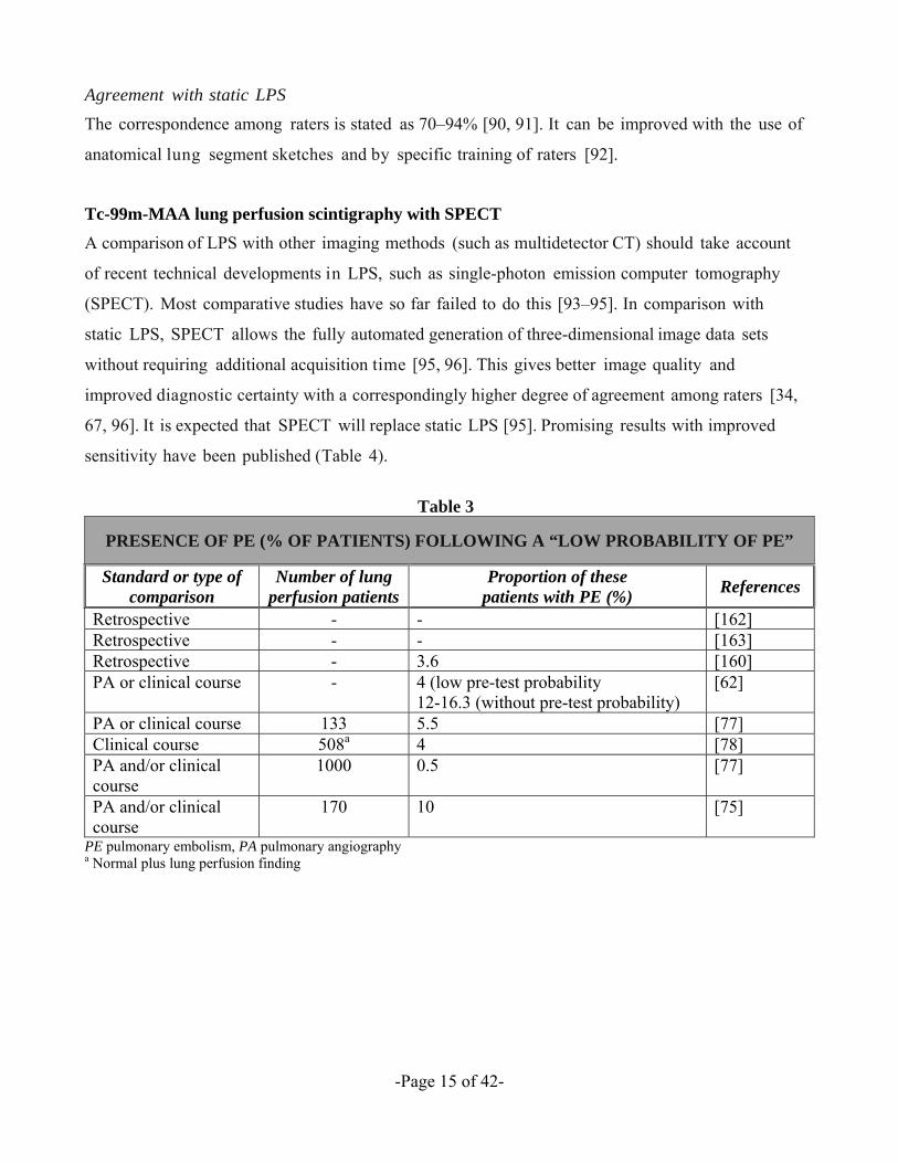
Agreement with static LPS
The correspondence among raters is stated as 70–94% [90, 91]. It can be improved with the use of
anatomical lung segment sketches and by specific training of raters [92].
Tc-99m-MAA lung perfusion scintigraphy with SPECT
A comparison of LPS with other imaging methods (such as multidetector CT) should take account
of recent technical developments in LPS, such as single-photon emission computer tomography
(SPECT). Most comparative studies have so far failed to do this [93–95]. In comparison with
static LPS, SPECT allows the fully automated generation of three-dimensional image data sets
without requiring additional acquisition time [95, 96]. This gives better image quality and
improved diagnostic certainty with a correspondingly higher degree of agreement among raters [34,
67, 96]. It is expected that SPECT will replace static LPS [95]. Promising results with improved
sensitivity have been published (Table 4).
Table 3
PRESENCE OF PE (% OF PATIENTS) FOLLOWING A “LOW PROBABILITY OF PE”
Standard or type of comparison
Number of lung perfusion patients
Proportion of these patients with PE (%)
References
Retrospective - - [162] Retrospective - - [163] Retrospective - 3.6 [160] PA or clinical course - 4 (low pre-test probability
12-16.3 (without pre-test probability) [62]
PA or clinical course 133 5.5 [77] Clinical course 508a 4 [78] PA and/or clinical course
1000 0.5 [77]
PA and/or clinical course
170 10 [75]
PE pulmonary embolism, PA pulmonary angiography a Normal plus lung perfusion finding

-Page 16 of 42-
Table 4
COMPARISON OF THE DIAGNOSTIC PREDICTIVE VALUE OF SPECT AND STATIC LS
Type of comparison
Number of patients
Static LS SPECT ReferencesSensitivity (%)
Specificity (%)
Sensitivity (%)
Specificity (%)
Retrospective 985 - - - - [99] Prospective 114 80 78 80 96 [96] Retrospective 103 68 99 92.6 98.6 [94] Prospective 83 67 85 97 91 [95]
SPECT single-photon emission computer tomography LS lung scintigraphy a Mean of values from three readers with differing experience
It was recently shown [95] that SPECT V/Q scintigraphy afforded comparable specificity (91%) and
diagnostic accuracy (94%) as multidetector spiral CT (98% and 93%, respectively), but also a higher
sensitivity than the latter (97% vs. 86%). It appears that the percentage of IP diagnoses can be reduced
by applying SPECT and the modified PIOPED criteria [94–98]. One paper claims a reduction of IP
results to only 4% [99]. Other authors believe that the modified PIOPED criteria should be adapted to
SPECT [95]. Automated interpretation of SPECT results has been compared with conventional visual
assessment; the respective sensitivity, specificity, and accuracy obtained by these methods were
95%:91%, 84%:97%, and 89%:94% [100]. It is important to note that the actual guidelines for LS of
the German Society of Nuclear Medicine, recently revised by Schümichen et al. [38], declines the
PIOPED criteria for interpretation of positive LPS results, excluding the normal scan. The PIOPED
data were obtained by single projection/ single breath ventilation scintigraphy with Xe-133. This
method as well as the technique of PA is not acceptable today. It should be clarified that ventilation
scintigraphy with Xe-133 is obsolete [in Europe] today because of its bad count statistics when
compared with Tc-99m-Technegas [Cyclomedica Austrailia], an explanation of the poor results of
PIOPED I study [62]. LPS interpretation should be performed in the context with the results of a
ventilation scintigraphy using Tc-99m-Technegas or Kr-81m, or as a minimal precondition in the
knowledge of a recent thorax X-ray. There are some reports about V/Q ratio histogram analyses
generated by software algorithms with very reliable results, but such software programs are not
generally available [96, 100]. SPECT is the method of choice to acquire the images and PIOPED
criteria are insufficient for interpretation of positive LPS results [38].

-Page 17 of 42-
V/Q scintigraphy in patients with concomitant chronic obstructive lung disease
The leading sign of PE in ventilation/perfusion scintigraphy is not the proof of the thrombus itself. but
the effect of it, in other words, the mismatch between the uptake of the ventilation (preserved) and
perfusion (absent) radio- tracer. In any area with disturbed ventilation for any reason, there is the so-
called hypoxic vasoconstriction leading to an absent perfusion so that the scintigraphic pattern is the
absent uptake of both tracers. Therefore, it is impossible to diagnose PE in those areas by nuclear
medicine methods. Because perfusion is disturbed in these areas anyway, it is not so clinically
important to confirm embolism in these segments.
Lung perfusion scintigraphy in comparison with other diagnostic methods for PE
Spiral CT in comparison with LPS
Spiral CT allows the visual representation of total or partial blockage, so that emboli in the
pulmonary blood vessels can be located [ 48]. The proportion of spiral CT examinations that give
no result because of technical failure, or give an indeterminate result, is variously reported as 2–10%
[49, 68, 101]. Errors can be caused by cardiac [102] or respiratory [49, 103] artifacts, too little contrast
medium [70, 102] or anatomical factors such as hilar nodes [31] or peribronchovascular infiltration
[49, 103]. First results from the early 1990s [104,105], admittedly only with acute central PE, indicated
a sensitivity of nearly 100% and a specificity of 96%. For segmental emboli, which are frequently
overlooked, the early instruments had a lower sensitivity and specificity (diagnostic accuracy 61–79%
[70]). The development of a faster scanning method, with resolution into thinner layers, allowed the
observation of smaller pulmonary arteries [102]. The introduction of up to 64-slice multidetectors
allows a slice thickness of 0.7–2.5 mm and better spatial resolution [70, 106]. Consequently, there is
considerable variation in literature reports of sensitivity (53–94%) and specificity (78–100%) based on
the equipment used (Table 5) [18, 21, 27, 82, 84, 107]. A meta-analysis has yielded average values of
88% for sensitivity and 92% for specificity [69]. These results should be interpreted with caution,
because no reliable references were used and only follow-up studies can give reliable results (see LPS
and spiral CT for the diagnosis of acute and chronic recurrent PE).
By today’s standards, spiral CT is not sensitive enough to detect subsegmental PEs (Table 6) [108–
110]. The meta-analysis by van Beek et al. [69] showed a sensitivity of only 50–65%. A recent study
[111] suggested a sensitivity of only 69% if subsegmental emboli were also considered (Table 6).
There is much current discussion [70] about the clinical relevance of isolated subsegmental emboli and
whether they should be treated. There is a need for long-term studies in this area. Although an isolated

-Page 18 of 42-
subsegmental PE has practically no hemodynamic or clinical relevance in an otherwise healthy and
relatively young patient, the hemodynamic impact for an older person with a history of
cardiopulmonary disease is clinically important and even fatal [49]. The incidence of isolated
subsegmental PEs among patients with suspected PE is quoted as 4–36% [25, 62, 89].
Direct comparative studies of static lung scintigrams and spiral CT results have been conducted. One
showed spiral CT to be superior in both sensitivity and specificity (Table 6). However, LS by SPECT
was superior in sensitivity and equal in specificity to spiral CT (Table 6).
Table 5
DIAGNOSTIC VALUE OF SPIRAL CT IN PE AT SEGMENTAL LEVEL
Comparison/ reference/ method
Number of patients
Sensitivity (%)
Specificity (%)
Type of comparison/comment
References
Diagnostic value of spiral CT in PE at segmental level PA, only central PE 42 100 96 Prospective [105] PA, only central PE 10 100 100 Prospective [104] PA 20 86 92 Prospective [164] Combined with V/Q
25 82 67 - [108]
PA 33 86 100 Retrospective [165] PA, only central PE 75 91 78 Prospective [102] Combined PA subgroup (n=56), 2 readers
149 82-90 93-96 Retrospective [110]
Combined with VQ and PA
139 87 95 Prospectivea [84]
PA 25 67 100 Prospective [166] Multidetector CT 158 90 94 - [167] Multidetector CT 94 96 98 - [93] Multidetector CT 230 86 - - [111]
Accuracy of spiral CT including the segmental level in the analysis Gold standard PA 20 63 89 - [164] 70 86 92 - [168] 26 67 100 - [166] 158 90 94 - [167] Gold standard PA 230 69 84 [111] 299 70 91 [109]
PE pulmonary embolism, PA pulmonary angiography a Direct comparison with V/Q lung scintigraphy

-Page 19 of 42-
Table 6
COMPARISON OF THE SENSITIVITY AND SPECIFICITY OF LS AND SPIRAL CT
Standard n Comment V/Q LS Spiral CT
ReferencesSensitivity (%)
Specificity (%)
Sensitivity (%)
Specificity (%)
C,V 83 P, MD, I
76a
97b 85a
91b 86a 98a
[95]
V 94 P, MD, I 86a 88a 96a 98a [93] C 179 P 81a 74a 94a 93.6a [101] C,PA 139 P 65a 94a 87a 94a [84] C, V/Q, V 123 R 49a 74a 75a 90a [82] C,V 128 R,I 91a 96a 81a 99a [169] V
112 P,I 83b,c
44b,d 65b
99b,d,c 86e
83f 82c
90f [67]
C,PA 227 R, I, MD
97a,c
96a,d 90a,
95a,d c 51 99
[68]
LS lung scintigraphy, C combined gold standard, V clinical course, PA pulmonary angiography, P prospective, R retrospective, I intra-individual, MD multidetector a Static, b SPECT, c intermediate and high probability scans, d only high probabiity scans, e indeterminate and positive results, f only positive results
The probability of a non-lethal VTE after negative spiral CT is reported as 0.5–4.5% with up to 0.9%
for lethal VTEs (Table 7). However, in many of these CT studies other tests results were considered
in the final determination of the negative CT result—for instance, negative leg-vein sonography
[112]. This complicates the validity assessment of the method. According to a meta-analysis [69], a
negative CT result does not justify withholding anticoagulant therapy, and this method is thus not
currently regarded as sufficient to exclude PE [18, 28].
Table 7
STUDIES OF THE CLINICAL COURSE OF PATIENTS AFTER A NEGATIVE RESULT IN SPIRAL CT
No of patients Follow-up duration (months) VTE (non-fatal) VTE (fatal) References 78 6 1 - [73] 100 6-24 - - [112] 215 3 2 1 (0.5%) [89] 112 3 5(4.5%) 1 (0.9%) [170] 993 3 8 (0.8%) 3 (0.3%) [171] 198 3 2 (1.0%) - [81]
VTE venous thrombo-embolic events

-Page 20 of 42-
LPS and spiral CT for the diagnosis of acute and chronic recurrent PE
Of special interest are prospective management studies addressing the outcome after a normal LPS,
spiral CT or PA with duration of observation ≥3 months, a low clinical probabil i ty estimated, a
normal D-dimer test result, and the knowledge of ultrasonographic results of the legs. From those
results can be concluded that recurrent thrombembolic events are an indirect measure of sensitivity of
the primarily used diagnostic imaging method. Van Beek et al. [69] found in their meta-analysis
recurrent PE after a normal LPS in 0.3% and after a normal spiral CT in 5.5% of patients. Sensitivity of
single-slice CT was calculated from follow-up as 69% [111] and 63% [113]. The aim must be to define
a range within which a percentage of recurrent PE can be expected. For static (planar) V/Q
scintigraphy, this is expected to be 0% to 0.5%; for single-slice CT and PA 1.0% to 1.5%. Multislice
(detector) CT is expected to be below the latter, but this has not been proved yet.
Regarding chronic recurrent PE, promising data has recently been published by Tunariu et al. from
Hammersmith Hospital London (UK) demonstrating that V/Q scintigraphy has a higher sensitivity
than PA in detecting chronic recurrent PE [66]. They found a sensitivity of 96% to 97.4% and a
specificity of 90% to 95% for V/Q scintigraphy when compared to PA of 51% and 99%,
respectively [68]. This suggests a greater value of LPS when compared with spiral CT and/or PA,
in particular when chronic recurrent PE is suspected in patients suffering from (treatable)
pulmonary hypertension.
LPS or spiral CT?
In comparison with spiral CT, static LPS has the advantage in that a “normal” finding is a more
reliable indicator of the absence of PE. Unlike LPS, spiral CT is not currently recommended as a sole
criterion for the exclusion of possible PE [18, 28]. Prospective studies designed to demonstrate
unambiguously the value of spiral CT in patient management are still incomplete [51]. An advantage
of spiral CT over LPS is in the greater proportion of diagnoses that can be made with confidence,
more indeterminate (IP) results are obtained with LPS [82, 114]. An overall consideration of the
published results leads to the conclusion that the two methods are comparably useful [65].
Technological progress in SPECT LPS and in spiral CT may be expected to allow both methods to
find their place in the diagnostic decision tree. The final positioning of spiral CT should be defined
take into account the results of the large, prospective, comparative, and multicenter study PIOPED II

-Page 21 of 42-
[115]. Some authors have recommended the complete replacement of LPS with spiral CT [31, 67,
116–118], but these represent somewhat subjective individual opinions. The recently published data
of the PIOPED II study done by Stein et al. [43, 116, 119] are important as the basis for claiming the
leadership of CT angiography and decline of LPS. PIOPED II [43] found the sensitivity of CT
angiography to be (only) 83% with a specificity of 96%, whereas the sensitivity of CT angiography
when combined with venography was 90% with specificity of 95%.
Because the number of slices mainly affects the acquisition time, there are serious doubts whether a
further increase in slice number will able to significantly improve the sensitivity of the method
because lung movement is minimal. Furthermore, CT angiography has several contraindications
including renal failure, hyperthyroidism, contrast-medium allergy, and pregnancy. Metal artifacts
from pacemakers lead to inconclusive CT images. Last but not least, CT angiography has a significant
higher radiation exposure compared with LPS, of concern especially in young women. The well-
established LPS with a lower radiation dose, should still occupy an important position in the guide-
lines [120, 121]. If CT is contraindicated (for example, in cases of contrast-medium allergy or renal
failure), then a combined perfusion/ventilation scintigraphy must be performed.
Other imaging procedures for the diagnosis of PE
Magnetic resonance pulmonary angiography (MRPA)
Magnetic resonance is not yet an established method for the diagnosis of PE as too few large studies
of consolidated data are available to justify positioning this method within the diagnostic algorithm
[122]. In particular, there is a lack of outcome studies in cases where the MRPA result was negative.
Such studies would establish the clinical reliability of a “normal” finding [50]. A meta- analysis of
prospective, blinded studies of the detection of PE, using the former gold standard PA or autopsy as
reference, showed an average sensitivity of only 77% (ranging from 54.7% to 87.5%) with a
specificity of 87% (range: 78.3–93.1%) [123]. MRPA is not suitable for the detection of subsegmental
PEs; the largest study to date showed a sensitivity of only 40% for isolated subsegmental PEs when
compared with 84% for segmental PEs [124]. Further development of hard and software is needed
[125] before the suitability of this technique for general use can be determined, but first results
comparing MRPA with LPS and SPECT are encouraging [126].

-Page 22 of 42-
Vein ultrasound of the lower leg
Duplex sonography of the veins of the leg can reveal deep vein thrombosis as a possible cause of
a PE [50]. The sensitivity and specificity of sonography are ∼91% and 99%. The sensitivity for
thrombus detection in the deep calf veins and the iliac region is only moderate and is generally
lower for asymptomatic patients [28]. If the result of LS is indeterminate, leg-vein sonography
offers a valuable complementary method recommended by many guidelines. If deep leg-vein
thrombosis is detected, then anticoagulant therapy should be initiated, regardless of PE presence as
the treatment is the same for the two indications [18, 28].
Pulmonary angiography
This invasive method, the “former gold standard” in PE diagnosis, is only used when findings from
other methods are unclear, e.g., when an indeterminate result is obtained by V/Q scintigraphy or spiral
CT [28, 50]. However, this is relatively rare [86, 87]. The risk of fatal complications in PA is
estimated at between 0.1% and 0.5% and that of serious non-fatal complications as 1.5% [18, 86].
Because PA is regarded as a “gold standard” reference, its sensitivity and specificity can only be
indirectly inferred, these are estimated as �98% and 95–98%, respectively [18]. Studies of the clinical
course of 840 cases [127–131] revealed non-fatal VTEs in 1.5% and fatal VTEs in 0.4% of cases.
Such frequencies are similar to those of non- lethal thrombo-embolic events after negative LS.
Agreement between two raters of 80–96% was found [39], although some authors state lower values
[111]. This method is also subject to limitations in the detection of peripheral subsegmental emboli
with agreement only 80–96% [70]. PA gives an indeterminate result in about 3% of cases [86].
However, among cases where LS yields an “IP” result, this rises to 30–60% [132] which means that in
such cases PA does not always offer an appropriate supportive diagnosis. Unfortunately, PA may not
serve as gold standard today [70–72] because its sensitivity, as low as 70% [71], generates a clinically
significant percentage of false positive findings even in the PIOPED I study.
Transthoracic and transesophageal echocardiography (TTE and TEE)
Unlike LS, these procedures are most often used in cases where there is a hemodynamically relevant,
severe PE with more than 30–40% occlusion of the pulmonary blood vessels, usually in clinically
unstable patients [18, 28]. The prevalence of PE in echo studies is around 77%, very high, reflecting
the choice of this method in severe PE cases. The particular value of this method consists in its ability
to detect the hemodynamic consequences of a severe and extended PE, such as right ventricular strain,
pulmonary hypertension [28, 117], and differential diagnosis of other causes such as cardiac

-Page 23 of 42-
tamponade or acute left-heart insufficiency [117]. Pooling of sensitivity and specificity data from eight
TTE studies gave values of 68% and 89%, respectively. The sensitivity of TEE was found to be �70%
[123].
Safety aspects of Tc-99m-MAA lung perfusion scintigraphy
The potential risks associated with the use of Tc-99m-MAA can be classified as those arising from the
radioactivity, those arising from the injection of small colloidal particles into the bloodstream, and
those that could be exacerbated by pre-existing disorders. We consider these in turn with particular
reference to the issue of risks to children.
The radioactive dosage of Tc-99m-MAA required for LPS is well grounded in experience and is
reflected in the various guidelines and recommendations [16, 17, 66, 133]. The lungs absorb about 98%
of the radioactivity administered [6, 7] with an exposure of 0.066 mGy/ MBq, followed by the liver
(0.016 mGy/MBq) [134]. All other organs absorb <0.01 mGy/MBq. A maximum recommended dosage
of 200 MBq exposes the lungs to 13.2 mGy. According to ICRP 80 [134] the total exposure for an adult
is 0.011 mSv/MBq Tc-99m-MAA, or 2.2 mSv for the highest recommended dose. This does not
represent a significant risk factor when compared to natural background radiation in Germany which
varies according to region between 1 mSv/year and 5 mSv/year. The recommended Tc-99m-MAA
dosages for children are lower (see the Guidelines of the European Society for Nuclear Medicine [16,
17, 135]). For example, exposure for a 1-year-old patient weighing 10 kg would be 1.4 mSv and does
not represent a significant risk. Lactation should be interrupted for 9–12 h following LPS [66, 136].
An issue with labeled MAA could be the risk of free radioisotope introduced as a contaminant of the
labeled MAA. Two studies [137, 138] found trace amounts of Tc-99m activity in the thyroid, whereas
in a third a transient absorption of 0.2% of the applied activity was found there. Even in this worst
case, the total exposure of the thyroid is negligible. Although thyroid uptake could be reduced further
by thyroid blockage, this does not appear to be necessary.
The effect of particle size has been studied in dogs. No hemodynamic effects were induced by 35-μm
particles up to 40 mg/kg body weight, whereas particles sized 80 μm and above showed effects such
as raised pulmonary blood pressure, lower pressure in the femoral artery at 40 mg/kg and even lower
dosages for larger particles [2]. A lower toxic limit of 20 mg/kg may be assumed [2, 139]. A typical
dosage for an adult would be 0.007 mg/kg implying a safety margin of 3,000-fold.
-Page 24 of 42-
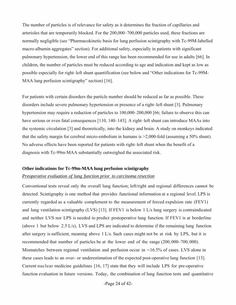
The number of particles is of relevance for safety as it determines the fraction of capillaries and
arterioles that are temporarily blocked. For the 200,000–700,000 particles used, these fractions are
normally negligible (see “Pharmacokinetic basis for lung perfusion scintigraphy with Tc-99M-labelled
macro-albumin aggregates” section). For additional safety, especially in patients with significant
pulmonary hypertension, the lower end of this range has been recommended for use in adults [66]. In
children, the number of particles must be reduced according to age and indication and kept as low as
possible especially for right–left shunt quantification (see below and “Other indications for Tc-99M-
MAA lung perfusion scintigraphy” section) [16].
For patients with certain disorders the particle number should be reduced as far as possible. These
disorders include severe pulmonary hypertension or presence of a right–left shunt [3]. Pulmonary
hypertension may require a reduction of particles to 100,000–200,000 [66; failure to observe this can
have serious or even fatal consequences [110, 140–143]. A right–left shunt can introduce MAAs into
the systemic circulation [3] and theoretically, into the kidney and brain. A study on monkeys indicated
that the safety margin for cerebral micro-embolism in humans is >2,000-fold (assuming a 50% shunt).
No adverse effects have been reported for patients with right–left shunt when the benefit of a
diagnosis with Tc-99m-MAA substantially outweighed the associated risk.
Other indications for Tc-99m-MAA lung perfusion scintigraphy
Preoperative evaluation of lung function prior to carcinoma resection
Conventional tests reveal only the overall lung function; left/right and regional differences cannot be
detected. Scintigraphy is one method that provides functional information at a regional level. LPS is
currently regarded as a valuable complement to the measurement of forced expulsion rate (FEV1)
and lung ventilation scintigraphy (LVS) [13]. If FEV1 is below 1 L/s lung surgery is contraindicated
and neither LVS nor LPS is needed to predict postoperative lung function. If FEV1 is at borderline
(above 1 but below 2.5 L/s), LVS and LPS are indicated to determine if the remaining lung function
after surgery is sufficient, meaning above 1 L/s. Such cases might not be at risk by LPS, but it is
recommended that number of particles be at the lower end of the range (200, 000–700, 000).
Mismatches between regional ventilation and perfusion occur in ∼16.5% of cases. LVS alone in
these cases leads to an over- or underestimation of the expected post-operative lung function [13].
Current nuclear medicine guidelines [16, 17] state that they will include LPS for pre-operative
function evaluation in future versions. Today, the combination of lung function tests and quantitative

-Page 25 of 42-
V/Q LS is routinely used to predict the post-operative lung function of lung-carcinoma patients [144,
145]. Tc-99m-MAA is the method of choice with exact and reproducible quantification of static lung
perfusion [146]. Other methods, such as MR [147, 148] are more costly and have not yet been
rigorously compared to LPS. Consequently, MR does not play a significant role in t h i s clinical
routine [149].
Preoperative evaluation of lung function in lung transplantation
The qualitative determination of parenchymal perfusion anomalies can provide valuable information
in the planning of lung transplantation. In one study of 46 patients with advanced cystic fibrosis
waiting for a lung transplant, it was shown with Tc-99m-MAA that unilateral perfusion anomalies
were associated with a higher mortality risk during the waiting period. Such information can be
used to modify the priority of transplantation [150]. Quantitative LPS with Tc-99m-MAA 1–3
months following the transplantation is able to predict rejection of the transplant with higher
sensitivity and (especially) specificity than traditional tests of lung function such as FEV1 (83% and
88% vs. 80% and 67% [151]).
Optimization of radiation therapy for lung cancer
In 10% of cases, radiation therapy leads to acute radiation pneumonitis; pulmonary fibrosis can
occur later and is associated with mortality risk [152]. These effects depend on the radiation dosage,
fractionation schedule, the lung volume irradiated, and biological factors. Optimization of the
radiation therapy plan for lung carcinoma can be supported by a Tc-99m-MAA perfusion test. A
quantitative V/Q SPECT LS, giving regional and functional information that morphological methods
cannot provide, allows for a better prediction of the effects of radiation upon the pulmonary tissue
[152]. The use of perfusion information can help to prevent radiation damage to the remaining
functioning lung parenchyma, especially in patients with major perfusion deficiencies [153]. De
Jaeger et al. [154] showed that the best predictors of pulmonary function following radiation
therapy were variables obtained from Tc-99m-MAA such as “predicted perfusion reduction” and
“mean perfusion- weighed lung dosage”.
Right–left shunt quantification
In adults, the most common cause of interpulmonary right–left shunt are Osler’s and Waldenström’s
diseases, arteriovenous angioma, pulmonary fibrosis, and sclerodermatitis [13]. It is also found in

-Page 26 of 42-
various end-stage lung diseases (10.3%), especially with primary pulmonary hypertension (19%)
[155]. The presence of a shunt influences the surgical procedure to be adopted for lung transplantation
[155]. LPS offers the simplest and cheapest procedure for detection and quantification of a right–left
shunt and for estimating the consequent right ventricular strain [13]. This indication is planned for
inclusion in the guidelines of the German Society of Nuclear Medicine [17]. The shunt is revealed by
extra- pulmonary deposition of Tc-99m-MAA particles, mainly in the brain, the liver, and the kidneys.
Quantification of the shunt is performed by measuring renal activity with known effective renal
plasma flow (renal function scintigraphy) [13]. The absence of activity accumulation in the brain in a
static image virtually excludes a significant right–left shunt, and the specificity of a positive result is
close to 100% [155].
Pediatric use of Tc-99m-MAA LPS
In children and adolescents, LPS is indicated for worsening of lung function by cystic fibrosis,
the clarification of relapsed bronchi in cases of suspected bronchiolectasis, assessment of lung
perfusion before and after operation for congenital heart defect or anomalies of the pericardiac
blood vessels, right–left shunt quantification, diagnosis and exclusion of possible PE, and
monitoring lung perfusion after PE. Dosages are given in the relevant guidelines [16, 17, 135].
CONCLUSIONS
The clinical studies and reports surveyed in this review have demonstrated that Tc-99m-LPS
continues to be of value in the diagnosis of PE. In comparison with spiral CT and/or PA, LPS is still a
diagnostically definitive pulmonary imaging procedure as mentioned recent PIOPED II reports [156].
This applies in particular to chronic or recurring embolisms, whereas currently spiral CT may be of
greater value for major or life-threatening embolisms. The most frequent indication for LPS in clinical
routine is the suspected PE. In this setting, LPS should be combined with a ventilation scintigraphy
using Tc-99m- Technegas or Kr-81m. [SPECT is the method of choice to acquire the images even
though the PIOPED criteria are insufficient for interpretation of positive LPS results. At present, LPS
with Tc-99m-MAA has a role in situations that do not involve embolism, such as in pediatrics or in
the quantification of regional pulmonary function in a pre-operative context or prior to radiation
therapy.
-Page 27 of 42-
REFERENCES
1. Kumar AM, Parker JA. Ventilation/perfusion scintigraphy. Emerg Med Clin North AM 2001;
19:957-73
2. Taplin GV, MacDonald NS. Radiochemistry of macro- aggregated albumin and newer lung scanning agents. Semin Nucl Med 1971; 1:132–52.
3. Wagner HN. Regional ventilation and perfusion. Princ Nucl Med 1995; 2:887–95.
4. Weiss K. Pulmonary thromboembolism: epidemiology and techniques of nuclear medicine. Semin Thromb Hemost1996; 22:27–32.
5. Tow DE, Wagner HN Jr, Lopez-Majano V, Smith EM, Migita T. Validity of measuring regional pulmonary arterial blood flow with macroaggregates of human serum albumin. Am J Roentgenol Radium Ther Nucl Med 1966; 96:664–76
6. Darte L, Persson BR, Soderbom L. Quality control and testing of 99mTc-macroaggregated albumin. Nuklearmed- izin 1976;15:80–5.
7. Malone LA, Malone JF, Ennis JT. Kinetics of technetium 99m labelled macroaggregated albumin in humans. Br J Radiol 1983;56:109–12
8. Chandra R, Shamoun J, Braunstein P, DuHov OL. Clinical evaluation of an instant kit for preparation of 99mTc-MAA for lung scanning. J Nucl Med 1973; 14:702–5.
9. Monroe LA, Thompson WL, Anderton NS, Burdine JA. Evaluation of an improved 99mTc-stannous aggregated albumin preparation for lung imaging. J Nucl Med 1974; 15:192–4.
10. Neumann RD, Sostman HD, Gottschalk A. Current status of ventilation-perfusion imaging. Semin Nucl Med 1980; 10:198–217.
11. Robbins PJ, Feller PA, Nishiyama H. Evaluation and dosimetry of a 99mTc-Sn-MAA lung imaging agent in humans. Health Phys 1976;30:173–8.
12. Weibel ER. Morphometry of the human lung. Heidelberg: Springer; 1963.
13. Schümichen C. Nuclear medicine diagnosis of the lung. Radiology 2000;40:878–87.
14. Stein PD, Gottschalk A. Critical review of ventilation/ perfusion lung scans in acute pulmonary embolism. Prog Cardiovasc Dis 1994; 37:13–24.
15. Heck LL, Duley JW Jr. Statistical considerations in lung imaging with 99mTc albumin particles. Radiology 1974;113:675–9.
16. Parker JA, Coleman RE, Siegel BA, Sostman HD, McKusick KA, Royal HD. Procedure guideline for lung scintigraphy: 1.0. Society of Nuclear Medicine. J Nucl Med 1996;37:1906–10.

-Page 28 of 42-
17. Schümichen C. Guidelines for lung scintigraphy. Nuklear- medizin 1999;38:233–6.
18. European Society of Cardiology. Guidelines on diagnosis and management of acute pulmonary embolism. Task force on pulmonary embolism. Eur Heart J 2000;21:1301–36.
19. Patriquin L, Khorasani R, Polak JF. Correlation of diagnostic imaging and subsequent autopsy findings in patients with pulmonary embolism. AJR Am J Roentgenol 1998;171:347–9.
20. Stein PD, Henry JW. Prevalence of acute pulmonary embolism among patients in a general hospital and at utopsy. Chest 1995;108:978–81.
21. Goldhaber SZ. Pulmonary embolism. Lancet 2004;363:1295–305.
22. Wilson MA. Pulmonary system. Chapter 4. Textbook of nuclear medicine. New York: Lippincott-Raven; 1997. p.89–116.
23. Barritt DW, Jordan SC. Anticoagulant drugs in the treatment of pulmonary embolism: a controlled trial. Lancet1960;1:1309–12.
24. Douketis JD, Foster GA, Crowther MA, Prins MH, Ginsberg JS. Clinical risk actors and timing of recurrent venous thromboembolism during the initial 3 months of anticoagulant therapy. Arch Intern Med 2000;160:3431–6.
25. de Monyé W, Van Strijen MJ, Huisman MV, Kieft GJ, Pattynama PM. Suspected pulmonary embolism: prevalence and anatomic distribution in 487 consecutive patients. Advances in New Technologies Evaluating the Localisation of Pulmonary Embolism (ANTELOPE) Group. Radiology 2000;215:184–8.
26. Landefeld CS, Beyth RJ. Anticoagulant-related bleeding: clinical epidemiology, prediction, and prevention. Am J Med 1993;95:315–28.
27. Fedullo PF, Tapson VF. Clinical practice: the evaluation of suspected pulmonary embolism. N Engl J Med 2003;349:1247–56.
28. Deutsche Gesellschaft Für Angiologie. AWMF Leitlinie: Diagnostik und Therapie der Bein-und Beckenvenenthrom-bose und Lungenembolie; 2005. http://www.radiologieiederrhein.de/radiologie/fortbildung/leitlinie-thrombose- embolie.htm.
29. Neumann SM, Freyschmidt J, Holland BR, Henschel M, Gahnem NR. Comparison of ventilation-/perfusion scintigraphy with spiral CT in acute lung embolism. Med Klin (Munich) 1997;92:635–41.
30. Goldhaber SZ, Elliott CG. Acute pulmonary embolism. Part I: epidemiology, pathophysiology, and diagnosis. Circulation 2003;108:2726–9.
31. Powell T, Muller NL. Imaging of acute pulmonary thromboembolism: should spiral computed tomography replace the ventilation-perfusion scan? Clin Chest Med 2003;24:29–38.
32. Meyer G, Roy PM, Sors H, Sanchez O. Laboratory tests in the diagnosis of pulmonary embolism. Respiration 2003;70:125–32.
-Page 29 of 42-
33. Perrier A. Noninvasive diagnosis of pulmonary embolism. Haematologica 1997;82:328–31.
34. Dunn KL, Wolf JP, Dorfman DM, Fitzpatrick P, Baker JL, Goldhaber SZ. Normal D-dimer levels in emergency depart ment patients suspected of acute pulmonary embolism. J Am Coll Cardiol 2002;40:1475–8.
35. Kipper MS, Moser KM, Kortman KE, Ashburn WL. Long- term follow-up of patients with suspected pulmonary embolism and a normal lung scan: perfusion scans in embolic suspects. Chest 1982;82:411–5.
36. Quinn DA, Fogel RB, Smith CD, Laposata M, Taylor TB, Johnson SM, et al. D-dimers in the diagnosis of pulmonary embolism. Am J Respir Crit Care Med 1999;159:1445–9.
37. Stein PD, Hull RD, Patel KC, Olson RE, Ghali WA, Brant R, et al. D-dimer for the exclusion of acute venous thrombosis and pulmonary embolism: a systematic review. Ann Intern Med 2004;140:589–602.
38. Schümichen C, Krause T, Reinartz P. Leitlinie für die Lun- genszintigraphie (version 2). Nuklearmedizin. http://www. nuklearmedizin.de/publikationen/leitlinien/lunge_szin.php. Accessed 2007
39. Robinson PJ. Ventilation-perfusion lung scanning and spiral computed tomography of the lungs: competing or comple mentary modalities? Eur J Nucl Med 1996;23:1547–53.
40. Mathis G, Blank W, Reißig A, Lechleitner P, Reuß J, Schuler A, et al. Thoracic ultrasound for diagnosing pulmonary embolism: a prospective multicenter study of 352 patients. Chest 2005;128:1531–8.
41. Perrier A, Roy PM, Sanchez O, Le Gal G, Meyer , Gourdier AL, et al. Multidetector-row computed tomogra phy in suspected pulmonary embolism. N Engl J Med 2005;352:1760–8.
42. Anderson DR, Kovacs MJ, ennie C, Kovacs G, Stiell I, Dreyer J, et al. Use of spiral computed tomography contrast angiography and ultrasonography to exclude the diagnosis of pulmonary embolism in the emergency department. J Emer Med 2005;29:399–404.
43. Stein PD, Fowler SE, Goodman LR, Gottschalk A, Hales CA, Hull RD, et al. Multidetector computed tomography for acute pulmonary embolism. N Engl J Med 2006;354:2317–27.
44. Bettmann MA, Boxt LM, Gomes AS, Grollman J, Henkin RE, Higgins CB, et al. Acute chest pain: suspected pulmonary embolism. American College of Radiology. ACR appropriateness criteria. Radiology 2000;215:15–21.
45. Tapson VF, Carroll BA, Davidson BL, Elliott CG, Fedullo PF, Hales CA, et al. The diagnostic approach to acute venous thromboembolism: clinical practice guideline. American Thoracic Society. Am J Respir Crit Care Med 1999;160:1043–66.
46. Ten Wolde M, Hagen PJ, Macgillavry MR, Pollen IJ, Mairuhu AT, Koopman MM, et al. Non-invasive diagnostic work-up of patients with clinically suspected pulmonary embolism; results of a management study. J Thromb Haemost 2004;2:1110–7.

-Page 30 of 42-
47. Riedel M. Diagnosing pulmonary embolism. Postgrad Med J 2004;80:309–19.
48. Fleischmann D, Kontrus M, Bankier AA, Wiesmayr MN, Janata-Schwatczek K, Herold CJ. Spiral CT in acute pulmonary embolism. Radiologe 1996;36:489–95.
49. Grenier PA, Beigelman C. Spiral computed tomographic scanning and magnetic resonance angiography for the diagnosis of pulmonary embolism. Thorax 1998;53:S25–31.
50. Khan A, Cann AD, Shah RD. Imaging of acute pulmonary emboli. Thorac Surg Clin 2004;14:113–24.
51. British Thoracic Society. Suspected acute pulmonary embolism: a practical approach. Thorax 1997;52:S1–24.
52. Berman AR, Arnsten JH. Diagnosis and treatment of pulmonary embolism in the elderly. Clin Geriatr Med 2003;19:157–75.
53. Kauczor HU, Heussel CP, Thelen M. Update on diagnostic strategies of pulmonary mbolism. Eur Radiol 1999;9:262–75.
54. Wilson HT, Meagher TM, Williams SJ. Combined helical computed tomographic pulmonary angiography and lung perfusion scintigraphy for investigating acute pulmonary embolism. Clin Radiol 2002;57:33–6.
55. Giordano A, Angiolillo DJ. Current role of lung scintigraphy in pulmonary embolism. Q J Nucl Med 2001;45:294–301.
56. Kruip MJ, Leclercq MG, van der HC, Prins MH, Buller HR. Diagnostic strategies for excluding pulmonary embolism in clinical outcome studies: a systematic review. Ann Intern Med 2003;138:941–51.
57. Ramzi DW, Leeper KV. DVT and pulmonary embolism: Part I. Diagnosis. Am Fam Physician 2004;69:2829–36.
58. Bounameaux H, Perrier A. Diagnostic approaches to suspected deep vein thrombosis and pulmonary embolism. Hematol J 2003;4:97–103.
59. British Thoracic Society. British Thoracic Society guidelines for the management of suspected acute pulmonary embolism. Thorax 2003;58:470–83.
60. Scatarige JC, Weiss CR, Diette GB, Haponik EF, Merriman B, Fishman EK. Scanning systems and protocols used during imaging for acute pulmonary embolism: how much do our clinical colleagues know? Acad Radiol 2006;13:678–85.
61. Madsen PH, Hess S, Jorgensen HB, Hoilund-Carlsen PF. Diagnostic imaging in acute pulmonary embolism in Denmark: a survey. Ugeskr Laeger 2005;167:3875–7.
62. PIOPED Investigators. Value of the ventilation/perfusion scan in acute pulmonary embolism: results of the prospective investigation of pulmonary embolism diagnosis (PIOPED). JAMA 1990;263:2753–9.

-Page 31 of 42-
63. Gottschalk A, Juni JE, Sostman HD, Coleman RE, Thrall J, McKusick KA, et al. Ventilation-perfusion scintigraphy in the PIOPED study. Part I. Data collection and tabulation. J Nucl Med 1993;34:1109–18.
64. Gottschalk A, Sostman HD, Coleman RE, Juni JE, Thrall J, McKusick KA, et al. Ventilation-perfusion scintigraphy in the PIOPED study. Part II. Evaluation of the scintigraphic criteria and interpretations. J Nucl Med 1993;34:1119–26.
65. Sostman HD, Coleman RE, DeLong DM, Newman GE, Paine S. Evaluation of revised criteria for ventilation- perfusion scintigraphy in patients with suspected pulmonary embolism. Radiology 1994;193:103–7.
66. British Nuclear Medicine Society. BNMS Quality Guidelines for ventilation/perfusion imaging for pulmonary embolism. British Nuclear Medicine Society, editor. BNMS Quality Guidelines; 2003.
67. Macdonald WB, Patrikeos AP, Thompson RI, Adler BD, van der Schaaf AA. Diagnosis of pulmonary embolism: ventilation perfusion scintigraphy versus helical computed tomography pulmonary angiography. Australas Radiol 2005;49:32–8.
68. Tunariu N, Gibbs SJ, Win Z, Gin-Sing W, Graham A, Gishen P, et al. Ventilation-perfusion scintigraphy is more sensitive than multidetector CTPA in detecting chronic thromboembolic pulmonary disease as a treatable cause of pulmonary hypertension. J Nucl Med 2007;48:680–4.
69. van Beek EJ, Brouwers EM, Song B, Bongaerts AH, Oudkerk M. Lung scintigraphy and helical computed tomography for the diagnosis of pulmonary embolism: a meta-analysis. Clin Appl Thromb Hemost 2001;7:87–92.
70. Schoepf UJ, Costello P. CT angiography for diagnosis of pulmonary embolism: state of the art. Radiology 2004;230:329–37.
71. Schümichen C. Pulmonary embolism: is multislice CT the method of choice? Against. Eur J Nucl Med Mol Imaging 2005;32:103–7.
72. Michiels JJ, Gadisseur A, van der Planken M, Schroyens W, de Maeseneer M, Hermesen JT, et al. A critical of non- invasive diagnosis and exclusion of deep vein thrombosis and pulmonary embolism in outpatients with suspected deep vein thrombosis or pulmonary embolism: how many tests do we need? Int Angiol 2005;24:29–39.
73. Garg K, Sieler H, Welsh CH, Johnston RJ, Russ PD. Clinical validity of helical CT being interpreted as negative for pulmonary embolism: implications for patient treatment. AJR Am J Roentgenol 1999;172:1627–31.
74. Miniati M, Pistolesi M, Marini C, Di Ricco G, Formichi B, Prediletto R, et al. Value of perfusion lung scan in the diagnosis of pulmonary embolism: results of the prospective investigative study of acute pulmonary embolism diagnosis (PISA-PED). Am J Respir Crit Care Med 1996;154:1387–93.
75. Nilsson T, Mare K, Carlsson A. Value of structured clinical and scintigraphic protocols in acute pulmonary embolism. J Intern Med 2001;250:213–8.
-Page 32 of 42-
76. Bonnin F, Hadjikostova H, Jebrak G, Denninger MH, Vera P, Rufat P, et al. Complementarity of lung scintigraphy and D-dimer test in pulmonary embolism. Eur J Nucl Med 1997;24:444–7.
77. Freitas JE, Sarosi MG, Nagle CC, Yeomans ME, Freitas AE, Juni JE. Modified PIOPED criteria used in clinical practice. J Nucl Med 1995;36:1573–8.
78. Howarth DM, Lan L, Thomas PA, Allen LW. 99mTc Technegas ventilation and perfusion lung scintigraphy for the diagnosis of pulmonary embolus. J Nucl Med 1999;40:579–84.
79. Lowe VJ, Bullard AG, Coleman RE. Ventilation/perfusion lung scan probability category distributions in university and community hospitals. Clin Nucl Med 1995;20:1079–83.
80. Worsley DF, Alavi A. Radionuclide imaging of acute pulmonary embolism. Semin Nucl Med 2003;33:259–78.
81. Goodman LR, Lipchik RJ, Kuzo RS, Liu Y, McAuliffe TL, O’Brien DJ. Subsequent pulmonary embolism: risk after a negative helical CT pulmonary angiogram: prospective comparison with scintigraphy. Radiology 2000;215:535–42.
82. van Rossum AB, Pattynama PM, Mallens WM, Hermans J, Heijerman HG. Can helical CT replace scintigraphy in the diagnostic process in suspected pulmonary embolism? A retrolective-prolective cohort study focusing on total diagnostic yield. Eur Radiol 1998;8:90–6.
83. Barghouth G, Yersin B, Boubaker A, Doenz F, Schnyder P, Delaloye AB. Combination of clinical and V/Q scan assessment for the diagnosis of pulmonary embolism: a 2-year outcome prospective study. Eur J Nucl Med 2000;27:1280–5.
84. Mayo JR, Remy-Jardin M, Muller NL, Remy J, Worsley DF, Hossein-Foucher C, et al. Pulmonary embolism: prospective comparison of spiral CT with ventilation-perfusion scintigraphy. Radiology 1997;205:447–52.
85. Kearon C, Ginsberg JS, Douketis J, Turpie AG, Bates SM, Lee AY, et al. An evaluation of D-dimer in the diagnosis of pulmonary embolism. Ann Intern Med 2006;144:812–21.
86. Stein PD, Athanasoulis C, Alavi A, Greenspan RH, Hales CA, Saltzman HA, et al. Complications and validity of pulmonary angiography in acute pulmonary embolism. Circulation 1992;85:462–8.
87. Zuckerman DA, Sterling KM, Oser RF. Safety of pulmonary angiography in the 1990s. J Vasc Interv Radiol 1996;7:199–205.
88. Radan L, Mor M, Gips S, Schlag-Eisenberg D, Lurie Y, Dickstein K, et al. The added value of spiral computed tomographic angiography after lung scintigraphy for the diagnosis of pulmonary embolism. Clin Nucl Med 2004;29:255–61.
89. Gottsäter A, Berg A, Centergard J, Frennby B, Nirhov N, Nyman U. Clinically suspected pulmonary embolism: is it safe to withhold anticoagulation after a negative spiral CT? Eur Radiol 2001;11:65–72.

-Page 33 of 42-
90. Bateman NT, Coakley AJ, Croft DN, Lyall JR. Ventilation- perfusion lung scans for pulmonary emboli: accuracy of reporting. Eur J Nucl Med 1977;2:201–3.
91. Sullivan DC, Coleman RE, Mills SR, Ravin CE, Hedlund LW. Lung scan interpretation: effect of different observers and different criteria. Radiology 1983;149:803–7.
92. van Beek EJ, Tiel-van Buul MM, Hoefnagel CA, Jagt HH, van Royen EA. Reporting of perfusion/ventilation lung scintigraphy using an anatomical lung segment chart: a prospective study. Nucl Med Commun 1994;15:746–51.
93. Coche E, Verschuren F, Keyeux A, Goffette P, Goncette L, Hainaut P, et al. Diagnosis of acute pulmonary embolism in outpatients: comparison of thin-collimation multi-detector row spiral CT and planar ventilation-perfusion scintigraphy. Radiology 2003;229:757–65.
94. Reinartz P, Schirp U, Zimny M, Sabri O, Nowak B, Schafer W, et al. Optimizing ventilation-perfusion lung scintigraphy: parting with planar imaging. Nuklearmedizin 2001;40:38–43.
95. Reinartz P, Wildberger JE, Schaefer W, Nowak B, Mahnken AH, Buell U. Tomographic imaging in the diagnosis of pulmonary embolism: a comparison between V/Q lung scintigraphy in SPECT technique and multislice spiral CT. J Nucl Med 2004;45:1501–8.
96. Bajc M, Olsson CG, Olsson B, Palmer J, Jonson B. Diagnostic evaluation of planar and tomographic ventilation/ perfusion lung images in patients with suspected pulmonary emboli. Clin Physiol Funct Imaging 2004;24:249–56.
97. Palmer J, Bitzen U, Jonson B, Bajc M. Comprehensive ventilation/perfusion SPECT. J Nucl Med 2001;42:1288–94.
98. Collart JP, Roelants V, Vanpee D, Lacrosse M, Trigaux JP, Delaunois L, et al. Is a lung perfusion scan obtained by using single photon emission computed tomography able to improve the radionuclide diagnosis of pulmonary embolism? Nucl Med Commun 2002;23:1107–13.
99. Corbus HF, Seitz JP, Larson RK, Stobbe DE, Wooten W, Sayre JW, et al. Diagnostic usefulness of lung SPET in pulmonary thromboembolism: an outcome study. Nucl Med Commun 1997;18:897–906.
100. Reinartz P, Kaiser H-J, Wildberger JE, Gordji C, Nowak B, Buell U. SPECT imaging in the diagnosis of pulmonary embolism: automated detection of match and mismatch defects by means of image-processing techniques. J Nucl Med 2006;47:968–73.
101. Blachere H, Latrabe V, Montaudon M, Valli N, Couffinhal T, Raherisson C, et al. Pulmonary embolism revealed on helical CT angiography: comparison with ventilation- perfusion radionuclide lung scanning. AJR Am J Roentgenol 2000;174:1041–7.
102. Remy-Jardin M, Remy J, Deschildre F, Artaud D, Beregi JP, Hossein-Foucher C, et al. Diagnosis of pulmonary embolism with spiral CT: comparison with pulmonary angiography and scintigraphy. Radiology 1996;200:699–706.

-Page 34 of 42-
103. Herold CJ. Spiral computed tomography of pulmonary embolism. Eur Respir J Suppl 2002;35:13s–21s.
104. Blum AG, Delfau F, Grignon B, Beurrier D, Chabot F, Claudon M, et al. Spiral-computed tomography versus pulmonary angiography in the diagnosis of acute massive pulmonary embolism. Am J Cardiol 1994;74:96–8.
105. Remy-Jardin M, Remy J, Wattinne L, Giraud F. Central pulmonary thromboembolism: diagnosis with spiral volumetric CT with the single-breath-hold technique: comparison with pulmonary angiography. Radiology 1992;185:381–7.
106. Raptopoulos V, Boiselle PM. Multi-detector row spiral CT pulmonary angiography: comparison with single-detector row spiral CT. Radiology 2001;221:606–13.
107. Ghaye B, Remy J, Remy-Jardin M. Non-traumatic thoracic emergencies: CT diagnosis of acute pulmonary embolism: the first 10 years. Eur Radiol 2002;12:1886–905.
108. Dresel S, Stabler A, Scheidler J, Holzknecht N, Tatsch K, Hahn K. Diagnostic approach in acute pulmonary embolism: perfusion scintigraphy versus spiral computed tomography. Nucl Med Commun 1995;16:1009–15.
109. Perrier A, Howarth N, Didier D, Loubeyre P, Unger PF, De Moerloose P, et al. Performance of helical computed tomography in unselected outpatients with suspected pulmonary embolism. Ann Intern Med 2001;135:88–97.
110. van Rossum AB, Pattynama PM, Ton ER, Treurniet FE, Arndt JW, van Eck B, et al. Pulmonary embolism: validation of spiral CT angiography in 149 patients. Radiology 1996;201:467–70.
111. Van Strijen MJ, De Monye W, Kieft GJ, Pattynama PM, Prins MH, Huisman MV. Accuracy of single-detector spiral CT in the diagnosis of pulmonary embolism: a prospective multicenter cohort study of consecutive patients with abnormal perfusion scintigraphy. J Thromb Haemost 2005;3:17–25.
112. Lomis NN, Yoon HC, Moran AG, Miller FJ. Clinical out-comes of patients after a negative spiral CT pulmonary arteriogram in the evaluation of acute pulmonaryembolism. J Vasc Interv Radiol 1999;10:707–12.
113. Jimenez D, Gomez M, Herrero R, Lapresa E, Diaz G, Lanzara L, et al. Thrombembolic events in patients after a negative computed tomography pulmonary angiogram: a retrospective study of 165 patients. rch Bronconeumol 2006;42:344–8.
114. Cross JJ, Kemp PM, Walsh CG, Flower CD, Dixon AK. A randomized trial of spiral CT and ventilation perfusion scintigraphy for the diagnosis of pulmonary embolism. Clin Radiol 1998;53:177–82.
115. Gottschalk A, Stein PD, Goodman LR, Sostman HD. Over- view of prospective investigation of pulmonary embolism diagnosis II. Semin Nucl Med 2002;32:173–82.

-Page 35 of 42-
116. Gottschalk A, Stein PD, Sostmann HD, Matta F, Beemath A. Very low probability interpretation of V/Q lung scans in combination with low probability objective clinical assessment reliably excludes pulmonary embolism: data from PIOPED II. J Nucl Med 2007;48:1411–5.
117. Roberts HC, Kauczor HU, Pitton MB, Schweden F, Thelen M. The algorithm of imaging diagnostics of pulmonary embolism: is it time for a new definition? Röfo 1997;166:463–74.
118. Strashun AM. A reduced role of V/Q scintigraphy in the diagnosis of acute pulmonary mbolism. J Nucl Med 2007;48:1405–7.
119. Stein PD, Woodard PK, Weg JG, Wakefield TW, Tapson VF, Sostman HD, et al. Diagnostic pathways in acute pulmonary embolism: recommendations of the PIOPED II investigators. Am J Med 2006;119:1048–55.
120. Glassroth J. Imaging of pulmonary embolism: too much of a good thing? JAMA 2007;298:2788–9.
121. Anderson DR, Kahn SR, Rodger MA, Kovacs MJ, Morris T, Hirsch A, et al. Computed tomographic pulmonary angiography vs. ventilation-perfusion lung scanning in patients with suspected pulmonary embolism. A randomized controlled trial. JAMA 2007;298:2743–53.
122. van Beek EJ, Wild JM, Fink C, Moody AR, Kauczor HU, Oudkerk M. MRI for the diagnosis of pulmonary embolism. J Magn Reson Imaging 2003;18:627–40.
123. Kline JA. New diagnostic tests for pulmonary embolism. Ann Emerg Med 2000;36:280–1.
124. Oudkerk M, van Beek EJ, Wielopolski P, van Ooijen PM, Brouwers-Kuyper EM, Bongaerts AH, et al. Comparison of contrast-enhanced magnetic resonance angiography and conventional pulmonary angiography for the diagnosis of pulmonary embolism: a prospective study. Lancet 2002;359:1643–7.
125. Meaney JF, Weg JG, Chenevert TL, Stafford-Johnson D, Hamilton BH, Prince MR. Diagnosis of pulmonary embolism with magnetic resonance angiography. N Engl J Med 1997;336:1422–7.
126. Kluge A, Gerriets T, Stolz E, Dill T, Mueller KD, Mueller C, et al. Pulmonary perfusion in acute pulmonary embolism: agreement of MRI and SPECT for lobar, segmental and sub- segmental perfusion defects. Acta Radiol 2006;47:933–40.
127. Cheely R, McCartney WH, Perry JR, Delany DJ, Bustad L, Wynia VH, et al. The role of noninvasive tests versus pulmonary angiography in the diagnosis of pulmonary embolism. Am J Med 1981;70:17–22.
128. Henry JW, Relyea B, Stein PD. Continuing risk of thrombo- emboli among patients with normal pulmonary angiograms. Chest 1995;107:1375–8.
129. Hull RD, Hirsh J, Carter CJ, Jay RM, Dodd PE, Ockelford PA, et al. Pulmonary angiography, ventilation lung scanning, and venography for clinically suspected pulmonary embolism with abnormal perfusion lung scan. Ann Intern Med 1983;98:891–9.

-Page 36 of 42-
130. Novelline RA, Baltarowich OH, Athanasoulis CA, Waltman AC, Greenfield AJ, McKusick KA. The clinical course of patients with suspected pulmonary embolism and a negative pulmonary arteriogram. Radiology 1978;126:561–7.
131. van Beek EJ, Reekers JA, Batchelor DA, Brandjes DP, Buller HR. Feasibility, safety and clinical utility of angiography in patients with suspected pulmonary embolism. Eur Radiol 1996;6:415–9.
132. Lette J, Cerino M, Barrette G, Dufresne MP, De Maria S, Eybalin MC, et al. Scintigraphic probability and angiographic diagnostic certainty in acute pulmonary embolism. Clin Nucl Med 2003;28:897–904.
133. Nosske D, Minkov V, Brix G. Establishment and application of diagnostic reference levels for nuclear medicine procedures in Germany. Nuklearmedizin 2004;43:79–84.
134. ICRP Publication 80. Addendum 2 to ICRP Publication 72. Recalculated dose data for 19 frequently used radiopharmaceuticals from ICRP Publication 53. New York: Pergamon Press; 1999. p. 47–69.
135. Piepsz A, Hahn K, Roca I, Ciofetta G, Toth G, Gordon I, et al. A radiopharmaceuticals schedule for imaging in paedi atrics. Paediatric Task Group European Association Nuclear Medicine. Eur J Nucl Med 1990;17:127–9.
136. Stabin MG, Breitz HB. Breast milk excretion of radiophar maceuticals: mechanisms, findings, and radiation dosimetry. J Nucl Med 2000;41:863–73.
137. Al Janabi MA, Yousif ZM, Kadim AH, Al Salem AM. A new technique for the preparation of ready-to-use macroaggregated albumin (MAA) kits to be labelled with 99mTc for lung scanning. Int J Appl Radiat Isot 1983;34:1473–8.
138. Clayton BD, Ice RD, Beierwaltes WH. Preparation, quality control and clinical acceptance of labeled Macrotec. Radiology 1975;116:223–5.
139. Bundesgesundheitsamt. Aufbereitungsmonographie Macro salb-[99mTc] Technetium Bundesanzeiger 1991; Nr 182,6902.
140. Child JS, Wolfe JD, Tashkin D, Nakano F. Fatal lung scan in a case of pulmonary hypertension due to obliterative pulmonary vascular disease. Chest 1975;67:308–10.
141. Dworkin HJ, Smith JR, Bull FE. A reaction following administration of macroaggregated albumin (MAA) for a lung scan. Am J Roentgenol Radium Ther Nucl Med 1966; 98:427–33.
142. Dworkin HJ, Smith JR, Bull FE. Reaction after administration of macroaggregated albumin for a lung scan. N Engl J Med 1966;275:376.
143. Williams JO. Death following injection of lung scanning agent in a case of pulmonary hypertension. Br J Radiol 1974;47:61–3.
-Page 37 of 42-
144. Wu MT, Pan HB, Chiang AA, Hsu HK, Chang HC, Peng NJ, et al. Prediction of postoperative lung function in patients with lung cancer: comparison of quantitative CT with perfusion scintigraphy. AJR Am J Roentgenol 2002;178: 667–72.
145. Chenuel B, Haouzi P, Olivier P, Marie PY, Chalon B, Borrelly J. Effect of exercise on lung-perfusion scanning in patients with bronchogenic carcinoma. Eur Respir J 2002;20:710–6.
146. Fleming JS, Whalley DR, Skrypniuk JV, Jarritt PH, Houston AS, Cosgriff PS, et al. UK audit of relative lung function measurement from planar radionuclide imaging. Nucl Med Commun 2004;25:923–34.
147. Curran WJ Jr, Moldofsky PJ, Solin LJ. Observations on the predictive value of perfusion lung scans on post-irradiation pulmonary function among 210 patients with bronchogenic carcinoma. Int J Radiat Oncol Biol Phys 1992;24:31–6.
148. Lehnhardt S, Thorsten WJ, Strecker R, Hogerle S, Herget G, Geens V, et al. Assessment of pulmonary perfusion with ultrafast projection magnetic resonance angiography in comparison with lung perfusion scintigraphy in patients with malignant stenosis. Invest Radiol 2002;37:594–9.
149. Eichinger M, Tetzlaff R, Puderbach M, Woodhouse N, Kauczor HU. Proton magnetic resonance imaging for assessment of lung function and respiratory dynamics. Eur J Radiol 2007;64:329–34.
150. Stanchina ML, Tantisira KG, Aquino SL, Wain JC, Ginns LC. Association of lung perfusion disparity and mortality in patients with cystic fibrosis awaiting lung transplantation. J Heart Lung Transplant 2002;21:217–25.
151. Hardoff R, Steinmetz AP, Krausz Y, Bar-Sever Z, Liani M, Kramer MR. The prognostic value of perfusion lung scintigraphy in patients who underwent single-lung transplantation for emphysema and pulmonary fibrosis. J Nucl Med 2000;41:1771–6.
152. Goethals I, Dierckx R, De Meerleer G, De Sutter J, De Winter O, De Neve W, et al. The role of nuclear medicine in the prediction and detection of radiation-associated normal pulmonary and cardiac damage. J Nucl Med 2003;44:1531–9.
153. Seppenwoolde Y, Engelsman M, De Jaeger K, Muller SH, Baas P, McShan DL, et al. Optimizing radiation treatment plans for lung cancer using lung perfusion information. Radiother Oncol 2002;63:165–77.
154. De Jaeger K, Seppenwoolde Y, Boersma LJ, Muller SH, Baas P, Belderbos JS, et al. Pulmonary function following high-dose radiotherapy of non-small-cell lung cancer. Int J Radiat Oncol Biol Phys 2003;55:1331–40.
155. Graves MW, Kiratli PO, Mozley D, Palevsky H, Zukerberg B, Alavi A. Scintigraphic diagnosis of a right to left shunt in end-stage lung disease. Respir Med 2003;97:549–54.
156. Sostman HD, Stein PD, Gottschalk A, Matta F, Hull R, Goodman L. Acute pulmonary embolism: sensitivity and specificity of ventilation-perfusion scintigraphy in PIOPED II study. Radiology 2008;246:941–6.
-Page 38 of 42-
157. Hull RD, Raskob GE, Ginsberg JS, Panju AA, Brill-Edwards P, Coates G, et al. A noninvasive strategy for the treatment of patients with suspected pulmonary embolism. Arch Intern Med 1994;154:289–97.
158. van Beek EJ, Kuyer PM, Schenk BE, Brandjes DP, ten Cate JW, Buller HR. A normal perfusion lung scan in patients with clinically suspected pulmonary embolism: frequency and clinical validity. Chest 1995;108:170–3.
159. Hull RD, Hirsh J, Carter CJ, Raskob GE, Gill GJ, Jay RM, et al. Diagnostic value of ventilation-perfusion lung scanning in patients with suspected pulmonary embolism. Chest 1985;88:819–28.
160. Spies WG, Burstein SP, Dillehay GL, Vogelzang RL, Spies SM. Ventilation-perfusion scintigraphy in suspected pulmonary embolism: correlation with pulmonary angiography and refinement of criteria for interpretation. Radiology 1986;159:383–90.
161. Trujillo NP, Pratt JP, Talusani S, Quaife RA, Kumpe D, Lear JL. DTPA aerosol in ventilation/perfusion scintigraphy for diagnosing pulmonary embolism. J Nucl Med 1997;38:1781–3.
162. Kahn D, Bushnell DL, Dean R, Perlman SB. Clinical outcome of patients with a “low probability” of pulmonary embolism on ventilation-perfusion lung scan. Arch Intern Med 1989;149:377–9.
163. Lee ME, Biello DR, Kumar B, Siegel BA. “Low-probability” ventilation-perfusion scintigrams: clinical outcomes in 99 patients. Radiology 1985;156:497–500.
164. Goodman LR, Curtin JJ, Mewissen MW, Foley WD, Lipchik RJ, Crain MR, et al. Detection of pulmonary embolism in patients with unresolved clinical and scintigraphic diagnosis: helical CT versus angiography. AJR Am J Roentgenol 1995;164:1369–74.
165. Senac JP, Vernhet H, Bousquet C, Giron J, Pieuchot P, Durand G, et al. Pulmonary embolism: contribution of spiral X-ray computed tomography. J Radiol 1995;76:339–45.
166. Garg K, Welsh CH, Feyerabend AJ, Subber SW, Russ PD, Johnston RJ, et al. Pulmonary embolism: diagnosis with spiral CT and ventilation-perfusion scanning: correlation with pulmonary angiographic results or clinical outcome. Radiology 1998;208:201–8.
167. Qanadli SD, Hajjam ME, Mesurolle B, Barre O, Bruckert F, Joseph T, et al. Pulmonary embolism detection: prospective evaluation of dual-section helical CT versus selective pulmonary arteriography in 157 patients. Radiology 2000;217:447–55.
168. Christiansen F. Diagnostic imaging of acute pulmonary embolism. Acta Radiol Suppl 1997;410:1–33.
169. Bajc M, Albrechtsson U, Olsson CG, Olsson B, Jonson B. Comparison of ventilation/perfusion scintigraphy and helical CT for diagnosis of pulmonary embolism: strategy using clinical data and ancillary findings. Clin Physiol Funct Imaging 2002;22:392–7.

-Page 39 of 42-
170. Ferretti GR, Bosson JL, Buffaz PD, Ayanian D, Pison C, Blanc F, et al. Acute pulmonary embolism: role of helical CT in 164 patients with intermediate robability at ventilation- perfusion scintigraphy and normal results at duplex US of the legs. Radiology 1997;205:453–8.
171. Swensen SJ, Sheedy PF, Ryu JH, Pickett DD, Schleck CD, Ilstrup DM, et al. Outcomes after withholding anticoagulation from patients with suspected acute pulmonary embolism and negative computed tomographic findings: a cohort study. Mayo Clin Proc.

-Page 40 of 42-
ASSESSMENT QUESTIONS
1. Tc-99m MAA particles localize in lung vasculature via which mechanism?
a. Chemisorption b. Capillary blockade c. Compartmental localization d. Cell sequestration
2. An injection of 300,000 particles is estimated to block one capillary in every
a. 1,000 b. 10,000 c. 100,000 d. 1,000,000
3. A negative D-dimer finding is a reliable indicator suggesting which of the following?
a. anticoagulation therapy b. an imaging procedure c. absence of pulmonary emboli d. sensitivity to contrast media
4. Lung scintigraphy and spiral CT are considered equal pulmonary emboli imaging options for
use when which of the following patient factors exist?
a. Pregnancy b. High clinical pre-test probability c. Chronic renal disease d. Positive D-dimer test
5. A lung perfusion scan without ventilation has been shown to have a high negative predictive
value of 96-100%. This suggests that the likelihood of fatal pulmonary emboli is of which probability?
a. Rare b. Low c. Intermediate d. High
6. When comparing spiral CT to lung perfusion imaging, which of the following is true?
a. The probability of a VTE is significantly less with a negative spiral CT. b. Spiral CT results are more sensitive to patient-specific artifacts. c. A negative spiral CT is sufficient to avoid anticoagulation therapy. d. Sub-segmental pulmonary emboli are reliably imaged with spiral CT.

-Page 41 of 42-
7. The primary reason that magnetic resonance imaging is not yet an established method for pulmonary emboli is?
a. Lack of large outcome studies b. Unacceptable sensitivity and specificity c. Inability to detect subsegmental emboli d. Discouraging results when compared to CT or LPS
8. The authors suggest that Pulmonary Angiography is the ‘former’ gold standard for detecting PE
for all reasons listed EXCEPT:
a. Risk of complications from invasive study b. Low sensitivity and specificity c. Significant percentage of false positive findings d. Similar to LPS for incidence of thromboembolic events
9. In establishing the probability of pulmonary emboli, a positive ultrasound of the lower leg
veins
a. eliminates the need for additional imaging b. has low sensitivity for DVT c. is found in less than 50% of PE patients d. clarifies intermediate findings of LPS
10. Transthoracic and transesophageal echocardiography procedures are most often used in
patients with
a. chronic pulmonary hypertension b. recurrent pulmonary emboli c. severe and extensive pulmonary emboli d. right-left shunt
11. When considering risks associated with Tc-99m MAA perfusion imaging which of the
following is true?
a. A safety margin greater than 1, 000-fold b. Radiation dose much higher than background levels c. Potential damage to the thyroid d. Fetal death from pulmonary capillary blockade
12. Recommendations to reduce the number of particles used in Tc-99m MAA imaging include all
of the following EXCEPT for
a. Pediatric patients b. Severe pulmonary hypertension cases c. Right-left shunt cases d. Lactating patients

-Page 42 of 42-
13. Lung perfusion imaging in preoperative evaluation of lung function prior to carcinoma
resection
a. provides useful anatomical information at regional levels b. is useful when FEV1 is less than 1 L/s c. predicts post-operative lung perfusion d. has been replaced with magnetic resonance imaging
14. Right-left shunt quantification using Tc-99m MAA is
a. only indicated in pediatric patients b. positive when activity localizes in the brain c. confirmed with spiral CT d. diagnostic in all end-stage lung diseases
15. The use of Tc-99m MAA perfusion lung imaging
a. has been replaced with spiral CT b. will most likely be replaced by magnetic resonance c. requires planar camera imaging d. is still useful for imaging emboli
Related Documents