Annu. Rev. Entomol. 2004. 49:141–74 doi: 10.1146/annurev.ento.49.061802.123422 First published online as a Review in Advance on August 14, 2003 VENEZUELAN EQUINE ENCEPHALITIS * Scott C. Weaver, 1 Cristina Ferro, 2 Roberto Barrera, 3** Jorge Boshell, 2 and Juan-Carlos Navarro 3 1 Center for Biodefense and Emerging Infectious Diseases and Department of Pathology, University of Texas Medical Branch, Galveston, Texas 77555-0609; email: [email protected] 2 Instituto Nacional de Salud, Avenida Eldorado, Carrera 50, Apartado 80080, Bogot´ a, Colombia; email: [email protected]; [email protected] 3 Instituto de Zoolog´ ıa Tropical, Laboratorio de Biolog´ ıa de Vectores, Universidad Central de Venezuela, 1041-A, Apartado 47058, Caracas, Venezuela; email: [email protected] Key Words mosquito, vector, emergence, alphavirus, arbovirus ■ Abstract Venezuelan equine encephalitis virus (VEEV) remains a naturally emerging disease threat as well as a highly developed biological weapon. Recently, progress has been made in understanding the complex ecological and viral genetic mechanisms that coincide in time and space to generate outbreaks. Enzootic, equine avirulent, serotype ID VEEV strains appear to alter their serotype to IAB or IC, and their vertebrate and mosquito host range, to mediate repeated VEE emergence via muta- tions in the E2 envelope glycoprotein that represent convergent evolution. Adaptation to equines results in highly efficient amplification, which results in human disease. Although epizootic VEEV strains are opportunistic in their use of mosquito vectors, the most widespread outbreaks appear to involve specific adaptation to Ochlerotatus taeniorhynchus, the most common vector in many coastal areas. In contrast, enzootic VEEV strains are highly specialized and appear to utilize vectors exclusively in the Spissipes section of the Culex (Melanoconion) subgenus. INTRODUCTION Of the New World alphaviruses (Togaviridae: Alphavirus), Venezuelan equine en- cephalitis virus (VEEV) is the most important human and equine pathogen. VEEV has caused periodic outbreaks of febrile and neurological disease, primarily in Latin * The U.S. Government has the right to retain a nonexclusive, royalty-free license in and to any copyright covering this paper. ** Present address: Dengue Branch, Division of Vector-Borne Infectious Diseases, National Center for Infectious Diseases, Centers for Disease Control and Prevention, San Juan, Puerto Rico; email: [email protected]. 141

Welcome message from author
This document is posted to help you gain knowledge. Please leave a comment to let me know what you think about it! Share it to your friends and learn new things together.
Transcript
23 Oct 2003 21:39 AR AR208-EN49-07.tex AR208-EN49-07.sgm LaTeX2e(2002/01/18)P1: GCE10.1146/annurev.ento.49.061802.123422
Annu. Rev. Entomol. 2004. 49:141–74doi: 10.1146/annurev.ento.49.061802.123422
First published online as a Review in Advance on August 14, 2003
VENEZUELAN EQUINE ENCEPHALITIS∗
Scott C. Weaver,1 Cristina Ferro,2 Roberto Barrera,3∗∗Jorge Boshell,2 and Juan-Carlos Navarro31Center for Biodefense and Emerging Infectious Diseases and Departmentof Pathology, University of Texas Medical Branch, Galveston, Texas 77555-0609;email: [email protected] Nacional de Salud, Avenida Eldorado, Carrera 50, Apartado 80080, Bogota,Colombia; email: [email protected]; [email protected] de Zoologıa Tropical, Laboratorio de Biologıa de Vectores, UniversidadCentral de Venezuela, 1041-A, Apartado 47058, Caracas, Venezuela;email: [email protected]
Key Words mosquito, vector, emergence, alphavirus, arbovirus
■ Abstract Venezuelan equine encephalitis virus (VEEV) remains a naturallyemerging disease threat as well as a highly developed biological weapon. Recently,progress has been made in understanding the complex ecological and viral geneticmechanisms that coincide in time and space to generate outbreaks. Enzootic, equineavirulent, serotype ID VEEV strains appear to alter their serotype to IAB or IC, andtheir vertebrate and mosquito host range, to mediate repeated VEE emergence via muta-tions in the E2 envelope glycoprotein that represent convergent evolution. Adaptationto equines results in highly efficient amplification, which results in human disease.Although epizootic VEEV strains are opportunistic in their use of mosquito vectors,the most widespread outbreaks appear to involve specific adaptation toOchlerotatustaeniorhynchus, the most common vector in many coastal areas. In contrast, enzooticVEEV strains are highly specialized and appear to utilize vectors exclusively in theSpissipes section of theCulex(Melanoconion) subgenus.
INTRODUCTION
Of the New World alphaviruses (Togaviridae:Alphavirus), Venezuelan equine en-cephalitis virus (VEEV) is the most important human and equine pathogen. VEEVhas caused periodic outbreaks of febrile and neurological disease, primarily in Latin
∗The U.S. Government has the right to retain a nonexclusive, royalty-free license in and toany copyright covering this paper.∗∗Present address: Dengue Branch, Division of Vector-Borne Infectious Diseases, NationalCenter for Infectious Diseases, Centers for Disease Control and Prevention, San Juan, PuertoRico; email: [email protected].
141
23 Oct 2003 21:39 AR AR208-EN49-07.tex AR208-EN49-07.sgm LaTeX2e(2002/01/18)P1: GCE
142 WEAVER ET AL.
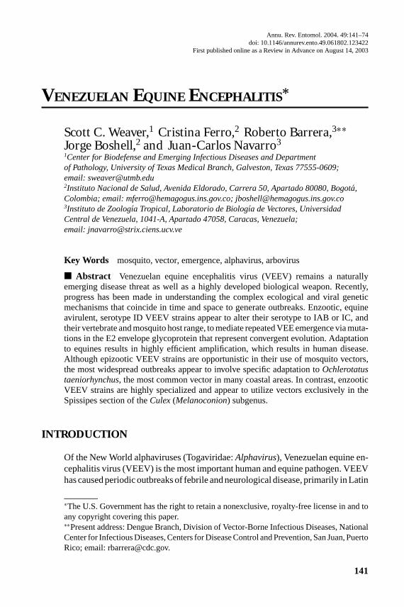
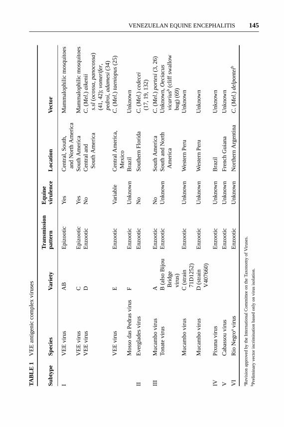
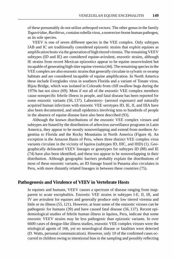
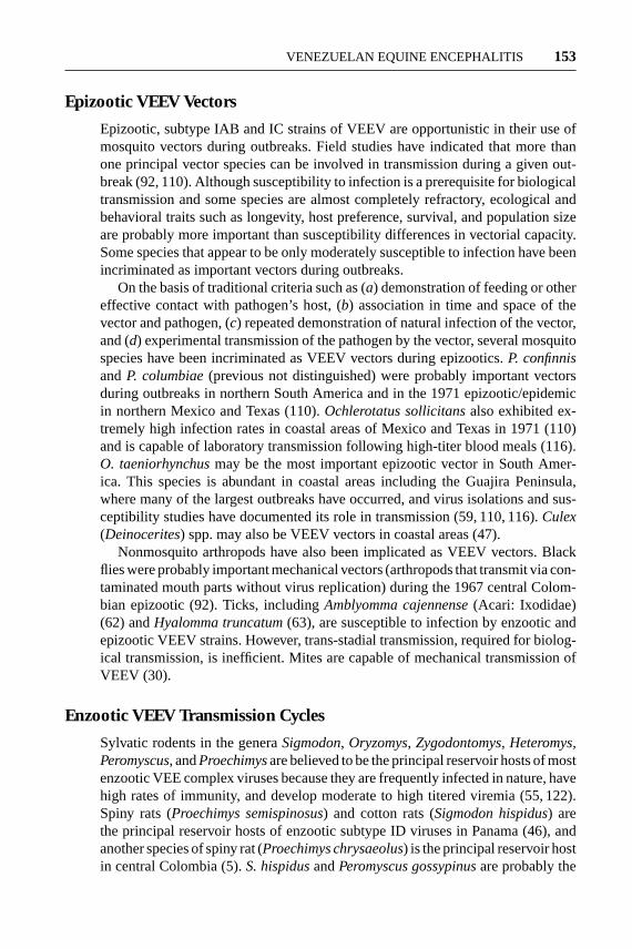
Figure 1 Map showing locations of VEE epizootics since the virus was isolated in 1938,along with virus subtypes implicated as the etiologic agents.
America, during the past century (Figure 1). Many outbreaks have involved tensto hundreds of thousands of equine and human cases, have spread over large geo-graphical regions, and have lasted up to several years. Recent outbreaks in Mexicoand South America demonstrated that VEE is a re-emerging disease. VEEV isalso a highly developed biological weapon amenable to use in warfare or terrorism(53). The current emphasis on biological defense has therefore renewed interest
23 Oct 2003 21:39 AR AR208-EN49-07.tex AR208-EN49-07.sgm LaTeX2e(2002/01/18)P1: GCE
VENEZUELAN EQUINE ENCEPHALITIS 143
in VEEV, both as a naturally emerging pathogen and as a terrorist agent that couldbe introduced artificially to cause widespread disease.
History of VEE
Because equines remain important in Latin America for agriculture and transporta-tion, VEE outbreaks have had profound social and economic effects in addition todirect effects on human health (55, 122, 127). VEE was first recognized as a dis-ease of horses, mules, and donkeys in northern South America during the 1930s.There is no clear evidence from epidemiological records of outbreaks during thenineteenth century (122), and phylogenetic estimates derived from sequences ofVEEV strains implicated in early outbreaks indicate that they probably evolvedin the early twentieth century (82). After 1938, major VEE outbreaks occurredperiodically until 1973, with interepizootic periods of roughly 10 years withoutany activity (64, 122). The source of the VEEV strains that caused outbreaks wastherefore the subject of considerable research and remains an important topic inarbovirology. One VEE outbreak spread through Central America, Mexico, andTexas during 1969–1972, involving tens of thousands of equines and people. Be-tween 1973 and 1992, no VEE was documented, prompting speculation that varietyIAB and IC epizootic VEE viruses had become extinct (122). However, severalrecent outbreaks (73, 86, 131) underscore the continued natural threat of VEE inthe Americas (Figure 1).
Early VEE Outbreaks
The first widely recognized VEE outbreak appeared in the central river valleys ofColombia during 1935, although epidemiological reports suggest that outbreaksmay have begun during the 1920s (118). One year later the outbreak spread intothe Guajira Peninsula of northern Colombia and Venezuela, a desert environ-ment populated by large numbers of mosquitoes following infrequent rainfall.The Guajira is also inhabited by feral donkeys that serve as efficient amplifica-tion hosts and are virtually impossible to vaccinate, probably the principal rea-son that the Guajira has been the epicenter of many VEE outbreaks. From 1936to 1938 the VEE outbreak spread across northern Venezuela and in 1943 ap-peared on the Island of Trinidad, where early vaccination efforts may have con-tributed to its containment. During these early outbreaks, human disease due toVEEV undoubtedly occurred. However, the connection between equine and hu-man disease was not established until the 1950s and 1960s, when human diseasewas documented in Colombia (93) and VEEV was isolated from human casesof febrile illness and fatal encephalitis in northern Venezuela (14). Some of thelargest outbreaks on record occurred during the 1960s in central Colombia, whereover 200,000 human cases and more than 100,000 equine deaths were estimated(50). Other major outbreaks involved the Pacific coast of Peru during the 1940s(64, 122).
23 Oct 2003 21:39 AR AR208-EN49-07.tex AR208-EN49-07.sgm LaTeX2e(2002/01/18)P1: GCE
144 WEAVER ET AL.
Identification of the Etiologic Agent of VEE
The etiological agent of VEE, VEEV, was first isolated in 1938 from the brainsof fatal equine cases in Yaracuy State, Venezuela. Between 1938 and 1956, onlyepizootic VEEV strains, later classified as antigenic subtype IAB, were isolatedin northern South America. Then, beginning in the late 1950s, VEEV and relatedVEE serocomplex virus strains were isolated in Central America (56, 97), SouthAmerica (103), Mexico (95), and Florida (18) from sylvatic and swamp habitatsin the absence of equine disease. Although these viruses utilize mosquito vectorsin a manner similar to their epizootic VEEV relatives, they infect primarily smallmammalian reservoir hosts and are not associated with equine disease. Humanswere shown to become infected with these enzootic VEEV and VEE complexstrains, with occasional fatal cases documented (56, 137). Later antigenic studies(134) demonstrated that these enzootic viruses, along with the epizootic vari-ants, comprise a serocomplex of related alphaviruses. The VEE complex nowcomprises 14 subtypes and varieties and includes 7 different virus species (129)(Table 1).
Vaccine Development and Use
Soon after the first isolation of VEEV in 1938, equine vaccines were made fromexperimentally infected mouse brain and other animal tissues containing high titersof the virus. These vaccines, inactivated with formalin, were made in Venezuela,Peru, Trinidad, and probably other locations from the only serotype of VEEVisolated from 1938 to 1957, subtype IAB. However, such inactivated viral vaccinesgenerally induce short-lived immunity, and equine vaccination in South Americaprobably had a limited effect on preventing disease. Moreover, genetic studies ofsubtype IAB VEEV strains isolated during outbreaks from 1938 to 1973 (58),phylogenetic relationships among IAB viruses (130), and the isolation of livevirus from some vaccinated humans after administration of “inactivated” vaccinepreparations (111) suggest that many if not all outbreaks during this era wereinitiated by the use of incompletely inactivated vaccines.
Since the early 1970s, inactivated vaccines have not been produced from wild-type VEEV strains, and epizootic IC strains were never used for vaccine produc-tion. Therefore, vaccines are not the explanation for recent subtype IC outbreaks(86, 131). In 1961 the development of a live-attenuated vaccine strain, TC-83, wasachieved by passaging the virulent subtype IAB strain, Trinidad donkey, 83 timesin guinea pig heart cell cultures (8). This vaccine was used first in Colombia in1968, and more extensively during the 1969–1971 Central America–Mexico-Texasoutbreak, and subsequently was shown to be safe and effective during experimentalequine trials (120). Currently, both live and inactivated [usually multivalent for-mulations combined with eastern (EEEV) and western equine encephalitis viruses(WEEV)] versions of TC-83 are used to vaccinate equines. The live-attenuatedversion is far superior in areas of Latin America at high risk for VEE outbreaksowing to the faster and longer-lasting immunity elicited (probably lifetime). Live
23 Oct 2003 21:39 AR AR208-EN49-07.tex AR208-EN49-07.sgm LaTeX2e(2002/01/18)P1: GCE
VENEZUELAN EQUINE ENCEPHALITIS 145TA
BLE
1V
EE
antig
enic
com
plex
viru
ses
Tran
smis
sion
Equ
ine
Sub
type
Spe
cies
Varie
typa
ttern
viru
lenc
eLo
catio
nVe
ctor
IV
EE
viru
sA
BE
pizo
otic
Yes
Cen
tral
,Sou
th,
Mam
mal
ophi
licm
osqu
itoes
and
Nor
thA
mer
ica
VE
Evi
rus
CE
pizo
otic
Yes
Sou
thA
mer
ica
Mam
mal
ophi
licm
osqu
itoes
VE
Evi
rus
DE
nzoo
ticN
oC
entr
alan
dC
.(M
el.)
aik
en
iiS
outh
Am
eric
as.
sl(o
coss
a,p
an
oco
ssa)
(41,
42);
vom
erife
r,p
ed
roi,a
da
me
si(34
)V
EE
viru
sE
Enz
ootic
Varia
ble
Cen
tral
Am
eric
a,C
.(M
el.)
tae
nio
pu
s(25
)M
exic
oM
osso
das
Ped
ras
viru
sF
Enz
ootic
Unk
now
nB
razi
lU
nkno
wn
IIE
verg
lade
svi
rus
Enz
ootic
No
Sou
ther
nF
lorid
aC
.(M
el.)
ced
ece
i(1
7,19
,132
)
IIIM
ucam
bovi
rus
AE
nzoo
ticN
oS
outh
Am
eric
aC
.(M
el.)
po
rte
si(3
,26)
Tona
tevi
rus
B(a
lso
Bijo
uE
nzoo
ticU
nkno
wn
Sou
than
dN
orth
Unk
now
n,O
eci
acu
sB
ridge
Am
eric
avi
cariu
sb(c
liffs
wal
low
viru
s)bu
g)(6
9)M
ucam
bovi
rus
C(s
trai
nE
nzoo
ticU
nkno
wn
Wes
tern
Per
uU
nkno
wn
71D
1252
)M
ucam
bovi
rus
D(s
trai
nE
nzoo
ticU
nkno
wn
Wes
tern
Per
uU
nkno
wn
V40
7660
)
IVP
ixun
avi
rus
Enz
ootic
Unk
now
nB
razi
lU
nkno
wn
VC
abas
sou
viru
sE
nzoo
ticU
nkno
wn
Fre
nch
Gui
ana
Unk
now
n
VI
Rio
Neg
roavi
rus
Enz
ootic
Unk
now
nN
orth
ern
Arg
entin
aC
.(M
el.)
de
lpo
nte
ib
a Rev
isio
nap
prov
edby
the
Inte
rnat
iona
lCom
mitt
eeon
the
Taxo
nom
yof
Viru
ses.
b Pre
limin
ary
vect
orin
crim
inat
ion
base
don
lyon
viru
sis
olat
ion.
23 Oct 2003 21:39 AR AR208-EN49-07.tex AR208-EN49-07.sgm LaTeX2e(2002/01/18)P1: GCE
146 WEAVER ET AL.
TC-83 has also been used for vaccination of laboratory personnel at risk of oc-cupational exposure, although human vaccination is accompanied by a high rateof adverse reactions and failures to seroconvert; a formalin-inactivated version ofTC-83, C-84, is administered to nonresponders (78). Availability for civilians ofthese vaccines from the U.S. Army Special Immunizations Program has becomehighly restricted during recent years. A new, genetically engineered VEEV vac-cine strain, 3526, appears to be superior to TC-83 in murine studies (67) andmay eventually replace TC-83 if human trials corroborate its improved safety andimmunogenicity.
Recent VEE Outbreaks
Following a 19-year hiatus with no confirmed VEE activity, several outbreaks haveoccurred during the past decade. The first began in December of 1992 in TrujilloState of western Venezuela (86). The epicenter was near a recently constructedreservoir and later spread discontinuously to the western shore of Lake Maracaiboin July of 1993. Twenty-four equine and four human cases were documented,although the actual number was undoubtedly much higher. Unlike most exten-sive outbreaks, the 1992–1993 epizootic/epidemic did not extend into the nearbyGuajira Peninsula of Venezuela or Colombia. Experimental equine infections withsubtype IC VEEV strains isolated from this outbreak demonstrated that horsesefficiently amplify these strains, consistent with a typical epizootic transmissioncycle (124).
Also in the summer of 1993, a small equine outbreak was described in Pacificcoastal communities of Chiapas State in southern Mexico, involving 125 docu-mented equine cases with 63 deaths (73). Three years later, from June to July1996, another equine epizootic occurred in adjacent Oaxaca State, involving 32horses with 12 deaths. Human cases were not detected during either outbreak,although recent human serosurveys and virus isolations from sentinel animalsin the affected regions indicate that VEE is endemic (J. Estrada-Franco & S.C.Weaver, unpublished data). Vaccination (TC-83) of 38,000 (1993) and 16,000(1996) horses, limitations on the transport of equines, and insecticide sprayingwere used to control the outbreaks, and bans on the importation of horses into theUnited States were instituted. Since 1996, sporadic cases of equine encephalitishave occurred in Mexico and Central America but remain unconfirmed by virusisolation or specific serology.
One of the largest VEE epizootics and epidemics on record, involving an es-timated 75,000 to 100,000 people, occurred in 1995 (131). Cases were detectedfirst in eastern Falcon State, Venezuela, in April and then in Carabobo, Yaracuy,and Lara States during July. By mid-July the epidemic had spread into northeast-ern Zulia State, and in August a major epidemic occurred in rural areas of theGuajira Peninsula, both in Venezuela and Colombia. Rainfall was unusually heavyduring July, August, and September, and mosquito collections in affected com-munities yielded large numbers ofOchlerotatus(formerlyAedes) taeniorhynchus,
23 Oct 2003 21:39 AR AR208-EN49-07.tex AR208-EN49-07.sgm LaTeX2e(2002/01/18)P1: GCE
VENEZUELAN EQUINE ENCEPHALITIS 147
Psorophora confinnis, and Anopheles aquasalis. Emergency control measures,including restricting the movement of equines, aerial and ground insecticide ap-plications, and equine vaccination (more than 95,000 animals in Colombia alone),may have prevented the epizootic from moving south into more populated regionsof Colombia. Sporadic human and equine cases continued in central Venezuela(Trujillo, Portuguesa, Cojedes, and Guarico States) until December 1995. Thisoutbreak was remarkably similar to one that occurred in the same regions ofVenezuela and Colombia during 1962–1964 (14). Symptoms and signs of infectedpatients, estimated human mortality rates (∼0.5%), unusually heavy rainfall pre-ceding the epidemic, and seasonal patterns of transmission were all similar tothose reported in 1962. In addition, viruses isolated during 1995 were antigenicallyidentical (subtype IC) and nearly genetically identical to those obtained from 1962to 1964.
VEE COMPLEX VIRUSES
VEEV Structure and Replication
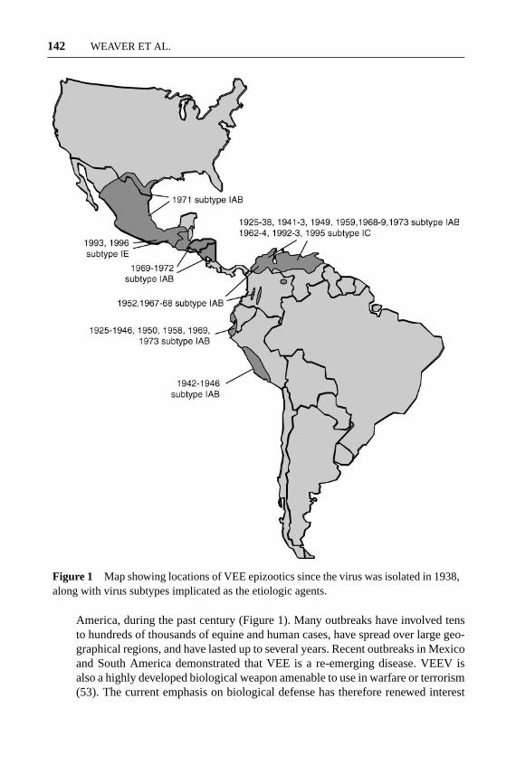
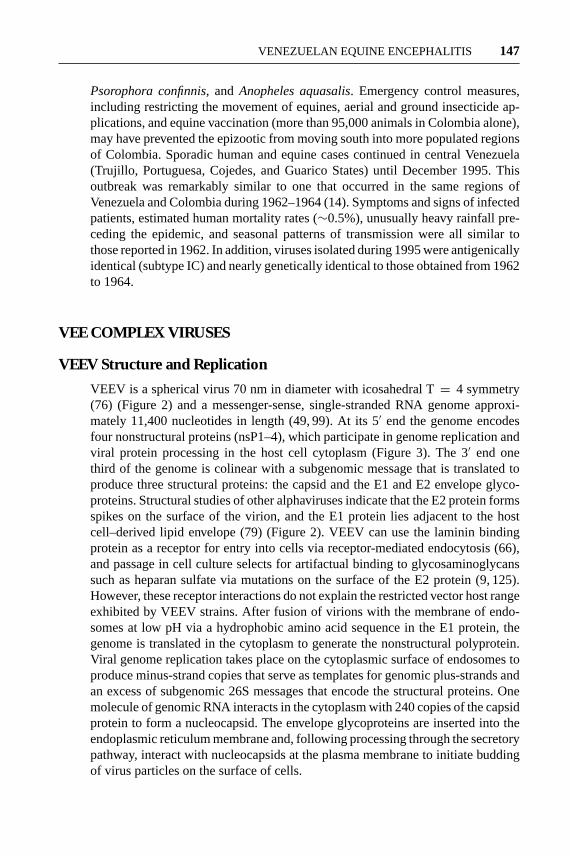
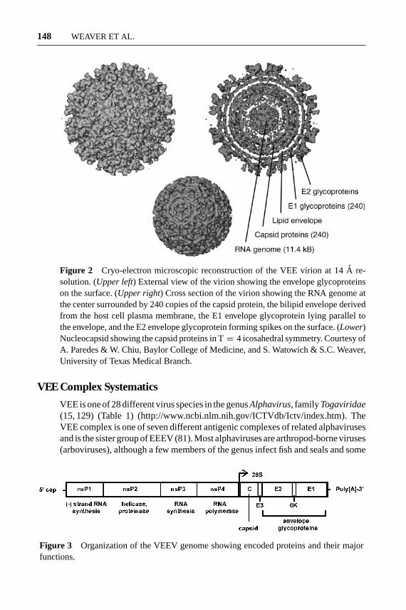
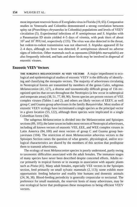
VEEV is a spherical virus 70 nm in diameter with icosahedral T= 4 symmetry(76) (Figure 2) and a messenger-sense, single-stranded RNA genome approxi-mately 11,400 nucleotides in length (49, 99). At its 5′ end the genome encodesfour nonstructural proteins (nsP1–4), which participate in genome replication andviral protein processing in the host cell cytoplasm (Figure 3). The 3′ end onethird of the genome is colinear with a subgenomic message that is translated toproduce three structural proteins: the capsid and the E1 and E2 envelope glyco-proteins. Structural studies of other alphaviruses indicate that the E2 protein formsspikes on the surface of the virion, and the E1 protein lies adjacent to the hostcell–derived lipid envelope (79) (Figure 2). VEEV can use the laminin bindingprotein as a receptor for entry into cells via receptor-mediated endocytosis (66),and passage in cell culture selects for artifactual binding to glycosaminoglycanssuch as heparan sulfate via mutations on the surface of the E2 protein (9, 125).However, these receptor interactions do not explain the restricted vector host rangeexhibited by VEEV strains. After fusion of virions with the membrane of endo-somes at low pH via a hydrophobic amino acid sequence in the E1 protein, thegenome is translated in the cytoplasm to generate the nonstructural polyprotein.Viral genome replication takes place on the cytoplasmic surface of endosomes toproduce minus-strand copies that serve as templates for genomic plus-strands andan excess of subgenomic 26S messages that encode the structural proteins. Onemolecule of genomic RNA interacts in the cytoplasm with 240 copies of the capsidprotein to form a nucleocapsid. The envelope glycoproteins are inserted into theendoplasmic reticulum membrane and, following processing through the secretorypathway, interact with nucleocapsids at the plasma membrane to initiate buddingof virus particles on the surface of cells.
23 Oct 2003 21:39 AR AR208-EN49-07.tex AR208-EN49-07.sgm LaTeX2e(2002/01/18)P1: GCE
148 WEAVER ET AL.
Figure 2 Cryo-electron microscopic reconstruction of the VEE virion at 14A re-solution. (Upper left) External view of the virion showing the envelope glycoproteinson the surface. (Upper right) Cross section of the virion showing the RNA genome atthe center surrounded by 240 copies of the capsid protein, the bilipid envelope derivedfrom the host cell plasma membrane, the E1 envelope glycoprotein lying parallel tothe envelope, and the E2 envelope glycoprotein forming spikes on the surface. (Lower)Nucleocapsid showing the capsid proteins in T= 4 icosahedral symmetry. Courtesy ofA. Paredes & W. Chiu, Baylor College of Medicine, and S. Watowich & S.C. Weaver,University of Texas Medical Branch.
VEE Complex Systematics
VEE is one of 28 different virus species in the genusAlphavirus, familyTogaviridae(15, 129) (Table 1) (http://www.ncbi.nlm.nih.gov/ICTVdb/Ictv/index.htm). TheVEE complex is one of seven different antigenic complexes of related alphavirusesand is the sister group of EEEV (81). Most alphaviruses are arthropod-borne viruses(arboviruses), although a few members of the genus infect fish and seals and some
Figure 3 Organization of the VEEV genome showing encoded proteins and their majorfunctions.
23 Oct 2003 21:39 AR AR208-EN49-07.tex AR208-EN49-07.sgm LaTeX2e(2002/01/18)P1: GCE
VENEZUELAN EQUINE ENCEPHALITIS 149
of these presumably do not utilize arthropod vectors. The other genus in the familyTogaviridae,Ruvibirus, contains rubella virus, a nonvector-borne human pathogen,as its sole species.
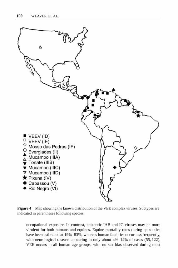
VEEV is one of seven different species in the VEE complex. Only subtypesIAB and IC are traditionally considered epizootic strains that exploit equines asamplification hosts via the generation of high titered viremia. The remaining VEEVsubtypes (ID and IE) are considered equine-avirulent, enzootic strains, althoughIE strains from recent Mexican epizootics appear to be equine neurovirulent butincapable of generating high-titer equine viremia (44). The remaining species in theVEE complex are also enzootic strains that generally circulate in sylvatic or swamphabitats and are considered incapable of equine amplification. In North Americathese include Everglades virus in southern Florida and a variant of Tonate virus,Bijou Bridge, which was isolated in Colorado from cliff swallow bugs during the1970s but not since (69). Most if not all of the enzootic VEE complex memberscause nonspecific febrile illness in people, and fatal disease has been reported forsome enzootic variants (56, 137). Laboratory- (aerosol exposure) and naturallyacquired human infections with enzootic VEE serotypes ID, IE, II, and IIIA havealso been documented, and small epidemics involving tens to hundreds of peoplein the absence of equine disease have also been described (93).
Although the known distributions of the enzootic VEE complex viruses andsubtypes are biased by the distribution of arbovirus surveillance programs in LatinAmerica, they appear to be mostly nonoverlapping and extend from northern Ar-gentina to Florida and the Rocky Mountains in North America (Figure 4). Anexception is the Amazon Basin of Peru, where three distinct VEE complex virusvariants circulate in the vicinity of Iquitos (subtypes ID, IIIC, and IIID) (1). Geo-graphically delineated VEEV lineages or genotypes for subtypes ID (88) and IE(74) have also been identified, and these too appear to be nonoverlapping in theirdistribution. Although geographic barriers probably explain the distributions ofmost of these enzootic variants, an ID lineage found in Panama also circulates inPeru, with more distantly related lineages in between these countries (75).
Pathogenesis and Virulence of VEEV in Vertebrate Hosts
In equines and humans, VEEV causes a spectrum of disease ranging from inap-parent to acute encephalitis. Enzootic VEE strains in subtypes I-E, II, III, andIV are avirulent for equines and generally produce only low titered viremia andlittle or no illness (55, 121). However, at least some of the enzootic viruses can bepathogenic for humans (39) and have caused fatal disease (56, 137). Recent epi-demiological studies of febrile human illness in Iquitos, Peru, indicate that someenzootic VEEV strains may be less pathogenic than epizootic variants. In over6600 cases of dengue-like illness studies, enzootic VEE complex viruses were theetiological agents of 166, yet no neurological disease or fatalities were detected(D. Watts, personal communication). However, only 10 of the confirmed cases oc-curred in children owing to intentional bias in the sampling and possibly reflecting
23 Oct 2003 21:39 AR AR208-EN49-07.tex AR208-EN49-07.sgm LaTeX2e(2002/01/18)P1: GCE
150 WEAVER ET AL.
Figure 4 Map showing the known distribution of the VEE complex viruses. Subtypes areindicated in parentheses following species.
occupational exposure. In contrast, epizootic IAB and IC viruses may be morevirulent for both humans and equines. Equine mortality rates during epizooticshave been estimated at 19%–83%, whereas human fatalities occur less frequently,with neurological disease appearing in only about 4%–14% of cases (55, 122).VEE occurs in all human age groups, with no sex bias observed during most
23 Oct 2003 21:39 AR AR208-EN49-07.tex AR208-EN49-07.sgm LaTeX2e(2002/01/18)P1: GCE
VENEZUELAN EQUINE ENCEPHALITIS 151
outbreaks. Children are more likely to develop fatal encephalitis and suffer per-manent neurological sequelae than adults. VEEV also infects the fetus in pregnantwomen and causes birth defects as well as spontaneous abortions and stillbirths.
In equines, generalized signs usually appear about 2–5 days after infection withepizootic VEEV, including fever, tachycardia, depression, and anorexia (55, 121,124). Some or most animals go on to develop encephalitis 5–10 days after infection,with signs of circling, ataxia, and hyperexcitability. Death usually occurs about oneweek after experimental infection. Encephalitis and death are correlative with themagnitude of equine viremia, but even equine-avirulent enzootic strains producelethal encephalitis when inoculated intracerebrally. This suggests that virulence isrelated to the ability of VEEV to replicate extracerebrally and spread to the brainrather than to innate neurovirulence. Resistance to murine interferon (IFN)-∂/β hasbeen suggested as a determinant of epizootic potential (54, 107). However, studiesof strains from the 1992 and 1995 Venezuelan outbreaks suggest that IFN-∂/βsensitivity is not related to the emergence of equine virulence (M. Anishchenko,S. Paessler & S.C. Weaver, unpublished data). Attenuation of the TC-83 vaccinestrain was accompanied by an increase in IFN-∂/β sensitivity; this change wasmapped to an E2 glycoprotein amino acid change that is augmented by a 5′-noncoding nucleotide change (107).
In human VEEV infection the incubation period is usually 2–5 days (55). Mostinfections are apparent, and symptoms appear abruptly, including malaise, fever,chills, and severe retro-orbital or occipital headache. Myalgia typically centersin the thighs and lumbar region of the back. Signs usually include leukopenia,tachycardia, and fever and are frequently accompanied by nausea, vomiting, anddiarrhea. Signs and symptoms of central nervous system involvement occur lessfrequently and typically include convulsions, somnolence, confusion, and photo-phobia. Acute disease usually subsides 4–6 days after onset, followed by astheniaof several weeks duration. Occasionally, illness is biphasic with recurrence 4–8days after onset. A small proportion of human cases proceed to stupor and comaand are sometimes followed by death. Lethal human VEE (generally less than 1%of cases) is accompanied by diffuse congestion and edema with hemorrhage in thebrain, gastrointestinal tract, and lungs (28). Meningoencephalitis associated withintense necrotizing vasculitis and cerebritis is also observed in some patients. Aswith equines and laboratory rodents, a striking depletion of lymphocytes occursin the lymph nodes, spleen, and gastrointestinal tract.
Following experimental infection with VEEV, nonhuman primates develop anonspecific febrile illness similar to that in most human cases. Domestic rabbits,goats, dogs, and sheep also suffer fatal disease during VEE epizootics (55). Lab-oratory rodents including hamsters and mice are highly susceptible to infectionwith all VEE complex variants and suffer fatal disease with some subtypes (122).Guinea pigs are killed by epizootic IAB and IC viruses and some enzootic IDstrains, but not by other enzootic subtypes (some ID and all IE, II, III, and IVstrains tested) (94). In experimentally infected equines and rodents, VEEV causessevere myeloid depletion in bone marrow and lymphocyte destruction in lymph
23 Oct 2003 21:39 AR AR208-EN49-07.tex AR208-EN49-07.sgm LaTeX2e(2002/01/18)P1: GCE
152 WEAVER ET AL.
nodes and spleen (27, 48, 94). Encephalitis is accompanied by a wide range ofhistopathology, from mild neutrophilic infiltration to neuronal degeneration, necro-tizing vasculitis, and Purkinje cell destruction. In mice, VEEV appears to reach thebrain via the olfactory nerve, seeded by viremia (27). Natural reservoir hosts be-come viremic but generally show no detectable disease after experimental infection(11, 135, 136).
Infection and Dissemination of VEEV in Mosquito Vectors
Biological transmission of arthropod-borne viruses generally involves initial in-fection of the mosquito midgut following ingestion of a viremic blood meal (126).Posterior midgut epithelial cells are believed to become infected first, followedby dissemination into the hemocoel and infection of secondary organs and tis-sues including the salivary glands. Virus maturation (budding) of alphaviruses inmidgut epithelial cells occurs exclusively on the basal margins adjacent to the basallamina. The mechanism of passage through the basal lamina is unknown; directpenetration seems unlikely because of the small pore size. Cytopathic effects onmidgut epithelial cells by EEEV and WEEV may facilitate dissemination into thehemocoel. Although this mechanism has not been investigated with VEEV, cy-topathology in salivary glands of experimentally infectedAedes aegyptihas beendetected (61). In the salivary glands, VEEV particles bud into the apical cavitiesand accumulate there, before salivation during blood feeding results in their pas-sage through the salivary ducts and into the vertebrate host (126). Mosquito salivaappears to be deposited primarily in the extravascular space prior to canulation ofa blood vessel (117).
TRANSMISSION CYCLES OF VEEV
Epizootic VEEV Transmission Cycles
The epizootic transmission cycle of VEEV has been studied during many outbreaksand is reasonably well understood. A feature common to all major outbreaks isthe role of equines as highly efficient amplification hosts. Although the vertebratehost range of epizootic VEEV strains is wide and includes humans, sheep, dogs,bats, rodents, and some birds, major epidemics in the absence of equine cases havenever occurred despite the repeated occurrence of epizootics near major cities suchas Maracaibo. Experimentally infected equines generally develop viremia lasting2–4 days, with typical peak titers of 105 to 107 suckling mouse LD50/ml serum andoccasional titers of 108 (55, 124). Humans develop similar levels of viremia butprobably are incapable of efficient amplification because of their lesser exposureto mosquito bites. However, the potential for urban transmission by a species suchasA. aegypti, which is susceptible to infection after biting humans and exhibitsbehavioral traits such as multiple host feeding and peridomesticity that augmentits vector competence (52), should be considered as human populations continueto expand and those of equines decline in Latin America.
23 Oct 2003 21:39 AR AR208-EN49-07.tex AR208-EN49-07.sgm LaTeX2e(2002/01/18)P1: GCE
VENEZUELAN EQUINE ENCEPHALITIS 153
Epizootic VEEV Vectors
Epizootic, subtype IAB and IC strains of VEEV are opportunistic in their use ofmosquito vectors during outbreaks. Field studies have indicated that more thanone principal vector species can be involved in transmission during a given out-break (92, 110). Although susceptibility to infection is a prerequisite for biologicaltransmission and some species are almost completely refractory, ecological andbehavioral traits such as longevity, host preference, survival, and population sizeare probably more important than susceptibility differences in vectorial capacity.Some species that appear to be only moderately susceptible to infection have beenincriminated as important vectors during outbreaks.
On the basis of traditional criteria such as (a) demonstration of feeding or othereffective contact with pathogen’s host, (b) association in time and space of thevector and pathogen, (c) repeated demonstration of natural infection of the vector,and (d) experimental transmission of the pathogen by the vector, several mosquitospecies have been incriminated as VEEV vectors during epizootics.P. confinnisandP. columbiae(previous not distinguished) were probably important vectorsduring outbreaks in northern South America and in the 1971 epizootic/epidemicin northern Mexico and Texas (110).Ochlerotatus sollicitansalso exhibited ex-tremely high infection rates in coastal areas of Mexico and Texas in 1971 (110)and is capable of laboratory transmission following high-titer blood meals (116).O. taeniorhynchusmay be the most important epizootic vector in South Amer-ica. This species is abundant in coastal areas including the Guajira Peninsula,where many of the largest outbreaks have occurred, and virus isolations and sus-ceptibility studies have documented its role in transmission (59, 110, 116).Culex(Deinocerites) spp. may also be VEEV vectors in coastal areas (47).
Nonmosquito arthropods have also been implicated as VEEV vectors. Blackflies were probably important mechanical vectors (arthropods that transmit via con-taminated mouth parts without virus replication) during the 1967 central Colom-bian epizootic (92). Ticks, includingAmblyomma cajennense(Acari: Ixodidae)(62) andHyalomma truncatum(63), are susceptible to infection by enzootic andepizootic VEEV strains. However, trans-stadial transmission, required for biolog-ical transmission, is inefficient. Mites are capable of mechanical transmission ofVEEV (30).
Enzootic VEEV Transmission Cycles
Sylvatic rodents in the generaSigmodon, Oryzomys, Zygodontomys, Heteromys,Peromyscus, andProechimysare believed to be the principal reservoir hosts of mostenzootic VEE complex viruses because they are frequently infected in nature, havehigh rates of immunity, and develop moderate to high titered viremia (55, 122).Spiny rats (Proechimys semispinosus) and cotton rats (Sigmodon hispidus) arethe principal reservoir hosts of enzootic subtype ID viruses in Panama (46), andanother species of spiny rat (Proechimys chrysaeolus) is the principal reservoir hostin central Colombia (5).S. hispidusandPeromyscus gossypinusare probably the
23 Oct 2003 21:39 AR AR208-EN49-07.tex AR208-EN49-07.sgm LaTeX2e(2002/01/18)P1: GCE
154 WEAVER ET AL.
most important reservoir hosts of Everglades virus in Florida (19, 65). Comparativestudies in Venezuela and Colombia demonstrated a strong correlation betweenspiny rat (Proechimys chrysaeolusin Colombia) populations and levels of VEEVcirculation (5). Experimental infections ofP. semispinosusandS. hispiduswitha Panamanian ID strain yielded 4–5 days of viremia, with peak titers of about105 and 107 PFU/ml, respectively (135). The virus was also detected in the throat,but rodent-to-rodent transmission was not observed.S. hispidusappeared ill for2–4 days, although no fever was detected;P. semispinosusshowed no adversesigns of infection. Other mammals such as opossums (Didelphis marsupialis) arealso frequently infected, and bats and shore birds may be involved in dispersal ofenzootic viruses.
Enzootic VEEV Vectors
THE SUBGENUS MELANOCONION AS VEEV VECTORS A major impediment to eco-logical and epidemiological studies of enzootic VEEV is the difficulty of identify-ing and classifying the mosquito vectors. The majority of arboviruses circulatingin Neotropical forests are transmitted by members of the genusCulex, subgenusMelanoconion(41, 127), a diverse and taxonomically difficult group of 156 rec-ognized species that occurs throughout the Neotropics (a few occur in subtropicaland temperate areas) (38, 51, 77, 89, 90). Seven species are proven vectors of VEEcomplex viruses (Tables 1 and 2), and others are likely vectors of EEEV, as wellgroup C and Guama group arboviruses in the familyBunyaviridae. Most studies ofenzootic VEEV ecology have incriminated a single species as the principal vectorin a given location (55, 122), although three species were implicated in a singleColombian forest (34).
The subgenusMelanoconionis divided into the Melanoconion and Spissipessections (89, 105); the latter taxon includes most vectors of Neotropical arboviruses,including all known vectors of enzootic VEE, EEE, and WEE complex viruses inLatin America (84, 100) and most vectors of group C and Guama group bun-yaviruses (104). The restriction of mostMelanoconionarbovirus vectors to theSpissipes Section raises the question of what genetic, physiological, and/or eco-logical characteristics are shared by the members of this section that predisposethem to transmit arboviruses.
The ecology of mostMelanoconionspecies is poorly understood, partly owingto taxonomic difficulties associated with the adult females. The immature stagesof many species have never been described despite concerted efforts. Adults oc-cur primarily in tropical forests or in swamps in association with aquatic plantssuch asPistia (41). Many adult females, especially VEE vectors in the Spissipessection, feed primarily on small sylvatic mammals (2, 113); some exhibit moreopportunistic feeding behavior and readily bite humans and domestic animals(24, 36, 38). Blood-feeding periodicity is generally crepuscular or nocturnal. Thepreference for small mammals, the reservoir hosts of many arboviruses, may beone ecological factor that predisposes these mosquitoes to being efficient VEEVvectors.
23 Oct 2003 21:39 AR AR208-EN49-07.tex AR208-EN49-07.sgm LaTeX2e(2002/01/18)P1: GCE
VENEZUELAN EQUINE ENCEPHALITIS 155
TABLE 2 Checklist of Spissipes section [Culex (Melanoconion)] mosquito species
Species Evidence of natural VEEV transmission
adamesia Sirivanakarn & Galindo 1980 Subtype ID-Colombia (34)
akritosForattini & Sallum 1995 None
cedeceia Stone & Hair 1968 Subtype II (Everglades virus)-Florida(19, 132)
crybdaDyar 1924 None
delponteiDuret 1969 Subtype VI (Rio Negro virus) (16)
epanastasisDyar 1922 None
fauraniDuret 1968 None
jubifer Komp & Brown 1935 None
gnomatosb Sallum, Hutchings Subtype ID-Peru (115)& Ferreira 1997
ikelosForattini & Sallum 1995 None
lopesiSirivanakarn & Jacob 1979 None
ocossaa Dyar & Knab 1919 Subtype ID-Panama (42)
panocossaa Dyar 1923 Subtype ID-Panama (42)
paracrybdaKomp 1936 None
pedroia Sirivanakarn & Belkin 1980 Subtype ID-Colombia (34)
pereyraiDuret 1967 None
portesia Senevet & Abbonenc 1941 Subtype IIIA (Mucambo virus) (2, 3)
ribeirensisForattini & Sallum 1985 None
saquettaeSirivanakarn & Jacob 1981 None
simulatorDyar & Knab 1906 None
spissipesb Theobald 1903 Subtype ID-Venezuela (reportedin error asferreri) (119)
taeniopusa Dyar & Knab 1903 Subtype IE-Guatemala (25)
vomerifera Komp 1932 Subtype ID-Colombia (34)
aProven vector meeting established criteria (4).bSuggested but unproven vector role based only on virus isolation or susceptibility to infection.
TAXONOMIC HISTORY OF THE SUBGENUS MELANOCONION Despite its public he-alth importance, little progress has been made in the systematics of the subgenusMelanoconion, which underwent several changes in interpretation and taxonomictreatment from Theobald (114) (genusMelanoconion) until Rozeboom & Komp(87), when its subgeneric status became accepted. Due to repeated resurrectionsand synonymizations, the classification remains confusing. The taxonomic historyhas been reviewed by Sirivanakarn (105) and Pecor et al. (77).
23 Oct 2003 21:39 AR AR208-EN49-07.tex AR208-EN49-07.sgm LaTeX2e(2002/01/18)P1: GCE
156 WEAVER ET AL.
A complete classification ofMelanoconionawaited the comprehensive revisionof Sirivanakarn (105), based on morphological characteristics, which distinguished153 species within 3 sections: Ocellatus (4 spp.), Melanoconion (125 spp.), andSpissipes (20 spp.). Recently, Pecor et al. (77) reduced theMelanoconionsectionsto two, after excluding the four Ocellatus species without subgeneric assignment.
THE SPISSIPES SECTION AmongMelanoconion(sensu lato) mosquitoes, the Spis-sipes group, first proposed by Galindo (40), has received the most attention owingto the inclusion of several arbovirus vectors (23, 34, 40, 105). Recently, Sallum &Forattini (89) revised the Spissipes section using adult morphological characters(with male and female keys) including the 22 species described to date.
Within the Spissipes section, several important taxonomic changes have beenmade recently, includingC. taeniopusand C. cedecei(23) [synonymized withopisthopusandannulipes(6)], C. aikenii s.l. [divided intoocossaandpanocossa(7)], C. taeniopus[with the new species emergence ofC. pedroi(105)],C. adamesi(106), the resurrection ofC. pedroi[with the new species emergence ofakritosandikelos(89)],C. epanastasis[with the new species emergence ofC. ribeirensis(37)], andC. vomerifer[with the new species emergence ofC. gnomatos(89)].Recently, with the use of ribosomal DNA sequences and phylogenetic methodscorroborated by morphological analyses, two cryptic species underC. pedroiweredetected in VEEV enzootic areas of South America (72).
The evolutionary relationships ofMelanoconionto others in the tribe Culicinihave not been examined other than at higher taxonomic levels (68, 71). Recently,Navarro & Weaver (72) inferred the evolutionary relationships among the Vomer-ifer and Pedroi groups in the Spissipes section, two proven VEEV vector groups(34), including the twenty-three Spissipes group species described by Forattini &Sallum (38) (Table 2).
ECOLOGY OF MOSQUITOES IN THE MELANOCONION SUBGENUS IN COLOMBIA InColombia, 54 species in theCulex(Melanoconion) subgenus have been identifiedin diverse habitats ranging from the desert-like Guajira Peninsula to the humid,tropical forests of Choc´o Department (70). The abundance and diversity of manyof these species are typified by a humid tropical forest in the Middle MagdalenaValley of Colombia (5), where from 1998 to 1999 an enzootic focus of subtype IDVEEV was studied intensively. FourteenCulex(Melanoconion) species, 6 fromthe Spissipes section (C. pedroi, C. vomerifer, C. adamesi, C. spissipes, C. crybda,andC. ocossa) and 8 from the Melanoconion section were collected in CDC lighttraps. The majority of the males (2573/3009) of the males collected during thetwo-year study were captured during May 1999; the factors that regulate the mas-sive emergence of these mosquitoes in the month of May are unknown and moredetailed studies are needed. However, climatic variables are probably involved.
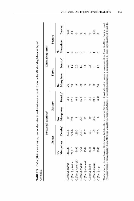
Studies of the behavior of the VEEV vector species (C. pedroi, C. vomerifer,C. adamesi) in the Monte San Miguel forest (5) determined that these mosquitoeshave a nocturnal activity peak and remain in forest habitats (Table 3). An exception
23 Oct 2003 21:39 AR AR208-EN49-07.tex AR208-EN49-07.sgm LaTeX2e(2002/01/18)P1: GCE
VENEZUELAN EQUINE ENCEPHALITIS 157
TAB
LE3
Cu
lex(
Me
lan
oco
nio
n)sp
p.ve
ctor
dens
ities
inan
dou
tsid
ean
enzo
otic
focu
sin
the
Mid
dle
Mag
dale
naVa
lley
ofC
olom
bia
Noc
turn
alca
ptur
esa
Diu
rnal
capt
ures
a
For
est
Pas
ture
For
est
Pas
ture
No.
No.
No.
No.
Mos
quito
esD
ensi
tyaM
osqu
itoes
Den
sitya
Mos
quito
esD
ensi
tyaM
osqu
itoes
Den
sitya
C.(
Me
l.)p
ed
roi
21,7
2760
3.5
693.
626
0.7
10.
05
C.(
Me
l.)sp
issi
pe
s21
,115
586.
623
012
.153
1.4
20.
1
C.(
Me
l.)vo
me
rife
r64
9218
0.3
80.
49
0.2
00
C.(
Me
l.)cr
ybd
a72
6120
1.7
291
15.3
391.
00
0
C.(
Me
l.)a
da
me
si15
0241
.721
1.1
50.
10
0
C.(
Me
l.)d
un
ni
3351
93.1
593.
16
0.1
00
C.(
Me
l.)o
coss
a14
13.
936
419
.10
01
0.05
C.(
Me
l.)sp
p.22
4862
.58
0.42
501.
30
0
a Num
bero
fspe
cies
(fem
ales
)cap
ture
din
the
Mon
teS
anM
igue
lfor
est,
noct
urna
l:36
.Num
bero
fspe
cies
(fem
ales
)cap
ture
din
past
ures
outs
ide
the
Mon
teS
anM
igue
lfor
est,
noct
urna
l:19
.Num
bero
fspe
cies
(fem
ales
)cap
ture
din
the
Mon
teS
anM
igue
lfor
est,
noct
urna
l:38
.Num
bero
fspe
cies
(fem
ales
)cap
ture
din
past
ures
outs
ide
the
Mon
teS
anM
igue
lfor
est,
diur
nal:
19.
23 Oct 2003 21:39 AR AR208-EN49-07.tex AR208-EN49-07.sgm LaTeX2e(2002/01/18)P1: GCE
158 WEAVER ET AL.
was C. ocossa, a vector incriminated in Panama, which had higher populationlevels outside the forest habitat (29.6 females/trap/night outside the forest versus20.0 females/trap/night inside). In addition toC. ocossa, small numbers of allSpissipes section species (C. pedroi, C. vomerifer, C. adamesi, C. spissipes, andC.crybda), but few from the Melanoconion section, were collected outside the forest.Populations of the species common to the forest interior were most abundant inMay and August (precipitation 245–266 mm per month), and those ofC. ocossawere more abundant in November (precipitation 385 mm).
A study of the effect of climatic variables, including precipitation, maximumand minimum temperature, and relative humidity, on VEEV vector (C. pedroiandC. vomerifer) populations established that precipitation and relative humid-ity had a significant effect on both species. The lowest population densities oc-curred during the months with lowest mean precipitation (<165 mm), with 38–108females/trap/night forC. vomeriferand 229–685 females/trap/night forC. pedroi.In months with more than 200 mm of precipitation the population densities rose,reaching values of 148–639 females/trap/night forC. vomeriferand 549–1337 forC. pedroi(C. Ferro, unpublished data).
Origins of Epizootic VEEV Strains
Because of the sporadic and discontinuous nature of epizootic VEE, the sourceof the subtype IAB and IC strains has been a major topic of research. Johnson &Martin (55) proposed five hypotheses to explain the origins of epizootic strains:(a) maintenance of epizootic strains in continuous, cryptic transmission cycles,(b) maintenance of epizootic strains in latent equine or other animal infections,(c) re-emergence of epizootic viruses following administration of incompletely in-activated vaccines, (d) maintenance of epizootic strains as minority subpopulationswithin enzootic virus populations, and (e) periodic emergence of epizootic virusesvia mutations of enzootic strains. The evidence with regard to these hypotheses isas follows:
■ No evidence for maintenance of IAB and/or IC viruses in continuous trans-mission cycles has been obtained during field studies where epizootics fre-quently occur. However, the isolation of subtype IC VEEV strains inVenezuela during 2000, nearly five years after the apparent end of the 1995Venezuelan epizootic, indicates that this virus may circulate in Venezuela in agenetically static mode without apparent disease (G. Medina & S.C. Weaver,unpublished data). If confirmed, the interepizootic subtype IC transmissioncycle must be studied to assess the implications for re-emergence of thesestrains.
■ No evidence of latent or persistent VEEV infection of any vertebrate host hasbeen reported, although WEEV, another alphavirus, can persist for severalmonths in avian hosts (83).
■ Genetic conservation and phylogenetic relationships of IAB viruses isolated
23 Oct 2003 21:39 AR AR208-EN49-07.tex AR208-EN49-07.sgm LaTeX2e(2002/01/18)P1: GCE
VENEZUELAN EQUINE ENCEPHALITIS 159
over a 35-year time span are consistent with a vaccine etiology in severaloutbreaks (58, 130).
■ Stanick et al. (108) examined the possibility that epizootic IAB or IC virusesare present as a minority population within enzootic ID and IE isolates.Using hydroxylapatite chromatography and selection of small plaques onVero cells, which can isolate as few as one epizootic virion in a populationof 106 enzootic virions, they obtained no evidence of epizootic virions in 23subtype ID strains examined.
■ Periodic emergence of epizootic VEE viruses by mutation of enzootic strains(55, 122) was first supported by antigenic (134) and later genetic (57, 58, 85)similarities between epizootic and enzootic (subtype ID) VEEV. Phylogeneticanalyses of all members of the VEE complex first showed a close evolutionaryrelationship among IAB, IC, and ID VEE viruses and indicated that epizooticviruses probably arose several times from subtype ID–like ancestors (128).More detailed phylogenetic studies have delineated six major lineages of en-zootic VEEV, including five ID-like lineages and the subtype IE lineage. Allthe epizootic strains from major outbreaks fall into one of three clades nestedwithin one of these lineages, which is otherwise composed of enzootic, sub-type ID strains isolated in western Venezuela, Colombia, and northern Peru(Figure 5). Equine-virulent strains isolated during recent Mexican epizooticsgroup with enzootic strains from the Pacific coast of Guatemala. These datasupport the hypothesis that epizootic VEEV strains have arisen on at leastfour occasions by mutation of enzootic strains and changes in host range (seebelow). Both the epizootic phenotype and the IC serotype represent examplesof convergent evolution.
Control and Prevention of VEE Outbreaks
Prevention of VEE outbreaks could probably be attained by sustained equine vacci-nation in Colombia, Venezuela, and Mexico, where progenitors of epizootic strainsare believed to circulate and where recent outbreaks have been documented. Al-though governments in affected countries generally provide free equine vaccinationduring and soon after outbreaks, these programs are rarely sustained and suscepti-ble populations are replenished within 5–10 years. During outbreaks, equine vac-cination can be effective if VEEV circulation is anticipated or recognized quickly,but governmental agencies are often slow in acknowledging epizootics becauseof their perceived veterinary/public heath failures. Although live-attenuated TC-83 vaccine is manufactured in Mexico, Venezuela, and Colombia and marketedthroughout most of Latin America, some equines in South America are vaccinatedwith inactivated, multivalent alphavirus vaccines marketed in the United States.These vaccines are inferior for protection of animals and for the prevention ofepizootics and epidemics because (a) inactivated preparations generate protectiveneutralizing immunity only after multiple inoculations, (b) immunity resultingfrom inactivated vaccines is short-lived and frequent boosters are required to
23 Oct 2003 21:39 AR AR208-EN49-07.tex AR208-EN49-07.sgm LaTeX2e(2002/01/18)P1: GCE
160 WEAVER ET AL.
Figure 5 Phylogenetic tree derived from nucleotide sequences encoding the PE2 envelopeglycoprotein using the neighbor joining method and the HK distance formula, as implementedin the PAUP 4.0 software package (112). Strains are indicated by subtype, followed by country(FL = Florida, USA) and strain designation. Major lineages of VEEV are indicated on theright, and strains isolated from epizootics are boxed and branches are in bold. Bootstrapvalues for all major lineages and epizootic clades were≥98%.
maintain protection, and (c) once vaccinated with an inactivated preparation,equines probably do not respond appropriately to the live vaccine, requiring boost-ers for life to maintain immunity. Therefore, public and veterinary health officialsshould strongly discourage the use of these inactivated vaccines in regions ofLatin America with a history of VEE. An adequate supply of live TC-83 vaccineshould also be available in the United States for use in the event of a natural orterrorist-related introduction of VEEV.
Limitations on the movement of equines in affected regions have generallyproved ineffective, probably because infected animals are asymptomatic for 1–3days; owners therefore unknowingly move asymptomatic, infected animals to un-affected areas for protection. Mosquito control, principally by aerial applicationsof adulticides, probably has some impact on reducing transmission and may havelimited the spread of VEEV during the 1971 Texas outbreak. However, reductionsin rainfall also occurred coincident with a decline in VEE incidence. As with mostarboviral diseases, protection of human populations relies principally on personalprotection against mosquito bites by limiting physical exposure and applying re-pellants containing the active ingredient diethylmethylbenzamide (DEET,≤35%
23 Oct 2003 21:39 AR AR208-EN49-07.tex AR208-EN49-07.sgm LaTeX2e(2002/01/18)P1: GCE
VENEZUELAN EQUINE ENCEPHALITIS 161
formulations recommended for adults,≤10% for children). Permethrin can be ap-plied to clothing to enhance protection. These protective measures are especiallyimportant for individuals who reside or work near equine herds during epizootics,or those who contact tropical forest or swamp habitats where enzootic VEEVcirculates.
THE ROLE OF ADAPTATION TO HOSTS ANDVECTORS IN VEE EMERGENCE
Phylogenetic studies support the hypothesis that epizootic VEEV strains arisevia mutation of enzootic strains, principally ID strains from western Venezuela,Colombia and/or northern Peru, and from IE strains from the Pacific coast of Cen-tral America and Mexico (although the Mexican epizootic strains are not typicalin that they do not amplify efficiently in equines) (Figure 5). These phyloge-netic studies have been extended to complete genomic sequences, and the resultscan generate hypotheses for the mutations that mediate the host range changesin vertebrates and mosquitoes that accompany VEE emergence. When maximumparsimony methods are used to predict mutations that accompanied these fouremergence events (evolution of epizootic strains), common or similar mutationscan be sought to explain the convergence of the epizootic phenotype. This approachhas resulted in a focus on E2 envelope glycoprotein mutations that increase thecharge on the surface of the VEE virion (12, 123). Although these mutations aresimilar in nature to those implicated in artifactual adaptation to glycosaminogly-can binding in cell culture, analyses of low passage history epizootic strains havedemonstrated that the mutations that accompany VEE emergence are positionallyand functionally independent (125).
Adaptation to Equines by Epizootic VEEV
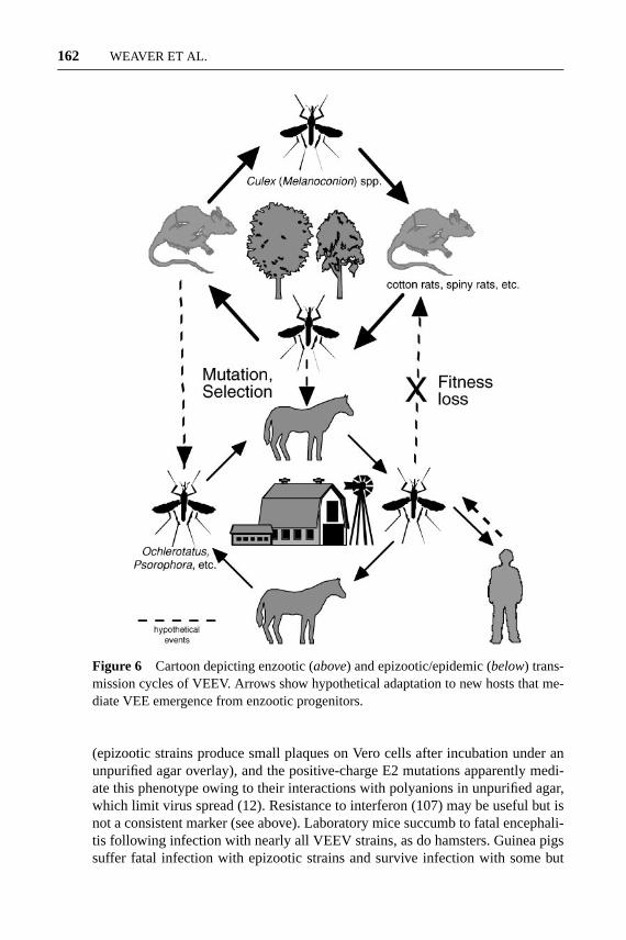
Experimental infections of equines with enzootic versus epizootic VEEV strainsdemonstrated many years ago that equine viremia is a major determinant of theability to cause widespread disease. This implies that, when epizootic strains arisevia mutation of enzootic progenitors, equines select for mutants with increasedviremia potential and indirectly for increased virulence (Figure 6). The strongcorrelation between the antigenic subtype (IAB and IC) and equine viremia impliesthat the E2 envelope glycoprotein, the site of the major antigenic determinants, isan important determinant of equine virulence and viremia.
Thanks to advances in viral genetics over the past two decades, experimentalstudies using reverse genetic approaches can now be used to identify the indi-vidual genes and mutations that can adapt enzootic VEEV strains to equine am-plification. Ideally, these studies would utilize in vitro markers of the epizooticphenotype and/or a small laboratory animal model that responds differentially toenzootic versus epizootic VEEV strains (like equines) following experimental in-fection (55, 122, 124). Plaque size is a reliable marker of the epizootic phenotype
23 Oct 2003 21:39 AR AR208-EN49-07.tex AR208-EN49-07.sgm LaTeX2e(2002/01/18)P1: GCE
162 WEAVER ET AL.
Figure 6 Cartoon depicting enzootic (above) and epizootic/epidemic (below) trans-mission cycles of VEEV. Arrows show hypothetical adaptation to new hosts that me-diate VEE emergence from enzootic progenitors.
(epizootic strains produce small plaques on Vero cells after incubation under anunpurified agar overlay), and the positive-charge E2 mutations apparently medi-ate this phenotype owing to their interactions with polyanions in unpurified agar,which limit virus spread (12). Resistance to interferon (107) may be useful but isnot a consistent marker (see above). Laboratory mice succumb to fatal encephali-tis following infection with nearly all VEEV strains, as do hamsters. Guinea pigssuffer fatal infection with epizootic strains and survive infection with some but
23 Oct 2003 21:39 AR AR208-EN49-07.tex AR208-EN49-07.sgm LaTeX2e(2002/01/18)P1: GCE
VENEZUELAN EQUINE ENCEPHALITIS 163
not all enzootic isolates (94). Subtype IE strains are nonlethal for guinea pigs andhave been used to map virulence determinants of epizootic strains.
Chimeric viruses including the nonstructural proteins of an epizootic IAB strainand structural proteins of an enzootic IE strain (IAB/IE), or reciprocal constructs(IE/IAB), show intermediate levels of virus replication and virulence for guineapigs compared with the parental strains (80). The IE/IAB chimera produced slightlyhigher viremia and an average survival time two days shorter than the VE/IAB-IEvirus, which suggests that the structural proteins are more important determinantsof virulence. Similar results have been obtained with subtype IC and ID strainsand chimeric recombinants with swapped E2 genes (I. Greene, S. Paessler & S.C.Weaver, unpublished data).
Adaptation to Epizootic Mosquito Vectors
Because the vectors implicated during epizootics generally belong to differentgenera from the enzootic vectors (Figure 6), adaptation for efficient transmissionby epizootic vectors is also a potential mechanism that contributes to VEE emer-gence. If VEEV undergoes adaptation during the evolution of epizootic strainsfrom enzootic progenitors, epizootic vectors implicated during outbreaks shouldshow greater susceptibility to epizootic strains than to enzootic viruses closelyrelated to their progenitors. The first evidence of this difference came from ex-perimental studies withO. taeniorhynchus, a proven epizootic vector of epizooticsubtype IAB and IC viruses. This species is more susceptible to subtype IABstrains than to enzootic, subtype IE strains from Guatemala (59). However, the IEstrains are not closely related to the progenitors of the IAB serotype, which arebelieved to be ID strains from Colombia and Venezuela (Figure 5). Later studiesdemonstrating a greater susceptibility ofO. taeniorhynchusto infection with IABand IC epizootic than with ID enzootic strains more directly supported the hypoth-esis that adaptation to this species is an important mechanism of VEE emergence.Experiments using chimeric viruses engineered from subtype IAB, IC, and IDstrains demonstrated that the E2 envelope glycoprotein is an important determi-nant of these susceptibility differences, suggesting that interactions with receptorson midgut epithelial cells may be involved (13).
More recent studies of the susceptibility ofO. taeniorhynchusto infectionwith VEEV strains isolated during a smaller outbreak suggest that adaptation tothis species may affect the size of outbreaks by facilitating efficient transmis-sion in coastal regions. Subtype IC strains isolated during the small 1992–1993Venezuelan outbreak, which did not extend to coastal regions inhabited byO. tae-niorhynchusand produced a relatively small number of equine and human cases(86), were tested for their ability to infect this species. Despite the ability of theetiologic subtype IC strains to replicate efficiently in equines (124), they do notinfect O. taeniorhynchusmore efficiently than closely related enzootic ID strainsbelieved to represent progenitors of the outbreak (D. Ortiz & S.C. Weaver, unpub-lished data). These data suggest that the inability of the 1992–1993 strains to adaptto O. taeniorhynchusmay have limited the extent of the epidemic. Further studies
23 Oct 2003 21:39 AR AR208-EN49-07.tex AR208-EN49-07.sgm LaTeX2e(2002/01/18)P1: GCE
164 WEAVER ET AL.
examining the susceptibility of three other mosquito species, including the provenepizootic vectorP. confinnis, also show no apparent difference in susceptibilityto a variety of enzootic versus epizootic VEEV strains. These studies imply thatadaptation toP. confinnisdoes not accompany VEE emergence (D. Ortiz & S.C.Weaver, unpublished data).
A hypothesis to explain the apparent failure of epizootic VEEV strains to bemaintained in sylvatic transmission cycles similar to those of the enzootic variantsis that adaptation for infection of epizootic vectors leads to a fitness decrease in theenzootic mosquito vectors (Figure 6). Scherer et al. (98) demonstrated that enzooticvectors are in some cases more susceptible to infection by sympatric enzootic thanby epizootic or allopatric enzootic variants.C. taeniopus, the enzootic subtype IEvector in Guatemala, is highly susceptible to infection with sympatric IE strains butrelatively refractory to subtype IAB, IC, ID, II (Everglades), III (Mucambo), and IV(Pixuna) viruses. The refractoriness ofC. taeniopusto epizootic strains may ex-plain the inability of subtype IAB VEEV to persist in Guatemala following the1969 outbreak. Additional experimental infections of subtype ID enzootic vectors(Table 1) with epizootic and enzootic strains are needed to test the hypothesis thatadaptation to epizootic vectors reduces the fitness of epizootic strains for the an-cestral enzootic vectors (Figure 6). The same fitness loss may apply to the reservoirhosts of enzootic strains when epizootic strains adapt for equine replication, andcomparisons of enzootic versus epizootic VEEV infection rodent hosts are neededto evaluate this version of the hypothesis.
FUTURE DIRECTIONS OF VEE RESEARCH
Treatment and Prevention of VEE
Despite over 60 years of research on VEE, there is no licensed human vaccineor effective antiviral treatment for human or equine disease. The recent concernthat VEEV may be used for biological terrorism has underscored the need todevelop improved, licensed vaccines and effective antivirals. Recent advancesin genetic engineering and vaccine design have been exploited to produce animproved candidate vaccine (67), and comparable efforts are needed to developantivirals. The ability of vaccines to protect against challenge with heterologousVEE subtypes needs to be evaluated because several can cause human disease.
Mechanisms of Natural VEE Emergence
Phylogenetic and reverse genetic studies have provided strong evidence to supportthe hypothesis that epizootic VEEV strains can arise via mutation of enzooticstrains in subtype ID. However, all three subtype IAB/IC epizootic clades appearto have arisen from only one of six major VEEV lineages. The reason(s) why theother lineages have not generated major epizootics is unknown and is an importanttopic of future research. One hypothesis is that the ability to generate epizootic
23 Oct 2003 21:39 AR AR208-EN49-07.tex AR208-EN49-07.sgm LaTeX2e(2002/01/18)P1: GCE
VENEZUELAN EQUINE ENCEPHALITIS 165
strains is highly dependent on the genetic makeup of the enzootic strains; theenzootic VEEV lineages that circulate in Florida, Mexico, Central America, north-central Venezuela, southwestern Colombia, Ecuador, and parts of Peru (Figure 5)may be incapable of equine adaptation via a small number of mutations. Anotherhypothesis is that the ecological conditions required for the onset of an epizooticdo not occur in some of the areas inhabited by these enzootic strains. However, thehistory of VEE epizootics in Mexico, Central America, north-central Venezuela,southwestern Colombia, Ecuador, and coastal Peru argue against this hypothesis.Answering this question is important for public health in many locations includingFlorida, where the potential for epidemics remains unknown.
Another aspect of VEE emergence that deserves further study is the ecologicalmechanisms that allow newly generated epizootic strains to reach locations con-ducive to amplification. Most regions of South America known to harbor enzooticVEEV do not usually experience epizootics, presumably because the enzooticstrains immunize equines against epizootic VEEV (124). This raises the questionof how epizootic strains that may be generated via mutation in enzootic locationsare translocated across this “halo” of equine immunity to locations with largepopulations of susceptible equines and mosquito vectors.
The possible role of flying vertebrate hosts in disseminating VEEV amongenzootic foci, or in transporting epizootic viruses during outbreaks, has receivedlittle study. VEEV antibody rates are generally low in birds (55, 122), althoughnatural avian VEEV infections were reported during a 1967 Colombian epizootic(92), and from six species of passerine and wading birds, primarily fledglingsor nestlings, in Panama (43). Experimental infections indicate that some wadingbirds, particularly nestlings, develop viremia sufficient to infect vectors and havethe potential for serving as amplifying hosts (29, 45).
The evidence for a natural role of bats in enzootic and epizootic VEEV dis-semination is stronger. Virus isolations from bats have been reported in Colombia(92) and Mexico (21, 96). In Guatemala, antibodies were detected in seven species,(101), and VEEV was isolated from the blood of oneUroderma bilobatum. Anoverall seropositivity rate of 10% was estimated for the genusArtibeus, whichsuggests that these bats may serve as alternative reservoir hosts to maintain viruscirculation if most terrestrial animals become immune (101). A variety of batsincluding vampires become viremic with moderate to high titers and shed virusinto their saliva for up to 168 h following experimental infection (22, 91, 102); per-sistent viremia has been detected for at least 26 days at hibernating temperatures,and low viremia levels persisted for over 90 days and rose when the bats werereturned to room temperature (22). These studies indicate that bats deserve furtherattention as potential reservoir, amplification, and/or transport hosts.
In addition to questions regarding the evolution of epizootic IAB and IC strains,their ability to persist following the apparent cessation of equine and human casesdeserves attention. Evidence of VEEV circulation in 1966, two years after theapparent end of the 1962–1964 outbreak, was found in eastern Venezuela (109).Similarly, subtype IC VEEV strains were isolated in 2000 in two locations of
23 Oct 2003 21:39 AR AR208-EN49-07.tex AR208-EN49-07.sgm LaTeX2e(2002/01/18)P1: GCE
166 WEAVER ET AL.
Venezuela, more than four years after the apparent end of the 1995 outbreak (G.Medina & S.C. Weaver, unpublished data). Because the epizootic strains have neverbeen isolated in sylvatic cycles typical of enzootic strains, alternative transmissioncycles during these interepizootic periods deserve further attention.
Molecular Systematics of VEEV Vectors
The powerful combination of DNA sequencing and phylogenetic methods hasrecently been used to elucidate evolutionary trends in vectors of medical im-portance that have long defied morphological approaches, including the generaAnopheles(35, 60, 133),Aedes(133),Culex(68, 72), and higher in level hierar-chic resolution (10). Although morphological studies should remain a foundation,several important questions regarding arbovirus vectors can be answered usingmolecular systematics applied to theCulex(Melanoconion) subgenus: (a) Is theSpissipes section monophyletic? If so, what synapomorphic genetic, physiologi-cal, or ecological factors shared by these mosquitoes explain their important rolein transmitting arboviruses? (b) Has VEEV coevolved (cospeciatedsensu lato)with its Culex(Melanoconion) vectors, or has VEEV adapted to these mosquitoesindependently on several occasions during the evolution of the serocomplex?(c) Are all arbovirus vectors in theCulex (Melanoconion) subgenus correctlyidentified, or are there cryptic species of medical importance? These questions areof vital importance to understanding the evolution of vector-borne diseases andtheir association with mosquitoes.
ACKNOWLEDGMENTS
The authors’ research is supported by grants AI49725-01, AI48807-01, AI-25489,and TW 5919 from the National Institutes of Health, by Colciencias project 2104-04-758-98, and by Consejo de Desarrollo Cientifico y Humanistico de la Univer-sidad Central de Venezuela (CDCH).
The Annual Review of Entomologyis online at http://ento.annualreviews.org
LITERATURE CITED
1. Aguilar P, Greene I, Coffey L, MoncayoAC, Medina G, et al. 2003. EndemicVenezuelan equine encephalitis in North-ern Peru: characterization of virus iso-lates.Emerg. Infect. Dis.In press
2. Aitken TH, Worth CB, Tikasingh ES.1968. Arbovirus studies in Bush BushForest, Trinidad, W.I., September 1959–December 1964. 3. Entomologic studies.Am. J. Trop. Med. Hyg.17:253–68
3. Aitken THG. 1972. Habits of somemosquito hosts of VEE (Mucambo)virus from northeastern South Amer-ica, including Trinidad.Proc. Workshop-Symposium on Venezuelan EncephalitisVirus. pp. 254–56. Washington, DC: PanAm. Health Organ.
4. Barnett HC. 1960. The incrimination ofarthropods as vectors of disease.Proc.11th Intl. Congr. Entomol.2:341–45
23 Oct 2003 21:39 AR AR208-EN49-07.tex AR208-EN49-07.sgm LaTeX2e(2002/01/18)P1: GCE
VENEZUELAN EQUINE ENCEPHALITIS 167
5. Barrera R, Ferro C, Navarro JC, FreierJ, Liria J, et al. 2002. Contrasting syl-vatic foci of Venezuelan equine encephali-tis virus in northern South America.Am.J. Trop. Med. Hyg.67:324–34
6. Belkin JN. 1969. The problem of the iden-tity of the species ofCulex (Melanoco-nion) related to opisthopus.Mosq. Syst.Newsl.1:26–28
7. Belkin JN. 1970.Culex (Melanoconion)aikenii (A & R, 1906) a nomen dubium;ocossaD & K, 1919 andpanocossaDyar,1923 both valid.Mosq. Syst. Newsl.2:59–60
8. Berge TO, Banks IS, Tigertt WD. 1961.Attenuation of Venezuelan equine en-cephalomyelitis virus byin vitro cultiva-tion in guinea pig heart cells.Am. J. Hyg.73:209–18
9. Bernard KA, Klimstra WB, Johnston RE.2000. Mutations in the E2 glycoprotein ofVenezuelan equine encephalitis virus con-fer heparan sulfate interaction, low mor-bidity, and rapid clearance from blood ofmice.Virology276:93–103
10. Besansky NJ, Fahey GT. 1997. Utility ofthe white gene in estimating phylogeneticrelationships among mosquitoes (Diptera:Culicidae).Mol. Biol. Evol.14:442–54
11. Bigler WJ, Lewis AL, Wellings FM.1974. Experimental infection of thecotton mouse (Peromyscus gossypinus)with Venezuelan equine encephalomyeli-tis virus.Am. J. Trop. Med. Hyg.23:1185–88
12. Brault AC, Powers AM, Holmes EC,Woelk CH, Weaver SC. 2002. Positivelycharged amino acid substitutions in the E2envelope glycoprotein are associated withthe emergence of Venezuelan equine en-cephalitis virus.J. Virol. 76:1718–30
13. Brault AC, Powers AM, Weaver SC. 2002.Vector infection determinants of Venezue-lan equine encephalitis virus reside withinthe E2 envelope glycoprotein.J. Virol.76:6387–92
14. Briceno Rossi AL. 1967. Rural epidemicencephalitis in Venezuela caused by a
group A arbovirus (VEE).Progr. Med.Virol. 9:176–203
15. Calisher CH, Karabatsos N. 1988. Arbo-virus serogroups: definition and geo-graphic distribution. InThe Arboviruses:Epidemiology and Ecology,Vol. I, ed. TPMonath, pp. 19–57. Boca Raton, FL: CRCPress
16. Calisher CH, Monath TP, Mitchell CJ,Sabattini MS, Cropp CB, et al. 1985.Arbovirus investigations in Argentina,1977–1980. III. Identification and char-acterization of viruses isolated, includingnew subtypes of western and Venezuelanequine encephalitis viruses and four newbunyaviruses (Las Maloyas, Resistencia,Barranqueras, and Antequera).Am. J.Trop. Med. Hyg.34:956–65
17. Chamberlain RW. 1972. Discussion com-ments. Proc. Workshop-Symposium onVenezuelan Encephalitis Virus.pp. 257–58. Washington, DC: Pan Am. HealthOrgan.
18. Chamberlain RW, Sudia WD, ColemanPH, Work TH. 1964. Venezuelan equineencephalitis virus from south Florida.Sci-ence145:272–74
19. Chamberlain RW, Sudia WD, Work TH,Coleman PH, Newhouse VF, et al.1969. Arbovirus studies in south Florida,with emphasis on Venezuelan equine en-cephalomyelitis virus.Am. J. Epidemiol.89:197–210
20. Deleted in proof21. Correa-Giron P, Calisher CH, Baer GM.
1972. Epidemic strain of Venezuelanequine encephalomyelitis virus from avampire bat captured in Oaxaca, Mexico,1970.Science175:546–47
22. Corristan EC, La Motte LC, Smith DG.1956. Susceptibility of bats to certain en-cephalitis viruses.Fed. Proc. Fed. Am.Soc. Exp. Biol.15:584
23. Cupp EW, Kreutzer RD, Weaver SC.1989. The biosystematics ofCulex(Mela-noconion) taeniopus sensu latoin relationto Venezuelan equine encephalomyelitis.Mosq. Syst.21:216–21
23 Oct 2003 21:39 AR AR208-EN49-07.tex AR208-EN49-07.sgm LaTeX2e(2002/01/18)P1: GCE
168 WEAVER ET AL.
24. Cupp EW, Scherer WF, Lok JB, Bren-ner RJ, Dziem GM, et al. 1986. Entomo-logical studies at an enzootic Venezuelanequine encephalitis virus focus in Guate-mala, 1977–1980.Am. J. Trop. Med. Hyg.35:851–59
25. Cupp EW, Scherer WF, Ordonez JV. 1979.Transmission of Venezuelan encephali-tis virus by naturally infectedCulex(Melanoconion) opisthopus. Am. J. Trop.Med. Hyg.28:1060–63
26. Davies JB, Martinez R. 1970. Observa-tions on the population dynamics, be-havior and maintenance of a laboratorycolony of Culex (Melanoconion) portesiSenevet and Abonnenc, 1941 (Diptera:Culicidae).J. Med. Entomol.7:179–88
27. Davis NL, Grieder FB, Smith JF, Green-wald GF, Valenski ML, et al. 1994. Amolecular genetic approach to the studyof Venezuelan equine encephalitis viruspathogenesis.Arch. Virol. Suppl.9:99–109
28. de la Monte SM, Castro F, Bonilla NJ, deUrdaneta AG, Hutchins GM. 1985. Thesystemic pathology of Venezuelan equineencephalitis virus infection in humans.Am. J. Trop. Med. Hyg.34:194–202
29. Dickerman RW, Bonacorsa CM, SchererWF. 1976. Viremia in young herons andibis infected with Venezuelan encephalitisvirus.Am. J. Epidemiol.104:678–83
30. Durden LA, Linthicum KJ, Turell MJ.1992. Mechanical transmission of Vene-zuelan equine encephalomyelitis virus byhematophagous mites (Acari).J. Med.Entomol.29:118–21
31. Deleted in proof32. Deleted in proof33. Deleted in proof34. Ferro C, Boshell J, Moncayo AC, Gonza-
lez M, Ahumada ML, et al. 2003. Natu-ral enzootic vectors of Venezuelan equineencephalitis virus, Magdalena Valley,Colombia.Emerg. Infect. Dis.9:49–54
35. Foley DH, Bryan JH, Yeates D, SaulA. 1998. Evolution and systematics ofAnopheles: insights from a molecular
phylogeny of Australasian mosquitoes.Mol. Phylogenet. Evol.9:262–75
36. Forattini OP, de Castro Gomes A, Na-tal D, Kakitani I, Marucci D. 1989. Foodpreferences and domiciliation of Culici-dae mosquitoes in the Ribeira Valley, SaoPaulo, Brazil, with special reference toAedes scapularisand Culex (Melanoco-nion). Rev. Saude Publ.23:9–19
37. Forattini OP, Sallum MAM. 1992. Cibar-ial armature as taxonomic characters forthe Spissipes Section ofCulex(Melanoco-nion) (Diptera: Culicidae).Mosq. Syst.24:70–84
38. Forattini OP, Sallum MAM. 1995. Twonew species of the Spissipes section ofCulex (Melanoconion) (Diptera: Culici-dae).Mosq. Syst.27:125–42
39. Franck PT, Johnson KM. 1970. An out-break of Venezuelan encephalitis in manin the Panama Canal Zone.Am. J. Trop.Med. Hyg.19:860–65
40. Galindo P. 1969. Notes on the systematicsof Culex(Melanoconion) taeniopusDyar& Knab and related species, gathered dur-ing arbovirus investigations in Panama.Mosq. Syst. Newsl.1:82–89
41. Galindo P. 1972. Endemic vectors ofVenezuelan encephalitis.Proc. Work-shop-Symposium on Venezuelan Ence-phalitis Virus, pp. 249–53. Washington,DC: Pan Am. Health Organ.
42. Galindo P, Grayson MA. 1971.Culex(Melanoconion) aikenii: natural vectorin Panama of endemic Venezuelan en-cephalitis.Science172:594–95
43. Galindo P, Srihongse S, Rodaniche ED,Grayson MA. 1966. An ecological sur-vey for arboviruses in Almirante, Panama,1959–1962.Am. J. Trop. Med. Hyg.15:385–400
44. Gonzalez-Salazar D, Estrada-Franco JG,Carrara AS, Aronson JF, Weaver SC.2003. Equine amplification and virulenceof subtype IE Venezuelan equine en-cephalitis viruses isolated during the 1993and 1996 Mexican epizootics.Emerg. In-fect. Dis.9:161–68
23 Oct 2003 21:39 AR AR208-EN49-07.tex AR208-EN49-07.sgm LaTeX2e(2002/01/18)P1: GCE
VENEZUELAN EQUINE ENCEPHALITIS 169
45. Grayson MA. 1972. Discussion and com-ments. Proc. Workshop-Symposium onVenezuelan Encephalitis Virus.pp. 284–85. Washington, DC: Pan Am. Health Or-gan.
46. Grayson MA, Galindo P. 1969. Ecologyof Venezuelan equine encephalitis virusin Panama.J. Am. Vet. Med. Assoc.155:2141–45
47. Grayson MA, Galindo P. 1972. Ex-perimental transmission of Venezuelanequine encephalitis virus byDeinoceritespseudesDyar and Knab, 1909.J. Med. En-tomol.9:196–200
48. Grieder FB, Davis NL, Aronson JF,Charles PC, Sellon DC, et al. 1995. Speci-fic restrictions in the progression ofVenezuelan equine encephalitis virus-induced disease resulting from singleamino acid changes in the glycoproteins.Virology206:994–1006
49. Griffin DE. 2001. Alphaviruses. See Ref.58a, pp. 917–62
50. Groot H. 1972. The health and economicimpact of Venezuelan equine encephalitis.Proc. Workshop-Symposium on Venezue-lan Encephalitis Virus.pp. 7–16. Wash-ington, DC: Pan Am. Health Organ.
51. Guimaraes JH. 1997.Systematic Databa-se of Diptera of the Americas South ofthe United States, Family Culicidae. SaoPablo, Brazil: Fundac˜ao de Amparo aPesquisa do Estado de S˜ao Paulo/Ed.Pleide. 286 pp.
52. Harrington LC, Edman JD, Scott TW.2001. Why do femaleAedes aegypti(Diptera: Culicidae) feed preferentiallyand frequently on human blood?J. Med.Entomol.38:411–22
53. Hawley RJ, Eitzen EM Jr. 2001. Biolog-ical weapons—a primer for microbiolo-gists.Annu. Rev. Microbiol.55:235–53
54. Jahrling PB, Navarro E, Scherer WF.1976. Interferon induction and sensitivityas correlates to virulence of Venezuelanencephalitis viruses for hamsters.Arch.Virol. 51:23–35
55. Johnson KM, Martin DH. 1974. Venezue-
lan equine encephalitis.Adv. Vet. Sci.Comp. Med.18:79–116
56. Johnson KM, Shelokov A, Peralta PH,Dammin GJ, Young NA. 1968. Recoveryof Venezuelan equine encephalomyelitisvirus in Panama. A fatal case in man.Am.J. Trop. Med. Hyg.17:432–40
57. Kinney RM, Tsuchiya KR, Sneider JM,Trent DW. 1992. Genetic evidence thatepizootic Venezuelan equine encephalitis(VEE) viruses may have evolved from en-zootic VEE subtype I-D virus.Virology191:569–80
58. Kinney RM, Tsuchiya KR, Sneider JM,Trent DW. 1992. Molecular evidence forthe origin of the widespread Venezuelanequine encephalitis epizootic of 1969 to1972.J. Gen. Virol.73:3301–5
58a. Knipe DM, Howley PM, eds. 2001.Fields’ Virology, Fourth Edition. NewYork: Lippincott Williams & Wilkins
59. Kramer LD, Scherer WF. 1976. Vectorcompetence of mosquitoes as a marker todistinguish Central American and Mex-ican epizootic from enzootic strains ofVenezuelan encephalitis virus.Am. J.Trop. Med. Hyg.25:336–46
60. Krzywinski J, Wilkerson RC, BesanskyNJ. 2001. Evolution of mitochondrial andribosomal gene sequences in anopheli-nae (Diptera: Culicidae): implications forphylogeny reconstruction.Mol. Phylo-genet. Evol.18:479–87
61. Larsen JR, Ashley RF. 1971. Demon-stration of Venezuelan equine encephalo-myelitis virus in tissues ofAedes aegypti.Am. J. Trop. Med. Hyg.20:754–60
62. Linthicum KJ, Gordon SW, Monath TP.1992. Comparative infections of epizooticand enzootic strains of Venezuelan equineencephalomyelitis virus inAmblyommacajennense(Acari: Ixodidae).J. Med. En-tomol.29:827–31
63. Linthicum KJ, Logan TM. 1994. Labo-ratory transmission of Venezuelan equineencephalomyelitis virus by the tickHyalo-mma truncatum.Trans. R. Soc. Trop. Med.Hyg.88:126
23 Oct 2003 21:39 AR AR208-EN49-07.tex AR208-EN49-07.sgm LaTeX2e(2002/01/18)P1: GCE
170 WEAVER ET AL.
64. Lord RD. 1974. History and geographicdistribution of Venezuelan equine en-cephalitis.Bull. PAHO8:100–10
65. Lord RD, Calisher CH, Sudia WD, WorkTH. 1973. Ecological investigation of ver-tebrate hosts of Venezuelan equine en-cephalomyelitis virus in south Florida.Am. J. Trop. Med. Hyg.22:116–23
66. Ludwig GV, Kondig JP, Smith JF. 1996.A putative receptor for Venezuelan equineencephalitis virus from mosquito cells.J.Virol. 70:5592–99
67. Ludwig GV, Turell MJ, Vogel P, KondigJP, Kell WK, et al. 2001. Comparativeneurovirulence of attenuated and non-attenuated strains of Venezuelan equineencephalitis virus in mice.Am. J. Trop.Med. Hyg.64:49–55
68. Miller BR, Crabtree MB, Savage HM.1996. Phylogeny of fourteenCulexmosquito species, including theCulexpipiens complex, inferred from the in-ternal transcribed spacers of ribosomalDNA. Insect Mol. Biol.5:93–107
69. Monath TP, Lazuick JS, Cropp CB, RushWA, Calisher CH, et al. 1980. Recoveryof Tonate virus (“Bijou Bridge” strain),a member of the Venezuelan equine en-cephalomyelitis virus complex, from cliffswallow nest bugs (Oeciacus vicarius)and nestling birds in North America.Am.J. Trop. Med. Hyg.29:969–83
70. Morales A, Ferro C, Isaza de Rodr´ıguez C,Cura E. 1987. Encuesta sobre artr´opodosde interes medico en La Guajira, Colom-bia, Suram´erica.Biomedica7:87–93
71. Navarro JC, Liria J. 2000. Phylogeneticrelationships among eighteen neotropicalCulicini species.J. Am. Mosq. Control As-soc.16:75–85
72. Navarro JC, Weaver SC. 2003. Phylogenyof Vomerifer and Pedroi groups in theSpissipes section ofCulex (Melanoco-nion) (Diptera: Culicidae) using rDNA,and evidence of two cryptic species.J.Med. Entomol.In press
73. Oberste MS, Fraire M, Navarro R, ZepedaC, Zarate ML, et al. 1998. Association
of Venezuelan equine encephalitis virussubtype IE with two equine epizootics inMexico.Am. J. Trop. Med. Hyg.59:100–7
74. Oberste MS, Schmura SM, Weaver SC,Smith JF. 1999. Geographic distributionof Venezuelan equine encephalitis virussubtype IE genotypes in Central Amer-ica and Mexico.Am. J. Trop. Med. Hyg.60:630–34
75. Oberste MS, Weaver SC, Watts DM,Smith JF. 1998. Identification and geneticanalysis of Panama-genotype Venezuelanequine encephalitis virus subtype ID inPeru.Am. J. Trop. Med. Hyg.58:41–46
76. Paredes A, Alwell-Warda K, Weaver SC,Chiu W, Watowich SJ. 2001. Venezuelanequine encephalomyelitis virus structureand its divergence from Old World al-phaviruses.J. Virol. 75:9532–37
77. Pecor JE, Mallampalli VL, Harbach RE,Peyton EL. 1992. Catalog and illustratedreview of the subgenusMelanoconionofCulex(Diptera: Culicidae).Contrib. Am.Entomol. Inst.27:1–228
78. Pittman PR, Makuch RS, Mangiafico JA,Cannon TL, Gibbs PH, et al. 1996. Long-term duration of detectable neutralizingantibodies after administration of live-attenuated VEE vaccine and followingbooster vaccination with inactivated VEEvaccine.Vaccine14:337–43
79. Pletnev SV, Zhang W, MukhopadhyayS, Fisher BR, Hernandez R, et al.2001. Locations of carbohydrate siteson alphavirus glycoproteins show thatE1 forms an icosahedral scaffold.Cell105:127–36
80. Powers AM, Brault AC, Kinney RM,Weaver SC. 2000. The use of chimericVenezuelan equine encephalitis viruses asan approach for the molecular identifica-tion of natural virulence determinants.J.Virol. 74:4258–63
81. Powers AM, Brault AC, Shirako Y,Strauss EG, Kang W, et al. 2001. Evo-lutionary relationships and systematics ofthe alphaviruses.J. Virol. 75:10118–31
82. Powers AM, Oberste MS, Brault AC,
23 Oct 2003 21:39 AR AR208-EN49-07.tex AR208-EN49-07.sgm LaTeX2e(2002/01/18)P1: GCE
VENEZUELAN EQUINE ENCEPHALITIS 171
Rico-Hesse R, Schmura SM, et al.1997. Repeated emergence of epidemic/epizootic Venezuelan equine encephalitisfrom a single genotype of enzootic sub-type ID virus.J. Virol. 71:6697–705
83. Reeves WC, Hutson GA, Bellamy RE,Scrivani RP. 1958. Chronic latent infec-tions of birds with western equine en-cephalomyelitis virus.Proc. Soc. Exp.Biol. Med.97:733–36
84. Reisen WK, Monath TP. 1988. West-ern equine encephalomyelitis. InThe Ar-boviruses: Epidemiology and Ecology,Vol. V, ed. TP Monath, pp. 89–137. BocaRaton, FL: CRC Press
85. Rico-Hesse R, Roehrig JT, Trent DW,Dickerman RW. 1988. Genetic variationof Venezuelan equine encephalitis virusstrains of the ID variety in Colombia.Am.J. Trop. Med. Hyg.38:195–204
86. Rico-Hesse R, Weaver SC, de Siger J,Medina G, Salas RA. 1995. Emergenceof a new epidemic/epizootic Venezuelanequine encephalitis virus in South Amer-ica.Proc. Natl. Acad. Sci. USA92:5278–81
87. Rozeboom LE, Komp WHW. 1950. Areview of the species ofCulex of thesubgenusMelanoconion(Diptera: Culi-cidae).Ann. Entomol. Soc. Am.43:75–114
88. Salas RA, Garcia CZ, Liria J, Barrera R,Navarro JC, et al. 2001. Ecological studiesof enzootic Venezuelan equine encephali-tis in north-central Venezuela, 1997–1998. Am. J. Trop. Med. Hyg.64:84–92
89. Sallum MAM, Forattini OP. 1996. Re-vision of the Spissipes section ofCulex(Melanoconion) (Diptera: Culicidae).J.Am. Mosq. Control Assoc.12:517–600
90. Sallum MAM, Gomes-Hutchings RS,Menezes-Ferreira RL. 1997.Culex gno-matosa new species of the Spissipes sec-tion of Culex(Melanoconion) (Dipetera:Culicidae) from the Amazon region.Mem.Inst. Oswaldo Cruz92:215–19
91. Sanmartin C. 1972. Discussion and com-
ments. Proc. Workshop-Symposium onVenezuelan Encephalitis Virus, pp. 247–48. Washington, DC: Pan Am. Health Or-gan.
92. Sanmartin C, Mackenzie RB, Trapido H,Barreto P, Mullenax CH, et al. 1967. Ence-falitis equina Venezolana en Colombia,1967. Bol. Off. Sanit. Panam.74:108–37
93. Sanmartin-Barberi C, Osorno-Mesa E.1954. Human epidemic in Colombiacaused by the Venezuelan equine en-cephalomyelitis virus.Am. J. Trop. Med.Hyg.3:283–91
94. Scherer WF, Chin J, Ordonez JV. 1979.Further observations on infections ofguinea pigs with Venezuelan encephali-tis virus strains.Am. J. Trop. Med. Hyg.28:725–28
95. Scherer WF, Dickerman RW, Chia CW,Ventura A, Moorhouse A, et al. 1963.Venezuelan equine encephalitis virus inVeracruz, Mexico, and the use of hamstersas sentinels.Science145:274–75
96. Scherer WF, Dickerman RW, La FiandraRP, Wong Chia C, Terrian J. 1971. Eco-logic studies of Venezuelan encephalitisvirus in southeastern Mexico. IV. Infec-tions of wild mammals.Am. J. Trop. Med.Hyg.20:980–88
97. Scherer WF, Dickerman RW, Ordonez JV.1970. Discovery and geographic distribu-tion of Venezuelan encephalitis virus inGuatemala, Honduras, and British Hon-duras during 1965–68, and its possiblemovement to Central America and Mex-ico. Am. J. Trop. Med. Hyg.19:703–11
98. Scherer WF, Weaver SC, Taylor CA, CuppEW, Dickerman RW, et al. 1987. Vec-tor competence ofCulex(Melanoconion)taeniopus for allopatric and epizooticVenezuelan equine encephalomyelitisviruses.Am. J. Trop. Med. Hyg.36:194–97
99. Schlesinger S, Schlesinger MJ. 2001. To-gaviridae: the viruses and their replica-tion. See Ref. 58a, pp. 895–916
23 Oct 2003 21:39 AR AR208-EN49-07.tex AR208-EN49-07.sgm LaTeX2e(2002/01/18)P1: GCE
172 WEAVER ET AL.
100. Scott TW, Weaver SC. 1989. Easternequine encephalomyelitis virus: epidemi-ology and evolution of mosquito transmis-sion.Adv. Virus Res.37:277–328
101. Seymour C, Dickerman RW, Martin MS.1978. Venezuelan encephalitis virus in-fection in neotropical bats. I. Natural in-fection in a Guatemalan enzootic focus.Am. J. Trop. Med. Hyg.27:290–96
102. Seymour C, Dickerman RW, Martin MS.1978. Venezuelan encephalitis virus in-fection in neotropical bats. II. Experimen-tal infections.Am. J. Trop. Med. Hyg.27:297–306
103. Shope RE, Causey OR, HomobonoPaes de Andrade A, Theiler M. 1964.The Venezuelan equine encephalomyeli-tis complex of group A arthropod-borneviruses, including Mucambo and Pixunafrom the Amazon region of Brazil.Am. J.Trop. Med. Hyg.13:723–27
104. Shope RE, Woodall JP, Travassos da RosaA. 1988. The epidemiology of diseasescaused by viruses in groups C and Guama(Bunyaviridae). In The Arboviruses: Epi-demiology and Ecology,Vol. III, ed. TPMonath, pp. 37–52. Boca Raton, FL: CRCPress
105. Sirivanakarn S. 1982. A review of the sys-tematics and proposed scheme of internalclassification of the New World subgenusMelanoconionof Culex(Diptera: Culici-dae).Mosq. Syst.14:265–333
106. Sirivanakarn S, Galindo P. 1980.Culex(Melanoconion) adamesi, a new speciesfrom Panama (Diptera: Culicidae).Mosq.Syst.12:25–34
107. Spotts DR, Reich RM, Kalkhan MA, Kin-ney RM, Roehrig JT. 1998. Resistanceto alpha/beta interferons correlates withthe epizootic and virulence potential ofVenezuelan equine encephalitis virusesand is determined by the 5′ noncodingregion and glycoproteins.J. Virol. 72:10286–91
108. Stanick DR, Wiebe ME, Scherer WF.1985. Markers of Venezuelan encephalitisvirus which distinguish enzootic strains of
subtype I-D from those of I-E.Am. J. Epi-demiol.122:234–44
109. Suarez OM, Bergold GH. 1968. Inves-tigations of an outbreak of Venezuelanequine encephalitis in towns of east-ern Venezuela.Am. J. Trop. Med. Hyg.17:875–80
110. Sudia WD, Newhouse VF, Beadle ID,Miller DL, Johnston JG Jr, et al. 1975.Epidemic Venezuelan equine encephalitisin North America in 1971: vector studies.Am. J. Epidemiol.101:17–35
111. Sutton LS, Brooke CC. 1954. Venezuelanequine encephalomyelitis due to vaccina-tion in man.JAMA155:1473–76
112. Swofford DL. 1998.PAUP∗. PhylogeneticAnalysis Using Parsimony (∗and OtherMethods). Version 4.Sunderland, MA:Sinauer
113. Tempelis CH, Galindo P. 1975. Host-feeding patterns ofCulex(Melanoconion)andCulex(Aedinus) mosquitoes collectedin Panama.J. Med. Entomol.12:205–9
114. Theobald FV. 1903.A Monograph of theCulicidae or Mosquitoes, Vol. 3. London:Br. Mus. Nat. Hist. 359 pp.
115. Turell MJ, Jones JW, Sardelis MR, DohmDJ, Coleman RE, et al. 2000. Vec-tor competence of Peruvian mosquitoes(Diptera: Culicidae) for epizootic and en-zootic strains of Venezuelan equine en-cephalomyelitis virus.J. Med. Entomol.37:835–39
116. Turell MJ, Ludwig GV, Beaman JR. 1992.Transmission of Venezuelan equine en-cephalomyelitis virus byAedes sollici-tansandAedes taeniorhynchus(Diptera:Culicidae). J. Med. Entomol 29:62–65
117. Turell MJ, Spielman A. 1992. Nonvascu-lar delivery of Rift Valley fever virus byinfected mosquitoes.Am. J. Trop. Med.Hyg.47:190–94
118. Velasquez J. 1939. Enfermedades de losanimales transmisibles al hombre: pesteloca.Salud y Sanidad8:22–32
119. Walder R, Suarez OM, Calisher CH.
23 Oct 2003 21:39 AR AR208-EN49-07.tex AR208-EN49-07.sgm LaTeX2e(2002/01/18)P1: GCE
VENEZUELAN EQUINE ENCEPHALITIS 173
1984. Arbovirus studies in southwest-ern Venezuela during 1973–1981. II.Isolations and further studies of Venezue-lan and eastern equine encephalitis, Una,Itaqui, and Moju viruses.Am. J. Trop.Med. Hyg.33:483–91
120. Walton TE, Alvarez O Jr, BuckwalterRM, Johnson KM. 1972. Experimentalinfection of horses with an attenuatedVenezuelan equine encephalomyelitisvaccine (strain TC-83).Infect. Immun.5:750–56
121. Walton TE, Alvarez O Jr, BuckwalterRM, Johnson KM. 1973. Experimentalinfection of horses with enzootic andepizootic strains of Venezuelan equineencephalomyelitis virus.J. Infect. Dis.128:271–82
122. Walton TE, Grayson MA. 1988. Venezue-lan equine encephalomyelitis. InThe Ar-boviruses: Epidemiology and Ecology,Vol. IV, ed. TP Monath, pp. 203–31. BocaRaton, FL: CRC Press
123. Wang E, Barrera R, Boshell J, Ferro C,Freier JE, et al. 1999. Genetic and pheno-typic changes accompanying the emer-gence of epizootic subtype IC Venezue-lan equine encephalitis viruses from anenzootic subtype ID progenitor.J. Virol.73:4266–71
124. Wang E, Bowen RA, Medina G, PowersAM, Kang W, et al. 2001. Virulence andviremia characteristics of 1992 epizooticsubtype IC Venezuelan equine encephali-tis viruses and closely related enzooticsubtype ID strains.Am. J. Trop. Med. Hyg.65:64–69
125. Wang E, Brault AC, Powers AM, KangW, Weaver SC. 2003. Glycosaminoglycanbinding properties of natural Venezuelanequine encephalitis virus isolates.J. Virol.77:1204–10
126. Weaver SC. 1997. Vector biology in vi-ral pathogenesis. InViral Pathogenesis,ed. N Nathanson, pp. 329–52. New York:Lippincott-Raven
127. Weaver SC. 1998. Recurrent emergenceof Venezuelan equine encephalomyelitis.
In Emerging Infections I, ed. WM Scheld,J Hughes, pp. 27–42. Washington, DC:ASM Press
128. Weaver SC, Bellew LA, Rico-HesseR. 1992. Phylogenetic analysis of al-phaviruses in the Venezuelan equine en-cephalitis complex and identification ofthe source of epizootic viruses.Virology191:282–90
129. Weaver SC, Dalgarno L, Frey TK, HuangHV, Kinney RM, et al. 2000. Family To-gaviridae. InVirus Taxonomy: Classifica-tion and Nomenclature of Viruses. Sev-enth Report of the International Commit-tee on Taxonomy of Viruses, ed. MHV vanRegenmortel, CM Fauquet, DHL Bishop,EB Carstens, MK Estes, et al., pp. 879–89.San Diego: Academic
130. Weaver SC, Pfeffer M, Marriott K, KangW, Kinney RM. 1999. Genetic evidencefor the origins of Venezuelan equineencephalitis virus subtype IAB out-breaks.Am. J. Trop. Med. Hyg.60:441–48
131. Weaver SC, Salas R, Rico-Hesse R,Ludwig GV, Oberste MS, et al. 1996.Re-emergence of epidemic Venezuelanequine encephalomyelitis in South Amer-ica. VEE study group.Lancet348:436–40
132. Weaver SC, Scherer WF, Taylor CA,Castello DA, Cupp EW. 1986. Lab-oratory vector competence ofCulex(Melanoconion) cedecei for sympatricand allopatric Venezuelan equine en-cephalomyelitis viruses.Am. J. Trop.Med. Hyg.35:619–23
133. Wesson DM, Porter CH, Collins FH.1992. Sequence and secondary structurecomparisons of ITS rDNA in mosquitoes(Diptera: Culicidae).Mol. Phylogenet.Evol.1:253–69
134. Young NA, Johnson KM. 1969. Antigenicvariants of Venezuelan equine encephali-tis virus: their geographic distribution andepidemiologic significance.Am. J. Epi-demiol.89:286–307
135. Young NA, Johnson KM, Gauld LW.
23 Oct 2003 21:39 AR AR208-EN49-07.tex AR208-EN49-07.sgm LaTeX2e(2002/01/18)P1: GCE
174 WEAVER ET AL.
1969. Viruses of the Venezuelan equineencephalomyelitis complex. Experimen-tal infection of Panamanian rodents.Am.J. Trop. Med. Hyg.18:290–96
136. Zarate ML, Scherer WF. 1969. A com-parative study of virulences, plaquemorphologies and antigenic character-istics of Venezuelan encephalitis virus
strains. Am. J. Epidemiol. 89:489–502
137. Zarate ML, Scherer WF, Dickerman RW.1970. Venezuelan equine encephalitisvirus as a human infection determinant.Description of a fatal case occurring inJaltipan, Ver., in 1965.Rev. Invest. SaludPubl.30:296–302
Related Documents