GUIDE FOR DOCTORS : LONG-TERM CONDITIONS January 2007 Gaucher disease National Diagnosis and Treatment Protocol

Welcome message from author
This document is posted to help you gain knowledge. Please leave a comment to let me know what you think about it! Share it to your friends and learn new things together.
Transcript
GUIDE FOR DOCTORS : LONG-TERM CONDITIONS
January 2007
Gaucher disease National Diagnosis and Treatment Protocol
This document may be downloaded from www.has-sante.fr
Haute Autorité de Santé
Communications Department 2 avenue du Stade de France - F 93218 Saint-Denis La Plaine CEDEX
Tel.: +33 (0)1 55 93 70 00 - Fax: +33 (0)1 55 93 74 00
This document was validated by the French National Authority Board in January 2007. © Haute Autorité de santé – 2007

PNDS Gaucher Disease
3
Contents List of abbreviations.............................. ....................................................... 5
Synopsis of the Gaucher Disease PNDS ............... ..................................... 6
I. Introduction....................................... .................................................. 10
1. Purpose ............................................ ................................................... 10
2. Gaucher disease.................................... ............................................. 10
3. Method............................................. .................................................... 12
II. National Diagnosis and treatment protocol (PNDS)... ...................... 13
1. Initial assessment at baseline ..................... ...................................... 13 1.1 Primary objectives ................................................................................ 13 1.2 Professionals involved .......................................................................... 13 1.3 Clinical Examination ............................................................................. 13 1.4 Paraclinical examinations ..................................................................... 15
2. Clinical Management ................................ .......................................... 20 2.1 Objectives............................................................................................. 20 2.2 Professionals involved .......................................................................... 20 2.3 Pharmacological treatments ................................................................. 22 2.4 Other non-surgical therapy ................................................................... 31 2.5 Surgical treatments............................................................................... 32
3. Follow-up.......................................... ................................................... 33 3.1 Objectives............................................................................................. 33 3.2 Professionals involved .......................................................................... 33 3.3 Rhythm and contents of visits ............................................................... 34
Appendix 1. List of persons who helped draft this P NDS on Gaucher disease ............................................ .............................................................. 39
Appendix 2. References ............................. .................................................. 40
Appendix 3. Opinion on treatment from a Rare Diseas e Reference Centre 46
Appendix 4 – Traceability form of imiglucerase infu sions ........................ 47
PNDS Gaucher Disease
4
Updating of PNDS/Long-Term Condition
The French National Diagnosis and Treatment Protocol (PNDS) for Gaucher disease was drafted by the designated reference centre, with methodological support from the French National Health Authority (HAS), pursuant to the provisions of the 2005–2008 French National Rare Diseases Plan. HAS validates the PNDS within the scope of its mission on long term illnesses. This protocol as well as the resulting list of procedures and services (LAP) is revised every three years. In the interim, the LAP is revised, at minimum, on a yearly basis. This list is available on the HAS website.
www.has-sante.fr
PNDS Gaucher Disease
5
List of abbreviations ALD Long-term condition ANAES French National Agency for Accreditation & Evaluation in
Healthcare APTT Activated Partial Thromboplastin Time BMD Bone Mineral Density CETG Gaucher Treatment and Evaluation Committee CNAMTS National Health Insurance Fund for Salaried Workers CPAMTS Employed Persons Health Insurance Office CRP C-Reactive Protein CT Computed Tomography ECG Electrocardiogram EMG Electromyogram ERT Enzyme Replacement Therapy HAS French National Authority for Health IgG Immunoglobulin G MA Marketing Authorization MGUS Monoclonal Gammapathy of Undetermined Significance MRI Magnetic Resonance Imaging PH Pulmonary Hypertension PNDS National Diagnosis and Treatment Protocol PT Prothrombin Time SPC Summary of Product Characteristics SMN-MMH French National Medical Office for Hereditary Metabolic
Diseases SRT Substrate-Reduction Therapy TID Three Times Daily TRAP Tartrate-Resistant Acid Phosphatases
PNDS Gaucher Disease
6
Synopsis of the Gaucher Disease PNDS This synopsis was drafted using the National Diagnosis and Treatment Protocol (PNDS) available on the HAS website (www.has-sante.fr).
Gaucher disease (GD) is an autosomal recessive lysosomal storage disease characterized by glucocerebrosidase deficiency. The prevalence of GD is approximately 1/60,000 in the general population though this may reach 1/1000 in the Ashkenazi Jewish population. Its clinical expression is extremely variable, ranging from asymptomatic forms to lethal in-utero forms.
1. Initial assessment at baseline This multi-systememic disease is heterogeneous: its management requires an individually tailored multidisciplinary evaluation co-ordinated by a hospital doctor. This management is ensured by a multidisciplinary team including:
���� Lysosomal storage disease or metabolic disease reference centres; ���� The specialists most often involved are: paediatrician, internist,
haematologist, rheumatologist, neurologist, and gastroenterologist; ���� The primary care physician
Three main phenotypes are conventionally distinguis hed:
���� Chronic non-neuronopathic, type I: this accounts for 95% of cases. The main signs and symptoms are hepatosplenomegaly, bone lesions (painful crises caused by bone infarcts or osteonecrosis) and thrombocytopenia;
���� Acute neuronopathic type II: early involvement (before the age of 1 year) of the brain stem and rapidly progressive (death before the age of 2 years), associated with organomegaly;
���� Subacute neuronopathic type III: Progressive encephalopathy (oculomotor apraxia - epilepsy – ataxia) associated with the signs of type I disease in the child or adolescent. This brain involvement may be present from the beginning or occur later.
The presentation of the foetal form involves a reduction in foetal movements or even foetal immobility or anasarca.
Definitive diagnosis is established by the demonstration of a deficiency of glucocerebrosidase activity by a specialized laboratory. This test is usually carried out on the patient’s blood or during prenatal diagnosis.
PNDS Gaucher Disease
7
2. Clinical management Patients with all forms of GD are exempted from the insured person's copayment. Management of a GD patient is multidisciplinary and is coordinated by a hospital doctor working in cooperation with a reference centre.
(A list of centres is available on the website www.cetl.net ).
Therapeutic patient education must ensure that the patient and his close relations have a good understanding of GD. This information must describe: ���� GD and its symptoms. It should point out warning signs requiring
medical attention. The patient should seek medical attention after any change or worsening of symptoms;
���� The treatments prescribed and the possible adverse effects of the treatment received by the patient;
���� The follow-up schedule.
– Patient associations Healthcare providers and patients should be informed about the existence of patient associations by reference centres, institutional websites and Orphanet.
Specific GD medication There are currently 2 specific medications for GD:
���� Enzyme replacement therapy (imiglucerase) is the reference treatment; ���� Substrate-reduction therapy (miglustat). Institution of any treatment for GD must be validated in a multidisciplinary manner by experts at a designated reference centre.
Specific treatment is indicated if the patient presents one or more of the criteria given in a list on page 24 (severe form).
Specific treatment is not justified in all GD patients. Once it has been instituted, specific GD medication must generally be administered for life. Certain complications of GD may cause irreversible lesions: fibrous splenomegaly, secondary osteoarthritis, osteonecrosis, deformities due to vertebral compression, hepatic fibrosis, lung fibrosis. Once these lesions are established they do not respond to specific treatments. Treatment must therefore be instituted before they occur. Enzyme replacement therapy (imiglucerase) is always administered by intravenous infusion sometimes by a central venous access. Treatment is

PNDS Gaucher Disease
8
generally well tolerated. A few non-serious systemic signs may occur. More serious adverse effects are exceptional (anaphylactic shock). Home-based treatment is possible in certain situations though it always requires the presence of a third person. In this case, patients must be told to discontinue their infusion and contact a doctor in the event of symptoms suggesting hypersensitivity (urticaria, angioedema, pruritis, rash, respiratory discomfort etc). These signs mainly occur at the start of treatment, justifying administration in a hospital setting during the first 2 years. If the patient has a history of adverse effects suggesting hypersensitivity, an adrenaline kit must be available at home.
Imiglucerase should only be administered to pregnant woman when it is formally indicated and after careful assessment and ensuring that the benefits outweigh the risks both for the mother and foetus.
Substrate-reduction therapy (miglustat) administered orally, is used for second-line treatment in type I GD if enzyme replacement therapy is impossible. The adverse effects of this treatment include gastrointestinal disorders, in particular diarrhoea in more than 80% of cases, weight loss in 6 to 7% of patients, peripheral neuropathy which is mainly sensory, tremors and cognitive impairment.
Because of its teratogenicity in animals, any treatment by miglustat must be accompanied by effective contraception in men and women. Miglustat should not be used during pregnancy or by breastfeeding mothers. As abnormal spermatogenesis has been demonstrated in animals, male patients who would like to have a child should interrupt miglustat and continue to use reliable measures of contraception for a further 3 months.
Pregnancy Whenever possible, pregnancy in women with GD must be anticipated as pregnancy may worsen the signs of the disease.
3. Follow-up A clinical follow-up examination must be carried out:
���� In untreated patients: Every 6 months in the absence of worsening; ���� In treated patients:
� Every 3 months at the start of treatment; � Every 6 months when treatment goals have been reached; � After each change in dosage.
In between specialist visits, the general practitioner should treat any intercurrent diseases in collaboration with a doctor at the reference centre when necessary.
PNDS Gaucher Disease
9
Useful information ���� PNDS available at: www.has-sante.fr (long-term illness section) ���� Committee for the Evaluation of Treatment of Lysosomal Storage
Diseases: http://www.cetl.net (CETG section) ���� General Information: http://www.orphanet.net/ (Gaucher Disease
section) ���� Patient associations: http://www.vml-asso.org/
PNDS Gaucher Disease
10
I. Introduction
1. Purpose The purpose of this National Diagnosis and treatment protocol (PNDS) is to explain to health providers the optimum management and integrated care pathway for a patient with ALD 17 long-term illness status: hereditary metabolic diseases requiring specialized and prolonged treatment. This PNDS is restricted to Gaucher disease (GD).
It is a practical tool to which the primary care physician1, in collaboration with the specialist, may refer for the management of the disease considered, in particular when establishing the treatment plan,2 jointly with the consultant and patient.
A PNDS cannot be comprehensive, i.e. cover all comorbidities, hospital care protocols, etc. It does not claim cover all the ways in which Gaucher disease may be managed, nor does it discharge doctors from their responsibility to their patients/ It just describes the main organization of management of GD patients.
2. Gaucher disease Gaucher disease (GD) is an autosomal recessive lysosomal storage disease characterized by a deficiency in the activity of glucocerebrosidase (or glucosylceramidase or acid β-glucosidase) or in exceptional cases, its activator saposin C.
Glucocerebrosidase catalyses the hydrolysis of glucosylceramide (or glucocerebroside) formed by breakdown of the cell membranes in the formed elements of blood (sphingolipids) into ceramide (or cerebroside) and glucose. In GD, the undegraded glucosylceramide mainly accumulates in the lysosomes of cells of the reticuloendothelial system, and in particular, macrophages. These hyperactivated macrophages develop a characteristic morphology (Gaucher cells).
The prevalence of GD is approximately 1/60,000 in the general population, though it may reach 1/1000 in the Ashkenazi Jewish population. Four hundred and fifty-six patients were registered in the French national CETG registry in 2006. Its clinical expression is extremely variable, ranging from
1 Primary care physician : doctor designated by the patient to his/her health insurance fund 2 Under exceptional circumstances, in particular when the diagnosis is made in the hospital or in an emergency situation, another doctor may establish this treatment protocol. In this case, treatment is provided free of charge for a renewable period of 6 months.

PNDS Gaucher Disease
11
asymptomatic forms to lethal in-utero forms. This multi-system disease is heterogeneous: it requires complex and individually tailored management and a multidisciplinary assessment.
Three main phenotypes are conventionally distinguished:
���� Chronic non-neuroneopathic type I: this accounts for 95% of cases. Diagnosis may be made at any age. The clinical presentation is very heterogeneous. The main signs and symptoms are hepatosplenomegaly, bone involvement (pain, bone infarcts, osteonecrosis) and thrombocytopenia. There are asymptomatic forms. Asthenia is frequent with sometimes a negative impact on school and socio-professional life. There may be growth retardation or delayed puberty. Splenomegaly may be complicated by spleen infarcts. Rupture of the spleen may occur in very rare cases. Progression to hepatic fibrosis and then cirrhosis rarely occurs. Hepatosplenomegaly sometimes causes painful abdominal distension which may obstruct breathing.
���� Acute neuroneopathic type II: this is the most severe and rarest form. Onset is usually in the infant aged from 3 to 6 months (sometimes, in utero) with systemic toxicity, hepatosplenomegaly and an early and severe neurological syndrome. The first signs are oculomotor paralysis or bilateral fixed strabismus associated secondarily with bulbar signs and in particular, severe swallowing disorders, gradually worsening spasticity and choreoathetosic movements. Convulsions occur later and lead to myoclonic epilepsy resistant to antiepileptic treatments. Death generally occurs before the age of 2 years.
���� Subacute neuroneopathic type III: like type 1 disease this includes a very heterogeneous group of patients. Certain patients have moderate systemic involvement associated with ophthalmoplegia as the only neurological symptom. Variable neurological signs are seen in more severe forms: Supranuclear horizontal ophthalmoplegia, progressive myoclonic epilepsy, cerebellar ataxia, spasticity and dementia.
The foetal form is very rare and particularly severe. It is detected at the foetal stage and is characterized by foetoplacental anasarca, hepatosplenomegaly, ichthyosis, arthrogryposis, and facial dysmorphia. Death often occurs in utero or otherwise very rapidly after birth.
There are currently 2 specific medications for GD:
���� Enzyme replacement therapy (ERT) is the reference treatment; ���� Substrate-reduction therapy (SRT) is a second-line treatment.
GD must be treated before the onset of irreversible lesions.
PNDS Gaucher Disease
12
3. Method This PNDS was drafted after a critical analysis of the international literature in accordance with the “Method for drafting National Diagnosis and treatment protocols by the rare disease reference centre” published by HAS (March 2006).
The contents of the PNDS were discussed and validated by a multidisciplinary working group.
A list of surgical procedures and prescriptions was defined from the PNDS using the formats of the 100% healthcare reimbursement forms proposed by the different health insurance organisations.
PNDS Gaucher Disease
13
II. National Diagnosis and treatment protocol (PNDS)
1. Initial assessment
1.1 Primary objectives ���� Confirm the diagnosis of GD; ���� Define the extent of the initial disease: Determine the number of
affected organs and their respective degree of involvement; ���� Establish indications for treatment.
1.2 Professionals involved The initial management of the GD patient is multidisciplinary and is coordinated by a hospital doctor. It is implemented by:
���� Lysosomal storage disease or metabolic disease reference centres and their corresponding network of contact(s);
���� The specialists most often involved are: paediatrician, internist, haematologist, rheumatologist, neurologist, and gastroenterologist;
���� The primary care physician ; ���� Any other specialist whose opinion is required according to the clinical
picture.
The recommended examinations for the initial assessment are described below.
1.3 Clinical Examination
1.3.1 Type 1, non-neuroneopathic GD (95% of cases)
The clinical presentation is very heterogeneous.
Diagnosis may be made at any age (average age of diagnosis: 20 years).
There are asymptomatic forms.
The following signs should be sought during the interview and physical:
���� Asthenia, which is frequently observed and may have a negative impact on school and socio-professional life;
���� Growth retardation or delayed puberty; ���� Splenomegaly, which is sometimes very severe and observed in 95% of
patients; sometimes painful spleen infarct. Rupture of the spleen is extremely rare.
���� Bleeding, which is usually only moderate.
PNDS Gaucher Disease
14
Hepatomegaly (more than 80% of cases). Progression to hepatic fibrosis and then cirrhosis is rare. Hepatosplenomegaly sometimes causes painful abdominal distension.
���� Bone involvement (80 % of cases) This impacts the functional prognosis. It may result in:
� Crises of disabling hyperalgic pain caused by bone infarcts and aseptic osteonecrosis;
� Pathological fractures; � Vertebral compression; � Chronic pain; � Deformities.
���� Involvement of other organs with rarely: � Pulmonary lesions (lung fibrosis, secondary restrictive syndrome with
deformity of the spine, pulmonary hypertension); � Cardiac lesions (interstitial infiltration of the myocardium or
pericardium); ���� Pigmentation of the skin and ocular, gastrointestinal or renal
involvement is very rare.
1.3.2 Acute neuronopathic type 2 (1% of cases)
This is the most severe and rarest form. Onset is usually in infants aged from 3 to 6 months (sometimes in utero).
It associates:
���� Systemic involvement with hepatosplenomegaly; ���� An early and severe neurological syndrome; ���� The first signs are oculomotor paralysis or bilateral fixed strabismus
secondarily associated with bulbar signs, in particular severe swallowing disorders, progressive spasticity and dystonic movements;
���� Convulsions occur later, with the onset of myoclonic epilepsy resistant to antiepileptic treatments.
The initial neurological workup includes:
���� Clinical examination conducted by a neurologist who, if possible, already has experience of GD;
���� Filmed recording by a specialized team of possible oculomotor apraxia.
1.3.3 Type 3 (4 % of cases)
This is also called juvenile or subacute neuroneopathic GD. As for type 1, it comprises a heterogeneous group of patients. Neurological involvement occurs later and progression is more gradual than in type 2.
Certain patients have moderate systemic involvement and associated ophthalmoplegia is the only neurological symptom.
PNDS Gaucher Disease
15
Variable neurological signs are seen in the more severe forms: supranuclear horizontal ophthalmoplegia, progressive myoclonic epilepsy, cerebellar ataxia, spasticity and dementia.
The initial neurological workup includes:
���� Clinical examination conducted by a neurologist who, if possible, already has experience of GD;
���� Filmed recording, by a specialized team, of possible oculomotor apraxia.
1.3.4 Foetal form
The foetal form of GD is rare. It is suspected in the event of foetal deaths in utero, unexplained foetal anasarca, foetal organomegaly and thrombocytopenia, immobility, skin abnormalities (collodion baby). Neurological distress occurs after a live birth and the infant rapidly dies.
It is crucial to establish the diagnosis of these forms for subsequent prenatal diagnosis:
���� From autopsy data, after birth of a dead foetus or neonatal death; ���� From the assay of glucocerebrosidase and/or genotyping using a skin
biopsy culture or an amniotic fluid specimen; ���� By genotyping the parents.
1.4 Paraclinical examinations
1.4.1 Definitive diagnosis
The definitive diagnosis is based on the demonstration of a deficiency of glucocerebrosidase enzyme activity (or acid beta-glucosidase or glucosylceramidase) (below 20-25%). This determination must be made by a specialized laboratory committed to quality control procedures. It is usually carried out on the patient’s blood or during prenatal diagnosis, on total white blood cells or better on mononuclear cells, using synthetic substrates. If there is a discrepancy between the clinical and biochemical findings, a skin biopsy must be made to obtain a fibroblast culture on which to check glucocerebrosidase enzyme activity.
Saposin C deficiency, which is very rare, must be sought by the above laboratories when the enzyme activity is normal and the clinical presentation is suggestive of GD.
1.4.2 Laboratory investigations
– Complete blood count and platelet count To detect:
���� Anaemia/and or thrombocytopenia and/or leucopenia;
PNDS Gaucher Disease
16
���� Pancytopenia which is rarely severe and is caused by splenic sequestration and bone marrow infiltration.
NB! In patients with a history of splenectomy, the complete blood count may be modified and must be interpreted accor dingly.
– Coagulation assessment ���� In addition to the complete blood count and platelet count, this
comprises: Activated partial thromboplastin time (APTT), prothrombin time (PT), fibrinogen.
���� If abnormal clinical or biochemical coagulation values are obtained, a coagulation specialist should be consulted.
– C-Reactive Protein (CRP) A normal CRP assay result rules out an inflammatory syndrome, which is absent in GD.
It may be high in the event of a bone infarct or an infectious complication (rare case of osteomyelitis in GD).
– Total proteins and electrophoresis of serum proteins ���� Demonstration of polyclonal hypergammaglobulinaemia (frequent) ���� Screening for monoclonal gammapathy
– Immunoelectrophoresis of serum proteins The increased incidence of monoclonal hypergammaglobulinaemia of the MGUS type (Monoclonal Gammapathy of Undetermined Significance) justifies immunoelectrophoresis of serum proteins.
– Liver function tests These comprise an assay of free and combined bilirubin, transaminases (ASAT – ALAT), alkaline phosphatases, Gamma GT or 5' -nucleotidase in the child.
– GD biomarkers (related to overloading of activated macrophages) ���� Determination of chitotriosidase activity
Chitotriosidase must be assayed after confirmation of the diagnosis of GD by demonstration of a glucocerebrosidase deficiency, in order to detect an increased chitotriosidase activity (enzyme produced by activated macrophages) which reflects the degree of glucocerebroside overload in the body.
���� Tartrate-Resistant Acid Phosphatases (TRAP) The serum activity of this enzyme is often high in GD.
���� Angiotensin converting enzyme: The serum activity of this enzyme is often high in GD (macrophage origin).
���� Serum ferritin
PNDS Gaucher Disease
17
Test for hyperferritinaemia which frequently occurs in GD (and is related to the macrophage overload). Its existence does not rule out a possible iron deficiency.
– Depending on the clinical or laboratory findings: ���� Iron status assessment
Comprising: Serum iron, transferrin saturation coefficient, total iron-binding capacity.
���� Vitamin B12, serum folates In the case of a suggestive abnormality of the complete blood count.
1.4.3 Myelogram
One of the usual clinical presentations of type 1GD is splenomegaly with thrombocytopenia or pancytopenia of variable severity. The myelogram and certain other investigations are performed to investigate these cases and guide diagnosis by demonstrating the presence of Gaucher cells. However, the presence of Gaucher cells is insufficient and enzymatic confirmation is mandatory.
The myelogram is not indicated if the diagnosis of GD is suspected from the outset and diagnosis is confirmed by demonstration of a deficiency in glucocerebrosidase activity.
– Specific case of hepatic biopsy A hepatic biopsy is indicated if there is suspected cirrhosis.
– Analysis of the glucocerebrosidase gene (GBA gene). The presence in a patient of the N370S (or 1226G) mutation in a homozygote or heterozygote state rules out the risk of neurological involvement (types 2 or 3), but is not predictive of the severity of the bone and visceral involvement. Certain homozygous N370S/N370S patients may remain asymptomatic for a very long time.
Patients homozygous for the L444P (or 1448C) mutation have a very high risk of developing neurological disease. Patients homozygous for the D409H mutation have a characteristic heart valve disorder. Patients with two null mutations, leading to a total absence of enzyme activity (RecNciI, 84GG), cannot survive beyond the perinatal period (foetal forms incompatible with life).
Analysis of the GBA gene must be carried out in all patients in order to establish phenotype-genotype correlations and more particularly in children to determine if they are at risk of developing a neurological form of the disease.
PNDS Gaucher Disease
18
1.4.4 Imaging
– Liver-Spleen Imaging This is carried out to confirm and extend the clinical findings and to detect parenchymatous lesions (nodules, sequelae of infarction) and gall stones which frequently occur in GD.
���� Choice of morphological examination, in order of decreasing priority: � Ultrasonography; � MRI: the most precise examination; � CT-SCAN: This is indicated if there is a contraindication for MRI.
– Bone Imaging The initial bone evaluation must include all of these examinations:
���� Radiography � Pelvis; spine; femur; tibia; humerus. � Determination of bone age (reference values are established using:
anteroposterior view of left wrist and hand or anteroposterior and lateral view of elbow depending on age)
���� The radiographs show in order of decreasing frequency: � Deformities (remodelling disorder of the lower extremity of the femur:
Erlenmeyer flask deformity in 80% of the cases); � Sequelae of bone infarction (osteosclerosis) � Aseptic osteonecrosis which may involve the femoral and humeral
heads, femoral condyles, tibial plateaus and rarely the feet (astragalus, calcaneum), jaw;
� Lytic lesions; � Cortical thinning; � Vertebral compression, fractures; � Osteomyelitis (very rare); � Osteoarthritis complicating osteonecrosis; � Prostheses or their complications (unsealing, wear, infection).
���� Technetium-99m bone scan
This is non-specific, but it may in certain cases pinpoint and evaluate the extension of atypical lesions requiring complementary imaging.
���� MRI of the spine, femur, pelvis and tibia
This is used to: � Quantify the degree of bone infiltration by Gaucher cells which modify
the normal bone marrow signal (hyposignal on T1 and T2-weighted images);
� Assess the extent of the lesions and whether they are recent (oedema of a recent infarct) or not;
� Various scores are used in the literature: these are generally used for clinical research at reference centres. Whatever the method used, the radiologist must be able to quantify the degree of bone infiltration. In
PNDS Gaucher Disease
19
children, interpretation requires specialist expertise as the physiological transformation of red marrow into yellow marrow may be mistaken for infiltration.
���� Bone CT-scan This is not usually indicated in GD as it poorly evaluates changes in the bone marrow, except under particular circumstances (MRI impossible – need to visualize the cortical bone which may be thinned in GD).
���� Evaluation of bone mineral density
The evaluation of bone mineral density (BMD) in GD and the relations between osteoporosis, osteopenia, bone fragility and the risk of fracture are under study.
The purpose of this examination is to determine the degree of osteopenia and osteoporosis (osteopenia is characterized by a BMD measured on the lumbar spine and/or the upper end of the hip of between 1 and 2.5 standard deviations below the mean for young adults: this deviation is called a T score and osteoporosis is characterized by a T score ≤ 2.5).
– In patients with neurological signs (types 2 and 3) ���� Where possible, eye movements are examined by electrooculography
in order to seek a slowing of saccades that are not visible by clinical examination alone;
���� Ophthalmological examination (ophthalmoscopy); ���� Audiogram; ���� Auditory evoked potentials (to investigate the brain stem); ���� Brain MRI scan; ���� Electroencephalogram; ���� Neuropsychological tests.
– In patients with cardiac or lung signs ���� Resting ECG; ���� Chest X-ray and/or CT-scan; ���� Echocardiography with Doppler; ���� Lung function tests with test for a diffusion disorder; ���� Alveolar lavage in the case of interstitial lung syndrome.
– Management of couples ���� Genetic counselling
This must be provided during a clinical genetics visit. The purpose of genetic counselling for the family of a GD patient is to:
� Screen for GD in ancestors, siblings and descendents by tracing the family tree, a reduction in glucocerebrosidase activity, and genotyping if useful information was obtained from the index case. Enzymatic
PNDS Gaucher Disease
20
assay alone does not usually allow a reliable diagnosis of heterozygotes (carriers).
� To inform couples at risk about the probability of transmitting the disease in homozygote form and the potential clinical consequences of such transmission. Screening for GD in spouses is only useful in the case of consanguinity or if the spouse is of Ashkenazi origin.
���� Prenatal Diagnosis
This is recommended for couples who have had a child with type 2 or 3 GD (currently incurable forms), or a form with a foetal onset (cf.2.3.4). It must be organized within the scope of a genetic counselling visit. The family index case and the 2 parents must be studied before carrying out prenatal diagnosis. This is performed by:
� Determining the genotype of foetal cells by trophoblast biopsy (10-12 weeks of amenorrhoea), or amniocentesis (from 15 - 16 weeks of amenorrhoea) if mutations of the index case index have previously been identified.
� Assay of glucocerebrosidase activity in trophoblast cells or amniotic cells if no mutation could be identified in the genetic study of the index case.
2. Clinical Management All Gaucher disease patients are exempted from the insured person's copayment.
The Gaucher Disease Treatment Evaluation Committee (CETG) conducts a multidisciplinary assessment of the therapeutic management of difficult cases (Appendix 4).
2.1 Objectives ���� Identify patients who must be treated to avoid complications of GD; ���� Choose the specific treatment; ���� Evaluate the required dosage; ���� Examine the possible additional use of non-specific treatments.
2.2 Professionals involved Treatment management of a GD patient is multidisciplinary and is coordinated by a hospital doctor working within a reference centre.
Management is ensured by:
���� The GD reference centre and its network of contacts;
PNDS Gaucher Disease
21
���� The specialists most often involved are: paediatrician, internist, haematologist, rheumatologist, neurologist, and gastroenterologist;
���� The primary care physician ; ���� Any other specialist whose opinion is required according to the clinical
picture.
– Self management education and lifestyle adjustments Self management education involves all the activities (awareness raising, information, training, psychological and social assistance) required to help the patient (and his close contacts) understand the disease and its treatments, participate in healthcare, take care of their own state of health and encourage in as far as possible, a return to normal activities.
It must consider the patient as a whole person by evaluating his/her personal projects, experience of GD, and knowledge.
Self management education must ensure that both the patient and his close relatives have a good understanding of the information provided.
This information should concern:
���� GD and its symptoms, specifying the warning signs that require medical attention and the need to seek medical attention if there is any change or worsening of symptoms;
���� The treatments prescribed and the possible adverse effects of the treatment received by patients;
���� Scheduling of routine examinations or screening for complications and their results.
Self management education should concentrate on the following points in particular:
���� Understanding of the disease: patients and the parents of paediatric patients must be given a clear explanation of GD. Home infusion procedures should be explained: contact with nursing staff, infusion equipment, need for the presence of a third person during infusions, conduct in the event of adverse effects, handling of home generated medical waste (syringes, needles), patient healthcare record filled in by the medical and ancillary medical staff;
���� When treatment is administered at home, the patient must obtain the treatment from the hospital nearest his/her home. The cold chain must be respected for storage of vials;
���� Travel for holidays or work (training course, studies) must be prepared several weeks or months in advance in order to organize the enzyme replacement therapy on site;
���� Pregnancy must be anticipated whenever possible so as to avoid haemorrhagic and bone complications;
PNDS Gaucher Disease
22
���� Genetic counselling must be systematically proposed in order to explain the genetic mode of transmission of the disease;
���� Vaccination diary if the patient has undergone splenectomy (prevention of bacterial infections);
���� Compliance : after several years of infusions, certain patients miss or delay infusion sessions or even follow-up visits. Psychotherapy support may be proposed to try and re-establish treatment compliance;
���� Dietary education for miglustat treatment; ���� Training of nurses about how to conduct ERT infusions, and
imiglucerase (reconstitution) should only be prepared when the patient is present and infused;
���� Encourage coordination between private physicians by correctly updating the patient’s treatment record.
– Role of patient associations Health providers and patients must be informed about the existence of patient associations by reference centres, institutional websites and Orphanet.
These associations help improve patients’ overall management by encouraging cooperation between patients, patient associations and care providers.
2.3 Pharmacological treatments ALD guides refer to drug classes without listing all the drugs indicated in the disease in question. Each drug is to be used only within the framework of its Marketing Authorisation. If for a specific reason this is not the case, and more generally, whenever a drug is prescribed in circumstances other than those given in the Marketing Authorisation, this is the responsibility of the prescribing physiscian; who must specifically inform the patient of this.
2.3.1 Specific medication for GD
– General considerations Specific medication is not justified in all GD patients. Once it has been initiated, specific GD medication must generally be administered for life.
Certain complications of GD may cause irreversible lesions: fibrous splenomegaly, secondary osteoarthritis, osteonecrosis, deformities after vertebral compression, hepatic fibrosis, lung fibrosis. These sequelae are irreversible and are not improved by specific treatments. Treatment must therefore be instituted before they occur.
There are currently 2 specific medications for GD:
���� Enzyme replacement therapy (ERT) is the reference treatment;
PNDS Gaucher Disease
23
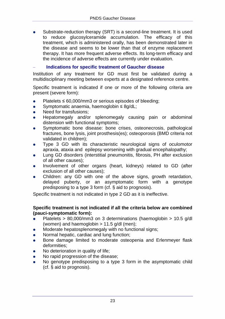
���� Substrate-reduction therapy (SRT) is a second-line treatment. It is used to reduce glucosylceramide accumulation. The efficacy of this treatment, which is administered orally, has been demonstrated later in the disease and seems to be lower than that of enzyme replacement therapy. It has more frequent adverse effects. Its long-term efficacy and the incidence of adverse effects are currently under evaluation.
– Indications for specific treatment of Gaucher disease Institution of any treatment for GD must first be validated during a multidisciplinary meeting between experts at a designated reference centre.
Specific treatment is indicated if one or more of the following criteria are present (severe form):
���� Platelets ≤ 60,000/mm3 or serious episodes of bleeding; ���� Symptomatic anaemia, haemoglobin ≤ 8g/dL; ���� Need for transfusions; ���� Hepatomegaly and/or splenomegaly causing pain or abdominal
distension with functional symptoms; ���� Symptomatic bone disease: bone crises, osteonecrosis, pathological
fractures, bone lysis, joint prosthesis(es); osteoporosis (BMD criteria not validated in children);
���� Type 3 GD with its characteristic neurological signs of oculomotor apraxia, ataxia and epilepsy worsening with gradual encephalopathy;
���� Lung GD disorders (interstitial pneumonitis, fibrosis, PH after exclusion of all other causes);
���� Involvement of other organs (heart, kidneys) related to GD (after exclusion of all other causes);
���� Children: any GD with one of the above signs, growth retardation, delayed puberty, or an asymptomatic form with a genotype predisposing to a type 3 form (cf. § aid to prognosis).
Specific treatment is not indicated in type 2 GD as it is ineffective.
Specific treatment is not indicated if all the crit eria below are combined (pauci-symptomatic form): ���� Platelets > 80,000/mm3 on 3 determinations (haemoglobin > 10.5 g/dl
(women) and haemoglobin > 11.5 g/dl (men); ���� Moderate hepatosplenomegaly with no functional signs; ���� Normal hepatic, cardiac and lung function; ���� Bone damage limited to moderate osteopenia and Erlenmeyer flask
deformities; ���� No deterioration in quality of life; ���� No rapid progression of the disease; ���� No genotype predisposing to a type 3 form in the asymptomatic child
(cf. § aid to prognosis).
PNDS Gaucher Disease
24
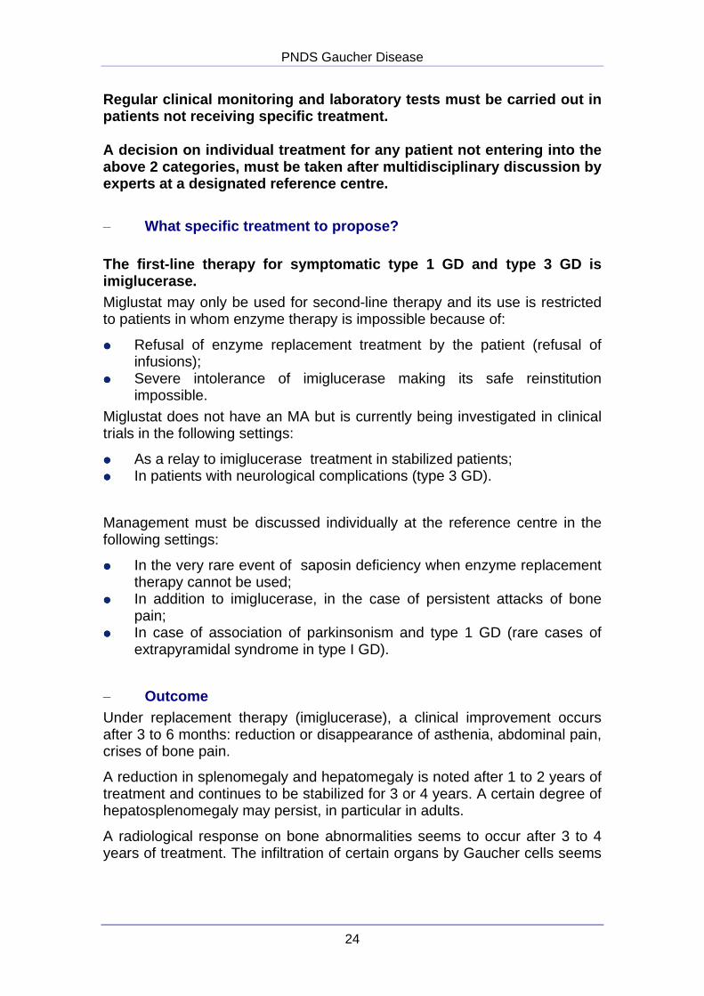
Regular clinical monitoring and laboratory tests mu st be carried out in patients not receiving specific treatment. A decision on individual treatment for any patient not entering into the above 2 categories, must be taken after multidiscip linary discussion by experts at a designated reference centre.
– What specific treatment to propose? The first-line therapy for symptomatic type 1 GD an d type 3 GD is imiglucerase. Miglustat may only be used for second-line therapy and its use is restricted to patients in whom enzyme therapy is impossible because of:
���� Refusal of enzyme replacement treatment by the patient (refusal of infusions);
���� Severe intolerance of imiglucerase making its safe reinstitution impossible.
Miglustat does not have an MA but is currently being investigated in clinical trials in the following settings:
���� As a relay to imiglucerase treatment in stabilized patients; ���� In patients with neurological complications (type 3 GD).
Management must be discussed individually at the reference centre in the following settings:
���� In the very rare event of saposin deficiency when enzyme replacement therapy cannot be used;
���� In addition to imiglucerase, in the case of persistent attacks of bone pain;
���� In case of association of parkinsonism and type 1 GD (rare cases of extrapyramidal syndrome in type I GD).
– Outcome Under replacement therapy (imiglucerase), a clinical improvement occurs after 3 to 6 months: reduction or disappearance of asthenia, abdominal pain, crises of bone pain.
A reduction in splenomegaly and hepatomegaly is noted after 1 to 2 years of treatment and continues to be stabilized for 3 or 4 years. A certain degree of hepatosplenomegaly may persist, in particular in adults.
A radiological response on bone abnormalities seems to occur after 3 to 4 years of treatment. The infiltration of certain organs by Gaucher cells seems
PNDS Gaucher Disease
25
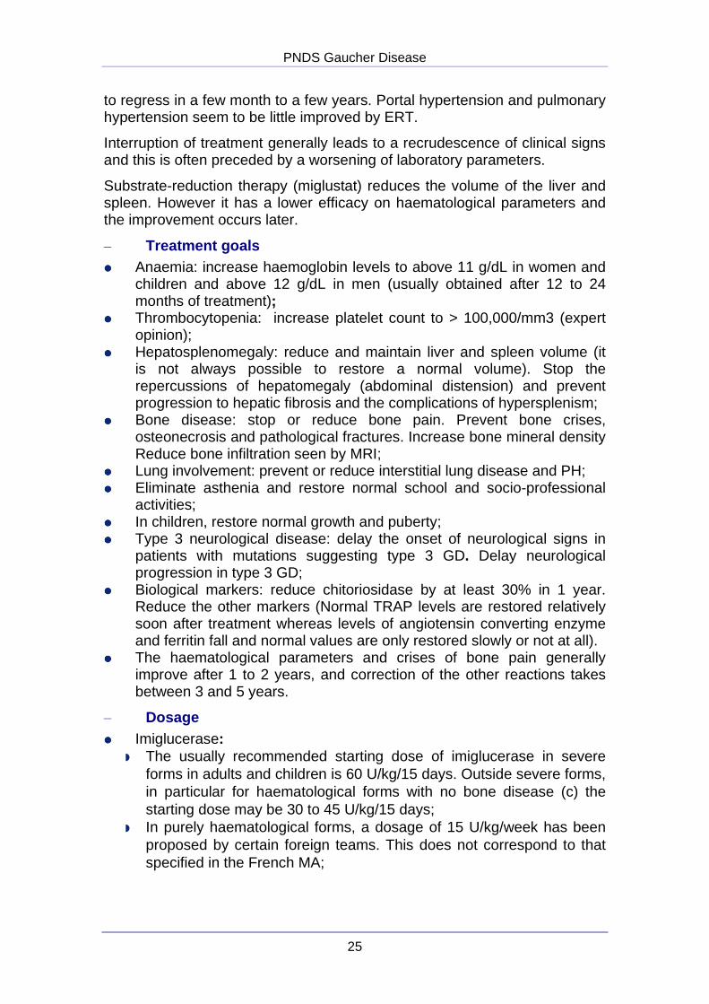
to regress in a few month to a few years. Portal hypertension and pulmonary hypertension seem to be little improved by ERT.
Interruption of treatment generally leads to a recrudescence of clinical signs and this is often preceded by a worsening of laboratory parameters.
Substrate-reduction therapy (miglustat) reduces the volume of the liver and spleen. However it has a lower efficacy on haematological parameters and the improvement occurs later.
– Treatment goals ���� Anaemia: increase haemoglobin levels to above 11 g/dL in women and
children and above 12 g/dL in men (usually obtained after 12 to 24 months of treatment);
���� Thrombocytopenia: increase platelet count to > 100,000/mm3 (expert opinion);
���� Hepatosplenomegaly: reduce and maintain liver and spleen volume (it is not always possible to restore a normal volume). Stop the repercussions of hepatomegaly (abdominal distension) and prevent progression to hepatic fibrosis and the complications of hypersplenism;
���� Bone disease: stop or reduce bone pain. Prevent bone crises, osteonecrosis and pathological fractures. Increase bone mineral density Reduce bone infiltration seen by MRI;
���� Lung involvement: prevent or reduce interstitial lung disease and PH; ���� Eliminate asthenia and restore normal school and socio-professional
activities; ���� In children, restore normal growth and puberty; ���� Type 3 neurological disease: delay the onset of neurological signs in
patients with mutations suggesting type 3 GD. Delay neurological progression in type 3 GD;
���� Biological markers: reduce chitoriosidase by at least 30% in 1 year. Reduce the other markers (Normal TRAP levels are restored relatively soon after treatment whereas levels of angiotensin converting enzyme and ferritin fall and normal values are only restored slowly or not at all).
���� The haematological parameters and crises of bone pain generally improve after 1 to 2 years, and correction of the other reactions takes between 3 and 5 years.
– Dosage ���� Imiglucerase:
� The usually recommended starting dose of imiglucerase in severe forms in adults and children is 60 U/kg/15 days. Outside severe forms, in particular for haematological forms with no bone disease (c) the starting dose may be 30 to 45 U/kg/15 days;
� In purely haematological forms, a dosage of 15 U/kg/week has been proposed by certain foreign teams. This does not correspond to that specified in the French MA;
PNDS Gaucher Disease
26
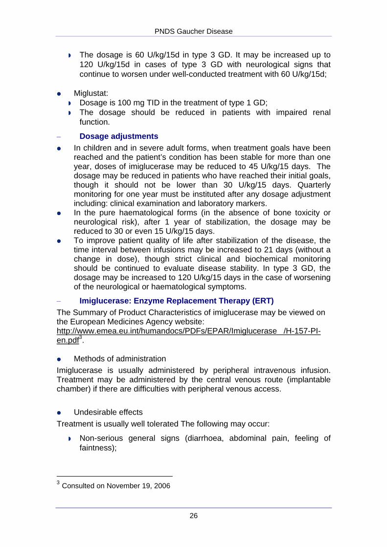
� The dosage is 60 U/kg/15d in type 3 GD. It may be increased up to 120 U/kg/15d in cases of type 3 GD with neurological signs that continue to worsen under well-conducted treatment with 60 U/kg/15d;
���� Miglustat:
� Dosage is 100 mg TID in the treatment of type 1 GD; � The dosage should be reduced in patients with impaired renal
function.
– Dosage adjustments ���� In children and in severe adult forms, when treatment goals have been
reached and the patient’s condition has been stable for more than one year, doses of imiglucerase may be reduced to 45 U/kg/15 days. The dosage may be reduced in patients who have reached their initial goals, though it should not be lower than 30 U/kg/15 days. Quarterly monitoring for one year must be instituted after any dosage adjustment including: clinical examination and laboratory markers.
���� In the pure haematological forms (in the absence of bone toxicity or neurological risk), after 1 year of stabilization, the dosage may be reduced to 30 or even 15 U/kg/15 days.
���� To improve patient quality of life after stabilization of the disease, the time interval between infusions may be increased to 21 days (without a change in dose), though strict clinical and biochemical monitoring should be continued to evaluate disease stability. In type 3 GD, the dosage may be increased to 120 U/kg/15 days in the case of worsening of the neurological or haematological symptoms.
– Imiglucerase: Enzyme Replacement Therapy (ERT) The Summary of Product Characteristics of imiglucerase may be viewed on the European Medicines Agency website: http://www.emea.eu.int/humandocs/PDFs/EPAR/Imiglucerase /H-157-PI-en.pdf3. ���� Methods of administration Imiglucerase is usually administered by peripheral intravenous infusion. Treatment may be administered by the central venous route (implantable chamber) if there are difficulties with peripheral venous access.
���� Undesirable effects
Treatment is usually well tolerated The following may occur:
� Non-serious general signs (diarrhoea, abdominal pain, feeling of faintness);
3 Consulted on November 19, 2006
PNDS Gaucher Disease
27
� Serious signs include: skin reactions (urticaria, angioedema, pruritis, rash), respiratory discomfort and in exceptional cases, anaphylactic shock. These signs mainly occur at the start of the treatment, justifying administration in a hospital setting for the first 2 years.
The duration of the infusion must be adjusted to the dosage administered, initially 2 to 3 hours for 60 U/kg. The infusion rate may be increased (1h30) if there is no adverse effect.
If a minor adverse effect occurs during the infusion:
� Reduce the infusion rate, or temporarily stop the infusion until symptoms disappear;
� Administration of antihistamines and/or corticosteroids.
If an adverse effect occurs suggesting hypersensitivity, the infusion must be stopped and appropriate treatment instituted. A test for anti-imiglucerase IgG antibody should be carried out after the acute episode.
Patients with a history of mild or moderate acute intolerance reactions should be given a preliminary treatment with antihistamines and/or corticosteroids administered orally or intravenously, 1 to 24 hours before the infusion to prevent subsequent reactions.
Imiglucerase should be administered with caution in patients who have developed antibodies or hypersensitivity symptoms with alglucerase. Since 1996, a purified human enzyme is no longer used in France.
���� Contraindications: A life-threatening hypersensitivity reaction to the active substance and/or to the one of the excipients is a lifelong contraindication for imiglucerase administration.
���� Pregnancy: Animal reproductive studies have not been conducted with imiglucerase. It is not known whether imiglucerase can cause foetal harm when administered to a pregnant woman, or whether it affects reproductive capacity. Imiglucerase should be given to a pregnant woman only if clearly needed and after a careful risk/benefit analysis has been conducted for both the mother and the conceptus.
���� Treatment at home:
This is possible:
� In volunteer patients with a good understanding of their disease and management procedures;
PNDS Gaucher Disease
28
� After 2 years of hospital treatment, and with continuation of six-monthly hospital follow-up;
� In the presence of a third person who may intervene in the event of an emergency (call to Centre 15).
� With updating of the healthcare record (date of infusion, traceability label, intercurrent event) (Appendix 5: traceability form).
For patients with a history of side effects suggesting hypersensitivity, an adrenalin kit must be available at home during the infusions.
Patients and the third person present during the infusion should be informed that they must stop the infusion and contact a doctor if symptoms suggesting hypersensitivity occur (urticaria, angioedema, pruritis, rash, respiratory discomfort etc).
– Miglustat: Substrate-reduction therapy (SRT) The Summary of Product Characteristics of miglustat may be viewed on the European Medicines Agency website:
http://www.emea.eu.int/humandocs/PDFs/EPAR/miglustat/H-435-PI-en.pdf4.
This oral treatment is used for second-line treatment in type 1 GD for patients in whom enzyme replacement therapy is impossible (cf. below).
���� Undesirable effects � Gastrointestinal disorders and in particular, diarrhoea in more than
80% of the cases. This is often only mild to moderate, yields spontaneously on continuation of treatment and may usually be controlled by transit modifiers and dietary guidance (intestinal enzyme inhibition, in particular glucosidase and disaccharidase). In the event of diarrhoea, a dietician should provide dietary guidance to restrict the intake of disaccharides (sucrose, lactose) and/or complex carbohydrates while respecting the necessary intake of trace elements and vitamins for the patient’s age;
� Weight loss (6 to 7%), observed in 60%. This is usually followed by weight gain with a trend to a return to the initial weight. Body weight should be monitored.
� Mainly sensory peripheral neuropathy. A post-licensure safety surveillance program was set up because of the possible occurrence of peripheral neuropathy. In particular, this involves: a meticulous neurological examination, an EMG before institution of treatment, periodic neurological assessment during treatment and an EMG in the event of suggestive symptoms. A careful reassessment of the risk-
4 Consulted on November 19, 2006.
PNDS Gaucher Disease
29
benefit ratio must be carried out in the event of peripheral neuropathy and it may be necessary to discontinue miglustat treatment;
� Tremors have been reported in 30% of cases: these are often transient at the start of the treatment, though they may make it necessary to temporarily reduce the dose or even interrupt treatment;
� Cognitive disorders have been described and an initial neurological assessment and monitoring are recommended in all patients;
� Light-headedness, headaches, cramps in the legs and visual disorders have frequently been reported.
���� Contraindication: Hypersensitivity to the active substance and/or to one of the excipients.
���� Reproduction, pregnancy and lactation: Because of its teratogenicity in animals, both men and women receiving miglustat treatment must use an effective method of contraception. Miglustat should not be used during pregnancy or by breastfeeding mothers.
As abnormal spermatogenesis has been demonstrated in animals, male patients who would like to have a child are recommended to stop miglustat and continue to use reliable measures of contraception for a further 3 months.
���� Precautions for use:
� Possible signs of peripheral neuropathy should be sought before prescription of miglustat (clinical examination and EMG);
� Miglustat should not be used in patients with severe renal impairment (creatinine clearance less than 30 ml/min/1.73 m²;
� Miglustat should be used with caution in patients with renal impairment and a creatinine clearance of more than 30 ml/min/1.73 m², or with impaired hepatic function. There are insufficient data to establish specific dosage recommendations in these patients;
� The use of Miglustat is not recommended: - In the child and adolescent; - In patients who refuse to use personal contraception; - No data are available for subjects aged less than 18 years or over
70 years or for patients with impaired hepatic function.
– Specific situations ���� Neonate or infant
When GD is discovered before the age of 2 months, specific treatment is only indicated for symptomatic types 1 and 3. Monitoring alone is required for patients with only isolated moderate splenomegaly. A genetic study is useful to determine the prognosis.

PNDS Gaucher Disease
30
���� Type 2 GD Specific treatment is only indicated if there are early neurological signs (brain stem toxicity: apnoea, dystonic seizures, swallowing disorders, oculomotor disorders) corresponding to type 2. Replacement treatment including by the intrathecal route was not shown to have any effect on the neurological outcome and did not improve life expectancy. Management is the same as that of a progressive neurological disease in the infant.
���� Pregnancy
Management of pregnancy in women with GD must be initiated as early as possible as pregnancy may worsen the signs of GD:
� Worsening of thrombocytopenia and coagulation disorders which may lead to postpartum haemorrhage and contra-indicate peridural anaesthesia;
� Onset of crises of bone pain; � Caesarean section may be necessary in the case of aseptic
osteonecrosis of the hips or a hip prosthesis; � Bisphosphonates are contra-indicated during pregnancy.
In women with a pauci-symptomatic form, ERT may be indicated before pregnancy when the platelets count is below 100,000/mm3 to prevent these complications.
Whenever possible, patients receiving ERT should warn their doctor if they are planning a pregnancy. ERT is not recommended during pregnancy except in exceptional cases.
Treatment procedures and the benefit-risk ratio must be discussed with the patient and her partner.
An expert consensus about treatment in pregnancy was reached after taking into account the fact that GD may worsen on discontinuation of treatment and that pregnancies have been continued to term with ERT without complications:
� Treatment should be continued until conception; � A pregnancy test should be given before each infusion; � If this test is positive, infusions should be stopped for the first trimester
and then resumed during the second or third trimester only in the case of a worsening of the biochemical and clinical parameters and after obtaining the agreement of the patient and her partner.
Pregnancy and delivery require multidisciplinary management: rheumatologist, internist, haematologist, obstetrician, anaesthetist, coagulation specialist.
PNDS Gaucher Disease
31
2.3.2 Non-specific medication
– Level I, II and III analgesics Treatment of chronic bone pain and bone pain crises may require class III analgesics.
– Oral bisphosphonates Treatment is indicated in adults and in the event of vertebral compression and osteoporosis, in combination with specific treatment. Increase in bone mineral density after treatment. There are no data about a reduction in the risk of fracture.
– Antiepileptic in the case of epilepsy
– Prophylactic antibiotic therapy ���� Before placing a prosthesis; ���� In the case of splenectomy in the child.
– Curative antibiotic therapy ���� In the case of osteomyelitis; ���� For intercurrent infections.
– Anti-pneumococcal vaccination If possible before splenectomy and then every 5 years in splenectomized patients.
2.4 Other non-surgical therapy – Bone marrow transplant The value of this treatment of type 3 GD in infants is debatable. Although bone marrow transplantation is effective, it is not indicated in type 1 GD as enzyme replacement therapy has a better risk-benefit ratio.
– Physical medicine and rehabilitation
– Physiotherapy
– When appropriate to circumstances: ���� Auditory and ocular correction; orthoptics (rehabilitation of ocular
motricity disorders); ���� Trophoblast biopsy; ���� Amniocentesis when necessary; ���� Medical abortion when decided by both partners (type 2 GD); ���� In the case of development of multiple disabilities.
Adjustments may have to be made during everyday life (at home, in the car) and prescription medical devices (crutches, corsets, moulded chair, day and/or night splints, orthopaedic shoes, walker, manual or electric wheelchair, anti-pressure sore mattress, medicalized bed, home
PNDS Gaucher Disease
32
oxygen, aspiration equipment, central venous access device, bedside or portable peripheral or central venous infusion kit, nasogastric or gastrostomy tube), with help from the appropriate institutions (department houses for the disabled, specialized centres).
2.5 Surgical treatments
2.5.1 Splenectomy
���� Theoretically splenectomy is no longer indicated in GD since the introduction of replacement therapy except in exceptional cases: failure to respond to well-conducted ERT with persistent severe thrombocytopenia (generally related to voluminous nodular and fibrous splenomegaly).
���� Splenectomized patients must be informed about the risk of infection (encapsulated microorganisms) (splenectomized patient card) and be vaccinated against pneumoccocus infection every 5 years (recommended immunization schedule). In children: primary immunization by Prevenar®, booster with PNEUMO 23®. Ideally, this vaccination must be made 15 days before splenectomy. If splenectomy was not programmed, vaccination must be performed even if the response is uncertain and less intense.
���� Oracillin treatment is given to children up to adolescence. In adults for 2 years after splenectomy.
2.5.2 Orthopaedic treatment
���� Protected weight bearing with crutches in the case of aseptic osteonecrosis of the lower limbs;
���� Immobilization and possible osteosynthesis in the case of aseptic osteonecrosis;
���� Plaster casts; ���� Osteosynthesis of fractures; ���� Prosthesis – arthrodesis; ���� Corset.
Outside an emergency, the patient may be operated after correcting biochemical parameters (Full blood count – coagulation values).
PNDS Gaucher Disease
33
3. Follow-up
3.1 Objectives To make a comparative assessment of the different lesions in comparison with the initial baseline evaluation.
3.1.1 For the untreated patient:
���� Determine outcome (progression of previously documented lesions and screening for any undocumented lesions);
���� Re-evaluation of the need for treatment.
3.1.2 For the treated patient:
���� Determine the outcome (progression or regression of a previously documented lesion and screen for any as yet undocumented lesions);
���� Detect any adverse effects of treatment; ���� Reassess the dosage; ���� Assess compliance to treatment:
� Evaluation of the improvement in the patient’s understanding of the disease;
� Discussion of any change of treatment, home-based treatment, stay abroad, possible pregnancy, contraception;
� Detection of the development of any comorbidity; � Evaluation of the psychological, familial and socio-professional
repercussions of GD.
3.2 Professionals involved The doctor at the reference centre and/or the specialist managing the patient and ensuring follow-up: paediatrician, internist, haematologist, rheumatologist, neurologist, gastroenterologist.
Other specialists (cf. II.2) usually only intervene at the request of the above-mentioned doctors.
The visits required in the integrated care pathway depend on the initial assessment and clinical course:
���� Primary care physician ; ���� Lysosome storage disease or metabolic disease reference centre,
multidisciplinary visit in particular when passing from childhood to adulthood;
���� Specialized visits:
� Paediatrics: Up to 18 years;
PNDS Gaucher Disease
34
� Haematology: In particular in the event of progressive haematological symptoms;
� Internal medicine; � Neurology: Systematically in type 3 GD and after the onset of a
neurological sign; � Rheumatology: In the case of any bone disease, for the management
of osteoporosis; � Gastroenterology-hepatology: in the event of hepatic fibrosis,
hepatocellular insufficiency, portal hypertension, associated hepatitis C or B;
� Gynaecology: in particular in the case of gynaecological bleeding, for contraception (combined oral contraceptives) and during pregnancy;
� Obstetrics: in the case of pregnancy: � Surgery: – Orthopaedics: for a fracture, aseptic osteonecrosis, or placement
of a prosthesis; – Visceral surgery (splenectomy, cholecystectomy as gall stones
frequently occur during GD); – Maxillofacial surgery (rare lesions of the jaw, dental
haemorrhage); � Cardiology: detection of PH; � Dermatology: purpura, pigmentation of the skin; � Pneumology: In the case of PH and interstitial pneumopathy; � Physical medicine and rehabilitation; � Radiology: for the baseline evaluation and follow-up.
3.3 Rhythm and content of visits
3.3.1 Clinical examination:
The clinical follow-up assessment is identical to that performed for the initial assessment. The frequency of visits is adjusted according to the clinical progression.
Generally, a clinical examination is performed:
���� In untreated patients: every 6 months if there is no worsening; ���� In treated patients:
� every 3 months at the start of treatment; � every 6 months when treatment goals have been reached; � after each change in dosage.
Specific neurological follow-up of type 3 GD:
���� Neurological examination: every 3 months for one year, then every 6 months;
���� Examination of eye movements: every 6 months;
PNDS Gaucher Disease
35
���� Ophthalmological examination: once/year; ���� Audiogram: once/year. Follow-up of forms at high risk of developing neurological disease (cf. analysis of the GBA gene): systematic visit to the neurologist once/year.
3.3.2 Paraclinical examinations
– Laboratory investigations ���� Complete blood count and platelet count
The complete blood count and platelet count should be performed:
� In untreated patients: every 6 to 12 months; � In treated patients: – every 3 months at the start of treatment; – every 6 to 12 months when treatment goals have been reached; – after each change in dosage.
���� Coagulation status
Blood coagulation tests should be performed:
� In untreated patients: every 12 months; � In treated patients, if there was an abnormality in the baseline
examination: – every 3 months at the start of treatment until normal values are
restored; – then every 12 months; – after each change in dosage.
���� C-Reactive Protein (CRP)
If suspected infection, myeloma or intercurrent disease.
���� Total proteins ���� Electrophoresis of serum proteins The assay of total proteins and electrophoresis of serum proteins should be performed:
� In untreated patients: every 12 to 24 months (except in special cases); � In treated patients: – every 12-24 months if there was no abnormality in the pre-
treatment assessment; – every 12 months in the case of hypergammaglobulinaemia; – every 6 months in the case of MGUS type monoclonal
gammapathy;
���� Liver function tests comprising: � Free and conjugated bilirubin;
PNDS Gaucher Disease
36
� Transaminases (ASAT – ALAT); � Alkaline Phosphatases; � Gamma GT or 5’-nucleotidase in the child.
These tests should be performed:
� In untreated patients: every 6 to 12 months; � In treated patients: – every 3 months at the start of treatment; – every 6 to 12 months when treatment goals have been reached; – after each change in the dosage.
���� Phosphorus and calcium status Once a year
���� GD biomarkers: Chitotriosidase - tartrate-resistant acid phosphatases (TRAP) - angiotensin converting enzyme – serum ferritin
� In patients receiving no specific treatment: assay once a year � During the first year of treatment: assay every 3 months.
Then: if there is a satisfactory clinical improvement with normal biomarkers:
� Only annual surveillance of chitotriosidase is justified; � Every 3 months if clinical improvement is insufficient.
Additional assays should be carried out after changes in the dosage or a specific clinical event (to be reported to the laboratory conducting the assay).
���� Myelogram: in the case of progressive haematological symptoms.
���� Assays of anti-imiglucerase antibodies: � Every 6 months during the first 18 months of treatment, then
discontinuation; � In the case of immunoallergic reaction(s); � When there is lack of treatment efficacy (very rare cases of
neutralizing antibodies).
– Imaging
���� Liver-spleen imaging
Indications: Measurement and determination of changes in the dimensions and morphology of the liver and spleen (if possible determination of liver volume) with, where possible, the same examination as during the baseline evaluation.
Liver-spleen imaging should be performed:
PNDS Gaucher Disease
37
� In untreated patients: every 12 to 24 months; � In treated patients: – every 6 months until stabilization of liver and spleen volumes; – then every 12-24/months; – 6 months after each change in dosage.
���� Bone Imaging
� MRI of the spine, femur, pelvis and tibia: – In untreated patients: every 12 to 24 months; – In treated patients:
- every 6 months for 2 years in order to asses the regression of the infiltration;
- once stabilization is obtained, MRI may then be performed at 2 to 3-year intervals.
– In every case: Additional MRI of any intercurrent bone event.
� Radiography – In untreated patients: Only in the case of an intercurrent bone
event. – In treated patients:
- Annual radiography for osteoarthritis complicating osteonecrosis or earlier in the event of pain, reduction in the walking capacity or deformity to establish the appropriateness of a prosthesis;
- Surveillance of a prosthesis; - Radiography of the skeleton if suspected myeloma (skull –
spine – humerus – femur – pelvis) or plasmocytoma.
� Bone CT-scan
This is not indicated in GD as it does not evaluate the bone medulla. It may however be used to measure the thickness of cortical bone after a fracture.
� Evaluation of BMD – Every year or every 2 years. – In the child, interpretation requires specialist expertise as the
physiological transformation of red marrow into yellow marrow may be mistaken for infiltration.
���� Neurological imaging
Cerebral MRI: only if clinically indicated.
���� Other neurological assessments:
� Electroencephalogram: only in epilepsy; � Neuropsychological tests: 1 every 2 years if clinical progression
PNDS Gaucher Disease
38
� In patients receiving miglustat: – Evaluation of cognitive function; – EMG if clinically indicated.
���� Lung and/or cardiac function tests
� In splenectomized patients with an increased risk of PH:
Echocardiography with Doppler to detect PH every year; � In the case of pulmonary fibrosis: – Resting ECG: Examination for ventricular hypertrophy, rhythm or
conduction disorders, ischaemia; – Chest X-ray and/or CT-scan: Examination for interstitial
pneumopathy, lung fibrosis, hypertrophy of lung arteries, cardiomegaly;
– Respiratory function tests with detection of any diffusion disorder: restrictive syndrome, oxygen diffusion disorder.
PNDS Gaucher Disease
39
Appendix 1. List of persons who helped draft this PNDS on Gaucher disease ���� Dr Emmanuel Corbillon – Project leader – ALD department -
conventional agreements ���� Dr Nadia Belmatoug: Internal Medicine - Beaujon Hospital –
Clichy, coordinator of the Lysosomal Storage Disease Reference Centre and the Gaucher Treatment Evaluation Committee
���� Professor Thierry Billette: Paediatric Neurology - Trousseau Hospital - Paris
���� Dr Christian Rose: Blood Diseases - Claude Huriet Hospital - Lille ���� Dr Pierre Kaminsky: Orphan Disease Unit - Brabois University
Hospital – Nancy ���� Dr Frédéric Sedel: Federation for Diseases of the Nervous
System - Pitié Salpétrière Hospital Group - Paris ���� Dr Catherine Caillaud: Genetics Laboratory - Cochin Hospital -
Paris ���� Dr Roselyne Froissard: Pediatric Biochemistry - Debrousse
Hospital – Lyon ���� Dr Nathalie Guffon: Metabolic Diseases – Edouard Herriot
Hospital – Lyon ���� Dr Christian Lavigne: Internal Medicine – Angers University
Hospital ���� Dr Adrien Kettaneh: Internal Medicine - St Antoine University
Hospital - Paris ���� Dr Christine Broissand: Pharmacy - Necker Hospital - Paris ���� Dr Jérôme Stirnemann: Internal Medicine - Jean Verdier
University Hospital - Bondy ���� Dr Irène Maire: Pediatric Biochemistry - Debrousse Hospital –
Lyon ���� Professor François Feillet: Department of Infantile Medicine –
Children’s Hospital, Brabois University Hospital Vandoeuvre les Nancy
���� Dr Danièle Bouniol: French National Medical Office for Hereditary Metabolic Diseases
���� Dr Francis Gaspari: French National Medical Office for Hereditary Metabolic Diseases
���� Dr Murielle Jousselin: Afssaps – Saint Denis ���� Dr Cornet: GP – Paris ���� Dr Aïda Jolivet: CNAMTS consultant doctor ���� Dr Philippe Perez: RSI consultant doctor ���� Delphine Genevaz: Association for the Fight against Lysosomal
Storage Diseases
PNDS Gaucher Disease
40
Appendix 2. References
Main references
Barton NW, Brady RO, Dambrosia JM, Di Bisceglie AM, Doppelt SH, Hill SC, et al. Replacement therapy for inherited enzyme deficiency--macrophage-targeted glucocerebrosidase for Gaucher's disease. N Engl J Med 1991;324(21):1464-70.
Belmatoug N, Guffon N, Stirnemann J, Caillaud C, Vanier MT, Sedel F, et al. Pregnancy in Gaucher disease in the era of enzyme replacement therapy. 7th European Working Group on GAUCHER Disease 2006. <http://www.ewggd.co.uk/EWGGD%20Abstracts.pdf> [consulté le 23-10-2006].
Beutler E, Grabowski GA. Gaucher Disease. In: Scriver CR, Beaudet AL, Sly WS, Valle D, ed. The metabolic and molecular basis of inherited disease. New York: McGraw Hill; 2001. p. 3635-68.
Boot RG, Renkema GH, Verhoek M, Strijland A, Bliek J, de Meulemeester TM, et al. The human chitotriosidase gene. Nature of inherited enzyme deficiency. J Biol Chem 1998;273(40):25680-5.
Brady RO, Kanfer JN, Shapiro D. Metabolism of glucocerebrosides. ii. evidence of an enzymatic
deficiency in gaucher's disease. Biochem Biophys Res Commun 1965;18:221-5.
Charrow J, Andersson HC, Kaplan P, Kolodny EH, Mistry P, Pastores G, et al. Enzyme replacement therapy and monitoring for children with type 1 Gaucher disease: consensus recommendations. J Pediatr 2004;144(1):112-20.
Ciana G, Addobbati R, Tamaro G, Leopaldi A, Nevyjel M, Ronfani L, et al. Gaucher disease and bone: laboratory and skeletal mineral density variations during a long period of enzyme replacement therapy. J Inherit Metab Dis 2005;28(5):723-32.
Cox TM, Aerts JM, Andria G, Beck M, Belmatoug N, Bembi B, et al. The role of the iminosugar N-butyldeoxynojirimycin (miglustat) in the management of type I (non-neuronopathic) Gaucher disease: a position statement. J Inherit Metab Dis 2003;26(6):513-26.
Dechelotte P. Type 1 Gaucher's disease in the adult. Nutritional management during initiation of treatment with miglustat. Presse Med 2004;33(7):494-6.
Elstein Y, Eisenberg V, Granovsky-Grisaru S, Rabinowitz
PNDS Gaucher Disease
41
R, Samueloff A, Zimran A, et al. Pregnancies in Gaucher disease: a 5-year study. Am J Obstet Gynecol 2004;190(2):435-41.
Froissart R. Biomarqueurs actuels et futurs de la GD . Rev Med Interne 2006;27 Suppl 1:S22-S25.
Lebel E, Dweck A, Foldes AJ, Golowa Y, Itzchaki M, Zimran A, et al. Bone density changes with enzyme therapy for Gaucher
disease. J Bone Miner Metab 2004;22(6):597-601.
Mignot C, Gelot A, Bessieres B, Daffos F, Voyer M, Menez F , et al. Perinatal-lethal Gaucher disease. Am J Med Genet A 2003;120(3):338-44.
Stirnemann J, Caubel I, Kettaneh A, Fain O, Belmatoug N. Epidemiologic, clinical, biological and therapeutic aspects of Gaucher disease. Presse Med 2003;32(11):503-11.
Secondary references
Abrahamov A, Elstein D, Gross-Tsur V, Farber B, Glaser Y, Hadas-Halpern I, et al. Gaucher's disease variant characterised by progressive calcification of heart valves and unique genotype. Lancet 1995;346(8981):1000-3.
Agence nationale d'accréditation et d'évaluation en santé. Synthèse des textes réglementaires concernant l'utilisation thérapeutique des transfusions de produits sanguins labiles. Paris: ANAES; 1997.
Altarescu G, Hill S, Wiggs E, Jeffries N, Kreps C, Parker CC, et al. The efficacy of enzyme replacement therapy in patients with chronic neuronopathic Gaucher's disease. J Pediatr 2001;138(4):539-47.
Amsallem D, Rodriguez D, Vanier MT, Khayat N, Millat G,
Campanello M. Third case of Gaucher disease with sap-C deficiency and Évaluation of a 12 months' therapy by miglustat. J Inherit Metab Dis 2005;28(suppl1):-152.
Baldellou A, Andria G, Campbell PE, Charrow J, Cohen IJ, Grabowski GA, et al. Paediatric non-neuronopathic Gaucher disease: recommendations for treatment and monitoring. Eur J Pediatr 2004;163(2):67-75.
Chabas A, Cormand B, Grinberg D, Burguera JM, Balcells S, Merino JL, et al. Unusual expression of Gaucher's disease: cardiovascular calcifications in three sibs homozygous for the D409H mutation. J Med Genet 1995;32(9):740-2.
Costello R, O'callaghan T, Sebahoun G. Gaucher disease
PNDS Gaucher Disease
42
and multiple myeloma. Leuk Lymphoma 2006;47(7):1365-8.
de Fost M, vom DS, Weverling GJ, Brill N, Brett S, Haussinger D, et al. Increased incidence of cancer in adult Gaucher disease in Western Europe. Blood Cells Mol Dis 2006;36(1):53-8.
Deegan PB, Moran MT, McFarlane I, Schofield JP, Boot RG, Aerts JM, et al. Clinical Évaluation of chemokine and enzymatic biomarkers of Gaucher disease. Blood Cells Mol Dis 2005;35(2):259-67.
De Fost M, Aerts JM, Groener JE, Mass A, Akkerman EM, Wiersma MG, Hollak CE. 1Low frequenct maintenance therapy with imiglucerase in adult type 1 Gaucher disease: a prospective randomized trial.)
Hematologica; 2007 (92);215-221
Elstein D, Abrahamov A, Dweck A, Hadas-Halpern I, Zimran A. Gaucher disease: pediatric concerns. Paediatr Drugs 2002;4(7):417-26.
Elstein D, Hollak C, Aerts JM, van Weely S, Maas M, Cox TM , et al. Sustained therapeutic effects of oral miglustat (miglustat, N-butyldeoxynojirimycin, OGT 918) in type I Gaucher disease. J Inherit Metab Dis 2004;27(6):757-66.
Erikson A, Forsberg H, Nilsson M, Astrom M, Mansson JE. Ten
years' experience of enzyme infusion therapy of Norrbottnian (type 3) Gaucher disease. Acta Paediatr 2006;95(3):312-7.
Erikson A, Johansson K, Mansson JE, Svennerholm L. Enzyme replacement therapy of infantile Gaucher disease. Neuropediatrics 1993;24(4):237-8.
Finkelstein R, Nachum Z, Reissman P, Reiss ND, Besser M, Trajber I, et al. Anaerobic osteomyelitis in patients with Gaucher's disease. Clin Infect Dis 1992;15(5):771-3.
Fleshner PR, Aufses AH, Jr., Grabowski GA, Elias R. A 27-year experience with splenectomy for Gaucher's disease. Am J Surg 1991;161(1):69-75.
Gillis S, Hyam E, Abrahamov A, Elstein D, Zimran A. Platelet function abnormalities in Gaucher disease patients. Am J Hematol 1999;61(2):103-6.
Giona F, Palumbo G, Amendola A, Santoro C, Mazzuconi MG. Platelet function and coagulation abnormalities in type 1 Gaucher disease patients: effects of enzyme replacement therapy (ERT). J Thromb Haemost 2006;4(8):1831-3.
Grabowski GA, Andria G, Baldellou A, Campbell PE, Charrow J, Cohen IJ, et al. Pediatric non-neuronopathic
PNDS Gaucher Disease
43
Gaucher disease: presentation, diagnosis and assessment. Consensus statements. Eur J Pediatr 2004;163(2):58-66.
Hollak C, Maas M, Akkerman E, den Heeten A, Aerts H. Dixon quantitative chemical shift imaging is a sensitive tool for the Évaluation of bone marrow responses to individualized doses of enzyme supplementation therapy in type 1 Gaucher disease. Blood Cells Mol Dis 2001;27(6):1005-12.
Hollak CE, Levi M, Berends F, Aerts JM, van Oers MH. Coagulation abnormalities in type 1 Gaucher disease are due to low-grade activation and can be partly restored by enzyme supplementation therapy. Br J Haematol 1997;96(3):470-6.
Krasnewich D, Dietrich K, Bauer L, Ginns EI, Sidransky E, Hill S. Splenectomy in Gaucher disease: new management dilemmas [letter]. Blood 1998;91(8):3085-7.
Maas M, van Kuijk C, Stoker J, Hollak CE, Akkerman EM, Aerts JF, et al. Quantification of bone involvement in Gaucher disease: MR imaging bone marrow burden score as an alternative to Dixon quantitative chemical shift MR imaging--initial experience. Radiology 2003;229(2):554-61.
Maaswinkel-Mooij P, Hollak C, Eysden-Plaisier M, Prins M, Aerts H, Poll R. The natural course of Gaucher disease in The
Netherlands: implications for monitoring of disease manifestations. J Inherit Metab Dis 2000;23(1):77-82.
Miller SP, Zirzow GC, Doppelt SH, Brady RO, Barton NW. Analysis of the lipids of normal and Gaucher bone marrow. J Lab Clin Med 1996;127(4):353-8.
Mistry PK, Sirrs S, Chan A, Pritzker MR, Duffy TP, Grace ME, et al. Pulmonary hypertension in type 1 Gaucher's disease: genetic and epigenetic determinants of phenotype and response to therapy. Mol Genet Metab 2002;77(1-2):91-8.
Pastores GM, Sibille AR, Grabowski GA. Enzyme therapy in Gaucher disease type 1: dosage efficacy and adverse effects in 33 patients treated for 6 to 24 months. Blood 1993;82(2):408-16.
Pastores GM, Wallenstein S, Desnick RJ, Luckey MM. Bone density in Type 1 Gaucher disease. J Bone Miner Res 1996;11(11):1801-7.
Pastores GM, Barnett NL, Kolodny EH. An open-label, noncomparative study of miglustat in type I Gaucher disease: efficacy and tolerability over 24 months of treatment. Clin Ther 2005;27(8):1215-27.
Patrick AD. A deficiency of glucocerebrosidase in Gaucher's
PNDS Gaucher Disease
44
disease. Biochem J 1965;97(2):17C.
Prows CA, Sanchez N, Daugherty C, Grabowski GA. Gaucher disease: enzyme therapy in the acute neuronopathic variant. Am J Med Genet 1997;71(1):16-21.
Ries M, Schaefer E, Luhrs T, Mani L, Kuhn J, Vanier MT, et al. Critical assessment of chitotriosidase analysis in the rational laboratory diagnosis of children with Gaucher disease and Niemann-Pick disease type A/B and C. J Inherit Metab Dis 2006;29(5):647-52.
Rosenbloom BE, Weinreb NJ, Zimran A, Kacena KA, Charrow J, Ward E. Gaucher disease and cancer incidence: a study from the Gaucher Registry. Blood 2005;105(12):4569-72.
Rosengarten D, Abrahamov A, Nir A, Farber B, Glaser J, Zimran A, et al. Outcome of ten years' echocardiographic follow-up in children with Gaucher disease. Eur J Pediatr 2006.
Sandhoff K, Kolter T, Harzer K. Sphingolipid activator proteins. In: Scriver CR, Beaudet AL, Sly WS, Valle D, Childs B, Kinzler KW, et al., ed. The Metabolic and Molecular Bases of Inherited Disease. New York: McGraw Hill; 1995. p. 3371-88.
Schiffmann R, Mankin H, Dambrosia JM, Xavier RJ, Kreps
C, Hill SC, et al. Decreased bone density in splenectomized Gaucher patients receiving enzyme replacement therapy. Blood Cells Mol Dis 2002;28(2):288-96.
Sherer Y, Dulitzki M, Levy Y, Livneh A, Shoenfeld Y, Langevitz P. Successful pregnancy outcome in a patient with Gaucher's disease and antiphospholipid syndrome. Ann Hematol 2002;81(3):161-3.
Shoenfeld Y, Beresovski A, Zharhary D, Tomer Y, Swissa M, Sela E, et al. Natural autoantibodies in sera of patients with Gaucher's disease. J Clin Immunol 1995;15(6):363-72.
Sidransky E, Sherer DM, Ginns EI. Gaucher disease in the neonate: a distinct Gaucher phenotype is analogous to a mouse model created by targeted disruption of the glucocerebrosidase gene. Pediatr Res 1992;32(4):494-8.
Takahashi T, Yoshida Y, Sato W, Yano T, Shoji Y, Sawaishi Y, et al. Enzyme therapy in Gaucher disease type 2: an autopsy case. Tohoku J Exp Med 1998;186(2):143-9.
Vellodi A, Bembi B, de Villemeur TB, Collin-Histed T, Erikson A, Mengel E, et al. Management of neuronopathic Gaucher disease: a European consensus. J Inherit Metab Dis 2001;24(3):319-27.
PNDS Gaucher Disease
45
Vellodi A, Foo Y, Cole TJ. Évaluation of three biochemical markers in the monitoring of Gaucher disease. J Inherit Metab Dis 2005;28(4):585-92.
vom Dahl S, Poll L, Di Rocco M, Ciana G, Denes C, Mariani G, et al. Evidence-based recommendations for monitoring bone disease and the response to enzyme replacement therapy in Gaucher patients. Curr Med Res Opin 2006;22(6):1045-64.
Weinreb NJ, Aggio MC, Andersson HC, Andria G, Charrow J, Clarke JT, et al. Gaucher disease type 1: revised recommendations on Évaluation s and monitoring for adult patients. Semin Hematol 2004;41(4 Suppl 5):15-22.
Weinreb NJ, Charrow J, Andersson HC, Kaplan P, Kolodny EH, Mistry P, et al. Effectiveness of enzyme replacement therapy in 1028 patients with type 1 Gaucher disease after 2 to 5 years of treatment: a report from the Gaucher Registry. Am J Med 2002;113(2):112-9.
Weinreb NJ, Barranger JA, Charrow J, Grabowski GA, Mankin HJ, Mistry P. Guidance on the use of miglustat for treating patients with type 1 Gaucher disease. Am J Hematol 2005;80(3):223-9.
Wenstrup RJ, Bailey L, Grabowski GA, Moskovitz J,
Oestreich AE, Wu W, et al. Gaucher disease: alendronate disodium improves bone mineral density in adults receiving enzyme therapy. Blood 2004;104(5):1253-7.
Yates P, Morse C, Standen GR. Gaucher's disease and acquired coagulopathy. Clin Lab Haematol 1992;14(4):331-4.
Zimran A, Kay A, Gelbart T, Garver P, Thurston D, Saven A, et al. Gaucher disease. Clinical, laboratory, radiologic, and genetic features of 53 patients. Medicine (Baltimore) 1992;71(6):337-53.
Zimran A, Liphshitz I, Barchana M, Abrahamov A, Elstein D. Incidence of malignancies among patients with type I Gaucher disease from a single referral clinic. Blood Cells Mol Dis 2005;34(3):197-200.
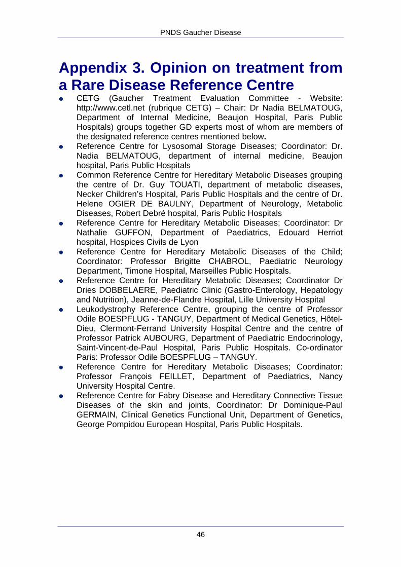
PNDS Gaucher Disease
46
Appendix 3. Opinion on treatment from a Rare Disease Reference Centre ���� CETG (Gaucher Treatment Evaluation Committee - Website:
http://www.cetl.net (rubrique CETG) – Chair: Dr Nadia BELMATOUG, Department of Internal Medicine, Beaujon Hospital, Paris Public Hospitals) groups together GD experts most of whom are members of the designated reference centres mentioned below.
���� Reference Centre for Lysosomal Storage Diseases; Coordinator: Dr. Nadia BELMATOUG, department of internal medicine, Beaujon hospital, Paris Public Hospitals
���� Common Reference Centre for Hereditary Metabolic Diseases grouping the centre of Dr. Guy TOUATI, department of metabolic diseases, Necker Children’s Hospital, Paris Public Hospitals and the centre of Dr. Helene OGIER DE BAULNY, Department of Neurology, Metabolic Diseases, Robert Debré hospital, Paris Public Hospitals
���� Reference Centre for Hereditary Metabolic Diseases; Coordinator: Dr Nathalie GUFFON, Department of Paediatrics, Edouard Herriot hospital, Hospices Civils de Lyon
���� Reference Centre for Hereditary Metabolic Diseases of the Child; Coordinator: Professor Brigitte CHABROL, Paediatric Neurology Department, Timone Hospital, Marseilles Public Hospitals.
���� Reference Centre for Hereditary Metabolic Diseases; Coordinator Dr Dries DOBBELAERE, Paediatric Clinic (Gastro-Enterology, Hepatology and Nutrition), Jeanne-de-Flandre Hospital, Lille University Hospital
���� Leukodystrophy Reference Centre, grouping the centre of Professor Odile BOESPFLUG - TANGUY, Department of Medical Genetics, Hôtel-Dieu, Clermont-Ferrand University Hospital Centre and the centre of Professor Patrick AUBOURG, Department of Paediatric Endocrinology, Saint-Vincent-de-Paul Hospital, Paris Public Hospitals. Co-ordinator Paris: Professor Odile BOESPFLUG – TANGUY.
���� Reference Centre for Hereditary Metabolic Diseases; Coordinator: Professor François FEILLET, Department of Paediatrics, Nancy University Hospital Centre.
���� Reference Centre for Fabry Disease and Hereditary Connective Tissue Diseases of the skin and joints, Coordinator: Dr Dominique-Paul GERMAIN, Clinical Genetics Functional Unit, Department of Genetics, George Pompidou European Hospital, Paris Public Hospitals.
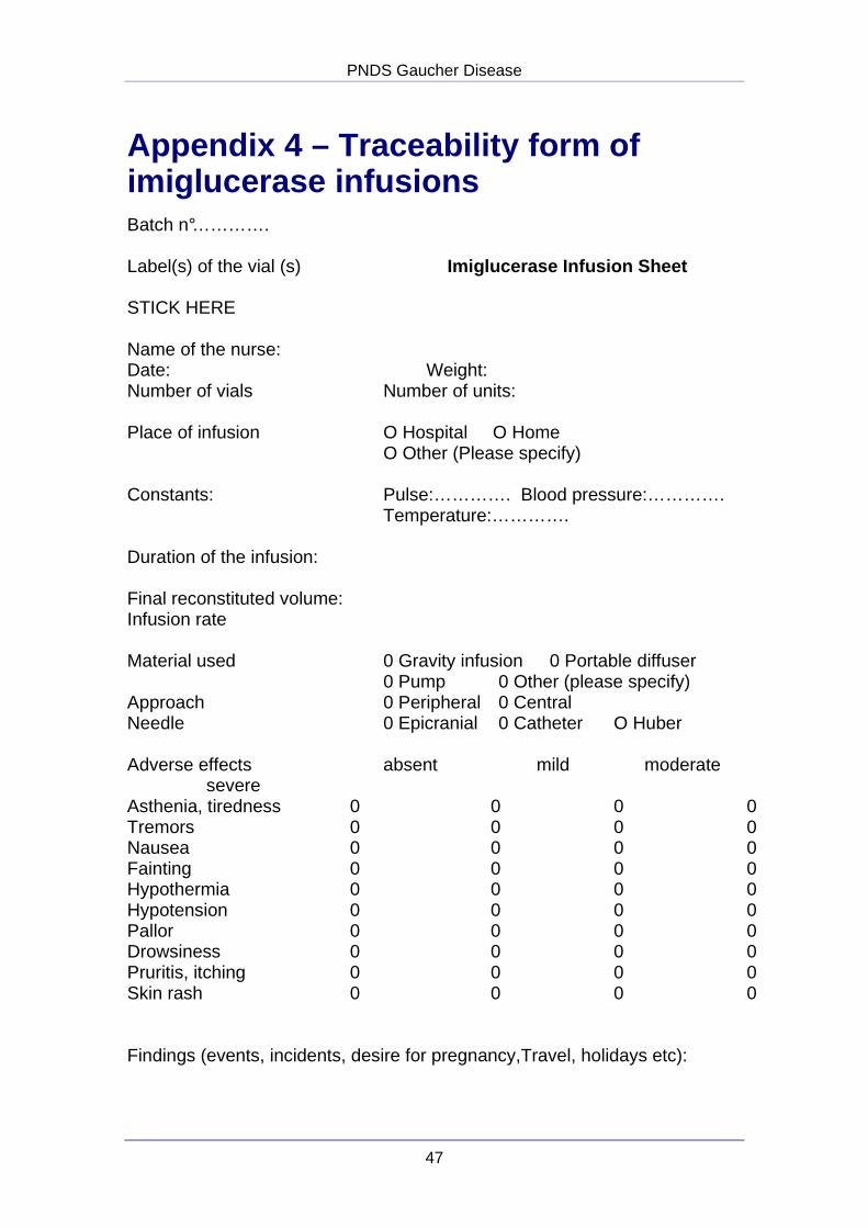
PNDS Gaucher Disease
47
Appendix 4 – Traceability form of imiglucerase infusions Batch n°…………. Label(s) of the vial (s) Imiglucerase Infusion Sheet STICK HERE Name of the nurse: Date: Weight: Number of vials Number of units: Place of infusion O Hospital O Home O Other (Please specify) Constants: Pulse:…………. Blood pressure:…………. Temperature:…………. Duration of the infusion: Final reconstituted volume: Infusion rate Material used 0 Gravity infusion 0 Portable diffuser 0 Pump 0 Other (please specify) Approach 0 Peripheral 0 Central Needle 0 Epicranial 0 Catheter O Huber Adverse effects absent mild moderate severe Asthenia, tiredness 0 0 0 0 Tremors 0 0 0 0 Nausea 0 0 0 0 Fainting 0 0 0 0 Hypothermia 0 0 0 0 Hypotension 0 0 0 0 Pallor 0 0 0 0 Drowsiness 0 0 0 0 Pruritis, itching 0 0 0 0 Skin rash 0 0 0 0 Findings (events, incidents, desire for pregnancy,Travel, holidays etc):

Printing completed in xxxxxx - Imprimerie Moderne de l’Est – Registration of Copyright Month yyyy (Body of text 3)
All HAS publications are available for download at www.has-sante.fr
CO
DE
Etu
de (
rem
pli p
ar le
ser
vice
com
mun
icat
ion)
Related Documents